EP4421186A2 - Verfahren zur bestimmung eines chirurgischen randes und verfahren zur verwendung davon - Google Patents
Verfahren zur bestimmung eines chirurgischen randes und verfahren zur verwendung davon Download PDFInfo
- Publication number
- EP4421186A2 EP4421186A2 EP24171813.9A EP24171813A EP4421186A2 EP 4421186 A2 EP4421186 A2 EP 4421186A2 EP 24171813 A EP24171813 A EP 24171813A EP 4421186 A2 EP4421186 A2 EP 4421186A2
- Authority
- EP
- European Patent Office
- Prior art keywords
- analyte
- tissue
- capture
- location
- tissue sample
- Prior art date
- Legal status (The legal status is an assumption and is not a legal conclusion. Google has not performed a legal analysis and makes no representation as to the accuracy of the status listed.)
- Granted
Links
Images


Classifications
-
- C—CHEMISTRY; METALLURGY
- C12—BIOCHEMISTRY; BEER; SPIRITS; WINE; VINEGAR; MICROBIOLOGY; ENZYMOLOGY; MUTATION OR GENETIC ENGINEERING
- C12N—MICROORGANISMS OR ENZYMES; COMPOSITIONS THEREOF; PROPAGATING, PRESERVING, OR MAINTAINING MICROORGANISMS; MUTATION OR GENETIC ENGINEERING; CULTURE MEDIA
- C12N15/00—Mutation or genetic engineering; DNA or RNA concerning genetic engineering, vectors, e.g. plasmids, or their isolation, preparation or purification; Use of hosts therefor
- C12N15/09—Recombinant DNA-technology
- C12N15/10—Processes for the isolation, preparation or purification of DNA or RNA
- C12N15/1034—Isolating an individual clone by screening libraries
- C12N15/1065—Preparation or screening of tagged libraries, e.g. tagged microorganisms by STM-mutagenesis, tagged polynucleotides, gene tags
-
- C—CHEMISTRY; METALLURGY
- C12—BIOCHEMISTRY; BEER; SPIRITS; WINE; VINEGAR; MICROBIOLOGY; ENZYMOLOGY; MUTATION OR GENETIC ENGINEERING
- C12Q—MEASURING OR TESTING PROCESSES INVOLVING ENZYMES, NUCLEIC ACIDS OR MICROORGANISMS; COMPOSITIONS OR TEST PAPERS THEREFOR; PROCESSES OF PREPARING SUCH COMPOSITIONS; CONDITION-RESPONSIVE CONTROL IN MICROBIOLOGICAL OR ENZYMOLOGICAL PROCESSES
- C12Q1/00—Measuring or testing processes involving enzymes, nucleic acids or microorganisms; Compositions therefor; Processes of preparing such compositions
- C12Q1/68—Measuring or testing processes involving enzymes, nucleic acids or microorganisms; Compositions therefor; Processes of preparing such compositions involving nucleic acids
- C12Q1/6813—Hybridisation assays
- C12Q1/6841—In situ hybridisation
-
- C—CHEMISTRY; METALLURGY
- C12—BIOCHEMISTRY; BEER; SPIRITS; WINE; VINEGAR; MICROBIOLOGY; ENZYMOLOGY; MUTATION OR GENETIC ENGINEERING
- C12Q—MEASURING OR TESTING PROCESSES INVOLVING ENZYMES, NUCLEIC ACIDS OR MICROORGANISMS; COMPOSITIONS OR TEST PAPERS THEREFOR; PROCESSES OF PREPARING SUCH COMPOSITIONS; CONDITION-RESPONSIVE CONTROL IN MICROBIOLOGICAL OR ENZYMOLOGICAL PROCESSES
- C12Q1/00—Measuring or testing processes involving enzymes, nucleic acids or microorganisms; Compositions therefor; Processes of preparing such compositions
- C12Q1/68—Measuring or testing processes involving enzymes, nucleic acids or microorganisms; Compositions therefor; Processes of preparing such compositions involving nucleic acids
- C12Q1/6869—Methods for sequencing
- C12Q1/6874—Methods for sequencing involving nucleic acid arrays, e.g. sequencing by hybridisation
Definitions
- Cells within a tissue of a subject have differences in cell morphology and/or function due to varied analyte levels (e.g., gene and/or protein expression) within the different cells.
- the specific position of a cell within a tissue e.g., the cell's position relative to neighboring cells or the cell's position relative to the tissue microenvironment
- a capture probe of the plurality of capture probes comprises (i) a capture domain that specifically binds to an analyte of the tissue sample and (ii) a spatial barcode; (b) determining (i) all or a part of a nucleic acid sequence corresponding to the analyte specifically bound to the capture domain or a complement thereof, and (ii) all or a part of a nucleic acid sequence corresponding to the spatial barcode or a complement thereof, and using the determined nucleic acid sequences of (i) and (ii) to identify the presence of the analyte at a location in the tissue sample; (c) comparing the presence of the analyte at the location in the tissue sample to presence of the analyte at different location(
- Also provided herein are methods of determining size and site of a tissue to be resected from a subject comprising: (a) contacting a tissue sample obtained from the subject to an array comprising a plurality of capture probes, wherein a capture probe of the plurality of capture probes comprises (i) a capture domain that binds specifically to an analyte of the tissue sample and (ii) a spatial barcode; (b) determining (i) all or a part of a nucleic acid sequence corresponding to the analyte specifically bound to the capture domain or a complement thereof, and (ii) all or a part of a nucleic acid sequence corresponding to the spatial barcode or a complement thereof, and using the determined nucleic acid sequences of (i) and (ii) to identify the presence of the analyte at a location in the tissue sample; (c) comparing the presence of the analyte at the location in the tissue sample to presence of the analyte at a different location
- Also provided herein are methods of reducing the risk of re-excision of a tissue comprising: (a) contacting a tissue sample to an array comprising a plurality of capture probes, wherein a capture probe of the plurality of capture probes comprises a (i) capture domain that specifically binds to an analyte of the tissue sample and (ii) a spatial barcode; (b) determining (i) all or a part of a nucleic acid sequence corresponding to the analyte specifically bound to the capture domain or a complement thereof, and (ii) all or a part of a nucleic acid sequence corresponding to the spatial barcode or a complement thereof, and using the determined nucleic acid sequences of (i) and (ii) to identify the presence of the analyte at a location in the tissue sample; (c) comparing the presence of the analyte at the location in the tissue sample to presence of the analyte at different location(s) in the tissue sample, and determining
- the array comprises a slide. In some embodiments, the array is a bead array.
- step (b) comprises sequencing (i) all or a part of the nucleic acid sequence corresponding to the analyte specifically bound to the capture domain or a complement thereof, and (ii) all or a part of the nucleic acid sequence corresponding to the spatial barcode or a complement thereof.
- the sequencing is high throughput sequencing.
- step (b) comprises extending a 3' end of the capture probe using the specifically bound analyte as a template to generate an extended capture probe.
- step (b) further comprises generating a single-stranded nucleic acid comprising a nucleic acid sequence that is complementary to all or a part of the extended capture probe.
- Also provided herein are methods of determining size and site of a tissue to be resected from a subject comprising: (a) contacting a tissue sample obtained from the subject to a plurality of analyte capture agents, wherein an analyte capture agent of the plurality of analyte capture agents comprises an analyte binding moiety barcode, an analyte capture sequence, and an analyte binding moiety that binds specifically to an analyte; (b) disposing the tissue sample onto an array, wherein the array comprises a plurality of capture probes, wherein a capture probe of the plurality of capture probes comprises a spatial barcode and a capture domain that binds specifically to the analyte capture sequence; (c) determining (i) all or a part of a nucleic acid sequence corresponding to the analyte binding moiety barcode or a complement thereof, and (ii) all or a part of a nucleic acid sequence corresponding to the
- step (c) comprises sequencing (i) all or a part of the nucleic acid sequence corresponding to the analyte binding moiety barcode or a complement thereof, and (ii) all or a part of the nucleic acid sequence corresponding to the spatial barcode or a complement thereof.
- the sequencing is high throughput sequencing.
- step (c) comprises extending a 3' end of the capture probe using the specifically bound analyte capture agent as a template to generate an extended capture probe.
- step (c) further comprises generating a single-stranded nucleic acid comprising a sequence that is complementary to all or a part of the extended capture probe.
- the array comprises a slide. In some embodiments, the array comprises a slide having the plurality of capture probes. In some embodiments, the array is a bead array.
- the tissue to be resected is a tumor. In some embodiments, the tissue to be resected is infected tissue, necrotic tissue, or diseased tissue. In some embodiments, the resected tissue is a tumor. In some embodiments, the resected tissue is infected tissue, necrotic tissue, or diseased tissue.
- the analyte is RNA. In some embodiments, the RNA is mRNA. In some embodiments, the analyte is DNA. In some embodiments, the DNA is genomic DNA.
- the subject is suspected of or diagnosed as having a cancer.
- the cancer is breast cancer.
- the subject is suspected of or diagnosed as having ductal carcinoma in situ.
- the analyte is a protein.
- the protein is an intracellular protein.
- the protein is an extracellular protein.
- the analyte binding moiety is an antibody or an antigen-binding antibody fragment.
- This application is based on the discovery of a method of analyzing spatial expression profiles of analytes in tissue sections and its applications on determining surgical margins and methods of treating patients in need thereof.
- Spatial analysis methodologies and compositions described herein can provide a vast amount of analyte and/or expression data for a variety of analytes within a biological sample at high spatial resolution, while retaining native spatial context.
- Spatial analysis methods and compositions can include, e.g., the use of a capture probe including a spatial barcode (e.g., a nucleic acid sequence that provides information as to the location or position of an analyte within a cell or a tissue sample (e.g., mammalian cell or a mammalian tissue sample) and a capture domain that is capable of binding to an analyte (e.g., a protein and/or a nucleic acid) produced by and/or present in a cell.
- a spatial barcode e.g., a nucleic acid sequence that provides information as to the location or position of an analyte within a cell or a tissue sample
- a capture domain that is capable of binding to an analyte (
- Spatial analysis methods and compositions can also include the use of a capture probe having a capture domain that captures an intermediate agent for indirect detection of an analyte.
- the intermediate agent can include a nucleic acid sequence (e.g., a barcode) associated with the intermediate agent. Detection of the intermediate agent is therefore indicative of the analyte in the cell or tissue sample.
- Non-limiting aspects of spatial analysis methodologies and compositions are described in U.S. Patent Nos. 10,774,374 , 10,724,078 , 10,480,022 , 10,059,990 , 10,041,949 , 10,002,316 , 9,879,313 , 9,783,841 , 9,727,810 , 9,593,365 , 8,951,726 , 8,604,182 , 7,709,198 , U.S. Patent Application Publication Nos.
- a "barcode” is a label, or identifier, that conveys or is capable of conveying information (e.g., information about an analyte in a sample, a bead, and/or a capture probe).
- a barcode can be part of an analyte, or independent of an analyte.
- a barcode can be attached to an analyte.
- a particular barcode can be unique relative to other barcodes.
- an "analyte” can include any biological substance, structure, moiety, or component to be analyzed.
- target can similarly refer to an analyte of interest.
- Analytes can be broadly classified into one of two groups: nucleic acid analytes, and non-nucleic acid analytes.
- non-nucleic acid analytes include, but are not limited to, lipids, carbohydrates, peptides, proteins, glycoproteins (N-linked or O-linked), lipoproteins, phosphoproteins, specific phosphorylated or acetylated variants of proteins, amidation variants of proteins, hydroxylation variants of proteins, methylation variants of proteins, ubiquitylation variants of proteins, sulfation variants of proteins, viral proteins (e.g., viral capsid, viral envelope, viral coat, viral accessory, viral glycoproteins, viral spike, etc.), extracellular and intracellular proteins, antibodies, and antigen binding fragments.
- viral proteins e.g., viral capsid, viral envelope, viral coat, viral accessory, viral glycoproteins, viral spike, etc.
- the analyte(s) can be localized to subcellular location(s), including, for example, organelles, e.g., mitochondria, Golgi apparatus, endoplasmic reticulum, chloroplasts, endocytic vesicles, exocytic vesicles, vacuoles, lysosomes, etc.
- organelles e.g., mitochondria, Golgi apparatus, endoplasmic reticulum, chloroplasts, endocytic vesicles, exocytic vesicles, vacuoles, lysosomes, etc.
- analyte(s) can be peptides or proteins, including without limitation antibodies and enzymes. Additional examples of analytes can be found in Section (I)(c) of WO 2020/176788 and/or U.S. Patent Application Publication No. 2020/0277663 .
- an analyte can be detected indirectly, such as through detection of an intermediate agent, for example, a ligation product or an analyte capture agent (e.g., an oligonucleotide-conjugated antibody), such as those described herein.
- an intermediate agent for example, a ligation product or an analyte capture agent (e.g., an oligonucleotide-conjugated antibody), such as those described herein.
- a “biological sample” is typically obtained from the subject for analysis using any of a variety of techniques including, but not limited to, biopsy, surgery, and laser capture microscopy (LCM), and generally includes cells and/or other biological material from the subject.
- a biological sample can be a tissue section.
- a biological sample can be a fixed and/or stained biological sample (e.g., a fixed and/or stained tissue section).
- stains include histological stains (e.g., hematoxylin and/or eosin) and immunological stains (e.g., fluorescent stains).
- a biological sample e.g., a fixed and/or stained biological sample
- Biological samples are also described in Section (I)(d) of WO 2020/176788 and/or U.S. Patent Application Publication No. 2020/0277663 .
- a biological sample is permeabilized with one or more permeabilization reagents.
- permeabilization of a biological sample can facilitate analyte capture.
- Exemplary permeabilization agents and conditions are described in Section (I)(d)(ii)(13) or the Exemplary Embodiments Section of WO 2020/176788 and/or U.S. Patent Application Publication No. 2020/0277663 .
- Array-based spatial analysis methods involve the transfer of one or more analytes from a biological sample to an array of features on a substrate, where each feature is associated with a unique spatial location on the array. Subsequent analysis of the transferred analytes includes determining the identity of the analytes and the spatial location of the analytes within the biological sample. The spatial location of an analyte within the biological sample is determined based on the feature to which the analyte is bound (e.g., directly or indirectly) on the array, and the feature's relative spatial location within the array.
- a “capture probe” refers to any molecule capable of capturing (directly or indirectly) and/or labelling an analyte (e.g., an analyte of interest) in a biological sample.
- the capture probe is a nucleic acid or a polypeptide.
- the capture probe includes a barcode (e.g., a spatial barcode and/or a unique molecular identifier (UMI)) and a capture domain).
- UMI unique molecular identifier
- a capture probe can include a cleavage domain and/or a functional domain (e.g., a primer-binding site, such as for next-generation sequencing (NGS)).
- NGS next-generation sequencing
- Section (II)(b) e.g., subsections (i)-(vi)) of WO 2020/176788 and/or U.S. Patent Application Publication No. 2020/0277663 .
- Generation of capture probes can be achieved by any appropriate method, including those described in Section (II)(d)(ii) of WO 2020/176788 and/or U.S. Patent Application Publication No. 2020/0277663 .
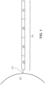
- FIG. 1 is a schematic diagram showing an exemplary capture probe, as described herein.
- the capture probe 102 is optionally coupled to a feature 101 by a cleavage domain 103, such as a disulfide linker.
- the capture probe can include a functional sequence 104 that are useful for subsequent processing.
- the functional sequence 104 can include all or a part of sequencer specific flow cell attachment sequence (e.g., a P5 or P7 sequence), all or a part of a sequencing primer sequence, (e.g., a R1 primer binding site, a R2 primer binding site), or combinations thereof.
- the capture probe can also include a spatial barcode 105.
- the capture probe can also include a unique molecular identifier (UMI) sequence 106. While FIG.
- UMI unique molecular identifier
- the capture probe can also include a capture domain 107 to facilitate capture of a target analyte.
- the capture domain can have a sequence complementary to a sequence of a nucleic acid analyte.
- the capture domain can have a sequence complementary to a connected probe described herein.
- the capture domain can have a sequence complementary to a capture handle sequence present in an analyte capture agent.
- the capture domain can have a sequence complementary to a splint oligonucleotide.
- Such splint oligonucleotide in addition to having a sequence complementary to a capture domain of a capture probe, can have a sequence complementary to a sequence of a nucleic acid analyte, a portion of a connected probe described herein, a capture handle sequence described herein, and/or a methylated adaptor described herein.
- the functional sequences can generally be selected for compatibility with any of a variety of different sequencing systems, e.g., Ion Torrent Proton or PGM, Illumina sequencing instruments, PacBio, Oxford Nanopore, etc., and the requirements thereof.
- functional sequences can be selected for compatibility with non-commercialized sequencing systems. Examples of such sequencing systems and techniques, for which suitable functional sequences can be used, include (but are not limited to) Ion Torrent Proton or PGM sequencing, Illumina sequencing, PacBio SMRT sequencing, and Oxford Nanopore sequencing.
- functional sequences can be selected for compatibility with other sequencing systems, including non-commercialized sequencing systems.
- the spatial barcode 105 and functional sequences 104 is common to all of the probes attached to a given feature.
- the UMI sequence 106 of a capture probe attached to a given feature is different from the UMI sequence of a different capture probe attached to the given feature.
- FIG. 2 is a schematic illustrating a cleavable capture probe, wherein the cleaved capture probe can enter into a non-permeabilized cell and bind to analytes within the sample.
- the capture probe 201 contains a cleavage domain 202, a cell penetrating peptide 203, a reporter molecule 204, and a disulfide bond (-S-S-).
- 205 represents all other parts of a capture probe, for example a spatial barcode and a capture domain.
- Cleavable capture probe are further described in WO 2020/176788 and/or U.S. Patent Application Publication No. 2020/0277663 , each of which is incorporated by reference in its entirety.
- the one or more spatial barcode sequences of the multiple capture probes can include sequences that are the same for all capture probes coupled to the feature, and/or sequences that are different across all capture probes coupled to the feature.
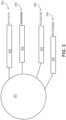
- FIG. 3 is a schematic diagram of an exemplary multiplexed spatially-barcoded feature.
- the feature 301 can be coupled to spatially-barcoded capture probes, wherein the spatially-barcoded probes of a particular feature can possess the same spatial barcode, but have different capture domains designed to associate the spatial barcode of the feature with more than one target analyte.
- a feature may be coupled to four different types of spatially-barcoded capture probes, each type of spatially-barcoded capture probe possessing the spatial barcode 302.
- One type of capture probe associated with the feature includes the spatial barcode 302 in combination with a poly(T) capture domain 303, designed to capture mRNA target analytes.
- a second type of capture probe associated with the feature includes the spatial barcode 302 in combination with a random N-mer capture domain 304 for gDNA analysis.
- a third type of capture probe associated with the feature includes the spatial barcode 302 in combination with a capture domain complementary to a capture handle sequence of an analyte capture agent of interest 305.
- a fourth type of capture probe associated with the feature includes the spatial barcode 302 in combination with a capture domain that can specifically bind a nucleic acid molecule 306 that can function in a CRISPR assay (e.g., CRISPR/Cas9). While only four different capture probe-barcoded constructs are shown in FIG.
- capture-probe barcoded constructs can be tailored for analyses of any given analyte associated with a nucleic acid and capable of binding with such a construct.
- the schemes shown in FIG. 3 can also be used for concurrent analysis of other analytes disclosed herein, including, but not limited to: (a) mRNA, a lineage tracing construct, cell surface or intracellular proteins and metabolites, and gDNA; (b) mRNA, accessible chromatin (e.g., ATAC-seq, DNase-seq, and/or MNase-seq) cell surface or intracellular proteins and metabolites, and a perturbation agent (e.g., a CRISPR crRNA/sgRNA, TALEN, zinc finger nuclease, and/or antisense oligonucleotide as described herein); (c) mRNA, cell surface or intracellular proteins and/or metabolites, a barcoded labelling agent (e.g., the MHC multimers
- a perturbation agent can be a small molecule, an antibody, a drug, an aptamer, a miRNA, a physical environmental (e.g., temperature change), or any other known perturbation agents. See, e.g., Section (II)(b) (e.g., subsections (i)-(vi)) of WO 2020/176788 and/or U.S. Patent Application Publication No. 2020/0277663 . Generation of capture probes can be achieved by any appropriate method, including those described in Section (II)(d)(ii) of WO 2020/176788 and/or U.S. Patent Application Publication No. 2020/0277663 .
- Capture probes attached to a single array feature can include identical (or common) spatial barcode sequences, different spatial barcode sequences, or a combination of both. Capture probes attached to a feature can include multiple sets of capture probes. Capture probes of a given set can include identical spatial barcode sequences. The identical spatial barcode sequences can be different from spatial barcode sequences of capture probes of another set.
- the plurality of capture probes can include spatial barcode sequences (e.g., nucleic acid barcode sequences) that are associated with specific locations on a spatial array.
- a first plurality of capture probes can be associated with a first region, based on a spatial barcode sequence common to the capture probes within the first region
- a second plurality of capture probes can be associated with a second region, based on a spatial barcode sequence common to the capture probes within the second region.
- the second region may or may not be associated with the first region.
- Additional pluralities of capture probes can be associated with spatial barcode sequences common to the capture probes within other regions.
- the spatial barcode sequences can be the same across a plurality of capture probe molecules.
- multiple different spatial barcodes are incorporated into a single arrayed capture probe.
- a mixed but known set of spatial barcode sequences can provide a stronger address or attribution of the spatial barcodes to a given spot or location, by providing duplicate or independent confirmation of the identity of the location.
- the multiple spatial barcodes represent increasing specificity of the location of the particular array point.
- more than one analyte type e.g., nucleic acids and proteins
- a biological sample can be detected (e.g., simultaneously or sequentially) using any appropriate multiplexing technique, such as those described in Section (IV) of WO 2020/176788 and/or U.S. Patent Application Publication No. 2020/0277663 .
- an analyte capture agent refers to an agent that interacts with an analyte (e.g., an analyte in a biological sample) and with a capture probe (e.g., a capture probe attached to a substrate or a feature) to identify the analyte.
- the analyte capture agent includes: (i) an analyte binding moiety (e.g., that binds to an analyte), for example, an antibody or antigen-binding fragment thereof; (ii) analyte binding moiety barcode; and (iii) an analyte capture sequence.
- an analyte binding moiety barcode refers to a barcode that is associated with or otherwise identifies the analyte binding moiety.
- analyte capture sequence refers to a region or moiety configured to hybridize to, bind to, couple to, or otherwise interact with a capture domain of a capture probe.
- an analyte binding moiety barcode (or portion thereof) may be able to be removed (e.g., cleaved) from the analyte capture agent. Additional description of analyte capture agents can be found in Section (II)(b)(ix) of WO 2020/176788 and/or Section (II)(b)(viii) U.S. Patent Application Publication No. 2020/0277663 .
- a spatial barcode with one or more neighboring cells, such that the spatial barcode identifies the one or more cells, and/or contents of the one or more cells, as associated with a particular spatial location.
- One method is to promote analytes or analyte proxies (e.g., intermediate agents) out of a cell and towards a spatially-barcoded array (e.g., including spatially-barcoded capture probes).
- Another method is to cleave spatially-barcoded capture probes from an array and promote the spatially-barcoded capture probes towards and/or into or onto the biological sample.
- capture probes may be configured to prime, replicate, and consequently yield optionally barcoded extension products from a template (e.g., a DNA or RNA template, such as an analyte or an intermediate agent (e.g., a ligation product or an analyte capture agent), or a portion thereof), or derivatives thereof (see, e.g., Section (II)(b)(vii) of WO 2020/176788 and/or U.S. Patent Application Publication No. 2020/0277663 regarding extended capture probes).
- a template e.g., a DNA or RNA template, such as an analyte or an intermediate agent (e.g., a ligation product or an analyte capture agent), or a portion thereof), or derivatives thereof (see, e.g., Section (II)(b)(vii) of WO 2020/176788 and/or U.S. Patent Application Publication No. 2020/0277663 regarding extended capture probes).
- capture probes may be configured to form ligation products with a template (e.g., a DNA or RNA template, such as an analyte or an intermediate agent, or portion thereof), thereby creating ligations products that serve as proxies for a template.
- a template e.g., a DNA or RNA template, such as an analyte or an intermediate agent, or portion thereof
- an "extended capture probe” refers to a capture probe having additional nucleotides added to the terminus (e.g., 3' or 5' end) of the capture probe thereby extending the overall length of the capture probe.
- an "extended 3' end” indicates additional nucleotides were added to the most 3' nucleotide of the capture probe to extend the length of the capture probe, for example, by polymerization reactions used to extend nucleic acid molecules including templated polymerization catalyzed by a polymerase (e.g., a DNA polymerase or a reverse transcriptase).
- a polymerase e.g., a DNA polymerase or a reverse transcriptase
- extending the capture probe includes adding to a 3' end of a capture probe a nucleic acid sequence that is complementary to a nucleic acid sequence of an analyte or intermediate agent specifically bound to the capture domain of the capture probe.
- the capture probe is extended using reverse transcription.
- the capture probe is extended using one or more DNA polymerases. The extended capture probes include the sequence of the capture probe and the sequence of the spatial barcode of the capture probe.
- extended capture probes are amplified (e.g., in bulk solution or on the array) to yield quantities that are sufficient for downstream analysis, e.g., via DNA sequencing.
- extended capture probes e.g., DNA molecules
- act as templates for an amplification reaction e.g., a polymerase chain reaction.
- Section (II)(a) of WO 2020/176788 and/or U.S. Patent Application Publication No. 2020/0277663 Analysis of captured analytes (and/or intermediate agents or portions thereof), for example, including sample removal, extension of capture probes, sequencing (e.g., of a cleaved extended capture probe and/or a cDNA molecule complementary to an extended capture probe), sequencing on the array (e.g., using, for example, in situ hybridization or in situ ligation approaches), temporal analysis, and/or proximity capture, is described in Section (II)(g) of WO 2020/176788 and/or U.S. Patent Application Publication No. 2020/0277663 . Some quality control measures are described in Section (II)(h) of WO 2020/176788 and/or U.S. Patent Application Publication No. 2020/0277663 .
- Spatial information can provide information of biological and/or medical importance.
- the methods and compositions described herein can allow for: identification of one or more biomarkers (e.g., diagnostic, prognostic, and/or for determination of efficacy of a treatment) of a disease or disorder; identification of a candidate drug target for treatment of a disease or disorder; identification (e.g., diagnosis) of a subject as having a disease or disorder; identification of stage and/or prognosis of a disease or disorder in a subject; identification of a subject as having an increased likelihood of developing a disease or disorder; monitoring of progression of a disease or disorder in a subject; determination of efficacy of a treatment of a disease or disorder in a subject; identification of a patient subpopulation for which a treatment is effective for a disease or disorder; modification of a treatment of a subject with a disease or disorder; selection of a subject for participation in a clinical trial; and/or selection of a treatment for a subject with a disease or disorder.
- Spatial information can provide information of biological importance.
- the methods and compositions described herein can allow for: identification of transcriptome and/or proteome expression profiles (e.g., in healthy and/or diseased tissue); identification of multiple analyte types in close proximity (e.g., nearest neighbor analysis); determination of up- and/or down-regulated genes and/or proteins in diseased tissue; characterization of tumor microenvironments; characterization of tumor immune responses; characterization of cells types and their co-localization in tissue; and identification of genetic variants within tissues (e.g., based on gene and/or protein expression profiles associated with specific disease or disorder biomarkers).
- a substrate functions as a support for direct or indirect attachment of capture probes to features of the array.
- a "feature” is an entity that acts as a support or repository for various molecular entities used in spatial analysis.
- some or all of the features in an array are functionalized for analyte capture.
- Exemplary substrates are described in Section (II)(c) of WO 2020/176788 and/or U.S. Patent Application Publication No. 2020/0277663 .
- Exemplary features and geometric attributes of an array can be found in Sections (II)(d)(i), (II)(d)(iii), and (II)(d)(iv) of WO 2020/176788 and/or U.S. Patent Application Publication No. 2020/0277663 .
- analytes and/or intermediate agents can be captured when contacting a biological sample with a substrate including capture probes (e.g., a substrate with capture probes embedded, spotted, printed, fabricated on the substrate, or a substrate with features (e.g., beads, wells) comprising capture probes).
- capture probes e.g., a substrate with capture probes embedded, spotted, printed, fabricated on the substrate, or a substrate with features (e.g., beads, wells) comprising capture probes.
- contact contacted
- contacting a biological sample with a substrate refers to any contact (e.g., direct or indirect) such that capture probes can interact (e.g., bind covalently or non-covalently (e.g., hybridize)) with analytes from the biological sample.
- Capture can be achieved actively (e.g., using electrophoresis) or passively (e.g., using diffusion). Analyte capture is further described in Section (II)(e) of WO 2020/176788 and/or U.S. Patent Application Publication No. 2020/0277663 .
- spatial analysis can be performed by attaching and/or introducing a molecule (e.g., a peptide, a lipid, or a nucleic acid molecule) having a barcode (e.g., a spatial barcode) to a biological sample (e.g., to a cell in a biological sample).
- a plurality of molecules e.g., a plurality of nucleic acid molecules
- a plurality of barcodes e.g., a plurality of spatial barcodes
- a biological sample e.g., to a plurality of cells in a biological sample for use in spatial analysis.
- the biological sample after attaching and/or introducing a molecule having a barcode to a biological sample, the biological sample can be physically separated (e.g., dissociated) into single cells or cell groups for analysis.
- Some such methods of spatial analysis are described in Section (III) of WO 2020/176788 and/or U.S. Patent Application Publication No. 2020/0277663 .
- spatial analysis can be performed by detecting multiple oligonucleotides that hybridize to an analyte.
- spatial analysis can be performed using RNA-templated ligation (RTL).
- RTL RNA-templated ligation
- Methods of RTL have been described previously. See, e.g., Credle et al., Nucleic Acids Res. 2017 Aug 21;45(14):e128 .
- RTL includes hybridization of two oligonucleotides to adjacent sequences on an analyte (e.g., an RNA molecule, such as an mRNA molecule).
- the oligonucleotides are DNA molecules.
- one of the oligonucleotides includes at least two ribonucleic acid bases at the 3' end and/or the other oligonucleotide includes a phosphorylated nucleotide at the 5' end.
- one of the two oligonucleotides includes a capture domain (e.g., a poly(A) sequence, a non-homopolymeric sequence).
- a ligase e.g., SplintR ligase
- the two oligonucleotides hybridize to sequences that are not adjacent to one another.
- hybridization of the two oligonucleotides creates a gap between the hybridized oligonucleotides.
- a polymerase e.g., a DNA polymerase
- the ligation product is released from the analyte.
- the ligation product is released using an endonuclease (e.g., RNAse H).
- the released ligation product can then be captured by capture probes (e.g., instead of direct capture of an analyte) on an array, optionally amplified, and sequenced, thus determining the location and optionally the abundance of the analyte in the biological sample.
- capture probes e.g., instead of direct capture of an analyte
- sequence information for a spatial barcode associated with an analyte is obtained, and the sequence information can be used to provide information about the spatial distribution of the analyte in the biological sample.
- Various methods can be used to obtain the spatial information.
- specific capture probes and the analytes they capture are associated with specific locations in an array of features on a substrate.
- specific spatial barcodes can be associated with specific array locations prior to array fabrication, and the sequences of the spatial barcodes can be stored (e.g., in a database) along with specific array location information, so that each spatial barcode uniquely maps to a particular array location.
- specific spatial barcodes can be deposited at predetermined locations in an array of features during fabrication such that at each location, only one type of spatial barcode is present so that spatial barcodes are uniquely associated with a single feature of the array.
- the arrays can be decoded using any of the methods described herein so that spatial barcodes are uniquely associated with array feature locations, and this mapping can be stored as described above.
- each array feature location represents a position relative to a coordinate reference point (e.g., an array location, a fiducial marker) for the array. Accordingly, each feature location has an "address" or location in the coordinate space of the array.
- Some exemplary spatial analysis workflows are described in the Exemplary Embodiments section of WO 2020/176788 and/or U.S. Patent Application Publication No. 2020/0277663 . See, for example, the Exemplary embodiment starting with "In some non-limiting examples of the workflows described herein, the sample can be immersed... " of WO 2020/176788 and/or U.S. Patent Application Publication No. 2020/0277663 . See also, e.g., the Visium Spatial Gene Expression Reagent Kits User Guide (e.g., Rev C, dated June 2020 ), and/or the Visium Spatial Tissue Optimization Reagent Kits User Guide (e.g., Rev C, dated July 2020 ).
- spatial analysis can be performed using dedicated hardware and/or software, such as any of the systems described in Sections (II)(e)(ii) and/or (V) of WO 2020/176788 and/or U.S. Patent Application Publication No. 2020/0277663 , or any of one or more of the devices or methods described in Sections Control Slide for Imaging, Methods of Using Control Slides and Substrates for, Systems of Using Control Slides and Substrates for Imaging, and/or Sample and Array Alignment Devices and Methods, Informational labels of WO 2020/123320 .
- Suitable systems for performing spatial analysis can include components such as a chamber (e.g., a flow cell or sealable, fluid-tight chamber) for containing a biological sample.
- the biological sample can be mounted for example, in a biological sample holder.
- One or more fluid chambers can be connected to the chamber and/or the sample holder via fluid conduits, and fluids can be delivered into the chamber and/or sample holder via fluidic pumps, vacuum sources, or other devices coupled to the fluid conduits that create a pressure gradient to drive fluid flow.
- One or more valves can also be connected to fluid conduits to regulate the flow of reagents from reservoirs to the chamber and/or sample holder.
- the systems can optionally include a control unit that includes one or more electronic processors, an input interface, an output interface (such as a display), and a storage unit (e.g., a solid state storage medium such as, but not limited to, a magnetic, optical, or other solid state, persistent, writeable and/or re-writeable storage medium).
- the control unit can optionally be connected to one or more remote devices via a network.
- the control unit (and components thereof) can generally perform any of the steps and functions described herein. Where the system is connected to a remote device, the remote device (or devices) can perform any of the steps or features described herein.
- the systems can optionally include one or more detectors (e.g., CCD, CMOS) used to capture images.
- the systems can also optionally include one or more light sources (e.g., LED-based, diode-based, lasers) for illuminating a sample, a substrate with features, analytes from a biological sample captured on a substrate, and various control and calibration media.
- one or more light sources e.g., LED-based, diode-based, lasers
- the systems can optionally include software instructions encoded and/or implemented in one or more of tangible storage media and hardware components such as application specific integrated circuits.
- the software instructions when executed by a control unit (and in particular, an electronic processor) or an integrated circuit, can cause the control unit, integrated circuit, or other component executing the software instructions to perform any of the method steps or functions described herein.
- the systems described herein can detect (e.g., register an image) the biological sample on the array.
- Exemplary methods to detect the biological sample on an array are described in PCT Application No. 2020/061064 and/or U.S. Patent Application Serial No. 16/951,854 .
- the biological sample Prior to transferring analytes from the biological sample to the array of features on the substrate, the biological sample can be aligned with the array. Alignment of a biological sample and an array of features including capture probes can facilitate spatial analysis, which can be used to detect differences in analyte presence and/or level within different positions in the biological sample, for example, to generate a three-dimensional map of the analyte presence and/or level. Exemplary methods to generate a two- and/or three-dimensional map of the analyte presence and/or level are described in PCT Application No. 2020/053655 and spatial analysis methods are generally described in WO 2020/061108 and/or U.S. Patent Application Serial No. 16/951,864 .
- a map of analyte presence and/or level can be aligned to an image of a biological sample using one or more fiducial markers, e.g., objects placed in the field of view of an imaging system which appear in the image produced, as described in the Substrate Attributes Section, Control Slide for Imaging Section of WO 2020/123320 , PCT Application No. 2020/061066 , and/or U.S. Patent Application Serial No. 16/951,843 .
- fiducial markers e.g., objects placed in the field of view of an imaging system which appear in the image produced, as described in the Substrate Attributes Section, Control Slide for Imaging Section of WO 2020/123320 , PCT Application No. 2020/061066 , and/or U.S. Patent Application Serial No. 16/951,843 .
- Fiducial markers can be used as a point of reference or measurement scale for alignment (e.g., to align a sample and an array, to align two substrates, to determine a location of a sample or array on a substrate relative to a fiducial marker) and/or for quantitative measurements of sizes and/or distances.
- Cancer and disease diagnosis and treatment plans are oftentimes guided by three diagnostic tools: blood work, imaging, and/or biopsies.
- surgery is the first line of treatment when cancerous and diseased tissues are identified, especially in the early stages of the disease.
- clinicians face difficult decisions on the size and site of the resection of the diseased tissue.
- Conclusive diagnosis and staging are oftentimes obtained after surgical resection has been completed. Unfortunately, sometimes the entire abnormal tissue is not completely removed, leaving tissue margins that contain diseased cells that can proliferate in the body and cause continued cancer and disease.
- the methods described here can help support a clinician's decision on the type of surgical intervention to provide a subject with a potential cancer or diseased tissue.
- information from practicing the described methods can help determine the size and site of tissue resection by more completely identifying abnormal tissue margins, thereby reducing the risk of re-excision and/or reducing the risk of future recurrence of the cancerous or diseased tissue in the subject.
- determining the size and site of a tissue to be resected from a subject that include: (a) contacting a tissue sample obtained from the subject to an array comprising a plurality of capture probes, where a capture probe of the plurality of capture probes comprises (i) a capture domain that binds specifically to an analyte of the tissue sample and (ii) a spatial barcode; (b) determining (i) all or a part of a nucleic acid sequence corresponding to the analyte specifically bound to the capture domain or a complement thereof, and (ii) all or a part of a nucleic acid sequence corresponding to the spatial barcode or a complement thereof, and using the determined nucleic acid sequences of (i) and (ii) to identify the presence of the analyte at a location in the tissue sample; (c) comparing the presence of the analyte at the location in the tissue sample to presence of the analyte at different location(s) in the
- the analyte is a DNA or RNA. In some embodiments, the analyte is a messenger RNA (mRNA) molecule. In some embodiments, the analyte is a genomic DNA. In some embodiments, the analyte comprises a full-length sequence of a biomarker described herein. In some embodiments, the analyte comprises a fragment of the sequence of a biomarker described herein. In some embodiments of any of the methods described herein, each of the plurality of capture probes comprises (i) a capture domain that binds specifically to an analyte of the tissue section and (ii) a spatial barcode. The capture probe can be any capture probe described herein.
- the capture domain of the capture probe comprises a sequence that is substantially complementary (e.g., at least 80%, at least 85%, at least 90%, at least 95%, at least 99%, or 100% complementary) to a portion of the sequence of the analyte of the tissue sample.
- the capture domain can have a total of about 10 nucleotides to about 125 nucleotides (or any of the subranges of this range described herein).
- the sequence that is substantially complementary to a portion of the sequence of the analyte can be a random sequence.
- the sequence that is substantially complementary to a portion of the sequence of the analyte can include a poly(T) oligonucleotide sequence (e.g., at least 5 contiguous Ts, at least 10 continguous Ts, or at least 15 contiguous Ts).
- a poly(T) oligonucleotide sequence e.g., at least 5 contiguous Ts, at least 10 continguous Ts, or at least 15 contiguous Ts.
- step (b) comprises sequencing (i) all or a part of the nucleic acid sequence corresponding to the analyte specifically bound to the capture domain or a complement thereof, and (ii) all or a part of the nucleic acid sequence corresponding to the spatial barcode or a complement thereof.
- the sequencing is high throughput sequencing, sequencing by synthesis, sequencing by hybridization, sequencing by ligation or any of the other methods for sequencing described herein or known in the art.
- sequencing can involve one or more of nucleic acid amplification, the ligation or addition of one or more sequencing adaptors, cleavage of the capture probe from the array, extension of the capture probe using the bound cDNA as a template, and generating a single-stranded nucleic acid comprising a sequence that is complementary to all or a part of the extended capture probe.
- Non-limiting methods for determining the sequence of (i) all or a part of the sequence of the target nucleic acid, or a complement thereof, or (ii) all or a part of the nucleic acid sequence corresponding to the binding moiety barcode, or a complement thereof, are described herein or are known in the art.
- Also provided herein are methods of determining the size and site of a tissue to be resected from a subject that include: (a) contacting a tissue sample obtained from the subject to a plurality of analyte capture agents, where an analyte capture agent of the plurality of analyte capture agents comprises an analyte binding moiety barcode, an analyte capture sequence, and an analyte binding moiety that binds specifically to an analyte; (b) disposing the tissue sample onto an array, where the array comprises a plurality of capture probes, where a capture probe of the plurality of capture probes comprises a spatial barcode and a capture domain that binds specifically to the analyte capture sequence; (c) determining (i) all or a part of the nucleic acid sequence corresponding to the analyte binding moiety barcode or a complement thereof, and (ii) all or a part of the nucleic acid sequence corresponding to the binding moiety barcode or
- the analyte is a protein. In some embodiments, the analyte is a full-length protein. In some embodiments, the analyte is a fragment of a protein. In some embodiments, the analyte is a byproduct of a protein. In some embodiments, the protein is any of the exemplary cancer biomarkers described herein.
- each of the plurality of analyte capture agents comprises an analyte binding moiety barcode, an analyte capture sequence, and an analyte binding moiety that binds specifically to an analyte.
- the analyte binding moiety is an antibody or an antigen-binding antibody fragment (e.g., a Fab). Any other suitable protein binding moiety known in the art can also be used as an analyte binding moiety.
- the analyte binding moiety barcode can be any barcode described herein.
- the analyte capture sequence can be any analyte capture sequence described herein.
- each of the plurality of capture probes comprises a spatial barcode and a capture domain that binds specifically to the analyte capture sequence.
- the capture probe can be any capture probe described herein.
- the capture domain of the capture probe comprises a sequence that is substantially complementary (e.g., at least 80%, at least 85%, at least 90%, at least 95%, at least 99%, or 100% complementary) to a portion of the analyte capture sequence.
- the capture domain can have a total of about 10 nucleotides to about 125 nucleotides (or any of the subranges of this range described herein).
- the sequence that is substantially complementary to a portion of the analyte capture sequence can be a random sequence. In some embodiments, the sequence that is substantially complementary to a portion of the analyte capture sequence can include a poly(T) oligonucleotide sequence (e.g., at least 5 contiguous Ts, at least 10 continguous Ts, or at least 15 contiguous Ts).
- the determining of the sequence is by sequencing.
- the sequencing is high throughput sequencing, sequencing by hybridization, or any of the other methods for sequencing described herein or known in the art.
- sequencing can involve one or more of nucleic acid amplification, the ligation or addition of one or more sequencing adaptors, cleavage of the capture probe from the array, extension of the capture probe using the bound analyte capture sequence as a template, and generating a single-stranded nucleic acid comprising a sequence that is complementary to all or a part of the extended capture probe.
- Non-limiting methods for determining the sequence of (i) all or a part of the nucleic acid sequence corresponding to the analyte binding moiety barcode, or a complement thereof, or (ii) all or a part of the nucleic acid sequence corresponding to the spatial barcode, or a complement thereof, are described herein or are known in the art.
- the tissue to be resected is a tumor (e.g., a malignant or a benign tumor).
- the tumor is a solid tumor.
- the subject is suspected of having a cancer.
- the subject has been previously diagnosed or identified as having a cancer (e.g., any of the exemplary cancers described herein).
- Non-limiting examples of cancers referred to in any one the methods described herein include: sarcomas, carcinomas, adrenocortical carcinoma, AIDS-related cancers, anal cancer, appendix cancer, astrocytomas, atypical teratoid/rhabdoid tumor, basal cell carcinoma, bladder cancer, brain stem glioma, brain tumors (including brain stem glioma, central nervous system atypical teratoid/rhabdoid tumor, central nervous system embryonal tumors, astrocytomas, craniopharyngioma, ependymoblastoma, ependymoma, medulloblastoma, medulloepithelioma, pineal parenchymal tumors of intermediate differentiation, supratentorial primitive neuroectodermal tumors, and pineoblastoma), breast cancer, bronchial tumors, cancer of unknown primary site, carcinoid tumor, carcinoma of unknown primary site, central
- the tissue to be resected can include a tumor (e.g., a malignant tumor) of any of the types of cancer described herein.
- a tumor e.g., a malignant tumor
- the analyte is a tumor biomarker.
- the analyte is a tumor antigen.
- Exemplary tumor antigens include, but are not limited to, melanoma-associated antigen (MAGE) series of antigens (e.g., MAGE-C1 (cancer/testis antigen CT7), MAGE-B1 antigen (MAGE-XP antigen, DAM10), MAGE-B2 antigen (DAM6), MAGE-2 antigen, MAGE-4a antigen, and MAGE-4b antigen), tyrosinase, glycoprotein 100 (gp100), disialoganglioside GD-2, disialoganglioside O-acetylated GD-3, ganglioside GM-2, epidermal growth factor receptor (EGFR), vascular endothelial growth factor receptor (VEGFR), mutant B-Raf antigen associated with melanoma and colon cancer, human
- MAGE melanoma-associated antigen
- the tissue to be resected is an infected tissue, a necrotic tissue, or a diseased tissue.
- the analyte can be associated with an infection, necrosis, inflammation, or disease. Non-limiting examples of such analytes are known in the art.
- the tissue to be resected is infected by a bacterium. In some embodiments, the tissue to be resected is infected by a virus. In some embodiments, the tissue to be resected is infected by a fungus. In some embodiments, the tissue to be resected is infected by a parasite or protozoa.
- the tissue to be resected is infected by a bacterium, e.g., a Bordetella pertussis, a Brucella abortis, a Escherichia coli, a Salmonella species, e.g., a Salmonella typhi, a Streptococci, a Vibrio ( V cholera, V.
- a bacterium e.g., a Bordetella pertussis, a Brucella abortis, a Escherichia coli, a Salmonella species, e.g., a Salmonella typhi, a Streptococci, a Vibrio ( V cholera, V.
- a Shigella a Pseudomonas, a Brucella species, a Klebsiella, a Mycobacteria species (a tuberculosis, an avium, a BCG, a leprosy), a Pneumococci, a Staphylococci, a Enterobacter species, a Clostridium tetani, a Bacillus anthracis, a Streptococcus pneumoniae, a Meningococcus A, B, C, Y, W, or W-135, a Helicobacter pylori, a Rochalimaea henselae, a Pasteurella ( P.
- haemolytica P. multocida
- Chlamydia C. trachomatis, C. psittaci
- a Treponema pallidum a Haemophilus species, e.g., a Haemophilus influenza type b, a mycoplasma species, a Borrelia burgdorferi, a Legionella pneumophila, a Clostridium botulinum, a Corynebacterium diptheriae, a Yersinia entercolitica, a Ehrlichia, a Anaplasma, or a Coxiella burnetii bacterium.
- the tissue to be resected is infected by a parasite or protozoa, e.g., those causing malaria ( Plasmodium falciparum, P. vivax, or P. malariae ), a schistosome, a trypanosome, leishmania, a filarial nematode, Trichomonas vaginalis, a sarcocystis, a Taenia species ( T. saginata or T. solium ), Toxoplasma gondi, Trichinella spiralis, or an Eimeria species.
- a parasite or protozoa e.g., those causing malaria ( Plasmodium falciparum, P. vivax, or P. malariae ), a schistosome, a trypanosome, leishmania, a filarial nematode, Trichomonas vaginalis, a sarcocystis, a Taenia species ( T
- the tissue to be resected is infected by a fungus, e.g., Cryptococcus neoformans, Candida albicans, Aspergillus fumigatus, Coccidioides immitis, or Coccidioides posadasii.
- a fungus e.g., Cryptococcus neoformans, Candida albicans, Aspergillus fumigatus, Coccidioides immitis, or Coccidioides posadasii.
- the tissue to be resected is infected by a virus, e.g., a rotavirus, an aphthovirus (the agent for foot and mouth disease), an Ebola virus, a Hanta virus, a parainfluenza, a herpes virus species (e.g., herpes simplex virus, Epstein-Barr virus, chicken pox virus, pseudorabies, or cytomegalovirus), a rabies virus, a polio virus, a Hepatitis A, B, C or E, distemper, a Venezuelan equine encephalomyelitis virus, a feline leukemia virus, a reovirus, a respiratory syncytial virus, a Lassa fever virus, a polyoma virus, a canine parvovirus, a papilloma virus, a flavivirus, a tick home encephalitis virus, a paramyxovirus (the agent for Rinder), a her
- the methods provided herein comprise comparing the presence of the analyte at the location in the tissue sample to presence of the analyte at different location(s) in the tissue sample, and determining the size and site of a tissue to be resected from the subject based on the comparison.
- the different location(s) in the tissue sample are reference location(s).
- the reference location(s) in the tissue sample are locations of healthy tissue.
- the reference location(s) in the tissue sample are locations of non-cancerous tissue.
- the reference location(s) in the tissue sample are locations of non-tumor tissue.
- the reference location(s) in the tissue sample are locations with no abnormalities such as tumor, cancer, necrosis, inflammation, infection, or disease.
- the presence of the analyte at the location in the tissue sample is significantly different from the presence of the analyte at the different location(s) in the tissue. In some embodiments, the presence of the analyte at the location in the tissue sample is significantly greater than the presence of the analyte at the different location(s) in the tissue sample. In some embodiments, the presence of the analyte at the location in the tissue sample is significantly less than the presence of the analyte at the different location(s) in the tissue sample.
- the location at the tissue sample is determined to be resected if the presence of the analyte at the location in the tissue sample is significantly different from the presence of the analyte at the different location(s). In some embodiments, the location at the tissue sample is determined to be resected if the presence of the analyte at the location in the tissue sample is significantly greater than the presence of the analyte at the different location(s). In some embodiments, the location at the tissue sample is determined to be resected if the presence of the analyte at the location in the tissue sample is significantly less than the presence of the analyte at the different location(s).
- the presence of the analyte at the location in the tissue sample is about 0.1-fold to about 100-fold (e.g., about 0.1-fold to about 90-fold, about 0.1-fold to about 80-fold, about 0.1-fold to about 70-fold, about 0.1-fold to about 60-fold, about 0.1-fold to about 50-fold, about 0.1-fold to about 40-fold, about 0.1-fold to about 30-fold, about 0.1-fold to about 20-fold, about 0.1-fold to about 15-fold, about 0.1-fold to about 10-fold, about 0.1-fold to about 8-fold, about 0.1-fold to about 6-fold, about 0.1-fold to about 5-fold, about 0.1-fold to about 4-fold, about 0.1-fold to about 3-fold, about 0.1-fold to about 2-fold, about 0.1-fold to about 1.5-fold, about 0.1-fold to about 1-fold, about 0.1-fold to about 0.8-fold, about 0.1-fold to about 0.6-fold, about 0.1-fold to about
- the presence of the analyte at the location in the tissue sample is about 1% to about 99% (e.g., about 1% to about 95%, about 1% to about 90%, about 1% to about 85%, about 1% to about 80%, about 1% to about 75%, about 1% to about 70%, about 1% to about 65%, about 1% to about 60%, about 1% to about 55%, about 1% to about 50%, about 1% to about 45%, about 1% to about 40%, about 1% to about 35%, about 1% to about 30%, about 1% to about 25%, about 1% to about 20%, about 1% to about 15%, about 1% to about 10%, about 1% to about 5%, about 5% to about 99%, about 5% to about 95%, about 5% to about 90%, about 5% to about 85%, about 5% to about 80%, about 5% to about 75%, about 5% to about 70%, about 5% to about 65%, about 5% to about 60%, about 5% to about 55%, about
- the presence of certain biomarkers associated with a cancer and/or disease e.g., breast cancer biomarkers in ductal carcinoma
- a cancer and/or disease e.g., breast cancer biomarkers in ductal carcinoma
- the location in the tissue sample is considered "clear.” If the presence of certain biomarkers associated with a cancer and/or disease are above a threshold value for those biomarkers, the location in the tissue sample is considered within the margin of tissue to be resected.
- the method further comprises comparing presence of one or more additional analyte(s) at the location in the tissue sample with the presence of the one or more additional analyte(s) at the different location(s) in the tissue sample.
- the presence of a total of about 1 to about 20,000 e.g., about 1 to about 18,000, about 1 to about 16,000, about 1 to about 14,000, about 1 to about 12,000, about 1 to about 10,000, about 1 to about 9,000, about 1 to about 8,000, about 1 to about 7,000, about 1 to about 6,000, about 1 to about 5,000, about 1 to about 4,500, about 1 to about 4,000, about 1 to about 3,500, about 1 to about 3,000, about 1 to about 2,500, about 1 to about 2,000, about 1 to about 1,500, about 1 to about 1,000, about 1 to about 800, about 1 to about 600, about 1 to about 500, about 1 to about 400, about 1 to about 300, about 1 to about 200, about 1 to about 100, about 1 to about 1 to about
- mutant cells are identified according to the presence of the one or more analyte(s) at the location in the tissue sample. In some embodiments, a mutant cell is identified according to the presence of one or more biomarkers described herein. In some embodiments, a mutant cell is identified according to the presence of one or more cell-surface biomarkers, e.g., a cell-surface receptor, at the location in the tissue sample.
- a cell within a location at the tissue sample is identified as a mutant cell if the presence of the one or more analyte(s) at the location in the tissue sample are significantly different from the presence of the analyte(s) at the different location(s). In some embodiments, a cell within a location at the tissue sample is identified as a mutant cell if the presence of the one or more analyte(s) at the location in the tissue sample are significantly greater than the presence of the analyte(s) at the different location(s).
- the mutant cell(s) within the location at the tissue sample is determined to be resected if the presence of the analyte(s) at the location in the tissue sample are significantly less than the presence of the analyte at the different location(s).
- a location at the tissue sample comprises about 1 to about 100,000 (e.g., about 1 to about 90,000, about 1 to about 80,000, about 1 to about 70,000, about 1 to about 60,000, about 1 to about 50,000, about 1 to about 40,000, about 1 to about 30,000, about 1 to about 20,000, about 1 to about 10,000, about 1 to about 9,000, about 1 to about 8,000, about 1 to about 7,000, about 1 to about 6,000, about 1 to about 5,000, about 1 to about 4,000, about 1 to about 3,000, about 1 to about 2,000, about 1 to about 1,000, about 1 to about 900, about 1 to about 800, about 1 to about 700, about 1 to about 600, about 1 to about 500, about 1 to about 400, about 1 to about 300, about 1 to about 200, about 1 to about 100, about 1 to about 90, about 1 to about 80, about 1 to about 70, about 1 to about 60, about 1 to about 50, about 1 to about 40, about 1 to about 30, about 1 to about 20, about 1 to about 10,
- the tissue sample can be obtained from any suitable tissue or organ from the subject (e.g., breast tissue, muscle tissue, gland tissue, fat or adipose tissue, nerve tissue, joint tissue, ligament tissue, tendon tissue, mouth tissue, tongue tissue, salivary gland tissue, parotid gland tissue, submandibular gland tissue, sublingual gland tissue, pharynx tissue, esophageal tissue, stomach tissue, small intestine tissue, duodenum tissue, jejunum tissue, ileum tissue, large intestine tissue, liver tissue, gallbladder tissue, mesentery tissue, pancreas tissue, anal canal tissue, anus tissue, nasal cavity tissue, pharynx tissue, larynx tissue, trachea tissue, bronchi tissue, lung tissue, diaphragm tissue, kidney tissue, ureter tissue, bladder tissue, urethra tissue, ovarian tissue, fallopian tube tissue, uterus tissue, vagina tissue, vulva tissue
- the spatial barcode of the capture probe can be any spatial barcode described herein.
- the array can be any of the types of arrays described herein.
- the array comprises a slide.
- the capture probe is attached to the slide (e.g., by its 5' end).
- the array is a bead array. In some embodiments, a 5' end of the capture probe is attached to a bead of the bead array.
- the method comprises extending a 3' end of the capture probe using the specifically bound analyte or analyte binding agent barcode as a template to generate an extended capture probe.
- additional methods are used in combination with the methods described herein to determine the site and size of the tissue to be resected in a subject.
- medical imaging modalities such as computed tomography (CT) and magnetic resonance imaging (MRI) are used in combination with the methods described herein.
- MRI magnetic resonance imaging
- PET position emission tomography
- an initial scanning of a cancer patient and/or imaging of a tissue sample from a cancer patient can be performed using, e.g., MRI, CT, and/or PET prior to the methods described herein, and a preliminary assessment of a surgical margin can be performed.
- the initial information can provide guidance on, e.g., where to obtain the tissue sample for use in the methods described herein, the size of the tissue sample, and/or the number of tissue samples needed.
- a follow-up scanning and/or imaging can be performed using e.g., MRI, CT, and/or PET after the methods described herein are performed.
- the follow-up scanning and /or imaging provide information on, e.g., the clearance of the cancerous and/or diseased tissue, and whether there are residual cancerous and/or diseased tissue. Any other suitable methods known in the art can also be used in combination with the methods described herein.
- Some embodiments of any of the methods described herein can further include obtaining the tissue sample from the subject (e.g., obtain a biopsy from the subject).
- At least a portion of the tissue to be resected includes cancer cell(s), pre-cancerous cell(s), necrotic cell(s), infected cell(s), and/or diseased tissue. In some embodiments, at least 80%, at least 85%, at least 90%, at least 95%, at least 99%, or 100% of the tissue to be resected includes one or more of cancer cell(s), pre-cancerous cell(s), necrotic cell(s), infected cell(s), and disease tissue.
- Also provided herein are methods of treating a subject in need thereof that include: (a) contacting a tissue sample obtained from the subject to an array comprising a plurality of capture probes, wherein a capture probe of the plurality of capture probes comprises (i) a capture domain that specifically binds to an analyte of the tissue sample and (ii) a spatial barcode; (b) determining (i) all or a part of a nucleic acid sequence corresponding to the analyte specifically bound to the capture domain or a complement thereof, and (ii) all or a part of a nucleic acid sequence corresponding to the spatial barcode or a complement thereof, and using the determined nucleic acid sequences of (i) and (ii) to identify the presence of the analyte at a location in the tissue sample; (c) comparing the presence of the analyte at the location in the tissue sample to presence of the analyte at a different location in the tissue sample, and determining the surgical margin based on
- a method of treating a subject comprising: resecting tissue from the subject using a surgical margin previously determined using a method comprising the steps of: (a) contacting a tissue sample obtained from the subject to an array comprising a plurality of capture probes, wherein a capture probe of the plurality of capture probes comprises (i) a capture domain that specifically binds to an analyte of the tissue sample and (ii) a spatial barcode; (b) determining (i) all or a part of a nucleic acid sequence corresponding to the analyte specifically bound to the capture domain or a complement thereof, and (ii) all or a part of a nucleic acid sequence corresponding to the spatial barcode or a complement thereof, and using the determined nucleic acid sequences of (i) and (ii) to identify the presence of the analyte at a location in the tissue sample; (c) comparing the presence of the analyte at the location in the tissue sample to the presence of the analyl tissue
- the data obtained can provide the clinician with information on the accurate location of the cancerous or diseased tissue, therefore provide the accurate surgical margin of the tissue to be resected.
- the clinician is able to achieve, e.g., more complete resection, thereby treating the subject.
- the analyte is a DNA or RNA. In some embodiments, the analyte is a messenger RNA (mRNA) molecule. In some embodiments, the analyte is a genomic DNA. In some embodiments, the analyte comprises a full-length sequence of a biomarker described herein. In some embodiments, the analyte comprises a fragment of the sequence of a biomarker described herein. In some embodiments of any of the methods described herein, each of the plurality of capture probes comprises (i) a capture domain that binds specifically to an analyte of the tissue sample and (ii) a spatial barcode. The capture probe can be any capture probe described herein.
- the capture domain of the capture probe comprises a sequence that is substantially complementary (e.g., at least 80%, at least 85%, at least 90%, at least 95%, at least 99%, or 100% complementary) to a portion of the sequence of the analyte of the tissue sample.
- the capture domain can have a total of about 10 nucleotides to about 125 nucleotides (or any of the subranges of this range described herein).
- the sequence that is substantially complementary to a portion of the sequence of the analyte can be a random sequence.
- the sequence that is substantially complementary to a portion of the sequence of the analyte can include a poly(T) oligonucleotide sequence (e.g., at least 5 contiguous Ts, at least 10 continguous Ts, or at least 15 contiguous Ts).
- a poly(T) oligonucleotide sequence e.g., at least 5 contiguous Ts, at least 10 continguous Ts, or at least 15 contiguous Ts.
- step (b) comprises sequencing (i) all or a part of the nucleic acid sequence corresponding to the analyte specifically bound to the capture domain or a complement thereof, and (ii) all or a part of the nucleic acid sequence corresponding to the spatial barcode or a complement thereof.
- the sequencing is high throughput sequencing, sequencing by hybridization, or any of the other methods for sequencing described herein or known in the art.
- sequencing can involve one or more of nucleic acid amplification, the ligation or addition of one or more sequencing adaptors, cleavage of the capture probe from the array, extension of the capture probe using the bound cDNA as a template, and generating a single-stranded nucleic acid comprising a sequence that is complementary to all or a part of the extended capture probe.
- Non-limiting methods for determining the sequence of (i) all or a part of the sequence of the target nucleic acid, or a complement thereof, or (ii) all or a part of the nucleic acid sequence corresponding to the spatial barcode, or a complement thereof, are described herein or are known in the art.
- Also provided herein are methods of treating a subject that include: (a) contacting a tissue sample obtained from the subject to a plurality of analyte capture agents, wherein an analyte capture agent of the plurality of analyte capture agents comprises an analyte binding moiety barcode, an analyte capture sequence, and an analyte binding moiety that binds specifically to an analyte; (b) disposing the tissue sample onto an array, wherein the array comprises a plurality of capture probes, wherein a capture probe of the plurality of capture probes comprises a spatial barcode and a capture domain that binds specifically to the analyte capture sequence; (c) determining (i) all or a part of a nucleic acid sequence corresponding to the analyte binding moiety barcode or a complement thereof, and (ii) all or a part of a nucleic acid sequence corresponding to the spatial barcode or a complement thereof, and using the determined nucleic acid
- Also provided herein are methods of treating a subject comprising: resecting tissue from the subject using a surgical margin previously determined using a method comprising the steps of: (a) contacting a tissue sample obtained from the subject to a plurality of analyte capture agents, wherein an analyte capture agent of the plurality of analyte capture agents comprises an analyte binding moiety barcode, an analyte capture sequence, and an analyte binding moiety that binds specifically to an analyte; (b) disposing the tissue sample onto an array, wherein the array comprises a plurality of capture probes, wherein a capture probe of the plurality of capture probes comprises a spatial barcode and a capture domain that binds specifically to the analyte capture sequence; (c) determining (i) all or a part of a nucleic acid sequence corresponding to the analyte binding moiety barcode or a complement thereof, and (ii) all or a part of
- the analyte is a protein. In some embodiments, the analyte is a full-length protein. In some embodiments, the analyte is a fragment of a protein. In some embodiments, the analyte is a byproduct of a protein. In some embodiments, the protein is any of the exemplary cancer biomarkers described herein.
- each of the plurality of analyte capture agents comprises an analyte binding moiety barcode, an analyte capture sequence, and an analyte binding moiety that binds specifically to an analyte.
- the analyte binding moiety is an antibody or an antigen-binding antibody fragment (e.g., a Fab). Any other suitable protein binding moiety known in the art can also be used as an analyte binding moiety.
- the analyte binding moiety barcode can be any barcode described herein.
- the analyte capture sequence can be any analyte capture sequence described herein.
- each of the plurality of capture probes comprises a spatial barcode and a capture domain that binds specifically to the analyte capture sequence.
- the capture probe can be any capture probe described herein.
- the capture domain of the capture probe comprises a sequence that is substantially complementary (e.g., at least 80%, at least 85%, at least 90%, at least 95%, at least 99%, or 100% complementary) to a portion of the analyte capture sequence.
- the capture domain can have a total of about 10 nucleotides to about 125 nucleotides (or any of the subranges of this range described herein).
- the sequence that is substantially complementary to a portion of the analyte capture sequence can be a random sequence. In some embodiments, the sequence that is substantially complementary to a portion of the analyte capture sequence can include a poly(T) oligonucleotide sequence (e.g., at least 5 contiguous Ts, at least 10 continguous Ts, or at least 15 contiguous Ts).
- the determining of the sequence is by sequencing.
- the sequencing is high throughput sequencing, sequencing by hybridization, or any of the other methods for sequencing described herein or known in the art.
- sequencing can involve one or more of nucleic acid amplification, the ligation or addition of one or more sequencing adaptors, cleavage of the capture probe from the array, extension of the capture probe using the bound analyte capture sequence as a template, and generating a single-stranded nucleic acid comprising a sequence that is complementary to all or a part of the extended capture probe.
- Non-limiting methods for determining the sequence of (i) all or a part of the nucleic acid sequence corresponding to the analyte binding moiety barcode, or a complement thereof, or (ii) all or a part of the nucleic acid sequence corresponding to the spatial barcode, or a complement thereof, are described herein or are known in the art.
- the resected tissue is or comprises a tumor (e.g., a malignant or a benign tumor).
- the tumor is a solid tumor.
- the subject is suspected of having a cancer.
- the subject has been previously diagnosed or identified as having a cancer (e.g., any of the exemplary cancers described herein).
- the resected tissue can include a tumor, (e.g., a malignant tumor) of any of the types of cancer describes herein.
- a tumor e.g., a malignant tumor
- the analyte is a tumor biomarker. In some embodiments, the analyte is a tumor antigen. Exemplary tumor antigens include, but are not limited to, any of the exemplary tumor antigens described herein.
- the resected tissue is or comprises an infected tissue, a necrotic tissue, or a diseased tissue.
- the analyte can be associated with an infection, necrosis, inflammation, or disease. Non-limiting examples of such analytes are known in the art.
- the resected tissue is infected by a bacterium (e.g., any of the exemplary bacteria described herein), a parasite or protozoa (e.g., any of the exemplary parasites or protozoa described herein), a fungus (e.g., any of the exemplary fungi described herein), or a virus (e.g., any of the exemplary viruses described herein).
- a bacterium e.g., any of the exemplary bacteria described herein
- a parasite or protozoa e.g., any of the exemplary parasites or protozoa described herein
- a fungus e.g., any of the exemplary fungi described herein
- a virus e.g., any of the exemplary viruses described herein.
- the methods provided herein comprise comparing the presence of the analyte at the location in the tissue sample to the presence of the analyte at different location(s) in the tissue sample, and determining the size and site of a tissue to be resected from the subject based on the comparison.
- the different location(s) in the tissue sample are reference location(s).
- the reference location(s) in the tissue sample are locations of healthy tissue.
- the reference location(s) in the tissue sample are locations of non-cancerous tissue.
- the reference location(s) in the tissue sample are locations of non-tumor tissue.
- the reference location(s) in the tissue sample are locations with no abnormalities such as tumor, cancer, necrosis, inflammation, infection, or disease.
- the presence of the analyte at the location in the tissue sample is significantly different from the presence of the analyte at the different location(s) in the tissue. In some embodiments, the presence of the analyte at the location in the tissue sample is significantly greater than the presence of the analyte at the different location(s) in the tissue sample. In some embodiments, the presence of the analyte at the location in the tissue sample is significantly less than the presence of the analyte at the different location(s) in the tissue sample.
- the location at the tissue sample is determined to be resected if the presence of the analyte at the location in the tissue sample is significantly different from the presence of the analyte at the different location(s). In some embodiments, the location at the tissue sample is determined to be resected if the presence of the analyte at the location in the tissue sample is significantly greater than the presence of the analyte at the different location(s). In some embodiments, the location at the tissue sample is determined to be resected if the presence of the analyte at the location in the tissue sample is significantly less than the presence of the analyte at the different location(s).
- the presence of the analyte at the location in the tissue sample is about 0.1-fold to about 100-fold (e.g., or any of the subranges of this range described herein) greater than the presence of the analyte at the different location(s).
- the presence of the analyte at the location in the tissue sample is about 1% to about 99% (e.g., or any of the subranges of this range described herein) decreased than the presence of the analyte at the different location(s).
- the presence of certain biomarkers associated with a cancer and/or disease e.g., breast cancer biomarkers in ductal carcinoma
- a cancer and/or disease e.g., breast cancer biomarkers in ductal carcinoma
- the location in the tissue sample is considered "clear.” If the presence of certain biomarkers associated with a cancer and/or disease are above a threshold value for those biomarkers, the location in the tissue sample is considered within the margin of tissue to be resected.
- the method further comprises comparing the presence of one or more additional analyte(s) at the location in the tissue sample with the presence of the one or more additional analyte(s) at the different location(s) in the tissue sample.
- the presence of a total of about 1 to about 20,000 (e.g., or any of the subranges of this range described herein) analyte(s) at the location are compared to the presence of the analyte(s) at the different location(s).
- mutant cells are identified according to the presence of the one or more analyte(s) at the location in the tissue sample. In some embodiments, a mutant cell is identified according to the presence of one or more biomarkers decribed herein. In some embodiments, a mutant cell is identified according to the presence of one or more cell-surface biomarkers, e.g., a cell-sruface receptor, at the location in the tissue sample.
- a cell within a location at the tissue sample is identified as a mutant cell if the presence of the one or more analyte(s) at the location in the tissue sample are significantly different from the presence of the analyte(s) at the different location(s). In some embodiments, a cell within a location at the tissue sample is identified as a mutant cell if the presence of the one or more analyte(s) at the location in the tissue sample are significantly greater than the presence of the analyte(s) at the different location(s).
- the mutant cell(s) within the location at the tissue sample is determined to be resected if the presence of the analyte(s) at the location in the tissue sample are significantly less than the presence of the analyte at the different location(s).
- a location at the tissue sample comprises about 1 to about 100,000 cells.
- the spatial barcode of the capture probe can be any spatial barcode described herein.
- the array can be any of the types of arrays described herein.
- the array comprises a slide.
- the capture probe is attached to the slide (e.g., by its 5' end).
- the array is a bead array. In some embodiments, a 5' end of the capture probe is attached to a bead of the bead array.
- the method comprises extending a 3' end of the capture probe using the specifically bound analyte or analyte binding agent barcode as a template to generate an extended capture probe.
- additional methods are used in combination with the methods described herein to determine the site and size of the tissue to be resected in a subject.
- medical imaging modalities such as computed tomography (CT) and magnetic resonance imaging (MRI) are used in combination with the methods described herein.
- MRI magnetic resonance imaging
- PET position emission tomography
- an initial scanning of a cancer patient and/or imaging of a tissue sample from a cancer patient can be performed using, e.g., MRI, CT, and/or PET prior to the methods described herein, and a preliminary assessment of a surgical margin can be performed.
- the initial information can provide guidance on, e.g., where to obtain the tissue sample for use in the methods described herein, the size of the tissue sample, and/or the number of tissue samples needed.
- a follow-up scanning and/or imaging can be performed using e.g., MRI, CT, and/or PET after the methods described herein are performed.
- the follow-up scanning and /or imaging provide information on, e.g., the clearance of the cancerous and/or diseased tissue, and wether there are residual cancerous and/or diseased tissue. Any other suitable methods known in the art can also be used in combination with the methods described herein.
- Some embodiments of any of the methods described herein can further include obtaining the tissue sample from the subject (e.g., obtain a biopsy sample from the subject).
- At least a portion of the resected tissue includes cancer cell(s), pre-cancerous cell(s), necrotic cell(s), infected cell(s), and/or diseased tissue. In some embodiments, at least 80%, at least 85%, at least 90%, at least 95%, at least 99%, or 100% of the resected tissue includes one or more of cancer cell(s), pre-cancerous cell(s), necrotic cell(s), infected cell(s), and disease tissue.
- the tissue resection is considered successful when less than 20%, less than 15%, less than 10%, less than 5%, or zero cancer cell(s), pre-cancerous cell(s), necrotic cell(s), infected cell(s), and/or diseased tissue are detected post-resection as compared to the identified cancer cell(s), pre-cancerous cell(s), necrotic cell(s), infected cell(s), and/or diseased tissue prior to the resection.
- the treatment is considered successful when less than 20%, less than 15%, less than 10%, less than 5%, or zero cancer cell(s), pre-cancerous cell(s), necrotic cell(s), infected cell(s), and/or diseased tissue are detected post-resection as compared to the identified cancer cell(s), pre-cancerous cell(s), necrotic cell(s), infected cell(s), and/or diseased tissue prior to the resection.
- the surgical margin can be the margin between the location(s) of one or more of cancer cell(s), pre-cancerous cell(s), necrotic cell(s), infected cell(s), and disease tissue, and the location(s) of healthy or normal tissue, in a subject.
- identifying a surgical margin of a tissue to be resected in a subject comprising: (a) contacting a tissue sample obtained from the subject to an array comprising a plurality of capture probes, wherein a capture probe of the plurality of capture probes comprises (i) a capture domain that specifically binds to an analyte of the tissue sample and (ii) a spatial barcode; (b) determining (i) all or a part of the nucleic acid sequence corresponding to the analyte specifically bound to the capture domain or a complement thereof, and (ii) all or a part of the nucleic acid sequence corresponding to the spatial barcode or a complement thereof, and using the determined nucleic acid sequences of (i) and (ii) to identify the presence of the analyte at a location in the tissue sample; (c) comparing the presence of the analyte at the location in the tissue sample to the presence of the analyte at different location(s
- the analyte is a DNA or RNA. In some embodiments, the analyte is a messenger RNA (mRNA) molecule. In some embodiments, the analyte is a genomic DNA. In some embodiments, the analyte comprises a full-length sequence of a biomarker described herein. In some embodiments, the analyte comprises a fragment of the sequence of a biomarker described herein. In some embodiments of any of the methods described herein, each of the plurality of capture probes comprises (i) a capture domain that binds specifically to an analyte of the tissue sample and (ii) a spatial barcode. The capture probe can be any capture probe described herein.
- the capture domain of the capture probe comprises a sequence that is substantially complementary (e.g., at least 80%, at least 85%, at least 90%, at least 95%, at least 99%, or 100% complementary) to a portion of the sequence of the analyte of the tissue sample.
- the capture domain can have a total of about 10 nucleotides to about 125 nucleotides (or any of the subranges of this range described herein).
- the sequence that is substantially complementary to a portion of the sequence of the analyte can be a random sequence.
- the sequence that is substantially complementary to a portion of the sequence of the analyte can include a poly(T) oligonucleotide sequence (e.g., at least 5 contiguous Ts, at least 10 continguous Ts, or at least 15 contiguous Ts).
- a poly(T) oligonucleotide sequence e.g., at least 5 contiguous Ts, at least 10 continguous Ts, or at least 15 contiguous Ts.
- step (b) comprises sequencing (i) all or a part of the nucleic acid sequence corresponding to the analyte specifically bound to the capture domain or a complement thereof, and (ii) all or a part of the nucleic acid sequence corresponding to the spatial barcode or a complement thereof.
- the sequencing is high throughput sequencing, sequencing by hybridization, or any of the other methods for sequencing described herein or known in the art.
- sequencing can involve one or more of nucleic acid amplification, the ligation or addition of one or more sequencing adaptors, cleavage of the capture probe from the array, extension of the capture probe using the bound cDNA as a template, and generating a single-stranded nucleic acid comprising a sequence that is complementary to all or a part of the extended capture probe.
- Non-limiting methods for determining the sequence of (i) all or a part of the sequence of the target nucleic acid, or a complement thereof, or (ii) all or a part of the nucleic acid sequence corresponding to the spatial barcode, or a complement thereof, are described herein or are known in the art.
- identifying a surgical margin of a tissue to be resected in a subject that include: (a) contacting a tissue sample obtained from the subject to a plurality of analyte capture agents, where an analyte capture agent of the plurality of analyte capture agents comprises an analyte binding moiety barcode, an analyte capture sequence, and an analyte binding moiety that binds specifically to an analyte; (b) disposing the tissue sample onto an array, wherein the array comprises a plurality of capture probes, where a capture probe of the plurality of capture probes comprises a spatial barcode and a capture domain that binds specifically to the analyte capture sequence; (c) determining (i) all or a part of the nucleic acid sequence corresponding to the analyte binding moiety barcode or a complement thereof, and (ii) all or a part of the nucleic acid sequence corresponding to the spatial barcode or a
- the analyte is a protein. In some embodiments, the analyte is a full-length protein. In some embodiments, the analyte is a fragment of a protein. In some embodiments, the analyte is a byproduct of a protein. In some embodiments, the protein is any of the exemplary cancer biomarkers described herein.
- each of the plurality of analyte capture agents comprises an analyte binding moiety barcode, an analyte capture sequence, and an analyte binding moiety that binds specifically to an analyte.
- the analyte binding moiety is an antibody or an antigen-binding antibody fragment (e.g., a Fab). Any other suitable protein binding moiety known in the art can also be used as an analyte binding moiety.
- the analyte binding moiety barcode can be any barcode described herein.
- the analyte capture sequence can be any analyte capture sequence described herein.
- each of the plurality of capture probes comprises a spatial barcode and a capture domain that binds specifically to the analyte capture sequence.
- the capture probe can be any capture probe described herein.
- the capture domain of the capture probe comprises a sequence that is substantially complementary (e.g., at least 80%, at least 85%, at least 90%, at least 95%, at least 99%, or 100% complementary) to a portion of the analyte capture sequence.
- the capture domain can have a total of about 10 nucleotides to about 125 nucleotides (or any of the subranges of this range described herein).
- the sequence that is substantially complementary to a portion of the analyte capture sequence can be a random sequence. In some embodiments, the sequence that is substantially complementary to a portion of the analyte capture sequence can include a poly(T) oligonucleotide sequence (e.g., at least 5 contiguous Ts, at least 10 continguous Ts, or at least 15 contiguous Ts).
- the determining of the sequence is by sequencing.
- the sequencing is high throughput sequencing, sequencing by hybridization, sequencing by ligation or any of the other methods for sequencing described herein or known in the art.
- sequencing can involve one or more of nucleic acid amplification, the ligation or addition of one or more sequencing adaptors, cleavage of the capture probe from the array, extension of the capture probe using the bound analyte capture sequence as a template, and generating a single-stranded nucleic acid comprising a sequence that is complementary to all or a part of the extended capture probe.
- Non-limiting methods for determining the sequence of (i) all or a part of the nucleic acid sequence corresponding to the analyte binding moiety barcode, or a complement thereof, or (ii) all or a part of the nucleic acid sequence corresponding to the spatial barcode, or a complement thereof, are described herein or are known in the art.
- the tissue to be resected is or comprises a tumor (e.g., a malignant or a benign tumor).
- the tumor is a solid tumor.
- the subject is suspected of having a cancer.
- the subject has been previously diagnosed or identified as having a cancer (e.g., any of the exemplary cancers described herein).
- the tissue to be resected can include a tumor (e.g., a malignant tumor) of any of the types of cancer describes herein.
- a tumor e.g., a malignant tumor
- the analyte is a tumor biomarker. In some embodiments, the analyte is a tumor antigen. Exemplary tumor antigens include, but are not limited to, any of the exemplary tumor antigens described herein.
- the tissue to be resected is or comprises an infected tissue, a necrotic tissue, or a diseased tissue.
- the analyte can be associated with an infection, necrosis, inflammation, or disease. Non-limiting examples of such analytes are known in the art.
- the tissue to be resected is infected by a bacterium (e.g., any of the exemplary bacteria described herein), a parasite or protozoa (e.g., any of the exemplary parasites or protozoa described herein), a fungus (e.g., any of the exemplary fungi described herein), or a virus (e.g., any of the exemplary viruses described herein).
- a bacterium e.g., any of the exemplary bacteria described herein
- a parasite or protozoa e.g., any of the exemplary parasites or protozoa described herein
- a fungus e.g., any of the exemplary fungi described herein
- a virus e.g., any of the exemplary viruses described herein.
- the methods provided herein comprise comparing the presence of the analyte at the location in the tissue sample to the presence of the analyte at different location(s) in the tissue sample, and determining the size and site of a tissue to be resected from the subject based on the comparison.
- the different location(s) in the tissue sample are reference location(s).
- the reference location(s) in the tissue sample are locations of healthy tissue.
- the reference location(s) in the tissue sample are locations of non-cancerous tissue.
- the reference location(s) in the tissue sample are locations of non-tumor tissue.
- the reference location(s) in the tissue sample are locations with no abnormalities such as tumor, cancer, necrosis, inflammation, infection, or disease.
- the presence of the analyte at the location in the tissue sample is significantly different from the presence of the analyte at the different location(s) in the tissue. In some embodiments, the presence of the analyte at the location in the tissue sample is significantly greater than the presence of the analyte at the different location(s) in the tissue sample. In some embodiments, the presence of the analyte at the location in the tissue sample is significantly less than the presence of the analyte at the different location(s) in the tissue sample.
- the location at the tissue sample is determined to be resected if the presence of the analyte at the location in the tissue sample is significantly different from the presence of the analyte at the different location(s). In some embodiments, the location at the tissue sample is determined to be resected if the presence of the analyte at the location in the tissue sample is significantly greater than the presence of the analyte at the different location(s). In some embodiments, the location at the tissue sample is determined to be resected if the presence of the analyte at the location in the tissue sample is significantly less than the presence of the analyte at the different location(s).
- the presence of the analyte at the location in the tissue sample is about 0.1-fold to about 100-fold (e.g., or any of the subranges of this range described herein) greater than the presence of the analyte at the different location(s).
- the presence of the analyte at the location in the tissue sample is about 1% to about 99% (e.g., or any of the subranges of this range described herein) decreased than the presence of the analyte at the different location(s).
- the presence of certain biomarkers associated with a cancer and/or disease e.g., breast cancer biomarkers in ductal carcinoma
- a cancer and/or disease e.g., breast cancer biomarkers in ductal carcinoma
- the location in the tissue sample is considered "clear.” If the presence of certain biomarkers associated with a cancer and/or disease are above a threshold value for those biomarkers, the location in the tissue sample is considered within the margin of tissue to be resected.
- the method further comprises comparing presence of one or more additional analyte(s) at the location in the tissue sample with the presence of the one or more additional analyte(s) at the different location(s) in the tissue sample.
- the presence of a total of about 1 to about 20,000 (e.g., or any of the subranges of this range described herein) analyte(s) at the location are compared to the presence of the analyte(s) at the different location(s).
- mutant cells are identified according to the presence of the one or more analyte(s) at the location in the tissue sample. In some embodiments, a mutant cell is identified according to the presence of one or more biomarkers decribed herein. In some embodiments, a mutant cell is identified according to the presence of one or more cell-surface biomarkers, e.g., a cell-sruface receptor, at the location in the tissue sample.
- a cell within a location at the tissue sample is identified as a mutant cell if the presence of the one or more analyte(s) at the location in the tissue sample are significantly different from the presence of the analyte(s) at the different location(s). In some embodiments, a cell within a location at the tissue sample is identified as a mutant cell if the presence of the one or more analyte(s) at the location in the tissue sample are significantly greater than the presence of the analyte(s) at the different location(s).
- the mutant cell(s) within the location at the tissue sample is determined to be resected if the presence of the analyte(s) at the location in the tissue sample are significantly less than the presence of the analyte at the different location(s).
- a location at the tissue sample comprises about 1 to about 100,000 cells.
- the spatial barcode of the capture probe can be any spatial barcode described herein.
- the array can be any of the types of arrays described herein.
- the array comprises a slide.
- the capture probe is attached to the slide (e.g., by its 5' end).
- the array is a bead array. In some embodiments, a 5' end of the capture probe is attached to a bead of the bead array.
- the method comprises extending a 3' end of the capture probe using the specifically bound analyte or analyte binding agent barcode as a template to generate an extended capture probe.
- additional methods are used in combination with the methods described herein to determine the site and size of the tissue to be resected in a subject.
- medical imaging modalities such as computed tomography (CT) and magnetic resonance imaging (MRI) are used in combination with the methods described herein.
- MRI magnetic resonance imaging
- PET position emission tomography
- an initial scanning of a cancer patient and/or imaging of a tissue sample from a cancer patient can be performed using, e.g., MRI, CT, and/or PET prior to the methods described herein, and a preliminary assessment of a surgical margin can be performed.
- the initial information can provide guidance on, e.g., where to obtain the tissue sample for use in the methods described herein, the size of the tissue sample, and/or the number of tissue samples needed.
- a follow-up scanning and/or imaging can be performed using e.g., MRI, CT, and/or PET after the methods described herein are performed.
- the follow-up scanning and /or imaging provide information on, e.g., the clearance of the cancerous and/or diseased tissue, and wether there are residual cancerous and/or diseased tissue. Any other suitable methods known in the art can also be used in combination with the methods described herein.
- Some embodiments of any of the methods described herein can further include obtaining the tissue sample from the subject (e.g., obtain a biopsy sample from the subject).
- At least a portion of the tissue to be resected includes cancer cell(s), pre-cancerous cell(s), necrotic cell(s), infected cell(s), and/or diseased tissue. In some embodiments, at least 80%, at least 85%, at least 90%, at least 95%, at least 99%, or 100% of the tissue to be resected includes one or more of cancer cell(s), pre-cancerous cell(s), necrotic cell(s), infected cell(s), and disease tissue.
- the tissue resection is considered successful when less than 20%, less than 15%, less than 10%, less than 5%, or zero cancer cell(s), pre-cancerous cell(s), necrotic cell(s), infected cell(s), and/or diseased tissue are detected post-resection as compared to the identified cancer cell(s), pre-cancerous cell(s), necrotic cell(s), infected cell(s), and/or diseased tissue prior to the resection.
- the surgical margin can be the margin between the location(s) of one or more of cancer cell(s), pre-cancerous cell(s), necrotic cell(s), infected cell(s), and disease tissue, and the location(s) of healthy or normal tissue, in a subject.
- Also provided herein are methods of reducing the risk of re-excision of a tissue from a subject that include: (a) contacting a tissue sample obtained from the subject to an array comprising a plurality of capture probes, wherein a capture probe of the plurality of capture probes comprises a (i) capture domain that specifically binds to an analyte of the tissue sample and (ii) a spatial barcode; (b) determining (i) all or a part of a nucleic acid sequence corresponding to the analyte specifically bound to the capture domain or a complement thereof, and (ii) all or a part of a nucleic acid sequence corresponding to the spatial barcode or a complement thereof, and using the determined nucleic acid sequences of (i) and (ii) to identify the presence of the analyte at a location in the tissue sample; (c) comparing the presence of the analyte at the location in the tissue sample to the presence of the analyte at different location(s) in
- Also provided herein are methods of reducing the risk of re-excision of a tissue from a subject comprising: resecting tissue from the subject using a surgical margin previously determined using a method comprising the steps of: (a) contacting a tissue sample obtained from the subject to an array comprising a plurality of capture probes, wherein a capture probe of the plurality of capture probes comprises a (i) capture domain that specifically binds to an analyte of the tissue sample and (ii) a spatial barcode; (b) determining (i) all or a part of a nucleic acid sequence corresponding to the analyte specifically bound to the capture domain or a complement thereof, and (ii) all or a part of a nucleic acid sequence corresponding to the spatial barcode or a complement thereof, and using the determined nucleic acid sequences of (i) and (ii) to identify the presence of the analyte at a location in the tissue sample; (c) comparing the presence of the ana
- the data obtained can provide the clinician with guidance on where to resect the tissue margins thereby reducing the probability that an additional resection might be needed; the clinician will be more confident in capturing tissue margins for a more complete resection.
- the analyte is a DNA or RNA.
- the analyte is a messenger RNA (mRNA) molecule.
- the analyte is a genomic DNA.
- the analyte comprises a full-length sequence of a biomarker described herein.
- the analyte comprises a fragment of the sequence of a biomarker described herein.
- each of the plurality of capture probes comprises (i) a capture domain that binds specifically to an analyte of the tissue sample and (ii) a spatial barcode.
- the capture probe can be any capture probe described herein.
- the capture domain of the capture probe comprises a sequence that is substantially complementary (e.g., at least 80%, at least 85%, at least 90%, at least 95%, at least 99%, or 100% complementary) to a portion of the sequence of the analyte of the tissue sample.
- the capture domain can have a total of about 10 nucleotides to about 125 nucleotides (or any of the subranges of this range described herein).
- the sequence that is substantially complementary to a portion of the sequence of the analyte can be a random sequence.
- the sequence that is substantially complementary to a portion of the sequence of the analyte can include a poly(T) oligonucleotide sequence (e.g., at least 5 contiguous Ts, at least 10 continguous Ts, or at least 15 contiguous Ts).
- step (b) comprises sequencing (i) all or a part of the nucleic acid sequence corresponding to the analyte specifically bound to the capture domain or a complement thereof, and (ii) all or a part of the nucleic acid sequence corresponding to the spatial barcode or a complement thereof.
- the sequencing is high throughput sequencing, sequencing by hybridization, or any of the other methods for sequencing described herein or known in the art.
- sequencing can involve one or more of nucleic acid amplification, the ligation or addition of one or more sequencing adaptors, cleavage of the capture probe from the array, extension of the capture probe using the bound cDNA as a template, and generating a single-stranded nucleic acid comprising a sequence that is complementary to all or a part of the extended capture probe.
- Non-limiting methods for determining the sequence of (i) all or a part of the sequence of the target nucleic acid, or a complement thereof, or (ii) all or a part of the nucleic acid sequence corresponding to the spatial barcode, or a complement thereof, are described herein or are known in the art.
- Also provided herein are methods of reducing the risk of re-excision of a tissue from a subject that include: (a) contacting a tissue sample obtained from the subject to a plurality of analyte capture agents, wherein an analyte capture agent of the plurality of analyte capture agents comprises an analyte binding moiety barcode, an analyte capture sequence, and an analyte binding moiety that binds specifically to an analyte; (b) disposing the tissue sample onto an array, wherein the array comprises a plurality of capture probes, wherein a capture probe of the plurality of capture probes comprises a spatial barcode and a capture domain that binds specifically to the analyte capture sequence; (c) determining (i) all or a part of a nucleic acid sequence corresponding to the analyte binding moiety barcode or a complement thereof, and (ii) all or a part of a nucleic acid sequence corresponding to the spatial barcode
- Also provided herein are methods of reducing the risk of re-excision of a tissue from a subject comprising: resecting tissue from the subject using a surgical margin previously determined using a method comprising the steps of: (a) contacting a tissue sample obtained from the subject to a plurality of analyte capture agents, wherein an analyte capture agent of the plurality of analyte capture agents comprises an analyte binding moiety barcode, an analyte capture sequence, and an analyte binding moiety that binds specifically to an analyte; (b) disposing the tissue sample onto an array, wherein the array comprises a plurality of capture probes, wherein a capture probe of the plurality of capture probes comprises a spatial barcode and a capture domain that binds specifically to the analyte capture sequence; (c) determining (i) all or a part of a nucleic acid sequence corresponding to the analyte binding moiety barcode or a complement
- the analyte is a full-length protein. In some embodiments, the analyte is a fragment of a protein. In some embodiments, the analyte is a byproduct of a protein. In some embodiments, the protein is any of the exemplary cancer biomarkers described herein.
- each of the plurality of analyte capture agents comprises an analyte binding moiety barcode, an analyte capture sequence, and an analyte binding moiety that binds specifically to an analyte.
- the analyte binding moiety is an antibody or an antigen-binding antibody fragment (e.g., a Fab). Any other suitable protein binding moiety known in the art can also be used as an analyte binding moiety.
- the analyte binding moiety barcode can be any barcode described herein.
- the analyte capture sequence can be any analyte capture sequence described herein.
- each of the plurality of capture probes comprises a spatial barcode and a capture domain that binds specifically to the analyte capture sequence.
- the capture probe can be any capture probe described herein.
- the capture domain of the capture probe comprises a sequence that is substantially complementary (e.g., at least 80%, at least 85%, at least 90%, at least 95%, at least 99%, or 100% complementary) to a portion of the analyte capture sequence.
- the capture domain can have a total of about 10 nucleotides to about 125 nucleotides (or any of the subranges of this range described herein).
- the sequence that is substantially complementary to a portion of the analyte capture sequence can be a random sequence. In some embodiments, the sequence that is substantially complementary to a portion of the analyte capture sequence can include a poly(T) oligonucleotide sequence (e.g., at least 5 contiguous Ts, at least 10 continguous Ts, or at least 15 contiguous Ts).
- the determining of the sequence is by sequencing.
- the sequencing is high throughput sequencing, sequencing by hybridization, or any of the other methods for sequencing described herein or known in the art.
- sequencing can involve one or more of nucleic acid amplification, the ligation or addition of one or more sequencing adaptors, cleavage of the capture probe from the array, extension of the capture probe using the bound analyte capture sequence as a template, and generating a single-stranded nucleic acid comprising a sequence that is complementary to all or a part of the extended capture probe.
- Non-limiting methods for determining the sequence of (i) all or a part of the nucleic acid sequence corresponding to the analyte binding moiety barcode, or a complement thereof, or (ii) all or a part of the nucleic acid sequence corresponding to the spatial barcode, or a complement thereof, are described herein or are known in the art.
- the resected tissue is or comprises a tumor (e.g., a malignant or a benign tumor).
- the tumor is a solid tumor.
- the subject is suspected of having a cancer.
- the subject has been previously diagnosed or identified as having a cancer (e.g., any of the exemplary cancers described herein).
- the resected tissue can include a tumor (e.g., a malignant tumor) of any of the types of cancer describes herein.
- a tumor e.g., a malignant tumor
- the analyte is a tumor biomarker. In some embodiments, the analyte is a tumor antigen. Exemplary tumor antigens include, but are not limited to, any of the exemplary tumor antigens described herein.
- the resected tissue is or comprises an infected tissue, a necrotic tissue, or a diseased tissue.
- the analyte can be associated with an infection, necrosis, inflammation, or disease. Non-limiting examples of such analytes are known in the art.
- the resected tissue is infected by a bacterium (e.g., any of the exemplary bacteria described herein), a parasite or protozoa (e.g., any of the exemplary parasites or protozoa described herein), a fungus (e.g., any of the exemplary fungi described herein), or a virus (e.g., any of the exemplary viruses described herein).
- a bacterium e.g., any of the exemplary bacteria described herein
- a parasite or protozoa e.g., any of the exemplary parasites or protozoa described herein
- a fungus e.g., any of the exemplary fungi described herein
- a virus e.g., any of the exemplary viruses described herein.
- the methods provided herein comprise comparing the presence of the analyte at the location in the tissue sample to presence of the analyte at different location(s) in the tissue sample, and determining the size and site of a tissue to be resected from the subject based on the comparison.
- the different location(s) in the tissue sample are reference location(s).
- the reference location(s) in the tissue sample are locations of healthy tissue.
- the reference location(s) in the tissue sample are locations of non-cancerous tissue.
- the reference location(s) in the tissue sample are locations of non-tumor tissue.
- the reference location(s) in the tissue sample are locations with no abnormalities such as tumor, cancer, necrosis, inflammation, infection, or disease.
- the presence of the analyte at the location in the tissue sample is significantly different from the presence of the analyte at the different location(s) in the tissue. In some embodiments, the presence of the analyte at the location in the tissue sample is significantly greater than the presence of the analyte at the different location(s) in the tissue sample. In some embodiments, the presence of the analyte at the location in the tissue sample is significantly less than the presence of the analyte at the different location(s) in the tissue sample.
- the location at the tissue sample is determined to be resected if the presence of the analyte at the location in the tissue sample is significantly different from the presence of the analyte at the different location(s). In some embodiments, the location at the tissue sample is determined to be resected if the presence of the analyte at the location in the tissue sample is significantly greater than the presence of the analyte at the different location(s). In some embodiments, the location at the tissue sample is determined to be resected if the presence of the analyte at the location in the tissue sample is significantly less than the presence of the analyte at the different location(s).
- the presence of the analyte at the location in the tissue sample is about 0.1-fold to about 100-fold (e.g., or any of the subranges of this range described herein) greater than the presence of the analyte at the different location(s).
- the presence of the analyte at the location in the tissue sample is about 1% to about 99% (e.g., or any of the subranges of this range described herein) decreased than the presence of the analyte at the different location(s).
- the presence of certain biomarkers associated with a cancer and/or disease e.g., breast cancer biomarkers in ductal carcinoma
- a cancer and/or disease e.g., breast cancer biomarkers in ductal carcinoma
- the location in the tissue sample is considered "clear.” If the presence of certain biomarkers associated with a cancer and/or disease are above a threshold value for those biomarkers, the location in the tissue sample is considered within the margin of tissue to be resected.
- the method further comprises comparing the presence of one or more additional analyte(s) at the location in the tissue sample with the presence of the one or more additional analyte(s) at the different location(s) in the tissue sample.
- the presence of a total of about 1 to about 20,000 (e.g., or any of the subranges of this range described herein) analyte(s) at the location are compared to the presence of the analyte(s) at the different location(s).
- mutant cells are identified according to the presence of the one or more analyte(s) at the location in the tissue sample. In some embodiments, a mutant cell is identified according to the presence of one or more biomarkers decribed herein. In some embodiments, a mutant cell is identified according to the presence of one or more cell-surface biomarkers, e.g., a cell-sruface receptor, at the location in the tissue sample.
- a cell within a location at the tissue sample is identified as a mutant cell if the presence of the one or more analyte(s) at the location in the tissue sample are significantly different from the presence of the analyte(s) at the different location(s). In some embodiments, a cell within a location at the tissue sample is identified as a mutant cell if the presence of the one or more analyte(s) at the location in the tissue sample are significantly greater than the presence of the analyte(s) at the different location(s).
- the mutant cell(s) within the location at the tissue sample is determined to be resected if the presence of the analyte(s) at the location in the tissue sample are significantly less than the presence of the analyte at the different location(s).
- a location at the tissue sample comprises about 1 to about 100,000 cells.
- the spatial barcode of the capture probe can be any spatial barcode described herein.
- the array can be any of the types of arrays described herein.
- the array comprises a slide.
- the capture probe is attached to the slide (e.g., by its 5' end).
- the array is a bead array. In some embodiments, a 5' end of the capture probe is attached to a bead of the bead array.
- the method comprises extending a 3' end of the capture probe using the specifically bound analyte or analyte binding agent barcode as a template to generate an extended capture probe.
- additional methods are used in combination with the methods described herein to determine the site and size of the tissue to be resected in a subject.
- medical imaging modalities such as computed tomography (CT) and magnetic resonance imaging (MRI) are used in combination with the methods described herein.
- MRI magnetic resonance imaging
- PET position emission tomography
- an initial scanning of a cancer patient and/or imaging of a tissue sample from a cancer patient can be performed using, e.g., MRI, CT, and/or PET prior to the methods described herein, and a preliminary assessment of a surgical margin can be performed.
- the initial information can provide guidance on, e.g., where to obtain the tissue sample for use in the methods described herein, the size of the tissue sample, and/or the number of tissue samples needed.
- a follow-up scanning and/or imaging can be performed using e.g., MRI, CT, and/or PET after the methods described herein are performed.
- the follow-up scanning and /or imaging provide information on, e.g., the clearance of the cancerous and/or diseased tissue, and wether there are residual cancerous and/or diseased tissue. Any other suitable methods known in the art can also be used in combination with the methods described herein.
- Some embodiments of any of the methods described herein can further include obtaining the tissue sample from the subject (e.g., obtain a biopsy sample from the subject).
- At least a portion of the resected tissue includes cancer cell(s), pre-cancerous cell(s), necrotic cell(s), infected cell(s), and/or diseased tissue. In some embodiments, at least 80%, at least 85%, at least 90%, at least 95%, at least 99%, or 100% of the resected tissue includes one or more of cancer cell(s), pre-cancerous cell(s), necrotic cell(s), infected cell(s), and disease tissue.
- the surgical margin can be the margin between the location(s) of one or more of cancer cell(s), pre-cancerous cell(s), necrotic cell(s), infected cell(s), and disease tissue, and the location(s) of healthy or normal tissue, in a subject.
- the methods described herein are more accurate in determining the surgical margin of a tissue to be resected than the traditional methods, such as medical imaging or scanning methods.
- a re-excision includes additional tissue excisions during the initial surgical procedure to obtain the tissue sample. In some embodiments, a re-excision includes additional tissue excisions during a future procedure.
- the methods described herein results in a reduction (e.g., at least a 5% reduction, at least a 10% reduction, at least a 15% reduction, at least a 20% reduction, at least a 25% reduction, at least a 30% reduction, at least a 35% reduction, at least a 40% reduction, at least a 45% reduction, at least a 50% reduction, at least a 55% reduction, at least a 60% reduction, at least a 65% reduction, at least a 70% reduction, at least a 75% reduction, at least a 80% reduction, at least a 85% reduction, at least a 90% reduction, at least a 95% reduction, or at least a 99% reduction, or about a 1% to about a 5% reduction, about a 5% to about a 10% reduction, about a 10% to about a 99% reduction, about a 10% to about a 90% reduction, about a 10% to about a 80% reduction, about a 10% to about a 70% reduction, about a 10% to about a 60% reduction,
- FIG. 4 A non-limiting example of a method for identifying a surgical margin of a tissue to be resected is depicted in FIG. 4 .
- a biopsy tissue is excised from a subject and imaged for potential tumor tissue cells or other disease related cells. After imaging, the tissue is sectioned and one or more sections of tissue are subjected to the methods described herein for spatial determination and location of cells of interest in a tissue. For example, gene expression along the margins of a tissue section indicative of cancer or a disease state and/or the location of receptors along the margins of a tissue section indicative of cancer or a disease state is determined via a spatial array. Additionally, known mutations associated with a cancer or disease state can be spatially identified along the margins of a tissue section.
- Gene expression analysis and/or receptor presence and/or mutational state of cells within the tissue margins can be used to determine whether a surgeon has sufficiently resected the tissue. For example, the presence of one or more genes, receptors and/or mutations indicative of a cancer or disease state in a spatially analyzed tissue margin section would indicate that the resection of the cancerous or diseased tissue was not complete as such a more expanded resection might be necessary. When the tissue section margins are absent of those biomarkers that were used to indicate a cancer or disease state, then a resection could be considered successful for that location.
- a capture probe of the plurality of capture probes comprises a (i) capture domain that specifically binds to an analyte of the tissue sample and (ii) a spatial barcode; (b) determining (i) all or a part of a nucleic acid sequence corresponding to the analyte specifically bound to the capture domain or a complement thereof, and (ii) all or a part of a nucleic acid sequence corresponding to the spatial barcode or a complement thereof, and using the determined nucleic acid sequences of (i) and (ii) to identify the presence of the analyte at a location in the tissue sample; (c) comparing the presence of the analyte at the location in the tissue sample to presence of the analyte at different location(s) in
- the methods described herein allow a clinician to identify tissue tumor margins with confidence such that tissue resection will increase the likelihood that the complete tumor or tissue abnormality has been removed from the subject, thereby reducing the rate of recurrence of the abnormal tissue or tumor in the subject.
- the analyte is a DNA or RNA. In some embodiments, the analyte is a messenger RNA (mRNA) molecule. In some embodiments, the analyte is a genomic DNA. In some embodiments, the analyte comprises a full-length sequence of a biomarker described herein. In some embodiments, the analyte comprises a fragment of the sequence of a biomarker described herein. In some embodiments of any of the methods described herein, each of the plurality of capture probes comprises (i) a capture domain that binds specifically to an analyte of the tissue sample and (ii) a spatial barcode. The capture probe can be any capture probe described herein.
- the capture domain of the capture probe comprises a sequence that is substantially complementary (e.g., at least 80%, at least 85%, at least 90%, at least 95%, at least 99%, or 100% complementary) to a portion of the sequence of the analyte of the tissue sample.
- the capture domain can have a total of about 10 nucleotides to about 125 nucleotides (or any of the subranges of this range described herein).
- the sequence that is substantially complementary to a portion of the sequence of the analyte can be a random sequence.
- the sequence that is substantially complementary to a portion of the sequence of the analyte can include a poly(T) oligonucleotide sequence (e.g., at least 5 contiguous Ts, at least 10 continguous Ts, or at least 15 contiguous Ts).
- a poly(T) oligonucleotide sequence e.g., at least 5 contiguous Ts, at least 10 continguous Ts, or at least 15 contiguous Ts.
- step (b) comprises sequencing (i) all or a part of the nucleic acid sequence corresponding to the analyte specifically bound to the capture domain or a complement thereof, and (ii) all or a part of the nucleic acid sequence corresponding to the spatial barcode or a complement thereof.
- the sequencing is high throughput sequencing, sequencing by hybridization, or any of the other methods for sequencing described herein or known in the art.
- sequencing can involve one or more of nucleic acid amplification, the ligation or addition of one or more sequencing adaptors, cleavage of the capture probe from the array, extension of the capture probe using the bound cDNA as a template, and generating a single-stranded nucleic acid comprising a sequence that is complementary to all or a part of the extended capture probe.
- Non-limiting methods for determining the sequence of (i) all or a part of the sequence of the target nucleic acid, or a complement thereof, or (ii) all or a part of the nucleic acid sequence corresponding to the spatial barcode, or a complement thereof, are described herein or are known in the art.
- Also provided herein are methods of reducing the rate of recurrence of a tissue abnormality in a subject that include: (a) contacting a tissue sample obtained from the subject to a plurality of analyte capture agents, wherein an analyte capture agent of the plurality of analyte capture agents comprises an analyte binding moiety barcode, an analyte capture sequence, and an analyte binding moiety that binds specifically to an analyte; (b) disposing the tissue sample onto an array, wherein the array comprises a plurality of capture probes, wherein a capture probe of the plurality of capture probes comprises a spatial barcode and a capture domain that binds specifically to the analyte capture sequence; (c) determining (i) all or a part of a nucleic acid sequence corresponding to the analyte binding moiety barcode or a complement thereof, and (ii) all or a part of a nucleic acid sequence corresponding to the spatial
- the analyte is a protein. In some embodiments, the analyte is a full-length protein. In some embodiments, the analyte is a fragment of a protein. In some embodiments, the analyte is a byproduct of a protein. In some embodiments, the protein is any of the exemplary cancer biomarkers described herein.
- each of the plurality of analyte capture agents comprises an analyte binding moiety barcode, an analyte capture sequence, and an analyte binding moiety that binds specifically to an analyte.
- the analyte binding moiety is an antibody or an antigen-binding antibody fragment (e.g., a Fab). Any other suitable protein binding moiety known in the art can also be used as an analyte binding moiety.
- the analyte binding moiety barcode can be any barcode described herein.
- the analyte capture sequence can be any analyte capture sequence described herein.
- each of the plurality of capture probes comprises a spatial barcode and a capture domain that binds specifically to the analyte capture sequence.
- the capture probe can be any capture probe described herein.
- the capture domain of the capture probe comprises a sequence that is substantially complementary (e.g., at least 80%, at least 85%, at least 90%, at least 95%, at least 99%, or 100% complementary) to a portion of the analyte capture sequence.
- the capture domain can have a total of about 10 nucleotides to about 125 nucleotides (or any of the subranges of this range described herein).
- the sequence that is substantially complementary to a portion of the analyte capture sequence can be a random sequence. In some embodiments, the sequence that is substantially complementary to a portion of the analyte capture sequence can include a poly(T) oligonucleotide sequence (e.g., at least 5 contiguous Ts, at least 10 continguous Ts, or at least 15 contiguous Ts).
- the determining of the sequence is by sequencing.
- the sequencing is high throughput sequencing, sequencing by hybridization, or any of the other methods for sequencing described herein or known in the art.
- sequencing can involve one or more of nucleic acid amplification, the ligation or addition of one or more sequencing adaptors, cleavage of the capture probe from the array, extension of the capture probe using the bound analyte capture sequence as a template, and generating a single-stranded nucleic acid comprising a sequence that is complementary to all or a part of the extended capture probe.
- Non-limiting methods for determining the sequence of (i) all or a part of the nucleic acid sequence corresponding to the analyte binding moiety barcode, or a complement thereof, or (ii) all or a part of the nucleic acid sequence corresponding to the spatial barcode, or a complement thereof, are described herein or are known in the art.
- an abnormality in the resected tissue is or comprises a tumor (e.g., a malignant or a benign tumor).
- the tumor is a solid tumor.
- the subject is suspected of having a cancer.
- the subject has been previously diagnosed or identified as having a cancer (e.g., any of the exemplary cancers described herein).
- the resected tissue can include a tumor, e.g., a tumor (e.g., a malignant tumor) of any of the types of cancer describes herein.
- a tumor e.g., a tumor (e.g., a malignant tumor) of any of the types of cancer describes herein.
- the analyte is a tumor biomarker. In some embodiments, the analyte is a tumor antigen. Exemplary tumor antigens include, but are not limited to, any of the exemplary tumor antigens described herein.
- the resected tissue is or comprises an infected tissue, a necrotic tissue, or a diseased tissue.
- the analyte can be associated with an infection, necrosis, inflammation, or disease. Non-limiting examples of such analytes are known in the art.
- the resected tissue is infected by a bacterium (e.g., any of the exemplary bacteria described herein), a parasite or protozoa (e.g., any of the exemplary parasites or protozoa described herein), a fungus (e.g., any of the exemplary fungi described herein), or a virus (e.g., any of the exemplary viruses described herein).
- a bacterium e.g., any of the exemplary bacteria described herein
- a parasite or protozoa e.g., any of the exemplary parasites or protozoa described herein
- a fungus e.g., any of the exemplary fungi described herein
- a virus e.g., any of the exemplary viruses described herein.
- the methods provided herein comprise comparing the presence of the analyte at the location in the tissue sample to presence of the analyte at different location(s) in the tissue sample, and determining the size and site of a tissue to be resected from the subject based on the comparison.
- the different location(s) in the tissue sample are reference location(s).
- the reference location(s) in the tissue sample are locations of healthy tissue.
- the reference location(s) in the tissue sample are locations of non-cancerous tissue.
- the reference location(s) in the tissue sample are locations of non-tumor tissue.
- the reference location(s) in the tissue sample are locations with no abnormalities such as tumor, cancer, necrosis, inflammation, infection, or disease.
- the presence of the analyte at the location in the tissue sample is significantly different from the presence of the analyte at the different location(s) in the tissue. In some embodiments, the presence of the analyte at the location in the tissue sample is significantly greater than the presence of the analyte at the different location(s) in the tissue sample. In some embodiments, the presence of the analyte at the location in the tissue sample is significantly less than the presence of the analyte at the different location(s) in the tissue sample.
- the location at the tissue sample is determined to be resected if the presence of the analyte at the location in the tissue sample is significantly different from the presence of the analyte at the different location(s). In some embodiments, the location at the tissue sample is determined to be resected if the presence of the analyte at the location in the tissue sample is significantly greater than the presence of the analyte at the different location(s). In some embodiments, the location at the tissue sample is determined to be resected if the presence of the analyte at the location in the tissue sample is significantly less than the presence of the analyte at the different location(s).
- the presence of the analyte at the location in the tissue sample is about 0.1-fold to about 100-fold (e.g., or any of the subranges of this range described herein) greater than the presence of the analyte at the different location(s).
- the presence of the analyte at the location in the tissue sample is about 1% to about 99% (e.g., or any of the subranges of this range described herein) decreased than the presence of the analyte at the different location(s).
- the presence of certain biomarkers associated with a cancer and/or disease e.g., breast cancer biomarkers in ductal carcinoma
- a cancer and/or disease e.g., breast cancer biomarkers in ductal carcinoma
- the location in the tissue sample is considered "clear.” If the presence of certain biomarkers associated with a cancer and/or disease are above a threshold value for those biomarkers, the location in the tissue sample is considered within the margin of tissue to be resected.
- the method further comprises comparing presence of one or more additional analyte(s) at the location in the tissue sample with the presence of the one or more additional analyte(s) at the different location(s) in the tissue sample.
- the presence of a total of about 1 to about 20,000 (e.g., or any of the subranges of this range described herein) analyte(s) at the location are compared to the presence of the analyte(s) at the different location(s).
- mutant cells are identified according to the presence of the one or more analyte(s) at the location in the tissue sample. In some embodiments, a mutant cell is identified according to the presence of one or more biomarkers decribed herein. In some embodiments, a mutant cell is identified according to the presence of one or more cell-surface biomarkers, e.g., a cell-sruface receptor, at the location in the tissue sample.
- a cell within a location at the tissue sample is identified as a mutant cell if the presence of the one or more analyte(s) at the location in the tissue sample are significantly different from the presence of the analyte(s) at the different location(s). In some embodiments, a cell within a location at the tissue sample is identified as a mutant cell if the presence of the one or more analyte(s) at the location in the tissue sample are significantly greater than the presence of the analyte(s) at the different location(s).
- the mutant cell(s) within the location at the tissue sample is determined to be resected if the presence of the analyte(s) at the location in the tissue sample are significantly less than the presence of the analyte at the different location(s).
- a location at the tissue sample comprises about 1 to about 100,000 cells.
- the spatial barcode of the capture probe can be any spatial barcode described herein.
- the array can be any of the types of arrays described herein.
- the array comprises a slide.
- the capture probe is attached to the slide (e.g., by its 5' end).
- the array is a bead array. In some embodiments, a 5' end of the capture probe is attached to a bead of the bead array.
- the method comprises extending a 3' end of the capture probe using the specifically bound analyte or analyte binding agent barcode as a template to generate an extended capture probe.
- additional methods are used in combination with the methods described herein to determine the site and size of the tissue to be resected in a subject.
- medical imaging modalities such as computed tomography (CT) and magnetic resonance imaging (MRI) are used in combination with the methods described herein.
- MRI magnetic resonance imaging
- PET position emission tomography
- an initial scanning of a cancer patient and/or imaging of a tissue sample from a cancer patient can be performed using, e.g., MRI, CT, and/or PET prior to the methods described herein, and a preliminary assessment of a surgical margin can be performed.
- the initial information can provide guidance on, e.g., where to obtain the tissue sample for use in the methods described herein, the size of the tissue sample, and/or the number of tissue samples needed.
- a follow-up scanning and/or imaging can be performed using e.g., MRI, CT, and/or PET after the methods described herein are performed.
- the follow-up scanning and /or imaging provide information on, e.g., the clearance of the cancerous and/or diseased tissue, and wether there are residual cancerous and/or diseased tissue. Any other suitable methods known in the art can also be used in combination with the methods described herein.
- Some embodiments of any of the methods described herein can further include obtaining the tissue sample from the subject (e.g., obtain a biopsy sample from the subject).
- At least a portion of the resected tissue includes cancer cell(s), pre-cancerous cell(s), necrotic cell(s), infected cell(s), and/or diseased tissue. In some embodiments, at least 80%, at least 85%, at least 90%, at least 95%, at least 99%, or 100% of the resected tissue includes one or more of cancer cell(s), pre-cancerous cell(s), necrotic cell(s), infected cell(s), and disease tissue.
- the surgical margin can be the margin between the location(s) of one or more of cancer cell(s), pre-cancerous cell(s), necrotic cell(s), infected cell(s), and disease tissue, and the location(s) of healthy or normal tissue, in a subject.
- the methods described herein results in a reduction (e.g., at least a 5% reduction, at least a 10% reduction, at least a 15% reduction, at least a 20% reduction, at least a 25% reduction, at least a 30% reduction, at least a 35% reduction, at least a 40% reduction, at least a 45% reduction, at least a 50% reduction, at least a 55% reduction, at least a 60% reduction, at least a 65% reduction, at least a 70% reduction, at least a 75% reduction, at least a 80% reduction, at least a 85% reduction, at least a 90% reduction, at least a 95% reduction, or at least a 99% reduction, or about a 1% to about a 99% reduction (e.g., or any of the subranges of this range described herein) in the rate of recurrence of a tissue abnormality (e.g., any of the cancers described herein) in the subject (e.g., as a compared to a similar subject that
- Also provided herein are methods of treating a subject that include: (a) contacting a tissue sample obtained from the subject to an array comprising a plurality of capture probes, wherein a capture probe of the plurality of capture probes comprises a (i) capture domain that specifically binds to an analyte of the tissue sample and (ii) a spatial barcode; (b) determining (i) all or a part of a nucleic acid sequence corresponding to the analyte specifically bound to the capture domain or a complement thereof, and (ii) all or a part of a nucleic acid sequence corresponding to the spatial barcode or a complement thereof, and using the determined nucleic acid sequences of (i) and (ii) to identify the presence of the analyte at a location in the tissue sample; (c) comparing the presence of the analyte at the location in the tissue sample to presence of the analyte at different location(s) in the tissue sample, and determining the surgical margin based on the comparison
- Also provided herein are methods of treating a subject the method comprising: excising a nerve or blood vessel that is within a surgical margin in the subject, or avoiding excision of a nerve or blood vessel outside of the surgical margin in the subject, wherein the surgical margin was previously determined using a method comprising the steps of: (a) contacting a tissue sample obtained from the subject to an array comprising a plurality of capture probes, wherein a capture probe of the plurality of capture probes comprises a (i) capture domain that specifically binds to an analyte of the tissue sample and (ii) a spatial barcode; (b) determining (i) all or a part of a nucleic acid sequence corresponding to the analyte specifically bound to the capture domain or a complement thereof, and (ii) all or a part of a nucleic acid sequence corresponding to the spatial barcode or a complement thereof, and using the determined nucleic acid sequences of (i) and (ii) to identify the presence of the analyte at
- the data obtained can provide the clinician with information of the accurate location of the cancerous or diseased tissue, therefore provide the accurate surgical margin of the tissue to be resected.
- the clinician is able to achieve, e.g., more complete and accurate excision, thereby treating the subject in need thereof.
- the analyte is a DNA or RNA. In some embodiments, the analyte is a messenger RNA (mRNA) molecule. In some embodiments, the analyte is a genomic DNA. In some embodiments, the analyte comprises a full-length sequence of a biomarker described herein. In some embodiments, the analyte comprises a fragment of the sequence of a biomarker described herein. In some embodiments of any of the methods described herein, each of the plurality of capture probes comprises (i) a capture domain that binds specifically to an analyte of the tissue sample and (ii) a spatial barcode. The capture probe can be any capture probe described herein.
- the capture domain of the capture probe comprises a sequence that is substantially complementary (e.g., at least 80%, at least 85%, at least 90%, at least 95%, at least 99%, or 100% complementary) to a portion of the sequence of the analyte of the tissue sample.
- the capture domain can have a total of about 10 nucleotides to about 125 nucleotides (or any of the subranges of this range described herein).
- the sequence that is substantially complementary to a portion of the sequence of the analyte can be a random sequence.
- the sequence that is substantially complementary to a portion of the sequence of the analyte can include a poly(T) oligonucleotide sequence (e.g., at least 5 contiguous Ts, at least 10 continguous Ts, or at least 15 contiguous Ts).
- a poly(T) oligonucleotide sequence e.g., at least 5 contiguous Ts, at least 10 continguous Ts, or at least 15 contiguous Ts.
- step (b) comprises sequencing (i) all or a part of the nucleic acid sequence corresponding to the analyte specifically bound to the capture domain or a complement thereof, and (ii) all or a part of the nucleic acid sequence corresponding to the spatial barcode or a complement thereof.
- the sequencing is high throughput sequencing, sequencing by hybridization, or any of the other methods for sequencing described herein or known in the art.
- sequencing can involve one or more of nucleic acid amplification, the ligation or addition of one or more sequencing adaptors, cleavage of the capture probe from the array, extension of the capture probe using the bound cDNA as a template, and generating a single-stranded nucleic acid comprising a sequence that is complementary to all or a part of the extended capture probe.
- Non-limiting methods for determining the sequence of (i) all or a part of the sequence of the target nucleic acid, or a complement thereof, or (ii) all or a part of the nucleic acid sequence corresponding to the spatial barcode, or a complement thereof, are described herein or are known in the art.
- Also provided herein are methods of treating a subject that include: (a) contacting a tissue sample obtained from the subject to a plurality of analyte capture agents, wherein an analyte capture agent of the plurality of analyte capture agents comprises an analyte binding moiety barcode, an analyte capture sequence, and an analyte binding moiety that binds specifically to an analyte; (b) disposing the tissue sample onto an array, wherein the array comprises a plurality of capture probes, wherein a capture probe of the plurality of capture probes comprises a spatial barcode and a capture domain that binds specifically to the analyte capture sequence; (c) determining (i) all or a part of a nucleic acid sequence corresponding to the analyte binding moiety barcode or a complement thereof, and (ii) all or a part of a nucleic acid sequence corresponding to the spatial barcode or a complement thereof, and using the determined nucleic acid
- Also provided herein are methods of treating a subject comprising: excising a nerve or blood vessel that is within a surgical margin in the subject, or avoiding excision of a nerve or blood vessel outside of the surgical margin in the subject, wherein the surgical margin was previously determined using a method comprising the steps of: (a) contacting a tissue sample obtained from the subject to a plurality of analyte capture agents, wherein an analyte capture agent of the plurality of analyte capture agents comprises an analyte binding moiety barcode, an analyte capture sequence, and an analyte binding moiety that binds specifically to an analyte; (b) disposing the tissue sample onto an array, wherein the array comprises a plurality of capture probes, wherein a capture probe of the plurality of capture probes comprises a spatial barcode and a capture domain that binds specifically to the analyte capture sequence; (c) determining (i) all or a part of a nucleic acid
- the analyte is a protein. In some embodiments, the analyte is a full-length protein. In some embodiments, the analyte is a fragment of a protein. In some embodiments, the analyte is a byproduct of a protein. In some embodiments, the protein is any of the exemplary cancer biomarkers described herein.
- each of the plurality of analyte capture agents comprises an analyte binding moiety barcode, an analyte capture sequence, and an analyte binding moiety that binds specifically to an analyte.
- the analyte binding moiety is an antibody or an antigen-binding antibody fragment (e.g., a Fab). Any other suitable protein binding moiety known in the art can also be used as an analyte binding moiety.
- the analyte binding moiety barcode can be any barcode described herein.
- the analyte capture sequence can be any analyte capture sequence described herein.
- each of the plurality of capture probes comprises a spatial barcode and a capture domain that binds specifically to the analyte capture sequence.
- the capture probe can be any capture probe described herein.
- the capture domain of the capture probe comprises a sequence that is substantially complementary (e.g., at least 80%, at least 85%, at least 90%, at least 95%, at least 99%, or 100% complementary) to a portion of the analyte capture sequence.
- the capture domain can have a total of about 10 nucleotides to about 125 nucleotides (or any of the subranges of this range described herein).
- the sequence that is substantially complementary to a portion of the analyte capture sequence can be a random sequence. In some embodiments, the sequence that is substantially complementary to a portion of the analyte capture sequence can include a poly(T) oligonucleotide sequence (e.g., at least 5 contiguous Ts, at least 10 continguous Ts, or at least 15 contiguous Ts).
- the determining of the sequence is by sequencing.
- the sequencing is high throughput sequencing, sequencing by hybridization, or any of the other methods for sequencing described herein or known in the art.
- sequencing can involve one or more of nucleic acid amplification, the ligation or addition of one or more sequencing adaptors, cleavage of the capture probe from the array, extension of the capture probe using the bound analyte capture sequence as a template, and generating a single-stranded nucleic acid comprising a sequence that is complementary to all or a part of the extended capture probe.
- Non-limiting methods for determining the sequence of (i) all or a part of the nucleic acid sequence corresponding to the analyte binding moiety barcode, or a complement thereof, or (ii) all or a part of the nucleic acid sequence corresponding to the spatial barcode, or a complement thereof, are described herein or are known in the art.
- the tissue to be resected is or comprises a tumor (e.g., a malignant or a benign tumor).
- the tumor is a solid tumor.
- the subject is suspected of having a cancer.
- the subject has been previously diagnosed or identified as having a cancer (e.g., any of the exemplary cancers described herein).
- the tissue to be resected can include a tumor (e.g., a malignant tumor) of any of the types of cancer describes herein.
- a tumor e.g., a malignant tumor
- the analyte is a tumor biomarker. In some embodiments, the analyte is a tumor antigen. Exemplary tumor antigens include, but are not limited to, any of the exemplary tumor antigens described herein.
- the tissue to be resected is or comprises an infected tissue, a necrotic tissue, or a diseased tissue.
- the analyte can be associated with an infection, necrosis, inflammation, or disease. Non-limiting examples of such analytes are known in the art.
- the tissue to be resected is infected by a bacterium (e.g., any of the exemplary bacteria described herein), a parasite or protozoa (e.g., any of the exemplary parasites or protozoa described herein), a fungus (e.g., any of the exemplary fungi described herein), or a virus (e.g., any of the exemplary viruses described herein).
- a bacterium e.g., any of the exemplary bacteria described herein
- a parasite or protozoa e.g., any of the exemplary parasites or protozoa described herein
- a fungus e.g., any of the exemplary fungi described herein
- a virus e.g., any of the exemplary viruses described herein.
- the methods provided herein comprise comparing the presence of the analyte at the location in the tissue sample to presence of the analyte at different location(s) in the tissue sample, and determining the size and site of a tissue to be resected from the subject based on the comparison.
- the different location(s) in the tissue sample are reference location(s).
- the reference location(s) in the tissue sample are locations of healthy tissue.
- the reference location(s) in the tissue sample are locations of non-cancerous tissue.
- the reference location(s) in the tissue sample are locations of non-tumor tissue.
- the reference location(s) in the tissue sample are locations with no abnormalities such as tumor, cancer, necrosis, inflammation, infection, or disease.
- the presence of the analyte at the location in the tissue sample is significantly different from the presence of the analyte at the different location(s) in the tissue. In some embodiments, the presence of the analyte at the location in the tissue sample is significantly greater than the presence of the analyte at the different location(s) in the tissue sample. In some embodiments, the presence of the analyte at the location in the tissue sample is significantly less than the presence of the analyte at the different location(s) in the tissue sample.
- the location at the tissue sample is determined to be resected if the presence of the analyte at the location in the tissue sample is significantly different from the presence of the analyte at the different location(s). In some embodiments, the location at the tissue sample is determined to be resected if the presence of the analyte at the location in the tissue sample is significantly greater than the presence of the analyte at the different location(s). In some embodiments, the location at the tissue sample is determined to be resected if the presence of the analyte at the location in the tissue sample is significantly less than the presence of the analyte at the different location(s).
- the presence of the analyte at the location in the tissue sample is about 0.1-fold to about 100-fold (e.g., or any of the subranges of this range described herein) greater than the presence of the analyte at the different location(s).
- the presence of the analyte at the location in the tissue sample is about 1% to about 99% (e.g., or any of the subranges of this range described herein) decreased than the presence of the analyte at the different location(s).
- the presence of certain biomarkers associated with a cancer and/or disease at a location in a tissue sample are evaluated. If the presence of certain biomarkers associated with a cancer and/or disease are below a threshold value for those biomarkers, the location in the tissue sample is considered "clear.” If the presence of certain biomarkers associated with a cancer and/or disease are above a threshold value for those biomarkers, the location in the tissue sample is considered within the margin of nerve and/or blood vessel to be resected.
- the method further comprises comparing presence of one or more additional analyte(s) at the location in the tissue sample with the presence of the one or more additional analyte(s) at the different location(s) in the tissue sample.
- the presence of a total of about 1 to about 20,000 (e.g., or any of the subranges of this range described herein) analyte(s) at the location are compared to the presence of the analyte(s) at the different location(s).
- mutant cells are identified according to the presence of the one or more analyte(s) at the location in the tissue sample. In some embodiments, a mutant cell is identified according to the presence of one or more biomarkers decribed herein. In some embodiments, a mutant cell is identified according to the presence of one or more cell-surface biomarkers, e.g., a cell-sruface receptor, at the location in the tissue sample.
- a cell within a location at the tissue sample is identified as a mutant cell if the presence of the one or more analyte(s) at the location in the tissue sample are significantly different from the presence of the analyte(s) at the different location(s). In some embodiments, a cell within a location at the tissue sample is identified as a mutant cell if the presence of the one or more analyte(s) at the location in the tissue sample are significantly greater than the presence of the analyte(s) at the different location(s).
- the mutant cell(s) within the location at the tissue sample is determined to be resected if the presence of the analyte(s) at the location in the tissue sample are significantly less than the presence of the analyte at the different location(s).
- a location at the tissue sample comprises about 1 to about 100,000 cells.
- the spatial barcode of the capture probe can be any spatial barcode described herein.
- the array can be any of the types of arrays described herein.
- the array comprises a slide.
- the capture probe is attached to the slide (e.g., by its 5' end).
- the array is a bead array. In some embodiments, a 5' end of the capture probe is attached to a bead of the bead array.
- the method comprises extending a 3' end of the capture probe using the specifically bound analyte or analyte binding agent barcode as a template to generate an extended capture probe.
- additional methods are used in combination with the methods described herein to determine the site and size of the tissue to be resected in a subject.
- medical imaging modalities such as computed tomography (CT) and magnetic resonance imaging (MRI) are used in combination with the methods described herein.
- MRI magnetic resonance imaging
- PET position emission tomography
- an initial scanning of a cancer patient and/or imaging of a tissue sample from a cancer patient can be performed using, e.g., MRI, CT, and/or PET prior to the methods described herein, and a preliminary assessment of a surgical margin can be performed.
- the initial information can provide guidance on, e.g., where to obtain the tissue sample for use in the methods described herein, the size of the tissue sample, and/or the number of tissue samples needed.
- a follow-up scanning and/or imaging can be performed using e.g., MRI, CT, and/or PET after the methods described herein are performed.
- the follow-up scanning and /or imaging provide information on, e.g., the clearance of the cancerous and/or diseased tissue, and wether there are residual cancerous and/or diseased tissue. Any other suitable methods known in the art can also be used in combination with the methods described herein.
- Some embodiments of any of the methods described herein can further include obtaining the tissue sample from the subject (e.g., obtain a biopsy sample from the subject).
- At least a portion of the tissue to be resected includes cancer cell(s), pre-cancerous cell(s), necrotic cell(s), infected cell(s), and/or diseased tissue. In some embodiments, at least 80%, at least 85%, at least 90%, at least 95%, at least 99%, or 100% of the tissue to be resected includes one or more of cancer cell(s), pre-cancerous cell(s), necrotic cell(s), infected cell(s), and disease tissue.
- the surgical margin can be the margin between the location(s) of one or more of cancer cell(s), pre-cancerous cell(s), necrotic cell(s), infected cell(s), and disease tissue, and the location(s) of healthy or normal tissue, in a subject.
- the methods described herein comprise excising a nerve and/or blood vessel that is within the determined surgical margin in the subject. In some embodiments, at least 80%, at least 85%, at least 90%, at least 95%, or at least 99% of the nerve and/or blood vessel that is within the determined surgical margin are excised.
- the excision of a nerve and/or blood vessel is considered successful when less than 20%, less than 15%, less than 10%, less than 5%, or zero nerve and/or blood vessel that is within the determined surgical margin is detected post-excision as compared to the identified nerve and/or blood vessel that is within the determined surgical margin prior to the resection.
- the treatment is considered successful when less than 20%, less than 15%, less than 10%, less than 5%, or zero nerve and/or blood vessel that is within the determined surgical margin is detected post-excision as compared to the identified nerve and/or blood vessel that is within the determined surgical margin prior to the resection.
- the methods described herein comprise avoiding excision of a nerve and/or blood vessel outside of the determined surgical margin in the subject. In some embodiments, no more than 20%, no more than 15%, no more than 10%, no more than 8%, no more than 6%, no more than 4%, no more than 2%, or no more than 1% of the nerve and/or blood vessel outside of the determined surgical margin is excised.
- presence refers to the existence and/or level(s) of any object(s) (e.g., an analyte) being measured, quantified, and/or compared in any of the methods described herein.
- Embodiment 1 A method of determining size and site of a tissue to be resected from a subject, the method comprising:
- Embodiment 2 The method of Embodiment 1, wherein the tissue to be resected is a tumor.
- Embodiment 3 The method of Embodiment 1, wherein the tissue to be resected is infected tissue, necrotic tissue, or diseased tissue.
- Embodiment 4 The method of Embodiment 1 or 2, wherein the subject is suspected of or diagnosed as having a cancer.
- Embodiment 5 The method of Embodiment 4, wherein the cancer is breast cancer.
- Embodiment 6 The method of Embodiment 1, wherein the subject is suspected of or diagnosed as having ductal carcinoma in situ.
- Embodiment 7 The method of any one of Embodiments 1-6, wherein the analyte is RNA.
- Embodiment 8 The method of Embodiment 7, wherein the RNA is mRNA.
- Embodiment 9 The method of any one of Embodiments 1-6, wherein the analyte is DNA.
- Embodiment 10 The method of Embodiment 9, wherein the DNA is genomic DNA.
- Embodiment 11 The method of any one of Embodiments 1-10, wherein the array comprises a slide having the plurality of capture probes.
- Embodiment 12 The method of any one of Embodiments 1-10, wherein the array is a bead array.
- Embodiment 13 The method of any one of Embodiments 1-12, wherein step (b) comprises sequencing (i) all or a part of the nucleic acid sequence corresponding to the analyte specifically bound to the capture domain or a complement thereof, and (ii) all or a part of the nucleic acid sequence corresponding to the spatial barcode or a complement thereof.
- Embodiment 14 The method of Embodiment 13, wherein the sequencing is high throughput sequencing.
- Embodiment 15 The method of any one of Embodiments 1-12, wherein step (b) comprises extending a 3' end of the capture probe using the specifically bound analyte as a template to generate an extended capture probe.
- Embodiment 16 The method of Embodiment 15, wherein step (b) further comprises generating a single-stranded nucleic acid comprising a nucleic acid sequence that is complementary to all or a part of the extended capture probe.
- Embodiment 17 A method of determining size and site of a tissue to be resected from a subject, the method comprising:
- Embodiment 18 The method of Embodiment 17, wherein the tissue to be resected is a tumor.
- Embodiment 19 The method of Embodiment 17, wherein the tissue to be resected is infected tissue, necrotic tissue, or diseased tissue.
- Embodiment 20 The method of Embodiment 17 or 18, wherein the subject is suspected of or diagnosed as having a cancer.
- Embodiment 21 The method of Embodiment 20, wherein the cancer is breast cancer.
- Embodiment 22 The method of Embodiment 17, wherein the subject is suspected of or diagnosed as having ductal carcinoma in situ.
- Embodiment 23 The method of any one of Embodiments 17-22, wherein the analyte is a protein.
- Embodiment 24 The method of Embodiment 23, wherein the analyte is an intracellular protein.
- Embodiment 25 The method of Embodiment 23, wherein the analyte is an extracellular protein.
- Embodiment 26 The method of any one of Embodiments 23-25, wherein the analyte binding moiety is an antibody or an antigen-binding antibody fragment.
- Embodiment 27 The method of any one of Embodiments 17-26, wherein the array comprises a slide having the plurality of capture probes.
- Embodiment 28 The method of any one of Embodiments 17-26, wherein the array is a bead array.
- Embodiment 29 The method of any one of Embodiments 17-28, wherein step (c) comprises sequencing (i) all or a part of the nucleic acid sequence corresponding to the analyte binding moiety barcode or a complement thereof, and (ii) all or a part of the nucleic acid sequence corresponding to the spatial barcode or a complement thereof.
- Embodiment 30 The method of Embodiment 29, wherein the sequencing is high throughput sequencing.
- Embodiment 31 The method of any one of Embodiments 17-30, wherein step (c) comprises extending a 3' end of the capture probe using the specifically bound analyte capture agent as a template to generate an extended capture probe.
- Embodiment 32 The method of Embodiment 31, wherein step (c) further comprises generating a single-stranded nucleic acid comprising a sequence that is complementary to all or a part of the extended capture probe.
- Embodiment 33 A method of treating a subject in need thereof, the method comprising: resecting tissue from the subject using a surgical margin previously determined using a method comprising the steps of:
- Embodiment 34 The method of Embodiment 33, wherein the resected tissue is a tumor.
- Embodiment 35 The method of Embodiment 33, wherein the resected tissue is infected tissue, necrotic tissue, or diseased tissue.
- Embodiment 36 The method of Embodiment 33 or 34, wherein the subject is suspected of or diagnosed as having a cancer.
- Embodiment 37 The method of Embodiment 36, wherein the cancer is breast cancer.
- Embodiment 38 The method of Embodiment 33, wherein the subject is suspected of or diagnosed as having ductal carcinoma in situ.
- Embodiment 39 The method of any one of Embodiments 33-38, wherein the analyte is RNA.
- Embodiment 40 The method of Embodiment 39, wherein the RNA is mRNA.
- Embodiment 41 The method of any one of Embodiments 33-38, wherein the analyte is DNA.
- Embodiment 42 The method of Embodiment 41, wherein the DNA is genomic DNA.
- Embodiment 43 The method of any one of Embodiments 33-42, wherein the array comprises a slide having the plurality of capture probes.
- Embodiment 44 The method of any one of Embodiments 33-42, wherein the array is a bead array.
- Embodiment 45 The method of any one of Embodiments 33-44, wherein step (b) comprises sequencing (i) all or a part of the nucleic acid sequence corresponding to the analyte specifically bound to the capture domain or a complement thereof, and (ii) all or a part of the nucleic acid sequence corresponding to the spatial barcode or a complement thereof.
- Embodiment 46 The method of Embodiment 45, wherein the sequencing is high throughput sequencing.
- Embodiment 47 The method of any one of Embodiments 33-46, wherein step (b) comprises extending a 3' end of the capture probe using the specifically bound analyte as a template to generate an extended capture probe.
- Embodiment 48 The method of Embodiment 47, wherein step (b) further comprises generating a single-stranded nucleic acid comprising a sequence that is complementary to all or a part of the extended capture probe.
- Embodiment 49 A method of treating a subject in need thereof, the method comprising: resecting tissue from the subject using a surgical margin previously determined using a method comprising the steps of:
- Embodiment 50 The method of Embodiment 49, wherein the resected tissue is a tumor.
- Embodiment 51 The method of Embodiment 49, wherein the resected tissue is infected tissue, necrotic tissue, or diseased tissue.
- Embodiment 52 The method of Embodiment 49 or 50, wherein the subject is suspected of or diagnosed as having a cancer.
- Embodiment 53 The method of Embodiment 52, wherein the cancer is breast cancer.
- Embodiment 54 The method of Embodiment 49, wherein the subject is suspected of or diagnosed as having ductal carcinoma in situ.
- Embodiment 55 The method of any one of Embodiments 49-54, wherein the analyte is a protein.
- Embodiment 56 The method of Embodiment 55, wherein the protein is an intracellular protein.
- Embodiment 57 The method of Embodiment 55, wherein the protein is an extracellular protein.
- Embodiment 58 The method of any one of Embodiments 55-57, wherein the analyte binding moiety is an antibody or an antigen-binding antibody fragment.
- Embodiment 59 The method of any one of Embodiments 49-58, wherein the array comprises a slide having the plurality of capture probes.
- Embodiment 60 The method of any one of Embodiments 49-58, wherein the array is a bead array.
- Embodiment 61 The method of any one of Embodiments 49-60, wherein step (c) comprises sequencing (i) all or a part of the nucleic acid sequence corresponding to the analyte binding moiety barcode or a complement thereof, and (ii) all or a part of the nucleic acid sequence corresponding to the spatial barcode or a complement thereof.
- Embodiment 62 The method of Embodiment 61, wherein the sequencing is high throughput sequencing.
- Embodiment 63 The method of any one of Embodiments 49-62, wherein step (c) comprises extending a 3' end of the capture probe using the specifically bound analyte capture agent as a template to generate an extended capture probe.
- Embodiment 64 The method of Embodiment 63, wherein step (c) further comprises generating a single-stranded nucleic acid comprising a sequence that is complementary to all or a part of the extended capture probe.
- Embodiment 65 A method of identifying a surgical margin of a tissue to be resected in a subject, the method comprising:
- Embodiment 66 The method of Embodiment 65, wherein the tissue to be resected is a tumor.
- Embodiment 67 The method of Embodiment 65, wherein the tissue to be resected is infected tissue, necrotic tissue, or diseased tissue.
- Embodiment 68 The method of Embodiment 65 or 66, wherein the subject is suspected of or diagnosed as having a cancer.
- Embodiment 69 The method of Embodiment 68, wherein the cancer is breast cancer.
- Embodiment 70 The method of Embodiment 65, wherein the subject is suspected of or diagnosed as having ductal carcinoma in situ.
- Embodiment 71 The method of any one of Embodiments 65-70, wherein the analyte is RNA.
- Embodiment 72 The method of Embodiment 71, wherein the RNA is mRNA.
- Embodiment 73 The method of any one of Embodiments 65-70, wherein the analyte is DNA.
- Embodiment 74 The method of Embodiment 73, wherein the DNA is genomic DNA.
- Embodiment 75 The method of any one of Embodiments 65-74, wherein the array comprises a slide.
- Embodiment 76 The method of any one of Embodiments 65-74, wherein the array is a bead array.
- Embodiment 77 The method of any one of Embodiments 65-76, wherein step (b) comprises sequencing (i) all or a part of the nucleic acid sequence corresponding to the analyte specifically bound to the capture domain or a complement thereof, and (ii) all or a part of the nucleic acid sequence corresponding to the spatial barcode or a complement thereof.
- Embodiment 78 The method of Embodiment 77, wherein the sequencing is high throughput sequencing.
- Embodiment 79 The method of any one of Embodiments 65-78, wherein step (b) comprises extending a 3' end of the capture probe using the specifically bound analyte as a template to generate an extended capture probe.
- Embodiment 80 The method of Embodiment 79, wherein step (b) further comprises generating a single-stranded nucleic acid comprising a sequence that is complementary to all or a part of the extended capture probe.
- Embodiment 81 A method of identifying a surgical margin of a tissue to be resected in a subject, the method comprising:
- Embodiment 82 The method of Embodiment 81, wherein the tissue to be resected is a tumor.
- Embodiment 83 The method of Embodiment 81, wherein the tissue to be resected is infected tissue, necrotic tissue, or diseased tissue.
- Embodiment 84 The method of Embodiment 81 or 82, wherein the subject is suspected of or diagnosed as having a cancer.
- Embodiment 85 The method of Embodiment 84, wherein the cancer is breast cancer.
- Embodiment 86 The method of Embodiment 81, wherein the subject is suspected of or diagnosed as having ductal carcinoma in situ.
- Embodiment 87 The method of any one of Embodiments 81-86, wherein the analyte is a protein.
- Embodiment 88 The method of Embodiment 87, wherein the protein is an intracellular protein.
- Embodiment 89 The method of Embodiment 87, wherein the protein is an extracellular protein.
- Embodiment 90 The method of any one of Embodiments 87-89, wherein the analyte binding moiety is an antibody or an antigen-binding antibody fragment.
- Embodiment 91 The method of any one of Embodiments 81-90, wherein the array comprises a slide.
- Embodiment 92 The method of any one of Embodiments 81-90, wherein the array is a bead array.
- Embodiment 93 The method of any one of Embodiments 81-92, wherein step (c) comprises sequencing (i) all or a part of the nucleic acid sequence corresponding to the analyte binding moiety barcode or a complement thereof, and (ii) all or a part of the nucleic acid sequence corresponding to the spatial barcode or a complement thereof.
- Embodiment 94 The method of Embodiment 93, wherein the sequencing is high throughput sequencing.
- Embodiment 95 The method of any one of Embodiments 81-94, wherein step (c) comprises extending a 3' end of the capture probe using the specifically bound analyte capture agent as a template to generate an extended capture probe.
- Embodiment 96 The method of Embodiment 95, wherein step (c) further comprises generating a single-stranded nucleic acid comprising a sequence that is complementary to all or a part of the extended capture probe.
- Embodiment 97 A method of reducing the risk of re-excision of a tissue from a subject, the method comprising: resecting tissue from the subject using a surgical margin previously determined using a method comprising the steps of:
- Embodiment 98 The method of Embodiment 97, wherein the resected tissue is a tumor.
- Embodiment 99 The method of Embodiment 97, wherein the resected tissue is infected tissue, necrotic tissue, or diseased tissue.
- Embodiment 100 The method of Embodiment 97 or 98, wherein the subject is suspected of or diagnosed as having a cancer.
- Embodiment 101 The method of Embodiment 100, wherein the cancer is breast cancer.
- Embodiment 102 The method of Embodiment 97, wherein the subject is suspected of or diagnosed as having ductal carcinoma in situ.
- Embodiment 103 The method of any one of Embodiments 97-102, wherein the analyte is RNA.
- Embodiment 104 The method of Embodiment 103, wherein the RNA is mRNA.
- Embodiment 105 The method of any one of Embodiments 97-102, wherein the analyte is DNA.
- Embodiment 106 The method of Embodiment 105, wherein the DNA is genomic DNA.
- Embodiment 107 The method of any one of Embodiments 97-106, wherein the array comprises a slide having the plurality of capture probes.
- Embodiment 108 The method of any one of Embodiments 97-106, wherein the array is a bead array.
- Embodiment 109 The method of any one of Embodiments 97-108, wherein step (b) comprises sequencing (i) all or a part of the nucleic acid sequence corresponding to the analyte specifically bound to the capture domain or a complement thereof, and (ii) all or a part of the nucleic acid sequence corresponding to the spatial barcode or a complement thereof.
- Embodiment 110 The method of Embodiment 109, wherein the sequencing is high throughput sequencing.
- Embodiment 111 The method of any one of Embodiments 97-110, wherein step (b) comprises extending a 3' end of the capture probe using the specifically bound analyte as a template to generate an extended capture probe.
- Embodiment 112 The method of Embodiment 111, wherein step (b) further comprises generating a single-stranded nucleic acid comprising a sequence that is complementary to all or a part of the extended capture probe.
- Embodiment 113 A method of reducing the risk of re-excision of a tissue from a subject, the method comprising: resecting tissue from the subject using a surgical margin previously determined using a method comprising the steps of:
- Embodiment 114 The method of Embodiment 113, wherein the resected tissue is a tumor.
- Embodiment 115 The method of Embodiment 113, wherein the resected tissue is infected tissue, necrotic tissue, or diseased tissue.
- Embodiment 116 The method of Embodiment 113 or 114, wherein the subject is suspected of or diagnosed as having a cancer.
- Embodiment 117 The method of Embodiment 116, wherein the cancer is breast cancer.
- Embodiment 118 The method of Embodiment 113, wherein the subject is suspected of or diagnosed as having ductal carcinoma in situ.
- Embodiment 119 The method of any one of Embodiments 113-118, wherein the analyte is a protein.
- Embodiment 120 The method of Embodiment 119, wherein the protein is intracellular.
- Embodiment 121 The method of Embodiment 119, wherein the protein is extracellular.
- Embodiment 122 The method of any one of Embodiments 119-121, wherein the analyte binding moiety is an antibody or an antigen-binding antibody fragment.
- Embodiment 123 The method of any one of Embodiments 113-122, wherein the array comprises a slide having the plurality of capture probes.
- Embodiment 124 The method of any one of Embodiments 113-122, wherein the array is a bead array.
- Embodiment 125 The method of any one of Embodiments 113-124, wherein step (c) comprises sequencing (i) all or a part of the nucleic acid sequence corresponding to the analyte binding moiety barcode or a complement thereof, and (ii) all or a part of the nucleic acid sequence corresponding to the spatial barcode or a complement thereof.
- Embodiment 126 The method of Embodiment 125, wherein the sequencing is high throughput sequencing.
- Embodiment 127 The method of any one of Embodiments 113-126, wherein step (c) comprises extending a 3' end of the capture probe using the specifically bound analyte capture agent as a template to generate an extended capture probe.
- Embodiment 128 The method of Embodiment 127, wherein step (c) further comprises generating a single-stranded nucleic acid comprising a sequence that is complementary to all or a part of the extended capture probe.
- Embodiment 129 A method of reducing the rate of recurrence of a tissue abnormality in a subject, the method comprising: resecting tissue from the subject using a surgical margin previously determined using a method comprising the steps of:
- Embodiment 130 The method of Embodiment 129, wherein the resected tissue is a tumor.
- Embodiment 131 The method of Embodiment 129, wherein the resected tissue is infected tissue, necrotic tissue, or diseased tissue.
- Embodiment 132 The method of Embodiment 129 or 130, wherein the subject is suspected of or diagnosed as having a cancer.
- Embodiment 133 The method of Embodiment 132, wherein the cancer is breast cancer.
- Embodiment 134 The method of Embodiment 129, wherein the subject is suspected of or diagnosed as having ductal carcinoma in situ.
- Embodiment 135. The method of any one of Embodiments 129-134, wherein the analyte is RNA.
- Embodiment 136 The method of Embodiment 135, wherein the RNA is mRNA.
- Embodiment 137 The method of any one of Embodiments 129-134, wherein the analyte is DNA.
- Embodiment 138 The method of Embodiment 137, wherein the DNA is genomic DNA.
- Embodiment 139 The method of any one of Embodiments 129-138, wherein the array comprises a slide having the plurality of capture probes.
- Embodiment 140 The method of any one of Embodiments 129-138, wherein the array is a bead array.
- Embodiment 141 The method of any one of Embodiments 129-140, wherein step (b) comprises sequencing (i) all or a part of the nucleic acid sequence corresponding to the analyte specifically bound to the capture domain or a complement thereof, and (ii) all or a part of the nucleic acid sequence corresponding to the spatial barcode or a complement thereof.
- Embodiment 142 The method of Embodiment 141, wherein the sequencing is high throughput sequencing.
- Embodiment 143 The method of any one of Embodiments 129-142, wherein step (b) comprises extending a 3' end of the capture probe using the specifically bound analyte as a template to generate an extended capture probe.
- Embodiment 144 The method of Embodiment 143, wherein step (b) further comprises generating a single-stranded nucleic acid comprising a sequence that is complementary to all or a part of the extended capture probe.
- Embodiment 145 A method of reducing the rate of recurrence of a tissue abnormality in a subject, the method comprising: resecting tissue from the subject using a surgical margin previously determined using a method comprising the steps of:
- Embodiment 146 The method of Embodiment 145, wherein the resected tissue is a tumor.
- Embodiment 147 The method of Embodiment 145, wherein the resected tissue is infected tissue, necrotic tissue, or diseased tissue.
- Embodiment 148 The method of Embodiment 145 or 146, wherein the subject is suspected of or diagnosed as having a cancer.
- Embodiment 149 The method of Embodiment 148, wherein the cancer is breast cancer.
- Embodiment 150 The method of Embodiment 145, wherein the subject is suspected of or diagnosed as having ductal carcinoma in situ.
- Embodiment 151 The method of any one of Embodiments 145-150, wherein the analyte is a protein.
- Embodiment 152 The method of Embodiment 151, wherein the protein is an intracellular protein.
- Embodiment 153 The method of Embodiment 151, wherein the protein is an extracellular protein.
- Embodiment 154 The method of any one of Embodiments 151-153, wherein the analyte binding moiety is an antibody or an antigen-binding antibody fragment.
- Embodiment 155 The method of any one of Embodiments 145-154, wherein the array comprises a slide having the plurality of capture probes.
- Embodiment 156 The method of any one of Embodiments 145-154, wherein the array is a bead array.
- Embodiment 157 The method of any one of Embodiments 145-156, wherein step (c) comprises sequencing (i) all or a part of the nucleic acid sequence corresponding to the analyte binding moiety barcode or a complement thereof, and (ii) all or a part of the nucleic acid sequence corresponding to the spatial barcode or a complement thereof.
- Embodiment 158 The method of Embodiment 157, wherein the sequencing is high throughput sequencing.
- Embodiment 159 The method of any one of Embodiments 145-158, wherein step (c) comprises extending a 3' end of the capture probe using the specifically bound analyte capture agent as a template to generate an extended capture probe.
- Embodiment 160 The method of Embodiment 159, wherein step (c) further comprises generating a single-stranded nucleic acid comprising a sequence that is complementary to all or a part of the extended capture probe.
- Embodiment 161 A method of treating a subject, the method comprising: excising a nerve or blood vessel that is within a surgical margin in the subject, or avoiding excision of a nerve or blood vessel outside of the surgical margin in the subject, wherein the surgical margin was previously determined using a method comprising the steps of:
- Embodiment 162 The method of Embodiment 161, wherein the subject is suspected of or diagnosed as having a cancer.
- Embodiment 163 The method of Embodiment 162, wherein the cancer is breast cancer.
- Embodiment 164 The method of Embodiment 161, wherein the subject is suspected of or diagnosed as having ductal carcinoma in situ.
- Embodiment 165 The method of any one of Embodiments 161-164, wherein the analyte is RNA.
- Embodiment 166 The method of Embodiment 165, wherein the RNA is mRNA.
- Embodiment 167 The method of any one of Embodiments 161-164, wherein the analyte is DNA.
- Embodiment 168 The method of Embodiment 167, wherein the DNA is genomic DNA.
- Embodiment 169 The method of any one of Embodiments 161-168, wherein the array comprises a slide having the plurality of capture probes.
- Embodiment 170 The method of any one of Embodiments 161-169, wherein the array is a bead array.
- Embodiment 171 The method of any one of Embodiments 161-170, wherein step (b) comprises sequencing (i) all or a part of the nucleic acid sequence corresponding to the analyte specifically bound to the capture domain or a complement thereof, and (ii) all or a part of the nucleic acid sequence corresponding to the spatial barcode or a complement thereof.
- Embodiment 172 The method of Embodiment 171, wherein the sequencing is high throughput sequencing.
- Embodiment 173 The method of any one of Embodiments 161-172, wherein step (b) comprises extending a 3' end of the capture probe using the specifically bound analyte as a template to generate an extended capture probe.
- Embodiment 174 The method of Embodiment 173, wherein step (b) further comprises generating a single-stranded nucleic acid comprising a sequence that is complementary to all or a part of the extended capture probe.
- Embodiment 175. The method of any one of Embodiments 161-174, wherein the method excising a nerve or blood vessel that is within the surgical margin in the subject.
- Embodiment 176 The method of any one of Embodiments 161-174, wherein the method comprises avoiding excision of a nerve or blood vessel outside of the surgical margin in the subject.
- Embodiment 177 A method of treating a subject, the method comprising: excising a nerve or blood vessel that is within a surgical margin in the subject, or avoiding excision of a nerve or blood vessel outside of the surgical margin in the subject, wherein the surgical margin was previously determined using a method comprising the steps of:
- Embodiment 178 The method of Embodiment 177, wherein the subject is suspected of or diagnosed as having a cancer.
- Embodiment 179 The method of Embodiment 178, wherein the cancer is breast cancer.
- Embodiment 180 The method of Embodiment 177, wherein the subject is suspected of or diagnosed as having ductal carcinoma in situ.
- Embodiment 181 The method of any one of Embodiments 177-180, wherein the analyte is a protein.
- Embodiment 182 The method of Embodiment 181, wherein the protein is an intracellular protein.
- Embodiment 183 The method of Embodiment 181, wherein the protein is an extracellular protein.
- Embodiment 184 The method of any one of Embodiments 181-183, wherein the analyte binding moiety is an antibody or an antigen-binding antibody fragment.
- Embodiment 185 The method of any one of Embodiments 177-184, wherein the array comprises a slide having the plurality of capture probes.
- Embodiment 186 The method of any one of Embodiments 177-184, wherein the array is a bead array.
- Embodiment 187 The method of any one of Embodiments 177-186, wherein step (c) comprises sequencing (i) all or a part of the nucleic acid sequence corresponding to the analyte binding moiety barcode or a complement thereof, and (ii) all or a part of the nucleic acid sequence corresponding to the spatial barcode or a complement thereof.
- Embodiment 188 The method of Embodiment 187, wherein the sequencing is high throughput sequencing.
- Embodiment 189 The method of any one of Embodiments 177-188, wherein step (c) comprises extending a 3' end of the capture probe using the specifically bound analyte capture agent as a template to generate an extended capture probe.
- Embodiment 190 The method of Embodiment 189, wherein step (c) further comprises generating a single-stranded nucleic acid comprising a sequence that is complementary to all or a part of the extended capture probe.
- Embodiment 191 The method of any one of Embodiments 177-190, wherein the method comprises excising a nerve or blood vessel that is within the surgical margin in the subj ect.
- Embodiment 192 The method of any one of Embodiments 177-190, wherein the method comprises avoiding excision of a nerve or blood vessel outside of the surgical margin in the subject.
Landscapes
- Chemical & Material Sciences (AREA)
- Life Sciences & Earth Sciences (AREA)
- Organic Chemistry (AREA)
- Health & Medical Sciences (AREA)
- Engineering & Computer Science (AREA)
- Wood Science & Technology (AREA)
- Zoology (AREA)
- Proteomics, Peptides & Aminoacids (AREA)
- Genetics & Genomics (AREA)
- Bioinformatics & Cheminformatics (AREA)
- Biotechnology (AREA)
- General Engineering & Computer Science (AREA)
- Molecular Biology (AREA)
- Biochemistry (AREA)
- Microbiology (AREA)
- General Health & Medical Sciences (AREA)
- Biophysics (AREA)
- Physics & Mathematics (AREA)
- Immunology (AREA)
- Analytical Chemistry (AREA)
- Biomedical Technology (AREA)
- Crystallography & Structural Chemistry (AREA)
- Bioinformatics & Computational Biology (AREA)
- Plant Pathology (AREA)
- Measuring Or Testing Involving Enzymes Or Micro-Organisms (AREA)
- Chemical Kinetics & Catalysis (AREA)
- Surgical Instruments (AREA)
Applications Claiming Priority (3)
| Application Number | Priority Date | Filing Date | Title |
|---|---|---|---|
| US202063036195P | 2020-06-08 | 2020-06-08 | |
| EP21743315.0A EP4162074B1 (de) | 2020-06-08 | 2021-06-08 | Verfahren zur bestimmung eines chirurgischen randes und verfahren zur verwendung davon |
| PCT/US2021/036415 WO2021252499A1 (en) | 2020-06-08 | 2021-06-08 | Methods of determining a surgical margin and methods of use thereof |
Related Parent Applications (1)
| Application Number | Title | Priority Date | Filing Date |
|---|---|---|---|
| EP21743315.0A Division EP4162074B1 (de) | 2020-06-08 | 2021-06-08 | Verfahren zur bestimmung eines chirurgischen randes und verfahren zur verwendung davon |
Publications (4)
| Publication Number | Publication Date |
|---|---|
| EP4421186A2 true EP4421186A2 (de) | 2024-08-28 |
| EP4421186A3 EP4421186A3 (de) | 2024-09-18 |
| EP4421186B1 EP4421186B1 (de) | 2025-08-13 |
| EP4421186C0 EP4421186C0 (de) | 2025-08-13 |
Family
ID=76971988
Family Applications (2)
| Application Number | Title | Priority Date | Filing Date |
|---|---|---|---|
| EP21743315.0A Active EP4162074B1 (de) | 2020-06-08 | 2021-06-08 | Verfahren zur bestimmung eines chirurgischen randes und verfahren zur verwendung davon |
| EP24171813.9A Active EP4421186B1 (de) | 2020-06-08 | 2021-06-08 | Verfahren zur bestimmung eines chirurgischen randes und verfahren zur verwendung davon |
Family Applications Before (1)
| Application Number | Title | Priority Date | Filing Date |
|---|---|---|---|
| EP21743315.0A Active EP4162074B1 (de) | 2020-06-08 | 2021-06-08 | Verfahren zur bestimmung eines chirurgischen randes und verfahren zur verwendung davon |
Country Status (4)
| Country | Link |
|---|---|
| US (5) | US11407992B2 (de) |
| EP (2) | EP4162074B1 (de) |
| ES (1) | ES2981265T3 (de) |
| WO (1) | WO2021252499A1 (de) |
Families Citing this family (69)
| Publication number | Priority date | Publication date | Assignee | Title |
|---|---|---|---|---|
| US20190300945A1 (en) | 2010-04-05 | 2019-10-03 | Prognosys Biosciences, Inc. | Spatially Encoded Biological Assays |
| US10787701B2 (en) | 2010-04-05 | 2020-09-29 | Prognosys Biosciences, Inc. | Spatially encoded biological assays |
| GB201106254D0 (en) | 2011-04-13 | 2011-05-25 | Frisen Jonas | Method and product |
| WO2014060483A1 (en) | 2012-10-17 | 2014-04-24 | Spatial Transcriptomics Ab | Methods and product for optimising localised or spatial detection of gene expression in a tissue sample |
| EP3013983B1 (de) | 2013-06-25 | 2023-02-15 | Prognosys Biosciences, Inc. | Räumlich codierte biologische assays mit einer mikrofluidischen vorrichtung |
| US10774374B2 (en) | 2015-04-10 | 2020-09-15 | Spatial Transcriptomics AB and Illumina, Inc. | Spatially distinguished, multiplex nucleic acid analysis of biological specimens |
| US11519033B2 (en) | 2018-08-28 | 2022-12-06 | 10X Genomics, Inc. | Method for transposase-mediated spatial tagging and analyzing genomic DNA in a biological sample |
| EP3894590B1 (de) | 2018-12-10 | 2025-10-22 | 10X Genomics, Inc. | Verfahren zur verwendung von master/kopie-anordnungen zur räumlichen detektion |
| US12529094B2 (en) | 2018-12-10 | 2026-01-20 | 10X Genomics, Inc. | Imaging system hardware |
| US11926867B2 (en) | 2019-01-06 | 2024-03-12 | 10X Genomics, Inc. | Generating capture probes for spatial analysis |
| US11649485B2 (en) | 2019-01-06 | 2023-05-16 | 10X Genomics, Inc. | Generating capture probes for spatial analysis |
| EP3976820A1 (de) | 2019-05-30 | 2022-04-06 | 10X Genomics, Inc. | Verfahren zum nachweis der räumlichen heterogenität einer biologischen probe |
| WO2021092433A2 (en) | 2019-11-08 | 2021-05-14 | 10X Genomics, Inc. | Enhancing specificity of analyte binding |
| WO2021091611A1 (en) | 2019-11-08 | 2021-05-14 | 10X Genomics, Inc. | Spatially-tagged analyte capture agents for analyte multiplexing |
| WO2021133849A1 (en) | 2019-12-23 | 2021-07-01 | 10X Genomics, Inc. | Methods for spatial analysis using rna-templated ligation |
| CN115135984B (zh) | 2019-12-23 | 2025-12-23 | 10X基因组学有限公司 | 可逆固定试剂及其使用方法 |
| EP4081656A1 (de) | 2019-12-23 | 2022-11-02 | 10X Genomics, Inc. | Zusammensetzungen und verfahren zur verwendung von fixierten biologischen proben bei partitionsbasierten tests |
| US12365942B2 (en) | 2020-01-13 | 2025-07-22 | 10X Genomics, Inc. | Methods of decreasing background on a spatial array |
| US12405264B2 (en) | 2020-01-17 | 2025-09-02 | 10X Genomics, Inc. | Electrophoretic system and method for analyte capture |
| US11702693B2 (en) | 2020-01-21 | 2023-07-18 | 10X Genomics, Inc. | Methods for printing cells and generating arrays of barcoded cells |
| US11732299B2 (en) | 2020-01-21 | 2023-08-22 | 10X Genomics, Inc. | Spatial assays with perturbed cells |
| US20210230681A1 (en) | 2020-01-24 | 2021-07-29 | 10X Genomics, Inc. | Methods for spatial analysis using proximity ligation |
| US11821035B1 (en) | 2020-01-29 | 2023-11-21 | 10X Genomics, Inc. | Compositions and methods of making gene expression libraries |
| US12076701B2 (en) | 2020-01-31 | 2024-09-03 | 10X Genomics, Inc. | Capturing oligonucleotides in spatial transcriptomics |
| US11898205B2 (en) | 2020-02-03 | 2024-02-13 | 10X Genomics, Inc. | Increasing capture efficiency of spatial assays |
| US12110541B2 (en) | 2020-02-03 | 2024-10-08 | 10X Genomics, Inc. | Methods for preparing high-resolution spatial arrays |
| US11732300B2 (en) | 2020-02-05 | 2023-08-22 | 10X Genomics, Inc. | Increasing efficiency of spatial analysis in a biological sample |
| WO2021158925A1 (en) | 2020-02-07 | 2021-08-12 | 10X Genomics, Inc. | Quantitative and automated permeabilization performance evaluation for spatial transcriptomics |
| US11835462B2 (en) | 2020-02-11 | 2023-12-05 | 10X Genomics, Inc. | Methods and compositions for partitioning a biological sample |
| US12399123B1 (en) | 2020-02-14 | 2025-08-26 | 10X Genomics, Inc. | Spatial targeting of analytes |
| US12281357B1 (en) | 2020-02-14 | 2025-04-22 | 10X Genomics, Inc. | In situ spatial barcoding |
| US11891654B2 (en) | 2020-02-24 | 2024-02-06 | 10X Genomics, Inc. | Methods of making gene expression libraries |
| US11926863B1 (en) | 2020-02-27 | 2024-03-12 | 10X Genomics, Inc. | Solid state single cell method for analyzing fixed biological cells |
| US11768175B1 (en) | 2020-03-04 | 2023-09-26 | 10X Genomics, Inc. | Electrophoretic methods for spatial analysis |
| ES2965354T3 (es) | 2020-04-22 | 2024-04-12 | 10X Genomics Inc | Métodos para análisis espacial que usan eliminación de ARN elegido como diana |
| CN116134308B (zh) | 2020-05-19 | 2025-05-27 | 10X基因组学有限公司 | 电泳盒和仪器 |
| EP4153775B1 (de) | 2020-05-22 | 2024-07-24 | 10X Genomics, Inc. | Simultane räumlich-zeitliche messung der genexpression und der zellaktivität |
| EP4553168A3 (de) | 2020-05-22 | 2025-08-27 | 10x Genomics, Inc. | Räumliche analyse zur erkennung von sequenzvarianten |
| WO2021242834A1 (en) | 2020-05-26 | 2021-12-02 | 10X Genomics, Inc. | Method for resetting an array |
| US12265079B1 (en) | 2020-06-02 | 2025-04-01 | 10X Genomics, Inc. | Systems and methods for detecting analytes from captured single biological particles |
| EP4158054B1 (de) | 2020-06-02 | 2025-04-16 | 10X Genomics, Inc. | Räumliche transkriptomik für antigenrezeptoren |
| EP4025692A2 (de) | 2020-06-02 | 2022-07-13 | 10X Genomics, Inc. | Nukleinsäure-bibliotheksverfahren |
| US12031177B1 (en) | 2020-06-04 | 2024-07-09 | 10X Genomics, Inc. | Methods of enhancing spatial resolution of transcripts |
| WO2021252499A1 (en) | 2020-06-08 | 2021-12-16 | 10X Genomics, Inc. | Methods of determining a surgical margin and methods of use thereof |
| US12435363B1 (en) | 2020-06-10 | 2025-10-07 | 10X Genomics, Inc. | Materials and methods for spatial transcriptomics |
| WO2021252747A1 (en) | 2020-06-10 | 2021-12-16 | 1Ox Genomics, Inc. | Fluid delivery methods |
| EP4165207B1 (de) | 2020-06-10 | 2024-09-25 | 10X Genomics, Inc. | Verfahren zur bestimmung der position eines analyten in einer biologischen probe |
| EP4172362B1 (de) | 2020-06-25 | 2024-09-18 | 10X Genomics, Inc. | Räumliche analyse der dna-methylierung |
| US12209280B1 (en) | 2020-07-06 | 2025-01-28 | 10X Genomics, Inc. | Methods of identifying abundance and location of an analyte in a biological sample using second strand synthesis |
| US11981960B1 (en) | 2020-07-06 | 2024-05-14 | 10X Genomics, Inc. | Spatial analysis utilizing degradable hydrogels |
| US11761038B1 (en) | 2020-07-06 | 2023-09-19 | 10X Genomics, Inc. | Methods for identifying a location of an RNA in a biological sample |
| US12553898B1 (en) | 2020-08-10 | 2026-02-17 | 10X Genomics, Inc. | Fluorescent hybridization of antibody-oligonucleotide for multiplexing and signal amplification |
| US11981958B1 (en) | 2020-08-20 | 2024-05-14 | 10X Genomics, Inc. | Methods for spatial analysis using DNA capture |
| EP4491742B1 (de) | 2020-09-18 | 2026-03-04 | 10X Genomics, Inc. | Probenhandhabungsvorrichtung und bildregistrierungsverfahren |
| US11926822B1 (en) | 2020-09-23 | 2024-03-12 | 10X Genomics, Inc. | Three-dimensional spatial analysis |
| US11827935B1 (en) | 2020-11-19 | 2023-11-28 | 10X Genomics, Inc. | Methods for spatial analysis using rolling circle amplification and detection probes |
| EP4729631A2 (de) | 2020-12-21 | 2026-04-22 | 10X Genomics, Inc. | Verfahren, zusammensetzungen und systeme zur erfassung von sonden und/oder barcodes |
| WO2022178267A2 (en) | 2021-02-19 | 2022-08-25 | 10X Genomics, Inc. | Modular assay support devices |
| ES3008686T3 (en) | 2021-03-18 | 2025-03-24 | 10X Genomics Inc | Multiplex capture of gene and protein expression from a biological sample |
| EP4428246B1 (de) | 2021-04-14 | 2025-12-24 | 10X Genomics, Inc. | Verfahren zur messung der fehllokalisierung eines analyten |
| EP4320271B1 (de) | 2021-05-06 | 2025-03-19 | 10X Genomics, Inc. | Verfahren zur erhöhung der auflösung von räumlicher analyse |
| EP4582555A3 (de) | 2021-06-03 | 2025-10-22 | 10X Genomics, Inc. | Verfahren, zusammensetzungen, kits und systeme zur verbesserung der analyterfassung zur räumlichen analyse |
| US12553805B2 (en) | 2021-08-02 | 2026-02-17 | 10X Genomics, Inc. | Methods of preserving a biological sample |
| ES3011462T3 (en) | 2021-09-01 | 2025-04-07 | 10X Genomics Inc | Methods for blocking a capture probe on a spatial array |
| WO2023076832A1 (en) | 2021-10-25 | 2023-05-04 | Singular Genomics Systems, Inc. | Manipulating and detecting biological samples |
| WO2023086880A1 (en) | 2021-11-10 | 2023-05-19 | 10X Genomics, Inc. | Methods, compositions, and kits for determining the location of an analyte in a biological sample |
| EP4305195A2 (de) | 2021-12-01 | 2024-01-17 | 10X Genomics, Inc. | Verfahren, zusammensetzungen und systeme für verbesserten in-situ-nachweis von analyten und raumanalyse |
| EP4441711A1 (de) | 2021-12-20 | 2024-10-09 | 10X Genomics, Inc. | Selbsttest für eine pathologie-/histologie-objektträgerbildgebungsvorrichtung |
| WO2024102809A1 (en) | 2022-11-09 | 2024-05-16 | 10X Genomics, Inc. | Methods, compositions, and kits for determining the location of multiple analytes in a biological sample |
Citations (39)
| Publication number | Priority date | Publication date | Assignee | Title |
|---|---|---|---|---|
| US7709198B2 (en) | 2005-06-20 | 2010-05-04 | Advanced Cell Diagnostics, Inc. | Multiplex detection of nucleic acids |
| US20130171621A1 (en) | 2010-01-29 | 2013-07-04 | Advanced Cell Diagnostics Inc. | Methods of in situ detection of nucleic acids |
| US20150000854A1 (en) | 2013-06-27 | 2015-01-01 | The Procter & Gamble Company | Sheet products bearing designs that vary among successive sheets, and apparatus and methods for producing the same |
| US20160108458A1 (en) | 2014-10-06 | 2016-04-21 | The Board Of Trustees Of The Leland Stanford Junior University | Multiplexed detection and quantification of nucleic acids in single-cells |
| US20170016053A1 (en) | 2015-07-17 | 2017-01-19 | Nanostring Technologies, Inc. | Simultaneous quantification of gene expression in a user-defined region of a cross-sectioned tissue |
| US20170029875A1 (en) | 2014-04-18 | 2017-02-02 | William Marsh Rice University | Competitive compositions of nucleic acid molecules for enrichment of rare-allele-bearing species |
| US20170067096A1 (en) | 2015-08-07 | 2017-03-09 | Massachusetts Institute Of Technology | Nanoscale Imaging of Proteins and Nucleic Acids via Expansion Microscopy |
| US9593365B2 (en) | 2012-10-17 | 2017-03-14 | Spatial Transcriptions Ab | Methods and product for optimising localised or spatial detection of gene expression in a tissue sample |
| US20170089811A1 (en) | 2015-08-07 | 2017-03-30 | Massachusetts Institute Of Technology | Protein Retention Expansion Microscopy |
| US9727810B2 (en) | 2015-02-27 | 2017-08-08 | Cellular Research, Inc. | Spatially addressable molecular barcoding |
| US20170241911A1 (en) | 2016-02-22 | 2017-08-24 | Miltenyi Biotec Gmbh | Automated analysis tool for biological specimens |
| US9783841B2 (en) | 2012-10-04 | 2017-10-10 | The Board Of Trustees Of The Leland Stanford Junior University | Detection of target nucleic acids in a cellular sample |
| US9879313B2 (en) | 2013-06-25 | 2018-01-30 | Prognosys Biosciences, Inc. | Methods and systems for determining spatial patterns of biological targets in a sample |
| US20180051322A1 (en) | 2013-03-12 | 2018-02-22 | President And Fellows Of Harvard College | Method for Generating A Three-Dimensional Nucleic Acid Containing Matrix |
| WO2018091676A1 (en) | 2016-11-17 | 2018-05-24 | Spatial Transcriptomics Ab | Method for spatial tagging and analysing nucleic acids in a biological specimen |
| US20180216161A1 (en) | 2017-01-23 | 2018-08-02 | Massachusetts Institute Of Technology | Multiplexed Signal Amplified FISH via Splinted Ligation Amplification and Sequencing |
| US10041949B2 (en) | 2013-09-13 | 2018-08-07 | The Board Of Trustees Of The Leland Stanford Junior University | Multiplexed imaging of tissues using mass tags and secondary ion mass spectrometry |
| US10059990B2 (en) | 2015-04-14 | 2018-08-28 | Massachusetts Institute Of Technology | In situ nucleic acid sequencing of expanded biological samples |
| US20180245142A1 (en) | 2015-07-27 | 2018-08-30 | Illumina, Inc. | Spatial mapping of nucleic acid sequence information |
| US20190055594A1 (en) | 2016-02-26 | 2019-02-21 | The Board Of Trustee Of The Leland Stanford Junior University | Multiplexed single molecule rna visualization with a two-probe proximity ligation system |
| US20190085383A1 (en) | 2014-07-11 | 2019-03-21 | President And Fellows Of Harvard College | Methods for High-Throughput Labelling and Detection of Biological Features In Situ Using Microscopy |
| US20190161796A1 (en) | 2016-06-21 | 2019-05-30 | Cartana Ab | Nucleic acid sequencing |
| US20190194709A1 (en) | 2016-08-31 | 2019-06-27 | President And Fellows Of Harvard College | Methods of Combining the Detection of Biomolecules Into a Single Assay Using Fluorescent In Situ Sequencing |
| US20190264268A1 (en) | 2011-04-13 | 2019-08-29 | Spatial Transcriptions Ab | Methods of Detecting Analytes |
| US20190330617A1 (en) | 2016-08-31 | 2019-10-31 | President And Fellows Of Harvard College | Methods of Generating Libraries of Nucleic Acid Sequences for Detection via Fluorescent in Situ Sequ |
| US10480022B2 (en) | 2010-04-05 | 2019-11-19 | Prognosys Biosciences, Inc. | Spatially encoded biological assays |
| US20200024641A1 (en) | 2016-07-27 | 2020-01-23 | The Board Of Trustees Of The Leland Stanford Junior University | Highly-multiplexed fluorescent imaging |
| US20200080136A1 (en) | 2016-09-22 | 2020-03-12 | William Marsh Rice University | Molecular hybridization probes for complex sequence capture and analysis |
| WO2020053655A1 (en) | 2018-09-13 | 2020-03-19 | Zenith Epigenetics Ltd. | Combination therapy for the treatment of triple-negative breast cancer |
| WO2020061066A1 (en) | 2018-09-17 | 2020-03-26 | Computer World Services Corp. dba LabSavvy | Systems and methods for automated reporting and education for laboratory test results |
| WO2020061064A1 (en) | 2018-09-17 | 2020-03-26 | Piggy Llc | Systems, methods, and computer programs for providing users maximum benefit in electronic commerce |
| WO2020061108A1 (en) | 2018-09-17 | 2020-03-26 | Schneider Electric Systems Usa, Inc. | Industrial system event detection and corresponding response |
| WO2020123320A2 (en) | 2018-12-10 | 2020-06-18 | 10X Genomics, Inc. | Imaging system hardware |
| US20200224244A1 (en) | 2017-10-06 | 2020-07-16 | Cartana Ab | Rna templated ligation |
| US10724078B2 (en) | 2015-04-14 | 2020-07-28 | Koninklijke Philips N.V. | Spatial mapping of molecular profiles of biological tissue samples |
| US20200239946A1 (en) | 2017-10-11 | 2020-07-30 | Expansion Technologies | Multiplexed in situ hybridization of tissue sections for spatially resolved transcriptomics with expansion microscopy |
| US20200256867A1 (en) | 2016-12-09 | 2020-08-13 | Ultivue, Inc. | Methods for Multiplex Imaging Using Labeled Nucleic Acid Imaging Agents |
| WO2020176788A1 (en) | 2019-02-28 | 2020-09-03 | 10X Genomics, Inc. | Profiling of biological analytes with spatially barcoded oligonucleotide arrays |
| US10774374B2 (en) | 2015-04-10 | 2020-09-15 | Spatial Transcriptomics AB and Illumina, Inc. | Spatially distinguished, multiplex nucleic acid analysis of biological specimens |
Family Cites Families (655)
| Publication number | Priority date | Publication date | Assignee | Title |
|---|---|---|---|---|
| US4965188A (en) | 1986-08-22 | 1990-10-23 | Cetus Corporation | Process for amplifying, detecting, and/or cloning nucleic acid sequences using a thermostable enzyme |
| US4683195A (en) | 1986-01-30 | 1987-07-28 | Cetus Corporation | Process for amplifying, detecting, and/or-cloning nucleic acid sequences |
| US4683202A (en) | 1985-03-28 | 1987-07-28 | Cetus Corporation | Process for amplifying nucleic acid sequences |
| US4883867A (en) | 1985-11-01 | 1989-11-28 | Becton, Dickinson And Company | Detection of reticulocytes, RNA or DNA |
| US4800159A (en) | 1986-02-07 | 1989-01-24 | Cetus Corporation | Process for amplifying, detecting, and/or cloning nucleic acid sequences |
| US5525464A (en) | 1987-04-01 | 1996-06-11 | Hyseq, Inc. | Method of sequencing by hybridization of oligonucleotide probes |
| GB8810400D0 (en) | 1988-05-03 | 1988-06-08 | Southern E | Analysing polynucleotide sequences |
| US4988617A (en) | 1988-03-25 | 1991-01-29 | California Institute Of Technology | Method of detecting a nucleotide change in nucleic acids |
| US5130238A (en) | 1988-06-24 | 1992-07-14 | Cangene Corporation | Enhanced nucleic acid amplification process |
| US5512439A (en) | 1988-11-21 | 1996-04-30 | Dynal As | Oligonucleotide-linked magnetic particles and uses thereof |
| US5002882A (en) | 1989-04-27 | 1991-03-26 | New England Biolabs, Inc. | Method for producing the XmaI restriction endonuclease and methylase |
| RU2145635C1 (ru) | 1989-05-18 | 2000-02-20 | Чирон Корпорейшн | Олигомер (варианты), способ обнаружения последовательности hcv (варианты), набор для обнаружения, способ получения крови |
| CA2044616A1 (en) | 1989-10-26 | 1991-04-27 | Roger Y. Tsien | Dna sequencing |
| US5494810A (en) | 1990-05-03 | 1996-02-27 | Cornell Research Foundation, Inc. | Thermostable ligase-mediated DNA amplifications system for the detection of genetic disease |
| US5559032A (en) | 1990-06-29 | 1996-09-24 | Pomeroy; Patrick C. | Method and apparatus for post-transfer assaying of material on solid support |
| US5455166A (en) | 1991-01-31 | 1995-10-03 | Becton, Dickinson And Company | Strand displacement amplification |
| WO1993004199A2 (en) | 1991-08-20 | 1993-03-04 | Scientific Generics Limited | Methods of detecting or quantitating nucleic acids and of producing labelled immobilised nucleic acids |
| US5474796A (en) | 1991-09-04 | 1995-12-12 | Protogene Laboratories, Inc. | Method and apparatus for conducting an array of chemical reactions on a support surface |
| US6872816B1 (en) | 1996-01-24 | 2005-03-29 | Third Wave Technologies, Inc. | Nucleic acid detection kits |
| US6759226B1 (en) | 2000-05-24 | 2004-07-06 | Third Wave Technologies, Inc. | Enzymes for the detection of specific nucleic acid sequences |
| WO1993006482A1 (en) | 1991-09-16 | 1993-04-01 | Molecular Probes, Inc. | Dimers of unsymmetrical cyanine dyes |
| US5321130A (en) | 1992-02-10 | 1994-06-14 | Molecular Probes, Inc. | Unsymmetrical cyanine dyes with a cationic side chain |
| US5308751A (en) | 1992-03-23 | 1994-05-03 | General Atomics | Method for sequencing double-stranded DNA |
| US5503980A (en) | 1992-11-06 | 1996-04-02 | Trustees Of Boston University | Positional sequencing by hybridization |
| US5410030A (en) | 1993-04-05 | 1995-04-25 | Molecular Probes, Inc. | Dimers of unsymmetrical cyanine dyes containing pyridinium moieties |
| US5436134A (en) | 1993-04-13 | 1995-07-25 | Molecular Probes, Inc. | Cyclic-substituted unsymmetrical cyanine dyes |
| US5658751A (en) | 1993-04-13 | 1997-08-19 | Molecular Probes, Inc. | Substituted unsymmetrical cyanine dyes with selected permeability |
| US5837832A (en) | 1993-06-25 | 1998-11-17 | Affymetrix, Inc. | Arrays of nucleic acid probes on biological chips |
| US6401267B1 (en) | 1993-09-27 | 2002-06-11 | Radoje Drmanac | Methods and compositions for efficient nucleic acid sequencing |
| SE9400522D0 (sv) | 1994-02-16 | 1994-02-16 | Ulf Landegren | Method and reagent for detecting specific nucleotide sequences |
| US5512462A (en) | 1994-02-25 | 1996-04-30 | Hoffmann-La Roche Inc. | Methods and reagents for the polymerase chain reaction amplification of long DNA sequences |
| US5677170A (en) | 1994-03-02 | 1997-10-14 | The Johns Hopkins University | In vitro transposition of artificial transposons |
| US6015880A (en) | 1994-03-16 | 2000-01-18 | California Institute Of Technology | Method and substrate for performing multiple sequential reactions on a matrix |
| WO1995025180A1 (en) | 1994-03-16 | 1995-09-21 | Gen-Probe Incorporated | Isothermal strand displacement nucleic acid amplification |
| US5552278A (en) | 1994-04-04 | 1996-09-03 | Spectragen, Inc. | DNA sequencing by stepwise ligation and cleavage |
| US5807522A (en) | 1994-06-17 | 1998-09-15 | The Board Of Trustees Of The Leland Stanford Junior University | Methods for fabricating microarrays of biological samples |
| US5641658A (en) | 1994-08-03 | 1997-06-24 | Mosaic Technologies, Inc. | Method for performing amplification of nucleic acid with two primers bound to a single solid support |
| JP3102800B2 (ja) | 1994-08-19 | 2000-10-23 | パーキン−エルマー コーポレイション | 増幅及び連結反応の共役法 |
| US5736351A (en) | 1995-01-09 | 1998-04-07 | New Horizons Diagnostics Corporation | Method for detection of contaminants |
| US5750341A (en) | 1995-04-17 | 1998-05-12 | Lynx Therapeutics, Inc. | DNA sequencing by parallel oligonucleotide extensions |
| US5648245A (en) | 1995-05-09 | 1997-07-15 | Carnegie Institution Of Washington | Method for constructing an oligonucleotide concatamer library by rolling circle replication |
| SE504798C2 (sv) | 1995-06-16 | 1997-04-28 | Ulf Landegren | Immunanalys och testkit med två reagens som kan tvärbindas om de adsorberats till analyten |
| AU727348B2 (en) | 1995-10-13 | 2000-12-14 | President And Fellows Of Harvard College | Phosphopantetheinyl transferases and uses thereof |
| US5716825A (en) | 1995-11-01 | 1998-02-10 | Hewlett Packard Company | Integrated nucleic acid analysis system for MALDI-TOF MS |
| US5763175A (en) | 1995-11-17 | 1998-06-09 | Lynx Therapeutics, Inc. | Simultaneous sequencing of tagged polynucleotides |
| US5854033A (en) | 1995-11-21 | 1998-12-29 | Yale University | Rolling circle replication reporter systems |
| US6300063B1 (en) | 1995-11-29 | 2001-10-09 | Affymetrix, Inc. | Polymorphism detection |
| EP0880598A4 (de) | 1996-01-23 | 2005-02-23 | Affymetrix Inc | Verfahren zur analyse von nukleinsäure |
| US6875572B2 (en) | 1996-01-24 | 2005-04-05 | Third Wave Technologies, Inc. | Nucleic acid detection assays |
| US6913881B1 (en) | 1996-01-24 | 2005-07-05 | Third Wave Technologies, Inc. | Methods and compositions for detecting target sequences |
| US5985557A (en) | 1996-01-24 | 1999-11-16 | Third Wave Technologies, Inc. | Invasive cleavage of nucleic acids |
| US6852487B1 (en) | 1996-02-09 | 2005-02-08 | Cornell Research Foundation, Inc. | Detection of nucleic acid sequence differences using the ligase detection reaction with addressable arrays |
| EP2332958B1 (de) | 1996-02-09 | 2016-04-20 | Cornell Research Foundation, Inc. | Detektion von Unterschieden in der Sequenz von Nukleinsäuren mittels der Ligasedetektionsreaktion und adressierbaren Arrays |
| US6013440A (en) | 1996-03-11 | 2000-01-11 | Affymetrix, Inc. | Nucleic acid affinity columns |
| EP1736554B1 (de) | 1996-05-29 | 2013-10-09 | Cornell Research Foundation, Inc. | Bestimmung von Unterschieden in der Nukleinsäuresequenz mittels einer Kombination von Ligasebestimmung und Polymerasekettenreaktion |
| US20050003431A1 (en) | 1996-08-16 | 2005-01-06 | Wucherpfennig Kai W. | Monovalent, multivalent, and multimeric MHC binding domain fusion proteins and conjugates, and uses therefor |
| US5965443A (en) | 1996-09-09 | 1999-10-12 | Wisconsin Alumni Research Foundation | System for in vitro transposition |
| US5925545A (en) | 1996-09-09 | 1999-07-20 | Wisconsin Alumni Research Foundation | System for in vitro transposition |
| GB9620209D0 (en) | 1996-09-27 | 1996-11-13 | Cemu Bioteknik Ab | Method of sequencing DNA |
| US6083761A (en) | 1996-12-02 | 2000-07-04 | Glaxo Wellcome Inc. | Method and apparatus for transferring and combining reagents |
| US6060240A (en) | 1996-12-13 | 2000-05-09 | Arcaris, Inc. | Methods for measuring relative amounts of nucleic acids in a complex mixture and retrieval of specific sequences therefrom |
| US5837466A (en) | 1996-12-16 | 1998-11-17 | Vysis, Inc. | Devices and methods for detecting nucleic acid analytes in samples |
| GB9626815D0 (en) | 1996-12-23 | 1997-02-12 | Cemu Bioteknik Ab | Method of sequencing DNA |
| WO1998030575A1 (en) | 1997-01-08 | 1998-07-16 | Proligo Llc | Bioconjugation of macromolecules |
| US6309824B1 (en) | 1997-01-16 | 2001-10-30 | Hyseq, Inc. | Methods for analyzing a target nucleic acid using immobilized heterogeneous mixtures of oligonucleotide probes |
| US6023540A (en) | 1997-03-14 | 2000-02-08 | Trustees Of Tufts College | Fiber optic sensor with encoded microspheres |
| US6327410B1 (en) | 1997-03-14 | 2001-12-04 | The Trustees Of Tufts College | Target analyte sensors utilizing Microspheres |
| US20020058250A1 (en) | 1997-03-21 | 2002-05-16 | Marshall, Gerstein & Borun | Extraction and utilisation of vntr alleles |
| US20020055100A1 (en) | 1997-04-01 | 2002-05-09 | Kawashima Eric H. | Method of nucleic acid sequencing |
| JP2002503954A (ja) | 1997-04-01 | 2002-02-05 | グラクソ、グループ、リミテッド | 核酸増幅法 |
| US6143496A (en) | 1997-04-17 | 2000-11-07 | Cytonix Corporation | Method of sampling, amplifying and quantifying segment of nucleic acid, polymerase chain reaction assembly having nanoliter-sized sample chambers, and method of filling assembly |
| WO1999005295A1 (en) | 1997-07-25 | 1999-02-04 | Thomas Jefferson University | Composition and method for targeted integration into cells |
| CN1273609A (zh) | 1997-08-15 | 2000-11-15 | 希斯克有限公司 | 检测或量化核酸物类的方法和组合物 |
| US6432360B1 (en) | 1997-10-10 | 2002-08-13 | President And Fellows Of Harvard College | Replica amplification of nucleic acid arrays |
| US6054274A (en) | 1997-11-12 | 2000-04-25 | Hewlett-Packard Company | Method of amplifying the signal of target nucleic acid sequence analyte |
| AU2003200718B2 (en) | 1997-12-15 | 2006-10-19 | Somalogic, Inc. | Nucleic acid ligand diagnostic biochip |
| US6242246B1 (en) | 1997-12-15 | 2001-06-05 | Somalogic, Inc. | Nucleic acid ligand diagnostic Biochip |
| US6844158B1 (en) | 1997-12-22 | 2005-01-18 | Hitachi Chemical Co., Ltd. | Direct RT-PCR on oligonucleotide-immobilized PCR microplates |
| US6699710B1 (en) | 1998-02-25 | 2004-03-02 | The United States Of America As Represented By The Department Of Health And Human Services | Tumor tissue microarrays for rapid molecular profiling |
| US6358475B1 (en) | 1998-05-27 | 2002-03-19 | Becton, Dickinson And Company | Device for preparing thin liquid for microscopic analysis |
| JP3662850B2 (ja) | 1998-06-24 | 2005-06-22 | イルミナ インコーポレイテッド | 微小球を有するアレイセンサーのデコード |
| EP1088103A2 (de) | 1998-06-26 | 2001-04-04 | Visible Genetics Inc. | Verfahren zur sequenzierung von nukleinsäuren mit reduzierter fehlerrate |
| US20040106110A1 (en) | 1998-07-30 | 2004-06-03 | Solexa, Ltd. | Preparation of polynucleotide arrays |
| US6787308B2 (en) | 1998-07-30 | 2004-09-07 | Solexa Ltd. | Arrayed biomolecules and their use in sequencing |
| US20030022207A1 (en) | 1998-10-16 | 2003-01-30 | Solexa, Ltd. | Arrayed polynucleotides and their use in genome analysis |
| DK1109938T3 (da) | 1998-09-18 | 2002-05-27 | Micromet Ag | DNA-amplificering af en enkelt celle |
| US6159736A (en) | 1998-09-23 | 2000-12-12 | Wisconsin Alumni Research Foundation | Method for making insertional mutations using a Tn5 synaptic complex |
| AR021833A1 (es) | 1998-09-30 | 2002-08-07 | Applied Research Systems | Metodos de amplificacion y secuenciacion de acido nucleico |
| US6573043B1 (en) | 1998-10-07 | 2003-06-03 | Genentech, Inc. | Tissue analysis and kits therefor |
| WO2000024940A1 (en) | 1998-10-28 | 2000-05-04 | Vysis, Inc. | Cellular arrays and methods of detecting and using genetic disorder markers |
| WO2000036151A1 (en) | 1998-12-14 | 2000-06-22 | Li-Cor, Inc. | A heterogeneous assay for pyrophosphate detection |
| US6830884B1 (en) | 1998-12-15 | 2004-12-14 | Molecular Staging Inc. | Method of amplification |
| WO2000040758A2 (en) | 1999-01-06 | 2000-07-13 | Hyseq Inc. | Enhanced sequencing by hybridization using pools of probes |
| GB9901475D0 (en) | 1999-01-22 | 1999-03-17 | Pyrosequencing Ab | A method of DNA sequencing |
| DE69913092T2 (de) | 1999-01-27 | 2004-09-09 | Commissariat à l'Energie Atomique | Microassay zur Serienanalyse der Genexpression und Anwendungen davon |
| US20020150909A1 (en) | 1999-02-09 | 2002-10-17 | Stuelpnagel John R. | Automated information processing in randomly ordered arrays |
| US6153389A (en) | 1999-02-22 | 2000-11-28 | Haarer; Brian K. | DNA additives as a mechanism for unambiguously marking biological samples |
| US6355431B1 (en) | 1999-04-20 | 2002-03-12 | Illumina, Inc. | Detection of nucleic acid amplification reactions using bead arrays |
| EP1923471B1 (de) | 1999-04-20 | 2012-12-19 | Illumina, Inc. | Erkennung von Nukleinsäurereaktionen auf Bead-Arrays |
| US20050181440A1 (en) | 1999-04-20 | 2005-08-18 | Illumina, Inc. | Nucleic acid sequencing using microsphere arrays |
| US6673620B1 (en) | 1999-04-20 | 2004-01-06 | Cytologix Corporation | Fluid exchange in a chamber on a microscope slide |
| US20060275782A1 (en) | 1999-04-20 | 2006-12-07 | Illumina, Inc. | Detection of nucleic acid reactions on bead arrays |
| EP1171636A2 (de) | 1999-04-22 | 2002-01-16 | Albert Einstein College Of Medicine Of Yeshiva University | Bestimmungsmethode für gen expressionsmustern durch multi fluor fish |
| US7276336B1 (en) | 1999-07-22 | 2007-10-02 | Agilent Technologies, Inc. | Methods of fabricating an addressable array of biopolymer probes |
| US20010055764A1 (en) | 1999-05-07 | 2001-12-27 | Empedocles Stephen A. | Microarray methods utilizing semiconductor nanocrystals |
| US6544732B1 (en) | 1999-05-20 | 2003-04-08 | Illumina, Inc. | Encoding and decoding of array sensors utilizing nanocrystals |
| WO2000075373A2 (en) | 1999-05-20 | 2000-12-14 | Illumina, Inc. | Combinatorial decoding of random nucleic acid arrays |
| CA2379132C (en) | 1999-07-14 | 2011-05-31 | Packard Bioscience Company | Method for multiplexed analysis of a plurality of target nucleic acid sequences in a sample |
| CA2375034C (en) | 1999-07-26 | 2012-01-03 | The Government Of The United States Of America, As Represented By The Secretary, Department Of Health & Human Services, The National Institutes Of Health | Method and device for analysis of biological specimens |
| CN1367840A (zh) | 1999-08-02 | 2002-09-04 | 威斯康星校友研究基金会 | 突变型Tn5转座酶及其使用方法 |
| JP2003527087A (ja) | 1999-08-13 | 2003-09-16 | イェール・ユニバーシティ | バイナリーコード化配列タグ |
| AU775380B2 (en) | 1999-08-18 | 2004-07-29 | Illumina, Inc. | Compositions and methods for preparing oligonucleotide solutions |
| AU2246601A (en) | 1999-08-30 | 2001-04-10 | Illumina, Inc. | Methods for improving signal detection from an array |
| MXPA02002656A (es) | 1999-09-13 | 2003-10-14 | Nugen Technologies Inc | Metodos y composiciones para la ampliacion lineal isotermica de secuencias de polinucleotidos. |
| US6274320B1 (en) | 1999-09-16 | 2001-08-14 | Curagen Corporation | Method of sequencing a nucleic acid |
| JP2003509060A (ja) | 1999-09-17 | 2003-03-11 | ホワイトヘッド・インスティテュート・フォー・バイオメディカル・リサーチ | 逆トランスフェクション方法 |
| WO2001023610A2 (en) | 1999-09-29 | 2001-04-05 | Solexa Ltd. | Polynucleotide sequencing |
| US6291180B1 (en) | 1999-09-29 | 2001-09-18 | American Registry Of Pathology | Ultrasound-mediated high-speed biological reaction and tissue processing |
| AU7855900A (en) | 1999-10-04 | 2001-05-10 | University Of Medicine And Dentistry Of New Jersey | Methods for identifying rna binding compounds |
| WO2001027327A2 (en) | 1999-10-08 | 2001-04-19 | Protogene Laboratories, Inc. | Method and apparatus for performing large numbers of reactions using array assembly |
| CA2394358A1 (en) | 1999-12-13 | 2001-06-14 | The Government Of The United States Of America, As Represented By The Se Cretary, Department Of Health & Human Services, The National Institutes | High-throughput tissue microarray technology and applications |
| US6248535B1 (en) | 1999-12-20 | 2001-06-19 | University Of Southern California | Method for isolation of RNA from formalin-fixed paraffin-embedded tissue specimens |
| GB0002389D0 (en) | 2000-02-02 | 2000-03-22 | Solexa Ltd | Molecular arrays |
| US7955794B2 (en) | 2000-09-21 | 2011-06-07 | Illumina, Inc. | Multiplex nucleic acid reactions |
| EP1990428B1 (de) | 2000-02-07 | 2010-12-22 | Illumina, Inc. | Nukleinsäuredetektionsverfahren mit universellem Priming |
| US8076063B2 (en) | 2000-02-07 | 2011-12-13 | Illumina, Inc. | Multiplexed methylation detection methods |
| AU3806701A (en) | 2000-02-07 | 2001-08-14 | Illumina Inc | Nucleic acid detection methods using universal priming |
| US7582420B2 (en) | 2001-07-12 | 2009-09-01 | Illumina, Inc. | Multiplex nucleic acid reactions |
| US7611869B2 (en) | 2000-02-07 | 2009-11-03 | Illumina, Inc. | Multiplexed methylation detection methods |
| US7361488B2 (en) | 2000-02-07 | 2008-04-22 | Illumina, Inc. | Nucleic acid detection methods using universal priming |
| US20010026919A1 (en) | 2000-02-08 | 2001-10-04 | Alex Chenchik | Nucleic acid assays employing universal arrays |
| US6770441B2 (en) | 2000-02-10 | 2004-08-03 | Illumina, Inc. | Array compositions and methods of making same |
| WO2001061044A1 (en) | 2000-02-15 | 2001-08-23 | Lynx Therapeutics, Inc. | Data analysis and display system for ligation-based dna sequencing |
| JP2001284267A (ja) | 2000-04-03 | 2001-10-12 | Canon Inc | 排気処理方法、プラズマ処理方法及びプラズマ処理装置 |
| CA2403296A1 (en) | 2000-04-10 | 2001-10-18 | Benjamin F. Cravatt | Proteomic analysis using activity-based probe libraries |
| US6368801B1 (en) | 2000-04-12 | 2002-04-09 | Molecular Staging, Inc. | Detection and amplification of RNA using target-mediated ligation of DNA by RNA ligase |
| US6291187B1 (en) | 2000-05-12 | 2001-09-18 | Molecular Staging, Inc. | Poly-primed amplification of nucleic acid sequences |
| US6828098B2 (en) | 2000-05-20 | 2004-12-07 | The Regents Of The University Of Michigan | Method of producing a DNA library using positional amplification based on the use of adaptors and nick translation |
| US6511809B2 (en) | 2000-06-13 | 2003-01-28 | E. I. Du Pont De Nemours And Company | Method for the detection of an analyte by means of a nucleic acid reporter |
| US7439016B1 (en) | 2000-06-15 | 2008-10-21 | Digene Corporation | Detection of nucleic acids by type-specific hybrid capture method |
| US6323009B1 (en) | 2000-06-28 | 2001-11-27 | Molecular Staging, Inc. | Multiply-primed amplification of nucleic acid sequences |
| WO2002004680A2 (en) | 2000-07-07 | 2002-01-17 | Visigen Biotechnologies, Inc. | Real-time sequence determination |
| GB0018120D0 (en) | 2000-07-24 | 2000-09-13 | Fermentas Ab | Nuclease |
| AU2001278613B2 (en) | 2000-08-15 | 2006-09-07 | Discerna Limited | Functional protein arrays |
| AU2001293163A1 (en) | 2000-09-27 | 2002-04-08 | Lynx Therapeutics, Inc. | Method for determining relative abundance of nucleic acid sequences |
| EP1348034B1 (de) | 2000-11-15 | 2016-07-20 | Minerva Biotechnologies Corporation | Oligonukleotididentifikatoren |
| US7378280B2 (en) | 2000-11-16 | 2008-05-27 | California Institute Of Technology | Apparatus and methods for conducting assays and high throughput screening |
| US7211414B2 (en) | 2000-12-01 | 2007-05-01 | Visigen Biotechnologies, Inc. | Enzymatic nucleic acid synthesis: compositions and methods for altering monomer incorporation fidelity |
| AR031640A1 (es) | 2000-12-08 | 2003-09-24 | Applied Research Systems | Amplificacion isotermica de acidos nucleicos en un soporte solido |
| US20030215936A1 (en) | 2000-12-13 | 2003-11-20 | Olli Kallioniemi | High-throughput tissue microarray technology and applications |
| US20030017451A1 (en) | 2000-12-21 | 2003-01-23 | Hui Wang | Methods for detecting transcripts |
| JP4061043B2 (ja) | 2000-12-28 | 2008-03-12 | 株式会社ポストゲノム研究所 | invitro転写/翻訳系によるペプチド等の製造方法 |
| US7135296B2 (en) | 2000-12-28 | 2006-11-14 | Mds Inc. | Elemental analysis of tagged biologically active materials |
| EP1360490B1 (de) | 2001-01-23 | 2011-12-21 | President and Fellows of Harvard College | Nukleinsäure-programmierbare protein-arrays |
| EP2327794A3 (de) | 2001-01-25 | 2011-09-14 | TM Bioscience Corporation | Polynukleotide zur verwendung als tags und tagkomplemente, herstellung und verwendung |
| KR20020063359A (ko) | 2001-01-27 | 2002-08-03 | 일렉트론 바이오 (주) | 핵산 및 올리거뉴클레오티드의 상보적 이중결합의 특정서열에 특이적으로 반응하는 절단기법을 이용한 핵산 혼성분석 방법 및 장치 |
| US6573051B2 (en) | 2001-03-09 | 2003-06-03 | Molecular Staging, Inc. | Open circle probes with intramolecular stem structures |
| EP1368497A4 (de) | 2001-03-12 | 2007-08-15 | California Inst Of Techn | Verfahren und vorrichtung zur analyse von polynukleotidsequenzen durch asynchrone basenverlängerung |
| AU785425B2 (en) | 2001-03-30 | 2007-05-17 | Genetic Technologies Limited | Methods of genomic analysis |
| AU2002322457A1 (en) | 2001-06-28 | 2003-03-03 | Illumina, Inc. | Multiplex decoding of array sensors with microspheres |
| US7473767B2 (en) | 2001-07-03 | 2009-01-06 | The Institute For Systems Biology | Methods for detection and quantification of analytes in complex mixtures |
| AU2002319613A1 (en) | 2001-07-19 | 2003-03-03 | Signet Laboratories, Inc. | Human tissue specific drug screening procedure |
| US20040091857A1 (en) | 2001-07-20 | 2004-05-13 | Nallur Girish N. | Gene expression profiling |
| GB0118031D0 (en) | 2001-07-24 | 2001-09-19 | Oxford Glycosciences Uk Ltd | Self assembled protein nucleic acid complexes and self assembled protein arrays |
| US7297778B2 (en) | 2001-07-25 | 2007-11-20 | Affymetrix, Inc. | Complexity management of genomic DNA |
| US20060188875A1 (en) | 2001-09-18 | 2006-08-24 | Perlegen Sciences, Inc. | Human genomic polymorphisms |
| US20040019005A1 (en) | 2001-10-01 | 2004-01-29 | Jeffrey Van Ness | Methods for parallel measurement of genetic variations |
| US20030148335A1 (en) | 2001-10-10 | 2003-08-07 | Li Shen | Detecting targets by unique identifier nucleotide tags |
| US6942972B2 (en) | 2001-10-24 | 2005-09-13 | Beckman Coulter, Inc. | Efficient synthesis of protein-oligonucleotide conjugates |
| JP2005535283A (ja) | 2001-11-13 | 2005-11-24 | ルビコン ゲノミクス インコーポレイテッド | ランダムフラグメント化により生成されたdna分子を用いたdna増幅および配列決定 |
| GB0127564D0 (en) | 2001-11-16 | 2002-01-09 | Medical Res Council | Emulsion compositions |
| US7057026B2 (en) | 2001-12-04 | 2006-06-06 | Solexa Limited | Labelled nucleotides |
| US7098041B2 (en) | 2001-12-11 | 2006-08-29 | Kimberly-Clark Worldwide, Inc. | Methods to view and analyze the results from diffraction-based diagnostics |
| US7499806B2 (en) | 2002-02-14 | 2009-03-03 | Illumina, Inc. | Image processing in microsphere arrays |
| US20040002090A1 (en) | 2002-03-05 | 2004-01-01 | Pascal Mayer | Methods for detecting genome-wide sequence variations associated with a phenotype |
| US20030170637A1 (en) | 2002-03-06 | 2003-09-11 | Pirrung Michael C. | Method of analyzing mRNA splice variants |
| US7166441B2 (en) | 2002-03-12 | 2007-01-23 | Perseptive Biosystems Inc. | Method and apparatus for the identification and quantification of biomolecules |
| DK2260875T3 (da) | 2002-05-06 | 2014-06-30 | Endocyte Inc | Folatreceptor-targetede billeddannelsesmidler |
| AU2003302463A1 (en) | 2002-05-09 | 2004-06-18 | U.S. Genomics, Inc. | Methods for analyzing a nucleic acid |
| AU2003242605A1 (en) | 2002-06-03 | 2003-12-19 | Pamgene B.V. | Novel high density arrays and methods for analyte analysis |
| US7108976B2 (en) | 2002-06-17 | 2006-09-19 | Affymetrix, Inc. | Complexity management of genomic DNA by locus specific amplification |
| US20050019776A1 (en) | 2002-06-28 | 2005-01-27 | Callow Matthew James | Universal selective genome amplification and universal genotyping system |
| US20050118616A1 (en) | 2002-08-16 | 2005-06-02 | Kawashima Tadashi R. | Amplification of target nucleotide sequence without polymerase chain reaction |
| US7205128B2 (en) | 2002-08-16 | 2007-04-17 | Agilent Technologies, Inc. | Method for synthesis of the second strand of cDNA |
| EP3002289B1 (de) | 2002-08-23 | 2018-02-28 | Illumina Cambridge Limited | Modifizierte nukleotide zur polynukleotidsequenzierung |
| US7595883B1 (en) | 2002-09-16 | 2009-09-29 | The Board Of Trustees Of The Leland Stanford Junior University | Biological analysis arrangement and approach therefor |
| JP4632788B2 (ja) | 2002-09-20 | 2011-02-16 | ニュー・イングランド・バイオラブズ・インコーポレイティッド | 核酸のヘリカーゼ依存性増幅 |
| US7662594B2 (en) | 2002-09-20 | 2010-02-16 | New England Biolabs, Inc. | Helicase-dependent amplification of RNA |
| JP2006501056A (ja) | 2002-09-25 | 2006-01-12 | カリフォルニア インスティテュート オブ テクノロジー | ミクロ流体大規模集積 |
| US20040259105A1 (en) | 2002-10-03 | 2004-12-23 | Jian-Bing Fan | Multiplex nucleic acid analysis using archived or fixed samples |
| US20040067492A1 (en) | 2002-10-04 | 2004-04-08 | Allan Peng | Reverse transcription on microarrays |
| WO2004055159A2 (en) | 2002-12-12 | 2004-07-01 | Chiron Corporation | Identification of oligonucleotides for the capture, detection and quantitation of west nile virus |
| WO2005003375A2 (en) | 2003-01-29 | 2005-01-13 | 454 Corporation | Methods of amplifying and sequencing nucleic acids |
| GB0302058D0 (en) | 2003-01-29 | 2003-02-26 | Univ Cranfield | Replication of nucleic acid arrays |
| EP2365095A1 (de) | 2003-02-26 | 2011-09-14 | Callida Genomics, Inc. | Zufallsarray DNA Analyse mittels Hybridisierung |
| EP1604040B1 (de) | 2003-03-07 | 2010-10-13 | Rubicon Genomics, Inc. | Amplifikation und analyse von gesamtgenom- und gesamttranskriptom- bibliotheken, die durch ein dns-polymerisierungsverfahren produziert wurden |
| CA2518569C (en) | 2003-03-10 | 2011-11-15 | Expression Pathology, Inc. | Liquid tissue preparation from histopathologically processed biological samples, tissues and cells |
| FR2852317B1 (fr) | 2003-03-13 | 2006-08-04 | Biopuces a sondes et leurs methodes d'utilisation | |
| US7083980B2 (en) | 2003-04-17 | 2006-08-01 | Wisconsin Alumni Research Foundation | Tn5 transposase mutants and the use thereof |
| CN1300333C (zh) | 2003-04-17 | 2007-02-14 | 中国人民解放军军事医学科学院放射与辐射医学研究所 | 炭疽菌诊断基因芯片的制备及应用 |
| AU2004242249A1 (en) | 2003-05-23 | 2004-12-02 | Epfl-Ecole Polytechnique Federale De Lausanne | Methods for protein labeling based on acyl carrier protein |
| US20050026188A1 (en) | 2003-05-30 | 2005-02-03 | Van Kessel Andrew G. | Methods of identifying, characterizing and comparing organism communities |
| US7255994B2 (en) | 2003-06-10 | 2007-08-14 | Applera Corporation | Ligation assay |
| US20060216775A1 (en) | 2003-06-17 | 2006-09-28 | The Regents Of The University Of Califoenia | Compositions and methods for analysis and manipulation of enzymes in biosynthetic proteomes |
| CA2528577C (en) | 2003-06-20 | 2012-08-07 | Illumina, Inc. | Methods and compositions for whole genome amplification and genotyping |
| US20070128656A1 (en) | 2003-06-26 | 2007-06-07 | University Of South Florida | Direct Fluorescent Label Incorporation Via 1st Strand cDNA Synthesis |
| JP2007524399A (ja) | 2003-07-03 | 2007-08-30 | ザ・レジェンツ・オブ・ザ・ユニバーシティ・オブ・カリフォルニア | 機能性dnaエレメントおよび細胞性タンパク質のゲノムマッピング |
| EP1641809B2 (de) | 2003-07-05 | 2018-10-03 | The Johns Hopkins University | Verfahren und zusammensetzungen zum nachweis und zur auflistung genetischer variationen |
| EP1651779A4 (de) | 2003-08-08 | 2006-10-04 | Univ Jefferson | Verfahren zur schnellen identifizierung von alternativem spleissen |
| US20050037362A1 (en) | 2003-08-11 | 2005-02-17 | Eppendorf Array Technologies, S.A. | Detection and quantification of siRNA on microarrays |
| WO2005021794A2 (en) | 2003-09-02 | 2005-03-10 | Keygene N.V. | Ola-based methods for the detection of target nucleic acid sequences |
| US20050095627A1 (en) | 2003-09-03 | 2005-05-05 | The Salk Institute For Biological Studies | Multiple antigen detection assays and reagents |
| DE602004026077D1 (de) | 2003-09-10 | 2010-04-29 | Althea Technologies Inc | Erstellung von expressionsprofilen unter verwendung von mikroarrays |
| GB0321306D0 (en) | 2003-09-11 | 2003-10-15 | Solexa Ltd | Modified polymerases for improved incorporation of nucleotide analogues |
| WO2005026329A2 (en) | 2003-09-12 | 2005-03-24 | Cornell Research Foundation, Inc. | Methods for identifying target nucleic acid molecules |
| EP1670939B1 (de) | 2003-09-18 | 2009-11-04 | Nuevolution A/S | Methode zur Gewinnung struktureller Informationen kodierter Moleküle und zur Selektion von Verbindungen |
| US20050064435A1 (en) | 2003-09-24 | 2005-03-24 | Xing Su | Programmable molecular barcodes |
| EP1676292A4 (de) | 2003-10-10 | 2009-01-07 | Protein Discovery Inc | Verfahren und einrichtungen zum konzentrieren und reinigen von analyten für die chemische analyse unter verwendung der matrixunterstützten laserdesorptions-/-ionisations-(maldi-)massenspektrometrie (ms) |
| CN101208437B (zh) | 2003-12-12 | 2012-07-04 | 圣路易斯大学 | 检测大分子和其它分析物的生物传感器 |
| US20050136414A1 (en) | 2003-12-23 | 2005-06-23 | Kevin Gunderson | Methods and compositions for making locus-specific arrays |
| US20050147976A1 (en) | 2003-12-29 | 2005-07-07 | Xing Su | Methods for determining nucleotide sequence information |
| US7569392B2 (en) | 2004-01-08 | 2009-08-04 | Vanderbilt University | Multiplex spatial profiling of gene expression |
| JP2007524410A (ja) | 2004-01-23 | 2007-08-30 | リングヴィテ エーエス | ポリヌクレオチドライゲーション反応の改良 |
| FR2866493B1 (fr) | 2004-02-16 | 2010-08-20 | Commissariat Energie Atomique | Dispositif de controle du deplacement d'une goutte entre deux ou plusieurs substrats solides |
| WO2005089508A2 (en) | 2004-03-18 | 2005-09-29 | Atom Sciences, Inc. | Dna sequence detection by limited primer extension |
| KR100624420B1 (ko) | 2004-04-10 | 2006-09-19 | 삼성전자주식회사 | 마이크로어레이에 대한 정보가 스팟의 형태로 저장되어있는 마이크로어레이, 그를 제조하는 방법 및 그를이용하는 방법 |
| CA2563168A1 (en) | 2004-04-14 | 2005-11-17 | President And Fellows Of Harvard College | Nucleic-acid programmable protein arrays |
| DE102004022263A1 (de) | 2004-05-06 | 2005-12-15 | Clondiag Chip Technologies Gmbh | Vorrichtung und Verfahren zum Nachweis von molekularen Wechselwirkungen |
| US7618780B2 (en) | 2004-05-20 | 2009-11-17 | Trillion Genomics Limited | Use of mass labelled probes to detect target nucleic acids using mass spectrometry |
| US7608434B2 (en) | 2004-08-04 | 2009-10-27 | Wisconsin Alumni Research Foundation | Mutated Tn5 transposase proteins and the use thereof |
| WO2006073504A2 (en) | 2004-08-04 | 2006-07-13 | President And Fellows Of Harvard College | Wobble sequencing |
| US7776547B2 (en) | 2004-08-26 | 2010-08-17 | Intel Corporation | Cellular analysis using Raman surface scanning |
| US20060228758A1 (en) | 2004-09-13 | 2006-10-12 | Xencor, Inc. | Analysis of MHC-peptide binding interactions |
| CN100396790C (zh) | 2004-09-17 | 2008-06-25 | 北京大学 | 溶液识别、表面寻址蛋白质芯片及其制备和检测方法 |
| US7527970B2 (en) | 2004-10-15 | 2009-05-05 | The United States Of America As Represented By The Department Of Health And Human Services | Method of identifying active chromatin domains |
| CA2857881A1 (en) | 2004-11-12 | 2006-12-28 | Asuragen, Inc. | Methods and compositions involving mirna and mirna inhibitor molecules |
| US7183119B2 (en) | 2004-11-15 | 2007-02-27 | Eastman Kodak Company | Method for sensitive detection of multiple biological analytes |
| EP1828381B1 (de) | 2004-11-22 | 2009-01-07 | Peter Birk Rasmussen | Matrizengerichtete split-and-mix-synthese von bibliotheken kleiner moleküle |
| ITPD20040301A1 (it) | 2004-11-26 | 2005-02-26 | Dimensional Srl P | Metodo ed apparato per la separazione simultanea di molecole biologiche mediante elettroforesi bidimensionale |
| EP1841879A4 (de) | 2005-01-25 | 2009-05-27 | Population Genetics Technologi | Isothermische dna-amplifikation |
| US7458661B2 (en) | 2005-01-25 | 2008-12-02 | The Regents Of The University Of California | Method and apparatus for promoting the complete transfer of liquid drops from a nozzle |
| WO2006084132A2 (en) | 2005-02-01 | 2006-08-10 | Agencourt Bioscience Corp. | Reagents, methods, and libraries for bead-based squencing |
| US7393665B2 (en) | 2005-02-10 | 2008-07-01 | Population Genetics Technologies Ltd | Methods and compositions for tagging and identifying polynucleotides |
| US7407757B2 (en) | 2005-02-10 | 2008-08-05 | Population Genetics Technologies | Genetic analysis by sequence-specific sorting |
| GB0504774D0 (en) | 2005-03-08 | 2005-04-13 | Lingvitae As | Method |
| US7727721B2 (en) | 2005-03-08 | 2010-06-01 | California Institute Of Technology | Hybridization chain reaction amplification for in situ imaging |
| US7776567B2 (en) | 2005-03-17 | 2010-08-17 | Biotium, Inc. | Dimeric and trimeric nucleic acid dyes, and associated systems and methods |
| US7601498B2 (en) | 2005-03-17 | 2009-10-13 | Biotium, Inc. | Methods of using dyes in association with nucleic acid staining or detection and associated technology |
| US7303880B2 (en) | 2005-03-18 | 2007-12-04 | Wisconsin Alumni Research Foundation | Microdissection-based methods for determining genomic features of single chromosomes |
| EP1712639B1 (de) | 2005-04-06 | 2008-08-27 | Maurice Stroun | Methode zur Krebsdiagnose mittels Nachweis von DNA und RNA im Kreislauf |
| WO2006120433A1 (en) | 2005-05-10 | 2006-11-16 | Solexa Limited | Improved polymerases |
| WO2006124771A2 (en) | 2005-05-12 | 2006-11-23 | Panomics, Inc. | Multiplex branched-chain dna assays |
| WO2006125143A2 (en) | 2005-05-18 | 2006-11-23 | Novartis Ag | Methods for diagnosis and treatment of proliferative disorders mediated by cd40 signaling |
| US20060263789A1 (en) | 2005-05-19 | 2006-11-23 | Robert Kincaid | Unique identifiers for indicating properties associated with entities to which they are attached, and methods for using |
| CN100526453C (zh) | 2005-05-20 | 2009-08-12 | 麦克奥迪实业集团有限公司 | 激光显微切割后细胞收集方法 |
| WO2006128010A2 (en) | 2005-05-26 | 2006-11-30 | The Trustees Of Boston University | Quantification of nucleic acids and proteins using oligonucleotide mass tags |
| US7601499B2 (en) | 2005-06-06 | 2009-10-13 | 454 Life Sciences Corporation | Paired end sequencing |
| JP5331476B2 (ja) | 2005-06-15 | 2013-10-30 | カリダ・ジェノミックス・インコーポレイテッド | 遺伝子解析および化学解析用の単分子アレイ |
| US20070087360A1 (en) | 2005-06-20 | 2007-04-19 | Boyd Victoria L | Methods and compositions for detecting nucleotides |
| US7873193B2 (en) | 2005-06-21 | 2011-01-18 | Carl Zeiss Microimaging Gmbh | Serial section analysis for computer-controlled microscopic imaging |
| ES2393318T3 (es) | 2005-06-23 | 2012-12-20 | Keygene N.V. | Estrategias para la identificación y detección de alto rendimiento de polimorfismos |
| US20070026430A1 (en) | 2005-06-30 | 2007-02-01 | Applera Corporation | Proximity probing of target proteins comprising restriction and/or extension |
| US7883848B2 (en) | 2005-07-08 | 2011-02-08 | Olink Ab | Regulation analysis by cis reactivity, RACR |
| JP4822753B2 (ja) | 2005-07-11 | 2011-11-24 | 一般社団法人オンチップ・セロミクス・コンソーシアム | 細胞構成物質分取チップならびに細胞構成物質解析システム及びこれらを用いる細胞構成物質解析法 |
| US20070020640A1 (en) | 2005-07-21 | 2007-01-25 | Mccloskey Megan L | Molecular encoding of nucleic acid templates for PCR and other forms of sequence analysis |
| JP2007074967A (ja) | 2005-09-13 | 2007-03-29 | Canon Inc | 識別子プローブ及びそれを用いた核酸増幅方法 |
| US20090170713A1 (en) | 2005-09-29 | 2009-07-02 | Keygene N.V. | High throughput screening of mutagenized populations |
| US7405281B2 (en) | 2005-09-29 | 2008-07-29 | Pacific Biosciences Of California, Inc. | Fluorescent nucleotide analogs and uses therefor |
| WO2007041689A2 (en) | 2005-10-04 | 2007-04-12 | President And Fellows Of Harvard College | Methods of site-specific labeling of molecules and molecules produced thereby |
| GB0522310D0 (en) | 2005-11-01 | 2005-12-07 | Solexa Ltd | Methods of preparing libraries of template polynucleotides |
| US20070116612A1 (en) | 2005-11-02 | 2007-05-24 | Biopath Automation, L.L.C. | Prefix tissue cassette |
| EP1951905B1 (de) | 2005-11-10 | 2012-04-11 | Affymetrix, Inc. | Nachweis von nukleinsäuren über amplifikation von stellvertreternukleinsäuren |
| WO2007120208A2 (en) | 2005-11-14 | 2007-10-25 | President And Fellows Of Harvard College | Nanogrid rolling circle dna sequencing |
| WO2007061284A1 (en) | 2005-11-22 | 2007-05-31 | Plant Research International B.V. | Multiplex nucleic acid detection |
| CN101313218A (zh) | 2005-11-25 | 2008-11-26 | 皇家飞利浦电子股份有限公司 | 用于酶活性确定的磁性生物传感器 |
| WO2007100392A2 (en) | 2005-11-30 | 2007-09-07 | Biotium, Inc. | Enzyme substrate comprising a functional dye and associated technology and methods |
| US20070161029A1 (en) | 2005-12-05 | 2007-07-12 | Panomics, Inc. | High throughput profiling of methylation status of promoter regions of genes |
| US7803751B2 (en) | 2005-12-09 | 2010-09-28 | Illumina, Inc. | Compositions and methods for detecting phosphomonoester |
| DE102005060738A1 (de) | 2005-12-16 | 2007-06-21 | Qiagen Gmbh | Verfahren zur Extraktion von Biomolekülen aus fixierten Geweben |
| US20070178503A1 (en) | 2005-12-19 | 2007-08-02 | Feng Jiang | In-situ genomic DNA chip for detection of cancer |
| DK1966393T3 (da) | 2005-12-22 | 2012-10-08 | Keygene Nv | Fremgangsmåde til AFLP-baseret påvisning af polymorfisme med høj omsætning |
| CN101365803B (zh) | 2005-12-22 | 2013-03-20 | 关键基因股份有限公司 | 采用高通量测序技术的改进的转录谱描述策略 |
| US8986926B2 (en) | 2005-12-23 | 2015-03-24 | Nanostring Technologies, Inc. | Compositions comprising oriented, immobilized macromolecules and methods for their preparation |
| EP1963531B1 (de) | 2005-12-23 | 2011-09-21 | Nanostring Technologies, Inc. | Nanoreporter und verfahren zu deren herstellung und verwendung |
| DE602007009634D1 (de) | 2006-01-04 | 2010-11-18 | Si Lok | Verfahren zur zuordnung von nukleinsäuren und zur identifikation fein strukturierter variationen in nukleinsäuren sowie hilfsmittel dafür |
| WO2007087312A2 (en) | 2006-01-23 | 2007-08-02 | Population Genetics Technologies Ltd. | Molecular counting |
| WO2007087339A2 (en) | 2006-01-24 | 2007-08-02 | Perkinelmer Las, Inc. | Multiplexed analyte quantitation by two-dimensional planar electrochromatography |
| WO2007092538A2 (en) | 2006-02-07 | 2007-08-16 | President And Fellows Of Harvard College | Methods for making nucleotide probes for sequencing and synthesis |
| CA2643700A1 (en) | 2006-02-24 | 2007-11-22 | Callida Genomics, Inc. | High throughput genome sequencing on dna arrays |
| SG170028A1 (en) | 2006-02-24 | 2011-04-29 | Callida Genomics Inc | High throughput genome sequencing on dna arrays |
| CN101395280A (zh) | 2006-03-01 | 2009-03-25 | 凯津公司 | 基于测序的高通量SNPs连接检测技术 |
| WO2007107710A1 (en) | 2006-03-17 | 2007-09-27 | Solexa Limited | Isothermal methods for creating clonal single molecule arrays |
| GB0605584D0 (en) | 2006-03-20 | 2006-04-26 | Olink Ab | Method for analyte detection using proximity probes |
| DK3239304T3 (da) | 2006-04-04 | 2020-10-26 | Keygene Nv | Højgennemløbsdetektering af molekylære markører baseret på AFLP og sekventering med højt gennemløb |
| US8383338B2 (en) | 2006-04-24 | 2013-02-26 | Roche Nimblegen, Inc. | Methods and systems for uniform enrichment of genomic regions |
| WO2007127458A2 (en) | 2006-04-28 | 2007-11-08 | Nsabp Foundation, Inc. | Methods of whole genome or microarray expression profiling using nucleic acids |
| EP2677039B8 (de) | 2006-05-10 | 2022-10-05 | DxTerity Diagnostics Incorporated | Erkennung von Nukleinsäuretargets mithilfe chemisch reaktiver Oligonnucleotidsonden |
| JP5081232B2 (ja) | 2006-05-22 | 2012-11-28 | ナノストリング テクノロジーズ, インコーポレイテッド | ナノレポーターを分析するためのシステムおよび方法 |
| US20080132429A1 (en) | 2006-05-23 | 2008-06-05 | Uchicago Argonne | Biological microarrays with enhanced signal yield |
| US7595150B2 (en) | 2006-06-30 | 2009-09-29 | Searete Llc | Method of applying an elongated molecule to a surface |
| US8362242B2 (en) | 2006-06-30 | 2013-01-29 | Dh Technologies Development Pte. Ltd. | Analyte determination utilizing mass tagging reagents comprising a non-encoded detectable label |
| AT503862B1 (de) | 2006-07-05 | 2010-11-15 | Arc Austrian Res Centers Gmbh | Pathogen-identifizierung anhand eines 16s oder 18s-rrna mikroarray |
| US7955644B2 (en) | 2006-07-10 | 2011-06-07 | California Institute Of Technology | Method for selectively anchoring large numbers of nanoscale structures |
| CN101490562B (zh) | 2006-07-10 | 2012-12-19 | 株式会社日立高新技术 | 液体输送设备 |
| EP1878502A1 (de) | 2006-07-14 | 2008-01-16 | Roche Diagnostics GmbH | Temperiervorrichtung |
| CN101522915A (zh) | 2006-08-02 | 2009-09-02 | 加州理工学院 | 用于检测和/或分选靶标的方法和系统 |
| EP2076609A1 (de) | 2006-10-10 | 2009-07-08 | Illumina Inc. | Zusammensetzungen und verfahren zur repräsentativen auswahl von nukleinsäuren aus komplexen gemischen mittels hybridisierung |
| US8343746B2 (en) | 2006-10-23 | 2013-01-01 | Pacific Biosciences Of California, Inc. | Polymerase enzymes and reagents for enhanced nucleic acid sequencing |
| US20080108804A1 (en) | 2006-11-02 | 2008-05-08 | Kabushiki Kaisha Dnaform | Method for modifying RNAS and preparing DNAS from RNAS |
| WO2008069906A2 (en) | 2006-11-14 | 2008-06-12 | The Regents Of The University Of California | Digital expression of gene analysis |
| US9201063B2 (en) | 2006-11-16 | 2015-12-01 | General Electric Company | Sequential analysis of biological samples |
| US7948015B2 (en) | 2006-12-14 | 2011-05-24 | Life Technologies Corporation | Methods and apparatus for measuring analytes using large scale FET arrays |
| US8262900B2 (en) | 2006-12-14 | 2012-09-11 | Life Technologies Corporation | Methods and apparatus for measuring analytes using large scale FET arrays |
| US8349167B2 (en) | 2006-12-14 | 2013-01-08 | Life Technologies Corporation | Methods and apparatus for detecting molecular interactions using FET arrays |
| CN101221182A (zh) | 2007-01-08 | 2008-07-16 | 山东司马特生物芯片有限公司 | 一种荧光蛋白质芯片血清肿瘤系列诊断新方法 |
| WO2008093098A2 (en) | 2007-02-02 | 2008-08-07 | Illumina Cambridge Limited | Methods for indexing samples and sequencing multiple nucleotide templates |
| GB0701999D0 (en) | 2007-02-02 | 2007-03-14 | Advanced Biotech Ltd | Multi-well improved plate |
| WO2008098100A2 (en) | 2007-02-07 | 2008-08-14 | Perscitus Biosciences, Llc | Detection of molecule proximity |
| US20090006002A1 (en) | 2007-04-13 | 2009-01-01 | Sequenom, Inc. | Comparative sequence analysis processes and systems |
| EP2164985A4 (de) | 2007-06-01 | 2014-05-14 | 454 Life Sciences Corp | System und verfahren zur identifizierung individueller proben aus einem multiplex-gemisch |
| WO2008151127A1 (en) | 2007-06-04 | 2008-12-11 | President And Fellows Of Harvard College | Compounds and methods for chemical ligation |
| CA2754884C (en) | 2007-06-21 | 2013-04-30 | Gen-Probe Incorporated | Methods of concentrating an analyte |
| EP2171097A2 (de) | 2007-06-29 | 2010-04-07 | Population Genetics Technologies LTD. | Verfahren und zusammensetzungen zur isolierung der varianten von nukleinsäuresequenzen |
| US12180549B2 (en) | 2007-07-23 | 2024-12-31 | The Chinese University Of Hong Kong | Diagnosing fetal chromosomal aneuploidy using genomic sequencing |
| JP2009036694A (ja) | 2007-08-03 | 2009-02-19 | Tokyo Medical & Dental Univ | 空間分布を保った細胞内生体物質の解析方法 |
| US20090093378A1 (en) | 2007-08-29 | 2009-04-09 | Helen Bignell | Method for sequencing a polynucleotide template |
| US9388457B2 (en) | 2007-09-14 | 2016-07-12 | Affymetrix, Inc. | Locus specific amplification using array probes |
| WO2009036525A2 (en) | 2007-09-21 | 2009-03-26 | Katholieke Universiteit Leuven | Tools and methods for genetic tests using next generation sequencing |
| EP2053132A1 (de) | 2007-10-23 | 2009-04-29 | Roche Diagnostics GmbH | Anreicherung und Sequenzanalyse von Genombereichen |
| US8518640B2 (en) | 2007-10-29 | 2013-08-27 | Complete Genomics, Inc. | Nucleic acid sequencing and process |
| US8592150B2 (en) | 2007-12-05 | 2013-11-26 | Complete Genomics, Inc. | Methods and compositions for long fragment read sequencing |
| ES2990227T3 (es) | 2008-01-17 | 2024-11-29 | Sequenom Inc | Composiciones y procedimientos de análisis de ácido nucleico de secuencia única |
| KR20090081260A (ko) | 2008-01-23 | 2009-07-28 | 삼성전자주식회사 | 마이크로 어레이 혼성화 검출 방법 |
| DE102008023438B4 (de) | 2008-05-14 | 2011-06-30 | Bruker Daltonik GmbH, 28359 | Verfahren zur Analyse von Gewebeschnitten |
| US8093064B2 (en) | 2008-05-15 | 2012-01-10 | The Regents Of The University Of California | Method for using magnetic particles in droplet microfluidics |
| DE102008025656B4 (de) | 2008-05-28 | 2016-07-28 | Genxpro Gmbh | Verfahren zur quantitativen Analyse von Nukleinsäuren, Marker dafür und deren Verwendung |
| WO2009148560A2 (en) | 2008-05-30 | 2009-12-10 | Board Of Regents, The Universtiy Of Texas System | Methods and compositions for nucleic acid sequencing |
| GB0811574D0 (en) | 2008-06-24 | 2008-07-30 | Trillion Genomics Ltd | Characterising planar samples by mass spectrometry |
| US20100035249A1 (en) | 2008-08-05 | 2010-02-11 | Kabushiki Kaisha Dnaform | Rna sequencing and analysis using solid support |
| US8519115B2 (en) | 2008-08-14 | 2013-08-27 | Nanostring Technologies, Inc. | Stable nanoreporters |
| CA2734868C (en) | 2008-08-26 | 2019-09-10 | Fluidigm Corporation | Assay methods for increased throughput of samples and/or targets |
| US8586310B2 (en) | 2008-09-05 | 2013-11-19 | Washington University | Method for multiplexed nucleic acid patch polymerase chain reaction |
| US8383345B2 (en) | 2008-09-12 | 2013-02-26 | University Of Washington | Sequence tag directed subassembly of short sequencing reads into long sequencing reads |
| JP2012502660A (ja) | 2008-09-22 | 2012-02-02 | エフ.ホフマン−ラ ロシュ アーゲー | マイクロアレイ基板上の生体物質の選択的プロセシング |
| US9080211B2 (en) | 2008-10-24 | 2015-07-14 | Epicentre Technologies Corporation | Transposon end compositions and methods for modifying nucleic acids |
| WO2010056513A2 (en) | 2008-10-30 | 2010-05-20 | Sequenom, Inc. | Products and processes for multiplex nucleic acid identification |
| US8748103B2 (en) | 2008-11-07 | 2014-06-10 | Sequenta, Inc. | Monitoring health and disease status using clonotype profiles |
| CN104195227B (zh) | 2008-11-07 | 2017-04-12 | 适应生物技术公司 | 通过序列分析监测状况的方法 |
| US9394567B2 (en) | 2008-11-07 | 2016-07-19 | Adaptive Biotechnologies Corporation | Detection and quantification of sample contamination in immune repertoire analysis |
| CA2745431A1 (en) | 2008-12-03 | 2010-06-10 | Timothy J. O'leary | Pressure-assisted molecular recovery (pamr) of biomolecules, pressure-assisted antigen retrieval (paar), and pressure-assisted tissue histology (path) |
| WO2010081114A2 (en) | 2009-01-12 | 2010-07-15 | 20/20 Genesystems, Inc. | Oligonucleotide-coated affinity membranes and uses thereof |
| US8790873B2 (en) | 2009-01-16 | 2014-07-29 | Affymetrix, Inc. | DNA ligation on RNA template |
| KR101059565B1 (ko) | 2009-02-11 | 2011-08-26 | 어플라이드 프레시젼, 인코포레이티드 | 밝은 기준점 표지를 갖는 마이크로어레이 및 그로부터 광 데이터를 수집하는 방법 |
| US8481698B2 (en) | 2009-03-19 | 2013-07-09 | The President And Fellows Of Harvard College | Parallel proximity ligation event analysis |
| KR101829182B1 (ko) | 2009-04-02 | 2018-03-29 | 플루이다임 코포레이션 | 표적 핵산의 바코딩을 위한 멀티 프라이머 증폭 방법 |
| CN104404134B (zh) | 2009-04-03 | 2017-05-10 | 莱弗斯基因股份有限公司 | 多重核酸检测方法和系统 |
| EP2425240A4 (de) | 2009-04-30 | 2012-12-12 | Good Start Genetics Inc | Verfahren und zusammensetzung zur bewertung genetischer marker |
| WO2010127186A1 (en) | 2009-04-30 | 2010-11-04 | Prognosys Biosciences, Inc. | Nucleic acid constructs and methods of use |
| US8497071B2 (en) | 2009-06-29 | 2013-07-30 | California Institute Of Technology | Isolation of unknown rearranged T-cell receptors from single cells |
| GB0912909D0 (en) | 2009-07-23 | 2009-08-26 | Olink Genomics Ab | Probes for specific analysis of nucleic acids |
| WO2011014811A1 (en) | 2009-07-31 | 2011-02-03 | Ibis Biosciences, Inc. | Capture primers and capture sequence linked solid supports for molecular diagnostic tests |
| ES2461992T3 (es) | 2009-10-09 | 2014-05-22 | Invisible Sentinel, Inc. | Dispositivo para la detección de antígenos y usos de los mismos |
| WO2011047087A2 (en) | 2009-10-13 | 2011-04-21 | Nanostring Technologies, Inc. | Protein detection via nanoreporters |
| US9005891B2 (en) | 2009-11-10 | 2015-04-14 | Genomic Health, Inc. | Methods for depleting RNA from nucleic acid samples |
| BR112012011181A2 (pt) | 2009-11-13 | 2020-10-13 | Ventana Medical Systems, Inc. | ''estação de processsamento de peças corrediça automatizada e método de processamento de uma amostra'' |
| WO2011062933A2 (en) | 2009-11-18 | 2011-05-26 | Raybiotech, Inc. | Array-based proximity ligation association assays |
| US20120245053A1 (en) | 2009-12-04 | 2012-09-27 | Hitachi, Ltd. | GENE EXPRESSION ANALYSIS METHOD USING TWO DIMENSIONAL cDNA LIBRARY |
| WO2011071923A2 (en) | 2009-12-07 | 2011-06-16 | Illumina, Inc. | Multi-sample indexing for multiplex genotyping |
| US8835358B2 (en) | 2009-12-15 | 2014-09-16 | Cellular Research, Inc. | Digital counting of individual molecules by stochastic attachment of diverse labels |
| SG181543A1 (en) | 2009-12-15 | 2012-07-30 | Agency Science Tech & Res | Processing of amplified dna fragments for sequencing |
| EP2354242A1 (de) | 2010-02-03 | 2011-08-10 | Epiontis GmbH | Assay zur Bestimmung des Typs und/oder des Zustands eines zellbasierten epigenetischen Musters und der chromatischen Struktur |
| EP2516681B1 (de) | 2010-02-11 | 2017-10-18 | Nanostring Technologies, Inc | Zusammensetzungen und verfahren zur erkennung von vorzugsweise kleiner rnas mittels brücken hybridisierung und ligation |
| EP2536818B1 (de) | 2010-02-18 | 2018-10-03 | Bima Limited | Immunmultiplex-assay mit immobilisierten kügelchen |
| US20190300945A1 (en) | 2010-04-05 | 2019-10-03 | Prognosys Biosciences, Inc. | Spatially Encoded Biological Assays |
| US10787701B2 (en) | 2010-04-05 | 2020-09-29 | Prognosys Biosciences, Inc. | Spatially encoded biological assays |
| US20110245101A1 (en) | 2010-04-05 | 2011-10-06 | Prognosys Biosciences, Inc. | Co-localization affinity assays |
| WO2011143556A1 (en) | 2010-05-13 | 2011-11-17 | Gen9, Inc. | Methods for nucleotide sequencing and high fidelity polynucleotide synthesis |
| US8828688B2 (en) | 2010-05-27 | 2014-09-09 | Affymetrix, Inc. | Multiplex amplification methods |
| DK3425062T3 (da) | 2010-06-09 | 2023-09-04 | Keygene Nv | Stregkoder med kombinatorisk sekvens til høj gennemløbsscreening |
| EP2588144B1 (de) | 2010-07-02 | 2018-05-09 | Ventana Medical Systems, Inc. | Nachweis von zielmolekülen mit massenmarkierungen und massenspektrometrie |
| US11203786B2 (en) | 2010-08-06 | 2021-12-21 | Ariosa Diagnostics, Inc. | Detection of target nucleic acids using hybridization |
| CA2811185C (en) | 2010-09-21 | 2020-09-22 | Population Genetics Technologies Ltd. | Increasing confidence of allele calls with molecular counting |
| EP2627781B1 (de) | 2010-10-15 | 2017-02-22 | Olink Bioscience AB | Dynamikbereich verfahren |
| EP2633080B1 (de) | 2010-10-29 | 2018-12-05 | President and Fellows of Harvard College | Verfahren zur detektion von zielmolekülen durch fluoreszensmarkierte sonden bestehend aus nukleinsäure-nanotuben |
| US20130123121A1 (en) | 2010-11-22 | 2013-05-16 | The University Of Chicago | Methods and/or Use of Oligonucleotide-Bead Conjugates for Assays and Detections |
| US20140121118A1 (en) | 2010-11-23 | 2014-05-01 | Opx Biotechnologies, Inc. | Methods, systems and compositions regarding multiplex construction protein amino-acid substitutions and identification of sequence-activity relationships, to provide gene replacement such as with tagged mutant genes, such as via efficient homologous recombination |
| KR20190002733A (ko) | 2010-12-30 | 2019-01-08 | 파운데이션 메디신 인코포레이티드 | 종양 샘플의 다유전자 분석의 최적화 |
| US8951781B2 (en) | 2011-01-10 | 2015-02-10 | Illumina, Inc. | Systems, methods, and apparatuses to image a sample for biological or chemical analysis |
| JP2014509189A (ja) | 2011-01-25 | 2014-04-17 | アルマック ダイアグノスティックス リミテッド | 結腸ガン遺伝子発現シグネチャーおよび使用方法 |
| EP2675916B1 (de) | 2011-02-15 | 2016-08-24 | Leica Biosystems Newcastle Limited | Verfahren für den in situ nachweis von mrna |
| EP2689028B1 (de) | 2011-03-23 | 2017-08-30 | Pacific Biosciences Of California, Inc. | Isolierung von polymerase-nukleinsäurekomplexen und deren ladung auf substrate |
| US9260753B2 (en) | 2011-03-24 | 2016-02-16 | President And Fellows Of Harvard College | Single cell nucleic acid detection and analysis |
| EP2694709B1 (de) | 2011-04-08 | 2016-09-14 | Prognosys Biosciences, Inc. | Peptidkonstrukte und testsysteme dafür |
| US8946389B2 (en) | 2011-04-25 | 2015-02-03 | University Of Washington | Compositions and methods for multiplex biomarker profiling |
| CN103649335B (zh) | 2011-05-04 | 2015-11-25 | Htg分子诊断有限公司 | 定量核酸酶保护测定(qnpa)和测序(qnps)的改进 |
| SG194722A1 (en) | 2011-05-09 | 2013-12-30 | Fluidigm Corp | Probe based nucleic acid detection |
| US9745616B2 (en) | 2011-05-17 | 2017-08-29 | Dxterity Diagnostics Incorporated | Methods and compositions for detecting target nucleic acids |
| JP6040227B2 (ja) | 2011-05-19 | 2016-12-07 | アジーナ バイオサイエンス, インコーポレイテッド | マルチプレックス核酸同定のための製品およびプロセス |
| US9005935B2 (en) | 2011-05-23 | 2015-04-14 | Agilent Technologies, Inc. | Methods and compositions for DNA fragmentation and tagging by transposases |
| GB201108678D0 (en) | 2011-05-24 | 2011-07-06 | Olink Ab | Multiplexed proximity ligation assay |
| CA2831969A1 (en) | 2011-06-06 | 2012-12-30 | Biocartis S.A. | Selective lysis of cells by ionic surfactants |
| EP2739752B1 (de) | 2011-08-03 | 2017-07-19 | Bio-Rad Laboratories, Inc. | Filterung von kleinen nukleinsäuren anhand von permeabilisierten zellen |
| WO2013033271A2 (en) | 2011-08-29 | 2013-03-07 | Derren Barken | Method to augment immune system in response to disease or injury |
| US10385475B2 (en) | 2011-09-12 | 2019-08-20 | Adaptive Biotechnologies Corp. | Random array sequencing of low-complexity libraries |
| CA2859660C (en) | 2011-09-23 | 2021-02-09 | Illumina, Inc. | Methods and compositions for nucleic acid sequencing |
| WO2013055995A2 (en) | 2011-10-14 | 2013-04-18 | President And Fellows Of Harvard College | Sequencing by structure assembly |
| US8987174B2 (en) | 2011-10-28 | 2015-03-24 | Prognosys Biosciences, Inc. | Methods for manufacturing molecular arrays |
| DK2788499T3 (en) | 2011-12-09 | 2016-03-21 | Illumina Inc | Enhanced root for polymer tags |
| WO2013090390A2 (en) | 2011-12-13 | 2013-06-20 | Sequenta, Inc. | Method of measuring immune activation |
| US9803188B2 (en) | 2011-12-22 | 2017-10-31 | Ibis Biosciences, Inc. | Systems and methods for isolating nucleic acids |
| EP4697020A2 (de) | 2011-12-22 | 2026-02-18 | President And Fellows Of Harvard College | Zusammensetzungen und verfahren zum nachweis von analyten |
| CN104204228A (zh) | 2012-02-14 | 2014-12-10 | 康奈尔大学 | 使用组合的核酸酶、连接和聚合酶反应用于核酸序列的相对定量、表达或拷贝变化的方法 |
| DK2814959T3 (en) | 2012-02-17 | 2018-04-23 | Hutchinson Fred Cancer Res | COMPOSITIONS AND PROCEDURES FOR EXACTLY IDENTIFYING MUTATIONS |
| NO2694769T3 (de) | 2012-03-06 | 2018-03-03 | ||
| US9862995B2 (en) | 2012-03-13 | 2018-01-09 | Abhijit Ajit Patel | Measurement of nucleic acid variants using highly-multiplexed error-suppressed deep sequencing |
| EP3744857A1 (de) | 2012-03-20 | 2020-12-02 | University Of Washington Through Its Center For Commercialization | Verfahren zur verminderung der fehlerrate einer massiv parallelen dna-sequenzierung unter verwendung einer duplex-konsens-sequenzierung |
| US10053729B2 (en) | 2012-03-26 | 2018-08-21 | The Johns Hopkins University | Rapid aneuploidy detection |
| EP2647426A1 (de) | 2012-04-03 | 2013-10-09 | Max-Planck-Gesellschaft zur Förderung der Wissenschaften e.V. | Replikation von verteilten Nukleinsäuremolekülen mit Aufrechterhaltung ihrer relativen Verteilung durch hybridisierungsbasierte Bindung |
| SG11201406538VA (en) | 2012-04-13 | 2014-11-27 | Sequenta Inc | Detection and quantitation of sample contamination in immune repertoire analysis |
| WO2013158936A1 (en) | 2012-04-20 | 2013-10-24 | Sequenta, Inc | Monitoring immunoglobulin heavy chain evolution in b-cell acute lymphoblastic leukemia |
| US9914967B2 (en) | 2012-06-05 | 2018-03-13 | President And Fellows Of Harvard College | Spatial sequencing of nucleic acids using DNA origami probes |
| US8895249B2 (en) | 2012-06-15 | 2014-11-25 | Illumina, Inc. | Kinetic exclusion amplification of nucleic acid libraries |
| WO2014015187A1 (en) | 2012-07-18 | 2014-01-23 | Biological Dynamics, Inc. | Manipulation of microparticles in low field dielectrophoretic regions |
| EP2881465B1 (de) | 2012-07-30 | 2018-07-04 | Hitachi, Ltd. | An eine etikettabfolge angehängte zweidimensionale cdns-bibliothek sowie genexpressionsanalyseverfahren und genexpressionsanalysevorrichtung damit |
| US10323279B2 (en) | 2012-08-14 | 2019-06-18 | 10X Genomics, Inc. | Methods and systems for processing polynucleotides |
| US10273541B2 (en) | 2012-08-14 | 2019-04-30 | 10X Genomics, Inc. | Methods and systems for processing polynucleotides |
| EP2898090B1 (de) | 2012-09-18 | 2017-11-08 | Qiagen GmbH | Verfahren und kit zur herstellung einer ziel-rna-verringerten zusammensetzung |
| US9732390B2 (en) | 2012-09-20 | 2017-08-15 | The Chinese University Of Hong Kong | Non-invasive determination of methylome of fetus or tumor from plasma |
| JP6449160B2 (ja) | 2012-10-01 | 2019-01-09 | アダプティブ バイオテクノロジーズ コーポレイション | 適応免疫受容体多様性およびクローン性特性決定による免疫能力評価関連出願の相互参照 |
| WO2014076209A1 (en) | 2012-11-14 | 2014-05-22 | Olink Ab | Localised rca-based amplification method |
| EP4293125A3 (de) | 2012-12-10 | 2024-02-28 | Resolution Bioscience, Inc. | Verfahren zur gezielten genomanalyse |
| AU2014214682B2 (en) | 2013-02-08 | 2018-07-26 | 10X Genomics, Inc. | Polynucleotide barcode generation |
| WO2014130576A1 (en) | 2013-02-19 | 2014-08-28 | Biodot, Inc. | Automated fish analysis of tissue and cell samples using an isolating barrier for precise dispensing of probe and other reagents on regions of interest |
| US9512422B2 (en) | 2013-02-26 | 2016-12-06 | Illumina, Inc. | Gel patterned surfaces |
| DK2969479T3 (da) | 2013-03-13 | 2021-08-02 | Illumina Inc | Væskeanordninger i flere lag og fremgangsmåder til deres fremstilling |
| WO2014152397A2 (en) | 2013-03-14 | 2014-09-25 | The Broad Institute, Inc. | Selective purification of rna and rna-bound molecular complexes |
| US9273349B2 (en) | 2013-03-14 | 2016-03-01 | Affymetrix, Inc. | Detection of nucleic acids |
| US10656149B2 (en) | 2013-03-15 | 2020-05-19 | The Trustees Of Princeton University | Analyte detection enhancement by targeted immobilization, surface amplification, and pixelated reading and analysis |
| CN113337604A (zh) | 2013-03-15 | 2021-09-03 | 莱兰斯坦福初级大学评议会 | 循环核酸肿瘤标志物的鉴别和用途 |
| CN105189749B (zh) | 2013-03-15 | 2020-08-11 | 血统生物科学公司 | 用于标记和分析样品的方法和组合物 |
| US20160019337A1 (en) | 2013-03-15 | 2016-01-21 | Htg Molecular Diagnostics, Inc. | Subtyping lung cancers |
| US9330295B2 (en) | 2013-03-15 | 2016-05-03 | Brown University | Spatial sequencing/gene expression camera |
| WO2014176435A2 (en) | 2013-04-25 | 2014-10-30 | Bergo Vladislav B | Microarray compositions and methods of their use |
| US10510435B2 (en) | 2013-04-30 | 2019-12-17 | California Institute Of Technology | Error correction of multiplex imaging analysis by sequential hybridization |
| EP4321628B1 (de) | 2013-05-23 | 2025-09-03 | The Board of Trustees of the Leland Stanford Junior University | Transposition in natives chromatin für persönliche epigenomik |
| EP3008201B1 (de) | 2013-06-12 | 2019-08-07 | The General Hospital Corporation | Verfahren für multiplex-nachweis von zielmolekülen und verwendungen dafür |
| ES2711168T3 (es) | 2013-08-28 | 2019-04-30 | Becton Dickinson Co | Análisis masivo en paralelo de células individuales |
| US9582877B2 (en) | 2013-10-07 | 2017-02-28 | Cellular Research, Inc. | Methods and systems for digitally counting features on arrays |
| CN105705515B (zh) | 2013-11-07 | 2021-05-25 | 安捷伦科技有限公司 | 多种用于dna操作的转座酶适体 |
| US9834814B2 (en) | 2013-11-22 | 2017-12-05 | Agilent Technologies, Inc. | Spatial molecular barcoding of in situ nucleic acids |
| GB2520765A (en) | 2013-12-02 | 2015-06-03 | Vanadis Diagnostics Ab | Multiplex detection of nucleic acids |
| GB201401885D0 (en) | 2014-02-04 | 2014-03-19 | Olink Ab | Proximity assay with detection based on hybridisation chain reaction (HCR) |
| US10858698B2 (en) | 2014-03-25 | 2020-12-08 | President And Fellows Of Harvard College | Barcoded protein array for multiplex single-molecule interaction profiling |
| WO2015157567A1 (en) | 2014-04-10 | 2015-10-15 | 10X Genomics, Inc. | Fluidic devices, systems, and methods for encapsulating and partitioning reagents, and applications of same |
| WO2015154334A1 (zh) | 2014-04-11 | 2015-10-15 | 珠海美路得企业发展有限公司 | 一种生物芯片检测系统 |
| TWI682997B (zh) | 2014-04-15 | 2020-01-21 | 美商伊路米納有限公司 | 用於改善插入序列傾向及增加dna輸入耐受度之經修飾的轉位酶 |
| EP3155426B1 (de) | 2014-06-13 | 2023-07-19 | Immudex ApS | Allgemeiner nachweis und isolierung spezifischer zellen durch binden markierter moleküle |
| US9909167B2 (en) | 2014-06-23 | 2018-03-06 | The Board Of Trustees Of The Leland Stanford Junior University | On-slide staining by primer extension |
| WO2016003814A1 (en) | 2014-06-30 | 2016-01-07 | Illumina, Inc. | Methods and compositions using one-sided transposition |
| CN112029826B (zh) | 2014-07-30 | 2025-03-14 | 哈佛学院院长及董事 | 用于测定核酸的系统和方法 |
| EP3183358B1 (de) | 2014-08-19 | 2020-10-07 | President and Fellows of Harvard College | Rna-geführte systeme zur sondierung und abbildung von nukleinsäuren |
| US9856521B2 (en) | 2015-01-27 | 2018-01-02 | BioSpyder Technologies, Inc. | Ligation assays in liquid phase |
| US11091810B2 (en) | 2015-01-27 | 2021-08-17 | BioSpyder Technologies, Inc. | Focal gene expression profiling of stained FFPE tissues with spatial correlation to morphology |
| US9938566B2 (en) | 2014-09-08 | 2018-04-10 | BioSpyder Technologies, Inc. | Profiling expression at transcriptome scale |
| US9957550B2 (en) | 2014-09-08 | 2018-05-01 | BioSpyder Technologies, Inc. | Attenuators |
| US20170283860A1 (en) | 2014-09-16 | 2017-10-05 | The Board Of Trustees Of The Leland Stanford Junio University | Methods and compositions for the removal of aldehyde adducts and crosslinks from biomolecules |
| JP6702951B2 (ja) | 2014-09-26 | 2020-06-03 | オンテラ インコーポレイテッド | 合成プローブのナノ細孔センシングによる標的配列検出 |
| MX2017005267A (es) | 2014-10-29 | 2017-07-26 | 10X Genomics Inc | Metodos y composiciones para la secuenciacion de acidos nucleicos seleccionados como diana. |
| CA2967036A1 (en) | 2014-11-13 | 2016-05-19 | The Board Of Trustees Of The University Of Illinois | Bio-engineered hyper-functional "super" helicases |
| CA2968376C (en) | 2014-11-21 | 2020-06-23 | Nanostring Technologies, Inc. | Enzyme- and amplification-free sequencing |
| EP3234192B1 (de) | 2014-12-19 | 2021-07-14 | The Broad Institute, Inc. | Unbeeinflusste identifizierung von doppelstrangbrüchen und genomischer umordnung durch genomweite einsatzerfassungssequenzierung |
| EP4095261B1 (de) | 2015-01-06 | 2025-05-28 | Molecular Loop Biosciences, Inc. | Abtasten auf strukturelle varianten |
| CA2974659C (en) | 2015-01-23 | 2021-04-13 | Mestek, Inc. | Airfoil blade and method of assembly |
| CN107208157B (zh) | 2015-02-27 | 2022-04-05 | 贝克顿迪金森公司 | 用于条形编码核酸以用于测序的方法和组合物 |
| WO2016160844A2 (en) | 2015-03-30 | 2016-10-06 | Cellular Research, Inc. | Methods and compositions for combinatorial barcoding |
| BR112017021256A2 (pt) | 2015-04-03 | 2018-06-26 | Abbott Laboratories | dispositivos e métodos para a análise de amostras |
| US20180095067A1 (en) | 2015-04-03 | 2018-04-05 | Abbott Laboratories | Devices and methods for sample analysis |
| US11408890B2 (en) | 2015-04-14 | 2022-08-09 | Massachusetts Institute Of Technology | Iterative expansion microscopy |
| US20180057873A1 (en) | 2015-04-17 | 2018-03-01 | Centrillion Technology Holdings Corporation | Methods for performing spatial profiling of biological materials |
| SG11201708579WA (en) | 2015-04-21 | 2017-11-29 | Gen Automation Lab Tech Inc | High resolution systems, kits, apparatus, and methods for high throughput microbiology applications |
| EP3325649B1 (de) | 2015-07-17 | 2024-01-03 | Nanostring Technologies, Inc. | Gleichzeitigen quantifizierung einer vielzahl von proteinen in einer benutzerdefinierten region eines gewebequerschnitts |
| WO2017019481A1 (en) | 2015-07-24 | 2017-02-02 | The Johns Hopkins University | Compositions and methods of rna analysis |
| US11118216B2 (en) | 2015-09-08 | 2021-09-14 | Affymetrix, Inc. | Nucleic acid analysis by joining barcoded polynucleotide probes |
| WO2017044614A1 (en) | 2015-09-11 | 2017-03-16 | The Regents Of The University Of California | Isoelectric focusing arrays and methods of use thereof |
| WO2017048871A1 (en) | 2015-09-14 | 2017-03-23 | Essenlix Corp. | Device and system for analyzing a sample, particularly blood, as well as methods of using the same |
| EP3368668B1 (de) | 2015-10-28 | 2023-11-29 | Silicon Valley Scientific, Inc. | Verfahren und vorrichtung zur codierung von zellularen räumlichen positionsinformationen |
| SG11201804086VA (en) | 2015-12-04 | 2018-06-28 | 10X Genomics Inc | Methods and compositions for nucleic acid analysis |
| WO2017112957A1 (en) | 2015-12-23 | 2017-06-29 | VOR, Inc. | Dual-image based bioimaging devices and techniques |
| US10049770B2 (en) | 2015-12-30 | 2018-08-14 | Case Western Reserve University | Prediction of recurrence of non-small cell lung cancer |
| US11254974B2 (en) | 2016-02-10 | 2022-02-22 | The Board Of Trustees Of The Leland Stanford Junior University | RNA fixation and detection in clarity-based hydrogel tissue |
| US10633648B2 (en) | 2016-02-12 | 2020-04-28 | University Of Washington | Combinatorial photo-controlled spatial sequencing and labeling |
| US20210207131A1 (en) | 2016-02-18 | 2021-07-08 | President And Fellows Of Harvard College | Multiplex Alteration of Cells Using a Pooled Nucleic Acid Library and Analysis Thereof |
| EP3426774A4 (de) | 2016-03-10 | 2019-08-14 | The Board of Trustees of the Leland Stanford Junior University | Transposase-vermittelte abbildung des zugänglichen genoms |
| WO2017161251A1 (en) | 2016-03-17 | 2017-09-21 | President And Fellows Of Harvard College | Methods for detecting and identifying genomic nucleic acids |
| US12060412B2 (en) | 2016-03-21 | 2024-08-13 | The Broad Institute, Inc. | Methods for determining spatial and temporal gene expression dynamics in single cells |
| WO2017177308A1 (en) | 2016-04-15 | 2017-10-19 | University Health Network (Uhn) | Hybrid-capture sequencing for determining immune cell clonality |
| WO2017184984A1 (en) | 2016-04-21 | 2017-10-26 | Cell Data Sciences, Inc. | Biomolecule processing from fixed biological samples |
| CN109415761B (zh) | 2016-04-25 | 2022-09-20 | 哈佛学院董事及会员团体 | 用于原位分子检测的杂交链反应方法 |
| AU2017259794B2 (en) | 2016-05-02 | 2023-04-13 | Encodia, Inc. | Macromolecule analysis employing nucleic acid encoding |
| EP4674976A3 (de) | 2016-05-16 | 2026-03-25 | Bruker Spatial Biology, Inc. | Verfahren zum nachweis von zielnukleinsäuren in einer probe |
| US10894990B2 (en) | 2016-05-17 | 2021-01-19 | Shoreline Biome, Llc | High throughput method for identification and sequencing of unknown microbial and eukaryotic genomes from complex mixtures |
| EP3252452A1 (de) | 2016-05-25 | 2017-12-06 | The Board of Trustees of the Leland Stanford Junior University | Verfahren zur abbildung und analyse einer biologischen probe |
| IL324487A (en) | 2016-07-05 | 2026-01-01 | California Inst Of Techn | Hybridization chain reaction based on a minimal initiator |
| WO2018023068A1 (en) | 2016-07-29 | 2018-02-01 | New England Biolabs, Inc. | Methods and compositions for preventing concatemerization during template- switching |
| US12421540B2 (en) | 2016-08-01 | 2025-09-23 | California Institute Of Technology | Sequential probing of molecular targets based on pseudo-color barcodes with embedded error correction mechanism |
| EP3491151B1 (de) | 2016-08-01 | 2026-03-11 | California Institute of Technology | Sequentielle untersuchung von molekularen zielen auf der basis von pseudofarbstrichcodes mit eingebettetem fehlerkorrekturmechanismus |
| CA3034617A1 (en) | 2016-08-30 | 2018-03-08 | California Institute Of Technology | Immunohistochemistry via hybridization chain reaction |
| EP3519612B1 (de) | 2016-10-01 | 2022-04-06 | Berkeley Lights, Inc. | Dna-barcode-zusammensetzungen und verfahren zur in-situ-identifizierung in einer mikrofluidischen vorrichtung |
| JP2018062131A (ja) | 2016-10-13 | 2018-04-19 | コニカミノルタ株式会社 | 光書き込み装置及び画像形成装置 |
| EP3526348A4 (de) | 2016-10-17 | 2020-06-24 | Lociomics Corporation | Hochauflösende räumliche genomische analyse von geweben und zellaggregaten |
| EP4198140B1 (de) | 2016-11-02 | 2024-09-04 | ArcherDX, LLC | Verfahren zur nukleinsäureprobenvorbereitung für immunrepertoiresequenzierung |
| KR102313431B1 (ko) | 2016-11-21 | 2021-10-18 | 나노스트링 테크놀로지스, 인크. | 화학적 조성물 및 이것을 사용하는 방법 |
| US10656144B2 (en) | 2016-12-02 | 2020-05-19 | The Charlotte Mecklenburg Hospital Authority | Immune profiling and minimal residue disease following stem cell transplantation in multiple myeloma |
| US10550429B2 (en) | 2016-12-22 | 2020-02-04 | 10X Genomics, Inc. | Methods and systems for processing polynucleotides |
| US20190177800A1 (en) | 2017-12-08 | 2019-06-13 | 10X Genomics, Inc. | Methods and compositions for labeling cells |
| US10011872B1 (en) | 2016-12-22 | 2018-07-03 | 10X Genomics, Inc. | Methods and systems for processing polynucleotides |
| US10815525B2 (en) | 2016-12-22 | 2020-10-27 | 10X Genomics, Inc. | Methods and systems for processing polynucleotides |
| EP3568492B1 (de) | 2017-01-10 | 2025-01-01 | President and Fellows of Harvard College | Multiplexierte signalverstärkung |
| CN110214186B (zh) | 2017-01-30 | 2023-11-24 | 10X基因组学有限公司 | 用于基于微滴的单细胞条形编码的方法和系统 |
| GB201701691D0 (en) | 2017-02-01 | 2017-03-15 | Illumina Inc | System and method with reflective fiducials |
| JP7177073B2 (ja) | 2017-02-08 | 2022-11-22 | エッセンリックス コーポレーション | アッセイ用の光学系、デバイス、およびシステム |
| US20180312822A1 (en) | 2017-04-26 | 2018-11-01 | 10X Genomics, Inc. | Mmlv reverse transcriptase variants |
| IL270723B2 (en) | 2017-05-17 | 2025-01-01 | Microbio Pty Ltd | Biomarkers and uses thereof |
| EP3427829A1 (de) | 2017-07-12 | 2019-01-16 | Lunaphore Technologies SA | Verfahren zur in-situ-antigenrückgewinnung einer biologischen probe und abbildung davon |
| US11180804B2 (en) | 2017-07-25 | 2021-11-23 | Massachusetts Institute Of Technology | In situ ATAC sequencing |
| WO2019032760A1 (en) | 2017-08-10 | 2019-02-14 | Rootpath Genomics, Inc. | IMPROVED METHOD OF ANALYZING NUCLEIC ACID CONTENT FROM MULTIPLE BIOLOGICAL PARTICLES |
| US20190064173A1 (en) | 2017-08-22 | 2019-02-28 | 10X Genomics, Inc. | Methods of producing droplets including a particle and an analyte |
| US10590244B2 (en) | 2017-10-04 | 2020-03-17 | 10X Genomics, Inc. | Compositions, methods, and systems for bead formation using improved polymers |
| US10837047B2 (en) | 2017-10-04 | 2020-11-17 | 10X Genomics, Inc. | Compositions, methods, and systems for bead formation using improved polymers |
| EP3954782A1 (de) | 2017-11-15 | 2022-02-16 | 10X Genomics, Inc. | Funktionalisierte gelperlen |
| WO2019108853A1 (en) | 2017-11-29 | 2019-06-06 | Xgenomes Corp. | Sequencing of nucleic acids by emergence |
| WO2019113457A1 (en) | 2017-12-07 | 2019-06-13 | Massachusetts Institute Of Technology | Single cell analyses |
| US20210115504A1 (en) | 2017-12-08 | 2021-04-22 | California Institute Of Technology | Multiplex labeling of molecules by sequential hybridization barcoding with rapid switching and rehybridization of probes |
| US20210095341A1 (en) | 2017-12-22 | 2021-04-01 | The University Of Chicago | Multiplex 5mc marker barcode counting for methylation detection in cell free dna |
| WO2019140334A1 (en) | 2018-01-11 | 2019-07-18 | Essenlix Corporation | Homogeneous assay (ii) |
| US11584929B2 (en) | 2018-01-12 | 2023-02-21 | Claret Bioscience, Llc | Methods and compositions for analyzing nucleic acid |
| WO2019157529A1 (en) | 2018-02-12 | 2019-08-15 | 10X Genomics, Inc. | Methods characterizing multiple analytes from individual cells or cell populations |
| KR20250161650A (ko) | 2018-02-12 | 2025-11-17 | 브루커 스페이셜 바이올로지, 인크. | 유전자 및 단백질 발현을 검출하는 생체 분자 프로브 및 방법 |
| EP4722385A2 (de) | 2018-02-22 | 2026-04-08 | 10X Genomics, Inc. | Ligationsvermittelte analyse von nukleinsäuren |
| CN112262218B (zh) | 2018-04-06 | 2024-11-08 | 10X基因组学有限公司 | 用于单细胞处理中的质量控制的系统和方法 |
| US12385033B2 (en) | 2018-05-02 | 2025-08-12 | The General Hospital Corporation | High-resolution spatial macromolecule abundance assessment |
| CA3097976A1 (en) | 2018-05-03 | 2019-11-07 | Becton, Dickinson And Company | High throughput multiomics sample analysis |
| US20190360043A1 (en) | 2018-05-23 | 2019-11-28 | Pacific Biosciences Of California, Inc. | Enrichment of dna comprising target sequence of interest |
| US12049665B2 (en) | 2018-06-12 | 2024-07-30 | Accuragen Holdings Limited | Methods and compositions for forming ligation products |
| CN108949924B (zh) | 2018-06-27 | 2021-10-08 | 中国科学院宁波工业技术研究院慈溪生物医学工程研究所 | 基因缺失突变的荧光检测试剂盒和荧光检测方法 |
| CN112770776B (zh) | 2018-07-30 | 2025-08-19 | 瑞德库尔有限责任公司 | 用于样品处理或分析的方法和系统 |
| KR101981301B1 (ko) | 2018-08-10 | 2019-05-22 | 대신아이브(주) | 화재진압용 비행기 |
| US11519033B2 (en) | 2018-08-28 | 2022-12-06 | 10X Genomics, Inc. | Method for transposase-mediated spatial tagging and analyzing genomic DNA in a biological sample |
| EP3844308A1 (de) | 2018-08-28 | 2021-07-07 | 10X Genomics, Inc. | Auflösung von räumlichen anordnungen |
| EP3844304B1 (de) | 2018-08-28 | 2024-10-02 | 10X Genomics, Inc. | Verfahren zur erzeugung räumlich barcodierter arrays |
| US20210324457A1 (en) | 2018-08-28 | 2021-10-21 | Eswar Prasad Ramachandran Iyer | Methods for Generating Spatially Barcoded Arrays |
| US10953408B2 (en) | 2018-09-04 | 2021-03-23 | The Governing Council Of The University Of Toronto Banting Institute | Patterned optoelectronic tweezers |
| WO2020056381A1 (en) | 2018-09-14 | 2020-03-19 | Cold Spring Harbor Laboratory | PROGRAMMABLE RNA-TEMPLATED SEQUENCING BY LIGATION (rSBL) |
| WO2020076979A1 (en) | 2018-10-10 | 2020-04-16 | Readcoor, Inc. | Surface capture of targets |
| EP3867406B1 (de) | 2018-10-19 | 2023-01-04 | Akoya Biosciences, Inc. | Detektion von gleichzeitig auftretenden rezeptorcodierenden nukleinsäuresegmenten |
| GB201818742D0 (en) | 2018-11-16 | 2019-01-02 | Cartana Ab | Method for detection of RNA |
| EP4477758B1 (de) | 2018-11-30 | 2026-04-22 | Illumina, Inc. | Analyse von mehreren analyten unter verwendung eines einzigen tests |
| WO2020117914A1 (en) | 2018-12-04 | 2020-06-11 | Roche Sequencing Solutions, Inc. | Spatially oriented quantum barcoding of cellular targets |
| US20210189475A1 (en) | 2018-12-10 | 2021-06-24 | 10X Genomics, Inc. | Imaging system hardware |
| US20220049293A1 (en) | 2018-12-10 | 2022-02-17 | 10X Genomics, Inc. | Methods for determining a location of a biological analyte in a biological sample |
| DE102018132378A1 (de) | 2018-12-17 | 2020-06-18 | Hamm Ag | Bodenbearbeitungsmaschine |
| US20200199565A1 (en) | 2018-12-20 | 2020-06-25 | New England Biolabs, Inc. | Proteinases with Improved Properties |
| JP2022512058A (ja) | 2018-12-21 | 2022-02-02 | イルミナ インコーポレイテッド | ヌクレアーゼを利用したrna枯渇 |
| US12228481B2 (en) | 2018-12-31 | 2025-02-18 | Expansion Technologies | Expansion microscopy compatible anchorable HandE staining for histopathology |
| US11649485B2 (en) | 2019-01-06 | 2023-05-16 | 10X Genomics, Inc. | Generating capture probes for spatial analysis |
| US20220267844A1 (en) | 2019-11-27 | 2022-08-25 | 10X Genomics, Inc. | Methods for determining a location of a biological analyte in a biological sample |
| US12529092B2 (en) | 2019-01-28 | 2026-01-20 | The Broad Institute, Inc. | In-situ spatial transcriptomics |
| WO2020167862A1 (en) | 2019-02-12 | 2020-08-20 | 10X Genomics, Inc. | Systems and methods for transfer of reagents between droplets |
| WO2020176882A1 (en) | 2019-02-28 | 2020-09-03 | 10X Genomics, Inc. | Devices, systems, and methods for increasing droplet formation efficiency |
| US20220145361A1 (en) | 2019-03-15 | 2022-05-12 | 10X Genomics, Inc. | Methods for using spatial arrays for single cell sequencing |
| WO2020190509A1 (en) | 2019-03-15 | 2020-09-24 | 10X Genomics, Inc. | Methods for using spatial arrays for single cell sequencing |
| US20220017951A1 (en) | 2019-03-22 | 2022-01-20 | 10X Genomics, Inc. | Three-dimensional spatial analysis |
| WO2020198071A1 (en) | 2019-03-22 | 2020-10-01 | 10X Genomics, Inc. | Three-dimensional spatial analysis |
| CN114174532A (zh) | 2019-04-05 | 2022-03-11 | 德克萨斯大学系统董事会 | 细胞条形码编码的方法和应用 |
| WO2020219901A1 (en) | 2019-04-26 | 2020-10-29 | 10X Genomics, Inc. | Imaging support devices |
| US20200370095A1 (en) | 2019-05-24 | 2020-11-26 | Takara Bio Usa, Inc. | Spatial Analysis |
| EP3976820A1 (de) | 2019-05-30 | 2022-04-06 | 10X Genomics, Inc. | Verfahren zum nachweis der räumlichen heterogenität einer biologischen probe |
| WO2020240025A1 (en) | 2019-05-31 | 2020-12-03 | Cartana Ab | Method of detecting target nucleic acid molecules |
| EP3754028A1 (de) | 2019-06-18 | 2020-12-23 | Apollo Life Sciences GmbH | Verfahren zur signalcodierung von analyten in einer probe |
| US20220251632A1 (en) | 2019-07-23 | 2022-08-11 | University Of Washington | Method for spatially barcoding cells in tissue slices |
| CN114787348B (zh) | 2019-09-30 | 2025-09-19 | 耶鲁大学 | 用于空间组学测序的确定性条形码 |
| EP4038546B1 (de) | 2019-10-01 | 2024-08-21 | 10X Genomics, Inc. | Systeme und methoden zur identifizierung morphologischer muster in gewebeproben |
| US20210140982A1 (en) | 2019-10-18 | 2021-05-13 | 10X Genomics, Inc. | Identification of spatial biomarkers of brain disorders and methods of using the same |
| US12157124B2 (en) | 2019-11-06 | 2024-12-03 | 10X Genomics, Inc. | Imaging system hardware |
| WO2021091611A1 (en) | 2019-11-08 | 2021-05-14 | 10X Genomics, Inc. | Spatially-tagged analyte capture agents for analyte multiplexing |
| WO2021092433A2 (en) | 2019-11-08 | 2021-05-14 | 10X Genomics, Inc. | Enhancing specificity of analyte binding |
| WO2021097255A1 (en) | 2019-11-13 | 2021-05-20 | 10X Genomics, Inc. | Generating capture probes for spatial analysis |
| AU2020388047A1 (en) | 2019-11-18 | 2022-06-23 | 10X Genomics, Inc. | Systems and methods for tissue classification |
| WO2021102039A1 (en) | 2019-11-21 | 2021-05-27 | 10X Genomics, Inc, | Spatial analysis of analytes |
| US20210199660A1 (en) | 2019-11-22 | 2021-07-01 | 10X Genomics, Inc. | Biomarkers of breast cancer |
| EP4435718A3 (de) | 2019-11-22 | 2024-12-18 | 10X Genomics, Inc. | Systeme und verfahren zur räumlichen analyse von analyten unter verwendung von referenzmarkerausrichtung |
| EP4073241A2 (de) | 2019-12-11 | 2022-10-19 | 10X Genomics, Inc. | Varianten der reversen transcriptase |
| EP4081656A1 (de) | 2019-12-23 | 2022-11-02 | 10X Genomics, Inc. | Zusammensetzungen und verfahren zur verwendung von fixierten biologischen proben bei partitionsbasierten tests |
| CN115135984B (zh) | 2019-12-23 | 2025-12-23 | 10X基因组学有限公司 | 可逆固定试剂及其使用方法 |
| WO2021133849A1 (en) | 2019-12-23 | 2021-07-01 | 10X Genomics, Inc. | Methods for spatial analysis using rna-templated ligation |
| US20210198741A1 (en) | 2019-12-30 | 2021-07-01 | 10X Genomics, Inc. | Identification of spatial biomarkers of heart disorders and methods of using the same |
| US20220348992A1 (en) | 2020-01-10 | 2022-11-03 | 10X Genomics, Inc. | Methods for determining a location of a target nucleic acid in a biological sample |
| CN115715329A (zh) | 2020-01-10 | 2023-02-24 | 10X基因组学有限公司 | 确定生物样品中靶核酸位置的方法 |
| US12365942B2 (en) | 2020-01-13 | 2025-07-22 | 10X Genomics, Inc. | Methods of decreasing background on a spatial array |
| US12405264B2 (en) | 2020-01-17 | 2025-09-02 | 10X Genomics, Inc. | Electrophoretic system and method for analyte capture |
| US20210222253A1 (en) | 2020-01-21 | 2021-07-22 | 10X Genomics, Inc. | Identification of biomarkers of glioblastoma and methods of using the same |
| US11732299B2 (en) | 2020-01-21 | 2023-08-22 | 10X Genomics, Inc. | Spatial assays with perturbed cells |
| US11702693B2 (en) | 2020-01-21 | 2023-07-18 | 10X Genomics, Inc. | Methods for printing cells and generating arrays of barcoded cells |
| US20210230681A1 (en) | 2020-01-24 | 2021-07-29 | 10X Genomics, Inc. | Methods for spatial analysis using proximity ligation |
| US12076701B2 (en) | 2020-01-31 | 2024-09-03 | 10X Genomics, Inc. | Capturing oligonucleotides in spatial transcriptomics |
| US11898205B2 (en) | 2020-02-03 | 2024-02-13 | 10X Genomics, Inc. | Increasing capture efficiency of spatial assays |
| US12110541B2 (en) | 2020-02-03 | 2024-10-08 | 10X Genomics, Inc. | Methods for preparing high-resolution spatial arrays |
| US11732300B2 (en) | 2020-02-05 | 2023-08-22 | 10X Genomics, Inc. | Increasing efficiency of spatial analysis in a biological sample |
| WO2021158925A1 (en) | 2020-02-07 | 2021-08-12 | 10X Genomics, Inc. | Quantitative and automated permeabilization performance evaluation for spatial transcriptomics |
| US11835462B2 (en) | 2020-02-11 | 2023-12-05 | 10X Genomics, Inc. | Methods and compositions for partitioning a biological sample |
| US20230081381A1 (en) | 2020-02-20 | 2023-03-16 | 10X Genomics, Inc. | METHODS TO COMBINE FIRST AND SECOND STRAND cDNA SYNTHESIS FOR SPATIAL ANALYSIS |
| EP4484571A3 (de) | 2020-02-21 | 2025-03-19 | 10X Genomics, Inc. | Verfahren und zusammensetzungen für integrierten räumlichen in-situ-assay |
| CN116157533A (zh) | 2020-02-21 | 2023-05-23 | 10X基因组学有限公司 | 使用杂交方法捕获遗传靶标 |
| US11891654B2 (en) | 2020-02-24 | 2024-02-06 | 10X Genomics, Inc. | Methods of making gene expression libraries |
| ES2965354T3 (es) | 2020-04-22 | 2024-04-12 | 10X Genomics Inc | Métodos para análisis espacial que usan eliminación de ARN elegido como diana |
| WO2021225900A1 (en) | 2020-05-04 | 2021-11-11 | 10X Genomics, Inc. | Spatial transcriptomic transfer modes |
| CN116134308B (zh) | 2020-05-19 | 2025-05-27 | 10X基因组学有限公司 | 电泳盒和仪器 |
| EP4553168A3 (de) | 2020-05-22 | 2025-08-27 | 10x Genomics, Inc. | Räumliche analyse zur erkennung von sequenzvarianten |
| EP4153775B1 (de) | 2020-05-22 | 2024-07-24 | 10X Genomics, Inc. | Simultane räumlich-zeitliche messung der genexpression und der zellaktivität |
| WO2021237056A1 (en) | 2020-05-22 | 2021-11-25 | 10X Genomics, Inc. | Rna integrity analysis in a biological sample |
| WO2021242834A1 (en) | 2020-05-26 | 2021-12-02 | 10X Genomics, Inc. | Method for resetting an array |
| EP4158054B1 (de) | 2020-06-02 | 2025-04-16 | 10X Genomics, Inc. | Räumliche transkriptomik für antigenrezeptoren |
| EP4025692A2 (de) | 2020-06-02 | 2022-07-13 | 10X Genomics, Inc. | Nukleinsäure-bibliotheksverfahren |
| WO2021252499A1 (en) | 2020-06-08 | 2021-12-16 | 10X Genomics, Inc. | Methods of determining a surgical margin and methods of use thereof |
| WO2021252747A1 (en) | 2020-06-10 | 2021-12-16 | 1Ox Genomics, Inc. | Fluid delivery methods |
| EP4165207B1 (de) | 2020-06-10 | 2024-09-25 | 10X Genomics, Inc. | Verfahren zur bestimmung der position eines analyten in einer biologischen probe |
| US20230279474A1 (en) | 2020-06-10 | 2023-09-07 | 10X Genomics, Inc. | Methods for spatial analysis using blocker oligonucleotides |
| EP4172362B1 (de) | 2020-06-25 | 2024-09-18 | 10X Genomics, Inc. | Räumliche analyse der dna-methylierung |
| US20230287475A1 (en) | 2020-07-31 | 2023-09-14 | 10X Genomics, Inc. | De-crosslinking compounds and methods of use for spatial analysis |
| EP4530358A3 (de) | 2020-09-15 | 2025-06-18 | 10x Genomics, Inc. | Verfahren zur freisetzung einer erweiterten erfassungssonde aus einem substrat und verwendungen davon |
| AU2021345133A1 (en) | 2020-09-16 | 2023-03-30 | 10X Genomics, Inc. | Methods of determining the location of an analyte in a biological sample using a plurality of wells |
| US20240033743A1 (en) | 2020-09-18 | 2024-02-01 | 10X Genomics, Inc. | Sample handling apparatus and fluid delivery methods |
| AU2021366701A1 (en) | 2020-10-22 | 2023-05-04 | 10X Genomics, Inc. | Methods for spatial analysis using rolling circle amplification |
| WO2022098810A1 (en) | 2020-11-06 | 2022-05-12 | 10X Genomics, Inc. | Assay support devices |
| AU2021376399A1 (en) | 2020-11-06 | 2023-06-15 | 10X Genomics, Inc. | Compositions and methods for binding an analyte to a capture probe |
| AU2021385065A1 (en) | 2020-11-18 | 2023-06-15 | 10X Genomics, Inc. | Methods and compositions for analyzing immune infiltration in cancer stroma to predict clinical outcome |
| EP4729631A2 (de) | 2020-12-21 | 2026-04-22 | 10X Genomics, Inc. | Verfahren, zusammensetzungen und systeme zur erfassung von sonden und/oder barcodes |
| WO2022147296A1 (en) | 2020-12-30 | 2022-07-07 | 10X Genomics, Inc. | Cleavage of capture probes for spatial analysis |
| WO2022147005A1 (en) | 2020-12-30 | 2022-07-07 | 10X Genomics, Inc. | Methods for analyte capture determination |
| US20240093290A1 (en) | 2021-01-29 | 2024-03-21 | 10X Genomics, Inc. | Method for transposase mediated spatial tagging and analyzing genomic dna in a biological sample |
| WO2022178267A2 (en) | 2021-02-19 | 2022-08-25 | 10X Genomics, Inc. | Modular assay support devices |
| ES3008686T3 (en) | 2021-03-18 | 2025-03-24 | 10X Genomics Inc | Multiplex capture of gene and protein expression from a biological sample |
| EP4428246B1 (de) | 2021-04-14 | 2025-12-24 | 10X Genomics, Inc. | Verfahren zur messung der fehllokalisierung eines analyten |
| US20220333192A1 (en) | 2021-04-20 | 2022-10-20 | 10X Genomics, Inc. | Methods and devices for spatial assessment of rna quality |
| WO2022226057A1 (en) | 2021-04-20 | 2022-10-27 | 10X Genomics, Inc. | Methods for assessing sample quality prior to spatial analysis using templated ligation |
| EP4320271B1 (de) | 2021-05-06 | 2025-03-19 | 10X Genomics, Inc. | Verfahren zur erhöhung der auflösung von räumlicher analyse |
| EP4582555A3 (de) | 2021-06-03 | 2025-10-22 | 10X Genomics, Inc. | Verfahren, zusammensetzungen, kits und systeme zur verbesserung der analyterfassung zur räumlichen analyse |
| US20240368711A1 (en) | 2021-06-22 | 2024-11-07 | 10X Genomics, Inc. | Spatial detection of sars-cov-2 using templated ligation |
| EP4352252B1 (de) | 2021-07-13 | 2026-01-28 | 10X Genomics, Inc. | Verfahren zur räumlichen analyse unter verwendung von gezielter sondenabschaltung |
| US20230014008A1 (en) | 2021-07-13 | 2023-01-19 | 10X Genomics, Inc. | Methods for improving spatial performance |
| US20230034216A1 (en) | 2021-07-28 | 2023-02-02 | 10X Genomics, Inc. | Multiplexed spatial capture of analytes |
| US12553805B2 (en) | 2021-08-02 | 2026-02-17 | 10X Genomics, Inc. | Methods of preserving a biological sample |
| US20230042817A1 (en) | 2021-08-04 | 2023-02-09 | 10X Genomics, Inc. | Analyte capture from an embedded biological sample |
| WO2023018799A1 (en) | 2021-08-12 | 2023-02-16 | 10X Genomics, Inc. | Methods, compositions and systems for identifying antigen-binding molecules |
| ES3011462T3 (en) | 2021-09-01 | 2025-04-07 | 10X Genomics Inc | Methods for blocking a capture probe on a spatial array |
-
2021
- 2021-06-08 WO PCT/US2021/036415 patent/WO2021252499A1/en not_active Ceased
- 2021-06-08 EP EP21743315.0A patent/EP4162074B1/de active Active
- 2021-06-08 ES ES21743315T patent/ES2981265T3/es active Active
- 2021-06-08 EP EP24171813.9A patent/EP4421186B1/de active Active
- 2021-12-13 US US17/549,175 patent/US11407992B2/en active Active
-
2022
- 2022-07-14 US US17/864,785 patent/US11492612B1/en active Active
- 2022-11-04 US US17/981,082 patent/US11624063B2/en active Active
-
2023
- 2023-03-10 US US18/181,943 patent/US11781130B2/en active Active
- 2023-06-14 US US18/334,775 patent/US20230323340A1/en active Pending
Patent Citations (43)
| Publication number | Priority date | Publication date | Assignee | Title |
|---|---|---|---|---|
| US8604182B2 (en) | 2005-06-20 | 2013-12-10 | Advanced Cell Diagnostics, Inc. | Multiplex detection of nucleic acids |
| US8951726B2 (en) | 2005-06-20 | 2015-02-10 | Advanced Cell Diagnostics, Inc. | Multiplex detection of nucleic acids |
| US7709198B2 (en) | 2005-06-20 | 2010-05-04 | Advanced Cell Diagnostics, Inc. | Multiplex detection of nucleic acids |
| US20130171621A1 (en) | 2010-01-29 | 2013-07-04 | Advanced Cell Diagnostics Inc. | Methods of in situ detection of nucleic acids |
| US10480022B2 (en) | 2010-04-05 | 2019-11-19 | Prognosys Biosciences, Inc. | Spatially encoded biological assays |
| US20190264268A1 (en) | 2011-04-13 | 2019-08-29 | Spatial Transcriptions Ab | Methods of Detecting Analytes |
| US9783841B2 (en) | 2012-10-04 | 2017-10-10 | The Board Of Trustees Of The Leland Stanford Junior University | Detection of target nucleic acids in a cellular sample |
| US9593365B2 (en) | 2012-10-17 | 2017-03-14 | Spatial Transcriptions Ab | Methods and product for optimising localised or spatial detection of gene expression in a tissue sample |
| US20180051322A1 (en) | 2013-03-12 | 2018-02-22 | President And Fellows Of Harvard College | Method for Generating A Three-Dimensional Nucleic Acid Containing Matrix |
| US9879313B2 (en) | 2013-06-25 | 2018-01-30 | Prognosys Biosciences, Inc. | Methods and systems for determining spatial patterns of biological targets in a sample |
| US20150000854A1 (en) | 2013-06-27 | 2015-01-01 | The Procter & Gamble Company | Sheet products bearing designs that vary among successive sheets, and apparatus and methods for producing the same |
| US10041949B2 (en) | 2013-09-13 | 2018-08-07 | The Board Of Trustees Of The Leland Stanford Junior University | Multiplexed imaging of tissues using mass tags and secondary ion mass spectrometry |
| US20170029875A1 (en) | 2014-04-18 | 2017-02-02 | William Marsh Rice University | Competitive compositions of nucleic acid molecules for enrichment of rare-allele-bearing species |
| US20190085383A1 (en) | 2014-07-11 | 2019-03-21 | President And Fellows Of Harvard College | Methods for High-Throughput Labelling and Detection of Biological Features In Situ Using Microscopy |
| US20160108458A1 (en) | 2014-10-06 | 2016-04-21 | The Board Of Trustees Of The Leland Stanford Junior University | Multiplexed detection and quantification of nucleic acids in single-cells |
| US10002316B2 (en) | 2015-02-27 | 2018-06-19 | Cellular Research, Inc. | Spatially addressable molecular barcoding |
| US9727810B2 (en) | 2015-02-27 | 2017-08-08 | Cellular Research, Inc. | Spatially addressable molecular barcoding |
| US10774374B2 (en) | 2015-04-10 | 2020-09-15 | Spatial Transcriptomics AB and Illumina, Inc. | Spatially distinguished, multiplex nucleic acid analysis of biological specimens |
| US10724078B2 (en) | 2015-04-14 | 2020-07-28 | Koninklijke Philips N.V. | Spatial mapping of molecular profiles of biological tissue samples |
| US10059990B2 (en) | 2015-04-14 | 2018-08-28 | Massachusetts Institute Of Technology | In situ nucleic acid sequencing of expanded biological samples |
| US20170016053A1 (en) | 2015-07-17 | 2017-01-19 | Nanostring Technologies, Inc. | Simultaneous quantification of gene expression in a user-defined region of a cross-sectioned tissue |
| US20180245142A1 (en) | 2015-07-27 | 2018-08-30 | Illumina, Inc. | Spatial mapping of nucleic acid sequence information |
| US20170089811A1 (en) | 2015-08-07 | 2017-03-30 | Massachusetts Institute Of Technology | Protein Retention Expansion Microscopy |
| US20170067096A1 (en) | 2015-08-07 | 2017-03-09 | Massachusetts Institute Of Technology | Nanoscale Imaging of Proteins and Nucleic Acids via Expansion Microscopy |
| US20170241911A1 (en) | 2016-02-22 | 2017-08-24 | Miltenyi Biotec Gmbh | Automated analysis tool for biological specimens |
| US20190055594A1 (en) | 2016-02-26 | 2019-02-21 | The Board Of Trustee Of The Leland Stanford Junior University | Multiplexed single molecule rna visualization with a two-probe proximity ligation system |
| US20190161796A1 (en) | 2016-06-21 | 2019-05-30 | Cartana Ab | Nucleic acid sequencing |
| US20200024641A1 (en) | 2016-07-27 | 2020-01-23 | The Board Of Trustees Of The Leland Stanford Junior University | Highly-multiplexed fluorescent imaging |
| US20190194709A1 (en) | 2016-08-31 | 2019-06-27 | President And Fellows Of Harvard College | Methods of Combining the Detection of Biomolecules Into a Single Assay Using Fluorescent In Situ Sequencing |
| US20190330617A1 (en) | 2016-08-31 | 2019-10-31 | President And Fellows Of Harvard College | Methods of Generating Libraries of Nucleic Acid Sequences for Detection via Fluorescent in Situ Sequ |
| US20200080136A1 (en) | 2016-09-22 | 2020-03-12 | William Marsh Rice University | Molecular hybridization probes for complex sequence capture and analysis |
| WO2018091676A1 (en) | 2016-11-17 | 2018-05-24 | Spatial Transcriptomics Ab | Method for spatial tagging and analysing nucleic acids in a biological specimen |
| US20200256867A1 (en) | 2016-12-09 | 2020-08-13 | Ultivue, Inc. | Methods for Multiplex Imaging Using Labeled Nucleic Acid Imaging Agents |
| US20180216161A1 (en) | 2017-01-23 | 2018-08-02 | Massachusetts Institute Of Technology | Multiplexed Signal Amplified FISH via Splinted Ligation Amplification and Sequencing |
| US20200224244A1 (en) | 2017-10-06 | 2020-07-16 | Cartana Ab | Rna templated ligation |
| US20200239946A1 (en) | 2017-10-11 | 2020-07-30 | Expansion Technologies | Multiplexed in situ hybridization of tissue sections for spatially resolved transcriptomics with expansion microscopy |
| WO2020053655A1 (en) | 2018-09-13 | 2020-03-19 | Zenith Epigenetics Ltd. | Combination therapy for the treatment of triple-negative breast cancer |
| WO2020061108A1 (en) | 2018-09-17 | 2020-03-26 | Schneider Electric Systems Usa, Inc. | Industrial system event detection and corresponding response |
| WO2020061064A1 (en) | 2018-09-17 | 2020-03-26 | Piggy Llc | Systems, methods, and computer programs for providing users maximum benefit in electronic commerce |
| WO2020061066A1 (en) | 2018-09-17 | 2020-03-26 | Computer World Services Corp. dba LabSavvy | Systems and methods for automated reporting and education for laboratory test results |
| WO2020123320A2 (en) | 2018-12-10 | 2020-06-18 | 10X Genomics, Inc. | Imaging system hardware |
| US20200277663A1 (en) | 2018-12-10 | 2020-09-03 | 10X Genomics, Inc. | Methods for determining a location of a biological analyte in a biological sample |
| WO2020176788A1 (en) | 2019-02-28 | 2020-09-03 | 10X Genomics, Inc. | Profiling of biological analytes with spatially barcoded oligonucleotide arrays |
Non-Patent Citations (9)
| Title |
|---|
| CHEN ET AL., SCIENCE, vol. 348, no. 6233, 2015, pages 6090 |
| CREDLE ET AL., NUCLEIC ACIDS RES., vol. 45, no. 14, 21 August 2017 (2017-08-21), pages e128 |
| GAO ET AL., BMC BIOL., vol. 15, 2017, pages 50 |
| GUPTA ET AL., NATURE BIOTECHNOL., vol. 36, 2018, pages 1197 - 1202 |
| LEE ET AL., NAT. PROTOC., vol. 10, no. 3, 2015, pages 442 - 458 |
| REV C: "the Visium Spatial Gene Expression Reagent Kits User Guide", June 2020 |
| REV C: "the Visium Spatial Tissue Optimization Reagent Kits User Guide", July 2020 |
| RODRIQUES ET AL., SCIENCE, vol. 363, no. 6434, 2019, pages 1463 - 1467 |
| TREJO ET AL., PLOS ONE, vol. 14, no. 2, 2019, pages e0212031 |
Also Published As
| Publication number | Publication date |
|---|---|
| EP4162074B1 (de) | 2024-04-24 |
| US11407992B2 (en) | 2022-08-09 |
| US20230227811A1 (en) | 2023-07-20 |
| US20220098576A1 (en) | 2022-03-31 |
| ES2981265T3 (es) | 2024-10-08 |
| US20230056549A1 (en) | 2023-02-23 |
| EP4162074C0 (de) | 2024-04-24 |
| US11624063B2 (en) | 2023-04-11 |
| US20230323340A1 (en) | 2023-10-12 |
| EP4421186B1 (de) | 2025-08-13 |
| EP4162074A1 (de) | 2023-04-12 |
| US20220348905A1 (en) | 2022-11-03 |
| WO2021252499A1 (en) | 2021-12-16 |
| EP4421186C0 (de) | 2025-08-13 |
| EP4421186A3 (de) | 2024-09-18 |
| US11492612B1 (en) | 2022-11-08 |
| US11781130B2 (en) | 2023-10-10 |
Similar Documents
| Publication | Publication Date | Title |
|---|---|---|
| US11781130B2 (en) | Methods of determining a surgical margin and methods of use thereof | |
| US11661626B2 (en) | Spatial analysis of DNA methylation | |
| US12275988B2 (en) | Methods, compositions, and kits for determining the location of an analyte in a biological sample | |
| US20250250632A1 (en) | Molecular barcode readers for analyte detection | |
| US20250019689A1 (en) | Methods, compositions, and systems for identifying antigen-binding molecules | |
| US11952627B2 (en) | Methods for identifying a location of an RNA in a biological sample | |
| ES3008686T3 (en) | Multiplex capture of gene and protein expression from a biological sample | |
| US20240360494A1 (en) | Methods of capturing target analytes | |
| US20230042817A1 (en) | Analyte capture from an embedded biological sample | |
| EP4482979A1 (de) | Verfahren, zusammensetzungen und kits zur bestimmung der position mehrerer analyten in einer biologischen probe | |
| US12461110B2 (en) | Methods for treating breast cancer and for identifying breast cancer antigens | |
| US20260117286A1 (en) | Spatial analysis of rna methylation | |
| WO2025168224A1 (en) | Marker and method for analysing a biological sample |
Legal Events
| Date | Code | Title | Description |
|---|---|---|---|
| PUAI | Public reference made under article 153(3) epc to a published international application that has entered the european phase |
Free format text: ORIGINAL CODE: 0009012 |
|
| STAA | Information on the status of an ep patent application or granted ep patent |
Free format text: STATUS: THE APPLICATION HAS BEEN PUBLISHED |
|
| PUAL | Search report despatched |
Free format text: ORIGINAL CODE: 0009013 |
|
| AC | Divisional application: reference to earlier application |
Ref document number: 4162074 Country of ref document: EP Kind code of ref document: P |
|
| AK | Designated contracting states |
Kind code of ref document: A2 Designated state(s): AL AT BE BG CH CY CZ DE DK EE ES FI FR GB GR HR HU IE IS IT LI LT LU LV MC MK MT NL NO PL PT RO RS SE SI SK SM TR |
|
| AK | Designated contracting states |
Kind code of ref document: A3 Designated state(s): AL AT BE BG CH CY CZ DE DK EE ES FI FR GB GR HR HU IE IS IT LI LT LU LV MC MK MT NL NO PL PT RO RS SE SI SK SM TR |
|
| RIC1 | Information provided on ipc code assigned before grant |
Ipc: C12Q 1/6841 20180101AFI20240809BHEP |
|
| STAA | Information on the status of an ep patent application or granted ep patent |
Free format text: STATUS: REQUEST FOR EXAMINATION WAS MADE |
|
| 17P | Request for examination filed |
Effective date: 20241113 |
|
| RBV | Designated contracting states (corrected) |
Designated state(s): AL AT BE BG CH CY CZ DE DK EE ES FI FR GB GR HR HU IE IS IT LI LT LU LV MC MK MT NL NO PL PT RO RS SE SI SK SM TR |
|
| GRAP | Despatch of communication of intention to grant a patent |
Free format text: ORIGINAL CODE: EPIDOSNIGR1 |
|
| STAA | Information on the status of an ep patent application or granted ep patent |
Free format text: STATUS: GRANT OF PATENT IS INTENDED |
|
| INTG | Intention to grant announced |
Effective date: 20250328 |
|
| GRAS | Grant fee paid |
Free format text: ORIGINAL CODE: EPIDOSNIGR3 |
|
| GRAA | (expected) grant |
Free format text: ORIGINAL CODE: 0009210 |
|
| STAA | Information on the status of an ep patent application or granted ep patent |
Free format text: STATUS: THE PATENT HAS BEEN GRANTED |
|
| AC | Divisional application: reference to earlier application |
Ref document number: 4162074 Country of ref document: EP Kind code of ref document: P |
|
| AK | Designated contracting states |
Kind code of ref document: B1 Designated state(s): AL AT BE BG CH CY CZ DE DK EE ES FI FR GB GR HR HU IE IS IT LI LT LU LV MC MK MT NL NO PL PT RO RS SE SI SK SM TR |
|
| REG | Reference to a national code |
Ref country code: GB Ref legal event code: FG4D |
|
| REG | Reference to a national code |
Ref country code: CH Ref legal event code: EP |
|
| REG | Reference to a national code |
Ref country code: DE Ref legal event code: R096 Ref document number: 602021036429 Country of ref document: DE |
|
| REG | Reference to a national code |
Ref country code: IE Ref legal event code: FG4D |
|
| U01 | Request for unitary effect filed |
Effective date: 20250909 |
|
| U07 | Unitary effect registered |
Designated state(s): AT BE BG DE DK EE FI FR IT LT LU LV MT NL PT RO SE SI Effective date: 20250924 |
|
| PG25 | Lapsed in a contracting state [announced via postgrant information from national office to epo] |
Ref country code: IS Free format text: LAPSE BECAUSE OF FAILURE TO SUBMIT A TRANSLATION OF THE DESCRIPTION OR TO PAY THE FEE WITHIN THE PRESCRIBED TIME-LIMIT Effective date: 20251213 |
|
| PG25 | Lapsed in a contracting state [announced via postgrant information from national office to epo] |
Ref country code: NO Free format text: LAPSE BECAUSE OF FAILURE TO SUBMIT A TRANSLATION OF THE DESCRIPTION OR TO PAY THE FEE WITHIN THE PRESCRIBED TIME-LIMIT Effective date: 20251113 |
|
| PG25 | Lapsed in a contracting state [announced via postgrant information from national office to epo] |
Ref country code: HR Free format text: LAPSE BECAUSE OF FAILURE TO SUBMIT A TRANSLATION OF THE DESCRIPTION OR TO PAY THE FEE WITHIN THE PRESCRIBED TIME-LIMIT Effective date: 20250813 |
|
| PG25 | Lapsed in a contracting state [announced via postgrant information from national office to epo] |
Ref country code: GR Free format text: LAPSE BECAUSE OF FAILURE TO SUBMIT A TRANSLATION OF THE DESCRIPTION OR TO PAY THE FEE WITHIN THE PRESCRIBED TIME-LIMIT Effective date: 20251114 |
|
| PG25 | Lapsed in a contracting state [announced via postgrant information from national office to epo] |
Ref country code: PL Free format text: LAPSE BECAUSE OF FAILURE TO SUBMIT A TRANSLATION OF THE DESCRIPTION OR TO PAY THE FEE WITHIN THE PRESCRIBED TIME-LIMIT Effective date: 20250813 |
|
| PG25 | Lapsed in a contracting state [announced via postgrant information from national office to epo] |
Ref country code: RS Free format text: LAPSE BECAUSE OF FAILURE TO SUBMIT A TRANSLATION OF THE DESCRIPTION OR TO PAY THE FEE WITHIN THE PRESCRIBED TIME-LIMIT Effective date: 20251113 |
|
| PG25 | Lapsed in a contracting state [announced via postgrant information from national office to epo] |
Ref country code: ES Free format text: LAPSE BECAUSE OF FAILURE TO SUBMIT A TRANSLATION OF THE DESCRIPTION OR TO PAY THE FEE WITHIN THE PRESCRIBED TIME-LIMIT Effective date: 20250813 |
|
| PG25 | Lapsed in a contracting state [announced via postgrant information from national office to epo] |
Ref country code: SM Free format text: LAPSE BECAUSE OF FAILURE TO SUBMIT A TRANSLATION OF THE DESCRIPTION OR TO PAY THE FEE WITHIN THE PRESCRIBED TIME-LIMIT Effective date: 20250813 |