EP3636164B1 - Devices for access across adjacent tissue layers - Google Patents
Devices for access across adjacent tissue layers Download PDFInfo
- Publication number
- EP3636164B1 EP3636164B1 EP19207123.1A EP19207123A EP3636164B1 EP 3636164 B1 EP3636164 B1 EP 3636164B1 EP 19207123 A EP19207123 A EP 19207123A EP 3636164 B1 EP3636164 B1 EP 3636164B1
- Authority
- EP
- European Patent Office
- Prior art keywords
- tip
- tissue
- stent
- catheter
- catheter assembly
- Prior art date
- Legal status (The legal status is an assumption and is not a legal conclusion. Google has not performed a legal analysis and makes no representation as to the accuracy of the status listed.)
- Active
Links
Images


Classifications
-
- A—HUMAN NECESSITIES
- A61—MEDICAL OR VETERINARY SCIENCE; HYGIENE
- A61B—DIAGNOSIS; SURGERY; IDENTIFICATION
- A61B18/00—Surgical instruments, devices or methods for transferring non-mechanical forms of energy to or from the body
- A61B18/04—Surgical instruments, devices or methods for transferring non-mechanical forms of energy to or from the body by heating
- A61B18/12—Surgical instruments, devices or methods for transferring non-mechanical forms of energy to or from the body by heating by passing a current through the tissue to be heated, e.g. high-frequency current
- A61B18/14—Probes or electrodes therefor
- A61B18/1492—Probes or electrodes therefor having a flexible, catheter-like structure, e.g. for heart ablation
-
- A—HUMAN NECESSITIES
- A61—MEDICAL OR VETERINARY SCIENCE; HYGIENE
- A61B—DIAGNOSIS; SURGERY; IDENTIFICATION
- A61B17/00—Surgical instruments, devices or methods
- A61B17/11—Surgical instruments, devices or methods for performing anastomosis; Buttons for anastomosis
- A61B17/1114—Surgical instruments, devices or methods for performing anastomosis; Buttons for anastomosis of the digestive tract, e.g. bowels or oesophagus
-
- A—HUMAN NECESSITIES
- A61—MEDICAL OR VETERINARY SCIENCE; HYGIENE
- A61B—DIAGNOSIS; SURGERY; IDENTIFICATION
- A61B17/00—Surgical instruments, devices or methods
- A61B17/32—Surgical cutting instruments
- A61B17/320016—Endoscopic cutting instruments, e.g. arthroscopes, resectoscopes
-
- A—HUMAN NECESSITIES
- A61—MEDICAL OR VETERINARY SCIENCE; HYGIENE
- A61B—DIAGNOSIS; SURGERY; IDENTIFICATION
- A61B17/00—Surgical instruments, devices or methods
- A61B17/34—Trocars; Puncturing needles
- A61B17/3476—Powered trocars, e.g. electrosurgical cutting, lasers, powered knives
-
- A—HUMAN NECESSITIES
- A61—MEDICAL OR VETERINARY SCIENCE; HYGIENE
- A61B—DIAGNOSIS; SURGERY; IDENTIFICATION
- A61B17/00—Surgical instruments, devices or methods
- A61B17/34—Trocars; Puncturing needles
- A61B17/3478—Endoscopic needles, e.g. for infusion
-
- A—HUMAN NECESSITIES
- A61—MEDICAL OR VETERINARY SCIENCE; HYGIENE
- A61B—DIAGNOSIS; SURGERY; IDENTIFICATION
- A61B18/00—Surgical instruments, devices or methods for transferring non-mechanical forms of energy to or from the body
- A61B18/04—Surgical instruments, devices or methods for transferring non-mechanical forms of energy to or from the body by heating
- A61B18/12—Surgical instruments, devices or methods for transferring non-mechanical forms of energy to or from the body by heating by passing a current through the tissue to be heated, e.g. high-frequency current
- A61B18/1206—Generators therefor
-
- A—HUMAN NECESSITIES
- A61—MEDICAL OR VETERINARY SCIENCE; HYGIENE
- A61B—DIAGNOSIS; SURGERY; IDENTIFICATION
- A61B17/00—Surgical instruments, devices or methods
- A61B17/00234—Surgical instruments, devices or methods for minimally invasive surgery
- A61B2017/00238—Type of minimally invasive operation
- A61B2017/00278—Transorgan operations, e.g. transgastric
-
- A—HUMAN NECESSITIES
- A61—MEDICAL OR VETERINARY SCIENCE; HYGIENE
- A61B—DIAGNOSIS; SURGERY; IDENTIFICATION
- A61B17/00—Surgical instruments, devices or methods
- A61B17/34—Trocars; Puncturing needles
- A61B17/3417—Details of tips or shafts, e.g. grooves, expandable, bendable; Multiple coaxial sliding cannulas, e.g. for dilating
- A61B17/3421—Cannulas
- A61B17/3423—Access ports, e.g. toroid shape introducers for instruments or hands
- A61B2017/3425—Access ports, e.g. toroid shape introducers for instruments or hands for internal organs, e.g. heart ports
-
- A—HUMAN NECESSITIES
- A61—MEDICAL OR VETERINARY SCIENCE; HYGIENE
- A61B—DIAGNOSIS; SURGERY; IDENTIFICATION
- A61B17/00—Surgical instruments, devices or methods
- A61B17/34—Trocars; Puncturing needles
- A61B17/3417—Details of tips or shafts, e.g. grooves, expandable, bendable; Multiple coaxial sliding cannulas, e.g. for dilating
- A61B2017/3454—Details of tips
- A61B2017/346—Details of tips with wings
-
- A—HUMAN NECESSITIES
- A61—MEDICAL OR VETERINARY SCIENCE; HYGIENE
- A61B—DIAGNOSIS; SURGERY; IDENTIFICATION
- A61B18/00—Surgical instruments, devices or methods for transferring non-mechanical forms of energy to or from the body
- A61B2018/00315—Surgical instruments, devices or methods for transferring non-mechanical forms of energy to or from the body for treatment of particular body parts
- A61B2018/00345—Vascular system
-
- A—HUMAN NECESSITIES
- A61—MEDICAL OR VETERINARY SCIENCE; HYGIENE
- A61B—DIAGNOSIS; SURGERY; IDENTIFICATION
- A61B18/00—Surgical instruments, devices or methods for transferring non-mechanical forms of energy to or from the body
- A61B2018/00315—Surgical instruments, devices or methods for transferring non-mechanical forms of energy to or from the body for treatment of particular body parts
- A61B2018/00482—Digestive system
- A61B2018/00494—Stomach, intestines or bowel
-
- A—HUMAN NECESSITIES
- A61—MEDICAL OR VETERINARY SCIENCE; HYGIENE
- A61B—DIAGNOSIS; SURGERY; IDENTIFICATION
- A61B18/00—Surgical instruments, devices or methods for transferring non-mechanical forms of energy to or from the body
- A61B2018/00571—Surgical instruments, devices or methods for transferring non-mechanical forms of energy to or from the body for achieving a particular surgical effect
- A61B2018/00601—Cutting
-
- A—HUMAN NECESSITIES
- A61—MEDICAL OR VETERINARY SCIENCE; HYGIENE
- A61B—DIAGNOSIS; SURGERY; IDENTIFICATION
- A61B18/00—Surgical instruments, devices or methods for transferring non-mechanical forms of energy to or from the body
- A61B18/04—Surgical instruments, devices or methods for transferring non-mechanical forms of energy to or from the body by heating
- A61B18/12—Surgical instruments, devices or methods for transferring non-mechanical forms of energy to or from the body by heating by passing a current through the tissue to be heated, e.g. high-frequency current
- A61B18/14—Probes or electrodes therefor
- A61B2018/1405—Electrodes having a specific shape
- A61B2018/1407—Loop
-
- A—HUMAN NECESSITIES
- A61—MEDICAL OR VETERINARY SCIENCE; HYGIENE
- A61B—DIAGNOSIS; SURGERY; IDENTIFICATION
- A61B18/00—Surgical instruments, devices or methods for transferring non-mechanical forms of energy to or from the body
- A61B18/04—Surgical instruments, devices or methods for transferring non-mechanical forms of energy to or from the body by heating
- A61B18/12—Surgical instruments, devices or methods for transferring non-mechanical forms of energy to or from the body by heating by passing a current through the tissue to be heated, e.g. high-frequency current
- A61B18/14—Probes or electrodes therefor
- A61B2018/1405—Electrodes having a specific shape
- A61B2018/1412—Blade
-
- A—HUMAN NECESSITIES
- A61—MEDICAL OR VETERINARY SCIENCE; HYGIENE
- A61B—DIAGNOSIS; SURGERY; IDENTIFICATION
- A61B18/00—Surgical instruments, devices or methods for transferring non-mechanical forms of energy to or from the body
- A61B18/04—Surgical instruments, devices or methods for transferring non-mechanical forms of energy to or from the body by heating
- A61B18/12—Surgical instruments, devices or methods for transferring non-mechanical forms of energy to or from the body by heating by passing a current through the tissue to be heated, e.g. high-frequency current
- A61B18/14—Probes or electrodes therefor
- A61B2018/1405—Electrodes having a specific shape
- A61B2018/1425—Needle
-
- A—HUMAN NECESSITIES
- A61—MEDICAL OR VETERINARY SCIENCE; HYGIENE
- A61B—DIAGNOSIS; SURGERY; IDENTIFICATION
- A61B18/00—Surgical instruments, devices or methods for transferring non-mechanical forms of energy to or from the body
- A61B18/04—Surgical instruments, devices or methods for transferring non-mechanical forms of energy to or from the body by heating
- A61B18/12—Surgical instruments, devices or methods for transferring non-mechanical forms of energy to or from the body by heating by passing a current through the tissue to be heated, e.g. high-frequency current
- A61B18/14—Probes or electrodes therefor
- A61B2018/1405—Electrodes having a specific shape
- A61B2018/144—Wire
Definitions
- the present disclosure relates generally to medical apparatus. More particularly, the present disclosure relates to apparatus for penetrating adjacent tissue layers and enlarging the resulting penetration.
- a number of inter and intra-luminal endoscopic procedures require precise placement of anchors or stents.
- a number of procedures may be performed by entering the gastrointestinal (GI) tract through a first organ or structure, such as the esophagus, stomach, duodenum, small intestine, or large intestine, and delivering the anchor or stent to adjacent organs and lumen or tissue structures such as an adjacent portion of the GI tract, the bile duct, the pancreatic duct, the gallbladder, the pancreas, cysts, pseudocysts, abscesses, and the like.
- GI gastrointestinal
- the risk can be exacerbated when it is necessary to not only penetrate the luminal walls to gain initial access, usually with a needle, but to subsequently enlarge or dilate the initial penetration, for example by passing a tapered dilator over the needle used to establish initial access. Dilation of the initial tissue penetration can cause additional damage to the tissue penetration and is an additional opportunity for leakage.
- the methods include positioning a catheter with a distal tip proximal to the first luminal wall, the catheter including a stent; providing an electrical current to a conductive portion of the distal tip; advancing distally the distal tip of the catheter through the first and second luminal walls to create a passage therethrough, wherein the conductive portion of the distal tip includes a first cutting feature and one or more projections extending from the first cutting feature towards an outer diameter of the distal tip; deploying a distal flange of the stent in the second body lumen; and drawing proximally on the distal flange to pull the first and second luminal walls towards each other.
- the methods can include releasing a proximal flange of the stent into the first body lumen.
- the methods can include attaching the catheter to an endoscope prior to positioning the catheter.
- the methods can also include forming a hole in the first and second body lumens using a needle prior to positioning the catheter.
- the catheter can be positioned using a guidewire.
- a guidewire is not used for positioning the catheter and the distal tip does not have a guidewire lumen.
- the first cutting feature and one or more projections can have an arced configuration.
- the first cutting feature comprises a concentric ring that is concentric to a lumen in the distal tip of the catheter.
- the catheter is disposed about a guidewire and positioning includes advancing the catheter along the guidewire, wherein the concentric ring is configured to be disposed about the guidewire.
- advancing the distal tip through the first and second luminal walls to create the passage therethrough forms a first patterned hole in the first luminal wall and a second patterned hole in the second luminal wall.
- the first and second patterned holes can have a diameter that is less than the maximum diameter of the distal tip.
- the first and second patterned holes can each include a central hole with one or more projections radiating from the central hole.
- the one or more outwardly extending projections extend from the first cutting feature to a maximum radial position, the maximum radial position defining a length between the radial position and a center point of the distal tip, wherein the length is less than 50% of the outer diameter of the tip.
- the one or more projections outwardly extending from the first cutting feature towards the outer diameter of the tip include a covered portion adjacent to a maximum diameter of the tip.
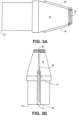
- the distal tip shown in FIGS. 3A-3C can produce a tissue cut pattern that contains a central cut region with two linear cuts protruding radially from the central region or ring as shown in FIG. 16B and FIG. 17 .
- the projections 36 in FIGS. 3A-3C recede into the recessed portion 41 of the distal tip 32 before the distal tip 32 reaches its maximum diameter.
- the projections can be covered adjacent to the outer diameter such that the exposed portion of the projections do not reach the maximum outer diameter of the distal tip 32.
- the slits made in the tissue by the projections 36 are slightly shorter than the diameter of the tip. Some force can be applied to push the distal tip through the tissue slits made by the energized tip.
- the elasticity of the tissue can accommodate the slightly larger diameter of the distal tip and catheter. The tight fit can prevent leakage of biological material from the body lumen.
- Initial access between lumen L1 and lumen L2 can be done using a 19 gauge needle.
- a puncture can be made at the desired location using a 19 gauge needle, such as an electrosurgical needle, followed by placement of a guidewire 102 through the needle lumen.
- a guidewire is not used.
- the catheter device 10 can be used for initial access to make a puncture between lumen L1 and lumen L2. Electrical energy can be provided to the cutting element 35 and projections 36 to make the initial puncture in lumen L1 and lumen L2.
- the blunt cone shaped tip was calculated to have a surface area of approximately 20.2 mm 2 .
- the tip design in the embodiment illustrated in FIGS. 3A-3C was calculated to have a cutting surface area of about 3.17 mm 2 .
- the conductive cutting portion of the distal tip has a surface area of less than about 10 mm 2 .
- the conductive cutting portion of the distal tip has a surface area of less than about 5 mm 2 .
- the conductive cutting portion of the distal tip has a surface area of less than about 4 mm 2 .
- the central feature and projections do not include a ring.
- the central feature and projections can be an arc extending from one side of the distal tip to the opposing side of the distal tip in an arc configuration.
- the catheter devices do not have a guidewire lumen.
- the catheter device can locate the target tissue without following a guidewire.
- the energized tip can then be used for the initial penetration in the target tissue.
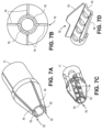
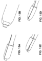
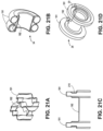
- FIGS. 13A-13C illustrate portions of the tips of catheter devices not in accordance with the invention that can be utilized without a guidewire.
- FIG. 13A illustrates a distal tip with a dome shaped tip with a metal cutting element extending from the dome.
- FIG. 13B illustrates a cone shaped distal tip with a conductive wire cutting element bent over the distal tip.
- FIG. 13C illustrates a dome shaped distal tip with a conductive wire cutting element bent over the dome shaped distal tip.
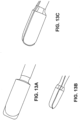
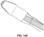
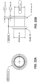
- FIG. 15D is an image of a cone shaped tip with a cutting element connected to a copper wire with the cutting element offset from the guidewire lumen shaft.
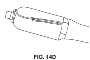
- FIG. 15E is an image of a dome shaped tip with a stainless steel cutting element adjacent to the guidewire lumen shaft.
- FIG. 15F is an image of a nose shaped tip with a cutting element concentric to the guidewire lumen shaft.
- FIGS. 16A-16H illustrates various tissue access patterns made by the different energized tip configurations disclosed herein.
- the dotted lines in FIGS. 16A-16H correspond to the outer diameter of the catheter shaft.
- FIG. 16A illustrates a tissue cutting or access pattern generated by an offset ring and one outer projection.
- FIG. 16B illustrates a tissue cutting pattern generated by a central cutting feature with two outward projections.
- FIG. 16C illustrates a tissue cutting pattern generated by a central cutting feature with three outward projections.
- FIG. 16D illustrates a tissue cutting pattern generated by a central cutting feature with four outward projections.
- FIGS. 16E and 16F illustrate tissue cutting pattern generated by a central cutting feature helical projections extending counter clockwise and clockwise, respectively.
- FIG. 16G illustrates a tissue cutting pattern generated by an offset cutting feature with two outward projections.
- FIG. 16H illustrates a tissue cutting pattern generated by an offset cutting feature with an offset arc element.
- FIG. 17 is an image of a tissue access pattern generated using the distal tip and cutting features illustrated in FIGS. 3A-3C .
- the devices disclosed herein can be used with a guidewire having an anchor structure as disclosed in co-owned application U.S. Patent Publication No. 2010-0268029 .
- the anchor structure can be a shape memory alloy, for example, so that it can be configured to automatically expand to a pre-determined shape upon being advanced distally from a needle.
- the catheter is disposed within an endoscope, and in other embodiments, the catheter is an endoscope or takes the place of an endoscope.
- the devices and methods disclosed herein can be used in a variety of applications and for obtaining access between a variety of different body lumens. In some embodiments the devices disclosed herein can be used for cross-luminal drainage.
- an anastomosis or fistula can be formed using the devices and methods disclosed herein.
- a stent can be placed between two body lumens to provide fluid communication between the two body lumens.
- the two adjacent body lumens can form an anastomosis as they heal around the stent.
- the devices and methods can be used in the gastrointestinal (GI) tract and areas adjacent to a portion of the GI tract.
- GI gastrointestinal
- anatomy in the GI tract include the esophagus, stomach, duodenum, jejunum, small intestine, and large intestine.
- portions adjacent to the GI tract include the peritoneal cavity, bile duct, pancreatic duct, gall bladder, pancreas, cysts, pseudocysts, abscesses and the like.
- Embodiments can be useful for a variety of medical procedures. Embodiments can be applied to ERCP applications where there is a need to access target anatomical structures through multiple tissue planes from a guidewire access. Embodiments are useful for applications and procedures, such as gastrojejunostomy, gastroduodenostomy, gastrocolostomy, transduodenal, transgastric, biliary, pancreatic pseudocysts, transhepatic, transcystic, transpancreatic, transenteric, transbiliary, gastroplexy, cystoplexy, transsesophageal, transbronchial, transgastric, colon resection, gastric bypass, jejunostomy, etc.
- the methods and devices disclosed herein can also be used for cross-luminal therapy or access to body lumens to provide further treatment, such as chemotherapy, placing sensors, placing treatment delivery devices, providing pharmaceutical devices, radioisotope treatment, and others.
- the access to the body lumens can be through the stent or other device placed in or adjacent to the body lumen of interest for the targeted treatment.
- the stent can also include additional therapeutic agents, such as pharmaceutical agents, radioactive agents, and other therapeutic agents.
- the therapeutic agents can be impregnated in the stent or included as a coating.
- a stent can be placed next to or adjacent to a tumor.
- the tumor can be treated with targeted chemotherapy.
- the chemotherapy can be introduced using the stent or using the stent passageway to facilitate treatment.
- tissue stents and anchors can be precisely delivered.
- a stent can be delivered between the fundal pouch formed in gastric bypass surgery and a section of the duodenum or jejunum to form an anastomosis.
- a stent can be placed between two sections of the colon to form an anastomosis.
- the stent and resulting anastomosis can limit leakage, limit the formation of strictures, and create a standard size stoma or anastomosis.
- a blocked anastomosis or decreased anastomosis sizes can result in complications with fluid flow and solid food flow.
- anastomosis can promote fluid and communication and decrease complications from blocked or too small anastomoses.
- the anastomosis size would correspond to the stent size so the variability in the anastomosis size would be greatly decreased over current surgical practices that involve manually stapling the tissue together to form the anastomosis.
- the devices disclosed herein can be used to treat metabolic conditions, such as diabetes.
- Food is first churned in the stomach before passing into the small intestine.
- a number of hormones are released that can inhibit further food intake and have thus been dubbed "satiety factors".
- Changes in circulating hormone levels after gastric bypass have been hypothesized to produce reductions in food intake and body weight in obese patients.
- the devices can be used to make physical changes in the patient that can change hormonal balances that can improve the health of a patient, such as the "satiety factors”.
- diabetes can be treated through procedures such as a Roux En Y gastric bypass or gastrojejunostomy as described herein.
- the devices disclosed herein can be used in laparoscopic assisted procedures.
- the devices disclosed herein can be used with ear, nose, and throat (ENT) delivery.
- the devices disclosed herein can be used in Natural Orifice Transgastric Endoluminal Surgery (NOTES).
- NOTES Natural Orifice Transgastric Endoluminal Surgery
- the devices disclosed herein can be used with procedures that clamp or rivet tissue.
- the devices disclosed herein can be used to close an opening in mammalian tissue.
- a tissue anchor or stent with a closed lumen could be placed to seal an opening.
- the devices disclosed herein can be used for access without delivering and placing a stent.
- the devices can be used for a necosectomy to remove necrosed tissue as in the case of a necrotizing pancreatitis.
- the devices disclosed herein can also be used to perform a cystotome.
- the device can be positioned adjacent to the target tissue, e.g. the transgastric or transduodenal wall. Electrical energy can be supplied to the conductive surfaces of the distal tip of the device to electrosurgically puncture a hole in the transgastric or transduodenal wall along with puncturing a pancreatic pseudocyst.
- the pancreatic pseudocyst can be visibly bulging into the GI tract.
- the distal tip can be supported by an inner catheter shaft within an outer tubular body of the catheter. The distal tip and inner catheter shaft can move relative to the outer tubular body of the catheter. After puncturing the target areas, e.g.
- the catheter devices disclosed herein can be used to form an initial passage with a larger working channel than conventional needle procedures.
- the outer tubular body can have a diameter of greater than 10 French.
- the outer tubular body can have an 11 French diameter to leave a large working channel in body lumen.
- Positioning the catheter for the cystotome can be done without a guidewire. In some cases a guidewire can be used for positioning.
- FIGS. 18A-F , 19A-19I , 20A-20B , 21A-21D , and 22A-22B illustrate various stent designs that can be used with the devices disclosed herein. Additional stent designs are disclosed in co-owned U.S. Patent Publication 2009-0281557 .
- the tissue anchors and stents include a body formed from a woven filament braid.
- the filament will typically be a metal wire, more typically being a nickel-titanium or other super-elastic or shape memory metal wire.
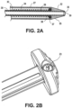
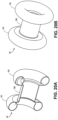
- the body can have both an elongated tubular configuration ( FIG. 2A ) and a foreshortened configuration where proximal and distal ends of the body expand radially into flange structures, as illustrated in FIG. 5E .
- the stents can expand radially to form a pair of adjacent annular rings which define the flange structures. After such foreshortening and deployment of the flange structures, the body will further have a cylindrical saddle region between the flange structures.
- the flange structures When the anchor is deployed in tissue, the flange structures engage the outer surfaces of adjacent tissue layers (e.g. tissue T1 and tissue T2 illustrated in FIGS. 5A-5E ) and the saddle region typically resides within a penetration through the tissue layers.
- the flange structures can have various configurations, including one or more inflection points to provide additional structural support to the stent flanges.
- FIGS. 18A-F illustrate various configurations of the flanges 191 in double-walled configurations.
- the central saddle region 192 has a length L and a diameter d.
- the flanges 191 have a diameter D.
- the stent 34 has lips or cuffs 193 on either end.
- the cuffs 193 have a diameter d'.
- FIG. 18A illustrates a covering or membrane 194 over the entire exterior of the stent 34. The cover or membrane inhibits tissue ingrowth and minimizes fluid leakage when the stent is implanted.
- the cuffs 193 have a diameter d' that is slightly larger than the diameter d of the central saddle region 192 in FIG.
- the stent 34 has a cuff 193 diameter d' that is approximately the same as the diameter d of the central saddle region 192.
- the stent 34 has a smaller diameter d than the stents illustrated in FIGS. 18A and 18B.
- FIG. 18C illustrates a cuff 193 diameter d' that is approximately the same as the diameter d of the central saddle region 192.
- FIG. 18D a stent 34 having a central saddle region 192 with an expanded diameter d and smaller cuff 193 diameter d' is illustrated.
- FIG. 18E a stent 34 having a central saddle region 192 with an expanded diameter d and cuff 193 is illustrated.
- FIG. 18E has a greater cuff 193 diameter d' than the stent 34 illustrated in FIG. 18D.
- FIG. 18F illustrates a stent 34 without a cuff.
- FIG. 19D illustrates a flange 191 with the stent curved away from the central saddle region 192.
- FIG. 19E illustrates a flange 191 with multiple inflections.
- FIG. 19F illustrates a flange 191 with a circular configuration.
- FIG. 19G illustrates a flange that curves towards the opposing flange and back on itself.
- FIG. 19H illustrates a flange 191 with notches or ribs in portions of the stent cross-section.
- FIG. 19I illustrates a flange 191 with a sinusoidal section to provide additional strength.
- FIG. 20A is a cross-section of the stent illustrated in 20B.
- the flanges 191 have are inflected with a circular configuration that curves back towards the central saddle region.
- FIG. 21A illustrates a stent with flanges 191 that have sinusoidal configuration across the cross section of the flange.
- FIG. 21B illustrates a stent with flanges 191 that curve towards the opposing flanges and the central saddle region 192.
- FIG. 21C is a cross section of the stent illustrated in FIG. 21D .
- the stent has a flange 191 with a notch/rib 210 incorporated into the cross section of the rib.
- the diameter D of the flanges 191 and the diameter d of the central saddle region 192 can be selected to allow the desired strength and fluid communication between the body lumens.
- the diameter d should be large enough to allow for fluid communication between the body lumens. In some cases liquid flows through the central saddle region 192. In other cases particulate matter or solids can flow through the central saddle region 192.
- the diameter d should be sized to allow for the expected fluids and/or solids to flow through the saddle region 192.
- the diameter d can also be sized with a diameter close to the size of the hole in the body lumens to reduce the chance of leakage and improve stent engagement with the holes in the body lumens.
- the cuff length, diameter (d'), and shapes can be configured to achieve the desired physical properties in the stent.
- the cuff can provide additional strength to the stent structure.
- the cuff can also have an increased diameter to make it easier for subsequent access to the interior volume of the stent with additional medical instruments or with a device to remove the stent.
- the tissue anchors of the prior art such as metal clamps and rivets, have often been either too rigid, providing good attachment but presenting substantial risk of tissue necrosis or adhesion, or too weak, presenting little risk of tissue damage but allowing leakage and movement at the point of tissue penetration.
- the stents and tissue anchors disclosed herein can provide firm attachment of tissue while minimizing the risk of necrosis and other damage to the tissue.
- the stents disclosed herein provide enough pressure to hold the tissue walls together with the expanded flange structures but not too much force that can cause trauma to the tissue walls.
- Prior art stent designs for implantation in vascular applications are designed to be delivered to the target location and held in place permanently.
- the vascular stents are not designed to be removed or moved after implantation.
- the stents disclosed herein can be removed.
- the tissue anchors disclosed herein are also removable, both during initial implantation procedures as well as in a subsequent procedure(s) many weeks, months, or even years following the initial implantation.
- the length of the saddle region can be optimized based on the thickness of each of the tissue walls such that the flanges securely engage and hold the tissue planes together.
- Each of the flanges can engage the tissue walls such that the flange ends do not migrate or move.
- the double walled flanges can hold the opposing tissue planes in place.
- the flanges can engage the tissue walls to control the positioning of the tissue walls and hold the tissue in a desired orientation to the tissue engaged by the other end of the stent.
- the stent can be formed from woven shaped memory metal wires, such as nitinol or eligiloy.
- the wires can can a relatively small diameter, typically in the range from 0.0254 mm (0.001 inch) to 0.508 mm (0.02 inch), usually from 0.0508 mm (0.002 inch) to 0.254 mm (0.01 inch), where the braid will include from as few as 10 to as many as 200 wires, more commonly being from 20 wires to 100 wires.
- the wires will be round having diameters in the range from 0.0762 mm (0.003 inch) to 0.1778 mm (0.007 inch) with a total of from 24 to 60 wires.
- the wires are braided into a tubular geometry by conventional techniques, and the tubular geometry will be heat-treated to impart the desired shape memory.
- the braided tube will be formed into the desired final (deployed) configuration with the flanges at each end.
- Such a flanged configuration will then be heat set or formed into the braid so that, in the absence of a radially constraining or axially elongating force, the anchor will assume the foreshortened configuration with the flanges at each end.
- Such foreshortened-memory configurations will allow the anchor to be delivered in a constrained configuration (either radially or axially elongated) and thereafter released from constraint so that the body assumes the flanged configuration at the target site.
- the woven stent design can promote delivery with its flexibility, collapsibility, and elasticity.
- Expanded stent shapes and sizes can be manufactured with controlled dimensions, such as the internal diameter and length for the central saddle region and diameter of the double walled flanges.
- a stent illustrated in detail in FIGS. 22A and 22B ) with a 15 mm internal diameter of the saddle region (illustrated as d in FIG. 19 ) with a 10 mm saddle length (illustrated as L in FIG. 19 ) and 24 mm flange diameter (illustrated as D in FIG. 19 ) can be used.
- the stent structures are illustrated in detail in FIGS. 22A (cross-section) and 22B (side-view).
- FIG. 22B illustrates a cuff or lip diameter of 18 mm.
- a stent with a 10 mm internal diameter of the saddle region with a 10 mm saddle length and 21 mm flange diameter can be used.
- a stent with a 6 mm internal diameter of the saddle region with an 8 mm saddle length and 15 mm flange diameter can be used.
- a stent with an 8 mm internal diameter of the saddle region with an 8 mm saddle length and 17 mm flange diameter can be used.
- a stent with a 10 mm internal diameter of the saddle region with a 6 mm saddle length and 21 mm flange diameter can be used.
- the stents or tissue anchors can be adapted to be delivered by a catheter based delivery device, such as the delivery device disclosed herein, typically an endoscopic delivery catheter.
- the catheter can have a small diameter in the range from 1 mm to 8 mm, usually from 2 mm to 5 mm.
- the elongated tubular configuration of the anchor body will usually have a diameter less than that of the catheter diameter, usually from 0.8 mm to 7.5 mm, more usually from 0.8 mm to 4.5 mm, where the double-walled flanged structures will be expandable significantly, usually being in the range from 3 mm to 70 mm, more usually in the range from 5 mm to 40 mm.
- the cylindrical saddle region of the anchor will often not increase in diameter during deployment, but may optionally increase to a diameter from 2 mm to 50 mm, more usually from 5 mm to 20 mm.
- the lumen or passage through the deployed tissue anchor can have a variety of diameters, typically from as small as 0.2 mm to as large as 40 mm, more usually being in the range from 1 mm to 20 mm, and typically having a diameter which is slightly smaller than the expanded diameter of the cylindrical saddle region.
- the length of the body may also vary significantly. Typically, when in the elongated tubular configuration, the body will have a length in the range from 7 mm to 100 mm, usually from 12 mm to 70 mm.
- the body When deployed, the body will be foreshortened, typically by at least 20%, more typically by at least 40% and often by 70% or greater.
- the foreshortened length will typically be in the range from 2 mm to 80 mm, usually in the range from 2.5 mm to 60 mm, and more usually being in the range from 3 mm to 40 mm.
- the body of the tissue anchor may consist of the woven filament braid with no other coverings or layers.
- the tissue anchor may further comprise a membrane or other covering formed over at least a portion of the body.
- the membrane can be used to prevent or inhibit tissue ingrowth to allow the device to be removed after having been implanted for weeks, months, or longer.
- Suitable membrane materials include polytetrafluoroethylene (PTFE), expanded PTFE (ePTFE), silicone, polypropylene, urethane polyether block amides (PEBA), polyethyleneterephthalate (PET), polyethylene, C-Flex TM . thermoplastic elastomer, Krator TM , SEBS and SBS polymers, and the like.
- the membranes may be formed over the entire portion of the anchor or stent. In some embodiments the membrane or covering is formed over only a portion of the anchor or stent. The covering or membrane may be formed over the exterior or interior of the body and will typically be elastomeric so that the membrane conforms to the body in both the elongated and foreshortened configurations.
- the stents can be coated with active compounds, such as therapeutic compounds to provide therapy to the tissue areas adjacent to the stent.
- the therapeutic compounds can be used to reduce tissue attachment and engagement with the stent during tissue healing after the deployment of the stent.
- Coatings and surfaces that do not promote tissue in-growth can be used in some embodiments.
- Sirolimus and its analogs can be used to halt cell proliferation.
- Taxols can also be used to halt cell proliferation, in addition to being used in cancer treatment.
- Silicone and other surfaces can be used to stop or slow the rate of cell proliferation and in-growth.
- the therapeutic compounds can promote healing of the tissue and formation of a healthy anastomosis.
- the therapeutic compound can be a medicine that is released from the stent over a period of time.
- the therapeutic compound can be used to treat tissue or anatomy adjacent to the stent location.
- the therapeutic agent can be used with a stent that is not fully covered to promote tissue in-growth.
- a titanium oxide layer or coating can be used to promote cell proliferation and tissue in-growth.
- Biologic compounds such as cytokines and hormones can promote tissue growth as well.
- a cylindrical saddle region remains on the anchor body between the deployed flanges, where the flanges are able to press against the tissue layers to provide the approximating force.
- the body will be foreshortened to a degree selected to apply sufficient pressure to the tissues to hold them together without causing significant tissue injury or necrosis.
- the applied pressure will be in the range from 0.005 g/mm 2 to 5 g/mm 2 , usually from 0.2 g/mm 2 to 1 g/mm 2 .
- relative terms can refer to plus or minus 10%. In some cases the relative terms can refer to plus or minus 5%. In some cases the relative terms can refer to plus or minus 2%. In some cases the relative terms can refer to plus or minus 1%.
Landscapes
- Health & Medical Sciences (AREA)
- Surgery (AREA)
- Life Sciences & Earth Sciences (AREA)
- Engineering & Computer Science (AREA)
- Heart & Thoracic Surgery (AREA)
- Animal Behavior & Ethology (AREA)
- Veterinary Medicine (AREA)
- Public Health (AREA)
- General Health & Medical Sciences (AREA)
- Biomedical Technology (AREA)
- Nuclear Medicine, Radiotherapy & Molecular Imaging (AREA)
- Medical Informatics (AREA)
- Molecular Biology (AREA)
- Physics & Mathematics (AREA)
- Plasma & Fusion (AREA)
- Otolaryngology (AREA)
- Pathology (AREA)
- Physiology (AREA)
- Cardiology (AREA)
- Orthopedic Medicine & Surgery (AREA)
- Surgical Instruments (AREA)
- Media Introduction/Drainage Providing Device (AREA)
Priority Applications (1)
| Application Number | Priority Date | Filing Date | Title |
|---|---|---|---|
| EP24185828.1A EP4431030A3 (en) | 2012-05-17 | 2013-04-26 | Devices for access across adjacent tissue layers |
Applications Claiming Priority (5)
| Application Number | Priority Date | Filing Date | Title |
|---|---|---|---|
| US201261648544P | 2012-05-17 | 2012-05-17 | |
| US201261727629P | 2012-11-16 | 2012-11-16 | |
| US201361767577P | 2013-02-21 | 2013-02-21 | |
| EP13791682.1A EP2854654B1 (en) | 2012-05-17 | 2013-04-26 | Devices for access across adjacent tissue layers |
| PCT/US2013/038502 WO2013173045A1 (en) | 2012-05-17 | 2013-04-26 | Methods and devices for access across adjacent tissue layers |
Related Parent Applications (1)
| Application Number | Title | Priority Date | Filing Date |
|---|---|---|---|
| EP13791682.1A Division EP2854654B1 (en) | 2012-05-17 | 2013-04-26 | Devices for access across adjacent tissue layers |
Related Child Applications (1)
| Application Number | Title | Priority Date | Filing Date |
|---|---|---|---|
| EP24185828.1A Division EP4431030A3 (en) | 2012-05-17 | 2013-04-26 | Devices for access across adjacent tissue layers |
Publications (2)
| Publication Number | Publication Date |
|---|---|
| EP3636164A1 EP3636164A1 (en) | 2020-04-15 |
| EP3636164B1 true EP3636164B1 (en) | 2024-07-03 |
Family
ID=49581916
Family Applications (3)
| Application Number | Title | Priority Date | Filing Date |
|---|---|---|---|
| EP24185828.1A Pending EP4431030A3 (en) | 2012-05-17 | 2013-04-26 | Devices for access across adjacent tissue layers |
| EP13791682.1A Active EP2854654B1 (en) | 2012-05-17 | 2013-04-26 | Devices for access across adjacent tissue layers |
| EP19207123.1A Active EP3636164B1 (en) | 2012-05-17 | 2013-04-26 | Devices for access across adjacent tissue layers |
Family Applications Before (2)
| Application Number | Title | Priority Date | Filing Date |
|---|---|---|---|
| EP24185828.1A Pending EP4431030A3 (en) | 2012-05-17 | 2013-04-26 | Devices for access across adjacent tissue layers |
| EP13791682.1A Active EP2854654B1 (en) | 2012-05-17 | 2013-04-26 | Devices for access across adjacent tissue layers |
Country Status (6)
| Country | Link |
|---|---|
| US (5) | US9381041B2 (enExample) |
| EP (3) | EP4431030A3 (enExample) |
| JP (2) | JP6360042B2 (enExample) |
| DE (1) | DE202013012853U1 (enExample) |
| ES (2) | ES2982851T3 (enExample) |
| WO (1) | WO2013173045A1 (enExample) |
Families Citing this family (91)
| Publication number | Priority date | Publication date | Assignee | Title |
|---|---|---|---|---|
| US12303105B2 (en) | 2004-04-12 | 2025-05-20 | Boston Scientific Scimed, Inc. | Luminal structure anchoring devices and methods |
| US11298113B2 (en) | 2008-10-01 | 2022-04-12 | Covidien Lp | Device for needle biopsy with integrated needle protection |
| US8968210B2 (en) | 2008-10-01 | 2015-03-03 | Covidien LLP | Device for needle biopsy with integrated needle protection |
| US9782565B2 (en) | 2008-10-01 | 2017-10-10 | Covidien Lp | Endoscopic ultrasound-guided biliary access system |
| US9332973B2 (en) | 2008-10-01 | 2016-05-10 | Covidien Lp | Needle biopsy device with exchangeable needle and integrated needle protection |
| US9186128B2 (en) | 2008-10-01 | 2015-11-17 | Covidien Lp | Needle biopsy device |
| WO2010138277A1 (en) | 2009-05-29 | 2010-12-02 | Xlumena, Inc. | Apparatus and method for deploying stent across adjacent tissue layers |
| WO2011085006A2 (en) | 2010-01-05 | 2011-07-14 | Beacon Endoscopic Corporation | Methods and apparatus for magnet-induced compression anastomosis between adjacent organs |
| US8870898B2 (en) | 2010-01-05 | 2014-10-28 | GI Windows, Inc. | Self-assembling magnetic anastomosis device having an exoskeleton |
| US9526648B2 (en) | 2010-06-13 | 2016-12-27 | Synerz Medical, Inc. | Intragastric device for treating obesity |
| US10420665B2 (en) | 2010-06-13 | 2019-09-24 | W. L. Gore & Associates, Inc. | Intragastric device for treating obesity |
| US8628554B2 (en) | 2010-06-13 | 2014-01-14 | Virender K. Sharma | Intragastric device for treating obesity |
| US10010439B2 (en) | 2010-06-13 | 2018-07-03 | Synerz Medical, Inc. | Intragastric device for treating obesity |
| ES2982851T3 (es) * | 2012-05-17 | 2024-10-17 | Boston Scient Scimed Inc | Dispositivos de acceso a través de capas de tejido adyacentes |
| EP2958527B1 (en) | 2013-02-21 | 2020-07-22 | Boston Scientific Scimed, Inc. | Devices for forming an anastomosis |
| WO2014151615A2 (en) * | 2013-03-15 | 2014-09-25 | Boston Scientific Scimed, Inc. | Stent delivery system |
| US9364238B2 (en) | 2013-04-16 | 2016-06-14 | Ethicon Endo-Surgery, Inc. | Method and apparatus for joining hollow organ sections in anastomosis |
| US11033272B2 (en) | 2013-04-16 | 2021-06-15 | Ethicon Endo-Surgery, Inc. | Methods for partial diversion of the intestinal tract |
| KR101514055B1 (ko) * | 2013-12-17 | 2015-04-21 | 주식회사 스텐다드싸이텍 | 스텐트 삽입장치 |
| JP6336619B2 (ja) * | 2014-06-18 | 2018-06-06 | ボストン サイエンティフィック サイムド,インコーポレイテッドBoston Scientific Scimed,Inc. | 胆管ステント |
| US10195064B2 (en) * | 2014-08-15 | 2019-02-05 | W. L. Gore & Associates, Inc. | Endoprosthesis delivery systems with improved retraction |
| DE102014222875A1 (de) * | 2014-11-10 | 2016-05-12 | Somatex Medical Technologies Gmbh | Shunt zum Abführen einer Flüssigkeit aus einer Körperhöhle |
| US10842554B2 (en) * | 2014-11-13 | 2020-11-24 | Alan Ellman | Electrosurgical electrode |
| US20160206338A1 (en) | 2015-01-20 | 2016-07-21 | Talon Medical, LLC | Tissue engagement devices, systems, and methods |
| KR101696810B1 (ko) * | 2015-02-04 | 2017-02-01 | 주식회사 엠아이텍 | 연결용 스텐트 및 그의 제조방법 |
| CA3244673A1 (en) | 2015-03-05 | 2025-02-21 | Merit Medical Systems, Inc. | VASCULAR PROSTHESIS DEPLOYMENT DEVICE AND ITS METHOD OF USE |
| WO2016145414A1 (en) | 2015-03-12 | 2016-09-15 | GI Windows, Inc. | Magnetic anastomosis devices with varying magnetic force at a distance |
| WO2016176567A1 (en) | 2015-04-29 | 2016-11-03 | Innoblative Designs, Inc. | Cavitary tissue ablation |
| US10470906B2 (en) | 2015-09-15 | 2019-11-12 | Merit Medical Systems, Inc. | Implantable device delivery system |
| JP6933857B2 (ja) | 2015-10-29 | 2021-09-08 | イノブレイティブ デザインズ, インコーポレイテッド | 網球状組織アブレーションデバイスおよび方法 |
| US12207863B2 (en) | 2015-10-29 | 2025-01-28 | Innoblative Designs, Inc. | Screen sphere tissue ablation devices and methods |
| BR112018010622B1 (pt) | 2015-11-25 | 2023-04-11 | Talon Medical, LLC | Dispositivo de engate de tecido e kit |
| US20170215947A1 (en) | 2016-02-02 | 2017-08-03 | Innoblative Designs, Inc. | Cavitary tissue ablation system |
| KR101781052B1 (ko) * | 2016-02-15 | 2017-10-23 | (주) 태웅메디칼 | 양극형 전기 소작 팁이 포함된 스텐트 전달 시스템 |
| US10869714B2 (en) * | 2016-03-01 | 2020-12-22 | Innoblative Designs, Inc. | Resecting and coagulating tissue |
| US10603101B2 (en) | 2016-03-26 | 2020-03-31 | Paul Joseph Weber | Apparatus, systems and methods for minimally invasive dissection of tissues |
| US10893899B2 (en) | 2016-03-26 | 2021-01-19 | Paul Weber | Apparatus and systems for minimally invasive dissection of tissues |
| US11510730B2 (en) | 2016-03-26 | 2022-11-29 | Paul Joseph Weber | Apparatus and methods for minimally invasive dissection and modification of tissues |
| US10779980B2 (en) | 2016-04-27 | 2020-09-22 | Synerz Medical, Inc. | Intragastric device for treating obesity |
| JP2019523059A (ja) * | 2016-07-29 | 2019-08-22 | クック・メディカル・テクノロジーズ・リミテッド・ライアビリティ・カンパニーCook Medical Technologies Llc | 解剖学的構造にアクセスするための、単一のチューブ状導電素子を備える電気外科機器 |
| CN109789025B (zh) | 2016-09-29 | 2022-06-10 | 美国医疗设备有限公司 | 用于接纳和协助调配血管假体的柔顺性构件 |
| WO2018062387A1 (ja) | 2016-09-30 | 2018-04-05 | テルモ株式会社 | 医療デバイスおよび処置方法 |
| US10070921B2 (en) | 2016-10-17 | 2018-09-11 | Innoblative Designs, Inc. | Treatment devices and methods |
| WO2018144090A2 (en) | 2016-11-08 | 2018-08-09 | Innoblative Designs, Inc. | Electrosurgical tissue and vessel sealing device |
| KR101902781B1 (ko) * | 2016-11-16 | 2018-10-01 | (주) 태웅메디칼 | 단극형 전기 소작 팁이 포함된 스텐트 전달 시스템 |
| JP2018099266A (ja) * | 2016-12-20 | 2018-06-28 | ビーシーエム カンパニー,リミテッド | 端部の抵抗力が強化された医療用ステント |
| US10555735B2 (en) | 2017-01-30 | 2020-02-11 | Ethicon Llc | Tissue compression assemblies with biodegradable interlinks |
| US10376265B2 (en) | 2017-01-30 | 2019-08-13 | Ethicon Llc | Non-magnetic fragmentable tissue compression devices |
| KR101962598B1 (ko) * | 2017-02-03 | 2019-03-27 | 주식회사 에스앤지바이오텍 | 스텐트 삽입장치 |
| KR20250161641A (ko) | 2017-02-27 | 2025-11-17 | 보스톤 싸이엔티픽 싸이메드 인코포레이티드 | 마커를 포함하는 전개 카테터 |
| EP4467111A3 (en) | 2017-03-15 | 2025-03-05 | Merit Medical Systems, Inc. | Transluminal stents |
| WO2018170066A1 (en) | 2017-03-15 | 2018-09-20 | Merit Medical Systems, Inc. | Transluminal delivery devices and related kits and methods |
| USD836194S1 (en) | 2017-03-21 | 2018-12-18 | Merit Medical Systems, Inc. | Stent deployment device |
| US11589884B2 (en) | 2017-04-28 | 2023-02-28 | W. L. Gore & Associates, Inc. | Endoscopic transluminal stent access and delivery system |
| WO2019023328A1 (en) | 2017-07-26 | 2019-01-31 | Innoblative Designs, Inc. | MINIMALLY INVASIVE JOINT ASSEMBLY HAVING ABLATION CAPABILITIES |
| US10939805B2 (en) * | 2017-09-25 | 2021-03-09 | Broncus Medical Inc. | Medical appliance for controlling medical device through catheter sheath based on pneumatic action |
| JP6926318B2 (ja) | 2017-10-03 | 2021-08-25 | ボストン サイエンティフィック サイムド,インコーポレイテッドBoston Scientific Scimed,Inc. | フローコントロールステント |
| JP6840649B2 (ja) * | 2017-10-13 | 2021-03-10 | 日本ライフライン株式会社 | 高周波治療用カテーテル |
| EP3769708B1 (en) * | 2018-03-20 | 2022-07-27 | TERUMO Kabushiki Kaisha | Medical device |
| WO2019199763A1 (en) | 2018-04-11 | 2019-10-17 | Boston Scientific Scimed, Inc. | Devices and methods for extending a working channel |
| US10888691B2 (en) | 2018-04-24 | 2021-01-12 | Olympus Corporation | Stent delivery method |
| US20210236129A1 (en) * | 2018-05-11 | 2021-08-05 | Mayo Foundation For Medical Education And Research | Luminal apposition devices |
| US11801089B2 (en) * | 2018-05-21 | 2023-10-31 | Boston Scientific Scimed, Inc. | Cauterizing device for use with stents |
| GB2574219A (en) * | 2018-05-30 | 2019-12-04 | Creo Medical Ltd | Electrosurgical instrument |
| EP3801299B1 (en) | 2018-06-02 | 2024-01-03 | GI Windows Inc. | Devices for forming anastomoses |
| CN111202485A (zh) * | 2018-11-21 | 2020-05-29 | 南微医学科技股份有限公司 | 一种医用连接装置 |
| WO2020163820A1 (en) | 2019-02-07 | 2020-08-13 | NXT Biomedical | Rivet shunt and method of deployment |
| CN112754642A (zh) * | 2019-11-06 | 2021-05-07 | 南微医学科技股份有限公司 | 一种内窥镜用切割装置 |
| EP3986308B1 (en) * | 2019-12-24 | 2025-03-19 | Boston Scientific Scimed, Inc. | Eus access device with electrosurgery-enhanced puncture |
| US11553958B2 (en) * | 2020-02-07 | 2023-01-17 | Covidien Lp | Electrosurgical device for cutting tissue |
| WO2021178135A1 (en) * | 2020-03-03 | 2021-09-10 | W. L. Gore & Associates, Inc. | Transcatheter tissue cutting system |
| WO2022020633A1 (en) | 2020-07-24 | 2022-01-27 | Merit Medical Systems, Inc. | Esophageal stents and related methods |
| CN116096319A (zh) * | 2020-07-30 | 2023-05-09 | 波士顿科学医疗设备有限公司 | 细长医用针 |
| EP4231973A4 (en) | 2020-10-26 | 2024-10-02 | Merit Medical Systems, Inc. | HELICAL THREAD ESOPHAGEAL STENT PROSTHESES |
| WO2022225923A1 (en) | 2021-04-20 | 2022-10-27 | G.I. Windows, Inc. | Systems, devices, and methods for endoscope or laparoscopic magnetic navigation |
| US12303189B2 (en) | 2021-08-03 | 2025-05-20 | Medtronic Advanced Energy Llc | Energized corers with energized internals |
| US20230040816A1 (en) * | 2021-08-03 | 2023-02-09 | Medtronic Advanced Energy Llc | Energized corers with powered conveying |
| US20230233349A1 (en) * | 2022-01-21 | 2023-07-27 | Covidien Lp | Apparatuses for stent delivery and positioning for transluminal application |
| US20230233313A1 (en) * | 2022-01-21 | 2023-07-27 | Covidien Lp | Methods for stent delivery and positioning for transluminal application |
| DE102022107302A1 (de) | 2022-03-28 | 2023-09-28 | Matthias Birth | Anastomosendübel und applikationsvorrichtung für einen anastomosendübel |
| US12201300B2 (en) | 2022-08-05 | 2025-01-21 | G.I. Windows, Inc. | Magnetic compression anastomosis device with multipiece vertebra |
| JP2025529235A (ja) | 2022-09-01 | 2025-09-04 | ジーアイ ウィンドウズ, インコーポレイテッド | 圧力プロファイル磁気圧縮吻合デバイス |
| CN119836270A (zh) | 2022-09-02 | 2025-04-15 | G.I.窗公司 | 用于内窥镜或腹腔镜磁性导航的系统、装置和方法 |
| WO2024186692A1 (en) | 2023-03-03 | 2024-09-12 | Theraheart Inc. | Expandable elements for shunting catheters |
| WO2024203788A1 (ja) * | 2023-03-31 | 2024-10-03 | 日本ゼオン株式会社 | ステントデリバリー装置 |
| US12290310B2 (en) | 2023-04-06 | 2025-05-06 | Theraheart Inc. | Slicing elements for shunting catheters |
| US12262943B2 (en) | 2023-06-15 | 2025-04-01 | Theraheart Inc. | Expandable ablation mechanisms for shunting catheters |
| WO2025074196A1 (en) * | 2023-10-02 | 2025-04-10 | Baylis Medical Technologies Inc. | Devices and methods for transjugular intrahepatic portosystemic shunt procedures |
| WO2025080729A1 (en) * | 2023-10-10 | 2025-04-17 | Edwards Lifesciences Corporation | Delivery apparatus with perforation device |
| WO2025160095A1 (en) * | 2024-01-22 | 2025-07-31 | Taurus Vascular, Inc. | Methods and systems for transcaval treatment of aneurysms |
| US12201354B1 (en) | 2024-04-01 | 2025-01-21 | Theraheart Inc. | Expandable ablation mechanisms for shunting catheters |
Family Cites Families (391)
| Publication number | Priority date | Publication date | Assignee | Title |
|---|---|---|---|---|
| US2127903A (en) | 1936-05-05 | 1938-08-23 | Davis & Geck Inc | Tube for surgical purposes and method of preparing and using the same |
| US3039468A (en) | 1959-01-07 | 1962-06-19 | Joseph L Price | Trocar and method of treating bloat |
| US3717151A (en) | 1971-03-11 | 1973-02-20 | R Collett | Flesh penetrating apparatus |
| US3874388A (en) | 1973-02-12 | 1975-04-01 | Ochsner Med Found Alton | Shunt defect closure system |
| US3970090A (en) | 1975-02-03 | 1976-07-20 | Physio Medics, Inc. | Catheter |
| US4173392A (en) | 1977-07-20 | 1979-11-06 | American Hospital Supply Corporation | Glass fiber light guide and method of making the same |
| US4235238A (en) | 1978-05-11 | 1980-11-25 | Olympus Optical Co., Ltd. | Apparatus for suturing coeliac tissues |
| DE2821048C2 (de) | 1978-05-13 | 1980-07-17 | Willy Ruesch Gmbh & Co Kg, 7053 Kernen | Medizinisches Instrument |
| JPS614260B2 (enExample) * | 1980-05-13 | 1986-02-07 | Amerikan Hosupitaru Sapurai Corp | |
| JPS5835219U (ja) | 1981-09-01 | 1983-03-08 | 株式会社潤工社 | コイルケ−ブル |
| US6656182B1 (en) | 1982-05-20 | 2003-12-02 | John O. Hayhurst | Tissue manipulation |
| US4587972A (en) | 1984-07-16 | 1986-05-13 | Morantte Jr Bernardo D | Device for diagnostic and therapeutic intravascular intervention |
| US4580568A (en) | 1984-10-01 | 1986-04-08 | Cook, Incorporated | Percutaneous endovascular stent and method for insertion thereof |
| US4790813A (en) | 1984-12-17 | 1988-12-13 | Intravascular Surgical Instruments, Inc. | Method and apparatus for surgically removing remote deposits |
| US4608965A (en) | 1985-03-27 | 1986-09-02 | Anspach Jr William E | Endoscope retainer and tissue retracting device |
| US4705040A (en) | 1985-11-18 | 1987-11-10 | Medi-Tech, Incorporated | Percutaneous fixation of hollow organs |
| US5000185A (en) | 1986-02-28 | 1991-03-19 | Cardiovascular Imaging Systems, Inc. | Method for intravascular two-dimensional ultrasonography and recanalization |
| JPS62233168A (ja) | 1986-04-03 | 1987-10-13 | オリンパス光学工業株式会社 | カテ−テル |
| SE453258B (sv) | 1986-04-21 | 1988-01-25 | Medinvent Sa | Elastisk, sjelvexpanderande protes samt forfarande for dess framstellning |
| US4920967A (en) | 1986-07-18 | 1990-05-01 | Pfizer Hospital Products Group, Inc. | Doppler tip wire guide |
| US4990139A (en) | 1986-09-10 | 1991-02-05 | Jang G David | Tandem independently inflatable/deflatable multiple diameter balloon angioplasty catheter systems |
| JPH0755222B2 (ja) | 1986-12-12 | 1995-06-14 | オリンパス光学工業株式会社 | 処置具 |
| US4917097A (en) | 1987-10-27 | 1990-04-17 | Endosonics Corporation | Apparatus and method for imaging small cavities |
| US5180392A (en) | 1988-02-01 | 1993-01-19 | Einar Skeie | Anastomotic device |
| US4869263A (en) | 1988-02-04 | 1989-09-26 | Cardiometrics, Inc. | Device and method for measuring volumetric blood flow in a vessel |
| US5588432A (en) | 1988-03-21 | 1996-12-31 | Boston Scientific Corporation | Catheters for imaging, sensing electrical potentials, and ablating tissue |
| AU4945490A (en) | 1989-01-06 | 1990-08-01 | Angioplasty Systems Inc. | Electrosurgical catheter for resolving atherosclerotic plaque |
| US5425739A (en) | 1989-03-09 | 1995-06-20 | Avatar Design And Development, Inc. | Anastomosis stent and stent selection system |
| US5078717A (en) | 1989-04-13 | 1992-01-07 | Everest Medical Corporation | Ablation catheter with selectively deployable electrodes |
| US4973317A (en) | 1989-07-14 | 1990-11-27 | Bobrove Arthur M | Automatic sheath protection of hypodermic needle |
| DE69020075T2 (de) | 1989-08-09 | 1995-11-16 | Bard Inc C R | Katheterführung und Führungsdrahts zur Durchführung eines schnellen Katheteraustausches. |
| US5211651A (en) | 1989-08-18 | 1993-05-18 | Evi Corporation | Catheter atherotome |
| US5024655A (en) | 1989-09-05 | 1991-06-18 | Freeman Andrew B | Epidural catheter apparatus and associated method |
| CA2067110C (en) | 1989-09-08 | 2001-07-31 | John E. Abele | Physiologic low stress angioplasty |
| US5330497A (en) | 1989-11-22 | 1994-07-19 | Dexide, Inc. | Locking trocar sleeve |
| US4950285A (en) | 1989-11-27 | 1990-08-21 | Wilk Peter J | Suture device |
| US5207229A (en) | 1989-12-21 | 1993-05-04 | Advanced Biomedical Devices, Inc. | Flexibility steerable guidewire with inflatable balloon |
| US5254990A (en) | 1990-02-26 | 1993-10-19 | Fujitsu Limited | Method and apparatus for compression and decompression of data |
| US5197971A (en) | 1990-03-02 | 1993-03-30 | Bonutti Peter M | Arthroscopic retractor and method of using the same |
| US5021059A (en) | 1990-05-07 | 1991-06-04 | Kensey Nash Corporation | Plug device with pulley for sealing punctures in tissue and methods of use |
| US5064435A (en) | 1990-06-28 | 1991-11-12 | Schneider (Usa) Inc. | Self-expanding prosthesis having stable axial length |
| US5234447A (en) | 1990-08-28 | 1993-08-10 | Robert L. Kaster | Side-to-end vascular anastomotic staple apparatus |
| US5368595A (en) | 1990-09-06 | 1994-11-29 | United States Surgical Corporation | Implant assist apparatus |
| ATE121303T1 (de) | 1990-10-04 | 1995-05-15 | Schneider Europ Ag | Ballondilationskatheter. |
| CA2052310A1 (en) | 1990-10-09 | 1992-04-10 | Thomas L. Foster | Surgical access sheath |
| ES2085435T3 (es) | 1990-10-09 | 1996-06-01 | Cook Inc | Dispositivo dilatador percutaneo. |
| JPH0550563A (ja) | 1991-08-21 | 1993-03-02 | Dainippon Printing Co Ltd | バリアー性紙複合容器 |
| WO1992008513A1 (en) | 1990-11-20 | 1992-05-29 | Interventional Thermodynamics, Inc. | Tension guide and dilator |
| US5221258A (en) | 1991-01-22 | 1993-06-22 | Shturman Technologies, Inc. | Introduction balloon catheter |
| US5275610A (en) | 1991-05-13 | 1994-01-04 | Cook Incorporated | Surgical retractors and method of use |
| US5183464A (en) | 1991-05-17 | 1993-02-02 | Interventional Thermodynamics, Inc. | Radially expandable dilator |
| US5183033A (en) | 1991-07-15 | 1993-02-02 | Wilk Peter J | Surgical instrument assembly and apparatus and surgical method |
| EP0533321A3 (en) | 1991-07-22 | 1993-05-12 | Dow Corning Wright Corporation | Expanding atherectomy device |
| US5199419A (en) | 1991-08-05 | 1993-04-06 | United States Surgical Corporation | Surgical retractor |
| US5387235A (en) | 1991-10-25 | 1995-02-07 | Cook Incorporated | Expandable transluminal graft prosthesis for repair of aneurysm |
| JPH0796038B2 (ja) | 1991-10-28 | 1995-10-18 | 英夫 高岡 | トレーニング器具構造 |
| SE469198B (sv) | 1991-10-29 | 1993-05-24 | Perstorp Analytical Ab | Luminometeranordning |
| ES2069968T3 (es) | 1991-11-25 | 1995-05-16 | Cook Inc | Aparato para reparacion de defectos tisulares. |
| US5258000A (en) | 1991-11-25 | 1993-11-02 | Cook Incorporated | Tissue aperture repair device |
| US5713870A (en) | 1991-11-27 | 1998-02-03 | Yoon; Inbae | Retractable safety penetrating instrument with laterally extendable spring strip |
| US5395349A (en) | 1991-12-13 | 1995-03-07 | Endovascular Technologies, Inc. | Dual valve reinforced sheath and method |
| US5224945A (en) | 1992-01-13 | 1993-07-06 | Interventional Technologies, Inc. | Compressible/expandable atherectomy cutter |
| US5209727A (en) | 1992-01-29 | 1993-05-11 | Interventional Technologies, Inc. | Guide wire with integral angioplasty balloon |
| US5257990A (en) | 1992-02-24 | 1993-11-02 | Kensey Nash Corporation | Electrosurgical catheter instrument with impacting working head and method of use |
| US5226421A (en) | 1992-03-06 | 1993-07-13 | Cardiometrics, Inc. | Doppler elongate flexible member having an inflatable balloon mounted thereon |
| US5246007A (en) | 1992-03-13 | 1993-09-21 | Cardiometrics, Inc. | Vascular catheter for measuring flow characteristics and method |
| DE4312147C2 (de) * | 1992-04-14 | 1996-01-25 | Olympus Optical Co | Trokar |
| JP3402643B2 (ja) * | 1992-04-14 | 2003-05-06 | オリンパス光学工業株式会社 | トラカール |
| US5707362A (en) | 1992-04-15 | 1998-01-13 | Yoon; Inbae | Penetrating instrument having an expandable anchoring portion for triggering protrusion of a safety member and/or retraction of a penetrating member |
| US5536248A (en) | 1992-05-11 | 1996-07-16 | Arrow Precision Products, Inc. | Method and apparatus for electrosurgically obtaining access to the biliary tree and placing a stent therein |
| US5443484A (en) | 1992-06-16 | 1995-08-22 | Loma Linda University Medical Center | Trocar and method for endoscopic surgery |
| DE4221390C1 (enExample) | 1992-06-30 | 1993-04-01 | Haindl, Hans, Dr.Med., 3015 Wennigsen, De | |
| US5261920A (en) | 1992-08-21 | 1993-11-16 | Ethicon, Inc. | Anvil bushing for circular stapler |
| US5458131A (en) | 1992-08-25 | 1995-10-17 | Wilk; Peter J. | Method for use in intra-abdominal surgery |
| US5364408A (en) | 1992-09-04 | 1994-11-15 | Laurus Medical Corporation | Endoscopic suture system |
| EP0596162B1 (en) | 1992-11-06 | 2002-08-21 | Texas Instruments Incorporated | hypodermic needle with a protrusion |
| IL103737A (en) | 1992-11-13 | 1997-02-18 | Technion Res & Dev Foundation | Stapler device particularly useful in medical suturing |
| US5972000A (en) | 1992-11-13 | 1999-10-26 | Influence Medical Technologies, Ltd. | Non-linear anchor inserter device and bone anchors |
| US5304198A (en) | 1992-11-13 | 1994-04-19 | Target Therapeutics | Single-lumen balloon catheter having a directional valve |
| US5372588A (en) | 1992-11-24 | 1994-12-13 | Farley; Kevin | Trocar having blunt tip |
| US5431676A (en) | 1993-03-05 | 1995-07-11 | Innerdyne Medical, Inc. | Trocar system having expandable port |
| WO1994023786A1 (en) | 1993-04-13 | 1994-10-27 | Boston Scientific Corporation | Prosthesis delivery system |
| US5897567A (en) | 1993-04-29 | 1999-04-27 | Scimed Life Systems, Inc. | Expandable intravascular occlusion material removal devices and methods of use |
| WO1994024946A1 (en) | 1993-04-29 | 1994-11-10 | Scimed Life Systems, Inc. | Expandable intravascular occlusion material removal device |
| US5417687A (en) | 1993-04-30 | 1995-05-23 | Medical Scientific, Inc. | Bipolar electrosurgical trocar |
| US5462561A (en) | 1993-08-05 | 1995-10-31 | Voda; Jan K. | Suture device |
| US5449355A (en) | 1993-11-24 | 1995-09-12 | Valleylab Inc. | Retrograde tissue splitter and method |
| RU2089131C1 (ru) | 1993-12-28 | 1997-09-10 | Сергей Апполонович Пульнев | Стент |
| US5728122A (en) | 1994-01-18 | 1998-03-17 | Datascope Investment Corp. | Guide wire with releaseable barb anchor |
| US5843116A (en) | 1996-05-02 | 1998-12-01 | Cardiovascular Dynamics, Inc. | Focalized intraluminal balloons |
| US5415664A (en) | 1994-03-30 | 1995-05-16 | Corvita Corporation | Method and apparatus for introducing a stent or a stent-graft |
| JP2672464B2 (ja) | 1994-05-02 | 1997-11-05 | オリンパス光学工業株式会社 | バルーンカテーテル |
| US5470337A (en) | 1994-05-17 | 1995-11-28 | Moss; Gerald | Surgical fastener |
| EP0765137B1 (en) | 1994-06-17 | 2003-07-30 | Heartport, Inc. | Surgical stapling instrument |
| US5725552A (en) | 1994-07-08 | 1998-03-10 | Aga Medical Corporation | Percutaneous catheter directed intravascular occlusion devices |
| US5843127A (en) | 1994-08-22 | 1998-12-01 | Le Medical Technologies, Inc. | Fixation device and method for installing same |
| JP3724652B2 (ja) | 1994-09-08 | 2005-12-07 | 日本ゼオン株式会社 | アンカー式ガイドワイヤー |
| JPH10505620A (ja) | 1994-09-09 | 1998-06-02 | ヒュールス アーゲー | 生物学的に分解可能なポリエステルおよび該ポリエステルからなる材料 |
| US5531699A (en) | 1994-09-19 | 1996-07-02 | Abbott Laboratories | Spring-loaded reciprocable stylet holder |
| JP3614943B2 (ja) | 1994-09-29 | 2005-01-26 | オリンパス株式会社 | 内視鏡用穿刺針 |
| US5620457A (en) | 1994-11-23 | 1997-04-15 | Medinol Ltd. | Catheter balloon |
| US5904697A (en) | 1995-02-24 | 1999-05-18 | Heartport, Inc. | Devices and methods for performing a vascular anastomosis |
| US5749851A (en) | 1995-03-02 | 1998-05-12 | Scimed Life Systems, Inc. | Stent installation method using balloon catheter having stepped compliance curve |
| US5495851A (en) | 1995-03-23 | 1996-03-05 | Roanoke Gastroenterology, P.C. | Use of endoscopic ultrasound and stimulated bilary drainage in the diagnosis of cholecystitis and microlithiasis |
| US5868740A (en) | 1995-03-24 | 1999-02-09 | Board Of Regents-Univ Of Nebraska | Method for volumetric tissue ablation |
| US6575967B1 (en) | 1995-03-24 | 2003-06-10 | The Board Of Regents Of The University Of Nebraska | Method and systems for volumetric tissue ablation |
| US5857999A (en) | 1995-05-05 | 1999-01-12 | Imagyn Medical Technologies, Inc. | Small diameter introducer for laparoscopic instruments |
| DE19532718A1 (de) | 1995-09-05 | 1997-03-06 | Basf Ag | Pulverförmige, poröse, N-Vinylimidazol-Einheiten enthaltende Polymere, Verfahren zu ihrer Herstellung und ihre Verwendung |
| US5702418A (en) | 1995-09-12 | 1997-12-30 | Boston Scientific Corporation | Stent delivery system |
| US6071300A (en) | 1995-09-15 | 2000-06-06 | Sub-Q Inc. | Apparatus and method for percutaneous sealing of blood vessel punctures |
| KR19990064208A (ko) | 1995-10-13 | 1999-07-26 | 트랜스바스큘라, 인코포레이티드 | 동맥 폐색부를 우회하고/거나 그 밖의 혈관 횡단 과정을 수행하기 위한 방법 및 장치 |
| JPH11513577A (ja) | 1995-10-13 | 1999-11-24 | トランスバスキュラー インコーポレイテッド | 組織間経管インターベンションのための装置、システム及び方法 |
| US5709671A (en) | 1995-10-16 | 1998-01-20 | Ethicon Endo-Surgery, Inc. | Trocar having an improved tip configuration |
| US5620456A (en) | 1995-10-20 | 1997-04-15 | Lasersurge, Inc. | Trocar assembly |
| DE69612507T2 (de) | 1995-10-30 | 2001-08-09 | Children's Medical Center Corp., Boston | Selbstzentrierende, schirmartige vorrichtung zum verschliessen eines septal-defektes |
| US5632762A (en) | 1995-11-09 | 1997-05-27 | Hemodynamics, Inc. | Ostial stent balloon |
| ATE177928T1 (de) | 1995-11-14 | 1999-04-15 | Schneider Europ Gmbh | Vorrichtung zur stentimplantierung |
| US5697944A (en) | 1995-11-15 | 1997-12-16 | Interventional Technologies Inc. | Universal dilator with expandable incisor |
| JP2000505316A (ja) | 1996-02-02 | 2000-05-09 | トランスバスキュラー インコーポレイテッド | 隣接する血管又は他の解剖学的構造内に形成される開口部を接合する方法及び装置 |
| US5951588A (en) | 1996-02-29 | 1999-09-14 | Moenning; Stephen P. | Apparatus and method for protecting a port site opening in the wall of a body cavity |
| US5817062A (en) | 1996-03-12 | 1998-10-06 | Heartport, Inc. | Trocar |
| US5853422A (en) | 1996-03-22 | 1998-12-29 | Scimed Life Systems, Inc. | Apparatus and method for closing a septal defect |
| EP1331885B1 (en) | 2000-11-07 | 2009-03-11 | Carag AG | A device for plugging an opening such as in a wall of a hollow or tubular organ |
| SE510577C2 (sv) | 1996-05-08 | 1999-06-07 | Carag Ag | Anordning vid implantat |
| US5893856A (en) | 1996-06-12 | 1999-04-13 | Mitek Surgical Products, Inc. | Apparatus and method for binding a first layer of material to a second layer of material |
| US6007544A (en) | 1996-06-14 | 1999-12-28 | Beth Israel Deaconess Medical Center | Catheter apparatus having an improved shape-memory alloy cuff and inflatable on-demand balloon for creating a bypass graft in-vivo |
| US6358264B2 (en) | 1996-07-24 | 2002-03-19 | Surgical Design Corporation | Surgical instruments with movable member |
| US5993447A (en) | 1996-08-16 | 1999-11-30 | United States Surgical | Apparatus for thermal treatment of tissue |
| US6007522A (en) | 1996-09-13 | 1999-12-28 | Boston Scientific Corporation | Single operator exchange biliary catheter |
| CA2209366C (en) | 1996-09-13 | 2004-11-02 | Interventional Technologies, Inc. | Incisor-dilator with tapered balloon |
| US5935107A (en) | 1996-10-07 | 1999-08-10 | Applied Medical Resources Corporation | Apparatus and method for surgically accessing a body cavity |
| US6379319B1 (en) | 1996-10-11 | 2002-04-30 | Transvascular, Inc. | Systems and methods for directing and snaring guidewires |
| US6682536B2 (en) | 2000-03-22 | 2004-01-27 | Advanced Stent Technologies, Inc. | Guidewire introducer sheath |
| EP1011458A2 (en) | 1996-11-08 | 2000-06-28 | Russell A. Houser | Percutaneous bypass graft and securing system |
| US6053935A (en) | 1996-11-08 | 2000-04-25 | Boston Scientific Corporation | Transvaginal anchor implantation device |
| JP4118354B2 (ja) | 1996-12-04 | 2008-07-16 | オリンパス株式会社 | 臓器吊り上げ装置 |
| US6015431A (en) | 1996-12-23 | 2000-01-18 | Prograft Medical, Inc. | Endolumenal stent-graft with leak-resistant seal |
| US6458069B1 (en) | 1998-02-19 | 2002-10-01 | Endology, Inc. | Multi layer radiation delivery balloon |
| US6241757B1 (en) | 1997-02-04 | 2001-06-05 | Solco Surgical Instrument Co., Ltd. | Stent for expanding body's lumen |
| EP1017321B1 (en) | 1997-02-13 | 2004-01-14 | Boston Scientific Limited | Percutaneous and hiatal devices for use in minimally invasive pelvic surgery |
| DE69816306T2 (de) | 1997-02-13 | 2004-05-27 | Boston Scientific Ltd., St. Michael | Dilatator für minimal invasive beckenchirurgie |
| DE29708149U1 (de) | 1997-05-07 | 1997-09-25 | Binmöller, Kenneth F., Dr., 20251 Hamburg | Biopsiegerät |
| US5938660A (en) | 1997-06-27 | 1999-08-17 | Daig Corporation | Process and device for the treatment of atrial arrhythmia |
| US6071292A (en) | 1997-06-28 | 2000-06-06 | Transvascular, Inc. | Transluminal methods and devices for closing, forming attachments to, and/or forming anastomotic junctions in, luminal anatomical structures |
| US6017352A (en) | 1997-09-04 | 2000-01-25 | Kensey Nash Corporation | Systems for intravascular procedures and methods of use |
| ATE520356T1 (de) | 1997-09-26 | 2011-09-15 | Cryolife Inc | Nahtlose anastomosevorrichtung |
| US6074416A (en) | 1997-10-09 | 2000-06-13 | St. Jude Medical Cardiovascular Group, Inc. | Wire connector structures for tubular grafts |
| NL1007349C2 (nl) | 1997-10-24 | 1999-04-27 | Suyker Wilhelmus Joseph Leonardus | Systeem voor het mechanisch vervaardigen van anastomoses tussen holle structuren; alsmede inrichting en applicator voor gebruik daarbij. |
| ATE404123T1 (de) | 1997-11-12 | 2008-08-15 | Genesis Technologies Llc | Vorrichtung zum entfernen von okklusionen in biologischen durchgängen |
| US6626919B1 (en) | 1997-12-29 | 2003-09-30 | Lee L. Swanstrom | Method and apparatus for attaching or locking an implant to an anatomic vessel or hollow organ wall |
| US5989231A (en) | 1998-01-15 | 1999-11-23 | Scimed Life Systems, Inc. | Optical gastrostomy and jejunostomy |
| DE69833882T2 (de) | 1998-01-30 | 2006-08-17 | St. Jude Medical ATG, Inc., Maple Grove | Medizinischer transplantatverbinder oder stopfen sowie verfahren zu ihrer herstellung |
| US5944738A (en) | 1998-02-06 | 1999-08-31 | Aga Medical Corporation | Percutaneous catheter directed constricting occlusion device |
| WO1999039649A1 (en) | 1998-02-10 | 1999-08-12 | Dubrul William R | Occlusion, anchoring, tensioning and flow direction apparatus and methods for use |
| US7027398B2 (en) | 2001-04-12 | 2006-04-11 | General Instrument Corporation | Method and apparatus for monitoring voice conversations from customer premises equipment |
| US6325798B1 (en) | 1998-02-19 | 2001-12-04 | Curon Medical, Inc. | Vacuum-assisted systems and methods for treating sphincters and adjoining tissue regions |
| US5951576A (en) | 1998-03-02 | 1999-09-14 | Wakabayashi; Akio | End-to-side vascular anastomosing stapling device |
| US6312425B1 (en) | 1998-05-05 | 2001-11-06 | Cardiac Pacemakers, Inc. | RF ablation catheter tip electrode with multiple thermal sensors |
| AU737877B2 (en) | 1998-05-21 | 2001-09-06 | Christopher J. Walshe | A tissue anchor system |
| US6113609A (en) | 1998-05-26 | 2000-09-05 | Scimed Life Systems, Inc. | Implantable tissue fastener and system for treating gastroesophageal reflux disease |
| US6113611A (en) | 1998-05-28 | 2000-09-05 | Advanced Vascular Technologies, Llc | Surgical fastener and delivery system |
| US6402770B1 (en) | 1998-06-01 | 2002-06-11 | Avatar Design & Development, Inc. | Method and apparatus for placing and maintaining a percutaneous tube into a body cavity |
| US6514265B2 (en) | 1999-03-01 | 2003-02-04 | Coalescent Surgical, Inc. | Tissue connector apparatus with cable release |
| WO2003024305A2 (en) * | 2001-09-14 | 2003-03-27 | Arthrocare Corporation | Electrosurgical apparatus and methods for tissue treatment & removal |
| US6187000B1 (en) | 1998-08-20 | 2001-02-13 | Endius Incorporated | Cannula for receiving surgical instruments |
| US6746489B2 (en) | 1998-08-31 | 2004-06-08 | Wilson-Cook Medical Incorporated | Prosthesis having a sleeve valve |
| US6022362A (en) | 1998-09-03 | 2000-02-08 | Rubicor Medical, Inc. | Excisional biopsy devices and methods |
| JP3581591B2 (ja) | 1999-02-25 | 2004-10-27 | ペンタックス株式会社 | 内視鏡用ドレナージチューブ留置具 |
| US6290728B1 (en) | 1998-09-10 | 2001-09-18 | Percardia, Inc. | Designs for left ventricular conduit |
| EP1112041A1 (en) | 1998-09-10 | 2001-07-04 | Percardia, Inc. | Tmr shunt |
| US6036698A (en) | 1998-10-30 | 2000-03-14 | Vivant Medical, Inc. | Expandable ring percutaneous tissue removal device |
| US6475222B1 (en) | 1998-11-06 | 2002-11-05 | St. Jude Medical Atg, Inc. | Minimally invasive revascularization apparatus and methods |
| US6508252B1 (en) | 1998-11-06 | 2003-01-21 | St. Jude Medical Atg, Inc. | Medical grafting methods and apparatus |
| US20030032975A1 (en) | 1999-01-06 | 2003-02-13 | Bonutti Peter M. | Arthroscopic retractors |
| US6231515B1 (en) | 1999-01-13 | 2001-05-15 | Scimed Life Systems, Inc. | Safety mechanism and method to prevent rotating imaging guide device from exiting a catheter |
| US6022359A (en) | 1999-01-13 | 2000-02-08 | Frantzen; John J. | Stent delivery system featuring a flexible balloon |
| EP1150610A1 (en) | 1999-01-15 | 2001-11-07 | Ventrica Inc. | Methods and devices for forming vascular anastomoses |
| US7025773B2 (en) | 1999-01-15 | 2006-04-11 | Medtronic, Inc. | Methods and devices for placing a conduit in fluid communication with a target vessel |
| US7018401B1 (en) | 1999-02-01 | 2006-03-28 | Board Of Regents, The University Of Texas System | Woven intravascular devices and methods for making the same and apparatus for delivery of the same |
| US6149646A (en) | 1999-02-02 | 2000-11-21 | Linvatec Corporation | Monopolar tissue ablator |
| JP4271375B2 (ja) | 1999-02-10 | 2009-06-03 | サブ−キュー・インコーポレーテッド | 生検管路の止血を容易とするためのデバイスおよび方法 |
| US6632197B2 (en) | 1999-04-16 | 2003-10-14 | Thomas R. Lyon | Clear view cannula |
| US6656206B2 (en) | 1999-05-13 | 2003-12-02 | Cardia, Inc. | Occlusion device with non-thrombogenic properties |
| US6428550B1 (en) | 1999-05-18 | 2002-08-06 | Cardica, Inc. | Sutureless closure and deployment system for connecting blood vessels |
| US6241758B1 (en) | 1999-05-28 | 2001-06-05 | Advanced Cardiovascular Systems, Inc. | Self-expanding stent delivery system and method of use |
| US6375668B1 (en) | 1999-06-02 | 2002-04-23 | Hanson S. Gifford | Devices and methods for treating vascular malformations |
| US6494888B1 (en) | 1999-06-22 | 2002-12-17 | Ndo Surgical, Inc. | Tissue reconfiguration |
| US20040122456A1 (en) | 2002-12-11 | 2004-06-24 | Saadat Vahid C. | Methods and apparatus for gastric reduction |
| US7416554B2 (en) | 2002-12-11 | 2008-08-26 | Usgi Medical Inc | Apparatus and methods for forming and securing gastrointestinal tissue folds |
| US7175644B2 (en) * | 2001-02-14 | 2007-02-13 | Broncus Technologies, Inc. | Devices and methods for maintaining collateral channels in tissue |
| KR20020016943A (ko) | 1999-08-12 | 2002-03-06 | 켐 호킨스 | 다수의 직경을 가진 팽창 벌룬 카테터 |
| EP1210014A1 (en) | 1999-09-07 | 2002-06-05 | Microvena Corporation | Retrievable septal defect closure device |
| US6964674B1 (en) | 1999-09-20 | 2005-11-15 | Nuvasive, Inc. | Annulotomy closure device |
| EP1225948B1 (en) | 1999-09-20 | 2007-08-08 | ev3 Endovascular, Inc. | Apparatus for closing a body lumen |
| US6231561B1 (en) | 1999-09-20 | 2001-05-15 | Appriva Medical, Inc. | Method and apparatus for closing a body lumen |
| US6436119B1 (en) | 1999-09-30 | 2002-08-20 | Raymedica, Inc. | Adjustable surgical dilator |
| JP2001095747A (ja) | 1999-09-30 | 2001-04-10 | Olympus Optical Co Ltd | 電子内視鏡 |
| WO2001030245A1 (en) | 1999-10-26 | 2001-05-03 | H Randall Craig | Helical suture instrument |
| US6669708B1 (en) | 1999-12-09 | 2003-12-30 | Michael Nissenbaum | Devices, systems and methods for creating sutureless on-demand vascular anastomoses and hollow organ communication channels |
| US6547776B1 (en) | 2000-01-03 | 2003-04-15 | Curon Medical, Inc. | Systems and methods for treating tissue in the crura |
| US6264675B1 (en) | 2000-02-04 | 2001-07-24 | Gregory R. Brotz | Single suture structure |
| US6475185B1 (en) | 2000-02-24 | 2002-11-05 | Scimed Life Systems, Inc. | Occlusion device |
| ATE346563T1 (de) | 2000-03-10 | 2006-12-15 | Paracor Medical Inc | Expandierbarer herzbeutel zur behandlung von kongestiven herzversagens |
| US6468303B1 (en) | 2000-03-27 | 2002-10-22 | Aga Medical Corporation | Retrievable self expanding shunt |
| JP2001275947A (ja) | 2000-03-29 | 2001-10-09 | Olympus Optical Co Ltd | 内視鏡用穿刺針操作器具 |
| US6214029B1 (en) | 2000-04-26 | 2001-04-10 | Microvena Corporation | Septal defect occluder |
| US6592596B1 (en) | 2000-05-10 | 2003-07-15 | Scimed Life Systems, Inc. | Devices and related methods for securing a tissue fold |
| US8105351B2 (en) | 2001-05-18 | 2012-01-31 | C.R. Bard, Inc. | Method of promoting tissue adhesion |
| US6921361B2 (en) | 2000-07-24 | 2005-07-26 | Olympus Corporation | Endoscopic instrument for forming an artificial valve |
| AU2000274774A1 (en) | 2000-09-01 | 2002-03-22 | Angiolink Corporation | Wound site management and wound closure device |
| US6432040B1 (en) | 2000-09-14 | 2002-08-13 | Nizam N. Meah | Implantable esophageal sphincter apparatus for gastroesophageal reflux disease and method |
| US7037324B2 (en) | 2000-09-15 | 2006-05-02 | United States Surgical Corporation | Knotless tissue anchor |
| US6736828B1 (en) | 2000-09-29 | 2004-05-18 | Scimed Life Systems, Inc. | Method for performing endoluminal fundoplication and apparatus for use in the method |
| JP2002177201A (ja) | 2000-10-02 | 2002-06-25 | Olympus Optical Co Ltd | 内視鏡 |
| US6447524B1 (en) | 2000-10-19 | 2002-09-10 | Ethicon Endo-Surgery, Inc. | Fastener for hernia mesh fixation |
| US6966917B1 (en) | 2000-11-09 | 2005-11-22 | Innovation Interventional Technologies B.V. | Deformable connector for mechanically connecting hollow structures |
| US6475168B1 (en) | 2000-11-10 | 2002-11-05 | Scimed Life Systems, Inc. | Guide wire having x-ray transparent window for x-ray catheter |
| US6695867B2 (en) | 2002-02-21 | 2004-02-24 | Integrated Vascular Systems, Inc. | Plunger apparatus and methods for delivering a closure device |
| US6614595B2 (en) | 2001-02-16 | 2003-09-02 | Olympus Optical Co., Ltd. | Stereo endoscope |
| JP4261814B2 (ja) | 2001-04-04 | 2009-04-30 | オリンパス株式会社 | 組織穿刺システム |
| US6620122B2 (en) * | 2001-04-26 | 2003-09-16 | Scimed Life Systems, Inc. | Gastric pseudocyst drainage and stent delivery system for use therein |
| WO2002087666A2 (en) | 2001-05-01 | 2002-11-07 | Mayo Foundation For Medical Education And Research | Vascular needle |
| US6535764B2 (en) | 2001-05-01 | 2003-03-18 | Intrapace, Inc. | Gastric treatment and diagnosis device and method |
| US6916332B2 (en) | 2001-05-23 | 2005-07-12 | Scimed Life Systems, Inc. | Endoluminal fundoplication device and related method for installing tissue fastener |
| US7338514B2 (en) | 2001-06-01 | 2008-03-04 | St. Jude Medical, Cardiology Division, Inc. | Closure devices, related delivery methods and tools, and related methods of use |
| US20020188301A1 (en) | 2001-06-11 | 2002-12-12 | Dallara Mark Douglas | Tissue anchor insertion system |
| US7115136B2 (en) | 2001-06-20 | 2006-10-03 | Park Medical Llc | Anastomotic device |
| US6611699B2 (en) * | 2001-06-28 | 2003-08-26 | Scimed Life Systems, Inc. | Catheter with an irrigated composite tip electrode |
| US6645205B2 (en) | 2001-08-15 | 2003-11-11 | Core Medical, Inc. | Apparatus and methods for reducing lung volume |
| US20030040803A1 (en) | 2001-08-23 | 2003-02-27 | Rioux Robert F. | Maintaining an open passageway through a body lumen |
| US6629988B2 (en) | 2001-08-28 | 2003-10-07 | Ethicon, Inc. | Composite staple for completing an anastomosis |
| US6702835B2 (en) | 2001-09-07 | 2004-03-09 | Core Medical, Inc. | Needle apparatus for closing septal defects and methods for using such apparatus |
| US6776784B2 (en) | 2001-09-06 | 2004-08-17 | Core Medical, Inc. | Clip apparatus for closing septal defects and methods of use |
| US7736336B2 (en) | 2001-09-13 | 2010-06-15 | Allegiance Corporation | Paracentesis device having multiple detachable components |
| US7892247B2 (en) | 2001-10-03 | 2011-02-22 | Bioconnect Systems, Inc. | Devices and methods for interconnecting vessels |
| JP2003116982A (ja) | 2001-10-10 | 2003-04-22 | Medicos Hirata:Kk | 内視鏡下経十二指腸的胆曩ドレナージ用システム |
| US6893431B2 (en) | 2001-10-15 | 2005-05-17 | Scimed Life Systems, Inc. | Medical device for delivering patches |
| EP1490757A2 (en) | 2001-10-18 | 2004-12-29 | Matsushita Electric Industrial Co., Ltd. | Host network interface device and drive network interface device |
| US7169161B2 (en) | 2001-11-06 | 2007-01-30 | Possis Medical, Inc. | Guidewire having occlusive device and repeatably crimpable proximal end |
| US6942678B2 (en) | 2001-11-06 | 2005-09-13 | Possis Medical, Inc. | Gas inflation/evacuation system and sealing system for guidewire assembly having occlusive device |
| US7150723B2 (en) | 2001-11-29 | 2006-12-19 | C-I-Medic Co., Ltd. | Medical device including guide wire and balloon catheter for curing a coronary artery |
| US7182771B1 (en) | 2001-12-20 | 2007-02-27 | Russell A. Houser | Vascular couplers, techniques, methods, and accessories |
| US7637919B2 (en) | 2002-01-30 | 2009-12-29 | Olympus Corporation | Anastomosis system for performing anastomosis in body |
| ATE473695T1 (de) | 2002-02-25 | 2010-07-15 | Jeffrey E Yeung | Spreizbares befestigungselement mit zusammendrückbaren greifelementen |
| US6960233B1 (en) | 2002-12-10 | 2005-11-01 | Torax Medical, Inc. | Methods and apparatus for improving the function of biological passages |
| US7077850B2 (en) | 2002-05-01 | 2006-07-18 | Scimed Life Systems, Inc. | Tissue fastening devices and related insertion tools and methods |
| US7377897B1 (en) | 2002-05-02 | 2008-05-27 | Kunkel Sanford S | Portal device |
| US7112172B2 (en) | 2002-05-17 | 2006-09-26 | Tyco Healthcare Group Lp | Endoscopic organ retraction system and method of using the same |
| WO2003099352A2 (en) | 2002-05-28 | 2003-12-04 | Endobionics, Inc. | Methods and apparatus for aspiration and priming of inflatable structures in catheters |
| US7182756B2 (en) | 2002-05-29 | 2007-02-27 | Wilson-Cook Medical, Inc. | Device for directing a wire guide |
| AU2003240831A1 (en) | 2002-05-30 | 2003-12-19 | The Board Of Trustees Of The Leland Stanford Junior University | Apparatus and method for coronary sinus access |
| US7125413B2 (en) | 2002-06-20 | 2006-10-24 | Scimed Life Systems, Inc. | Endoscopic fundoplication devices and methods for treatment of gastroesophageal reflux disease |
| US7998155B2 (en) | 2002-06-20 | 2011-08-16 | Tyco Healthcare Group Lp | Method and apparatus for anastomosis including annular joining member |
| US6773440B2 (en) | 2002-07-02 | 2004-08-10 | Satiety, Inc. | Method and device for use in tissue approximation and fixation |
| US7309334B2 (en) | 2002-07-23 | 2007-12-18 | Von Hoffmann Gerard | Intracranial aspiration catheter |
| US6902535B2 (en) | 2002-08-26 | 2005-06-07 | Kansey Nash Corporation | Guide-wire mounted balloon modulation device and methods of use |
| US6835189B2 (en) | 2002-10-15 | 2004-12-28 | Scimed Life Systems, Inc. | Controlled deployment balloon |
| US6974467B1 (en) | 2002-11-04 | 2005-12-13 | Gonzales Jr Antonio | Method and apparatus for making a precise surgical incision |
| US8187324B2 (en) | 2002-11-15 | 2012-05-29 | Advanced Cardiovascular Systems, Inc. | Telescoping apparatus for delivering and adjusting a medical device in a vessel |
| US9149602B2 (en) | 2005-04-22 | 2015-10-06 | Advanced Cardiovascular Systems, Inc. | Dual needle delivery system |
| JP4517619B2 (ja) | 2002-12-05 | 2010-08-04 | オムロンヘルスケア株式会社 | 脈波測定装置 |
| US20040143262A1 (en) * | 2003-01-21 | 2004-07-22 | Baylis Medical Company Inc. | Surgical perforation device and method with pressure monitoring and staining abilities |
| DE10362223B4 (de) | 2003-01-21 | 2010-02-04 | pfm Produkte für die Medizin AG | Grundwickelform |
| US8021359B2 (en) | 2003-02-13 | 2011-09-20 | Coaptus Medical Corporation | Transseptal closure of a patent foramen ovale and other cardiac defects |
| JP4068989B2 (ja) * | 2003-02-20 | 2008-03-26 | オリンパス株式会社 | 高周波処置具 |
| JP4477382B2 (ja) | 2003-03-04 | 2010-06-09 | オリンパス株式会社 | 内視鏡的腹腔内処置システム |
| GB0307715D0 (en) | 2003-04-03 | 2003-05-07 | Ethicon Endo Surgery Inc | Guide wire structure for insertion into an internal space |
| GB0307826D0 (en) | 2003-04-04 | 2003-05-07 | Univ London | A device for transfixing and joining tissue |
| US20040249335A1 (en) | 2003-04-08 | 2004-12-09 | Faul John L. | Implantable arteriovenous shunt device |
| US7621924B2 (en) | 2003-04-16 | 2009-11-24 | Tyco Healthcare Group Lp | Method and apparatus for radical prostatectomy anastomosis including an anchor for engaging a body vessel and deployable sutures |
| US20040215220A1 (en) | 2003-04-24 | 2004-10-28 | Dolan Mark J. | Anastomotic stent, apparatus and methods of use thereof |
| US7985213B2 (en) | 2003-04-25 | 2011-07-26 | Cook Medical Technologies Llc | Delivery catheter and method of manufacture |
| US8206320B2 (en) | 2003-07-31 | 2012-06-26 | Cook Medical Technologies Llc | System and method for introducing multiple medical devices |
| CA2482707C (en) | 2003-09-30 | 2013-07-02 | Ethicon Endo-Surgery, Inc. | Applier having automated release of surgical device |
| CA2482697C (en) | 2003-09-30 | 2012-11-20 | Ethicon Endo-Surgery, Inc. | Applier for a surgical device |
| US7309341B2 (en) | 2003-09-30 | 2007-12-18 | Ethicon Endo-Surgery, Inc. | Single lumen anastomosis applier for self-deploying fastener |
| US8211142B2 (en) | 2003-09-30 | 2012-07-03 | Ortiz Mark S | Method for hybrid gastro-jejunostomy |
| US20050075654A1 (en) | 2003-10-06 | 2005-04-07 | Brian Kelleher | Methods and devices for soft tissue securement |
| US20050080444A1 (en) | 2003-10-14 | 2005-04-14 | Kraemer Stefan J.M. | Transesophageal gastric reduction device, system and method |
| US7229457B2 (en) | 2003-10-31 | 2007-06-12 | Medtronic, Inc. | Surgical instrument with adjustable rotary cutting tool and method of cutting |
| WO2005055834A1 (en) | 2003-11-20 | 2005-06-23 | Nmt Medical, Inc. | Device, with electrospun fabric, for a percutaneous transluminal procedure, and methods thereof |
| US7361180B2 (en) | 2004-05-07 | 2008-04-22 | Usgi Medical, Inc. | Apparatus for manipulating and securing tissue |
| US7632287B2 (en) | 2004-02-20 | 2009-12-15 | Endogastric Solutions, Inc. | Tissue fixation devices and assemblies for deploying the same |
| US20060142790A1 (en) | 2004-03-23 | 2006-06-29 | Michael Gertner | Methods and devices to facilitate connections between body lumens |
| US8007495B2 (en) | 2004-03-31 | 2011-08-30 | Biosense Webster, Inc. | Catheter for circumferential ablation at or near a pulmonary vein |
| US20050228312A1 (en) | 2004-03-31 | 2005-10-13 | Vihar Surti | Biopsy needle system |
| US8425539B2 (en) | 2004-04-12 | 2013-04-23 | Xlumena, Inc. | Luminal structure anchoring devices and methods |
| US20050228413A1 (en) | 2004-04-12 | 2005-10-13 | Binmoeller Kenneth F | Automated transluminal tissue targeting and anchoring devices and methods |
| US7803150B2 (en) | 2004-04-21 | 2010-09-28 | Acclarent, Inc. | Devices, systems and methods useable for treating sinusitis |
| US20050251159A1 (en) | 2004-05-07 | 2005-11-10 | Usgi Medical Inc. | Methods and apparatus for grasping and cinching tissue anchors |
| US20050251208A1 (en) | 2004-05-07 | 2005-11-10 | Usgi Medical Inc. | Linear anchors for anchoring to tissue |
| US20070179426A1 (en) | 2004-05-11 | 2007-08-02 | Selden Nathan R | Interfacial stent and method of maintaining patency of surgical fenestrations |
| US8475476B2 (en) | 2004-06-01 | 2013-07-02 | Cook Medical Technologies Llc | System and method for accessing a body cavity |
| US7678135B2 (en) | 2004-06-09 | 2010-03-16 | Usgi Medical, Inc. | Compressible tissue anchor assemblies |
| US8206417B2 (en) | 2004-06-09 | 2012-06-26 | Usgi Medical Inc. | Apparatus and methods for optimizing anchoring force |
| JP5178194B2 (ja) | 2004-06-14 | 2013-04-10 | ロックス メディカル, インコーポレイテッド | 動脈−静脈フィステル生成のためのデバイス、システム、および方法 |
| US7429264B2 (en) | 2004-06-15 | 2008-09-30 | Warsaw Orthopedic, Inc. | Minimally invasive deployable cutting instrument |
| EP1607036A1 (fr) | 2004-06-18 | 2005-12-21 | Universite Libre De Bruxelles | Support d' instruments comprenant une bague et montable sur un endoscope |
| US20080154153A1 (en) | 2004-08-25 | 2008-06-26 | Heuser Richard R | Multiple-wire systems and methods for ablation of occlusions within blood vessels |
| US7828814B2 (en) | 2004-08-27 | 2010-11-09 | Rox Medical, Inc. | Device and method for establishing an artificial arterio-venous fistula |
| US9706997B2 (en) | 2004-08-27 | 2017-07-18 | Rox Medical, Inc. | Device and method for establishing an artificial arterio-venous fistula |
| US8926545B2 (en) | 2004-08-27 | 2015-01-06 | Rox Medical, Inc. | Device and method for establishing an artificial arterio-venous fistula |
| US20060047337A1 (en) | 2004-08-27 | 2006-03-02 | Brenneman Rodney A | Device and method for establishing an artificial arterio-venous fistula |
| US9326756B2 (en) | 2006-05-17 | 2016-05-03 | St. Jude Medical, Atrial Fibrillation Division, Inc. | Transseptal catheterization assembly and methods |
| US20060062996A1 (en) | 2004-09-22 | 2006-03-23 | Yun-Chao Yeh | Resin matrix composite with aluminum for lubrication in drilling |
| US7845536B2 (en) | 2004-10-18 | 2010-12-07 | Tyco Healthcare Group Lp | Annular adhesive structure |
| ITMI20042129A1 (it) | 2004-11-05 | 2005-02-05 | Ethicon Endo Surgery Inc | Dispositivo e metodo per la terapia dell'obesita' |
| US20060111704A1 (en) * | 2004-11-22 | 2006-05-25 | Rox Medical, Inc. | Devices, systems, and methods for energy assisted arterio-venous fistula creation |
| US20060116697A1 (en) | 2004-11-30 | 2006-06-01 | Esophyx, Inc. | Flexible transoral endoscopic gastroesophageal flap valve restoration device and method |
| CA2590275C (en) | 2004-12-07 | 2010-03-09 | Wilson-Cook Medical Inc. | Catheter aperture with related structures and method |
| EP1858396B1 (en) | 2004-12-08 | 2019-02-06 | Boston Scientific Scimed, Inc. | Apparatus for performing needle guided interventions |
| US8226592B2 (en) | 2004-12-15 | 2012-07-24 | Rox Medical, Inc. | Method of treating COPD with artificial arterio-venous fistula and flow mediating systems |
| JP4681006B2 (ja) | 2005-01-06 | 2011-05-11 | ジー.アイ.ビュー リミティド | ガイド要素上の胃腸用具 |
| EP1841365B1 (en) | 2005-01-27 | 2013-07-24 | Cook Medical Technologies LLC | Endoscopic cutting device |
| US10080481B2 (en) | 2005-02-10 | 2018-09-25 | G.I. View Ltd. | Advancement techniques for gastrointestinal tool with guiding element |
| JP2006223338A (ja) | 2005-02-15 | 2006-08-31 | Humed Co Ltd | カテーテル |
| US20060190021A1 (en) | 2005-02-18 | 2006-08-24 | Michael Hausman | Cutting device for subcutaneous incisions |
| US20060259074A1 (en) | 2005-02-22 | 2006-11-16 | Brian Kelleher | Methods and devices for anchoring to soft tissue |
| US7942890B2 (en) | 2005-03-15 | 2011-05-17 | Tyco Healthcare Group Lp | Anastomosis composite gasket |
| DE102005016103B4 (de) | 2005-04-08 | 2014-10-09 | Merit Medical Systems, Inc. | Duodenumstent |
| US7534247B2 (en) | 2005-05-03 | 2009-05-19 | Ethicon Endo-Surgery, Inc. | Sheathless anastomotic ring applier device |
| US20090259288A1 (en) * | 2005-05-27 | 2009-10-15 | Bandula Wijay | Catheter device for delivery of stents to bifurcated arteries |
| US8784437B2 (en) | 2005-06-09 | 2014-07-22 | Xlumena, Inc. | Methods and devices for endosonography-guided fundoplexy |
| US8777967B2 (en) * | 2005-06-09 | 2014-07-15 | Xlumena, Inc. | Methods and devices for anchoring to tissue |
| US7591828B2 (en) | 2005-07-22 | 2009-09-22 | Ethicon Endo-Surgery, Inc. | Resposable anastomotic ring applier device |
| AU2006273614A1 (en) | 2005-07-25 | 2007-02-01 | Endogun Medical Systems Ltd. | Anastomosis device and system |
| EP1912592A4 (en) | 2005-07-26 | 2016-01-06 | Rox Medical Inc | DEVICES, SYSTEMS AND METHOD FOR THE PRODUCTION OF PERIPHERAL ARTERIOVENOUS FIBERS |
| US8790396B2 (en) | 2005-07-27 | 2014-07-29 | Medtronic 3F Therapeutics, Inc. | Methods and systems for cardiac valve delivery |
| CA2618492C (en) | 2005-08-08 | 2018-09-11 | Smart Medical Systems Ltd. | Balloon guided endoscopy |
| US20070078297A1 (en) | 2005-08-31 | 2007-04-05 | Medtronic Vascular, Inc. | Device for Treating Mitral Valve Regurgitation |
| US20070123934A1 (en) | 2005-09-26 | 2007-05-31 | Whisenant Brian K | Delivery system for patent foramen ovale closure device |
| US9265605B2 (en) | 2005-10-14 | 2016-02-23 | Boston Scientific Scimed, Inc. | Bronchoscopic lung volume reduction valve |
| US20070123840A1 (en) | 2005-10-18 | 2007-05-31 | Usgi Medical, Inc. | Instrument assisted abdominal access |
| US7758565B2 (en) | 2005-10-18 | 2010-07-20 | Cook Incorporated | Identifiable wire guide |
| US7731693B2 (en) | 2005-10-27 | 2010-06-08 | Cook Incorporated | Coupling wire guide |
| EP2243507A1 (en) | 2005-10-29 | 2010-10-27 | PNN Medical SA | Stent with anchoring portion |
| US7850712B2 (en) | 2005-11-15 | 2010-12-14 | Ethicon Endo-Surgery, Inc. | Self-shielding suture anchor |
| US20070112363A1 (en) | 2005-11-15 | 2007-05-17 | Endogastric Solutions, Inc. | Apparatus including multiple invaginators for restoring a gastroesophageal flap valve and method |
| US20070123917A1 (en) | 2005-11-29 | 2007-05-31 | Ortiz Mark S | Anastomotic device promoting tissue necrosis |
| US20070179495A1 (en) | 2006-01-27 | 2007-08-02 | Mitchell Mathew E | Combination electrosurgery |
| US20110004058A1 (en) | 2006-01-30 | 2011-01-06 | Vision - Sciences Inc. | Controllable Endoscope |
| US7785275B2 (en) | 2006-01-31 | 2010-08-31 | Cook Incorporated | Wire guide having distal coupling tip |
| DE102006005385A1 (de) | 2006-02-03 | 2007-08-23 | Gebrüder Lödige Maschinenbau-Gesellschaft mit beschränkter Haftung | Verfahren zur Bereitstellung eines Verdickungsmittels auf Basis von Polysacchariden |
| CA2646825A1 (en) | 2006-03-31 | 2007-10-11 | Nmt Medical, Inc. | Adjustable length patent foramen ovale (pfo) occluder and catch system |
| US20070260273A1 (en) | 2006-05-08 | 2007-11-08 | Ethicon Endo-Surgery, Inc. | Endoscopic Translumenal Surgical Systems |
| US7993302B2 (en) | 2006-05-09 | 2011-08-09 | Stephen Hebert | Clot retrieval device |
| CN101511285B (zh) | 2006-06-30 | 2012-07-18 | 阿瑟罗迈德公司 | 动脉粥样硬化切除术装置和方法 |
| US8870916B2 (en) | 2006-07-07 | 2014-10-28 | USGI Medical, Inc | Low profile tissue anchors, tissue anchor systems, and methods for their delivery and use |
| US7918784B2 (en) | 2006-08-18 | 2011-04-05 | Microaire Surgical Instruments, Inc. | Endoscopic surgical tool with retractable blade for carpal tunnel release |
| US20080051626A1 (en) | 2006-08-28 | 2008-02-28 | Olympus Medical Systems Corp. | Fistulectomy method between first duct and second duct, ultrasonic endoscope, catheter with balloon, magnet retaining device, and magnet set |
| JP4531735B2 (ja) * | 2006-09-25 | 2010-08-25 | Hoya株式会社 | 内視鏡用高周波切開具 |
| US20080077180A1 (en) | 2006-09-26 | 2008-03-27 | Nmt Medical, Inc. | Scaffold for tubular septal occluder device and techniques for attachment |
| DE102006050385A1 (de) | 2006-10-05 | 2008-04-10 | pfm Produkte für die Medizin AG | Implantierbare Einrichtung |
| EP1908421B1 (en) | 2006-10-06 | 2009-08-26 | Ethicon Endo-Surgery, Inc. | A carrier member, anastomotic device and instrumentation for performing endoluminal and/or transluminal anastomosis |
| US8147413B2 (en) | 2006-10-12 | 2012-04-03 | Innoscion, Llc | Image guided catheter having deployable balloons and pericardial access procedure |
| US7914552B2 (en) * | 2006-11-09 | 2011-03-29 | Ethicon Endo-Surgery, Inc. | Method of performing an end-to-end anastomosis using a stent and an adhesive |
| US9237916B2 (en) | 2006-12-15 | 2016-01-19 | Gmedeleware 2 Llc | Devices and methods for vertebrostenting |
| US20100261962A1 (en) | 2007-03-21 | 2010-10-14 | Friedberg Joseph S | Natural orifice transluminal endoscopic surgery overtube and method of introducing multiple endoscopes |
| US8034063B2 (en) | 2007-07-13 | 2011-10-11 | Xlumena, Inc. | Methods and systems for treating hiatal hernias |
| WO2009012250A1 (en) | 2007-07-17 | 2009-01-22 | Wilson-Cook Medical Inc. | Rivet introduction system |
| US8858576B2 (en) * | 2007-09-10 | 2014-10-14 | Olympus Medical Systems Corp. | Tissue fastening tool, stent, applicator for placing the same, and tissue fastening method through natural orifice |
| US20090082803A1 (en) | 2007-09-26 | 2009-03-26 | Aga Medical Corporation | Braided vascular devices having no end clamps |
| US9301761B2 (en) | 2007-10-22 | 2016-04-05 | James E. Coleman | Anastomosis devices and methods |
| US20090143713A1 (en) | 2007-11-30 | 2009-06-04 | Jacques Van Dam | Biliary Shunts, Delivery Systems, Methods of Using the Same and Kits Therefor |
| US8430807B2 (en) | 2007-12-28 | 2013-04-30 | Boston Scientific Scimed, Inc. | Devices and methods for treating pelvic floor dysfunctions |
| US8062316B2 (en) * | 2008-04-23 | 2011-11-22 | Avinger, Inc. | Catheter system and method for boring through blocked vascular passages |
| US20090281379A1 (en) | 2008-05-12 | 2009-11-12 | Xlumena, Inc. | System and method for transluminal access |
| US8454632B2 (en) | 2008-05-12 | 2013-06-04 | Xlumena, Inc. | Tissue anchor for securing tissue layers |
| US9232992B2 (en) | 2008-07-24 | 2016-01-12 | Aga Medical Corporation | Multi-layered medical device for treating a target site and associated method |
| US20100048990A1 (en) | 2008-08-25 | 2010-02-25 | Ethicon Endo-Surgery, Inc. | Endoscopic needle for natural orifice translumenal endoscopic surgery |
| WO2010039862A1 (en) | 2008-09-30 | 2010-04-08 | Rox Medical, Inc. | Methods for screening and treating patients with compromised cardiopulmonary function |
| US8197498B2 (en) | 2008-11-06 | 2012-06-12 | Trinitas Ventures Ltd. | Gastric bypass devices and procedures |
| US20100168557A1 (en) * | 2008-12-30 | 2010-07-01 | Deno D Curtis | Multi-electrode ablation sensing catheter and system |
| US8052741B2 (en) | 2009-03-23 | 2011-11-08 | Medtronic Vascular, Inc. | Branch vessel prosthesis with a roll-up sealing assembly |
| US8343035B2 (en) | 2009-04-20 | 2013-01-01 | Spine View, Inc. | Dilator with direct visualization |
| US20100268029A1 (en) | 2009-04-21 | 2010-10-21 | Xlumena, Inc. | Methods and apparatus for advancing a device from one body lumen to another |
| US20120130417A1 (en) | 2010-10-25 | 2012-05-24 | Keke Lepulu | Apparatus and method for penetrating and enlarging adjacent tissue layers |
| US9364259B2 (en) | 2009-04-21 | 2016-06-14 | Xlumena, Inc. | System and method for delivering expanding trocar through a sheath |
| US20110137394A1 (en) | 2009-05-29 | 2011-06-09 | Xlumena, Inc. | Methods and systems for penetrating adjacent tissue layers |
| US9901347B2 (en) | 2009-05-29 | 2018-02-27 | Terus Medical, Inc. | Biliary shunts, delivery systems, and methods of using the same |
| WO2010138277A1 (en) * | 2009-05-29 | 2010-12-02 | Xlumena, Inc. | Apparatus and method for deploying stent across adjacent tissue layers |
| US20110251482A1 (en) | 2009-10-29 | 2011-10-13 | Rox Medical, Inc. | Devices, systems and methods for enhanced visualization of the anatomy of a patient |
| WO2012007052A1 (en) | 2010-07-16 | 2012-01-19 | Ethicon Endo-Surgery, Inc. | A device for an endoluminal cholecysto - enterostomy |
| WO2012007042A1 (en) | 2010-07-16 | 2012-01-19 | Ethicon Endo-Surgery, Inc. | An anastomosis device for a cholecysto-enterostomy |
| WO2012007044A1 (en) | 2010-07-16 | 2012-01-19 | Ethicon Endo-Surgery, Inc. | A device for translumenal diversion of bile |
| CA2810518C (en) | 2010-09-07 | 2018-05-15 | Boston Scientific Scimed, Inc. | Endoscopic ultrasound fine needle aspiration device |
| JP2013545517A (ja) * | 2010-10-25 | 2013-12-26 | エックスルミナ, インコーポレイテッド | 隣接した組織の膜を貫通および拡大するための装置および方法 |
| US8920482B2 (en) | 2011-06-30 | 2014-12-30 | Cook Medical Technologies Llc | Stent delivery system |
| KR101311756B1 (ko) | 2011-07-06 | 2013-09-26 | 신경민 | 인체 기관의 인접 조직 연결 시술용 스텐트 |
| ES2982851T3 (es) * | 2012-05-17 | 2024-10-17 | Boston Scient Scimed Inc | Dispositivos de acceso a través de capas de tejido adyacentes |
| EP2958527B1 (en) | 2013-02-21 | 2020-07-22 | Boston Scientific Scimed, Inc. | Devices for forming an anastomosis |
-
2013
- 2013-04-26 ES ES19207123T patent/ES2982851T3/es active Active
- 2013-04-26 DE DE202013012853.4U patent/DE202013012853U1/de not_active Expired - Lifetime
- 2013-04-26 EP EP24185828.1A patent/EP4431030A3/en active Pending
- 2013-04-26 WO PCT/US2013/038502 patent/WO2013173045A1/en not_active Ceased
- 2013-04-26 US US13/871,978 patent/US9381041B2/en active Active
- 2013-04-26 JP JP2015512662A patent/JP6360042B2/ja active Active
- 2013-04-26 ES ES13791682T patent/ES2765184T3/es active Active
- 2013-04-26 EP EP13791682.1A patent/EP2854654B1/en active Active
- 2013-04-26 EP EP19207123.1A patent/EP3636164B1/en active Active
-
2016
- 2016-05-05 US US15/147,731 patent/US10729492B2/en active Active
-
2018
- 2018-04-11 JP JP2018076294A patent/JP2018140188A/ja active Pending
-
2020
- 2020-07-02 US US16/919,883 patent/US11432873B2/en active Active
-
2022
- 2022-08-17 US US17/820,489 patent/US12343072B2/en active Active
-
2025
- 2025-01-17 US US19/029,543 patent/US20250160943A1/en active Pending
Also Published As
| Publication number | Publication date |
|---|---|
| DE202013012853U1 (de) | 2020-08-31 |
| US20250160943A1 (en) | 2025-05-22 |
| JP2018140188A (ja) | 2018-09-13 |
| ES2765184T3 (es) | 2020-06-08 |
| JP6360042B2 (ja) | 2018-07-18 |
| EP3636164A1 (en) | 2020-04-15 |
| ES2982851T3 (es) | 2024-10-17 |
| US20160242846A1 (en) | 2016-08-25 |
| US20200330156A1 (en) | 2020-10-22 |
| US10729492B2 (en) | 2020-08-04 |
| EP4431030A3 (en) | 2024-12-11 |
| WO2013173045A1 (en) | 2013-11-21 |
| JP2015518741A (ja) | 2015-07-06 |
| EP2854654A1 (en) | 2015-04-08 |
| US9381041B2 (en) | 2016-07-05 |
| EP2854654B1 (en) | 2019-11-06 |
| EP4431030A2 (en) | 2024-09-18 |
| US11432873B2 (en) | 2022-09-06 |
| US20130310833A1 (en) | 2013-11-21 |
| US12343072B2 (en) | 2025-07-01 |
| US20230095107A1 (en) | 2023-03-30 |
| EP2854654A4 (en) | 2016-03-16 |
Similar Documents
| Publication | Publication Date | Title |
|---|---|---|
| US11432873B2 (en) | Methods and devices for access across adjacent tissue layers | |
| US20250160839A1 (en) | Devices and methods for forming an anastomosis | |
| JP7374129B2 (ja) | 吻合部を形成するためのシステム、装置および方法 |
Legal Events
| Date | Code | Title | Description |
|---|---|---|---|
| PUAI | Public reference made under article 153(3) epc to a published international application that has entered the european phase |
Free format text: ORIGINAL CODE: 0009012 |
|
| STAA | Information on the status of an ep patent application or granted ep patent |
Free format text: STATUS: REQUEST FOR EXAMINATION WAS MADE |
|
| 17P | Request for examination filed |
Effective date: 20191204 |
|
| AC | Divisional application: reference to earlier application |
Ref document number: 2854654 Country of ref document: EP Kind code of ref document: P |
|
| AK | Designated contracting states |
Kind code of ref document: A1 Designated state(s): AL AT BE BG CH CY CZ DE DK EE ES FI FR GB GR HR HU IE IS IT LI LT LU LV MC MK MT NL NO PL PT RO RS SE SI SK SM TR |
|
| RIN1 | Information on inventor provided before grant (corrected) |
Inventor name: KHOUNE, SITH Inventor name: BINMOELLER, KENNETH F. Inventor name: DONOVAN, RYAN Inventor name: BROWN, PETER Inventor name: LEPULU, KEKE |
|
| STAA | Information on the status of an ep patent application or granted ep patent |
Free format text: STATUS: EXAMINATION IS IN PROGRESS |
|
| 17Q | First examination report despatched |
Effective date: 20230118 |
|
| GRAP | Despatch of communication of intention to grant a patent |
Free format text: ORIGINAL CODE: EPIDOSNIGR1 |
|
| STAA | Information on the status of an ep patent application or granted ep patent |
Free format text: STATUS: GRANT OF PATENT IS INTENDED |
|
| INTG | Intention to grant announced |
Effective date: 20240131 |
|
| GRAS | Grant fee paid |
Free format text: ORIGINAL CODE: EPIDOSNIGR3 |
|
| GRAA | (expected) grant |
Free format text: ORIGINAL CODE: 0009210 |
|
| STAA | Information on the status of an ep patent application or granted ep patent |
Free format text: STATUS: THE PATENT HAS BEEN GRANTED |
|
| AC | Divisional application: reference to earlier application |
Ref document number: 2854654 Country of ref document: EP Kind code of ref document: P |
|
| AK | Designated contracting states |
Kind code of ref document: B1 Designated state(s): AL AT BE BG CH CY CZ DE DK EE ES FI FR GB GR HR HU IE IS IT LI LT LU LV MC MK MT NL NO PL PT RO RS SE SI SK SM TR |
|
| P01 | Opt-out of the competence of the unified patent court (upc) registered |
Effective date: 20240527 |
|
| REG | Reference to a national code |
Ref country code: CH Ref legal event code: EP |
|
| REG | Reference to a national code |
Ref country code: DE Ref legal event code: R096 Ref document number: 602013085863 Country of ref document: DE |
|
| REG | Reference to a national code |
Ref country code: NL Ref legal event code: FP |
|
| REG | Reference to a national code |
Ref country code: SE Ref legal event code: TRGR |
|
| REG | Reference to a national code |
Ref country code: ES Ref legal event code: FG2A Ref document number: 2982851 Country of ref document: ES Kind code of ref document: T3 Effective date: 20241017 |
|
| REG | Reference to a national code |
Ref country code: LT Ref legal event code: MG9D |
|
| PG25 | Lapsed in a contracting state [announced via postgrant information from national office to epo] |
Ref country code: PT Free format text: LAPSE BECAUSE OF FAILURE TO SUBMIT A TRANSLATION OF THE DESCRIPTION OR TO PAY THE FEE WITHIN THE PRESCRIBED TIME-LIMIT Effective date: 20241104 |
|
| REG | Reference to a national code |
Ref country code: AT Ref legal event code: MK05 Ref document number: 1698970 Country of ref document: AT Kind code of ref document: T Effective date: 20240703 |
|
| PG25 | Lapsed in a contracting state [announced via postgrant information from national office to epo] |
Ref country code: PT Free format text: LAPSE BECAUSE OF FAILURE TO SUBMIT A TRANSLATION OF THE DESCRIPTION OR TO PAY THE FEE WITHIN THE PRESCRIBED TIME-LIMIT Effective date: 20241104 |
|
| PG25 | Lapsed in a contracting state [announced via postgrant information from national office to epo] |
Ref country code: NO Free format text: LAPSE BECAUSE OF FAILURE TO SUBMIT A TRANSLATION OF THE DESCRIPTION OR TO PAY THE FEE WITHIN THE PRESCRIBED TIME-LIMIT Effective date: 20241003 |
|
| PG25 | Lapsed in a contracting state [announced via postgrant information from national office to epo] |
Ref country code: GR Free format text: LAPSE BECAUSE OF FAILURE TO SUBMIT A TRANSLATION OF THE DESCRIPTION OR TO PAY THE FEE WITHIN THE PRESCRIBED TIME-LIMIT Effective date: 20241004 Ref country code: FI Free format text: LAPSE BECAUSE OF FAILURE TO SUBMIT A TRANSLATION OF THE DESCRIPTION OR TO PAY THE FEE WITHIN THE PRESCRIBED TIME-LIMIT Effective date: 20240703 Ref country code: PL Free format text: LAPSE BECAUSE OF FAILURE TO SUBMIT A TRANSLATION OF THE DESCRIPTION OR TO PAY THE FEE WITHIN THE PRESCRIBED TIME-LIMIT Effective date: 20240703 |
|
| PG25 | Lapsed in a contracting state [announced via postgrant information from national office to epo] |
Ref country code: BG Free format text: LAPSE BECAUSE OF FAILURE TO SUBMIT A TRANSLATION OF THE DESCRIPTION OR TO PAY THE FEE WITHIN THE PRESCRIBED TIME-LIMIT Effective date: 20240703 |
|
| PG25 | Lapsed in a contracting state [announced via postgrant information from national office to epo] |
Ref country code: LV Free format text: LAPSE BECAUSE OF FAILURE TO SUBMIT A TRANSLATION OF THE DESCRIPTION OR TO PAY THE FEE WITHIN THE PRESCRIBED TIME-LIMIT Effective date: 20240703 |
|
| PG25 | Lapsed in a contracting state [announced via postgrant information from national office to epo] |
Ref country code: AT Free format text: LAPSE BECAUSE OF FAILURE TO SUBMIT A TRANSLATION OF THE DESCRIPTION OR TO PAY THE FEE WITHIN THE PRESCRIBED TIME-LIMIT Effective date: 20240703 Ref country code: IS Free format text: LAPSE BECAUSE OF FAILURE TO SUBMIT A TRANSLATION OF THE DESCRIPTION OR TO PAY THE FEE WITHIN THE PRESCRIBED TIME-LIMIT Effective date: 20241103 |
|
| PG25 | Lapsed in a contracting state [announced via postgrant information from national office to epo] |
Ref country code: CZ Free format text: LAPSE BECAUSE OF FAILURE TO SUBMIT A TRANSLATION OF THE DESCRIPTION OR TO PAY THE FEE WITHIN THE PRESCRIBED TIME-LIMIT Effective date: 20240703 Ref country code: HR Free format text: LAPSE BECAUSE OF FAILURE TO SUBMIT A TRANSLATION OF THE DESCRIPTION OR TO PAY THE FEE WITHIN THE PRESCRIBED TIME-LIMIT Effective date: 20240703 |
|
| PG25 | Lapsed in a contracting state [announced via postgrant information from national office to epo] |
Ref country code: RS Free format text: LAPSE BECAUSE OF FAILURE TO SUBMIT A TRANSLATION OF THE DESCRIPTION OR TO PAY THE FEE WITHIN THE PRESCRIBED TIME-LIMIT Effective date: 20241003 |
|
| PG25 | Lapsed in a contracting state [announced via postgrant information from national office to epo] |
Ref country code: RS Free format text: LAPSE BECAUSE OF FAILURE TO SUBMIT A TRANSLATION OF THE DESCRIPTION OR TO PAY THE FEE WITHIN THE PRESCRIBED TIME-LIMIT Effective date: 20241003 Ref country code: PL Free format text: LAPSE BECAUSE OF FAILURE TO SUBMIT A TRANSLATION OF THE DESCRIPTION OR TO PAY THE FEE WITHIN THE PRESCRIBED TIME-LIMIT Effective date: 20240703 Ref country code: NO Free format text: LAPSE BECAUSE OF FAILURE TO SUBMIT A TRANSLATION OF THE DESCRIPTION OR TO PAY THE FEE WITHIN THE PRESCRIBED TIME-LIMIT Effective date: 20241003 Ref country code: LV Free format text: LAPSE BECAUSE OF FAILURE TO SUBMIT A TRANSLATION OF THE DESCRIPTION OR TO PAY THE FEE WITHIN THE PRESCRIBED TIME-LIMIT Effective date: 20240703 Ref country code: IS Free format text: LAPSE BECAUSE OF FAILURE TO SUBMIT A TRANSLATION OF THE DESCRIPTION OR TO PAY THE FEE WITHIN THE PRESCRIBED TIME-LIMIT Effective date: 20241103 Ref country code: HR Free format text: LAPSE BECAUSE OF FAILURE TO SUBMIT A TRANSLATION OF THE DESCRIPTION OR TO PAY THE FEE WITHIN THE PRESCRIBED TIME-LIMIT Effective date: 20240703 Ref country code: GR Free format text: LAPSE BECAUSE OF FAILURE TO SUBMIT A TRANSLATION OF THE DESCRIPTION OR TO PAY THE FEE WITHIN THE PRESCRIBED TIME-LIMIT Effective date: 20241004 Ref country code: FI Free format text: LAPSE BECAUSE OF FAILURE TO SUBMIT A TRANSLATION OF THE DESCRIPTION OR TO PAY THE FEE WITHIN THE PRESCRIBED TIME-LIMIT Effective date: 20240703 Ref country code: CZ Free format text: LAPSE BECAUSE OF FAILURE TO SUBMIT A TRANSLATION OF THE DESCRIPTION OR TO PAY THE FEE WITHIN THE PRESCRIBED TIME-LIMIT Effective date: 20240703 Ref country code: BG Free format text: LAPSE BECAUSE OF FAILURE TO SUBMIT A TRANSLATION OF THE DESCRIPTION OR TO PAY THE FEE WITHIN THE PRESCRIBED TIME-LIMIT Effective date: 20240703 Ref country code: AT Free format text: LAPSE BECAUSE OF FAILURE TO SUBMIT A TRANSLATION OF THE DESCRIPTION OR TO PAY THE FEE WITHIN THE PRESCRIBED TIME-LIMIT Effective date: 20240703 |
|
| REG | Reference to a national code |
Ref country code: DE Ref legal event code: R097 Ref document number: 602013085863 Country of ref document: DE |
|
| PGFP | Annual fee paid to national office [announced via postgrant information from national office to epo] |
Ref country code: SE Payment date: 20250319 Year of fee payment: 13 |
|
| PG25 | Lapsed in a contracting state [announced via postgrant information from national office to epo] |
Ref country code: DK Free format text: LAPSE BECAUSE OF FAILURE TO SUBMIT A TRANSLATION OF THE DESCRIPTION OR TO PAY THE FEE WITHIN THE PRESCRIBED TIME-LIMIT Effective date: 20240703 Ref country code: RO Free format text: LAPSE BECAUSE OF FAILURE TO SUBMIT A TRANSLATION OF THE DESCRIPTION OR TO PAY THE FEE WITHIN THE PRESCRIBED TIME-LIMIT Effective date: 20240703 Ref country code: SM Free format text: LAPSE BECAUSE OF FAILURE TO SUBMIT A TRANSLATION OF THE DESCRIPTION OR TO PAY THE FEE WITHIN THE PRESCRIBED TIME-LIMIT Effective date: 20240703 |
|
| PGFP | Annual fee paid to national office [announced via postgrant information from national office to epo] |
Ref country code: NL Payment date: 20250319 Year of fee payment: 13 |
|
| PGFP | Annual fee paid to national office [announced via postgrant information from national office to epo] |
Ref country code: IE Payment date: 20250321 Year of fee payment: 13 |
|
| PG25 | Lapsed in a contracting state [announced via postgrant information from national office to epo] |
Ref country code: EE Free format text: LAPSE BECAUSE OF FAILURE TO SUBMIT A TRANSLATION OF THE DESCRIPTION OR TO PAY THE FEE WITHIN THE PRESCRIBED TIME-LIMIT Effective date: 20240703 |
|
| PGFP | Annual fee paid to national office [announced via postgrant information from national office to epo] |
Ref country code: FR Payment date: 20250319 Year of fee payment: 13 |
|
| PG25 | Lapsed in a contracting state [announced via postgrant information from national office to epo] |
Ref country code: SK Free format text: LAPSE BECAUSE OF FAILURE TO SUBMIT A TRANSLATION OF THE DESCRIPTION OR TO PAY THE FEE WITHIN THE PRESCRIBED TIME-LIMIT Effective date: 20240703 |
|
| PGFP | Annual fee paid to national office [announced via postgrant information from national office to epo] |
Ref country code: IT Payment date: 20250319 Year of fee payment: 13 Ref country code: GB Payment date: 20250319 Year of fee payment: 13 |
|
| PLBE | No opposition filed within time limit |
Free format text: ORIGINAL CODE: 0009261 |
|
| STAA | Information on the status of an ep patent application or granted ep patent |
Free format text: STATUS: NO OPPOSITION FILED WITHIN TIME LIMIT |
|
| 26N | No opposition filed |
Effective date: 20250404 |
|
| PGFP | Annual fee paid to national office [announced via postgrant information from national office to epo] |
Ref country code: DE Payment date: 20250319 Year of fee payment: 13 |
|
| PGFP | Annual fee paid to national office [announced via postgrant information from national office to epo] |
Ref country code: ES Payment date: 20250502 Year of fee payment: 13 |
|
| REG | Reference to a national code |
Ref country code: CH Ref legal event code: H13 Free format text: ST27 STATUS EVENT CODE: U-0-0-H10-H13 (AS PROVIDED BY THE NATIONAL OFFICE) Effective date: 20251125 |
|
| PG25 | Lapsed in a contracting state [announced via postgrant information from national office to epo] |
Ref country code: LU Free format text: LAPSE BECAUSE OF NON-PAYMENT OF DUE FEES Effective date: 20250426 |