EP3606946B1 - Immunoconjugates of an anti-pd-1 antibody with a mutant il-2 or with il-15 - Google Patents
Immunoconjugates of an anti-pd-1 antibody with a mutant il-2 or with il-15 Download PDFInfo
- Publication number
- EP3606946B1 EP3606946B1 EP18715008.1A EP18715008A EP3606946B1 EP 3606946 B1 EP3606946 B1 EP 3606946B1 EP 18715008 A EP18715008 A EP 18715008A EP 3606946 B1 EP3606946 B1 EP 3606946B1
- Authority
- EP
- European Patent Office
- Prior art keywords
- amino acid
- domain
- immunoconjugate
- seq
- polypeptide
- Prior art date
- Legal status (The legal status is an assumption and is not a legal conclusion. Google has not performed a legal analysis and makes no representation as to the accuracy of the status listed.)
- Active
Links
- 229940127121 immunoconjugate Drugs 0.000 title claims description 250
- 108010002350 Interleukin-2 Proteins 0.000 claims description 362
- 102000000588 Interleukin-2 Human genes 0.000 claims description 361
- 125000003275 alpha amino acid group Chemical group 0.000 claims description 213
- 235000001014 amino acid Nutrition 0.000 claims description 209
- 241000282414 Homo sapiens Species 0.000 claims description 133
- 230000027455 binding Effects 0.000 claims description 113
- 238000000034 method Methods 0.000 claims description 107
- 108090000765 processed proteins & peptides Proteins 0.000 claims description 102
- 101710089372 Programmed cell death protein 1 Proteins 0.000 claims description 98
- 238000006467 substitution reaction Methods 0.000 claims description 90
- 102000004196 processed proteins & peptides Human genes 0.000 claims description 82
- 229920001184 polypeptide Polymers 0.000 claims description 77
- 102000040430 polynucleotide Human genes 0.000 claims description 59
- 108091033319 polynucleotide Proteins 0.000 claims description 59
- 239000002157 polynucleotide Substances 0.000 claims description 59
- 206010028980 Neoplasm Diseases 0.000 claims description 56
- 101001002657 Homo sapiens Interleukin-2 Proteins 0.000 claims description 50
- 208000037265 diseases, disorders, signs and symptoms Diseases 0.000 claims description 48
- 239000012636 effector Substances 0.000 claims description 48
- 238000011282 treatment Methods 0.000 claims description 48
- 108010087819 Fc receptors Proteins 0.000 claims description 44
- 102000009109 Fc receptors Human genes 0.000 claims description 44
- 230000014509 gene expression Effects 0.000 claims description 44
- 201000010099 disease Diseases 0.000 claims description 40
- 108060003951 Immunoglobulin Proteins 0.000 claims description 39
- 102000018358 immunoglobulin Human genes 0.000 claims description 39
- 238000012986 modification Methods 0.000 claims description 33
- 230000004048 modification Effects 0.000 claims description 32
- 239000003814 drug Substances 0.000 claims description 30
- 239000013598 vector Substances 0.000 claims description 30
- 125000000539 amino acid group Chemical group 0.000 claims description 27
- 230000010056 antibody-dependent cellular cytotoxicity Effects 0.000 claims description 27
- 108700004922 F42A Proteins 0.000 claims description 19
- 102220610852 Thialysine N-epsilon-acetyltransferase_L72G_mutation Human genes 0.000 claims description 18
- 102220505697 Palmitoyl-protein thioesterase 1_Y45F_mutation Human genes 0.000 claims description 17
- 201000011510 cancer Diseases 0.000 claims description 17
- 239000013604 expression vector Substances 0.000 claims description 17
- 239000008194 pharmaceutical composition Substances 0.000 claims description 17
- NFGXHKASABOEEW-UHFFFAOYSA-N 1-methylethyl 11-methoxy-3,7,11-trimethyl-2,4-dodecadienoate Chemical compound COC(C)(C)CCCC(C)CC=CC(C)=CC(=O)OC(C)C NFGXHKASABOEEW-UHFFFAOYSA-N 0.000 claims description 16
- 125000000151 cysteine group Chemical group N[C@@H](CS)C(=O)* 0.000 claims description 14
- 230000001737 promoting effect Effects 0.000 claims description 12
- 239000003937 drug carrier Substances 0.000 claims description 9
- 108010073807 IgG Receptors Proteins 0.000 claims description 8
- 102000009490 IgG Receptors Human genes 0.000 claims description 8
- 125000003295 alanine group Chemical group N[C@@H](C)C(=O)* 0.000 claims description 8
- 125000003607 serino group Chemical group [H]N([H])[C@]([H])(C(=O)[*])C(O[H])([H])[H] 0.000 claims description 8
- 125000000341 threoninyl group Chemical group [H]OC([H])(C([H])([H])[H])C([H])(N([H])[H])C(*)=O 0.000 claims description 8
- 125000001493 tyrosinyl group Chemical group [H]OC1=C([H])C([H])=C(C([H])=C1[H])C([H])([H])C([H])(N([H])[H])C(*)=O 0.000 claims description 8
- 125000001909 leucine group Chemical group [H]N(*)C(C(*)=O)C([H])([H])C(C([H])([H])[H])C([H])([H])[H] 0.000 claims description 7
- 102200134447 rs41295338 Human genes 0.000 claims description 4
- 125000000430 tryptophan group Chemical group [H]N([H])C(C(=O)O*)C([H])([H])C1=C([H])N([H])C2=C([H])C([H])=C([H])C([H])=C12 0.000 claims description 4
- 125000002987 valine group Chemical group [H]N([H])C([H])(C(*)=O)C([H])(C([H])([H])[H])C([H])([H])[H] 0.000 claims description 4
- 238000012258 culturing Methods 0.000 claims description 3
- 125000000291 glutamic acid group Chemical group N[C@@H](CCC(O)=O)C(=O)* 0.000 claims description 3
- 102220495631 Putative uncharacterized protein LOC645739_F42A_mutation Human genes 0.000 claims 4
- 102100023990 60S ribosomal protein L17 Human genes 0.000 claims 2
- 210000004027 cell Anatomy 0.000 description 273
- 102100040678 Programmed cell death protein 1 Human genes 0.000 description 183
- 210000001744 T-lymphocyte Anatomy 0.000 description 138
- 229940024606 amino acid Drugs 0.000 description 122
- 230000035772 mutation Effects 0.000 description 110
- 150000001413 amino acids Chemical class 0.000 description 108
- 101000611936 Homo sapiens Programmed cell death protein 1 Proteins 0.000 description 94
- 101000834898 Homo sapiens Alpha-synuclein Proteins 0.000 description 83
- 101000652359 Homo sapiens Spermatogenesis-associated protein 2 Proteins 0.000 description 83
- 125000002496 methyl group Chemical group [H]C([H])([H])* 0.000 description 66
- 230000002829 reductive effect Effects 0.000 description 57
- 108090000623 proteins and genes Proteins 0.000 description 56
- 239000000203 mixture Substances 0.000 description 54
- 108010038453 Interleukin-2 Receptors Proteins 0.000 description 53
- 102000010789 Interleukin-2 Receptors Human genes 0.000 description 53
- 241000699670 Mus sp. Species 0.000 description 50
- 241000699666 Mus <mouse, genus> Species 0.000 description 49
- 241001529936 Murinae Species 0.000 description 46
- 230000000694 effects Effects 0.000 description 46
- 229940027941 immunoglobulin g Drugs 0.000 description 45
- 239000000427 antigen Substances 0.000 description 44
- 239000000872 buffer Substances 0.000 description 44
- 230000006870 function Effects 0.000 description 44
- 102000036639 antigens Human genes 0.000 description 43
- 108091007433 antigens Proteins 0.000 description 43
- 102000004169 proteins and genes Human genes 0.000 description 43
- 102100034922 T-cell surface glycoprotein CD8 alpha chain Human genes 0.000 description 40
- 210000003819 peripheral blood mononuclear cell Anatomy 0.000 description 40
- 210000000822 natural killer cell Anatomy 0.000 description 39
- 235000018102 proteins Nutrition 0.000 description 39
- 108010074708 B7-H1 Antigen Proteins 0.000 description 37
- 102100024216 Programmed cell death 1 ligand 1 Human genes 0.000 description 37
- 238000001943 fluorescence-activated cell sorting Methods 0.000 description 32
- 206010061902 Pancreatic neoplasm Diseases 0.000 description 31
- 210000003289 regulatory T cell Anatomy 0.000 description 31
- 241001465754 Metazoa Species 0.000 description 30
- 239000003795 chemical substances by application Substances 0.000 description 30
- 238000004519 manufacturing process Methods 0.000 description 30
- FAPWRFPIFSIZLT-UHFFFAOYSA-M Sodium chloride Chemical compound [Na+].[Cl-] FAPWRFPIFSIZLT-UHFFFAOYSA-M 0.000 description 29
- 230000035755 proliferation Effects 0.000 description 29
- 238000001727 in vivo Methods 0.000 description 27
- 150000007523 nucleic acids Chemical group 0.000 description 27
- 238000002560 therapeutic procedure Methods 0.000 description 27
- 239000000243 solution Substances 0.000 description 26
- 102000005962 receptors Human genes 0.000 description 24
- 108020003175 receptors Proteins 0.000 description 24
- 230000037396 body weight Effects 0.000 description 23
- WSFSSNUMVMOOMR-UHFFFAOYSA-N Formaldehyde Chemical compound O=C WSFSSNUMVMOOMR-UHFFFAOYSA-N 0.000 description 22
- 108020001507 fusion proteins Proteins 0.000 description 22
- 102000037865 fusion proteins Human genes 0.000 description 22
- 239000002609 medium Substances 0.000 description 22
- 102000039446 nucleic acids Human genes 0.000 description 22
- 108020004707 nucleic acids Proteins 0.000 description 22
- 230000004913 activation Effects 0.000 description 21
- 230000004083 survival effect Effects 0.000 description 21
- 102000004127 Cytokines Human genes 0.000 description 20
- 108090000695 Cytokines Proteins 0.000 description 20
- 239000012634 fragment Substances 0.000 description 20
- DHMQDGOQFOQNFH-UHFFFAOYSA-N Glycine Chemical compound NCC(O)=O DHMQDGOQFOQNFH-UHFFFAOYSA-N 0.000 description 19
- 102220619344 RNA polymerase I-specific transcription initiation factor RRN3_F42D_mutation Human genes 0.000 description 19
- 238000003556 assay Methods 0.000 description 19
- 239000003981 vehicle Substances 0.000 description 19
- 238000013459 approach Methods 0.000 description 18
- 238000005734 heterodimerization reaction Methods 0.000 description 18
- HNDVDQJCIGZPNO-YFKPBYRVSA-N L-histidine Chemical compound OC(=O)[C@@H](N)CC1=CN=CN1 HNDVDQJCIGZPNO-YFKPBYRVSA-N 0.000 description 17
- 108010076504 Protein Sorting Signals Proteins 0.000 description 17
- HNDVDQJCIGZPNO-UHFFFAOYSA-N histidine Natural products OC(=O)C(N)CC1=CN=CN1 HNDVDQJCIGZPNO-UHFFFAOYSA-N 0.000 description 17
- 235000014304 histidine Nutrition 0.000 description 17
- 238000002474 experimental method Methods 0.000 description 16
- 238000003364 immunohistochemistry Methods 0.000 description 16
- 210000000496 pancreas Anatomy 0.000 description 16
- 230000008685 targeting Effects 0.000 description 16
- 241000701022 Cytomegalovirus Species 0.000 description 15
- 108010074328 Interferon-gamma Proteins 0.000 description 15
- 238000004458 analytical method Methods 0.000 description 15
- 238000005415 bioluminescence Methods 0.000 description 15
- 230000029918 bioluminescence Effects 0.000 description 15
- 230000003993 interaction Effects 0.000 description 15
- 238000002360 preparation method Methods 0.000 description 15
- 239000011780 sodium chloride Substances 0.000 description 15
- 229940124597 therapeutic agent Drugs 0.000 description 15
- 102100037850 Interferon gamma Human genes 0.000 description 14
- 241000700605 Viruses Species 0.000 description 14
- 230000002055 immunohistochemical effect Effects 0.000 description 14
- 230000001225 therapeutic effect Effects 0.000 description 14
- 210000001519 tissue Anatomy 0.000 description 14
- 108020004414 DNA Proteins 0.000 description 13
- 108060005986 Granzyme Proteins 0.000 description 13
- 102000001398 Granzyme Human genes 0.000 description 13
- 108010021625 Immunoglobulin Fragments Proteins 0.000 description 13
- 102000008394 Immunoglobulin Fragments Human genes 0.000 description 13
- 102000001712 STAT5 Transcription Factor Human genes 0.000 description 13
- 108010029477 STAT5 Transcription Factor Proteins 0.000 description 13
- 238000005516 engineering process Methods 0.000 description 13
- 230000001404 mediated effect Effects 0.000 description 13
- 230000001629 suppression Effects 0.000 description 13
- 238000002198 surface plasmon resonance spectroscopy Methods 0.000 description 13
- 210000004881 tumor cell Anatomy 0.000 description 13
- 210000001266 CD8-positive T-lymphocyte Anatomy 0.000 description 12
- 230000006786 activation induced cell death Effects 0.000 description 12
- 210000000987 immune system Anatomy 0.000 description 12
- 230000002163 immunogen Effects 0.000 description 12
- 238000011002 quantification Methods 0.000 description 12
- 230000015572 biosynthetic process Effects 0.000 description 11
- 210000004899 c-terminal region Anatomy 0.000 description 11
- 238000004113 cell culture Methods 0.000 description 11
- 210000001151 cytotoxic T lymphocyte Anatomy 0.000 description 11
- 238000002347 injection Methods 0.000 description 11
- 239000007924 injection Substances 0.000 description 11
- 239000002953 phosphate buffered saline Substances 0.000 description 11
- 230000026731 phosphorylation Effects 0.000 description 11
- 230000000284 resting effect Effects 0.000 description 11
- -1 tripeptides Proteins 0.000 description 11
- 108091026890 Coding region Proteins 0.000 description 10
- 239000004471 Glycine Substances 0.000 description 10
- QNAYBMKLOCPYGJ-REOHCLBHSA-N L-alanine Chemical compound C[C@H](N)C(O)=O QNAYBMKLOCPYGJ-REOHCLBHSA-N 0.000 description 10
- ZDXPYRJPNDTMRX-VKHMYHEASA-N L-glutamine Chemical compound OC(=O)[C@@H](N)CCC(N)=O ZDXPYRJPNDTMRX-VKHMYHEASA-N 0.000 description 10
- 241000124008 Mammalia Species 0.000 description 10
- 239000004698 Polyethylene Substances 0.000 description 10
- 235000004279 alanine Nutrition 0.000 description 10
- 238000009169 immunotherapy Methods 0.000 description 10
- 208000008443 pancreatic carcinoma Diseases 0.000 description 10
- 238000006366 phosphorylation reaction Methods 0.000 description 10
- 238000010186 staining Methods 0.000 description 10
- 239000006228 supernatant Substances 0.000 description 10
- 241000196324 Embryophyta Species 0.000 description 9
- 102100026120 IgG receptor FcRn large subunit p51 Human genes 0.000 description 9
- 229920001213 Polysorbate 20 Polymers 0.000 description 9
- 239000004480 active ingredient Substances 0.000 description 9
- 210000003719 b-lymphocyte Anatomy 0.000 description 9
- 230000004540 complement-dependent cytotoxicity Effects 0.000 description 9
- 238000012217 deletion Methods 0.000 description 9
- 230000037430 deletion Effects 0.000 description 9
- 230000004069 differentiation Effects 0.000 description 9
- 208000021045 exocrine pancreatic carcinoma Diseases 0.000 description 9
- 210000004408 hybridoma Anatomy 0.000 description 9
- 210000004698 lymphocyte Anatomy 0.000 description 9
- 239000012188 paraffin wax Substances 0.000 description 9
- 239000000256 polyoxyethylene sorbitan monolaurate Substances 0.000 description 9
- 235000010486 polyoxyethylene sorbitan monolaurate Nutrition 0.000 description 9
- 239000000047 product Substances 0.000 description 9
- 230000009467 reduction Effects 0.000 description 9
- 238000012552 review Methods 0.000 description 9
- 238000001228 spectrum Methods 0.000 description 9
- 239000000725 suspension Substances 0.000 description 9
- 230000001988 toxicity Effects 0.000 description 9
- 231100000419 toxicity Toxicity 0.000 description 9
- 238000013518 transcription Methods 0.000 description 9
- 230000035897 transcription Effects 0.000 description 9
- 230000009261 transgenic effect Effects 0.000 description 9
- 108091007491 NSP3 Papain-like protease domains Proteins 0.000 description 8
- PXIPVTKHYLBLMZ-UHFFFAOYSA-N Sodium azide Chemical compound [Na+].[N-]=[N+]=[N-] PXIPVTKHYLBLMZ-UHFFFAOYSA-N 0.000 description 8
- 230000000903 blocking effect Effects 0.000 description 8
- 208000035475 disorder Diseases 0.000 description 8
- 238000009472 formulation Methods 0.000 description 8
- ZDXPYRJPNDTMRX-UHFFFAOYSA-N glutamine Natural products OC(=O)C(N)CCC(N)=O ZDXPYRJPNDTMRX-UHFFFAOYSA-N 0.000 description 8
- 235000004554 glutamine Nutrition 0.000 description 8
- 230000036541 health Effects 0.000 description 8
- 230000006698 induction Effects 0.000 description 8
- 230000001939 inductive effect Effects 0.000 description 8
- 201000002528 pancreatic cancer Diseases 0.000 description 8
- 230000004044 response Effects 0.000 description 8
- 150000003839 salts Chemical class 0.000 description 8
- 230000003248 secreting effect Effects 0.000 description 8
- 230000028327 secretion Effects 0.000 description 8
- 230000000638 stimulation Effects 0.000 description 8
- 238000003786 synthesis reaction Methods 0.000 description 8
- 238000001890 transfection Methods 0.000 description 8
- 108091032973 (ribonucleotides)n+m Proteins 0.000 description 7
- 101000861452 Homo sapiens Forkhead box protein P3 Proteins 0.000 description 7
- 101000914514 Homo sapiens T-cell-specific surface glycoprotein CD28 Proteins 0.000 description 7
- 101710177940 IgG receptor FcRn large subunit p51 Proteins 0.000 description 7
- 102000006496 Immunoglobulin Heavy Chains Human genes 0.000 description 7
- 108010019476 Immunoglobulin Heavy Chains Proteins 0.000 description 7
- 239000012980 RPMI-1640 medium Substances 0.000 description 7
- 102100027213 T-cell-specific surface glycoprotein CD28 Human genes 0.000 description 7
- 241000251539 Vertebrata <Metazoa> Species 0.000 description 7
- 238000001042 affinity chromatography Methods 0.000 description 7
- 230000008901 benefit Effects 0.000 description 7
- 230000003915 cell function Effects 0.000 description 7
- 230000000295 complement effect Effects 0.000 description 7
- 150000001875 compounds Chemical class 0.000 description 7
- 230000002068 genetic effect Effects 0.000 description 7
- 230000013595 glycosylation Effects 0.000 description 7
- 238000006206 glycosylation reaction Methods 0.000 description 7
- 238000011532 immunohistochemical staining Methods 0.000 description 7
- 238000003780 insertion Methods 0.000 description 7
- 230000037431 insertion Effects 0.000 description 7
- 239000003550 marker Substances 0.000 description 7
- 238000000746 purification Methods 0.000 description 7
- 230000001105 regulatory effect Effects 0.000 description 7
- 230000011664 signaling Effects 0.000 description 7
- 239000011550 stock solution Substances 0.000 description 7
- IAZDPXIOMUYVGZ-UHFFFAOYSA-N Dimethylsulphoxide Chemical compound CS(C)=O IAZDPXIOMUYVGZ-UHFFFAOYSA-N 0.000 description 6
- 241000238631 Hexapoda Species 0.000 description 6
- 241000282412 Homo Species 0.000 description 6
- 239000012979 RPMI medium Substances 0.000 description 6
- 230000003213 activating effect Effects 0.000 description 6
- 230000000259 anti-tumor effect Effects 0.000 description 6
- 230000005888 antibody-dependent cellular phagocytosis Effects 0.000 description 6
- 230000006907 apoptotic process Effects 0.000 description 6
- 230000003247 decreasing effect Effects 0.000 description 6
- 239000012737 fresh medium Substances 0.000 description 6
- 230000004927 fusion Effects 0.000 description 6
- 102000048362 human PDCD1 Human genes 0.000 description 6
- 238000003384 imaging method Methods 0.000 description 6
- 239000012642 immune effector Substances 0.000 description 6
- 229940121354 immunomodulator Drugs 0.000 description 6
- 238000011534 incubation Methods 0.000 description 6
- 239000003446 ligand Substances 0.000 description 6
- 108020004999 messenger RNA Proteins 0.000 description 6
- 238000003752 polymerase chain reaction Methods 0.000 description 6
- 230000003389 potentiating effect Effects 0.000 description 6
- 230000022532 regulation of transcription, DNA-dependent Effects 0.000 description 6
- 238000012216 screening Methods 0.000 description 6
- 239000000126 substance Substances 0.000 description 6
- 230000003612 virological effect Effects 0.000 description 6
- 102100027581 Forkhead box protein P3 Human genes 0.000 description 5
- 101001018097 Homo sapiens L-selectin Proteins 0.000 description 5
- 101000917858 Homo sapiens Low affinity immunoglobulin gamma Fc region receptor III-A Proteins 0.000 description 5
- 241000701044 Human gammaherpesvirus 4 Species 0.000 description 5
- 102100033467 L-selectin Human genes 0.000 description 5
- KDXKERNSBIXSRK-UHFFFAOYSA-N Lysine Natural products NCCCCC(N)C(O)=O KDXKERNSBIXSRK-UHFFFAOYSA-N 0.000 description 5
- 239000004472 Lysine Substances 0.000 description 5
- 230000004989 O-glycosylation Effects 0.000 description 5
- 241000700159 Rattus Species 0.000 description 5
- 108060008682 Tumor Necrosis Factor Proteins 0.000 description 5
- 102000000852 Tumor Necrosis Factor-alpha Human genes 0.000 description 5
- 229940127174 UCHT1 Drugs 0.000 description 5
- 230000004071 biological effect Effects 0.000 description 5
- 239000006285 cell suspension Substances 0.000 description 5
- 238000006243 chemical reaction Methods 0.000 description 5
- 230000007423 decrease Effects 0.000 description 5
- 125000003630 glycyl group Chemical group [H]N([H])C([H])([H])C(*)=O 0.000 description 5
- 230000001900 immune effect Effects 0.000 description 5
- 229940127130 immunocytokine Drugs 0.000 description 5
- 230000001506 immunosuppresive effect Effects 0.000 description 5
- 230000001976 improved effect Effects 0.000 description 5
- 230000008595 infiltration Effects 0.000 description 5
- 238000001764 infiltration Methods 0.000 description 5
- 230000002401 inhibitory effect Effects 0.000 description 5
- 238000002955 isolation Methods 0.000 description 5
- 238000002372 labelling Methods 0.000 description 5
- 238000004020 luminiscence type Methods 0.000 description 5
- 235000018977 lysine Nutrition 0.000 description 5
- 210000004962 mammalian cell Anatomy 0.000 description 5
- 239000000463 material Substances 0.000 description 5
- 238000001543 one-way ANOVA Methods 0.000 description 5
- 230000002093 peripheral effect Effects 0.000 description 5
- 230000008569 process Effects 0.000 description 5
- 230000005180 public health Effects 0.000 description 5
- 238000001542 size-exclusion chromatography Methods 0.000 description 5
- 230000004936 stimulating effect Effects 0.000 description 5
- 208000024891 symptom Diseases 0.000 description 5
- 238000012360 testing method Methods 0.000 description 5
- NVKAWKQGWWIWPM-ABEVXSGRSA-N 17-β-hydroxy-5-α-Androstan-3-one Chemical compound C1C(=O)CC[C@]2(C)[C@H]3CC[C@](C)([C@H](CC4)O)[C@@H]4[C@@H]3CC[C@H]21 NVKAWKQGWWIWPM-ABEVXSGRSA-N 0.000 description 4
- PIFFQYJYNWXNGE-UHFFFAOYSA-N 2,4-diacetylphloroglucinol Chemical compound CC(=O)C1=C(O)C=C(O)C(C(C)=O)=C1O PIFFQYJYNWXNGE-UHFFFAOYSA-N 0.000 description 4
- 239000004475 Arginine Substances 0.000 description 4
- 241000894006 Bacteria Species 0.000 description 4
- OYPRJOBELJOOCE-UHFFFAOYSA-N Calcium Chemical compound [Ca] OYPRJOBELJOOCE-UHFFFAOYSA-N 0.000 description 4
- 241000725303 Human immunodeficiency virus Species 0.000 description 4
- CKLJMWTZIZZHCS-REOHCLBHSA-N L-aspartic acid Chemical compound OC(=O)[C@@H](N)CC(O)=O CKLJMWTZIZZHCS-REOHCLBHSA-N 0.000 description 4
- OUYCCCASQSFEME-QMMMGPOBSA-N L-tyrosine Chemical compound OC(=O)[C@@H](N)CC1=CC=C(O)C=C1 OUYCCCASQSFEME-QMMMGPOBSA-N 0.000 description 4
- 102100029204 Low affinity immunoglobulin gamma Fc region receptor II-a Human genes 0.000 description 4
- 108091028043 Nucleic acid sequence Proteins 0.000 description 4
- 108700026244 Open Reading Frames Proteins 0.000 description 4
- 206010035226 Plasma cell myeloma Diseases 0.000 description 4
- 239000002202 Polyethylene glycol Substances 0.000 description 4
- 241000288906 Primates Species 0.000 description 4
- 241000700584 Simplexvirus Species 0.000 description 4
- 108010003723 Single-Domain Antibodies Proteins 0.000 description 4
- 239000012505 Superdex™ Substances 0.000 description 4
- 230000002411 adverse Effects 0.000 description 4
- 238000010171 animal model Methods 0.000 description 4
- 239000002246 antineoplastic agent Substances 0.000 description 4
- ODKSFYDXXFIFQN-UHFFFAOYSA-N arginine Natural products OC(=O)C(N)CCCNC(N)=N ODKSFYDXXFIFQN-UHFFFAOYSA-N 0.000 description 4
- 235000009697 arginine Nutrition 0.000 description 4
- 235000003704 aspartic acid Nutrition 0.000 description 4
- 239000012298 atmosphere Substances 0.000 description 4
- OQFSQFPPLPISGP-UHFFFAOYSA-N beta-carboxyaspartic acid Natural products OC(=O)C(N)C(C(O)=O)C(O)=O OQFSQFPPLPISGP-UHFFFAOYSA-N 0.000 description 4
- 239000008280 blood Substances 0.000 description 4
- 239000011575 calcium Substances 0.000 description 4
- 229910052791 calcium Inorganic materials 0.000 description 4
- 230000021615 conjugation Effects 0.000 description 4
- 210000004748 cultured cell Anatomy 0.000 description 4
- 238000011161 development Methods 0.000 description 4
- 230000018109 developmental process Effects 0.000 description 4
- 229940079593 drug Drugs 0.000 description 4
- 230000002349 favourable effect Effects 0.000 description 4
- IKAIKUBBJHFNBZ-UHFFFAOYSA-N glycyl-lysine Chemical group NCCCCC(C(O)=O)NC(=O)CN IKAIKUBBJHFNBZ-UHFFFAOYSA-N 0.000 description 4
- 235000003642 hunger Nutrition 0.000 description 4
- 238000000099 in vitro assay Methods 0.000 description 4
- 238000001802 infusion Methods 0.000 description 4
- 239000004615 ingredient Substances 0.000 description 4
- 238000010212 intracellular staining Methods 0.000 description 4
- 210000003292 kidney cell Anatomy 0.000 description 4
- 230000007246 mechanism Effects 0.000 description 4
- 239000003094 microcapsule Substances 0.000 description 4
- 238000012544 monitoring process Methods 0.000 description 4
- 239000000178 monomer Substances 0.000 description 4
- 201000000050 myeloid neoplasm Diseases 0.000 description 4
- 229920001223 polyethylene glycol Polymers 0.000 description 4
- 239000003755 preservative agent Substances 0.000 description 4
- 238000003259 recombinant expression Methods 0.000 description 4
- 102200013599 rs452472 Human genes 0.000 description 4
- 210000002966 serum Anatomy 0.000 description 4
- 238000002741 site-directed mutagenesis Methods 0.000 description 4
- 239000001488 sodium phosphate Substances 0.000 description 4
- 229910000162 sodium phosphate Inorganic materials 0.000 description 4
- 239000003381 stabilizer Substances 0.000 description 4
- 230000037351 starvation Effects 0.000 description 4
- 230000014616 translation Effects 0.000 description 4
- RYFMWSXOAZQYPI-UHFFFAOYSA-K trisodium phosphate Chemical compound [Na+].[Na+].[Na+].[O-]P([O-])([O-])=O RYFMWSXOAZQYPI-UHFFFAOYSA-K 0.000 description 4
- OUYCCCASQSFEME-UHFFFAOYSA-N tyrosine Natural products OC(=O)C(N)CC1=CC=C(O)C=C1 OUYCCCASQSFEME-UHFFFAOYSA-N 0.000 description 4
- HJCMDXDYPOUFDY-WHFBIAKZSA-N Ala-Gln Chemical compound C[C@H](N)C(=O)N[C@H](C(O)=O)CCC(N)=O HJCMDXDYPOUFDY-WHFBIAKZSA-N 0.000 description 3
- DCXYFEDJOCDNAF-UHFFFAOYSA-N Asparagine Natural products OC(=O)C(N)CC(N)=O DCXYFEDJOCDNAF-UHFFFAOYSA-N 0.000 description 3
- WVDDGKGOMKODPV-UHFFFAOYSA-N Benzyl alcohol Chemical compound OCC1=CC=CC=C1 WVDDGKGOMKODPV-UHFFFAOYSA-N 0.000 description 3
- 206010006187 Breast cancer Diseases 0.000 description 3
- 241000282693 Cercopithecidae Species 0.000 description 3
- 206010009944 Colon cancer Diseases 0.000 description 3
- IGXWBGJHJZYPQS-SSDOTTSWSA-N D-Luciferin Chemical compound OC(=O)[C@H]1CSC(C=2SC3=CC=C(O)C=C3N=2)=N1 IGXWBGJHJZYPQS-SSDOTTSWSA-N 0.000 description 3
- 102220497066 DNA dC->dU-editing enzyme APOBEC-3C_L72D_mutation Human genes 0.000 description 3
- KCXVZYZYPLLWCC-UHFFFAOYSA-N EDTA Chemical compound OC(=O)CN(CC(O)=O)CCN(CC(O)=O)CC(O)=O KCXVZYZYPLLWCC-UHFFFAOYSA-N 0.000 description 3
- 238000002965 ELISA Methods 0.000 description 3
- 241000588724 Escherichia coli Species 0.000 description 3
- 108010021468 Fc gamma receptor IIA Proteins 0.000 description 3
- 241000233866 Fungi Species 0.000 description 3
- WQZGKKKJIJFFOK-GASJEMHNSA-N Glucose Natural products OC[C@H]1OC(O)[C@H](O)[C@@H](O)[C@@H]1O WQZGKKKJIJFFOK-GASJEMHNSA-N 0.000 description 3
- WHUUTDBJXJRKMK-UHFFFAOYSA-N Glutamic acid Natural products OC(=O)C(N)CCC(O)=O WHUUTDBJXJRKMK-UHFFFAOYSA-N 0.000 description 3
- 101000581981 Homo sapiens Neural cell adhesion molecule 1 Proteins 0.000 description 3
- 206010062016 Immunosuppression Diseases 0.000 description 3
- PIWKPBJCKXDKJR-UHFFFAOYSA-N Isoflurane Chemical compound FC(F)OC(Cl)C(F)(F)F PIWKPBJCKXDKJR-UHFFFAOYSA-N 0.000 description 3
- DCXYFEDJOCDNAF-REOHCLBHSA-N L-asparagine Chemical compound OC(=O)[C@@H](N)CC(N)=O DCXYFEDJOCDNAF-REOHCLBHSA-N 0.000 description 3
- AYFVYJQAPQTCCC-GBXIJSLDSA-N L-threonine Chemical compound C[C@@H](O)[C@H](N)C(O)=O AYFVYJQAPQTCCC-GBXIJSLDSA-N 0.000 description 3
- QIVBCDIJIAJPQS-VIFPVBQESA-N L-tryptophane Chemical compound C1=CC=C2C(C[C@H](N)C(O)=O)=CNC2=C1 QIVBCDIJIAJPQS-VIFPVBQESA-N 0.000 description 3
- 101100519207 Mus musculus Pdcd1 gene Proteins 0.000 description 3
- 101100407308 Mus musculus Pdcd1lg2 gene Proteins 0.000 description 3
- 102100027347 Neural cell adhesion molecule 1 Human genes 0.000 description 3
- 241000283973 Oryctolagus cuniculus Species 0.000 description 3
- 102220509039 Platelet-activating factor acetylhydrolase IB subunit beta_L72A_mutation Human genes 0.000 description 3
- 102220509020 Platelet-activating factor acetylhydrolase IB subunit beta_L72E_mutation Human genes 0.000 description 3
- 229920002873 Polyethylenimine Polymers 0.000 description 3
- 108700030875 Programmed Cell Death 1 Ligand 2 Proteins 0.000 description 3
- 102100024213 Programmed cell death 1 ligand 2 Human genes 0.000 description 3
- 102220477050 Protein C-ets-1_F42N_mutation Human genes 0.000 description 3
- 241000283984 Rodentia Species 0.000 description 3
- 240000004808 Saccharomyces cerevisiae Species 0.000 description 3
- MTCFGRXMJLQNBG-UHFFFAOYSA-N Serine Natural products OCC(N)C(O)=O MTCFGRXMJLQNBG-UHFFFAOYSA-N 0.000 description 3
- 206010072170 Skin wound Diseases 0.000 description 3
- 108091008874 T cell receptors Proteins 0.000 description 3
- 102000016266 T-Cell Antigen Receptors Human genes 0.000 description 3
- AYFVYJQAPQTCCC-UHFFFAOYSA-N Threonine Natural products CC(O)C(N)C(O)=O AYFVYJQAPQTCCC-UHFFFAOYSA-N 0.000 description 3
- 239000004473 Threonine Substances 0.000 description 3
- QIVBCDIJIAJPQS-UHFFFAOYSA-N Tryptophan Natural products C1=CC=C2C(CC(N)C(O)=O)=CNC2=C1 QIVBCDIJIAJPQS-UHFFFAOYSA-N 0.000 description 3
- 102220618307 YLP motif-containing protein 1_F42E_mutation Human genes 0.000 description 3
- 230000003187 abdominal effect Effects 0.000 description 3
- 239000002253 acid Substances 0.000 description 3
- 108700025316 aldesleukin Proteins 0.000 description 3
- 239000003963 antioxidant agent Substances 0.000 description 3
- 235000006708 antioxidants Nutrition 0.000 description 3
- 235000009582 asparagine Nutrition 0.000 description 3
- 229960001230 asparagine Drugs 0.000 description 3
- 230000001580 bacterial effect Effects 0.000 description 3
- 210000004369 blood Anatomy 0.000 description 3
- 238000005251 capillar electrophoresis Methods 0.000 description 3
- 230000003833 cell viability Effects 0.000 description 3
- 238000012054 celltiter-glo Methods 0.000 description 3
- 230000001413 cellular effect Effects 0.000 description 3
- 230000008859 change Effects 0.000 description 3
- 238000007385 chemical modification Methods 0.000 description 3
- 210000004978 chinese hamster ovary cell Anatomy 0.000 description 3
- 210000000349 chromosome Anatomy 0.000 description 3
- 230000001684 chronic effect Effects 0.000 description 3
- 238000010367 cloning Methods 0.000 description 3
- 238000012411 cloning technique Methods 0.000 description 3
- 239000003431 cross linking reagent Substances 0.000 description 3
- 231100000433 cytotoxic Toxicity 0.000 description 3
- 230000001472 cytotoxic effect Effects 0.000 description 3
- 239000003405 delayed action preparation Substances 0.000 description 3
- 230000001419 dependent effect Effects 0.000 description 3
- 239000003085 diluting agent Substances 0.000 description 3
- 230000008034 disappearance Effects 0.000 description 3
- 239000003623 enhancer Substances 0.000 description 3
- 210000003527 eukaryotic cell Anatomy 0.000 description 3
- 235000013922 glutamic acid Nutrition 0.000 description 3
- 239000004220 glutamic acid Substances 0.000 description 3
- 238000004128 high performance liquid chromatography Methods 0.000 description 3
- 239000000710 homodimer Substances 0.000 description 3
- 230000036737 immune function Effects 0.000 description 3
- 230000028993 immune response Effects 0.000 description 3
- 229940072221 immunoglobulins Drugs 0.000 description 3
- 238000000338 in vitro Methods 0.000 description 3
- 230000005764 inhibitory process Effects 0.000 description 3
- 238000007912 intraperitoneal administration Methods 0.000 description 3
- 238000001990 intravenous administration Methods 0.000 description 3
- 229960002725 isoflurane Drugs 0.000 description 3
- 210000003734 kidney Anatomy 0.000 description 3
- 239000007788 liquid Substances 0.000 description 3
- 238000011068 loading method Methods 0.000 description 3
- 238000004949 mass spectrometry Methods 0.000 description 3
- 230000007935 neutral effect Effects 0.000 description 3
- 239000002773 nucleotide Substances 0.000 description 3
- 125000003729 nucleotide group Chemical group 0.000 description 3
- 210000001672 ovary Anatomy 0.000 description 3
- 238000010647 peptide synthesis reaction Methods 0.000 description 3
- 238000002823 phage display Methods 0.000 description 3
- 239000000546 pharmaceutical excipient Substances 0.000 description 3
- COLNVLDHVKWLRT-QMMMGPOBSA-N phenylalanine group Chemical group N[C@@H](CC1=CC=CC=C1)C(=O)O COLNVLDHVKWLRT-QMMMGPOBSA-N 0.000 description 3
- 230000036470 plasma concentration Effects 0.000 description 3
- 239000013612 plasmid Substances 0.000 description 3
- 239000000843 powder Substances 0.000 description 3
- 238000012545 processing Methods 0.000 description 3
- 230000002062 proliferating effect Effects 0.000 description 3
- 230000002035 prolonged effect Effects 0.000 description 3
- 238000000159 protein binding assay Methods 0.000 description 3
- 230000004043 responsiveness Effects 0.000 description 3
- 102220005261 rs33926796 Human genes 0.000 description 3
- 102220000379 rs397514441 Human genes 0.000 description 3
- 102220344309 rs397514441 Human genes 0.000 description 3
- 102220339220 rs771012029 Human genes 0.000 description 3
- 229920006395 saturated elastomer Polymers 0.000 description 3
- 239000001509 sodium citrate Substances 0.000 description 3
- NLJMYIDDQXHKNR-UHFFFAOYSA-K sodium citrate Chemical compound O.O.[Na+].[Na+].[Na+].[O-]C(=O)CC(O)(CC([O-])=O)C([O-])=O NLJMYIDDQXHKNR-UHFFFAOYSA-K 0.000 description 3
- 239000002904 solvent Substances 0.000 description 3
- 230000009870 specific binding Effects 0.000 description 3
- 231100000440 toxicity profile Toxicity 0.000 description 3
- 238000013519 translation Methods 0.000 description 3
- 238000002054 transplantation Methods 0.000 description 3
- 230000032258 transport Effects 0.000 description 3
- 229960001005 tuberculin Drugs 0.000 description 3
- 230000003827 upregulation Effects 0.000 description 3
- 230000035899 viability Effects 0.000 description 3
- 238000005406 washing Methods 0.000 description 3
- XLYOFNOQVPJJNP-UHFFFAOYSA-N water Substances O XLYOFNOQVPJJNP-UHFFFAOYSA-N 0.000 description 3
- 210000005253 yeast cell Anatomy 0.000 description 3
- STMRGLKPBJVVEG-UHFFFAOYSA-N 2-(2-oxopropyl)isoindole-1,3-dione Chemical compound C1=CC=C2C(=O)N(CC(=O)C)C(=O)C2=C1 STMRGLKPBJVVEG-UHFFFAOYSA-N 0.000 description 2
- QTBSBXVTEAMEQO-UHFFFAOYSA-N Acetic acid Natural products CC(O)=O QTBSBXVTEAMEQO-UHFFFAOYSA-N 0.000 description 2
- 102000009027 Albumins Human genes 0.000 description 2
- 108010088751 Albumins Proteins 0.000 description 2
- 102220540040 Alkaline phosphatase, placental type_R38A_mutation Human genes 0.000 description 2
- CIWBSHSKHKDKBQ-JLAZNSOCSA-N Ascorbic acid Chemical compound OC[C@H](O)[C@H]1OC(=O)C(O)=C1O CIWBSHSKHKDKBQ-JLAZNSOCSA-N 0.000 description 2
- 206010005003 Bladder cancer Diseases 0.000 description 2
- 208000003174 Brain Neoplasms Diseases 0.000 description 2
- 208000026310 Breast neoplasm Diseases 0.000 description 2
- 108091008048 CMVpp65 Proteins 0.000 description 2
- 102000000844 Cell Surface Receptors Human genes 0.000 description 2
- 108010001857 Cell Surface Receptors Proteins 0.000 description 2
- 108020004705 Codon Proteins 0.000 description 2
- 208000001333 Colorectal Neoplasms Diseases 0.000 description 2
- 241000699802 Cricetulus griseus Species 0.000 description 2
- FBPFZTCFMRRESA-FSIIMWSLSA-N D-Glucitol Natural products OC[C@H](O)[C@H](O)[C@@H](O)[C@H](O)CO FBPFZTCFMRRESA-FSIIMWSLSA-N 0.000 description 2
- FBPFZTCFMRRESA-JGWLITMVSA-N D-glucitol Chemical compound OC[C@H](O)[C@@H](O)[C@H](O)[C@H](O)CO FBPFZTCFMRRESA-JGWLITMVSA-N 0.000 description 2
- 108010010803 Gelatin Proteins 0.000 description 2
- 102000003886 Glycoproteins Human genes 0.000 description 2
- 108090000288 Glycoproteins Proteins 0.000 description 2
- 101001109501 Homo sapiens NKG2-D type II integral membrane protein Proteins 0.000 description 2
- DGAQECJNVWCQMB-PUAWFVPOSA-M Ilexoside XXIX Chemical class C[C@@H]1CC[C@@]2(CC[C@@]3(C(=CC[C@H]4[C@]3(CC[C@@H]5[C@@]4(CC[C@@H](C5(C)C)OS(=O)(=O)[O-])C)C)[C@@H]2[C@]1(C)O)C)C(=O)O[C@H]6[C@@H]([C@H]([C@@H]([C@H](O6)CO)O)O)O.[Na+] DGAQECJNVWCQMB-PUAWFVPOSA-M 0.000 description 2
- 108010050904 Interferons Proteins 0.000 description 2
- 102000014150 Interferons Human genes 0.000 description 2
- 208000008839 Kidney Neoplasms Diseases 0.000 description 2
- 229930182816 L-glutamine Natural products 0.000 description 2
- FFEARJCKVFRZRR-BYPYZUCNSA-N L-methionine Chemical compound CSCC[C@H](N)C(O)=O FFEARJCKVFRZRR-BYPYZUCNSA-N 0.000 description 2
- KZSNJWFQEVHDMF-BYPYZUCNSA-N L-valine Chemical compound CC(C)[C@H](N)C(O)=O KZSNJWFQEVHDMF-BYPYZUCNSA-N 0.000 description 2
- 102100029193 Low affinity immunoglobulin gamma Fc region receptor III-A Human genes 0.000 description 2
- 206010058467 Lung neoplasm malignant Diseases 0.000 description 2
- 239000012515 MabSelect SuRe Substances 0.000 description 2
- 241000829100 Macaca mulatta polyomavirus 1 Species 0.000 description 2
- 206010027476 Metastases Diseases 0.000 description 2
- 229930191564 Monensin Natural products 0.000 description 2
- GAOZTHIDHYLHMS-UHFFFAOYSA-N Monensin A Natural products O1C(CC)(C2C(CC(O2)C2C(CC(C)C(O)(CO)O2)C)C)CCC1C(O1)(C)CCC21CC(O)C(C)C(C(C)C(OC)C(C)C(O)=O)O2 GAOZTHIDHYLHMS-UHFFFAOYSA-N 0.000 description 2
- 102100022680 NKG2-D type II integral membrane protein Human genes 0.000 description 2
- MUBZPKHOEPUJKR-UHFFFAOYSA-N Oxalic acid Natural products OC(=O)C(O)=O MUBZPKHOEPUJKR-UHFFFAOYSA-N 0.000 description 2
- 108091093037 Peptide nucleic acid Proteins 0.000 description 2
- 208000037581 Persistent Infection Diseases 0.000 description 2
- ISWSIDIOOBJBQZ-UHFFFAOYSA-N Phenol Natural products OC1=CC=CC=C1 ISWSIDIOOBJBQZ-UHFFFAOYSA-N 0.000 description 2
- 206010060862 Prostate cancer Diseases 0.000 description 2
- 208000000236 Prostatic Neoplasms Diseases 0.000 description 2
- 102220472091 Protein ENL_D20T_mutation Human genes 0.000 description 2
- 108020004511 Recombinant DNA Proteins 0.000 description 2
- 206010038389 Renal cancer Diseases 0.000 description 2
- 208000000453 Skin Neoplasms Diseases 0.000 description 2
- 230000024932 T cell mediated immunity Effects 0.000 description 2
- FEWJPZIEWOKRBE-UHFFFAOYSA-N Tartaric acid Natural products [H+].[H+].[O-]C(=O)C(O)C(O)C([O-])=O FEWJPZIEWOKRBE-UHFFFAOYSA-N 0.000 description 2
- 102000003978 Tissue Plasminogen Activator Human genes 0.000 description 2
- 108090000373 Tissue Plasminogen Activator Proteins 0.000 description 2
- 208000007097 Urinary Bladder Neoplasms Diseases 0.000 description 2
- KZSNJWFQEVHDMF-UHFFFAOYSA-N Valine Natural products CC(C)C(N)C(O)=O KZSNJWFQEVHDMF-UHFFFAOYSA-N 0.000 description 2
- 108010067390 Viral Proteins Proteins 0.000 description 2
- 208000036142 Viral infection Diseases 0.000 description 2
- 238000010521 absorption reaction Methods 0.000 description 2
- 238000009825 accumulation Methods 0.000 description 2
- 230000009471 action Effects 0.000 description 2
- 239000012190 activator Substances 0.000 description 2
- 230000002730 additional effect Effects 0.000 description 2
- 210000000612 antigen-presenting cell Anatomy 0.000 description 2
- 239000007864 aqueous solution Substances 0.000 description 2
- 229960003852 atezolizumab Drugs 0.000 description 2
- 239000008228 bacteriostatic water for injection Substances 0.000 description 2
- 239000002585 base Substances 0.000 description 2
- 230000009286 beneficial effect Effects 0.000 description 2
- WQZGKKKJIJFFOK-VFUOTHLCSA-N beta-D-glucose Chemical compound OC[C@H]1O[C@@H](O)[C@H](O)[C@@H](O)[C@@H]1O WQZGKKKJIJFFOK-VFUOTHLCSA-N 0.000 description 2
- KQNZDYYTLMIZCT-KQPMLPITSA-N brefeldin A Chemical compound O[C@@H]1\C=C\C(=O)O[C@@H](C)CCC\C=C\[C@@H]2C[C@H](O)C[C@H]21 KQNZDYYTLMIZCT-KQPMLPITSA-N 0.000 description 2
- JUMGSHROWPPKFX-UHFFFAOYSA-N brefeldin-A Natural products CC1CCCC=CC2(C)CC(O)CC2(C)C(O)C=CC(=O)O1 JUMGSHROWPPKFX-UHFFFAOYSA-N 0.000 description 2
- 150000001720 carbohydrates Chemical class 0.000 description 2
- 235000014633 carbohydrates Nutrition 0.000 description 2
- YCIMNLLNPGFGHC-UHFFFAOYSA-N catechol Chemical compound OC1=CC=CC=C1O YCIMNLLNPGFGHC-UHFFFAOYSA-N 0.000 description 2
- 230000020411 cell activation Effects 0.000 description 2
- 239000003153 chemical reaction reagent Substances 0.000 description 2
- 230000002759 chromosomal effect Effects 0.000 description 2
- 238000003776 cleavage reaction Methods 0.000 description 2
- 230000001447 compensatory effect Effects 0.000 description 2
- 238000012875 competitive assay Methods 0.000 description 2
- 235000018417 cysteine Nutrition 0.000 description 2
- XUJNEKJLAYXESH-UHFFFAOYSA-N cysteine Natural products SCC(N)C(O)=O XUJNEKJLAYXESH-UHFFFAOYSA-N 0.000 description 2
- 230000009089 cytolysis Effects 0.000 description 2
- 238000002784 cytotoxicity assay Methods 0.000 description 2
- 231100000263 cytotoxicity test Toxicity 0.000 description 2
- 230000005860 defense response to virus Effects 0.000 description 2
- 230000029087 digestion Effects 0.000 description 2
- 238000010790 dilution Methods 0.000 description 2
- 239000012895 dilution Substances 0.000 description 2
- 239000000539 dimer Substances 0.000 description 2
- 238000006471 dimerization reaction Methods 0.000 description 2
- 239000002612 dispersion medium Substances 0.000 description 2
- 238000010494 dissociation reaction Methods 0.000 description 2
- 230000005593 dissociations Effects 0.000 description 2
- 230000003828 downregulation Effects 0.000 description 2
- 210000003162 effector t lymphocyte Anatomy 0.000 description 2
- 230000008030 elimination Effects 0.000 description 2
- 238000003379 elimination reaction Methods 0.000 description 2
- 239000000839 emulsion Substances 0.000 description 2
- 230000002708 enhancing effect Effects 0.000 description 2
- 150000002148 esters Chemical class 0.000 description 2
- 238000000684 flow cytometry Methods 0.000 description 2
- 239000012530 fluid Substances 0.000 description 2
- 239000012537 formulation buffer Substances 0.000 description 2
- 238000001502 gel electrophoresis Methods 0.000 description 2
- 239000008273 gelatin Substances 0.000 description 2
- 229920000159 gelatin Polymers 0.000 description 2
- 235000019322 gelatine Nutrition 0.000 description 2
- 235000011852 gelatine desserts Nutrition 0.000 description 2
- 239000008103 glucose Substances 0.000 description 2
- 239000001963 growth medium Substances 0.000 description 2
- 229940093915 gynecological organic acid Drugs 0.000 description 2
- 201000010536 head and neck cancer Diseases 0.000 description 2
- 208000014829 head and neck neoplasm Diseases 0.000 description 2
- 230000002209 hydrophobic effect Effects 0.000 description 2
- 230000005847 immunogenicity Effects 0.000 description 2
- 230000036512 infertility Effects 0.000 description 2
- 239000003112 inhibitor Substances 0.000 description 2
- 229940079322 interferon Drugs 0.000 description 2
- 238000001361 intraarterial administration Methods 0.000 description 2
- 238000007918 intramuscular administration Methods 0.000 description 2
- 238000004255 ion exchange chromatography Methods 0.000 description 2
- 238000001155 isoelectric focusing Methods 0.000 description 2
- YWXYYJSYQOXTPL-SLPGGIOYSA-N isosorbide mononitrate Chemical compound [O-][N+](=O)O[C@@H]1CO[C@@H]2[C@@H](O)CO[C@@H]21 YWXYYJSYQOXTPL-SLPGGIOYSA-N 0.000 description 2
- 201000010982 kidney cancer Diseases 0.000 description 2
- 230000000670 limiting effect Effects 0.000 description 2
- 239000002502 liposome Substances 0.000 description 2
- 210000005229 liver cell Anatomy 0.000 description 2
- 201000005202 lung cancer Diseases 0.000 description 2
- 208000020816 lung neoplasm Diseases 0.000 description 2
- RLSSMJSEOOYNOY-UHFFFAOYSA-N m-cresol Chemical compound CC1=CC=CC(O)=C1 RLSSMJSEOOYNOY-UHFFFAOYSA-N 0.000 description 2
- 210000002540 macrophage Anatomy 0.000 description 2
- 238000012423 maintenance Methods 0.000 description 2
- 238000007726 management method Methods 0.000 description 2
- 239000011159 matrix material Substances 0.000 description 2
- MYWUZJCMWCOHBA-VIFPVBQESA-N methamphetamine Chemical compound CN[C@@H](C)CC1=CC=CC=C1 MYWUZJCMWCOHBA-VIFPVBQESA-N 0.000 description 2
- 229930182817 methionine Natural products 0.000 description 2
- 244000005700 microbiome Species 0.000 description 2
- 229960005358 monensin Drugs 0.000 description 2
- GAOZTHIDHYLHMS-KEOBGNEYSA-N monensin A Chemical compound C([C@@](O1)(C)[C@H]2CC[C@@](O2)(CC)[C@H]2[C@H](C[C@@H](O2)[C@@H]2[C@H](C[C@@H](C)[C@](O)(CO)O2)C)C)C[C@@]21C[C@H](O)[C@@H](C)[C@@H]([C@@H](C)[C@@H](OC)[C@H](C)C(O)=O)O2 GAOZTHIDHYLHMS-KEOBGNEYSA-N 0.000 description 2
- 210000001616 monocyte Anatomy 0.000 description 2
- 108010068617 neonatal Fc receptor Proteins 0.000 description 2
- 231100000252 nontoxic Toxicity 0.000 description 2
- 230000003000 nontoxic effect Effects 0.000 description 2
- 150000007524 organic acids Chemical class 0.000 description 2
- 235000005985 organic acids Nutrition 0.000 description 2
- 230000001575 pathological effect Effects 0.000 description 2
- AQIXEPGDORPWBJ-UHFFFAOYSA-N pentan-3-ol Chemical compound CCC(O)CC AQIXEPGDORPWBJ-UHFFFAOYSA-N 0.000 description 2
- 230000000144 pharmacologic effect Effects 0.000 description 2
- COLNVLDHVKWLRT-UHFFFAOYSA-N phenylalanine Natural products OC(=O)C(N)CC1=CC=CC=C1 COLNVLDHVKWLRT-UHFFFAOYSA-N 0.000 description 2
- 229920000642 polymer Polymers 0.000 description 2
- 229920001451 polypropylene glycol Polymers 0.000 description 2
- 230000004481 post-translational protein modification Effects 0.000 description 2
- 238000004321 preservation Methods 0.000 description 2
- 230000002265 prevention Effects 0.000 description 2
- 230000000770 proinflammatory effect Effects 0.000 description 2
- 229940087463 proleukin Drugs 0.000 description 2
- QELSKZZBTMNZEB-UHFFFAOYSA-N propylparaben Chemical compound CCCOC(=O)C1=CC=C(O)C=C1 QELSKZZBTMNZEB-UHFFFAOYSA-N 0.000 description 2
- 230000001681 protective effect Effects 0.000 description 2
- 239000012460 protein solution Substances 0.000 description 2
- 230000006337 proteolytic cleavage Effects 0.000 description 2
- 230000002685 pulmonary effect Effects 0.000 description 2
- 230000002285 radioactive effect Effects 0.000 description 2
- 238000003127 radioimmunoassay Methods 0.000 description 2
- 230000007420 reactivation Effects 0.000 description 2
- 230000006798 recombination Effects 0.000 description 2
- 230000010076 replication Effects 0.000 description 2
- GHMLBKRAJCXXBS-UHFFFAOYSA-N resorcinol Chemical compound OC1=CC=CC(O)=C1 GHMLBKRAJCXXBS-UHFFFAOYSA-N 0.000 description 2
- 238000002702 ribosome display Methods 0.000 description 2
- 102200001994 rs121908462 Human genes 0.000 description 2
- 230000007017 scission Effects 0.000 description 2
- 238000012163 sequencing technique Methods 0.000 description 2
- 230000019491 signal transduction Effects 0.000 description 2
- 201000000849 skin cancer Diseases 0.000 description 2
- 229910052708 sodium Inorganic materials 0.000 description 2
- 239000011734 sodium Substances 0.000 description 2
- 239000007787 solid Substances 0.000 description 2
- 238000010532 solid phase synthesis reaction Methods 0.000 description 2
- 239000000600 sorbitol Substances 0.000 description 2
- 230000000087 stabilizing effect Effects 0.000 description 2
- 238000003860 storage Methods 0.000 description 2
- 238000007920 subcutaneous administration Methods 0.000 description 2
- 238000007910 systemic administration Methods 0.000 description 2
- 230000009885 systemic effect Effects 0.000 description 2
- 231100001274 therapeutic index Toxicity 0.000 description 2
- 239000004308 thiabendazole Substances 0.000 description 2
- 229960000187 tissue plasminogen activator Drugs 0.000 description 2
- 231100000331 toxic Toxicity 0.000 description 2
- 230000002588 toxic effect Effects 0.000 description 2
- 230000002103 transcriptional effect Effects 0.000 description 2
- GETQZCLCWQTVFV-UHFFFAOYSA-N trimethylamine Chemical compound CN(C)C GETQZCLCWQTVFV-UHFFFAOYSA-N 0.000 description 2
- 201000005112 urinary bladder cancer Diseases 0.000 description 2
- 239000004474 valine Substances 0.000 description 2
- 230000009385 viral infection Effects 0.000 description 2
- 230000003442 weekly effect Effects 0.000 description 2
- HDTRYLNUVZCQOY-UHFFFAOYSA-N α-D-glucopyranosyl-α-D-glucopyranoside Natural products OC1C(O)C(O)C(CO)OC1OC1C(O)C(O)C(O)C(CO)O1 HDTRYLNUVZCQOY-UHFFFAOYSA-N 0.000 description 1
- QBYIENPQHBMVBV-HFEGYEGKSA-N (2R)-2-hydroxy-2-phenylacetic acid Chemical compound O[C@@H](C(O)=O)c1ccccc1.O[C@@H](C(O)=O)c1ccccc1 QBYIENPQHBMVBV-HFEGYEGKSA-N 0.000 description 1
- MTCFGRXMJLQNBG-REOHCLBHSA-N (2S)-2-Amino-3-hydroxypropansäure Chemical compound OC[C@H](N)C(O)=O MTCFGRXMJLQNBG-REOHCLBHSA-N 0.000 description 1
- MZOFCQQQCNRIBI-VMXHOPILSA-N (3s)-4-[[(2s)-1-[[(2s)-1-[[(1s)-1-carboxy-2-hydroxyethyl]amino]-4-methyl-1-oxopentan-2-yl]amino]-5-(diaminomethylideneamino)-1-oxopentan-2-yl]amino]-3-[[2-[[(2s)-2,6-diaminohexanoyl]amino]acetyl]amino]-4-oxobutanoic acid Chemical compound OC[C@@H](C(O)=O)NC(=O)[C@H](CC(C)C)NC(=O)[C@H](CCCN=C(N)N)NC(=O)[C@H](CC(O)=O)NC(=O)CNC(=O)[C@@H](N)CCCCN MZOFCQQQCNRIBI-VMXHOPILSA-N 0.000 description 1
- UKAUYVFTDYCKQA-UHFFFAOYSA-N -2-Amino-4-hydroxybutanoic acid Natural products OC(=O)C(N)CCO UKAUYVFTDYCKQA-UHFFFAOYSA-N 0.000 description 1
- BRMWTNUJHUMWMS-UHFFFAOYSA-N 3-Methylhistidine Natural products CN1C=NC(CC(N)C(O)=O)=C1 BRMWTNUJHUMWMS-UHFFFAOYSA-N 0.000 description 1
- OCKGFTQIICXDQW-ZEQRLZLVSA-N 5-[(1r)-1-hydroxy-2-[4-[(2r)-2-hydroxy-2-(4-methyl-1-oxo-3h-2-benzofuran-5-yl)ethyl]piperazin-1-yl]ethyl]-4-methyl-3h-2-benzofuran-1-one Chemical compound C1=C2C(=O)OCC2=C(C)C([C@@H](O)CN2CCN(CC2)C[C@H](O)C2=CC=C3C(=O)OCC3=C2C)=C1 OCKGFTQIICXDQW-ZEQRLZLVSA-N 0.000 description 1
- 229940117976 5-hydroxylysine Drugs 0.000 description 1
- 102000007469 Actins Human genes 0.000 description 1
- 108010085238 Actins Proteins 0.000 description 1
- QGZKDVFQNNGYKY-UHFFFAOYSA-O Ammonium Chemical compound [NH4+] QGZKDVFQNNGYKY-UHFFFAOYSA-O 0.000 description 1
- 108091008875 B cell receptors Proteins 0.000 description 1
- 102100029822 B- and T-lymphocyte attenuator Human genes 0.000 description 1
- 230000003844 B-cell-activation Effects 0.000 description 1
- 206010005949 Bone cancer Diseases 0.000 description 1
- 208000018084 Bone neoplasm Diseases 0.000 description 1
- 241000283690 Bos taurus Species 0.000 description 1
- 108700031361 Brachyury Proteins 0.000 description 1
- 102100033640 Bromodomain-containing protein 1 Human genes 0.000 description 1
- 125000001433 C-terminal amino-acid group Chemical group 0.000 description 1
- 108010021064 CTLA-4 Antigen Proteins 0.000 description 1
- 102000008203 CTLA-4 Antigen Human genes 0.000 description 1
- 229940045513 CTLA4 antagonist Drugs 0.000 description 1
- 241000282465 Canis Species 0.000 description 1
- 241000282472 Canis lupus familiaris Species 0.000 description 1
- 101800005309 Carboxy-terminal peptide Proteins 0.000 description 1
- 206010008342 Cervix carcinoma Diseases 0.000 description 1
- 241000282552 Chlorocebus aethiops Species 0.000 description 1
- KRKNYBCHXYNGOX-UHFFFAOYSA-K Citrate Chemical compound [O-]C(=O)CC(O)(CC([O-])=O)C([O-])=O KRKNYBCHXYNGOX-UHFFFAOYSA-K 0.000 description 1
- 108010047041 Complementarity Determining Regions Proteins 0.000 description 1
- 108091035707 Consensus sequence Proteins 0.000 description 1
- 241000699800 Cricetinae Species 0.000 description 1
- FBPFZTCFMRRESA-KVTDHHQDSA-N D-Mannitol Chemical compound OC[C@@H](O)[C@@H](O)[C@H](O)[C@H](O)CO FBPFZTCFMRRESA-KVTDHHQDSA-N 0.000 description 1
- WQZGKKKJIJFFOK-QTVWNMPRSA-N D-mannopyranose Chemical compound OC[C@H]1OC(O)[C@@H](O)[C@@H](O)[C@@H]1O WQZGKKKJIJFFOK-QTVWNMPRSA-N 0.000 description 1
- 102000053602 DNA Human genes 0.000 description 1
- 239000012625 DNA intercalator Substances 0.000 description 1
- 108010049207 Death Domain Receptors Proteins 0.000 description 1
- 102000009058 Death Domain Receptors Human genes 0.000 description 1
- 229920002307 Dextran Polymers 0.000 description 1
- 239000004375 Dextrin Substances 0.000 description 1
- 229920001353 Dextrin Polymers 0.000 description 1
- 108010016626 Dipeptides Proteins 0.000 description 1
- 206010061818 Disease progression Diseases 0.000 description 1
- 206010014733 Endometrial cancer Diseases 0.000 description 1
- 206010014759 Endometrial neoplasm Diseases 0.000 description 1
- 102000005593 Endopeptidases Human genes 0.000 description 1
- 108010059378 Endopeptidases Proteins 0.000 description 1
- 241000283086 Equidae Species 0.000 description 1
- 208000000461 Esophageal Neoplasms Diseases 0.000 description 1
- 206010015866 Extravasation Diseases 0.000 description 1
- 101150064015 FAS gene Proteins 0.000 description 1
- 102000015212 Fas Ligand Protein Human genes 0.000 description 1
- 108010039471 Fas Ligand Protein Proteins 0.000 description 1
- 102100036089 Fascin Human genes 0.000 description 1
- 241000282326 Felis catus Species 0.000 description 1
- 102220630762 Fibrinogen alpha chain_R38N_mutation Human genes 0.000 description 1
- 102220630764 Fibrinogen alpha chain_R38S_mutation Human genes 0.000 description 1
- 108700039691 Genetic Promoter Regions Proteins 0.000 description 1
- 102000002812 Heat-Shock Proteins Human genes 0.000 description 1
- 108010004889 Heat-Shock Proteins Proteins 0.000 description 1
- 102100021519 Hemoglobin subunit beta Human genes 0.000 description 1
- 108091005904 Hemoglobin subunit beta Proteins 0.000 description 1
- 206010019837 Hepatocellular injury Diseases 0.000 description 1
- 102100026122 High affinity immunoglobulin gamma Fc receptor I Human genes 0.000 description 1
- 101000864344 Homo sapiens B- and T-lymphocyte attenuator Proteins 0.000 description 1
- 101000869050 Homo sapiens Caveolae-associated protein 2 Proteins 0.000 description 1
- 101000935587 Homo sapiens Flavin reductase (NADPH) Proteins 0.000 description 1
- 101000913074 Homo sapiens High affinity immunoglobulin gamma Fc receptor I Proteins 0.000 description 1
- 101000878602 Homo sapiens Immunoglobulin alpha Fc receptor Proteins 0.000 description 1
- 101000878605 Homo sapiens Low affinity immunoglobulin epsilon Fc receptor Proteins 0.000 description 1
- 101000917826 Homo sapiens Low affinity immunoglobulin gamma Fc region receptor II-a Proteins 0.000 description 1
- 101000917824 Homo sapiens Low affinity immunoglobulin gamma Fc region receptor II-b Proteins 0.000 description 1
- 101000946843 Homo sapiens T-cell surface glycoprotein CD8 alpha chain Proteins 0.000 description 1
- 101000611023 Homo sapiens Tumor necrosis factor receptor superfamily member 6 Proteins 0.000 description 1
- PMMYEEVYMWASQN-DMTCNVIQSA-N Hydroxyproline Chemical compound O[C@H]1CN[C@H](C(O)=O)C1 PMMYEEVYMWASQN-DMTCNVIQSA-N 0.000 description 1
- 108010054477 Immunoglobulin Fab Fragments Proteins 0.000 description 1
- 102000001706 Immunoglobulin Fab Fragments Human genes 0.000 description 1
- 102000013463 Immunoglobulin Light Chains Human genes 0.000 description 1
- 108010065825 Immunoglobulin Light Chains Proteins 0.000 description 1
- 102100038005 Immunoglobulin alpha Fc receptor Human genes 0.000 description 1
- 238000012695 Interfacial polymerization Methods 0.000 description 1
- 102000008070 Interferon-gamma Human genes 0.000 description 1
- 108010066719 Interleukin Receptor Common gamma Subunit Proteins 0.000 description 1
- 102000018682 Interleukin Receptor Common gamma Subunit Human genes 0.000 description 1
- 108020004684 Internal Ribosome Entry Sites Proteins 0.000 description 1
- 108091092195 Intron Proteins 0.000 description 1
- AHLPHDHHMVZTML-BYPYZUCNSA-N L-Ornithine Chemical compound NCCC[C@H](N)C(O)=O AHLPHDHHMVZTML-BYPYZUCNSA-N 0.000 description 1
- ODKSFYDXXFIFQN-BYPYZUCNSA-N L-arginine Chemical compound OC(=O)[C@@H](N)CCCN=C(N)N ODKSFYDXXFIFQN-BYPYZUCNSA-N 0.000 description 1
- 229930064664 L-arginine Natural products 0.000 description 1
- 235000014852 L-arginine Nutrition 0.000 description 1
- ODKSFYDXXFIFQN-BYPYZUCNSA-P L-argininium(2+) Chemical compound NC(=[NH2+])NCCC[C@H]([NH3+])C(O)=O ODKSFYDXXFIFQN-BYPYZUCNSA-P 0.000 description 1
- UKAUYVFTDYCKQA-VKHMYHEASA-N L-homoserine Chemical compound OC(=O)[C@@H](N)CCO UKAUYVFTDYCKQA-VKHMYHEASA-N 0.000 description 1
- KDXKERNSBIXSRK-YFKPBYRVSA-N L-lysine Chemical compound NCCCC[C@H](N)C(O)=O KDXKERNSBIXSRK-YFKPBYRVSA-N 0.000 description 1
- 108091026898 Leader sequence (mRNA) Proteins 0.000 description 1
- 102100038007 Low affinity immunoglobulin epsilon Fc receptor Human genes 0.000 description 1
- 101710099301 Low affinity immunoglobulin gamma Fc region receptor III-A Proteins 0.000 description 1
- 239000004907 Macro-emulsion Substances 0.000 description 1
- 229930195725 Mannitol Natural products 0.000 description 1
- 206010027480 Metastatic malignant melanoma Diseases 0.000 description 1
- 206010050513 Metastatic renal cell carcinoma Diseases 0.000 description 1
- 102000029749 Microtubule Human genes 0.000 description 1
- 108091022875 Microtubule Proteins 0.000 description 1
- 108020005196 Mitochondrial DNA Proteins 0.000 description 1
- 241000699660 Mus musculus Species 0.000 description 1
- 101000933447 Mus musculus Beta-glucuronidase Proteins 0.000 description 1
- 101001009604 Mus musculus Granzyme B(G,H) Proteins 0.000 description 1
- 101100179075 Mus musculus Icos gene Proteins 0.000 description 1
- 102220555203 Myoblast determination protein 1_R38E_mutation Human genes 0.000 description 1
- JDHILDINMRGULE-LURJTMIESA-N N(pros)-methyl-L-histidine Chemical compound CN1C=NC=C1C[C@H](N)C(O)=O JDHILDINMRGULE-LURJTMIESA-N 0.000 description 1
- 230000004988 N-glycosylation Effects 0.000 description 1
- 102220537072 NKG2-D type II integral membrane protein_K35E_mutation Human genes 0.000 description 1
- 206010030113 Oedema Diseases 0.000 description 1
- 206010030155 Oesophageal carcinoma Diseases 0.000 description 1
- 108010038807 Oligopeptides Proteins 0.000 description 1
- 102000015636 Oligopeptides Human genes 0.000 description 1
- AHLPHDHHMVZTML-UHFFFAOYSA-N Orn-delta-NH2 Natural products NCCCC(N)C(O)=O AHLPHDHHMVZTML-UHFFFAOYSA-N 0.000 description 1
- UTJLXEIPEHZYQJ-UHFFFAOYSA-N Ornithine Natural products OC(=O)C(C)CCCN UTJLXEIPEHZYQJ-UHFFFAOYSA-N 0.000 description 1
- 206010033128 Ovarian cancer Diseases 0.000 description 1
- 206010061535 Ovarian neoplasm Diseases 0.000 description 1
- 229910019142 PO4 Inorganic materials 0.000 description 1
- 241000282577 Pan troglodytes Species 0.000 description 1
- 241001494479 Pecora Species 0.000 description 1
- ZLMJMSJWJFRBEC-UHFFFAOYSA-N Potassium Chemical compound [K] ZLMJMSJWJFRBEC-UHFFFAOYSA-N 0.000 description 1
- 208000006994 Precancerous Conditions Diseases 0.000 description 1
- 102220553082 Proenkephalin-A_F76A_mutation Human genes 0.000 description 1
- 102220504140 Protein YAE1 homolog_K35A_mutation Human genes 0.000 description 1
- 206010037423 Pulmonary oedema Diseases 0.000 description 1
- IWYDHOAUDWTVEP-UHFFFAOYSA-N R-2-phenyl-2-hydroxyacetic acid Natural products OC(=O)C(O)C1=CC=CC=C1 IWYDHOAUDWTVEP-UHFFFAOYSA-N 0.000 description 1
- 102220619401 RNA polymerase I-specific transcription initiation factor RRN3_Y79F_mutation Human genes 0.000 description 1
- 241000700157 Rattus norvegicus Species 0.000 description 1
- 208000015634 Rectal Neoplasms Diseases 0.000 description 1
- 241000714474 Rous sarcoma virus Species 0.000 description 1
- 238000012300 Sequence Analysis Methods 0.000 description 1
- 102000007562 Serum Albumin Human genes 0.000 description 1
- 108010071390 Serum Albumin Proteins 0.000 description 1
- 241000256251 Spodoptera frugiperda Species 0.000 description 1
- 208000005718 Stomach Neoplasms Diseases 0.000 description 1
- 229930006000 Sucrose Natural products 0.000 description 1
- CZMRCDWAGMRECN-UGDNZRGBSA-N Sucrose Chemical compound O[C@H]1[C@H](O)[C@@H](CO)O[C@@]1(CO)O[C@@H]1[C@H](O)[C@@H](O)[C@H](O)[C@@H](CO)O1 CZMRCDWAGMRECN-UGDNZRGBSA-N 0.000 description 1
- 230000006044 T cell activation Effects 0.000 description 1
- 230000037453 T cell priming Effects 0.000 description 1
- 108020005038 Terminator Codon Proteins 0.000 description 1
- 108091036066 Three prime untranslated region Proteins 0.000 description 1
- 101710120037 Toxin CcdB Proteins 0.000 description 1
- 102000004887 Transforming Growth Factor beta Human genes 0.000 description 1
- 108090001012 Transforming Growth Factor beta Proteins 0.000 description 1
- HDTRYLNUVZCQOY-WSWWMNSNSA-N Trehalose Natural products O[C@@H]1[C@@H](O)[C@@H](O)[C@@H](CO)O[C@@H]1O[C@@H]1[C@H](O)[C@@H](O)[C@@H](O)[C@@H](CO)O1 HDTRYLNUVZCQOY-WSWWMNSNSA-N 0.000 description 1
- 108060008683 Tumor Necrosis Factor Receptor Proteins 0.000 description 1
- 102100040403 Tumor necrosis factor receptor superfamily member 6 Human genes 0.000 description 1
- 208000006105 Uterine Cervical Neoplasms Diseases 0.000 description 1
- 208000002495 Uterine Neoplasms Diseases 0.000 description 1
- 101150117115 V gene Proteins 0.000 description 1
- 244000000188 Vaccinium ovalifolium Species 0.000 description 1
- 238000012452 Xenomouse strains Methods 0.000 description 1
- 210000001015 abdomen Anatomy 0.000 description 1
- 239000003070 absorption delaying agent Substances 0.000 description 1
- DPXJVFZANSGRMM-UHFFFAOYSA-N acetic acid;2,3,4,5,6-pentahydroxyhexanal;sodium Chemical compound [Na].CC(O)=O.OCC(O)C(O)C(O)C(O)C=O DPXJVFZANSGRMM-UHFFFAOYSA-N 0.000 description 1
- XBJFCYDKBDVADW-UHFFFAOYSA-N acetonitrile;formic acid Chemical compound CC#N.OC=O XBJFCYDKBDVADW-UHFFFAOYSA-N 0.000 description 1
- 230000021736 acetylation Effects 0.000 description 1
- 238000006640 acetylation reaction Methods 0.000 description 1
- 239000013543 active substance Substances 0.000 description 1
- 239000000654 additive Substances 0.000 description 1
- 230000000996 additive effect Effects 0.000 description 1
- 239000002671 adjuvant Substances 0.000 description 1
- 230000001919 adrenal effect Effects 0.000 description 1
- 230000002776 aggregation Effects 0.000 description 1
- 238000004220 aggregation Methods 0.000 description 1
- 229960005310 aldesleukin Drugs 0.000 description 1
- 125000000217 alkyl group Chemical group 0.000 description 1
- 239000002168 alkylating agent Substances 0.000 description 1
- 229940100198 alkylating agent Drugs 0.000 description 1
- 230000000172 allergic effect Effects 0.000 description 1
- HDTRYLNUVZCQOY-LIZSDCNHSA-N alpha,alpha-trehalose Chemical compound O[C@@H]1[C@@H](O)[C@H](O)[C@@H](CO)O[C@@H]1O[C@@H]1[C@H](O)[C@@H](O)[C@H](O)[C@@H](CO)O1 HDTRYLNUVZCQOY-LIZSDCNHSA-N 0.000 description 1
- 230000009435 amidation Effects 0.000 description 1
- 238000007112 amidation reaction Methods 0.000 description 1
- 150000003862 amino acid derivatives Chemical class 0.000 description 1
- 125000003277 amino group Chemical group 0.000 description 1
- AVKUERGKIZMTKX-NJBDSQKTSA-N ampicillin Chemical compound C1([C@@H](N)C(=O)N[C@H]2[C@H]3SC([C@@H](N3C2=O)C(O)=O)(C)C)=CC=CC=C1 AVKUERGKIZMTKX-NJBDSQKTSA-N 0.000 description 1
- 229960000723 ampicillin Drugs 0.000 description 1
- 239000004037 angiogenesis inhibitor Substances 0.000 description 1
- 230000008485 antagonism Effects 0.000 description 1
- 239000005557 antagonist Substances 0.000 description 1
- 239000003242 anti bacterial agent Substances 0.000 description 1
- 230000000340 anti-metabolite Effects 0.000 description 1
- 230000005809 anti-tumor immunity Effects 0.000 description 1
- 230000009830 antibody antigen interaction Effects 0.000 description 1
- 239000003429 antifungal agent Substances 0.000 description 1
- 229940121375 antifungal agent Drugs 0.000 description 1
- 230000000890 antigenic effect Effects 0.000 description 1
- 229940100197 antimetabolite Drugs 0.000 description 1
- 239000002256 antimetabolite Substances 0.000 description 1
- 230000001640 apoptogenic effect Effects 0.000 description 1
- 238000003782 apoptosis assay Methods 0.000 description 1
- 125000000637 arginyl group Chemical group N[C@@H](CCCNC(N)=N)C(=O)* 0.000 description 1
- 235000010323 ascorbic acid Nutrition 0.000 description 1
- 229960005070 ascorbic acid Drugs 0.000 description 1
- 239000011668 ascorbic acid Substances 0.000 description 1
- 208000010668 atopic eczema Diseases 0.000 description 1
- 229960000686 benzalkonium chloride Drugs 0.000 description 1
- 229960001950 benzethonium chloride Drugs 0.000 description 1
- UREZNYTWGJKWBI-UHFFFAOYSA-M benzethonium chloride Chemical compound [Cl-].C1=CC(C(C)(C)CC(C)(C)C)=CC=C1OCCOCC[N+](C)(C)CC1=CC=CC=C1 UREZNYTWGJKWBI-UHFFFAOYSA-M 0.000 description 1
- 235000019445 benzyl alcohol Nutrition 0.000 description 1
- CADWTSSKOVRVJC-UHFFFAOYSA-N benzyl(dimethyl)azanium;chloride Chemical compound [Cl-].C[NH+](C)CC1=CC=CC=C1 CADWTSSKOVRVJC-UHFFFAOYSA-N 0.000 description 1
- 238000013357 binding ELISA Methods 0.000 description 1
- 239000011230 binding agent Substances 0.000 description 1
- 230000008827 biological function Effects 0.000 description 1
- HUTDDBSSHVOYJR-UHFFFAOYSA-H bis[(2-oxo-1,3,2$l^{5},4$l^{2}-dioxaphosphaplumbetan-2-yl)oxy]lead Chemical compound [Pb+2].[Pb+2].[Pb+2].[O-]P([O-])([O-])=O.[O-]P([O-])([O-])=O HUTDDBSSHVOYJR-UHFFFAOYSA-H 0.000 description 1
- 230000036772 blood pressure Effects 0.000 description 1
- 210000000988 bone and bone Anatomy 0.000 description 1
- 108010006025 bovine growth hormone Proteins 0.000 description 1
- 210000000481 breast Anatomy 0.000 description 1
- LRHPLDYGYMQRHN-UHFFFAOYSA-N butyl alcohol Substances CCCCO LRHPLDYGYMQRHN-UHFFFAOYSA-N 0.000 description 1
- 125000000484 butyl group Chemical group [H]C([*])([H])C([H])([H])C([H])([H])C([H])([H])[H] 0.000 description 1
- 208000035269 cancer or benign tumor Diseases 0.000 description 1
- 238000013368 capillary electrophoresis sodium dodecyl sulfate analysis Methods 0.000 description 1
- 125000003178 carboxy group Chemical group [H]OC(*)=O 0.000 description 1
- 239000001768 carboxy methyl cellulose Substances 0.000 description 1
- 239000000969 carrier Substances 0.000 description 1
- 230000021164 cell adhesion Effects 0.000 description 1
- 239000006143 cell culture medium Substances 0.000 description 1
- 230000004663 cell proliferation Effects 0.000 description 1
- 230000036755 cellular response Effects 0.000 description 1
- 238000005119 centrifugation Methods 0.000 description 1
- 201000010881 cervical cancer Diseases 0.000 description 1
- 208000019065 cervical carcinoma Diseases 0.000 description 1
- 238000012512 characterization method Methods 0.000 description 1
- 239000002738 chelating agent Substances 0.000 description 1
- 230000006690 co-activation Effects 0.000 description 1
- 230000004186 co-expression Effects 0.000 description 1
- 238000005354 coacervation Methods 0.000 description 1
- 238000000576 coating method Methods 0.000 description 1
- 208000029742 colonic neoplasm Diseases 0.000 description 1
- 238000002648 combination therapy Methods 0.000 description 1
- 230000024203 complement activation Effects 0.000 description 1
- 238000011109 contamination Methods 0.000 description 1
- 230000001276 controlling effect Effects 0.000 description 1
- 239000012050 conventional carrier Substances 0.000 description 1
- 238000007796 conventional method Methods 0.000 description 1
- 238000004132 cross linking Methods 0.000 description 1
- HPXRVTGHNJAIIH-UHFFFAOYSA-N cyclohexanol Chemical compound OC1CCCCC1 HPXRVTGHNJAIIH-UHFFFAOYSA-N 0.000 description 1
- 102000003675 cytokine receptors Human genes 0.000 description 1
- 108010057085 cytokine receptors Proteins 0.000 description 1
- 230000001461 cytolytic effect Effects 0.000 description 1
- 230000001086 cytosolic effect Effects 0.000 description 1
- 239000000824 cytostatic agent Substances 0.000 description 1
- 239000002254 cytotoxic agent Substances 0.000 description 1
- 229940127089 cytotoxic agent Drugs 0.000 description 1
- 231100000599 cytotoxic agent Toxicity 0.000 description 1
- 230000003013 cytotoxicity Effects 0.000 description 1
- 231100000135 cytotoxicity Toxicity 0.000 description 1
- 238000007405 data analysis Methods 0.000 description 1
- 230000002950 deficient Effects 0.000 description 1
- 230000001934 delay Effects 0.000 description 1
- YSMODUONRAFBET-UHFFFAOYSA-N delta-DL-hydroxylysine Natural products NCC(O)CCC(N)C(O)=O YSMODUONRAFBET-UHFFFAOYSA-N 0.000 description 1
- 230000004041 dendritic cell maturation Effects 0.000 description 1
- 238000001212 derivatisation Methods 0.000 description 1
- 238000001514 detection method Methods 0.000 description 1
- 235000019425 dextrin Nutrition 0.000 description 1
- 238000003745 diagnosis Methods 0.000 description 1
- 239000000032 diagnostic agent Substances 0.000 description 1
- 229940039227 diagnostic agent Drugs 0.000 description 1
- 235000014113 dietary fatty acids Nutrition 0.000 description 1
- 210000002249 digestive system Anatomy 0.000 description 1
- UGMCXQCYOVCMTB-UHFFFAOYSA-K dihydroxy(stearato)aluminium Chemical compound CCCCCCCCCCCCCCCCCC(=O)O[Al](O)O UGMCXQCYOVCMTB-UHFFFAOYSA-K 0.000 description 1
- ZPWVASYFFYYZEW-UHFFFAOYSA-L dipotassium hydrogen phosphate Chemical compound [K+].[K+].OP([O-])([O-])=O ZPWVASYFFYYZEW-UHFFFAOYSA-L 0.000 description 1
- 229910000396 dipotassium phosphate Inorganic materials 0.000 description 1
- LOKCTEFSRHRXRJ-UHFFFAOYSA-I dipotassium trisodium dihydrogen phosphate hydrogen phosphate dichloride Chemical compound P(=O)(O)(O)[O-].[K+].P(=O)(O)([O-])[O-].[Na+].[Na+].[Cl-].[K+].[Cl-].[Na+] LOKCTEFSRHRXRJ-UHFFFAOYSA-I 0.000 description 1
- 150000002016 disaccharides Chemical class 0.000 description 1
- 230000005750 disease progression Effects 0.000 description 1
- 239000002270 dispersing agent Substances 0.000 description 1
- 239000006185 dispersion Substances 0.000 description 1
- 238000009826 distribution Methods 0.000 description 1
- PMMYEEVYMWASQN-UHFFFAOYSA-N dl-hydroxyproline Natural products OC1C[NH2+]C(C([O-])=O)C1 PMMYEEVYMWASQN-UHFFFAOYSA-N 0.000 description 1
- 239000003534 dna topoisomerase inhibitor Substances 0.000 description 1
- 239000002552 dosage form Substances 0.000 description 1
- 230000002222 downregulating effect Effects 0.000 description 1
- 238000012377 drug delivery Methods 0.000 description 1
- 230000036267 drug metabolism Effects 0.000 description 1
- 230000009977 dual effect Effects 0.000 description 1
- 239000000975 dye Substances 0.000 description 1
- 230000002526 effect on cardiovascular system Effects 0.000 description 1
- 230000001804 emulsifying effect Effects 0.000 description 1
- 210000003372 endocrine gland Anatomy 0.000 description 1
- 210000002889 endothelial cell Anatomy 0.000 description 1
- 239000002158 endotoxin Substances 0.000 description 1
- YSMODUONRAFBET-UHNVWZDZSA-N erythro-5-hydroxy-L-lysine Chemical compound NC[C@H](O)CC[C@H](N)C(O)=O YSMODUONRAFBET-UHNVWZDZSA-N 0.000 description 1
- 201000004101 esophageal cancer Diseases 0.000 description 1
- 125000001495 ethyl group Chemical group [H]C([H])([H])C([H])([H])* 0.000 description 1
- 238000011156 evaluation Methods 0.000 description 1
- 230000017188 evasion or tolerance of host immune response Effects 0.000 description 1
- 230000001747 exhibiting effect Effects 0.000 description 1
- 230000036251 extravasation Effects 0.000 description 1
- 229930195729 fatty acid Natural products 0.000 description 1
- 239000000194 fatty acid Substances 0.000 description 1
- 239000010685 fatty oil Substances 0.000 description 1
- 238000001914 filtration Methods 0.000 description 1
- 239000000796 flavoring agent Substances 0.000 description 1
- MHMNJMPURVTYEJ-UHFFFAOYSA-N fluorescein-5-isothiocyanate Chemical compound O1C(=O)C2=CC(N=C=S)=CC=C2C21C1=CC=C(O)C=C1OC1=CC(O)=CC=C21 MHMNJMPURVTYEJ-UHFFFAOYSA-N 0.000 description 1
- 235000013355 food flavoring agent Nutrition 0.000 description 1
- 235000003599 food sweetener Nutrition 0.000 description 1
- 239000012458 free base Substances 0.000 description 1
- 238000004108 freeze drying Methods 0.000 description 1
- 230000005714 functional activity Effects 0.000 description 1
- 206010017758 gastric cancer Diseases 0.000 description 1
- 230000002496 gastric effect Effects 0.000 description 1
- 239000000499 gel Substances 0.000 description 1
- 238000010353 genetic engineering Methods 0.000 description 1
- 210000004602 germ cell Anatomy 0.000 description 1
- 239000011521 glass Substances 0.000 description 1
- 210000002288 golgi apparatus Anatomy 0.000 description 1
- 210000002443 helper t lymphocyte Anatomy 0.000 description 1
- 201000005787 hematologic cancer Diseases 0.000 description 1
- 230000002489 hematologic effect Effects 0.000 description 1
- 208000024200 hematopoietic and lymphoid system neoplasm Diseases 0.000 description 1
- 230000002440 hepatic effect Effects 0.000 description 1
- 125000005842 heteroatom Chemical group 0.000 description 1
- 239000000833 heterodimer Substances 0.000 description 1
- 230000013632 homeostatic process Effects 0.000 description 1
- 238000001794 hormone therapy Methods 0.000 description 1
- 230000005745 host immune response Effects 0.000 description 1
- 210000005260 human cell Anatomy 0.000 description 1
- 235000011167 hydrochloric acid Nutrition 0.000 description 1
- 229920001477 hydrophilic polymer Polymers 0.000 description 1
- 229920001600 hydrophobic polymer Polymers 0.000 description 1
- 150000004679 hydroxides Chemical class 0.000 description 1
- 229920003063 hydroxymethyl cellulose Polymers 0.000 description 1
- 229940031574 hydroxymethyl cellulose Drugs 0.000 description 1
- 229960002591 hydroxyproline Drugs 0.000 description 1
- 230000002519 immonomodulatory effect Effects 0.000 description 1
- 210000002865 immune cell Anatomy 0.000 description 1
- 230000008076 immune mechanism Effects 0.000 description 1
- 230000008629 immune suppression Effects 0.000 description 1
- 230000006058 immune tolerance Effects 0.000 description 1
- 230000036039 immunity Effects 0.000 description 1
- 230000003053 immunization Effects 0.000 description 1
- 238000002649 immunization Methods 0.000 description 1
- 230000016784 immunoglobulin production Effects 0.000 description 1
- 239000002955 immunomodulating agent Substances 0.000 description 1
- 230000001024 immunotherapeutic effect Effects 0.000 description 1
- 238000002513 implantation Methods 0.000 description 1
- 239000000411 inducer Substances 0.000 description 1
- 208000015181 infectious disease Diseases 0.000 description 1
- 239000007972 injectable composition Substances 0.000 description 1
- 150000007529 inorganic bases Chemical class 0.000 description 1
- 230000010354 integration Effects 0.000 description 1
- 229960003130 interferon gamma Drugs 0.000 description 1
- 238000010255 intramuscular injection Methods 0.000 description 1
- 239000007927 intramuscular injection Substances 0.000 description 1
- 239000007928 intraperitoneal injection Substances 0.000 description 1
- 238000007913 intrathecal administration Methods 0.000 description 1
- 238000010253 intravenous injection Methods 0.000 description 1
- 229940065638 intron a Drugs 0.000 description 1
- 239000003456 ion exchange resin Substances 0.000 description 1
- 229920003303 ion-exchange polymer Polymers 0.000 description 1
- FZWBNHMXJMCXLU-BLAUPYHCSA-N isomaltotriose Chemical compound O[C@@H]1[C@@H](O)[C@H](O)[C@@H](CO)O[C@@H]1OC[C@@H]1[C@@H](O)[C@H](O)[C@@H](O)[C@@H](OC[C@@H](O)[C@@H](O)[C@H](O)[C@@H](O)C=O)O1 FZWBNHMXJMCXLU-BLAUPYHCSA-N 0.000 description 1
- JJWLVOIRVHMVIS-UHFFFAOYSA-N isopropylamine Chemical compound CC(C)N JJWLVOIRVHMVIS-UHFFFAOYSA-N 0.000 description 1
- 239000007951 isotonicity adjuster Substances 0.000 description 1
- 229940043355 kinase inhibitor Drugs 0.000 description 1
- 230000003902 lesion Effects 0.000 description 1
- 231100000518 lethal Toxicity 0.000 description 1
- 230000001665 lethal effect Effects 0.000 description 1
- 238000004895 liquid chromatography mass spectrometry Methods 0.000 description 1
- 210000004185 liver Anatomy 0.000 description 1
- 231100000849 liver cell damage Toxicity 0.000 description 1
- 239000000314 lubricant Substances 0.000 description 1
- 210000004072 lung Anatomy 0.000 description 1
- 210000005265 lung cell Anatomy 0.000 description 1
- 210000004324 lymphatic system Anatomy 0.000 description 1
- 125000003588 lysine group Chemical group [H]N([H])C([H])([H])C([H])([H])C([H])([H])C([H])([H])C([H])(N([H])[H])C(*)=O 0.000 description 1
- 238000002824 mRNA display Methods 0.000 description 1
- 230000014759 maintenance of location Effects 0.000 description 1
- 208000015486 malignant pancreatic neoplasm Diseases 0.000 description 1
- 229960002510 mandelic acid Drugs 0.000 description 1
- 239000000594 mannitol Substances 0.000 description 1
- 235000010355 mannitol Nutrition 0.000 description 1
- 210000004379 membrane Anatomy 0.000 description 1
- 239000012528 membrane Substances 0.000 description 1
- 229910052751 metal Inorganic materials 0.000 description 1
- 239000002184 metal Chemical class 0.000 description 1
- 230000009401 metastasis Effects 0.000 description 1
- 208000037819 metastatic cancer Diseases 0.000 description 1
- 208000011575 metastatic malignant neoplasm Diseases 0.000 description 1
- 208000021039 metastatic melanoma Diseases 0.000 description 1
- 235000010270 methyl p-hydroxybenzoate Nutrition 0.000 description 1
- 239000004292 methyl p-hydroxybenzoate Substances 0.000 description 1
- 229960002216 methylparaben Drugs 0.000 description 1
- 239000004530 micro-emulsion Substances 0.000 description 1
- 239000004005 microsphere Substances 0.000 description 1
- 210000004688 microtubule Anatomy 0.000 description 1
- 150000007522 mineralic acids Chemical class 0.000 description 1
- 238000002156 mixing Methods 0.000 description 1
- 239000003607 modifier Substances 0.000 description 1
- 238000010369 molecular cloning Methods 0.000 description 1
- 150000002772 monosaccharides Chemical class 0.000 description 1
- 210000000066 myeloid cell Anatomy 0.000 description 1
- 239000002088 nanocapsule Substances 0.000 description 1
- 239000002105 nanoparticle Substances 0.000 description 1
- 210000000653 nervous system Anatomy 0.000 description 1
- 230000000926 neurological effect Effects 0.000 description 1
- 229960003301 nivolumab Drugs 0.000 description 1
- 239000002736 nonionic surfactant Substances 0.000 description 1
- 231100000956 nontoxicity Toxicity 0.000 description 1
- 238000010899 nucleation Methods 0.000 description 1
- 239000003921 oil Substances 0.000 description 1
- 235000019198 oils Nutrition 0.000 description 1
- 210000000056 organ Anatomy 0.000 description 1
- 230000004768 organ dysfunction Effects 0.000 description 1
- 150000007530 organic bases Chemical class 0.000 description 1
- 229960003104 ornithine Drugs 0.000 description 1
- LXCFILQKKLGQFO-UHFFFAOYSA-N p-hydroxybenzoic acid methyl ester Natural products COC(=O)C1=CC=C(O)C=C1 LXCFILQKKLGQFO-UHFFFAOYSA-N 0.000 description 1
- 230000000849 parathyroid Effects 0.000 description 1
- 230000036961 partial effect Effects 0.000 description 1
- 230000007170 pathology Effects 0.000 description 1
- 230000037361 pathway Effects 0.000 description 1
- 229960002621 pembrolizumab Drugs 0.000 description 1
- 210000005105 peripheral blood lymphocyte Anatomy 0.000 description 1
- 210000004303 peritoneum Anatomy 0.000 description 1
- 230000002085 persistent effect Effects 0.000 description 1
- NBIIXXVUZAFLBC-UHFFFAOYSA-K phosphate Chemical compound [O-]P([O-])([O-])=O NBIIXXVUZAFLBC-UHFFFAOYSA-K 0.000 description 1
- 239000010452 phosphate Substances 0.000 description 1
- 235000011007 phosphoric acid Nutrition 0.000 description 1
- 150000003016 phosphoric acids Chemical class 0.000 description 1
- 239000003757 phosphotransferase inhibitor Substances 0.000 description 1
- 239000002504 physiological saline solution Substances 0.000 description 1
- 230000001817 pituitary effect Effects 0.000 description 1
- 239000004033 plastic Substances 0.000 description 1
- 229920003023 plastic Polymers 0.000 description 1
- 210000002706 plastid Anatomy 0.000 description 1
- 239000001267 polyvinylpyrrolidone Substances 0.000 description 1
- 229920000036 polyvinylpyrrolidone Polymers 0.000 description 1
- 235000013855 polyvinylpyrrolidone Nutrition 0.000 description 1
- 230000001323 posttranslational effect Effects 0.000 description 1
- 229910052700 potassium Inorganic materials 0.000 description 1
- 239000011591 potassium Substances 0.000 description 1
- 239000001608 potassium adipate Substances 0.000 description 1
- 230000002335 preservative effect Effects 0.000 description 1
- 230000003449 preventive effect Effects 0.000 description 1
- MFDFERRIHVXMIY-UHFFFAOYSA-N procaine Chemical compound CCN(CC)CCOC(=O)C1=CC=C(N)C=C1 MFDFERRIHVXMIY-UHFFFAOYSA-N 0.000 description 1
- 229960004919 procaine Drugs 0.000 description 1
- 238000004393 prognosis Methods 0.000 description 1
- 230000005522 programmed cell death Effects 0.000 description 1
- 125000001500 prolyl group Chemical group [H]N1C([H])(C(=O)[*])C([H])([H])C([H])([H])C1([H])[H] 0.000 description 1
- 230000000069 prophylactic effect Effects 0.000 description 1
- 238000011321 prophylaxis Methods 0.000 description 1
- 235000010232 propyl p-hydroxybenzoate Nutrition 0.000 description 1
- 239000004405 propyl p-hydroxybenzoate Substances 0.000 description 1
- 229960003415 propylparaben Drugs 0.000 description 1
- 108020001775 protein parts Proteins 0.000 description 1
- 230000004850 protein–protein interaction Effects 0.000 description 1
- 230000002797 proteolythic effect Effects 0.000 description 1
- 239000003586 protic polar solvent Substances 0.000 description 1
- 208000005333 pulmonary edema Diseases 0.000 description 1
- 239000002510 pyrogen Substances 0.000 description 1
- 238000007420 radioactive assay Methods 0.000 description 1
- 238000001959 radiotherapy Methods 0.000 description 1
- 230000008707 rearrangement Effects 0.000 description 1
- 229940044551 receptor antagonist Drugs 0.000 description 1
- 239000002464 receptor antagonist Substances 0.000 description 1
- 238000010188 recombinant method Methods 0.000 description 1
- 238000005215 recombination Methods 0.000 description 1
- 206010038038 rectal cancer Diseases 0.000 description 1
- 201000001275 rectum cancer Diseases 0.000 description 1
- 230000003362 replicative effect Effects 0.000 description 1
- 238000011160 research Methods 0.000 description 1
- 230000000717 retained effect Effects 0.000 description 1
- 230000001177 retroviral effect Effects 0.000 description 1
- 238000004007 reversed phase HPLC Methods 0.000 description 1
- 230000002441 reversible effect Effects 0.000 description 1
- 229920002477 rna polymer Polymers 0.000 description 1
- 210000003935 rough endoplasmic reticulum Anatomy 0.000 description 1
- 102200012545 rs111033635 Human genes 0.000 description 1
- 102200042537 rs121909608 Human genes 0.000 description 1
- 102220013601 rs397516689 Human genes 0.000 description 1
- 102220057404 rs730881766 Human genes 0.000 description 1
- 102220198260 rs772110575 Human genes 0.000 description 1
- 239000012146 running buffer Substances 0.000 description 1
- 230000035945 sensitivity Effects 0.000 description 1
- 210000000717 sertoli cell Anatomy 0.000 description 1
- 239000008159 sesame oil Substances 0.000 description 1
- 235000011803 sesame oil Nutrition 0.000 description 1
- 230000007781 signaling event Effects 0.000 description 1
- 210000003491 skin Anatomy 0.000 description 1
- 239000001601 sodium adipate Substances 0.000 description 1
- 235000019812 sodium carboxymethyl cellulose Nutrition 0.000 description 1
- 229920001027 sodium carboxymethylcellulose Polymers 0.000 description 1
- 238000002415 sodium dodecyl sulfate polyacrylamide gel electrophoresis Methods 0.000 description 1
- 210000004872 soft tissue Anatomy 0.000 description 1
- 241000894007 species Species 0.000 description 1
- 210000000952 spleen Anatomy 0.000 description 1
- 206010041823 squamous cell carcinoma Diseases 0.000 description 1
- SFVFIFLLYFPGHH-UHFFFAOYSA-M stearalkonium chloride Chemical compound [Cl-].CCCCCCCCCCCCCCCCCC[N+](C)(C)CC1=CC=CC=C1 SFVFIFLLYFPGHH-UHFFFAOYSA-M 0.000 description 1
- 210000000130 stem cell Anatomy 0.000 description 1
- 238000011146 sterile filtration Methods 0.000 description 1
- 201000011549 stomach cancer Diseases 0.000 description 1
- 238000010254 subcutaneous injection Methods 0.000 description 1
- 239000007929 subcutaneous injection Substances 0.000 description 1
- 239000005720 sucrose Substances 0.000 description 1
- 235000000346 sugar Nutrition 0.000 description 1
- 150000008163 sugars Chemical class 0.000 description 1
- 230000008093 supporting effect Effects 0.000 description 1
- 239000004094 surface-active agent Substances 0.000 description 1
- 239000000375 suspending agent Substances 0.000 description 1
- 230000002459 sustained effect Effects 0.000 description 1
- 239000003765 sweetening agent Substances 0.000 description 1
- 208000011580 syndromic disease Diseases 0.000 description 1
- 210000001550 testis Anatomy 0.000 description 1
- ZRKFYGHZFMAOKI-QMGMOQQFSA-N tgfbeta Chemical compound C([C@H](NC(=O)[C@H](C(C)C)NC(=O)CNC(=O)[C@H](CCC(O)=O)NC(=O)[C@H](CCCNC(N)=N)NC(=O)[C@H](CC(N)=O)NC(=O)[C@H](CC(C)C)NC(=O)[C@H]([C@@H](C)O)NC(=O)[C@H](CCC(O)=O)NC(=O)[C@H]([C@@H](C)O)NC(=O)[C@H](CC(C)C)NC(=O)CNC(=O)[C@H](C)NC(=O)[C@H](CO)NC(=O)[C@H](CCC(N)=O)NC(=O)[C@@H](NC(=O)[C@H](C)NC(=O)[C@H](C)NC(=O)[C@@H](NC(=O)[C@H](CC(C)C)NC(=O)[C@@H](N)CCSC)C(C)C)[C@@H](C)CC)C(=O)N[C@@H]([C@@H](C)O)C(=O)N[C@@H](C(C)C)C(=O)N[C@@H](CC=1C=CC=CC=1)C(=O)N[C@@H](C)C(=O)N1[C@@H](CCC1)C(=O)N[C@@H]([C@@H](C)O)C(=O)N[C@@H](CC(N)=O)C(=O)N[C@@H](CCC(O)=O)C(=O)N[C@@H](C)C(=O)N[C@@H](CC=1C=CC=CC=1)C(=O)N[C@@H](CCCNC(N)=N)C(=O)N[C@@H](C)C(=O)N[C@@H](CC(C)C)C(=O)N1[C@@H](CCC1)C(=O)N1[C@@H](CCC1)C(=O)N[C@@H](CCCNC(N)=N)C(=O)N[C@@H](CCC(O)=O)C(=O)N[C@@H](CCCNC(N)=N)C(=O)N[C@@H](CO)C(=O)N[C@@H](CCCNC(N)=N)C(=O)N[C@@H](CC(C)C)C(=O)N[C@@H](CC(C)C)C(O)=O)C1=CC=C(O)C=C1 ZRKFYGHZFMAOKI-QMGMOQQFSA-N 0.000 description 1
- 238000011287 therapeutic dose Methods 0.000 description 1
- 210000000115 thoracic cavity Anatomy 0.000 description 1
- 210000001541 thymus gland Anatomy 0.000 description 1
- 210000001685 thyroid gland Anatomy 0.000 description 1
- 229940044693 topoisomerase inhibitor Drugs 0.000 description 1
- 230000005030 transcription termination Effects 0.000 description 1
- 238000011830 transgenic mouse model Methods 0.000 description 1
- 230000014621 translational initiation Effects 0.000 description 1
- 102000035160 transmembrane proteins Human genes 0.000 description 1
- 108091005703 transmembrane proteins Proteins 0.000 description 1
- 150000003626 triacylglycerols Chemical class 0.000 description 1
- 230000004614 tumor growth Effects 0.000 description 1
- 102000003298 tumor necrosis factor receptor Human genes 0.000 description 1
- 210000003171 tumor-infiltrating lymphocyte Anatomy 0.000 description 1
- 241001430294 unidentified retrovirus Species 0.000 description 1
- 210000002229 urogenital system Anatomy 0.000 description 1
- 206010046766 uterine cancer Diseases 0.000 description 1
- 229960005486 vaccine Drugs 0.000 description 1
- 238000001291 vacuum drying Methods 0.000 description 1
- 230000002792 vascular Effects 0.000 description 1
- 230000008728 vascular permeability Effects 0.000 description 1
- 210000005166 vasculature Anatomy 0.000 description 1
Images


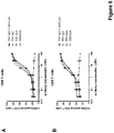
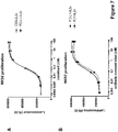
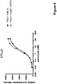


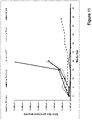

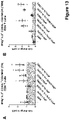


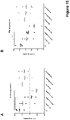

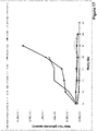


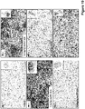


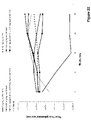
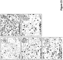
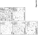
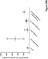

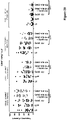

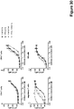
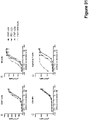
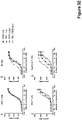
Classifications
-
- A—HUMAN NECESSITIES
- A61—MEDICAL OR VETERINARY SCIENCE; HYGIENE
- A61K—PREPARATIONS FOR MEDICAL, DENTAL OR TOILETRY PURPOSES
- A61K38/00—Medicinal preparations containing peptides
- A61K38/16—Peptides having more than 20 amino acids; Gastrins; Somatostatins; Melanotropins; Derivatives thereof
- A61K38/17—Peptides having more than 20 amino acids; Gastrins; Somatostatins; Melanotropins; Derivatives thereof from animals; from humans
- A61K38/19—Cytokines; Lymphokines; Interferons
- A61K38/20—Interleukins [IL]
- A61K38/2013—IL-2
-
- A—HUMAN NECESSITIES
- A61—MEDICAL OR VETERINARY SCIENCE; HYGIENE
- A61K—PREPARATIONS FOR MEDICAL, DENTAL OR TOILETRY PURPOSES
- A61K38/00—Medicinal preparations containing peptides
- A61K38/16—Peptides having more than 20 amino acids; Gastrins; Somatostatins; Melanotropins; Derivatives thereof
- A61K38/17—Peptides having more than 20 amino acids; Gastrins; Somatostatins; Melanotropins; Derivatives thereof from animals; from humans
- A61K38/19—Cytokines; Lymphokines; Interferons
- A61K38/20—Interleukins [IL]
- A61K38/2086—IL-13 to IL-16
-
- A—HUMAN NECESSITIES
- A61—MEDICAL OR VETERINARY SCIENCE; HYGIENE
- A61K—PREPARATIONS FOR MEDICAL, DENTAL OR TOILETRY PURPOSES
- A61K39/00—Medicinal preparations containing antigens or antibodies
-
- A—HUMAN NECESSITIES
- A61—MEDICAL OR VETERINARY SCIENCE; HYGIENE
- A61K—PREPARATIONS FOR MEDICAL, DENTAL OR TOILETRY PURPOSES
- A61K39/00—Medicinal preparations containing antigens or antibodies
- A61K39/395—Antibodies; Immunoglobulins; Immune serum, e.g. antilymphocytic serum
- A61K39/39533—Antibodies; Immunoglobulins; Immune serum, e.g. antilymphocytic serum against materials from animals
- A61K39/3955—Antibodies; Immunoglobulins; Immune serum, e.g. antilymphocytic serum against materials from animals against proteinaceous materials, e.g. enzymes, hormones, lymphokines
-
- A—HUMAN NECESSITIES
- A61—MEDICAL OR VETERINARY SCIENCE; HYGIENE
- A61K—PREPARATIONS FOR MEDICAL, DENTAL OR TOILETRY PURPOSES
- A61K47/00—Medicinal preparations characterised by the non-active ingredients used, e.g. carriers or inert additives; Targeting or modifying agents chemically bound to the active ingredient
- A61K47/50—Medicinal preparations characterised by the non-active ingredients used, e.g. carriers or inert additives; Targeting or modifying agents chemically bound to the active ingredient the non-active ingredient being chemically bound to the active ingredient, e.g. polymer-drug conjugates
- A61K47/51—Medicinal preparations characterised by the non-active ingredients used, e.g. carriers or inert additives; Targeting or modifying agents chemically bound to the active ingredient the non-active ingredient being chemically bound to the active ingredient, e.g. polymer-drug conjugates the non-active ingredient being a modifying agent
- A61K47/68—Medicinal preparations characterised by the non-active ingredients used, e.g. carriers or inert additives; Targeting or modifying agents chemically bound to the active ingredient the non-active ingredient being chemically bound to the active ingredient, e.g. polymer-drug conjugates the non-active ingredient being a modifying agent the modifying agent being an antibody, an immunoglobulin or a fragment thereof, e.g. an Fc-fragment
- A61K47/6801—Drug-antibody or immunoglobulin conjugates defined by the pharmacologically or therapeutically active agent
- A61K47/6803—Drugs conjugated to an antibody or immunoglobulin, e.g. cisplatin-antibody conjugates
- A61K47/6811—Drugs conjugated to an antibody or immunoglobulin, e.g. cisplatin-antibody conjugates the drug being a protein or peptide, e.g. transferrin or bleomycin
- A61K47/6813—Drugs conjugated to an antibody or immunoglobulin, e.g. cisplatin-antibody conjugates the drug being a protein or peptide, e.g. transferrin or bleomycin the drug being a peptidic cytokine, e.g. an interleukin or interferon
-
- A—HUMAN NECESSITIES
- A61—MEDICAL OR VETERINARY SCIENCE; HYGIENE
- A61K—PREPARATIONS FOR MEDICAL, DENTAL OR TOILETRY PURPOSES
- A61K47/00—Medicinal preparations characterised by the non-active ingredients used, e.g. carriers or inert additives; Targeting or modifying agents chemically bound to the active ingredient
- A61K47/50—Medicinal preparations characterised by the non-active ingredients used, e.g. carriers or inert additives; Targeting or modifying agents chemically bound to the active ingredient the non-active ingredient being chemically bound to the active ingredient, e.g. polymer-drug conjugates
- A61K47/51—Medicinal preparations characterised by the non-active ingredients used, e.g. carriers or inert additives; Targeting or modifying agents chemically bound to the active ingredient the non-active ingredient being chemically bound to the active ingredient, e.g. polymer-drug conjugates the non-active ingredient being a modifying agent
- A61K47/68—Medicinal preparations characterised by the non-active ingredients used, e.g. carriers or inert additives; Targeting or modifying agents chemically bound to the active ingredient the non-active ingredient being chemically bound to the active ingredient, e.g. polymer-drug conjugates the non-active ingredient being a modifying agent the modifying agent being an antibody, an immunoglobulin or a fragment thereof, e.g. an Fc-fragment
- A61K47/6835—Medicinal preparations characterised by the non-active ingredients used, e.g. carriers or inert additives; Targeting or modifying agents chemically bound to the active ingredient the non-active ingredient being chemically bound to the active ingredient, e.g. polymer-drug conjugates the non-active ingredient being a modifying agent the modifying agent being an antibody, an immunoglobulin or a fragment thereof, e.g. an Fc-fragment the modifying agent being an antibody or an immunoglobulin bearing at least one antigen-binding site
- A61K47/6849—Medicinal preparations characterised by the non-active ingredients used, e.g. carriers or inert additives; Targeting or modifying agents chemically bound to the active ingredient the non-active ingredient being chemically bound to the active ingredient, e.g. polymer-drug conjugates the non-active ingredient being a modifying agent the modifying agent being an antibody, an immunoglobulin or a fragment thereof, e.g. an Fc-fragment the modifying agent being an antibody or an immunoglobulin bearing at least one antigen-binding site the antibody targeting a receptor, a cell surface antigen or a cell surface determinant
-
- A—HUMAN NECESSITIES
- A61—MEDICAL OR VETERINARY SCIENCE; HYGIENE
- A61K—PREPARATIONS FOR MEDICAL, DENTAL OR TOILETRY PURPOSES
- A61K47/00—Medicinal preparations characterised by the non-active ingredients used, e.g. carriers or inert additives; Targeting or modifying agents chemically bound to the active ingredient
- A61K47/50—Medicinal preparations characterised by the non-active ingredients used, e.g. carriers or inert additives; Targeting or modifying agents chemically bound to the active ingredient the non-active ingredient being chemically bound to the active ingredient, e.g. polymer-drug conjugates
- A61K47/51—Medicinal preparations characterised by the non-active ingredients used, e.g. carriers or inert additives; Targeting or modifying agents chemically bound to the active ingredient the non-active ingredient being chemically bound to the active ingredient, e.g. polymer-drug conjugates the non-active ingredient being a modifying agent
- A61K47/68—Medicinal preparations characterised by the non-active ingredients used, e.g. carriers or inert additives; Targeting or modifying agents chemically bound to the active ingredient the non-active ingredient being chemically bound to the active ingredient, e.g. polymer-drug conjugates the non-active ingredient being a modifying agent the modifying agent being an antibody, an immunoglobulin or a fragment thereof, e.g. an Fc-fragment
- A61K47/6891—Pre-targeting systems involving an antibody for targeting specific cells
-
- A—HUMAN NECESSITIES
- A61—MEDICAL OR VETERINARY SCIENCE; HYGIENE
- A61P—SPECIFIC THERAPEUTIC ACTIVITY OF CHEMICAL COMPOUNDS OR MEDICINAL PREPARATIONS
- A61P35/00—Antineoplastic agents
-
- C—CHEMISTRY; METALLURGY
- C07—ORGANIC CHEMISTRY
- C07K—PEPTIDES
- C07K14/00—Peptides having more than 20 amino acids; Gastrins; Somatostatins; Melanotropins; Derivatives thereof
- C07K14/435—Peptides having more than 20 amino acids; Gastrins; Somatostatins; Melanotropins; Derivatives thereof from animals; from humans
- C07K14/52—Cytokines; Lymphokines; Interferons
- C07K14/54—Interleukins [IL]
- C07K14/5443—IL-15
-
- C—CHEMISTRY; METALLURGY
- C07—ORGANIC CHEMISTRY
- C07K—PEPTIDES
- C07K14/00—Peptides having more than 20 amino acids; Gastrins; Somatostatins; Melanotropins; Derivatives thereof
- C07K14/435—Peptides having more than 20 amino acids; Gastrins; Somatostatins; Melanotropins; Derivatives thereof from animals; from humans
- C07K14/52—Cytokines; Lymphokines; Interferons
- C07K14/54—Interleukins [IL]
- C07K14/55—IL-2
-
- C—CHEMISTRY; METALLURGY
- C07—ORGANIC CHEMISTRY
- C07K—PEPTIDES
- C07K16/00—Immunoglobulins [IGs], e.g. monoclonal or polyclonal antibodies
- C07K16/18—Immunoglobulins [IGs], e.g. monoclonal or polyclonal antibodies against material from animals or humans
- C07K16/28—Immunoglobulins [IGs], e.g. monoclonal or polyclonal antibodies against material from animals or humans against receptors, cell surface antigens or cell surface determinants
- C07K16/2803—Immunoglobulins [IGs], e.g. monoclonal or polyclonal antibodies against material from animals or humans against receptors, cell surface antigens or cell surface determinants against the immunoglobulin superfamily
- C07K16/2818—Immunoglobulins [IGs], e.g. monoclonal or polyclonal antibodies against material from animals or humans against receptors, cell surface antigens or cell surface determinants against the immunoglobulin superfamily against CD28 or CD152
-
- C—CHEMISTRY; METALLURGY
- C07—ORGANIC CHEMISTRY
- C07K—PEPTIDES
- C07K19/00—Hybrid peptides, i.e. peptides covalently bound to nucleic acids, or non-covalently bound protein-protein complexes
-
- A—HUMAN NECESSITIES
- A61—MEDICAL OR VETERINARY SCIENCE; HYGIENE
- A61K—PREPARATIONS FOR MEDICAL, DENTAL OR TOILETRY PURPOSES
- A61K39/00—Medicinal preparations containing antigens or antibodies
- A61K2039/505—Medicinal preparations containing antigens or antibodies comprising antibodies
-
- A—HUMAN NECESSITIES
- A61—MEDICAL OR VETERINARY SCIENCE; HYGIENE
- A61K—PREPARATIONS FOR MEDICAL, DENTAL OR TOILETRY PURPOSES
- A61K38/00—Medicinal preparations containing peptides
-
- C—CHEMISTRY; METALLURGY
- C07—ORGANIC CHEMISTRY
- C07K—PEPTIDES
- C07K2317/00—Immunoglobulins specific features
- C07K2317/70—Immunoglobulins specific features characterized by effect upon binding to a cell or to an antigen
- C07K2317/73—Inducing cell death, e.g. apoptosis, necrosis or inhibition of cell proliferation
-
- C—CHEMISTRY; METALLURGY
- C07—ORGANIC CHEMISTRY
- C07K—PEPTIDES
- C07K2319/00—Fusion polypeptide
- C07K2319/70—Fusion polypeptide containing domain for protein-protein interaction
- C07K2319/74—Fusion polypeptide containing domain for protein-protein interaction containing a fusion for binding to a cell surface receptor
Definitions
- the present invention generally relates to immunoconjugates, particularly immunoconjugates comprising a mutant interleukin-2 polypeptide and an antibody that binds to PD-1.
- the invention relates to polynucleotide molecules encoding the immunoconjugates, and vectors and host cells comprising such polynucleotide molecules.
- the invention further relates to methods for producing the mutant immunoconjugates, pharmaceutical compositions comprising the same, and uses thereof.
- Interleukin-2 also known as T cell growth factor (TCGF)
- TCGF T cell growth factor
- TCGF T cell growth factor
- IL-2 mediates its action by binding to IL-2 receptors (IL-2R), which consist of up to three individual subunits, the different association of which can produce receptor forms that differ in their affinity to IL-2.
- IL-2R IL-2 receptors
- Association of the ⁇ (CD25), ⁇ (CD122), and ⁇ ( ⁇ c , CD132) subunits results in a trimeric, high-affinity receptor for IL-2.
- Dimeric IL-2 receptor consisting of the ⁇ and ⁇ subunits is termed intermediate-affinity IL-2R.
- the ⁇ subunit forms the monomeric low affinity IL-2 receptor.
- the dimeric intermediate-affinity IL-2 receptor binds IL-2 with approximately 100-fold lower affinity than the trimeric high-affinity receptor
- both the dimeric and the trimeric IL-2 receptor variants are able to transmit signal upon IL-2 binding
- the ⁇ -subunit, CD25 is not essential for IL-2 signalling. It confers high-affinity binding to its receptor, whereas the ⁇ subunit, CD122, and the ⁇ -subunit are crucial for signal transduction ( Krieg et al., Proc Natl Acad Sci 107, 11906-11 (2010 )).
- Trimeric IL-2 receptors including CD25 are expressed by (resting) CD4 + forkhead box P3 (FoxP3) + regulatory T (T reg ) cells. They are also transiently induced on conventional activated T cells, whereas in the resting state these cells express only dimeric IL-2 receptors. T reg cells consistently express the highest level of CD25 in vivo ( Fontenot et al., Nature Immunol 6, 1142-51 (2005 )).
- IL-2 is synthesized mainly by activated T-cells, in particular CD4 + helper T cells. It stimulates the proliferation and differentiation of T cells, induces the generation of cytotoxic T lymphocytes (CTLs) and the differentiation of peripheral blood lymphocytes to cytotoxic cells and lymphokine-activated killer (LAK) cells, promotes cytokine and cytolytic molecule expression by T cells, facilitates the proliferation and differentiation of B-cells and the synthesis of immunoglobulin by B-cells, and stimulates the generation, proliferation and activation of natural killer (NK) cells (reviewed e.g.
- CTLs cytotoxic T lymphocytes
- LAK lymphokine-activated killer
- IL-2 lymphocyte populations in vivo and to increase the effector functions of these cells confers antitumor effects to IL-2, making IL-2 immunotherapy an attractive treatment option for certain metastatic cancers. Consequently, high-dose IL-2 treatment has been approved for use in patients with metastatic renal-cell carcinoma and malignant melanoma.
- IL-2 has a dual function in the immune response in that it not only mediates expansion and activity of effector cells, but also is crucially involved in maintaining peripheral immune tolerance.
- AICD IL-2 induced activation-induced cell death
- T cells A major mechanism underlying peripheral self-tolerance is IL-2 induced activation-induced cell death (AICD) in T cells.
- AICD is a process by which fully activated T cells undergo programmed cell death through engagement of cell surface-expressed death receptors such as CD95 (also known as Fas) or the TNF receptor.
- CD95 also known as Fas
- FasL Fas ligand
- TNF tumor necrosis factor
- T reg peripheral CD4 + CD25 + regulatory T cells
- T reg peripheral CD4 + CD25 + regulatory T cells
- CD4 + CD25 + regulatory T cells suppressor T cells. They suppress effector T cells from destroying their (self-)target, either through cell-cell contact by inhibiting T cell help and activation, or through release of immunosuppressive cytokines such as IL-10 or TGF- ⁇ . Depletion of T reg cells was shown to enhance IL-2 induced anti-tumor immunity ( Imai et al., Cancer Sci 98, 416-23 (2007 )).
- IL-2 is not optimal for inhibiting tumor growth, because in the presence of IL-2 either the CTLs generated might recognize the tumor as self and undergo AICD or the immune response might be inhibited by IL-2 dependent T reg cells.
- VLS vascular leak syndrome
- VLS Low-dose IL-2 regimens have been tested in patients to avoid VLS, however, at the expense of suboptimal therapeutic results.
- VLS was believed to be caused by the release of proinflammatory cytokines, such as tumor necrosis factor (TNF)- ⁇ from IL-2-activated NK cells, however it has recently been shown that IL-2-induced pulmonary edema resulted from direct binding of IL-2 to lung endothelial cells, which expressed low to intermediate levels of functional ⁇ IL-2 receptors ( Krieg et al., Proc Nat Acad Sci USA 107, 11906-11 (2010 )).
- TNF tumor necrosis factor
- a particular mutant IL-2 polypeptide designed to overcome the above-mentioned problems associated with IL-2 immunotherapy (toxicity caused by the induction of VLS, tumor tolerance caused by the induction of AICD, and immunosuppression caused by activation of T reg cells), is described in WO 2012/107417 .
- Substitution of the phenylalanine residue at position 42 by alanine, the tyrosine residue at position 45 by alanine and the leucine residue at position 72 of IL-2 by glycine essentially abolishes binding of this mutant IL-2 polypeptide to the ⁇ -subunit of the IL-2 receptor (CD25).
- IL-2 immunotherapy may be improved by selectively targeting IL-2 to tumors, e.g. in the form of immunoconjugates comprising an antibody that binds to an antigen expressed on tumor cells.
- immunoconjugates comprising an antibody that binds to an antigen expressed on tumor cells.
- Tumors may be able, however, to escape such targeting by shedding, mutating or downregulating the target antigen of the antibody. Moreover, tumor-targeted IL-2 may not come into optimal contact with effector cells such as cytotoxic T lymphocytes (CTLs), in tumor microenvironments that actively exclude lymphocytes.
- CTLs cytotoxic T lymphocytes
- An approach which may circumvent the problems of tumor-targeting, is to target IL-2 directly to effector cells, in particular CTLs.
- Ghasemi et al. have described a fusion protein of IL-2 and an NKG2D binding protein ( Ghashemi et al., Nat Comm (2016) 7, 12878 ), for targeting IL-2 to NKG2D-bearing cells such as natural killer (NK) cells.
- NK natural killer
- PD-1 Programmed cell death protein 1
- CD28 is an inhibitory member of the CD28 family of receptors, that also includes CD28, CTLA-4, ICOS and BTLA.
- PD-1 is a cell surface receptor and is expressed on activated B cells, T cells, and myeloid cells ( Okazaki et al (2002) Curr. Opin. Immunol. 14: 391779-82 ; Bennett et al. (2003) J Immunol 170:711-8 ).
- PD-1 The structure of PD-1 is a monomeric type 1 transmembrane protein, consisting of one immunoglobulin variable-like extracellular domain and a cytoplasmic domain containing an immunoreceptor tyrosine-based inhibitory motif (ITIM) and an immunoreceptor tyrosine-based switch motif (ITSM).
- ITIM immunoreceptor tyrosine-based inhibitory motif
- ITMS immunoreceptor tyrosine-based switch motif
- Two ligands for PD-1 have been identified, PD-L1 and PD-L2, that have been shown to downregulate T cell activation upon binding to PD-1 ( Freeman et al (2000) J Exp Med 192: 1027-34 ; Latchman et al (2001) Nat Immunol 2:261-8 ; Carter etal (2002) Eur J Immunol 32:634-43 ).
- Both PD-L1 and PD-L2 are B7 homologs that bind to PD-1, but do not bind to other CD28 family members.
- One ligand for PD-1, PD-L1 is abundant in a variety of human cancers ( Dong et al (2002) Nat. Med 8:787-9 ).
- the interaction between PD-1 and PD-L1 results in a decrease in tumor infiltrating lymphocytes, a decrease in T-cell receptor mediated proliferation, and immune evasion by the cancerous cells ( Dong et al. (2003) J. Mol. Med. 81:281-7 ; Blank et al. (2005) Cancer Immunol. Immunother. 54:307-314 ; Konishi et al. (2004) Clin. Cancer Res.
- Immune suppression can be reversed by inhibiting the local interaction of PD-1 with PD-L1, and the effect is additive when the interaction of PD-1 with PD-L2 is blocked as well ( Iwai et al. (2002) Proc. Nat 7. Acad. ScL USA 99: 12293-7 ; Brown et al. (2003) J. Immunol. 170:1257-66 ).
- Antibodies that bind to PD-1 are described e.g. in WO 2017/055443 A1 .
- the present invention provides a novel approach of targeting a mutant form of IL-2 with advantageous properties for immunotherapy directly to immune effector cells, such as cytotoxic T lymphocytes, rather than tumor cells.
- Targeting to immune effector cells is achieved by conjugation of the mutant IL-2 molecule to an antibody that binds to PD-1.
- the IL-2 mutant used in the present invention has been designed to overcome the problems associated with IL-2 immunotherapy, in particular toxicity caused by the induction of VLS, tumor tolerance caused by the induction of AICD, and immunosuppression caused by activation of T reg cells.
- targeting of the IL-2 mutant to immune effector cells may further increase the preferential activation of CTLs over immunosuppressive T reg cells.
- the suppression of T-cell activity induced by the interaction of PD-1 with its ligand PD-L1 may additionally be reversed, thus further enhancing the immune response.
- IL-2 fusion protein comprising the anti-PD-L1 antibody atezolizumab has been described by Chen et al. ( Chen et al., Biochem Biophys Res Comm (2016) 480, 160-165 ).
- the immunoconjugate of the invention comprising an antibody that binds to PD-1, shows significantly superior anti-tumor efficacy in vivo as compared to a similar immunoconjugate targeting PD-L1 (see Example 3 hereinbelow).
- an immunoconjugate comprising an antibody that binds to PD-1 and an IL-2 polypetide.
- the invention provides an immunoconjugate comprising a mutant IL-2 polypeptide and an antibody that binds to PD-1, wherein the mutant IL-2 polypeptide is a human IL-2 molecule comprising the amino acid substitutions F42A, Y45A and L72G (numbering relative to the human IL-2 sequence SEQ ID NO: 19).
- the invention provides an immunoconjugate comprising a mutant IL-2 polypeptide and an antibody that binds to PD-1, wherein the mutant IL-2 polypeptide is a human IL-2 molecule comprising the amino acid substitutions F42A, Y45A and L72G (numbering relative to the human IL-2 sequence SEQ ID NO: 19); and wherein the antibody comprises (a) a heavy chain variable region (VH) comprising a HVR-H1 comprising the amino acid sequence of SEQ ID NO:1, a HVR-H2 comprising the amino acid sequence of SEQ ID NO:2, a HVR-H3 comprising the amino acid sequence of SEQ ID NO:3, and a FR-H3 comprising the amino acid sequence of SEQ ID NO:7 at positions 71-73 according to Kabat numbering, and (b) a light chain variable region (VL) comprising a HVR-L1 comprising the amino acid sequence of SEQ ID NO:4, a HVR-L2 comprising the
- the invention provides an immunoconjugate comprising a mutant IL-2 polypeptide and an antibody that binds to PD-1, wherein the mutant IL-2 polypeptide is a human IL-2 molecule comprising the amino acid substitutions F42A, Y45A and L72G (numbering relative to the human IL-2 sequence SEQ ID NO: 19); and wherein the antibody comprises (a) a heavy chain variable region (VH) comprising an amino acid sequence that is at least about 95%, 96%, 97%, 98%, 99% or 100% identical to the amino acid sequence of SEQ ID NO:14, and (b) a light chain variable region (VL) comprising an amino acid sequence that is at least about 95%, 96%, 97%, 98%, 99% or 100% identical to an amino acid sequence selected from the group consisting of SEQ ID NO:15, SEQ ID NO:16, SEQ ID NO: 17, and SEQ ID NO:18.
- VH heavy chain variable region
- VL light chain variable region
- the mutant IL-2 polypeptide further comprises the amino acid substitution T3A and/or the amino acid substitution C125A.
- the mutant IL-2 polypeptide comprises the sequence of SEQ ID NO: 20.
- the immunoconjugate comprises not more than one mutant IL-2 polypeptide.
- the antibody comprises an Fc domain composed of a first and a second subunit.
- the Fc domain is an IgG class, particularly an IgG 1 subclass, Fc domain, and/or the Fc domain is a human Fc domain.
- the antibody is an IgG class, particularly an IgG 1 subclass immunoglobulin.
- the Fc domain comprises a modification promoting the association of the first and the second subunit of the Fc domain.
- an amino acid residue in the CH3 domain of the first subunit of the Fc domain an amino acid residue is replaced with an amino acid residue having a larger side chain volume, thereby generating a protuberance within the CH3 domain of the first subunit which is positionable in a cavity within the CH3 domain of the second subunit, and in the CH3 domain of the second subunit of the Fc domain an amino acid residue is replaced with an amino acid residue having a smaller side chain volume, thereby generating a cavity within the CH3 domain of the second subunit within which the protuberance within the CH3 domain of the first subunit is positionable.
- the threonine residue at position 366 is replaced with a tryptophan residue (T366W), and in the second subunit of the Fc domain the tyrosine residue at position 407 is replaced with a valine residue (Y407V) and optionally the threonine residue at position 366 is replaced with a serine residue (T366S) and the leucine residue at position 368 is replaced with an alanine residue (L368A) (numberings according to Kabat EU index).
- the mutant IL-2 polypeptide is fused at its amino-terminal amino acid to the carboxy-terminal amino acid of one of the subunits of the Fc domain, particularly the first subunit of the Fc domain, optionally through a linker peptide.
- the linker peptide has the amino acid sequence of SEQ ID NO:21.
- the Fc domain comprises one or more amino acid substitution that reduces binding to an Fc receptor, particularly an Fc ⁇ receptor, and/or effector function, particularly antibody-dependent cell-mediated cytotoxicity (ADCC).
- said one or more amino acid substitution is at one or more position selected from the group of L234, L235, and P329 (Kabat EU index numbering).
- each subunit of the Fc domain comprises the amino acid substitutions L234A, L235A and P329G (Kabat EU index numbering).
- the immunoconjugate according to the invention comprises a polypeptide comprising an amino acid sequence that is at least about 80%, 85%, 90%, 95%, 96%, 97%, 98%, 99% or 100% identical to the sequence of SEQ ID NO:22, a polypeptide comprising an amino acid sequence that is at least about 80%, 85%, 90%, 95%, 96%, 97%, 98%, 99% or 100% identical to the sequence of SEQ ID NO:23 or SEQ ID NO:24, and a polypeptide comprising an amino acid sequence that is at least about 80%, 85%, 90%, 95%, 96%, 97%, 98%, 99% or 100% identical to the sequence of SEQ ID NO:25.
- the immunoconjugate essentially consists of a mutant IL-2 polypeptide and an IgG 1 immunoglobulin molecule, joined by a linker sequence.
- the invention further provides one or more isolated polynucleotide encoding an immunoconjugate of the invention, one or more vector (particularly expression vector) comprising said polynucleotides, and host cells comprising said polynucleotide(s) or said vector(s).
- the invention further provides a pharmaceutical composition comprising an immunoconjugate of the invention and a pharmaceutically acceptable carrier.
- the invention encompasses an immunoconjugate according to the invention for use as a medicament, and for use in the treatment of a disease, wherein said disease is cancer.
- interleukin-2 refers to any native IL-2 from any vertebrate source, including mammals such as primates (e.g. humans) and rodents (e.g., mice and rats), unless otherwise indicated.
- the term encompasses unprocessed IL-2 as well as any form of IL-2 that results from processing in the cell.
- the term also encompasses naturally occurring variants of IL-2, e.g. splice variants or allelic variants.
- the amino acid sequence of an exemplary human IL-2 is shown in SEQ ID NO: 19.
- Unprocessed human IL-2 additionally comprises an N-terminal 20 amino acid signal peptide having the sequence of SEQ ID NO: 26, which is absent in the mature IL-2 molecule.
- IL-2 mutant or "mutant IL-2 polypeptide” as used herein is intended to encompass any mutant forms of various forms of the IL-2 molecule including full-length IL-2, truncated forms of IL-2 and forms where IL-2 is linked to another molecule such as by fusion or chemical conjugation.
- Full-length when used in reference to IL-2 is intended to mean the mature, natural length IL-2 molecule.
- full-length human IL-2 refers to a molecule that has 133 amino acids (see e.g. SEQ ID NO: 19).
- the various forms of IL-2 mutants are characterized in having a at least one amino acid mutation affecting the interaction of IL-2 with CD25.
- This mutation may involve substitution, deletion, truncation or modification of the wild-type amino acid residue normally located at that position. Mutants obtained by amino acid substitution are preferred.
- an IL-2 mutant may be referred to herein as a mutant IL-2 peptide sequence, a mutant IL-2 polypeptide, a mutant IL-2 protein or a mutant IL-2 analog. Designation of various forms of IL-2 is herein made with respect to the sequence shown in SEQ ID NO: 19. Various designations may be used herein to indicate the same mutation. For example a mutation from phenylalanine at position 42 to alanine can be indicated as 42A, A42, A 42 , F42A, or Phe42Ala.
- human IL-2 molecule an IL-2 molecule comprising an amino acid sequence that is at least about 90%, at least about 91%, at least about 92%, at least about 93%, at least about 94%, at least about 95% or at least about 96% identical to the human IL-2 sequence of SEQ ID NO: 19. Particularly, the sequence identity is at least about 95%, more particularly at least about 96%.
- the human IL-2 molecule is a full-length IL-2 molecule.
- amino acid mutation as used herein is meant to encompass amino acid substitutions, deletions, insertions, and modifications. Any combination of substitution, deletion, insertion, and modification can be made to arrive at the final construct, provided that the final construct possesses the desired characteristics, e.g. reduced binding to CD25.
- Amino acid sequence deletions and insertions include amino- and/or carboxy-terminal deletions and insertions of amino acids.
- An example of a terminal deletion is the deletion of the alanine residue in position 1 of full-length human IL-2.
- Preferred amino acid mutations are amino acid substitutions. For the purpose of altering e.g. the binding characteristics of an IL-2 polypeptide, non-conservative amino acid substitutions, i.e.
- amino acid substitions include replacing a hydrophobic by a hydrophilic amino acid.
- Amino acid substitutions include replacement by non-naturally occurring amino acids or by naturally occurring amino acid derivatives of the twenty standard amino acids (e.g. 4-hydroxyproline, 3-methylhistidine, ornithine, homoserine, 5-hydroxylysine).
- Amino acid mutations can be generated using genetic or chemical methods well known in the art. Genetic methods may include site-directed mutagenesis, PCR, gene synthesis and the like. It is contemplated that methods of altering the side chain group of an amino acid by methods other than genetic engineering, such as chemical modification, may also be useful.
- a "wild-type" form of IL-2 is a form of IL-2 that is otherwise the same as the mutant IL-2 polypeptide except that the wild-type form has a wild-type amino acid at each amino acid position of the mutant IL-2 polypeptide.
- the wild-type form of this mutant is full-length native IL-2.
- the IL-2 mutant is a fusion between IL-2 and another polypeptide encoded downstream of IL-2 (e.g.
- the wild-type form of this IL-2 mutant is IL-2 with a wild-type amino acid sequence, fused to the same downstream polypeptide. Furthermore, if the IL-2 mutant is a truncated form of IL-2 (the mutated or modified sequence within the non-truncated portion of IL-2) then the wild-type form of this IL-2 mutant is a similarly truncated IL-2 that has a wild-type sequence.
- wild-type IL-2 for the purpose of the present invention comprises the amino acid substitution C125A (see SEQ ID NO: 29).
- wild-type IL-2 polypeptide to which the mutant IL-2 polypeptide is compared comprises the amino acid sequence of SEQ ID NO: 19.
- wild-type IL-2 polypeptide to which the mutant IL-2 polypeptide is compared comprises the amino acid sequence of SEQ ID NO: 29.
- CD25 or "a-subunit of the IL-2 receptor” as used herein, refers to any native CD25 from any vertebrate source, including mammals such as primates (e.g. humans) and rodents (e.g., mice and rats), unless otherwise indicated.
- the term encompasses "full-length", unprocessed CD25 as well as any form of CD25 that results from processing in the cell.
- the term also encompasses naturally occurring variants of CD25, e.g. splice variants or allelic variants.
- CD25 is human CD25.
- the amino acid sequence of human CD25 is found e.g. in UniProt entry no. P01589 (version 185).
- high-affinity IL-2 receptor refers to the heterotrimeric form of the IL-2 receptor, consisting of the receptor ⁇ -subunit (also known as common cytokine receptor ⁇ -subunit, ⁇ c , or CD132, see UniProt entry no. P14784 (version 192)), the receptor ⁇ -subunit (also known as CD122 or p70, see UniProt entry no. P31785 (version 197)) and the receptor ⁇ -subunit (also known as CD25 or p55, see UniProt entry no. P01589 (version 185)).
- the receptor ⁇ -subunit also known as common cytokine receptor ⁇ -subunit, ⁇ c , or CD132, see UniProt entry no. P14784 (version 192)
- the receptor ⁇ -subunit also known as CD122 or p70, see UniProt entry no. P31785 (version 197)
- the receptor ⁇ -subunit also known as CD25 or p55, see UniProt entry no. P015
- intermediate-affinity IL-2 receptor refers to the IL-2 receptor including only the ⁇ -subunit and the ⁇ -subunit, without the ⁇ -subunit (for a review see e.g. Olejniczak and Kasprzak, Med Sci Monit 14, RA179-189 (2008 )).
- Binding affinity refers to intrinsic binding affinity which reflects a 1:1 interaction between members of a binding pair (e.g., an antigen binding moiety and an antigen, or a receptor and its ligand).
- the affinity of a molecule X for its partner Y can generally be represented by the dissociation constant (K D ), which is the ratio of dissociation and association rate constants (k off and k on , respectively).
- affinities may comprise different rate constants, as long as the ratio of the rate constants remains the same.
- Affinity can be measured by well established methods known in the art, including those described herein.
- a particular method for measuring affinity is Surface Plasmon Resonance (SPR).
- the affinity of the mutant or wild-type IL-2 polypeptide for various forms of the IL-2 receptor can be determined in accordance with the method set forth in the WO 2012/107417 by surface plasmon resonance (SPR), using standard instrumentation such as a BIAcore instrument (GE Healthcare) and receptor subunits such as may be obtained by recombinant expression (see e.g. Shanafelt et al., Nature Biotechnol 18, 1197-1202 (2000 )).
- binding affinity of IL-2 mutants for different forms of the IL-2 receptor may be evaluated using cell lines known to express one or the other such form of the receptor. Specific illustrative and exemplary embodiments for measuring binding affinity are described hereinafter.
- T reg cell a specialized type of CD4 + T cell that can suppress the responses of other T cells.
- T reg cells are characterized by expression of the ⁇ -subunit of the IL-2 receptor (CD25) and the transcription factor forkhead box P3 (FOXP3) ( Sakaguchi, Annu Rev Immunol 22, 531-62 (2004 )) and play a critical role in the induction and maintenance of peripheral self-tolerance to antigens, including those expressed by tumors.
- T reg cells require IL-2 for their function and development and induction of their suppressive characteristics.
- effector cells refers to a population of lymphocytes that mediate the cytotoxic effects of IL-2. Effector cells include effector T cells such as CD8 + cytotoxic T cells, NK cells, lymphokine-activated killer (LAK) cells and macrophages/monocytes.
- effector T cells such as CD8 + cytotoxic T cells, NK cells, lymphokine-activated killer (LAK) cells and macrophages/monocytes.
- PD1 As used herein, the term “PD1”, “human PD1”, “PD-1” or “human PD-1” (also known as Programmed cell death protein 1, or Programmed Death 1) refers to the human protein PD1 (SEQ ID NO: 27, protein without signal sequence) / (SEQ ID NO: 28, protein with signal sequence). See also UniProt entry no. Q15116 (version 156).
- an antibody “binding to PD-1”, “specifically binding to PD-1”, “that binds to PD-1” or “anti-PD-1 antibody” refers to an antibody that is capable of binding PD-1, especially a PD-1 polypeptide expressed on a cell surface, with sufficient affinity such that the antibody is useful as a diagnostic and/or therapeutic agent in targeting PD-1.
- the extent of binding of an anti-PD-1 antibody to an unrelated, non-PD-1 protein is less than about 10% of the binding of the antibody to PD-1 as measured, e.g., by radioimmunoassay (RIA) or flow cytometry (FACS) or by a Surface Plasmon Resonance assay using a biosensor system such as a Biacore ® system.
- an antibody that binds to PD-1 has a KD value of the binding affinity for binding to human PD-1 of ⁇ 1 ⁇ M, ⁇ 100 nM, ⁇ 10 nM, ⁇ 1 nM, ⁇ 0.1 nM, ⁇ 0.01 nM, or ⁇ 0.001 nM (e.g.
- the KD value of the binding affinity is determined in a Surface Plasmon Resonance assay using the Extracellular domain (ECD) of human PD-1 (PD-1-ECD, see SEQ ID NO: 43) as antigen.
- ECD Extracellular domain
- telomere binding is meant that the binding is selective for the antigen and can be discriminated from unwanted or non-specific interactions.
- a specific antigen e.g. PD-1
- ELISA enzyme-linked immunosorbent assay
- SPR surface plasmon resonance
- the extent of binding of an antibody to an unrelated protein is less than about 10% of the binding of the antibody to the antigen as measured, e.g., by SPR.
- the antibody comprised in the immunoconjugate described herein specifically binds to PD-1.
- polypeptide refers to a molecule composed of monomers (amino acids) linearly linked by amide bonds (also known as peptide bonds).
- polypeptide refers to any chain of two or more amino acids, and does not refer to a specific length of the product.
- peptides, dipeptides, tripeptides, oligopeptides, "protein”, “amino acid chain”, or any other term used to refer to a chain of two or more amino acids are included within the definition of "polypeptide”, and the term “polypeptide” may be used instead of, or interchangeably with any of these terms.
- polypeptide is also intended to refer to the products of post-expression modifications of the polypeptide, including without limitation glycosylation, acetylation, phosphorylation, amidation, derivatization by known protecting/blocking groups, proteolytic cleavage, or modification by non-naturally occurring amino acids.
- a polypeptide may be derived from a natural biological source or produced by recombinant technology, but is not necessarily translated from a designated nucleic acid sequence. It may be generated in any manner, including by chemical synthesis.
- Polypeptides may have a defined three-dimensional structure, although they do not necessarily have such structure. Polypeptides with a defined three-dimensional structure are referred to as folded, and polypeptides which do not possess a defined three-dimensional structure, but rather can adopt a large number of different conformations, and are referred to as unfolded.
- an “isolated” polypeptide or a variant, or derivative thereof is intended a polypeptide that is not in its natural milieu. No particular level of purification is required.
- an isolated polypeptide can be removed from its native or natural environment.
- Recombinantly produced polypeptides and proteins expressed in host cells are considered isolated for the purpose of the invention, as are native or recombinant polypeptides which have been separated, fractionated, or partially or substantially purified by any suitable technique.
- Percent (%) amino acid sequence identity with respect to a reference polypeptide sequence is defined as the percentage of amino acid residues in a candidate sequence that are identical with the amino acid residues in the reference polypeptide sequence, after aligning the sequences and introducing gaps, if necessary, to achieve the maximum percent sequence identity, and not considering any conservative substitutions as part of the sequence identity. Alignment for purposes of determining percent amino acid sequence identity can be achieved in various ways that are within the skill in the art, for instance, using publicly available computer software such as BLAST, BLAST-2, Clustal W, Megalign (DNASTAR) software or the FASTA program package.
- % amino acid sequence identity values are generated using the ggsearch program of the FASTA package version 36.3.8c or later with a BLOSUM50 comparison matrix.
- the FASTA program package was authored by W. R. Pearson and D. J. Lipman (1988), "Improved Tools for Biological Sequence Analysis", PNAS 85:2444-2448 ; W. R. Pearson (1996) “Effective protein sequence comparison” Meth. Enzymol. 266:227- 258 ; and Pearson et. al.
- Genomics 46:24-36 is publicly available from http://fasta.bioch.virginia.edu/fasta_www2/fasta_down.shtml.
- polynucleotide refers to an isolated nucleic acid molecule or construct, e.g. messenger RNA (mRNA), virally-derived RNA, or plasmid DNA (pDNA).
- mRNA messenger RNA
- pDNA virally-derived RNA
- a polynucleotide may comprise a conventional phosphodiester bond or a non-conventional bond (e.g. an amide bond, such as found in peptide nucleic acids (PNA).
- PNA peptide nucleic acids
- nucleic acid molecule refers to any one or more nucleic acid segments, e.g. DNA or RNA fragments, present in a polynucleotide.
- isolated nucleic acid molecule or polynucleotide is intended a nucleic acid molecule, DNA or RNA, which has been removed from its native environment.
- a recombinant polynucleotide encoding a polypeptide contained in a vector is considered isolated for the purposes of the present invention.
- Further examples of an isolated polynucleotide include recombinant polynucleotides maintained in heterologous host cells or purified (partially or substantially) polynucleotides in solution.
- An isolated polynucleotide includes a polynucleotide molecule contained in cells that ordinarily contain the polynucleotide molecule, but the polynucleotide molecule is present extrachromosomally or at a chromosomal location that is different from its natural chromosomal location.
- Isolated RNA molecules include in vivo or in vitro RNA transcripts of the present invention, as well as positive and negative strand forms, and double-stranded forms. Isolated polynucleotides or nucleic acids according to the present invention further include such molecules produced synthetically.
- a polynucleotide or a nucleic acid may be or may include a regulatory element such as a promoter, ribosome binding site, or a transcription terminator.
- isolated polynucleotide (or nucleic acid) encoding [e.g. an immunoconjugate of the invention]” refers to one or more polynucleotide molecules encoding antibody heavy and light chains and/or IL-2 polypeptides (or fragments thereof), including such polynucleotide molecule(s) in a single vector or separate vectors, and such nucleic acid molecule(s) present at one or more locations in a host cell.
- expression cassette refers to a polynucleotide generated recombinantly or synthetically, with a series of specified nucleic acid elements that permit transcription of a particular nucleic acid in a target cell.
- the recombinant expression cassette can be incorporated into a plasmid, chromosome, mitochondrial DNA, plastid DNA, virus, or nucleic acid fragment.
- the recombinant expression cassette portion of an expression vector includes, among other sequences, a nucleic acid sequence to be transcribed and a promoter.
- the expression cassette comprises polynucleotide sequences that encode immunoconjugates of the invention or fragments thereof.
- vector refers to a DNA molecule that is used to introduce and direct the expression of a specific gene to which it is operably associated in a cell.
- the term includes the vector as a self-replicating nucleic acid structure as well as the vector incorporated into the genome of a host cell into which it has been introduced.
- the expression vector of the present invention comprises an expression cassette. Expression vectors allow transcription of large amounts of stable mRNA. Once the expression vector is inside the cell, the ribonucleic acid molecule or protein that is encoded by the gene is produced by the cellular transcription and/or translation machinery.
- the expression vector of the invention comprises an expression cassette that comprises polynucleotide sequences that encode immunoconjugates of the invention or fragments thereof.
- host cell refers to cells into which exogenous nucleic acid has been introduced, including the progeny of such cells.
- Host cells include “transformants” and “transformed cells,” which include the primary transformed cell and progeny derived therefrom without regard to the number of passages. Progeny may not be completely identical in nucleic acid content to a parent cell, but may contain mutations. Mutant progeny that have the same function or biological activity as screened or selected for in the originally transformed cell are included herein.
- a host cell is any type of cellular system that can be used to generate the immunoconjugates of the present invention.
- Host cells include cultured cells, e.g.
- mammalian cultured cells such as HEK cells, CHO cells, BHK cells, NS0 cells, SP2/0 cells, YO myeloma cells, P3X63 mouse myeloma cells, PER cells, PER.C6 cells or hybridoma cells, yeast cells, insect cells, and plant cells, to name only a few, but also cells comprised within a transgenic animal, transgenic plant or cultured plant or animal tissue.
- antibody herein is used in the broadest sense and encompasses various antibody structures, including but not limited to monoclonal antibodies, polyclonal antibodies, multispecific antibodies (e.g. bispecific antibodies), and antibody fragments so long as they exhibit the desired antigen binding activity.
- monoclonal antibody refers to an antibody obtained from a population of substantially homogeneous antibodies, i.e. the individual antibodies comprised in the population are identical and/or bind the same epitope, except for possible variant antibodies, e.g., containing naturally occurring mutations or arising during production of a monoclonal antibody preparation, such variants generally being present in minor amounts.
- polyclonal antibody preparations typically include different antibodies directed against different determinants (epitopes)
- each monoclonal antibody of a monoclonal antibody preparation is directed against a single determinant on an antigen.
- the modifier "monoclonal” indicates the character of the antibody as being obtained from a substantially homogeneous population of antibodies, and is not to be construed as requiring production of the antibody by any particular method.
- the monoclonal antibodies to be used in accordance with the present invention may be made by a variety of techniques, including but not limited to the hybridoma method, recombinant DNA methods, phage-display methods, and methods utilizing transgenic animals containing all or part of the human immunoglobulin loci, such methods and other exemplary methods for making monoclonal antibodies being described herein.
- an "isolated” antibody is one which has been separated from a component of its natural environment, i.e. that is not in its natural milieu. No particular level of purification is required.
- an isolated antibody can be removed from its native or natural environment.
- Recombinantly produced antibodies expressed in host cells are considered isolated for the purpose of the invention, as are native or recombinant antibodies which have been separated, fractionated, or partially or substantially purified by any suitable technique. As such, the immunoconjugates of the present invention are isolated.
- an antibody is purified to greater than 95% or 99% purity as determined by, for example, electrophoretic (e.g., SDS-PAGE, isoelectric focusing (IEF), capillary electrophoresis) or chromatographic (e.g., ion exchange or reverse phase HPLC) methods.
- electrophoretic e.g., SDS-PAGE, isoelectric focusing (IEF), capillary electrophoresis
- chromatographic e.g., ion exchange or reverse phase HPLC
- full-length antibody “intact antibody,” and “whole antibody” are used herein interchangeably to refer to an antibody having a structure substantially similar to a native antibody structure.
- antibody fragment refers to a molecule other than an intact antibody that comprises a portion of an intact antibody that binds the antigen to which the intact antibody binds.
- antibody fragments include but are not limited to Fv, Fab, Fab', Fab'-SH, F(ab') 2 , diabodies, linear antibodies, single-chain antibody molecules (e.g. scFv), and single-domain antibodies.
- scFv single-chain antibody molecules
- Diabodies are antibody fragments with two antigen-binding sites that may be bivalent or bispecific.
- Single-domain antibodies are antibody fragments comprising all or a portion of the heavy chain variable domain or all or a portion of the light chain variable domain of an antibody.
- a single-domain antibody is a human single-domain antibody (Domantis, Inc., Waltham, MA; see e.g. U.S. Patent No.
- Antibody fragments can be made by various techniques, including but not limited to proteolytic digestion of an intact antibody as well as production by recombinant host cells (e.g. E. coli or phage), as described herein.
- recombinant host cells e.g. E. coli or phage
- immunoglobulin molecule refers to a protein having the structure of a naturally occurring antibody.
- immunoglobulins of the IgG class are heterotetrameric glycoproteins of about 150,000 daltons, composed of two light chains and two heavy chains that are disulfide-bonded. From N- to C-terminus, each heavy chain has a variable domain (VH), also called a variable heavy domain or a heavy chain variable region, followed by three constant domains (CH1, CH2, and CH3), also called a heavy chain constant region.
- VH variable domain
- CH1, CH2, and CH3 constant domains
- each light chain has a variable domain (VL), also called a variable light domain or a light chain variable region, followed by a constant light (CL) domain, also called a light chain constant region.
- VL variable domain
- CL constant light
- the heavy chain of an immunoglobulin may be assigned to one of five types, called ⁇ (IgA), ⁇ (IgD), ⁇ (IgE), ⁇ (IgG), or ⁇ (IgM), some of which may be further divided into subtypes, e.g. ⁇ 1 (IgG 1 ), ⁇ 2 (IgG 2 ), ⁇ 3 (IgG 3 ), ⁇ 4 (IgG 4 ), ⁇ 1 (IgA 1 ) and ⁇ 2 (IgA 2 ).
- the light chain of an immunoglobulin may be assigned to one of two types, called kappa ( ⁇ ) and lambda ( ⁇ ), based on the amino acid sequence of its constant domain.
- An immunoglobulin essentially consists of two Fab molecules and an Fc domain, linked via the immunoglobulin hinge region.
- an antigen binding domain refers to the part of an antibody that comprises the area which specifically binds to and is complementary to part or all of an antigen.
- An antigen binding domain may be provided by, for example, one or more antibody variable domains (also called antibody variable regions).
- an antigen binding domain comprises an antibody light chain variable domain (VL) and an antibody heavy chain variable domain (VH).
- variable region refers to the domain of an antibody heavy or light chain that is involved in binding the antibody to antigen.
- the variable domains of the heavy chain and light chain (VH and VL, respectively) of a native antibody generally have similar structures, with each domain comprising four conserved framework regions (FRs) and three hypervariable regions (HVRs). See, e.g., Kindt et al., Kuby Immunology, 6th ed., W.H. Freeman and Co., page 91 (2007 ). A single VH or VL domain may be sufficient to confer antigen-binding specificity.
- Kabat numbering refers to the numbering system set forth by Kabat et al., Sequences of Proteins of Immunological Interest, 5th Ed. Public Health Service, National Institutes of Health, Bethesda, MD (1991 ).
- amino acid positions of all constant regions and domains of the heavy and light chain are numbered according to the Kabat numbering system described in Kabat, et al., Sequences of Proteins of Immunological Interest, 5th ed., Public Health Service, National Institutes of Health, Bethesda, MD (1991 ), referred to as “numbering according to Kabat” or "Kabat numbering” herein.
- Kabat numbering system see pages 647-660 of Kabat, et al., Sequences of Proteins of Immunological Interest, 5th ed., Public Health Service, National Institutes of Health, Bethesda, MD (1991 )
- CL light chain constant domain
- Kabat EU index numbering system see pages 661-723
- CH1, Hinge, CH2 and CH3 heavy chain constant domains
- hypervariable region refers to each of the regions of an antibody variable domain which are hypervariable in sequence ("complementarity determining regions” or “CDRs") and/or form structurally defined loops ("hypervariable loops") and/or contain the antigen-contacting residues ("antigen contacts").
- CDRs complementarity determining regions
- hypervariable loops form structurally defined loops
- antigen contacts Generally, antibodies comprise six HVRs; three in the VH (HI, H2, H3), and three in the VL (L1, L2, L3).
- Exemplary HVRs herein include:
- HVR residues and other residues in the variable domain are numbered herein according to Kabat et al., supra.
- FR Framework or "FR” refers to variable domain residues other than hypervariable region (HVR) residues.
- the FR of a variable domain generally consists of four FR domains: FR1, FR2, FR3, and FR4. Accordingly, the HVR and FR sequences generally appear in the following order in VH (or VL): FR1-H1(L1)-FR2-H2(L2)-FR3-H3(L3)-FR4.
- a “humanized” antibody refers to a chimeric antibody comprising amino acid residues from non-human HVRs and amino acid residues from human FRs.
- a humanized antibody will comprise substantially all of at least one, and typically two, variable domains, in which all or substantially all of the HVRs (e.g., CDRs) correspond to those of a non-human antibody, and all or substantially all of the FRs correspond to those of a human antibody.
- Such variable domains are referred to herein as "humanized variable region”.
- a humanized antibody optionally may comprise at least a portion of an antibody constant region derived from a human antibody.
- some FR residues in a humanized antibody are substituted with corresponding residues from a non-human antibody (e.g., the antibody from which the HVR residues are derived), e.g., to restore or improve antibody specificity or affinity.
- a "humanized form" of an antibody e.g. of a non-human antibody, refers to an antibody that has undergone humanization.
- Other forms of "humanized antibodies” encompassed by the present invention are those in which the constant region has been additionally modified or changed from that of the original antibody to generate the properties according to the invention, especially in regard to C1q binding and/or Fc receptor (FcR) binding.
- a "human antibody” is one which possesses an amino acid sequence which corresponds to that of an antibody produced by a human or a human cell or derived from a non-human source that utilizes human antibody repertoires or other human antibody-encoding sequences. This definition of a human antibody specifically excludes a humanized antibody comprising non-human antigen-binding residues.
- a human antibody is derived from a non-human transgenic mammal, for example a mouse, a rat, or a rabbit.
- a human antibody is derived from a hybridoma cell line.
- Antibodies or antibody fragments isolated from human antibody libraries are also considered human antibodies or human antibody fragments herein.
- the "class" of an antibody or immunoglobulin refers to the type of constant domain or constant region possessed by its heavy chain.
- the heavy chain constant domains that correspond to the different classes of immunoglobulins are called ⁇ , ⁇ , ⁇ , ⁇ , and ⁇ , respectively.
- Fc domain or "Fc region” herein is used to define a C-terminal region of an immunoglobulin heavy chain that contains at least a portion of the constant region.
- the term includes native sequence Fc regions and variant Fc regions.
- the boundaries of the Fc region of an IgG heavy chain might vary slightly, the human IgG heavy chain Fc region is usually defined to extend from Cys226, or from Pro230, to the carboxyl-terminus of the heavy chain.
- antibodies produced by host cells may undergo post-translational cleavage of one or more, particularly one or two, amino acids from the C-terminus of the heavy chain.
- an antibody produced by a host cell by expression of a specific nucleic acid molecule encoding a full-length heavy chain may include the full-length heavy chain, or it may include a cleaved variant of the full-length heavy chain (also referred to herein as a "cleaved variant heavy chain").
- a cleaved variant heavy chain also referred to herein as a "cleaved variant heavy chain”
- the final two C-terminal amino acids of the heavy chain are glycine (G446) and lysine (K447, numbering according to Kabat EU index). Therefore, the C-terminal lysine (Lys447), or the C-terminal glycine (Gly446) and lysine (K447), of the Fc region may or may not be present.
- a heavy chain including a subunit of an Fc domain as specified herein comprises an additional C-terminal glycine-lysine dipeptide (G446 and K447, numbering according to EU index of Kabat).
- a heavy chain including a subunit of an Fc domain as specified herein, comprised in an immunoconjuate according to the invention comprises an additional C-terminal glycine residue (G446, numbering according to EU index of Kabat).
- Compositions of the invention such as the pharmaceutical compositions described herein, comprise a population of immunoconjugates of the invention.
- the population of immunoconjugates may comprise molecules having a full-length heavy chain and molecules having a cleaved variant heavy chain.
- the population of immunoconjugates may consist of a mixture of molecules having a full-length heavy chain and molecules having a cleaved variant heavy chain, wherein at least 50%, at least 60%, at least 70%, at least 80% or at least 90% of the immunoconjugates have a cleaved variant heavy chain.
- a composition comprising a population of immunoconjugates of the invention comprises an immunoconjugate comprising a heavy chain including a subunit of an Fc domain as specified herein with an additional C-terminal glycine-lysine dipeptide (G446 and K447, numbering according to EU index of Kabat).
- such a composition comprises a population of immunoconjugates comprised of molecules comprising a heavy chain including a subunit of an Fc domain as specified herein; molecules comprising a heavy chain including a subunit of a Fc domain as specified herein with an additional C-terminal glycine residue (G446, numbering according to EU index of Kabat); and molecules comprising a heavy chain including a subunit of an Fc domain as specified herein with an additional C-terminal glycine-lysine dipeptide (G446 and K447, numbering according to EU index of Kabat).
- a "subunit" of an Fc domain as used herein refers to one of the two polypeptides forming the dimeric Fc domain, i.e. a polypeptide comprising C-terminal constant regions of an immunoglobulin heavy chain, capable of stable self-association.
- a subunit of an IgG Fc domain comprises an IgG CH2 and an IgG CH3 constant domain.
- a "modification promoting the association of the first and the second subunit of the Fc domain” is a manipulation of the peptide backbone or the post-translational modifications of an Fc domain subunit that reduces or prevents the association of a polypeptide comprising the Fc domain subunit with an identical polypeptide to form a homodimer.
- a modification promoting association as used herein particularly includes separate modifications made to each of the two Fc domain subunits desired to associate (i.e. the first and the second subunit of the Fc domain), wherein the modifications are complementary to each other so as to promote association of the two Fc domain subunits.
- a modification promoting association may alter the structure or charge of one or both of the Fc domain subunits so as to make their association sterically or electrostatically favorable, respectively.
- (hetero)dimerization occurs between a polypeptide comprising the first Fc domain subunit and a polypeptide comprising the second Fc domain subunit, which might be non-identical in the sense that further components fused to each of the subunits (e.g. antigen binding moieties) are not the same.
- the modification promoting association comprises an amino acid mutation in the Fc domain, specifically an amino acid substitution.
- the modification promoting association comprises a separate amino acid mutation, specifically an amino acid substitution, in each of the two subunits of the Fc domain.
- effector functions when used in reference to antibodies refers to those biological activities attributable to the Fc region of an antibody, which vary with the antibody isotype.
- antibody effector functions include: C1q binding and complement dependent cytotoxicity (CDC), Fc receptor binding, antibody-dependent cell-mediated cytotoxicity (ADCC), antibody-dependent cellular phagocytosis (ADCP), cytokine secretion, immune complex-mediated antigen uptake by antigen presenting cells, down regulation of cell surface receptors (e.g. B cell receptor), and B cell activation.
- Antibody-dependent cell-mediated cytotoxicity is an immune mechanism leading to the lysis of antibody-coated target cells by immune effector cells.
- the target cells are cells to which antibodies or derivatives thereof comprising an Fc region specifically bind, generally via the protein part that is N-terminal to the Fc region.
- reduced ADCC is defined as either a reduction in the number of target cells that are lysed in a given time, at a given concentration of antibody in the medium surrounding the target cells, by the mechanism of ADCC defined above, and/or an increase in the concentration of antibody in the medium surrounding the target cells, required to achieve the lysis of a given number of target cells in a given time, by the mechanism of ADCC.
- the reduction in ADCC is relative to the ADCC mediated by the same antibody produced by the same type of host cells, using the same standard production, purification, formulation and storage methods (which are known to those skilled in the art), but that has not been engineered.
- the reduction in ADCC mediated by an antibody comprising in its Fc domain an amino acid substitution that reduces ADCC is relative to the ADCC mediated by the same antibody without this amino acid substitution in the Fc domain.
- Suitable assays to measure ADCC are well known in the art (see e.g. PCT publication no. WO 2006/082515 or PCT publication no. WO 2012/130831 ).
- an “activating Fc receptor” is an Fc receptor that following engagement by an Fc domain of an antibody elicits signaling events that stimulate the receptor-bearing cell to perform effector functions.
- Human activating Fc receptors include Fc ⁇ RIIIa (CD16a), Fc ⁇ RI (CD64), Fc ⁇ RIIa (CD32), and FcaRI (CD89).
- engine engineered, engineering
- engineering includes modifications of the amino acid sequence, of the glycosylation pattern, or of the side chain group of individual amino acids, as well as combinations of these approaches.
- Reduced binding for example reduced binding to an Fc receptor or CD25, refers to a decrease in affinity for the respective interaction, as measured for example by SPR.
- the term includes also reduction of the affinity to zero (or below the detection limit of the analytic method), i.e. complete abolishment of the interaction.
- increased binding refers to an increase in binding affinity for the respective interaction.
- immunoconjugate refers to a polypeptide molecule that includes at least one IL-2 molecule and at least one antibody.
- the IL-2 molecule can be joined to the antibody by a variety of interactions and in a variety of configurations as described herein.
- the IL-2 molecule is fused to the antibody via a peptide linker.
- Particular immunoconjugates according to the invention essentially consist of one IL-2 molecule and an antibody joined by one or more linker sequences.
- fused is meant that the components (e.g. an antibody and an IL-2 molecule) are linked by peptide bonds, either directly or via one or more peptide linkers.
- first and second with respect to Fc domain subunits etc., are used for convenience of distinguishing when there is more than one of each type of moiety. Use of these terms is not intended to confer a specific order or orientation of the immunoconjugate unless explicitly so stated.
- an “effective amount” of an agent refers to the amount that is necessary to result in a physiological change in the cell or tissue to which it is administered.
- a “therapeutically effective amount” of an agent refers to an amount effective, at dosages and for periods of time necessary, to achieve the desired therapeutic or prophylactic result.
- a therapeutically effective amount of an agent for example eliminates, decreases, delays, minimizes or prevents adverse effects of a disease.
- mammals include, but are not limited to, domesticated animals (e.g. cows, sheep, cats, dogs, and horses), primates (e.g. humans and non-human primates such as monkeys), rabbits, and rodents (e.g. mice and rats). Particularly, the individual or subject is a human.
- domesticated animals e.g. cows, sheep, cats, dogs, and horses
- primates e.g. humans and non-human primates such as monkeys
- rabbits e.g. mice and rats
- rodents e.g. mice and rats
- composition refers to a preparation which is in such form as to permit the biological activity of an active ingredient contained therein to be effective, and which contains no additional components which are unacceptably toxic to a subject to which the composition would be administered.
- a “pharmaceutically acceptable carrier” refers to an ingredient in a pharmaceutical composition, other than an active ingredient, which is nontoxic to a subject.
- a pharmaceutically acceptable carrier includes, but is not limited to, a buffer, excipient, stabilizer, or preservative.
- treatment refers to clinical intervention in an attempt to alter the natural course of a disease in the individual being treated, and can be performed either for prophylaxis or during the course of clinical pathology. Desirable effects of treatment include, but are not limited to, preventing occurrence or recurrence of disease, alleviation of symptoms, diminishment of any direct or indirect pathological consequences of the disease, preventing metastasis, decreasing the rate of disease progression, amelioration or palliation of the disease state, and remission or improved prognosis.
- immunoconjugates of the invention are used to delay development of a disease or to slow the progression of a disease.
- the immunoconjugates according to the present invention comprise a mutant IL-2 polypeptide having advantageous properties for immunotherapy.
- pharmacological properties of IL-2 that contribute to toxicity but are not essential for efficacy of IL-2 are eliminated in the mutant IL-2 polypeptide.
- Such mutant IL-2 polypeptides are described in detail in WO 2012/107417 , which is incorporated herein by reference in its entirety.
- different forms of the IL-2 receptor consist of different subunits and exhibit different affinities for IL-2.
- the intermediate-affinity IL-2 receptor consisting of the ⁇ and ⁇ receptor subunits, is expressed on resting effector cells and is sufficient for IL-2 signaling.
- the high-affinity IL-2 receptor is mainly expressed on regulatory T (T reg ) cells as well as on activated effector cells where its engagement by IL-2 can promote T reg cell-mediated immunosuppression or activation-induced cell death (AICD), respectively.
- T reg regulatory T
- AICD activation-induced cell death
- reducing or abolishing the affinity of IL-2 to the ⁇ -subunit of the IL-2 receptor should reduce IL-2 induced downregulation of effector cell function by regulatory T cells and development of tumor tolerance by the process of AICD.
- maintaining the affinity to the intermediate-affinity IL-2 receptor should preserve the induction of proliferation and activation of effector cells like NK and T cells by IL-2.
- the mutant interleukin-2 (IL-2) polypeptide comprised in the immunoconjugate according to the disclosure comprises at least one amino acid mutation that abolishes or reduces affinity of the mutant IL-2 polypeptide to the ⁇ -subunit of the IL-2 receptor and preserves affinity of the mutant IL-2 polypeptide to the intermediate-affinity IL-2 receptor each compared to a wild-type IL-2 polypeptide.
- Mutants of human IL-2 (hIL-2) with decreased affinity to CD25 may for example be generated by amino acid substitution at amino acid position 35, 38, 42, 43, 45 or 72 or combinations thereof (numbering relative to the human IL-2 sequence SEQ ID NO: 19).
- Exemplary amino acid substitutions include K35E, K35A, R38A, R38E, R38N, R38F, R38S, R38L, R38G, R38Y, R38W, F42L, F42A, F42G, F42S, F42T, F42Q, F42E, F42N, F42D, F42R, F42K, K43E, Y45A, Y45G, Y45S, Y45T, Y45Q, Y45E, Y45N, Y45D, Y45R, Y45K, L72G, L72A, L72S, L72T, L72Q, L72E, L72N, L72D, L72R, and L72K.
- Particular IL-2 mutants useful in the immunoconjugates of the invention comprise an amino acid mutation at an amino acid position corresponding to residue 42, 45, or 72 of human IL-2, or a combination thereof.
- said amino acid mutation is an amino acid substitution selected from the group of F42A, F42G, F42S, F42T, F42Q, F42E, F42N, F42D, F42R, F42K, Y45A, Y45G, Y45S, Y45T, Y45Q, Y45E, Y45N, Y45D, Y45R, Y45K, L72G, L72A, L72S, L72T, L72Q, L72E, L72N, L72D, L72R, and L72K, more specifically an amino acid substitution selected from the group of F42A, Y45A and L72G.
- These mutants exhibit substantially similar binding affinity to the intermediate-affinity IL-2 receptor, and have substantially reduced affinity to the ⁇ -subunit of
- useful mutants may include the ability to induce proliferation of IL-2 receptor-bearing T and/or NK cells, the ability to induce IL-2 signaling in IL-2 receptor-bearing T and/or NK cells, the ability to generate interferon (IFN)-y as a secondary cytokine by NK cells, a reduced ability to induce elaboration of secondary cytokines - particularly IL-10 and TNF- ⁇ - by peripheral blood mononuclear cells (PBMCs), a reduced ability to activate regulatory T cells, a reduced ability to induce apoptosis in T cells, and a reduced toxicity profile in vivo.
- IFN interferon
- Particular mutant IL-2 polypeptides useful comprise three amino acid mutations that abolish or reduce affinity of the mutant IL-2 polypeptide to the ⁇ -subunit of the IL-2 receptor but preserve affinity of the mutant IL-2 polypeptide to the intermediate affinity IL-2 receptor.
- said three amino acid mutations are at positions corresponding to residue 42, 45 and 72 of human IL-2.
- said three amino acid mutations are amino acid substitutions.
- said three amino acid mutations are amino acid substitutions selected from the group of F42A, F42G, F42S, F42T, F42Q, F42E, F42N, F42D, F42R, F42K, Y45A, Y45G, Y45S, Y45T, Y45Q, Y45E, Y45N, Y45D, Y45R, Y45K, L72G, L72A, L72S, L72T, L72Q, L72E, L72N, L72D, L72R, and L72K.
- said three amino acid mutations are amino acid substitutions F42A, Y45A and L72G (numbering relative to the human IL-2 sequence of SEQ ID NO: 19).
- said amino acid mutation reduces the affinity of the mutant IL-2 polypeptide to the ⁇ -subunit of the IL-2 receptor by at least 5-fold, specifically at least 10-fold, more specifically at least 25-fold.
- the combination of these amino acid mutations may reduce the affinity of the mutant IL-2 polypeptide to the ⁇ -subunit of the IL-2 receptor by at least 30-fold, at least 50-fold, or even at least 100-fold.
- said amino acid mutation or combination of amino acid mutations abolishes the affinity of the mutant IL-2 polypeptide to the ⁇ -subunit of the IL-2 receptor so that no binding is detectable by surface plasmon resonance.
- Substantially similar binding to the intermediate-affinity receptor i.e. preservation of the affinity of the mutant IL-2 polypeptide to said receptor, is achieved when the IL-2 mutant exhibits greater than about 70% of the affinity of a wild-type form of the IL-2 mutant to the intermediate-affinity IL-2 receptor.
- IL-2 mutants of the invention may exhibit greater than about 80% and even greater than about 90% of such affinity.
- Reduction of the affinity of IL-2 for the ⁇ -subunit of the IL-2 receptor in combination with elimination of the O-glycosylation of IL-2 results in an IL-2 protein with improved properties.
- elimination of the O-glycosylation site results in a more homogenous product when the mutant IL-2 polypeptide is expressed in mammalian cells such as CHO or HEK cells.
- the mutant IL-2 polypeptide comprises an additional amino acid mutation which eliminates the O-glycosylation site of IL-2 at a position corresponding to residue 3 of human IL-2.
- said additional amino acid mutation which eliminates the O-glycosylation site of IL-2 at a position corresponding to residue 3 of human IL-2 is an amino acid substitution.
- Exemplary amino acid substitutions include T3A, T3G, T3Q, T3E, T3N, T3D, T3R, T3K, and T3P.
- said additional amino acid mutation is the amino acid substitution T3A.
- the mutant IL-2 polypeptide is essentially a full-length IL-2 molecule. In certain aspects of the disclosurethe mutant IL-2 polypeptide is a human IL-2 molecule. In one aspect of the disclosurethe mutant IL-2 polypeptide comprises the sequence of SEQ ID NO: 19 with at least one amino acid mutation that abolishes or reduces affinity of the mutant IL-2 polypeptide to the ⁇ -subunit of the IL-2 receptor but preserve affinity of the mutant IL-2 polypeptide to the intermediate affinity IL-2 receptor, compared to an IL-2 polypeptide comprising SEQ ID NO: 19 without said mutation. In another aspect of the disclosure, the mutant IL-2 polypeptide comprises the sequence of SEQ ID NO: 29 with at least one amino acid mutation that abolishes or reduces affinity of the mutant IL-2 polypeptide to the ⁇ -subunit of the
- IL-2 receptor but preserve affinity of the mutant IL-2 polypeptide to the intermediate affinity IL-2 receptor, compared to an IL-2 polypeptide comprising SEQ ID NO: 29 without said mutation.
- the mutant IL-2 polypeptide can elicit one or more of the cellular responses selected from the group consisting of: proliferation in an activated T lymphocyte cell, differentiation in an activated T lymphocyte cell, cytotoxic T cell (CTL) activity, proliferation in an activated B cell, differentiation in an activated B cell, proliferation in a natural killer (NK) cell, differentiation in a NK cell, cytokine secretion by an activated T cell or an NK cell, and NK/lymphocyte activated killer (LAK) antitumor cytotoxicity.
- CTL cytotoxic T cell
- NK natural killer
- LAK NK/lymphocyte activated killer
- the mutant IL-2 polypeptide has a reduced ability to induce IL-2 signaling in regulatory T cells, compared to a wild-type IL-2 polypeptide. In one embodiment the mutant IL-2 polypeptide induces less activation-induced cell death (AICD) in T cells, compared to a wild-type IL-2 polypeptide. In one aspect of the disclosurethe mutant IL-2 polypeptide has a reduced toxicity profile in vivo, compared to a wild-type IL-2 polypeptide. In one aspect of the disclosurethe mutant IL-2 polypeptide has a prolonged serum half-life, compared to a wild-type IL-2 polypeptide.
- AICD activation-induced cell death
- a particular mutant IL-2 polypeptide useful in the invention comprises four amino acid substitutions at positions corresponding to residues 3, 42, 45 and 72 of human IL-2. Specific amino acid substitutions are T3A, F42A, Y45A and L72G.
- said quadruple mutant IL-2 polypeptide exhibits no detectable binding to CD25, reduced ability to induce apoptosis in T cells, reduced ability to induce IL-2 signaling in T reg cells, and a reduced toxicity profile in vivo. However, it retains ability to activate IL-2 signaling in effector cells, to induce proliferation of effector cells, and to generate IFN- ⁇ as a secondary cytokine by NK cells.
- said mutant IL-2 polypeptide has further advantageous properties, such as reduced surface hydrophobicity, good stability, and good expression yield, as described in WO 2012/107417 .
- said mutant IL-2 polypeptide also provides a prolonged serum half-life, compared to wild-type IL-2.
- IL-2 mutants useful in the invention in addition to having mutations in the region of IL-2 that forms the interface of IL-2 with CD25 or the glycosylation site, also may have one or more mutations in the amino acid sequence outside these regions.
- Such additional mutations in human IL-2 may provide additional advantages such as increased expression or stability.
- the cysteine at position 125 may be replaced with a neutral amino acid such as serine, alanine, threonine or valine, yielding C125S IL-2, C125A IL-2, C125T IL-2 or C125V IL-2 respectively, as described in U.S. Patent no. 4,518,584 .
- the IL-2 mutant may include a mutation whereby methionine normally occurring at position 104 of wild-type human IL-2 is replaced by a neutral amino acid such as alanine (see U.S. Patent no. 5,206,344 ).
- the resulting mutants e.
- the mutant IL-2 polypeptide comprises an additional amino acid mutation at a position corresponding to residue 125 of human IL-2.
- said additional amino acid mutation is the amino acid substitution C125A.
- the mutant IL-2 polypeptide comprises no more than 12, no more than 11, no more than 10, no more than 9, no more than 8, no more than 7, no more than 6, or no more than 5 amino acid mutations as compared to the corresponding wild-type IL-2 sequence, e.g. the human IL-2 sequence of SEQ ID NO: 19.
- the mutant IL-2 polypeptide comprises no more than 5 amino acid mutations as compared to the corresponding wild-type IL-2 sequence, e.g. the human IL-2 sequence of SEQ ID NO: 19.
- mutant IL-2 polypeptide comprises the sequence of SEQ ID NO: 20. In one embodiment the mutant IL-2 polypeptide consists of the sequence of SEQ ID NO: 20.
- Immunoconjugates as described herein comprise an IL-molecule and an antibody. Such immunoconjugates significantly increase the efficacy of IL-2 therapy by directly targeting IL-2 e.g. into a tumor microenvironment.
- an antibody comprised in the immunoconjugate can be a whole antibody or immunoglobulin, or a portion or variant thereof that has a biological function such as antigen specific binding affinity.
- an antibody comprised in an immunoconjugate recognizes a tumor-specific epitope and results in targeting of the immunoconjugate molecule to the tumor site. Therefore, high concentrations of IL-2 can be delivered into the tumor microenvironment, thereby resulting in activation and proliferation of a variety of immune effector cells mentioned herein using a much lower dose of the immunoconjugate than would be required for unconjugated IL-2.
- IL-2 in form of immunoconjugates allows lower doses of the cytokine itself, the potential for undesirable side effects of IL-2 is restricted, and targeting the IL-2 to a specific site in the body by means of an immunoconjugate may also result in a reduction of systemic exposure and thus less side effects than obtained with unconjugated IL-2.
- the increased circulating half-life of an immunoconjugate compared to unconjugated IL-2 contributes to the efficacy of the immunoconjugate.
- IL-2 immunoconjugates may again aggravate potential side effects of the IL-2 molecule: Because of the significantly longer circulating half-life of IL-2 immunoconjugate in the bloodstream relative to unconjugated IL-2, the probability for IL-2 or other portions of the fusion protein molecule to activate components generally present in the vasculature is increased. The same concern applies to other fusion proteins that contain IL-2 fused to another moiety such as Fc or albumin, resulting in an extended half-life of IL-2 in the circulation. Therefore an immunoconjugate comprising a mutant IL-2 polypeptide as described herein and in WO 2012/107417 , with reduced toxicity compared to wild-type forms of IL-2, is particularly advantageous.
- IL-2 directly to immune effector cells rather than tumor cells may be advantageous for IL-2 immunotherapy.
- the invention provides a mutant IL-2 polypeptide as described hereinbefore, and an antibody that binds to PD-1.
- the mutant IL-2 polypeptide and the antibody form a fusion protein, i.e. the mutant IL-2 polypeptide shares a peptide bond with the antibody.
- the antibody comprises an Fc domain composed of a first and a second subunit.
- the mutant IL-2 polypeptide is fused at its amino-terminal amino acid to the carboxy-terminal amino acid of one of the subunits of the Fc domain, optionally through a linker peptide.
- the antibody is a full-length antibody.
- the antibody is an immunoglobulin molecule, particularly an IgG class immunoglobulin molecule, more particularly an IgG 1 subclass immunoglobulin molecule.
- the mutant IL-2 polypeptide shares an amino-terminal peptide bond with one of the immunoglobulin heavy chains.
- the antibody is an antibody fragment.
- the antibody is a Fab molecule or a scFv molecule.
- the antibody is a Fab molecule.
- the antibody is a scFv molecule.
- the immunoconjugate may also comprise more than one antibody. Where more than one antibody is comprised in the immunoconjugate, e.g.
- each antibody can be independently selected from various forms of antibodies and antibody fragments.
- the first antibody can be a Fab molecule and the second antibody can be a scFv molecule.
- each of said first and said second antibodies is a scFv molecule or each of said first and said second antibodies is a Fab molecule.
- each of said first and said second antibodies is a Fab molecule.
- each of said first and said second antibodies binds to PD-1.
- immunoconjugate formats are described in PCT publication no. WO 2011/020783 , which is incorporated herein by reference in its entirety. These immunoconjugates comprise at least two antibodies.
- the immunoconjugate according to the present invention comprises a mutant IL-2 polypeptide as described herein, and at least a first and a second antibody.
- said first and second antibody are independently selected from the group consisting of an Fv molecule, particularly a scFv molecule, and a Fab molecule.
- said mutant IL-2 polypeptide shares an amino- or carboxy-terminal peptide bond with said first antibody and said second antibody shares an amino- or carboxy-terminal peptide bond with either i) the mutant IL-2 polypeptide or ii) the first antibody.
- the immunoconjugate consists essentially of a mutant IL-2 polypeptide and first and second antibodies, particularly Fab molecules, joined by one or more linker sequences. Such formats have the advantage that they bind with high affinity to the target antigen (PD-1), but provide only monomeric binding to the IL-2 receptor, thus avoiding targeting the immunoconjugate to IL-2 receptor bearing immune cells at other locations than the target site.
- a mutant IL-2 polypeptide shares a carboxy-terminal peptide bond with a first antibody, particularly a first Fab molecule, and further shares an amino-terminal peptide bond with a second antibody, particularly a second Fab molecule.
- a first antibody, particularly a first Fab molecule shares a carboxy-terminal peptide bond with a mutant IL-2 polypeptide, and further shares an amino-terminal peptide bond with a second antibody, particularly a second Fab molecule.
- a first antibody shares an amino-terminal peptide bond with a first mutant IL-2 polypeptide, and further shares a carboxy-terminal peptide with a second antibody, particularly a second Fab molecule.
- a mutant IL-2 polypeptide shares a carboxy-terminal peptide bond with a first heavy chain variable region and further shares an amino-terminal peptide bond with a second heavy chain variable region.
- a mutant IL-2 polypeptide shares a carboxy-terminal peptide bond with a first light chain variable region and further shares an amino-terminal peptide bond with a second light chain variable region.
- a first heavy or light chain variable region is joined by a carboxy-terminal peptide bond to a mutant IL-2 polypeptide and is further joined by an amino-terminal peptide bond to a second heavy or light chain variable region.
- a first heavy or light chain variable region is joined by an amino-terminal peptide bond to a mutant IL-2 polypeptide and is further joined by a carboxy-terminal peptide bond to a second heavy or light chain variable region.
- a mutant IL-2 polypeptide shares a carboxy-terminal peptide bond with a first Fab heavy or light chain and further shares an amino-terminal peptide bond with a second Fab heavy or light chain.
- a first Fab heavy or light chain shares a carboxy-terminal peptide bond with a mutant IL-2 polypeptide and further shares an amino-terminal peptide bond with a second Fab heavy or light chain.
- a first Fab heavy or light chain shares an amino-terminal peptide bond with a mutant IL-2 polypeptide and further shares a carboxy-terminal peptide bond with a second Fab heavy or light chain.
- the immunoconjugate comprises a mutant IL-2 polypeptide sharing an amino-terminal peptide bond with one or more scFv molecules and further sharing a carboxy-terminal peptide bond with one or more scFv molecules.
- immunoconjugates comprise an immunoglobulin molecule as antibody.
- immunoconjugate formats are described in WO 2012/146628 , which is incorporated herein by reference in its entirety.
- the immunoconjugate comprises a mutant IL-2 polypeptide as described herein and an immunoglobulin molecule that binds to PD-1, particularly an IgG molecule, more particularly an IgG 1 molecule.
- the immunoconjugate comprises not more than one mutant IL-2 polypeptide.
- the immunoglobulin molecule is human.
- the immunoglobulin molecule comprises a human constant region, e.g. a human CH1, CH2, CH3 and/or CL domain.
- the immunoglobulin comprises a human Fc domain, particularly a human IgG 1 Fc domain.
- the mutant IL-2 polypeptide shares an amino- or carboxy-terminal peptide bond with the immunoglobulin molecule.
- the immunoconjugate essentially consists of a mutant IL-2 polypeptide and an immunoglobulin molecule, particularly an IgG molecule, more particularly an IgG 1 molecule, joined by one or more linker sequences.
- the mutant IL-2 polypeptide is fused at its amino-terminal amino acid to the carboxy-terminal amino acid of one of the immunoglobulin heavy chains, optionally through a linker peptide.
- the mutant IL-2 polypeptide may be fused to the antibody directly or through a linker peptide, comprising one or more amino acids, typically about 2-20 amino acids.
- Linker peptides are known in the art and are described herein. Suitable, non-immunogenic linker peptides include, for example, (G 4 S) n , (SG 4 ) n , (G 4 S) n or G 4 (SG 4 ) n linker peptides.
- "n" is generally an integer from 1 to 10, typically from 2 to 4.
- the linker peptide has a length of at least 5 amino acids, in one embodiment a length of 5 to 100, in a further embodiment of 10 to 50 amino acids.
- the linker peptide has a length of 15 amino acids.
- the linker peptide is (G 4 S) 3 (SEQ ID NO: 21).
- the linker peptide has (or consists of) the amino acid sequence of SEQ ID NO: 21.
- the immunoconjugate comprises a mutant IL-2 molecule and an immunoglobulin molecule, particularly an IgG 1 subclass immunoglobulin molecule, that binds to PD-1, wherein the mutant IL-2 molecule is fused at its amino-terminal amino acid to the carboxy-terminal amino acid of one of the immunoglobulin heavy chains through the linker peptide of SEQ ID NO: 21.
- the immunoconjugate comprises a mutant IL-2 molecule and an antibody that binds to PD-1, wherein the antibody comprises an Fc domain, particularly a human IgG 1 Fc domain, composed of a first and a second subunit, and the mutant IL-2 molecule is fused at its amino-terminal amino acid to the carboxy-terminal amino acid of one of the subunits of the Fc domain through the linker peptide of SEQ ID NO: 21.
- the antibody comprised in the immunoconjugate of the invention binds to PD-1, particularly human PD-1, and is able to direct the mutant IL-2 polypeptide to a target site where PD-1 is expressed, particularly to a T cell that expresses PD-1, for example associated with a tumor.
- Suitable PD-1 antibodies that may be used in the immunoconjugate of the invention are described in WO 2017/055443 A1 .
- the immunoconjugate of the invention may comprise two or more antibodies, which may bind to the same or to different antigens. In particular embodiments, however, each of these antibodies binds to PD-1.
- the antibody comprised in the immunoconjugate of the invention is monospecific.
- the immunoconjugate comprises a single, monospecific antibody, particularly a monospecific immunoglobulin molecule.
- the antibody can be any type of antibody or fragment thereof that retains specific binding to PD-1, particularly human PD-1.
- Antibody fragments include, but are not limited to, Fv molecules, scFv molecule, Fab molecule, and F(ab') 2 molecules. In particular embodiments, however, the antibody is a full-length antibody.
- the antibody comprises an Fc domain, composed of a first and a second subunit.
- the antibody is an immunoglobulin, particularly an IgG class, more particularly an IgG 1 subclass immunoglobulin.
- the antibody is a monoclonal antibody.
- the antibody comprises a HVR-H1 comprising the amino acid sequence of SEQ ID NO:1, a HVR-H2 comprising the amino acid sequence of SEQ ID NO:2, a HVR-H3 comprising the amino acid sequence of SEQ ID NO:3, a FR-H3 comprising the amino acid sequence of SEQ ID NO:7 at positions 71-73 according to Kabat numbering, a HVR-L1 comprising the amino acid sequence of SEQ ID NO:4, a HVR-L2 comprising the amino acid sequence of SEQ ID NO:5, and a HVR-L3 comprising the amino acid sequence of SEQ ID NO:6.
- the antibody comprises (a) a heavy chain variable region (VH) comprising a HVR-H1 comprising the amino acid sequence of SEQ ID NO:1, a HVR-H2 comprising the amino acid sequence of SEQ ID NO:2, a HVR-H3 comprising the amino acid sequence of SEQ ID NO:3, and a FR-H3 comprising the amino acid sequence of SEQ ID NO:7 at positions 71-73 according to Kabat numbering, and (b) a light chain variable region (VL) comprising a HVR-L1 comprising the amino acid sequence of SEQ ID NO:4, a HVR-L2 comprising the amino acid sequence of SEQ ID NO:5, and a HVR-L3 comprising the amino acid sequence of SEQ ID NO:6.
- the heavy and/or light chain variable region is a humanized variable region.
- the heavy and/or light chain variable region comprises human framework regions (FR).
- the antibody comprises a HVR-H1 comprising the amino acid sequence of SEQ ID NO:8, a HVR-H2 comprising the amino acid sequence of SEQ ID NO:9, a HVR-H3 comprising the amino acid sequence of SEQ ID NO:10, a HVR-L1 comprising the amino acid sequence of SEQ ID NO:11, a HVR-L2 comprising the amino acid sequence of SEQ ID NO:12, and a HVR-L3 comprising the amino acid sequence of SEQ ID NO:13.
- the antibody comprises (a) a heavy chain variable region (VH) comprising a HVR-H1 comprising the amino acid sequence of SEQ ID NO:8, a HVR-H2 comprising the amino acid sequence of SEQ ID NO:9, and a HVR-H3 comprising the amino acid sequence of SEQ ID NO:10, and (b) a light chain variable region (VL) comprising a HVR-L1 comprising the amino acid sequence of SEQ ID NO:11, a HVR-L2 comprising the amino acid sequence of SEQ ID NO:12, and a HVR-L3 comprising the amino acid sequence of SEQ ID NO:13.
- the heavy and/or light chain variable region is a humanized variable region.
- the heavy and/or light chain variable region comprises human framework regions (FR).
- the antibody comprises a heavy chain variable region (VH) comprising an amino acid sequence that is at least about 95%, 96%, 97%, 98%, 99% or 100% identical to the amino acid sequence of SEQ ID NO:14.
- the antibody comprises a light chain variable region (VL) comprising an amino acid sequence that is at least about 95%, 96%, 97%, 98%, 99% or 100% identical to an amino acid sequence selected from the group consisting of SEQ ID NO:15, SEQ ID NO:16, SEQ ID NO: 17, and SEQ ID NO:18.
- the antibody comprises (a) a heavy chain variable region (VH) comprising an amino acid sequence that is at least about 95%, 96%, 97%, 98%, 99% or 100% identical to the amino acid sequence of SEQ ID NO:14, and (b) a light chain variable region (VL) comprising an amino acid sequence that is at least about 95%, 96%, 97%, 98%, 99% or 100% identical to an amino acid sequence selected from the group consisting of SEQ ID NO:15, SEQ ID NO:16, SEQ ID NO: 17, and SEQ ID NO:18.
- VH heavy chain variable region
- VL light chain variable region
- the antibody comprises (a) a heavy chain variable region (VH) comprising the amino acid sequence of SEQ ID NO: 14, and (b) a light chain variable region (VL) comprising the amino acid sequence of SEQ ID NO: 15.
- the antibody is a humanized antibody.
- the antibody is an immunoglobulin molecule comprising a human constant region, particularly an IgG class immunoglobulin molecule comprising a human CH1, CH2, CH3 and/or CL domain.
- Exemplary sequences of human constant domains are given in SEQ ID NOs 31 and 32 (human kappa and lambda CL domains, respectively) and SEQ ID NO: 33 (human IgG1 heavy chain constant domains CH1-CH2-CH3).
- the antibody comprises a light chain constant region comprising the amino acid sequence of SEQ ID NO: 31 or SEQ ID NO: 32, particularly the amino acid sequence of SEQ ID NO: 31.
- the antibody comprises a heavy chain constant region comprising an amino acid sequence that is at least about 95%, 96%, 97%, 98%, 99% or 100% identical to the amino acid sequence of SEQ ID NO: 33.
- the heavy chain constant region may comprise amino acid mutations in the Fc domain as described herein.
- the antibody comprised in the immunconjugates according to the invention comprises an Fc domain, composed of a first and a second subunit.
- the Fc domain of an antibody consists of a pair of polypeptide chains comprising heavy chain domains of an immunoglobulin molecule.
- the Fc domain of an immunoglobulin G (IgG) molecule is a dimer, each subunit of which comprises the CH2 and CH3 IgG heavy chain constant domains.
- the two subunits of the Fc domain are capable of stable association with each other.
- the immunoconjugate of the invention comprises not more than one Fc domain.
- the Fc domain of the antibody comprised in the immunoconjugate is an IgG Fc domain.
- the Fc domain is an IgG 1 Fc domain. In another embodiment the Fc domain is an IgG 4 Fc domain. In a more specific embodiment, the Fc domain is an IgG 4 Fc domain comprising an amino acid substitution at position S228 (Kabat EU index numbering), particularly the amino acid substitution S228P. This amino acid substitution reduces in vivo Fab arm exchange of IgG 4 antibodies (see Stubenrauch et al., Drug Metabolism and Disposition 38, 84-91 (2010 )). In a further particular embodiment the Fc domain is a human Fc domain. In an even more particular embodiment, the Fc domain is a human IgG 1 Fc domain. An exemplary sequence of a human IgG 1 Fc region is given in SEQ ID NO: 30.
- Immunoconjugates according to the invention comprise a mutant IL-2 polypeptide, particularly a single (not more than one) mutant IL-2 polypeptide, fused to one or the other of the two subunits of the Fc domain, thus the two subunits of the Fc domain are typically comprised in two non-identical polypeptide chains. Recombinant co-expression of these polypeptides and subsequent dimerization leads to several possible combinations of the two polypeptides. To improve the yield and purity of the immunoconjugate in recombinant production, it will thus be advantageous to introduce in the Fc domain of the antibody a modification promoting the association of the desired polypeptides.
- the Fc domain of the antibody comprised in the immunoconjugate according to the invention comprises a modification promoting the association of the first and the second subunit of the Fc domain.
- the site of most extensive protein-protein interaction between the two subunits of a human IgG Fc domain is in the CH3 domain of the Fc domain.
- said modification is in the CH3 domain of the Fc domain.
- the CH3 domain of the first subunit of the Fc domain and the CH3 domain of the second subunit of the Fc domain are both engineered in a complementary manner so that each CH3 domain (or the heavy chain comprising it) can no longer homodimerize with itself but is forced to heterodimerize with the complementarily engineered other CH3 domain (so that the first and second CH3 domain heterodimerize and no homodimers between the two first or the two second CH3 domains are formed).
- said modification promoting the association of the first and the second subunit of the Fc domain is a so-called "knob-into-hole” modification, comprising a "knob” modification in one of the two subunits of the Fc domain and a "hole” modification in the other one of the two subunits of the Fc domain.
- the method involves introducing a protuberance ("knob") at the interface of a first polypeptide and a corresponding cavity ("hole") in the interface of a second polypeptide, such that the protuberance can be positioned in the cavity so as to promote heterodimer formation and hinder homodimer formation.
- Protuberances are constructed by replacing small amino acid side chains from the interface of the first polypeptide with larger side chains (e.g. tyrosine or tryptophan).
- Compensatory cavities of identical or similar size to the protuberances are created in the interface of the second polypeptide by replacing large amino acid side chains with smaller ones (e.g. alanine or threonine).
- an amino acid residue is replaced with an amino acid residue having a larger side chain volume, thereby generating a protuberance within the CH3 domain of the first subunit which is positionable in a cavity within the CH3 domain of the second subunit, and in the CH3 domain of the second subunit of the Fc domain an amino acid residue is replaced with an amino acid residue having a smaller side chain volume, thereby generating a cavity within the CH3 domain of the second subunit within which the protuberance within the CH3 domain of the first subunit is positionable.
- amino acid residue having a larger side chain volume is selected from the group consisting of arginine (R), phenylalanine (F), tyrosine (Y), and tryptophan (W).
- amino acid residue having a smaller side chain volume is selected from the group consisting of alanine (A), serine (S), threonine (T), and valine (V).
- the protuberance and cavity can be made by altering the nucleic acid encoding the polypeptides, e.g. by site-specific mutagenesis, or by peptide synthesis.
- the threonine residue at position 366 in the CH3 domain of the first subunit of the Fc domain (the "knobs” subunit) is replaced with a tryptophan residue (T366W), and in the CH3 domain of the second subunit of the Fc domain (the "hole” subunit) the tyrosine residue at position 407 is replaced with a valine residue (Y407V).
- the threonine residue at position 366 is replaced with a serine residue (T366S) and the leucine residue at position 368 is replaced with an alanine residue (L368A) (numberings according to Kabat EU index).
- the serine residue at position 354 is replaced with a cysteine residue (S354C) or the glutamic acid residue at position 356 is replaced with a cysteine residue (E356C) (particularly the serine residue at position 354 is replaced with a cysteine residue), and in the second subunit of the Fc domain additionally the tyrosine residue at position 349 is replaced by a cysteine residue (Y349C) (numberings according to Kabat EU index). Introduction of these two cysteine residues results in formation of a disulfide bridge between the two subunits of the Fc domain, further stabilizing the dimer ( Carter, J Immunol Methods 248, 7-15 (2001 )).
- the first subunit of the Fc domain comprises the amino acid substitutions S354C and T366W
- the second subunit of the Fc domain comprises the amino acid substitutions Y349C, T366S, L368A and Y407V (numbering according to Kabat EU index).
- the second subunit of the Fc domain additionally comprises the amino acid substitutions H435R and Y436F (numbering according to Kabat EU index).
- the mutant IL-2 polypeptide is fused (optionally through a linker peptide) to the first subunit of the Fc domain (comprising the "knob" modification).
- fusion of the mutant IL-2 polypeptide to the knob-containing subunit of the Fc domain will (further) minimize the generation of immunoconjugates comprising two mutant IL-2 polypeptides (steric clash of two knob-containing polypeptides).
- Other techniques of CH3-modification for enforcing the heterodimerization are contemplated as alternatives according to the invention and are described e.g.
- the heterodimerization approach described in EP 1870459 is used alternatively. This approach is based on the introduction of charged amino acids with opposite charges at specific amino acid positions in the CH3/CH3 domain interface between the two subunits of the Fc domain.
- One preferred embodiment for the antibody comprised in the immunoconjugate of the invention are amino acid mutations R409D; K370E in one of the two CH3 domains (of the Fc domain) and amino acid mutations D399K; E357K in the other one of the CH3 domains of the Fc domain (numbering according to Kabat EU index).
- the antibody comprised in the immunoconjugate of the invention comprises amino acid mutation T366W in the CH3 domain of the first subunit of the Fc domain and amino acid mutations T366S, L368A, Y407V in the CH3 domain of the second subunit of the Fc domain, and additionally amino acid mutations R409D; K370E in the CH3 domain of the first subunit of the Fc domain and amino acid mutations D399K; E357K in the CH3 domain of the second subunit of the Fc domain (numberings according to Kabat EU index).
- the antibody comprised in the immunoconjugate of the invention comprises amino acid mutations S354C, T366W in the CH3 domain of the first subunit of the Fc domain and amino acid mutations Y349C, T366S, L368A, Y407V in the CH3 domain of the second subunit of the Fc domain, or said antibody comprises amino acid mutations Y349C, T366W in the CH3 domain of the first subunit of the Fc domain and amino acid mutations S354C, T366S, L368A, Y407V in the CH3 domains of the second subunit of the Fc domain and additionally amino acid mutations R409D; K370E in the CH3 domain of the first subunit of the Fc domain and amino acid mutations D399K; E357K in the CH3 domain of the second subunit of the Fc domain (all numberings according to Kabat EU index).
- a first CH3 domain comprises amino acid mutation T366K and a second CH3 domain comprises amino acid mutation L351D (numberings according to Kabat EU index).
- the first CH3 domain comprises further amino acid mutation L351K.
- the second CH3 domain comprises further an amino acid mutation selected from Y349E, Y349D and L368E (preferably L368E) (numberings according to Kabat EU index).
- a first CH3 domain comprises amino acid mutations L351Y, Y407A and a second CH3 domain comprises amino acid mutations T366A, K409F.
- the second CH3 domain comprises a further amino acid mutation at position T411, D399, S400, F405, N390, or K392, e.g.
- T411N, T411R, T411Q, T411K, T411D, T411E or T411W b) D399R, D399W, D399Y or D399K
- S400E, S400D, S400R, or S400K d) F405I, F405M, F405T, F405S, F405V or F405W, e) N390R, N390K or N390D, f) K392V, K392M, K392R, K392L, K392F or K392E (numberings according to Kabat EU index).
- a first CH3 domain comprises amino acid mutations L351Y, Y407A and a second CH3 domain comprises amino acid mutations T366V, K409F.
- a first CH3 domain comprises amino acid mutation Y407A and a second CH3 domain comprises amino acid mutations T366A, K409F.
- the second CH3 domain further comprises amino acid mutations K392E, T411E, D399R and S400R (numberings according to Kabat EU index).
- heterodimerization approach described in WO 2011/143545 is used alternatively, e.g. with the amino acid modification at a position selected from the group consisting of 368 and 409 (numbering according to Kabat EU index).
- a first CH3 domain comprises amino acid mutation T366W and a second CH3 domain comprises amino acid mutation Y407A.
- a first CH3 domain comprises amino acid mutation T366Y and a second CH3 domain comprises amino acid mutation Y407T (numberings according to Kabat EU index).
- the antibody comprised in the immunoconjugate or its Fc domain is of IgG 2 subclass and the heterodimerization approach described in WO 2010/129304 is used alternatively.
- a modification promoting association of the first and the second subunit of the Fc domain comprises a modification mediating electrostatic steering effects, e.g. as described in PCT publication WO 2009/089004 .
- this method involves replacement of one or more amino acid residues at the interface of the two Fc domain subunits by charged amino acid residues so that homodimer formation becomes electrostatically unfavorable but heterodimerization electrostatically favorable.
- a first CH3 domain comprises amino acid substitution of K392 or N392 with a negatively charged amino acid (e.g.
- the first CH3 domain further comprises amino acid substitution of K409 or R409 with a negatively charged amino acid (e.g. glutamic acid (E), or aspartic acid (D), preferably K409D or R409D).
- the first CH3 domain further or alternatively comprises amino acid substitution of K439 and/or K370 with a negatively charged amino acid (e.g. glutamic acid (E), or aspartic acid (D)) (all numberings according to Kabat EU index).
- a negatively charged amino acid e.g. glutamic acid (E), or aspartic acid (D)
- E glutamic acid
- D aspartic acid
- a first CH3 domain comprises amino acid mutations K253E, D282K, and K322D and a second CH3 domain comprises amino acid mutations D239K, E240K, and K292D (numberings according to Kabat EU index).
- heterodimerization approach described in WO 2007/110205 can be used alternatively.
- the first subunit of the Fc domain comprises amino acid substitutions K392D and K409D
- the second subunit of the Fc domain comprises amino acid substitutions D356K and D399K (numbering according to Kabat EU index).
- the Fc domain confers to the immunoconjugate favorable pharmacokinetic properties, including a long serum half-life which contributes to good accumulation in the target tissue and a favorable tissue-blood distribution ratio. At the same time it may, however, lead to undesirable targeting of the immunoconjugate to cells expressing Fc receptors rather than to the preferred antigenbearing cells. Moreover, the co-activation of Fc receptor signaling pathways may lead to cytokine release which, in combination with the IL-2 polypeptide and the long half-life of the immunoconjugate, results in excessive activation of cytokine receptors and severe side effects upon systemic administration. In line with this, conventional IgG-IL-2 immunoconjugates have been described to be associated with infusion reactions (see e.g. King et al., J Clin Oncol 22, 4463-4473 (2004 )).
- the Fc domain of the antibody comprised in the immunoconjugate according to the invention exhibits reduced binding affinity to an Fc receptor and/or reduced effector function, as compared to a native IgG 1 Fc domain.
- the Fc domain (or the antibody comprising said Fc domain) exhibits less than 50%, preferably less than 20%, more preferably less than 10% and most preferably less than 5% of the binding affinity to an Fc receptor, as compared to a native IgG 1 Fc domain (or an antibody comprising a native IgG 1 Fc domain), and/or less than 50%, preferably less than 20%, more preferably less than 10% and most preferably less than 5% of the effector function, as compared to a native IgG 1 Fc domain domain (or an antibody comprising a native IgG 1 Fc domain).
- the Fc domain domain (or an antibody comprising said Fc domain) does not substantially bind to an Fc receptor and/or induce effector function.
- the Fc receptor is an Fc ⁇ receptor.
- the Fc receptor is a human Fc receptor.
- the Fc receptor is an activating Fc receptor.
- the Fc receptor is an activating human Fc ⁇ receptor, more specifically human Fc ⁇ RIIIa, Fc ⁇ RI or Fc ⁇ RIIa, most specifically human Fc ⁇ RIIIa.
- the effector function is one or more selected from the group of CDC, ADCC, ADCP, and cytokine secretion. In a particular embodiment the effector function is ADCC.
- the Fc domain domain exhibits substantially similar binding affinity to neonatal Fc receptor (FcRn), as compared to a native IgG 1 Fc domain domain.
- FcRn neonatal Fc receptor
- Substantially similar binding to FcRn is achieved when the Fc domain (or an antibody comprising said Fc domain) exhibits greater than about 70%, particularly greater than about 80%, more particularly greater than about 90% of the binding affinity of a native IgG 1 Fc domain (or an antibody comprising a native IgG 1 Fc domain) to FcRn.
- the Fc domain is engineered to have reduced binding affinity to an Fc receptor and/or reduced effector function, as compared to a non-engineered Fc domain.
- the Fc domain of the antibody comprised in the immunoconjugate comprises one or more amino acid mutation that reduces the binding affinity of the Fc domain to an Fc receptor and/or effector function.
- the same one or more amino acid mutation is present in each of the two subunits of the Fc domain.
- the amino acid mutation reduces the binding affinity of the Fc domain to an Fc receptor.
- the amino acid mutation reduces the binding affinity of the Fc domain to an Fc receptor by at least 2-fold, at least 5-fold, or at least 10-fold.
- the combination of these amino acid mutations may reduce the binding affinity of the Fc domain to an Fc receptor by at least 10-fold, at least 20-fold, or even at least 50-fold.
- the antibody comprising an engineered Fc domain exhibits less than 20%, particularly less than 10%, more particularly less than 5% of the binding affinity to an Fc receptor as compared to an antibody comprising a non-engineered Fc domain.
- the Fc receptor is an Fc ⁇ receptor.
- the Fc receptor is a human Fc receptor.
- the Fc receptor is an activating Fc receptor.
- the Fc receptor is an activating human Fc ⁇ receptor, more specifically human Fc ⁇ RIIIa, Fc ⁇ RI or Fc ⁇ RIIa, most specifically human Fc ⁇ RIIIa.
- binding to each of these receptors is reduced.
- binding affinity to a complement component, specifically binding affinity to C1q is also reduced.
- binding affinity to neonatal Fc receptor (FcRn) is not reduced. Substantially similar binding to FcRn, i.e.
- the Fc domain or an antibody comprising said Fc domain
- the Fc domain, or antibody comprised in the immunoconjugate of the invention comprising said Fc domain may exhibit greater than about 80% and even greater than about 90% of such affinity.
- the Fc domain of the antibody comprised in the immunoconjugate is engineered to have reduced effector function, as compared to a non-engineered Fc domain.
- the reduced effector function can include, but is not limited to, one or more of the following: reduced complement dependent cytotoxicity (CDC), reduced antibody-dependent cell-mediated cytotoxicity (ADCC), reduced antibody-dependent cellular phagocytosis (ADCP), reduced cytokine secretion, reduced immune complex-mediated antigen uptake by antigen-presenting cells, reduced binding to NK cells, reduced binding to macrophages, reduced binding to monocytes, reduced binding to polymorphonuclear cells, reduced direct signaling inducing apoptosis, reduced crosslinking of target-bound antibodies, reduced dendritic cell maturation, or reduced T cell priming.
- CDC complement dependent cytotoxicity
- ADCC reduced antibody-dependent cell-mediated cytotoxicity
- ADCP reduced antibody-dependent cellular phagocytosis
- reduced immune complex-mediated antigen uptake by antigen-presenting cells reduced binding to NK cells, reduced binding to macrophages, reduced binding to monocytes, reduced binding to polymorphonuclear cells, reduced direct signaling inducing
- the reduced effector function is one or more selected from the group of reduced CDC, reduced ADCC, reduced ADCP, and reduced cytokine secretion. In a particular embodiment the reduced effector function is reduced ADCC. In one embodiment the reduced ADCC is less than 20% of the ADCC induced by a non-engineered Fc domain (or an antibody comprising a non-engineered Fc domain).
- the amino acid mutation that reduces the binding affinity of the Fc domain to an Fc receptor and/or effector function is an amino acid substitution.
- the Fc domain comprises an amino acid substitution at a position selected from the group of E233, L234, L235, N297, P331 and P329 (numberings according to Kabat EU index).
- the Fc domain comprises an amino acid substitution at a position selected from the group of L234, L235 and P329 (numberings according to Kabat EU index).
- the Fc domain comprises the amino acid substitutions L234A and L235A (numberings according to Kabat EU index).
- the Fc domain is an IgG 1 Fc domain, particularly a human IgG 1 Fc domain.
- the Fc domain comprises an amino acid substitution at position P329.
- the amino acid substitution is P329A or P329G, particularly P329G (numberings according to Kabat EU index).
- the Fc domain comprises an amino acid substitution at position P329 and a further amino acid substitution at a position selected from E233, L234, L235, N297 and P331 (numberings according to Kabat EU index).
- the further amino acid substitution is E233P, L234A, L235A, L235E, N297A, N297D or P331S.
- the Fc domain comprises amino acid substitutions at positions P329, L234 and L235 (numberings according to Kabat EU index).
- the Fc domain comprises the amino acid mutations L234A, L235A and P329G ("P329G LALA”, “PGLALA” or “LALAPG”).
- each subunit of the Fc domain comprises the amino acid substitutions L234A, L235A and P329G (Kabat EU index numbering), i.e.
- the leucine residue at position 234 is replaced with an alanine residue (L234A)
- the leucine residue at position 235 is replaced with an alanine residue (L235A)
- the proline residue at position 329 is replaced by a glycine residue (P329G) (numbering according to Kabat EU index).
- the Fc domain is an IgG 1 Fc domain, particularly a human IgG 1 Fc domain.
- the "P329G LALA" combination of amino acid substitutions almost completely abolishes Fc ⁇ receptor (as well as complement) binding of a human IgG 1 Fc domain, as described in PCT publication no. WO 2012/130831 , which is incorporated herein by reference in its entirety.
- WO 2012/130831 also describes methods of preparing such mutant Fc domains and methods for determining its properties such as Fc receptor binding or effector functions.
- the Fc domain of the antibody comprised in the immunoconjugate of the invention is an IgG 4 Fc domain, particularly a human IgG 4 Fc domain.
- the IgG 4 Fc domain comprises amino acid substitutions at position S228, specifically the amino acid substitution S228P (numberings according to Kabat EU index).
- the IgG 4 Fc domain comprises an amino acid substitution at position L235, specifically the amino acid substitution L235E (numberings according to Kabat EU index).
- the IgG 4 Fc domain comprises an amino acid substitution at position P329, specifically the amino acid substitution P329G (numberings according to Kabat EU index).
- the IgG 4 Fc domain comprises amino acid substitutions at positions S228, L235 and P329, specifically amino acid substitutions S228P, L235E and P329G (numberings according to Kabat EU index).
- Such IgG 4 Fc domain mutants and their Fc ⁇ receptor binding properties are described in PCT publication no. WO 2012/130831 , incorporated herein by reference in its entirety.
- the Fc domain exhibiting reduced binding affinity to an Fc receptor and/or reduced effector function, as compared to a native IgG 1 Fc domain is a human IgG 1 Fc domain comprising the amino acid substitutions L234A, L235A and optionally P329G, or a human IgG 4 Fc domain comprising the amino acid substitutions S228P, L235E and optionally P329G (numberings according to Kabat EU index).
- the Fc domain comprises an amino acid mutation at position N297, particularly an amino acid substitution replacing asparagine by alanine (N297A) or aspartic acid (N297D) (numberings according to Kabat EU index).
- Fc domains with reduced Fc receptor binding and/or effector function also include those with substitution of one or more of Fc domain residues 238, 265, 269, 270, 297, 327 and 329 ( U.S. Patent No. 6,737,056 ) (numberings according to Kabat EU index).
- Fc mutants include Fc mutants with substitutions at two or more of amino acid positions 265, 269, 270, 297 and 327, including the so-called "DANA" Fc mutant with substitution of residues 265 and 297 to alanine ( US Patent No. 7,332,581 ).
- Mutant Fc domains can be prepared by amino acid deletion, substitution, insertion or modification using genetic or chemical methods well known in the art. Genetic methods may include site-specific mutagenesis of the encoding DNA sequence, PCR, gene synthesis, and the like. The correct nucleotide changes can be verified for example by sequencing.
- Binding to Fc receptors can be easily determined e.g. by ELISA, or by Surface Plasmon Resonance (SPR) using standard instrumentation such as a BIAcore instrument (GE Healthcare), and Fc receptors such as may be obtained by recombinant expression.
- binding affinity of Fc domains or antibodies comprising an Fc domain for Fc receptors may be evaluated using cell lines known to express particular Fc receptors, such as human NK cells expressing Fc ⁇ IIIa receptor.
- Effector function of an Fc domain, or an antibody comprising an Fc domain can be measured by methods known in the art.
- Examples of in vitro assays to assess ADCC activity of a molecule of interest are described in U.S. Patent No. 5,500,362 ; Hellstrom et al. Proc Natl Acad Sci USA 83, 7059-7063 (1986 ) and Hellstrom et al., Proc Natl Acad Sci USA 82, 1499-1502 (1985 ); U.S. Patent No. 5,821,337 ; Bruggemann et al., J Exp Med 166, 1351-1361 (1987 ).
- non-radioactive assays methods may be employed (see, for example, ACTI TM non-radioactive cytotoxicity assay for flow cytometry (CellTechnology, Inc. Mountain View, CA); and CytoTox 96 ® non-radioactive cytotoxicity assay (Promega, Madison, WI)).
- Useful effector cells for such assays include peripheral blood mononuclear cells (PBMC) and Natural Killer (NK) cells.
- PBMC peripheral blood mononuclear cells
- NK Natural Killer
- ADCC activity of the molecule of interest may be assessed in vivo, e.g. in a animal model such as that disclosed in Clynes et al., Proc Natl Acad Sci USA 95, 652-656 (1998 ).
- binding of the Fc domain to a complement component, specifically to C1q is reduced.
- said reduced effector function includes reduced CDC.
- C1q binding assays may be carried out to determine whether the Fc domain, or antibody comprising the Fc domain, is able to bind C1q and hence has CDC activity. See e.g., C1q and C3c binding ELISA in WO 2006/029879 and WO 2005/100402 .
- a CDC assay may be performed (see, for example, Gazzano-Santoro et al., J Immunol Methods 202, 163 (1996 ); Cragg et al., Blood 101, 1045-1052 (2003 ); and Cragg and Glennie, Blood 103, 2738-2743 (2004 )).
- FcRn binding and in vivo clearance/half life determinations can also be performed using methods known in the art (see, e.g., Petkova, S.B. et al., Int'l. Immunol. 18(12):1759-1769 (2006 ); WO 2013/120929 ).
- the invention provides an immunoconjugate comprising a mutant IL-2 polypeptide and an antibody that binds to PD-1, wherein the mutant IL-2 polypeptide is a human IL-2 molecule comprising the amino acid substitutions F42A, Y45A and L72G (numbering relative to the human IL-2 sequence SEQ ID NO: 19); and wherein the antibody comprises (a) a heavy chain variable region (VH) comprising the amino acid sequence of SEQ ID NO:14, and (b) a light chain variable region (VL) comprising the amino acid sequence of SEQ ID NO: 15.
- VH heavy chain variable region
- VL light chain variable region
- the invention provides an immunoconjugate comprising a mutant IL-2 polypeptide and an antibody that binds to PD-1, wherein the mutant IL-2 polypeptide is a human IL-2 molecule comprising the amino acid substitutions T3A, F42A, Y45A, L72G and C125A (numbering relative to the human IL-2 sequence SEQ ID NO: 19); and wherein the antibody comprises (a) a heavy chain variable region (VH) comprising the amino acid sequence of SEQ ID NO:14, and (b) a light chain variable region (VL) comprising the amino acid sequence of SEQ ID NO:15.
- VH heavy chain variable region
- VL light chain variable region
- the invention provides an immunoconjugate comprising a mutant IL-2 polypeptide and an antibody that binds to PD-1, wherein the mutant IL-2 polypeptide comprises the amino acid sequence of SEQ ID NO: 20; and wherein the antibody comprises (a) a heavy chain variable region (VH) comprising the amino acid sequence of SEQ ID NO:14, and (b) a light chain variable region (VL) comprising the amino acid sequence of SEQ ID NO: 15.
- VH heavy chain variable region
- VL light chain variable region
- the antibody is an IgG class immunoglobulin, comprising a human IgG 1 Fc domain composed of a first and a second subunit, wherein in the first subunit of the Fc domain the threonine residue at position 366 is replaced with a tryptophan residue (T366W), and in the second subunit of the Fc domain the tyrosine residue at position 407 is replaced with a valine residue (Y407V) and optionally the threonine residue at position 366 is replaced with a serine residue (T366S) and the leucine residue at position 368 is replaced with an alanine residue (L368A) (numberings according to Kabat EU index), and wherein further each subunit of the Fc domain comprises the amino acid substitutions L234A, L235A and P329G (Kabat EU index numbering).
- mutant IL-2 polypeptide may be fused at its amino-terminal amino acid to the carboxy-terminal amino acid of the first subunit of the Fc domain, through a linker peptide of SEQ ID NO: 21.
- the invention provides an immunoconjugate comprising a polypeptide comprising an amino acid sequence that is at least about 80%, 85%, 90%, 95%, 96%, 97%, 98%, 99% or 100% identical to the sequence of SEQ ID NO:22, a polypeptide comprising an amino acid sequence that is at least about 80%, 85%, 90%, 95%, 96%, 97%, 98%, 99% or 100% identical to the sequence of SEQ ID NO:23 or SEQ ID NO:24, and a polypeptide comprising an amino acid sequence that is at least about 80%, 85%, 90%, 95%, 96%, 97%, 98%, 99% or 100% identical to the sequence of SEQ ID NO:25.
- the invention further provides isolated polynucleotides encoding an immunoconjugate as described herein or a fragment thereof.
- said fragment is an antigen binding fragment.
- polynucleotides encoding immunoconjugates of the invention may be expressed as a single polynucleotide that encodes the entire immunoconjugate or as multiple (e.g., two or more) polynucleotides that are co-expressed.
- Polypeptides encoded by polynucleotides that are co-expressed may associate through, e.g., disulfide bonds or other means to form a functional immunoconjugate.
- the light chain portion of an antibody may be encoded by a separate polynucleotide from the portion of the immunoconjugate comprising the heavy chain portion of the antibody and the mutant IL-2 polypeptide.
- the heavy chain polypeptides When co-expressed, the heavy chain polypeptides will associate with the light chain polypeptides to form the immunoconjugate.
- the portion of the immunoconjugate comprising one of the two Fc domain subunits and the mutant IL-2 polypeptide could be encoded by a separate polynucleotide from the portion of the immunoconjugate comprising the the other of the two Fc domain subunits.
- the Fc domain subunits will associate to form the Fc domain.
- the isolated polynucleotide encodes the entire immunoconjugate according to the invention as described herein. In other embodiments, the isolated polynucleotide encodes a polypeptide comprised in the immunoconjugate according to the invention as described herein.
- an isolated polynucleotide of the invention encodes the heavy chain of the antibody comprised in the immunoconjugate (e.g. an immunoglobulin heavy chain), and the mutant IL-2 polypeptide.
- an isolated polynucleotide of the invention encodes the light chain of the antibody comprised in the immunoconjugate.
- RNA for example, in the form of messenger RNA (mRNA).
- mRNA messenger RNA
- RNA of the present invention may be single stranded or double stranded.
- Mutant IL-2 polypeptides useful in the invention can be prepared by deletion, substitution, insertion or modification using genetic or chemical methods well known in the art. Genetic methods may include site-specific mutagenesis of the encoding DNA sequence, PCR, gene synthesis, and the like. The correct nucleotide changes can be verified for example by sequencing.
- the nucleotide sequence of native IL-2 has been described by Taniguchi et al. (Nature 302, 305-10 (1983 )) and nucleic acid encoding human IL-2 is available from public depositories such as the American Type Culture Collection (Rockville MD). The sequence of native human IL-2 is shown in SEQ ID NO: 19. Substitution or insertion may involve natural as well as non-natural amino acid residues.
- Amino acid modification includes well known methods of chemical modification such as the addition of glycosylation sites or carbohydrate attachments, and the like.
- Immunoconjugates of the invention may be obtained, for example, by solid-state peptide synthesis (e.g. Merrifield solid phase synthesis) or recombinant production.
- one or more polynucleotide encoding the immunoconjugate (fragment), e.g., as described above, is isolated and inserted into one or more vectors for further cloning and/or expression in a host cell.
- Such polynucleotide may be readily isolated and sequenced using conventional procedures.
- a vector, preferably an expression vector, comprising one or more of the polynucleotides of the invention is provided.
- the expression vector can be part of a plasmid, virus, or may be a nucleic acid fragment.
- the expression vector includes an expression cassette into which the polynucleotide encoding the immunoconjugate (fragment) (i.e. the coding region) is cloned in operable association with a promoter and/or other transcription or translation control elements.
- a "coding region" is a portion of nucleic acid which consists of codons translated into amino acids.
- a "stop codon" (TAG, TGA, or TAA) is not translated into an amino acid, it may be considered to be part of a coding region, if present, but any flanking sequences, for example promoters, ribosome binding sites, transcriptional terminators, introns, 5' and 3' untranslated regions, and the like, are not part of a coding region.
- Two or more coding regions can be present in a single polynucleotide construct, e.g. on a single vector, or in separate polynucleotide constructs, e.g. on separate (different) vectors.
- any vector may contain a single coding region, or may comprise two or more coding regions, e.g.
- a vector of the present invention may encode one or more polypeptides, which are post- or co-translationally separated into the final proteins via proteolytic cleavage.
- a vector, polynucleotide, or nucleic acid of the invention may encode heterologous coding regions, either fused or unfused to a polynucleotide encoding the immunoconjugate of the invention, or variant or derivative thereof.
- Heterologous coding regions include without limitation specialized elements or motifs, such as a secretory signal peptide or a heterologous functional domain.
- An operable association is when a coding region for a gene product, e.g.
- a polypeptide is associated with one or more regulatory sequences in such a way as to place expression of the gene product under the influence or control of the regulatory sequence(s).
- Two DNA fragments (such as a polypeptide coding region and a promoter associated therewith) are "operably associated” if induction of promoter function results in the transcription of mRNA encoding the desired gene product and if the nature of the linkage between the two DNA fragments does not interfere with the ability of the expression regulatory sequences to direct the expression of the gene product or interfere with the ability of the DNA template to be transcribed.
- a promoter region would be operably associated with a nucleic acid encoding a polypeptide if the promoter was capable of effecting transcription of that nucleic acid.
- the promoter may be a cell-specific promoter that directs substantial transcription of the DNA only in predetermined cells.
- Other transcription control elements besides a promoter, for example enhancers, operators, repressors, and transcription termination signals, can be operably associated with the polynucleotide to direct cell-specific transcription.
- Suitable promoters and other transcription control regions are disclosed herein.
- a variety of transcription control regions are known to those skilled in the art. These include, without limitation, transcription control regions, which function in vertebrate cells, such as, but not limited to, promoter and enhancer segments from cytomegaloviruses (e.g. the immediate early promoter, in conjunction with intron-A), simian virus 40 (e.g.
- transcription control regions include those derived from vertebrate genes such as actin, heat shock protein, bovine growth hormone and rabbit ⁇ -globin, as well as other sequences capable of controlling gene expression in eukaryotic cells. Additional suitable transcription control regions include tissue-specific promoters and enhancers as well as inducible promoters (e.g. promoters inducible tetracyclins). Similarly, a variety of translation control elements are known to those of ordinary skill in the art.
- the expression cassette may also include other features such as an origin of replication, and/or chromosome integration elements such as retroviral long terminal repeats (LTRs), or adeno-associated viral (AAV) inverted terminal repeats (ITRs).
- LTRs retroviral long terminal repeats
- AAV adeno-associated viral
- Polynucleotide and nucleic acid coding regions of the present invention may be associated with additional coding regions which encode secretory or signal peptides, which direct the secretion of a polypeptide encoded by a polynucleotide of the present invention.
- proteins secreted by mammalian cells have a signal peptide or secretory leader sequence which is cleaved from the mature protein once export of the growing protein chain across the rough endoplasmic reticulum has been initiated.
- polypeptides secreted by vertebrate cells generally have a signal peptide fused to the N-terminus of the polypeptide, which is cleaved from the translated polypeptide to produce a secreted or "mature" form of the polypeptide.
- human IL-2 is translated with a 20 amino acid signal sequence at the N-terminus of the polypeptide, which is subsequently cleaved off to produce the mature, 133 amino acid human IL-2.
- the native signal peptide e.g.
- the IL-2 signal peptide or an immunoglobulin heavy chain or light chain signal peptide is used, or a functional derivative of that sequence that retains the ability to direct the secretion of the polypeptide that is operably associated with it.
- a heterologous mammalian signal peptide, or a functional derivative thereof may be used.
- the wild-type leader sequence may be substituted with the leader sequence of human tissue plasminogen activator (TPA) or mouse ⁇ -glucuronidase.
- DNA encoding a short protein sequence that could be used to facilitate later purification (e.g. a histidine tag) or assist in labeling the immunoconjugate may be included within or at the ends of the immunoconjugate (fragment) encoding polynucleotide.
- a host cell comprising one or more polynucleotides of the invention.
- a host cell comprising one or more vectors of the invention.
- the polynucleotides and vectors may incorporate any of the features, singly or in combination, described herein in relation to polynucleotides and vectors, respectively.
- a host cell comprises (e.g. has been transformed or transfected with) one or more vector comprising one or more polynucleotide that encodes the immunoconjugate of the invention.
- the term "host cell” refers to any kind of cellular system which can be engineered to generate the immunoconjugates of the invention or fragments thereof.
- Host cells suitable for replicating and for supporting expression of immunoconjugates are well known in the art. Such cells may be transfected or transduced as appropriate with the particular expression vector and large quantities of vector containing cells can be grown for seeding large scale fermenters to obtain sufficient quantities of the immunoconjugate for clinical applications.
- Suitable host cells include prokaryotic microorganisms, such as E. coli, or various eukaryotic cells, such as Chinese hamster ovary cells (CHO), insect cells, or the like.
- polypeptides may be produced in bacteria in particular when glycosylation is not needed. After expression, the polypeptide may be isolated from the bacterial cell paste in a soluble fraction and can be further purified.
- eukaryotic microbes such as filamentous fungi or yeast are suitable cloning or expression hosts for polypeptide-encoding vectors, including fungi and yeast strains whose glycosylation pathways have been "humanized", resulting in the production of a polypeptide with a partially or fully human glycosylation pattern.
- fungi and yeast strains whose glycosylation pathways have been "humanized", resulting in the production of a polypeptide with a partially or fully human glycosylation pattern.
- Suitable host cells for the expression of (glycosylated) polypeptides are also derived from multicellular organisms (invertebrates and vertebrates). Examples of invertebrate cells include plant and insect cells.
- baculoviral strains have been identified which may be used in conjunction with insect cells, particularly for transfection of Spodoptera frugiperda cells.
- Plant cell cultures can also be utilized as hosts. See e.g. US Patent Nos. 5,959,177 , 6,040,498 , 6,420,548 , 7,125,978 , and 6,417,429 (describing PLANTIBODIES TM technology for producing antibodies in transgenic plants).
- Vertebrate cells may also be used as hosts.
- mammalian cell lines that are adapted to grow in suspension may be useful.
- TM4 cells as described, e.g., in Mather, Biol Reprod 23, 243-251 (1980 )
- monkey kidney cells CV1
- African green monkey kidney cells VERO-76
- human cervical carcinoma cells HELA
- canine kidney cells MDCK
- buffalo rat liver cells BBL 3A
- human lung cells W138
- human liver cells Hep G2
- mouse mammary tumor cells MMT 060562
- TRI cells as described, e.g., in Mather et al., Annals N.Y.
- CHO Chinese hamster ovary
- dhfr - CHO cells Urlaub et al., Proc Natl Acad Sci USA 77, 4216 (1980 )
- myeloma cell lines such as YO, NS0, P3X63 and Sp2/0.
- Yazaki and Wu Methods in Molecular Biology, Vol. 248 (B.K.C. Lo, ed., Humana Press, Totowa, NJ), pp. 255-268 (2003 ).
- Host cells include cultured cells, e.g., mammalian cultured cells, yeast cells, insect cells, bacterial cells and plant cells, to name only a few, but also cells comprised within a transgenic animal, transgenic plant or cultured plant or animal tissue.
- the host cell is a eukaryotic cell, preferably a mammalian cell, such as a Chinese Hamster Ovary (CHO) cell, a human embryonic kidney (HEK) cell or a lymphoid cell (e.g., Y0, NS0, Sp20 cell).
- CHO Chinese Hamster Ovary
- HEK human embryonic kidney
- a lymphoid cell e.g., Y0, NS0, Sp20 cell.
- Cells expressing a mutant-IL-2 polypeptide fused to either the heavy or the light chain of an antibody may be engineered so as to also express the other of the antibody chains such that the expressed mutant IL-2 fusion product is an antibody that has both a heavy and a light chain.
- a method of producing an immunoconjugate according to the invention comprises culturing a host cell comprising one or more polynucleotide encoding the immunoconjugate, as provided herein, under conditions suitable for expression of the immunoconjugate, and optionally recovering the immunoconjugate from the host cell (or host cell culture medium).
- the mutant IL-2 polypeptide may be genetically fused to the antibody, or may be chemically conjugated to the antibody. Genetic fusion of the IL-2 polypeptide to the antibody can be designed such that the IL-2 sequence is fused directly to the polypeptide or indirectly through a linker sequence. The composition and length of the linker may be determined in accordance with methods well known in the art and may be tested for efficacy. Particular linker peptides are described herein. Additional sequences may also be included to incorporate a cleavage site to separate the individual components of the fusion if desired, for example an endopeptidase recognition sequence.
- an IL-2 fusion protein may also be synthesized chemically using methods of polypeptide synthesis as is well known in the art (e.g. Merrifield solid phase synthesis).
- Mutant IL-2 polypeptides may be chemically conjugated to other molecules, e.g. antibodies, using well known chemical conjugation methods.
- Bi-functional cross-linking reagents such as homofunctional and heterofunctional cross-linking reagents well known in the art can be used for this purpose.
- the type of cross-linking reagent to use depends on the nature of the molecule to be coupled to IL-2 and can readily be identified by those skilled in the art.
- mutant IL-2 and/or the molecule to which it is intended to be conjugated may be chemically derivatized such that the two can be conjugated in a separate reaction as is also well known in the art.
- the immunoconjugates of the invention comprise an antibody.
- Methods to produce antibodies are well known in the art (see e.g. Harlow and Lane, "Antibodies, a laboratory manual", Cold Spring Harbor Laboratory, 1988 ).
- Non-naturally occurring antibodies can be constructed using solid phase-peptide synthesis, can be produced recombinantly (e.g. as described in U.S. patent No. 4,186,567 ) or can be obtained, for example, by screening combinatorial libraries comprising variable heavy chains and variable light chains (see e.g. U.S. Patent. No. 5,969,108 to McCafferty).
- Immunoconjugates, antibodies, and methods for producing the same are also described in detail e.g. in PCT publication nos. WO 2011/020783 , WO 2012/107417 , and WO 2012/146628 , each of which are incorporated herein by reference in their entirety.
- Non-limiting antibodies useful in the present invention can be of murine, primate, or human origin. If the immunoconjugate is intended for human use, a chimeric form of antibody may be used wherein the constant regions of the antibody are from a human.
- a humanized or fully human form of the antibody can also be prepared in accordance with methods well known in the art (see e. g. U.S. Patent No. 5,565,332 to Winter ). Humanization may be achieved by various methods including, but not limited to (a) grafting the non-human (e.g., donor antibody) CDRs onto human (e.g. recipient antibody) framework and constant regions with or without retention of critical framework residues (e.g.
- Human framework regions that may be used for humanization include but are not limited to: framework regions selected using the "best-fit” method (see, e.g., Sims et al. J. Immunol. 151:2296 (1993 )); framework regions derived from the consensus sequence of human antibodies of a particular subgroup of light or heavy chain variable regions (see, e.g., Carter et al. Proc. Natl. Acad. Sci. USA, 89:4285 (1992 ); and Presta et al. J.
- Human antibodies can be produced using various techniques known in the art. Human antibodies are described generally in van Dijk and van de Winkel, Curr Opin Pharmacol 5, 368-74 (2001 ) and Lonberg, Curr Opin Immunol 20, 450-459 (2008 ). Human antibodies may be prepared by administering an immunogen to a transgenic animal that has been modified to produce intact human antibodies or intact antibodies with human variable regions in response to antigenic challenge. Such animals typically contain all or a portion of the human immunoglobulin loci, which replace the endogenous immunoglobulin loci, or which are present extrachromosomally or integrated randomly into the animal's chromosomes. In such transgenic mice, the endogenous immunoglobulin loci have generally been inactivated.
- Human antibodies can also be made by hybridoma-based methods. Human myeloma and mouse-human heteromyeloma cell lines for the production of human monoclonal antibodies have been described. (See, e.g., Kozbor J. Immunol., 133: 3001 (1984 ); Brodeur et al., Monoclonal Antibody Production Techniques and Applications, pp. 51-63 (Marcel Dekker, Inc., New York, 1987 ); and Boerner et al., J. Immunol., 147: 86 (1991 ).) Human antibodies generated via human B-cell hybridoma technology are also described in Li et al., Proc. Natl. Acad. Sci.
- Human antibodies may also be generated by isolation from human antibody libraries, as described herein.
- Antibodies useful in the invention may be isolated by screening combinatorial libraries for antibodies with the desired activity or activities. Methods for screening combinatorial libraries are reviewed, e.g., in Lerner et al. in Nature Reviews 16:498-508 (2016 ). For example, a variety of methods are known in the art for generating phage display libraries and screening such libraries for antibodies possessing the desired binding characteristics. Such methods are reviewed, e.g., in Frenzel et al. in mAbs 8:1177-1194 (2016 ); Bazan et al. in Human Vaccines and Immunotherapeutics 8:1817-1828 (2012 ) and Zhao et al.
- repertoires of VH and VL genes are separately cloned by polymerase chain reaction (PCR) and recombined randomly in phage libraries, which can then be screened for antigen-binding phage as described in Winter et al. in Annual Review of Immunology 12: 433-455 (1994 ).
- Phage typically display antibody fragments, either as single-chain Fv (scFv) fragments or as Fab fragments.
- scFv single-chain Fv
- naive repertoire can be cloned (e.g., from human) to provide a single source of antibodies to a wide range of non-self and also self antigens without any immunization as described by Griffiths et al. in EMBO Journal 12: 725-734 (1993 ).
- naive libraries can also be made synthetically by cloning unrearranged V-gene segments from stem cells, and using PCR primers containing random sequence to encode the highly variable CDR3 regions and to accomplish rearrangement in vitro, as described by Hoogenboom and Winter in Journal of Molecular Biology 227: 381-388 (1992 ).
- Patent publications describing human antibody phage libraries include, for example: US Patent Nos.
- immunoconjugate of the invention may be desirable.
- problems of immunogenicity and short half-life may be improved by conjugation to substantially straight chain polymers such as polyethylene glycol (PEG) or polypropylene glycol (PPG) (see e.g. WO 87/00056 ).
- PEG polyethylene glycol
- PPG polypropylene glycol
- Immunoconjugates prepared as described herein may be purified by art-known techniques such as high performance liquid chromatography, ion exchange chromatography, gel electrophoresis, affinity chromatography, size exclusion chromatography, and the like.
- the actual conditions used to purify a particular protein will depend, in part, on factors such as net charge, hydrophobicity, hydrophilicity etc., and will be apparent to those having skill in the art.
- affinity chromatography purification an antibody, ligand, receptor or antigen can be used to which the immunoconjugate binds.
- an antibody which specifically binds the mutant IL-2 polypeptide may be used.
- affinity chromatography purification of immunoconjugates of the invention a matrix with protein A or protein G may be used.
- sequential Protein A or G affinity chromatography and size exclusion chromatography can be used to isolate an immunoconjugate essentially as described in the Examples.
- the purity of the immunoconjugate can be determined by any of a variety of well known analytical methods including gel electrophoresis, high pressure liquid chromatography, and the like.
- compositions Compositions, Formulations, and Routes of Administration
- the invention provides pharmaceutical compositions comprising an immunoconjugate as described herein, e.g., for use in any of the below therapeutic methods.
- a pharmaceutical composition comprises any of the immunoconjugates provided herein and a pharmaceutically acceptable carrier.
- a pharmaceutical composition comprises any of the immunoconjugates provided herein and at least one additional therapeutic agent, e.g., as described below.
- a further aspect of the disclosure is a method of producing an immunoconjugate of the invention in a form suitable for administration in vivo, the method comprising (a) obtaining an immunoconjugate according to the invention, and (b) formulating the immunoconjugate with at least one pharmaceutically acceptable carrier, whereby a preparation of immunoconjugate is formulated for administration in vivo.
- compositions of the present invention comprise a therapeutically effective amount of immunoconjugate dissolved or dispersed in a pharmaceutically acceptable carrier.
- pharmaceutically acceptable refers to molecular entities and compositions that are generally non-toxic to recipients at the dosages and concentrations employed, i.e. do not produce an adverse, allergic or other untoward reaction when administered to an animal, such as, for example, a human, as appropriate.
- the preparation of a pharmaceutical composition that contains immunoconjugate and optionally an additional active ingredient will be known to those of skill in the art in light of the present disclosure, as exemplified by Remington's Pharmaceutical Sciences, 18th Ed. Mack Printing Company, 1990 , incorporated herein by reference.
- compositions are lyophilized formulations or aqueous solutions.
- pharmaceutically acceptable carrier includes any and all solvents, buffers, dispersion media, coatings, surfactants, antioxidants, preservatives ( e.g .
- antibacterial agents antifungal agents
- isotonic agents absorption delaying agents, salts, preservatives, antioxidants, proteins, drugs, drug stabilizers, polymers, gels, binders, excipients, disintegration agents, lubricants, sweetening agents, flavoring agents, dyes, such like materials and combinations thereof, as would be known to one of ordinary skill in the art (see, for example, Remington's Pharmaceutical Sciences, 18th Ed. Mack Printing Company, 1990, pp. 1289-1329 , incorporated herein by reference). Except insofar as any conventional carrier is incompatible with the active ingredient, its use in the therapeutic or pharmaceutical compositions is contemplated.
- An immunoconjugate of the invention can be administered by any suitable means, including parenteral, intrapulmonary, and intranasal, and, if desired for local treatment, intralesional administration.
- Parenteral infusions include intramuscular, intravenous, intraarterial, intraperitoneal, or subcutaneous administration. Dosing can be by any suitable route, e.g. by injections, such as intravenous or subcutaneous injections, depending in part on whether the administration is brief or chronic.
- parenteral compositions include those designed for administration by injection, e.g. subcutaneous, intradermal, intralesional, intravenous, intraarterial intramuscular, intrathecal or intraperitoneal injection.
- the immunoconjugates of the invention may be formulated in aqueous solutions, preferably in physiologically compatible buffers such as Hanks' solution, Ringer's solution, or physiological saline buffer.
- the solution may contain formulatory agents such as suspending, stabilizing and/or dispersing agents.
- the immunoconjugates may be in powder form for constitution with a suitable vehicle, e.g., sterile pyrogen-free water, before use.
- Sterile injectable solutions are prepared by incorporating the immunoconjugates of the invention in the required amount in the appropriate solvent with various of the other ingredients enumerated below, as required. Sterility may be readily accomplished, e.g., by filtration through sterile filtration membranes. Generally, dispersions are prepared by incorporating the various sterilized active ingredients into a sterile vehicle which contains the basic dispersion medium and/or the other ingredients. In the case of sterile powders for the preparation of sterile injectable solutions, suspensions or emulsion, the preferred methods of preparation are vacuum-drying or freeze-drying techniques which yield a powder of the active ingredient plus any additional desired ingredient from a previously sterile-filtered liquid medium thereof.
- the liquid medium should be suitably buffered if necessary and the liquid diluent first rendered isotonic prior to injection with sufficient saline or glucose.
- the composition must be stable under the conditions of manufacture and storage, and preserved against the contaminating action of microorganisms, such as bacteria and fungi. It will be appreciated that endotoxin contamination should be kept minimally at a safe level, for example, less that 0.5 ng/mg protein.
- Suitable pharmaceutically acceptable carriers include, but are not limited to: buffers such as phosphate, citrate, and other organic acids; antioxidants including ascorbic acid and methionine; preservatives (such as octadecyldimethylbenzyl ammonium chloride; hexamethonium chloride; benzalkonium chloride; benzethonium chloride; phenol, butyl or benzyl alcohol; alkyl parabens such as methyl or propyl paraben; catechol; resorcinol; cyclohexanol; 3-pentanol; and m-cresol); low molecular weight (less than about 10 residues) polypeptides; proteins, such as serum albumin, gelatin, or immunoglobulins; hydrophilic polymers such as polyvinylpyrrolidone; amino acids such as glycine, glutamine, asparagine, histidine, arginine, or lysine; monosaccharides
- Aqueous injection suspensions may contain compounds which increase the viscosity of the suspension, such as sodium carboxymethyl cellulose, sorbitol, dextran, or the like.
- the suspension may also contain suitable stabilizers or agents which increase the solubility of the compounds to allow for the preparation of highly concentrated solutions.
- suspensions of the active compounds may be prepared as appropriate oily injection suspensions.
- Suitable lipophilic solvents or vehicles include fatty oils such as sesame oil, or synthetic fatty acid esters, such as ethyl cleats or triglycerides, or liposomes.
- Active ingredients may be entrapped in microcapsules prepared, for example, by coacervation techniques or by interfacial polymerization, for example, hydroxymethylcellulose or gelatin-microcapsules and poly-(methylmethacylate) microcapsules, respectively, in colloidal drug delivery systems (for example, liposomes, albumin microspheres, microemulsions, nanoparticles and nanocapsules) or in macroemulsions.
- colloidal drug delivery systems for example, liposomes, albumin microspheres, microemulsions, nanoparticles and nanocapsules
- Sustained-release preparations may be prepared.
- sustained-release preparations include semipermeable matrices of solid hydrophobic polymers containing the polypeptide, which matrices are in the form of shaped articles, e.g. films, or microcapsules.
- prolonged absorption of an injectable composition can be brought about by the use in the compositions of agents delaying absorption, such as, for example, aluminum monostearate, gelatin or combinations thereof.
- the immunoconjugates may also be formulated as a depot preparation. Such long acting formulations may be administered by implantation (for example subcutaneously or intramuscularly) or by intramuscular injection.
- the immunoconjugates may be formulated with suitable polymeric or hydrophobic materials (for example as an emulsion in an acceptable oil) or ion exchange resins, or as sparingly soluble derivatives, for example, as a sparingly soluble salt.
- compositions comprising the immunoconjugates of the invention may be manufactured by means of conventional mixing, dissolving, emulsifying, encapsulating, entrapping or lyophilizing processes.
- Pharmaceutical compositions may be formulated in conventional manner using one or more physiologically acceptable carriers, diluents, excipients or auxiliaries which facilitate processing of the proteins into preparations that can be used pharmaceutically. Proper formulation is dependent upon the route of administration chosen.
- the immunoconjugates may be formulated into a composition in a free acid or base, neutral or salt form.
- Pharmaceutically acceptable salts are salts that substantially retain the biological activity of the free acid or base. These include the acid addition salts, e.g., those formed with the free amino groups of a proteinaceous composition, or which are formed with inorganic acids such as for example, hydrochloric or phosphoric acids, or such organic acids as acetic, oxalic, tartaric or mandelic acid. Salts formed with the free carboxyl groups can also be derived from inorganic bases such as for example, sodium, potassium, ammonium, calcium or ferric hydroxides; or such organic bases as isopropylamine, trimethylamine, histidine or procaine. Pharmaceutical salts tend to be more soluble in aqueous and other protic solvents than are the corresponding free base forms.
- Immunoconjugates of the invention may be used as immunotherapeutic agents, for example in the treatment of cancers.
- immunoconjugates of the invention would be formulated, dosed, and administered in a fashion consistent with good medical practice.
- Factors for consideration in this context include the particular disorder being treated, the particular mammal being treated, the clinical condition of the individual patient, the cause of the disorder, the site of delivery of the agent, the method of administration, the scheduling of administration, and other factors known to medical practitioners.
- Immunoconjugates of the invention may be particularly useful in treating disease states where stimulation of the immune system of the host is beneficial, in particular conditions where an enhanced cellular immune response is desirable. These may include disease states where the host immune response is insufficient or deficient. Disease states for which the immunoconjugates of the invention may be administered comprise, for example, a tumor or infection where a cellular immune response would be a critical mechanism for specific immunity. The immunoconjugates of the invention may be administered per se or in any suitable pharmaceutical composition.
- immunoconjugates of the invention for use as a medicament are provided.
- immunoconjugates of the invention for use in treating a disease are provided.
- immunoconjugates of the invention for use in a method of treatment are provided.
- an immunoconjugate is provided as described herein for use in the treatment of a disease in an individual in need thereof, wherein the disease is cancer.
- the invention provides an immunoconjugate for use in a method of treating an individual having a disease comprising administering to the individual a therapeutically effective amount of the immunoconjugate, wherein the disease is cancer.
- the method further comprises administering to the individual a therapeutically effective amount of at least one additional therapeutic agent, e.g., an anti-cancer agent if the disease to be treated is cancer.
- an immunoconjugate is provided for use in stimulating the immune system.
- an immunoconjugate is provided for use in a method of stimulating the immune system in an individual comprising administering to the individual an effective amount of the immunoconjugate to stimulate the immune system.
- An "individual" according to any of the above embodiments is a mammal, preferably a human.
- stimulation of the immune system may include any one or more of a general increase in immune function, an increase in T cell function, an increase in B cell function, a restoration of lymphocyte function, an increase in the expression of IL-2 receptors, an increase in T cell responsiveness, an increase in natural killer cell activity or lymphokine-activated killer (LAK) cell activity, and the like.
- a general increase in immune function an increase in T cell function, an increase in B cell function, a restoration of lymphocyte function, an increase in the expression of IL-2 receptors, an increase in T cell responsiveness, an increase in natural killer cell activity or lymphokine-activated killer (LAK) cell activity, and the like.
- LAK lymphokine-activated killer
- the use of an immunconjugate of the invention in the manufacture or preparation of a medicament is provided.
- the medicament is for the treatment of a disease in an individual in need thereof.
- the medicament is for use in a method of treating a disease comprising administering to an individual having the disease a therapeutically effective amount of the medicament.
- the disease to be treated is a proliferative disorder.
- the disease is cancer.
- the method further comprises administering to the individual a therapeutically effective amount of at least one additional therapeutic agent, e.g., an anti-cancer agent if the disease to be treated is cancer.
- the medicament is for stimulating the immune system.
- the medicament is for use in a method of stimulating the immune system in an individual comprising administering to the individual an effective amount of the medicament to stimulate the immune system.
- An "individual" according to any of the above aspects of the disclosure may be a mammal, preferably a human.
- stimulation of the immune system may include any one or more of a general increase in immune function, an increase in T cell function, an increase in B cell function, a restoration of lymphocyte function, an increase in the expression of IL-2 receptors, an increase in T cell responsiveness, an increase in natural killer cell activity or lymphokine-activated killer (LAK) cell activity, and the like.
- a general increase in immune function an increase in T cell function, an increase in B cell function, a restoration of lymphocyte function, an increase in the expression of IL-2 receptors, an increase in T cell responsiveness, an increase in natural killer cell activity or lymphokine-activated killer (LAK) cell activity, and the like.
- LAK lymphokine-activated killer
- the disclosure provides a method for treating a disease in an individual.
- the method comprises administering to an individual having such disease a therapeutically effective amount of an immunoconjugate of the invention.
- a composition is administered to said invididual, comprising the immunoconjugate of the invention in a pharmaceutically acceptable form.
- the disease to be treated is a proliferative disorder.
- the disease is cancer.
- the method further comprises administering to the individual a therapeutically effective amount of at least one additional therapeutic agent, e.g., an anti-cancer agent if the disease to be treated is cancer.
- the disclosure provides a method for stimulating the immune system in an individual, comprising administering to the individual an effective amount of an immunoconjugate to stimulate the immune system.
- An "individual” according to any of the above aspects of the disclosure may be a mammal, preferably a human.
- stimulation of the immune system may include any one or more of a general increase in immune function, an increase in T cell function, an increase in B cell function, a restoration of lymphocyte function, an increase in the expression of IL-2 receptors, an increase in T cell responsiveness, an increase in natural killer cell activity or lymphokine-activated killer (LAK) cell activity, and the like.
- the disease to be treated is a proliferative disorder, particularly cancer.
- cancers include bladder cancer, brain cancer, head and neck cancer, pancreatic cancer, lung cancer, breast cancer, ovarian cancer, uterine cancer, cervical cancer, endometrial cancer, esophageal cancer, colon cancer, colorectal cancer, rectal cancer, gastric cancer, prostate cancer, blood cancer, skin cancer, squamous cell carcinoma, bone cancer, and kidney cancer.
- neoplasms located in the: abdomen, bone, breast, digestive system, liver, pancreas, peritoneum, endocrine glands (adrenal, parathyroid, pituitary, testicles, ovary, thymus, thyroid), eye, head and neck, nervous system (central and peripheral), lymphatic system, pelvic, skin, soft tissue, spleen, thoracic region, and urogenital system. Also included are pre-cancerous conditions or lesions and cancer metastases.
- the cancer is chosen from the group consisting of kidney cancer, skin cancer, lung cancer, colorectal cancer, breast cancer, brain cancer, head and neck cancer, prostate cancer and bladder cancer.
- the immunoconjugates may not provide a cure but may only provide partial benefit.
- a physiological change having some benefit is also considered therapeutically beneficial.
- an amount of immunoconjugate that provides a physiological change is considered an "effective amount” or a "therapeutically effective amount".
- the subject, patient, or individual in need of treatment is typically a mammal, more specifically a human.
- an effective amount of an immunoconjugate of the invention is administered to a cell.
- a therapeutically effective amount of an immunoconjugates of the invention is administered to an individual for the treatment of disease.
- an immunoconjugate of the invention when used alone or in combination with one or more other additional therapeutic agents, will depend on the type of disease to be treated, the route of administration, the body weight of the patient, the type of molecule (e.g. comprising an Fc domain or not), the severity and course of the disease, whether the immunoconjugate is administered for preventive or therapeutic purposes, previous or concurrent therapeutic interventions, the patient's clinical history and response to the immunoconjugate, and the discretion of the attending physician..
- the practitioner responsible for administration will, in any event, determine the concentration of active ingredient(s) in a composition and appropriate dose(s) for the individual subject.
- Various dosing schedules including but not limited to single or multiple administrations over various time-points, bolus administration, and pulse infusion are contemplated herein.
- the immunoconjugate is suitably administered to the patient at one time or over a series of treatments.
- about 1 ⁇ g/kg to 15 mg/kg (e.g. 0.1 mg/kg - 10 mg/kg) of immunoconjugate can be an initial candidate dosage for administration to the patient, whether, for example, by one or more separate administrations, or by continuous infusion.
- One typical daily dosage might range from about 1 ⁇ g/kg to 100 mg/kg or more, depending on the factors mentioned above.
- the treatment would generally be sustained until a desired suppression of disease symptoms occurs.
- One exemplary dosage of the immunoconjugate would be in the range from about 0.005 mg/kg to about 10 mg/kg.
- a dose may also comprise from about 1 microgram/kg/body weight, about 5 microgram/kg/body weight, about 10 microgram/kg/body weight, about 50 microgram/kg/body weight, about 100 microgram/kg/body weight, about 200 microgram/kg/body weight, about 350 microgram/kg/body weight, about 500 microgram/kg/body weight, about 1 milligram/kg/body weight, about 5 milligram/kg/body weight, about 10 milligram/kg/body weight, about 50 milligram/kg/body weight, about 100 milligram/kg/body weight, about 200 milligram/kg/body weight, about 350 milligram/kg/body weight, about 500 milligram/kg/body weight, to about 1000 mg/kg/body weight or more per administration, and any range derivable therein.
- a range of about 5 mg/kg/body weight to about 100 mg/kg/body weight, about 5 microgram/kg/body weight to about 500 milligram/kg/body weight, etc. can be administered, based on the numbers described above.
- one or more doses of about 0.5 mg/kg, 2.0 mg/kg, 5.0 mg/kg or 10 mg/kg (or any combination thereof) may be administered to the patient.
- Such doses may be administered intermittently, e.g. every week or every three weeks (e.g. such that the patient receives from about two to about twenty, or e.g. about six doses of the immunoconjugate).
- An initial higher loading dose, followed by one or more lower doses may be administered.
- other dosage regimens may be useful. The progress of this therapy is easily monitored by conventional techniques and assays.
- the immunoconjugates of the invention will generally be used in an amount effective to achieve the intended purpose.
- the immunoconjugates of the invention, or pharmaceutical compositions thereof are administered or applied in a therapeutically effective amount. Determination of a therapeutically effective amount is well within the capabilities of those skilled in the art, especially in light of the detailed disclosure provided herein.
- a therapeutically effective dose can be estimated initially from in vitro assays, such as cell culture assays.
- a dose can then be formulated in animal models to achieve a circulating concentration range that includes the IC 50 as determined in cell culture. Such information can be used to more accurately determine useful doses in humans.
- Initial dosages can also be estimated from in vivo data, e.g., animal models, using techniques that are well known in the art. One having ordinary skill in the art could readily optimize administration to humans based on animal data.
- Dosage amount and interval may be adjusted individually to provide plasma levels of the immunoconjugates which are sufficient to maintain therapeutic effect.
- Usual patient dosages for administration by injection range from about 0.1 to 50 mg/kg/day, typically from about 0.5 to 1 mg/kg/day.
- Therapeutically effective plasma levels may be achieved by administering multiple doses each day. Levels in plasma may be measured, for example, by HPLC.
- the effective local concentration of the immunoconjugates may not be related to plasma concentration.
- One having skill in the art will be able to optimize therapeutically effective local dosages without undue experimentation.
- a therapeutically effective dose of the immunoconjugates described herein will generally provide therapeutic benefit without causing substantial toxicity.
- Toxicity and therapeutic efficacy of an immunoconjugate can be determined by standard pharmaceutical procedures in cell culture or experimental animals. Cell culture assays and animal studies can be used to determine the LD 50 (the dose lethal to 50% of a population) and the ED 50 (the dose therapeutically effective in 50% of a population). The dose ratio between toxic and therapeutic effects is the therapeutic index, which can be expressed as the ratio LD 50 /ED 50 .
- Immunoconjugates that exhibit large therapeutic indices are preferred. In one embodiment, the immunoconjugate according to the present invention exhibits a high therapeutic index.
- the data obtained from cell culture assays and animal studies can be used in formulating a range of dosages suitable for use in humans.
- the dosage lies preferably within a range of circulating concentrations that include the ED 50 with little or no toxicity.
- the dosage may vary within this range depending upon a variety of factors, e.g., the dosage form employed, the route of administration utilized, the condition of the subject, and the like.
- the exact formulation, route of administration and dosage can be chosen by the individual physician in view of the patient's condition. (See, e.g., Fingl et al., 1975, In: The Pharmacological Basis of Therapeutics, Ch. 1, p. 1 , incorporated herein by reference in its entirety).
- the attending physician for patients treated with immunoconjugates of the invention would know how and when to terminate, interrupt, or adjust administration due to toxicity, organ dysfunction, and the like. Conversely, the attending physician would also know to adjust treatment to higher levels if the clinical response were not adequate (precluding toxicity).
- the magnitude of an administered dose in the management of the disorder of interest will vary with the severity of the condition to be treated, with the route of administration, and the like. The severity of the condition may, for example, be evaluated, in part, by standard prognostic evaluation methods. Further, the dose and perhaps dose frequency will also vary according to the age, body weight, and response of the individual patient.
- the maximum therapeutic dose of an immunoconjugate comprising a mutant IL-2 polypeptide as described herein may be increased from those used for an immunoconjugate comprising wild-type IL-2.
- the immunoconjugates according to the invention may be administered in combination with one or more other agents in therapy.
- an immunoconjugate of the invention may be co-administered with at least one additional therapeutic agent.
- therapeutic agent encompasses any agent administered to treat a symptom or disease in an individual in need of such treatment.
- additional therapeutic agent may comprise any active ingredients suitable for the particular indication being treated, preferably those with complementary activities that do not adversely affect each other.
- an additional therapeutic agent is an immunomodulatory agent, a cytostatic agent, an inhibitor of cell adhesion, a cytotoxic agent, an activator of cell apoptosis, or an agent that increases the sensitivity of cells to apoptotic inducers.
- the additional therapeutic agent is an anti-cancer agent, for example a microtubule disruptor, an antimetabolite, a topoisomerase inhibitor, a DNA intercalator, an alkylating agent, a hormonal therapy, a kinase inhibitor, a receptor antagonist, an activator of tumor cell apoptosis, or an antiangiogenic agent.
- an anti-cancer agent for example a microtubule disruptor, an antimetabolite, a topoisomerase inhibitor, a DNA intercalator, an alkylating agent, a hormonal therapy, a kinase inhibitor, a receptor antagonist, an activator of tumor cell apoptosis, or an antiangiogenic agent.
- Such other agents are suitably present in combination in amounts that are effective for the purpose intended.
- the effective amount of such other agents depends on the amount of immunoconjugate used, the type of disorder or treatment, and other factors discussed above.
- the immunoconjugates are generally used in the same dosages and with administration routes as described herein, or about from 1 to 99% of the dosages described herein, or in any dosage and by any route that is empirically/clinically determined to be appropriate.
- Such combination therapies noted above encompass combined administration (where two or more therapeutic agents are included in the same or separate compositions), and separate administration, in which case, administration of the immunoconjugate of the invention can occur prior to, simultaneously, and/or following, administration of the additional therapeutic agent and/or adjuvant.
- Immunoconjugates of the invention may also be used in combination with radiation therapy.
- an article of manufacture containing materials useful for the treatment, prevention and/or diagnosis of the disorders described above comprises a container and a label or package insert on or associated with the container.
- Suitable containers include, for example, bottles, vials, syringes, IV solution bags, etc.
- the containers may be formed from a variety of materials such as glass or plastic.
- the container holds a composition which is by itself or combined with another composition effective for treating, preventing and/or diagnosing the condition and may have a sterile access port (for example the container may be an intravenous solution bag or a vial having a stopper pierceable by a hypodermic injection needle).
- At least one active agent in the composition is an immunoconjugate of the invention.
- the label or package insert indicates that the composition is used for treating the condition of choice.
- the article of manufacture may comprise (a) a first container with a composition contained therein, wherein the composition comprises an immunoconjugate of the invention; and (b) a second container with a composition contained therein, wherein the composition comprises a further cytotoxic or otherwise therapeutic agent.
- the article of manufacture in this embodiment of the invention may further comprise a package insert indicating that the compositions can be used to treat a particular condition.
- the article of manufacture may further comprise a second (or third) container comprising a pharmaceutically-acceptable buffer, such as bacteriostatic water for injection (BWFI), phosphate-buffered saline, Ringer's solution and dextrose solution. It may further include other materials desirable from a commercial and user standpoint, including other buffers, diluents, filters, needles, and syringes.
- BWFI bacteriostatic water for injection
- phosphate-buffered saline such
- Example 1A Preparation of PD1-IL2v fusion proteins
- the expression cassette for the antibody heavy chain - interleukin-2 (IL-2) fusion protein [heavy chain variable region of anti-human PD-1 antibody, human IgG 1 heavy chain (bearing mutations L234A, L235A and P329G (EU numbering) for removal of effector functions, and mutations S354C and T366W (EU numbering) for heterodimerization ("knob")), (G 4 S) 3 linker, and human IL-2v (bearing the mutations T3A, F42A, Y45A, L72G and C125A)], the expression cassette for the antibody heavy chain [heavy chain variable region of anti-human PD-1 antibody, and human IgG 1 heavy chain (bearing mutations L234A, L235A and P329G (EU numbering) for removal of effector functions, mutations Y349C, T366S, L368A and Y410V (EU numbering) for heterodimerization ("hole”), and optionally mutations H435
- the vector further contained a bacterial ampicillin resistance gene and an origin of replication from E.coli.
- the human PD1-IL-2v fusion protein (SEQ ID NOs 22, 24 and 25) was generated by cotransfection of HEK293F cells (Invitrogen) with the above-described vectors in the ratio of 1:1:1 in shaking flasks. After one week supernatant was harvested and filtrated through sterile filters.
- the fusion protein was purified from the supernatant by a combination of Protein A affinity chromatography and size exclusion chromatography.
- the obtained product was characterized for identity by mass spectrometry and analytical properties such as purity by capillary electrophoresis (CE-SDS), monomer content and stability.
- the murine surrogate PD1-IL2v fusion protein (SEQ ID NOs 34-36) was produced analogously.
- the surrogate molecule comprises a murine IgG1 anti-mouse PD-1 antibody (bearing Fc mutations for removal of effector function and for heterodimerization) and murine interleukin-2 with analogous mutations to the human molecule.
- Both fusion proteins could be produced in good yields and are stable.
- Example 1B Preparation of PD-L1-IL2v fusion proteins
- the expression cassette for the antibody heavy chain - interleukin-2 (IL-2) fusion protein [heavy chain variable region of anti-human PD-L1 antibody, human IgG 1 heavy chain (bearing mutations L234A, L235A and P329G (EU numbering) for removal of effector functions, and mutations S354C and T366W (EU numbering) for heterodimerization ("knob")), (G 4 S) 3 linker, and human IL-2v (bearing the mutations T3A, F42A, Y45A, L72G and C125A)], the expression cassette for the antibody heavy chain [heavy chain variable region of anti-human PD-L1 antibody, and human IgG 1 heavy chain (bearing mutations L234A, L235A and P329G (EU numbering) for removal of effector functions, mutations Y349C, T366S, L368A and Y410V (EU numbering) for heterodimerization ("hole”))], and the expression
- the molecules were produced by co-transfecting HEK293-EBNA cells growing in suspension with the mammalian expression vectors using polyethylenimine. The cells were transfected with the corresponding expression vectors in a 1:1:2 ratio ("vector heavy chain (VH-CH1-CH2-CH3-IL2v)” : “vector heavy chain (VH-CH1-CH2-CH3)” : “vector light chain (VL-CL)”).
- HEK293 EBNA cells are cultivated in suspension in serum free Excell culture medium containing 6 mM L-glutamine and 250 mg/l G418 culture medium.
- 600 ml tubespin flasks maximum. working volume 400 mL
- 600 million HEK293 EBNA cells were seeded 24 hours before transfection.
- cells were centrifuged for 5 min by 210 ⁇ g, and supernatant was replaced by 20 ml pre-warmed CD CHO medium.
- Expression vectors were mixed in 20 ml CD CHO medium to a final amount of 400 ⁇ g DNA.
- Unbound protein was removed by washing with at least 10 column volumes 20 mM sodium phosphate, 20 mM sodium citrate, pH 7.5 and target protein was eluted in 6 column volumes 20 mM sodium citrate, 100 mM sodium chloride, 100 mM glycine, 0.01% Tween20 pH 3.0. Protein solution was neutralized by adding 1/10 of 0.5M sodium phosphate, pH 8.0. Target protein was concentrated and filtered prior to loading on a HiLoad Superdex 200 column (GE Healthcare) equilibrated with 20 mM histidine, 140 mM sodium chloride, pH 6.0, 0.01% Tween20.
- the murine PD-L1-IL2v construct (SEQ ID NOs 40-42) was likewise purified by one affinity step with proteinA (MabSelectSure, GE Healthcare) followed by size exclusion chromatography (HiLoad 16/60 Superdex 200, GE Healthcare).
- Unbound protein was removed by washing with at least 10 column volumes of 1 M glycine, 0.3 M NaCl pH 8.6 and target protein was eluted in 6 column volume of 1 M glycine, 0.3 M NaCl pH 4.0. Protein solution was neutralized by adding 1/10 of 0.5 M sodium phosphate, pH 8.0. Target protein was concentrated and filtered prior to loading on a HiLoad Superdex 200 column (GE Healthcare) equilibrated with 20 mM histidine, 140 mM sodium chloride, pH 6.0, 0.01% Tween20.
- the final preparation of human PD-L1-IL2v contained 99% monomer (determined on a TSKgel G3000 SW XL analytical size-exclusion column (Tosoh) in 25 mM K 2 HPO 4 , 125 mM NaCl, 200 mM L-arginine monohydrocloride, 0.02 % (w/v) NaN 3 , pH 6.7 running buffer at 25°C) and had 100% purity (determined by non-reduced SDS capillary electrophoresis on a Caliper LabChip GXII system (Caliper lifescience) used according to the manufacturer's instruction). The production yield was 23 mg/L.
- Mass spectrometry analysis revealed a mostly correctly assembled molecule with traces ( ⁇ 5%) of molecules without interleukin-2 (mass spectrometry performed on an Agilent LC-MS system (Agilent Technologies) with a NUCLEOGEL RP1000-8, 250 mm ⁇ 4.6 mm column (MACHEREY-NAGEL GmbH) and an acetonitrile-formic acid gradient).
- the monomer content was of 96%, the purity 100% and the production yield 3.8 mg/L.
- Example 1C Prepartion of murine surrogate CEA-IL2v and murine surrogate FAP-IL2v
- a murinized surrogate molecule of the CEA-targeted IL-2 variant immunocytokine CEA-IL2v termed muCEA-muIL2v (also termed muCEA-IL2v) was generated for use in vivo tumor models in fully immunocompetent mice in order to reduce the formation of anti-drug antibodies (ADA).
- muCEA-muIL2v also termed muCEA-IL2v
- ADA anti-drug antibodies
- the Fc domain knob-into-holes mutations were replaced by DDKK mutations on muIgG1 and the LALA P329G mutations were replaced by DAPG mutations on muIgG1 (as disclosed in PCT application WO 2016/0330350 A1 , which is incorporated by reference in its entirety).
- muCEA-muIL2v is characterized by the following features.
- parental antibody a human-mouse chimeric IgG1 antibody is applied with human(ized) variable regions, but murine constant regions.
- the corresponding Black 6 allotype was used (sequence published by Mouse Genomes Project). Binding to muIL2R ⁇ was abolished by three mutations homologous to those identified in human IL-2v and the respective O-glycosylation site was removed: T23A (O-Glyco), F76A, Y79A, L106G.
- the cysteine residue was mutated to avoid aggregation by a C160A mutation (numbering based on UniProt ID P04351 including the signal peptide).
- muIgG1 already has reduced Fc ⁇ R binding, binding to murine Fc ⁇ Rs was completely abolished by introduction of the DAPG mutations (D265A, P329G), while muFcRn binding is retained.
- muIL-2v was fused via a non-immunogenic (G 4 S) 2 -connector only to the C-terminus of one heavy chain of the muIgG1 antibody.
- the immunocytokine was engineered using electrostatic steering via DDKK mutations in the Fc domain to allow heterodimerization in the mouse background.
- the polypeptide sequences of muCEA-muIL2v are as follows:
- the polypeptide sequences of muFAP-muIL2v are as follows:
- the polypeptide sequences of muPD1 are as follows:
- the immunoconjugates prepared in this Example were further used in the following Examples.
- Example 2A Binding of PD1-IL2v to activated CD8 and CD4 T cells
- PBMCs Freshly isolated PBMCs from healthy human donors were stimulated overnight with CD3 and CD28 to induce upregulation of PD1 on T cells.
- PBMCs were seeded in medium (RPMI1640, 10% FCS, 2 mM Glutamine) into cell culture flasks that were coated for 1 h at 37°C with 1 ⁇ g/ml CD3 (clone OKT3, #317304, BioLegend).
- CD28 was added in solution to the PBMCs at a concentration of 2 ⁇ g/ml (clone CD28.2, #302914, BioLegend).
- PBMCs were harvested and transferred into a 96 well round bottom plate (200'000 cells per well).
- the cells were washed with FACS buffer (PBS, 2% FBS, 5 mM EDTA, 0.025% NaN 3 ) and stained with 40 ⁇ l of the indicated molecules (PD1 IgG, PD1-IL2v and CEA-IL2v) in FACS buffer for 30 min at 4°C. The cells were washed twice with FACS buffer to remove unbound molecules. Then 40 ⁇ l of the diluted PE anti-human Fc specific secondary antibody (#109-116-170, Jackson ImmunoResearch) was added to the cells. After 30 min incubation at 4°C the cells were washed twice with FACS buffer.
- FACS buffer PBS, 2% FBS, 5 mM EDTA, 0.025% NaN 3
- PBMCs were stained with 40 ⁇ l of a mixture of CD3 FITC (clone UCHT1, #300406, BioLegend), CD4 APC (clone RPA-T4, 300514, BioLegend) and CD8 BV421 (clone RPA-T8, #301036, BioLegend) for 30 min at 4°C.
- the unbound antibodies were removed by washing twice with FACS buffer.
- the cells were fixed with 1% PFA in FACS buffer and measured using a BD Fortessa gating on CD3+CD4+ cells (CD4 T cells) and CD3+CD8+ cells (CD8 T cells).
- PD1-IL2v and the corresponding PD1 IgG bind similarly to CD4 and CD8 T cells.
- Example 2B Activation and proliferation of T cells and NK cells with PD1-IL2v
- PBMCs Freshly isolated PBMCs from healthy human donors were incubated overnight and then labeled with CFSE (5(6)-Carboxyfluorescein diacetate N-succinimidyl ester, #21888, Sigma-Aldrich). Briefly 30 million PBMCs were washed once with PBS. In parallel the CSFE stock solution (2 mM in DMSO) was diluted 1:20 in PBS. PBMCs were resuspended in 30 ml prewarmed PBS, 30 ⁇ l of the CFSE solution was added and the cells were mixed immediately. For an optimal labeling the cells were incubated for 15 min at 37°C.
- CFSE 6-Carboxyfluorescein diacetate N-succinimidyl ester
- PBMCs were seeded in a 96 well round bottom plate (200'000 cells per well) and treated for 5 days with the indicated molecules (PD1-IL2v, CEA-IL2v, PD1 IgG, and the combination of PD1 IgG plus CEA-IL2v).
- CD3 APC/Cy7 clone UCHT1, #300426, BioLegend
- CD56 APC clone HCH56, 3#18310, BioLegend
- CD4 PE clone RPA-T4, #300508, BioLegend
- CD25 BV421 clone M-A251, BioLegend
- PBMCs were washed twice with FACS buffer before fixing them with 1% PFA in FACS buffer and measuring the fluorescence with a BD Fortessa. Proliferation was determined by measuring CFSE dilution of CD8 T cells (CD3+CD4-), CD4 T cells (CD3+CD4+) and NK cells (CD3-CD56+).
- the activity of PD1-IL2v is comparable to the activity of CEA-IL2v, and CEA-IL2v in combination with PD1 IgG.
- PD1 IgG alone has no activity in this setting.
- Figure 4 shows that PD1-IL2v induces activation (as measured by upregulation of CD25) of NK cells, CD8 T cells and CD4 T cells.
- the activation induced by CEA-IL2v and the combination of CEA-IL2v with PD1 IgG is similar.
- PD1 IgG alone does not induce activation in this setting.
- Example 2C Activation and proliferation of pre-activated CD8 and CD4 T cells with PD1-IL2v
- PBMCs Freshly isolated PBMCs from healthy human donors were labeled with CFSE (5(6)-Carboxyfluorescein diacetate N-succinimidyl ester, #21888, Sigma-Aldrich). Briefly 30 million PBMCs were washed once with PBS. In parallel the CSFE stock solution (2 mM in DMSO) was diluted 1:20 in PBS. PBMCs were resuspended in 30 ml prewarmed PBS, 30 ⁇ l of the CFSE solution was added and the cells were mixed immediately. For an optimal labeling the cells were incubated for 15 min at 37°C.
- CFSE 6-Carboxyfluorescein diacetate N-succinimidyl ester
- the PBMCs were then seeded in a 96 well round bottom plate (200'000 cells per well) and treated for 4 days with the indicated molecules (PD1-IL2v, CEA-IL2v, PD1 IgG, and the combination of PD1 IgG plus CEA-IL2v). After the incubation the cells were washed once with FACS buffer and stained with 20 ⁇ l of a mixture of CD3 APC/Cy7 (clone UCHT1, #300426, BioLegend), CD8 APC (clone SKI, BioLegend) and CD25 BV421 (clone M-A251, BioLegend) in FACS buffer for 30 min at 4°C.
- CD3 APC/Cy7 clone UCHT1, #300426, BioLegend
- CD8 APC clone SKI, BioLegend
- CD25 BV421 clone M-A251, BioLegend
- PBMCs were washed twice with FACS buffer before fixing them with 1% PFA in FACS buffer and measuring the fluorescence with a BD Fortessa. Proliferation was determined by measuring CFSE dilution of CD8 T cells (CD3+CD8+) and CD4 T cells (CD3+CD8-).
- Figure 5 shows that PD1-IL2v induces proliferation of PHA activated CD8 and CD4 T cells which express PD-1.
- the activity is comparable to CEA-IL2v and the combination of CEA-IL2v with PD1 IgG.
- PD1 IgG alone has no activity in this setting. No additional effect of PD-1 blocking can be observed in this setting probably due to the absence of PD-L1 positive tumor cells.
- PD1-IL2v induces activation of PHA activated CD8 and CD4 T cells which express PD-1 (using CD25 expression as a marker of activation).
- CEA-IL2v and the combination of CEA-IL2v with PD1 IgG induce a comparable activation of T cells.
- PD1 IgG alone has no activity in this setting. No additional effect of PD-1 blocking can be observed in this setting probably due to the absence of PD-L1 positive tumor cells.
- Example 2D Proliferation of NK92 with PD1-IL2v and PD-L1-IL2v
- NK92 cells were harvested, counted and assessed for viability. Cells were washed three times with PBS to remove residual IL-2 and were re-suspended in medium (RPMI1640, 10% FCS, 1% Glutamine) without IL-2. The washed NK92 cells were incubated for two hours in cell incubator (IL-2 starvation).
- cells were re-suspended in fresh medium without IL2- to 200'000 cells per ml and 50 ⁇ l of the cell suspension was transferred in a 96-well cell culture treated flat bottom plate and supplemented with 50 ⁇ l of the diluted antibodies (in medium without IL-2), Proleukin (1.5 ⁇ g/ml final concentration) or medium (control wells) to reach a final volume of 100 ⁇ l per well.
- the plate was incubated for 2 days in the incubator. After 2 days the CellTiter-Glo (Promega) reagents and the cell culture plate were equilibrated to room temperature.
- the CellTiter-Glo solution was prepared as described in the manufacturer's instructions and 100 ⁇ l of the solution was added to each well. After 10 min of incubation remaining aggregates were re-suspended by pipetting and 150 ⁇ l of the mixture was transferred to a white flat bottom plate. The luminescence was measured with Tecan Spark 10M multimode reader.
- Figure 7A shows that PD-L1-IL2v induces proliferation of NK92 cells as efficiently as CEA-IL2v.
- Figure 7B shows that PD-L1-IL2v and PD1-IL2v have the same activity in inducing proliferation of NK92 cells.
- NK92 cells are PD1 negative.
- the murine T cell line CTLL2 expresses PD-L1. These cells were used to test binding of PD-L1-IL2v (murine surrogate) to PD-L1. The cells were harvested, viability was checked and they were transferred into a 96 well round bottom plate (200'000 cells per well). The cells were washed with FACS buffer (PBS, 2% FBS, 5 mM EDTA, 0.025% NaN 3 ) and stained with 40 ⁇ l of the indicated molecules in FACS buffer for 30 min at 4°C. The cells were washed twice with FACS buffer to remove unbound molecules.
- FACS buffer PBS, 2% FBS, 5 mM EDTA, 0.025% NaN 3
- Figure 8 shows that PD-L1-IL2v binds as good as the corresponding PD-L1 muIgG1 to CTLL2 cells. These cells are PD1 negative.
- Example 2F Proliferation of CTLL2 cells with PD1-IL2v and PD-L1-IL2v
- CTLL2 cells were harvested, counted and assessed for viability. The cells were washed three times with PBS (to remove residual IL-2), re-suspended in medium (RPMI1640, 10% FCS, 1% Glutamine) without IL-2 and incubated for two hours in cell incubator (IL-2 starvation). After starvation, 200'000 CTLL2 cells per ml were re-suspended in fresh medium without IL2 and 50 ⁇ l of cell suspension transferred in a 96-well cell culture treated flat bottom plate.
- diluted murine surrogate PD1-IL2v, murine surrogate PD-L1-IL2v, murine surrogate CEA-IL2v, diluted Proleukin (1.5 ⁇ g/ml final concentration) or medium alone (all using IL-2-free medium) were added to wells to a final volume of 100 ⁇ l/well. Samples were incubated for 3 days in cells incubator and proliferation was assessed using CellTiter-Glo according to manufacturer's instructions. Briefly, 100 ⁇ l reagents were added to each well and incubated for 10 min. Remaining aggregates were re-suspended by pipetting and 150 ⁇ l of mixture were transferred to a white flat bottom plate. The luminescence was measured with Tecan Spark 10M multimode reader.
- Figure 9A shows that PD-L1-IL2v induces proliferation of CTLL2 cells. The activity seems to be higher compared to CEA-IL2v probably due to the expression of PD-L1 on CTLL2 cells.
- Figure 9B again shows that PD1-IL2v induces proliferation of CTLL2 cells. The activity seems to be lower compared to PD-L1-IL2v probably because these cells express only PD-L1 but not PD1.
- the PD1-IL2v and PD-L1-IL2v immunoconjugates were tested alone and in comparison to corresponding PD1 and PD-L1 antibodies for their anti-tumoral efficacy in the Panc02-Fluc syngeneic model.
- Panc02-H7 cells (mouse pancreatic carcinoma) were originally obtained from the MD Anderson cancer center (Texas, USA) and after expansion deposited in the Roche-Glycart internal cell bank.
- Panc02-H7-Fluc cell line was produced in house by calcium transfection and sub-cloning techniques.
- Panc02-H7-Fluc was cultured in RPMI medium containing 10% FCS (Sigma), 500 ⁇ g/ml hygromicin and 1% of Glutamax. The cells were cultured at 37°C in a water-saturated atmosphere at 5% CO 2 . Passage 23 was used for transplantation. Cell viability was 90.4%.
- mice Female Black 6 mice aged 10-12 weeks at the start of the experiment (Charles Rivers, Lyon, France) were maintained under specific-pathogen-free condition with daily cycles of 12 h light / 12 h darkness according to committed guidelines (GV-Solas; Felasa; TierschG). The experimental study protocol was reviewed and approved by local government (ZH193/2014). After arrival, animals were maintained for one week to get accustomed to the new environment and for observation. Continuous health monitoring was carried out on a regular basis.
- mice were injected intra-pancreatically on study day 0 with 1 ⁇ 10 5 Panc02-Fluc cells, randomized and weighed.
- One week after the tumor cell injection mice were injected i.v. with PD1-IL2v, PD-L1-IL2v or PD1 and PD-L1 antibodies, once weekly for four weeks.
- mice All mice were injected i.v. with 200 ⁇ l of the appropriate solution.
- the mice in the vehicle group were injected with histidine buffer and the treatment groups with the murine surrogate PD1-IL2v and PD-L1-IL2v conjugates or the corresponding PD1 and PD-L1 antibodies, diluted with histidine buffer as appropriate.
- the amount of antibodies injected per mouse in mg/kg was the following: 1.5 mg/kg PD1-IL2v and PD-L1-IL2v, 10 mg/kg PD1 and PD-L1 antibodies.
- Figure 10 and Table 1 show that PD1-IL2v mediated significantly superior efficacy in terms of enhanced median and overall survival compared to all other single agents tested, notably including PD-L1-IL2v.
- Table 1 Median and overall survival of Black 6 mice treated with PD1-IL2v, PD-L1-IL2v, PD1 or PD-L1 antibody, in the Panc02-Fluc syngeneic tumor model. Groups Median Survival in days p-value vs control Overals survival muPD1-IL2v Not reached ⁇ 0.0001 ⁇ 7/8 muPD-L1-IL2v 43 0.0045 ⁇ 0/8 muPD1 58 0.0002 ⁇ 0/8 muPD-L1 28 0.0985 0/8 Vehicle 24 1 0/8
- mice were injected intra-peritoneally with 150 mg/kg of D-Luciferin 10 minutes before bioluminescence imaging acquisition (BLI) and later anesthetized with 4% isoflurane. Subsequently the mice were transferred into an isolation chamber, which is positioned into the IVIS ® spectrum. In vivo BLI acquisitions were performed by acquiring the luminescence signal for 10-50 seconds. Data was stored as Radiance (photons)/sec/cm2/sr. In vivo BLI data analysis was performed with the Living Image ® 4.4 software and represented by a tumor inhibition curve.
- Figure 11 shows PD1-IL2v superior efficacy in terms of decreasing the bioluminescence signal (photons/second) compared to all other groups, notably including PD-L1-IL2v.
- PD-L1-IL2v As early as after the first therapy administration at day 7 a reduction in the Panc02-Fluc bioluminescence signal was detected by IVIS ® Spectrum in several treated groups, but only PD1-IL2v show a complete disappearance of the BLI signal in most mice that lasted the entire duration of the experiment, indicative of a complete response in 7 out of 8 mice.
- CD4 T cells able to simultaneously secrete IL-2 and IFN- ⁇ confer protection from viral re-activation in chronic infections.
- the polyfunctional signature of CD4 T cells has been associated with viral-control in healthy individuals infected by Cytomegalovirus (CMV), Epstein-Barr virus (EBV) and Herpes Simplex virus (HSV) as well as in those individuals infected with Human Immunodeficiency virus (HIV) who remain symptoms-free for several years.
- CMV Cytomegalovirus
- EBV Epstein-Barr virus
- HSV Herpes Simplex virus
- Example 5A Preferential binding of PD1-IL2v to activated conventional T cells over activated regulatory T cells
- CD4 + CD25 + CD127 dim Regulatory T cells (T reg ) were isolated with the two-step Regulatory T cell Isolation Kit (Miltenyi, #130-094-775).
- T conv the CD4 + CD25 - conventional T cells
- T conv and T reg were together seeded into a culture plate that were coated overnight at 4°C with 1 ⁇ g/ml CD3 (clone OKT3, #317315, BioLegend).
- CD28 was added in solution at a concentration of 1 ⁇ g/ml CD28 (clone CD28.2, #302923, BioLegend).
- PD1 0376
- PD1-IL2v 0590
- the PD1-IL2v bispecific antibody shows a comparable binding profile as PD1 ( Figure 14A and 14B).
- Figure 14A shows the Delta of the frequency of a given antibody bound on T conv versus T reg within the same sample.
- Both molecules show higher binding capacity to T conv over T reg due to higher expression levels of PD-1 on T conv than on T reg ( Figure 14B ; data from one representative donor showing the binding to T conv (black line) and T reg (grey)).
- the PD1-IL2v bispecific antibody maintains the binding properties of PD1 despite the IL2v being coupled to the antibody.
- T conv and T reg are cultured together for 5 days, with or without blocking antibodies, in presence of CD4 - CD25 - from an unrelated donor for allospecific stimulation.
- T conv and T reg were isolated and labelled as described above.
- the accumulation of cytokines in the Golgi complex was enhanced by applying Protein Transport Inhibitors (GolgiPlug #555029, BD and GolgiStop #554724, BD) for 5 hours prior to the FACS staining.
- Protein Transport Inhibitors GolgiPlug #555029, BD and GolgiStop #554724, BD
- T reg suppression 100 ⁇ % cytokine Tconv + Treg ⁇ blocking antibody / % cytokine Tconv untreated * 100 ) , where % cytokine Tconv+Treg ⁇ blocking antibody is the level of cytokine secreted by T conv in the presence of T reg ⁇ blocking antibody, % cytokine Tconv untreated is the level of cytokine secreted by T conv in the absence of Treg.
- each symbol represents a separate donor
- dotted lines at 0% represents no suppression by T reg .
- P was calculated using one-way ANOVA ( ⁇ p ⁇ 0.05, ⁇ p ⁇ 0.01, ⁇ p ⁇ 0.001, ⁇ p ⁇ 0.0001).
- FIG. 15A shows that the treatment with the PD1 antibody (0376) results in a median of 47.7% of T conv function suppression, compared to a median of 68.6 % suppression in the untreated group (no statistical significance).
- the blocking of PD-1/PD-L1 interaction with Atezolizumab Nivolumab and Pembrolizumab showed the same tendency as for the PD1 antibody.
- the murine surrogate PD1-IL2v immuno-conjugate (muPD1-IL2v) was tested in comparison to a combination of murine surrogate PD1 and murine surrogate FAP-IL2v (muFAP-IL2v) for its anti-tumoral efficacy in a syngeneic model.
- the syngeneic model used was the Panc02-Fluc pancreatic Syngeneic Model.
- Panc02-H7 cells mouse pancreatic carcinoma
- Panc02-H7-Fluc cell line was produced in house by calcium transfection and sub-cloning techniques.
- Panc02-H7-Fluc was cultured in RPMI medium containing 10% FCS (Sigma), 500 ⁇ g/ml hygromicin and 1% of Glutamax.
- the cells were cultured at 37°C in a water-saturated atmosphere at 5% CO 2 . Passage 23 was used for transplantation. Cell viability was 87.5 %. 1 ⁇ 10 5 cells per animal were injected into the pancreas of the mice using a 0.3 ml tuberculin syringe (BD Biosciences, Germany). For this a small incision was made at the left abdominal site of anesthetized Black 6 mouse. The peritoneal wall was opened and the pancreas carefully isolated with forceps. Ten microliters (1 ⁇ 10 5 Panc02-H7-Fluc cells in RPMI medium) cell suspension was injected in the tail of the pancreas. Peritoneal wall and skin wounds were closed using 5/0 resolvable sutures.
- mice Female Black 6 mice aged 10-12 weeks at the start of the experiment (Charles Rivers, Lyon, France) were maintained under specific-pathogen-free condition with daily cycles of 12 h light / 12 h darkness according to committed guidelines (GV-Solas; Felasa; TierschG). The experimental study protocol was reviewed and approved by local government (ZH193/2014). After arrival, animals were maintained for one week to accustomize to the new environment and for observation. Health monitoring was carried out on a regular basis.
- mice were injected intra-pancreatically on study day 0 with 1 ⁇ 10 5 Panc02-Fluc cells, randomized and weighed.
- One week after the tumor cell injection mice were injected intravenously with two different doses of PD1-IL2v and compared to the combination of PD1 and FAP-IL2v antibodies, once per week for four weeks.
- mice All mice were injected intravenously with 200 ⁇ l of the appropriate solution.
- the mice in the vehicle group were injected with Histidine Buffer and the treatment groups with the PD1-IL2v (0.5 mg/kg or 1 mg/kg), PD1 (10 mg/kg) and FAP-IL2v (2.5 mg/kg) antibodies or the combination of PD1 and FAP-IL2v (10mg/kg PD1 and 2.5 mg/kg FAP-IL2v) antibodies.
- the stock solutions were diluted with Histidine Buffer when necessary according to Table 2.
- Compound Dose Formulation buffer Concentration of stock solution (mg/mL) muPD1-IL2v 10 ⁇ g and 30 ⁇ g 20 mM Histidine, 140 mM NaCl, 0.01% Tween20; pH6.0 3.63 muFAP -IL2v 50 ⁇ g 20 mM Histidine, 140 mM NaCl, 0.01% Tween20; pH6.0 4.91 muPD1 200 ⁇ g 20 mM Histidine, 140 mM NaCl, 0.01% Tween20; pH6.0 5.84
- mice are injected intra-peritoneal with 150 mg/kg of D-Luciferin 10 minutes before bioluminescence imaging acquisition (BLI) and later anesthetized with 4% isoflurane. Subsequently the mice are transferred into an isolation chamber, which is positioned into the IVIS ® spectrum.
- BLI acquisitions are performed by acquiring the luminescence signal for 10-50 seconds. Data is stored as Radiance (photons)/sec/cm 2 /sr.
- In vivo BLI data's analysis is performed with the Living Image ® 4.4 software and represented by a tumor inhibition curve.
- mice per group were sacrificed 4 days after the first therapy by neck dislocation.
- the pancreas tumors were harvested and fix immediately in formalin 10%.
- the tissue was left in formalin solution overnight and later processed for FFPET (Leica 1020, Germany).
- 4 ⁇ m paraffin sections were subsequently cut in a microtome (Leica RM2235, Germany).
- Mouse CD3, PD1 and ICOS immunohistochemistry was performed in the Leica autostainer (Leica ST5010, Germany) following the manufacture's protocols. Images were scanned with Olympus scanner.
- Figure 16 shows the results of an efficacy experiment comparing murine surrogate PD1-IL2v to murine surrogate FAP-IL2v and murine surrogate PD-1 antibodies as single agents and in combination.
- the Panc02-H7-Fluc transfectant pancreatic carcinoma cell line was injected into the pancreas in Black 6 mice to study survival in a pancreatic orthotopic syngeneic model.
- the amount of antibodies injected per mouse in mg/kg is the following: 0.5 and 1 mg/kg murine surrogate PD1-IL2v, 10 mg/kg murine surrogate PD1 and 2.5 mg/kg murine surrogate FAP-IL2v antibodies.
- the antibodies were injected intravenously once per week for 4 weeks.
- Figure 17 shows the results of an efficacy experiment comparing murine surrogate PD1-IL2v to FAP-IL2v, murine surrogate PD1 and their combination.
- the Panc02-H7-Fluc transfectant pancreatic carcinoma cell line was injected into the pancreas in Black 6 mice to study survival in a pancreatic orthotopic syngeneic model by means of bioluminescence.
- FIGs 18A and 18B show the results of immunohistochemical images of pancreas tumors stained for anti-mouse CD3 ( Figure 18A ) and the T cell quantification analysis ( Figure 18B ).
- Immunohistochemistry staining of CD3 T cells was performed on Black 6 mouse pancreas tumors derived from the indicated treatment groups. Tissue samples were prepared for immunohistochemical staining: tumors were harvested from animals after treatment administration, fix in formalin 10% (Sigma, Germany) and later processed for FFPET (Leica 1020, Germany). 4 ⁇ m paraffin sections were subsequently cut in a microtome (Leica RM2235, Germany).
- FIG 19 shows the results of immunohistochemical images of pancreas tumors stained for anti-mouse PD1.
- Immunohistochemistry staining of PD1 positive T cells was performed on Black 6 mouse pancreas tumors derived from the indicated treatment groups. Tissue samples were prepared for immunohistochemical staining: tumors were harvested from animals after treatment administration, fix in formalin 10% (Sigma, Germany) and later processed for FFPET (Leica 1020, Germany). 4 ⁇ m paraffin sections were subsequently cut in a microtome (Leica RM2235, Germany). Mouse PD1 immunohistochemistry was performed with anti-mouse PD1 (R&D System, Germany) in the Leica autostainer (Leica ST5010, Germany) following the manufacture's protocols.
- FIG 20 shows the results of immunohistochemical images of pancreas tumors stained for anti-mouse ICOS.
- Immunohistochemistry staining of ICOS positive T cells was performed on Black 6 mouse pancreas tumors derived from the indicated treatment groups. Tissue samples were prepared for immunohistochemical staining: tumors were harvested from animals after treatment administration, fix in formalin 10% (Sigma, Germany) and later processed for FFPET (Leica 1020, Germany). 4 ⁇ m paraffin sections were subsequently cut in a microtome (Leica RM2235, Germany).
- Mouse ICOS immunohistochemistry was performed with anti-mouse ICOS (My Biosource, Germany) in the Leica autostainer (Leica ST5010, Germany) following the manufacture's protocols.
- PD1-IL2v immune-conjugate was tested in comparison to murine surrogate PD1 plus murine surrogate FAP-IL2v combination with two different doses for its anti-tumoral efficacy in one syngeneic model.
- the syngeneic model used was the Panc02-Fluc pancreatic Syngeneic Model.
- the murine surrogate PD1-IL2v immune-conjugate was tested in the mouse pancreatic Panc02-Fluc transfectant cell line intra-pancreatically injected into Black 6 mice.
- Panc02-H7 cells (mouse pancreatic carcinoma) were originally obtained from the MD Anderson cancer center (Texas, USA) and after expansion deposited in the Roche-Glycart internal cell bank.
- Panc02-H7-Fluc cell line was produced in house by calcium transfection and sub-cloning techniques.
- Panc02-H7-Fluc was cultured in RPMI medium containing 10% FCS (Sigma), 500 ⁇ g/ml hygromicin and 1% of Glutamax. The cells were cultured at 37°C in a water-saturated atmosphere at 5 % CO 2 . Passage 16 was used for transplantation. Cell viability was 83.3 %. 1 ⁇ 10 5 cells per animal were injected into the pancreas of the mice using a 0.3 ml tuberculin syringe (BD Biosciences, Germany). For this a small incision was made at the left abdominal site of anesthetized Black 6 mouse.
- the peritoneal wall was opened and the pancreas carefully isolated with forceps.
- Ten microliters (1 ⁇ 10 5 Panc02-H7-Fluc cells in RPMI medium) cell suspension was injected in the tail of the pancreas.
- Peritoneal wall and skin wounds were closed using 5/0 resolvable sutures.
- mice Female Black 6 mice aged 10-12 weeks at the start of the experiment (Charles Rivers, Lyon, France) were maintained under specific-pathogen-free condition with daily cycles of 12 h light / 12 h darkness according to committed guidelines (GV-Solas; Felasa; TierschG). The experimental study protocol was reviewed and approved by local government (ZH193/2014). After arrival, animals were maintained for one week to get accustomed to the new environment and for observation. Continuous health monitoring was carried out on a regular basis.
- mice were injected intra-pancreatically on study day 0 with 1 ⁇ 10 5 Panc02-Fluc cells, randomized and weighed.
- One week after the tumor cell injection mice were injected intravenously with PD1-IL2v and compare to the combination of PD1 + FAP-IL2v Mabs with two different doses, once weekly for three weeks.
- mice All mice were injected intravenously with 200 ⁇ l of the appropriate solution.
- the mice in the vehicle group were injected with Histidine Buffer and the treatment groups with the PD1-IL2v (1 mg/kg), PD1 (10 mg/kg) and FAP-IL2v (0.625 mg/kg or 1.25 mg/kg) antibodies or the combination of PD1 + FAP-IL2v (10 mg/kg + 1.25 mg/kg or 10 mg/kg + 0.625 mg/kg) antibodies.
- the stock solutions were diluted with Histidine Buffer when necessary according to Table 4. Table 4. Compounds, doses, formulation buffers and stock solution concentration.
- Compound Dose Formulation buffer Concentration of stoc solution (mg/mL) muPD1-IL2v 10 ⁇ g and 30 ⁇ g 20 mM Histidine, 140 mM NaCl, 0.01% Tween20; pH6.0 3.63 muFAP-IL2v 50 ⁇ g 20 mM Histidine, 140 mM NaCl, 0.01% Tween20; pH6.0 4.91 muPD1 200 ⁇ g 20 mM Histidine, 140 mM NaCl, 0.01% Tween20; pH6.0 5.84
- mice are injected intra-peritoneal with 150 mg/kg of D-Luciferin 10 minutes before bioluminescence imaging acquisition (BLI) and later anesthetized with 4% isoflurane. Subsequently the mice are transferred into an isolation chamber, which is positioned into the IVIS ® spectrum.
- BLI acquisitions are performed by acquiring the luminescence signal for 10-50 seconds. Data is stored as Radiance (photons)/sec/cm 2 /sr.
- In vivo BLI data's analysis is performed with the Living Image ® 4.4 software and represented by a tumor inhibition curve.
- mice from selected groups were sacrificed 4 days after the first therapy by neck dislocation.
- the pancreas tumors were harvested and fix immediately in formalin 10%.
- the tissue was left in formalin solution overnight and later processed for FFPET (Leica 1020, Germany).
- 4 ⁇ m paraffin sections were subsequently cut in a microtome (Leica RM2235, Germany).
- Mouse CD3, CD8, PD1 and Granzyme B immunohistochemistry was performed in the Leica autostainer (Leica ST5010, Germany) following the manufacture's protocols. Images were scanned with Olympus scanner.
- Figure 21 and Table 5 show the results of an efficacy experiment comparing muPD1-IL2v to muFAP-IL2v and muPD-1 antibodies as single agents and in combination.
- the Panc02-H7-Fluc transfectant pancreatic carcinoma cell line was injected into the pancreas in Black 6 mice to study survival in a pancreatic orthotopic syngeneic model.
- the amount of antibodies injected per mouse in mg/kg is the following: 1 mg/kg muPD1-IL2v, 10 mg/kg muPD1 and 0.625 or 1.25 mg/kg muFAP-IL2v antibodies.
- the antibodies were injected intravenously once per week for 4 weeks.
- Figure 22 shows the results of an efficacy experiment comparing muPD1-IL2v to FAP-IL2v, muPD1 and their combination with two different doses.
- the Panc02-H7-Fluc transfectant pancreatic carcinoma cell line was injected into the pancreas in Black 6 mice to study survival in a pancreatic orthotopic syngeneic model by means of bioluminescence.
- a reduction in the Panc02-Fluc bioluminescence signal was detected by IVIS ® Spectrum in several treated groups, but only muPD1-IL2v therapy showed a complete disappearance of the BLI signal in all mice that lasted the whole duration of the experiment, indicative of a complete response in all 6 mice.
- Figure 22 shows that PD1-IL2v mediated superior efficacy in terms of decreasing the bioluminescence signal (photons/second) compared to all other single agent and the combination groups.
- FIG 23 shows the results of immunohistochemical images of pancreas tumors stained for anti-mouse CD3.
- Immunohistochemistry staining of CD3 T cells was performed on Black 6 mouse pancreas tumors derived from the indicated treatment groups. Tissue samples were prepared for immunohistochemical staining: tumors were harvested from animals after treatment administration, fix in formalin 10% (Sigma, Germany) and later processed for FFPET (Leica 1020, Germany). 4 ⁇ m paraffin sections were subsequently cut in a microtome (Leica RM2235, Germany).
- Mouse CD3 immunohistochemistry was performed with anti-mouse CD3 (Diagnostic Biosystem, Germany) in the Leica autostainer (Leica ST5010, Germany) following the manufacture's protocols.
- FIG 24A and 24B and table 6 show the results of immunohistochemical images of pancreas tumors stained for anti-mouse CD8 ( Figure 24A ) and the T cell quantification analysis ( Figure 24B ).
- Immunohistochemistry staining of CD8 T cells was performed on Black 6 mouse pancreas tumors derived from the indicated treatment groups. Tissue samples were prepared for immunohistochemical staining: tumors were harvested from animals after treatment administration, fix in formalin 10% (Sigma, Germany) and later processed for FFPET (Leica 1020, Germany). 4 ⁇ m paraffin sections were subsequently cut in a microtome (Leica RM2235, Germany).
- Mouse CD8 immunohistochemistry was performed with anti-mouse CD8 (Serotec, Germany) in the Leica autostainer (Leica ST5010, Germany) following the manufacture's protocols. Images were scanned with Olympus scanner. Quantification of muCD8 positive T cells was performed with Definiens software (Definiens, Germany). Statistics were analyzed by one way ANOVA with multiple comparison tests. As early as after the first therapy administration, at day 4, a significant increase in the number of CD8 positive T cells was detected in the muPD1-IL2v treated groups compared to all other groups. A tendency to increase in CD8 positive cells was also seen in all other therapeutic groups tested, but was not significant.
- Figure 24A and 24B show that PD1-IL2v elicited an increase in CD8 infiltration in the pancreatic tumor 4 days after the first therapy compared to all groups.
- Table 6. CD8 positive T-cells. Groups Number of CD8 positive T cells/mm 2 p-value vs control muPD1-IL2v 1 mg/kg 4914 0.0032 ⁇ muPD1 10 mg/kg 974 0.7395 muFAP-IL2v 1.25 mg/kg 872 0.8066 muPD-1 10 mg/kg + muFAP-IL2v 1.25 mg/kg 1899 0.2703 Vehicle 590 1
- FIG. 25A and 25B and table 7 show the results of immunohistochemical images of pancreas tumors stained for anti-Granzyme B (5A) and Granzyme B marker area quantification analysis (5B).
- Immunohistochemistry staining of Granzyme B was performed on Black 6 mouse pancreas tumors derived from the indicated treatment groups.
- Tissue samples were prepared for immunohistochemical staining: tumors were harvested from animals after treatment administration, fix in formalin 10% (Sigma, Germany) and later processed for FFPET (Leica 1020, Germany). 4 ⁇ m paraffin sections were subsequently cut in a microtome (Leica RM2235, Germany).
- Figure 25A and 25B show that PD1-IL2v elicited an increase in Granzyme B positive area in the pancreatic tumor 4 days after the first therapy compared to all groups.
- Table 7. Granzyme B positive area.
- FIG 26A and 26B and table 8 present the results of immunohistochemical images of pancreas tumors stained for anti-mouse PD1 ( Figure 26A ) and the PD1 positive cell quantification analysis ( Figure 26B ).
- Immunohistochemistry staining of PD1 cells was performed on Black 6 mouse pancreas tumors derived from the indicated treatment groups. Tissue samples were prepared for immunohistochemical staining: tumors were harvested from animals after treatment administration, fix in formalin 10% (Sigma, Germany) and later processed for FFPET (Leica 1020, Germany). 4 ⁇ m paraffin sections were subsequently cut in a microtome (Leica RM2235, Germany).
- Mouse PD1 immunohistochemistry was performed with anti-mouse PD1 (Serotec, Germany) in the Leica autostainer (Leica ST5010, Germany) following the manufacture's protocols. Images were scanned with Olympus scanner. Quantification of muPD1 positive cells was performed with Definiens software (Definiens, Germany). Statistics were analyzed by one way ANOVA with multiple comparison tests. As early as after the first therapy administration, at day 4, a significant increase in the number of PD1 positive cells was detected in the muPD1-IL2v treated groups compared to all other groups. A tendency to increase in PD1 positive cells was also seen in all other therapeutic groups tested, but was not significant.
- Figure 26A and 26B show that PD1-IL2v elicited an increase in PD1 positive cells in the pancreatic tumor 4 days after the first therapy compared to all groups.
- Table 8. PD1 positive cells. Groups Number of PD1 positive cells/mm 2 p-value vs control muPD1-IL2v 1 mg/kg 6628 0.0004 ⁇ muPD1 10 mg/kg 1582 0.5331 muFAP-IL2v 1.25 mg/kg 1350 0.6705 muPD-1 10 mg/kg + muFAP-IL2v 1.25 mg/kg 3038 0.0803 Vehicle 858 1
- CD4 T cells able to simultaneously secrete IL-2 and IFN-yconfer protection from viral re-activation in chronic infections.
- CMV Cytomegalovirus
- EBV Epstein-Barr virus
- HSV Herpes Simplex virus
- Figure 27 shows the ability of CD4 T cells to secrete IL-2 (A), IL-2 and IFN-y (B) or IFN-y (C) and to proliferate (D) upon 48hours recall with CMV immunogenic protein pp65 in presence of either anti-PD-1 alone, in combination with IL-2v, or as fusion protein.
- Figure 28 shows differentiation state, as per expression of CD45RO and CD62L, of virus-specific CD4 T cells secreting IFN-y upon 48 hours recall with CMV immunogenic protein pp65 in presence of either anti-PD-1 alone, in combination with IL-2v, or as fusion protein.
- a phenotype characterization of the expanded IFN- ⁇ -secreting virus-specific CD4 T cells revealed an effector-memory (CD45RO+CD62L-) profile.
- delivering IL-2v to exhausted CMV-specific CD4 T cells through the PD1-IL2v fusion protein results in the expansion of a long-lived protective virus-specific population characterized by a differentiated memory profile and the ability to secrete both IL-2 and IFN- ⁇ .
- Example 8A Cell activation of donors 1 and 2 (pSTAT5 assay)
- Freshly isolated PBMCs from healthy donors were seeded in warm medium (RPMI1640, 10% FCS, 2 mM Glutamine) into a 96 well round bottom plate (200'000 cells/well). The plates were centrifuged at 300 g for 10 min and the supernatant was removed. The cells were re-suspended in 50 ⁇ l medium containing the IL2 molecules and stimulated for 20 min at 37°C. To preserve the phosphorylation status, the cells were immediately fixed after stimulation with equal amount of pre-warmed Cytofix buffer (554655, BD Bioscience) for 10 min at 37°C. Afterwards the plates were centrifuged for 10 min at 300 g and the supernatant was removed.
- pre-warmed Cytofix buffer 554655, BD Bioscience
- the cells were permeabilized in 200 ⁇ l Phosflow Perm buffer III (558050, BD Bioscience) for 30 min at 4°C. Then the cells were washed twice with 150 ⁇ l cold FACS buffer and split in two 96 well round bottom plates and stained each with 20 ⁇ l of the antibody mix I or II for 60 min in the fridge. Antibody mix I was used to stain pSTAT5 in CD4 T cells and regulatory T cells and antibody mix II was used to stain pSTAT5 in CD8 T cells and NK cells. Afterwards the cells were washed twice with FACS buffer and re-suspended in 200 ⁇ l FACS buffer containing 2 % PFA per well. The analysis was performed using a BD Fortessa flow cytometer.
- FACS antibody mixes according to table 9 and table 10 were used. Table 9.
- FACS antibody mix I CD4 T cells and regulatory T cells
- Antibody Volume/sample CD4 PE/Cy7 clone SK3, mouse IgG1, ⁇ (557852, BD Bioscience) 0.5 ⁇ l /well CD25 APC, clone M-A251, mouse IgG1, ⁇ (356110, BioLegend) 4 ⁇ l / well
- PE Mouse anti-Human FoxP3 Clone 259D/C7 (560046, BD Bioscience) I ⁇ l / well A488 pSTAT5 (pY694), clone 47, mouse IgG1 (562075, BD Bioscience) I ⁇ l / well Table 10.
- FACS antibody mix II (CD8 T cells and NK cells) Antibody Volume/sample CD3 PE/Cy7, clone UCHT1, mouse IgG1, ⁇ (300420, BioLegend) I ⁇ l / well CD56 APC, clone HCD56, mouse IgG1, ⁇ (318310, BioLegend) I ⁇ l / well CD8 PE, clone HIT8a, mouse IgG1 (555635, BD Bioscience) 1 ⁇ l / well A488 pSTAT5 (pY694), clone 47, mouse IgG1 (BD Bioscience) I ⁇ l / well
- Figure 29 shows STAT5 phosphorylation in CD8 T-cells (A), NK cells (B), CD4 T-cells (C) and regulatory T-cells (D) upon treatment of resting PBMCs of donor 1 with PD1-IL2v, FAP-IL2v and FAP-IL2wt. All three tested molecules are equally potent on CD8 T-cells, NK cells and CD4 T-cells (excluding Tregs).
- FAP-IL2wt is more potent in inducing STAT5 phosphorylation in Tregs followed by PD1-IL2v.
- FAP-IL2v has the lowest activity on Tregs.
- Figure 30 shows STAT5 phosphorylation in CD4 T-cells (A), CD8 T-cells (B), regulatory T-cells (C) and NK cells (D) upon treatment of resting PBMCs of donor 2 with FAP-IL2v, PD1-IL2c, FAP-IL2wt and PD1-TIM3-IL2v. All four tested molecules are comparable active on CD8 T-cells, NK cells and CD4 T-cells (excluding Tregs).
- FAP-IL2wt is more potent in inducing STAT5 phosphorylation in Tregs followed by PD1-IL2v.
- FAP-IL2v has the lowest activity on Tregs.
- PBMCs isolated from healthy donors were thawed and cultured overnight at 37°C.
- the cells were seeded in warm medium (RPMI1640, 10% FCS, 2 mM Glutamine) into a 96 well round bottom plate (200'000 cells/well). The plates were centrifuged at 300 g for 10 min and the supernatant was removed. The cells were re-suspended in 50 ⁇ l medium containing the IL2 molecules and stimulated for 20 min at 37°C. To preserve the phosphorylation status, the cells were immediately fixed after stimulation with equal amount of pre-warmed Cytofix buffer (554655, BD Bioscience) for 10 min at 37°C.
- pre-warmed Cytofix buffer 554655, BD Bioscience
- the plates were centrifuged for 10 min at 300 g and the supernatant was removed.
- the cells were permeabilized in 200 ⁇ l Phosflow Perm buffer III (558050, BD Bioscience) for 30 min at 4°C. Then the cells were washed twice with 150 ⁇ l cold FACS buffer and split in two 96 well round bottom plates and stained each with 20 ⁇ l of the antibody mix I or II for 60 min in the fridge.
- Antibody mix I was used to stain pSTAT5 in CD4 T cells and regulatory T cells and antibody mix II was used to stain pSTAT5 in CD8 T cells and NK cells.
- FACS antibody mix I CD4 T cells and regulatory T cells
- Antibody Volume/sample CD4 PE/Cy7 clone SK3, mouse IgG1, ⁇ (557852, BD Bioscience) 0.5 ⁇ l / well CD25 APC, clone M-A251, mouse IgG1, ⁇ (356110, BioLegend) 4 ⁇ l / well PE
- Mouse anti-Human FoxP3 Clone 259D/C7 (560046, BD Bioscience) I ⁇ l / well A488 pSTAT5 (pY694), clone 47, mouse IgG1 (562075, BD Bioscience) I ⁇ l / well Table 12.
- FACS antibody mix II (CD8 T cells and NK cells) Antibody Volume/sample CD3 PE/Cy7, clone UCHT1, mouse IgG1, ⁇ (300420, BioLegend) I ⁇ l / well CD56 APC, clone HCD56, mouse IgG1, ⁇ (318310, BioLegend) I ⁇ l / well CD8 PE, clone HIT8a, mouse IgG1 (555635, BD Bioscience) I ⁇ l / well A488 pSTAT5 (pY694), clone 47, mouse IgG1 (BD Bioscience) I ⁇ l / well
- Figure 31 shows STAT5 phosphorylation in CD8 T-cells (A), NK cells (B), CD4 T-cells (C) and regulatory T-cells (D) upon treatment of resting PBMCs of donor 3 with FAP-IL2v, PD1-IL2v, FAP-IL2wt, PD1-TIM3-IL2v. All four tested molecules are comparable active on CD8 T-cells, NK cells and CD4 T-cells (excluding Tregs).
- FAP-IL2wt is more potent in inducing STAT5 phosphorylation in Tregs followed by PD1-IL2v.
- FAP-IL2v has the lowest activity on Tregs.
- Figure 32 shows STAT5 phosphorylation in CD8 T-cells (A), NK cells (B), CD4 T-cells (C) and regulatory T-cells (D) upon treatment of resting PBMCs of donor 4 with FAP-IL2v, PD1-IL2v, FAP-IL2wt, PD1-TIM3-IL2v. All four tested molecules are comparable active on CD8 T cells, NK cells and CD4 T cells (excluding Tregs).
- FAP-IL2wt is more potent in inducing STAT5 phosphorylation in Tregs followed by PD1-IL2v.
- FAP-IL2v has the lowest activity on Tregs.
Landscapes
- Health & Medical Sciences (AREA)
- Life Sciences & Earth Sciences (AREA)
- Chemical & Material Sciences (AREA)
- Medicinal Chemistry (AREA)
- General Health & Medical Sciences (AREA)
- Bioinformatics & Cheminformatics (AREA)
- Engineering & Computer Science (AREA)
- Immunology (AREA)
- Organic Chemistry (AREA)
- Animal Behavior & Ethology (AREA)
- Pharmacology & Pharmacy (AREA)
- Public Health (AREA)
- Veterinary Medicine (AREA)
- Epidemiology (AREA)
- Proteomics, Peptides & Aminoacids (AREA)
- Molecular Biology (AREA)
- Biochemistry (AREA)
- Genetics & Genomics (AREA)
- Biophysics (AREA)
- Gastroenterology & Hepatology (AREA)
- Zoology (AREA)
- Toxicology (AREA)
- Chemical Kinetics & Catalysis (AREA)
- General Chemical & Material Sciences (AREA)
- Nuclear Medicine, Radiotherapy & Molecular Imaging (AREA)
- Cell Biology (AREA)
- Microbiology (AREA)
- Mycology (AREA)
- Endocrinology (AREA)
- Peptides Or Proteins (AREA)
- Micro-Organisms Or Cultivation Processes Thereof (AREA)
- Preparation Of Compounds By Using Micro-Organisms (AREA)
- Medicines That Contain Protein Lipid Enzymes And Other Medicines (AREA)
- Medicines Containing Antibodies Or Antigens For Use As Internal Diagnostic Agents (AREA)
- Medicinal Preparation (AREA)
- Medicines Containing Material From Animals Or Micro-Organisms (AREA)
Priority Applications (3)
| Application Number | Priority Date | Filing Date | Title |
|---|---|---|---|
| EP22191366.8A EP4201953A1 (en) | 2017-04-03 | 2018-03-29 | Immunoconjugates of an anti-pd-1 antibody with a mutant il-2 or with il-15 |
| RS20220957A RS63663B1 (sr) | 2017-04-03 | 2018-03-29 | Imunokonjugati anti-pd-1 antitela sa mutantom il-2 ili sa il-15 |
| HRP20221254TT HRP20221254T1 (hr) | 2017-04-03 | 2018-03-29 | Imunokonjugati protutijela anti-pd-1 s mutacijskim polipeptidom interleukina-2 ili interleukina-15 |
Applications Claiming Priority (2)
| Application Number | Priority Date | Filing Date | Title |
|---|---|---|---|
| EP17164533 | 2017-04-03 | ||
| PCT/EP2018/058034 WO2018184964A1 (en) | 2017-04-03 | 2018-03-29 | Immunoconjugates of an anti-pd-1 antibody with a mutant il-2 or with il-15 |
Related Child Applications (1)
| Application Number | Title | Priority Date | Filing Date |
|---|---|---|---|
| EP22191366.8A Division EP4201953A1 (en) | 2017-04-03 | 2018-03-29 | Immunoconjugates of an anti-pd-1 antibody with a mutant il-2 or with il-15 |
Publications (2)
| Publication Number | Publication Date |
|---|---|
| EP3606946A1 EP3606946A1 (en) | 2020-02-12 |
| EP3606946B1 true EP3606946B1 (en) | 2022-08-24 |
Family
ID=58488892
Family Applications (2)
| Application Number | Title | Priority Date | Filing Date |
|---|---|---|---|
| EP18715008.1A Active EP3606946B1 (en) | 2017-04-03 | 2018-03-29 | Immunoconjugates of an anti-pd-1 antibody with a mutant il-2 or with il-15 |
| EP22191366.8A Pending EP4201953A1 (en) | 2017-04-03 | 2018-03-29 | Immunoconjugates of an anti-pd-1 antibody with a mutant il-2 or with il-15 |
Family Applications After (1)
| Application Number | Title | Priority Date | Filing Date |
|---|---|---|---|
| EP22191366.8A Pending EP4201953A1 (en) | 2017-04-03 | 2018-03-29 | Immunoconjugates of an anti-pd-1 antibody with a mutant il-2 or with il-15 |
Country Status (32)
| Country | Link |
|---|---|
| US (2) | US11413331B2 (ru) |
| EP (2) | EP3606946B1 (ru) |
| JP (3) | JP7426825B2 (ru) |
| KR (2) | KR20190121816A (ru) |
| CN (2) | CN117003887A (ru) |
| AR (2) | AR111400A1 (ru) |
| AU (1) | AU2018247765B2 (ru) |
| BR (1) | BR112019017329A2 (ru) |
| CA (1) | CA3053357A1 (ru) |
| CL (1) | CL2019002346A1 (ru) |
| CO (1) | CO2019009354A2 (ru) |
| CR (1) | CR20190426A (ru) |
| DK (1) | DK3606946T3 (ru) |
| ES (1) | ES2928718T3 (ru) |
| HR (1) | HRP20221254T1 (ru) |
| HU (1) | HUE059885T2 (ru) |
| IL (1) | IL269395A (ru) |
| LT (1) | LT3606946T (ru) |
| MA (1) | MA49033B1 (ru) |
| MX (2) | MX2019011770A (ru) |
| MY (1) | MY201482A (ru) |
| PE (1) | PE20191494A1 (ru) |
| PH (1) | PH12019502273A1 (ru) |
| PL (1) | PL3606946T3 (ru) |
| PT (1) | PT3606946T (ru) |
| RS (1) | RS63663B1 (ru) |
| RU (1) | RU2761377C2 (ru) |
| SG (1) | SG11201909205YA (ru) |
| TW (1) | TWI800506B (ru) |
| UA (1) | UA125700C2 (ru) |
| WO (1) | WO2018184964A1 (ru) |
| ZA (1) | ZA201905517B (ru) |
Families Citing this family (55)
| Publication number | Priority date | Publication date | Assignee | Title |
|---|---|---|---|---|
| CR20180161A (es) | 2015-10-02 | 2018-05-25 | Hoffmann La Roche | Anticuerpos biespecíficos para pd1 y tim3 |
| EP3522923A1 (en) * | 2016-10-06 | 2019-08-14 | Pfizer Inc | Dosing regimen of avelumab for the treatment of cancer |
| EP3596108A4 (en) | 2017-03-15 | 2020-12-23 | Pandion Operations, Inc. | TARGETED IMMUNOTOLERANCE |
| CN107082812B (zh) * | 2017-03-29 | 2018-11-13 | 上海科医联创生物科技有限公司 | 一种恢复衰竭性免疫细胞功能的融合蛋白及其应用 |
| PL3606946T3 (pl) | 2017-04-03 | 2022-11-28 | F. Hoffmann-La Roche Ag | Immunokoniugaty przeciwciała anty-PD-1 ze zmutowaną IL-2 lub z IL-15 |
| BR112019019821A2 (pt) | 2017-04-05 | 2020-04-22 | Hoffmann La Roche | anticorpo biespecífico, polinucleotídeo, célula hospedeira procariótica ou eucariótica, métodos de produção do anticorpo biespecífico, de tratamento de um indivíduo que tem câncer ou uma infecção viral crônica e de inibição do crescimento de células de tumor, composição farmacêutica e uso do anticorpo biespecífico |
| JP2020521452A (ja) | 2017-05-24 | 2020-07-27 | パンディオン・セラピューティクス・インコーポレイテッド | 標的化免疫寛容 |
| BR122021015266B1 (pt) | 2017-08-03 | 2023-01-24 | Amgen Inc. | Conjugado compreendendo muteína de il-21 e anticorpo anti-pd, kit e composição farmacêutica |
| US10946068B2 (en) | 2017-12-06 | 2021-03-16 | Pandion Operations, Inc. | IL-2 muteins and uses thereof |
| US10174092B1 (en) | 2017-12-06 | 2019-01-08 | Pandion Therapeutics, Inc. | IL-2 muteins |
| JP2019122373A (ja) | 2018-01-12 | 2019-07-25 | アムジエン・インコーポレーテツド | 抗pd−1抗体及び治療方法 |
| PE20201418A1 (es) * | 2018-09-17 | 2020-12-09 | Gi Innovation Inc | Proteina de fusion que comprende proteina il-2 y proteina cd80 y uso de la misma |
| SG11202104297XA (en) * | 2018-10-29 | 2021-05-28 | 1Globe Biomedical Co Ltd | Novel rationally designed protein compositions |
| AU2019407814A1 (en) * | 2018-12-21 | 2021-07-22 | Ose Immunotherapeutics | Bifunctional anti-PD-1/IL-7 molecule |
| US20220025050A1 (en) | 2018-12-21 | 2022-01-27 | Ose Immunotherapeutics | Bifunctional molecule directed against human pd-1 |
| AU2020279240A1 (en) | 2019-05-20 | 2021-12-23 | Pandion Operations, Inc. | MAdCAM targeted immunotolerance |
| CA3142738A1 (en) * | 2019-06-05 | 2020-12-10 | Asher Biotherapeutics, Inc. | Fusions of mutant interleukin-2 polypeptides with antigen binding molecules for modulating immune cell function |
| US20220235133A1 (en) * | 2019-06-24 | 2022-07-28 | Nanjing GenScript Biotech Co., Ltd. | Monoclonal antibody-cytokine fusion protein dimer and application thereof |
| CN116574183A (zh) * | 2019-08-22 | 2023-08-11 | 盛禾(中国)生物制药有限公司 | 多功能抗体、其制备及其用途 |
| CN112679615B (zh) * | 2019-10-17 | 2023-09-22 | 瑞阳(苏州)生物科技有限公司 | 一种融合蛋白 |
| US11692020B2 (en) | 2019-11-20 | 2023-07-04 | Anwita Biosciences, Inc. | Cytokine fusion proteins, and their pharmaceutical compositions and therapeutic applications |
| CN114901807B (zh) * | 2019-11-20 | 2023-07-11 | 吉爱希公司 | 调节性t细胞培养组合物及其用途 |
| EP4067481A4 (en) * | 2019-11-27 | 2024-01-17 | Gi Cell Inc | ANTICANCER TREATMENT COMPOSITION, COMPRISING NK CELLS AND A FUSION PROTEIN WHICH COMPRISES AN IL-2 PROTEIN AND A CD80 PROTEIN |
| WO2021119516A1 (en) * | 2019-12-13 | 2021-06-17 | Cugene Inc. | Cytokine-based bioactivatable drugs and methods of uses thereof |
| BR112022012112A2 (pt) | 2019-12-20 | 2022-09-06 | Regeneron Pharma | Agonistas de il2 e métodos de uso dos mesmos |
| EP4087865A2 (en) | 2020-01-10 | 2022-11-16 | Bright Peak Therapeutics AG | Modified il-2 polypeptides and uses thereof |
| KR102559355B1 (ko) * | 2020-01-31 | 2023-07-25 | 주식회사 제넥신 | 항-taa 항체, 항-pd-l1 항체 및 il-2를 포함하는 융합단백질 및 이의 용도 |
| JP2023513903A (ja) | 2020-02-13 | 2023-04-04 | エフ. ホフマン-ラ ロシュ アーゲー | 核磁気共鳴緩和測定によってaav粒子のローディング状態を決定するための方法 |
| EP4107187A1 (en) | 2020-02-21 | 2022-12-28 | Pandion Operations, Inc. | Tissue targeted immunotolerance with a cd39 effector |
| WO2021222150A2 (en) | 2020-04-28 | 2021-11-04 | Anwita Biosciences, Inc. | Interleukin-2 polypeptides and fusion proteins thereof, and their pharmaceutical compositions and therapeutic applications |
| CN115551540A (zh) | 2020-05-11 | 2022-12-30 | 豪夫迈·罗氏有限公司 | 使用经修饰的pbmc和免疫缀合物的组合疗法 |
| CA3177843A1 (en) | 2020-05-13 | 2021-11-18 | John Thomas MULLIGAN | Compositions of protein complexes and methods of use thereof |
| US20230257438A1 (en) * | 2020-06-30 | 2023-08-17 | Gi Innovation, Inc. | Fusion protein comprising anti-lag-3 antibody and il-2, and use thereof |
| US11865082B2 (en) | 2020-09-09 | 2024-01-09 | Hoffmann-La Roche Inc. | Combination therapy of PD-1-targeted IL-2 variant immunocytokines and antibodies against human PD-L1 |
| WO2022098954A1 (en) | 2020-11-06 | 2022-05-12 | Nektar Therapeutics | Tlr agonist compounds and related cancer immunotherapy methods |
| CN116635403A (zh) | 2020-12-04 | 2023-08-22 | 豪夫迈·罗氏有限公司 | pH依赖性突变型白细胞介素-2多肽 |
| WO2022125694A1 (en) * | 2020-12-09 | 2022-06-16 | Asher Biotherapeutics, Inc. | Fusions of interleukin polypeptides with bispecific antigen binding molecules for modulating immune cell function |
| EP4267606A1 (en) | 2020-12-22 | 2023-11-01 | Neogene Therapeutics B.V. | Peptide markers to track genetically engineered cells |
| CN114712495A (zh) * | 2021-01-06 | 2022-07-08 | 盛禾(中国)生物制药有限公司 | 一种多功能抗体的组合物 |
| WO2022148853A1 (en) * | 2021-01-11 | 2022-07-14 | F. Hoffmann-La Roche Ag | Immunoconjugates |
| US20240092856A1 (en) | 2021-03-09 | 2024-03-21 | Hoffmann-La Roche Inc. | Combination therapy of pd-1-targeted il-2 variant immunoconjugates and fap/4-1bb binding molecules |
| JP2024512382A (ja) | 2021-03-09 | 2024-03-19 | エフ・ホフマン-ラ・ロシュ・アクチェンゲゼルシャフト | Pd-1標的化il-2バリアント免疫コンジュゲートと抗tyrp1/抗cd3二重特異性抗体の併用療法 |
| CN117157312A (zh) | 2021-03-30 | 2023-12-01 | 豪夫迈·罗氏有限公司 | 蛋白酶活化的多肽 |
| IL307419A (en) | 2021-04-09 | 2023-12-01 | Ose Immunotherapeutics | A new scaffold for bifunctional molecules with improved properties |
| WO2022268991A1 (en) | 2021-06-23 | 2022-12-29 | Cytune Pharma | Interleukin 15 variants |
| BR112023027312A2 (pt) | 2021-06-23 | 2024-03-12 | Cytune Pharma | Imunocitocina |
| CN115521379B (zh) * | 2021-07-16 | 2023-12-22 | 南京吉盛澳玛生物医药有限公司 | Pd-1抗体及其用途 |
| CA3233075A1 (en) * | 2021-09-22 | 2023-03-30 | Fortvita Biologics (Singapore) Pte. Ltd. | Interleukin-2 mutant and fusion protein thereof |
| CN118019849A (zh) * | 2021-09-26 | 2024-05-10 | 上海药明生物技术有限公司 | Il-2变体及其融合蛋白 |
| CA3238260A1 (en) * | 2021-11-17 | 2023-05-25 | John Thomas MULLIGAN | Compositions of protein complexes and methods of use thereof |
| WO2023151661A1 (zh) * | 2022-02-11 | 2023-08-17 | 江苏恒瑞医药股份有限公司 | 免疫缀合物及其用途 |
| WO2023245097A2 (en) | 2022-06-16 | 2023-12-21 | Cephalon Llc | Anti-pd-1 antibody-attenuated il-2 immunoconjugates and uses thereof |
| WO2024043643A1 (ko) * | 2022-08-23 | 2024-02-29 | 머스트바이오 주식회사 | Il2 변이체 및 이를 포함하는 단백질 복합체 |
| WO2024068705A1 (en) | 2022-09-29 | 2024-04-04 | F. Hoffmann-La Roche Ag | Protease-activated polypeptides |
| CN116041539B (zh) * | 2022-10-31 | 2023-07-21 | 山东博安生物技术股份有限公司 | Il-2突变体免疫缀合物 |
Citations (3)
| Publication number | Priority date | Publication date | Assignee | Title |
|---|---|---|---|---|
| WO2011109789A2 (en) * | 2010-03-05 | 2011-09-09 | The Johns Hopkins University | Compositions and methods for targeted immunomodulatory antibodies and fusion proteins |
| WO2015018528A1 (en) * | 2013-08-08 | 2015-02-12 | Cytune Pharma | Il-15 and il-15raplha sushi domain based modulokines |
| WO2015112900A1 (en) * | 2014-01-24 | 2015-07-30 | Dana-Farber Cancer Institue, Inc. | Antibody molecules to pd-1 and uses thereof |
Family Cites Families (178)
| Publication number | Priority date | Publication date | Assignee | Title |
|---|---|---|---|---|
| BR7801185A (pt) | 1977-04-18 | 1978-11-28 | Hitachi Metals Ltd | Ornato adaptado para ser fixado por pelo menos um ima permanente |
| US4518584A (en) | 1983-04-15 | 1985-05-21 | Cetus Corporation | Human recombinant interleukin-2 muteins |
| US5116943A (en) | 1985-01-18 | 1992-05-26 | Cetus Corporation | Oxidation-resistant muteins of Il-2 and other protein |
| DE3676670D1 (de) | 1985-06-26 | 1991-02-07 | Cetus Corp | Solubilisierung von proteinen fuer pharmazeutische zusammensetzungen mittels polymerkonjugierung. |
| US5206344A (en) | 1985-06-26 | 1993-04-27 | Cetus Oncology Corporation | Interleukin-2 muteins and polymer conjugation thereof |
| US6548640B1 (en) | 1986-03-27 | 2003-04-15 | Btg International Limited | Altered antibodies |
| IL85035A0 (en) | 1987-01-08 | 1988-06-30 | Int Genetic Eng | Polynucleotide molecule,a chimeric antibody with specificity for human b cell surface antigen,a process for the preparation and methods utilizing the same |
| JP2919890B2 (ja) | 1988-11-11 | 1999-07-19 | メディカル リサーチ カウンスル | 単一ドメインリガンド、そのリガンドからなる受容体、その製造方法、ならびにそのリガンドおよび受容体の使用 |
| DE3920358A1 (de) | 1989-06-22 | 1991-01-17 | Behringwerke Ag | Bispezifische und oligospezifische, mono- und oligovalente antikoerperkonstrukte, ihre herstellung und verwendung |
| US5959177A (en) | 1989-10-27 | 1999-09-28 | The Scripps Research Institute | Transgenic plants expressing assembled secretory antibodies |
| US6075181A (en) | 1990-01-12 | 2000-06-13 | Abgenix, Inc. | Human antibodies derived from immunized xenomice |
| US6150584A (en) | 1990-01-12 | 2000-11-21 | Abgenix, Inc. | Human antibodies derived from immunized xenomice |
| GB9015198D0 (en) | 1990-07-10 | 1990-08-29 | Brien Caroline J O | Binding substance |
| US5770429A (en) | 1990-08-29 | 1998-06-23 | Genpharm International, Inc. | Transgenic non-human animals capable of producing heterologous antibodies |
| DK0564531T3 (da) | 1990-12-03 | 1998-09-28 | Genentech Inc | Berigelsesfremgangsmåde for variantproteiner med ændrede bindingsegenskaber |
| US5571894A (en) | 1991-02-05 | 1996-11-05 | Ciba-Geigy Corporation | Recombinant antibodies specific for a growth factor receptor |
| CA2103059C (en) | 1991-06-14 | 2005-03-22 | Paul J. Carter | Method for making humanized antibodies |
| GB9114948D0 (en) | 1991-07-11 | 1991-08-28 | Pfizer Ltd | Process for preparing sertraline intermediates |
| US5565332A (en) | 1991-09-23 | 1996-10-15 | Medical Research Council | Production of chimeric antibodies - a combinatorial approach |
| US5587458A (en) | 1991-10-07 | 1996-12-24 | Aronex Pharmaceuticals, Inc. | Anti-erbB-2 antibodies, combinations thereof, and therapeutic and diagnostic uses thereof |
| EP0625200B1 (en) | 1992-02-06 | 2005-05-11 | Chiron Corporation | Biosynthetic binding protein for cancer marker |
| US5229109A (en) | 1992-04-14 | 1993-07-20 | Board Of Regents, The University Of Texas System | Low toxicity interleukin-2 analogues for use in immunotherapy |
| US5731168A (en) | 1995-03-01 | 1998-03-24 | Genentech, Inc. | Method for making heteromultimeric polypeptides |
| US6066322A (en) | 1995-03-03 | 2000-05-23 | Millennium Pharmaceuticals, Inc. | Methods for the treatment of immune disorders |
| US5869046A (en) | 1995-04-14 | 1999-02-09 | Genentech, Inc. | Altered polypeptides with increased half-life |
| DE69632967T2 (de) | 1996-11-29 | 2005-08-11 | Applied Research Systems Ars Holding N.V. | Verfahren zur Verhinderung der Transplantabstoßung bei einer Transplantation und zur Herstellung einer universalen Gentherapie-Wirtszelle unter Anwendung von Lymphocyten-Aktivierungsgen (LAG-3) |
| WO1998050431A2 (en) | 1997-05-02 | 1998-11-12 | Genentech, Inc. | A method for making multispecific antibodies having heteromultimeric and common components |
| US6040498A (en) | 1998-08-11 | 2000-03-21 | North Caroline State University | Genetically engineered duckweed |
| US6610833B1 (en) | 1997-11-24 | 2003-08-26 | The Institute For Human Genetics And Biochemistry | Monoclonal human natural antibodies |
| WO1999029888A1 (en) | 1997-12-05 | 1999-06-17 | The Scripps Research Institute | Humanization of murine antibody |
| US6660843B1 (en) * | 1998-10-23 | 2003-12-09 | Amgen Inc. | Modified peptides as therapeutic agents |
| US6737056B1 (en) | 1999-01-15 | 2004-05-18 | Genentech, Inc. | Polypeptide variants with altered effector function |
| BR0014480A (pt) | 1999-10-04 | 2002-06-11 | Medicago Inc | Método para regular a transcrição de genes estranhos |
| US7125978B1 (en) | 1999-10-04 | 2006-10-24 | Medicago Inc. | Promoter for regulating expression of foreign genes |
| JP2003516755A (ja) | 1999-12-15 | 2003-05-20 | ジェネンテック・インコーポレーテッド | ショットガン走査、すなわち機能性タンパク質エピトープをマッピングするための組み合わせ方法 |
| SI2857516T1 (sl) | 2000-04-11 | 2017-09-29 | Genentech, Inc. | Multivalentna protitelesa in njihove uporabe |
| CN1451043A (zh) | 2000-06-29 | 2003-10-22 | 艾博特公司 | 双特异性抗体及其制备方法和用途 |
| US6596541B2 (en) | 2000-10-31 | 2003-07-22 | Regeneron Pharmaceuticals, Inc. | Methods of modifying eukaryotic cells |
| ES2295228T3 (es) | 2000-11-30 | 2008-04-16 | Medarex, Inc. | Roedores transcromosomicos transgenicos para la preparacion de anticuerpos humanos. |
| CA2456470A1 (en) | 2001-08-13 | 2003-02-27 | University Of Southern California | Interleukin-2 mutants with reduced toxicity |
| CA2466279A1 (en) | 2001-11-13 | 2003-05-22 | Dana-Farber Cancer Institute, Inc. | Agents that modulate immune cell activation and methods of use thereof |
| AU2002357784B2 (en) | 2001-12-04 | 2008-07-31 | Merck Patent Gmbh | Immunocytokines with modulated selectivity |
| EP1467759A4 (en) | 2002-01-30 | 2006-05-31 | Brigham & Womens Hospital | COMPOSITIONS AND METHODS ASSOCIATED WITH TIM-3, TH1-SPECIFIC CELL SURFACE MOLECULE |
| WO2004087754A1 (en) | 2003-04-03 | 2004-10-14 | Yissum Research Development Company Of The Hebrew University Of Jerusalem | Broad-spectrum in-vivo effective superantigen toxin antagonists based on the interaction between cd28 and the superantigen and uses thereof |
| EP1513879B1 (en) | 2002-06-03 | 2018-08-22 | Genentech, Inc. | Synthetic antibody phage libraries |
| ATE481985T1 (de) | 2002-07-03 | 2010-10-15 | Ono Pharmaceutical Co | Immunpotenzierende zusammensetzungen |
| SI1549344T1 (sl) | 2002-10-10 | 2015-05-29 | Merck Patent Gmbh | FARMACEVTSKI SESTAVKI PROTI RECEPTORJEM ErbB1 |
| CA2508660C (en) | 2002-12-23 | 2013-08-20 | Wyeth | Antibodies against pd-1 and uses therefor |
| CA2510003A1 (en) | 2003-01-16 | 2004-08-05 | Genentech, Inc. | Synthetic antibody phage libraries |
| EP1591527B1 (en) | 2003-01-23 | 2015-08-26 | Ono Pharmaceutical Co., Ltd. | Substance specific to human pd-1 |
| US7772188B2 (en) * | 2003-01-28 | 2010-08-10 | Ironwood Pharmaceuticals, Inc. | Methods and compositions for the treatment of gastrointestinal disorders |
| EP1590372A2 (en) | 2003-02-06 | 2005-11-02 | Micromet AG | Trimeric polypeptide construct to induce an enduring t cell response |
| EP1897548B2 (en) | 2003-02-28 | 2024-05-22 | The Johns Hopkins University | T cell regulation |
| KR101471732B1 (ko) | 2003-08-27 | 2014-12-16 | 옵쏘테크 코포레이션 | 안구의 혈관신생성 장애를 치료하기 위한 조합 치료법 |
| NZ550217A (en) | 2004-03-31 | 2009-11-27 | Genentech Inc | Humanized anti-TGF-beta antibodies |
| US7785903B2 (en) | 2004-04-09 | 2010-08-31 | Genentech, Inc. | Variable domain library and uses |
| EP2374817B1 (en) | 2004-04-13 | 2017-09-06 | F. Hoffmann-La Roche AG | Anti-P-selectin antibodies |
| TWI309240B (en) | 2004-09-17 | 2009-05-01 | Hoffmann La Roche | Anti-ox40l antibodies |
| AU2005286607B2 (en) | 2004-09-23 | 2011-01-27 | Genentech, Inc. | Cysteine engineered antibodies and conjugates |
| KR101569300B1 (ko) | 2005-02-07 | 2015-11-13 | 로슈 글리카트 아게 | Egfr 에 결합하는 항원 결합 분자, 이를 코딩하는 벡터, 및 그의 용도 |
| WO2006106905A1 (ja) | 2005-03-31 | 2006-10-12 | Chugai Seiyaku Kabushiki Kaisha | 会合制御によるポリペプチド製造方法 |
| SI2439273T1 (sl) | 2005-05-09 | 2019-05-31 | Ono Pharmaceutical Co., Ltd. | Človeška monoklonska protitelesa za programirano smrt 1 (PD-1) in postopki za zdravljenje raka z uporabo protiteles proti PD-1 samostojno ali v kombinaciji z ostalimi imunoterapevtiki |
| CN104436190A (zh) | 2005-06-08 | 2015-03-25 | 达纳-法伯癌症研究院公司 | 通过抑制程序性细胞死亡1(pd-1)途经治疗持续性感染和癌症的方法及组合物 |
| JP2006345852A (ja) | 2005-06-16 | 2006-12-28 | Virxsys Corp | 抗体複合体 |
| CN101248089A (zh) | 2005-07-01 | 2008-08-20 | 米德列斯公司 | 抗程序性死亡配体1(pd-l1)的人单克隆抗体 |
| US7612181B2 (en) | 2005-08-19 | 2009-11-03 | Abbott Laboratories | Dual variable domain immunoglobulin and uses thereof |
| EP2465870A1 (en) | 2005-11-07 | 2012-06-20 | Genentech, Inc. | Binding polypeptides with diversified and consensus VH/VL hypervariable sequences |
| EP1962961B1 (en) | 2005-11-29 | 2013-01-09 | The University Of Sydney | Demibodies: dimerisation-activated therapeutic agents |
| US20070237764A1 (en) | 2005-12-02 | 2007-10-11 | Genentech, Inc. | Binding polypeptides with restricted diversity sequences |
| ES2395969T3 (es) | 2006-03-24 | 2013-02-18 | Merck Patent Gmbh | Dominios de proteínas heterodiméricas genéticamente modificados |
| EP2016101A2 (en) | 2006-05-09 | 2009-01-21 | Genentech, Inc. | Binding polypeptides with optimized scaffolds |
| MX2008015524A (es) | 2006-06-12 | 2009-01-13 | Trubion Pharmaceuticals Inc | Proteinas de union multivalentes monocatenarias con funcion efectora. |
| BRPI0713000A8 (pt) | 2006-06-12 | 2017-12-05 | Trubion Pharmaceuticals Inc | Proteína de ligação multiespecífica de cadeia simples, composição farmacêutica e uso da referida proteína de ligação multiespecífica de cadeia simples |
| JP2009541275A (ja) | 2006-06-22 | 2009-11-26 | ノボ・ノルデイスク・エー/エス | 二重特異性抗体の生産 |
| PT2066796E (pt) | 2006-09-20 | 2011-10-11 | Mt Biomethan Gmbh | Procedimento e dispositivo para a separação do metano e do dióxido de carbono do biogás |
| WO2008071447A2 (en) | 2006-12-15 | 2008-06-19 | Ablynx N.V. | Amino acid sequences that modulate the interaction between cells of the immune system |
| NZ578650A (en) | 2006-12-27 | 2011-12-22 | Harvard College | Pd-1 and activated t-cell compositions and methods for the treatment of viral infections and tumors |
| US9244059B2 (en) | 2007-04-30 | 2016-01-26 | Immutep Parc Club Orsay | Cytotoxic anti-LAG-3 monoclonal antibody and its use in the treatment or prevention of organ transplant rejection and autoimmune disease |
| WO2009015917A2 (en) | 2007-05-14 | 2009-02-05 | F. Hoffmann-La Roche Ag | Dihydroquinone and dihydronaphthridine inhibitors of jnk |
| KR101586617B1 (ko) | 2007-06-18 | 2016-01-20 | 머크 샤프 앤 도메 비.브이. | 사람 프로그램된 사멸 수용체 pd-1에 대한 항체 |
| WO2008156083A1 (ja) | 2007-06-18 | 2008-12-24 | Chugai Seiyaku Kabushiki Kaisha | 抗体依存性細胞障害活性を模擬する抗体の結合活性測定法 |
| WO2009014708A2 (en) | 2007-07-23 | 2009-01-29 | Cell Genesys, Inc. | Pd-1 antibodies in combination with a cytokine-secreting cell and methods of use thereof |
| EP2195347A1 (en) | 2007-08-17 | 2010-06-16 | INSERM (Institut National de la Santé et de la Recherche Médicale) | Method for treating and diagnosing hematologic malignancies |
| EP2212350B1 (en) | 2007-10-26 | 2013-08-28 | Governing Council of the University of Toronto | Treating chronic viral infection by targetting TIM-3 |
| US8906356B2 (en) | 2007-11-05 | 2014-12-09 | Massachusetts Institute Of Technology | Mutant interleukin-2 (IL-2) polypeptides |
| PT2235064E (pt) | 2008-01-07 | 2016-03-01 | Amgen Inc | Método de preparação de moléculas heterodiméricas de fc de anticorpos utilizando efeitos de indução eletrostática |
| WO2009101611A1 (en) | 2008-02-11 | 2009-08-20 | Curetech Ltd. | Monoclonal antibodies for tumor treatment |
| EP2262837A4 (en) | 2008-03-12 | 2011-04-06 | Merck Sharp & Dohme | PD-1 BINDING PROTEINS |
| US8450460B2 (en) | 2008-07-21 | 2013-05-28 | Apogenix Gmbh | Single-chain TNFSF fusion polypeptides |
| AR072999A1 (es) | 2008-08-11 | 2010-10-06 | Medarex Inc | Anticuerpos humanos que se unen al gen 3 de activacion linfocitaria (lag-3) y los usos de estos |
| JP2012510429A (ja) | 2008-08-25 | 2012-05-10 | アンプリミューン、インコーポレーテッド | Pd−1アンタゴニストおよびその使用方法 |
| PE20110435A1 (es) | 2008-08-25 | 2011-07-20 | Amplimmune Inc | Composiciones antagonistas del pd-1 |
| EP2342228B1 (en) | 2008-09-12 | 2017-09-06 | Oxford University Innovation Limited | Pd-1 specific antibodies and uses thereof |
| WO2010029435A1 (en) | 2008-09-12 | 2010-03-18 | Isis Innovation Limited | Pd-1 specific antibodies and uses thereof |
| SI2342226T1 (sl) | 2008-09-26 | 2016-11-30 | Dana-Farber Cancer Institute Inc. | Humana protitelesa proti PD-1, PD-L1 in PD-L2 in njihove uporabe |
| MX2011005691A (es) | 2008-11-28 | 2011-07-20 | Univ Emory | Metodos para el tratamiento de infecciones y tumores. |
| WO2010084999A1 (en) | 2009-01-26 | 2010-07-29 | Protegene, Inc. | Immunosuppressive agents and prophylactic and therapeutic agents for autoimmune diseases |
| ES2629337T3 (es) | 2009-02-09 | 2017-08-08 | Inserm - Institut National De La Santé Et De La Recherche Médicale | Anticuerpos contra PD-1 y anticuerpos contra PD-L1 y usos de los mismos |
| US20110313129A1 (en) | 2009-02-11 | 2011-12-22 | Life Technologies Corporation | Large stokes shift dyes |
| CA2756244A1 (en) | 2009-04-02 | 2010-10-07 | Roche Glycart Ag | Multispecific antibodies comprising full length antibodies and single chain fab fragments |
| WO2010129304A2 (en) | 2009-04-27 | 2010-11-11 | Oncomed Pharmaceuticals, Inc. | Method for making heteromultimeric molecules |
| US9676845B2 (en) | 2009-06-16 | 2017-06-13 | Hoffmann-La Roche, Inc. | Bispecific antigen binding proteins |
| PE20120630A1 (es) | 2009-08-17 | 2012-05-26 | Roche Glycart Ag | Inmunoconjugados dirigidos |
| US20130017199A1 (en) | 2009-11-24 | 2013-01-17 | AMPLIMMUNE ,Inc. a corporation | Simultaneous inhibition of pd-l1/pd-l2 |
| EP3112382A1 (en) | 2009-12-29 | 2017-01-04 | Emergent Product Development Seattle, LLC | Heterodimer binding proteins and uses thereof |
| TW201134488A (en) | 2010-03-11 | 2011-10-16 | Ucb Pharma Sa | PD-1 antibodies |
| US8993731B2 (en) | 2010-03-11 | 2015-03-31 | Ucb Biopharma Sprl | PD-1 antibody |
| JP6022444B2 (ja) | 2010-05-14 | 2016-11-09 | ライナット ニューロサイエンス コーポレイション | ヘテロ二量体タンパク質ならびにそれを生産および精製するための方法 |
| EP2581113B1 (en) | 2010-06-11 | 2018-05-09 | Kyowa Hakko Kirin Co., Ltd. | Anti-tim-3 antibody |
| JP2013532153A (ja) | 2010-06-18 | 2013-08-15 | ザ ブリガム アンド ウィメンズ ホスピタル インコーポレイテッド | 慢性免疫病に対する免疫治療のためのtim−3およびpd−1に対する二重特異性抗体 |
| MA34519B1 (fr) | 2010-08-13 | 2013-09-02 | Roche Glycart Ag | Anticorps anti-fap et procédés d'utilisation |
| JP6167040B2 (ja) | 2010-11-05 | 2017-07-19 | ザイムワークス,インコーポレイテッド | Fcドメイン中に突然変異を有する、安定したヘテロ二量体抗体の設計 |
| EP2673294B1 (en) | 2011-02-10 | 2016-04-27 | Roche Glycart AG | Mutant interleukin-2 polypeptides |
| SG193554A1 (en) | 2011-03-29 | 2013-11-29 | Roche Glycart Ag | Antibody fc variants |
| CA2831820C (en) | 2011-04-01 | 2021-05-25 | Universitat Stuttgart | Recombinant tnf ligand family member polypeptides with antibody binding domain and uses thereof |
| DK2699264T3 (en) | 2011-04-20 | 2018-06-25 | Medimmune Llc | ANTIBODIES AND OTHER MOLECULES BINDING B7-H1 AND PD-1 |
| EA201892619A1 (ru) | 2011-04-29 | 2019-04-30 | Роше Гликарт Аг | Иммуноконъюгаты, содержащие мутантные полипептиды интерлейкина-2 |
| US8841418B2 (en) | 2011-07-01 | 2014-09-23 | Cellerant Therapeutics, Inc. | Antibodies that specifically bind to TIM3 |
| FR2977674B1 (fr) | 2011-07-06 | 2015-08-14 | Cisbio Bioassays | Methode amelioree de detection et/ou de quantification d'un analyte present a la surface d'une cellule |
| EP2543680A1 (en) | 2011-07-07 | 2013-01-09 | Centre National de la Recherche Scientifique | Multispecific mutated antibody Fab fragments |
| RU2604814C2 (ru) | 2011-07-24 | 2016-12-10 | Кьюртек Лтд. | Варианты гуманизированных иммуномодулирующих моноклональных антител |
| SG2014012298A (en) | 2011-08-11 | 2014-06-27 | Medarex Llc | Human antibodies that bind lymphocyte activation gene-3 (lag-3), and uses thereof |
| FR2980271B1 (fr) | 2011-09-16 | 2013-10-11 | Cisbio Bioassays | Procede de determination de la glycosylation d'un anticorps |
| WO2013096291A2 (en) | 2011-12-20 | 2013-06-27 | Medimmune, Llc | Modified polypeptides for bispecific antibody scaffolds |
| BR112014017630A8 (pt) | 2012-02-01 | 2017-07-11 | Hoffmann La Roche | Métodos para a determinação de presença e/ou da quantidade de um antígeno |
| JP6152120B2 (ja) | 2012-02-15 | 2017-06-21 | エフ.ホフマン−ラ ロシュ アーゲーF. Hoffmann−La Roche Aktiengesellschaft | Fc受容体に基づくアフィニティークロマトグラフィー |
| US9291563B2 (en) | 2012-03-22 | 2016-03-22 | Mitsui Engineering & Shipbuilding | FRET measurement device and FRET measurement method |
| PT2838918T (pt) | 2012-04-20 | 2019-08-23 | Merus Nv | Métodos e meios para a produção de moléculas heterrodiméricas do tipo ig |
| PT3489254T (pt) | 2012-04-30 | 2022-12-30 | Biocon Ltd | Proteínas de fusão direcionadas/imunomoduladoras e seus métodos de fabrico |
| AR091649A1 (es) | 2012-07-02 | 2015-02-18 | Bristol Myers Squibb Co | Optimizacion de anticuerpos que se fijan al gen de activacion de linfocitos 3 (lag-3) y sus usos |
| CN104428315B (zh) | 2012-07-13 | 2017-09-29 | 罗氏格黎卡特股份公司 | 双特异性抗‑vegf/抗‑ang‑2抗体及其在治疗眼血管疾病中的应用 |
| EP2882777B1 (en) * | 2012-08-07 | 2018-10-10 | Roche Glycart AG | Composition comprising two antibodies engineered to have reduced and increased effector function |
| WO2014055784A1 (en) | 2012-10-03 | 2014-04-10 | Zymeworks Inc. | Methods of quantitating heavy and light chain polypeptide pairs |
| JP6224739B2 (ja) | 2013-03-15 | 2017-11-01 | グラクソスミスクライン、インテレクチュアル、プロパティー、ディベロップメント、リミテッドGlaxosmithkline Intellectual Property Development Limited | 抗lag−3結合タンパク質 |
| SG11201508528TA (en) | 2013-05-02 | 2015-11-27 | Anaptysbio Inc | Antibodies directed against programmed death-1 (pd-1) |
| HUE062317T2 (hu) | 2013-09-20 | 2023-10-28 | Bristol Myers Squibb Co | Anti-LAG-3 antitestek és anti-PD-1 antitestek kombinációja tumorok kezeléséhez |
| EP3049442A4 (en) | 2013-09-26 | 2017-06-28 | Costim Pharmaceuticals Inc. | Methods for treating hematologic cancers |
| SG10201804945WA (en) | 2013-12-12 | 2018-07-30 | Shanghai hengrui pharmaceutical co ltd | Pd-1 antibody, antigen-binding fragment thereof, and medical application thereof |
| CN106102774A (zh) | 2013-12-17 | 2016-11-09 | 豪夫迈·罗氏有限公司 | 包含ox40结合激动剂和pd‑1轴结合拮抗剂的组合疗法 |
| JP6786392B2 (ja) | 2014-01-15 | 2020-11-18 | エフ.ホフマン−ラ ロシュ アーゲーF. Hoffmann−La Roche Aktiengesellschaft | FcRn結合特性が改変され、プロテインA結合特性が保持されているFc領域変異体 |
| US10794898B2 (en) | 2014-01-17 | 2020-10-06 | Regents Of The University Of Minnesota | High-throughput, high-precision methods for detecting protein structural changes in living cells |
| TWI681969B (zh) * | 2014-01-23 | 2020-01-11 | 美商再生元醫藥公司 | 針對pd-1的人類抗體 |
| EP3988572A1 (en) | 2014-01-28 | 2022-04-27 | Bristol-Myers Squibb Company | Anti-lag-3 antibodies to treat hematological malignancies |
| CU24481B1 (es) | 2014-03-14 | 2020-03-04 | Immutep Sas | Moléculas de anticuerpo que se unen a lag-3 |
| UA117289C2 (uk) | 2014-04-02 | 2018-07-10 | Ф. Хоффманн-Ля Рош Аг | Мультиспецифічне антитіло |
| SG10201809411PA (en) | 2014-04-25 | 2018-11-29 | Genentech Inc | Methods of treating early breast cancer with trastuzumab-mcc-dm1 and pertuzumab |
| TWI693232B (zh) | 2014-06-26 | 2020-05-11 | 美商宏觀基因股份有限公司 | 與pd-1和lag-3具有免疫反應性的共價結合的雙抗體和其使用方法 |
| JO3663B1 (ar) | 2014-08-19 | 2020-08-27 | Merck Sharp & Dohme | الأجسام المضادة لمضاد lag3 وأجزاء ربط الأنتيجين |
| JP6603707B2 (ja) | 2014-08-29 | 2019-11-06 | エフ・ホフマン−ラ・ロシュ・アクチェンゲゼルシャフト | 腫瘍を標的としたil−2バリアント免疫サイトカインとヒトpd−l1に対する抗体との併用療法 |
| MX2017005920A (es) | 2014-11-06 | 2017-06-27 | Hoffmann La Roche | Anticuerpos anti-tim3 y metodos de uso. |
| MX2020012798A (es) | 2014-11-14 | 2022-04-07 | Hoffmann La Roche | Moléculas de unión a antígeno que comprenden un trímero de ligando de la familia de tnf. |
| EP4141032B1 (en) | 2014-11-20 | 2024-05-29 | F. Hoffmann-La Roche AG | Combination therapy of t cell activating bispecific antigen binding molecules and pd-1 axis binding antagonists |
| TWI595006B (zh) | 2014-12-09 | 2017-08-11 | 禮納特神經系統科學公司 | 抗pd-1抗體類和使用彼等之方法 |
| DK3237446T3 (en) | 2014-12-22 | 2021-07-26 | Pd 1 Acquisition Group Llc | Anti-PD-1-antistoffer |
| MA41463A (fr) | 2015-02-03 | 2017-12-12 | Anaptysbio Inc | Anticorps dirigés contre le gène d'activation 3 des lymphocytes (lag-3) |
| EP3064507A1 (en) * | 2015-03-06 | 2016-09-07 | Deutsches Krebsforschungszentrum Stiftung des öffentlichen Rechts | Fusion proteins comprising a binding protein and an interleukin-15 polypeptide having a reduced affinity for IL15ra and therapeutic uses thereof |
| TWI773646B (zh) | 2015-06-08 | 2022-08-11 | 美商宏觀基因股份有限公司 | 結合lag-3的分子和其使用方法 |
| EP3313883B1 (en) | 2015-06-23 | 2023-12-06 | Memorial Sloan Kettering Cancer Center | Novel pd-1 immune modulating agents |
| AR105444A1 (es) | 2015-07-22 | 2017-10-04 | Sorrento Therapeutics Inc | Anticuerpos terapéuticos que se unen a la proteína codificada por el gen de activación de linfocitos 3 (lag3) |
| CA2995639A1 (en) | 2015-09-02 | 2017-03-09 | Immutep S.A.S. | Anti-lag-3 antibodies |
| EP3356404B1 (en) * | 2015-10-02 | 2021-08-18 | F. Hoffmann-La Roche AG | Anti-pd1 antibodies and methods of use |
| CR20180161A (es) | 2015-10-02 | 2018-05-25 | Hoffmann La Roche | Anticuerpos biespecíficos para pd1 y tim3 |
| EP3356821B1 (en) | 2015-10-02 | 2019-10-23 | H. Hoffnabb-La Roche Ag | Cellular based fret assay for the determination of simultaneous binding |
| MX2018004157A (es) | 2015-10-07 | 2019-04-01 | F Hoffmann La Roche Ag | Anticuerpos biespecificos con tetravalencia para un receptor de fnt coestimulador. |
| AU2016349685A1 (en) | 2015-11-03 | 2018-03-15 | F. Hoffmann-La Roche Ag | Combination therapy of an HBV capsid assembly inhibitor and an interferon |
| EP3383412A4 (en) | 2015-12-02 | 2019-06-05 | Stcube, Inc. | SPECIFIC ANTIBODIES OF GLYCOSYLATED PD-1 PROTEIN AND METHODS OF USE |
| CN108137579B (zh) | 2015-12-10 | 2022-01-07 | 豪夫迈·罗氏有限公司 | 桥连哌啶衍生物 |
| WO2017172517A1 (en) | 2016-03-29 | 2017-10-05 | Stcube & Co., Inc. | Methods for selecting antibodies that specifically bind glycosylated immune checkpoint proteins |
| EP3243836A1 (en) | 2016-05-11 | 2017-11-15 | F. Hoffmann-La Roche AG | C-terminally fused tnf family ligand trimer-containing antigen binding molecules |
| CN109071652B (zh) | 2016-05-11 | 2022-09-23 | 豪夫迈·罗氏有限公司 | 包含tnf家族配体三聚体和生腱蛋白结合模块的抗原结合分子 |
| EP3243832A1 (en) | 2016-05-13 | 2017-11-15 | F. Hoffmann-La Roche AG | Antigen binding molecules comprising a tnf family ligand trimer and pd1 binding moiety |
| CN106519034B (zh) * | 2016-12-22 | 2020-09-18 | 鲁南制药集团股份有限公司 | 抗pd-1抗体及其用途 |
| PL3606946T3 (pl) | 2017-04-03 | 2022-11-28 | F. Hoffmann-La Roche Ag | Immunokoniugaty przeciwciała anty-PD-1 ze zmutowaną IL-2 lub z IL-15 |
| EP3606947B1 (en) | 2017-04-03 | 2022-12-21 | F. Hoffmann-La Roche AG | Immunoconjugates of il-2 with an anti-pd-1 and tim-3 bispecific antibody |
| BR112019019821A2 (pt) | 2017-04-05 | 2020-04-22 | Hoffmann La Roche | anticorpo biespecífico, polinucleotídeo, célula hospedeira procariótica ou eucariótica, métodos de produção do anticorpo biespecífico, de tratamento de um indivíduo que tem câncer ou uma infecção viral crônica e de inibição do crescimento de células de tumor, composição farmacêutica e uso do anticorpo biespecífico |
| TWI707871B (zh) | 2017-04-05 | 2020-10-21 | 瑞士商赫孚孟拉羅股份公司 | 抗lag3抗體 |
| WO2018185135A1 (en) | 2017-04-05 | 2018-10-11 | Boehringer Ingelheim International Gmbh | Anticancer combination therapy |
| EP4116328A1 (en) | 2017-04-05 | 2023-01-11 | Chia Tai Tianqing Pharmaceutical Group Co., Ltd. | Combination therapies targeting pd-1, tim-3, and lag-3 |
-
2018
- 2018-03-29 PL PL18715008.1T patent/PL3606946T3/pl unknown
- 2018-03-29 HR HRP20221254TT patent/HRP20221254T1/hr unknown
- 2018-03-29 SG SG11201909205Y patent/SG11201909205YA/en unknown
- 2018-03-29 MY MYPI2019005796A patent/MY201482A/en unknown
- 2018-03-29 ES ES18715008T patent/ES2928718T3/es active Active
- 2018-03-29 EP EP18715008.1A patent/EP3606946B1/en active Active
- 2018-03-29 KR KR1020197027871A patent/KR20190121816A/ko not_active IP Right Cessation
- 2018-03-29 RS RS20220957A patent/RS63663B1/sr unknown
- 2018-03-29 MX MX2019011770A patent/MX2019011770A/es unknown
- 2018-03-29 AU AU2018247765A patent/AU2018247765B2/en active Active
- 2018-03-29 CA CA3053357A patent/CA3053357A1/en active Pending
- 2018-03-29 HU HUE18715008A patent/HUE059885T2/hu unknown
- 2018-03-29 DK DK18715008.1T patent/DK3606946T3/da active
- 2018-03-29 UA UAA201910599A patent/UA125700C2/uk unknown
- 2018-03-29 PT PT187150081T patent/PT3606946T/pt unknown
- 2018-03-29 CN CN202310831443.4A patent/CN117003887A/zh active Pending
- 2018-03-29 EP EP22191366.8A patent/EP4201953A1/en active Pending
- 2018-03-29 KR KR1020217031434A patent/KR102461885B1/ko active IP Right Grant
- 2018-03-29 CN CN201880017251.7A patent/CN110392692B/zh active Active
- 2018-03-29 PE PE2019001547A patent/PE20191494A1/es unknown
- 2018-03-29 LT LTEPPCT/EP2018/058034T patent/LT3606946T/lt unknown
- 2018-03-29 MA MA49033A patent/MA49033B1/fr unknown
- 2018-03-29 JP JP2019554375A patent/JP7426825B2/ja active Active
- 2018-03-29 WO PCT/EP2018/058034 patent/WO2018184964A1/en unknown
- 2018-03-29 RU RU2019134338A patent/RU2761377C2/ru active
- 2018-03-29 CR CR20190426A patent/CR20190426A/es unknown
- 2018-03-29 BR BR112019017329A patent/BR112019017329A2/pt unknown
- 2018-04-02 TW TW107111572A patent/TWI800506B/zh active
- 2018-04-02 US US15/943,237 patent/US11413331B2/en active Active
- 2018-04-03 AR ARP180100833A patent/AR111400A1/es unknown
-
2019
- 2019-08-20 CL CL2019002346A patent/CL2019002346A1/es unknown
- 2019-08-21 ZA ZA2019/05517A patent/ZA201905517B/en unknown
- 2019-08-28 CO CONC2019/0009354A patent/CO2019009354A2/es unknown
- 2019-09-16 IL IL26939519A patent/IL269395A/en unknown
- 2019-09-30 MX MX2023003397A patent/MX2023003397A/es unknown
- 2019-10-02 PH PH12019502273A patent/PH12019502273A1/en unknown
-
2022
- 2022-01-05 JP JP2022000421A patent/JP2022062001A/ja not_active Withdrawn
- 2022-06-30 US US17/810,041 patent/US20230134606A1/en active Pending
- 2022-09-06 AR ARP220102406A patent/AR126987A2/es unknown
-
2023
- 2023-12-27 JP JP2023221984A patent/JP2024045143A/ja active Pending
Patent Citations (3)
| Publication number | Priority date | Publication date | Assignee | Title |
|---|---|---|---|---|
| WO2011109789A2 (en) * | 2010-03-05 | 2011-09-09 | The Johns Hopkins University | Compositions and methods for targeted immunomodulatory antibodies and fusion proteins |
| WO2015018528A1 (en) * | 2013-08-08 | 2015-02-12 | Cytune Pharma | Il-15 and il-15raplha sushi domain based modulokines |
| WO2015112900A1 (en) * | 2014-01-24 | 2015-07-30 | Dana-Farber Cancer Institue, Inc. | Antibody molecules to pd-1 and uses thereof |
Also Published As
Similar Documents
| Publication | Publication Date | Title |
|---|---|---|
| EP3606946B1 (en) | Immunoconjugates of an anti-pd-1 antibody with a mutant il-2 or with il-15 | |
| US20230071733A1 (en) | Immunoconjugates | |
| JP7418346B2 (ja) | 4-1BBLを含むHer2標的化抗原結合分子 | |
| TW202219065A (zh) | 免疫活化 Fc 域結合分子 | |
| US20230192795A1 (en) | Immunoconjugates | |
| EP3994169A1 (en) | Immunoconjugates comprising a mutant interleukin-2 and an anti-cd8 antibody | |
| WO2022148853A1 (en) | Immunoconjugates | |
| WO2023062048A1 (en) | Alternative pd1-il7v immunoconjugates for the treatment of cancer |
Legal Events
| Date | Code | Title | Description |
|---|---|---|---|
| REG | Reference to a national code |
Ref country code: HR Ref legal event code: TUEP Ref document number: P20221254T Country of ref document: HR |
|
| STAA | Information on the status of an ep patent application or granted ep patent |
Free format text: STATUS: UNKNOWN |
|
| STAA | Information on the status of an ep patent application or granted ep patent |
Free format text: STATUS: THE INTERNATIONAL PUBLICATION HAS BEEN MADE |
|
| TPAC | Observations filed by third parties |
Free format text: ORIGINAL CODE: EPIDOSNTIPA |
|
| PUAI | Public reference made under article 153(3) epc to a published international application that has entered the european phase |
Free format text: ORIGINAL CODE: 0009012 |
|
| STAA | Information on the status of an ep patent application or granted ep patent |
Free format text: STATUS: REQUEST FOR EXAMINATION WAS MADE |
|
| 17P | Request for examination filed |
Effective date: 20191104 |
|
| AK | Designated contracting states |
Kind code of ref document: A1 Designated state(s): AL AT BE BG CH CY CZ DE DK EE ES FI FR GB GR HR HU IE IS IT LI LT LU LV MC MK MT NL NO PL PT RO RS SE SI SK SM TR |
|
| AX | Request for extension of the european patent |
Extension state: BA ME |
|
| TPAC | Observations filed by third parties |
Free format text: ORIGINAL CODE: EPIDOSNTIPA |
|
| DAX | Request for extension of the european patent (deleted) | ||
| RAV | Requested validation state of the european patent: fee paid |
Extension state: MA Effective date: 20191104 |
|
| STAA | Information on the status of an ep patent application or granted ep patent |
Free format text: STATUS: EXAMINATION IS IN PROGRESS |
|
| 17Q | First examination report despatched |
Effective date: 20200902 |
|
| STAA | Information on the status of an ep patent application or granted ep patent |
Free format text: STATUS: EXAMINATION IS IN PROGRESS |
|
| GRAJ | Information related to disapproval of communication of intention to grant by the applicant or resumption of examination proceedings by the epo deleted |
Free format text: ORIGINAL CODE: EPIDOSDIGR1 |
|
| GRAP | Despatch of communication of intention to grant a patent |
Free format text: ORIGINAL CODE: EPIDOSNIGR1 |
|
| GRAP | Despatch of communication of intention to grant a patent |
Free format text: ORIGINAL CODE: EPIDOSNIGR1 |
|
| STAA | Information on the status of an ep patent application or granted ep patent |
Free format text: STATUS: GRANT OF PATENT IS INTENDED |
|
| INTG | Intention to grant announced |
Effective date: 20211116 |
|
| GRAJ | Information related to disapproval of communication of intention to grant by the applicant or resumption of examination proceedings by the epo deleted |
Free format text: ORIGINAL CODE: EPIDOSDIGR1 |
|
| STAA | Information on the status of an ep patent application or granted ep patent |
Free format text: STATUS: EXAMINATION IS IN PROGRESS |
|
| INTC | Intention to grant announced (deleted) | ||
| GRAP | Despatch of communication of intention to grant a patent |
Free format text: ORIGINAL CODE: EPIDOSNIGR1 |
|
| STAA | Information on the status of an ep patent application or granted ep patent |
Free format text: STATUS: GRANT OF PATENT IS INTENDED |
|
| INTG | Intention to grant announced |
Effective date: 20220316 |
|
| GRAS | Grant fee paid |
Free format text: ORIGINAL CODE: EPIDOSNIGR3 |
|
| GRAA | (expected) grant |
Free format text: ORIGINAL CODE: 0009210 |
|
| STAA | Information on the status of an ep patent application or granted ep patent |
Free format text: STATUS: THE PATENT HAS BEEN GRANTED |
|
| AK | Designated contracting states |
Kind code of ref document: B1 Designated state(s): AL AT BE BG CH CY CZ DE DK EE ES FI FR GB GR HR HU IE IS IT LI LT LU LV MC MK MT NL NO PL PT RO RS SE SI SK SM TR |
|
| REG | Reference to a national code |
Ref country code: CH Ref legal event code: EP |
|
| REG | Reference to a national code |
Ref country code: IE Ref legal event code: FG4D |
|
| REG | Reference to a national code |
Ref country code: AT Ref legal event code: REF Ref document number: 1513607 Country of ref document: AT Kind code of ref document: T Effective date: 20220915 Ref country code: DE Ref legal event code: R096 Ref document number: 602018039678 Country of ref document: DE |
|
| REG | Reference to a national code |
Ref country code: RO Ref legal event code: EPE Ref country code: PT Ref legal event code: SC4A Ref document number: 3606946 Country of ref document: PT Date of ref document: 20221017 Kind code of ref document: T Free format text: AVAILABILITY OF NATIONAL TRANSLATION Effective date: 20221010 |
|
| REG | Reference to a national code |
Ref country code: FI Ref legal event code: FGE |
|
| REG | Reference to a national code |
Ref country code: DK Ref legal event code: T3 Effective date: 20221021 |
|
| REG | Reference to a national code |
Ref country code: MA Ref legal event code: VAGR Ref document number: 49033 Country of ref document: MA Kind code of ref document: B1 Ref country code: NO Ref legal event code: T2 Effective date: 20220824 |
|
| REG | Reference to a national code |
Ref country code: NL Ref legal event code: FP |
|
| REG | Reference to a national code |
Ref country code: SE Ref legal event code: TRGR |
|
| REG | Reference to a national code |
Ref country code: GR Ref legal event code: EP Ref document number: 20220402078 Country of ref document: GR Effective date: 20221109 |
|
| REG | Reference to a national code |
Ref country code: ES Ref legal event code: FG2A Ref document number: 2928718 Country of ref document: ES Kind code of ref document: T3 Effective date: 20221122 |
|
| REG | Reference to a national code |
Ref country code: SK Ref legal event code: T3 Ref document number: E 40587 Country of ref document: SK |
|
| REG | Reference to a national code |
Ref country code: HR Ref legal event code: T1PR Ref document number: P20221254 Country of ref document: HR |
|
| REG | Reference to a national code |
Ref country code: HU Ref legal event code: AG4A Ref document number: E059885 Country of ref document: HU |
|
| PG25 | Lapsed in a contracting state [announced via postgrant information from national office to epo] |
Ref country code: LV Free format text: LAPSE BECAUSE OF FAILURE TO SUBMIT A TRANSLATION OF THE DESCRIPTION OR TO PAY THE FEE WITHIN THE PRESCRIBED TIME-LIMIT Effective date: 20220824 |
|
| PGFP | Annual fee paid to national office [announced via postgrant information from national office to epo] |
Ref country code: RS Payment date: 20221228 Year of fee payment: 6 |
|
| PG25 | Lapsed in a contracting state [announced via postgrant information from national office to epo] |
Ref country code: IS Free format text: LAPSE BECAUSE OF FAILURE TO SUBMIT A TRANSLATION OF THE DESCRIPTION OR TO PAY THE FEE WITHIN THE PRESCRIBED TIME-LIMIT Effective date: 20221224 |
|
| PGFP | Annual fee paid to national office [announced via postgrant information from national office to epo] |
Ref country code: PL Payment date: 20221220 Year of fee payment: 6 Ref country code: HR Payment date: 20221230 Year of fee payment: 6 |
|
| REG | Reference to a national code |
Ref country code: HR Ref legal event code: ODRP Ref document number: P20221254 Country of ref document: HR Payment date: 20221230 Year of fee payment: 6 |
|
| PG25 | Lapsed in a contracting state [announced via postgrant information from national office to epo] |
Ref country code: SM Free format text: LAPSE BECAUSE OF FAILURE TO SUBMIT A TRANSLATION OF THE DESCRIPTION OR TO PAY THE FEE WITHIN THE PRESCRIBED TIME-LIMIT Effective date: 20220824 |
|
| PGFP | Annual fee paid to national office [announced via postgrant information from national office to epo] |
Ref country code: NO Payment date: 20230223 Year of fee payment: 6 Ref country code: FR Payment date: 20230209 Year of fee payment: 6 Ref country code: DK Payment date: 20230227 Year of fee payment: 6 |
|
| REG | Reference to a national code |
Ref country code: DE Ref legal event code: R097 Ref document number: 602018039678 Country of ref document: DE |
|
| PG25 | Lapsed in a contracting state [announced via postgrant information from national office to epo] |
Ref country code: EE Free format text: LAPSE BECAUSE OF FAILURE TO SUBMIT A TRANSLATION OF THE DESCRIPTION OR TO PAY THE FEE WITHIN THE PRESCRIBED TIME-LIMIT Effective date: 20220824 |
|
| PGFP | Annual fee paid to national office [announced via postgrant information from national office to epo] |
Ref country code: TR Payment date: 20230316 Year of fee payment: 6 Ref country code: SE Payment date: 20230307 Year of fee payment: 6 Ref country code: IT Payment date: 20230310 Year of fee payment: 6 Ref country code: BE Payment date: 20230213 Year of fee payment: 6 |
|
| P01 | Opt-out of the competence of the unified patent court (upc) registered |
Effective date: 20230404 |
|
| PG25 | Lapsed in a contracting state [announced via postgrant information from national office to epo] |
Ref country code: AL Free format text: LAPSE BECAUSE OF FAILURE TO SUBMIT A TRANSLATION OF THE DESCRIPTION OR TO PAY THE FEE WITHIN THE PRESCRIBED TIME-LIMIT Effective date: 20220824 |
|
| PLBE | No opposition filed within time limit |
Free format text: ORIGINAL CODE: 0009261 |
|
| STAA | Information on the status of an ep patent application or granted ep patent |
Free format text: STATUS: NO OPPOSITION FILED WITHIN TIME LIMIT |
|
| PGFP | Annual fee paid to national office [announced via postgrant information from national office to epo] |
Ref country code: ES Payment date: 20230403 Year of fee payment: 6 Ref country code: CH Payment date: 20230401 Year of fee payment: 6 |
|
| 26N | No opposition filed |
Effective date: 20230525 |
|
| PG25 | Lapsed in a contracting state [announced via postgrant information from national office to epo] |
Ref country code: MC Free format text: LAPSE BECAUSE OF FAILURE TO SUBMIT A TRANSLATION OF THE DESCRIPTION OR TO PAY THE FEE WITHIN THE PRESCRIBED TIME-LIMIT Effective date: 20220824 |
|
| PG25 | Lapsed in a contracting state [announced via postgrant information from national office to epo] |
Ref country code: LU Free format text: LAPSE BECAUSE OF NON-PAYMENT OF DUE FEES Effective date: 20230329 |
|
| REG | Reference to a national code |
Ref country code: DE Ref legal event code: R082 Ref document number: 602018039678 Country of ref document: DE Representative=s name: KRAUS & LEDERER PARTGMBB, DE |
|
| PGFP | Annual fee paid to national office [announced via postgrant information from national office to epo] |
Ref country code: GR Payment date: 20240222 Year of fee payment: 7 |
|
| REG | Reference to a national code |
Ref country code: HR Ref legal event code: ODRP Ref document number: P20221254 Country of ref document: HR Payment date: 20240314 Year of fee payment: 7 |
|
| PGFP | Annual fee paid to national office [announced via postgrant information from national office to epo] |
Ref country code: LT Payment date: 20240220 Year of fee payment: 7 |
|
| PGFP | Annual fee paid to national office [announced via postgrant information from national office to epo] |
Ref country code: NL Payment date: 20240220 Year of fee payment: 7 Ref country code: IE Payment date: 20240222 Year of fee payment: 7 |
|
| PGFP | Annual fee paid to national office [announced via postgrant information from national office to epo] |
Ref country code: AT Payment date: 20240222 Year of fee payment: 7 |
|
| PGFP | Annual fee paid to national office [announced via postgrant information from national office to epo] |
Ref country code: RO Payment date: 20240301 Year of fee payment: 7 Ref country code: HU Payment date: 20240311 Year of fee payment: 7 Ref country code: FI Payment date: 20240223 Year of fee payment: 7 Ref country code: DE Payment date: 20240220 Year of fee payment: 7 Ref country code: CZ Payment date: 20240226 Year of fee payment: 7 Ref country code: BG Payment date: 20240227 Year of fee payment: 7 Ref country code: SK Payment date: 20240228 Year of fee payment: 7 Ref country code: GB Payment date: 20240220 Year of fee payment: 7 Ref country code: PT Payment date: 20240221 Year of fee payment: 7 |
|
| PGFP | Annual fee paid to national office [announced via postgrant information from national office to epo] |
Ref country code: SI Payment date: 20240301 Year of fee payment: 7 |
|
| REG | Reference to a national code |
Ref country code: AT Ref legal event code: UEP Ref document number: 1513607 Country of ref document: AT Kind code of ref document: T Effective date: 20220824 |