EP3037105B1 - Anticorps dirigés vers les mutants de suppression de récepteur de facteur de croissance épidermique et utilisations associées - Google Patents
Anticorps dirigés vers les mutants de suppression de récepteur de facteur de croissance épidermique et utilisations associées Download PDFInfo
- Publication number
- EP3037105B1 EP3037105B1 EP15193917.0A EP15193917A EP3037105B1 EP 3037105 B1 EP3037105 B1 EP 3037105B1 EP 15193917 A EP15193917 A EP 15193917A EP 3037105 B1 EP3037105 B1 EP 3037105B1
- Authority
- EP
- European Patent Office
- Prior art keywords
- egfrviii
- antibody
- seq
- antibodies
- cells
- Prior art date
- Legal status (The legal status is an assumption and is not a legal conclusion. Google has not performed a legal analysis and makes no representation as to the accuracy of the status listed.)
- Active
Links
Images


Classifications
-
- B—PERFORMING OPERATIONS; TRANSPORTING
- B82—NANOTECHNOLOGY
- B82Y—SPECIFIC USES OR APPLICATIONS OF NANOSTRUCTURES; MEASUREMENT OR ANALYSIS OF NANOSTRUCTURES; MANUFACTURE OR TREATMENT OF NANOSTRUCTURES
- B82Y5/00—Nanobiotechnology or nanomedicine, e.g. protein engineering or drug delivery
-
- C—CHEMISTRY; METALLURGY
- C07—ORGANIC CHEMISTRY
- C07K—PEPTIDES
- C07K16/00—Immunoglobulins [IGs], e.g. monoclonal or polyclonal antibodies
- C07K16/18—Immunoglobulins [IGs], e.g. monoclonal or polyclonal antibodies against material from animals or humans
- C07K16/28—Immunoglobulins [IGs], e.g. monoclonal or polyclonal antibodies against material from animals or humans against receptors, cell surface antigens or cell surface determinants
- C07K16/30—Immunoglobulins [IGs], e.g. monoclonal or polyclonal antibodies against material from animals or humans against receptors, cell surface antigens or cell surface determinants from tumour cells
-
- C—CHEMISTRY; METALLURGY
- C07—ORGANIC CHEMISTRY
- C07K—PEPTIDES
- C07K16/00—Immunoglobulins [IGs], e.g. monoclonal or polyclonal antibodies
- C07K16/18—Immunoglobulins [IGs], e.g. monoclonal or polyclonal antibodies against material from animals or humans
- C07K16/28—Immunoglobulins [IGs], e.g. monoclonal or polyclonal antibodies against material from animals or humans against receptors, cell surface antigens or cell surface determinants
-
- A—HUMAN NECESSITIES
- A61—MEDICAL OR VETERINARY SCIENCE; HYGIENE
- A61K—PREPARATIONS FOR MEDICAL, DENTAL OR TOILETRY PURPOSES
- A61K47/00—Medicinal preparations characterised by the non-active ingredients used, e.g. carriers or inert additives; Targeting or modifying agents chemically bound to the active ingredient
- A61K47/50—Medicinal preparations characterised by the non-active ingredients used, e.g. carriers or inert additives; Targeting or modifying agents chemically bound to the active ingredient the non-active ingredient being chemically bound to the active ingredient, e.g. polymer-drug conjugates
- A61K47/51—Medicinal preparations characterised by the non-active ingredients used, e.g. carriers or inert additives; Targeting or modifying agents chemically bound to the active ingredient the non-active ingredient being chemically bound to the active ingredient, e.g. polymer-drug conjugates the non-active ingredient being a modifying agent
- A61K47/68—Medicinal preparations characterised by the non-active ingredients used, e.g. carriers or inert additives; Targeting or modifying agents chemically bound to the active ingredient the non-active ingredient being chemically bound to the active ingredient, e.g. polymer-drug conjugates the non-active ingredient being a modifying agent the modifying agent being an antibody, an immunoglobulin or a fragment thereof, e.g. an Fc-fragment
- A61K47/6801—Drug-antibody or immunoglobulin conjugates defined by the pharmacologically or therapeutically active agent
- A61K47/6803—Drugs conjugated to an antibody or immunoglobulin, e.g. cisplatin-antibody conjugates
-
- A—HUMAN NECESSITIES
- A61—MEDICAL OR VETERINARY SCIENCE; HYGIENE
- A61K—PREPARATIONS FOR MEDICAL, DENTAL OR TOILETRY PURPOSES
- A61K47/00—Medicinal preparations characterised by the non-active ingredients used, e.g. carriers or inert additives; Targeting or modifying agents chemically bound to the active ingredient
- A61K47/50—Medicinal preparations characterised by the non-active ingredients used, e.g. carriers or inert additives; Targeting or modifying agents chemically bound to the active ingredient the non-active ingredient being chemically bound to the active ingredient, e.g. polymer-drug conjugates
- A61K47/51—Medicinal preparations characterised by the non-active ingredients used, e.g. carriers or inert additives; Targeting or modifying agents chemically bound to the active ingredient the non-active ingredient being chemically bound to the active ingredient, e.g. polymer-drug conjugates the non-active ingredient being a modifying agent
- A61K47/68—Medicinal preparations characterised by the non-active ingredients used, e.g. carriers or inert additives; Targeting or modifying agents chemically bound to the active ingredient the non-active ingredient being chemically bound to the active ingredient, e.g. polymer-drug conjugates the non-active ingredient being a modifying agent the modifying agent being an antibody, an immunoglobulin or a fragment thereof, e.g. an Fc-fragment
- A61K47/6835—Medicinal preparations characterised by the non-active ingredients used, e.g. carriers or inert additives; Targeting or modifying agents chemically bound to the active ingredient the non-active ingredient being chemically bound to the active ingredient, e.g. polymer-drug conjugates the non-active ingredient being a modifying agent the modifying agent being an antibody, an immunoglobulin or a fragment thereof, e.g. an Fc-fragment the modifying agent being an antibody or an immunoglobulin bearing at least one antigen-binding site
- A61K47/6849—Medicinal preparations characterised by the non-active ingredients used, e.g. carriers or inert additives; Targeting or modifying agents chemically bound to the active ingredient the non-active ingredient being chemically bound to the active ingredient, e.g. polymer-drug conjugates the non-active ingredient being a modifying agent the modifying agent being an antibody, an immunoglobulin or a fragment thereof, e.g. an Fc-fragment the modifying agent being an antibody or an immunoglobulin bearing at least one antigen-binding site the antibody targeting a receptor, a cell surface antigen or a cell surface determinant
-
- A—HUMAN NECESSITIES
- A61—MEDICAL OR VETERINARY SCIENCE; HYGIENE
- A61K—PREPARATIONS FOR MEDICAL, DENTAL OR TOILETRY PURPOSES
- A61K47/00—Medicinal preparations characterised by the non-active ingredients used, e.g. carriers or inert additives; Targeting or modifying agents chemically bound to the active ingredient
- A61K47/50—Medicinal preparations characterised by the non-active ingredients used, e.g. carriers or inert additives; Targeting or modifying agents chemically bound to the active ingredient the non-active ingredient being chemically bound to the active ingredient, e.g. polymer-drug conjugates
- A61K47/51—Medicinal preparations characterised by the non-active ingredients used, e.g. carriers or inert additives; Targeting or modifying agents chemically bound to the active ingredient the non-active ingredient being chemically bound to the active ingredient, e.g. polymer-drug conjugates the non-active ingredient being a modifying agent
- A61K47/68—Medicinal preparations characterised by the non-active ingredients used, e.g. carriers or inert additives; Targeting or modifying agents chemically bound to the active ingredient the non-active ingredient being chemically bound to the active ingredient, e.g. polymer-drug conjugates the non-active ingredient being a modifying agent the modifying agent being an antibody, an immunoglobulin or a fragment thereof, e.g. an Fc-fragment
- A61K47/6891—Pre-targeting systems involving an antibody for targeting specific cells
- A61K47/6897—Pre-targeting systems with two or three steps using antibody conjugates; Ligand-antiligand therapies
-
- A—HUMAN NECESSITIES
- A61—MEDICAL OR VETERINARY SCIENCE; HYGIENE
- A61P—SPECIFIC THERAPEUTIC ACTIVITY OF CHEMICAL COMPOUNDS OR MEDICINAL PREPARATIONS
- A61P1/00—Drugs for disorders of the alimentary tract or the digestive system
-
- A—HUMAN NECESSITIES
- A61—MEDICAL OR VETERINARY SCIENCE; HYGIENE
- A61P—SPECIFIC THERAPEUTIC ACTIVITY OF CHEMICAL COMPOUNDS OR MEDICINAL PREPARATIONS
- A61P11/00—Drugs for disorders of the respiratory system
-
- A—HUMAN NECESSITIES
- A61—MEDICAL OR VETERINARY SCIENCE; HYGIENE
- A61P—SPECIFIC THERAPEUTIC ACTIVITY OF CHEMICAL COMPOUNDS OR MEDICINAL PREPARATIONS
- A61P13/00—Drugs for disorders of the urinary system
- A61P13/08—Drugs for disorders of the urinary system of the prostate
-
- A—HUMAN NECESSITIES
- A61—MEDICAL OR VETERINARY SCIENCE; HYGIENE
- A61P—SPECIFIC THERAPEUTIC ACTIVITY OF CHEMICAL COMPOUNDS OR MEDICINAL PREPARATIONS
- A61P13/00—Drugs for disorders of the urinary system
- A61P13/12—Drugs for disorders of the urinary system of the kidneys
-
- A—HUMAN NECESSITIES
- A61—MEDICAL OR VETERINARY SCIENCE; HYGIENE
- A61P—SPECIFIC THERAPEUTIC ACTIVITY OF CHEMICAL COMPOUNDS OR MEDICINAL PREPARATIONS
- A61P15/00—Drugs for genital or sexual disorders; Contraceptives
-
- A—HUMAN NECESSITIES
- A61—MEDICAL OR VETERINARY SCIENCE; HYGIENE
- A61P—SPECIFIC THERAPEUTIC ACTIVITY OF CHEMICAL COMPOUNDS OR MEDICINAL PREPARATIONS
- A61P35/00—Antineoplastic agents
-
- A—HUMAN NECESSITIES
- A61—MEDICAL OR VETERINARY SCIENCE; HYGIENE
- A61P—SPECIFIC THERAPEUTIC ACTIVITY OF CHEMICAL COMPOUNDS OR MEDICINAL PREPARATIONS
- A61P43/00—Drugs for specific purposes, not provided for in groups A61P1/00-A61P41/00
-
- C—CHEMISTRY; METALLURGY
- C07—ORGANIC CHEMISTRY
- C07K—PEPTIDES
- C07K16/00—Immunoglobulins [IGs], e.g. monoclonal or polyclonal antibodies
- C07K16/18—Immunoglobulins [IGs], e.g. monoclonal or polyclonal antibodies against material from animals or humans
- C07K16/28—Immunoglobulins [IGs], e.g. monoclonal or polyclonal antibodies against material from animals or humans against receptors, cell surface antigens or cell surface determinants
- C07K16/2863—Immunoglobulins [IGs], e.g. monoclonal or polyclonal antibodies against material from animals or humans against receptors, cell surface antigens or cell surface determinants against receptors for growth factors, growth regulators
-
- G—PHYSICS
- G01—MEASURING; TESTING
- G01N—INVESTIGATING OR ANALYSING MATERIALS BY DETERMINING THEIR CHEMICAL OR PHYSICAL PROPERTIES
- G01N33/00—Investigating or analysing materials by specific methods not covered by groups G01N1/00 - G01N31/00
- G01N33/48—Biological material, e.g. blood, urine; Haemocytometers
- G01N33/50—Chemical analysis of biological material, e.g. blood, urine; Testing involving biospecific ligand binding methods; Immunological testing
- G01N33/53—Immunoassay; Biospecific binding assay; Materials therefor
- G01N33/531—Production of immunochemical test materials
- G01N33/532—Production of labelled immunochemicals
-
- G—PHYSICS
- G01—MEASURING; TESTING
- G01N—INVESTIGATING OR ANALYSING MATERIALS BY DETERMINING THEIR CHEMICAL OR PHYSICAL PROPERTIES
- G01N33/00—Investigating or analysing materials by specific methods not covered by groups G01N1/00 - G01N31/00
- G01N33/48—Biological material, e.g. blood, urine; Haemocytometers
- G01N33/50—Chemical analysis of biological material, e.g. blood, urine; Testing involving biospecific ligand binding methods; Immunological testing
- G01N33/53—Immunoassay; Biospecific binding assay; Materials therefor
- G01N33/574—Immunoassay; Biospecific binding assay; Materials therefor for cancer
-
- A—HUMAN NECESSITIES
- A61—MEDICAL OR VETERINARY SCIENCE; HYGIENE
- A61K—PREPARATIONS FOR MEDICAL, DENTAL OR TOILETRY PURPOSES
- A61K39/00—Medicinal preparations containing antigens or antibodies
- A61K2039/505—Medicinal preparations containing antigens or antibodies comprising antibodies
-
- C—CHEMISTRY; METALLURGY
- C07—ORGANIC CHEMISTRY
- C07K—PEPTIDES
- C07K2317/00—Immunoglobulins specific features
- C07K2317/20—Immunoglobulins specific features characterized by taxonomic origin
- C07K2317/21—Immunoglobulins specific features characterized by taxonomic origin from primates, e.g. man
-
- C—CHEMISTRY; METALLURGY
- C07—ORGANIC CHEMISTRY
- C07K—PEPTIDES
- C07K2317/00—Immunoglobulins specific features
- C07K2317/30—Immunoglobulins specific features characterized by aspects of specificity or valency
- C07K2317/34—Identification of a linear epitope shorter than 20 amino acid residues or of a conformational epitope defined by amino acid residues
-
- C—CHEMISTRY; METALLURGY
- C07—ORGANIC CHEMISTRY
- C07K—PEPTIDES
- C07K2317/00—Immunoglobulins specific features
- C07K2317/70—Immunoglobulins specific features characterized by effect upon binding to a cell or to an antigen
- C07K2317/77—Internalization into the cell
-
- C—CHEMISTRY; METALLURGY
- C07—ORGANIC CHEMISTRY
- C07K—PEPTIDES
- C07K2317/00—Immunoglobulins specific features
- C07K2317/90—Immunoglobulins specific features characterized by (pharmaco)kinetic aspects or by stability of the immunoglobulin
- C07K2317/92—Affinity (KD), association rate (Ka), dissociation rate (Kd) or EC50 value
-
- Y—GENERAL TAGGING OF NEW TECHNOLOGICAL DEVELOPMENTS; GENERAL TAGGING OF CROSS-SECTIONAL TECHNOLOGIES SPANNING OVER SEVERAL SECTIONS OF THE IPC; TECHNICAL SUBJECTS COVERED BY FORMER USPC CROSS-REFERENCE ART COLLECTIONS [XRACs] AND DIGESTS
- Y10—TECHNICAL SUBJECTS COVERED BY FORMER USPC
- Y10S—TECHNICAL SUBJECTS COVERED BY FORMER USPC CROSS-REFERENCE ART COLLECTIONS [XRACs] AND DIGESTS
- Y10S435/00—Chemistry: molecular biology and microbiology
- Y10S435/81—Packaged device or kit
Definitions
- the present embodiments relate to novel antibodies, particularly antibodies directed against deletion mutants of epidermal growth factor receptor and particularly to the type III deletion mutant, EGFRvIII.
- the embodiments also relate to human monoclonal antibodies directed against deletion mutants of epidermal growth factor receptor and particularly to EGFRvIII.
- the embodiments also relate to variants of such antibodies. Diagnostic and therapeutic formulations of such antibodies, and immunoconjugates thereof, are also provided.
- Tumor specific molecules to aid in better diagnosis and treatment of human and animal cancer have been sought since the last century.
- Hard evidence of tumor-specific substances, based on molecular structural data, has been difficult to provide in most types of human cancer except those based on virally-induced cancer and involving molecular structures specified by the virus genome.
- tumor-specific molecules based on novel molecular structures.
- malignant human gliomas and other tumors potentially associated with amplification or changes in the epidermal growth factor receptor molecule, such as carcinoma of the breast and other human carcinomas there have been no unequivocal demonstrations of structurally altered molecules with unique sequences.
- the epidermal growth factor receptor is the 170 kilodalton membrane glycoprotein product of the proto-oncogene c-erb B.
- the sequence of the EGFR gene is known ( Ullrich et al. (1984). Human Epidermal Growth Factor Receptor cDNA Sequence and Aberrant Expression of the Amplified Gene in A431 Epidermoid Carcinoma Cells. Nature 309:418-425 ).
- the EGFR gene is the cellular homolog of the erb B oncogene originally identified in avian erythroblastosis viruses ( Downward et al. (1984). Close Similarity of Epidermal Growth Factor Receptor and v-erb B Oncogene Protein Sequence.
- EGF-r has been demonstrated to be overexpressed on many types of human solid tumors. Mendelsohn Cancer Cells 7:359 (1989 ), Mendelsohn Cancer Biology 1:339-344 (1990 ), Modjtahedi and Dean Int'l J. Oncology 4:277-296 (1994 ).
- EGFR overexpression has been observed in certain lung, breast, colon, gastric, brain, bladder, head and neck, ovarian, kidney and prostate carcinomas. Modjtahedi and Dean Int'l J. Oncology 4:277-296 (1994 ).
- EGF epidermal growth factor
- TGF-alpha transforming growth factor-alpha
- v-erb B oncogenes are amino-truncated versions of the normal receptor; they lack most of the extracytoplasmic domain but retain the transmembrane and tyrosine kinase domains ( Fung et al., (1984) Activation of the Cellular Oncogene c-erb B by LTR Insertion: Molecular Basis for Induction of Erythroblastosis by Avian Leukosis Virus. Cell 33:357-368 ; Yamamoto et al., (1983).
- Amplification of the EGFR gene occurs in 40% of malignant human gliomas ( Libermann et al., (1985) Amplification, Enhanced Expression and Possible Rearrangement of EGF Receptor Gene in Primary Human Brain Tumours of Glial Origin. Nature 313:144-147 ; Wong et al., (1987). Increased Expression of the Epidermal Growth Factor Receptor Gene in Malignant Gliomas is Invariably Associated with Gene Amplification. Proc. Natl. Acad. Sci. USA 84:6899-6903 ), Rearrangement of the receptor gene is evident in many of the tumors with gene amplification.
- EGFR variants are caused by gene rearrangement accompanied by EGFR gene amplification.
- EGFr There are eight major variants of EGFr that are known: (i) EGFRvI lacks a majority of the extracellular domain of EGFR, (ii) EGFRvII consists of an 83 aa in-frame deletion in the extracellular domain of EGFR, (iii) EGFRvIII consists of a 267 aa in-frame deletion in the extracellular domain of EGFR, (iv) EGFRvIV contains deletions in the cytoplasmic domain of EGFR, (v) EGFRvV contains deletions in cytoplasmic domain of EGFR, (vi) EGFR.TDM/2-7 contains a duplication of exons 2-7 in the extracellular domain of EGFR, (vii) EGFR.TDM/18-25 contains a duplication of exons 18-26 in the tyrosine kinase domain of EGFR, and (vii
- EGF mutant receptor vIII as a molecular target in cancer therapy.
- Endocr Relat Cancer. 8(2):83-96 (2001 ) there is a second, more rare, EGFRvIII mutant (EGFRvIII/ ⁇ 12-13) that possesses a second deletion that introduces a novel histidine residue at the junction of exons 11 and 14 ( Kuan et al. EGF mutant receptor vIII as a molecular target in cancer therapy.
- EGFRvIII/ ⁇ 12-13 EGFRvIII/ ⁇ 12-13
- EGFRvIII is the most commonly occurring variant of the epidermal growth factor (EGF) receptor in human cancers ( Kuan et al. EGF mutant receptor vIII as a molecular target in cancer therapy. Endocr Relat Cancer. 8(2):83-96 (2001 )).
- EGF epidermal growth factor
- a 267 amino acid deletion occurs in the extracellular domain creating a novel junction to which tumor specific monoclonal antibodies can be directed.
- This variant of the EGF receptor contributes to tumor progression through constitutive signaling in a ligand independent manner.
- EGFrVIII is not know to be expressed on any normal tissues ( Wikstrand, CJ. et al.
- Monoclonal antibodies against EGFRvIII are tumor specific and react with breast and lung carcinomas malignant gliomas. Cancer Research 55(14): 3140-3148 (1995 ); Olapade-Olaopa, EO. et al. Evidence for the differential expression of a variant EGF receptor protein in human prostate cancer. Br J Cancer. 82(1):186-94 (2000 )). Yet, EGFRvIII shows significant expression in tumor cells, e.g., 27 ⁇ 76% breast cancer biopsies express EGFRvIII ( Wikstrand, CJ. et al. Monoclonal antibodies against EGFRvIII are tumor specific and react with breast and lung carcinomas malignant gliomas. Cancer Research 55(14): 3140-3148 (1995 ); Ge H.
- NSCL cancers express EGFRvIII ( Garcia de Palazzo, IE. et al. Expression of mutated epidermal growth factor receptor by non-small cell lung carcinomas. Cancer Res. 53(14):3217-20 (1993 )), 75% ovarian cancers express EGFRvIII ( Moscatello, G. et al. Frequent expression of a mutant epidermal growth factor receptor in multiple human tumors. Cancer Res. 55(23):5536-9 (1995 )), and 68% prostate cancers express EGFRvIII ( Olapade-Olaopa, EO. et al. Evidence for the differential expression of a variant EGF receptor protein in human prostate cancer. Br J Cancer. 82(1):186-94 (2000 )).
- EGFRvIII may be an ideal target for drug targeting in tumor therapy.
- EGFRvIII would appear to be an ideal candidate for immunoconjugate therapy of tumors (e.g., an antibody conjugated to an antineoplastic agent or toxin).
- Another method of treatment of cancers which over-express EGFRvIII involved the use of a tumor-specific ribozyme targeted specifically to the variant receptor which did not cleave normal EGFR. The ribozyme was found to significantly inhibit breast cancer growth in athymic nude mice ( Luo et al, Int. J. Cancer. 104(6):716-21 (2003 )).
- the class III variant of the epidermal growth factor receptor (EGFRvIII) characterization and utilization as an immunotherapeutic target. J.Neurovirol. 4(2):148-158 (1998 ), Jungbluth et al. A monoclonal antibody recognizing human cancers with amplification/overexpression of the human epidermal growth factor receptor. Proc Natl Acad Sci U S A. 100(2):639-44 (2003 ), Mamot et al. Epidermal Growth Factor Receptor (EGFR)-targeted Immunoliposomes Mediate Specific and Efficient Drug Delivery to EGFR- and EGFRvIII-overexpressing Tumor Cells. Cancer Research 63:3154-3161 (2003 )).
- EGFRvIII epidermal growth factor receptor
- each of these above-mentioned antibodies possess or contain murine sequences in either the variable and/or constant regions.
- the presence of such murine derived proteins can lead to the rapid clearance of the antibodies or can lead to the generation of an immune response against the antibody in a patient.
- such antibodies are relatively low affinity, on the order of 2.2 x 10 -8 through 1.5 x 10 -9 , even after affinity maturation. ( Kuan et al. EGF mutant receptor vIII as a molecular target in cancer therapy. Endocr Relat Cancer. 8(2):83-96 (2001 )).
- bispecific antibody fragment that binds to EGFRvIII and CD3, the bispecific antibody fragment comprising:
- a single chain bispecific antibody fragment that has a first specificity to EGFRvIII and a second specificity to CD3 for use as a medicament, wherein the single chain bispecific antibody fragment comprises:
- the heavy chain variable region amino sequence can include an amino acid sequence that is encoded by a JH4b gene, or an amino acid sequence that is encoded by a D gene that is selected from the group consisting of D6-13 and D3-9.
- the light chain variable region amino sequence can include an amino acid sequence that is encoded by a JK1 gene.
- the antibody can be a monoclonal antibody, a chimeric antibody, a humanized antibody or a human antibody.
- the antibody or fragment can be associated with a pharmaceutically acceptable carrier or diluent, and can be conjugated to a therapeutic agent.
- the therapeutic agent can be a toxin.
- the therapeutic agent can be a toxin such as DM-1, AEFP, AURISTATIN E, or ZAP.
- the agent can be associated with the antibody via a linker.
- the toxin can be associated with the antibody via a secondary antibody.
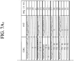
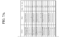
- the antibody can comprise a heavy chain amino acid sequence selected from the group consisting of the heavy chain amino acid sequence of antibody 13.1.2 (SEQ ID NO: 138), 131 (SEQ ID NO: 2), 170 (SEQ ID NO: 4), 150 (SEQ ID NO: 5), 095 (SEQ ID NO: 7), 250 (SEQ ID NO: 9), 139 (SEQ ID NO: 10), 211 (SEQ ID NO: 12), 124 (SEQ ID NO: 13), 318 (SEQ ID NO: 15), 342 (SEQ ID NO: 16), and 333 (SEQ ID NO: 17).
- the method can be performed in vivo, and performed on a mammal, such as a human, who suffers from a cancer involving epithelial cell proliferation, such as a lung, colon, gastric, renal, prostate, breast, glioblastoma or ovarian carcinoma.
- a mammal such as a human
- a cancer involving epithelial cell proliferation such as a lung, colon, gastric, renal, prostate, breast, glioblastoma or ovarian carcinoma.
- the antibody binds to a peptide LEEKKGNY (SEQ ID NO: 133).
- the antibody can have a binding affinity greater than 1. 3 ⁇ 10 -9 M to the peptide.
- the toxin may be selected from AEFP, MMAE, DM-1, and ZAP.
- the antibody toxin compound may be 10 fold more toxic to targeted cells than to cells without the peptide.
- the antibody can comprise a heavy chain amino acid sequence selected from the group consisting of the heavy chain amino acid sequence of antibody 13.1.2 (SEQ ID NO: 138), 131 (SEQ ID NO: 2), 170 (SEQ ID NO: 4), 150 (SEQ ID NO: 5), 095 (SEQ ID NO: 7), 250 (SEQ ID NO:9), 139 (SEQ ID NO: 10), 211 (SEQ ID NO: 12), 124 (SEQ ID NO: 13), 318 (SEQ ID NO: 15), 342 (SEQ. ID NO: 16), and 333 (SEQ ID NO: 17).
- the antibody can be associated with a toxin via a peptide linker or a second antibody.
- CDRs complementarity determining regions
- the antibody can be a monoclonal antibody, a chimeric antibody, a humanized antibody, or a human antibody. It can be associated with a pharmaceutically acceptable carrier or diluent, or conjugated to a therapeutic agent, such as a toxin, for example DM1 or AURISTATIN E.
- the method can be performed in vivo and on a mammal, such as a human, who suffers from a cancer involving epithelial cell proliferation such as lung, colon, gastric, renal, prostate, breast, glioblastoma or ovarian carcinoma.
- CDRs complementarity determining regions
- the antibody identified in the previous paragraph can further include a heavy chain amino acid sequence comprising the following complementarity determining regions (CDRs):
- a method of inhibiting cell proliferation associated with the expression of EGFRvIII comprising treating cells expressing EGFRvIII with an effective amount of the antibody or fragment described above.
- the method can be performed in vivo, on a mammal, such as a human, suffering from a cancer involving epithelial cell proliferation, such as lung carcinoma, breast carcinoma, head & neck cancer, prostate carcinoma or glioblastoma.
- an isolated polynucleotide molecule comprising a nucleotide sequence encoding a heavy chain amino acid sequence, or a fragment thereof, selected from the group consisting of the heavy chain amino acid sequence of antibodies 13.1.2, 131, 170, 150, 095, 250, 139, 211, 124, 318, 342, and 333 as identified in SEQ ID NO: 138, 2, 4, 5, 7, 9, 10, 12, 13, 15, 16, and 17, or an isolated polynucleotide molecule comprising a nucleotide sequence encoding a light chain amino acid sequence, or a fragment thereof, selected from the group consisting of the light chain amino acid sequence of antibodies 13.1.2, 131, 170, 150, 123, 095, 139, 250, 211, 318, 342, and 333, as identified in SEQ ID NO: 140, 19, 20, 21, 29, 23, 25, 26, 28, 33, 31 and 32.
- an article of manufacture comprising a container, a composition contained therein, and a package insert or label indicating that the composition can be used to treat cancer characterized by the expression of EGFRvIII, wherein the composition comprises an antibody as described above.
- cancers include a lung carcinoma, breast carcinoma, head & neck cancer, prostate carcinoma or glioblastoma.
- the antibody can be a labeled monoclonal antibody, or the antibody can be an unlabeled first antibody and the means for indicating the reaction comprises a labeled second antibody that is anti-immunoglobulin.
- the antibody that binds the antigen can be labeled with a marker selected from the group consisting of a fluorochrome, an enzyme, a Radionuclide and a radiopaque material.
- the antibody that binds the antigen can also bind to over-expressed wtEGFR.
- the kit can be used clinically for patient selection.
- the variant may have a pFLAG insert, may consist of the amino acids in SEQ ID NO: 56, and can exist in silico.
- the antibody variant can further bind to a peptide that comprises SEQ ID NO: 57.
- the antibody variant can have residues that interact with residues EKNY or EEKGN in the peptide.
- the antibody variant binds to the peptide sequence ten fold more tightly than it does to a wild-type EGFR protein.
- the antibody can bind specifically to EGFRvIII and the peptide of SEQ ID NO: 56.
- the isolated antibody or variant may have a complementarity determining region comprising a deep cavity, wherein the cavity is created by CDR2 and CDR3 of the heavy chain, CDR3 of the light chain, and a small portion from CDR1 of the light chain.
- the isolated antibody or variant may have residues 31, 37, 95-101, 143-147, 159, 162-166, 169-171, 211-219, 221, and 223 within 5 angstroms of a binding cavity.
- the isolated antibody or variant can have a complementarity determining region comprising a narrow groove, wherein the groove is created by heavy chain CDR2 and CDR3, and light chain CDR1, CDR2, and CDR3.
- the isolated antibody or variant can have residues 31, 33, 35-39, 51, 54-56, 58-61, 94-101, 144-148, 160, 163-166, 172, and 211-221 within 5 angstroms of a binding groove.
- the isolated antibody or variant has residues 31-33, 35, 37, 55, 96-101, 148, 163, 165, 170, 172, 178, 217, and 218 within 5 angstroms of a binding groove.
- the isolated antibody or variant may have a paratope configured so that when the epitope of peptide EEKKGN (SEQ ID NO 127) binds to the paratope of the antibody, at least one bond is formed between two residues selected from the group consisting of E2 and Y172, K3 and H31, K4 and H31, N6 and D33, N6 and Y37, and N6 and K55.
- the isolated antibody or variant can have a paratope configured so that when the epitope of peptide EEKKGNY (SEQ ID 131) binds to the paratope of the antibody, at least one bond is formed between two residues selected from the group consisting of K4 and Q95, K4 and Q95, N6 and Q98, G5 and H31, Y7 and H31, Y7 and W165.
- the antibody may have a structure or interaction with a structure that is determined in silico.
- a method for selecting variants that bind to EGFRvIII with particular binding characteristics comprising the use of a molecular structure to create a paratope, the use of a molecular structure to create an epitope, calculating the interaction energy between the two and comparing that energy level to the energy level of the epitope and a second paratope of a mAb variant, and selecting a variant based on the differences in the energy levels.
- the method can further include using an interaction energy between a second variant of the paratope and the epitope to determine a third interaction energy and comparing the third interaction energy and the second interaction energy to determine which variant to select.
- the variant is created and tested for binding.
- a method for making antibody variants to EGFRvIII comprising analyzing the residues of an epitope which interact with a paratope, selecting the more important residues of an epitope to create a recognition sequence, using the recognition sequence to create an EGFRvIII variant, and using the EGFRvIII variant to select antibody variants.
- the selection of the antibodies may be achieved in silico.
- the selection of the antibodies through the use of the EGFRvIII variant can be achieved by raising antibodies against EGFRvIII variant.
- the antibody can further comprise a point mutation of the following: Tyrl72Arg, Leu99Glu, Arg101Glu, Leu217Glu, Leu99Asn, Leu99His, L99T, Arg101Asp, or some combination thereof.
- the antibody may be a monoclonal antibody, a chimeric antibody, a humanized antibody, or a human antibody.
- the antibody or variant thereof binds to the sequence EEKKGNYVVT (SEQ ID NO: 57), and the antibody or variant has subnanomolar binding ability.
- an antibody binds to EGFRvIII and the antibody has a paratope that binds to an epitope, and the epitope has a set of residues that interact with the paratope that include E, K, N, and Y.
- the antibody is antibody 131.
- an antibody binds to EGFRvIII and the antibody has a paratope that binds to an epitope that has a set of residues that interact with the paratope comprising: E, E, K, G, and N.
- the primary structure of the epitope can be EEKKGNY (SEQ ID NO: 131). In one embodiment, the antibody is 13.1.2.
- the antibody is specific for SEQ ID NO: 56 compared to a wild type EGFR peptide.
- the nonspecific binding of the antibody to the wild type EGFR peptide is less than 10% of that of the specific binding of the antibody to EGFRvIII (SEQ ID NO: 135).
- the antibody is selected from the group consisting of 131, 139, and 13.1.2.
- the antibody is internalized. In one embodiment, the internalization occurs for at least about 70% or at least about 80% of the antibody.
- a variant human monoclonal antibody preferentially binds to an epitope that is substantially unique to an EGFRvIII protein compared to a wild-type EGFR protein or variant thereof (SEQ ID NO: 134).
- the variant can comprise a heavy chain complementarity determining region (CDR1) corresponding to canonical class 1.
- the variant can comprise a heavy chain complementarity determining region (CDR2) corresponding to canonical class 3.
- the variant can comprise a light chain complementarity determining region (CDR1) corresponding to canonical class 4.
- the variant can comprise a light chain complementarity determining region (CDR2) corresponding to canonical class 1.
- the variant can comprise a light chain complementarity determining region (CDR3) corresponding to canonical class 1.
- the variant comprises a first heavy chain complementarity determining region (CDR1) corresponding to canonical class 1, a second heavy chain complementarity determining region (CDR2) corresponding to canonical class 3, a first light chain complementarity determining region (CDR1) corresponding to canonical class 4, a second light chain complementarity determining region (CDR2) corresponding to canonical class 1; and a third light chain complementarity determining region (CDR3) corresponding to canonical class 1, wherein the complementary determining regions are configured to allow the variant to bind to an epitope that is substantially unique to an EGFRvIII protein as compared to a EGFR protein.
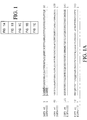
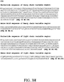
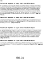
- EGFRvIII is a deletion mutant of EGFR in which 267 amino acids in the extracellular domain of EGFr are deleted with a single amino acid substitution of Glycine at the junction. These features are shown in a sequence alignment between wild type EGFR and EGFRvIII in FIG. 1 . In view of the amino acid substitution of Glycine at the junction of the deletion, it becomes theoretically possible to generate antibodies to the novel epitope present in EGFRvIII that is not present in wild type EGFR. Thus, a peptide for immunization and screening was designed, termed PEP3, as shown in FIG. 2 ( Kuan et al. EGF mutant receptor vIII as a molecular target in cancer therapy. Endocr Relat Cancer.
- Such 14-mer peptide possesses the 5 n-terminal amino acids common to EGFRvIII and wild type EGFR, the unique Glycine junction site, and 8 amino acid residues contained in the conserved sequences between wild type EGFR (corresponding to residues 273-280) and EGFRvIII (corresponding to residues 7-14).
- glioblastoma cell and cells B300.19 cells transfected with the gene encoding EGFRvIII were also utilized for immunization and screening (sometimes referred to herein as B300.19/EGFRvIII transfectants).
- transgenic XenoMouse® mice were immunized with combinations of glioblastoma cells/EGFRvIII, B300.19/EGFRvIII cells, and peptides (PEP3) directed to the junction region in the novel extracellular domain represented in EGFRvIII as compared to wild type EGFR.
- B cells from immunized mice were isolated and either used to produce hybridomas followed by screening for binding to EGFRvIII or used directly in screening for binding to EGFRvIII using XenoMaxTM/SLAMTM technologies ( Babcook et al. A novel strategy for generating monoclonal antibodies from single, isolated lymphocytes producing antibodies of defined specificities.
- Antibodies identified that bound to EGFRvIII were screened in a series of assays to ascertain specific recognition of EGFRvIII. Through this process, panels of human monoclonal antibodies that bound to and were specific for EGFRvIII were generated, isolated, and characterized. Subsequent epitope mapping demonstrated unique but overlapping specificities . All antibodies were further evaluated in vitro for their ability to be internalized by cells for the purpose of delivering cytotoxic drugs to cells.
- Antibodies demonstrating efficient drug delivery were directly conjugated with a cytotoxic drug and examined for their ability to kill tumor cells expressing EGFRvIII in vitro and in vivo. These studies provide the basis for the next generation of antibody drug conjugates for treating cancer in patients whose tumor harbor specific genetic lesions.
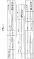
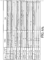
- FIGs. 4-7 (SEQ ID NO: 1-33 and 141-144).
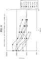
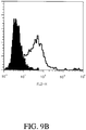
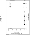
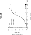
- a comparison of the sequences and binding abilities of the various antibodies was made and the results are displayed in FIGs. 4-10 .
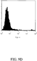
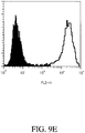
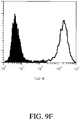
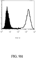
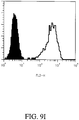
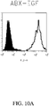
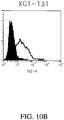
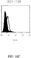
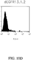
- FIGs. 9A-9L and FIGs. 10A-10D antibodies 131, 139, and 13.1.2 all demonstrated superior selectivity for EGFRvIII expressing cells (H1477) as compared to ABX-EGF.
- FIGs. 9M-9P Some of the results are shown in graph form in FIGs. 9M-9P , which demonstrates that at least two of the antibodies, 13.1.2 and 131 demonstrated superior specificity for EGFRvIII expressing cells compared to simply EGFRvIII cells.
- variants of the antibodies were made in order to obtain antibodies with altered binding characteristics.
- antibodies of the invention are highly useful for the screening of other antibodies that bind to the same or similar epitopes.
- Antibodies of the invention can be utilized in cross competition studies for the elucidation of other antibodies that are expected to have the same or improved effects with respect to characteristics of the antigen-antibody complex that is formed.
- Antibodies that bind to the same epitope as, or compete for binding with, the 13.1.2 and 131 antibodies are highly desirable. As discussed in more detail below, through Alanine scanning on SPOTs arrays important residues for binding of certain antibodies have been elucidated. Accordingly, antibdodies that share critical binding residues are also highly desirable.
- isolated polynucleotide shall mean a polynucleotide of genomic, cDNA, or synthetic origin or some combination thereof, which by virtue of its origin the "isolated polynucleotide” (1) is not associated with all or a portion of a polynucleotide in which the "isolated polynucleotide” is found in nature, (2) is operably linked to a polynucleotide which it is not linked to in nature, or (3) does not occur in nature as part of a larger sequence.
- isolated protein means a protein of cDNA, recombinant RNA, or synthetic origin or some combination thereof, which by virtue of its origin, or source of derivation, the "isolated protein” (1) is not associated with proteins found in nature, (2) is free of other proteins from the same source, e.g. free of murine proteins, (3) is expressed by a cell from a different species, or (4) does not occur in nature.
- polypeptide is used herein as a generic term to refer to native protein, fragments, or analogs of a polypeptide sequence.
- native protein, fragments, and analogs are species of the polypeptide genus.
- Preferred polypeptides in accordance with the invention comprise the human heavy chain immunoglobulin molecules and the human kappa light chain immunoglobulin molecules, as well as antibody molecules formed by combinations comprising the heavy chain immunoglobulin molecules with light chain immunoglobulin molecules, such as the kappa light chain immunoglobulin molecules or lambda light chain immunoglobulin molecules, and vice versa, as well as fragments and analogs thereof.
- naturally-occurring refers to the fact that an object can be found in nature.
- a polypeptide or polynucleotide sequence that is present in an organism (including viruses) that can be isolated from a source in nature and which has not been intentionally modified by man in the laboratory or otherwise is naturally-occurring.
- operably linked refers to positions of components so described are in a relationship permitting them to function in their intended manner.
- a control sequence "operably linked" to a coding sequence is ligated in such a way that expression of the coding sequence is achieved under conditions compatible with the control sequences.
- control sequence refers to polynucleotide sequences which are necessary to effect the expression and processing of coding sequences to which they are ligated. The nature of such control sequences differs depending upon the host organism; in prokaryotes, such control sequences generally include promoter, ribosomal binding site, and transcription termination sequence; in eukaryotes, generally, such control sequences include promoters and transcription termination sequence.
- control sequences is intended to include, at a minimum, all components whose presence is essential for expression and processing, and can also include additional components whose presence is advantageous, for example, leader sequences and fusion partner sequences.
- polynucleotide as referred to herein means a polymeric form of nucleotides of at least 10 bases in length, either ribonucleotides or deoxynucleotides or a modified form of either type of nucleotide.
- the term includes single and double stranded forms of DNA.
- oligonucleotide includes naturally occurring, and modified nucleotides linked together by naturally occurring, and non-naturally occurring oligonucleotide linkages.
- Oligonucleotides are a polynucleotide subset generally comprising a length of 200 bases or fewer. Preferably oligonucleotides are 10 to 60 bases in length and most preferably 12, 13, 14, 15, 16, 17, 18, 19, or 20 to 40 bases in length. Oligonucleotides are usually single stranded, e.g. for probes; although oligonucleotides may be double stranded, e.g. for use in the construction of a gene mutant. Oligonucleotides of the invention can be either sense or antisense oligonucleotides.
- nucleotides includes deoxyribonucleotides and ribonucleotides.
- modified nucleotides includes nucleotides with modified or substituted sugar groups and the like.
- oligonucleotide linkages includes oligonucleotides linkages such as phosphorothioate, phosphorodithioate, phosphoroselenoate, phosphorodiselenoate, phosphoroanilothioate, phosphoraniladate, phosphoroamidate, and the like. See e.g., LaPlanche et al. Nucl. Acids Res.
- An oligonucleotide can include a label for detection, if desired.
- variant is a polypeptide, polynucleotide, or molecule that differs from the recited polypeptide or polynucleotide, but only such that the activity of the protein is not detrimentally altered.
- epitopes There may be variants of epitopes.
- variants of antibodies There may be variants of antibodies.
- the ability of a protein variant to bind to the epitope is not detrimentally altered.
- the protein variant can bind with 10-500% of the ability of the wild type mAb.
- the protein variant can bind with 10%, 50%, 110%, 500%, or greater than 500% of the ability of the wild type mAb.
- the range of binding abilities between 10-500% is inlcuded. Binding ability may be reflected in many ways, including, but not limited to the k a , k d , or K D of the variant to an epitope.
- the epitope is one described in the present specification.
- variant antibodies can differ from the wild-type sequence by substitution, deletion or addition of five amino acids or fewer. Such variants may generally be identified by modifying one of the disclosed polypeptide sequences, and evaluating the binding properties of the modified polypeptide using, for example, the representative procedures described herein.
- polypeptide variants preferably exhibit at least about 70%, more preferably at least about 90% and most preferably at least about 95% identity to the identified polypeptides. Preferrably, the variant differs only in conservative substitutions and/or modifications.
- Variant proteins include those that are structurally similar and those that are functionally equivalent to the protein structures described in the present specification.
- the protein is a variant if it is functionally equivalent to the proteins described in this specification, so long as the paratope of variant is similar to the paratopes described in the specification.
- any substance with a shape that is similar to the paratope described in FIG. 11 is a variant.
- any substance with a shape that is similar to the paratope described in FIG. 12 is a variant.
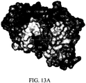
- any substance that has a shape that is similar to the interaction surface described in FIG. 13A and 13B is a variant.
- the antibody is a variant if the nucleic acid sequence can selectively hybridize to wild-type sequence under stringent conditions.
- suitable moderately stringent conditions include prewashing in a solution of 5xSSC; 0.5% SDS, 1.0 mM EDTA (pH 8:0); hybridizing at 50°C-65°C, 5xSSC, overnight or, in the event of cross-species homology, at 45°C with 0.5xSSC; followed by washing twice at 65°C for 20 minutes with each of 2x, 0.5x and 0.2xSSC containing 0.1% SDS.
- hybridizing DNA sequences are also within the scope of this invention, as are nucleotide sequences that, due to code degeneracy, encode an antibody polypeptide that is encoded by a hybridizing DNA sequence.
- the term "selectively hybridize” referred to herein means to detectably and specifically bind.
- Polynucleotides, oligonucleotides and fragments thereof in accordance with the invention selectively hybridize to nucleic acid strands under hybridization and wash conditions that minimize appreciable amounts of detectable binding to nonspecific nucleic acids. High stringency conditions can be used to achieve selective hybridization conditions as known in the art and discussed herein.
- nucleic acid sequence homology between the polynucleotides, oligonucleotides, and fragments of the invention and a nucleic acid sequence of interest will be at least 80%, and more typically with preferably increasing homologies of at least 85%, 90%, 95%, 99%, and 100%.
- Two amino acid sequences are homologous if there is a partial or complete identity between their sequences. For example, 85% homology means that 85% of the amino acids are identical when the two sequences are aligned for maximum matching. Gaps (in either of the two sequences being matched) are allowed in maximizing matching; gap lengths of 5 or less are preferred with 2 or less being more preferred.
- two protein sequences are homologous, as this term is used herein, if they have an alignment score of at more than 5 (in standard deviation units) using the program ALIGN with the mutation data matrix and a gap penalty of 6 or greater. See Dayhoff, M.O., in Atlas of Protein Sequence and Structure, pp. 101-110 (Volume 5, National Biomedical Research Foundation (1972 )) and Supplement 2 to this volume, pp. 1-10.
- the two sequences or parts thereof are more preferably homologous if their amino acids are greater than or equal to 50% identical when optimally aligned using the ALIGN program.
- a polynucleotide sequence is homologous (i.e., is identical, not strictly evolutionarily related) to all or a portion of a reference polynucleotide sequence, or that a polypeptide sequence is identical to a reference polypeptide sequence.
- the term “complementary to” is used herein to mean that the complementary sequence is homologous to all or a portion of a reference polynucleotide sequence.
- the nucleotide sequence "TATAC” corresponds to a reference sequence "TATAC” and is complementary to a reference sequence "GTATA".
- reference sequence is a defined sequence used as a basis for a sequence comparison; a reference sequence may be a subset of a larger sequence, for example, as a segment of a full-length cDNA or gene sequence given in a sequence listing or may comprise a complete cDNA or gene sequence. Generally, a reference sequence is at least 18 nucleotides or 6 amino acids in length, frequently at least 24 nucleotides or 8 amino acids in length, and often at least 48 nucleotides or 16 amino acids in length.
- two polynucleotides or amino acid sequences may each (1) comprise a sequence (i.e., a portion of the complete polynucleotide or amino acid sequence) that is similar between the two molecules, and (2) may further comprise a sequence that is divergent between the two polynucleotides or amino acid sequences
- sequence comparisons between two (or more) molecules are typically performed by comparing sequences of the two molecules over a "comparison window" to identify and compare local regions of sequence similarity.
- a “comparison window”, as used herein, refers to a conceptual segment of at least 18 contiguous nucleotide positions or 6 amino acids wherein a polynucleotide sequence or amino acid sequence may be compared to a reference sequence of at least 18 contiguous nucleotides or 6 amino acid sequences and wherein the portion of the polynucleotide sequence in the comparison window may comprise additions, deletions, substitutions, and the like (i.e., gaps) of 20 percent or less as compared to the reference sequence (which does not comprise additions or deletions) for optimal alignment of the two sequences.
- Optimal alignment of sequences for aligning a comparison window may be conducted by the local homology algorithm of Smith and Waterman Adv. Appl. Math.
- sequence identity means that two polynucleotide or amino acid sequences are identical (i.e., on a nucleotide-by-nucleotide or residue-by-residue basis) over the comparison window.
- percentage of sequence identity is calculated by comparing two optimally aligned sequences over the window of comparison, determining the number of positions at which the identical nucleic acid base (e.g., A, T, C, G, U, or I) or residue occurs in both sequences to yield the number of matched positions, dividing the number of matched positions by the total number of positions in the comparison window (i.e., the window size), and multiplying the result by 100 to yield the percentage of sequence identity.
- substantially identical denotes a characteristic of a polynucleotide or amino acid sequence, wherein the polynucleotide or amino acid comprises a sequence that has at least 85 percent sequence identity, preferably at least 90 to 95 percent sequence identity, more usually at least 99 percent sequence identity as compared to a reference sequence over a comparison window of at least 18 nucleotide (6 amino acid) positions, frequently over a window of at least 24-48 nucleotide (8-16 amino acid) positions, wherein the percentage of sequence identity is calculated by comparing the reference sequence to the sequence which may include deletions or additions which total 20 percent or less of the reference sequence over the comparison window.
- the reference sequence may be a subset of a larger sequence.
- Amino acids or nucleic acids with substantial identity to the wild-type protein or nucleic acid are examples of variants of the wild-type protein or nucleic acid.
- Examples of unconventional amino acids include: 4-hydroxyproline, ⁇ -carboxyglutamate, ⁇ -N,N,N-trimethyllysine, ⁇ -N-acetyllysine, O-phosphoserine, N-acetylserine, N-formylmethionine, 3-methylhistidine, 5-hydroxylysine, ⁇ -N-methylarginine, and other similar amino acids and imino acids (e.g., 4-hydroxyproline).
- the left-hand direction is the amino terminal direction and the righthand direction is the carboxy-terminal direction, in accordance with standard usage and convention.
- the left-hand end of single-stranded polynucleotide sequences is the 5' end; the left-hand direction of double-stranded polynucleotide sequences is referred to as the 5' direction.
- the direction of 5' to 3' addition of nascent RNA transcripts is referred to as the transcription direction; sequence regions on the DNA strand having the same sequence as the RNA and which are 5' to the 5' end of the RNA transcript are referred to as "upstream sequences"; sequence regions on the DNA strand having the same sequence as the RNA and which are 3' to the 3' end of the RNA transcript are referred to as "downstream sequences".
- the term "substantial identity” means that two peptide sequences, when optimally aligned, such as by the programs GAP or BESTFIT using default gap weights, share at least 80 percent sequence identity, preferably at least 90 percent sequence identity, more preferably at least 95 percent sequence identity, and most preferably at least 99 percent sequence identity.
- residue positions which are not identical differ by conservative amino acid substitutions.
- Conservative amino acid substitutions refer to the interchangeability of residues having similar side chains.
- a group of amino acids having aliphatic side chains is glycine, alanine, valine, leucine, and isoleucine; a group of amino acids having aliphatic-hydroxyl side chains is serine and threonine; a group of amino acids having amide-containing side chains is asparagine and glutamine; a group of amino acids having aromatic side chains is phenylalanine, tyrosine, and tryptophan; a group of amino acids having basic side chains is lysine, arginine, and histidine; and a group of amino acids having sulfur-containing side chains is cysteine and methionine.
- Preferred conservative amino acids substitution groups are: valine-leucine-isoleucine, phenylalanine-tyrosine, lysine-arginine, alanine-valine, glutamic-aspartic, and asparagine-glutamine.
- Polypeptides with substantial identity can be variants.
- Variant proteins also include proteins with minor variations. As discussed herein, minor variations in the amino acid sequences of antibodies or immunoglobulin molecules are contemplated as being encompassed by the present invention, providing that the variations in the amino acid sequence maintain at least 75%, more preferably at least 80%, 90%, 95%, and most preferably 99%. In particular, conservative amino acid replacements are contemplated.
- More preferred families are: serine and threonine are aliphatic-hydroxy family; asparagine and glutamine are an amide-containing family; alanine, valine, leucine and isoleucine are an aliphatic family; and phenylalanine, tryptophan, and tyrosine are an aromatic family.
- serine and threonine are aliphatic-hydroxy family
- asparagine and glutamine are an amide-containing family
- alanine, valine, leucine and isoleucine are an aliphatic family
- phenylalanine, tryptophan, and tyrosine are an aromatic family.
- Whether an amino acid change results in a functional peptide can readily be determined by assaying the specific activity of the polypeptide derivative. Assays are described in detail herein. Fragments or analogs of antibodies or immunoglobulin molecules can be readily prepared by those of ordinary skill in the art. Preferred amino- and carboxy-termini of fragments or analogs occur near boundaries of functional domains. Structural and functional domains can be identified by comparison of the nucleotide and/or amino acid sequence data to public or proprietary sequence databases. Preferably, computerized comparison methods are used to identify sequence motifs or predicted protein conformation domains that occur in other proteins of known structure and/or function. Methods to identify protein sequences that fold into a known three-dimensional structure are known. Bowie et al. Science 253:164 (1991 ). Thus, the foregoing examples demonstrate that those of skill in the art can recognize sequence motifs and structural conformations that may be used to define structural and functional domains in accordance with the antibodies described herein.
- Preferred amino acid substitutions are those which: (1) reduce susceptibility to proteolysis, (2) reduce susceptibility to oxidation, (3) alter binding affinity for forming protein complexes, (4) alter binding affinities, and (4) confer or modify other physicochemical or functional properties of such analogs.
- Analogs can include various muteins of a sequence other than the naturally-occurring peptide sequence. For example, single or multiple amino acid substitutions (preferably conservative amino acid substitutions) may be made in the naturally-occurring sequence (preferably in the portion of the polypeptide outside the domain(s) forming intermolecular contacts.
- a conservative amino acid substitution should not substantially change the structural characteristics of the parent sequence (e.g., a replacement amino acid should not tend to break a helix that occurs in the parent sequence, or disrupt other types of secondary structure that characterizes the parent sequence).
- Examples of art-recognized polypeptide secondary and tertiary structures are described in Proteins, Structures and Molecular Principles (Creighton, Ed., W. H. Freeman and Company, New York (1984 )); Introduction to Protein Structure (C. Branden and J. Tooze, eds., Garland Publishing, New York, N.Y. (1991 )); and Thornton et at. Nature 354:105 (1991 ).
- polypeptide fragment refers to a polypeptide that has an amino-terminal and/or carboxy-terminal deletion, but where the remaining amino acid sequence is identical to the corresponding positions in the naturally-occurring sequence deduced, for example, from a full-length cDNA sequence. Fragments typically are at least 5, 6, 8 or 10 amino acids long, preferably at least 14 amino acids long, more preferably at least 20 amino acids long, usually at least 50 amino acids long, and even more preferably at least 70 amino acids long.
- analog refers to polypeptides which are comprised of a segment of at least 25 amino acids that has substantial identity to a portion of a deduced amino acid sequence. Analogs typically are at least 20 amino acids long, preferably at least 50 amino acids long or longer, and can often be as long as a full-length naturally-occurring polypeptide. Both fragments and analogs are forms of variants
- Peptide analogs are commonly used in the pharmaceutical industry as non-peptide drugs with properties analogous to those of the template peptide. These types of non-peptide compound are termed "peptide mimetics" or "peptidomimetics”. Fauchere, J. Adv. Drug Res. 15:29 (1986 ); Veber and Freidinger TINS p.392 (1985 ); and Evans et al. J. Med. Chem. 30:1229 (1987 ). Such compounds are often developed with the aid of computerized molecular modeling. Peptide mimetics that are structurally similar to therapeutically useful peptides may be used to produce an equivalent therapeutic or prophylactic effect.
- a paradigm polypeptide i.e., a polypeptide that has a biochemical property or pharmacological activity
- Systematic substitution of one or more amino acids of a consensus sequence with a D-amino acid of the same type may be used to generate more stable peptides.
- constrained peptides comprising a consensus sequence or a substantially identical consensus sequence variation may be generated by methods known in the art ( Rizo and Gierasch Ann. Rev. Biochem. 61:387 (1992 )); for example, by adding internal cysteine residues capable of forming intramolecular disulfide bridges which cyclize the peptide.
- Peptide mimetics and peptidomimetics are both forms of variants.
- Antibody or “antibody peptide(s)” refer to an intact antibody, or a binding fragment thereof that competes with the intact antibody for specific binding. Binding fragments are produced by recombinant DNA techniques, or by enzymatic or chemical cleavage of intact antibodies. Binding fragments include Fab, Fab', F(ab') 2 , Fv, and single-chain antibodies. An antibody other than a "bispecific” or “bifunctional” antibody is understood to have each of its binding sites identical.
- An antibody substantially inhibits adhesion of a receptor to a counterreceptor when an excess of antibody reduces the quantity of receptor bound to counterreceptor by at least about 20%, 40%, 60% or 80%, and more usually greater than about 85% (as measured in an in vitro competitive binding assay).
- epitope includes any protein determinant capable of specific binding to an immunoglobulin or T-cell receptor or otherwise interacting with a molecule.
- Epitopic determinants generally consist of chemically active surface groupings of molecules such as amino acids or carbohydrate or sugar side chains and generally have specific three-dimensional structural characteristics, as well as specific charge characteristics.
- An epitope may be "linear” or “conformational.” In a linear epitope, all of the points of interaction between the protein and the interacting molecule (such as an antibody) occur linearally along the primary amino acid sequence of the protein. In a conformational epitope, the points of interaction occur across amino acid residues on the protein that are separated from one another.
- An antibody is said to specifically bind an antigen when the dissociation constant is ⁇ 1 ⁇ M, preferably ⁇ 100 nM and more preferably ⁇ 10 nM, and even more preferably ⁇ 1nM.
- the dissociation constant is ⁇ 1 ⁇ M, preferably ⁇ 100 nM and more preferably ⁇ 10 nM, and even more preferably ⁇ 1nM.
- an epitope can comprises those residues to which the antibody binds.
- the epitope is the EGFRvIII epitope.
- the epitope is that described in Example 4 of this specification.
- the epitope is the epitope described in Example 14.
- the epitope comprises the sequence LEEKKGNYVVTD (SEQ ID NO: 59).
- the epitope comprises the sequence EEKKGNYVVT (SEQ ID NO: 57). In one embodiment, the epitope comprises the sequence EKNY (SEQ ID NO: 60). In one embodiment, the epitope comprises the sequence EEKGN (SEQ ID NO: 61).
- these need not be actually assembled in this order on a single peptide, rather, these are the residues that form the eptiope which interacts with the paratope.
- the space that is occupied by a residue or side chain that creates the shape of a molecule helps to determine what an epitope is. Likewise, any functional groups associated with the epitope, van der Waals interactions, degree of mobility of side chains, etc. can all determine what an epitope actually is. Thus an epitope may also include energetic interactions.
- paratope is meant to describe the general structure of a binding region that determines binding to an epitope. This structure influences whether or not and in what manner the binding region might bind to an epitope.
- Paratope can refer to an antigenic site of an antibody that is responsible for an antibody or fragment thereof, to bind to an antigenic determinant.
- Paratope also refers to the idiotope of the antibody, and the complementary determining region (CDR) region that binds to the epitope.
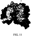
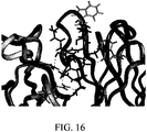
- the paratope is the region of the antibody that is L1 10, L2 30, L3 50, H1 20, H2 40, and H3 60 in FIG. 11 .
- the paratope is the region of the antibody that comprises the CDR sequences in Example 16 for L1, L2, L3, H1, H2, and H3. In one embodiment, the paratope is the region of the antibody that is L1 110, L2 130, L3 150, H1 120, H2 140, and H3 160 in FIG. 12 . In one embodiment, the paratope is the region of the antibody that comprises the CDR sequences in Example 18 for L1, L2, L3, H1, H2, and H3. In one embodiment, the paratope comprises the sequences listed in Example 18. In one embodiment, the paratope comprises the residues that interact with the epitope, as shown in FIG. 13A and FIG. 13B .
- the solid black structure is the peptide structure.
- the paratope comprises residue Tyr172Arg of the 13.1.2 mAb.
- the paratope of the 13.1.2 mAb comprises at least one residue selected from the group consisting of: Tyr 172Arg, Arg101Glu, Leu99Asn, Leu99His, Arg101Asp, Leu217Gln, Leu99Thr, Leu217Asn, Arg101Gln, and Asn35Gly.
- the paratope of any antibody, or variant thereof can be determined in the manner set forth by the present application. Residues are considered "important" if they are predicted to contribute 10% of the binding energy.
- residues are considered “important” if they are predicted to contribute 2% of the binding energy. In one embodiment, residues are considered “important” if they are predicted to contribute 50% of the binding energy. In one embodiment, residues are considered “important” if they are predicted to interact with the surface of the epitope, or the surface of the paratope. In one embodiment, residues are considered “important” if changing the residue results in a loss in binding.
- the terms “specifically” or “preferrentially” binds to, or similar phrases are not meant to denote that the antibody exclusively binds to that epitope. Rather, what is meant is that the antibody, or variant thereof, can bind to that epitope, to a higher degree than the antibody binds to at least one other substance to which the antibody is exposed to.
- the specifically binding antibody will bind to the EGFRvIII protein with an affinity greater than (more tightly, or lower K D ) it will to the EGFR protein.
- the specifically binding antibody will bind more tightly by at least a minimal increase to 1, 1-2, 2-5, 5-10, 10-20, 20-30, 30-50, 50-70, 70-90, 90-120, 120-150, 150-200, 200-300, 300-500, 500-1000 percent or more.
- Tyr172Arg would mean that while the wild type protein has a tyrosine at position 172, the mutant has an arginine at position 172.
- agent is used herein to denote a chemical compound, a mixture of chemical compounds, a biological macromolecule, or an extract made from biological materials.
- mammal when used herein refers to any animal that is considered a mammal. Preferably, the mammal is human.
- Digestion of antibodies with the enzyme, papain results in two identical antigen-binding fragments, known also as "Fab” fragments, and a "Fc” fragment, having no antigen-binding activity but having the ability to crystallize.
- Digestion of antibodies with the enzyme, pepsin results in the a F(ab') 2 fragment in which the two arms of the antibody molecule remain linked and comprise two-antigen binding sites.
- the F(ab') 2 fragment has the ability to crosslink antigen.
- Fv when used herein refers to the minimum fragment of an antibody that retains both antigen-recognition and antigen-binding sites. These fragments can also be considered variants of the antibody.
- Fab when used herein refers to a fragment of an antibody which comprises the constant domain of the light chain and the CH1 domain of the heavy chain.
- mAb refers to monoclonal antibody.
- XenoMax method generated antibody sequences is coded as follows: "AB”-referring to antibody, “EGFRvIII”-referring to antibody's binding specificity, "X” referring to XenoMouse mouse derived, "G1"-referring to IgG1 isotype or “G2” referring to IgG2 isotype, the last three digits refer to the single cell number from which the antibody was derived, for example: AB- EGFRvIII -XG1-095 would be an antibody with binding specificity to EGFRvIII from XenoMouse mouse of a IgG1 isotype and cell number 95.
- SC refers to single cell and a particular XenoMax method derived antibody may be referred to as SC followed by three digits, or just three digits, referring to the single cell number from which the antibody was derived herein.
- hybridoma derived antibody sequences is coded as follows: "AB”-referring to antibody, “EGFRvIII”-refers to the antibody's binding specificity, "X” refers to XenoMouse mouse derived, "G1"-refers to IgG1 isotype or “G2” refers to IgG2 isotype, “K” refers to kappa, “L' refers to lambda. The last three digits referring to the clone from which the antibody was derived, for example: AB-EGFRvIII-XG1K-13.1.2
- Label refers to the addition of a detectable moiety to a polypeptide, for example, a radiolabel, fluorescent label, enzymatic label chemiluminescent labeled or a biotinyl group.
- Radioisotopes or radionuclides may include 3 H, 14 C, 15 N, 35 S, 90 Y, 99 Tc, 111 In, 125 I, 131 I, fluorescent labels may include rhodamine, lanthanide phosphors or FITC and enzymatic labels may include horseradish peroxidase, ⁇ -galactosidase, luciferase, alkaline phosphatase.
- pharmaceutical agent or drug refers to a chemical compound or composition capable of inducing a desired therapeutic effect when properly administered to a patient.
- Other chemistry terms herein are used according to conventional usage in the art, as exemplified by The McGraw-Hill Dictionary of Chemical Terms (Parker, S., Ed., McGraw-Hill, San Francisco (1985 )).
- substantially pure means an object species is the predominant species present (i.e., on a molar basis it is more abundant than any other individual species in the composition), and preferably a substantially purified fraction is a composition wherein the object species comprises at least about 50 percent (on a molar basis) of all macromolecular species present. Generally, a substantially pure composition will comprise more than about 80 percent of all macromolecular species present in the composition, more preferably more than about 85%, 90%, 95%, 99%, and 99.9%. Most preferably, the object species is purified to essential homogeneity (contaminant species cannot be detected in the composition by conventional detection methods) wherein the composition consists essentially of a single macromolecular species.
- patient includes human and veterinary subjects.
- SAM® Technology refers to the "Selected Lymphocyte Antibody Method” ( Babcook et al., Proc. Natl. Acad. Sci. USA, i93:7843-7848 (1996 ), and Schrader, US Patent No. 5,627,052 .
- XenoMaxTM refers to the use of SLAM Technology with XenoMouse® mice (as described below).
- the basic antibody structural unit is known to comprise a tetramer.
- Each tetramer is composed of two identical pairs of polypeptide chains, each pair having one "light” (about 25 kDa) and one "heavy” chain (about 50-70 kDa).
- the amino-terminal portion of each chain includes a variable region of about 100 to 110 or more amino acids primarily responsible for antigen recognition.
- the carboxy-terminal portion of each chain defines a constant region primarily responsible for effector function.
- Human light chains are classified as kappa and lambda light chains.
- Heavy chains are classified as mu, delta, gamma, alpha, or epsilon, and define the antibody's isotype as IgM, IgD, IgG, IgA, and IgE, respectively.
- the variable and constant regions are joined by a "J" region of about 12 or more amino acids, with the heavy chain also including a "D” region of about 10 more amino acids. See generally , Fundamental Immunology Ch. 7 (Paul, W., ed., 2nd ed. Raven Press, N.Y. (1989 ))).
- the variable regions of each light/heavy chain pair form the antibody binding site.
- an intact antibody has two binding sites. Except in bifunctional or bispecific antibodies, the two binding sites are the same.
- the chains all exhibit the same general structure of relatively conserved framework regions (FR) joined by three hyper variable regions, also called complementarity determining regions or CDRs.
- the CDRs from the two chains of each pair are aligned by the framework regions, enabling binding to a specific epitope.
- both light and heavy chains comprise the domains FR1, CDR1, FR2, CDR2, FR3, CDR3 and FR4.
- the assignment of amino acids to each domain is in accordance with the definitions of Kabat Sequences of Proteins of Immunological Interest (National Institutes of Health, Bethesda, Md. (1987 and 1991 )), or Chothia & Lesk J. Mol. Biol. 196:901-917 (1987 ); Chothia et al. Nature 342:878-883 (1989 ).
- a bispecific or bifunctional antibody is an artificial hybrid antibody having two different heavy/light chain pairs and two different binding sites.
- Bispecific antibodies can be produced by a variety of methods including fusion of hybridomas or linking of Fab' fragments. See, e.g., Songsivilai & Lachmann Clin. Exp. Immunol. 79: 315-321 (1990 ), Kostelny et al, J. Immunol. 148:1547-1553 (1992 ). Production of bispecific antibodies can be a relatively labor intensive process compared with production of conventional antibodies and yields and degree of purity are generally lower for bispecific antibodies. Bispecific antibodies do not exist in the form of fragments having a single binding site (e.g., Fab, Fab', and Fv).
- the structure of the CDRs form a paratope, through which an antibody is able to bind to an epitope.
- the structure of such a paratope may be determined in a number of ways. Traditional structural examination approaches may be used, such as NMR or x-ray crystalography. These approaches may examine the structure of the paratope alone, or while it is bound to the epitope. Alternatively, molecular models may be generated in silico. A structure can be generated through homology modeling, aided with a commercial package, such as InsightII modeling package from Accelrys (San Diego, CA).
- there are many alternative methods for generating such structures in silico any of which may be used. For instance, a process similar to the one described in Hardman et al., issued U.S. Pat. No.
- the result is that one is able to estimate where and how the epitope interacts with the paratope.
- only a fragment, or variant, of the epitope is used to assist in determining the relevant interactions.
- the entire epitope is used in the modeling of the interaction between the paratope and the epitope.
- both approaches are used to a limited extent, in order to cross check the results.
- a variant of an epitope it will be optimized so that the variant of the epitope comprises the most important residues of the epitope.
- the identity of the most important residues can be determined in any number of ways, for instance as described in Examples 4 and 14 of the present specification.
- one is able to determine which residues are the most important in the interaction between the epitope and the paratope.
- one is able to readily select which residues to change in order to alter the binding characteristics of the antibody. For instance, it may be apparent from the docking models that the side chains of certain residues in the paratope may sterically hinder the binding of the epitope, thus altering these residues to residues with smaller side chains may be beneficial.
- One can determine this in many ways. For example, one may simply look at the two models and esitmate interactions based on functional groups and proximety. Alternatively, one may perform repeated pairings of epitope and paratope, as described above, in order to obtain more favorable energy interactions.
- the models determined above can be tested through various techniques. For example, the interaction energy can determined with the programs discussed above in order to determine which of the variants to further examine. Also, coulumbic and van der Waals interactions are used to determine the interaction energies of the epitope and the variant paratopes. Also site directed mutagenesis is used to see if predicted changes in antibody structure actually result in the desired changes in binding characteristics. Alternatively, changes may be made to the epitope to verify that the models are correct or to determine general binding themes that may be occurring between the paratope and the epitope.
- the above methods for modeling structures can be used to determine what changes in protein structure will result in particular desired characteristics of an antibody. These methods can be used to determine what changes in protein structure will not result in the desired characteristics.
- any modification may also have additional side effects on the activity of the antibody. For instance, while any alteration predicted to result in greater binding, may induce greater binding, it may also cause other structural changes which might reduce or alter the activity of the antibody. The determination of whether or not this is the case is routine in the art and can be achieved in many ways.
- the activity can be tested through an ELISA test, as in Example 21.
- the samples can be tested through the use of a surface plasmon resonance device.
- the models described above are used to increase the binding ability of the antibody to its epitope.
- the antibody can bind to the epitope more readily, and thus have a higher association constant (k a ).
- the antibody may dissociate from the epitope slower, and thus have a lower dissociation constant (k d ), or the K D of the epitope-paratope interaction can be smaller in value, thus making the extent of the binding between the epitope and paratope higher.
- the variant antibodies are designed to bind with the opposite characteristics. That is, the antibodies do not bind as tightly or perhaps as quickly.
- the variant antibodies are not different in their K D from the wild-type antibodies, but the variant antibodies are more specific for a particular epitope. This may mean that the paratopes of the designed antibodies have a lower risk of binding to other epitopes.
- the antibodies can have other characteristics that have been altered. For example, a variant may be more immune to nonspecific antibody binding or may stay solvated in solution even when the antibody is present in high concentrations. Such a variant may be present in the discussed antibodies. For instance, while the higher concentrations of some variant antibodies examined below resulted in slower binding components in Biacore experiments, for instance 13.1.2 mAb, certain variants did not exhibit this slower component, even at the same concentrations, L217N-2.1, for example.
- the variants predicted by the models determined above can be created and then tested to determine if they actually bind with the desired characteristics. Mutants with a greater total interaction energy with the epitope can be selected for further testing.
- the interaction energy can be determined in a number of ways, one of which is described above.
- exemplary options include and are not limited to KinExA (e.g., Lackie, Issued Pat. No. 5,372,783, Dec 13, 1994 ) (Sapidyne Instruments Inc., ID, Boise), surface plasmon resonance (SPR)( e . g ., BIACORETM Biacore, Inc., Pistcataway, N.J.), stopped-flow fluorescence, resonant mirror, and fluorescence polarization. Many of these options are able to not only record the data, but also provide ready means for fitting the data to various theoretical curves and thus determine the k a , k d , and K D , as well as other properties.
- KinExA e.g., Lackie, Issued Pat. No. 5,372,783, Dec 13, 1994
- SPR surface plasmon resonance
- SPR surface plasmon resonance
- stopped-flow fluorescence e.g ., BIACORETM Biacore, Inc., Pistcataway, N.
- Human antibodies avoid some of the problems associated with antibodies that possess murine or rat variable and/or constant regions.
- the presence of such murine or rat derived proteins can lead to the rapid clearance of the antibodies or can lead to the generation of an immune response against the antibody by a patient.
- fully human antibodies can be generated through the introduction of human antibody function into a rodent so that the rodent produces fully human antibodies.
- the XenoMouse strains were engineered with yeast artificial chromosomes (YACs) containing 245 kb and 190 kb-sized germline configuration fragments of the human heavy chain locus and kappa light chain locus, respectively, which contained core variable and constant region sequences.
- YACs yeast artificial chromosomes
- the human Ig containing YACs proved to be compatible with the mouse system for both rearrangement and expression of antibodies and were capable of substituting for the inactivated mouse Ig genes. This was demonstrated by their ability to induce B-cell development, to produce an adult-like human repertoire of fully human antibodies, and to generate antigen-specific human mAbs.
- minilocus In an alternative approach, others, including GenPharm International, Inc., have utilized a "minilocus" approach. In the minilocus approach, an exogenous Ig locus is mimicked through the inclusion of pieces (individual genes) from the Ig locus. Thus, one or more V H genes, one or more D H genes, one or more J H genes, a mu constant region, and a second constant region (preferably a gamma constant region) are formed into a construct for insertion into an animal. This approach is described in U.S. Patent No. 5,545,807 to Surani et al. and U.S. Patent Nos.
- Kirin has also demonstrated the generation of human antibodies from mice in which, through microcell fusion, large pieces of chromosomes, or entire chromosomes, have been introduced. See European Patent Application Nos. 773 288 and 843 961 .
- Xenerex Biosciences is developing a technology for the potential generation of human antibodies.
- SCID mice are reconstituted with human lymphatic cells, e.g., B and/or T cells. Mice are then immunized with an antigen and can generate an immune response against the antigen. See U.S. Patent Nos. 5,476,996 , 5,698,767 , and 5,958,765 .
- HAMA Human anti-mouse antibody
- HACA human anti-chimeric antibody
- the function of the EGFRvIII antibody appears important to at least a portion of its mode of operation.
- function it is meant, by way of example, the activity of the EGFRvIII antibody in operation with EGFRvIII. Accordingly, in certain respects, it may be desirable in connection with the generation of antibodies as therapeutic candidates against EGFRvIII that the antibodies be capable of fixing complement and recruiting cytotoxic lymphocytes thus participating in CDC and ADCC.
- isotypes of antibodies that are capable of the same, including, without limitation, the following: murine IgM, murine IgG2a, murine IgG2b, murine IgG3, human IgM, human IgG1, human IgG3, and human IgA.
- antibodies as therapeutic candidates against EGFRvIII that the antibodies be capable of activating antibody-dependent celluclar cytotoxicity (ADCC), through engagement of Fc receptors on effectors cells such as natural killer (NK) cells.
- ADCC antibody-dependent celluclar cytotoxicity
- NK natural killer cells
- isotypes of antibodies that are capable of ADCC including, without limitation, the following: murine IgG2a, murine IgG2b, murine IgG3, human IgG1, and human IgG3. It will be appreciated that antibodies that are generated need not initially possess such an isotype but, rather, the antibody as generated can possess any isotype and the antibody can be isotype switched thereafter using conventional techniques that are well known in the art.
- Such techniques include the use of direct recombinant techniques (see e.g ., U.S. Patent No. 4,816,397 ) and cell-cell fusion techniques ( see e.g., U.S. Patent Nos. 5,916,771 and 6,207,418 ), among others.
- a myeloma or other cell line is prepared that possesses a heavy chain with any desired isotype and another myeloma or other cell line is prepared that possesses the light chain.
- Such cells can, thereafter, be fused and a cell line expressing an intact antibody can be isolated.
- certain anti-EGFRvIII antibodies discussed herein are human anti-EGFRvIII IgG1 antibodies. If such antibody possessed desired binding to the EGFRvIII molecule, it could be readily isotype switched to generate a human IgM, human IgG3, or human IgGA while still possessing the same variable region (which defines the antibody's specificity and some of its affinity). Such molecules, including IgG1, would then be capable of fixing complement and participating in CDC, and, if comprising and IgG1 or IgG3 constant region, such molecules would also be capable of participating in antibody-dependent cellular cytotoxicity (ADCC) through recruiting cytotoxic lymphocytes.
- ADCC antibody-dependent cellular cytotoxicity
- antibody candidates are generated that meet desired "structural" attributes as discussed above, they can generally be provided with at least certain of the desired "functional” attributes through isotype switching.
- Such modalities include, without limitation, advanced antibody therapeutics, such as bispecific antibodies, immunotoxins, and radiolabeled therapeutics, generation of peptide therapeutics, gene therapies, particularly intrabodies, antisense therapeutics, and small molecules.
- bispecific antibodies can be generated that comprise (i) two antibodies one with a specificity to EGFRvIII and another to a second molecule that are conjugated together, (ii) a single antibody that has one chain specific to EGFRvIII and a second chain specific to a second molecule, or (iii) a single chain antibody that has specificity to EGFRvIII and the other molecule.
- Such bispecific antibodies can be generated using techniques that are well known for example, in connection with (i) and (ii) see e.g ., Fanger et al. Immunol Methods 4:72-81 (1994 ) and Wright and Harris, supra. and in connection with (iii) see e.g ., .
- the second specificity can be made to the Fc chain activation receptors, including, without limitation, CD16 or CD64 (see e.g., Deo et al. 18:127 (1997)) CD3 (Micromet's BiTE technology) or CD89 ( see e.g ., Valerius et al. Blood 90:4485-4492 (1997 )).
- Bispecific antibodies prepared in accordance with the foregoing would be likely to kill cells expressing EGFRvIII, and particularly those cells in which the EGFRvIII antibodies of the invention are effective.
- antibodies can be modified to act as immunotoxins utilizing techniques that are well known in the art. See e.g ., Vitetta Immunol Today 14:252 (1993 ). See also U.S. Patent No. 5,194,594 .
- modified antibodies can also be readily prepared utilizing techniques that are well known in the art. See e.g., Junghans et al. in Cancer Chemotherapy and Biotherapy 655-686 (2d edition, Chafner and Longo, eds., Lippincott Raven (1996 )). See also U.S. Patent Nos.
- Each of immunotoxins and radiolabeled molecules would be likely to kill cells expressing EGFRvIII, and particularly those cells in which the antibodies described herein are effective.
- the antibodies can be designed to bind more quickly, or to dissociate more slowly from the epitope, and thus the antibodies themselves can be designed therapeutics.
- the altered characterisitics of the antibodies can be used, for example, in the administration of a therapeutic to a patient.
- antibodies conjugated to drugs, toxins, or other molecules are highly useful in the targeted killing of cells that express a molecule that can be specifically bound by a specific binding molecule, such as an antibody.
- a specific binding molecule such as an antibody.
- EGFRvIII is not known to be expressed on any normal tissues. Further, EGFRvIII shows significant expression in numerous human tumors. Accordingly, EGFRvIII is a highly attractive molecule for targeting with an immunotoxin.
- Cytotoxic drugs such as methotrexate, daunorubicin, doxorubicin, vincristine, vinblastine, melphalan, mitomycin C, and chlorambucil have been conjugated to a variety of murine monoclonal antibodies.
- the drug molecules were linked to the antibody molecules through an intermediary carrier molecule such as serum albumin ( Garnett et al. Cancer Res. 46:2407-2412 (1986 ); Ohkawa et al. Cancer Immumol. Immunother. 23:81-86 (1986 ); Endo et al. Cancer Res. 47:1076-1080 (1980 )), dextran ( Hurwitz et al. Appl. Biochem.
- One of the cleavable linkers that has been employed for the preparation of antibody-drug conjugates is an acid-labile linker based on cis-aconitic acid that takes advantage of the acidic environment of different intracellular compartments such as the endosomes encountered during receptor mediated endocytosis and the lysosomes.
- Shen and Ryser introduced this method for the preparation of conjugates of daunorubicin with macromolecular carriers (Biochem. Biophys. Res. Commun. 102:1048-1054 (1981 )).
- Yang and Reisfeld used the same technique to conjugate daunorubicin to an anti-melanoma antibody (J. Natl. Canc. Inst. 80:1154-1159 (1988 )).
- Dillman et al. also used an acid-labile linker in a similar fashion to prepare conjugates of daunorubicin with an anti-T cell antibody (Cancer Res. 48:6097-6102 (1988 )).
- Another major drawback with existing antibody-drug conjugates is their inability to deliver a sufficient concentration of drug to the target site because of the limited number of targeted antigens and the relatively moderate cytotoxicity of cancerostatic drugs like methotrexate, daunorubicin and vincristine.
- cancerostatic drugs like methotrexate, daunorubicin and vincristine.
- linkage of a large number of drug molecules either directly to the antibody or through a polymeric carrier molecule becomes necessary.
- heavily modified antibodies often display impaired binding to the target antigen and fast in vivo clearance from the blood stream.
- Maytansinoids are highly cytotoxic drugs. Maytansine was first isolated by Kupchan et al. from the east African shrub Maytenus serrata and shown to be 100 to 1000 fold more cytotoxic than conventional cancer chemotherapeutic agents like methotrexate, daunorubicin, and vincristine ( U.S. Pat. No. 3,896,111 ). Subsequently, it was discovered that some microbes also produce maytansinoids, such as maytansinol and C-3 esters of maytansinol ( U.S. Pat. No. 4,151,042 ). Synthetic C-3 esters of maytansinol and analogues of maytansinol have also been reported ( Kupchan et al. J.
- Examples of analogues of maytansinol from which C-3 esters have been prepared include maytansinol with modifications on the aromatic ring (e.g. dechloro) or at the C-9, C-14 (e.g. hydroxylated methyl group), C-15, C-18, C-20 and C-4,5.
- the naturally occurring and synthetic C-3 esters can be classified into two groups:
- Esters of group (b) were found to be much more cytotoxic than esters of group (a).
- Maytansine is a mitotic inhibitor. Treatment of L1210 cells in vivo with maytansine has been reported to result in 67% of the cells accumulating in mitosis. Untreated control cells were reported to demonstrate a mitotic index ranging from between 3.2 to 5.8% ( Sieber et al. 43 Comparative Leukemia Research 1975 , Bibl. Haemat. 495-500 (1976 )). Experiments with sea urchin eggs and clam eggs have suggested that maytansine inhibits mitosis by interfering with the formation of microtubules through the inhibition of the polymerization of the microtubule protein, tubulin ( Remillard et al. Science 189:1002-1005 (1975 )).
- maytansine has also been shown to be active. Tumor growth in the P388 lymphocytic leukemia system was shown to be inhibited over a 50- to 100-fold dosage range which suggested a high therapeutic index; also significant inhibitory activity could be demonstrated with the L1210 mouse leukemia system, the human Lewis lung carcinoma system and the human B-16 melanocarcinoma system ( Kupchan, Ped. Proc. 33:2288-2295 (1974 )).
- a cell binding agent for example an antibody
- a cross-linking reagent such as N-succinimidyl pyridyldithiopropionate (SPDP) to introduce dithiopyridyl groups into the antibody
- SPDP N-succinimidyl pyridyldithiopropionate
- a reactive maytansinoid having a thiol group such as DM1 (formally termed N 2 '-deacetyl-N 2 '-(3-mercapto-1-oxopropyl)-maytansine, as the starting reagent., is added to the modified antibody, resulting in the displacement of the thiopyridyl groups in the modified antibodies, and the production of disulfide-linked cytotoxic maytansinoid/antibody conjugates ( U.S. Pat. No. 5,208,020 ).
- a one-step process for conjugation of maytansinoids is described in U.S. Patent No. 6,441,163 . Maytansinoid-based immunotoxin technology is available from Immunogen Corporation (Cambridge, MA).
- Auristatins are derived from Dolastatin 10 that was obtained from the Indian Ocean sea hare Dolabella, as a potent cell growth inhibitory substance. See U.S. Patent Nos. 4,816,444 and 4,978,744 . With respect to other Dolastatins, see also U.S. Patent Nos. 4,414,205 (Dolastatin-1, 2, and 3), 5,076,973 (Dolastatin-3), 4,486,414 (Dolastatin-A and B),; 4,986,988 (Dolastatin-13), 5,138,036 (Dolastatin-14), and 4,879,278 (dolastatin-15). Isolated and synthesized by Dr.
- auristatin E monomethyl auristatin E
- MMAE monomethyl auristatin E
- MMAE monomethyl auristatin E
- Such linkers are based on a branched peptide design and include, for example, mAb-valine-citrulline-MMAE and mAb-phenylalanine-lysine-MMAE conjugates.
- Such designs and conjugation techniques are described, for example, by King et al.
- Such molecules and examples of drug products utilizing them include the following: Colchicine-site Binders (Curacin), Combretastatins (AVE806, Combretastatin A-4 prodrug (CA4P), Oxi-4503), Cryptophycins (LY355703), Discodermolide, Dolastatin and Analogs (Auristatin PHE, Dolastatin 10, ILX-651, Symplostatin 1, TZT-1027), Epothilones (BMS-247550, BMS-310705, EPO906, KOS-862, ZK-EPO), Eleutherobin, FR182877, Halichondrin B (E7389), Halimide (NPI-2352 and NPI-2358), Hemiasterlins (HTI-286), Laulimalide, Maytansinoids (“DM1”)(Bivatuzumab mertansine, Cantuzumab mertansine, huN90 1-DM1/BB-10901TAP
- a prolonged duration of action will allow for less frequent and more convenient dosing schedules by alternate parenteral routes such as intravenous, subcutaneous or intramuscular injection.
- antibody formulations described herein When used for in vivo administration, antibody formulations described herein should be sterile. This is readily accomplished, for example, by filtration through sterile filtration membranes, prior to or following lyophilization and reconstitution. Antibodies ordinarily will be stored in lyophilized form or in solution. Therapeutic antibody compositions generally are placed into a container having a sterile access port, for example, an intravenous solution bag or vial having an adapter that allows retrieval of the formulation, such as a stopper pierceable by a hypodermic injection needle.
- a sterile access port for example, an intravenous solution bag or vial having an adapter that allows retrieval of the formulation, such as a stopper pierceable by a hypodermic injection needle.
- the route of antibody administration is in accord with known methods, e.g., injection or infusion by intravenous, intraperitoneal, intracerebral, intramuscular, intraocular, intraarterial, intrathecal, inhalation or intralesional routes, or by sustained release systems as noted below.
- Antibodies are preferably administered continuously by infusion or by bolus injection.
- an effective amount of antibody to be employed therapeutically will depend, for example, upon the therapeutic objectives, the route of administration, and the condition of the patient. Accordingly, it is preferred for the therapist to titer the dosage and modify the route of administration as required to obtain the optimal therapeutic effect. Typically, the clinician will administer antibody until a dosage is reached that achieves the desired effect. The progress of this therapy is easily monitored by conventional assays or by the assays described herein.
- Antibodies as described herein can be prepared in a mixture with a pharmaceutically acceptable carrier.
- Therapeutic compositions can be administered intravenously or through the nose or lung, preferably as a liquid or powder aerosol (lyophilized). Composition can also be administered parenterally or subcutaneously as desired. When administered systemically, therapeutic compositions should be sterile, pyrogen-free and in a parenterally acceptable solution having due regard for pH, isotonicity, and stability. These conditions are known to those skilled in the art. Briefly, dosage formulations of the compounds are prepared for storage or administration by mixing the compound having the desired degree of purity with physiologically acceptable carriers, excipients, or stabilizers.
- Such materials are non-toxic to the recipients at the dosages and concentrations employed, and include buffers such as TRIS HCl, phosphate, citrate, acetate and other organic acid salts; antioxidants such as ascorbic acid; low molecular weight (less than about ten residues) peptides such as polyarginine, proteins, such as serum albumin, gelatin, or immunoglobulins; hydrophilic polymers such as polyvinylpyrrolidinone; amino acids such as glycine, glutamic acid, aspartic acid, or arginine; monosaccharides, disaccharides, and other carbohydrates including cellulose or its derivatives, glucose, mannose, or dextrins; chelating agents such as EDTA; sugar alcohols such as mannitol or sorbitol; counterions such as sodium and/or nonionic surfactants such as TWEEN, PLURONICS or polyethyleneglycol.
- buffers such as TRIS HCl, phosphate, citrate, a
- Sterile compositions for injection can be formulated according to conventional pharmaceutical practice as described in Remington's Pharmaceutical Sciences (18th ed, Mack Publishing Company, Easton, PA, 1990 ).
- a vehicle such as water or naturally occurring vegetable oil like sesame, peanut, or cottonseed oil or a synthetic fatty vehicle like ethyl oleate or the like may be desired.
- Buffers, preservatives, antioxidants and the like can be incorporated according to accepted pharmaceutical practice.
- sustained-release preparations include semipermeable matrices of solid hydrophobic polymers containing the polypeptide, which matrices are in the form of shaped articles, films or microcapsules.
- sustained-release matrices include polyesters, hydrogels (e.g., poly(2-hydroxyethyl-methacrylate) as described by Langer et al., J. Biomed Mater. Res., (1981) 15:167-277 and Langer, Chem. Tech., (1982) 12:98-105 , or poly(vinylalcohol)), polylactides ( U.S. Pat. No.
- Sustained-released compositions also include preparations of crystals of the antibody suspended in suitable formulations capable of maintaining crystals in suspension. These preparations when injected subcutaneously or intraperitoneally can produce a sustain release effect.
- Other compositions also include liposomally entrapped antibodies. Liposomes containing such antibodies are prepared by methods known per se: U.S. Pat. No. DE 3,218,121 ; Epstein et al., Proc. Natl. Acad. Sci. USA, (1985) 82:3688-3692 ; Hwang et al., Proc. Natl. Acad. Sci.
- the dosage of the antibody formulation for a given patient will be determined by the attending physician taking into consideration various factors known to modify the action of drugs including severity and type of disease, body weight, sex, diet, time and route of administration, other medications and other relevant clinical factors.
- Therapeutically effective dosages may be determined by either in vitro or in vivo methods.
- an effective amount of the antibody to be employed therapeutically will depend, for example, upon the therapeutic objectives, the route of administration, and the condition of the patient. Accordingly, it is preferred that the therapist titer the dosage and modify the route of administration as required to obtain the optimal therapeutic effect.
- a typical daily dosage might range from about 0.001 mg/kg to up to 100 mg/kg or more, depending on the factors mentioned above.
- the clinician will administer the therapeutic antibody until a dosage is reached that achieves the desired effect. The progress of this therapy is easily monitored by conventional assays or as described herein.
- compositions and methods herein will be administered with suitable carriers, excipients, and other agents that are incorporated into formulations to provide improved transfer, delivery, tolerance, and the like.
- suitable carriers, excipients, and other agents that are incorporated into formulations to provide improved transfer, delivery, tolerance, and the like.
- formulations include, for example, powders, pastes, ointments, jellies, waxes, oils, lipids, lipid (cationic or anionic) containing vesicles (such as LipofectinTM), DNA conjugates, anhydrous absorption pastes, oil-in-water and water-in-oil emulsions, emulsions carbowax (polyethylene glycols of various molecular weights), semi-solid gels, and semi-solid mixtures containing carbowax. Any of the foregoing mixtures may be appropriate in treatments and therapies in accordance with the present invention, provided that the active ingredient in the formulation is not inactivated by the formulation and the formulation is physiologically compatible and tolerable with the route of administration.
- mice were prepared through the utilization of the XenoMouse® technology, as described below. Such mice, then, are capable of producing human immunoglobulin molecules and antibodies and are deficient in the production of murine immunoglobulin molecules and antibodies. Technologies utilized for achieving the same are disclosed in the patents, applications, and references disclosed herein. In particular, however, a one embodiment of transgenic production of mice and antibodies therefrom is disclosed in U.S. Patent Application Serial No. 08/759,620, filed December 3, 1996 and International Patent Application Nos. WO 98/24893, published June 11, 1998 and WO 00/76310, published December 21, 2000 . See also Mendez et al. Nature Genetics 15:146-156 (1997 ).
- XenoMouse® lines of mice are immunized with an antigen of interest (e.g. EGFRvIII), lymphatic cells are recovered (such as B-cells) from the mice that expressed antibodies, and such cells are fused with a myeloid-type cell line to prepare immortal hybridoma cell lines, and such hybridoma cell lines are screened and selected to identify hybridoma cell lines that produce antibodies specific to the antigen of interest.
- an antigen of interest e.g. EGFRvIII
- lymphatic cells such as B-cells
- myeloid-type cell line to prepare immortal hybridoma cell lines
- hybridoma cell lines are screened and selected to identify hybridoma cell lines that produce antibodies specific to the antigen of interest.
- methods for the production of multiple hybridoma cell lines that produce antibodies specific to EGFRvIII are further, provided herein are characterization of the antibodies produced by such cell lines, including nucleotide and amino acid sequences of the heavy and light chains of such antibodies.
- the antibody produced by recovered cells are screened further for reactivity against the initial antigen, preferably EGFRvIII protein.
- the initial antigen preferably EGFRvIII protein.
- Such screening includes ELISA with EGFR vIII protein, in vitro binding to NR6 M cells stably expressing full length EGFRvIII and internalization of EGFRvIII receptor by the antibodies in NR6 M cells.
- Single B cells secreting antibodies of interest are then isolated using a EGFRvIII-specific hemolytic plaque assay ( Babcook et al., Proc. Natl. Acad. Sci. USA, 193:7843-7848 (1996 )).
- Cells targeted for lysis are preferably sheep red blood cells (SRBCs) coated with the EGFRvIII antigen.
- SRBCs sheep red blood cells
- the formation of a plaque indicates specific EGFRvIII-mediated lysis of the target cells.
- the single antigen-specific plasma cell in the center of the plaque can be isolated and the genetic information that encodes the specificity of the antibody is isolated from the single plasma cell.
- reversetranscriptase PCR the DNA encoding the variable region of the antibody secreted can be cloned.
- Such cloned DNA can then be further inserted into a suitable expression vector, preferably a vector cassette such as a pcDNA, more preferably such a pcDNA vector containing the constant domains of immunglobulin heavy and light chain.
- the generated vector can then be transfected into host cells, preferably CHO cells, and cultured in conventional nutrient media modified as appropriate for inducing promoters, selecting transformants, or amplifying the genes encoding the desired sequences.
- host cells preferably CHO cells
- host cells preferably CHO cells
- B cells from XenoMouse mice may be also be used as a source of genetic material from which antibody display libraries may be generated. Such libraries may be made in bacteriophage, yeast or in vitro via ribosome display using ordinary skills in the art.
- Hyperimmunized XenoMouse mice may be a rich source from which high-affinity, antigen-reactive antibodies may be isolated. Accordingly, XenoMouse mice hyperimmunized against EGFRvIII may be used to generate antibody display libraries from which high-affinity antibodies against EGFRvIII may be isolated. Such libraries could be screened against the pep3 oligopeptide and the resultingly derived antibodies screening against cells expressing EGFRvIII to confirm specificity for the natively display antigen. Full IgG antibody may then be expressed using recombinant DNA technology. See e.g ., WO 99/53049 .
- antibodies produced by the above-mentioned cell lines possessed fully human IgG1 or IgG2 heavy chains with human kappa light chains.
- the antibodies possessed high affinities, typically possessing Kd's of from about 10 -9 through about 10 -13 M, when measured by either solid phase and solution phase. In other embodiments the antibodies possessed lower affinities, from about 10 -6 through about 10 -8 M.
- antibodies in accordance with the present embodiments can be expressed in cell lines other than hybridoma cell lines. Sequences encoding particular antibodies can be used for transformation of a suitable mammalian host cell. Transformation can be by any known method for introducing polynucleotides into a host cell, including, for example packaging the polynucleotide in a virus (or into a viral vector) and transducing a host cell with the virus (or vector) or by transfection procedures known in the art, as exemplified by U.S. Patent Nos. 4,399,216 , 4,912,040 , 4,740,461 , and 4,959,455 ). The transformation procedure used depends upon the host to be transformed.
- Methods for introduction of heterologous polynucleotides into mammalian cells include dextranmediated transfection, calcium phosphate precipitation, polybrene mediated transfection, protoplast fusion, electroporation, encapsulation of the polynucleotide(s) in liposomes, and direct microinjection of the DNA into nuclei.
- Mammalian cell lines available as hosts for expression are well known in the art and include many immortalized cell lines available from the American Type Culture Collection (ATCC), including but not limited to Chinese hamster ovary (CHO) cells, HeLa cells, baby hamster kidney (BHK) cells, monkey kidney cells (COS), human hepatocellular carcinoma cells (e.g., Hep G2), and a number of other cell lines.
- ATCC American Type Culture Collection
- Cell lines of particular preference are selected through determining which cell lines have high expression levels and produce antibodies with constitutive EGFRvIII binding properties.
- the strategy for generating EGFRvIII-specific antibodies initially involved immunization of XenoMouse mice with combinations of antigens (peptide, various soluble proteins, antigen-expressing cells) followed by isolation of antibody producing cells, either as through fusions to produce hybridomas or isolation of B cell cells through the XenoMaxTM/SLAMTM technology.
- Antibody producing cells were subjected to a primary screen for specificity by ELISA and a secondary screen for cell surface binding by FMAT and/or FACS. Internalization assays were then conducted to identify antibodies that would be useful for drug delivery. Affinities of the antibodies were measured. Certain antibodies were selected for epitope mapping. In addition, certain antibodies were selected for in vitro and in vivo tests to analyze the efficacy of such antibodies for treatment of cancers.
- the 14-mer human EGFRvIII PEP3 (L E E K K G N Y V V T D H C (SEQ ID NO: 56)) peptide was custom synthesized by R&D Systems.
- the PEP3 peptide was then conjugated to keyhole limpet hemocyanin (KLH), as follows: EGFRvIII PEP3 (200 mcg) (R&D) was mixed with 50 mcg of keyhole limpet hemocyanin (KLH; Pierce, Rockford, IL) to a final volume of 165 mcl using distilled water.
- KLH keyhole limpet hemocyanin
- conjugation buffer 0.1M MES, 0.9M NaCl, pH 4.7
- EGFRvIII PEP3 and KLH were crosslinked by the addition of 25 mcl of 10 mg/ml stock solution of 1-ethyl-3-[3-dimethylaminopropyl]carbodiimide hydrochloride (EDC, Pierce, Rockford, IL).
- Conjugate was incubated for 2 hours at room temperature and the unreacted EDC was removed by centrifugation through a 1 kDa filter (Centrifugal filter; Millipore, Bedford, MA) using PBS pH 7.4.
- the 14-mer human EGFRvIII PEP3 (L E E K KG NYV V T D H C (SEQ ID NO: 56)) peptide was custom synthesized.
- the PEP3 peptide was then conjugated to KLH, as follows: human EGFRvIII PEP3 (200 mcg) was mixed with 50 mcg of keyhole limpet hemocyanin (KLH; Pierce, Rockford, IL) to a final volume of 165 mcl using distilled water.
- KLH keyhole limpet hemocyanin
- conjugation buffer 0.1M MES, 0.9M NaCl, pH 4.7
- EGFRvIII PEP3 and KLH were crosslinked by the addition of 25 mcl of 10 mg/ml stock solution of 1-ethyl-3-[3-dimethylaminopropyl]carbodiimide hydrochloride (EDC, Pierce, Rockford, IL).
- Conjugate was incubated for 2 hours at room temperature and the unreacted EDC was removed by centrifugation through a 1 kDa filter (Centrifugal filter; Millipore, Bedford, MA) using PBS pH 7.4.
- wild type EGFR was initially cloned from A431 cells and EGFR gene was modified to code for EGFRvIII to delete the codons encoding residues 6-273, with a codon encoding a Glycine residue created at the junction of the deletion.
- the deletion occurs within the codons surrounding the deletion GTT (Valine) and CGT (Arginine), such that the resulting codon after the deletion is GGT (Glycine).
- PolyA+mRNA was extracted from A431 (ATCC) cells usingMicro-fast RNA kit (Invitrogen, Burlington, ON). Total cDNA was synthesized from polyA+ mRNA with random pdN6 primers and M-MuLV reverse transcriptase (NEB, New England Biolabs, Beverly, Mass.). A 2.3kb PCR product was amplified from A431 cDNA with the following primers:
- the PCR product was digested with XhoI, gel purified and ligated into plasmid pWBFNP (see International Patent Application No. WO 99/45031 ) linearized with XhoI to yield plasmid Wt-EGFR/pWBFNP.
- a 232 bp fragment representing the 5'end of the deletion was generated with primer pair C13659/C29538 from Wt-EGFR/pWBFNP template amplified with Pfu polymerase (NEB, New England Biolabs, Beverly, Mass.).
- the PCR product was digested with EcoR1 (NEB, New England Biolabs, Beverly, Mass.) and gel purified.
- a 1273 bp fragment representing the 3'end of the deletion was generated with primer pair C29539/C14288 from Wt-EGFR/pWBFNP and the template amplified with Pfu polymerase.
- the PCR product was digested with Xho1 (NEB, New England Biolabs, Beverly, Mass.) and gel purified. Fragments were ligated into EcoR1/Xho1 digested pWBDHFR2 with T4 DNA ligase (NEB, New England Biolabs, Beverly, Mass.) to yield construct EGFRvIII/pWBDHFR
- the intracellular domain of EGFR was introduced into the resulting construct as follows: A 1566bp DraIII/XhoI fragment was isolated from plasmid Wt-EGFR/pWBFNP and ligated into DraIII/XhoI digested EGFRvIII/pWBDHFR to yield EGFRvIII-FL/pWBDHFR.
- B300.19 cells (8x10 6 ) were used per transfection in 700 ⁇ l DMEM/HI medium. 20 ⁇ g EGFRvIII-FL/pWBDHFR and 2 ⁇ g CMV-Puro plasmid DNA were added. Cells were electroporated at 300 volts/960uF with Bio-Rad Gene Pulser. Following electroporation, cells were cooled on ice for 10 minutes and, thereafter, 10 ml non-selection medium (DMEM/HI Glucose, 10% FBS, 50 ⁇ M BME, 2mM L-Glutamine, 100 units Penicillin-G/ml, 100 units MCG Streptomycin/ml) was added. Cells were incubated for 48hrs at 37°C 7.5% CO 2 .
- cells were split into selection medium (DMEM/HI Glucose, 10% FBS, 2 mM L-Glutamine, 50 ⁇ M BME, 100 units Penicillin-G/ml, 100 units MCG Streptomycin/ml, 2ug/ml puromycin) at 2x10 4 , 0.4x10 4 , and 0.08x10 4 cells/ well in 96 well plate and were selected in selection medium for 14 days to generate stable clones.
- selection medium DMEM/HI Glucose, 10% FBS, 2 mM L-Glutamine, 50 ⁇ M BME, 100 units Penicillin-G/ml, 100 units MCG Streptomycin/ml, 2ug/ml puromycin
- E752 mAb an anti-EGFR antibody, described in Yang et al., Crit Rev Oncol Hematol., 38(1):17-23 (2001 )
- goat anti-human IgG PE then analyzed on FACS Vantage (Becton Dickinson).
- the resulting PCR product was digested with KpnI and NotI, gel purified and ligated into KpnI/NotI digested pcDNA3.1(+)/Hygro (Invitrogen, Burlington, ON) to yield plasmid RbFc/pcDNA3.1 Hygro .
- Primers 1290/1293 were used to amplify an 1165bp product from EGFRvIII-FL/pWBDHFR plasmid template with Pfu polymerase
- the PCR product was digested with NheI and KpnI, gel purified and ligated into NheI/KpnI digested RbFc/pcDNA3.1 Hygro to yield plasmid EGFRvIII-RbFc/pcDNA3.1Hygro.
- a 2170 bp SnaBI/XhoI fragment was isolated from EGFRvIII-RbFc/pcDNA3.1Hygro and subcloned into SnaBI/XhoI digested pCEP4 (Invitrogen, Burlington, ON) to yield plasmid EGFRvIII-RbFc/pCEP4.
- Plasmid EGFRvIII-RbFc/pCEP4 was introduced into 293F cells (Gibco, Grand Island, NY) by Calcium Phosphate transfection, as follows: one day prior to transfection, 1x10 6 293F cells were plated on a gelatin coated 100mm tissue culture petridish and incubated at 5% CO2, 37°C. Cells were fed with 10ml of fresh non-selective media (DMEM/F12, 10% FBS, 2mM L-Glutamine, 100U/ml Penicillin G, 100U/ml MCG Streptomycin) 2-3 hours before transfection.
- DMEM/F12 fresh non-selective media
- Transfection reagents were prepared in a microfuge tube, as follow: 10 ⁇ g of DNA (EGFRvIII-RbFc/pCEP4) was mixed with 62 ⁇ l of 2M Calcium Phosphate and deionized water to make the final volume 500 ⁇ l. In another tube pipette 500 ⁇ l of 2XHBS is drawn and used to transfer the transfection reagents.
- the solution in the tube pipette was added to the cells drop by drop, while maintaining proper pH by leaving cells in a 5% CO2 incubator until transfection was performed. 15-20 hours after transfection, cells were washed with PBS and feed with 10ml of fresh 293F non-selective media. Expressing cells were harvested with trypsin 48-72 post-transfection and cells were plated at 0.08x10 4 cells/well in a 96 well plate in 293F selective media (DMEM/F12, 10% FBS, 2mM L-Glutamine, 100U/ml Penicillin G, 100U/ml MCG Streptomycin, 250ug/ml Hygromycin) for 14 days.
- 293F selective media DMEM/F12, 10% FBS, 2mM L-Glutamine, 100U/ml Penicillin G, 100U/ml MCG Streptomycin, 250ug/ml Hygromycin
- Hygromycin resistent clones were screened by ELISA using anti-EGFR antibody E763 ( US Patent No. 6,235,883 ) as the capture antibody at 1ug/ml and detecting with a goat antirabbit IgG HRPO (CalTag) at 1:100 dilution.
- XenoMouse mice that produce antibodies with a gamma-1 constant region (XenoMouse G1 mice) were immunized on day 0 and boosted on days 11, 21, 32, 44 and 54 for this protocol and fusions were performed on day 58. All immunizations were conducted via subcutaneous administration at the base of tail plus intraperitoneal administartion for all injections. The day 0 immunization was done with 1.5 x 10 7 B300.19/EGFRvIII transfected cells (Example 1A) suspended in pyrogen free DPBS admixed 1:1 v/v with complete Freunds adjuvant (CFA) (Sigma, St. Louis, MO).
- CFA complete Freunds adjuvant
- Boosts on days 11, 21, and 32 were done with 1.5 x 10 7 B300.19/EGFRvIII transfected cells in DPBS admixed 1:1 v/v with incomplete Freunds adjuvant (IFA) (Sigma, St. Louis, MO).
- the boosts on day 44 was done with 5 ⁇ g of the PEP3 (EGFRvIII peptide) - KLH conjugate (Example 1) in DPBS admixed 1:1 v/v with IFA and final boost, on day 54, was done with 5ug PEP3 (EGFRvIII peptide) - KLH conjugate in DPBS without adjuvant.
- mice On day 58, mice were euthanized, and then inguinal and Lumbar lymph nodes were recovered. Lymphocytes were released by mechanical disruption of the lymph nodes using a tissue grinder then depleted of T cells by CD90 negative selection. The fusion was performed by mixing washed enriched B cells and non-secretory myeloma P3X63Ag8.653 cells purchased from ATCC, cat. # CRL 1580 ( Kearney et al, J. Immunol. 123:1548-1550 (1979 )) at a ratio of 1:1. The cell mixture was gently pelleted by centrifugation at 800 g.
- the cells were treated with 2-4 mL of Pronase solution (CalBiochem, cat. # 53702; 0.5 mg/ml in PBS) for no more than 2 minutes. Then, 3-5 ml of FBS was added to stop the enzyme activity and the suspension was adjusted to 40 ml total volume using electro cell fusion solution, ECFS (0.3M Sucrose, Sigma, Cat# S7903, 0.1mM Magnesium Acetate, Sigma, Cat# M2545, 0.1 mM Calcium Acetate, Sigma, Cat# C4705 (St. Louis, MO)).
- ECFS electro cell fusion solution
- Electro-cell fusion was performed using a fusion generator, model ECM2001, Genetronic, Inc., San Diego, CA.
- the fusion chamber size used was 2.0 ml, and using the following instrument settings: Alignment condition: voltage: 50 v, time: 50 s, Membrane breaking at: voltage: 3000 v, time: 30 ⁇ s, Post-fusion holding time: 3 s.
- the cells were re-suspended in DMEM (JRH Biosciences),15% FCS (Hyclone), containing HAT, and supplemented with L-glutamine, pen/strep, OPI (oxaloacetate, pyruvate, bovine insulin) (all from Sigma, St. Louis, MO) and IL-6 (Boehringer Mannheim) for culture at 37°C and 10% CO 2 in air.
- DMEM JRH Biosciences
- FCS Hyclone
- OPI oxaloacetate, pyruvate, bovine insulin
- IL-6 Boehringer Mannheim
- HAT hypoxanthine, aminopterin and thymidine
- HT hypoxanthine and thymidine
- the ELISA format entailed incubating supernatants on antigen coated plates (EGFRvIII peptide-OVA coated plates and wild type EGFr peptide-OVA coated plates as a counter screen) and detecting EGFRvIII-specific binding using horseradish peroxidase (HRP) labeled mouse anti-human IgG (see Table 2.1).
- HRP horseradish peroxidase
- Cloning was performed on selected antigen-positive wells using limited dilution plating. Plates were visually inspected for the presence of single colony growth and supernatants from single colony wells then screened by antigen-specific ELISAs and FACS confirmation as described above. Highly reactive clones were assayed to verify purity of human gamma and kappa chain by multiplex ELISA using a Luminex instrument. Based on EGFRvIII specificity in the ELISA and FACS assay, Clone 13.1.2 was selected as the most promising candidate for further screening and analysis. The nucleotide and amino acid sequences of the heavy and light chains of 13.1.2 antibody are shown in FIG.
- Human monoclonal antibodies against human EGFRvIII were developed by sequentially immunizing XenoMouse mice that produce antibodies with a gamma-1 constant region (XenoMouse G1 mice), XenoMouse mice that produce antibodies with gamma-2 constant regions (XenoMouse XMG2 mice), and XenoMouse mice that produce antibodies with a gamma-4 constant region (XenoMouse G4 mice).
- mice were immunized with EGFRvIII PEP3 (Example 1A) and EGFRvIII-expressing 300.19 cells (Example 1B) or with bacterially expressed extracellular domain of EGFRvIII protein (EGFRvIII-ECD) (Dr.
- EGFRvIII-expressing 300.19 cells or with EGFRvIII-Rabbit Fc fusion protein (EGFRvIII-RbFc) (Example 1C) and EGFRvIII-expressing 300.19 cells or with EGFRvIII-RbFc only via foot pad (FP), or via base of the tail by subcutaneous injection and intraperitoneum (BIP).
- EGFRvIII-RbFc EGFRvIII-RbFc
- the initial immunization was with or without 10 X 10 6 EGFRvIII-expressing 300.19 cells and with or without 10 ⁇ g of EGFRvIII PEP3 or EGFRvIII-ECD or EGFRvIII-RbFc mixed 1:1 v/v with Titermax gold (Sigma, Oakville, ON) per mouse.
- the subsequent boosts were performed with half of the amount of immunogen used in the initial immunization.
- the first four boosts were done by taking the immunogen mixed with alum (Sigma, Oakville, ON), adsorbed overnight, per mouse as shown in the Table 3.1 below.
- the animals were immunized on days 0, 14, 28, 42, 56, and day 75 (final boost) as shown in Table 3.2 below. The animals were bled on day 63 to obtain sera and determine the titer for harvest selection. The animals were harvested on Day 78.
- Anti-hEGFRvIII antibody titers were determined by ELISA.
- EGFRvIII-RbFc 2.5 ⁇ g/ml
- a control RbFc (2 ⁇ g/ml)
- EGFRvIIIpeptide-OVA (2 ⁇ g/ml)
- control OVA 4 ⁇ g/ml
- the plates were developed with the addition of TMB chromogenic substrate (Gaithersburg, MD) for 30 minutes and the ELISA was stopped by the addition of 1 M phosphoric acid.
- the specific titer of individual XenoMouse® animals was determined from the optical density at 450 nm and is shown in Tables 3.3 and 3.4. The titer represents the reciprocal dilution of the serum and therefore the higher the number the greater the humoral immune response to hEGFRvIII.
- mice immunized via base of the tail by subcutaneous injection and intraperitoneum the titre was determined exactly as above except the plates were coated with EGFRvIII-RbFc (2.0 ⁇ g/ml) or a control RbFc (2.5 ⁇ g/ml).
- EGFRvIII peptide-OVA coated at 2.0 ⁇ g/ml OVA coated at 4.0 ⁇ g/ml Na ⁇ ve 100 ⁇ 100 n/a n/a 7 FP EGFRvIII-RbFc (see Imm. Sched.)
- XMG2 0694-1 >6400 >6400 250 243 O694-2 >6400 >6400 296 309 O694-3 >6400 >6400 736 605 O694-4 >6400 >6400 739 1111 0694-5 3710 >6400 517 465 Na ⁇ ve ⁇ 100 >6400 242 14 BIP EGFRvIII-300.19 cells + EGFRvIII-RbFc (see Imm. Sched.) XM3C-3 0703-1 2740 >6400 O703-2 408 >6400 O703-3 1406 >6400 O703-4 1017 >6400 O703-5 403 >6400 Naive ⁇ 100 >6400 15 BIP EGFRvIII-RbFc(see Imm.
- XenoMouse animals (0695-1, 0695-3 and 0695-4) were selected for harvests based on the serology data in Table 3.4.
- B-cells from the above-discussed animals were harvested and cultured. Those secreting EGFRvIII-peptide specific antibodies were isolated as described in Babcook et al., Proc. Natl. Acad. Sci. USA, 93:7843-7848 (1996 ).
- ELISA was used to identify primary EGFRvIII-peptide-OVA -specific wells. About 5 million B-cells were cultured from XenoMouse animals in 245 96 well plates at 500 or 150 or 50 cells/well, and were screened on EGFRvIII-peptide-OVA to identify the antigen-specific wells. About 515 wells showed ODs significantly over background, a representative sample of which are shown in Table 3.5.
- the limited antigen analysis is a method that affinity-ranks the antigen-specific antibodies present in B-cell culture supernatants relative to all other antigen-specific antibodies. In the presence of a very low coating of antigen, only the highest affinity antibodies should be able to bind to any detectable level at equilibrium. ( See, e.g., International Patent Application No. WO 03/48730 )
- EGFRvIII peptide-OVA was coated to plates at three concentrations; 7.5 ng/ml, 1.5 ng/ml and 0.03 ng/ml for overnight at 4° C on 96-well Elisa plates. Each plate was washed 5 times with dH 2 O, before 50ul of 1% milk in PBS with 0.05% sodium azide were added to the plate, followed by 4 ⁇ l of B cell supernatant added to each well. After 18 hours at room temperature on a shaker, the plates were again washed 5 times with dH 2 O. To each well was added 50ul of Gt anti-Human (Fc)-HRP at 1 ⁇ g/ml.
- Fc Gt anti-Human
- EGFRvIII peptide-OVA-Elisa positive well supernatants were analyzed for their ability to bind to the native form of EGFRvIII stably expressed on NR6 cells (NR6 M cells) ( See, Batra et al. Epidermal growth factor ligand-independent, unregulated, cell-transforming potential of a naturally occurring human mutant EGFRvIII gene. Cell Growth Differ. 6(10): 1251-9 (1995 )).
- NR 6 M cells were seeded at 8000 cells per well and incubated over night in 96 well FMAT plates. Media was then removed leaving 15 ⁇ l in the well.
- NR6 Wt cells NR6 cells expressing EGF receptor
- EGF receptor EGF receptor
- ABX-EGF was used as a positive control
- PK 16.3.1 at the same concentration was used as a negative control antibody.
- 3 out the 134 NR6 M binders were binding strongly to NR6 Wt cells. 190 of the 244 wells bound EGFRvIII peptide in Elisa were also bound to the native form on cells.
- the top 60 native binding B cell culture supernatants were further assayed for their ability to internalize the receptor.
- NR6 M cells were seeded at 8000 cells/well into 96 well FMAT plates and incubated overnight. Media was removed and 10-15 ⁇ l B-Cell culture supernatant in a total volume of 30 ⁇ l media, in duplicate was added. Next, 15 ⁇ l of secondary antibody (SS Alexa 647 anti-human IgG Fab at 1.5 ⁇ g/ml final concentration) was added and the mixture was incubated on ice for 1 hr. An irrelevant B-Cell Culture supernatant was used to see the effect of the culture media.
- secondary antibody SS Alexa 647 anti-human IgG Fab at 1.5 ⁇ g/ml final concentration
- Human anti-EGFRvIII mAb 13.2.1 was used as a positive control starting at 1 ⁇ g/ml (final concentration) and negative control was PK 16.3.1 (human anti-KLH IgG2 antibody) at the same concentration.
- PK 16.3.1 human anti-KLH IgG2 antibody
- the cells were washed with cold PBS, 50 ⁇ l media was added to all of the wells, one of the duplicates were incubated at 37 °C for 30 mins while the other duplicate remained on ice. After the incubations media was removed, 100ul of cold 50 mM glutathione was added to the set incubated at 37 °C and 100 ⁇ l of cold media added to the other set, both sets were then left on ice for 1 hr.
- the genes encoding the variable regions were rescued by RT-PCR on the single micromanipulated plasma cells. mRNA was extracted and reverse transcriptase PCR was conducted to generate cDNA. The cDNA encoding the variable heavy and light chains was specifically amplified using polymerase chain reaction. The human variable heavy chain region was cloned into an IgG1 expression vector. This vector was generated by cloning the constant domain of human IgG1 into the multiple cloning site of pcDNA3.1+/Hygro (Invitrogen, Burlington, ON). The human variable light chain region was cloned into an IgK expression vector.
- the secretion ELISA tests were performed as follows. For Ab secretion, 2 ⁇ g/mL of Goat anti-human IgG H+L and for antigen binding, 1.5 ⁇ g/ml of EGFRvIII-Rab Ig Fc fusion protein was coated onto Costar Labcoat Universal Binding Polystyrene 96 well plates and held overnight at four degrees. The plates were washed five times with dH 2 O. Recombinant antibodies were titrated 1:2 for 7 wells from the undiluted minilipofection supernatant. The plates were washed five times with dH 2 O.
- a goat anti-human IgG Fc-specific HRP-conjugated antibody was added at a final concentration of 1 ⁇ g/mL for 1 hour at RT for the secretion plates and binding plates detected with 1 ⁇ g/ml Rb anti Hu Fc for 1 hour at room temperature.
- the plates were washed five times with dH 2 O.
- the plates were developed with the addition of TMB for 30 minutes and the ELISA was stopped by the addition of 1 M phosphoric acid. Each ELISA plate was analyzed to determine the optical density of each well at 450 nm.
- the cloned heavy and light chain cDNAs were sequenced in both directions and analyzed to determine the germline sequence derivation of the antibodies and identify changes from germline sequence. Such sequences are provided in FIGs. 3A-3K and (SEQ ID NO: 34-55). A comparison of each of the heavy and light chain sequences and the germline sequences from which they are derived is provided in FIGs 4-7 . In addition, the sequence of the hybridoma derived 13.1.2 antibody is compared to its germline sequence in FIGs. 4 and 5 .
- each of the 131 antibody and the 13.1.2 antibody possess very high affinities for EGFRvIII, are internalized well by cells, and appear highly effective in cell killing when conjugated to toxins.
- each of the antibodies despite having been generated in different immunizations of XenoMouse mice, and utilizing different technologies, each are derived from very similar germline genes. Based upon epitope mapping work (described herein), each of the antibodies, however, appear to bind to slightly different epitopes on the EGFRvIII molecule and have slightly different residues on EGFRvIII that are essential for binding.
- binding of anti-EGFRvIII antibodies to NR6 M cells was measured. Specifically, unquantitated supernatants of XenoMax derived IgG1 recombinant antibodies were assayed for their ability to bind to NR6 M and NR6 WT cells. Cells were seeded at 10000 / well and incubated overnight at 37 C in FMAT 96 well plates. Media was removed and 40 ⁇ l mini lipo supernatant (titrated down) was added, the cells were incubated on ice for 1 hr. The human 13.1.2 EGFRvIII antibodies and ABX EGF (E7.6.3, U.S. Patent No. 6,235,883 ) antibodies were added as positive controls.
- the PK 16.3.1 antibody was used as a negative control.
- the cells were washed with Cold PBS, secondary antibody was added (SS Alexa antihuman IgG Fc) at 1 ⁇ g/ml, 40 ⁇ l/well and incubated on ice for 1 hr.
- the cells were then washed with Cold PBS and fixed and read by FMAT. All antibodies were tested for specificity for binding by counter screening against NR6 WT cells.
- heavy and light chain expression vectors (2.5 ⁇ g of each chain/dish) were lipofected into ten 100 mm dishes that were 70% confluent with HEK 293 cells.
- the transfected cells were incubated at 37°C for 4 days, the supernatant (6 mL) was harvested and replaced with 6 mL of fresh media. At day 7, the supernatant was removed and pooled with the initial harvest (120 mL total from 10 plates).
- Each antibody was purified from the supernatant using a Protein-A Sepharose (Amersham Biosciences, Piscataway, NJ) affinity chromatography (1 mL).
- the antibody was eluted from the Protein-A column with 500 mcL of 0.1 M Glycine pH 2.5.
- the eluate was dialyzed in PBS, pH 7.4 and filter-sterilized.
- the antibody was analyzed by non-reducing SDS-PAGE to assess purity and yield. Concentration was also measured by UV analysis at OD 250.
- XenoMax derived IgG1 recombinant antibodies were expressed, purified and quantitated as described previously. Antibodies were further assayed for their ability to internalize the EGFRvIII receptor in NR6 M cells. 250,000 NR6 M cells were incubated with primary antibody (SC95, SC131, SC133, SC139, SC150, SC170, SC211, SC230, SC250 and human 13.1.2 as a control) at 0.25 ⁇ g/ml, 7 mins on ice in 96 well v-bottomed plate in triplicate.
- primary antibody SC95, SC131, SC133, SC139, SC150, SC170, SC211, SC230, SC250 and human 13.1.2 as a control
- the cells were washed with cold 10% FCS in PBS and secondary antibody (SS Alexa antihuman IgG Fab) at 3 ⁇ g/ml Fab was added and incubated for 7 mins on ice.
- the cells were washed with cold 10% FCS in PBS once and then resuspended in cold media. Next, two sets of the triplicate were incubated at 37 °C and the remaining set was incubated at 4 °C for 1 hr. After that the cells incubated at 4 °C and one set of the cells incubated at 37 °C were treated with glutathione (as previously mentioned) for 1 hr on ice.
- 13.1.2 is an antibody that was generated through hybridoma generation (Example 2) that was directed against the EGFRvIII epitope previously and was used as a positive control in this experiment.
- Table 3.12 demonstrate the presence of two subsets of antibodies, those that are efficiently internalized (70-80%) and those that are not (22% or less).
- the epitopes of 6 human and 3 murine monoclonal antibodies (mabs) against EGFRvIII were mapped using synthetic peptides derived from the specific EGFRvIII peptide sequence.
- the antibodies mapped were the human hybridoma derived anti-EGFRvIII 13.1.2 antibody, the human XenoMax derived anti-EGFRvIII 131, 139, 250, 095, and 211 antibodies and the murine anti-EGFRvIII H10, Y10, and B9 antibodies (from Dr. D. Bigner, Duke University).
- SPOTs technology is based on the solid-phase synthesis of peptides in a format suitable for the systematic analysis of antibody epitopes. Synthesis of custom arrayed oligopeptides is commerically available from Sigma-Genosys. A peptide array of overlapping oligopeptides derived from the amino-acid sequence of the EGFr VIII variant was ordered from Sigma-Genosys.
- a series of nine 12-mer peptides were synthesized as spots on polypropylene membrane sheets.
- the peptide array spanned residues 1- 20 of the EGFrVIII sequence, representing the deletion of amino acids 6-273 in the extracellular domain of wtEGFr, and the generation of a glycine (G) residue at the junction point.
- G glycine
- Each consecutive peptide was offset by 1 residue from the previous one, yielding a nested, overlapping library of arrayed oligopeptides.
- the membrane carrying the 9 peptides was reacted with 9 different anti EGFrVIII antibodies (1 ⁇ g/ml).
- AEEKKGNYVVTD (SEQ ID NO: 80) 3. LAEKKGNYVVTD (SEQ ID NO: 81) 4. LEAKKGNYVVTD (SEQ ID NO: 82) 5. LEEAKGNYVVTD (SEQ ID NO: 83) 6. LEEKAGNYVVTD (SEQ ID NO: 84) 7. LEEKKANYVVTD (SEQ ID NO: 85) 8. LEEKKGAYVVTD (SEQ ID NO: 86) 9. LEEKKGNAVVTD (SEQ ID NO: 87) 10. LEEKKGNYAVTD (SEQ ID NO: 88) 11. LEEKKGNYVATD (SEQ ID NO: 89) 12. LEEKKGNYVVAD (SEQ ID NO: 90) 13. LEEKKGNYVVTA (SEQ ID NO: 91)
- the shaded amino acids shown in Table 4.3 are the most relevant residues in the eiptope for antibody recognition.
- the minimal lengths of epitopes of all ten of the mAbs were precisely mapped using peptides of overlapping sequences, and the tolerance for mAb binding to mutated epitopes was determined by systematically replacing each residue in the epitope with Alanine.
- antibody 211 is due to a conformational epitope present in wild-type EGFR and represented differently in the EGFRvIII variant.
- the epitopes of 5 of the mAbs are within residues 2-12 spanning the EGFRvIII variant specific Glycine residue, whereas the epitope of 4 of the mAbs (including H10 and 211) spans residues 7-16 which are common to the EGFRvIII and wildtype EGFr.
- Antibody 131 binds to A431 and A549 cells in FACS. These cells are apparently negative for expression of EGFRvIII while positive for EGFR expression.
- Antibody 131 does not bind to non-reduced or non-reduced purified EGFR or to reduced or non-reduced lysates of A43 and A549 cells in Westerns suggesting that antibody 131 may be binding to a variant of EGFR expressed on the cell surface of some human tumor cell lines. This variant would be sensitive to denaturation.
- the specificity of the purified antibodies was ascertained by performing FACS analysis on NR6 cells that were transfected with either wild type or mutant EGFR. Cells were incubated on ice with 5 ⁇ g/ml of the respective antibody for 1 hr, washed in FACs buffer and subsequently incubated with PE-conjugated goat anti-human IgG.
- Antibodies directed to variant EGF receptors have been shown to cross-react with subsets of wild type EGF receptors on cells in which gene amplification has occurred ( Johns et al., Int. J. Cancer. 98: 398, 2002 ). To determine whether the human EGFRvIII antibodies identified had similar properties, they were tested for their ability to recognize wild type EGF receptors on a variety of cells in culture. Antibodies were incubated with the indicated cell lines at 4°C. After washing in FACS buffer, a secondary antibody conjugated with phyoerythrin was added and the incubation was continued. All cell lines analyzed expressed wild type EGFR.
- the ability of antibodies directed to EGFRvIII to cross-react with subpopulations of wild type receptors may be determined by both the specific epitope within the junction of the mutant receptor and the affinity of the antibody for this unique epitope (See the results of the epitope mapping and affinity determination section herein).
- the specificity of the purified antibodies was ascertained by performing FACS analysis on a panel of cell lines.
- H80, a human glioblastoma line, and H1477 (H80-EGFRvIII) that expresses high levels of EGFRvIII, A431, a human epidermoid carcinoma line, and A549, a human lung carcinoma cell line were used as the cell lines. All cell lines were from Dr. Bigner except A431 and A549, which were from ATCC (Rockville, MD, U.S.A.).
- FIGs. 9A-9L and 10A-10D the darkened histogram indicates cells stained with an irrelevant IgG, the outlined, or white histogram, represents the staining of the relevant antibodies.
- the anti-EGFRvIII antibodies 13.1.2, 131 and 139 bind to the EGFRvIII protein on the transfected cell lines.
- a graph summarizing some of the results is displayed in FIGs. 9M-9P .
- Antibodies directed to variant EGF receptors have been shown to cross-react with subsets of wild type EGF receptors on cells in which gene amplification has occurred ( Johns et al., Int. J. Cancer. 98: 398, 2002 ).
- FIG. 10B and FIG. 10C show that 131 and 139 have certain cross reactivity with the wild type EGFR instead of just recognizing a subset of population in H80, A431 and A549 line.
- this cross reactivity is only at 10% of the level of ABX-EGF (E7.6.3) staining on these cell lines.
- the results are provided in FIGs 9A-9P and 10A-10D .
- Antibodies directed to cell surface antigens can be used as delivery vehicles that specifically transport drugs or toxins into cells. If the antibody stimulates internalization of antigen, the drug or toxin can result in death of the cell, perhaps after the drug or toxin is cleaved from the antibody. Such a mechanism can be utilized to specifically kill tumor cells in animals and in patients.
- One way to select antibodies that can deliver drugs to cells is through secondary cytotoxicity assays. In these assays the primary antibody binds to the cell surface and a secondary antibody that is conjugated with a drug or toxin is added. If the primary antibody stimulates antigen internalization, the secondary antibody will be co-internalized and upon cleavage of the drug or toxin result in cell killing.
- This example demonstrates how the antibodies could be used to direct toxins conjugated via secondary antibodies to cells expressing the target epitope (target cell).
- Antibodies conjugated to a toxin are administered to cells expressing the target peptide. The death of those cells indicates the effectiveness of the antibody toxin combination.
- the amount of specific killing required can vary depending upon the particular use. In one embodiment, any reduction in possibly cancerous cells is sufficient. For example, a reduction of 0-1, 1-5, 5-10, 10-20, 20-30, 30-40, 40-50, 50-60, 60-70, 70-80, 80-90, 90-95, 95-99, or 100% of the target cells will be sufficient.
- the desired reduction in target cell number is also a function of the nonspecific lethality of the antibody combination. For example, antibody/toxin combinations that only have a 10 % decrease in target cell number may be sufficient, if there is very little nonspecific targeting and lethality by the antibody. For example, the antibody toxin combination kills less than 10% of a non-target population.
- the particular amount will depend on the particular need and situation. Particularly useful are antibodies that are highly selective for a target cell and bind well to the target cell, or proteins associated with the cell.
- the target is the EGFRvIII protein, or a fragment thereof.
- antibodies that are human, or humanized, efficient at being internalized, specific to the EGFRvIII protein or fragments thereof, associate tightly with the EGFRvIII protein or fragment, and are associated with an effective toxin, are taught from these examples.
- EGFRvIII-specific antibodies can also be used for clonogenic assays.
- antibodies associated with toxin are administered to cells. The ability of the cells to proliferate is monitored. A decrease in proliferation indicates that the antibody toxin combination is effective.
- these tests can also be performed with antibodies that are directly conjugated to the toxins.
- An antibody that is directly conjugated to a toxin is used in the same manner as described for the antibody toxin conjugate in Example 8.
- the antibody toxin conjugates can also be tested in vivo.
- An antibody toxin conjugate is administered to a test organism that has target cells that are expressing the target peptide.
- a decrease in the number of target cells in the test organism indicates the ability of the antibody conjugated toxin to work in an in vivo setting.
- Example 11 will be used to treat lung cancer and gliomas. This will be broadly examined by producing animal models. Animal models for glioblastoma and lung cancer are developed as follows: lung cancer cells that express wt-EGFR are transfected with EGFRvIII. The cells are injected into the lungs of nu/nu mice and tumors allowed to progress to a comparable stage to that above. Anti-EGFRvIII conjugates will then be injected intravenously as above every 1 to 10 days as needed. The size and prevention or suppression of continued growth of these cancer cells will then be monitored, to determine the effectiveness of these Anti-EGFRvIII antibodies and antibody-toxins combinations. As appreciated by one of skill in the art, this can be done for any of the antibodies disclosed herein.
- Heavy and light chains of mAbs 131 and 13.1.2 were shuffled and transiently transfected into 293T cells. Seventy-two hours later supernatants were collected and assayed for secretion and binding to EGFrVIII antigen by ELISA.
- the three-dimensional structure model of the variable region of antibody 131 was generated through a homology modeling approach using the InsightII modeling package from Accelrys (San Diego, CA). The model was built from the variable region sequences described below, Table 16.1. The residue numbering starts with the light chain amino acids, and continues to heavy chain amino acids.
- Antibody 131 sequences were used to search against the Protein Data Bank to identify homologous antibodies and their structures. Based on the homologous antibodies' sequence similarity to the 131 antibody, several structures were selected. The structures selected for modeling samples from the Protein Data Bank had the Protein Data Bank identifications of 1HEZ, 2H1P, 1AQK, 1DQL, 1MF2 and 1FLR. These template structures were then aligned by superposition and used to generate structure-based sequence alignments among the templates. The sequences of antibody 131's variable region were then aligned to the template sequences. The structure and sequence alignments were used to generate the molecular model for the variable region of the 131 antibody. The sequence for CDR1, light chain was: RSSQSLVHSDGNTYLS (SEQ ID NO 103).
- the sequence for CDR2, light chain was: RISRRFS (SEQ ID NO 103).
- the sequence for CDR3, light chain was: MQSTHVPWT (SEQ ID NO 105).
- the sequence for CDR1, heavy chain was: NYGMH (SEQ ID NO 108).
- the sequence for CDR2, heavy chain was: VIWYDGSDKYYADSVRG (SEQ ID NO 110).
- the sequence for CDR3, heavy chain was: DGYDILTGNPRDFDY (SEQ ID NO 112).
- the interaction surface for antibody 131 was calculated from the structure model and shown in FIG. 11 .
- the various CDRs are identified as follows: L1 (light CDR1) 10 , H1 (heavy CDR1) 20, L2 30, H2 40, L3 50 and H3 60.
- a prominent feature on the predicted antibody 131 interaction surface is a deep cavity.
- the cavity is mainly surrounded by heavy chain CDR2, CDR3 and light chain CDR3, with a small portion contributed by light chain CDR1.
- the cavity is probably the binding pocket.
- residues are likely to comprise the paratope and make key contacts in the binding of EGFRvIII epitope. It is also likely that the residues provide important structural features to the binding site in general.
- Antibody variants of the 131 clone were generated by single residue mutations introduced to the heavy and the light chain of mAb 131. These variants were then analyzed to determine how the altered side chains from the point mutation contributed to antigen binding.
- the three-dimensional structure model of the variable region of the 13.1.2 antibody was generated through homology modeling approach with the InsightII modeling package from Accelrys (San Diego, CA). The model was built from the variable region sequences, shown below in Table 18.1, using the published x-ray crystal structures as templates.
- GVPDRFSGSGAGTAFTLKISRVEAEDVGVYYC (SEQ ID NO: 117) MQATQLPRT (CDR3) (SEQ ID NO: 118) FGQGTKVEIKR (SEQ ID NO: 119) Heavy chain variable region (114-234) QVQLVESGGGVVQPGRSLRLSCAASGFTFS (SEQ ID NO: 120) SYGMH (CDR1) (SEQ ID NO: 121) WVRQAPGKGLEWVA (SEQ ID NO: 122) VIWYDGSNKYYVDSVKG (CDR2) (SEQ ID NO: 123) RFTISRDNSKNTLYLQMNSLRAEDTAVYYCAR (SEQ ID NO: 124) DGWQQLAPFDY (CDR3) (SEQ ID NO: 125) WGQGTLVTVSSA (SEQ ID NO: 126)
- the sequence for CDR1, light chain was: RSSQSLVHSDGNTYLS (SEQ ID NO: 101).
- the sequence for CDR2, light chain was: KISNRFS (SEQ ID NO: 116).
- the sequence for CDR3, light chain was: MQATQLPRT (SEQ ID NO: 118).
- the sequence for CDR1, heavy chain was: SYGMH (SEQ ID NO: 121).
- the sequence for CDR2, heavy chain was: VIWYDGSNKYYVDSVKG (SEQ ID NO: 123).
- the sequence for CDR3, heavy chain was: DGWQQLAPFDY (SEQ ID NO: 125).
- Antibody 13.1.2 sequences were used to search the Protein Data Bank to identify homologous antibodies.
- the structures with the Protein Data Bank identifications of 1HEZ, 2H1P, 8FAB and 1AQK were selected as modeling templates, based on their sequence similarity to antibody 13.1.2.
- the template structures were aligned by superposition and used to generate structure-based sequence alignments among the templates.
- the sequences of the variable regions of the 13.1.2 antibody were then aligned to the template sequences.
- the structure and sequence alignments were used to generate the molecular model for antibody 13.1.2 variable region.
- a major feature of the 13.1.2 model is a long and narrow groove on the surface of the CDR region.
- the groove is outlined by heavy chain CDR2 140, CDR3 160, and light chain CDR1 110, CDR2 130 and CDR3 150.
- One end of the groove touches the rest of light chain CDR3 150, and the other end opens to the wider area of heavy chain CDR3 160 near the heavy chain-light chain interface.
- the groove is probably the binding pocket for the antigen.
- residues 31, 33, 35-39, 51, 54-56, 58-61, 94-101, 144-148, 160, 163-166, 172, and 211-221 are likely to comprise the paratope for the binding of EGFRvIII epitope. It is also likely that the residues provide important structural features to the binding site in general.
- Example 14 The epitope mapping studies in Example 14 revealed that the relevant amino acids required for binding of the epitope to the paratope of 13.2.1 mAb reside in the six-residue peptide EEKKGN (SEQ ID NO: 127). Therefore, docking models of this six-residue peptide complexed to the CDR region of 13.1.2 structure model were generated. First, a model of the peptide EEKKGN (SEQ ID NO: 127) was produced. This was done, similarly as described before, except this time using the x-ray crystal structure of 118I, as identified in the Protein Data Bank, as the template. Next, this peptide structure was manually placed into the long groove to form an initial assembly complex.
- the residues facing the binding groove with the most significant contacts to the antibody are E2, K4 and N6. This indicates that these three residues may be important to peptide binding, consistent with the epitope mapping results.
- the interaction energies for each of the six peptide residues with the 13.1.2 paratope was calculated with the Discover_3 module and the results are shown in Table 19.1.
- Table 19.1 shows the interaction energies for each of the six peptide residues with the 13.1.2 paratope. All energies are in the unit of kcal/mol.
- the paratope is defined as the residues within 5 Angstroms of the docked peptide.
- the 20 residues comprising the paratope as so defined are residues 31-33, 35, 37, 55, 96-101, 148, 163, 165, 170, 172, 178 and 217-218.
- This Example demonstrates how the docking model can be used as the basis of rational design for affinity-improved antibodies by site-directed mutagenesis.
- Each of the 13.1.2 paratope residues was mutated to all 19 other amino acids in silico, resulting in a total of 19x20 or 380 virtual mutants.
- the mutation was done by residue replacement followed by 50 steps of energy minimization to account for any local conformational changes that could be induced by the side chain change.
- the interaction energy between the whole peptide and the whole paratope was calculated for each mutant. Mutants with a total interaction energy stronger than the wild type 13.1.2 could potentially have a higher affinity for the peptide EEKKGN (SEQ ID NO: 127), and perhaps even the whole EGFRvIII protein.
- the mutants listed in Table 20.1 could be candidates for engineering of affinity-improved antibodies. For the top 14 candidates in the list, per residue contributions on the antigen side and on the antibody side were further analyzed to examine the impact of the proposed modifications.
- the 10 mutants selected for in vitro site-directed mutagenesis were Tyr172Arg, Arg101Glu, Leu99Asn, Leu99His, Arg101Asp, Leu217Gln, Leu99Thr, Leu217Asn, Arg101Gln and Asn35Gly. The results can be seen in Example 21.
- Antibody variants of 13.1.2 were generated by single residue mutations introduced into the heavy and the light chains of the 13.1.2 mAb. The variants were analyzed to determine the contribution that the various side chains had in antigen binding. A list of the mutations introduced by site directed mutagenesis are summarized in Table 21.1.
- Each of the 10 mutations in Table 21.1 was introduced into the heavy or light chain of the 13.1.2 mAb. Each mutated chain was then transfected with the complementary wild-type chain in 293 cells. Supernatants were then tested for expression and secretion of human IgG antibodies, and for binding to EGFrVIII antigen. The results, as determined by an ELISA, are summarized in Table 21.2.
- This example demonstrates how a variant EGFRvIII protein can be made.
- 500 ⁇ g of EGFRvIII/pFLAG-CMV-1# 1 plasmid DNA was resuspended in 25 ml of Opti-MEMI (Invitrogen, Burlington, ON) and combined with 500 ⁇ l of 293fectin (Invitrogen, Burlington, ON) resuspended in 25 ml of Opti-MEMI.
- the mixture was incubated for 20 min at room temperature then mixed with 293T cells (1x10 9 ) prepared in 1L 293 FreeStyle media (Invitrogen, Burlington, ON), supplemented with 2% FBS and 50 ⁇ g/ml G418 (Invitrogen, Burlington, ON). Cells are grown for 7 days at 37°C in 8% CO 2 with shaking at 125 rpm.
- EGFRvIII-FLAG fusion protein purification was carried out with Anti-FLAG M2 Affinity Chromatography kit (Sigma, St. Louis, MO) according to the manufacture's protocol.
- Monomeric fusion protein was produced as follows. First, purified protein (1508 ⁇ g), was reduced with DTT in a final concentration of 10 mM for 30 minutes at 55 °C. Then IAA (iodoacetic acid) (Sigma, St. Louis, MO) was added to 22 mM and incubated 15 minutes at room temperature in the dark then dialyzed against PBS at 4 °C in 7k MWCO dialysis cassettes (Pierce, Rockford, Ill.).
- IAA iodoacetic acid
- the antigen EGFRvIIIpflag iodoacetic acid reacted in order to block aggregation through free sulfhdryl groups), MW 39,907, was prepared in-house.
- Bovine serum albumin (BSA) fraction V (#BP1605-100) was purchased from Fisher Scientific (Pittsburgh, PA). All other general reagents were purchased from Sigma-Aldrich, Inc (St. Louis, MO).
- the KinExA detection antibody was Cy5-labeled goat anti-human IgG, Fc ⁇ specific (Jackson ImmunoResearch Laboratories, Inc., West Grove, PA, #109-175-008) and was diluted 1000-fold in HEPES buffer (0.01 M HEPES, 0.15 M NaCl, pH 7.2) from a 0.5 mg/mL stock (1 X PBS, pH 7.4).
- HEPES buffer 0.01 M HEPES, 0.15 M NaCl, pH 7.2
- the solid phase particles used for the KinExA experiments were NHS-activated Sepharose 4 Fast Flow beads (Pharmacia Biotech AB, Uppsala, Sweden, #17-0906-01).
- a bead stock aliquot of 1.5 mL in a microcentrifuge tube was spun down and washed at least six times with cold deionized H 2 O. After rinsing the beads once with sodium carbonate buffer (0.05 M, pH 9.3), antigen ( ⁇ 40 ⁇ g) in sodium carbonate buffer was added to the sepharose beads. The sepharose/antigen tube was rocked overnight at 4°C. After rocking, the sepharose was spun and rinsed twice with 1 M Tris buffer, pH 8.3. The antigen-coated beads were then rocked for 1 hour at room temperature in 1 M Tris buffer with 2% BSA.
- Standard EDC/NHS and carbohydrate coupling was used to covalently immobilize mAbs to a CM5 sensor chip.
- mAbs were immobilized at levels that gave a maximum antigen binding response (R max ) of no more than 50 - 100 RU.
- R max maximum antigen binding response
- a reference flow cell on each chip was activated and blocked with no mAb immobilization to serve as a control.
- the injections corresponding to the first or second concentration of the pep-3 series were excluded in the nonlinear kinetic fit where it was obvious that the sensorgrams were not described well by a 1:1 interaction model.
- the K D was calculated from the quotient k d /k a .
- the EGFRvIIIpflag/mAb 131 complex was studied with KinExA under K D -controlled conditions only.
- the starting [EGFRvIIIpflag] was 198 nM and the [mAb binding site] was 150 pM.
- a sample volume of 1 mL was drawn through the flow cell. Duplicate measurements were collected for all samples.
- the equilibrium titration data were fit in a dual curve analysis to a 1:1 binding model using KinExA software (Version 2.4, Sapidyne Instruments). See Example 28 below for results and predicted equilibrium constant.
- the binding kinetics of the wild type mAb 131 was observed by using a Surface Plasmon Resonance (SPR) instrument from Biacore.
- the K D was very low, 380 pM, owing to the very slow k d and a rapid k a .
- improved or variant antibodies with improved kinetics are taught.
- improved kinetics it is meant that one of the kinetic elements of antibody binding to an epitope is superior to the same element in previously known antibodies for the same epitope.
- an antibody that binds to pep-3 with a K D of greater (in binding ability) than 1.3 ⁇ 10 -9 M would be an improved antibody.
- antibodies with a K D of less than 500 nM, 500-300 nM, 300-100nM, 100-1nM, 1.3 nM, 1.3 nM to 1000 pM, 1000 pM to 900 pM, 900-500pM, 500-400 pM, 400-300 pM, 300-100 pM, 100-50 pM, 50-1 pM, or smaller K D are contemplated.
- a KinExA experiment was performed to deteremine the binding characterisitics of the mAb 13.1.2.
- the K D determined from a dual curve analysis was 7.538 ⁇ 10 -10 .
- the antigen in this example was the EGFRvIIIpflag variant and was reacted with iodoacetic acid (IAA).
- the binding kinetics of mAb L99T-5.3 to Pep-3 were examined.
- the first step was to immobilize 5,600 resonance units (RU) to 8,000 RU of mAb L99T-5.3 to two flow cells (Fc) of a CM5 sensor chip and 5,600 resonance units (RU) to 8,000 RU of mAb 13.1.2 to one Fc using standard EDC/NHS coupling chemistry.
- This surface density yielded a binding signal with pep-3 of less than 100 RU.
- Two CM5 sensor chips were used in total to immobilize both mAbs. With the previously collected data, this produced a total of 5 independent experiments for both antibodies that allows the 95% confidence intervals to be calculated.
- Biacore 2000 optical biosensors were used for all studies.
- pep-3 was flowed across the mAb immobilized biosensor surfaces.
- the starting concentration of pep-3 was 1.25 ⁇ M, which was followed with eight two-fold serial dilutions in randomized triplicate injections. Blank injections were run every sixth sample throughout the injection series for double referencing purposes.
- biosensor data was processed with Scrubber and the data was fit to curves utilizing Clamp with a 1:1 interaction model with a term included for mass transport.
- Examples 32-38 further examined the binding kinetics of the variant mAbs through the use of a Biacore device.
- the first step in these examples involved the immobilization of 5,600 resonance units (RU) to 8,000 RU of each mAb tested to one flow cell (Fc) of a CM5 sensor chip using standard EDC/NHS coupling chemistry. This surface density yielded a binding signal with pep-3 of less than 100 RU.
- Three CM5 sensor chips were used in total to immobilize all mutant mAbs with a unique mAb immobilized to each flow cell.
- MAb 13.1.2 was included on one flow cell for two out of the three CM5 sensor chips.
- Biacore 2000 optical biosensors were used for all studies.
- pep-3 was run across the mAb immobilized biosensor surfaces.
- the starting concentration of pep-3 was 4.98 ⁇ M, followed by eight to eleven two-fold serial dilutions in randomized duplicate or triplicate injections. Blank injections were run every sixth sample throughout the injection series for double referencing purposes.
- biosensor data was processed with Scrubber and fitted utilizing Clamp with a 1:1 interaction model with a term included for mass transport.
- Example 32 Similar to Example 32, the binding kinetics of mAb L99N-4.1 to Pep-3 (EGFRvIII epitope) were examined. The estimated K D was 1.4 ⁇ M. MAb L99N-4.1 was fit using a steady-state (equilibrium) binding model in order to determine the K D because the kinetics were too fast to be fitted.
- the examples described above can be performed with various length peptides rather than just peptides that are 6 amino acids in length, as long as the key binding residues are included in the peptide.
- EEKKGN SEQ ID NO: 127
- EEKKGNY SEQ ID NO: 131
- Any size peptide for the epitope can be used.
- the peptide is selected from the following peptides: LEEKKGNYVVTDHC (SEQ ID NO: 56), LEEKKGNYVVTD (SEQ ID NO: 59), LEEKKGNYVVT (SEQ ID NO:132), and EEKKGNYVVT (SEQ ID NO:57). Any sized peptide between the short fragments disclosed herein, to the full length peptide, or variants thereof, can be used.
- the presence of additional amino acids can alter the manner in which the peptide binds to the antibody. Not only does the presence of the additional amino acid allow for alternative and additional bonds to be formed between the peptide and the antibody, but the additional amino acid can change the structure of the peptide and the structure of the antibody upon binding of the peptide with the antibody.
- various lengths of the epitope peptide e.g. EEKKGN (SEQ ID NO: 127) and EEKKGNY (SEQ ID NO: 131), can be examined for binding properties and binding optimization.
- additional filtering steps can be performed on the various docking models in order to select a refined docking model.
- An additional filtering step can allow one to filter through numerous docking models to find those that are consistent with available experimental data.
- the filter is based on fine-resolution epitope mapping data, e.g., the experimentally characterized individual residue binding profile, which could be correlated with the computed binding energy profile for each amino acid in the peptide.
- the binding energy profile of a seven amino acid peptide can be used to select docking models that contain similar binding energy profiles.
- a binding energy profile is an assignment of the binding energy of each amino acid in a peptide to the particular amino acid to create a profile of the peptide in terms of each amino acid's binding energy in that model.
- the template requires additional limitations; for example, that the value at position B is four fold higher than the value at position A.
- the filter can be used to pick out favorable docking models for further examination. If the binding energy profile template is dissimilar to the desired binding energy profile, then the filter can be used to eliminate unfavorable docking models.
- the filtering process includes a template with both favorable and unfavorable binding energies and the filter is used to both select and exclude docking models.
- the binding energy profile template, and the binding energy profile selected by the template will have relatively high binding energy at position 2, 4, or 6 of the peptide, EEKKGNY (SEQ ID NO: 131).
- the binding energy profile template will have a relatively high binding energy at positions 2, 4, and 6 of the peptide EEKKGNY (SEQ ID NO: 131).
- the binding energy profile template will have a relatively low binding energy attributed to position 3 of the peptide EEKKGNY (SEQ ID NO: 131).
- the positions are assigned as follows: E1, E2, K3, K4 G5, N6, Y7.
- the filtering process first involves a comparison of the binding energies at K3 and K4. Docking models that result in a relatively higher binding energy for K4 compared to K3 are selected, while docking models that result in a lower binding energy for K4 compared to K3 are filtered out.
- “relatively high” it is meant that the binding energy for K4 is greater (more negative value, larger absolute value) than K3.
- the docking models are again filtered through the binding energy profile template, this time, those binding models with relatively higher energies at positions 2, 4, and 6 are selected for, while the other models can be removed.
- the binding energy profile template could be summarized as follows: E1 can be any value, E2 should be greater than the lowest value, K3 should be less than K4, K4 should be greater than the lowest value, G5 can be any value, N6 should be greater than the lowest value, Y7 can be any value.
- E1, G5, and Y7 could be any value, as long as at least one (or K3) is lower than at least one of E2, K4, and N6.
- "relatively high” can be set to a standard value as determined through modeling or experimentation. In one embodiment, that the docking models pass the first filter is more important than the docking model pass the second filtering step. As appreciated by one of skill in the art, one need not perform these two steps sequentially and they can be performed simultaneously.
- these profile templates for filtering through results will vary depending upon the peptide, the antibody, and the binding conditions.
- Table 14.1 there are several possible important residues for peptide binding both in the 131 and in the 13.1.2 antibody.
- positions E2, K4, N6, and Y7 are important for the particular peptide tested.
- positions E1, E2, K4, G5, and N6 are important for the particular peptide tested.
- Those residues that are important can be residues involved in the creation of a binding energy profile template.
- Example 39 appears to be different from that suggested by an analysis of Example 14.
- Example 39 is a less stringent version of the template that allows more models to pass through the screening step. If one wanted to reduce the number of models that made it through the screening step, one could further add requirements concerning E1 and G5.
- This example demonstrates the generation of a set of docking models for a seven-residue peptide complexed to the CDR region of 13.1.2 structure model. Additionally, this example demonstrates methods for selecting one docking model over another docking model.
- a structural model for the seven-residue peptide EEKKGNY (SEQ ID NO: 131) was built in an extended conformation and energy minimized with Discover_3 module in InsightII modeling package.
- the peptide structure was manually placed into the combining site to form an initial assembly.
- a Monte Carlo search in the translational and rotational spaces was then automatically performed with relaxed energy constraints in the Docking module in InsightII.
- the residues within 5 Angstroms of the binding groove were allowed to move while other antibody residues were fixed.
- the peptide was constrained to within 5 Angstroms of the starting position.
- the plausible configurations found by the Monte Carlo search were followed by simulated annealing and energy minimization to reach the final complex structure models.
- a total of 63 docking models were obtained.
- the interaction energy between the antibody and the individual residue in the peptide was calculated with Discover_3.
- the profile of individual residue contribution in epitope-antibody binding was inspected to select the docking models that are consistent with the fine-resolution epitope mapping data, i.e., "the binding energy profile template.” 19 out of 63 docking models passed this check.
- a typical individual residue binding energy profile is shown in Table 39.1. Consistent with the epitope mapping data, in Example 14, the binding energy for K4 is prominent, and those for N6 and E2 are large. This binding energy profile template placed particular emphasis on the fact that K4 is greater than K3. This binding energy profile also placed an emphasis on the requirement that E2, K4, and N6 are relatively large. In other words, the binding energies of E2, K4, and N6 were not the lowest (least negative or smallest absolute value) binding energies in the peptide.
- the interaction energy between the seven-residue peptide and the whole antibody was calculated for each mutant for the selected parameters.
- the epitope-antibody binding energies for this model are shown in Table 39.2. The correlation coefficient was 0.80 with all the data.
- Example 37 As the K D for Leu99Asn was not measured with high degree of accuracy, see Example 37 above, a separate linear fitting was performed excluding the data for Leu99Asn. An excellent correlation coefficient of 0.91 was obtained, as shown in FIG. 14 .
- the refined docking model is thus well represented by the above selected model.
- the model with space-filling peptide is shown in FIG. 15
- the hydrogen bonds are shown in FIG. 16 .
- L3 150 is the lower section
- H3 160 is the upper section on FIG. 16 .
- H2 140 is to the right of the peptide binding area.
- the peptide itself is placed into the binding site with E1 being positioned at the top of the page in a light shade, down through K3, K4, G5, N6, and Y7, progressively getting darker.
- the antibody residues involved in hydrogen bonding are shown in FIG. 16 .
- the model produced from this example demonstrates that there are seven hydrogen bonds: K4...Q95, K4...Q95, N6...Q98, G5...H31, Y7...H31, and Y7...W165. TABLE 39.2. SIMULATION OF EPITOPE-ANTIBODY BINDING ENERGETICS, IN COMPARISON WITH LOGARITHM OF KD.
- Example 39 also demonstrates that the above described method results in a strong correlation between binding energy and K D , suggesting that models created by this method will also allow optimization or at least a prediction of the K D of the antibody-peptide complex.
- Example 39 there are some residues that are important between the two models, some residues that appear only in the seven amino acid docking model, as well as some residues that do not appear to be as important in the seven amino acid docking model.
- the seven peptide epitope appears to create H bonds between K4...Q95, K4...Q95, N6...Q98, G5...H31, Y7...H31, and Y7...W163.
- the six peptide epitope appears to create H bonds between E2...Y172, K3...H31, K4...H31, N6...D33, N6...Y37, and N6...K55.
- both the six and the seven amino acid peptide models emphasize the importance of H 31, as both models involve H31 forming two hydrogen bonds with the peptide. While there are other possible trends between the two data sets, it also appears that many of the binding interactions have changed from the six amino acid model to the seven amino acid model. However, these examples demonstrate that variations due to epitope size can be detected with these models and thus the scaling up from shorter to longer epitope peptides should not be problematic in light of the present disclosure. The presence of amino acids that consistently demonstrate their importance in various binding models allows one to bias the importance of the various interactions accordingly so that shorter peptide models can be more representative of longer peptide interactions.
- Example 20 can be repeated with the information from Example 39 for rational design for affinity-improved antibodies by site-directed mutagenesis.
- Example 21 can be repeated, using the results of Example 39, following an attempt of rational design for affinity-improved antibodies by site-directed mutagenesis to test any new antibodies derived from Example 20.
- the results from Example 39 are used to redefine the interaction area between the antibody and the peptide.
- the paratope for EEKKGNY (SEQ ID NO: 131), can be defined as including the other residues on the antibody that are predicted to interact with the peptide, for example, residue 95.
- the paratope can be defined as all residues within 5 Angstroms of the docked peptide.
- Antibody affinity maturation has been successfully done in vitro in a number of different studies. Typically, randomized mutant libraries need to be constructed by molecular biology methods and selection/screening assays need to be developed to enrich the clones with good binding capability. Selected variants then need to be purified to determine affinities. This process requires a series of lengthy and laborious experiments. The following example demonstrates that it is possible to accurately predict affinity maturation through in silico selection utilizing an antibody-antigen complex structure alone.
- This Example demonstrates that in silico antibody-antigen binding energetics simulations can be used for affinity maturation.
- this example demonstrates that the binding kinetics of a Fab-12 (IgG form known as rhuMAb VEGF) to VEGF (vascular endothelial growth factor) can be predicted through the above described in silico process.
- Fab-12 IgG form known as rhuMAb VEGF
- VEGF vascular endothelial growth factor
- the crystal structure of the VEGF-Fab complex used was located in the PDB database with the accession number 1BJ1, at a resolution of 2.4 angstroms. Published experimental affinity data for a series of mutants of an anti-VEGF Fab were used to test the concept.
- the 3-D coordinates of the VEGF-Fab structure were used for carrying out in silico mutation for the following mutants: H97Y, S100aT, T28D, 28D31H, 28D31H97Y100aT, N31H, Y53W, 71I73K, 71V73V.
- the affinity data were obtained from the paper by Chen, Y et al., (J Mol Biol., 293(4):865-81 (1999 )).
- Chothia, et al have described antibody structure in terms of "canonical classes" for the hypervariable regions of each immunoglobulin chain (J. Mol. Biol. 1987 Aug 20;196(4):901-17 ).
- the atomic structures of the Fab and VL fragments of a variety of immunoglobulins were analyzed to determine the relationship between their amino acid sequences and the three-dimensional structures of their antigen binding sites.
- Chothia, et al. found that there were relatively few residues that, through their packing, hydrogen bonding or the ability to assume unusual phi, psi or omega conformations, were primarily responsible for the main-chain conformations of the hypervariable regions. These residues were found to occur at sites within the hypervariable regions and in the conserved beta-sheet framework.
- Chothia, et al show that many immunoglobuins have hypervariable regions that are similar in size to one of the known structures and additionally contained identical residues at the sites responsible for the observed conformation.
- CDRs complementarity determining regions
- a canonical class of 1-3-2-1-5 refers to an antibody that has a HCDR1 that falls into canonical class 1, a HCDR2 that falls into canonical class 3, a LCDR1 that falls into canonical class 2, a LCDR2 that falls into canonical class 1, and a LCDR3 that falls into canonical class 5.
- Each CDR (except for H3) was assigned to a canonical structure if it satisfies the length requirement and matches the key residues defined in the canonical class.
- the amino acids defined for each antibody can be found, for example, in the articles by Chothia, et al. referred to above.
Claims (15)
- Fragment d'anticorps bispécifique qui se lie à EGFRvIII et CD3, le fragment d'anticorps bispécifique comprenant :une première région variable de chaîne légère comprenant :une CDR1 de chaîne légère qui est la CDR1 de chaîne légère dans SEQ ID n° : 19 ;une CDR2 de chaîne légère qui est la CDR2 de chaîne légère dans SEQ ID n° : 19 ; etune CDR3 de chaîne légère qui est la CDR3 de chaîne légère dans SEQ ID n° : 19 ; etune première région variable de chaîne lourde comprenant :une CDR1 de chaîne lourde qui est la CDR1 de chaîne lourde dans SEQ ID n° : 2 ;une CDR2 de chaîne lourde qui est la CDR2 de chaîne lourde dans SEQ ID n° : 2 ; etune CDR3 de chaîne lourde qui est la CDR3 de chaîne lourde dans SEQ ID n° : 2,dans lequel chaque CDR est définie conformément à la définition de CDR de Kabat, ou dans lequel chaque CDR est définie conformément à la définition de CDR de Chothia.
- Fragment d'anticorps bispécifique selon la revendication 1, dans lequel :la séquence d'acides aminés de première région variable de chaîne légère est au moins 90 percent identique à la séquence d'acides aminés de région variable de chaîne légère dans SEQ ID n° : 19 ; etla séquence d'acides aminés de première région variable de chaîne lourde est au moins 90 percent identique à la séquence d'acides aminés de région variable de chaîne lourde dans SEQ ID n° : 2.
- Fragment d'anticorps bispécifique selon la revendication 1, comprenant en outre :une seconde région variable de chaîne légère qui se lie à CD3 ; etune seconde région variable de chaîne lourde qui se lie à CD3.
- Fragment d'anticorps bispécifique selon la revendication 3, dans lequel le fragment d'anticorps bispécifique comprend un fragment d'anticorps bispécifique monocaténaire qui a une spécificité vis-à-vis de EGFRvIII et une spécificité vis-à-vis de CD3.
- Fragment d'anticorps bispécifique selon la revendication 4, le fragment d'anticorps bispécifique monocaténaire comprenant :un premier Fv comprenant la première région variable de chaîne lourde et première région variable de chaîne légère fournit la spécificité vis-à-vis de EGFRvIII ; etun second Fv comprenant la seconde région variable lourde et la seconde région variable de chaîne légère qui fournit la spécificité vis-à-vis de CD3.
- Fragment d'anticorps bispécifique selon l'une quelconque de la revendication 1 à la revendication 5, dans lequel la protéine se lie à un récepteur d'activation de chaîne de Fe.
- Fragment d'anticorps bispécifique selon l'une quelconque de la revendication 1 à la revendication 3, dans lequel le fragment d'anticorps bispécifique comprend une construction monocaténaire.
- Fragment d'anticorps bispécifique selon l'une quelconque de la revendication 1 à la revendication 7, dans lequel le fragment d'anticorps bispécifique se lie spécifiquement à un peptide qui comprend la séquence d'acides aminés LEEKKGNYVVTDHC (SEQ ID n° : 56).
- Fragment d'anticorps bispécifique monocaténaire qui a une première spécificité vis-à-vis de EGFRvIII et une seconde spécificité vis-à-vis de CD3 pour l'utilisation en tant que médicament, dans lequel le fragment d'anticorps bispécifique monocaténaire comprend :une région variable de chaîne légère comprenant :une CDR1 de chaîne légère qui est la CDR1 de chaîne légère dans SEQ ID n° : 19 ;une CDR2 de chaîne légère qui est la CDR2 de chaîne légère dans SEQ ID n° : 19 ; etune CDR3 de chaîne légère qui est la CDR3 de chaîne légère dans SEQ ID n° : 19 ; etune région variable de chaîne lourde comprenant :une CDR1 de chaîne lourde qui est la CDR1 de chaîne lourde dans SEQ ID n° : 2 ;une CDR2 de chaîne lourde qui est la CDR2 de chaîne lourde dans SEQ ID n° : 2 ; etune CDR3 de chaîne lourde qui est la CDR3 de chaîne lourde dans SEQ ID n° : 2,dans lequel chaque CDR est définie conformément à la définition de CDR de Kabat, ou dans lequel chaque CDR est définie conformément à la définition de CDR de Chothia,dans lequel le fragment d'anticorps bispécifique monocaténaire se lie à LEEKKGNYVVTDHC (SEQ ID n° : 56) de EGFRvIII ; etdans lequel le fragment d'anticorps bispécifique monocaténaire se lie à EGFRvIII avec un KD de moins de 1,3 x 10⁻⁹ M.
- Fragment d'anticorps bispécifique monocaténaire pour l'utilisation selon la revendication 9, dans lequel l'utilisation est sur un mammifère.
- Fragment d'anticorps bispécifique monocaténaire pour l'utilisation selon la revendication 10, dans lequel le mammifère est un humain.
- Fragment d'anticorps bispécifique monocaténaire pour l'utilisation selon la revendication 11, dans lequel le mammifère souffre d'un cancer impliquant une prolifération de cellules épithéliales.
- Fragment d'anticorps bispécifique monocaténaire pour l'utilisation selon la revendication 12, dans lequel le cancer comprend un carcinome du poumon, du colon, gastrique, rénal, de la prostate, du sein, glioblastome ou carcinome ovarien.
- Fragment d'anticorps bispécifique monocaténaire pour l'utilisation selon la revendication 12, dans lequel le cancer est un glioblastome.
- Cellule hôte isolée, comprenant une séquence d'acides nucléiques encodant le fragment d'anticorps bispécifique selon l'une quelconque de la revendication 1 à la revendication 8 ou le fragment d'anticorps bispécifique monocaténaire pour l'utilisation selon l'une quelconque des revendications 9 à 14.
Priority Applications (1)
| Application Number | Priority Date | Filing Date | Title |
|---|---|---|---|
| EP20155925.9A EP3679951A1 (fr) | 2003-06-27 | 2004-06-25 | Anticorps dirigés vers les mutants de suppression de récepteur de facteur de croissance épidermique et utilisations associées |
Applications Claiming Priority (5)
| Application Number | Priority Date | Filing Date | Title |
|---|---|---|---|
| US48314503P | 2003-06-27 | 2003-06-27 | |
| US52557003P | 2003-11-26 | 2003-11-26 | |
| US56245304P | 2004-04-15 | 2004-04-15 | |
| PCT/US2004/020564 WO2005012479A2 (fr) | 2003-06-27 | 2004-06-25 | Anticorps contre les mutants de deletion du recepteur du facteur de croissance epidermique et utilisations |
| EP04777147.2A EP1639092B1 (fr) | 2003-06-27 | 2004-06-25 | Anticorps contre les mutants de deletion du recepteur du facteur de croissance epidermique et utilisations |
Related Parent Applications (2)
| Application Number | Title | Priority Date | Filing Date |
|---|---|---|---|
| EP04777147.2A Division EP1639092B1 (fr) | 2003-06-27 | 2004-06-25 | Anticorps contre les mutants de deletion du recepteur du facteur de croissance epidermique et utilisations |
| EP04777147.2A Division-Into EP1639092B1 (fr) | 2003-06-27 | 2004-06-25 | Anticorps contre les mutants de deletion du recepteur du facteur de croissance epidermique et utilisations |
Related Child Applications (2)
| Application Number | Title | Priority Date | Filing Date |
|---|---|---|---|
| EP20155925.9A Division EP3679951A1 (fr) | 2003-06-27 | 2004-06-25 | Anticorps dirigés vers les mutants de suppression de récepteur de facteur de croissance épidermique et utilisations associées |
| EP20155925.9A Division-Into EP3679951A1 (fr) | 2003-06-27 | 2004-06-25 | Anticorps dirigés vers les mutants de suppression de récepteur de facteur de croissance épidermique et utilisations associées |
Publications (2)
| Publication Number | Publication Date |
|---|---|
| EP3037105A1 EP3037105A1 (fr) | 2016-06-29 |
| EP3037105B1 true EP3037105B1 (fr) | 2021-03-17 |
Family
ID=34108807
Family Applications (7)
| Application Number | Title | Priority Date | Filing Date |
|---|---|---|---|
| EP15193910.5A Active EP3011971B1 (fr) | 2003-06-27 | 2004-06-25 | Anticorps dirigés vers les mutants de suppression de récepteur de facteur de croissance épidermique et utilisations associées |
| EP04777034.2A Active EP1638606B1 (fr) | 2003-06-27 | 2004-06-25 | Anticorps diriges contre les mutants de deletion du recepteur du facteur de croissance epidermique et utilisations correspondantes |
| EP11188665A Withdrawn EP2457586A1 (fr) | 2003-06-27 | 2004-06-25 | Anticorps dirigés vers les mutants de suppression de récepteur de facteur de croissance épidermique et utilisations associées |
| EP04777147.2A Active EP1639092B1 (fr) | 2003-06-27 | 2004-06-25 | Anticorps contre les mutants de deletion du recepteur du facteur de croissance epidermique et utilisations |
| EP15193917.0A Active EP3037105B1 (fr) | 2003-06-27 | 2004-06-25 | Anticorps dirigés vers les mutants de suppression de récepteur de facteur de croissance épidermique et utilisations associées |
| EP11188666A Withdrawn EP2457587A1 (fr) | 2003-06-27 | 2004-06-25 | Anticorps dirigés vers les mutants de suppression de récepteur de facteur de croissance épidermique et utilisations associées |
| EP20155925.9A Pending EP3679951A1 (fr) | 2003-06-27 | 2004-06-25 | Anticorps dirigés vers les mutants de suppression de récepteur de facteur de croissance épidermique et utilisations associées |
Family Applications Before (4)
| Application Number | Title | Priority Date | Filing Date |
|---|---|---|---|
| EP15193910.5A Active EP3011971B1 (fr) | 2003-06-27 | 2004-06-25 | Anticorps dirigés vers les mutants de suppression de récepteur de facteur de croissance épidermique et utilisations associées |
| EP04777034.2A Active EP1638606B1 (fr) | 2003-06-27 | 2004-06-25 | Anticorps diriges contre les mutants de deletion du recepteur du facteur de croissance epidermique et utilisations correspondantes |
| EP11188665A Withdrawn EP2457586A1 (fr) | 2003-06-27 | 2004-06-25 | Anticorps dirigés vers les mutants de suppression de récepteur de facteur de croissance épidermique et utilisations associées |
| EP04777147.2A Active EP1639092B1 (fr) | 2003-06-27 | 2004-06-25 | Anticorps contre les mutants de deletion du recepteur du facteur de croissance epidermique et utilisations |
Family Applications After (2)
| Application Number | Title | Priority Date | Filing Date |
|---|---|---|---|
| EP11188666A Withdrawn EP2457587A1 (fr) | 2003-06-27 | 2004-06-25 | Anticorps dirigés vers les mutants de suppression de récepteur de facteur de croissance épidermique et utilisations associées |
| EP20155925.9A Pending EP3679951A1 (fr) | 2003-06-27 | 2004-06-25 | Anticorps dirigés vers les mutants de suppression de récepteur de facteur de croissance épidermique et utilisations associées |
Country Status (12)
| Country | Link |
|---|---|
| US (9) | US7628986B2 (fr) |
| EP (7) | EP3011971B1 (fr) |
| JP (9) | JP4800210B2 (fr) |
| KR (11) | KR101531400B1 (fr) |
| CN (4) | CN104119439A (fr) |
| AU (4) | AU2004259398A1 (fr) |
| BR (2) | BRPI0411852A (fr) |
| CA (2) | CA2530172A1 (fr) |
| HK (2) | HK1201861A1 (fr) |
| IN (1) | IN2012DN00313A (fr) |
| MX (2) | MXPA05014152A (fr) |
| WO (2) | WO2005010151A2 (fr) |
Families Citing this family (149)
| Publication number | Priority date | Publication date | Assignee | Title |
|---|---|---|---|---|
| US20100056762A1 (en) | 2001-05-11 | 2010-03-04 | Old Lloyd J | Specific binding proteins and uses thereof |
| ES2552281T3 (es) | 2001-05-11 | 2015-11-26 | Ludwig Institute For Cancer Research Ltd. | Proteínas de unión específica y usos de las mismas |
| US20050271663A1 (en) * | 2001-06-28 | 2005-12-08 | Domantis Limited | Compositions and methods for treating inflammatory disorders |
| JP4303105B2 (ja) * | 2001-06-28 | 2009-07-29 | ドマンティス リミテッド | 二重特異性リガンドとその利用 |
| US7658924B2 (en) * | 2001-10-11 | 2010-02-09 | Amgen Inc. | Angiopoietin-2 specific binding agents |
| US7696320B2 (en) | 2004-08-24 | 2010-04-13 | Domantis Limited | Ligands that have binding specificity for VEGF and/or EGFR and methods of use therefor |
| DK1517921T3 (da) * | 2002-06-28 | 2006-10-09 | Domantis Ltd | Immunglobulin-enkeltvariable antigen-bindende domæner og dobbeltspecifikke konstruktioner deraf |
| US9321832B2 (en) * | 2002-06-28 | 2016-04-26 | Domantis Limited | Ligand |
| CA2511910A1 (fr) * | 2002-12-27 | 2004-07-15 | Domantis Limited | Ligand |
| PT1897548E (pt) * | 2003-02-28 | 2013-11-19 | Univ Johns Hopkins | Regulação de células t |
| KR101531400B1 (ko) * | 2003-06-27 | 2015-06-26 | 암젠 프레몬트 인코포레이티드 | 상피 성장 인자 수용체의 결실 돌연변이체 지향 항체 및 그용도 |
| ATE476994T1 (de) * | 2004-11-30 | 2010-08-15 | Curagen Corp | Antikörper gegen gpnmb und ihre verwendungen |
| US20110166319A1 (en) * | 2005-02-11 | 2011-07-07 | Immunogen, Inc. | Process for preparing purified drug conjugates |
| EP1739179A1 (fr) * | 2005-06-30 | 2007-01-03 | Octapharma AG | Transfection stable exempt de sérum et production des protéines recombinantes humaines dans des lignées cellulaires humaines |
| NZ609752A (en) | 2005-08-24 | 2014-08-29 | Immunogen Inc | Process for preparing maytansinoid antibody conjugates |
| CA2629095A1 (fr) * | 2005-11-12 | 2007-05-24 | Eli Lily And Company | Anticorps anti-egfr |
| EA200801170A1 (ru) * | 2005-12-01 | 2008-12-30 | Домантис Лимитед | Форматы неконкурентного доменного антитела, которые связываются с рецептором интерлейкина 1 первого типа |
| DOP2006000277A (es) * | 2005-12-12 | 2007-08-31 | Bayer Pharmaceuticals Corp | Anticuerpos anti mn y métodos para su utilización |
| AR059900A1 (es) * | 2006-03-17 | 2008-05-07 | Genentech Inc | Anticuerpos anti-tat226 e inmunoconjugados |
| US20070274991A1 (en) * | 2006-03-31 | 2007-11-29 | Way Jeffrey C | Treatment of tumors expressing mutant EGF receptors |
| US20090286727A1 (en) * | 2006-04-14 | 2009-11-19 | Ablynx N.V. | DP-78-Like Nanobodies |
| JP5276017B2 (ja) | 2007-01-25 | 2013-08-28 | デイナ ファーバー キャンサー インスティチュート,インコーポレイテッド | Egfr変異体仲介性疾患の治療における抗egfr抗体の使用 |
| US9340601B2 (en) * | 2007-03-01 | 2016-05-17 | The Board Of Trustees Of The Leland Stanford Junior University | Splice variants of the EGF receptor |
| WO2008115404A1 (fr) | 2007-03-15 | 2008-09-25 | Ludwing Institute For Cancer Research | Procédé de traitement utilisant des anticorps d'egfr et des inhibiteurs de src, et formulations en rapport |
| CA2683801A1 (fr) * | 2007-06-06 | 2008-12-11 | Domantis Limited | Polypeptides, domaines variables d'anticorps et antagonistes |
| US20090011060A1 (en) * | 2007-07-06 | 2009-01-08 | Peter Koepke | Campsiandra angustifolia extract and methods of extracting and using such extract |
| US20090023199A1 (en) * | 2007-07-19 | 2009-01-22 | New England Clean Fuels, Inc. | Micro-organism production system and method |
| WO2009012488A1 (fr) * | 2007-07-19 | 2009-01-22 | Duke University | Dosage pour anticorps anti-egfrviii) |
| US20100120134A1 (en) * | 2007-07-19 | 2010-05-13 | Texas Clean Fuels, Inc. | Micro-organism production apparatus and system |
| JP5532486B2 (ja) | 2007-08-14 | 2014-06-25 | ルードヴィッヒ インスティテュート フォー キャンサー リサーチ | Egf受容体を標的とするモノクローナル抗体175ならびにその誘導体および用途 |
| US7879369B2 (en) | 2007-09-18 | 2011-02-01 | Selvamedica, Llc | Combretum laurifolium Mart. extract and methods of extracting and using such extract |
| SI2207809T1 (sl) * | 2007-09-26 | 2013-11-29 | U3 Pharma Gmbh | Proteini, ki veĹľejo antigen rastnega faktorja, podobnega epidermalnemu rastnemu faktorju, ki veĹľe heparin |
| JO2913B1 (en) | 2008-02-20 | 2015-09-15 | امجين إنك, | Antibodies directed towards angiopoietin-1 and angiopoietin-2 proteins and their uses |
| WO2009149094A2 (fr) * | 2008-06-02 | 2009-12-10 | University Of Pittsburgh - Of The Commonwealth System Of Higher Education | Peptide egfr défini par lymphocyte t cytotoxique et peptide dérivé optimisé |
| PL2437790T3 (pl) | 2009-06-03 | 2019-09-30 | Immunogen, Inc. | Sposoby sprzęgania |
| US8506963B2 (en) * | 2009-09-22 | 2013-08-13 | Shanghai Cancer Institute | Anti-EFGRv3 monoclonal antibody |
| US20110076232A1 (en) * | 2009-09-29 | 2011-03-31 | Ludwig Institute For Cancer Research | Specific binding proteins and uses thereof |
| CA2774032C (fr) * | 2009-10-23 | 2019-03-26 | Millennium Pharmaceuticals, Inc. | Molecules d'anticorps anti-gcc, compositions et procedes apparentes |
| SI2496698T1 (sl) | 2009-11-03 | 2019-07-31 | City Of Hope | Skrajšan epiderimalni receptor faktorja rasti (EGFRt) za selekcijo transduciranih T celic |
| JP6007105B2 (ja) * | 2009-12-22 | 2016-10-12 | セルデックス・セラピューティクス・インコーポレイテッド | ワクチン組成物 |
| WO2011156617A2 (fr) | 2010-06-09 | 2011-12-15 | Aveo Pharmaceuticals, Inc. | Anticorps anti-egfr |
| AU2011320318B9 (en) | 2010-10-29 | 2015-11-19 | Immunogen, Inc. | Non-antagonistic EGFR-binding molecules and immunoconjugates thereof |
| SG189929A1 (en) | 2010-10-29 | 2013-06-28 | Immunogen Inc | Novel egfr-binding molecules and immunoconjugates thereof |
| US9249217B2 (en) * | 2010-12-03 | 2016-02-02 | Secretary, DHHS | Bispecific EGFRvIII x CD3 antibody engaging molecules |
| US10570211B2 (en) | 2011-01-24 | 2020-02-25 | Gilead Sciences, Inc. | Antibodies selective for cells presenting EGFR at high density |
| EP2668207A4 (fr) * | 2011-01-24 | 2015-06-10 | Univ Singapore | Protéines de liaison à l'antigène lipoarabinomannane coiffé d'un mannose provenant de mycobactéries pathogènes |
| CA2831467C (fr) | 2011-03-29 | 2020-03-24 | Immunogen, Inc. | Preparation de conjugues de maytansinoide anticorps par procede en une etape |
| BR112013025228A2 (pt) * | 2011-03-29 | 2018-09-25 | Immunogen Inc | processo para fabricar conjugados de homogeneidade aperfeçoada |
| MY171008A (en) | 2011-03-29 | 2019-09-23 | Immunogen Inc | Preparation of maytansinoid antibody conjugates by a one-step process |
| PL2694549T3 (pl) * | 2011-04-08 | 2019-01-31 | The United States Of America, As Represented By The Secretary, Department Of Health And Human Services | Chimeryczne receptory antygenowe anty-wariant III receptora czynnika wzrostu naskórka i ich zastosowanie do leczenia raka |
| CN103826661B (zh) | 2011-04-21 | 2019-03-05 | 西雅图基因公司 | 新的结合剂-药物缀合物(adc)及其用途 |
| KR102225422B1 (ko) * | 2011-09-30 | 2021-03-08 | 다나-파버 캔서 인스티튜트 인크. | 치료 펩티드 |
| MX2014005957A (es) | 2011-11-16 | 2015-02-04 | Amgen Inc | Metodos para tratar trastornos relacionados con mutante viii de eliminacion de factor de crecimiento epidermico. |
| KR20140105765A (ko) | 2011-11-21 | 2014-09-02 | 이뮤노젠 아이엔씨 | EGfr 항체 세포독성제 접합체에 의한 EGfr 치료에 내성을 나타내는 종양의 치료 방법 |
| WO2013112942A1 (fr) | 2012-01-25 | 2013-08-01 | Dna Trix, Inc. | Biomarqueurs et polythérapies utilisant un virus oncolytique et l'immunomodulation |
| ES2759785T3 (es) | 2012-02-02 | 2020-05-12 | Univ Texas | Adenovirus que expresan antígenos oncógenos heterólogos |
| EP2832363B1 (fr) * | 2012-03-27 | 2018-09-19 | Green Cross Corporation | Épitopes d'un antigène de surface de récepteur de facteur de croissance épidermique et leur utilisation |
| CN103382223B (zh) | 2012-04-01 | 2015-06-10 | 上海益杰生物技术有限公司 | 针对表皮生长因子受体隐蔽表位和t细胞抗原的多功能抗体多肽 |
| CA2870015A1 (fr) * | 2012-06-07 | 2013-12-12 | F. Hoffmann-La Roche Ag | Auto-anticorps |
| CA2876133C (fr) * | 2012-06-07 | 2017-06-06 | Duke University | Molecules entrant en contact avec un anticorps bispecifique humain contre egfrviii |
| CA2919907A1 (fr) | 2012-08-02 | 2014-02-06 | The Board Of Trustees Of The Leland Stanford Junior University | Vaccins peptidiques bases sur la sequence egfrviii dans le traitement de tumeurs |
| CA2886993A1 (fr) | 2012-10-04 | 2014-04-10 | Immunogen, Inc. | Utilisation d'une membrane de pvdf pour purifier des conjugues d'agent de liaison cellulaire-agent cytotoxique |
| SI2839860T1 (sl) | 2012-10-12 | 2019-07-31 | MedImmune Limited, | Pirolobenzodiazepini in njihovi konjugati |
| UY35148A (es) | 2012-11-21 | 2014-05-30 | Amgen Inc | Immunoglobulinas heterodiméricas |
| AU2013204922B2 (en) | 2012-12-20 | 2015-05-14 | Celgene Corporation | Chimeric antigen receptors |
| WO2014127266A1 (fr) * | 2013-02-15 | 2014-08-21 | Exosome Diagnostics, Inc. | Nouveau variant egfr |
| DK2958943T3 (da) | 2013-02-20 | 2019-12-09 | Univ Pennsylvania | Behandling af cancer ved anvendelse af humaniseret anti-EGFRvIII kimær antigenreceptor |
| AU2014230735B2 (en) | 2013-03-13 | 2018-03-15 | Medimmune Limited | Pyrrolobenzodiazepines and conjugates thereof |
| US9168300B2 (en) | 2013-03-14 | 2015-10-27 | Oncomed Pharmaceuticals, Inc. | MET-binding agents and uses thereof |
| WO2014159580A1 (fr) * | 2013-03-14 | 2014-10-02 | Oncomed Pharmaceuticals, Inc. | Agents de liaison à la protéine c-met et leurs utilisations |
| MY183572A (en) | 2013-03-15 | 2021-02-26 | Regeneron Pharma | Biologically active molecules, conjugates thereof, and therapeutic uses |
| CN105518018B (zh) | 2013-03-15 | 2020-04-03 | 细胞基因公司 | 修饰的t淋巴细胞 |
| US9708375B2 (en) | 2013-03-15 | 2017-07-18 | Amgen Inc. | Inhibitory polypeptides specific to WNT inhibitors |
| WO2014144791A2 (fr) | 2013-03-15 | 2014-09-18 | Dana-Farber Cancer Institute, Inc. | Peptides thérapeutiques |
| JP6433085B2 (ja) | 2013-04-09 | 2018-12-05 | ボストン バイオメディカル, インコーポレイテッド | がんの処置に使用するための2−アセチルナフト[2,3−b]フラン−4,9−ジオン |
| WO2015000062A1 (fr) | 2013-07-05 | 2015-01-08 | Avidbiologics Inc. | Conjugués d'anticorps anti-egfr |
| TR201904121T4 (tr) * | 2013-07-09 | 2019-04-22 | The Government Of The United States As Represented By The Secretary Of The Dept Of Health And Human | İnsan bispesifik egfrviii antikoru birleştirme molekülleri. |
| DK3030581T3 (da) * | 2013-08-07 | 2021-04-12 | Affimed Gmbh | Antistofbindingssteder, der er specifikke for egfrviii |
| WO2015031396A1 (fr) | 2013-08-26 | 2015-03-05 | Regeneron Pharmaceuticals, Inc. | Compositions pharmaceutiques comportant des diastéréomères macrolides, leurs procédés de synthèse et leurs utilisations thérapeutiques |
| WO2015035215A1 (fr) | 2013-09-05 | 2015-03-12 | Amgen Inc. | Molécules contenant des fc et présentant des profils de glycoforme prévisibles, uniformes et reproductibles |
| ES2777939T3 (es) | 2013-09-09 | 2020-08-06 | Canimguide Therapeutics Ab | Moduladores del sistema inmunitario |
| DK3071697T3 (da) | 2013-11-22 | 2020-01-27 | Dnatrix Inc | Adenovirus der udtrykker immuncelle-stimulatorisk(e) receptor agonist(er) |
| US10106611B2 (en) | 2013-12-06 | 2018-10-23 | Dana-Farber Cancer Institute, Inc. | Antibodies that bind to MHC class I polypeptide-related sequence A |
| BR112016014830A2 (pt) | 2013-12-23 | 2017-09-19 | Bayer Pharma AG | Conjugados de fármaco de anticorpo (adcs) com inibidores de ksp |
| RS59077B1 (sr) * | 2014-03-11 | 2019-09-30 | Regeneron Pharma | Anti-egfrviii antitela i njihova primena |
| US10279021B2 (en) | 2014-03-14 | 2019-05-07 | Dana-Faber Cancer Institute, Inc. | Vaccine compositions and methods for restoring NKG2D pathway function against cancers |
| MX2016014761A (es) | 2014-05-16 | 2017-05-25 | Amgen Inc | Ensayo para detectar poblaciones celulares linfocitos t colaboradores 1 (th1) y linfocitos t colaboradores (th2). |
| US9951141B2 (en) | 2014-06-02 | 2018-04-24 | Regeneron Pharmaceuticals, Inc. | Antibody-drug conjugates, their preparation and their therapeutic use |
| AR101829A1 (es) | 2014-07-21 | 2017-01-18 | Novartis Ag | Tratamiento de cáncer utilizando un receptor quimérico de antígeno cll-1 |
| CN106687483B (zh) | 2014-07-21 | 2020-12-04 | 诺华股份有限公司 | 使用人源化抗-bcma嵌合抗原受体治疗癌症 |
| EP3193940A1 (fr) | 2014-09-10 | 2017-07-26 | Medimmune Limited | Pyrrolobenzodiazépines et leurs conjugués |
| SG11201701388UA (en) | 2014-10-23 | 2017-03-30 | Innate Pharma | Treatment of cancers using anti-nkg2a agents |
| CA2966932A1 (fr) | 2014-11-19 | 2016-05-26 | Immunogen, Inc. | Procede de preparation de conjugues agent de liaison cellulaire-agent cytotoxique |
| JP6616833B2 (ja) * | 2014-11-25 | 2019-12-04 | ファームアブシン インコーポレイテッド | 新規EGFRvIII抗体及びこれを含む組成物 |
| WO2016144650A1 (fr) | 2015-03-06 | 2016-09-15 | Canimguide Therapeutics Ab | Modulateurs et compositions du système immunitaire |
| AU2016235251B2 (en) * | 2015-03-23 | 2022-03-17 | The Johns Hopkins University | HLA-restricted epitopes encoded by somatically mutated genes |
| AU2016271475A1 (en) | 2015-06-03 | 2017-12-21 | Boston Biomedical, Inc. | Compositions comprising a cancer stemness inhibitor and an immunotherapeutic agent for use in treating cancer |
| CN114917361A (zh) | 2015-06-22 | 2022-08-19 | 拜耳医药股份有限公司 | 具有酶可裂解基团的抗体药物缀合物(adc)和抗体前药缀合物(apdc) |
| MX2017017138A (es) | 2015-06-23 | 2018-04-30 | Bayer Pharma AG | Conjugados homogeneos especificos de sitio con inhibidores de ksp. |
| CN106349389B (zh) * | 2015-07-21 | 2019-11-15 | 科济生物医药(上海)有限公司 | 肿瘤特异性抗egfr抗体及其应用 |
| CN105132357B (zh) * | 2015-07-29 | 2019-12-06 | 苏州诺普再生医学有限公司 | 一种用于表皮细胞培养的无血清培养体系 |
| TWI744242B (zh) * | 2015-07-31 | 2021-11-01 | 德商安美基研究(慕尼黑)公司 | Egfrviii及cd3抗體構築體 |
| TWI796283B (zh) | 2015-07-31 | 2023-03-21 | 德商安美基研究(慕尼黑)公司 | Msln及cd3抗體構築體 |
| TWI829617B (zh) | 2015-07-31 | 2024-01-21 | 德商安美基研究(慕尼黑)公司 | Flt3及cd3抗體構築體 |
| WO2017060322A2 (fr) | 2015-10-10 | 2017-04-13 | Bayer Pharma Aktiengesellschaft | Conjugué anticorps-médicament (adc) inhibiteur de ptefb |
| ES2904593T3 (es) * | 2016-01-21 | 2022-04-05 | Pfizer | Anticuerpos monoespecíficos y biespecíficos para la variante III del receptor del factor de crecimiento epidérmico y CD3 y sus usos |
| US9950076B2 (en) | 2016-01-25 | 2018-04-24 | Regeneron Pharmaceuticals, Inc. | Maytansinoid derivatives, conjugates thereof, and methods of use |
| EA039859B1 (ru) * | 2016-02-03 | 2022-03-21 | Эмджен Рисерч (Мюник) Гмбх | Биспецифические конструкты антител, связывающие egfrviii и cd3 |
| JP2019510741A (ja) | 2016-02-05 | 2019-04-18 | イミュノジェン・インコーポレーテッド | 細胞結合剤・細胞毒性剤コンジュゲートを調製するための効率的なプロセス |
| WO2017142294A1 (fr) * | 2016-02-15 | 2017-08-24 | 사회복지법인 삼성생명공익재단 | Anticorps anti-egfrviii et utilisation correspondante |
| CN109476738A (zh) | 2016-02-15 | 2019-03-15 | 社会福祉法人三星生命公益财团 | 抗EGFRvIII的抗体及其用途 |
| BR112018069483A2 (pt) | 2016-03-24 | 2019-07-30 | Bayer Pharma AG | pró-fármacos de medicamentos citotóxicos contendo grupos enzimaticamente cliváveis. |
| US20170348429A1 (en) * | 2016-05-17 | 2017-12-07 | Abbvie Biotherapeutics Inc. | ANTI-cMET ANTIBODY DRUG CONJUGATES AND METHODS FOR THEIR USE |
| EP3919518A1 (fr) | 2016-06-15 | 2021-12-08 | Bayer Pharma Aktiengesellschaft | Conjugués anticorps-médicament spécifiques (adc) avec inhibiteurs de ksp et des anticorps anti-cd123 |
| CN109862919A (zh) | 2016-10-11 | 2019-06-07 | 免疫医疗有限公司 | 抗体-药物缀合物联合免疫介导的治疗剂 |
| US11331380B2 (en) | 2016-10-20 | 2022-05-17 | Celgene Corporation | Cereblon-based heterodimerizable chimeric antigen receptors |
| CA3045306A1 (fr) | 2016-11-29 | 2018-06-07 | Boston Biomedical, Inc. | Derives de naphthofurane, preparation et procedes d'utilisation associes |
| EP3558388A1 (fr) | 2016-12-21 | 2019-10-30 | Bayer Pharma Aktiengesellschaft | Conjugués liant-principe actif (adc) ayant des groupes enzymatiquement clivables |
| EP3558386A1 (fr) | 2016-12-21 | 2019-10-30 | Bayer Aktiengesellschaft | Promédicaments de principes actifs cytotoxiques contenant des groupes divisibles par voie enzymatique |
| EP3558387B1 (fr) | 2016-12-21 | 2021-10-20 | Bayer Pharma Aktiengesellschaft | Conjugués anticorps-principe actif (adc) spécifiques renfermant des inhibiteurs de ksp |
| US20200131518A1 (en) | 2017-03-14 | 2020-04-30 | Amgen Inc. | Control of total afucosylated glycoforms of antibodies produced in cell culture |
| WO2018195339A1 (fr) | 2017-04-19 | 2018-10-25 | Board Of Regents, The University Of Texas System | Cellules immunitaires exprimant des récepteurs d'antigènes modifiés |
| CA3063905A1 (fr) | 2017-05-16 | 2018-11-22 | The Johns Hopkins University | Manabodies et procedes d'utilisation |
| WO2018213424A1 (fr) | 2017-05-17 | 2018-11-22 | Boston Biomedical, Inc. | Méthodes pour le traitement du cancer |
| US20190049448A1 (en) * | 2017-08-11 | 2019-02-14 | Wipro Limited | Method and device for identifying cancer |
| WO2019035649A1 (fr) * | 2017-08-14 | 2019-02-21 | 주식회사 레고켐 바이오사이언스 | Conjugués anticorps-médicament comprenant un anticorps contre egfrviii |
| KR102119783B1 (ko) * | 2017-08-14 | 2020-06-08 | 사회복지법인 삼성생명공익재단 | 인간 상피 성장 인자 수용체 변이체 ⅲ에 결합하는 항체 및 그의 항체 단편 |
| CN107367611B (zh) * | 2017-09-05 | 2019-11-12 | 贵州医科大学 | 表皮生长因子受体ⅲ型突变体的elisa检测试剂盒 |
| EP3775251A1 (fr) | 2018-03-26 | 2021-02-17 | Amgen Inc. | Glycoformes afucosylées totales d'anticorps produits en culture cellulaire |
| KR20210016562A (ko) | 2018-05-23 | 2021-02-16 | 에이디씨 테라퓨틱스 에스에이 | 분자 애쥬번트 |
| BR112020025048A2 (pt) | 2018-06-13 | 2021-04-06 | Novartis Ag | Receptores de antígeno quimérico de bcma e usos dos mesmos |
| KR101953299B1 (ko) * | 2018-08-02 | 2019-02-28 | 의료법인 성광의료재단 | EGFRvIII에 대한 항체 및 이의 이용 |
| WO2020123936A1 (fr) * | 2018-12-14 | 2020-06-18 | Bluebird Bio, Inc. | Complexes d'immunorécepteurs régulés par un agent de dimérisation |
| EP3902828A1 (fr) | 2018-12-26 | 2021-11-03 | Innate Pharma | Composés et méthodes de traitement du cancer de la tête et du cou |
| US20220193132A1 (en) * | 2019-03-27 | 2022-06-23 | National Research Council Of Canada | Anti-egfrviii antibodies and antigen-binding fragments thereof |
| JP2022549329A (ja) | 2019-09-26 | 2022-11-24 | アムジェン インコーポレイテッド | 抗体組成物を製造する方法 |
| CA3155505A1 (fr) * | 2019-11-07 | 2021-05-14 | Amgen Inc. | Regime posologique d'agents anti-egfrviii |
| CN110922484B (zh) * | 2020-02-18 | 2020-05-08 | 南京诺艾新生物技术有限公司 | 抗EGFRvIII抗体及其在疾病诊断或治疗中的应用 |
| CN111548417B (zh) * | 2020-04-03 | 2022-05-03 | 首都医科大学附属北京胸科医院 | EGFRvIII和EGFR的双特异性人源抗体及其应用 |
| EP4162257A1 (fr) | 2020-06-04 | 2023-04-12 | Amgen Inc. | Évaluation de procédures de nettoyage d'un procédé de fabrication biothérapeutique |
| MX2023004364A (es) | 2020-10-15 | 2023-05-03 | Amgen Inc | Glucanos no emparejados relativos en metodos de produccion de anticuerpos. |
| US20230372528A1 (en) | 2020-10-16 | 2023-11-23 | University Of Georgia Research Foundation, Inc. | Glycoconjugates |
| AU2021400975A1 (en) * | 2020-12-18 | 2023-07-13 | Bioardis, Llc | Cea6 binding molecules and uses thereof |
| US20240084029A1 (en) * | 2021-01-20 | 2024-03-14 | Oncoresponse, Inc. | Use of immunomodulatory antibodies to treat fibrotic diseases |
| GB202102396D0 (en) | 2021-02-19 | 2021-04-07 | Adc Therapeutics Sa | Molecular adjuvant |
| TW202317614A (zh) | 2021-06-07 | 2023-05-01 | 美商安進公司 | 使用岩藻糖苷酶控制糖基化蛋白的去岩藻糖基化水平 |
| AU2022298653A1 (en) | 2021-06-22 | 2023-10-12 | Regeneron Pharmaceuticals, Inc. | Anti-egfrviii antibody drug conjugates and uses thereof |
| CA3233279A1 (fr) | 2021-10-05 | 2023-04-13 | Amgen Inc. | Liaison de recepteur fc-gamma et teneur en glycane |
| WO2023215725A1 (fr) | 2022-05-02 | 2023-11-09 | Fred Hutchinson Cancer Center | Compositions et méthodes pour l'immunothérapie cellulaire |
Family Cites Families (166)
| Publication number | Priority date | Publication date | Assignee | Title |
|---|---|---|---|---|
| US634219A (en) * | 1899-05-18 | 1899-10-03 | Henry H Sessions | Draw-bar-attaching means. |
| US1234567A (en) * | 1915-09-14 | 1917-07-24 | Edward J Quigley | Soft collar. |
| US3180193A (en) | 1963-02-25 | 1965-04-27 | Benedict David | Machines for cutting lengths of strip material |
| US3773919A (en) | 1969-10-23 | 1973-11-20 | Du Pont | Polylactide-drug mixtures |
| US3896111A (en) | 1973-02-20 | 1975-07-22 | Research Corp | Ansa macrolides |
| US4151042A (en) | 1977-03-31 | 1979-04-24 | Takeda Chemical Industries, Ltd. | Method for producing maytansinol and its derivatives |
| US4137230A (en) | 1977-11-14 | 1979-01-30 | Takeda Chemical Industries, Ltd. | Method for the production of maytansinoids |
| US4265814A (en) | 1978-03-24 | 1981-05-05 | Takeda Chemical Industries | Matansinol 3-n-hexadecanoate |
| US4263428A (en) | 1978-03-24 | 1981-04-21 | The Regents Of The University Of California | Bis-anthracycline nucleic acid function inhibitors and improved method for administering the same |
| JPS5562090A (en) | 1978-10-27 | 1980-05-10 | Takeda Chem Ind Ltd | Novel maytansinoid compound and its preparation |
| JPS5566585A (en) | 1978-11-14 | 1980-05-20 | Takeda Chem Ind Ltd | Novel maytansinoid compound and its preparation |
| JPS55164687A (en) | 1979-06-11 | 1980-12-22 | Takeda Chem Ind Ltd | Novel maytansinoid compound and its preparation |
| US4229389A (en) | 1979-03-16 | 1980-10-21 | Thompson Marine Corporation | Gas diffuser, aerator, or sparger apparatus |
| JPS55162791A (en) | 1979-06-05 | 1980-12-18 | Takeda Chem Ind Ltd | Antibiotic c-15003pnd and its preparation |
| JPS55164685A (en) | 1979-06-08 | 1980-12-22 | Takeda Chem Ind Ltd | Novel maytansinoid compound and its preparation |
| JPS55164686A (en) | 1979-06-11 | 1980-12-22 | Takeda Chem Ind Ltd | Novel maytansinoid compound and its preparation |
| US4309428A (en) | 1979-07-30 | 1982-01-05 | Takeda Chemical Industries, Ltd. | Maytansinoids |
| JPS5645483A (en) | 1979-09-19 | 1981-04-25 | Takeda Chem Ind Ltd | C-15003phm and its preparation |
| US4399216A (en) | 1980-02-25 | 1983-08-16 | The Trustees Of Columbia University | Processes for inserting DNA into eucaryotic cells and for producing proteinaceous materials |
| DE3169595D1 (en) | 1980-11-10 | 1985-05-02 | Gersonde Klaus | Method of preparing lipid vesicles by ultrasonic treatment, the use of this method and apparatus for its application |
| IE52535B1 (en) | 1981-02-16 | 1987-12-09 | Ici Plc | Continuous release pharmaceutical compositions |
| US4485045A (en) | 1981-07-06 | 1984-11-27 | Research Corporation | Synthetic phosphatidyl cholines useful in forming liposomes |
| US4414205A (en) | 1981-08-28 | 1983-11-08 | University Patents, Inc. | Cell growth inhibitory substances |
| JPS58118008A (ja) | 1982-01-06 | 1983-07-13 | Nec Corp | デ−タ処理装置 |
| EP0088046B1 (fr) | 1982-02-17 | 1987-12-09 | Ciba-Geigy Ag | Lipides en phase aqueuse |
| JPS58166633A (ja) | 1982-03-29 | 1983-10-01 | Toshiba Corp | 有機溶媒電池用正極 |
| JPS58166634A (ja) | 1982-03-29 | 1983-10-01 | Toshiba Corp | 有機溶媒電池用正極 |
| DE3218121A1 (de) | 1982-05-14 | 1983-11-17 | Leskovar, Peter, Dr.-Ing., 8000 München | Arzneimittel zur tumorbehandlung |
| EP0102324A3 (fr) | 1982-07-29 | 1984-11-07 | Ciba-Geigy Ag | Lipides et composés tensio-actifs en phase aqueuse |
| US4486414A (en) | 1983-03-21 | 1984-12-04 | Arizona Board Of Reagents | Dolastatins A and B cell growth inhibitory substances |
| GB8308235D0 (en) | 1983-03-25 | 1983-05-05 | Celltech Ltd | Polypeptides |
| US4544545A (en) | 1983-06-20 | 1985-10-01 | Trustees University Of Massachusetts | Liposomes containing modified cholesterol for organ targeting |
| JPS607934A (ja) | 1983-06-29 | 1985-01-16 | Dai Ichi Seiyaku Co Ltd | リポソ−ムの製造方法 |
| HUT35524A (en) | 1983-08-02 | 1985-07-29 | Hoechst Ag | Process for preparing pharmaceutical compositions containing regulatory /regulative/ peptides providing for the retarded release of the active substance |
| ATE59966T1 (de) | 1983-09-26 | 1991-02-15 | Ehrenfeld Udo | Mittel und erzeugnis fuer die diagnose und therapie von tumoren sowie zur behandlung von schwaechen der zelligen und humoralen immunabwehr. |
| EP0143949B1 (fr) | 1983-11-01 | 1988-10-12 | TERUMO KABUSHIKI KAISHA trading as TERUMO CORPORATION | Composition pharmaceutique contenant de l'urokinase |
| US4681581A (en) * | 1983-12-05 | 1987-07-21 | Coates Fredrica V | Adjustable size diaper and folding method therefor |
| US4740461A (en) | 1983-12-27 | 1988-04-26 | Genetics Institute, Inc. | Vectors and methods for transformation of eucaryotic cells |
| US4735210A (en) | 1985-07-05 | 1988-04-05 | Immunomedics, Inc. | Lymphographic and organ imaging method and kit |
| US5101827A (en) | 1985-07-05 | 1992-04-07 | Immunomedics, Inc. | Lymphographic and organ imaging method and kit |
| US5776093A (en) | 1985-07-05 | 1998-07-07 | Immunomedics, Inc. | Method for imaging and treating organs and tissues |
| JPS62170639A (ja) | 1986-01-22 | 1987-07-27 | 株式会社システムメンテナンス | 防蟻板の取付け工法 |
| US4959455A (en) | 1986-07-14 | 1990-09-25 | Genetics Institute, Inc. | Primate hematopoietic growth factors IL-3 and pharmaceutical compositions |
| US5055291A (en) | 1986-11-04 | 1991-10-08 | Baylor College Of Medicine | Compositions for preventing secondary cataracts |
| US4912040A (en) | 1986-11-14 | 1990-03-27 | Genetics Institute, Inc. | Eucaryotic expression system |
| US4816444A (en) | 1987-07-10 | 1989-03-28 | Arizona Board Of Regents, Arizona State University | Cell growth inhibitory substance |
| US4981979A (en) * | 1987-09-10 | 1991-01-01 | Neorx Corporation | Immunoconjugates joined by thioether bonds having reduced toxicity and improved selectivity |
| US5648471A (en) | 1987-12-03 | 1997-07-15 | Centocor, Inc. | One vial method for labeling antibodies with Technetium-99m |
| JP3040121B2 (ja) * | 1988-01-12 | 2000-05-08 | ジェネンテク,インコーポレイテッド | 増殖因子レセプターの機能を阻害することにより腫瘍細胞を処置する方法 |
| US5476996A (en) | 1988-06-14 | 1995-12-19 | Lidak Pharmaceuticals | Human immune system in non-human animal |
| AU4128089A (en) | 1988-09-15 | 1990-03-22 | Rorer International (Overseas) Inc. | Monoclonal antibodies specific to human epidermal growth factor receptor and therapeutic methods employing same |
| GB8823869D0 (en) | 1988-10-12 | 1988-11-16 | Medical Res Council | Production of antibodies |
| US5076973A (en) | 1988-10-24 | 1991-12-31 | Arizona Board Of Regents | Synthesis of dolastatin 3 |
| US5175384A (en) | 1988-12-05 | 1992-12-29 | Genpharm International | Transgenic mice depleted in mature t-cells and methods for making transgenic mice |
| US5530101A (en) * | 1988-12-28 | 1996-06-25 | Protein Design Labs, Inc. | Humanized immunoglobulins |
| US4978744A (en) | 1989-01-27 | 1990-12-18 | Arizona Board Of Regents | Synthesis of dolastatin 10 |
| US5576288A (en) * | 1989-04-27 | 1996-11-19 | The Salk Institute For Biological Studies | Fibroblast growth factor conjugates |
| US4879278A (en) | 1989-05-16 | 1989-11-07 | Arizona Board Of Regents | Isolation and structural elucidation of the cytostatic linear depsipeptide dolastatin 15 |
| US4986988A (en) | 1989-05-18 | 1991-01-22 | Arizona Board Of Regents | Isolation and structural elucidation of the cytostatic linear depsipeptides dolastatin 13 and dehydrodolastatin 13 |
| WO1991001754A1 (fr) | 1989-08-09 | 1991-02-21 | Rhodes Buck A | Radiomarquage direct d'anticorps et d'autres proteines a l'aide de technetium ou de rhenium |
| AU639726B2 (en) | 1989-09-08 | 1993-08-05 | Duke University | Structural alterations of the egf receptor gene in human gliomas |
| US5981725A (en) | 1989-09-08 | 1999-11-09 | The Johns Hopkins Univiersity | Structural alterations of the EGF receptor gene in human tumors |
| US5208020A (en) | 1989-10-25 | 1993-05-04 | Immunogen Inc. | Cytotoxic agents comprising maytansinoids and their therapeutic use |
| US5138036A (en) | 1989-11-13 | 1992-08-11 | Arizona Board Of Regents Acting On Behalf Of Arizona State University | Isolation and structural elucidation of the cytostatic cyclodepsipeptide dolastatin 14 |
| US6150584A (en) | 1990-01-12 | 2000-11-21 | Abgenix, Inc. | Human antibodies derived from immunized xenomice |
| ATE356869T1 (de) | 1990-01-12 | 2007-04-15 | Amgen Fremont Inc | Bildung von xenogenen antikörpern |
| US6673986B1 (en) | 1990-01-12 | 2004-01-06 | Abgenix, Inc. | Generation of xenogeneic antibodies |
| US6075181A (en) | 1990-01-12 | 2000-06-13 | Abgenix, Inc. | Human antibodies derived from immunized xenomice |
| US5151510A (en) | 1990-04-20 | 1992-09-29 | Applied Biosystems, Inc. | Method of synethesizing sulfurized oligonucleotide analogs |
| FR2664073A1 (fr) | 1990-06-29 | 1992-01-03 | Thomson Csf | Moyens de marquage d'objets, procede de realisation et dispositif de lecture. |
| WO1992002551A1 (fr) | 1990-08-02 | 1992-02-20 | B.R. Centre Limited | Procedes de production de proteines presentant une fonction souhaitee |
| US5661016A (en) | 1990-08-29 | 1997-08-26 | Genpharm International Inc. | Transgenic non-human animals capable of producing heterologous antibodies of various isotypes |
| US5877397A (en) | 1990-08-29 | 1999-03-02 | Genpharm International Inc. | Transgenic non-human animals capable of producing heterologous antibodies of various isotypes |
| US5625126A (en) | 1990-08-29 | 1997-04-29 | Genpharm International, Inc. | Transgenic non-human animals for producing heterologous antibodies |
| US5770429A (en) | 1990-08-29 | 1998-06-23 | Genpharm International, Inc. | Transgenic non-human animals capable of producing heterologous antibodies |
| US6255458B1 (en) | 1990-08-29 | 2001-07-03 | Genpharm International | High affinity human antibodies and human antibodies against digoxin |
| US5545806A (en) | 1990-08-29 | 1996-08-13 | Genpharm International, Inc. | Ransgenic non-human animals for producing heterologous antibodies |
| ATE352612T1 (de) | 1990-08-29 | 2007-02-15 | Pharming Intellectual Pty Bv | Homologe rekombination in säugetier-zellen |
| ES2108048T3 (es) | 1990-08-29 | 1997-12-16 | Genpharm Int | Produccion y utilizacion de animales inferiores transgenicos capaces de producir anticuerpos heterologos. |
| US6300129B1 (en) | 1990-08-29 | 2001-10-09 | Genpharm International | Transgenic non-human animals for producing heterologous antibodies |
| US5633425A (en) | 1990-08-29 | 1997-05-27 | Genpharm International, Inc. | Transgenic non-human animals capable of producing heterologous antibodies |
| US5789650A (en) | 1990-08-29 | 1998-08-04 | Genpharm International, Inc. | Transgenic non-human animals for producing heterologous antibodies |
| US5814318A (en) | 1990-08-29 | 1998-09-29 | Genpharm International Inc. | Transgenic non-human animals for producing heterologous antibodies |
| US5874299A (en) | 1990-08-29 | 1999-02-23 | Genpharm International, Inc. | Transgenic non-human animals capable of producing heterologous antibodies |
| US5194594A (en) | 1990-09-07 | 1993-03-16 | Techniclone, Inc. | Modified antibodies |
| WO1992022670A1 (fr) | 1991-06-12 | 1992-12-23 | Genpharm International, Inc. | Detection precoce d'embryons transgeniques |
| AU2235992A (en) | 1991-06-14 | 1993-01-12 | Genpharm International, Inc. | Transgenic immunodeficient non-human animals |
| WO1993004169A1 (fr) | 1991-08-20 | 1993-03-04 | Genpharm International, Inc. | Ciblage de genes dans des cellules animales au moyen de produits de synthese d'adn isogeniques |
| EP0746609A4 (fr) | 1991-12-17 | 1997-12-17 | Genpharm Int | Animaux transgeniques non humains capables de produire des anticorps heterologues |
| JPH067934A (ja) | 1992-01-16 | 1994-01-18 | Nippon Steel Corp | 二重管の管端シール溶接方法 |
| NZ253943A (en) | 1992-06-18 | 1997-01-29 | Genpharm Int | Transfering polynucleotides into eukaryotic cells using co-lipofection complexes of a cationic lipid and the polynucleotide |
| WO1994002602A1 (fr) | 1992-07-24 | 1994-02-03 | Cell Genesys, Inc. | Production d'anticorps xenogeniques |
| US5372783A (en) | 1992-08-03 | 1994-12-13 | Sapidyne, Inc. | Assay system |
| US5958708A (en) | 1992-09-25 | 1999-09-28 | Novartis Corporation | Reshaped monoclonal antibodies against an immunoglobulin isotype |
| US5981175A (en) | 1993-01-07 | 1999-11-09 | Genpharm Internation, Inc. | Methods for producing recombinant mammalian cells harboring a yeast artificial chromosome |
| AU6819494A (en) | 1993-04-26 | 1994-11-21 | Genpharm International, Inc. | Transgenic non-human animals capable of producing heterologous antibodies |
| US5401020A (en) | 1993-07-22 | 1995-03-28 | Dutton; Craig E. | Device for simulating breaking football tackles |
| US5625825A (en) | 1993-10-21 | 1997-04-29 | Lsi Logic Corporation | Random number generating apparatus for an interface unit of a carrier sense with multiple access and collision detect (CSMA/CD) ethernet data network |
| DE29507422U1 (de) * | 1994-05-10 | 1995-06-29 | Philips Electronics Nv | Gesockelte Hochdruckentladungslampe |
| US5643763A (en) | 1994-11-04 | 1997-07-01 | Genpharm International, Inc. | Method for making recombinant yeast artificial chromosomes by minimizing diploid doubling during mating |
| WO1996016988A1 (fr) * | 1994-11-28 | 1996-06-06 | Thomas Jefferson University | Reactifs et procede de ciblage des recepteurs mutants du facteur de croissance de l'epiderme |
| WO1996034096A1 (fr) | 1995-04-28 | 1996-10-31 | Abgenix, Inc. | Anticorps humains derives de xeno-souris immunisees |
| DE69616651D1 (de) * | 1995-05-26 | 2001-12-13 | Merck Patent Gmbh | Antiidiotypische Antikörper die eine Immunantwort gegen den Rezeptor für epidermalen Wachstumsfaktor induzieren |
| US5811524A (en) | 1995-06-07 | 1998-09-22 | Idec Pharmaceuticals Corporation | Neutralizing high affinity human monoclonal antibodies specific to RSV F-protein and methods for their manufacture and therapeutic use thereof |
| CA2230759C (fr) | 1995-08-29 | 2012-02-21 | Kirin Beer Kabushiki Kaisha | Animal chimerique et procede pour le produire |
| US5916771A (en) | 1996-10-11 | 1999-06-29 | Abgenix, Inc. | Production of a multimeric protein by cell fusion method |
| EP0941334B1 (fr) * | 1996-11-06 | 2004-06-02 | THE UNITED STATES OF AMERICA, as represented by the Secretary of the Department of Health and Human Services | Proproteines similaires a l'exotoxine de pseudomonas, pouvant etre activees par une protease |
| WO1998024893A2 (fr) | 1996-12-03 | 1998-06-11 | Abgenix, Inc. | MAMMIFERES TRANSGENIQUES POSSEDANT DES LOCI DE GENES D'IMMUNOGLOBULINE D'ORIGINE HUMAINE, DOTES DE REGIONS VH ET Vλ, ET ANTICORPS PRODUITS A PARTIR DE TELS MAMMIFERES |
| US6235883B1 (en) | 1997-05-05 | 2001-05-22 | Abgenix, Inc. | Human monoclonal antibodies to epidermal growth factor receptor |
| US6244868B1 (en) * | 1997-12-10 | 2001-06-12 | Douglas Alan Schappert | Integrated guided-tissue-regeneration barrier for root-form dental implants |
| JP2002505097A (ja) | 1998-03-03 | 2002-02-19 | アブジェニックス インク. | 治療薬としてのcd147結合分子 |
| US20020029391A1 (en) | 1998-04-15 | 2002-03-07 | Claude Geoffrey Davis | Epitope-driven human antibody production and gene expression profiling |
| US6914128B1 (en) * | 1999-03-25 | 2005-07-05 | Abbott Gmbh & Co. Kg | Human antibodies that bind human IL-12 and methods for producing |
| US6163963A (en) * | 1999-04-14 | 2000-12-26 | Huang; Yin Han | Utility knife |
| ATE269357T1 (de) * | 1999-04-28 | 2004-07-15 | Univ Texas | Zusammensetzungen und verfahren zur krebsbehandlung durch die selektive hemmung von vegf |
| US6492497B1 (en) * | 1999-04-30 | 2002-12-10 | Cambridge Antibody Technology Limited | Specific binding members for TGFbeta1 |
| ATE415979T1 (de) | 1999-05-14 | 2008-12-15 | Genentech Inc | Behandlung mit anti-erbb2 antikörpern |
| US6833268B1 (en) | 1999-06-10 | 2004-12-21 | Abgenix, Inc. | Transgenic animals for producing specific isotypes of human antibodies via non-cognate switch regions |
| EP1191944A2 (fr) | 1999-06-25 | 2002-04-03 | Genentech, Inc. | Techniques de traitement utilisant des conjugues maytansinoides-anticorps anti-erbb |
| EP2266607A3 (fr) | 1999-10-01 | 2011-04-20 | Immunogen, Inc. | Des immunoconjugués pour le traitement des cancers. |
| US6455598B1 (en) * | 1999-10-22 | 2002-09-24 | Aster Roof Cover, Inc. | Roof sealant composition and method of applying |
| DE60106394T3 (de) * | 2000-02-24 | 2013-07-25 | Washington University St. Louis | Humanisierte antikörper, die amyloid beta peptid sequestrieren |
| EP1266009B1 (fr) * | 2000-02-25 | 2008-12-31 | THE GOVERNMENT OF THE UNITED STATES OF AMERICA, as represented by THE SECRETARY, DEPARTMENT OF HEALTH AND HUMAN SERVICES | SCFV ANTI-EGFRvIII POSSEDANT UNE CYTOTOXICITE ET UN RENDEMENT AMELIORES, IMMUNOTOXINES A BASE DE CES SCFV ET PROCEDE D'UTILISATION ASSOCIE |
| US6946546B2 (en) * | 2000-03-06 | 2005-09-20 | Cambridge Antibody Technology Limited | Human antibodies against eotaxin |
| US7097840B2 (en) | 2000-03-16 | 2006-08-29 | Genentech, Inc. | Methods of treatment using anti-ErbB antibody-maytansinoid conjugates |
| HUP0300369A2 (hu) * | 2000-04-11 | 2003-06-28 | Genentech, Inc. | Többértékű antitestek és alkalmazásuk |
| US20030059427A1 (en) | 2000-04-28 | 2003-03-27 | Force Walker R. | Isolation and characterization of highly active anti-CD40 antibody |
| WO2002002641A1 (fr) * | 2000-06-16 | 2002-01-10 | Human Genome Sciences, Inc. | Anticorps se liant de maniere immunospecifique a un stimulateur de lymphocyte b |
| US7220840B2 (en) | 2000-06-16 | 2007-05-22 | Human Genome Sciences, Inc. | Antibodies that immunospecifically bind to B lymphocyte stimulator protein |
| AUPQ841800A0 (en) * | 2000-06-28 | 2000-07-20 | Biomolecular Research Institute Limited | Truncated egf receptor |
| ES2332402T5 (es) * | 2000-10-12 | 2018-05-14 | Genentech, Inc. | Formulaciones de proteína concentradas de viscosidad reducida |
| DE60237969D1 (de) | 2001-04-24 | 2010-11-25 | Bayer Corp | Menschliche antikörper gegen timp-1 |
| IL149701A0 (en) * | 2001-05-23 | 2002-11-10 | Pfizer Prod Inc | Use of anti-ctla-4 antibodies |
| US6441163B1 (en) | 2001-05-31 | 2002-08-27 | Immunogen, Inc. | Methods for preparation of cytotoxic conjugates of maytansinoids and cell binding agents |
| CA2450793A1 (fr) * | 2001-06-20 | 2002-12-27 | Prochon Biotech Ltd. | Anticorps bloquant l'activation d'un recepteur a activite proteine tyrosine kinase, procedes de criblage et utilisations associes |
| US7084257B2 (en) * | 2001-10-05 | 2006-08-01 | Amgen Inc. | Fully human antibody Fab fragments with human interferon-gamma neutralizing activity |
| AU2002363939A1 (en) | 2001-11-20 | 2003-06-10 | Seattle Genetics, Inc. | Treatment of immunological disorders using anti-cd30 antibodies |
| ES2645563T3 (es) | 2001-11-30 | 2017-12-05 | Amgen Fremont Inc. | Animales transgénicos que portan genes de cadena ligera de Ig humana |
| US7771951B2 (en) | 2001-12-03 | 2010-08-10 | Amgen Fremont Inc. | Antibody categorization based on binding characteristics |
| EP1554576B9 (fr) | 2001-12-03 | 2008-08-20 | Amgen Fremont Inc. | Identification de molecules de haute affinite par depistage par dilutions limitees |
| WO2003080672A1 (fr) | 2002-03-22 | 2003-10-02 | Aprogen, Inc. | Anticorps humanise et procede de preparation correspondant |
| KR200289359Y1 (ko) | 2002-05-31 | 2002-09-16 | 파츠닉(주) | 편향요크의 마그네트링 지지구조 |
| KR200290873Y1 (ko) | 2002-05-31 | 2002-10-04 | 최대용 | 다기능 액정 텔레비전 |
| US6875433B2 (en) | 2002-08-23 | 2005-04-05 | The United States Of America As Represented By The Secretary Of The Army | Monoclonal antibodies and complementarity-determining regions binding to Ebola glycoprotein |
| ES2449578T3 (es) * | 2002-09-06 | 2014-03-20 | Amgen Inc. | Anticuerpo monoclonal anti-IL-1R1 humano terapéutico |
| AU2003284062A1 (en) | 2002-10-09 | 2004-05-04 | Integrigen, Inc. | Recombinant catalytic polypeptides and their uses |
| US7261893B2 (en) | 2002-10-22 | 2007-08-28 | Wyeth | Neutralizing antibodies against GDF-8 and uses therefor |
| US20040147428A1 (en) * | 2002-11-15 | 2004-07-29 | Pluenneke John D. | Methods of treatment using an inhibitor of epidermal growth factor receptor |
| CN100434440C (zh) | 2002-12-02 | 2008-11-19 | 阿布格尼克斯公司 | 针对肿瘤坏死因子的抗体及其用途 |
| DK1613750T3 (en) * | 2003-03-19 | 2016-01-18 | Amgen Fremont Inc | ANTIBODIES TO T CELL AND MUCINDO immunoglobulin-binding domain 1 (TIM-1) antigen and uses thereof |
| JP4999158B2 (ja) * | 2003-05-21 | 2012-08-15 | メダレツクス・インコーポレーテツド | 炭疽菌(bachillusanthracis)の感染防御抗原に対するヒトモノクローナル抗体 |
| KR101531400B1 (ko) * | 2003-06-27 | 2015-06-26 | 암젠 프레몬트 인코포레이티드 | 상피 성장 인자 수용체의 결실 돌연변이체 지향 항체 및 그용도 |
| KR200338557Y1 (ko) | 2003-10-17 | 2004-01-16 | 강윤성 | 과일포장용 팔각상자 |
| US7268986B2 (en) * | 2004-03-31 | 2007-09-11 | Hitachi Global Storage Technologies Netherlands B.V. | Double tunnel junction using self-pinned center ferromagnet |
| WO2006081139A2 (fr) | 2005-01-26 | 2006-08-03 | Abgenix, Inc. | Anticorps diriges contre interleukine-1 beta |
| CA2629453C (fr) | 2005-11-10 | 2018-03-06 | Curagen Corporation | Methode de traitement du cancer de l'ovaire et du rein utilisant des anticorps diriges contre l'antigene a domaine 1 de mucine et a domaine immunoglobuline des lymphocytes t (tim-1) |
| US7566605B2 (en) | 2006-03-31 | 2009-07-28 | Intel Corporation | Epitaxial silicon germanium for reduced contact resistance in field-effect transistors |
| ES2392478T3 (es) | 2006-05-11 | 2012-12-11 | Alnylam Pharmaceuticals Inc. | Composiciones y métodos para inhibir la expresión del gen PCSK9 |
| JP4616237B2 (ja) | 2006-11-07 | 2011-01-19 | 日本電信電話株式会社 | シリコン化合物薄膜の形成方法 |
| PE20081610A1 (es) | 2007-01-09 | 2008-12-09 | Wyeth Corp | Formulaciones de anticuerpos anti-il-13 y usos de los mismos |
| EP2249886A4 (fr) | 2008-02-07 | 2013-05-22 | Tufts College | Compositions d'hydroxyapatite et de soie en trois dimensions |
| KR20110057245A (ko) | 2008-09-19 | 2011-05-31 | 메디뮨 엘엘씨 | Cd105에 대해 지시된 항체 및 그의 용도 |
| DE102011052852A1 (de) | 2011-08-19 | 2013-02-21 | Gustav Klauke Gmbh | Pressvorrichtung |
| MX2014005957A (es) | 2011-11-16 | 2015-02-04 | Amgen Inc | Metodos para tratar trastornos relacionados con mutante viii de eliminacion de factor de crecimiento epidermico. |
| JP6071725B2 (ja) | 2013-04-23 | 2017-02-01 | カルソニックカンセイ株式会社 | 電気自動車の駆動力制御装置 |
| US11284893B2 (en) | 2019-04-02 | 2022-03-29 | Covidien Lp | Stapling device with articulating tool assembly |
-
2004
- 2004-06-25 KR KR1020057024871A patent/KR101531400B1/ko not_active IP Right Cessation
- 2004-06-25 KR KR1020147000817A patent/KR101581040B1/ko not_active IP Right Cessation
- 2004-06-25 CN CN201410050615.5A patent/CN104119439A/zh active Pending
- 2004-06-25 CN CN201410050583.9A patent/CN104059147A/zh active Pending
- 2004-06-25 AU AU2004259398A patent/AU2004259398A1/en not_active Abandoned
- 2004-06-25 KR KR1020117031662A patent/KR20120060788A/ko not_active Application Discontinuation
- 2004-06-25 CA CA002530172A patent/CA2530172A1/fr not_active Abandoned
- 2004-06-25 KR KR1020127026173A patent/KR20120130014A/ko not_active Application Discontinuation
- 2004-06-25 EP EP15193910.5A patent/EP3011971B1/fr active Active
- 2004-06-25 MX MXPA05014152A patent/MXPA05014152A/es active IP Right Grant
- 2004-06-25 EP EP04777034.2A patent/EP1638606B1/fr active Active
- 2004-06-25 WO PCT/US2004/020295 patent/WO2005010151A2/fr active Application Filing
- 2004-06-25 JP JP2006517700A patent/JP4800210B2/ja active Active
- 2004-06-25 EP EP11188665A patent/EP2457586A1/fr not_active Withdrawn
- 2004-06-25 US US10/877,773 patent/US7628986B2/en active Active
- 2004-06-25 BR BRPI0411852-9A patent/BRPI0411852A/pt active Search and Examination
- 2004-06-25 KR KR1020147004191A patent/KR20140033239A/ko not_active Application Discontinuation
- 2004-06-25 CN CN2011102337849A patent/CN102558352A/zh active Pending
- 2004-06-25 KR KR1020147029678A patent/KR20140142311A/ko not_active Application Discontinuation
- 2004-06-25 US US10/877,774 patent/US7736644B2/en active Active
- 2004-06-25 WO PCT/US2004/020564 patent/WO2005012479A2/fr active Application Filing
- 2004-06-25 CA CA2530285A patent/CA2530285C/fr active Active
- 2004-06-25 KR KR1020137011483A patent/KR20130065723A/ko not_active Application Discontinuation
- 2004-06-25 EP EP04777147.2A patent/EP1639092B1/fr active Active
- 2004-06-25 KR KR1020157036166A patent/KR20160003332A/ko not_active Application Discontinuation
- 2004-06-25 JP JP2006517620A patent/JP4808614B2/ja active Active
- 2004-06-25 BR BRPI0411803-0A patent/BRPI0411803A/pt not_active IP Right Cessation
- 2004-06-25 AU AU2004260936A patent/AU2004260936B2/en active Active
- 2004-06-25 IN IN313DEN2012 patent/IN2012DN00313A/en unknown
- 2004-06-25 EP EP15193917.0A patent/EP3037105B1/fr active Active
- 2004-06-25 KR KR1020057024883A patent/KR101435167B1/ko active IP Right Grant
- 2004-06-25 EP EP11188666A patent/EP2457587A1/fr not_active Withdrawn
- 2004-06-25 KR KR1020137014050A patent/KR20130083463A/ko not_active Application Discontinuation
- 2004-06-25 MX MXPA05014155A patent/MXPA05014155A/es active IP Right Grant
- 2004-06-25 EP EP20155925.9A patent/EP3679951A1/fr active Pending
- 2004-06-25 CN CN2011104605368A patent/CN102675462A/zh active Pending
- 2004-06-25 KR KR1020127019601A patent/KR20120098932A/ko not_active Application Discontinuation
-
2008
- 2008-11-10 US US12/268,352 patent/US9085624B2/en active Active
- 2008-11-10 US US12/268,363 patent/US9062113B2/en active Active
-
2009
- 2009-03-02 US US12/396,286 patent/US9073998B2/en active Active
- 2009-03-02 US US12/396,313 patent/US9096672B2/en active Active
- 2009-12-29 US US12/649,179 patent/US10118968B2/en active Active
-
2010
- 2010-05-20 AU AU2010202054A patent/AU2010202054A1/en not_active Abandoned
- 2010-09-09 AU AU2010219363A patent/AU2010219363A1/en not_active Abandoned
- 2010-11-11 JP JP2010252825A patent/JP5583557B2/ja active Active
- 2010-11-25 JP JP2010262796A patent/JP5583560B2/ja active Active
-
2013
- 2013-12-27 JP JP2013272029A patent/JP6328929B2/ja active Active
- 2013-12-27 JP JP2013272028A patent/JP6328928B2/ja active Active
-
2015
- 2015-03-10 HK HK15102456.6A patent/HK1201861A1/xx unknown
- 2015-04-14 HK HK15103624.1A patent/HK1203205A1/xx unknown
- 2015-06-18 US US14/743,886 patent/US10508153B2/en active Active
-
2017
- 2017-08-28 JP JP2017163089A patent/JP2017210486A/ja active Pending
- 2017-09-13 JP JP2017175574A patent/JP2017222711A/ja active Pending
-
2019
- 2019-06-10 JP JP2019107945A patent/JP7062616B2/ja active Active
- 2019-11-04 US US16/673,234 patent/US11492411B2/en active Active
Non-Patent Citations (1)
| Title |
|---|
| None * |
Also Published As
Similar Documents
| Publication | Publication Date | Title |
|---|---|---|
| US11492411B2 (en) | Antibodies directed to the deletion mutants of epidermal growth factor receptor and uses thereof | |
| AU2015242981B2 (en) | Antibodies directed to the deletion mutants of epidermal growth factor receptor and uses thereof | |
| AU2011265359B2 (en) | Antibodies directed to the deletion mutants of epidermal growth factor receptor and uses thereof | |
| AU2014203036A1 (en) | Antibodies directed to the deletion mutants of epidermal growth factor receptor and uses thereof | |
| AU2012268864B2 (en) | Antibodies directed to the deletion mutants of epidermal growth factor receptor and uses thereof |
Legal Events
| Date | Code | Title | Description |
|---|---|---|---|
| PUAI | Public reference made under article 153(3) epc to a published international application that has entered the european phase |
Free format text: ORIGINAL CODE: 0009012 |
|
| AC | Divisional application: reference to earlier application |
Ref document number: 1639092 Country of ref document: EP Kind code of ref document: P |
|
| AK | Designated contracting states |
Kind code of ref document: A1 Designated state(s): AT BE BG CH CY CZ DE DK EE ES FI FR GB GR HU IE IT LI LU MC NL PL PT RO SE SI SK TR |
|
| STAA | Information on the status of an ep patent application or granted ep patent |
Free format text: STATUS: REQUEST FOR EXAMINATION WAS MADE |
|
| 17P | Request for examination filed |
Effective date: 20161229 |
|
| RBV | Designated contracting states (corrected) |
Designated state(s): AT BE BG CH CY CZ DE DK EE ES FI FR GB GR HU IE IT LI LU MC NL PL PT RO SE SI SK TR |
|
| STAA | Information on the status of an ep patent application or granted ep patent |
Free format text: STATUS: EXAMINATION IS IN PROGRESS |
|
| 17Q | First examination report despatched |
Effective date: 20170824 |
|
| GRAP | Despatch of communication of intention to grant a patent |
Free format text: ORIGINAL CODE: EPIDOSNIGR1 |
|
| STAA | Information on the status of an ep patent application or granted ep patent |
Free format text: STATUS: GRANT OF PATENT IS INTENDED |
|
| INTG | Intention to grant announced |
Effective date: 20190729 |
|
| GRAJ | Information related to disapproval of communication of intention to grant by the applicant or resumption of examination proceedings by the epo deleted |
Free format text: ORIGINAL CODE: EPIDOSDIGR1 |
|
| STAA | Information on the status of an ep patent application or granted ep patent |
Free format text: STATUS: EXAMINATION IS IN PROGRESS |
|
| INTC | Intention to grant announced (deleted) | ||
| GRAP | Despatch of communication of intention to grant a patent |
Free format text: ORIGINAL CODE: EPIDOSNIGR1 |
|
| STAA | Information on the status of an ep patent application or granted ep patent |
Free format text: STATUS: GRANT OF PATENT IS INTENDED |
|
| INTG | Intention to grant announced |
Effective date: 20200123 |
|
| GRAJ | Information related to disapproval of communication of intention to grant by the applicant or resumption of examination proceedings by the epo deleted |
Free format text: ORIGINAL CODE: EPIDOSDIGR1 |
|
| STAA | Information on the status of an ep patent application or granted ep patent |
Free format text: STATUS: EXAMINATION IS IN PROGRESS |
|
| INTC | Intention to grant announced (deleted) | ||
| GRAP | Despatch of communication of intention to grant a patent |
Free format text: ORIGINAL CODE: EPIDOSNIGR1 |
|
| STAA | Information on the status of an ep patent application or granted ep patent |
Free format text: STATUS: GRANT OF PATENT IS INTENDED |
|
| INTG | Intention to grant announced |
Effective date: 20200925 |
|
| GRAS | Grant fee paid |
Free format text: ORIGINAL CODE: EPIDOSNIGR3 |
|
| GRAA | (expected) grant |
Free format text: ORIGINAL CODE: 0009210 |
|
| STAA | Information on the status of an ep patent application or granted ep patent |
Free format text: STATUS: THE PATENT HAS BEEN GRANTED |
|
| AC | Divisional application: reference to earlier application |
Ref document number: 1639092 Country of ref document: EP Kind code of ref document: P |
|
| AK | Designated contracting states |
Kind code of ref document: B1 Designated state(s): AT BE BG CH CY CZ DE DK EE ES FI FR GB GR HU IE IT LI LU MC NL PL PT RO SE SI SK TR |
|
| REG | Reference to a national code |
Ref country code: GB Ref legal event code: FG4D |
|
| REG | Reference to a national code |
Ref country code: CH Ref legal event code: EP |
|
| REG | Reference to a national code |
Ref country code: DE Ref legal event code: R096 Ref document number: 602004054934 Country of ref document: DE |
|
| REG | Reference to a national code |
Ref country code: IE Ref legal event code: FG4D |
|
| REG | Reference to a national code |
Ref country code: AT Ref legal event code: REF Ref document number: 1371614 Country of ref document: AT Kind code of ref document: T Effective date: 20210415 |
|
| PG25 | Lapsed in a contracting state [announced via postgrant information from national office to epo] |
Ref country code: GR Free format text: LAPSE BECAUSE OF FAILURE TO SUBMIT A TRANSLATION OF THE DESCRIPTION OR TO PAY THE FEE WITHIN THE PRESCRIBED TIME-LIMIT Effective date: 20210618 Ref country code: FI Free format text: LAPSE BECAUSE OF FAILURE TO SUBMIT A TRANSLATION OF THE DESCRIPTION OR TO PAY THE FEE WITHIN THE PRESCRIBED TIME-LIMIT Effective date: 20210317 Ref country code: BG Free format text: LAPSE BECAUSE OF FAILURE TO SUBMIT A TRANSLATION OF THE DESCRIPTION OR TO PAY THE FEE WITHIN THE PRESCRIBED TIME-LIMIT Effective date: 20210617 |
|
| REG | Reference to a national code |
Ref country code: AT Ref legal event code: MK05 Ref document number: 1371614 Country of ref document: AT Kind code of ref document: T Effective date: 20210317 |
|
| REG | Reference to a national code |
Ref country code: NL Ref legal event code: MP Effective date: 20210317 |
|
| PG25 | Lapsed in a contracting state [announced via postgrant information from national office to epo] |
Ref country code: SE Free format text: LAPSE BECAUSE OF FAILURE TO SUBMIT A TRANSLATION OF THE DESCRIPTION OR TO PAY THE FEE WITHIN THE PRESCRIBED TIME-LIMIT Effective date: 20210317 |
|
| PG25 | Lapsed in a contracting state [announced via postgrant information from national office to epo] |
Ref country code: NL Free format text: LAPSE BECAUSE OF FAILURE TO SUBMIT A TRANSLATION OF THE DESCRIPTION OR TO PAY THE FEE WITHIN THE PRESCRIBED TIME-LIMIT Effective date: 20210317 |
|
| PG25 | Lapsed in a contracting state [announced via postgrant information from national office to epo] |
Ref country code: EE Free format text: LAPSE BECAUSE OF FAILURE TO SUBMIT A TRANSLATION OF THE DESCRIPTION OR TO PAY THE FEE WITHIN THE PRESCRIBED TIME-LIMIT Effective date: 20210317 Ref country code: CZ Free format text: LAPSE BECAUSE OF FAILURE TO SUBMIT A TRANSLATION OF THE DESCRIPTION OR TO PAY THE FEE WITHIN THE PRESCRIBED TIME-LIMIT Effective date: 20210317 Ref country code: AT Free format text: LAPSE BECAUSE OF FAILURE TO SUBMIT A TRANSLATION OF THE DESCRIPTION OR TO PAY THE FEE WITHIN THE PRESCRIBED TIME-LIMIT Effective date: 20210317 |
|
| PG25 | Lapsed in a contracting state [announced via postgrant information from national office to epo] |
Ref country code: SK Free format text: LAPSE BECAUSE OF FAILURE TO SUBMIT A TRANSLATION OF THE DESCRIPTION OR TO PAY THE FEE WITHIN THE PRESCRIBED TIME-LIMIT Effective date: 20210317 Ref country code: RO Free format text: LAPSE BECAUSE OF FAILURE TO SUBMIT A TRANSLATION OF THE DESCRIPTION OR TO PAY THE FEE WITHIN THE PRESCRIBED TIME-LIMIT Effective date: 20210317 Ref country code: PT Free format text: LAPSE BECAUSE OF FAILURE TO SUBMIT A TRANSLATION OF THE DESCRIPTION OR TO PAY THE FEE WITHIN THE PRESCRIBED TIME-LIMIT Effective date: 20210719 Ref country code: PL Free format text: LAPSE BECAUSE OF FAILURE TO SUBMIT A TRANSLATION OF THE DESCRIPTION OR TO PAY THE FEE WITHIN THE PRESCRIBED TIME-LIMIT Effective date: 20210317 Ref country code: ES Free format text: LAPSE BECAUSE OF FAILURE TO SUBMIT A TRANSLATION OF THE DESCRIPTION OR TO PAY THE FEE WITHIN THE PRESCRIBED TIME-LIMIT Effective date: 20210317 |
|
| REG | Reference to a national code |
Ref country code: DE Ref legal event code: R097 Ref document number: 602004054934 Country of ref document: DE |
|
| PLBE | No opposition filed within time limit |
Free format text: ORIGINAL CODE: 0009261 |
|
| STAA | Information on the status of an ep patent application or granted ep patent |
Free format text: STATUS: NO OPPOSITION FILED WITHIN TIME LIMIT |
|
| PG25 | Lapsed in a contracting state [announced via postgrant information from national office to epo] |
Ref country code: DK Free format text: LAPSE BECAUSE OF FAILURE TO SUBMIT A TRANSLATION OF THE DESCRIPTION OR TO PAY THE FEE WITHIN THE PRESCRIBED TIME-LIMIT Effective date: 20210317 Ref country code: MC Free format text: LAPSE BECAUSE OF FAILURE TO SUBMIT A TRANSLATION OF THE DESCRIPTION OR TO PAY THE FEE WITHIN THE PRESCRIBED TIME-LIMIT Effective date: 20210317 |
|
| 26N | No opposition filed |
Effective date: 20211220 |
|
| PG25 | Lapsed in a contracting state [announced via postgrant information from national office to epo] |
Ref country code: SI Free format text: LAPSE BECAUSE OF FAILURE TO SUBMIT A TRANSLATION OF THE DESCRIPTION OR TO PAY THE FEE WITHIN THE PRESCRIBED TIME-LIMIT Effective date: 20210317 |
|
| REG | Reference to a national code |
Ref country code: BE Ref legal event code: MM Effective date: 20210630 |
|
| PG25 | Lapsed in a contracting state [announced via postgrant information from national office to epo] |
Ref country code: LU Free format text: LAPSE BECAUSE OF NON-PAYMENT OF DUE FEES Effective date: 20210625 |
|
| PG25 | Lapsed in a contracting state [announced via postgrant information from national office to epo] |
Ref country code: IT Free format text: LAPSE BECAUSE OF FAILURE TO SUBMIT A TRANSLATION OF THE DESCRIPTION OR TO PAY THE FEE WITHIN THE PRESCRIBED TIME-LIMIT Effective date: 20210317 Ref country code: IE Free format text: LAPSE BECAUSE OF NON-PAYMENT OF DUE FEES Effective date: 20210625 |
|
| PG25 | Lapsed in a contracting state [announced via postgrant information from national office to epo] |
Ref country code: BE Free format text: LAPSE BECAUSE OF NON-PAYMENT OF DUE FEES Effective date: 20210630 |
|
| PG25 | Lapsed in a contracting state [announced via postgrant information from national office to epo] |
Ref country code: HU Free format text: LAPSE BECAUSE OF FAILURE TO SUBMIT A TRANSLATION OF THE DESCRIPTION OR TO PAY THE FEE WITHIN THE PRESCRIBED TIME-LIMIT; INVALID AB INITIO Effective date: 20040625 Ref country code: CY Free format text: LAPSE BECAUSE OF FAILURE TO SUBMIT A TRANSLATION OF THE DESCRIPTION OR TO PAY THE FEE WITHIN THE PRESCRIBED TIME-LIMIT Effective date: 20210317 |
|
| P01 | Opt-out of the competence of the unified patent court (upc) registered |
Effective date: 20230503 |
|
| PGFP | Annual fee paid to national office [announced via postgrant information from national office to epo] |
Ref country code: FR Payment date: 20230523 Year of fee payment: 20 Ref country code: DE Payment date: 20230523 Year of fee payment: 20 |
|
| PGFP | Annual fee paid to national office [announced via postgrant information from national office to epo] |
Ref country code: GB Payment date: 20230523 Year of fee payment: 20 Ref country code: CH Payment date: 20230702 Year of fee payment: 20 |