EP2465925A1 - Lichtaktivierter Kationenkanal und dessen Verwendung - Google Patents
Lichtaktivierter Kationenkanal und dessen Verwendung Download PDFInfo
- Publication number
- EP2465925A1 EP2465925A1 EP11172106A EP11172106A EP2465925A1 EP 2465925 A1 EP2465925 A1 EP 2465925A1 EP 11172106 A EP11172106 A EP 11172106A EP 11172106 A EP11172106 A EP 11172106A EP 2465925 A1 EP2465925 A1 EP 2465925A1
- Authority
- EP
- European Patent Office
- Prior art keywords
- light
- cells
- chr2
- cell
- protein
- Prior art date
- Legal status (The legal status is an assumption and is not a legal conclusion. Google has not performed a legal analysis and makes no representation as to the accuracy of the status listed.)
- Withdrawn
Links
Images


Classifications
-
- G—PHYSICS
- G01—MEASURING; TESTING
- G01N—INVESTIGATING OR ANALYSING MATERIALS BY DETERMINING THEIR CHEMICAL OR PHYSICAL PROPERTIES
- G01N33/00—Investigating or analysing materials by specific methods not covered by groups G01N1/00 - G01N31/00
- G01N33/48—Biological material, e.g. blood, urine; Haemocytometers
- G01N33/50—Chemical analysis of biological material, e.g. blood, urine; Testing involving biospecific ligand binding methods; Immunological testing
- G01N33/68—Chemical analysis of biological material, e.g. blood, urine; Testing involving biospecific ligand binding methods; Immunological testing involving proteins, peptides or amino acids
- G01N33/6872—Intracellular protein regulatory factors and their receptors, e.g. including ion channels
-
- A—HUMAN NECESSITIES
- A61—MEDICAL OR VETERINARY SCIENCE; HYGIENE
- A61F—FILTERS IMPLANTABLE INTO BLOOD VESSELS; PROSTHESES; DEVICES PROVIDING PATENCY TO, OR PREVENTING COLLAPSING OF, TUBULAR STRUCTURES OF THE BODY, e.g. STENTS; ORTHOPAEDIC, NURSING OR CONTRACEPTIVE DEVICES; FOMENTATION; TREATMENT OR PROTECTION OF EYES OR EARS; BANDAGES, DRESSINGS OR ABSORBENT PADS; FIRST-AID KITS
- A61F2/00—Filters implantable into blood vessels; Prostheses, i.e. artificial substitutes or replacements for parts of the body; Appliances for connecting them with the body; Devices providing patency to, or preventing collapsing of, tubular structures of the body, e.g. stents
- A61F2/02—Prostheses implantable into the body
- A61F2/18—Internal ear or nose parts, e.g. ear-drums
-
- A—HUMAN NECESSITIES
- A61—MEDICAL OR VETERINARY SCIENCE; HYGIENE
- A61K—PREPARATIONS FOR MEDICAL, DENTAL OR TOILETRY PURPOSES
- A61K31/00—Medicinal preparations containing organic active ingredients
- A61K31/13—Amines
- A61K31/135—Amines having aromatic rings, e.g. ketamine, nortriptyline
- A61K31/137—Arylalkylamines, e.g. amphetamine, epinephrine, salbutamol, ephedrine or methadone
-
- A—HUMAN NECESSITIES
- A61—MEDICAL OR VETERINARY SCIENCE; HYGIENE
- A61K—PREPARATIONS FOR MEDICAL, DENTAL OR TOILETRY PURPOSES
- A61K31/00—Medicinal preparations containing organic active ingredients
- A61K31/33—Heterocyclic compounds
- A61K31/335—Heterocyclic compounds having oxygen as the only ring hetero atom, e.g. fungichromin
- A61K31/35—Heterocyclic compounds having oxygen as the only ring hetero atom, e.g. fungichromin having six-membered rings with one oxygen as the only ring hetero atom
- A61K31/352—Heterocyclic compounds having oxygen as the only ring hetero atom, e.g. fungichromin having six-membered rings with one oxygen as the only ring hetero atom condensed with carbocyclic rings, e.g. methantheline
- A61K31/353—3,4-Dihydrobenzopyrans, e.g. chroman, catechin
-
- A—HUMAN NECESSITIES
- A61—MEDICAL OR VETERINARY SCIENCE; HYGIENE
- A61K—PREPARATIONS FOR MEDICAL, DENTAL OR TOILETRY PURPOSES
- A61K31/00—Medicinal preparations containing organic active ingredients
- A61K31/33—Heterocyclic compounds
- A61K31/395—Heterocyclic compounds having nitrogen as a ring hetero atom, e.g. guanethidine or rifamycins
- A61K31/40—Heterocyclic compounds having nitrogen as a ring hetero atom, e.g. guanethidine or rifamycins having five-membered rings with one nitrogen as the only ring hetero atom, e.g. sulpiride, succinimide, tolmetin, buflomedil
- A61K31/403—Heterocyclic compounds having nitrogen as a ring hetero atom, e.g. guanethidine or rifamycins having five-membered rings with one nitrogen as the only ring hetero atom, e.g. sulpiride, succinimide, tolmetin, buflomedil condensed with carbocyclic rings, e.g. carbazole
- A61K31/404—Indoles, e.g. pindolol
- A61K31/405—Indole-alkanecarboxylic acids; Derivatives thereof, e.g. tryptophan, indomethacin
-
- A—HUMAN NECESSITIES
- A61—MEDICAL OR VETERINARY SCIENCE; HYGIENE
- A61K—PREPARATIONS FOR MEDICAL, DENTAL OR TOILETRY PURPOSES
- A61K31/00—Medicinal preparations containing organic active ingredients
- A61K31/33—Heterocyclic compounds
- A61K31/395—Heterocyclic compounds having nitrogen as a ring hetero atom, e.g. guanethidine or rifamycins
- A61K31/41—Heterocyclic compounds having nitrogen as a ring hetero atom, e.g. guanethidine or rifamycins having five-membered rings with two or more ring hetero atoms, at least one of which being nitrogen, e.g. tetrazole
- A61K31/4164—1,3-Diazoles
- A61K31/4172—Imidazole-alkanecarboxylic acids, e.g. histidine
-
- A—HUMAN NECESSITIES
- A61—MEDICAL OR VETERINARY SCIENCE; HYGIENE
- A61K—PREPARATIONS FOR MEDICAL, DENTAL OR TOILETRY PURPOSES
- A61K35/00—Medicinal preparations containing materials or reaction products thereof with undetermined constitution
- A61K35/12—Materials from mammals; Compositions comprising non-specified tissues or cells; Compositions comprising non-embryonic stem cells; Genetically modified cells
- A61K35/30—Nerves; Brain; Eyes; Corneal cells; Cerebrospinal fluid; Neuronal stem cells; Neuronal precursor cells; Glial cells; Oligodendrocytes; Schwann cells; Astroglia; Astrocytes; Choroid plexus; Spinal cord tissue
-
- A—HUMAN NECESSITIES
- A61—MEDICAL OR VETERINARY SCIENCE; HYGIENE
- A61L—METHODS OR APPARATUS FOR STERILISING MATERIALS OR OBJECTS IN GENERAL; DISINFECTION, STERILISATION OR DEODORISATION OF AIR; CHEMICAL ASPECTS OF BANDAGES, DRESSINGS, ABSORBENT PADS OR SURGICAL ARTICLES; MATERIALS FOR BANDAGES, DRESSINGS, ABSORBENT PADS OR SURGICAL ARTICLES
- A61L27/00—Materials for grafts or prostheses or for coating grafts or prostheses
- A61L27/36—Materials for grafts or prostheses or for coating grafts or prostheses containing ingredients of undetermined constitution or reaction products thereof, e.g. transplant tissue, natural bone, extracellular matrix
- A61L27/3604—Materials for grafts or prostheses or for coating grafts or prostheses containing ingredients of undetermined constitution or reaction products thereof, e.g. transplant tissue, natural bone, extracellular matrix characterised by the human or animal origin of the biological material, e.g. hair, fascia, fish scales, silk, shellac, pericardium, pleura, renal tissue, amniotic membrane, parenchymal tissue, fetal tissue, muscle tissue, fat tissue, enamel
-
- A—HUMAN NECESSITIES
- A61—MEDICAL OR VETERINARY SCIENCE; HYGIENE
- A61N—ELECTROTHERAPY; MAGNETOTHERAPY; RADIATION THERAPY; ULTRASOUND THERAPY
- A61N5/00—Radiation therapy
- A61N5/06—Radiation therapy using light
- A61N5/0601—Apparatus for use inside the body
-
- A—HUMAN NECESSITIES
- A61—MEDICAL OR VETERINARY SCIENCE; HYGIENE
- A61N—ELECTROTHERAPY; MAGNETOTHERAPY; RADIATION THERAPY; ULTRASOUND THERAPY
- A61N5/00—Radiation therapy
- A61N5/06—Radiation therapy using light
- A61N5/0613—Apparatus adapted for a specific treatment
- A61N5/062—Photodynamic therapy, i.e. excitation of an agent
-
- A—HUMAN NECESSITIES
- A61—MEDICAL OR VETERINARY SCIENCE; HYGIENE
- A61N—ELECTROTHERAPY; MAGNETOTHERAPY; RADIATION THERAPY; ULTRASOUND THERAPY
- A61N5/00—Radiation therapy
- A61N5/06—Radiation therapy using light
- A61N5/0613—Apparatus adapted for a specific treatment
- A61N5/0622—Optical stimulation for exciting neural tissue
-
- A—HUMAN NECESSITIES
- A61—MEDICAL OR VETERINARY SCIENCE; HYGIENE
- A61P—SPECIFIC THERAPEUTIC ACTIVITY OF CHEMICAL COMPOUNDS OR MEDICINAL PREPARATIONS
- A61P17/00—Drugs for dermatological disorders
-
- A—HUMAN NECESSITIES
- A61—MEDICAL OR VETERINARY SCIENCE; HYGIENE
- A61P—SPECIFIC THERAPEUTIC ACTIVITY OF CHEMICAL COMPOUNDS OR MEDICINAL PREPARATIONS
- A61P21/00—Drugs for disorders of the muscular or neuromuscular system
-
- A—HUMAN NECESSITIES
- A61—MEDICAL OR VETERINARY SCIENCE; HYGIENE
- A61P—SPECIFIC THERAPEUTIC ACTIVITY OF CHEMICAL COMPOUNDS OR MEDICINAL PREPARATIONS
- A61P25/00—Drugs for disorders of the nervous system
-
- A—HUMAN NECESSITIES
- A61—MEDICAL OR VETERINARY SCIENCE; HYGIENE
- A61P—SPECIFIC THERAPEUTIC ACTIVITY OF CHEMICAL COMPOUNDS OR MEDICINAL PREPARATIONS
- A61P25/00—Drugs for disorders of the nervous system
- A61P25/02—Drugs for disorders of the nervous system for peripheral neuropathies
-
- A—HUMAN NECESSITIES
- A61—MEDICAL OR VETERINARY SCIENCE; HYGIENE
- A61P—SPECIFIC THERAPEUTIC ACTIVITY OF CHEMICAL COMPOUNDS OR MEDICINAL PREPARATIONS
- A61P25/00—Drugs for disorders of the nervous system
- A61P25/14—Drugs for disorders of the nervous system for treating abnormal movements, e.g. chorea, dyskinesia
- A61P25/16—Anti-Parkinson drugs
-
- A—HUMAN NECESSITIES
- A61—MEDICAL OR VETERINARY SCIENCE; HYGIENE
- A61P—SPECIFIC THERAPEUTIC ACTIVITY OF CHEMICAL COMPOUNDS OR MEDICINAL PREPARATIONS
- A61P25/00—Drugs for disorders of the nervous system
- A61P25/24—Antidepressants
-
- A—HUMAN NECESSITIES
- A61—MEDICAL OR VETERINARY SCIENCE; HYGIENE
- A61P—SPECIFIC THERAPEUTIC ACTIVITY OF CHEMICAL COMPOUNDS OR MEDICINAL PREPARATIONS
- A61P25/00—Drugs for disorders of the nervous system
- A61P25/28—Drugs for disorders of the nervous system for treating neurodegenerative disorders of the central nervous system, e.g. nootropic agents, cognition enhancers, drugs for treating Alzheimer's disease or other forms of dementia
-
- A—HUMAN NECESSITIES
- A61—MEDICAL OR VETERINARY SCIENCE; HYGIENE
- A61P—SPECIFIC THERAPEUTIC ACTIVITY OF CHEMICAL COMPOUNDS OR MEDICINAL PREPARATIONS
- A61P25/00—Drugs for disorders of the nervous system
- A61P25/30—Drugs for disorders of the nervous system for treating abuse or dependence
-
- A—HUMAN NECESSITIES
- A61—MEDICAL OR VETERINARY SCIENCE; HYGIENE
- A61P—SPECIFIC THERAPEUTIC ACTIVITY OF CHEMICAL COMPOUNDS OR MEDICINAL PREPARATIONS
- A61P27/00—Drugs for disorders of the senses
- A61P27/02—Ophthalmic agents
-
- A—HUMAN NECESSITIES
- A61—MEDICAL OR VETERINARY SCIENCE; HYGIENE
- A61P—SPECIFIC THERAPEUTIC ACTIVITY OF CHEMICAL COMPOUNDS OR MEDICINAL PREPARATIONS
- A61P29/00—Non-central analgesic, antipyretic or antiinflammatory agents, e.g. antirheumatic agents; Non-steroidal antiinflammatory drugs [NSAID]
-
- A—HUMAN NECESSITIES
- A61—MEDICAL OR VETERINARY SCIENCE; HYGIENE
- A61P—SPECIFIC THERAPEUTIC ACTIVITY OF CHEMICAL COMPOUNDS OR MEDICINAL PREPARATIONS
- A61P3/00—Drugs for disorders of the metabolism
- A61P3/04—Anorexiants; Antiobesity agents
-
- A—HUMAN NECESSITIES
- A61—MEDICAL OR VETERINARY SCIENCE; HYGIENE
- A61P—SPECIFIC THERAPEUTIC ACTIVITY OF CHEMICAL COMPOUNDS OR MEDICINAL PREPARATIONS
- A61P31/00—Antiinfectives, i.e. antibiotics, antiseptics, chemotherapeutics
- A61P31/12—Antivirals
-
- A—HUMAN NECESSITIES
- A61—MEDICAL OR VETERINARY SCIENCE; HYGIENE
- A61P—SPECIFIC THERAPEUTIC ACTIVITY OF CHEMICAL COMPOUNDS OR MEDICINAL PREPARATIONS
- A61P31/00—Antiinfectives, i.e. antibiotics, antiseptics, chemotherapeutics
- A61P31/12—Antivirals
- A61P31/14—Antivirals for RNA viruses
- A61P31/18—Antivirals for RNA viruses for HIV
-
- A—HUMAN NECESSITIES
- A61—MEDICAL OR VETERINARY SCIENCE; HYGIENE
- A61P—SPECIFIC THERAPEUTIC ACTIVITY OF CHEMICAL COMPOUNDS OR MEDICINAL PREPARATIONS
- A61P31/00—Antiinfectives, i.e. antibiotics, antiseptics, chemotherapeutics
- A61P31/12—Antivirals
- A61P31/20—Antivirals for DNA viruses
-
- A—HUMAN NECESSITIES
- A61—MEDICAL OR VETERINARY SCIENCE; HYGIENE
- A61P—SPECIFIC THERAPEUTIC ACTIVITY OF CHEMICAL COMPOUNDS OR MEDICINAL PREPARATIONS
- A61P35/00—Antineoplastic agents
-
- A—HUMAN NECESSITIES
- A61—MEDICAL OR VETERINARY SCIENCE; HYGIENE
- A61P—SPECIFIC THERAPEUTIC ACTIVITY OF CHEMICAL COMPOUNDS OR MEDICINAL PREPARATIONS
- A61P43/00—Drugs for specific purposes, not provided for in groups A61P1/00-A61P41/00
-
- A—HUMAN NECESSITIES
- A61—MEDICAL OR VETERINARY SCIENCE; HYGIENE
- A61P—SPECIFIC THERAPEUTIC ACTIVITY OF CHEMICAL COMPOUNDS OR MEDICINAL PREPARATIONS
- A61P5/00—Drugs for disorders of the endocrine system
-
- A—HUMAN NECESSITIES
- A61—MEDICAL OR VETERINARY SCIENCE; HYGIENE
- A61P—SPECIFIC THERAPEUTIC ACTIVITY OF CHEMICAL COMPOUNDS OR MEDICINAL PREPARATIONS
- A61P5/00—Drugs for disorders of the endocrine system
- A61P5/02—Drugs for disorders of the endocrine system of the hypothalamic hormones, e.g. TRH, GnRH, CRH, GRH, somatostatin
-
- A—HUMAN NECESSITIES
- A61—MEDICAL OR VETERINARY SCIENCE; HYGIENE
- A61P—SPECIFIC THERAPEUTIC ACTIVITY OF CHEMICAL COMPOUNDS OR MEDICINAL PREPARATIONS
- A61P5/00—Drugs for disorders of the endocrine system
- A61P5/06—Drugs for disorders of the endocrine system of the anterior pituitary hormones, e.g. TSH, ACTH, FSH, LH, PRL, GH
-
- A—HUMAN NECESSITIES
- A61—MEDICAL OR VETERINARY SCIENCE; HYGIENE
- A61P—SPECIFIC THERAPEUTIC ACTIVITY OF CHEMICAL COMPOUNDS OR MEDICINAL PREPARATIONS
- A61P5/00—Drugs for disorders of the endocrine system
- A61P5/48—Drugs for disorders of the endocrine system of the pancreatic hormones
-
- A—HUMAN NECESSITIES
- A61—MEDICAL OR VETERINARY SCIENCE; HYGIENE
- A61P—SPECIFIC THERAPEUTIC ACTIVITY OF CHEMICAL COMPOUNDS OR MEDICINAL PREPARATIONS
- A61P5/00—Drugs for disorders of the endocrine system
- A61P5/48—Drugs for disorders of the endocrine system of the pancreatic hormones
- A61P5/50—Drugs for disorders of the endocrine system of the pancreatic hormones for increasing or potentiating the activity of insulin
-
- A—HUMAN NECESSITIES
- A61—MEDICAL OR VETERINARY SCIENCE; HYGIENE
- A61P—SPECIFIC THERAPEUTIC ACTIVITY OF CHEMICAL COMPOUNDS OR MEDICINAL PREPARATIONS
- A61P7/00—Drugs for disorders of the blood or the extracellular fluid
-
- A—HUMAN NECESSITIES
- A61—MEDICAL OR VETERINARY SCIENCE; HYGIENE
- A61P—SPECIFIC THERAPEUTIC ACTIVITY OF CHEMICAL COMPOUNDS OR MEDICINAL PREPARATIONS
- A61P7/00—Drugs for disorders of the blood or the extracellular fluid
- A61P7/06—Antianaemics
-
- A—HUMAN NECESSITIES
- A61—MEDICAL OR VETERINARY SCIENCE; HYGIENE
- A61P—SPECIFIC THERAPEUTIC ACTIVITY OF CHEMICAL COMPOUNDS OR MEDICINAL PREPARATIONS
- A61P9/00—Drugs for disorders of the cardiovascular system
-
- C—CHEMISTRY; METALLURGY
- C07—ORGANIC CHEMISTRY
- C07K—PEPTIDES
- C07K14/00—Peptides having more than 20 amino acids; Gastrins; Somatostatins; Melanotropins; Derivatives thereof
- C07K14/405—Peptides having more than 20 amino acids; Gastrins; Somatostatins; Melanotropins; Derivatives thereof from algae
-
- C—CHEMISTRY; METALLURGY
- C07—ORGANIC CHEMISTRY
- C07K—PEPTIDES
- C07K14/00—Peptides having more than 20 amino acids; Gastrins; Somatostatins; Melanotropins; Derivatives thereof
- C07K14/435—Peptides having more than 20 amino acids; Gastrins; Somatostatins; Melanotropins; Derivatives thereof from animals; from humans
- C07K14/705—Receptors; Cell surface antigens; Cell surface determinants
-
- H—ELECTRICITY
- H05—ELECTRIC TECHNIQUES NOT OTHERWISE PROVIDED FOR
- H05K—PRINTED CIRCUITS; CASINGS OR CONSTRUCTIONAL DETAILS OF ELECTRIC APPARATUS; MANUFACTURE OF ASSEMBLAGES OF ELECTRICAL COMPONENTS
- H05K999/00—PRINTED CIRCUITS; CASINGS OR CONSTRUCTIONAL DETAILS OF ELECTRIC APPARATUS; MANUFACTURE OF ASSEMBLAGES OF ELECTRICAL COMPONENTS dummy group
- H05K999/99—PRINTED CIRCUITS; CASINGS OR CONSTRUCTIONAL DETAILS OF ELECTRIC APPARATUS; MANUFACTURE OF ASSEMBLAGES OF ELECTRICAL COMPONENTS dummy group dummy group
-
- A—HUMAN NECESSITIES
- A01—AGRICULTURE; FORESTRY; ANIMAL HUSBANDRY; HUNTING; TRAPPING; FISHING
- A01K—ANIMAL HUSBANDRY; CARE OF BIRDS, FISHES, INSECTS; FISHING; REARING OR BREEDING ANIMALS, NOT OTHERWISE PROVIDED FOR; NEW BREEDS OF ANIMALS
- A01K2217/00—Genetically modified animals
- A01K2217/05—Animals comprising random inserted nucleic acids (transgenic)
-
- A—HUMAN NECESSITIES
- A01—AGRICULTURE; FORESTRY; ANIMAL HUSBANDRY; HUNTING; TRAPPING; FISHING
- A01K—ANIMAL HUSBANDRY; CARE OF BIRDS, FISHES, INSECTS; FISHING; REARING OR BREEDING ANIMALS, NOT OTHERWISE PROVIDED FOR; NEW BREEDS OF ANIMALS
- A01K2227/00—Animals characterised by species
- A01K2227/10—Mammal
- A01K2227/105—Murine
-
- A—HUMAN NECESSITIES
- A01—AGRICULTURE; FORESTRY; ANIMAL HUSBANDRY; HUNTING; TRAPPING; FISHING
- A01K—ANIMAL HUSBANDRY; CARE OF BIRDS, FISHES, INSECTS; FISHING; REARING OR BREEDING ANIMALS, NOT OTHERWISE PROVIDED FOR; NEW BREEDS OF ANIMALS
- A01K2227/00—Animals characterised by species
- A01K2227/40—Fish
-
- A—HUMAN NECESSITIES
- A01—AGRICULTURE; FORESTRY; ANIMAL HUSBANDRY; HUNTING; TRAPPING; FISHING
- A01K—ANIMAL HUSBANDRY; CARE OF BIRDS, FISHES, INSECTS; FISHING; REARING OR BREEDING ANIMALS, NOT OTHERWISE PROVIDED FOR; NEW BREEDS OF ANIMALS
- A01K2227/00—Animals characterised by species
- A01K2227/70—Invertebrates
- A01K2227/703—Worms, e.g. Caenorhabdities elegans
-
- A—HUMAN NECESSITIES
- A01—AGRICULTURE; FORESTRY; ANIMAL HUSBANDRY; HUNTING; TRAPPING; FISHING
- A01K—ANIMAL HUSBANDRY; CARE OF BIRDS, FISHES, INSECTS; FISHING; REARING OR BREEDING ANIMALS, NOT OTHERWISE PROVIDED FOR; NEW BREEDS OF ANIMALS
- A01K2227/00—Animals characterised by species
- A01K2227/70—Invertebrates
- A01K2227/706—Insects, e.g. Drosophila melanogaster, medfly
-
- A—HUMAN NECESSITIES
- A61—MEDICAL OR VETERINARY SCIENCE; HYGIENE
- A61K—PREPARATIONS FOR MEDICAL, DENTAL OR TOILETRY PURPOSES
- A61K38/00—Medicinal preparations containing peptides
-
- A—HUMAN NECESSITIES
- A61—MEDICAL OR VETERINARY SCIENCE; HYGIENE
- A61K—PREPARATIONS FOR MEDICAL, DENTAL OR TOILETRY PURPOSES
- A61K48/00—Medicinal preparations containing genetic material which is inserted into cells of the living body to treat genetic diseases; Gene therapy
-
- C—CHEMISTRY; METALLURGY
- C07—ORGANIC CHEMISTRY
- C07K—PEPTIDES
- C07K2319/00—Fusion polypeptide
- C07K2319/60—Fusion polypeptide containing spectroscopic/fluorescent detection, e.g. green fluorescent protein [GFP]
-
- C—CHEMISTRY; METALLURGY
- C12—BIOCHEMISTRY; BEER; SPIRITS; WINE; VINEGAR; MICROBIOLOGY; ENZYMOLOGY; MUTATION OR GENETIC ENGINEERING
- C12N—MICROORGANISMS OR ENZYMES; COMPOSITIONS THEREOF; PROPAGATING, PRESERVING, OR MAINTAINING MICROORGANISMS; MUTATION OR GENETIC ENGINEERING; CULTURE MEDIA
- C12N2740/00—Reverse transcribing RNA viruses
- C12N2740/00011—Details
- C12N2740/10011—Retroviridae
- C12N2740/15011—Lentivirus, not HIV, e.g. FIV, SIV
- C12N2740/15041—Use of virus, viral particle or viral elements as a vector
- C12N2740/15043—Use of virus, viral particle or viral elements as a vector viral genome or elements thereof as genetic vector
-
- C—CHEMISTRY; METALLURGY
- C12—BIOCHEMISTRY; BEER; SPIRITS; WINE; VINEGAR; MICROBIOLOGY; ENZYMOLOGY; MUTATION OR GENETIC ENGINEERING
- C12N—MICROORGANISMS OR ENZYMES; COMPOSITIONS THEREOF; PROPAGATING, PRESERVING, OR MAINTAINING MICROORGANISMS; MUTATION OR GENETIC ENGINEERING; CULTURE MEDIA
- C12N2740/00—Reverse transcribing RNA viruses
- C12N2740/00011—Details
- C12N2740/10011—Retroviridae
- C12N2740/15011—Lentivirus, not HIV, e.g. FIV, SIV
- C12N2740/15071—Demonstrated in vivo effect
-
- G—PHYSICS
- G01—MEASURING; TESTING
- G01N—INVESTIGATING OR ANALYSING MATERIALS BY DETERMINING THEIR CHEMICAL OR PHYSICAL PROPERTIES
- G01N2500/00—Screening for compounds of potential therapeutic value
- G01N2500/04—Screening involving studying the effect of compounds C directly on molecule A (e.g. C are potential ligands for a receptor A, or potential substrates for an enzyme A)
-
- G—PHYSICS
- G01—MEASURING; TESTING
- G01N—INVESTIGATING OR ANALYSING MATERIALS BY DETERMINING THEIR CHEMICAL OR PHYSICAL PROPERTIES
- G01N2500/00—Screening for compounds of potential therapeutic value
- G01N2500/10—Screening for compounds of potential therapeutic value involving cells
-
- Y—GENERAL TAGGING OF NEW TECHNOLOGICAL DEVELOPMENTS; GENERAL TAGGING OF CROSS-SECTIONAL TECHNOLOGIES SPANNING OVER SEVERAL SECTIONS OF THE IPC; TECHNICAL SUBJECTS COVERED BY FORMER USPC CROSS-REFERENCE ART COLLECTIONS [XRACs] AND DIGESTS
- Y02—TECHNOLOGIES OR APPLICATIONS FOR MITIGATION OR ADAPTATION AGAINST CLIMATE CHANGE
- Y02A—TECHNOLOGIES FOR ADAPTATION TO CLIMATE CHANGE
- Y02A90/00—Technologies having an indirect contribution to adaptation to climate change
- Y02A90/10—Information and communication technologies [ICT] supporting adaptation to climate change, e.g. for weather forecasting or climate simulation
-
- Y—GENERAL TAGGING OF NEW TECHNOLOGICAL DEVELOPMENTS; GENERAL TAGGING OF CROSS-SECTIONAL TECHNOLOGIES SPANNING OVER SEVERAL SECTIONS OF THE IPC; TECHNICAL SUBJECTS COVERED BY FORMER USPC CROSS-REFERENCE ART COLLECTIONS [XRACs] AND DIGESTS
- Y10—TECHNICAL SUBJECTS COVERED BY FORMER USPC
- Y10S—TECHNICAL SUBJECTS COVERED BY FORMER USPC CROSS-REFERENCE ART COLLECTIONS [XRACs] AND DIGESTS
- Y10S530/00—Chemistry: natural resins or derivatives; peptides or proteins; lignins or reaction products thereof
- Y10S530/82—Proteins from microorganisms
Definitions
- the invention relates to light-activated ion channel proteins that can generate millisecond-timescale electrical spikes when incorporated into neurons and illuminated with rapid pulses of light.
- the invention provides millisecond-timescale temporal control of cation channels using moderate light intensities in cells, cell lines, transgenic animals, and humans.
- the invention is useful for driving neuronal networks, for drug screening, and for therapy.
- Ion channel proteins control the flow of ions across membranes, for instance, between the cytoplasm and the outside of a cell.
- a cation channel operates by controlling the flow of cations such as sodium, potassium, calcium, lithium, rubidium, and cesium.
- cations such as sodium, potassium, calcium, lithium, rubidium, and cesium.
- the transport of cations across the membranes is slow, when the cation channel opens, the flow of cations through the channel increases. If the opening of the channel results in a net flow of cations to one side of the membrane, an electrical current will be generated.
- the flow of ions across the membrane can also result in a change in the voltage across the membrane. If there is a net voltage across the membrane at the time the channel is opened, cations will tend to flow so as to cause depolarization of the membrane.
- Neurons use rapid depolarizations to create action potentials (spikes) creating electrical signals that propagate down the neuron.
- action potentials, nerve impulses, or spikes occur on the millisecond time scale, and they are the basis by which the neuron acts to signal, and control brain and muscle function.
- Neurons receive, conduct, and transmit signals.
- the signals represent commands for the contraction of a particular muscle.
- signals represent the information that a specific type of stimulus is present.
- signals represent part of a computation that combines sensory information from many different sources and generates an appropriate set of motor commands in response. Communication depends on an electrical disturbance in one part of the membrane spreading to other parts of the cell. These communications are often via an action potential, also referred to as a spike or a nerve impulse.
- Neuronal signals are transmitted from cell to cell at synapses which are specialized sites of cell contact. Synapses can be either electrical synapses (gap junctions), or chemical synapses.
- the usual mechanism of communication across a chemical synapse involves a change in electrical potential within a first (presynaptic) neuron that results in the release of neurotransmitter.
- the neurotransmitter diffuses to a second (postsynaptic) neuron across the gap between the neurons (the synaptic cleft).
- the neurotransmitter can provoke an electrical response in the postsynaptic neuron.
- the change created at the synapse due to the electrical signal from the presynaptic neuron is a synaptic event
- Synaptic events can be excitatory or inhibitory.
- Noninvasive temporal control of activity in defined neuronal populations is a long-sought goal of neuroscience.
- neural computation depends on the temporally diverse, precise spiking patterns of different classes of neurons, which express unique genetic markers and display heterogeneous morphological and wiring properties (e.g. Pouille et al., Nature 429:717 (2004 ), Nirenberg et al., Neuron 18:637 (1997 ), Klausberger et al., Nature 421:844 (2003 ), Hausser et al., Neuron 19:665 (1997 )) within connected networks.
- subthreshold depolarizations convey information of physiological significance.
- subthreshold depolarizations are highly potent for activating synapse-to-nucleus signaling ( Mermelstein et al., J. Neurosci. 20:266 (2000 )), and the relative timing of subthreshold and suprathreshold depolarizations is critical for determining the sign of synaptic plasticity ( Bi et al., J. Neurosci. 18:10464 (1998 )).
- it is in principle a more difficult task to drive reliable and precisely sized subthreshold depolarizations.
- the sharp threshold for action potential production facilitates reliable spiking, while the all-or-none dynamics of spiking produces virtually identical waveforms from spike to spike, even in the presence of significant neuron-to-neuron variability in electrical properties.
- subthreshold depolarizations which operate in the linear regime of membrane voltage, will lack these intrinsic normalizing mechanisms.
- compositions and methods for higher-temporal resolution, noninvasive, and genetically-based control of neural activity would be desirable.
- one aspect of the invention provides for compositions and methods related to light-activated cation channel (LACC) proteins with millisecond-scale opening kinetics that provide genetically targeted photostimulation at fine temporal resolution, enabling elucidation of the temporal activity patterns in specific neurons that suffice to drive circuit dynamics, information processing, and plasticity.
- the composition and methods herein offer optical control of the electrical and ionic milieu of neurons and a variety of other cell types, both in vitro and in vivo, at the rapid timescales important for many biological processes - including ion channel modulation, signal transduction, neural coding via temporal spiking and synaptic activation patterns, sensory and motor processing, interneuron modulation of circuit dynamics, and neuropsychiatric dysfunction.
- Additional aspects of the invention include compositions and methods relating to light-activated cation channel proteins for drug discovery and biotechnological and neuropsychiatric applications, advancing the ability to characterize, detect, and treat a variety of medical disorders.
- compositions comprising a light-activated cation channel protein that is expressed in a cell wherein the cell is selected from the group consisting of mammalian cells, neurons, and stem cells.
- the light-activated cation channel protein is ChR2, Chop2, ChR2-310, or Chop2-310.
- the light-activated cation channel protein is a 7-transmembrane protein.
- the light-activated cation channel protein is a single-component protein.
- the LACC protein covalently binds retinal.
- Still another aspect of the present invention is an isolated LACC protein that responds to a light stimulus within 1ms.
- Another aspect of the present invention is an isolated LACC protein that generates a stable photocurrent that does not increase or decrease by about 10% over about 60 min.
- Another aspect of the present invention is an isolated LACC protein that generates electrical responses with a temporal jitter of less than about 5 ms.
- Another aspect of the present invention is an isolated LACC protein that can generate subthreshold pulses with a coefficient of variation of less than about 0.2 over about 5 pulses.
- nucleic acid sequence comprising a gene for LACC protein and a promoter.
- the promoter is a cell specific promoter.
- the promoter is a promoter for somatostatin, parvalbumin, GABA ⁇ 6, L7, or calbindin.
- the promoter is a cell general purpose promoter.
- the promoter is EF1-alpha.
- the nucleic acid sequence comprises a bacterial artificial chromosome (BAC).
- the promoter is an inducible promoter, such as a promoter inducible by a trans-acting factor which can respond to an administered drug.
- Another aspect of the present invention is a fusion protein comprising a LACC protein coupled to another functional protein.
- the other functional protein is a fluorescent protein.
- the other functional protein is mCherry, GFP, YFP, or CFP.
- the other functional protein targets a subcellular region.
- the other functional protein has a PDZ or AIS domain.
- Another aspect of the present invention is a vector for delivering a LACC protein comprising; a nucleic acid sequence that codes for LACC protein and a promoter.
- the vector comprises a virus.
- a preferred embodiment of a virus is a lentivirus or retrovirus.
- Another aspect of the present invention is a cell that expresses a LACC protein.
- the cell is a mammal cell, a stem cell, or a neuron.
- Another aspect of the present invention is a cell line that expresses a LACC protein.
- the cell line is a mammal cell line, a stem cell line, or a neuronal cell line.
- transgenic animal that expresses a LACC protein.
- the transgenic animal is a fly, worm, mouse, or zebrafish.
- Another aspect of the present invention is a method of optically controlling cell properties comprising; causing the cell to express light activated cation channel protein; and exposing the cell to light to activate the LACC protein.
- the exposing of the cell to light creates an electrical response in the cell.
- the activation of the LACC protein causes the release of a peptide (e.g. insulin, leptin, neuropeptide Y, substance P, human growth hormone, secretin, glucagon, endorphin, oxytocin, vasopressin, or orexin/hypocretin).
- a peptide e.g. insulin, leptin, neuropeptide Y, substance P, human growth hormone, secretin, glucagon, endorphin, oxytocin, vasopressin, or orexin/hypocretin.
- the activation of the LACC protein causes the release of a small molecule whose synthesis or release is dependent on cell electrical activity (e.g., nitric oxide, or a cannabinoid such as anandamide or 2-arachidonylglycerol (2-AG)).
- the activation of the LACC protein causes the release of a cytokine.
- the cell causes a muscle cell, and the activation of the LACC protein comprises causing the muscle cell to contract
- Another aspect of the present invention is a method of controlling synaptic transmissions comprising; causing a neuron that ends in a synapse to express a LACC protein; and exposing the neuron to light to activate the LACC protein, wherein the exposing to light causes a synaptic event.
- the synaptic event creates a synaptic transmission.
- the synaptic transmission transmits a signal to a second neuron.
- the synaptic transmission drives activity through a connected neural network.
- the synaptic event is excitatory or inhibitory.
- the synaptic event comprises the release of a small-molecule neuromodulator such as norepinephrine, serotonin, dopamine, acetylcholine, D-serine, histamine, or other small molecules that modulate cellular function.
- a small-molecule neuromodulator such as norepinephrine, serotonin, dopamine, acetylcholine, D-serine, histamine, or other small molecules that modulate cellular function.
- Another aspect of the present invention is a method for targeted delivery of light-activated cation-channel proteins to specific cells comprising; contacting said cells with a vector comprising a nucleic acid sequence comprising a LACC protein wherein said vector selectively targets specific cells.
- Another aspect of the present invention is a method for targeted delivery of light-activated cation-channel proteins to specific cells comprising; contacting said cells with a vector comprising a nucleic acid sequence comprising a LACC protein and a cell specific promoter, wherein said specific cells express said LACC protein.
- Another aspect of the present invention is a method for screening for drugs comprising; expressing a LACC protein in a group of cells; exposing said group of cells to a compound that may have an effect on the cells; exposing said groups of cells to light; and monitoring the electrical response of cells within the group of cells to determine whether or not the compound has an effect on the cells.
- the monitoring of electrical response comprises optical imaging of fluorescence changes.
- Another aspect of the present invention is a method for treating a subject comprising; delivering a vector comprising a LACC protein to excitable cells within the subject; and exposing said cells to light to affect such excitable cells.
- Another aspect of the present invention is a method of controlling the behavior of an organism comprising; delivering a vector comprising a LACC protein to cells within the organism; and exposing said cells to light to control the organism's behavior.
- Another aspect of the present invention is a method of stimulating subsets of nerve cells in the presence of other nerve cells comprising; exposing a group of nerve cells to a vector capable of genetically targeting a subset of the group of cells; and exposing the cells to light to activate the LACC protein in the subset of cells.
- Another aspect of the present invention is method of driving differentiation in cells comprising: causing cells to express a LACC protein; and exposing the cells to light to activate the LACC protein, wherein the activation of the LACC protein drives differentiation of the progeny of the exposed cells.
- the cells comprise stem cells.
- the light-activated cation channel protein is coded by a sequence of SEQ ID No. 2 or SEQ ID No. 3.
- Another aspect of the invention is a method of treating a subject comprising administering to a patient in need thereof a therapeutically effective amount of a light activated cation channel protein.
- the methods and compositions described herein can be used for therapeutic and/or prophylatic benefit.
- the light activated cation channel is administered either in the form of a cell, the cell expressing a light activated cation channel protein or in the form of a vector comprising a nucleic acid sequence coding a light-activated cation channel protein, wherein the administration of the vector causes the expression of the light activated cation channel protein in a cell in said patient.
- the light-activated cation channel protein is coded by a sequence of SEQ ID No.
- the activation of the light-activated cation channel protein with light causes a release of a peptide, such as insulin, leptin, neuropeptide Y, substance P, human growth hormone, secretin, glucagon, endorphin, oxytocin, vasopressin, orexin/hypocretin, or a combination thereof.
- a peptide such as insulin, leptin, neuropeptide Y, substance P, human growth hormone, secretin, glucagon, endorphin, oxytocin, vasopressin, orexin/hypocretin, or a combination thereof.
- the activation of the light-activated cation channel protein can also cause release of a small molecule, such as nitric oxide or a cannabinoid.
- the activation of the light-activated cation channel protein can cause release of a cytokine.
- the light activated cation channel is expressed in a muscle cell and activation of the light-activated cation channel protein causes the muscle cell to contract.
- the light activated cation channel is expressed in a neuronal cell and activation of the light-activated cation channel protein causes a synaptic event.
- the synaptic event can cause the release of a small-molecule neuromodulator, such as norepinephrine, serotonin, dopamine, acetylcholine, D-serine, histamine, or a combination thereof.
- the synaptic event can be excitatory or inhibitory.
- the light activated cation channel can be expressed in a stem cell, such as human embryonic stem cell, and activation of the light-activated cation channel protein can cause the stem cell to differentiate.
- the light activated cation channel is expressed in a cancer cell and activation of the light-activated cation channel protein causes a modulation of replication, survival, and/or controlled death in said cancer cell.
- the light-activated cation channel protein is ChR2, Chop2, ChR2-310, or Chop2-310.
- An embodiment of an in vivo or ex vivo method comprises expressing a light activated cation channel protein in a stern cell, where said light activated cation channel protein is activated by exposure to light and said activation causes a modulation of replication said stem cell.
- the light activated cation channel is expressed in retinal ganglion cells or spinal ganglion cells.
- a preferred embodiment is prosthetic devices, i.e., cells for implantation which express light activated cation channel. Preferred prosthetics are for the eyes or ears and preferably express ChR2, Chop2, ChR2-310, or Chop2-310.
- the light activated cation channel protein is coded by a sequence of SEQ ID No. 2 or SEQ ID No. 3.
- Yet another preferred aspect is a method of predicting potential ion channel modulating properties of a drug comprising: (a) expressing a light-activated cation channel protein and an ion channel of interest in a cell; (b) exposing said cell to light and monitoring a first response in said cell; (c) exposing said cell to a candidate drug; (d) further exposing said cell to light and monitoring a second response in said cell; and (e) determining an ion channel modulating property of said candidate drug based on a comparison of said first and second response.
- the monitoring of the responses can comprise optical imaging of changes in fluorescence in said cell or monitoring a signal transduction pathway.
- the signal transduction pathway can be monitored with an antibody, fluorescent small molecule, or a genetically encoded indicator.
- the light-activated cation channel protein is ChR2, Chop2, ChR2-310, or Chop2-310.
- the light-activated cation channel protein is coded by a sequence of SEQ ID No. 2 or SEQ ID No. 3.
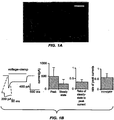
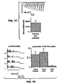
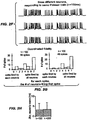
- FIG. 1(a) shows a micrograph of hippocampal neurons expressing ChR2-YFP.
- FIG. 1(c) shows currents in a hippocampal neuron illuminated as in FIG. 1(b) , in response to light pulses.
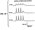
- FIG. 1(e) has voltage traces in response to brief light pulse sequences, with pulses lasting 5 ms (top), 10 ms (middle), and 15 ms (bottom).
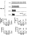
- FIG. 2(a) has voltage traces showing spikes in a current-clamped hippocampal neuron, in response to three deliveries of a Poisson train of light pulses.
- FIG. 2(b) shows trial-to-trial reliability of light evoked spike trains, as measured by comparing the presence or absence of a spike in two repeated trials of the same Poisson train delivered to the same neuron.
- FIG. 2(c) shows trial- to-trial jitter of the light-evoked spike trains.
- FIG. 2(d) shows the percent fidelity of spike transmission throughout the entire 8-second Poisson train.
- FIG. 2(e) shows latency of the spikes throughout each light pulse sequence (i), and jitter of spike times throughout the train (ii).
- FIG. 2(f) has voltage traces showing spikes in three different hippocampal neurons, in response to the same temporally patterned light stimulus as used in FIG. 2(a) .
- FIG. 2(g) has a histogram showing how many of the 7 neurons spiked in response to each light pulse in the Poisson train.
- FIG. 2(h) shows neuron-to-neuron jitter of spikes evoked by light stimulation.
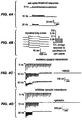
- FIG. 3(a) has voltage traces showing spikes in a current-clamped hippocampal neuron evoked by 5, 10, 20, or 30 Hz trains of light pulses.
- FIG. 3(b) has population data showing the number of spikes (out of 20 possible) evoked in current-clamped hippocampal neurons.
- FIG. 3(c) shows the number of extraneous spikes evoked by the trains of light pulses, for the experiment described in FIG. 3(b) .
- FIG. 3(d) shows jitter of spike times throughout the train of light pulses for the experiment described in FIG. 3(b) .
- FIG. 3(e) shows the latency to spike peak throughout the light pulse train for the experiment described in FIG. 3(b) .
- FIG. 4(a) has voltage traces showing subthreshold depolarizations in a current-clamped hippocampal neuron (left).
- FIG. 4(c) shows excitatory synaptic transmission driven by light pulses.
- the glutamatergic blocker NBQX abolishes these synaptic responses (right).
- FIG. 4(d) shows inhibitory synaptic transmission driven by light pulses.
- the GABAergic transmission blocker gabazine abolishes these synaptic responses (right).
- FIG. 5(b) shows membrane resting potential of the same neurons described in FIG. 5(a) .
- FIG. 5(c) shows the number of spikes evoked by a 300-pA depolarisation, in the same neurons.
- FIG. 6 shows a map of a retroviruses containing ChR2.
- FIG 7 shows a micrograph of a clonal stem cell line expressing ChR2 introduced by lentiviral transduction.
- FIG. 8 illustrates increased nuclear Ser-133 CREB phosphorylation in neural progenitor cell (NPC) triggered by light
- FIG. 9 is a micrograph of living zebrafish with ChR2 expressed in a neuron (left) and muscle cell (right).
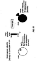
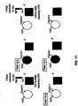
- FIG. 10 illustrates an embodiment of one of the methods of the invention.
- FIG. 11 illustrates an embodiment of one of the methods of the invention.
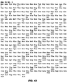
- FIG. 12 illustrates SEQ ID. No. 1.
- FIG. 13 illustrates SEQ ID. No. 2.
- FIG. 14 illustrates SEQ ID. No. 3.
- the present invention provides novel compositions and methods for controlling the electrical and chemical properties of cell membranes using light.
- the present invention also provides for a noninvasive, genetically targeted, high temporal resolution control of membrane electrical and chemical properties.
- the light-activated cation channel protein comprises a 7-transmembrane protein.
- the LACC comprises the protein, or portions of the protein Channelrhodopsin-2 (ChR2).
- ChR2 is a rhodopsin derived from the unicellular green alga Chlamydomonas reinhardtii.
- the term "rhodopsin” as used herein is a protein that comprises at least two building blocks, an opsin protein, and a covalently bound cofactor, usually retinal (retinaldehyde).
- the rhodopsin ChR2 is derived from the opsin Channelopsin-2 (Chop2) ( Nagel, et. al. Proc. Natl. Acad. Sci. USA 100:13940 , and references cited therein).
- the LACC protein of the present invention can incorporate retinal that is added to the system, or, depending on the cell type that is used, background levels of retinal present in the cell may produce the required retinal. It is intended herein that the methods of the invention encompass either the opsin or the rhodopsin form of the LACC protein, e.g. Chop2 or ChR2. Typically, Chop2 and ChR2 can be interconverted by the addition or removal of the cofactor.
- a LACC protein comprises an opsin with or without a co-factor.
- a nucleic acid codes for an opsin protein such as Chop2
- a cell expresses an opsin protein such as Chop2, it expresses a LACC protein.
- the LACC of the present invention may also cause the modulation of the flow of anions such as chloride across a membrane when activated by light. Optically induced electrical and chemical changes due to activation of the LACC by light are also included within the invention.
- the LACC protein covalently binds retinal.
- retinal as used in comprises all-trans retinal, 11-cis retinal, and other isomers of retinal.
- the protein Bcdo can be expressed along with ChR2.
- Bcdo converts the common dietary molecule beta carotene into retinal ( Yan et. al., Genomics 72 (2):193 (2001 )), thus providing retinal to convert Chop2 to ChR2.
- a preferred embodiment of the present invention comprises the amino terminal 310 amino acids of Chop2 which is referred to herein as Chop2-310.
- a preferred embodiment the present invention comprises the amino terminal 310 amino acids of ChR2 which is referred to as ChR2-310.
- the amino-terminal 310 amino acids of ChR2 show homology to the 7-transmembrane structure of many microbial-type rhodopsins, and comprise a channel with a light-gated conductance.
- a LACC protein comprises a 7-transmembrane protein.
- the LACC protein is a 7-transmembrane protein that either has a binding affinity for retinal, or has retinal bound to it.
- the LACC of the present invention is derived from a microbial-type rhodopsin. In a preferred embodiment, the LACC of the present invention is derived from a bacteriorhodopsin.
- a single-component protein that is a LACC protein.
- a single component protein is a single covalently linked chain of amino acids. Multiple component systems require communication between non-covalently linked molecules, which can be much slower than within-protein signaling via conformational changes.
- the present invention allows the creation of light gated membrane conductance with a single protein component. While not being bound by theory, it is believed that the retinal in ChR2, as a microbial type rhodopsin, is strongly bound, allowing the retinal to re-isomerize to the all-trans ground state in a dark reaction without the need for other enzymes. This mechanism allows for fast recovery (closing of the ionic channel) when the light is removed, and it obviates the need for other enzyme components for re-generation of the all trans-retinal and closing of the channel.
- the light-activated cation-channel Channelrhodopsin-2 (ChR2) is genetically introduced into a cellular membrane.
- the LACC protein of the present invention also comprises the protein sequence of Chop2-310 [SEQ ID NO:1, depicted in Figure 12 ].
- "Protein” in this sense includes proteins, polypeptides, and peptides.
- amino acid variants of the naturally occurring sequences as determined herein.
- the variants are greater than about 75% homologous to the protein sequence of Chop2 or Chop2-310, more preferably greater than about 80%, even more preferably greater than about 85% and most preferably greater than 90%.
- the homology will be as high as about 93 to about 95 or about 98%. Homology in this context means sequence similarity or identity, with identity being preferred.
- compositions of the present invention include the protein and nucleic acid sequences provided herein including variants which are more than about 50% homologous to the provided sequence, more than about 55% homologous to the provided sequence, more than about 60% homologous to the provided sequence, more than about 65% homologous to the provided sequence, more than about 70% homologous to the provided sequence, more than about 75% homologous to the provided sequence, more than about 80% homologous to the provided sequence, more than about 85% homologous to the provided sequence, more than about 90% homologous to the provided sequence, or more than about 95% homologous to the provided sequence.
- LACC proteins of the present invention may be shorter or longer than the protein sequence of Chop2 or Chop2-310.
- included within the definition of LACC proteins are portions or fragments of the protein sequence of Chop2 or of Chop2-310.
- nucleic acids of the invention may be used to obtain additional coding regions, and thus additional protein sequence, using techniques known in the art.
- the LACC proteins of the present invention are derivative or variant protein sequences, as compared to Chop2 or Chop2-310. That is, the derivative LACC proteins of the present invention will contain at least one amino acid substitution, deletion or insertion, with amino acid substitutions being particularly preferred. The amino acid substitution, insertion or deletion may occur at any residue within the LACC protein.
- These variants fall into one or more of three classes: substitutional, insertional or deletional variants.
- These variants ordinarily are prepared by site specific mutagenesis of nucleotides in the DNA encoding the LACC proteins, using cassette or PCR mutagenesis or other techniques well known in the art, to produce DNA encoding the variant, and thereafter expressing the DNA in recombinant cell culture.
- Amino acid sequence variants are characterized by the predetermined nature of the variation, a feature that sets them apart from naturally occurring allelic or interspecies variation of the LACC proteins of the present invention.
- the variants typically exhibit the same qualitative biological activity as the naturally occurring analogue, although variants can also be selected which have modified characteristics.
- the mutation per se need not be predetermined.
- random mutagenesis may be conducted at the target codon or region and the expressed breast cancer variants screened for the optimal combination of desired activity.
- Techniques for making substitution mutations at predetermined sites in DNA having a known sequence are well known, for example, M13 primer mutagenesis and PCR mutagenesis.
- Amino acid substitutions are typically of single residues; insertions usually will be on the order of from about 1 to 20 amino acids, although considerably larger insertions may be tolerated. Deletions range from about 1 to about 20 residues, although in some cases deletions may be much larger.
- substitutions are generally made in accordance with the following table: Table I Original Exemplary Residue Substitutions Ala Ser Arg Lys Asn Gln, His Asp Glu Cys Ser Gln Asn Glu Asp Gly Pro His Asn, Gln Ile, Leu, Val Leu Ile, Val Lys Arg, Gln, Glu Met Leu, Ile Phe Met, Leu, Tyr Ser Thr Thr Ser Trp Tyr Tyr Trp, Phe Val Ile, Leu
- substitutions that are less conservative than those shown in Table 1.
- substitutions may be made which more significantly affect the structure of the polypeptide backbone in the area of the alteration, for example the alpha-helical or beta-sheet structure; the charge or hydrophobicity of the molecule at the target site; or the bulk of the side chain.
- substitutions which in general are expected to produce the greatest changes in the polypeptide's properties are those in which (a) a hydrophilic residue, e.g. seryl or threonyl is substituted for (or by) a hydrophobic residue, e.g.
- leucyl isoleucyl, phenylalanyl, valyl or alanyl
- a cysteine or proline is substituted for (or by) any other residue
- a residue having an electropositive side chain e.g. lysyl, arginyl, or histidyl
- an electronegative residue e.g. glutamyl or aspartyl
- a residue having a bulky side chain e.g. phenylalanine, is substituted for (or by) one not having a side chain, e.g. glycine.
- variants or derivatives typically exhibit the same qualitative activity as the Chop2, ChR2, Chop-310, or ChR2-310 protein, although variants or derivatives also are selected to modify the characteristics of the LACC proteins as needed. Variants or derivatives can show enhanced ion selectivity, stability, speed, compatibility, and reduced toxicity. For example, the protein can be modified such that it can be driven by different wavelength of light than the wavelength of around 460nm of the wild type ChR2 protein.
- the protein can be modified, for example, such that it can be driven at a higher wavelength such as about 480 nm, 490 nm, 500 nm, 510 nm, 520 nm, 530 nm, 540 nm, 550 nm, 560 nm, 570 nm, 580 nm, or 590 nm.
- the LACC proteins of the present invention can incorporate un-natural amino acids as well as natural amino acids.
- the unnatural amino acids can be used to enhance ion selectivity, stability, speed, compatibility, or to lower toxicity.
- An aspect of the present invention is a fusion protein comprising a light-activated cation channel protein. It is well known in the art that fusion proteins can be made that will create a single protein with the combined activities of several proteins. In one embodiment, the fusion proteins can be used to target Chop2 or ChR2 to specific cells or regions within cells.
- a fusion protein comprising a LACC protein is a fusion protein that targets sub-cellular regions of the cell.
- the fusion proteins can target, for instance, axons, dendrites, and synapses of neurons.
- a PDZ (PSD-95, Dlg and ZO-1) domain is fused to ChR2 or Chop2 which target dendrites.
- Axon initial segment (AIS) domain is fused to ChR2 or Chop2 which target axons.
- fusion proteins of the present invention are proteins combining ChR2 and a fluorescent protein in order to allow for monitoring of the localization of ChR2.
- Preferred fusion proteins are those with red fluorescent protein (mCherry), yellow fluorescent protein (YFP), cyan fluorescent protein (CFP) and green fluorescent protein (GFP).
- mCherry red fluorescent protein
- YFP yellow fluorescent protein
- CFP cyan fluorescent protein
- GFP green fluorescent protein
- These fusion proteins such as the ChR2-mCherrry fusion protein, allow for the independent stimulation of ChR2 and the simultaneous monitoring of localization. The simultaneous stimulation and monitoring of localization can be carried out in many cell types including mammalian systems.
- the light-activated ion channel proteins of the present invention do not perturb the basal electrical properties, alter the dynamic electrical properties, or jeopardize the prospects for cellular survival.
- the light-activated cation channel proteins of the present invention do not alter the membrane resistance of the cells in the absence of light.
- the light-activated ion channels do not lead to apoptosis in the cells, nor lead to the generation of pyknotic nuclei.
- the presence of the LACC protein does not alter cell health or ongoing electrical activity, at the level of subthreshold changes in voltage or in spike output, either by shunting current through leaky channels or by altering the voltage dependence of existing neuronal input-output relationships.
- the presence of LACC, protein creates no significant long-term plastic or homeostatic alterations in the electrical properties of neurons expressing the protein.
- nucleic acid sequences which code for the LACC proteins of the present invention. It would be understood by a person of skill in the art that the LACC proteins of the present invention can be coded for by various nucleic acids. Each amino acid in the protein is represented by one or more sets of 3 nucleic acids (codons). Since many amino acids are represented by more than one codon, there is not a unique nucleic acid sequence that codes for a given protein. It is well understood by persons of skill in the art how to make a nucleic acid that can code for the LACC proteins of the present invention by knowing the amino acid sequence of the protein.
- a nucleic acid sequence that codes for a polypeptide or protein is the "gene" of that polypeptide or protein.
- a gene can be RNA, DNA, or other nucleic acid than will code for the polypeptide or protein.
- nucleic acid sequence comprises [SEQ ID NO:2, depicted in Figure 13 ].
- An aspect of the present invention provides a nucleic acid sequence that codes for a light-activated cation protein that is optimized for expression with a mammalian cell.
- a preferred embodiment comprises a nucleic acid sequence optimized for expression in a human cell.
- a preferred embodiment of a nucleic acid sequence that codes for a light-activated cation protein that is optimized for expression with a human cell comprises [SEQ ID NO:3, depicted in Figure 14 ].
- Another aspect of the present invention provides for reagents for genetically targeted expression of the LACC proteins including ChR2.
- Genetic targeting can be used to deliver light-activated cation channel proteins to specific cell types, to specific cell subtypes, to specific spatial regions within an organism, and to sub-cellular regions within a cell.
- Genetic targeting also relates to the control of the amount of light-activated cation channel protein expressed, and the timing of the expression.
- a preferred embodiment of a reagent for genetically targeted expression of the LACC protein comprises a vector which contains the gene for the LACC protein.
- vector refers to a nucleic acid molecule capable of transporting between different genetic environments another nucleic acid to which it has been operatively linked.
- vector also refers to a virus or organism that is capable of transporting the nucleic acid molecule.
- One type of preferred vector is an episome, i.e., a nucleic acid molecule capable of extra-chromosomal replication.
- Preferred vectors are those capable of autonomous replication and/or expression of nucleic acids to which they are linked.
- Vectors capable of directing the expression of genes to which they are operatively linked are referred to herein as "expression vectors”.
- kits are viruses such as lentiviruses, retroviruses, adenoviruses and phages.
- Preferred vectors can genetically insert LACC proteins into both dividing and non-dividing cells.
- Preferred vectors can genetically insert LACC proteins in-vivo or in-vitro.
- Those vectors that include a prokaryotic replicon can also include a prokaryotic promoter capable of directing the expression (transcription and translation) of the LACC protein in a bacterial host cell, such as E. coli.
- a promoter is an expression control element formed by a DNA sequence that permits binding of RNA polymerase and transcription to occur. Promoter sequences compatible with bacterial hosts are typically provided in plasmid vectors containing convenience restriction sites for insertion of a DNA segment of the present invention.
- Typical of such vector plasmids are pUC8, pUC9, pBR322, and pBR329 available from BioRad Laboratories, (Richmond, Calif.) and pPL and pKK223 available from Pharmacia, (Piscataway, N.J.).
- Eukaryotic cell expression vectors are well known in the art and are available from several commercial sources. Typically, such vectors are provided containing convenient restriction sites for insertion of the desired DNA homologue. Typical of such vectors are pKSV-10 (Pharmacia), pBPV-1/PML2d (International Biotechnologies, Inc.), and pTDT1 (ATCC, No. 31255).
- an expression vector of the present invention is a lentivirus comprising the gene for ChR2 or Chop2 and an EF1-alpha promoter.
- This lentivirus vector is used in one aspect of the present invention to create stable cell lines.
- the term "cell line” as used herein is an established cell culture that will continue to proliferate given the appropriate medium.
- an expression vector of the present invention is a lentivirus comprising the gene for ChR2 or Chop2 and a cell specific promoter.
- cell specific promoters are promoters for somatostatin, parvalbumin, GABA ⁇ 6, L7, and calbindin.
- Other cell specific promoters are promoters for kinases such as PKC, PKA, and CaMKII; promoters for other ligand receptors such as NMDAR1, NMDAR2B, GluR2; promoters for ion channels including calcium channels, potassium channels, chloride channels, and sodium channels; and promoters for other markers that label classical mature and dividing cell types, such as calretinin, nestin, and beta3-tubulin.
- Another preferred embodiment is a lentivirus containing tetracycline elements that allow control of the gene expression levels of ChR2, simply by altering levels of exogenous drugs such as doxycycline.
- This method, or other methods that place ChR2 under the control of a drug-dependent promoter, will enable control of the dosage of ChR2 in cells, allowing a given amount of light to have different effects on electrical activation, substance release, or cellular development
- One aspect of the invention is nucleic acid sequences comprising the gene for LACC proteins and promoters for genetically targeted expression of the proteins.
- the genetically targeted expression of the LACCs of the present invention can be facilitated by the selection of promoters.
- promoter as used herein is nucleic acid sequence that enables a specific gene to be transcribed.
- the promoter usually resides near a region of DNA to be transcribed.
- the promoter is usually recognized by an RNA polymerase, which, under the control of the promoter, creates RNA, which is then converted into the protein for which it codes.
- RNA polymerase which, under the control of the promoter, creates RNA, which is then converted into the protein for which it codes.
- the level of expression of LACC protein can be controlled.
- Cells use promoters to control where, when, and how much of a specific protein is expressed.
- promoters that are selectively expressed predominantly within one type of cell, one subtype of cells, a given spatial region within an organism, or sub-cellular region within a cell.
- the use of promoters also allows the control of the amount of LACC expressed, and the timing of the expression.
- the promoters can be prokaryotic or eukaryotic promoters.
- One embodiment of the invention is a nucleic acid sequence comprising the gene for LACC protein and a general purpose promoter.
- a general purpose promoter allows expression of the LACC protein in a wide variety of cell types.
- One example of a general purpose promoter of the present invention is The EF1-alpha promoter.
- the EF-1 alpha gene encodes for elongation factor-1 alpha which is one of the most abundant proteins in eukaryotic cells and is expressed in almost all kinds of mammalian cells.
- the promoter of this "housekeeping" gene can lead to persistent expression of the transgene in vivo.
- Another preferred general promoter is the CMV (cytomegalovirus) promoter, which can drive gene expression at very high levels.
- Still other preferred general-purpose promoters include those for CaMKII and synapsin I ( Dittgen et. al, PNAS 101:18206-11 (2004 )).
- One embodiment of the present invention is a nucleic acid sequence comprising the gene for LACC protein and a cell specific promoter.
- cell specific promoters are promoters for somatostatin, parvalbumin, GABA ⁇ 6, L7, and calbindin.
- Other cell specific promoters are promoters for kinases such as PKC, PKA, and CaMKII; promoters for other ligand receptors such as NMDAR1, NMDAR2B, GluR2; promoters for ion channels including calcium channels, potassium channels, chloride channels, and sodium channels; and promoters for other markers that label classical mature and dividing cell types, such as calretinin, nestin, and beta3-tubulin.
- the nucleic acid comprises a bacterial artificial chromosome (BAC).
- promoter is an inducible promoter.
- the promoter can be inducible by a trans-acting factor which responds to an exogenously administered drug.
- the promoters could be, but are not limited to tetracycline-on or tetracycline-off, or tamoxifen-inducible Cre-ER.
- One aspect of the present invention is a cell that expresses LACC proteins, and specifically a cell that expresses ChR2 or Chop2.
- Another aspect of the invention is a LACC protein expressing-cell that also expresses other ion channels, receptors, or signaling proteins, both in the normal and/or impaired form.
- Another aspect to the invention is a business method to make commercially available the cells of the invention.
- the cells of the present invention can be created using a vector including a DNA expression vector, a virus or an organism.
- Preferred vectors include lentiviruses and retroviruses.
- expression of ChR2 can be induced by using lipofection techniques, such as exposing cell lines to micelles containing Lipofectamine or Fugene, and then FACS-sorting to isolate stably expressing cell lines.
- Cells of any origin are candidate cells for transfection or infection with a LACC protein such as ChR2 or Chop2.
- LACC protein such as ChR2 or Chop2.
- specific cell types that can be grown in culture include connective tissue elements such as fibroblast, skeletal tissue (bone and cartilage), skeletal, cardiac and smooth muscle, epithelial tissues (e.g. liver, lung, breast, skin, bladder and kidney), neural cells (glia and neurones), endocrine cells (adrenal, pituitary, pancreatic islet cells), bone marrow cells, melanocytes, and many different types ofhematopoetic cells.
- Suitable cells can also be cells representative of a specific body tissue from a subject.
- body tissues include, but are not limited, to blood, muscle, nerve, brain, heart, lung, liver, pancreas, spleen, thymus, esophagus, stomach, intestine, kidney, testis, ovary, hair, skin, bone, breast, uterus, bladder, spinal cord and various kinds of body fluids.
- Cells in culture can be freshly isolated from body tissues (known as primary culture) or subcultured by expansion and/or cloning of the cells present in the primary culture (known as cell lines).
- Cells of different developmental stages (embryonic or adult) of an organism, or more specifically of various developmental origins including ectoderm, endoderm and mesoderm, can also be applied.
- Another type of cells embodied in the present invention is a "personal cell type", which comprises cells derived from individuals of a family, or individuals from different generations within the same pedigree.
- cells that are associated with a particular disease or with a specific disease stage are cells derived from natural and induced immune deficiency states, cardiovascular disease, neuronal disease, inflammation states and diseases caused by a variety of pathogens.
- the association with a particular disease or disease stage may be established by the cell's aberrant behavior in one or more biological processes such as cell cycle regulation, cell differentiation, apoptosis, chemotaxis, cell motility and cytoskeletal rearrangement.
- a disease cell may also be confirmed by the presence of a pathogen causing the disease of concern (e.g. HIV for AIDS and HBV for hepatitis B).
- Preferred cells are mammalian cells and cell lines derived from mammalian cells. Other preferred cells are embryonic stem cells and adult stem cells including hematopoetic stem cells, bone marrow, neural stem cells, epithelial stem cells, skin stem cells. Preferred cell lines appropriate for ChR2 expression include, HEK cells, neural stem cell lines, pancreatic islet cell lines, and other excitable or secretory cells.
- an aspect of the invention is a cell that has both the LACC protein and other ion channels or a splice variant of an ion channel that can be activated by the LACC protein.
- ion channels include, but are not limited to, Voltage-gated channels such as sodium and potassium voltage-gated channels of nerve and muscle; Ligand-gated such as Acetylcholine receptor, AMPA receptor and other neurotransmitter-gated channels; Cyclic nucleotide-gated channels such as calcium-activated channels; cardiac ion channels such as HERG channels; Stretch-activated channels; G-protein-gated channels; Inward-rectifier K channels; Resting channels; Store-operated channels such as calcium release-activated calcium (CRAC) channel; as well as other calcium channels, other potassium and sodium channels.
- the one or more ion channel proteins can be artificial, and can be targeted to be expressed in the cell containing the LACC protein.
- a preferred embodiment of the invention is a cell that comprises a LACC protein and one or more ion channel proteins that can be activated and thus controlled and whose behavior can be understood.
- Another preferred embodiment of the invention is a cell that comprises a LACC protein and one or more ion channel proteins that can be activated and thus controlled for screening for ion channel modulators as described below.
- Another preferred embodiment of the invention is a cell that comprises a LACC protein and one or more cardiac ion channels (e.g. HERG channel) that can be activated and thus controlled for screening for side effect of compounds as described below.
- Another preferred embodiment is an article of manufacture as described below containing a cell that comprises a LACC protein and one or more ion channel proteins that can be activated as described by the method herein.
- a preferred cell that expresses LACC proteins, and specifically a cell that expresses ChR2 or Chop2 is a mammalian cell.
- One aspect of the present invention is a cell line that expresses LACC proteins, and specifically a cell line that expresses ChR2 or Chop2.
- the cell line of the present invention can be created using a vector including a DNA expression vector, a virus or an organism.
- Preferred vectors include lentiviruses and retroviruses.
- expression of ChR2 can be induced by using lipofection techniques, such as exposing cell lines to micelles containing Lipofectamine or Fugene, and then FACS-sorting to isolate stably expressing cell lines.
- Preferred cell lines are mammalian cell lines as described above.
- Preferred cell lines are neuronal cell lines or other excitable cell lines.
- Preferred cell lines appropriate for ChR2 expression include HEK cells, neural stem cell lines, pancreatic islet cell lines, and other excitable or secretory cells.
- a preferred cell line of the present invention is a clonal neuronal stem cell line expressing ChR2 under the EF1-alpha promoter. Such a cell line is useful for the screening of drugs, particularly drugs that affect the influence of electrical activity on neuronal genesis, development, and apoptosis.
- Another preferred cell line of the present invention is a line of hippocampal neurons that expresses ChR2 under the EF1-alpha promoter. Such a cell line is useful for the in-vitro screening of drugs, and as a model for controlling neurons with light in-vivo.
- An embodiment of the invention is a stem cell lines that provide therapies based on transplantable, optically activatable cells that release substances as described below such as insulin, growth hormones, or other small molecules or polypeptides. Control of substance release with the present invention can be done on the second-to-minute timescale, allowing precise management of drug dosing, especially for conditions like diabetes or growth retardation.
- the cells of the invention can be grown as a monolayer anchored onto a solid phase substrate, or as aggregates in a suspension culture.
- the choice of substrate is determined largely by the type of cell and the desired growth parameters (e.g. growth rate, desired density, media requirements etc.)
- Most cells can be propagated on a substrate made of e.g., glass, plastic or ceramic material.
- a substrate made of e.g., glass, plastic or ceramic material.
- substrates pre-coated with charged substances that enhance cell attachment and spreading are preferred.
- Commonly employed coating materials include biological substrates that bear a net positive charge.
- Non-limiting examples of biological substrates include extracellular matrix/adhesion proteins such as laminin, fibronectin, collagen, or synthetic polypeptide such as poly-lysine.
- a variety of non-biological substrates such as membranes made of nitrocellulose, nylon, polytetrafluoroethylene, or any other implant materials can also be used to support growth of cells in a suitable medium according to cell
- Precautions are generally taken to maintain membrane integrity and preserve cell membrane components when harvesting cells cultured on different substrates.
- the method of the invention contemplate the use of traditional method of dissociating of anchored cells or cell layers including proteolytic enzymes such as serine proteinase, trypsin.
- cells of the present invention can be removed from the culture substrates by agents that minimize damages to the cell surface antigens.
- agents include chelating agents, such as EDTA and EGTA, which bind to divalent metal ions (e.g. calcium and magnesium) known to be necessary for cell-substrate attachment
- suitable cell dissociation agents encompass collagenases, dispases, and neutral proteinases when used in conjunction with serine proteinase inhibitors (e.g.
- soybean trypsin inhibitor Treatment of cells with these agents mostly results in disruption of the extracellular matrix components while preserving the cell surface proteins.
- the time required to detach the cells anchored on a solid substrate can vary depending on the protease enzymes chosen, but will normally be a period of about 1 minute to 30 minutes, and preferably about 5 minutes to 15 minutes.
- the enzymatic treatment can be carried out at room temperature or at about 37 °C. Excess enzyme can be removed by gentle washing with buffers having pH and salt concentrations in the physiological range that are routinely prepared by one skilled in the art.
- Cell viability may be confirmed by the measurement of membrane integrity.
- the methods for assessing membrane integrity are known in the art. The most common assay involves staining cells with a dye that reacts with either living or dead cells.
- exemplary dyes include trypan blue, eosin Y, naphthalene black, nigrosin, erythrosin B and fast green.
- One aspect of the invention is a transgenic animal that expresses a LACC protein.
- a preferred embodiment is a transgenic animal that expresses Chop2 or ChR2.
- Expression of LACC protein in particular subsets of neurons can be used for analyzing circuit function, behavior, plasticity, and animal models of psychiatric disease
- Preferred transgenic animal species of the present invention that expresses LACC protein include zebrafish (Danio rerio).
- the LACC protein can be introduced fish embryos by acute injection at the few-hundred cell stage.
- Another preferred transgenic animal species of the present invention that expresses LACC protein is flies (Drosophila melanogaster). In one preferred embodiment, flies express ChR2 under the UAS promoter for use in the GAL4-UAS system). Another preferred transgenic animal species of the present invention that expresses LACC protein is worms (Caenorhabditis elegans). In one preferred embodiment, stable lines are made with injection of plasmids containing ChR2 under specific promoters into the gonad.
- mice that expresses LACC protein are made using BAC (bacterial artificial chromosome) transgenic technology, as well as position effect variegation techniques.
- BAC bacterial artificial chromosome
- transgenic animal of the present invention that expresses LACC protein is Drosophila in which ChR2 is expressed in serotonergic and dopaminergic neurons, which are important for the driving of motivated behavior and the creation of finely tuned motor patterns.
- transgenic animal of the present invention that expresses LACC protein is Caenorhabditis elegans in which ChR2 is expressed in serotonergic and dopaminergic neurons, which are important for the driving of motivated behavior and the creation of finely tuned motor patterns.
- Another preferred embodiment of the present invention is a transgenic animal wherein the LACC is expressed under a specific promoter.
- Another preferred embodiment of the present invention is a transgenic animal wherein the LACC expressed in the transgenic animal is introduced via a BAC.
- Another preferred embodiment of the present invention is a transgenic animal wherein the LACC gene is knocked into a known locus.
- electrical spikes, or action potentials are created across a membrane by illumination with light.
- the light can be provided by a light source such as a xenon lamp, or the light source can be a laser. While a laser can be used, it would be understood by one skilled in the art that if the intensity of light is too high, there can be damaged to the cells under illumination due to local heating, etc. It is preferred to use a light intensity that does not damage the cells. In a preferred embodiment of the present invention medium intensity light is used to activate the ion channel.
- a preferred level of light is between about 0.1 mW/mm2 and about 500 mW/mm2, preferably from about 1 mW/mm2 and about 100 mW/mm2, and most preferably from about 5 mW/mm2 and about 50 mW/mm2.
- the LACC protein is ChR2 and the wavelength of the illuminating light is between about 400 nm and about 600 nm, preferably from about 450 nm and about 550 nm, most preferably from about 450 nm and about 490 nm.
- the light-activated ion channels of the invention can generate millisecond-timescale spikes when incorporated into neurons and illuminated with rapid pulses of light.
- One aspect of the present invention is a light-activated ion channel that is expressed in an excitable cell and will operate on the millisecond timeframe.
- a LACC responds within 100 millisecond (ms), more preferably within 10 ms, even more preferably within 1 ms, and most preferably within 0.1 ms.
- a preferred embodiment of the present invention is ChR2 that is expressed in a neuron or other excitable cell.
- the excitable cell responds in less than 10 ms, preferably less than 5 ms, and most preferably within 1 ms of being illuminated.
- One aspect of the present invention is a LACC protein expressed in a cell membrane that provides stable photo-currents over long time scales.
- the LACC protein is ChR2
- the photocurrent does not change by more than 20% over an hour of illumination, preferably not more than 10%, and most preferably not a detectable change over an hour of illumination.
- One aspect of the present invention is a LACC protein expressed in a cell membrane that provides precisely timed spikes with low temporal jitter.
- the temporal jitter is preferably lower than 10 ms, more preferably less than 5 ms, and most preferably lower than 3 ms when measured within neurons.
- One aspect of the present invention is a LACC protein expressed in a cell membrane that provides low temporal jitter across neutrons.
- Low temporal jitter across neutrons is desirable because it allows heterogeneous populations of neutrons to be controlled in concert.
- the temporal jitter across neutrons is also less than 10 ms, more preferably less than 5 ms, and most preferably lower than 3 ms.
- the across neutron jitter is within 50% of the within neutron jitter, most preferably the across neutron jitter is within 20% of the within neutron jitter, most preferably the within neutron jitter is indistinguishable from the across neutron jitter.
- One aspect of the present invention is a LACC protein expressed in a membrane that provides reliable and precisely timed subthreshold pulses.
- a cell expressing a light-activated cation channel protein of the present invention showed small trial-to-trial variability in the subthreshold deflections evoked by repeated light pulses.
- the light-activated cation channel proteins of the present invention such as ChR2 can therefore be employed to drive reliably timed subthreshold depolarizations with precisely determined amplitude.
- An aspect of the present invention is a LACC protein expressed in a membrane that has repeatable subthreshold polarizations.
- the LACC protein expressed in a membrane generates subthreshold pulses with a coefficient of variation of less than 0.2 when measured over 5 consecutive pulses, more preferably the coefficient of variation is less than 0.15 when measured over 5 consecutive pulses, and most preferably the coefficient of variation is less than 0.10 when measured over 5 pulses.
- the present invention allows for the optical control of cell properties by expressing a LACC protein within the cell, and activating the protein with light.
- the invention allows the optical control of excitable cells.
- One type of control is the control of electrical properties of the cell membrane for processes such as control of neurons and of transmissions of signals between neurons.
- Another type of control is control of influx of ions including Ca+2.
- the control of influx of Ca+2 is known to affect thousands of cellular processes.
- the optical control of the properties of excitable cells can be carried out in-vivo or in-vitro, and can be used for understanding biological processes, for drug discovery, and for therapeutic uses.
- Another type of control provided by the present invention is the use of the activation of the LACC protein to cause the cell to release proteins, peptides, or small molecules.
- the cell can be caused to release a protein, such as a cytokine.
- the cell through the light activation of ChR2, the cell can be caused to release a peptide such as insulin, leptin, neuropeptide Y, substance P, human growth hormone, secretin, glucagon, endorphin, oxytocin, vasopressin, or orexin/hypocretin.
- the activation of the LACC protein can cause the release of a small molecule whose synthesis or release is dependent on electrical activity, such as nitric oxide, or a cannabinoid such as anandamide or 2-arachidonylglycerol (2-AG).
- a small molecule whose synthesis or release is dependent on electrical activity, such as nitric oxide, or a cannabinoid such as anandamide or 2-arachidonylglycerol (2-AG).
- the present invention also allows for the control of cellular activity beyond the release of substances.
- LACC expressed in a muscle cell can allow for optical control of muscle cell contraction.
- This optical contraction of muscle cells, especially the contraction of specific muscle cells in the presence of other muscle cells, can be used for therapeutic purposes, or for the optical control of muscular contractions.
- the control provided by the present invention also included initiating specific intracellular signal conduction pathways. These pathways include, but are not limited to, kinases, transcription factors, and second messenger systems. These pathways can be specifically activated by the present invention due to the specific temporal pattern of light that is used. The pathways can also be specifically activated due to the specific sub-cellular localization of the LACC.
- An aspect of the present invention is a method of optically controlling a cell comprising; causing the cell to express LACC protein; and illuminating the cell with light to activate the LACC protein.
- a preferred embodiment of the method of optically controlling cell properties is a method in which the LACC protein is ChR2.
- Another preferred embodiment of the method of optically controlling cell properties is a method in which the cells are neurons or other excitable cells.
- the neurons can be connected to other neural cells and can be in a neural network.
- Another preferred embodiment of the method of optically controlling cell properties is a method in which the cells are spatially or genetically targeted subsets of cells that express the LACC protein of the present invention and can be specifically activated.
- Another preferred embodiment of a method of controlling cell properties comprises a method wherein the activation of the LACC also controls other ion channels within the cell.
- a cell can be caused to express LACC and one or more other ion channel proteins which may be artificial proteins.
- the LACC activation can then be used to activate or inactivate the other ion channel proteins within the cell.
- a cell expressing LACC can be illuminated with light in order to depolarize the cell membrane sufficiently to inactivate the cell's other ion channels.
- LACC protein can be used to selectively target differentiating cells whose cell fate is dependent on activity.
- ChR2 can be delivered to a progenitor cell or cell line, and then light can be used to drive the differentiation of the cell into appropriate progeny.
- a cell expressing a light-activated cation channel protein of the present invention will reliably generate spikes on the millisecond timeframe if the optical illuminations is provided in pulses in which a rapid light pulse is followed by a period of darkness.
- the method of optically controlling cell properties is a method comprising illuminating the cell with the LACC protein with a series of light pulses in which light periods are from 0.1ms to 100 ms), preferably from 1 ms to 50 ms, most preferably between 5 ms and 20 ms, and the periods of darkness were greater than 1ms, preferably greater than 10 ms, and most preferably greater than 20 ms.
- the periods of darkness can be long if desired, and can be periods of greater than seconds to minutes.
- the methods of optically controlling cell properties of the present invention will result in the ability to probe causal function in intact neural circuits, with it becoming possible to examine the role of particular neurons in animal models of learning, emotion, motor coordination, and sensory processing. This will enable the discovery of drugs capable of modulating whole-circuit function, essential for the addressing of complex neurological and psychiatric diseases.
- genetically-targeted neurons within animals will be addressable by light on timescales appropriate for examining the neural code mediating the behavioral or circuit-dynamics function observed, whether normal or dysfunctional.
- the millisecond scale control of electrical signals in neurons that is made possible by the present invention creates the ability to control synaptic events.
- the ease of eliciting synaptic transmission allows LACC proteins of the present invention including ChR2 to be an ideal tool for the temporally precise analysis of neural circuits.
- a LACC protein of the present invention expressed within a neuron can be activated by light to create an electrical signal within the neuron. That electrical signal can propagate along the neuron to the synapse where the signal can elicit a synaptic event.
- the synaptic event can be either an electrical or a chemical synaptic event.
- the synaptic event releases a small molecule that can modulate cellular function.
- the synaptic event comprises the release of a small-molecule neuromodulator such as norepinephrine, serotonin, dopamine, acetylcholine, D-serine, or histamine.
- the synaptic event can result in a synaptic transmission between two neurons.
- the present invention thus provides optically driven communication between sets of neurons, and thereby provides optically driven activity throughout a connected neural network.
- a preferred embodiment for the use of light to create a synaptic event comprises expressing ChR2 in a first neuron and illuminating the neuron with pulse of light to activate ChR2 to create a spike which causes a synaptic event.
- the use of light to create synaptic events can be carried out in-vitro or in-vivo.
- Another preferred embodiment for the use of light to create a synaptic event comprises expressing ChR2 in a first neuron and that in synaptic contact with a second neuron, and illuminating the first neuron so as to create a spike, wherein the spike results in a synaptic event at the synapse between the first and second neurons, and the second neuron is either excited or inhibited due to the signal from the first neuron, thus resulting in a synaptic transmission between the first and second neurons.
- Another preferred embodiment for the use of light to create a synaptic event comprises using a vector that is targeted to deliver the LACC protein of the present invention to specific neurons, specific subsets of neurons, or specific neuronal subtypes.
- the targeted delivery can be a specific spatial targeting into different spatial areas of the specimen, and/or the targeted delivery can be to chemical targeting to molecularly defined classes or subclasses of neurons.
- An aspect of the invention is a method for targeted delivery of light-activated cation-channel proteins to specific cells comprising; contacting said cells with a vector comprising a nucleic acid sequence comprising a LACC protein and a cell specific promoter; wherein said specific cells express said LACC protein.
- a preferred embodiment of a method for targeted delivery of light-activated cation-channel proteins to specific cells comprises a method wherein the vector comprises a nucleic acid sequence that codes for Chop2 or ChR2 and a cell specific promoter.
- Preferred cell specific promoters are the promoters for somatostatin, parvalbumin, GABA ⁇ 6, L7, and calbindin.
- promoters for kinases such as PKC, PKA, and CaMKII
- promoters for other ligand receptors such as NMDAR1, NMDAR2B, GluR2
- promoters for ion channels including calcium channels, potassium channels, chloride channels, and sodium channels
- promoters for other markers that label classical mature and dividing cell types such as calretinin, nestin, and beta3-tubulin.
- a preferred embodiment of a method for targeted delivery of light-activated cation-channel proteins to specific cells comprises a method wherein the vector comprises a lentivirus or a retrovirus.
- An aspect of the invention is a method for targeted delivery of light-activated cation-channel proteins to specific cells comprising; contacting said cells with a vector comprising a nucleic acid sequence comprising a LACC protein wherein said vector selectively targets specific cells.
- Preferred vectors are lentiviruses and retrovirus.
- Another aspect of the invention is a method for treating a subject comprising delivering a vector comprising a LACC protein to excitable cells within the subject and illuminating said cells with pulses of light.
- a preferred embodiment of a method for treating a subject comprises performing human therapeutic functions in which the function of cells is rescued or controlled by the genetic addition of LACC proteins such as ChR2, accompanied by the use of physically delivered pulses of light, preferably blue light.
- Delivering a LACC. protein such as ChR2 in human patients via viral vectors can enable control of excitable cells by blue light, either from a wearable optical device (for chronic stimulation) or at a fixed optical station (for more occasional stimulation).
- peripheral neurons like cutaneous pain suppressing nerves, virally transduced to express ChR2, allow light stimulation to activate dorsal column-medial lemniscus neurons in order to suppress painful C fiber responses.
- modified herpes viruses can be used to deliver ion channels to pain-pathway neurons; which can be used herein with ChR2 for the targeting of the channel to pain-pathway neurons and for reduction of the perception of pain.
- patients who have rod or cone loss can be virally transduced to express a LACC protein such as ChR2 in retinal ganglion cells, which restores the transduction of light in pathways mediating visual perception.
- ChR2 channelrhodopsin-2
- ChR2 channelrhodopsin-2
- the optical device used to excite LACC protein-expressing cells in patients is a light-emitting diode (LED).
- the LED can be in the millimeter or nanometer scale in size.
- One non-limiting example includes SML0805-B1K-TR LEDtronics.
- the LED can be battery-powered or remotely power by RF by methods that are known in the art.
- the LED can also be couple to a 68-microHenry surface-mount ferrite core inductor such as CF1008-682K from Gowana Electronics, NY.
- the wearable optical device can be non-invasively activated in a non-wireless fashion.
- depression patients are treated by delivering and exciting a LACC protein such as ChR2 to the anterior and/or subgenual cingulate cortex and to anterior limb of internal capsule of human patients by the methods described herein.
- a LACC protein such as ChR2
- the methods and compositions provided herein can also provide a beneficial effect for chronic pain patients.
- chronic pain patients are treated by delivering and exciting a LACC protein such as ChR2 to the anterior and/or dorsal cingulate cortex of human patients by the methods described herein.
- the methods and compositions provided herein can provide a beneficial effect for obesity patients.
- obesity patients are treated by delivering and exciting a LACC protein such as ChR2 to the ventromedial nucleus of the thalamus of human patients by the methods described herein.
- OCD obsessive compulsive
- addiction patients are treated by delivering and exciting a LACC protein such as ChR2 to the nucleus accumbens and septum of human patients by the methods described herein.
- a LACC protein such as ChR2
- the methods and compositions provided herein can provide a beneficial effect for Alzheimer's patients.
- Alzheimer's patients are treated by delivering and exciting a LACC protein such as ChR2 to hippocampus of human patients by the methods described herein.
- Parkinson's patients are treated by delivering and exciting a LACC protein such as ChR2 to the subthalamic nuclei and/or globus pallidus of human patients by the methods described herein.
- a LACC protein such as ChR2
- LACC protein-expressing secretory cell for implantation in patients (for example, nanoencapsulated to avoid immune responses) in which secretion is stimulated in the cells by the use of physically delivered pulses of light.
- ChR2-expressing neuroendocrine cells that release thyroid hormones can be implanted subcutaneously to allow for controlled peptide release over timescales from months to years. Sequences of flashes of light allow controlled release of such neuroendocrine substances, allowing modulation of stress, reproduction, metabolism, and sleep.
- LACC protein-expressing pancreatic islet cells can be made to release insulin when stimulated with light; implanted cells can enable control of diabetes symptoms on a minute-to-minute timescale without need for pump implantation or other invasive therapy.
- LACC protein-expressing cells are encapsulated prior to implantation into patients.
- the cells can be macroencapsulated or nanoencapsulated.
- capsules include but are not limited to semipermeable membranes, hollow fibers, beads and planar diffusion devices.
- encapsulation of dopamine-secreting cells in a semipermeable membrane has been examined to avoid rejection of implanted cells by the immune system ( Emerich et al. Neurosci Biobehav Rev 16, 437-447 ).
- the selectively permeable nature of the polymer membrane permits bidirectional access of low molecular weight compounds, including the inward diffusion of glucose, oxygen and other vital nutrients, and the outward diffusion of dopamine.
- the membrane restricts the passage of elements of the host immune system, thereby preventing host rejection of the encapsulated cells.
- encapsulated PC 12 cells a catecholaminergic cell line derived from rat pheochromocytoma
- encapsulated PC 12 cells can be implanted into the striatum of monkeys and adult guinea pigs ( Date et al. Cell Transplant 9, 705-709 , Aebischer et al. Exp Neurol. 111, 269-275 ).
- LACC protein-expressing cells are generated in the capsule prior to transplantation.
- ES cells can be: i) differentiated into dopamine producing neurons, ii) transfected with a nucleic acid containing LACC as described above, and iii) grown in a capsule, prior to implantation into patients. It has been shown that dopaminergic neurons can be induced from ES cells from mouse enclosed in hollow fibers using conditioning medium from PA6 cells, the stromal cells derived from skull bone marrow ( Yamazoe et al. Biomaterials 27 (2006) 4871-4880 ).
- differentiated LACC protein-expressing stem cells capable of secreting dopamine would be implanted, directly into the brain of a patient, and then drive their activation using light.
- Dopamine-secreting cells can be transfected or infected as described herein with a LACC protein such as ChR2, before or after the differentiation step, and then these cells can be implanted into the brain of the patients.
- the LACC protein-expressing stem cells are then activated by an optical device such as a light-emitting diode or a laser with an optical fiber attached to the end. Since the cells are light responsive, they will release dopamine even deep within tissue, simply by remotely being stimulated with light.
- LACC protein-expressing secretory cells are implanted into a tissue or an organ of a patient.
- the secretory cell is transfected or infected as described herein with a LACC protein such as ChR2, and then these cells are implanted into the tissue or organ of the patient.
- the LACC protein-expressing secretory cells are then induced to secrete chemicals by an optical device such as a light-emitting diode or a laser with an optical fiber attached to the end.
- Figure 10 illustrates one embodiment of the methods described herein, wherein cells expressing ChR2 (by transfection or infection) allow release of substances to be controlled by pulses of light.
- Examples of chemicals that can be secreted into the tissue or organ of a patient by the methods described herein include but are not limited to insulin, growth hormone, fast neurotransmitter, dopamine, cytokines, chemokines, hormones and hormone antagonists, pituitary hormones and their hypothalamic releasing factors, thyroid and antithyroid drugs, estrogens and progestins, androgens, adrenocorticotropic hormone; adrenocortical steroids and their synthetic analogs; inhibitors of the synthesis and actions of adrenocortical hormones, insulin, oral hypoglycemic agents, and the pharmacology of the endocrine pancreas, agents affecting calcification and bone turnover: calcium, phosphate, parathyroid hormone, vitamin D, calcitonin, vitamins such as water-soluble vitamins, vitamin B complex, ascorbic acid, fat-soluble vitamins, vitamins A, K, and E, growth factors, muscarinic receptor agonists and antagonists; anticholinesterase agents
- LACC protein-expressing secretory cells are implanted into the skin of a diabetic or patient.
- the LACC protein-expressing secretory cells are then induced to secrete insulin by an optical device such as a light-emitting diode or a laser with an optical fiber attached to the end.
- a LACC protein such as ChR2 allows for temporal, noninvasive control of release of substances from excitable cells. It should be apparent that the electrical activity, Ca+2, and secretion of different chemicals can affect literally thousands of cellular processes. Thus, it is possible that the general arena of applications of a LACC protein such as ChR2 in vivo may enable control over many biological disease processes in a variety of systems.
- Another aspect of the present invention is a method of controlling the behavior of an organism comprising delivering a vector comprising a LACC protein to excitable cells within the organism; and illuminating said cells with pulses of light so as to control the behavior of the organism.
- Another aspect of the invention involves the use of the method described herein to control the fate of cells.
- the techniques described herein are used to modulate activities of cells that are modulated by electrical activity.
- the methods can be used to modulate the survival, replication, differentiation, and/or death of cells.
- Channelrhodopsin is used to drive any one of these processes, depending on the precise pattern of stimulation used to drive activation of channelrhodopsin, which would then result in a specific pattern of downstream signal transduction and a specific cellular fate response.
- targeting Channelrhodopsin to specific cells, then exposing them to particular light patterns is used to enhance the survival of, drive differentiation or replication of, or hasten the death of, the cells expressing the light-activated channel.
- This modulation of cellular processes is preferably used in stem cells, where particular patterns of activity may drive the differentiation of stem cells (including human embryonic stem cells), drive the replication of stem cells, or drive the death of the stem cells (in the case where excessive replication is desired to cease).
- stem cells including human embryonic stem cells
- the target cells are tumor or cancer cells
- targeting channelrhodopsin to those cells permits the use of specific and appropriate patterns of light to kill the tumor/cancer cells.
- Other suitable target cells include various secretory or organ cells or their precursors, cardiac or other muscle cells, or glial cells in the brain. In each of these cases, it may be desirable to control the replication, differentiation, and death of these cells precisely.
- Channelrhodopsin may be useful for controlling these things in vitro, in vivo in experimental animals, or in vivo in humans (before or after transplantation into the body).
- An aspect of the invention is a method for screening for drugs comprising expressing a LACC protein in a group of cells; exposing said group of cells to a compound; illuminating said groups of cells with light; and monitoring the electrical response of cells within the group of cells.
- the electrical response of cells can be monitored electrically, optically, or by other means.
- the electrical responses of the cells can be seen and monitored using chemical or voltage-sensitive dyes, or dyes that undergo oxidation or reduction. These changes can be monitored in a microscope, with optical sensors or with a camera using film, CCD arrays, and other methods known by persons of skill in the art.
- a preferred embodiment of a method of drug screening is high-throughput screening for drugs affecting the ionic and signaling function of cells such as central and peripheral neurons, heart muscle, pancreatic islet cells, neuroendocrine cells in pituitary and kidney, stem cells, cancer cells, and others.
- a preferred embodiment of a method of drug screening comprises rapidly modulating voltage in electrically excitable cells capable of fast responses (such as neurons and heart muscle, or cell lines derived thereof), to screen for drugs that block or activate voltage-gated ion channels, alter excitatory or inhibitory synaptic transmission (play a role in epilepsy and pain), and alter muscular contraction. Since many channels and receptors activate rapidly, and then desensitize or inactivate, the ability to control membrane voltage with brief pulses of light greatly enhances the ability to develop drugs that target certain channel, synapse, or muscle disease phenotypes. For example, familial hemiplegic migraine patients have point mutations in a calcium channel, reducing the calcium influx, for a given brief depolarization.
- the methods of the present invention will allow for screening drugs that modulate the calcium response (measured using a calcium dye) to a given stereotyped depolarization, in a cell line expressing the mutant calcium channel and ChR2, to discover ways of improving channel function in this patient population.
- pancreatic islet cells and neuroendocrine cells release hormones in a Ca+2-dependent way, via the fusion of dense core vesicles.
- LACC proteins of the present invention such as ChR2, which not only causes cell depolarization but is capable of passing Ca+2 ions through its channel pore, can be used to activate the release of peptides or hormones from such cells (or derived cell lines), aiding in the screening of drugs that enhance or suppress hormone release.
- ChR2 which not only causes cell depolarization but is capable of passing Ca+2 ions through its channel pore, can be used to activate the release of peptides or hormones from such cells (or derived cell lines), aiding in the screening of drugs that enhance or suppress hormone release.
- Such discoveries can aid in the discovery of pharmacological methods of treating problems of impaired growth, development, metabolism, stress, and reproduction.
- the LACC proteins of the present invention including ChR2 can be used to screen for drugs that alter slow signal transduction processes in cells, revealing treatments for chronic disease processes ranging from cancer to depression.
- immune cells respond to ongoing patterns of Ca+2 influx with a variety of long-lasting changes, which can lead to strengthening or weakening of immune responses, or even possibly autoimmune symptoms.
- Cancer cells also express ion channels, and may respond to electrical activity with altered proliferation.
- Neural stem cells respond to depolarization and Ca+2 influx by differentiating into neurons; activating stem cells expressing LACC proteins with light can then enhance survival and incorporation into mature tissues, a step in generation of functional brain and other tissue.
- the stem cell line expressing a LACC protein of the present invention such as ChR2 can be used to optically control tissue repair, and illumination of these stem cells with light results in CREB phosphorylation, a critical step in enhancing their transformation into neurons.
- Another preferred embodiment is a method of drug screening comprises making a human embryonic stem (ES) cell line with ChR2 under a stem-cell specific promoter to build cells which differentiate under optical control. Expression of ChR2 in such cells allows discovery of drugs which alter the activity-dependent progress of cellular life and death amidst their environments.
- ES human embryonic stem
- cells can be forced to change developmental fate via remote control, and a drug screening tool where this is possible allows the discovery of new molecules that manipulate such long term effects of cell proliferation, differentiation, and apoptosis.
- specific cell populations will be light-addressable on rapid timescales, allowing the screening of many aspects of cellular function.
- Figure 11 illustrates one example of the methods for screening drugs.
- the baseline electrical activity of the ion channel is measured by flashing the light and observing the depolarization. The fluorescence of the cells will return to its original value after the light is off.
- a drug is applied, for example, by pipetting in the drug (or flowing in through a microfluidic channel). Finally, the light is re-flashed and the electrical activity of the ion channel is measured.
- This technique can be used in combination with the whole-cell patch clamp technique, wherein an electrode is placed up to a cell, suction is applied to break into the cell, and to record the activity of the cell expressing an ion channel before and after the application of a drug.
- drugs that can be screen according to the methods described herein include, but are not limited to, antidepressants, antipsychotics, calcium antagonist, antiepilecticts, as well as drugs to treat OCD, Alzheimer's, Parkinson, obesity, addiction, chronic pain, muscle and heart disorders.
- step 11 illustrates one example in which the drug is a negative modulator of channel function
- Steps (4), (5), and (6) can take place in a robotic device that moves a 384-well plate into the focus of an optical beam.
- the wells of the plate could all contain the same cell line, facilitating the screening of drugs that affect a particular channel ("high throughput target screening,” see below), or each well could contain cells of a different cell line, facilitating the screening of one drug against many different channels ("screening against side effects,” see below).
- a voltage sensitive dye is preferred due its fast kinetics, but other dyes (e.g., a calcium-sensitive dye in the case that channel n is a calcium channel) could also serve to indicate whether channel function is modulated by the drug.
- Genetically encoded indicators of voltage or calcium may also be useful for reading out the activity of the cell (e.g., FLASH, GCaMP2, cameleon, etc.). In this case, these indicators would be stably expressed in the cell line as well. Other methods of reading out whether the drug had an effect are also useful - e.g., immunostaining for the phosphorylation of a site that is phosphorylated during or after periods of ion channel activity.
- an optical imaging device containing 1) a light source (light-emitting diode (LED), lamp, laser) for illuminating the cell expressing Channelrhodopsin and driving a change in cell voltage, 2) a light source for illuminating a dye or indicator, possibly the same light source as used for driving the voltage change, and 3) a switch for alternating between the two light sources or a beamsplitter for simultaneous non-interfering delivery of both kinds of light.
- the fluorescence of the dye or indicator is typically measured by a sensor (CCD camera, PMT, or photodiode). This kind of device is useful for ion channel drug screening, as described above.
- the device itself may also consist of a robotic arm for moving a plate (e.g., a 384-well plate) through the arena where the light sources and sensor are present.
- a plate e.g., a 384-well plate
- This kind of combined light source/imaging device has also diagnostic applications. For example, taking cells from a patient, expressing Channelrhodopsin in them, and then exposing them to light, could be used to detect patient-specific ion channel syndromes in biopsy samples or in cells of the circulatory system.
- channelrhodopsin any suitable light-activated cation channel can be used.
- high-throughput target screening comprises a plate reader that moves wells containing cells of interest into the optical stimulation/readout area, and then performs the methods described above and illustrated in Figure 11 . Then the machine moves the plate to enable the next well to be flashed, in turn.
- well plates include 96-, 384, 1526-well plates.
- the high-throughput target screening methods of the invention can analyze up to about 2,000 drugs per day per setup. In another embodiment, the high-throughput target screening methods of the invention can analyze up to about 2,500,000 drugs per year per 5 setups.
- high-throughput target screening comprises a chip-based system.
- a chip-based system can enable sequential rather than parallel testing of compounds on ion channels by high-speed scanning of a single cell across a laminar stream of solution environments created in the microfluidic chips. The scanning of the microfluidic chip, causing a cell to sample the discrete zones of drug solutions, is controlled by a computer controlled motorized scan stage.
- the microfluidic chips for example, can have eight-channel, 16-channel, 48-channels or more for high-throughput analysis.
- high-throughput target screening comprises a cell sorting device that includes an array of discrete locations for capturing cells traveling along a fluid flow.
- the discrete locations can be arranged in a defined pattern across a surface such that the discrete sites are also addressable and contain discrete zones of drug solution.
- surfaces that may be used for creating arrays of cells in discrete sites include, but are not limited to, cellulose, cellulose acetate, nitrocellulose, glass, quartz or other crystalline substrates such as gallium arsenide, silicones, metals, semiconductors, various plastics and plastic copolymers, cyclo-olefin polymers, various membranes and gels, microspheres, beads and paramagnetic or supramagnetic microparticles.
- Yet another aspect of the invention is methods for screening against side effects. This method allows for screening for drugs that selectively affect one ion channel, but do not affect other ion channels. This screening against individual ion channels allows for screening to predict potential side effects.
- many channels are expressed differentially in the heart vs. the brain.
- screening for drugs that differentially bind to channels in the brain, but not the heart it is possible to find neuropsychiatric drugs that don't affect heart function.
- Especially important for heart function are calcium channels, HERG channels, other potassium channels, and other ion channels that affect the rhythmicity or amplitude of the heartbeat.
- Another aspect of the invention is the use of the methods and compositions described herein in prosthetic devices.
- Blindness, deafness, and other sensory deficits affect millions of people worldwide, severely impacting their quality of life.
- Channelrhodopsin targeted to somatic cells in the human patient, opens up a new class of sensory prostheses.
- some forms of blindness destroy photosensor function, but leave signal processing in downstream neurons intact.
- targeting Channelrhodopsin to retinal ganglion cells for example, by injecting viruses expressing channelrhodopsin into the retinal cell layers inside the eye could enable restoration of visual function.
- channelrhodopsin targeted to retinal ganglion cells
- the retinal ganglion cells would themselves become photosensitive, enabling vision with resolution comparable to the native eye, and preferably not requiring invasive technology beyond that point.
- Channelrhodopsin is sufficiently sensitive to detect sunlight (power ⁇ 1 kW/m ⁇ 2).
- the Channelrhodopsin can be targeted to amacrine cells or bipolar cells to enable vision. It is also possible that expressing channelrhodopsin in a retinal cell, accompanied with a projection device that would amplify the ambient light, would enable vision indoors or in low-light conditions.
- hair cells are lost, but downstream neurons are intact.
- Expressing Channelrhodopsin in spiral ganglion cells i.e., eight nerve neurons) would enable activation of these cells with light.
- Cochlear implants currently stimulate all the cells of the cochlea with a single electrode or at most a few electrodes, and do not attempt to recapitulate any of the spatial distribution of sensory afferents in the cochlea.
- a tonotopically-mapped stimulator By inserting a device containing 1) a microphone to detect sound, 2) a microprocessor to analyze the frequency components of the sound and convert them to LED signals, and 3) multiple LEDs for emitting light in a spatially patterned fashion, into the cochlea, a tonotopically-mapped stimulator could be created, which would drive different frequencies of sound perception simply by targeting the light to appropriate cells.
- central nervous system neurons in a human are infected with virus expressing Channelrhodopsin (or otherwise come to express Channelrhodopsin), these neurons become capable of responding to light.
- This gene therapy approach allows optical stimulation of neuronal targets in the brain. If the targeted neurons are in sensory cortex, this opens up the possibility of a new kind of cortical sensory prosthesis. If the targeted neurons are in the frontal cortex or other parts of the brain, it is in possible that these light-sensitive neurons permit modulation of emotion or cognition. If the targeted neurons were in the spinal cord, it is possible that neurons that inhibit painful stimuli could be driven by light.
- this gene therapy approach opens up a new kind of generalized prosthetic, in which light is converted into neural activity, in defined parts of the nervous system
- a prosthetic device is for implanted cells that are engineered to secrete compounds and to respond to light, and also for hunting for neural-circuit level targets in the intact animal brain, it may be very useful to have an implantable or head-mounted LED, or other small light source.
- a light source could be implanted under the skin, under the skull, deep within the brain, or deep within another organ of interest, in which Channelrhodopsin-expressing cells are also located (either exogenously introduced, or endogenously located and targeted with a virus).
- This device could be used for stimulating ChR2 in cells located directly adjacent to the light source.
- a strip of LEDs, each individually controllable could be useful.
- a 2-dimensional array of LEDs could be useful.
- an insulin-secreting cell located under the skin a wearable blue LED on a bracelet, powered by a battery, could be useful.
- the LEDs are remotely powered.
- a remotely-powered LED could be made by combining an LED in a closed-loop series circuit with an inductor. This would allow radiofrequency (RF) energy or rapidly changing magnetic fields (e.g., delivered by a transcranial magnetic resonance (TMS) coil) to temporarily power-up the inductor, and thus the connected LED, allowing local delivery of light, even deep in a brain structure.
- RF radiofrequency
- TMS transcranial magnetic resonance
- Such a device could be implanted under the skin, under the skull, deep within the brain, or deep within another organ, of interest in which Channelrhodopsin-expressing cells are also located (either exogenously introduced, or endogenously located and targeted with a virus). Then a device that can remotely deliver RF or magnetic energy could be placed nearby, or worn on the patient, for activating the implanted device.
- Another aspect includes biochemical modifications of light activated channels, such as Channelrhodopsin. Such modifications are typically performed to target Channelrhodopsin to different parts of a cell. Fusing channelrhodopsin to a targeting sequence of DNA, so that the resultant protein contains both channelrhodopsin and the targeting peptide, could be used to send Channelrhodopsin to the presynaptic terminal, the postsynaptic terminal, the nucleus, or other intracellular compartments.
- targeting sequences include PDZ domains, glutamate and GABA receptor C-terminal sequences, ion channel c-terminal sequences, presynaptic scaffolding targeting sequences, and other targeting sequences. These versions of Channelrhodopsin could be used to trigger specific intracellular signaling events, including those important for neuroprotection, memory, or other enduring signaling functions.
- these reagents could complement the other applications of Channelrhodopsin.
- these reagents could be useful for drug screening (e.g., finding drugs that modulate the function of a channel in a particular subcellular compartment).
- these reagents could also be useful for prosthetic devices (e.g., driving activity on the dendrites of a neuron, to more closely mimic natural synaptic activity).
- Preferred light sources used to illuminate the Channelrhodopsin-expressing cells have the following properties:
- articles of manufacture containing the compositions described herein e.g. a nucleic acid comprising an LACC sequence or a LACC protein-expressing cell
- the article of manufacture comprises a container and a label.
- Suitable containers include, for example, bottles, vials, syringes, plates, and test tubes.
- the containers may be formed from a variety of materials such as glass or plastic.
- Suitable packaging and additional articles for use e.g., measuring cup for liquid preparations, foil wrapping to minimize exposure to air, dispensers, and the like) are known in the art and may be included in the article of manufacture.
- the container is a 96-, 384-, and 1526-well plate containing different cells of interest in each well, for example, prefrozen in a protective medium (e.g. medium containing 20% DMSO).
- a protective medium e.g. medium containing 20% DMSO.
- the plates can be shipped in dry ice and store upon receipt at -80 degrees Celsius for short term storages. Alternatively, the plates are stored in liquid nitrogen for long term storage. This plate fit into a plate-reader/motorized stage device as the ones described above or a regular plate reader known in the art.
- the container holds a composition that is effective for treating a disease condition and may have a sterile access port (for example the container may be an intravenous solution bag or a vial having a stopper pierceable by a hypodermic injection needle).
- the active agent in the composition is the compositions of the invention described herein (e.g. a nucleic acid comprising an LACC sequence or a LACC protein-expressing cell).
- the article of manufacture can further comprise, within the same or a separate container, another agent such as a therapeutic agent that is co-administered with the compositions of the invention and optionally a pharmaceutically acceptable buffer, such as phosphate-buffered saline, Ringer's solution and dextrose solution.
- the label on, or associated with, the container indicates instructions to use the composition, for instance, the container may indicated that the composition is used for treating a disease condition of choice or for screening compounds or screening compound's side effects.
- the containers described herein may further include other materials desirable from a commercial and user standpoint, including other buffers, diluents, filters, needles, syringes, and package inserts with instructions for use.
- ChR2-YFP gene was constructed by in-frame fusing EYFP (Clontech) to the C-terminus of the first 310 amino acid residues of ChR2 (GeneBank accession no. AF461397) via a Not I site.
- the lentiviral vector pLECYT was generated by PCR amplifying ChR2-YFP with primers:
- VSVg pseudotyped lentiviruses were produced by triple-transfection of 293FT cells (Invitrogen) with pLECYT, pMD.G, and pCMVdeltaR8.7 (gifts from Eric Wexler and Theo Palmer) using Lipofectamine 2000.
- the lentiviral production protocol is same as previously described 42 except for the use of Lipofectamine 2000 instead of calcium phosphate precipitation.
- viruses were concentrated by centrifuging in a SW28 rotor (Beckmann Coulter) at 20,000 rpm for 2 h at 4 °C. The concentrated viral titer is determined via FACS sorting to be between 5x10 ⁇ 8 and 1x10 ⁇ 9 IU/mL.
- Hippocampal Cell Culture Hippocampi of postnatal day 0 (P0) Sprague-Dawley rats (Charles River) were removed and treated with papain (20 U/ml) for 45 min at 37°C. The digestion was stopped with 10 ml of MEM/Earle's salts without L-glutamine along with 20 mM glucose, Serum Extender (1:1000), and 10% heat-inactivated fetal bovine serum containing 25 mg of bovine serum albumin (BSA) and 25 mg of trypsin inhibitor. The tissue was triturated in a small volume of this solution with a fire-polished Pasteur pipette, and ⁇ 100,000 cells in 1 ml plated per coverslip in 24-well plates.
- Viral Infection Hippocampal cultures were infected on d.i.v. 7 using 5-fold serial dilutions of lentivirus ( ⁇ 1x10 ⁇ 6 IU/mL). Viral dilutions were added to hippocampal cultures seeded on coverslips in 24 well plates and then incubated at 37°C for 7 d before recording using patch-clamping technique.
- Coverslips were then fixed for 5 minutes in PBS plus fresh 4% paraformaldehyde, permeabilized for 2 minutes with PBS plus 0.1% Triton X-100, and then immersed for 5 minutes in 5 ⁇ g/mL PI for observation of any pyknotic nuclei. At least eight different fields were examined per coverslip.
- Neurons were perfused in Tyrode's solution, described above. All experiments were performed at room temperature (22-24°C). For all experiments except for the synaptic transmission data shown in Fig. 4b and 4c , we patched fluorescent cells immersed in Tyrode solution containing 5 ⁇ M NBQX and 20 ⁇ M gabazine to block synaptic transmission. Photocurrents were measured while holding neurons in voltage clamp at -65 mV. Recovery from inactivation was measured by measuring photocurrents while illuminating neurons with pairs of pulses lasting 500 ms each, separated by periods of darkness lasting 1-10 seconds. Spiking was measured while injecting current to keep the voltage of the cell at approximately-65 mV (holding current ranging from 0 pA to 200 pA).
- pClamp 9 software (Axon Instruments) was used to record all data and to operate the MultiClamp 700B amplifier, and a Sutter DG-4 ultrafast optical switch with 300W xenon lamp (Sutter Instruments) was used to deliver the light pulses for ChR2 activation.
- a standard Endow GFP excitation filter (excitation filter HQ470/40x, dichroic Q495LP; Chroma) was used for delivering blue light for ChR2 activation, in the bandwidth 450-490 nm.
- YFP was visualized with a standard YFP filter (excitation HQ500/20x, dichroic Q515LP, emission HQ535/30m; Chroma).
- Data Analysis Data was analyzed automatically using Clampfit (Axon) and custom software written in MATLAB. Spikes were extracted by looking for crossings of the voltage above a threshold (typically 60 mV above resting potential), and latencies were measured from the onset of the light pulse to the spike peak. Extraneous spikes were measured as the number of extra spikes after a single light pulse, plus any spikes occurring later than 30 ms after the onset of a light pulse. Jitter was calculated as the standard deviation of spike latencies, measured either throughout the spike train (when gauging reliability throughout a train), or for the same spike across different trains (when gauging reliability across trials, or across neurons).
- Fig. 1e highlights the fact that longer light pulses evoke single spikes with greater probability than short light pulses.
- the ability to easily alter light pulse duration with fast optical switching suggests a straightforward method for eliciting spikes even in multiple neurons possessing different ChR2 current densities, by simply increasing the light pulse duration until single spikes are reliably obtained in all the neurons being illuminated. Rapid modulation of light power would also allow for this kind of control.
- this across-neuron jitter ( Fig. 2h ) was identical to the within-neuron jitter, measured throughout the light pulse series ( Fig. 2e ).
- heterogeneous populations of neurons can be controlled in concert, with practically the same precision observed for the control of single neurons over time.
- ChR2 can induce spiking across a physiologically relevant range of firing frequencies, appropriate for driving trains and bursts of spikes.
- ChR2 therefore can be employed to drive reliably timed subthreshold depolarizations with precisely determined amplitude.
- ChR2 has little basal electrical activity, or even passive shunting ability, in the absence of light. It also suggests that expression of ChR2 did not lead to impairment of general cell health, as indicated by electrical determination of membrane integrity.
- PI DNA-binding dye propidium iodide
- ChR2+ expression did not affect the percentage of live neurons that took up PI (1/56 ChR2+ neurons vs. 1/49 ChR2-neurons; p > 0.9 by ⁇ 2 test). Neither did we see any pyknotic nuclei, indicative of apoptotic degeneration, in cells expressing ChR2 (data not shown).
- ChR2 does not alter cell health or ongoing electrical activity, at the level of subthreshold changes in voltage or in spike output, either by shunting current through leaky channels or by altering the voltage dependence of existing neuronal input-output relationships.
- ChR2 predisposes neurons to light-induced problems with cellular health
- Moloney-type retroviruses selectively target dividing cells ( Ory et al., Proc Natl Acad Sci U S A 93:11400 (1996 )), such as stem cells.
- dividing cells Ory et al., Proc Natl Acad Sci U S A 93:11400 (1996 )
- These retroviruses have been generated by triply transfecting the plasmid containing ChR2 with helper vectors into 293T cells.
- tetracycline is used to suppress the expression of VSV-G, which is toxic to 293GPG cells, during growth phase. After cells grow to 70% confluence, then tetracycline is removed to allow the expression of VSV-G. At this point, the plasmid containing Chr2 and GFP (right) are transfected into the 293GPG cells using Lipofectamine, and cultured 293GPG cells are monitored for signs of fluorescence as gene expression and retrovirus production begins. These cell lines are then frozen for future production of retrovirus.
- viruses have been shown to efficaciously infect dividing cells, both in vitro (kidney cell lines, stem cells) and in vivo in rats (hippocampal stem cells, glia).
- This construct will be useful for targeting ChR2 to dividing cells ex vivo , for transplantation into humans for therapy, for the creation of cell lines, and for the development of animals with selective populations of labeled cells for studies of diseases of cellular activity, such as epilepsy, migraine, narcolepsy, and even autoimmune diseases.
- the lentiviruses that we have generated contain tetracycline elements that allow control of the gene expression levels of ChR2, simply by altering levels of exogenous drugs such as doxycycline.
- This method or other methods that place ChR2 under the control of a drug-dependent promoter, will enable control of the dosage of ChR2 in cells, allowing a given amount of light to have different effects on electrical activation, substance release, or cellular development.
- the lentiviruses that we have generated contain tetracycline elements that allow control of the gene expression levels of ChR2, simply by altering levels of exogenous drugs such as doxycycline.
- Fig. 7 shows a.micrograph of cells from this neural progenitor cell (NPC) stem cell line.
- NPC neural progenitor cell
- This cell line is appropriate for the screening of drugs that affect the influence of electrical activity on neuronal genesis, development, or apoptosis.
- the stem cell line we have already made is a correct step in realizing the goal of optically controllable tissue repair, and illumination of these stem cells with light results in CREB phosphorylation, a critical step in enhancing their transformation into neurons ( Fig. 8 ).
- These homogeneous stem cell lines can also be transplanted into the brain of adult animals, where they incorporate into hippocampal circuitry and can be studied functionally after 2-6 weeks.
- Fig. 9 shows micrographs of ChR2-EYFP selectively expressed in a zebrafish trigeminal neuron (left) and in a zebrafish muscle cell (right).
- larvae from 48 hours to 96 hours post fertilization were anesthetized in 0.02% tricaine in fish Ringer's solution and mounted in 1.2% agarose for imaging and photostimulation.
- Illumination of the live fish expressing ChR2 in muscle with blue light for 0.5 seconds caused rapid, phasic, single muscle contractions, which was never seen in zebrafish not containing ChR2.
- Neurons tolerated the ChR2 expression well, and were able to be loaded with fluorescent calcium dyes (e.g., Oregon Green BAPTA-1, X-rhod-1), appropriate for the monitoring of neural activity downstream of a selectively activated neuron expressing ChR2, using two-photon microscopy.
- fluorescent calcium dyes e.g., Oregon Green BAPTA-1, X-rhod-1
- Flies (Drosophila melanogaster) have been generated that will express ChR2 under the UAS promoter, for use in the GAL4-UAS system that enables flexible control of gene expression in flies.
- the UAS-ChR2 flies can be crossed with a variety of GAL4 lines of Drosophila, taking advantage of the large number of transgenic flies that have been created over the last several years.
- Worms have been caused to express ChR2 by the injection of plasmids containing ChR2 under specific promoters into the syncytial gonad of the worm. Worms are also co-injected with a visible marker gene, to allow visual inspection of the success of generating the transgenic nematode.
- the gonad of the worm takes up the plasmid DNA and stores the DNA in large extrachromosomal arrays in eggs, passing the plasmid on to the worm's offspring.
- ChR2 in C. elegans in serotonergic and dopaminergic neurons, important for the driving of motivated behavior and the creation of finely tuned motor patterns.
- Mice can be made to express ChR2 transgenically under the control of specific promoters (by pronuclear injection of plasmids containing ChR2 under a specific promoter), in specific loci ("knocking in” a gene into an existing locus), using BAC transgenic technology (to mimic the natural genetic environment of a gene), or by position effect variegation techniques (a transgenic method allowing genes to randomly express in tiny subsets of neurons).
- BAC transgenic technology to mimic the natural genetic environment of a gene
- position effect variegation techniques a transgenic method allowing genes to randomly express in tiny subsets of neurons.
- Thy1 an immunoglobulin superfamily member that is expressed by projection neurons in many parts of the nervous system ( Gordon et al. Cell 50:445 (1987 ), Feng et al., Neuron 28:41 (2000 )).
- Previous transgenics expressing GFP under control of the Thy1 promoter showed that many lines of the Thy1-GFP mice express GFP in small subsets of neurons, due to random interactions of the Thy1 promoter with local control elements (Feng et al., 2000).
- mice We have injected linearized plasmids containing Thy1-ChR2-EYFP into embryonic stem cells of mice, and these mice are currently being mated to produce offspring expressing Thy1 in specific subsets of neurons in the nervous system. These mice will prove enormously powerful for the analysis of the function of previously unknown cell types in the brain, leading to an understanding of the causal function of specific neuronal types.
- Inner retinal neurons can be converted to photosensitive cells by delivering channelrhodopsin-2 (ChR2) using a lentivirus vector.
- ChR2 channelrhodopsin-2
- the vectors can be constructed as described in the examples above
- Vector Injection - Newborn (P1) rat (Sprague-Dawley and Long-Evans) and mouse (C57/BL and C3H/HeJ or rd1/rd1) pups can be anesthetized by chilling on ice.
- Adult mice (rd1/rd1) can be anesthetized by intraperitoneal injection of the combination of katamine (100 mg/kg) and xylazine (10 mg/kg). Under a dissecting microscope, an incision is made by scissors through the eyelid to expose the sclera.
- a small perforation is made in the sclera region posterior to the lens with a needle and viral vector suspension of 0.8-1.S ⁇ l at the concentration of ⁇ 13 3 x 10 11 genomic particles/ml can be injected into intravitreal space through the hole with a Hamilton syringe with a 32-gauge blunt-ended needle.
- a Hamilton syringe with a 32-gauge blunt-ended needle.
- the light illumination of the room housing the animals measured at the wavelength of 500 nm is typically 6.0 x 10 14 photons cm -2 s -1 .
- ChR2 protein in the transfected retinal neurons can be measured by methods known in the art including those described herein.
- Visual-Evoked Potential Recordings are carried out in wild-type mice (C57BL/6 and 129/SV) at 4-6 months of age and in the rd1/rd1 mice at 6-11 months of age and 2-6 months after the viral vector injection. After being anesthetized by intraperitoneal injection of the combination ofkatamine (100 mg/kg) and acepromazine (0.8 mg/kg), animals are mounted in a stereotaxic apparatus. Body temperature is either maintained at 34°C with a heating pat and a rectal probe or unregulated. Pupils are dilated by 1% atropine and 2.5% accu-phenylephrine.
- a small portion of the skull ( ⁇ 1.5 x 1.5 mm) centered about 2.5 mm from the midline and 1 mm rostral to the lambdoid suture is drilled and removed.
- Recordings are made from visual cortex (area V1) by a glass micropipette (resistance about 0.5 M after filled with 4 M NaCl) advanced 0.4 mm beneath the surface of the cortex at the contralateral side of the stimulated eye.
- the stimuli are 20ms pluses at 0.5 Hz.
- Responses are amplified (1,000 to 10,000), band-pass filtered (0.3e100 Hz), digitized (1 kHz), and are averaged between 30-250 trials.
- Light Stimulation For visual evoked potential, light stimuli are generated by the monochromator and projected to the eyes through the optical fiber. The light intensity is attenuated by neutral density filters. The light energy is measured by a thin-type sensor (TQ82017) and an optical power meter (Model: TQ8210) (Advantest, Tokyo, Japan).
- Macroencapsulated islets can reverse hyperglycemia in diabetic animals when transplanted i.p., s.c., or into the fat pad. Transplantation of macroencapsulated ChR2 transfected islets would provide a method for control the release of insulin in a temporally precise manner. This can be accomplished by transfecting islets with ChR2 and implanting these cells into the skin of the animal and then driving their activation using light.
- mice Male Swiss Webster nude mice (Taconic, Germantown, NY) of 25-30 g can be made diabetic with 250 mg/kg body weight (bw) i.p. injection of 4% streptozocin (STZ *) (Sigma, St. Louis, MO) dissolved in citrate buffer, pH 4.5. Only animals with blood glucose concentrations above 350 mg/dl are then used as recipients. Sprague Dawley (SD) rats (Taconic) of about 250 g are used as donors. All animals would be kept under conventional conditions in acclimatized rooms with free access to standard pelleted food and tap water. Nonfasting blood glucose levels and body weight (bw) of recipients are measured on the day of transplantation and then weekly for the next 7 weeks.
- STZ * streptozocin
- Plasma glucose concentrations can be measured with a One Touch II portable glucometer (Lifescan Inc., Milpitas, CA). Additional samples of plasma from freely fed and fasted normal mice and rats are collected to compare the relationship between plasma glucose and whole blood glucose in donor and recipient strains. Plasma glucose values can be assessed by a Glucose Analyzer 2 (Beckman, Palo Alto, CA).
- Islet isolation - Rat islets are isolated technique known in the art. For example, 1-2 mg/ml collagenase P (Boehringer Mannheim, Indianapolis, IN) solution is injected into the pancreatic duct and the pancreas is digested for 19 min at 37°C; islets are then separated from the exocrine tissue using discontinuous Histopaque-1077 (Sigma) density gradient centrifugation. Islets with diameters of 50-250 ⁇ m are hand picked, counted, and cultured in RPMI-1640 tissue culture medium with the standard glucose concentration of 200 mg/dl, supplemented with 10% fetal calf serum, 100 U/ml penicillin, and 100 mg/ml streptomycin.
- Islet can be transfected with ChR2 as described in the previous examples. Expression and functional properties of ChR2 protein in the transfected cells can be measured by methods known in the art including those described herein.
- Macroencapsulation and transplantation - Ported devices (Thera CyteTM) with an internal volume of 20 ⁇ l, can be used for islet macroencapsulation.
- the devices are fabricated from membrane laminates composed of three layers: a cell-retentive membrane with a nominal pore diameter of 0.45 ⁇ m laminated to an outer 5 ⁇ m pore diameter polytetrafluoroethylene vascularizing membrane with an outer polyester mesh providing support.
- the membrane laminates in a rectangular shape is sealed on edges by ultrasonic welding.
- polyethylene tubing is attached to provide access to the lumen.
- the cells can be activated to release insulin with a light-emitting diode or a laser with an optical fiber attached to the end or by the methods described herein.
- IPGTTs insulin tolerance tests - Seven weeks after transplantation, IPGTTs are performed (glucose 2 g/kg bw, injected as a 10% solution). Before the glucose challenge, all animals fast for about 16 hr. Samples from snipped tails for the blood glucose are taken at 0, 15, 30, 60, 90 and 120 min after glucose injection.
- CM PA6 conditioned medium
- PBS(+) phosphate-buffered saline with calcium and magnesium ions
- G-MEM phosphate-buffered saline with calcium and magnesium ions
- 0.1mM non-essential amino acids 1mM pyruvate
- 0.1mM 2-mercaptoethanol without/with heparin (10 or 100 mg/ml).
- the supernatant is collected and filtered with a 0.22 mm filter.
- CM collected using GMEM based media without, with 10 mg/ml and with 100 mg/ml heparin are referred to as CM-0H, CM-10H and CM-100H, respectively.
- ES cells are plated on polyornithine (Sigma)/fibronectin (Invitrogen)-coated glass plates at a density of 1250 cells/cm_2 in G-MEM supplemented with 5% KSR, 0.1mM non-essential amino acids, 1mM pyruvate and 0.1mM 2-mercaptoethanol.
- CM-0H, CM-10H final concentration of heparin is 3.3 mg/ml
- CM-100H final concentration of heparin is 33 mg/ml
- FB-150F fibers are used for Haemodialysis (NIPRO, Osaka, Japan) and are made of cellulose triacetate with a 200 mm internal diameter and 15 mm thickness wall.
- Evafluxs 2A and Evafluxs 5A fibers are used for plasmapheresis (Kuraray, Okayama, Japan) and are made of ethylene vinyl alcohol copolymer with a 175 mm internaldiameter and 40 mm thickness wall. Hollow fibers of FB-150F, Evafluxs 2A or Evafluxs 5A are referred to as HF-1, HF-2 or HF-3, and their molecular weight cutoff values are 55 kDa (95% cutoff), 144 kDa (90% cutoff) and 570 kDa (90% cutoff), respectively.
- ES cells are dissociated into single cells by treating them with 0.25% trypsin and 1mM EDTA in PBS.
- the cells are loaded into a 1 ml syringe at a concentration of 1x 10 7 cells/ml and are gently injected into hollow fibers 9 cm in length.
- ES cell-loaded fibers are placed in 100mm cell culture dishes and are maintained in G-MEM supplemented with 5% KSR, 0.1mM non-essential amino acids, 1 mM pyruvate, 0.1mM 2-mercaptoethanol and 33% CM-10H. All culture dishes are kept in air containing 5% CO2 at 37 1C for 16 days.
- ES cells can be transfected with ChR2 prior or after differentiation by the methods describe herein.
- Cell growth of culture cells can be performed by methods known in the art. Differentiation of ES cells into dopaminergic neurons can be performed by methods known in the art such as immunofluorescence analysis and RT-PCR. Dopamine levels can be determined by methods knows in the art such as Reverse-phase HPLC.
- ChR2 protein in the transfected cells can be measured by methods known in the art including those described herein.
- ChR2 transfected neurons in hollow fibers is a promising approach to perform cell therapy, for example, of Parkinson's disease,.
- This method allows for the collection and encapsulation of dopaminergic ES progeny with minimal damage and protection of implanted cells from the host immune system, which are two of the obstacles encounter today.
- ChR2 protein expression in the dopaminergic neurons allows for the control release of dopamine into the brain according to the methods described herein. While preferred embodiments of the present invention have been shown and described herein, it will be obvious to those skilled in the art that such embodiments are provided by way of example only. Numerous variations, changes, and substitutions will now occur to those skilled in the art without departing from the invention.
- ChR2 is an ideal tool for the temporally precise analysis of neural circuits.
Applications Claiming Priority (2)
| Application Number | Priority Date | Filing Date | Title |
|---|---|---|---|
| US70179905P | 2005-07-22 | 2005-07-22 | |
| EP06824781.6A EP1919497B1 (de) | 2005-07-22 | 2006-07-24 | Lichtaktivierter kationenkanal und dessen verwendung |
Related Parent Applications (3)
| Application Number | Title | Priority Date | Filing Date |
|---|---|---|---|
| EP06824781.6 Division | 2006-07-24 | ||
| WOPCT/US2006/028868 Previously-Filed-Application | 2006-07-24 | ||
| EP06824781.6A Division-Into EP1919497B1 (de) | 2005-07-22 | 2006-07-24 | Lichtaktivierter kationenkanal und dessen verwendung |
Publications (1)
| Publication Number | Publication Date |
|---|---|
| EP2465925A1 true EP2465925A1 (de) | 2012-06-20 |
Family
ID=37772099
Family Applications (2)
| Application Number | Title | Priority Date | Filing Date |
|---|---|---|---|
| EP11172106A Withdrawn EP2465925A1 (de) | 2005-07-22 | 2006-07-24 | Lichtaktivierter Kationenkanal und dessen Verwendung |
| EP06824781.6A Active EP1919497B1 (de) | 2005-07-22 | 2006-07-24 | Lichtaktivierter kationenkanal und dessen verwendung |
Family Applications After (1)
| Application Number | Title | Priority Date | Filing Date |
|---|---|---|---|
| EP06824781.6A Active EP1919497B1 (de) | 2005-07-22 | 2006-07-24 | Lichtaktivierter kationenkanal und dessen verwendung |
Country Status (5)
| Country | Link |
|---|---|
| US (10) | US9278159B2 (de) |
| EP (2) | EP2465925A1 (de) |
| JP (3) | JP2009502140A (de) |
| AU (1) | AU2006284425A1 (de) |
| WO (1) | WO2007024391A2 (de) |
Cited By (1)
| Publication number | Priority date | Publication date | Assignee | Title |
|---|---|---|---|---|
| WO2020125530A1 (zh) * | 2018-12-18 | 2020-06-25 | 深圳先进技术研究院 | D-丝氨酸作为标志物在制备抑郁症诊断试剂盒中的应用及抑郁症诊断试剂盒 |
Families Citing this family (165)
| Publication number | Priority date | Publication date | Assignee | Title |
|---|---|---|---|---|
| WO2004009022A2 (en) * | 2002-07-18 | 2004-01-29 | The General Hospital Corp. | Method for augmenting vision in persons suffering from photoreceptor cell degeneration |
| WO2007024290A2 (en) * | 2005-04-28 | 2007-03-01 | University Of Illinois At Chicago | Nanoscale neuromodulating platform for retina neuron activation apparatus and method |
| US9399145B2 (en) | 2005-04-28 | 2016-07-26 | The Board Of Trustees Of The University Of Illinois | Nanoscale neuromodulating platform for retina neuron activation apparatus and method |
| US9238150B2 (en) | 2005-07-22 | 2016-01-19 | The Board Of Trustees Of The Leland Stanford Junior University | Optical tissue interface method and apparatus for stimulating cells |
| US10052497B2 (en) * | 2005-07-22 | 2018-08-21 | The Board Of Trustees Of The Leland Stanford Junior University | System for optical stimulation of target cells |
| US8926959B2 (en) * | 2005-07-22 | 2015-01-06 | The Board Of Trustees Of The Leland Stanford Junior University | System for optical stimulation of target cells |
| EP2465925A1 (de) | 2005-07-22 | 2012-06-20 | The Board Of Trustees Of The Leland | Lichtaktivierter Kationenkanal und dessen Verwendung |
| US9274099B2 (en) | 2005-07-22 | 2016-03-01 | The Board Of Trustees Of The Leland Stanford Junior University | Screening test drugs to identify their effects on cell membrane voltage-gated ion channel |
| US7736382B2 (en) | 2005-09-09 | 2010-06-15 | Lockheed Martin Corporation | Apparatus for optical stimulation of nerves and other animal tissue |
| US8792978B2 (en) | 2010-05-28 | 2014-07-29 | Lockheed Martin Corporation | Laser-based nerve stimulators for, E.G., hearing restoration in cochlear prostheses and method |
| US8709078B1 (en) | 2011-08-03 | 2014-04-29 | Lockheed Martin Corporation | Ocular implant with substantially constant retinal spacing for transmission of nerve-stimulation light |
| US8475506B1 (en) | 2007-08-13 | 2013-07-02 | Lockheed Martin Corporation | VCSEL array stimulator apparatus and method for light stimulation of bodily tissues |
| US8956396B1 (en) | 2005-10-24 | 2015-02-17 | Lockheed Martin Corporation | Eye-tracking visual prosthetic and method |
| US8945197B1 (en) | 2005-10-24 | 2015-02-03 | Lockheed Martin Corporation | Sight-restoring visual prosthetic and method using infrared nerve-stimulation light |
| US8929973B1 (en) | 2005-10-24 | 2015-01-06 | Lockheed Martin Corporation | Apparatus and method for characterizing optical sources used with human and animal tissues |
| US7988688B2 (en) * | 2006-09-21 | 2011-08-02 | Lockheed Martin Corporation | Miniature apparatus and method for optical stimulation of nerves and other animal tissue |
| US8012189B1 (en) | 2007-01-11 | 2011-09-06 | Lockheed Martin Corporation | Method and vestibular implant using optical stimulation of nerves |
| EP1991668A4 (de) * | 2006-02-03 | 2011-06-15 | Harvard College | Entwickeltes zellwachstum auf polymerfolien und biotechnologische anwendungen davon |
| WO2007131180A2 (en) | 2006-05-04 | 2007-11-15 | Wayne State University | Restoration of visual responses by in vivo delivery of rhodopsin nucleic acids |
| EP1891976A1 (de) * | 2006-08-23 | 2008-02-27 | Novartis Forschungsstiftung, Zweigniederlassung Friedrich Miescher Institute for Biomedical Research | Verwendung lichtempfindlicher Gene |
| US8996131B1 (en) | 2006-09-28 | 2015-03-31 | Lockheed Martin Corporation | Apparatus and method for managing chronic pain with infrared light sources and heat |
| US8498699B2 (en) | 2008-10-03 | 2013-07-30 | Lockheed Martin Company | Method and nerve stimulator using simultaneous electrical and optical signals |
| WO2008045506A2 (en) * | 2006-10-10 | 2008-04-17 | President And Fellows Of Harvard College | Biopolymer structures |
| US8314119B2 (en) | 2006-11-06 | 2012-11-20 | Abbvie Inc. | Azaadamantane derivatives and methods of use |
| US8642093B2 (en) * | 2007-10-30 | 2014-02-04 | The Invention Science Fund I, Llc | Methods and systems for use of photolyzable nitric oxide donors |
| US20110190604A1 (en) * | 2006-12-22 | 2011-08-04 | Hyde Roderick A | Nitric oxide sensors and systems |
| US20090110933A1 (en) * | 2007-10-30 | 2009-04-30 | Searete Llc, A Limited Liability Corporation Of The State Of Delaware | Systems and devices related to nitric oxide releasing materials |
| US20090112197A1 (en) * | 2007-10-30 | 2009-04-30 | Searete Llc | Devices configured to facilitate release of nitric oxide |
| US8398692B2 (en) * | 2007-01-10 | 2013-03-19 | The Board Of Trustees Of The Leland Stanford Junior University | System for optical stimulation of target cells |
| US7883536B1 (en) | 2007-01-19 | 2011-02-08 | Lockheed Martin Corporation | Hybrid optical-electrical probes |
| EP1953579B1 (de) * | 2007-02-05 | 2010-01-13 | Olympus Corporation | Laser-Scanning-Mikroskop |
| WO2008101128A1 (en) | 2007-02-14 | 2008-08-21 | The Board Of Trustees Of The Leland Stanford Junior University | System, method and applications involving identification of biological circuits such as neurological characteristics |
| WO2008106694A2 (en) | 2007-03-01 | 2008-09-04 | The Board Of Trustees Of The Leland Stanford Junior University | Systems, methods and compositions for optical stimulation of target cells |
| US9220837B2 (en) | 2007-03-19 | 2015-12-29 | Insuline Medical Ltd. | Method and device for drug delivery |
| US8622991B2 (en) | 2007-03-19 | 2014-01-07 | Insuline Medical Ltd. | Method and device for drug delivery |
| CN104069567A (zh) | 2007-03-19 | 2014-10-01 | 茵苏莱恩医药有限公司 | 药物输送设备 |
| US20090210039A1 (en) * | 2007-05-09 | 2009-08-20 | Boyden Edward S | Prosthetic system for therapeutic optical activation and silencing of genetically-targeted neurons |
| US20140324138A1 (en) * | 2007-05-09 | 2014-10-30 | Massachusetts Institute Of Technology | Wirelessly-powered illumination of biological tissue |
| US8910638B2 (en) | 2007-05-09 | 2014-12-16 | Massachusetts Institute Of Technology | Methods and apparatus for high-throughput neural screening |
| US20090112193A1 (en) * | 2007-10-30 | 2009-04-30 | Searete Llc, A Limited Liability Corporation Of The State Of Delaware | Systems and devices that utilize photolyzable nitric oxide donors |
| US20090112055A1 (en) * | 2007-10-30 | 2009-04-30 | Searete Llc, A Limited Liability Corporation Of The State Of Delaware | Sleeves configured to facilitate release of nitric oxide |
| US8877508B2 (en) * | 2007-10-30 | 2014-11-04 | The Invention Science Fund I, Llc | Devices and systems that deliver nitric oxide |
| US8980332B2 (en) | 2007-10-30 | 2015-03-17 | The Invention Science Fund I, Llc | Methods and systems for use of photolyzable nitric oxide donors |
| US10080823B2 (en) * | 2007-10-30 | 2018-09-25 | Gearbox Llc | Substrates for nitric oxide releasing devices |
| US20110182970A1 (en) * | 2007-10-30 | 2011-07-28 | Hyde Roderick A | Nitric oxide sensors and systems |
| US7846400B2 (en) * | 2007-10-30 | 2010-12-07 | The Invention Science Fund I, Llc | Substrates for nitric oxide releasing devices |
| US10434327B2 (en) | 2007-10-31 | 2019-10-08 | The Board Of Trustees Of The Leland Stanford Junior University | Implantable optical stimulators |
| US10035027B2 (en) * | 2007-10-31 | 2018-07-31 | The Board Of Trustees Of The Leland Stanford Junior University | Device and method for ultrasonic neuromodulation via stereotactic frame based technique |
| US8894697B2 (en) | 2011-07-22 | 2014-11-25 | Lockheed Martin Corporation | Optical pulse-width modulation used in an optical-stimulation cochlear implant |
| US8409133B2 (en) | 2007-12-18 | 2013-04-02 | Insuline Medical Ltd. | Drug delivery device with sensor for closed-loop operation |
| JP2009213430A (ja) * | 2008-03-12 | 2009-09-24 | Tohoku Univ | 網膜外層の障害を治療するための新規融合タンパク質 |
| US20110022131A1 (en) * | 2008-04-01 | 2011-01-27 | Pelvipharm | Method for Restoring an Ejaculatory Failure |
| DE102008020152B4 (de) * | 2008-04-22 | 2012-04-05 | MAX-PLANCK-Gesellschaft zur Förderung der Wissenschaften e.V. | Verwendung von biologischen Photorezeptoren als direkt lichtgesteuerte Ionenkanäle |
| WO2009131837A2 (en) | 2008-04-23 | 2009-10-29 | The Board Of Trustees Of The Leland Stanford Junior University. | Systems, methods and compositions for optical stimulation of target cells |
| AU2009274482A1 (en) | 2008-05-20 | 2010-01-28 | Eos Neuroscience, Inc. | Vectors for delivery of light-sensitive proteins and methods of use |
| WO2010011407A2 (en) * | 2008-05-23 | 2010-01-28 | President And Fellows Of Harvard College | Methods of generating patterned soft substrates and uses thereof |
| AU2014233591B2 (en) * | 2008-05-29 | 2016-09-22 | The Board Of Trustees Of The Leland Stanford Junior University | Cell line, system and method for optical control of secondary messengers |
| AU2009256457B2 (en) | 2008-05-29 | 2014-06-26 | The Board Of Trustees Of The Leland Stanford Junior University | Cell line, system and method for optical control of secondary messengers |
| SG191593A1 (en) | 2008-06-17 | 2013-07-31 | Univ Leland Stanford Junior | Methods, systems and devices for optical stimulation of target cells using an optical transmission element |
| BRPI0915583A2 (pt) * | 2008-06-17 | 2016-01-26 | Univ Leland Stanford Junior | aparelho e métodos para controle do desenvolvimento celular |
| US9101759B2 (en) | 2008-07-08 | 2015-08-11 | The Board Of Trustees Of The Leland Stanford Junior University | Materials and approaches for optical stimulation of the peripheral nervous system |
| WO2010036972A1 (en) * | 2008-09-25 | 2010-04-01 | The Trustees Of Columbia University In The City Of New York | Devices, apparatus and method for providing photostimulation and imaging of structures |
| EP2355758A2 (de) | 2008-11-07 | 2011-08-17 | Insuline Medical Ltd. | Arzneimittelabgabevorrichtung und -verfahren |
| NZ602416A (en) | 2008-11-14 | 2014-08-29 | Univ Leland Stanford Junior | Optically-based stimulation of target cells and modifications thereto |
| DE102009015389A1 (de) | 2009-03-20 | 2010-09-30 | Retina Implant Ag | Aktives Retina-Implantat |
| US20100247446A1 (en) * | 2009-03-24 | 2010-09-30 | Olympus Corporation | Method and system for analyzing optical signal |
| US9089698B2 (en) * | 2009-04-17 | 2015-07-28 | The Regents Of The University Of California | Method and apparatus for optogenetic treatment of blindness including retinitis pigmentosa |
| WO2010127280A1 (en) * | 2009-05-01 | 2010-11-04 | President And Fellows Of Harvard College | High throughput assays for determining contractile function and devices for use therein |
| WO2011057137A1 (en) | 2009-11-05 | 2011-05-12 | Neuronexus Technologies | Waveguide neural interface device |
| US20110125078A1 (en) * | 2009-11-25 | 2011-05-26 | Medtronic, Inc. | Optical stimulation therapy |
| EP2539015B1 (de) | 2010-02-26 | 2015-12-30 | Cornell University | Netzhautprothese |
| US9545422B2 (en) * | 2010-03-01 | 2017-01-17 | The Regents Of The University Of California | Methods of inducing photosensitivity by targeting channelrhodopsin-2 and halorhodopsin to subcellular regions of retinal ganglion cells |
| CN106011073A (zh) | 2010-03-17 | 2016-10-12 | 小利兰·斯坦福大学托管委员会 | 光敏离子透过性分子 |
| US20130225664A1 (en) | 2010-04-05 | 2013-08-29 | Alan Horsager | Methods and compositions for decreasing chronic pain |
| US9453241B2 (en) * | 2010-05-04 | 2016-09-27 | Wayne State University | AAV-mediated subcellular targeting of heterologous rhodopsins in retinal ganglion cells |
| RU2013113921A (ru) | 2010-08-31 | 2014-10-10 | Корнелл Юнивёрсити | Протез сетчатой оболочки |
| US9302103B1 (en) | 2010-09-10 | 2016-04-05 | Cornell University | Neurological prosthesis |
| CN103180321A (zh) | 2010-09-23 | 2013-06-26 | Abbvie公司 | 氮杂金刚烷衍生物的一水合物 |
| EP2629839A4 (de) * | 2010-10-18 | 2014-04-23 | Univ New York State Res Found | Optische kontrolle der herzfunktion |
| WO2012061679A2 (en) | 2010-11-05 | 2012-05-10 | The Board Of Trustees Of The Leland Stanford Junior University | Light-activated chimeric opsins and methods of using the same |
| WO2012061744A2 (en) | 2010-11-05 | 2012-05-10 | The Board Of Trustees Of The Leland Stanford Junior University | Stabilized step function opsin proteins and methods of using the same |
| CA2816968C (en) | 2010-11-05 | 2019-11-26 | The Board Of Trustees Of The Leland Stanford Junior University | Optically-controlled cns dysfunction |
| JP6328424B6 (ja) | 2010-11-05 | 2018-07-11 | ザ ボード オブ トラスティーズ オブ ザ レランド スタンフォード ジュニア ユニバーシティー | 記憶機能の制御および特性化 |
| US9522288B2 (en) | 2010-11-05 | 2016-12-20 | The Board Of Trustees Of The Leland Stanford Junior University | Upconversion of light for use in optogenetic methods |
| CA2816976C (en) | 2010-11-05 | 2019-12-03 | The Board Of Trustees Of The Leland Standford Junior University | Optogenetic control of reward-related behaviors |
| US20120214188A1 (en) * | 2010-11-12 | 2012-08-23 | Nathan Klapoetke | Light-activated ion channel molecules and uses thereof |
| US8957028B2 (en) | 2010-11-13 | 2015-02-17 | Massachusetts Institute Of Technology | Red-shifted opsin molecules and uses thereof |
| US8696722B2 (en) | 2010-11-22 | 2014-04-15 | The Board Of Trustees Of The Leland Stanford Junior University | Optogenetic magnetic resonance imaging |
| AU2012245328B2 (en) | 2011-04-22 | 2016-09-29 | The Regents Of The University Of California | Adeno-associated virus virions with variant capsid and methods of use thereof |
| US9592398B2 (en) | 2011-05-12 | 2017-03-14 | Medtronic, Inc. | Leadless implantable medical device with osmotic pump |
| JP6117206B2 (ja) | 2011-08-25 | 2017-04-19 | コーネル ユニヴァーシティー | 機械視覚のための網膜符号化器 |
| US9199080B2 (en) | 2011-09-12 | 2015-12-01 | Okuvision Gmbh | Method for treating an eye |
| JP6108469B2 (ja) * | 2011-09-12 | 2017-04-05 | 国立大学法人東北大学 | ラット脳内光誘発けいれんモデル |
| US8868351B2 (en) | 2011-10-13 | 2014-10-21 | The Cleveland Clinic Foundation | Estimation of neural response for optical stimulation |
| PL2776455T3 (pl) * | 2011-11-12 | 2018-07-31 | Massachusetts Institute Of Technology | Rodopsyny kanałowe do kontroli optycznej komórek |
| JP6406581B2 (ja) | 2011-12-16 | 2018-10-17 | ザ ボード オブ トラスティーズ オブ ザ レランド スタンフォード ジュニア ユニバーシティー | オプシンポリペプチドおよびその使用法 |
| US9265969B2 (en) * | 2011-12-21 | 2016-02-23 | Cardiac Pacemakers, Inc. | Methods for modulating cell function |
| JP6537826B2 (ja) | 2012-02-21 | 2019-07-03 | ザ ボード オブ トラスティーズ オブ ザ レランド スタンフォード ジュニア ユニバーシティー | 骨盤底の神経性障害を処置するための組成物および方法 |
| WO2013134295A1 (en) | 2012-03-05 | 2013-09-12 | Wayne State University | Identification of channelrhodopsin-2 (chop2) mutations and methods of use |
| US20130317572A1 (en) * | 2012-05-25 | 2013-11-28 | Boston Scientific Neuromodulation Corporation | Low-level laser therapy |
| CN105142529A (zh) | 2012-11-21 | 2015-12-09 | 电路治疗公司 | 用于光遗传疗法的系统和方法 |
| US20150360048A1 (en) * | 2013-02-07 | 2015-12-17 | Rocomp Global, Llc | Electromagnetic radiation targeting devices, assemblies, systems and methods |
| CA2906756A1 (en) | 2013-03-15 | 2014-09-18 | The Board Of Trustees Of The Leland Stanford Junior University | Optogenetic control of behavioral state |
| US9636380B2 (en) | 2013-03-15 | 2017-05-02 | The Board Of Trustees Of The Leland Stanford Junior University | Optogenetic control of inputs to the ventral tegmental area |
| WO2014179331A2 (en) | 2013-04-29 | 2014-11-06 | The Board Of Trustees Of The Leland Stanford Junior University | Devices, systems and methods for optogenetic modulation of action potentials in target cells |
| CN105829538A (zh) | 2013-08-14 | 2016-08-03 | 小利兰·斯坦福大学托管委员会 | 用于控制疼痛的组合物和方法 |
| US8843217B1 (en) | 2013-09-13 | 2014-09-23 | Lockheed Martin Corporation | Combined vestibular and cochlear implant and method |
| JP6118239B2 (ja) * | 2013-11-29 | 2017-04-19 | 雅敏 堀 | 害虫の防除方法及び防除装置 |
| WO2015167639A1 (en) | 2014-02-07 | 2015-11-05 | Massachusetts Institute Of Technology | Blue light-activated ion channel molecules and uses thereof |
| KR101639859B1 (ko) * | 2014-03-06 | 2016-07-14 | 포항공과대학교 산학협력단 | 광 자극에 의한 미토콘드리아 활성의 시공간적 가역 조절 방법 |
| WO2015149033A1 (en) * | 2014-03-27 | 2015-10-01 | Circuit Therapeutics, Inc. | System and method for therapeutic management of cough |
| EP3581580A1 (de) | 2014-03-28 | 2019-12-18 | The Board of Trustees of the Leland Stanford Junior University | Manipulierte lichtaktivierte anionenkanalproteine und verfahren zur verwendung davon |
| KR101593224B1 (ko) * | 2014-06-10 | 2016-02-12 | 재단법인 대구경북과학기술원 | 신경세포 축약 모델링 방법 |
| US10882892B2 (en) | 2014-08-05 | 2021-01-05 | Massachusetts Institute Of Technology | Channelrhodopsin variants and uses thereof |
| WO2016070286A1 (en) * | 2014-11-07 | 2016-05-12 | The Governors Of The University Of Alberta | Bioengineered adipocytes for the light-controlled release of insulin and other peptides |
| US10377818B2 (en) | 2015-01-30 | 2019-08-13 | The Board Of Trustees Of The Leland Stanford Junior University | Method of treating glioma |
| CN112842690B (zh) | 2015-04-20 | 2023-10-17 | 康奈尔大学 | 具有维度数据缩减的机器视觉 |
| US10568516B2 (en) | 2015-06-22 | 2020-02-25 | The Board Of Trustees Of The Leland Stanford Junior University | Methods and devices for imaging and/or optogenetic control of light-responsive neurons |
| US11384328B2 (en) | 2015-11-18 | 2022-07-12 | President And Fellows Of Harvard College | Cartridge-based system for long term culture of cell clusters |
| EP3386374A4 (de) * | 2015-12-09 | 2019-11-06 | The Board of Trustees of the Leland Stanford Junior University | Methoden zur funktionellen hirnschaltungsanalyse |
| AU2017214317B2 (en) | 2016-02-05 | 2019-08-22 | Boston Scientfic Neuromodulation Corporation | Implantable optical stimulation lead |
| KR101887051B1 (ko) * | 2016-02-12 | 2018-08-10 | 한국과학기술원 | 오브젝트 탐닉행동을 활용한 동물행동 조절 방법 |
| WO2017190125A1 (en) | 2016-04-29 | 2017-11-02 | Adverum Biotechnologies, Inc. | Evasion of neutralizing antibodies by a recombinant adeno-associated virus |
| EP3448411A1 (de) | 2016-04-29 | 2019-03-06 | Gensight Biologics SA | Optogenetische visuelle wiederherstellung mithilfe von chrimson |
| JP7083821B2 (ja) | 2016-06-17 | 2022-06-13 | ソルボンヌ・ユニヴェルシテ | 制御された光強度で対象物を照明するための装置および関連する方法 |
| US11278633B2 (en) | 2016-07-21 | 2022-03-22 | Massachusetts Institute Of Technology | Dummy-fluorescent protein fusions and methods of use |
| AU2017302013B2 (en) | 2016-07-29 | 2022-05-26 | The Regents Of The University Of California | Adeno-associated virus virions with variant capsid and methods of use thereof |
| CN110023327B (zh) | 2016-08-29 | 2024-02-27 | 韦恩州立大学 | 在具有改善光敏感性的通道视蛋白变体中突变的识别及其使用方法 |
| US11192925B2 (en) | 2016-10-19 | 2021-12-07 | Adverum Biotechnologies, Inc. | Modified AAV capsids and uses thereof |
| US10625072B2 (en) | 2016-10-21 | 2020-04-21 | Boston Scientific Neuromodulation Corporation | Electrical stimulation methods with optical observation and devices therefor |
| EP3535024A1 (de) | 2016-11-01 | 2019-09-11 | Massachusetts Institute Of Technology | Transdermale optogenetische periphere nervenstimulation |
| WO2018134168A1 (en) | 2017-01-17 | 2018-07-26 | INSERM (Institut National de la Santé et de la Recherche Médicale) | Methods of expressing a polynucleotide of interest in the cone photoreceptors |
| JP6827214B2 (ja) * | 2017-01-26 | 2021-02-10 | パナソニックIpマネジメント株式会社 | 照明システム、及び、昼行性家禽類の飼育方法 |
| US10767164B2 (en) | 2017-03-30 | 2020-09-08 | The Research Foundation For The State University Of New York | Microenvironments for self-assembly of islet organoids from stem cells differentiation |
| US11294165B2 (en) | 2017-03-30 | 2022-04-05 | The Board Of Trustees Of The Leland Stanford Junior University | Modular, electro-optical device for increasing the imaging field of view using time-sequential capture |
| US11058889B1 (en) | 2017-04-03 | 2021-07-13 | Xiant Technologies, Inc. | Method of using photon modulation for regulation of hormones in mammals |
| WO2019005689A1 (en) | 2017-06-26 | 2019-01-03 | Boston Scientific Neuromodulation Corporation | SYSTEMS AND METHODS FOR VISUALIZING AND CONTROLLING OPTOGENETIC STIMULATION USING OPTICAL STIMULATION SYSTEMS |
| EP3422065A1 (de) | 2017-06-28 | 2019-01-02 | Gensight Biologics | Objektiv, kamera und system für optogenetik mit solch einem objektiv |
| US11542314B2 (en) | 2017-09-11 | 2023-01-03 | Massachusetts Institute Of Technology | Step-function channelrhodopsins for optical control of cells |
| JP2019047763A (ja) * | 2017-09-12 | 2019-03-28 | 国立大学法人 名古屋工業大学 | ロドプシンホスホジエステラーゼ |
| EP3684463A4 (de) | 2017-09-19 | 2021-06-23 | Neuroenhancement Lab, LLC | Verfahren und vorrichtung für neuro-enhancement |
| US11629318B2 (en) | 2017-10-20 | 2023-04-18 | President And Fellows Of Harvard College | Methods for producing mature adipocytes and methods of use thereof |
| WO2019077159A1 (en) | 2017-10-20 | 2019-04-25 | INSERM (Institut National de la Santé et de la Recherche Médicale) | METHODS OF EXPRESSING POLYNUCLEOTIDE OF INTEREST IN CONE PHOTORECCEPTORS OF A SUBJECT COMPRISING SUB-RETINAL ADMINISTRATION OF A THERAPEUTICALLY EFFECTIVE AMOUNT OF A RECOMBINANT AAV9 DERIVATIVE VECTOR |
| US11717686B2 (en) | 2017-12-04 | 2023-08-08 | Neuroenhancement Lab, LLC | Method and apparatus for neuroenhancement to facilitate learning and performance |
| US11318277B2 (en) | 2017-12-31 | 2022-05-03 | Neuroenhancement Lab, LLC | Method and apparatus for neuroenhancement to enhance emotional response |
| US11135438B2 (en) | 2018-01-11 | 2021-10-05 | Boston Scientific Neuromodulation Corporation | Methods and systems for stimulation for glial modulation |
| EP3738075B1 (de) | 2018-01-11 | 2023-08-16 | Gensight Biologics | Verfahren und vorrichtung zur verarbeitung von durch einen ereignisbasierten lichtsensor erzeugten asynchronen signalen |
| WO2019183068A1 (en) | 2018-03-23 | 2019-09-26 | Boston Scientific Neuromodulation Corporation | An optical stimulation system with on-demand monitoring and methods of making and using |
| WO2019183054A1 (en) | 2018-03-23 | 2019-09-26 | Boston Scientific Neuromodulation Corporation | Optical stimulation systems with calibration and methods of making and using |
| US11364361B2 (en) | 2018-04-20 | 2022-06-21 | Neuroenhancement Lab, LLC | System and method for inducing sleep by transplanting mental states |
| CA3112564A1 (en) | 2018-09-14 | 2020-03-19 | Neuroenhancement Lab, LLC | System and method of improving sleep |
| US11224743B2 (en) | 2018-09-21 | 2022-01-18 | Boston Scientific Neuromodulation Corporation | Systems and methods for making and using modular leads for electrical stimulation systems |
| US11426595B2 (en) | 2018-11-16 | 2022-08-30 | Boston Scientific Neuromodulation Corporation | Optical stimulation system with on-demand monitoring and methods of making and using |
| EP3693061A1 (de) | 2019-02-05 | 2020-08-12 | Gensight Biologics | Verfahren zur steuerung einer optogenetischen vorrichtung mittels filtrierung und zugehörige vorrichtungen |
| EP3693060A1 (de) | 2019-02-05 | 2020-08-12 | Gensight Biologics | Verfahren zur steuerung eines optogenetischen vorrichtung unter verwendung einer befehlsregel für die strahlungsleistung einer lichtquelle und zugehörige vorrichtungen |
| US20220099540A1 (en) * | 2019-03-20 | 2022-03-31 | Ion Diagnostics Llc | Methods and systems for assessing bioelectric patterns |
| TW202106699A (zh) | 2019-04-26 | 2021-02-16 | 美商愛德維仁生物科技公司 | 用於玻璃體內遞送之變異體aav蛋白殼 |
| EP3733139A1 (de) | 2019-05-02 | 2020-11-04 | Gensight Biologics | Betrachtungsvorrichtung und verfahren zur projizierung eines lichtsignals |
| AU2020355109A1 (en) * | 2019-09-25 | 2022-04-28 | The Board Of Trustees Of The Leland Stanford Junior University | Thalamic input to orbitofrontal cortex drives brain-wide, frequency-dependent inhibition mediated by GABA and zona incerta |
| CN111073906A (zh) * | 2019-12-10 | 2020-04-28 | 天津大学 | 光遗传与脑啡肽酶基因的结合方法 |
| WO2021214282A1 (en) | 2020-04-24 | 2021-10-28 | INSERM (Institut National de la Santé et de la Recherche Médicale) | Methods for preventing induction of immune responses to the transduced cells expressing a transgene product after ocular gene therapy |
| EP3932940A1 (de) * | 2020-07-03 | 2022-01-05 | Rheinische Friedrich-Wilhelms-Universität Bonn | Polypeptide zur lichtinduzierten ca2-freisetzung und konstrukte damit |
| CN111840551B (zh) * | 2020-07-28 | 2022-07-19 | 苏州大学 | 治疗糖尿病的非侵入式近红外光控的纳米材料 |
| US11806547B2 (en) | 2020-09-04 | 2023-11-07 | Boston Scientific Neuromodulation Corporation | Stimulation systems with a lens arrangement for light coupling and methods of making and using |
| CN115024274A (zh) * | 2022-06-01 | 2022-09-09 | 中国人民解放军总医院第一医学中心 | 一种皮层扩步性抑制动物模型及其模型实验构建方法 |
Citations (1)
| Publication number | Priority date | Publication date | Assignee | Title |
|---|---|---|---|---|
| US20030040080A1 (en) * | 2001-08-16 | 2003-02-27 | Gero Miesenbock | Bio-synthetic photostimulators and methods of use |
Family Cites Families (308)
| Publication number | Priority date | Publication date | Assignee | Title |
|---|---|---|---|---|
| US2968302A (en) | 1956-07-20 | 1961-01-17 | Univ Illinois | Multibeam focusing irradiator |
| US3131690A (en) | 1962-10-22 | 1964-05-05 | American Optical Corp | Fiber optics devices |
| US3499437A (en) | 1967-03-10 | 1970-03-10 | Ultrasonic Systems | Method and apparatus for treatment of organic structures and systems thereof with ultrasonic energy |
| US3567847A (en) | 1969-01-06 | 1971-03-02 | Edgar E Price | Electro-optical display system |
| US4343301A (en) | 1979-10-04 | 1982-08-10 | Robert Indech | Subcutaneous neural stimulation or local tissue destruction |
| US4559951A (en) | 1982-11-29 | 1985-12-24 | Cardiac Pacemakers, Inc. | Catheter assembly |
| US4616231A (en) | 1984-03-26 | 1986-10-07 | Hughes Aircraft Company | Narrow-band beam steering system |
| FR2580277B1 (fr) | 1985-04-15 | 1988-06-10 | Oreal | Nouveaux derives naphtaleniques a action retinoique, le ur procede de preparation et compositions medicamenteuse et cosmetique les contenant |
| US4865042A (en) | 1985-08-16 | 1989-09-12 | Hitachi, Ltd. | Ultrasonic irradiation system |
| US4876333A (en) | 1985-10-15 | 1989-10-24 | Ciba-Geigy Corporation | β-crystalline modification of diasazo acid dyestuff |
| EP0335326B1 (de) | 1988-03-28 | 1994-06-15 | Canon Kabushiki Kaisha | Ionenpermeable Membran und Ionentransportverfahren unter Verwendung dieser Membran |
| US5082670A (en) | 1988-12-15 | 1992-01-21 | The Regents Of The University Of California | Method of grafting genetically modified cells to treat defects, disease or damage or the central nervous system |
| JP2882818B2 (ja) | 1989-09-08 | 1999-04-12 | 株式会社エス・エル・ティ・ジャパン | レーザ光の照射装置 |
| CA2028261C (en) | 1989-10-28 | 1995-01-17 | Won Suck Yang | Non-invasive method and apparatus for measuring blood glucose concentration |
| US5032123A (en) | 1989-12-28 | 1991-07-16 | Cordis Corporation | Laser catheter with radially divergent treatment beam |
| JP4236698B2 (ja) | 1990-11-26 | 2009-03-11 | ジェネティックス インスティチュート,リミテッド ライアビリティ カンパニー | 宿主細胞でのpaceの発現およびその使用法 |
| US5550316A (en) | 1991-01-02 | 1996-08-27 | Fox Chase Cancer Center | Transgenic animal model system for human cutaneous melanoma |
| US6497872B1 (en) | 1991-07-08 | 2002-12-24 | Neurospheres Holdings Ltd. | Neural transplantation using proliferated multipotent neural stem cells and their progeny |
| US5249575A (en) | 1991-10-21 | 1993-10-05 | Adm Tronics Unlimited, Inc. | Corona discharge beam thermotherapy system |
| SE9103752D0 (sv) | 1991-12-18 | 1991-12-18 | Astra Ab | New compounds |
| US5670113A (en) | 1991-12-20 | 1997-09-23 | Sibia Neurosciences, Inc. | Automated analysis equipment and assay method for detecting cell surface protein and/or cytoplasmic receptor function using same |
| US5739273A (en) | 1992-02-12 | 1998-04-14 | Yale University | Transmembrane polypeptide and methods of use |
| US5460954A (en) | 1992-04-01 | 1995-10-24 | Cheil Foods & Chemicals, Inc. | Production of human proinsulin using a novel vector system |
| US5330515A (en) | 1992-06-17 | 1994-07-19 | Cyberonics, Inc. | Treatment of pain by vagal afferent stimulation |
| US5382516A (en) | 1992-09-15 | 1995-01-17 | Schleicher & Schuell, Inc. | Method and devices for delivery of substrate for the detection of enzyme-linked, membrane-based binding assays |
| US5527695A (en) | 1993-01-29 | 1996-06-18 | Purdue Research Foundation | Controlled modification of eukaryotic genomes |
| PL176653B1 (pl) | 1993-03-25 | 1999-07-30 | The Regents Of The University Of California | Wektor ekspresyjny i sposób otrzymywania polipeptydów heterologicznych w komórkach Halobacteria |
| JP3128386B2 (ja) | 1993-04-07 | 2001-01-29 | 三洋電機株式会社 | 神経モデル素子 |
| US5411540A (en) | 1993-06-03 | 1995-05-02 | Massachusetts Institute Of Technology | Method and apparatus for preferential neuron stimulation |
| GB2278783A (en) | 1993-06-11 | 1994-12-14 | Daniel Shellon Gluck | Method of magnetically stimulating neural cells |
| US6346101B1 (en) | 1993-07-19 | 2002-02-12 | Research Foundation Of City College Of New York | Photon-mediated introduction of biological materials into cells and/or cellular components |
| US5445608A (en) | 1993-08-16 | 1995-08-29 | James C. Chen | Method and apparatus for providing light-activated therapy |
| JPH07171162A (ja) | 1993-09-07 | 1995-07-11 | Olympus Optical Co Ltd | レーザプローブ |
| US6056738A (en) | 1997-01-31 | 2000-05-02 | Transmedica International, Inc. | Interstitial fluid monitoring |
| US5470307A (en) | 1994-03-16 | 1995-11-28 | Lindall; Arnold W. | Catheter system for controllably releasing a therapeutic agent at a remote tissue site |
| ATE386131T1 (de) | 1994-04-13 | 2008-03-15 | Univ Rockefeller | Aav-vermittelte überbringung von dna in zellen des nervensystems |
| US6436908B1 (en) | 1995-05-30 | 2002-08-20 | Duke University | Use of exogenous β-adrenergic receptor and β-adrenergic receptor kinase gene constructs to enhance myocardial function |
| US5495541A (en) | 1994-04-19 | 1996-02-27 | Murray; Steven C. | Optical delivery device with high numerical aperture curved waveguide |
| US5503737A (en) | 1994-07-25 | 1996-04-02 | Ingersoll-Rand Company | Air inflow restrictor for disc filters |
| US5807285A (en) | 1994-08-18 | 1998-09-15 | Ethicon-Endo Surgery, Inc. | Medical applications of ultrasonic energy |
| US5520188A (en) | 1994-11-02 | 1996-05-28 | Focus Surgery Inc. | Annular array transducer |
| US5795581A (en) | 1995-03-31 | 1998-08-18 | Sandia Corporation | Controlled release of molecular components of dendrimer/bioactive complexes |
| US6334846B1 (en) | 1995-03-31 | 2002-01-01 | Kabushiki Kaisha Toshiba | Ultrasound therapeutic apparatus |
| WO1996032076A1 (en) | 1995-04-11 | 1996-10-17 | Baxter Internatonal Inc. | Tissue implant systems |
| US6342379B1 (en) | 1995-06-07 | 2002-01-29 | The Regents Of The University Of California | Detection of transmembrane potentials by optical methods |
| US6480743B1 (en) | 2000-04-05 | 2002-11-12 | Neuropace, Inc. | System and method for adaptive brain stimulation |
| US5755750A (en) | 1995-11-13 | 1998-05-26 | University Of Florida | Method and apparatus for selectively inhibiting activity in nerve fibers |
| US5722426A (en) | 1996-02-26 | 1998-03-03 | Kolff; Jack | Coronary light probe and method of use |
| US5703985A (en) | 1996-04-29 | 1997-12-30 | Eclipse Surgical Technologies, Inc. | Optical fiber device and method for laser surgery procedures |
| US5898058A (en) | 1996-05-20 | 1999-04-27 | Wellman, Inc. | Method of post-polymerization stabilization of high activity catalysts in continuous polyethylene terephthalate production |
| US5939320A (en) | 1996-05-20 | 1999-08-17 | New York University | G-coupled receptors associated with macrophage-trophic HIV, and diagnostic and therapeutic uses thereof |
| US20040076613A1 (en) | 2000-11-03 | 2004-04-22 | Nicholas Mazarakis | Vector system |
| US7732129B1 (en) | 1998-12-01 | 2010-06-08 | Crucell Holland B.V. | Method for the production and purification of adenoviral vectors |
| US5741316A (en) | 1996-12-02 | 1998-04-21 | Light Sciences Limited Partnership | Electromagnetic coil configurations for power transmission through tissue |
| US5756351A (en) | 1997-01-13 | 1998-05-26 | The Regents Of The University Of California | Biomolecular optical sensors |
| US5782896A (en) | 1997-01-29 | 1998-07-21 | Light Sciences Limited Partnership | Use of a shape memory alloy to modify the disposition of a device within an implantable medical probe |
| US5904659A (en) | 1997-02-14 | 1999-05-18 | Exogen, Inc. | Ultrasonic treatment for wounds |
| US6436708B1 (en) | 1997-04-17 | 2002-08-20 | Paola Leone | Delivery system for gene therapy to the brain |
| US5816256A (en) | 1997-04-17 | 1998-10-06 | Bioanalytical Systems, Inc. | Movement--responsive system for conducting tests on freely-moving animals |
| US7276488B2 (en) | 1997-06-04 | 2007-10-02 | Oxford Biomedica (Uk) Limited | Vector system |
| US5984861A (en) | 1997-09-29 | 1999-11-16 | Boston Scientific Corporation | Endofluorescence imaging module for an endoscope |
| US6647296B2 (en) | 1997-10-27 | 2003-11-11 | Neuropace, Inc. | Implantable apparatus for treating neurological disorders |
| US6016449A (en) | 1997-10-27 | 2000-01-18 | Neuropace, Inc. | System for treatment of neurological disorders |
| US6597954B1 (en) | 1997-10-27 | 2003-07-22 | Neuropace, Inc. | System and method for controlling epileptic seizures with spatially separated detection and stimulation electrodes |
| US6790652B1 (en) | 1998-01-08 | 2004-09-14 | Bioimage A/S | Method and apparatus for high density format screening for bioactive molecules |
| US6289229B1 (en) | 1998-01-20 | 2001-09-11 | Scimed Life Systems, Inc. | Readable probe array for in vivo use |
| AU3550099A (en) | 1998-04-07 | 1999-10-25 | Windy Hill Technology, Inc. | Methods and devices for the localization of lesions in solid tissue |
| US6319241B1 (en) | 1998-04-30 | 2001-11-20 | Medtronic, Inc. | Techniques for positioning therapy delivery elements within a spinal cord or a brain |
| US6108081A (en) | 1998-07-20 | 2000-08-22 | Battelle Memorial Institute | Nonlinear vibrational microscopy |
| AU5898599A (en) | 1998-08-19 | 2000-03-14 | Board Of Trustees Of The Leland Stanford Junior University | Methods and compositions for genomic modification |
| US6377842B1 (en) | 1998-09-22 | 2002-04-23 | Aurora Optics, Inc. | Method for quantitative measurement of fluorescent and phosphorescent drugs within tissue utilizing a fiber optic probe |
| US6253109B1 (en) | 1998-11-05 | 2001-06-26 | Medtronic Inc. | System for optimized brain stimulation |
| US7211054B1 (en) | 1998-11-06 | 2007-05-01 | University Of Rochester | Method of treating a patient with a neurodegenerative disease using ultrasound |
| US6303362B1 (en) | 1998-11-19 | 2001-10-16 | The Board Of Trustees Of The Leland Stanford Junior University | Adenoviral vector and methods for making and using the same |
| US6790657B1 (en) | 1999-01-07 | 2004-09-14 | The United States Of America As Represented By The Department Of Health And Human Services | Lentivirus vector system |
| US7507545B2 (en) | 1999-03-31 | 2009-03-24 | Cardiome Pharma Corp. | Ion channel modulating activity method |
| US6224566B1 (en) | 1999-05-04 | 2001-05-01 | Cardiodyne, Inc. | Method and devices for creating a trap for confining therapeutic drugs and/or genes in the myocardium |
| US6161045A (en) | 1999-06-01 | 2000-12-12 | Neuropace, Inc. | Method for determining stimulation parameters for the treatment of epileptic seizures |
| US7655423B2 (en) | 1999-06-14 | 2010-02-02 | Henry Ford Health System | Nitric oxide donors for inducing neurogenesis |
| US6662039B2 (en) | 1999-06-18 | 2003-12-09 | The Trustees Of Columbia University In The City Of New York | Optical probing of neuronal connections with fluorescent indicators |
| US7674463B1 (en) | 1999-07-15 | 2010-03-09 | Research Development Foundation | Method of inhibiting angiogenesis by administration of a corticotropin releasing factor receptor 2 agonist |
| US20040034882A1 (en) | 1999-07-15 | 2004-02-19 | Vale Wylie W. | Corticotropin releasing factor receptor 2 deficient mice and uses thereof |
| WO2001005306A1 (en) | 1999-07-19 | 2001-01-25 | Epicor, Inc. | Apparatus and method for ablating tissue |
| ES2152900B1 (es) | 1999-07-23 | 2001-08-16 | Palleja Xavier Estivill | Ratones transgenicos y modelo de sobreexpresion del gen ntrk3 (trkc) basado en los mismos para el estudio y monitorizacion de tratamientos de la ansiedad, depresion y enfermedades psiquiatricas relacionadas. |
| WO2001011612A1 (fr) | 1999-08-06 | 2001-02-15 | Mitsubishi Chemical Corporation | Plateau de transport d'une tete magnetique destinee a un disque magnetique |
| GB9923558D0 (en) | 1999-10-05 | 1999-12-08 | Oxford Biomedica Ltd | Producer cell |
| GB9928248D0 (en) | 1999-12-01 | 2000-01-26 | Gill Steven S | An implantable guide tube for neurosurgery |
| ATE291737T1 (de) | 2000-01-14 | 2005-04-15 | Migenix Corp | Screening-assays unter verwendung von intramitochondrialem kalzium |
| US7706882B2 (en) | 2000-01-19 | 2010-04-27 | Medtronic, Inc. | Methods of using high intensity focused ultrasound to form an ablated tissue area |
| US6595934B1 (en) | 2000-01-19 | 2003-07-22 | Medtronic Xomed, Inc. | Methods of skin rejuvenation using high intensity focused ultrasound to form an ablated tissue area containing a plurality of lesions |
| ES2355493T3 (es) | 2000-02-18 | 2011-03-28 | The Board Of Trustees Of The Leland Stanford Junior University | Recombinasas alteradas para la modificación del genoma. |
| US6473639B1 (en) | 2000-03-02 | 2002-10-29 | Neuropace, Inc. | Neurological event detection procedure using processed display channel based algorithms and devices incorporating these procedures |
| EP1280929A2 (de) | 2000-05-01 | 2003-02-05 | Novartis AG | VEKTOREN FüR AUGENSTRANSDUKTION UND IHRE VERWENDUNGEN FüR DIE GENETISCHE BEHANDLUNG |
| US6599281B1 (en) | 2000-05-03 | 2003-07-29 | Aspect Medical Systems, Inc. | System and method for adaptive drug delivery |
| US7250294B2 (en) | 2000-05-17 | 2007-07-31 | Geron Corporation | Screening small molecule drugs using neural cells differentiated from human embryonic stem cells |
| US6551346B2 (en) | 2000-05-17 | 2003-04-22 | Kent Crossley | Method and apparatus to prevent infections |
| WO2001091803A2 (en) | 2000-06-01 | 2001-12-06 | University Of North Carolina At Chapel Hill | Methods and compounds for controlled release of recombinant parvovirus vectors |
| AU2001271839A1 (en) | 2000-07-05 | 2002-01-14 | Pharmacia And Upjohn Company | Human ion channels |
| US7399599B2 (en) | 2000-07-10 | 2008-07-15 | Vertex Pharmaceuticals (San Diego) Llc | Ion channel assay methods |
| AU2001288251A1 (en) | 2000-08-16 | 2002-02-25 | Vanderbilt University | Methods and devices for optical stimulation of neural tissues |
| US6567690B2 (en) | 2000-10-16 | 2003-05-20 | Cole Giller | Method and apparatus for probe localization in brain matter |
| US7350522B2 (en) | 2000-10-17 | 2008-04-01 | Sony Corporation | Scanning method for applying ultrasonic acoustic data to the human neural cortex |
| US6536440B1 (en) | 2000-10-17 | 2003-03-25 | Sony Corporation | Method and system for generating sensory data onto the human neural cortex |
| US6584357B1 (en) | 2000-10-17 | 2003-06-24 | Sony Corporation | Method and system for forming an acoustic signal from neural timing difference data |
| US20020086814A1 (en) | 2000-11-15 | 2002-07-04 | Brian Storrie | B/B-like fragment targeting for the purposes of photodynamic therapy and medical imaging |
| US6506154B1 (en) | 2000-11-28 | 2003-01-14 | Insightec-Txsonics, Ltd. | Systems and methods for controlling a phased array focused ultrasound system |
| SE525540C2 (sv) | 2000-11-30 | 2005-03-08 | Datainnovation I Lund Ab | System och förfarande för automatisk provtagning från ett provobjekt |
| US20070196838A1 (en) | 2000-12-08 | 2007-08-23 | Invitrogen Corporation | Methods and compositions for synthesis of nucleic acid molecules using multiple recognition sites |
| US6489115B2 (en) | 2000-12-21 | 2002-12-03 | The Board Of Regents Of The University Of Nebraska | Genetic assays for trinucleotide repeat mutations in eukaryotic cells |
| US6615080B1 (en) | 2001-03-29 | 2003-09-02 | John Duncan Unsworth | Neuromuscular electrical stimulation of the foot muscles for prevention of deep vein thrombosis and pulmonary embolism |
| US7047078B2 (en) | 2001-03-30 | 2006-05-16 | Case Western Reserve University | Methods for stimulating components in, on, or near the pudendal nerve or its branches to achieve selective physiologic responses |
| AU2002303283A1 (en) | 2001-04-04 | 2002-10-21 | Irm Llc | Methods for treating drug addiction |
| US7107996B2 (en) | 2001-04-10 | 2006-09-19 | Ganz Robert A | Apparatus and method for treating atherosclerotic vascular disease through light sterilization |
| US6961045B2 (en) | 2001-06-16 | 2005-11-01 | Che-Chih Tsao | Pattern projection techniques for volumetric 3D displays and 2D displays |
| US6810285B2 (en) | 2001-06-28 | 2004-10-26 | Neuropace, Inc. | Seizure sensing and detection using an implantable device |
| CN1309736C (zh) | 2001-07-06 | 2007-04-11 | 塞诺米克斯公司 | 人类嗅觉cng亚基多肽、核酸序列及相关的检定法 |
| EP1434852B1 (de) | 2001-08-23 | 2007-04-25 | The Regents of the University of California | Universelles, durch licht einschaltbares genpromotorsystem |
| US6974448B2 (en) | 2001-08-30 | 2005-12-13 | Medtronic, Inc. | Method for convection enhanced delivery catheter to treat brain and other tumors |
| US7904176B2 (en) | 2006-09-07 | 2011-03-08 | Bio Control Medical (B.C.M.) Ltd. | Techniques for reducing pain associated with nerve stimulation |
| WO2003020103A2 (en) | 2001-09-04 | 2003-03-13 | Amit Technology Science & Medicine Ltd. | Method of and device for therapeutic illumination of internal organs and tissues |
| US20040054952A1 (en) * | 2002-09-13 | 2004-03-18 | Morrow James W. | Device verification system and method |
| WO2003026618A1 (en) | 2001-09-28 | 2003-04-03 | Saoirse Corporation | Localized non-invasive biological modulation system |
| US7175596B2 (en) | 2001-10-29 | 2007-02-13 | Insightec-Txsonics Ltd | System and method for sensing and locating disturbances in an energy path of a focused ultrasound system |
| US8308784B2 (en) | 2006-08-24 | 2012-11-13 | Jackson Streeter | Low level light therapy for enhancement of neurologic function of a patient affected by Parkinson's disease |
| US7303578B2 (en) | 2001-11-01 | 2007-12-04 | Photothera, Inc. | Device and method for providing phototherapy to the brain |
| US7790845B2 (en) | 2001-11-08 | 2010-09-07 | Children's Medical Center Corporation | Bacterial ion channel |
| WO2003041496A1 (fr) | 2001-11-14 | 2003-05-22 | Yamanouchi Pharmaceutical Co., Ltd. | Animal transgenique |
| AU2002360424A1 (en) | 2001-11-26 | 2003-06-10 | Advanced Cell Technology, Inc. | Methods for making and using reprogrammed human somatic cell nuclei and autologous and isogenic human stem cells |
| US20030104512A1 (en) | 2001-11-30 | 2003-06-05 | Freeman Alex R. | Biosensors for single cell and multi cell analysis |
| US10695577B2 (en) | 2001-12-21 | 2020-06-30 | Photothera, Inc. | Device and method for providing phototherapy to the heart |
| US6873868B2 (en) | 2001-12-31 | 2005-03-29 | Infraredx, Inc. | Multi-fiber catheter probe arrangement for tissue analysis or treatment |
| US6721603B2 (en) | 2002-01-25 | 2004-04-13 | Cyberonics, Inc. | Nerve stimulation as a treatment for pain |
| US6666857B2 (en) | 2002-01-29 | 2003-12-23 | Robert F. Smith | Integrated wavefront-directed topography-controlled photoablation |
| AU2003208901A1 (en) | 2002-02-01 | 2003-09-02 | The Cleveland Clinic Foundation | Microinfusion device |
| WO2003070105A1 (en) | 2002-02-20 | 2003-08-28 | Liposonix, Inc. | Ultrasonic treatment and imaging of adipose tissue |
| JP4363843B2 (ja) | 2002-03-08 | 2009-11-11 | オリンパス株式会社 | カプセル型内視鏡 |
| US20030186249A1 (en) | 2002-04-01 | 2003-10-02 | Zairen Sun | Human TARPP genes and polypeptides |
| US20070135875A1 (en) | 2002-04-08 | 2007-06-14 | Ardian, Inc. | Methods and apparatus for thermally-induced renal neuromodulation |
| DE10216005A1 (de) * | 2002-04-11 | 2003-10-30 | Max Planck Gesellschaft | Verwendung von biologischen Photorezeptoren als direkt lichtgesteuerte Ionenkanäle |
| US7283861B2 (en) | 2002-04-30 | 2007-10-16 | Alexander Bystritsky | Methods for modifying electrical currents in neuronal circuits |
| US9592409B2 (en) | 2002-04-30 | 2017-03-14 | The Regents Of The University Of California | Methods for modifying electrical currents in neuronal circuits |
| AU2003224374A1 (en) | 2002-05-13 | 2003-11-11 | Koninklijke Philips Electronics N.V. | Reduction of susceptibility artifacts in subencoded single-shot magnetic resonance imaging |
| US7883846B2 (en) | 2002-05-31 | 2011-02-08 | Sloan-Kettering Institute For Cancer Research | Heterologous stimulus-gated ion channels and methods of using same |
| AU2003239957A1 (en) | 2002-06-04 | 2003-12-19 | Cyberkinetics, Inc. | Optically-connected implants and related systems and methods of use |
| WO2003106486A1 (de) | 2002-06-12 | 2003-12-24 | Fraunhofer-Gelellschaft Zur Förderung Der Angewandten Forschung E.V. | Pflanzliche proteinpräparate und deren verwendung |
| US7292890B2 (en) | 2002-06-20 | 2007-11-06 | Advanced Bionics Corporation | Vagus nerve stimulation via unidirectional propagation of action potentials |
| US20040049134A1 (en) | 2002-07-02 | 2004-03-11 | Tosaya Carol A. | System and methods for treatment of alzheimer's and other deposition-related disorders of the brain |
| US20050020945A1 (en) | 2002-07-02 | 2005-01-27 | Tosaya Carol A. | Acoustically-aided cerebrospinal-fluid manipulation for neurodegenerative disease therapy |
| EP1527407A2 (de) | 2002-08-09 | 2005-05-04 | Siemens Aktiengesellschaft | Verfahren sowie computerprogramm mit programmcode-mitteln und computerprogramm-produkt zur analyse einer wirksamkeit eines pharmazeutischen präparats |
| WO2004016315A1 (en) | 2002-08-19 | 2004-02-26 | Arizona Board Regents | Neurostimulator |
| US20060014206A1 (en) | 2002-10-10 | 2006-01-19 | Menghang Xia | Assay methods for state-dependent calcium channel agonists/antagonists |
| US7355033B2 (en) | 2002-11-18 | 2008-04-08 | Health Research, Inc. | Screening for West Nile Virus antiviral therapy |
| AU2003297023A1 (en) | 2002-12-16 | 2004-07-29 | Genentech, Inc. | Transgenic mice expressing human cd20 |
| US20040122475A1 (en) * | 2002-12-18 | 2004-06-24 | Myrick Andrew J. | Electrochemical neuron systems |
| WO2004080416A2 (en) | 2003-03-12 | 2004-09-23 | Samaritan Pharmaceuticals, Inc. | Animal model simulating neurologic disease |
| US20040216177A1 (en) | 2003-04-25 | 2004-10-28 | Otsuka Pharmaceutical Co., Ltd. | Congenic rats containing a mutant GPR10 gene |
| US7377900B2 (en) | 2003-06-02 | 2008-05-27 | Insightec - Image Guided Treatment Ltd. | Endo-cavity focused ultrasound transducer |
| CA2432810A1 (en) | 2003-06-19 | 2004-12-19 | Andres M. Lozano | Method of treating depression, mood disorders and anxiety disorders by brian infusion |
| WO2005007233A2 (en) | 2003-06-20 | 2005-01-27 | Massachusetts Institute Of Technology | Application of electrical stimulation for functional tissue engineering in vitro and in vivo |
| US7091500B2 (en) | 2003-06-20 | 2006-08-15 | Lucent Technologies Inc. | Multi-photon endoscopic imaging system |
| JP2005034073A (ja) | 2003-07-16 | 2005-02-10 | Masamitsu Iino | ミオシン軽鎖リン酸化の測定用蛍光性プローブ |
| US20050153885A1 (en) | 2003-10-08 | 2005-07-14 | Yun Anthony J. | Treatment of conditions through modulation of the autonomic nervous system |
| WO2005039696A1 (en) | 2003-10-21 | 2005-05-06 | The Regents Of The University Of Michigan | Intracranial neural interface system |
| US6952097B2 (en) | 2003-10-22 | 2005-10-04 | Siemens Aktiengesellschaft | Method for slice position planning of tomographic measurements, using statistical images |
| US20080119421A1 (en) | 2003-10-31 | 2008-05-22 | Jack Tuszynski | Process for treating a biological organism |
| US20060034943A1 (en) | 2003-10-31 | 2006-02-16 | Technology Innovations Llc | Process for treating a biological organism |
| CN1997749A (zh) | 2003-11-21 | 2007-07-11 | 约翰·霍普金斯大学 | 生物分子分隔基序及其用途 |
| US20050124897A1 (en) | 2003-12-03 | 2005-06-09 | Scimed Life Systems, Inc. | Apparatus and methods for delivering acoustic energy to body tissue |
| US7783349B2 (en) | 2006-04-10 | 2010-08-24 | Cardiac Pacemakers, Inc. | System and method for closed-loop neural stimulation |
| CN1236305C (zh) | 2004-02-03 | 2006-01-11 | 复旦大学 | 生物光敏蛋白-纳米半导体复合光电极的制备方法 |
| US7662114B2 (en) | 2004-03-02 | 2010-02-16 | Focus Surgery, Inc. | Ultrasound phased arrays |
| US20050215764A1 (en) | 2004-03-24 | 2005-09-29 | Tuszynski Jack A | Biological polymer with differently charged portions |
| ITMI20040598A1 (it) | 2004-03-26 | 2004-06-26 | Carlotta Giorgi | Metodo per la rilevazione di parametri intracellulari con sonde proteiche luminescenti per lo screening di molecole in grado di alterare detti parametri |
| US9801527B2 (en) | 2004-04-19 | 2017-10-31 | Gearbox, Llc | Lumen-traveling biological interface device |
| EP1750800A1 (de) | 2004-04-30 | 2007-02-14 | Advanced Neuromodulation Systems, Inc. | Verfahren zur behandlung von stimmungsstörungen und/oder angststörungen durch stimulation des gehirns |
| US7670838B2 (en) | 2004-05-24 | 2010-03-02 | The Board Of Trustees Of The Leland Stanford Junior University | Coupling of excitation and neurogenesis in neural stem/progenitor cells |
| US20050279354A1 (en) | 2004-06-21 | 2005-12-22 | Harvey Deutsch | Structures and Methods for the Joint Delivery of Fluids and Light |
| US20060057614A1 (en) | 2004-08-04 | 2006-03-16 | Nathaniel Heintz | Tethering neuropeptides and toxins for modulation of ion channels and receptors |
| US7699780B2 (en) | 2004-08-11 | 2010-04-20 | Insightec—Image-Guided Treatment Ltd. | Focused ultrasound system with adaptive anatomical aperture shaping |
| US8409099B2 (en) | 2004-08-26 | 2013-04-02 | Insightec Ltd. | Focused ultrasound system for surrounding a body tissue mass and treatment method |
| US8821559B2 (en) | 2004-08-27 | 2014-09-02 | Codman & Shurtleff, Inc. | Light-based implants for treating Alzheimer's disease |
| WO2006047291A2 (en) | 2004-10-21 | 2006-05-04 | Advanced Neuromodulation Systems, Inc. | Spinal cord stimulation to treat auditory dysfunction |
| US7544171B2 (en) | 2004-10-22 | 2009-06-09 | General Patent Llc | Methods for promoting nerve regeneration and neuronal growth and elongation |
| US7833257B2 (en) | 2004-11-12 | 2010-11-16 | Northwestern University | Apparatus and methods for optical stimulation of the auditory nerve |
| US20060155348A1 (en) | 2004-11-15 | 2006-07-13 | Decharms Richard C | Applications of the stimulation of neural tissue using light |
| US20060129126A1 (en) | 2004-11-19 | 2006-06-15 | Kaplitt Michael G | Infusion device and method for infusing material into the brain of a patient |
| US8109981B2 (en) | 2005-01-25 | 2012-02-07 | Valam Corporation | Optical therapies and devices |
| US7686839B2 (en) | 2005-01-26 | 2010-03-30 | Lumitex, Inc. | Phototherapy treatment devices for applying area lighting to a wound |
| US7553284B2 (en) | 2005-02-02 | 2009-06-30 | Vaitekunas Jeffrey J | Focused ultrasound for pain reduction |
| US9034650B2 (en) | 2005-02-02 | 2015-05-19 | Intrexon Corporation | Site-specific serine recombinases and methods of their use |
| JP2006217866A (ja) | 2005-02-10 | 2006-08-24 | Tohoku Univ | 光感受性を新たに賦与した神経細胞 |
| US7548780B2 (en) | 2005-02-22 | 2009-06-16 | Cardiac Pacemakers, Inc. | Cell therapy and neural stimulation for cardiac repair |
| US7288108B2 (en) | 2005-03-14 | 2007-10-30 | Codman & Shurtleff, Inc. | Red light implant for treating Parkinson's disease |
| US20070059775A1 (en) | 2005-03-29 | 2007-03-15 | The Trustees Of Columbia University In The City Of New York | Synthesis and conjugation of iron oxide nanoparticles to antibodies for targeting specific cells using fluorescence and MR imaging techniques |
| US20090319008A1 (en) | 2005-03-31 | 2009-12-24 | Esther Mayer | Probe device, system and method for photobiomodulation of tissue lining a body cavity |
| JP2006295350A (ja) | 2005-04-07 | 2006-10-26 | Sony Corp | 撮像装置及び撮像結果の処理方法 |
| US9445211B2 (en) | 2005-04-11 | 2016-09-13 | St. Jude Medical, Atrial Fibrillation Division, Inc. | Methods for manufacturing high intensity ultrasound transducers |
| GB0508254D0 (en) | 2005-04-23 | 2005-06-01 | Smith & Nephew | Ultrasound device |
| US7640057B2 (en) | 2005-04-25 | 2009-12-29 | Cardiac Pacemakers, Inc. | Methods of providing neural markers for sensed autonomic nervous system activity |
| US8066908B2 (en) | 2005-04-26 | 2011-11-29 | Uvic Industry Partnerships Inc. | Production of light from sol-gel derived thin films made with lanthanide doped nanoparticles, and preparation thereof |
| CA2606490C (en) | 2005-05-02 | 2018-02-06 | Genzyme Corporation | Gene therapy for spinal cord disorders |
| CN1879906A (zh) | 2005-06-15 | 2006-12-20 | 郑云峰 | 中枢神经系统磁刺激装置及其使用方法 |
| US20070027443A1 (en) | 2005-06-29 | 2007-02-01 | Ondine International, Ltd. | Hand piece for the delivery of light and system employing the hand piece |
| US10052497B2 (en) | 2005-07-22 | 2018-08-21 | The Board Of Trustees Of The Leland Stanford Junior University | System for optical stimulation of target cells |
| EP2465925A1 (de) | 2005-07-22 | 2012-06-20 | The Board Of Trustees Of The Leland | Lichtaktivierter Kationenkanal und dessen Verwendung |
| US8926959B2 (en) | 2005-07-22 | 2015-01-06 | The Board Of Trustees Of The Leland Stanford Junior University | System for optical stimulation of target cells |
| US9274099B2 (en) | 2005-07-22 | 2016-03-01 | The Board Of Trustees Of The Leland Stanford Junior University | Screening test drugs to identify their effects on cell membrane voltage-gated ion channel |
| US9238150B2 (en) | 2005-07-22 | 2016-01-19 | The Board Of Trustees Of The Leland Stanford Junior University | Optical tissue interface method and apparatus for stimulating cells |
| US7736382B2 (en) | 2005-09-09 | 2010-06-15 | Lockheed Martin Corporation | Apparatus for optical stimulation of nerves and other animal tissue |
| US8852184B2 (en) | 2005-09-15 | 2014-10-07 | Cannuflow, Inc. | Arthroscopic surgical temperature control system |
| US7988688B2 (en) | 2006-09-21 | 2011-08-02 | Lockheed Martin Corporation | Miniature apparatus and method for optical stimulation of nerves and other animal tissue |
| US8058509B2 (en) | 2005-12-21 | 2011-11-15 | Pioneer Hi-Bred International, Inc. | Methods and compositions for in planta production of inverted repeats |
| US7610100B2 (en) | 2005-12-30 | 2009-10-27 | Boston Scientific Neuromodulation Corporation | Methods and systems for treating osteoarthritis |
| US20070191906A1 (en) | 2006-02-13 | 2007-08-16 | Anand Iyer | Method and apparatus for selective nerve stimulation |
| US20070219600A1 (en) | 2006-03-17 | 2007-09-20 | Michael Gertner | Devices and methods for targeted nasal phototherapy |
| US20070282404A1 (en) | 2006-04-10 | 2007-12-06 | University Of Rochester | Side-firing linear optic array for interstitial optical therapy and monitoring using compact helical geometry |
| US20070253995A1 (en) | 2006-04-28 | 2007-11-01 | Medtronic, Inc. | Drug Delivery Methods and Devices for Treating Stress Urinary Incontinence |
| US8057464B2 (en) | 2006-05-03 | 2011-11-15 | Light Sciences Oncology, Inc. | Light transmission system for photoreactive therapy |
| WO2007131180A2 (en) | 2006-05-04 | 2007-11-15 | Wayne State University | Restoration of visual responses by in vivo delivery of rhodopsin nucleic acids |
| US20080176076A1 (en) | 2006-05-11 | 2008-07-24 | University Of Victoria Innovation And Development Corporation | Functionalized lanthanide rich nanoparticles and use thereof |
| US20080262411A1 (en) | 2006-06-02 | 2008-10-23 | Dobak John D | Dynamic nerve stimulation in combination with other eating disorder treatment modalities |
| CA2656023A1 (en) | 2006-06-19 | 2007-12-27 | Highland Instruments, Inc. | Apparatus and method for stimulation of biological tissue |
| US7795632B2 (en) | 2006-06-26 | 2010-09-14 | Osram Sylvania Inc. | Light emitting diode with direct view optic |
| WO2008014382A2 (en) | 2006-07-26 | 2008-01-31 | Case Western Reserve University | System and method for controlling g-protein coupled receptor pathways |
| US20080027505A1 (en) | 2006-07-26 | 2008-01-31 | G&L Consulting, Llc | System and method for treatment of headaches |
| SG139588A1 (en) | 2006-07-28 | 2008-02-29 | St Microelectronics Asia | Addressable led architecure |
| US7848797B2 (en) | 2006-08-17 | 2010-12-07 | Neurometrix, Inc. | Motor unit number estimation (MUNE) for the assessment of neuromuscular function |
| US7558145B2 (en) * | 2006-08-31 | 2009-07-07 | Infineon Technologies Ag | Word line control for improving read and write margins |
| US7521590B2 (en) | 2006-09-01 | 2009-04-21 | Korea Institute Of Science And Technology | Phospholipase C β1 (PLCβ1) knockout mice as a model system for testing schizophrenia drugs |
| US10420948B2 (en) | 2006-10-30 | 2019-09-24 | Medtronic, Inc. | Implantable medical device with variable data retransmission characteristics based upon data type |
| US20100021982A1 (en) | 2006-12-06 | 2010-01-28 | Stefan Herlitze | Light-sensitive constructs for inducing cell death and cell signaling |
| EE200600039A (et) | 2006-12-12 | 2008-10-15 | Tartu Ülikool | Transgeenne loommudel patoloogilise ärevuse modelleerimiseks, meetod patoloogilisest ärevusest p?hjustatud haiguste v?i seisundite ravimiseks sobilike ühendite tuvastamiseks ja meetod Wfs1 valgu kasutamiseks sihtmärgina patoloogilise ärevuse vastase |
| US8398692B2 (en) | 2007-01-10 | 2013-03-19 | The Board Of Trustees Of The Leland Stanford Junior University | System for optical stimulation of target cells |
| US7883536B1 (en) | 2007-01-19 | 2011-02-08 | Lockheed Martin Corporation | Hybrid optical-electrical probes |
| WO2008101128A1 (en) | 2007-02-14 | 2008-08-21 | The Board Of Trustees Of The Leland Stanford Junior University | System, method and applications involving identification of biological circuits such as neurological characteristics |
| WO2008106694A2 (en) | 2007-03-01 | 2008-09-04 | The Board Of Trustees Of The Leland Stanford Junior University | Systems, methods and compositions for optical stimulation of target cells |
| US8282559B2 (en) | 2007-03-09 | 2012-10-09 | Philip Chidi Njemanze | Method for inducing and monitoring long-term potentiation and long-term depression using transcranial doppler ultrasound device in head-down bed rest |
| US8139339B2 (en) | 2007-03-16 | 2012-03-20 | Old Dominion University Research Foundation | Modulation of neuromuscular functions with ultrashort electrical pulses |
| US20080287821A1 (en) | 2007-03-30 | 2008-11-20 | Searete Llc, A Limited Liability Corporation Of The State Of Delaware | Computational user-health testing |
| CN101288768A (zh) | 2007-04-20 | 2008-10-22 | 中央研究院 | 用于治疗渐进神经退化症的医药组合物 |
| WO2008137579A1 (en) | 2007-05-01 | 2008-11-13 | Neurofocus, Inc. | Neuro-informatics repository system |
| US20110165681A1 (en) | 2009-02-26 | 2011-07-07 | Massachusetts Institute Of Technology | Light-Activated Proton Pumps and Applications Thereof |
| US8097422B2 (en) | 2007-06-20 | 2012-01-17 | Salk Institute For Biological Studies | Kir channel modulators |
| US9138596B2 (en) | 2007-08-22 | 2015-09-22 | Cardiac Pacemakers, Inc. | Optical depolarization of cardiac tissue |
| US8090955B2 (en) * | 2007-10-17 | 2012-01-03 | Micron Technology, Inc. | Boot block features in synchronous serial interface NAND |
| US10434327B2 (en) | 2007-10-31 | 2019-10-08 | The Board Of Trustees Of The Leland Stanford Junior University | Implantable optical stimulators |
| US10035027B2 (en) | 2007-10-31 | 2018-07-31 | The Board Of Trustees Of The Leland Stanford Junior University | Device and method for ultrasonic neuromodulation via stereotactic frame based technique |
| US9302116B2 (en) | 2007-11-06 | 2016-04-05 | Duke University | Non-invasive energy upconversion methods and systems for in-situ photobiomodulation |
| WO2009070705A2 (en) | 2007-11-26 | 2009-06-04 | Microtransponder Inc. | Transfer coil architecture |
| US8562658B2 (en) | 2007-12-06 | 2013-10-22 | Technion Research & Development Foundation Limited | Method and system for optical stimulation of neurons |
| US8883719B2 (en) | 2008-01-16 | 2014-11-11 | University Of Connecticut | Bacteriorhodopsin protein variants and methods of use for long term data storage |
| US20090254134A1 (en) | 2008-02-04 | 2009-10-08 | Medtrode Inc. | Hybrid ultrasound/electrode device for neural stimulation and recording |
| JP5544659B2 (ja) | 2008-03-24 | 2014-07-09 | 国立大学法人東北大学 | 改変された光受容体チャネル型ロドプシンタンパク質 |
| CN102056625B (zh) | 2008-04-04 | 2015-11-25 | 免疫之光有限责任公司 | 用于原位光生物调节的非侵入性系统和方法 |
| WO2009131837A2 (en) | 2008-04-23 | 2009-10-29 | The Board Of Trustees Of The Leland Stanford Junior University. | Systems, methods and compositions for optical stimulation of target cells |
| AU2009274482A1 (en) | 2008-05-20 | 2010-01-28 | Eos Neuroscience, Inc. | Vectors for delivery of light-sensitive proteins and methods of use |
| AU2009256457B2 (en) | 2008-05-29 | 2014-06-26 | The Board Of Trustees Of The Leland Stanford Junior University | Cell line, system and method for optical control of secondary messengers |
| US8636653B2 (en) | 2008-06-09 | 2014-01-28 | Capso Vision, Inc. | In vivo camera with multiple sources to illuminate tissue at different distances |
| BRPI0915583A2 (pt) | 2008-06-17 | 2016-01-26 | Univ Leland Stanford Junior | aparelho e métodos para controle do desenvolvimento celular |
| SG191593A1 (en) | 2008-06-17 | 2013-07-31 | Univ Leland Stanford Junior | Methods, systems and devices for optical stimulation of target cells using an optical transmission element |
| US9101759B2 (en) | 2008-07-08 | 2015-08-11 | The Board Of Trustees Of The Leland Stanford Junior University | Materials and approaches for optical stimulation of the peripheral nervous system |
| WO2010036972A1 (en) | 2008-09-25 | 2010-04-01 | The Trustees Of Columbia University In The City Of New York | Devices, apparatus and method for providing photostimulation and imaging of structures |
| NZ602416A (en) | 2008-11-14 | 2014-08-29 | Univ Leland Stanford Junior | Optically-based stimulation of target cells and modifications thereto |
| WO2010061686A1 (ja) | 2008-11-26 | 2010-06-03 | シャープ株式会社 | 液晶表示装置、液晶表示装置の駆動方法、テレビジョン受像機 |
| CA2758459A1 (en) | 2009-03-24 | 2010-10-30 | Spinal Modulation, Inc. | Pain management with stimulation subthreshold to paresthesia |
| KR101081360B1 (ko) | 2009-03-25 | 2011-11-08 | 한국과학기술연구원 | 어레이형 광 자극 장치 |
| WO2011005978A2 (en) | 2009-07-08 | 2011-01-13 | Duke University | Methods of manipulating cell signaling |
| JP5322067B2 (ja) | 2009-08-10 | 2013-10-23 | 国立大学法人東北大学 | 発現効率が改善された光受容チャネルロドプシン |
| US20110112463A1 (en) | 2009-11-12 | 2011-05-12 | Jerry Silver | Compositions and methods for treating a neuronal injury or neuronal disorders |
| US20110125078A1 (en) | 2009-11-25 | 2011-05-26 | Medtronic, Inc. | Optical stimulation therapy |
| EP2539015B1 (de) | 2010-02-26 | 2015-12-30 | Cornell University | Netzhautprothese |
| CN106011073A (zh) | 2010-03-17 | 2016-10-12 | 小利兰·斯坦福大学托管委员会 | 光敏离子透过性分子 |
| US20130225664A1 (en) | 2010-04-05 | 2013-08-29 | Alan Horsager | Methods and compositions for decreasing chronic pain |
| US10051240B2 (en) | 2010-06-14 | 2018-08-14 | Howard Hughes Medical Institute | Structured plane illumination microscopy |
| CA2838330C (en) | 2010-08-23 | 2021-01-26 | President And Fellows Of Harvard College | Optogenetic probes for measuring membrane potential |
| BR112013005634A2 (pt) | 2010-09-08 | 2017-06-27 | Max Planck Gesellschaft | canal-rodopsina 2 mutante |
| WO2012061741A2 (en) | 2010-11-05 | 2012-05-10 | The Board Of Trustees Of The Leland Stanford Junior University. | Control and characterization of psychotic states |
| WO2012061679A2 (en) | 2010-11-05 | 2012-05-10 | The Board Of Trustees Of The Leland Stanford Junior University | Light-activated chimeric opsins and methods of using the same |
| CA2816976C (en) | 2010-11-05 | 2019-12-03 | The Board Of Trustees Of The Leland Standford Junior University | Optogenetic control of reward-related behaviors |
| JP6328424B6 (ja) | 2010-11-05 | 2018-07-11 | ザ ボード オブ トラスティーズ オブ ザ レランド スタンフォード ジュニア ユニバーシティー | 記憶機能の制御および特性化 |
| CA2816968C (en) | 2010-11-05 | 2019-11-26 | The Board Of Trustees Of The Leland Stanford Junior University | Optically-controlled cns dysfunction |
| US9522288B2 (en) | 2010-11-05 | 2016-12-20 | The Board Of Trustees Of The Leland Stanford Junior University | Upconversion of light for use in optogenetic methods |
| WO2012061744A2 (en) | 2010-11-05 | 2012-05-10 | The Board Of Trustees Of The Leland Stanford Junior University | Stabilized step function opsin proteins and methods of using the same |
| US8957028B2 (en) | 2010-11-13 | 2015-02-17 | Massachusetts Institute Of Technology | Red-shifted opsin molecules and uses thereof |
| US8696722B2 (en) | 2010-11-22 | 2014-04-15 | The Board Of Trustees Of The Leland Stanford Junior University | Optogenetic magnetic resonance imaging |
| WO2012106407A2 (en) | 2011-02-01 | 2012-08-09 | The University Of Vermont And State Agricultural College | Diagnostic and therapeutic methods and products related to anxiety disorders |
| JP5746885B2 (ja) | 2011-03-14 | 2015-07-08 | 株式会社ニフコ | 可動体のアシスト装置及びハウジング |
| US20120253261A1 (en) | 2011-03-29 | 2012-10-04 | Medtronic, Inc. | Systems and methods for optogenetic modulation of cells within a patient |
| US20140128800A1 (en) | 2011-06-28 | 2014-05-08 | University Of Rochester | Photoactivatable receptors and their uses |
| US9782091B2 (en) | 2011-07-25 | 2017-10-10 | Neuronexus Technologies, Inc. | Opto-electrical device for artifact reduction |
| KR102023754B1 (ko) | 2011-07-27 | 2019-09-20 | 더 보오드 오브 트러스티스 오브 더 유니버시티 오브 일리노이즈 | 생체분자 특성규명용 나노포어 센서 |
| US8759492B2 (en) | 2011-08-17 | 2014-06-24 | The Regents Of The University Of California | Engineered red-shifted channelrhodopsin variants |
| JP6406581B2 (ja) | 2011-12-16 | 2018-10-17 | ザ ボード オブ トラスティーズ オブ ザ レランド スタンフォード ジュニア ユニバーシティー | オプシンポリペプチドおよびその使用法 |
| JP6537826B2 (ja) | 2012-02-21 | 2019-07-03 | ザ ボード オブ トラスティーズ オブ ザ レランド スタンフォード ジュニア ユニバーシティー | 骨盤底の神経性障害を処置するための組成物および方法 |
| CN104471462B (zh) | 2012-02-23 | 2017-09-19 | 美国卫生与公共服务秘书部 | 多焦结构化照明显微系统和方法 |
| CN104270942B (zh) | 2012-03-20 | 2018-02-02 | 斯坦福大学托管董事会 | 非人类动物抑郁症模型及其使用方法 |
| CN105142529A (zh) | 2012-11-21 | 2015-12-09 | 电路治疗公司 | 用于光遗传疗法的系统和方法 |
| CN105379253A (zh) | 2013-01-25 | 2016-03-02 | 纽约市哥伦比亚大学理事会 | 景深3d成像空间光调制器显微镜 |
| US9636380B2 (en) | 2013-03-15 | 2017-05-02 | The Board Of Trustees Of The Leland Stanford Junior University | Optogenetic control of inputs to the ventral tegmental area |
| CA2906756A1 (en) | 2013-03-15 | 2014-09-18 | The Board Of Trustees Of The Leland Stanford Junior University | Optogenetic control of behavioral state |
| WO2014179331A2 (en) | 2013-04-29 | 2014-11-06 | The Board Of Trustees Of The Leland Stanford Junior University | Devices, systems and methods for optogenetic modulation of action potentials in target cells |
| CN105829538A (zh) | 2013-08-14 | 2016-08-03 | 小利兰·斯坦福大学托管委员会 | 用于控制疼痛的组合物和方法 |
| US20150112411A1 (en) | 2013-10-18 | 2015-04-23 | Varaya Photoceuticals, Llc | High powered light emitting diode photobiology compositions, methods and systems |
| EP3581580A1 (de) | 2014-03-28 | 2019-12-18 | The Board of Trustees of the Leland Stanford Junior University | Manipulierte lichtaktivierte anionenkanalproteine und verfahren zur verwendung davon |
| US20160030765A1 (en) | 2014-07-29 | 2016-02-04 | Circuit Therapeutics, Inc. | System and method for optogenetic therapy |
| PT3131171T (pt) | 2014-11-11 | 2019-03-18 | Guangdong Oppo Mobile Telecommunications Corp Ltd | Adaptador de alimentação, terminal e sistema de carregamento |
| CN107206106A (zh) | 2014-12-04 | 2017-09-26 | 斯坦福大学托管董事会 | 2型多巴胺受体特异性启动子和其使用方法 |
| US11147457B2 (en) | 2015-11-18 | 2021-10-19 | The Board Of Trustees Of The Leland Stanford Junior University | Method and systems for measuring neural activity |
-
2006
- 2006-07-24 EP EP11172106A patent/EP2465925A1/de not_active Withdrawn
- 2006-07-24 WO PCT/US2006/028868 patent/WO2007024391A2/en active Application Filing
- 2006-07-24 US US11/459,638 patent/US9278159B2/en active Active
- 2006-07-24 US US11/459,637 patent/US20070053996A1/en not_active Abandoned
- 2006-07-24 AU AU2006284425A patent/AU2006284425A1/en not_active Abandoned
- 2006-07-24 JP JP2008523046A patent/JP2009502140A/ja active Pending
- 2006-07-24 EP EP06824781.6A patent/EP1919497B1/de active Active
- 2006-07-24 US US11/459,636 patent/US8906360B2/en active Active
-
2010
- 2010-03-01 US US12/715,259 patent/US10036758B2/en active Active
-
2012
- 2012-07-09 JP JP2012153542A patent/JP6118042B2/ja active Active
- 2012-07-23 US US13/555,981 patent/US9101690B2/en active Active
-
2014
- 2014-11-10 US US14/537,290 patent/US10094840B2/en active Active
-
2015
- 2015-06-25 JP JP2015127514A patent/JP6312635B2/ja active Active
- 2015-08-10 US US14/822,552 patent/US9829492B2/en active Active
-
2016
- 2016-03-02 US US15/059,159 patent/US10422803B2/en active Active
-
2018
- 2018-07-18 US US16/039,176 patent/US10627410B2/en active Active
- 2018-08-30 US US16/118,304 patent/US20190049462A1/en not_active Abandoned
Patent Citations (1)
| Publication number | Priority date | Publication date | Assignee | Title |
|---|---|---|---|---|
| US20030040080A1 (en) * | 2001-08-16 | 2003-02-27 | Gero Miesenbock | Bio-synthetic photostimulators and methods of use |
Non-Patent Citations (45)
| Title |
|---|
| AEBISCHER ET AL., EXP NEUROL., vol. 111, pages 269 - 275 |
| BANGHART ET AL., NAT NEUROSCI, vol. 7, 2004, pages 1381 |
| BANGHART MATTHEW ET AL: "Light-activated ion channels for remote control of neuronal firing", NATURE NEUROSCIENCE, NATURE AMERICA, INC, US, vol. 7, no. 12, 1 December 2004 (2004-12-01), pages 1381 - 1386, XP002523154, ISSN: 1097-6256, [retrieved on 20041121], DOI: 10.1038/NN1356 * |
| BI ANDING ET AL: "Ectopic expression of a microbial-type rhodopsin restores visual responses in mice with photoreceptor degeneration", NEURON, CELL PRESS, US, vol. 50, no. 1, 6 April 2006 (2006-04-06), pages 23 - 33, XP002406402, ISSN: 0896-6273, DOI: 10.1016/J.NEURON.2006.02.026 * |
| BI ET AL., J. NEUROSCI., vol. 18, 1998, pages 10464 |
| BI ET AL., NEURON, vol. 50, 2006, pages 23 - 33 |
| DALVA ET AL., SCIENCE, vol. 265, 1994, pages 255 |
| DATE ET AL., CELL TRANSPLANT, vol. 9, pages 705 - 709 |
| DAVIDSON B L ET AL: "VIRAL VECTORS FOR GENE DELIVERY TO THE NERVOUS SYSTEM", NATURE REVIEWS NEUROSCIENCE, NATURE PUBLISHING GROUP, LONDON, GB, vol. 4, no. 5, 1 May 2003 (2003-05-01), pages 353 - 364, XP001180914, ISSN: 1471-0048, DOI: 10.1038/NRN1104 * |
| DENK ET AL., J NEUROSCI METHODS, vol. 54, 1994, pages 151 |
| DITTERICH ET AL., NAT NEUROSCI, vol. 6, 2003, pages 891 |
| DITTGEN, PNAS, vol. 101, 2004, pages 18206 - 18211 |
| EMERICH ET AL., NEUROSCI BIOBEHAV REV, vol. 16, pages 437 - 447 |
| FENG ET AL., NEURON, vol. 28, 2000, pages 41 |
| FENG ZHANG ET AL: "Channelrhodopsin-2 and optical control of excitable cells", NATURE METHODS, NATURE PUBLISHING GROUP, GB, vol. 3, no. 10, 1 October 2006 (2006-10-01), pages 785 - 792, XP008144949, ISSN: 1548-7091, DOI: 10.1038/NMETH936 * |
| FOSTER ET AL., NATURE, vol. 433, 2005, pages 698 |
| GOLD ET AL., NATURE, vol. 404, 2000, pages 390 |
| GORDON ET AL., CELL, vol. 50, 1987, pages 445 |
| HAN XUE ET AL: "Millisecond-Timescale Optical Control of Neural Dynamics in the Nonhuman Primate Brain", NEURON, CELL PRESS, US, vol. 62, no. 2, 1 April 2009 (2009-04-01), pages 191 - 198, XP009159079, ISSN: 0896-6273 * |
| HAUSSER ET AL., NEURON, vol. 19, 1997, pages 665 |
| KANDEL ET AL., J NEUROPHYSIOL, vol. 24, 1961, pages 225 |
| KANDEL ET AL., J NEUROPHYSIOL, vol. 24, 1961, pages 243 |
| KATZ ET AL., J NEUROSCI METHODS, vol. 54, 1994, pages 205 |
| KLAUSBERGER ET AL., NATURE, vol. 421, 2003, pages 844 |
| LI XIANG ET AL: "Fast noninvasive activation and inhibition of neural and network activity by vertebrate rhodopsin and green algae channelrhodopsin", PROCEEDINGS OF THE NATIONAL ACADEMY OF SCIENCES, NATIONAL ACADEMY OF SCIENCES, vol. 102, no. 49, 6 December 2005 (2005-12-06), pages 17816 - 17821, XP009159155, ISSN: 0027-8424, [retrieved on 20051123], DOI: 10.1073/PNAS.0509030102 * |
| LIMA ET AL., CELL, vol. 121, 2005, pages 141 |
| LIMA SUSANA Q ET AL: "Remote control of behavior through genetically targeted photostimulation of neurons", CELL, CELL PRESS, US, vol. 121, no. 1, 8 April 2005 (2005-04-08), pages 141 - 152, XP009159156, ISSN: 0092-8674, [retrieved on 20050407], DOI: 10.1016/J.CELL.2005.02.004 * |
| MERMELSTEIN ET AL., J. NEUROSCI., vol. 20, 2000, pages 266 |
| NAGEL ET AL: "Light Activation of Channelrhodopsin-2 in Excitable Cells of Caenorhabditis elegans Triggers Rapid Behavioral Responses", CURRENT BIOLOGY, CURRENT SCIENCE, GB, vol. 15, no. 24, 24 December 2005 (2005-12-24), pages 2279 - 2284, XP005230468, ISSN: 0960-9822, DOI: 10.1016/J.CUB.2005.11.032 * |
| NAGEL G ET AL: "Channelrhodopsin-2, a directly light-gated cation-selective membrane channel", PROCEEDINGS OF THE NATIONAL ACADEMY OF SCIENCES, NATIONAL ACADEMY OF SCIENCES, vol. 100, no. 24, 25 November 2003 (2003-11-25), pages 13940 - 13945, XP002406405, ISSN: 0027-8424, DOI: 10.1073/PNAS.1936192100 * |
| NAGEL, PROC. NATL. ACAD. SCI. USA, vol. 100, pages 13940 |
| NIRENBERG ET AL., NEURON, vol. 18, 1997, pages 637 |
| ORY ET AL., PROC NATL ACAD SCI U S A, vol. 93, 1996, pages 11400 |
| PAN Z-H ET AL: "Functional expression of a directly light-gated membrane channel in mammalian retinal neurons: A potential strategy for restoring light sensitivity to the retina after photoreceptor degeneration", INVESTIGATIVE OPHTHALMOLOGY & VISUAL SCIENCE,, 1 May 2005 (2005-05-01), XP009113473 * |
| PETTIT ET AL., J NEUROPHYSIOL, vol. 81, 1999, pages 1424 |
| POUILLE ET AL., NATURE, vol. 429, 2004, pages 717 |
| SALZMAN ET AL., NATURE, vol. 346, 1990, pages 174 |
| SHEPHERD ET AL., NEURON, vol. 38, 2003, pages 277 |
| TATARKIEWICZ ET AL., TRANSPLANTATION, vol. 67, no. 5, pages 665 - 671 |
| WANG H ET AL: "High-speed mapping of synaptic connectivity using photostimulation in Channel rhodopsin-2 transgenic mice", PROCEEDINGS OF THE NATIONAL ACADEMY OF SCIENCES, NATIONAL ACADEMY OF SCIENCES, vol. 104, no. 19, 8 May 2007 (2007-05-08), pages 8143 - 8148, XP009159082, ISSN: 0027-8424, DOI: 10.1073/PNAS.0700384104 * |
| YAMAZOE ET AL., BIOMATERIALS, vol. 27, 2006, pages 4871 - 4880 |
| YAN, GENOMICS, vol. 72, no. 2, 2001, pages 193 |
| YOSHIMURA ET AL., NATURE, vol. 433, 2005, pages 868 |
| ZEMELMAN B V ET AL: "Selective Photostimulation of Genetically ChARGed Neurons", NEURON, CELL PRESS, US, vol. 33, 3 January 2002 (2002-01-03), pages 15 - 22, XP003015919, ISSN: 0896-6273, DOI: 10.1016/S0896-6273(01)00574-8 * |
| ZEMELMAN ET AL., PROC NATL ACAD SCI U S A, vol. 100, 2003, pages 1352 |
Cited By (1)
| Publication number | Priority date | Publication date | Assignee | Title |
|---|---|---|---|---|
| WO2020125530A1 (zh) * | 2018-12-18 | 2020-06-25 | 深圳先进技术研究院 | D-丝氨酸作为标志物在制备抑郁症诊断试剂盒中的应用及抑郁症诊断试剂盒 |
Also Published As
| Publication number | Publication date |
|---|---|
| US20070053996A1 (en) | 2007-03-08 |
| WO2007024391A2 (en) | 2007-03-01 |
| JP2015231373A (ja) | 2015-12-24 |
| US9101690B2 (en) | 2015-08-11 |
| US20150165227A1 (en) | 2015-06-18 |
| AU2006284425A1 (en) | 2007-03-01 |
| US20100234273A1 (en) | 2010-09-16 |
| US10627410B2 (en) | 2020-04-21 |
| US20180328944A1 (en) | 2018-11-15 |
| JP2012235785A (ja) | 2012-12-06 |
| WO2007024391A3 (en) | 2008-09-25 |
| US10036758B2 (en) | 2018-07-31 |
| US20070261127A1 (en) | 2007-11-08 |
| US9278159B2 (en) | 2016-03-08 |
| US20160096036A1 (en) | 2016-04-07 |
| US20190049462A1 (en) | 2019-02-14 |
| US20160252524A1 (en) | 2016-09-01 |
| EP1919497A4 (de) | 2009-04-15 |
| US8906360B2 (en) | 2014-12-09 |
| JP6118042B2 (ja) | 2017-04-19 |
| US20130184817A1 (en) | 2013-07-18 |
| US20070054319A1 (en) | 2007-03-08 |
| US10094840B2 (en) | 2018-10-09 |
| JP6312635B2 (ja) | 2018-04-18 |
| JP2009502140A (ja) | 2009-01-29 |
| EP1919497B1 (de) | 2020-02-12 |
| US10422803B2 (en) | 2019-09-24 |
| US9829492B2 (en) | 2017-11-28 |
| EP1919497A2 (de) | 2008-05-14 |
Similar Documents
| Publication | Publication Date | Title |
|---|---|---|
| US10627410B2 (en) | Light-activated cation channel and uses thereof | |
| US11007374B2 (en) | System for optical stimulation of target cells | |
| DK2776455T3 (en) | CHANNEL RHODOPSINES FOR OPTICAL CELL CONTROL | |
| US7144733B2 (en) | Bio-synthetic photostimulators and methods of use | |
| JP6942789B2 (ja) | Chrimsonを用いた光遺伝学的視覚回復 | |
| US10882892B2 (en) | Channelrhodopsin variants and uses thereof | |
| KR102471929B1 (ko) | 크림슨의 돌연변이 광-유도성 이온 채널 | |
| Froemke et al. | Neural circuitry for maternal oxytocin release induced by infant cries | |
| US20210246185A1 (en) | Inverted transporter polypeptides and methods of using |
Legal Events
| Date | Code | Title | Description |
|---|---|---|---|
| PUAI | Public reference made under article 153(3) epc to a published international application that has entered the european phase |
Free format text: ORIGINAL CODE: 0009012 |
|
| 17P | Request for examination filed |
Effective date: 20110630 |
|
| AC | Divisional application: reference to earlier application |
Ref document number: 1919497 Country of ref document: EP Kind code of ref document: P |
|
| AK | Designated contracting states |
Kind code of ref document: A1 Designated state(s): AT BE BG CH CY CZ DE DK EE ES FI FR GB GR HU IE IS IT LI LT LU LV MC NL PL PT RO SE SI SK TR |
|
| AX | Request for extension of the european patent |
Extension state: AL BA HR MK RS |
|
| 17Q | First examination report despatched |
Effective date: 20141027 |
|
| RAP1 | Party data changed (applicant data changed or rights of an application transferred) |
Owner name: THE BOARD OF TRUSTEES OF THE LELAND STANFORD JUNIO |
|
| RAP1 | Party data changed (applicant data changed or rights of an application transferred) |
Owner name: THE BOARD OF TRUSTEES OF THE LELAND STANFORD JUNIO |
|
| RAP1 | Party data changed (applicant data changed or rights of an application transferred) |
Owner name: THE BOARD OF TRUSTEES OF THE LELAND STANFORD JUNIO |
|
| RIC1 | Information provided on ipc code assigned before grant |
Ipc: G01N 33/68 20060101ALI20190912BHEP Ipc: C12N 5/10 20060101ALI20190912BHEP Ipc: C12N 5/0793 20100101AFI20190912BHEP |
|
| GRAP | Despatch of communication of intention to grant a patent |
Free format text: ORIGINAL CODE: EPIDOSNIGR1 |
|
| INTG | Intention to grant announced |
Effective date: 20191212 |
|
| STAA | Information on the status of an ep patent application or granted ep patent |
Free format text: STATUS: THE APPLICATION IS DEEMED TO BE WITHDRAWN |
|
| 18D | Application deemed to be withdrawn |
Effective date: 20200603 |