EP2359863A2 - Isolation et utilisation de cellules souches de tumeur solide - Google Patents
Isolation et utilisation de cellules souches de tumeur solide Download PDFInfo
- Publication number
- EP2359863A2 EP2359863A2 EP10179961A EP10179961A EP2359863A2 EP 2359863 A2 EP2359863 A2 EP 2359863A2 EP 10179961 A EP10179961 A EP 10179961A EP 10179961 A EP10179961 A EP 10179961A EP 2359863 A2 EP2359863 A2 EP 2359863A2
- Authority
- EP
- European Patent Office
- Prior art keywords
- cells
- solid tumor
- cell
- tumor stem
- notch
- Prior art date
- Legal status (The legal status is an assumption and is not a legal conclusion. Google has not performed a legal analysis and makes no representation as to the accuracy of the status listed.)
- Granted
Links
- 206010028980 Neoplasm Diseases 0.000 title claims abstract description 876
- 210000000130 stem cell Anatomy 0.000 title claims abstract description 487
- 238000002955 isolation Methods 0.000 title description 10
- 210000004027 cell Anatomy 0.000 claims abstract description 851
- 210000004881 tumor cell Anatomy 0.000 claims abstract description 123
- 102000005650 Notch Receptors Human genes 0.000 claims abstract description 114
- 108010070047 Notch Receptors Proteins 0.000 claims abstract description 114
- 241000699670 Mus sp. Species 0.000 claims abstract description 56
- 230000035755 proliferation Effects 0.000 claims abstract description 43
- 238000000034 method Methods 0.000 claims description 222
- 206010006187 Breast cancer Diseases 0.000 claims description 152
- 101000868273 Homo sapiens CD44 antigen Proteins 0.000 claims description 151
- 208000026310 Breast neoplasm Diseases 0.000 claims description 150
- 102100032912 CD44 antigen Human genes 0.000 claims description 150
- 230000014509 gene expression Effects 0.000 claims description 138
- 201000011510 cancer Diseases 0.000 claims description 116
- 230000000381 tumorigenic effect Effects 0.000 claims description 109
- 231100000588 tumorigenic Toxicity 0.000 claims description 103
- 101000884271 Homo sapiens Signal transducer CD24 Proteins 0.000 claims description 80
- 102100038081 Signal transducer CD24 Human genes 0.000 claims description 80
- 239000003550 marker Substances 0.000 claims description 65
- 230000000694 effects Effects 0.000 claims description 60
- 238000000338 in vitro Methods 0.000 claims description 53
- 108090000765 processed proteins & peptides Proteins 0.000 claims description 50
- 238000001727 in vivo Methods 0.000 claims description 38
- 239000000203 mixture Substances 0.000 claims description 35
- 102000004196 processed proteins & peptides Human genes 0.000 claims description 30
- 229920001184 polypeptide Polymers 0.000 claims description 26
- 230000004044 response Effects 0.000 claims description 22
- 239000002458 cell surface marker Substances 0.000 claims description 19
- 239000003124 biologic agent Substances 0.000 claims description 16
- 230000011664 signaling Effects 0.000 claims description 13
- 206010061535 Ovarian neoplasm Diseases 0.000 claims description 11
- 201000009030 Carcinoma Diseases 0.000 claims description 10
- 206010033128 Ovarian cancer Diseases 0.000 claims description 9
- 230000022131 cell cycle Effects 0.000 claims description 9
- 239000005557 antagonist Substances 0.000 claims description 8
- 238000002054 transplantation Methods 0.000 claims description 8
- 230000004075 alteration Effects 0.000 claims description 6
- 239000008194 pharmaceutical composition Substances 0.000 claims description 6
- 230000008685 targeting Effects 0.000 claims description 6
- 230000002401 inhibitory effect Effects 0.000 claims description 5
- 238000009826 distribution Methods 0.000 claims description 4
- 230000003833 cell viability Effects 0.000 claims description 3
- 230000002147 killing effect Effects 0.000 claims description 3
- 210000000441 neoplastic stem cell Anatomy 0.000 claims description 2
- 239000007924 injection Substances 0.000 abstract description 59
- 238000002347 injection Methods 0.000 abstract description 59
- 238000003556 assay Methods 0.000 abstract description 41
- 230000037361 pathway Effects 0.000 abstract description 15
- 230000003021 clonogenic effect Effects 0.000 abstract description 10
- 231100000504 carcinogenesis Toxicity 0.000 abstract description 9
- 208000005623 Carcinogenesis Diseases 0.000 abstract description 8
- 230000036952 cancer formation Effects 0.000 abstract description 8
- 210000005075 mammary gland Anatomy 0.000 abstract description 2
- 108090000623 proteins and genes Proteins 0.000 description 133
- 239000000523 sample Substances 0.000 description 99
- 102000004169 proteins and genes Human genes 0.000 description 69
- 238000012360 testing method Methods 0.000 description 68
- 210000001519 tissue Anatomy 0.000 description 67
- 235000018102 proteins Nutrition 0.000 description 65
- 238000004458 analytical method Methods 0.000 description 63
- 108091032973 (ribonucleotides)n+m Proteins 0.000 description 56
- 231100001221 nontumorigenic Toxicity 0.000 description 56
- 108020004635 Complementary DNA Proteins 0.000 description 54
- 238000002493 microarray Methods 0.000 description 53
- 102100025254 Neurogenic locus notch homolog protein 4 Human genes 0.000 description 50
- 108010029741 Notch4 Receptor Proteins 0.000 description 50
- 238000009396 hybridization Methods 0.000 description 50
- 102000040430 polynucleotide Human genes 0.000 description 50
- 108091033319 polynucleotide Proteins 0.000 description 50
- 239000002157 polynucleotide Substances 0.000 description 50
- 150000001875 compounds Chemical class 0.000 description 48
- 239000003814 drug Substances 0.000 description 47
- 239000003446 ligand Substances 0.000 description 47
- 238000011579 SCID mouse model Methods 0.000 description 43
- 238000010804 cDNA synthesis Methods 0.000 description 42
- 239000002299 complementary DNA Substances 0.000 description 41
- 108020004414 DNA Proteins 0.000 description 40
- 102000053602 DNA Human genes 0.000 description 39
- 241000699666 Mus <mouse, genus> Species 0.000 description 39
- 239000003153 chemical reaction reagent Substances 0.000 description 38
- 108020004999 messenger RNA Proteins 0.000 description 36
- 150000007523 nucleic acids Chemical group 0.000 description 36
- 238000000684 flow cytometry Methods 0.000 description 35
- 238000001943 fluorescence-activated cell sorting Methods 0.000 description 32
- 230000027455 binding Effects 0.000 description 28
- 229940079593 drug Drugs 0.000 description 26
- 241001465754 Metazoa Species 0.000 description 25
- 238000011282 treatment Methods 0.000 description 25
- 230000012010 growth Effects 0.000 description 24
- 238000003491 array Methods 0.000 description 22
- 102000039446 nucleic acids Human genes 0.000 description 21
- 108020004707 nucleic acids Proteins 0.000 description 21
- 230000001225 therapeutic effect Effects 0.000 description 21
- 239000000427 antigen Substances 0.000 description 20
- 108091007433 antigens Proteins 0.000 description 20
- 102000036639 antigens Human genes 0.000 description 20
- 238000005516 engineering process Methods 0.000 description 20
- 230000018109 developmental process Effects 0.000 description 19
- 208000037265 diseases, disorders, signs and symptoms Diseases 0.000 description 19
- 238000002372 labelling Methods 0.000 description 19
- 229940124597 therapeutic agent Drugs 0.000 description 19
- 238000011161 development Methods 0.000 description 18
- 201000010099 disease Diseases 0.000 description 18
- 239000000243 solution Substances 0.000 description 18
- 239000013598 vector Substances 0.000 description 18
- 108010066687 Epithelial Cell Adhesion Molecule Proteins 0.000 description 17
- 102000018651 Epithelial Cell Adhesion Molecule Human genes 0.000 description 17
- 239000000758 substrate Substances 0.000 description 17
- 241000700605 Viruses Species 0.000 description 16
- 239000001963 growth medium Substances 0.000 description 16
- 238000003752 polymerase chain reaction Methods 0.000 description 16
- 102000005962 receptors Human genes 0.000 description 16
- 108020003175 receptors Proteins 0.000 description 16
- 230000004083 survival effect Effects 0.000 description 16
- 108091028043 Nucleic acid sequence Proteins 0.000 description 15
- 206010039491 Sarcoma Diseases 0.000 description 15
- 230000003321 amplification Effects 0.000 description 15
- 238000013459 approach Methods 0.000 description 15
- 230000028993 immune response Effects 0.000 description 15
- 239000000463 material Substances 0.000 description 15
- 238000003199 nucleic acid amplification method Methods 0.000 description 15
- 239000002773 nucleotide Substances 0.000 description 15
- 239000000556 agonist Substances 0.000 description 14
- 210000000481 breast Anatomy 0.000 description 14
- 238000001514 detection method Methods 0.000 description 14
- 238000002360 preparation method Methods 0.000 description 14
- 230000002062 proliferating effect Effects 0.000 description 14
- 239000007787 solid Substances 0.000 description 14
- 238000002560 therapeutic procedure Methods 0.000 description 14
- 230000005740 tumor formation Effects 0.000 description 14
- 230000004913 activation Effects 0.000 description 13
- 230000004069 differentiation Effects 0.000 description 13
- 125000003729 nucleotide group Chemical group 0.000 description 13
- 238000011160 research Methods 0.000 description 13
- LFQSCWFLJHTTHZ-UHFFFAOYSA-N Ethanol Chemical compound CCO LFQSCWFLJHTTHZ-UHFFFAOYSA-N 0.000 description 12
- 210000000577 adipose tissue Anatomy 0.000 description 12
- 239000002609 medium Substances 0.000 description 12
- -1 B38.1 Proteins 0.000 description 11
- 239000000872 buffer Substances 0.000 description 11
- 239000006285 cell suspension Substances 0.000 description 11
- 238000012239 gene modification Methods 0.000 description 11
- 230000005017 genetic modification Effects 0.000 description 11
- 235000013617 genetically modified food Nutrition 0.000 description 11
- 210000003958 hematopoietic stem cell Anatomy 0.000 description 11
- 230000035772 mutation Effects 0.000 description 11
- 241000894007 species Species 0.000 description 11
- 102000004190 Enzymes Human genes 0.000 description 10
- 108090000790 Enzymes Proteins 0.000 description 10
- 230000000295 complement effect Effects 0.000 description 10
- 229940088598 enzyme Drugs 0.000 description 10
- 230000006870 function Effects 0.000 description 10
- 239000000047 product Substances 0.000 description 10
- 239000007790 solid phase Substances 0.000 description 10
- 239000000725 suspension Substances 0.000 description 10
- 230000004614 tumor growth Effects 0.000 description 10
- 101150029707 ERBB2 gene Proteins 0.000 description 9
- 239000012981 Hank's balanced salt solution Substances 0.000 description 9
- 206010026673 Malignant Pleural Effusion Diseases 0.000 description 9
- HEMHJVSKTPXQMS-UHFFFAOYSA-M Sodium hydroxide Chemical compound [OH-].[Na+] HEMHJVSKTPXQMS-UHFFFAOYSA-M 0.000 description 9
- 239000003795 chemical substances by application Substances 0.000 description 9
- 238000007876 drug discovery Methods 0.000 description 9
- 238000001415 gene therapy Methods 0.000 description 9
- 230000003993 interaction Effects 0.000 description 9
- 230000003211 malignant effect Effects 0.000 description 9
- 238000004519 manufacturing process Methods 0.000 description 9
- 238000005259 measurement Methods 0.000 description 9
- 230000008569 process Effects 0.000 description 9
- 230000004952 protein activity Effects 0.000 description 9
- 238000012216 screening Methods 0.000 description 9
- 238000012163 sequencing technique Methods 0.000 description 9
- 238000010186 staining Methods 0.000 description 9
- 238000000018 DNA microarray Methods 0.000 description 8
- 206010027476 Metastases Diseases 0.000 description 8
- 239000008280 blood Substances 0.000 description 8
- 230000010261 cell growth Effects 0.000 description 8
- 238000006243 chemical reaction Methods 0.000 description 8
- 230000005757 colony formation Effects 0.000 description 8
- VJJPUSNTGOMMGY-MRVIYFEKSA-N etoposide Chemical compound COC1=C(O)C(OC)=CC([C@@H]2C3=CC=4OCOC=4C=C3[C@@H](O[C@H]3[C@@H]([C@@H](O)[C@@H]4O[C@H](C)OC[C@H]4O3)O)[C@@H]3[C@@H]2C(OC3)=O)=C1 VJJPUSNTGOMMGY-MRVIYFEKSA-N 0.000 description 8
- 125000005647 linker group Chemical group 0.000 description 8
- 239000011159 matrix material Substances 0.000 description 8
- 239000012528 membrane Substances 0.000 description 8
- 210000004379 membrane Anatomy 0.000 description 8
- 230000009401 metastasis Effects 0.000 description 8
- 230000035945 sensitivity Effects 0.000 description 8
- 241000701161 unidentified adenovirus Species 0.000 description 8
- 108091034117 Oligonucleotide Proteins 0.000 description 7
- 102000005789 Vascular Endothelial Growth Factors Human genes 0.000 description 7
- 108010019530 Vascular Endothelial Growth Factors Proteins 0.000 description 7
- 238000011319 anticancer therapy Methods 0.000 description 7
- 210000004369 blood Anatomy 0.000 description 7
- 150000001720 carbohydrates Chemical class 0.000 description 7
- 235000014633 carbohydrates Nutrition 0.000 description 7
- 230000002596 correlated effect Effects 0.000 description 7
- 230000000875 corresponding effect Effects 0.000 description 7
- 230000007423 decrease Effects 0.000 description 7
- 239000003596 drug target Substances 0.000 description 7
- 229960005420 etoposide Drugs 0.000 description 7
- 238000002474 experimental method Methods 0.000 description 7
- 239000000835 fiber Substances 0.000 description 7
- 239000012634 fragment Substances 0.000 description 7
- 238000000099 in vitro assay Methods 0.000 description 7
- 238000005462 in vivo assay Methods 0.000 description 7
- 230000001965 increasing effect Effects 0.000 description 7
- 238000000746 purification Methods 0.000 description 7
- 230000002441 reversible effect Effects 0.000 description 7
- 238000000926 separation method Methods 0.000 description 7
- 230000000638 stimulation Effects 0.000 description 7
- 229960005486 vaccine Drugs 0.000 description 7
- 206010055113 Breast cancer metastatic Diseases 0.000 description 6
- 102000017420 CD3 protein, epsilon/gamma/delta subunit Human genes 0.000 description 6
- 108050005493 CD3 protein, epsilon/gamma/delta subunit Proteins 0.000 description 6
- 101000917858 Homo sapiens Low affinity immunoglobulin gamma Fc region receptor III-A Proteins 0.000 description 6
- 101000917839 Homo sapiens Low affinity immunoglobulin gamma Fc region receptor III-B Proteins 0.000 description 6
- 101000946889 Homo sapiens Monocyte differentiation antigen CD14 Proteins 0.000 description 6
- 101000934346 Homo sapiens T-cell surface antigen CD2 Proteins 0.000 description 6
- 102100029185 Low affinity immunoglobulin gamma Fc region receptor III-B Human genes 0.000 description 6
- 102100035877 Monocyte differentiation antigen CD14 Human genes 0.000 description 6
- 102100023181 Neurogenic locus notch homolog protein 1 Human genes 0.000 description 6
- 102100025237 T-cell surface antigen CD2 Human genes 0.000 description 6
- 238000010171 animal model Methods 0.000 description 6
- 229940124650 anti-cancer therapies Drugs 0.000 description 6
- 230000008901 benefit Effects 0.000 description 6
- 238000004166 bioassay Methods 0.000 description 6
- 230000015572 biosynthetic process Effects 0.000 description 6
- 230000004663 cell proliferation Effects 0.000 description 6
- 230000001413 cellular effect Effects 0.000 description 6
- 230000008859 change Effects 0.000 description 6
- 238000009643 clonogenic assay Methods 0.000 description 6
- 231100000096 clonogenic assay Toxicity 0.000 description 6
- 230000037430 deletion Effects 0.000 description 6
- 238000012217 deletion Methods 0.000 description 6
- 239000000499 gel Substances 0.000 description 6
- 230000002068 genetic effect Effects 0.000 description 6
- 230000001900 immune effect Effects 0.000 description 6
- 230000006872 improvement Effects 0.000 description 6
- 238000010369 molecular cloning Methods 0.000 description 6
- 238000010172 mouse model Methods 0.000 description 6
- 210000005170 neoplastic cell Anatomy 0.000 description 6
- 210000000056 organ Anatomy 0.000 description 6
- 239000002243 precursor Substances 0.000 description 6
- 238000003757 reverse transcription PCR Methods 0.000 description 6
- 239000000126 substance Substances 0.000 description 6
- 102100026122 High affinity immunoglobulin gamma Fc receptor I Human genes 0.000 description 5
- 101000913074 Homo sapiens High affinity immunoglobulin gamma Fc receptor I Proteins 0.000 description 5
- 206010061598 Immunodeficiency Diseases 0.000 description 5
- 241000124008 Mammalia Species 0.000 description 5
- 102100025247 Neurogenic locus notch homolog protein 3 Human genes 0.000 description 5
- 108010029756 Notch3 Receptor Proteins 0.000 description 5
- 150000001413 amino acids Chemical class 0.000 description 5
- 239000002246 antineoplastic agent Substances 0.000 description 5
- YBJHBAHKTGYVGT-ZKWXMUAHSA-N biotin Natural products N1C(=O)N[C@@H]2[C@H](CCCCC(=O)O)SC[C@@H]21 YBJHBAHKTGYVGT-ZKWXMUAHSA-N 0.000 description 5
- 238000004113 cell culture Methods 0.000 description 5
- 210000000349 chromosome Anatomy 0.000 description 5
- 210000004443 dendritic cell Anatomy 0.000 description 5
- 238000003745 diagnosis Methods 0.000 description 5
- 238000009509 drug development Methods 0.000 description 5
- 239000000975 dye Substances 0.000 description 5
- 238000009472 formulation Methods 0.000 description 5
- 239000011521 glass Substances 0.000 description 5
- 238000013537 high throughput screening Methods 0.000 description 5
- 210000004408 hybridoma Anatomy 0.000 description 5
- 238000007901 in situ hybridization Methods 0.000 description 5
- 208000015181 infectious disease Diseases 0.000 description 5
- 239000006249 magnetic particle Substances 0.000 description 5
- 108010082117 matrigel Proteins 0.000 description 5
- 229910052751 metal Inorganic materials 0.000 description 5
- 239000002184 metal Substances 0.000 description 5
- 230000004048 modification Effects 0.000 description 5
- 238000012986 modification Methods 0.000 description 5
- 238000007899 nucleic acid hybridization Methods 0.000 description 5
- 239000002245 particle Substances 0.000 description 5
- 229920000642 polymer Polymers 0.000 description 5
- MYFATKRONKHHQL-UHFFFAOYSA-N rhodamine 123 Chemical compound [Cl-].COC(=O)C1=CC=CC=C1C1=C2C=CC(=[NH2+])C=C2OC2=CC(N)=CC=C21 MYFATKRONKHHQL-UHFFFAOYSA-N 0.000 description 5
- 210000002966 serum Anatomy 0.000 description 5
- 238000012546 transfer Methods 0.000 description 5
- 238000012384 transportation and delivery Methods 0.000 description 5
- 230000035899 viability Effects 0.000 description 5
- XLYOFNOQVPJJNP-UHFFFAOYSA-N water Substances O XLYOFNOQVPJJNP-UHFFFAOYSA-N 0.000 description 5
- 102000040650 (ribonucleotides)n+m Human genes 0.000 description 4
- 102000053642 Catalytic RNA Human genes 0.000 description 4
- 108090000994 Catalytic RNA Proteins 0.000 description 4
- IAZDPXIOMUYVGZ-UHFFFAOYSA-N Dimethylsulphoxide Chemical compound CS(C)=O IAZDPXIOMUYVGZ-UHFFFAOYSA-N 0.000 description 4
- 206010059866 Drug resistance Diseases 0.000 description 4
- 108060001084 Luciferase Proteins 0.000 description 4
- 239000005089 Luciferase Substances 0.000 description 4
- 108700020796 Oncogene Proteins 0.000 description 4
- 208000002151 Pleural effusion Diseases 0.000 description 4
- 241000700159 Rattus Species 0.000 description 4
- 240000004808 Saccharomyces cerevisiae Species 0.000 description 4
- 230000000692 anti-sense effect Effects 0.000 description 4
- 230000000890 antigenic effect Effects 0.000 description 4
- 239000012472 biological sample Substances 0.000 description 4
- 230000000903 blocking effect Effects 0.000 description 4
- 238000002512 chemotherapy Methods 0.000 description 4
- 239000012141 concentrate Substances 0.000 description 4
- 238000013461 design Methods 0.000 description 4
- 238000001962 electrophoresis Methods 0.000 description 4
- 230000007613 environmental effect Effects 0.000 description 4
- 210000002919 epithelial cell Anatomy 0.000 description 4
- 238000003384 imaging method Methods 0.000 description 4
- 230000001976 improved effect Effects 0.000 description 4
- 238000011534 incubation Methods 0.000 description 4
- 230000001939 inductive effect Effects 0.000 description 4
- 239000002502 liposome Substances 0.000 description 4
- 239000007788 liquid Substances 0.000 description 4
- 239000011859 microparticle Substances 0.000 description 4
- 238000000386 microscopy Methods 0.000 description 4
- 230000002018 overexpression Effects 0.000 description 4
- 239000004033 plastic Substances 0.000 description 4
- 229920003023 plastic Polymers 0.000 description 4
- 239000002244 precipitate Substances 0.000 description 4
- 238000007639 printing Methods 0.000 description 4
- XJMOSONTPMZWPB-UHFFFAOYSA-M propidium iodide Chemical compound [I-].[I-].C12=CC(N)=CC=C2C2=CC=C(N)C=C2[N+](CCC[N+](C)(CC)CC)=C1C1=CC=CC=C1 XJMOSONTPMZWPB-UHFFFAOYSA-M 0.000 description 4
- 238000012552 review Methods 0.000 description 4
- 108091092562 ribozyme Proteins 0.000 description 4
- 239000013545 self-assembled monolayer Substances 0.000 description 4
- 238000003196 serial analysis of gene expression Methods 0.000 description 4
- 238000003786 synthesis reaction Methods 0.000 description 4
- 238000013518 transcription Methods 0.000 description 4
- 230000035897 transcription Effects 0.000 description 4
- 230000002103 transcriptional effect Effects 0.000 description 4
- 230000009466 transformation Effects 0.000 description 4
- 238000013519 translation Methods 0.000 description 4
- 230000004565 tumor cell growth Effects 0.000 description 4
- 238000005406 washing Methods 0.000 description 4
- 238000001262 western blot Methods 0.000 description 4
- 238000012447 xenograft mouse model Methods 0.000 description 4
- 108091003079 Bovine Serum Albumin Proteins 0.000 description 3
- 102000029816 Collagenase Human genes 0.000 description 3
- 108060005980 Collagenase Proteins 0.000 description 3
- 206010009944 Colon cancer Diseases 0.000 description 3
- LYCAIKOWRPUZTN-UHFFFAOYSA-N Ethylene glycol Chemical compound OCCO LYCAIKOWRPUZTN-UHFFFAOYSA-N 0.000 description 3
- PEDCQBHIVMGVHV-UHFFFAOYSA-N Glycerine Chemical compound OCC(O)CO PEDCQBHIVMGVHV-UHFFFAOYSA-N 0.000 description 3
- 206010026749 Mania Diseases 0.000 description 3
- 241001529936 Murinae Species 0.000 description 3
- 238000011789 NOD SCID mouse Methods 0.000 description 3
- 102100025246 Neurogenic locus notch homolog protein 2 Human genes 0.000 description 3
- 101100384865 Neurospora crassa (strain ATCC 24698 / 74-OR23-1A / CBS 708.71 / DSM 1257 / FGSC 987) cot-1 gene Proteins 0.000 description 3
- 102100024616 Platelet endothelial cell adhesion molecule Human genes 0.000 description 3
- 102100032733 Protein jagged-2 Human genes 0.000 description 3
- 101710170213 Protein jagged-2 Proteins 0.000 description 3
- 102100030086 Receptor tyrosine-protein kinase erbB-2 Human genes 0.000 description 3
- 210000001744 T-lymphocyte Anatomy 0.000 description 3
- 101710137500 T7 RNA polymerase Proteins 0.000 description 3
- 108700019146 Transgenes Proteins 0.000 description 3
- JLCPHMBAVCMARE-UHFFFAOYSA-N [3-[[3-[[3-[[3-[[3-[[3-[[3-[[3-[[3-[[3-[[3-[[5-(2-amino-6-oxo-1H-purin-9-yl)-3-[[3-[[3-[[3-[[3-[[3-[[5-(2-amino-6-oxo-1H-purin-9-yl)-3-[[5-(2-amino-6-oxo-1H-purin-9-yl)-3-hydroxyoxolan-2-yl]methoxy-hydroxyphosphoryl]oxyoxolan-2-yl]methoxy-hydroxyphosphoryl]oxy-5-(5-methyl-2,4-dioxopyrimidin-1-yl)oxolan-2-yl]methoxy-hydroxyphosphoryl]oxy-5-(6-aminopurin-9-yl)oxolan-2-yl]methoxy-hydroxyphosphoryl]oxy-5-(6-aminopurin-9-yl)oxolan-2-yl]methoxy-hydroxyphosphoryl]oxy-5-(6-aminopurin-9-yl)oxolan-2-yl]methoxy-hydroxyphosphoryl]oxy-5-(6-aminopurin-9-yl)oxolan-2-yl]methoxy-hydroxyphosphoryl]oxyoxolan-2-yl]methoxy-hydroxyphosphoryl]oxy-5-(5-methyl-2,4-dioxopyrimidin-1-yl)oxolan-2-yl]methoxy-hydroxyphosphoryl]oxy-5-(4-amino-2-oxopyrimidin-1-yl)oxolan-2-yl]methoxy-hydroxyphosphoryl]oxy-5-(5-methyl-2,4-dioxopyrimidin-1-yl)oxolan-2-yl]methoxy-hydroxyphosphoryl]oxy-5-(5-methyl-2,4-dioxopyrimidin-1-yl)oxolan-2-yl]methoxy-hydroxyphosphoryl]oxy-5-(6-aminopurin-9-yl)oxolan-2-yl]methoxy-hydroxyphosphoryl]oxy-5-(6-aminopurin-9-yl)oxolan-2-yl]methoxy-hydroxyphosphoryl]oxy-5-(4-amino-2-oxopyrimidin-1-yl)oxolan-2-yl]methoxy-hydroxyphosphoryl]oxy-5-(4-amino-2-oxopyrimidin-1-yl)oxolan-2-yl]methoxy-hydroxyphosphoryl]oxy-5-(4-amino-2-oxopyrimidin-1-yl)oxolan-2-yl]methoxy-hydroxyphosphoryl]oxy-5-(6-aminopurin-9-yl)oxolan-2-yl]methoxy-hydroxyphosphoryl]oxy-5-(4-amino-2-oxopyrimidin-1-yl)oxolan-2-yl]methyl [5-(6-aminopurin-9-yl)-2-(hydroxymethyl)oxolan-3-yl] hydrogen phosphate Polymers Cc1cn(C2CC(OP(O)(=O)OCC3OC(CC3OP(O)(=O)OCC3OC(CC3O)n3cnc4c3nc(N)[nH]c4=O)n3cnc4c3nc(N)[nH]c4=O)C(COP(O)(=O)OC3CC(OC3COP(O)(=O)OC3CC(OC3COP(O)(=O)OC3CC(OC3COP(O)(=O)OC3CC(OC3COP(O)(=O)OC3CC(OC3COP(O)(=O)OC3CC(OC3COP(O)(=O)OC3CC(OC3COP(O)(=O)OC3CC(OC3COP(O)(=O)OC3CC(OC3COP(O)(=O)OC3CC(OC3COP(O)(=O)OC3CC(OC3COP(O)(=O)OC3CC(OC3COP(O)(=O)OC3CC(OC3COP(O)(=O)OC3CC(OC3COP(O)(=O)OC3CC(OC3COP(O)(=O)OC3CC(OC3COP(O)(=O)OC3CC(OC3CO)n3cnc4c(N)ncnc34)n3ccc(N)nc3=O)n3cnc4c(N)ncnc34)n3ccc(N)nc3=O)n3ccc(N)nc3=O)n3ccc(N)nc3=O)n3cnc4c(N)ncnc34)n3cnc4c(N)ncnc34)n3cc(C)c(=O)[nH]c3=O)n3cc(C)c(=O)[nH]c3=O)n3ccc(N)nc3=O)n3cc(C)c(=O)[nH]c3=O)n3cnc4c3nc(N)[nH]c4=O)n3cnc4c(N)ncnc34)n3cnc4c(N)ncnc34)n3cnc4c(N)ncnc34)n3cnc4c(N)ncnc34)O2)c(=O)[nH]c1=O JLCPHMBAVCMARE-UHFFFAOYSA-N 0.000 description 3
- 230000002159 abnormal effect Effects 0.000 description 3
- 239000007864 aqueous solution Substances 0.000 description 3
- 239000011324 bead Substances 0.000 description 3
- 230000009286 beneficial effect Effects 0.000 description 3
- 108010005774 beta-Galactosidase Proteins 0.000 description 3
- 230000008236 biological pathway Effects 0.000 description 3
- 238000001574 biopsy Methods 0.000 description 3
- 229960002685 biotin Drugs 0.000 description 3
- 235000020958 biotin Nutrition 0.000 description 3
- 239000011616 biotin Substances 0.000 description 3
- 230000008195 breast development Effects 0.000 description 3
- 230000030833 cell death Effects 0.000 description 3
- 230000003915 cell function Effects 0.000 description 3
- 238000012512 characterization method Methods 0.000 description 3
- 229960002424 collagenase Drugs 0.000 description 3
- 230000021615 conjugation Effects 0.000 description 3
- 238000011109 contamination Methods 0.000 description 3
- 238000010790 dilution Methods 0.000 description 3
- 239000012895 dilution Substances 0.000 description 3
- BFMYDTVEBKDAKJ-UHFFFAOYSA-L disodium;(2',7'-dibromo-3',6'-dioxido-3-oxospiro[2-benzofuran-1,9'-xanthene]-4'-yl)mercury;hydrate Chemical compound O.[Na+].[Na+].O1C(=O)C2=CC=CC=C2C21C1=CC(Br)=C([O-])C([Hg])=C1OC1=C2C=C(Br)C([O-])=C1 BFMYDTVEBKDAKJ-UHFFFAOYSA-L 0.000 description 3
- 238000010494 dissociation reaction Methods 0.000 description 3
- 230000005593 dissociations Effects 0.000 description 3
- 241001493065 dsRNA viruses Species 0.000 description 3
- 210000002889 endothelial cell Anatomy 0.000 description 3
- 210000000981 epithelium Anatomy 0.000 description 3
- 238000012869 ethanol precipitation Methods 0.000 description 3
- 210000003527 eukaryotic cell Anatomy 0.000 description 3
- 238000011156 evaluation Methods 0.000 description 3
- 239000000284 extract Substances 0.000 description 3
- 239000012091 fetal bovine serum Substances 0.000 description 3
- 239000012530 fluid Substances 0.000 description 3
- 210000001035 gastrointestinal tract Anatomy 0.000 description 3
- 239000005090 green fluorescent protein Substances 0.000 description 3
- 239000012642 immune effector Substances 0.000 description 3
- 229940121354 immunomodulator Drugs 0.000 description 3
- 238000002513 implantation Methods 0.000 description 3
- 230000005764 inhibitory process Effects 0.000 description 3
- 150000002632 lipids Chemical class 0.000 description 3
- 238000001638 lipofection Methods 0.000 description 3
- 230000001394 metastastic effect Effects 0.000 description 3
- 206010061289 metastatic neoplasm Diseases 0.000 description 3
- 238000010208 microarray analysis Methods 0.000 description 3
- 238000012775 microarray technology Methods 0.000 description 3
- 238000002156 mixing Methods 0.000 description 3
- 238000012544 monitoring process Methods 0.000 description 3
- 230000009871 nonspecific binding Effects 0.000 description 3
- 238000009206 nuclear medicine Methods 0.000 description 3
- 239000005022 packaging material Substances 0.000 description 3
- 238000002559 palpation Methods 0.000 description 3
- 210000004910 pleural fluid Anatomy 0.000 description 3
- 230000002285 radioactive effect Effects 0.000 description 3
- 230000002829 reductive effect Effects 0.000 description 3
- 230000001105 regulatory effect Effects 0.000 description 3
- 230000010076 replication Effects 0.000 description 3
- 238000001356 surgical procedure Methods 0.000 description 3
- 231100000419 toxicity Toxicity 0.000 description 3
- 230000001988 toxicity Effects 0.000 description 3
- 238000001890 transfection Methods 0.000 description 3
- 238000011269 treatment regimen Methods 0.000 description 3
- 206010003445 Ascites Diseases 0.000 description 2
- 239000012583 B-27 Supplement Substances 0.000 description 2
- 241000282693 Cercopithecidae Species 0.000 description 2
- 241000699800 Cricetinae Species 0.000 description 2
- RTZKZFJDLAIYFH-UHFFFAOYSA-N Diethyl ether Chemical compound CCOCC RTZKZFJDLAIYFH-UHFFFAOYSA-N 0.000 description 2
- KCXVZYZYPLLWCC-UHFFFAOYSA-N EDTA Chemical compound OC(=O)CN(CC(O)=O)CCN(CC(O)=O)CC(O)=O KCXVZYZYPLLWCC-UHFFFAOYSA-N 0.000 description 2
- 241000588724 Escherichia coli Species 0.000 description 2
- 206010015548 Euthanasia Diseases 0.000 description 2
- 108091029865 Exogenous DNA Proteins 0.000 description 2
- 108010043121 Green Fluorescent Proteins Proteins 0.000 description 2
- 102000004144 Green Fluorescent Proteins Human genes 0.000 description 2
- WZUVPPKBWHMQCE-UHFFFAOYSA-N Haematoxylin Chemical compound C12=CC(O)=C(O)C=C2CC2(O)C1C1=CC=C(O)C(O)=C1OC2 WZUVPPKBWHMQCE-UHFFFAOYSA-N 0.000 description 2
- 241000282412 Homo Species 0.000 description 2
- 101001012157 Homo sapiens Receptor tyrosine-protein kinase erbB-2 Proteins 0.000 description 2
- 101000738771 Homo sapiens Receptor-type tyrosine-protein phosphatase C Proteins 0.000 description 2
- 238000012404 In vitro experiment Methods 0.000 description 2
- 102100034343 Integrase Human genes 0.000 description 2
- 108700003486 Jagged-1 Proteins 0.000 description 2
- YQEZLKZALYSWHR-UHFFFAOYSA-N Ketamine Chemical compound C=1C=CC=C(Cl)C=1C1(NC)CCCCC1=O YQEZLKZALYSWHR-UHFFFAOYSA-N 0.000 description 2
- 239000000232 Lipid Bilayer Substances 0.000 description 2
- 102000003729 Neprilysin Human genes 0.000 description 2
- 108090000028 Neprilysin Proteins 0.000 description 2
- 108700037638 Neurogenic locus notch homolog protein 1 Proteins 0.000 description 2
- 108020004711 Nucleic Acid Probes Proteins 0.000 description 2
- 108091005461 Nucleic proteins Proteins 0.000 description 2
- 239000004677 Nylon Substances 0.000 description 2
- 241000283973 Oryctolagus cuniculus Species 0.000 description 2
- 108010004729 Phycoerythrin Proteins 0.000 description 2
- 206010060862 Prostate cancer Diseases 0.000 description 2
- 208000000236 Prostatic Neoplasms Diseases 0.000 description 2
- 102100032702 Protein jagged-1 Human genes 0.000 description 2
- 108700037966 Protein jagged-1 Proteins 0.000 description 2
- 108010092799 RNA-directed DNA polymerase Proteins 0.000 description 2
- 102100037422 Receptor-type tyrosine-protein phosphatase C Human genes 0.000 description 2
- 102000007056 Recombinant Fusion Proteins Human genes 0.000 description 2
- 108010008281 Recombinant Fusion Proteins Proteins 0.000 description 2
- 241000283984 Rodentia Species 0.000 description 2
- 239000006146 Roswell Park Memorial Institute medium Substances 0.000 description 2
- FAPWRFPIFSIZLT-UHFFFAOYSA-M Sodium chloride Chemical compound [Na+].[Cl-] FAPWRFPIFSIZLT-UHFFFAOYSA-M 0.000 description 2
- 101100043635 Solanum tuberosum SS2 gene Proteins 0.000 description 2
- 108010090804 Streptavidin Proteins 0.000 description 2
- 108091008874 T cell receptors Proteins 0.000 description 2
- 102000016266 T-Cell Antigen Receptors Human genes 0.000 description 2
- 102000004338 Transferrin Human genes 0.000 description 2
- 108090000901 Transferrin Proteins 0.000 description 2
- 230000009471 action Effects 0.000 description 2
- 208000009956 adenocarcinoma Diseases 0.000 description 2
- 230000000274 adsorptive effect Effects 0.000 description 2
- 230000002411 adverse Effects 0.000 description 2
- 238000007605 air drying Methods 0.000 description 2
- 230000000259 anti-tumor effect Effects 0.000 description 2
- 238000011394 anticancer treatment Methods 0.000 description 2
- 210000000612 antigen-presenting cell Anatomy 0.000 description 2
- 229940041181 antineoplastic drug Drugs 0.000 description 2
- 230000005975 antitumor immune response Effects 0.000 description 2
- 230000036528 appetite Effects 0.000 description 2
- 235000019789 appetite Nutrition 0.000 description 2
- 210000003719 b-lymphocyte Anatomy 0.000 description 2
- 230000004888 barrier function Effects 0.000 description 2
- 238000012742 biochemical analysis Methods 0.000 description 2
- 230000033228 biological regulation Effects 0.000 description 2
- 238000000225 bioluminescence resonance energy transfer Methods 0.000 description 2
- 238000007469 bone scintigraphy Methods 0.000 description 2
- 230000024245 cell differentiation Effects 0.000 description 2
- 239000002771 cell marker Substances 0.000 description 2
- 230000000739 chaotic effect Effects 0.000 description 2
- 238000003776 cleavage reaction Methods 0.000 description 2
- OROGSEYTTFOCAN-DNJOTXNNSA-N codeine Chemical compound C([C@H]1[C@H](N(CC[C@@]112)C)C3)=C[C@H](O)[C@@H]1OC1=C2C3=CC=C1OC OROGSEYTTFOCAN-DNJOTXNNSA-N 0.000 description 2
- 239000000470 constituent Substances 0.000 description 2
- 238000010276 construction Methods 0.000 description 2
- 238000012258 culturing Methods 0.000 description 2
- 230000001419 dependent effect Effects 0.000 description 2
- 230000009274 differential gene expression Effects 0.000 description 2
- XBDQKXXYIPTUBI-UHFFFAOYSA-N dimethylselenoniopropionate Natural products CCC(O)=O XBDQKXXYIPTUBI-UHFFFAOYSA-N 0.000 description 2
- 238000004520 electroporation Methods 0.000 description 2
- 230000008030 elimination Effects 0.000 description 2
- 238000003379 elimination reaction Methods 0.000 description 2
- 238000000295 emission spectrum Methods 0.000 description 2
- 230000002255 enzymatic effect Effects 0.000 description 2
- 239000013604 expression vector Substances 0.000 description 2
- 238000000605 extraction Methods 0.000 description 2
- GNBHRKFJIUUOQI-UHFFFAOYSA-N fluorescein Chemical compound O1C(=O)C2=CC=CC=C2C21C1=CC=C(O)C=C1OC1=CC(O)=CC=C21 GNBHRKFJIUUOQI-UHFFFAOYSA-N 0.000 description 2
- 238000002875 fluorescence polarization Methods 0.000 description 2
- 238000002866 fluorescence resonance energy transfer Methods 0.000 description 2
- 239000007850 fluorescent dye Substances 0.000 description 2
- PCHJSUWPFVWCPO-UHFFFAOYSA-N gold Chemical compound [Au] PCHJSUWPFVWCPO-UHFFFAOYSA-N 0.000 description 2
- 229910052737 gold Inorganic materials 0.000 description 2
- 239000010931 gold Substances 0.000 description 2
- 230000002710 gonadal effect Effects 0.000 description 2
- 239000003102 growth factor Substances 0.000 description 2
- 230000003394 haemopoietic effect Effects 0.000 description 2
- 238000004128 high performance liquid chromatography Methods 0.000 description 2
- 230000036039 immunity Effects 0.000 description 2
- 230000002163 immunogen Effects 0.000 description 2
- 238000010874 in vitro model Methods 0.000 description 2
- NOESYZHRGYRDHS-UHFFFAOYSA-N insulin Chemical compound N1C(=O)C(NC(=O)C(CCC(N)=O)NC(=O)C(CCC(O)=O)NC(=O)C(C(C)C)NC(=O)C(NC(=O)CN)C(C)CC)CSSCC(C(NC(CO)C(=O)NC(CC(C)C)C(=O)NC(CC=2C=CC(O)=CC=2)C(=O)NC(CCC(N)=O)C(=O)NC(CC(C)C)C(=O)NC(CCC(O)=O)C(=O)NC(CC(N)=O)C(=O)NC(CC=2C=CC(O)=CC=2)C(=O)NC(CSSCC(NC(=O)C(C(C)C)NC(=O)C(CC(C)C)NC(=O)C(CC=2C=CC(O)=CC=2)NC(=O)C(CC(C)C)NC(=O)C(C)NC(=O)C(CCC(O)=O)NC(=O)C(C(C)C)NC(=O)C(CC(C)C)NC(=O)C(CC=2NC=NC=2)NC(=O)C(CO)NC(=O)CNC2=O)C(=O)NCC(=O)NC(CCC(O)=O)C(=O)NC(CCCNC(N)=N)C(=O)NCC(=O)NC(CC=3C=CC=CC=3)C(=O)NC(CC=3C=CC=CC=3)C(=O)NC(CC=3C=CC(O)=CC=3)C(=O)NC(C(C)O)C(=O)N3C(CCC3)C(=O)NC(CCCCN)C(=O)NC(C)C(O)=O)C(=O)NC(CC(N)=O)C(O)=O)=O)NC(=O)C(C(C)CC)NC(=O)C(CO)NC(=O)C(C(C)O)NC(=O)C1CSSCC2NC(=O)C(CC(C)C)NC(=O)C(NC(=O)C(CCC(N)=O)NC(=O)C(CC(N)=O)NC(=O)C(NC(=O)C(N)CC=1C=CC=CC=1)C(C)C)CC1=CN=CN1 NOESYZHRGYRDHS-UHFFFAOYSA-N 0.000 description 2
- 238000007918 intramuscular administration Methods 0.000 description 2
- 238000007912 intraperitoneal administration Methods 0.000 description 2
- 239000007928 intraperitoneal injection Substances 0.000 description 2
- 238000007913 intrathecal administration Methods 0.000 description 2
- 238000001990 intravenous administration Methods 0.000 description 2
- 238000007914 intraventricular administration Methods 0.000 description 2
- 229960003299 ketamine Drugs 0.000 description 2
- 239000010410 layer Substances 0.000 description 2
- 238000007834 ligase chain reaction Methods 0.000 description 2
- 238000007798 limiting dilution analysis Methods 0.000 description 2
- 230000000670 limiting effect Effects 0.000 description 2
- 238000007885 magnetic separation Methods 0.000 description 2
- 238000012423 maintenance Methods 0.000 description 2
- 238000009607 mammography Methods 0.000 description 2
- 201000001441 melanoma Diseases 0.000 description 2
- 229910044991 metal oxide Inorganic materials 0.000 description 2
- 150000004706 metal oxides Chemical class 0.000 description 2
- 238000001531 micro-dissection Methods 0.000 description 2
- 238000007479 molecular analysis Methods 0.000 description 2
- 230000003990 molecular pathway Effects 0.000 description 2
- BQJCRHHNABKAKU-KBQPJGBKSA-N morphine Chemical compound O([C@H]1[C@H](C=C[C@H]23)O)C4=C5[C@@]12CCN(C)[C@@H]3CC5=CC=C4O BQJCRHHNABKAKU-KBQPJGBKSA-N 0.000 description 2
- 230000001613 neoplastic effect Effects 0.000 description 2
- 210000000933 neural crest Anatomy 0.000 description 2
- 229910000510 noble metal Inorganic materials 0.000 description 2
- 231100001223 noncarcinogenic Toxicity 0.000 description 2
- 239000002853 nucleic acid probe Substances 0.000 description 2
- 210000004940 nucleus Anatomy 0.000 description 2
- 229920001778 nylon Polymers 0.000 description 2
- 238000011275 oncology therapy Methods 0.000 description 2
- 238000004091 panning Methods 0.000 description 2
- 239000008188 pellet Substances 0.000 description 2
- 230000002974 pharmacogenomic effect Effects 0.000 description 2
- NMHMNPHRMNGLLB-UHFFFAOYSA-N phloretic acid Chemical compound OC(=O)CCC1=CC=C(O)C=C1 NMHMNPHRMNGLLB-UHFFFAOYSA-N 0.000 description 2
- 238000000053 physical method Methods 0.000 description 2
- 230000004962 physiological condition Effects 0.000 description 2
- 239000002504 physiological saline solution Substances 0.000 description 2
- 239000013612 plasmid Substances 0.000 description 2
- 229920002401 polyacrylamide Polymers 0.000 description 2
- 238000001556 precipitation Methods 0.000 description 2
- 125000002924 primary amino group Chemical group [H]N([H])* 0.000 description 2
- 238000004393 prognosis Methods 0.000 description 2
- 230000002685 pulmonary effect Effects 0.000 description 2
- 238000001959 radiotherapy Methods 0.000 description 2
- 108091008146 restriction endonucleases Proteins 0.000 description 2
- 230000001177 retroviral effect Effects 0.000 description 2
- 238000010839 reverse transcription Methods 0.000 description 2
- PYWVYCXTNDRMGF-UHFFFAOYSA-N rhodamine B Chemical compound [Cl-].C=12C=CC(=[N+](CC)CC)C=C2OC2=CC(N(CC)CC)=CC=C2C=1C1=CC=CC=C1C(O)=O PYWVYCXTNDRMGF-UHFFFAOYSA-N 0.000 description 2
- 238000007423 screening assay Methods 0.000 description 2
- 239000002094 self assembled monolayer Substances 0.000 description 2
- 230000019491 signal transduction Effects 0.000 description 2
- 239000002356 single layer Substances 0.000 description 2
- 230000004936 stimulating effect Effects 0.000 description 2
- UCSJYZPVAKXKNQ-HZYVHMACSA-N streptomycin Chemical compound CN[C@H]1[C@H](O)[C@@H](O)[C@H](CO)O[C@H]1O[C@@H]1[C@](C=O)(O)[C@H](C)O[C@H]1O[C@@H]1[C@@H](NC(N)=N)[C@H](O)[C@@H](NC(N)=N)[C@H](O)[C@H]1O UCSJYZPVAKXKNQ-HZYVHMACSA-N 0.000 description 2
- 238000007920 subcutaneous administration Methods 0.000 description 2
- 230000002194 synthesizing effect Effects 0.000 description 2
- 231100000331 toxic Toxicity 0.000 description 2
- 230000002588 toxic effect Effects 0.000 description 2
- 239000012581 transferrin Substances 0.000 description 2
- 230000001052 transient effect Effects 0.000 description 2
- 230000005748 tumor development Effects 0.000 description 2
- 238000000539 two dimensional gel electrophoresis Methods 0.000 description 2
- 210000003932 urinary bladder Anatomy 0.000 description 2
- 239000003981 vehicle Substances 0.000 description 2
- 230000009385 viral infection Effects 0.000 description 2
- 229960001600 xylazine Drugs 0.000 description 2
- BPICBUSOMSTKRF-UHFFFAOYSA-N xylazine Chemical compound CC1=CC=CC(C)=C1NC1=NCCCS1 BPICBUSOMSTKRF-UHFFFAOYSA-N 0.000 description 2
- JWDFQMWEFLOOED-UHFFFAOYSA-N (2,5-dioxopyrrolidin-1-yl) 3-(pyridin-2-yldisulfanyl)propanoate Chemical compound O=C1CCC(=O)N1OC(=O)CCSSC1=CC=CC=N1 JWDFQMWEFLOOED-UHFFFAOYSA-N 0.000 description 1
- TZCPCKNHXULUIY-RGULYWFUSA-N 1,2-distearoyl-sn-glycero-3-phosphoserine Chemical compound CCCCCCCCCCCCCCCCCC(=O)OC[C@H](COP(O)(=O)OC[C@H](N)C(O)=O)OC(=O)CCCCCCCCCCCCCCCCC TZCPCKNHXULUIY-RGULYWFUSA-N 0.000 description 1
- QWENRTYMTSOGBR-UHFFFAOYSA-N 1H-1,2,3-Triazole Chemical compound C=1C=NNN=1 QWENRTYMTSOGBR-UHFFFAOYSA-N 0.000 description 1
- PRDFBSVERLRRMY-UHFFFAOYSA-N 2'-(4-ethoxyphenyl)-5-(4-methylpiperazin-1-yl)-2,5'-bibenzimidazole Chemical compound C1=CC(OCC)=CC=C1C1=NC2=CC=C(C=3NC4=CC(=CC=C4N=3)N3CCN(C)CC3)C=C2N1 PRDFBSVERLRRMY-UHFFFAOYSA-N 0.000 description 1
- QKNYBSVHEMOAJP-UHFFFAOYSA-N 2-amino-2-(hydroxymethyl)propane-1,3-diol;hydron;chloride Chemical compound Cl.OCC(N)(CO)CO QKNYBSVHEMOAJP-UHFFFAOYSA-N 0.000 description 1
- LQILVUYCDHSGEU-UHFFFAOYSA-N 4-[(2,5-dioxopyrrol-1-yl)methyl]cyclohexane-1-carboxylic acid Chemical compound C1CC(C(=O)O)CCC1CN1C(=O)C=CC1=O LQILVUYCDHSGEU-UHFFFAOYSA-N 0.000 description 1
- DWAOUXYZOSPAOH-UHFFFAOYSA-N 4-[2-(diethylamino)ethoxy]furo[3,2-g]chromen-7-one;hydrochloride Chemical compound [Cl-].O1C(=O)C=CC2=C1C=C1OC=CC1=C2OCC[NH+](CC)CC DWAOUXYZOSPAOH-UHFFFAOYSA-N 0.000 description 1
- OPIFSICVWOWJMJ-AEOCFKNESA-N 5-bromo-4-chloro-3-indolyl beta-D-galactoside Chemical compound O[C@@H]1[C@@H](O)[C@@H](O)[C@@H](CO)O[C@H]1OC1=CNC2=CC=C(Br)C(Cl)=C12 OPIFSICVWOWJMJ-AEOCFKNESA-N 0.000 description 1
- YXHLJMWYDTXDHS-IRFLANFNSA-N 7-aminoactinomycin D Chemical compound C[C@H]1OC(=O)[C@H](C(C)C)N(C)C(=O)CN(C)C(=O)[C@@H]2CCCN2C(=O)[C@@H](C(C)C)NC(=O)[C@H]1NC(=O)C1=C(N)C(=O)C(C)=C2OC(C(C)=C(N)C=C3C(=O)N[C@@H]4C(=O)N[C@@H](C(N5CCC[C@H]5C(=O)N(C)CC(=O)N(C)[C@@H](C(C)C)C(=O)O[C@@H]4C)=O)C(C)C)=C3N=C21 YXHLJMWYDTXDHS-IRFLANFNSA-N 0.000 description 1
- 108700012813 7-aminoactinomycin D Proteins 0.000 description 1
- 102100033350 ATP-dependent translocase ABCB1 Human genes 0.000 description 1
- 102000002260 Alkaline Phosphatase Human genes 0.000 description 1
- 108020004774 Alkaline Phosphatase Proteins 0.000 description 1
- 108700028369 Alleles Proteins 0.000 description 1
- 239000004382 Amylase Substances 0.000 description 1
- 108010065511 Amylases Proteins 0.000 description 1
- 102000013142 Amylases Human genes 0.000 description 1
- 201000003076 Angiosarcoma Diseases 0.000 description 1
- 102000000412 Annexin Human genes 0.000 description 1
- 108050008874 Annexin Proteins 0.000 description 1
- 108090000672 Annexin A5 Proteins 0.000 description 1
- 102000004121 Annexin A5 Human genes 0.000 description 1
- 244000105975 Antidesma platyphyllum Species 0.000 description 1
- 101001007348 Arachis hypogaea Galactose-binding lectin Proteins 0.000 description 1
- 206010003571 Astrocytoma Diseases 0.000 description 1
- 108090001008 Avidin Proteins 0.000 description 1
- 102100024222 B-lymphocyte antigen CD19 Human genes 0.000 description 1
- 238000008677 BTA stat Test Methods 0.000 description 1
- 241000894006 Bacteria Species 0.000 description 1
- 108091032955 Bacterial small RNA Proteins 0.000 description 1
- 206010004146 Basal cell carcinoma Diseases 0.000 description 1
- 102100026189 Beta-galactosidase Human genes 0.000 description 1
- BVKZGUZCCUSVTD-UHFFFAOYSA-M Bicarbonate Chemical compound OC([O-])=O BVKZGUZCCUSVTD-UHFFFAOYSA-M 0.000 description 1
- 206010004593 Bile duct cancer Diseases 0.000 description 1
- 206010005003 Bladder cancer Diseases 0.000 description 1
- 208000019838 Blood disease Diseases 0.000 description 1
- 241000283690 Bos taurus Species 0.000 description 1
- 108010008629 CA-125 Antigen Proteins 0.000 description 1
- 102000007269 CA-125 Antigen Human genes 0.000 description 1
- 108010071965 CD24 Antigen Proteins 0.000 description 1
- 102000007645 CD24 Antigen Human genes 0.000 description 1
- 210000004366 CD4-positive T-lymphocyte Anatomy 0.000 description 1
- 101100379376 Caenorhabditis elegans apx-1 gene Proteins 0.000 description 1
- 102100033620 Calponin-1 Human genes 0.000 description 1
- 241000283707 Capra Species 0.000 description 1
- 108010022366 Carcinoembryonic Antigen Proteins 0.000 description 1
- 102100025475 Carcinoembryonic antigen-related cell adhesion molecule 5 Human genes 0.000 description 1
- 206010008342 Cervix carcinoma Diseases 0.000 description 1
- 108010035563 Chloramphenicol O-acetyltransferase Proteins 0.000 description 1
- 208000005243 Chondrosarcoma Diseases 0.000 description 1
- 201000009047 Chordoma Diseases 0.000 description 1
- 208000006332 Choriocarcinoma Diseases 0.000 description 1
- KRKNYBCHXYNGOX-UHFFFAOYSA-K Citrate Chemical compound [O-]C(=O)CC(O)(CC([O-])=O)C([O-])=O KRKNYBCHXYNGOX-UHFFFAOYSA-K 0.000 description 1
- 108091026890 Coding region Proteins 0.000 description 1
- 208000009798 Craniopharyngioma Diseases 0.000 description 1
- 102000018832 Cytochromes Human genes 0.000 description 1
- 108010052832 Cytochromes Proteins 0.000 description 1
- FBPFZTCFMRRESA-FSIIMWSLSA-N D-Glucitol Natural products OC[C@H](O)[C@H](O)[C@@H](O)[C@H](O)CO FBPFZTCFMRRESA-FSIIMWSLSA-N 0.000 description 1
- FBPFZTCFMRRESA-JGWLITMVSA-N D-glucitol Chemical compound OC[C@H](O)[C@@H](O)[C@H](O)[C@H](O)CO FBPFZTCFMRRESA-JGWLITMVSA-N 0.000 description 1
- 239000003298 DNA probe Substances 0.000 description 1
- 108010014303 DNA-directed DNA polymerase Proteins 0.000 description 1
- 102000016928 DNA-directed DNA polymerase Human genes 0.000 description 1
- 102000004163 DNA-directed RNA polymerases Human genes 0.000 description 1
- 108090000626 DNA-directed RNA polymerases Proteins 0.000 description 1
- 241000252212 Danio rerio Species 0.000 description 1
- 102100036466 Delta-like protein 3 Human genes 0.000 description 1
- 101710112748 Delta-like protein 3 Proteins 0.000 description 1
- AHCYMLUZIRLXAA-SHYZEUOFSA-N Deoxyuridine 5'-triphosphate Chemical compound O1[C@H](COP(O)(=O)OP(O)(=O)OP(O)(O)=O)[C@@H](O)C[C@@H]1N1C(=O)NC(=O)C=C1 AHCYMLUZIRLXAA-SHYZEUOFSA-N 0.000 description 1
- 241000702421 Dependoparvovirus Species 0.000 description 1
- 229920002307 Dextran Polymers 0.000 description 1
- 238000009007 Diagnostic Kit Methods 0.000 description 1
- SHIBSTMRCDJXLN-UHFFFAOYSA-N Digoxigenin Natural products C1CC(C2C(C3(C)CCC(O)CC3CC2)CC2O)(O)C2(C)C1C1=CC(=O)OC1 SHIBSTMRCDJXLN-UHFFFAOYSA-N 0.000 description 1
- LTMHDMANZUZIPE-AMTYYWEZSA-N Digoxin Natural products O([C@H]1[C@H](C)O[C@H](O[C@@H]2C[C@@H]3[C@@](C)([C@@H]4[C@H]([C@]5(O)[C@](C)([C@H](O)C4)[C@H](C4=CC(=O)OC4)CC5)CC3)CC2)C[C@@H]1O)[C@H]1O[C@H](C)[C@@H](O[C@H]2O[C@@H](C)[C@H](O)[C@@H](O)C2)[C@@H](O)C1 LTMHDMANZUZIPE-AMTYYWEZSA-N 0.000 description 1
- 108090000204 Dipeptidase 1 Proteins 0.000 description 1
- 241000255581 Drosophila <fruit fly, genus> Species 0.000 description 1
- 239000006144 Dulbecco’s modified Eagle's medium Substances 0.000 description 1
- 102000001301 EGF receptor Human genes 0.000 description 1
- 108060006698 EGF receptor Proteins 0.000 description 1
- LVGKNOAMLMIIKO-UHFFFAOYSA-N Elaidinsaeure-aethylester Natural products CCCCCCCCC=CCCCCCCCC(=O)OCC LVGKNOAMLMIIKO-UHFFFAOYSA-N 0.000 description 1
- 201000009051 Embryonal Carcinoma Diseases 0.000 description 1
- 241000991587 Enterovirus C Species 0.000 description 1
- 206010014967 Ependymoma Diseases 0.000 description 1
- 241000283074 Equus asinus Species 0.000 description 1
- 241000283073 Equus caballus Species 0.000 description 1
- 108700039887 Essential Genes Proteins 0.000 description 1
- 208000006168 Ewing Sarcoma Diseases 0.000 description 1
- 201000008808 Fibrosarcoma Diseases 0.000 description 1
- 108090000331 Firefly luciferases Proteins 0.000 description 1
- 238000012413 Fluorescence activated cell sorting analysis Methods 0.000 description 1
- 241000233866 Fungi Species 0.000 description 1
- 230000010190 G1 phase Effects 0.000 description 1
- 230000004668 G2/M phase Effects 0.000 description 1
- 101150098893 GEM gene Proteins 0.000 description 1
- 241000287828 Gallus gallus Species 0.000 description 1
- 229930182566 Gentamicin Natural products 0.000 description 1
- CEAZRRDELHUEMR-URQXQFDESA-N Gentamicin Chemical compound O1[C@H](C(C)NC)CC[C@@H](N)[C@H]1O[C@H]1[C@H](O)[C@@H](O[C@@H]2[C@@H]([C@@H](NC)[C@@](C)(O)CO2)O)[C@H](N)C[C@@H]1N CEAZRRDELHUEMR-URQXQFDESA-N 0.000 description 1
- 208000032612 Glial tumor Diseases 0.000 description 1
- 206010018338 Glioma Diseases 0.000 description 1
- 108010060309 Glucuronidase Proteins 0.000 description 1
- 102000053187 Glucuronidase Human genes 0.000 description 1
- ZWZWYGMENQVNFU-UHFFFAOYSA-N Glycerophosphorylserin Natural products OC(=O)C(N)COP(O)(=O)OCC(O)CO ZWZWYGMENQVNFU-UHFFFAOYSA-N 0.000 description 1
- 102000003886 Glycoproteins Human genes 0.000 description 1
- 108090000288 Glycoproteins Proteins 0.000 description 1
- 108060003393 Granulin Proteins 0.000 description 1
- 102100021186 Granulysin Human genes 0.000 description 1
- 208000001258 Hemangiosarcoma Diseases 0.000 description 1
- 241000238631 Hexapoda Species 0.000 description 1
- 238000010867 Hoechst staining Methods 0.000 description 1
- 101000980825 Homo sapiens B-lymphocyte antigen CD19 Proteins 0.000 description 1
- 101000945318 Homo sapiens Calponin-1 Proteins 0.000 description 1
- 101001040751 Homo sapiens Granulysin Proteins 0.000 description 1
- 101001133056 Homo sapiens Mucin-1 Proteins 0.000 description 1
- 101000692455 Homo sapiens Platelet-derived growth factor receptor beta Proteins 0.000 description 1
- 101000652736 Homo sapiens Transgelin Proteins 0.000 description 1
- 102000002265 Human Growth Hormone Human genes 0.000 description 1
- 108010000521 Human Growth Hormone Proteins 0.000 description 1
- 239000000854 Human Growth Hormone Substances 0.000 description 1
- 108090000144 Human Proteins Proteins 0.000 description 1
- 102000003839 Human Proteins Human genes 0.000 description 1
- 206010021143 Hypoxia Diseases 0.000 description 1
- 108090001061 Insulin Proteins 0.000 description 1
- 102000004877 Insulin Human genes 0.000 description 1
- 108010025815 Kanamycin Kinase Proteins 0.000 description 1
- 108090001090 Lectins Proteins 0.000 description 1
- 102000004856 Lectins Human genes 0.000 description 1
- 208000018142 Leiomyosarcoma Diseases 0.000 description 1
- 206010058467 Lung neoplasm malignant Diseases 0.000 description 1
- 102000043129 MHC class I family Human genes 0.000 description 1
- 108091054437 MHC class I family Proteins 0.000 description 1
- XRYVAQQLDYTHCL-UHFFFAOYSA-N Marini Chemical compound O1C=2C(CC(CC=C(C)C)C(C)=C)=C(O)C=C(O)C=2C(=O)CC1C1=CC=C(O)C=C1O XRYVAQQLDYTHCL-UHFFFAOYSA-N 0.000 description 1
- 208000007054 Medullary Carcinoma Diseases 0.000 description 1
- 208000000172 Medulloblastoma Diseases 0.000 description 1
- 108010047230 Member 1 Subfamily B ATP Binding Cassette Transporter Proteins 0.000 description 1
- 206010027406 Mesothelioma Diseases 0.000 description 1
- 102100034256 Mucin-1 Human genes 0.000 description 1
- 108010063954 Mucins Proteins 0.000 description 1
- 102000015728 Mucins Human genes 0.000 description 1
- 101100348848 Mus musculus Notch4 gene Proteins 0.000 description 1
- 239000012580 N-2 Supplement Substances 0.000 description 1
- 238000011887 Necropsy Methods 0.000 description 1
- 241000244206 Nematoda Species 0.000 description 1
- 206010029113 Neovascularisation Diseases 0.000 description 1
- 206010029260 Neuroblastoma Diseases 0.000 description 1
- 108700037064 Neurogenic locus notch homolog protein 2 Proteins 0.000 description 1
- 239000000020 Nitrocellulose Substances 0.000 description 1
- 238000000636 Northern blotting Methods 0.000 description 1
- 108010077850 Nuclear Localization Signals Proteins 0.000 description 1
- 231100000264 OECD 451 Carcinogenicity Study Toxicity 0.000 description 1
- 201000010133 Oligodendroglioma Diseases 0.000 description 1
- 108020005187 Oligonucleotide Probes Proteins 0.000 description 1
- 108010038807 Oligopeptides Proteins 0.000 description 1
- 102000015636 Oligopeptides Human genes 0.000 description 1
- 102000043276 Oncogene Human genes 0.000 description 1
- 101710160107 Outer membrane protein A Proteins 0.000 description 1
- 229910019142 PO4 Inorganic materials 0.000 description 1
- 206010061902 Pancreatic neoplasm Diseases 0.000 description 1
- 241001494479 Pecora Species 0.000 description 1
- 229930182555 Penicillin Natural products 0.000 description 1
- JGSARLDLIJGVTE-MBNYWOFBSA-N Penicillin G Chemical compound N([C@H]1[C@H]2SC([C@@H](N2C1=O)C(O)=O)(C)C)C(=O)CC1=CC=CC=C1 JGSARLDLIJGVTE-MBNYWOFBSA-N 0.000 description 1
- 102000035195 Peptidases Human genes 0.000 description 1
- 108091005804 Peptidases Proteins 0.000 description 1
- 241001148064 Photorhabdus luminescens Species 0.000 description 1
- 208000007641 Pinealoma Diseases 0.000 description 1
- 206010035226 Plasma cell myeloma Diseases 0.000 description 1
- 102100026547 Platelet-derived growth factor receptor beta Human genes 0.000 description 1
- 241001363516 Plusia festucae Species 0.000 description 1
- 239000004743 Polypropylene Substances 0.000 description 1
- 102100022033 Presenilin-1 Human genes 0.000 description 1
- 108010036933 Presenilin-1 Proteins 0.000 description 1
- 241000288906 Primates Species 0.000 description 1
- 239000004365 Protease Substances 0.000 description 1
- 108010026552 Proteome Proteins 0.000 description 1
- 108091034057 RNA (poly(A)) Proteins 0.000 description 1
- 108020004518 RNA Probes Proteins 0.000 description 1
- 108091008103 RNA aptamers Proteins 0.000 description 1
- 239000003391 RNA probe Substances 0.000 description 1
- 230000007022 RNA scission Effects 0.000 description 1
- 108091028733 RNTP Proteins 0.000 description 1
- 239000012980 RPMI-1640 medium Substances 0.000 description 1
- 208000037323 Rare tumor Diseases 0.000 description 1
- 102000004278 Receptor Protein-Tyrosine Kinases Human genes 0.000 description 1
- 108090000873 Receptor Protein-Tyrosine Kinases Proteins 0.000 description 1
- 101710100968 Receptor tyrosine-protein kinase erbB-2 Proteins 0.000 description 1
- 108020004511 Recombinant DNA Proteins 0.000 description 1
- 102100030000 Recombining binding protein suppressor of hairless Human genes 0.000 description 1
- 108700005075 Regulator Genes Proteins 0.000 description 1
- 102100021035 Regulator of G-protein signaling 18 Human genes 0.000 description 1
- 101710148110 Regulator of G-protein signaling 18 Proteins 0.000 description 1
- 208000006265 Renal cell carcinoma Diseases 0.000 description 1
- 201000000582 Retinoblastoma Diseases 0.000 description 1
- 230000018199 S phase Effects 0.000 description 1
- BUGBHKTXTAQXES-UHFFFAOYSA-N Selenium Chemical compound [Se] BUGBHKTXTAQXES-UHFFFAOYSA-N 0.000 description 1
- 201000010208 Seminoma Diseases 0.000 description 1
- 108091081021 Sense strand Proteins 0.000 description 1
- 238000012300 Sequence Analysis Methods 0.000 description 1
- 102000011842 Serrate-Jagged Proteins Human genes 0.000 description 1
- 108010036039 Serrate-Jagged Proteins Proteins 0.000 description 1
- 241000710960 Sindbis virus Species 0.000 description 1
- 102220497176 Small vasohibin-binding protein_T47D_mutation Human genes 0.000 description 1
- VMHLLURERBWHNL-UHFFFAOYSA-M Sodium acetate Chemical compound [Na+].CC([O-])=O VMHLLURERBWHNL-UHFFFAOYSA-M 0.000 description 1
- 108700005078 Synthetic Genes Proteins 0.000 description 1
- 108010062276 T-Cell Acute Lymphocytic Leukemia Protein 1 Proteins 0.000 description 1
- 102100040365 T-cell acute lymphocytic leukemia protein 1 Human genes 0.000 description 1
- 241000255588 Tephritidae Species 0.000 description 1
- 208000024313 Testicular Neoplasms Diseases 0.000 description 1
- 102000006601 Thymidine Kinase Human genes 0.000 description 1
- 108020004440 Thymidine kinase Proteins 0.000 description 1
- 230000010632 Transcription Factor Activity Effects 0.000 description 1
- 102000004142 Trypsin Human genes 0.000 description 1
- 108090000631 Trypsin Proteins 0.000 description 1
- 208000007097 Urinary Bladder Neoplasms Diseases 0.000 description 1
- 208000006105 Uterine Cervical Neoplasms Diseases 0.000 description 1
- 241000700618 Vaccinia virus Species 0.000 description 1
- 241000251539 Vertebrata <Metazoa> Species 0.000 description 1
- 208000014070 Vestibular schwannoma Diseases 0.000 description 1
- 241000607598 Vibrio Species 0.000 description 1
- 208000036142 Viral infection Diseases 0.000 description 1
- 208000008383 Wilms tumor Diseases 0.000 description 1
- 108010027570 Xanthine phosphoribosyltransferase Proteins 0.000 description 1
- ODUIXUGXPFKQLG-QWRGUYRKSA-N [2-(4-chloro-2-fluoroanilino)-5-methyl-1,3-thiazol-4-yl]-[(2s,3s)-2,3-dimethylpiperidin-1-yl]methanone Chemical compound C[C@H]1[C@@H](C)CCCN1C(=O)C1=C(C)SC(NC=2C(=CC(Cl)=CC=2)F)=N1 ODUIXUGXPFKQLG-QWRGUYRKSA-N 0.000 description 1
- 210000001015 abdomen Anatomy 0.000 description 1
- 230000003187 abdominal effect Effects 0.000 description 1
- 230000005856 abnormality Effects 0.000 description 1
- DPXJVFZANSGRMM-UHFFFAOYSA-N acetic acid;2,3,4,5,6-pentahydroxyhexanal;sodium Chemical compound [Na].CC(O)=O.OCC(O)C(O)C(O)C(O)C=O DPXJVFZANSGRMM-UHFFFAOYSA-N 0.000 description 1
- 208000004064 acoustic neuroma Diseases 0.000 description 1
- 230000003213 activating effect Effects 0.000 description 1
- 239000004480 active ingredient Substances 0.000 description 1
- 239000003463 adsorbent Substances 0.000 description 1
- 238000012382 advanced drug delivery Methods 0.000 description 1
- 238000001042 affinity chromatography Methods 0.000 description 1
- 239000011543 agarose gel Substances 0.000 description 1
- 150000001356 alkyl thiols Chemical class 0.000 description 1
- 125000003277 amino group Chemical group 0.000 description 1
- 235000019418 amylase Nutrition 0.000 description 1
- 229940035676 analgesics Drugs 0.000 description 1
- 239000012491 analyte Substances 0.000 description 1
- 230000033115 angiogenesis Effects 0.000 description 1
- 230000002491 angiogenic effect Effects 0.000 description 1
- 210000004102 animal cell Anatomy 0.000 description 1
- 239000000730 antalgic agent Substances 0.000 description 1
- 239000003242 anti bacterial agent Substances 0.000 description 1
- 230000001093 anti-cancer Effects 0.000 description 1
- 230000006023 anti-tumor response Effects 0.000 description 1
- 229940088710 antibiotic agent Drugs 0.000 description 1
- 238000009175 antibody therapy Methods 0.000 description 1
- 238000003782 apoptosis assay Methods 0.000 description 1
- 230000006907 apoptotic process Effects 0.000 description 1
- 238000000149 argon plasma sintering Methods 0.000 description 1
- 230000017047 asymmetric cell division Effects 0.000 description 1
- 108010051210 beta-Fructofuranosidase Proteins 0.000 description 1
- 102000006635 beta-lactamase Human genes 0.000 description 1
- 201000007180 bile duct carcinoma Diseases 0.000 description 1
- 230000000975 bioactive effect Effects 0.000 description 1
- 230000004071 biological effect Effects 0.000 description 1
- 238000010170 biological method Methods 0.000 description 1
- 230000031018 biological processes and functions Effects 0.000 description 1
- 229960000074 biopharmaceutical Drugs 0.000 description 1
- 201000001531 bladder carcinoma Diseases 0.000 description 1
- 230000008499 blood brain barrier function Effects 0.000 description 1
- 210000000601 blood cell Anatomy 0.000 description 1
- 230000017531 blood circulation Effects 0.000 description 1
- 238000009534 blood test Methods 0.000 description 1
- 210000001218 blood-brain barrier Anatomy 0.000 description 1
- 210000001124 body fluid Anatomy 0.000 description 1
- 230000037396 body weight Effects 0.000 description 1
- 210000001185 bone marrow Anatomy 0.000 description 1
- 210000002798 bone marrow cell Anatomy 0.000 description 1
- 210000004556 brain Anatomy 0.000 description 1
- 201000008275 breast carcinoma Diseases 0.000 description 1
- 208000030270 breast disease Diseases 0.000 description 1
- 210000000069 breast epithelial cell Anatomy 0.000 description 1
- 208000003362 bronchogenic carcinoma Diseases 0.000 description 1
- 230000009702 cancer cell proliferation Effects 0.000 description 1
- 239000012830 cancer therapeutic Substances 0.000 description 1
- 239000002775 capsule Substances 0.000 description 1
- 239000001768 carboxy methyl cellulose Substances 0.000 description 1
- 239000005018 casein Substances 0.000 description 1
- BECPQYXYKAMYBN-UHFFFAOYSA-N casein, tech. Chemical compound NCCCCC(C(O)=O)N=C(O)C(CC(O)=O)N=C(O)C(CCC(O)=N)N=C(O)C(CC(C)C)N=C(O)C(CCC(O)=O)N=C(O)C(CC(O)=O)N=C(O)C(CCC(O)=O)N=C(O)C(C(C)O)N=C(O)C(CCC(O)=N)N=C(O)C(CCC(O)=N)N=C(O)C(CCC(O)=N)N=C(O)C(CCC(O)=O)N=C(O)C(CCC(O)=O)N=C(O)C(COP(O)(O)=O)N=C(O)C(CCC(O)=N)N=C(O)C(N)CC1=CC=CC=C1 BECPQYXYKAMYBN-UHFFFAOYSA-N 0.000 description 1
- 235000021240 caseins Nutrition 0.000 description 1
- 230000015556 catabolic process Effects 0.000 description 1
- 239000006143 cell culture medium Substances 0.000 description 1
- 230000011712 cell development Effects 0.000 description 1
- 230000032823 cell division Effects 0.000 description 1
- 210000000170 cell membrane Anatomy 0.000 description 1
- 239000013553 cell monolayer Substances 0.000 description 1
- 108091092356 cellular DNA Proteins 0.000 description 1
- 230000019522 cellular metabolic process Effects 0.000 description 1
- 239000001913 cellulose Substances 0.000 description 1
- 229920002678 cellulose Polymers 0.000 description 1
- 210000003169 central nervous system Anatomy 0.000 description 1
- 238000005119 centrifugation Methods 0.000 description 1
- 210000001175 cerebrospinal fluid Anatomy 0.000 description 1
- 201000010881 cervical cancer Diseases 0.000 description 1
- 238000012412 chemical coupling Methods 0.000 description 1
- 238000007385 chemical modification Methods 0.000 description 1
- 229940044683 chemotherapy drug Drugs 0.000 description 1
- 210000003837 chick embryo Anatomy 0.000 description 1
- 238000004587 chromatography analysis Methods 0.000 description 1
- 239000011248 coating agent Substances 0.000 description 1
- 238000000576 coating method Methods 0.000 description 1
- 229960004126 codeine Drugs 0.000 description 1
- 238000004440 column chromatography Methods 0.000 description 1
- 230000000052 comparative effect Effects 0.000 description 1
- 230000009137 competitive binding Effects 0.000 description 1
- 238000002591 computed tomography Methods 0.000 description 1
- 239000000356 contaminant Substances 0.000 description 1
- 238000004132 cross linking Methods 0.000 description 1
- 239000003431 cross linking reagent Substances 0.000 description 1
- 239000013078 crystal Substances 0.000 description 1
- 238000007821 culture assay Methods 0.000 description 1
- 210000004748 cultured cell Anatomy 0.000 description 1
- 238000011461 current therapy Methods 0.000 description 1
- 208000035250 cutaneous malignant susceptibility to 1 melanoma Diseases 0.000 description 1
- KAATUXNTWXVJKI-UHFFFAOYSA-N cypermethrin Chemical compound CC1(C)C(C=C(Cl)Cl)C1C(=O)OC(C#N)C1=CC=CC(OC=2C=CC=CC=2)=C1 KAATUXNTWXVJKI-UHFFFAOYSA-N 0.000 description 1
- 208000002445 cystadenocarcinoma Diseases 0.000 description 1
- 238000004163 cytometry Methods 0.000 description 1
- 230000002435 cytoreductive effect Effects 0.000 description 1
- SUYVUBYJARFZHO-RRKCRQDMSA-N dATP Chemical compound C1=NC=2C(N)=NC=NC=2N1[C@H]1C[C@H](O)[C@@H](COP(O)(=O)OP(O)(=O)OP(O)(O)=O)O1 SUYVUBYJARFZHO-RRKCRQDMSA-N 0.000 description 1
- SUYVUBYJARFZHO-UHFFFAOYSA-N dATP Natural products C1=NC=2C(N)=NC=NC=2N1C1CC(O)C(COP(O)(=O)OP(O)(=O)OP(O)(O)=O)O1 SUYVUBYJARFZHO-UHFFFAOYSA-N 0.000 description 1
- RGWHQCVHVJXOKC-SHYZEUOFSA-J dCTP(4-) Chemical compound O=C1N=C(N)C=CN1[C@@H]1O[C@H](COP([O-])(=O)OP([O-])(=O)OP([O-])([O-])=O)[C@@H](O)C1 RGWHQCVHVJXOKC-SHYZEUOFSA-J 0.000 description 1
- HAAZLUGHYHWQIW-KVQBGUIXSA-N dGTP Chemical compound C1=NC=2C(=O)NC(N)=NC=2N1[C@H]1C[C@H](O)[C@@H](COP(O)(=O)OP(O)(=O)OP(O)(O)=O)O1 HAAZLUGHYHWQIW-KVQBGUIXSA-N 0.000 description 1
- NHVNXKFIZYSCEB-XLPZGREQSA-N dTTP Chemical compound O=C1NC(=O)C(C)=CN1[C@@H]1O[C@H](COP(O)(=O)OP(O)(=O)OP(O)(O)=O)[C@@H](O)C1 NHVNXKFIZYSCEB-XLPZGREQSA-N 0.000 description 1
- 238000007405 data analysis Methods 0.000 description 1
- 238000013523 data management Methods 0.000 description 1
- 230000034994 death Effects 0.000 description 1
- 231100000517 death Toxicity 0.000 description 1
- 230000003247 decreasing effect Effects 0.000 description 1
- 230000007812 deficiency Effects 0.000 description 1
- 230000002950 deficient Effects 0.000 description 1
- 238000006731 degradation reaction Methods 0.000 description 1
- 230000003111 delayed effect Effects 0.000 description 1
- 238000000151 deposition Methods 0.000 description 1
- 230000001627 detrimental effect Effects 0.000 description 1
- 230000009547 development abnormality Effects 0.000 description 1
- 238000010586 diagram Methods 0.000 description 1
- 150000004985 diamines Chemical class 0.000 description 1
- 235000014113 dietary fatty acids Nutrition 0.000 description 1
- QONQRTHLHBTMGP-UHFFFAOYSA-N digitoxigenin Natural products CC12CCC(C3(CCC(O)CC3CC3)C)C3C11OC1CC2C1=CC(=O)OC1 QONQRTHLHBTMGP-UHFFFAOYSA-N 0.000 description 1
- SHIBSTMRCDJXLN-KCZCNTNESA-N digoxigenin Chemical compound C1([C@@H]2[C@@]3([C@@](CC2)(O)[C@H]2[C@@H]([C@@]4(C)CC[C@H](O)C[C@H]4CC2)C[C@H]3O)C)=CC(=O)OC1 SHIBSTMRCDJXLN-KCZCNTNESA-N 0.000 description 1
- LTMHDMANZUZIPE-PUGKRICDSA-N digoxin Chemical compound C1[C@H](O)[C@H](O)[C@@H](C)O[C@H]1O[C@@H]1[C@@H](C)O[C@@H](O[C@@H]2[C@H](O[C@@H](O[C@@H]3C[C@@H]4[C@]([C@@H]5[C@H]([C@]6(CC[C@@H]([C@@]6(C)[C@H](O)C5)C=5COC(=O)C=5)O)CC4)(C)CC3)C[C@@H]2O)C)C[C@@H]1O LTMHDMANZUZIPE-PUGKRICDSA-N 0.000 description 1
- 229960005156 digoxin Drugs 0.000 description 1
- LTMHDMANZUZIPE-UHFFFAOYSA-N digoxine Natural products C1C(O)C(O)C(C)OC1OC1C(C)OC(OC2C(OC(OC3CC4C(C5C(C6(CCC(C6(C)C(O)C5)C=5COC(=O)C=5)O)CC4)(C)CC3)CC2O)C)CC1O LTMHDMANZUZIPE-UHFFFAOYSA-N 0.000 description 1
- 208000035475 disorder Diseases 0.000 description 1
- VZFRNCSOCOPNDB-UHFFFAOYSA-N domoic acid Natural products OC(=O)C(C)C=CC=C(C)C1CNC(C(O)=O)C1CC(O)=O VZFRNCSOCOPNDB-UHFFFAOYSA-N 0.000 description 1
- 239000002552 dosage form Substances 0.000 description 1
- 230000007783 downstream signaling Effects 0.000 description 1
- 239000003937 drug carrier Substances 0.000 description 1
- 238000009510 drug design Methods 0.000 description 1
- 238000012362 drug development process Methods 0.000 description 1
- 238000012912 drug discovery process Methods 0.000 description 1
- 238000007877 drug screening Methods 0.000 description 1
- 238000007878 drug screening assay Methods 0.000 description 1
- 230000001804 emulsifying effect Effects 0.000 description 1
- 239000000839 emulsion Substances 0.000 description 1
- 230000003511 endothelial effect Effects 0.000 description 1
- 210000003038 endothelium Anatomy 0.000 description 1
- 239000003623 enhancer Substances 0.000 description 1
- 238000007824 enzymatic assay Methods 0.000 description 1
- 238000009585 enzyme analysis Methods 0.000 description 1
- 238000001976 enzyme digestion Methods 0.000 description 1
- YQGOJNYOYNNSMM-UHFFFAOYSA-N eosin Chemical compound [Na+].OC(=O)C1=CC=CC=C1C1=C2C=C(Br)C(=O)C(Br)=C2OC2=C(Br)C(O)=C(Br)C=C21 YQGOJNYOYNNSMM-UHFFFAOYSA-N 0.000 description 1
- 208000037828 epithelial carcinoma Diseases 0.000 description 1
- 229940011871 estrogen Drugs 0.000 description 1
- 239000000262 estrogen Substances 0.000 description 1
- ZMMJGEGLRURXTF-UHFFFAOYSA-N ethidium bromide Chemical compound [Br-].C12=CC(N)=CC=C2C2=CC=C(N)C=C2[N+](CC)=C1C1=CC=CC=C1 ZMMJGEGLRURXTF-UHFFFAOYSA-N 0.000 description 1
- 229960005542 ethidium bromide Drugs 0.000 description 1
- LVGKNOAMLMIIKO-QXMHVHEDSA-N ethyl oleate Chemical compound CCCCCCCC\C=C/CCCCCCCC(=O)OCC LVGKNOAMLMIIKO-QXMHVHEDSA-N 0.000 description 1
- 229940093471 ethyl oleate Drugs 0.000 description 1
- 230000005284 excitation Effects 0.000 description 1
- 230000001747 exhibiting effect Effects 0.000 description 1
- 238000010195 expression analysis Methods 0.000 description 1
- 239000000194 fatty acid Substances 0.000 description 1
- 229930195729 fatty acid Natural products 0.000 description 1
- 239000010685 fatty oil Substances 0.000 description 1
- 210000005002 female reproductive tract Anatomy 0.000 description 1
- 239000012894 fetal calf serum Substances 0.000 description 1
- 210000002950 fibroblast Anatomy 0.000 description 1
- 238000001914 filtration Methods 0.000 description 1
- MHMNJMPURVTYEJ-UHFFFAOYSA-N fluorescein-5-isothiocyanate Chemical compound O1C(=O)C2=CC(N=C=S)=CC=C2C21C1=CC=C(O)C=C1OC1=CC(O)=CC=C21 MHMNJMPURVTYEJ-UHFFFAOYSA-N 0.000 description 1
- 238000001917 fluorescence detection Methods 0.000 description 1
- 238000000799 fluorescence microscopy Methods 0.000 description 1
- 238000001215 fluorescent labelling Methods 0.000 description 1
- 238000005187 foaming Methods 0.000 description 1
- 238000005194 fractionation Methods 0.000 description 1
- 239000012520 frozen sample Substances 0.000 description 1
- 238000010230 functional analysis Methods 0.000 description 1
- 238000002825 functional assay Methods 0.000 description 1
- 210000000232 gallbladder Anatomy 0.000 description 1
- 238000001502 gel electrophoresis Methods 0.000 description 1
- 238000003633 gene expression assay Methods 0.000 description 1
- 238000010363 gene targeting Methods 0.000 description 1
- 239000003193 general anesthetic agent Substances 0.000 description 1
- 238000007429 general method Methods 0.000 description 1
- 231100000722 genetic damage Toxicity 0.000 description 1
- 238000010353 genetic engineering Methods 0.000 description 1
- 229960002518 gentamicin Drugs 0.000 description 1
- 210000004602 germ cell Anatomy 0.000 description 1
- 210000001368 germline stem cell Anatomy 0.000 description 1
- 230000004022 gliogenesis Effects 0.000 description 1
- ZDXPYRJPNDTMRX-UHFFFAOYSA-N glutamine Natural products OC(=O)C(N)CCC(N)=O ZDXPYRJPNDTMRX-UHFFFAOYSA-N 0.000 description 1
- 230000009036 growth inhibition Effects 0.000 description 1
- 235000009424 haa Nutrition 0.000 description 1
- 201000002222 hemangioblastoma Diseases 0.000 description 1
- 208000014951 hematologic disease Diseases 0.000 description 1
- 210000000777 hematopoietic system Anatomy 0.000 description 1
- 230000011132 hemopoiesis Effects 0.000 description 1
- 206010073071 hepatocellular carcinoma Diseases 0.000 description 1
- 229940088597 hormone Drugs 0.000 description 1
- 239000005556 hormone Substances 0.000 description 1
- 238000001794 hormone therapy Methods 0.000 description 1
- 102000048851 human CD44 Human genes 0.000 description 1
- 210000005260 human cell Anatomy 0.000 description 1
- 235000020256 human milk Nutrition 0.000 description 1
- 210000004251 human milk Anatomy 0.000 description 1
- OROGSEYTTFOCAN-UHFFFAOYSA-N hydrocodone Natural products C1C(N(CCC234)C)C2C=CC(O)C3OC2=C4C1=CC=C2OC OROGSEYTTFOCAN-UHFFFAOYSA-N 0.000 description 1
- 230000001146 hypoxic effect Effects 0.000 description 1
- 238000005286 illumination Methods 0.000 description 1
- 210000000987 immune system Anatomy 0.000 description 1
- 238000003018 immunoassay Methods 0.000 description 1
- 238000003119 immunoblot Methods 0.000 description 1
- 238000012760 immunocytochemical staining Methods 0.000 description 1
- 238000011575 immunodeficient mouse model Methods 0.000 description 1
- 230000005847 immunogenicity Effects 0.000 description 1
- 238000003364 immunohistochemistry Methods 0.000 description 1
- 230000001506 immunosuppresive effect Effects 0.000 description 1
- 238000012606 in vitro cell culture Methods 0.000 description 1
- 238000011065 in-situ storage Methods 0.000 description 1
- 230000006698 induction Effects 0.000 description 1
- 210000004969 inflammatory cell Anatomy 0.000 description 1
- 238000001802 infusion Methods 0.000 description 1
- 239000003112 inhibitor Substances 0.000 description 1
- 238000011081 inoculation Methods 0.000 description 1
- 229910052500 inorganic mineral Inorganic materials 0.000 description 1
- 238000003780 insertion Methods 0.000 description 1
- 230000037431 insertion Effects 0.000 description 1
- 229940125396 insulin Drugs 0.000 description 1
- 230000010354 integration Effects 0.000 description 1
- 230000000968 intestinal effect Effects 0.000 description 1
- 230000003834 intracellular effect Effects 0.000 description 1
- 230000009545 invasion Effects 0.000 description 1
- 239000001573 invertase Substances 0.000 description 1
- 235000011073 invertase Nutrition 0.000 description 1
- 230000002427 irreversible effect Effects 0.000 description 1
- 238000001155 isoelectric focusing Methods 0.000 description 1
- FZWBNHMXJMCXLU-BLAUPYHCSA-N isomaltotriose Chemical compound O[C@@H]1[C@@H](O)[C@H](O)[C@@H](CO)O[C@@H]1OC[C@@H]1[C@@H](O)[C@H](O)[C@@H](O)[C@@H](OC[C@@H](O)[C@@H](O)[C@H](O)[C@@H](O)C=O)O1 FZWBNHMXJMCXLU-BLAUPYHCSA-N 0.000 description 1
- 239000000644 isotonic solution Substances 0.000 description 1
- 238000011813 knockout mouse model Methods 0.000 description 1
- 238000011005 laboratory method Methods 0.000 description 1
- 238000012177 large-scale sequencing Methods 0.000 description 1
- 239000002523 lectin Substances 0.000 description 1
- 230000003902 lesion Effects 0.000 description 1
- 231100000518 lethal Toxicity 0.000 description 1
- 230000001665 lethal effect Effects 0.000 description 1
- 210000000265 leukocyte Anatomy 0.000 description 1
- 206010024627 liposarcoma Diseases 0.000 description 1
- 239000012160 loading buffer Substances 0.000 description 1
- 230000004807 localization Effects 0.000 description 1
- 230000004777 loss-of-function mutation Effects 0.000 description 1
- 238000003670 luciferase enzyme activity assay Methods 0.000 description 1
- 238000004020 luminiscence type Methods 0.000 description 1
- 201000005296 lung carcinoma Diseases 0.000 description 1
- 210000001165 lymph node Anatomy 0.000 description 1
- 208000037829 lymphangioendotheliosarcoma Diseases 0.000 description 1
- 208000012804 lymphangiosarcoma Diseases 0.000 description 1
- 210000004324 lymphatic system Anatomy 0.000 description 1
- 210000004698 lymphocyte Anatomy 0.000 description 1
- 229920002521 macromolecule Polymers 0.000 description 1
- 230000014759 maintenance of location Effects 0.000 description 1
- 230000036210 malignancy Effects 0.000 description 1
- 208000015486 malignant pancreatic neoplasm Diseases 0.000 description 1
- 210000004962 mammalian cell Anatomy 0.000 description 1
- 238000007726 management method Methods 0.000 description 1
- 238000013507 mapping Methods 0.000 description 1
- 230000007246 mechanism Effects 0.000 description 1
- 208000023356 medullary thyroid gland carcinoma Diseases 0.000 description 1
- 206010027191 meningioma Diseases 0.000 description 1
- 150000002739 metals Chemical class 0.000 description 1
- 208000037819 metastatic cancer Diseases 0.000 description 1
- 208000011575 metastatic malignant neoplasm Diseases 0.000 description 1
- 230000002906 microbiologic effect Effects 0.000 description 1
- 230000003278 mimic effect Effects 0.000 description 1
- 239000011707 mineral Substances 0.000 description 1
- 239000003226 mitogen Substances 0.000 description 1
- 230000000394 mitotic effect Effects 0.000 description 1
- 238000002715 modification method Methods 0.000 description 1
- 238000001823 molecular biology technique Methods 0.000 description 1
- 238000000329 molecular dynamics simulation Methods 0.000 description 1
- 230000009456 molecular mechanism Effects 0.000 description 1
- 229960005181 morphine Drugs 0.000 description 1
- 230000004660 morphological change Effects 0.000 description 1
- 108700043516 mouse H-2Kb Proteins 0.000 description 1
- 230000036457 multidrug resistance Effects 0.000 description 1
- 201000000050 myeloid neoplasm Diseases 0.000 description 1
- 208000001611 myxosarcoma Diseases 0.000 description 1
- 210000003739 neck Anatomy 0.000 description 1
- 239000013642 negative control Substances 0.000 description 1
- 208000025189 neoplasm of testis Diseases 0.000 description 1
- 238000007857 nested PCR Methods 0.000 description 1
- 230000001537 neural effect Effects 0.000 description 1
- 230000004766 neurogenesis Effects 0.000 description 1
- 210000002569 neuron Anatomy 0.000 description 1
- 210000002445 nipple Anatomy 0.000 description 1
- 125000000449 nitro group Chemical group [O-][N+](*)=O 0.000 description 1
- 229920001220 nitrocellulos Polymers 0.000 description 1
- 125000006501 nitrophenyl group Chemical group 0.000 description 1
- 231100000956 nontoxicity Toxicity 0.000 description 1
- 238000010606 normalization Methods 0.000 description 1
- 238000011580 nude mouse model Methods 0.000 description 1
- 238000002966 oligonucleotide array Methods 0.000 description 1
- 239000002751 oligonucleotide probe Substances 0.000 description 1
- 238000005457 optimization Methods 0.000 description 1
- 230000004768 organ dysfunction Effects 0.000 description 1
- 210000003463 organelle Anatomy 0.000 description 1
- 210000002220 organoid Anatomy 0.000 description 1
- 201000008968 osteosarcoma Diseases 0.000 description 1
- JMANVNJQNLATNU-UHFFFAOYSA-N oxalonitrile Chemical compound N#CC#N JMANVNJQNLATNU-UHFFFAOYSA-N 0.000 description 1
- 201000002528 pancreatic cancer Diseases 0.000 description 1
- 208000008443 pancreatic carcinoma Diseases 0.000 description 1
- 208000004019 papillary adenocarcinoma Diseases 0.000 description 1
- 201000010198 papillary carcinoma Diseases 0.000 description 1
- 238000007911 parenteral administration Methods 0.000 description 1
- 230000001575 pathological effect Effects 0.000 description 1
- 230000007170 pathology Effects 0.000 description 1
- 229940049954 penicillin Drugs 0.000 description 1
- 230000002093 peripheral effect Effects 0.000 description 1
- 239000000546 pharmaceutical excipient Substances 0.000 description 1
- 230000000144 pharmacologic effect Effects 0.000 description 1
- 239000012071 phase Substances 0.000 description 1
- NBIIXXVUZAFLBC-UHFFFAOYSA-K phosphate Chemical compound [O-]P([O-])([O-])=O NBIIXXVUZAFLBC-UHFFFAOYSA-K 0.000 description 1
- 239000010452 phosphate Substances 0.000 description 1
- 239000008363 phosphate buffer Substances 0.000 description 1
- WTJKGGKOPKCXLL-RRHRGVEJSA-N phosphatidylcholine Chemical compound CCCCCCCCCCCCCCCC(=O)OC[C@H](COP([O-])(=O)OCC[N+](C)(C)C)OC(=O)CCCCCCCC=CCCCCCCCC WTJKGGKOPKCXLL-RRHRGVEJSA-N 0.000 description 1
- 208000024724 pineal body neoplasm Diseases 0.000 description 1
- 201000004123 pineal gland cancer Diseases 0.000 description 1
- 230000036470 plasma concentration Effects 0.000 description 1
- 238000007747 plating Methods 0.000 description 1
- 102000054765 polymorphisms of proteins Human genes 0.000 description 1
- 229920001155 polypropylene Polymers 0.000 description 1
- 229920001296 polysiloxane Polymers 0.000 description 1
- 239000011148 porous material Substances 0.000 description 1
- 230000023603 positive regulation of transcription initiation, DNA-dependent Effects 0.000 description 1
- 238000012545 processing Methods 0.000 description 1
- 229940002612 prodrug Drugs 0.000 description 1
- 239000000651 prodrug Substances 0.000 description 1
- 230000005522 programmed cell death Effects 0.000 description 1
- 230000001737 promoting effect Effects 0.000 description 1
- 230000000644 propagated effect Effects 0.000 description 1
- 235000019260 propionic acid Nutrition 0.000 description 1
- 238000002731 protein assay Methods 0.000 description 1
- 108020001580 protein domains Proteins 0.000 description 1
- 230000004853 protein function Effects 0.000 description 1
- 230000017854 proteolysis Effects 0.000 description 1
- IUVKMZGDUIUOCP-BTNSXGMBSA-N quinbolone Chemical compound O([C@H]1CC[C@H]2[C@H]3[C@@H]([C@]4(C=CC(=O)C=C4CC3)C)CC[C@@]21C)C1=CCCC1 IUVKMZGDUIUOCP-BTNSXGMBSA-N 0.000 description 1
- 239000000376 reactant Substances 0.000 description 1
- 238000001525 receptor binding assay Methods 0.000 description 1
- 230000006798 recombination Effects 0.000 description 1
- 230000009467 reduction Effects 0.000 description 1
- 208000016691 refractory malignant neoplasm Diseases 0.000 description 1
- 238000009256 replacement therapy Methods 0.000 description 1
- 230000003362 replicative effect Effects 0.000 description 1
- 125000006853 reporter group Chemical group 0.000 description 1
- 230000008458 response to injury Effects 0.000 description 1
- 201000009410 rhabdomyosarcoma Diseases 0.000 description 1
- 238000010079 rubber tapping Methods 0.000 description 1
- 239000012146 running buffer Substances 0.000 description 1
- 239000011833 salt mixture Substances 0.000 description 1
- 230000007017 scission Effects 0.000 description 1
- 201000008407 sebaceous adenocarcinoma Diseases 0.000 description 1
- 230000003248 secreting effect Effects 0.000 description 1
- 238000004062 sedimentation Methods 0.000 description 1
- 229910052711 selenium Inorganic materials 0.000 description 1
- 239000011669 selenium Substances 0.000 description 1
- 238000001338 self-assembly Methods 0.000 description 1
- 239000004017 serum-free culture medium Substances 0.000 description 1
- 239000008159 sesame oil Substances 0.000 description 1
- 235000011803 sesame oil Nutrition 0.000 description 1
- 230000035939 shock Effects 0.000 description 1
- 125000005630 sialyl group Chemical group 0.000 description 1
- 238000007873 sieving Methods 0.000 description 1
- 229910052709 silver Inorganic materials 0.000 description 1
- 239000004332 silver Substances 0.000 description 1
- 208000000587 small cell lung carcinoma Diseases 0.000 description 1
- 150000003384 small molecules Chemical class 0.000 description 1
- 239000001632 sodium acetate Substances 0.000 description 1
- 235000017281 sodium acetate Nutrition 0.000 description 1
- 235000019812 sodium carboxymethyl cellulose Nutrition 0.000 description 1
- 229920001027 sodium carboxymethylcellulose Polymers 0.000 description 1
- 239000011780 sodium chloride Substances 0.000 description 1
- 238000002415 sodium dodecyl sulfate polyacrylamide gel electrophoresis Methods 0.000 description 1
- 238000010532 solid phase synthesis reaction Methods 0.000 description 1
- 238000005063 solubilization Methods 0.000 description 1
- 230000007928 solubilization Effects 0.000 description 1
- 239000002904 solvent Substances 0.000 description 1
- 239000000600 sorbitol Substances 0.000 description 1
- 230000009870 specific binding Effects 0.000 description 1
- 230000003595 spectral effect Effects 0.000 description 1
- 238000004611 spectroscopical analysis Methods 0.000 description 1
- 210000000278 spinal cord Anatomy 0.000 description 1
- 210000000273 spinal nerve root Anatomy 0.000 description 1
- 238000009987 spinning Methods 0.000 description 1
- 230000002269 spontaneous effect Effects 0.000 description 1
- 238000005507 spraying Methods 0.000 description 1
- 206010041823 squamous cell carcinoma Diseases 0.000 description 1
- 239000003381 stabilizer Substances 0.000 description 1
- 238000010561 standard procedure Methods 0.000 description 1
- 238000001370 static light scattering Methods 0.000 description 1
- 238000007619 statistical method Methods 0.000 description 1
- 229960005322 streptomycin Drugs 0.000 description 1
- 239000007929 subcutaneous injection Substances 0.000 description 1
- 238000010254 subcutaneous injection Methods 0.000 description 1
- JJAHTWIKCUJRDK-UHFFFAOYSA-N succinimidyl 4-(N-maleimidomethyl)cyclohexane-1-carboxylate Chemical compound C1CC(CN2C(C=CC2=O)=O)CCC1C(=O)ON1C(=O)CCC1=O JJAHTWIKCUJRDK-UHFFFAOYSA-N 0.000 description 1
- 239000013589 supplement Substances 0.000 description 1
- 230000008093 supporting effect Effects 0.000 description 1
- 238000011477 surgical intervention Methods 0.000 description 1
- 201000010965 sweat gland carcinoma Diseases 0.000 description 1
- 208000024891 symptom Diseases 0.000 description 1
- 206010042863 synovial sarcoma Diseases 0.000 description 1
- 239000003826 tablet Substances 0.000 description 1
- 201000003120 testicular cancer Diseases 0.000 description 1
- 229940126585 therapeutic drug Drugs 0.000 description 1
- 231100001274 therapeutic index Toxicity 0.000 description 1
- 230000004797 therapeutic response Effects 0.000 description 1
- 125000003396 thiol group Chemical group [H]S* 0.000 description 1
- 150000003573 thiols Chemical class 0.000 description 1
- 239000003104 tissue culture media Substances 0.000 description 1
- 230000002110 toxicologic effect Effects 0.000 description 1
- 231100000041 toxicology testing Toxicity 0.000 description 1
- 238000010361 transduction Methods 0.000 description 1
- 230000026683 transduction Effects 0.000 description 1
- 238000000844 transformation Methods 0.000 description 1
- 230000009261 transgenic effect Effects 0.000 description 1
- 150000003626 triacylglycerols Chemical class 0.000 description 1
- 230000010415 tropism Effects 0.000 description 1
- 239000012588 trypsin Substances 0.000 description 1
- 230000005747 tumor angiogenesis Effects 0.000 description 1
- 238000012036 ultra high throughput screening Methods 0.000 description 1
- 241000701447 unidentified baculovirus Species 0.000 description 1
- 241001529453 unidentified herpesvirus Species 0.000 description 1
- 241001430294 unidentified retrovirus Species 0.000 description 1
- 208000010570 urinary bladder carcinoma Diseases 0.000 description 1
- 210000003462 vein Anatomy 0.000 description 1
- 238000012795 verification Methods 0.000 description 1
- 238000003142 viral transduction method Methods 0.000 description 1
- 239000013603 viral vector Substances 0.000 description 1
- 230000003612 virological effect Effects 0.000 description 1
- 238000012800 visualization Methods 0.000 description 1
- 239000011782 vitamin Substances 0.000 description 1
- 229940088594 vitamin Drugs 0.000 description 1
- 229930003231 vitamin Natural products 0.000 description 1
- 235000013343 vitamin Nutrition 0.000 description 1
- 235000012431 wafers Nutrition 0.000 description 1
- 230000003442 weekly effect Effects 0.000 description 1
- 230000004580 weight loss Effects 0.000 description 1
Images


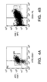


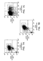


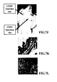

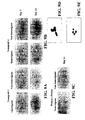


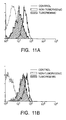
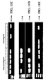
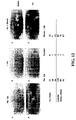
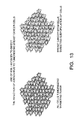

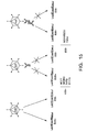

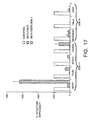

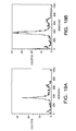

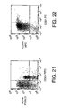
Classifications
-
- C—CHEMISTRY; METALLURGY
- C12—BIOCHEMISTRY; BEER; SPIRITS; WINE; VINEGAR; MICROBIOLOGY; ENZYMOLOGY; MUTATION OR GENETIC ENGINEERING
- C12N—MICROORGANISMS OR ENZYMES; COMPOSITIONS THEREOF; PROPAGATING, PRESERVING, OR MAINTAINING MICROORGANISMS; MUTATION OR GENETIC ENGINEERING; CULTURE MEDIA
- C12N5/00—Undifferentiated human, animal or plant cells, e.g. cell lines; Tissues; Cultivation or maintenance thereof; Culture media therefor
- C12N5/06—Animal cells or tissues; Human cells or tissues
- C12N5/0602—Vertebrate cells
- C12N5/0693—Tumour cells; Cancer cells
- C12N5/0695—Stem cells; Progenitor cells; Precursor cells
-
- A—HUMAN NECESSITIES
- A01—AGRICULTURE; FORESTRY; ANIMAL HUSBANDRY; HUNTING; TRAPPING; FISHING
- A01K—ANIMAL HUSBANDRY; CARE OF BIRDS, FISHES, INSECTS; FISHING; REARING OR BREEDING ANIMALS, NOT OTHERWISE PROVIDED FOR; NEW BREEDS OF ANIMALS
- A01K67/00—Rearing or breeding animals, not otherwise provided for; New breeds of animals
- A01K67/027—New breeds of vertebrates
- A01K67/0271—Chimeric animals, e.g. comprising exogenous cells
-
- A—HUMAN NECESSITIES
- A61—MEDICAL OR VETERINARY SCIENCE; HYGIENE
- A61K—PREPARATIONS FOR MEDICAL, DENTAL OR TOILETRY PURPOSES
- A61K39/00—Medicinal preparations containing antigens or antibodies
- A61K39/395—Antibodies; Immunoglobulins; Immune serum, e.g. antilymphocytic serum
- A61K39/39533—Antibodies; Immunoglobulins; Immune serum, e.g. antilymphocytic serum against materials from animals
- A61K39/39558—Antibodies; Immunoglobulins; Immune serum, e.g. antilymphocytic serum against materials from animals against tumor tissues, cells, antigens
-
- A—HUMAN NECESSITIES
- A61—MEDICAL OR VETERINARY SCIENCE; HYGIENE
- A61P—SPECIFIC THERAPEUTIC ACTIVITY OF CHEMICAL COMPOUNDS OR MEDICINAL PREPARATIONS
- A61P35/00—Antineoplastic agents
-
- C—CHEMISTRY; METALLURGY
- C12—BIOCHEMISTRY; BEER; SPIRITS; WINE; VINEGAR; MICROBIOLOGY; ENZYMOLOGY; MUTATION OR GENETIC ENGINEERING
- C12Q—MEASURING OR TESTING PROCESSES INVOLVING ENZYMES, NUCLEIC ACIDS OR MICROORGANISMS; COMPOSITIONS OR TEST PAPERS THEREFOR; PROCESSES OF PREPARING SUCH COMPOSITIONS; CONDITION-RESPONSIVE CONTROL IN MICROBIOLOGICAL OR ENZYMOLOGICAL PROCESSES
- C12Q1/00—Measuring or testing processes involving enzymes, nucleic acids or microorganisms; Compositions therefor; Processes of preparing such compositions
- C12Q1/68—Measuring or testing processes involving enzymes, nucleic acids or microorganisms; Compositions therefor; Processes of preparing such compositions involving nucleic acids
- C12Q1/6876—Nucleic acid products used in the analysis of nucleic acids, e.g. primers or probes
- C12Q1/6883—Nucleic acid products used in the analysis of nucleic acids, e.g. primers or probes for diseases caused by alterations of genetic material
- C12Q1/6886—Nucleic acid products used in the analysis of nucleic acids, e.g. primers or probes for diseases caused by alterations of genetic material for cancer
-
- G—PHYSICS
- G01—MEASURING; TESTING
- G01N—INVESTIGATING OR ANALYSING MATERIALS BY DETERMINING THEIR CHEMICAL OR PHYSICAL PROPERTIES
- G01N33/00—Investigating or analysing materials by specific methods not covered by groups G01N1/00 - G01N31/00
- G01N33/48—Biological material, e.g. blood, urine; Haemocytometers
- G01N33/50—Chemical analysis of biological material, e.g. blood, urine; Testing involving biospecific ligand binding methods; Immunological testing
- G01N33/5005—Chemical analysis of biological material, e.g. blood, urine; Testing involving biospecific ligand binding methods; Immunological testing involving human or animal cells
- G01N33/5008—Chemical analysis of biological material, e.g. blood, urine; Testing involving biospecific ligand binding methods; Immunological testing involving human or animal cells for testing or evaluating the effect of chemical or biological compounds, e.g. drugs, cosmetics
- G01N33/5011—Chemical analysis of biological material, e.g. blood, urine; Testing involving biospecific ligand binding methods; Immunological testing involving human or animal cells for testing or evaluating the effect of chemical or biological compounds, e.g. drugs, cosmetics for testing antineoplastic activity
-
- G—PHYSICS
- G01—MEASURING; TESTING
- G01N—INVESTIGATING OR ANALYSING MATERIALS BY DETERMINING THEIR CHEMICAL OR PHYSICAL PROPERTIES
- G01N33/00—Investigating or analysing materials by specific methods not covered by groups G01N1/00 - G01N31/00
- G01N33/48—Biological material, e.g. blood, urine; Haemocytometers
- G01N33/50—Chemical analysis of biological material, e.g. blood, urine; Testing involving biospecific ligand binding methods; Immunological testing
- G01N33/53—Immunoassay; Biospecific binding assay; Materials therefor
- G01N33/574—Immunoassay; Biospecific binding assay; Materials therefor for cancer
- G01N33/57484—Immunoassay; Biospecific binding assay; Materials therefor for cancer involving compounds serving as markers for tumor, cancer, neoplasia, e.g. cellular determinants, receptors, heat shock/stress proteins, A-protein, oligosaccharides, metabolites
- G01N33/57492—Immunoassay; Biospecific binding assay; Materials therefor for cancer involving compounds serving as markers for tumor, cancer, neoplasia, e.g. cellular determinants, receptors, heat shock/stress proteins, A-protein, oligosaccharides, metabolites involving compounds localized on the membrane of tumor or cancer cells
-
- A—HUMAN NECESSITIES
- A61—MEDICAL OR VETERINARY SCIENCE; HYGIENE
- A61K—PREPARATIONS FOR MEDICAL, DENTAL OR TOILETRY PURPOSES
- A61K39/00—Medicinal preparations containing antigens or antibodies
- A61K2039/505—Medicinal preparations containing antigens or antibodies comprising antibodies
-
- A—HUMAN NECESSITIES
- A61—MEDICAL OR VETERINARY SCIENCE; HYGIENE
- A61K—PREPARATIONS FOR MEDICAL, DENTAL OR TOILETRY PURPOSES
- A61K39/00—Medicinal preparations containing antigens or antibodies
- A61K2039/51—Medicinal preparations containing antigens or antibodies comprising whole cells, viruses or DNA/RNA
- A61K2039/515—Animal cells
- A61K2039/5152—Tumor cells
-
- A—HUMAN NECESSITIES
- A61—MEDICAL OR VETERINARY SCIENCE; HYGIENE
- A61K—PREPARATIONS FOR MEDICAL, DENTAL OR TOILETRY PURPOSES
- A61K39/00—Medicinal preparations containing antigens or antibodies
- A61K2039/51—Medicinal preparations containing antigens or antibodies comprising whole cells, viruses or DNA/RNA
- A61K2039/515—Animal cells
- A61K2039/5158—Antigen-pulsed cells, e.g. T-cells
-
- C—CHEMISTRY; METALLURGY
- C07—ORGANIC CHEMISTRY
- C07K—PEPTIDES
- C07K14/00—Peptides having more than 20 amino acids; Gastrins; Somatostatins; Melanotropins; Derivatives thereof
- C07K14/435—Peptides having more than 20 amino acids; Gastrins; Somatostatins; Melanotropins; Derivatives thereof from animals; from humans
- C07K14/705—Receptors; Cell surface antigens; Cell surface determinants
- C07K14/70585—CD44
-
- C—CHEMISTRY; METALLURGY
- C07—ORGANIC CHEMISTRY
- C07K—PEPTIDES
- C07K14/00—Peptides having more than 20 amino acids; Gastrins; Somatostatins; Melanotropins; Derivatives thereof
- C07K14/435—Peptides having more than 20 amino acids; Gastrins; Somatostatins; Melanotropins; Derivatives thereof from animals; from humans
- C07K14/705—Receptors; Cell surface antigens; Cell surface determinants
- C07K14/71—Receptors; Cell surface antigens; Cell surface determinants for growth factors; for growth regulators
-
- C—CHEMISTRY; METALLURGY
- C07—ORGANIC CHEMISTRY
- C07K—PEPTIDES
- C07K16/00—Immunoglobulins [IGs], e.g. monoclonal or polyclonal antibodies
- C07K16/18—Immunoglobulins [IGs], e.g. monoclonal or polyclonal antibodies against material from animals or humans
- C07K16/28—Immunoglobulins [IGs], e.g. monoclonal or polyclonal antibodies against material from animals or humans against receptors, cell surface antigens or cell surface determinants
- C07K16/2863—Immunoglobulins [IGs], e.g. monoclonal or polyclonal antibodies against material from animals or humans against receptors, cell surface antigens or cell surface determinants against receptors for growth factors, growth regulators
-
- C—CHEMISTRY; METALLURGY
- C07—ORGANIC CHEMISTRY
- C07K—PEPTIDES
- C07K16/00—Immunoglobulins [IGs], e.g. monoclonal or polyclonal antibodies
- C07K16/18—Immunoglobulins [IGs], e.g. monoclonal or polyclonal antibodies against material from animals or humans
- C07K16/28—Immunoglobulins [IGs], e.g. monoclonal or polyclonal antibodies against material from animals or humans against receptors, cell surface antigens or cell surface determinants
- C07K16/30—Immunoglobulins [IGs], e.g. monoclonal or polyclonal antibodies against material from animals or humans against receptors, cell surface antigens or cell surface determinants from tumour cells
-
- C—CHEMISTRY; METALLURGY
- C12—BIOCHEMISTRY; BEER; SPIRITS; WINE; VINEGAR; MICROBIOLOGY; ENZYMOLOGY; MUTATION OR GENETIC ENGINEERING
- C12N—MICROORGANISMS OR ENZYMES; COMPOSITIONS THEREOF; PROPAGATING, PRESERVING, OR MAINTAINING MICROORGANISMS; MUTATION OR GENETIC ENGINEERING; CULTURE MEDIA
- C12N2501/00—Active agents used in cell culture processes, e.g. differentation
- C12N2501/40—Regulators of development
- C12N2501/42—Notch; Delta; Jagged; Serrate
-
- C—CHEMISTRY; METALLURGY
- C12—BIOCHEMISTRY; BEER; SPIRITS; WINE; VINEGAR; MICROBIOLOGY; ENZYMOLOGY; MUTATION OR GENETIC ENGINEERING
- C12N—MICROORGANISMS OR ENZYMES; COMPOSITIONS THEREOF; PROPAGATING, PRESERVING, OR MAINTAINING MICROORGANISMS; MUTATION OR GENETIC ENGINEERING; CULTURE MEDIA
- C12N2503/00—Use of cells in diagnostics
- C12N2503/02—Drug screening
Definitions
- This invention relates to the diagnosis and treatment of cancer.
- Cancer remains the number two cause of mortality in this country, resulting in over 500,000 deaths per year. Despite advances in detection and treatment, cancer mortality remains high. Despite the remarkable progress in understanding the molecular basis of cancer, this knowledge has not yet been translated into effective therapeutic strategies.
- breast cancer is the most common cancer in American women, with approximately one in nine women developing breast cancer in their lifetime.
- metastatic breast cancer is still an incurable disease.
- Most women with metastatic breast cancer succumb to the disease.
- the invention is based upon the discovery that a small percentage of cells within an established solid tumor have the properties of "stem cells". These solid tumor “stem” cells give rise both to more solid tumor stem cells and to the majority of cells in the tumor, cancer cells that have lost the capacity for extensive proliferation and the ability to give rise to new tumors. Thus, solid tumor cell heterogeneity reflects the presence of a variety of tumor cell types that arise from a solid tumor stem cell.
- Populations of cells obtained from the solid tumor are isolated and structurally characterized using Fluorescence Activated Cell Sorting (FACS).
- FACS Fluorescence Activated Cell Sorting
- Solid tumor stem cells are the truly tumorigenic cells that are capable of re-establishing a tumor following treatment.
- the invention provides in vivo and in vitro assays of solid tumor stem cell function and cell function by the various populations of cells isolated from a solid tumor.
- the invention provides methods for using the various populations of cells isolated from a solid tumor (such as a population of cells enriched for solid tumor stem cells) to identify factors influencing solid tumor stem cell proliferation, to analyze populations of cells isolated from solid tumors for gene expression patterns or protein expression patterns, to identify new anti-cancer drug targets, to predict the sensitivity of tumors from individual patients to existing anti-cancer treatment regimens, to model anti-cancer treatment, to test new therapeutic compounds, to identify and test new diagnostic markers, to treat tumors, to produce genetically modified solid tumor stem cells, and to prepare cDNA libraries and microarrays of polynucleotides and polypeptides from solid tumor stem cells.
- the invention provides a method for consistently growing solid tumor cells in vivo.
- the invention also provides a method to grow solid tumor cells that are in single cell suspension or in small aggregates.
- the invention provides a chimeric animal (a xenograft model) in which tumors can be established from solid tumor primary cells and in which the tumors derived from these solid tumor cells can be tested.
- the invention provides tumor banks (large enough to perform substantial numbers of bioassays) derived from single solid tumor stem cells.
- the invention usefully provides methods for screening for anti-cancer agents; for the testing of anti-cancer therapies; for the development of drugs targeting novel pathways; for the identification of new anti-cancer therapeutic targets; the identification and diagnosis of malignant cells in pathology specimens; for the testing and assaying of solid tumor stem cell drug sensitivity; for the measurement of specific factors that predict drug sensitivity; and for the screening of patients (e.g., as an adjunct for mammography).
- the invention can be used as a model to test patients' tumor sensitivity to known therapies; as a model for identification of new therapeutic targets for cancer treatment; as a system to establish a tumor bank for testing new therapeutic agents for treatment of cancer; and as a system to identify the tumorigenic cancer cells.
- the invention provides synergy between the methods of the invention and breast cancer genomic databases, for an improved anti-cancer drug discovery.
- FIG. 1 shows two models of solid tumor heterogeneity.
- mutations or environmental differences cause tumor cells to adopt a variety of different phenotypes.
- Environmentally determined differences in phenotype represented by white, green, and red cells, may be reversible while mutationally determined changes in phenotype, represented by purple cells, may not be reversible.
- Many cells with a variety of different phenotypes are thought to have the potential to proliferate extensively and form new tumors.
- the tumor stem cell model ( FIG. 1B ) is distinguished by having only a minor population of tumor cells that are tumorigenic (yellow cells). These tumor stem cells are characterized by indefinite proliferative potential, the ability to form new tumors, and the ability to give rise to heterogeneous non-tumorigenic cancer cells that typically form the bulk of a tumor.
- FIG. 2 is a set of FACS plots of breast cancer tumor cells. Mice were implanted with primary breast cancer tumor cells removed from two human patients. Resultant tumors were removed from the mouse and single cell suspensions were made. Cells were stained with anti-CD44-PE, anti-520C9-APC, anti-mouse H2K-FITC (which stains infiltrating mouse cells) and Propidium Iodide (PI, which stains dead cells). Live, human CD44 + and human CD44 - cells were isolated and used for in vitro and in vivo studies.
- FIG. 3 is a set of FACS plots showing the expression of CD24 by malignant breast cells. Cells were isolated and stained as described in FIG. 2 . Mouse cells and dead cells were gated out of the analysis. The FACS plots of cells from three breast cancer tumors are shown. Note that cells from all three tumors have a similar phenotype.
- FIG. 4 is a set of FACS plots showing an analysis of tumors arising from the CD24 - cell population from human breast cancers.
- the CD24 - cells give rise to tumors that contain both CD24 + and CD24 - cells.
- secondary transplants were performed using B38.1 + CD24 - cells ( FIG. 4A ).
- the resultant tumors were removed and the cells were re-analyzed with respect to B38.1 and CD24 expression.
- FIG. 4B As predicted by the stem cell model, cells obtained from a tumor arising from transplanted B38.1 + CD24 - cells were heterogeneous with respect to expression of both B38.1 and CD24 ( FIG. 4B ).
- the marker expression pattern of the cells isolated from the tumor initiated by the B38.1 + CD24 - cells was similar to that of the original tumor ( FIG. 4 ).
- FIG. 5 is a FACS plot showing an analysis of Notch 4 expression.
- Cells were isolated from a mouse xenograft tumor ( see , below) and stained with antibodies. Malignant cells were analyzed for expression of B38.1 and Notch 4. Mouse cells and dead cells were gated out of the analysis.
- FIG. 6 shows the fractionation of breast cancer cells based upon CD44 expression.
- Tumor T1 cells FIG. 6A, FIG. 6C, and FIG. 6E
- Tumor T2 cells FIG. 6B, FIG. 6D, and FIG. 6F
- FIG. 6B, FIG. 6D, and FIG. 6F were stained with anti-CD44-PE, anti-mouse H2K-FITC and the viability dye 7AAD.
- Flow cytometry was used to isolate live, human (H2K-) cells that were either CD44 + ( FIG. 6C, FIG. 6D ) or CD44 - ( FIG. 6E, FIG. 6F ).
- Dead cells (7AAD + ) were eliminated from all analyses.
- FIG. 6B are dot plots of the unfractionated T1 and T2 cells showing CD44 and H2K expression as indicated.
- FIG. 7 shows the isolation of tumorigenic cells.
- Flow cytometry was used to isolate subpopulations of Tumor T1 ( FIG. 7A , FIG. 7D , and FIG. 7G ), Tumor T2 ( FIG. 7B , FIG. 7E, and FIG. 7F ) or Tumor T5 cells ( FIG. 7C , FIG. 7F , and FIG. 7I ) that were tested for tumorigenicity in NOD/SCID mice.
- T1 and T2 cells had been passaged once in NOD/SCID mice while T5 cells were obtained from material that had been frozen immediately after removal from a patient.
- FIG. 7A, FIG. 7B, and FIG. 7C show unfractionated tumor cells.
- FIG. 7I shows a representative tumor in a mouse at the B38.1 + CD44 + CD24 -/lo LINEAGE- injection site, but not at the B38.1 + CD44 + CD24 + LINEAGE- injection site. Histology performed on tissue from the CD24 + ( FIG. 7K , 20x objective magnification) and CD24 -/lo ( FIG. 7L , 40x objective magnification) injection sites exhibited normal mouse tissue and malignant cells respectively.
- FIG. 8 shows the enrichment of tumorigenic cells based upon ESA expression.
- Flow cytometry was used to isolate subpopulations of Tumor T1 cells that were tested for tumorigenicity in NOD/SCID mice. T1 cells had been passaged once in NOD/SCID mice. Cells were stained with anti-B38.1-APC, anti-CD24-PE, anti-ESA-FITC, anti-LINEAGE-Cytochrome, anti-mouse-H2K-Cytochrome (T1), and 7AAD. Dead cells (7AAD + ), mouse cells (H2K + ) and LINEAGE + cells were eliminated from the analysis. The dot plot in FIG.
- FIG. 8A depicts the CD24 and ESA staining pattern of live human B38.1 + LINEAGE- cells.
- the tumorigenic population is boxed and marked with an arrow.
- FIG. 8B the ESA + B38.1 + CD24 -/lo LINEAGE- cells (left panel) and the remaining LINEAGE - H2K - cells (right panel) were collected using flow cytometry.
- FIG. 9 is the results of an in vitro clonogenic assay. Flow cytometry was used to isolate tumorigenic cell or the rest of the non-tumorigenic neoplastic (non-tumorigenic cells) as described. The cells were placed in tissue culture medium containing soluble Delta for the indicated number of days. The tumorigenic and non-tumorigenic xenograft Tumor 1 (T1) ( FIG. 9A ), Tumor 4 (T4) ( FIG. 9B ) or primary patient ( FIG. 9C ) cells are shown at the indicated time after being placed in tissue culture. T4 cells were stained with Papanicolaou stain and examined under light microscopy (100x objective). Note that both the non-tumorigenic ( FIG.
- tumorigenic populations consist of neoplastic cells with large nuclei and prominent nucleoli. Note that the number of cells that attached to the tissue culture plate is similar in both populations, but that the tumorigenic population always gave rise to colonies. Non-tumorigenic populations do not give rise to established colonies (or only for brief periods, about 2-6 days).
- FIG. 10 is a set of dot plots showing the phenotypic diversity in tumors arising from B38.1 + CD44 + CD24 -/lo LINEAGE - cells.
- the dot plots depict the CD24 and CD44 staining patterns of live human LINEAGE - cells from Tumor T1 ( FIG. 10A - FIG. 10C ) or Tumor T2 ( FIG. 10D - FIG. 10F ).
- FIG. 10A and FIG. 10D show unfractionated T1 or T2 cells obtained from tumors that had been passaged once in NOD/SCID mice.
- B38.1 + CD44 + CD44 -/lo LINEAGE - cells from T1 ( FIG. 10B ) or T2 ( FIG. 10E ) were isolated as described in FIG.
- FIG. 10B and FIG. 10E depict analyses of the tumors that arose from these B38.1 + CD44 + CD24 -/lo LINEAGE - cells. Note that in both cases, the B38.1 + CD44 + CD24 -/lo LINEAGE - cells formed tumors that contained a phenotypically diverse population of cells similar to that observed in the original tumor.
- FIG. 11 shows the expression of HER2/neu and EGF-R.
- Flow cytometry was used to isolate subpopulations of Tumor T1 cells that had been passaged once in NOD/SCID mice.
- Cells were stained with, in FIG. 11A , anti-EGF-R-PE, anti-B38.1-APC, anti-CD24-FITC, anti-LINEAGE-Cytochrome, anti-mouse-H2K-Cytochrome, and 7AAD or, in FIG. 11B anti-HER2/neu-FITC, anti-B38.1-APC, anti-CD24-PE, anti-LINEAGE-Cytochrome, anti-mouse-H2K-Cytochrome, and 7AAD.
- Dead cells (7AAD + ), mouse cells (H2K + ) and LINEAGE + cells were eliminated from all analyses.
- the histogram in FIG. 11A depicts the EGF-R expression of the unstained cells (dotted line), B38.1 + CD24 - LINEAGE - tumorigenic population (shaded) and the B38 + CD24 + LINEAGE - non-tumorigenic (solid line) population.
- the histogram in FIG. 11B shows HER2/neu expression of the unstained cells (dotted line), B38.1 + CD24 - LINEAGE-tumorigenic population (shaded) and the B38 + CD24 + LINEAGE - non-tumorigenic (solid line) population.
- RT-PCR was performed using nested primers to detect EGF-R ( FIG. 11C and FIG. 11D ) or to detect HER2/neu ( FIG. 11E ).
- EGF-R is expressed at lower levels in tumorigenic cells than in non-tumorigenic cells at both the protein ( FIG. 11A ) and mRNA levels ( FIG. 11C, FIG. 11D ).
- FIG. 12 is a photomicrograph of breast cancer cells placed in tissue culture after exposure to an anti-Notch 4 antibody.
- Cells were incubated on ice for one hour in HBSS with or without soluble Delta but no anti-Notch 4 antibody, with anti-Notch 4 antibody, or with anti-Notch 4 antibody that had been preincubated with the peptide used to generate the antibody. The number of colonies that formed in the triplicate experiments is shown.
- Soluble Delta was added to the culture.
- FIG. 13 is a schematic diagram of B38.1 + cells within a tumor.
- the breast cancer stem cells from multiple patients are B38.1 + .
- these cells can be targeted with a vector.
- FIG. 14 is a description of the method for obtaining the bi-specific conjugate and the chemical modifications introduced in the antibodies.
- FIG. 15 is a strategy for re-targeting Adenovirus.
- the LaZ virus can infect most of the cells from a tumor. After the LaZ virus is incubated only with the anti-fiber antibody, the LaZ virus loses ability to infect all of the cells. After the LaZ virus is incubated with the bi-specific conjugate, the B38.1 moiety of the molecule allows the attachment of the virus to the B38.1 + cells, so only these cells are infected.
- FIG. 16 shows the targeting of breast cancer stem cells with the bi-specific antibody.
- AdLaZ is an E1-deleted Adenovirus that expresses the ⁇ -galactosidase gene
- the virus was incubated with the anti-fiber antibody for 30 min before infection (yellow columns).
- the virus was incubated with the bi-specific conjugate (green columns).
- the monolayers were fixed and incubated with X-Gal-contained buffer.
- the infected cells are blue, and the graphic shows the percentage of blue cells obtained, relative to the control infection ( i . e ., the reduction or increase in infectivity of the virus after incubation with the different antibodies).
- FIG. 17 shows that the bispecific antibody can target an adenovirus vector to breast cancer stem cells.
- the columns represent the absolute number of infected cells per field.
- Gray The indicated cells infected with the control adenovirus.
- Yellow the indicated cells were infected with the adenovirus that had been incubated with the anti-fiber antibody.
- Green The indicated cells were incubated with the bi-specific conjugate antibody.
- Red Cells were infected with the virus that had been incubated with the bi-specific conjugate antibody, but the cells were pre-treated with an excess of B38.1 antibody.
- FIG. 18 is a photograph of some of the cell monolayers after X-Gal staining.
- the infected cells appear like dark dots in this black and white picture (the ⁇ -galactosidase gene of the LaZ virus has a nuclear localization signal.
- the staining is in the nuclei of the cells.
- FIG. 19 is an analysis if different populations of cells in a breast cancer.
- ESA + CD44 + CD24 -/lo LINEAGE - cells breast cancer stem cells FIG. 19A
- ESA + CD44 + CD24 + LINEAGE- non-tumorigenic cells FIG. 19B
- the cells were stained with Hoechst 33342 as described by Eaves and colleagues ( Glimm H et al., Blood. 96(13): 4185-93 (2000 )).
- the histogram for the breast cancer stem cells is shaded. Note that the breast cancer stem cells and the non-tumorigenic cells are distributed in all phases of the cells cycle.
- FIG. 20 is a further analysis if different populations of cells in a breast cancer.
- CD44 + CD24 -/lo LINEAGE- cells breast cancer stem cells and non-tumorigenic CD44 + CD24 + LINEAGE - non-tumorigenic cells were obtained as described in FIG. 7 .
- the cells were stained with Rhodamine 123 as described by Spangrude et al., Blood 85(4):1006-16, 1995 ).
- the histogram for the breast cancer stem cells is shaded. Note that the breast cancer stem cells tend to stain less intensely with Rhodamine 123.
- FIG. 21 is an analysis of ascites fluid for ovarian cancer stem cells.
- Cells were stained with anti-B38.1-APC, anti-CD44-PE, anti-CD24-FITC, anti-Lineage-Cytochrome and 7AAD.
- Dead cells (7AAD + , and LINEAGE + cells were eliminated from the analyses. Note that there is a distinct CD44 + CD24 -/lo LINEAGE - population of cells that resembles the breast cancer stem cells.
- FIG. 22 is an analysis of sarcoma cells for solid tumor stem cells.
- P1 sarcoma cells growing in the xenograft model were stained with anti-B38.1-APC, anti-CD44-PE, anti-CD24-FITC, anti- LINEAGE-Cytochrome, anti-H2K-Cytochrome and 7AAD.
- Dead cells (7AAD + ), LINEAGE + cells and mouse cells were eliminated from the analyses. Note that the lineage cocktail in this analysis did not include CD 10, CD31 or CD 140b. Also note that there is a distinct CD44 + CD24 -/lo LINEAGE - population of cells.
- Solid tumors are composed of heterogeneous cell populations.
- breast cancers are a mixture of cancer cells and normal cells, including mesenchymal (stromal) cells, inflammatory cells, and endothelial cells.
- stromal mesenchymal
- Classic models hold that phenotypically distinct cancer cell populations all have the capacity to proliferate and give rise to a new tumor ( FIG. 1A ).
- tumor cell heterogeneity results from environmental factors as well as ongoing mutation within cancer cells resulting in diverse populations of tumorigenic cells and all populations of cells would have similar tumorigenic potential.
- This invention is based upon an alternative model of solid tumor cell heterogeneity, in which a solid tumor results from a "solid tumor stem cell” (or “cancer stem cell” from a solid tumor) and the subsequent chaotic development of the solid tumor stem cell.
- solid tumors contain a distinct, limited (or possibly rare) subset of cells that share the properties of normal "stem cells", in that they proliferate extensively or indefinitely and that they efficiently give rise to additional solid tumor stem cells.
- solid tumor stem cells proliferate extensively and form new tumors, but solid tumor stem cells proliferate extensively and give rise to additional solid tumor stem cells as well as to other tumor cells that lack tumorigenic potential. It is this solid tumor stem cell population that proliferates and ultimately proves fatal.
- stem cells During normal animal development, cells of most or all tissues are derived from normal precursors, called stem cells ( Morrison et al., Cell 88(3): 287-98 (1997 ); Morrison et al., Curr. Opin. Immunol. 9(2): 216-21 (1997 ); Morrison et al., Annu. Rev. Cell. Dev. Biol. 11: 35-71 (1995 )).
- stem cell is known in the art to mean (1) that the cell is a cell capable of generating one or more kinds of progeny with reduced proliferative or developmental potential; (2) that the cell has extensive proliferative capacity; and (3) that the cell is capable of self-renewal or self-maintenance (see, Potten et al., Development 110: 1001 (1990 ); U.S. Pat. Nos. 5,750,376 , 5,851,832 , 5,753,506 , 5,589,376 , 5,824,489 , 5,654,183 , 5,693,482 , 5,672,499 , and 5,849,553 , all incorporated by reference).
- hematopoietic stem and progenitor cells The best-known example of adult cell renewal by the differentiation of stem cells is the hematopoietic system (see, U.S. Pat. Nos. 5,061,620 , 5,087,570 , 5,643,741 , 5,821,108 , 5,914,108 , each incorporated by reference).
- Developmentally immature precursors hematopoietic stem and progenitor cells
- Stem cells are also found in other tissues, including epithelial tissues (see, Slack, Science 287: 1431 (2000 )) and mesenchymal tissues. (see, U.S. Pat. No. 5,942,225 ; incorporated by reference).
- solid tumor stem cells examples include sarcomas and carcinomas such as, but not limited to: fibrosarcoma, myxosarcoma, liposarcoma, chondrosarcoma, osteogenic sarcoma, chordoma, angiosarcoma, endotheliosarcoma, lymphangiosarcoma, lymphangioendotheliosarcoma, synovioma, mesothelioma, Ewing's tumor, leiomyosarcoma, rhabdomyosarcoma, colon carcinoma, pancreatic cancer, breast cancer, ovarian cancer, prostate cancer, squamous cell carcinoma, basal cell carcinoma, adenocarcinoma, sweat gland carcinoma, sebaceous gland carcinoma, papillary carcinoma, papillary adenocarcinomas, cyst
- Solid tumor stem cells are defined structurally and functionally as described herein; using the methods and assays similar to those described below. Because tumor cells are known to evolve phenotypically and functionally over time as additional genetic mutations occur, the solid tumor stem cells may change phenotypically and functionally over time in an individual patient. Nevertheless, one can use the method of the invention, employing the markers disclosed herein, which are consistently useful in the isolation or identification of solid tumor stem cells in a majority of patients.
- solid tumor stem cells undergo “self-renewal” and “differentiation” in a chaotic development to form a tumor, give rise to abnormal cell types, and may change over time as additional mutations occur.
- the functional features of a solid tumor stem cell are that they are tumorigenic, they give rise to additional tumorigenic cells ("self-renew”), and they can give rise to non-tumorigenic tumor cells ("differentiation").
- Solid tumor stem cells can vary between different types of solid tumor cancers.
- Solid tumor stem cells may arise either as a result of genetic damage that deregulates the proliferation and differentiation of normal stem cells ( Lapidot et al., Nature 367(6464): 645-8 (1994 )) or by the dysregulated proliferation of a normal restricted progenitor or a normal differentiated cell type.
- solid tumors are visualized and initially identified according to their locations, not by their developmental origin.
- a non-tumorigenic cell from a solid tumor is a cell from a population that fails to form a palpable tumor upon transplantation into an immunocompromised mouse, wherein if the same number of unfractionated, dissociated tumor cells were transplanted under the same circumstances, the solid tumor stem cells would form a palpable tumor in the same period of time.
- non-tumorigenic cells are depleted for tumor forming activity in an animal model.
- a "palpable tumor” is known to those in the medical arts as a tumor that is capable of being handled, touched, or felt.
- the invention provides several novel uses:
- the solid tumor stem cells of the model of the invention differs from the "cancer stem line" provided by U.S. Pat. 6,004,528 .
- the "cancer stem line” is defined as a slow growing progenitor cell type that itself has few mutations but which undergoes symmetric rather than asymmetric cell divisions as a result of tumorigenic changes that occur in the cell's environment.
- This "cancer stem line” hypothesis thus proposes that highly mutated, rapidly proliferating tumor cells arise largely as a result of an abnormal environment, which causes relatively normal stem cells to accumulate and then undergo mutations that cause them to become tumor cells.
- U.S. Pat. 6,004,528 proposes that such a model can be used to enhance the diagnosis of cancer.
- solid tumor stem cell model is fundamentally different than the "cancer stem line” model and as a result exhibits utilities not offered by the "cancer stem line” model.
- solid tumor stem cells are not “mutationally spared”.
- the "mutationally spared cancer stem line" described by U.S. Pat. No. 6,004,528 may be considered a pre-cancerous lesion, while the solid tumor stem cells described of this invention are cancer cells that themselves contain the mutations that are responsible for tumorigenesis. That is, the solid tumor stem cells (“cancer stem cells”) of the invention would be included among the highly mutated cells that are distinguished from the "cancer stem line" in U.S. Pat. 6,004,528 .
- solid tumor stem cell model predicts that isolated solid tumor stem cells can give rise to additional tumors upon transplantation (thus explaining metastasis) while the "cancer stem line” model would predict that transplanted "cancer stem line” cells would not be able to give rise to a new tumor, since it was their abnormal environment that was tumorigenic. Indeed, the ability to transplant dissociated, and phenotypically isolated human solid tumor stem cells to mice (into an environment that is very different from the normal tumor environment), where they still form new tumors, distinguishes the present invention from the "cancer stem line” model.
- solid tumor stem cells likely divide both symmetrically and asymmetrically, such that symmetric cell division is not an obligate property.
- solid tumor stem cells may divide rapidly or slowly, depending on many variables, such that a slow proliferation rate is not a defining characteristic.
- solid tumor stem cells can be operationally characterized by cell surface markers. These cell surface markers can be recognized by reagents that specifically bind to the cell surface markers. For example, proteins, carbohydrates, or lipids on the surfaces of solid tumor stem cells can be immunologically recognized by antibodies specific for the particular protein or carbohydrate (for construction and use of antibodies to markers, see, Harlow, Using Antibodies: A Laboratory Manual (Cold Spring Harbor Press, Cold Spring Harbor, New York, 1999 ); see also, EXAMPLES).
- the set of markers present on the cell surfaces of solid tumor stem cells (the "cancer stem cells" of the invention) and absent from the cell surfaces of these cells is characteristic for solid tumor stem cells. Therefore, solid tumor stem cells can be selected by positive and negative selection of cell surface markers.
- a reagent that binds to a solid tumor stem cell is a "positive marker” (i. e., a marker present on the cell surfaces of solid tumor stem cells) that can be used for the positive selection of solid tumor stem cells.
- a reagent that binds to a solid tumor stem cell "negative marker” i.e., a marker not present on the cell surfaces of solid tumor stem cells but present on the surfaces of other cells obtained from solid tumors
- the discrimination between cells based upon the detected expression of cell surface markers is by comparing the detected expression of the cell surface marker as compared with the mean expression by a control population of cells.
- the expression of a marker on a solid tumor stem cell can be compared to the mean expression of the marker by the other cells derived from the same tumor as the solid tumor stem cell.
- Other methods of discriminating among cells by marker expression include methods of gating cells by flow cytometry based upon marker expression (see, Givan A, Flow Cytometry: First Principles, (Wiley-Liss, New York, 1992 ); Owens MA & Loken MR., Flow Cytometry: Principles for Clinical Laboratory Practice, (Wiley-Liss, New York, 1995 )).
- Solid tumor stem cell positive markers may also be present on cells other than solid tumor stem cells.
- Solid tumor stem cell negative markers may also be absent from cells other than solid tumor stem cells. While it is rare to identify a single marker that identifies a stem cell, it has often been possible to identify combinations of positive and negative markers that uniquely identify stem cells and allow their substantial enrichment in other contexts. Morrison et al., Cell 96(5): 737-49 (1999 ); Morrison et al., Proc. Natl. Acad. Sci. USA 92(22): 10302-6 (1995 ); Morrison & Weissman, Immunity 1(8): 661-73 (1994 ).
- a "combination of reagents” is at least two reagents that bind to cell surface markers either present (positive marker) or not present (negative marker) on the surfaces of solid tumor stem cells, or to a combination of positive and negative markers ( see, EXAMPLES 7 and 8, TABLE 6).
- the use of a combination of antibodies specific for solid tumor stem cell surface markers results in the method of the invention being useful for the isolation or enrichment of solid tumor stem cells from a variety of solid tumors, including sarcomas, ovarian cancers, and breast tumors.
- Guidance to the use of a combination of reagents can be found in PCT patent application WO 01/052143 (Morrison & Anderson ), incorporated by reference.
- solid tumor stem cells can be isolated from any animal solid tumor, particularly any mammalian solid tumor. It will be appreciated that, taking into consideration factors such as a binding affinities, that antibodies that recognize species-specific varieties of markers are used to enrich for and select solid tumor stem cells. Antibodies that recognize the species-specific varieties of CD44, B38.1, CD24 and other markers will be used to enrich for or isolate solid tumor stem cells from that species (for example, antibody to a mouse CD44 for mouse solid tumor stem cells, antibody to a monkey B38.1 for monkey solid tumor stem cells, etc. ) .
- the invention provides a xenograft model in which to establish tumors by the injection of solid tumor cells into a host animal.
- the host animal can be a model organism such as nematode, fruit fly, zebrafish; preferably a laboratory mammal such as a mouse (nude mouse, SCID mouse, NOD/SCID mouse, Beige/SCID Mouse), rat, rabbit, or primate.
- the severely immunodeficient NOD-SCID mice were chosen as recipients to maximize the participation of injected cells. Immunodeficient mice do not reject human tissues, and SCID and NOD-SCID mice have been used as hosts for in vivo studies of human hematopoiesis and tissue engraftment.
- Xenograft tumors have been established from mastectomy specimens of all the patients that have been tested to date (see, EXAMPLE 7). Tumors in mice have also been established from malignant pleural effusions. In addition, tumors have been established by the subcutaneous injection of cells that have been obtained from two sarcomas. Furthermore, for all the tumors that we have attempted, we have been able to make single-cell suspensions (or suspensions with a few aggregates of cell, such as less than 100; preferably less than 10) and then transfer the tumors. This xenograft assay is useful for biological and molecular assays to characterize the tumorigenic, clonigenic solid tumor stem cells.
- the NOD/SCID or Beige/SCID mice can be further immunosuppressed, using VP-16 ( see , EXAMPLES 1 and 3), radiation therapy, chemotherapy, or other immunosuppressive biological agents.
- breast cancer cells are injected into immunocompromised mice, to grow the tumor.
- the cells are injected either into the mammary fat pads of mice or subcutaneously into the mice.
- tumors can be established from single-cell suspensions (or suspensions with a few aggregates of cell, such as less than 100; preferably less than 10) and then the tumors transferred to other mice.
- the limitations in the assays made it impossible to determine whether the colonies arose from stem cells that had lost their capacity to proliferate in vitro ,from non-tumorigenic cells that had limited proliferative potential, or whether the small number of cells able to form colonies in vitro was due to a "stem cell” population within the tumor or due to a rare cell that could proliferate in vitro. Furthermore, it was not possible to distinguish phenotypically different populations of cells: prior to this invention, very limited use was made of techniques like flow-cytometry to separate and analyze phenotypically distinct populations of solid tumor cells by flow-cytometry. Indeed, the clonogenic assays used in the prior art did not predict the behavior of an individual patient's tumor and fell out of favor.
- Notch Role of Notch in breast cancer.
- the Notch family of receptors has been implicated in stem cell development and differentiation (see, Morrison et al, Cell 101(5): 499-510 (2000 ); Artavanis-Tsakonas et al., Science 284: 770 (1999 ); and Artavanis-Tsakonas et al., Science 268: 225-232 (1995 ); U.S. Pat. No. 6,090,922 , incorporated by reference).
- Notch was originally identified in Drosophila through loss-of function mutations that produced too many neurons at the expense of other cell types. Poulson, Proc. Natl. Acad. Sci. USA 23: 133 (1937 ).
- Notch In all animal models tested, mutations in the Notch receptor result in developmental abnormalities. In C. elegans, Notch is required for germ line stem cell self-renewal. Berry et al., Development 124(4): 925-36 (1997 ). In rats, Notch regulates neural crest stem cell differentiation. Morrison et al., Cell 101 (5): 499-510 (2000 ). Transient Notch activation initiates an irreversible switch from neurogenesis to gliogenesis by neural crest stem cells.
- neighboring cells can express Notch receptors and ligands, one cell can affect the fate of a neighboring cell by activating Notch signaling in the neighboring cell.
- Proteins with knife-edge names such as Jagged ( Shimizu et al., Journal of Biological Chemistry 274(46) 32961-9 (1999 ); Jarriault et al., Molecular and Cellular Biology 18: 7423-7431 (1998 )), Serrate, and Delta (and variants of each, such as Delta1, Delta2, Delta3, Delta4, and Jagged2, LAG-2 and APX-1 in C. elegans ) , bind to the Notch receptor and activate a downstream signaling pathway that prevents neighboring cells from becoming neural progenitors.
- a recently identified ligand is D114, a Notch ligand of the Delta family expressed in arterial endothelium. Shutter et al., Genes Dev 14(11): 1313-8 (2000 )).
- Notch ligands may bind and activate Notch family receptors promiscuously.
- the expression of other genes like Fringe family members ( Panin et al, Nature 387(6636): 908-912 (1997 )), may modify the interactions of Notch receptors with Notch ligands. Numb family members may also modify Notch signaling intracellularly.
- Ligand binding to Notch results in activation of a presenilin-1-dependent gamma-secretase-like protein that cleaves Notch.
- De Strooper et al. Nature 398: 518-522 (1999 ), Mumm et al., Molecular Cell. 5: 197-206 (2000 ).
- Cleavage in the extracellular region may involve a furin-like convertase.
- Logeat et al. Proceedings of the National Academy of Sciences of the USA 95: 8108-8112 (1998 ).
- the intracellular domain is released and transactivates genes by associating with the DNA binding protein RBP-J. Kato et al., Development 124: 4133 -4141 (1997 )).
- Notch 1 Notch 2 and Notch 4 are thought to transactivate genes such as members of the Enhancer of Split (HES) family, while Notch 3 signaling may be inhibitory. Beatus et al., Development 126: 3925-3935 (1999 ). Finally, secreted proteins in the Fringe family bind to the Notch receptors and modify their function. Zhang & Gridley, Nature 394 (1998 ).
- HES Enhancer of Split
- Notch 4 is the human ortholog of the mouse int-3 oncogene that plays a role in breast cancer in mice.
- the invention is based upon the discovery that Notch 4 plays a role both in normal human breast development and in tumorigenesis. Within an individual tumor, only a small subpopulation of tumorigenic cells expresses high levels of Notch 4.
- An antibody that recognizes Notch 4 blocks the growth of breast cancer tumor cells in vitro and in vivo (see, EXAMPLES 2, 5, 12 and 15).
- the antibody binds to the extracellular domain of Notch 4.
- the antibody binds to the polypeptide region LLCVSVVRPRGLLCGSFPE (LeuLeuCysValSerValValArgProArgGlyLeuLeuCysGlySerPheProGlu) (SEQ ID NO:1).
- any anti-Notch 4 antibody that inhibits Notch activation can be used to impair tumor survival.
- Inhibitors of Notch signaling can be used in the methods of the invention to inhibit solid tumor stem cells.
- the Notch pathway is modified to kill or inhibit the proliferation of solid tumor stem cells.
- the invention differs from the manipulation of non-terminally differentiated cells using the Notch pathway provided in U.S. Pat. No. 5,780,300 .
- U.S. Pat. No. 5,780,300 addresses the modification of normal cells, not cancer cells. That patent is directed to methods for the expansion of non-terminally differentiated cells (normal precursor cells) using agonists of Notch function, by inhibiting the differentiation of the cells without inhibiting proliferation (mitotic activity) such that an expanded population of non-terminally differentiated cells is obtained.
- These expanded cells can be used in cell replacement therapy, a use that is incompatible with the goal of killing or inhibiting the proliferation of solid tumor stem cells by modifying Notch signaling in this invention.
- a corollary to the solid tumor stem cell model of the invention is that, to effectively treat cancer and achieve higher cure rates, anti-cancer therapies must be directed against solid tumor stem cells. Since current therapies are directed against the bulk population, they may be ineffective at eradicating solid tumor stem cells. The limitations of current cancer therapies derive from their inability to effectively kill solid tumor stem cells. The identification of solid tumor stem cells permits the specific targeting of therapeutic agents to this cell population, resulting in more effective cancer treatments. This concept would fundamentally change our approach to cancer treatment.
- genomics provide a powerful means for identifying drug targets in cancer cells, these targets are only valid if the targets are present within the tumorigenic cell population.
- genomics must be focused on individual populations within the heterogeneous cells that compose a tumor that are responsible for tumorigenic growth. In solid tumors, these are the solid tumor stem cells. Additionally, genomics has not yet been used to identify genes expressed in purified cell populations derived from cancerous tissues.
- flow cytometry such as FACS
- the xenograft animal model of the invention to enrich for specific cell populations.
- This technique has the advantage of being able to simultaneously isolate phenotypically pure populations of viable normal and tumor cells for molecular analysis.
- flow cytometry allows us to test the functions of the cell populations and use them in biological assays in addition to studying their gene expression profiles.
- such cells can also be characterized in biological assays.
- mesenchymal (stromal) cells can be analyzed for production of growth factors, matrix proteins and proteases, endothelial cells can be analyzed for production of specific factors involved in solid tumor growth support (such as neo-vascularization), and different subsets of tumor cells from a solid tumor can be isolated and analyzed for tumorigenicity, drug resistance and metastatic potential.
- Enriched as in an enriched population of cells, can be defined based upon the increased number of cells having a particular marker in a fractionated set of cells as compared with the number of cells having the marker in the unfractionated set of cells.
- the term "enriched can be preferably defined by tumorigenic function as the minimum number of cells that form tumors at limit dilution frequency in test mice.
- tumorigenic function as the minimum number of cells that form tumors at limit dilution frequency in test mice.
- FACS methods using CD44 alone can enrich solid tumor stem cells at least 2-fold ( see , EXAMPLE 1 and 3).
- FACS methods using B38.1 and CD24 can enrich for solid tumor stem cells 5-6 fold (see, EXAMPLE 3).
- Enrichment using additional markers can enrich 10-fold or more and can be used to isolate solid tumor stem cells.
- Isolated refers to a cell that is removed from its natural environment (such as in a solid tumor) and that is isolated or separated, and is at least about 75% free, and most preferably about 90% free, from other cells with which it is naturally present, but which lack the marker based on which the cells were isolated.
- the tumorigenic cells from breast cancer. Focusing on the individual populations of cells within a solid tumor provides a clearer understanding of how to focus new cancer treatments and identify novel targets for drug discovery.
- purifying solid tumor stem (such as breast cancer tumorigenic) cells provides a material for screening for drug sensitivity and identifying markers that predict tumorigenicity or metastatic potential.
- the in vivo proliferation of solid tumor stem cells can be accomplished by injection of solid tumor stem cells into animals, preferably mammals, more preferably in rodents such as mice (due to the predictable methods that have been developed in the art for injection into laboratory rodents), and most preferably into immunocompromised mice, such as SCID mice, Beige/SCID mice or NOD/SCID mice (see, EXAMPLES).
- NOD/SCID mice are injected with the varying number of cells and observed for tumor formation.
- the injection can be by any method known in the art, following the enrichment of the injected population of cells for solid tumor stem cells.
- Ketamine/Xylazine 300 mg Ketamine combined with 20 mg Xylazine in a 4 ml volume. Then, 0.02 ml of the solution was diluted in HBSS is used per 20 g mouse. Mice were then treated with VP-16 (etoposide) via an intraperitoneal injection (30 mg etoposide per 1 kg, diluted in serum-free HBSS for a final injection volume of 0.2 ml).
- estrogen pellets were placed subcutanously on the back of the necks of the mice using a trocar. The mice were then warmed and placed back in to the cages after they awoke. All tumor injections/implantations were done 3-5 days after this procedure.
- pleural effusions or dissociated solid tumor cells For the injection of the pleural effusions or dissociated solid tumor cells, cells were received shortly after surgery and washed with HBSS serum-free. Cells were then suspended in serum free-RPMI/Matrigel mixture (1:1 volume) and then injected into the upper right and left mammary pads using an 18G needle. To do this, the desired number of cells were suspended in 0.2 ml and injected. The site of the needle injection was sealed with Nexaban to prevent any cell leakage.
- tumors from a patient solid tumors or grown in mice (by the methods of the invention) were cut up into small pieces and then minced completely using sterile blades. The resulting pieces were then mixed with ultra-pure Collagenase III in HBSS solution (200-250 U collagenase/ml) and allowed to incubate at 37C for 3-4 hr, pipetting with a 10 ml pipette is done every 15-20 minutes. At the end of the incubation, cells were filtered through a 45-micron nylon mesh and washed with RPMI-20%FBS, then washed with HBSS twice. Cells to be injected were then suspended in HBSS/Matrigel mix (1:1 volume) and injected into the mammalian fat pad or subcutaneously as described above. Nexaban can be used to seal the injection site.
- a solid tumor is removed from the mice and made into a single cell suspension.
- Cells are stained and analyzed by flow cytometry (FACS) using methods known to those skilled in the art ( Morrison & Weissman, Immunity 1(8): 661-73 (1994 )).
- the phenotype of tumorigenic cells is CD44 + CD24 -/lo in all tumors, and B38.1 + CD44 + CD24 -/lo in most tumors.
- Cells are stained with 7AAD (which stains dead cells), anti H2K-PE (which stains mouse cells), and combinations of antibodies against various markers that have heterogeneous expression patterns by the cancer cells including anti-B38.1, -annexin V, -Notch 4, -CD9, -CD24, -MUC1, -CD49F, -CD62P, -P-glycoprotein, -Notch 1, -520C9, -260F9 and -317G5.
- FACS is used to isolate viable human cells that either do or do not express one of the differentially expressed antigens.
- a combination of markers allows the greatest enrichment of tumorigenic cells. For the limiting dilution assays, one hundred, one thousand, ten thousand and one hundred thousand cells of each population are analyzed in vivo.
- SCID mice, NOD/SCID mice or Beige/SCID mice are injected with the varying number of cells and observed for tumors. Any tumors that form are removed for pathologic examination and FACS analysis. The tests are repeated (for example, about ten times) to confirm the results. The phenotypes of the tumorigenic cells are thus determined.
- Suitable routes may include parenteral delivery, including intramuscular, subcutaneous (see, above), intramedullary injections, as well as intrathecal, direct intraventricular, intravenous, intraperitoneal, intranasal, or intraocular injections, just to name a few.
- the agents of the invention may be formulated in aqueous solutions, preferably in physiologically compatible buffers such as Hanks's solution, Ringer's solution, or physiological saline buffer.
- penetrants appropriate to the barrier to be permeated are used in the formulation. Such penetrants are generally known in the art.
- the invention is an improvement over the methods of Mueller & Reisfeld, Cancer Metastasis Rev. 10: 193-200 (1991 ) (who used the SCID mouse, which allows disseminated growths for a number of human tumors, particularly hematologic disorders and malignant melanoma) and Sakakibara et al., Cancer- J. Sci. Am. 2: 291-300 (1996 ) (who studied the growth and metastatic potential of surgical specimens of breast carcinomas engrafted into the large abdominal (gonadal) fat pad of severe combined immunodeficient (SCID) mice).
- the injection of solid tumor stem cells can consistently result in the successful establishment of tumors, more than 75% of the time, preferably more than 80% of the time, more preferably more than 85%, more than 90%, or more than 95% of the time.
- the invention provides for the advantageous establishment of solid tumors (particularly tumors from breast tumor stem cells) in mammary fat pads, an area not accessable for establishment using the methods of Sakakibara et al., Cancer J. 2: 291-300 (1996 ).
- Cells can be obtained from solid tumor tissue by dissociation of individual cells. Tissue from a particular tumor is removed using a sterile procedure, and the cells are dissociated using any method known in the art (see, Sambrook et al., Molecular Cloning: A Laboratory Manual (Cold Spring Harbor Press, Cold Spring Harbor, New York, 1989 ); Current Protocols in Molecular Biology, Ausubel et al., eds., (Wiley Interscience, New York, 1993 ), and Molecular Biology LabFax, Brown, ed.
- Dissociated cells are centrifuged at low speed, between 200 and 2000 rpm, usually about 1000 rpm (210 g), and then resuspended in culture medium. For guidance to methods for cell culture, see Spector et al., Cells: A Laboratory Manual (Cold Spring Harbor Press, Cold Spring Harbor, New York, 1998 ).
- the dissociated tumor cells can be placed into any known culture medium capable of supporting cell growth, including HEM, DMEM, RPMI, F-12, and the like, containing supplements which are required for cellular metabolism such as glutamine and other amino acids, vitamins, minerals and useful proteins such as transferrin and the like.
- Medium may also contain antibiotics to prevent contamination with yeast, bacteria and fungi such as penicillin, streptomycin, gentamicin and the like.
- the medium may contain serum derived from bovine, equine, chicken and the like.
- a preferred embodiment for proliferation of solid tumor stem cells is to use a defined, low-serum culture medium.
- a preferred culture medium for solid tumor stem cells is a defined culture medium comprising a mixture of Ham's F12, 2% fetal calf serum, and a defined hormone and salt mixture, either insulin, transferrin, and selenium or B27 supplement. Brewer et al., J Neuroscience Res. 35: 567 (1993 ).
- the culture medium can be a chemically defined medium that is supplemented with fetal bovine serum or chick embryo extract (CEE) as a source of mitogens and survival factors to allow the growth of tumor stem cells in culture.
- CEE chick embryo extract
- Other serum-free culture medium containing one or more predetermined growth factors effective for inducing stem cell proliferation, such as N2 supplement or B27 supplement, known to those of skill in the art can be used to isolate and propagate solid tumor stem cells from other bird and mammalian species, such as human. See, U.S. Pat. Nos.
- the culture medium for the proliferation of solid tumor stem cells thus supports the growth of solid tumor stem cells and the proliferated progeny.
- the "proliferated progeny” are undifferentiated tumor cells, including solid tumor stem cells, since solid tumor stem cells have a capability for extensive proliferation in culture.
- Conditions for culturing should be close to physiological conditions.
- the pH of the culture medium should be close to physiological pH, preferably between pH 6-8, more preferably between about pH 7 to 7.8, with pH 7.4 being most preferred.
- Physiological temperatures range between about 30°C to 40°C.
- Cells are preferably cultured at temperatures between about 32°C to about 38°C, and more preferably between about 35°C to about 37°C.
- cells may be cultured in levels of O 2 that are comparatively reduced relative to O 2 concentrations in air, such that the O 2 concentration is comparable to physiological levels (1-6%), rather than 20% O 2 in air.
- Solid tumor stem cell proliferated in vitro can also be genetically modified using techniques known in the art (see, below; see also, Sambrook et al., Molecular Cloning: A Laboratory Manual (Cold Spring Harbor Press, Cold Spring Harbor, New York, 1989 ); Current Protocols in Molecular Biology, Ausubel et al., eds., (Wiley Interscience, New York, 1993 )).
- the in vitro genetic modification may be more desirable in certain circumstances than in vivo genetic modification techniques when more control over the infection with the genetic material is required.
- Solid tumor stem cells and stem cell progeny can be cryopreserved until they are needed by any method known in the art.
- the cells can be suspended in an isotonic solution, preferably a cell culture medium, containing a particular cryopreservant.
- cryopreservants include dimethyl sulfoxide (DMSO), glycerol and the like. These cryopreservants are used at a concentration of 5-15%, preferably 8-10%.
- Cells are frozen gradually to a temperature of -10°C to -150°C, preferably -20°C to -100°C, and more preferably -150°C.
- genetic modification of solid tumor stem cells and solid tumor stem cell progeny In the undifferentiated state, the solid tumor stem cells rapidly divide and are therefore excellent targets for genetic modification.
- the term "genetic modification” as used herein refers to the stable or transient alteration of the genotype of a precursor cell by intentional introduction of exogenous DNA.
- DNA may be synthetic, or naturally derived, and may contain genes, portions of genes, or other useful DNA sequences.
- the term “genetic modification” as used herein is not meant to include naturally occurring alterations such as that which occurs through natural viral activity, natural genetic recombination, or the like. General methods for the genetic modification of eukaryotic cells are known in the art.
- Vectors may be introduced into hematopoietic stem cells taken from the patient and clonally propagated. By the method of the invention, such methods are extended to solid tumor stem cells.
- Transformation describes a process by which exogenous DNA enters and changes a recipient cell. Transformation may occur under natural or artificial conditions according to various methods well known in the art, and may rely on any known method for the insertion of foreign nucleic acid sequences into a prokaryotic or eukaryotic host cell. The method for transformation is selected based on the type of host cell being transformed and may include, but is not limited to, viral infection, electroporation, heat shock, lipofection, and particle bombardment.
- transformed cells includes stably transformed cells in which the inserted DNA is capable of replication either as an autonomously replicating plasmid or as part of the host chromosome, as well as transiently transformed cells which express the inserted DNA or RNA for limited periods of time.
- Viral transduction methods may comprise the use of a recombinant DNA or an RNA virus comprising a nucleic acid sequence that drives or inhibits expression of a protein to infect a target cell.
- a suitable DNA virus for use in the present invention includes but is not limited to an adenovirus (Ad), adeno-associated virus (AAV), herpes virus, vaccinia virus or a polio virus.
- a suitable RNA virus for use in the present invention includes but is not limited to a retrovirus or Sindbis virus. Several such DNA and RNA viruses exist that may be suitable for use in the present invention.
- Adenoviral vectors have proven especially useful for gene transfer into eukaryotic cells for vaccine development ( Graham FL & Prevec L, In Vaccines: New Approaches to Immunological Problems, Ellis RV ed., 363-390 (Butterworth-Heinemann, Boston, 1992 ).
- Non-viral delivery techniques that have been used or proposed for gene therapy include DNA-ligand complexes, adenovirus-ligand-DNA complexes, direct injection of DNA, CaP0 4 precipitation, gene gun techniques, electroporation, and lipofection. Mulligan RC, Science 260: 926-932 (1993 ). Any of these methods are widely available to one skilled in the art and would be suitable for use in the present invention. Other suitable methods are available to one skilled in the art, and it is to be understood that the present invention may be accomplished using any of the available methods of transfection. Lipofection may be accomplished by encapsulating an isolated DNA molecule within a liposomal particle and contacting the liposomal particle with the cell membrane of the target cell.
- Liposomes are self-assembling, colloidal particles in which a lipid bilayer, composed of amphiphilic molecules such as phosphatidyl serine or phosphatidyl choline, encapsulates a portion of the surrounding media such that the lipid bilayer surrounds a hydrophilic interior.
- Unilammellar or multilammellar liposomes can be constructed such that the interior contains a desired chemical, drug, or, as in the instant invention, an isolated DNA molecule. Delivery by transfection, by liposome injections, or by polycationic amino polymers may be achieved using methods which are well known in the art ( see, e.g., Goldman, C. K. et al. Nature Biotechnology 15:462-466 (1997 )).
- Deletion mutants are wild-type cells that have been modified genetically so that a single gene, usually a protein-coding gene, is substantially deleted. Deletion mutants also include mutants in which a gene has been disrupted so that usually no detectable mRNA or bioactive protein is expressed from the gene, even though some portion of the genetic material may be present.
- mutants with a deletion or mutation that removes or inactivates one activity of a protein are used and are encompassed in the term "deletion mutants.”
- Over-expression mutants are wild-type cells that are modified genetically so that at least one gene, most often only one, in the modified solid tumor stem cell is expressed at a higher level as compared to a cell in which the gene is not modified.
- Tumor stem cells can be subjected to tissue culture protocols known in the art (see, U.S. Pat. Nos. 5,750,376 and 5,851,832 , Spector et al., Cells: A Laboratory Manual (Cold Spring Harbor Press, Cold Spring Harbor, New York, 1998 )).
- Tumor stem cells can be genetically modified in culture to promote differentiation, cell death, or immunogenicity.
- tumor stem cells can be modified to enhance expression of products that direct an immune response against the patient's solid tumor.
- the solid tumor stem cells can be subjected to various proliferation protocols in vitro prior to genetic modification. The protocol used depends upon the type of genetically modified solid tumor stem cell or solid tumor stem cell progeny desired.
- Cells having the desired phenotype can be isolated and implanted into recipients in need of the protein or biologically active molecule that is expressed by the genetically modified cell. Such molecules can enhance tumor regression or inhibit tumor spread.
- Solid tumor stem cells and solid tumor stem cell progeny cultured in vitro can be used for the screening of potential therapeutic compositions.
- These compositions for the treatment of solid tumors can be applied to these cells in culture at varying dosages, and the response of these cells monitored for various time periods. Physical characteristics of these cells can be analyzed by observing cells by microscopy. The induction of expression of new or increased levels of proteins such as enzymes, receptors and other cell surface molecules can be analyzed with any technique known in the art see, Clarke et al., Proc. Natl.
- biochemical analysis includes protein assays, enzymatic assays, receptor binding assays, enzyme-linked immunosorbant assays (ELISA), electrophoretic analysis, analysis with high performance liquid chromatography (HPLC), Western blots, and radioimmune assays (RIA).
- Nucleic acid analysis such as Northern blots can be used to examine the levels of mRNA coding for these molecules or PCR (see, EXAMPLE 14).
- such cells treated with these pharmaceutical compositions can be transplanted into an animal (such as in the xenograft model of the invention), and their survival, ability to form tumors, and biochemical and immunological characteristics examined.
- the solid stem cells and solid stem cell progeny of the invention can be used in methods of determining the effect of a biological agents on solid tumor cells.
- biological agent or “test compound” refers to any agent (including a virus, protein, peptide, amino acid, lipid, carbohydrate, nucleic acid, nucleotide, drug, antibody, prodrug or other substance) that may have an effect on tumor cells whether such effect is harmful, beneficial, or otherwise.
- a culture of precursor cells derived from tumor stem cells can be obtained from tissue of a subject, such as a patient with a tumor or other cancerous disease, such as an epithelial cancer or breast cancer. Once the solid tumor stem cells or other desired populations of cells are obtained from the subject, the cells are cultured in vitro. The choice of culture depends upon the particular test compound being tested and the diagnostic effects that the laboratory personnel want to achieve.
- the ability of various biological agents to increase, decrease, or modify in some other way the number and nature of the solid tumor stem cells and solid tumor stem cell progeny can be screened.
- test compounds that decrease the proliferative ability of the solid tumor stem cells, which would be useful for identifying anti-cancer therapeutic agents.
- the relevant cells are cultured in the presence of the test compounds of interest and assayed for the degree of proliferation or cell death that occurs.
- test compound or combination of test compounds on the extensive proliferation of solid tumor stem cells and their progeny can be determined. It is possible to screen non-tumorigenic solid tumor cells that have already been induced to lose the ability to extensively proliferate before the screening. It is also possible to determine the effects of the test compounds on the proliferation process by applying them to solid tumor stem cells.
- the test compound is solubilized and added to the culture medium or to the mouse in the in vivo assay at varying concentrations to determine the effect of the test compounds or agent at each dose.
- the culture medium may be replenished with the test compound or biological agent every couple of days in amounts so as to keep the concentration of the agent somewhat constant.
- the test compound can be re-administered to the mouse at different intervals to assess the effect of the compound over time.
- Changes in proliferation are observed by an increase or decrease in the number of cells that form or an increase or decrease in the size of the foci in vitro, or tumor size in vivo (which is a reflection of the rate of proliferation and the rate of cell death-- determined by the numbers of cells per foci or tumor size in the mouse).
- the effect of the test compound on tumor stem cells are measured by determining the number of tumor stem cells that persist in culture or in the tumors in vivo after treatment with the test compound.
- the effects of the test compound on tumor stem cell-cycle status, and marker expression are also determined by flow-cytometry.
- test compounds or biological agents added to the culture medium or injected into the mouse can be at a final concentration in the range of about 10 pg/ml to 1 ⁇ g/ml, preferably about 1 ng/ml (or 1 ng/cc of blood) to 100 ng/ml (or 100 ng/cc of blood).
- test compounds or biological agents are identified on the basis of significant difference relative to control cultures with respect to criteria such as the ratios of expressed phenotypes, cell viability, proliferation rate, number of tumor stem cells, tumor stem cell activity upon transplantation in vivo, tumor stem cell activity upon transplantation in culture, cell cycle distribution of tumor cells, and alterations in gene expression.
- a pharmaceutical composition containing a Notch ligand, an anti-Notch antibody, or other therapeutic agent that acts as an agonist or antagonist of proteins in the Notch signal transduction/response pathway can be administered by any effective method.
- a physiologically appropriate solution containing an effective concentration of anti-Notch therapeutic agent can be administered topically, intraocularly, parenterally, orally, intranasally, intravenously, intramuscularly, subcutaneously or by any other effective means.
- the anti-Notch therapeutic agent may be directly injected into a target cancer or tumor tissue by a needle in amounts effective to treat the tumor cells of the target tissue.
- a cancer or tumor present in a body cavity such as in the eye, gastrointestinal tract, genitourinary tract (e.g., the urinary bladder), pulmonary and bronchial system and the like can receive a physiologically appropriate composition (e.g., a solution such as a saline or phosphate buffer, a suspension, or an emulsion, which is sterile) containing an effective concentration of anti-Notch therapeutic agent via direct injection with a needle or via a catheter or other delivery tube placed into the cancer or tumor afflicted hollow organ.
- a physiologically appropriate solution containing an effective concentration of anti-Notch therapeutic agent can be administered systemically into the blood circulation to treat a cancer or tumor that cannot be directly reached or anatomically isolated.
- solid tumors present in the epithelial linings of hollow organs may be treated by infusing the vector suspension into a hollow fluid filled organ, or by spraying or misting into a hollow air filled organ.
- the tumor cells may be present in or among the epithelial tissue in the lining of pulmonary bronchial tree, the lining of the gastrointestinal tract, the lining of the female reproductive tract, genitourinary tract, bladder, the gall bladder and any other organ tissue accessible to contact with the anti-Notch therapeutic agent.
- the solid tumor may be located in or on the lining of the central nervous system, such as, for example, the spinal cord, spinal roots or brain, so that anti-Notch therapeutic agent infused in the cerebrospinal fluid contacts and transduces the cells of the solid tumor in that space.
- the anti-Notch therapeutic agent can be modified to cross the blood brain barrier using method known in the art).
- the anti-Notch therapeutic agent can be administered to the solid tumor by direct injection of the vector suspension into the tumor so that anti-Notch therapeutic agent contacts and affects the tumor cells inside the tumor.
- the vector is administered in a composition comprising the vector together with a carrier or vehicle suitable for maintaining the transduction or transfection efficiency of the chosen vector and promoting a safe infusion.
- a carrier may be a pH balanced physiological buffer, such as a phosphate, citrate or bicarbonate buffers a saline solution, a slow release composition and any other substance useful for safely and effectively placing the anti-Notch therapeutic agent in contact with solid tumor stem cells to be treated.
- a therapeutically effective amount of an anti-Notch therapeutic agent is administered.
- a therapeutically effective dose refers to that amount of the compound sufficient to result in amelioration of symptoms or a prolongation of survival in a patient.
- Toxicity and therapeutic efficacy of such compounds can be determined by standard pharmaceutical procedures in cell cultures or experimental animals, e.g., for determining the LD 50 (the dose lethal to 50% of the population) and the ED 50 (the dose therapeutically effective in 50% of the population).
- the dose ratio between toxic and therapeutic effects is the therapeutic index and it can be expressed as the ratio LD 50 /ED 50 .
- Compounds that exhibit large therapeutic indices are preferred.
- the data obtained from these cell culture assays and animal studies can be used in formulating a range of dosage for use in humans.
- the dosage of such compounds lies preferably within a range of circulating concentrations that include the ED 50 with little or no toxicity.
- the dosage may vary within this range depending upon the dosage form employed and the route of administration utilized.
- the therapeutically effective dose can be estimated initially from cell culture assays.
- a dose may be formulated in animal models to achieve a circulating plasma concentration range that includes the IC 50 as determined in cell culture. Such information can be used to more accurately determine useful doses in humans.
- Levels in plasma may be measured, for example, by high performance liquid chromatography (HPLC).
- the exact formulation, route of administration and dosage can be chosen by the individual physician in view of the patient's condition (see e.g. Fingl et al., In The Pharmacological Basis of Therapeutics, Ch. 1, pg. 1 (1975 )).
- the attending physician would know how to and when to terminate, interrupt, or adjust administration due to toxicity, or to organ dysfunctions. Conversely, the attending physician would also know to adjust treatment to higher levels if the clinical response were not adequate (precluding toxicity).
- the magnitude of an administrated dose in the management of the clinical disorder of interest can vary with the severity of the condition to be treated and the route of administration.
- the severity of the condition may, for example, be evaluated, in part, by appropriate prognostic evaluation methods. Further, the dose and perhaps dose frequency, also vary according to the age, body weight, and response of the individual patient. A program comparable to that discussed above may be used in veterinary medicine.
- agents may be formulated and administered systemically or locally.
- Techniques for formulation and administration may be found in Remington's Pharmaceutical Sciences, 20th ed. (Mack Publishing Co., Easton, PA ). Suitable routes may include oral, rectal, transdermal, vaginal, transmucosal, or intestinal administration; parenteral delivery, including intramuscular, subcutaneous, intramedullary injections, as well as intrathecal, direct intraventricular, intravenous, intraperitoneal, intranasal, or intraocular injections, just to name a few.
- the agents of the invention may be formulated in aqueous solutions, preferably in physiologically compatible buffers such as Hanks's solution, Ringer's solution, or physiological saline buffer.
- physiologically compatible buffers such as Hanks's solution, Ringer's solution, or physiological saline buffer.
- penetrants appropriate to the barrier to be permeated are used in the formulation. Such penetrants are generally known in the art.
- these pharmaceutical compositions may contain suitable pharmaceutically acceptable carriers comprising excipients and auxiliaries, which facilitate processing of the active compounds into preparations which can be used pharmaceutically.
- the preparations formulated for oral administration may be in the form of tablets, capsules, or solutions.
- the pharmaceutical compositions of the present invention may be manufactured in a manner that is itself known, e.g., by means of conventional mixing, dissolving, granulating, levigating, emulsifying, encapsulating, entrapping or lyophilizing processes.
- Pharmaceutical formulations for parenteral administration include aqueous solutions of the active compounds in water-soluble form. Additionally, suspensions of the active compounds may be prepared as appropriate oily injection suspensions.
- Suitable lipophilic solvents or vehicles include fatty oils such as sesame oil, or synthetic fatty acid esters, such as ethyl oleate or triglycerides, or liposomes.
- Aqueous injection suspensions may contain substances that increase the viscosity of the suspension, such as sodium carboxymethyl cellulose, sorbitol, or dextran.
- the suspension may also contain suitable stabilizers or agents that increase the solubility of the compounds to allow for the preparation of highly concentrated solutions.
- Polynucleotides and polypeptides obtained from solid tumor stem cells refers to chain of nucleotides, which can be a nucleic acid, nucleic acid sequence, oligonucleotide, nucleotide, or any fragment thereof. It may be DNA or RNA of genomic DNA, mRNA, cDNA, or synthetic origin, double-stranded or single-stranded, and combined with carbohydrate, lipids, protein or other materials to perform a particular activity or form a useful composition. "Oligonucleotide” is substantially equivalent to the terms amplimer, primer, oligomer, element, and probe.
- probe refers to a polynucleotide sequence capable of hybridizing with a target sequence to form a polynucleotide probe/target complex.
- target polynucleotide refers to a chain of nucleotides to which a polynucleotide probe can hybridize by base pairing. In some instances, the sequences will be complementary (no mismatches) when aligned. In other instances, there may be up to a 10% mismatch.
- DNA or RNA can be isolated from the sample according to any of a number of methods well known to those of skill in the art. For example, methods of purification of nucleic acids are described in Tijssen, Laboratory Techniques in Biochemistry and Molecular Biology: Hybridization With Nucleic Acid Probes, Part 1 Theory and Nucleic Acid Preparation (Elsevier, New York N.Y, 1993 ). "Sample” is used in its broadest sense.
- a sample containing polynucleotides or polypeptides can be a bodily fluid; an extract from a cell, chromosome, organelle, or membrane isolated from a cell; genomic DNA, RNA, or cDNA in solution or bound to a substrate; a cell; a tissue; a tissue print; and the like.
- Total RNA can be isolated using the TRIZOL reagent (Life Technologies, Gaithersburg MD, USA), and mRNA is isolated using oligo d(T) column chromatography or glass beads.
- the target polynucleotides when target polynucleotides are derived from an mRNA, the target polynucleotides can be a cDNA reverse transcribed from an mRNA, an RNA transcribed from that cDNA, a DNA amplified from that cDNA, an RNA transcribed from the amplified DNA, and the like.
- the target polynucleotide is derived from DNA
- the target polynucleotide can be DNA amplified from DNA or RNA reverse transcribed from DNA.
- Ribozymes are RNAs which are capable of catalyzing RNA cleavage reactions.
- Ribozyme methods involve exposing a cell to, inducing expression in a cell, etc. of such small RNA ribozyme molecules.
- antisense refers to any composition containing a nucleic acid sequence which is complementary to the "sense" strand of a specific nucleic acid sequence. Antisense molecules may be produced by any method including synthesis or transcription. Once introduced into a cell, the complementary nucleotides combine with natural sequences produced by the cell to form duplexes and to block either transcription or translation. The designation “negative” can refer to the antisense strand, and the designation “positive” can refer to the sense strand.
- Oligonucleotides may be synthesized by standard methods known in the art, e.g. by use of an automated DNA synthesizer (such as are commercially available from Biosearch, Applied Biosystems, etc. ) .
- Amplification relates to the production of additional copies of a nucleic acid sequence. Amplification is generally carried out using polymerase chain reaction (PCR) technologies well known in the art. See, e.g., Dieffenbach CW & Dveksler GS, PCR Primer, a Laboratory Manual-5(Cold Spring Harbor Press, Plainview, N.Y., 1995 ). Amplification can be polymerase chain reaction (PCR), ligase chain reaction (LCR), nucleic acid sequence-based amplification (NASBA), or T7 based RNA amplification.
- PCR polymerase chain reaction
- LCR ligase chain reaction
- NASBA nucleic acid sequence-based amplification
- T7 based RNA amplification T7 based RNA amplification.
- Polypeptide refers to an amino acid, amino acid sequence, oligopeptide, peptide, or protein or portions thereof whether naturally occurring or synthetic.
- Methods for direct measurement of protein activity are well known to those of skill in the art. Such methods include, e.g., methods which depend on having an antibody ligand for the protein, such as Western blotting, see, e.g., Burnette, A. Anal. Biochem. 112: 195-203 (1981 ). Such methods also include enzymatic activity assays, which are available for most well-studied protein drug targets. Detection of proteins can be accomplished by antibodies (see, EXAMPLES).
- antigenic determinant refers to that fragment of a molecule (i . e ., an epitope) that makes contact with a particular antibody.
- an antigenic determinant may compete with the intact antigen (i.e ., the immunogen used to elicit the immune response) for binding to an antibody.
- Proteins isolated from an enriched population of solid tumor stem cells or isolated solid tumor cells can be separated by two-dimensional gel electrophoresis systems.
- Two-dimensional gel electrophoresis is well-known in the art and typically involves iso-electric focusing along a first dimension followed by SDS-PAGE electrophoresis along a second dimension. See, e.g., Hames et al, Gel Electrophoresis of Proteins: A Practical Approach (IRL Press, New York 1990 ); Lander, Science 274:536-539 (1996 ).
- the resulting electrophoretograms can be analyzed by numerous techniques, including mass spectrometric techniques, western blotting and immunoblot analysis using polyclonal and monoclonal antibodies, and internal and N-terminal micro-sequencing. Using these techniques, it is possible to identify a substantial fraction of all the proteins produced under given physiological conditions, including in cells (e.g., in solid tumor stem cells) exposed to a drug, or in cells modified by, e.g., deletion or over-expression of a specific gene.
- the purified solid tumor stem cells, solid tumor stem cell progeny, non-tumorigenic cells, and unfractionated tumor cells can be used to make arrays or cDNA libraries using methods known in the art (see, Sambrook et al., Molecular Cloning: A Laboratory Manual (Cold Spring Harbor Press, Cold Spring Harbor, New York, 1989 ); Current Protocols in Molecular Biology, Ausubel et al., eds., (Wiley Interscience, New York, 1993 )) to identify potential novel drug targets.
- Molecular biology comprises a wide variety of techniques for the analysis of nucleic acids and proteins, many of which form the basis of clinical diagnostic assays. These techniques include nucleic acid hybridization analysis, restriction enzyme analysis, genetic sequence analysis, and separation and purification of nucleic acids and proteins ( Sambrook et al., Molecular Cloning: A Laboratory Manual, Second Ed. (Cold Spring Harbor Laboratory Press, Cold Spring Harbor, New York, 1989 ). Many molecular biology techniques involve carrying out numerous operations on a large number of samples.
- Nucleic acid hybridization analysis generally involves the detection of a very small numbers of specific target nucleic acids (DNA or RNA) with probes among a large amount of non-target nucleic acids. Multiple sample nucleic acid hybridization analysis has been conducted on a variety of filter and solid support formats.
- the "dot blot” hybridization involves the non-covalent attachment of target DNAs to a filter, which are subsequently hybridized with a labeled probes.
- the "dot blot” hybridization has been further developed for multiple analysis (European Patent application EP 0 228 075 ) and for the detection of overlapping clones and the construction of genomic maps ( U.S. Pat. 5,219,726 ).
- PCR polymerase chain reaction
- Microarrays Mimicking the in situ hybridization in some aspects, new techniques are being developed for carrying out multiple sample nucleic acid hybridization analysis on micro-formatted multiplex or matrix devices (e.g., DNA chips) (see, Barinaga, Science 253: 1489 (1991 ); Bains, Bio/Technology 10: 757-758 (1992 )).
- Guidance for the use of microarrays is provided by Wang, E et al., Nature Biotechnology 18; 457-459 (2000 ); Diehn M et al., Nature Genetics 25: 58-62 (2000 ).
- Microarrays are known in the art and consist of a surface to which probes that correspond in sequence to gene products (e.g., cDNAs, oligonucleotides, mRNAs, cRNAs, polypeptides, and fragments thereof), can be specifically hybridized or bound at a known position.
- the microarray is an array (i. e., a matrix) in which each position represents a discrete binding site for a product encoded by a gene (e.g., a protein or RNA), and in which binding sites are present for products of most or almost all of the genes in the organism's genome.
- the polynucleotides, polypeptides, or analogues are attached to a solid support or substrate, which may be made from glass, plastic (e.g., polypropylene, nylon), polyacrylamide, nitrocellulose, or other materials.
- substrate refers to any suitable rigid or semi-rigid support to which polynucleotides or polypeptides are bound and includes membranes, filters, chips, slides, wafers, fibers, magnetic or nonmagnetic beads, gels, capillaries or other tubing, plates, polymers, and microparticles with a variety of surface forms including wells, trenches, pins, channels and pores.
- the polynucleotides can be immobilized on a substrate by any method known in the art.
- the substrates are optically transparent.
- a variety of methods are currently available for making arrays of biological macromolecules, such as arrays of nucleic acid molecules or proteins.
- One method for making ordered arrays of DNA on a porous membrane is a "dot blot" or "slot-blot" method.
- a more efficient technique employed for making ordered arrays of fragments uses an array of pins dipped into the wells, e.g., the 96 wells of a microtiter plate, for transferring an array of samples to a substrate, such as a porous membrane.
- An alternate method of creating ordered arrays of nucleic acid sequences is described by U.S. Pat. No. 5,143,854 (to Pirrung ). and also by Fodor et al., Science 251:767-773 (1991 ).
- the method involves synthesizing different nucleic acid sequences at different discrete regions of a support.
- Khrapko et al., DNA Sequence 1:375-388 8 (1991 ) describes a method of making an oligonucleotide matrix by spotting DNA onto a thin layer of polyacrylamide, manually with a micropipette.
- U.S. Pat. No. 5,807,522 (to Brown et al. , incorporated by reference) describes methods for fabricating microarrays of biological samples by dispensing a known volume of a reagent at each selected array position, by tapping a capillary dispenser on the support under conditions effective to draw a defined volume of liquid onto the support.
- Spotters can use pin, ink-jet, and other technologies to deposit samples onto the support material.
- Several of the more common methods utilize metal pins, which can be either solid or split. When the pins are dipped into wells that contain the compounds of interest, each picks up a small amount of the material. The pin is then brought into contact with the solid support and a nanoliter volume is dispensed at the desired location.
- split pins alsowise known as quills
- Quill pins are most often used with glass slides, while solid pins are typically used for spotting membranes. Amersham Pharmacia Biotech, GeneMachines, and other companies offer spotting robots.
- Ink jet technology provides another method of spotting microarrays. Adapted from the printer industry and redesigned for use in biotechnological applications, this uses piezoelectric crystal oscillators and an electrode guidance system to deposit the compound in a precise location on the slide or membrane. Companies such as Cartesian Technologies and ProtoGene Laboratories use this technology.
- a method for attaching the nucleic acids to a surface is by printing on glass plates, as is described generally by PCT publication WO 95/35505 ; DeRisi et al., Nature Genetics 14:457-460 (1996 ); Shalon et al., Genome Res. 6:639-645 (1996 ); and Schena et al., Proc. Natl. Acad. Sci. USA 93: 10614-10619 (1995 ).
- Another method for making microarrays is by making high-density oligonucleotide arrays.
- U.S. Pat. No. 6,110,426 (to Shalon et al. ) a method and apparatus for fabricating microarrays of biological samples for large scale screening assays, such as arrays of DNA samples to be used in DNA hybridization assays for genetic research and diagnostic applications.
- U.S. Pat. No. 6,221,674 (to Sluka et al. ) discloses a process is described for applying spatially defined reagent areas to a solid phase which is characterized in that a liquid containing an adsorptive binding reagent is contacted with spatially defined areas of a solid phase which comprises an essentially continuous metal or metal oxide surface for an adequate time period to enable the formation of adsorptive bonds between the binding reagent and the solid phase.
- Affymetrix, Inc. USA
- NimbleGen Systems, Inc. Madison, Wisconsin, USA
- Incyte Genomics USA
- Agilent Technologies USA
- Graffinity Pharmaceutical Design, GmbH Germany
- CLONTECH Laboratories Becton Dickinson Bioscience
- BioRobotics, Ltd. Great Britain
- metal and metal oxide surfaces have the advantage that they can be coated with an exactly defined matrix layer by self-assembly techniques.
- a self-assembled monolayer (SAM) is formed for example when organic alkylthiols are adsorbed onto a gold surface, the spontaneous organisation of such a densely packed monolayer being based on strong specific interactions between the support material and the adsorbent. Nuzzo et al., J. Am. Chem. Soc. 105: 4481 (1983 ). In this manner it is possible to apply an exactly defined monolayer of a binding matrix to the surface of metals such as e.g. gold or silver. Furthermore the specific binding capability of self-assembled solid phases can be further optimized by dilution of the specific solid phase reactants as described in EP-A-0 515 615 .
- the coating of metal surfaces with microstructures based on self-assembled monolayers is also known and can be used to attach components isolated from solid tumor stem cells.
- Whitesides et al., Langmuir 10 (1994) 1498-1511 describe a process in which reagents are stamped onto a noble metal surface by means of a special microstructured silicone stamp. This enables microstructured monolayers to be generated with zones that are spatially separated from one another.
- Microstructures of self-assembled monolayers on noble metal surfaces can be formed by irradiation through masks of substrates whose whole area is covered with thiols and subsequent washing. Hemminger et al., Langmuir 10: 626-628 (1994 ). Spatially separate zones are also formed in this process which are all identically functionalized.
- a further possibility of producing reagent spots is firstly to apply gold spots to a support that are already spatially separated from one another which are then subsequently coated with reagents.
- binding of analytes to a functionalized solid phase matrix according to the invention can for example be detected by confocal scanner fluorescence microscopy or by plasmon resonance spectroscopy. Ruthenhausler B et al., Nature, 332: 615-617 (1988 ).
- U.S. Pat. No. 6,228,659 describes an apparatus for producing a plurality of arrays of reagent regions is disclosed.
- a dispensing assembly in the apparatus has a plurality of heads which are spaced for depositing reagents at selected positions in different array areas in a substrate.
- Transcript arrays can be employed for analyzing the transcriptional state in a cell, and especially for measuring the transcriptional states of cells exposed to graded levels of a therapy of interest such as graded levels of a drug of interest or to graded levels of a disease state of interest.
- transcript arrays are produced by hybridizing detectably labeled polynucleotides representing the mRNA transcripts present in a cell (e.g., fluorescently labeled cDNA synthesized from total cell mRNA) to a microarray.
- the cDNA or RNA probe can be synthesized in the absence of detectable label and may be labeled subsequently, e.g., by incorporating biotinylated dNTPs or rNTP, or some similar means (e.g., photo-cross-linking a psoralen derivative of biotin to RNAs), followed by addition of labeled streptavidin (e.g., phycoerythrin-conjugated streptavidin) or the equivalent.
- the label for the probe may be selected from the group consisting of biotin, fluorescent, radioactive, and enzymatic labels.
- fluorophores When fluorescently-labeled probes are used, many suitable fluorophores are known, including fluorescein, lissamine, phycoerythrin, rhodamine (Perkin Elmer Cetus), Cy2, Cy3, Cy3.5, Cy5, Cy5.5, Cy7, FluorX (Amersham) and others ( see, e.g., Kricka, Nonisotopic DNA Probe Techniques (Academic Press San Diego, Calif., 1992 ). It will be appreciated that pairs of fluorophores are chosen that have distinct emission spectra so that they can be easily distinguished. In another embodiment, a label other than a fluorescent label is used.
- a radioactive label or a pair of radioactive labels with distinct emission spectra, can be used (see Zhao et al., Gene 156:207 (1995 ); Pietu et al., Genome Res. 6:492 (1996 ); see also, EXAMPLE 21).
- 32 P can be used.
- These methods of attaching transcripts usually attach specific polynucleotide sequences to very small specific areas of a solid support, such as micro-wells of a DNA chip.
- These hybridization formats are micro-scale versions of the conventional "reverse dot blot” and “sandwich” hybridization systems. The micro-formatted hybridization can be used to carry out “sequencing by hybridization” ( see, Barinaga, Science 253: 1489 (1991 ); Bains, Bio/Technology 10: 757-758 (1992 ).
- Sequencing by hybridization makes use of all possible n-nucleotide oligomers (n-mers) to identify n-mers in an unknown DNA sample, which are subsequently aligned by algorithm analysis to produce the DNA sequence (see, U.S. Pat. No. 5,202,231 ; see also, United Kingdom patent application GB 8810400 (1988 ); Southern et al., Genomics 13: 1008 (1992 ); Fodor et al., Nature 364: 555-556 (1993 ); Fodor et al., Science 251: 767-773 (1991 ); U.S. Pat. No. 5,143,854 .
- Probes can be synthesized, in whole or in part, on the surface of a substrate using a chemical coupling procedure and a piezoelectric printing apparatus, such as that described in PCT publication WO 95/251116 (Baldeschweiler et al. ) .
- the probe can be synthesized on a substrate surface using a self-addressable electronic device that controls when reagents are added ( U.S. Pat. No. 5,605,662 ).
- the probes do not have to be directly bound to the substrate, but rather can be bound to the substrate through a linker group.
- the linker groups are typically about 6 to 50 atoms long to provide exposure to the attached polynucleotide probe.
- Preferred linker groups include ethylene glycol oligomers, diamines, diacids and the like.
- Reactive groups on the substrate surface react with one of the terminal portions of the linker to bind the linker to the substrate. The other terminal portion of the linker is then functionalized for binding the polynucleotide probe.
- VLSIPS Very Large Scale Immobilized Polymer Synthesis
- DNA deoxyribonucleic acid
- GENECHIPTM Kozal et al. Nature Medicine 2: 753-759 (1996 ).
- VLSIPS technology is disclosed in U.S. Pat. No. 5,143,854 , PCT International patent applications WO 90/15070 ), WO 92/10092 , and WO 95/11995 ; and Fodor et al., Science 251: 767-777(1991 ).
- Nucleic acid hybridization and wash conditions are chosen so that the probe "specifically binds" or “specifically hybridizes” to a specific array site, i.e., the probe hybridizes, duplexes or binds to a sequence array site with a complementary nucleic acid sequence but does not hybridize to a site with a non-complementary nucleic acid sequence.
- one polynucleotide sequence is considered complementary to another when, if the shorter of the polynucleotides is less than or equal to 25 bases, there are no mismatches using standard base-pairing rules or, if the shorter of the polynucleotides is longer than 25 bases, there is no more than a 5% mismatch.
- the polynucleotides are perfectly complementary (no mismatches). It can easily be demonstrated that specific hybridization conditions result in specific hybridization by carrying out a hybridization assay including negative controls. Optimal hybridization conditions will depend on the length (e.g., oligomer versus polynucleotide greater than 200 bases) and type (e.g., RNA, DNA, PNA) of labeled probe and immobilized polynucleotide or oligonucleotide. General parameters for specific (i.e., stringent) hybridization conditions for nucleic acids are described in Ausubel et al., Current Protocols in Molecular Biology (Greene Publishing and Wiley-Interscience, New York 1987 ).
- Useful hybridization conditions are also provided in, e.g., Tijessen, Hybridization With Nucleic Acid Probes, (Elsevier Science Publishers B.V., 1993 ) and Kricka, Nonisotopic DNA Probes Techniques, (Academic Press, San Diego, Calif., 1992 ).
- the level of hybridization to the site in the array corresponding to any particular gene will reflect the prevalence in the cell of RNA transcribed from that gene.
- detectably labeled e.g., with a fluorophore
- the site on the array corresponding to a gene i. e., capable of specifically binding the product of the gene
- the site on the array corresponding to a gene i. e., capable of specifically binding the product of the gene
- a gene for which the encoded mRNA is prevalent will have a relatively strong signal.
- U.S. Pat. No, 6,183,968 discloses polynucleotide probes that can be used as hybridizable array elements in a microarray, each of the polynucleotide probes having at least a portion of a gene which encodes a protein associated with cell proliferation or a receptor.
- the fluorescence emissions at each site of a transcript array can be, preferably, detected by scanning confocal laser microscopy.
- a separate scan, using the appropriate excitation line, is carried out for each of the two fluorophores used.
- a laser can be used that allows simultaneous specimen illumination at wavelengths specific to the two fluorophores and emissions from the two fluorophores can be analyzed simultaneously (see Shalon et al., Genome Research 6:639-645 (1996 )). Signals are recorded and, preferably, analyzed by computer, using commercially available methods.
- 5,840,484 can be used to tabulate and sort by frequency the mRNA transcripts corresponding to each gene identified. Since some of the polynucleotide sequences are identified solely based on expression levels, it is not essential to know a priori the function of a particular gene in solid tumor stem cells.
- Transcript image comparisons can be obtained by methods well known to those skilled in the art. Transcript levels and images can be obtained and compared, for example, by a differential gene expression assay based on a quantitative hybridization of arrayed DNA clones ( Nguyen et al. Genomics 29:207-216 (1995 ), based on the serial analysis of gene expression (SAGE) technology ( Velculescu et al. Science 270:484-487 (1995 )), based on the polymerase chain reaction ( Peng et al. Science 257:967-971 (1992 ), based on a differential amplification protocol ( U.S. Pat. No. 5,545,522 ), or based on electronic analysis, such as comparative gene transcript analysis ( U.S. Pat. No. 5,840,484 ) or the GEMTOOLS gene expression analysis program (Incyte Pharmaceuticals, Palo Alto, Calif., USA).
- comparisons between two or more transcript profiles are performed electronically.
- U.S. Pat. No. 6,215,894 discloses a system for scanning biochip arrays includes a unique image array identifier recorded for each array, and a computer-stored record corresponding to each identifier and containing the parameters of the experiment in the array identified by the identifier.
- the system further includes means for accessing a protocol library to retrieve the scanning protocols associated with the identified arrays and then scanning the arrays in accordance with the respective protocols.
- the resulting image maps generated by the scanners are stored in locations corresponding to the associated identifiers.
- Measurement of the translational state may be performed according to several methods.
- whole genome monitoring of protein i.e., the "proteome," can be carried out by constructing a microarray in which binding sites comprise immobilized, preferably monoclonal, antibodies specific to a plurality of protein species encoded by the cell genome.
- microarrays can be employed in several applications including solid tumor cancer diagnostics, prognostics and treatment regimens, drug discovery and development, toxicological and carcinogenicity studies, forensics, pharmacogenomics and the like.
- the microarray is used to monitor the progression of disease. Physicians can assess and catalog the differences in gene expression between healthy and cancerous tissues by analyzing changes in patterns of gene expression compared with solid tumor stem cells from the patient. Thus, cancer can be diagnosed at earlier stages before the patient is symptomatic.
- the invention can also be used to monitor the efficacy of treatment.
- the microarray is employed to "fine tune" the treatment regimen. A dosage is established that causes a change in genetic expression patterns indicative of successful treatment. Expression patterns associated with undesirable side effects are avoided. This approach may be more sensitive and rapid than waiting for the patient to show inadequate improvement, or to manifest side effects, before altering the course of treatment.
- animal models which mimic a disease can be used to characterize expression profiles associated with a particular disease or condition.
- This gene expression data may be useful in diagnosing and monitoring the course of disease in a patient, in determining gene targets for intervention, and in testing novel treatment regimens.
- the invention provides the means to determine the molecular mode of action of a drug.
- U.S. Pat Nos. 6,218,122 , 6,165,709 , and 6,146,830 discloses methods for identifying targets of a drug in a cell by comparing (i) the effects of the drug on a wild-type cell, (ii) the effects on a wild-type cell of modifications to a putative target of the drug, and (iii) the effects of the drug on a wild-type cell which has had the putative target modified of the drug.
- the effects on the cell can be determined by measuring gene expression, protein abundances, protein activities, or a combination of such measurements.
- modifications to a putative target in the cell can be made by modifications to the genes encoding the target, modification to abundances of RNAs encoding the target, modifications to abundances of target proteins, or modifications to activities of the target proteins.
- the present invention provides an improvement to these methods of drug discovery by providing the tumorigenic solid tumor stem cells, for a more precise drug discovery program.
- An "expression profile” comprises measurement of a plurality of cellular constituents that indicate aspects of the biological state of a cell. Such measurements may include, e.g., RNA or protein abundances or activity levels. Aspects of the biological state of a cell of a subject, for example, the transcriptional state, the translational state, or the activity state, are measured. The collection of these measurements, optionally graphically represented, is called the "diagnostic profile”. Aspects of the biological state of a cell which are similar to those measured in the diagnostic profile, e.g., the transcriptional state, are measured in an analogous subject or subjects in response to a known correlated disease state or, if therapeutic efficacy is being monitored, in response to a known, correlated effect of a therapy.
- response profile The collection of these measurements, optionally graphically represented, is called herein the "response profile".
- the response profiles are interpolated to predict response profiles for all levels of protein activity within the range of protein activity measured.
- the response profile may be correlated to a beneficial effect, an adverse effect, such as a toxic effect, or to both beneficial and adverse effects.
- protein activities result from protein abundances; protein abundances result from translation of mRNA (balanced against protein degradation); and mRNA abundances result from transcription of DNA (balanced against mRNA degradation). Therefore, genetic level modifications to a cellular DNA constituent alters transcribed mRNA abundances, translated protein abundances, and ultimately protein activities. RNA level modifications similarly alter RNA abundance and protein abundances and activities. Protein level modifications alter protein abundances and activities. Finally, protein activity modifications are the most targeted modification methods. As is commonly appreciated, it is ultimately protein activities (and the activities of catalytically active RNAs) that cause cellular transformations and effects. Also, most drugs act by altering protein activities.
- cDNAs from two different cells are hybridized to the binding sites of the microarray.
- therapeutic efficacy e.g., in response to drugs
- one cell is exposed to a therapy and another cell of the same type is not exposed to the therapy.
- another cell of the same type is not exposed to the therapy.
- disease states one cell exhibits a particular level of disease state and another cell of the same type does not exhibit the disease state (or the level thereof).
- the cDNA derived from each of the two cell types are differently labeled so that they can be distinguished.
- cDNA from a cell treated with a drug is synthesized using a fluorescein-labeled dNTP
- cDNA from a second cell, not drug-exposed is synthesized using a rhodamine-labeled dNTP.
- U.S. Pat. No. 6,194,158 for a diagnostic assay for cancer with a DNA chip of specific sequences for measuring expression levels of certain sequences within a cancer cell to determine whether expression is up- or down-regulated.
- the DNA chip comprising nucleotide sequences capable of hybridizing to one or more members of a panel of DNA sequences may be synthesized using commonly available techniques. mRNA is isolated from a normal, non-cancer cell and a cancer cell and hybridized to the DNA chip comprising one of more of the sequences from the panel. Hybridization is then detected by any of the available methods. In such a manner, sequences that are either overexpressed or underexpressed in a cancer cell as compared to a normal cell are.
- mRNA from a cancer cell that has been contacted with a compound may be hybridized to sequences on the DNA chip to determine whether that compound affects expression of a particular sequence.
- the present invention provides an improvement over this method, in that the "cancer cell" from which mRNA can be isolated is the tumorigenic solid tumor stem cell of the invention.
- HSC hematopoietic stem cells
- the invention provides an improved method of drug discovery over the methods of Terskikh, in that the use of the solid tumor stem cells of the invention can provide a distinct set of drug targets when compared with a patient's normal tissue (such as from the area of the solid tumor) or compared with the other populations of cells obtained from the solid tumor.
- microarrays of genomic DNA from solid tumor stem cells can be probed for single nucleotide polymorphisms (SNP), to localize the sites of genetic mutations that cause cells to become precancerous or tumorigenic.
- SNP single nucleotide polymorphisms
- the solid tumor stem cells of the invention can be used to raise anti-cancer cell antibodies.
- the method involves obtaining an enriched population of solid tumor stem cells or isolated solid tumor stem cells; treating the population to prevent cell replication (for example, by irradiation); and administering the treated cell to a human or animal subject in an amount effective for inducing an immune response to solid tumor stem cells.
- an effective dose of cells to be injected or orally administered see, U.S. Pat. No. 6,218,166 , 6,207,147 , and 6,156,305 , incorporated herein by reference.
- the method involves obtaining an enriched population of solid tumor stem cells or isolated solid tumor stem cells; mixing the tumor stem cells in an in vitro culture with immune effector cells (according to immunological methods known in the art) from a human subject or host animal in which the antibody is to be raised; removing the immune effector cells from the culture; and transplanting the immune effector cells into a host animal in a dose that is effective to stimulate an immune response in the animal.
- immune effector cells according to immunological methods known in the art
- Monoclonal antibodies to solid tumor stem cells may be prepared using any technique which provides for the production of antibody molecules by continuous cell lines in culture. These include, but are not limited to, the hybridoma technique, the human B-cell hybridoma technique, and the EBV-hybridoma technique (see, e.g., Kozbor, D. et al., J. Immunol. Methods 81:31-42 (1985 ); Cote RJ et al. Proc. Natl. Acad Sci. 80:2026-2030 (1983 ); and Cole SP et al. Mol. Cell Biol. 62:109-120 (1984 )).
- chimeric antibodies such as the splicing of mouse antibody genes to human antibody genes to obtain a molecule with appropriate antigen specificity and biological activity, can be used (see, e.g., Morrison SL et al. Proc. Natl. Acad. Sci. 81:6851-6855 (1984 ); Neuberger MS et al. Nature 312:604-608 (1984 ); and Takeda S et al. Nature 314:452-454 (1985 )).
- the antibody can also be a humanized antibody.
- humanized antibody refers to antibody molecules in which the amino acid sequence in the non-antigen binding regions has been altered so that the antibody more closely resembles a human antibody, and still retains its original binding ability.
- Antibodies are humanized so that they are less immunogenic and therefore persist longer when administered therapeutically to a patient.
- Human antibodies can be generated using the XenoMouseTM technology from Abgenix (Fremont, Calif, USA), which enables the generation and selection of high affinity, fully human antibody product candidates to essentially any disease target appropriate for antibody therapy. See, U.S. Pat. No.
- Antibodies with fully human protein sequences are generated using genetically engineered strains of mice in which mouse antibody gene expression is suppressed and functionally replaced with human antibody gene expression, while leaving intact the rest of the mouse immune system.
- the generation of antibodies directed against markers present in or on the solid tumor stem cells of the invention can be used as a method of identifying targets for drug development.
- the antibodies that are raised in an immune response to the solid tumor stem cells can be used to identify antigenic proteins on the solid tumor stem cells using methods known in the art ( Harlow, Using Antibodies: A Laboratory Manual (Cold Spring Harbor Press, Cold Spring Harbor, New York, 1999 )) and can further be used to identify polynucleotides coding for such proteins ( Sambrook et al., Molecular Cloning: A Laboratory Manual (Cold Spring Harbor Press, Cold Spring Harbor, New York, 1989 ); Current Protocols in Molecular Biology, Ausubel et al., eds., (Wiley Interscience, New York, 1993 )).
- the proteins and polynucleotides can be compared with other proteins and polynucleotides previously identified to be involved in cancer.
- the XenoMouseTM technology to produce fully human antibodies can be used to generate antibodies directed against drug development targets (see, Jeffrey Krasner, Boston Globe (July 25, 2001 ) at F4).
- the present invention provides an improvement to these antibody-based methods of drug discovery by providing the tumorigenic solid tumor stem cells, to which the immune response is raised, for a more precise drug discovery program.
- the Microarray Network In addition to the cDNA libraries and DNA array techniques described above for systematically studying gene expression patterns, one of skill in the molecular biological art can obtain guidance for the use of microarray technology from the University of Michigan Microarray Network ( see also, EXAMPLE 9). Furthermore, the University of Michigan has made a large commitment to bioinformatics, by providing funding specifically for microarray data management and analysis, as well as by founding a new Bioinformatics Center for the University.
- methods for raising an immune response can take advantage of the "stem cell” qualities of the solid tumor stem cell of the invention.
- the immune response can be directed against cancer cells, as shown by standard immunological methods.
- the solid tumor stem cells (enriched populations of isolated cells) or proteins can be contacted with o dendritic cells in culture, antigen presenting cells in culture, or antigen presenting cells and T cells in culture. Then antigen-stimulated cells are infused back into the patient.
- the solid tumor stem cells of the invention can be genetically engineered to promote an immune response against the tumor stem cells.
- hematopoietic stem cells can be engineered to contain a T-cell receptor targeting a tumor stem cell antigen. See, U.S. Pat. Nos. 5,914,108 and 5,928,638 incorporated by reference.
- T cell receptors that recognize antigens expressed by tumor stem cells can be identified, then cloned into hematopoietic stem cells.
- the engineered hematopoietic stem cells can then be transplanted into a patient and allowed to engraft, giving rise to large numbers of T cells that express receptors recognizing the tumor stem cells. By increasing the numbers of tumor stem cell-specific T cells the anti-tumor immune response can be potentiated.
- Tumor stem cells can be used as a vaccine by killing a patient's tumor stem cells, such as by irradiation, and readministering the killed stem cells back into the patient in a physiological and immunologically acceptable carrier, for the purpose of generating an immune response against the tumor stem cells. See, U.S. Pat No. 4,960,716 , in which antibodies were raised to membrane vesicle preparations of breast carcinoma cell cells; U.S. Pat No. 4,584,268 , in which anti-human mammary epithelial antibody was produced a membrane fraction of delipidated human milk fat globules; both incorporated by reference.
- Dendritic cells from a patient can be cultured in vitro and killed tumor stem cells from the same patient can be added to the cultures to stimulate the dendritic cells.
- the activated dendritic cells presenting tumor stem cell antigens, can then be re-administered to the patient to stimulate the patient's anti-tumor response.
- tumor stem cells can be administered to an animal such as a mouse, rat, hamster, goat, sheep, rabbit, or donkey to generate antibodies against the tumor stem cells.
- monoclonal anti-tumor stem cell antibodies are made in mouse, rat, or hamster. Monoclonal antibodies that are made in this way can then be administered to patients, or first humanized (as described above) and then administered to patients, to promote an immune response against the tumor stem cells in the patient.
- adenoviral vectors have proven especially useful for gene transfer into eukaryotic cells for vaccine development.
- Probe for scanning microarray The complete sequencing of the human genome makes possible the identification of the genes expressed by a particular population of cells. Probes from enriched populations of tumor stem cells can be made using methods known to the art (see, Wang et al., Nature Biotechnology 18: 457 (2000 )). Analysis of gene expression patterns and protein expression patterns for tumor stem cells and tumor cell progeny populations can be preformed by comparing results to known Gene Pattern Databases or to known Protein Pattern Databases (for example, PROSITE, PRINTS: Protein Motif Fingerprint Database, BLOCKS, PFAM, DOMO, PRODOM, or other databases).
- BCM Search Launcher General Protein Sequence/Pattern Searches (Baylor College of Medicine, Human Genome Sequencing Center, One Baylor Plaza, Houston, TX).
- CGC Genetics Computer Group, Inc.
- DNA STRIDER DNA STRIDER
- the factors involved in the proliferation, differentiation, or survival of tumor stem cells and tumor stem cell progeny, or their responses to biological agents can be isolated either by constructing cDNA for libraries or to probe microarrays from tumor stem cells, or tumor stem cell, non-tumorigenic cancer cells, or normal tumor cells using techniques known in the art.
- cDNA can also be made from any of the different populations after exposure to biological agents or drugs to determine the response to such manipulations.
- the libraries from cells of one population are compared with those of cells of different populations to determine the sequence of gene expression during development and to reveal the effects of various biological agents or to reveal new biological agents that alter gene expression in cancer cells.
- the libraries are prepared from neoplastic tissue
- genetic factors may be identified that play a role in the cause of cancer cell growth, for example, by comparing the libraries from the cancerous tissue with those from normal tissue. This information can be used in the design of anti-cancer therapies. Additionally, probes can be identified for use in the diagnosis of various cancers or for use in identifying cells at a particular stage in tumor development.
- Diagnostic and prognostic evaluation of tumors A variety of methods can be employed for the diagnostic and prognostic evaluation of tumor and metastasis, and for the identification of subjects having a predisposition to such conditions.
- methods well known in the art are the use of bone scans, X-ray imaging, MRI tests, CAT scans, and blood tests for tumor associated antigens ( see, American Cancer Society, Cancer Facts and Figures 1999 : Selected Cancers (1999); American Cancer Society , Breast Cancer Guidelines and Statistics (1999 ); Kopans Breast Imaging. 2nd Edition (JB Lippincott, Philadelphia, Pennsylvania, 1998 ); Potter & Partin, NCCN Practice Guidelines for Early Detection of Prostate Cancer 13(11A) Oncology (November 1999 ).
- Tumor associated antigens include for example, BCA 225 ( U.S. Pat. No.
- Bladder Tumor Associated Antigen BTA stat test, ARUP Laboratories, Salt Lake City, UT); tumor-associated antigen CA125 ( Wagner et al., Hybridoma 16(1): 33-40 (1997 )); 22-1-1 Ag, YH 206, GA 733, CA 125, carcinoembryonic antigen, and sialyl Le x ( Sonoda et al., Cancer 77(8): 1501-1509 (1996 )).
- cancerous cells can be looked for in the patient's bone marrow. See, for example, Bruan et al., New Engl. J Med. (Feb. 24, 2000 )).
- Such methods may, for example, utilize reagents such as VEGF nucleotide sequences and VEGF antibodies.
- reagents such as VEGF nucleotide sequences and VEGF antibodies.
- such reagents may be used, for example, for: (1) the detection of the presence or over-expression of VEGF mRNA relative to the non-carcinogenic tissue state; (2) the detection of an over-abundance of VEGF protein relative to the non-carcinogenic tissue state; (3) the detection of hypoxic conditions in the tumor mass; (4) the detection of the expression of VEGF tyrosine kinase receptors and other angiogenic receptors in adjacent endothelial tissues; and (5) the detection of the expression of oncogenes.
- the methods may be performed, for example, by utilizing pre-packaged diagnostic kits comprising at least one specific VEGF nucleotide sequence or VEGF antibody reagent described herein, which may be conveniently used, e.g., in clinical settings, to diagnose patients at risk for tumor angiogenesis and metastasis.
- the expression of different oncogene alleles may be assessed using these methods.
- the additional information obtained regarding the expression of other markers provides guidance for the design of appropriate therapies to inhibit angiogenesis or tumor proliferation tailored to the molecular stage of the cancer in a particular patient.
- the invention provides a method for identifying a test compound for reducing solid tumors.
- the practice of the method can be further determined using the guidance provided in the EXAMPLES below.
- the steps of the method include assaying the response of tumor cells to biological agent and determining the effects of the biological agent on the tumor stem cell.
- the invention provides improved methods of drug discovery by the use of the solid tumor stem cells of the invention.
- EGF epidermal growth factor
- HER2/neu markers known to be involved in cancers
- therapies directed against the EGF-R e.g., Yang X et al., Crit Rev Oncol Hematol. 38(1): 17-23 (2001 )
- HER2/neu markers see, Breast Disease, Vol. II, HER2: Basic Research, Prognosis and Therapy, Y. Yarden, ed. (IOS Press, Amsterdam, 2000 )
- biological pathways are an important part of modem drug discovery process.
- Biological pathways in solid tumor stem cells and other cell populations obtained from solid tumors particularly pathways involved in drug actions, i.e., pathways that originate at a drug target (e.g., proteins), can be identified for use as shown by U.S. Pat. No. 5,965,352 , which is incorporated herein by reference.
- drugs are screened to determine the binding of test compounds to receptors, in which the binding activates a cell's biological pathway to cause expression of reporter polypeptides.
- the reporter polypeptides are coded for on recombinant polypeptides, in which the coding polynucleotide is in operable linkage with a promoter.
- operably associated refers to functionally related polynucleotides.
- a promoter is operably associated or operably linked with a coding sequence if the promoter controls the translation of the encoded polypeptide. While operably associated or operably linked nucleic acid sequences can be contiguous and in the same reading frame, certain genetic elements, e.g., repressor genes, are not contiguously linked to the sequence encoding the polypeptide but still bind to operator sequences that control expression of the polypeptide.
- the detectable signal can be fluorescence, absorbence, or luminescence, depending on the reporter polypeptide, which can be, for example, luciferase (firefly luciferase, Vibrio fisceri luciferase, or Xenorhabdus luminescens luciferase), green fluorescent protein, green fluorescent protein variant, chloramphenicol acetyltransferase, ⁇ -glucuronidase, ⁇ -galactosidase, neomycin phosphotransferase, guanine xanthine phosphoribosyltransferase, thymidine kinase, ⁇ -lactamase, alkaline phosphatase, invertase, amylase (for yeast based assays) human growth hormone (for activity based assays).
- luciferase firefly luciferase, Vibrio fisceri luciferase, or
- the fluorescent detectable signal can be fluorescence resonance energy transfer (FRET), bioluminescence resonance energy transfer (BRET), time-resolved fluorescence (TRF) or fluorescence polarization (FP).
- FRET fluorescence resonance energy transfer
- BRET bioluminescence resonance energy transfer
- TRF time-resolved fluorescence
- FP fluorescence polarization
- the detectable signal is detected by a machine such as a fluorometer, luminometer, fluorescence microplate reader, dual-monochromator microplate spectrofluorometer, spectrophotometer, confocal microscope (laser scanner), or a charge-coupled device (CCD).
- the detectable signal is determined by comparing the amount of signal produced when the reporter polypeptide is expressed in the tumor stem cell with the signal produced when the reporter polypeptide is not expressed in the tumor stem cell.
- Another technique for drug screening provides for high throughput screening of compounds (see, e.g., PCT application WO 84/03564 .)
- a solid substrate such as plastic pins or some other surface.
- the test compounds are reacted with solid tumor stem cells, or portions thereof, and washed.
- Bound solid tumor stem cells are then detected by methods well known in the art, using commercially available machinery and methods, for example, the Automated Assay Optimization (AAO) software platforms (Beckman, USA) that interface with liquid handlers to enable direct statistical analysis that optimizes the assays; modular systems from CRS Robotics Corp.
- AAO Automated Assay Optimization
- the assays measure a response the target cells (solid tumor stem cells or genetically modified solid tumor stem cells) that provides detectable evidence that the test compound may be efficacious.
- the detectable signal is compared to control cells and the detectable signal identified by subtraction analysis.
- the relative abundance of the differences between the "targeted" and “untargeted” aliquots are simultaneously compared using a "subtraction” analysis (differential analysis) technique such as differential display, representational difference analysis (RDA), GEM-Gene Expression Microarrays ( U.S. Pat. No. 5,545,531 ), suppressive subtraction hybridization (SSH) and direct sequencing (PCT patent application WO 96117957 ).
- the subtraction analysis can include the methods of differential display, representational differential analysis (RDA), suppressive subtraction hybridization (SSH), serial analysis of gene expression (SAGE), gene expression microarray (GEM), nucleic acid chip technology, or direct sequencing.
- the solid tumor stem cell of the invention is particularly useful in the drug development process because solid tumor stem cells provide a limited and enriched set of targets for drug development.
- One of the most important steps in rational drug design is the identification of a target, the molecule with which the drug itself interacts. Frequently, the target will be a receptor on or in a tumorigenic solid tumor stem cell.
- the genetically modified solid tumor stem cell of the invention is particularly useful in the drug development.
- the genetically modified stem cell can contain polynucleotide with a promoter operably linked to the polynucleotide encoding a reporter polypeptide.
- the reporter polypeptide is expressed in the tumor stem cell after a receptor of the tumor stem cell is activated by binding to a test compound or inactivated by binding to a test compound.
- Such a detectable signal makes the genetically modified solid tumor stem cell appropriate for use in high throughput screening (HTS).
- HTS high throughput screening
- the detectable signal can be a result of a positive selection or a negative selection.
- the positive selection includes manipulations that test the ability of cells to survive under specific culture conditions, ability to express a specific factor, changes in cell structure, or differential gene expression.
- the selection can be based on the ability of the solid tumor stem cells or genetically modified solid tumor stem cells to:
- the invention provides an improved method of ascertaining propensity for malignancy, monitoring the progress of chemotherapy or other anticancer therapy, screening for re-occurrence of cancer, or other similar detection of present or potential cancer, where such method detects for the expression of at least one gene which is over- or underexpressed in a potential cancer cell, as compared with either a solid tumor stem cell isolated from the patient or a collection of solid tumor stem cells.
- the method is the assaying of a biological sample (such as from the patient) to be tested for a signal indicating the transcription of a significant (by comparison with the solid tumor stem cell) polynucleotide transcript.
- screening assays of biological samples are contemplated, where such assays are conducted during the course of chemotherapy alone, or after surgical intervention to treat cancer, to monitor for the continued presence or return of cancerous cells.
- the invention provides an article of manufacture (a system or a kit), comprising packaging material and a primary reagent contained within said packaging material.
- the primary reagent is solid tumor stem cell preparation as described above.
- the packaging material includes a label that indicates that the primary reagent can be used for identifying an agent for reducing solid tumors.
- the invention provides a kit for determining the activity level of a particular polynucleotide or protein in a cell.
- kits contain arrays or microarrays containing a solid phase, e.g., a surface, to which are bound, either directly or indirectly, solid tumor stem cells (enriched populations of or isolated), polynucleotides extracted from such solid tumor stem cells, or proteins extracted from such solid tumor stem cells.
- the kit may also contain probes that are hybridized or bound to the solid tumor stem cell components at a known location of the solid phase. These probes consist of nucleic acids of known, different sequence, with each nucleic acid being capable of hybridizing to an RNA species or to a cDNA species derived therefrom.
- the probes contained in the kits of this invention are nucleic acids capable of hybridizing specifically to nucleic acid sequences derived from RNA species which are known to increase or decrease in response to perturbations correlated to the particular diseases or therapies to be monitored by the kit.
- the probes contained in the kits of this invention preferably substantially exclude nucleic acids which hybridize to RNA species that are not increased or decreased in response to perturbations correlated to the particular levels of disease states or therapeutic effects to be determined by the kit.
- This EXAMPLE is to show structural and cell functional characterization of breast cancer stem cells.
- the tumor cells isolated from malignant pleural effusions obtained from two patients were suspended in MatrigelTM (available from Becton Dickinson, Franklin Lakes, NJ, USA), then were injected into mice. Tumors formed in the injected mice.
- Phenotypically distinct subsets of tumor cells can be isolated by any suitable means known in the art, including FACS using a fluorochrome conjugated marker-binding reagent. Any other suitable method including attachment to and disattachment from solid phase, is also within the scope of the invention.
- Procedures for separation may include magnetic separation, using antibody-coated magnetic beads, affinity chromatography and "panning" with antibody attached to a solid matrix, e.g. plate, or other convenient technique.
- Techniques providing accurate separation include fluorescence activated cell sorters, which can have varying degrees of sophistication, such as multiple color channels, low angle and obtuse light scattering detecting channels, impedance channels, etc.
- Dead cells may be eliminated by selection with dyes that bind dead cells (such as propidium iodide (PI), or 7-AAD). Any technique may be employed that is not unduly detrimental to the viability of the selected cells.
- the marker-binding reagent can be directly or indirectly conjugated to a magnetic reagent, such as a superparamagnetic microparticle (microparticle). Direct conjugation to a magnetic particle is achieved by use of various chemical linking groups, as known in the art.
- Antibody can be coupled to the microparticles through side chain amino or sufhydryl groups and heterofunctional cross-linking reagents. A large number of heterofunctional compounds are available for linking to entities.
- a preferred linking group is 3-(2-pyridyidithio)propionic acid N-hydroxysuccinimide ester (SPDP) or 4-(N-maleimidomethyl)-cyclohexane-1-carboxylic acid N-hydroxysuccinimide ester (SMCC) with a reactive sulfhydryl group on the antibody and a reactive amino group on the magnetic particle.
- the marker-binding reagent is indirectly coupled to the magnetic particles.
- the marker-binding reagent is directly conjugated to a hapten, and hapten-specific, second stage antibodies are conjugated to the particles.
- Suitable haptens include digoxin, digoxigenin, FITC, dinitrophenyl, nitrophenyl, avidin, biotin, etc. Methods for conjugation of the hapten to a protein, i. e. are known in the art, and kits for such conjugations are commercially available. Fluorochrome labeled antibodies are useful for FACS separation, magnetic particles for immunomagnetic selection, particularly high gradient magnetic selection (HGMS), etc. Exemplary magnetic separation devices are described in PCT patent applications WO 90/07380 and WO 96/09550 , and European patent 0 438 520 .
- This EXAMPLE shows that that the clonogenic breast cancer tumor cell from two tumors express CD44. Other markers also allow the further purification of the breast cancer stem cell.
- CD44 + (tumorigenic) cells are also B38.1 + .
- B38.1 + cells are also B38.1 + .
- Annexin, Notch-4, and CD24 expression is heterogeneous by the B38.1 + cells.
- This EXAMPLE is to provide preliminary evidence that in at least two different tumors, Notch 4 is expressed by a minority of the tumorigenic cells.
- Cells from tumor T1 and tumor T2 from EXAMPLE 1 were analyzed for expression of Notch 4.
- Cells were stained with an anti-Notch 4 polyclonal antibody and analyzed by FACS. 5-15% of cells expressed detectable levels of Notch 4.
- different populations of non-tumorigenic cells express different Notch ligands and members of the Fringe family.
- Notch 1, Notch 3 and Notch 4, but not Notch 2 were expressed by both normal breast cells and breast tumor cells.
- Notch recombinant ligand or agonist peptides of the ligand were added to the medium in cultures of normal mammary epithelial cells. Both Notch agonists stimulated the survival/proliferation of single cells: in the presence of the Notch agonists, 2-3 times more colonies formed and they included a higher percentage of large, mixed colonies (40% versus 20%).
- Notch agonists stimulated the survival/proliferation of single cells: in the presence of the Notch agonists, 2-3 times more colonies formed and they included a higher percentage of large, mixed colonies (40% versus 20%).
- Notch 4 expression was examined in breast cancer tumor cells. High levels of Notch 4 were expressed on a minority of the tumor cells. When the B38.1 population, which identifies the tumorigenic population, was analyzed for Notch 4 expression, a distinct minor population of cells that were B38.1 low , Notch 4 + became apparent.
- Notch 4 + and Notch 4- cells were sorted them in vitro. Surprisingly, neither population grew in tissue culture. There were two possible explanations. First, it was possible that interaction between the two populations of cells was required for cell growth. Next, the antibody may be either an agonist or an antagonist of Notch 4 and may inhibit tumor cell growth. To distinguish between these possibilities, tumor cells were incubated with the anti-Notch 4 antibody, and then assayed in vitro. Breast cancer cells were placed in tissue culture after exposure to an anti-Notch 4 antibody. Cells were incubated on ice for 1 hr.
- Tumor cells were exposed to the anti-Notch 4 antibody and then the cells were injected into mice to determine whether the antibody inhibits tumor formation. 35,000 tumor cells were incubated with no antibody, the anti-Notch 4 antibody, or the anti-Notch 4 antibody that had been preincubated with the peptide used to generate the antibody. After two weeks, the diameter of tumors formed by the cells exposed to the anti-Notch 4 antibody was 40% smaller than the control tumors. Thus the cells that were exposed to the anti-Notch 4 antibody formed smaller tumors than cells that were exposed to the anti-Notch 4 antibody that was incubated with the peptide used to generate the antibody.
- Notch Notch ligands
- Notch signal modifying proteins in populations of breast tumor cells.
- Notch 4 + or Notch 4- populations One hundred Notch 4 + or Notch 4 - cells were isolated by FACS (see, FIG. 4 ). Forty rounds of PCR were performed to detect the indicated mRNA.
- the Notch 4 + population expressed Notch 1, Notch 4, and Jagged (a Notch ligand).
- the Notch 4 - population expressed Notch 1 and Notch 3 as well as Jagged.
- Notch 3 (which may inhibit signaling through other Notch receptors) was not expressed by the Notch 4 + population.
- Xenograft tumors have been established from mastectomy specimens of all five patients that have been tested to date.
- NOD/SCID mice were treated with VP-16, and implanted with primary human breast cancer tissue.
- Tumor cells isolated from three malignant pleural effusions suspended in Matrigel® were injected into mice and also formed a tumors. This enabled us to generate enough malignant tumor cells to facilitate analysis by flow-cytometry and assay for the ability of different subsets of cells to form tumors.
- a human tumor growing in the mouse model was harvested, made into a single cell suspension, and analyzed by FACS. There was heterogeneity of expression of cell surface inarkers by tumor cells. Initially, breast cancer cells isolated from a malignant pleural effusion were separated into groups based upon CD44 expression. Cells were analyzed for expression of markers 520C9 and CD44. Three populations of cells were identifiable. There was a small population of cells that expressed both markers 520C9 and CD44, a population that expressed either marker alone, as well as a population that expressed neither marker. Cells were isolated with regard to CD44 expression. CD44 + or CD44 - cells were tested for their ability to proliferate.
- marker CD44 + cells but not marker CD44 - cells
- CD44 + (tumorigenic) cells were also B38.1 + .
- B38.1 + CD24 + cells were isolated from a primary biopsy and placed them in tissue culture. Only the B38.1 + CD24- cells formed colonies.
- CD24 - cells give rise to tumors that contain both CD24 + and CD24 - cells.
- secondary transplants were performed using B38.1 + CD24 - cells.
- the resultant tumors were removed and the cells were re-analyzed with respect to B38.1 and CD24 expression.
- the marker expression pattern of the cells isolated from the tumor initiated by the B38.1 + CD24 - cells was similar to that of the original tumor.
- the phenotype of the tumorigenic cell is determined for each different tumor.
- a tumor is removed from the mice and made into a single cell suspension.
- the phenotype of tumorigenic cells is indeed B38.1 + CD44 + CD24 -/lo .
- the antibody cocktail that leads to the greatest purification of the putative tumorigenic cell follows: anti-38.1-APC, anti-CD44-FITC, and anti-CD24-PE, anti-CD3-cytochrome, anti-CD2-cytochrome, anti-CD10-cytochrome, , anti-CD14-cytochrome, anti-CD 16-cytochrome, anti-CD31-cytochrome, CD45-cytochrome, CD140b-cytochrome, anti-CD64-cytochrome, anti-ESA-Phar-red and 7AAD (a viability marker).
- LINEAGE- FACS is used to isolate the putative breast cancer stem cell, which in xenograft tumor T1 are the CD44 + CD24- /lo LINEAGE - population of cells, the CD44 + B38.1 + CD24 -/lo LINEAGK population of cells are more enriched, and the ESA + CD44 + B38.1 +CD24 -/lo LINEAGEpopulation of cells are most enriched for the breast cancer stem cell.
- the different populations of malignant cells are tested in the in vivo and in vitro models to confirm that the phenotype of the primary tumorigenic cell does not change while growing in the mouse xenograft model. As we progressively enrich the tumorigenic cells, the number of cells and time needed to form a tumor decreases.
- the Notch protein is a receptor for the growth/survival factors Delta and Jagged ( Panin et al., Nature 387(6636): 908-912 (1997 ); Perron et al., Cellular & Molecular Life Sciences 57(2): 215-23 (2000 ); Shimizu et al., Journal of Biological Chemistry 274(46): 32961-9 (1999 )). In some normal stem cells, Notch is known to play a role in proliferation, survival and differentiation.
- Notch 4 expression in the breast cancer tumor cells was examined. Notch 4 was expressed on the minority of the tumor cells. When the B38.1 population, which identifies the tumorigenic population, was analyzed for Notch 4 expression, a distinct minor population of cells that were B3 8.1 lo and Notch 4 + became apparent.
- the antibody may be either an agonist or antagonist of Notch 4 and inhibition or activation of the receptor may inhibit tumor cell growth.
- tumor cells were incubated with the anti-Notch 4 antibody and then the cells were injected into mice to determine whether the antibody inhibits tumor formation.
- the cells that were exposed to the anti-Notch 4 antibody formed smaller tumors than cells that were exposed to the anti-Notch 4 antibody that was incubated with the peptide used to generate the antibody before addition to the cells.
- Approximately 350,000 tumor cells were incubated with no antibody, the anti-Notch 4 antibody, or the anti-Notch 4 antibody that had been incubated with the peptide used to generate the antibody.
- the diameter of tumors formed by the cells exposed to the anti-Notch 4 antibody was 40% smaller than either of the control tumors.
- the simplest explanation of the in vitro and in vivo assays is that Notch 4 activation promotes proliferation/survival of stem cells and that the anti-Notch 4 antibody blocks receptor activation.
- Human breast cancer tumors have been established in NOD/SCID mice from five tumors (mastectomy specimens of three primary breast tumors and two malignant pleural effusions from metastatic breast cancer). The five tumors were initially used to understand neoplastic cell marker heterogeneity. Based upon these results, experiments using cells isolated from primary tumors directly after removal from a patient were done to confirm that the mouse xenograft model accurately reflects human breast cancer.
- T1 a frozen sample
- T2 a 1-2 mm 3 piece of T2 (fresh biopsy sample)
- T1 a frozen sample
- T2 a 1-2 mm 3 piece of T2
- the resulting human tumors were then harvested, made into a single cell suspension and analyzed by flow-cytometry for expression of several different antigens.
- Contaminating mouse cells were gated out of the analysis by eliminating cells expressing mouse H-2K (major histocompatibility complex class I) and dead cells were eliminated using a viability dye.
- CD44 + and CD44 - cells were isolated from these in vivo passaged T1 or T2 cells and NOD/SCID mice were injected with CD44 + or CD44 - cells.
- mice were examined for tumors by observation and palpation, then all mice were necropsied to look for growths at injection sites that might be too small to palpate. All of the CD44 + injections gave rise to visible tumors, but none of the CD44 - injections formed detectable tumors (TABLE 5).
- cells from first passage T1 and T2 were sorted based upon the expression of 838.1 and injected into mice. Tumors appeared from all injections of B38.1 + cells but no tumor formation was detected from B38.1 - cells (TABLE 5). Thus, tumorigenic cells from both passaged tumors were B38.1 + CD44 + .
- Tumorigenicity of different populations of tumor T1 and tumor T2 cells # tumors/# of injections Cells/injection 8x 10 5 5x10 5 2x10 5 T1 cells CD44 - 0/2 0/2 - CD44 + 2/2 2/2 - B38.1 - 0/2 0/2 - B38.1 + 2/2 2/2 - CD24 + - - 1/6 CD24- - - 6/6 T2 cells CD44 - 0/2 0/2 - CD44 T 2/2 2/2 - B38.1 - 0/2 0/2 - B38.1 + 2/2 2/2 - CD24 + - - 1/6 CD24 - - - 6/6 Cells were isolated by flow cytometry as described in FIG.
- the tumors were also heterogeneous for CD24 expression.
- 200,000 CD24 + or CD24- /lo cells were injected into NOD/SCID mice, all mice injected with CD24- /lo cells grew tumors (TABLE 5). Although no tumors could be detected by palpation in the locations injected with CD24 + cells, two of twelve mice injected with CD24 + cells did contain small growths at the injection site that were detected only upon necropsy. The sites injected with CD24 -/lo cells, by contrast, all had tumors greater than 1 cm in diameter that were readily apparent visually and by palpation.
- the small growths likely represent contamination by the 1-3% of CD24 - cells that are typically present in the sorted CD24 + population. Alternately, the small growths might have arisen from CD24 + cancer cells that had reduced proliferative capacity. At present we cannot distinguish between these possibilities. Nonetheless, all of the cells that produced palpable tumors in this xenograft model were B38.1 + CD44 + CD24 -/lo
- LINEAGE CD2, CD3, CD10, CD14, CD16,CD19, CD31, -CD45, -CD64 and -CD140b markers
- LINEAGE + cells normal human leukocytes, endothelial cells, and fibroblasts were eliminated, especially from unpassaged or first passage tumors.
- B38.1 + CD44 + CD24 -/lo LINEAGE- cells and B38.1+CD44 + CD24 + LINEAGE-cells were examined by histology.
- the B38.1 + CD44 + CD24 -/lo LINEAGEinjection sites contained tumors greater than 1 cm in diameter while the B38.1 + CD44 + CD24 + LINEAGE injection sites contained no detectable tumors.
- Only normal mouse mammary tissue was seen by histology at the sites of the B38.1 + CD44 + CD24 + LINEAGE injections ( FIG.
- the CD24 -/lo population formed tumors from the T1 cells and in 2 of 3 injections from T5 cells, but the CD24 + population never formed tumors, demonstrating that the tumorigenic population identified in the passaged tumors also existed in the unpassaged T1 and T5 samples.
- the frequency of B38.1 + CD44 + CD24- /lo LINEAGE cells was similar in both passaged and unpassaged T1 samples.
- Tumorigenic breast cancer cells are highly enriched in the CD44 + B38 + CD24 - population Number of tumors/number of injections for each cell dose Cells/injection 5x10 5 1x10 5 5x10 4 2x10 4 1x10 4 5x10 3 1x10 3 5x10 2 2x10 2 T1 cells (xenograft) Unsorted 4/4 4/4 6/6 - 2/6 - 0/6 - - B38.1 + CD44 + CD24 + - - - 0/5 0/5 0/5 B38.1 + CD44 + CD24 - - - - 5/5 5/5 5/5 5/5 - - ESA + B38 + CD24 - - - - - - - 8/8* 2/2 1/2 ESA - B38 + CD24 - - - - - - - - - 8/8* 2/2 1/2 ESA - B38 + CD24 - - - - - - 0/8* 0/2
- B38. I + CD44 + CD24 -/lo LINEAGE- and B38.1I + CD44 + CD24 + LINEAGE - cells were isolated by flow-cytometry as described inFIG. 3. The indicated number of cells of each phenotype was injected into the mammary fat pad of NOD/SCID mice. The number of tumors that formed from injections of each group of cells is indicated. Note that isolation of B38.1 + CD44 + CD24 -/lo LINEAGE - cells results in more than a ten-fold enrichment in tumorigenic cells and that no tumors arose from injection of B38.1 + CD44 + CD24 + LINEAGE - cells.
- tumorigenic cells were further enriched by selecting the ESA + subset of the B38.1 + CD44 + CD24 -/10 population.
- ESA + CD44 + CD24 -/lo LINEAGE - cells were isolated from T1 (FIG. 8A), as few as 200 cells from this population were able to give rise to a tumor while injection of 2,000 B38.1 + CD24 -/lo LINEAGE - but ESA - cells failed to form tumors in any case (TABLE 6). At least 10,000 unfractionated cells were required to form any tumors.
- the ESA + B38.1 + CD24 -lo LINEAGE- population was at least 50 fold enriched for the ability to form tumors relative to unsorted tumor cells.
- the ESA + B38.1 + CD24 -/lo LINEAGE population accounted for 2-4% of T1 cells (2.5-5% of cancer cells).
- the ESA + B38.1 + CD24 -/lo LINEAGE - and the non-tumorigenic cells were examined by cytology and both populations consisted of malignant cells that were virtually indistinguishable in appearance.
- tissue culture it has long been known that only a minority of primary tumor cells are capable of forming colonies. According to the solid tumor stem cell model, while the non-tumorigenic population of cells might retain some ability to proliferate, the tumorigenic population of cells would have a greater capacity to do so. Equal numbers of the tumorigenic phenotype and the non-tumorigenic cells, isolated either directly from a primary tumor or from tumor T1 and tumor T4 growing in mice, were seeded at clonal density in tissue culture plates and colony formation was measured. The primary patient cells were from an aliquot of frozen primary cells from a tumor that easily forms a breast cancer cell line and proliferates well in tissue culture.
- Cytology showed that each population contained cancer cells of epithelial origin ( FIG. 9 ).
- the phenotype of the stem cells is not an artifact of the immune-deficient mouse assay.
- the fact that the phenotypically identical population of cells had increased proliferative ability in both the mouse xenograft model and the in vitro assays shows that this population is the tumorigenic population of cells found in patients with breast cancer.
- non-tumorigenic cells have the capacity for limited proliferation. This shows that the markers used to identify the non-tumorigenic population were not simply selecting for cells that lacked viability.
- B38.1 + CD44 + CD24 -/lo LINEAGE - tumorigenic cells form tumors that contain additional B38.1 + CD44 + CD24- /lo LINEAGE" tumorigenic cells as well as phenotypically heterogeneous populations of non-tumorigenic cells.
- Classical models would suggest that tumors that form from B38.1 + CD44 + CD24 -/lo LINEAGE - cells consist solely of expanded numbers of B38.1 + CD44 + CD24 -/lo LINEAGE - cells.
- deterministic solid tumor stem cell model ( FIG. 1B ) expression of genes by the tumorigenic and non-tumorigenic populations of tumor cells may differ. Accordingly, the inability of current cancer treatments to significantly improve outcome is due to the tendency of therapeutics to target non-tumorigenic cells but not the rare tumorigenic solid tumor stem cells. If only the non-tumorigenic populations are killed by particular therapies, then tumors may shrink, but the remaining tumorigenic solid tumor stem cells can drive regrowth of the tumor.
- EGF-R EGF receptor
- HER2/neu EGF receptor
- Notch was a solid tumor stem cell marker
- Notch 4 + and Notch 4 - cells were sorted their ability to form colonies in vitro. Surprisingly, neither population grew in tissue culture.
- the antibody may be either an agonist or antagonist of Notch 4 and inhibition or activation of the receptor may inhibit tumor cell growth.
- unseparated tumor cells were incubated with the anti-Notch 4 antibody, and then assayed for the ability to form colonies in vitro in tissue culture medium_containing soluble Delta, a Notch ligand. Delta was essential for colony formation from cells isolated from this tumor. While control cells grew in tissue culture and formed colonies ( FIG. 12A ), cells incubated with the Notch 4 antibody did not grow ( FIG. 12B ).
- the anti-Notch 4 antibody inhibits breast cancer tumor cell growth in vivo.
- 20,000, 10,000 or 5,000 tumorigenic cells were incubated with the anti-Notch 4 antibody or the anti-Notch 4 antibody and the blocking peptide.
- the antibody delayed the appearance of the tumors by one week when either 20,000 or 10,000 tumorigenic cells were injected, and by 13 days when 5,000 cells were injected. This suggests that the antibody may block tumor formation in vivo.
- This EXAMPLE shows that Notch 4 activation promotes proliferation/survival of solid tumor stem cells and that the anti-Notch 4 antibody blocks receptor activation.
- the breast cancer stem cell comprises a minority of the tumor cells in a breast tumor. It is an object of the invention that gene therapy strategies target these cells ( FIG. 13 ).
- the anti-fiber antibody part of the conjugate binds to the adenovirus, while the anti-B38.1 moiety binds to the breast cancer stem cell.
- Incubation of the AdLacZ virus with the anti-fiber alone blocks the infectivity of the virus ( FIG. 16 ). If we incubate the virus with the bi-specific conjugate, the infectivity is restored only in the cells that express high levels of the B38.1 antigen ( FIG. 18 ).
- the re-targeting is specific, because it can be inhibited by free B38.1 antibody ( FIG. 18 ).
- the new bi-specific conjugate modifies the infectivity of Adenovirus, blocking its natural tropism and directing the infection to cells that express the breast cancer stem cell surface marker.
- microarrays Preparation of microarrays. We have used the microarray technology to analyze gene expression of hematopoietic stem cells. We now extend this work to cancer stem cells.
- the University of Michigan Microarray Network currently has 32,500 sequence verified human cDNAs from Research Genetics.
- a "cancer” chip has been assembled in collaboration with the NCI. This chip contains a comprehensive constellation of 1,200 genes involved in proliferation and tumorigenesis.
- RNA is isolated either from freshly purified breast cancer stem cells or from breast cancer stem cells incubated in the presence or absence of various Notch ligands.
- the RNA is amplified if necessary, such as by PCR or linear RNA amplification Wang et al., Nature Biotechnology. 18: 457-459 (April 2000 ).
- Probe are prepared by reverse transcription from an oligo-dT primer, and labeled by incorporating CY3 orCY5 conjugated nucleotides.
- Gene expression profiles are examined using probe prepared from freshly isolated, uncultured breast cancer cells, as well as from cultured breast cancer cells, such as cells that have been exposed to the appropriate Notch ligands (including Fringe family members, either singly or in combination as determined by which ligands are expressed by the different populations of tumor cells.
- Notch ligands including Fringe family members, either singly or in combination as determined by which ligands are expressed by the different populations of tumor cells.
- To do these assays we expose the cells to a soluble form of Delta or Jagged family members in which the transmembrane region has been deleted, or one of the Fringes. The Fringes are secreted proteins.
- We make recombinant protein of each Notch ligand of the Delta, Jagged and Fringe families from insect cells or mammalian cells transfected with a baculovirus or mammalian expression vector, respectively.
- microarrays hybridized with probe from each test condition are compared and analyzed to gain insights into molecular pathways affected by Notch ligand interactions. For example, if a particular population of cells expresses Delta and Manic Fringe, then one group of breast cancer stem cells is exposed to Delta alone, a second to Delta and Manic Fringe and a third to Manic Fringe alone.
- cDNA is made from each population with Cy5 or Cy3 labeling, and used to probe a microarray chip.
- cDNA from each population is used with cDNA made from cells cultured in control medium and freshly isolated breast cancer cells to probe a microarray chip. Each group is compared 5 times to assure that any differences in expression profiles of the arrayed genes by each test groups are real.
- probe Preparation of probe from cells treated with the anti-Notch 4 antibody.
- An antibody against Notch 4 inhibits growth in vitro and tumorigenesis in vivo. This effect could be explained if the antibody acts as either a Notch-4 agonist or antagonist. Since soluble Delta promotes cancer cell growth in vitro, the antibody most likely is a Notch 4 antagonist.
- probe is made from cells incubated in the presence or absence of the anti-Notch 4 or control irrelevant antibody and the various combinations of the Notch ligands and used for microarray expression analysis as described above.
- Another control group includes cells incubated with the antibodies and no Notch ligand. Each comparison is performed in at least six independent tests employing independently prepared batches of probe. By comparing the gene expression patterns of each group, we should be able to determine how the anti-Notch 4 antibody affects Notch signaling.
- cDNA probe Making the cDNA probe. 1-2 ⁇ g of mRNA is commonly used to synthesize probe for screening gene expression profiles on microarrays ( Wang et al., Nature Biotechnology. 18: 457-459 (April 2000 )). Since we screen a set of three slides (containing a total of 32,500 cDNAs) in each test, 6 ⁇ g of mRNA is required per assay (reverse transcription of 6 ⁇ g of mRNA should yield around 3 ⁇ g of cDNA probe, or 1 ⁇ g of probe per slide). Cancer cells tend to have a high RNA content. In past assays, 10 7 cancer cells yielded around 100 ⁇ g of total RNA, which in turn yielded around 3 ⁇ g of poly A + RNA. Thus in order to generate 6 ⁇ g ofmRNA, around 2x10 7 cells would be required. As described in the preliminary data, that number of flow-cytometrically purified breast cancer stem cells can be isolated from approximately five-ten 1 cm tumors.
- the breast cancer stem cells represent approximately 5% of the total number of cells within a tumor. It is not practical to isolate more than 10 6 freshly dissociated (uncultured) breast cancer stem cells by flow-cytometry in one day. This would yield less than 0.5 ⁇ g of mRNA from one day of sorting. While breast cancer stem can be combined from multiple days of sorting to pool enough mRNA to prepare probe from freshly isolated cells, it may not be practical to perform all assays in this manner. Some assays require brief periods of tissue culture. Plating efficiency of the sorted cells is approximately 10%. Thus it may be necessary to enzymatically amplify the template prior to synthesizing probe. This can be done either by PCR or by linear amplification of RNA using T7 RNA polymerase.
- HSC hematopoietic stem cell
- cDNA is primed using an oligo-dT primer that contains a T7 RNA polymerase binding site and synthesized by Superscript reverse transcriptase (Gibco) and the Clontech 5's switch oligomer that allows the tagging of the 5' end of the cDNA.
- Second strand cDNA is synthesized using E. coli DNA polymerase.
- amplified RNA aRNA is produced using T7 RNA polymerase or PCR. Which of the two that amplification methods are used is determined by comparing probe made with standard cDNA synthesis.
- cDNA is re-synthesized using random hexamers.
- This cDNA can then be used for probe, or if necessary, additional rounds of amplification can be performed. Both approaches are used to prepare probe from 40,000 MCF-7 cells (a human breast cancer stem cell line). This probe is hybridized to human cDNA microarrays along with probe from unamplified MCF-7 cells. The amplification approach that most closely reproduces the hybridization pattern of the unamplified probe is selected. Then amplification conditions are modified until the amplified probe reproduces the hybridization pattern of the unamplified probe as closely as possible.
- Hybridization patterns are analyzed in the Cancer and Microarray Core facility using their laser scanning system.
- the use of an integrated system for arraying, hybridizing, scanning, and analyzing hybridization patterns in which all components are provided by Genomics Solutions permits a seamless and efficient analysis of hybridization patterns.
- a transcript is differentially expressed if there is at least a 3-fold difference in normalized hybridization levels between probes.
- Hybridization signals from Cy3 and Cy5 labeled probes within a single test are normalized to each other to correct for potential differences in the effective concentration of each probe and replicates of each test are done using the opposite fluor for each group to correct for differences in the amounts or labeling efficiencies of probes.
- cDNAs that consistently hybridize to probe from groups of cells but not to probe from the control groups of cells are further characterized.
- the sequences of these cDNAs are obtained from the Microarray Network.
- Two approaches are used to confirm the differential expression of candidate genes between cell populations. The first is to prepare in situ hybridization probes against candidate genes, and then perform in situ hybridizations on breast cancer stem cells cultured in medium with or without various Notch ligands as described above. In situ hybridizations are then performed on cultured cells. The advantage of this approach would be that expression could be compared at the level of individual cells.
- An alternate approach is to design nested PCR primers against candidate genes, and to perform RT-PCR on multiple 1-10-cell aliquots of freshly purified breast cancer stem cells (isolated as described above). By performing RT-PCR on small numbers of cells it is possible to observe a difference in the ability to amplify particular transcripts, even if the "non-expressing" population contains rare expressing cells. We used this approach to demonstrate the differential expression of RGS18 between different subpopulations of multipotent hematopoietic progenitors.
- RNA from 1-2 x 10 7 breast cancer stem cells cultured with or without the Notch ligands are hybridized to probes of the differentially expressed cDNAs. Hybridization signals are quantitatively compared between these samples.
- probe for the microarray analysis is made using cDNA made form the cell line incubated in medium with or without the various Notch ligands or anti Notch 4 or control antibodies. Since a virtually unlimited number of cells can be analyzed, we can make probe that has not been amplified.
- cell lines are useful for confirming whether the anti-Notch 4 antibody is an agonist or antagonist. If a cell line is identified that clonogenicity is enhanced by soluble Delta and inhibited by the anti-Notch 4 antibody, then it is used in these assays.
- the cells are stably transfected with a luciferase minigene under the control of the Notch-inducible HET-1 promoter. Jarriault et al., Molecular & Cellular Biology. 18(12): 7423-31 (Dec. 1998 ).
- the cells are plated at single cell density to prevent cell-cell Notch-Notch ligand interactions. They are treated with the various combinations of Notch ligands and either the anti-Notch 4 antibody or a control antibody.
- the cells are harvested and a luciferase assay is done to determine how each condition affects Notch signal transduction as reflected by transactivation of the HET1 promoter.
- a comprehensive functional analysis of candidate genes that emerge from the microarray analysis can be performed.
- Full-length cDNAs are isolated and cloned into a retroviral expression vector.
- Breast cancer cell lines and breast cancer stem cells isolated from the five xenograft tumors are infected in vitro and the effect of the retroviral trans gene on self-renewal and tumorigenicity is assayed relative to clones infected with a control vector.
- the transgene is expressed as a bicistranic message that contains IRES-GFP. This allows identification of transduced cells via FACS or fluorescent microscopy.
- the effect of the transgene on Notch signaling is examined in vitro and in vivo. To do this, transduced cells are tested for response to the various combinations of Notch ligands found to affect colony formation in tissue culture and tumorigenicity in mice.
- microarray analysis to simultaneously compare the expression of many genes provides unparalleled power to screen for changes in gene expression patterns. Combined with the ability to purify stem cells and to regulate their self-renewal and differentiation in vitro, microarray analyses can be applied with great precision to screen for specific types of regulatory genes.
- one thousand, ten thousand, and one hundred thousand breast cancer tumor cells are incubated with soluble Notch ligands, alone or in combination, or control media and then injected (6 replicates of each) into NOD/SCID mice.
- the soluble forms of Delta, Delta-like, Jagged 1, and Jagged 2 are used as well as members of the Fringe family.
- the cells are incubated with each ligand individually, as well as combinations of different ligands, for two hours.
- the cells are suspended in Matrigel® with the Notch ligands and then injected into the mice.
- the effects of each ligand of Notch stimulation on both the number of cells needed to form a tumor as well as the time needed to form a tumor are determined. This allows us to determine whether Notch affects tumor growth in vivo.
- the anti-Notch 4 antibody retarded tumor formation after a single, brief incubation with cells.
- one thousand, ten thousand, and one hundred thousand tumorigenic cells from each of the five xenograft tumors are incubated with either a control antibody or the anti-Notch 4 antibody alone, or with various combinations of Delta, Jagged and Fringe family proteins.
- Each group is then mixed in Matrigel® containing the same antibody or Notch ligands and injected into mice.
- the effects of the anti-Notch 4 antibody on the number of cells and time needed to form a tumor are measured. The results are correlated with the microarray expression data, obtained in EXAMPLE 14.
- Notch Stimulation of Notch should drive the proliferation of the normal breast stem cells. Like in normal stem cells, members of the Fringe family should modulate Delta or Jagged signaling. Notch should also play a role in the self-renewal of the breast cancer stem cell.
- the following human sources of material are obtained for use in the methods of diagnosis of the invention: (1) primary tumors from patients with breast cancer and (2) pleural fluid from patients with metastatic breast cancer. A portion of the tumor or the pleural fluid is obtained during routine treatment when the patient's physician deems removal of the tumor or pleural fluid to be clinically indicated.
- the University of Michigan complies with the Animal Welfare Act as amended (7 U.S.C. ⁇ 2131 et seq.), and other Federal statutes and regulations relating to animals.
- mice undergo euthanasia prior to development of illness If the mice do show evidence of discomfort from tumors (posture, appetite, or behavior changes, weight loss), they can be immediately sacrificed. Should the mice show evidence of more than mild discomfort (posture, appetite, or behavior changes) they are given analgesics such as oral morphine or codeine. Mice have bone marrow cells inoculated into their retro-orbital vein while anaesthetized with either ether or a similar general anesthetic agent.
- mice are killed by the use of CO 2 , which is consistent with the recommendations of the Panel on Euthanasia of the American Veterinary Medical Association. This method was chosen because it is safe and humane.
- FIG. 19A The cell cycle status of the breast cancer stem cell ( FIG. 19A ) and non-tumorigenic populations of cells ( FIG. 19B ) in the tumor was determined by flow cytometry. There was a similar distribution of cells in the G1, S, and G2/M phases of the cell cycle in both populations. Thus, both the tumorigenic and non-tumorigenic populations exhibited similar cell-cycle distributions.
- This EXAMPLE rules out the possibility that all cells that expressed the solid tumor stem cell markers were at a particular stage of the cell cycle in the tumor that was analyzed.
- This EXAMPLE is to examine the activity of the multi-drug resistance pump in breast cancer stem cells and the non-tumorigenic cancer cells.
- Rhodamine 123 staining in a cell One of the major factors that determines the intensity of Rhodamine 123 staining in a cell is the MDR pump activity that eliminates this dye from the cells. Yumoto R. et al., Drug Metabolism & Disposition 29(2): 145-51 (2001 ); Daoud R. et al., Biochemistry 39(50): 15344-52 (2000 ). We found that some of the solid tumor stem cells from this tumor stained less intensely with Rhodamine 123 than did the non-tumorigenic cancer cells ( FIG. 20 ).
- This EXAMPLE shows tumor cell heterogeneity and indicates that MDR pump activity may be higher in a solid tumor cells.
- B38.1 is known to be expressed by ovarian cancer cells.
- the flow cytometry analysis revealed multiple cell populations, and there was a distinct B38.1 + CD44 + CD24 -/lo LINEAGE - population of cells ( FIG. 21 ).
- This distinct cell population phenotypically resembles the breast cancer stem cell and may represent an ovarian cancer stem cell.
- the B38.1 antigen had previously been described to be expressed only in breast cancer and ovarian cancer.
- We established two sarcomas by placing sarcoma cells in the flanks of NOD/SCID mice that had been treated with VP 16.
- One of the tumors was excised and examined by flow cytometry.
- the sarcoma cells expressed the B38.1 antigen.
- sarcoma cells include a CD44+ population that phenotypically resembles breast cancer stem cells and that may represent sarcoma stem cells.
- TABLE 7 Tumorigenicity of Different Populations of Tumor T1 and T2 Cells tumors/# of injections Cells/injection 8x10 5 5x10 5 2x10 5 T1 cells CD44- 0/2 0/2 - CD44 + 2/2 2/2 - B38.1- 0/2 0/2 - B38.1 + 2/2 2/2 - CD24 + - - 1/6 CD24 - - - 6/6 T2 cells CD44 - 0/2 0/2 - CD44 + 2/2 2/2 - B38.1 - 0/2 0/2 - B38.1 + 2/2 2/2 - CD24 + - - 1/6 CD24 - - - 6/6 Cells were isolated by flow cytometry as described in figure 2 based upon expression of the indicated marker and assayed for the ability to form tumors after injection of 2
- the polynucleotides isolated from isolated solid tumor stem cells or enriched populations of solid tumor stem cells can be RNA extracted from the cells or complementary DNA (cDNA) made from the extracted RNA.
- labeled cDNA is useful.
- RNA preparations that look good by the standard molecular biology criteria can give poor results.
- Typical problems include disperse, fine-grain noise over the entire hybridized surface and non-specific binding of fluor to the zones of DNA immobilization on the slide. These problems seem likely to have some roots in contaminating carbohydrate, and as would be expected with carbohydrate, the problems are exacerbated by ethanol precipitations before and after labeling. Very impure preparations frequently produce visible aggregates if precipitated after labeling and ethanol precipitation, which are essentially resistant to solubilization.
- nucleic acids form strong aggregates with carbohydrate when either dried together or when co-precipitated. This interaction is the basis for nucleic acid "immobilization onto chromatography supports such as cellulose. To minimize this sort of problem, we recommend " preparative procedures which use few or preferably no ethanol precipitations during RNA preparation and labeling. We use at least the volumes of extractant and washing solutions suggested for" the number of cells being processed. Appropriate or slightly excessive extraction/wash volumes tend to minimize noise in a hybridization assay.
- Total RNA is prepared from tissue (from solid tumor or xenograft tumor), tissue culture or enriched populations of solid tumor stem cells obtained by flow cytometry .
- tissue from solid tumor or xenograft tumor
- tissue culture or enriched populations of solid tumor stem cells obtained by flow cytometry .
- A260 small sample
- 50 mM NaOH 50 mM NaOH.
- Fluorescent Nucleotides are from Amersham Life Sciences or Perkin Elmer Applied Biosystems Division. We use FluoroLink Cy3-dUTP (#401-896) or FluoroLink Cy5-dUTP. The Cy 3 and Cy 5 nucleotides come at concentration of 1 mM.
- the R1 10 nucleotide comes at concentration of 0.1 mM, and must be dried and resuspended at 0.1 x the initial volume to bring it to 1 mM.
- the factors of labeling efficiency, fluorescent yield, spectral separation, and tendency toward non-specific binding make the Cy3/Cy5 pair the most useful for the detection system.
- the primer can be Pharmacia oligo (dT) 12-18 (27-7858-01).
- Unlabeled nucleotide mixes are prepared from Pharmacia 100 mM stocks.
- the reverse transriptase is BRL SuperScript II (18064-014).
- the 5X buffer is the buffer supplied with the polymerase.
- normalization in a two fluor labeling is achieved by reference to housekeeping genes distributed through the microarray.
- NTP Fluor nucleotide RT labeling: We add the following: TABLE 9 REVERSE TRANSCRIPTASE LABELING Component ⁇ l 5X first strand buffer 8 Oligo (dT)12-18 (500 ⁇ g/ml) 2 10X low dT NTP mix 4 Fluor dUTP (1 mM) 4 0.1 M DTT 4 RNAsin 1 syn mRNA std (0.06 ⁇ g) 0.5 100mg total RNA 17.0 total 40
- the Cy5 probe may produce a gelatinous blue precipitate which is recovered in the concentrated volume.
- the presence of this material signals the presence of contaminants. The more extreme the contamination, the greater the fraction of probe which will be captured in this gel. Even if heat solubilized, this material tends to produce uniform, non-specific binding to the DNA targets.
- the blocking species is poly(dA) from Pharmacia (27-7988-01) (resuspended at 8 mg/ml); yeast tRNA from Sigma (R8759) (resuspended at 4 mg/ml); and CoT1 DNA from Life Technologies Inc. (concentrateed 10 fold to 10 mg/ml).
- the volume required for the hybridization is dependent on the size of array used. For hybridizations that require small volumes (20-25 ⁇ l), the probe volumes after microcon concentration can be too large. If so, then we add the blockers to the Cy3 probe and precipitate the Cy3 probe.
Applications Claiming Priority (5)
| Application Number | Priority Date | Filing Date | Title |
|---|---|---|---|
| US22279400P | 2000-08-03 | 2000-08-03 | |
| US24031700P | 2000-10-13 | 2000-10-13 | |
| US09/920,517 US6984522B2 (en) | 2000-08-03 | 2001-08-01 | Isolation and use of solid tumor stem cells |
| PCT/US2001/024243 WO2002012447A2 (fr) | 2000-08-03 | 2001-08-02 | Isolement et utilisation de cellules souches de tumeurs solides |
| EP01956101.8A EP1343871B1 (fr) | 2000-08-03 | 2001-08-02 | Isolement et utilisation de cellules souches de tumeurs solides |
Related Parent Applications (3)
| Application Number | Title | Priority Date | Filing Date |
|---|---|---|---|
| EP01956101.8 Division | 2001-08-02 | ||
| EP01956101.8A Division-Into EP1343871B1 (fr) | 2000-08-03 | 2001-08-02 | Isolement et utilisation de cellules souches de tumeurs solides |
| EP01956101.8A Division EP1343871B1 (fr) | 2000-08-03 | 2001-08-02 | Isolement et utilisation de cellules souches de tumeurs solides |
Publications (3)
| Publication Number | Publication Date |
|---|---|
| EP2359863A2 true EP2359863A2 (fr) | 2011-08-24 |
| EP2359863A3 EP2359863A3 (fr) | 2012-08-15 |
| EP2359863B1 EP2359863B1 (fr) | 2018-11-14 |
Family
ID=27397140
Family Applications (2)
| Application Number | Title | Priority Date | Filing Date |
|---|---|---|---|
| EP10179961.7A Expired - Lifetime EP2359863B1 (fr) | 2000-08-03 | 2001-08-02 | Isolation et utilisation de cellules souches de tumeur solide |
| EP01956101.8A Expired - Lifetime EP1343871B1 (fr) | 2000-08-03 | 2001-08-02 | Isolement et utilisation de cellules souches de tumeurs solides |
Family Applications After (1)
| Application Number | Title | Priority Date | Filing Date |
|---|---|---|---|
| EP01956101.8A Expired - Lifetime EP1343871B1 (fr) | 2000-08-03 | 2001-08-02 | Isolement et utilisation de cellules souches de tumeurs solides |
Country Status (7)
| Country | Link |
|---|---|
| US (13) | US6984522B2 (fr) |
| EP (2) | EP2359863B1 (fr) |
| JP (1) | JP5352037B2 (fr) |
| AU (2) | AU7813401A (fr) |
| CA (1) | CA2417909C (fr) |
| ES (1) | ES2553796T3 (fr) |
| WO (1) | WO2002012447A2 (fr) |
Families Citing this family (189)
| Publication number | Priority date | Publication date | Assignee | Title |
|---|---|---|---|---|
| US7361336B1 (en) * | 1997-09-18 | 2008-04-22 | Ivan Bergstein | Methods of cancer therapy targeted against a cancer stem line |
| US6984522B2 (en) * | 2000-08-03 | 2006-01-10 | Regents Of The University Of Michigan | Isolation and use of solid tumor stem cells |
| US8044259B2 (en) * | 2000-08-03 | 2011-10-25 | The Regents Of The University Of Michigan | Determining the capability of a test compound to affect solid tumor stem cells |
| CA2469204A1 (fr) * | 2001-12-07 | 2003-06-19 | Regents Of The University Of Michigan | Identification et caracterisation prospectives des cellules souches cancereuses du sein |
| WO2003089580A2 (fr) * | 2002-04-16 | 2003-10-30 | Dana-Farber Cancer Institute, Inc. | Modeles de cancer |
| JP2006501449A (ja) | 2002-09-27 | 2006-01-12 | ザ ジェネラル ホスピタル コーポレーション | 細胞分離のためのマイクロ流体デバイスおよびその使用 |
| US20060084599A1 (en) * | 2003-03-05 | 2006-04-20 | Maine Medical Center | Compositions, methods and kits relating to notch signaling |
| WO2004090110A2 (fr) * | 2003-03-31 | 2004-10-21 | Bresagen Inc. | Compositions et procedes pour la regulation, la differenciation et/ou la manipulation de cellules multipotentes par l'intermediaire d'une voie de signalisation de gamma-secretase |
| US20040265281A1 (en) * | 2003-04-07 | 2004-12-30 | Denis Rodgerson | System capable of treating and defining various diseases using stem cells |
| GB0310191D0 (en) * | 2003-05-02 | 2003-06-04 | Cytovation As | Method of generating and isolating tumour cells |
| CA2528669A1 (fr) * | 2003-06-09 | 2005-01-20 | The Regents Of The University Of Michigan | Compositions et methodes de traitement et de diagnostic du cancer |
| US8790637B2 (en) | 2003-06-27 | 2014-07-29 | DePuy Synthes Products, LLC | Repair and regeneration of ocular tissue using postpartum-derived cells |
| US20060223177A1 (en) | 2003-06-27 | 2006-10-05 | Ethicon Inc. | Postpartum cells derived from umbilical cord tissue, and methods of making and using the same |
| US9572840B2 (en) | 2003-06-27 | 2017-02-21 | DePuy Synthes Products, Inc. | Regeneration and repair of neural tissue using postpartum-derived cells |
| US8491883B2 (en) | 2003-06-27 | 2013-07-23 | Advanced Technologies And Regenerative Medicine, Llc | Treatment of amyotrophic lateral sclerosis using umbilical derived cells |
| US8518390B2 (en) | 2003-06-27 | 2013-08-27 | Advanced Technologies And Regenerative Medicine, Llc | Treatment of stroke and other acute neural degenerative disorders via intranasal administration of umbilical cord-derived cells |
| EP1641914B1 (fr) | 2003-06-27 | 2016-07-20 | DePuy Synthes Products, Inc. | Cellules post-partum derivees de tissu placentaire et procedes de production et d'utilisation de ces dernieres |
| US7875272B2 (en) | 2003-06-27 | 2011-01-25 | Ethicon, Incorporated | Treatment of stroke and other acute neuraldegenerative disorders using postpartum derived cells |
| US9592258B2 (en) | 2003-06-27 | 2017-03-14 | DePuy Synthes Products, Inc. | Treatment of neurological injury by administration of human umbilical cord tissue-derived cells |
| US20050193432A1 (en) * | 2003-11-18 | 2005-09-01 | Whitehead Institute For Biomedical Research | Xenograft model of functional normal and malignant human breast tissues in rodents and methods thereof |
| US20050232927A1 (en) * | 2004-02-03 | 2005-10-20 | The Regents Of The University Of Michigan | Compositions and methods for characterizing, regulating, diagnosing, and treating cancer |
| CN1950518A (zh) | 2004-02-03 | 2007-04-18 | 密执安州立大学董事会 | 表征、调节、诊断和治疗癌症的组合物和方法 |
| US20060121624A1 (en) * | 2004-03-03 | 2006-06-08 | Huang Lotien R | Methods and systems for fluid delivery |
| EP1765503A2 (fr) * | 2004-03-03 | 2007-03-28 | The General Hospital Corporation | Systeme de distribution de solution diluee |
| US8252591B2 (en) | 2004-05-07 | 2012-08-28 | Whitehead Institute For Biomedical Research | Hormone responsive tissue culture system and uses thereof |
| NZ551828A (en) * | 2004-05-12 | 2009-03-31 | Inst Medical W & E Hall | A method of cell isolating a homogenous population of mammary stem cells using specific cell surface marker proteins |
| US8067230B2 (en) * | 2004-05-14 | 2011-11-29 | New York University | Prostatic stem cells, isolation and uses |
| EP1774325B1 (fr) * | 2004-06-17 | 2016-02-24 | Massachusetts Institute of Technology | Methode permettant d'identifier les cellules souches a partir des morphotypes nucleaires |
| US7989001B2 (en) * | 2004-09-13 | 2011-08-02 | Untae Kim | Method of separating tumor cells with and without lymphotropic metastatic potential in a human carcinoma |
| ITRM20040438A1 (it) * | 2004-09-15 | 2004-12-15 | Univ Palermo | Metodo per la purificazione e l'amplificazione di cellule staminali tumorali. |
| GB0421838D0 (en) * | 2004-09-30 | 2004-11-03 | Congenia S R L | Cancer markers |
| US8048418B2 (en) | 2004-10-29 | 2011-11-01 | Regeneron Pharmaceuticals, Inc. | Therapeutic methods for inhibiting tumor growth with combination of Dll4 antagonists and VEGF antagonists |
| WO2006047878A1 (fr) * | 2004-11-03 | 2006-05-11 | British Columbia Cancer Agency Branch | Agents therapeutiques contre le cancer comprenant des domaines extracellulaires du recepteur notch humain et procedes d'utilisation correspondants |
| US10538742B2 (en) * | 2004-11-12 | 2020-01-21 | Cambridge Enterprise Limited | Methods and means related to cancer stem cells |
| WO2006071802A2 (fr) | 2004-12-23 | 2006-07-06 | Ethicon Incorporated | Traitement de l'ictus cerebral et d'autres troubles neurodegeneratifs aigus a base de cellules tirees de tissus puerperaux |
| US20070026418A1 (en) * | 2005-07-29 | 2007-02-01 | Martin Fuchs | Devices and methods for enrichment and alteration of circulating tumor cells and other particles |
| US20060252073A1 (en) * | 2005-04-18 | 2006-11-09 | Regents Of The University Of Michigan | Compositions and methods for the treatment of cancer |
| JP5757495B2 (ja) | 2005-05-16 | 2015-07-29 | アッヴィ バイオテクノロジー リミテッド | びらん性多発性関節炎の治療のためのtnf阻害剤の使用 |
| EP1726208A3 (fr) * | 2005-05-24 | 2007-02-28 | Centro de Investigacion Biomolecular Aplicada S.L. | Cellules souches de la souris et leur utilisations |
| WO2007044091A2 (fr) * | 2005-06-02 | 2007-04-19 | Fluidigm Corporation | Analyse utilisant des dispositifs de separation microfluidiques |
| WO2006135886A2 (fr) * | 2005-06-13 | 2006-12-21 | The Regents Of The University Of Michigan | Compositions et methodes pour le traitement et le diagnostic du cancer |
| AU2006259583A1 (en) * | 2005-06-13 | 2006-12-28 | The Regents Of The University Of Michigan | Compositions and methods for treating and diagnosing cancer |
| JP5405824B2 (ja) | 2005-06-30 | 2014-02-05 | ホワイトヘッド インスティチュート フォー バイオメディカル リサーチ | 前駆細胞及びその使用 |
| US20070044168A1 (en) * | 2005-07-12 | 2007-02-22 | Northwestern University | Transplantable tumor model |
| US8921102B2 (en) | 2005-07-29 | 2014-12-30 | Gpb Scientific, Llc | Devices and methods for enrichment and alteration of circulating tumor cells and other particles |
| US20070059680A1 (en) * | 2005-09-15 | 2007-03-15 | Ravi Kapur | System for cell enrichment |
| US20070059719A1 (en) * | 2005-09-15 | 2007-03-15 | Michael Grisham | Business methods for prenatal Diagnosis |
| US20070059716A1 (en) * | 2005-09-15 | 2007-03-15 | Ulysses Balis | Methods for detecting fetal abnormality |
| US20070220621A1 (en) * | 2005-10-31 | 2007-09-20 | Clarke Michael F | Genetic characterization and prognostic significance of cancer stem cells in cancer |
| US7723477B2 (en) | 2005-10-31 | 2010-05-25 | Oncomed Pharmaceuticals, Inc. | Compositions and methods for inhibiting Wnt-dependent solid tumor cell growth |
| DE102005052384B4 (de) * | 2005-10-31 | 2009-09-24 | Fraunhofer-Gesellschaft zur Förderung der angewandten Forschung e.V. | Verfahren zur Erkennung, Markierung und Behandlung von epithelialen Lungentumorzellen sowie Mittel zur Durchführung des Verfahrens |
| WO2007053577A2 (fr) | 2005-10-31 | 2007-05-10 | Oncomed Pharmaceuticals, Inc. | Compositions et procedes pour diagnostiquer et traiter un cancer |
| AU2006308847C1 (en) | 2005-10-31 | 2012-05-10 | Oncomed Pharmaceuticals, Inc. | Compositions and methods for treating and diagnosing cancer |
| EP1963490A2 (fr) * | 2005-12-09 | 2008-09-03 | Massachussets Institute of Technology | Méthodes pour identifier et cibler des cellules souche tumorales sur base de morphologie nucléaire |
| PT1962895E (pt) * | 2005-12-16 | 2013-03-28 | Regeneron Pharma | Utilização terapêutica de um antagonista dll4 e de um inibidor fcev para inibição de crescimento de tumores |
| PL1971681T3 (pl) | 2005-12-16 | 2018-01-31 | Depuy Synthes Products Inc | Kompozycje oraz sposoby do hamowania niepożądanej odpowiedzi immunologicznej w przypadku transplantacji z brakiem zgodności tkankowej |
| JP5179376B2 (ja) * | 2005-12-19 | 2013-04-10 | エシコン・インコーポレイテッド | ローラーボトルでの分娩後取り出し細胞の体外増殖 |
| US9125906B2 (en) | 2005-12-28 | 2015-09-08 | DePuy Synthes Products, Inc. | Treatment of peripheral vascular disease using umbilical cord tissue-derived cells |
| WO2008121102A2 (fr) * | 2006-02-21 | 2008-10-09 | The Regents Of The University Of Michigan | Traitement du cancer par un antagoniste de la voie de signalisation hedgehog |
| US20070243192A1 (en) * | 2006-02-21 | 2007-10-18 | Regents Of The University Of Michigan | Growth hormone receptor antagonist cancer treatment |
| CA2646127A1 (fr) * | 2006-03-16 | 2007-09-27 | Health Research Inc. | Inhibition de la croissance et de la metastase de cellules souches du cancer du sein |
| EP2666472A3 (fr) | 2006-04-10 | 2014-04-02 | Abbott Biotechnology Ltd | Utilisations et compositions pour le traitement de l'arthrite psoriasique |
| WO2007134274A2 (fr) * | 2006-05-12 | 2007-11-22 | Attenuon, Llc | Anticorps contre le récepteur de l'activateur du plasminogène de type urokinase (upar) se liant aux cellules souches de cancer : utilisation diagnostique et thérapeutique |
| JP2009539403A (ja) | 2006-06-13 | 2009-11-19 | オンコメッド ファーマシューティカルズ インコーポレイテッド | 癌を診断および処置するための組成物および方法 |
| EP2589668A1 (fr) | 2006-06-14 | 2013-05-08 | Verinata Health, Inc | Analyse de cellules rares utilisant la division d'échantillons et les marqueurs d'ADN |
| US8137912B2 (en) | 2006-06-14 | 2012-03-20 | The General Hospital Corporation | Methods for the diagnosis of fetal abnormalities |
| EP2029779A4 (fr) | 2006-06-14 | 2010-01-20 | Living Microsystems Inc | Utilisation de génotypage snp fortement parallèle pour diagnostic fétal |
| WO2007147018A1 (fr) * | 2006-06-14 | 2007-12-21 | Cellpoint Diagnostics, Inc. | Analyse d'échantillons enrichis de cellules rares |
| US20080050739A1 (en) | 2006-06-14 | 2008-02-28 | Roland Stoughton | Diagnosis of fetal abnormalities using polymorphisms including short tandem repeats |
| WO2007147165A2 (fr) * | 2006-06-16 | 2007-12-21 | Bresagen, Inc. | Compositions de culture de cellules souches cancéreuses humaines comprenant des variants d'erbb2 et leurs procédés d'utilisation |
| CA2659231A1 (fr) * | 2006-07-28 | 2008-01-31 | Chundsell Medicals Ab | Marqueurs de cellules souches embryonnaires destines a etablir un diagnostic ou un pronostic de cancer |
| WO2009045201A1 (fr) * | 2006-08-02 | 2009-04-09 | Biogen Idec Ma Inc. | Cellules souches cancéreuses |
| CA2698597A1 (fr) * | 2006-09-07 | 2008-03-13 | Stemline Therapeutics, Inc. | Surveillance de cellules souches cancereuses |
| WO2008030538A2 (fr) * | 2006-09-07 | 2008-03-13 | Stemline Therapeutics, Inc. | Thérapie anticancéreuse ciblant les cellules souches cancéreuses |
| US20080187938A1 (en) * | 2006-09-22 | 2008-08-07 | The Regents Of The University Of Michigan | ALDH1 As A Cancer Stem Cell Marker |
| CA2700573C (fr) | 2006-09-26 | 2016-11-22 | Cedars-Sinai Medical Center | Vaccins comprenant des antigenes de cellules souches cancereuses et procedes |
| CA2700579A1 (fr) | 2006-09-28 | 2008-04-03 | Cedars-Sinai Medical Center | Vaccins contre le cancer et methodes de vaccination |
| ME02371B (fr) | 2006-09-29 | 2016-06-20 | Oncomed Pharm Inc | Compositions et procédés permettant de diagnostiquer et de traiter le cancer |
| ITMI20062100A1 (it) * | 2006-10-31 | 2008-05-01 | Fondazione I R C C S Istituto Neurologico | Metodo per la stimolazi0ne di cellule dendritiche e prodotto cellulare cosi'ottenuto per la immunoterapia autologa di tumori solidi umani |
| US8765390B2 (en) * | 2006-12-08 | 2014-07-01 | The Board Of Trustees Of The Leland Stanford Junior University | Identification and isolation of squamous carcinoma stem cells |
| TWI419904B (zh) | 2006-12-18 | 2013-12-21 | Genentech Inc | 拮抗劑抗-notch3抗體及其用於預防及治療notch3相關疾病之用途 |
| EA017613B1 (ru) | 2006-12-20 | 2013-01-30 | Новаркс | Универсальная вакцина из опухолевых клеток для противораковой терапии и профилактического использования |
| JP2010516259A (ja) * | 2007-01-22 | 2010-05-20 | レイベン バイオテクノロジーズ | ヒトがん幹細胞 |
| US8148147B2 (en) | 2007-01-24 | 2012-04-03 | The Regents Of The University Of Michigan | Compositions and methods for treating and diagnosing pancreatic cancer |
| CA2676008A1 (fr) | 2007-01-24 | 2008-07-31 | Oncomed Pharmaceuticals, Inc. | Compositions et procedes utilises pour le diagnostic et le traitement du cancer |
| EP2115456A2 (fr) * | 2007-02-01 | 2009-11-11 | Dana-Farber Cancer Institute, Inc. | Systèmes de coculture cellulaire et utilisations de ceux-ci |
| US20090208491A1 (en) * | 2007-02-14 | 2009-08-20 | Austin Gurney | Compositions and Methods for Diagnosing and Treating Cancer |
| WO2008143795A1 (fr) * | 2007-05-11 | 2008-11-27 | Champions Biotechnology, Inc. | Modèle de xénogreffe de chrondrosarcome mésenchymateux humain et utilisation dans des essais de chimiosensibilité |
| WO2008140751A1 (fr) * | 2007-05-11 | 2008-11-20 | Champions Biotechnology, Inc. | Modèles de xénogreffons de léiomyosarcome humain et du cancer bronchopulmonaire non à petites cellules |
| JP5190861B2 (ja) * | 2007-05-11 | 2013-04-24 | オリンパス株式会社 | 生体組織分解方法 |
| EP2155249A4 (fr) * | 2007-05-15 | 2011-11-16 | Oncomed Pharm Inc | Compositions et procédés pour le diagnostic et le traitement du cancer |
| WO2008156629A2 (fr) * | 2007-06-13 | 2008-12-24 | Massachusetts Institute Of Technology | Méthodes et agents pouvant inhiber une croissance tumorale |
| US8642306B2 (en) * | 2007-06-14 | 2014-02-04 | Mitsui Engineering & Shipbuilding Co., Ltd. | Flow cytometer having cell-sorting function and method of separating living cells |
| SI2173379T1 (sl) | 2007-07-02 | 2015-12-31 | Oncomed Pharmaceuticals, Inc. | Sestavki in postopki za zdravljenje in diagnosticiranje raka |
| EP2676678A1 (fr) * | 2007-07-17 | 2013-12-25 | The General Hospital Corporation | Procédés pour identifier et enrichir des populations de cellules souches cancéreuses ovariennes et cellules souches somatiques et leurs utilisations |
| EP2188367A4 (fr) | 2007-08-10 | 2010-10-27 | Whitehead Biomedical Inst | Système de culture tissulaire réactif aux hormones, et utilisations de celui-ci |
| EP2037265A1 (fr) * | 2007-09-17 | 2009-03-18 | Adnagen AG | Procédé d'enrichissement et/ou isolation des cellules avec phases solides |
| EP2188630A4 (fr) * | 2007-10-02 | 2010-11-03 | Univ Rochester | Méthodes et compositions associées aux réponses synergiques aux mutations oncogènes |
| AU2008308531B2 (en) | 2007-10-05 | 2014-04-24 | Ethicon, Incorporated | Repair and regeneration of renal tissue using human umbilical cord tissue-derived cells |
| US8236538B2 (en) | 2007-12-20 | 2012-08-07 | Advanced Technologies And Regenerative Medicine, Llc | Methods for sterilizing materials containing biologically active agents |
| WO2009097135A1 (fr) * | 2008-02-01 | 2009-08-06 | The Board Of Trustees Of The Leland Stanford Junior University | Méthodes et compositions pour le traitement de cellules souches de carcinome |
| US8008032B2 (en) | 2008-02-25 | 2011-08-30 | Cellective Dx Corporation | Tagged ligands for enrichment of rare analytes from a mixed sample |
| US20110092452A1 (en) * | 2008-03-05 | 2011-04-21 | The Regents Of The University Of Michigan | Compositions and methods for diagnosing and treating pancreatic cancer |
| US9132189B2 (en) | 2008-07-08 | 2015-09-15 | Oncomed Pharmaceuticals, Inc. | Notch1 binding agents and methods of use thereof |
| CN102112490B (zh) | 2008-07-08 | 2014-10-22 | 昂考梅德药品有限公司 | Notch1受体结合剂和其使用方法 |
| WO2010011893A1 (fr) * | 2008-07-24 | 2010-01-28 | University Of Central Florida Research Foundation, Inc. | Thérapie ciblant des cellules souches cancéreuses |
| ES2565779T3 (es) * | 2008-09-02 | 2016-04-06 | Cedars-Sinai Medical Center | Epítopos CD133 |
| CA2735900A1 (fr) * | 2008-09-19 | 2010-03-25 | Medimmune, Llc | Anticorps diriges contre dll4 et leurs utilisations |
| EP2952589B1 (fr) | 2008-09-20 | 2018-02-14 | The Board of Trustees of The Leland Stanford Junior University | Diagnostic non invasif d'une aneuploïdie foetale par séquençage |
| TWI506037B (zh) | 2008-09-26 | 2015-11-01 | Oncomed Pharm Inc | 捲曲結合劑類及彼等之用途 |
| US20100093552A1 (en) | 2008-10-09 | 2010-04-15 | Asit Panja | Use and identification of biomarkers for gastrointestinal diseases |
| US20110256110A1 (en) * | 2008-10-24 | 2011-10-20 | Children's Hospital Los Angeles | Amniotic fluid cells and uses thereof |
| CN104634972B (zh) | 2008-11-11 | 2017-06-13 | 密执安大学评议会 | 抗cxcr1组合物和方法 |
| AU2009327384B2 (en) | 2008-12-19 | 2014-07-10 | DePuy Synthes Products, LLC | Treatment of lung and pulmonary diseases and disorders |
| US10179900B2 (en) * | 2008-12-19 | 2019-01-15 | DePuy Synthes Products, Inc. | Conditioned media and methods of making a conditioned media |
| US20110311494A1 (en) | 2009-02-26 | 2011-12-22 | Tel Hashomer Medical Research Infrastructure And Services Ltd. | Methods of reprogramming renal cells |
| US8170320B2 (en) * | 2009-03-03 | 2012-05-01 | Hologic, Inc. | Mammography/tomosynthesis systems and methods automatically deriving breast characteristics from breast x-ray images and automatically adjusting image processing parameters accordingly |
| US8762069B2 (en) * | 2009-03-11 | 2014-06-24 | Institute for Medical Biomathematics | Therapeutic implications of dickkopf affecting cancer stem cell fate |
| CN102498204B (zh) | 2009-03-26 | 2015-02-04 | 德普伊新特斯产品有限责任公司 | 人脐带组织细胞作为用于阿尔茨海默病的疗法 |
| WO2010129895A2 (fr) * | 2009-05-07 | 2010-11-11 | Immunocellular Therapeutics, Ltd. | Epitopes des cd133 |
| JP5965318B2 (ja) | 2009-10-16 | 2016-08-03 | オンコメッド ファーマシューティカルズ インコーポレイテッド | Dll4アンタゴニストおよび降圧剤の治療的併用およびそれらによる治療の方法 |
| CN102770530A (zh) * | 2009-11-05 | 2012-11-07 | 斯隆-凯特林癌症研究院 | 自由漂浮链:浆膜癌干细胞 |
| WO2011063237A2 (fr) | 2009-11-19 | 2011-05-26 | Oncomed Pharmaceuticals, Inc. | Agents de liaison au jagged et utilisations associées |
| EP2506875A4 (fr) * | 2009-12-01 | 2013-11-27 | Oncomed Pharm Inc | Procédés et traitement de cancers comprenant des mutations k-ras |
| EP2517555A4 (fr) * | 2009-12-25 | 2014-09-24 | Chugai Pharmaceutical Co Ltd | Procédé de recherche et de dépistage d'une cible d'agent anticancéreux à l'aide d'un modèle animal non humain dans lequel a été transplantée une lignée de cellules cancéreuses établie sur nog |
| TWI535445B (zh) | 2010-01-12 | 2016-06-01 | 安可美德藥物股份有限公司 | Wnt拮抗劑及治療和篩選方法 |
| AU2011205316B2 (en) | 2010-01-13 | 2015-05-28 | Oncomed Pharmaceuticals, Inc. | Notch1 binding agents and methods of use thereof |
| US20110312503A1 (en) | 2010-01-23 | 2011-12-22 | Artemis Health, Inc. | Methods of fetal abnormality detection |
| JP5847733B2 (ja) | 2010-01-28 | 2016-01-27 | ザ リージェンツ オブ ザ ユニバーシティ オブ ミシガン | 懸滴用アレイプレート |
| CN102971337B (zh) | 2010-04-01 | 2016-09-21 | 昂考梅德药品有限公司 | 卷曲蛋白结合药剂及其应用 |
| DK2596119T3 (da) | 2010-07-23 | 2021-07-26 | Astellas Inst For Regenerative Medicine | Fremgangsmåder til påvisning af sjældne subpopulationer af celler og stærkt oprensede sammensætninger af celler |
| CA2809864A1 (fr) | 2010-09-03 | 2012-03-08 | Stem Centrx, Inc. | Identification et enrichissement de sous-populations cellulaires |
| US9778264B2 (en) | 2010-09-03 | 2017-10-03 | Abbvie Stemcentrx Llc | Identification and enrichment of cell subpopulations |
| US8551479B2 (en) | 2010-09-10 | 2013-10-08 | Oncomed Pharmaceuticals, Inc. | Methods for treating melanoma |
| JP2013541951A (ja) | 2010-10-01 | 2013-11-21 | アメリカ合衆国 | p53アイソフォームによる幹細胞機能の操作 |
| JP6230789B2 (ja) | 2010-10-06 | 2017-11-15 | 中外製薬株式会社 | 癌幹細胞集団及びその作製方法 |
| US9725689B2 (en) | 2010-10-08 | 2017-08-08 | Terumo Bct, Inc. | Configurable methods and systems of growing and harvesting cells in a hollow fiber bioreactor system |
| CN103354905A (zh) | 2010-10-25 | 2013-10-16 | 麻省理工学院 | 伤口愈合性准核干细胞及其使用方法 |
| US8912156B1 (en) | 2011-02-04 | 2014-12-16 | Whitehead Institute For Biomedical Research | Markers for and methods of targeting tumor stem cells |
| WO2012167101A1 (fr) | 2011-06-03 | 2012-12-06 | The General Hospital Corporation | Cellules souches du cancer de l'ovaire et procédés d'isolement et utilisations associés |
| MX349198B (es) | 2011-07-15 | 2017-07-18 | Oncomed Pharmaceuticals Inc * | Agentes que se unen a las proteinas de r-espondinas (rspo) y usos de los mismos. |
| US10018630B2 (en) | 2011-09-07 | 2018-07-10 | Chugai Seiyaku Kabushiki Kaisha | Cancer stem cell isolation |
| EP3485903B1 (fr) | 2011-09-23 | 2022-11-16 | Mereo BioPharma 5, Inc. | Agents de liaison à vegf/dll4 et leurs utilisations |
| AU2012319150B2 (en) | 2011-10-05 | 2017-08-17 | Genentech, Inc. | Methods of treating liver conditions using Notch2 antagonists |
| WO2013063229A1 (fr) | 2011-10-25 | 2013-05-02 | The Regents Of The University Of Michigan | Traitement par un agent de ciblage de her2 dans des cancers non amplifiés par her2 ayant des cellules souches cancéreuses exprimant her2 |
| US20140302511A1 (en) | 2011-10-28 | 2014-10-09 | Pharmalogicals Research Pte. Ltd. | Cancer stem cell-specific molecule |
| AU2012358810B2 (en) | 2011-12-23 | 2018-03-15 | DePuy Synthes Products, Inc. | Detection of human umbilical cord tissue-derived cells |
| DK2817620T3 (en) * | 2012-02-22 | 2016-09-05 | Verik Bio Inc | IMMUNTERY TREATMENT SYSTEM FOR TUMOR FORMATION AND PROGRESSION |
| US20140147861A1 (en) * | 2012-05-04 | 2014-05-29 | The Regents of the University of Califomia | Markers to identify primary cells from tumor biopsies |
| WO2013177206A2 (fr) | 2012-05-21 | 2013-11-28 | Fluidigm Corporation | Analyse de particules uniques de populations de particules |
| NZ704269A (en) | 2012-07-13 | 2016-05-27 | Oncomed Pharm Inc | Rspo3 binding agents and uses thereof |
| CN114939119A (zh) | 2012-08-09 | 2022-08-26 | 细胞基因公司 | 免疫相关和炎性疾病的治疗 |
| US20150038511A1 (en) | 2012-08-09 | 2015-02-05 | Celgene Corporation | Treatment of immune-related and inflammatory diseases |
| CN104520328B (zh) | 2012-08-13 | 2019-06-07 | 霍夫曼-拉罗奇有限公司 | 抗锯齿蛋白抗体及使用方法 |
| US20160017293A1 (en) * | 2012-08-15 | 2016-01-21 | Neostem Oncology, Llc | Individualized high purity hepatocellular carcinoma stem cells, methods and use of the same |
| GB2519717A (en) * | 2012-08-15 | 2015-04-29 | California Stem Cell Inc | Rapid method production high purity cancer stem cells and population of high purity cancer stem cells |
| JP2015531240A (ja) * | 2012-10-09 | 2015-11-02 | ファイブ3・ジェノミクス・エルエルシーFive3 Genomics, Llc | 腫瘍クローン性解析のためのシステムおよび方法 |
| US10033777B2 (en) * | 2012-10-19 | 2018-07-24 | Interdigital Patent Holdings, Inc. | Multi-hypothesis rate adaptation for HTTP streaming |
| CN104768579A (zh) | 2012-10-23 | 2015-07-08 | 昂科梅德制药有限公司 | 使用Wnt途径结合剂来治疗神经内分泌肿瘤的方法 |
| JP6371294B2 (ja) | 2012-10-31 | 2018-08-08 | オンコメッド ファーマシューティカルズ インコーポレイテッド | Dll4アンタゴニストによる処置の方法およびモニタリング |
| EP2950885B1 (fr) | 2013-02-04 | 2018-11-21 | Oncomed Pharmaceuticals, Inc. | Méthodes et surveillance de traitement par un inhibiteur de la voie wnt |
| US20140234351A1 (en) | 2013-02-14 | 2014-08-21 | Immunocellular Therapeutics, Ltd. | Cancer vaccines and vaccination methods |
| EP2968408A4 (fr) * | 2013-03-11 | 2016-11-23 | Neostem Oncology Llc | Procédé d'induction et de purification d'une population cellulaire responsable de mimétisme vasculaire et utilisation associée |
| EP2968459A4 (fr) * | 2013-03-12 | 2016-10-19 | Neostem Oncology Llc | Cellules souches de glioblastome multiforme de haute pureté individualisées et méthodes de stimulation de réponse immunitaire |
| US9168300B2 (en) | 2013-03-14 | 2015-10-27 | Oncomed Pharmaceuticals, Inc. | MET-binding agents and uses thereof |
| SG11201507427QA (en) | 2013-03-15 | 2015-10-29 | Genentech Inc | Compositions and methods for diagnosis and treatment of hepatic cancers |
| WO2015048305A1 (fr) * | 2013-09-27 | 2015-04-02 | Vaccinogen, Inc. | Vaccins tumoraux autologues et méthodes |
| JP6633522B2 (ja) | 2013-11-16 | 2020-01-22 | テルモ ビーシーティー、インコーポレーテッド | バイオリアクターにおける細胞増殖 |
| EP3097418B1 (fr) * | 2014-01-20 | 2021-04-28 | National Centre For Cell Science | Plate-forme de déconstruction d'une tumeur pour l'analyse de l'hétérogénéité intratumorale |
| AR099465A1 (es) | 2014-02-12 | 2016-07-27 | Genentech Inc | Anticuerpos anti-jagged1 y métodos de uso |
| JP6783143B2 (ja) | 2014-03-25 | 2020-11-11 | テルモ ビーシーティー、インコーポレーテッド | 培地の受動的補充 |
| TWI745271B (zh) | 2014-05-19 | 2021-11-11 | 美商西建公司 | 全身紅斑性狼瘡之治療 |
| SG11201610630RA (en) | 2014-06-25 | 2017-01-27 | Tel Hashomer Medical Res Infrastructure & Services Ltd | Identification of cancer stem cell markers and use of same for diagnosis and treatment |
| AU2015317986A1 (en) | 2014-09-16 | 2017-03-09 | Oncomed Pharmaceuticals, Inc. | Treatment of fibrotic diseases |
| CN106715676A (zh) | 2014-09-26 | 2017-05-24 | 泰尔茂比司特公司 | 按计划供养 |
| ES2808153T3 (es) | 2014-10-31 | 2021-02-25 | Mereo Biopharma 5 Inc | Terapia de combinación para tratamiento de enfermedad |
| AR104250A1 (es) * | 2015-04-16 | 2017-07-05 | Eisai R&D Man Co Ltd | Anticuerpo anti-notch4 humano |
| WO2017004592A1 (fr) | 2015-07-02 | 2017-01-05 | Terumo Bct, Inc. | Croissance cellulaire à l'aide de stimuli mécaniques |
| WO2017053705A1 (fr) | 2015-09-23 | 2017-03-30 | Oncomed Pharmaceuticals, Inc. | Méthodes et compositions pour le traitement du cancer |
| US11104874B2 (en) | 2016-06-07 | 2021-08-31 | Terumo Bct, Inc. | Coating a bioreactor |
| US11685883B2 (en) | 2016-06-07 | 2023-06-27 | Terumo Bct, Inc. | Methods and systems for coating a cell growth surface |
| TW202127033A (zh) * | 2017-01-04 | 2021-07-16 | 國立臺灣大學 | 癌幹細胞之生物標誌 |
| US11624046B2 (en) | 2017-03-31 | 2023-04-11 | Terumo Bct, Inc. | Cell expansion |
| EP3656841A1 (fr) | 2017-03-31 | 2020-05-27 | Terumo BCT, Inc. | Expansion cellulaire |
| WO2020047099A1 (fr) | 2018-08-28 | 2020-03-05 | Fred Hutchinson Cancer Research Center | Procédés et compositions pour thérapie adoptive par lymphocytes t comportant une signalisation notch induite |
| CN110487998B (zh) * | 2019-08-13 | 2023-01-31 | 迈克医疗电子有限公司 | 磁分离系统的参数优化方法和装置、分析仪器、存储介质 |
| WO2021116714A1 (fr) * | 2019-12-12 | 2021-06-17 | Achilles Therapeutics Uk Limited | Procédé d'obtention d'un acide nucléique pour séquençage |
| RU2735982C2 (ru) * | 2020-06-02 | 2020-11-11 | Федеральное государственное бюджетное учреждение | Способ прогнозирования радиочувствительности злокачественных новообразований верхних дыхательных путей |
| WO2023225352A2 (fr) * | 2022-05-20 | 2023-11-23 | Helex Inc. | Méthodes d'évaluation d'effets d'édition génique |
Citations (84)
| Publication number | Priority date | Publication date | Assignee | Title |
|---|---|---|---|---|
| US4411990A (en) | 1979-06-13 | 1983-10-25 | University Patents, Inc. | Primary bioassay of human tumor stem cells |
| WO1984003564A1 (fr) | 1983-03-08 | 1984-09-13 | Commw Serum Lab Commission | Procede de determination de sequences d'acides amines antigeniquement actives |
| US4584268A (en) | 1981-10-13 | 1986-04-22 | Ceriani Roberto Luis | Method and compositions for carcinoma diagnosis |
| EP0228075A2 (fr) | 1986-01-03 | 1987-07-08 | Molecular Diagnostics, Inc. | Procédé d'hybridisation dot-blot d'ADN eucaryotique génomique |
| US4751177A (en) | 1985-06-13 | 1988-06-14 | Amgen | Methods and kits for performing nucleic acid hybridization assays |
| US4753894A (en) | 1984-02-08 | 1988-06-28 | Cetus Corporation | Monoclonal anti-human breast cancer antibodies |
| WO1988009810A1 (fr) | 1987-06-11 | 1988-12-15 | Synthetic Genetics | Nouveaux conjugues d'acide nucleique amphiphile |
| US4877745A (en) | 1986-11-17 | 1989-10-31 | Abbott Laboratories | Apparatus and process for reagent fluid dispensing and printing |
| WO1990001564A1 (fr) | 1988-08-09 | 1990-02-22 | Microprobe Corporation | Procedes d'analyse a cibles multiples par hybridation d'acides nucleiques |
| WO1990007380A2 (fr) | 1988-12-28 | 1990-07-12 | Stefan Miltenyi | Procedes et matieres pour la separation magnetique a gradient eleve de matieres biologiques |
| US4960716A (en) | 1984-05-01 | 1990-10-02 | Ciba Corning Diagnostics Corp. | Monoclonal antibodies specific for 330 KD breast tumor antigen and assay using said monoclonal antibodies |
| WO1990011364A1 (fr) | 1989-03-24 | 1990-10-04 | University Patents, Inc. | Procede et endoribonucleases de restriction de ribozymes d'arn |
| WO1990015070A1 (fr) | 1989-06-07 | 1990-12-13 | Affymax Technologies N.V. | Synthese a grande echelle de peptides immobilises |
| EP0438520A1 (fr) | 1988-10-11 | 1991-07-31 | Baxter Int | Systeme servant a separer par affinite magnetique des cellules d'un concentre cellulaire. |
| US5061620A (en) | 1990-03-30 | 1991-10-29 | Systemix, Inc. | Human hematopoietic stem cell |
| US5087570A (en) | 1988-05-10 | 1992-02-11 | Weissman Irving L | Homogeneous mammalian hematopoietic stem cell composition |
| WO1992010092A1 (fr) | 1990-12-06 | 1992-06-25 | Affymax Technologies N.V. | Synthese de polymeres immobilises sur tres grande echelle |
| WO1992010588A1 (fr) | 1990-12-06 | 1992-06-25 | Affymax Technologies N.V. | Mise en sequence par hybridation d'un acide nucleique cible en une matrice d'oligonucleotides determines |
| EP0515615A1 (fr) | 1990-12-12 | 1992-12-02 | Boehringer Mannheim Gmbh | Pellicule universelle de liaison. |
| EP0534858A1 (fr) | 1991-09-24 | 1993-03-31 | Keygene N.V. | Amplification sélective des fragments de restriction: procédé général pour le "fingerprinting" d'ADN |
| US5202231A (en) | 1987-04-01 | 1993-04-13 | Drmanac Radoje T | Method of sequencing of genomes by hybridization of oligonucleotide probes |
| US5219726A (en) | 1989-06-02 | 1993-06-15 | The Salk Institute For Biological Studies | Physical mapping of complex genomes |
| US5384261A (en) | 1991-11-22 | 1995-01-24 | Affymax Technologies N.V. | Very large scale immobilized polymer synthesis using mechanically directed flow paths |
| WO1995011995A1 (fr) | 1993-10-26 | 1995-05-04 | Affymax Technologies N.V. | Reseaux de sondes d'acide nucleique sur des microplaquettes biologiques |
| WO1995025116A1 (fr) | 1994-03-16 | 1995-09-21 | California Institute Of Technology | Procede et dispositif servant a effectuer des reactions sequentielles multiples sur une matrice |
| WO1995035505A1 (fr) | 1994-06-17 | 1995-12-28 | The Board Of Trustees Of The Leland Stanford Junior University | Procede et appareil pour fabriquer des microensembles d'echantillons biologiques |
| WO1996009550A1 (fr) | 1994-09-19 | 1996-03-28 | Promega Corporation | Dispositif de separation magnetique pour des echantillons multiples |
| WO1996017957A1 (fr) | 1994-12-09 | 1996-06-13 | Hyseq, Inc. | Procedes et appareil de sequencage et d'identification d'adn |
| US5545531A (en) | 1995-06-07 | 1996-08-13 | Affymax Technologies N.V. | Methods for making a device for concurrently processing multiple biological chip assays |
| US5545522A (en) | 1989-09-22 | 1996-08-13 | Van Gelder; Russell N. | Process for amplifying a target polynucleotide sequence using a single primer-promoter complex |
| US5556752A (en) | 1994-10-24 | 1996-09-17 | Affymetrix, Inc. | Surface-bound, unimolecular, double-stranded DNA |
| US5571639A (en) | 1994-05-24 | 1996-11-05 | Affymax Technologies N.V. | Computer-aided engineering system for design of sequence arrays and lithographic masks |
| US5578832A (en) | 1994-09-02 | 1996-11-26 | Affymetrix, Inc. | Method and apparatus for imaging a sample on a device |
| US5589376A (en) | 1992-07-27 | 1996-12-31 | California Institute Of Technology | Mammalian neural crest stem cells |
| US5599695A (en) | 1995-02-27 | 1997-02-04 | Affymetrix, Inc. | Printing molecular library arrays using deprotection agents solely in the vapor phase |
| US5605662A (en) | 1993-11-01 | 1997-02-25 | Nanogen, Inc. | Active programmable electronic devices for molecular biological analysis and diagnostics |
| US5631734A (en) | 1994-02-10 | 1997-05-20 | Affymetrix, Inc. | Method and apparatus for detection of fluorescently labeled materials |
| US5650317A (en) | 1994-09-16 | 1997-07-22 | Michigan State University | Human breast epithelial cell type with stem cell and luminal epithelial cell characteristics |
| US5654183A (en) | 1992-07-27 | 1997-08-05 | California Institute Of Technology | Genetically engineered mammalian neural crest stem cells |
| US5672499A (en) | 1992-07-27 | 1997-09-30 | California Institute Of Technology | Immoralized neural crest stem cells and methods of making |
| US5674694A (en) | 1993-09-21 | 1997-10-07 | Biologic & Immunologic Science Laboratories, Inc. | Clonogenic assay for detecting micro levels of tumor cells in hematopoietic samples |
| US5681860A (en) | 1993-09-21 | 1997-10-28 | The Trustees Of Columbia University In The City Of New York | Method of increasing expression of HLA, cell surface and TAA antigens of cells using 3-(N-acetylamino)-5-(N-decyl-N-methylamino)-benzyl alcohol |
| US5688648A (en) | 1994-02-01 | 1997-11-18 | The Regents Of The University Of California | Probes labelled with energy transfer coupled dyes |
| US5710000A (en) | 1994-09-16 | 1998-01-20 | Affymetrix, Inc. | Capturing sequences adjacent to Type-IIs restriction sites for genomic library mapping |
| US5733729A (en) | 1995-09-14 | 1998-03-31 | Affymetrix, Inc. | Computer-aided probability base calling for arrays of nucleic acid probes on chips |
| US5744305A (en) | 1989-06-07 | 1998-04-28 | Affymetrix, Inc. | Arrays of materials attached to a substrate |
| US5744306A (en) | 1993-04-19 | 1998-04-28 | Emory University | Methods for nucleic acid detection, sequencing, and cloning using exonuclease |
| US5750376A (en) | 1991-07-08 | 1998-05-12 | Neurospheres Holdings Ltd. | In vitro growth and proliferation of genetically modified multipotent neural stem cells and their progeny |
| US5753439A (en) | 1995-05-19 | 1998-05-19 | Trustees Of Boston University | Nucleic acid detection methods |
| US5753506A (en) | 1996-05-23 | 1998-05-19 | Cns Stem Cell Technology, Inc. | Isolation propagation and directed differentiation of stem cells from embryonic and adult central nervous system of mammals |
| US5770456A (en) | 1989-06-07 | 1998-06-23 | Affymetrix, Inc. | Cyclic nucleic acid and polypeptide arrays |
| US5780300A (en) | 1995-09-29 | 1998-07-14 | Yale University | Manipulation of non-terminally differentiated cells using the notch pathway |
| US5821108A (en) | 1995-04-07 | 1998-10-13 | The Board Of Trustees Of The Leland Stanford Junior University | Enrichment for a thymocyte subset having progenitor cell activity using c-kit as a selection marker |
| US5824489A (en) | 1992-07-27 | 1998-10-20 | California Institute Of Technology | In vitro method for obtaining an isolated population of mammalian neural crest stem cells |
| US5840484A (en) | 1992-07-17 | 1998-11-24 | Incyte Pharmaceuticals, Inc. | Comparative gene transcript analysis |
| US5849553A (en) | 1992-07-27 | 1998-12-15 | California Institute Of Technology | Mammalian multipotent neural stem cells |
| US5851832A (en) | 1991-07-08 | 1998-12-22 | Neurospheres, Ltd. | In vitro growth and proliferation of multipotent neural stem cells and their progeny |
| US5916771A (en) | 1996-10-11 | 1999-06-29 | Abgenix, Inc. | Production of a multimeric protein by cell fusion method |
| US5928638A (en) | 1996-06-17 | 1999-07-27 | Systemix, Inc. | Methods for gene transfer |
| US5939598A (en) | 1990-01-12 | 1999-08-17 | Abgenix, Inc. | Method of making transgenic mice lacking endogenous heavy chains |
| US5942225A (en) | 1995-01-24 | 1999-08-24 | Case Western Reserve University | Lineage-directed induction of human mesenchymal stem cell differentiation |
| US5965352A (en) | 1998-05-08 | 1999-10-12 | Rosetta Inpharmatics, Inc. | Methods for identifying pathways of drug action |
| US5985615A (en) | 1996-03-20 | 1999-11-16 | Abgenix, Inc. | Directed switch-mediated DNA recombination |
| US5998209A (en) | 1995-04-21 | 1999-12-07 | Abgenix, Inc. | Generation of large genomic DNA deletions |
| US6004528A (en) | 1997-09-18 | 1999-12-21 | Bergstein; Ivan | Methods of cancer diagnosis and therapy targeted against the cancer stemline |
| US6075181A (en) | 1990-01-12 | 2000-06-13 | Abgenix, Inc. | Human antibodies derived from immunized xenomice |
| US6090922A (en) | 1991-05-03 | 2000-07-18 | Yale University | Antibodies to human notch proteins and fragments |
| US6091001A (en) | 1995-03-29 | 2000-07-18 | Abgenix, Inc. | Production of antibodies using Cre-mediated site-specific recombination |
| US6130364A (en) | 1995-03-29 | 2000-10-10 | Abgenix, Inc. | Production of antibodies using Cre-mediated site-specific recombination |
| US6146830A (en) | 1998-09-23 | 2000-11-14 | Rosetta Inpharmatics, Inc. | Method for determining the presence of a number of primary targets of a drug |
| US6150584A (en) | 1990-01-12 | 2000-11-21 | Abgenix, Inc. | Human antibodies derived from immunized xenomice |
| US6156305A (en) | 1994-07-08 | 2000-12-05 | Baxter International Inc. | Implanted tumor cells for the prevention and treatment of cancer |
| US6162963A (en) | 1990-01-12 | 2000-12-19 | Abgenix, Inc. | Generation of Xenogenetic antibodies |
| US6165709A (en) | 1997-02-28 | 2000-12-26 | Fred Hutchinson Cancer Research Center | Methods for drug target screening |
| US6183968B1 (en) | 1998-03-27 | 2001-02-06 | Incyte Pharmaceuticals, Inc. | Composition for the detection of genes encoding receptors and proteins associated with cell proliferation |
| US6194158B1 (en) | 1998-11-12 | 2001-02-27 | Nyxis Neurotherapies, Inc. | Diagnostic assay for cancer |
| US6207147B1 (en) | 1996-10-11 | 2001-03-27 | The Regents Of The University Of California | Cancer immunotherapy using tumor cells combined with mixed lymphocytes |
| US6215894B1 (en) | 1999-02-26 | 2001-04-10 | General Scanning, Incorporated | Automatic imaging and analysis of microarray biochips |
| US6218122B1 (en) | 1998-06-19 | 2001-04-17 | Rosetta Inpharmatics, Inc. | Methods of monitoring disease states and therapies using gene expression profiles |
| US6218166B1 (en) | 1994-12-09 | 2001-04-17 | John Wayne Cancer Institute | Adjuvant incorporation into antigen carrying cells: compositions and methods |
| US6221674B1 (en) | 1997-04-14 | 2001-04-24 | Roche Diagnostics Gmbh | Process for the application of reagent spots |
| US6228659B1 (en) | 1997-10-31 | 2001-05-08 | PE Corporation (“NY”) | Method and apparatus for making arrays |
| US6235883B1 (en) | 1997-05-05 | 2001-05-22 | Abgenix, Inc. | Human monoclonal antibodies to epidermal growth factor receptor |
| WO2001052143A1 (fr) | 2000-01-10 | 2001-07-19 | Cd Warehouse, Inc. | Procede et dispositif de gestion des ventes a centralisation des commandes et decentralisation des expeditions |
Family Cites Families (222)
| Publication number | Priority date | Publication date | Assignee | Title |
|---|---|---|---|---|
| US3687808A (en) * | 1969-08-14 | 1972-08-29 | Univ Leland Stanford Junior | Synthetic polynucleotides |
| US4089003A (en) * | 1977-02-07 | 1978-05-09 | Motorola, Inc. | Multifrequency microstrip antenna |
| US4109496A (en) * | 1977-12-20 | 1978-08-29 | Norris Industries | Trapped key mechanism |
| US4323546A (en) * | 1978-05-22 | 1982-04-06 | Nuc Med Inc. | Method and composition for cancer detection in humans |
| US4873191A (en) * | 1981-06-12 | 1989-10-10 | Ohio University | Genetic transformation of zygotes |
| US4612282A (en) * | 1981-12-15 | 1986-09-16 | The United States Of America As Represented By The Secretary Of The Department Of Health And Human Services | Monoclonal antibodies reactive with human breast cancer |
| US4670393A (en) * | 1982-03-22 | 1987-06-02 | Genentech, Inc. | DNA vectors encoding a novel human growth hormone-variant protein |
| US4816567A (en) * | 1983-04-08 | 1989-03-28 | Genentech, Inc. | Recombinant immunoglobin preparations |
| US5019497A (en) * | 1984-11-09 | 1991-05-28 | Lennart Olsson | Human squamous lung carcinoma cell specific antigens and antibodies |
| US5034506A (en) * | 1985-03-15 | 1991-07-23 | Anti-Gene Development Group | Uncharged morpholino-based polymers having achiral intersubunit linkages |
| US5225539A (en) * | 1986-03-27 | 1993-07-06 | Medical Research Council | Recombinant altered antibodies and methods of making altered antibodies |
| GB2198290B (en) * | 1986-11-29 | 1990-05-09 | Stc Plc | Dual band circularly polarised antenna with hemispherical coverage |
| US5080891A (en) * | 1987-08-03 | 1992-01-14 | Ddi Pharmaceuticals, Inc. | Conjugates of superoxide dismutase coupled to high molecular weight polyalkylene glycols |
| US4981785A (en) * | 1988-06-06 | 1991-01-01 | Ventrex Laboratories, Inc. | Apparatus and method for performing immunoassays |
| US4968103A (en) * | 1988-07-22 | 1990-11-06 | Photofinish Cosmetics Inc. | Method of making a brush |
| US5223409A (en) * | 1988-09-02 | 1993-06-29 | Protein Engineering Corp. | Directed evolution of novel binding proteins |
| US5994617A (en) | 1988-09-19 | 1999-11-30 | Hsc Research Development Corporation | Engraftment of immune-deficient mice with human cells |
| GB8823869D0 (en) * | 1988-10-12 | 1988-11-16 | Medical Res Council | Production of antibodies |
| US5688666A (en) * | 1988-10-28 | 1997-11-18 | Genentech, Inc. | Growth hormone variants with altered binding properties |
| US5534617A (en) * | 1988-10-28 | 1996-07-09 | Genentech, Inc. | Human growth hormone variants having greater affinity for human growth hormone receptor at site 1 |
| US5530101A (en) | 1988-12-28 | 1996-06-25 | Protein Design Labs, Inc. | Humanized immunoglobulins |
| GB8901778D0 (en) | 1989-01-27 | 1989-03-15 | Univ Court Of The University O | Manipulatory technique |
| US6583115B1 (en) | 1989-10-12 | 2003-06-24 | Ohio University/Edison Biotechnology Institute | Methods for treating acromegaly and giantism with growth hormone antagonists |
| JP2802518B2 (ja) * | 1989-10-13 | 1998-09-24 | 戸田工業株式会社 | 磁気記録媒体 |
| US5614396A (en) * | 1990-06-14 | 1997-03-25 | Baylor College Of Medicine | Methods for the genetic modification of endogenous genes in animal cells by homologous recombination |
| US5602240A (en) * | 1990-07-27 | 1997-02-11 | Ciba Geigy Ag. | Backbone modified oligonucleotide analogs |
| US5489677A (en) * | 1990-07-27 | 1996-02-06 | Isis Pharmaceuticals, Inc. | Oligonucleoside linkages containing adjacent oxygen and nitrogen atoms |
| US5545806A (en) * | 1990-08-29 | 1996-08-13 | Genpharm International, Inc. | Ransgenic non-human animals for producing heterologous antibodies |
| ES2246502T3 (es) * | 1990-08-29 | 2006-02-16 | Genpharm International, Inc. | Animales no humanos transgenicos capaces de producir anticuerpos heterologos. |
| US5661016A (en) * | 1990-08-29 | 1997-08-26 | Genpharm International Inc. | Transgenic non-human animals capable of producing heterologous antibodies of various isotypes |
| US5625126A (en) * | 1990-08-29 | 1997-04-29 | Genpharm International, Inc. | Transgenic non-human animals for producing heterologous antibodies |
| US5633425A (en) * | 1990-08-29 | 1997-05-27 | Genpharm International, Inc. | Transgenic non-human animals capable of producing heterologous antibodies |
| EP0564531B1 (fr) * | 1990-12-03 | 1998-03-25 | Genentech, Inc. | Methode d'enrichissement pour des variantes de l'hormone de croissance avec des proprietes de liaison modifiees |
| US5856441A (en) * | 1991-05-03 | 1999-01-05 | Yale University | Serrate fragments and derivatives |
| IL101728A (en) | 1991-05-03 | 2007-08-19 | Univ Yale | Human Abandonment and Delta, Restrictive Areas of Effect in Tophoric Proteins, and Methods Based on Them |
| US6429186B1 (en) | 1991-05-10 | 2002-08-06 | Genentech, Inc. | Ligand antagonists for treatment of breast cancer |
| JP3417558B2 (ja) | 1991-05-10 | 2003-06-16 | ジェネンテク,インコーポレイテッド | 配位子作用薬および拮抗薬の選択 |
| US5539082A (en) * | 1993-04-26 | 1996-07-23 | Nielsen; Peter E. | Peptide nucleic acids |
| US5719262A (en) * | 1993-11-22 | 1998-02-17 | Buchardt, Deceased; Ole | Peptide nucleic acids having amino acid side chains |
| US5714331A (en) * | 1991-05-24 | 1998-02-03 | Buchardt, Deceased; Ole | Peptide nucleic acids having enhanced binding affinity, sequence specificity and solubility |
| WO1994004679A1 (fr) * | 1991-06-14 | 1994-03-03 | Genentech, Inc. | Procede pour fabriquer des anticorps humanises |
| US5565332A (en) * | 1991-09-23 | 1996-10-15 | Medical Research Council | Production of chimeric antibodies - a combinatorial approach |
| US5753229A (en) * | 1991-09-25 | 1998-05-19 | Mordoh; Jose | Monoclonal antibodies reactive with tumor proliferating cells |
| DE69233391D1 (de) * | 1991-11-07 | 2004-09-02 | Nanotronics Inc | Hybridisierung von mit Chromophoren und Fluorophoren konjugierten Polynukleotiden zur Erzeugung eines Donor-Donor Energietransfersystems |
| US6353150B1 (en) | 1991-11-22 | 2002-03-05 | Hsc Research And Development Limited Partnership | Chimeric mammals with human hematopoietic cells |
| US5869282A (en) * | 1991-12-11 | 1999-02-09 | Imperial Cancer Research Technology, Ltd. | Nucleotide and protein sequences of the serrate gene and methods based thereon |
| US6004924A (en) | 1991-12-11 | 1999-12-21 | Imperial Cancer Research Technology, Ltd. | Protein sequences of serrate gene products |
| DE69326967T2 (de) * | 1992-01-17 | 2000-06-15 | Lakowicz Joseph R | Phasenmodulationsenergieübertragungsfluoroimmunassay |
| US5376313A (en) * | 1992-03-27 | 1994-12-27 | Abbott Laboratories | Injection molding a plastic assay cuvette having low birefringence |
| US20050112121A1 (en) | 1992-04-30 | 2005-05-26 | Yale University | Therapeutic and diagnostic methods and compositions based on notch proteins and nucleic acids |
| US5786158A (en) | 1992-04-30 | 1998-07-28 | Yale University | Therapeutic and diagnostic methods and compositions based on notch proteins and nucleic acids |
| ATE316573T1 (de) | 1992-10-30 | 2006-02-15 | Gen Hospital Corp | Wechselwirkendes fallensystem zur isolierung von proteinen |
| GB9223084D0 (en) * | 1992-11-04 | 1992-12-16 | Imp Cancer Res Tech | Compounds to target cells |
| JP3106021B2 (ja) * | 1992-11-30 | 2000-11-06 | キヤノン株式会社 | パターンデータの圧縮方法及び装置と出力方法及び装置 |
| JP3189279B2 (ja) * | 1993-02-19 | 2001-07-16 | 日本新薬株式会社 | 核酸コポリマー含有医薬組成物 |
| US5876978A (en) * | 1993-04-06 | 1999-03-02 | Medical College Of Ohio | Method for quantitative measurement of gene expression using multiplex competitive reverse transcriptase-polymerase chain reaction |
| US5643765A (en) * | 1993-04-06 | 1997-07-01 | University Of Rochester | Method for quantitative measurement of gene expression using multiplex competitive reverse transcriptase-polymerase chain reaction |
| US5639606A (en) * | 1993-04-06 | 1997-06-17 | The University Of Rochester | Method for quantitative measurement of gene expression using multiplex competitive reverse transcriptase-polymerase chain reaction |
| US5538848A (en) * | 1994-11-16 | 1996-07-23 | Applied Biosystems Division, Perkin-Elmer Corp. | Method for detecting nucleic acid amplification using self-quenching fluorescence probe |
| US5599677A (en) * | 1993-12-29 | 1997-02-04 | Abbott Laboratories | Immunoassays for prostate specific antigen |
| EP0784690B1 (fr) * | 1994-06-10 | 2006-08-16 | Genvec, Inc. | Systemes de vecteurs adenoviraux et lignees cellulaires complementaires |
| DK0787200T3 (da) * | 1994-10-28 | 2005-08-15 | Univ Pennsylvania | Forbedret adenovirus og fremgangsmåder til anvendelse heraf |
| US5872154A (en) * | 1995-02-24 | 1999-02-16 | The Trustees Of The University Of Pennsylvania | Method of reducing an immune response to a recombinant adenovirus |
| US5731168A (en) * | 1995-03-01 | 1998-03-24 | Genentech, Inc. | Method for making heteromultimeric polypeptides |
| US6090822A (en) * | 1995-03-03 | 2000-07-18 | Margolin; Solomon B. | Treatment of cytokine growth factor caused disorders |
| US5707618A (en) * | 1995-03-24 | 1998-01-13 | Genzyme Corporation | Adenovirus vectors for gene therapy |
| US5633161A (en) * | 1995-03-29 | 1997-05-27 | Millennium Pharmaceuticals, Inc. | Murine gene fomy030 coding for tumor progression inhibitor |
| US5641870A (en) * | 1995-04-20 | 1997-06-24 | Genentech, Inc. | Low pH hydrophobic interaction chromatography for antibody purification |
| US5610620A (en) * | 1995-05-19 | 1997-03-11 | Comant Industries, Inc. | Combination antenna |
| AU6261696A (en) * | 1995-06-05 | 1996-12-24 | Trustees Of The University Of Pennsylvania, The | A replication-defective adenovirus human type 5 recombinant as a vaccine carrier |
| DK1445322T4 (da) * | 1995-06-15 | 2012-09-03 | Crucell Holland Bv | Pakningssystemer for humant rekombinant adenovirus til anvendelse ved genterapi |
| US6117985A (en) * | 1995-06-16 | 2000-09-12 | Stemcell Technologies Inc. | Antibody compositions for preparing enriched cell preparations |
| DK0861261T3 (da) | 1995-06-28 | 2009-12-14 | Imp Cancer Res Tech | Nukleotid- og proteinsekvenser af hvirveldyrs-Delta-gener og fremgangsmåder baseret derpå |
| EP0851925B1 (fr) * | 1995-09-21 | 2005-08-03 | Genentech, Inc. | Variants de l'hormone de croissance humaine |
| US5986170A (en) * | 1995-11-13 | 1999-11-16 | Corixa Corporation | Murine model for human carcinoma |
| AU1867397A (en) * | 1995-11-16 | 1997-06-05 | Michael W. Dahm | Method of quantifying tumour cells in a body fluid and a suitable test kit |
| AU716889B2 (en) | 1995-11-17 | 2000-03-09 | Asahi Kasei Kogyo Kabushiki Kaisha | Differentiation-suppressive polypeptide |
| US5598677A (en) * | 1995-12-19 | 1997-02-04 | Rehm, Iii; Frederick G. | Insulated covering for building sheathing |
| WO1997023782A1 (fr) | 1995-12-22 | 1997-07-03 | Abbott Laboratories | Methode diagnostique de dosage immunologique par polarisation en fluorescence |
| US5720722A (en) * | 1996-01-11 | 1998-02-24 | Medela, Incorporated | Connector for use in single and double breast pumping and breast pump using same |
| US5994316A (en) | 1996-02-21 | 1999-11-30 | The Immune Response Corporation | Method of preparing polynucleotide-carrier complexes for delivery to cells |
| AU2160297A (en) | 1996-03-30 | 1997-10-22 | Boehringer Mannheim Gmbh | Method for the production of activated marked tumor-specific t cells and use thereof in treatment of tumors |
| US6716974B1 (en) | 1996-05-31 | 2004-04-06 | Maine Medical Center Research Institute | Therapeutic and diagnostic methods and compositions based on jagged/notch proteins and nucleic acids |
| AU720890B2 (en) | 1996-05-31 | 2000-06-15 | National American Red Cross, The | Therapeutic and diagnostic methods and compositions based on jagged/notch proteins and nucleic acids |
| US5885529A (en) * | 1996-06-28 | 1999-03-23 | Dpc Cirrus, Inc. | Automated immunoassay analyzer |
| FR2751986B1 (fr) | 1996-08-01 | 1998-12-31 | Inst Nat Sante Rech Med | Gene implique dans le cadasil, methode de diagnostic et application therapeutique |
| CA2263883C (fr) * | 1996-08-29 | 2003-07-08 | The Regents Of The University Of California | Kuz, nouvelle famille de metalloproteases |
| US5994132A (en) * | 1996-10-23 | 1999-11-30 | University Of Michigan | Adenovirus vectors |
| US5859535A (en) * | 1997-02-12 | 1999-01-12 | The United States Of America As Represented By The Secretary Of The Navy | System for determining size and location of defects in material by use of microwave radiation |
| SE508356C2 (sv) * | 1997-02-24 | 1998-09-28 | Ericsson Telefon Ab L M | Antennanordningar |
| WO1998039636A1 (fr) | 1997-03-07 | 1998-09-11 | Clare Chemical Research Llc | Detection fluorimetrique au moyen de lumiere visible |
| US6080912A (en) * | 1997-03-20 | 2000-06-27 | Wisconsin Alumni Research Foundation | Methods for creating transgenic animals |
| US20020137890A1 (en) | 1997-03-31 | 2002-09-26 | Genentech, Inc. | Secreted and transmembrane polypeptides and nucleic acids encoding the same |
| CA2285020A1 (fr) | 1997-04-04 | 1998-10-15 | Millennium Biotherapeutics, Inc. | Nouvelles compositions contenant des proteines d'origine humaine delta3 |
| US6121045A (en) * | 1997-04-04 | 2000-09-19 | Millennium Biotherapeutics, Inc. | Human Delta3 nucleic acid molecules |
| US20030180784A1 (en) | 1997-04-04 | 2003-09-25 | Millennium Pharmaceuticals, Inc. | Novel human Delta3 compositions and therapeutic and diagnostic uses therefor |
| US20060122373A1 (en) | 1997-04-04 | 2006-06-08 | Millennium Pharmaceuticals, Inc. | Delta3, FTHMA-070, Tango85, Tango77, SPOIL,NEOKINE, Tango129 and integrin alpha subunit protein and nucleic acid molecules and uses thereof |
| US5830730A (en) * | 1997-05-08 | 1998-11-03 | The Regents Of The University Of California | Enhanced adenovirus-assisted transfection composition and method |
| US5894869A (en) * | 1997-05-12 | 1999-04-20 | Crosman Corporation | CO2 cartridge pressurization device |
| AU730782B2 (en) | 1997-05-14 | 2001-03-15 | Asahi Kasei Kabushiki Kaisha | Novel differentiation-inhibitor |
| AU8162898A (en) | 1997-06-18 | 1999-01-04 | Trustees Of Columbia University In The City Of New York, The | Angiogenic modulation by notch signal transduction |
| US6379925B1 (en) | 1997-06-18 | 2002-04-30 | The Trustees Of Columbia University In The City Of New York | Angiogenic modulation by notch signal transduction |
| US6136952A (en) * | 1997-06-25 | 2000-10-24 | University Of Washington | Human jagged polypeptide, encoding nucleic acids and methods of use |
| CA2290736A1 (fr) | 1997-07-11 | 1999-01-21 | Introgene B.V. | Therapie genique anticancereuse a l'aide d'interleukine-3 |
| US7361336B1 (en) * | 1997-09-18 | 2008-04-22 | Ivan Bergstein | Methods of cancer therapy targeted against a cancer stem line |
| US6197523B1 (en) * | 1997-11-24 | 2001-03-06 | Robert A. Levine | Method for the detection, identification, enumeration and confirmation of circulating cancer and/or hematologic progenitor cells in whole blood |
| US6506559B1 (en) | 1997-12-23 | 2003-01-14 | Carnegie Institute Of Washington | Genetic inhibition by double-stranded RNA |
| US5981225A (en) * | 1998-04-16 | 1999-11-09 | Baylor College Of Medicine | Gene transfer vector, recombinant adenovirus particles containing the same, method for producing the same and method of use of the same |
| US6818213B1 (en) * | 1998-07-13 | 2004-11-16 | Board Of Regents, The University Of Texas System | Cancer treatment compositions comprising therapeutic conjugates that bind to aminophospholipids |
| WO2000006726A2 (fr) | 1998-07-27 | 2000-02-10 | Amgen Inc. | Polypeptides relatifs a delta |
| WO2000009675A1 (fr) | 1998-08-14 | 2000-02-24 | Aventis Pharmaceuticals Products Inc. | Formulations d'adenovirus pour therapie genique |
| EP1108047A1 (fr) | 1998-08-27 | 2001-06-20 | Aventis Pharma S.A. | Vecteurs d'adenovirus cibles servant a administrer des genes heterologues |
| GB9819681D0 (en) | 1998-09-09 | 1998-11-04 | Smithkline Beecham Plc | Novel compounds |
| AU768269B2 (en) * | 1998-10-02 | 2003-12-04 | Government of The United States of America, as represented by The Secretary Department of Health & Human Services, The National Institutes of Health, The | Apoptosis inducing agents and methods |
| WO2000025725A2 (fr) | 1998-11-02 | 2000-05-11 | Biogen, Inc. | Antagonistes fonctionnels de l'activite hedgehog |
| US6291516B1 (en) | 1999-01-13 | 2001-09-18 | Curis, Inc. | Regulators of the hedgehog pathway, compositions and uses related thereto |
| US6610484B1 (en) * | 1999-01-26 | 2003-08-26 | Cytyc Health Corporation | Identifying material from a breast duct |
| AU767066B2 (en) | 1999-03-03 | 2003-10-30 | Curis, Inc. | Methods of modulating lipid metabolism and storage |
| US20030003572A1 (en) | 1999-03-05 | 2003-01-02 | David J. Anderson | Isolation and enrichment of neural stem cells from uncultured tissue based on cell-surface marker expression |
| US20030119029A1 (en) | 1999-04-30 | 2003-06-26 | Regents Of The University Of Michigan | Compositions and methods relating to novel benzodiazepine compounds and targets thereof |
| WO2000073337A1 (fr) | 1999-06-01 | 2000-12-07 | Biogen, Inc. | Conjugues polymeres de proteines hedgehog et leurs utilisations |
| US6703221B1 (en) | 1999-08-19 | 2004-03-09 | Chiron Corporation | Notch receptor ligands and uses thereof |
| CA2490853A1 (fr) | 1999-12-01 | 2001-06-07 | Genentech, Inc. | Polypeptides secretes et transmembranaires et acides nucleiques codant ces polypeptides |
| US6380362B1 (en) | 1999-12-23 | 2002-04-30 | Genesis Research & Development Corporation Ltd. | Polynucleotides, polypeptides expressed by the polynucleotides and methods for their use |
| US20040048249A1 (en) | 2000-01-21 | 2004-03-11 | Tang Y. Tom | Novel nucleic acids and secreted polypeptides |
| JP2003520266A (ja) | 2000-01-24 | 2003-07-02 | メルク シャープ エンド ドーム リミテッド | γ−セクレターゼ阻害薬 |
| GB0005251D0 (en) | 2000-03-03 | 2000-04-26 | Merck Sharp & Dohme | Therapeutic compounds |
| US7115653B2 (en) | 2000-03-30 | 2006-10-03 | Curis, Inc. | Small organic molecule regulators of cell proliferation |
| US20050070578A1 (en) | 2000-03-30 | 2005-03-31 | Baxter Anthony David | Small organic molecule regulators of cell proliferation |
| GB0008710D0 (en) | 2000-04-07 | 2000-05-31 | Merck Sharp & Dohme | Therapeutic compounds |
| US7498304B2 (en) | 2000-06-16 | 2009-03-03 | Curis, Inc. | Angiogenesis-modulating compositions and uses |
| US7060436B2 (en) | 2000-06-17 | 2006-06-13 | Third Wave Technologies, Inc. | Nucleic acid accessible hybridization sites |
| WO2001098354A2 (fr) | 2000-06-21 | 2001-12-27 | Incyte Genomics, Inc. | Recepteurs |
| US6632620B1 (en) * | 2000-06-22 | 2003-10-14 | Andrew N. Makarovskiy | Compositions for identification and isolation of stem cells |
| DE10031380A1 (de) | 2000-06-28 | 2002-01-10 | Merck Patent Gmbh | Verfahren zur Übertragung von Alkyliden-Gruppen auf organischen Verbindungen |
| GB0016681D0 (en) | 2000-07-06 | 2000-08-23 | Merck Sharp & Dohme | Therapeutic compounds |
| AU2001277172A1 (en) | 2000-07-26 | 2002-02-05 | Applied Genomics, Inc. | Bstp-trans protein and related reagents and methods of use thereof |
| US6984522B2 (en) * | 2000-08-03 | 2006-01-10 | Regents Of The University Of Michigan | Isolation and use of solid tumor stem cells |
| AU2001288628A1 (en) | 2000-08-31 | 2002-03-13 | Loyola University Chicago | Method and reagents for treatment of skin disorders by modulating the notch pathway |
| US6689744B2 (en) | 2000-09-22 | 2004-02-10 | Genentech, Inc. | Notch receptor agonists and uses |
| WO2002028432A2 (fr) * | 2000-10-04 | 2002-04-11 | Ca*Tx, Inc. | Procedes reposant sur l'utilisation de cellules embryonnaires pour la prevention et le traitement de la tumorigenese |
| US7708998B2 (en) | 2000-10-13 | 2010-05-04 | Curis, Inc. | Methods of inhibiting unwanted cell proliferation using hedgehog antagonists |
| US20020169300A1 (en) | 2001-01-30 | 2002-11-14 | Waterman Marian L. | Method of detection and treatment of colon cancer by analysis of beta-catenin-sensitive isoforms of lymphoid enhancer factor-1 |
| US7413873B2 (en) | 2001-01-30 | 2008-08-19 | The Regents Of The University Of California | Method of detection and treatment of colon cancer |
| US20030064384A1 (en) | 2001-04-02 | 2003-04-03 | Mien-Chie Hung | Beta-catenin is a strong and independent prognostic factor for cancer |
| AU2002308557A1 (en) | 2001-05-01 | 2002-11-11 | The Regents Of The University Of California | Wnt and frizzled receptors as targets for immunotherapy in head and neck squamous cell carcinomas |
| US7713526B2 (en) | 2001-05-01 | 2010-05-11 | The Regents Of The University Of California | Wnt and frizzled receptors as targets for immunotherapy in head and neck squamous cell carcinomas |
| US20030044409A1 (en) | 2001-05-01 | 2003-03-06 | Carson Dennis A. | Immunologic compositions and methods for studying and treating cancers expressing frizzled antigens |
| EP1401431A4 (fr) | 2001-06-15 | 2004-07-07 | Genentech Inc | Antagonistes de l'hormone de croissance humaine |
| US7171311B2 (en) | 2001-06-18 | 2007-01-30 | Rosetta Inpharmatics Llc | Methods of assigning treatment to breast cancer patients |
| US20040023244A1 (en) | 2001-06-21 | 2004-02-05 | Griffin Jennifer A | Receptors |
| WO2003000848A2 (fr) * | 2001-06-22 | 2003-01-03 | Stemcells, Inc. | Cellules de greffe hepatique, dosages, et leurs utilisations |
| WO2003000893A2 (fr) | 2001-06-26 | 2003-01-03 | Decode Genetics Ehf. | Acides nucleiques codants pour des recepteurs couples a une proteine g |
| CA2452152A1 (fr) | 2001-07-02 | 2002-10-10 | Sinan Tas | Utilisation de cyclopamine dans le traitement du psoriasis |
| ATE305300T1 (de) | 2001-07-02 | 2005-10-15 | Sinan Tas | Verwendung von cyclopamine, zur behandlung des basalzellkarzinoms und anderer tumoren |
| AU2002317105A1 (en) | 2001-07-05 | 2003-01-21 | University Of British Columbia | Methods for identifying therapeutic agents for treating diseases involving wnt polypeptides and wnt receptors |
| JP2004536877A (ja) | 2001-07-25 | 2004-12-09 | ロランティス リミテッド | 免疫療法に用いるノッチシグナル伝達モジュレーター |
| EP1482928B1 (fr) | 2001-07-27 | 2009-09-23 | Curis, Inc. | Mediateurs des voies de signalisation hedgehog, compositions associees et utilisations de ces dernieres |
| CN1304390C (zh) | 2001-08-03 | 2007-03-14 | 先灵公司 | 新颖的γ—分泌酶抑制剂 |
| AR035260A1 (es) | 2001-08-03 | 2004-05-05 | Schering Corp | Derivados de piperidina amino sustituidas, composiciones farmaceuticas y el uso de dichos derivados para la preparacion de medicamentos como inhibidores de la gama secretasa, y para el tratamiento de la enfermedad de alzheimer |
| WO2003042246A2 (fr) | 2001-11-14 | 2003-05-22 | Lorantis Limited | Traitement medical |
| US20050137130A1 (en) | 2001-11-14 | 2005-06-23 | Bodmer Mark W. | Medical treatment |
| US7053547B2 (en) | 2001-11-29 | 2006-05-30 | Universal Display Corporation | Increased emission efficiency in organic light-emitting devices on high-index substrates |
| CA2469204A1 (fr) | 2001-12-07 | 2003-06-19 | Regents Of The University Of Michigan | Identification et caracterisation prospectives des cellules souches cancereuses du sein |
| AU2003206756A1 (en) | 2002-01-17 | 2003-07-30 | Kobenhavns Universitet | A suprabasal breast cell line with stem cell properties |
| GB0201674D0 (en) | 2002-01-25 | 2002-03-13 | Lorantis Ltd | Medical treatment |
| US20050089896A1 (en) | 2003-02-18 | 2005-04-28 | Roy Frans V. | Method to control tumor progression and invasiveness |
| US20030185829A1 (en) | 2002-03-12 | 2003-10-02 | Erich Koller | Jagged 2 inhibitors for inducing apoptosis |
| DK1918386T3 (da) | 2002-03-13 | 2012-01-02 | Genomic Health Inc | Genekspressionsprofiler i biopsier af tumorvæv |
| CA2483311A1 (fr) | 2002-04-22 | 2003-10-30 | Philip A. Beachy | Modulateurs de voies de signalisation hedgehog, compositions et utilisations associees |
| EP1534331B1 (fr) | 2002-06-21 | 2014-10-29 | Johns Hopkins University School of Medicine | Marqueurs de l'endothelium tumoral associe a une membrane |
| GB0218879D0 (en) | 2002-08-14 | 2002-09-25 | Lorantis Ltd | Medical treatment |
| US7803370B2 (en) | 2002-08-30 | 2010-09-28 | Oncotherapy Science, Inc. | Method for treating synovial sarcoma |
| JP2006515177A (ja) | 2002-09-10 | 2006-05-25 | ロランティス リミテッド | Notchリガンドタンパク質を含む医薬組成物及び医学的処置 |
| GB0221952D0 (en) | 2002-09-20 | 2002-10-30 | Novartis Forschungsstiftung | Wnt mediated ErbB1 signalling,compositions and uses related thereto |
| US20040247593A1 (en) | 2002-10-04 | 2004-12-09 | Regents Of The University Of California, | Methods for treating cancer by inhibiting Wnt signaling |
| EP1578367A4 (fr) | 2002-11-01 | 2012-05-02 | Genentech Inc | Compositions et methodes pour le traitement de maladies liees au systeme immunitaire |
| US20040231909A1 (en) | 2003-01-15 | 2004-11-25 | Tai-Yang Luh | Motorized vehicle having forward and backward differential structure |
| US20040219579A1 (en) | 2003-02-19 | 2004-11-04 | Natasha Aziz | Methods of diagnosis of cancer, compositions and methods of screening for modulators of cancer |
| EP2233926A3 (fr) | 2003-04-01 | 2011-01-12 | The Johns Hopkins University | Modèles d'expression des cellules endothéliales mammaires |
| WO2004097030A2 (fr) | 2003-04-28 | 2004-11-11 | Bristol-Myers Squibb Company | Biomarqueurs pronostiques du cancer du sein |
| EP1627081A4 (fr) | 2003-05-02 | 2006-08-16 | Health Research Inc | Utilisation de l'expression de jag2 dans le diagnostic de troubles de cellules plasmiques |
| CA2528669A1 (fr) | 2003-06-09 | 2005-01-20 | The Regents Of The University Of Michigan | Compositions et methodes de traitement et de diagnostic du cancer |
| GB0321805D0 (en) | 2003-09-18 | 2003-10-15 | Univ Wales Medicine | Human tumour growth patterns |
| US6894522B2 (en) * | 2003-10-06 | 2005-05-17 | International Business Machines Corporation | Specific site backside underlaying and micromasking method for electrical characterization of semiconductor devices |
| US20050232927A1 (en) | 2004-02-03 | 2005-10-20 | The Regents Of The University Of Michigan | Compositions and methods for characterizing, regulating, diagnosing, and treating cancer |
| US20050221476A1 (en) | 2004-03-18 | 2005-10-06 | Arindom Sen | Chemical dissociation of cell aggregates |
| NZ551828A (en) | 2004-05-12 | 2009-03-31 | Inst Medical W & E Hall | A method of cell isolating a homogenous population of mammary stem cells using specific cell surface marker proteins |
| WO2005116236A2 (fr) | 2004-05-14 | 2005-12-08 | The Regents Of The University Of California | Methodes de traitement du cancer au moyen d'anticorps monoclonaux anti-wnt2 et d'arn interferent court |
| CA2579078C (fr) | 2004-08-27 | 2016-11-22 | Infinity Pharmaceuticals, Inc. | Analogues de cyclopamine et procedes d'utilisation de ceux-ci |
| WO2006027693A2 (fr) | 2004-09-09 | 2006-03-16 | Exonhit Therapeutics Sa | Genes et arn variants specifiques de tumeur et leurs utilisations comme cibles pour le traitement et le diagnostic du cancer |
| GB0421838D0 (en) | 2004-09-30 | 2004-11-03 | Congenia S R L | Cancer markers |
| US8048418B2 (en) | 2004-10-29 | 2011-11-01 | Regeneron Pharmaceuticals, Inc. | Therapeutic methods for inhibiting tumor growth with combination of Dll4 antagonists and VEGF antagonists |
| US20060134121A1 (en) | 2004-10-29 | 2006-06-22 | Gavin Thurston | DII4 antagonists, assays, and therapeutic methods thereof |
| AU2005302846A1 (en) | 2004-11-10 | 2006-05-18 | Hubrecht Laboratorium | Treatment of an intestinal adenoma and/or adenocarcinoma by inhibition of notch pathway activation |
| AU2006259583A1 (en) | 2005-06-13 | 2006-12-28 | The Regents Of The University Of Michigan | Compositions and methods for treating and diagnosing cancer |
| WO2006135886A2 (fr) | 2005-06-13 | 2006-12-21 | The Regents Of The University Of Michigan | Compositions et methodes pour le traitement et le diagnostic du cancer |
| AU2006308847C1 (en) | 2005-10-31 | 2012-05-10 | Oncomed Pharmaceuticals, Inc. | Compositions and methods for treating and diagnosing cancer |
| US20070220621A1 (en) | 2005-10-31 | 2007-09-20 | Clarke Michael F | Genetic characterization and prognostic significance of cancer stem cells in cancer |
| US7714014B2 (en) | 2005-12-09 | 2010-05-11 | The Regents Of The University Of California | Targeting GLI proteins in human cancer by small molecules |
| PT1962895E (pt) * | 2005-12-16 | 2013-03-28 | Regeneron Pharma | Utilização terapêutica de um antagonista dll4 e de um inibidor fcev para inibição de crescimento de tumores |
| WO2008121102A2 (fr) | 2006-02-21 | 2008-10-09 | The Regents Of The University Of Michigan | Traitement du cancer par un antagoniste de la voie de signalisation hedgehog |
| US20070243192A1 (en) | 2006-02-21 | 2007-10-18 | Regents Of The University Of Michigan | Growth hormone receptor antagonist cancer treatment |
| WO2008057144A2 (fr) | 2006-05-15 | 2008-05-15 | The Brigham And Women's Hospital, Inc. | Séquences de domaine de régulation négative fonctionnelle de notch 1 et 2 humain et domaines lnr isolés de notch 1 humain |
| KR20090016762A (ko) | 2006-06-06 | 2009-02-17 | 제넨테크, 인크. | 혈관 발달을 조정하기 위한 조성물 및 방법 |
| US20080187938A1 (en) | 2006-09-22 | 2008-08-07 | The Regents Of The University Of Michigan | ALDH1 As A Cancer Stem Cell Marker |
| US7700113B2 (en) | 2006-10-19 | 2010-04-20 | Maine Medical Research Institute, A Division Of Maine Medical Center | Inhibiting breast cancer cell growth by administering an intracellular domain of NOTCH2 |
| AU2007309229B8 (en) | 2006-10-19 | 2013-05-09 | Genentech, Inc. | Anti-Notch3 agonist antibodies and their use in the treatment of Notch3-related diseases |
| ITRM20060583A1 (it) | 2006-10-27 | 2008-04-28 | Francesco Bistoni | Uso della timosina alfa 1 per la preparazione di un medicamento per la prevenzione e la cura delle allergie |
| WO2008070042A2 (fr) | 2006-12-04 | 2008-06-12 | Medimmune, Inc. | Anticorps recombinants de forte puissance, procédé pour leur production et utilisation en thérapie contre le cancer |
| NO347649B1 (no) | 2006-12-14 | 2024-02-12 | Regeneron Pharma | Humant antistoff eller antistoff fragment som spesifikt binder human deltaliknende ligand 4 (hDII4), nukleinsyremolekyl som koder for slike og vektor og vert-vektorsystemer, samt fremgangsmåte for fremstilling, sammensetning og anvendelse. |
| TWI419904B (zh) | 2006-12-18 | 2013-12-21 | Genentech Inc | 拮抗劑抗-notch3抗體及其用於預防及治療notch3相關疾病之用途 |
| US8148147B2 (en) | 2007-01-24 | 2012-04-03 | The Regents Of The University Of Michigan | Compositions and methods for treating and diagnosing pancreatic cancer |
| JP2010517944A (ja) | 2007-01-26 | 2010-05-27 | バイオインヴェント インターナショナル アーベー | Dll4シグナリング阻害薬およびその使用 |
| WO2008108910A2 (fr) | 2007-03-05 | 2008-09-12 | Board Of Regents, The University Of Texas System | Régulation génétique négative de la régénération des cellules cancéreuses en synergie avec une immunothérapie notch ou numb spécifique |
| GB0709333D0 (en) | 2007-05-15 | 2007-06-20 | Smart Targeting Ltd | Binding protein |
| PE20090321A1 (es) * | 2007-06-04 | 2009-04-20 | Genentech Inc | Anticuerpos anti-notch1 nrr, metodo de preparacion y composicion farmaceutica |
| AU2007356840B2 (en) | 2007-07-25 | 2013-07-18 | Alma Mater Studiorum - Universita Di Bologna | Pharmaceutical composition and pharmaceutical kit for the treatment of hepatocellular carcinoma |
| WO2009025867A2 (fr) | 2007-08-23 | 2009-02-26 | The Trustees Of Columbia University In The City Of New York | Compositions protéines de fusion notch humanisées et procédés de traitement |
| WO2009035522A1 (fr) | 2007-09-14 | 2009-03-19 | Albert Einstein College Of Medicine Of Yeshiva University | Utilisation d'inhibiteurs de gamma secrétase et inhibiteurs de la voie notch pour le traitement et la prévention d'une maladie rénale |
| WO2009075565A1 (fr) | 2007-12-12 | 2009-06-18 | Erasmus University Medical Center Rotterdam | Méthodes de régulation de la vasculogenèse |
| CN102112490B (zh) * | 2008-07-08 | 2014-10-22 | 昂考梅德药品有限公司 | Notch1受体结合剂和其使用方法 |
-
2001
- 2001-08-01 US US09/920,517 patent/US6984522B2/en not_active Expired - Lifetime
- 2001-08-02 EP EP10179961.7A patent/EP2359863B1/fr not_active Expired - Lifetime
- 2001-08-02 AU AU7813401A patent/AU7813401A/xx active Pending
- 2001-08-02 AU AU2001278134A patent/AU2001278134B2/en not_active Ceased
- 2001-08-02 EP EP01956101.8A patent/EP1343871B1/fr not_active Expired - Lifetime
- 2001-08-02 CA CA2417909A patent/CA2417909C/fr not_active Expired - Fee Related
- 2001-08-02 ES ES01956101.8T patent/ES2553796T3/es not_active Expired - Lifetime
- 2001-08-02 US US10/343,692 patent/US7115360B2/en not_active Expired - Lifetime
- 2001-08-02 JP JP2002517738A patent/JP5352037B2/ja not_active Expired - Fee Related
- 2001-08-02 WO PCT/US2001/024243 patent/WO2002012447A2/fr active Application Filing
-
2005
- 2005-06-10 US US11/150,524 patent/US9492538B2/en not_active Expired - Fee Related
- 2005-06-10 US US11/150,073 patent/US20060051325A1/en not_active Abandoned
-
2006
- 2006-09-29 US US11/529,869 patent/US7850961B2/en not_active Expired - Fee Related
- 2006-12-01 US US11/607,780 patent/US7754206B2/en not_active Expired - Fee Related
- 2006-12-28 US US11/647,006 patent/US20070212737A1/en not_active Abandoned
-
2007
- 2007-01-09 US US11/651,214 patent/US20090004205A1/en not_active Abandoned
- 2007-05-24 US US11/753,191 patent/US7713710B2/en not_active Expired - Fee Related
-
2010
- 2010-04-12 US US12/758,540 patent/US20110033481A1/en not_active Abandoned
-
2012
- 2012-08-01 US US13/563,884 patent/US20120295350A1/en not_active Abandoned
-
2013
- 2013-03-12 US US13/795,381 patent/US9089556B2/en not_active Expired - Fee Related
-
2016
- 2016-09-07 US US15/258,419 patent/US10227567B2/en not_active Expired - Fee Related
Patent Citations (98)
| Publication number | Priority date | Publication date | Assignee | Title |
|---|---|---|---|---|
| US4411990A (en) | 1979-06-13 | 1983-10-25 | University Patents, Inc. | Primary bioassay of human tumor stem cells |
| US4584268A (en) | 1981-10-13 | 1986-04-22 | Ceriani Roberto Luis | Method and compositions for carcinoma diagnosis |
| WO1984003564A1 (fr) | 1983-03-08 | 1984-09-13 | Commw Serum Lab Commission | Procede de determination de sequences d'acides amines antigeniquement actives |
| US4753894A (en) | 1984-02-08 | 1988-06-28 | Cetus Corporation | Monoclonal anti-human breast cancer antibodies |
| US4960716A (en) | 1984-05-01 | 1990-10-02 | Ciba Corning Diagnostics Corp. | Monoclonal antibodies specific for 330 KD breast tumor antigen and assay using said monoclonal antibodies |
| US4751177A (en) | 1985-06-13 | 1988-06-14 | Amgen | Methods and kits for performing nucleic acid hybridization assays |
| EP0228075A2 (fr) | 1986-01-03 | 1987-07-08 | Molecular Diagnostics, Inc. | Procédé d'hybridisation dot-blot d'ADN eucaryotique génomique |
| US4877745A (en) | 1986-11-17 | 1989-10-31 | Abbott Laboratories | Apparatus and process for reagent fluid dispensing and printing |
| US5202231A (en) | 1987-04-01 | 1993-04-13 | Drmanac Radoje T | Method of sequencing of genomes by hybridization of oligonucleotide probes |
| WO1988009810A1 (fr) | 1987-06-11 | 1988-12-15 | Synthetic Genetics | Nouveaux conjugues d'acide nucleique amphiphile |
| US5087570A (en) | 1988-05-10 | 1992-02-11 | Weissman Irving L | Homogeneous mammalian hematopoietic stem cell composition |
| WO1990001564A1 (fr) | 1988-08-09 | 1990-02-22 | Microprobe Corporation | Procedes d'analyse a cibles multiples par hybridation d'acides nucleiques |
| EP0438520A1 (fr) | 1988-10-11 | 1991-07-31 | Baxter Int | Systeme servant a separer par affinite magnetique des cellules d'un concentre cellulaire. |
| WO1990007380A2 (fr) | 1988-12-28 | 1990-07-12 | Stefan Miltenyi | Procedes et matieres pour la separation magnetique a gradient eleve de matieres biologiques |
| WO1990011364A1 (fr) | 1989-03-24 | 1990-10-04 | University Patents, Inc. | Procede et endoribonucleases de restriction de ribozymes d'arn |
| US5219726A (en) | 1989-06-02 | 1993-06-15 | The Salk Institute For Biological Studies | Physical mapping of complex genomes |
| WO1990015070A1 (fr) | 1989-06-07 | 1990-12-13 | Affymax Technologies N.V. | Synthese a grande echelle de peptides immobilises |
| US5753788A (en) | 1989-06-07 | 1998-05-19 | Affymetrix, Inc. | Photolabile nucleoside protecting groups |
| US5143854A (en) | 1989-06-07 | 1992-09-01 | Affymax Technologies N.V. | Large scale photolithographic solid phase synthesis of polypeptides and receptor binding screening thereof |
| US5770456A (en) | 1989-06-07 | 1998-06-23 | Affymetrix, Inc. | Cyclic nucleic acid and polypeptide arrays |
| US5744305A (en) | 1989-06-07 | 1998-04-28 | Affymetrix, Inc. | Arrays of materials attached to a substrate |
| US5445934A (en) | 1989-06-07 | 1995-08-29 | Affymax Technologies N.V. | Array of oligonucleotides on a solid substrate |
| US5510270A (en) | 1989-06-07 | 1996-04-23 | Affymax Technologies N.V. | Synthesis and screening of immobilized oligonucleotide arrays |
| US5545522A (en) | 1989-09-22 | 1996-08-13 | Van Gelder; Russell N. | Process for amplifying a target polynucleotide sequence using a single primer-promoter complex |
| US6162963A (en) | 1990-01-12 | 2000-12-19 | Abgenix, Inc. | Generation of Xenogenetic antibodies |
| US5939598A (en) | 1990-01-12 | 1999-08-17 | Abgenix, Inc. | Method of making transgenic mice lacking endogenous heavy chains |
| US6150584A (en) | 1990-01-12 | 2000-11-21 | Abgenix, Inc. | Human antibodies derived from immunized xenomice |
| US6114598A (en) | 1990-01-12 | 2000-09-05 | Abgenix, Inc. | Generation of xenogeneic antibodies |
| US6075181A (en) | 1990-01-12 | 2000-06-13 | Abgenix, Inc. | Human antibodies derived from immunized xenomice |
| US5061620A (en) | 1990-03-30 | 1991-10-29 | Systemix, Inc. | Human hematopoietic stem cell |
| US5643741A (en) | 1990-03-30 | 1997-07-01 | Systemix, Inc. | Identification and isolation of human hematopoietic stem cells |
| US5914108A (en) | 1990-03-30 | 1999-06-22 | Systemix, Inc. | Human hematopoietic stem cell |
| WO1992010092A1 (fr) | 1990-12-06 | 1992-06-25 | Affymax Technologies N.V. | Synthese de polymeres immobilises sur tres grande echelle |
| WO1992010588A1 (fr) | 1990-12-06 | 1992-06-25 | Affymax Technologies N.V. | Mise en sequence par hybridation d'un acide nucleique cible en une matrice d'oligonucleotides determines |
| EP0515615A1 (fr) | 1990-12-12 | 1992-12-02 | Boehringer Mannheim Gmbh | Pellicule universelle de liaison. |
| US6090922A (en) | 1991-05-03 | 2000-07-18 | Yale University | Antibodies to human notch proteins and fragments |
| US5851832A (en) | 1991-07-08 | 1998-12-22 | Neurospheres, Ltd. | In vitro growth and proliferation of multipotent neural stem cells and their progeny |
| US5750376A (en) | 1991-07-08 | 1998-05-12 | Neurospheres Holdings Ltd. | In vitro growth and proliferation of genetically modified multipotent neural stem cells and their progeny |
| EP0534858A1 (fr) | 1991-09-24 | 1993-03-31 | Keygene N.V. | Amplification sélective des fragments de restriction: procédé général pour le "fingerprinting" d'ADN |
| US5384261A (en) | 1991-11-22 | 1995-01-24 | Affymax Technologies N.V. | Very large scale immobilized polymer synthesis using mechanically directed flow paths |
| US5840484A (en) | 1992-07-17 | 1998-11-24 | Incyte Pharmaceuticals, Inc. | Comparative gene transcript analysis |
| US5849553A (en) | 1992-07-27 | 1998-12-15 | California Institute Of Technology | Mammalian multipotent neural stem cells |
| US5824489A (en) | 1992-07-27 | 1998-10-20 | California Institute Of Technology | In vitro method for obtaining an isolated population of mammalian neural crest stem cells |
| US5654183A (en) | 1992-07-27 | 1997-08-05 | California Institute Of Technology | Genetically engineered mammalian neural crest stem cells |
| US5672499A (en) | 1992-07-27 | 1997-09-30 | California Institute Of Technology | Immoralized neural crest stem cells and methods of making |
| US5693482A (en) | 1992-07-27 | 1997-12-02 | California Institute Of Technology | Neural chest stem cell assay |
| US5589376A (en) | 1992-07-27 | 1996-12-31 | California Institute Of Technology | Mammalian neural crest stem cells |
| US5744306A (en) | 1993-04-19 | 1998-04-28 | Emory University | Methods for nucleic acid detection, sequencing, and cloning using exonuclease |
| US5674694A (en) | 1993-09-21 | 1997-10-07 | Biologic & Immunologic Science Laboratories, Inc. | Clonogenic assay for detecting micro levels of tumor cells in hematopoietic samples |
| US5681860A (en) | 1993-09-21 | 1997-10-28 | The Trustees Of Columbia University In The City Of New York | Method of increasing expression of HLA, cell surface and TAA antigens of cells using 3-(N-acetylamino)-5-(N-decyl-N-methylamino)-benzyl alcohol |
| WO1995011995A1 (fr) | 1993-10-26 | 1995-05-04 | Affymax Technologies N.V. | Reseaux de sondes d'acide nucleique sur des microplaquettes biologiques |
| US5605662A (en) | 1993-11-01 | 1997-02-25 | Nanogen, Inc. | Active programmable electronic devices for molecular biological analysis and diagnostics |
| US5688648A (en) | 1994-02-01 | 1997-11-18 | The Regents Of The University Of California | Probes labelled with energy transfer coupled dyes |
| US5631734A (en) | 1994-02-10 | 1997-05-20 | Affymetrix, Inc. | Method and apparatus for detection of fluorescently labeled materials |
| WO1995025116A1 (fr) | 1994-03-16 | 1995-09-21 | California Institute Of Technology | Procede et dispositif servant a effectuer des reactions sequentielles multiples sur une matrice |
| US5571639A (en) | 1994-05-24 | 1996-11-05 | Affymax Technologies N.V. | Computer-aided engineering system for design of sequence arrays and lithographic masks |
| US5593839A (en) | 1994-05-24 | 1997-01-14 | Affymetrix, Inc. | Computer-aided engineering system for design of sequence arrays and lithographic masks |
| US5807522A (en) | 1994-06-17 | 1998-09-15 | The Board Of Trustees Of The Leland Stanford Junior University | Methods for fabricating microarrays of biological samples |
| US6110426A (en) | 1994-06-17 | 2000-08-29 | The Board Of Trustees Of The Leland Stanford Junior University | Methods for fabricating microarrays of biological samples |
| WO1995035505A1 (fr) | 1994-06-17 | 1995-12-28 | The Board Of Trustees Of The Leland Stanford Junior University | Procede et appareil pour fabriquer des microensembles d'echantillons biologiques |
| US6156305A (en) | 1994-07-08 | 2000-12-05 | Baxter International Inc. | Implanted tumor cells for the prevention and treatment of cancer |
| US5578832A (en) | 1994-09-02 | 1996-11-26 | Affymetrix, Inc. | Method and apparatus for imaging a sample on a device |
| US5814511A (en) | 1994-09-16 | 1998-09-29 | Michigan State University | Human breast epithelial cell type with stem cell and luminal epithelial cell characteristics |
| US5650317A (en) | 1994-09-16 | 1997-07-22 | Michigan State University | Human breast epithelial cell type with stem cell and luminal epithelial cell characteristics |
| US5710000A (en) | 1994-09-16 | 1998-01-20 | Affymetrix, Inc. | Capturing sequences adjacent to Type-IIs restriction sites for genomic library mapping |
| WO1996009550A1 (fr) | 1994-09-19 | 1996-03-28 | Promega Corporation | Dispositif de separation magnetique pour des echantillons multiples |
| US5770722A (en) | 1994-10-24 | 1998-06-23 | Affymetrix, Inc. | Surface-bound, unimolecular, double-stranded DNA |
| US5556752A (en) | 1994-10-24 | 1996-09-17 | Affymetrix, Inc. | Surface-bound, unimolecular, double-stranded DNA |
| WO1996017957A1 (fr) | 1994-12-09 | 1996-06-13 | Hyseq, Inc. | Procedes et appareil de sequencage et d'identification d'adn |
| US6218166B1 (en) | 1994-12-09 | 2001-04-17 | John Wayne Cancer Institute | Adjuvant incorporation into antigen carrying cells: compositions and methods |
| US5942225A (en) | 1995-01-24 | 1999-08-24 | Case Western Reserve University | Lineage-directed induction of human mesenchymal stem cell differentiation |
| US5599695A (en) | 1995-02-27 | 1997-02-04 | Affymetrix, Inc. | Printing molecular library arrays using deprotection agents solely in the vapor phase |
| US6091001A (en) | 1995-03-29 | 2000-07-18 | Abgenix, Inc. | Production of antibodies using Cre-mediated site-specific recombination |
| US6130364A (en) | 1995-03-29 | 2000-10-10 | Abgenix, Inc. | Production of antibodies using Cre-mediated site-specific recombination |
| US5821108A (en) | 1995-04-07 | 1998-10-13 | The Board Of Trustees Of The Leland Stanford Junior University | Enrichment for a thymocyte subset having progenitor cell activity using c-kit as a selection marker |
| US5998209A (en) | 1995-04-21 | 1999-12-07 | Abgenix, Inc. | Generation of large genomic DNA deletions |
| US5753439A (en) | 1995-05-19 | 1998-05-19 | Trustees Of Boston University | Nucleic acid detection methods |
| US5545531A (en) | 1995-06-07 | 1996-08-13 | Affymax Technologies N.V. | Methods for making a device for concurrently processing multiple biological chip assays |
| US5733729A (en) | 1995-09-14 | 1998-03-31 | Affymetrix, Inc. | Computer-aided probability base calling for arrays of nucleic acid probes on chips |
| US5780300A (en) | 1995-09-29 | 1998-07-14 | Yale University | Manipulation of non-terminally differentiated cells using the notch pathway |
| US5985615A (en) | 1996-03-20 | 1999-11-16 | Abgenix, Inc. | Directed switch-mediated DNA recombination |
| US5753506A (en) | 1996-05-23 | 1998-05-19 | Cns Stem Cell Technology, Inc. | Isolation propagation and directed differentiation of stem cells from embryonic and adult central nervous system of mammals |
| US5928638A (en) | 1996-06-17 | 1999-07-27 | Systemix, Inc. | Methods for gene transfer |
| US6207147B1 (en) | 1996-10-11 | 2001-03-27 | The Regents Of The University Of California | Cancer immunotherapy using tumor cells combined with mixed lymphocytes |
| US6207418B1 (en) | 1996-10-11 | 2001-03-27 | Abgenix, Inc. | Production of a multimeric protein by cell fusion method |
| US5916771A (en) | 1996-10-11 | 1999-06-29 | Abgenix, Inc. | Production of a multimeric protein by cell fusion method |
| US6165709A (en) | 1997-02-28 | 2000-12-26 | Fred Hutchinson Cancer Research Center | Methods for drug target screening |
| US6221674B1 (en) | 1997-04-14 | 2001-04-24 | Roche Diagnostics Gmbh | Process for the application of reagent spots |
| US6235883B1 (en) | 1997-05-05 | 2001-05-22 | Abgenix, Inc. | Human monoclonal antibodies to epidermal growth factor receptor |
| US6004528A (en) | 1997-09-18 | 1999-12-21 | Bergstein; Ivan | Methods of cancer diagnosis and therapy targeted against the cancer stemline |
| US6228659B1 (en) | 1997-10-31 | 2001-05-08 | PE Corporation (“NY”) | Method and apparatus for making arrays |
| US6183968B1 (en) | 1998-03-27 | 2001-02-06 | Incyte Pharmaceuticals, Inc. | Composition for the detection of genes encoding receptors and proteins associated with cell proliferation |
| US5965352A (en) | 1998-05-08 | 1999-10-12 | Rosetta Inpharmatics, Inc. | Methods for identifying pathways of drug action |
| US6218122B1 (en) | 1998-06-19 | 2001-04-17 | Rosetta Inpharmatics, Inc. | Methods of monitoring disease states and therapies using gene expression profiles |
| US6146830A (en) | 1998-09-23 | 2000-11-14 | Rosetta Inpharmatics, Inc. | Method for determining the presence of a number of primary targets of a drug |
| US6194158B1 (en) | 1998-11-12 | 2001-02-27 | Nyxis Neurotherapies, Inc. | Diagnostic assay for cancer |
| US6215894B1 (en) | 1999-02-26 | 2001-04-10 | General Scanning, Incorporated | Automatic imaging and analysis of microarray biochips |
| WO2001052143A1 (fr) | 2000-01-10 | 2001-07-19 | Cd Warehouse, Inc. | Procede et dispositif de gestion des ventes a centralisation des commandes et decentralisation des expeditions |
Non-Patent Citations (167)
| Title |
|---|
| "Breast Cancer Guidelines and Statistics", 1999, AMERICAN CANCER SOCIETY |
| "Breast Disease, Vol. II, HER2: Basic Research, Prognosis and Therapy", vol. II, 2000, IOS PRESS |
| "Cancer Facts and Figures 1999: Selected Cancers", 1999, AMERICAN CANCER SOCIETY |
| "Current Protocols in Molecular Biology", 1993, WILEY INTERSCIENCE |
| "Merck Index: An Encyclopedia of Chemicals, Drugs and Biologicals", 1996, CRC PRESS |
| "Molecular Biology LabFax", 1991, ACADEMIC PRESS |
| "Physicians' Desk Reference", 2000 |
| "Remington's Pharmaceutical Sciences", MACK PUBLISHING CO. |
| "Worthington Enzyme Manual", 2000, WORTHINGTON BIOCHEMICAL CORPORATION |
| APELQVIST ET AL., NATURE, vol. 400, no. 6747, 1999, pages 877 - 81 |
| ARTAVANIS-TSAKONAS ET AL., SCIENCE, vol. 268, 1995, pages 225 - 232 |
| ARTAVANIS-TSAKONAS ET AL., SCIENCE, vol. 284, 1999, pages 770 |
| ATLAS ET AL.: "Handbook of Microbiological Media", 1993, CRC PRESS |
| AUBELE M; WERNER M, ANALYT. CELL. PATH., vol. 19, 1999, pages 53 |
| AUSUBEL ET AL.: "Current Protocols in Molecular Biology", 1987, GREENE PUBLISHING AND WILEY-INTERSCIENCE |
| BAINS, BIO/TECHNOLOGY, vol. 10, 1992, pages 757 - 758 |
| BAINS, BIOLTECHNOLOGY, vol. 10, 1992, pages 757 - 758 |
| BARINAGA, SCIENCE, vol. 253, 1991, pages 1489 |
| BEATUS ET AL., DEVELOPMENT, vol. 126, 1999, pages 3925 - 3935 |
| BEERMAN H ET AL., CYTOMETRY, vol. 12, no. 2, 1991, pages 147 - 54 |
| BERRY ET AL., DEVELOPMENT, vol. 124, no. 4, 1997, pages 925 - 36 |
| BIRREN ET AL.: "Genome Analysis: A Laboratory Manual Series, Volume 1, Analyzing DNA", vol. 1, 1997, COLD SPRING HARBOR PRESS |
| BIRREN ET AL.: "Genome Analysis: A Laboratory Manual Series, Volume 2, Detecting Genes", vol. 2, 1998, COLD SPRING HARBOR PRESS |
| BIRREN ET AL.: "Genome Analysis: A Laboratory Manual Series, Volume 4, Mapping Genomes", vol. 4, 1999, COLD SPRING HARBOR PRESS |
| BONSING ET AL., CANCER, vol. 71, 1993, pages 382 - 391 |
| BONSING ET AL., GENES CHROMOSOMES &CANCER, vol. 82, 2000, pages 173 - 183 |
| BREWER ET AL., J NEUROSCIENCE RES., vol. 35, 1993, pages 567 |
| BRUAN ET AL., NEW ENGL. J MED., 24 February 2000 (2000-02-24) |
| BURNETTE, A. ANAL. BIOCHEM., vol. 112, 1981, pages 195 - 203 |
| CARNEY DN ET AL., STEM CELLS, vol. 1, 1981, pages 149 - 164 |
| CHADD HE; CHAMOW SM, CURR OPIN BIOTECHNOL, vol. 12, no. 2, 2001, pages 188 - 94 |
| CLARKE ET AL., PROC. NATL. ACAD SCI. USA, vol. 92, 1995, pages 11024 - 11028 |
| CLARKE ET AL., PROC. NATL. ACAD. SCI. USA., vol. 92, November 1995 (1995-11-01), pages 11024 - 28 |
| COLE SP ET AL., MOL. CELL BIOL., vol. 62, 1984, pages 109 - 120 |
| COTE RJ ET AL., PROC. NATL. ACAD SCI., vol. 80, 1983, pages 2026 - 2030 |
| DAOUD R. ET AL., BIOCHEMISTRY, vol. 39, no. 50, 2000, pages 15344 - 52 |
| DE STROOPER ET AL., NATURE, vol. 398, 1999, pages 518 - 522 |
| DELTA (HAN ET AL., BLOOD, vol. 95, no. 5, 2000, pages 161625 |
| DERISI ET AL., NATURE GENETICS, vol. 14, 1996, pages 457 - 460 |
| DIEFFENBACH CW; DVEKSLER GS: "PCR Primer, a Laboratory.Alfanual 1-5", 1995, COLD SPRING HARBOR PRESS |
| DIEHN M ET AL., NATURE GENETICS, vol. 25, 2000, pages 58 - 62 |
| DOUGLAS JT ET AL., NATURE BIOTECHNOLOGY, vol. 14, no. 11, 1996, pages 1574 - 8 |
| FEDERICO M ET AL., GYNECOLOGIC ONCOLOGY., vol. 55, no. 3 PT 2, 1994, pages S156 - 63 |
| FENHALL ET AL., BRITISH J. CANCER, vol. 81, 1999, pages 1142 - 1149 |
| FINGL ET AL.: "The Pharmacological Basis of Therapeutics", 1975, pages: 1 |
| FODOR ET AL., NATURE, vol. 364, 1993, pages 555 - 556 |
| FODOR ET AL., SCIENCE, vol. 251, 1991, pages 767 - 773 |
| FODOR ET AL., SCIENCE, vol. 251, 1991, pages 767 - 777 |
| FRANKLIN ET AL., BREAST CANCER RESEARCH & TREATMENT, vol. 41, no. 1, 1996, pages 1 - 13 |
| FRESHNEY: "Cutler on Animal Cells, A Manual of Basic Technique", 1994, WILEY-LISS |
| GALLAHAN, CANCER RES., vol. 56, no. 8, 1996, pages 1775 - 85 |
| GIBSON, CANCER AND METASTASIS REVIEWS, vol. 15, 1996, pages 287 - 299 |
| GIVAN A: "Flow Cytometry: First Principles", 1992, WILEY-LISS |
| GLIMM H ET AL., BLOOD, vol. 96, no. 13, 2000, pages 4185 - 93 |
| GOLDMAN, C. K. ET AL., NATURE BIOTECHNOLOGY, vol. 15, 1997, pages 462 - 466 |
| GOOD ET AL., GENE THERAPY, vol. 4, 1997, pages 45 - 54 |
| GRAHAM FL; PREVEC L: "Vaccines: New Approaches to Immunological Problems", 1992, BUTTERWORTH-HEINEMANN, pages: 363 - 390 |
| GRASSI; MARINI, ANNALS OF MEDICINE, vol. 28, 1996, pages 499 - 510 |
| GREEN LL, JOURNAL OFLMMUNOLOGICAL METHODS, vol. 231, 1999, pages 11 - 23 |
| GREGG, EO., IMMUNOL, vol. 138, 1987, pages 4502 - 4508 |
| GWYNNE P; HEEBNER G: "DNA Chips and Microarrays", SCIENCE, 2001 |
| HAMBURGER ET AL., BLOOD, vol. 47, 1976, pages 995 |
| HAMES ET AL.: "Gel Electrophoresis of Proteins: A Practical Approach", 1990, IRL PRESS |
| HAMILTON TC ET AL., CANCER RESEARCH, vol. 44, no. 11, 1984, pages 5286 - 90 |
| HARLOW: "Using Antibodies: A Laboratory Manual", 1999, COLD SPRING HARBOR PRESS |
| HEMMINGER ET AL., LANGMUIR, vol. 10, 1994, pages 626 - 628 |
| HERNANDEZ-ALCOCEBA ET AL., HUMAN GENE THERAPY, vol. 11, no. 11, September 2000 (2000-09-01), pages 20 |
| IMATANI; CALLAHAN, ONCOGENE, vol. 19, no. 2, 2000, pages 223 - 31 |
| INNIS ET AL.: "PCR Protocols: A Guide to Methods and Applications", 1990, ACADEMIC PRESS |
| JAKOBOVITS A, ADVANCED DRUG DELIVERY REVIEWS, vol. 31, 1998, pages 33 - 42 |
| JARRIAULT ET AL., MOLECULAR & CELLULAR BIOLOGY., vol. 18, no. 12, December 1998 (1998-12-01), pages 7423 - 31 |
| JARRIAULT ET AL., MOLECULAR AND CELLULAR BIOLOGY, vol. 18, 1998, pages 7423 - 7431 |
| JEFFREY KRASNER, BOSTON GLOBE, 25 July 2001 (2001-07-25) |
| KAMEL-REID; DICK, SCIENCE, vol. 242, 1988, pages 1706 - 9 |
| KATO ET AL., DEVELOPMENT, vol. 124, 1997, pages 4133 - 4L41 |
| KHRAPKO ET AL., DNA SEQUENCE, vol. 1, 1991, pages 375 - 388 |
| KOPANS: "Breast Imaging", 1998, JB LIPPINCOTT, PHILADELPHIA |
| KORDON; SMITH, DEVELOPMENT, vol. 125, 1998, pages 1921 - 1930 |
| KOZAL ET AL., NATURE MEDICINE, vol. 2, 1996, pages 753 - 759 |
| KOZBOR, D. ET AL., J IMMUNOL. METHODS, vol. 81, 1985, pages 31 - 42 |
| KRICKA: "Nonisotopic DNA Probe Techniques", 1992, ACADEMIC PRESS |
| KUFE ET AL., CANCER RESEARCH, vol. 43, no. 2, 1983, pages 851 - 7 |
| KUUKASJÄRVI ET AL., CANCER RES., vol. 57, 1997, pages 1597 - 1604 |
| LANDER, SCIENCE, vol. 274, 1996, pages 536 - 539 |
| LAPIDOT ET AL., NATURE, vol. 367, no. 6464, 1994, pages 645 - 8 |
| LAROCHELLE ET AL., NAT. MED., vol. 2, 1996, pages 1329 - 37 |
| LOCKHART ET AL., NATURE BIOTECH, vol. 14, 1996, pages 1675 |
| LOGEAT ET AL., PROCEEDINGS OF THE NATIONAL ACADEMY OF SCIENCES OF THE USA, vol. 95, 1998, pages 8108 - 8112 |
| MCCUNE ET AL., SCIENCE, vol. 241, 1988, pages 1632 - 9 |
| MORRISON ET AL., ANNU. REV. CELL. DEV. BIOL., vol. 11, 1995, pages 35 - 71 |
| MORRISON ET AL., CELL, vol. 101, no. 5, 2000, pages 499 - 510 |
| MORRISON ET AL., CELL, vol. 88, no. 3, 1997, pages 287 - 98 |
| MORRISON ET AL., CELL, vol. 96, no. 5, 1999, pages 737 - 49 |
| MORRISON ET AL., CURR. OPIN. IMNIUNOL., vol. 9, no. 2, 1997, pages 216 - 21 |
| MORRISON ET AL., PROC. NATL. ACAD SCI. USA, vol. 92, no. 22, 1995, pages 10302 - 6 |
| MORRISON SL ET AL., PROC. NATL. ACAD SCI., vol. 81, 1984, pages 6851 - 6855 |
| MORRISON, CELL, vol. 101, 2000, pages 499 - 510 |
| MORRISON; WEISSMAN, IMMUNITY, vol. 1, no. 8, 1994, pages 661 - 73 |
| MUELLER B; REISFELD RA, CANCER METASTASIS REV., vol. 10, 1991, pages 193 - 200 |
| MUELLER; REISFELD, CANCER METASTASIS REV., vol. 10, 1991, pages 193 - 200 |
| MULLIGAN RC, SCIENCE, vol. 260, 1993, pages 926 - 932 |
| MULLIGAN RC: "uman Gene Therapy", vol. 5, 1993, pages: 543 - 563 |
| MUMM ET AL., MOLECULAR CELL, vol. 5, 2000, pages 197 - 206 |
| NEUBERGER MS ET AL., NATURE, vol. 312, 1984, pages 604 - 608 |
| NGUYEN ET AL., GENOMICS, vol. 29, 1995, pages 207 - 216 |
| NUZZO ET AL., J. AM. CHEM. SOC., vol. 105, 1983, pages 4481 |
| OGAWA M ET AL., CANCER RESEARCH, vol. 31, no. 12, 1971, pages 2116 - 2119 |
| OGAWA M ET AL., CANCER RESEARCH, vol. 33, no. 12, 1973, pages 3172 - 3175 |
| OWENS MA; LOKEN MR.: "Flow Cytometry: Principles for Clinical Laboratory Practice", 1995, WILEY-LISS |
| PANDIS ET AL., GENES, CHROMOSOMES &CANCER, vol. 12, 1998, pages 122 - 129 |
| PANIN ET AL., NATURE, vol. 387, no. 6636, 1997, pages 908 - 912 |
| PATEL ET AL., HUMAN GENE THERAPY, vol. 5, 1994, pages 577 - 584 |
| PEASE ET AL., PROC. NATL. ACAD. SCI. USA, vol. 91, 1994, pages 5022 - 5026 |
| PENG ET AL., SCIENCE, vol. 257, 1992, pages 967 - 971 |
| PERRON ET AL., CELLULAR & MOLECULAR LIFE SCIENCES, vol. 57, no. 2, 2000, pages 215 - 23 |
| PIETU ET AL., GENOME RES., vol. 6, 1996, pages 492 |
| POTTEN ET AL., DEVELOPMENT, vol. 110, 1990, pages 1001 |
| POTTER; PARTIN: "NCCN Practice Guidelines for Early Detection ofPt^ostate Cancer", ONCOLOGY, vol. 13, no. 11A, November 1999 (1999-11-01) |
| POULSON, PROC. NATL. ACAD. SCI. USA, vol. 23, 1937, pages 133 |
| PRASHAR ET AL., PROC. NATL. ACAD SCI. USA, vol. 93, 1996, pages 659 - 663 |
| RING ET AL., MOLECULAR IMMUNOLOGY, vol. 28, 1991, pages 915 |
| RUTHENHAUSLER B ET AL., NATURE, vol. 332, 1988, pages 615 - 617 |
| SAKAKIBARA ET AL., CANCER J SCI. AM., vol. 2, 1996, pages 291 - 300 |
| SAKAKIBARA ET AL., CANCER J, vol. 2, 1996, pages 291 - 300 |
| SAKAKIBARA T ET AL., CANCER 1. SI. AM, vol. 2, 1996, pages 291 - 300 |
| SALMON ET AL., AACR ABSTRACTS, vol. 19, 1978, pages 231 |
| SALMON SE, RECENT RESULTS IN CANCER RESEARCH, vol. 94, 1984, pages 8 |
| SALMON SE; HAMBURGER AW, SCIENCE, vol. 197, 1977, pages 461 - 463 |
| SALMON, S.E.; HAMBURGER AW, SCIENCE, vol. 197, 1977, pages 461 - 463 |
| SAMBROOK ET AL.: "Molecular Cloning: A Laboratory Manual", 1989, COLD SPRING HARBOR LABORATORY PRESS |
| SAMBROOK ET AL.: "Molecular Cloning: A Laboratory Manual", 1989, COLD SPRING HARBOR PRESS |
| SCHENA ET AL., PROC. NATL. ACAD. SCI. USA, vol. 93, 1995, pages 10614 - 10619 |
| SCHLAG P ET AL., CANCER TREATMENT REVIEWS, vol. 11, no. A, 1984, pages 131 - 7 |
| SCHLAG P; FLENTJE D, CANCER TREATMENT REVIEWS, vol. 11, no. A, 1984, pages 131 - 7 |
| SHALON ET AL., GENOME RES., vol. 6, 1996, pages 639 - 645 |
| SHALON ET AL., GENOME RESEARCH, vol. 6, 1996, pages 639 - 645 |
| SHEN L ET AL., CANCER RES., vol. 60, 2000, pages 3884 |
| SHENA ET AL., SCIENCE, vol. 270, 1995, pages 467 - 470 |
| SHIMIZU ET AL., JOURNAL OF BIOLOGICAL CHEMISTRY, vol. 274, no. 46, 1999, pages 32961 - 9 |
| SHUTTER ET AL., GENES DEV, vol. 14, no. 11, 2000, pages 1313 - 8 |
| SLACK, SCIENCE, vol. 287, 2000, pages 1431 |
| SONODA ET AL., CANCER, vol. 77, no. 8, 1996, pages 1501 - 1509 |
| SOUTHERN ET AL., GENOMICS, vol. 13, 1992, pages 1008 |
| SPANGRUDE ET AL., BLOOD, vol. 85, no. 4, 1995, pages 1006 - 16 |
| SPECTOR ET AL.: "Cells: A Laboratory Manual", 1998, COLD SPRING HARBOR PRESS |
| TAKEDA S ET AL., NATURE, vol. 314, 1985, pages 452 - 454 |
| TERSKIKH AV ET AL., PROC NATL ACAD SCI USA, vol. 98, no. 14, 2001, pages 7934 - 7939 |
| TIJESSEN: "Hybridization With Nucleic A cid Probes", 1993, ELSEVIER SCIENCE PUBLISHERS B.V. |
| TIJSSEN: "Laboratory Techniques in Biochemistry and Molecular Biology: Hybridization With Nucleic Acid Probes, Part 1 Theory and Nucleic Acid Preparation", 1993, ELSEVIER |
| UYTTENDAELE, DEVELOPMENT, vol. 2122, 1996, pages 251 |
| VELCULESCU ET AL., SCIENCE, vol. 270, 1995, pages 484 - 487 |
| VELCULESCU, SCIENCE, vol. 270, 1995, pages 484 - 487 |
| VON HOFF DD ET AL., CANCER., vol. 67, no. 1, 1991, pages 20 - 7 |
| WAGNER ET AL., HYBRIDOMA, vol. 16, no. 1, 1997, pages 33 - 40 |
| WANG ET AL., NATURE BIOTECHNOLOGY, vol. 18, 2000, pages 457 |
| WANG ET AL., NATURE BIOTECHNOLOGY, vol. 18, April 2000 (2000-04-01), pages 457 - 459 |
| WANG, E ET AL., NATURE BIOTECHNOLOGY, vol. 18, 2000, pages 457 - 459 |
| WEINER LM ET AL., CANCER RES., vol. 49, 1989, pages 4062 - 4067 |
| WHITESIDES ET AL., LANGMUIR, vol. 10, 1994, pages 1498 - 1511 |
| YANG X ET AL., CRIT REV ONCOL HEMATO, vol. 38, no. 1, 2001, pages 17 - 23 |
| YANG X ET AL., CRIT REV ONCOL HEMATOL., vol. 38, no. 1, 2001, pages 17 - 23 |
| YANG X-D ET AL., CANCER RESEARCH, vol. 59, no. 6, 1999, pages 1236 - 1243 |
| YASUTOMO ET AL., NATURE, vol. 404, no. 6777, 2000, pages 506 - 10 |
| YUMOTO R. ET AL., DRUG METABOLISM & DISPOSITION, vol. 29, no. 2, 2001, pages 145 - 51 |
| ZHANG ET AL., INVASION & METASTASIS, vol. 11, no. 4, 1991, pages 204 - 15 |
| ZHANG; GRIDLEY, NATURE, no. 394, 1998 |
| ZHAO ET AL., GENE, vol. 156, 1995, pages 207 |
Also Published As
Similar Documents
| Publication | Publication Date | Title |
|---|---|---|
| US10227567B2 (en) | Isolation and use of solid tumor stem cells | |
| US8044259B2 (en) | Determining the capability of a test compound to affect solid tumor stem cells | |
| AU2001278134A1 (en) | Isolation and use of solid tumor stem cells | |
| US8357491B2 (en) | Isolation and use of solid tumor stem cells | |
| Shehata et al. | Phenotypic and functional characterisation of the luminal cell hierarchy of the mammary gland | |
| US20070220621A1 (en) | Genetic characterization and prognostic significance of cancer stem cells in cancer | |
| US20110111434A1 (en) | Colon stem cells associated with colitisand colorectal cancer and methods of use | |
| AU2008200133B2 (en) | Isolation and use of solid tumor stem cells | |
| CA2607859A1 (fr) | Cellules souches de souris et applications associees |
Legal Events
| Date | Code | Title | Description |
|---|---|---|---|
| PUAI | Public reference made under article 153(3) epc to a published international application that has entered the european phase |
Free format text: ORIGINAL CODE: 0009012 |
|
| AC | Divisional application: reference to earlier application |
Ref document number: 1343871 Country of ref document: EP Kind code of ref document: P |
|
| AK | Designated contracting states |
Kind code of ref document: A2 Designated state(s): AT BE CH CY DE DK ES FI FR GB GR IE IT LI LU MC NL PT SE TR |
|
| 111L | Licence recorded |
Designated state(s): AT BE CH CY DE DK ES FI FR GB GR IE IT LU MC NL PT SE TR Free format text: EXCLUSIVE LICENSE Name of requester: ONCOMED PHARMACEUTICALS, INC., US Effective date: 20111202 |
|
| PUAL | Search report despatched |
Free format text: ORIGINAL CODE: 0009013 |
|
| AK | Designated contracting states |
Kind code of ref document: A3 Designated state(s): AT BE CH CY DE DK ES FI FR GB GR IE IT LI LU MC NL PT SE TR |
|
| RIC1 | Information provided on ipc code assigned before grant |
Ipc: G01N 33/574 20060101ALI20120712BHEP Ipc: C12Q 1/68 20060101ALI20120712BHEP Ipc: A61K 49/00 20060101ALI20120712BHEP Ipc: A61K 39/395 20060101ALI20120712BHEP Ipc: A01K 67/027 20060101ALI20120712BHEP Ipc: C12Q 1/02 20060101ALI20120712BHEP Ipc: C12N 5/10 20060101ALI20120712BHEP Ipc: G01N 33/50 20060101ALI20120712BHEP Ipc: A61K 39/00 20060101ALI20120712BHEP Ipc: A61K 47/48 20060101AFI20120712BHEP |
|
| RAP1 | Party data changed (applicant data changed or rights of an application transferred) |
Owner name: THE REGENTS OF THE UNIVERSITY OF MICHIGAN |
|
| 17P | Request for examination filed |
Effective date: 20130204 |
|
| 17Q | First examination report despatched |
Effective date: 20140505 |
|
| 17Q | First examination report despatched |
Effective date: 20140523 |
|
| STAA | Information on the status of an ep patent application or granted ep patent |
Free format text: STATUS: EXAMINATION IS IN PROGRESS |
|
| REG | Reference to a national code |
Ref country code: DE Ref legal event code: R079 Ref document number: 60151010 Country of ref document: DE Free format text: PREVIOUS MAIN CLASS: A61K0047480000 Ipc: C12N0005095000 |
|
| RIC1 | Information provided on ipc code assigned before grant |
Ipc: C07K 16/28 20060101ALI20180409BHEP Ipc: C12N 5/095 20100101AFI20180409BHEP Ipc: C07K 14/71 20060101ALI20180409BHEP Ipc: G01N 33/50 20060101ALI20180409BHEP Ipc: C07K 16/30 20060101ALI20180409BHEP Ipc: C07K 14/705 20060101ALI20180409BHEP Ipc: C12Q 1/6886 20180101ALI20180409BHEP Ipc: A61K 39/00 20060101ALI20180409BHEP Ipc: A61K 39/395 20060101ALI20180409BHEP Ipc: G01N 33/574 20060101ALI20180409BHEP Ipc: A01K 67/027 20060101ALI20180409BHEP |
|
| GRAP | Despatch of communication of intention to grant a patent |
Free format text: ORIGINAL CODE: EPIDOSNIGR1 |
|
| STAA | Information on the status of an ep patent application or granted ep patent |
Free format text: STATUS: GRANT OF PATENT IS INTENDED |
|
| INTG | Intention to grant announced |
Effective date: 20180605 |
|
| GRAS | Grant fee paid |
Free format text: ORIGINAL CODE: EPIDOSNIGR3 |
|
| GRAA | (expected) grant |
Free format text: ORIGINAL CODE: 0009210 |
|
| STAA | Information on the status of an ep patent application or granted ep patent |
Free format text: STATUS: THE PATENT HAS BEEN GRANTED |
|
| 111L | Licence recorded |
Designated state(s): AT BE CH CY DE DK ES FI FR GB GR IE IT LU MC NL PT SE TR Free format text: EXCLUSIVE LICENSE Name of requester: ONCOMED PHARMACEUTICALS, INC., US Effective date: 20111202 |
|
| AC | Divisional application: reference to earlier application |
Ref document number: 1343871 Country of ref document: EP Kind code of ref document: P |
|
| AK | Designated contracting states |
Kind code of ref document: B1 Designated state(s): AT BE CH CY DE DK ES FI FR GB GR IE IT LI LU MC NL PT SE TR |
|
| REG | Reference to a national code |
Ref country code: GB Ref legal event code: FG4D |
|
| REG | Reference to a national code |
Ref country code: CH Ref legal event code: EP Ref country code: AT Ref legal event code: REF Ref document number: 1064847 Country of ref document: AT Kind code of ref document: T Effective date: 20181115 |
|
| REG | Reference to a national code |
Ref country code: DE Ref legal event code: R096 Ref document number: 60151010 Country of ref document: DE |
|
| REG | Reference to a national code |
Ref country code: IE Ref legal event code: FG4D |
|
| REG | Reference to a national code |
Ref country code: NL Ref legal event code: MP Effective date: 20181114 |
|
| REG | Reference to a national code |
Ref country code: AT Ref legal event code: MK05 Ref document number: 1064847 Country of ref document: AT Kind code of ref document: T Effective date: 20181114 |
|
| PG25 | Lapsed in a contracting state [announced via postgrant information from national office to epo] |
Ref country code: FI Free format text: LAPSE BECAUSE OF FAILURE TO SUBMIT A TRANSLATION OF THE DESCRIPTION OR TO PAY THE FEE WITHIN THE PRESCRIBED TIME-LIMIT Effective date: 20181114 Ref country code: AT Free format text: LAPSE BECAUSE OF FAILURE TO SUBMIT A TRANSLATION OF THE DESCRIPTION OR TO PAY THE FEE WITHIN THE PRESCRIBED TIME-LIMIT Effective date: 20181114 Ref country code: ES Free format text: LAPSE BECAUSE OF FAILURE TO SUBMIT A TRANSLATION OF THE DESCRIPTION OR TO PAY THE FEE WITHIN THE PRESCRIBED TIME-LIMIT Effective date: 20181114 |
|
| PG25 | Lapsed in a contracting state [announced via postgrant information from national office to epo] |
Ref country code: SE Free format text: LAPSE BECAUSE OF FAILURE TO SUBMIT A TRANSLATION OF THE DESCRIPTION OR TO PAY THE FEE WITHIN THE PRESCRIBED TIME-LIMIT Effective date: 20181114 Ref country code: NL Free format text: LAPSE BECAUSE OF FAILURE TO SUBMIT A TRANSLATION OF THE DESCRIPTION OR TO PAY THE FEE WITHIN THE PRESCRIBED TIME-LIMIT Effective date: 20181114 Ref country code: PT Free format text: LAPSE BECAUSE OF FAILURE TO SUBMIT A TRANSLATION OF THE DESCRIPTION OR TO PAY THE FEE WITHIN THE PRESCRIBED TIME-LIMIT Effective date: 20190314 Ref country code: GR Free format text: LAPSE BECAUSE OF FAILURE TO SUBMIT A TRANSLATION OF THE DESCRIPTION OR TO PAY THE FEE WITHIN THE PRESCRIBED TIME-LIMIT Effective date: 20190215 |
|
| PG25 | Lapsed in a contracting state [announced via postgrant information from national office to epo] |
Ref country code: IT Free format text: LAPSE BECAUSE OF FAILURE TO SUBMIT A TRANSLATION OF THE DESCRIPTION OR TO PAY THE FEE WITHIN THE PRESCRIBED TIME-LIMIT Effective date: 20181114 Ref country code: DK Free format text: LAPSE BECAUSE OF FAILURE TO SUBMIT A TRANSLATION OF THE DESCRIPTION OR TO PAY THE FEE WITHIN THE PRESCRIBED TIME-LIMIT Effective date: 20181114 |
|
| REG | Reference to a national code |
Ref country code: DE Ref legal event code: R097 Ref document number: 60151010 Country of ref document: DE |
|
| PLBE | No opposition filed within time limit |
Free format text: ORIGINAL CODE: 0009261 |
|
| STAA | Information on the status of an ep patent application or granted ep patent |
Free format text: STATUS: NO OPPOSITION FILED WITHIN TIME LIMIT |
|
| 26N | No opposition filed |
Effective date: 20190815 |
|
| REG | Reference to a national code |
Ref country code: DE Ref legal event code: R119 Ref document number: 60151010 Country of ref document: DE |
|
| PG25 | Lapsed in a contracting state [announced via postgrant information from national office to epo] |
Ref country code: TR Free format text: LAPSE BECAUSE OF FAILURE TO SUBMIT A TRANSLATION OF THE DESCRIPTION OR TO PAY THE FEE WITHIN THE PRESCRIBED TIME-LIMIT Effective date: 20181114 |
|
| GBPC | Gb: european patent ceased through non-payment of renewal fee |
Effective date: 20190802 |
|
| PG25 | Lapsed in a contracting state [announced via postgrant information from national office to epo] |
Ref country code: LI Free format text: LAPSE BECAUSE OF NON-PAYMENT OF DUE FEES Effective date: 20190831 Ref country code: LU Free format text: LAPSE BECAUSE OF NON-PAYMENT OF DUE FEES Effective date: 20190802 Ref country code: MC Free format text: LAPSE BECAUSE OF FAILURE TO SUBMIT A TRANSLATION OF THE DESCRIPTION OR TO PAY THE FEE WITHIN THE PRESCRIBED TIME-LIMIT Effective date: 20181114 Ref country code: CH Free format text: LAPSE BECAUSE OF NON-PAYMENT OF DUE FEES Effective date: 20190831 |
|
| REG | Reference to a national code |
Ref country code: BE Ref legal event code: MM Effective date: 20190831 |
|
| PG25 | Lapsed in a contracting state [announced via postgrant information from national office to epo] |
Ref country code: DE Free format text: LAPSE BECAUSE OF NON-PAYMENT OF DUE FEES Effective date: 20200303 Ref country code: IE Free format text: LAPSE BECAUSE OF NON-PAYMENT OF DUE FEES Effective date: 20190802 Ref country code: FR Free format text: LAPSE BECAUSE OF NON-PAYMENT OF DUE FEES Effective date: 20190831 |
|
| PG25 | Lapsed in a contracting state [announced via postgrant information from national office to epo] |
Ref country code: GB Free format text: LAPSE BECAUSE OF NON-PAYMENT OF DUE FEES Effective date: 20190802 Ref country code: BE Free format text: LAPSE BECAUSE OF NON-PAYMENT OF DUE FEES Effective date: 20190831 |
|
| PG25 | Lapsed in a contracting state [announced via postgrant information from national office to epo] |
Ref country code: CY Free format text: LAPSE BECAUSE OF FAILURE TO SUBMIT A TRANSLATION OF THE DESCRIPTION OR TO PAY THE FEE WITHIN THE PRESCRIBED TIME-LIMIT Effective date: 20181114 |