EP2250498B1 - Sélection d' un médicament pour le traitement du cancer du sein à partir des matrices d' anticorps - Google Patents
Sélection d' un médicament pour le traitement du cancer du sein à partir des matrices d' anticorps Download PDFInfo
- Publication number
- EP2250498B1 EP2250498B1 EP09715619A EP09715619A EP2250498B1 EP 2250498 B1 EP2250498 B1 EP 2250498B1 EP 09715619 A EP09715619 A EP 09715619A EP 09715619 A EP09715619 A EP 09715619A EP 2250498 B1 EP2250498 B1 EP 2250498B1
- Authority
- EP
- European Patent Office
- Prior art keywords
- cells
- antibodies
- tumor
- weak
- activation
- Prior art date
- Legal status (The legal status is an assumption and is not a legal conclusion. Google has not performed a legal analysis and makes no representation as to the accuracy of the status listed.)
- Active
Links
Images


Classifications
-
- G—PHYSICS
- G01—MEASURING; TESTING
- G01N—INVESTIGATING OR ANALYSING MATERIALS BY DETERMINING THEIR CHEMICAL OR PHYSICAL PROPERTIES
- G01N33/00—Investigating or analysing materials by specific methods not covered by groups G01N1/00 - G01N31/00
- G01N33/48—Biological material, e.g. blood, urine; Haemocytometers
- G01N33/50—Chemical analysis of biological material, e.g. blood, urine; Testing involving biospecific ligand binding methods; Immunological testing
- G01N33/53—Immunoassay; Biospecific binding assay; Materials therefor
- G01N33/574—Immunoassay; Biospecific binding assay; Materials therefor for cancer
- G01N33/57407—Specifically defined cancers
- G01N33/57415—Specifically defined cancers of breast
-
- G—PHYSICS
- G01—MEASURING; TESTING
- G01N—INVESTIGATING OR ANALYSING MATERIALS BY DETERMINING THEIR CHEMICAL OR PHYSICAL PROPERTIES
- G01N33/00—Investigating or analysing materials by specific methods not covered by groups G01N1/00 - G01N31/00
- G01N33/48—Biological material, e.g. blood, urine; Haemocytometers
- G01N33/50—Chemical analysis of biological material, e.g. blood, urine; Testing involving biospecific ligand binding methods; Immunological testing
- G01N33/5005—Chemical analysis of biological material, e.g. blood, urine; Testing involving biospecific ligand binding methods; Immunological testing involving human or animal cells
- G01N33/5008—Chemical analysis of biological material, e.g. blood, urine; Testing involving biospecific ligand binding methods; Immunological testing involving human or animal cells for testing or evaluating the effect of chemical or biological compounds, e.g. drugs, cosmetics
- G01N33/5011—Chemical analysis of biological material, e.g. blood, urine; Testing involving biospecific ligand binding methods; Immunological testing involving human or animal cells for testing or evaluating the effect of chemical or biological compounds, e.g. drugs, cosmetics for testing antineoplastic activity
-
- G—PHYSICS
- G01—MEASURING; TESTING
- G01N—INVESTIGATING OR ANALYSING MATERIALS BY DETERMINING THEIR CHEMICAL OR PHYSICAL PROPERTIES
- G01N2800/00—Detection or diagnosis of diseases
- G01N2800/52—Predicting or monitoring the response to treatment, e.g. for selection of therapy based on assay results in personalised medicine; Prognosis
-
- G—PHYSICS
- G01—MEASURING; TESTING
- G01N—INVESTIGATING OR ANALYSING MATERIALS BY DETERMINING THEIR CHEMICAL OR PHYSICAL PROPERTIES
- G01N2800/00—Detection or diagnosis of diseases
- G01N2800/60—Complex ways of combining multiple protein biomarkers for diagnosis
Definitions
- EGF epidermal growth factor
- EGFR epidermal growth factor receptor
- the phosphorylated tyrosine residues on the activated EGFR provide a docking site for the binding of SH2 domain containing adaptor proteins such as GRB2.
- GRB2 In its function as an adaptor, GRB2 further binds to a guanine nucleotide exchange factor, SOS, by way of an SH3 domain on GRB2.
- SOS guanine nucleotide exchange factor
- the formation of the complex of EGFR-GRB2-SOS leads to SOS activation of a guanine nucleotide exchange factor that promotes the removal of GDP from Ras. Upon removal of GDP, Ras binds GTP and becomes activated.
- the present invention provides methods according to the claims for detecting the activation states of components of signal transduction pathways in tumor cells (e.g ., circulating cells of a breast tumor).
- Information on the activation states of components of signal transduction pathways derived from practice of the present invention can be used for cancer diagnosis, prognosis, and in the design of cancer treatments.
- the present invention provides a method according to claim 1 for selecting a suitable anticancer drug for the treatment of a breast tumor, the method comprising:
- the present invention provides a method according to claim 2 for identifying the response of a breast tumor to treatment with an anticancer drug, the method comprising:
- the method of claim 2 for identifying the response of a breast tumor to treatment with an anticancer drug comprises:
- the methods of the present invention may be useful to aid or assist in the prediction of a subject's likelihood of responding to treatment with an anticancer drug. In other embodiments, the methods of the present invention may be useful for improving the prediction of a subject's likelihood of responding to treatment with an anticancer drug.
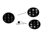
- Figure 2 shows one embodiment of the present invention in which the proximity assays described herein detected phosphorylated EGFR (pEGFR) and phosphorylated HER-2 (pHER-2) with single cell sensitivity.
- Figure 4 shows schematically the application of the addressable arrays of the invention for drug selection throughout the course of cancer treatment.
- cancers examples include, but are not limited to, breast cancer; lung cancer (e.g ., non-small cell lung cancer); digestive and gastrointestinal cancers such as colorectal cancer, gastrointestinal stromal tumors, gastrointestinal carcinoid tumors, colon cancer, rectal cancer, anal cancer, bile duct cancer, small intestine cancer, and stomach (gastric) cancer; esophageal cancer; gallbladder cancer; liver cancer; pancreatic cancer; appendix cancer; ovarian cancer; renal cancer (e.g ., renal cell carcinoma); cancer of the central nervous system; skin cancer; lymphomas; choriocarcinomas; head and neck cancers; osteogenic sarcomas; and blood cancers.
- lung cancer e.g ., non-small cell lung cancer
- digestive and gastrointestinal cancers such as colorectal cancer, gastrointestinal stromal tumors, gastrointestinal carcinoid tumors, colon cancer, rectal cancer, anal cancer, bile duct cancer, small intestine cancer,
- An “array” or “microarray” comprises a distinct set and/or dilution series of capture antibodies immobilized or restrained on a solid support such as, for example, glass ( e.g ., a glass slide), plastic, chips, pins, filters, beads ( e.g ., magnetic beads, polystyrene beads, etc .), paper, membrane ( e.g ., nylon, nitrocellulose, polyvinylidene fluoride (PVDF), etc .), fiber bundles, or any other suitable substrate.
- a solid support such as, for example, glass ( e.g ., a glass slide), plastic, chips, pins, filters, beads (e.g ., magnetic beads, polystyrene beads, etc .), paper, membrane ( e.g ., nylon, nitrocellulose, polyvinylidene fluoride (PVDF), etc .), fiber bundles, or any other suitable substrate.
- PVDF polyvinylidene fluoride
- substantially identical or “substantial identity,” in the context of two or more nucleic acids, refer to two or more sequences or subsequences that are the same or have a specified percentage of nucleotides that are the same (i.e ., at least about 60%, preferably at least about 65%, 70%, 75%, 80%, 85%, 90%, or 95% identity over a specified region) when compared and aligned for maximum correspondence over a comparison window or designated region as measured using a sequence comparison algorithm or by manual alignment and visual inspection.
- This definition when the context indicates, also refers analogously to the complement of a sequence.
- the substantial identity exists over a region that is at least about 5, 10, 15, 20, 25, 30, 35, 40, 45, 50, 75, or 100 nucleotides in length.
- Circulating cells of a solid tumor include cells that have either metastasized or micrometastasized from a solid tumor, including cancer stem cells or cells that are migrating to the tumor ( e.g ., due to chemoattraction), such as endothelial progenitor cells, circulating endothelial cells, pericytes, circulating pro-angiogenic myeloid cells, dendritic cells, etc.
- Patient samples containing the circulating cells can be obtained from any accessible biological fluid (e.g. , whole blood, serum, plasma, sputum, bronchial lavage fluid, urine, nipple aspirate, lymph, saliva, fine needle aspirate, etc .).
- the signal transducers are advantageously extracted shortly after the cells are isolated, preferably within 96, 72, 48, 24, 6, or 1 hr, more preferably within 30, 15, or 5 minutes.
- the isolated cells may also be advantageously incubated with growth factors usually at nanomolar to micromolar concentrations for about 1-30 minutes to resuscitate or stimulate signal transducer activation (see, e.g., Irish et al., Cell, 118:217-228 (2004 )).
- hormonal therapeutic agents include, without limitation, aromatase inhibitors (e.g ., aminoglutethimide, anastrozole (Arimidex ® ), letrozole (Femara ® ), vorozole, exemestane (Aromasin ® ), 4-androstene-3,6,17-trione (6-OXO), 1,4,6-androstatrien-3,17-dione (ATD), formestane (Lentaron ® ), etc .), selective estrogen receptor modulators (e.g ., apeledoxifene, clomifene, fulvestrant, lasofoxifene, raloxifene, tamoxifen, toremifene, etc .), steroids ( e.g ., dexamethasone), finasteride, and gonadotropin-releasing hormone agonists (GnRH) such as goserelin, pharmaceutically acceptable salts thereof, stereoisomers thereof, derivativese hormone
- Non-limiting examples of cancer vaccines useful in the present invention include ANYARA from Active Biotech, DCVax-LB from Northwest Biotherapeutics, EP-2101 from IDM Pharma, GV1001 from Pharmexa, IO-2055 from Idera Pharmaceuticals, INGN 225 from Introgen Therapeutics and Stimuvax from Biomira/Merck.
- the cellular extract comprises an extract of circulating cells of a solid breast tumor.
- the circulating cells are typically isolated from a patient sample using one or more separation methods known in the art including, for example, immunomagnetic separation, the CellTracks ® System, microfluidic separation, FACS, density gradient centrifugation, and depletion methods.
- the preferred embodiment may further comprise, i.e ., as step (e), or alternatively comprise, i.e ., as step (d), the step of indicating that the anticancer drug is unsuitable for the treatment of the breast tumor when the activation state detected for the one or more analytes is not substantially decreased compared to the reference activation profile.
- the preferred embodiment may further comprise sending or reporting the results of step (d) to a clinician, e.g ., an oncologist or a general practitioner.
- the preferred embodiment may further comprise recording or storing the results of step (d) in a computer database or other suitable machine or device for storing information, e.g ., at a laboratory.
- the isolated cells are stimulated in vitro with growth factors as described herein.
- the anticancer drug may comprise one or more of the therapeutic agents described herein, including but not limited to monoclonal antibodies, tyrosine kinase inhibitors, chemotherapeutic agents, hormonal therapeutic agents, radiotherapeutic agents, and vaccines.
- the horseradish peroxidase and the activation state-dependent antibodies can be conjugated to a sulfhydryl-activated dextran molecule.
- the sulfhydryl-activated dextran molecule typically has a molecular weight of about 70kDa (e.g ., about 40, 45, 50, 55, 60, 65, 70, 75, 80, 85, 90, 95, or 100kDa).
- the methods of the present invention may further comprise sending or reporting the results of step (c) to a clinician, e.g ., an oncologist or a general practitioner.
- the methods of the present invention may further comprise recording or storing the results of step (c) in a computer database or other suitable machine or device for storing information, e.g ., at a laboratory.
- the method of claim 2 for identifying the response of a breast tumor to treatment with an anticancer drug comprises:
- Suitable samples include, but are not limited to, whole blood, serum, plasma, ductal lavage fluid, nipple aspirate, lymph, bone marrow aspirate, urine, saliva, fine needle aspirate (FNA), and combinations thereof.
- the sample is a whole blood or FNA sample.
- circulating cells of a breast tumor may be isolated from the whole blood sample or breast cancer cells may be isolated from the FNA sample. If isolated cells are obtained from a subject who has not received treatment with an anticancer drug, the isolated cells may be incubated in vitro under suitable conditions with one or a cocktail of anticancer drugs which target one or more of the analytes to be detected in step (b).
- the plurality of signal transduction molecules is selected from the group consisting of ErbB1, ErbB2, p95ErbB2, ErbB3, ErbB4, VEGFR-1, VEGFR-2, VEGFR-3, ER, PR, and combinations thereof.
- the activation state detected for the one or more analytes present in the cellular extract may be, e.g ., a phosphorylation state, a ubiquitination state, a complexation state, or combinations thereof.
- the solid support may comprise, e.g ., glass, plastic, chips, pins, filters, beads, paper, membrane, fiber bundles, and combinations thereof.
- the capture antibodies are restrained on the solid support in an addressable array.
- the assay in step (b) comprises:
- the activated tyramide may be directly detected or indirectly detected, e.g ., upon the addition of a signal-detecting reagent.
- signal-detecting reagents include streptavidin-labeled fluorophores and combinations of streptavidin-labeled peroxidases and chromogenic reagents such as, e.g ., 3,3',5,5'-tetramethylbenzidine (TMB).
- the present invention provides a method of claim 3 for predicting the response of a subject having a breast tumor to treatment with an anticancer drug, the method comprising:
- the preferred embodiment may further comprise, i.e ., as step (e), or alternatively comprise, i.e ., as step (d), the step of indicating that the subject will not likely respond (e.g ., have an unlikely chance or low probability of responding) to treatment with the anticancer drug when the activation state detected for the one or more analytes is not substantially decreased compared to the reference activation profile.
- Suitable samples include, but are not limited to, whole blood, serum, plasma, ductal lavage fluid, nipple aspirate, lymph, bone marrow aspirate, urine, saliva, fine needle aspirate (FNA), and combinations thereof.
- the sample is a whole blood or FNA sample.
- circulating cells of a breast tumor may be isolated from the whole blood sample or breast cancer cells may be isolated from the FNA sample. If isolated cells are obtained from a subject who has not received treatment with an anticancer drug, the isolated cells may be incubated in vitro under suitable conditions with one or a cocktail of anticancer drugs which target one or more of the analytes to be detected in step (c).
- the facilitating moiety is glucose oxidase.
- the glucose oxidase and the activation state-independent antibodies can be conjugated to a sulfhydryl-activated dextran molecule as described in, e.g ., Examples 16 and 17.
- the sulfhydryl-activated dextran molecule typically has a molecular weight of about 500kDa ( e.g ., about 250, 300, 350, 400, 450, 500, 550, 600, 650, 700, or 750kDa).
- the oxidizing agent may be, for example, hydrogen peroxide (H 2 O 2 ).
- the first member of the signal amplification pair may be, for example, a peroxidase such as horseradish peroxidase (HRP).
- the second member of the signal amplification pair may be, for example, a tyramide reagent (e.g ., biotin-tyramide).
- the amplified signal is generated by peroxidase oxidization of biotin-tyramide to produce an activated tyramide ( e.g ., to transform the biotin-tyramide into an activated tyramide).
- the capture antibodies may comprise, for example, one or more members selected from the group consisting of antibodies reactive with Shc, PI3K, Erk, Rsk, Akt, p70S6K, VEGFR-1, VEGFR-2, Tie 2, V-Cadherin-R2 complex, PDGFRA, and PDGFRB.
- the addressable arrays described herein are particularly useful for determining the expression and/or activation state of signal transduction molecules and other proteins involved in breast cancer.
- Breast cancer is the fifth most common cause of cancer death worldwide, after lung cancer, stomach cancer, liver cancer, and colon cancer. In 2005, breast cancer caused 502,000 deaths worldwide. Among women worldwide, breast cancer is the most common cause of cancer death.
- SERMs selective estrogen receptor modulators
- tamoxifen bind ER and partially block its activity.
- Ovarian ablation, luteinizing hormone-releasing hormone agonists, and aromatase inhibitors such as anastrozole (Arimidex ® ), letrozole (Femara ® ), and exemestane (Aromasin ® ) reduce the level of estrogen and inhibit ligand-induced activation of ER.
- the ideal SERM should function as an anti-estrogen in the breast and uterus and a partial estrogen agonist in the skeletal, cardiovascular, and central nervous systems, as well as the gastrointestinal tract and vagina.
- ER/PR biology ER and PR are nuclear hormone receptors which function as transcription factors in the nucleus when they are bound to ligand(s). ER and PR have similar structures and contain a DNA binding domain, a dimerization domain, a hormone binding domain, and several transcription activating domains. A greater reduction in risk for recurrence was noted for patients with ER positive, PR positive tumors compared with those with ER positive, PR negative tumors.
- Akt and MAPK Activation of these pathways by estrogen sends powerful cell survival and cell proliferative signals via activation of Akt and MAPK.
- these kinases can phosphorylate ER and its coregulators to augment nuclear ER signaling. Phosphorylation of these proteins can also increase the estrogen agonist-like activity of tamoxifen and other SERMs.
- the pure anti-estrogen fulvestrant does not activate membrane ER in this way; however, SERMs such as tamoxifen do activate membrane ER in a manner similar to estrogen.
- SERMs such as tamoxifen do activate membrane ER in a manner similar to estrogen.
- the membrane effects of ER like its nuclear activity, may be cell, receptor-subtype, and ligand-specific, and it may also be influenced by the growth factor signaling milieu being much more prominent, for instance, in breast cancers overexpressing ErbB1 or HER-2. Stimulation of the MISS activity of ER by tamoxifen and other SERMs may, in part, explain the resistance to these agents sometimes observed in HER-2-overexpressing tumors.
- ER has at least two major functions. It serves as a transcription factor for estrogen-regulated genes and a co-activator for other transcription factors in the nucleus. It also functions in the cytoplasm and in the plasma membrane to activate growth factor signaling. In some breast tumors, particularly those with highly active growth factor signaling pathways such as HER-2 amplification, a vicious cycle is established in which estrogen activates growth factor signaling, and the growth factor signaling pathway further activates ER. Estrogen in such tumors would be expected to be a dominant factor by activating multiple pathways important in tumor progression. This molecular crosstalk has important implications for the treatment of breast cancer. As an example, estrogen-deprivation therapy with aromatase inhibitors should be more effective than SERMs in HER-2 amplified tumors by shutting off both the nuclear-initiated steroid signaling and MISS activities of ER.
- estrogen-blocking agents may be used for treatment of metastatic breast cancer.
- the treatment response to these agents is unpredictable, however, and approximately one-third of patients with metastatic breast cancer with receptors for estrogen or progesterone have no benefit from hormonal therapy. Nearly all patients with metastatic breast cancer will eventually become resistant to hormonal therapy despite the fact that the hormone receptors are still present.
- Microarray scanners suitable for use in the present invention are available from PerkinElmer (Boston, MA), Agilent Technologies (Palo Alto, CA), Applied Precision (Issaquah, WA), GSI Lumonics Inc. (Billerica, MA), and Axon Instruments (Union City, CA).
- a GSI ScanArray3000 for fluorescence detection can be used with ImaGene software for quantitation.
- the assay for detecting the activation state of a particular analyte (e.g ., signal transduction molecule) of interest in a cellular extract of tumor cells such as circulating cells of a solid tumor is a multiplex, high-throughput proximity (i.e. , three-antibody) assay having superior dynamic range.
- the three antibodies used in the proximity assay can comprise: (1) a capture antibody specific for the analyte; (2) a detection antibody specific for an activated form of the analyte ( i.e ., activation state-dependent antibody); and (3) a detection antibody which detects the total amount of the analyte ( i.e. , activation state-independent antibody).
- the proximity assays described herein are typically antibody-based arrays which comprise a plurality of different capture antibodies at a range of capture antibody concentrations that are coupled to the surface of a solid support in different addressable locations. Examples of suitable solid supports for use in the present invention are described above.
- the activation state-independent antibodies are directly labeled with the facilitating moiety.
- the facilitating moiety can be coupled to the activation state-independent antibodies using methods well-known in the art.
- the moiety for use in the present invention is glucose oxidase, which is capable of generating an oxidizing agent which channels to ( i.e ., is directed to) and reacts with ( i.e. , binds, is bound by, or forms a complex with) another molecule in proximity ( i.e ., spatially near or close) to the facilitating moiety. glucose oxidase or any other enzyme that catalyzes an involving molecular oxygen as the .
- 5'-phosphate groups on oligonucleotides can be treated with watersoluble carbodiimide EDC to form phosphate esters and subsequently coupled to amine-containing molecules.
- the diol on the 3'-ribose residue can be oxidized to aldehyde groups and then conjugated to the amine groups of antibodies or other types of proteins using reductive amination.
- the oligonucleotide linker can be synthesized with a biotin modification on either the 3' or 5' end and conjugated to streptavidin-labeled molecules.
- Oligonucleotide linkers can be synthesized using any of a variety of techniques known in the art, such as those described in Usman et al., J. Am. Chem. Soc., 109:7845 (1987 ); Scaringe et al., Nucl. Acids Res., 18:5433 (1990 ); Wincott et al., Nucl. Acids Res., 23:2677-2684 (1995 ); and Wincott et al., Methods Mol. Bio., 74:59 (1997 ).
- Example 4 An exemplary protocol for performing the proximity assays described herein is provided in Example 4.
- kits for performing the proximity assays described above comprising: (a) a dilution series of a plurality of capture antibodies restrained on a solid support; and (b) a plurality of detection antibodies (e.g ., activation state-independent antibodies and activation state-dependent antibodies).
- the kits can further contain instructions for methods of using the kit to detect the activation states of a plurality of signal transduction molecules of circulating cells of a solid tumor.
- microtiter wells are then washed and a labeled secondary antibody (e.g ., an anti-mouse antibody conjugated to alkaline phosphatase if the raised antibodies are mouse antibodies) is added to the wells and incubated for about 30 minutes and then washed. Substrate is added to the wells and a color reaction will appear where antibody to the immobilized polypeptide antigen is present.
- a labeled secondary antibody e.g ., an anti-mouse antibody conjugated to alkaline phosphatase if the raised antibodies are mouse antibodies
- DNA encoding the monoclonal antibodies can be readily isolated and sequenced using conventional procedures (e.g ., by using oligonucleotide probes that are capable of binding specifically to genes encoding the heavy and light chains of murine antibodies).
- the hybridoma cells serve as a preferred source of such DNA.
- the DNA may be placed into expression vectors, which are then transfected into host cells such as E . coli cells, simian COS cells, Chinese Hamster Ovary (CHO) cells, or myeloma cells that do not otherwise produce antibody, to induce the synthesis of monoclonal antibodies in the recombinant host cells. See , e.g., Skerra et al., Curr. Opin.
- Another method uses a particular FR derived from the consensus sequence of all human antibodies of a particular subgroup of light or heavy chains.
- the same FR may be used for several different humanized antibodies (see , e.g ., Carter et al., Proc. Natl. Acad. Sci. USA, 89:4285 (1992 ); and Presta et al., J. Immunol., 151:2623 (1993 )).
- a repertoire of V genes from unimmunized human donors can be constructed and antibodies to a diverse array of antigens (including self-antigens) can be isolated essentially following the techniques described in Marks et al., J. Mol. Biol., 222:581-597 (1991 ); Griffith et al., EMBO J., 12:725-734 (1993 ); and U.S. Patent Nos. 5,565,332 and 5,573,905 .
- bispecific antibodies are known in the art. Traditional production of full-length bispecific antibodies is based on the co-expression of two immunoglobulin heavy chain-light chain pairs, where the two chains have different specificities ( see, e.g ., Millstein et al., Nature, 305:537-539 (1983 )). Because of the random assortment of immunoglobulin heavy and light chains, these hybridomas (quadromas) produce a potential mixture of 10 different antibody molecules, of which only one has the correct bispecific structure. Purification of the correct molecule is usually performed by affinity chromatography. Similar procedures are disclosed in PCT Publication No. WO 93/08829 and Traunecker et al., EMBO J., 10:3655-3659 (1991 ).
- Bispecific antibodies include cross-linked or "heteroconjugate" antibodies.
- one of the antibodies in the heteroconjugate can be coupled to avidin, the other to biotin.
- Heteroconjugate antibodies can be made using any convenient cross-linking method. Suitable cross-linking agents and techniques are well-known in the art, and are disclosed in, e.g ., U.S. Patent No. 4,676,980 .
- the mixture comprising the antibody of interest and contaminants may be subjected to low pH hydrophobic interaction chromatography using an elution buffer at a pH between about 2.5-4.5, preferably performed at low salt concentrations ( e.g ., from about 0-0.25 M salt).
- the dosage forms typically include a conventional pharmaceutical carrier or excipient and may additionally include other medicinal agents, carriers, adjuvants, diluents, tissue permeation enhancers, solubilizers, and the like.
- Appropriate excipients can be tailored to the particular dosage form and route of administration by methods well known in the art (see, e.g REMINGTON'S PHARMACEUTICAL SCIENCES, supra).
- excipients include, but are not limited to, lactose, dextrose, sucrose, sorbitol, mannitol, starches, gum acacia, calcium phosphate, alginates, tragacanth, gelatin, calcium silicate, microcrystalline cellulose, polyvinylpyrrolidone, cellulose, water, saline, syrup, methylcellulose, ethylcellulose, hydroxypropylmethylcellulose, and polyacrylic acids such as Carbopols, e.g ., Carbopol 941, Carbopol 980, Carbopol 981, etc.
- Carbopols e.g ., Carbopol 941, Carbopol 980, Carbopol 981, etc.
- the methods described herein can be used in conjunction with panels of gene expression markers that predict the likelihood of breast cancer prognosis and/or recurrence in various populations of women with for example, node-negative disease.
- These gene panels can be useful for identifying women who are unlikely to experience recurrence and, thus, unlikely to benefit from adjuvant chemotherapy.
- the expression panels can be used to identify women who can safely avoid adjuvant chemotherapy, without negatively affecting disease-free and overall survival outcomes. Suitable systems include, but are not limited to, Oncotype DXTM, which is a 21-gene panel from Genomic Health, Inc.; MammaPrint, ® which is a 70-gene panel from Agendia; and a 76-gene panel from Veridex.
- Circulating cells of a solid tumor comprise cells that have either metastasized or micrometastasized from a solid tumor and include circulating tumor cells (CTCs), cancer stem cells (CSCs), and/or cells that are migrating to the tumor (e.g ., circulating endothelial 88 progenitor cells (CEPCs), circulating endothelial cells (CECs), circulating pro-angiogenic myeloid cells, circulating dendritic cells, etc.).
- CTCs circulating tumor cells
- CSCs cancer stem cells
- CEPCs circulating endothelial 88 progenitor cells
- CECs circulating endothelial cells
- circulating pro-angiogenic myeloid cells circulating dendritic cells, etc.
- Patient samples containing circulating cells can be obtained from any accessible biological fluid (e.g ., whole blood, serum, plasma, ductal lavage fluid, nipple aspirate, lymph, urine, saliva, fine needle aspirate, etc.
- the circulating cells can be isolated from a patient sample using one or more separation methods such as, for example, immunomagnetic separation (see, e.g., Racila et al., Proc. Natl. Acad. Sci. USA, 95:4589-4594 (1998 ); Bilkenroth et al., Int. J. Cancer, 92:577-582 (2001 )), the CellTrackTM System by Immunicon (Huntingdon Valley, PA), microfluidic separation (see, e.g., Mohamed et al., IEEE Trans. Nanobiosci., 3:251-256 (2004 ); Lin et al., Abstract No. 5147, 97th AACR Annual Meeting, Washington, D.C.
- immunomagnetic separation see, e.g., Racila et al., Proc. Natl. Acad. Sci. USA, 95:4589-4594 (1998 ); Bilkenroth et al., Int. J. Cancer,
- the entire Veridex system can be used for CTC enumeration or, by removing the sample manually after isolation with the CellTracks ® AutoPrep ® System, can provide a method of isolation prior to analysis for pathway activation.
- the number of CTCs can be informative for algorithm development.
- Triton X-100 1 1.00 1M Tris, pH 7.5 1 0.05 0.05 1M NaF 1 0.05 0.05 5M NaCl 5 0.1 0.20 2M B-glycerolphosphate 1 0.05 0.50 0.1M Na 3 VO 4 0.1 0.001 0.10 1 mg/ml pepstatin 1 0.10 Complete mini protease 1 tablet 0.5M EDTA 0.5 0.005 0.10 Total (ml) 3.00 Water (ml) 7.00
- This example illustrates methods for isolating, stimulating, and lysing cells from tumor tissue or biopsy specimens.
- This example also illustrates methods for initiating, stimulating, and lysing primary cultures of tumor cells isolated from tissue, biopsy, or whole blood. Additional methods for isolating and culturing tumor cells from biological specimens for screening chemotherapeutic agents are described, e.g., in U.S. Patent Nos. 5,728,541 ; 6,416,967 ; 6,887,680 ; 6,900,027 ; 6,933,129 ; and 7,112,415 ; and in U.S. Patent Publication Nos. 20040023375 and 20050202411 .
- the cellular extracts prepared in accordance with this example can be used in the single detection or proximity assays described herein.
- Stimulated cells are lysed using the following protocol:
- This example illustrates a multiplex, high-throughput, proximity dual detection microarray ELISA having superior dynamic range that is suitable for analyzing the activation states of signal transduction molecules in rare circulating cells:
- Example 6 Addressable Arrays for Analysis of Activated Receptor Tyrosine Kinases.
- Figure 5 illustrates an exemplary addressable receptor tyrosine kinase array of the invention.
- receptor tyrosine kinases are key components of many signal transduction pathways involved in cell proliferation.
- the ErbB family of receptor tyrosine kinases has four family members and plays an important role in fundamental cell processes like cell proliferation, differentiation, and survival. This family of receptor tyrosine kinases has been reported to be overexpressed in a number of different cancers and is associated with worse clinical outcome.
- On growth factor binding, ErbB1/EGFR, ErbB3/HER-3, and ErbB4/HER-4 homo- and hetero-dimerize to activate a number of different signaling pathways.
- ErbB2/HER-2 does not bind to a growth factor and is the preferred hetero-dimerization partner for all three family members. ErbB2 can also homo-dimerize when overexpressed and activate signaling pathways. Homo- or hetero-dimerization of ErbB family results in trans-phosphorylation. Auto- or trans-phosphorylation relieves the inhibitory conformation of receptor tyrosine kinases, enabling full kinase activation and at the same time creates binding sites for numerous SH2-containing signaling molecules, such as Src, Shc SHP-1, SHEP-1, and PI3K. Adapter proteins or signaling proteins like Shc, Grb2, or PI3K are recruited to the phosphorylated receptors.
- the addressable array shown in Figure 5 allows one to not only determine the expression of the ErbB family of receptor tyrosine kinases, but also their activation status. Both MAPK and PI3K/Akt pathway activation can also be studied on the addressable chip. In addition, the expression and/or activation status of nuclear hormone receptors such as ER (estrogen receptor) and PR (progesterone receptor), and other proteins such as NCOR (nuclear receptor corepressor), AIB1 (amplified in breast cancer-1), IGF-IR, cMET, Ki67, and TOPO II can be studied on the addressable chip. Another feature of the chip is the presence of internal controls to determine the tumor or tumor-associated cell (CEC's, CEP's, pericytes, etc.) content and non-specific IgG to determine any non-specific binding.
- CEC's, CEP's, pericytes, etc. non-specific IgG to determine any non-specific binding.
- MAPK pathway activation can be evaluated by determining the phosphorylation status of Erk and Rsk, while PI3K pathway activation can be evaluated by determining the phosphorylation status of Akt and p70S6K.
- addressable angiogenesis chips such as those shown in Figures 6 and 7 , allow one to not only determine the expression of all the signal transduction components involved in angiogenesis in a patient sample, but also their activation status. Both MAPK and PI3K/Akt pathway activation can also be studied on the addressable chip.
- the chip has internal controls to determine the tumor or tumor-associated cell (CEC's, CEP's, pericytes, etc .) content and non-specific IgG to determine any non-specific binding.
- Example 8 Selection of Patients for Treatment of Breast Cancer.
- the present invention encompasses the realization that signaling pathways can be used to provide new insights into the biological etiology of cancer and disease progression. This allows for treatment of cancers with various activated signaling pathways using a personalized therapeutic regimen.
- ER regulates growth and differentiation in both normal and malignant breast cells.
- Expression of functional ER and/or progesterone receptors (PR) is essential for a tumor to be responsive to antihormonal therapies ("hormone-responsive"), and multiple studies have demonstrated that expression of ER is strongly predictive for response to antihormonal therapies, although its expression is only weakly prognostic.
- ER does not act alone to stimulate tumor growth; rather, a complex interaction network operates to ensure the viability of cancer cells. Understanding this network provides scientific rationale for the selection of targeted therapies.
- Patient pre- or postmenopausal woman was biopsied or CTCs were isolated from blood. Analysis of the tumor cells revealed high expression and activation of ER. PR was expressed at very low levels. ErbB1, ErbB2, and ErbB3 were activated. The patient was treated with an aromatase inhibitor + lapatinib to shut down all ER/ErbB1/ErbB2/ErbB3-related activity. The patient responded and on re-biopsy had the protein profile as shown above. Thus, a patient with the above protein profile responds to therapy with an aromatase inhibitor + lapatinib.
- Table 17 provides an example of a patient with locally recurrent or metastatic breast cancer who has relapsed on anti-angiogenic therapy. At least 3 weeks had elapsed since the patient received adjuvant chemotherapy and hormonal therapy for the locally recurrent or metastatic breast cancer.
- Patient pre- or postmenopausal woman was biopsied or CTCs were isolated from blood. Analysis of the tumor cells revealed high expression and activation of ER. PR was expressed at very low levels. IGF-1R was activated. The patient was treated with an aromatase inhibitor + anti-IGF-1R antibodies to shut down all ER/IGF-1R-related activity. The patient responded and on re-biopsy had the protein profile as shown above. Thus, a patient with the above protein profile responds to aromatase inhibitor + anti-IGF-1R antibodies.
- Patient pre- or postmenopausal woman with locally recurrent or metastatic breast cancer was biopsied or CTCs were isolated from blood. Analysis of the tumor cells and endothelial cells revealed high expression and activation of ER, PR, and ErbB2 along with VEGFR2 activation. The patient was treated with an aromatase inhibitor + Herceptin ® + Avastin ® to shut down all ER, ErbB2, and VEGFR2-related activity. The patient responded and on re-biopsy had the protein profile as shown above. Thus, a patient with the above protein profile responds to combination therapy with an aromatase inhibitor + Herceptin ® + Avastin ® .
- Example 9 Selection of Patients for Treatment of Her2-Positive Breast Cancer.
- the exemplary patient profiles shown below in Tables 23-25 illustrate how an analysis of the pathways active in circulating tumor cells (CTCs) from blood or cancer cells obtained from a biopsy can be used to help physicians select patients who may be responsive to trastuzumab (Herceptin ® and therefore benefit from such therapy for the treatment of a breast tumor.
- the exemplary patient profiles shown below in Tables 26-31 illustrate how an analysis of the pathways active in CTCs from blood or cancer cells obtained from a biopsy can be used to help physicians select a suitable therapy for patients after Herceptin ® relapse resulting from either de novo or acquired resistance.
- Patient pre- or postmenopausal woman with locally recurrent or metastatic breast cancer was biopsied or CTCs were isolated from blood. Analysis of her tumor cells and endothelial cells revealed high expression and activation of ErbB2 and PDGFR, along with VEGFR2 activation. The patient was treated with Herceptin o + sorafinib + Avastin ® to shut down all ErbB2, PDGFR, and VEGFR2-related activity. Because PDGFR is overexpressed and activated in Avastin ® -resistant patients, information such as that presented above indicates that AZD2171 or sorafinib, which inhibits PDGFR as well as VEGFR, may be agents of choice to treat such tumors.
- Patient pre- or postmenopausal woman with locally recurrent or metastatic breast cancer was biopsied or CTCs were isolated from blood. Analysis of her tumor cells and endothelial cells revealed high expression and activation of IGF-1R, ErbB2, and PDGFR, along with VEGFR2 activation. The patient was treated with Herceptin ® + sorafinib + Avastin ® + IGF-1R antibody (Ab) to shut down all IGF-1R, ErbB2, PDGFR, and VEGFR2-related activity.
- Herceptin ® + sorafinib + Avastin ® + IGF-1R antibody (Ab) to shut down all IGF-1R, ErbB2, PDGFR, and VEGFR2-related activity.
- Patient pre- or postmenopausal woman with metastatic breast cancer was biopsied or CTCs were isolated from blood. Analysis of her tumor cells and endothelial cells revealed high expression and activation of ErbB2 and PDGFR, along with VEGFR2 activation. The patient had brain metastases. The patient was treated with lapatinib + sorafinib to shut down all ErbB2, PDGFR, and VEGFR2-related activity. Because PDGFR is overexpressed and activated in Avastin ® -resistant patients, information such as that presented above indicates that AZD2171 or sorafinib, which inhibits PDGFR as well as VEGFR, may be agents of choice to treat such tumors.
- Lapatinib was used instead of Herceptin ® as the patient had brain metastases. The patient responded and on re-biopsy had the protein profile as shown above. Thus, a patient with the above protein profile responds to lapatinib + sorafinib + chemotherapy.
- triple negative breast cancer Approximately 15-20% of women with breast cancer have the triple negative type of cancer. Patients with "triple receptor negative breast cancer" have a complete absence of hormone receptors ER, PR, and HER-2/ErbB2, with an aggressive clinical course and a paucity of treatment options. The only therapeutic option is chemotherapy and in this respect the choice of cytostatic agents is limited.
- the standard treatment for triple negative breast cancer is typically a combination of chemotherapy, surgery, and/or radiation therapy. When treated with standard therapy, women with triple negative breast cancer have a worse long-term outcome compared to women with non-triple negative breast cancer. Triple negative breast cancers cells usually have ErbB1 expressed on their cell surface. Women with ErbB1-positive breast cancer have worse long-term outcome compared to women whose tumors do not express ErbB1.
- Table 32 Patient 6001: (Triple negative with low ErbB1) Receptor Expression Activation (+/-GF) (Level of Phosphorylation) Treatment with Taxanes + Avastin ® ER Low PR Low IGF-1R Low Weak Weak ErbB1 Low Weak Weak ErbB2 Low Weak Weak P95 ErbB2 Low Weak Weak ErbB3 Low Weak Weak ErbB4 Low Weak Weak Shc Medium Weak PI3K Medium Weak Erk Medium Weak Rsk Medium Weak Akt Medium Weak P70S6K Medium Weak Ki67 High Weak TOPO2 Low Weak endothelial Cells and Pericytes: Receptor Expression Activation (Level of Phosphorylation) Activation with AZD2171 or Sorafinib or Sunitinib Activation with Avastin ® VEGFR2 Medium Strong Weak Weak Weak Weak
- Patient pre- or postmenopausal women with locally recurrent or metastatic breast cancer was biopsied or CTCs were isolated from blood. Analysis of her tumor cells and endothelial cells revealed low expression and no activation of ER, ErbB2, and p95 ErbB2, with only VEGFR2 activation. The patient was treated with Taxanes + Avastin ® to shut down VEGFR2-related activity. The patient responded and on re-biopsy had the protein profile as shown above. Thus, a patient with the above protein profile responds to Avastin ® + chemotherapy.
- Patient pre- or postmenopausal woman with locally recurrent or metastatic breast cancer was biopsied or CTCs were isolated from blood. Analysis of her tumor cells and endothelial cells revealed medium expression and activation of ErbB1 and VEGFR2 activation. The patient was treated with Tarceva ® + Avastin ® to shut down all ErbB 1 and VEGFR2-related activity. The patient responded and on re-biopsy had the protein profile as shown above. Thus, a patient with the above protein profile responds to Tarceva ® + Avastin ® + chemotherapy.
- Patient pre- or postmenopausal woman with locally recurrent or metastatic breast cancer was biopsied or CTCs were isolated from blood. Analysis of her tumor cells and endothelial cells revealed medium expression and activation of ErbB1, ErbB2, and ErbB3, along with VEGFR2 activation. The patient was treated with a Pan Her inhibitor + Avastin ® to shut down all ErbB1, ErbB2, ErbB3, and VEGFR2-related activity. The patient responded and on re-biopsy had the protein profile as shown above. Thus, a patient with the above protein profile responds to the combination of a Pan Her inhibitor + Avastin ® + chemotherapy.
- Patient pre- or postmenopausal woman with locally recurrent or metastatic breast cancer was biopsied or CTCs were isolated from blood. Analysis of her tumor cells and endothelial cells revealed medium expression and activation of ErbB1, ErbB2, and ErbB3, along with VEGFR2 activation. PTEN was deleted. The patient was treated with a Pan Her inhibitor + Avastin ® + mTOR inhibitor to shut down all ErbB1, ErbB2, ErbB3, and VEGFR2-related activity. The patient responded and on re-biopsy had the protein profile as shown above. Thus, a patient with the above protein profile responds to a Pan Her inhibitor + Avastin ® + rapamycin + chemotherapy.
- Example 11 Monitoring Breast Cancer Patients for EGFR and/or HER-2 Activation Guide Treatment Selection.
- Figure 12 shows images of CTC staining for EGFR, cytokeratin (CK), and cytokeratin with DAPI.
- Cell line controls were SKBr3 and A431, which are positive for HER-2 and EGFR expression, respectively.
- Whole blood from 6 normal individuals was processed using the same protocol as controls. None of the normal samples showed EGFR or HER-2 phosphorylation above background.
- Table 36 Demographics of the 5 breast cancer patients in the study.
- Patient 02-017 tested negatively for ER, PR, and HER-2 overexpression in the primary tumor.
- the patient had previously been treated with adriamycin + cytoxen, but at the time of blood collection was not on cancer therapy.
- the sample contained 3 CTCs, all of which stained positively for EGFR expression. There was significant activation of both EGFR and HER-2 detected in the proximity assays. Although the primary tumor of this patient was negative for HER-2 overexpression, the EGFR/HER-2 pathways were active. Treatment with Herceptin ® , either alone or in combination with chemotherapeutics, or chemotherapeutics alone would not have been an adequate therapy for this patient.
- Example 13 Selection of Patients for Treatment After Determination of Primary Tissue of Origin by a Gene Expression Panel.
- Example 14 Novel Set of Breast Cancer Tests Using Proximity Assays.
- the relative activation score is derived from a ratio of phosphorylation to expression.
- the test quantifies: HER-2 activation (phosphorylation); HER-2 expression; HER-1 activation (phosphorylation); HER-1 expression; and pancytokeratin (for cell number normalization).
- the test has high sensitivity since HER-2 activation is detected in single circulating tumor cells.
- the test also has a specificity of ⁇ 99% as measured by cross-reactivity of less than 1% with other markers.
- Reproducibility (intra assay): CV 5-15%.
- Reproducibility (inter assay): CV 10-20%.
- Monoclonal antibodies against ErbB1/HER-1 and p95HER-2 are used, while a pancytokeratin antibody for epithelial cells is used.
- the test quantifies: p95-HER-2 activation (phosphorylation); p95-HER-2 expression; HER-1 activation (phosphorylation); HER-1 expression; and pancytokeratin (for cell number normalization).
- This test may be validated in a clinical trial of metastatic patients with p95-HER-2 disease who were previously assigned as HER-2-positive based on IHC or FISH and who failed Herceptin ® . Patients who demonstrate activation of p95-HER-2 and HER-1 based on the proximity assay can be treated with Tykerb ® .
- Example 17 Novel Multiplexed Assay to Detect Activation of ErbB Family Receptor Tyrosine Kinases in a Circulating Tumor Cell.
- the expression/activation profiling of kinases and other signal transduction pathway molecules on a serial sampling of tumor tissues provides valuable information on changes occurring in tumor cells as a function of time and therapies. This temporal profiling of tumor progression enables clinicians to monitor rapidly evolving cancer signatures in each patient.
- This example illustrates a novel and robust assay to detect the level of expression and the degree of phosphorylation of the ErbB family of receptor tyrosine kinases (RTKs) and demonstrates the advantages of using such a therapy-guiding diagnostic system with single cell level sensitivity.
- the assay generally relies on samples such as fine needle aspirates (FNAs) and blood and achieves high sensitivity and specificity for interrogating the limited amount of cancer cells obtained from such samples.
- FNAs fine needle aspirates
- the assay described herein is provided by the triple-antibody enzyme approach that confers ultra-high sensitivity while preserving specificity.
- the assay may be performed as follows:
- SKBR3, MDA-MB-468, T47D, and BT474 cell lines were obtained from ATCC. Cells were grown in the following growth media in 100 mm tissue culture plates at 37°C in 5% CO 2 : SKBR3 - MacCoy's 5A medium with 10% FBS; MDA-MB-468 - DMEM, 10% FBS; BT474 - DMEM, 10% FBS; T47D - RPMI 1640, 10% FBS, 0.2 U/ml bovine insulin. Cells were harvested at 70-80% confluency with gentle detachment process (trypsin treatment + subsequent inactivation) and were subsequently counted and washed with 1X PBS.
- MDA-MB-468 has marginal HER-1 activation before stimulation, while both cell lines peak at approximately 2-5% of their RTKs activated ( ⁇ 0.5 to 1 x 10 5 phosphorylation events per cell).
- Figure 20 shows that the assay format described herein enables detection of less than 10 5 activation events with single cell sensitivity.
- Analytical specificity of the collaborative immunoassay format described herein was >99.99% based on a comparative study performed on multiple cell lines with various RTK levels.
- Cell lines used and their dominant ErbB expression and the RTK activation upon EGF or HRG ⁇ stimulation is shown in the Western blot in Figure 21 .
- the RTK activation profile for T47D cells, which express some level of ErbB2 and ErbB3 but an extremely low amount of ErbB 1, is shown in Figure 22 .
- the number of cells required to detect EC20 (12000 RFU) pHER-1 or pHER-2 were used to calculate per-cell RTK activation (RFU/cell) as shown in Table 43.
- MDA MB 468 cells When MB468 cells expressing an extremely low amount of HER-2 were used, having ⁇ 1000 cells per reaction pad was not sufficient to achieve EC20, and this type of low or non-detectable signal in other cell lines was indicated as "ND" in Table 43. MDA MB 468 cells have ⁇ 4000RFU/cell level of pHER-1 when stimulated with EGF, yet do not show any detectable pHER-2.
- This collaborative immunoassay format ensures ultra-specificity while maintaining its sub-single cell level(10 4 to 10 5 ) molecular level assay sensitivity.
- This example illustrates a novel assay capable of specifically detecting the state of phosphorylation of ErbB family receptor members with sensitivity that enables its use with rare circulating tumor cells (CTCs).
- CTCs rare circulating tumor cells
- the assay format described herein will enable clinicians to monitor rapidly evolving cancer signatures in each patient. Because of its unparallel sensitivity and specificity, the assay format described in this example can be applied to detect phosphorylation events in ErbB family receptor members present in rare CTCs. As such, this method can provide guidance, not only for the initial selection of targeted therapeutics, but also in subsequent monitoring for therapy progression.
- the multiplexed proximity-based collaborative-immunoassay platform described herein provides valuable clinical information on limited samples with ultra-sensitivity and specificity to assist oncologists in maintaining or adjusting their disease treatment options for each patient according to a "personal" cancer profile shift.
- Example 18 Method to Detect Activation of ErbB Family Receptor Tyrosine Kinases.
- the application presents technology capable of specifically detecting phosphorylation events in ErbB family receptor tyrosine kinases (RTKs) at a single-cell level sensitivity.
- RTKs ErbB family receptor tyrosine kinases
- this multiplexed protein microarray platform utilizes the formation of a unique "triple-antibody-enzyme-channeling" immuno-complex.
- this complex requires co-localization of two detector-antibodies conjugated with corresponding channeling-enzymes once target proteins are bound by the capture antibodies.
- the channeling events between two detector enzymes in proximity, glucose oxidase (GO, conjugated to anti-RTK antibodies) and horseradish peroxidase (HRP, conjugated to anti-phosphorylated sites in RTKs) enabled the profiling of the RTK with extreme sensitivity.
- This principle was applied to two breast cancer model systems with a limited number of target cells: cancer cells found in a patient's whole blood (circulating tumor cells, CTCs) and in a fine needle aspirate (FNA) sample.
- CTCs primary tumor and circulating tumor cells
- a triple-antibody-enzyme-channeling multiplexed protein microarray platform has been developed to detect the phosphorylation of target molecules.
- This multiplexed protein microarray platform utilizes a unique immuno-complex formation via co-localization of two detector enzyme-conjugated-antibodies once target proteins are captured on the microarray surface.
- the channeling events between the two detector enzymes in proximity enables the profiling of the receptor tyrosine kinases (RTKs) with a single-cell level sensitivity.
- RTKs receptor tyrosine kinases
- Specificity for the detection of phosphorylated targets is greatly increased given the requirement for simultaneous binding of three antibodies.
- assays were performed on activated CTCs from 75 cancer patients on various therapy regimens.
- the multiplexed proximity-mediated platform advantageously provides single-cell level sensitivity for detecting the activation of RTKs in a limited amount of sample. As such, CTCs found in metastatic stage cancers can be obtained and profiled to provide valuable information to impact clinical practice.
- Example 20 Method to Detect Activation of Receptor Tyrosine Kinases on Metastatic Lesions Using a Proximity-Mediated Microarray Immunoassay.
- the multiplexed proximity-mediated platform advantageously provides single-cell level sensitivity of target phosphorylation events for detecting the activation of RTKs in a limited amount of mFNA tissue sample.
- the ability to profile tumors at different metastatic sites therefore provides valuable information on their differential metastatic potentials.
- minimally-invasive single-passage mFNA samples may be utilized to tailor therapy options as the disease profile changes.
Landscapes
- Health & Medical Sciences (AREA)
- Life Sciences & Earth Sciences (AREA)
- Engineering & Computer Science (AREA)
- Immunology (AREA)
- Biomedical Technology (AREA)
- Chemical & Material Sciences (AREA)
- Hematology (AREA)
- Urology & Nephrology (AREA)
- Molecular Biology (AREA)
- Analytical Chemistry (AREA)
- General Health & Medical Sciences (AREA)
- Microbiology (AREA)
- Biotechnology (AREA)
- Pathology (AREA)
- General Physics & Mathematics (AREA)
- Food Science & Technology (AREA)
- Medicinal Chemistry (AREA)
- Physics & Mathematics (AREA)
- Cell Biology (AREA)
- Biochemistry (AREA)
- Hospice & Palliative Care (AREA)
- Oncology (AREA)
- Tropical Medicine & Parasitology (AREA)
- Bioinformatics & Cheminformatics (AREA)
- Toxicology (AREA)
- Measuring Or Testing Involving Enzymes Or Micro-Organisms (AREA)
- Investigating Or Analysing Biological Materials (AREA)
- Peptides Or Proteins (AREA)
- Apparatus Associated With Microorganisms And Enzymes (AREA)
- Medicines Containing Material From Animals Or Micro-Organisms (AREA)
- Pharmaceuticals Containing Other Organic And Inorganic Compounds (AREA)
Claims (19)
- Procédé comprenant les étapes suivantes :a) lyser des cellules d'une tumeur du sein isolées après administration d'un médicament anticancéreux, ou avant incubation avec un médicament anticancéreux, pour produire un extrait cellulaire ;b) détecter un état d'activation d'un ou de plusieurs analyte(s) dans l'extrait cellulaire, en effectuant un essai dans lequel on a recours à plusieurs séries de dilutions d'anticorps de capture spécifiques de l'analyte ou des analytes, lesquels anticorps de capture sont attachés sur un support solide, et lequel essai comporte les opérations suivantes :i) faire incuber l'extrait cellulaire avec les plusieurs séries de dilutions d'anticorps de capture, pour former plusieurs analytes capturés ;ii) faire incuber ces analytes capturés avec des anticorps de détection comprenant plusieurs anticorps indépendants de l'état d'activation et plusieurs anticorps dépendants de l'état d'activation, spécifiques des analytes correspondants, pour former plusieurs analytes capturés détectables, étant entendu- que les anticorps indépendants de l'état d'activation sont marqués avec de la glucose oxydase,- que la glucose oxydase et les anticorps indépendants de l'état d'activation sont conjugués à des molécules de dextrane activées par sulfhydryle,- que les anticorps dépendants de l'état d'activation sont marqués avec un premier élément d'une paire d'amplification du signal,- et que la glucose oxydase génère un agent oxydant qui est dirigé vers le premier élément de paire d'amplification du signal et réagit avec celui-ci ;iii) faire incuber les plusieurs analytes capturés détectables avec un deuxième élément de la paire d'amplification du signal, pour générer un signal amplifié ;iv) et détecter le signal amplifié généré à partir des premier et deuxième éléments de la paire d'amplification du signal ;c) et déterminer si le médicament anticancéreux est approprié ou non pour le traitement de la tumeur du sein, en comparant l'état d'activation détecté pour l'analyte ou les analytes avec un profil d'activation de référence généré en l'absence du médicament anticancéreux, le procédé ayant pour but de sélectionner un médicament anticancéreux approprié pour le traitement d'une tumeur du sein.
- Procédé conforme à la revendication 1, dans lequel l'étape (c) consiste, à la place, à identifier la tumeur du sein comme répondant ou non au traitement par le médicament anticancéreux, en comparant l'état d'activation détecté pour l'analyte ou les analytes avec un profil d'activation de référence généré en l'absence du médicament anticancéreux, le procédé ayant pour but d'identifier la réponse d'une tumeur du sein à un traitement par un médicament anticancéreux.
- Procédé conforme à la revendication 1, dans lequel l'étape (c) consiste, à la place, à prédire la probabilité qu'a le sujet de répondre au traitement par le médicament anticancéreux, en comparant l'état d'activation détecté pour l'analyte ou les analytes avec un profil d'activation de référence généré en l'absence du médicament anticancéreux, le procédé ayant pour but de prédire la réponse d'un sujet atteint d'une tumeur du sein à un traitement par un médicament anticancéreux.
- Procédé conforme à l'une des revendications 1 à 3,
dans lequel la tumeur du sein provient d'un sujet atteint d'un carcinome canalaire, lequel carcinome canalaire est de préférence un carcinome canalaire invasif ou un carcinome canalaire in situ,
ou dans lequel la tumeur du sein provient d'un sujet atteint d'un carcinome lobulaire, lequel carcinome lobulaire est de préférence un carcinome lobulaire invasif ou un carcinome lobulaire in situ. - Procédé conforme à l'une des revendications 1 à 3, dans lequel les cellules comprennent des cellules circulantes de la tumeur du sein, lesquelles cellules circulantes ont été de préférence isolées par séparation immuno-magnétique à partir d'un échantillon, lequel échantillon est choisi de préférence dans l'ensemble formé par du sang complet, du sérum, du plasma, du liquide de lavage des canaux, du liquide prélevé au niveau du mamelon par aspiration, de la lymphe, de la moelle osseuse prélevée par aspiration, de l'urine, de la salive, et un échantillon prélevé par aspiration au moyen d'une aiguille fine, ainsi que les combinaisons de tels échantillons, et lesquelles cellules circulantes sont choisies de préférence dans l'ensemble formé par des cellules tumorales circulantes, des cellules endothéliales circulantes, des cellules progénitrices de cellules endothéliales circulantes, des cellules souches tumorales et des cellules tumorales disséminées, ainsi que les combinaisons de telles cellules.
- Procédé conforme à l'une des revendications 1 à 3, pour lequel les cellules ont été isolées à partir d'un tissu tumoral, lequel tissu tumoral est de préférence un tissu de tumeur primitive ou un tissu de tumeur métastatique, et pour lequel les cellules ont de préférence été isolées à partir du tissu tumoral sous la forme d'un échantillon prélevé par aspiration au moyen d'une aiguille fine.
- Procédé conforme à l'une des revendications 1 à 3, dans lequel les cellules isolées sont stimulées in vitro au moyen de facteurs de croissance.
- Procédé conforme à l'une des revendications 1 à 3, dans lequel le médicament anticancéreux est choisi dans l'ensemble formé par les suivants : un anticorps monoclonal, un inhibiteur de tyrosine kinase, un agent de chimiothérapie, un agent d'hormonothérapie, un agent de radiothérapie, un vaccin, et les combinaisons de tels agents, étant entendu que :- l'anticorps monoclonal est de préférence choisi dans l'ensemble formé par les suivants : trastuzumab (Herceptin®), alemtuzumab (Campath®), bevacizumab (Avastin®), cetuximab (Erbitux®), gemtuzumab (Mylotarg®), panitumumab (Vectibix®), rituximab (Rituxan®), tositumomab (Bexxar®), et leurs combinaisons ;- l'inhibiteur de tyrosine kinase est de préférence choisi dans l'ensemble formé par les suivants : gefitinib (Iressa®), sunitinib (Sutent®), erlotinib (Tarceva®), lapatinib (Tykerb®), canertinib (CI 1033), semaxinib (SU5416), vatalanib (PTK787/ZK222584), sorafenib (BAY 43-9006), imatinib mésylate (Gleevec®), léflunomide (SU101), vandétanib (Zactima®, ZD6474), et leurs combinaisons ;- l'agent de chimiothérapie est de préférence choisi dans l'ensemble que constituent les suivants : pémétrexed (Alimta®), gemcitabine (Gemzar®), sirolimus (rapamycine), analogues de rapamycine, composés du platine, carboplatine, cisplatine, satraplatine, paclitaxel (Taxol®), docétaxel (Taxotère®), temsirolimus (CCI-779), évérolimus (RAD001), et leurs combinaisons ;- l'agent d'hormonothérapie est de préférence choisi dans l'ensemble formé par les suivants : inhibiteurs d'aromatase, modulateurs sélectifs de récepteurs d'oestrogènes, stéroïdes, finastéride, agonistes de la gonadolibérine, ainsi que leurs sels pharmacologiquement admissibles, leurs stéréoisomères, leurs dérivés, leurs analogues, et leurs combinaisons ;- et l'agent de radiothérapie est de préférence choisi dans l'ensemble formé par les suivants : 47Sc, 64Cu, 67Cu, 89Sr, 86Y, 87Y, 90Y, 105Rh, 111Ag, 111In, 117mSn, 149Pm, 153Sm, 166Ho, 177Lu, 186Re, 188Re, 211At, 212Bi, et leurs combinaisons.
- Procédé conforme à l'une des revendications 1 à 3, dans lequel l'analyte ou les analytes comprennent plusieurs molécules de transduction du signal,
lesquelles plusieurs molécules de transduction du signal sont de préférence choisies dans l'ensemble formé par les tyrosine-kinases récepteurs, les tyrosine-kinases non-récepteurs, les composants de cascade de signalisation de type tyrosine-kinase, les récepteurs nucléaires d'hormones, les coactivateurs de récepteurs nucléaires, les répresseurs de récepteurs nucléaires, et leurs combinaisons ;
ou lesquelles plusieurs molécules de transduction du signal sont de préférence choisies dans l'ensemble constitué par les suivantes : EGFR (ErbB1), HER-2 (ErbB2), p95ErbB2, HER-3 (ErbB3), HER-4 (ErbB4),
Raf, SRC, Mek, NFkB-IkB, mTor, PI3K, VEGF, VEGFR-1, VEGFR-2, VEGFR-3, Eph-a, Eph-b, Eph-c, Eph-d, cMet, FGFR, cKit, Flt-3, Tie-1, Tie-2, Flt-3, cFMS, PDGFRA, PDGFRB, Ab1, FTL 3, RET, Kit, HGFR, FGFR1, FGFR2, FGFR3, FGFR4, IGF-1R, ER, PR, NCOR, AIB1, et leurs combinaisons ;
ou lesquelles plusieurs molécules de transduction du signal sont de préférence choisies dans l'ensemble formé par les suivantes : ErbB1, ErbB2, p95ErbB2, ErbB3, ErbB4, VEGFR-1, VEGFR-2, VEGFR-3, ER, PR, et leurs combinaisons. - Procédé conforme à l'une des revendications 1 à 3, dans lequel l'état d'activation est choisi dans l'ensemble que constituent un état de phosphorylation, un état d'ubiquitination, un état de complexation, et les combinaisons de tels états.
- Procédé conforme à l'une des revendications 1 à 3, dans lequel le support solide est choisi dans l'ensemble que constituent les suivants : verre, matière plastique, copeaux, pointes, filtres, perles, papier, membranes, faisceaux de fibres, et leurs combinaisons.
- Procédé conforme à l'une des revendications 1 à 3, dans lequel les anticorps de capture sont attachés sur le support solide en un réseau addressable.
- Procédé conforme à l'une des revendications 1 à 3, dans lequel- les anticorps dépendants de l'état d'activation sont directement marqués avec le premier élément de paire d'amplification du signal,- ou les anticorps dépendants de l'état d'activation sont marqués avec le premier élément de paire d'amplification du signal moyennant une liaison entre un premier élément de paire de liaison, conjugué aux anticorps dépendants de l'état d'activation, et un deuxième élément de paire de liaison, conjugué au premier élément de paire d'amplification du signal, étant entendu que le premier élément de paire de liaison est de préférence de la biotine et que le deuxième élément de paire de liaison est de préférence de la streptavidine.
- Procédé conforme à l'une des revendications 1 à 3, dans lequel les molécules de dextrane activées par sulfhydryle présentent une masse moléculaire de 500 kDa.
- Procédé conforme à l'une des revendications 1 à 3, dans lequel l'agent oxydant est du peroxyde d'hydrogène H2O2.
- Procédé conforme à la revendication 15, dans lequel le premier élément de paire d'amplification du signal est une peroxydase, de préférence de la peroxydase de raifort (HRP), et/ou le deuxième élément de paire d'amplification du signal est un réactif tyramide, de préférence du biotinyl-tyramide.
- Procédé conforme à la revendication 16, dans lequel le signal amplifié est généré par oxydation du biotinyl-tyramide par la peroxydase, ce qui donne un tyramide activé.
- Procédé conforme à la revendication 17, dans lequel on détecte directement le tyramide activé, ou l'on détecte le tyramide activé après addition d'un réactif de détection du signal, lequel réactif de détection du signal est de préférence un fluorophore marqué à la streptavidine ou une combinaison de peroxydase marquée à la streptavidine et d'un réactif chromogène, lequel réactif chromogène est de préférence de la 3,3',5,5'-tétraméthyl-benzidine (TMB).
- Utilisation d'un réseau comprenant plusieurs séries de dilutions d'anticorps de capture attachés sur un support solide, dans lequel réseau les anticorps de capture de chaque série de dilutions sont spécifiques d'un ou de plusieurs analytes correspondant à un composant d'une voie de transduction du signal dans un extrait cellulaire, pour mettre en oeuvre un procédé conforme à l'une des revendications 1 à 3.
Priority Applications (3)
| Application Number | Priority Date | Filing Date | Title |
|---|---|---|---|
| DK12183659.7T DK2602623T3 (en) | 2008-02-25 | 2009-02-24 | METHOD OF DETECTING INTRACELLULAR TRUNCTED RECEPTORS |
| EP13164311.6A EP2618146B1 (fr) | 2008-02-25 | 2009-02-24 | Sélection d'un médicament pour la thérapie du cancer du sein à partir des matrices d'anticorps |
| EP12183659.7A EP2602623B1 (fr) | 2008-02-25 | 2009-02-24 | Procédé de détection de recepteurs intracellulaires tronques |
Applications Claiming Priority (6)
| Application Number | Priority Date | Filing Date | Title |
|---|---|---|---|
| US3131908P | 2008-02-25 | 2008-02-25 | |
| US10640408P | 2008-10-17 | 2008-10-17 | |
| US10838408P | 2008-10-24 | 2008-10-24 | |
| US11790808P | 2008-11-25 | 2008-11-25 | |
| US14055808P | 2008-12-23 | 2008-12-23 | |
| PCT/US2009/035013 WO2009108637A1 (fr) | 2008-02-25 | 2009-02-24 | Sélection d’un médicament pour le traitement du cancer du sein à partir des matrices d’anticorps |
Related Child Applications (3)
| Application Number | Title | Priority Date | Filing Date |
|---|---|---|---|
| EP12183659.7A Division EP2602623B1 (fr) | 2008-02-25 | 2009-02-24 | Procédé de détection de recepteurs intracellulaires tronques |
| EP13164311.6A Division EP2618146B1 (fr) | 2008-02-25 | 2009-02-24 | Sélection d'un médicament pour la thérapie du cancer du sein à partir des matrices d'anticorps |
| EP12183659.7 Division-Into | 2012-09-10 |
Publications (2)
| Publication Number | Publication Date |
|---|---|
| EP2250498A1 EP2250498A1 (fr) | 2010-11-17 |
| EP2250498B1 true EP2250498B1 (fr) | 2012-10-31 |
Family
ID=40599946
Family Applications (3)
| Application Number | Title | Priority Date | Filing Date |
|---|---|---|---|
| EP09715619A Active EP2250498B1 (fr) | 2008-02-25 | 2009-02-24 | Sélection d' un médicament pour le traitement du cancer du sein à partir des matrices d' anticorps |
| EP12183659.7A Active EP2602623B1 (fr) | 2008-02-25 | 2009-02-24 | Procédé de détection de recepteurs intracellulaires tronques |
| EP13164311.6A Not-in-force EP2618146B1 (fr) | 2008-02-25 | 2009-02-24 | Sélection d'un médicament pour la thérapie du cancer du sein à partir des matrices d'anticorps |
Family Applications After (2)
| Application Number | Title | Priority Date | Filing Date |
|---|---|---|---|
| EP12183659.7A Active EP2602623B1 (fr) | 2008-02-25 | 2009-02-24 | Procédé de détection de recepteurs intracellulaires tronques |
| EP13164311.6A Not-in-force EP2618146B1 (fr) | 2008-02-25 | 2009-02-24 | Sélection d'un médicament pour la thérapie du cancer du sein à partir des matrices d'anticorps |
Country Status (13)
| Country | Link |
|---|---|
| US (4) | US8163499B2 (fr) |
| EP (3) | EP2250498B1 (fr) |
| JP (2) | JP5414701B2 (fr) |
| CN (2) | CN103399144B (fr) |
| AU (1) | AU2009219437B2 (fr) |
| CA (2) | CA2716826C (fr) |
| DK (2) | DK2602623T3 (fr) |
| ES (3) | ES2545760T3 (fr) |
| HK (2) | HK1182441A1 (fr) |
| IL (3) | IL207637A (fr) |
| MX (1) | MX2010009140A (fr) |
| NZ (3) | NZ600339A (fr) |
| WO (1) | WO2009108637A1 (fr) |
Families Citing this family (91)
| Publication number | Priority date | Publication date | Assignee | Title |
|---|---|---|---|---|
| EP2284180B1 (fr) * | 2003-03-03 | 2015-09-09 | Dyax Corp. | Utilisations de peptides qui lient spécifiquement le récepteur HGF (cMET) |
| CN108421044A (zh) | 2005-02-03 | 2018-08-21 | 综合医院公司 | 治疗吉非替尼耐药性癌症的方法 |
| KR101354828B1 (ko) | 2005-11-04 | 2014-02-18 | 와이어쓰 엘엘씨 | mTOR 저해자, 헤르셉틴, 및/또는 HKI-272의항신생물성 조합 |
| US9250243B2 (en) | 2006-09-21 | 2016-02-02 | Nestec S.A. | Drug selection for lung cancer therapy using antibody-based arrays |
| BRPI0717416A2 (pt) | 2006-09-21 | 2013-11-12 | Prometheus Lab Inc | Método para realizar um imunoensaio complexo de alta produtividade, e, arranjo |
| US8022216B2 (en) | 2007-10-17 | 2011-09-20 | Wyeth Llc | Maleate salts of (E)-N-{4-[3-chloro-4-(2-pyridinylmethoxy)anilino]-3-cyano-7-ethoxy-6-quinolinyl}-4-(dimethylamino)-2-butenamide and crystalline forms thereof |
| MX2010009140A (es) | 2008-02-25 | 2010-12-20 | Prometheus Lab Inc | Seleccion de farmacos para terapia de cancer de mama utilizando matrices basadas en anticuerpos. |
| KR20130088908A (ko) | 2008-06-17 | 2013-08-08 | 와이어쓰 엘엘씨 | Hki-272 및 비노렐빈을 함유하는 항신생물성 조합물 |
| HUE032958T2 (hu) | 2008-08-04 | 2017-11-28 | Wyeth Llc | 4-Anilino-3-ciano-kinolinok és capecitabin antineoplasztikus kombinációi |
| WO2010028288A2 (fr) | 2008-09-05 | 2010-03-11 | Aueon, Inc. | Procédés pour la stratification et l’annotation des options de traitement médicamenteux contre le cancer |
| WO2010085845A1 (fr) * | 2009-01-28 | 2010-08-05 | The University Of Queensland | Diagnostic du cancer et/ou thérapie contre celui-ci |
| LT3000467T (lt) | 2009-04-06 | 2023-04-11 | Wyeth Llc | Krūties vėžio gydymo schema naudojant neratinibą |
| CA2761777A1 (fr) | 2009-05-14 | 2010-11-18 | Prometheus Laboratories Inc. | Biomarqueurs permettant de determiner la sensibilite de cellules cancereuses du sein a un traitement ciblant le recepteur her2 |
| ES2641479T3 (es) * | 2009-06-23 | 2017-11-10 | Fundação D. Anna De Sommer Champalimaud E Dr. Carlos Montez Champalimaud | Sistemas y métodos para tratar, diagnosticar y predecir la respuesta a la terapia del cáncer de mama |
| ES2627909T3 (es) | 2009-07-15 | 2017-08-01 | Diatech Holdings, Inc. | Selección de fármacos para el tratamiento del cáncer gástrico usando matrices basadas en anticuercuerpos |
| WO2011015920A2 (fr) * | 2009-08-03 | 2011-02-10 | Avesthagen Limited | Procédé hautement efficace de purification et de production de trastuzumab de recombinaison |
| WO2011015921A1 (fr) * | 2009-08-03 | 2011-02-10 | Avesthagen Limited | Procédé hautement efficace de purification et de production de cetuximab de recombinaison |
| WO2011034919A2 (fr) * | 2009-09-15 | 2011-03-24 | H. Lee Moffitt Cancer Center And Research Institute, Inc. | Profilage du domaine sh2 pour caractériser la signalisation par phosphorylation de tyrosine dans le cancer |
| EP2491385B1 (fr) * | 2009-10-20 | 2017-05-10 | DiaTech Holdings, Inc. | Dosages à médiation de proximité pour détecter des protéines de fusion oncogènes |
| RU2558931C2 (ru) | 2010-01-12 | 2015-08-10 | Нестек С.А. | Способы предсказания ответа трижды негативного рака молочной железы на терапию |
| JP6126382B2 (ja) * | 2010-01-13 | 2017-05-10 | ワイス・エルエルシー | 腫瘍を正確に識別し、かつPAN‐ErbB阻害剤に対する薬物応答を予測するPTENタンパク質発現におけるカットポイント |
| WO2012006421A2 (fr) * | 2010-07-07 | 2012-01-12 | The Regents Of The University Of Michigan | Diagnostic et traitement du cancer du sein |
| TW201302793A (zh) | 2010-09-03 | 2013-01-16 | Glaxo Group Ltd | 新穎之抗原結合蛋白 |
| NZ608313A (en) | 2010-09-24 | 2013-12-20 | Univ Leland Stanford Junior | Direct capture, amplification and sequencing of target dna using immobilized primers |
| SG191230A1 (en) | 2010-12-23 | 2013-07-31 | Nestec Sa | Drug selection for malignant cancer therapy using antibody-based arrays |
| US9962361B2 (en) | 2011-01-03 | 2018-05-08 | The William M. Yarbrough Foundation | Isothiocyanate functional surfactants, formulations incorporating the same, and associated methods of use |
| WO2012106559A1 (fr) * | 2011-02-02 | 2012-08-09 | Translational Genomics Research Institute | Biomarqueurs et leurs procédés d'utilisation |
| US9719995B2 (en) | 2011-02-03 | 2017-08-01 | Pierian Holdings, Inc. | Drug selection for colorectal cancer therapy using receptor tyrosine kinase profiling |
| US9532969B2 (en) | 2011-02-08 | 2017-01-03 | The William M. Yarbrough Foundation | Method for treating psoriasis |
| MX343327B (es) | 2011-02-17 | 2016-11-01 | Nestec Sa | Ensayos para detectar autoanticuerpos contra farmacos anti-tnfa. |
| KR101511889B1 (ko) | 2011-02-17 | 2015-04-13 | 네스텍 소시에테아노님 | 여과에 의해 백혈구 및 종양 세포를 단리하는 장비 및 방법 |
| EP2908132B8 (fr) | 2011-03-02 | 2019-06-12 | Nestec S.A. | Prédiction de la sensibilité aux médicaments de tumeurs du poumon sur la base de signatures génétiques et moléculaires |
| MX339340B (es) | 2011-04-04 | 2016-05-20 | Nestec Sa | Metodos para predecir y mejorar la supervivencia de pacientes con cancer gastrico. |
| JP2014517279A (ja) | 2011-05-10 | 2014-07-17 | ネステク ソシエテ アノニム | 個別治療管理のための疾患活動性プロファイリングの方法 |
| MX2014001941A (es) | 2011-08-18 | 2016-04-28 | Nestec Sa | Composiciones y metodos para detectar variantes alelicas. |
| ES2553456T3 (es) | 2011-09-02 | 2015-12-09 | Nestec S.A. | Perfilado de proteínas de ruta de señal para determinar una eficacia terapéutica |
| AU2012332481B2 (en) * | 2011-11-04 | 2017-01-19 | Invivis Sas | Assay for predictive biomarkers of anti-estrogen efficacy |
| EP2788752B1 (fr) * | 2011-12-05 | 2018-10-03 | Pierian Holdings, Inc. | Procédé de sélection d'un traitement destiné à des patients souffrant d'un cancer du poumon |
| CN103157109A (zh) * | 2011-12-12 | 2013-06-19 | 中国科学院生物物理研究所 | Cd146及其抗体诊断和治疗三阴性乳腺癌的应用 |
| MX2018004950A (es) * | 2011-12-30 | 2022-01-24 | Quest Diagnostics Invest Inc | Aptámeros y métodos de diagnóstico para detectar el receptor de egf. |
| US20130330761A1 (en) | 2012-06-12 | 2013-12-12 | Celcuity, LLC | Whole cell assays and methods |
| US10434082B2 (en) | 2012-07-26 | 2019-10-08 | The William M. Yarbrough Foundation | Isothiocyanate functional compounds augmented with secondary antineoplastic medicaments and associated methods for treating neoplasms |
| US10335387B2 (en) | 2012-07-26 | 2019-07-02 | The William M. Yarbrough Foundation | Method for treating infectious diseases with isothiocyanate functional compounds |
| US10434081B2 (en) | 2012-07-26 | 2019-10-08 | The William M. Yarbrough Foundation | Inhibitors of macrophage migration inhibitory factor |
| US10441561B2 (en) | 2012-07-26 | 2019-10-15 | The William M. Yanbrough Foundation | Method for treating benign prostatic hyperplasia (BPH), prostatitis, and prostate cancer |
| EP2877853A1 (fr) * | 2012-07-27 | 2015-06-03 | Centre Léon Bérard | Détection du complexe er /src/pi3k en tant que marqueur de prédiction dans le cancer du sein |
| SG11201502646RA (en) | 2012-10-05 | 2015-05-28 | Nestec Sa | Methods for predicting and monitoring mucosal healing |
| CA3215087A1 (fr) * | 2013-01-03 | 2014-07-10 | Meso Scale Technologies, Llc | Panels d'analyse |
| EP2954329A1 (fr) | 2013-02-05 | 2015-12-16 | Nestec S.A. | Procédés de sélection d'une polythérapie pour des patients atteints d'un cancer colorectal |
| WO2014122582A1 (fr) | 2013-02-05 | 2014-08-14 | Nestec S.A. | Sélection d'un médicament pour une thérapie anticancéreuse du cancer du poumon non à petites cellules |
| EP2954328A1 (fr) | 2013-02-05 | 2015-12-16 | Nestec S.A. | Sélection de médicaments pour la thérapie du cancer colorectal utilisant l'établissement du profil de récepteur tyrosine kinases |
| CA2904181A1 (fr) | 2013-03-13 | 2014-10-09 | Anahit Aghvanyan | Dosage immunologique sandwich comprenant un reactif d'ancrage |
| US10114015B2 (en) | 2013-03-13 | 2018-10-30 | Meso Scale Technologies, Llc. | Assay methods |
| CN105308186A (zh) | 2013-03-15 | 2016-02-03 | 詹森药业有限公司 | 预测性生物标记的测定法 |
| JP6496301B2 (ja) * | 2013-04-12 | 2019-04-03 | アサナ・バイオサイエンシズ,リミテッド・ライアビリティ・カンパニー | Ras/raf/mek/erk経路およびpi3k/akt/pten/mtor経路の二重阻害剤としてのキナゾリンおよびアザキナゾリン |
| AU2014269946A1 (en) | 2013-05-21 | 2015-12-03 | Nestec S.A. | Methods for predicting and improving the survival of colorectal cancer patients |
| CA2912318A1 (fr) | 2013-07-09 | 2015-01-15 | Nestec S.A. | Essai immunologique microfluidique a reactivite renforcee par collaboration enzymatique ceer |
| CA2922605A1 (fr) | 2013-08-30 | 2015-03-05 | Nestec S.A. | Reapparition de polypes |
| EP3063291B1 (fr) * | 2013-10-11 | 2019-03-20 | Ventana Medical Systems, Inc. | Tests multiplex de co-coloration des récepteurs her2 et des récepteurs aux oestrogènes afin de détecter une hétérogénéité tumorale |
| AU2014339977A1 (en) * | 2013-10-23 | 2016-05-19 | Oregon Health & Science University | Methods of determining breast cancer prognosis |
| CA2932569A1 (fr) | 2013-12-03 | 2015-06-11 | Nestec S.A. | Voies de signalisation dans des tissus provenant de patients souffrant d'une maladie inflammatoire chronique de l'intestin |
| WO2015089380A2 (fr) | 2013-12-12 | 2015-06-18 | Celcuity Llc | Tests et procédés pour déterminer la sensibilité d'un sujet individuel à un agent thérapeutique |
| US9730935B2 (en) * | 2013-12-13 | 2017-08-15 | The Board Of Trustees Of The Leland Stanford Junior University | Targeting a non-canonical notch signaling pathway for cancer treatment |
| JP2017505448A (ja) * | 2014-01-14 | 2017-02-16 | チョイ ウォンチョルCHOI, Won Cheol | ルテリアルの形態特性を用いた癌予防剤または抗癌剤のスクリーニング方法 |
| WO2015110989A1 (fr) | 2014-01-23 | 2015-07-30 | Nestec S.A. | Panel de biomarqueurs pour l'évaluation de la cicatrisation muqueuse |
| WO2015166461A1 (fr) | 2014-05-01 | 2015-11-05 | Nestec S.A. | Procédés de sélection de régime de traitement utilisant des niveaux de médicament de tnf alpha et d'anti-tnf alpha |
| KR102497054B1 (ko) | 2014-05-15 | 2023-02-06 | 메소 스케일 테크놀러지즈, 엘엘시 | 개선된 분석 방법 |
| CN106913870A (zh) * | 2014-06-27 | 2017-07-04 | 中国科学院生物物理研究所 | 用于阻断Netrin‑1与CD146之间的相互作用的试剂及其肿瘤治疗用途 |
| WO2016063204A1 (fr) | 2014-10-20 | 2016-04-28 | Nestec S.A. | Méthodes de prédiction de taux de médicaments anti-tnf alpha et formation d'autoanticorps |
| CA2969326A1 (fr) | 2014-12-02 | 2016-06-09 | Nestec S.A. | Procedes pour etablir un regime de dosage du vedolizumab pour traiter des patients atteints de la maladie du colon irritable |
| JP6782698B2 (ja) * | 2014-12-12 | 2020-11-11 | セルキュイティー インコーポレイテッド | がん患者を診断および処置するためのerbbシグナル伝達経路活性の測定方法 |
| EP3270160B1 (fr) | 2015-03-13 | 2020-03-18 | Sysmex Corporation | Procédé de détection de substance à tester et kit de réactifs utilisé dans ledit procédé |
| WO2017065277A1 (fr) * | 2015-10-14 | 2017-04-20 | 日東紡績株式会社 | Méthode de détermination de lignées cellulaires humaines sensibles à un médicament par une méthode d'analyse utilisant une mesure de l'activité de deux types de protéine kinase |
| JP2019519772A (ja) * | 2016-05-31 | 2019-07-11 | ネステク ソシエテ アノニム | Her2及びher3の経路サブタイピングに基づく乳癌患者の薬物療法の選択方法 |
| CN107727752B (zh) * | 2016-08-12 | 2019-11-08 | 北大方正集团有限公司 | 免疫抑制剂依维莫司的分析方法 |
| JP6518985B2 (ja) | 2016-09-29 | 2019-05-29 | 住友ゴム工業株式会社 | がん細胞捕捉方法 |
| JP6779483B2 (ja) | 2016-09-29 | 2020-11-04 | 住友ゴム工業株式会社 | 医療用検査装置及び細胞検査方法 |
| CN106645728A (zh) * | 2016-11-09 | 2017-05-10 | 百奥森(江苏)食品安全科技有限公司 | 一种食品中氟喹诺酮类药物的检测试剂盒 |
| WO2018103834A1 (fr) * | 2016-12-07 | 2018-06-14 | Fundació Privada Institut D'investigació Oncológica De Vall D'hebron | Her2 en tant que prédicteur de réponse à un blocage de her2 double en l'absence de thérapie cytotoxique |
| KR20220165826A (ko) | 2017-03-20 | 2022-12-15 | 셀퀴티 인크. | 치료제의 선택을 위한 신호전달 경로 활성을 측정하는 방법 |
| HUE057745T2 (hu) | 2017-04-18 | 2022-06-28 | Lilly Co Eli | Fenil-2-hidroxi-acetilamino-2-metil-fenil vegyületek |
| AU2018277256A1 (en) | 2017-05-31 | 2020-01-30 | Prometheus Biosciences, Inc. | Methods for assessing mucosal healing in Crohn's disease patients |
| US20200241006A1 (en) | 2017-10-10 | 2020-07-30 | Prometheus Biosciences, Inc. | Methods for monitoring vedolizumab treatment |
| JP7158671B2 (ja) | 2018-02-14 | 2022-10-24 | 住友ゴム工業株式会社 | 特定細胞捕捉方法 |
| JP7109719B2 (ja) | 2018-02-14 | 2022-08-01 | 住友ゴム工業株式会社 | 特定細胞捕捉方法 |
| MX2020009922A (es) * | 2018-03-26 | 2020-10-14 | Icahn School Med Mount Sinai | Metodos de tratamiento del cancer residual minimo. |
| US10871485B2 (en) | 2018-04-13 | 2020-12-22 | Rarecyte, Inc. | Kits for labeling of biomarkers and methods of using the same |
| US11614440B2 (en) | 2019-01-24 | 2023-03-28 | Sumitomo Rubber Industries, Ltd. | Specific cell fractionating and capturing methods |
| JP6886667B2 (ja) * | 2019-03-27 | 2021-06-16 | 住友ゴム工業株式会社 | がん細胞捕捉方法 |
| TW202229343A (zh) | 2020-08-21 | 2022-08-01 | 美商健臻公司 | Fgfr3抗體及使用方法 |
| CN113929764B (zh) * | 2021-10-15 | 2023-12-29 | 中山大学孙逸仙纪念医院 | 一种乳腺叶状肿瘤分子标志物cd146及其应用 |
Family Cites Families (133)
| Publication number | Priority date | Publication date | Assignee | Title |
|---|---|---|---|---|
| US4233402A (en) * | 1978-04-05 | 1980-11-11 | Syva Company | Reagents and method employing channeling |
| US5516931A (en) * | 1982-02-01 | 1996-05-14 | Northeastern University | Release tag compounds producing ketone signal groups |
| US4816567A (en) | 1983-04-08 | 1989-03-28 | Genentech, Inc. | Recombinant immunoglobin preparations |
| US4690890A (en) | 1984-04-04 | 1987-09-01 | Cetus Corporation | Process for simultaneously detecting multiple antigens using dual sandwich immunometric assay |
| SE8500817D0 (sv) | 1985-02-20 | 1985-02-20 | Lars Jonsson | Halvoppet anestesisystem |
| US4676980A (en) | 1985-09-23 | 1987-06-30 | The United States Of America As Represented By The Secretary Of The Department Of Health And Human Services | Target specific cross-linked heteroantibodies |
| US5567610A (en) | 1986-09-04 | 1996-10-22 | Bioinvent International Ab | Method of producing human monoclonal antibodies and kit therefor |
| US4975532A (en) | 1986-11-28 | 1990-12-04 | Sclavo, Inc. | Method to derivatize dextran |
| US4959306A (en) * | 1986-11-28 | 1990-09-25 | Sclavo, Inc. | Labeling design for a binding assay reagent |
| DK546988A (da) | 1987-10-02 | 1989-04-03 | Du Pont | Immunobestemmelse og reagenssaet til anvendesle herved |
| US4855241A (en) * | 1988-05-26 | 1989-08-08 | Washington University | Tumor diagnostic method |
| US5223409A (en) | 1988-09-02 | 1993-06-29 | Protein Engineering Corp. | Directed evolution of novel binding proteins |
| GB8823869D0 (en) | 1988-10-12 | 1988-11-16 | Medical Res Council | Production of antibodies |
| US5175384A (en) | 1988-12-05 | 1992-12-29 | Genpharm International | Transgenic mice depleted in mature t-cells and methods for making transgenic mice |
| US5192660A (en) | 1989-04-24 | 1993-03-09 | The United States Of America As Represented By The Department Of Health And Human Services | ELISA methods for the determination of human platelet derived growth factor (PDGF) dimer forms present in human tissues and fluids |
| US5089419A (en) * | 1989-08-07 | 1992-02-18 | International Canine Genetics | Detection of pregnancy by identification of the c peptide of relaxin in the urine of animals |
| US5120660A (en) * | 1989-12-05 | 1992-06-09 | International Canine Genetics, Inc. | Method for canine fertility detection |
| US5532138A (en) | 1990-04-26 | 1996-07-02 | Behringwerke Ag | Method and kits for determining peroxidatively active catalysts |
| US5229275A (en) | 1990-04-26 | 1993-07-20 | Akzo N.V. | In-vitro method for producing antigen-specific human monoclonal antibodies |
| FR2672128B1 (fr) * | 1991-01-28 | 1995-08-18 | Cis Bio Int | Procede de mesure de la luminescence emise dans un dosage par luminescence. |
| US5571894A (en) | 1991-02-05 | 1996-11-05 | Ciba-Geigy Corporation | Recombinant antibodies specific for a growth factor receptor |
| US6251581B1 (en) | 1991-05-22 | 2001-06-26 | Dade Behring Marburg Gmbh | Assay method utilizing induced luminescence |
| ES2136092T3 (es) | 1991-09-23 | 1999-11-16 | Medical Res Council | Procedimientos para la produccion de anticuerpos humanizados. |
| US5587458A (en) | 1991-10-07 | 1996-12-24 | Aronex Pharmaceuticals, Inc. | Anti-erbB-2 antibodies, combinations thereof, and therapeutic and diagnostic uses thereof |
| WO1993008829A1 (fr) | 1991-11-04 | 1993-05-13 | The Regents Of The University Of California | Compositions induisant la destruction de cellules infectees par l'hiv |
| DE69333807T2 (de) | 1992-02-06 | 2006-02-02 | Chiron Corp., Emeryville | Marker für krebs und biosynthetisches bindeprotein dafür |
| JP3571337B2 (ja) | 1992-02-11 | 2004-09-29 | セル ジェネシス,インコーポレーテッド | 遺伝子標的現象による同型遺伝子接合 |
| US5573905A (en) | 1992-03-30 | 1996-11-12 | The Scripps Research Institute | Encoded combinatorial chemical libraries |
| US5332662A (en) | 1992-07-31 | 1994-07-26 | Syntex (U.S.A.) Inc. | Methods for determining peroxidatively active substances |
| CA2140280A1 (fr) | 1992-08-17 | 1994-03-03 | Avi J. Ashkenazi | Immunoadhesines bispecifiques |
| JPH06109734A (ja) | 1992-09-30 | 1994-04-22 | S R L:Kk | 抗原の測定方法 |
| US6201109B1 (en) * | 1993-01-13 | 2001-03-13 | Dade Behring Marburg Gmbh | Assay for bone alkaline phosphatase |
| JP3498960B2 (ja) | 1993-09-03 | 2004-02-23 | デイド・ベーリング・マルブルク・ゲゼルシヤフト・ミツト・ベシユレンクテル・ハフツング | 蛍光酸素チャンネリングイムノアッセイ |
| JP3825815B2 (ja) | 1994-01-31 | 2006-09-27 | 信義 清水 | 上皮細胞増殖因子レセプターに対するモノクローナル抗体の結合体ならびにその複合体およびその細胞への嵌入方法 |
| US5807522A (en) | 1994-06-17 | 1998-09-15 | The Board Of Trustees Of The Leland Stanford Junior University | Methods for fabricating microarrays of biological samples |
| FI98961C (fi) | 1994-08-26 | 1997-09-10 | Medix Biochemica Ab Oy | Menetelmät ja määritysvälineet parodontaalisairauden aktiivisuuden ja/tai peri-implantiitin ja/tai niiden kohonneen riskin diagnosoimiseksi |
| US5731168A (en) | 1995-03-01 | 1998-03-24 | Genentech, Inc. | Method for making heteromultimeric polypeptides |
| US5641870A (en) | 1995-04-20 | 1997-06-24 | Genentech, Inc. | Low pH hydrophobic interaction chromatography for antibody purification |
| US5876944A (en) * | 1996-06-10 | 1999-03-02 | Bayer Corporation | Method for amplification of the response signal in a sandwich immunoassay |
| US20040023375A1 (en) | 2002-07-30 | 2004-02-05 | Precision Therapeutics, Inc. | Method for preparing cell cultures from biological specimens for chemotherapeutic and other assays |
| US6416967B2 (en) | 1996-07-12 | 2002-07-09 | Precision Therapeutics, Inc. | Method of using multicellular particulates to analyze malignant or hyperproliferative tissue |
| US5728541A (en) | 1996-07-12 | 1998-03-17 | Precision Therapeutics, Inc. | Method for preparing cell cultures from biologial specimens for chemotherapeutic and other assays |
| US6057098A (en) | 1997-04-04 | 2000-05-02 | Biosite Diagnostics, Inc. | Polyvalent display libraries |
| US6897073B2 (en) | 1998-07-14 | 2005-05-24 | Zyomyx, Inc. | Non-specific binding resistant protein arrays and methods for making the same |
| US6780582B1 (en) | 1998-07-14 | 2004-08-24 | Zyomyx, Inc. | Arrays of protein-capture agents and methods of use thereof |
| US6197599B1 (en) | 1998-07-30 | 2001-03-06 | Guorong Chin | Method to detect proteins |
| US6541214B1 (en) | 1998-11-13 | 2003-04-01 | Oregon Heath Science University | N-terminally truncated HER-2/neu protein as a cancer prognostic indicator |
| RU2165081C2 (ru) | 1999-01-05 | 2001-04-10 | Ставропольский научно-исследовательский противочумный институт | Способ индикации микроорганизмов |
| US6335173B1 (en) | 1999-01-12 | 2002-01-01 | Verve, Ltd. C/O James Bell | Methods for detecting an analyte of interest using tyramide coating technology |
| GB9901072D0 (en) | 1999-01-19 | 1999-03-10 | Imp Cancer Res Tech | Methods for detecting changes to a macromolecular component of a cell |
| US6972198B2 (en) * | 1999-02-26 | 2005-12-06 | Cyclacel, Ltd. | Methods and compositions using protein binding partners |
| RU2149404C1 (ru) | 1999-03-24 | 2000-05-20 | Московский НИИ глазных болезней им. Гельмгольца | Способ прогнозирования клинического течения увеальной меланомы |
| US6627400B1 (en) * | 1999-04-30 | 2003-09-30 | Aclara Biosciences, Inc. | Multiplexed measurement of membrane protein populations |
| US6322980B1 (en) * | 1999-04-30 | 2001-11-27 | Aclara Biosciences, Inc. | Single nucleotide detection using degradation of a fluorescent sequence |
| US6649351B2 (en) | 1999-04-30 | 2003-11-18 | Aclara Biosciences, Inc. | Methods for detecting a plurality of analytes by mass spectrometry |
| DE19926010C2 (de) * | 1999-06-08 | 2003-01-30 | Bundesdruckerei Gmbh | Wert- und Sicherheitsdokument mit Sicherheitsmerkmal mit Piezo-Effekt-basierten Eigenschaften und dazugehöriges Nachweisverfahren |
| US7179638B2 (en) | 1999-07-30 | 2007-02-20 | Large Scale Biology Corporation | Microarrays and their manufacture by slicing |
| US7033781B1 (en) * | 1999-09-29 | 2006-04-25 | Diversa Corporation | Whole cell engineering by mutagenizing a substantial portion of a starting genome, combining mutations, and optionally repeating |
| AU1630501A (en) * | 1999-10-08 | 2001-04-23 | Superarray, Inc. | Compositions and methods for detecting protein modification and enzymatic activity |
| ATE263375T1 (de) | 1999-10-13 | 2004-04-15 | Incyte Corp | Multiple analyse von zytokinen |
| US7537938B2 (en) * | 2000-04-28 | 2009-05-26 | Monogram Biosciences, Inc. | Biomarker detection in circulating cells |
| US6511809B2 (en) | 2000-06-13 | 2003-01-28 | E. I. Du Pont De Nemours And Company | Method for the detection of an analyte by means of a nucleic acid reporter |
| WO2002012893A2 (fr) | 2000-08-03 | 2002-02-14 | Massachusetts Institute Of Technology | Microreseaux de biomolecules fonctionnelles, et utilisations associees |
| ATE475887T1 (de) * | 2000-09-09 | 2010-08-15 | Univ New York State Res Found | Verfahren und zusammensetzungen zur isolierung von metastatischen krebszellen und anwendung zur messung des metastatischen potentials eines krebses |
| US6797481B1 (en) * | 2000-10-17 | 2004-09-28 | Dade Behring Marburg Gmbh | Simultaneous screening of multiple analytes |
| US7635571B2 (en) * | 2000-12-07 | 2009-12-22 | Siemens Healthcare Diagnostics Products Gmbh | Amplified signal in binding assays |
| DE10064827A1 (de) | 2000-12-22 | 2002-06-27 | Dade Behring Marburg Gmbh | Nachweisverfahren |
| US20070111944A1 (en) * | 2001-02-16 | 2007-05-17 | Scrofani Sergio D B | Purification of vascular endothelial growth factor-B |
| US20020168641A1 (en) * | 2001-03-09 | 2002-11-14 | Bruce Mortensen | Fluorescein-cyanine 5 as a fluorescence resonance energy transfer pair |
| US20020142361A1 (en) * | 2001-03-27 | 2002-10-03 | Emmert-Buck Michael R. | Biodetection method |
| AU2002303384A1 (en) | 2001-04-17 | 2002-10-28 | William J. Dower | Epitope-captured antibody display |
| AU2002305337A1 (en) | 2001-05-03 | 2002-11-18 | Genometrix Genomics, Inc. | Method and apparatus to determine the performance of protein arrays |
| JP2004537047A (ja) * | 2001-06-14 | 2004-12-09 | アナディーズ ファーマスーティカルズ,アイエヌシー. | 標的分子のリガンドをスクリーニングする方法 |
| US7381535B2 (en) * | 2002-07-10 | 2008-06-03 | The Board Of Trustees Of The Leland Stanford Junior | Methods and compositions for detecting receptor-ligand interactions in single cells |
| US7695926B2 (en) * | 2001-07-10 | 2010-04-13 | The Board Of Trustees Of The Leland Stanford Junior University | Methods and compositions for detecting receptor-ligand interactions in single cells |
| JP2003028874A (ja) * | 2001-07-12 | 2003-01-29 | Mitsubishi Chemicals Corp | 2段階酵素反応を用いた測定対象物質の測定法 |
| AU2002322461B2 (en) | 2001-07-12 | 2007-12-06 | Vegenics Limited | Lymphatic endothelial cells materials and methods |
| US20050069962A1 (en) | 2001-10-12 | 2005-03-31 | Archer Robert M | Antibody complexes and methods for immunolabeling |
| US8323903B2 (en) | 2001-10-12 | 2012-12-04 | Life Technologies Corporation | Antibody complexes and methods for immunolabeling |
| WO2003040670A2 (fr) * | 2001-11-07 | 2003-05-15 | Sensor Technologies Llc | Procede d'identification de capteurs de transfert d'energie pour analytes |
| US6949347B2 (en) * | 2002-03-05 | 2005-09-27 | Aclara Biosciences, Inc. | Multiplex analysis using membrane-bound sensitizers |
| AU2003223495A1 (en) * | 2002-04-05 | 2003-10-27 | Cell Signaling Technology, Inc. | Molecular profiling of disease and therapeutic response using phospho-specific antibodies |
| US20030190689A1 (en) * | 2002-04-05 | 2003-10-09 | Cell Signaling Technology,Inc. | Molecular profiling of disease and therapeutic response using phospho-specific antibodies |
| US20040229380A1 (en) * | 2002-05-21 | 2004-11-18 | Po-Ying Chan-Hui | ErbB heterodimers as biomarkers |
| DK2263691T3 (da) * | 2002-07-15 | 2012-10-22 | Hoffmann La Roche | Behandling af cancer med det rekombinante humaniserede monoklonale anti-ErbB2-antistof 2C4 (rhuMAb-2C4) |
| FR2843126B1 (fr) * | 2002-08-01 | 2006-01-27 | Cis Bio Int | "methode de determination d'une activite enzymatique endoglycosidase" |
| US20040235002A1 (en) | 2002-09-20 | 2004-11-25 | Sangamo Biosciences, Inc. | Multiplex screening assays |
| US20030153013A1 (en) * | 2002-11-07 | 2003-08-14 | Ruo-Pan Huang | Antibody-based protein array system |
| JP4609930B2 (ja) * | 2002-12-11 | 2011-01-12 | ベンタナ・メディカル・システムズ・インコーポレーテッド | Her−2指向性治療に対する応答を予測する方法 |
| CA2515096A1 (fr) | 2003-02-06 | 2004-08-26 | Genomic Health, Inc. | Marqueurs d'expression genique utilises en vue d'une reaction a des medicaments inhibiteurs de egfr |
| AU2004225439A1 (en) | 2003-04-01 | 2004-10-14 | Monogram Biosciences, Inc. | Intracellular complexes as biomarkers |
| US20040265938A1 (en) | 2003-06-25 | 2004-12-30 | Jose Remacle | Method for the determination of cell activation |
| US7279286B2 (en) * | 2003-09-11 | 2007-10-09 | Sanofi-Aventis Deutschland Gmbh | High-throughput-assay with high sensitivity for measuring of the activity of β-adrenergic receptor kinase and for determining the impact of test substances on such activity |
| FI20031370A0 (fi) | 2003-09-23 | 2003-09-23 | Wallac Oy | Automaattinen hemoglobiinin määritysmenetelmä vastasyntyneiden seulontaan |
| EP1681983A4 (fr) * | 2003-10-14 | 2008-12-10 | Monogram Biosciences Inc | Analyse de la voie de signalisation de la tyrosine kinase de recepteur pour diagnostic et therapie |
| US20070292887A1 (en) | 2003-11-04 | 2007-12-20 | Bayer Pharmaceuticals Corporation | Immunohistochemical Methods |
| US20070134670A1 (en) * | 2003-12-12 | 2007-06-14 | Bayer Pharmaceuticals Corporation | Methods for prediction and prognosis of cancer, and monitoring cancer therapy |
| AU2004318038A1 (en) | 2004-03-03 | 2005-10-13 | Zeptosens Ag | Analytical platform and method for generating protein expression profiles of cell populations |
| ES2677562T3 (es) | 2004-05-27 | 2018-08-03 | The Regents Of The University Of Colorado | Métodos para la predicción del resultado clínico para inhibidores del receptor del factor de crecimiento epidérmico para pacientes de cáncer |
| SV2006002143A (es) | 2004-06-16 | 2006-01-26 | Genentech Inc | Uso de un anticuerpo para el tratamiento del cancer resistente al platino |
| US20060263837A1 (en) | 2004-06-17 | 2006-11-23 | Liu George D | Immunoassay system and method for detection of antigens |
| WO2006026020A2 (fr) * | 2004-07-30 | 2006-03-09 | Adeza Biomedical Corporation | Fibronectine oncofoetale en tant que marqueur de maladies et d'autres conditions et procedes pour la detection de la fibronectine oncofoetale |
| WO2006031815A2 (fr) | 2004-09-13 | 2006-03-23 | Monogram Biosciences, Inc. | Procedes et compositions de dosages de proximite |
| WO2006044748A2 (fr) * | 2004-10-15 | 2006-04-27 | Monogram Biosciences, Inc. | Predicteurs de reponse pour medicaments specifiques de la voie erbb |
| WO2006045991A1 (fr) | 2004-10-25 | 2006-05-04 | Astrazeneca Ab | Methode permettant de determiner si une tumeur va reagir a un traitement chimiotherapeutique |
| CA2587765A1 (fr) | 2004-11-17 | 2006-05-26 | Immunivest Corporation | Enrichissement magnetique de cellules en circulation, fragments et debris pour proteomique et genomique par hts dans la detection de maladie |
| WO2006055739A2 (fr) | 2004-11-17 | 2006-05-26 | Robert Sack | Jeux ordonnes d'anticorps en phase stationnaire pour l'analyse de proteines a l'etat de traces |
| DE102004056794B4 (de) | 2004-11-24 | 2010-08-26 | Protagen Ag | Verwendung einer Anordnung und Verfahren zur Validierung von Bindern |
| WO2006105642A1 (fr) | 2005-04-05 | 2006-10-12 | British Columbia Cancer Agency | Marqueurs biologiques pour la detection du cancer du poumon et utilisations de ceux-ci |
| JP4851730B2 (ja) * | 2005-05-09 | 2012-01-11 | 小澤 滿 | 除塵加湿装置 |
| AR053272A1 (es) | 2005-05-11 | 2007-04-25 | Hoffmann La Roche | Determinacion de responsivos a la quimioterapia |
| US7771955B2 (en) | 2005-06-09 | 2010-08-10 | University Of Maryland | Affinity membrane for capture of a target biomolecule and formation thereof by site-directed immobilization of a capture biomolecule |
| JP2007089548A (ja) * | 2005-09-30 | 2007-04-12 | Univ Of Tokushima | シグナル増幅方法 |
| CN101008644A (zh) * | 2006-01-27 | 2007-08-01 | 陈云 | 功能性中枢神经损伤诊断检测用蛋白芯片及其制造方法 |
| EP2032982B1 (fr) * | 2006-05-05 | 2012-08-29 | Yale University | Utilisation de profils de localisation subcellulaires comme indicateurs de pronostic ou indicateurs de prévision |
| US20080108091A1 (en) * | 2006-08-07 | 2008-05-08 | Hennessy Bryan T | Proteomic Patterns of Cancer Prognostic and Predictive Signatures |
| KR101485303B1 (ko) | 2006-09-21 | 2015-01-22 | 네스텍 소시에테아노님 | 희귀 순환세포 내의 다수의 신호전달인자를 검출하기 위한 항체 기반 분석법 |
| BRPI0717416A2 (pt) | 2006-09-21 | 2013-11-12 | Prometheus Lab Inc | Método para realizar um imunoensaio complexo de alta produtividade, e, arranjo |
| US9250243B2 (en) | 2006-09-21 | 2016-02-02 | Nestec S.A. | Drug selection for lung cancer therapy using antibody-based arrays |
| JP2010507809A (ja) * | 2006-10-23 | 2010-03-11 | ザ ユーエービー リサーチ ファウンデーション | がん感受性についてのバイオマーカー及びその用途 |
| KR20090083925A (ko) * | 2006-11-10 | 2009-08-04 | 디메릭스 바이오사이언스 피티와이 엘티디 | 탐지시스템 및 그 이용 |
| AU2007324868B2 (en) | 2006-11-28 | 2014-03-20 | Daiichi Sankyo Europe Gmbh | Activated HER3 as a marker for predicting therapeutic efficacy |
| JP2010522554A (ja) | 2007-03-27 | 2010-07-08 | ロゼッタ ゲノミックス エルティーディー. | 癌の分類のための遺伝子発現サイン |
| JP2008292424A (ja) | 2007-05-28 | 2008-12-04 | Nippon Medical School | 腫瘍の検出方法 |
| EP2065475A1 (fr) | 2007-11-30 | 2009-06-03 | Siemens Healthcare Diagnostics GmbH | Procédé pour prédire des thérapies contre les tumeurs avec des irrégulières d'expression d'au moins un ligand VEGF et/ou d'au moins un récepteur ErbB |
| MX2010009140A (es) | 2008-02-25 | 2010-12-20 | Prometheus Lab Inc | Seleccion de farmacos para terapia de cancer de mama utilizando matrices basadas en anticuerpos. |
| US20120270745A1 (en) | 2009-07-15 | 2012-10-25 | Prometheus Laboratories Inc. | Drug selection for cancer therapy by profiling signal transduction proteins in ascites or pleural efflux samples |
| ES2627909T3 (es) | 2009-07-15 | 2017-08-01 | Diatech Holdings, Inc. | Selección de fármacos para el tratamiento del cáncer gástrico usando matrices basadas en anticuercuerpos |
| WO2012047294A2 (fr) | 2010-10-06 | 2012-04-12 | Ludwig Institute For Cancer Research Ltd | Anticorps dirigés contre des homodimères hla-b27, et méthodes et utilisations associées en diagnostic et en thérapie |
| US9719995B2 (en) | 2011-02-03 | 2017-08-01 | Pierian Holdings, Inc. | Drug selection for colorectal cancer therapy using receptor tyrosine kinase profiling |
| US20120231965A1 (en) | 2011-02-03 | 2012-09-13 | Prometheus Laboratories Inc. | Drug selection for colorectal cancer therapy using antibody-based arrays |
| US20130216523A1 (en) | 2011-03-31 | 2013-08-22 | Laboratory Corporation Of America Holdings | Methods for Facilitating Diagnosis, Prognosis and Treatment of Cancer by Detecting HER1 Expression |
| ES2553456T3 (es) | 2011-09-02 | 2015-12-09 | Nestec S.A. | Perfilado de proteínas de ruta de señal para determinar una eficacia terapéutica |
-
2009
- 2009-02-24 MX MX2010009140A patent/MX2010009140A/es active IP Right Grant
- 2009-02-24 WO PCT/US2009/035013 patent/WO2009108637A1/fr active Application Filing
- 2009-02-24 ES ES13164311.6T patent/ES2545760T3/es active Active
- 2009-02-24 AU AU2009219437A patent/AU2009219437B2/en not_active Ceased
- 2009-02-24 NZ NZ600339A patent/NZ600339A/en not_active IP Right Cessation
- 2009-02-24 NZ NZ617520A patent/NZ617520A/en not_active IP Right Cessation
- 2009-02-24 DK DK12183659.7T patent/DK2602623T3/en active
- 2009-02-24 ES ES09715619T patent/ES2398618T3/es active Active
- 2009-02-24 NZ NZ587420A patent/NZ587420A/en not_active IP Right Cessation
- 2009-02-24 CA CA2716826A patent/CA2716826C/fr active Active
- 2009-02-24 EP EP09715619A patent/EP2250498B1/fr active Active
- 2009-02-24 CA CA2923248A patent/CA2923248A1/fr not_active Abandoned
- 2009-02-24 CN CN201310300562.3A patent/CN103399144B/zh not_active Expired - Fee Related
- 2009-02-24 EP EP12183659.7A patent/EP2602623B1/fr active Active
- 2009-02-24 JP JP2010547852A patent/JP5414701B2/ja not_active Expired - Fee Related
- 2009-02-24 ES ES12183659.7T patent/ES2553390T3/es active Active
- 2009-02-24 EP EP13164311.6A patent/EP2618146B1/fr not_active Not-in-force
- 2009-02-24 DK DK09715619.4T patent/DK2250498T3/da active
- 2009-02-24 CN CN200980114742.4A patent/CN102016581B/zh not_active Expired - Fee Related
- 2009-07-28 US US12/511,017 patent/US8163499B2/en active Active
-
2010
- 2010-08-16 IL IL207637A patent/IL207637A/en not_active IP Right Cessation
-
2012
- 2012-04-04 US US13/439,737 patent/US8609349B2/en not_active Expired - Fee Related
- 2012-11-05 IL IL222872A patent/IL222872A/en active IP Right Grant
-
2013
- 2013-03-14 IL IL225241A patent/IL225241A/en not_active IP Right Cessation
- 2013-05-14 JP JP2013102165A patent/JP5852047B2/ja not_active Expired - Fee Related
- 2013-08-19 HK HK13109645.5A patent/HK1182441A1/zh not_active IP Right Cessation
- 2013-11-15 HK HK13112827.9A patent/HK1185414A1/xx not_active IP Right Cessation
- 2013-11-26 US US14/091,114 patent/US9274116B2/en not_active Expired - Fee Related
-
2016
- 2016-01-12 US US14/993,813 patent/US10436786B2/en not_active Expired - Fee Related
Also Published As
Similar Documents
| Publication | Publication Date | Title |
|---|---|---|
| US10436786B2 (en) | Methods for detecting truncated receptors using antibody-based arrays | |
| EP2430443B1 (fr) | Biomarqueurs permettant de déterminer la sensibilité de cellules cancéreuses du sein à un traitement ciblant le récepteur her2 | |
| AU2010273319B2 (en) | Drug selection for gastric cancer therapy using antibody-based arrays | |
| US20120270745A1 (en) | Drug selection for cancer therapy by profiling signal transduction proteins in ascites or pleural efflux samples | |
| AU2013205274B2 (en) | Drug selection for breast cancer therapy using antibody-based arrays |
Legal Events
| Date | Code | Title | Description |
|---|---|---|---|
| PUAI | Public reference made under article 153(3) epc to a published international application that has entered the european phase |
Free format text: ORIGINAL CODE: 0009012 |
|
| 17P | Request for examination filed |
Effective date: 20100830 |
|
| AK | Designated contracting states |
Kind code of ref document: A1 Designated state(s): AT BE BG CH CY CZ DE DK EE ES FI FR GB GR HR HU IE IS IT LI LT LU LV MC MK MT NL NO PL PT RO SE SI SK TR |
|
| AX | Request for extension of the european patent |
Extension state: AL BA RS |
|
| RIN1 | Information on inventor provided before grant (corrected) |
Inventor name: NERI, BRUCE Inventor name: BARHAM, ROBERT Inventor name: LIU, LIMIN Inventor name: LIU, XINJUN Inventor name: KIM, PHILLIP Inventor name: HARVEY, JEANNE Inventor name: SINGH, SHARAT |
|
| 17Q | First examination report despatched |
Effective date: 20110421 |
|
| GRAP | Despatch of communication of intention to grant a patent |
Free format text: ORIGINAL CODE: EPIDOSNIGR1 |
|
| GRAS | Grant fee paid |
Free format text: ORIGINAL CODE: EPIDOSNIGR3 |
|
| GRAA | (expected) grant |
Free format text: ORIGINAL CODE: 0009210 |
|
| AK | Designated contracting states |
Kind code of ref document: B1 Designated state(s): AT BE BG CH CY CZ DE DK EE ES FI FR GB GR HR HU IE IS IT LI LT LU LV MC MK MT NL NO PL PT RO SE SI SK TR |
|
| AX | Request for extension of the european patent |
Extension state: AL BA RS |
|
| REG | Reference to a national code |
Ref country code: GB Ref legal event code: FG4D Ref country code: CH Ref legal event code: EP |
|
| REG | Reference to a national code |
Ref country code: AT Ref legal event code: REF Ref document number: 582275 Country of ref document: AT Kind code of ref document: T Effective date: 20121115 |
|
| REG | Reference to a national code |
Ref country code: IE Ref legal event code: FG4D |
|
| RAP2 | Party data changed (patent owner data changed or rights of a patent transferred) |
Owner name: NESTEC S.A. |
|
| REG | Reference to a national code |
Ref country code: DE Ref legal event code: R096 Ref document number: 602009010823 Country of ref document: DE Effective date: 20121227 |
|
| REG | Reference to a national code |
Ref country code: DK Ref legal event code: T3 |
|
| REG | Reference to a national code |
Ref country code: NL Ref legal event code: SD Effective date: 20130201 |
|
| REG | Reference to a national code |
Ref country code: SE Ref legal event code: TRGR |
|
| REG | Reference to a national code |
Ref country code: NL Ref legal event code: T3 |
|
| REG | Reference to a national code |
Ref country code: GB Ref legal event code: 732E Free format text: REGISTERED BETWEEN 20130215 AND 20130220 |
|
| REG | Reference to a national code |
Ref country code: AT Ref legal event code: MK05 Ref document number: 582275 Country of ref document: AT Kind code of ref document: T Effective date: 20121031 |
|
| REG | Reference to a national code |
Ref country code: ES Ref legal event code: FG2A Ref document number: 2398618 Country of ref document: ES Kind code of ref document: T3 Effective date: 20130320 |
|
| REG | Reference to a national code |
Ref country code: NO Ref legal event code: T2 Effective date: 20121031 Ref country code: LT Ref legal event code: MG4D |
|
| PG25 | Lapsed in a contracting state [announced via postgrant information from national office to epo] |
Ref country code: LT Free format text: LAPSE BECAUSE OF FAILURE TO SUBMIT A TRANSLATION OF THE DESCRIPTION OR TO PAY THE FEE WITHIN THE PRESCRIBED TIME-LIMIT Effective date: 20121031 Ref country code: HR Free format text: LAPSE BECAUSE OF FAILURE TO SUBMIT A TRANSLATION OF THE DESCRIPTION OR TO PAY THE FEE WITHIN THE PRESCRIBED TIME-LIMIT Effective date: 20121031 Ref country code: IS Free format text: LAPSE BECAUSE OF FAILURE TO SUBMIT A TRANSLATION OF THE DESCRIPTION OR TO PAY THE FEE WITHIN THE PRESCRIBED TIME-LIMIT Effective date: 20130228 |
|
| PG25 | Lapsed in a contracting state [announced via postgrant information from national office to epo] |
Ref country code: SI Free format text: LAPSE BECAUSE OF FAILURE TO SUBMIT A TRANSLATION OF THE DESCRIPTION OR TO PAY THE FEE WITHIN THE PRESCRIBED TIME-LIMIT Effective date: 20121031 Ref country code: BE Free format text: LAPSE BECAUSE OF FAILURE TO SUBMIT A TRANSLATION OF THE DESCRIPTION OR TO PAY THE FEE WITHIN THE PRESCRIBED TIME-LIMIT Effective date: 20121031 Ref country code: PT Free format text: LAPSE BECAUSE OF FAILURE TO SUBMIT A TRANSLATION OF THE DESCRIPTION OR TO PAY THE FEE WITHIN THE PRESCRIBED TIME-LIMIT Effective date: 20130228 Ref country code: PL Free format text: LAPSE BECAUSE OF FAILURE TO SUBMIT A TRANSLATION OF THE DESCRIPTION OR TO PAY THE FEE WITHIN THE PRESCRIBED TIME-LIMIT Effective date: 20121031 Ref country code: LV Free format text: LAPSE BECAUSE OF FAILURE TO SUBMIT A TRANSLATION OF THE DESCRIPTION OR TO PAY THE FEE WITHIN THE PRESCRIBED TIME-LIMIT Effective date: 20121031 Ref country code: GR Free format text: LAPSE BECAUSE OF FAILURE TO SUBMIT A TRANSLATION OF THE DESCRIPTION OR TO PAY THE FEE WITHIN THE PRESCRIBED TIME-LIMIT Effective date: 20130201 |
|
| PG25 | Lapsed in a contracting state [announced via postgrant information from national office to epo] |
Ref country code: AT Free format text: LAPSE BECAUSE OF FAILURE TO SUBMIT A TRANSLATION OF THE DESCRIPTION OR TO PAY THE FEE WITHIN THE PRESCRIBED TIME-LIMIT Effective date: 20121031 |
|
| PG25 | Lapsed in a contracting state [announced via postgrant information from national office to epo] |
Ref country code: CZ Free format text: LAPSE BECAUSE OF FAILURE TO SUBMIT A TRANSLATION OF THE DESCRIPTION OR TO PAY THE FEE WITHIN THE PRESCRIBED TIME-LIMIT Effective date: 20121031 Ref country code: BG Free format text: LAPSE BECAUSE OF FAILURE TO SUBMIT A TRANSLATION OF THE DESCRIPTION OR TO PAY THE FEE WITHIN THE PRESCRIBED TIME-LIMIT Effective date: 20130131 Ref country code: SK Free format text: LAPSE BECAUSE OF FAILURE TO SUBMIT A TRANSLATION OF THE DESCRIPTION OR TO PAY THE FEE WITHIN THE PRESCRIBED TIME-LIMIT Effective date: 20121031 Ref country code: EE Free format text: LAPSE BECAUSE OF FAILURE TO SUBMIT A TRANSLATION OF THE DESCRIPTION OR TO PAY THE FEE WITHIN THE PRESCRIBED TIME-LIMIT Effective date: 20121031 |
|
| PG25 | Lapsed in a contracting state [announced via postgrant information from national office to epo] |
Ref country code: RO Free format text: LAPSE BECAUSE OF FAILURE TO SUBMIT A TRANSLATION OF THE DESCRIPTION OR TO PAY THE FEE WITHIN THE PRESCRIBED TIME-LIMIT Effective date: 20121031 |
|
| PLBE | No opposition filed within time limit |
Free format text: ORIGINAL CODE: 0009261 |
|
| STAA | Information on the status of an ep patent application or granted ep patent |
Free format text: STATUS: NO OPPOSITION FILED WITHIN TIME LIMIT |
|
| PG25 | Lapsed in a contracting state [announced via postgrant information from national office to epo] |
Ref country code: MC Free format text: LAPSE BECAUSE OF NON-PAYMENT OF DUE FEES Effective date: 20130228 |
|
| 26N | No opposition filed |
Effective date: 20130801 |
|
| REG | Reference to a national code |
Ref country code: DE Ref legal event code: R097 Ref document number: 602009010823 Country of ref document: DE Effective date: 20130801 |
|
| PG25 | Lapsed in a contracting state [announced via postgrant information from national office to epo] |
Ref country code: CY Free format text: LAPSE BECAUSE OF FAILURE TO SUBMIT A TRANSLATION OF THE DESCRIPTION OR TO PAY THE FEE WITHIN THE PRESCRIBED TIME-LIMIT Effective date: 20121031 |
|
| PG25 | Lapsed in a contracting state [announced via postgrant information from national office to epo] |
Ref country code: MT Free format text: LAPSE BECAUSE OF FAILURE TO SUBMIT A TRANSLATION OF THE DESCRIPTION OR TO PAY THE FEE WITHIN THE PRESCRIBED TIME-LIMIT Effective date: 20121031 |
|
| PG25 | Lapsed in a contracting state [announced via postgrant information from national office to epo] |
Ref country code: TR Free format text: LAPSE BECAUSE OF FAILURE TO SUBMIT A TRANSLATION OF THE DESCRIPTION OR TO PAY THE FEE WITHIN THE PRESCRIBED TIME-LIMIT Effective date: 20121031 |
|
| PG25 | Lapsed in a contracting state [announced via postgrant information from national office to epo] |
Ref country code: HU Free format text: LAPSE BECAUSE OF FAILURE TO SUBMIT A TRANSLATION OF THE DESCRIPTION OR TO PAY THE FEE WITHIN THE PRESCRIBED TIME-LIMIT; INVALID AB INITIO Effective date: 20090224 Ref country code: LU Free format text: LAPSE BECAUSE OF NON-PAYMENT OF DUE FEES Effective date: 20130224 Ref country code: MK Free format text: LAPSE BECAUSE OF FAILURE TO SUBMIT A TRANSLATION OF THE DESCRIPTION OR TO PAY THE FEE WITHIN THE PRESCRIBED TIME-LIMIT Effective date: 20121031 |
|
| REG | Reference to a national code |
Ref country code: FR Ref legal event code: PLFP Year of fee payment: 8 |
|
| REG | Reference to a national code |
Ref country code: CH Ref legal event code: NV Representative=s name: OFFICE ERNEST T. FREYLINGER S.A., CH |
|
| REG | Reference to a national code |
Ref country code: FR Ref legal event code: PLFP Year of fee payment: 9 |
|
| REG | Reference to a national code |
Ref country code: FR Ref legal event code: PLFP Year of fee payment: 10 |
|
| REG | Reference to a national code |
Ref country code: NL Ref legal event code: PD Owner name: SOCIETE DES PRODUITS NESTLE S.A.; CH Free format text: DETAILS ASSIGNMENT: CHANGE OF OWNER(S), MERGE; FORMER OWNER NAME: NESTEC S.A. Effective date: 20190620 |
|
| REG | Reference to a national code |
Ref country code: NO Ref legal event code: CREP Representative=s name: BRYN AARFLOT AS, STORTINGSGATA 8, 0161 OSLO, NORGE Ref country code: NO Ref legal event code: CHAD Owner name: SOCIETE DES PRODUITS NESTLE S.A., CH |
|
| REG | Reference to a national code |
Ref country code: CH Ref legal event code: PFUS Owner name: SOCIETE DES PRODUITS NESTLE S.A., CH Free format text: FORMER OWNER: NESTEC S.A., CH |
|
| REG | Reference to a national code |
Ref country code: GB Ref legal event code: 732E Free format text: REGISTERED BETWEEN 20190801 AND 20190807 |
|
| REG | Reference to a national code |
Ref country code: ES Ref legal event code: PC2A Owner name: SOCIETE DES PRODUITS NESTLE S.A. Effective date: 20191015 |
|
| REG | Reference to a national code |
Ref country code: DE Ref legal event code: R082 Ref document number: 602009010823 Country of ref document: DE Representative=s name: MITSCHERLICH, PATENT- UND RECHTSANWAELTE PARTM, DE Ref country code: DE Ref legal event code: R081 Ref document number: 602009010823 Country of ref document: DE Owner name: SOCIETE DES PRODUITS NESTLE S.A., CH Free format text: FORMER OWNER: NESTEC S.A., VEVEY, CH |
|
| REG | Reference to a national code |
Ref country code: NO Ref legal event code: CREP Representative=s name: ZACCO NORWAY AS, POSTBOKS 2003 VIKA, 0125 OSLO |
|
| PGFP | Annual fee paid to national office [announced via postgrant information from national office to epo] |
Ref country code: DE Payment date: 20200211 Year of fee payment: 12 Ref country code: NO Payment date: 20200211 Year of fee payment: 12 Ref country code: IE Payment date: 20200210 Year of fee payment: 12 Ref country code: DK Payment date: 20200211 Year of fee payment: 12 Ref country code: NL Payment date: 20200212 Year of fee payment: 12 Ref country code: SE Payment date: 20200210 Year of fee payment: 12 Ref country code: FI Payment date: 20200210 Year of fee payment: 12 Ref country code: GB Payment date: 20200212 Year of fee payment: 12 Ref country code: ES Payment date: 20200302 Year of fee payment: 12 Ref country code: IT Payment date: 20200128 Year of fee payment: 12 |
|
| PGFP | Annual fee paid to national office [announced via postgrant information from national office to epo] |
Ref country code: CH Payment date: 20200213 Year of fee payment: 12 |
|
| PGFP | Annual fee paid to national office [announced via postgrant information from national office to epo] |
Ref country code: FR Payment date: 20200113 Year of fee payment: 12 |
|
| REG | Reference to a national code |
Ref country code: DE Ref legal event code: R119 Ref document number: 602009010823 Country of ref document: DE |
|
| REG | Reference to a national code |
Ref country code: DK Ref legal event code: EBP Effective date: 20210228 |
|
| REG | Reference to a national code |
Ref country code: NO Ref legal event code: MMEP |
|
| REG | Reference to a national code |
Ref country code: SE Ref legal event code: EUG |
|
| REG | Reference to a national code |
Ref country code: FI Ref legal event code: MAE |
|
| GBPC | Gb: european patent ceased through non-payment of renewal fee |
Effective date: 20210224 |
|
| PG25 | Lapsed in a contracting state [announced via postgrant information from national office to epo] |
Ref country code: FI Free format text: LAPSE BECAUSE OF NON-PAYMENT OF DUE FEES Effective date: 20210224 Ref country code: LI Free format text: LAPSE BECAUSE OF NON-PAYMENT OF DUE FEES Effective date: 20210228 Ref country code: CH Free format text: LAPSE BECAUSE OF NON-PAYMENT OF DUE FEES Effective date: 20210228 |
|
| PG25 | Lapsed in a contracting state [announced via postgrant information from national office to epo] |
Ref country code: SE Free format text: LAPSE BECAUSE OF NON-PAYMENT OF DUE FEES Effective date: 20210225 Ref country code: NO Free format text: LAPSE BECAUSE OF NON-PAYMENT OF DUE FEES Effective date: 20210228 |
|
| REG | Reference to a national code |
Ref country code: NL Ref legal event code: MM Effective date: 20210301 |
|
| PG25 | Lapsed in a contracting state [announced via postgrant information from national office to epo] |
Ref country code: NL Free format text: LAPSE BECAUSE OF NON-PAYMENT OF DUE FEES Effective date: 20210301 |
|
| PG25 | Lapsed in a contracting state [announced via postgrant information from national office to epo] |
Ref country code: DK Free format text: LAPSE BECAUSE OF NON-PAYMENT OF DUE FEES Effective date: 20210228 Ref country code: DE Free format text: LAPSE BECAUSE OF NON-PAYMENT OF DUE FEES Effective date: 20210901 Ref country code: IE Free format text: LAPSE BECAUSE OF NON-PAYMENT OF DUE FEES Effective date: 20210224 Ref country code: GB Free format text: LAPSE BECAUSE OF NON-PAYMENT OF DUE FEES Effective date: 20210224 Ref country code: FR Free format text: LAPSE BECAUSE OF NON-PAYMENT OF DUE FEES Effective date: 20210228 |
|
| PG25 | Lapsed in a contracting state [announced via postgrant information from national office to epo] |
Ref country code: IT Free format text: LAPSE BECAUSE OF NON-PAYMENT OF DUE FEES Effective date: 20210224 |
|
| REG | Reference to a national code |
Ref country code: ES Ref legal event code: FD2A Effective date: 20220513 |
|
| PG25 | Lapsed in a contracting state [announced via postgrant information from national office to epo] |
Ref country code: ES Free format text: LAPSE BECAUSE OF NON-PAYMENT OF DUE FEES Effective date: 20210225 |