EP2183393B1 - Signatures de microarn dans le cancer ovarien humain - Google Patents
Signatures de microarn dans le cancer ovarien humain Download PDFInfo
- Publication number
- EP2183393B1 EP2183393B1 EP08799295.4A EP08799295A EP2183393B1 EP 2183393 B1 EP2183393 B1 EP 2183393B1 EP 08799295 A EP08799295 A EP 08799295A EP 2183393 B1 EP2183393 B1 EP 2183393B1
- Authority
- EP
- European Patent Office
- Prior art keywords
- mir
- cancer
- expression
- subject
- ovarian cancer
- Prior art date
- Legal status (The legal status is an assumption and is not a legal conclusion. Google has not performed a legal analysis and makes no representation as to the accuracy of the status listed.)
- Not-in-force
Links
Images


Classifications
-
- C—CHEMISTRY; METALLURGY
- C12—BIOCHEMISTRY; BEER; SPIRITS; WINE; VINEGAR; MICROBIOLOGY; ENZYMOLOGY; MUTATION OR GENETIC ENGINEERING
- C12Q—MEASURING OR TESTING PROCESSES INVOLVING ENZYMES, NUCLEIC ACIDS OR MICROORGANISMS; COMPOSITIONS OR TEST PAPERS THEREFOR; PROCESSES OF PREPARING SUCH COMPOSITIONS; CONDITION-RESPONSIVE CONTROL IN MICROBIOLOGICAL OR ENZYMOLOGICAL PROCESSES
- C12Q1/00—Measuring or testing processes involving enzymes, nucleic acids or microorganisms; Compositions therefor; Processes of preparing such compositions
- C12Q1/68—Measuring or testing processes involving enzymes, nucleic acids or microorganisms; Compositions therefor; Processes of preparing such compositions involving nucleic acids
- C12Q1/6876—Nucleic acid products used in the analysis of nucleic acids, e.g. primers or probes
- C12Q1/6883—Nucleic acid products used in the analysis of nucleic acids, e.g. primers or probes for diseases caused by alterations of genetic material
- C12Q1/6886—Nucleic acid products used in the analysis of nucleic acids, e.g. primers or probes for diseases caused by alterations of genetic material for cancer
-
- A—HUMAN NECESSITIES
- A61—MEDICAL OR VETERINARY SCIENCE; HYGIENE
- A61K—PREPARATIONS FOR MEDICAL, DENTAL OR TOILETRY PURPOSES
- A61K31/00—Medicinal preparations containing organic active ingredients
- A61K31/70—Carbohydrates; Sugars; Derivatives thereof
- A61K31/7088—Compounds having three or more nucleosides or nucleotides
-
- A—HUMAN NECESSITIES
- A61—MEDICAL OR VETERINARY SCIENCE; HYGIENE
- A61P—SPECIFIC THERAPEUTIC ACTIVITY OF CHEMICAL COMPOUNDS OR MEDICINAL PREPARATIONS
- A61P15/00—Drugs for genital or sexual disorders; Contraceptives
-
- A—HUMAN NECESSITIES
- A61—MEDICAL OR VETERINARY SCIENCE; HYGIENE
- A61P—SPECIFIC THERAPEUTIC ACTIVITY OF CHEMICAL COMPOUNDS OR MEDICINAL PREPARATIONS
- A61P35/00—Antineoplastic agents
-
- C—CHEMISTRY; METALLURGY
- C12—BIOCHEMISTRY; BEER; SPIRITS; WINE; VINEGAR; MICROBIOLOGY; ENZYMOLOGY; MUTATION OR GENETIC ENGINEERING
- C12Q—MEASURING OR TESTING PROCESSES INVOLVING ENZYMES, NUCLEIC ACIDS OR MICROORGANISMS; COMPOSITIONS OR TEST PAPERS THEREFOR; PROCESSES OF PREPARING SUCH COMPOSITIONS; CONDITION-RESPONSIVE CONTROL IN MICROBIOLOGICAL OR ENZYMOLOGICAL PROCESSES
- C12Q2600/00—Oligonucleotides characterized by their use
- C12Q2600/106—Pharmacogenomics, i.e. genetic variability in individual responses to drugs and drug metabolism
-
- C—CHEMISTRY; METALLURGY
- C12—BIOCHEMISTRY; BEER; SPIRITS; WINE; VINEGAR; MICROBIOLOGY; ENZYMOLOGY; MUTATION OR GENETIC ENGINEERING
- C12Q—MEASURING OR TESTING PROCESSES INVOLVING ENZYMES, NUCLEIC ACIDS OR MICROORGANISMS; COMPOSITIONS OR TEST PAPERS THEREFOR; PROCESSES OF PREPARING SUCH COMPOSITIONS; CONDITION-RESPONSIVE CONTROL IN MICROBIOLOGICAL OR ENZYMOLOGICAL PROCESSES
- C12Q2600/00—Oligonucleotides characterized by their use
- C12Q2600/112—Disease subtyping, staging or classification
-
- C—CHEMISTRY; METALLURGY
- C12—BIOCHEMISTRY; BEER; SPIRITS; WINE; VINEGAR; MICROBIOLOGY; ENZYMOLOGY; MUTATION OR GENETIC ENGINEERING
- C12Q—MEASURING OR TESTING PROCESSES INVOLVING ENZYMES, NUCLEIC ACIDS OR MICROORGANISMS; COMPOSITIONS OR TEST PAPERS THEREFOR; PROCESSES OF PREPARING SUCH COMPOSITIONS; CONDITION-RESPONSIVE CONTROL IN MICROBIOLOGICAL OR ENZYMOLOGICAL PROCESSES
- C12Q2600/00—Oligonucleotides characterized by their use
- C12Q2600/136—Screening for pharmacological compounds
-
- C—CHEMISTRY; METALLURGY
- C12—BIOCHEMISTRY; BEER; SPIRITS; WINE; VINEGAR; MICROBIOLOGY; ENZYMOLOGY; MUTATION OR GENETIC ENGINEERING
- C12Q—MEASURING OR TESTING PROCESSES INVOLVING ENZYMES, NUCLEIC ACIDS OR MICROORGANISMS; COMPOSITIONS OR TEST PAPERS THEREFOR; PROCESSES OF PREPARING SUCH COMPOSITIONS; CONDITION-RESPONSIVE CONTROL IN MICROBIOLOGICAL OR ENZYMOLOGICAL PROCESSES
- C12Q2600/00—Oligonucleotides characterized by their use
- C12Q2600/154—Methylation markers
-
- C—CHEMISTRY; METALLURGY
- C12—BIOCHEMISTRY; BEER; SPIRITS; WINE; VINEGAR; MICROBIOLOGY; ENZYMOLOGY; MUTATION OR GENETIC ENGINEERING
- C12Q—MEASURING OR TESTING PROCESSES INVOLVING ENZYMES, NUCLEIC ACIDS OR MICROORGANISMS; COMPOSITIONS OR TEST PAPERS THEREFOR; PROCESSES OF PREPARING SUCH COMPOSITIONS; CONDITION-RESPONSIVE CONTROL IN MICROBIOLOGICAL OR ENZYMOLOGICAL PROCESSES
- C12Q2600/00—Oligonucleotides characterized by their use
- C12Q2600/16—Primer sets for multiplex assays
-
- C—CHEMISTRY; METALLURGY
- C12—BIOCHEMISTRY; BEER; SPIRITS; WINE; VINEGAR; MICROBIOLOGY; ENZYMOLOGY; MUTATION OR GENETIC ENGINEERING
- C12Q—MEASURING OR TESTING PROCESSES INVOLVING ENZYMES, NUCLEIC ACIDS OR MICROORGANISMS; COMPOSITIONS OR TEST PAPERS THEREFOR; PROCESSES OF PREPARING SUCH COMPOSITIONS; CONDITION-RESPONSIVE CONTROL IN MICROBIOLOGICAL OR ENZYMOLOGICAL PROCESSES
- C12Q2600/00—Oligonucleotides characterized by their use
- C12Q2600/178—Oligonucleotides characterized by their use miRNA, siRNA or ncRNA
Definitions
- the present invention relates generally to the field of molecular biology. More particularly, it concerns methods and compositions involving microRNA (miRNAs or miRs) molecules. Methods and compositions for isolating, labeling, preparing miRNAs for analysis or as a tool for analysis are described, such as miRNA arrays. In addition, there are applications for miRNAs in diagnostics, therapeutics, and prognostics.
- miRNAs microRNA
- Epithelial ovarian cancer is the most common gynecological malignancy and the sixth most common cancer in women worldwide, with highly aggressive natural history causing almost 125,000 deaths yearly (1).
- Despite advances in detection and cytotoxic therapies only 30% of patients with advanced-stage ovarian cancer survive 5 years after initial diagnosis (2).
- the high mortality of this disease is mainly due to late stage diagnosis for more than 70% of ovarian cancers.
- ovarian cancer is diagnosed in its early stage, that is still organ-confined, the five year survival rate exceeds 90%. Unfortunately, only 19% of all ovarian cancers are diagnosed at this early stage.
- Ovarian adenocarcinomas occur as four major histological subtypes, serous, mucinous, endometrioid and clear cell, with serous being the most common. Current data indicate that each of these histological types is associated with distinct morphologic and molecular genetic alterations (4), but further investigations of the molecular mechanisms promoting ovarian cancer are necessary to determine how each of the subtypes emerges.
- microRNAs A new class of small non-coding RNAs, named microRNAs, was recently discovered and shown to regulate gene expression at post-transcriptional level, for the most part by binding through partial sequence homology to the 3' untranslated region (3' UTR) of target mRNAs, and causing block of translation and/or mRNA degradation (7).
- MicroRNAs are 19-25 nt long molecules cleaved from 70-100 nt hairpin pre-miRNA precursors.
- the precursor is cleaved by cytoplasmic RNase III Dicer into -22-nt miRNA duplex: one strand (miRNA*) of the short-lived duplex is degraded, while the other strand, that serves as mature miRNA, is incorporated into the RNA-induced silencing complex (RISC) and drives the selection of target mRNAs containing antisense sequences.
- miRNA* one strand of the short-lived duplex
- RISC RNA-induced silencing complex
- miRNAs play important roles in essential processes, such as differentiation, cell growth and cell death (8, 9).
- miRNAs are aberrantly expressed or mutated in cancers, suggesting that they may play a role as a novel class of oncogenes or tumor suppressor genes, depending on the targets they regulate: let-7, downregulated in lung cancer, suppresses RAS (10) and HMGA2 (11, 12) mir-15 and mir-16, deleted or down-regulated in leukemia, suppress BCL2 (13); mir-17-5p and mir-20a control the balance of cell death and proliferation driven by the proto-oncogene c-Myc (14).
- mir-17-92 acts as an oncogene in lymphoma and lung cancer (15); mir-372 and mir-373 are novel oncogenes in testicular germ cell tumors by numbing p53 pathway (16), miR-155, overexpressed in B cell lymphomas and solid tumors, leads to the development of B cell malignancies in an in vivo model of transgenic mice (17).
- microRNA microarray technologies has been used as a powerful tool to recognize microRNAs differentially expressed between normal and tumor samples (18-20), and also to identify miRNA expression signatures associated with well-defined clinico-pathological features and disease outcome (21, 22).
- Several studies have also investigated the molecular mechanisms leading to an aberrant microRNAs expression, identifying the presence of genomic abnormalities in microRNA genes (21, 23, 24). More recently, few evidences have shown that microRNAs genes may be regulated also by epigenetic mechanisms, as changes in genomic DNA methylation pattern: miR-127 (25) and miR-124a (26) are transcriptionally inactivated by CpG island hypermethylation, while in lung cancer the overexpression of let-7a-3 seems to be due to DNA hypomethylation (27).
- WO 2006/133022 discloses compositions and methods that are useful for treating or diagnosing a neoplasia.
- the invention is based in part on the observation that c-Myc activated expression of a cluster of six miRNAs on human chromosome 13. Accordingly, the invention provides therapeutic compositions and methods for altering the expression of a microRNA of the invention thereby treating a neoplasia, as well as compositions and methods for diagnosing a neoplasia.
- Flavin, R J et al, 96th Annual Meeting of the US and Canada Academy of Pathology, March 24-30, 2007, page 197a discusses microRNA gene expression profiling in human ovarian and primary peritoneal serous carcinomas, including mir-152, mir-184, mir-187 and mir-205.
- Several of the up/dpown regulated miRNAs may have a role in ovarian and primary peritoneal serous carcinoma pathogenesis.
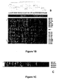
- miRNAs are aberrantly expressed in human ovarian cancer.
- the overall miRNA expression could clearly separate normal versus cancer tissues.
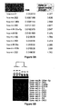
- the most significantly overexpressed miRNAs were miR-200a, miR-141, miR-200c, and miR-200b, whereas miR-199a, miR-140, miR-145 and miR-125b1 were among the most down modulated miRNAs.
- Wiemer et al discusses a review of the role of microRNAs in cancer
- microRNA genes are highly associated with chromosomal features involved in the etiology of different cancers.
- the perturbations in the genomic structure or chromosomal architecture of a cell caused by these cancer-associated chromosomal features can affect the expression of the miR gene(s) located in close proximity to that chromosomal feature.
- Evaluation of miR gene expression can therefore be used to indicate the presence of a cancer-causing chromosomal lesion in a subject.
- a given cancer can be treated by restoring the level of miR gene expression to normal.
- microRNA expression profiling can be used to diagnose cancer and predict whether a particular cancer is associated with an adverse prognosis.;
- the identification of specific mutations associated with genomic regions that harbor miR genes in CLL patients provides a means for diagnosing CLL and possibly other cancers.
- the present invention is based, in part, on the identification of an ovarian cancer-specific signature of miRNAs that are differentially-expressed in ovarian cancer cells, relative to normal control cells.
- the disclosure encompasses methods of diagnosing whether a subject has, or is at risk for developing, ovarian cancer, comprising measuring the level of at least one miR in a test sample from the subject, wherein an alteration in the level of the miR in the test sample, relative to the level of a corresponding miR in a control sample, is indicative of the subject either having, or being at risk for developing, ovarian cancer.
- a method of diagnosing whether a subject has ovarian cancer comprising: measuring the level of miR-200a in a test sample from the subject, and wherein an increase in the level of miR-200a in the test sample, relative to the level of miR-200a in a control sample, is indicative of the subject having, ovarian cancer.
- the invention provides a method of identifying an anti-ovarian cancer agent, comprising providing a test agent to a cell and measuring the level of miR-200a, wherein a decrease in the level of miR-200a in the cell, relative to a suitable control cell, is indicative of the test agent being an anti-ovarian cancer agent.
- a method of diagnosing whether a subject has, or is at risk for developing, ovarian cancer comprising measuring the level of at least one miR in a test sample from the subject.
- An alteration in the level of the miR in the test sample, relative to the level of a corresponding miR in a control sample, is indicative of the subject either having, or being at risk for developing, ovarian cancer.
- a method that includes identifying a correlation between miR expression and ovarian cancer or a predisposition for ovarian cancer, comprising: (a) labeling the miR isolated from a sample from a subject having or suspected of having a disease or condition; (b) hybridizing the miR to an miR array; (c) determining miR hybridization to the array; and (d) identifying miR differentially expressed in a sample representative of the disease or condition compared to a reference.
- a method where identifying miR differentially expressed comprises generating an miR profile for the sample and evaluating the miR profile to determine whether miR in the sample are differentially expressed compared to a normal sample.
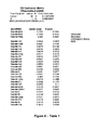
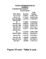
- the miR profile is selected from one or more of the miRs shown in Table 1. Also, in certain embodiments, the miR profile is selected from one or more of the miRs shown in Figure 3A or Figure 3B .
- the ovarian cancer is one or more of clear cell, serous or endometrioid ovarian cancer.
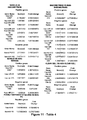
- the miR profile is selected from one or more of the miRs shown in Table 3, whereby ovarian cancer cells are distinguished from normal cells.
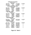
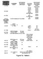
- the miR profile is selected from one or more of the miRs shown in Table 4, whereby ovarian cancer cells are distinguished by histotype among: serous, non-serous endometrioid, non-endometrioid, clear cell, non-clear cell, poorly differentiated and non-poorly differentiated.
- the miR profile involves at least one miR selected from the group consisting of miR-200a, miR-200b, miR-200c, miR-141, miR-199a, miR-140, miR-145 and miR-125b1, wherein a difference in expression of one or more of the miRNA compared to a normal sample is indicative of ovarian cancer.
- the miR profile involves at least miR-200a, miR-200b, miR-200c, miR-141, miR-199a, miR-140, miR-145 and miR-125b1, wherein a difference in expression of one or more of the miR compared to a normal sample is indicative of ovarian cancer.
- a method wherein an increase in expression of miR-200a, miR-200b, miR-200c or miR-141, and/or a decrease in expression of miR-199a, miR-140, miR-145 or miR-125b1, as compared to a normal sample, is indicative of ovarian cancer.
- the miR profile involves at least one miRNA selected from the group consisting of miR-200a, miR-200b, miR-200c and miR-141, wherein a difference in expression of one or more of the miRNA compared to a normal sample is indicative of serous ovarian cancer.
- the miR profile involves at least one miRNA selected from the group consisting of miR-205, miR21, miR-182, miR-200b, miR-141, miR-200c, miR-200a wherein a difference in expression of one or more of the miRNA compared to a normal sample is indicative of endometrioid ovarian cancer.
- the miR profile is selected from one or more of the miRs shown in Figure 3A or Figure 3B , and is indicative of serous ovarian cancer. In certain other embodiments, the miR profile is selected from one or more of the miRs shown in Figure 3A or Figure 3B , and is indicative of endometriod ovarian cancer. In certain other embodiments, the miR profile is selected from one or more of the miRs shown in Figure 3A or Figure 3B , and is indicative of clear cell ovarian cancer.
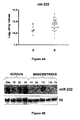
- miR-200a, miR-200b, miR-200c and miR-141 are up-regulated, and have as common putative target the oncosuppressor BAP1, BRCA1-associated protein, that is down-modulated in ovarian cancer.
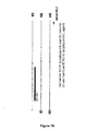
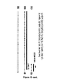
- the level of the at least one miR can be measured using a variety of techniques that are well known to those of skill in the art. In one embodiment, the level of the at least one miR is measured using Northern blot analysis. In another embodiment, the level of the at least one miR in the test sample is less than the level of the corresponding miR in the control sample. Also, in another embodiment, the level of the at least one miR in the test sample can be greater than the level of the corresponding miR in the control sample.
- the present disclosure also provides methods of diagnosing a cancer associated with one or more prognostic markers in a subject, comprising measuring the level of at least one miR in a cancer sample from the subject, wherein an alteration in the level of the at least one miR in the test sample, relative to the level of a corresponding miR in a control sample, is indicative of the subject having a cancer associated with the one or more prognostic markers.
- the level of the at least one miR is measured by reverse transcribing RNA from a test sample obtained from the subject to provide a set of target oligodeoxynucleotides; hybridizing the target oligodeoxynucleotides to a microarray comprising miR-specific probe oligonucleotides to provide a hybridization profile for the test sample; and, comparing the test sample hybridization profile to a hybridization profile generated from a control sample.
- An alteration in the signal of at least one miR is indicative of the subject either having, or being at risk for developing, such cancer.
- the present disclosure also encompasses methods of treating cancer in a subject, wherein the signal of at least one miR, relative to the signal generated from the control sample, is de-regulated (e.g., down-regulated, up-regulated).
- de-regulated e.g., down-regulated, up-regulated
- the present disclosure also encompasses methods of diagnosing whether a subject has, or is at risk for developing, a cancer associated with one or more adverse prognostic markers in a subject, by reverse transcribing RNA from a test sample obtained from the subject to provide a set of target oligodeoxynucleotides; hybridizing the target oligodeoxynucleotides to a microarray comprising miR-specific probe oligonucleotides to provide a hybridization profile for the test sample; and, comparing the test sample hybridization profile to a hybridization profile generated from a control sample. An alteration in the signal is indicative of the subject either having, or being at risk for developing, the cancer.
- the present disclosure also encompasses methods of treating cancer in a subject who has a cancer in which at least one miR is down-regulated or up-regulated in the cancer cells of the subject relative to control cells.
- the method comprises administering to the subject an effective amount of at least one isolated miR, such that proliferation of cancer cells in the subject is inhibited.
- the method comprises administering to the subject an effective amount of at least one compound for inhibiting expression of the at least one miR, such that proliferation of cancer cells in the subject is inhibited.
- the invention provides methods of treating cancer in a subject, comprising: determining the amount of at least one miR in cancer cells, relative to control cells; and altering the amount of miR expressed in the cancer cells by: administering to the subject an effective amount of at least one isolated miR, if the amount of the miR expressed in the cancer cells is less than the amount of the miR expressed in control cells; or administering to the subject an effective amount of at least one compound for inhibiting expression of the at least one miR, if the amount of the miR expressed in the cancer cells is greater than the amount of the miR expressed in control cells, such that proliferation of cancer cells in the subject is inhibited.
- the present disclosure further provides pharmaceutical compositions for treating cancer, comprising at least one isolated miR and a pharmaceutically-acceptable carrier.
- the pharmaceutical compositions the at least one isolated miR corresponds to a miR that is down-regulated in cancer cells relative to suitable control cells.
- the pharmaceutical composition comprises at least one miR expression inhibitor compound and a pharmaceutically-acceptable carrier. Also, in a particular embodiment, the pharmaceutical composition comprises at least one miR expression inhibitor compound is specific for a miR that is down regulated and/or up-regulated in cancer cells relative to suitable control cells.
- the present invention provides methods of identifying an anti-cancer agent, comprising providing a test agent to a cell and measuring the level of at least one miR associated with decreased expression levels in cancer cells, wherein an increase in the level of the miR in the cell, relative to a suitable control cell, is indicative of the test agent being an anti-cancer agent.
- the present invention also provides methods of identifying an anti-ovarian cancer agent, comprising providing a test agent to an ovarian cancer cell and measuring the level of at least one miR associated with increased expression levels in cancer cells, wherein an decrease in the level of the miR in the cell, relative to a suitable control cell, is indicative of the test agent being an anti-cancer.
- At least one miR is selected the group shown in Table 3.
- the miR is selected from the group consisting of miR-200a, miR-141, miR-200c, and miR-200b, miR-199a, miR-140, miR145, and miR-125b1.
- miRNAs whose expression is correlated with specific ovarian cancer biopathologic features, such as histotype, lymphovascular and organ invasion, and involvement of ovarian surface.
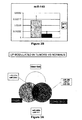
- the levels of miR-21, miR-203, and miR-205, up-modulated in ovarian carcinomas compared with normal tissues were significantly increased after 5-aza-2'-deoxycytidine demethylating treatment of OVCAR3 cells.
- the present invention is directed to compositions and methods relating to preparation and characterization of miRNAs, as well as use of miRNAs for therapeutic, prognostic, and diagnostic applications.
- a “miR ,” “microRNA,” “miR,” or “miRNA” refers to the unprocessed or processed RNA transcript from an miR gene. As the miRs are not translated into protein, the term “miRs” does not include proteins.
- the unprocessed miR gene transcript is also called an "miR precursor,” and typically comprises an RNA transcript of about 70-100 nucleotides in length.
- the miR precursor can be processed by digestion with an RNAse (for example, Dicer, Argonaut, or RNAse III, e.g., E . coli RNAse III)) into an active 19-25 nucleotide RNA molecule.
- RNAse for example, Dicer, Argonaut, or RNAse III, e.g., E . coli RNAse III
- miR This active 19-25 nucleotide RNA molecule is also called the "processed" miR gene transcript or “mature” miRNA.
- miR can include one or more of miR-oligonucleotides, including mature miRs, pre-miRs, pri-miRs, or a miR seed sequence. In certain embodiments, a mixture of various miR nucleic acids can also be used. Also, in certain embodiments, the miRs may be modified to enhance delivery.
- the miRNA (miR) information is available from the Sanger Institute, which maintains a registry of miRNA at http: / microrna.sanger.ac.uk / sequences / .
- the miRBase Sequence database includes the nucleotide sequences and annotations of published miRNA from a variety of sources.
- the miRBase Registry provides unique names for novel miRNA genes that comply with conventional naming nomenclature for new miRNA prior to publication. Also, the miRBase Targets is a resource for predicated miRNA targets in animals.
- the active 19-25 nucleotide RNA molecule can be obtained from the miR precursor through natural processing routes (e.g ., using intact cells or cell lysates) or by synthetic processing routes (e.g ., using isolated processing enzymes, such as isolated Dicer, Argonaut, or RNAase III). It is understood that the active 19-25 nucleotide RNA molecule can also be produced directly by biological or chemical synthesis, without having been processed from the miR precursor.
- the present disclosure encompasses methods of diagnosing whether a subject has, or is at risk for developing, cancer, comprising measuring the level of at least one miR in a test sample from the subject and comparing the level of the miR in the test sample to the level of a corresponding miR in a control sample.
- a "subject" can be any mammal that has, or is suspected of having, breast cancer.
- the subject is a human who has, or is suspected of having, cancer.
- the level of at least one miR can be measured in cells of a biological sample obtained from the subject.
- a tissue sample can be removed from a subject suspected of having ovarian cancer associated with by conventional biopsy techniques.
- a blood sample can be removed from the subject, and white blood cells can be isolated for DNA extraction by standard techniques.
- the blood or tissue sample is preferably obtained from the subject prior to initiation of radiotherapy, chemotherapy or other therapeutic treatment.
- a corresponding control tissue or blood sample can be obtained from unaffected tissues of the subject, from a normal human individual or population of normal individuals, or from cultured cells corresponding to the majority of cells in the subject's sample.
- the control tissue or blood sample is then processed along with the sample from the subject, so that the levels of miR produced from a given miR gene in cells from the subject's sample can be compared to the corresponding miR levels from cells of the control sample.
- an alteration i.e ., an increase or decrease in the level of a miR in the sample obtained from the subject, relative to the level of a corresponding miR in a control sample, is indicative of the presence of cancer in the subject.
- the level of the at least one miR in the test sample is greater than the level of the corresponding miR in the control sample (i.e., expression of the miR is "up-regulated”).
- expression of a miR is "up-regulated” when the amount of miR in a cell or tissue sample from a subject is greater than the amount the same in a control cell or tissue sample.
- the level of the at least one miR in the test sample is less than the level of the corresponding miR in the control sample (i.e., expression of the miR is "down-regulated”).
- expression of an miR gene is “down-regulated” when the amount of miR produced from that gene in a cell or tissue sample from a subject is less than the amount produced from the same gene in a control cell or tissue sample.
- the relative miR gene expression in the control and normal samples can be determined with respect to one or more RNA expression standards.
- the standards can comprise, for example, a zero miR gene expression level, the miR gene expression level in a standard cell line, or the average level of miR gene expression previously obtained for a population of normal human controls.
- the level of a miR in a sample can be measured using any technique that is suitable for detecting RNA expression levels in a biological sample. Suitable techniques for determining RNA expression levels in cells from a biological sample (e.g., Northern blot analysis, RT-PCR, in situ hybridization) are well known to those of skill in the art.
- the level of at least one miR is detected using Northern blot analysis. For example, total cellular RNA can be purified from cells by homogenization in the presence of nucleic acid extraction buffer, followed by centrifugation. Nucleic acids are precipitated, and DNA is removed by treatment with DNase and precipitation.
- RNA molecules are then separated by gel electrophoresis on agarose gels according to standard techniques, and transferred to nitrocellulose filters.
- the RNA is then immobilized on the filters by heating. Detection and quantification of specific RNA is accomplished using appropriately labeled DNA or RNA probes complementary to the RNA in question. See, for example, Molecular Cloning: A Laboratory Manual, J. Sambrook et al., eds., 2nd edition, Cold Spring Harbor Laboratory Press, 1989, Chapter 7 .
- Suitable probes for Northern blot hybridization of a given miR can be produced from the nucleic acid sequences of the given miR. Methods for preparation of labeled DNA and RNA probes, and the conditions for hybridization thereof to target nucleotide sequences, are described in Molecular Cloning: A Laboratory Manual, J. Sambrook et al., eds., 2nd edition, Cold Spring Harbor Laboratory Press, 1989, Chapters 10 and 11 .
- the nucleic acid probe can be labeled with, e.g., a radionuclide, such as 3 H, 32 P, 33 P, 14 C, or 35 S; a heavy metal; or a ligand capable of functioning as a specific binding pair member for a labeled ligand (e.g., biotin, avidin or an antibody), a fluorescent molecule, a chemiluminescent molecule, an enzyme or the like.
- a radionuclide such as 3 H, 32 P, 33 P, 14 C, or 35 S
- a heavy metal e.g., a ligand capable of functioning as a specific binding pair member for a labeled ligand (e.g., biotin, avidin or an antibody), a fluorescent molecule, a chemiluminescent molecule, an enzyme or the like.
- Probes can be labeled to high specific activity by either the nick translation method of Rigby et al. (1977), J. Mol. Biol. 113:237-251 or by the random priming method of Fienberg et al. (1983), Anal. Biochem. 132:6-13 .
- the latter is the method of choice for synthesizing 32 P-labeled probes of high specific activity from single-stranded DNA or from RNA templates. For example, by replacing preexisting nucleotides with highly radioactive nucleotides according to the nick translation method, it is possible to prepare 32 P-labeled nucleic acid probes with a specific activity well in excess of 10 8 cpm/microgram.
- Autoradiographic detection of hybridization can then be performed by exposing hybridized filters to photographic film. Densitometric scanning of the photographic films exposed by the hybridized filters provides an accurate measurement of miR gene transcript levels. Using another approach, miR gene transcript levels can be quantified by computerized imaging systems, such the Molecular Dynamics 400-B 2D Phosphorimager available from Amersham Biosciences, Piscataway, NJ.
- the random-primer method can be used to incorporate an analogue, for example, the dTTP analogue 5-(N-(N-biotinyl-epsilon-aminocaproyl)-3-aminoallyl)deoxyuridine triphosphate, into the probe molecule.
- analogue for example, the dTTP analogue 5-(N-(N-biotinyl-epsilon-aminocaproyl)-3-aminoallyl)deoxyuridine triphosphate
- the biotinylated probe oligonucleotide can be detected by reaction with biotin-binding proteins, such as avidin, streptavidin, and antibodies (e.g., anti-biotin antibodies) coupled to fluorescent dyes or enzymes that produce color reactions.
- determining the levels of RNA transcripts can be accomplished using the technique of in situ hybridization.
- This technique requires fewer cells than the Northern blotting technique, and involves depositing whole cells onto a microscope cover slip and probing the nucleic acid content of the cell with a solution containing radioactive or otherwise labeled nucleic acid (e.g., cDNA or RNA) probes.
- a solution containing radioactive or otherwise labeled nucleic acid e.g., cDNA or RNA
- This technique is particularly well-suited for analyzing tissue biopsy samples from subjects.
- the practice of the in situ hybridization technique is described in more detail in U.S. Pat. No. 5,427,916 .
- Suitable probes for in situ hybridization of a given miR can be produced from the nucleic acid sequences.
- the relative number of miR gene transcripts in cells can also be determined by reverse transcription of miR gene transcripts, followed by amplification of the reverse-transcribed transcripts by polymerase chain reaction (RT-PCR).
- the levels of miR gene transcripts can be quantified in comparison with an internal standard, for example, the level of mRNA from a "housekeeping" gene present in the same sample.
- a suitable "housekeeping" gene for use as an internal standard includes, e.g., myosin or glyceraldehyde-3-phosphate dehydrogenase (G3PDH).
- G3PDH glyceraldehyde-3-phosphate dehydrogenase
- an oligolibrary in microchip format (i.e., a microarray), may be constructed containing a set of probe oligodeoxynucleotides that are specific for a set of miR genes.
- a microarray the expression level of multiple microRNAs in a biological sample can be determined by reverse transcribing the RNAs to generate a set of target oligodeoxynucleotides, and hybridizing them to probe oligodeoxynucleotides on the microarray to generate a hybridization, or expression, profile.
- the hybridization profile of the test sample can then be compared to that of a control sample to determine which microRNAs have an altered expression level in cancer.
- probe oligonucleotide or “probe oligodeoxynucleotide” refers to an oligonucleotide that is capable of hybridizing to a target oligonucleotide.
- Target oligonucleotide or “target oligodeoxynucleotide” refers to a molecule to be detected (e.g., via hybridization).
- miR-specific probe oligonucleotide or "probe oligonucleotide specific for an miR” is meant a probe oligonucleotide that has a sequence selected to hybridize to a specific miR, or to a reverse transcript of the specific miR.
- an "expression profile” or “hybridization profile” of a particular sample is essentially a fingerprint of the state of the sample; while two states may have any particular gene similarly expressed, the evaluation of a number of genes simultaneously allows the generation of a gene expression profile that is unique to the state of the cell. That is, normal cells may be distinguished from cancer cells, and within cancer cells, different prognosis states (good or poor long term survival prospects, for example) may be determined. By comparing expression profiles of cancer cells in different states, information regarding which genes are important (including both up- and down-regulation of genes) in each of these states is obtained.
- sequences that are differentially expressed in cancer cells or normal cells, as well as differential expression resulting in different prognostic outcomes allows the use of this information in a number of ways. For example, a particular treatment regime may be evaluated (e.g., to determine whether a chemotherapeutic drug act to improve the long-term prognosis in a particular patient). Similarly, diagnosis may be done or confirmed by comparing patient samples with the known expression profiles. Furthermore, these gene expression profiles (or individual genes) allow screening of drug candidates that suppress the cancer expression profile or convert a poor prognosis profile to a better prognosis profile.
- the disclosure provides methods of diagnosing whether a subject has, or is at risk for developing, cancer, comprising reverse transcribing RNA from a test sample obtained from the subject to provide a set of target oligo-deoxynucleotides, hybridizing the target oligo-deoxynucleotides to a microarray comprising miRNA-specific probe oligonucleotides to provide a hybridization profile for the test sample, and comparing the test sample hybridization profile to a hybridization profile generated from a control sample, wherein an alteration in the signal of at least one miRNA is indicative of the subject either having, or being at risk for developing, cancer.
- the microarray comprises miRNA-specific probe oligonucleotides for a substantial portion of the human miRNome.
- the microarray can be prepared from gene-specific oligonucleotide probes generated from known miRNA sequences.
- the array may contain two different oligonucleotide probes for each miRNA, one containing the active, mature sequence and the other being specific for the precursor of the miRNA.
- the array may also contain controls, such as one or more mouse sequences differing from human orthologs by only a few bases, which can serve as controls for hybridization stringency conditions.
- tRNAs from both species may also be printed on the microchip, providing an internal, relatively stable, positive control for specific hybridization.
- One or more appropriate controls for non-specific hybridization may also be included on the microchip. For this purpose, sequences are selected based upon the absence of any homology with any known miRNAs.
- the microarray may be fabricated using techniques known in the art. For example, probe oligonucleotides of an appropriate length, e.g., 40 nucleotides, are 5'-amine modified at position C6 and printed using commercially available microarray systems, e.g., the GeneMachine OmniGrid Tm 100 Microarrayer and Amersham CodeLink Tm activated slides. Labeled cDNA oligomer corresponding to the target RNAs is prepared by reverse transcribing the target RNA with labeled primer. Following first strand synthesis, the RNA/DNA hybrids are denatured to degrade the RNA templates.
- the labeled target cDNAs thus prepared are then hybridized to the microarray chip under hybridizing conditions, e.g., 6X SSPE/30% formamide at 25°C for 18 hours, followed by washing in 0.75X TNT at 37°C for 40 minutes. At positions on the array where the immobilized probe DNA recognizes a complementary target cDNA in the sample, hybridization occurs.
- the labeled target cDNA marks the exact position on the array where binding occurs, allowing automatic detection and quantification.
- the output consists of a list of hybridization events, indicating the relative abundance of specific cDNA sequences, and therefore the relative abundance of the corresponding complementary miRs, in the patient sample.
- the labeled cDNA oligomer is a biotin-labeled cDNA, prepared from a biotin-labeled primer.
- the microarray is then processed by direct detection of the biotin-containing transcripts using, e.g., Streptavidin-Alexa647 conjugate, and scanned utilizing conventional scanning methods. Image intensities of each spot on the array are proportional to the abundance of the corresponding miR in the patient sample.
- the use of the array has several advantages for miRNA expression detection.
- the relatively limited number of miRNAs allows the construction of a common microarray for several species, with distinct oligonucleotide probes for each. Such a tool would allow for analysis of trans-species expression for each known miR under various conditions.
- a microchip containing miRNA-specific probe oligonucleotides corresponding to a substantial portion of the miRNome, preferably the entire miRNome may be employed to carry out miR gene expression profiling, for analysis of miR expression patterns. Distinct miR signatures can be associated with established disease markers, or directly with a disease state.
- total RNA from a sample from a subject suspected of having cancer is quantitatively reverse transcribed to provide a set of labeled target oligodeoxynucleotides complementary to the RNA in the sample.
- the target oligodeoxynucleotides are then hybridized to a microarray comprising miRNA-specific probe oligonucleotides to provide a hybridization profile for the sample.
- the result is a hybridization profile for the sample representing the expression pattern of miRNA in the sample.
- the hybridization profile comprises the signal from the binding of the target oligodeoxynucleotides from the sample to the miRNA-specific probe oligonucleotides in the microarray.
- the profile may be recorded as the presence or absence of binding (signal vs. zero signal). More preferably, the profile recorded includes the intensity of the signal from each hybridization. The profile is compared to the hybridization profile generated from a normal, i.e., noncancerous, control sample. An alteration in the signal is indicative of the presence of the cancer in the subject.
- the disclosure also provides methods of diagnosing a cancer associated with one or more prognostic markers, comprising measuring the level of at least one miR in a cancer test sample from a subject and comparing the level of the at least one miR in the cancer test sample to the level of a corresponding miR in a control sample.
- An alteration e.g., an increase, a decrease
- in the signal of at least one miRNA in the test sample relative to the control sample is indicative of the subject either having, or being at risk for developing, cancer associated with the one or more prognostic markers.
- the cancer can be associated with one or more prognostic markers or features, including, a marker associated with an adverse (i.e., negative) prognosis, or a marker associated with a good (i.e., positive) prognosis.
- the cancer that is diagnosed using the methods described herein is associated with one or more adverse prognostic features.
- the level of the at least one miR is measured by reverse transcribing RNA from a test sample obtained from the subject to provide a set of target oligodeoxynucleotides, hybridizing the target oligodeoxynucleotides to a microarray that comprises miRNA-specific probe oligonucleotides to provide a hybridization profile for the test sample, and comparing the test sample hybridization profile to a hybridization profile generated from a control sample.
- alterations in the level of one or more miRs in cells can result in the deregulation of one or more intended targets for these miRs, which can lead to the formation of cancer.
- altering the level of the miR e.g., by decreasing the level of a miR that is up-regulated in CLL cells, by increasing the level of a miR that is down-regulated in cancer cells
- altering the level of the miR may successfully treat the cancer.
- putative gene targets for miRNAs that are deregulated in cancer cells are described herein.
- the present disclosure encompasses methods of treating cancer in a subject, wherein at least one miR is de-regulated (e.g., down-regulated, up-regulated) in the cancer cells of the subject.
- the method comprises administering an effective amount of the at least one isolated miR such that proliferation of cancer cells in the subject is inhibited.
- the method comprises administering to the subject an effective amount of at least one compound for inhibiting expression of the at least one miR gene, referred to herein as miR gene expression inhibition compounds, such that proliferation of cancer cells is inhibited.
- treat refers to ameliorating symptoms associated with a disease or condition, for example, cancer, including preventing or delaying the onset of the disease symptoms, and/or lessening the severity or frequency of symptoms of the disease or condition.
- subject and “individual” are defined herein to include animals, such as mammals, including but not limited to, primates, cows, sheep, goats, horses, dogs, cats, rabbits, guinea pigs, rats, mice or other bovine, ovine, equine, canine, feline, rodent, or murine species.
- the animal is a human.
- an "effective amount" of an isolated miR is an amount sufficient to inhibit proliferation of a cancer cell in a subject suffering from cancer.
- an effective amount of an miR to be administered to a given subject by taking into account factors, such as the size and weight of the subject; the extent of disease penetration; the age, health and sex of the subject; the route of administration; and whether the administration is regional or systemic.
- an effective amount of an isolated miR can be based on the approximate or estimated body weight of a subject to be treated. Preferably, such effective amounts are administered parenterally or enterally, as described herein.
- an effective amount of the isolated miR is administered to a subject can range from about 5 - 3000 micrograms/kg of body weight, from about 700 - 1000 micrograms/kg of body weight, or greater than about 1000 micrograms/kg of body weight.
- an appropriate dosage regimen for the administration of an isolated miR to a given subject can be administered to the subject once ( e.g., as a single injection or deposition).
- an miR can be administered once or twice daily to a subject for a period of from about three to about twenty-eight days, more particularly from about seven to about ten days.
- an miR is administered once a day for seven days.
- a dosage regimen comprises multiple administrations, it is understood that the effective amount of the miR administered to the subject can comprise the total amount of miR administered over the entire dosage regimen.
- an "isolated" miR is one which is synthesized, or altered or removed from the natural state through human intervention.
- a synthetic miR, or an miR partially or completely separated from the coexisting materials of its natural state is considered to be “isolated.”
- An isolated miR can exist in substantially-purified form, or can exist in a cell into which the miR has been delivered.
- an miR which is deliberately delivered to, or expressed in, a cell is considered an “isolated” miR.
- An miR produced inside a cell from an miR precursor molecule is also considered to be “isolated” molecule.
- Isolated miRs can be obtained using a number of standard techniques.
- the miRs can be chemically synthesized or recombinantly produced using methods known in the art.
- miRs are chemically synthesized using appropriately protected ribonucleoside phosphoramidites and a conventional DNA/RNA synthesizer.
- RNA molecules or synthesis reagents include, e.g., Proligo (Hamburg, Germany), Dharmacon Research (Lafayette, CO, U.S.A.), Pierce Chemical (part of Perbio Science, Rockford, IL, U.S.A.), Glen Research (Sterling, VA, U.S.A.), ChemGenes (Ashland, MA, U.S.A.) and Cruachem (Glasgow, UK).
- the miRs can be expressed from recombinant circular or liner DNA plasmids using any suitable promoter.
- suitable promoters for expressing RNA from a plasmid include, e.g., the U6 or H1 RNA pol III promoter sequences, or the cytomegalovirus promoters. Selection of other suitable promoters is within the skill in the art.
- the recombinant plasmids of the invention can also comprise inducible or regulatable promoters for expression of the miRs in cancer cells.
- the miRs that are expressed from recombinant plasmids can be isolated from cultured cell expression systems by standard techniques.
- the miRs which are expressed from recombinant plasmids can also be delivered to, and expressed directly in, the cancer cells.
- the use of recombinant plasmids to deliver the miRs to cancer cells is discussed in more detail below.
- the miRs can be expressed from a separate recombinant plasmid, or they can be expressed from the same recombinant plasmid.
- the miRs are expressed as RNA precursor molecules from a single plasmid, and the precursor molecules are processed into the functional miR by a suitable processing system, including, but not limited to, processing systems extant within a cancer cell.
- suitable processing systems include, e.g., the in vitro Drosophila cell lysate system (e.g., as described in U.S. Published Patent Application No. 2002/0086356 to Tuschl et al. . and the E. coli RNAse III system (e.g., as described in U.S. Published Patent Application No. 2004/0014113 to Yang et al. .
- plasmids suitable for expressing the miRs are within the skill in the art. See, for example, Zeng et al. (2002), Molecular Cell 9:1327-1333 ; Tuschl (2002), Nat. Biotechnol, 20:446-448 ; Brummelkamp et al. (2002), Science 296:550-553 ; Miyagishi et al. (2002), Nat. Biotechnol. 20:497-500 ; Paddison et al. (2002), Genes Dev. 16:948-958 ; Lee et al. (2002), Nat. Biotechnol. 20:500-505 ; and Paul et al. (2002), Nat. Biotechnol. 20:505-508 .
- a plasmid expressing the miRs comprises a sequence encoding a miR precursor RNA under the control of the CMV intermediate-early promoter.
- "under the control" of a promoter means that the nucleic acid sequences encoding the miR are located 3' of the promoter, so that the promoter can initiate transcription of the miR coding sequences.
- the miRs can also be expressed from recombinant viral vectors. It is contemplated that the miRs can be expressed from two separate recombinant viral vectors, or from the same viral vector.
- the RNA expressed from the recombinant viral vectors can either be isolated from cultured cell expression systems by standard techniques, or can be expressed directly in cancer cells. The use of recombinant viral vectors to deliver the miRs to cancer cells is discussed in more detail below.
- the recombinant viral vectors of the invention comprise sequences encoding the miRs and any suitable promoter for expressing the RNA sequences.
- suitable promoters include, for example, the U6 or H1 RNA pol III promoter sequences, or the cytomegalovirus promoters. Selection of other suitable promoters is within the skill in the art.
- the recombinant viral vectors of the invention can also comprise inducible or regulatable promoters for expression of the miRs in a cancer cell.
- Any viral vector capable of accepting the coding sequences for the miRs can be used; for example, vectors derived from adenovirus (AV); adeno-associated virus (AAV); retroviruses (e.g., lentiviruses (LV), Rhabdoviruses, murine leukemia virus); herpes virus, and the like.
- AV adenovirus
- AAV adeno-associated virus
- retroviruses e.g., lentiviruses (LV), Rhabdoviruses, murine leukemia virus
- herpes virus and the like.
- the tropism of the viral vectors can be modified by pseudotyping the vectors with envelope proteins or other surface antigens from other viruses, or by substituting different viral capsid proteins, as appropriate.
- lentiviral vectors of the invention can be pseudotyped with surface proteins from vesicular stomatitis virus (VSV), rabies, Ebola, Mokola, and the like.
- AAV vectors of the invention can be made to target different cells by engineering the vectors to express different capsid protein serotypes.
- an AAV vector expressing a serotype 2 capsid on a serotype 2 genome is called AAV 2/2.
- This serotype 2 capsid gene in the AAV 2/2 vector can be replaced by a serotype 5 capsid gene to produce an AAV 2/5 vector.
- AAV vectors that express different capsid protein serotypes are within the skill in the art; see, e.g., Rabinowitz, J.E., et al. (2002), J. Virol. 76:791-801 .
- recombinant viral vectors suitable for use in the invention methods for inserting nucleic acid sequences for expressing RNA into the vector, methods of delivering the viral vector to the cells of interest, and recovery of the expressed RNA products are within the skill in the art. See, for example, Dornburg (1995), Gene Therap. 2:301-310 ; Eglitis (1988), Biotechniques 6:608-614 ; Miller (1990), Hum. Gene Therap. 1:5-14 ; and Anderson (1998), Nature 392:25-30 .
- Particularly suitable viral vectors are those derived from AV and AAV.
- a suitable AV vector for expressing the miRs, a method for constructing the recombinant AV vector, and a method for delivering the vector into target cells are described in Xia et al. (2002), Nat. Biotech. 20:1006-1010 .
- Suitable AAV vectors for expressing the miRs, methods for constructing the recombinant AAV vector, and methods for delivering the vectors into target cells are described in Samulski et al. (1987), J. Virol. 61:3096-3101 ; Fisher et al. (1996), J. Virol., 70:520-532 ; Samulski et al. (1989), J. Virol.
- the miRs are expressed from a single recombinant AAV vector comprising the CMV intermediate early promoter.
- a recombinant AAV viral vector of the invention comprises a nucleic acid sequence encoding an miR precursor RNA in operable connection with a polyT termination sequence under the control of a human U6 RNA promoter.
- operable connection with a polyT termination sequence means that the nucleic acid sequences encoding the sense or antisense strands are immediately adjacent to the polyT termination signal in the 5' direction.
- the polyT termination signals act to terminate transcription.
- an effective amount of at least one compound which inhibits miR expression can also be administered to the subject.
- inhibiting miR expression means that the production of the active, mature form of miR after treatment is less than the amount produced prior to treatment.
- One skilled in the art can readily determine whether miR expression has been inhibited in a cancer cell, using for example the techniques for determining miR transcript level discussed above for the diagnostic method. Inhibition can occur at the level of gene expression (i.e., by inhibiting transcription of a miR gene encoding the miR) or at the level of processing (e.g., by inhibiting processing of a miR precursor into a mature, active miR).
- an "effective amount" of a compound that inhibits miR expression is an amount sufficient to inhibit proliferation of a cancer cell in a subject suffering from a cancer associated with a cancer-associated chromosomal feature.
- an effective amount of an miR expression-inhibiting compound to be administered to a given subject by taking into account factors, such as the size and weight of the subject; the extent of disease penetration; the age, health and sex of the subject; the route of administration; and whether the administration is regional or systemic.
- an effective amount of the expression-inhibiting compound can be based on the approximate or estimated body weight of a subject to be treated. Such effective amounts are administered parenterally or enterally, among others, as described herein.
- an effective amount of the expression-inhibiting compound administered to a subject can range from about 5 -3000 micrograms/kg of body weight, from about 700 - 1000 micrograms/kg of body weight, or it can be greater than about 1000 micrograms/kg of body weight.
- an expression-inhibiting compound can be administered to the subject once (e.g., as a single injection or deposition).
- an expression-inhibiting compound can be administered once or twice daily to a subject for a period of from about three to about twenty-eight days, more preferably from about seven to about ten days.
- an expression-inhibiting compound is administered once a day for seven days.
- the effective amount of the expression-inhibiting compound administered to the subject can comprise the total amount of compound administered over the entire dosage regimen.
- Suitable compounds for inhibiting miR gene expression include double-stranded RNA (such as short- or small-interfering RNA or "siRNA”), antisense nucleic acids, and enzymatic RNA molecules, such as ribozymes. Each of these compounds can be targeted to a given miR and destroy or induce the destruction of the target miR.
- siRNA short- or small-interfering RNA or "siRNA”
- antisense nucleic acids such as antisense nucleic acids
- enzymatic RNA molecules such as ribozymes.
- expression of a given miR gene can be inhibited by inducing RNA interference of the miR gene with an isolated double-stranded RNA (“dsRNA”) molecule which has at least 90%, for example at least 95%, at least 98%, at least 99% or 100%, sequence homology with at least a portion of the miR.
- dsRNA isolated double-stranded RNA
- the dsRNA molecule is a "short or small interfering RNA" or "siRNA.”
- siRNA useful in the present methods comprise short double-stranded RNA from about 17 nucleotides to about 29 nucleotides in length, preferably from about 19 to about 25 nucleotides in length.
- the siRNA comprise a sense RNA strand and a complementary antisense RNA strand annealed together by standard Watson-Crick base-pairing interactions (hereinafter "base-paired").
- the sense strand comprises a nucleic acid sequence which is substantially identical to a nucleic acid sequence contained within the target miR.
- a nucleic acid sequence in an siRNA which is "substantially identical" to a target sequence contained within the target mRNA is a nucleic acid sequence that is identical to the target sequence, or that differs from the target sequence by one or two nucleotides.
- the sense and antisense strands of the siRNA can comprise two complementary, single-stranded RNA molecules, or can comprise a single molecule in which two complementary portions are base-paired and are covalently linked by a single-stranded "hairpin" area.
- the siRNA can also be altered RNA that differs from naturally-occurring RNA by the addition, deletion, substitution and/or alteration of one or more nucleotides.
- Such alterations can include addition of non-nucleotide material, such as to the end(s) of the siRNA or to one or more internal nucleotides of the siRNA, or modifications that make the siRNA resistant to nuclease digestion, or the substitution of one or more nucleotides in the siRNA with deoxyribonucleotides.
- the siRNA can also comprise a 3' overhang.
- a "3' overhang” refers to at least one unpaired nucleotide extending from the 3'-end of a duplexed RNA strand.
- the siRNA comprises at least one 3' overhang of from 1 to about 6 nucleotides (which includes ribonucleotides or deoxyribonucleotides) in length, from 1 to about 5 nucleotides in length, from 1 to about 4 nucleotides in length, or from about 2 to about 4 nucleotides in length.
- the 3' overhang is present on both strands of the siRNA, and is 2 nucleotides in length.
- each strand of the siRNA can comprise 3' overhangs of dithymidylic acid ("TT") or diuridylic acid (“uu").
- the siRNA can be produced chemically or biologically, or can be expressed from a recombinant plasmid or viral vector, as described above for the isolated miRs.
- Exemplary methods for producing and testing dsRNA or siRNA molecules are described in U.S. Published Patent Application No. 2002/0173478 to Gewirtz and in U.S. Published Patent Application No. 2004/0018176 to Reich et al..
- an antisense nucleic acid refers to a nucleic acid molecule that binds to target RNA by means of RNA-RNA or RNA-DNA or RNA-peptide nucleic acid interactions, which alters the activity of the target RNA.
- Antisense nucleic acids suitable for use in the present methods are single-stranded nucleic acids (e.g., RNA, DNA, RNA-DNA chimeras, PNA) that generally comprise a nucleic acid sequence complementary to a contiguous nucleic acid sequence in an miR.
- the antisense nucleic acid can comprise a nucleic acid sequence that is 50-100% complementary, 75-100% complementary, or 95-100% complementary to a contiguous nucleic acid sequence in an miR. Nucleic acid sequences for the miRs are provided herein. Without wishing to be bound by any theory, it is believed that the antisense nucleic acids activate RNase H or another cellular nuclease that digests the miR /antisense nucleic acid duplex.
- Antisense nucleic acids can also contain modifications to the nucleic acid backbone or to the sugar and base moieties (or their equivalent) to enhance target specificity, nuclease resistance, delivery or other properties related to efficacy of the molecule.
- modifications include cholesterol moieties, duplex intercalators, such as acridine, or one or more nuclease-resistant groups.
- Antisense nucleic acids can be produced chemically or biologically, or can be expressed from a recombinant plasmid or viral vector, as described above for the isolated miRs. Exemplary methods for producing and testing are within the skill in the art; see, e.g., Stein and Cheng (1993), Science 261:1004 and U.S. Pat. No. 5,849,902 to Woolf et al. .
- an "enzymatic nucleic acid” refers to a nucleic acid comprising a substrate binding region that has complementarity to a contiguous nucleic acid sequence of an miR, and which is able to specifically cleave the miR.
- the enzymatic nucleic acid substrate binding region can be, for example, 50-100% complementary, 75-100% complementary, or 95-100% complementary to a contiguous nucleic acid sequence in an miR.
- the enzymatic nucleic acids can also comprise modifications at the base, sugar, and/or phosphate groups.
- An exemplary enzymatic nucleic acid for use in the present methods is a ribozyme.
- the enzymatic nucleic acids can be produced chemically or biologically, or can be expressed from a recombinant plasmid or viral vector, as described above for the isolated miRs.
- Exemplary methods for producing and testing dsRNA or siRNA molecules are described in Werner and Uhlenbeck (1995), Nucl. Acids Res. 23:2092-96 ; Hammann et al. (1999), Antisense and Nucleic Acid Drug Dev. 9:25-31 ; and U.S. Pat. No. 4,987,071 to Cech et al .
- Administration of at least one miR, or at least one compound for inhibiting miR expression will inhibit the proliferation of cancer cells in a subject who has a cancer associated with a cancer-associated chromosomal feature.
- to "inhibit the proliferation of a cancer cell” means to kill the cell, or permanently or temporarily arrest or slow the growth of the cell.
- Inhibition of cancer cell proliferation can be inferred if the number of such cells in the subject remains constant or decreases after administration of the miRs or miR gene expression-inhibiting compounds.
- An inhibition of cancer cell proliferation can also be inferred if the absolute number of such cells increases, but the rate of tumor growth decreases.
- the number of cancer cells in a subject's body can be determined by direct measurement, or by estimation from the size of primary or metastatic tumor masses.
- the number of cancer cells in a subject can be measured by immunohistological methods, flow cytometry, or other techniques designed to detect characteristic surface markers of cancer cells.
- the miRs or miR gene expression-inhibiting compounds can be administered to a subject by any means suitable for delivering these compounds to cancer cells of the subject.
- the miRs or miR expression inhibiting compounds can be administered by methods suitable to transfect cells of the subject with these compounds, or with nucleic acids comprising sequences encoding these compounds.
- the cells are transfected with a plasmid or viral vector comprising sequences encoding at least one miR or miR gene expression inhibiting compound.
- Transfection methods for eukaryotic cells include, e.g., direct injection of the nucleic acid into the nucleus or pronucleus of a cell; electroporation; liposome transfer or transfer mediated by lipophilic materials; receptor-mediated nucleic acid delivery, bioballistic or particle acceleration; calcium phosphate precipitation, and transfection mediated by viral vectors.
- cells can be transfected with a liposomal transfer compound, e.g., DOTAP (N-[1-(2,3-dioleoyloxy)propyl]-N,N,N-trimethyl-ammonium methylsulfate, Boehringer - Mannheim) or an equivalent, such as LIPOFECTIN.
- DOTAP N-[1-(2,3-dioleoyloxy)propyl]-N,N,N-trimethyl-ammonium methylsulfate, Boehringer - Mannheim
- LIPOFECTIN LIPOFECTIN
- An miR or miR gene expression inhibiting compound can also be administered to a subject by any suitable enteral or parenteral administration route.
- Suitable enteral administration routes for the present methods include, e.g., oral, rectal, or intranasal delivery.
- Suitable parenteral administration routes include, e.g., intravascular administration (e.g., intravenous bolus injection, intravenous infusion, intra-arterial bolus injection, intra-arterial infusion and catheter instillation into the vasculature); peri- and intra-tissue injection (e.g., peri-tumoral and intra-tumoral injection, intra-retinal injection, or subretinal injection); subcutaneous injection or deposition, including subcutaneous infusion (such as by osmotic pumps); direct application to the tissue of interest, for example by a catheter or other placement device (e.g., a retinal pellet or a suppository or an implant comprising a porous, non-porous, or ge
- an miR or miR expression inhibiting compound can be administered to the subject either as naked RNA, in combination with a delivery reagent, or as a nucleic acid (e.g., a recombinant plasmid or viral vector) comprising sequences that express the miR or expression inhibiting compound.
- Suitable delivery reagents include, e.g., the Mirus Transit TKO lipophilic reagent; lipofectin; lipofectamine; cellfectin; polycations ( e.g., polylysine), and liposomes.
- Recombinant plasmids and viral vectors comprising sequences that express the miRs or miR gene expression inhibiting compounds, and techniques for delivering such plasmids and vectors to cancer cells, are discussed herein.
- liposomes are used to deliver an miR or miR gene expression-inhibiting compound (or nucleic acids comprising sequences encoding them) to a subject.
- Liposomes can also increase the blood half-life of the s or nucleic acids.
- Suitable liposomes for use in the invention can be formed from standard vesicle-forming lipids, which generally include neutral or negatively charged phospholipids and a sterol, such as cholesterol. The selection of lipids is generally guided by consideration of factors, such as the desired liposome size and half-life of the liposomes in the blood stream.
- a variety of methods are known for preparing liposomes, for example, as described in Szoka et al. (1980), Ann. Rev. Biophys. Bioeng. 9:467 ; and U.S. Pat. Nos. 4,235,871 , 4,501,728 , 4,837,028 , and 5,019,369 .
- the liposomes for use in the present methods can comprise a ligand molecule that targets the liposome to cancer cells.
- Ligands which bind to receptors prevalent in cancer cells such as monoclonal antibodies that bind to tumor cell antigens, are preferred.
- the liposomes for use in the present methods can also be modified so as to avoid clearance by the mononuclear macrophage system ("MMS") and reticuloendothelial system ("RES").
- MMS mononuclear macrophage system
- RES reticuloendothelial system
- modified liposomes have opsonization-inhibition moieties on the surface or incorporated into the liposome structure.
- a liposome of the invention can comprise both opsonization-inhibition moieties and a ligand.
- Opsonization-inhibiting moieties for use in preparing the liposomes of the invention are typically large hydrophilic polymers that are bound to the liposome membrane.
- an opsonization inhibiting moiety is "bound" to a liposome membrane when it is chemically or physically attached to the membrane, e.g., by the intercalation of a lipid-soluble anchor into the membrane itself, or by binding directly to active groups of membrane lipids.
- These opsonization-inhibiting hydrophilic polymers form a protective surface layer that significantly decreases the uptake of the liposomes by the MMS and RES; e.g., as described in U.S. Pat. No. 4,920,016 .
- Opsonization inhibiting moieties suitable for modifying liposomes are preferably water-soluble polymers with a number-average molecular weight from about 500 to about 40,000 daltons, and more preferably from about 2,000 to about 20,000 daltons.
- Such polymers include polyethylene glycol (PEG) or polypropylene glycol (PPG) derivatives; e.g., methoxy PEG or PPG, and PEG or PPG stearate; synthetic polymers, such as polyacrylamide or poly N-vinyl pyrrolidone; linear, branched, or dendrimeric polyamidoamines; polyacrylic acids; polyalcohols, e.g., polyvinylalcohol and polyxylitol to which carboxylic or amino groups are chemically linked, as well as gangliosides, such as ganglioside GM1.
- PEG polyethylene glycol
- PPG polypropylene glycol
- synthetic polymers such as polyacrylamide or poly N-vin
- Copolymers of PEG, methoxy PEG, or methoxy PPG, or derivatives thereof, are also suitable.
- the opsonization inhibiting polymer can be a block copolymer of PEG and either a polyamino acid, polysaccharide, polyamidoamine, polyethyleneamine, or polynucleotide.
- the opsonization inhibiting polymers can also be natural polysaccharides containing amino acids or carboxylic acids, e.g., galacturonic acid, glucuronic acid, mannuronic acid, hyaluronic acid, pectic acid, neuraminic acid, alginic acid, carrageenan; aminated polysaccharides or oligosaccharides (linear or branched); or carboxylated polysaccharides or oligosaccharides, e.g., reacted with derivatives of carbonic acids with resultant linking of carboxylic groups.
- the opsonization-inhibiting moiety is a PEG, PPG, or derivatives thereof. Liposomes modified with PEG or PEG-derivatives are sometimes called "PEGylated liposomes.”
- the opsonization inhibiting moiety can be bound to the liposome membrane by any one of numerous well-known techniques.
- an N-hydroxysuccinimide ester of PEG can be bound to a phosphatidyl-ethanolamine lipid-soluble anchor, and then bound to a membrane.
- a dextran polymer can be derivatized with a stearylamine lipid-soluble anchor via reductive amination using Na(CN)BH 3 and a solvent mixture, such as tetrahydrofuran and water in a 30:12 ratio at 60 °C.
- Liposomes modified with opsonization-inhibition moieties remain in the circulation much longer than unmodified liposomes. For this reason, such liposomes are sometimes called "stealth” liposomes. Stealth liposomes are known to accumulate in tissues fed by porous or "leaky” microvasculature. Thus, tissue characterized by such microvasculature defects, for example solid tumors, will efficiently accumulate these liposomes; see Gabizon, et al. (1988), Proc. Natl. Acad. Sci., U.S.A., 18:6949-53 .
- liposomes that are modified with opsonization-inhibition moieties are particularly suited to deliver the miRs or miR gene expression inhibition compounds (or nucleic acids comprising sequences encoding them) to tumor cells.
- the miRs or miR gene expression inhibition compounds can be formulated as pharmaceutical compositions, sometimes called “medicaments,” prior to administering them to a subject, according to techniques known in the art. Accordingly, the invention encompasses pharmaceutical compositions for treating cancer.
- the pharmaceutical compositions comprise at least one isolated miR and a pharmaceutically-acceptable carrier.
- the at least one miR corresponds to a miR that has a decreased level of expression in cancer cells relative to suitable control cells.
- the pharmaceutical compositions of the invention comprise at least one miR expression inhibition compound.
- the at least one miR gene expression inhibition compound is specific for a miR gene whose expression is greater in cancer cells than control cells.
- compositions of the present disclosure are characterized as being at least sterile and pyrogen-free.
- pharmaceutical formulations include formulations for human and veterinary use. Methods for preparing pharmaceutical compositions of the invention are within the skill in the art, for example as described in Remington's Pharmaceutical Science, 17th ed., Mack Publishing Company, Easton, Pa. (1985 ).
- the present pharmaceutical formulations comprise at least one miR or miR gene expression inhibition compound (or at least one nucleic acid comprising sequences encoding them) (e.g., 0.1 to 90% by weight), or a physiologically acceptable salt thereof, mixed with a pharmaceutically-acceptable carrier.
- the pharmaceutical formulations of the invention can also comprise at least one miR or miR gene expression inhibition compound (or at least one nucleic acid comprising sequences encoding them) which are encapsulated by liposomes and a pharmaceutically-acceptable carrier.
- Especially suitable pharmaceutically-acceptable carriers are water, buffered water, normal saline, 0.4% saline, 0.3% glycine, hyaluronic acid and the like.
- the pharmaceutical compositions of the disclosure comprise at least one miR or miR gene expression inhibition compound (or at least one nucleic acid comprising sequences encoding them) which is resistant to degradation by nucleases.
- nucleic acids which are nuclease resistant, for example by incorporating one or more ribonucleotides that are modified at the 2'-position into the miRs.
- Suitable 2'-modified ribonucleotides include those modified at the 2'-position with fluoro, amino, alkyl, alkoxy, and 0-allyl.
- compositions of the disclosure can also comprise conventional pharmaceutical excipients and/or additives.
- Suitable pharmaceutical excipients include stabilizers, antioxidants, osmolality adjusting agents, buffers, and pH adjusting agents.
- Suitable additives include, e.g., physiologically biocompatible buffers (e.g., tromethamine hydrochloride), additions of chelants (such as, for example, DTPA or DTPA-bisamide) or calcium chelate complexes (such as, for example, calcium DTPA, CaNaDTPA-bisamide), or, optionally, additions of calcium or sodium salts (for example, calcium chloride, calcium ascorbate, calcium gluconate or calcium lactate).
- Pharmaceutical compositions of the invention can be packaged for use in liquid form, or can be lyophilized.
- conventionalnontoxic solid pharmaceutically-acceptable carriers can be used; for example, pharmaceutical grades of mannitol, lactose, starch, magnesium stearate, sodium saccharin, talcum, cellulose, glucose, sucrose, magnesium carbonate, and the like.
- a solid pharmaceutical composition for oral administration can comprise any of the carriers and excipients listed above and 10-95%, preferably 25%-75%, of the at least one miR or miR gene expression inhibition compound (or at least one nucleic acid comprising sequences encoding them).
- a pharmaceutical composition for aerosol (inhalational) administration can comprise 0.01-20% by weight, preferably 1%-10% by weight, of the at least one miR or miR gene expression inhibition compound (or at least one nucleic acid comprising sequences encoding them) encapsulated in a liposome as described above, and a propellant.
- a carrier can also be included as desired; e.g., lecithin for intranasal delivery.
- the disclosure also encompasses methods of identifying an anti- cancer agent, comprising providing a test agent to a cell and measuring the level of at least one miR in the cell.
- the method comprises providing a test agent to a cell and measuring the level of at least one miR associated with decreased expression levels in cancer cells. An increase in the level of the miR in the cell, relative to a suitable control cell, is indicative of the test agent being an anti- cancer agent.
- the method comprises providing a test agent to a cell and measuring the level of at least one miR associated with increased expression levels in cancer cells. A decrease in the level of the miR in the cell, relative to a suitable control cell, is indicative of the test agent being an anti- cancer agent.
- Suitable agents include, but are not limited to drugs (e.g., small molecules, peptides), and biological macromolecules (e.g., proteins, nucleic acids).
- the agent can be produced recombinantly, synthetically, or it may be isolated (i.e., purified) from a natural source.
- Various methods for providing such agents to a cell e.g., transfection
- Methods for detecting the expression of at least one miR e.g., Northern blotting, in situ hybridization, RT-PCR, expression profiling are also well known in the art.
- ovarian cancer cell line IGROV1 was originally derived by Dr. Bernard (Institute Gustave Roussy, Villejuf, France), from a moderately differentiated ovarian carcinoma of an untreated patient, OAW-42 from Dr. Ulrich U.
- OVCAR3, OVCAR8 and SK-OV3 were purchased from the American Type Culture Collection. All the cell lines were maintained in RPMI medium (Life Technologies, Rockville, MD), supplemented with 10% (v/v) fetal bovine serum (FCS), 3mM L-Glutamine and 100 U/ml penicillin/streptomycin.
- RNA isolation was performed with Trizol (Invitrogen, Carlsbad, CA) according to the manufacturer's instructions.
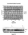
- RNA labeling and hybridization on microRNA microarray chips were performed as previously described (28) using 5 ⁇ g of total RNA from each sample.
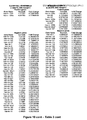
- Hybridization was carried out on our microRNA microarray (Ohio State Comprehensive Cancer Center, version 2.0), which contains probes for 460 mature microRNAs spotted in quadruplicate (235 homo sapiens, 222 mus musculus, and three Arabidopsis thaliana) with annotated active sites. Often, more than one probe set exists for a given mature microRNA. Additionally, there are quadruplicate probes corresponding to most precursor microRNAs.
- Hybridization signals were detected with Streptavidin-Alexa647 conjugate and scanned images (Axon 4000B) were quantified using the Genepix 6.0 software (Axon Instruments, Sunnyvale, CA).
- Microarray images were analyzed by using GENEPIX PRO. Average values of the replicate spots of each miRNA were background subtracted, normalized, and subjected to further analysis. We performed a global median normalization of Ovary microarray data by using BRB ArrayTools developed by Richard Simon & Amy Peng Lam (29). Absent calls were thresholded to 4.5 before subsequent statistical analysis. This level is the average minimum intensity level detected in the experiments. miRNA nomenclature was according to the Genome Browser (genome.ucsc.edu) and the miRNA database at Sanger Center (microrna.sanger.ac.uk/); in case of discrepancies the miRNA database was followed. Differentially expressed miRNAs were identified by using the t test procedure within significance analysis of microarrays (SAM), a method developed at Stanford University Labs based on recent paper of Tusher, Tibshirani and Chu (30).
- SAM significance analysis of microarrays
- PAM performs sample classification from gene expression data, via the "nearest shrunken centroid method" of Tibshirani, Hastie, Narasimhan and Chu (31).
- RNA samples (10 ⁇ g each) were run on 15% Polyacrylamide, 7M Urea Criterion pre-casted gels (Bio-Rad, Hercules, CA) and transferred onto Hybond-N+ membranes (Amersham, Piscataway, NJ). The hybridization was performed at 370C in ULTRAhyb-Oligo hybrization buffer (Ambion, Austin, TX) for 16 hours. Membranes were washed at 370C, twice with 2X SSPE and 0.5% SDS.
- the oligonucleotides used as probes were antisense to the sequence of the mature microRNAs (miR Registry at sanger.ac.uk/Software/Rfam/mirna/): miR-200a: 5'- ACA TCG TTA CCA GAC AGT GTT A -3' [SEQ ID NO:92]; miR-141: 5'- CCA TCT TTA CCA GAC AGT GTT A - 3' [SEQ ID NO:93]; miR-199a: 5'- GAA CAG GTA GTC TGA ACA CTG GG -3' [SEQ ID NO:94]; miR-125b1: 5'TCA CAA GTT AGG GTC TCA GGG A -3' [SEQ ID NO:95]; miR-145: 5'- AAG GGA TTC CTG GGA AAA CTG GAC - 3' [SEQ ID NO:96]; miR-222: 5'- GAG ACC CAG TAG CCA GAT G
- 5S RNA or EtBr gel staining were used to normalize. 200 ng of each probe was end labeled with 100 ⁇ Ci [gamma-32P]-ATP using the polynucleotide kinase (Roche). Blots were stripped in boiling 0.1% SDS for 10 minutes before re-hybridization.
- the single tube TaqMan MicroRNA Assays were used to detect and quantify mature microRNAs on Applied Biosystems Real-Time PCR instruments in accordance with manufacturer's instructions (Applied Biosystems, Foster City, CA). Normalization was performed with 18S rRNA. All RT reactions, including no-template controls and RT minus controls, were run in a GeneAmp PCR 9700 Thermocycler (Applied Biosystems). Gene expression levels were quantified using the ABI Prism 7900HT Sequence detection system (Applied Biosystems). Comparative real-time PCR was performed in triplicate, including no-template controls. Relative expression was calculated using the comparative Ct method.
- OVCAR3 cells were seeded at low density 48h before treatment with 10 ⁇ M 5'aza-2'deoxycytidine (5'-AZA, Sigma). After 24h of treatment, cells were collected and total RNA was isolated using Trizol reagent (Invitrogen). 3 replicates for both untreated cells and AZA-treated cells were used to evaluate the miR expression by Microarray profiling. Differentially expressed microRNAs were identified by using univariate two-classes T-test with random variance model.
- a microRNA expression signature discriminates Ovarian Cancer Tissues from Normal Ovary.
- the unsupervised hierarchical clustering based on all the human microRNAs spotted on the chip, generated a tree with a clear distinction of samples in two main groups, represented by normal tissues and malignant tissues ( Fig . 1 ).
- the PAM analysis in Figs. 6A and 6B displays the graphical representation of the probabilities (0.0 to 1.0) of each sample for being a cancer or a normal tissue according to the miR signature reported in Fig. 8 - Table 1, which describes a smaller set of 29 miRs, 4 up-modulated (miR-200a, -200b, -200c and -141) and 25 down-modulated (being miR-199a,miR-140, miR-145 and miR-125b1 among the most significant) differentiating normal versus tumor with a classification rate of 89%.
- ovarian epithelial carcinomas occur as different histological subtypes characterized by distinct moiphologic and molecular genetic alterations
- Complete lists resulting from SAM analyses are reported in Fig. 10 - Table 3 , while a summary is shown in the Venn diagrams in Figs. 3A and 3B :
- Fig.3A Two (2) out of 4 microRNAs most significantly up-modulated ( Fig.3A ) in tumors versus normal tissue, miR-200a and miR-200c, are up-modulated in all the three histotypes considered (serous, endometrioid and clear cell), while miR-200b and miR-141 up-modulation is shared by endometrioid and serous histotypes.
- the endometrioid histotype shows the up-modulation of 3 additional microRNAs, miR-21, miR-203 and miR-205.
- 19 miRs, including miR-125b1, miR-199a and miR-140, are down-modulated ( Fig. 3B ) in all the three histotypes examined in comparison with normal tissue, while 4 are shared in each paired analysis of the different signatures: miR-145, for example, is down-modulated in both serous and clear cell carcinomas; miR-222 in both endometrioid and clear cell carcinomas.
- the first group revealed a signature with characteristics of different histotypes, sharing for example the overexpression of miR-200c and miR-181 with the endometrioid carcinomas, and the down-modulation of miR-214 with the serous, while the "poorly differentiated” tumors have a quite different pattern of microRNAs expression ( Fig. 10 - Table 3 ).
- Fig. 4B a Northern Blot on a small set of samples verifies miR-222 overexpression in serous tumors compared to endometrioid.
- Figs. 5C and 5D Real-Time PCRs to verify the up-modulation of the 5 most significantly induced miRs are described in Figs. 5C and 5D as graphical representation of miR expression levels ( Fig . 5C ), and miR-21 was also validated by Northern Blot ( Fig . 5D ).
- miR-21, miR-203 and miR-205 are overexpressed in ovarian carcinomas compared to normal tissues (see SAM analysis in Fig. 9 - Table 2 and Venn Diagram in Figs. 3A and 3B ): the reactivation of these miR genes after demethylating treatment suggests that the hypomethylation could be the mechanism responsible for their overexpression in vivo.
- miR-21 and miR-203 are associated with CpG islands, being the miR-203 embedded in a CpG island 875 bp long, and the miR-21 characterized by a CpG island -2 kb upstream the mature sequence ( Fig . 7B ), while miR-205 does not show any CpG island in a region spanning 2 Kb upstream its mature form.
- microRNAs are aberrantly expressed in human ovarian cancer.
- the overall microRNA expression can clearly separate normal versus cancer tissues, identifying a number of microRNAs altered in human ovarian cancer and probably involved in the development of this neoplasia.
- miR-200a and miR-141 The expression of all the four microRNAs we found most significantly up-modulated, miR-200a and miR-141, belonging to the same family; miR-200b (localized in the same region of miR-200a, at chr.1p36.33); and miR-200c, (localized in the same region of miR-141, at chr.12p13.31), is concordant with the results obtained at genomic level by Zhang et al. (24), suggesting that the mechanism driving their up-modulation could be the amplification of the microRNA genes.
- GATA6 lost or excluded from the nucleus in 85% of ovarian tumors, may be regulated by miR-200a, and GATA4, absent in the majority of ovarian cancer cell lines, may be targeted by miR-200b ( Fig. 12 - Table 5 ).
- miR-125b1 altered also in breast cancer, as well as miR-145 (18); mir-199a, recently shown down-modulated in other tumors, as hepatocellular carcinoma (36); miR-140, deleted in ovarian carcinoma (24).
- miR-140 is indeed located at chr.6q22, a fragile region often deleted in ovarian tumor, and it is predicted to target important molecules as c-SRK, MMP13 and FGF2.
- Endometrioid tumors also present down-modulation of several microRNAs in comparison with the other classes of tumors, for example miR-222, already demonstrated targeting c-Kit (41), being involved in cancer (42-44) and down-modulated under folate-deficient conditions (45).
- miR-212 for example, down-modulated in serous carcinoma, has as putative target WT1, overexpressed in this subgroup of ovarian carcinomas (47).
- Another putative target of miR-212 is BRCA1: mutated in hereditary ovarian cancer, this molecule has been recently found involved also in the pathoetiology of sporadic ovarian epithelial cancer (OEC), where a loss of gene function due to epigenetic alterations has been observed more commonly (48). he decreased BRCA1 expression could be determined by overexpression of one or more microRNAs.
- miR-299-5p and miR-135b up-modulated in serous histotype compared to endometrioid, are supposed to target, respectively, DLK1 (Delta-like 1) and MSX2 (msh homeobox 2), overexpressed in endometrioid carcinomas (47).
- DLK1 Delta-like 1
- MSX2 mish homeobox 2
- clear cell carcinomas show expression levels of miR-30-5p and of miR-20a opposite (46) to two putative targets, RBP4 (retinol binding protein 4) and SLC40A1 (solute carrier 40-iron-regulated transporter, member 1), respectively.
- microRNAs significantly differentially expressed in relation to tumor Stage or Grade might be explained by the fact that our set of samples is mostly represented by advanced stage tumors, as expected considering the late diagnosis of this kind of neoplasia; however, the difference in size among the different groups of samples could have represented a limit for the statistical analysis.
- microRNAs might be important for the development of human ovarian carcinoma but not for the progression of the disease.
- miR-21 is associated with a CpG island (miR-203 is embedded in a CpG island, while miR-21 has a CpG island -2 kb upstream its mature sequence), supporting the idea that the demethylation leads to the reactivation of these microRNA genes.
- miR-21 has already been described up-modulated in several human tumors and having an anti-apoptotic role in different cellular models.
- the miRNAs of interest are listed in public databases.
- the public database can be a central repository provided by the Sanger Institute http://microrna.sanger.ac.uk/sequences/ to which miRNA sequences are submitted for naming and nomenclature assignment, as well as placement of the sequences in a database for archiving and for online retrieval via the world wide web.
- the data collected on the sequences of miRNAs by the Sanger Institute include species, source, corresponding genomic sequences and genomic location (chromosomal coordinates), as well as full length transcription products and sequences for the mature fully processed miRNA (miRNA with a 5' terminal phosphate group).
- GenBank database accessed through the National Center for Biotechnology Information (NCBI) website, maintained by the National Institutes of Health and the National Library of Medicine.
- NCBI National Center for Biotechnology Information
Claims (8)
- Procédé permettant de diagnostiquer si un sujet a un cancer de l'ovaire, comprenant :la mesure du taux de miR-200a dans un échantillon d'essai provenant du sujet, etdans lequel une augmentation dans le taux de miR-200a dans l'échantillon d'essai, par rapport au taux de miR-200a dans un échantillon témoin, révèle que le sujet a un cancer de l'ovaire.
- Procédé selon la revendication 1 dans lequel l'expression dudit miR est déterminée par :(a) marquage du miR isolé à partir d'un échantillon provenant d'un sujet ayant ou supposé avoir une maladie ou affection ;(b) hybridation du miR à une puce à miR ;(c) détermination de l'hybridation du miR à la puce.
- Procédé selon la revendication 1, dans lequel le cancer de l'ovaire est un cancer de l'ovaire à cellules claires, un cancer de l'ovaire séreux et/ou un cancer de l'ovaire endométrioïde.
- Procédé selon la revendication 1, comprenant en outre :la mesure du taux d'au moins un miR choisi parmi miR-199a, miR-140, miR-145 et miR-125b-1,dans lequel une diminution dans l'expression d'au moins l'un de miR-199a, miR-140, miR-145 ou miR-125b-1, par comparaison avec un échantillon normal, révèle un cancer de l'ovaire.
- Procédé selon la revendication 1, comprenant en outre :la mesure du taux d'au moins un miR choisi dans le groupe constitué par miR-205, miR-21, miR-182, miR-200c, miR-141 et miR-200b,dans lequel une augmentation dans l'expression d'un ou plusieurs desdits miARN par comparaison avec un échantillon normal révèle un cancer de l'ovaire endométrioïde.
- Procédé selon la revendication 1, dans lequel le sujet est humain.
- Procédé selon la revendication 1, comprenant :(1) la transcription inverse d'ARN provenant de l'échantillon d'essai obtenu auprès du sujet pour fournir un ensemble d'oligodésoxynucléotides cibles ;(2) l'hybridation des oligodésoxynucléotides cibles à une puce à ADN comprenant des oligonucléotides sondes spécifiques de miARN pour fournir un profil d'hybridation pour l'échantillon d'essai ; et(3) la comparaison du profil d'hybridation de l'échantillon d'essai à un profil d'hybridation généré à partir d'un échantillon témoin,dans lequel une modification dans le signal de miR-200a révèle que le sujet soit a, soit présente un risque de développer, un cancer de l'ovaire.
- Procédé permettant d'identifier un agent contre le cancer de l'ovaire, comprenant l'apport d'un agent d'essai à une cellule de cancer de l'ovaire et la mesure du taux de miR-200a, dans lequel une diminution du taux de miR-200a dans la cellule, par rapport à une cellule témoin appropriée, révèle que l'agent d'essai est un agent contre le cancer de l'ovaire.
Priority Applications (3)
| Application Number | Priority Date | Filing Date | Title |
|---|---|---|---|
| EP16156869.6A EP3048177A1 (fr) | 2007-09-06 | 2008-09-08 | Signatures de microarn dans le cancer de l'ovaire humain |
| EP16184406.3A EP3138926A3 (fr) | 2007-09-06 | 2008-09-08 | Signatures de microarn dans le cancer de l'ovaire humain |
| EP14164377.5A EP2775001B1 (fr) | 2007-09-06 | 2008-09-08 | Signatures de microARN dans le cancer de l'ovaire humain |
Applications Claiming Priority (2)
| Application Number | Priority Date | Filing Date | Title |
|---|---|---|---|
| US96766307P | 2007-09-06 | 2007-09-06 | |
| PCT/US2008/075565 WO2009033140A1 (fr) | 2007-09-06 | 2008-09-08 | Signatures de microarn dans le cancer ovarien humain |
Related Child Applications (4)
| Application Number | Title | Priority Date | Filing Date |
|---|---|---|---|
| EP16184406.3A Division EP3138926A3 (fr) | 2007-09-06 | 2008-09-08 | Signatures de microarn dans le cancer de l'ovaire humain |
| EP16156869.6A Division EP3048177A1 (fr) | 2007-09-06 | 2008-09-08 | Signatures de microarn dans le cancer de l'ovaire humain |
| EP14164377.5A Division-Into EP2775001B1 (fr) | 2007-09-06 | 2008-09-08 | Signatures de microARN dans le cancer de l'ovaire humain |
| EP14164377.5A Division EP2775001B1 (fr) | 2007-09-06 | 2008-09-08 | Signatures de microARN dans le cancer de l'ovaire humain |
Publications (3)
| Publication Number | Publication Date |
|---|---|
| EP2183393A1 EP2183393A1 (fr) | 2010-05-12 |
| EP2183393A4 EP2183393A4 (fr) | 2010-11-24 |
| EP2183393B1 true EP2183393B1 (fr) | 2014-06-11 |
Family
ID=40429408
Family Applications (4)
| Application Number | Title | Priority Date | Filing Date |
|---|---|---|---|
| EP16184406.3A Withdrawn EP3138926A3 (fr) | 2007-09-06 | 2008-09-08 | Signatures de microarn dans le cancer de l'ovaire humain |
| EP14164377.5A Not-in-force EP2775001B1 (fr) | 2007-09-06 | 2008-09-08 | Signatures de microARN dans le cancer de l'ovaire humain |
| EP08799295.4A Not-in-force EP2183393B1 (fr) | 2007-09-06 | 2008-09-08 | Signatures de microarn dans le cancer ovarien humain |
| EP16156869.6A Withdrawn EP3048177A1 (fr) | 2007-09-06 | 2008-09-08 | Signatures de microarn dans le cancer de l'ovaire humain |
Family Applications Before (2)
| Application Number | Title | Priority Date | Filing Date |
|---|---|---|---|
| EP16184406.3A Withdrawn EP3138926A3 (fr) | 2007-09-06 | 2008-09-08 | Signatures de microarn dans le cancer de l'ovaire humain |
| EP14164377.5A Not-in-force EP2775001B1 (fr) | 2007-09-06 | 2008-09-08 | Signatures de microARN dans le cancer de l'ovaire humain |
Family Applications After (1)
| Application Number | Title | Priority Date | Filing Date |
|---|---|---|---|
| EP16156869.6A Withdrawn EP3048177A1 (fr) | 2007-09-06 | 2008-09-08 | Signatures de microarn dans le cancer de l'ovaire humain |
Country Status (7)
| Country | Link |
|---|---|
| US (2) | US20100249213A1 (fr) |
| EP (4) | EP3138926A3 (fr) |
| JP (3) | JP5401460B2 (fr) |
| CN (1) | CN101939446B (fr) |
| AU (1) | AU2008296022B2 (fr) |
| CA (1) | CA2698771A1 (fr) |
| WO (1) | WO2009033140A1 (fr) |
Cited By (2)
| Publication number | Priority date | Publication date | Assignee | Title |
|---|---|---|---|---|
| US10636512B2 (en) | 2017-07-14 | 2020-04-28 | Cofactor Genomics, Inc. | Immuno-oncology applications using next generation sequencing |
| US11584932B2 (en) | 2016-11-01 | 2023-02-21 | The Research Foundation For The State University Of New York | 5-halouracil-modified microRNAs and their use in the treatment of cancer |
Families Citing this family (55)
| Publication number | Priority date | Publication date | Assignee | Title |
|---|---|---|---|---|
| CN103866018B (zh) | 2005-08-01 | 2016-05-25 | 俄亥俄州立大学研究基金会 | 用于乳腺癌的诊断、预后和治疗的基于MicroRNA的方法和组合物 |
| CN101296702B (zh) | 2005-09-12 | 2012-11-28 | 俄亥俄州立大学研究基金会 | 用于诊断或治疗bcl2相关癌症的组合物和方法 |
| CA2633674A1 (fr) | 2006-01-05 | 2007-07-19 | The Ohio State University Research Foundation | Procedes et compositions bases sur des micro-arn et s'appliquant au diagnostic, au pronostic et au traitement du cancer du poumon |
| EP1968622B1 (fr) | 2006-01-05 | 2014-08-27 | The Ohio State University Research Foundation | Anomalies dans l'expression des micro-arn dans des tumeurs endocrines pancréatiques et des tumeurs à cellules acineuses |
| EP2487257B1 (fr) | 2006-01-05 | 2015-07-01 | The Ohio State University Research Foundation | Procédés à base de micro ARN et compositions pour le diagnostic et le traitement des cancers solides |
| ES2446362T3 (es) | 2006-03-20 | 2014-03-07 | The Ohio State University Research Foundation | Huellas de microARN durante megacariocipoyesis humana |
| WO2008008430A2 (fr) | 2006-07-13 | 2008-01-17 | The Ohio State University Research Foundation | Procédés et compositions à base de micro-arn pour le diagnostic et le traitement de maladies apparentées au cancer du côlon |
| EP2061907B1 (fr) | 2006-09-19 | 2011-11-23 | The Ohio State University Research Foundation | Expression tcl1 dans la leucémie lymphocytaire chronique (llc) régulée par mir-29 et mir-181 |
| ES2425416T3 (es) | 2006-11-01 | 2013-10-15 | The Ohio State University Research Foundation | Firma de expresión del microARN para predecir la supervivencia y la metástasis en el carcinoma hepatocelular |
| CN105256004A (zh) | 2007-01-31 | 2016-01-20 | 俄亥俄州立大学研究基金会 | 用于急性髓细胞白血病(aml)的诊断、预后和治疗的基于微rna的方法和组合物 |
| WO2012070037A2 (fr) * | 2010-11-22 | 2012-05-31 | Rosetta Genomics Ltd. | Procédés et matériaux pour la classification de tissus originaires d'échantillons tumoraux |
| US9096906B2 (en) | 2007-03-27 | 2015-08-04 | Rosetta Genomics Ltd. | Gene expression signature for classification of tissue of origin of tumor samples |
| CN101711287B (zh) | 2007-06-08 | 2016-04-27 | 由卫生与公众服务部代表的美利坚合众国政府 | 确定肝细胞癌亚型和检测肝癌干细胞的方法 |
| US8053186B2 (en) | 2007-06-15 | 2011-11-08 | The Ohio State University Research Foundation | Oncogenic ALL-1 fusion proteins for targeting Drosha-mediated microRNA processing |
| AU2008282318B2 (en) | 2007-07-31 | 2014-02-27 | The Ohio State University Research Foundation | Methods for reverting methylation by targeting methyltransferases |
| CN101835902B (zh) | 2007-08-03 | 2014-03-26 | 俄亥俄州立大学研究基金会 | 编码ncrna的超保守区域 |
| CA2926831A1 (fr) | 2007-08-22 | 2009-02-26 | The Ohio State University Research Foundation | Procedes et compositions pour induire une deregulation de la phosphorylation de epha7 et de erk dans des cas de leucemies humaines aigues |
| AU2008316577B2 (en) | 2007-10-26 | 2014-04-10 | The Ohio State University Research Foundation | Methods for identifying fragile histidine triad (FHIT) interaction and uses thereof |
| AU2009257410B2 (en) | 2008-06-11 | 2014-03-06 | Fudan University | Use of miR-26 family as a predictive marker of hepatocellular carcinoma and responsiveness to therapy |
| CN102575289A (zh) * | 2009-06-25 | 2012-07-11 | 耶鲁大学 | 在处的单核苷酸多态性和癌症风险 |
| EP2473628A1 (fr) | 2009-08-31 | 2012-07-11 | Alcedo Biotech GmbH | Procédés et compositions à base de micro-arn pour le diagnostic, le pronostic et le traitement de tumeurs impliquant des remaniements chromosomiques |
| WO2011029903A1 (fr) * | 2009-09-10 | 2011-03-17 | Flemming Velin | Procédé d'élaboration de micro-arn et application thérapeutique de celui-ci |
| WO2011057304A2 (fr) * | 2009-11-09 | 2011-05-12 | Yale University | Signatures de microarn qui permettent de différencier les tumeurs séreuses papillaires utérines et ovariennes |
| AU2010321555B2 (en) | 2009-11-23 | 2015-10-15 | The Ohio State University | Materials and methods useful for affecting tumor cell growth, migration and invasion |
| CN102080086B (zh) * | 2009-12-01 | 2012-12-26 | 中国科学院上海药物研究所 | 人miR-133a反义核酸及其应用 |
| EP2354246A1 (fr) * | 2010-02-05 | 2011-08-10 | febit holding GmbH | ARNm dans le diagnostic du cancer ovarien |
| CA2816603A1 (fr) | 2010-11-12 | 2012-05-18 | The Ohio State University Research Foundation | Materiaux et procedes relatifs aux microarn-21, reparation de desappariement et cancer colorectal |
| AU2011329066B2 (en) | 2010-11-15 | 2017-03-09 | The Ohio State University Research Foundation | Controlled release mucoadhesive systems |
| WO2012093384A1 (fr) | 2011-01-03 | 2012-07-12 | Rosetta Genomics Ltd. | Compositions et procédés de traitement du cancer de l'ovaire |
| EP2778237A1 (fr) * | 2011-01-13 | 2014-09-17 | Industrial Technology Research Institute | Biomarqueurs de prédiction de la récurrence du cancer colorectal |
| AU2012225506B2 (en) | 2011-03-07 | 2016-11-17 | The Ohio State University | Mutator activity induced by microRNA-155 (miR-155) links inflammation and cancer |
| CN102178959B (zh) * | 2011-03-15 | 2013-10-23 | 清华大学深圳研究生院 | 抑制肺转移肿瘤生长的siRNA及其寡聚核酸组合与应用 |
| US8765707B2 (en) | 2011-04-22 | 2014-07-01 | University Of Houston System | MicroRNA-140-5p as a tumor suppressor and sensitizing agent for chemotherapy |
| EP2766500A4 (fr) | 2011-10-14 | 2015-10-14 | Univ Ohio State | Méthodes et matériaux relatifs au cancer des ovaires |
| US9481885B2 (en) | 2011-12-13 | 2016-11-01 | Ohio State Innovation Foundation | Methods and compositions related to miR-21 and miR-29a, exosome inhibition, and cancer metastasis |
| CA2866052A1 (fr) | 2012-01-20 | 2013-07-25 | The Ohio State University | Signatures de marqueurs biologiques du cancer du sein concernant le pouvoir envahissant et le pronostic |
| WO2013148151A1 (fr) * | 2012-03-29 | 2013-10-03 | University Of Pittsburgh - Of The Commonwealth System Of Higher Education | Acides microribonucléiques plasmatiques à titre de biomarqueurs de l'endométriose et du cancer de l'ovaire associé à l'endométriose (eaoc) |
| WO2014145142A2 (fr) * | 2013-03-15 | 2014-09-18 | Miles Gregory | Procédé d'amélioration de la survie dans le cancer |
| CN103343160B (zh) * | 2013-05-03 | 2014-12-10 | 中国人民解放军南京军区南京总医院 | microRNA-30家族的新用途 |
| CN103243163A (zh) * | 2013-05-09 | 2013-08-14 | 江苏省中医院 | miR-27a的新用途 |
| KR101508580B1 (ko) | 2013-05-14 | 2015-04-07 | 연세대학교 산학협력단 | 재발성 난소암의 예측 또는 진단 방법 |
| WO2014193309A1 (fr) * | 2013-05-30 | 2014-12-04 | Agency For Science, Technology And Research | Procédé de détermination de pronostic de cancer |
| US20170218454A1 (en) * | 2013-06-27 | 2017-08-03 | Institute For Systems Biology | Products and Methods Relating to Micro RNAS and Cancer |
| CN103952465A (zh) * | 2013-12-10 | 2014-07-30 | 长沙赢润生物技术有限公司 | 一种基于血浆microRNA分析的卵巢癌肿瘤诊断方法 |
| CN104975019B (zh) * | 2014-04-01 | 2019-07-12 | 上海翔琼生物技术有限公司 | 小rna组成的指纹图谱在人卵巢癌诊断和治疗中的应用 |
| CN105177173A (zh) * | 2015-11-02 | 2015-12-23 | 崔长友 | 用于卵巢癌诊断的miRNA生物标志物及检测试剂盒 |
| US11236337B2 (en) | 2016-11-01 | 2022-02-01 | The Research Foundation For The State University Of New York | 5-halouracil-modified microRNAs and their use in the treatment of cancer |
| WO2018129535A1 (fr) * | 2017-01-09 | 2018-07-12 | Dana-Farber Cancer Institute, Inc. | Signatures de microarn circulants pour le cancer de l'ovaire |
| CN110546263B (zh) | 2017-04-28 | 2024-03-05 | 东丽株式会社 | 用于检测卵巢肿瘤的试剂盒、装置和方法 |
| CN108992457A (zh) * | 2018-01-15 | 2018-12-14 | 广东省实验动物监测所 | miR-144-3p及其靶基因在制备调节人心脏成纤维细胞功能的药物中的应用 |
| CN108103199B (zh) * | 2018-02-13 | 2021-08-10 | 朱伟 | 一种与卵巢癌辅助诊断相关的循环miRNA标志物及其应用 |
| WO2019178216A1 (fr) * | 2018-03-13 | 2019-09-19 | Baylor Research Institute | Méthodes et compositions pour le traitement, le diagnostic et le pronostic du cancer des ovaires |
| CN109337978B (zh) * | 2018-07-03 | 2023-12-01 | 华中科技大学同济医学院附属同济医院 | miRNA在制备高级浆液性上皮性卵巢癌化疗耐药性评价试剂盒中的应用 |
| CN110564854B (zh) * | 2019-09-09 | 2023-09-29 | 晶准生物医药集团有限公司 | 基于miRNA检测的试剂盒及其在癌症早期筛查中的应用 |
| CN114854756B (zh) * | 2022-05-31 | 2023-08-29 | 华南农业大学 | miR-370调控GLI1表达在猪卵巢颗粒细胞中的应用 |
Family Cites Families (117)
| Publication number | Priority date | Publication date | Assignee | Title |
|---|---|---|---|---|
| US127895A (en) * | 1872-06-11 | Improvement in washing-machines | ||
| US105340A (en) * | 1870-07-12 | Improvement in fur collars | ||
| US189557A (en) * | 1877-04-17 | Improvement in stoves | ||
| US99196A (en) * | 1870-01-25 | Improved holdback for carriages | ||
| US22934A (en) * | 1859-02-15 | Improvement in instruments for threading needles | ||
| US176723A (en) * | 1876-04-25 | Improvement in fenders for life-boats | ||
| US112630A (en) * | 1871-03-14 | Improvement in sleighs | ||
| US24780A (en) * | 1859-07-12 | Improvement in sewing-machines | ||
| US152112A (en) * | 1874-06-16 | Improvement in patterns and core-bars for casting pipes | ||
| US292878A (en) * | 1884-02-05 | Weighing attachment for baby-carriages | ||
| US256072A (en) * | 1882-04-04 | Francis taggaet | ||
| US163435A (en) * | 1875-05-18 | Improvement in paper boxes | ||
| US99034A (en) * | 1870-01-18 | El wood tush | ||
| US99619A (en) * | 1870-02-08 | Improvement in neckties | ||
| US116726A (en) * | 1871-07-04 | Improvement in punching-machines | ||
| US123533A (en) * | 1872-02-06 | Improvement in combined latches and locks for sliding doors | ||
| US192102A (en) * | 1877-06-19 | Improvement in paper folder and cutter | ||
| US171667A (en) * | 1876-01-04 | Improvement in milk and cheese pans | ||
| US256650A (en) * | 1882-04-18 | davis | ||
| US78834A (en) * | 1868-06-09 | William sanderson | ||
| US123912A (en) * | 1872-02-20 | Improvement in devices for opening or closing umbrellas | ||
| US75511A (en) * | 1868-03-17 | Jeamtm atkins | ||
| US26951A (en) * | 1860-01-24 | John e | ||
| US192114A (en) * | 1877-06-19 | Improvement in casting brass and other metals | ||
| US186589A (en) * | 1877-01-23 | Improvement in ice-machines | ||
| US26796A (en) * | 1860-01-10 | Improvement in cultivators | ||
| US19286A (en) * | 1858-02-09 | boyers | ||
| US213292A (en) * | 1879-03-18 | Improvement in safety-plugs | ||
| US292616A (en) * | 1884-01-29 | Vapor-burner | ||
| US23594A (en) * | 1859-04-12 | Improvement in steam-engines | ||
| US178502A (en) * | 1876-06-13 | Improvement in saw-mandrels | ||
| US123482A (en) * | 1872-02-06 | Improvement in railroad switches | ||
| US178105A (en) * | 1876-05-30 | Improvement in baking-pans | ||
| US86331A (en) * | 1869-01-26 | Improved medical compound | ||
| US254473A (en) * | 1882-03-07 | Rope-clamp | ||
| US176025A (en) * | 1876-04-11 | Improvement in check-hooks | ||
| US19890A (en) * | 1858-04-06 | Gkate fob steam-engines | ||
| US192111A (en) * | 1877-06-19 | Improvement in crimping-forms for boot-uppers | ||
| US212727A (en) * | 1879-02-25 | Improvement in carpet-sweepers | ||
| US188959A (en) * | 1877-03-27 | Improvement in brushes | ||
| US192235A (en) * | 1877-06-19 | ceaig | ||
| US163434A (en) * | 1875-05-18 | Improvement in mills for grating cocoa-nuts | ||
| US74797A (en) * | 1868-02-25 | Improvement in oheese-boxes | ||
| US123933A (en) * | 1872-02-20 | Improvement in plate-printing machinery | ||
| US253780A (en) * | 1882-02-14 | Apparatus for utilizing the waste products of combustion in furnaces | ||
| US203544A (en) * | 1878-05-14 | Improvement in treatment of flour | ||
| US173319A (en) * | 1876-02-08 | Improvement in scroll-sawing machines | ||
| US92974A (en) * | 1869-07-27 | William h | ||
| US175827A (en) * | 1876-04-11 | Improvement in air-gas machines | ||
| US165659A (en) * | 1875-07-20 | Improvement in registering board-rules | ||
| US227533A (en) * | 1880-05-11 | Shutter-worker | ||
| US65840A (en) * | 1867-06-18 | Improved medical compound | ||
| US188924A (en) * | 1877-03-27 | Improvement in unloading grain-vessels | ||
| US270484A (en) * | 1883-01-09 | Territobt | ||
| US163430A (en) * | 1875-05-18 | Improvement in gages for shaping wagon-axles | ||
| US176766A (en) * | 1876-05-02 | Improvement in fruit-jars | ||
| US161004A (en) * | 1875-03-23 | Improvement in water-closets | ||
| US61424A (en) * | 1867-01-22 | James l | ||
| US48681A (en) * | 1865-07-11 | Mkebagskf | ||
| US65844A (en) * | 1867-06-18 | Improvement in preserving fruit | ||
| US105360A (en) * | 1870-07-12 | Improvement in faucets | ||
| US137410A (en) * | 1873-04-01 | Improvement in saw-sets | ||
| US54849A (en) * | 1866-05-22 | Improvement in trunk-locks | ||
| US257618A (en) * | 1882-05-09 | Eighths to | ||
| US4196265A (en) * | 1977-06-15 | 1980-04-01 | The Wistar Institute | Method of producing antibodies |
| US4235871A (en) | 1978-02-24 | 1980-11-25 | Papahadjopoulos Demetrios P | Method of encapsulating biologically active materials in lipid vesicles |
| US4501728A (en) | 1983-01-06 | 1985-02-26 | Technology Unlimited, Inc. | Masking of liposomes from RES recognition |
| US5019369A (en) | 1984-10-22 | 1991-05-28 | Vestar, Inc. | Method of targeting tumors in humans |
| US5139941A (en) | 1985-10-31 | 1992-08-18 | University Of Florida Research Foundation, Inc. | AAV transduction vectors |
| US5015568A (en) * | 1986-07-09 | 1991-05-14 | The Wistar Institute | Diagnostic methods for detecting lymphomas in humans |
| US5202429A (en) * | 1986-07-09 | 1993-04-13 | The Wistar Institute | DNA molecules having human BCL-2 gene sequences |
| US4987071A (en) | 1986-12-03 | 1991-01-22 | University Patents, Inc. | RNA ribozyme polymerases, dephosphorylases, restriction endoribonucleases and methods |
| US4837028A (en) | 1986-12-24 | 1989-06-06 | Liposome Technology, Inc. | Liposomes with enhanced circulation time |
| US4920016A (en) | 1986-12-24 | 1990-04-24 | Linear Technology, Inc. | Liposomes with enhanced circulation time |
| EP0341904B1 (fr) | 1988-05-09 | 1995-03-29 | Temple University of the Commonwealth System of Higher Education | Procédé pour prédire l'effectivité d'une thérapie antinéoplasique chez des malades individuels. |
| US5198338A (en) * | 1989-05-31 | 1993-03-30 | Temple University | Molecular probing for human t-cell leukemia and lymphoma |
| US5252479A (en) | 1991-11-08 | 1993-10-12 | Research Corporation Technologies, Inc. | Safe vector for gene therapy |
| US5633135A (en) * | 1991-12-11 | 1997-05-27 | Thomas Jefferson University | Chimeric nucleic acids and proteins resulting from ALL-1 region chromosome abnormalities |
| US6040140A (en) * | 1991-12-11 | 2000-03-21 | Thomas Jefferson University | Methods for screening and treating leukemias resulting from all-1 region chromosome abnormalities |
| WO1993012136A1 (fr) * | 1991-12-11 | 1993-06-24 | Thomas Jefferson University | Detection et traitement de leucemies aigues resultant d'anomalies chromosomiques dans la region all-1 |
| US5587308A (en) | 1992-06-02 | 1996-12-24 | The United States Of America As Represented By The Department Of Health & Human Services | Modified adeno-associated virus vector capable of expression from a novel promoter |
| EP0667920B1 (fr) * | 1992-10-29 | 2003-01-15 | Thomas Jefferson University | Procedes de detection de micrometastase du cancer de la prostate |
| US5478745A (en) | 1992-12-04 | 1995-12-26 | University Of Pittsburgh | Recombinant viral vector system |
| US7175995B1 (en) * | 1994-10-27 | 2007-02-13 | Thomas Jefferson University | TCL-1 protein and related methods |
| US6242212B1 (en) * | 1996-02-09 | 2001-06-05 | Thomas Jefferson University | Fragile histidine triad (FHIT) nucleic acids and methods of producing FHIT proteins |
| US5849902A (en) | 1996-09-26 | 1998-12-15 | Oligos Etc. Inc. | Three component chimeric antisense oligonucleotides |
| US6187536B1 (en) * | 1997-02-18 | 2001-02-13 | Thomas Jefferson University | Methods of identifying and detecting pancreatic cancer |
| AU5128999A (en) * | 1998-07-24 | 2000-02-14 | Yeda Research And Development Co. Ltd. | Prevention of metastasis with 5-aza-2'-deoxycytidine |
| IL151928A0 (en) | 2000-03-30 | 2003-04-10 | Whitehead Biomedical Inst | Rna sequence-specific mediators of rna interference |
| US7060811B2 (en) * | 2000-10-13 | 2006-06-13 | Board Of Regents, The University Of Texas System | WWOX: a tumor suppressor gene mutated in multiple cancers |
| US20020173478A1 (en) | 2000-11-14 | 2002-11-21 | The Trustees Of The University Of Pennsylvania | Post-transcriptional gene silencing by RNAi in mammalian cells |
| EP2386637B1 (fr) * | 2001-09-28 | 2018-05-16 | Max-Planck-Gesellschaft zur Förderung der Wissenschaften e.V. | Molécules de micro arn |
| AU2003241325A1 (en) * | 2002-04-29 | 2003-11-17 | Thomas Jefferson University | Human chronic lymphocytic leukemia modeled in mouse by targeted tcl1 expression |
| EP1537227B1 (fr) | 2002-05-31 | 2010-02-17 | The Regents Of The University Of California | Procede d'interference efficace par arn dans des cellules de mammifere |
| AU2003273542A1 (en) * | 2002-05-31 | 2003-12-19 | The Board Of Trustees Of The Leland Stanford Junior University | Methods of identifying and isolating stem cells and cancer stem cells |
| US7148342B2 (en) | 2002-07-24 | 2006-12-12 | The Trustees Of The University Of Pennyslvania | Compositions and methods for sirna inhibition of angiogenesis |
| JP4939055B2 (ja) * | 2002-11-13 | 2012-05-23 | トマス ジェファソン ユニバーシティ | 癌の診断および治療のための組成物および方法 |
| WO2004071464A2 (fr) * | 2003-02-12 | 2004-08-26 | Johns Hopkins University School Of Medicine | Applications therapeutiques et diagnostiques de genes s'exprimant de maniere differentielle dans des cellules souches lympho-hematopoietiques |
| US8106180B2 (en) * | 2003-08-07 | 2012-01-31 | Whitehead Institute For Biomedical Research | Methods and products for expression of micro RNAs |
| KR20060120652A (ko) * | 2003-10-07 | 2006-11-27 | 밀레니엄 파머슈티컬스 인코퍼레이티드 | 난소암의 확인, 평가, 예방 및 치료를 위한 핵산 분자 및단백질 |
| WO2005047477A2 (fr) * | 2003-11-07 | 2005-05-26 | University Of Massachusetts | Arn a elements repetitifs intercales utilises comme substrat, inhibiteurs et vehicules d'apport pour l'interference d'arn |
| EP1713938A2 (fr) * | 2004-02-09 | 2006-10-25 | Thomas Jefferson University | Diagnostic et traitement de cancers a l'aide de microarn present dans ou au voisinage de caracteristiques chromosomiennes liees aux cancers |
| AU2005243410B2 (en) * | 2004-05-14 | 2010-04-22 | Rosetta Genomics Ltd. | Micronas and uses thereof |
| EP2471924A1 (fr) * | 2004-05-28 | 2012-07-04 | Asuragen, INC. | Procédés et compositions impliquant du microARN |
| US7642348B2 (en) * | 2004-10-04 | 2010-01-05 | Rosetta Genomics Ltd | Prostate cancer-related nucleic acids |
| EP2302055B1 (fr) * | 2004-11-12 | 2014-08-27 | Asuragen, Inc. | Procédés et compositions impliquant l'ARNmi et des molécules inhibitrices de l'ARNmi |
| WO2006133022A2 (fr) * | 2005-06-03 | 2006-12-14 | The Johns Hopkins University | Compositions et procedes pour diminuer l'expression de microarn pour le traitement de la neoplasie |
| US20070092882A1 (en) * | 2005-10-21 | 2007-04-26 | Hui Wang | Analysis of microRNA |
| US7390792B2 (en) * | 2005-12-15 | 2008-06-24 | Board Of Regents, The University Of Texas System | MicroRNA1 therapies |
| EP1968622B1 (fr) * | 2006-01-05 | 2014-08-27 | The Ohio State University Research Foundation | Anomalies dans l'expression des micro-arn dans des tumeurs endocrines pancréatiques et des tumeurs à cellules acineuses |
| EP2487257B1 (fr) * | 2006-01-05 | 2015-07-01 | The Ohio State University Research Foundation | Procédés à base de micro ARN et compositions pour le diagnostic et le traitement des cancers solides |
| US7955848B2 (en) * | 2006-04-03 | 2011-06-07 | Trustees Of Dartmouth College | MicroRNA biomarkers for human breast and lung cancer |
| CN101472470B (zh) * | 2006-04-24 | 2013-07-24 | 俄亥俄州立大学研究基金会 | miR155转基因小鼠中前B细胞增殖和淋巴母细胞性白血病/高度淋巴瘤 |
| AU2007299748A1 (en) * | 2006-09-19 | 2008-03-27 | Asuragen, Inc. | miR-15, miR-26, miR -31,miR -145, miR-147, miR-188, miR-215, miR-216 miR-331, mmu-miR-292-3p regulated genes and pathways as targets for therapeutic intervention |
| US8765702B2 (en) * | 2007-02-27 | 2014-07-01 | Rosetta Genomics Ltd. | Composition and methods for modulating cell proliferation and cell death |
| WO2008113758A1 (fr) * | 2007-03-16 | 2008-09-25 | Covalx Ag | Analyse par spectrométrie de masse directe de médicaments candidats ciblant des complexes protéiques |
| US8216784B2 (en) * | 2007-07-25 | 2012-07-10 | University Of Louisville Research Foundation, Inc. | Cancer-derived microvesicle-associated microrna as a diagnostic marker |
-
2008
- 2008-09-08 EP EP16184406.3A patent/EP3138926A3/fr not_active Withdrawn
- 2008-09-08 EP EP14164377.5A patent/EP2775001B1/fr not_active Not-in-force
- 2008-09-08 EP EP08799295.4A patent/EP2183393B1/fr not_active Not-in-force
- 2008-09-08 CN CN200880112585.9A patent/CN101939446B/zh not_active Expired - Fee Related
- 2008-09-08 CA CA2698771A patent/CA2698771A1/fr not_active Abandoned
- 2008-09-08 US US12/676,670 patent/US20100249213A1/en not_active Abandoned
- 2008-09-08 EP EP16156869.6A patent/EP3048177A1/fr not_active Withdrawn
- 2008-09-08 AU AU2008296022A patent/AU2008296022B2/en not_active Ceased
- 2008-09-08 WO PCT/US2008/075565 patent/WO2009033140A1/fr active Application Filing
- 2008-09-08 JP JP2010524221A patent/JP5401460B2/ja not_active Expired - Fee Related
-
2013
- 2013-10-28 JP JP2013223103A patent/JP2014027948A/ja active Pending
-
2014
- 2014-09-15 US US14/485,995 patent/US9574239B2/en not_active Expired - Fee Related
-
2015
- 2015-04-03 JP JP2015076636A patent/JP2015130879A/ja not_active Ceased
Cited By (2)
| Publication number | Priority date | Publication date | Assignee | Title |
|---|---|---|---|---|
| US11584932B2 (en) | 2016-11-01 | 2023-02-21 | The Research Foundation For The State University Of New York | 5-halouracil-modified microRNAs and their use in the treatment of cancer |
| US10636512B2 (en) | 2017-07-14 | 2020-04-28 | Cofactor Genomics, Inc. | Immuno-oncology applications using next generation sequencing |
Also Published As
| Publication number | Publication date |
|---|---|
| US20150024963A1 (en) | 2015-01-22 |
| CN101939446A (zh) | 2011-01-05 |
| JP2015130879A (ja) | 2015-07-23 |
| EP3138926A3 (fr) | 2017-04-05 |
| EP2775001A1 (fr) | 2014-09-10 |
| EP3138926A2 (fr) | 2017-03-08 |
| AU2008296022B2 (en) | 2014-01-23 |
| US20100249213A1 (en) | 2010-09-30 |
| WO2009033140A1 (fr) | 2009-03-12 |
| JP2014027948A (ja) | 2014-02-13 |
| JP5401460B2 (ja) | 2014-01-29 |
| US9574239B2 (en) | 2017-02-21 |
| EP3048177A1 (fr) | 2016-07-27 |
| EP2775001B1 (fr) | 2016-03-09 |
| EP2183393A1 (fr) | 2010-05-12 |
| CA2698771A1 (fr) | 2009-03-12 |
| WO2009033140A9 (fr) | 2010-05-14 |
| CN101939446B (zh) | 2015-02-11 |
| AU2008296022A1 (en) | 2009-03-12 |
| EP2183393A4 (fr) | 2010-11-24 |
| JP2010538610A (ja) | 2010-12-16 |
Similar Documents
| Publication | Publication Date | Title |
|---|---|---|
| EP2183393B1 (fr) | Signatures de microarn dans le cancer ovarien humain | |
| EP2061907B1 (fr) | Expression tcl1 dans la leucémie lymphocytaire chronique (llc) régulée par mir-29 et mir-181 | |
| JP5489459B2 (ja) | 乳癌の診断、予後及び治療のためのマイクロrnaに基づいた方法及び組成物 | |
| EP2109687B1 (fr) | Méthodes utilisant des micro-arn pour le traitement de la leucémie aigüe myéloïde (lam) | |
| JP5451077B2 (ja) | 肺癌の診断、予後診断及び治療のためのマイクロrnaを基盤とした方法及び組成物 | |
| AU2014202073B2 (en) | MicroRNA signatures in human ovarian cancer | |
| CROCE | Patent 2698771 Summary | |
| AU2016203848B2 (en) | MicroRNA-based methods and compositions for the diagnosis, prognosis and treatment of lung cancer |
Legal Events
| Date | Code | Title | Description |
|---|---|---|---|
| PUAI | Public reference made under article 153(3) epc to a published international application that has entered the european phase |
Free format text: ORIGINAL CODE: 0009012 |
|
| 17P | Request for examination filed |
Effective date: 20100226 |
|
| AK | Designated contracting states |
Kind code of ref document: A1 Designated state(s): AT BE BG CH CY CZ DE DK EE ES FI FR GB GR HR HU IE IS IT LI LT LU LV MC MT NL NO PL PT RO SE SI SK TR |
|
| AX | Request for extension of the european patent |
Extension state: AL BA MK RS |
|
| A4 | Supplementary search report drawn up and despatched |
Effective date: 20101021 |
|
| DAX | Request for extension of the european patent (deleted) | ||
| 17Q | First examination report despatched |
Effective date: 20111118 |
|
| RAP1 | Party data changed (applicant data changed or rights of an application transferred) |
Owner name: THE OHIO STATE UNIVERSITY RESEARCH FOUNDATION |
|
| GRAP | Despatch of communication of intention to grant a patent |
Free format text: ORIGINAL CODE: EPIDOSNIGR1 |
|
| INTG | Intention to grant announced |
Effective date: 20140110 |
|
| GRAS | Grant fee paid |
Free format text: ORIGINAL CODE: EPIDOSNIGR3 |
|
| GRAA | (expected) grant |
Free format text: ORIGINAL CODE: 0009210 |
|
| AK | Designated contracting states |
Kind code of ref document: B1 Designated state(s): AT BE BG CH CY CZ DE DK EE ES FI FR GB GR HR HU IE IS IT LI LT LU LV MC MT NL NO PL PT RO SE SI SK TR |
|
| REG | Reference to a national code |
Ref country code: GB Ref legal event code: FG4D |
|
| REG | Reference to a national code |
Ref country code: CH Ref legal event code: EP |
|
| REG | Reference to a national code |
Ref country code: IE Ref legal event code: FG4D |
|
| REG | Reference to a national code |
Ref country code: AT Ref legal event code: REF Ref document number: 672294 Country of ref document: AT Kind code of ref document: T Effective date: 20140715 |
|
| REG | Reference to a national code |
Ref country code: DE Ref legal event code: R096 Ref document number: 602008032722 Country of ref document: DE Effective date: 20140724 |
|
| PG25 | Lapsed in a contracting state [announced via postgrant information from national office to epo] |
Ref country code: LT Free format text: LAPSE BECAUSE OF FAILURE TO SUBMIT A TRANSLATION OF THE DESCRIPTION OR TO PAY THE FEE WITHIN THE PRESCRIBED TIME-LIMIT Effective date: 20140611 Ref country code: NO Free format text: LAPSE BECAUSE OF FAILURE TO SUBMIT A TRANSLATION OF THE DESCRIPTION OR TO PAY THE FEE WITHIN THE PRESCRIBED TIME-LIMIT Effective date: 20140911 Ref country code: FI Free format text: LAPSE BECAUSE OF FAILURE TO SUBMIT A TRANSLATION OF THE DESCRIPTION OR TO PAY THE FEE WITHIN THE PRESCRIBED TIME-LIMIT Effective date: 20140611 Ref country code: GR Free format text: LAPSE BECAUSE OF FAILURE TO SUBMIT A TRANSLATION OF THE DESCRIPTION OR TO PAY THE FEE WITHIN THE PRESCRIBED TIME-LIMIT Effective date: 20140912 |
|
| REG | Reference to a national code |
Ref country code: NL Ref legal event code: VDEP Effective date: 20140611 |
|
| REG | Reference to a national code |
Ref country code: AT Ref legal event code: MK05 Ref document number: 672294 Country of ref document: AT Kind code of ref document: T Effective date: 20140611 |
|
| REG | Reference to a national code |
Ref country code: LT Ref legal event code: MG4D |
|
| PG25 | Lapsed in a contracting state [announced via postgrant information from national office to epo] |
Ref country code: HR Free format text: LAPSE BECAUSE OF FAILURE TO SUBMIT A TRANSLATION OF THE DESCRIPTION OR TO PAY THE FEE WITHIN THE PRESCRIBED TIME-LIMIT Effective date: 20140611 Ref country code: SE Free format text: LAPSE BECAUSE OF FAILURE TO SUBMIT A TRANSLATION OF THE DESCRIPTION OR TO PAY THE FEE WITHIN THE PRESCRIBED TIME-LIMIT Effective date: 20140611 Ref country code: LV Free format text: LAPSE BECAUSE OF FAILURE TO SUBMIT A TRANSLATION OF THE DESCRIPTION OR TO PAY THE FEE WITHIN THE PRESCRIBED TIME-LIMIT Effective date: 20140611 |
|
| PG25 | Lapsed in a contracting state [announced via postgrant information from national office to epo] |
Ref country code: RO Free format text: LAPSE BECAUSE OF FAILURE TO SUBMIT A TRANSLATION OF THE DESCRIPTION OR TO PAY THE FEE WITHIN THE PRESCRIBED TIME-LIMIT Effective date: 20140611 Ref country code: SK Free format text: LAPSE BECAUSE OF FAILURE TO SUBMIT A TRANSLATION OF THE DESCRIPTION OR TO PAY THE FEE WITHIN THE PRESCRIBED TIME-LIMIT Effective date: 20140611 Ref country code: ES Free format text: LAPSE BECAUSE OF FAILURE TO SUBMIT A TRANSLATION OF THE DESCRIPTION OR TO PAY THE FEE WITHIN THE PRESCRIBED TIME-LIMIT Effective date: 20140611 Ref country code: PT Free format text: LAPSE BECAUSE OF FAILURE TO SUBMIT A TRANSLATION OF THE DESCRIPTION OR TO PAY THE FEE WITHIN THE PRESCRIBED TIME-LIMIT Effective date: 20141013 Ref country code: EE Free format text: LAPSE BECAUSE OF FAILURE TO SUBMIT A TRANSLATION OF THE DESCRIPTION OR TO PAY THE FEE WITHIN THE PRESCRIBED TIME-LIMIT Effective date: 20140611 Ref country code: CZ Free format text: LAPSE BECAUSE OF FAILURE TO SUBMIT A TRANSLATION OF THE DESCRIPTION OR TO PAY THE FEE WITHIN THE PRESCRIBED TIME-LIMIT Effective date: 20140611 |
|
| PG25 | Lapsed in a contracting state [announced via postgrant information from national office to epo] |
Ref country code: AT Free format text: LAPSE BECAUSE OF FAILURE TO SUBMIT A TRANSLATION OF THE DESCRIPTION OR TO PAY THE FEE WITHIN THE PRESCRIBED TIME-LIMIT Effective date: 20140611 Ref country code: NL Free format text: LAPSE BECAUSE OF FAILURE TO SUBMIT A TRANSLATION OF THE DESCRIPTION OR TO PAY THE FEE WITHIN THE PRESCRIBED TIME-LIMIT Effective date: 20140611 Ref country code: IS Free format text: LAPSE BECAUSE OF FAILURE TO SUBMIT A TRANSLATION OF THE DESCRIPTION OR TO PAY THE FEE WITHIN THE PRESCRIBED TIME-LIMIT Effective date: 20141011 Ref country code: PL Free format text: LAPSE BECAUSE OF FAILURE TO SUBMIT A TRANSLATION OF THE DESCRIPTION OR TO PAY THE FEE WITHIN THE PRESCRIBED TIME-LIMIT Effective date: 20140611 |
|
| REG | Reference to a national code |
Ref country code: DE Ref legal event code: R082 Ref document number: 602008032722 Country of ref document: DE Representative=s name: PATENTANWAELTE LIPPERT, STACHOW & PARTNER, DE Ref country code: DE Ref legal event code: R082 Ref document number: 602008032722 Country of ref document: DE Representative=s name: LIPPERT STACHOW PATENTANWAELTE RECHTSANWAELTE , DE |
|
| REG | Reference to a national code |
Ref country code: DE Ref legal event code: R097 Ref document number: 602008032722 Country of ref document: DE |
|
| PLBE | No opposition filed within time limit |
Free format text: ORIGINAL CODE: 0009261 |
|
| STAA | Information on the status of an ep patent application or granted ep patent |
Free format text: STATUS: NO OPPOSITION FILED WITHIN TIME LIMIT |
|
| PG25 | Lapsed in a contracting state [announced via postgrant information from national office to epo] |
Ref country code: IT Free format text: LAPSE BECAUSE OF FAILURE TO SUBMIT A TRANSLATION OF THE DESCRIPTION OR TO PAY THE FEE WITHIN THE PRESCRIBED TIME-LIMIT Effective date: 20140611 Ref country code: LU Free format text: LAPSE BECAUSE OF FAILURE TO SUBMIT A TRANSLATION OF THE DESCRIPTION OR TO PAY THE FEE WITHIN THE PRESCRIBED TIME-LIMIT Effective date: 20140908 Ref country code: DK Free format text: LAPSE BECAUSE OF FAILURE TO SUBMIT A TRANSLATION OF THE DESCRIPTION OR TO PAY THE FEE WITHIN THE PRESCRIBED TIME-LIMIT Effective date: 20140611 Ref country code: MC Free format text: LAPSE BECAUSE OF FAILURE TO SUBMIT A TRANSLATION OF THE DESCRIPTION OR TO PAY THE FEE WITHIN THE PRESCRIBED TIME-LIMIT Effective date: 20140611 |
|
| REG | Reference to a national code |
Ref country code: CH Ref legal event code: PL |
|
| 26N | No opposition filed |
Effective date: 20150312 |
|
| REG | Reference to a national code |
Ref country code: IE Ref legal event code: MM4A |
|
| REG | Reference to a national code |
Ref country code: DE Ref legal event code: R097 Ref document number: 602008032722 Country of ref document: DE Effective date: 20150312 |
|
| PG25 | Lapsed in a contracting state [announced via postgrant information from national office to epo] |
Ref country code: LI Free format text: LAPSE BECAUSE OF NON-PAYMENT OF DUE FEES Effective date: 20140930 Ref country code: SI Free format text: LAPSE BECAUSE OF FAILURE TO SUBMIT A TRANSLATION OF THE DESCRIPTION OR TO PAY THE FEE WITHIN THE PRESCRIBED TIME-LIMIT Effective date: 20140611 Ref country code: CH Free format text: LAPSE BECAUSE OF NON-PAYMENT OF DUE FEES Effective date: 20140930 |
|
| PG25 | Lapsed in a contracting state [announced via postgrant information from national office to epo] |
Ref country code: IE Free format text: LAPSE BECAUSE OF NON-PAYMENT OF DUE FEES Effective date: 20140908 |
|
| PG25 | Lapsed in a contracting state [announced via postgrant information from national office to epo] |
Ref country code: BG Free format text: LAPSE BECAUSE OF FAILURE TO SUBMIT A TRANSLATION OF THE DESCRIPTION OR TO PAY THE FEE WITHIN THE PRESCRIBED TIME-LIMIT Effective date: 20140611 |
|
| PG25 | Lapsed in a contracting state [announced via postgrant information from national office to epo] |
Ref country code: CY Free format text: LAPSE BECAUSE OF FAILURE TO SUBMIT A TRANSLATION OF THE DESCRIPTION OR TO PAY THE FEE WITHIN THE PRESCRIBED TIME-LIMIT Effective date: 20140611 Ref country code: MT Free format text: LAPSE BECAUSE OF FAILURE TO SUBMIT A TRANSLATION OF THE DESCRIPTION OR TO PAY THE FEE WITHIN THE PRESCRIBED TIME-LIMIT Effective date: 20140611 |
|
| PG25 | Lapsed in a contracting state [announced via postgrant information from national office to epo] |
Ref country code: HU Free format text: LAPSE BECAUSE OF FAILURE TO SUBMIT A TRANSLATION OF THE DESCRIPTION OR TO PAY THE FEE WITHIN THE PRESCRIBED TIME-LIMIT; INVALID AB INITIO Effective date: 20080908 Ref country code: BE Free format text: LAPSE BECAUSE OF FAILURE TO SUBMIT A TRANSLATION OF THE DESCRIPTION OR TO PAY THE FEE WITHIN THE PRESCRIBED TIME-LIMIT Effective date: 20140611 Ref country code: TR Free format text: LAPSE BECAUSE OF FAILURE TO SUBMIT A TRANSLATION OF THE DESCRIPTION OR TO PAY THE FEE WITHIN THE PRESCRIBED TIME-LIMIT Effective date: 20140611 |
|
| REG | Reference to a national code |
Ref country code: FR Ref legal event code: PLFP Year of fee payment: 9 |
|
| REG | Reference to a national code |
Ref country code: FR Ref legal event code: PLFP Year of fee payment: 10 |
|
| PGFP | Annual fee paid to national office [announced via postgrant information from national office to epo] |
Ref country code: FR Payment date: 20170925 Year of fee payment: 10 Ref country code: GB Payment date: 20170927 Year of fee payment: 10 |
|
| PGFP | Annual fee paid to national office [announced via postgrant information from national office to epo] |
Ref country code: DE Payment date: 20170927 Year of fee payment: 10 |
|
| REG | Reference to a national code |
Ref country code: DE Ref legal event code: R119 Ref document number: 602008032722 Country of ref document: DE |
|
| GBPC | Gb: european patent ceased through non-payment of renewal fee |
Effective date: 20180908 |
|
| PG25 | Lapsed in a contracting state [announced via postgrant information from national office to epo] |
Ref country code: DE Free format text: LAPSE BECAUSE OF NON-PAYMENT OF DUE FEES Effective date: 20190402 |
|
| PG25 | Lapsed in a contracting state [announced via postgrant information from national office to epo] |
Ref country code: FR Free format text: LAPSE BECAUSE OF NON-PAYMENT OF DUE FEES Effective date: 20180930 |
|
| PG25 | Lapsed in a contracting state [announced via postgrant information from national office to epo] |
Ref country code: GB Free format text: LAPSE BECAUSE OF NON-PAYMENT OF DUE FEES Effective date: 20180908 |