WO2015025810A1 - 羊膜間葉系細胞組成物の製造方法及び凍結保存方法、並びに治療剤 - Google Patents
羊膜間葉系細胞組成物の製造方法及び凍結保存方法、並びに治療剤 Download PDFInfo
- Publication number
- WO2015025810A1 WO2015025810A1 PCT/JP2014/071546 JP2014071546W WO2015025810A1 WO 2015025810 A1 WO2015025810 A1 WO 2015025810A1 JP 2014071546 W JP2014071546 W JP 2014071546W WO 2015025810 A1 WO2015025810 A1 WO 2015025810A1
- Authority
- WO
- WIPO (PCT)
- Prior art keywords
- cells
- mesenchymal
- mesenchymal cell
- cell
- producing
- Prior art date
Links
Images
Classifications
-
- A—HUMAN NECESSITIES
- A61—MEDICAL OR VETERINARY SCIENCE; HYGIENE
- A61K—PREPARATIONS FOR MEDICAL, DENTAL OR TOILETRY PURPOSES
- A61K35/00—Medicinal preparations containing materials or reaction products thereof with undetermined constitution
- A61K35/12—Materials from mammals; Compositions comprising non-specified tissues or cells; Compositions comprising non-embryonic stem cells; Genetically modified cells
- A61K35/48—Reproductive organs
- A61K35/50—Placenta; Placental stem cells; Amniotic fluid; Amnion; Amniotic stem cells
-
- A—HUMAN NECESSITIES
- A61—MEDICAL OR VETERINARY SCIENCE; HYGIENE
- A61K—PREPARATIONS FOR MEDICAL, DENTAL OR TOILETRY PURPOSES
- A61K35/00—Medicinal preparations containing materials or reaction products thereof with undetermined constitution
- A61K35/12—Materials from mammals; Compositions comprising non-specified tissues or cells; Compositions comprising non-embryonic stem cells; Genetically modified cells
- A61K35/28—Bone marrow; Haematopoietic stem cells; Mesenchymal stem cells of any origin, e.g. adipose-derived stem cells
-
- A—HUMAN NECESSITIES
- A61—MEDICAL OR VETERINARY SCIENCE; HYGIENE
- A61K—PREPARATIONS FOR MEDICAL, DENTAL OR TOILETRY PURPOSES
- A61K9/00—Medicinal preparations characterised by special physical form
- A61K9/0012—Galenical forms characterised by the site of application
- A61K9/0019—Injectable compositions; Intramuscular, intravenous, arterial, subcutaneous administration; Compositions to be administered through the skin in an invasive manner
-
- A—HUMAN NECESSITIES
- A61—MEDICAL OR VETERINARY SCIENCE; HYGIENE
- A61L—METHODS OR APPARATUS FOR STERILISING MATERIALS OR OBJECTS IN GENERAL; DISINFECTION, STERILISATION OR DEODORISATION OF AIR; CHEMICAL ASPECTS OF BANDAGES, DRESSINGS, ABSORBENT PADS OR SURGICAL ARTICLES; MATERIALS FOR BANDAGES, DRESSINGS, ABSORBENT PADS OR SURGICAL ARTICLES
- A61L27/00—Materials for grafts or prostheses or for coating grafts or prostheses
- A61L27/36—Materials for grafts or prostheses or for coating grafts or prostheses containing ingredients of undetermined constitution or reaction products thereof, e.g. transplant tissue, natural bone, extracellular matrix
- A61L27/38—Materials for grafts or prostheses or for coating grafts or prostheses containing ingredients of undetermined constitution or reaction products thereof, e.g. transplant tissue, natural bone, extracellular matrix containing added animal cells
- A61L27/3804—Materials for grafts or prostheses or for coating grafts or prostheses containing ingredients of undetermined constitution or reaction products thereof, e.g. transplant tissue, natural bone, extracellular matrix containing added animal cells characterised by specific cells or progenitors thereof, e.g. fibroblasts, connective tissue cells, kidney cells
- A61L27/3834—Cells able to produce different cell types, e.g. hematopoietic stem cells, mesenchymal stem cells, marrow stromal cells, embryonic stem cells
-
- A—HUMAN NECESSITIES
- A61—MEDICAL OR VETERINARY SCIENCE; HYGIENE
- A61L—METHODS OR APPARATUS FOR STERILISING MATERIALS OR OBJECTS IN GENERAL; DISINFECTION, STERILISATION OR DEODORISATION OF AIR; CHEMICAL ASPECTS OF BANDAGES, DRESSINGS, ABSORBENT PADS OR SURGICAL ARTICLES; MATERIALS FOR BANDAGES, DRESSINGS, ABSORBENT PADS OR SURGICAL ARTICLES
- A61L27/00—Materials for grafts or prostheses or for coating grafts or prostheses
- A61L27/50—Materials characterised by their function or physical properties, e.g. injectable or lubricating compositions, shape-memory materials, surface modified materials
-
- A—HUMAN NECESSITIES
- A61—MEDICAL OR VETERINARY SCIENCE; HYGIENE
- A61P—SPECIFIC THERAPEUTIC ACTIVITY OF CHEMICAL COMPOUNDS OR MEDICINAL PREPARATIONS
- A61P1/00—Drugs for disorders of the alimentary tract or the digestive system
-
- A—HUMAN NECESSITIES
- A61—MEDICAL OR VETERINARY SCIENCE; HYGIENE
- A61P—SPECIFIC THERAPEUTIC ACTIVITY OF CHEMICAL COMPOUNDS OR MEDICINAL PREPARATIONS
- A61P1/00—Drugs for disorders of the alimentary tract or the digestive system
- A61P1/04—Drugs for disorders of the alimentary tract or the digestive system for ulcers, gastritis or reflux esophagitis, e.g. antacids, inhibitors of acid secretion, mucosal protectants
-
- A—HUMAN NECESSITIES
- A61—MEDICAL OR VETERINARY SCIENCE; HYGIENE
- A61P—SPECIFIC THERAPEUTIC ACTIVITY OF CHEMICAL COMPOUNDS OR MEDICINAL PREPARATIONS
- A61P1/00—Drugs for disorders of the alimentary tract or the digestive system
- A61P1/16—Drugs for disorders of the alimentary tract or the digestive system for liver or gallbladder disorders, e.g. hepatoprotective agents, cholagogues, litholytics
-
- A—HUMAN NECESSITIES
- A61—MEDICAL OR VETERINARY SCIENCE; HYGIENE
- A61P—SPECIFIC THERAPEUTIC ACTIVITY OF CHEMICAL COMPOUNDS OR MEDICINAL PREPARATIONS
- A61P29/00—Non-central analgesic, antipyretic or antiinflammatory agents, e.g. antirheumatic agents; Non-steroidal antiinflammatory drugs [NSAID]
-
- A—HUMAN NECESSITIES
- A61—MEDICAL OR VETERINARY SCIENCE; HYGIENE
- A61P—SPECIFIC THERAPEUTIC ACTIVITY OF CHEMICAL COMPOUNDS OR MEDICINAL PREPARATIONS
- A61P37/00—Drugs for immunological or allergic disorders
-
- A—HUMAN NECESSITIES
- A61—MEDICAL OR VETERINARY SCIENCE; HYGIENE
- A61P—SPECIFIC THERAPEUTIC ACTIVITY OF CHEMICAL COMPOUNDS OR MEDICINAL PREPARATIONS
- A61P37/00—Drugs for immunological or allergic disorders
- A61P37/02—Immunomodulators
- A61P37/06—Immunosuppressants, e.g. drugs for graft rejection
-
- A—HUMAN NECESSITIES
- A61—MEDICAL OR VETERINARY SCIENCE; HYGIENE
- A61P—SPECIFIC THERAPEUTIC ACTIVITY OF CHEMICAL COMPOUNDS OR MEDICINAL PREPARATIONS
- A61P39/00—General protective or antinoxious agents
-
- C—CHEMISTRY; METALLURGY
- C12—BIOCHEMISTRY; BEER; SPIRITS; WINE; VINEGAR; MICROBIOLOGY; ENZYMOLOGY; MUTATION OR GENETIC ENGINEERING
- C12N—MICROORGANISMS OR ENZYMES; COMPOSITIONS THEREOF; PROPAGATING, PRESERVING, OR MAINTAINING MICROORGANISMS; MUTATION OR GENETIC ENGINEERING; CULTURE MEDIA
- C12N5/00—Undifferentiated human, animal or plant cells, e.g. cell lines; Tissues; Cultivation or maintenance thereof; Culture media therefor
- C12N5/06—Animal cells or tissues; Human cells or tissues
- C12N5/0602—Vertebrate cells
- C12N5/0603—Embryonic cells ; Embryoid bodies
- C12N5/0605—Cells from extra-embryonic tissues, e.g. placenta, amnion, yolk sac, Wharton's jelly
-
- C—CHEMISTRY; METALLURGY
- C12—BIOCHEMISTRY; BEER; SPIRITS; WINE; VINEGAR; MICROBIOLOGY; ENZYMOLOGY; MUTATION OR GENETIC ENGINEERING
- C12N—MICROORGANISMS OR ENZYMES; COMPOSITIONS THEREOF; PROPAGATING, PRESERVING, OR MAINTAINING MICROORGANISMS; MUTATION OR GENETIC ENGINEERING; CULTURE MEDIA
- C12N5/00—Undifferentiated human, animal or plant cells, e.g. cell lines; Tissues; Cultivation or maintenance thereof; Culture media therefor
- C12N5/06—Animal cells or tissues; Human cells or tissues
- C12N5/0602—Vertebrate cells
- C12N5/0652—Cells of skeletal and connective tissues; Mesenchyme
- C12N5/0662—Stem cells
- C12N5/0668—Mesenchymal stem cells from other natural sources
-
- C—CHEMISTRY; METALLURGY
- C12—BIOCHEMISTRY; BEER; SPIRITS; WINE; VINEGAR; MICROBIOLOGY; ENZYMOLOGY; MUTATION OR GENETIC ENGINEERING
- C12N—MICROORGANISMS OR ENZYMES; COMPOSITIONS THEREOF; PROPAGATING, PRESERVING, OR MAINTAINING MICROORGANISMS; MUTATION OR GENETIC ENGINEERING; CULTURE MEDIA
- C12N5/00—Undifferentiated human, animal or plant cells, e.g. cell lines; Tissues; Cultivation or maintenance thereof; Culture media therefor
- C12N5/06—Animal cells or tissues; Human cells or tissues
- C12N5/0602—Vertebrate cells
- C12N5/0652—Cells of skeletal and connective tissues; Mesenchyme
- C12N5/0669—Bone marrow stromal cells; Whole bone marrow
-
- A—HUMAN NECESSITIES
- A61—MEDICAL OR VETERINARY SCIENCE; HYGIENE
- A61L—METHODS OR APPARATUS FOR STERILISING MATERIALS OR OBJECTS IN GENERAL; DISINFECTION, STERILISATION OR DEODORISATION OF AIR; CHEMICAL ASPECTS OF BANDAGES, DRESSINGS, ABSORBENT PADS OR SURGICAL ARTICLES; MATERIALS FOR BANDAGES, DRESSINGS, ABSORBENT PADS OR SURGICAL ARTICLES
- A61L2300/00—Biologically active materials used in bandages, wound dressings, absorbent pads or medical devices
- A61L2300/60—Biologically active materials used in bandages, wound dressings, absorbent pads or medical devices characterised by a special physical form
- A61L2300/64—Animal cells
-
- C—CHEMISTRY; METALLURGY
- C12—BIOCHEMISTRY; BEER; SPIRITS; WINE; VINEGAR; MICROBIOLOGY; ENZYMOLOGY; MUTATION OR GENETIC ENGINEERING
- C12N—MICROORGANISMS OR ENZYMES; COMPOSITIONS THEREOF; PROPAGATING, PRESERVING, OR MAINTAINING MICROORGANISMS; MUTATION OR GENETIC ENGINEERING; CULTURE MEDIA
- C12N2506/00—Differentiation of animal cells from one lineage to another; Differentiation of pluripotent cells
- C12N2506/03—Differentiation of animal cells from one lineage to another; Differentiation of pluripotent cells from non-embryonic pluripotent stem cells
Definitions
- the present invention relates to a method for producing a mesenchymal cell composition by easily separating mesenchymal cells (MSC) suitable for cell therapy application from amnion with high purity and ease.
- the present invention further relates to a method for producing a cryopreserved mesenchymal cell composition and a therapeutic agent containing the mesenchymal cell composition.
- Mesenchymal stem cells are somatic stem cells that have been pointed out to be present in the bone marrow, and have the ability to differentiate into bone, cartilage and fat as stem cells.
- Mesenchymal stem cells have attracted attention as a promising cell source in cell therapy, and are recently known to exist in fetal appendages such as placenta, umbilical cord, and egg membrane.
- mesenchymal stem cells are attracting attention because they have immunosuppressive ability in addition to differentiation ability, using bone marrow-derived mesenchymal stem cells, acute graft-versus-host disease (GVHD) in hematopoietic stem cell transplantation, and inflammatory Clinical trials for Crohn's disease, an intestinal disease, are in progress.
- GVHD acute graft-versus-host disease
- the present inventors have advanced research aiming at clinical application for immune-related diseases of mesenchymal stem cells derived from fetal appendages.
- Non-patent Document 6 fetal appendage-derived mesenchymal stem cells have the same differentiation potential as bone marrow-derived mesenchymal stem cells (Non-patent Document 6), and that the egg membrane-derived mesenchymal stem cells are rat autoimmune.
- Non-patent Document 7 To improve the pathological condition of the myocarditis model (Non-patent Document 7) and to improve the survival rate of the mouse acute graft-versus-host disease (GVHD) model by umbilical cord-derived mesenchymal stem cells (Non-patent Document 8) I have reported.
- GVHD mouse acute graft-versus-host disease
- Non-patent Document 7 MSCs containing mesenchymal stem cells derived from fetal appendages can be applied to cell therapy for various immune-related diseases due to their remarkable immunosuppressive effects.
- Patent Document 1 describes a method in which these stem cells are positioned as c-kit (CD117) positive and separated by flow cytometry.
- Patent Document 2 describes a method for obtaining stem cells / progenitor cells having the ability to differentiate into various cells of adults or children from the placenta and umbilical cord.
- stem cells and progenitor cells having the ability to differentiate into cells constituting various organs and tissues are contained in the placenta and umbilical cord, and the separation thereof A method is described.
- Non-Patent Documents 1 and 2 For cell separation including stem cells and progenitor cells derived from these fetal appendages, degrading enzymes such as trypsin, collagenase or dispase are generally used (Patent Documents 1 and 2, Non-Patent Documents 1 to 4).
- the egg membrane is divided into the amniotic membrane that is in contact with the amniotic fluid and located on the most fetal side, and the chorion located on the outer side.
- MSCs derived from the egg membrane (amniotic membrane and chorion) also use degrading enzymes.
- the separation method is common (Non-Patent Documents 1 and 4).
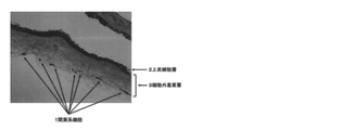
- amniotic membrane which is a part of the egg membrane of the fetal appendage, is divided into an epithelial cell layer in contact with the amniotic fluid and an extracellular matrix layer containing MSC below it (FIG. 1). For this reason, when the entire amniotic membrane is treated with trypsin, not only the extracellular matrix layer but also the basement membrane supporting the epithelial cell layer is digested, resulting in a mixture of epithelial cells and MSCs.
- Non-Patent Documents 1 to 3 in order to separate MSC from the amniotic membrane with high purity, decomposing enzyme or manually removing epithelial cells first, and the remaining extracellular matrix layer There is a method in which MSC is recovered by treating again with a separating enzyme. However, these methods have a problem that epithelial cells cannot be completely removed or the amount of recovered MSCs is lowered.
- cryopreservation is essential to use egg membrane MSC for cell therapy on demand.
- various cell cryopreservation solutions based on dimethyl sulfoxide (DMSO) are commercially available, and in bone marrow MSC clinical trials, a cryopreservation solution containing 10% DMSO is used. There was a problem that the rate fell.
- DMSO dimethyl sulfoxide
- the production process is as simple as possible in order to eliminate contamination such as bacteria and viruses.
- Multiple enzyme treatments require removal of the enzyme during the course of washing, centrifugation, etc., resulting in lower MSC recovery efficiency.
- not all epithelial cells are removed by the process using a degrading enzyme (Non-patent Document 3 and Example 3), and many epithelial cells still adhere to the amniotic membrane.
- Non-patent Document 9 A cryopreservation solution in which DMSO content is reduced using rat bone marrow MSC has also been reported (Non-patent Document 9).
- the present invention has been made in view of the above problems, and a method for producing a mesenchymal cell composition by simply and aseptically separating high-purity amniotic membrane-derived MSCs by only one enzyme treatment It is an issue to provide.
- a further object of the present invention is to provide a method for producing cryopreserved mesenchymal cells that suppress cell aggregation and are optimized for MSC transplantation. Furthermore, this invention makes it a subject to provide the cell therapeutic agent containing the amniotic membrane origin MSC manufactured by the said method.
- the inventors of the present invention treated the amniotic membrane with collagenase and thermolysin and / or dispase, and passed the enzyme-treated amniotic membrane through a mesh to obtain mesenchymal cells. It was found that it can be separated with high purity. Furthermore, the present inventors cryopreserved a mixture containing mesenchymal cells in a solution containing 5 to 10% by weight of dimethyl sulfoxide, 5 to 10% by weight of hydroxylethyl starch, or 1 to 5% by weight of dextran. It was found that mesenchymal cells cryopreserved can be produced in a form optimized for MSC transplantation. Furthermore, the present inventors have demonstrated that the mesenchymal cell composition obtained by the above method is useful as a cell therapeutic agent. The present invention has been completed based on these findings.
- a method for producing a mesenchymal cell composition comprising: (2) A step of collecting the cells that have passed through the mesh; and a step of culturing the collected cells:
- the step of recovering cells that have passed through the mesh is a step of recovering mesenchymal cells by centrifugation after diluting the filtrate with a medium amount or a balanced salt solution of a double amount or more.
- a method for producing a cell-based cell composition (5) The method for producing a mesenchymal cell composition according to any one of (1) to (4), wherein the enzyme treatment step is a step of treatment at 30 to 40 ° C. for 30 minutes or more.
- (12) Mesenchymal cells obtained by culturing the mesenchymal cell composition according to (10) or (11) in a medium containing albumin containing 0.05% by mass or more and 5% by mass or less. Culture composition.
- a step of cryopreserving a mixture containing mesenchymal cells in a solution containing 5 to 10% by weight of dimethyl sulfoxide, 5 to 10% by weight of hydroxylethyl starch or 1 to 5% by weight of dextran is included.
- a method for producing cryopreserved mesenchymal cells is included.
- a cell therapeutic agent comprising a mesenchymal cell administration composition as an active ingredient.
- a method for treating a disease comprising administering a composition for administering mesenchymal cells to a subject in need of cell therapy.
- the treatment method according to (21), wherein the composition is an injectable preparation.
- the treatment method according to (21), wherein the composition is a preparation for transplantation having a cell mass or a sheet-like structure.
- human amnion-derived MSCs can be separated easily and accurately. Therefore, it can be expected that the present invention will lead to promotion of industrial use of MSCs that have been shown to be effective in the field of regenerative medicine.
- thermolysin concentration is fixed at 250 PU / ml and the collagenase concentration is shaken.
- the therapeutic effect of amnion-derived MSC transplantation in a rat cirrhosis model was observed in terms of the fibrosis area of the liver.
- the therapeutic effect of amnion-derived MSC transplantation in a rat radiation enteritis model is shown by the number of PAS-positive goblet cells by rectal PAS staining.
- fetal appendage refers to the egg membrane, placenta, umbilical cord and amniotic fluid.
- egg membrane is a fetal sac containing fetal amniotic fluid and consists of an amnion, chorion and decidua from the inside. Of these, the amnion and chorion originate from the fetus.
- the “amniotic membrane” is a transparent thin film with poor blood vessels in the innermost layer of the egg membrane. The inner wall is covered with a single epithelial cell having a secretory function and secretes amniotic fluid.
- MSC Mesenchymal stromal cell
- pluripotent mesenchymal cells i) Adhesive to plastic under culture conditions in standard medium. ii) The surface antigens CD105, CD73, and CD90 are positive, and CD45, CD34, CD14, CD11b, CD79alpha, CD19, and HLA-DR are negative. iii) Differentiation into bone cells, adipocytes and chondrocytes under culture conditions.
- the “mesenchymal cell composition” in the present specification means any composition containing mesenchymal cells, and is not particularly limited.
- mesenchymal cells after treating amniotic membrane with a degrading enzyme are used. It means cell suspension containing.
- the “mesenchymal cell culture composition” in the present specification means a cell suspension obtained by culturing the mesenchymal cell composition.
- the “composition for mesenchymal cell administration” means any composition prepared using the above mesenchymal cell composition in a form suitable for administration, and is not particularly limited.
- an infusion preparation is more than twice as much as a mixture containing mesenchymal cells in a solution containing 5 to 10% by weight of dimethyl sulfoxide, 5 to 10% by weight of hydroxylethyl starch or 1 to 5% by weight of dextran. It means cell suspension obtained in addition.
- Method for producing mesenchymal cell composition comprises a step of enzymatic treatment of amniotic membrane with collagenase and thermolysin and / or dispase; It is a method including the process of passing through a mesh.
- human amniotic membrane is treated only once with an enzyme solution containing collagenase and thermolysin and / or dispase adjusted to an optimum concentration, and after the enzyme treatment Can be passed through the mesh.
- the epithelial cell layer containing the basement membrane that is not digested with collagenase and thermolysin and / or dispase adjusted to the optimal concentration remains in the mesh, and the extracellular matrix layer that is digested with collagenase and thermolysin and / or dispase adjusted to the optimal concentration Since the contained MSC passes through the mesh, the MSC can be recovered by recovering the cells that have passed through the mesh.
- the mesenchymal cell composition may be produced by culturing the collected cells.
- Fig. 1 is a histology of human amniotic membrane.
- the amniotic membrane is composed of a superficial epithelial cell layer and an extracellular matrix layer below it, and the latter contains MSC.
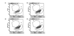
- Amniotic epithelial cells like other epithelial cells, are characterized by the expression of epithelial cadherin (E-cadherin: CD324) and epithelial adhesion factor (EpCAM: CD326), whereas MSCs express these epithelial specific surface antigen markers. It is not expressed and can be easily distinguished by FACS (FIG. 3).
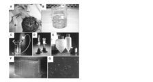
- FIG. 2 is a diagram for explaining the outline of the amnion-derived MSC separation method according to this embodiment.
- Physically harvest amniotic membrane from human fetal appendages A.
- the amniotic membrane is washed with an isotonic solution such as physiological saline (B), and the amniotic membrane is immersed in a solution containing an enzyme containing an optimal concentration of collagenase and thermolysin and / or dispase and stirred at an appropriate temperature.
- C The epithelial cell layer is not separated into individual cells by the enzyme treatment that targets only the extracellular matrix, but remains in the layer structure. Therefore, when the obtained amniotic fluid is passed through the mesh (D), the epithelial cell layer is meshed. The MSC remaining on the extracellular matrix layer passes through the mesh and can be collected.
- E is a centrifuge tube
- F is a culture dish
- G is a photograph of cultured cells.
- the mesenchymal cells are pelleted by centrifugation at 400 xg for 5 minutes. 9. Dilute with ⁇ MEM supplemented with 10% FBS, seed in a plastic flask, and incubate.
- the enzyme treatment is performed a plurality of times, and a decrease in recovery rate and contamination by microorganisms are prevented by repeating centrifugation and washing each time the treatment is performed.
- the purification procedure is not required, and a large amount of homogeneous MSC can be easily prepared in a short time.
- the combination of enzymes used for separation of amnion-derived MSC according to the present invention is collagenase that digests only collagen and thermolysin and / or dispase, which is a metalloproteinase that cleaves the N-terminal side of nonpolar amino acids.
- a combination of collagenase, thermolysin and / or dispase is used.
- an enzyme (or a combination thereof) that releases MSC and does not degrade the epithelial cell layer can also be used.
- concentrations of collagenase, thermolysin and / or dispase can be determined by microscopic observation or FACS after enzyme treatment. The concentration is preferably such that the epithelial cell layer is not degraded and the MSC contained in the extracellular matrix layer is released.
- the concentration of collagenase is preferably 50 CDU / ml to 1000 CDU / ml, and the concentration of thermolysin and / or dispase is preferably 100 PU / ml to 800 PU / ml.
- thermolysin concentration 400 PU / ml.
- CD90-positive MSC content was 83.3% and CD324-positive epithelial cells were 12.8% (Example 1, Table 2).
- the optimal dispase concentration to be added to collagenase 300 CDU / ml was 200 PU / ml.
- the CD90-positive MSC content was 83.3% and CD324-positive epithelial cells were 12.8% (Example 6, Table 9). .
- thermolysin and dispase were added alone at a concentration higher than 400 PU / ml, epithelial cells were observed to break down (Example 2, FIG. 6, Example 7, and FIG. 14).
- Enzymatic treatment is preferably performed by immersing the amniotic membrane washed with physiological saline or the like into the enzyme solution and treating with stirring with a stirrer or shaker, but other stirring means may be used. In short, MSC may be efficiently released. . This releases MSC contained in the extracellular matrix layer.
- the enzyme treatment can be preferably performed by stirring for 30 minutes or more at 10 rpm / min to 100 rpm / min using a stirrer or shaker.
- the upper limit of the enzyme treatment time is not particularly limited, but is generally within 6 hours, preferably within 3 hours, for example, within 90 minutes.
- the enzyme treatment temperature is not particularly limited as long as the object of the present invention can be achieved, but is preferably 30 to 40 ° C, more preferably 30 to 37 ° C.
- the pore size (size) of the mesh is not particularly limited as long as it is not contrary to the object of the present invention, but is preferably 40 to 200 ⁇ m, more preferably 40 to 150 ⁇ m, and further preferably 70 to 150 ⁇ m. Particularly preferred is 100 to 150 ⁇ m.
- Nylon mesh is preferably used as the mesh material. Tubes containing nylon mesh of 40 ⁇ m, 70 ⁇ m, and 100 ⁇ m, such as Falcon cell strainer, which is widely used for research, can be used. Moreover, the medical mesh cloth (nylon and polyester) currently used by hemodialysis etc. can be utilized. Furthermore, an arterial filter (polyester mesh, 40 ⁇ m to 120 ⁇ m) used during extracorporeal circulation can also be used. Other materials, such as stainless mesh (wire mesh), can also be used.
- the MSC that has passed through the mesh can be collected by centrifugation after diluting the filtrate with double or more medium or balanced salt buffer.
- the collected cells can be grown by culture to produce a mesenchymal cell culture composition.
- the medium is ⁇ MEM ⁇ M199 containing 0.05% or more and less than 5% of albumin or a medium based on these, and the growth of DMEM • F12 ⁇ RPMI1640 or a medium based on these is reduced.
- it is an ⁇ MEM medium containing 10% or more of bovine human serum, and is cultured in a plastic dish flask at 5% CO 2 in a 37 ° C environment.
- a buffer solution such as Dulbecco's phosphate buffer (DPBS), Earl's parallel salt (EBSS), Hanks balanced salt (HBSS), phosphate buffer (PBS) or the like can be used.
- the content ratio of CD324 and CD326 positive epithelial cells is 20% or less
- the content ratio of CD90 positive cells is 75% or more
- the viable cell ratio is 80% or more.
- a mesenchymal cell composition can be produced, and the mesenchymal cell composition is also within the scope of the present invention.
- the method for producing a cryopreserved mesenchymal cell according to the present invention comprises 5-10% by mass of dimethyl sulfoxide (DMSO) and 5-hydroxylethyl starch.
- DMSO dimethyl sulfoxide
- a method comprising cryopreserving a mixture containing mesenchymal cells in a solution containing 10% by mass or 1 to 5% by mass of dextran.
- cryopreservation solution of MSC used in the method of the present invention is characterized by reducing DMSO content and adding hydroxylethyl starch (HES) or dextran (such as Dextran40) instead.
- the cryopreservation solution may further contain more than 0% by mass and less than 5% by mass of human albumin.
- cryopreservation solution As an example of the cryopreservation solution, a cryopreservation solution having a composition of DMSO 5 mass%, HES 6 mass%, and human albumin 4 mass% can be used. Using the above cryopreservation solution, use a program freezer to lower the temperature to a temperature between -30 ° C and -50 ° C (eg, -40 ° C) at a freezing rate of -1 to -2 ° C / min. Further, cryopreserved mesenchymal cells can be produced by lowering the temperature to a temperature between ⁇ 80 ° C. and ⁇ 100 ° C. (eg, ⁇ 90 ° C.) at a freezing rate of ⁇ 10 ° C./min. . A mesenchymal cell administration composition can be produced by thawing the cryopreserved mesenchymal cells obtained by the above method and then diluting it twice or more with an infusion preparation.
- the MSCs prepared above can be used as therapeutic agents for intractable diseases. That is, according to the present invention, there is provided a cell therapeutic agent comprising the above-described mesenchymal cell composition and / or mesenchymal cell culture composition and / or mesenchymal cell administration composition as an active ingredient. Furthermore, according to the present invention, the above-described mesenchymal cell composition and / or mesenchymal cell culture composition and / or mesenchymal cell administration composition used for cell transplantation therapy are provided. .
- the method comprises the step of administering to the subject a therapeutically effective amount of the mesenchymal cell composition and / or mesenchymal cell culture composition and / or mesenchymal cell administration composition described above.
- Methods of transplanting cells into a subject, as well as methods of treating a subject's disease are provided.
- the cell therapeutic agent, mesenchymal cell composition and / or mesenchymal cell culture composition and / or mesenchymal cell administration composition of the present invention may be prepared by, for example, graft-versus-host disease (GVHD, Example 10), Inflammatory bowel diseases including Crohn's disease (Example 11) and ulcerative colitis, collagen disease including systemic lupus erythematosus (Example 12), cirrhosis (Example 13), radiation enteritis (Example 14), atopic skin It can be applied to flames. Inflammation can be suppressed by administering MSC prepared by the production method of the present invention in an amount that can be measured at the treatment site.
- GVHD graft-versus-host disease
- Inflammatory bowel diseases including Crohn's disease (Example 11) and ulcerative colitis
- collagen disease including systemic lupus erythematosus
- cirrhosis Example 13
- radiation enteritis Example 14
- cryopreserved MSC in order to maintain cell viability, it is necessary to use frozen cryopreserved MSC as soon as possible after rapid thawing and dilution with an infusion preparation such as physiological saline. This is because DMSO contained in the cryopreservation solution is cytotoxic.
- Mesenchymal cell administration compositions diluted with the above infusion preparations are also patients with graft-versus-host disease, inflammatory bowel disease including Crohn's disease, collagen disease including systemic lupus erythematosus, cirrhosis, radiation enteritis, atopic dermatitis, etc. Can be administered intravenously and treated.
- the “infusion preparation” in the present specification is not particularly limited as long as it is a solution used in human treatment.
- physiological saline 5% glucose solution
- Ringer's solution lactated Ringer's solution
- Ringer's acetate solution No. 1 solution , No. 2, No. 3, No. 4, etc.
- the method of administering the cell therapeutic agent of the present invention is not particularly limited, and for example, subcutaneous injection, intralymphatic injection, intravenous injection, intraperitoneal injection, intrathoracic injection, direct injection into the local area, or direct transplantation to the local area However, it is not limited to these.
- the cell therapeutic agent of the present invention can also be used as an injectable preparation or a preparation for transplantation of a cell mass or a sheet-like structure for the purpose of treating other diseases like the bone marrow MSC preparation.
- the dose of the cell therapeutic agent of the present invention is such an amount of cells that, when administered to a subject, can obtain a therapeutic effect on a disease compared to a subject not administered.
- the specific dose can be appropriately determined depending on the administration form, administration method, purpose of use and age, weight, and symptoms of the subject.
- the number of mesenchymal cells is, for example, human (for example, adult) 10 5 to 10 9 body weight / kg body weight per administration is preferable, and 10 5 to 10 8 body weight / kg body weight is more preferable.
- the obtained cells were stained with mesenchymal marker anti-CD90-FITC antibody and epithelial marker anti-CD324-APC antibody (BD Bioscience) at 4 ° C for 15 minutes, and 7-AAD dye was added to remove dead cells Then, surface antigen marker analysis was performed with a flow cytometer (FACSCanto: BD). The results are shown in Table 1, Table 2, FIG. 3 and FIG.
- CDU collagen digestion unit
- PU protease unit
- the target CD90-positive MSC is only 34.6%, unnecessary CD324-positive epithelial cells are 8.6%, and other than that The number of cells considered to be red blood cells was 56.8%.
- the proportion of CD90-positive MSCs increased with the addition of thermolysin. With thermolysin 400 PU / ml (No. 4), CD90-positive MSC 83.3%, CD324-positive epithelial cells 12.8%, and others were 3.9%. .
- the number of MSCs obtained from each sample was 0.1 ⁇ 10 6 for collagenase alone (No. 1), but the MSC obtained by adding thermolysin increased to 400 PU / ml (No. 4). ) Obtained 1.66x10 6 MSCs, about 16 times that of No.1.
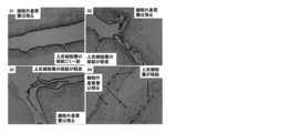
- FIG. 4 when the tissue remaining on the filter after enzyme treatment was examined by HE staining, the structure of the extracellular matrix layer was maintained with collagenase alone (No. 1), and digestion was insufficient. It was. Digestion of the extracellular matrix layer was observed by adding thermolysin, and complete digestion was performed at 400 PU / ml (No. 4).
- Example 2 Based on Example 1 above, the same method was used to examine only thermolysin except for collagenase. The results are shown in Table 3, FIG. 5 and FIG.
- Example 2 From the results of Example 2, it was found that the target MSC could not be obtained at all with thermolysin alone, and that the epithelial cell layer was broken when the thermolysin concentration was 800 PU / ml or higher.
- Non-patent Document 3 Take 1g of human amniotic membrane into a container, No.41) 0.05% trypsin (containing 0.53 mM EDTA) in 5 ml, 37 ° C, 90 minutes, 60 rpm shaking and stirring (by shaker), No.42) 0.05% trypsin (containing 0.53 mM EDTA, Invitrogen Company) After 5 minutes of shaking at 37 ° C for 90 minutes and 60 rpm with a shaker (by shaker), the remaining tissue was filtered through a nylon net filter (pore size: 100 ⁇ m), and the purified collagenase (300 CDU / ml) -containing Hanks balanced salt solution (Ca ⁇ (Containing Mg) at 37 ° C for 90 minutes and 60 rpm with shaking (by shaker), No.
- the conventional method of treating with collagenase after trypsin treatment has a merit that many cells can be obtained, but there is much contamination of epithelial cells, and high purity MSC can be obtained by techniques such as specific gravity centrifugation.
- Disadvantages include the need for an operation for sorting cells, and the two steps of collagenase treatment in addition to trypsin treatment, which complicates the operation.
- the latter demerit is that trypsin is inactivated by Ca, but collagenase is Ca-requiring and cannot be processed simultaneously.
- Example 4 Based on the above Examples 1 to 3, the collagenase concentration required for the separation of amnion mesenchymal cells when the thermolysin concentration was fixed at 250 PU / ml was examined. The results are shown in Table 6 and FIG.
- the number of viable cells negative for trypan blue staining obtained with collagenase 300 CDU / ml was 1.53 ⁇ 10 6 cells, whereas the collagenase concentration was 1/4 of 75CDU / ml.
- the number of viable cells was reduced to 1.16 ⁇ 10 6 .
- the CD90 positive mesenchymal cells in all the samples were 90% or more.
- the collagenase concentrations of 300 and 150 CDU / ml showed only the epithelial cell layer.
- the collagenase concentration was 75 CDU / ml (No. 61)
- the collagenase concentration is preferably at least 75 CDU / ml or more, more preferably 150 CDU / ml to sufficiently digest the extracellular matrix layer. I understood the necessity of more than / ml.
- Example 5 Furthermore, when the thermolysin concentration was fixed to 500 PU / ml, which is twice that of Example 3, the minimum collagenase concentration required for the separation of amnion mesenchymal cells was examined. The results are shown in Table 7 and FIG.
- the number of viable cells negative for trypan blue staining obtained with collagenase 150 CDU / ml was 2.05 ⁇ 10 6 , whereas the collagenase concentration was 1/4 of 37.5 CDU / ml.
- the number of viable cells was reduced to 0.82 ⁇ 10 6 .
- the CD90 positive mesenchymal cells in all the samples were 90% or more.
- Example 6 Regarding Example 1 above, instead of thermolysin, a dispase, which is a metalloproteinase that similarly cleaves the N-terminal side of nonpolar amino acids, was examined by adding collagenase thereto. The amniotic membrane was manually separated from the human fetal appendages from pregnant women who obtained informed consent.
- ⁇ MEM Alpha Modification of Minimum Essential Medium Eagle
- FBS fetal bovine serum
- ⁇ MEM Alpha Modification of Minimum Essential Medium Eagle
- the mixture was filtered through a nylon net filter (pore size: 100 ⁇ m).
- the tissue remaining on the filter was evaluated by hematoxylin and eosin (HE) staining.
- the filtrate was centrifuged at 400 ⁇ g for 5 minutes, the supernatant was discarded, the cells were resuspended in ⁇ MEM supplemented with 10% FBS, and the number of cells was measured after trypan blue staining.
- the obtained cells were stained with mesenchymal marker anti-CD90-FITC antibody and epithelial marker anti-CD324-APC antibody (BD Bioscience) at 4 ° C for 15 minutes, and 7-AAD dye was added to remove dead cells Then, surface antigen marker analysis was performed with a flow cytometer (FACSCanto: BD). The results are shown in Table 8, Table 9, FIG. 11 and FIG.
- the result of the flow cytometer shows that the target CD90-positive MSC is 90% or more regardless of whether collagenase alone (No. 81) or dispase is added (No. 82-84).
- the number of unnecessary CD324 positive epithelial cells was 10% or less.
- the result of collagenase alone (No. 81) was different from that of collagenase alone (No. 1) in Example 1 (FIG. 3), but this was thought to be because the manufacturers of collagenase used were different.
- the number of MSCs obtained from each sample was 0.39 ⁇ 10 6 for collagenase alone (No. 81), but the MSC obtained by addition of dispase increased to 200 PU / ml (No. 83). ), 2.86x10 6 pieces, MSC about 7 times that of No.81 was obtained.
- FIG. 12 when the tissue remaining on the filter after the enzyme treatment was examined by HE staining, the structure of the extracellular matrix layer was maintained with collagenase alone (No. 81), and digestion was insufficient. It was. Digestion of the extracellular matrix layer was observed by adding dispase, and complete digestion was performed at 200 PU / ml (No. 83) and 400 PU / ml (No. 84).
- Example 7 Based on Example 6 above, the same method was used to examine using only dispase except for collagenase. The results are shown in Table 10, FIG. 13 and FIG.
- the number of cells per gram of human amniotic membrane obtained by digestion with dispase alone increased in a thermolysin concentration-dependent manner.
- the results of the flow cytometer of the cells contained in the enzyme treatment solution containing only dispase showed that the target number of CD90-positive MSCs was very small at several percent.
- the extracellular matrix layer containing MSC was not digested at all, and the breakdown of the epithelial cell layer was caused by the concentration of dispase. Existed dependently.
- Example 7 From the results of Example 7, it was found that the target MSC could not be obtained with dispase alone, and that the epithelial cell layer was broken when the dispase concentration was 800 PU / ml or more.
- Example 8 Based on the above Examples 6 to 7, the minimum collagenase concentration required for the separation of amnion mesenchymal cells when the dispase concentration was fixed at 250 PU / ml was examined. The results are shown in Table 11 and FIG.
- the number of viable cells negative for trypan blue staining obtained with collagenase 300 CDU / ml was 1.57 ⁇ 10 6 cells, whereas the collagenase concentration was 1/4 that of 75 CDU / ml.
- the number of viable cells was reduced to 1.34 ⁇ 10 6 .
- the CD90 positive mesenchymal cells in all the samples were 90% or more.
- the collagenase concentration was 300 CDU / ml (No. 113), but only the epithelial cell layer,
- the collagenase concentration was 75 and 150 CDU / ml (No. 111 and 112), an extracellular matrix layer was observed and digestion was poor.
- the collagenase concentration is at least 75 CDU / ml or more, more preferably 150 CDU / ml or more, even more preferably, in order to sufficiently digest the extracellular matrix layer.
- the collagenase concentration is at least 75 CDU / ml or more, more preferably 150 CDU / ml or more, even more preferably, in order to sufficiently digest the extracellular matrix layer.
- Example 9 The cells obtained in Example 1 were diluted with a 10% FBS-added ⁇ MEM culture solution, and then seeded on a plastic dish to adhere and culture amniotic membrane-derived MSCs. After detachment of cells by trypsin treatment, neutralize trypsin with ⁇ MEM culture medium supplemented with 10% FBS, centrifuge and discard the supernatant, and resuspend the resulting cell pellet in RPMI1640 to create an amnion-derived MSC suspension did. A cryopreservation solution was prepared so that the final composition became Table 12 with respect to the obtained suspension.
- DMSO Simga-Aldrich, part number D2650 HES: Nipro, product number HES40 Human albumin (Alb): Venesis, blood donated albumin 25% IV 12.5g / 50ml Dextran 40: manufactured by Otsuka Pharmaceutical Factory Co., Ltd., low molecular weight dextran sugar injection physiological saline: manufactured by Otsuka Pharmaceutical Factory Co., Ltd .: The above-mentioned dilution adjustment solution was used.
- mice In mice, allogeneic bone marrow transplantation and splenocyte transplantation were performed to develop acute graft-versus-host disease (GVHD), and the therapeutic effect of human amnion-derived MSC transplantation was examined.
- GVHD graft-versus-host disease
- 7-8-week-old female B6C3F1 mice were intravenously injected with 1.0 x 10 7 bone marrow cells and 3 x 10 7 cells from allogeneic BDF1 mice after 15 Gy X-ray irradiation Transplanted.
- Example 11 In rats, dextran sulfate (DSS) was orally ingested to cause inflammatory bowel disease, and the therapeutic effect of human amnion-derived MSC transplantation was examined. Administration of 8% DSS with free drinking was started for 8-week-old male SD rats. The day after the start of DSS administration, human amnion-derived MSC (1 ⁇ 10 6 cells) obtained by the same method as in Example 1 (under the conditions of collagenase 300CDU / ml + thermolysin 250PU / ml) was transplanted intravenously, DSS was administered for a total of 5 days.
- DAI Disease activity index
- Example 12 In mice, systemic lupus erythematosus was developed by administering pristane (2,6,10,14-tetramethyl-pentadecane), and the therapeutic effect of human amnion-derived MSC transplantation was examined. Pristane was administered intraperitoneally to 13-week-old male BALB / c mice.
- human amniotic membrane-derived MSC (1 ⁇ 10 5 cells / 10 g) obtained by the same procedure as in Example 1 (under conditions of collagenase 300 CDU / ml + thermolysin 250 PU / ml) was administered to the tail vein, and the same number every other week thereafter The human amnion-derived MSC was administered, and biochemical evaluation was performed after 20 weeks.
- FIG. 20 shows the course of urine protein. The results revealed that proteinuria associated with systemic lupus erythematosus was improved by transplanting human amnion-derived MSCs.
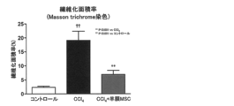
- Example 13 In rats, cirrhosis was caused by repeated administration of carbon tetrachloride (CCl 4 ), and the therapeutic effect of human amnion-derived MSC transplantation was examined. Intraperitoneal administration of 2 ml / kg CCl 4 was started twice a week for 6-week-old male SD rats. Three weeks after the start of CCl 4 administration, human amniotic membrane-derived MSC (1 ⁇ 10 6 cells) obtained by the same procedure as in Example 1 (collagenase 300CDU / ml + thermolysin 250PU / ml) was intravenously administered. After transplantation, CCl 4 was administered for a total of 7 weeks, and histological evaluation of the liver was performed.
- liver fibrosis area ratio (collagen fiber positive ratio) obtained from the result of Masson trichrome staining of the liver. From these results, it became clear that liver fibrosis associated with cirrhosis was improved by human amnion-derived MSC transplantation.
- Example 14 In rats, radiation enteritis was caused by irradiation of the rectum, and the therapeutic effect of human amnion-derived MSC was examined. Eight weeks old male SD rats were irradiated with 5 Gy / day of radiation in the lower abdomen for 5 consecutive days. On the last irradiation day, human amnion-derived MSC (1 ⁇ 10 6 cells) obtained by the same procedure as in Example 1 (collagenase 300CDU / ml + thermolysin 250PU / ml) was transplanted intravenously. A histological evaluation of the rectum was performed one day later. FIG.
Landscapes
- Health & Medical Sciences (AREA)
- Life Sciences & Earth Sciences (AREA)
- Engineering & Computer Science (AREA)
- Chemical & Material Sciences (AREA)
- Biomedical Technology (AREA)
- General Health & Medical Sciences (AREA)
- Developmental Biology & Embryology (AREA)
- Organic Chemistry (AREA)
- Cell Biology (AREA)
- Zoology (AREA)
- Bioinformatics & Cheminformatics (AREA)
- Biotechnology (AREA)
- Public Health (AREA)
- Medicinal Chemistry (AREA)
- Veterinary Medicine (AREA)
- Animal Behavior & Ethology (AREA)
- Pharmacology & Pharmacy (AREA)
- Immunology (AREA)
- Genetics & Genomics (AREA)
- Wood Science & Technology (AREA)
- Epidemiology (AREA)
- Chemical Kinetics & Catalysis (AREA)
- General Chemical & Material Sciences (AREA)
- Nuclear Medicine, Radiotherapy & Molecular Imaging (AREA)
- Reproductive Health (AREA)
- General Engineering & Computer Science (AREA)
- Microbiology (AREA)
- Biochemistry (AREA)
- Hematology (AREA)
- Virology (AREA)
- Pregnancy & Childbirth (AREA)
- Rheumatology (AREA)
- Gynecology & Obstetrics (AREA)
- Dermatology (AREA)
- Transplantation (AREA)
- Oral & Maxillofacial Surgery (AREA)
- Botany (AREA)
- Urology & Nephrology (AREA)
- Toxicology (AREA)
- Gastroenterology & Hepatology (AREA)
Abstract
本発明の目的は、一回の酵素処理のみで、簡便かつ無菌的に高純度な羊膜由来MSCを分離することによる間葉系細胞組成物の製造方法を提供することである。本発明によれば、羊膜を、コラゲナーゼと、サーモリシン及び/又はディスパーゼとにより酵素処理する工程;及び酵素処理した羊膜をメッシュに通す工程を含む、間葉系細胞組成物の製造方法;凍結保存された間葉系細胞組成物の製造方法;並びに前記間葉系細胞組成物を有効成分とする移植片対宿主病・炎症性腸疾患・全身性エリテマトーデス・肝硬変・放射線腸炎治療剤が提供される。
Description
本発明は、羊膜から、細胞治療応用に適した間葉系細胞(Mesenchymal stromal cell:MSC)を高純度かつ簡便に分離することによる間葉系細胞組成物の製造方法に関する。さらに本発明は、凍結保存された間葉系細胞組成物の製造方法、並びに間葉系細胞組成物を含む治療剤に関する。
間葉系幹細胞は、骨髄に存在することが指摘された体性幹細胞であり、幹細胞として骨、軟骨及び脂肪に分化する能力を有する。間葉系幹細胞は、細胞治療における有望な細胞ソースとして注目され、最近では、胎盤、臍帯、卵膜などの胎児付属物にも存在することが知られている。
現在、間葉系幹細胞は、分化能以外に免疫抑制能を有することで注目されており、骨髄由来間葉系幹細胞を用い、造血幹細胞移植における急性移植片対宿主病(GVHD)、及び炎症性腸疾患のCrohn病に対する治験が進んでいる。本発明者らは胎児付属物由来間葉系幹細胞の免疫関連疾患に対する臨床応用を目指した研究を進めてきた。これまでに本発明者らは、胎児付属物由来間葉系幹細胞が骨髄由来間葉系幹細胞と同様の分化能を有すること(非特許文献6)、卵膜由来間葉系幹細胞がラット自己免疫性心筋炎モデルの病態を改善すること(非特許文献7)、並びに臍帯由来間葉系幹細胞がマウス急性移植片対宿主病(GVHD)モデルの救命率を改善すること(非特許文献8)を報告してきた。胎児付属物由来間葉系幹細胞は、骨髄由来間葉系幹細胞と比較し、一度に多くの間葉系幹細胞が得られ、大量培養が短期間・低コストに可能であり、非侵襲的に採取可能であり、免疫抑制効果も高い(非特許文献7)。これらのことから、胎児付属物由来間葉系幹細胞を含むMSCは、その著明な免疫抑制効果により、様々な免疫関連疾患を対象とした細胞治療に応用可能である。
これまでに、胎児付属物である卵膜、胎盤および羊水から、多能性ヒト胎児由来幹細胞を取得する方法が報告されている(特許文献1)。特許文献1では、これら幹細胞をc-kit(CD117)陽性と位置づけ、フローサイトメトリーにより分離する方法が記載されている。また、胎盤および臍帯から、成人または小児の様々な細胞に分化する能力を有する幹細胞・前駆細胞を取得する方法も報告されている(特許文献2)。特許文献2では、様々な臓器や組織を構成する細胞に分化する能力を有する(=間葉系幹細胞以上の分化能力を有する)幹細胞・前駆細胞が胎盤および臍帯に含まれており、その分離に関する方法が記載されている。
これら胎児付属物由来の幹細胞および前駆細胞を含む細胞分離には、一般にトリプシン、コラゲナーゼ又はディスパーゼ等の分解酵素が用いられている(特許文献1及び2、非特許文献1~4)。胎児付属物のうち卵膜は、羊水に接し最も胎児側に位置する羊膜と、その外側に位置する絨毛膜とに分けられ、卵膜(羊膜および絨毛膜)由来のMSCも分解酵素を用いて分離する方法が一般的である(非特許文献1及び4)。
胎児付属物の卵膜の一部である羊膜は、羊水に接する上皮細胞層と、その下にあるMSCを含む細胞外基質層とに分けられる(図1)。そのため、羊膜全体をトリプシンにて処理した場合、細胞外基質層のみならず上皮細胞層を支持する基底膜も消化され、結果、上皮細胞とMSCの混合物となるという問題があった。この問題の解決方法としては、非特許文献1~3のように、羊膜からMSCを高純度に分離するために、分解酵素や用手的に上皮細胞を先に取り除き、残った細胞外基質層を再度分離酵素で処理し、MSCを回収する方法がある。しかし、これらの方法では、完全には上皮細胞を除去できない、又はMSCの回収量が下がるという問題があった。
更に、卵膜MSCをオンデマンドで細胞治療用に使用するには、凍結保存が必須である。研究レベルではジメチルスルホキシド(DMSO)をベースとした各種の細胞凍結保存液が市販され、骨髄MSCの臨床試験でもDMSOを10%含有した凍結保存液が用いられているが、解凍後の細胞の生存率が下がるという問題があった。
Am J Obstet Gynecol. 2004;190(1):87-92.
Am J Obstet Gynecol. 2006;194(3):664-73.
Current Protocols in Stem Cell Biology 1E.5
J Tissue Eng Regen Med. 2007;1(4):296-305.
Cytotherapy. 2006;8(4):315-7.
Stem Cells. 2008;26(10):2625-33.
J Mol Cell Cardiol. 2012;53(3):420-8.
Cytotherapy. 2012;14(4):441-50.
BMC Biotechnology 2012;12:49.
ヒト羊膜由来MSCの細胞製剤化を考えると、細菌・ウイルス等のコンタミネーションを排除するため、出来るだけ製造工程は簡便であることが好ましい。複数回の酵素処理は、途中、洗浄・遠心操作等による酵素除去が必要となり、MSC回収効率を下げる結果をもたらす。また、既知の方法では、分解酵素による工程で全ての上皮細胞が除去される訳ではなく(非特許文献3及び実施例3)、依然多くの上皮細胞が羊膜に接着している状態にある。
また、細胞保存液に関しては、DMSOは細胞毒性を有することから、できるだけ濃度を下げることが望ましい。ラット骨髄由来MSCを用い、DMSO含有量を減らした凍結保存液の報告もなされている(非特許文献9)。
本発明は、上記問題点に鑑みてなされたものであって、一回の酵素処理のみで、簡便かつ無菌的に高純度な羊膜由来MSCを分離することによる間葉系細胞組成物の製造方法を提供することを課題とする。更に本発明は、細胞凝集を抑制し、MSC移植に至適化されている凍結保存された間葉系細胞の製造方法を提供することを課題とする。さらに本発明は、上記方法で製造した羊膜由来MSCを含む細胞治療剤を提供することを課題とする。
本発明者らは上記課題を解決するために鋭意検討した結果、羊膜を、コラゲナーゼと、サーモリシン及び/又はディスパーゼとにより酵素処理し、酵素処理した羊膜をメッシュに通すことによって、間葉系細胞を高純度に分離できることを見出した。さらに本発明者らは、ジメチルスルホキシドを5~10質量%含有し、ヒドロキシルエチルデンプンを5~10質量%またはデキストランを1~5質量%含有する溶液中に間葉系細胞を含む混合物を凍結保存することによって凍結保存された間葉系細胞をMSC移植に至適化した形で製造できることを見出した。さらに本発明者らは、上記の方法で得た間葉系細胞組成物が細胞治療剤として有用であることを実証した。本発明はこれらの知見に基づいて完成したものである。
すなわち、本明細書によれば以下の発明が提供される。
(1)羊膜を、コラゲナーゼと、サーモリシン及び/又はディスパーゼとにより酵素処理する工程;及び
酵素処理した羊膜をメッシュに通す工程:
を含む、間葉系細胞組成物の製造方法。
(2)メッシュを通過した細胞を回収する工程;及び
回収した細胞を培養する工程:
をさらに含む、(1)に記載の間葉系細胞組成物の製造方法。
(3) 前記メッシュを通過した細胞を回収する工程が、倍量又はそれ以上の量の培地又は平衡塩溶液で濾液を希釈した後、遠心分離により間葉系細胞を回収する工程である、(2)に記載の間葉系細胞組成物の製造方法。
(4) コラゲナーゼの濃度が50CDU/ml~1000CDU/mlであり、サーモリシンおよび/又はディスパーゼの濃度が100PU/ml~800PU/mlである、(1)から(3)の何れかに記載の間葉系細胞組成物の製造方法。
(5) 酵素処理する工程が、30~40℃で30分以上処理する工程である、(1)から(4)の何れかに記載の間葉系細胞組成物の製造方法。
(6) 酵素処理する工程が、スターラー又はシェーカーを用いて10rpm/分~100rpm/分で30分以上撹拌する工程である、(1)から(5)の何れかに記載の間葉系細胞組成物の製造方法。
(7) 羊膜が帝王切開により得られたものである、(1)から(6)の何れかに記載の間葉系細胞組成物の製造方法。
(8) メッシュのポアサイズが40~200μmである、(1)から(7)の何れかに記載の間葉系細胞組成物の製造方法。
(9) 羊膜をメッシュを通す工程が自然落下である、(1)から(8)の何れかに記載の間葉系細胞組成物の製造方法。
(10) CD324及びCD326陽性上皮細胞の含有率が20%以下であり、CD90陽性細胞の含有率が75%以上であり、および生細胞率が80%以上である、間葉系細胞組成物。
(11) (1)から(9)の何れかに記載の間葉系細胞組成物の製造方法により得られる、(11)に記載の間葉系細胞組成物。
(12) (10)又は(11)に記載の間葉系細胞組成物を0.05質量%より多く5質量%以下のアルブミンを含有する培地にて培養することにより得られる、間葉系細胞培養組成物。
(11) (1)から(9)の何れかに記載の間葉系細胞組成物の製造方法により得られる、(11)に記載の間葉系細胞組成物。
(12) (10)又は(11)に記載の間葉系細胞組成物を0.05質量%より多く5質量%以下のアルブミンを含有する培地にて培養することにより得られる、間葉系細胞培養組成物。
(13) ジメチルスルホキシドを5~10質量%含有し、ヒドロキシルエチルデンプンを5~10質量%またはデキストランを1~5質量%含有する溶液中に間葉系細胞を含む混合物を凍結保存する工程を含む、凍結保存された間葉系細胞の製造方法。
(14) 前記溶液が、さらにヒトアルブミンを0質量%より多く5質量%以下含有する溶液である、(13)に記載の凍結保存された間葉系細胞の製造方法。
(15) 前記間葉系細胞が、(1)から(9)の何れかに記載の方法により製造された間葉系細胞組成物に含まれる間葉系細胞、請求(10)又は(11)に記載の間葉系細胞組成物に含まれる間葉系細胞、又は(12)に記載の間葉系細胞培養組成物に含まれる間葉系細胞である、(13)又は(14)に記載の凍結保存された間葉系細胞の製造方法。
(16) (13)から(15)の何れかに記載の方法により得られる凍結保存された間葉系細胞を解凍後、輸液製剤により2倍以上に希釈する工程を含む、間葉系細胞投与用組成物を製造する方法。
(17) (10)又は(11)に記載の間葉系細胞組成物、および/又は(12)に記載の間葉系細胞培養組成物、および/又は(16)に記載の方法により製造される間葉系細胞投与用組成物を有効成分として含む、細胞治療剤。
(18) 注射用製剤である、(17)に記載の細胞治療剤。
(19) 細胞塊又はシート状構造の移植用製剤である、(17)に記載の細胞治療剤。
(20) 移植片対宿主病、炎症性腸疾患、全身性エリテマトーデス、肝硬変、又は放射線腸炎から選択される疾患の治療剤である、(17)から(19)の何れかに記載の治療剤。
(21) (10)又は(11)に記載の間葉系細胞組成物、および/又は(12)に記載の間葉系細胞培養組成物、および/又は(16)に記載の方法により製造される間葉系細胞投与用組成物を細胞治療を必要とする対象者に投与することを含む、疾患の治療方法。
(22) 組成物が注射用製剤である、(21)に記載の治療方法。
(23) 組成物が細胞塊又はシート状構造の移植用製剤である、(21)に記載の治療方法。
(24) 疾患が移植片対宿主病、炎症性腸疾患、全身性エリテマトーデス、肝硬変、又は放射線腸炎から選択される疾患である、(21)から(23)の何れかに記載の治療方法。
本発明によれば、ヒト羊膜由来MSCを簡易且つ精度良く分離することができる。従って、本発明は再生医療分野において有効性が示されているMSCの産業利用促進につながることが期待できる。
以下、添付の図面を参照して本発明の実施形態について具体的に説明するが、当該実施形態は本発明の原理の理解を容易にするためのものであり、本発明の範囲は、下記の実施形態に限られるものではなく、当業者が以下の実施形態の構成を適宜置換した他の実施形態も、本発明の範囲に含まれる。
[1]用語の説明
本明細書において「胎児付属物」は、卵膜、胎盤、臍帯および羊水を指す。さらに「卵膜」は、胎児の羊水を含む胎嚢であり、内側から羊膜、絨毛膜および脱落膜からなる。このうち、羊膜と絨毛膜は胎児を起源とする。「羊膜」は、卵膜の最内層にある血管に乏しい透明薄膜で、内壁は分泌機能のある一層の上皮細胞で覆われ羊水を分泌する。
本明細書において「胎児付属物」は、卵膜、胎盤、臍帯および羊水を指す。さらに「卵膜」は、胎児の羊水を含む胎嚢であり、内側から羊膜、絨毛膜および脱落膜からなる。このうち、羊膜と絨毛膜は胎児を起源とする。「羊膜」は、卵膜の最内層にある血管に乏しい透明薄膜で、内壁は分泌機能のある一層の上皮細胞で覆われ羊水を分泌する。
本明細書における「間葉系細胞(Mesenchymal stromal cell:MSC)」は、間葉系および前駆細胞(1つ又は複数の様々な臓器又は組織を構成する細胞に分化する能力を有する細胞を意味)を含有し、The International Society for Cellular Therapyが提唱する多能性間葉系細胞(Mesenchymal stromal cell:MSC)の定義を満たす細胞を指す(非特許文献5、下記参照)。
多能性間葉系細胞の定義
i)標準培地での培養条件で、プラスチックに接着性を示す。
ii)表面抗原CD105, CD73, CD90が陽性であり、CD45, CD34, CD14, CD11b, CD79alpha, CD19, HLA-DRが陰性。
iii)培養条件にて骨細胞、脂肪細胞、軟骨細胞に分化可能。
i)標準培地での培養条件で、プラスチックに接着性を示す。
ii)表面抗原CD105, CD73, CD90が陽性であり、CD45, CD34, CD14, CD11b, CD79alpha, CD19, HLA-DRが陰性。
iii)培養条件にて骨細胞、脂肪細胞、軟骨細胞に分化可能。
本明細書における「間葉系細胞組成物」は、間葉系細胞を含む任意の組成物を意味し、特に限定されないが、例えば、羊膜を分解酵素にて処理した後の間葉系細胞を含有する細胞浮遊液などを意味する。
本明細書における「間葉系細胞培養組成物」とは、上記の間葉系細胞組成物を培養することにより得られる細胞浮遊液などを意味する。
本明細書における「間葉系細胞投与用組成物」とは、上記の間葉系細胞組成物を用いて投与に適した形に調製した任意の組成物を意味し、特には限定されないが、例えば、ジメチルスルホキシドを5~10質量%含有し、ヒドロキシルエチルデンプンを5~10質量%またはデキストランを1~5質量%含有する溶液中に間葉系細胞を含む混合物に輸液製剤を2倍量以上加えて得られる細胞浮遊液などを意味する。
本明細書における「間葉系細胞培養組成物」とは、上記の間葉系細胞組成物を培養することにより得られる細胞浮遊液などを意味する。
本明細書における「間葉系細胞投与用組成物」とは、上記の間葉系細胞組成物を用いて投与に適した形に調製した任意の組成物を意味し、特には限定されないが、例えば、ジメチルスルホキシドを5~10質量%含有し、ヒドロキシルエチルデンプンを5~10質量%またはデキストランを1~5質量%含有する溶液中に間葉系細胞を含む混合物に輸液製剤を2倍量以上加えて得られる細胞浮遊液などを意味する。
[2]間葉系細胞組成物の製造方法
本発明の間葉系細胞組成物の製造方法は、羊膜を、コラゲナーゼと、サーモリシン及び/又はディスパーゼとにより酵素処理する工程;及び酵素処理した羊膜をメッシュに通す工程を含む方法である。
本発明の間葉系細胞組成物の製造方法は、羊膜を、コラゲナーゼと、サーモリシン及び/又はディスパーゼとにより酵素処理する工程;及び酵素処理した羊膜をメッシュに通す工程を含む方法である。
本発明による間葉系細胞組成物の製造方法の一例においては、ヒト羊膜を、至適濃度に調整したコラゲナーゼとサーモリシン及び/又はディスパーゼとを含有する酵素液で一回のみ処理し、酵素処理後の羊膜消化液をメッシュに通すことができる。至適濃度に調整したコラゲナーゼとサーモリシン及び/又はディスパーゼでは消化されない基底膜を含む上皮細胞層はメッシュに残り、至適濃度に調整したコラゲナーゼとサーモリシン及び/又はディスパーゼでは消化される細胞外基質層に含まれるMSCはメッシュを通過することから、メッシュを通過した細胞を回収することにより、MSCを回収することができる。さらに、回収した細胞を培養することによって間葉系細胞組成物を製造してもよい。
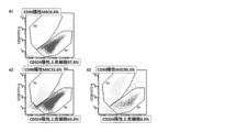
図1は、ヒト羊膜の組織図である。図1に示されるように、羊膜は表層の上皮細胞層とその下にある細胞外基質層からなり、後者にはMSCが含まれている。羊膜上皮細胞は、他の上皮細胞同様、特徴として上皮カドヘリン(E-cadherin:CD324)および上皮接着因子(EpCAM:CD326)を発現しているのに対し、MSCはこれら上皮特異的表面抗原マーカーを発現しておらず、FACSで容易に区別可能である(図3)。
図2は、本実施形態にかかる羊膜由来MSC分離法の概要を説明する図である。ヒト胎児付属物から羊膜を物理的に採取する(A)。羊膜を生理食塩水等の等張液にて洗浄し(B)、至適濃度のコラゲナーゼとサーモリシン及び/又はディスパーゼとを含有する酵素を含有する溶液に羊膜を浸して、適温にて震盪攪拌する(C)。上皮細胞層は細胞外基質のみを標的とする酵素処理では個々の細胞に分離されず層構造のままであるため、得られた羊膜消化液をメッシュに通すと(D)、上皮細胞層はメッシュ上に残り、細胞外基質層に含まれるMSCはメッシュを通過し、回収することが可能となる。Eは遠心管、Fは培養皿、Gは培養細胞の写真である。
本発明における実施形態を一例として詳細に述べる。
1.手術室にて待機的帝王切開例からヒト胎児付属物を無菌的に採取する。
2.胎児付属物から羊膜を無菌的且つ用手的に剥離する。
3.羊膜を滅菌容器(ディスポーザブルカップ)に移し、生理食塩水等の等張液を用いて数回洗浄し、付着した血液等を取り除く。
4.羊膜を、メス・ハサミ等にて大まかに切る(このステップは省略しても良い)。また、羊膜は、培地中で2~8℃、24~48時間保存してから使用してもよい。
5.羊膜を至適濃度に調整したコラゲナーゼと、サーモリシン及び/又はディスパーゼとを含有する溶液に浸し、恒温振盪機を用いて37℃90分間60rpm震盪攪拌する。
6.その結果、上皮細胞層は一層のままとなり、細胞外基質層に含まれるMSCは酵素含有溶液に浮遊する。
7. Falconセルストレーナー(100μmメッシュ)を滅菌チューブ(Falcon50mlチューブ)にセットし、自由落下にて羊膜消化後の細胞含有液をろ過することで、上皮細胞層はメッシュ上に残り、MSCのみがメッシュを通過する。
8.メッシュを通過したMSC含有溶液をハンクス平衡塩液で希釈後、400×g、5分遠心操作にて間葉系細胞をペレットにする
9. 10%FBS添加αMEMにて希釈し、プラスチックフラスコに播種し、培養する。
本発明によれば、従来のように、酵素処理を複数回行い、処理する度に遠心分離や洗浄を繰り返すことによる回収率の低下や微生物等の汚染を防止し、また、フィコール密度勾配遠心等の精製操作も不要で、容易に短時間で大量の均質なMSCを調製できる。
本発明にかかる羊膜由来MSCの分離に用いられる酵素の組み合わせは、コラーゲンのみを消化するコラゲナーゼと、非極性アミノ酸のN末端側を切断する金属プロテイナーゼであるサーモリシン及び/又はディスパーゼである。
本発明では、コラゲナーゼ、サーモリシンおよび/又はディスパーゼの組合せを用いているが、MSCを遊離し、上皮細胞層を分解しない酵素(又はその組み合わせ)を使用することもできる。コラゲナーゼ、サーモリシンおよび/又はディスパーゼの好ましい濃度の組合せは、酵素処理後顕微鏡観察やFACSにより決定することができる。上皮細胞層が分解されず、細胞外基質層に含まれるMSCが遊離する濃度条件であることが好ましい。
本発明では、コラゲナーゼ、サーモリシンおよび/又はディスパーゼの組合せを用いているが、MSCを遊離し、上皮細胞層を分解しない酵素(又はその組み合わせ)を使用することもできる。コラゲナーゼ、サーモリシンおよび/又はディスパーゼの好ましい濃度の組合せは、酵素処理後顕微鏡観察やFACSにより決定することができる。上皮細胞層が分解されず、細胞外基質層に含まれるMSCが遊離する濃度条件であることが好ましい。
コラゲナーゼの濃度は好ましくは50CDU/ml~1000CDU/mlであり、サーモリシンおよび/又はディスパーゼの濃度は好ましくは100PU/ml~800PU/mlである。
コラゲナーゼ300CDU/ml単独処理では生細胞数は0.29×106個だったが、サーモリシンを添加すると濃度依存的に生細胞数が増加し、サーモリシン400PU/mlでは生細胞数が1.99×106個と約7倍まで増加し、生細胞率も91.7%であった(実施例1、表1)。
同様に、コラゲナーゼ300CDU/mlにディスパーゼを添加すると濃度依存的に生細胞数は増加し、ディスパーゼ200PU/mlでは生細胞数が3.02×106個まで増加し、生細胞率も83.7%であった(実施例6、表8)。したがって、コラゲナーゼのみでなく、サーモリシン又はディスパーゼを混合して同時に酵素処理することにより、より多くの生細胞が得られる。
コラゲナーゼ300CDU/mlの場合、最適なサーモリシン濃度は400PU/mlであった。この場合のCD90陽性MSC含有率は83.3%、CD324陽性上皮細胞は12.8%であった(実施例1、表2)。また、コラゲナーゼ300CDU/mlに添加する最適なディスパーゼ濃度は200PU/mlであり、この場合のCD90陽性MSC含有率は83.3%、CD324陽性上皮細胞は12.8%であった(実施例6、表9)。
サーモリシンおよびディスパーゼを単独で400PU/mlよりも高濃度で添加すると、上皮細胞が破綻する現象が観察された(実施例2、図6、実施例7、図14)。
コラゲナーゼ300CDU/ml、サーモリシン250PU/mlの場合、CD90陽性MSCの含有率は93.3%であり、CD90陽性MSCの細胞数は1.43×106個であった(実施例4、表6)。また、コラゲナーゼ300CDU/ml、ディスパーゼ250PU/mlの場合、CD90陽性MSCの含有率は91.5%であり、CD90陽性MSCの細胞数は1.49×106個であった(実施例8、表11)。
酵素処理は、生理食塩水等で洗浄した羊膜を酵素液に浸漬し、スターラー又はシェーカーで撹拌しながら処理することが好ましいがこれ以外の撹拌手段でもよく、要は効率よくMSCが遊離すればよい。これにより細胞外基質層に含まれるMSCが遊離する。酵素処理は、好ましくはスターラー又はシェーカーを用いて10rpm/分~100rpm/分で30分以上撹拌することにより行うことができる。なお、酵素処理時間の上限は特に限定されないが、一般的には6時間以内、好ましくは3時間以内、例えば、90分以内行うことができる。また、酵素処理温度は、本発明の目的が達成できる限り特に限定されないが、好ましくは30~40℃であり、より好ましくは30~37℃である
この際、上皮細胞層が分解しない濃度の組合せにすることが重要である。上皮細胞が混入すると相対的にMSCの含有率が落ちるためである。
遊離したMSCを含む酵素溶液をメッシュを通してろ過することにより、遊離した細胞のみがメッシュを通過し、分解されなかった上皮細胞層はメッシュを通過できずにメッシュ状に残るため、遊離したMSCを容易に回収することができる。この際、メッシュのポアサイズ(大きさ)は本発明の目的に反しない限り特に限定されないが、好ましくは40~200μmであり、より好ましくは40~150μmであり、さらに好ましくは70~150μmであり、特に好ましくは100~150μmである。メッシュのポアサイズ(大きさ)を上記の範囲とすることにより、圧力を加えず、自然落下により落下させることで細胞の生存率が下がるのを防止することができる。
メッシュの材質としては、ナイロンメッシュが好ましく用いられる。研究用として汎用されるFalconセルストレーナーなどの40μm、70μm、100μmのナイロンメッシュを含有するチューブが利用可能である。また、血液透析などで使用されている医療用メッシュクロス(ナイロンおよびポリエステル)が利用できる。更に、体外循環時に使用される動脈フィルター(ポリエステルメッシュ、40μm~120μm)も利用可能である。他の材質、例えば、ステンレスメッシュ(金網)等も用いることが可能である。
MSCをメッシュ通過させる場合、自然落下(自由落下)が好ましい。ポンプ等を用いた吸引など強制的なメッシュ通過も可能であるが、細胞にダメージを与える事を避けるため、できるだけ弱い圧力が望ましい。
メッシュを通したMSCは、倍量又はそれ以上の培地又は平衡塩緩衝液で濾液を希釈した後、遠心分離により回収することができる。回収した細胞は、所望により、培養により増殖させることによって間葉系細胞培養組成物を製造することができる。培地は、0.05%より多く5%以下のアルブミンを含有するαMEM・M199あるいはこれらを基礎とする培地であり、DMEM・F12・RPMI1640あるいはこれらを基礎とする培地は増殖性が低下する。望ましくは10%以上のウシ・ヒト血清を含有するαMEM培地であり、プラスチックデッシュ・フラスコにて5%CO2、37℃環境にて培養を行う。平衡塩緩衝液としては、ダルベッコリン酸バッファー(DPBS)、アール平行塩(EBSS)、ハンクス平衡塩(HBSS)、リン酸バッファー(PBS)等の緩衝液を用いることができる。
上記した本発明の方法によれば、CD324及びCD326陽性上皮細胞の含有率が20%以下であり、CD90陽性細胞の含有率が75%以上であり、および生細胞率が80%以上である間葉系細胞組成物を製造することができ、前記間葉系細胞組成物も本発明の範囲内のものである。
[3]凍結保存された間葉系細胞の製造方法
本発明による凍結保存された間葉系細胞の製造方法は、ジメチルスルホキシド(DMSO)を5~10質量%含有し、ヒドロキシルエチルデンプンを5~10質量%またはデキストランを1~5質量%含有する溶液中に間葉系細胞を含む混合物を凍結保存する工程を含む方法である。
本発明による凍結保存された間葉系細胞の製造方法は、ジメチルスルホキシド(DMSO)を5~10質量%含有し、ヒドロキシルエチルデンプンを5~10質量%またはデキストランを1~5質量%含有する溶液中に間葉系細胞を含む混合物を凍結保存する工程を含む方法である。
本発明者らは鋭意研究を重ねた結果、高濃度DMSOによる細胞毒性が原因と思われる解凍後のMSCの生存率減少を見いだした(実施例)。凍結保存液におけるDMSO含有量を減らすことは、細胞死の抑制という観点から、細胞移植用ヒトMSCの凍結保存液として好ましいことが判明した。本発明の方法で用いるMSCの凍結保存液は、DMSO含有量を減らし、代わりにヒドロキシルエチルデンプン(HES)又はデキストラン(Dextran40など)を添加したことを特徴とするものである。凍結保存液は、さらにヒトアルブミンを0質量%より多く5質量%以下含有するものでもよい。凍結保存液の一例としては、DMSO5質量%、HES6質量%、ヒトアルブミン4質量%の組成の凍結保存液を用いることができる。
上記した凍結保存液を用いて、プログラムフリーザーを用い、例えば、-1~-2℃/分の凍結速度で-30℃~-50℃の間の温度(例えば、-40℃)まで温度を下げ、更に-10℃/分の凍結速度で-80℃~-100℃の間の温度(例えば、-90℃)まで温度を下げることによって、凍結保存された間葉系細胞を製造することができる。
上記の方法により得られた凍結保存された間葉系細胞は、解凍後、輸液製剤により2倍以上に希釈することによって、間葉系細胞投与用組成物を製造することができる。
上記した凍結保存液を用いて、プログラムフリーザーを用い、例えば、-1~-2℃/分の凍結速度で-30℃~-50℃の間の温度(例えば、-40℃)まで温度を下げ、更に-10℃/分の凍結速度で-80℃~-100℃の間の温度(例えば、-90℃)まで温度を下げることによって、凍結保存された間葉系細胞を製造することができる。
上記の方法により得られた凍結保存された間葉系細胞は、解凍後、輸液製剤により2倍以上に希釈することによって、間葉系細胞投与用組成物を製造することができる。
[4]細胞治療剤
上記で調製したMSC(増殖させたMSCも含む)は難治性疾患の治療剤として利用が可能である。
即ち、本発明によれば、上記した間葉系細胞組成物および/又は間葉系細胞培養組成物および/又は間葉系細胞投与用組成物を有効成分として含む細胞治療剤が提供される。さらに本発明によれば、細胞移植治療のために使用される、上記した間葉系細胞組成物および/又は間葉系細胞培養組成物および/又は間葉系細胞投与用組成物が提供される。更に本発明によれば、被験者に、上記の間葉系細胞組成物および/又は間葉系細胞培養組成物および/又は間葉系細胞投与用組成物の治療有効量を投与する工程を含む、被験者に細胞を移植する方法、並びに被験者の疾患の治療方法が提供される。
本発明の細胞治療剤、間葉系細胞組成物および/又は間葉系細胞培養組成物および/又は間葉系細胞投与用組成物は、例えば移植片対宿主病(GVHD、実施例10)、クローン病(実施例11)や潰瘍性大腸炎を含む炎症性腸疾患、全身性エリテマトーデス(実施例12)を含む膠原病、肝硬変(実施例13)、放射線腸炎(実施例14)、アトピー性皮膚炎等に適用することができる。本発明の製造法により調製したMSCを治療部位に効果が計測できる量投与することで、炎症を抑制することができる。
なお、凍結保存したMSCは、細胞生存率を維持するため、急速解凍後、生理食塩水等の輸液製剤で希釈後、できるだけ早く使用することが必要である。これは凍結保存液に含まれるDMSOに細胞毒性があるためである。上記輸液製剤にて希釈した間葉系細胞投与組成物も、移植片対宿主病、クローン病を含む炎症性腸炎、全身性エリテマトーデスを含む膠原病、肝硬変、放射線腸炎、アトピー性皮膚炎等の患者の静脈に投与し、治療することができる。
本明細書における「輸液製剤」としては、ヒトの治療の際に用いられる溶液であれば特に限定されないが、たとえば、生理食塩水、5%ブドウ糖液、リンゲル液、乳酸リンゲル液、酢酸リンゲル液、1号液、2号液、3号液、4号液等が挙げられる。
本発明の細胞治療剤の投与方法は、特に限定されないが、例えば、皮下注射、リンパ節内注射、静脈内注射、腹腔内注射、胸腔内注射又は局所への直接注射、又は局所に直接移植することなどが挙げられるが、これらに限定されない。
本発明の細胞治療剤は、骨髄MSC製剤同様、他の疾患治療目的に注射用製剤、あるいは細胞塊又はシート状構造の移植用製剤として用いることも可能である。
本発明の細胞治療剤の投与量としては、被験者に投与した場合に、投与していない被験者と比較して疾患に対して治療効果を得ることができるような細胞の量である。具体的な投与量は、投与形態、投与方法、使用目的および被験者の年齢、体重及び症状等によって適宜決定することができるが、一例としては、間葉系細胞数で、ヒト(例えば成人)の1回の投与当たり105~109個/kg体重が好ましく、105~108個/kg体重がより好ましい。
以下の実施例にて本発明を具体的に説明するが、本発明は実施例によって限定されるものではない。
(実施例1)
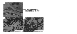
インフォームドコンセントを得た待機的帝王切開症例の妊婦由来ヒト胎児付属物から、羊膜を用手的に分離した。ハンクス平衡塩液(Ca・Mg不含有)にて2回洗浄後、得られた羊膜の内1gを容器に取り、精製コラゲナーゼ (CLSPA, Worthington社, 規格>500CDU/mg)300CDU/ml (=<600μg/ml)及びサーモリシン(和光純薬、規格>7000PU/mg)0~400PU/ml(=<60μg/ml) (No.1:0PU/ml、No.2:100PU/ml、No.3:200PU/ml、No.4:400PU/mlの4種類)を含有するハンクス平衡塩液(Ca・Mg含有)計5mlを添加し、37℃にて90分間、60rpmにてシェーカーにより震盪攪拌を行った。得られた混合物に2倍量の10%ウシ胎児血清(FBS)添加αMEM(Alpha Modification of Minimum Essential Medium Eagle)を添加後、ナイロンネットフィルター(ポアサイズ:100μm)で濾過した。フィルターに残った組織をヘマトキシリン・エオジン(HE)染色にて評価した。濾液は400 x gにて5分遠心操作を行い、上清を破棄後、10%FBS添加αMEMにて細胞を再懸濁し、細胞数をトリパンブルー染色後に測定した。得られた細胞は間葉系マーカー抗CD90-FITC抗体及び上皮系マーカー抗CD324-APC抗体(BD Bioscience社)にて4℃にて15分染色後、死細胞除去のため7-AAD色素を添加し、フローサイトメーター(FACSCanto:BD社)にて表面抗原マーカー解析を行った。結果を表1、表2,図3及び図4に示す。
インフォームドコンセントを得た待機的帝王切開症例の妊婦由来ヒト胎児付属物から、羊膜を用手的に分離した。ハンクス平衡塩液(Ca・Mg不含有)にて2回洗浄後、得られた羊膜の内1gを容器に取り、精製コラゲナーゼ (CLSPA, Worthington社, 規格>500CDU/mg)300CDU/ml (=<600μg/ml)及びサーモリシン(和光純薬、規格>7000PU/mg)0~400PU/ml(=<60μg/ml) (No.1:0PU/ml、No.2:100PU/ml、No.3:200PU/ml、No.4:400PU/mlの4種類)を含有するハンクス平衡塩液(Ca・Mg含有)計5mlを添加し、37℃にて90分間、60rpmにてシェーカーにより震盪攪拌を行った。得られた混合物に2倍量の10%ウシ胎児血清(FBS)添加αMEM(Alpha Modification of Minimum Essential Medium Eagle)を添加後、ナイロンネットフィルター(ポアサイズ:100μm)で濾過した。フィルターに残った組織をヘマトキシリン・エオジン(HE)染色にて評価した。濾液は400 x gにて5分遠心操作を行い、上清を破棄後、10%FBS添加αMEMにて細胞を再懸濁し、細胞数をトリパンブルー染色後に測定した。得られた細胞は間葉系マーカー抗CD90-FITC抗体及び上皮系マーカー抗CD324-APC抗体(BD Bioscience社)にて4℃にて15分染色後、死細胞除去のため7-AAD色素を添加し、フローサイトメーター(FACSCanto:BD社)にて表面抗原マーカー解析を行った。結果を表1、表2,図3及び図4に示す。
CDU(=collagen digestion unit):コラーゲンを基質として、37℃、pH7.5において5時間に1μmolのロイシンに相当するアミノ酸およびペプチドを生成する酵素量。
PU(=protease unit): 乳酸カゼインを基質として35℃、pH7.2において1分間に1μgのチロシンに相当するアミノ酸及びペプチドを生成する酵素量。
PU(=protease unit): 乳酸カゼインを基質として35℃、pH7.2において1分間に1μgのチロシンに相当するアミノ酸及びペプチドを生成する酵素量。
表1に示すように、コラゲナーゼのみ(No.1)で得られるトリパンブルー染色陰性の生細胞数は0.29x10⁶個であったのに対し、サーモリシンを加えることで生細胞数は増加し、サーモリシン400PU/ml(No.4)では1.99x106個と、約7倍まで増加した。一方トリパンブルー陽性の死細胞数に大きな変化はなく、どのサンプルでも80%以上の生細胞率が得られた。
図3に示すように、フローサイトメーターによる結果から、コラゲナーゼのみ(No.1)のサンプルでは、目的とするCD90陽性MSCは34.6%のみであり、不要なCD324陽性上皮細胞は8.6%、それ以外の赤血球と思われる細胞が56.8%であった。表1同様、サーモリシンを添加することでCD90陽性MSCの割合は増加し、サーモリシン400PU/ml(No.4)ではCD90陽性MSC83.3%、CD324陽性上皮細胞12.8%、その他が3.9%であった。
表2に示すように、各サンプルから得られるMSC数は、コラゲナーゼのみ(No.1)では0.1x10⁶個であったが、サーモリシン添加により得られるMSCは増加し、400PU/ml(No.4)では1.66x106個と、No.1の約16倍のMSCが得られた。
図4に示すように、酵素処理後のフィルター上に残留した組織をHE染色により検討を行ったところ、コラゲナーゼのみ(No.1)では細胞外基質層の構造が保たれ、消化不十分であった。サーモリシンを添加することで細胞外基質層の消化が認められ、400PU/ml(No.4)では完全に消化された。
図4に示すように、酵素処理後のフィルター上に残留した組織をHE染色により検討を行ったところ、コラゲナーゼのみ(No.1)では細胞外基質層の構造が保たれ、消化不十分であった。サーモリシンを添加することで細胞外基質層の消化が認められ、400PU/ml(No.4)では完全に消化された。
これら実施例1の結果から、コラゲナーゼ単独では羊膜を消化できないこと、コラゲナーゼにサーモリシンを加えることで、濃度依存的に羊膜は消化され、400PU/mlのサーモリシンでは、MSCを含む細胞外基質層が完全に消化されていたことが分かった。
(実施例2)
上記実施例1を踏まえ、同様の方法にて、コラゲナーゼを除きサーモリシンのみを用いた検討を行った。
結果を表3,図5及び図6に示す。
上記実施例1を踏まえ、同様の方法にて、コラゲナーゼを除きサーモリシンのみを用いた検討を行った。
結果を表3,図5及び図6に示す。
表3に示すように、サーモリシンのみの消化により得られる、ヒト羊膜1g当たりの細胞数はサーモリシンの濃度依存的に増加した。
しかしながら、図5に示すように、サーモリシンのみの酵素処理液に含有する細胞のフローサイトメーターによる結果からは、どの濃度においても得られる細胞はほぼ100%CD324陽性上皮細胞であり、目的とするCD90陽性MSCは得られなかった。
図6に示すように、酵素処理後のフィルター上に残留した組織のHE染色による検討では、MSCを含む細胞外基質層は全く消化されておらず、また、上皮細胞層の破綻がサーモリシンの濃度依存的に存在した。
しかしながら、図5に示すように、サーモリシンのみの酵素処理液に含有する細胞のフローサイトメーターによる結果からは、どの濃度においても得られる細胞はほぼ100%CD324陽性上皮細胞であり、目的とするCD90陽性MSCは得られなかった。
図6に示すように、酵素処理後のフィルター上に残留した組織のHE染色による検討では、MSCを含む細胞外基質層は全く消化されておらず、また、上皮細胞層の破綻がサーモリシンの濃度依存的に存在した。
これら実施例2の結果から、サーモリシンのみでは、目的とするMSCは全く得られないこと、サーモリシンの濃度が800PU/ml以上の場合、上皮細胞層の破綻が認められることが分かった。
(実施例3)
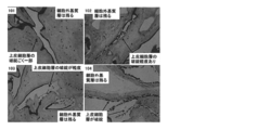
更に、従来法であるトリプシンを用いた方法との比較検討を行った(非特許文献3)。ヒト羊膜1gを容器に取り、No.41)0.05%トリプシン(0.53 mM EDTA含有)5mlにて37℃90分60rpm震盪攪拌(シェーカーにより)、No.42)0.05%トリプシン(0.53 mM EDTA含有、Invitrogen社)5mlにて37℃90分60rpm震盪攪拌(シェーカーにより)後、ナイロンネットフィルター(ポアサイズ:100μm)濾過にて残存した組織を、更に精製コラゲナーゼ (300CDU/ml)含有ハンクス平衡塩液(Ca・Mg含有)にて37℃90分60rpm震盪攪拌(シェーカーにより)、No.43)精製コラゲナーゼ(300CDU/ml)+サーモリシン(250PU/ml)含を有するハンクス平衡塩液(Ca・Mg含有)5mlにて、37℃90分震盪攪拌(シェーカーにより)を行った。以下は実施例1と同様とした。結果を表4、表5、図7及び図8に示す。
更に、従来法であるトリプシンを用いた方法との比較検討を行った(非特許文献3)。ヒト羊膜1gを容器に取り、No.41)0.05%トリプシン(0.53 mM EDTA含有)5mlにて37℃90分60rpm震盪攪拌(シェーカーにより)、No.42)0.05%トリプシン(0.53 mM EDTA含有、Invitrogen社)5mlにて37℃90分60rpm震盪攪拌(シェーカーにより)後、ナイロンネットフィルター(ポアサイズ:100μm)濾過にて残存した組織を、更に精製コラゲナーゼ (300CDU/ml)含有ハンクス平衡塩液(Ca・Mg含有)にて37℃90分60rpm震盪攪拌(シェーカーにより)、No.43)精製コラゲナーゼ(300CDU/ml)+サーモリシン(250PU/ml)含を有するハンクス平衡塩液(Ca・Mg含有)5mlにて、37℃90分震盪攪拌(シェーカーにより)を行った。以下は実施例1と同様とした。結果を表4、表5、図7及び図8に示す。
表4のように、トリプシン処理に加え、残った羊膜を更にコラゲナーゼ処理を行ったサンプル(No.42)から最も多くの細胞を得ることが出来た。
しかしながら、図7に示すように、各酵素処理液に含有する細胞のフローサイトメーターによる結果からは、トリプシンのみ(No.41)だと、目的とするCD90陽性MSCは0.6%と全く得られない、トリプシン処理後、更にコラゲナーゼ処理を加えた2段階処理(No.42)の場合、目的とするCD90陽性細胞は32.6%得られたが、不要なCD324陽性上皮細胞も65.6%含有している、コラゲナーゼ+サーモリシンの一回の処理(No.43)では、CD90陽性MSCは90.3%、CD324陽性上皮細胞は8.0%であった。
しかしながら、図7に示すように、各酵素処理液に含有する細胞のフローサイトメーターによる結果からは、トリプシンのみ(No.41)だと、目的とするCD90陽性MSCは0.6%と全く得られない、トリプシン処理後、更にコラゲナーゼ処理を加えた2段階処理(No.42)の場合、目的とするCD90陽性細胞は32.6%得られたが、不要なCD324陽性上皮細胞も65.6%含有している、コラゲナーゼ+サーモリシンの一回の処理(No.43)では、CD90陽性MSCは90.3%、CD324陽性上皮細胞は8.0%であった。
図8に示すように、酵素処理後のフィルター上に残留した組織のHE染色による検討では、41トリプシン処理のみでは、上皮細胞層の基底膜の破壊に加え、上皮細胞が球状かつばらばらになっているのに対し、細胞外基質層の構造は保たれていた、トリプシン処理後、コラゲナーゼ処理(No.42)をすることで、羊膜は完全に消化された、コラゲナーゼ+サーモリシンの一括処理(No.43)によって、細胞外基質層は完全に消化されているにもかかわらず、上皮細胞層はその構造が基底膜を含め保たれていた。
表5に示すように、各サンプルから得られるMSCは、トリプシンのみ(No.51)ではほとんどないこと、トリプシン後コラゲナーゼ処理(No.52)をした場合は、5.39 x10⁶個と非常に多く得られること、コラゲナーゼ+サーモリシンの一括処理(No.53)では、3.25 x10⁶個得られることが分かった。一方不要なCD324陽性上皮細胞に関しては、トリプシンのみ(No.51)では、2.25 x10⁶個の上皮細胞が得られること、トリプシン+コラゲナーゼの一括処理(No.52)では、13.07 x10⁶個と、必要なMSC以上の細胞数が得られてしまうこと、コラゲナーゼ+サーモリシンの一括処理(No.53)では、0.29 x10⁶個とMSCに比べると少ない細胞数であること、が明らかとなった。
以上のことから、従来のトリプシン処理後にコラゲナーゼにより処理する方法は、メリットとして、多くの細胞数が得られる反面、上皮細胞の混入が多く、高純度のMSCを得るには比重遠心等の手法により細胞を選別する操作が必要となること、工程がトリプシン処理に加えコラゲナーゼ処理の2段階になり、操作が煩雑になること、がデメリットとしてあげられる。特に後者のデメリットに関しては、トリプシンがCaにより不活化される反面、コラゲナーゼはCa要求性があるため、同時処理することが不可能であることがその原因である。
(実施例4)
上記実施例1~3を踏まえ、サーモリシン濃度を250PU/mlに固定した場合の、羊膜間葉系細胞分離に最小限必要なコラゲナーゼ濃度の検討を行った。結果を表6及び図9に示す。
上記実施例1~3を踏まえ、サーモリシン濃度を250PU/mlに固定した場合の、羊膜間葉系細胞分離に最小限必要なコラゲナーゼ濃度の検討を行った。結果を表6及び図9に示す。
表6に示すように、コラゲナーゼ300CDU/ml(No.63)で得られるトリパンブルー染色陰性の生細胞数は1.53x106個であったのに対し、コラゲナーゼ濃度がその1/4の75CDU/ml(No.61)の場合、生細胞数は1.16 x106個に減少した。トリパンブルー陽性の死細胞数に大きな変化はなく、どのサンプルでも80%以上の生細胞率が得られた。また、フローサイトメーターによる結果からはどのサンプルもCD90陽性間葉系細胞は90%以上であった。
図9に示すように、酵素処理後のフィルター上に残留した組織をHE染色により検討を行ったところ、コラゲナーゼ濃度が300および150 CDU/ml(No.63および62)では上皮細胞層のみであったのに対し、コラゲナーゼ濃度が75 CDU/ml(No.61)の場合、ごく一部であるが細胞外基質層が観察され、消化不十分であった。
これら実施例4の結果から、サーモリシン濃度を250PU/mlに固定した場合、細胞外基質層を十分に消化するには、コラゲナーゼ濃度は、好ましくは少なくとも75 CDU/ml以上であり、より好ましくは150CDU/ml以上にする必要性が分かった。
これら実施例4の結果から、サーモリシン濃度を250PU/mlに固定した場合、細胞外基質層を十分に消化するには、コラゲナーゼ濃度は、好ましくは少なくとも75 CDU/ml以上であり、より好ましくは150CDU/ml以上にする必要性が分かった。
(実施例5)
更にサーモリシン濃度を実施例3の倍である500PU/mlに固定した場合の、羊膜間葉系細胞分離に最小限必要なコラゲナーゼ濃度の検討を行った。結果を表7及び図10に示す。
更にサーモリシン濃度を実施例3の倍である500PU/mlに固定した場合の、羊膜間葉系細胞分離に最小限必要なコラゲナーゼ濃度の検討を行った。結果を表7及び図10に示す。
表7に示すように、コラゲナーゼ150CDU/ml(No.73)で得られるトリパンブルー染色陰性の生細胞数は2.05x106個であったのに対し、コラゲナーゼ濃度がその1/4の37.5CDU/ml(No.71)の場合、生細胞数は0.82x106個に減少した。トリパンブルー陽性の死細胞数に大きな変化はなく、どのサンプルでも80%以上の生細胞率が得られた。また、フローサイトメーターによる結果からはどのサンプルもCD90陽性間葉系細胞は90%以上であった。
図10に示すように、酵素処理後のフィルター上に残留した組織をHE染色により検討を行ったところ、コラゲナーゼ濃度が150および75 CDU/ml(No.73および72)では上皮細胞層のみであったのに対し、コラゲナーゼ濃度が37.5 CDU/ml(No.71)の場合、多くの細胞外基質層が観察され、消化不十分であった。
これら実施例5の結果から、サーモリシン濃度を500PU/mlに固定した場合、細胞外基質層を十分に消化するには、コラゲナーゼ濃度は、好ましくは少なくとも37.5CDU/ml以上であり、より好ましくは75CDU/ml以上にする必要性が分かった。
これら実施例5の結果から、サーモリシン濃度を500PU/mlに固定した場合、細胞外基質層を十分に消化するには、コラゲナーゼ濃度は、好ましくは少なくとも37.5CDU/ml以上であり、より好ましくは75CDU/ml以上にする必要性が分かった。
(実施例6)
上記実施例1に関し、サーモリシンに代えて、同様に非極性アミノ酸のN末端側を切断する金属プロテイナーゼであるディスパーゼを用い、これにコラゲナーゼを添加した検討を行った。
インフォームドコンセントを得た妊婦由来のヒト胎児付属物から、羊膜を用手的に分離した。ハンクス平衡塩液(Ca・Mg不含有)にて2回洗浄後、得られた羊膜の内1gを容器に取り、コラゲナーゼ (Brightase-C, ニッピ社, 規格>20万CDU/バイアル)300CDU/ml及びディスパーゼI(和光純薬、規格10000~13000PU/バイアル)0~400PU/ml (No.1:0PU/ml、No.2:100PU/ml、No.3:200PU/ml、No.4:400PU/mlの4種類)を含有するハンクス平衡塩液(Ca・Mg含有)計5ml添加し、37℃にて90分、60rpmにてシェーカーにより震盪攪拌を行った。得られた混合物に2倍量の10%ウシ胎児血清(FBS)添加αMEM(Alpha Modification of Minimum Essential Medium Eagle)を添加後、ナイロンネットフィルター(ポアサイズ:100μm)で濾過した。フィルターに残った組織をヘマトキシリン・エオジン(HE)染色にて評価した。濾液は400 x gにて5分遠心操作を行い、上清を破棄後、10%FBS添加αMEMにて細胞を再懸濁し、細胞数をトリパンブルー染色後、測定した。得られた細胞は間葉系マーカー抗CD90-FITC抗体及び上皮系マーカー抗CD324-APC抗体(BD Bioscience社)にて4℃にて15分染色後、死細胞除去のため7-AAD色素を添加し、フローサイトメーター(FACSCanto:BD社)にて表面抗原マーカー解析を行った。結果を表8、表9、図11及び図12に示す。
表8に示すように、コラゲナーゼのみ(No.81)で得られるトリパンブルー染色陰性の生細胞数は0.42x10⁶個であったのに対し、ディスパーゼを加えることで生細胞数は増加し、ディスパーゼ200PU/ml(No.83)では3.02x106個と、約7倍まで増加した。一方トリパンブルー陽性の死細胞数に大きな変化はなく、ディスパーゼを入れたどのサンプルでも80%以上の生細胞率が得られた。
図11に示すように、フローサイトメーターによる結果では、コラゲナーゼのみ(No.81)であっても、ディスパーゼを加えても(No.82-84)、目的とするCD90陽性MSCは90%以上であり、不要なCD324陽性上皮細胞はいずれも10%以下であった。コラゲナーゼのみ(No.81)の結果が、実施例1(図3)でのコラゲナーゼのみ(No.1)のそれと異なるが、これは用いたコラゲナーゼのメーカーが異なるためと考えられた。
表9に示すように、各サンプルから得られるMSC数は、コラゲナーゼのみ(No.81)では0.39x10⁶個であったが、ディスパーゼ添加により得られるMSCは増加し、200PU/ml(No.83)では2.86x106個と、No.81の約7倍のMSCが得られた。
図12に示すように、酵素処理後のフィルター上に残留した組織をHE染色により検討を行ったところ、コラゲナーゼのみ(No.81)では細胞外基質層の構造が保たれ、消化不十分であった。ディスパーゼを添加することで細胞外基質層の消化が認められ、200PU/ml(No.83)、400PU/ml(No.84)では完全に消化された。
図12に示すように、酵素処理後のフィルター上に残留した組織をHE染色により検討を行ったところ、コラゲナーゼのみ(No.81)では細胞外基質層の構造が保たれ、消化不十分であった。ディスパーゼを添加することで細胞外基質層の消化が認められ、200PU/ml(No.83)、400PU/ml(No.84)では完全に消化された。
これら実施例6の結果から、コラゲナーゼ単独では羊膜消化が不十分であること、コラゲナーゼにディスパーゼを加えることで、濃度依存的に羊膜は消化され、2000PU/ml以上のディスパーゼでは、MSCを含む細胞外基質層が完全に消化されていた。
(実施例7)
上記実施例6を踏まえ、同様の方法にて、コラゲナーゼを除きディスパーゼのみを用いた検討を行った。
結果を表10,図13及び図14に示す。
上記実施例6を踏まえ、同様の方法にて、コラゲナーゼを除きディスパーゼのみを用いた検討を行った。
結果を表10,図13及び図14に示す。
表10に示すように、ディスパーゼのみの消化により得られる、ヒト羊膜1g当たりの細胞数はサーモリシンの濃度依存的に増加した。
しかしながら、図13に示すように、ディスパーゼのみの酵素処理液に含有する細胞のフローサイトメーターによる結果からは、どの濃度においても目的とするCD90陽性MSC数%と非常に少なかった。
図14に示すように、酵素処理後のフィルター上に残留した組織のHE染色による検討では、MSCを含む細胞外基質層は全く消化されておらず、また、上皮細胞層の破綻がディスパーゼの濃度依存的に存在した。
しかしながら、図13に示すように、ディスパーゼのみの酵素処理液に含有する細胞のフローサイトメーターによる結果からは、どの濃度においても目的とするCD90陽性MSC数%と非常に少なかった。
図14に示すように、酵素処理後のフィルター上に残留した組織のHE染色による検討では、MSCを含む細胞外基質層は全く消化されておらず、また、上皮細胞層の破綻がディスパーゼの濃度依存的に存在した。
これら実施例7の結果から、ディスパーゼのみでは、目的とするMSCは全く得られない、ディスパーゼの濃度が800PU/ml以上の場合、上皮細胞層の破綻が認められることが分かった。
(実施例8)
上記実施例6~7を踏まえ、ディスパーゼ濃度を250PU/mlに固定した場合の、羊膜間葉系細胞分離に最小限必要なコラゲナーゼ濃度の検討を行った。結果を表11及び図15に示す。
上記実施例6~7を踏まえ、ディスパーゼ濃度を250PU/mlに固定した場合の、羊膜間葉系細胞分離に最小限必要なコラゲナーゼ濃度の検討を行った。結果を表11及び図15に示す。
表11に示すように、コラゲナーゼ300CDU/ml(No.113)で得られるトリパンブルー染色陰性の生細胞数は1.57x106個であったのに対し、コラゲナーゼ濃度がその1/4の75CDU/ml(No.113)の場合、生細胞数は1.34 x106個に減少した。トリパンブルー陽性の死細胞数に大きな変化はなく、どのサンプルでも80%以上の生細胞率が得られた。また、フローサイトメーターによる結果からはどのサンプルもCD90陽性間葉系細胞は90%以上であった。
図15に示すように、酵素処理後のフィルター上に残留した組織をHE染色により検討を行ったところ、コラゲナーゼ濃度が300CDU/ml(No.113)では上皮細胞層のみであったのに対し、コラゲナーゼ濃度が75 および150CDU/ml(No.111および112)の場合、細胞外基質層が観察され、消化不十分であった。
これら実施例8の結果から、ディスパーゼ濃度を250PU/mlに固定した場合、細胞外基質層を十分に消化するには、コラゲナーゼ濃度は少なくとも75CDU/ml以上、より好ましくは150CDU/ml以上、さらに好ましくは300CDU/ml以上にする必要性が分かった。
これら実施例8の結果から、ディスパーゼ濃度を250PU/mlに固定した場合、細胞外基質層を十分に消化するには、コラゲナーゼ濃度は少なくとも75CDU/ml以上、より好ましくは150CDU/ml以上、さらに好ましくは300CDU/ml以上にする必要性が分かった。
(実施例9)
実施例1にて得られた細胞を10%FBS添加αMEM培養液にて希釈後、プラスチックデッシュ上に播種し、羊膜由来MSCを接着培養させた。トリプシン処理にて細胞剥離後、10%FBS添加αMEM培養液にてトリプシン中和し、遠心後上清を捨て、得られた細胞ペレットをRPMI1640にて再懸濁し、羊膜由来MSC懸濁液を作成した。得られた懸濁液に対し、最終組成が表12となるように凍結保存液を調整した。
実施例1にて得られた細胞を10%FBS添加αMEM培養液にて希釈後、プラスチックデッシュ上に播種し、羊膜由来MSCを接着培養させた。トリプシン処理にて細胞剥離後、10%FBS添加αMEM培養液にてトリプシン中和し、遠心後上清を捨て、得られた細胞ペレットをRPMI1640にて再懸濁し、羊膜由来MSC懸濁液を作成した。得られた懸濁液に対し、最終組成が表12となるように凍結保存液を調整した。
DMSO:Simga-Aldrich社製、品番D2650
HES:ニプロ社製、品番HES40
ヒトアルブミン(Alb):ベネシス社製、献血アルブミン25%静注12.5g/50ml
デキストラン40:大塚製薬工場社製、低分子デキストラン糖注
生理食塩水:大塚製薬工場社製:上記の希釈調整用液とした。
HES:ニプロ社製、品番HES40
ヒトアルブミン(Alb):ベネシス社製、献血アルブミン25%静注12.5g/50ml
デキストラン40:大塚製薬工場社製、低分子デキストラン糖注
生理食塩水:大塚製薬工場社製:上記の希釈調整用液とした。
凍結保存液は羊膜由来MSCが106細胞/mlとなるように調整し、それぞれ1mlをクライオチューブに移し、プログラムフリーザーにて、-1~-2℃/分の凍結速度で-40℃まで温度を下げ、更に-90℃まで-10℃/分の速度で下げ、-150℃の超低温冷凍庫に保存した。翌日、クライオチューブを37℃恒温槽に浸して急速融解した。表13に急速解凍した細胞のトリパンブルー陰性生細胞割合を示す。
この結果から、IIIのDMSOのみでは生細胞が少ないこと、HESやデキストラン、アルブミンを添加することで生細胞割合は増加することが明らかとなった。
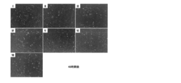
更に、解凍した細胞懸濁液100μLを24wellプレート4ウェルに播種し、3mlの10%FBS添加αMEM培養液を添加し、24時間後および48時間後に写真撮影後、一視野当たりの細胞数の平均値を測定した。表14、図16及び図17に結果を示す。
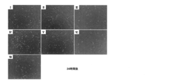
更に、解凍した細胞懸濁液100μLを24wellプレート4ウェルに播種し、3mlの10%FBS添加αMEM培養液を添加し、24時間後および48時間後に写真撮影後、一視野当たりの細胞数の平均値を測定した。表14、図16及び図17に結果を示す。
表13の結果同様、IIIのDMSOのみ、あるいはIVのDMSO+Albでは細胞増殖が遅かったが、HESやデキストランの添加により細胞増殖は促進した。特にDMSOの濃度を減らしたI、II、IVにおいて著明な細胞増殖を認めた。
(実施例10)
マウスにおいて、同種他家骨髄移植および脾細胞移植を行い、急性移植片対宿主病(GVHD)を発症させ、ヒト羊膜由来MSC移植による治療効果を検討した。7-8週齢の雌B6C3F1マウスに対して、15GyのX線照射の後、同種他家であるBDF1マウス由来骨髄細胞1.0×10⁷細胞および同脾細胞3×10⁷細胞を経静脈的に移植した。骨髄細胞移植14日目、17日目、21日目、15日目に、実施例1と同様の手法(コラゲナーゼ300CDU/ml+サーモリシン250PU/mlの条件下)にて得られたヒト羊膜由来MSC(1×105細胞 )を経静脈的に移植し、体重を経時的に観察した。図18に、骨髄細胞移植日からの体重変化率を示す。
この結果から、急性移植片対宿主病(GVHD)に伴う体重増加の遅延がヒト羊膜由来MSC移植により改善していることが明らかとなった。
(実施例11)
ラットにおいて、デキストラン硫酸(DSS)を経口摂取させることで炎症性腸疾患を発症させ、ヒト羊膜由来MSC移植による治療効果を検討した。8週齢の雄SDラットに対し8%DSSの自由飲水での投与を開始した。DSS投与開始の翌日、実施例1と同様の手法(コラゲナーゼ300CDU/ml+サーモリシン250PU/mlの条件下)にて得られたヒト羊膜由来MSC(1×106細胞 )を経静脈的に移植し、DSSを計5日間投与した。
図19に疾患活動性(Disease activity index(DAI):体重の減少、便の固さ、直腸出血を観察、測定し、点数化)および相対的体重変化を示す。この結果から、腸炎の病態がヒト羊膜由来MSC移植により改善していることが明らかとなった。
尚、DAIの点数化は、下記の文献の方法に従った。Cooper, H. S.; Murthy, S. N.; Shah, R. S.; Sedergran, D. J. Clinicopathologic study of dextran sulfate sodium experimental murine colitis. Lab. Invest. 69:238-249; 1993.
(実施例12)
マウスにおいて、プリスタン(Pristane; 2,6,10,14-tetramethyl-pentadecane)を投与することで全身性エリテマトーデスを発症させ、ヒト羊膜由来MSC移植による治療効果を検討した。13週齢雄BALB/cマウスに対しプリスタンを500μl腹腔内に投与した。同時に実施例1と同様の手法(コラゲナーゼ300CDU/ml+サーモリシン250PU/mlの条件下)にて得られたヒト羊膜由来MSC(1×105細胞/10g)を尾静脈に投与し、以後隔週で同数のヒト羊膜由来MSCを投与し、20週経過後に生化学的評価を行った。
図20に尿蛋白の経過を示す。この結果から、全身性エリテマトーデスに伴う蛋白尿がヒト羊膜由来MSC移植により改善していることが明らかとなった。
(実施例13)
ラットにおいて、四塩化炭素(CCl4)を繰り返し投与することで肝硬変を発症させ、ヒト羊膜由来MSC移植による治療効果を検討した。6週齢の雄SDラットに対し2ml/kgのCCl4を週2回の頻度で腹腔内投与を開始した。CCl4投与開始から3週目に実施例1と同様の手法(コラゲナーゼ300CDU/ml+サーモリシン250PU/mlの条件下)にて得られたヒト羊膜由来MSC(1×106細胞 )を経静脈的に移植し、CCl4を計7週間投与し、肝臓の組織学的評価を行った。
図21に肝臓のMasson trichrome染色の結果から得られた繊維化面積率(膠原線維陽性割合)を示す。この結果から、肝硬変に伴う肝線維化がヒト羊膜由来MSC移植により改善していることが明らかとなった。
(実施例14)
ラットにおいて、直腸に放射線照射することで放射線腸炎を発症させ、ヒト羊膜由来MSCによる治療効果を検討した。8週齢の雄SDラットに対し5Gy/日の放射線を5日間連日で下腹部に照射した。最終照射日に実施例1と同様の手法(コラゲナーゼ300CDU/ml+サーモリシン250PU/mlの条件下)にて得られたヒト羊膜由来MSC(1×106細胞 )を経静脈的に移植し、その3日後に直腸の組織学的評価を行った。
図22に直腸のPAS染色の結果から得られたPAS陽性杯細胞数(/HPF:強拡大視野当たり)の変化を示す。この結果から、放射線腸炎に伴う杯細胞の減少がヒト羊膜由来MSC移植により改善していることが明らかとなった。
1 間葉系幹細胞
2 上皮細胞層
3 細胞外基質層
2 上皮細胞層
3 細胞外基質層
Claims (20)
- 羊膜を、コラゲナーゼと、サーモリシン及び/又はディスパーゼとにより酵素処理する工程;及び
酵素処理した羊膜をメッシュに通す工程:
を含む、間葉系細胞組成物の製造方法。 - メッシュを通過した細胞を回収する工程;及び
回収した細胞を培養する工程:
をさらに含む、請求項1に記載の間葉系細胞組成物の製造方法。 - 前記メッシュを通過した細胞を回収する工程が、倍量又はそれ以上の量の培地又は平衡塩溶液で濾液を希釈した後、遠心分離により間葉系細胞を回収する工程である、請求項2に記載の間葉系細胞組成物の製造方法。
- コラゲナーゼの濃度が50CDU/ml~1000CDU/mlであり、サーモリシンおよび/又はディスパーゼの濃度が100PU/ml~800PU/mlである、請求項1から3の何れか1項に記載の間葉系細胞組成物の製造方法。
- 酵素処理する工程が、30~40℃で30分以上処理する工程である、請求項1から4の何れか1項に記載の間葉系細胞組成物の製造方法。
- 酵素処理する工程が、スターラー又はシェーカーを用いて10rpm/分~100rpm/分で30分以上撹拌する工程である、請求項1から5の何れか1項に記載の間葉系細胞組成物の製造方法。
- 羊膜が帝王切開により得られたものである、請求項1から6の何れか1項に記載の間葉系細胞組成物の製造方法。
- メッシュのポアサイズが40~200μmである、請求項1から7の何れか1項に記載の間葉系細胞組成物の製造方法。
- 酵素処理した羊膜をメッシュに通す工程が自然落下である、請求項1から8の何れか1項に記載の間葉系細胞組成物の製造方法。
- CD324及びCD326陽性上皮細胞の含有率が20%以下であり、CD90陽性細胞の含有率が75%以上であり、および生細胞率が80%以上である、間葉系細胞組成物。
- 請求項1から9の何れか1項に記載の間葉系細胞組成物の製造方法により得られる、請求項10に記載の間葉系細胞組成物。
- 請求項10又は11に記載の間葉系細胞組成物を0.05質量%より多く5質量%以下のアルブミンを含有する培地にて培養することにより得られる、間葉系細胞培養組成物。
- ジメチルスルホキシドを5~10質量%含有し、ヒドロキシルエチルデンプンを5~10質量%またはデキストランを1~5質量%含有する溶液中に間葉系細胞を含む混合物を凍結保存する工程を含む、凍結保存された間葉系細胞の製造方法。
- 前記溶液が、さらにヒトアルブミンを0質量%より多く5質量%以下含有する溶液である、請求項13に記載の凍結保存された間葉系細胞の製造方法。
- 前記間葉系細胞が、請求項1から9の何れか1項に記載の方法により製造された間葉系細胞組成物に含まれる間葉系細胞、請求項10又は11に記載の間葉系細胞組成物に含まれる間葉系細胞、又は請求項12に記載の間葉系細胞培養組成物に含まれる間葉系細胞である、請求項13又は14に記載の凍結保存された間葉系細胞の製造方法。
- 請求項13から15の何れか1項に記載の方法により得られる凍結保存された間葉系細胞を解凍後、輸液製剤により2倍以上に希釈する工程を含む、間葉系細胞投与用組成物を製造する方法。
- 請求項10又は11に記載の間葉系細胞組成物、および/又は請求項12に記載の間葉系細胞培養組成物、および/又は請求項16に記載の方法により製造される間葉系細胞投与用組成物を有効成分として含む、細胞治療剤。
- 注射用製剤である、請求項17に記載の細胞治療剤。
- 細胞塊又はシート状構造の移植用製剤である、請求項17に記載の細胞治療剤。
- 移植片対宿主病、炎症性腸疾患、全身性エリテマトーデス、肝硬変、又は放射線腸炎から選択される疾患の治療剤である、請求項17から19の何れか1項に記載の治療剤。
Priority Applications (4)
| Application Number | Priority Date | Filing Date | Title |
|---|---|---|---|
| US14/912,662 US10441611B2 (en) | 2013-08-19 | 2014-08-18 | Method for producing amniotic mesenchymal stromal cell composition, method for cryopreserving the same, and therapeutic agent |
| CN201480046241.8A CN105765060B (zh) | 2013-08-19 | 2014-08-18 | 羊膜间充质系细胞组合物的制造方法和冷冻保存方法、以及治疗剂 |
| EP14837352.5A EP3037523B1 (en) | 2013-08-19 | 2014-08-18 | Production method and cryopreservation method for amniotic mesenchymal cell composition, and therapeutic agent |
| US16/508,463 US11389486B2 (en) | 2013-08-19 | 2019-07-11 | Method for producing amniotic mesenchymal stromal cell composition, method for cryopreserving the same, and therapeutic agent |
Applications Claiming Priority (4)
| Application Number | Priority Date | Filing Date | Title |
|---|---|---|---|
| JP2013170008 | 2013-08-19 | ||
| JP2013-170008 | 2013-08-19 | ||
| JP2014-130142 | 2014-06-25 | ||
| JP2014130142A JP6512759B2 (ja) | 2013-08-19 | 2014-06-25 | 羊膜間葉系細胞組成物の製造方法及び凍結保存方法、並びに治療剤 |
Related Child Applications (2)
| Application Number | Title | Priority Date | Filing Date |
|---|---|---|---|
| US14/912,662 A-371-Of-International US10441611B2 (en) | 2013-08-19 | 2014-08-18 | Method for producing amniotic mesenchymal stromal cell composition, method for cryopreserving the same, and therapeutic agent |
| US16/508,463 Continuation US11389486B2 (en) | 2013-08-19 | 2019-07-11 | Method for producing amniotic mesenchymal stromal cell composition, method for cryopreserving the same, and therapeutic agent |
Publications (1)
| Publication Number | Publication Date |
|---|---|
| WO2015025810A1 true WO2015025810A1 (ja) | 2015-02-26 |
Family
ID=52483585
Family Applications (1)
| Application Number | Title | Priority Date | Filing Date |
|---|---|---|---|
| PCT/JP2014/071546 WO2015025810A1 (ja) | 2013-08-19 | 2014-08-18 | 羊膜間葉系細胞組成物の製造方法及び凍結保存方法、並びに治療剤 |
Country Status (5)
| Country | Link |
|---|---|
| US (2) | US10441611B2 (ja) |
| EP (1) | EP3037523B1 (ja) |
| JP (1) | JP6512759B2 (ja) |
| CN (1) | CN105765060B (ja) |
| WO (1) | WO2015025810A1 (ja) |
Cited By (6)
| Publication number | Priority date | Publication date | Assignee | Title |
|---|---|---|---|---|
| WO2017073656A1 (ja) * | 2015-10-27 | 2017-05-04 | 株式会社カネカ | 間葉系幹細胞を含む細胞集団の製造方法、間葉系幹細胞、細胞集団、並びに医薬組成物 |
| CN106967675A (zh) * | 2017-05-31 | 2017-07-21 | 东莞市保莱生物科技有限公司 | 一种从胎盘中分离提取造血干细胞的方法 |
| CN107849532A (zh) * | 2015-05-14 | 2018-03-27 | 株式会社再生科技工程 | 冷冻间充质细胞的制备方法和移植用治疗材料的制备方法 |
| WO2018186420A1 (ja) * | 2017-04-03 | 2018-10-11 | 株式会社カネカ | 間葉系幹細胞を含む細胞集団とその製造方法、間葉系幹細胞及び医薬組成物 |
| JPWO2018186421A1 (ja) * | 2017-04-03 | 2020-02-20 | 株式会社カネカ | 間葉系幹細胞を含む細胞集団とその製造方法、及び医薬組成物 |
| WO2022203022A1 (ja) * | 2021-03-26 | 2022-09-29 | 株式会社カネカ | 羊膜由来間葉系幹細胞を含む細胞集団の製造方法 |
Families Citing this family (13)
| Publication number | Priority date | Publication date | Assignee | Title |
|---|---|---|---|---|
| CA3013187C (en) * | 2016-02-01 | 2023-02-28 | Green Cross Lab Cell Corporation | Medium composition for cryopreservation of cell and use thereof |
| US11540507B2 (en) | 2016-11-04 | 2023-01-03 | The University Of Tokyo | Solution for cryopreservation of animal cells or animal tissues, cryopreserved product, and cryopreservation method |
| CN109952371A (zh) * | 2016-11-15 | 2019-06-28 | 株式会社钟化 | 包含来自胎儿附属物的间充质系干细胞的细胞群、其制备方法、以及医药组合物 |
| JP7140752B2 (ja) | 2017-04-03 | 2022-09-21 | 株式会社カネカ | 胎児付属物由来接着細胞を含む細胞集団とその製造方法、及び医薬組成物 |
| WO2019059279A1 (ja) * | 2017-09-20 | 2019-03-28 | 株式会社カネカ | 細胞懸濁液を調製するためのデバイス、システム及び方法 |
| WO2019132025A1 (ja) | 2017-12-28 | 2019-07-04 | 株式会社カネカ | 接着性幹細胞を含む細胞集団とその製造方法、及び医薬組成物 |
| CN111527198A (zh) * | 2017-12-28 | 2020-08-11 | 株式会社钟化 | 包含贴壁性干细胞的细胞群、其制造方法、以及医药组合物 |
| CN108904533A (zh) * | 2018-07-04 | 2018-11-30 | 卡替(上海)生物技术股份有限公司 | 牙髓间充质干细胞在制备肝硬化治疗药物中的用途 |
| JPWO2020184350A1 (ja) | 2019-03-08 | 2020-09-17 | ||
| WO2020251020A1 (ja) * | 2019-06-14 | 2020-12-17 | 株式会社カネカ | 間葉系細胞を含む細胞集団、それを含む医薬組成物、及び、その製造方法 |
| WO2022056046A1 (en) * | 2020-09-08 | 2022-03-17 | Amit Patel | Allogenic umbilical cord stem cells for treating severe respiratory conditions |
| CN113456891B (zh) * | 2021-06-16 | 2022-05-17 | 成都微沃科技有限公司 | 一种从细胞层中提取细胞外基质层的方法 |
| CN114097768A (zh) * | 2021-11-24 | 2022-03-01 | 李敬 | 一种骨髓干细胞用保护液及其制备方法和应用 |
Citations (6)
| Publication number | Priority date | Publication date | Assignee | Title |
|---|---|---|---|---|
| JP2000201672A (ja) * | 1999-01-11 | 2000-07-25 | Asahi Medical Co Ltd | 有核細胞の凍結保存用組成物 |
| WO2004058323A2 (en) * | 2002-12-20 | 2004-07-15 | Cell Genesys, Inc. | Directly injectable formulations which provide enhanced cryoprotection of cell products |
| JP3934539B2 (ja) | 2001-12-12 | 2007-06-20 | 独立行政法人科学技術振興機構 | 胎盤等由来の成体又は生後組織の前駆細胞 |
| WO2008100498A2 (en) * | 2007-02-12 | 2008-08-21 | Anthrogenesis Corporation | Treatment of inflammatory diseases using placental stem cells |
| WO2008151846A2 (en) * | 2007-06-15 | 2008-12-18 | Centro Di Ricerca E. Menni Fondazione Poliambulanza Istituto Ospedaliero | T cell immunomodulation by placenta cell preparations |
| JP4330995B2 (ja) | 2001-11-15 | 2009-09-16 | チルドレンズ メディカル センター コーポレーション | 絨毛膜絨毛、羊水、および胎盤からの胎児性幹細胞を単離、増殖、および分化させる方法、ならびにその治療的使用方法 |
Family Cites Families (9)
| Publication number | Priority date | Publication date | Assignee | Title |
|---|---|---|---|---|
| ATE342348T1 (de) * | 1998-11-09 | 2006-11-15 | Consorzio Per La Gestione Del | Serum-freies medium für chondrozyt-ähnliche zellen |
| MX2008015645A (es) * | 2006-06-09 | 2009-02-06 | Anthrogenesis Corp | Nicho placentario y uso de este para cultivar celulas primordiales. |
| CN102851254A (zh) | 2007-03-01 | 2013-01-02 | 干细胞国际公司 | 制备、分离及超低温保存子宫内膜/月经细胞 |
| MX2010004914A (es) * | 2007-10-31 | 2010-08-18 | Cryo Cell Int | Metodos para cocultivar celulas derivadas de sangre de cordon umbilical con celulas madre menstruales. |
| RU2563518C2 (ru) * | 2008-08-20 | 2015-09-20 | Антродженезис Корпорейшн | Улучшенная клеточная композиция и способы ее получения |
| NZ592726A (en) * | 2008-11-19 | 2012-12-21 | Anthrogenesis Corp | Amnion derived adherent cells |
| EP2468850A4 (en) * | 2009-08-19 | 2013-02-20 | Takara Bio Inc | METHOD FOR PRESERVING CELLS |
| US20110256202A1 (en) | 2010-02-18 | 2011-10-20 | Samson Tom | Immunocompatible amniotic membrane products |
| EP2731440A4 (en) * | 2011-07-15 | 2015-04-29 | Anthrogenesis Corp | RADIATION LESSON TREATMENT USING ADHESENT CELLS DERIVED FROM AMNIOS |
-
2014
- 2014-06-25 JP JP2014130142A patent/JP6512759B2/ja active Active
- 2014-08-18 EP EP14837352.5A patent/EP3037523B1/en active Active
- 2014-08-18 WO PCT/JP2014/071546 patent/WO2015025810A1/ja active Application Filing
- 2014-08-18 US US14/912,662 patent/US10441611B2/en active Active
- 2014-08-18 CN CN201480046241.8A patent/CN105765060B/zh active Active
-
2019
- 2019-07-11 US US16/508,463 patent/US11389486B2/en active Active
Patent Citations (7)
| Publication number | Priority date | Publication date | Assignee | Title |
|---|---|---|---|---|
| JP2000201672A (ja) * | 1999-01-11 | 2000-07-25 | Asahi Medical Co Ltd | 有核細胞の凍結保存用組成物 |
| JP4330995B2 (ja) | 2001-11-15 | 2009-09-16 | チルドレンズ メディカル センター コーポレーション | 絨毛膜絨毛、羊水、および胎盤からの胎児性幹細胞を単離、増殖、および分化させる方法、ならびにその治療的使用方法 |
| JP3934539B2 (ja) | 2001-12-12 | 2007-06-20 | 独立行政法人科学技術振興機構 | 胎盤等由来の成体又は生後組織の前駆細胞 |
| WO2004058323A2 (en) * | 2002-12-20 | 2004-07-15 | Cell Genesys, Inc. | Directly injectable formulations which provide enhanced cryoprotection of cell products |
| WO2008100498A2 (en) * | 2007-02-12 | 2008-08-21 | Anthrogenesis Corporation | Treatment of inflammatory diseases using placental stem cells |
| JP2010518096A (ja) | 2007-02-12 | 2010-05-27 | アンスロジェネシス コーポレーション | 胎盤幹細胞を使用した炎症性疾患の治療 |
| WO2008151846A2 (en) * | 2007-06-15 | 2008-12-18 | Centro Di Ricerca E. Menni Fondazione Poliambulanza Istituto Ospedaliero | T cell immunomodulation by placenta cell preparations |
Non-Patent Citations (11)
| Title |
|---|
| "UNIT 1E.5: Isolation of Amniotic Mesenchymal Stem Cells", CURRENT PROTOCOLS IN STEM CELL BIOLOGY, 2010 |
| AM J OBSTET GYNECOL., vol. 190, no. 1, 2004, pages 87 - 92 |
| AM J OBSTET GYNECOL., vol. 194, no. 3, 2006, pages 664 - 673 |
| BMC BIOTECHNOLOGY, vol. 12, 2012, pages 49 |
| COOPER, H. S.; MURTHY, S. N.; SHAH, R. S.; SEDERGRAN, D. J.: "Clinicopathologic study of dextran sulfate sodium experimental murine colitis", LAB. INVEST., vol. 69, 1993, pages 238 - 249 |
| CYTOTHERAPY, vol. 14, no. 4, 2012, pages 441 - 450 |
| CYTOTHERAPY, vol. 8, no. 4, 2006, pages 315 - 317 |
| J MOL CELL CARDIOL., vol. 53, no. 3, 2012, pages 420 - 428 |
| J TISSUE ENG REGEN MED., vol. 1, no. 4, 2007, pages 296 - 305 |
| See also references of EP3037523A4 |
| STEM CELLS., vol. 26, no. 10, 2008, pages 2625 - 2633 |
Cited By (13)
| Publication number | Priority date | Publication date | Assignee | Title |
|---|---|---|---|---|
| EP3296392A4 (en) * | 2015-05-14 | 2018-11-21 | Advanced Cell Technology and Engineering Ltd. | Method for producing frozen mesenchymal cells and method for producing implantable therapeutic member |
| US10774312B2 (en) | 2015-05-14 | 2020-09-15 | Advanced Cell Technology And Engineering Ltd. | Method for producing frozen mesenchymal cells and method for producing implantable therapeutic member |
| CN107849532A (zh) * | 2015-05-14 | 2018-03-27 | 株式会社再生科技工程 | 冷冻间充质细胞的制备方法和移植用治疗材料的制备方法 |
| JP2022060520A (ja) * | 2015-10-27 | 2022-04-14 | 国立研究開発法人国立循環器病研究センター | 間葉系幹細胞を含む細胞集団の製造方法、間葉系幹細胞、細胞集団、並びに医薬組成物 |
| JPWO2017073656A1 (ja) * | 2015-10-27 | 2018-08-16 | 国立研究開発法人国立循環器病研究センター | 間葉系幹細胞を含む細胞集団の製造方法、間葉系幹細胞、細胞集団、並びに医薬組成物 |
| WO2017073656A1 (ja) * | 2015-10-27 | 2017-05-04 | 株式会社カネカ | 間葉系幹細胞を含む細胞集団の製造方法、間葉系幹細胞、細胞集団、並びに医薬組成物 |
| WO2018186420A1 (ja) * | 2017-04-03 | 2018-10-11 | 株式会社カネカ | 間葉系幹細胞を含む細胞集団とその製造方法、間葉系幹細胞及び医薬組成物 |
| JPWO2018186420A1 (ja) * | 2017-04-03 | 2020-02-20 | 株式会社カネカ | 間葉系幹細胞を含む細胞集団とその製造方法、間葉系幹細胞及び医薬組成物 |
| JPWO2018186421A1 (ja) * | 2017-04-03 | 2020-02-20 | 株式会社カネカ | 間葉系幹細胞を含む細胞集団とその製造方法、及び医薬組成物 |
| JP7102397B2 (ja) | 2017-04-03 | 2022-07-19 | 株式会社カネカ | 間葉系幹細胞を含む細胞集団とその製造方法、及び医薬組成物 |
| JP7152389B2 (ja) | 2017-04-03 | 2022-10-12 | 株式会社カネカ | 間葉系幹細胞を含む細胞集団とその製造方法、間葉系幹細胞及び医薬組成物 |
| CN106967675A (zh) * | 2017-05-31 | 2017-07-21 | 东莞市保莱生物科技有限公司 | 一种从胎盘中分离提取造血干细胞的方法 |
| WO2022203022A1 (ja) * | 2021-03-26 | 2022-09-29 | 株式会社カネカ | 羊膜由来間葉系幹細胞を含む細胞集団の製造方法 |
Also Published As
| Publication number | Publication date |
|---|---|
| US11389486B2 (en) | 2022-07-19 |
| EP3037523B1 (en) | 2020-08-12 |
| US10441611B2 (en) | 2019-10-15 |
| US20190343892A1 (en) | 2019-11-14 |
| JP6512759B2 (ja) | 2019-05-15 |
| EP3037523A1 (en) | 2016-06-29 |
| CN105765060A (zh) | 2016-07-13 |
| CN105765060B (zh) | 2020-04-28 |
| JP2015061520A (ja) | 2015-04-02 |
| US20160228474A1 (en) | 2016-08-11 |
| EP3037523A4 (en) | 2017-01-25 |
Similar Documents
| Publication | Publication Date | Title |
|---|---|---|
| WO2015025810A1 (ja) | 羊膜間葉系細胞組成物の製造方法及び凍結保存方法、並びに治療剤 | |
| JP5341059B2 (ja) | 幹細胞懸濁液 | |
| JP2022046511A (ja) | 胎盤幹細胞を使用する免疫調節 | |
| JP2019017393A5 (ja) | ||
| WO2019161591A1 (zh) | 间充质干细胞的分离培养方法及冻存、复苏方法 | |
| CN106754674A (zh) | 从人胎盘羊膜制备羊膜间充质干细胞的方法及其应用 | |
| JP7132908B2 (ja) | 間葉系幹細胞を含む細胞集団とその製造方法、及び医薬組成物 | |
| KR20160043929A (ko) | 양수 수집 및 세포 분리를 위한 세포, 방법 및 장치 | |
| JP7530893B2 (ja) | 間葉系細胞を含む細胞集団、それを含む医薬組成物、及び、その製造方法 | |
| KR20100084620A (ko) | 조직 재생을 위한 세포 조성물 | |
| CN114134114A (zh) | 从胎盘组织中扩增自然杀伤细胞的方法 | |
| RU2642282C1 (ru) | Клетки, производные от кардиальной ткани | |
| JP6754459B2 (ja) | 羊膜間葉系細胞組成物の製造方法及び凍結保存方法、並びに治療剤 | |
| JP5753874B2 (ja) | 細胞生存率低下抑制剤 | |
| JP5282282B2 (ja) | 内側絨毛膜由来の間葉系幹細胞 | |
| WO2010140162A2 (en) | A process for preparing stem cell based formulations | |
| TWI454307B (zh) | Method and application for preparing human plasma for cell culture | |
| WO2018186420A1 (ja) | 間葉系幹細胞を含む細胞集団とその製造方法、間葉系幹細胞及び医薬組成物 | |
| WO2022203022A1 (ja) | 羊膜由来間葉系幹細胞を含む細胞集団の製造方法 | |
| TWI742399B (zh) | 無酵素前處理與培養脂肪間質幹細胞的方法 | |
| WO2022210574A1 (ja) | 筋ジストロフィー治療剤 | |
| JP2022120698A (ja) | 間葉系幹細胞を含む細胞集団を含有する軟部組織再生用医薬組成物 | |
| CN118064359A (zh) | 一种高免疫调节能力的人脐带间充质干细胞库的制备方法 | |
| CN111979189A (zh) | 一种羊膜间充质干细胞分离培养扩增方法 |
Legal Events
| Date | Code | Title | Description |
|---|---|---|---|
| 121 | Ep: the epo has been informed by wipo that ep was designated in this application |
Ref document number: 14837352 Country of ref document: EP Kind code of ref document: A1 |
|
| WWE | Wipo information: entry into national phase |
Ref document number: 2014837352 Country of ref document: EP |
|
| NENP | Non-entry into the national phase |
Ref country code: DE |
