US9539223B2 - Methods for treating bacterial infections - Google Patents
Methods for treating bacterial infections Download PDFInfo
- Publication number
- US9539223B2 US9539223B2 US14/888,289 US201414888289A US9539223B2 US 9539223 B2 US9539223 B2 US 9539223B2 US 201414888289 A US201414888289 A US 201414888289A US 9539223 B2 US9539223 B2 US 9539223B2
- Authority
- US
- United States
- Prior art keywords
- leptospira
- group
- spp
- rickettsia
- ncl812
- Prior art date
- Legal status (The legal status is an assumption and is not a legal conclusion. Google has not performed a legal analysis and makes no representation as to the accuracy of the status listed.)
- Active
Links
- 238000000034 method Methods 0.000 title claims abstract description 72
- 208000035143 Bacterial infection Diseases 0.000 title claims abstract description 28
- 208000022362 bacterial infectious disease Diseases 0.000 title claims abstract description 28
- 230000001580 bacterial effect Effects 0.000 claims description 108
- 239000003795 chemical substances by application Substances 0.000 claims description 80
- 241000894006 Bacteria Species 0.000 claims description 61
- 150000001875 compounds Chemical class 0.000 claims description 49
- 241000588724 Escherichia coli Species 0.000 claims description 36
- 229960004591 robenidine Drugs 0.000 claims description 27
- 241000894007 species Species 0.000 claims description 25
- 150000003839 salts Chemical class 0.000 claims description 23
- 210000002421 cell wall Anatomy 0.000 claims description 18
- 241000283073 Equus caballus Species 0.000 claims description 15
- 150000003952 β-lactams Chemical class 0.000 claims description 15
- 241000606161 Chlamydia Species 0.000 claims description 13
- 208000034309 Bacterial disease carrier Diseases 0.000 claims description 12
- 241000607598 Vibrio Species 0.000 claims description 10
- 241000588747 Klebsiella pneumoniae Species 0.000 claims description 9
- 241000589517 Pseudomonas aeruginosa Species 0.000 claims description 9
- 241000588914 Enterobacter Species 0.000 claims description 7
- 241000589248 Legionella Species 0.000 claims description 7
- 208000007764 Legionnaires' Disease Diseases 0.000 claims description 7
- 241000606125 Bacteroides Species 0.000 claims description 6
- 241000606768 Haemophilus influenzae Species 0.000 claims description 6
- 241000282414 Homo sapiens Species 0.000 claims description 6
- 241000588748 Klebsiella Species 0.000 claims description 6
- 230000037396 body weight Effects 0.000 claims description 6
- 229960000308 fosfomycin Drugs 0.000 claims description 6
- YMDXZJFXQJVXBF-STHAYSLISA-N fosfomycin Chemical compound C[C@@H]1O[C@@H]1P(O)(O)=O YMDXZJFXQJVXBF-STHAYSLISA-N 0.000 claims description 6
- 108010040201 Polymyxins Proteins 0.000 claims description 5
- 229940041153 polymyxins Drugs 0.000 claims description 5
- HDTRYLNUVZCQOY-UHFFFAOYSA-N α-D-glucopyranosyl-α-D-glucopyranoside Natural products OC1C(O)C(O)C(CO)OC1OC1C(O)C(O)C(O)C(CO)O1 HDTRYLNUVZCQOY-UHFFFAOYSA-N 0.000 claims description 4
- 241000588626 Acinetobacter baumannii Species 0.000 claims description 4
- 241000122230 Acinetobacter junii Species 0.000 claims description 4
- 241000607534 Aeromonas Species 0.000 claims description 4
- 241000607528 Aeromonas hydrophila Species 0.000 claims description 4
- 241000606749 Aggregatibacter actinomycetemcomitans Species 0.000 claims description 4
- 241001135163 Arcobacter Species 0.000 claims description 4
- 241000606560 Avibacterium avium Species 0.000 claims description 4
- 241000589977 Borrelia turicatae Species 0.000 claims description 4
- 241001148534 Brachyspira Species 0.000 claims description 4
- 241000589986 Campylobacter lari Species 0.000 claims description 4
- 241000606153 Chlamydia trachomatis Species 0.000 claims description 4
- 241000588917 Citrobacter koseri Species 0.000 claims description 4
- 241000589601 Francisella Species 0.000 claims description 4
- 241000605909 Fusobacterium Species 0.000 claims description 4
- 241000232174 Leptospira fainei Species 0.000 claims description 4
- 208000016604 Lyme disease Diseases 0.000 claims description 4
- 241000604449 Megasphaera Species 0.000 claims description 4
- 241000588772 Morganella morganii Species 0.000 claims description 4
- 241000588652 Neisseria gonorrhoeae Species 0.000 claims description 4
- 241000588650 Neisseria meningitidis Species 0.000 claims description 4
- 241000605861 Prevotella Species 0.000 claims description 4
- 241001135223 Prevotella melaninogenica Species 0.000 claims description 4
- 241000589516 Pseudomonas Species 0.000 claims description 4
- 241000606701 Rickettsia Species 0.000 claims description 4
- 241000607142 Salmonella Species 0.000 claims description 4
- 241000607720 Serratia Species 0.000 claims description 4
- 241000122973 Stenotrophomonas maltophilia Species 0.000 claims description 4
- 241001496920 Tenacibaculum Species 0.000 claims description 4
- HDTRYLNUVZCQOY-WSWWMNSNSA-N Trehalose Natural products O[C@@H]1[C@@H](O)[C@@H](O)[C@@H](CO)O[C@@H]1O[C@@H]1[C@H](O)[C@@H](O)[C@@H](O)[C@@H](CO)O1 HDTRYLNUVZCQOY-WSWWMNSNSA-N 0.000 claims description 4
- 241000589884 Treponema pallidum Species 0.000 claims description 4
- HDTRYLNUVZCQOY-LIZSDCNHSA-N alpha,alpha-trehalose Chemical compound O[C@@H]1[C@@H](O)[C@H](O)[C@@H](CO)O[C@@H]1O[C@@H]1[C@H](O)[C@@H](O)[C@H](O)[C@@H](CO)O1 HDTRYLNUVZCQOY-LIZSDCNHSA-N 0.000 claims description 4
- 229940076266 morganella morganii Drugs 0.000 claims description 4
- 241000589218 Acetobacteraceae Species 0.000 claims description 3
- 241000606828 Aggregatibacter aphrophilus Species 0.000 claims description 3
- 241000589155 Agrobacterium tumefaciens Species 0.000 claims description 3
- 241000606646 Anaplasma Species 0.000 claims description 3
- 241000271566 Aves Species 0.000 claims description 3
- 241001136175 Burkholderia pseudomallei Species 0.000 claims description 3
- 241000589876 Campylobacter Species 0.000 claims description 3
- 241000588878 Eikenella corrodens Species 0.000 claims description 3
- 241000588921 Enterobacteriaceae Species 0.000 claims description 3
- 241000588722 Escherichia Species 0.000 claims description 3
- 241000604777 Flavobacterium columnare Species 0.000 claims description 3
- 241000589014 Kingella kingae Species 0.000 claims description 3
- 241000589902 Leptospira Species 0.000 claims description 3
- 241000589928 Leptospira biflexa Species 0.000 claims description 3
- 241000589927 Leptospira borgpetersenii Species 0.000 claims description 3
- 241001148627 Leptospira inadai Species 0.000 claims description 3
- 241000589929 Leptospira interrogans Species 0.000 claims description 3
- 241001148628 Leptospira kirschneri Species 0.000 claims description 3
- 241001148629 Leptospira meyeri Species 0.000 claims description 3
- 241001135196 Leptospira noguchii Species 0.000 claims description 3
- 241001135200 Leptospira weilii Species 0.000 claims description 3
- 241000588622 Moraxella bovis Species 0.000 claims description 3
- 241000588655 Moraxella catarrhalis Species 0.000 claims description 3
- 241000606860 Pasteurella Species 0.000 claims description 3
- 241000606999 Plesiomonas shigelloides Species 0.000 claims description 3
- 241000588769 Proteus <enterobacteria> Species 0.000 claims description 3
- 241000218905 Pseudomonas luteola Species 0.000 claims description 3
- 241000607626 Vibrio cholerae Species 0.000 claims description 3
- 241000607447 Yersinia enterocolitica Species 0.000 claims description 3
- 239000002738 chelating agent Substances 0.000 claims description 3
- 229940038705 chlamydia trachomatis Drugs 0.000 claims description 3
- 230000000688 enterotoxigenic effect Effects 0.000 claims description 3
- 229940047650 haemophilus influenzae Drugs 0.000 claims description 3
- 230000001717 pathogenic effect Effects 0.000 claims description 3
- 229940098232 yersinia enterocolitica Drugs 0.000 claims description 3
- 241000589291 Acinetobacter Species 0.000 claims description 2
- 241001165345 Acinetobacter baylyi Species 0.000 claims description 2
- 241001165343 Acinetobacter bouvetii Species 0.000 claims description 2
- 241000588624 Acinetobacter calcoaceticus Species 0.000 claims description 2
- 241001165347 Acinetobacter gerneri Species 0.000 claims description 2
- 241001148231 Acinetobacter haemolyticus Species 0.000 claims description 2
- 241000122229 Acinetobacter johnsonii Species 0.000 claims description 2
- 241001135518 Acinetobacter lwoffii Species 0.000 claims description 2
- 241000930987 Acinetobacter parvus Species 0.000 claims description 2
- 241000122231 Acinetobacter radioresistens Species 0.000 claims description 2
- 241001556023 Acinetobacter schindleri Species 0.000 claims description 2
- 241001165356 Acinetobacter tandoii Species 0.000 claims description 2
- 241001165355 Acinetobacter tjernbergiae Species 0.000 claims description 2
- 241001165358 Acinetobacter towneri Species 0.000 claims description 2
- 241001556024 Acinetobacter ursingii Species 0.000 claims description 2
- 241000508783 Acinetobacter venetianus Species 0.000 claims description 2
- 241000606750 Actinobacillus Species 0.000 claims description 2
- 241000606729 Actinobacillus equuli Species 0.000 claims description 2
- 241000606801 Actinobacillus lignieresii Species 0.000 claims description 2
- 241000606748 Actinobacillus pleuropneumoniae Species 0.000 claims description 2
- 241000606793 Actinobacillus seminis Species 0.000 claims description 2
- 241000948980 Actinobacillus succinogenes Species 0.000 claims description 2
- 241000606791 Actinobacillus ureae Species 0.000 claims description 2
- 241000607516 Aeromonas caviae Species 0.000 claims description 2
- 241001486707 Aeromonas dhakensis Species 0.000 claims description 2
- 241000607525 Aeromonas salmonicida Species 0.000 claims description 2
- 241000607522 Aeromonas sobria Species 0.000 claims description 2
- 241000198060 Aeromonas veronii bv. sobria Species 0.000 claims description 2
- 241000606806 Aggregatibacter segnis Species 0.000 claims description 2
- 241000588813 Alcaligenes faecalis Species 0.000 claims description 2
- 241001112453 Anaplasma bovis Species 0.000 claims description 2
- 241000606643 Anaplasma centrale Species 0.000 claims description 2
- 241000312508 Anaplasma odocoilei Species 0.000 claims description 2
- 241001266092 Anaplasma ovis Species 0.000 claims description 2
- 241000605280 Anaplasma platys Species 0.000 claims description 2
- 241001135723 Arcobacter skirrowii Species 0.000 claims description 2
- 241001136161 Avibacterium Species 0.000 claims description 2
- 241000480192 Avibacterium endocarditidis Species 0.000 claims description 2
- 241000606591 Avibacterium gallinarum Species 0.000 claims description 2
- 241000606767 Avibacterium paragallinarum Species 0.000 claims description 2
- 241000606618 Avibacterium volantium Species 0.000 claims description 2
- 241000589938 Azospirillum brasilense Species 0.000 claims description 2
- 241000606124 Bacteroides fragilis Species 0.000 claims description 2
- 241000606660 Bartonella Species 0.000 claims description 2
- 241000559826 Bartonella australis Species 0.000 claims description 2
- 241000606685 Bartonella bacilliformis Species 0.000 claims description 2
- 241000158639 Bartonella birtlesii Species 0.000 claims description 2
- 241000011635 Bartonella bovis Species 0.000 claims description 2
- 241000011631 Bartonella capreoli Species 0.000 claims description 2
- 241000663865 Bartonella chomelii Species 0.000 claims description 2
- 241000567117 Bartonella clarridgeiae Species 0.000 claims description 2
- 241001464950 Bartonella doshiae Species 0.000 claims description 2
- 241000606070 Bartonella elizabethae Species 0.000 claims description 2
- 241001464952 Bartonella grahamii Species 0.000 claims description 2
- 241001518086 Bartonella henselae Species 0.000 claims description 2
- 241000332162 Bartonella koehlerae Species 0.000 claims description 2
- 241001603744 Bartonella peromysci Species 0.000 claims description 2
- 241000408194 Bartonella phoceensis Species 0.000 claims description 2
- 241000606108 Bartonella quintana Species 0.000 claims description 2
- 241000408207 Bartonella rattimassiliensis Species 0.000 claims description 2
- 241000895318 Bartonella rochalimae Species 0.000 claims description 2
- 241000383843 Bartonella schoenbuchensis Species 0.000 claims description 2
- 241000280718 Bartonella talpae Species 0.000 claims description 2
- 241000142360 Bartonella tamiae Species 0.000 claims description 2
- 241001464954 Bartonella taylorii Species 0.000 claims description 2
- 241001673680 Bartonella tribocorum Species 0.000 claims description 2
- 241000142488 Bartonella vinsonii subsp. arupensis Species 0.000 claims description 2
- 241000142483 Bartonella vinsonii subsp. vinsonii Species 0.000 claims description 2
- 241000604771 Bernardetia litoralis Species 0.000 claims description 2
- 241001553294 Bibersteinia Species 0.000 claims description 2
- 241000588807 Bordetella Species 0.000 claims description 2
- 241000853395 Bordetella ansorpii Species 0.000 claims description 2
- 241000588851 Bordetella avium Species 0.000 claims description 2
- 241000588779 Bordetella bronchiseptica Species 0.000 claims description 2
- 241001495147 Bordetella holmesii Species 0.000 claims description 2
- 241000588780 Bordetella parapertussis Species 0.000 claims description 2
- 241000588832 Bordetella pertussis Species 0.000 claims description 2
- 241000359246 Bordetella petrii Species 0.000 claims description 2
- 241000543043 Bordetella trematum Species 0.000 claims description 2
- 241000589968 Borrelia Species 0.000 claims description 2
- 241000589974 Borrelia anserina Species 0.000 claims description 2
- 241000589975 Borrelia coriaceae Species 0.000 claims description 2
- 241000124827 Borrelia duttonii Species 0.000 claims description 2
- 241000589978 Borrelia hermsii Species 0.000 claims description 2
- 241000124828 Borrelia hispanica Species 0.000 claims description 2
- 241001536361 Borrelia lonestari Species 0.000 claims description 2
- 241000216520 Borrelia miyamotoi Species 0.000 claims description 2
- 241000589976 Borrelia parkeri Species 0.000 claims description 2
- 241000180132 Borrelia persica Species 0.000 claims description 2
- 241000180135 Borrelia recurrentis Species 0.000 claims description 2
- 241000908527 Borreliella bissettii Species 0.000 claims description 2
- 241000589969 Borreliella burgdorferi Species 0.000 claims description 2
- 241001148605 Borreliella garinii Species 0.000 claims description 2
- 241001478201 Borreliella japonica Species 0.000 claims description 2
- 241001446608 Borreliella lusitaniae Species 0.000 claims description 2
- 241000019016 Borreliella spielmanii Species 0.000 claims description 2
- 241000876423 Borreliella valaisiana Species 0.000 claims description 2
- 241000283690 Bos taurus Species 0.000 claims description 2
- 241000576898 Brachyspira hampsonii Species 0.000 claims description 2
- 241000589893 Brachyspira hyodysenteriae Species 0.000 claims description 2
- 241001658774 Brachyspira murdochii Species 0.000 claims description 2
- 241000510930 Brachyspira pilosicoli Species 0.000 claims description 2
- 241000589562 Brucella Species 0.000 claims description 2
- 241001509299 Brucella canis Species 0.000 claims description 2
- 241000514713 Brucella ceti Species 0.000 claims description 2
- 241001148106 Brucella melitensis Species 0.000 claims description 2
- 241000589568 Brucella ovis Species 0.000 claims description 2
- 241001148111 Brucella suis Species 0.000 claims description 2
- 241001646647 Burkholderia ambifaria Species 0.000 claims description 2
- 241000790236 Burkholderia anthina Species 0.000 claims description 2
- 241000371430 Burkholderia cenocepacia Species 0.000 claims description 2
- 241000589513 Burkholderia cepacia Species 0.000 claims description 2
- 241000283588 Burkholderia diffusa Species 0.000 claims description 2
- 241001646389 Burkholderia dolosa Species 0.000 claims description 2
- 241000274232 Burkholderia latens Species 0.000 claims description 2
- 241000722910 Burkholderia mallei Species 0.000 claims description 2
- 241000283585 Burkholderia metallica Species 0.000 claims description 2
- 241000020731 Burkholderia multivorans Species 0.000 claims description 2
- 241000866604 Burkholderia pyrrocinia Species 0.000 claims description 2
- 241000283591 Burkholderia seminalis Species 0.000 claims description 2
- 241000371422 Burkholderia stabilis Species 0.000 claims description 2
- 241001459282 Burkholderia ubonensis Species 0.000 claims description 2
- 241000866606 Burkholderia vietnamiensis Species 0.000 claims description 2
- 241000589877 Campylobacter coli Species 0.000 claims description 2
- 241000589873 Campylobacter concisus Species 0.000 claims description 2
- 241000589985 Campylobacter curvus Species 0.000 claims description 2
- 241000589874 Campylobacter fetus Species 0.000 claims description 2
- 241000606208 Campylobacter gracilis Species 0.000 claims description 2
- 241001137866 Campylobacter helveticus Species 0.000 claims description 2
- 241001290832 Campylobacter hominis Species 0.000 claims description 2
- 241000589872 Campylobacter hyointestinalis Species 0.000 claims description 2
- 241000034487 Campylobacter insulaenigrae Species 0.000 claims description 2
- 241000589875 Campylobacter jejuni Species 0.000 claims description 2
- 241001277598 Campylobacter lanienae Species 0.000 claims description 2
- 241000589995 Campylobacter mucosalis Species 0.000 claims description 2
- 241000589996 Campylobacter rectus Species 0.000 claims description 2
- 241000589992 Campylobacter showae Species 0.000 claims description 2
- 241000589990 Campylobacter sputorum Species 0.000 claims description 2
- 241001135528 Campylobacter upsaliensis Species 0.000 claims description 2
- 241001179072 Candidatus Neoehrlichia lotoris Species 0.000 claims description 2
- 241000032691 Candidatus Neoehrlichia mikurensis Species 0.000 claims description 2
- 241000068542 Candidatus Piscichlamydia Species 0.000 claims description 2
- 241000282465 Canis Species 0.000 claims description 2
- 241000190890 Capnocytophaga Species 0.000 claims description 2
- 241001135194 Capnocytophaga canimorsus Species 0.000 claims description 2
- 241001135159 Capnocytophaga cynodegmi Species 0.000 claims description 2
- 241000190888 Capnocytophaga gingivalis Species 0.000 claims description 2
- 241000190566 Capnocytophaga granulosa Species 0.000 claims description 2
- 241000190564 Capnocytophaga haemolytica Species 0.000 claims description 2
- 241000014851 Capnocytophaga leadbetteri Species 0.000 claims description 2
- 241000190885 Capnocytophaga ochracea Species 0.000 claims description 2
- 241000190882 Capnocytophaga sputigena Species 0.000 claims description 2
- 241000207210 Cardiobacterium hominis Species 0.000 claims description 2
- 241001466752 Cardiobacterium valvarum Species 0.000 claims description 2
- 241000043249 Cedecea lapagei Species 0.000 claims description 2
- 241000043250 Cedecea neteri Species 0.000 claims description 2
- 229940123150 Chelating agent Drugs 0.000 claims description 2
- 241001647373 Chlamydia abortus Species 0.000 claims description 2
- 241000590502 Chlamydia avium Species 0.000 claims description 2
- 241001647371 Chlamydia caviae Species 0.000 claims description 2
- 241001647374 Chlamydia felis Species 0.000 claims description 2
- 241000590414 Chlamydia gallinacea Species 0.000 claims description 2
- 241001647367 Chlamydia muridarum Species 0.000 claims description 2
- 241001674218 Chlamydia pecorum Species 0.000 claims description 2
- 241001647372 Chlamydia pneumoniae Species 0.000 claims description 2
- 241001647378 Chlamydia psittaci Species 0.000 claims description 2
- 241001647370 Chlamydia suis Species 0.000 claims description 2
- 241000611330 Chryseobacterium Species 0.000 claims description 2
- 241000589589 Chryseobacterium indologenes Species 0.000 claims description 2
- 241000469381 Chryseobacterium piscicola Species 0.000 claims description 2
- 241000588923 Citrobacter Species 0.000 claims description 2
- 241000588919 Citrobacter freundii Species 0.000 claims description 2
- 241001445332 Coxiella <snail> Species 0.000 claims description 2
- 241000085288 Cronobacter condimenti Species 0.000 claims description 2
- 241000989066 Cronobacter dublinensis Species 0.000 claims description 2
- 241000988645 Cronobacter malonaticus Species 0.000 claims description 2
- 241000988643 Cronobacter muytjensii Species 0.000 claims description 2
- 241001135265 Cronobacter sakazakii Species 0.000 claims description 2
- 241000988642 Cronobacter turicensis Species 0.000 claims description 2
- 241001448462 Cronobacter universalis Species 0.000 claims description 2
- 241001634906 Cupriavidus gilardii Species 0.000 claims description 2
- 241001635226 Cupriavidus pauculus Species 0.000 claims description 2
- 241000605056 Cytophaga Species 0.000 claims description 2
- 241000605111 Cytophaga hutchinsonii Species 0.000 claims description 2
- 241000606006 Dichelobacter Species 0.000 claims description 2
- 241000605721 Dichelobacter nodosus Species 0.000 claims description 2
- 241000024398 Dysgonomonas gadei Species 0.000 claims description 2
- 241001167377 Dysgonomonas hofstadii Species 0.000 claims description 2
- 241000365095 Dysgonomonas mossii Species 0.000 claims description 2
- 241000472311 Dysgonomonas oryzarvi Species 0.000 claims description 2
- 241000827171 Dysgonomonas wimpennyi Species 0.000 claims description 2
- 241000607473 Edwardsiella <enterobacteria> Species 0.000 claims description 2
- 241000949274 Edwardsiella ictaluri Species 0.000 claims description 2
- 241000607471 Edwardsiella tarda Species 0.000 claims description 2
- 241000605314 Ehrlichia Species 0.000 claims description 2
- 241000605312 Ehrlichia canis Species 0.000 claims description 2
- 241000605310 Ehrlichia chaffeensis Species 0.000 claims description 2
- 241000605282 Ehrlichia ewingii Species 0.000 claims description 2
- 241001495399 Ehrlichia muris Species 0.000 claims description 2
- 241000681433 Ehrlichia ovina Species 0.000 claims description 2
- 241000606675 Ehrlichia ruminantium Species 0.000 claims description 2
- 241000604775 Eisenibacter elegans Species 0.000 claims description 2
- 241000589566 Elizabethkingia meningoseptica Species 0.000 claims description 2
- 241000588697 Enterobacter cloacae Species 0.000 claims description 2
- 241001240954 Escherichia albertii Species 0.000 claims description 2
- 241000588720 Escherichia fergusonii Species 0.000 claims description 2
- 241000131484 Ewingella americana Species 0.000 claims description 2
- 241000282324 Felis Species 0.000 claims description 2
- 241000589565 Flavobacterium Species 0.000 claims description 2
- 241000555689 Flavobacterium branchiophilum Species 0.000 claims description 2
- 241000523886 Flavobacterium oncorhynchi Species 0.000 claims description 2
- 241000382842 Flavobacterium psychrophilum Species 0.000 claims description 2
- 241000976304 Flexibacter echinicida Species 0.000 claims description 2
- 241000604773 Flexibacter flexilis Species 0.000 claims description 2
- 241000589282 Fluoribacter dumoffii Species 0.000 claims description 2
- 241000589278 Fluoribacter gormanii Species 0.000 claims description 2
- 241000245629 Francisella noatunensis Species 0.000 claims description 2
- 241001221064 Francisella noatunensis subsp. orientalis Species 0.000 claims description 2
- 241001135321 Francisella philomiragia Species 0.000 claims description 2
- 241000266827 Francisella tularensis subsp. holarctica Species 0.000 claims description 2
- 241000589599 Francisella tularensis subsp. novicida Species 0.000 claims description 2
- 241001106040 Franconibacter helveticus Species 0.000 claims description 2
- 241000070950 Franconibacter pulveris Species 0.000 claims description 2
- 241000605952 Fusobacterium necrophorum Species 0.000 claims description 2
- 241000605986 Fusobacterium nucleatum Species 0.000 claims description 2
- 241001291922 Fusobacterium nucleatum subsp. polymorphum Species 0.000 claims description 2
- 241000012732 Gallibacterium Species 0.000 claims description 2
- 241000606562 Gallibacterium anatis Species 0.000 claims description 2
- 241000009531 Gallibacterium genomosp. 1 Species 0.000 claims description 2
- 241000009532 Gallibacterium genomosp. 2 Species 0.000 claims description 2
- 241000348477 Gallibacterium genomosp. 3 Species 0.000 claims description 2
- 241000348473 Gallibacterium group V Species 0.000 claims description 2
- 241000717651 Gallibacterium melopsittaci Species 0.000 claims description 2
- 241000348478 Gallibacterium salpingitidis Species 0.000 claims description 2
- 241000727735 Gallibacterium trehalosifermentans Species 0.000 claims description 2
- 241000191102 Garritya polymorpha Species 0.000 claims description 2
- 241000606807 Glaesserella parasuis Species 0.000 claims description 2
- 241000606790 Haemophilus Species 0.000 claims description 2
- 241001501603 Haemophilus aegyptius Species 0.000 claims description 2
- 241000606788 Haemophilus haemolyticus Species 0.000 claims description 2
- 241000606822 Haemophilus parahaemolyticus Species 0.000 claims description 2
- 241000606766 Haemophilus parainfluenzae Species 0.000 claims description 2
- 241000588729 Hafnia alvei Species 0.000 claims description 2
- 241000162758 Hafnia paralvei Species 0.000 claims description 2
- 241000590002 Helicobacter pylori Species 0.000 claims description 2
- 241001581234 Histophilus Species 0.000 claims description 2
- 241000606831 Histophilus somni Species 0.000 claims description 2
- 241000191100 Hugenholtzia roseola Species 0.000 claims description 2
- 206010020429 Human ehrlichiosis Diseases 0.000 claims description 2
- 241000589015 Kingella denitrificans Species 0.000 claims description 2
- 241000589012 Kingella oralis Species 0.000 claims description 2
- 241000083229 Kingella potus Species 0.000 claims description 2
- 241000588915 Klebsiella aerogenes Species 0.000 claims description 2
- 241001534216 Klebsiella granulomatis Species 0.000 claims description 2
- 241000588749 Klebsiella oxytoca Species 0.000 claims description 2
- 241000498833 Kluyvera ascorbata Species 0.000 claims description 2
- 241000588773 Kluyvera cryocrescens Species 0.000 claims description 2
- 241001245439 Kosakonia cowanii Species 0.000 claims description 2
- 241001469654 Lawsonia <weevil> Species 0.000 claims description 2
- 241001148567 Lawsonia intracellularis Species 0.000 claims description 2
- 241001135524 Legionella anisa Species 0.000 claims description 2
- 241001135525 Legionella birminghamensis Species 0.000 claims description 2
- 241001135527 Legionella cincinnatiensis Species 0.000 claims description 2
- 241000589259 Legionella feeleii Species 0.000 claims description 2
- 241000589240 Legionella hackeliae Species 0.000 claims description 2
- 241000589276 Legionella jordanis Species 0.000 claims description 2
- 241000189478 Legionella lansingensis Species 0.000 claims description 2
- 241000589264 Legionella longbeachae Species 0.000 claims description 2
- 241001148224 Legionella oakridgensis Species 0.000 claims description 2
- 241000189497 Legionella parisiensis Species 0.000 claims description 2
- 241001135523 Legionella wadsworthii Species 0.000 claims description 2
- 241000937844 Legionella waltersii Species 0.000 claims description 2
- 241000570484 Leptospira alexanderi serovar Hebdomadis Species 0.000 claims description 2
- 241001152569 Leptospira alexanderi serovar Manhao 3 Species 0.000 claims description 2
- 241001136176 Leptospira alstonii Species 0.000 claims description 2
- 241001359215 Leptospira alstonii serovar Pingchang Species 0.000 claims description 2
- 241001359216 Leptospira alstonii serovar Sichuan Species 0.000 claims description 2
- 241001301761 Leptospira biflexa serovar Ancona Species 0.000 claims description 2
- 241001301759 Leptospira biflexa serovar Canela Species 0.000 claims description 2
- 241000031168 Leptospira borgpetersenii serovar Hardjo Species 0.000 claims description 2
- 241000402692 Leptospira borgpetersenii serovar Hardjo-bovis Species 0.000 claims description 2
- 241001168975 Leptospira borgpetersenii serovar Pomona Species 0.000 claims description 2
- 241000757681 Leptospira borgpetersenii serovar Tarassovi Species 0.000 claims description 2
- 241000873670 Leptospira broomii Species 0.000 claims description 2
- 241001381796 Leptospira broomii serovar Hurstbridge Species 0.000 claims description 2
- 241001205823 Leptospira idonii Species 0.000 claims description 2
- 241001152430 Leptospira inadai serovar Lyme Species 0.000 claims description 2
- 241000388004 Leptospira inadai serovar Malaya Species 0.000 claims description 2
- 241001550399 Leptospira interrogans serovar Australis Species 0.000 claims description 2
- 241000725095 Leptospira interrogans serovar Autumnalis Species 0.000 claims description 2
- 241000402695 Leptospira interrogans serovar Bratislava Species 0.000 claims description 2
- 241001550390 Leptospira interrogans serovar Canicola Species 0.000 claims description 2
- 241000757412 Leptospira interrogans serovar Grippotyphosa Species 0.000 claims description 2
- 241000589926 Leptospira interrogans serovar Hardjo Species 0.000 claims description 2
- 241001518163 Leptospira interrogans serovar Hardjo-bovis Species 0.000 claims description 2
- 241000292568 Leptospira interrogans serovar Icterohaemorrhagiae Species 0.000 claims description 2
- 241000178947 Leptospira interrogans serovar Pomona Species 0.000 claims description 2
- 241000757689 Leptospira interrogans serovar Pyrogenes Species 0.000 claims description 2
- 241000695436 Leptospira interrogans serovar Tarassovi Species 0.000 claims description 2
- 241001239771 Leptospira kirschneri serovar Bulgarica Species 0.000 claims description 2
- 241001152448 Leptospira kirschneri serovar Cynopteri Species 0.000 claims description 2
- 241001518154 Leptospira kirschneri serovar Grippotyphosa Species 0.000 claims description 2
- 241000667106 Leptospira kmetyi Species 0.000 claims description 2
- 241000466311 Leptospira licerasiae Species 0.000 claims description 2
- 241001239782 Leptospira meyeri serovar Sofia Species 0.000 claims description 2
- 241001152475 Leptospira noguchii serovar Panama Species 0.000 claims description 2
- 241000757685 Leptospira noguchii serovar Pomona Species 0.000 claims description 2
- 241001135198 Leptospira santarosai Species 0.000 claims description 2
- 241001152480 Leptospira terpstrae Species 0.000 claims description 2
- 241001152433 Leptospira vanthielii Species 0.000 claims description 2
- 241001152527 Leptospira weilii serovar Celledoni Species 0.000 claims description 2
- 241000168925 Leptospira weilii serovar Sarmin Species 0.000 claims description 2
- 241001148613 Leptospira wolbachii Species 0.000 claims description 2
- 241000692567 Leptospira wolffii Species 0.000 claims description 2
- 241001152576 Leptospira yanagawae Species 0.000 claims description 2
- 241000746927 Luteimonas aquatica Species 0.000 claims description 2
- 241000932067 Luteimonas composti Species 0.000 claims description 2
- 241000067631 Luteimonas lutimaris Species 0.000 claims description 2
- 241000274020 Luteimonas marina Species 0.000 claims description 2
- 241001647886 Luteimonas mephitis Species 0.000 claims description 2
- 241000117853 Luteimonas vadosa Species 0.000 claims description 2
- 241001293415 Mannheimia Species 0.000 claims description 2
- 241000428559 Mannheimia caviae Species 0.000 claims description 2
- 241001672247 Mannheimia glucosida Species 0.000 claims description 2
- 241001672246 Mannheimia granulomatis Species 0.000 claims description 2
- 241001293418 Mannheimia haemolytica Species 0.000 claims description 2
- 241001672245 Mannheimia ruminalis Species 0.000 claims description 2
- 241001672243 Mannheimia varigena Species 0.000 claims description 2
- 241000043362 Megamonas Species 0.000 claims description 2
- 206010058780 Meningitis neonatal Diseases 0.000 claims description 2
- 241001542929 Methylobacterium fujisawaense Species 0.000 claims description 2
- 241001535042 Methylobacterium mesophilicum Species 0.000 claims description 2
- 241001148213 Methylobacterium zatmanii Species 0.000 claims description 2
- 241000588621 Moraxella Species 0.000 claims description 2
- 241001478301 Moraxella atlantae Species 0.000 claims description 2
- 241000738045 Moraxella boevrei Species 0.000 claims description 2
- 241000542065 Moraxella bovoculi Species 0.000 claims description 2
- 241000293007 Moraxella canis Species 0.000 claims description 2
- 241000293008 Moraxella caprae Species 0.000 claims description 2
- 241001478292 Moraxella caviae Species 0.000 claims description 2
- 241001478293 Moraxella cuniculi Species 0.000 claims description 2
- 241000866062 Moraxella equi Species 0.000 claims description 2
- 241000588629 Moraxella lacunata Species 0.000 claims description 2
- 241000293012 Moraxella lincolnii Species 0.000 claims description 2
- 241000309547 Moraxella macacae Species 0.000 claims description 2
- 241000588630 Moraxella nonliquefaciens Species 0.000 claims description 2
- 241001144643 Moraxella oblonga Species 0.000 claims description 2
- 241001478294 Moraxella osloensis Species 0.000 claims description 2
- 241001148188 Moraxella ovis Species 0.000 claims description 2
- 241001482853 Moraxella pluranimalium Species 0.000 claims description 2
- 241000622911 Moraxella porci Species 0.000 claims description 2
- 241001394976 Moritella dasanensis Species 0.000 claims description 2
- 241000028638 Moritella japonica Species 0.000 claims description 2
- 241000294598 Moritella marina Species 0.000 claims description 2
- 241000162311 Moritella profunda Species 0.000 claims description 2
- 241000984628 Moritella yayanosii Species 0.000 claims description 2
- 102000016943 Muramidase Human genes 0.000 claims description 2
- 108010014251 Muramidase Proteins 0.000 claims description 2
- 241001291963 Myroides odoratimimus Species 0.000 claims description 2
- 241000589588 Myroides odoratus Species 0.000 claims description 2
- 108010062010 N-Acetylmuramoyl-L-alanine Amidase Proteins 0.000 claims description 2
- 241000588653 Neisseria Species 0.000 claims description 2
- 241000588654 Neisseria cinerea Species 0.000 claims description 2
- 241000588673 Neisseria elongata Species 0.000 claims description 2
- 241000588651 Neisseria flavescens Species 0.000 claims description 2
- 241000588649 Neisseria lactamica Species 0.000 claims description 2
- 241000588659 Neisseria mucosa Species 0.000 claims description 2
- 241000588660 Neisseria polysaccharea Species 0.000 claims description 2
- 241000588645 Neisseria sicca Species 0.000 claims description 2
- 241001136170 Neisseria subflava Species 0.000 claims description 2
- 241001468109 Neorickettsia Species 0.000 claims description 2
- 241001468092 Neorickettsia helminthoeca Species 0.000 claims description 2
- 241000604972 Neorickettsia risticii Species 0.000 claims description 2
- 241000604969 Neorickettsia sennetsu Species 0.000 claims description 2
- 241000409939 Nicoletella Species 0.000 claims description 2
- 241000180643 Nitrosomonas halophila Species 0.000 claims description 2
- 241000143419 Nitrosomonas oligotropha Species 0.000 claims description 2
- 241000588814 Ochrobactrum anthropi Species 0.000 claims description 2
- 241000359249 Ochrobactrum intermedium Species 0.000 claims description 2
- 241000293009 Oligella ureolytica Species 0.000 claims description 2
- 241000293016 Oligella urethralis Species 0.000 claims description 2
- 241000606693 Orientia tsutsugamushi Species 0.000 claims description 2
- 241001135630 Ornithobacterium Species 0.000 claims description 2
- 241001135620 Ornithobacterium rhinotracheale Species 0.000 claims description 2
- 241000520272 Pantoea Species 0.000 claims description 2
- 241000588912 Pantoea agglomerans Species 0.000 claims description 2
- 241000606601 Pasteurella bettyae Species 0.000 claims description 2
- 241000998599 Pasteurella caballi Species 0.000 claims description 2
- 241000606598 Pasteurella canis Species 0.000 claims description 2
- 241000606594 Pasteurella dagmatis Species 0.000 claims description 2
- 241000606856 Pasteurella multocida Species 0.000 claims description 2
- 241000606625 Pasteurella stomatis Species 0.000 claims description 2
- 241000606012 Pectinatus Species 0.000 claims description 2
- 241001459584 Pelosinus Species 0.000 claims description 2
- 241000607568 Photobacterium Species 0.000 claims description 2
- 241001123094 Photorhabdus asymbiotica Species 0.000 claims description 2
- 241000192127 Piscirickettsia Species 0.000 claims description 2
- 241000605894 Porphyromonas Species 0.000 claims description 2
- 241000605862 Porphyromonas gingivalis Species 0.000 claims description 2
- 241001135221 Prevotella intermedia Species 0.000 claims description 2
- 241001656788 Propionispira Species 0.000 claims description 2
- 241000169380 Propionispora Species 0.000 claims description 2
- 241000588770 Proteus mirabilis Species 0.000 claims description 2
- 241001472782 Proteus penneri Species 0.000 claims description 2
- 241000588767 Proteus vulgaris Species 0.000 claims description 2
- 241000576783 Providencia alcalifaciens Species 0.000 claims description 2
- 241000588777 Providencia rettgeri Species 0.000 claims description 2
- 241000588778 Providencia stuartii Species 0.000 claims description 2
- 241000157890 Pseudoalteromonas piscicida Species 0.000 claims description 2
- 241000589540 Pseudomonas fluorescens Species 0.000 claims description 2
- 241000218904 Pseudomonas oryzihabitans Species 0.000 claims description 2
- 241000589776 Pseudomonas putida Species 0.000 claims description 2
- 241000589614 Pseudomonas stutzeri Species 0.000 claims description 2
- 241001647876 Pseudoxanthomonas broegbernensis Species 0.000 claims description 2
- 241000982689 Pseudoxanthomonas japonensis Species 0.000 claims description 2
- 241001148186 Psychrobacter phenylpyruvicus Species 0.000 claims description 2
- 241001513398 Ralstonia mannitolilytica Species 0.000 claims description 2
- 241000589625 Ralstonia pickettii Species 0.000 claims description 2
- 241001174833 Raoultella electrica Species 0.000 claims description 2
- 241000531124 Raoultella ornithinolytica Species 0.000 claims description 2
- 241000588746 Raoultella planticola Species 0.000 claims description 2
- 241000588756 Raoultella terrigena Species 0.000 claims description 2
- 241000190529 Rickettsia aeschlimannii Species 0.000 claims description 2
- 241001495403 Rickettsia africae Species 0.000 claims description 2
- 241000606723 Rickettsia akari Species 0.000 claims description 2
- 241000144266 Rickettsia argasii Species 0.000 claims description 2
- 241000688833 Rickettsia asiatica Species 0.000 claims description 2
- 241000606720 Rickettsia australis Species 0.000 claims description 2
- 241000606716 Rickettsia canadensis Species 0.000 claims description 2
- 241000606699 Rickettsia conorii Species 0.000 claims description 2
- 241000984181 Rickettsia cooleyi Species 0.000 claims description 2
- 241000147135 Rickettsia felis Species 0.000 claims description 2
- 241000303241 Rickettsia heilongjiangensis Species 0.000 claims description 2
- 241001495405 Rickettsia helvetica Species 0.000 claims description 2
- 241001523686 Rickettsia honei Species 0.000 claims description 2
- 241001183184 Rickettsia hoogstraalii Species 0.000 claims description 2
- 241000303246 Rickettsia hulinensis Species 0.000 claims description 2
- 241000517674 Rickettsia hulinii Species 0.000 claims description 2
- 241001495396 Rickettsia japonica Species 0.000 claims description 2
- 241001163072 Rickettsia marmionii Species 0.000 claims description 2
- 241001105701 Rickettsia martinet Species 0.000 claims description 2
- 241001495395 Rickettsia massiliae Species 0.000 claims description 2
- 241001559658 Rickettsia monacensis Species 0.000 claims description 2
- 241001468105 Rickettsia montanensis Species 0.000 claims description 2
- 241001664779 Rickettsia monteiroi Species 0.000 claims description 2
- 241000533317 Rickettsia moreli Species 0.000 claims description 2
- 241001495398 Rickettsia parkeri Species 0.000 claims description 2
- 241000217142 Rickettsia peacockii Species 0.000 claims description 2
- 241001266917 Rickettsia philipii Species 0.000 claims description 2
- 241000606697 Rickettsia prowazekii Species 0.000 claims description 2
- 241001506408 Rickettsia raoultii Species 0.000 claims description 2
- 241001468107 Rickettsia rhipicephali Species 0.000 claims description 2
- 241000606695 Rickettsia rickettsii Species 0.000 claims description 2
- 241000096333 Rickettsia sibirica subgroup Species 0.000 claims description 2
- 241001495400 Rickettsia slovaca Species 0.000 claims description 2
- 241000355707 Rickettsia tamurae Species 0.000 claims description 2
- 241000606726 Rickettsia typhi Species 0.000 claims description 2
- 241001478225 Riemerella Species 0.000 claims description 2
- 241001478212 Riemerella anatipestifer Species 0.000 claims description 2
- 241001478126 Riemerella columbina Species 0.000 claims description 2
- 241000754231 Riemerella columbipharyngis Species 0.000 claims description 2
- 241000606583 Rodentibacter pneumotropicus Species 0.000 claims description 2
- 241001403850 Roseomonas gilardii Species 0.000 claims description 2
- 241000533331 Salmonella bongori Species 0.000 claims description 2
- 241000607766 Shigella boydii Species 0.000 claims description 2
- 241000607764 Shigella dysenteriae Species 0.000 claims description 2
- 241000607762 Shigella flexneri Species 0.000 claims description 2
- 241000607760 Shigella sonnei Species 0.000 claims description 2
- 241001105882 Siccibacter turicensis Species 0.000 claims description 2
- 241001208118 Sphaerotilus hippei Species 0.000 claims description 2
- 241001044180 Sphaerotilus montanus Species 0.000 claims description 2
- 241001478896 Sphaerotilus natans Species 0.000 claims description 2
- 241000589595 Sphingobacterium spiritivorum Species 0.000 claims description 2
- 241000736110 Sphingomonas paucimobilis Species 0.000 claims description 2
- 241000605006 Spirillum volutans Species 0.000 claims description 2
- 241000952594 Spirillum winogradskyi Species 0.000 claims description 2
- 241000204388 Sporomusa Species 0.000 claims description 2
- 241001647881 Stenotrophomonas nitritireducens Species 0.000 claims description 2
- 241001478878 Streptobacillus Species 0.000 claims description 2
- 241001478880 Streptobacillus moniliformis Species 0.000 claims description 2
- 241000123713 Sutterella wadsworthensis Species 0.000 claims description 2
- 241000589262 Tatlockia micdadei Species 0.000 claims description 2
- 241000637941 Tenacibaculum dicentrarchi Species 0.000 claims description 2
- 241000824180 Tenacibaculum gallaicum Species 0.000 claims description 2
- 241001542930 Tenacibaculum maritimum Species 0.000 claims description 2
- 241000462988 Tenacibaculum soleae Species 0.000 claims description 2
- 241000191098 Thermoflexibacter ruber Species 0.000 claims description 2
- 241000589886 Treponema Species 0.000 claims description 2
- 241001148134 Veillonella Species 0.000 claims description 2
- 241001447270 Verminephrobacter Species 0.000 claims description 2
- 241000607281 Vibrio adaptatus Species 0.000 claims description 2
- 241000607594 Vibrio alginolyticus Species 0.000 claims description 2
- 241000607323 Vibrio campbellii Species 0.000 claims description 2
- 241000607291 Vibrio fluvialis Species 0.000 claims description 2
- 241000607253 Vibrio mimicus Species 0.000 claims description 2
- 241000607272 Vibrio parahaemolyticus Species 0.000 claims description 2
- 241000607265 Vibrio vulnificus Species 0.000 claims description 2
- 241000863000 Vitreoscilla Species 0.000 claims description 2
- 241000190865 Weeksella virosa Species 0.000 claims description 2
- 241000604957 Wolbachia pipientis Species 0.000 claims description 2
- 241000607734 Yersinia <bacteria> Species 0.000 claims description 2
- 241000607479 Yersinia pestis Species 0.000 claims description 2
- 241000607477 Yersinia pseudotuberculosis Species 0.000 claims description 2
- 241001148129 Yersinia ruckeri Species 0.000 claims description 2
- 241000606834 [Haemophilus] ducreyi Species 0.000 claims description 2
- 241000606836 [Pasteurella] aerogenes Species 0.000 claims description 2
- 230000001464 adherent effect Effects 0.000 claims description 2
- 229940005347 alcaligenes faecalis Drugs 0.000 claims description 2
- 229940092528 bartonella bacilliformis Drugs 0.000 claims description 2
- 229940092527 bartonella clarridgeiae Drugs 0.000 claims description 2
- 229940092526 bartonella elizabethae Drugs 0.000 claims description 2
- 229940092524 bartonella henselae Drugs 0.000 claims description 2
- 229940092523 bartonella quintana Drugs 0.000 claims description 2
- 229940056450 brucella abortus Drugs 0.000 claims description 2
- 229940038698 brucella melitensis Drugs 0.000 claims description 2
- 229940074375 burkholderia mallei Drugs 0.000 claims description 2
- 229940051998 ehrlichia canis Drugs 0.000 claims description 2
- 229940092559 enterobacter aerogenes Drugs 0.000 claims description 2
- 230000000369 enteropathogenic effect Effects 0.000 claims description 2
- 229940037467 helicobacter pylori Drugs 0.000 claims description 2
- 239000000677 immunologic agent Substances 0.000 claims description 2
- 229940124541 immunological agent Drugs 0.000 claims description 2
- 229940115932 legionella pneumophila Drugs 0.000 claims description 2
- 239000004325 lysozyme Substances 0.000 claims description 2
- 229960000274 lysozyme Drugs 0.000 claims description 2
- 235000010335 lysozyme Nutrition 0.000 claims description 2
- 229940051027 pasteurella multocida Drugs 0.000 claims description 2
- 229940007042 proteus vulgaris Drugs 0.000 claims description 2
- 229940046939 rickettsia prowazekii Drugs 0.000 claims description 2
- 229940075118 rickettsia rickettsii Drugs 0.000 claims description 2
- 229940007046 shigella dysenteriae Drugs 0.000 claims description 2
- 229940115939 shigella sonnei Drugs 0.000 claims description 2
- MOOFYEJFXBSZGE-QJUDHZBZSA-N 1,2-bis[(z)-(4-chlorophenyl)methylideneamino]guanidine Chemical compound C=1C=C(Cl)C=CC=1\C=N/N=C(/N)N\N=C/C1=CC=C(Cl)C=C1 MOOFYEJFXBSZGE-QJUDHZBZSA-N 0.000 claims 2
- 241001660769 Aeromonadaceae Species 0.000 claims 2
- 241000589013 Alcaligenaceae Species 0.000 claims 2
- 241000605317 Anaplasmataceae Species 0.000 claims 2
- 241001313271 Armatimonadaceae Species 0.000 claims 2
- 241000606126 Bacteroidaceae Species 0.000 claims 2
- 241000606662 Bartonellaceae Species 0.000 claims 2
- 241001568341 Bdellovibrionaceae Species 0.000 claims 2
- 241001283960 Brachyspiraceae Species 0.000 claims 2
- 241000249497 Brucellaceae Species 0.000 claims 2
- 241000253402 Burkholderiaceae Species 0.000 claims 2
- 241001248433 Campylobacteraceae Species 0.000 claims 2
- 241000606007 Cardiobacteriaceae Species 0.000 claims 2
- 241000606069 Chlamydiaceae Species 0.000 claims 2
- 241000781407 Chthonomonadaceae Species 0.000 claims 2
- 241001600130 Comamonadaceae Species 0.000 claims 2
- 241000246077 Coxiellaceae Species 0.000 claims 2
- 241000031711 Cytophagaceae Species 0.000 claims 2
- 241001467894 Desulfovibrionaceae Species 0.000 claims 2
- 241000343503 Fimbriimonadaceae Species 0.000 claims 2
- 241000244332 Flavobacteriaceae Species 0.000 claims 2
- 241001478286 Francisellaceae Species 0.000 claims 2
- 241001183186 Fusobacteriaceae Species 0.000 claims 2
- 241001248432 Helicobacteraceae Species 0.000 claims 2
- 241000589246 Legionellaceae Species 0.000 claims 2
- 241001447154 Leptospira alexanderi Species 0.000 claims 2
- 241000589901 Leptospiraceae Species 0.000 claims 2
- 241000398941 Leptotrichiaceae Species 0.000 claims 2
- 241000253389 Methylobacteriaceae Species 0.000 claims 2
- 241000589289 Moraxellaceae Species 0.000 claims 2
- 241000107803 Moritellaceae Species 0.000 claims 2
- 241000588656 Neisseriaceae Species 0.000 claims 2
- 241001212101 Nitrosomonadaceae Species 0.000 claims 2
- 241000606752 Pasteurellaceae Species 0.000 claims 2
- 241000947893 Piscirickettsiaceae Species 0.000 claims 2
- 241000863008 Polyangiaceae Species 0.000 claims 2
- 241000692843 Porphyromonadaceae Species 0.000 claims 2
- 241000692844 Prevotellaceae Species 0.000 claims 2
- 241000947836 Pseudomonadaceae Species 0.000 claims 2
- 241001633102 Rhizobiaceae Species 0.000 claims 2
- 241000606683 Rickettsiaceae Species 0.000 claims 2
- 241000107801 Shewanellaceae Species 0.000 claims 2
- 241000131972 Sphingomonadaceae Species 0.000 claims 2
- 241000253368 Spirillaceae Species 0.000 claims 2
- 241000589971 Spirochaetaceae Species 0.000 claims 2
- 241001648303 Succinivibrionaceae Species 0.000 claims 2
- 241000123710 Sutterella Species 0.000 claims 2
- 241000813827 Sutterellaceae Species 0.000 claims 2
- MUMGGOZAMZWBJJ-DYKIIFRCSA-N Testostosterone Chemical compound O=C1CC[C@]2(C)[C@H]3CC[C@](C)([C@H](CC4)O)[C@@H]4[C@@H]3CCC2=C1 MUMGGOZAMZWBJJ-DYKIIFRCSA-N 0.000 claims 2
- 241001129210 Thermaceae Species 0.000 claims 2
- 241001128997 Thermotogaceae Species 0.000 claims 2
- 241001430183 Veillonellaceae Species 0.000 claims 2
- 241000607493 Vibrionaceae Species 0.000 claims 2
- 241000604965 Wolbachieae Species 0.000 claims 2
- 241001453327 Xanthomonadaceae Species 0.000 claims 2
- 241001673062 Achromobacter xylosoxidans Species 0.000 claims 1
- 241000607571 Aeromonas allosaccharophila Species 0.000 claims 1
- 241000722955 Anaerobiospirillum Species 0.000 claims 1
- 241000605281 Anaplasma phagocytophilum Species 0.000 claims 1
- 241001322113 Armatimonas rosea Species 0.000 claims 1
- 241000511683 Bartonella alsatica Species 0.000 claims 1
- 241000125168 Bartonella vinsonii subsp. berkhoffii Species 0.000 claims 1
- 241000604933 Bdellovibrio Species 0.000 claims 1
- 241000190863 Bergeyella zoohelcum Species 0.000 claims 1
- 241001495172 Bilophila wadsworthia Species 0.000 claims 1
- 241001477981 Bordetella hinzii Species 0.000 claims 1
- 241001148604 Borreliella afzelii Species 0.000 claims 1
- 241000589567 Brucella abortus Species 0.000 claims 1
- 241001453380 Burkholderia Species 0.000 claims 1
- 241000068509 Candidatus Piscichlamydia salmonis Species 0.000 claims 1
- 241000046143 Cedecea davisae Species 0.000 claims 1
- 241000588879 Chromobacterium violaceum Species 0.000 claims 1
- 241000789972 Chthonomonas calidirosea Species 0.000 claims 1
- 241000589519 Comamonas Species 0.000 claims 1
- 241000606678 Coxiella burnetii Species 0.000 claims 1
- 241001535083 Dialister Species 0.000 claims 1
- 241000190581 Dysgonomonas capnocytophagoides Species 0.000 claims 1
- 241001557979 Fimbriimonas ginsengisoli Species 0.000 claims 1
- 241000266828 Francisella tularensis subsp. tularensis Species 0.000 claims 1
- 241000607259 Grimontia hollisae Species 0.000 claims 1
- 241000590014 Helicobacter cinaedi Species 0.000 claims 1
- 241000590010 Helicobacter fennelliae Species 0.000 claims 1
- 241000589293 Legionella maceachernii Species 0.000 claims 1
- 241000589242 Legionella pneumophila Species 0.000 claims 1
- 241000123728 Leptotrichia buccalis Species 0.000 claims 1
- 241000774295 Luteimonas aestuarii Species 0.000 claims 1
- 241000921347 Meiothermus Species 0.000 claims 1
- 241001284867 Methylobacterium extorquens group Species 0.000 claims 1
- 241000592260 Moritella Species 0.000 claims 1
- 241000162309 Moritella abyssi Species 0.000 claims 1
- 241000409946 Nicoletella semolina Species 0.000 claims 1
- 241000605120 Nitrosomonas eutropha Species 0.000 claims 1
- 241001156808 Orientia chuto Species 0.000 claims 1
- 241000543245 Pasteurella multocida subsp. gallicida Species 0.000 claims 1
- 241000178955 Pasteurella multocida subsp. multocida Species 0.000 claims 1
- 241001517016 Photobacterium damselae Species 0.000 claims 1
- 241000192126 Piscirickettsia salmonis Species 0.000 claims 1
- 229920000297 Rayon Polymers 0.000 claims 1
- 241001468103 Rickettsia bellii Species 0.000 claims 1
- 241001195736 Roseomonas cervicalis Species 0.000 claims 1
- 241000607361 Salmonella enterica subsp. enterica Species 0.000 claims 1
- 241000863432 Shewanella putrefaciens Species 0.000 claims 1
- 241000862997 Sorangium cellulosum Species 0.000 claims 1
- 241001136276 Sphingobacterium multivorum Species 0.000 claims 1
- 241000605008 Spirillum Species 0.000 claims 1
- 241001135235 Tannerella forsythia Species 0.000 claims 1
- 241000204664 Thermotoga neapolitana Species 0.000 claims 1
- 241000589909 Treponema pallidum subsp. pallidum Species 0.000 claims 1
- 241000589904 Treponema pallidum subsp. pertenue Species 0.000 claims 1
- 241001148070 Vibrio furnissii Species 0.000 claims 1
- 241000604961 Wolbachia Species 0.000 claims 1
- 229960003604 testosterone Drugs 0.000 claims 1
- 239000000203 mixture Substances 0.000 abstract description 84
- 230000000844 anti-bacterial effect Effects 0.000 abstract description 58
- 239000003814 drug Substances 0.000 abstract description 27
- 238000012360 testing method Methods 0.000 description 58
- AVKUERGKIZMTKX-NJBDSQKTSA-N ampicillin Chemical compound C1([C@@H](N)C(=O)N[C@H]2[C@H]3SC([C@@H](N3C2=O)C(O)=O)(C)C)=CC=CC=C1 AVKUERGKIZMTKX-NJBDSQKTSA-N 0.000 description 55
- 229960000723 ampicillin Drugs 0.000 description 55
- 230000012010 growth Effects 0.000 description 54
- 241000191967 Staphylococcus aureus Species 0.000 description 43
- ULGZDMOVFRHVEP-RWJQBGPGSA-N Erythromycin Chemical compound O([C@@H]1[C@@H](C)C(=O)O[C@@H]([C@@]([C@H](O)[C@@H](C)C(=O)[C@H](C)C[C@@](C)(O)[C@H](O[C@H]2[C@@H]([C@H](C[C@@H](C)O2)N(C)C)O)[C@H]1C)(C)O)CC)[C@H]1C[C@@](C)(OC)[C@@H](O)[C@H](C)O1 ULGZDMOVFRHVEP-RWJQBGPGSA-N 0.000 description 42
- IAZDPXIOMUYVGZ-UHFFFAOYSA-N Dimethylsulphoxide Chemical compound CS(C)=O IAZDPXIOMUYVGZ-UHFFFAOYSA-N 0.000 description 41
- 230000000845 anti-microbial effect Effects 0.000 description 41
- 239000000725 suspension Substances 0.000 description 40
- 239000004599 antimicrobial Substances 0.000 description 37
- 238000003556 assay Methods 0.000 description 34
- 230000000694 effects Effects 0.000 description 34
- 238000009472 formulation Methods 0.000 description 33
- 230000003115 biocidal effect Effects 0.000 description 32
- 239000000243 solution Substances 0.000 description 30
- RJQXTJLFIWVMTO-TYNCELHUSA-N Methicillin Chemical compound COC1=CC=CC(OC)=C1C(=O)N[C@@H]1C(=O)N2[C@@H](C(O)=O)C(C)(C)S[C@@H]21 RJQXTJLFIWVMTO-TYNCELHUSA-N 0.000 description 29
- 208000015181 infectious disease Diseases 0.000 description 29
- 229960003085 meticillin Drugs 0.000 description 29
- PEDCQBHIVMGVHV-UHFFFAOYSA-N Glycerine Chemical compound OCC(O)CO PEDCQBHIVMGVHV-UHFFFAOYSA-N 0.000 description 27
- 206010041925 Staphylococcal infections Diseases 0.000 description 27
- 239000003242 anti bacterial agent Substances 0.000 description 27
- MOOFYEJFXBSZGE-LQGKIZFRSA-N 1,2-bis[(e)-(4-chlorophenyl)methylideneamino]guanidine Chemical compound C=1C=C(Cl)C=CC=1\C=N\N=C(/N)N\N=C\C1=CC=C(Cl)C=C1 MOOFYEJFXBSZGE-LQGKIZFRSA-N 0.000 description 26
- MYSWGUAQZAJSOK-UHFFFAOYSA-N ciprofloxacin Chemical compound C12=CC(N3CCNCC3)=C(F)C=C2C(=O)C(C(=O)O)=CN1C1CC1 MYSWGUAQZAJSOK-UHFFFAOYSA-N 0.000 description 26
- 208000015688 methicillin-resistant staphylococcus aureus infectious disease Diseases 0.000 description 26
- -1 Coparaffinate Chemical compound 0.000 description 25
- DNIAPMSPPWPWGF-UHFFFAOYSA-N Propylene glycol Chemical compound CC(O)CO DNIAPMSPPWPWGF-UHFFFAOYSA-N 0.000 description 24
- LFQSCWFLJHTTHZ-UHFFFAOYSA-N Ethanol Chemical compound CCO LFQSCWFLJHTTHZ-UHFFFAOYSA-N 0.000 description 22
- 230000005764 inhibitory process Effects 0.000 description 22
- 241000194032 Enterococcus faecalis Species 0.000 description 21
- 241000794282 Staphylococcus pseudintermedius Species 0.000 description 21
- 229960003276 erythromycin Drugs 0.000 description 21
- 238000002815 broth microdilution Methods 0.000 description 20
- 229940124587 cephalosporin Drugs 0.000 description 20
- 230000009467 reduction Effects 0.000 description 20
- 238000011282 treatment Methods 0.000 description 20
- 229930186147 Cephalosporin Natural products 0.000 description 19
- 210000004027 cell Anatomy 0.000 description 19
- 229940079593 drug Drugs 0.000 description 19
- 230000002401 inhibitory effect Effects 0.000 description 19
- XLYOFNOQVPJJNP-UHFFFAOYSA-N water Substances O XLYOFNOQVPJJNP-UHFFFAOYSA-N 0.000 description 19
- 241000699666 Mus <mouse, genus> Species 0.000 description 18
- 150000001780 cephalosporins Chemical class 0.000 description 18
- ZKHQWZAMYRWXGA-KQYNXXCUSA-N Adenosine triphosphate Chemical compound C1=NC=2C(N)=NC=NC=2N1[C@@H]1O[C@H](COP(O)(=O)OP(O)(=O)OP(O)(O)=O)[C@@H](O)[C@H]1O ZKHQWZAMYRWXGA-KQYNXXCUSA-N 0.000 description 17
- ZKHQWZAMYRWXGA-UHFFFAOYSA-N Adenosine triphosphate Natural products C1=NC=2C(N)=NC=NC=2N1C1OC(COP(O)(=O)OP(O)(=O)OP(O)(O)=O)C(O)C1O ZKHQWZAMYRWXGA-UHFFFAOYSA-N 0.000 description 17
- 239000004098 Tetracycline Substances 0.000 description 17
- 210000000170 cell membrane Anatomy 0.000 description 17
- 238000010790 dilution Methods 0.000 description 17
- 239000012895 dilution Substances 0.000 description 17
- 238000000338 in vitro Methods 0.000 description 17
- 150000003522 tetracyclines Chemical class 0.000 description 17
- 241000130810 Streptococcus pneumoniae D39 Species 0.000 description 16
- 150000001412 amines Chemical class 0.000 description 16
- 230000015572 biosynthetic process Effects 0.000 description 16
- 238000011534 incubation Methods 0.000 description 16
- 229960001019 oxacillin Drugs 0.000 description 16
- UWYHMGVUTGAWSP-JKIFEVAISA-N oxacillin Chemical compound N([C@@H]1C(N2[C@H](C(C)(C)S[C@@H]21)C(O)=O)=O)C(=O)C1=C(C)ON=C1C1=CC=CC=C1 UWYHMGVUTGAWSP-JKIFEVAISA-N 0.000 description 16
- 244000052769 pathogen Species 0.000 description 16
- 238000003786 synthesis reaction Methods 0.000 description 16
- 235000019364 tetracycline Nutrition 0.000 description 16
- 241000194033 Enterococcus Species 0.000 description 15
- 241000699670 Mus sp. Species 0.000 description 15
- 239000006161 blood agar Substances 0.000 description 15
- 239000012535 impurity Substances 0.000 description 15
- GNWUOVJNSFPWDD-XMZRARIVSA-M Cefoxitin sodium Chemical compound [Na+].N([C@]1(OC)C(N2C(=C(COC(N)=O)CS[C@@H]21)C([O-])=O)=O)C(=O)CC1=CC=CS1 GNWUOVJNSFPWDD-XMZRARIVSA-M 0.000 description 14
- 241001465754 Metazoa Species 0.000 description 14
- 241000193998 Streptococcus pneumoniae Species 0.000 description 14
- SRZNHPXWXCNNDU-RHBCBLIFSA-N cefotetan Chemical compound N([C@]1(OC)C(N2C(=C(CSC=3N(N=NN=3)C)CS[C@@H]21)C(O)=O)=O)C(=O)C1SC(=C(C(N)=O)C(O)=O)S1 SRZNHPXWXCNNDU-RHBCBLIFSA-N 0.000 description 14
- 229960005495 cefotetan Drugs 0.000 description 14
- 229960002682 cefoxitin Drugs 0.000 description 14
- 108090000623 proteins and genes Proteins 0.000 description 14
- 229940031000 streptococcus pneumoniae Drugs 0.000 description 14
- JGSARLDLIJGVTE-MBNYWOFBSA-N Penicillin G Chemical compound N([C@H]1[C@H]2SC([C@@H](N2C1=O)C(O)=O)(C)C)C(=O)CC1=CC=CC=C1 JGSARLDLIJGVTE-MBNYWOFBSA-N 0.000 description 13
- 229940088710 antibiotic agent Drugs 0.000 description 13
- 238000006243 chemical reaction Methods 0.000 description 13
- 229960003405 ciprofloxacin Drugs 0.000 description 13
- LOKCTEFSRHRXRJ-UHFFFAOYSA-I dipotassium trisodium dihydrogen phosphate hydrogen phosphate dichloride Chemical compound P(=O)(O)(O)[O-].[K+].P(=O)(O)([O-])[O-].[Na+].[Na+].[Cl-].[K+].[Cl-].[Na+] LOKCTEFSRHRXRJ-UHFFFAOYSA-I 0.000 description 13
- 239000000463 material Substances 0.000 description 13
- 239000002953 phosphate buffered saline Substances 0.000 description 13
- 241000186216 Corynebacterium Species 0.000 description 12
- 241000191940 Staphylococcus Species 0.000 description 12
- 108010059993 Vancomycin Proteins 0.000 description 12
- 150000002632 lipids Chemical class 0.000 description 12
- 239000003120 macrolide antibiotic agent Substances 0.000 description 12
- 238000003752 polymerase chain reaction Methods 0.000 description 12
- 238000001228 spectrum Methods 0.000 description 12
- 239000003981 vehicle Substances 0.000 description 12
- 241001494479 Pecora Species 0.000 description 11
- 238000009792 diffusion process Methods 0.000 description 11
- 238000002474 experimental method Methods 0.000 description 11
- 229940124307 fluoroquinolone Drugs 0.000 description 11
- 229960003907 linezolid Drugs 0.000 description 11
- 239000000546 pharmaceutical excipient Substances 0.000 description 11
- 102000004169 proteins and genes Human genes 0.000 description 11
- 229960002180 tetracycline Drugs 0.000 description 11
- 229930101283 tetracycline Natural products 0.000 description 11
- 229960004089 tigecycline Drugs 0.000 description 11
- FPZLLRFZJZRHSY-HJYUBDRYSA-N tigecycline Chemical class C([C@H]1C2)C3=C(N(C)C)C=C(NC(=O)CNC(C)(C)C)C(O)=C3C(=O)C1=C(O)[C@@]1(O)[C@@H]2[C@H](N(C)C)C(O)=C(C(N)=O)C1=O FPZLLRFZJZRHSY-HJYUBDRYSA-N 0.000 description 11
- 208000019206 urinary tract infection Diseases 0.000 description 11
- 229960003165 vancomycin Drugs 0.000 description 11
- MYPYJXKWCTUITO-LYRMYLQWSA-N vancomycin Chemical compound O([C@@H]1[C@@H](O)[C@H](O)[C@@H](CO)O[C@H]1OC1=C2C=C3C=C1OC1=CC=C(C=C1Cl)[C@@H](O)[C@H](C(N[C@@H](CC(N)=O)C(=O)N[C@H]3C(=O)N[C@H]1C(=O)N[C@H](C(N[C@@H](C3=CC(O)=CC(O)=C3C=3C(O)=CC=C1C=3)C(O)=O)=O)[C@H](O)C1=CC=C(C(=C1)Cl)O2)=O)NC(=O)[C@@H](CC(C)C)NC)[C@H]1C[C@](C)(N)[C@H](O)[C@H](C)O1 MYPYJXKWCTUITO-LYRMYLQWSA-N 0.000 description 11
- MYPYJXKWCTUITO-UHFFFAOYSA-N vancomycin Natural products O1C(C(=C2)Cl)=CC=C2C(O)C(C(NC(C2=CC(O)=CC(O)=C2C=2C(O)=CC=C3C=2)C(O)=O)=O)NC(=O)C3NC(=O)C2NC(=O)C(CC(N)=O)NC(=O)C(NC(=O)C(CC(C)C)NC)C(O)C(C=C3Cl)=CC=C3OC3=CC2=CC1=C3OC1OC(CO)C(O)C(O)C1OC1CC(C)(N)C(O)C(C)O1 MYPYJXKWCTUITO-UHFFFAOYSA-N 0.000 description 11
- FAPWRFPIFSIZLT-UHFFFAOYSA-M Sodium chloride Chemical compound [Na+].[Cl-] FAPWRFPIFSIZLT-UHFFFAOYSA-M 0.000 description 10
- 238000004458 analytical method Methods 0.000 description 10
- JQXXHWHPUNPDRT-BQVAUQFYSA-N chembl1523493 Chemical compound O([C@](C1=O)(C)O\C=C/[C@@H]([C@H]([C@@H](OC(C)=O)[C@H](C)[C@H](O)[C@H](C)[C@@H](O)[C@@H](C)/C=C\C=C(C)/C(=O)NC=2C(O)=C3C(O)=C4C)C)OC)C4=C1C3=C(O)C=2C=NN1CCN(C)CC1 JQXXHWHPUNPDRT-BQVAUQFYSA-N 0.000 description 10
- 229960005091 chloramphenicol Drugs 0.000 description 10
- WIIZWVCIJKGZOK-RKDXNWHRSA-N chloramphenicol Chemical compound ClC(Cl)C(=O)N[C@H](CO)[C@H](O)C1=CC=C([N+]([O-])=O)C=C1 WIIZWVCIJKGZOK-RKDXNWHRSA-N 0.000 description 10
- 229960002227 clindamycin Drugs 0.000 description 10
- KDLRVYVGXIQJDK-AWPVFWJPSA-N clindamycin Chemical compound CN1C[C@H](CCC)C[C@H]1C(=O)N[C@H]([C@H](C)Cl)[C@@H]1[C@H](O)[C@H](O)[C@@H](O)[C@@H](SC)O1 KDLRVYVGXIQJDK-AWPVFWJPSA-N 0.000 description 10
- 201000010099 disease Diseases 0.000 description 10
- 208000037265 diseases, disorders, signs and symptoms Diseases 0.000 description 10
- 229940032049 enterococcus faecalis Drugs 0.000 description 10
- TYZROVQLWOKYKF-ZDUSSCGKSA-N linezolid Chemical compound O=C1O[C@@H](CNC(=O)C)CN1C(C=C1F)=CC=C1N1CCOCC1 TYZROVQLWOKYKF-ZDUSSCGKSA-N 0.000 description 10
- 101150008979 mecA gene Proteins 0.000 description 10
- 239000012071 phase Substances 0.000 description 10
- 238000003753 real-time PCR Methods 0.000 description 10
- 229960001225 rifampicin Drugs 0.000 description 10
- CEAZRRDELHUEMR-URQXQFDESA-N Gentamicin Chemical compound O1[C@H](C(C)NC)CC[C@@H](N)[C@H]1O[C@H]1[C@H](O)[C@@H](O[C@@H]2[C@@H]([C@@H](NC)[C@@](C)(O)CO2)O)[C@H](N)C[C@@H]1N CEAZRRDELHUEMR-URQXQFDESA-N 0.000 description 9
- 229930182566 Gentamicin Natural products 0.000 description 9
- 206010052428 Wound Diseases 0.000 description 9
- 208000027418 Wounds and injury Diseases 0.000 description 9
- 244000052616 bacterial pathogen Species 0.000 description 9
- 229960002518 gentamicin Drugs 0.000 description 9
- 101150003203 mec gene Proteins 0.000 description 9
- 230000037361 pathway Effects 0.000 description 9
- 239000013641 positive control Substances 0.000 description 9
- 239000000843 powder Substances 0.000 description 9
- 238000002360 preparation method Methods 0.000 description 9
- 238000003908 quality control method Methods 0.000 description 9
- 108010028921 Lipopeptides Proteins 0.000 description 8
- 241000204031 Mycoplasma Species 0.000 description 8
- 229930182555 Penicillin Natural products 0.000 description 8
- 229920000604 Polyethylene Glycol 200 Polymers 0.000 description 8
- 229920001030 Polyethylene Glycol 4000 Polymers 0.000 description 8
- 229940041011 carbapenems Drugs 0.000 description 8
- 230000007423 decrease Effects 0.000 description 8
- 231100000673 dose–response relationship Toxicity 0.000 description 8
- 238000001727 in vivo Methods 0.000 description 8
- 239000007788 liquid Substances 0.000 description 8
- 229940041033 macrolides Drugs 0.000 description 8
- 239000002609 medium Substances 0.000 description 8
- 239000011780 sodium chloride Substances 0.000 description 8
- 239000002904 solvent Substances 0.000 description 8
- 229960005404 sulfamethoxazole Drugs 0.000 description 8
- 210000001519 tissue Anatomy 0.000 description 8
- IZXIZTKNFFYFOF-UHFFFAOYSA-N 2-Oxazolidone Chemical compound O=C1NCCO1 IZXIZTKNFFYFOF-UHFFFAOYSA-N 0.000 description 7
- 241000186046 Actinomyces Species 0.000 description 7
- 229920001817 Agar Polymers 0.000 description 7
- 108010065152 Coagulase Proteins 0.000 description 7
- 241000282412 Homo Species 0.000 description 7
- 206010035664 Pneumonia Diseases 0.000 description 7
- 108010093965 Polymyxin B Proteins 0.000 description 7
- 206010072170 Skin wound Diseases 0.000 description 7
- 241000194017 Streptococcus Species 0.000 description 7
- 241000193996 Streptococcus pyogenes Species 0.000 description 7
- 239000008272 agar Substances 0.000 description 7
- 229940126575 aminoglycoside Drugs 0.000 description 7
- GOOXRYWLNNXLFL-UHFFFAOYSA-H azane oxygen(2-) ruthenium(3+) ruthenium(4+) hexachloride Chemical compound N.N.N.N.N.N.N.N.N.N.N.N.N.N.[O--].[O--].[Cl-].[Cl-].[Cl-].[Cl-].[Cl-].[Cl-].[Ru+3].[Ru+3].[Ru+4] GOOXRYWLNNXLFL-UHFFFAOYSA-H 0.000 description 7
- 210000004369 blood Anatomy 0.000 description 7
- 239000008280 blood Substances 0.000 description 7
- 230000008859 change Effects 0.000 description 7
- 238000011161 development Methods 0.000 description 7
- 230000018109 developmental process Effects 0.000 description 7
- 238000000265 homogenisation Methods 0.000 description 7
- 230000003993 interaction Effects 0.000 description 7
- 239000012528 membrane Substances 0.000 description 7
- 239000007908 nanoemulsion Substances 0.000 description 7
- 239000006070 nanosuspension Substances 0.000 description 7
- 239000002674 ointment Substances 0.000 description 7
- 230000003287 optical effect Effects 0.000 description 7
- 210000001322 periplasm Anatomy 0.000 description 7
- 229920000024 polymyxin B Polymers 0.000 description 7
- 229960005266 polymyxin b Drugs 0.000 description 7
- 238000001556 precipitation Methods 0.000 description 7
- 239000001763 2-hydroxyethyl(trimethyl)azanium Substances 0.000 description 6
- 241000282472 Canis lupus familiaris Species 0.000 description 6
- 235000019743 Choline chloride Nutrition 0.000 description 6
- 241000193163 Clostridioides difficile Species 0.000 description 6
- 230000006820 DNA synthesis Effects 0.000 description 6
- 108010013198 Daptomycin Proteins 0.000 description 6
- 241000194031 Enterococcus faecium Species 0.000 description 6
- 108010015899 Glycopeptides Proteins 0.000 description 6
- 102000002068 Glycopeptides Human genes 0.000 description 6
- 241000186359 Mycobacterium Species 0.000 description 6
- 206010040047 Sepsis Diseases 0.000 description 6
- 241000193985 Streptococcus agalactiae Species 0.000 description 6
- 241000186064 Trueperella pyogenes Species 0.000 description 6
- 239000002253 acid Substances 0.000 description 6
- SGMZJAMFUVOLNK-UHFFFAOYSA-M choline chloride Chemical compound [Cl-].C[N+](C)(C)CCO SGMZJAMFUVOLNK-UHFFFAOYSA-M 0.000 description 6
- 229960003178 choline chloride Drugs 0.000 description 6
- 229960005484 daptomycin Drugs 0.000 description 6
- DOAKLVKFURWEDJ-QCMAZARJSA-N daptomycin Chemical compound C([C@H]1C(=O)O[C@H](C)[C@@H](C(NCC(=O)N[C@@H](CCCN)C(=O)N[C@@H](CC(O)=O)C(=O)N[C@H](C)C(=O)N[C@@H](CC(O)=O)C(=O)NCC(=O)N[C@H](CO)C(=O)N[C@H](C(=O)N1)[C@H](C)CC(O)=O)=O)NC(=O)[C@H](CC(O)=O)NC(=O)[C@@H](CC(N)=O)NC(=O)[C@H](CC=1C2=CC=CC=C2NC=1)NC(=O)CCCCCCCCC)C(=O)C1=CC=CC=C1N DOAKLVKFURWEDJ-QCMAZARJSA-N 0.000 description 6
- 239000000499 gel Substances 0.000 description 6
- 239000003112 inhibitor Substances 0.000 description 6
- 239000006194 liquid suspension Substances 0.000 description 6
- 229960003702 moxifloxacin Drugs 0.000 description 6
- FABPRXSRWADJSP-MEDUHNTESA-N moxifloxacin Chemical compound COC1=C(N2C[C@H]3NCCC[C@H]3C2)C(F)=CC(C(C(C(O)=O)=C2)=O)=C1N2C1CC1 FABPRXSRWADJSP-MEDUHNTESA-N 0.000 description 6
- 229940056360 penicillin g Drugs 0.000 description 6
- 150000002960 penicillins Chemical class 0.000 description 6
- 238000001243 protein synthesis Methods 0.000 description 6
- 102000005962 receptors Human genes 0.000 description 6
- 108020003175 receptors Proteins 0.000 description 6
- 238000010561 standard procedure Methods 0.000 description 6
- UCSJYZPVAKXKNQ-HZYVHMACSA-N streptomycin Chemical compound CN[C@H]1[C@H](O)[C@@H](O)[C@H](CO)O[C@H]1O[C@@H]1[C@](C=O)(O)[C@H](C)O[C@H]1O[C@@H]1[C@@H](NC(N)=N)[C@H](O)[C@@H](NC(N)=N)[C@H](O)[C@H]1O UCSJYZPVAKXKNQ-HZYVHMACSA-N 0.000 description 6
- 229940040944 tetracyclines Drugs 0.000 description 6
- 230000000699 topical effect Effects 0.000 description 6
- 230000014616 translation Effects 0.000 description 6
- SGKRLCUYIXIAHR-AKNGSSGZSA-N (4s,4ar,5s,5ar,6r,12ar)-4-(dimethylamino)-1,5,10,11,12a-pentahydroxy-6-methyl-3,12-dioxo-4a,5,5a,6-tetrahydro-4h-tetracene-2-carboxamide Chemical compound C1=CC=C2[C@H](C)[C@@H]([C@H](O)[C@@H]3[C@](C(O)=C(C(N)=O)C(=O)[C@H]3N(C)C)(O)C3=O)C3=C(O)C2=C1O SGKRLCUYIXIAHR-AKNGSSGZSA-N 0.000 description 5
- FFTVPQUHLQBXQZ-KVUCHLLUSA-N (4s,4as,5ar,12ar)-4,7-bis(dimethylamino)-1,10,11,12a-tetrahydroxy-3,12-dioxo-4a,5,5a,6-tetrahydro-4h-tetracene-2-carboxamide Chemical compound C1C2=C(N(C)C)C=CC(O)=C2C(O)=C2[C@@H]1C[C@H]1[C@H](N(C)C)C(=O)C(C(N)=O)=C(O)[C@@]1(O)C2=O FFTVPQUHLQBXQZ-KVUCHLLUSA-N 0.000 description 5
- 208000031729 Bacteremia Diseases 0.000 description 5
- 241000193403 Clostridium Species 0.000 description 5
- 241000579205 Mycoplasma suis Species 0.000 description 5
- 230000006819 RNA synthesis Effects 0.000 description 5
- 241000295644 Staphylococcaceae Species 0.000 description 5
- 241000194048 Streptococcus equi Species 0.000 description 5
- 241000194049 Streptococcus equinus Species 0.000 description 5
- 108010034396 Streptogramins Proteins 0.000 description 5
- WKDDRNSBRWANNC-UHFFFAOYSA-N Thienamycin Natural products C1C(SCCN)=C(C(O)=O)N2C(=O)C(C(O)C)C21 WKDDRNSBRWANNC-UHFFFAOYSA-N 0.000 description 5
- 230000004520 agglutination Effects 0.000 description 5
- 230000002924 anti-infective effect Effects 0.000 description 5
- 239000007978 cacodylate buffer Substances 0.000 description 5
- 229940036735 ceftaroline Drugs 0.000 description 5
- RGFBRLNVZCCMSV-BIRGHMBHSA-N ceftaroline Chemical compound S([C@@H]1[C@@H](C(N1C=1C([O-])=O)=O)NC(=O)\C(=N/OCC)C=2N=C(N)SN=2)CC=1SC(SC=1)=NC=1C1=CC=[N+](C)C=C1 RGFBRLNVZCCMSV-BIRGHMBHSA-N 0.000 description 5
- 230000001332 colony forming effect Effects 0.000 description 5
- 239000006185 dispersion Substances 0.000 description 5
- AVAACINZEOAHHE-VFZPANTDSA-N doripenem Chemical compound C=1([C@H](C)[C@@H]2[C@H](C(N2C=1C(O)=O)=O)[C@H](O)C)S[C@@H]1CN[C@H](CNS(N)(=O)=O)C1 AVAACINZEOAHHE-VFZPANTDSA-N 0.000 description 5
- 229960000895 doripenem Drugs 0.000 description 5
- 229960003722 doxycycline Drugs 0.000 description 5
- 229960002182 imipenem Drugs 0.000 description 5
- ZSKVGTPCRGIANV-ZXFLCMHBSA-N imipenem Chemical compound C1C(SCC\N=C\N)=C(C(O)=O)N2C(=O)[C@H]([C@H](O)C)[C@H]21 ZSKVGTPCRGIANV-ZXFLCMHBSA-N 0.000 description 5
- 239000002054 inoculum Substances 0.000 description 5
- 230000010534 mechanism of action Effects 0.000 description 5
- 238000002844 melting Methods 0.000 description 5
- 230000008018 melting Effects 0.000 description 5
- 229960002260 meropenem Drugs 0.000 description 5
- DMJNNHOOLUXYBV-PQTSNVLCSA-N meropenem Chemical compound C=1([C@H](C)[C@@H]2[C@H](C(N2C=1C(O)=O)=O)[C@H](O)C)S[C@@H]1CN[C@H](C(=O)N(C)C)C1 DMJNNHOOLUXYBV-PQTSNVLCSA-N 0.000 description 5
- 229960004023 minocycline Drugs 0.000 description 5
- 239000002245 particle Substances 0.000 description 5
- 210000002966 serum Anatomy 0.000 description 5
- 206010040872 skin infection Diseases 0.000 description 5
- 229940041030 streptogramins Drugs 0.000 description 5
- 239000004094 surface-active agent Substances 0.000 description 5
- 239000003826 tablet Substances 0.000 description 5
- XFALPSLJIHVRKE-GFCCVEGCSA-N tedizolid Chemical compound CN1N=NC(C=2N=CC(=CC=2)C=2C(=CC(=CC=2)N2C(O[C@@H](CO)C2)=O)F)=N1 XFALPSLJIHVRKE-GFCCVEGCSA-N 0.000 description 5
- 238000004627 transmission electron microscopy Methods 0.000 description 5
- YNJBWRMUSHSURL-UHFFFAOYSA-N trichloroacetic acid Chemical compound OC(=O)C(Cl)(Cl)Cl YNJBWRMUSHSURL-UHFFFAOYSA-N 0.000 description 5
- 238000005406 washing Methods 0.000 description 5
- 241000193830 Bacillus <bacterium> Species 0.000 description 4
- 241000186000 Bifidobacterium Species 0.000 description 4
- HEDRZPFGACZZDS-UHFFFAOYSA-N Chloroform Chemical compound ClC(Cl)Cl HEDRZPFGACZZDS-UHFFFAOYSA-N 0.000 description 4
- 241000193155 Clostridium botulinum Species 0.000 description 4
- 241000193468 Clostridium perfringens Species 0.000 description 4
- 208000035473 Communicable disease Diseases 0.000 description 4
- IECPWNUMDGFDKC-UHFFFAOYSA-N Fusicsaeure Natural products C12C(O)CC3C(=C(CCC=C(C)C)C(O)=O)C(OC(C)=O)CC3(C)C1(C)CCC1C2(C)CCC(O)C1C IECPWNUMDGFDKC-UHFFFAOYSA-N 0.000 description 4
- 208000008745 Healthcare-Associated Pneumonia Diseases 0.000 description 4
- JUZNIMUFDBIJCM-ANEDZVCMSA-N Invanz Chemical compound O=C([C@H]1NC[C@H](C1)SC=1[C@H](C)[C@@H]2[C@H](C(N2C=1C(O)=O)=O)[C@H](O)C)NC1=CC=CC(C(O)=O)=C1 JUZNIMUFDBIJCM-ANEDZVCMSA-N 0.000 description 4
- 241000204022 Mycoplasma gallisepticum Species 0.000 description 4
- 241000187654 Nocardia Species 0.000 description 4
- MUBZPKHOEPUJKR-UHFFFAOYSA-N Oxalic acid Chemical compound OC(=O)C(O)=O MUBZPKHOEPUJKR-UHFFFAOYSA-N 0.000 description 4
- 108010078471 Panton-Valentine leukocidin Proteins 0.000 description 4
- 241000316848 Rhodococcus <scale insect> Species 0.000 description 4
- 108010053950 Teicoplanin Proteins 0.000 description 4
- 241000202898 Ureaplasma Species 0.000 description 4
- 229960003022 amoxicillin Drugs 0.000 description 4
- LSQZJLSUYDQPKJ-NJBDSQKTSA-N amoxicillin Chemical compound C1([C@@H](N)C(=O)N[C@H]2[C@H]3SC([C@@H](N3C2=O)C(O)=O)(C)C)=CC=C(O)C=C1 LSQZJLSUYDQPKJ-NJBDSQKTSA-N 0.000 description 4
- 230000000941 anti-staphylcoccal effect Effects 0.000 description 4
- 239000003429 antifungal agent Substances 0.000 description 4
- 229940121375 antifungal agent Drugs 0.000 description 4
- 229960001139 cefazolin Drugs 0.000 description 4
- MLYYVTUWGNIJIB-BXKDBHETSA-N cefazolin Chemical compound S1C(C)=NN=C1SCC1=C(C(O)=O)N2C(=O)[C@@H](NC(=O)CN3N=NN=C3)[C@H]2SC1 MLYYVTUWGNIJIB-BXKDBHETSA-N 0.000 description 4
- 229960004261 cefotaxime Drugs 0.000 description 4
- AZZMGZXNTDTSME-JUZDKLSSSA-M cefotaxime sodium Chemical compound [Na+].N([C@@H]1C(N2C(=C(COC(C)=O)CS[C@@H]21)C([O-])=O)=O)C(=O)\C(=N/OC)C1=CSC(N)=N1 AZZMGZXNTDTSME-JUZDKLSSSA-M 0.000 description 4
- 229960004755 ceftriaxone Drugs 0.000 description 4
- VAAUVRVFOQPIGI-SPQHTLEESA-N ceftriaxone Chemical compound S([C@@H]1[C@@H](C(N1C=1C(O)=O)=O)NC(=O)\C(=N/OC)C=2N=C(N)SC=2)CC=1CSC1=NC(=O)C(=O)NN1C VAAUVRVFOQPIGI-SPQHTLEESA-N 0.000 description 4
- 229960001668 cefuroxime Drugs 0.000 description 4
- JFPVXVDWJQMJEE-IZRZKJBUSA-N cefuroxime Chemical compound N([C@@H]1C(N2C(=C(COC(N)=O)CS[C@@H]21)C(O)=O)=O)C(=O)\C(=N/OC)C1=CC=CO1 JFPVXVDWJQMJEE-IZRZKJBUSA-N 0.000 description 4
- 238000005119 centrifugation Methods 0.000 description 4
- DDTDNCYHLGRFBM-YZEKDTGTSA-N chembl2367892 Chemical compound CC(=O)N[C@H]1[C@@H](O)[C@H](O)[C@H](CO)O[C@H]1O[C@@H]([C@H]1C(N[C@@H](C2=CC(O)=CC(O[C@@H]3[C@H]([C@H](O)[C@H](O)[C@@H](CO)O3)O)=C2C=2C(O)=CC=C(C=2)[C@@H](NC(=O)[C@@H]2NC(=O)[C@@H]3C=4C=C(O)C=C(C=4)OC=4C(O)=CC=C(C=4)[C@@H](N)C(=O)N[C@H](CC=4C=C(Cl)C(O5)=CC=4)C(=O)N3)C(=O)N1)C(O)=O)=O)C(C=C1Cl)=CC=C1OC1=C(O[C@H]3[C@H]([C@@H](O)[C@H](O)[C@H](CO)O3)NC(C)=O)C5=CC2=C1 DDTDNCYHLGRFBM-YZEKDTGTSA-N 0.000 description 4
- 238000002512 chemotherapy Methods 0.000 description 4
- 208000037933 contagious bovine pleuropneumonia Diseases 0.000 description 4
- 125000000392 cycloalkenyl group Chemical group 0.000 description 4
- 230000001419 dependent effect Effects 0.000 description 4
- 239000003085 diluting agent Substances 0.000 description 4
- XBDQKXXYIPTUBI-UHFFFAOYSA-N dimethylselenoniopropionate Natural products CCC(O)=O XBDQKXXYIPTUBI-UHFFFAOYSA-N 0.000 description 4
- 238000012377 drug delivery Methods 0.000 description 4
- 239000003995 emulsifying agent Substances 0.000 description 4
- 239000000839 emulsion Substances 0.000 description 4
- 229960002770 ertapenem Drugs 0.000 description 4
- 229940014144 folate Drugs 0.000 description 4
- OVBPIULPVIDEAO-LBPRGKRZSA-N folic acid Chemical compound C=1N=C2NC(N)=NC(=O)C2=NC=1CNC1=CC=C(C(=O)N[C@@H](CCC(O)=O)C(O)=O)C=C1 OVBPIULPVIDEAO-LBPRGKRZSA-N 0.000 description 4
- 235000019152 folic acid Nutrition 0.000 description 4
- 239000011724 folic acid Substances 0.000 description 4
- 229960004675 fusidic acid Drugs 0.000 description 4
- IECPWNUMDGFDKC-MZJAQBGESA-N fusidic acid Chemical compound O[C@@H]([C@@H]12)C[C@H]3\C(=C(/CCC=C(C)C)C(O)=O)[C@@H](OC(C)=O)C[C@]3(C)[C@@]2(C)CC[C@@H]2[C@]1(C)CC[C@@H](O)[C@H]2C IECPWNUMDGFDKC-MZJAQBGESA-N 0.000 description 4
- 230000036541 health Effects 0.000 description 4
- 230000006698 induction Effects 0.000 description 4
- 239000004816 latex Substances 0.000 description 4
- 229920000126 latex Polymers 0.000 description 4
- 229940041028 lincosamides Drugs 0.000 description 4
- 239000012669 liquid formulation Substances 0.000 description 4
- 239000002207 metabolite Substances 0.000 description 4
- 230000000813 microbial effect Effects 0.000 description 4
- 230000002906 microbiologic effect Effects 0.000 description 4
- 238000012544 monitoring process Methods 0.000 description 4
- LSQZJLSUYDQPKJ-UHFFFAOYSA-N p-Hydroxyampicillin Natural products O=C1N2C(C(O)=O)C(C)(C)SC2C1NC(=O)C(N)C1=CC=C(O)C=C1 LSQZJLSUYDQPKJ-UHFFFAOYSA-N 0.000 description 4
- 230000003285 pharmacodynamic effect Effects 0.000 description 4
- 239000001967 plate count agar Substances 0.000 description 4
- 239000003755 preservative agent Substances 0.000 description 4
- 239000011347 resin Substances 0.000 description 4
- 229920005989 resin Polymers 0.000 description 4
- 238000013207 serial dilution Methods 0.000 description 4
- 239000002047 solid lipid nanoparticle Substances 0.000 description 4
- 239000011550 stock solution Substances 0.000 description 4
- 238000003860 storage Methods 0.000 description 4
- JLKIGFTWXXRPMT-UHFFFAOYSA-N sulphamethoxazole Chemical compound O1C(C)=CC(NS(=O)(=O)C=2C=CC(N)=CC=2)=N1 JLKIGFTWXXRPMT-UHFFFAOYSA-N 0.000 description 4
- 229960003879 tedizolid Drugs 0.000 description 4
- 229960001608 teicoplanin Drugs 0.000 description 4
- VZCYOOQTPOCHFL-UHFFFAOYSA-N trans-butenedioic acid Natural products OC(=O)C=CC(O)=O VZCYOOQTPOCHFL-UHFFFAOYSA-N 0.000 description 4
- QTBSBXVTEAMEQO-UHFFFAOYSA-N Acetic acid Chemical compound CC(O)=O QTBSBXVTEAMEQO-UHFFFAOYSA-N 0.000 description 3
- WEVYAHXRMPXWCK-UHFFFAOYSA-N Acetonitrile Chemical compound CC#N WEVYAHXRMPXWCK-UHFFFAOYSA-N 0.000 description 3
- 206010002091 Anaesthesia Diseases 0.000 description 3
- 108010078777 Colistin Proteins 0.000 description 3
- 241000186225 Corynebacterium pseudotuberculosis Species 0.000 description 3
- 102000004190 Enzymes Human genes 0.000 description 3
- 108090000790 Enzymes Proteins 0.000 description 3
- 241000283086 Equidae Species 0.000 description 3
- 241000646628 Escherichia coli O25b:ST131 Species 0.000 description 3
- XEKOWRVHYACXOJ-UHFFFAOYSA-N Ethyl acetate Chemical compound CCOC(C)=O XEKOWRVHYACXOJ-UHFFFAOYSA-N 0.000 description 3
- 241000282326 Felis catus Species 0.000 description 3
- VZCYOOQTPOCHFL-OWOJBTEDSA-N Fumaric acid Chemical compound OC(=O)\C=C\C(O)=O VZCYOOQTPOCHFL-OWOJBTEDSA-N 0.000 description 3
- DGAQECJNVWCQMB-PUAWFVPOSA-M Ilexoside XXIX Chemical compound C[C@@H]1CC[C@@]2(CC[C@@]3(C(=CC[C@H]4[C@]3(CC[C@@H]5[C@@]4(CC[C@@H](C5(C)C)OS(=O)(=O)[O-])C)C)[C@@H]2[C@]1(C)O)C)C(=O)O[C@H]6[C@@H]([C@H]([C@@H]([C@H](O6)CO)O)O)O.[Na+] DGAQECJNVWCQMB-PUAWFVPOSA-M 0.000 description 3
- 208000036209 Intraabdominal Infections Diseases 0.000 description 3
- GUBGYTABKSRVRQ-QKKXKWKRSA-N Lactose Natural products OC[C@H]1O[C@@H](O[C@H]2[C@H](O)[C@@H](O)C(O)O[C@@H]2CO)[C@H](O)[C@@H](O)[C@H]1O GUBGYTABKSRVRQ-QKKXKWKRSA-N 0.000 description 3
- 241000192132 Leuconostoc Species 0.000 description 3
- 241000186781 Listeria Species 0.000 description 3
- OFOBLEOULBTSOW-UHFFFAOYSA-N Malonic acid Chemical compound OC(=O)CC(O)=O OFOBLEOULBTSOW-UHFFFAOYSA-N 0.000 description 3
- AFVFQIVMOAPDHO-UHFFFAOYSA-N Methanesulfonic acid Chemical compound CS(O)(=O)=O AFVFQIVMOAPDHO-UHFFFAOYSA-N 0.000 description 3
- OKKJLVBELUTLKV-UHFFFAOYSA-N Methanol Chemical compound OC OKKJLVBELUTLKV-UHFFFAOYSA-N 0.000 description 3
- 241000204048 Mycoplasma hominis Species 0.000 description 3
- 241000202936 Mycoplasma mycoides Species 0.000 description 3
- 241000202942 Mycoplasma synoviae Species 0.000 description 3
- WHNWPMSKXPGLAX-UHFFFAOYSA-N N-Vinyl-2-pyrrolidone Chemical compound C=CN1CCCC1=O WHNWPMSKXPGLAX-UHFFFAOYSA-N 0.000 description 3
- 206010031252 Osteomyelitis Diseases 0.000 description 3
- 238000012408 PCR amplification Methods 0.000 description 3
- 208000029082 Pelvic Inflammatory Disease Diseases 0.000 description 3
- 241000158504 Rhodococcus hoagii Species 0.000 description 3
- 229930189077 Rifamycin Natural products 0.000 description 3
- 241000264435 Streptococcus dysgalactiae subsp. equisimilis Species 0.000 description 3
- 241000120569 Streptococcus equi subsp. zooepidemicus Species 0.000 description 3
- 241000460694 Trueperella Species 0.000 description 3
- 230000001154 acute effect Effects 0.000 description 3
- 229940024554 amdinocillin Drugs 0.000 description 3
- 230000003321 amplification Effects 0.000 description 3
- 230000037005 anaesthesia Effects 0.000 description 3
- 238000001949 anaesthesia Methods 0.000 description 3
- 230000003698 anagen phase Effects 0.000 description 3
- 230000008485 antagonism Effects 0.000 description 3
- 238000009635 antibiotic susceptibility testing Methods 0.000 description 3
- 239000003963 antioxidant agent Substances 0.000 description 3
- 235000006708 antioxidants Nutrition 0.000 description 3
- PERZMHJGZKHNGU-JGYWJTCASA-N bambermycin Chemical class O([C@H]1[C@H](NC(C)=O)[C@@H](O)[C@@H]([C@H](O1)CO[C@H]1[C@@H]([C@@H](O)[C@H](O)[C@@H](CO)O1)O)O[C@@H]1O[C@@H]([C@H]([C@H](O)[C@H]1NC(C)=O)O[C@H]1[C@@H]([C@@H](O)[C@@H](O)[C@H](O1)C(=O)NC=1C(CCC=1O)=O)O)C)[C@H]1[C@@H](OP(O)(=O)OC[C@@H](OC\C=C(/C)CC\C=C\C(C)(C)CCC(=C)C\C=C(/C)CCC=C(C)C)C(O)=O)O[C@H](C(O)=O)[C@@](C)(O)[C@@H]1OC(N)=O PERZMHJGZKHNGU-JGYWJTCASA-N 0.000 description 3
- WPYMKLBDIGXBTP-UHFFFAOYSA-N benzoic acid Chemical compound OC(=O)C1=CC=CC=C1 WPYMKLBDIGXBTP-UHFFFAOYSA-N 0.000 description 3
- 239000003781 beta lactamase inhibitor Substances 0.000 description 3
- 229940126813 beta-lactamase inhibitor Drugs 0.000 description 3
- 208000037815 bloodstream infection Diseases 0.000 description 3
- PPKJUHVNTMYXOD-PZGPJMECSA-N c49ws9n75l Chemical compound O=C([C@@H]1N(C2=O)CC[C@H]1S(=O)(=O)CCN(CC)CC)O[C@H](C(C)C)[C@H](C)\C=C\C(=O)NC\C=C\C(\C)=C\[C@@H](O)CC(=O)CC1=NC2=CO1.N([C@@H]1C(=O)N[C@@H](C(N2CCC[C@H]2C(=O)N(C)[C@@H](CC=2C=CC(=CC=2)N(C)C)C(=O)N2C[C@@H](CS[C@H]3C4CCN(CC4)C3)C(=O)C[C@H]2C(=O)N[C@H](C(=O)O[C@@H]1C)C=1C=CC=CC=1)=O)CC)C(=O)C1=NC=CC=C1O PPKJUHVNTMYXOD-PZGPJMECSA-N 0.000 description 3
- 239000002775 capsule Substances 0.000 description 3
- 150000001768 cations Chemical class 0.000 description 3
- XIURVHNZVLADCM-IUODEOHRSA-N cefalotin Chemical compound N([C@H]1[C@@H]2N(C1=O)C(=C(CS2)COC(=O)C)C(O)=O)C(=O)CC1=CC=CS1 XIURVHNZVLADCM-IUODEOHRSA-N 0.000 description 3
- 229940106164 cephalexin Drugs 0.000 description 3
- ZAIPMKNFIOOWCQ-UEKVPHQBSA-N cephalexin Chemical compound C1([C@@H](N)C(=O)N[C@H]2[C@@H]3N(C2=O)C(=C(CS3)C)C(O)=O)=CC=CC=C1 ZAIPMKNFIOOWCQ-UEKVPHQBSA-N 0.000 description 3
- 238000012512 characterization method Methods 0.000 description 3
- BWWVAEOLVKTZFQ-ISVUSNJMSA-N chembl530 Chemical compound N(/[C@H]1[C@H]2SC([C@@H](N2C1=O)C(O)=O)(C)C)=C\N1CCCCCC1 BWWVAEOLVKTZFQ-ISVUSNJMSA-N 0.000 description 3
- KRKNYBCHXYNGOX-UHFFFAOYSA-N citric acid Chemical compound OC(=O)CC(O)(C(O)=O)CC(O)=O KRKNYBCHXYNGOX-UHFFFAOYSA-N 0.000 description 3
- 229960002626 clarithromycin Drugs 0.000 description 3
- AGOYDEPGAOXOCK-KCBOHYOISA-N clarithromycin Chemical compound O([C@@H]1[C@@H](C)C(=O)O[C@@H]([C@@]([C@H](O)[C@@H](C)C(=O)[C@H](C)C[C@](C)([C@H](O[C@H]2[C@@H]([C@H](C[C@@H](C)O2)N(C)C)O)[C@H]1C)OC)(C)O)CC)[C@H]1C[C@@](C)(OC)[C@@H](O)[C@H](C)O1 AGOYDEPGAOXOCK-KCBOHYOISA-N 0.000 description 3
- 229960003324 clavulanic acid Drugs 0.000 description 3
- 229960003346 colistin Drugs 0.000 description 3
- 238000012790 confirmation Methods 0.000 description 3
- 238000001816 cooling Methods 0.000 description 3
- 239000013078 crystal Substances 0.000 description 3
- 125000000753 cycloalkyl group Chemical group 0.000 description 3
- 229960001760 dimethyl sulfoxide Drugs 0.000 description 3
- 206010014665 endocarditis Diseases 0.000 description 3
- 229940088598 enzyme Drugs 0.000 description 3
- 150000002148 esters Chemical class 0.000 description 3
- 239000012530 fluid Substances 0.000 description 3
- 238000003384 imaging method Methods 0.000 description 3
- 239000005414 inactive ingredient Substances 0.000 description 3
- 238000001802 infusion Methods 0.000 description 3
- 239000008101 lactose Substances 0.000 description 3
- 230000003902 lesion Effects 0.000 description 3
- 239000008297 liquid dosage form Substances 0.000 description 3
- 244000144972 livestock Species 0.000 description 3
- 238000004519 manufacturing process Methods 0.000 description 3
- 244000005700 microbiome Species 0.000 description 3
- 229940041009 monobactams Drugs 0.000 description 3
- 238000007491 morphometric analysis Methods 0.000 description 3
- JORAUNFTUVJTNG-BSTBCYLQSA-N n-[(2s)-4-amino-1-[[(2s,3r)-1-[[(2s)-4-amino-1-oxo-1-[[(3s,6s,9s,12s,15r,18s,21s)-6,9,18-tris(2-aminoethyl)-3-[(1r)-1-hydroxyethyl]-12,15-bis(2-methylpropyl)-2,5,8,11,14,17,20-heptaoxo-1,4,7,10,13,16,19-heptazacyclotricos-21-yl]amino]butan-2-yl]amino]-3-h Chemical compound CC(C)CCCCC(=O)N[C@@H](CCN)C(=O)N[C@H]([C@@H](C)O)CN[C@@H](CCN)C(=O)N[C@H]1CCNC(=O)[C@H]([C@@H](C)O)NC(=O)[C@H](CCN)NC(=O)[C@H](CCN)NC(=O)[C@H](CC(C)C)NC(=O)[C@@H](CC(C)C)NC(=O)[C@H](CCN)NC1=O.CCC(C)CCCCC(=O)N[C@@H](CCN)C(=O)N[C@H]([C@@H](C)O)CN[C@@H](CCN)C(=O)N[C@H]1CCNC(=O)[C@H]([C@@H](C)O)NC(=O)[C@H](CCN)NC(=O)[C@H](CCN)NC(=O)[C@H](CC(C)C)NC(=O)[C@@H](CC(C)C)NC(=O)[C@H](CCN)NC1=O JORAUNFTUVJTNG-BSTBCYLQSA-N 0.000 description 3
- 239000013642 negative control Substances 0.000 description 3
- 238000003199 nucleic acid amplification method Methods 0.000 description 3
- 150000003904 phospholipids Chemical class 0.000 description 3
- 150000003009 phosphonic acids Chemical class 0.000 description 3
- 230000004260 plant-type cell wall biogenesis Effects 0.000 description 3
- XDJYMJULXQKGMM-UHFFFAOYSA-N polymyxin E1 Natural products CCC(C)CCCCC(=O)NC(CCN)C(=O)NC(C(C)O)C(=O)NC(CCN)C(=O)NC1CCNC(=O)C(C(C)O)NC(=O)C(CCN)NC(=O)C(CCN)NC(=O)C(CC(C)C)NC(=O)C(CC(C)C)NC(=O)C(CCN)NC1=O XDJYMJULXQKGMM-UHFFFAOYSA-N 0.000 description 3
- KNIWPHSUTGNZST-UHFFFAOYSA-N polymyxin E2 Natural products CC(C)CCCCC(=O)NC(CCN)C(=O)NC(C(C)O)C(=O)NC(CCN)C(=O)NC1CCNC(=O)C(C(C)O)NC(=O)C(CCN)NC(=O)C(CCN)NC(=O)C(CC(C)C)NC(=O)C(CC(C)C)NC(=O)C(CCN)NC1=O KNIWPHSUTGNZST-UHFFFAOYSA-N 0.000 description 3
- 229920006316 polyvinylpyrrolidine Polymers 0.000 description 3
- NLKNQRATVPKPDG-UHFFFAOYSA-M potassium iodide Chemical compound [K+].[I-] NLKNQRATVPKPDG-UHFFFAOYSA-M 0.000 description 3
- 230000002265 prevention Effects 0.000 description 3
- 239000000047 product Substances 0.000 description 3
- 239000003380 propellant Substances 0.000 description 3
- 108010071077 quinupristin-dalfopristin Proteins 0.000 description 3
- 230000035945 sensitivity Effects 0.000 description 3
- 239000011734 sodium Substances 0.000 description 3
- 229910052708 sodium Inorganic materials 0.000 description 3
- 229940083542 sodium Drugs 0.000 description 3
- 239000007787 solid Substances 0.000 description 3
- 229960005322 streptomycin Drugs 0.000 description 3
- 239000000126 substance Substances 0.000 description 3
- 125000005017 substituted alkenyl group Chemical group 0.000 description 3
- 229940124530 sulfonamide Drugs 0.000 description 3
- 239000006228 supernatant Substances 0.000 description 3
- 239000013589 supplement Substances 0.000 description 3
- 238000001356 surgical procedure Methods 0.000 description 3
- 208000024891 symptom Diseases 0.000 description 3
- 230000009885 systemic effect Effects 0.000 description 3
- 239000000454 talc Substances 0.000 description 3
- 235000012222 talc Nutrition 0.000 description 3
- 229910052623 talc Inorganic materials 0.000 description 3
- ONUMZHGUFYIKPM-MXNFEBESSA-N telavancin Chemical compound O1[C@@H](C)[C@@H](O)[C@](NCCNCCCCCCCCCC)(C)C[C@@H]1O[C@H]1[C@H](OC=2C3=CC=4[C@H](C(N[C@H]5C(=O)N[C@H](C(N[C@@H](C6=CC(O)=C(CNCP(O)(O)=O)C(O)=C6C=6C(O)=CC=C5C=6)C(O)=O)=O)[C@H](O)C5=CC=C(C(=C5)Cl)O3)=O)NC(=O)[C@H](CC(N)=O)NC(=O)[C@H](NC(=O)[C@@H](CC(C)C)NC)[C@H](O)C3=CC=C(C(=C3)Cl)OC=2C=4)O[C@H](CO)[C@@H](O)[C@@H]1O ONUMZHGUFYIKPM-MXNFEBESSA-N 0.000 description 3
- 229960005240 telavancin Drugs 0.000 description 3
- 108010089019 telavancin Proteins 0.000 description 3
- 229940124597 therapeutic agent Drugs 0.000 description 3
- 230000036962 time dependent Effects 0.000 description 3
- JOXIMZWYDAKGHI-UHFFFAOYSA-N toluene-4-sulfonic acid Chemical compound CC1=CC=C(S(O)(=O)=O)C=C1 JOXIMZWYDAKGHI-UHFFFAOYSA-N 0.000 description 3
- 238000011200 topical administration Methods 0.000 description 3
- 238000012546 transfer Methods 0.000 description 3
- IEDVJHCEMCRBQM-UHFFFAOYSA-N trimethoprim Chemical compound COC1=C(OC)C(OC)=CC(CC=2C(=NC(N)=NC=2)N)=C1 IEDVJHCEMCRBQM-UHFFFAOYSA-N 0.000 description 3
- 229960001082 trimethoprim Drugs 0.000 description 3
- 238000003260 vortexing Methods 0.000 description 3
- 229940124586 β-lactam antibiotics Drugs 0.000 description 3
- QBYIENPQHBMVBV-HFEGYEGKSA-N (2R)-2-hydroxy-2-phenylacetic acid Chemical compound O[C@@H](C(O)=O)c1ccccc1.O[C@@H](C(O)=O)c1ccccc1 QBYIENPQHBMVBV-HFEGYEGKSA-N 0.000 description 2
- CIDUJQMULVCIBT-MQDUPKMGSA-N (2r,3r,4r,5r)-2-[(1s,2s,3r,4s,6r)-4-amino-3-[[(2s,3r)-3-amino-6-(aminomethyl)-3,4-dihydro-2h-pyran-2-yl]oxy]-6-(ethylamino)-2-hydroxycyclohexyl]oxy-5-methyl-4-(methylamino)oxane-3,5-diol Chemical compound O([C@@H]1[C@@H](N)C[C@H]([C@@H]([C@H]1O)O[C@@H]1[C@@H]([C@@H](NC)[C@@](C)(O)CO1)O)NCC)[C@H]1OC(CN)=CC[C@H]1N CIDUJQMULVCIBT-MQDUPKMGSA-N 0.000 description 2
- QJVHTELASVOWBE-AGNWQMPPSA-N (2s,5r,6r)-6-[[(2r)-2-amino-2-(4-hydroxyphenyl)acetyl]amino]-3,3-dimethyl-7-oxo-4-thia-1-azabicyclo[3.2.0]heptane-2-carboxylic acid;(2r,3z,5r)-3-(2-hydroxyethylidene)-7-oxo-4-oxa-1-azabicyclo[3.2.0]heptane-2-carboxylic acid Chemical compound OC(=O)[C@H]1C(=C/CO)/O[C@@H]2CC(=O)N21.C1([C@@H](N)C(=O)N[C@H]2[C@H]3SC([C@@H](N3C2=O)C(O)=O)(C)C)=CC=C(O)C=C1 QJVHTELASVOWBE-AGNWQMPPSA-N 0.000 description 2
- MINDHVHHQZYEEK-UHFFFAOYSA-N (E)-(2S,3R,4R,5S)-5-[(2S,3S,4S,5S)-2,3-epoxy-5-hydroxy-4-methylhexyl]tetrahydro-3,4-dihydroxy-(beta)-methyl-2H-pyran-2-crotonic acid ester with 9-hydroxynonanoic acid Natural products CC(O)C(C)C1OC1CC1C(O)C(O)C(CC(C)=CC(=O)OCCCCCCCCC(O)=O)OC1 MINDHVHHQZYEEK-UHFFFAOYSA-N 0.000 description 2
- WBYWAXJHAXSJNI-VOTSOKGWSA-M .beta-Phenylacrylic acid Natural products [O-]C(=O)\C=C\C1=CC=CC=C1 WBYWAXJHAXSJNI-VOTSOKGWSA-M 0.000 description 2
- WZPBZJONDBGPKJ-VEHQQRBSSA-L 2-[(z)-[1-(2-amino-1,3-thiazol-4-yl)-2-[[(2s,3s)-2-methyl-4-oxo-1-sulfonatoazetidin-3-yl]amino]-2-oxoethylidene]amino]oxy-2-methylpropanoate Chemical compound O=C1N(S([O-])(=O)=O)[C@@H](C)[C@@H]1NC(=O)C(=N/OC(C)(C)C([O-])=O)\C1=CSC(N)=N1 WZPBZJONDBGPKJ-VEHQQRBSSA-L 0.000 description 2
- FXQIIDINBDJDKL-SNVBAGLBSA-N 3-[[(3s)-3-(aminomethyl)-1-hydroxy-3h-2,1-benzoxaborol-7-yl]oxy]propan-1-ol Chemical compound C1=CC=C(OCCCO)C2=C1[C@@H](CN)OB2O FXQIIDINBDJDKL-SNVBAGLBSA-N 0.000 description 2
- QUHARGDBJJUOEB-UHFFFAOYSA-N 4-(4-ethyl-5-fluoro-2-hydroxyphenoxy)-3-fluorobenzamide Chemical compound C1=C(F)C(CC)=CC(O)=C1OC1=CC=C(C(N)=O)C=C1F QUHARGDBJJUOEB-UHFFFAOYSA-N 0.000 description 2
- HVBSAKJJOYLTQU-UHFFFAOYSA-N 4-aminobenzenesulfonic acid Chemical compound NC1=CC=C(S(O)(=O)=O)C=C1 HVBSAKJJOYLTQU-UHFFFAOYSA-N 0.000 description 2
- WUBBRNOQWQTFEX-UHFFFAOYSA-N 4-aminosalicylic acid Chemical compound NC1=CC=C(C(O)=O)C(O)=C1 WUBBRNOQWQTFEX-UHFFFAOYSA-N 0.000 description 2
- 241000186042 Actinomyces bovis Species 0.000 description 2
- 241000186041 Actinomyces israelii Species 0.000 description 2
- GUBGYTABKSRVRQ-XLOQQCSPSA-N Alpha-Lactose Chemical compound O[C@@H]1[C@@H](O)[C@@H](O)[C@@H](CO)O[C@H]1O[C@@H]1[C@@H](CO)O[C@H](O)[C@H](O)[C@H]1O GUBGYTABKSRVRQ-XLOQQCSPSA-N 0.000 description 2
- NLXLAEXVIDQMFP-UHFFFAOYSA-N Ammonia chloride Chemical compound [NH4+].[Cl-] NLXLAEXVIDQMFP-UHFFFAOYSA-N 0.000 description 2
- WZPBZJONDBGPKJ-UHFFFAOYSA-N Antibiotic SQ 26917 Natural products O=C1N(S(O)(=O)=O)C(C)C1NC(=O)C(=NOC(C)(C)C(O)=O)C1=CSC(N)=N1 WZPBZJONDBGPKJ-UHFFFAOYSA-N 0.000 description 2
- 241001135699 Arcanobacterium Species 0.000 description 2
- 206010053555 Arthritis bacterial Diseases 0.000 description 2
- CIWBSHSKHKDKBQ-JLAZNSOCSA-N Ascorbic acid Chemical compound OC[C@H](O)[C@H]1OC(=O)C(O)=C1O CIWBSHSKHKDKBQ-JLAZNSOCSA-N 0.000 description 2
- IJGRMHOSHXDMSA-UHFFFAOYSA-N Atomic nitrogen Chemical compound N#N IJGRMHOSHXDMSA-UHFFFAOYSA-N 0.000 description 2
- 241000193838 Atopobium parvulum Species 0.000 description 2
- 241001633064 Atopobium vaginae Species 0.000 description 2
- 208000035404 Autolysis Diseases 0.000 description 2
- 241000193738 Bacillus anthracis Species 0.000 description 2
- 108010001478 Bacitracin Proteins 0.000 description 2
- 102000004506 Blood Proteins Human genes 0.000 description 2
- 108010017384 Blood Proteins Proteins 0.000 description 2
- 208000004020 Brain Abscess Diseases 0.000 description 2
- 238000009631 Broth culture Methods 0.000 description 2
- 206010057248 Cell death Diseases 0.000 description 2
- 102100023336 Chymotrypsin-like elastase family member 3B Human genes 0.000 description 2
- 241000186581 Clostridium novyi Species 0.000 description 2
- 241001517048 Corynebacterium afermentans Species 0.000 description 2
- 206010011409 Cross infection Diseases 0.000 description 2
- 244000168525 Croton tiglium Species 0.000 description 2
- 241000186427 Cutibacterium acnes Species 0.000 description 2
- FEWJPZIEWOKRBE-JCYAYHJZSA-N Dextrotartaric acid Chemical compound OC(=O)[C@H](O)[C@@H](O)C(O)=O FEWJPZIEWOKRBE-JCYAYHJZSA-N 0.000 description 2
- RTZKZFJDLAIYFH-UHFFFAOYSA-N Diethyl ether Chemical compound CCOCC RTZKZFJDLAIYFH-UHFFFAOYSA-N 0.000 description 2
- 108090000204 Dipeptidase 1 Proteins 0.000 description 2
- JWCSIUVGFCSJCK-CAVRMKNVSA-N Disodium Moxalactam Chemical compound N([C@]1(OC)C(N2C(=C(CSC=3N(N=NN=3)C)CO[C@@H]21)C(O)=O)=O)C(=O)C(C(O)=O)C1=CC=C(O)C=C1 JWCSIUVGFCSJCK-CAVRMKNVSA-N 0.000 description 2
- KCXVZYZYPLLWCC-UHFFFAOYSA-N EDTA Chemical compound OC(=O)CN(CC(O)=O)CCN(CC(O)=O)CC(O)=O KCXVZYZYPLLWCC-UHFFFAOYSA-N 0.000 description 2
- 239000004150 EU approved colour Substances 0.000 description 2
- 241001468179 Enterococcus avium Species 0.000 description 2
- 241001522957 Enterococcus casseliflavus Species 0.000 description 2
- 241000520130 Enterococcus durans Species 0.000 description 2
- 241000194029 Enterococcus hirae Species 0.000 description 2
- 241000186811 Erysipelothrix Species 0.000 description 2
- 241000186810 Erysipelothrix rhusiopathiae Species 0.000 description 2
- 241000186394 Eubacterium Species 0.000 description 2
- UIOFUWFRIANQPC-JKIFEVAISA-N Floxacillin Chemical compound N([C@@H]1C(N2[C@H](C(C)(C)S[C@@H]21)C(O)=O)=O)C(=O)C1=C(C)ON=C1C1=C(F)C=CC=C1Cl UIOFUWFRIANQPC-JKIFEVAISA-N 0.000 description 2
- AGJUUQSLGVCRQA-SWOUQTJZSA-N Fungichromin Chemical compound CCCCC[C@@H](O)[C@@H]1[C@@H](O)C[C@@H](O)C[C@@H](O)C[C@@H](O)C[C@@H](O)C[C@@H](O)[C@@H](O)[C@H](O)\C(C)=C\C=C\C=C\C=C\C=C\[C@H](O)[C@@H](C)OC1=O AGJUUQSLGVCRQA-SWOUQTJZSA-N 0.000 description 2
- SXRSQZLOMIGNAQ-UHFFFAOYSA-N Glutaraldehyde Chemical compound O=CCCCC=O SXRSQZLOMIGNAQ-UHFFFAOYSA-N 0.000 description 2
- AEMRFAOFKBGASW-UHFFFAOYSA-N Glycolic acid Chemical compound OCC(O)=O AEMRFAOFKBGASW-UHFFFAOYSA-N 0.000 description 2
- 101000907951 Homo sapiens Chymotrypsin-like elastase family member 3B Proteins 0.000 description 2
- VEXZGXHMUGYJMC-UHFFFAOYSA-N Hydrochloric acid Chemical compound Cl VEXZGXHMUGYJMC-UHFFFAOYSA-N 0.000 description 2
- 208000004575 Infectious Arthritis Diseases 0.000 description 2
- PIWKPBJCKXDKJR-UHFFFAOYSA-N Isoflurane Chemical compound FC(F)OC(Cl)C(F)(F)F PIWKPBJCKXDKJR-UHFFFAOYSA-N 0.000 description 2
- KFZMGEQAYNKOFK-UHFFFAOYSA-N Isopropanol Chemical compound CC(C)O KFZMGEQAYNKOFK-UHFFFAOYSA-N 0.000 description 2
- 238000002768 Kirby-Bauer method Methods 0.000 description 2
- 241000579706 Kytococcus Species 0.000 description 2
- IEMDOFXTVAPVLX-YWQHLDGFSA-N Leucomycin A1 Chemical compound CO[C@H]1[C@H](O)CC(=O)O[C@H](C)C\C=C\C=C\[C@H](O)[C@H](C)C[C@H](CC=O)[C@@H]1O[C@H]1[C@H](O)[C@@H](N(C)C)[C@H](O[C@@H]2O[C@@H](C)[C@H](OC(=O)CC(C)C)[C@](C)(O)C2)[C@@H](C)O1 IEMDOFXTVAPVLX-YWQHLDGFSA-N 0.000 description 2
- GSDSWSVVBLHKDQ-JTQLQIEISA-N Levofloxacin Chemical compound C([C@@H](N1C2=C(C(C(C(O)=O)=C1)=O)C=C1F)C)OC2=C1N1CCN(C)CC1 GSDSWSVVBLHKDQ-JTQLQIEISA-N 0.000 description 2
- 239000000232 Lipid Bilayer Substances 0.000 description 2
- CSNNHWWHGAXBCP-UHFFFAOYSA-L Magnesium sulfate Chemical compound [Mg+2].[O-][S+2]([O-])([O-])[O-] CSNNHWWHGAXBCP-UHFFFAOYSA-L 0.000 description 2
- 229920000168 Microcrystalline cellulose Polymers 0.000 description 2
- 241000203736 Mobiluncus Species 0.000 description 2
- 241001332087 Mycobacterium abscessus subsp. bolletii Species 0.000 description 2
- 241000186367 Mycobacterium avium Species 0.000 description 2
- 241000202957 Mycoplasma agalactiae Species 0.000 description 2
- 241000204028 Mycoplasma arginini Species 0.000 description 2
- 241001138504 Mycoplasma bovis Species 0.000 description 2
- 241000191279 Mycoplasma conjunctivae Species 0.000 description 2
- 241000204051 Mycoplasma genitalium Species 0.000 description 2
- 241000202967 Mycoplasma iowae Species 0.000 description 2
- 241001148556 Mycoplasma ovipneumoniae Species 0.000 description 2
- 241000202934 Mycoplasma pneumoniae Species 0.000 description 2
- 241000202890 Mycoplasma putrefaciens Species 0.000 description 2
- 241000927544 Olsenella Species 0.000 description 2
- 206010033078 Otitis media Diseases 0.000 description 2
- 239000004100 Oxytetracycline Substances 0.000 description 2
- 208000026681 Paratuberculosis Diseases 0.000 description 2
- AGJUUQSLGVCRQA-UHFFFAOYSA-N Pentamycin Natural products CCCCCC(O)C1C(O)CC(O)CC(O)CC(O)CC(O)CC(O)C(O)C(O)C(C)=CC=CC=CC=CC=CC(O)C(C)OC1=O AGJUUQSLGVCRQA-UHFFFAOYSA-N 0.000 description 2
- 241001464878 Peptoniphilus indolicus Species 0.000 description 2
- 241000192035 Peptostreptococcus anaerobius Species 0.000 description 2
- 206010062070 Peritonitis bacterial Diseases 0.000 description 2
- NBIIXXVUZAFLBC-UHFFFAOYSA-N Phosphoric acid Chemical compound OP(O)(O)=O NBIIXXVUZAFLBC-UHFFFAOYSA-N 0.000 description 2
- 208000035109 Pneumococcal Infections Diseases 0.000 description 2
- 229920002594 Polyethylene Glycol 8000 Polymers 0.000 description 2
- 239000004721 Polyphenylene oxide Substances 0.000 description 2
- 229920003081 Povidone K 30 Polymers 0.000 description 2
- LCTONWCANYUPML-UHFFFAOYSA-N Pyruvic acid Chemical compound CC(=O)C(O)=O LCTONWCANYUPML-UHFFFAOYSA-N 0.000 description 2
- IWYDHOAUDWTVEP-UHFFFAOYSA-N R-2-phenyl-2-hydroxyacetic acid Natural products OC(=O)C(O)C1=CC=CC=C1 IWYDHOAUDWTVEP-UHFFFAOYSA-N 0.000 description 2
- ZVGNESXIJDCBKN-WUIGKKEISA-N R-Tiacumicin B Natural products O([C@@H]1[C@@H](C)O[C@H]([C@H]([C@H]1O)OC)OCC1=CC=CC[C@H](O)C(C)=C[C@@H]([C@H](C(C)=CC(C)=CC[C@H](OC1=O)[C@@H](C)O)O[C@H]1[C@H]([C@@H](O)[C@H](OC(=O)C(C)C)C(C)(C)O1)O)CC)C(=O)C1=C(O)C(Cl)=C(O)C(Cl)=C1CC ZVGNESXIJDCBKN-WUIGKKEISA-N 0.000 description 2
- 241000157939 Rothia mucilaginosa Species 0.000 description 2
- 241001147695 Staphylococcus caprae Species 0.000 description 2
- 241000201854 Staphylococcus chromogenes Species 0.000 description 2
- 241000520126 Staphylococcus delphini Species 0.000 description 2
- 241000191963 Staphylococcus epidermidis Species 0.000 description 2
- 241000201871 Staphylococcus felis Species 0.000 description 2
- 241000191984 Staphylococcus haemolyticus Species 0.000 description 2
- 241000192087 Staphylococcus hominis Species 0.000 description 2
- 241000191982 Staphylococcus hyicus Species 0.000 description 2
- 241001464905 Staphylococcus saccharolyticus Species 0.000 description 2
- 241001147691 Staphylococcus saprophyticus Species 0.000 description 2
- 241000192097 Staphylococcus sciuri Species 0.000 description 2
- 241000191978 Staphylococcus simulans Species 0.000 description 2
- 241001234013 Staphylococcus vitulinus Species 0.000 description 2
- 241000192086 Staphylococcus warneri Species 0.000 description 2
- 241000191973 Staphylococcus xylosus Species 0.000 description 2
- 241000194007 Streptococcus canis Species 0.000 description 2
- 241000194042 Streptococcus dysgalactiae Species 0.000 description 2
- 241001473878 Streptococcus infantarius Species 0.000 description 2
- 241000194054 Streptococcus uberis Species 0.000 description 2
- 238000000692 Student's t-test Methods 0.000 description 2
- 229930006000 Sucrose Natural products 0.000 description 2
- CZMRCDWAGMRECN-UGDNZRGBSA-N Sucrose Chemical compound O[C@H]1[C@H](O)[C@@H](CO)O[C@@]1(CO)O[C@@H]1[C@H](O)[C@@H](O)[C@H](O)[C@@H](CO)O1 CZMRCDWAGMRECN-UGDNZRGBSA-N 0.000 description 2
- QAOWNCQODCNURD-UHFFFAOYSA-L Sulfate Chemical compound [O-]S([O-])(=O)=O QAOWNCQODCNURD-UHFFFAOYSA-L 0.000 description 2
- NHUHCSRWZMLRLA-UHFFFAOYSA-N Sulfisoxazole Chemical compound CC1=NOC(NS(=O)(=O)C=2C=CC(N)=CC=2)=C1C NHUHCSRWZMLRLA-UHFFFAOYSA-N 0.000 description 2
- QAOWNCQODCNURD-UHFFFAOYSA-N Sulfuric acid Chemical compound OS(O)(=O)=O QAOWNCQODCNURD-UHFFFAOYSA-N 0.000 description 2
- 208000003217 Tetany Diseases 0.000 description 2
- GWEVSGVZZGPLCZ-UHFFFAOYSA-N Titan oxide Chemical compound O=[Ti]=O GWEVSGVZZGPLCZ-UHFFFAOYSA-N 0.000 description 2
- 241000609534 Trueperella bernardiae Species 0.000 description 2
- DRTQHJPVMGBUCF-XVFCMESISA-N Uridine Chemical compound O[C@@H]1[C@H](O)[C@@H](CO)O[C@H]1N1C(=O)NC(=O)C=C1 DRTQHJPVMGBUCF-XVFCMESISA-N 0.000 description 2
- 241001494431 [Eubacterium] nodatum Species 0.000 description 2
- 238000002835 absorbance Methods 0.000 description 2
- RRNJROHIFSLGRA-JEDNCBNOSA-N acetic acid;(2s)-2,6-diaminohexanoic acid Chemical compound CC(O)=O.NCCCC[C@H](N)C(O)=O RRNJROHIFSLGRA-JEDNCBNOSA-N 0.000 description 2
- 230000009471 action Effects 0.000 description 2
- 230000004913 activation Effects 0.000 description 2
- 239000004480 active ingredient Substances 0.000 description 2
- 125000000217 alkyl group Chemical group 0.000 description 2
- BJEPYKJPYRNKOW-UHFFFAOYSA-N alpha-hydroxysuccinic acid Natural products OC(=O)C(O)CC(O)=O BJEPYKJPYRNKOW-UHFFFAOYSA-N 0.000 description 2
- 229960004821 amikacin Drugs 0.000 description 2
- LKCWBDHBTVXHDL-RMDFUYIESA-N amikacin Chemical compound O([C@@H]1[C@@H](N)C[C@H]([C@@H]([C@H]1O)O[C@@H]1[C@@H]([C@@H](N)[C@H](O)[C@@H](CO)O1)O)NC(=O)[C@@H](O)CCN)[C@H]1O[C@H](CN)[C@@H](O)[C@H](O)[C@H]1O LKCWBDHBTVXHDL-RMDFUYIESA-N 0.000 description 2
- 229960004909 aminosalicylic acid Drugs 0.000 description 2
- 238000010171 animal model Methods 0.000 description 2
- 238000000137 annealing Methods 0.000 description 2
- 229960005475 antiinfective agent Drugs 0.000 description 2
- 238000013459 approach Methods 0.000 description 2
- 229960003644 aztreonam Drugs 0.000 description 2
- 229940065181 bacillus anthracis Drugs 0.000 description 2
- 229960003071 bacitracin Drugs 0.000 description 2
- 229930184125 bacitracin Natural products 0.000 description 2
- CLKOFPXJLQSYAH-ABRJDSQDSA-N bacitracin A Chemical compound C1SC([C@@H](N)[C@@H](C)CC)=N[C@@H]1C(=O)N[C@@H](CC(C)C)C(=O)N[C@H](CCC(O)=O)C(=O)N[C@@H]([C@@H](C)CC)C(=O)N[C@@H]1C(=O)N[C@H](CCCN)C(=O)N[C@@H]([C@@H](C)CC)C(=O)N[C@H](CC=2C=CC=CC=2)C(=O)N[C@@H](CC=2N=CNC=2)C(=O)N[C@H](CC(O)=O)C(=O)N[C@@H](CC(N)=O)C(=O)NCCCC1 CLKOFPXJLQSYAH-ABRJDSQDSA-N 0.000 description 2
- 210000001142 back Anatomy 0.000 description 2
- 230000003385 bacteriostatic effect Effects 0.000 description 2
- CJPQIRJHIZUAQP-MRXNPFEDSA-N benalaxyl-M Chemical compound CC=1C=CC=C(C)C=1N([C@H](C)C(=O)OC)C(=O)CC1=CC=CC=C1 CJPQIRJHIZUAQP-MRXNPFEDSA-N 0.000 description 2
- 230000008901 benefit Effects 0.000 description 2
- 230000005540 biological transmission Effects 0.000 description 2
- 229910052796 boron Inorganic materials 0.000 description 2
- 210000004556 brain Anatomy 0.000 description 2
- 229960000712 bromochlorosalicylanilide Drugs 0.000 description 2
- QBSGXIBYUQJHMJ-UHFFFAOYSA-N bromochlorosalicylanilide Chemical compound OC1=CC=C(Br)C=C1C(=O)NC1=CC=C(Cl)C=C1 QBSGXIBYUQJHMJ-UHFFFAOYSA-N 0.000 description 2
- 230000003139 buffering effect Effects 0.000 description 2
- XAAHAAMILDNBPS-UHFFFAOYSA-L calcium hydrogenphosphate dihydrate Chemical compound O.O.[Ca+2].OP([O-])([O-])=O XAAHAAMILDNBPS-UHFFFAOYSA-L 0.000 description 2
- 229960000603 cefalotin Drugs 0.000 description 2
- 229920002678 cellulose Polymers 0.000 description 2
- 239000001913 cellulose Substances 0.000 description 2
- 235000010980 cellulose Nutrition 0.000 description 2
- 239000003153 chemical reaction reagent Substances 0.000 description 2
- HVYWMOMLDIMFJA-DPAQBDIFSA-N cholesterol Chemical compound C1C=C2C[C@@H](O)CC[C@]2(C)[C@@H]2[C@@H]1[C@@H]1CC[C@H]([C@H](C)CCCC(C)C)[C@@]1(C)CC2 HVYWMOMLDIMFJA-DPAQBDIFSA-N 0.000 description 2
- 210000000349 chromosome Anatomy 0.000 description 2
- CTQMJYWDVABFRZ-UHFFFAOYSA-N cloxiquine Chemical compound C1=CN=C2C(O)=CC=C(Cl)C2=C1 CTQMJYWDVABFRZ-UHFFFAOYSA-N 0.000 description 2
- 229950003660 cloxiquine Drugs 0.000 description 2
- 238000000576 coating method Methods 0.000 description 2
- 230000000052 comparative effect Effects 0.000 description 2
- 230000009137 competitive binding Effects 0.000 description 2
- 229940052810 complex b Drugs 0.000 description 2
- 239000007891 compressed tablet Substances 0.000 description 2
- 238000011109 contamination Methods 0.000 description 2
- 230000001186 cumulative effect Effects 0.000 description 2
- 229960004244 cyclacillin Drugs 0.000 description 2
- HGBLNBBNRORJKI-WCABBAIRSA-N cyclacillin Chemical compound N([C@H]1[C@H]2SC([C@@H](N2C1=O)C(O)=O)(C)C)C(=O)C1(N)CCCCC1 HGBLNBBNRORJKI-WCABBAIRSA-N 0.000 description 2
- 229960002488 dalbavancin Drugs 0.000 description 2
- 108700009376 dalbavancin Proteins 0.000 description 2
- 230000003247 decreasing effect Effects 0.000 description 2
- 238000004925 denaturation Methods 0.000 description 2
- 230000036425 denaturation Effects 0.000 description 2
- 238000001514 detection method Methods 0.000 description 2
- 235000014113 dietary fatty acids Nutrition 0.000 description 2
- 238000005516 engineering process Methods 0.000 description 2
- 239000003623 enhancer Substances 0.000 description 2
- AEUTYOVWOVBAKS-UWVGGRQHSA-N ethambutol Chemical compound CC[C@@H](CO)NCCN[C@@H](CC)CO AEUTYOVWOVBAKS-UWVGGRQHSA-N 0.000 description 2
- CCIVGXIOQKPBKL-UHFFFAOYSA-M ethanesulfonate Chemical compound CCS([O-])(=O)=O CCIVGXIOQKPBKL-UHFFFAOYSA-M 0.000 description 2
- 239000000194 fatty acid Substances 0.000 description 2
- 229930195729 fatty acid Natural products 0.000 description 2
- 150000004665 fatty acids Chemical class 0.000 description 2
- 229960000628 fidaxomicin Drugs 0.000 description 2
- ZVGNESXIJDCBKN-UUEYKCAUSA-N fidaxomicin Chemical compound O([C@@H]1[C@@H](C)O[C@H]([C@H]([C@H]1O)OC)OCC\1=C/C=C/C[C@H](O)/C(C)=C/[C@@H]([C@H](/C(C)=C/C(/C)=C/C[C@H](OC/1=O)[C@@H](C)O)O[C@H]1[C@H]([C@@H](O)[C@H](OC(=O)C(C)C)C(C)(C)O1)O)CC)C(=O)C1=C(O)C(Cl)=C(O)C(Cl)=C1CC ZVGNESXIJDCBKN-UUEYKCAUSA-N 0.000 description 2
- 235000019374 flavomycin Nutrition 0.000 description 2
- 239000000796 flavoring agent Substances 0.000 description 2
- 229960004273 floxacillin Drugs 0.000 description 2
- 235000003599 food sweetener Nutrition 0.000 description 2
- 229940041010 fourth-generation cephalosporins Drugs 0.000 description 2
- 210000001035 gastrointestinal tract Anatomy 0.000 description 2
- 230000002068 genetic effect Effects 0.000 description 2
- 210000005256 gram-negative cell Anatomy 0.000 description 2
- 239000001963 growth medium Substances 0.000 description 2
- 238000010438 heat treatment Methods 0.000 description 2
- 125000001072 heteroaryl group Chemical group 0.000 description 2
- 125000000623 heterocyclic group Chemical group 0.000 description 2
- 235000010299 hexamethylene tetramine Nutrition 0.000 description 2
- VKYKSIONXSXAKP-UHFFFAOYSA-N hexamethylenetetramine Chemical compound C1N(C2)CN3CN1CN2C3 VKYKSIONXSXAKP-UHFFFAOYSA-N 0.000 description 2
- 239000001866 hydroxypropyl methyl cellulose Substances 0.000 description 2
- 235000010979 hydroxypropyl methyl cellulose Nutrition 0.000 description 2
- 229920003088 hydroxypropyl methyl cellulose Polymers 0.000 description 2
- UFVKGYZPFZQRLF-UHFFFAOYSA-N hydroxypropyl methyl cellulose Chemical compound OC1C(O)C(OC)OC(CO)C1OC1C(O)C(O)C(OC2C(C(O)C(OC3C(C(O)C(O)C(CO)O3)O)C(CO)O2)O)C(CO)O1 UFVKGYZPFZQRLF-UHFFFAOYSA-N 0.000 description 2
- 238000013101 initial test Methods 0.000 description 2
- 239000007924 injection Substances 0.000 description 2
- 238000002347 injection Methods 0.000 description 2
- 238000011081 inoculation Methods 0.000 description 2
- 150000007529 inorganic bases Chemical class 0.000 description 2
- 230000003834 intracellular effect Effects 0.000 description 2
- 238000010255 intramuscular injection Methods 0.000 description 2
- 239000007927 intramuscular injection Substances 0.000 description 2
- 238000011835 investigation Methods 0.000 description 2
- 239000002555 ionophore Substances 0.000 description 2
- 229960002725 isoflurane Drugs 0.000 description 2
- 230000002147 killing effect Effects 0.000 description 2
- 238000003367 kinetic assay Methods 0.000 description 2
- 229960000433 latamoxef Drugs 0.000 description 2
- 229960003376 levofloxacin Drugs 0.000 description 2
- 230000000670 limiting effect Effects 0.000 description 2
- 239000002502 liposome Substances 0.000 description 2
- 239000006210 lotion Substances 0.000 description 2
- 239000000314 lubricant Substances 0.000 description 2
- 229960005357 lysine acetate Drugs 0.000 description 2
- HQKMJHAJHXVSDF-UHFFFAOYSA-L magnesium stearate Chemical compound [Mg+2].CCCCCCCCCCCCCCCCCC([O-])=O.CCCCCCCCCCCCCCCCCC([O-])=O HQKMJHAJHXVSDF-UHFFFAOYSA-L 0.000 description 2
- VZCYOOQTPOCHFL-UPHRSURJSA-N maleic acid Chemical compound OC(=O)\C=C/C(O)=O VZCYOOQTPOCHFL-UPHRSURJSA-N 0.000 description 2
- 229960002510 mandelic acid Drugs 0.000 description 2
- 230000007246 mechanism Effects 0.000 description 2
- WSFSSNUMVMOOMR-NJFSPNSNSA-N methanone Chemical compound O=[14CH2] WSFSSNUMVMOOMR-NJFSPNSNSA-N 0.000 description 2
- 229960004011 methenamine Drugs 0.000 description 2
- QPJVMBTYPHYUOC-UHFFFAOYSA-N methyl benzoate Chemical compound COC(=O)C1=CC=CC=C1 QPJVMBTYPHYUOC-UHFFFAOYSA-N 0.000 description 2
- 239000000693 micelle Substances 0.000 description 2
- 239000008108 microcrystalline cellulose Substances 0.000 description 2
- 229940016286 microcrystalline cellulose Drugs 0.000 description 2
- 235000019813 microcrystalline cellulose Nutrition 0.000 description 2
- 150000007522 mineralic acids Chemical class 0.000 description 2
- 230000036457 multidrug resistance Effects 0.000 description 2
- 229960003128 mupirocin Drugs 0.000 description 2
- 229930187697 mupirocin Natural products 0.000 description 2
- DDHVILIIHBIMQU-YJGQQKNPSA-L mupirocin calcium hydrate Chemical compound O.O.[Ca+2].C[C@H](O)[C@H](C)[C@@H]1O[C@H]1C[C@@H]1[C@@H](O)[C@@H](O)[C@H](C\C(C)=C\C(=O)OCCCCCCCCC([O-])=O)OC1.C[C@H](O)[C@H](C)[C@@H]1O[C@H]1C[C@@H]1[C@@H](O)[C@@H](O)[C@H](C\C(C)=C\C(=O)OCCCCCCCCC([O-])=O)OC1 DDHVILIIHBIMQU-YJGQQKNPSA-L 0.000 description 2
- 239000002105 nanoparticle Substances 0.000 description 2
- 229960000808 netilmicin Drugs 0.000 description 2
- 230000007935 neutral effect Effects 0.000 description 2
- 239000002547 new drug Substances 0.000 description 2
- 229960002136 nifuratel Drugs 0.000 description 2
- SRQKTCXJCCHINN-NYYWCZLTSA-N nifuratel Chemical compound O=C1OC(CSC)CN1\N=C\C1=CC=C([N+]([O-])=O)O1 SRQKTCXJCCHINN-NYYWCZLTSA-N 0.000 description 2
- IAIWVQXQOWNYOU-FPYGCLRLSA-N nitrofural Chemical compound NC(=O)N\N=C\C1=CC=C([N+]([O-])=O)O1 IAIWVQXQOWNYOU-FPYGCLRLSA-N 0.000 description 2
- 235000015097 nutrients Nutrition 0.000 description 2
- 239000003921 oil Substances 0.000 description 2
- 229940006093 opthalmologic coloring agent diagnostic Drugs 0.000 description 2
- 229940041667 oral paste Drugs 0.000 description 2
- 150000007524 organic acids Chemical class 0.000 description 2
- 235000005985 organic acids Nutrition 0.000 description 2
- 150000007530 organic bases Chemical class 0.000 description 2
- 229960000625 oxytetracycline Drugs 0.000 description 2
- 235000019366 oxytetracycline Nutrition 0.000 description 2
- IWVCMVBTMGNXQD-PXOLEDIWSA-N oxytetracycline Chemical compound C1=CC=C2[C@](O)(C)[C@H]3[C@H](O)[C@H]4[C@H](N(C)C)C(O)=C(C(N)=O)C(=O)[C@@]4(O)C(O)=C3C(=O)C2=C1O IWVCMVBTMGNXQD-PXOLEDIWSA-N 0.000 description 2
- 239000003002 pH adjusting agent Substances 0.000 description 2
- 230000036961 partial effect Effects 0.000 description 2
- 229960000339 pentamycin Drugs 0.000 description 2
- 229940074571 peptostreptococcus anaerobius Drugs 0.000 description 2
- 206010034674 peritonitis Diseases 0.000 description 2
- 239000008194 pharmaceutical composition Substances 0.000 description 2
- 229920000570 polyether Polymers 0.000 description 2
- 229920000642 polymer Polymers 0.000 description 2
- 239000002244 precipitate Substances 0.000 description 2
- 230000008569 process Effects 0.000 description 2
- 108090000765 processed proteins & peptides Proteins 0.000 description 2
- 102000004196 processed proteins & peptides Human genes 0.000 description 2
- 229940055019 propionibacterium acne Drugs 0.000 description 2
- 235000019260 propionic acid Nutrition 0.000 description 2
- 230000005180 public health Effects 0.000 description 2
- QJBZDBLBQWFTPZ-UHFFFAOYSA-N pyrrolnitrin Chemical compound [O-][N+](=O)C1=C(Cl)C=CC=C1C1=CNC=C1Cl QJBZDBLBQWFTPZ-UHFFFAOYSA-N 0.000 description 2
- IUVKMZGDUIUOCP-BTNSXGMBSA-N quinbolone Chemical compound O([C@H]1CC[C@H]2[C@H]3[C@@H]([C@]4(C=CC(=O)C=C4CC3)C)CC[C@@]21C)C1=CCCC1 IUVKMZGDUIUOCP-BTNSXGMBSA-N 0.000 description 2
- LISFMEBWQUVKPJ-UHFFFAOYSA-N quinolin-2-ol Chemical compound C1=CC=C2NC(=O)C=CC2=C1 LISFMEBWQUVKPJ-UHFFFAOYSA-N 0.000 description 2
- 230000002829 reductive effect Effects 0.000 description 2
- 229960002771 retapamulin Drugs 0.000 description 2
- STZYTFJPGGDRJD-NHUWBDDWSA-N retapamulin Chemical compound C([C@H]([C@@]1(C)[C@@H](C[C@@](C)(C=C)[C@@H](O)[C@@H]2C)OC(=O)CS[C@@H]3C[C@H]4CC[C@H](N4C)C3)C)C[C@]32[C@H]1C(=O)CC3 STZYTFJPGGDRJD-NHUWBDDWSA-N 0.000 description 2
- 230000002441 reversible effect Effects 0.000 description 2
- 238000012552 review Methods 0.000 description 2
- 229960003292 rifamycin Drugs 0.000 description 2
- HJYYPODYNSCCOU-ODRIEIDWSA-N rifamycin SV Chemical compound OC1=C(C(O)=C2C)C3=C(O)C=C1NC(=O)\C(C)=C/C=C/[C@H](C)[C@H](O)[C@@H](C)[C@@H](O)[C@@H](C)[C@H](OC(C)=O)[C@H](C)[C@@H](OC)\C=C\O[C@@]1(C)OC2=C3C1=O HJYYPODYNSCCOU-ODRIEIDWSA-N 0.000 description 2
- 230000000630 rising effect Effects 0.000 description 2
- YGSDEFSMJLZEOE-UHFFFAOYSA-M salicylate Chemical compound OC1=CC=CC=C1C([O-])=O YGSDEFSMJLZEOE-UHFFFAOYSA-M 0.000 description 2
- YGSDEFSMJLZEOE-UHFFFAOYSA-N salicylic acid Chemical compound OC(=O)C1=CC=CC=C1O YGSDEFSMJLZEOE-UHFFFAOYSA-N 0.000 description 2
- 230000028043 self proteolysis Effects 0.000 description 2
- 201000001223 septic arthritis Diseases 0.000 description 2
- 239000007962 solid dispersion Substances 0.000 description 2
- 101150065015 spa gene Proteins 0.000 description 2
- 239000003381 stabilizer Substances 0.000 description 2
- 229940037649 staphylococcus haemolyticus Drugs 0.000 description 2
- 229940037648 staphylococcus simulans Drugs 0.000 description 2
- 238000007619 statistical method Methods 0.000 description 2
- 229940115920 streptococcus dysgalactiae Drugs 0.000 description 2
- 229940115922 streptococcus uberis Drugs 0.000 description 2
- 125000001424 substituent group Chemical group 0.000 description 2
- 125000000547 substituted alkyl group Chemical group 0.000 description 2
- 239000005720 sucrose Substances 0.000 description 2
- 229950010053 sulfacarbamide Drugs 0.000 description 2
- WVAKABMNNSMCDK-UHFFFAOYSA-N sulfacarbamide Chemical compound NC(=O)NS(=O)(=O)C1=CC=C(N)C=C1 WVAKABMNNSMCDK-UHFFFAOYSA-N 0.000 description 2
- 229960002135 sulfadimidine Drugs 0.000 description 2
- 229960000654 sulfafurazole Drugs 0.000 description 2
- 229960000468 sulfalene Drugs 0.000 description 2
- ASWVTGNCAZCNNR-UHFFFAOYSA-N sulfamethazine Chemical compound CC1=CC(C)=NC(NS(=O)(=O)C=2C=CC(N)=CC=2)=N1 ASWVTGNCAZCNNR-UHFFFAOYSA-N 0.000 description 2
- KXRZBTAEDBELFD-UHFFFAOYSA-N sulfamethopyrazine Chemical compound COC1=NC=CN=C1NS(=O)(=O)C1=CC=C(N)C=C1 KXRZBTAEDBELFD-UHFFFAOYSA-N 0.000 description 2
- FDDDEECHVMSUSB-UHFFFAOYSA-N sulfanilamide Chemical compound NC1=CC=C(S(N)(=O)=O)C=C1 FDDDEECHVMSUSB-UHFFFAOYSA-N 0.000 description 2
- JNMRHUJNCSQMMB-UHFFFAOYSA-N sulfathiazole Chemical compound C1=CC(N)=CC=C1S(=O)(=O)NC1=NC=CS1 JNMRHUJNCSQMMB-UHFFFAOYSA-N 0.000 description 2
- 150000003456 sulfonamides Chemical class 0.000 description 2
- OPYGFNJSCUDTBT-PMLPCWDUSA-N sultamicillin Chemical compound C1([C@@H](N)C(=O)N[C@H]2[C@H]3SC([C@@H](N3C2=O)C(=O)OCOC(=O)[C@H]2C(S(=O)(=O)[C@H]3N2C(C3)=O)(C)C)(C)C)=CC=CC=C1 OPYGFNJSCUDTBT-PMLPCWDUSA-N 0.000 description 2
- 229960001326 sultamicillin Drugs 0.000 description 2
- 239000000375 suspending agent Substances 0.000 description 2
- 239000003765 sweetening agent Substances 0.000 description 2
- 230000008685 targeting Effects 0.000 description 2
- IWVCMVBTMGNXQD-UHFFFAOYSA-N terramycin dehydrate Natural products C1=CC=C2C(O)(C)C3C(O)C4C(N(C)C)C(O)=C(C(N)=O)C(=O)C4(O)C(O)=C3C(=O)C2=C1O IWVCMVBTMGNXQD-UHFFFAOYSA-N 0.000 description 2
- 230000001225 therapeutic effect Effects 0.000 description 2
- MGSRCZKZVOBKFT-UHFFFAOYSA-N thymol Chemical compound CC(C)C1=CC=C(C)C=C1O MGSRCZKZVOBKFT-UHFFFAOYSA-N 0.000 description 2
- 229960000707 tobramycin Drugs 0.000 description 2
- NLVFBUXFDBBNBW-PBSUHMDJSA-N tobramycin Chemical compound N[C@@H]1C[C@H](O)[C@@H](CN)O[C@@H]1O[C@H]1[C@H](O)[C@@H](O[C@@H]2[C@@H]([C@@H](N)[C@H](O)[C@@H](CO)O2)O)[C@H](N)C[C@@H]1N NLVFBUXFDBBNBW-PBSUHMDJSA-N 0.000 description 2
- 239000012049 topical pharmaceutical composition Substances 0.000 description 2
- URAYPUMNDPQOKB-UHFFFAOYSA-N triacetin Chemical compound CC(=O)OCC(OC(C)=O)COC(C)=O URAYPUMNDPQOKB-UHFFFAOYSA-N 0.000 description 2
- 150000003626 triacylglycerols Chemical class 0.000 description 2
- 239000001974 tryptic soy broth Substances 0.000 description 2
- 108010050327 trypticase-soy broth Proteins 0.000 description 2
- 201000008297 typhoid fever Diseases 0.000 description 2
- GSXRBRIWJGAPDU-BBVRJQLQSA-N tyrocidine A Chemical compound C([C@H]1C(=O)N[C@H](C(=O)N[C@@H](CCCN)C(=O)N[C@H](C(N[C@H](CC=2C=CC=CC=2)C(=O)N2CCC[C@H]2C(=O)N[C@@H](CC=2C=CC=CC=2)C(=O)N[C@H](CC=2C=CC=CC=2)C(=O)N[C@@H](CC(N)=O)C(=O)N[C@@H](CCC(N)=O)C(=O)N1)=O)CC(C)C)C(C)C)C1=CC=C(O)C=C1 GSXRBRIWJGAPDU-BBVRJQLQSA-N 0.000 description 2
- 230000003442 weekly effect Effects 0.000 description 2
- KGPGQDLTDHGEGT-JCIKCJKQSA-N zeven Chemical compound C=1C([C@@H]2C(=O)N[C@H](C(N[C@H](C3=CC(O)=C4)C(=O)NCCCN(C)C)=O)[C@H](O)C5=CC=C(C(=C5)Cl)OC=5C=C6C=C(C=5O[C@H]5[C@@H]([C@@H](O)[C@H](O)[C@H](O5)C(O)=O)NC(=O)CCCCCCCCC(C)C)OC5=CC=C(C=C5)C[C@@H]5C(=O)N[C@H](C(N[C@H]6C(=O)N2)=O)C=2C(Cl)=C(O)C=C(C=2)OC=2C(O)=CC=C(C=2)[C@H](C(N5)=O)NC)=CC=C(O)C=1C3=C4O[C@H]1O[C@H](CO)[C@@H](O)[C@H](O)[C@@H]1O KGPGQDLTDHGEGT-JCIKCJKQSA-N 0.000 description 2
- AYIRNRDRBQJXIF-NXEZZACHSA-N (-)-Florfenicol Chemical compound CS(=O)(=O)C1=CC=C([C@@H](O)[C@@H](CF)NC(=O)C(Cl)Cl)C=C1 AYIRNRDRBQJXIF-NXEZZACHSA-N 0.000 description 1
- YKSVGLFNJPQDJE-YDMQLZBCSA-N (19E,21E,23E,25E,27E,29E,31E)-33-[(2R,3S,4R,5S,6R)-4-amino-3,5-dihydroxy-6-methyloxan-2-yl]oxy-17-[7-(4-aminophenyl)-5-hydroxy-4-methyl-7-oxoheptan-2-yl]-1,3,5,7,37-pentahydroxy-18-methyl-9,13,15-trioxo-16,39-dioxabicyclo[33.3.1]nonatriaconta-19,21,23,25,27,29,31-heptaene-36-carboxylic acid Chemical compound CC(CC(C)C1OC(=O)CC(=O)CCCC(=O)CC(O)CC(O)CC(O)CC2(O)CC(O)C(C(CC(O[C@@H]3O[C@H](C)[C@@H](O)[C@@H](N)[C@@H]3O)\C=C\C=C\C=C\C=C\C=C\C=C\C=C\C1C)O2)C(O)=O)C(O)CC(=O)C1=CC=C(N)C=C1 YKSVGLFNJPQDJE-YDMQLZBCSA-N 0.000 description 1
- IXXFZUPTQVDPPK-ZAWHAJPISA-N (1r,2r,4r,6r,7r,8r,10s,13r,14s)-17-[4-[4-(3-aminophenyl)triazol-1-yl]butyl]-7-[(2s,3r,4s,6r)-4-(dimethylamino)-3-hydroxy-6-methyloxan-2-yl]oxy-13-ethyl-10-fluoro-6-methoxy-2,4,6,8,10,14-hexamethyl-12,15-dioxa-17-azabicyclo[12.3.0]heptadecane-3,9,11,16-tet Chemical compound O([C@@H]1[C@@H](C)C(=O)[C@](C)(F)C(=O)O[C@@H]([C@]2(OC(=O)N(CCCCN3N=NC(=C3)C=3C=C(N)C=CC=3)[C@@H]2[C@@H](C)C(=O)[C@H](C)C[C@@]1(C)OC)C)CC)[C@@H]1O[C@H](C)C[C@H](N(C)C)[C@H]1O IXXFZUPTQVDPPK-ZAWHAJPISA-N 0.000 description 1
- MNULEGDCPYONBU-DJRUDOHVSA-N (1s,4r,5z,5'r,6'r,7e,10s,11r,12s,14r,15s,18r,19r,20s,21e,26r,27s)-4-ethyl-11,12,15,19-tetrahydroxy-6'-(2-hydroxypropyl)-5',10,12,14,16,18,20,26,29-nonamethylspiro[24,28-dioxabicyclo[23.3.1]nonacosa-5,7,21-triene-27,2'-oxane]-13,17,23-trione Polymers O([C@H]1CC[C@H](\C=C/C=C/C[C@H](C)[C@@H](O)[C@](C)(O)C(=O)[C@H](C)[C@@H](O)C(C)C(=O)[C@H](C)[C@H](O)[C@@H](C)/C=C/C(=O)OC([C@H]2C)C1C)CC)[C@]12CC[C@@H](C)[C@@H](CC(C)O)O1 MNULEGDCPYONBU-DJRUDOHVSA-N 0.000 description 1
- XMAYWYJOQHXEEK-OZXSUGGESA-N (2R,4S)-ketoconazole Chemical compound C1CN(C(=O)C)CCN1C(C=C1)=CC=C1OC[C@@H]1O[C@@](CN2C=NC=C2)(C=2C(=CC(Cl)=CC=2)Cl)OC1 XMAYWYJOQHXEEK-OZXSUGGESA-N 0.000 description 1
- BLSQLHNBWJLIBQ-OZXSUGGESA-N (2R,4S)-terconazole Chemical compound C1CN(C(C)C)CCN1C(C=C1)=CC=C1OC[C@@H]1O[C@@](CN2N=CN=C2)(C=2C(=CC(Cl)=CC=2)Cl)OC1 BLSQLHNBWJLIBQ-OZXSUGGESA-N 0.000 description 1
- MQHLMHIZUIDKOO-OKZBNKHCSA-N (2R,6S)-2,6-dimethyl-4-[(2S)-2-methyl-3-[4-(2-methylbutan-2-yl)phenyl]propyl]morpholine Chemical compound C1=CC(C(C)(C)CC)=CC=C1C[C@H](C)CN1C[C@@H](C)O[C@@H](C)C1 MQHLMHIZUIDKOO-OKZBNKHCSA-N 0.000 description 1
- XEQLFNPSYWZPOW-NUOYRARPSA-N (2r)-4-amino-n-[(1r,2s,3r,4r,5s)-5-amino-4-[(2r,3r,4r,5s,6r)-3-amino-6-(aminomethyl)-4,5-dihydroxyoxan-2-yl]oxy-3-[(2r,3r,4s,5r)-3,4-dihydroxy-5-(hydroxymethyl)oxolan-2-yl]oxy-2-hydroxycyclohexyl]-2-hydroxybutanamide Chemical compound O([C@@H]1[C@@H](N)C[C@H]([C@@H]([C@H]1O[C@@H]1[C@@H]([C@H](O)[C@@H](CO)O1)O)O)NC(=O)[C@H](O)CCN)[C@H]1O[C@H](CN)[C@@H](O)[C@H](O)[C@H]1N XEQLFNPSYWZPOW-NUOYRARPSA-N 0.000 description 1
- XUSXOPRDIDWMFO-CTMSJIKGSA-N (2r,3r,4r,5r)-2-[(1s,2s,3r,4s,6r)-4,6-diamino-3-[[(2s,3r)-3-amino-6-[(1s)-1-aminoethyl]-3,4-dihydro-2h-pyran-2-yl]oxy]-2-hydroxycyclohexyl]oxy-5-methyl-4-(methylamino)oxane-3,5-diol Chemical compound O1C[C@@](O)(C)[C@H](NC)[C@@H](O)[C@H]1O[C@@H]1[C@@H](O)[C@H](O[C@@H]2[C@@H](CC=C(O2)[C@H](C)N)N)[C@@H](N)C[C@H]1N XUSXOPRDIDWMFO-CTMSJIKGSA-N 0.000 description 1
- XBNDESPXQUOOBQ-LSMLZNGOSA-N (2r,3s)-4-[[(2s)-1-[[2-[[(2s)-1-[[2-[[(2r,3s)-1-[[(2s)-1-[(2s)-2-[[(1s)-1-[(3s,9ar)-1,4-dioxo-3,6,7,8,9,9a-hexahydro-2h-pyrido[1,2-a]pyrazin-3-yl]ethyl]carbamoyl]pyrrolidin-1-yl]-3-methyl-1-oxobutan-2-yl]amino]-3-amino-1-oxobutan-2-yl]amino]-2-oxoethyl]am Chemical compound CCC(C)CCCCC\C=C\CC(=O)N[C@@H](CC(O)=O)C(=O)N[C@@H]([C@@H](C)C(O)=O)C(=O)N[C@@H](CC(O)=O)C(=O)NCC(=O)N[C@@H](CC(O)=O)C(=O)NCC(=O)N[C@H]([C@H](C)N)C(=O)N[C@@H](C(C)C)C(=O)N1CCC[C@H]1C(=O)N[C@@H](C)[C@H]1C(=O)N2CCCC[C@@H]2C(=O)N1 XBNDESPXQUOOBQ-LSMLZNGOSA-N 0.000 description 1
- VWAMTBXLZPEDQO-UZSBJOJWSA-N (2r,3s,4r,5s,8r,10r,11r,12s,13s,14r)-11-[(2s,3r,4s,6r)-4-(dimethylamino)-3-hydroxy-6-methyloxan-2-yl]oxy-2-ethyl-3,4,10-trihydroxy-13-[(2r,4r,5s,6s)-5-hydroxy-4-methoxy-4,6-dimethyloxan-2-yl]oxy-3,5,8,10,12,14-hexamethyl-7-propyl-1-oxa-7-azacyclopentadeca Chemical compound O([C@@H]1[C@@H](C)C(=O)O[C@H](CC)[C@@](C)(O)[C@H](O)[C@@H](C)CN([C@@H](C[C@@](C)(O)[C@H](O[C@H]2[C@@H]([C@H](C[C@@H](C)O2)N(C)C)O)[C@H]1C)C)CCC)[C@H]1C[C@@](C)(OC)[C@@H](O)[C@H](C)O1 VWAMTBXLZPEDQO-UZSBJOJWSA-N 0.000 description 1
- IYDYFVUFSPQPPV-PEXOCOHZSA-N (2s)-4-amino-n-[(1r,2s,3s,4r,5s)-5-amino-4-[[(2s,3r)-3-amino-6-[(2-hydroxyethylamino)methyl]-3,4-dihydro-2h-pyran-2-yl]oxy]-2-[(2r,3r,4r,5r)-3,5-dihydroxy-5-methyl-4-(methylamino)oxan-2-yl]oxy-3-hydroxycyclohexyl]-2-hydroxybutanamide Chemical compound O1C[C@@](O)(C)[C@H](NC)[C@@H](O)[C@H]1O[C@@H]1[C@@H](O)[C@H](O[C@@H]2[C@@H](CC=C(CNCCO)O2)N)[C@@H](N)C[C@H]1NC(=O)[C@@H](O)CCN IYDYFVUFSPQPPV-PEXOCOHZSA-N 0.000 description 1
- ZNBNBTIDJSKEAM-NISBWGIBSA-N (2s,3r,4r)-4-[(2s,5r,7s,8r,9s)-7-hydroxy-2-[(2r,5s)-5-[(2r,3s,5r)-5-[(2s,3s,5r,6r)-6-hydroxy-6-(hydroxymethyl)-3,5-dimethyloxan-2-yl]-3-methyloxolan-2-yl]-5-methyloxolan-2-yl]-2,8-dimethyl-1,10-dioxaspiro[4.5]decan-9-yl]-2-methyl-3-propanoyloxypentanoic a Chemical compound C1[C@H](O)[C@@H](C)[C@@H]([C@@H](C)[C@@H](OC(=O)CC)[C@H](C)C(O)=O)O[C@]11O[C@](C)([C@@H]2O[C@@](C)(CC2)[C@H]2[C@H](C[C@@H](O2)[C@@H]2[C@H](C[C@@H](C)[C@](O)(CO)O2)C)C)CC1 ZNBNBTIDJSKEAM-NISBWGIBSA-N 0.000 description 1
- HBJOXQRURQPDEX-MHXMMLMNSA-N (2s,4r)-n-[(1s,2s)-2-chloro-1-[(2r,3r,4s,5r,6r)-3,4,5-trihydroxy-6-methylsulfanyloxan-2-yl]propyl]-4-ethylpiperidine-2-carboxamide Chemical compound C1[C@H](CC)CCN[C@@H]1C(=O)N[C@H]([C@H](C)Cl)[C@@H]1[C@H](O)[C@H](O)[C@@H](O)[C@@H](SC)O1 HBJOXQRURQPDEX-MHXMMLMNSA-N 0.000 description 1
- JETQIUPBHQNHNZ-NJBDSQKTSA-N (2s,5r,6r)-3,3-dimethyl-7-oxo-6-[[(2r)-2-phenyl-2-sulfoacetyl]amino]-4-thia-1-azabicyclo[3.2.0]heptane-2-carboxylic acid Chemical compound C1([C@H](C(=O)N[C@H]2[C@H]3SC([C@@H](N3C2=O)C(O)=O)(C)C)S(O)(=O)=O)=CC=CC=C1 JETQIUPBHQNHNZ-NJBDSQKTSA-N 0.000 description 1
- KMEGBUCIGMEPME-LQYKFRDPSA-N (2s,5r,6r)-6-[[(2r)-2-amino-2-phenylacetyl]amino]-3,3-dimethyl-7-oxo-4-thia-1-azabicyclo[3.2.0]heptane-2-carboxylic acid;(1r,4s)-3,3-dimethyl-2,2,6-trioxo-2$l^{6}-thiabicyclo[3.2.0]heptane-4-carboxylic acid Chemical compound O=S1(=O)C(C)(C)[C@H](C(O)=O)C2C(=O)C[C@H]21.C1([C@@H](N)C(=O)N[C@H]2[C@H]3SC([C@@H](N3C2=O)C(O)=O)(C)C)=CC=CC=C1 KMEGBUCIGMEPME-LQYKFRDPSA-N 0.000 description 1
- XWMVMWTVLSLJGY-FAJPTIRJSA-N (2s,5r,6r)-6-[[(2r)-2-carboxy-2-thiophen-3-ylacetyl]amino]-3,3-dimethyl-7-oxo-4-thia-1-azabicyclo[3.2.0]heptane-2-carboxylic acid;(2r,3z,5r)-3-(2-hydroxyethylidene)-7-oxo-4-oxa-1-azabicyclo[3.2.0]heptane-2-carboxylic acid Chemical compound OC(=O)[C@H]1C(=C/CO)/O[C@@H]2CC(=O)N21.C=1([C@@H](C(O)=O)C(=O)N[C@H]2[C@H]3SC([C@@H](N3C2=O)C(O)=O)(C)C)C=CSC=1 XWMVMWTVLSLJGY-FAJPTIRJSA-N 0.000 description 1
- ZGGHKIMDNBDHJB-NRFPMOEYSA-M (3R,5S)-fluvastatin sodium Chemical compound [Na+].C12=CC=CC=C2N(C(C)C)C(\C=C\[C@@H](O)C[C@@H](O)CC([O-])=O)=C1C1=CC=C(F)C=C1 ZGGHKIMDNBDHJB-NRFPMOEYSA-M 0.000 description 1
- NLFFJIIRAGZISV-LKMNLCDCSA-N (3S)-3,6-diamino-N-[(3S,6Z,9S,12S,15S)-3-[(4R,6S)-2-amino-6-hydroxy-1,4,5,6-tetrahydropyrimidin-4-yl]-6-[(carbamoylamino)methylidene]-9,12-bis(hydroxymethyl)-2,5,8,11,14-pentaoxo-1,4,7,10,13-pentazacyclohexadec-15-yl]hexanamide (3R,4R)-3,6-diamino-N-[(3S,6Z,9S,12S,15S)-3-[(4R,6S)-2-amino-6-hydroxy-1,4,5,6-tetrahydropyrimidin-4-yl]-6-[(carbamoylamino)methylidene]-9,12-bis(hydroxymethyl)-2,5,8,11,14-pentaoxo-1,4,7,10,13-pentazacyclohexadec-15-yl]-4-hydroxyhexanamide (3R,4R)-3,6-diamino-N-[(3S,6Z,9S,12S,15S)-3-[(4R)-2-amino-1,4,5,6-tetrahydropyrimidin-4-yl]-6-[(carbamoylamino)methylidene]-9,12-bis(hydroxymethyl)-2,5,8,11,14-pentaoxo-1,4,7,10,13-pentazacyclohexadec-15-yl]-4-hydroxyhexanamide Chemical compound NCCC[C@H](N)CC(=O)N[C@H]1CNC(=O)[C@@H](NC(=O)\C(NC(=O)[C@H](CO)NC(=O)[C@H](CO)NC1=O)=C\NC(N)=O)[C@H]1C[C@H](O)N=C(N)N1.NCC[C@@H](O)[C@H](N)CC(=O)N[C@H]1CNC(=O)[C@@H](NC(=O)\C(NC(=O)[C@H](CO)NC(=O)[C@H](CO)NC1=O)=C\NC(N)=O)[C@H]1CCN=C(N)N1.NCC[C@@H](O)[C@H](N)CC(=O)N[C@H]1CNC(=O)[C@@H](NC(=O)\C(NC(=O)[C@H](CO)NC(=O)[C@H](CO)NC1=O)=C\NC(N)=O)[C@H]1C[C@H](O)N=C(N)N1 NLFFJIIRAGZISV-LKMNLCDCSA-N 0.000 description 1
- HPWIIERXAFODPP-GHBBWTPBSA-N (3r,4r)-3,6-diamino-n-[(3s,6z,9s,12s,15s)-3-[(6r)-2-amino-1,4,5,6-tetrahydropyrimidin-6-yl]-6-[(carbamoylamino)methylidene]-9,12-bis(hydroxymethyl)-2,5,8,11,14-pentaoxo-1,4,7,10,13-pentazacyclohexadec-15-yl]-4-hydroxyhexanamide Chemical compound N1C(=O)\C(=C\NC(N)=O)NC(=O)[C@H](CO)NC(=O)[C@H](CO)NC(=O)[C@@H](NC(=O)C[C@@H](N)[C@H](O)CCN)CNC(=O)[C@@H]1[C@@H]1NC(=N)NCC1 HPWIIERXAFODPP-GHBBWTPBSA-N 0.000 description 1
- VCOPTHOUUNAYKQ-WBTCAYNUSA-N (3s)-3,6-diamino-n-[[(2s,5s,8e,11s,15s)-15-amino-11-[(6r)-2-amino-1,4,5,6-tetrahydropyrimidin-6-yl]-8-[(carbamoylamino)methylidene]-2-(hydroxymethyl)-3,6,9,12,16-pentaoxo-1,4,7,10,13-pentazacyclohexadec-5-yl]methyl]hexanamide;(3s)-3,6-diamino-n-[[(2s,5s,8 Chemical compound N1C(=O)\C(=C/NC(N)=O)NC(=O)[C@H](CNC(=O)C[C@@H](N)CCCN)NC(=O)[C@H](C)NC(=O)[C@@H](N)CNC(=O)[C@@H]1[C@@H]1NC(N)=NCC1.N1C(=O)\C(=C/NC(N)=O)NC(=O)[C@H](CNC(=O)C[C@@H](N)CCCN)NC(=O)[C@H](CO)NC(=O)[C@@H](N)CNC(=O)[C@@H]1[C@@H]1NC(N)=NCC1 VCOPTHOUUNAYKQ-WBTCAYNUSA-N 0.000 description 1
- KEDAXBWZURNCHS-GPODMPQUSA-N (4r,5s,6s)-3-[(3s,5s)-5-[(3s)-3-[[2-(diaminomethylideneamino)acetyl]amino]pyrrolidine-1-carbonyl]-1-methylpyrrolidin-3-yl]sulfanyl-6-[(1r)-1-hydroxyethyl]-4-methyl-7-oxo-1-azabicyclo[3.2.0]hept-2-ene-2-carboxylic acid Chemical compound O=C([C@@H]1C[C@@H](CN1C)SC=1[C@H](C)[C@@H]2[C@H](C(N2C=1C(O)=O)=O)[C@H](O)C)N1CC[C@H](NC(=O)CN=C(N)N)C1 KEDAXBWZURNCHS-GPODMPQUSA-N 0.000 description 1
- HNDXPZPJZGTJLJ-UEJFNEDBSA-N (4r,5s,6s,7r,9r,11e,13e,15r,16r)-6-[(2r,3r,4s,5s,6r)-4-(dimethylamino)-3,5-dihydroxy-6-methyloxan-2-yl]oxy-16-ethyl-4-hydroxy-5,9,13-trimethyl-7-(2-piperidin-1-ylethyl)-15-(piperidin-1-ylmethyl)-1-oxacyclohexadeca-11,13-diene-2,10-dione Chemical compound O([C@@H]1[C@@H](C)[C@H](O)CC(=O)O[C@@H]([C@H](/C=C(\C)/C=C/C(=O)[C@H](C)C[C@@H]1CCN1CCCCC1)CN1CCCCC1)CC)[C@@H]1O[C@H](C)[C@@H](O)[C@H](N(C)C)[C@H]1O HNDXPZPJZGTJLJ-UEJFNEDBSA-N 0.000 description 1
- XIYOPDCBBDCGOE-IWVLMIASSA-N (4s,4ar,5s,5ar,12ar)-4-(dimethylamino)-1,5,10,11,12a-pentahydroxy-6-methylidene-3,12-dioxo-4,4a,5,5a-tetrahydrotetracene-2-carboxamide Chemical compound C=C1C2=CC=CC(O)=C2C(O)=C2[C@@H]1[C@H](O)[C@H]1[C@H](N(C)C)C(=O)C(C(N)=O)=C(O)[C@@]1(O)C2=O XIYOPDCBBDCGOE-IWVLMIASSA-N 0.000 description 1
- RNIADBXQDMCFEN-IWVLMIASSA-N (4s,4ar,5s,5ar,12ar)-7-chloro-4-(dimethylamino)-1,5,10,11,12a-pentahydroxy-6-methylidene-3,12-dioxo-4,4a,5,5a-tetrahydrotetracene-2-carboxamide Chemical compound C=C1C2=C(Cl)C=CC(O)=C2C(O)=C2[C@@H]1[C@H](O)[C@H]1[C@H](N(C)C)C(=O)C(C(N)=O)=C(O)[C@@]1(O)C2=O RNIADBXQDMCFEN-IWVLMIASSA-N 0.000 description 1
- GUXHBMASAHGULD-SEYHBJAFSA-N (4s,4as,5as,6s,12ar)-7-chloro-4-(dimethylamino)-1,6,10,11,12a-pentahydroxy-3,12-dioxo-4a,5,5a,6-tetrahydro-4h-tetracene-2-carboxamide Chemical compound C1([C@H]2O)=C(Cl)C=CC(O)=C1C(O)=C1[C@@H]2C[C@H]2[C@H](N(C)C)C(=O)C(C(N)=O)=C(O)[C@@]2(O)C1=O GUXHBMASAHGULD-SEYHBJAFSA-N 0.000 description 1
- FAMUIRDLAWWMCQ-AQFAATAFSA-N (4s,4as,5as,6s,12ar)-n-[[4-[n-(diaminomethylidene)carbamimidoyl]piperazin-1-yl]methyl]-4-(dimethylamino)-1,6,10,11,12a-pentahydroxy-6-methyl-3,12-dioxo-4,4a,5,5a-tetrahydrotetracene-2-carboxamide Chemical compound OC([C@@]1(O)C(=O)C=2[C@@H]([C@](C3=CC=CC(O)=C3C=2O)(C)O)C[C@H]1[C@@H](C1=O)N(C)C)=C1C(=O)NCN1CCN(C(=N)N=C(N)N)CC1 FAMUIRDLAWWMCQ-AQFAATAFSA-N 0.000 description 1
- HBUJYEUPIIJJOS-PBHICJAKSA-N (5r)-3-[4-[1-[(2s)-2,3-dihydroxypropanoyl]-3,6-dihydro-2h-pyridin-4-yl]-3,5-difluorophenyl]-5-(1,2-oxazol-3-yloxymethyl)-1,3-oxazolidin-2-one Chemical compound C1N(C(=O)[C@@H](O)CO)CCC(C=2C(=CC(=CC=2F)N2C(O[C@@H](COC3=NOC=C3)C2)=O)F)=C1 HBUJYEUPIIJJOS-PBHICJAKSA-N 0.000 description 1
- FLSUCZWOEMTFAQ-PRBGKLEPSA-N (5r,6s)-6-[(1r)-1-hydroxyethyl]-7-oxo-3-[(1r,3s)-1-oxothiolan-3-yl]sulfanyl-4-thia-1-azabicyclo[3.2.0]hept-2-ene-2-carboxylic acid Chemical compound S([C@@H]1[C@H](C(N1C=1C(O)=O)=O)[C@H](O)C)C=1S[C@H]1CC[S@@](=O)C1 FLSUCZWOEMTFAQ-PRBGKLEPSA-N 0.000 description 1
- FMZXNVLFJHCSAF-DNVCBOLYSA-N (6R,7R)-3-[(4-carbamoyl-1-pyridin-1-iumyl)methyl]-8-oxo-7-[(1-oxo-2-thiophen-2-ylethyl)amino]-5-thia-1-azabicyclo[4.2.0]oct-2-ene-2-carboxylate Chemical compound C1=CC(C(=O)N)=CC=[N+]1CC1=C(C([O-])=O)N2C(=O)[C@@H](NC(=O)CC=3SC=CC=3)[C@H]2SC1 FMZXNVLFJHCSAF-DNVCBOLYSA-N 0.000 description 1
- XSPUSVIQHBDITA-KXDGEKGBSA-N (6r,7r)-7-[[(2e)-2-(2-amino-1,3-thiazol-4-yl)-2-methoxyiminoacetyl]amino]-3-[(5-methyltetrazol-2-yl)methyl]-8-oxo-5-thia-1-azabicyclo[4.2.0]oct-2-ene-2-carboxylic acid Chemical compound S([C@@H]1[C@@H](C(N1C=1C(O)=O)=O)NC(=O)/C(=N/OC)C=2N=C(N)SC=2)CC=1CN1N=NC(C)=N1 XSPUSVIQHBDITA-KXDGEKGBSA-N 0.000 description 1
- WDLWHQDACQUCJR-ZAMMOSSLSA-N (6r,7r)-7-[[(2r)-2-azaniumyl-2-(4-hydroxyphenyl)acetyl]amino]-8-oxo-3-[(e)-prop-1-enyl]-5-thia-1-azabicyclo[4.2.0]oct-2-ene-2-carboxylate Chemical compound C1([C@@H](N)C(=O)N[C@H]2[C@@H]3N(C2=O)C(=C(CS3)/C=C/C)C(O)=O)=CC=C(O)C=C1 WDLWHQDACQUCJR-ZAMMOSSLSA-N 0.000 description 1
- YWKJNRNSJKEFMK-PQFQYKRASA-N (6r,7r)-7-[[(2z)-2-(2-amino-1,3-thiazol-4-yl)-2-methoxyiminoacetyl]amino]-8-oxo-3-(5,6,7,8-tetrahydroquinolin-1-ium-1-ylmethyl)-5-thia-1-azabicyclo[4.2.0]oct-2-ene-2-carboxylate Chemical compound N([C@@H]1C(N2C(=C(C[N+]=3C=4CCCCC=4C=CC=3)CS[C@@H]21)C([O-])=O)=O)C(=O)\C(=N/OC)C1=CSC(N)=N1 YWKJNRNSJKEFMK-PQFQYKRASA-N 0.000 description 1
- RXZBMPWDPOLZGW-XMRMVWPWSA-N (E)-roxithromycin Chemical compound O([C@@H]1[C@@H](C)C(=O)O[C@@H]([C@@]([C@H](O)[C@@H](C)C(=N/OCOCCOC)/[C@H](C)C[C@@](C)(O)[C@H](O[C@H]2[C@@H]([C@H](C[C@@H](C)O2)N(C)C)O)[C@H]1C)(C)O)CC)[C@H]1C[C@@](C)(OC)[C@@H](O)[C@H](C)O1 RXZBMPWDPOLZGW-XMRMVWPWSA-N 0.000 description 1
- MPIPASJGOJYODL-SFHVURJKSA-N (R)-isoconazole Chemical compound ClC1=CC(Cl)=CC=C1[C@@H](OCC=1C(=CC=CC=1Cl)Cl)CN1C=NC=C1 MPIPASJGOJYODL-SFHVURJKSA-N 0.000 description 1
- QKDHBVNJCZBTMR-LLVKDONJSA-N (R)-temafloxacin Chemical compound C1CN[C@H](C)CN1C(C(=C1)F)=CC2=C1C(=O)C(C(O)=O)=CN2C1=CC=C(F)C=C1F QKDHBVNJCZBTMR-LLVKDONJSA-N 0.000 description 1
- MXOAEAUPQDYUQM-QMMMGPOBSA-N (S)-chlorphenesin Chemical compound OC[C@H](O)COC1=CC=C(Cl)C=C1 MXOAEAUPQDYUQM-QMMMGPOBSA-N 0.000 description 1
- XUBOMFCQGDBHNK-JTQLQIEISA-N (S)-gatifloxacin Chemical compound FC1=CC(C(C(C(O)=O)=CN2C3CC3)=O)=C2C(OC)=C1N1CCN[C@@H](C)C1 XUBOMFCQGDBHNK-JTQLQIEISA-N 0.000 description 1
- BJEPYKJPYRNKOW-REOHCLBHSA-N (S)-malic acid Chemical compound OC(=O)[C@@H](O)CC(O)=O BJEPYKJPYRNKOW-REOHCLBHSA-N 0.000 description 1
- UWYVPFMHMJIBHE-OWOJBTEDSA-N (e)-2-hydroxybut-2-enedioic acid Chemical compound OC(=O)\C=C(\O)C(O)=O UWYVPFMHMJIBHE-OWOJBTEDSA-N 0.000 description 1
- QXTWSUQCXCWEHF-JXMROGBWSA-N (e)-n-methyl-n-[(3-methyl-1-benzofuran-2-yl)methyl]-3-(7-oxo-6,8-dihydro-5h-1,8-naphthyridin-3-yl)prop-2-enamide Chemical compound N1C(=O)CCC2=CC(/C=C/C(=O)N(CC3=C(C4=CC=CC=C4O3)C)C)=CN=C21 QXTWSUQCXCWEHF-JXMROGBWSA-N 0.000 description 1
- PTBKFATYSVLSSD-UTCJRWHESA-N (nz)-n-[(5-nitrofuran-2-yl)methylidene]hydroxylamine Chemical compound O\N=C/C1=CC=C([N+]([O-])=O)O1 PTBKFATYSVLSSD-UTCJRWHESA-N 0.000 description 1
- QVEMCOSZMZXRKN-UHFFFAOYSA-N 1,2-benzoxaborol-3-ylmethanamine Chemical class C1=CC=C2C(CN)=BOC2=C1 QVEMCOSZMZXRKN-UHFFFAOYSA-N 0.000 description 1
- DTOUUUZOYKYHEP-UHFFFAOYSA-N 1,3-bis(2-ethylhexyl)-5-methyl-1,3-diazinan-5-amine Chemical compound CCCCC(CC)CN1CN(CC(CC)CCCC)CC(C)(N)C1 DTOUUUZOYKYHEP-UHFFFAOYSA-N 0.000 description 1
- HGKANZZLRDQFIM-UHFFFAOYSA-N 1,3-oxazolidin-2-one;1h-quinolin-2-one Chemical compound O=C1NCCO1.C1=CC=C2NC(=O)C=CC2=C1 HGKANZZLRDQFIM-UHFFFAOYSA-N 0.000 description 1
- MPTJIDOGFUQSQH-UHFFFAOYSA-N 1-(2,4-dichloro-10,11-dihydrodibenzo[a,d][7]annulen-5-yl)imidazole Chemical compound C12=CC=CC=C2CCC2=CC(Cl)=CC(Cl)=C2C1N1C=CN=C1 MPTJIDOGFUQSQH-UHFFFAOYSA-N 0.000 description 1
- KPQZUUQMTUIKBP-UHFFFAOYSA-N 1-(2-methyl-5-nitro-1-imidazolyl)-2-propanol Chemical compound CC(O)CN1C(C)=NC=C1[N+]([O-])=O KPQZUUQMTUIKBP-UHFFFAOYSA-N 0.000 description 1
- OWEGWHBOCFMBLP-UHFFFAOYSA-N 1-(4-chlorophenoxy)-1-(1H-imidazol-1-yl)-3,3-dimethylbutan-2-one Chemical compound C1=CN=CN1C(C(=O)C(C)(C)C)OC1=CC=C(Cl)C=C1 OWEGWHBOCFMBLP-UHFFFAOYSA-N 0.000 description 1
- AFNXATANNDIXLG-SFHVURJKSA-N 1-[(2r)-2-[(4-chlorophenyl)methylsulfanyl]-2-(2,4-dichlorophenyl)ethyl]imidazole Chemical compound C1=CC(Cl)=CC=C1CS[C@H](C=1C(=CC(Cl)=CC=1)Cl)CN1C=NC=C1 AFNXATANNDIXLG-SFHVURJKSA-N 0.000 description 1
- DECBQELQORZLLP-UAIOPKHMSA-N 1-[(e)-[(e)-3-(5-nitrofuran-2-yl)prop-2-enylidene]amino]imidazolidine-2,4-dione Chemical compound O1C([N+](=O)[O-])=CC=C1\C=C\C=N\N1C(=O)NC(=O)C1 DECBQELQORZLLP-UAIOPKHMSA-N 0.000 description 1
- ZCJYUTQZBAIHBS-UHFFFAOYSA-N 1-[2-(2,4-dichlorophenyl)-2-{[4-(phenylsulfanyl)benzyl]oxy}ethyl]imidazole Chemical compound ClC1=CC(Cl)=CC=C1C(OCC=1C=CC(SC=2C=CC=CC=2)=CC=1)CN1C=NC=C1 ZCJYUTQZBAIHBS-UHFFFAOYSA-N 0.000 description 1
- PZBPKYOVPCNPJY-UHFFFAOYSA-N 1-[2-(allyloxy)-2-(2,4-dichlorophenyl)ethyl]imidazole Chemical compound ClC1=CC(Cl)=CC=C1C(OCC=C)CN1C=NC=C1 PZBPKYOVPCNPJY-UHFFFAOYSA-N 0.000 description 1
- SWKACZZMDOWWGU-RHSMWYFYSA-N 1-[[(2s,4r)-2-(2,4-dichlorophenyl)-4-(prop-2-ynoxymethyl)-1,3-dioxolan-2-yl]methyl]imidazole Chemical compound ClC1=CC(Cl)=CC=C1[C@@]1(CN2C=NC=C2)O[C@H](COCC#C)CO1 SWKACZZMDOWWGU-RHSMWYFYSA-N 0.000 description 1
- OCAPBUJLXMYKEJ-UHFFFAOYSA-N 1-[biphenyl-4-yl(phenyl)methyl]imidazole Chemical compound C1=NC=CN1C(C=1C=CC(=CC=1)C=1C=CC=CC=1)C1=CC=CC=C1 OCAPBUJLXMYKEJ-UHFFFAOYSA-N 0.000 description 1
- CPKVUHPKYQGHMW-UHFFFAOYSA-N 1-ethenylpyrrolidin-2-one;molecular iodine Chemical compound II.C=CN1CCCC1=O CPKVUHPKYQGHMW-UHFFFAOYSA-N 0.000 description 1
- JLPULHDHAOZNQI-ZTIMHPMXSA-N 1-hexadecanoyl-2-(9Z,12Z-octadecadienoyl)-sn-glycero-3-phosphocholine Chemical compound CCCCCCCCCCCCCCCC(=O)OC[C@H](COP([O-])(=O)OCC[N+](C)(C)C)OC(=O)CCCCCCC\C=C/C\C=C/CCCCC JLPULHDHAOZNQI-ZTIMHPMXSA-N 0.000 description 1
- LEZWWPYKPKIXLL-UHFFFAOYSA-N 1-{2-(4-chlorobenzyloxy)-2-(2,4-dichlorophenyl)ethyl}imidazole Chemical compound C1=CC(Cl)=CC=C1COC(C=1C(=CC(Cl)=CC=1)Cl)CN1C=NC=C1 LEZWWPYKPKIXLL-UHFFFAOYSA-N 0.000 description 1
- QXHHHPZILQDDPS-UHFFFAOYSA-N 1-{2-[(2-chloro-3-thienyl)methoxy]-2-(2,4-dichlorophenyl)ethyl}imidazole Chemical compound S1C=CC(COC(CN2C=NC=C2)C=2C(=CC(Cl)=CC=2)Cl)=C1Cl QXHHHPZILQDDPS-UHFFFAOYSA-N 0.000 description 1
- JLGKQTAYUIMGRK-UHFFFAOYSA-N 1-{2-[(7-chloro-1-benzothiophen-3-yl)methoxy]-2-(2,4-dichlorophenyl)ethyl}imidazole Chemical compound ClC1=CC(Cl)=CC=C1C(OCC=1C2=CC=CC(Cl)=C2SC=1)CN1C=NC=C1 JLGKQTAYUIMGRK-UHFFFAOYSA-N 0.000 description 1
- GVEZIHKRYBHEFX-MNOVXSKESA-N 13C-Cerulenin Natural products CC=CCC=CCCC(=O)[C@H]1O[C@@H]1C(N)=O GVEZIHKRYBHEFX-MNOVXSKESA-N 0.000 description 1
- HCSBTDBGTNZOAB-UHFFFAOYSA-N 2,3-dinitrobenzoic acid Chemical compound OC(=O)C1=CC=CC([N+]([O-])=O)=C1[N+]([O-])=O HCSBTDBGTNZOAB-UHFFFAOYSA-N 0.000 description 1
- QYNRGGHZWCUZLK-UHFFFAOYSA-N 2-(dichloroamino)-2-methylpropane-1-sulfonic acid Chemical compound ClN(Cl)C(C)(C)CS(O)(=O)=O QYNRGGHZWCUZLK-UHFFFAOYSA-N 0.000 description 1
- HQVZOORKDNCGCK-UHFFFAOYSA-N 2-[(2,4-dichlorophenyl)methyl]-4-(2,4,4-trimethylpentan-2-yl)phenol Chemical compound CC(C)(C)CC(C)(C)C1=CC=C(O)C(CC=2C(=CC(Cl)=CC=2)Cl)=C1 HQVZOORKDNCGCK-UHFFFAOYSA-N 0.000 description 1
- VHVPQPYKVGDNFY-DFMJLFEVSA-N 2-[(2r)-butan-2-yl]-4-[4-[4-[4-[[(2r,4s)-2-(2,4-dichlorophenyl)-2-(1,2,4-triazol-1-ylmethyl)-1,3-dioxolan-4-yl]methoxy]phenyl]piperazin-1-yl]phenyl]-1,2,4-triazol-3-one Chemical compound O=C1N([C@H](C)CC)N=CN1C1=CC=C(N2CCN(CC2)C=2C=CC(OC[C@@H]3O[C@](CN4N=CN=C4)(OC3)C=3C(=CC(Cl)=CC=3)Cl)=CC=2)C=C1 VHVPQPYKVGDNFY-DFMJLFEVSA-N 0.000 description 1
- WINSLRIENGBHSH-ASZYJFLUSA-N 2-[(2r,3s,4s,5r,6s)-2,4-dihydroxy-6-[(1r)-1-[(2s,5r,7s,8r,9s)-7-hydroxy-2-[(2r,5s)-5-[(2r,3s,5r)-5-[(2s,3s,5r,6s)-6-hydroxy-3,5,6-trimethyloxan-2-yl]-3-[(2s,5s,6r)-5-methoxy-6-methyloxan-2-yl]oxyoxolan-2-yl]-5-methyloxolan-2-yl]-2,8-dimethyl-1,10-dioxaspi Chemical compound O1[C@H](C)[C@@H](OC)CC[C@@H]1O[C@@H]1[C@H]([C@@]2(C)O[C@H](CC2)[C@@]2(C)O[C@]3(O[C@@H]([C@H](C)[C@@H](O)C3)[C@@H](C)[C@H]3[C@@H]([C@@H](O)[C@H](C)[C@@](O)(CC(O)=O)O3)OC)CC2)O[C@@H]([C@@H]2[C@H](C[C@@H](C)[C@@](C)(O)O2)C)C1 WINSLRIENGBHSH-ASZYJFLUSA-N 0.000 description 1
- XWRCFDRXQPRCCO-FLQNVMKHSA-N 2-[(2s,5r,6r)-3,3-dimethyl-7-oxo-6-[(2-phenylacetyl)amino]-4-thia-1-azabicyclo[3.2.0]heptane-2-carbonyl]oxyethyl-diethylazanium;iodide Chemical compound I.N([C@H]1[C@@H]2N(C1=O)[C@H](C(S2)(C)C)C(=O)OCCN(CC)CC)C(=O)CC1=CC=CC=C1 XWRCFDRXQPRCCO-FLQNVMKHSA-N 0.000 description 1
- ACTOXUHEUCPTEW-BWHGAVFKSA-N 2-[(4r,5s,6s,7r,9r,10r,11e,13e,16r)-6-[(2s,3r,4r,5s,6r)-5-[(2s,4r,5s,6s)-4,5-dihydroxy-4,6-dimethyloxan-2-yl]oxy-4-(dimethylamino)-3-hydroxy-6-methyloxan-2-yl]oxy-10-[(2s,5s,6r)-5-(dimethylamino)-6-methyloxan-2-yl]oxy-4-hydroxy-5-methoxy-9,16-dimethyl-2-o Chemical compound O([C@H]1/C=C/C=C/C[C@@H](C)OC(=O)C[C@@H](O)[C@@H]([C@H]([C@@H](CC=O)C[C@H]1C)O[C@H]1[C@@H]([C@H]([C@H](O[C@@H]2O[C@@H](C)[C@H](O)[C@](C)(O)C2)[C@@H](C)O1)N(C)C)O)OC)[C@@H]1CC[C@H](N(C)C)[C@@H](C)O1 ACTOXUHEUCPTEW-BWHGAVFKSA-N 0.000 description 1
- NYWSLZMTZNODJM-MCGDBQAWSA-N 2-[5-[(4e,20e)-35-butyl-19-[(2s,3s,4s,5r)-3,4-dihydroxy-5-(hydroxymethyl)oxolan-2-yl]oxy-10,12,14,16,18,22,26,30,34-nonahydroxy-3,5,21,33-tetramethyl-36-oxo-1-oxacyclohexatriaconta-4,20-dien-2-yl]-4-hydroxyhexyl]guanidine Chemical compound OC1CC(O)CC(O)CC(O)CC(O)CCCC\C(C)=C\C(C)C(C(C)C(O)CCCN=C(N)N)OC(=O)C(CCCC)C(O)C(C)CCC(O)CCCC(O)CCCC(O)\C(C)=C\C1O[C@@H]1[C@@H](O)[C@H](O)[C@@H](CO)O1 NYWSLZMTZNODJM-MCGDBQAWSA-N 0.000 description 1
- LOYXLMNYVHOJJJ-UHFFFAOYSA-N 2-amino-4-dichloroarsanylphenol;hydrochloride Chemical compound Cl.NC1=CC([As](Cl)Cl)=CC=C1O LOYXLMNYVHOJJJ-UHFFFAOYSA-N 0.000 description 1
- HUADITLKOCMHSB-AVQIMAJZSA-N 2-butan-2-yl-4-[4-[4-[4-[[(2s,4r)-2-(2,4-difluorophenyl)-2-(1,2,4-triazol-1-ylmethyl)-1,3-dioxolan-4-yl]methoxy]phenyl]piperazin-1-yl]phenyl]-1,2,4-triazol-3-one Chemical compound O=C1N(C(C)CC)N=CN1C1=CC=C(N2CCN(CC2)C=2C=CC(OC[C@H]3O[C@@](CN4N=CN=C4)(OC3)C=3C(=CC(F)=CC=3)F)=CC=2)C=C1 HUADITLKOCMHSB-AVQIMAJZSA-N 0.000 description 1
- HMGCGUWFPZVPEK-UHFFFAOYSA-N 2-naphthalen-2-ylbenzoic acid Chemical compound OC(=O)C1=CC=CC=C1C1=CC=C(C=CC=C2)C2=C1 HMGCGUWFPZVPEK-UHFFFAOYSA-N 0.000 description 1
- YZEUHQHUFTYLPH-UHFFFAOYSA-N 2-nitroimidazole Chemical compound [O-][N+](=O)C1=NC=CN1 YZEUHQHUFTYLPH-UHFFFAOYSA-N 0.000 description 1
- KKAJSJJFBSOMGS-UHFFFAOYSA-N 3,6-diamino-10-methylacridinium chloride Chemical compound [Cl-].C1=C(N)C=C2[N+](C)=C(C=C(N)C=C3)C3=CC2=C1 KKAJSJJFBSOMGS-UHFFFAOYSA-N 0.000 description 1
- LEKBQPKGXGFBAN-MMXTXZKOSA-N 3-[(3s,6r,9r,12r,15s,18r,21s,24r,30s,33r,36s,40r)-33-[(1r)-2-amino-1-hydroxy-2-oxoethyl]-12-(2-amino-2-oxoethyl)-6,18-bis(3-aminopropyl)-24-benzyl-30,36-bis(hydroxymethyl)-9-(1h-indol-3-ylmethyl)-4,25-dimethyl-40-(4-methylpentyl)-21-(2-methylpropyl)-2,5,8 Chemical compound C([C@H]1N(C)C(=O)CNC(=O)[C@H](CO)NC(=O)[C@@H]([C@@H](O)C(N)=O)NC(=O)[C@H](CO)NC(=O)C[C@H](OC(=O)[C@H](C(C)C)N(C)C(=O)[C@@H](CCCN)NC(=O)[C@@H](CC=2C3=CC=CC=C3NC=2)NC(=O)[C@@H](CC(N)=O)NC(=O)[C@H](CCC(O)=O)NC(=O)[C@@H](CCCN)NC(=O)[C@H](CC(C)C)NC1=O)CCCC(C)C)C1=CC=CC=C1 LEKBQPKGXGFBAN-MMXTXZKOSA-N 0.000 description 1
- BMYNFMYTOJXKLE-UHFFFAOYSA-N 3-azaniumyl-2-hydroxypropanoate Chemical compound NCC(O)C(O)=O BMYNFMYTOJXKLE-UHFFFAOYSA-N 0.000 description 1
- WHBMMWSBFZVSSR-UHFFFAOYSA-N 3-hydroxybutyric acid Chemical compound CC(O)CC(O)=O WHBMMWSBFZVSSR-UHFFFAOYSA-N 0.000 description 1
- SAVXTCZPVGDUCR-UHFFFAOYSA-N 4-[4-(4-aminophenyl)sulfonylanilino]-4-oxobutanoic acid;2-(2-hydroxyethylamino)ethanol Chemical compound OCCNCCO.C1=CC(N)=CC=C1S(=O)(=O)C1=CC=C(NC(=O)CCC(O)=O)C=C1 SAVXTCZPVGDUCR-UHFFFAOYSA-N 0.000 description 1
- HLCZHPINLSRYNY-UHFFFAOYSA-N 4-[4-(aminomethyl)phenyl]sulfonylaniline Chemical compound C1=CC(CN)=CC=C1S(=O)(=O)C1=CC=C(N)C=C1 HLCZHPINLSRYNY-UHFFFAOYSA-N 0.000 description 1
- NHZLNPMOSADWGC-UHFFFAOYSA-N 4-amino-N-(2-quinoxalinyl)benzenesulfonamide Chemical compound C1=CC(N)=CC=C1S(=O)(=O)NC1=CN=C(C=CC=C2)C2=N1 NHZLNPMOSADWGC-UHFFFAOYSA-N 0.000 description 1
- SVYBEBLNQGDRHF-UHFFFAOYSA-N 4-amino-N-(5-ethyl-1,3,4-thiadiazol-2-yl)benzenesulfonamide Chemical compound S1C(CC)=NN=C1NS(=O)(=O)C1=CC=C(N)C=C1 SVYBEBLNQGDRHF-UHFFFAOYSA-N 0.000 description 1
- DAUFGBIKKGOPJA-UHFFFAOYSA-N 4-amino-n-(4,5-dimethyl-1,2-oxazol-3-yl)benzenesulfonamide Chemical compound CC1=C(C)ON=C1NS(=O)(=O)C1=CC=C(N)C=C1 DAUFGBIKKGOPJA-UHFFFAOYSA-N 0.000 description 1
- GCQZFVAHUZIDRP-UHFFFAOYSA-N 4-amino-n-(4-sulfamoylphenyl)benzenesulfonamide Chemical compound C1=CC(N)=CC=C1S(=O)(=O)NC1=CC=C(S(N)(=O)=O)C=C1 GCQZFVAHUZIDRP-UHFFFAOYSA-N 0.000 description 1
- WZRJTRPJURQBRM-UHFFFAOYSA-N 4-amino-n-(5-methyl-1,2-oxazol-3-yl)benzenesulfonamide;5-[(3,4,5-trimethoxyphenyl)methyl]pyrimidine-2,4-diamine Chemical compound O1C(C)=CC(NS(=O)(=O)C=2C=CC(N)=CC=2)=N1.COC1=C(OC)C(OC)=CC(CC=2C(=NC(N)=NC=2)N)=C1 WZRJTRPJURQBRM-UHFFFAOYSA-N 0.000 description 1
- QKLPUVXBJHRFQZ-UHFFFAOYSA-N 4-amino-n-(6-chloropyrazin-2-yl)benzenesulfonamide Chemical compound C1=CC(N)=CC=C1S(=O)(=O)NC1=CN=CC(Cl)=N1 QKLPUVXBJHRFQZ-UHFFFAOYSA-N 0.000 description 1
- YEAICDDXRUOCKJ-UHFFFAOYSA-N 4-amino-n-pyrazin-2-ylbenzenesulfonamide Chemical compound C1=CC(N)=CC=C1S(=O)(=O)NC1=CN=CC=N1 YEAICDDXRUOCKJ-UHFFFAOYSA-N 0.000 description 1
- PXACTUVBBMDKRW-UHFFFAOYSA-N 4-bromobenzenesulfonic acid Chemical compound OS(=O)(=O)C1=CC=C(Br)C=C1 PXACTUVBBMDKRW-UHFFFAOYSA-N 0.000 description 1
- SJZRECIVHVDYJC-UHFFFAOYSA-N 4-hydroxybutyric acid Chemical compound OCCCC(O)=O SJZRECIVHVDYJC-UHFFFAOYSA-N 0.000 description 1
- QTHBCQCKYVOFDR-PIJQHSLXSA-N 4-n,6-n-bis[3-[5-(diaminomethylideneamino)pentanoylamino]-2-[(3r)-pyrrolidin-3-yl]oxy-5-(trifluoromethyl)phenyl]pyrimidine-4,6-dicarboxamide;tetrahydrochloride Chemical compound Cl.Cl.Cl.Cl.O([C@H]1CNCC1)C=1C(NC(=O)CCCCNC(=N)N)=CC(C(F)(F)F)=CC=1NC(=O)C(N=CN=1)=CC=1C(=O)NC1=CC(C(F)(F)F)=CC(NC(=O)CCCCNC(N)=N)=C1O[C@@H]1CCNC1 QTHBCQCKYVOFDR-PIJQHSLXSA-N 0.000 description 1
- OBKXEAXTFZPCHS-UHFFFAOYSA-N 4-phenylbutyric acid Chemical compound OC(=O)CCCC1=CC=CC=C1 OBKXEAXTFZPCHS-UHFFFAOYSA-N 0.000 description 1
- YVMBAUWDIGJRNY-BESUKNQGSA-N 4o8o7q7iu4 Chemical compound C1C(=O)C[C@H](O)\C=C(/C)\C=C\CNC(=O)\C=C\[C@@H](C)[C@@H](C(C)C)OC(=O)C2=CCCN2C(=O)C2=COC1=N2.N([C@@H]1C(=O)N[C@@H](C(N2CCC[C@H]2C(=O)N(C)[C@@H](CC=2C=CC(=CC=2)N(C)C)C(=O)N2CCC(=O)C[C@H]2C(=O)N[C@H](C(=O)O[C@@H]1C)C=1C=CC=CC=1)=O)CC)C(=O)C1=NC=CC=C1O YVMBAUWDIGJRNY-BESUKNQGSA-N 0.000 description 1
- YVQVOQKFMFRVGR-VGOFMYFVSA-N 5-(morpholin-4-ylmethyl)-3-[(e)-(5-nitrofuran-2-yl)methylideneamino]-1,3-oxazolidin-2-one Chemical compound O1C([N+](=O)[O-])=CC=C1\C=N\N1C(=O)OC(CN2CCOCC2)C1 YVQVOQKFMFRVGR-VGOFMYFVSA-N 0.000 description 1
- HWJPWWYTGBZDEG-UHFFFAOYSA-N 5-[(2-cyclopropyl-7,8-dimethoxy-2H-chromen-5-yl)methyl]pyrimidine-2,4-diamine Chemical compound C=12C=CC(C3CC3)OC2=C(OC)C(OC)=CC=1CC1=CN=C(N)N=C1N HWJPWWYTGBZDEG-UHFFFAOYSA-N 0.000 description 1
- KEEYRKYKLYARHO-UHFFFAOYSA-N 5-[(4,5-dimethoxy-2-methylphenyl)methyl]pyrimidine-2,4-diamine Chemical compound C1=C(OC)C(OC)=CC(C)=C1CC1=CN=C(N)N=C1N KEEYRKYKLYARHO-UHFFFAOYSA-N 0.000 description 1
- XCQBENAYFZFNAR-UHFFFAOYSA-N 5-chloroquinolin-8-ol;7-chloroquinolin-8-ol;5,7-dichloroquinolin-8-ol Chemical compound C1=CN=C2C(O)=CC=C(Cl)C2=C1.C1=CN=C2C(O)=C(Cl)C=CC2=C1.C1=CN=C2C(O)=C(Cl)C=C(Cl)C2=C1 XCQBENAYFZFNAR-UHFFFAOYSA-N 0.000 description 1
- XBHBWNFJWIASRO-UHFFFAOYSA-N 6-fluoro-1-(4-fluorophenyl)-4-oxo-7-(1-piperazinyl)-3-quinolinecarboxylic acid Chemical compound C12=CC(N3CCNCC3)=C(F)C=C2C(=O)C(C(=O)O)=CN1C1=CC=C(F)C=C1 XBHBWNFJWIASRO-UHFFFAOYSA-N 0.000 description 1
- HGGAKXAHAYOLDJ-FHZUQPTBSA-N 6alpha-[(R)-1-hydroxyethyl]-2-[(R)-tetrahydrofuran-2-yl]pen-2-em-3-carboxylic acid Chemical compound S([C@@H]1[C@H](C(N1C=1C(O)=O)=O)[C@H](O)C)C=1[C@H]1CCCO1 HGGAKXAHAYOLDJ-FHZUQPTBSA-N 0.000 description 1
- WUWFMDMBOJLQIV-UHFFFAOYSA-N 7-(3-aminopyrrolidin-1-yl)-1-(2,4-difluorophenyl)-6-fluoro-4-oxo-1,4-dihydro-1,8-naphthyridine-3-carboxylic acid Chemical compound C1C(N)CCN1C(C(=C1)F)=NC2=C1C(=O)C(C(O)=O)=CN2C1=CC=C(F)C=C1F WUWFMDMBOJLQIV-UHFFFAOYSA-N 0.000 description 1
- MPORYQCGWFQFLA-ONPDANIMSA-N 7-[(7s)-7-amino-5-azaspiro[2.4]heptan-5-yl]-8-chloro-6-fluoro-1-[(1r,2s)-2-fluorocyclopropyl]-4-oxoquinoline-3-carboxylic acid;trihydrate Chemical compound O.O.O.C([C@H]1N)N(C=2C(=C3C(C(C(C(O)=O)=CN3[C@H]3[C@H](C3)F)=O)=CC=2F)Cl)CC11CC1.C([C@H]1N)N(C=2C(=C3C(C(C(C(O)=O)=CN3[C@H]3[C@H](C3)F)=O)=CC=2F)Cl)CC11CC1 MPORYQCGWFQFLA-ONPDANIMSA-N 0.000 description 1
- ZCYVEMRRCGMTRW-UHFFFAOYSA-N 7553-56-2 Chemical compound [I] ZCYVEMRRCGMTRW-UHFFFAOYSA-N 0.000 description 1
- XJGFWWJLMVZSIG-UHFFFAOYSA-N 9-aminoacridine Chemical compound C1=CC=C2C(N)=C(C=CC=C3)C3=NC2=C1 XJGFWWJLMVZSIG-UHFFFAOYSA-N 0.000 description 1
- GSDSWSVVBLHKDQ-UHFFFAOYSA-N 9-fluoro-3-methyl-10-(4-methylpiperazin-1-yl)-7-oxo-2,3-dihydro-7H-[1,4]oxazino[2,3,4-ij]quinoline-6-carboxylic acid Chemical compound FC1=CC(C(C(C(O)=O)=C2)=O)=C3N2C(C)COC3=C1N1CCN(C)CC1 GSDSWSVVBLHKDQ-UHFFFAOYSA-N 0.000 description 1
- DPSPPJIUMHPXMA-UHFFFAOYSA-N 9-fluoro-5-methyl-1-oxo-6,7-dihydro-1H,5H-pyrido[3,2,1-ij]quinoline-2-carboxylic acid Chemical compound C1CC(C)N2C=C(C(O)=O)C(=O)C3=C2C1=CC(F)=C3 DPSPPJIUMHPXMA-UHFFFAOYSA-N 0.000 description 1
- 241000201856 Abiotrophia defectiva Species 0.000 description 1
- QTBSBXVTEAMEQO-UHFFFAOYSA-M Acetate Chemical compound CC([O-])=O QTBSBXVTEAMEQO-UHFFFAOYSA-M 0.000 description 1
- 241000203024 Acholeplasma Species 0.000 description 1
- NIXOWILDQLNWCW-UHFFFAOYSA-M Acrylate Chemical compound [O-]C(=O)C=C NIXOWILDQLNWCW-UHFFFAOYSA-M 0.000 description 1
- 241001156739 Actinobacteria <phylum> Species 0.000 description 1
- 241000186043 Actinobaculum suis Species 0.000 description 1
- 241001061295 Actinomyces cardiffensis Species 0.000 description 1
- 241000933025 Actinomyces europaeus Species 0.000 description 1
- 241000915446 Actinomyces funkei Species 0.000 description 1
- 241000511656 Actinomyces georgiae Species 0.000 description 1
- 241000511654 Actinomyces gerencseriae Species 0.000 description 1
- 241000544634 Actinomyces graevenitzii Species 0.000 description 1
- 241000511579 Actinomyces hordeovulneris Species 0.000 description 1
- 241000197362 Actinomyces lingnae Species 0.000 description 1
- 241000511582 Actinomyces meyeri Species 0.000 description 1
- 241000186045 Actinomyces naeslundii Species 0.000 description 1
- 241001464988 Actinomyces neuii Species 0.000 description 1
- 241000186066 Actinomyces odontolyticus Species 0.000 description 1
- 241000900280 Actinomyces radingae Species 0.000 description 1
- 241000900275 Actinomyces turicensis Species 0.000 description 1
- 241000186044 Actinomyces viscosus Species 0.000 description 1
- 241000251468 Actinopterygii Species 0.000 description 1
- 206010001076 Acute sinusitis Diseases 0.000 description 1
- 241001041927 Alloscardovia omnicolens Species 0.000 description 1
- QGZKDVFQNNGYKY-UHFFFAOYSA-O Ammonium Chemical compound [NH4+] QGZKDVFQNNGYKY-UHFFFAOYSA-O 0.000 description 1
- APKFDSVGJQXUKY-KKGHZKTASA-N Amphotericin-B Natural products O[C@H]1[C@@H](N)[C@H](O)[C@@H](C)O[C@H]1O[C@H]1C=CC=CC=CC=CC=CC=CC=C[C@H](C)[C@@H](O)[C@@H](C)[C@H](C)OC(=O)C[C@H](O)C[C@H](O)CC[C@@H](O)[C@H](O)C[C@H](O)C[C@](O)(C[C@H](O)[C@H]2C(O)=O)O[C@H]2C1 APKFDSVGJQXUKY-KKGHZKTASA-N 0.000 description 1
- 241001464907 Anaerococcus hydrogenalis Species 0.000 description 1
- 241001464884 Anaerococcus lactolyticus Species 0.000 description 1
- 241000962510 Anaerococcus murdochii Species 0.000 description 1
- 241000530054 Anaerococcus octavius Species 0.000 description 1
- 241001464890 Anaerococcus prevotii Species 0.000 description 1
- 241001464898 Anaerococcus tetradius Species 0.000 description 1
- 241001464864 Anaerococcus vaginalis Species 0.000 description 1
- 206010060938 Anaesthetic complication Diseases 0.000 description 1
- 241000606665 Anaplasma marginale Species 0.000 description 1
- 241000252073 Anguilliformes Species 0.000 description 1
- 108010064760 Anidulafungin Proteins 0.000 description 1
- 241001135700 Arcanobacterium haemolyticum Species 0.000 description 1
- 241000186063 Arthrobacter Species 0.000 description 1
- BSYNRYMUTXBXSQ-UHFFFAOYSA-N Aspirin Chemical compound CC(=O)OC1=CC=CC=C1C(O)=O BSYNRYMUTXBXSQ-UHFFFAOYSA-N 0.000 description 1
- BHELIUBJHYAEDK-OAIUPTLZSA-N Aspoxicillin Chemical compound C1([C@H](C(=O)N[C@@H]2C(N3[C@H](C(C)(C)S[C@@H]32)C(O)=O)=O)NC(=O)[C@H](N)CC(=O)NC)=CC=C(O)C=C1 BHELIUBJHYAEDK-OAIUPTLZSA-N 0.000 description 1
- 241000193818 Atopobium Species 0.000 description 1
- 241000193815 Atopobium minutum Species 0.000 description 1
- 241000193836 Atopobium rimae Species 0.000 description 1
- XUKUURHRXDUEBC-KAYWLYCHSA-N Atorvastatin Chemical compound C=1C=CC=CC=1C1=C(C=2C=CC(F)=CC=2)N(CC[C@@H](O)C[C@@H](O)CC(O)=O)C(C(C)C)=C1C(=O)NC1=CC=CC=C1 XUKUURHRXDUEBC-KAYWLYCHSA-N 0.000 description 1
- XUKUURHRXDUEBC-UHFFFAOYSA-N Atorvastatin Natural products C=1C=CC=CC=1C1=C(C=2C=CC(F)=CC=2)N(CCC(O)CC(O)CC(O)=O)C(C(C)C)=C1C(=O)NC1=CC=CC=C1 XUKUURHRXDUEBC-UHFFFAOYSA-N 0.000 description 1
- 239000004190 Avilamycin Substances 0.000 description 1
- 229930192734 Avilamycin Natural products 0.000 description 1
- 239000004184 Avoparcin Substances 0.000 description 1
- 241000193755 Bacillus cereus Species 0.000 description 1
- 241000193752 Bacillus circulans Species 0.000 description 1
- 241000194108 Bacillus licheniformis Species 0.000 description 1
- 241000194107 Bacillus megaterium Species 0.000 description 1
- 241000194103 Bacillus pumilus Species 0.000 description 1
- 244000063299 Bacillus subtilis Species 0.000 description 1
- 235000014469 Bacillus subtilis Nutrition 0.000 description 1
- 208000004926 Bacterial Vaginosis Diseases 0.000 description 1
- 108010062877 Bacteriocins Proteins 0.000 description 1
- 201000004538 Bacteriuria Diseases 0.000 description 1
- MGQLHRYJBWGORO-LLVKDONJSA-N Balofloxacin Chemical compound C1[C@H](NC)CCCN1C1=C(F)C=C2C(=O)C(C(O)=O)=CN(C3CC3)C2=C1OC MGQLHRYJBWGORO-LLVKDONJSA-N 0.000 description 1
- 206010044583 Bartonella Infections Diseases 0.000 description 1
- 241001464955 Bartonella vinsonii Species 0.000 description 1
- SPFYMRJSYKOXGV-UHFFFAOYSA-N Baytril Chemical compound C1CN(CC)CCN1C(C(=C1)F)=CC2=C1C(=O)C(C(O)=O)=CN2C1CC1 SPFYMRJSYKOXGV-UHFFFAOYSA-N 0.000 description 1
- 239000005711 Benzoic acid Substances 0.000 description 1
- CWXYHOHYCJXYFQ-UHFFFAOYSA-N Betamipron Chemical compound OC(=O)CCNC(=O)C1=CC=CC=C1 CWXYHOHYCJXYFQ-UHFFFAOYSA-N 0.000 description 1
- 241000218561 Bibersteinia trehalosi Species 0.000 description 1
- LSNNMFCWUKXFEE-UHFFFAOYSA-M Bisulfite Chemical compound OS([O-])=O LSNNMFCWUKXFEE-UHFFFAOYSA-M 0.000 description 1
- 241001464894 Blautia producta Species 0.000 description 1
- 206010005940 Bone and joint infections Diseases 0.000 description 1
- ZOXJGFHDIHLPTG-UHFFFAOYSA-N Boron Chemical compound [B] ZOXJGFHDIHLPTG-UHFFFAOYSA-N 0.000 description 1
- 241000193764 Brevibacillus brevis Species 0.000 description 1
- 241000193417 Brevibacillus laterosporus Species 0.000 description 1
- 241000186146 Brevibacterium Species 0.000 description 1
- CPELXLSAUQHCOX-UHFFFAOYSA-M Bromide Chemical compound [Br-] CPELXLSAUQHCOX-UHFFFAOYSA-M 0.000 description 1
- 241000514715 Brucella pinnipedialis Species 0.000 description 1
- 206010006500 Brucellosis Diseases 0.000 description 1
- 241000249957 Bulleidia extructa Species 0.000 description 1
- 206010069748 Burkholderia pseudomallei infection Diseases 0.000 description 1
- 229930183180 Butirosin Natural products 0.000 description 1
- 108010051772 CB-183,315 Proteins 0.000 description 1
- MUAOHYJGHYFDSA-YZMLMZOASA-N CCCCC1C\C=C\C=C\C=C\C=C\[C@@H](C[C@@H]2O[C@@](O)(C[C@H](O)[C@H]2C(O)=O)C[C@@H](O)C[C@H]2O[C@@H]2\C=C\C(=O)O1)O[C@@H]1O[C@H](C)[C@@H](O)[C@H](N)[C@@H]1O Chemical compound CCCCC1C\C=C\C=C\C=C\C=C\[C@@H](C[C@@H]2O[C@@](O)(C[C@H](O)[C@H]2C(O)=O)C[C@@H](O)C[C@H]2O[C@@H]2\C=C\C(=O)O1)O[C@@H]1O[C@H](C)[C@@H](O)[C@H](N)[C@@H]1O MUAOHYJGHYFDSA-YZMLMZOASA-N 0.000 description 1
- 208000019300 CLIPPERS Diseases 0.000 description 1
- OYPRJOBELJOOCE-UHFFFAOYSA-N Calcium Chemical compound [Ca] OYPRJOBELJOOCE-UHFFFAOYSA-N 0.000 description 1
- 108090000312 Calcium Channels Proteins 0.000 description 1
- 102000003922 Calcium Channels Human genes 0.000 description 1
- BCZXFFBUYPCTSJ-UHFFFAOYSA-L Calcium propionate Chemical compound [Ca+2].CCC([O-])=O.CCC([O-])=O BCZXFFBUYPCTSJ-UHFFFAOYSA-L 0.000 description 1
- 108010065839 Capreomycin Proteins 0.000 description 1
- 229930188120 Carbomycin Natural products 0.000 description 1
- 108010020326 Caspofungin Proteins 0.000 description 1
- 208000003732 Cat-scratch disease Diseases 0.000 description 1
- 241000404107 Catabacter hongkongensis Species 0.000 description 1
- 102100035882 Catalase Human genes 0.000 description 1
- 108010053835 Catalase Proteins 0.000 description 1
- UQLLWWBDSUHNEB-CZUORRHYSA-N Cefaprin Chemical compound N([C@H]1[C@@H]2N(C1=O)C(=C(CS2)COC(=O)C)C(O)=O)C(=O)CSC1=CC=NC=C1 UQLLWWBDSUHNEB-CZUORRHYSA-N 0.000 description 1
- QYQDKDWGWDOFFU-IUODEOHRSA-N Cefotiam Chemical compound CN(C)CCN1N=NN=C1SCC1=C(C(O)=O)N2C(=O)[C@@H](NC(=O)CC=3N=C(N)SC=3)[C@H]2SC1 QYQDKDWGWDOFFU-IUODEOHRSA-N 0.000 description 1
- 238000003734 CellTiter-Glo Luminescent Cell Viability Assay Methods 0.000 description 1
- 206010007882 Cellulitis Diseases 0.000 description 1
- 208000014912 Central Nervous System Infections Diseases 0.000 description 1
- 241001660259 Cereus <cactus> Species 0.000 description 1
- GHXZTYHSJHQHIJ-UHFFFAOYSA-N Chlorhexidine Chemical compound C=1C=C(Cl)C=CC=1NC(N)=NC(N)=NCCCCCCN=C(N)N=C(N)NC1=CC=C(Cl)C=C1 GHXZTYHSJHQHIJ-UHFFFAOYSA-N 0.000 description 1
- VEXZGXHMUGYJMC-UHFFFAOYSA-M Chloride anion Chemical compound [Cl-] VEXZGXHMUGYJMC-UHFFFAOYSA-M 0.000 description 1
- 239000004099 Chlortetracycline Substances 0.000 description 1
- WBYWAXJHAXSJNI-SREVYHEPSA-N Cinnamic acid Chemical compound OC(=O)\C=C/C1=CC=CC=C1 WBYWAXJHAXSJNI-SREVYHEPSA-N 0.000 description 1
- KRKNYBCHXYNGOX-UHFFFAOYSA-K Citrate Chemical compound [O-]C(=O)CC(O)(CC([O-])=O)C([O-])=O KRKNYBCHXYNGOX-UHFFFAOYSA-K 0.000 description 1
- HZZVJAQRINQKSD-UHFFFAOYSA-N Clavulanic acid Natural products OC(=O)C1C(=CCO)OC2CC(=O)N21 HZZVJAQRINQKSD-UHFFFAOYSA-N 0.000 description 1
- QCDFBFJGMNKBDO-UHFFFAOYSA-N Clioquinol Chemical compound C1=CN=C2C(O)=C(I)C=C(Cl)C2=C1 QCDFBFJGMNKBDO-UHFFFAOYSA-N 0.000 description 1
- 241000186542 Clostridium baratii Species 0.000 description 1
- 241000193171 Clostridium butyricum Species 0.000 description 1
- 241000193466 Clostridium septicum Species 0.000 description 1
- 241001140928 Clostridium sordelli Species 0.000 description 1
- 241000193449 Clostridium tetani Species 0.000 description 1
- 241000224483 Coccidia Species 0.000 description 1
- 208000003322 Coinfection Diseases 0.000 description 1
- 241001262170 Collinsella aerofaciens Species 0.000 description 1
- 206010010741 Conjunctivitis Diseases 0.000 description 1
- 241001517050 Corynebacterium accolens Species 0.000 description 1
- 241000158508 Corynebacterium amycolatum Species 0.000 description 1
- 241000147183 Corynebacterium argentoratense Species 0.000 description 1
- 241000427397 Corynebacterium aurimucosum Species 0.000 description 1
- 241000168411 Corynebacterium auris Species 0.000 description 1
- 241001508000 Corynebacterium bovis Species 0.000 description 1
- 241001233907 Corynebacterium confusum Species 0.000 description 1
- 241000940098 Corynebacterium freneyi Species 0.000 description 1
- 241001533284 Corynebacterium glucuronolyticum Species 0.000 description 1
- 241001517041 Corynebacterium jeikeium Species 0.000 description 1
- 241000334646 Corynebacterium kroppenstedtii Species 0.000 description 1
- 241001495430 Corynebacterium kutscheri Species 0.000 description 1
- 241000334665 Corynebacterium lipophiloflavum Species 0.000 description 1
- 241001517018 Corynebacterium macginleyi Species 0.000 description 1
- 241000158496 Corynebacterium matruchotii Species 0.000 description 1
- 241001518260 Corynebacterium minutissimum Species 0.000 description 1
- 241001495433 Corynebacterium pilosum Species 0.000 description 1
- 241000158499 Corynebacterium propinquum Species 0.000 description 1
- 241001522132 Corynebacterium pseudodiphtheriticum Species 0.000 description 1
- 241000024400 Corynebacterium riegelii Species 0.000 description 1
- 241001313296 Corynebacterium simulans Species 0.000 description 1
- 241000158523 Corynebacterium striatum Species 0.000 description 1
- 241000918600 Corynebacterium ulcerans Species 0.000 description 1
- 241000158520 Corynebacterium urealyticum Species 0.000 description 1
- 241000186245 Corynebacterium xerosis Species 0.000 description 1
- 229920000742 Cotton Polymers 0.000 description 1
- WHPAGCJNPTUGGD-UHFFFAOYSA-N Croconazole Chemical compound ClC1=CC=CC(COC=2C(=CC=CC=2)C(=C)N2C=NC=C2)=C1 WHPAGCJNPTUGGD-UHFFFAOYSA-N 0.000 description 1
- 241000880357 Crossiella equi Species 0.000 description 1
- 241001464975 Cutibacterium granulosum Species 0.000 description 1
- DYDCUQKUCUHJBH-UWTATZPHSA-N D-Cycloserine Chemical compound N[C@@H]1CONC1=O DYDCUQKUCUHJBH-UWTATZPHSA-N 0.000 description 1
- DYDCUQKUCUHJBH-UHFFFAOYSA-N D-Cycloserine Natural products NC1CONC1=O DYDCUQKUCUHJBH-UHFFFAOYSA-N 0.000 description 1
- FBPFZTCFMRRESA-KVTDHHQDSA-N D-Mannitol Chemical compound OC[C@@H](O)[C@@H](O)[C@H](O)[C@H](O)CO FBPFZTCFMRRESA-KVTDHHQDSA-N 0.000 description 1
- 230000004544 DNA amplification Effects 0.000 description 1
- 238000007400 DNA extraction Methods 0.000 description 1
- QMLVECGLEOSESV-RYUDHWBXSA-N Danofloxacin Chemical compound C([C@@H]1C[C@H]2CN1C)N2C(C(=CC=1C(=O)C(C(O)=O)=C2)F)=CC=1N2C1CC1 QMLVECGLEOSESV-RYUDHWBXSA-N 0.000 description 1
- MQJKPEGWNLWLTK-UHFFFAOYSA-N Dapsone Chemical compound C1=CC(N)=CC=C1S(=O)(=O)C1=CC=C(N)C=C1 MQJKPEGWNLWLTK-UHFFFAOYSA-N 0.000 description 1
- FMTDIUIBLCQGJB-UHFFFAOYSA-N Demethylchlortetracyclin Natural products C1C2C(O)C3=C(Cl)C=CC(O)=C3C(=O)C2=C(O)C2(O)C1C(N(C)C)C(O)=C(C(N)=O)C2=O FMTDIUIBLCQGJB-UHFFFAOYSA-N 0.000 description 1
- 241001508502 Dermabacter Species 0.000 description 1
- 241000187831 Dermatophilus Species 0.000 description 1
- 241000187845 Dermatophilus congolensis Species 0.000 description 1
- 206010060803 Diabetic foot infection Diseases 0.000 description 1
- 206010012735 Diarrhoea Diseases 0.000 description 1
- QFVAWNPSRQWSDU-UHFFFAOYSA-N Dibenzthion Chemical compound C1N(CC=2C=CC=CC=2)C(=S)SCN1CC1=CC=CC=C1 QFVAWNPSRQWSDU-UHFFFAOYSA-N 0.000 description 1
- IIUZTXTZRGLYTI-UHFFFAOYSA-N Dihydrogriseofulvin Natural products COC1CC(=O)CC(C)C11C(=O)C(C(OC)=CC(OC)=C2Cl)=C2O1 IIUZTXTZRGLYTI-UHFFFAOYSA-N 0.000 description 1
- ASXBYYWOLISCLQ-UHFFFAOYSA-N Dihydrostreptomycin Natural products O1C(CO)C(O)C(O)C(NC)C1OC1C(CO)(O)C(C)OC1OC1C(N=C(N)N)C(O)C(N=C(N)N)C(O)C1O ASXBYYWOLISCLQ-UHFFFAOYSA-N 0.000 description 1
- 102000003850 Dipeptidase 1 Human genes 0.000 description 1
- 102100020743 Dipeptidase 1 Human genes 0.000 description 1
- 229940127422 Dipeptidase Inhibitors Drugs 0.000 description 1
- 241000408655 Dispar Species 0.000 description 1
- 108010049047 Echinocandins Proteins 0.000 description 1
- 241001657509 Eggerthella Species 0.000 description 1
- 241001657508 Eggerthella lenta Species 0.000 description 1
- 241000988238 Eggerthella sinensis Species 0.000 description 1
- 241000178336 Enterococcus cecorum Species 0.000 description 1
- 241000178337 Enterococcus dispar Species 0.000 description 1
- 241000194030 Enterococcus gallinarum Species 0.000 description 1
- 241000320082 Enterococcus gilvus Species 0.000 description 1
- 241001026002 Enterococcus italicus Species 0.000 description 1
- 241001235140 Enterococcus malodoratus Species 0.000 description 1
- 241000520134 Enterococcus mundtii Species 0.000 description 1
- 241000320078 Enterococcus pallens Species 0.000 description 1
- 241000178338 Enterococcus pseudoavium Species 0.000 description 1
- 241001235138 Enterococcus raffinosus Species 0.000 description 1
- 241001026958 Enterococcus thailandicus Species 0.000 description 1
- 108010038532 Enviomycin Proteins 0.000 description 1
- 241000186588 Erysipelatoclostridium ramosum Species 0.000 description 1
- 241001360526 Escherichia coli ATCC 25922 Species 0.000 description 1
- 229930188253 Everninomicin Natural products 0.000 description 1
- 208000001860 Eye Infections Diseases 0.000 description 1
- 241001282092 Filifactor alocis Species 0.000 description 1
- 229930183931 Filipin Natural products 0.000 description 1
- 241000192016 Finegoldia magna Species 0.000 description 1
- 241000192125 Firmicutes Species 0.000 description 1
- 238000000729 Fisher's exact test Methods 0.000 description 1
- BDAGIHXWWSANSR-UHFFFAOYSA-M Formate Chemical compound [O-]C=O BDAGIHXWWSANSR-UHFFFAOYSA-M 0.000 description 1
- 229930195503 Fortimicin Natural products 0.000 description 1
- GHJWNRRCRIGGIO-UHFFFAOYSA-N Fosfluconazole Chemical compound C1=NC=NN1CC(C=1C(=CC(F)=CC=1)F)(OP(O)(=O)O)CN1C=NC=N1 GHJWNRRCRIGGIO-UHFFFAOYSA-N 0.000 description 1
- MZHMKNKHHJVDLK-UHFFFAOYSA-N Fungichromin Natural products CCCCCC(O)C1C(O)CC(O)CC(O)CC(O)CC(O)CC(O)C(O)C(O)C(=CC=CC=CC=CC=CC(C)C(C)OC1=O)C MZHMKNKHHJVDLK-UHFFFAOYSA-N 0.000 description 1
- 241000220678 Gallicola barnesae Species 0.000 description 1
- 241000207201 Gardnerella vaginalis Species 0.000 description 1
- 241000316301 Gemella asaccharolytica Species 0.000 description 1
- 241001657443 Gemella bergeri Species 0.000 description 1
- 241001626818 Gemella cuniculi Species 0.000 description 1
- 241000193814 Gemella haemolysans Species 0.000 description 1
- 241001147749 Gemella morbillorum Species 0.000 description 1
- 241001609640 Gemella palaticanis Species 0.000 description 1
- 241001657446 Gemella sanguinis Species 0.000 description 1
- 206010048461 Genital infection Diseases 0.000 description 1
- DNYGXMICFMACRA-XHEDQWPISA-N Gentamicin C2b Chemical compound O1[C@H](CNC)CC[C@@H](N)[C@H]1O[C@H]1[C@H](O)[C@@H](O[C@@H]2[C@@H]([C@@H](NC)[C@@](C)(O)CO2)O)[C@H](N)C[C@@H]1N DNYGXMICFMACRA-XHEDQWPISA-N 0.000 description 1
- WQZGKKKJIJFFOK-GASJEMHNSA-N Glucose Natural products OC[C@H]1OC(O)[C@H](O)[C@@H](O)[C@@H]1O WQZGKKKJIJFFOK-GASJEMHNSA-N 0.000 description 1
- 229930186217 Glycolipid Natural products 0.000 description 1
- 241001337904 Gordonia <angiosperm> Species 0.000 description 1
- 108010026389 Gramicidin Proteins 0.000 description 1
- 241000201858 Granulicatella adiacens Species 0.000 description 1
- 241000978170 Granulicatella elegans Species 0.000 description 1
- 241001620099 Granulicatella para-adiacens Species 0.000 description 1
- AIJTTZAVMXIJGM-UHFFFAOYSA-N Grepafloxacin Chemical compound C1CNC(C)CN1C(C(=C1C)F)=CC2=C1C(=O)C(C(O)=O)=CN2C1CC1 AIJTTZAVMXIJGM-UHFFFAOYSA-N 0.000 description 1
- UXWOXTQWVMFRSE-UHFFFAOYSA-N Griseoviridin Natural products O=C1OC(C)CC=C(C(NCC=CC=CC(O)CC(O)C2)=O)SCC1NC(=O)C1=COC2=N1 UXWOXTQWVMFRSE-UHFFFAOYSA-N 0.000 description 1
- 229940121710 HMGCoA reductase inhibitor Drugs 0.000 description 1
- CTETYYAZBPJBHE-UHFFFAOYSA-N Haloprogin Chemical compound ClC1=CC(Cl)=C(OCC#CI)C=C1Cl CTETYYAZBPJBHE-UHFFFAOYSA-N 0.000 description 1
- 229930195098 Hamycin Natural products 0.000 description 1
- 241000193159 Hathewaya histolytica Species 0.000 description 1
- 241000589989 Helicobacter Species 0.000 description 1
- 101000873446 Homo sapiens Selenoprotein S Proteins 0.000 description 1
- OAKJQQAXSVQMHS-UHFFFAOYSA-N Hydrazine Chemical compound NN OAKJQQAXSVQMHS-UHFFFAOYSA-N 0.000 description 1
- GRRNUXAQVGOGFE-UHFFFAOYSA-N Hygromycin-B Natural products OC1C(NC)CC(N)C(O)C1OC1C2OC3(C(C(O)C(O)C(C(N)CO)O3)O)OC2C(O)C(CO)O1 GRRNUXAQVGOGFE-UHFFFAOYSA-N 0.000 description 1
- 239000005795 Imazalil Substances 0.000 description 1
- MIFYHUACUWQUKT-UHFFFAOYSA-N Isopenicillin N Natural products OC(=O)C1C(C)(C)SC2C(NC(=O)CCCC(N)C(O)=O)C(=O)N21 MIFYHUACUWQUKT-UHFFFAOYSA-N 0.000 description 1
- 201000008225 Klebsiella pneumonia Diseases 0.000 description 1
- 241000981200 Kytococcus schroeteri Species 0.000 description 1
- ROHFNLRQFUQHCH-YFKPBYRVSA-N L-leucine Chemical compound CC(C)C[C@H](N)C(O)=O ROHFNLRQFUQHCH-YFKPBYRVSA-N 0.000 description 1
- XAGMUUZPGZWTRP-ZETCQYMHSA-N LSM-5745 Chemical compound C([C@@H](N1C2=C(C(C(C(O)=O)=C1)=O)C=C1F)C)OC2=C1C1(N)CC1 XAGMUUZPGZWTRP-ZETCQYMHSA-N 0.000 description 1
- BPFYOAJNDMUVBL-UHFFFAOYSA-N LSM-5799 Chemical compound C1CN(C)CCN1C1=C(F)C=C2C(=O)C(C(O)=O)=CN3N(C)COC1=C32 BPFYOAJNDMUVBL-UHFFFAOYSA-N 0.000 description 1
- JVTAAEKCZFNVCJ-UHFFFAOYSA-M Lactate Chemical compound CC(O)C([O-])=O JVTAAEKCZFNVCJ-UHFFFAOYSA-M 0.000 description 1
- 241000186660 Lactobacillus Species 0.000 description 1
- 240000001046 Lactobacillus acidophilus Species 0.000 description 1
- 235000013956 Lactobacillus acidophilus Nutrition 0.000 description 1
- 244000199866 Lactobacillus casei Species 0.000 description 1
- 235000013958 Lactobacillus casei Nutrition 0.000 description 1
- 241000186840 Lactobacillus fermentum Species 0.000 description 1
- 241000186606 Lactobacillus gasseri Species 0.000 description 1
- 241001324870 Lactobacillus iners Species 0.000 description 1
- 240000006024 Lactobacillus plantarum Species 0.000 description 1
- 235000013965 Lactobacillus plantarum Nutrition 0.000 description 1
- 241000218588 Lactobacillus rhamnosus Species 0.000 description 1
- 241000316280 Lactobacillus ultunensis Species 0.000 description 1
- RWWSBGHRGAQGJE-UHFFFAOYSA-N Laidlomycin Natural products CC(C(CCC(=O)O)C(C)C(=O)O)C1OC2(CCC(C)(O2)C3CCC(C)(O3)C4OC(CC4C)C5OC(O)(CO)C(C)CC5C)CC(O)C1C RWWSBGHRGAQGJE-UHFFFAOYSA-N 0.000 description 1
- ZRTQSJFIDWNVJW-WYMLVPIESA-N Lanoconazole Chemical compound ClC1=CC=CC=C1C(CS\1)SC/1=C(\C#N)N1C=NC=C1 ZRTQSJFIDWNVJW-WYMLVPIESA-N 0.000 description 1
- 229930182504 Lasalocid Natural products 0.000 description 1
- 244000147568 Laurus nobilis Species 0.000 description 1
- 235000017858 Laurus nobilis Nutrition 0.000 description 1
- 241001288676 Leifsonia aquatica Species 0.000 description 1
- 241001152491 Leptospira fainei serovar Hurstbridge Species 0.000 description 1
- 206010024238 Leptospirosis Diseases 0.000 description 1
- ROHFNLRQFUQHCH-UHFFFAOYSA-N Leucine Natural products CC(C)CC(N)C(O)=O ROHFNLRQFUQHCH-UHFFFAOYSA-N 0.000 description 1
- 108010071170 Leucine-tRNA ligase Proteins 0.000 description 1
- 102100023339 Leucine-tRNA ligase, cytoplasmic Human genes 0.000 description 1
- 241001468192 Leuconostoc citreum Species 0.000 description 1
- 241000192129 Leuconostoc lactis Species 0.000 description 1
- 241000192130 Leuconostoc mesenteroides Species 0.000 description 1
- 241001468196 Leuconostoc pseudomesenteroides Species 0.000 description 1
- 241000337544 Limnoriidae Species 0.000 description 1
- OJMMVQQUTAEWLP-UHFFFAOYSA-N Lincomycin Natural products CN1CC(CCC)CC1C(=O)NC(C(C)O)C1C(O)C(O)C(O)C(SC)O1 OJMMVQQUTAEWLP-UHFFFAOYSA-N 0.000 description 1
- 102000004895 Lipoproteins Human genes 0.000 description 1
- 108090001030 Lipoproteins Proteins 0.000 description 1
- VPHPQNGOVQYUMG-UHFFFAOYSA-N Liranaftate Chemical compound COC1=CC=CC(N(C)C(=S)OC=2C=C3CCCCC3=CC=2)=N1 VPHPQNGOVQYUMG-UHFFFAOYSA-N 0.000 description 1
- 241000186780 Listeria ivanovii Species 0.000 description 1
- 241000186779 Listeria monocytogenes Species 0.000 description 1
- WHXSMMKQMYFTQS-UHFFFAOYSA-N Lithium Chemical compound [Li] WHXSMMKQMYFTQS-UHFFFAOYSA-N 0.000 description 1
- 206010024652 Liver abscess Diseases 0.000 description 1
- MUAOHYJGHYFDSA-UHFFFAOYSA-N Lucensomycin Natural products C1C(C(C(O)C2)C(O)=O)OC2(O)CC(O)CC2OC2C=CC(=O)OC(CCCC)CC=CC=CC=CC=CC1OC1OC(C)C(O)C(N)C1O MUAOHYJGHYFDSA-UHFFFAOYSA-N 0.000 description 1
- YTAOBBFIOAEMLL-REQDGWNSSA-N Luliconazole Chemical compound ClC1=CC(Cl)=CC=C1[C@H](CS\1)SC/1=C(\C#N)N1C=NC=C1 YTAOBBFIOAEMLL-REQDGWNSSA-N 0.000 description 1
- 241000193386 Lysinibacillus sphaericus Species 0.000 description 1
- 241000973043 Macrococcus caseolyticus Species 0.000 description 1
- IPWKIXLWTCNBKN-UHFFFAOYSA-N Madelen Chemical compound CC1=NC=C([N+]([O-])=O)N1CC(O)CCl IPWKIXLWTCNBKN-UHFFFAOYSA-N 0.000 description 1
- TYMRLRRVMHJFTF-UHFFFAOYSA-N Mafenide Chemical compound NCC1=CC=C(S(N)(=O)=O)C=C1 TYMRLRRVMHJFTF-UHFFFAOYSA-N 0.000 description 1
- 108060003100 Magainin Proteins 0.000 description 1
- OFOBLEOULBTSOW-UHFFFAOYSA-L Malonate Chemical compound [O-]C(=O)CC([O-])=O OFOBLEOULBTSOW-UHFFFAOYSA-L 0.000 description 1
- 229930195725 Mannitol Natural products 0.000 description 1
- ZRVUJXDFFKFLMG-UHFFFAOYSA-N Meloxicam Chemical compound OC=1C2=CC=CC=C2S(=O)(=O)N(C)C=1C(=O)NC1=NC=C(C)S1 ZRVUJXDFFKFLMG-UHFFFAOYSA-N 0.000 description 1
- 201000009906 Meningitis Diseases 0.000 description 1
- 206010027202 Meningitis bacterial Diseases 0.000 description 1
- 206010027280 Meningococcal sepsis Diseases 0.000 description 1
- 108010021062 Micafungin Proteins 0.000 description 1
- BYBLEWFAAKGYCD-UHFFFAOYSA-N Miconazole Chemical compound ClC1=CC(Cl)=CC=C1COC(C=1C(=CC(Cl)=CC=1)Cl)CN1C=NC=C1 BYBLEWFAAKGYCD-UHFFFAOYSA-N 0.000 description 1
- 241001467578 Microbacterium Species 0.000 description 1
- 241000203734 Mobiluncus curtisii Species 0.000 description 1
- 241000203732 Mobiluncus mulieris Species 0.000 description 1
- 241001494429 Mogibacterium timidum Species 0.000 description 1
- 241001670206 Mogibacterium vescum Species 0.000 description 1
- PCZOHLXUXFIOCF-UHFFFAOYSA-N Monacolin X Natural products C12C(OC(=O)C(C)CC)CC(C)C=C2C=CC(C)C1CCC1CC(O)CC(=O)O1 PCZOHLXUXFIOCF-UHFFFAOYSA-N 0.000 description 1
- 229930191564 Monensin Natural products 0.000 description 1
- GAOZTHIDHYLHMS-UHFFFAOYSA-N Monensin A Natural products O1C(CC)(C2C(CC(O2)C2C(CC(C)C(O)(CO)O2)C)C)CCC1C(O1)(C)CCC21CC(O)C(C)C(C(C)C(OC)C(C)C(O)=O)O2 GAOZTHIDHYLHMS-UHFFFAOYSA-N 0.000 description 1
- 241001600139 Moritella viscosa Species 0.000 description 1
- 241000115065 Moryella indoligenes Species 0.000 description 1
- FPTCMHOCGKKRGQ-WYOWUDGCSA-N Multhiomycin Natural products CC=C1NC(=O)[C@@H](NC(=O)c2csc(n2)c3cc(O)c(nc3c4csc(n4)[C@H]5CSC(=O)c6[nH]c7cccc(COC(=O)[C@@H](O)C[C@H](NC(=O)c8csc1n8)c9nc(cs9)C(=O)N5)c7c6C)c%10nc(cs%10)C(=O)N[C@@H](C)C(=O)N)[C@H](C)O FPTCMHOCGKKRGQ-WYOWUDGCSA-N 0.000 description 1
- 241001508003 Mycobacterium abscessus Species 0.000 description 1
- 241001105445 Mycobacterium abscessus subsp. massiliense Species 0.000 description 1
- 241001467553 Mycobacterium africanum Species 0.000 description 1
- 241000687894 Mycobacterium arupense Species 0.000 description 1
- 241000187474 Mycobacterium asiaticum Species 0.000 description 1
- 241001332085 Mycobacterium aubagnense Species 0.000 description 1
- 241001502334 Mycobacterium avium complex bacterium Species 0.000 description 1
- 241000157299 Mycobacterium branderi Species 0.000 description 1
- 241001312372 Mycobacterium canettii Species 0.000 description 1
- 241000211133 Mycobacterium caprae Species 0.000 description 1
- 241001134667 Mycobacterium celatum Species 0.000 description 1
- 241000187478 Mycobacterium chelonae Species 0.000 description 1
- 241000254210 Mycobacterium chimaera Species 0.000 description 1
- 241000419175 Mycobacterium colombiense Species 0.000 description 1
- 241000761550 Mycobacterium conceptionense Species 0.000 description 1
- 241000178318 Mycobacterium conspicuum Species 0.000 description 1
- 241001609973 Mycobacterium elephantis Species 0.000 description 1
- 241000187911 Mycobacterium farcinogenes Species 0.000 description 1
- 241001136226 Mycobacterium florentinum Species 0.000 description 1
- 241000186365 Mycobacterium fortuitum Species 0.000 description 1
- 241001509451 Mycobacterium genavense Species 0.000 description 1
- 241000936963 Mycobacterium goodii Species 0.000 description 1
- 241001147828 Mycobacterium haemophilum Species 0.000 description 1
- 241000142650 Mycobacterium heckeshornense Species 0.000 description 1
- 241000520088 Mycobacterium heidelbergense Species 0.000 description 1
- 241001316369 Mycobacterium houstonense Species 0.000 description 1
- 241001646019 Mycobacterium immunogenum Species 0.000 description 1
- 241001467535 Mycobacterium interjectum Species 0.000 description 1
- 241000186364 Mycobacterium intracellulare Species 0.000 description 1
- 241000186363 Mycobacterium kansasii Species 0.000 description 1
- 241000439014 Mycobacterium lacus Species 0.000 description 1
- 241001248583 Mycobacterium lentiflavum Species 0.000 description 1
- 241000186362 Mycobacterium leprae Species 0.000 description 1
- 241000908167 Mycobacterium lepraemurium Species 0.000 description 1
- 241000520670 Mycobacterium mageritense Species 0.000 description 1
- 241000187493 Mycobacterium malmoense Species 0.000 description 1
- 241000187492 Mycobacterium marinum Species 0.000 description 1
- 241000187919 Mycobacterium microti Species 0.000 description 1
- 241001672738 Mycobacterium montefiorense Species 0.000 description 1
- 241000557009 Mycobacterium mucogenicum Species 0.000 description 1
- 241001013798 Mycobacterium nebraskense Species 0.000 description 1
- 241000187469 Mycobacterium neoaurum Species 0.000 description 1
- 241000611872 Mycobacterium novocastrense Species 0.000 description 1
- 241001659709 Mycobacterium palustre Species 0.000 description 1
- 241001101480 Mycobacterium parmense Species 0.000 description 1
- 241000187481 Mycobacterium phlei Species 0.000 description 1
- 241001332086 Mycobacterium phocaicum Species 0.000 description 1
- 241001457456 Mycobacterium pinnipedii Species 0.000 description 1
- 241001532509 Mycobacterium porcinum Species 0.000 description 1
- 241000089536 Mycobacterium pseudoshottsii Species 0.000 description 1
- 241000224454 Mycobacterium saskatchewanense Species 0.000 description 1
- 241000187490 Mycobacterium scrofulaceum Species 0.000 description 1
- 241000187468 Mycobacterium senegalense Species 0.000 description 1
- 241000542761 Mycobacterium senuense Species 0.000 description 1
- 241000409180 Mycobacterium septicum Species 0.000 description 1
- 241000187489 Mycobacterium simiae Species 0.000 description 1
- 241000187480 Mycobacterium smegmatis Species 0.000 description 1
- 241000187496 Mycobacterium szulgai Species 0.000 description 1
- 241000187495 Mycobacterium terrae Species 0.000 description 1
- 241000218972 Mycobacterium triplex Species 0.000 description 1
- 241000187479 Mycobacterium tuberculosis Species 0.000 description 1
- 241001293520 Mycobacterium tusciae Species 0.000 description 1
- 241000187917 Mycobacterium ulcerans Species 0.000 description 1
- 241000611277 Mycobacterium wolinskyi Species 0.000 description 1
- 241000187494 Mycobacterium xenopi Species 0.000 description 1
- 241000191276 Mycoplasma alkalescens Species 0.000 description 1
- 241000435276 Mycoplasma amphoriforme Species 0.000 description 1
- 241001148550 Mycoplasma bovirhinis Species 0.000 description 1
- 241000191277 Mycoplasma bovoculi Species 0.000 description 1
- 241000544264 Mycoplasma buccale Species 0.000 description 1
- 241000202954 Mycoplasma californicum Species 0.000 description 1
- 241001148551 Mycoplasma canadense Species 0.000 description 1
- 241000123122 Mycoplasma capricolum subsp. capricolum Species 0.000 description 1
- 241000123142 Mycoplasma capricolum subsp. capripneumoniae Species 0.000 description 1
- 241000694560 Mycoplasma cynos Species 0.000 description 1
- 241000006377 Mycoplasma dispar Species 0.000 description 1
- 241000196742 Mycoplasma equigenitalium Species 0.000 description 1
- 241000565677 Mycoplasma faucium Species 0.000 description 1
- 241001468278 Mycoplasma felis Species 0.000 description 1
- 241000202952 Mycoplasma fermentans Species 0.000 description 1
- 241001495387 Mycoplasma gateae Species 0.000 description 1
- 241000963347 Mycoplasma haemocanis Species 0.000 description 1
- 241001148639 Mycoplasma haemofelis Species 0.000 description 1
- 241000204045 Mycoplasma hyopneumoniae Species 0.000 description 1
- 241000202938 Mycoplasma hyorhinis Species 0.000 description 1
- 241001148549 Mycoplasma hyosynoviae Species 0.000 description 1
- 241000202948 Mycoplasma leachii Species 0.000 description 1
- 241000202966 Mycoplasma lipophilum Species 0.000 description 1
- 241001148555 Mycoplasma meleagridis Species 0.000 description 1
- 241000202935 Mycoplasma mycoides subsp. mycoides Species 0.000 description 1
- 241000202894 Mycoplasma orale Species 0.000 description 1
- 241000693084 Mycoplasma ovis Species 0.000 description 1
- 241001135743 Mycoplasma penetrans Species 0.000 description 1
- 241000202892 Mycoplasma pirum Species 0.000 description 1
- 241000544275 Mycoplasma primatum Species 0.000 description 1
- 241000202889 Mycoplasma salivarium Species 0.000 description 1
- 241000894426 Mycoplasma spermatophilum Species 0.000 description 1
- 241000919466 Mycoplasma wenyonii Species 0.000 description 1
- OVRNDRQMDRJTHS-BLTUATFCSA-N N-[(3R,4R,5S,6R)-2,4,5-trihydroxy-6-(hydroxymethyl)oxan-3-yl]acetamide Chemical compound [14C](C)(=O)N[C@H]1C(O)O[C@@H]([C@H]([C@@H]1O)O)CO OVRNDRQMDRJTHS-BLTUATFCSA-N 0.000 description 1
- MVTQIFVKRXBCHS-SMMNFGSLSA-N N-[(3S,6S,12R,15S,16R,19S,22S)-3-benzyl-12-ethyl-4,16-dimethyl-2,5,11,14,18,21,24-heptaoxo-19-phenyl-17-oxa-1,4,10,13,20-pentazatricyclo[20.4.0.06,10]hexacosan-15-yl]-3-hydroxypyridine-2-carboxamide (10R,11R,12E,17E,19E,21S)-21-hydroxy-11,19-dimethyl-10-propan-2-yl-9,26-dioxa-3,15,28-triazatricyclo[23.2.1.03,7]octacosa-1(27),6,12,17,19,25(28)-hexaene-2,8,14,23-tetrone Chemical compound CC(C)[C@H]1OC(=O)C2=CCCN2C(=O)c2coc(CC(=O)C[C@H](O)\C=C(/C)\C=C\CNC(=O)\C=C\[C@H]1C)n2.CC[C@H]1NC(=O)[C@@H](NC(=O)c2ncccc2O)[C@@H](C)OC(=O)[C@@H](NC(=O)[C@@H]2CC(=O)CCN2C(=O)[C@H](Cc2ccccc2)N(C)C(=O)[C@@H]2CCCN2C1=O)c1ccccc1 MVTQIFVKRXBCHS-SMMNFGSLSA-N 0.000 description 1
- QIAFMBKCNZACKA-UHFFFAOYSA-N N-benzoylglycine Chemical compound OC(=O)CNC(=O)C1=CC=CC=C1 QIAFMBKCNZACKA-UHFFFAOYSA-N 0.000 description 1
- SKVLYVHULOWXTD-UHFFFAOYSA-N N-succinylsulfathiazole Chemical compound C1=CC(NC(=O)CCC(=O)O)=CC=C1S(=O)(=O)NC1=NC=CS1 SKVLYVHULOWXTD-UHFFFAOYSA-N 0.000 description 1
- KZMAKABEIAHTSP-VGENTYGXSA-N N=C(C/N=C/C1=CC=C(Cl)C=C1)C/N=C/C1=CC=C(Cl)C=C1 Chemical compound N=C(C/N=C/C1=CC=C(Cl)C=C1)C/N=C/C1=CC=C(Cl)C=C1 KZMAKABEIAHTSP-VGENTYGXSA-N 0.000 description 1
- 229910002651 NO3 Inorganic materials 0.000 description 1
- VHKXXVVRRDYCIK-CWCPJSEDSA-N Narasin Chemical compound C[C@H]1C[C@H](C)[C@H]([C@@H](CC)C(O)=O)O[C@H]1[C@@H](C)[C@H](O)[C@H](C)C(=O)[C@H](CC)[C@@H]1[C@@H](C)C[C@@H](C)[C@@]2(C=C[C@@H](O)[C@@]3(O[C@@](C)(CC3)[C@@H]3O[C@@H](C)[C@@](O)(CC)CC3)O2)O1 VHKXXVVRRDYCIK-CWCPJSEDSA-N 0.000 description 1
- VHKXXVVRRDYCIK-UHFFFAOYSA-N Narasin Natural products CC1CC(C)C(C(CC)C(O)=O)OC1C(C)C(O)C(C)C(=O)C(CC)C1C(C)CC(C)C2(C=CC(O)C3(OC(C)(CC3)C3OC(C)C(O)(CC)CC3)O2)O1 VHKXXVVRRDYCIK-UHFFFAOYSA-N 0.000 description 1
- 208000005119 Necrotizing Pneumonia Diseases 0.000 description 1
- DDUHZTYCFQRHIY-UHFFFAOYSA-N Negwer: 6874 Natural products COC1=CC(=O)CC(C)C11C(=O)C(C(OC)=CC(OC)=C2Cl)=C2O1 DDUHZTYCFQRHIY-UHFFFAOYSA-N 0.000 description 1
- 229930193140 Neomycin Natural products 0.000 description 1
- PVNIIMVLHYAWGP-UHFFFAOYSA-N Niacin Chemical compound OC(=O)C1=CC=CN=C1 PVNIIMVLHYAWGP-UHFFFAOYSA-N 0.000 description 1
- 108010053775 Nisin Proteins 0.000 description 1
- NVNLLIYOARQCIX-MSHCCFNRSA-N Nisin Chemical compound N1C(=O)[C@@H](CC(C)C)NC(=O)C(=C)NC(=O)[C@@H]([C@H](C)CC)NC(=O)[C@@H](NC(=O)C(=C/C)/NC(=O)[C@H](N)[C@H](C)CC)CSC[C@@H]1C(=O)N[C@@H]1C(=O)N2CCC[C@@H]2C(=O)NCC(=O)N[C@@H](C(=O)N[C@H](CCCCN)C(=O)N[C@@H]2C(NCC(=O)N[C@H](C)C(=O)N[C@H](CC(C)C)C(=O)N[C@H](CCSC)C(=O)NCC(=O)N[C@H](CS[C@@H]2C)C(=O)N[C@H](CC(N)=O)C(=O)N[C@H](CCSC)C(=O)N[C@H](CCCCN)C(=O)N[C@@H]2C(N[C@H](C)C(=O)N[C@@H]3C(=O)N[C@@H](C(N[C@H](CC=4NC=NC=4)C(=O)N[C@H](CS[C@@H]3C)C(=O)N[C@H](CO)C(=O)N[C@H]([C@H](C)CC)C(=O)N[C@H](CC=3NC=NC=3)C(=O)N[C@H](C(C)C)C(=O)NC(=C)C(=O)N[C@H](CCCCN)C(O)=O)=O)CS[C@@H]2C)=O)=O)CS[C@@H]1C NVNLLIYOARQCIX-MSHCCFNRSA-N 0.000 description 1
- NHNBFGGVMKEFGY-UHFFFAOYSA-N Nitrate Chemical compound [O-][N+]([O-])=O NHNBFGGVMKEFGY-UHFFFAOYSA-N 0.000 description 1
- GRYLNZFGIOXLOG-UHFFFAOYSA-N Nitric acid Chemical compound O[N+]([O-])=O GRYLNZFGIOXLOG-UHFFFAOYSA-N 0.000 description 1
- 241000187678 Nocardia asteroides Species 0.000 description 1
- 241001503696 Nocardia brasiliensis Species 0.000 description 1
- 241001503673 Nocardia farcinica Species 0.000 description 1
- 241001503638 Nocardia nova Species 0.000 description 1
- 241000187679 Nocardia otitidiscaviarum Species 0.000 description 1
- 241001503640 Nocardia transvalensis Species 0.000 description 1
- 101800003864 Nosiheptide Proteins 0.000 description 1
- YJQPYGGHQPGBLI-UHFFFAOYSA-N Novobiocin Natural products O1C(C)(C)C(OC)C(OC(N)=O)C(O)C1OC1=CC=C(C(O)=C(NC(=O)C=2C=C(CC=C(C)C)C(O)=CC=2)C(=O)O2)C2=C1C YJQPYGGHQPGBLI-UHFFFAOYSA-N 0.000 description 1
- NUAQIRUAZSJTAI-YRPZDAAMSA-N O([C@@H]1[C@@H](O)[C@H](O)[C@@H](CO)O[C@H]1OC1=C2C=C3C=C1OC1=CC=C(C=C1Cl)[C@@H](O)[C@H](C(N[C@@H](CC(N)=O)C(=O)N[C@H]3C(=O)N[C@H]1C(=O)N[C@H](C(N[C@@H](C3=CC(O)=CC(O)=C3C=3C(O)=CC=C1C=3)C(O)=O)=O)[C@H](O)C1=CC=C(C(=C1)Cl)O2)=O)NC(=O)[C@H](N)CC(C)C)[C@H]1C[C@](C)(N)[C@H](O)[C@H](C)O1 Chemical compound O([C@@H]1[C@@H](O)[C@H](O)[C@@H](CO)O[C@H]1OC1=C2C=C3C=C1OC1=CC=C(C=C1Cl)[C@@H](O)[C@H](C(N[C@@H](CC(N)=O)C(=O)N[C@H]3C(=O)N[C@H]1C(=O)N[C@H](C(N[C@@H](C3=CC(O)=CC(O)=C3C=3C(O)=CC=C1C=3)C(O)=O)=O)[C@H](O)C1=CC=C(C(=C1)Cl)O2)=O)NC(=O)[C@H](N)CC(C)C)[C@H]1C[C@](C)(N)[C@H](O)[C@H](C)O1 NUAQIRUAZSJTAI-YRPZDAAMSA-N 0.000 description 1
- CANCCLAKQQHLNK-LSDHHAIUSA-N O-[[(1R,8S)-4-tricyclo[6.2.1.02,7]undeca-2(7),3,5-trienyl]] N-methyl-N-(3-methylphenyl)carbamothioate Chemical compound CN(C(=S)Oc1ccc2[C@H]3CC[C@H](C3)c2c1)c1cccc(C)c1 CANCCLAKQQHLNK-LSDHHAIUSA-N 0.000 description 1
- 241000352063 Oerskovia Species 0.000 description 1
- TURHTASYUMWZCC-UHFFFAOYSA-N Olaquindox [BAN:INN] Chemical compound C1=CC=C2N([O-])C(C)=C(C(=O)NCCO)[N+](=O)C2=C1 TURHTASYUMWZCC-UHFFFAOYSA-N 0.000 description 1
- 239000004104 Oleandomycin Substances 0.000 description 1
- RZPAKFUAFGMUPI-UHFFFAOYSA-N Oleandomycin Natural products O1C(C)C(O)C(OC)CC1OC1C(C)C(=O)OC(C)C(C)C(O)C(C)C(=O)C2(OC2)CC(C)C(OC2C(C(CC(C)O2)N(C)C)O)C1C RZPAKFUAFGMUPI-UHFFFAOYSA-N 0.000 description 1
- 241000927555 Olsenella uli Species 0.000 description 1
- 108010084331 Omiganan Proteins 0.000 description 1
- QIPQASLPWJVQMH-DTORHVGOSA-N Orbifloxacin Chemical compound C1[C@@H](C)N[C@@H](C)CN1C1=C(F)C(F)=C2C(=O)C(C(O)=O)=CN(C3CC3)C2=C1F QIPQASLPWJVQMH-DTORHVGOSA-N 0.000 description 1
- 241000675114 Oribacterium sinus Species 0.000 description 1
- 241000984031 Orientia Species 0.000 description 1
- 241000283973 Oryctolagus cuniculus Species 0.000 description 1
- KYGZCKSPAKDVKC-UHFFFAOYSA-N Oxolinic acid Chemical compound C1=C2N(CC)C=C(C(O)=O)C(=O)C2=CC2=C1OCO2 KYGZCKSPAKDVKC-UHFFFAOYSA-N 0.000 description 1
- 229910019142 PO4 Inorganic materials 0.000 description 1
- 241000178961 Paenibacillus alvei Species 0.000 description 1
- TYMABNNERDVXID-DLYFRVTGSA-N Panipenem Chemical compound C([C@@H]1[C@H](C(N1C=1C(O)=O)=O)[C@H](O)C)C=1S[C@H]1CCN(C(C)=N)C1 TYMABNNERDVXID-DLYFRVTGSA-N 0.000 description 1
- 241000193157 Paraclostridium bifermentans Species 0.000 description 1
- 241000583469 Paraeggerthella hongkongensis Species 0.000 description 1
- UOZODPSAJZTQNH-UHFFFAOYSA-N Paromomycin II Natural products NC1C(O)C(O)C(CN)OC1OC1C(O)C(OC2C(C(N)CC(N)C2O)OC2C(C(O)C(O)C(CO)O2)N)OC1CO UOZODPSAJZTQNH-UHFFFAOYSA-N 0.000 description 1
- 241001464887 Parvimonas micra Species 0.000 description 1
- 206010034133 Pathogen resistance Diseases 0.000 description 1
- 241000192001 Pediococcus Species 0.000 description 1
- 239000004186 Penicillin G benzathine Substances 0.000 description 1
- 239000004185 Penicillin G procaine Substances 0.000 description 1
- 229930195708 Penicillin V Natural products 0.000 description 1
- 101710096749 Penicillin-binding protein 2A Proteins 0.000 description 1
- 101710202686 Penicillin-sensitive transpeptidase Proteins 0.000 description 1
- 241000206590 Peptococcus niger Species 0.000 description 1
- 239000001888 Peptone Substances 0.000 description 1
- 108010080698 Peptones Proteins 0.000 description 1
- 241000192013 Peptoniphilus asaccharolyticus Species 0.000 description 1
- 241000962519 Peptoniphilus gorbachii Species 0.000 description 1
- 241000530062 Peptoniphilus harei Species 0.000 description 1
- 241000530065 Peptoniphilus ivorii Species 0.000 description 1
- 241001464881 Peptoniphilus lacrimalis Species 0.000 description 1
- 241000962507 Peptoniphilus olsenii Species 0.000 description 1
- 241000684246 Peptostreptococcus stomatis Species 0.000 description 1
- DYUQAZSOFZSPHD-UHFFFAOYSA-N Phenylpropanol Chemical compound CCC(O)C1=CC=CC=C1 DYUQAZSOFZSPHD-UHFFFAOYSA-N 0.000 description 1
- NBIIXXVUZAFLBC-UHFFFAOYSA-L Phosphate ion(2-) Chemical compound OP([O-])([O-])=O NBIIXXVUZAFLBC-UHFFFAOYSA-L 0.000 description 1
- ZRZNJUXESFHSIO-UHFFFAOYSA-N Pleuromutilin Natural products CC1C(O)C(C)(C=C)CC(OC(=O)CO)C2(C)C(C)CCC31C2C(=O)CC3 ZRZNJUXESFHSIO-UHFFFAOYSA-N 0.000 description 1
- 206010035717 Pneumonia klebsiella Diseases 0.000 description 1
- 108010013381 Porins Proteins 0.000 description 1
- ZLMJMSJWJFRBEC-UHFFFAOYSA-N Potassium Chemical compound [K] ZLMJMSJWJFRBEC-UHFFFAOYSA-N 0.000 description 1
- 229920000153 Povidone-iodine Polymers 0.000 description 1
- TUZYXOIXSAXUGO-UHFFFAOYSA-N Pravastatin Natural products C1=CC(C)C(CCC(O)CC(O)CC(O)=O)C2C(OC(=O)C(C)CC)CC(O)C=C21 TUZYXOIXSAXUGO-UHFFFAOYSA-N 0.000 description 1
- 108010079780 Pristinamycin Proteins 0.000 description 1
- RLNUPSVMIYRZSM-UHFFFAOYSA-N Pristinamycin Natural products CC1OC(=O)C(C=2C=CC=CC=2)NC(=O)C2CC(=O)CCN2C(=O)C(CC=2C=CC(=CC=2)N(C)C)CCN(C)C(=O)C2CCCN2C(=O)C(CC)NC(=O)C1NC(=O)C1=NC=CC=C1O RLNUPSVMIYRZSM-UHFFFAOYSA-N 0.000 description 1
- WDVSHHCDHLJJJR-UHFFFAOYSA-N Proflavine Chemical compound C1=CC(N)=CC2=NC3=CC(N)=CC=C3C=C21 WDVSHHCDHLJJJR-UHFFFAOYSA-N 0.000 description 1
- XBDQKXXYIPTUBI-UHFFFAOYSA-M Propionate Chemical compound CCC([O-])=O XBDQKXXYIPTUBI-UHFFFAOYSA-M 0.000 description 1
- 241000186429 Propionibacterium Species 0.000 description 1
- VRDIULHPQTYCLN-UHFFFAOYSA-N Prothionamide Chemical compound CCCC1=CC(C(N)=S)=CC=N1 VRDIULHPQTYCLN-UHFFFAOYSA-N 0.000 description 1
- PWNMXPDKBYZCOO-UHFFFAOYSA-N Prulifloxacin Chemical compound C1=C2N3C(C)SC3=C(C(O)=O)C(=O)C2=CC(F)=C1N(CC1)CCN1CC=1OC(=O)OC=1C PWNMXPDKBYZCOO-UHFFFAOYSA-N 0.000 description 1
- 241000186336 Pseudopropionibacterium propionicum Species 0.000 description 1
- 241000184246 Pseudoramibacter alactolyticus Species 0.000 description 1
- 241000635201 Pumilus Species 0.000 description 1
- 206010037597 Pyelonephritis acute Diseases 0.000 description 1
- 206010037688 Q fever Diseases 0.000 description 1
- KGZHFKDNSAEOJX-WIFQYKSHSA-N Ramoplanin Chemical compound C([C@H]1C(=O)N[C@H](CCCN)C(=O)N[C@H](C(=O)N[C@@H](C(=O)N[C@H](C(=O)NCC(=O)N[C@@H](CC(C)C)C(=O)N[C@H](C)C(=O)N[C@H](C(=O)O[C@@H]([C@@H](C(N[C@@H](C(=O)N[C@H](CCCN)C(=O)N[C@@H](C(=O)N[C@H](C(=O)N[C@@H](C(=O)N[C@H](C(=O)N1)[C@H](C)O)C=1C=CC(O)=CC=1)C=1C=CC(O)=CC=1)[C@@H](C)O)C=1C=CC(O)=CC=1)=O)NC(=O)[C@H](CC(N)=O)NC(=O)\C=C/C=C/CC(C)C)C(N)=O)C=1C=C(Cl)C(O)=CC=1)C=1C=CC(O)=CC=1)[C@@H](C)O)C=1C=CC(O[C@@H]2[C@H]([C@@H](O)[C@H](O)[C@@H](CO)O2)O[C@@H]2[C@H]([C@@H](O)[C@H](O)[C@@H](CO)O2)O)=CC=1)C1=CC=CC=C1 KGZHFKDNSAEOJX-WIFQYKSHSA-N 0.000 description 1
- 241000187561 Rhodococcus erythropolis Species 0.000 description 1
- 241000187693 Rhodococcus rhodochrous Species 0.000 description 1
- URWAJWIAIPFPJE-UHFFFAOYSA-N Rickamicin Natural products O1CC(O)(C)C(NC)C(O)C1OC1C(O)C(OC2C(CC=C(CN)O2)N)C(N)CC1N URWAJWIAIPFPJE-UHFFFAOYSA-N 0.000 description 1
- 206010061495 Rickettsiosis Diseases 0.000 description 1
- 108010081391 Ristocetin Proteins 0.000 description 1
- VYWWNRMSAPEJLS-MDWYKHENSA-N Rokitamycin Chemical compound C1[C@](OC(=O)CC)(C)[C@@H](OC(=O)CCC)[C@H](C)O[C@H]1O[C@H]1[C@H](N(C)C)[C@@H](O)[C@H](O[C@@H]2[C@H]([C@H](O)CC(=O)O[C@H](C)C/C=C/C=C/[C@H](O)[C@H](C)C[C@@H]2CC=O)OC)O[C@@H]1C VYWWNRMSAPEJLS-MDWYKHENSA-N 0.000 description 1
- 241001453443 Rothia <bacteria> Species 0.000 description 1
- NJCJBUHJQLFDSW-UHFFFAOYSA-N Rufloxacin Chemical compound C1CN(C)CCN1C1=C(F)C=C2C(=O)C(C(O)=O)=CN3CCSC1=C32 NJCJBUHJQLFDSW-UHFFFAOYSA-N 0.000 description 1
- 241000282849 Ruminantia Species 0.000 description 1
- RYMZZMVNJRMUDD-UHFFFAOYSA-N SJ000286063 Natural products C12C(OC(=O)C(C)(C)CC)CC(C)C=C2C=CC(C)C1CCC1CC(O)CC(=O)O1 RYMZZMVNJRMUDD-UHFFFAOYSA-N 0.000 description 1
- AJLFOPYRIVGYMJ-UHFFFAOYSA-N SJ000287055 Natural products C12C(OC(=O)C(C)CC)CCC=C2C=CC(C)C1CCC1CC(O)CC(=O)O1 AJLFOPYRIVGYMJ-UHFFFAOYSA-N 0.000 description 1
- 235000019485 Safflower oil Nutrition 0.000 description 1
- 239000004189 Salinomycin Substances 0.000 description 1
- KQXDHUJYNAXLNZ-XQSDOZFQSA-N Salinomycin Chemical compound O1[C@@H]([C@@H](CC)C(O)=O)CC[C@H](C)[C@@H]1[C@@H](C)[C@H](O)[C@H](C)C(=O)[C@H](CC)[C@@H]1[C@@H](C)C[C@@H](C)[C@@]2(C=C[C@@H](O)[C@@]3(O[C@@](C)(CC3)[C@@H]3O[C@@H](C)[C@@](O)(CC)CC3)O2)O1 KQXDHUJYNAXLNZ-XQSDOZFQSA-N 0.000 description 1
- 241001138501 Salmonella enterica Species 0.000 description 1
- 102100034940 Selenoprotein S Human genes 0.000 description 1
- 241000607768 Shigella Species 0.000 description 1
- UGGAILYEBCSZIV-ITJSPEIASA-N Siccanin Chemical compound C1CCC(C)(C)[C@@H]2CC[C@]3(C)OC4=CC(C)=CC(O)=C4[C@H]4[C@@H]3[C@@]21CO4 UGGAILYEBCSZIV-ITJSPEIASA-N 0.000 description 1
- UGGAILYEBCSZIV-UHFFFAOYSA-N Siccanin Natural products C1CCC(C)(C)C2CCC3(C)OC4=CC(C)=CC(O)=C4C4C3C21CO4 UGGAILYEBCSZIV-UHFFFAOYSA-N 0.000 description 1
- 208000032023 Signs and Symptoms Diseases 0.000 description 1
- VYPSYNLAJGMNEJ-UHFFFAOYSA-N Silicium dioxide Chemical compound O=[Si]=O VYPSYNLAJGMNEJ-UHFFFAOYSA-N 0.000 description 1
- 229930192786 Sisomicin Natural products 0.000 description 1
- 241001657517 Slackia exigua Species 0.000 description 1
- 241001657510 Slackia heliotrinireducens Species 0.000 description 1
- 241001464874 Solobacterium moorei Species 0.000 description 1
- 102100025750 Sphingosine 1-phosphate receptor 1 Human genes 0.000 description 1
- 101710155454 Sphingosine 1-phosphate receptor 1 Proteins 0.000 description 1
- 239000004187 Spiramycin Substances 0.000 description 1
- 208000031726 Spotted Fever Group Rickettsiosis Diseases 0.000 description 1
- 241001147686 Staphylococcus arlettae Species 0.000 description 1
- 241001251125 Staphylococcus aureus subsp. anaerobius Species 0.000 description 1
- 241001147687 Staphylococcus auricularis Species 0.000 description 1
- 241001147736 Staphylococcus capitis Species 0.000 description 1
- 241001251124 Staphylococcus capitis subsp. capitis Species 0.000 description 1
- 241001279373 Staphylococcus capitis subsp. urealyticus Species 0.000 description 1
- 241000191965 Staphylococcus carnosus Species 0.000 description 1
- 241001147698 Staphylococcus cohnii Species 0.000 description 1
- 241001279375 Staphylococcus cohnii subsp. cohnii Species 0.000 description 1
- 241000358073 Staphylococcus cohnii subsp. urealyticus Species 0.000 description 1
- 241001220267 Staphylococcus condimenti Species 0.000 description 1
- 241001033898 Staphylococcus equorum Species 0.000 description 1
- 241001617353 Staphylococcus fleurettii Species 0.000 description 1
- 241000192085 Staphylococcus gallinarum Species 0.000 description 1
- 241000191980 Staphylococcus intermedius Species 0.000 description 1
- 241001147689 Staphylococcus kloosii Species 0.000 description 1
- 241000147121 Staphylococcus lentus Species 0.000 description 1
- 241001134656 Staphylococcus lugdunensis Species 0.000 description 1
- 241000010986 Staphylococcus lutrae Species 0.000 description 1
- 241000192101 Staphylococcus muscae Species 0.000 description 1
- 241001582999 Staphylococcus nepalensis Species 0.000 description 1
- 241000193817 Staphylococcus pasteuri Species 0.000 description 1
- 241000681475 Staphylococcus pettenkoferi Species 0.000 description 1
- 241001220301 Staphylococcus piscifermentans Species 0.000 description 1
- 241000192099 Staphylococcus schleiferi Species 0.000 description 1
- 241001279377 Staphylococcus schleiferi subsp. coagulans Species 0.000 description 1
- 241000967959 Staphylococcus simiae Species 0.000 description 1
- 241000861996 Staphylococcus succinus Species 0.000 description 1
- 229920002472 Starch Polymers 0.000 description 1
- 235000021355 Stearic acid Nutrition 0.000 description 1
- 229930182558 Sterol Natural products 0.000 description 1
- 241001147738 Streptococcus alactolyticus Species 0.000 description 1
- 241000194008 Streptococcus anginosus Species 0.000 description 1
- 241001291896 Streptococcus constellatus Species 0.000 description 1
- 241000194043 Streptococcus criceti Species 0.000 description 1
- 241000191981 Streptococcus cristatus Species 0.000 description 1
- 241000193992 Streptococcus downei Species 0.000 description 1
- 241000422857 Streptococcus dysgalactiae subsp. dysgalactiae Species 0.000 description 1
- 241001339231 Streptococcus equi subsp. equi Species 0.000 description 1
- 241000194050 Streptococcus ferus Species 0.000 description 1
- 241000520162 Streptococcus gallolyticus subsp. gallolyticus Species 0.000 description 1
- 241000194026 Streptococcus gordonii Species 0.000 description 1
- 241000194047 Streptococcus hyointestinalis Species 0.000 description 1
- 241001345024 Streptococcus hyovaginalis Species 0.000 description 1
- 241000960363 Streptococcus infantis Species 0.000 description 1
- 241000194056 Streptococcus iniae Species 0.000 description 1
- 241000194046 Streptococcus intermedius Species 0.000 description 1
- 241001617354 Streptococcus lutetiensis Species 0.000 description 1
- 241001134658 Streptococcus mitis Species 0.000 description 1
- 241000194019 Streptococcus mutans Species 0.000 description 1
- 241000194025 Streptococcus oralis Species 0.000 description 1
- 241000197578 Streptococcus orisratti Species 0.000 description 1
- 241000193991 Streptococcus parasanguinis Species 0.000 description 1
- 241001501869 Streptococcus pasteurianus Species 0.000 description 1
- 241000960362 Streptococcus peroris Species 0.000 description 1
- 241000194053 Streptococcus porcinus Species 0.000 description 1
- 241000194052 Streptococcus ratti Species 0.000 description 1
- 241000194024 Streptococcus salivarius Species 0.000 description 1
- 241000194023 Streptococcus sanguinis Species 0.000 description 1
- 241000193987 Streptococcus sobrinus Species 0.000 description 1
- 241000193990 Streptococcus sp. 'group B' Species 0.000 description 1
- 241000194021 Streptococcus suis Species 0.000 description 1
- 241000194020 Streptococcus thermophilus Species 0.000 description 1
- 241000194051 Streptococcus vestibularis Species 0.000 description 1
- 229930189330 Streptothricin Natural products 0.000 description 1
- 201000000002 Subdural Empyema Diseases 0.000 description 1
- KDYFGRWQOYBRFD-UHFFFAOYSA-N Succinic acid Natural products OC(=O)CCC(O)=O KDYFGRWQOYBRFD-UHFFFAOYSA-N 0.000 description 1
- QKLSCPPJEVXONT-UHFFFAOYSA-N Sulfametomidine Chemical compound CC1=NC(OC)=CC(NS(=O)(=O)C=2C=CC(N)=CC=2)=N1 QKLSCPPJEVXONT-UHFFFAOYSA-N 0.000 description 1
- WMPXPUYPYQKQCX-UHFFFAOYSA-N Sulfamonomethoxine Chemical compound C1=NC(OC)=CC(NS(=O)(=O)C=2C=CC(N)=CC=2)=N1 WMPXPUYPYQKQCX-UHFFFAOYSA-N 0.000 description 1
- LSNNMFCWUKXFEE-UHFFFAOYSA-N Sulfurous acid Chemical compound OS(O)=O LSNNMFCWUKXFEE-UHFFFAOYSA-N 0.000 description 1
- PJSFRIWCGOHTNF-UHFFFAOYSA-N Sulphormetoxin Chemical compound COC1=NC=NC(NS(=O)(=O)C=2C=CC(N)=CC=2)=C1OC PJSFRIWCGOHTNF-UHFFFAOYSA-N 0.000 description 1
- 208000031650 Surgical Wound Infection Diseases 0.000 description 1
- 108010000758 TD 1792 Proteins 0.000 description 1
- FEWJPZIEWOKRBE-UHFFFAOYSA-N Tartaric acid Natural products [H+].[H+].[O-]C(=O)C(O)C(O)C([O-])=O FEWJPZIEWOKRBE-UHFFFAOYSA-N 0.000 description 1
- 235000005212 Terminalia tomentosa Nutrition 0.000 description 1
- 241000278713 Theora Species 0.000 description 1
- NSFFHOGKXHRQEW-UHFFFAOYSA-N Thiostrepton B Natural products N1C(=O)C(C)NC(=O)C(=C)NC(=O)C(C)NC(=O)C(C(C)CC)NC(C(C2=N3)O)C=CC2=C(C(C)O)C=C3C(=O)OC(C)C(C=2SC=C(N=2)C2N=3)NC(=O)C(N=4)=CSC=4C(C(C)(O)C(C)O)NC(=O)C(N=4)CSC=4C(=CC)NC(=O)C(C(C)O)NC(=O)C(N=4)=CSC=4C21CCC=3C1=NC(C(=O)NC(=C)C(=O)NC(=C)C(N)=O)=CS1 NSFFHOGKXHRQEW-UHFFFAOYSA-N 0.000 description 1
- IQFYYKKMVGJFEH-XLPZGREQSA-N Thymidine Chemical compound O=C1NC(=O)C(C)=CN1[C@@H]1O[C@H](CO)[C@@H](O)C1 IQFYYKKMVGJFEH-XLPZGREQSA-N 0.000 description 1
- 239000005844 Thymol Substances 0.000 description 1
- 229930187202 Tiacumicin Natural products 0.000 description 1
- HJLSLZFTEKNLFI-UHFFFAOYSA-N Tinidazole Chemical compound CCS(=O)(=O)CCN1C(C)=NC=C1[N+]([O-])=O HJLSLZFTEKNLFI-UHFFFAOYSA-N 0.000 description 1
- XEFQLINVKFYRCS-UHFFFAOYSA-N Triclosan Chemical compound OC1=CC(Cl)=CC=C1OC1=CC=C(Cl)C=C1Cl XEFQLINVKFYRCS-UHFFFAOYSA-N 0.000 description 1
- DTQVDTLACAAQTR-UHFFFAOYSA-M Trifluoroacetate Chemical compound [O-]C(=O)C(F)(F)F DTQVDTLACAAQTR-UHFFFAOYSA-M 0.000 description 1
- 241000448574 Trueperella abortisuis Species 0.000 description 1
- 241001240305 Trueperella bialowiezensis Species 0.000 description 1
- 241001240306 Trueperella bonasi Species 0.000 description 1
- GLNADSQYFUSGOU-GPTZEZBUSA-J Trypan blue Chemical compound [Na+].[Na+].[Na+].[Na+].C1=C(S([O-])(=O)=O)C=C2C=C(S([O-])(=O)=O)C(/N=N/C3=CC=C(C=C3C)C=3C=C(C(=CC=3)\N=N\C=3C(=CC4=CC(=CC(N)=C4C=3O)S([O-])(=O)=O)S([O-])(=O)=O)C)=C(O)C2=C1N GLNADSQYFUSGOU-GPTZEZBUSA-J 0.000 description 1
- 241000204066 Tsukamurella Species 0.000 description 1
- 229930185860 Tuberactinomycin Natural products 0.000 description 1
- GUARTUJKFNAVIK-QPTWMBCESA-N Tulathromycin A Chemical compound C1[C@](OC)(C)[C@@](CNCCC)(O)[C@H](C)O[C@H]1O[C@@H]1[C@@H](C)C(=O)O[C@H](CC)[C@@](C)(O)[C@H](O)[C@@H](C)NC[C@H](C)C[C@@](C)(O)[C@H](O[C@H]2[C@@H]([C@H](C[C@@H](C)O2)N(C)C)O)[C@H]1C GUARTUJKFNAVIK-QPTWMBCESA-N 0.000 description 1
- 241001288658 Turicella Species 0.000 description 1
- 241001425419 Turicibacter Species 0.000 description 1
- 239000004182 Tylosin Substances 0.000 description 1
- 229930194936 Tylosin Natural products 0.000 description 1
- 108010076164 Tyrocidine Proteins 0.000 description 1
- 108010021006 Tyrothricin Proteins 0.000 description 1
- COQLPRJCUIATTQ-UHFFFAOYSA-N Uranyl acetate Chemical compound O.O.O=[U]=O.CC(O)=O.CC(O)=O COQLPRJCUIATTQ-UHFFFAOYSA-N 0.000 description 1
- 241000935255 Ureaplasma parvum Species 0.000 description 1
- 241000202921 Ureaplasma urealyticum Species 0.000 description 1
- 208000037009 Vaginitis bacterial Diseases 0.000 description 1
- ZYPGADGCNXOUJP-CXVPHVKISA-N Variotin Chemical compound CCCC[C@@H](O)\C=C(/C)\C=C\C=C\C(=O)N1CCCC1=O ZYPGADGCNXOUJP-CXVPHVKISA-N 0.000 description 1
- 208000009470 Ventilator-Associated Pneumonia Diseases 0.000 description 1
- XUSXOPRDIDWMFO-UHFFFAOYSA-N Verdamicin Natural products O1CC(O)(C)C(NC)C(O)C1OC1C(O)C(OC2C(CC=C(O2)C(C)N)N)C(N)CC1N XUSXOPRDIDWMFO-UHFFFAOYSA-N 0.000 description 1
- 108010015940 Viomycin Proteins 0.000 description 1
- OZKXLOZHHUHGNV-UHFFFAOYSA-N Viomycin Natural products NCCCC(N)CC(=O)NC1CNC(=O)C(=CNC(=O)N)NC(=O)C(CO)NC(=O)C(CO)NC(=O)C(NC1=O)C2CC(O)NC(=N)N2 OZKXLOZHHUHGNV-UHFFFAOYSA-N 0.000 description 1
- 108010080702 Virginiamycin Proteins 0.000 description 1
- 239000004188 Virginiamycin Substances 0.000 description 1
- 238000009651 Voges-Proskauer test Methods 0.000 description 1
- 241000192133 Weissella paramesenteroides Species 0.000 description 1
- ZZXDRXVIRVJQBT-UHFFFAOYSA-M Xylenesulfonate Chemical compound CC1=CC=CC(S([O-])(=O)=O)=C1C ZZXDRXVIRVJQBT-UHFFFAOYSA-M 0.000 description 1
- 208000025087 Yersinia pseudotuberculosis infectious disease Diseases 0.000 description 1
- ZNZIMXXFUIVOSF-KHDAEWLFSA-N [(1r,3s,4e,6e,9s,10e,12e,14r,15s,18r)-3-hydroxy-6,12,15,18-tetramethyl-16,19-dioxo-14-(2-oxopropanoylamino)-17-oxabicyclo[13.2.2]nonadeca-4,6,10,12-tetraen-9-yl] 4-methylpiperazine-1-carboxylate Chemical compound O([C@H]\1C/C=C(\C)/C=C/[C@@H](O)C[C@@H]2[C@H](C([C@@](C)(C(=O)O2)[C@H](NC(=O)C(C)=O)/C=C(\C)/C=C/1)=O)C)C(=O)N1CCN(C)CC1 ZNZIMXXFUIVOSF-KHDAEWLFSA-N 0.000 description 1
- LSNBAGMWJRMBEO-VGBMZARNSA-N [(1r,3s,4e,6e,9s,10e,12e,14r,15s,18r)-9-hydroxy-6,12,15,18-tetramethyl-16,19-dioxo-14-(2-oxopropanoylamino)-17-oxabicyclo[13.2.2]nonadeca-4,6,10,12-tetraen-3-yl] acetate Chemical compound C1[C@H](OC(C)=O)\C=C\C(\C)=C\C[C@H](O)\C=C\C(\C)=C\[C@@H](NC(=O)C(C)=O)[C@@]2(C)C(=O)[C@H](C)[C@@H]1OC2=O LSNBAGMWJRMBEO-VGBMZARNSA-N 0.000 description 1
- SMEGJBVQLJJKKX-HOTMZDKISA-N [(2R,3S,4S,5R,6R)-5-acetyloxy-3,4,6-trihydroxyoxan-2-yl]methyl acetate Chemical compound CC(=O)OC[C@@H]1[C@H]([C@@H]([C@H]([C@@H](O1)O)OC(=O)C)O)O SMEGJBVQLJJKKX-HOTMZDKISA-N 0.000 description 1
- UPADRKHAIMTUCC-OWALTSPQSA-N [(2r,3r,4r,6s)-6-[(2r,2'r,3's,3ar,4r,4'r,6s,7s,7ar)-6-[(2s,3r,4r,5s,6r)-2-[(2r,3s,4s,5s,6s)-6-[(3ar,3'as,4r,6's,7r,7'r,7as,7'as)-7-(2,4-dihydroxy-6-methylbenzoyl)oxy-7'-hydroxyspiro[3a,6,7,7a-tetrahydro-[1,3]dioxolo[4,5-c]pyran-4,2'-4,6,7,7a-tetrahydro-3a Chemical compound O([C@@H]1CO[C@]2([C@@H]3OCO[C@H]31)O[C@H]1CO[C@H]([C@@H]([C@@H]1O2)O)O[C@@H]1O[C@@H]([C@H]([C@H](O)[C@@H]1OC)O[C@H]1[C@@H]([C@@H](O[C@H]2[C@H]([C@@]3(C)O[C@]4(O[C@H](C)[C@@H](O[C@@H]5O[C@H](C)[C@@H](OC(=O)C=6C(=C(Cl)C(O)=C(Cl)C=6C)OC)[C@H](O[C@@H]6O[C@@H](C)[C@H](OC)[C@](C)(C6)[N+]([O-])=O)C5)[C@H](O)C4)O[C@@H]3[C@@H](C)O2)O)[C@@H](OC)[C@@H](C)O1)O)COC)C(=O)C1=C(C)C=C(O)C=C1O UPADRKHAIMTUCC-OWALTSPQSA-N 0.000 description 1
- NRAUADCLPJTGSF-ZPGVOIKOSA-N [(2r,3s,4r,5r,6r)-6-[[(3as,7r,7as)-7-hydroxy-4-oxo-1,3a,5,6,7,7a-hexahydroimidazo[4,5-c]pyridin-2-yl]amino]-5-[[(3s)-3,6-diaminohexanoyl]amino]-4-hydroxy-2-(hydroxymethyl)oxan-3-yl] carbamate Chemical compound NCCC[C@H](N)CC(=O)N[C@@H]1[C@@H](O)[C@H](OC(N)=O)[C@@H](CO)O[C@H]1\N=C/1N[C@H](C(=O)NC[C@H]2O)[C@@H]2N\1 NRAUADCLPJTGSF-ZPGVOIKOSA-N 0.000 description 1
- XIRGHRXBGGPPKY-OTPQUNEMSA-N [(2r,3s,4r,6s)-6-[(2'r,3's,3ar,4r,4'r,6s,7ar)-6-[(2s,3r,4r,5s,6r)-2-[(2r,3s,4s,5s,6s)-6-[(2r,3as,3'ar,6'r,7r,7's,7ar,7'ar)-7'-acetyl-7'-hydroxy-6'-methyl-7-(2-methylpropanoyloxy)spiro[4,6,7,7a-tetrahydro-3ah-[1,3]dioxolo[4,5-c]pyran-2,4'-6,7a-dihydro-3ah- Chemical compound O([C@H]1[C@H](O)C[C@@H](O[C@@H]1C)O[C@H]1[C@H](O)CC2(O[C@]3(C)C[C@@H](O[C@H](C)[C@H]3O2)O[C@H]2[C@@H](OC)[C@@H](C)O[C@H]([C@@H]2O)O[C@H]2[C@H](O)[C@H](OC)[C@H](OC3[C@@H]([C@@H]4O[C@]5(O[C@H]4CO3)[C@@H]3OCO[C@H]3[C@@](O)([C@@H](C)O5)C(C)=O)OC(=O)C(C)C)O[C@@H]2COC)O[C@@H]1C)C(=O)C1=C(C)C(Cl)=C(O)C(Cl)=C1OC XIRGHRXBGGPPKY-OTPQUNEMSA-N 0.000 description 1
- KCJJINQANFZSAM-HZDSEHBESA-N [(2s,3s,4r,6s)-6-[(2r,3s,4r,5r,6r)-6-[[(4r,5s,6s,7r,9r,11e,13e,15r,16r)-4-acetyloxy-16-ethyl-15-[[(2r,3r,4r,5r,6r)-5-hydroxy-3,4-dimethoxy-6-methyloxan-2-yl]oxymethyl]-5,9,13-trimethyl-2,10-dioxo-7-(2-oxoethyl)-1-oxacyclohexadeca-11,13-dien-6-yl]oxy]-4-(d Chemical compound O([C@@H]1[C@@H](C)O[C@H]([C@@H]([C@H]1N(C)C)O)O[C@@H]1[C@@H](C)[C@H](OC(C)=O)CC(=O)O[C@@H]([C@H](/C=C(\C)/C=C/C(=O)[C@H](C)C[C@@H]1CC=O)CO[C@H]1[C@@H]([C@H](OC)[C@H](O)[C@@H](C)O1)OC)CC)[C@H]1C[C@@](C)(O)[C@@H](OC(=O)CC(C)C)[C@H](C)O1 KCJJINQANFZSAM-HZDSEHBESA-N 0.000 description 1
- FQVHOULQCKDUCY-OGHXVOSASA-N [(2s,3s,4r,6s)-6-[(2r,3s,4r,5r,6s)-6-[[(1s,3r,7r,8s,9s,10r,12r,14e,16s)-7-acetyloxy-8-methoxy-3,12-dimethyl-5,13-dioxo-10-(2-oxoethyl)-4,17-dioxabicyclo[14.1.0]heptadec-14-en-9-yl]oxy]-4-(dimethylamino)-5-hydroxy-2-methyloxan-3-yl]oxy-4-hydroxy-2,4-dimeth Chemical compound O([C@@H]1[C@@H](C)O[C@H]([C@@H]([C@H]1N(C)C)O)O[C@H]1[C@@H](CC=O)C[C@@H](C)C(=O)/C=C/[C@@H]2O[C@H]2C[C@@H](C)OC(=O)C[C@H]([C@@H]1OC)OC(C)=O)[C@H]1C[C@@](C)(O)[C@@H](OC(=O)CC(C)C)[C@H](C)O1 FQVHOULQCKDUCY-OGHXVOSASA-N 0.000 description 1
- ZWBTYMGEBZUQTK-PVLSIAFMSA-N [(7S,9E,11S,12R,13S,14R,15R,16R,17S,18S,19E,21Z)-2,15,17,32-tetrahydroxy-11-methoxy-3,7,12,14,16,18,22-heptamethyl-1'-(2-methylpropyl)-6,23-dioxospiro[8,33-dioxa-24,27,29-triazapentacyclo[23.6.1.14,7.05,31.026,30]tritriaconta-1(32),2,4,9,19,21,24,26,30-nonaene-28,4'-piperidine]-13-yl] acetate Chemical compound CO[C@H]1\C=C\O[C@@]2(C)Oc3c(C2=O)c2c4NC5(CCN(CC(C)C)CC5)N=c4c(=NC(=O)\C(C)=C/C=C/[C@H](C)[C@H](O)[C@@H](C)[C@@H](O)[C@@H](C)[C@H](OC(C)=O)[C@@H]1C)c(O)c2c(O)c3C ZWBTYMGEBZUQTK-PVLSIAFMSA-N 0.000 description 1
- NRAUADCLPJTGSF-WJPMJIHPSA-N [6-[[(3as,7r,7as)-7-hydroxy-4-oxo-1,3a,5,6,7,7a-hexahydroimidazo[4,5-c]pyridin-2-yl]amino]-5-(3,6-diaminohexanoylamino)-4-hydroxy-2-(hydroxymethyl)oxan-3-yl] carbamate Chemical compound NCCCC(N)CC(=O)NC1C(O)C(OC(N)=O)C(CO)OC1NC1=N[C@@H]2C(=O)NC[C@@H](O)[C@H]2N1 NRAUADCLPJTGSF-WJPMJIHPSA-N 0.000 description 1
- 241000193460 [Clostridium] piliforme Species 0.000 description 1
- 241001147717 [Clostridium] sphenoides Species 0.000 description 1
- 241001494424 [Eubacterium] brachy Species 0.000 description 1
- 241000557616 [Eubacterium] infirmum Species 0.000 description 1
- 241001288813 [Eubacterium] minutum Species 0.000 description 1
- 241000498616 [Eubacterium] saphenum Species 0.000 description 1
- 241001282046 [Eubacterium] sulci Species 0.000 description 1
- 241000186397 [Eubacterium] tenue Species 0.000 description 1
- BFCREFADDLEJBO-UHFFFAOYSA-N [Ru].O=C.O=CCCCC=O Chemical compound [Ru].O=C.O=CCCCC=O BFCREFADDLEJBO-UHFFFAOYSA-N 0.000 description 1
- PENDGIOBPJLVBT-HMMOOPTJSA-N abt-773 Chemical compound O([C@@H]1[C@@H](C)C(=O)[C@@H](C)C(=O)O[C@@H]([C@]2(OC(=O)N[C@@H]2[C@@H](C)C(=O)[C@H](C)C[C@]1(C)OC\C=C\C=1C=C2C=CC=CC2=NC=1)C)CC)[C@@H]1O[C@H](C)C[C@H](N(C)C)[C@H]1O PENDGIOBPJLVBT-HMMOOPTJSA-N 0.000 description 1
- 238000009825 accumulation Methods 0.000 description 1
- FKKUMFTYSTZUJG-UHFFFAOYSA-N acediasulfone Chemical compound C1=CC(N)=CC=C1S(=O)(=O)C1=CC=C(NCC(O)=O)C=C1 FKKUMFTYSTZUJG-UHFFFAOYSA-N 0.000 description 1
- 229950010964 acediasulfone Drugs 0.000 description 1
- IPBVNPXQWQGGJP-UHFFFAOYSA-N acetic acid phenyl ester Natural products CC(=O)OC1=CC=CC=C1 IPBVNPXQWQGGJP-UHFFFAOYSA-N 0.000 description 1
- 229940081735 acetylcellulose Drugs 0.000 description 1
- YTIVTFGABIZHHX-UHFFFAOYSA-L acetylenedicarboxylate(2-) Chemical compound [O-]C(=O)C#CC([O-])=O YTIVTFGABIZHHX-UHFFFAOYSA-L 0.000 description 1
- 230000002378 acidificating effect Effects 0.000 description 1
- 150000007513 acids Chemical class 0.000 description 1
- VMKVDAAFMQKZJS-LFIBNONCSA-N acorafloxacin Chemical compound FC1=CC(C(C(C(O)=O)=CN2C3CC3)=O)=C2C(OC)=C1N1CCC\C(=C(/F)CN)C1 VMKVDAAFMQKZJS-LFIBNONCSA-N 0.000 description 1
- 150000001251 acridines Chemical class 0.000 description 1
- 229940023020 acriflavine Drugs 0.000 description 1
- YZODJQFXMFEJRM-UHFFFAOYSA-N acrisorcin Chemical compound CCCCCCC1=CC=C(O)C=C1O.C1=CC=C2C(N)=C(C=CC=C3)C3=NC2=C1 YZODJQFXMFEJRM-UHFFFAOYSA-N 0.000 description 1
- 229960004124 acrisorcin Drugs 0.000 description 1
- 201000005661 acute cystitis Diseases 0.000 description 1
- 201000001555 acute pyelonephritis Diseases 0.000 description 1
- 230000000996 additive effect Effects 0.000 description 1
- 229960001456 adenosine triphosphate Drugs 0.000 description 1
- 229950008644 adicillin Drugs 0.000 description 1
- 238000012382 advanced drug delivery Methods 0.000 description 1
- 239000000443 aerosol Substances 0.000 description 1
- 239000011543 agarose gel Substances 0.000 description 1
- 229950006704 aldesulfone Drugs 0.000 description 1
- NEDPPCHNEOMTJV-UHFFFAOYSA-N aldesulfone Chemical compound C1=CC(NCS(=O)O)=CC=C1S(=O)(=O)C1=CC=C(NCS(O)=O)C=C1 NEDPPCHNEOMTJV-UHFFFAOYSA-N 0.000 description 1
- 125000003342 alkenyl group Chemical group 0.000 description 1
- 150000003973 alkyl amines Chemical class 0.000 description 1
- 229950008560 almecillin Drugs 0.000 description 1
- 230000004075 alteration Effects 0.000 description 1
- 229910000147 aluminium phosphate Inorganic materials 0.000 description 1
- 229950000695 aminitrozole Drugs 0.000 description 1
- UJRRDDHEMZLWFI-UHFFFAOYSA-N aminitrozole Chemical compound CC(=O)NC1=NC=C([N+]([O-])=O)S1 UJRRDDHEMZLWFI-UHFFFAOYSA-N 0.000 description 1
- 229960001441 aminoacridine Drugs 0.000 description 1
- 235000019270 ammonium chloride Nutrition 0.000 description 1
- 229960001040 ammonium chloride Drugs 0.000 description 1
- 150000003868 ammonium compounds Chemical class 0.000 description 1
- 229960003204 amorolfine Drugs 0.000 description 1
- 229940072174 amphenicols Drugs 0.000 description 1
- 108010079465 amphomycin Proteins 0.000 description 1
- APKFDSVGJQXUKY-INPOYWNPSA-N amphotericin B Chemical compound O[C@H]1[C@@H](N)[C@H](O)[C@@H](C)O[C@H]1O[C@H]1/C=C/C=C/C=C/C=C/C=C/C=C/C=C/[C@H](C)[C@@H](O)[C@@H](C)[C@H](C)OC(=O)C[C@H](O)C[C@H](O)CC[C@@H](O)[C@H](O)C[C@H](O)C[C@](O)(C[C@H](O)[C@H]2C(O)=O)O[C@H]2C1 APKFDSVGJQXUKY-INPOYWNPSA-N 0.000 description 1
- 229960003942 amphotericin b Drugs 0.000 description 1
- 229940044726 anaerococcus tetradius Drugs 0.000 description 1
- 230000036592 analgesia Effects 0.000 description 1
- 238000000540 analysis of variance Methods 0.000 description 1
- 229960003348 anidulafungin Drugs 0.000 description 1
- JHVAMHSQVVQIOT-MFAJLEFUSA-N anidulafungin Chemical compound C1=CC(OCCCCC)=CC=C1C1=CC=C(C=2C=CC(=CC=2)C(=O)N[C@@H]2C(N[C@H](C(=O)N3C[C@H](O)C[C@H]3C(=O)N[C@H](C(=O)N[C@H](C(=O)N3C[C@H](C)[C@H](O)[C@H]3C(=O)N[C@H](O)[C@H](O)C2)[C@@H](C)O)[C@H](O)[C@@H](O)C=2C=CC(O)=CC=2)[C@@H](C)O)=O)C=C1 JHVAMHSQVVQIOT-MFAJLEFUSA-N 0.000 description 1
- 239000002259 anti human immunodeficiency virus agent Substances 0.000 description 1
- 230000000244 anti-pseudomonal effect Effects 0.000 description 1
- 229940124350 antibacterial drug Drugs 0.000 description 1
- 229940058936 antimalarials diaminopyrimidines Drugs 0.000 description 1
- 108010078256 antimicrobial peptide IB-367 Proteins 0.000 description 1
- 229940027998 antiseptic and disinfectant acridine derivative Drugs 0.000 description 1
- XMQVYNAURODYCQ-SLFBBCNNSA-N apalcillin Chemical compound C1([C@@H](NC(=O)C=2C(=C3N=CC=CC3=NC=2)O)C(=O)N[C@H]2[C@H]3SC([C@@H](N3C2=O)C(O)=O)(C)C)=CC=CC=C1 XMQVYNAURODYCQ-SLFBBCNNSA-N 0.000 description 1
- 229950001979 apalcillin Drugs 0.000 description 1
- XZNUGFQTQHRASN-XQENGBIVSA-N apramycin Chemical compound O([C@H]1O[C@@H]2[C@H](O)[C@@H]([C@H](O[C@H]2C[C@H]1N)O[C@@H]1[C@@H]([C@@H](O)[C@H](N)[C@@H](CO)O1)O)NC)[C@@H]1[C@@H](N)C[C@@H](N)[C@H](O)[C@H]1O XZNUGFQTQHRASN-XQENGBIVSA-N 0.000 description 1
- 229950006334 apramycin Drugs 0.000 description 1
- 239000008346 aqueous phase Substances 0.000 description 1
- 229960005397 arbekacin Drugs 0.000 description 1
- MKKYBZZTJQGVCD-XTCKQBCOSA-N arbekacin Chemical compound O([C@@H]1[C@@H](N)C[C@H]([C@@H]([C@H]1O)O[C@@H]1[C@@H]([C@@H](N)[C@H](O)[C@@H](CO)O1)O)NC(=O)[C@@H](O)CCN)[C@H]1O[C@H](CN)CC[C@H]1N MKKYBZZTJQGVCD-XTCKQBCOSA-N 0.000 description 1
- 150000004982 aromatic amines Chemical class 0.000 description 1
- XKNKHVGWJDPIRJ-UHFFFAOYSA-N arsanilic acid Chemical compound NC1=CC=C([As](O)(O)=O)C=C1 XKNKHVGWJDPIRJ-UHFFFAOYSA-N 0.000 description 1
- 229950002705 arsanilic acid Drugs 0.000 description 1
- 125000003118 aryl group Chemical group 0.000 description 1
- 229940072107 ascorbate Drugs 0.000 description 1
- 235000010323 ascorbic acid Nutrition 0.000 description 1
- 239000011668 ascorbic acid Substances 0.000 description 1
- 229960000202 aspoxicillin Drugs 0.000 description 1
- BIDUPMYXGFNAEJ-APGVDKLISA-N astromicin Chemical compound O[C@@H]1[C@H](N(C)C(=O)CN)[C@@H](OC)[C@@H](O)[C@H](N)[C@H]1O[C@@H]1[C@H](N)CC[C@@H]([C@H](C)N)O1 BIDUPMYXGFNAEJ-APGVDKLISA-N 0.000 description 1
- 229950004074 astromicin Drugs 0.000 description 1
- 229960005370 atorvastatin Drugs 0.000 description 1
- 229950001566 auriclosene Drugs 0.000 description 1
- 229960002379 avibactam Drugs 0.000 description 1
- NDCUAPJVLWFHHB-UHNVWZDZSA-N avibactam Chemical compound C1N2[C@H](C(N)=O)CC[C@@]1([H])N(OS(O)(=O)=O)C2=O NDCUAPJVLWFHHB-UHNVWZDZSA-N 0.000 description 1
- 229960005185 avilamycin Drugs 0.000 description 1
- 235000019379 avilamycin Nutrition 0.000 description 1
- 235000019377 avoparcin Nutrition 0.000 description 1
- JWFVWARSGMYXRN-HTQQBIQNSA-N avoparcin Chemical compound O([C@H]1[C@H](C(N[C@H](C(=O)N[C@H]2C(=O)N[C@H]3C(=O)N[C@H](C(N[C@H](C4=CC(O)=CC(O)=C4C=4C(O)=CC=C3C=4)C(O)=O)=O)CC3=C(O[C@@H]4O[C@@H](C)[C@H](O)[C@H](N)C4)C=C(C(=C3)Cl)OC=3C=C2C=C(C=3O[C@H]2[C@@H]([C@@H](O)[C@H](O)[C@@H](CO)O2)O[C@@H]2O[C@@H](C)[C@H](O)[C@H](N)C2)OC2=CC=C1C=C2)C=1C=CC(O)=CC=1)=O)NC(=O)[C@@H](NC)C=1C=CC(O[C@H]2[C@@H]([C@H](O)[C@@H](O)[C@H](C)O2)O)=CC=1)[C@@H]1O[C@@H](CO)[C@H](O)[C@@H](O)[C@H]1O JWFVWARSGMYXRN-HTQQBIQNSA-N 0.000 description 1
- 108010053278 avoparcin Proteins 0.000 description 1
- 229950001335 avoparcin Drugs 0.000 description 1
- 229960002278 azidamfenicol Drugs 0.000 description 1
- SGRUZFCHLOFYHZ-MWLCHTKSSA-N azidamfenicol Chemical compound [N-]=[N+]=NCC(=O)N[C@H](CO)[C@H](O)C1=CC=C([N+]([O-])=O)C=C1 SGRUZFCHLOFYHZ-MWLCHTKSSA-N 0.000 description 1
- 229960004328 azidocillin Drugs 0.000 description 1
- ODFHGIPNGIAMDK-NJBDSQKTSA-N azidocillin Chemical compound C1([C@@H](N=[N+]=[N-])C(=O)N[C@H]2[C@H]3SC([C@@H](N3C2=O)C(O)=O)(C)C)=CC=CC=C1 ODFHGIPNGIAMDK-NJBDSQKTSA-N 0.000 description 1
- 229960004099 azithromycin Drugs 0.000 description 1
- MQTOSJVFKKJCRP-BICOPXKESA-N azithromycin Chemical compound O([C@@H]1[C@@H](C)C(=O)O[C@@H]([C@@]([C@H](O)[C@@H](C)N(C)C[C@H](C)C[C@@](C)(O)[C@H](O[C@H]2[C@@H]([C@H](C[C@@H](C)O2)N(C)C)O)[C@H]1C)(C)O)CC)[C@H]1C[C@@](C)(OC)[C@@H](O)[C@H](C)O1 MQTOSJVFKKJCRP-BICOPXKESA-N 0.000 description 1
- 229960003623 azlocillin Drugs 0.000 description 1
- JTWOMNBEOCYFNV-NFFDBFGFSA-N azlocillin Chemical compound N([C@@H](C(=O)N[C@H]1[C@H]2SC([C@@H](N2C1=O)C(O)=O)(C)C)C=1C=CC=CC=1)C(=O)N1CCNC1=O JTWOMNBEOCYFNV-NFFDBFGFSA-N 0.000 description 1
- 229960002699 bacampicillin Drugs 0.000 description 1
- PFOLLRNADZZWEX-FFGRCDKISA-N bacampicillin Chemical compound C1([C@@H](N)C(=O)N[C@H]2[C@@H]3N(C2=O)[C@H](C(S3)(C)C)C(=O)OC(C)OC(=O)OCC)=CC=CC=C1 PFOLLRNADZZWEX-FFGRCDKISA-N 0.000 description 1
- 201000009904 bacterial meningitis Diseases 0.000 description 1
- 229950000805 balofloxacin Drugs 0.000 description 1
- 229950007118 bambermycin Drugs 0.000 description 1
- 229940100627 bambermycins Drugs 0.000 description 1
- AIOWJIMWVFWROP-UHFFFAOYSA-N baquiloprim Chemical compound C12=CC=CN=C2C(N(C)C)=C(C)C=C1CC1=CN=C(N)N=C1N AIOWJIMWVFWROP-UHFFFAOYSA-N 0.000 description 1
- 229950004557 baquiloprim Drugs 0.000 description 1
- 229940092522 bartonella vinsonii Drugs 0.000 description 1
- 239000011324 bead Substances 0.000 description 1
- 229960000508 bedaquiline Drugs 0.000 description 1
- QUIJNHUBAXPXFS-XLJNKUFUSA-N bedaquiline Chemical compound C1([C@H](C2=CC3=CC(Br)=CC=C3N=C2OC)[C@@](O)(CCN(C)C)C=2C3=CC=CC=C3C=CC=2)=CC=CC=C1 QUIJNHUBAXPXFS-XLJNKUFUSA-N 0.000 description 1
- 230000006399 behavior Effects 0.000 description 1
- 229960001192 bekanamycin Drugs 0.000 description 1
- 229960000686 benzalkonium chloride Drugs 0.000 description 1
- 229940077388 benzenesulfonate Drugs 0.000 description 1
- SRSXLGNVWSONIS-UHFFFAOYSA-M benzenesulfonate Chemical compound [O-]S(=O)(=O)C1=CC=CC=C1 SRSXLGNVWSONIS-UHFFFAOYSA-M 0.000 description 1
- UREZNYTWGJKWBI-UHFFFAOYSA-M benzethonium chloride Chemical compound [Cl-].C1=CC(C(C)(C)CC(C)(C)C)=CC=C1OCCOCC[N+](C)(C)CC1=CC=CC=C1 UREZNYTWGJKWBI-UHFFFAOYSA-M 0.000 description 1
- 229960001950 benzethonium chloride Drugs 0.000 description 1
- 235000010233 benzoic acid Nutrition 0.000 description 1
- CADWTSSKOVRVJC-UHFFFAOYSA-N benzyl(dimethyl)azanium;chloride Chemical compound [Cl-].C[NH+](C)CC1=CC=CC=C1 CADWTSSKOVRVJC-UHFFFAOYSA-N 0.000 description 1
- GKPMEGXMKPQRTN-CBDIPHIASA-N benzylpenicillin clemizole Chemical compound N([C@H]1[C@H]2SC([C@@H](N2C1=O)C([O-])=O)(C)C)C(=O)CC1=CC=CC=C1.C1=CC(Cl)=CC=C1CN1C2=CC=CC=C2N=C1C[NH+]1CCCC1 GKPMEGXMKPQRTN-CBDIPHIASA-N 0.000 description 1
- WHRVRSCEWKLAHX-LQDWTQKMSA-N benzylpenicillin procaine Chemical compound [H+].CCN(CC)CCOC(=O)C1=CC=C(N)C=C1.N([C@H]1[C@H]2SC([C@@H](N2C1=O)C([O-])=O)(C)C)C(=O)CC1=CC=CC=C1 WHRVRSCEWKLAHX-LQDWTQKMSA-N 0.000 description 1
- 229960004024 besifloxacin Drugs 0.000 description 1
- QFFGVLORLPOAEC-SNVBAGLBSA-N besifloxacin Chemical compound C1[C@H](N)CCCCN1C1=C(F)C=C2C(=O)C(C(O)=O)=CN(C3CC3)C2=C1Cl QFFGVLORLPOAEC-SNVBAGLBSA-N 0.000 description 1
- 238000003339 best practice Methods 0.000 description 1
- 239000003782 beta lactam antibiotic agent Substances 0.000 description 1
- WQZGKKKJIJFFOK-VFUOTHLCSA-N beta-D-glucose Chemical compound OC[C@H]1O[C@@H](O)[C@H](O)[C@@H](O)[C@@H]1O WQZGKKKJIJFFOK-VFUOTHLCSA-N 0.000 description 1
- IQFYYKKMVGJFEH-UHFFFAOYSA-N beta-L-thymidine Natural products O=C1NC(=O)C(C)=CN1C1OC(CO)C(O)C1 IQFYYKKMVGJFEH-UHFFFAOYSA-N 0.000 description 1
- DRTQHJPVMGBUCF-PSQAKQOGSA-N beta-L-uridine Natural products O[C@H]1[C@@H](O)[C@H](CO)O[C@@H]1N1C(=O)NC(=O)C=C1 DRTQHJPVMGBUCF-PSQAKQOGSA-N 0.000 description 1
- 229950007599 betamipron Drugs 0.000 description 1
- MRMBZHPJVKCOMA-YJFSRANCSA-N biapenem Chemical compound C1N2C=NC=[N+]2CC1SC([C@@H]1C)=C(C([O-])=O)N2[C@H]1[C@@H]([C@H](O)C)C2=O MRMBZHPJVKCOMA-YJFSRANCSA-N 0.000 description 1
- 229960003169 biapenem Drugs 0.000 description 1
- WOUDXEYYJPOSNE-VKZDFBPFSA-N bicozamycin Chemical compound N1C(=O)[C@@]2(O)NC(=O)[C@]1([C@@H](O)[C@@](O)(CO)C)OCCC2=C WOUDXEYYJPOSNE-VKZDFBPFSA-N 0.000 description 1
- 229950007558 bicozamycin Drugs 0.000 description 1
- WOUDXEYYJPOSNE-UHFFFAOYSA-N bicyclomycin Natural products N1C(=O)C2(O)NC(=O)C1(C(O)C(O)(CO)C)OCCC2=C WOUDXEYYJPOSNE-UHFFFAOYSA-N 0.000 description 1
- 229960002206 bifonazole Drugs 0.000 description 1
- 239000011230 binding agent Substances 0.000 description 1
- 238000002306 biochemical method Methods 0.000 description 1
- 230000033228 biological regulation Effects 0.000 description 1
- 230000029918 bioluminescence Effects 0.000 description 1
- 238000005415 bioluminescence Methods 0.000 description 1
- 238000001574 biopsy Methods 0.000 description 1
- 239000003876 biosurfactant Substances 0.000 description 1
- 230000002051 biphasic effect Effects 0.000 description 1
- HOQPTLCRWVZIQZ-UHFFFAOYSA-H bis[[2-(5-hydroxy-4,7-dioxo-1,3,2$l^{2}-dioxaplumbepan-5-yl)acetyl]oxy]lead Chemical compound [Pb+2].[Pb+2].[Pb+2].[O-]C(=O)CC(O)(CC([O-])=O)C([O-])=O.[O-]C(=O)CC(O)(CC([O-])=O)C([O-])=O HOQPTLCRWVZIQZ-UHFFFAOYSA-H 0.000 description 1
- 230000004321 blink reflex Effects 0.000 description 1
- 239000011449 brick Substances 0.000 description 1
- QPDYBCZNGUJZDK-DNQXCXABSA-N brilacidin Chemical compound O([C@H]1CNCC1)C=1C(NC(=O)CCCCNC(=N)N)=CC(C(F)(F)F)=CC=1NC(=O)C(N=CN=1)=CC=1C(=O)NC1=CC(C(F)(F)F)=CC(NC(=O)CCCCNC(N)=N)=C1O[C@@H]1CCNC1 QPDYBCZNGUJZDK-DNQXCXABSA-N 0.000 description 1
- 229950010313 brilacidin Drugs 0.000 description 1
- BFCRRLMMHNLSCP-UHFFFAOYSA-N brodimoprim Chemical compound COC1=C(Br)C(OC)=CC(CC=2C(=NC(N)=NC=2)N)=C1 BFCRRLMMHNLSCP-UHFFFAOYSA-N 0.000 description 1
- 229960000252 brodimoprim Drugs 0.000 description 1
- 206010006451 bronchitis Diseases 0.000 description 1
- ZGJHIFYEQJEUKA-UHFFFAOYSA-N buclosamide Chemical compound CCCCNC(=O)C1=CC=C(Cl)C=C1O ZGJHIFYEQJEUKA-UHFFFAOYSA-N 0.000 description 1
- 229950000430 buclosamide Drugs 0.000 description 1
- KDYFGRWQOYBRFD-NUQCWPJISA-N butanedioic acid Chemical compound O[14C](=O)CC[14C](O)=O KDYFGRWQOYBRFD-NUQCWPJISA-N 0.000 description 1
- 229960002962 butenafine Drugs 0.000 description 1
- ABJKWBDEJIDSJZ-UHFFFAOYSA-N butenafine Chemical compound C=1C=CC2=CC=CC=C2C=1CN(C)CC1=CC=C(C(C)(C)C)C=C1 ABJKWBDEJIDSJZ-UHFFFAOYSA-N 0.000 description 1
- 229950004527 butirosin Drugs 0.000 description 1
- 229960005074 butoconazole Drugs 0.000 description 1
- SWLMUYACZKCSHZ-UHFFFAOYSA-N butoconazole Chemical compound C1=CC(Cl)=CC=C1CCC(SC=1C(=CC=CC=1Cl)Cl)CN1C=NC=C1 SWLMUYACZKCSHZ-UHFFFAOYSA-N 0.000 description 1
- XWFCFMXQTBGXQW-GOSISDBHSA-N cadazolid Chemical compound O=C1O[C@@H](CO)CN1C(C=C1F)=CC=C1OCC1(O)CCN(C=2C(=CC=3C(=O)C(C(O)=O)=CN(C=3C=2)C2CC2)F)CC1 XWFCFMXQTBGXQW-GOSISDBHSA-N 0.000 description 1
- 229950004972 cadazolid Drugs 0.000 description 1
- GVEZIHKRYBHEFX-UHFFFAOYSA-N caerulein A Natural products CC=CCC=CCCC(=O)C1OC1C(N)=O GVEZIHKRYBHEFX-UHFFFAOYSA-N 0.000 description 1
- 239000011575 calcium Substances 0.000 description 1
- 229910052791 calcium Inorganic materials 0.000 description 1
- 239000004330 calcium propionate Substances 0.000 description 1
- 235000010331 calcium propionate Nutrition 0.000 description 1
- 229960004348 candicidin Drugs 0.000 description 1
- 229960004602 capreomycin Drugs 0.000 description 1
- 229960000427 carbadox Drugs 0.000 description 1
- YZBQHRLRFGPBSL-RXMQYKEDSA-N carbapenem Chemical compound C1C=CN2C(=O)C[C@H]21 YZBQHRLRFGPBSL-RXMQYKEDSA-N 0.000 description 1
- 229960003669 carbenicillin Drugs 0.000 description 1
- FPPNZSSZRUTDAP-UWFZAAFLSA-N carbenicillin Chemical compound N([C@H]1[C@H]2SC([C@@H](N2C1=O)C(O)=O)(C)C)C(=O)C(C(O)=O)C1=CC=CC=C1 FPPNZSSZRUTDAP-UWFZAAFLSA-N 0.000 description 1
- 229950005779 carbomycin Drugs 0.000 description 1
- 125000003178 carboxy group Chemical group [H]OC(*)=O 0.000 description 1
- 210000000748 cardiovascular system Anatomy 0.000 description 1
- 229960000717 carindacillin Drugs 0.000 description 1
- JIRBAUWICKGBFE-MNRDOXJOSA-N carindacillin Chemical compound N([C@H]1[C@H]2SC([C@@H](N2C1=O)C(O)=O)(C)C)C(=O)C(C(=O)OC=1C=C2CCCC2=CC=1)C1=CC=CC=C1 JIRBAUWICKGBFE-MNRDOXJOSA-N 0.000 description 1
- 239000000969 carrier Substances 0.000 description 1
- UIMOJFJSJSIGLV-JNHMLNOCSA-N carumonam Chemical compound O=C1N(S(O)(=O)=O)[C@H](COC(=O)N)[C@@H]1NC(=O)C(=N/OCC(O)=O)\C1=CSC(N)=N1 UIMOJFJSJSIGLV-JNHMLNOCSA-N 0.000 description 1
- 229960000662 carumonam Drugs 0.000 description 1
- 229960003034 caspofungin Drugs 0.000 description 1
- JYIKNQVWKBUSNH-WVDDFWQHSA-N caspofungin Chemical compound C1([C@H](O)[C@@H](O)[C@H]2C(=O)N[C@H](C(=O)N3CC[C@H](O)[C@H]3C(=O)N[C@H](NCCN)[C@H](O)C[C@@H](C(N[C@H](C(=O)N3C[C@H](O)C[C@H]3C(=O)N2)[C@@H](C)O)=O)NC(=O)CCCCCCCC[C@@H](C)C[C@@H](C)CC)[C@H](O)CCN)=CC=C(O)C=C1 JYIKNQVWKBUSNH-WVDDFWQHSA-N 0.000 description 1
- 229960003972 cefacetrile Drugs 0.000 description 1
- RRYMAQUWDLIUPV-BXKDBHETSA-N cefacetrile Chemical compound S1CC(COC(=O)C)=C(C(O)=O)N2C(=O)[C@@H](NC(=O)CC#N)[C@@H]12 RRYMAQUWDLIUPV-BXKDBHETSA-N 0.000 description 1
- 229960005361 cefaclor Drugs 0.000 description 1
- QYIYFLOTGYLRGG-GPCCPHFNSA-N cefaclor Chemical compound C1([C@H](C(=O)N[C@@H]2C(N3C(=C(Cl)CS[C@@H]32)C(O)=O)=O)N)=CC=CC=C1 QYIYFLOTGYLRGG-GPCCPHFNSA-N 0.000 description 1
- 229960004841 cefadroxil Drugs 0.000 description 1
- NBFNMSULHIODTC-CYJZLJNKSA-N cefadroxil monohydrate Chemical compound O.C1([C@@H](N)C(=O)N[C@H]2[C@@H]3N(C2=O)C(=C(CS3)C)C(O)=O)=CC=C(O)C=C1 NBFNMSULHIODTC-CYJZLJNKSA-N 0.000 description 1
- DIGADQKVPFDJSI-SPYBWZPUSA-N cefalexin pivoxil Chemical compound C1([C@@H](N)C(=O)N[C@H]2[C@@H]3N(C2=O)C(=C(CS3)C)C(=O)OCOC(=O)C(C)(C)C)=CC=CC=C1 DIGADQKVPFDJSI-SPYBWZPUSA-N 0.000 description 1
- FUBBGQLTSCSAON-PBFPGSCMSA-N cefaloglycin Chemical compound C1([C@@H](N)C(=O)N[C@H]2[C@@H]3N(C2=O)C(=C(CS3)COC(=O)C)C(O)=O)=CC=CC=C1 FUBBGQLTSCSAON-PBFPGSCMSA-N 0.000 description 1
- 229950004030 cefaloglycin Drugs 0.000 description 1
- 229950005258 cefalonium Drugs 0.000 description 1
- 229960003866 cefaloridine Drugs 0.000 description 1
- CZTQZXZIADLWOZ-CRAIPNDOSA-N cefaloridine Chemical compound O=C([C@@H](NC(=O)CC=1SC=CC=1)[C@H]1SC2)N1C(C(=O)[O-])=C2C[N+]1=CC=CC=C1 CZTQZXZIADLWOZ-CRAIPNDOSA-N 0.000 description 1
- 229960003012 cefamandole Drugs 0.000 description 1
- OLVCFLKTBJRLHI-AXAPSJFSSA-N cefamandole Chemical compound CN1N=NN=C1SCC1=C(C(O)=O)N2C(=O)[C@@H](NC(=O)[C@H](O)C=3C=CC=CC=3)[C@H]2SC1 OLVCFLKTBJRLHI-AXAPSJFSSA-N 0.000 description 1
- 229960004350 cefapirin Drugs 0.000 description 1
- UOCJDOLVGGIYIQ-PBFPGSCMSA-N cefatrizine Chemical compound S([C@@H]1[C@@H](C(N1C=1C(O)=O)=O)NC(=O)[C@H](N)C=2C=CC(O)=CC=2)CC=1CSC=1C=NNN=1 UOCJDOLVGGIYIQ-PBFPGSCMSA-N 0.000 description 1
- 229960002420 cefatrizine Drugs 0.000 description 1
- HGXLJRWXCXSEJO-GMSGAONNSA-N cefazaflur Chemical compound CN1N=NN=C1SCC1=C(C(O)=O)N2C(=O)[C@@H](NC(=O)CSC(F)(F)F)[C@H]2SC1 HGXLJRWXCXSEJO-GMSGAONNSA-N 0.000 description 1
- 229950004359 cefazaflur Drugs 0.000 description 1
- 229960005312 cefazedone Drugs 0.000 description 1
- VTLCNEGVSVJLDN-MLGOLLRUSA-N cefazedone Chemical compound S1C(C)=NN=C1SCC1=C(C(O)=O)N2C(=O)[C@@H](NC(=O)CN3C=C(Cl)C(=O)C(Cl)=C3)[C@H]2SC1 VTLCNEGVSVJLDN-MLGOLLRUSA-N 0.000 description 1
- 229960001817 cefbuperazone Drugs 0.000 description 1
- SMSRCGPDNDCXFR-CYWZMYCQSA-N cefbuperazone Chemical compound O=C1C(=O)N(CC)CCN1C(=O)N[C@H]([C@H](C)O)C(=O)N[C@]1(OC)C(=O)N2C(C(O)=O)=C(CSC=3N(N=NN=3)C)CS[C@@H]21 SMSRCGPDNDCXFR-CYWZMYCQSA-N 0.000 description 1
- 229960002966 cefcapene Drugs 0.000 description 1
- HJJRIJDTIPFROI-NVKITGPLSA-N cefcapene Chemical compound N([C@@H]1C(N2C(=C(COC(N)=O)CS[C@@H]21)C(O)=O)=O)C(=O)\C(=C/CC)C1=CSC(N)=N1 HJJRIJDTIPFROI-NVKITGPLSA-N 0.000 description 1
- 229960003719 cefdinir Drugs 0.000 description 1
- RTXOFQZKPXMALH-GHXIOONMSA-N cefdinir Chemical compound S1C(N)=NC(C(=N\O)\C(=O)N[C@@H]2C(N3C(=C(C=C)CS[C@@H]32)C(O)=O)=O)=C1 RTXOFQZKPXMALH-GHXIOONMSA-N 0.000 description 1
- 229960004069 cefditoren Drugs 0.000 description 1
- KMIPKYQIOVAHOP-YLGJWRNMSA-N cefditoren Chemical compound S([C@@H]1[C@@H](C(N1C=1C(O)=O)=O)NC(=O)\C(=N/OC)C=2N=C(N)SC=2)CC=1\C=C/C=1SC=NC=1C KMIPKYQIOVAHOP-YLGJWRNMSA-N 0.000 description 1
- HVFLCNVBZFFHBT-ZKDACBOMSA-N cefepime Chemical compound S([C@@H]1[C@@H](C(N1C=1C([O-])=O)=O)NC(=O)\C(=N/OC)C=2N=C(N)SC=2)CC=1C[N+]1(C)CCCC1 HVFLCNVBZFFHBT-ZKDACBOMSA-N 0.000 description 1
- 229960002100 cefepime Drugs 0.000 description 1
- 229960004041 cefetamet Drugs 0.000 description 1
- MQLRYUCJDNBWMV-GHXIOONMSA-N cefetamet Chemical compound N([C@@H]1C(N2C(=C(C)CS[C@@H]21)C(O)=O)=O)C(=O)\C(=N/OC)C1=CSC(N)=N1 MQLRYUCJDNBWMV-GHXIOONMSA-N 0.000 description 1
- OGUAFUAJSPORAH-KHCCTVBNSA-N cefilavancin Chemical compound S([C@@H]1[C@@H](C(N1C=1C([O-])=O)=O)NC(=O)\C(=N/OCCCNC(=O)[C@@H]2C3=CC(O)=CC(O)=C3C=3C(O)=CC=C(C=3)[C@@H]3C(=O)N[C@H](C(N2)=O)[C@H](O)C2=CC=C(C(=C2)Cl)OC=2C=C4C=C(C=2O[C@H]2[C@@H]([C@@H](O)[C@H](O)[C@@H](CO)O2)O[C@@H]2O[C@@H](C)[C@@H](O)[C@@](C)(N)C2)OC2=CC=C(C=C2Cl)[C@@H](O)[C@H](C(N[C@@H](CC(N)=O)C(=O)N[C@H]4C(=O)N3)=O)NC(=O)[C@@H](CC(C)C)NC)C2=C(SC(N)=N2)Cl)CC=1C[N+]1=CC=CC=C1 OGUAFUAJSPORAH-KHCCTVBNSA-N 0.000 description 1
- 229960002129 cefixime Drugs 0.000 description 1
- OKBVVJOGVLARMR-QSWIMTSFSA-N cefixime Chemical compound S1C(N)=NC(C(=N\OCC(O)=O)\C(=O)N[C@@H]2C(N3C(=C(C=C)CS[C@@H]32)C(O)=O)=O)=C1 OKBVVJOGVLARMR-QSWIMTSFSA-N 0.000 description 1
- 229960003791 cefmenoxime Drugs 0.000 description 1
- HJJDBAOLQAWBMH-YCRCPZNHSA-N cefmenoxime Chemical compound S([C@@H]1[C@@H](C(N1C=1C(O)=O)=O)NC(=O)\C(=N/OC)C=2N=C(N)SC=2)CC=1CSC1=NN=NN1C HJJDBAOLQAWBMH-YCRCPZNHSA-N 0.000 description 1
- 229960003585 cefmetazole Drugs 0.000 description 1
- SNBUBQHDYVFSQF-HIFRSBDPSA-N cefmetazole Chemical compound S([C@@H]1[C@@](C(N1C=1C(O)=O)=O)(NC(=O)CSCC#N)OC)CC=1CSC1=NN=NN1C SNBUBQHDYVFSQF-HIFRSBDPSA-N 0.000 description 1
- 229960002025 cefminox Drugs 0.000 description 1
- JSDXOWVAHXDYCU-VXSYNFHWSA-N cefminox Chemical compound S([C@@H]1[C@@](C(N1C=1C(O)=O)=O)(NC(=O)CSC[C@@H](N)C(O)=O)OC)CC=1CSC1=NN=NN1C JSDXOWVAHXDYCU-VXSYNFHWSA-N 0.000 description 1
- 229960001958 cefodizime Drugs 0.000 description 1
- XDZKBRJLTGRPSS-BGZQYGJUSA-N cefodizime Chemical compound S([C@@H]1[C@@H](C(N1C=1C(O)=O)=O)NC(=O)\C(=N/OC)C=2N=C(N)SC=2)CC=1CSC1=NC(C)=C(CC(O)=O)S1 XDZKBRJLTGRPSS-BGZQYGJUSA-N 0.000 description 1
- 229960004489 cefonicid Drugs 0.000 description 1
- DYAIAHUQIPBDIP-AXAPSJFSSA-N cefonicid Chemical compound S([C@@H]1[C@@H](C(N1C=1C(O)=O)=O)NC(=O)[C@H](O)C=2C=CC=CC=2)CC=1CSC1=NN=NN1CS(O)(=O)=O DYAIAHUQIPBDIP-AXAPSJFSSA-N 0.000 description 1
- 229960004682 cefoperazone Drugs 0.000 description 1
- GCFBRXLSHGKWDP-XCGNWRKASA-N cefoperazone Chemical compound O=C1C(=O)N(CC)CCN1C(=O)N[C@H](C=1C=CC(O)=CC=1)C(=O)N[C@@H]1C(=O)N2C(C(O)=O)=C(CSC=3N(N=NN=3)C)CS[C@@H]21 GCFBRXLSHGKWDP-XCGNWRKASA-N 0.000 description 1
- 229960004292 ceforanide Drugs 0.000 description 1
- SLAYUXIURFNXPG-CRAIPNDOSA-N ceforanide Chemical compound NCC1=CC=CC=C1CC(=O)N[C@@H]1C(=O)N2C(C(O)=O)=C(CSC=3N(N=NN=3)CC(O)=O)CS[C@@H]21 SLAYUXIURFNXPG-CRAIPNDOSA-N 0.000 description 1
- ZINFAXPQMLDEEJ-GFVOIPPFSA-N cefoselis Chemical compound S([C@@H]1[C@@H](C(N1C=1C(O)=O)=O)NC(=O)\C(=N/OC)C=2N=C(N)SC=2)CC=1CN1C=CC(=N)N1CCO ZINFAXPQMLDEEJ-GFVOIPPFSA-N 0.000 description 1
- 229950001580 cefoselis Drugs 0.000 description 1
- 229960001242 cefotiam Drugs 0.000 description 1
- 229960003391 cefovecin Drugs 0.000 description 1
- ZJGQFXVQDVCVOK-MSUXKOGISA-N cefovecin Chemical compound S([C@@H]1[C@@H](C(N1C=1C(O)=O)=O)NC(=O)/C(=N/OC)C=2N=C(N)SC=2)CC=1[C@@H]1CCCO1 ZJGQFXVQDVCVOK-MSUXKOGISA-N 0.000 description 1
- QDUIJCOKQCCXQY-WHJQOFBOSA-N cefozopran Chemical compound N([C@@H]1C(N2C(=C(CN3C4=CC=CN=[N+]4C=C3)CS[C@@H]21)C([O-])=O)=O)C(=O)\C(=N/OC)C1=NSC(N)=N1 QDUIJCOKQCCXQY-WHJQOFBOSA-N 0.000 description 1
- 229960002642 cefozopran Drugs 0.000 description 1
- LNZMRLHZGOBKAN-KAWPREARSA-N cefpimizole Chemical compound N1=CNC(C(=O)N[C@@H](C(=O)N[C@@H]2C(N3C(=C(C[N+]=4C=CC(CCS(O)(=O)=O)=CC=4)CS[C@@H]32)C([O-])=O)=O)C=2C=CC=CC=2)=C1C(=O)O LNZMRLHZGOBKAN-KAWPREARSA-N 0.000 description 1
- 229950004036 cefpimizole Drugs 0.000 description 1
- 229960005446 cefpiramide Drugs 0.000 description 1
- PWAUCHMQEXVFJR-PMAPCBKXSA-N cefpiramide Chemical compound C1=NC(C)=CC(O)=C1C(=O)N[C@H](C=1C=CC(O)=CC=1)C(=O)N[C@@H]1C(=O)N2C(C(O)=O)=C(CSC=3N(N=NN=3)C)CS[C@@H]21 PWAUCHMQEXVFJR-PMAPCBKXSA-N 0.000 description 1
- DKOQGJHPHLTOJR-WHRDSVKCSA-N cefpirome Chemical compound N([C@@H]1C(N2C(=C(C[N+]=3C=4CCCC=4C=CC=3)CS[C@@H]21)C([O-])=O)=O)C(=O)\C(=N/OC)C1=CSC(N)=N1 DKOQGJHPHLTOJR-WHRDSVKCSA-N 0.000 description 1
- 229960000466 cefpirome Drugs 0.000 description 1
- 229960005090 cefpodoxime Drugs 0.000 description 1
- WYUSVOMTXWRGEK-HBWVYFAYSA-N cefpodoxime Chemical compound N([C@H]1[C@@H]2N(C1=O)C(=C(CS2)COC)C(O)=O)C(=O)C(=N/OC)\C1=CSC(N)=N1 WYUSVOMTXWRGEK-HBWVYFAYSA-N 0.000 description 1
- 229960002580 cefprozil Drugs 0.000 description 1
- 229950009592 cefquinome Drugs 0.000 description 1
- 229960002588 cefradine Drugs 0.000 description 1
- 229960003844 cefroxadine Drugs 0.000 description 1
- RDMOROXKXONCAL-UEKVPHQBSA-N cefroxadine Chemical compound C1([C@@H](N)C(=O)N[C@H]2[C@@H]3N(C2=O)C(=C(CS3)OC)C(O)=O)=CCC=CC1 RDMOROXKXONCAL-UEKVPHQBSA-N 0.000 description 1
- 229960003202 cefsulodin Drugs 0.000 description 1
- SYLKGLMBLAAGSC-QLVMHMETSA-N cefsulodin Chemical compound C1=CC(C(=O)N)=CC=[N+]1CC1=C(C([O-])=O)N2C(=O)[C@@H](NC(=O)[C@@H](C=3C=CC=CC=3)S(O)(=O)=O)[C@H]2SC1 SYLKGLMBLAAGSC-QLVMHMETSA-N 0.000 description 1
- 229960000484 ceftazidime Drugs 0.000 description 1
- ORFOPKXBNMVMKC-DWVKKRMSSA-N ceftazidime Chemical compound S([C@@H]1[C@@H](C(N1C=1C([O-])=O)=O)NC(=O)\C(=N/OC(C)(C)C(O)=O)C=2N=C(N)SC=2)CC=1C[N+]1=CC=CC=C1 ORFOPKXBNMVMKC-DWVKKRMSSA-N 0.000 description 1
- 229950000679 cefteram Drugs 0.000 description 1
- 229960004366 ceftezole Drugs 0.000 description 1
- DZMVCVMFETWNIU-LDYMZIIASA-N ceftezole Chemical compound O=C([C@@H](NC(=O)CN1N=NN=C1)[C@H]1SC2)N1C(C(=O)O)=C2CSC1=NN=CS1 DZMVCVMFETWNIU-LDYMZIIASA-N 0.000 description 1
- 229960004086 ceftibuten Drugs 0.000 description 1
- UNJFKXSSGBWRBZ-BJCIPQKHSA-N ceftibuten Chemical compound S1C(N)=NC(C(=C\CC(O)=O)\C(=O)N[C@@H]2C(N3C(=CCS[C@@H]32)C(O)=O)=O)=C1 UNJFKXSSGBWRBZ-BJCIPQKHSA-N 0.000 description 1
- 229960005229 ceftiofur Drugs 0.000 description 1
- ZBHXIWJRIFEVQY-IHMPYVIRSA-N ceftiofur Chemical compound S([C@@H]1[C@@H](C(N1C=1C(O)=O)=O)NC(=O)\C(=N/OC)C=2N=C(N)SC=2)CC=1CSC(=O)C1=CC=CO1 ZBHXIWJRIFEVQY-IHMPYVIRSA-N 0.000 description 1
- 229960001991 ceftizoxime Drugs 0.000 description 1
- NNULBSISHYWZJU-LLKWHZGFSA-N ceftizoxime Chemical compound N([C@@H]1C(N2C(=CCS[C@@H]21)C(O)=O)=O)C(=O)\C(=N/OC)C1=CSC(N)=N1 NNULBSISHYWZJU-LLKWHZGFSA-N 0.000 description 1
- VOAZJEPQLGBXGO-SDAWRPRTSA-N ceftobiprole Chemical compound S1C(N)=NC(C(=N\O)\C(=O)N[C@@H]2C(N3C(=C(\C=C/4C(N([C@H]5CNCC5)CC\4)=O)CS[C@@H]32)C(O)=O)=O)=N1 VOAZJEPQLGBXGO-SDAWRPRTSA-N 0.000 description 1
- 229950004259 ceftobiprole Drugs 0.000 description 1
- 229960002405 ceftolozane Drugs 0.000 description 1
- JHFNIHVVXRKLEF-DCZLAGFPSA-N ceftolozane Chemical compound CN1C(N)=C(NC(=O)NCCN)C=[N+]1CC1=C(C(O)=O)N2C(=O)[C@@H](NC(=O)C(=N/OC(C)(C)C([O-])=O)\C=3N=C(N)SN=3)[C@H]2SC1 JHFNIHVVXRKLEF-DCZLAGFPSA-N 0.000 description 1
- CXHKZHZLDMQGFF-ZSDSSEDPSA-N cefuzonam Chemical compound S([C@@H]1[C@@H](C(N1C=1C(O)=O)=O)NC(=O)\C(=N/OC)C=2N=C(N)SC=2)CC=1CSC1=CN=NS1 CXHKZHZLDMQGFF-ZSDSSEDPSA-N 0.000 description 1
- 229950000807 cefuzonam Drugs 0.000 description 1
- 239000006285 cell suspension Substances 0.000 description 1
- 229920002301 cellulose acetate Polymers 0.000 description 1
- 208000025222 central nervous system infectious disease Diseases 0.000 description 1
- HOKIDJSKDBPKTQ-GLXFQSAKSA-N cephalosporin C Chemical compound S1CC(COC(=O)C)=C(C(O)=O)N2C(=O)[C@@H](NC(=O)CCC[C@@H](N)C(O)=O)[C@@H]12 HOKIDJSKDBPKTQ-GLXFQSAKSA-N 0.000 description 1
- RDLPVSKMFDYCOR-UEKVPHQBSA-N cephradine Chemical compound C1([C@@H](N)C(=O)N[C@H]2[C@@H]3N(C2=O)C(=C(CS3)C)C(O)=O)=CCC=CC1 RDLPVSKMFDYCOR-UEKVPHQBSA-N 0.000 description 1
- GVEZIHKRYBHEFX-NQQPLRFYSA-N cerulenin Chemical compound C\C=C\C\C=C\CCC(=O)[C@H]1O[C@H]1C(N)=O GVEZIHKRYBHEFX-NQQPLRFYSA-N 0.000 description 1
- 229950005984 cerulenin Drugs 0.000 description 1
- 229950010329 cethromycin Drugs 0.000 description 1
- 229960003431 cetrimonium Drugs 0.000 description 1
- 229960001927 cetylpyridinium chloride Drugs 0.000 description 1
- YMKDRGPMQRFJGP-UHFFFAOYSA-M cetylpyridinium chloride Chemical compound [Cl-].CCCCCCCCCCCCCCCC[N+]1=CC=CC=C1 YMKDRGPMQRFJGP-UHFFFAOYSA-M 0.000 description 1
- RLGQACBPNDBWTB-UHFFFAOYSA-N cetyltrimethylammonium ion Chemical compound CCCCCCCCCCCCCCCC[N+](C)(C)C RLGQACBPNDBWTB-UHFFFAOYSA-N 0.000 description 1
- 229940125400 channel inhibitor Drugs 0.000 description 1
- BGTFCAQCKWKTRL-YDEUACAXSA-N chembl1095986 Chemical compound C1[C@@H](N)[C@@H](O)[C@H](C)O[C@H]1O[C@@H]([C@H]1C(N[C@H](C2=CC(O)=CC(O[C@@H]3[C@H]([C@@H](O)[C@H](O)[C@@H](CO)O3)O)=C2C=2C(O)=CC=C(C=2)[C@@H](NC(=O)[C@@H]2NC(=O)[C@@H]3C=4C=C(C(=C(O)C=4)C)OC=4C(O)=CC=C(C=4)[C@@H](N)C(=O)N[C@@H](C(=O)N3)[C@H](O)C=3C=CC(O4)=CC=3)C(=O)N1)C(O)=O)=O)C(C=C1)=CC=C1OC1=C(O[C@@H]3[C@H]([C@H](O)[C@@H](O)[C@H](CO[C@@H]5[C@H]([C@@H](O)[C@H](O)[C@@H](C)O5)O)O3)O[C@@H]3[C@H]([C@@H](O)[C@H](O)[C@@H](CO)O3)O[C@@H]3[C@H]([C@H](O)[C@@H](CO)O3)O)C4=CC2=C1 BGTFCAQCKWKTRL-YDEUACAXSA-N 0.000 description 1
- NPGNOVNWUSPMDP-UTEPHESZSA-N chembl1650818 Chemical compound N(/[C@H]1[C@@H]2N(C1=O)[C@H](C(S2)(C)C)C(=O)OCOC(=O)C(C)(C)C)=C\N1CCCCCC1 NPGNOVNWUSPMDP-UTEPHESZSA-N 0.000 description 1
- HLFSMUUOKPBTSM-ISIOAQNYSA-N chembl1951095 Chemical compound C([C@H]1C[C@H]2[C@@H](C(=C(C(N)=O)C(=O)[C@@]2(O)C(O)=C1C(=O)C1=C2O)O)N(C)C)C1=C(F)C=C2NC(=O)CN1CCCC1 HLFSMUUOKPBTSM-ISIOAQNYSA-N 0.000 description 1
- PSWOBQSIXLVPDV-CXUHLZMHSA-N chembl2105120 Chemical compound C1=C(O)C(OC)=CC(\C=N\NC(=O)C=2C=CN=CC=2)=C1 PSWOBQSIXLVPDV-CXUHLZMHSA-N 0.000 description 1
- PQJQFLNBMSCUSH-SBAJWEJLSA-N chembl2364632 Chemical compound O=C1C2=C(O)[C@@](C(C(C(N)=O)=C(O)[C@H]3N(C)C)=O)(O)[C@H]3C[C@@H]2CC2=C1C(O)=CC=C2CN(C)OC PQJQFLNBMSCUSH-SBAJWEJLSA-N 0.000 description 1
- MYPYJXKWCTUITO-KIIOPKALSA-N chembl3301825 Chemical compound O([C@@H]1[C@@H](O)[C@H](O)[C@@H](CO)O[C@H]1OC1=C2C=C3C=C1OC1=CC=C(C=C1Cl)[C@@H](O)[C@H](C(N[C@@H](CC(N)=O)C(=O)N[C@H]3C(=O)N[C@H]1C(=O)N[C@H](C(N[C@H](C3=CC(O)=CC(O)=C3C=3C(O)=CC=C1C=3)C(O)=O)=O)[C@H](O)C1=CC=C(C(=C1)Cl)O2)=O)NC(=O)[C@@H](CC(C)C)NC)[C@H]1C[C@](C)(N)C(O)[C@H](C)O1 MYPYJXKWCTUITO-KIIOPKALSA-N 0.000 description 1
- 239000007795 chemical reaction product Substances 0.000 description 1
- VDQQXEISLMTGAB-UHFFFAOYSA-N chloramine T Chemical compound [Na+].CC1=CC=C(S(=O)(=O)[N-]Cl)C=C1 VDQQXEISLMTGAB-UHFFFAOYSA-N 0.000 description 1
- 229960003260 chlorhexidine Drugs 0.000 description 1
- 229960000849 chlormidazole Drugs 0.000 description 1
- WNAQOLSMVPFGTE-UHFFFAOYSA-N chlormidazole Chemical compound CC1=NC2=CC=CC=C2N1CC1=CC=C(Cl)C=C1 WNAQOLSMVPFGTE-UHFFFAOYSA-N 0.000 description 1
- JOYKCMAPFCSKNO-UHFFFAOYSA-N chloro benzenesulfonate Chemical compound ClOS(=O)(=O)C1=CC=CC=C1 JOYKCMAPFCSKNO-UHFFFAOYSA-N 0.000 description 1
- KVSASDOGYIBWTA-UHFFFAOYSA-N chloro benzoate Chemical compound ClOC(=O)C1=CC=CC=C1 KVSASDOGYIBWTA-UHFFFAOYSA-N 0.000 description 1
- CYDMQBQPVICBEU-UHFFFAOYSA-N chlorotetracycline Natural products C1=CC(Cl)=C2C(O)(C)C3CC4C(N(C)C)C(O)=C(C(N)=O)C(=O)C4(O)C(O)=C3C(=O)C2=C1O CYDMQBQPVICBEU-UHFFFAOYSA-N 0.000 description 1
- 229960004736 chloroxine Drugs 0.000 description 1
- WDFKMLRRRCGAKS-UHFFFAOYSA-N chloroxine Chemical compound C1=CN=C2C(O)=C(Cl)C=C(Cl)C2=C1 WDFKMLRRRCGAKS-UHFFFAOYSA-N 0.000 description 1
- 229960003993 chlorphenesin Drugs 0.000 description 1
- 229960002172 chlorquinaldol Drugs 0.000 description 1
- GPTXWRGISTZRIO-UHFFFAOYSA-N chlorquinaldol Chemical compound ClC1=CC(Cl)=C(O)C2=NC(C)=CC=C21 GPTXWRGISTZRIO-UHFFFAOYSA-N 0.000 description 1
- 229960004475 chlortetracycline Drugs 0.000 description 1
- 235000019365 chlortetracycline Nutrition 0.000 description 1
- CYDMQBQPVICBEU-XRNKAMNCSA-N chlortetracycline Chemical compound C1=CC(Cl)=C2[C@](O)(C)[C@H]3C[C@H]4[C@H](N(C)C)C(O)=C(C(N)=O)C(=O)[C@@]4(O)C(O)=C3C(=O)C2=C1O CYDMQBQPVICBEU-XRNKAMNCSA-N 0.000 description 1
- 235000012000 cholesterol Nutrition 0.000 description 1
- 108091016312 choline binding proteins Proteins 0.000 description 1
- 208000021930 chronic lymphocytic inflammation with pontine perivascular enhancement responsive to steroids Diseases 0.000 description 1
- 229960003749 ciclopirox Drugs 0.000 description 1
- SCKYRAXSEDYPSA-UHFFFAOYSA-N ciclopirox Chemical compound ON1C(=O)C=C(C)C=C1C1CCCCC1 SCKYRAXSEDYPSA-UHFFFAOYSA-N 0.000 description 1
- DHSUYTOATWAVLW-WFVMDLQDSA-N cilastatin Chemical compound CC1(C)C[C@@H]1C(=O)N\C(=C/CCCCSC[C@H](N)C(O)=O)C(O)=O DHSUYTOATWAVLW-WFVMDLQDSA-N 0.000 description 1
- 229960004912 cilastatin Drugs 0.000 description 1
- 229940114081 cinnamate Drugs 0.000 description 1
- 235000013985 cinnamic acid Nutrition 0.000 description 1
- 229930016911 cinnamic acid Natural products 0.000 description 1
- 229960004621 cinoxacin Drugs 0.000 description 1
- VDUWPHTZYNWKRN-UHFFFAOYSA-N cinoxacin Chemical compound C1=C2N(CC)N=C(C(O)=O)C(=O)C2=CC2=C1OCO2 VDUWPHTZYNWKRN-UHFFFAOYSA-N 0.000 description 1
- 235000015165 citric acid Nutrition 0.000 description 1
- HZZVJAQRINQKSD-PBFISZAISA-N clavulanic acid Chemical compound OC(=O)[C@H]1C(=C/CO)/O[C@@H]2CC(=O)N21 HZZVJAQRINQKSD-PBFISZAISA-N 0.000 description 1
- 229950006408 clemizole penicillin Drugs 0.000 description 1
- 229960003344 climbazole Drugs 0.000 description 1
- QGPKADBNRMWEQR-UHFFFAOYSA-N clinafloxacin Chemical compound C1C(N)CCN1C1=C(F)C=C2C(=O)C(C(O)=O)=CN(C3CC3)C2=C1Cl QGPKADBNRMWEQR-UHFFFAOYSA-N 0.000 description 1
- 229950001320 clinafloxacin Drugs 0.000 description 1
- 208000035850 clinical syndrome Diseases 0.000 description 1
- 229960005228 clioquinol Drugs 0.000 description 1
- 229960004287 clofazimine Drugs 0.000 description 1
- WDQPAMHFFCXSNU-BGABXYSRSA-N clofazimine Chemical compound C12=CC=CC=C2N=C2C=C(NC=3C=CC(Cl)=CC=3)C(=N/C(C)C)/C=C2N1C1=CC=C(Cl)C=C1 WDQPAMHFFCXSNU-BGABXYSRSA-N 0.000 description 1
- 229960003769 clofoctol Drugs 0.000 description 1
- 229960001351 clometocillin Drugs 0.000 description 1
- JKXQBIZCQJLVOS-GSNLGQFWSA-N clometocillin Chemical compound N([C@@H]1C(N2[C@H](C(C)(C)S[C@@H]21)C(O)=O)=O)C(=O)C(OC)C1=CC=C(Cl)C(Cl)=C1 JKXQBIZCQJLVOS-GSNLGQFWSA-N 0.000 description 1
- 229960004094 clomocycline Drugs 0.000 description 1
- BXVOHUQQUBSHLD-XCTBDMBQSA-N clomocycline Chemical compound C1=CC(Cl)=C2[C@](O)(C)[C@H]3C[C@H]4[C@H](N(C)C)C(=O)C(=C(/O)NCO)/C(=O)[C@@]4(O)C(=O)C3=C(O)C2=C1O BXVOHUQQUBSHLD-XCTBDMBQSA-N 0.000 description 1
- 229960004022 clotrimazole Drugs 0.000 description 1
- VNFPBHJOKIVQEB-UHFFFAOYSA-N clotrimazole Chemical compound ClC1=CC=CC=C1C(N1C=NC=C1)(C=1C=CC=CC=1)C1=CC=CC=C1 VNFPBHJOKIVQEB-UHFFFAOYSA-N 0.000 description 1
- 229960003326 cloxacillin Drugs 0.000 description 1
- LQOLIRLGBULYKD-JKIFEVAISA-N cloxacillin Chemical compound N([C@@H]1C(N2[C@H](C(C)(C)S[C@@H]21)C(O)=O)=O)C(=O)C1=C(C)ON=C1C1=CC=CC=C1Cl LQOLIRLGBULYKD-JKIFEVAISA-N 0.000 description 1
- 229940047766 co-trimoxazole Drugs 0.000 description 1
- 239000011248 coating agent Substances 0.000 description 1
- 238000004040 coloring Methods 0.000 description 1
- 239000006781 columbia blood agar Substances 0.000 description 1
- 230000000536 complexating effect Effects 0.000 description 1
- 239000002131 composite material Substances 0.000 description 1
- 238000007906 compression Methods 0.000 description 1
- 230000006835 compression Effects 0.000 description 1
- 235000009508 confectionery Nutrition 0.000 description 1
- 238000011217 control strategy Methods 0.000 description 1
- 238000013270 controlled release Methods 0.000 description 1
- 238000007796 conventional method Methods 0.000 description 1
- 239000006071 cream Substances 0.000 description 1
- 238000011461 current therapy Methods 0.000 description 1
- 125000004122 cyclic group Chemical group 0.000 description 1
- 229960003077 cycloserine Drugs 0.000 description 1
- 201000003146 cystitis Diseases 0.000 description 1
- 229960002615 dalfopristin Drugs 0.000 description 1
- SUYRLXYYZQTJHF-VMBLUXKRSA-N dalfopristin Chemical compound O=C([C@@H]1N(C2=O)CC[C@H]1S(=O)(=O)CCN(CC)CC)O[C@H](C(C)C)[C@H](C)\C=C\C(=O)NC\C=C\C(\C)=C\[C@@H](O)CC(=O)CC1=NC2=CO1 SUYRLXYYZQTJHF-VMBLUXKRSA-N 0.000 description 1
- 108700028430 dalfopristin Proteins 0.000 description 1
- 229960004385 danofloxacin Drugs 0.000 description 1
- 229960000860 dapsone Drugs 0.000 description 1
- GHVNFZFCNZKVNT-UHFFFAOYSA-N decanoic acid Chemical compound CCCCCCCCCC(O)=O GHVNFZFCNZKVNT-UHFFFAOYSA-N 0.000 description 1
- 230000002950 deficient Effects 0.000 description 1
- 229920006237 degradable polymer Polymers 0.000 description 1
- 230000018044 dehydration Effects 0.000 description 1
- 238000006297 dehydration reaction Methods 0.000 description 1
- 239000008367 deionised water Substances 0.000 description 1
- 229910021641 deionized water Inorganic materials 0.000 description 1
- DYDCPNMLZGFQTM-UHFFFAOYSA-N delafloxacin Chemical compound C1=C(F)C(N)=NC(N2C3=C(Cl)C(N4CC(O)C4)=C(F)C=C3C(=O)C(C(O)=O)=C2)=C1F DYDCPNMLZGFQTM-UHFFFAOYSA-N 0.000 description 1
- 229950006412 delafloxacin Drugs 0.000 description 1
- 229960003496 delamanid Drugs 0.000 description 1
- XDAOLTSRNUSPPH-XMMPIXPASA-N delamanid Chemical compound C([C@]1(C)OC2=NC(=CN2C1)[N+]([O-])=O)OC(C=C1)=CC=C1N(CC1)CCC1OC1=CC=C(OC(F)(F)F)C=C1 XDAOLTSRNUSPPH-XMMPIXPASA-N 0.000 description 1
- 229960002398 demeclocycline Drugs 0.000 description 1
- 239000000412 dendrimer Substances 0.000 description 1
- 229920000736 dendritic polymer Polymers 0.000 description 1
- 125000005265 dialkylamine group Chemical group 0.000 description 1
- 229910003460 diamond Inorganic materials 0.000 description 1
- 239000010432 diamond Substances 0.000 description 1
- 125000005266 diarylamine group Chemical group 0.000 description 1
- 229960003807 dibekacin Drugs 0.000 description 1
- JJCQSGDBDPYCEO-XVZSLQNASA-N dibekacin Chemical compound O1[C@H](CN)CC[C@@H](N)[C@H]1O[C@H]1[C@H](O)[C@@H](O[C@@H]2[C@@H]([C@@H](N)[C@H](O)[C@@H](CO)O2)O)[C@H](N)C[C@@H]1N JJCQSGDBDPYCEO-XVZSLQNASA-N 0.000 description 1
- 229960001585 dicloxacillin Drugs 0.000 description 1
- YFAGHNZHGGCZAX-JKIFEVAISA-N dicloxacillin Chemical compound N([C@@H]1C(N2[C@H](C(C)(C)S[C@@H]21)C(O)=O)=O)C(=O)C1=C(C)ON=C1C1=C(Cl)C=CC=C1Cl YFAGHNZHGGCZAX-JKIFEVAISA-N 0.000 description 1
- NOCJXYPHIIZEHN-UHFFFAOYSA-N difloxacin Chemical compound C1CN(C)CCN1C(C(=C1)F)=CC2=C1C(=O)C(C(O)=O)=CN2C1=CC=C(F)C=C1 NOCJXYPHIIZEHN-UHFFFAOYSA-N 0.000 description 1
- 229950001733 difloxacin Drugs 0.000 description 1
- NBIIXXVUZAFLBC-UHFFFAOYSA-M dihydrogenphosphate Chemical compound OP(O)([O-])=O NBIIXXVUZAFLBC-UHFFFAOYSA-M 0.000 description 1
- 229960002222 dihydrostreptomycin Drugs 0.000 description 1
- ASXBYYWOLISCLQ-HZYVHMACSA-N dihydrostreptomycin Chemical compound CN[C@H]1[C@H](O)[C@@H](O)[C@H](CO)O[C@H]1O[C@@H]1[C@](CO)(O)[C@H](C)O[C@H]1O[C@@H]1[C@@H](NC(N)=N)[C@H](O)[C@@H](NC(N)=N)[C@H](O)[C@H]1O ASXBYYWOLISCLQ-HZYVHMACSA-N 0.000 description 1
- OGGXGZAMXPVRFZ-UHFFFAOYSA-M dimethylarsinate Chemical compound C[As](C)([O-])=O OGGXGZAMXPVRFZ-UHFFFAOYSA-M 0.000 description 1
- 229960000946 dimetridazole Drugs 0.000 description 1
- IBXPYPUJPLLOIN-UHFFFAOYSA-N dimetridazole Chemical compound CC1=NC=C(N(=O)=O)N1C IBXPYPUJPLLOIN-UHFFFAOYSA-N 0.000 description 1
- XPPKVPWEQAFLFU-UHFFFAOYSA-J diphosphate(4-) Chemical compound [O-]P([O-])(=O)OP([O-])([O-])=O XPPKVPWEQAFLFU-UHFFFAOYSA-J 0.000 description 1
- 235000011180 diphosphates Nutrition 0.000 description 1
- 206010013023 diphtheria Diseases 0.000 description 1
- 229960004100 dirithromycin Drugs 0.000 description 1
- WLOHNSSYAXHWNR-NXPDYKKBSA-N dirithromycin Chemical compound O([C@@H]1[C@@H](C)C(=O)O[C@@H]([C@@]([C@H]2O[C@H](COCCOC)N[C@H]([C@@H]2C)[C@H](C)C[C@@](C)(O)[C@H](O[C@H]2[C@@H]([C@H](C[C@@H](C)O2)N(C)C)O)[C@H]1C)(C)O)CC)[C@H]1C[C@@](C)(OC)[C@@H](O)[C@H](C)O1 WLOHNSSYAXHWNR-NXPDYKKBSA-N 0.000 description 1
- 238000002828 disc diffusion antibiotic sensitivity testing Methods 0.000 description 1
- 239000007884 disintegrant Substances 0.000 description 1
- RGLKLHBCKRBXLJ-UHFFFAOYSA-L disodium;1-phenyl-3-(4-sulfamoylanilino)propane-1,3-disulfonate Chemical compound [Na+].[Na+].C1=CC(S(=O)(=O)N)=CC=C1NC(S([O-])(=O)=O)CC(S([O-])(=O)=O)C1=CC=CC=C1 RGLKLHBCKRBXLJ-UHFFFAOYSA-L 0.000 description 1
- 238000004090 dissolution Methods 0.000 description 1
- 239000012738 dissolution medium Substances 0.000 description 1
- 239000012153 distilled water Substances 0.000 description 1
- 239000002552 dosage form Substances 0.000 description 1
- 239000008298 dragée Substances 0.000 description 1
- 239000003937 drug carrier Substances 0.000 description 1
- 238000009509 drug development Methods 0.000 description 1
- 229960003062 eberconazole Drugs 0.000 description 1
- 229960003913 econazole Drugs 0.000 description 1
- 239000008344 egg yolk phospholipid Substances 0.000 description 1
- 238000000635 electron micrograph Methods 0.000 description 1
- 238000001962 electrophoresis Methods 0.000 description 1
- 230000008030 elimination Effects 0.000 description 1
- 238000003379 elimination reaction Methods 0.000 description 1
- 239000012149 elution buffer Substances 0.000 description 1
- 230000001804 emulsifying effect Effects 0.000 description 1
- 229960002125 enilconazole Drugs 0.000 description 1
- MIZMDSVSLSIMSC-OGLSAIDSSA-N enniatin Chemical compound CC(C)C1OC(=O)[C@H](C(C)C)N(C)C(=O)C(C(C)C)OC(=O)[C@H](C(C)C)N(C)C(=O)C(C(C)C)OC(=O)[C@H](C(C)C)N(C)C1=O MIZMDSVSLSIMSC-OGLSAIDSSA-N 0.000 description 1
- 229960002549 enoxacin Drugs 0.000 description 1
- IDYZIJYBMGIQMJ-UHFFFAOYSA-N enoxacin Chemical compound N1=C2N(CC)C=C(C(O)=O)C(=O)C2=CC(F)=C1N1CCNCC1 IDYZIJYBMGIQMJ-UHFFFAOYSA-N 0.000 description 1
- NJCUSQKMYNTYOW-MWUYRYRWSA-N enramicina Chemical compound O.N1C(=O)NC(=O)C(C=2C=C(Cl)C(O)=C(Cl)C=2)NC(=O)C(CO)NC(=O)C(C=2C=CC(O)=CC=2)NC(=O)C(CC2N=C(N)NC2)NC(=O)C(CCCNC(N)=O)NC(=O)C(C(C)O)NC(=O)C(C=2C=CC(O)=CC=2)NC(=O)C(C=2C=CC(O)=CC=2)NC(=O)C(C(C)O)NC(=O)N(CCCCN)C(=O)C(C=2C=CC(O)=CC=2)NC(=O)C(NC(=O)C(CC(O)=O)NC(=O)/C=C/C=C/CCCCC(C)CC)C(C)OC(=O)C(C=2C=CC(O)=CC=2)NC(=O)C(C)NC(=O)C1CC1CNC(N)=N1 NJCUSQKMYNTYOW-MWUYRYRWSA-N 0.000 description 1
- 108700041171 enramycin Proteins 0.000 description 1
- 229950003984 enramycin Drugs 0.000 description 1
- 229960000740 enrofloxacin Drugs 0.000 description 1
- 239000002662 enteric coated tablet Substances 0.000 description 1
- 229950000219 enviomycin Drugs 0.000 description 1
- 238000003912 environmental pollution Methods 0.000 description 1
- 229950008631 eperezolid Drugs 0.000 description 1
- 229960002457 epicillin Drugs 0.000 description 1
- RPBAFSBGYDKNRG-NJBDSQKTSA-N epicillin Chemical compound C1([C@@H](N)C(=O)N[C@H]2[C@H]3SC([C@@H](N3C2=O)C(O)=O)(C)C)=CCC=CC1 RPBAFSBGYDKNRG-NJBDSQKTSA-N 0.000 description 1
- 229950004877 eravacycline Drugs 0.000 description 1
- CIKWKGFPFXJVGW-UHFFFAOYSA-N ethacridine Chemical compound C1=C(N)C=CC2=C(N)C3=CC(OCC)=CC=C3N=C21 CIKWKGFPFXJVGW-UHFFFAOYSA-N 0.000 description 1
- 229960001588 ethacridine Drugs 0.000 description 1
- 229960000285 ethambutol Drugs 0.000 description 1
- AEOCXXJPGCBFJA-UHFFFAOYSA-N ethionamide Chemical compound CCC1=CC(C(N)=S)=CC=N1 AEOCXXJPGCBFJA-UHFFFAOYSA-N 0.000 description 1
- 229960002001 ethionamide Drugs 0.000 description 1
- NZGMVSJQULXLHF-RAKCNUBFSA-N etimicin Chemical compound O([C@@H]1[C@@H](N)C[C@H]([C@@H]([C@H]1O)O[C@@H]1[C@@H]([C@@H](NC)[C@@](C)(O)CO1)O)NCC)[C@H]1O[C@H](CN)CC[C@H]1N NZGMVSJQULXLHF-RAKCNUBFSA-N 0.000 description 1
- 229950009953 etimicin Drugs 0.000 description 1
- CKSJXOVLXUMMFF-UHFFFAOYSA-N exalamide Chemical compound CCCCCCOC1=CC=CC=C1C(N)=O CKSJXOVLXUMMFF-UHFFFAOYSA-N 0.000 description 1
- 229950010333 exalamide Drugs 0.000 description 1
- 230000007717 exclusion Effects 0.000 description 1
- 208000011323 eye infectious disease Diseases 0.000 description 1
- 229960000379 faropenem Drugs 0.000 description 1
- 239000003188 fatty acid synthesis inhibitor Substances 0.000 description 1
- 210000003608 fece Anatomy 0.000 description 1
- VZPPEUOYDWPUKO-MQWDNKACSA-N fenbenicillin Chemical compound N([C@H]1[C@H]2SC([C@@H](N2C1=O)C(O)=O)(C)C)C(=O)C(C=1C=CC=CC=1)OC1=CC=CC=C1 VZPPEUOYDWPUKO-MQWDNKACSA-N 0.000 description 1
- 229950002965 fenbenicillin Drugs 0.000 description 1
- 229960001274 fenticonazole Drugs 0.000 description 1
- 229950000152 filipin Drugs 0.000 description 1
- IMQSIXYSKPIGPD-NKYUYKLDSA-N filipin Chemical compound CCCCC[C@H](O)[C@@H]1[C@@H](O)C[C@@H](O)C[C@@H](O)C[C@@H](O)C[C@@H](O)C[C@@H](O)C[C@H](O)\C(C)=C\C=C\C=C\C=C\C=C\[C@H](O)[C@@H](C)OC1=O IMQSIXYSKPIGPD-NKYUYKLDSA-N 0.000 description 1
- IMQSIXYSKPIGPD-UHFFFAOYSA-N filipin III Natural products CCCCCC(O)C1C(O)CC(O)CC(O)CC(O)CC(O)CC(O)CC(O)C(C)=CC=CC=CC=CC=CC(O)C(C)OC1=O IMQSIXYSKPIGPD-UHFFFAOYSA-N 0.000 description 1
- 239000007941 film coated tablet Substances 0.000 description 1
- 229960002839 finafloxacin Drugs 0.000 description 1
- FYMHQCNFKNMJAV-HOTGVXAUSA-N finafloxacin Chemical compound C12=C(C#N)C(N3C[C@@H]4OCCN[C@H]4C3)=C(F)C=C2C(=O)C(C(=O)O)=CN1C1CC1 FYMHQCNFKNMJAV-HOTGVXAUSA-N 0.000 description 1
- 235000019634 flavors Nutrition 0.000 description 1
- 229960003306 fleroxacin Drugs 0.000 description 1
- XBJBPGROQZJDOJ-UHFFFAOYSA-N fleroxacin Chemical compound C1CN(C)CCN1C1=C(F)C=C2C(=O)C(C(O)=O)=CN(CCF)C2=C1F XBJBPGROQZJDOJ-UHFFFAOYSA-N 0.000 description 1
- 229960002878 flomoxef Drugs 0.000 description 1
- UHRBTBZOWWGKMK-DOMZBBRYSA-N flomoxef Chemical compound O([C@@H]1[C@@](C(N1C=1C(O)=O)=O)(NC(=O)CSC(F)F)OC)CC=1CSC1=NN=NN1CCO UHRBTBZOWWGKMK-DOMZBBRYSA-N 0.000 description 1
- DFSJQGCLWZVMOD-IQIMCDJDSA-N flopristin Chemical compound C1[C@H](F)C[C@H](O)\C=C(/C)\C=C\CNC(=O)\C=C\[C@@H](C)[C@@H](C(C)C)OC(=O)[C@H]2CCCN2C(=O)C2=COC1=N2 DFSJQGCLWZVMOD-IQIMCDJDSA-N 0.000 description 1
- 229950011406 flopristin Drugs 0.000 description 1
- 229960003760 florfenicol Drugs 0.000 description 1
- 229960004884 fluconazole Drugs 0.000 description 1
- RFHAOTPXVQNOHP-UHFFFAOYSA-N fluconazole Chemical compound C1=NC=NN1CC(C=1C(=CC(F)=CC=1)F)(O)CN1C=NC=N1 RFHAOTPXVQNOHP-UHFFFAOYSA-N 0.000 description 1
- 229960004413 flucytosine Drugs 0.000 description 1
- XRECTZIEBJDKEO-UHFFFAOYSA-N flucytosine Chemical compound NC1=NC(=O)NC=C1F XRECTZIEBJDKEO-UHFFFAOYSA-N 0.000 description 1
- 229960000702 flumequine Drugs 0.000 description 1
- 229960001398 flurithromycin Drugs 0.000 description 1
- XOEUHCONYHZURQ-HNUBZJOYSA-N flurithromycin Chemical compound O([C@@H]1[C@@H](C)C(=O)O[C@@H]([C@@]([C@H](O)[C@@H](C)C(=O)[C@@](C)(F)C[C@@](C)(O)[C@H](O[C@H]2[C@@H]([C@H](C[C@@H](C)O2)N(C)C)O)[C@H]1C)(C)O)CC)[C@H]1C[C@@](C)(OC)[C@@H](O)[C@H](C)O1 XOEUHCONYHZURQ-HNUBZJOYSA-N 0.000 description 1
- 229960000690 flutrimazole Drugs 0.000 description 1
- QHMWCHQXCUNUAK-UHFFFAOYSA-N flutrimazole Chemical compound C1=CC(F)=CC=C1C(N1C=NC=C1)(C=1C(=CC=CC=1)F)C1=CC=CC=C1 QHMWCHQXCUNUAK-UHFFFAOYSA-N 0.000 description 1
- 229960003765 fluvastatin Drugs 0.000 description 1
- 239000006260 foam Substances 0.000 description 1
- 235000013355 food flavoring agent Nutrition 0.000 description 1
- 229950008518 fosfluconazole Drugs 0.000 description 1
- 239000003205 fragrance Substances 0.000 description 1
- 229960003704 framycetin Drugs 0.000 description 1
- PGBHMTALBVVCIT-VCIWKGPPSA-N framycetin Chemical compound N[C@@H]1[C@@H](O)[C@H](O)[C@H](CN)O[C@@H]1O[C@H]1[C@@H](O)[C@H](O[C@H]2[C@@H]([C@@H](N)C[C@@H](N)[C@@H]2O)O[C@@H]2[C@@H]([C@@H](O)[C@H](O)[C@@H](CN)O2)N)O[C@@H]1CO PGBHMTALBVVCIT-VCIWKGPPSA-N 0.000 description 1
- 229940118764 francisella tularensis Drugs 0.000 description 1
- 229950006548 ftivazide Drugs 0.000 description 1
- 239000001530 fumaric acid Substances 0.000 description 1
- 235000011087 fumaric acid Nutrition 0.000 description 1
- 239000003517 fume Substances 0.000 description 1
- 230000006870 function Effects 0.000 description 1
- 229950000337 furaltadone Drugs 0.000 description 1
- 229960002375 furazidin Drugs 0.000 description 1
- 229960001625 furazolidone Drugs 0.000 description 1
- PLHJDBGFXBMTGZ-WEVVVXLNSA-N furazolidone Chemical compound O1C([N+](=O)[O-])=CC=C1\C=N\N1C(=O)OCC1 PLHJDBGFXBMTGZ-WEVVVXLNSA-N 0.000 description 1
- 229950008849 furazolium chloride Drugs 0.000 description 1
- 108010092764 fusafungin Proteins 0.000 description 1
- 229960003847 fusafungine Drugs 0.000 description 1
- 229960002314 gamithromycin Drugs 0.000 description 1
- 229940083124 ganglion-blocking antiadrenergic secondary and tertiary amines Drugs 0.000 description 1
- NJDRXTDGYFKORP-LLVKDONJSA-N garenoxacin Chemical compound N([C@@H](C1=CC=2)C)CC1=CC=2C(C=1OC(F)F)=CC=C(C(C(C(O)=O)=C2)=O)C=1N2C1CC1 NJDRXTDGYFKORP-LLVKDONJSA-N 0.000 description 1
- 229960001430 garenoxacin Drugs 0.000 description 1
- 229960003923 gatifloxacin Drugs 0.000 description 1
- 239000007903 gelatin capsule Substances 0.000 description 1
- JGBUYEVOKHLFID-UHFFFAOYSA-N gelred Chemical compound [I-].[I-].C=1C(N)=CC=C(C2=CC=C(N)C=C2[N+]=2CCCCCC(=O)NCCCOCCOCCOCCCNC(=O)CCCCC[N+]=3C4=CC(N)=CC=C4C4=CC=C(N)C=C4C=3C=3C=CC=CC=3)C=1C=2C1=CC=CC=C1 JGBUYEVOKHLFID-UHFFFAOYSA-N 0.000 description 1
- 229960003170 gemifloxacin Drugs 0.000 description 1
- ZRCVYEYHRGVLOC-HYARGMPZSA-N gemifloxacin Chemical compound C1C(CN)C(=N/OC)/CN1C(C(=C1)F)=NC2=C1C(=O)C(C(O)=O)=CN2C1CC1 ZRCVYEYHRGVLOC-HYARGMPZSA-N 0.000 description 1
- 238000012120 genotypic test Methods 0.000 description 1
- DNYGXMICFMACRA-UHFFFAOYSA-N gentamicin C1A Natural products O1C(CNC)CCC(N)C1OC1C(O)C(OC2C(C(NC)C(C)(O)CO2)O)C(N)CC1N DNYGXMICFMACRA-UHFFFAOYSA-N 0.000 description 1
- 239000011521 glass Substances 0.000 description 1
- 230000005182 global health Effects 0.000 description 1
- 239000008103 glucose Substances 0.000 description 1
- SQQCWHCJRWYRLB-AGNGBHFPSA-N glucosulfone Chemical compound C1=CC(NC([C@H](O)[C@@H](O)[C@H](O)[C@H](O)CO)S(O)(=O)=O)=CC=C1S(=O)(=O)C1=CC=C(NC([C@H](O)[C@@H](O)[C@H](O)[C@H](O)CO)S(O)(=O)=O)C=C1 SQQCWHCJRWYRLB-AGNGBHFPSA-N 0.000 description 1
- 229950009858 glucosulfone Drugs 0.000 description 1
- 239000001087 glyceryl triacetate Substances 0.000 description 1
- 235000013773 glyceryl triacetate Nutrition 0.000 description 1
- PCHJSUWPFVWCPO-UHFFFAOYSA-N gold Chemical compound [Au] PCHJSUWPFVWCPO-UHFFFAOYSA-N 0.000 description 1
- 244000000058 gram-negative pathogen Species 0.000 description 1
- 210000005255 gram-positive cell Anatomy 0.000 description 1
- 229960004905 gramicidin Drugs 0.000 description 1
- ZWCXYZRRTRDGQE-SORVKSEFSA-N gramicidina Chemical compound C1=CC=C2C(C[C@H](NC(=O)[C@@H](CC(C)C)NC(=O)[C@H](CC=3C4=CC=CC=C4NC=3)NC(=O)[C@@H](CC(C)C)NC(=O)[C@H](CC=3C4=CC=CC=C4NC=3)NC(=O)[C@@H](CC(C)C)NC(=O)[C@H](CC=3C4=CC=CC=C4NC=3)NC(=O)[C@H](C(C)C)NC(=O)[C@H](C(C)C)NC(=O)[C@@H](C(C)C)NC(=O)[C@H](C)NC(=O)[C@H](NC(=O)[C@H](C)NC(=O)CNC(=O)[C@@H](NC=O)C(C)C)CC(C)C)C(=O)NCCO)=CNC2=C1 ZWCXYZRRTRDGQE-SORVKSEFSA-N 0.000 description 1
- 229960000642 grepafloxacin Drugs 0.000 description 1
- 229960002867 griseofulvin Drugs 0.000 description 1
- DDUHZTYCFQRHIY-RBHXEPJQSA-N griseofulvin Chemical compound COC1=CC(=O)C[C@@H](C)[C@@]11C(=O)C(C(OC)=CC(OC)=C2Cl)=C2O1 DDUHZTYCFQRHIY-RBHXEPJQSA-N 0.000 description 1
- 230000003370 grooming effect Effects 0.000 description 1
- 229950007488 guamecycline Drugs 0.000 description 1
- 229960003372 hachimycin Drugs 0.000 description 1
- IDWJWYPAJJDASX-GKXBZDASSA-N hachimycin Chemical compound O1C(=O)CC(=O)CC(O)CC(O)CCCC(O)CC(O)CC(O2)(O)CC(O)C(C(O)=O)C2CC(OC2C(C(N)C(O)C(C)O2)O)\C=C/C=C\C=C\C=C/C=C\C=C\C=C\C(C)C1C(C)CCC(O)CC(=O)C1=CC=C(N)C=C1 IDWJWYPAJJDASX-GKXBZDASSA-N 0.000 description 1
- 229940045808 haemophilus influenzae type b Drugs 0.000 description 1
- 229960001906 haloprogin Drugs 0.000 description 1
- 229950006942 hamycin Drugs 0.000 description 1
- 238000003306 harvesting Methods 0.000 description 1
- 230000004217 heart function Effects 0.000 description 1
- 230000002949 hemolytic effect Effects 0.000 description 1
- MNWFXJYAOYHMED-UHFFFAOYSA-N heptanoic acid Chemical compound CCCCCCC(O)=O MNWFXJYAOYHMED-UHFFFAOYSA-N 0.000 description 1
- 229960003884 hetacillin Drugs 0.000 description 1
- DXVUYOAEDJXBPY-NFFDBFGFSA-N hetacillin Chemical compound C1([C@@H]2C(=O)N(C(N2)(C)C)[C@H]2[C@H]3SC([C@@H](N3C2=O)C(O)=O)(C)C)=CC=CC=C1 DXVUYOAEDJXBPY-NFFDBFGFSA-N 0.000 description 1
- 125000005241 heteroarylamino group Chemical group 0.000 description 1
- KKLGDUSGQMHBPB-UHFFFAOYSA-N hex-2-ynedioic acid Chemical compound OC(=O)CCC#CC(O)=O KKLGDUSGQMHBPB-UHFFFAOYSA-N 0.000 description 1
- 239000004312 hexamethylene tetramine Substances 0.000 description 1
- FUZZWVXGSFPDMH-UHFFFAOYSA-M hexanoate Chemical compound CCCCCC([O-])=O FUZZWVXGSFPDMH-UHFFFAOYSA-M 0.000 description 1
- 229960004867 hexetidine Drugs 0.000 description 1
- 238000004128 high performance liquid chromatography Methods 0.000 description 1
- 239000000017 hydrogel Substances 0.000 description 1
- XMBWDFGMSWQBCA-UHFFFAOYSA-N hydrogen iodide Chemical compound I XMBWDFGMSWQBCA-UHFFFAOYSA-N 0.000 description 1
- QAOWNCQODCNURD-UHFFFAOYSA-M hydrogensulfate Chemical compound OS([O-])(=O)=O QAOWNCQODCNURD-UHFFFAOYSA-M 0.000 description 1
- FMPJXUZSXKJUQI-UHFFFAOYSA-N hydron;3-(5-nitrofuran-2-yl)-5,6-dihydroimidazo[2,1-b][1,3]thiazole;chloride Chemical compound Cl.O1C([N+](=O)[O-])=CC=C1C1=CSC2=NCCN12 FMPJXUZSXKJUQI-UHFFFAOYSA-N 0.000 description 1
- 230000002209 hydrophobic effect Effects 0.000 description 1
- 239000002471 hydroxymethylglutaryl coenzyme A reductase inhibitor Substances 0.000 description 1
- GRRNUXAQVGOGFE-NZSRVPFOSA-N hygromycin B Chemical compound O[C@@H]1[C@@H](NC)C[C@@H](N)[C@H](O)[C@H]1O[C@H]1[C@H]2O[C@@]3([C@@H]([C@@H](O)[C@@H](O)[C@@H](C(N)CO)O3)O)O[C@H]2[C@@H](O)[C@@H](CO)O1 GRRNUXAQVGOGFE-NZSRVPFOSA-N 0.000 description 1
- 229940097277 hygromycin b Drugs 0.000 description 1
- DXKRGNXUIRKXNR-UHFFFAOYSA-N ibafloxacin Chemical compound C1CC(C)N2C=C(C(O)=O)C(=O)C3=C2C1=C(C)C(F)=C3 DXKRGNXUIRKXNR-UHFFFAOYSA-N 0.000 description 1
- 229950007954 ibafloxacin Drugs 0.000 description 1
- 229960001925 iclaprim Drugs 0.000 description 1
- 150000002460 imidazoles Chemical class 0.000 description 1
- 239000007943 implant Substances 0.000 description 1
- 238000000099 in vitro assay Methods 0.000 description 1
- 238000011065 in-situ storage Methods 0.000 description 1
- 230000002779 inactivation Effects 0.000 description 1
- 230000002458 infectious effect Effects 0.000 description 1
- 201000007119 infective endocarditis Diseases 0.000 description 1
- 230000008595 infiltration Effects 0.000 description 1
- 238000001764 infiltration Methods 0.000 description 1
- 238000013383 initial experiment Methods 0.000 description 1
- 239000013546 insoluble monolayer Substances 0.000 description 1
- 210000003093 intracellular space Anatomy 0.000 description 1
- 238000007918 intramuscular administration Methods 0.000 description 1
- 238000007912 intraperitoneal administration Methods 0.000 description 1
- 238000001990 intravenous administration Methods 0.000 description 1
- 239000011630 iodine Substances 0.000 description 1
- 229910052740 iodine Inorganic materials 0.000 description 1
- 230000000236 ionophoric effect Effects 0.000 description 1
- 150000002500 ions Chemical class 0.000 description 1
- JEIPFZHSYJVQDO-UHFFFAOYSA-N iron(III) oxide Inorganic materials O=[Fe]O[Fe]=O JEIPFZHSYJVQDO-UHFFFAOYSA-N 0.000 description 1
- DDFOUSQFMYRUQK-RCDICMHDSA-N isavuconazole Chemical compound C=1SC([C@H](C)[C@](O)(CN2N=CN=C2)C=2C(=CC=C(F)C=2)F)=NC=1C1=CC=C(C#N)C=C1 DDFOUSQFMYRUQK-RCDICMHDSA-N 0.000 description 1
- 229960000788 isavuconazole Drugs 0.000 description 1
- GUCYBPFJNGVFEB-XELKFLSISA-N iseganan Chemical compound C([C@H]1C(=O)N[C@H]2CSSC[C@H](NC(=O)[C@H](CC=3C=CC=CC=3)NC(=O)[C@H](CCCNC(N)=N)NC(=O)CNC(=O)[C@H](CCCNC(N)=N)NC2=O)C(=O)N[C@H](C(=O)N[C@@H](CSSC[C@@H](C(N1)=O)NC(=O)[C@@H](NC(=O)CNC(=O)CNC(=O)[C@@H](N)CCCNC(N)=N)CC(C)C)C(=O)N[C@@H](C(C)C)C(=O)NCC(=O)N[C@@H](CCCNC(N)=N)C(N)=O)C(C)C)C1=CC=C(O)C=C1 GUCYBPFJNGVFEB-XELKFLSISA-N 0.000 description 1
- 229950000488 iseganan Drugs 0.000 description 1
- 229960000798 isepamicin Drugs 0.000 description 1
- UDIIBEDMEYAVNG-ZKFPOVNWSA-N isepamicin Chemical compound O1C[C@@](O)(C)[C@H](NC)[C@@H](O)[C@H]1O[C@@H]1[C@@H](O)[C@H](O[C@@H]2[C@@H]([C@@H](O)[C@H](O)[C@@H](CN)O2)O)[C@@H](N)C[C@H]1NC(=O)[C@@H](O)CN UDIIBEDMEYAVNG-ZKFPOVNWSA-N 0.000 description 1
- SUMDYPCJJOFFON-UHFFFAOYSA-N isethionic acid Chemical compound OCCS(O)(=O)=O SUMDYPCJJOFFON-UHFFFAOYSA-N 0.000 description 1
- KQNPFQTWMSNSAP-UHFFFAOYSA-N isobutyric acid Chemical compound CC(C)C(O)=O KQNPFQTWMSNSAP-UHFFFAOYSA-N 0.000 description 1
- 229960004849 isoconazole Drugs 0.000 description 1
- 229960003350 isoniazid Drugs 0.000 description 1
- QRXWMOHMRWLFEY-UHFFFAOYSA-N isoniazide Chemical compound NNC(=O)C1=CC=NC=C1 QRXWMOHMRWLFEY-UHFFFAOYSA-N 0.000 description 1
- TWBYWOBDOCUKOW-UHFFFAOYSA-M isonicotinate Chemical compound [O-]C(=O)C1=CC=NC=C1 TWBYWOBDOCUKOW-UHFFFAOYSA-M 0.000 description 1
- 229960004130 itraconazole Drugs 0.000 description 1
- 229960004144 josamycin Drugs 0.000 description 1
- XJSFLOJWULLJQS-NGVXBBESSA-N josamycin Chemical compound CO[C@H]1[C@H](OC(C)=O)CC(=O)O[C@H](C)C\C=C\C=C\[C@H](O)[C@H](C)C[C@H](CC=O)[C@@H]1O[C@H]1[C@H](O)[C@@H](N(C)C)[C@H](O[C@@H]2O[C@@H](C)[C@H](OC(=O)CC(C)C)[C@](C)(O)C2)[C@@H](C)O1 XJSFLOJWULLJQS-NGVXBBESSA-N 0.000 description 1
- 229960000318 kanamycin Drugs 0.000 description 1
- 229930027917 kanamycin Natural products 0.000 description 1
- SBUJHOSQTJFQJX-NOAMYHISSA-N kanamycin Chemical compound O[C@@H]1[C@@H](O)[C@H](O)[C@@H](CN)O[C@@H]1O[C@H]1[C@H](O)[C@@H](O[C@@H]2[C@@H]([C@@H](N)[C@H](O)[C@@H](CO)O2)O)[C@H](N)C[C@@H]1N SBUJHOSQTJFQJX-NOAMYHISSA-N 0.000 description 1
- 229930182823 kanamycin A Natural products 0.000 description 1
- 229930182824 kanamycin B Natural products 0.000 description 1
- SKKLOUVUUNMCJE-FQSMHNGLSA-N kanamycin B Chemical compound N[C@@H]1[C@@H](O)[C@H](O)[C@@H](CN)O[C@@H]1O[C@H]1[C@H](O)[C@@H](O[C@@H]2[C@@H]([C@@H](N)[C@H](O)[C@@H](CO)O2)O)[C@H](N)C[C@@H]1N SKKLOUVUUNMCJE-FQSMHNGLSA-N 0.000 description 1
- SKKLOUVUUNMCJE-UHFFFAOYSA-N kanendomycin Natural products NC1C(O)C(O)C(CN)OC1OC1C(O)C(OC2C(C(N)C(O)C(CO)O2)O)C(N)CC1N SKKLOUVUUNMCJE-UHFFFAOYSA-N 0.000 description 1
- 229960004125 ketoconazole Drugs 0.000 description 1
- 239000003835 ketolide antibiotic agent Substances 0.000 description 1
- 238000010983 kinetics study Methods 0.000 description 1
- 229950007634 kitasamycin Drugs 0.000 description 1
- 150000003951 lactams Chemical class 0.000 description 1
- 229940039695 lactobacillus acidophilus Drugs 0.000 description 1
- 229940017800 lactobacillus casei Drugs 0.000 description 1
- 229940012969 lactobacillus fermentum Drugs 0.000 description 1
- 229940072205 lactobacillus plantarum Drugs 0.000 description 1
- 229960004561 laidlomycin Drugs 0.000 description 1
- 229950010163 lanoconazole Drugs 0.000 description 1
- BBMULGJBVDDDNI-OWKLGTHSSA-N lasalocid Chemical compound C([C@@H]1[C@@]2(CC)O[C@@H]([C@H](C2)C)[C@@H](CC)C(=O)[C@@H](C)[C@@H](O)[C@H](C)CCC=2C(=C(O)C(C)=CC=2)C(O)=O)C[C@](O)(CC)[C@H](C)O1 BBMULGJBVDDDNI-OWKLGTHSSA-N 0.000 description 1
- 229960000320 lasalocid Drugs 0.000 description 1
- 238000012092 latex agglutination test Methods 0.000 description 1
- 239000010410 layer Substances 0.000 description 1
- ZKUKMWMSYCIYRD-ZXFNITATSA-N lenampicillin Chemical compound O1C(=O)OC(COC(=O)[C@H]2C(S[C@H]3N2C([C@H]3NC(=O)[C@H](N)C=2C=CC=CC=2)=O)(C)C)=C1C ZKUKMWMSYCIYRD-ZXFNITATSA-N 0.000 description 1
- 229950005831 lenampicillin Drugs 0.000 description 1
- 229960005287 lincomycin Drugs 0.000 description 1
- OJMMVQQUTAEWLP-KIDUDLJLSA-N lincomycin Chemical compound CN1C[C@H](CCC)C[C@H]1C(=O)N[C@H]([C@@H](C)O)[C@@H]1[C@H](O)[C@H](O)[C@@H](O)[C@@H](SC)O1 OJMMVQQUTAEWLP-KIDUDLJLSA-N 0.000 description 1
- 229950003777 linopristin Drugs 0.000 description 1
- NVGNYGXBILPHOS-VXNAYKIUSA-N linopristin Chemical compound N([C@@H]1C(=O)N[C@@H](C(N2CCC[C@H]2C(=O)N(C)[C@@H](CC=2C=CC(=CC=2)N(C)C)C(=O)N2CC(CN3CCOCC3)=CC[C@H]2C(=O)N[C@H](C(=O)O[C@@H]1C)C=1C=CC=CC=1)=O)CC)C(=O)C1=NC=CC=C1O NVGNYGXBILPHOS-VXNAYKIUSA-N 0.000 description 1
- 239000004973 liquid crystal related substance Substances 0.000 description 1
- 238000005567 liquid scintillation counting Methods 0.000 description 1
- 229950010148 liranaftate Drugs 0.000 description 1
- 229910052744 lithium Inorganic materials 0.000 description 1
- ZHNUMLOCJMCLIT-UHFFFAOYSA-N loflucarban Chemical compound C1=CC(F)=CC=C1NC(=S)NC1=CC(Cl)=CC(Cl)=C1 ZHNUMLOCJMCLIT-UHFFFAOYSA-N 0.000 description 1
- 229950002384 loflucarban Drugs 0.000 description 1
- ZEKZLJVOYLTDKK-UHFFFAOYSA-N lomefloxacin Chemical compound FC1=C2N(CC)C=C(C(O)=O)C(=O)C2=CC(F)=C1N1CCNC(C)C1 ZEKZLJVOYLTDKK-UHFFFAOYSA-N 0.000 description 1
- 229960002422 lomefloxacin Drugs 0.000 description 1
- 229960001977 loracarbef Drugs 0.000 description 1
- JAPHQRWPEGVNBT-UTUOFQBUSA-M loracarbef anion Chemical compound C1([C@H](C(=O)N[C@@H]2C(N3C(=C(Cl)CC[C@@H]32)C([O-])=O)=O)N)=CC=CC=C1 JAPHQRWPEGVNBT-UTUOFQBUSA-M 0.000 description 1
- 229950004115 lotilibcin Drugs 0.000 description 1
- 108010062952 lotilibcin Proteins 0.000 description 1
- 229960004844 lovastatin Drugs 0.000 description 1
- PCZOHLXUXFIOCF-BXMDZJJMSA-N lovastatin Chemical compound C([C@H]1[C@@H](C)C=CC2=C[C@H](C)C[C@@H]([C@H]12)OC(=O)[C@@H](C)CC)C[C@@H]1C[C@@H](O)CC(=O)O1 PCZOHLXUXFIOCF-BXMDZJJMSA-N 0.000 description 1
- QLJODMDSTUBWDW-UHFFFAOYSA-N lovastatin hydroxy acid Natural products C1=CC(C)C(CCC(O)CC(O)CC(O)=O)C2C(OC(=O)C(C)CC)CC(C)C=C21 QLJODMDSTUBWDW-UHFFFAOYSA-N 0.000 description 1
- 229950005519 lucimycin Drugs 0.000 description 1
- 229960000570 luliconazole Drugs 0.000 description 1
- 229960004196 lymecycline Drugs 0.000 description 1
- AHEVKYYGXVEWNO-UEPZRUIBSA-N lymecycline Chemical compound C1=CC=C2[C@](O)(C)[C@H]3C[C@H]4[C@H](N(C)C)C(O)=C(C(=O)NCNCCCC[C@H](N)C(O)=O)C(=O)[C@@]4(O)C(O)=C3C(=O)C2=C1O AHEVKYYGXVEWNO-UEPZRUIBSA-N 0.000 description 1
- 229950006915 maduramicin Drugs 0.000 description 1
- RWVUEZAROXKXRT-VQLSFVLHSA-N maduramicin Chemical compound O1[C@@H](C)[C@H](OC)[C@@H](OC)C[C@H]1O[C@@H]1[C@H]([C@@]2(C)O[C@H](CC2)[C@@]2(C)O[C@]3(O[C@@H]([C@H](C)[C@@H](O)C3)[C@@H](C)[C@H]3[C@@H]([C@@H](OC)[C@H](C)[C@@](O)(CC(O)=O)O3)OC)CC2)O[C@@H]([C@@H]2[C@H](C[C@@H](C)[C@@](C)(O)O2)C)C1 RWVUEZAROXKXRT-VQLSFVLHSA-N 0.000 description 1
- 229960003640 mafenide Drugs 0.000 description 1
- ZLNQQNXFFQJAID-UHFFFAOYSA-L magnesium carbonate Chemical compound [Mg+2].[O-]C([O-])=O ZLNQQNXFFQJAID-UHFFFAOYSA-L 0.000 description 1
- 239000001095 magnesium carbonate Substances 0.000 description 1
- 229910000021 magnesium carbonate Inorganic materials 0.000 description 1
- 235000014380 magnesium carbonate Nutrition 0.000 description 1
- 159000000003 magnesium salts Chemical class 0.000 description 1
- 235000019359 magnesium stearate Nutrition 0.000 description 1
- 229910052943 magnesium sulfate Inorganic materials 0.000 description 1
- 229940049920 malate Drugs 0.000 description 1
- 239000011976 maleic acid Substances 0.000 description 1
- 239000001630 malic acid Substances 0.000 description 1
- 235000011090 malic acid Nutrition 0.000 description 1
- IWYDHOAUDWTVEP-UHFFFAOYSA-M mandelate Chemical compound [O-]C(=O)C(O)C1=CC=CC=C1 IWYDHOAUDWTVEP-UHFFFAOYSA-M 0.000 description 1
- 239000000594 mannitol Substances 0.000 description 1
- 235000010355 mannitol Nutrition 0.000 description 1
- 229960002531 marbofloxacin Drugs 0.000 description 1
- 239000011159 matrix material Substances 0.000 description 1
- 229960000826 meclocycline Drugs 0.000 description 1
- 101150025474 mecr gene Proteins 0.000 description 1
- 201000004015 melioidosis Diseases 0.000 description 1
- 229960001929 meloxicam Drugs 0.000 description 1
- 229960000667 mepartricin Drugs 0.000 description 1
- 229960003806 metampicillin Drugs 0.000 description 1
- FZECHKJQHUVANE-MCYUEQNJSA-N metampicillin Chemical compound C1([C@@H](N=C)C(=O)N[C@H]2[C@H]3SC([C@@H](N3C2=O)C(O)=O)(C)C)=CC=CC=C1 FZECHKJQHUVANE-MCYUEQNJSA-N 0.000 description 1
- 125000005341 metaphosphate group Chemical group 0.000 description 1
- 229940042016 methacycline Drugs 0.000 description 1
- 229940098779 methanesulfonic acid Drugs 0.000 description 1
- 229940019826 methicillin sodium Drugs 0.000 description 1
- MGFZNWDWOKASQZ-UMLIZJHQSA-M methicillin sodium Chemical compound [Na+].COC1=CC=CC(OC)=C1C(=O)N[C@@H]1C(=O)N2[C@@H](C([O-])=O)C(C)(C)S[C@@H]21 MGFZNWDWOKASQZ-UMLIZJHQSA-M 0.000 description 1
- ALPPGSBMHVCELA-WHUUVLPESA-N methyl (19E,21E,23E,25E,27E,29E,31E)-33-[(2R,3S,4S,5S,6R)-4-amino-3,5-dihydroxy-6-methyloxan-2-yl]oxy-17-[7-(4-aminophenyl)-5-hydroxy-7-oxoheptan-2-yl]-1,3,5,7,9,13,37-heptahydroxy-18-methyl-11,15-dioxo-16,39-dioxabicyclo[33.3.1]nonatriaconta-19,21,23,25,27,29,31-heptaene-36-carboxylate methyl (19E,21E,23E,25E,27E,29E,31E)-33-[(2R,3S,4S,5S,6R)-4-amino-3,5-dihydroxy-6-methyloxan-2-yl]oxy-1,3,5,7,9,13,37-heptahydroxy-17-[5-hydroxy-7-[4-(methylamino)phenyl]-7-oxoheptan-2-yl]-18-methyl-11,15-dioxo-16,39-dioxabicyclo[33.3.1]nonatriaconta-19,21,23,25,27,29,31-heptaene-36-carboxylate Chemical compound CC1\C=C\C=C\C=C\C=C\C=C\C=C\C=C\C(O[C@H]2[C@H]([C@@H](N)[C@H](O)[C@@H](C)O2)O)CC(O2)C(C(=O)OC)C(O)CC2(O)CC(O)CC(O)CC(O)CC(O)CC(=O)CC(O)CC(=O)OC1C(C)CCC(O)CC(=O)C1=CC=C(N)C=C1.C1=CC(NC)=CC=C1C(=O)CC(O)CCC(C)C1C(C)/C=C/C=C/C=C/C=C/C=C/C=C/C=C/C(O[C@H]2[C@H]([C@@H](N)[C@H](O)[C@@H](C)O2)O)CC(O2)C(C(=O)OC)C(O)CC2(O)CC(O)CC(O)CC(O)CC(O)CC(=O)CC(O)CC(=O)O1 ALPPGSBMHVCELA-WHUUVLPESA-N 0.000 description 1
- 229940095102 methyl benzoate Drugs 0.000 description 1
- BPMVRAQIQQEBLN-OBPBNMOMSA-N methyl n-[(e)-(1-hydroxy-4-oxidoquinoxalin-4-ium-2-ylidene)methyl]iminocarbamate Chemical compound C1=CC=C2N(O)C(=C/N=NC(=O)OC)/C=[N+]([O-])C2=C1 BPMVRAQIQQEBLN-OBPBNMOMSA-N 0.000 description 1
- WBYWAXJHAXSJNI-UHFFFAOYSA-N methyl p-hydroxycinnamate Natural products OC(=O)C=CC1=CC=CC=C1 WBYWAXJHAXSJNI-UHFFFAOYSA-N 0.000 description 1
- 229960000282 metronidazole Drugs 0.000 description 1
- VAOCPAMSLUNLGC-UHFFFAOYSA-N metronidazole Chemical compound CC1=NC=C([N+]([O-])=O)N1CCO VAOCPAMSLUNLGC-UHFFFAOYSA-N 0.000 description 1
- 229950009116 mevastatin Drugs 0.000 description 1
- AJLFOPYRIVGYMJ-INTXDZFKSA-N mevastatin Chemical compound C([C@H]1[C@@H](C)C=CC2=CCC[C@@H]([C@H]12)OC(=O)[C@@H](C)CC)C[C@@H]1C[C@@H](O)CC(=O)O1 AJLFOPYRIVGYMJ-INTXDZFKSA-N 0.000 description 1
- BOZILQFLQYBIIY-UHFFFAOYSA-N mevastatin hydroxy acid Natural products C1=CC(C)C(CCC(O)CC(O)CC(O)=O)C2C(OC(=O)C(C)CC)CCC=C21 BOZILQFLQYBIIY-UHFFFAOYSA-N 0.000 description 1
- 229960000198 mezlocillin Drugs 0.000 description 1
- YPBATNHYBCGSSN-VWPFQQQWSA-N mezlocillin Chemical compound N([C@@H](C(=O)N[C@H]1[C@H]2SC([C@@H](N2C1=O)C(O)=O)(C)C)C=1C=CC=CC=1)C(=O)N1CCN(S(C)(=O)=O)C1=O YPBATNHYBCGSSN-VWPFQQQWSA-N 0.000 description 1
- 229960002159 micafungin Drugs 0.000 description 1
- PIEUQSKUWLMALL-YABMTYFHSA-N micafungin Chemical compound C1=CC(OCCCCC)=CC=C1C1=CC(C=2C=CC(=CC=2)C(=O)N[C@@H]2C(N[C@H](C(=O)N3C[C@H](O)C[C@H]3C(=O)N[C@H](C(=O)N[C@H](C(=O)N3C[C@H](C)[C@H](O)[C@H]3C(=O)N[C@H](O)[C@H](O)C2)[C@H](O)CC(N)=O)[C@H](O)[C@@H](O)C=2C=C(OS(O)(=O)=O)C(O)=CC=2)[C@@H](C)O)=O)=NO1 PIEUQSKUWLMALL-YABMTYFHSA-N 0.000 description 1
- 229960002509 miconazole Drugs 0.000 description 1
- 239000004530 micro-emulsion Substances 0.000 description 1
- 229960004744 micronomicin Drugs 0.000 description 1
- ABQYZRZVRIPTPI-UHFFFAOYSA-N miloxacin Chemical compound C1=C2N(OC)C=C(C(O)=O)C(=O)C2=CC2=C1OCO2 ABQYZRZVRIPTPI-UHFFFAOYSA-N 0.000 description 1
- 229950007835 miloxacin Drugs 0.000 description 1
- 229960000931 miocamycin Drugs 0.000 description 1
- GQNZGCARKRHPOH-RQIKCTSVSA-N miocamycin Chemical compound C1[C@](OC(C)=O)(C)[C@@H](OC(=O)CC)[C@H](C)O[C@H]1O[C@H]1[C@H](N(C)C)[C@@H](O)[C@H](O[C@@H]2[C@H]([C@H](OC(=O)CC)CC(=O)O[C@H](C)C/C=C/C=C/[C@H](OC(C)=O)[C@H](C)C[C@@H]2CC=O)OC)O[C@@H]1C GQNZGCARKRHPOH-RQIKCTSVSA-N 0.000 description 1
- 229960005358 monensin Drugs 0.000 description 1
- GAOZTHIDHYLHMS-KEOBGNEYSA-N monensin A Chemical compound C([C@@](O1)(C)[C@H]2CC[C@@](O2)(CC)[C@H]2[C@H](C[C@@H](O2)[C@@H]2[C@H](C[C@@H](C)[C@](O)(CO)O2)C)C)C[C@@]21C[C@H](O)[C@@H](C)[C@@H]([C@@H](C)[C@@H](OC)[C@H](C)C(O)=O)O2 GAOZTHIDHYLHMS-KEOBGNEYSA-N 0.000 description 1
- GVTLAVKAVSKBKK-UHFFFAOYSA-N morinamide Chemical compound C=1N=CC=NC=1C(=O)NCN1CCOCC1 GVTLAVKAVSKBKK-UHFFFAOYSA-N 0.000 description 1
- 229960001974 morinamide Drugs 0.000 description 1
- 230000000877 morphologic effect Effects 0.000 description 1
- 239000004570 mortar (masonry) Substances 0.000 description 1
- 238000010172 mouse model Methods 0.000 description 1
- 229940055036 mycobacterium phlei Drugs 0.000 description 1
- WIDKTXGNSOORHA-CJHXQPGBSA-N n,n'-dibenzylethane-1,2-diamine;(2s,5r,6r)-3,3-dimethyl-7-oxo-6-[(2-phenylacetyl)amino]-4-thia-1-azabicyclo[3.2.0]heptane-2-carboxylic acid;tetrahydrate Chemical compound O.O.O.O.C=1C=CC=CC=1CNCCNCC1=CC=CC=C1.N([C@H]1[C@H]2SC([C@@H](N2C1=O)C(O)=O)(C)C)C(=O)CC1=CC=CC=C1.N([C@H]1[C@H]2SC([C@@H](N2C1=O)C(O)=O)(C)C)C(=O)CC1=CC=CC=C1 WIDKTXGNSOORHA-CJHXQPGBSA-N 0.000 description 1
- ARGDYOIRHYLIMT-UHFFFAOYSA-N n,n-dichloro-4-methylbenzenesulfonamide Chemical compound CC1=CC=C(S(=O)(=O)N(Cl)Cl)C=C1 ARGDYOIRHYLIMT-UHFFFAOYSA-N 0.000 description 1
- FBFBRAFXKGRRHI-UHFFFAOYSA-N n-(4-aminophenyl)sulfonyl-4-propan-2-yloxybenzamide Chemical compound C1=CC(OC(C)C)=CC=C1C(=O)NS(=O)(=O)C1=CC=C(N)C=C1 FBFBRAFXKGRRHI-UHFFFAOYSA-N 0.000 description 1
- GWVCIJWBGGVDJJ-UHFFFAOYSA-N n-(4-aminophenyl)sulfonyl-n-(3-methoxypyrazin-2-yl)acetamide Chemical compound COC1=NC=CN=C1N(C(C)=O)S(=O)(=O)C1=CC=C(N)C=C1 GWVCIJWBGGVDJJ-UHFFFAOYSA-N 0.000 description 1
- SWHNZGMQMGFQGW-MSOLQXFVSA-N n-[(2r)-3-[2-[6-[(9as)-3,4,6,7,9,9a-hexahydro-1h-pyrazino[2,1-c][1,4]oxazin-8-yl]-5-fluoro-2-methylpyrimidin-4-yl]hydrazinyl]-2-(cyclopentylmethyl)-3-oxopropyl]-n-hydroxyformamide Chemical compound C([C@@H](C(=O)NNC=1N=C(N=C(C=1F)N1C[C@H]2COCCN2CC1)C)CN(O)C=O)C1CCCC1 SWHNZGMQMGFQGW-MSOLQXFVSA-N 0.000 description 1
- SIMWTRCFFSTNMG-AWEZNQCLSA-N n-[[(5s)-3-[3-fluoro-4-[4-(2-hydroxyacetyl)piperazin-1-yl]phenyl]-2-oxo-1,3-oxazolidin-5-yl]methyl]acetamide Chemical compound O=C1O[C@@H](CNC(=O)C)CN1C(C=C1F)=CC=C1N1CCN(C(=O)CO)CC1 SIMWTRCFFSTNMG-AWEZNQCLSA-N 0.000 description 1
- CHVZPRDGLWBEMJ-UHFFFAOYSA-N n-chlorobenzenesulfonamide Chemical compound ClNS(=O)(=O)C1=CC=CC=C1 CHVZPRDGLWBEMJ-UHFFFAOYSA-N 0.000 description 1
- JYJTVFIEFKZWCJ-UHFFFAOYSA-N nadifloxacin Chemical compound FC1=CC(C(C(C(O)=O)=C2)=O)=C3N2C(C)CCC3=C1N1CCC(O)CC1 JYJTVFIEFKZWCJ-UHFFFAOYSA-N 0.000 description 1
- 229960003808 nadifloxacin Drugs 0.000 description 1
- 229960000515 nafcillin Drugs 0.000 description 1
- GPXLMGHLHQJAGZ-JTDSTZFVSA-N nafcillin Chemical compound C1=CC=CC2=C(C(=O)N[C@@H]3C(N4[C@H](C(C)(C)S[C@@H]43)C(O)=O)=O)C(OCC)=CC=C21 GPXLMGHLHQJAGZ-JTDSTZFVSA-N 0.000 description 1
- 229960004313 naftifine Drugs 0.000 description 1
- OZGNYLLQHRPOBR-DHZHZOJOSA-N naftifine Chemical compound C=1C=CC2=CC=CC=C2C=1CN(C)C\C=C\C1=CC=CC=C1 OZGNYLLQHRPOBR-DHZHZOJOSA-N 0.000 description 1
- 229960000210 nalidixic acid Drugs 0.000 description 1
- MHWLWQUZZRMNGJ-UHFFFAOYSA-N nalidixic acid Chemical compound C1=C(C)N=C2N(CC)C=C(C(O)=O)C(=O)C2=C1 MHWLWQUZZRMNGJ-UHFFFAOYSA-N 0.000 description 1
- 239000002088 nanocapsule Substances 0.000 description 1
- PSZYNBSKGUBXEH-UHFFFAOYSA-N naphthalene-1-sulfonic acid Chemical compound C1=CC=C2C(S(=O)(=O)O)=CC=CC2=C1 PSZYNBSKGUBXEH-UHFFFAOYSA-N 0.000 description 1
- KVBGVZZKJNLNJU-UHFFFAOYSA-N naphthalene-2-sulfonic acid Chemical compound C1=CC=CC2=CC(S(=O)(=O)O)=CC=C21 KVBGVZZKJNLNJU-UHFFFAOYSA-N 0.000 description 1
- 229960001851 narasin Drugs 0.000 description 1
- 229960003255 natamycin Drugs 0.000 description 1
- 239000004311 natamycin Substances 0.000 description 1
- 235000010298 natamycin Nutrition 0.000 description 1
- NCXMLFZGDNKEPB-FFPOYIOWSA-N natamycin Chemical compound O[C@H]1[C@@H](N)[C@H](O)[C@@H](C)O[C@H]1O[C@H]1/C=C/C=C/C=C/C=C/C[C@@H](C)OC(=O)/C=C/[C@H]2O[C@@H]2C[C@H](O)C[C@](O)(C[C@H](O)[C@H]2C(O)=O)O[C@H]2C1 NCXMLFZGDNKEPB-FFPOYIOWSA-N 0.000 description 1
- 230000017066 negative regulation of growth Effects 0.000 description 1
- 229960002353 nemonoxacin Drugs 0.000 description 1
- AVPQPGFLVZTJOR-RYUDHWBXSA-N nemonoxacin Chemical compound COC1=C(N2C[C@@H](N)C[C@H](C)C2)C=CC(C(C(C(O)=O)=C2)=O)=C1N2C1CC1 AVPQPGFLVZTJOR-RYUDHWBXSA-N 0.000 description 1
- 229960004927 neomycin Drugs 0.000 description 1
- 229950010757 neticonazole Drugs 0.000 description 1
- VWOIKFDZQQLJBJ-DTQAZKPQSA-N neticonazole Chemical compound CCCCCOC1=CC=CC=C1\C(=C/SC)N1C=NC=C1 VWOIKFDZQQLJBJ-DTQAZKPQSA-N 0.000 description 1
- 235000001968 nicotinic acid Nutrition 0.000 description 1
- 239000011664 nicotinic acid Substances 0.000 description 1
- WCEJYDIWHURKJX-VGOFMYFVSA-N nifurfoline Chemical compound O1C([N+](=O)[O-])=CC=C1\C=N\N1C(=O)N(CN2CCOCC2)C(=O)C1 WCEJYDIWHURKJX-VGOFMYFVSA-N 0.000 description 1
- 229950000495 nifurfoline Drugs 0.000 description 1
- YCWSUKQGVSGXJO-NTUHNPAUSA-N nifuroxazide Chemical compound C1=CC(O)=CC=C1C(=O)N\N=C\C1=CC=C([N+]([O-])=O)O1 YCWSUKQGVSGXJO-NTUHNPAUSA-N 0.000 description 1
- 229960003888 nifuroxazide Drugs 0.000 description 1
- 229950009490 nifuroxime Drugs 0.000 description 1
- JQKHJQJVKRFMCO-SNAWJCMRSA-N nifurpirinol Chemical compound OCC1=CC=CC(\C=C\C=2OC(=CC=2)[N+]([O-])=O)=N1 JQKHJQJVKRFMCO-SNAWJCMRSA-N 0.000 description 1
- 229950009146 nifurpirinol Drugs 0.000 description 1
- 229960002592 nifurtoinol Drugs 0.000 description 1
- UIDWQGRXEVDFCA-XCVCLJGOSA-N nifurtoinol Chemical compound O=C1N(CO)C(=O)CN1\N=C\C1=CC=C([N+]([O-])=O)O1 UIDWQGRXEVDFCA-XCVCLJGOSA-N 0.000 description 1
- 229960002804 nifurzide Drugs 0.000 description 1
- IDUMOVRJNBNOTR-BIZLIJPVSA-N nifurzide Chemical compound O1C([N+](=O)[O-])=CC=C1\C=C\C=N\NC(=O)C1=CC=C([N+]([O-])=O)S1 IDUMOVRJNBNOTR-BIZLIJPVSA-N 0.000 description 1
- 239000004309 nisin Substances 0.000 description 1
- 235000010297 nisin Nutrition 0.000 description 1
- 229910017604 nitric acid Inorganic materials 0.000 description 1
- 229960000349 nitrofural Drugs 0.000 description 1
- 229960000564 nitrofurantoin Drugs 0.000 description 1
- NXFQHRVNIOXGAQ-YCRREMRBSA-N nitrofurantoin Chemical compound O1C([N+](=O)[O-])=CC=C1\C=N\N1C(=O)NC(=O)C1 NXFQHRVNIOXGAQ-YCRREMRBSA-N 0.000 description 1
- 229960001907 nitrofurazone Drugs 0.000 description 1
- 229910052757 nitrogen Inorganic materials 0.000 description 1
- 229960005131 nitroxoline Drugs 0.000 description 1
- RJIWZDNTCBHXAL-UHFFFAOYSA-N nitroxoline Chemical compound C1=CN=C2C(O)=CC=C([N+]([O-])=O)C2=C1 RJIWZDNTCBHXAL-UHFFFAOYSA-N 0.000 description 1
- 239000000041 non-steroidal anti-inflammatory agent Substances 0.000 description 1
- 229940021182 non-steroidal anti-inflammatory drug Drugs 0.000 description 1
- 239000012457 nonaqueous media Substances 0.000 description 1
- 231100000252 nontoxic Toxicity 0.000 description 1
- 230000003000 nontoxic effect Effects 0.000 description 1
- OGJPXUAPXNRGGI-UHFFFAOYSA-N norfloxacin Chemical compound C1=C2N(CC)C=C(C(O)=O)C(=O)C2=CC(F)=C1N1CCNCC1 OGJPXUAPXNRGGI-UHFFFAOYSA-N 0.000 description 1
- 229960001180 norfloxacin Drugs 0.000 description 1
- 108700014375 norvancomycin Proteins 0.000 description 1
- MQWDKYHFGBWGQZ-JQTJYXGUSA-N nosiheptide Chemical compound N([C@H](C(=O)N\C(C=1SC=C(N=1)C(=O)N[C@@H]1CC(O)C(=O)OCC=2C=CC=C3NC(=C(C3=2)C)C(=O)SC[C@H](NC(=O)C=2N=C1SC=2)C=1SC=C(N=1)C1=N2)=C/C)[C@@H](C)O)C(=O)C(N=3)=CSC=3C1=CC(=O)\C2=C1/NC(C(=O)NC(=C)C(N)=O)=CS1 MQWDKYHFGBWGQZ-JQTJYXGUSA-N 0.000 description 1
- 229950006423 nosiheptide Drugs 0.000 description 1
- 229960002950 novobiocin Drugs 0.000 description 1
- YJQPYGGHQPGBLI-KGSXXDOSSA-N novobiocin Chemical compound O1C(C)(C)[C@H](OC)[C@@H](OC(N)=O)[C@@H](O)[C@@H]1OC1=CC=C(C(O)=C(NC(=O)C=2C=C(CC=C(C)C)C(O)=CC=2)C(=O)O2)C2=C1C YJQPYGGHQPGBLI-KGSXXDOSSA-N 0.000 description 1
- 229960000988 nystatin Drugs 0.000 description 1
- VQOXZBDYSJBXMA-NQTDYLQESA-N nystatin A1 Chemical compound O[C@H]1[C@@H](N)[C@H](O)[C@@H](C)O[C@H]1O[C@H]1/C=C/C=C/C=C/C=C/CC/C=C/C=C/[C@H](C)[C@@H](O)[C@@H](C)[C@H](C)OC(=O)C[C@H](O)C[C@H](O)C[C@H](O)CC[C@@H](O)[C@H](O)C[C@](O)(C[C@H](O)[C@H]2C(O)=O)O[C@H]2C1 VQOXZBDYSJBXMA-NQTDYLQESA-N 0.000 description 1
- QIQXTHQIDYTFRH-UHFFFAOYSA-N octadecanoic acid Chemical compound CCCCCCCCCCCCCCCCCC(O)=O QIQXTHQIDYTFRH-UHFFFAOYSA-N 0.000 description 1
- OQCDKBAXFALNLD-UHFFFAOYSA-N octadecanoic acid Natural products CCCCCCCC(C)CCCCCCCCC(O)=O OQCDKBAXFALNLD-UHFFFAOYSA-N 0.000 description 1
- WWZKQHOCKIZLMA-UHFFFAOYSA-M octanoate Chemical compound CCCCCCCC([O-])=O WWZKQHOCKIZLMA-UHFFFAOYSA-M 0.000 description 1
- SMGTYJPMKXNQFY-UHFFFAOYSA-N octenidine dihydrochloride Chemical compound Cl.Cl.C1=CC(=NCCCCCCCC)C=CN1CCCCCCCCCCN1C=CC(=NCCCCCCCC)C=C1 SMGTYJPMKXNQFY-UHFFFAOYSA-N 0.000 description 1
- 229960001699 ofloxacin Drugs 0.000 description 1
- 235000019198 oils Nutrition 0.000 description 1
- 229950010210 olaquindox Drugs 0.000 description 1
- 229960002351 oleandomycin Drugs 0.000 description 1
- 235000019367 oleandomycin Nutrition 0.000 description 1
- RZPAKFUAFGMUPI-KGIGTXTPSA-N oleandomycin Chemical compound O1[C@@H](C)[C@H](O)[C@@H](OC)C[C@@H]1O[C@@H]1[C@@H](C)C(=O)O[C@H](C)[C@H](C)[C@H](O)[C@@H](C)C(=O)[C@]2(OC2)C[C@H](C)[C@H](O[C@H]2[C@@H]([C@H](C[C@@H](C)O2)N(C)C)O)[C@H]1C RZPAKFUAFGMUPI-KGIGTXTPSA-N 0.000 description 1
- 229930191479 oligomycin Natural products 0.000 description 1
- 229920001542 oligosaccharide Polymers 0.000 description 1
- 150000002482 oligosaccharides Chemical class 0.000 description 1
- JEECQCWWSTZDCK-IQZGDKDPSA-N omadacycline Chemical compound C([C@H]1C2)C3=C(N(C)C)C=C(CNCC(C)(C)C)C(O)=C3C(=O)C1=C(O)[C@@]1(O)[C@@H]2[C@H](N(C)C)C(O)=C(C(N)=O)C1=O JEECQCWWSTZDCK-IQZGDKDPSA-N 0.000 description 1
- 229950004150 omadacycline Drugs 0.000 description 1
- MVPAMLBUDIFYGK-BHDRXCTLSA-N omiganan Chemical compound C1=CC=C2C(C[C@H](NC(=O)[C@@H]3CCCN3C(=O)[C@H](CC=3C4=CC=CC=C4NC=3)NC(=O)[C@H](CC=3C4=CC=CC=C4NC=3)NC(=O)[C@@H]3CCCN3C(=O)[C@H](CC=3C4=CC=CC=C4NC=3)NC(=O)[C@H](CCCNC(N)=N)NC(=O)[C@H](CC(C)C)NC(=O)[C@@H](N)[C@@H](C)CC)C(=O)N[C@@H](CCCNC(N)=N)C(=O)N[C@@H](CCCNC(N)=N)C(=O)N[C@@H](CCCCN)C(N)=O)=CNC2=C1 MVPAMLBUDIFYGK-BHDRXCTLSA-N 0.000 description 1
- 229950008583 omiganan Drugs 0.000 description 1
- 229960004031 omoconazole Drugs 0.000 description 1
- JMFOSJNGKJCTMJ-ZHZULCJRSA-N omoconazole Chemical compound C1=CN=CN1C(/C)=C(C=1C(=CC(Cl)=CC=1)Cl)\OCCOC1=CC=C(Cl)C=C1 JMFOSJNGKJCTMJ-ZHZULCJRSA-N 0.000 description 1
- 244000039328 opportunistic pathogen Species 0.000 description 1
- 239000006186 oral dosage form Substances 0.000 description 1
- 229940100692 oral suspension Drugs 0.000 description 1
- 229960004780 orbifloxacin Drugs 0.000 description 1
- 239000012074 organic phase Substances 0.000 description 1
- 239000003960 organic solvent Substances 0.000 description 1
- VHFGEBVPHAGQPI-MYYQHNLBSA-N oritavancin Chemical compound O([C@@H]1C2=CC=C(C(=C2)Cl)OC=2C=C3C=C(C=2O[C@H]2[C@@H]([C@@H](O)[C@H](O)[C@@H](CO)O2)O[C@@H]2O[C@@H](C)[C@H](O)[C@@](C)(NCC=4C=CC(=CC=4)C=4C=CC(Cl)=CC=4)C2)OC2=CC=C(C=C2Cl)[C@@H](O)[C@H](C(N[C@@H](CC(N)=O)C(=O)N[C@H]3C(=O)N[C@H]2C(=O)N[C@@H]1C(N[C@H](C1=CC(O)=CC(O)=C1C=1C(O)=CC=C2C=1)C(O)=O)=O)=O)NC(=O)[C@@H](CC(C)C)NC)[C@H]1C[C@](C)(N)[C@@H](O)[C@H](C)O1 VHFGEBVPHAGQPI-MYYQHNLBSA-N 0.000 description 1
- 229960001607 oritavancin Drugs 0.000 description 1
- 108010006945 oritavancin Proteins 0.000 description 1
- 229960003068 ormetoprim Drugs 0.000 description 1
- 229960002313 ornidazole Drugs 0.000 description 1
- 229910052762 osmium Inorganic materials 0.000 description 1
- 229910000489 osmium tetroxide Inorganic materials 0.000 description 1
- 239000012285 osmium tetroxide Substances 0.000 description 1
- 206010033072 otitis externa Diseases 0.000 description 1
- 235000006408 oxalic acid Nutrition 0.000 description 1
- 229960002894 oxiconazole nitrate Drugs 0.000 description 1
- WVNOAGNOIPTWPT-NDUABGMUSA-N oxiconazole nitrate Chemical compound O[N+]([O-])=O.ClC1=CC(Cl)=CC=C1CO\N=C(C=1C(=CC(Cl)=CC=1)Cl)/CN1C=NC=C1 WVNOAGNOIPTWPT-NDUABGMUSA-N 0.000 description 1
- 229960000321 oxolinic acid Drugs 0.000 description 1
- 229950011011 ozenoxacin Drugs 0.000 description 1
- XPIJWUTXQAGSLK-UHFFFAOYSA-N ozenoxacin Chemical compound C1=C(C)C(NC)=NC=C1C1=CC=C2C(=O)C(C(O)=O)=CN(C3CC3)C2=C1C XPIJWUTXQAGSLK-UHFFFAOYSA-N 0.000 description 1
- 229950011346 panipenem Drugs 0.000 description 1
- FJKROLUGYXJWQN-UHFFFAOYSA-N papa-hydroxy-benzoic acid Natural products OC(=O)C1=CC=C(O)C=C1 FJKROLUGYXJWQN-UHFFFAOYSA-N 0.000 description 1
- 229950010712 parconazole Drugs 0.000 description 1
- 238000007911 parenteral administration Methods 0.000 description 1
- 239000006201 parenteral dosage form Substances 0.000 description 1
- 229960001914 paromomycin Drugs 0.000 description 1
- UOZODPSAJZTQNH-LSWIJEOBSA-N paromomycin Chemical compound N[C@@H]1[C@@H](O)[C@H](O)[C@H](CN)O[C@@H]1O[C@H]1[C@@H](O)[C@H](O[C@H]2[C@@H]([C@@H](N)C[C@@H](N)[C@@H]2O)O[C@@H]2[C@@H]([C@@H](O)[C@H](O)[C@@H](CO)O2)N)O[C@@H]1CO UOZODPSAJZTQNH-LSWIJEOBSA-N 0.000 description 1
- 230000008506 pathogenesis Effects 0.000 description 1
- 230000007170 pathology Effects 0.000 description 1
- 229960002625 pazufloxacin Drugs 0.000 description 1
- 229960000599 pecilocin Drugs 0.000 description 1
- 229960004236 pefloxacin Drugs 0.000 description 1
- FHFYDNQZQSQIAI-UHFFFAOYSA-N pefloxacin Chemical compound C1=C2N(CC)C=C(C(O)=O)C(=O)C2=CC(F)=C1N1CCN(C)CC1 FHFYDNQZQSQIAI-UHFFFAOYSA-N 0.000 description 1
- 239000008188 pellet Substances 0.000 description 1
- 229960000596 penamecillin Drugs 0.000 description 1
- NLOOMWLTUVBWAW-HLLBOEOZSA-N penamecillin Chemical compound N([C@H]1[C@@H]2N(C1=O)[C@H](C(S2)(C)C)C(=O)OCOC(=O)C)C(=O)CC1=CC=CC=C1 NLOOMWLTUVBWAW-HLLBOEOZSA-N 0.000 description 1
- 230000035515 penetration Effects 0.000 description 1
- 229940049954 penicillin Drugs 0.000 description 1
- 235000019371 penicillin G benzathine Nutrition 0.000 description 1
- 235000019370 penicillin G procaine Nutrition 0.000 description 1
- MIFYHUACUWQUKT-GPUHXXMPSA-N penicillin N Chemical compound OC(=O)[C@H]1C(C)(C)S[C@@H]2[C@H](NC(=O)CCC[C@@H](N)C(O)=O)C(=O)N21 MIFYHUACUWQUKT-GPUHXXMPSA-N 0.000 description 1
- QULKGELYPOJSLP-WCABBAIRSA-N penicillin O Chemical compound OC(=O)[C@H]1C(C)(C)S[C@@H]2[C@H](NC(=O)CSCC=C)C(=O)N21 QULKGELYPOJSLP-WCABBAIRSA-N 0.000 description 1
- 229940056365 penicillin g benzathine Drugs 0.000 description 1
- 229940056362 penicillin g procaine Drugs 0.000 description 1
- 229940056367 penicillin v Drugs 0.000 description 1
- 229960003187 penimepicycline Drugs 0.000 description 1
- MEGKRPMNPGTIIG-VNYBMUHKSA-N penimepicycline Chemical compound N([C@H]1[C@H]2SC([C@@H](N2C1=O)C(O)=O)(C)C)C(=O)COC1=CC=CC=C1.O=C([C@@]1(O)C(O)=C2[C@@H]([C@](C3=CC=CC(O)=C3C2=O)(C)O)C[C@H]1[C@@H](C=1O)N(C)C)C=1C(=O)NCN1CCN(CCO)CC1 MEGKRPMNPGTIIG-VNYBMUHKSA-N 0.000 description 1
- 239000000081 peptide deformylase inhibitor Substances 0.000 description 1
- 235000019319 peptone Nutrition 0.000 description 1
- 239000002304 perfume Substances 0.000 description 1
- 208000008494 pericarditis Diseases 0.000 description 1
- 108010062940 pexiganan Proteins 0.000 description 1
- KGZGFSNZWHMDGZ-KAYYGGFYSA-N pexiganan Chemical compound C([C@H](NC(=O)[C@H](CCCCN)NC(=O)CNC(=O)[C@@H](NC(=O)CN)[C@@H](C)CC)C(=O)N[C@@H](CC(C)C)C(=O)N[C@@H](CCCCN)C(=O)N[C@@H](CCCCN)C(=O)N[C@@H](C)C(=O)N[C@@H](CCCCN)C(=O)N[C@@H](CCCCN)C(=O)N[C@@H](CC=1C=CC=CC=1)C(=O)NCC(=O)N[C@@H](CCCCN)C(=O)N[C@@H](C)C(=O)N[C@@H](CC=1C=CC=CC=1)C(=O)N[C@@H](C(C)C)C(=O)N[C@@H](CCCCN)C(=O)N[C@@H]([C@@H](C)CC)C(=O)N[C@@H](CC(C)C)C(=O)N[C@@H](CCCCN)C(=O)N[C@@H](CCCCN)C(N)=O)C1=CC=CC=C1 KGZGFSNZWHMDGZ-KAYYGGFYSA-N 0.000 description 1
- 229950001731 pexiganan Drugs 0.000 description 1
- 239000007793 ph indicator Substances 0.000 description 1
- ORMNNUPLFAPCFD-DVLYDCSHSA-M phenethicillin potassium Chemical compound [K+].N([C@@H]1C(N2[C@H](C(C)(C)S[C@@H]21)C([O-])=O)=O)C(=O)C(C)OC1=CC=CC=C1 ORMNNUPLFAPCFD-DVLYDCSHSA-M 0.000 description 1
- 229960004894 pheneticillin Drugs 0.000 description 1
- 150000002989 phenols Chemical class 0.000 description 1
- BPLBGHOLXOTWMN-MBNYWOFBSA-N phenoxymethylpenicillin Chemical compound N([C@H]1[C@H]2SC([C@@H](N2C1=O)C(O)=O)(C)C)C(=O)COC1=CC=CC=C1 BPLBGHOLXOTWMN-MBNYWOFBSA-N 0.000 description 1
- DYUMLJSJISTVPV-UHFFFAOYSA-N phenyl propanoate Chemical compound CCC(=O)OC1=CC=CC=C1 DYUMLJSJISTVPV-UHFFFAOYSA-N 0.000 description 1
- 229940049953 phenylacetate Drugs 0.000 description 1
- WLJVXDMOQOGPHL-UHFFFAOYSA-N phenylacetic acid Chemical compound OC(=O)CC1=CC=CC=C1 WLJVXDMOQOGPHL-UHFFFAOYSA-N 0.000 description 1
- 229950009215 phenylbutanoic acid Drugs 0.000 description 1
- NBIIXXVUZAFLBC-UHFFFAOYSA-K phosphate Chemical compound [O-]P([O-])([O-])=O NBIIXXVUZAFLBC-UHFFFAOYSA-K 0.000 description 1
- 239000010452 phosphate Substances 0.000 description 1
- XNGIFLGASWRNHJ-UHFFFAOYSA-L phthalate(2-) Chemical compound [O-]C(=O)C1=CC=CC=C1C([O-])=O XNGIFLGASWRNHJ-UHFFFAOYSA-L 0.000 description 1
- SNWQKAWITMVCQW-UHFFFAOYSA-N phthalylsulfacetamide Chemical compound C1=CC(S(=O)(=O)NC(=O)C)=CC=C1NC(=O)C1=CC=CC=C1C(O)=O SNWQKAWITMVCQW-UHFFFAOYSA-N 0.000 description 1
- 229960000837 phthalylsulfacetamide Drugs 0.000 description 1
- 229960001106 phthalylsulfathiazole Drugs 0.000 description 1
- PBMSWVPMRUJMPE-UHFFFAOYSA-N phthalylsulfathiazole Chemical compound OC(=O)C1=CC=CC=C1C(=O)NC1=CC=C(S(=O)(=O)\N=C\2SC=CN/2)C=C1 PBMSWVPMRUJMPE-UHFFFAOYSA-N 0.000 description 1
- 230000000704 physical effect Effects 0.000 description 1
- 230000035790 physiological processes and functions Effects 0.000 description 1
- 239000002504 physiological saline solution Substances 0.000 description 1
- 239000006187 pill Substances 0.000 description 1
- XATZHCXBMKRRDO-REHNUXHNSA-N pipacycline Chemical compound O=C([C@@]1(O)C(O)=C2[C@@H]([C@](C3=CC=CC(O)=C3C2=O)(C)O)C[C@H]1[C@@H](C=1O)N(C)C)C=1C(=O)NCN1CCN(CCO)CC1 XATZHCXBMKRRDO-REHNUXHNSA-N 0.000 description 1
- 229950001465 pipacycline Drugs 0.000 description 1
- JOHZPMXAZQZXHR-UHFFFAOYSA-N pipemidic acid Chemical compound N1=C2N(CC)C=C(C(O)=O)C(=O)C2=CN=C1N1CCNCC1 JOHZPMXAZQZXHR-UHFFFAOYSA-N 0.000 description 1
- 229960001732 pipemidic acid Drugs 0.000 description 1
- 229960002292 piperacillin Drugs 0.000 description 1
- WCMIIGXFCMNQDS-IDYPWDAWSA-M piperacillin sodium Chemical compound [Na+].O=C1C(=O)N(CC)CCN1C(=O)N[C@H](C=1C=CC=CC=1)C(=O)N[C@@H]1C(=O)N2[C@@H](C([O-])=O)C(C)(C)S[C@@H]21 WCMIIGXFCMNQDS-IDYPWDAWSA-M 0.000 description 1
- 229960001635 pirlimycin Drugs 0.000 description 1
- OIQJEQLSYJSNDS-UHFFFAOYSA-N piroctone Chemical compound CC(C)(C)CC(C)CC1=CC(C)=CC(=O)N1O OIQJEQLSYJSNDS-UHFFFAOYSA-N 0.000 description 1
- 229950001046 piroctone Drugs 0.000 description 1
- 229960004444 piromidic acid Drugs 0.000 description 1
- RCIMBBZXSXFZBV-UHFFFAOYSA-N piromidic acid Chemical compound N1=C2N(CC)C=C(C(O)=O)C(=O)C2=CN=C1N1CCCC1 RCIMBBZXSXFZBV-UHFFFAOYSA-N 0.000 description 1
- 229960002797 pitavastatin Drugs 0.000 description 1
- VGYFMXBACGZSIL-MCBHFWOFSA-N pitavastatin Chemical compound OC(=O)C[C@H](O)C[C@H](O)\C=C\C1=C(C2CC2)N=C2C=CC=CC2=C1C1=CC=C(F)C=C1 VGYFMXBACGZSIL-MCBHFWOFSA-N 0.000 description 1
- 229960003342 pivampicillin Drugs 0.000 description 1
- ZEMIJUDPLILVNQ-ZXFNITATSA-N pivampicillin Chemical compound C1([C@@H](N)C(=O)N[C@H]2[C@@H]3N(C2=O)[C@H](C(S3)(C)C)C(=O)OCOC(=O)C(C)(C)C)=CC=CC=C1 ZEMIJUDPLILVNQ-ZXFNITATSA-N 0.000 description 1
- 229960004212 pivmecillinam Drugs 0.000 description 1
- 239000011505 plaster Substances 0.000 description 1
- 239000004014 plasticizer Substances 0.000 description 1
- 229950010251 plazomicin Drugs 0.000 description 1
- ZRZNJUXESFHSIO-VYTKZBNOSA-N pleuromutilin Chemical compound C([C@H]([C@]1(C)[C@@H](C[C@@](C)(C=C)[C@@H](O)[C@@H]2C)OC(=O)CO)C)C[C@]32[C@H]1C(=O)CC3 ZRZNJUXESFHSIO-VYTKZBNOSA-N 0.000 description 1
- 229920001983 poloxamer Polymers 0.000 description 1
- 150000004291 polyenes Chemical class 0.000 description 1
- 229920001184 polypeptide Polymers 0.000 description 1
- 229920000036 polyvinylpyrrolidone Polymers 0.000 description 1
- 235000013855 polyvinylpyrrolidone Nutrition 0.000 description 1
- 239000011148 porous material Substances 0.000 description 1
- RAGOYPUPXAKGKH-XAKZXMRKSA-N posaconazole Chemical compound O=C1N([C@H]([C@H](C)O)CC)N=CN1C1=CC=C(N2CCN(CC2)C=2C=CC(OC[C@H]3C[C@@](CN4N=CN=C4)(OC3)C=3C(=CC(F)=CC=3)F)=CC=2)C=C1 RAGOYPUPXAKGKH-XAKZXMRKSA-N 0.000 description 1
- 229960001589 posaconazole Drugs 0.000 description 1
- 229950004447 posizolid Drugs 0.000 description 1
- 238000011886 postmortem examination Methods 0.000 description 1
- 230000002980 postoperative effect Effects 0.000 description 1
- 239000011591 potassium Substances 0.000 description 1
- 229910052700 potassium Inorganic materials 0.000 description 1
- 229960004839 potassium iodide Drugs 0.000 description 1
- 244000144977 poultry Species 0.000 description 1
- 229960001621 povidone-iodine Drugs 0.000 description 1
- 229960001248 pradofloxacin Drugs 0.000 description 1
- LZLXHGFNOWILIY-APPDUMDISA-N pradofloxacin Chemical compound C12=C(C#N)C(N3C[C@H]4NCCC[C@H]4C3)=C(F)C=C2C(=O)C(C(=O)O)=CN1C1CC1 LZLXHGFNOWILIY-APPDUMDISA-N 0.000 description 1
- 229960002965 pravastatin Drugs 0.000 description 1
- TUZYXOIXSAXUGO-PZAWKZKUSA-N pravastatin Chemical compound C1=C[C@H](C)[C@H](CC[C@@H](O)C[C@@H](O)CC(O)=O)[C@H]2[C@@H](OC(=O)[C@@H](C)CC)C[C@H](O)C=C21 TUZYXOIXSAXUGO-PZAWKZKUSA-N 0.000 description 1
- 150000003141 primary amines Chemical class 0.000 description 1
- 125000002924 primary amino group Chemical group [H]N([H])* 0.000 description 1
- 229950010664 primycin Drugs 0.000 description 1
- NYWSLZMTZNODJM-SDUQVVOESA-N primycin Natural products CCCC[C@H]1[C@H](O)[C@H](C)CC[C@@H](O)CCC[C@@H](O)CCC[C@@H](O)C(=C[C@H](O[C@H]2O[C@H](CO)[C@@H](O)[C@@H]2O)[C@@H](O)C[C@H](O)C[C@@H](O)C[C@H](O)C[C@H](O)CCCCC(=C[C@@H](C)[C@@H](OC1=O)[C@H](C)[C@H](O)CCCNC(=N)N)C)C NYWSLZMTZNODJM-SDUQVVOESA-N 0.000 description 1
- 229960003961 pristinamycin Drugs 0.000 description 1
- DAIKHDNSXMZDCU-OUDXUNEISA-N pristinamycin-IIA Natural products CC(C)[C@H]1OC(=O)C2=CCCN2C(=O)c3coc(CC(=O)C[C@H](O)C=C(C)C=CCNC(=O)C=C[C@@H]1C)n3 DAIKHDNSXMZDCU-OUDXUNEISA-N 0.000 description 1
- JOOMGSFOCRDAHL-XKCHLWDXSA-N pristinamycin-IIB Natural products CC(C)[C@@H]1OC(=O)[C@H]2CCCN2C(=O)c3coc(CC(=O)C[C@@H](O)C=C(C)C=CCNC(=O)C=C[C@H]1C)n3 JOOMGSFOCRDAHL-XKCHLWDXSA-N 0.000 description 1
- 229960000286 proflavine Drugs 0.000 description 1
- ABBQGOCHXSPKHJ-WUKNDPDISA-N prontosil Chemical compound NC1=CC(N)=CC=C1\N=N\C1=CC=C(S(N)(=O)=O)C=C1 ABBQGOCHXSPKHJ-WUKNDPDISA-N 0.000 description 1
- KCXFHTAICRTXLI-UHFFFAOYSA-N propane-1-sulfonic acid Chemical compound CCCS(O)(=O)=O KCXFHTAICRTXLI-UHFFFAOYSA-N 0.000 description 1
- 230000000069 prophylactic effect Effects 0.000 description 1
- HOCWPKXKMNXINF-XQERAMJGSA-N propicillin Chemical compound N([C@@H]1C(N2[C@H](C(C)(C)S[C@@H]21)C(O)=O)=O)C(=O)C(CC)OC1=CC=CC=C1 HOCWPKXKMNXINF-XQERAMJGSA-N 0.000 description 1
- 229960003672 propicillin Drugs 0.000 description 1
- 229940095574 propionic acid Drugs 0.000 description 1
- UORVCLMRJXCDCP-UHFFFAOYSA-M propynoate Chemical compound [O-]C(=O)C#C UORVCLMRJXCDCP-UHFFFAOYSA-M 0.000 description 1
- 238000007828 protein synthesis assay Methods 0.000 description 1
- 229960000918 protionamide Drugs 0.000 description 1
- 229960001224 prulifloxacin Drugs 0.000 description 1
- 229930194369 pseudomonic acid Natural products 0.000 description 1
- 229960005206 pyrazinamide Drugs 0.000 description 1
- IPEHBUMCGVEMRF-UHFFFAOYSA-N pyrazinecarboxamide Chemical compound NC(=O)C1=CN=CC=N1 IPEHBUMCGVEMRF-UHFFFAOYSA-N 0.000 description 1
- FGVVTMRZYROCTH-UHFFFAOYSA-N pyridine-2-thiol N-oxide Chemical compound [O-][N+]1=CC=CC=C1S FGVVTMRZYROCTH-UHFFFAOYSA-N 0.000 description 1
- WKSAUQYGYAYLPV-UHFFFAOYSA-N pyrimethamine Chemical compound CCC1=NC(N)=NC(N)=C1C1=CC=C(Cl)C=C1 WKSAUQYGYAYLPV-UHFFFAOYSA-N 0.000 description 1
- 229960000611 pyrimethamine Drugs 0.000 description 1
- 229960002026 pyrithione Drugs 0.000 description 1
- 229960002132 pyrrolnitrin Drugs 0.000 description 1
- 229940107700 pyruvic acid Drugs 0.000 description 1
- GPMSLJIYNWBYEL-TYNCELHUSA-N quinacillin Chemical compound C1=CC=C2N=C(C(O)=O)C(C(=O)N[C@H]3[C@H]4SC([C@@H](N4C3=O)C(O)=O)(C)C)=NC2=C1 GPMSLJIYNWBYEL-TYNCELHUSA-N 0.000 description 1
- 229950009721 quinacillin Drugs 0.000 description 1
- XSCHRSMBECNVNS-UHFFFAOYSA-N quinoxaline Chemical compound N1=CC=NC2=CC=CC=C21 XSCHRSMBECNVNS-UHFFFAOYSA-N 0.000 description 1
- 229960005442 quinupristin Drugs 0.000 description 1
- 108700028429 quinupristin Proteins 0.000 description 1
- WTHRRGMBUAHGNI-LCYNINFDSA-N quinupristin Chemical compound N([C@@H]1C(=O)N[C@@H](C(N2CCC[C@H]2C(=O)N(C)[C@@H](CC=2C=CC(=CC=2)N(C)C)C(=O)N2C[C@@H](CS[C@H]3C4CCN(CC4)C3)C(=O)C[C@H]2C(=O)N[C@H](C(=O)O[C@@H]1C)C=1C=CC=CC=1)=O)CC)C(=O)C1=NC=CC=C1O WTHRRGMBUAHGNI-LCYNINFDSA-N 0.000 description 1
- BTTNOGHPGJANSW-IBGZPJMESA-N radezolid Chemical compound O=C1O[C@@H](CNC(=O)C)CN1C1=CC=C(C=2C=CC(CNCC=3NN=NC=3)=CC=2)C(F)=C1 BTTNOGHPGJANSW-IBGZPJMESA-N 0.000 description 1
- 229950009965 radezolid Drugs 0.000 description 1
- 108010076689 ramoplanin Proteins 0.000 description 1
- 229950003551 ramoplanin Drugs 0.000 description 1
- OPAHEYNNJWPQPX-RCDICMHDSA-N ravuconazole Chemical compound C=1SC([C@H](C)[C@](O)(CN2N=CN=C2)C=2C(=CC(F)=CC=2)F)=NC=1C1=CC=C(C#N)C=C1 OPAHEYNNJWPQPX-RCDICMHDSA-N 0.000 description 1
- 229950004154 ravuconazole Drugs 0.000 description 1
- XFGOMLIRJYURLQ-GOKYHWASSA-N razupenem Chemical compound C=1([C@H](C)[C@@H]2[C@H](C(N2C=1C(O)=O)=O)[C@H](O)C)SC(SC=1)=NC=1C1=C[C@H](C)NC1 XFGOMLIRJYURLQ-GOKYHWASSA-N 0.000 description 1
- 229950000381 razupenem Drugs 0.000 description 1
- 238000011084 recovery Methods 0.000 description 1
- 238000005057 refrigeration Methods 0.000 description 1
- 239000005871 repellent Substances 0.000 description 1
- 230000002940 repellent Effects 0.000 description 1
- 238000011160 research Methods 0.000 description 1
- 230000008261 resistance mechanism Effects 0.000 description 1
- 230000004202 respiratory function Effects 0.000 description 1
- 230000004044 response Effects 0.000 description 1
- 230000026416 response to pain Effects 0.000 description 1
- 230000000717 retained effect Effects 0.000 description 1
- 229960003485 ribostamycin Drugs 0.000 description 1
- 229930190553 ribostamycin Natural products 0.000 description 1
- NSKGQURZWSPSBC-NLZFXWNVSA-N ribostamycin Chemical compound N[C@H]1[C@H](O)[C@@H](O)[C@H](CN)O[C@@H]1O[C@@H]1[C@@H](O[C@H]2[C@@H]([C@@H](O)[C@H](CO)O2)O)[C@H](O)[C@@H](N)C[C@H]1N NSKGQURZWSPSBC-NLZFXWNVSA-N 0.000 description 1
- NSKGQURZWSPSBC-UHFFFAOYSA-N ribostamycin A Natural products NC1C(O)C(O)C(CN)OC1OC1C(OC2C(C(O)C(CO)O2)O)C(O)C(N)CC1N NSKGQURZWSPSBC-UHFFFAOYSA-N 0.000 description 1
- 229960000885 rifabutin Drugs 0.000 description 1
- 229950003104 rifamide Drugs 0.000 description 1
- VFYNXKZVOUXHDX-VDPUEHCXSA-N rifamycin b diethylamide Chemical compound CC1=C(O)C(C=2O)=C3C(OCC(=O)N(CC)CC)=CC=2NC(=O)\C(C)=C/C=C/[C@H](C)[C@H](O)[C@@H](C)[C@@H](O)[C@@H](C)[C@H](OC(C)=O)[C@H](C)[C@@H](OC)\C=C\O[C@@]2(C)OC1=C3C2=O VFYNXKZVOUXHDX-VDPUEHCXSA-N 0.000 description 1
- BTVYFIMKUHNOBZ-QXMMDKDBSA-N rifamycin s Chemical class O=C1C(C(O)=C2C)=C3C(=O)C=C1NC(=O)\C(C)=C/C=C\C(C)C(O)C(C)C(O)C(C)C(OC(C)=O)C(C)C(OC)\C=C/OC1(C)OC2=C3C1=O BTVYFIMKUHNOBZ-QXMMDKDBSA-N 0.000 description 1
- 229940081192 rifamycins Drugs 0.000 description 1
- 229960002599 rifapentine Drugs 0.000 description 1
- WDZCUPBHRAEYDL-GZAUEHORSA-N rifapentine Chemical compound O([C@](C1=O)(C)O/C=C/[C@@H]([C@H]([C@@H](OC(C)=O)[C@H](C)[C@H](O)[C@H](C)[C@@H](O)[C@@H](C)\C=C\C=C(C)/C(=O)NC=2C(O)=C3C(O)=C4C)C)OC)C4=C1C3=C(O)C=2\C=N\N(CC1)CCN1C1CCCC1 WDZCUPBHRAEYDL-GZAUEHORSA-N 0.000 description 1
- 229960003040 rifaximin Drugs 0.000 description 1
- NZCRJKRKKOLAOJ-XRCRFVBUSA-N rifaximin Chemical compound OC1=C(C(O)=C2C)C3=C4N=C5C=C(C)C=CN5C4=C1NC(=O)\C(C)=C/C=C/[C@H](C)[C@H](O)[C@@H](C)[C@@H](O)[C@@H](C)[C@H](OC(C)=O)[C@H](C)[C@@H](OC)\C=C\O[C@@]1(C)OC2=C3C1=O NZCRJKRKKOLAOJ-XRCRFVBUSA-N 0.000 description 1
- 229950004257 ristocetin Drugs 0.000 description 1
- IKQNRQOUOZJHTR-UWBRJAPDSA-N ritipenem Chemical compound S1C(COC(N)=O)=C(C(O)=O)N2C(=O)[C@H]([C@H](O)C)[C@H]21 IKQNRQOUOZJHTR-UWBRJAPDSA-N 0.000 description 1
- 229950004286 ritipenem Drugs 0.000 description 1
- 229960001170 rokitamycin Drugs 0.000 description 1
- 229960005009 rolitetracycline Drugs 0.000 description 1
- HMEYVGGHISAPJR-IAHYZSEUSA-N rolitetracycline Chemical compound O=C([C@@]1(O)C(O)=C2[C@@H]([C@](C3=CC=CC(O)=C3C2=O)(C)O)C[C@H]1[C@@H](C=1O)N(C)C)C=1C(=O)NCN1CCCC1 HMEYVGGHISAPJR-IAHYZSEUSA-N 0.000 description 1
- 229960001505 ronidazole Drugs 0.000 description 1
- PQFRTXSWDXZRRS-UHFFFAOYSA-N ronidazole Chemical compound CN1C(COC(N)=O)=NC=C1[N+]([O-])=O PQFRTXSWDXZRRS-UHFFFAOYSA-N 0.000 description 1
- IUPCWCLVECYZRV-JZMZINANSA-N rosaramicin Chemical compound O([C@@H]1[C@@H](C)[C@H](O)CC(=O)O[C@@H]([C@H]([C@@H]2O[C@@]2(C)/C=C/C(=O)[C@H](C)C[C@@H]1CC=O)C)CC)[C@@H]1O[C@H](C)C[C@H](N(C)C)[C@H]1O IUPCWCLVECYZRV-JZMZINANSA-N 0.000 description 1
- 229950001447 rosaramicin Drugs 0.000 description 1
- 229960003889 rosoxacin Drugs 0.000 description 1
- XBPZXDSZHPDXQU-UHFFFAOYSA-N rosoxacin Chemical compound C1=C2N(CC)C=C(C(O)=O)C(=O)C2=CC=C1C1=CC=NC=C1 XBPZXDSZHPDXQU-UHFFFAOYSA-N 0.000 description 1
- 229960000672 rosuvastatin Drugs 0.000 description 1
- BPRHUIZQVSMCRT-VEUZHWNKSA-N rosuvastatin Chemical compound CC(C)C1=NC(N(C)S(C)(=O)=O)=NC(C=2C=CC(F)=CC=2)=C1\C=C\[C@@H](O)C[C@@H](O)CC(O)=O BPRHUIZQVSMCRT-VEUZHWNKSA-N 0.000 description 1
- XMVJITFPVVRMHC-UHFFFAOYSA-N roxarsone Chemical compound OC1=CC=C([As](O)(O)=O)C=C1[N+]([O-])=O XMVJITFPVVRMHC-UHFFFAOYSA-N 0.000 description 1
- 229960003052 roxarsone Drugs 0.000 description 1
- 229960005224 roxithromycin Drugs 0.000 description 1
- 229960004062 rufloxacin Drugs 0.000 description 1
- CVHZOJJKTDOEJC-UHFFFAOYSA-N saccharin Chemical compound C1=CC=C2C(=O)NS(=O)(=O)C2=C1 CVHZOJJKTDOEJC-UHFFFAOYSA-N 0.000 description 1
- 235000005713 safflower oil Nutrition 0.000 description 1
- 239000003813 safflower oil Substances 0.000 description 1
- DFPJEJKMDZFZHC-GHVJWSGMSA-N salazosulfadimidine Chemical compound CC1=CC(C)=NC(NS(=O)(=O)C2=CC=C(C=C2)\N=N\C2=CC(C(O)=O)=C(O)C=C2)=N1 DFPJEJKMDZFZHC-GHVJWSGMSA-N 0.000 description 1
- 229950009916 salazosulfadimidine Drugs 0.000 description 1
- WKEDVNSFRWHDNR-UHFFFAOYSA-N salicylanilide Chemical compound OC1=CC=CC=C1C(=O)NC1=CC=CC=C1 WKEDVNSFRWHDNR-UHFFFAOYSA-N 0.000 description 1
- 229950000975 salicylanilide Drugs 0.000 description 1
- 229960001860 salicylate Drugs 0.000 description 1
- 229960004889 salicylic acid Drugs 0.000 description 1
- 229960001548 salinomycin Drugs 0.000 description 1
- 235000019378 salinomycin Nutrition 0.000 description 1
- 238000005070 sampling Methods 0.000 description 1
- 229950005137 saperconazole Drugs 0.000 description 1
- 229950007734 sarafloxacin Drugs 0.000 description 1
- 229950000534 sarecycline Drugs 0.000 description 1
- 229920006395 saturated elastomer Polymers 0.000 description 1
- 206010039766 scrub typhus Diseases 0.000 description 1
- 229940116351 sebacate Drugs 0.000 description 1
- CXMXRPHRNRROMY-UHFFFAOYSA-L sebacate(2-) Chemical compound [O-]C(=O)CCCCCCCCC([O-])=O CXMXRPHRNRROMY-UHFFFAOYSA-L 0.000 description 1
- 229960004076 secnidazole Drugs 0.000 description 1
- 229930000044 secondary metabolite Natural products 0.000 description 1
- 229950001911 sedecamycin Drugs 0.000 description 1
- 229960004388 semduramicin Drugs 0.000 description 1
- 229960005429 sertaconazole Drugs 0.000 description 1
- 239000008159 sesame oil Substances 0.000 description 1
- 235000011803 sesame oil Nutrition 0.000 description 1
- 229950008379 siccanin Drugs 0.000 description 1
- 239000000741 silica gel Substances 0.000 description 1
- 229910002027 silica gel Inorganic materials 0.000 description 1
- 229960002855 simvastatin Drugs 0.000 description 1
- RYMZZMVNJRMUDD-HGQWONQESA-N simvastatin Chemical compound C([C@H]1[C@@H](C)C=CC2=C[C@H](C)C[C@@H]([C@H]12)OC(=O)C(C)(C)CC)C[C@@H]1C[C@@H](O)CC(=O)O1 RYMZZMVNJRMUDD-HGQWONQESA-N 0.000 description 1
- 229960005456 sisomicin Drugs 0.000 description 1
- URWAJWIAIPFPJE-YFMIWBNJSA-N sisomycin Chemical compound O1C[C@@](O)(C)[C@H](NC)[C@@H](O)[C@H]1O[C@@H]1[C@@H](O)[C@H](O[C@@H]2[C@@H](CC=C(CN)O2)N)[C@@H](N)C[C@H]1N URWAJWIAIPFPJE-YFMIWBNJSA-N 0.000 description 1
- 229960003177 sitafloxacin Drugs 0.000 description 1
- 210000002027 skeletal muscle Anatomy 0.000 description 1
- JXKPEJDQGNYQSM-UHFFFAOYSA-M sodium propionate Chemical compound [Na+].CCC([O-])=O JXKPEJDQGNYQSM-UHFFFAOYSA-M 0.000 description 1
- 239000004324 sodium propionate Substances 0.000 description 1
- 235000010334 sodium propionate Nutrition 0.000 description 1
- 229960003212 sodium propionate Drugs 0.000 description 1
- TUPFOYXHAYOHIB-WZGOVNIISA-M sodium;(2s,5r,6r)-6-[[(2s)-2-[(4-ethyl-2,3-dioxopiperazine-1-carbonyl)amino]-2-phenylacetyl]amino]-3,3-dimethyl-7-oxo-4-thia-1-azabicyclo[3.2.0]heptane-2-carboxylate;(2s,3s,5r)-3-methyl-4,4,7-trioxo-3-(triazol-1-ylmethyl)-4$l^{6}-thia-1-azabicyclo[3.2.0]h Chemical compound [Na+].C([C@]1(C)S([C@H]2N(C(C2)=O)[C@H]1C(O)=O)(=O)=O)N1C=CN=N1.O=C1C(=O)N(CC)CCN1C(=O)N[C@@H](C=1C=CC=CC=1)C(=O)N[C@@H]1C(=O)N2[C@@H](C([O-])=O)C(C)(C)S[C@@H]21 TUPFOYXHAYOHIB-WZGOVNIISA-M 0.000 description 1
- 210000004872 soft tissue Anatomy 0.000 description 1
- 239000002689 soil Substances 0.000 description 1
- 239000007909 solid dosage form Substances 0.000 description 1
- 229950008588 solithromycin Drugs 0.000 description 1
- 229940083466 soybean lecithin Drugs 0.000 description 1
- 235000012424 soybean oil Nutrition 0.000 description 1
- 239000003549 soybean oil Substances 0.000 description 1
- 229960004954 sparfloxacin Drugs 0.000 description 1
- DZZWHBIBMUVIIW-DTORHVGOSA-N sparfloxacin Chemical compound C1[C@@H](C)N[C@@H](C)CN1C1=C(F)C(N)=C2C(=O)C(C(O)=O)=CN(C3CC3)C2=C1F DZZWHBIBMUVIIW-DTORHVGOSA-N 0.000 description 1
- 229960000268 spectinomycin Drugs 0.000 description 1
- UNFWWIHTNXNPBV-WXKVUWSESA-N spectinomycin Chemical compound O([C@@H]1[C@@H](NC)[C@@H](O)[C@H]([C@@H]([C@H]1O1)O)NC)[C@]2(O)[C@H]1O[C@H](C)CC2=O UNFWWIHTNXNPBV-WXKVUWSESA-N 0.000 description 1
- 229960001294 spiramycin Drugs 0.000 description 1
- 235000019372 spiramycin Nutrition 0.000 description 1
- 229930191512 spiramycin Natural products 0.000 description 1
- 230000002269 spontaneous effect Effects 0.000 description 1
- 230000007480 spreading Effects 0.000 description 1
- 238000003892 spreading Methods 0.000 description 1
- 230000006641 stabilisation Effects 0.000 description 1
- 230000003019 stabilising effect Effects 0.000 description 1
- 238000010186 staining Methods 0.000 description 1
- 208000015339 staphylococcus aureus infection Diseases 0.000 description 1
- 239000008107 starch Substances 0.000 description 1
- 235000019698 starch Nutrition 0.000 description 1
- 238000012066 statistical methodology Methods 0.000 description 1
- 238000000528 statistical test Methods 0.000 description 1
- 239000008117 stearic acid Substances 0.000 description 1
- 150000003431 steroids Chemical class 0.000 description 1
- 150000003432 sterols Chemical class 0.000 description 1
- 235000003702 sterols Nutrition 0.000 description 1
- 229940115921 streptococcus equinus Drugs 0.000 description 1
- 238000007920 subcutaneous administration Methods 0.000 description 1
- 238000010254 subcutaneous injection Methods 0.000 description 1
- 239000007929 subcutaneous injection Substances 0.000 description 1
- TYFQFVWCELRYAO-UHFFFAOYSA-L suberate(2-) Chemical compound [O-]C(=O)CCCCCCC([O-])=O TYFQFVWCELRYAO-UHFFFAOYSA-L 0.000 description 1
- 125000005346 substituted cycloalkyl group Chemical group 0.000 description 1
- 229940086735 succinate Drugs 0.000 description 1
- KDYFGRWQOYBRFD-UHFFFAOYSA-L succinate(2-) Chemical compound [O-]C(=O)CCC([O-])=O KDYFGRWQOYBRFD-UHFFFAOYSA-L 0.000 description 1
- 229960005379 succinylsulfathiazole Drugs 0.000 description 1
- 229950008210 succisulfone Drugs 0.000 description 1
- 239000007940 sugar coated tablet Substances 0.000 description 1
- 229960005256 sulbactam Drugs 0.000 description 1
- FKENQMMABCRJMK-RITPCOANSA-N sulbactam Chemical compound O=S1(=O)C(C)(C)[C@H](C(O)=O)N2C(=O)C[C@H]21 FKENQMMABCRJMK-RITPCOANSA-N 0.000 description 1
- 229960004932 sulbenicillin Drugs 0.000 description 1
- 229960002999 sulbentine Drugs 0.000 description 1
- 229960002607 sulconazole Drugs 0.000 description 1
- 229960004730 sulfabenzamide Drugs 0.000 description 1
- PBCZLFBEBARBBI-UHFFFAOYSA-N sulfabenzamide Chemical compound C1=CC(N)=CC=C1S(=O)(=O)NC(=O)C1=CC=CC=C1 PBCZLFBEBARBBI-UHFFFAOYSA-N 0.000 description 1
- 229960002673 sulfacetamide Drugs 0.000 description 1
- SKIVFJLNDNKQPD-UHFFFAOYSA-N sulfacetamide Chemical compound CC(=O)NS(=O)(=O)C1=CC=C(N)C=C1 SKIVFJLNDNKQPD-UHFFFAOYSA-N 0.000 description 1
- XOXHILFPRYWFOD-UHFFFAOYSA-N sulfachloropyridazine Chemical compound C1=CC(N)=CC=C1S(=O)(=O)NC1=CC=C(Cl)N=N1 XOXHILFPRYWFOD-UHFFFAOYSA-N 0.000 description 1
- 229950008831 sulfachlorpyridazine Drugs 0.000 description 1
- ZELCNSAUMHNSSU-ISLYRVAYSA-N sulfachrysoidine Chemical compound OC(=O)c1cc(N)cc(N)c1\N=N\c1ccc(S(N)(=O)=O)cc1 ZELCNSAUMHNSSU-ISLYRVAYSA-N 0.000 description 1
- 229960001343 sulfachrysoidine Drugs 0.000 description 1
- 229950008545 sulfaclozine Drugs 0.000 description 1
- 229960002076 sulfacytine Drugs 0.000 description 1
- SIBQAECNSSQUOD-UHFFFAOYSA-N sulfacytine Chemical compound O=C1N(CC)C=CC(NS(=O)(=O)C=2C=CC(N)=CC=2)=N1 SIBQAECNSSQUOD-UHFFFAOYSA-N 0.000 description 1
- 229960004306 sulfadiazine Drugs 0.000 description 1
- SEEPANYCNGTZFQ-UHFFFAOYSA-N sulfadiazine Chemical compound C1=CC(N)=CC=C1S(=O)(=O)NC1=NC=CC=N1 SEEPANYCNGTZFQ-UHFFFAOYSA-N 0.000 description 1
- 229960002953 sulfadicramide Drugs 0.000 description 1
- XRVJPLDTMUSSDE-UHFFFAOYSA-N sulfadicramide Chemical compound CC(C)=CC(=O)NS(=O)(=O)C1=CC=C(N)C=C1 XRVJPLDTMUSSDE-UHFFFAOYSA-N 0.000 description 1
- 229960000973 sulfadimethoxine Drugs 0.000 description 1
- ZZORFUFYDOWNEF-UHFFFAOYSA-N sulfadimethoxine Chemical compound COC1=NC(OC)=CC(NS(=O)(=O)C=2C=CC(N)=CC=2)=N1 ZZORFUFYDOWNEF-UHFFFAOYSA-N 0.000 description 1
- 229960004673 sulfadoxine Drugs 0.000 description 1
- 229960003288 sulfaethidole Drugs 0.000 description 1
- 229960004257 sulfaguanidine Drugs 0.000 description 1
- BRBKOPJOKNSWSG-UHFFFAOYSA-N sulfaguanidine Chemical compound NC(=N)NS(=O)(=O)C1=CC=C(N)C=C1 BRBKOPJOKNSWSG-UHFFFAOYSA-N 0.000 description 1
- 229950008582 sulfaguanole Drugs 0.000 description 1
- IJZUQDQOAFUFJY-UHFFFAOYSA-N sulfaguanole Chemical compound O1C(C)=C(C)N=C1N\C(N)=N\S(=O)(=O)C1=CC=C(N)C=C1 IJZUQDQOAFUFJY-UHFFFAOYSA-N 0.000 description 1
- UPCBSVILVWKHIG-UHFFFAOYSA-N sulfaloxic acid Chemical compound C1=CC(S(=O)(=O)NC(=O)NCO)=CC=C1NC(=O)C1=CC=CC=C1C(O)=O UPCBSVILVWKHIG-UHFFFAOYSA-N 0.000 description 1
- 229950001027 sulfaloxic acid Drugs 0.000 description 1
- 229960002597 sulfamerazine Drugs 0.000 description 1
- QPPBRPIAZZHUNT-UHFFFAOYSA-N sulfamerazine Chemical compound CC1=CC=NC(NS(=O)(=O)C=2C=CC(N)=CC=2)=N1 QPPBRPIAZZHUNT-UHFFFAOYSA-N 0.000 description 1
- 229960005158 sulfamethizole Drugs 0.000 description 1
- VACCAVUAMIDAGB-UHFFFAOYSA-N sulfamethizole Chemical compound S1C(C)=NN=C1NS(=O)(=O)C1=CC=C(N)C=C1 VACCAVUAMIDAGB-UHFFFAOYSA-N 0.000 description 1
- GPTONYMQFTZPKC-UHFFFAOYSA-N sulfamethoxydiazine Chemical compound N1=CC(OC)=CN=C1NS(=O)(=O)C1=CC=C(N)C=C1 GPTONYMQFTZPKC-UHFFFAOYSA-N 0.000 description 1
- 229960004936 sulfamethoxypyridazine Drugs 0.000 description 1
- VLYWMPOKSSWJAL-UHFFFAOYSA-N sulfamethoxypyridazine Chemical compound N1=NC(OC)=CC=C1NS(=O)(=O)C1=CC=C(N)C=C1 VLYWMPOKSSWJAL-UHFFFAOYSA-N 0.000 description 1
- KJVQYDYPDFFJMP-UHFFFAOYSA-N sulfamethylthiazole Chemical compound CC1=CSC(NS(=O)(=O)C=2C=CC(N)=CC=2)=N1 KJVQYDYPDFFJMP-UHFFFAOYSA-N 0.000 description 1
- 229950005939 sulfamethylthiazole Drugs 0.000 description 1
- 229960001873 sulfametomidine Drugs 0.000 description 1
- 229960002229 sulfametoxydiazine Drugs 0.000 description 1
- 229960001969 sulfametrole Drugs 0.000 description 1
- IZOYMGQQVNAMHS-UHFFFAOYSA-N sulfametrole Chemical compound COC1=NSN=C1NS(=O)(=O)C1=CC=C(N)C=C1 IZOYMGQQVNAMHS-UHFFFAOYSA-N 0.000 description 1
- 229950008188 sulfamidochrysoidine Drugs 0.000 description 1
- 229950003874 sulfamonomethoxine Drugs 0.000 description 1
- 229960001363 sulfamoxole Drugs 0.000 description 1
- CYFLXLSBHQBMFT-UHFFFAOYSA-N sulfamoxole Chemical compound O1C(C)=C(C)N=C1NS(=O)(=O)C1=CC=C(N)C=C1 CYFLXLSBHQBMFT-UHFFFAOYSA-N 0.000 description 1
- 229950000244 sulfanilic acid Drugs 0.000 description 1
- 229960000277 sulfaperin Drugs 0.000 description 1
- DZQVFHSCSRACSX-UHFFFAOYSA-N sulfaperin Chemical compound N1=CC(C)=CN=C1NS(=O)(=O)C1=CC=C(N)C=C1 DZQVFHSCSRACSX-UHFFFAOYSA-N 0.000 description 1
- 229960004818 sulfaphenazole Drugs 0.000 description 1
- QWCJHSGMANYXCW-UHFFFAOYSA-N sulfaphenazole Chemical compound C1=CC(N)=CC=C1S(=O)(=O)NC1=CC=NN1C1=CC=CC=C1 QWCJHSGMANYXCW-UHFFFAOYSA-N 0.000 description 1
- 229950001296 sulfaproxyline Drugs 0.000 description 1
- GECHUMIMRBOMGK-UHFFFAOYSA-N sulfapyridine Chemical compound C1=CC(N)=CC=C1S(=O)(=O)NC1=CC=CC=N1 GECHUMIMRBOMGK-UHFFFAOYSA-N 0.000 description 1
- 229960002211 sulfapyridine Drugs 0.000 description 1
- 229960003097 sulfaquinoxaline Drugs 0.000 description 1
- 229960001544 sulfathiazole Drugs 0.000 description 1
- UEMLYRZWLVXWRU-UHFFFAOYSA-N sulfathiourea Chemical compound NC(=S)NS(=O)(=O)C1=CC=C(N)C=C1 UEMLYRZWLVXWRU-UHFFFAOYSA-N 0.000 description 1
- 229960004052 sulfathiourea Drugs 0.000 description 1
- 229950007264 sulfatroxazole Drugs 0.000 description 1
- YZMCKZRAOLZXAZ-UHFFFAOYSA-N sulfisomidine Chemical compound CC1=NC(C)=CC(NS(=O)(=O)C=2C=CC(N)=CC=2)=N1 YZMCKZRAOLZXAZ-UHFFFAOYSA-N 0.000 description 1
- 229960001975 sulfisomidine Drugs 0.000 description 1
- BDHFUVZGWQCTTF-UHFFFAOYSA-M sulfonate Chemical compound [O-]S(=O)=O BDHFUVZGWQCTTF-UHFFFAOYSA-M 0.000 description 1
- 150000003457 sulfones Chemical class 0.000 description 1
- 229950000153 sulopenem Drugs 0.000 description 1
- 229910021653 sulphate ion Inorganic materials 0.000 description 1
- 229950009194 surotomycin Drugs 0.000 description 1
- DYNMYYRPPFVAKR-CWXHRMTKSA-N surotomycin Chemical compound C1=CC(CCCCC)=CC=C1C(\C)=C\C(=O)N[C@H](C(=O)N[C@H](CC(N)=O)C(=O)N[C@@H](CC(O)=O)C(=O)N[C@@H]1C(NCC(=O)N[C@@H](CCCN)C(=O)N[C@@H](CC(O)=O)C(=O)N[C@H](C)C(=O)N[C@@H](CC(O)=O)C(=O)NCC(=O)N[C@H](CO)C(=O)N[C@H](C(=O)N[C@@H](CC(=O)C=2C(=CC=CC=2)N)C(=O)O[C@@H]1C)[C@H](C)CC(O)=O)=O)CC1=CNC2=CC=CC=C12 DYNMYYRPPFVAKR-CWXHRMTKSA-N 0.000 description 1
- 238000003239 susceptibility assay Methods 0.000 description 1
- 238000013268 sustained release Methods 0.000 description 1
- 239000012730 sustained-release form Substances 0.000 description 1
- FNDDDNOJWPQCBZ-ZDUSSCGKSA-N sutezolid Chemical compound O=C1O[C@@H](CNC(=O)C)CN1C(C=C1F)=CC=C1N1CCSCC1 FNDDDNOJWPQCBZ-ZDUSSCGKSA-N 0.000 description 1
- 229950000448 sutezolid Drugs 0.000 description 1
- 239000006188 syrup Substances 0.000 description 1
- 235000020357 syrup Nutrition 0.000 description 1
- 229940066769 systemic antihistamines substituted alkylamines Drugs 0.000 description 1
- 238000012353 t test Methods 0.000 description 1
- 239000007916 tablet composition Substances 0.000 description 1
- 229960002780 talampicillin Drugs 0.000 description 1
- SOROUYSPFADXSN-SUWVAFIASA-N talampicillin Chemical compound C1([C@@H](N)C(=O)N[C@H]2[C@H]3SC([C@@H](N3C2=O)C(=O)OC2C3=CC=CC=C3C(=O)O2)(C)C)=CC=CC=C1 SOROUYSPFADXSN-SUWVAFIASA-N 0.000 description 1
- 239000011975 tartaric acid Substances 0.000 description 1
- 235000002906 tartaric acid Nutrition 0.000 description 1
- 229960004267 taurolidine Drugs 0.000 description 1
- AJKIRUJIDFJUKJ-UHFFFAOYSA-N taurolidine Chemical compound C1NS(=O)(=O)CCN1CN1CNS(=O)(=O)CC1 AJKIRUJIDFJUKJ-UHFFFAOYSA-N 0.000 description 1
- 229960003865 tazobactam Drugs 0.000 description 1
- LPQZKKCYTLCDGQ-WEDXCCLWSA-N tazobactam Chemical compound C([C@]1(C)S([C@H]2N(C(C2)=O)[C@H]1C(O)=O)(=O)=O)N1C=CN=N1 LPQZKKCYTLCDGQ-WEDXCCLWSA-N 0.000 description 1
- SNUDIPVBUUXCDG-QHSBEEBCSA-N tebipenem pivoxil Chemical compound C=1([C@H](C)[C@@H]2[C@H](C(N2C=1C(=O)OCOC(=O)C(C)(C)C)=O)[C@H](O)C)SC(C1)CN1C1=NCCS1 SNUDIPVBUUXCDG-QHSBEEBCSA-N 0.000 description 1
- 229960003250 telithromycin Drugs 0.000 description 1
- LJVAJPDWBABPEJ-PNUFFHFMSA-N telithromycin Chemical compound O([C@@H]1[C@@H](C)C(=O)[C@@H](C)C(=O)O[C@@H]([C@]2(OC(=O)N(CCCCN3C=C(N=C3)C=3C=NC=CC=3)[C@@H]2[C@@H](C)C(=O)[C@H](C)C[C@@]1(C)OC)C)CC)[C@@H]1O[C@H](C)C[C@H](N(C)C)[C@H]1O LJVAJPDWBABPEJ-PNUFFHFMSA-N 0.000 description 1
- 229960004576 temafloxacin Drugs 0.000 description 1
- 229960001114 temocillin Drugs 0.000 description 1
- BVCKFLJARNKCSS-DWPRYXJFSA-N temocillin Chemical compound N([C@]1(OC)C(N2[C@H](C(C)(C)S[C@@H]21)C(O)=O)=O)C(=O)C(C(O)=O)C=1C=CSC=1 BVCKFLJARNKCSS-DWPRYXJFSA-N 0.000 description 1
- 229960004480 tenonitrozole Drugs 0.000 description 1
- ZLOXYEZYWCTXHU-UHFFFAOYSA-N tenonitrozole Chemical compound S1C([N+](=O)[O-])=CN=C1NC(=O)C1=CC=CS1 ZLOXYEZYWCTXHU-UHFFFAOYSA-N 0.000 description 1
- 229960002722 terbinafine Drugs 0.000 description 1
- DOMXUEMWDBAQBQ-WEVVVXLNSA-N terbinafine Chemical compound C1=CC=C2C(CN(C\C=C\C#CC(C)(C)C)C)=CC=CC2=C1 DOMXUEMWDBAQBQ-WEVVVXLNSA-N 0.000 description 1
- 229960000580 terconazole Drugs 0.000 description 1
- 229950011584 terdecamycin Drugs 0.000 description 1
- KKEYFWRCBNTPAC-UHFFFAOYSA-L terephthalate(2-) Chemical compound [O-]C(=O)C1=CC=C(C([O-])=O)C=C1 KKEYFWRCBNTPAC-UHFFFAOYSA-L 0.000 description 1
- 229960003457 terizidone Drugs 0.000 description 1
- ODKYYBOHSVLGNU-IAGONARPSA-N terizidone Chemical compound O=C1NOCC1\N=C\C(C=C1)=CC=C1\C=N\C1C(=O)NOC1 ODKYYBOHSVLGNU-IAGONARPSA-N 0.000 description 1
- WSWJIZXMAUYHOE-UHFFFAOYSA-N tetroxoprim Chemical compound C1=C(OC)C(OCCOC)=C(OC)C=C1CC1=CN=C(N)N=C1N WSWJIZXMAUYHOE-UHFFFAOYSA-N 0.000 description 1
- 229960004809 tetroxoprim Drugs 0.000 description 1
- 229960003053 thiamphenicol Drugs 0.000 description 1
- OTVAEFIXJLOWRX-NXEZZACHSA-N thiamphenicol Chemical compound CS(=O)(=O)C1=CC=C([C@@H](O)[C@@H](CO)NC(=O)C(Cl)Cl)C=C1 OTVAEFIXJLOWRX-NXEZZACHSA-N 0.000 description 1
- KVEZIRCKNOTGKY-UHFFFAOYSA-N thiazosulfone Chemical compound S1C(N)=NC=C1S(=O)(=O)C1=CC=C(N)C=C1 KVEZIRCKNOTGKY-UHFFFAOYSA-N 0.000 description 1
- 239000002562 thickening agent Substances 0.000 description 1
- 229960003231 thioacetazone Drugs 0.000 description 1
- SRVJKTDHMYAMHA-WUXMJOGZSA-N thioacetazone Chemical compound CC(=O)NC1=CC=C(\C=N\NC(N)=S)C=C1 SRVJKTDHMYAMHA-WUXMJOGZSA-N 0.000 description 1
- 150000003558 thiocarbamic acid derivatives Chemical class 0.000 description 1
- QERYCTSHXKAMIS-UHFFFAOYSA-N thiophene-2-carboxylic acid Chemical compound OC(=O)C1=CC=CS1 QERYCTSHXKAMIS-UHFFFAOYSA-N 0.000 description 1
- 229940063214 thiostrepton Drugs 0.000 description 1
- NSFFHOGKXHRQEW-AIHSUZKVSA-N thiostrepton Chemical compound C([C@]12C=3SC=C(N=3)C(=O)N[C@H](C(=O)NC(/C=3SC[C@@H](N=3)C(=O)N[C@H](C=3SC=C(N=3)C(=O)N[C@H](C=3SC=C(N=3)[C@H]1N=1)[C@@H](C)OC(=O)C3=CC(=C4C=C[C@H]([C@@H](C4=N3)O)N[C@H](C(N[C@@H](C)C(=O)NC(=C)C(=O)N[C@@H](C)C(=O)N2)=O)[C@@H](C)CC)[C@H](C)O)[C@](C)(O)[C@@H](C)O)=C\C)[C@@H](C)O)CC=1C1=NC(C(=O)NC(=C)C(=O)NC(=C)C(N)=O)=CS1 NSFFHOGKXHRQEW-AIHSUZKVSA-N 0.000 description 1
- 229930188070 thiostrepton Natural products 0.000 description 1
- NSFFHOGKXHRQEW-OFMUQYBVSA-N thiostrepton A Natural products CC[C@H](C)[C@@H]1N[C@@H]2C=Cc3c(cc(nc3[C@H]2O)C(=O)O[C@H](C)[C@@H]4NC(=O)c5csc(n5)[C@@H](NC(=O)[C@H]6CSC(=N6)C(=CC)NC(=O)[C@@H](NC(=O)c7csc(n7)[C@]8(CCC(=N[C@@H]8c9csc4n9)c%10nc(cs%10)C(=O)NC(=C)C(=O)NC(=C)C(=O)N)NC(=O)[C@H](C)NC(=O)C(=C)NC(=O)[C@H](C)NC1=O)[C@@H](C)O)[C@](C)(O)[C@@H](C)O)[C@H](C)O NSFFHOGKXHRQEW-OFMUQYBVSA-N 0.000 description 1
- 229940041007 third-generation cephalosporins Drugs 0.000 description 1
- 229960000790 thymol Drugs 0.000 description 1
- 229960004885 tiamulin Drugs 0.000 description 1
- UURAUHCOJAIIRQ-QGLSALSOSA-N tiamulin Chemical compound CCN(CC)CCSCC(=O)O[C@@H]1C[C@@](C)(C=C)[C@@H](O)[C@H](C)[C@@]23CC[C@@H](C)[C@]1(C)[C@@H]2C(=O)CC3 UURAUHCOJAIIRQ-QGLSALSOSA-N 0.000 description 1
- 229960004659 ticarcillin Drugs 0.000 description 1
- OHKOGUYZJXTSFX-KZFFXBSXSA-N ticarcillin Chemical compound C=1([C@@H](C(O)=O)C(=O)N[C@H]2[C@H]3SC([C@@H](N3C2=O)C(O)=O)(C)C)C=CSC=1 OHKOGUYZJXTSFX-KZFFXBSXSA-N 0.000 description 1
- VAMSVIZLXJOLHZ-QWFSEIHXSA-N tigemonam Chemical compound O=C1N(OS(O)(=O)=O)C(C)(C)[C@@H]1NC(=O)C(=N/OCC(O)=O)\C1=CSC(N)=N1 VAMSVIZLXJOLHZ-QWFSEIHXSA-N 0.000 description 1
- 229950010206 tigemonam Drugs 0.000 description 1
- 229960002659 tildipirosin Drugs 0.000 description 1
- 229960000223 tilmicosin Drugs 0.000 description 1
- JTSDBFGMPLKDCD-XVFHVFLVSA-N tilmicosin Chemical compound O([C@@H]1[C@@H](C)[C@H](O)CC(=O)O[C@@H]([C@H](/C=C(\C)/C=C/C(=O)[C@H](C)C[C@@H]1CCN1C[C@H](C)C[C@H](C)C1)CO[C@H]1[C@@H]([C@H](OC)[C@H](O)[C@@H](C)O1)OC)CC)[C@@H]1O[C@H](C)[C@@H](O)[C@H](N(C)C)[C@H]1O JTSDBFGMPLKDCD-XVFHVFLVSA-N 0.000 description 1
- 238000010610 time kill assay Methods 0.000 description 1
- 229960005053 tinidazole Drugs 0.000 description 1
- BWBONKHPVHMQHE-UHFFFAOYSA-N tiocarlide Chemical compound C1=CC(OCCC(C)C)=CC=C1NC(=S)NC1=CC=C(OCCC(C)C)C=C1 BWBONKHPVHMQHE-UHFFFAOYSA-N 0.000 description 1
- 229960002171 tiocarlide Drugs 0.000 description 1
- 229960004214 tioconazole Drugs 0.000 description 1
- 239000004408 titanium dioxide Substances 0.000 description 1
- 229960003916 tolciclate Drugs 0.000 description 1
- 229950007633 tolindate Drugs 0.000 description 1
- ANJNOJFLVNXCHT-UHFFFAOYSA-N tolindate Chemical compound C=1C=C2CCCC2=CC=1OC(=S)N(C)C1=CC=CC(C)=C1 ANJNOJFLVNXCHT-UHFFFAOYSA-N 0.000 description 1
- 229960004880 tolnaftate Drugs 0.000 description 1
- FUSNMLFNXJSCDI-UHFFFAOYSA-N tolnaftate Chemical compound C=1C=C2C=CC=CC2=CC=1OC(=S)N(C)C1=CC=CC(C)=C1 FUSNMLFNXJSCDI-UHFFFAOYSA-N 0.000 description 1
- 229950003816 tomopenem Drugs 0.000 description 1
- 239000003860 topical agent Substances 0.000 description 1
- 229950008187 tosufloxacin Drugs 0.000 description 1
- 229960001479 tosylchloramide sodium Drugs 0.000 description 1
- 231100000331 toxic Toxicity 0.000 description 1
- 230000002588 toxic effect Effects 0.000 description 1
- 239000003053 toxin Substances 0.000 description 1
- 231100000765 toxin Toxicity 0.000 description 1
- 229960002622 triacetin Drugs 0.000 description 1
- 125000005270 trialkylamine group Chemical group 0.000 description 1
- 125000005259 triarylamine group Chemical group 0.000 description 1
- 150000003852 triazoles Chemical class 0.000 description 1
- 229960003500 triclosan Drugs 0.000 description 1
- 229960001099 trimetrexate Drugs 0.000 description 1
- NOYPYLRCIDNJJB-UHFFFAOYSA-N trimetrexate Chemical compound COC1=C(OC)C(OC)=CC(NCC=2C(=C3C(N)=NC(N)=NC3=CC=2)C)=C1 NOYPYLRCIDNJJB-UHFFFAOYSA-N 0.000 description 1
- 229960005041 troleandomycin Drugs 0.000 description 1
- LQCLVBQBTUVCEQ-QTFUVMRISA-N troleandomycin Chemical compound O1[C@@H](C)[C@H](OC(C)=O)[C@@H](OC)C[C@@H]1O[C@@H]1[C@@H](C)C(=O)O[C@H](C)[C@H](C)[C@H](OC(C)=O)[C@@H](C)C(=O)[C@@]2(OC2)C[C@H](C)[C@H](O[C@H]2[C@@H]([C@H](C[C@@H](C)O2)N(C)C)OC(C)=O)[C@H]1C LQCLVBQBTUVCEQ-QTFUVMRISA-N 0.000 description 1
- 229960000497 trovafloxacin Drugs 0.000 description 1
- WVPSKSLAZQPAKQ-CDMJZVDBSA-N trovafloxacin Chemical compound C([C@H]1[C@@H]([C@H]1C1)N)N1C(C(=CC=1C(=O)C(C(O)=O)=C2)F)=NC=1N2C1=CC=C(F)C=C1F WVPSKSLAZQPAKQ-CDMJZVDBSA-N 0.000 description 1
- 239000006150 trypticase soy agar Substances 0.000 description 1
- 108700030422 tuberactinomycin Proteins 0.000 description 1
- 239000000814 tuberculostatic agent Substances 0.000 description 1
- 229960002859 tulathromycin Drugs 0.000 description 1
- 229960004059 tylosin Drugs 0.000 description 1
- 235000019375 tylosin Nutrition 0.000 description 1
- WBPYTXDJUQJLPQ-VMXQISHHSA-N tylosin Chemical compound O([C@@H]1[C@@H](C)O[C@H]([C@@H]([C@H]1N(C)C)O)O[C@@H]1[C@@H](C)[C@H](O)CC(=O)O[C@@H]([C@H](/C=C(\C)/C=C/C(=O)[C@H](C)C[C@@H]1CC=O)CO[C@H]1[C@@H]([C@H](OC)[C@H](O)[C@@H](C)O1)OC)CC)[C@H]1C[C@@](C)(O)[C@@H](O)[C@H](C)O1 WBPYTXDJUQJLPQ-VMXQISHHSA-N 0.000 description 1
- 229960002387 tylvalosin Drugs 0.000 description 1
- 229960003281 tyrothricin Drugs 0.000 description 1
- 238000002525 ultrasonication Methods 0.000 description 1
- BWVYFGUFLMSGAJ-UHFFFAOYSA-N undec-2-enoic acid;undec-10-enoic acid Chemical compound CCCCCCCCC=CC(O)=O.OC(=O)CCCCCCCCC=C BWVYFGUFLMSGAJ-UHFFFAOYSA-N 0.000 description 1
- 241001148471 unidentified anaerobic bacterium Species 0.000 description 1
- 238000011870 unpaired t-test Methods 0.000 description 1
- DRTQHJPVMGBUCF-UHFFFAOYSA-N uracil arabinoside Natural products OC1C(O)C(CO)OC1N1C(=O)NC(=O)C=C1 DRTQHJPVMGBUCF-UHFFFAOYSA-N 0.000 description 1
- 229940045145 uridine Drugs 0.000 description 1
- 210000001635 urinary tract Anatomy 0.000 description 1
- 229960005486 vaccine Drugs 0.000 description 1
- 229950008166 valnemulin Drugs 0.000 description 1
- LLYYNOVSVPBRGV-MVNKZKPCSA-N valnemulin Chemical compound CC(C)[C@@H](N)C(=O)NCC(C)(C)SCC(=O)O[C@@H]1C[C@@](C)(C=C)[C@@H](O)[C@H](C)[C@@]23CC[C@@H](C)[C@]1(C)[C@@H]2C(=O)CC3 LLYYNOVSVPBRGV-MVNKZKPCSA-N 0.000 description 1
- 229950001272 viomycin Drugs 0.000 description 1
- GXFAIFRPOKBQRV-GHXCTMGLSA-N viomycin Chemical compound N1C(=O)\C(=C\NC(N)=O)NC(=O)[C@H](CO)NC(=O)[C@H](CO)NC(=O)[C@@H](NC(=O)C[C@@H](N)CCCN)CNC(=O)[C@@H]1[C@@H]1NC(=N)N[C@@H](O)C1 GXFAIFRPOKBQRV-GHXCTMGLSA-N 0.000 description 1
- 235000019373 virginiamycin Nutrition 0.000 description 1
- 229960003842 virginiamycin Drugs 0.000 description 1
- YEIGUXGHHKAURB-VAMGGRTRSA-N viridin Chemical compound O=C1C2=C3CCC(=O)C3=CC=C2[C@@]2(C)[C@H](O)[C@H](OC)C(=O)C3=COC1=C23 YEIGUXGHHKAURB-VAMGGRTRSA-N 0.000 description 1
- 108010086097 viridin Proteins 0.000 description 1
- YEIGUXGHHKAURB-UHFFFAOYSA-N viridine Natural products O=C1C2=C3CCC(=O)C3=CC=C2C2(C)C(O)C(OC)C(=O)C3=COC1=C23 YEIGUXGHHKAURB-UHFFFAOYSA-N 0.000 description 1
- 230000001018 virulence Effects 0.000 description 1
- 239000000304 virulence factor Substances 0.000 description 1
- 230000007923 virulence factor Effects 0.000 description 1
- 230000000007 visual effect Effects 0.000 description 1
- BCEHBSKCWLPMDN-MGPLVRAMSA-N voriconazole Chemical compound C1([C@H](C)[C@](O)(CN2N=CN=C2)C=2C(=CC(F)=CC=2)F)=NC=NC=C1F BCEHBSKCWLPMDN-MGPLVRAMSA-N 0.000 description 1
- 229960004740 voriconazole Drugs 0.000 description 1
- 239000001993 wax Substances 0.000 description 1
- 238000005303 weighing Methods 0.000 description 1
- 238000001238 wet grinding Methods 0.000 description 1
- 239000000080 wetting agent Substances 0.000 description 1
- 229960001643 xibornol Drugs 0.000 description 1
- RNRHMQWZFJXKLZ-XUWXXGDYSA-N xibornol Chemical compound C1=C(C)C(C)=CC(O)=C1[C@H]1[C@](C2(C)C)(C)CC[C@@H]2C1 RNRHMQWZFJXKLZ-XUWXXGDYSA-N 0.000 description 1
- 229940071104 xylenesulfonate Drugs 0.000 description 1
- LFAXYIHYMGEIHW-UHFFFAOYSA-N xyloylsulfamine Chemical compound C1=C(C)C(C)=CC=C1C(=O)NS(=O)(=O)C1=CC=C(N)C=C1 LFAXYIHYMGEIHW-UHFFFAOYSA-N 0.000 description 1
- ZNPOCLHDJCAZAH-UCQKPKSFSA-N zabofloxacin Chemical compound CO\N=C1\CN(C=2C(=CC=3C(=O)C(C(O)=O)=CN(C=3N=2)C2CC2)F)CC11CNC1 ZNPOCLHDJCAZAH-UCQKPKSFSA-N 0.000 description 1
- 229950005850 zabofloxacin Drugs 0.000 description 1
- XDWXRAYGALQIFG-UHFFFAOYSA-L zinc;propanoate Chemical compound [Zn+2].CCC([O-])=O.CCC([O-])=O XDWXRAYGALQIFG-UHFFFAOYSA-L 0.000 description 1
- 239000002132 β-lactam antibiotic Substances 0.000 description 1
Images


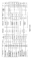


Classifications
-
- A—HUMAN NECESSITIES
- A61—MEDICAL OR VETERINARY SCIENCE; HYGIENE
- A61K—PREPARATIONS FOR MEDICAL, DENTAL OR TOILETRY PURPOSES
- A61K31/00—Medicinal preparations containing organic active ingredients
- A61K31/13—Amines
- A61K31/155—Amidines (), e.g. guanidine (H2N—C(=NH)—NH2), isourea (N=C(OH)—NH2), isothiourea (—N=C(SH)—NH2)
-
- A—HUMAN NECESSITIES
- A61—MEDICAL OR VETERINARY SCIENCE; HYGIENE
- A61K—PREPARATIONS FOR MEDICAL, DENTAL OR TOILETRY PURPOSES
- A61K31/00—Medicinal preparations containing organic active ingredients
- A61K31/33—Heterocyclic compounds
- A61K31/395—Heterocyclic compounds having nitrogen as a ring hetero atom, e.g. guanethidine or rifamycins
- A61K31/41—Heterocyclic compounds having nitrogen as a ring hetero atom, e.g. guanethidine or rifamycins having five-membered rings with two or more ring hetero atoms, at least one of which being nitrogen, e.g. tetrazole
- A61K31/425—Thiazoles
- A61K31/429—Thiazoles condensed with heterocyclic ring systems
- A61K31/43—Compounds containing 4-thia-1-azabicyclo [3.2.0] heptane ring systems, i.e. compounds containing a ring system of the formula, e.g. penicillins, penems
-
- A—HUMAN NECESSITIES
- A61—MEDICAL OR VETERINARY SCIENCE; HYGIENE
- A61K—PREPARATIONS FOR MEDICAL, DENTAL OR TOILETRY PURPOSES
- A61K45/00—Medicinal preparations containing active ingredients not provided for in groups A61K31/00 - A61K41/00
- A61K45/06—Mixtures of active ingredients without chemical characterisation, e.g. antiphlogistics and cardiaca
-
- A—HUMAN NECESSITIES
- A61—MEDICAL OR VETERINARY SCIENCE; HYGIENE
- A61K—PREPARATIONS FOR MEDICAL, DENTAL OR TOILETRY PURPOSES
- A61K9/00—Medicinal preparations characterised by special physical form
- A61K9/0012—Galenical forms characterised by the site of application
- A61K9/0014—Skin, i.e. galenical aspects of topical compositions
-
- A—HUMAN NECESSITIES
- A61—MEDICAL OR VETERINARY SCIENCE; HYGIENE
- A61K—PREPARATIONS FOR MEDICAL, DENTAL OR TOILETRY PURPOSES
- A61K9/00—Medicinal preparations characterised by special physical form
- A61K9/0012—Galenical forms characterised by the site of application
- A61K9/0053—Mouth and digestive tract, i.e. intraoral and peroral administration
-
- A—HUMAN NECESSITIES
- A61—MEDICAL OR VETERINARY SCIENCE; HYGIENE
- A61P—SPECIFIC THERAPEUTIC ACTIVITY OF CHEMICAL COMPOUNDS OR MEDICINAL PREPARATIONS
- A61P31/00—Antiinfectives, i.e. antibiotics, antiseptics, chemotherapeutics
- A61P31/04—Antibacterial agents
-
- A—HUMAN NECESSITIES
- A61—MEDICAL OR VETERINARY SCIENCE; HYGIENE
- A61P—SPECIFIC THERAPEUTIC ACTIVITY OF CHEMICAL COMPOUNDS OR MEDICINAL PREPARATIONS
- A61P43/00—Drugs for specific purposes, not provided for in groups A61P1/00-A61P41/00
-
- A—HUMAN NECESSITIES
- A61—MEDICAL OR VETERINARY SCIENCE; HYGIENE
- A61K—PREPARATIONS FOR MEDICAL, DENTAL OR TOILETRY PURPOSES
- A61K2300/00—Mixtures or combinations of active ingredients, wherein at least one active ingredient is fully defined in groups A61K31/00 - A61K41/00
Definitions
- This invention relates to methods of treating and preventing a bacterial infection in a subject, methods for preparing a medicament for use in treating and preventing a bacterial infection in a subject, and pharmaceutical and veterinary antibacterial compositions when used therein.
- MRSA methicillin resistant Staphylococcus aureus
- E. coli O25b:ST131 The global emergence and spread within the community of highly virulent MDR Gram-negative (G ⁇ ve) bacteria such as E. coli O25b:ST131 confirms that bacterial pathogens can simultaneously evolve both virulence and resistance determinants. Echoing recent MRSA epidemiology, E. coli O25b:ST131, a major cause of urinary tract and bloodstream infections in humans, has now been isolated from extraintestinal infections in companion animals, and poultry. The increasing significance of E.
- the World Health Organization has identified antibiotic resistance as one of the three major future threats to global health.
- a recent report from the US Centers for Disease Control and Prevention (CDC) estimated that “in the United States, more than two million people are sickened every year with antibiotic-resistant infections, with at least 23,000 dying as a result.”
- the extra medical costs, in the USA alone, associated with treating and managing a single case of antibiotic-resistant infection are estimated to be between US$18,588 and US$29,069 per year resulting in an overall direct cost to the US health system of over US$20 billion annually.
- the cost to US households in terms of lost productivity is estimated at over US$35 billion per annum.
- a method of treating or preventing a bacterial colonisation or infection in a subject comprising the step of: administering a therapeutically effective amount of robenidine, or a therapeutically acceptable salt thereof, to the subject.
- the bacterial colonisation or infection is caused by a bacterial agent.
- robenidine or a therapeutically acceptable salt thereof, in the manufacture of medicament for the treatment of a bacterial colonisation or infection in a subject.
- the bacterial colonisation or infection is caused by a bacterial agent.
- the subject may be any subject capable of colonisation or infection by bacteria.
- the subject may be mammalian, or may be piscine or avian.
- the subject is selected from the group comprising, but not limited to, human, canine, feline, bovine, ovine, caprine, other ruminant species, porcine, equine, avian, or piscine.
- robenidine also known as 1,2-bis[(E)-(4-chlorophenyl)methylideneamino]guanidine, or, as described by this specification, NCL812 refers to a compound having the following chemical structure:
- the robenidine may be administered to the subject in a dose selected from the group comprising 0.1 mg/kg to 250 mg/kg body weight, preferably 1 mg/kg to 100 mg/kg body weight, and more preferably 5 mg/kg to 50 mg/kg body weight.
- the robenidine may be administered to the subject using a dosing schedule selected from the group consisting of: hourly, 3 times daily; twice daily; daily; every second day; twice weekly; once weekly; once fortnightly; once monthly; once every two months or by constant rate or variable rate infusion.
- the robenidine is administered until colonisation or the signs and symptoms of infection have at least been partially treated or alleviated.
- the concentration of robenidine (or a robenidine metabolite) in the subject's blood after treatment is within a range selected from the group comprising, but not limited to: between 0.1 and 10 ug/mL at 2 hours, 1 and 200 ug/mL after 12 hours; between 0.1 and 5 ug/mL after 24 hrs; between 0.01 and 2 ug/mL after 48 hours; between 0.0001 and 1 ug/mL after 72 hrs.
- the concentration is selected from the group comprising, but not limited to: less than 200 ug/mL after 12 hours; less than 5 ug/mL after 24 hours; less than 1 ug/L after 48 hours and less than 0.5 ug/mL after 72 hours.
- the agent causing the bacterial infection is a bacterial agent.
- the agent is not a protozoan species.
- the agent is not a coccidian protozoan. More preferably, the agent is not Clostridium perfringens nor a heterotrophic bacterial species present in soil samples collected by Hansen et al from Jyndevad Denmark as discussed in the following papers: Hansen et al. 2012 , Chemosphere, 86:212-215; and Hansen et al. 2009 , Environmental Pollution 157:474-480.
- the bacterial agent is gram negative. In another embodiment, the bacterial agent is gram positive. In another embodiment, the bacterial agent has no cell wall. In another embodiment, the bacterial infection is caused by a mixture of at least two agents selected from the group consisting of: gram negative, gram positive and bacterial agents with no cell wall.
- the bacterial agent causing the bacterial infection may be a gram positive bacterial agent selected from the group comprising, but not limited to, Staphylococcus spp, Streptococci, Enterococcus spp, Leuconostoc spp, Corynebacterium spp, Arcanobacteria spp, Trueperella spp, Rhodococcus spp, Bacillus spp, Anaerobic Cocci, Anaerobic Gram-Positive Nonsporulating Bacilli, Actinomyces spp, Clostridium spp, Nocardia spp, Erysipelothrix spp, Listeria spp, Kytococcus spp, Mycoplasma spp, Ureaplasma spp, and Mycobacterium spp.
- the bacterial agent is gram positive and selected from the group comprising, but not limited to Staphylococcus spp.
- Staphylococcus spp include Staphylococcus epidermidis, Staphylococcus haemolyticus, Staphylococcus lugdunensis, Staphylococcus saprophyticus, Staphylococcus auricularis, Staphylococcus capitis, Staphylococcus caprae, Staphylococcus carnosus, Staphylococcus cohnii, Staphylococcus hominis, Staphylococcus pasteuri, Staphylococcus pettenkoferi, Staphylococcus pulvereri, Staphylococcus saccharolyticus, Staphylococcus simulans, Staphylococcus schleiferi, Staphylococcus warneri, Staphylococcus xylosus, Staphylococcus
- the bacterial agent is gram positive and is selected from the group comprising, but not limited to, Streptococcus spp.
- Streptococcus spp include Streptococcus agalactiae, Streptococcus alactolyticus, Streptococcus anginosus, Streptococcus canis, Streptococcus constellatus, Streptococcus cricetus, Streptococcus cristatus, Streptococcus downei, Streptococcus dysgalactiae subsp. dysgalactiae, Streptococcus dysgalactiae subsp.
- Streptococcus gallolyticus subsp. gallolyticus (formerly Streptococcus bovis biotype i), Streptococcus gallolyticus subsp.
- Streptococcus bovis biotype ii/2 Streptococcus gordonii, Streptococcus hyointestinalis, Streptococcus hyovaginalis, Streptococcus infantarius, Streptococcus infantarius subsp infantarius, Streptococcus infantis, Streptococcus iniae, Streptococcus intermedius, Streptococcus lutetiensis (formerly Streptococcus bovis biotype ii.1), Streptococcus macaccae, Streptococcus mitis, Streptococcus mutans, Streptococcus oralis, Streptococcus orisratti, Streptococcus parasanguinis, Streptococcus peroris, Streptococcus pneumoniae, Streptococcus porcinus, Streptococcus pseudintermedius, Strepto
- the bacterial agent is gram positive and selected from the group comprising, but not limited to, Enterococcus spp.
- Enterococcus spp include Enterococcus faecalis, Enterococcus faecium, Enterococcus gallinarum, Enterococcus durans, Enterococcus avium, Enterococcus raffinosus, Enterococcus pallens, Enterococcus gilvus, Enterococcus cecorum, Enterococcus malodoratus, Enterococcus italicus, Enterococcus sanguinicola, Enterococcus mundtii, Enterococcus casseliflavus/flavescens, Enterococcus dispar, Enterococcus hirae, Enterococcus pseudoavium , and Enterococcus bovis.
- the bacterial agent is gram positive and selected from the group comprising, but not limited to, Leuconostoc spp.
- Leuconostoc spp include Leuconostoc mesenteroides, Leuconostoc pseudomesenteroides, Leuconostoc paramesenteroides, Leuconostoc citreum , and Leuconostoc lactis.
- the bacterial agent is gram positive and selected from the group comprising, but not limited to, Corynebacterium spp.
- Corynebacterium spp include nonlipophilic, fermentative Corynebacterium spp such as Corynebacterium ulcerans, Corynebacterium pseudotuberculosis, Corynebacterium xerosis, Corynebacterium striatum, Corynebacterium minutissimum, Corynebacterium amycolatum, Corynebacterium glucuronolyticum, Corynebacterium argentoratense, Corynebacterium matruchotii, Corynebacterium riegelii, Corynebacterium confusum, Corynebacterium cystidis, Corynebacterium diphtheria, Corynebacterium simulans, Corynebacterium sundvallense, Corynebacterium thomssensii,
- the bacterial agent is gram positive and selected from the group comprising, but not limited to, Arcanobacteria spp.
- Arcanobacteria spp include A. haemolyticum, A. pyogenes (now known as Trueperella pyogenes , originally known as Actinomyces pyogenes ), and A. bernardiae.
- the bacterial agent is gram positive and selected from the group comprising, but not limited to, Rhodococcus spp.
- Rhodococcus spp include Rhodococcus equi, Rhodococcus erythropolis, Rhodococcus fasciens , and Rhodococcus rhodochrous.
- the bacterial agent is gram positive and selected from the group comprising, but not limited to, Gordonia spp.
- the bacterial agent is gram positive and selected from the group comprising, but not limited to, Tsukamurella spp.
- the bacterial agent is gram positive and selected from the group comprising, but not limited to, Acholeplasma spp.
- the bacterial agent is gram positive and selected from the group comprising, but not limited to, Actinobacteria such as Crossiella equi.
- the bacterial agent is gram positive and selected from the group comprising, but not limited to, Bacillus spp.
- Bacillus spp include Bacillus anthracis, Bacillus cereus, Bacillus circulans, Bacillus licheniformis, Bacillus megaterium, Bacillus pumilus, Bacillus sphaericus, Bacillus subtilis, Brevibacillus brevis, Brevibacillus laterosporus , and Paenibacillus alvei.
- the bacterial agent is gram positive and selected from the group comprising, but not limited to, Anaerobic Cocci.
- Anaerobic Cocci include Anaerococcus murdochii, Anaerococcus prevotii, Anaerococcus tetradius, Anaerococcus octavius, Anaerococcus hydrogenalis, Anaerococcus lactolyticus, Anaerococcus vaginalis, Atopobium parvulum, Finegoldia magna, Gallicola barnesae, Gemella asaccharolytica, Gemella bergeri, Gemella cuniculi, Gemella haemolysans, Gemella morbillorum, Gemella palaticanis, Gemella sanguinis; Parvimonas micra, Peptococcus niger, Peptoniphilus asaccharolyticus, Peptoniphilus gorbachii, Peptoniphilus indolicus, Peptoniphilus harei, Peptoniphil
- the bacterial agent is gram positive and selected from the group comprising, but not limited to, Anaerobic Gram-Positive Nonsporulating Bacilli.
- Anaerobic Gram-Positive Nonsporulating Bacilli include Alloscardovia omnicolens, Atopobium species (such as Atopobium minutum, Atopobium rimae, Atopobium parvulum , and Atopobium vaginae ), Bifidobacteria (such as Bifidobacteria adolescentis, Bifidobacteria dentium, Bifidobacteria scardovii ), Catabacter hongkongensis, Collinsella aerofaciens, Eggerthella (such as Eggerthella lenta, Eggerthella hongkongensis and Eggerthella sinensis ), Eubacterium and related species (such as Eubacterium nodatum, Eubacterium uben, Eubacterium brachium ,
- the bacterial agent is gram positive and selected from the group comprising, but not limited to, Actinomyces spp.
- Actinomyces spp include Actinomyces israelii, Actinomyces naeslundii, Actinomyces viscosus, Actinomyces odontolyticus, Actinomyces meyeri , and Actinomyces gerencseriae (formerly Actinomyces israelii serotype II), Actinomyces europaeus, Actinomyces neuii, Actinomyces radingae, Actinomyces graevenitzii, Actinomyces hordeovulneris, Actinomyces turicensis, Actinomyces georgiae, Arcanobacterium ( Actinomyces ) pyogenes, Arcanobacterium ( Actinomyces ) bernardiae, Actinomyces s
- the bacterial agent is gram positive and selected from the group comprising, but not limited to, Clostridium spp.
- Clostridium spp include Clostridium baratii, Clostridium bifermentans, Clostridium botulinum, Clostridium botulinum (types A, B, C, D, E, F, G), Clostridium butyricum, Clostridium difficile, Clostridium histolyticum, Clostridium novyi (type A), Clostridium novyi (type B), Clostridium perfringens, Clostridium perfringens (types A-E), Clostridium ramosum, Clostridium septicum, Clostridium sordelli, Clostridium sphenoides, Clostridium tedium , and Clostridium tetani.
- the bacterial agent is gram positive and selected from the group comprising, but not limited to, Nocardia spp.
- Nocardia spp include Nocardia asteroides, Nocardia brasiliensis, Nocardia farcinica, Nocardia nova, Nocardia otitidiscaviarum , and Nocardia transvalensis.
- the bacterial agent is gram positive and selected from the group comprising, but not limited to, Erysipelothrix spp, such as Erysipelothrix rhusiopathiae.
- the bacterial agent is gram positive and selected from the group comprising, but not limited to, Listeria spp, such as Listeria monocytogenes.
- the bacterial agent is gram positive and selected from the group comprising, but not limited to, Kytococcus spp, such as Kytococcus schroeteri.
- the bacterial agent is gram positive and selected from the group comprising, but not limited to, Mycobacterium spp.
- Mycobacterium spp include Mycobacterium abscessus, Mycobacterium arupense, Mycobacterium asiaticum, Mycobacterium aubagnense, Mycobacterium avium complex, Mycobacterium bolletii, Mycobacterium bolletii, Mycobacterium branderi, Mycobacterium canettii, Mycobacterium caprae, Mycobacterium celatum, Mycobacterium chelonae, Mycobacterium chimaera, Mycobacterium colombiense, Mycobacterium conceptionense, Mycobacterium conspicuum, Mycobacterium elephantis, Mycobacterium farcinogenes, Mycobacterium florentinum, Mycobacterium fortuitum group, Mycobacterium genavense, Mycobacterium goodii, Mycobacterium haemophilum, Mycobacterium homeeshorn
- the bacterial agent is gram positive and selected from the group comprising, but not limited to, Trueperella spp.
- Trueperella spp include Trueperella abortisuis, Trueperella bernardiae, Trueperella bialowiezensis, Trueperella bonasi, Trueperella pyogenes ( Arcanobacterium pyogenes ).
- the bacterial agent is gram positive, gram negative or does not have a cell wall and selected from the group comprising, but not limited to, livestock pathogens.
- livestock pathogens include Actinobaculum suis, Actinomyces bovis, Arcanobacterium pyogenes, Bacillus anthracis, cereus, licheniformis, pumilus, melaninogenicus, subtilis, Clostridium botulinum, chauvoei, haemolyticum, novyi, perfringens, septicum, sordellii, tetani, colinum, Corynebacterium pseudotuberculosis, renale, Dermatophilus congolensis, Enterococcus spp (such as E.
- Mycoplasma bovis dispar, mycoides subsp. mycoides (such as Contagious bovine pleuropneumonia CBPP) Mycoplasma gallisepticum (MG), iowae meleagridis (MM), synoviae (MS) Mycoplasma haemosuis (formerly Eperythrozoon suis ), alkalescens, bovigenitalum, bovirhinis, bovoculi, californicum, canadense, cynos, equigenitalium, gateae, haemocanis, haemofelis, hyopneumoniae, hyorhinis, hyosynoviae, iowae, leachii, meleagridis, mycoides subsp capri, wenyonii, suis, Rhodococcus equi, Staphylococcus epidermidi
- Staphylococcus cohnii subsp. urealyticus Staphylococcus capitis subsp. capitis, Staphylococcus capitis subsp. urealyticus, Staphylococcus hyicus, Staphylococcus aureus, Staphylococcus pseudintermedius, Staphylococcus delphini, Staphylococcus schleiferi subsp. coagulans, Staphylococcus aureus subsp.
- Streptococcus uberis Streptococcus canis, Streptococcus agalactiae, Streptococcus dysgalactiae, Streptococcus pyogenes, Streptococcus bovis, Streptococcus equi subsp.
- Streptococcus equinus Streptococcus equi ( Streptococcus equi subsp equi ), Streptococcus equisimilis ( Streptococcus dysgalactiae subsp equisimilis ), porcinus, suis, zooepidemicus, Streptococcus zooepidemicus ( Streptococcus equi subsp zooepidemicus ), Streptococcus dysgalactiae subsp.
- the bacterial agent is gram positive and selected from the group comprising, but not limited to, pathogens of companion animal species such as cats, dogs and horses.
- pathogens include equine pathogens such as Streptococcus equi, Streptococcus zooepidemicus, Rhodococcus equi, Clostridium difficile, Clostridium perfringens, Corynebacterium pseudotuberculosis, Clostridium piliforme, Actinomyces bovis, Staphylococcus aureus , beta haemolytic Steptococcus spp, Dermatophilus congolense, Clostridiium tetani , and Clostridium botulinum .
- equine pathogens such as Streptococcus equi, Streptococcus zooepidemicus, Rhodococcus equi, Clostridium difficile, Clostridium perfringens,
- pathogens of dogs and cats such as Staphylococcus spp, Streptococcus spp, Clostridium spp, Actinomyces spp, Enterococcus spp, Nocardia spp, Mycoplasma spp, and Mycobacterium spp.
- the bacterial agent is gram negative and selected from the group consisting of the following representative families and species: Acetobacteraceae:— Roseomonas cervicalis; Roseomonas fauriae; Roseomonas gilardii .—Aeromonadaceae:— Aeromonas allosaccharophila; Aeromonas aquariorum; Aeromonas caviae; Aeromonas hydrophila (and subspecies); Aeromonas salmonicida; Aeromonas shubertii; Aeromonas veronii biovar sobria ( Aeromonas sobria ).—Alcaligenaceae:— Achromobacter xylosoxidans; Alcaligenes faecalis; Bordetella ansorpii; Bordetella avium; Bordetella bronchiseptica; Bordetella Bordetella holmesii; Bordetella parapertussis; Bordetella per
- Bdellovibrionaceae Bdellovibrio spp.—Brachyspiraceae:— Brachyspira spp including Brachyspira hampsonii, Brachyspira hyodysenteriae, Brachyspira murdochii, Brachyspira pilosicoli .—Brucellaceae:— Brucella abortus; Brucella canis; Brucella ceti; Brucella melitensis; Brucella ovis; Brucella pinnipedialis; Brucella suis; Ochrobactrum anthropi; Ochrobactrum intermedium .—Burkholderiaceae:— Burkholderia aboris; Burkholderia ambifaria (genomovar VII); Burkholderia anthina (genomovar VIII); Burkholderia cenocepacia (genomovar
- Prevotella intermedia Prevotella melaninogenica .
- Pseudomonadaceae Chryseomonas luteola; Pseudomonas aeruginosa; Pseudomonas luteola; Pseudomonas fluorescens; Pseudomonas putida; Pseudomonas stutzeri; Pseudomonas oryzihabitans .
- Rhizobiaceae Agrobacterium tumefaciens; Rhizobium radiobacter .
- Rhickettsiaceae — Orientia chuto; Orientia tsutsugamushi; Rickettsia aeschlimannii; Rickettsia africae; Rickettsia akari; Rickettsia argasii; Rickettsia asiatica; Rickett
- the bacterial agent causing the bacterial infection is gram negative and is selected from the group comprising: Acinetobacter species, Aeromonas hydrophila, Citrobacter species, Enterobacter species, Escherichia coli, Klebsiella pneumoniae, Morganella morganii, Pseudomonas aeruginosa , and Stenotrophomonas maltophilia.
- the bacteria agent causing the bacterial colonisation or infection is resistant to a conventional antibiotic used to treat the colonisation or infection.
- the bacterial agent is resistant to a compound selected from the group comprising: one or more of aminoglycosides (for example gentamicin, tobramycin, amikacin, or netilmicin); anti-MRSA cephalosporins (for example ceftaroline); antipseudomonal penicillins+ ⁇ -lactamase inhibitors (for example ticarcillin-clavulanic acid or piperacillin-tazobactam); carbapenems (for example ertapenem, imipenem, meropenem or doripenem); non-extended spectrum cephalosporins; 1st and 2nd generation cephalosporins (for example cefazolin or cefuroxime); extended-spectrum cephalosporins; 3rd and 4th generation cephalosporins (for example cefotaxime or cef
- the bacterial agent is resistant to a compound selected from the group comprising: penicillins, cephalosporins, carbapenems, monobactams and other ⁇ -lactam antibiotics, fusidanes, aminoglycosides, fluoroquinolones, streptogramins, tetracyclines, glycylcyclines, chloramphenicol and other phenicols, macrolides and ketolides, lincosamides, oxazolidinones, aminocyclitols, polymyxins, glycopeptides, lipopeptides, bacitracin, mupiricin, pleuromutilins, rifamycins, sulphonamides and trimethoprim.
- the compound is selected from the group comprising: ⁇ -lactams, glycopeptides, lipopeptides, macrolides, oxazolidinones and tetracyclines.
- the bacterial agent is resistant to the compound when the compound is at a concentration range selected from the following: 0.001 ⁇ g/mL-10,000 ⁇ g/mL; 0.01 ⁇ g/mL-1000 ⁇ g/mL; 0.10 ⁇ g/mL-100 ⁇ g/mL; and 1 ⁇ g/mL-50 ⁇ g/mL
- the bacterial agent causing the bacterial infection is selected from the group comprising, but not limited to, gram positive bacteria.
- the bacterial agent is most preferably a Gram positive bacterial agent selected from the group comprising Staphylococcus aureus, Staphylococcus pseudintermedius, Streptococcus pneumoniae, Streptococcus pyogenes, Streptococcus agalactiae, Streptococcus uberis, Enterococcus faecium, Enterococcus faecalis , and Clostridium difficile.
- the bacterial agent has no cell wall.
- the bacterial agent is selected from the group comprising: Mycoplasma spp, Mycoplasma agalactiae, Mycoplasma alkalescens, Mycoplasma amphoriforme, Mycoplasma arginini, Mycoplasma bovigenitalum, Mycoplasma bovirhinis, Mycoplasma bovis, Mycoplasma bovoculi, Mycoplasma buccale, Mycoplasma californicum, Mycoplasma canadense, Mycoplasma capricolum subsp. capricolum, Mycoplasma capricolum subsp.
- Mycoplasma conjunctivae Mycoplasma cynos
- Mycoplasma dispar Mycoplasma equigenitalium
- Mycoplasma faucium Mycoplasma felis
- Mycoplasma fermentans incognitus str.
- Mycoplasma gallisepticum MG
- Mycoplasma gateae Mycoplasma genitalium, Mycoplasma haemocanis, Mycoplasma haemofelis, Mycoplasma haemosuis (formerly Eperythrozoon suis ), Mycoplasma hominis, Mycoplasma hyopneumoniae, Mycoplasma hyorhinis, Mycoplasma hyosynoviae, Mycoplasma iowae meleagridis (MM), Mycoplasma iowae, Mycoplasma leachii, Mycoplasma lipophilum, Mycoplasma meleagridis, Mycoplasma mycoides sub
- mycoides such as Contagious bovine pleuropneumonia CBPP), Mycoplasma orale, Mycoplasma ovipneumoniae, Mycoplasma ovis, Mycoplasma penetrans, Mycoplasma pirum, Mycoplasma pneumoniae, Mycoplasma primatum, Mycoplasma putrefaciens, Mycoplasma salivarium, Mycoplasma spermatophilum, Mycoplasma suis, Mycoplasma synoviae (MS), Mycoplasma wenyonii, Mycoplasma, Ureaplasma spp, Ureaplasma parvum, Ureaplasma urealyticum, Ureaplasma , and Ureoplasma diversum.
- CBPP Contagious bovine pleuropneumonia CBPP
- Mycoplasma orale Mycoplasma ovipneumoniae
- Mycoplasma ovis Mycoplasma penetrans, Mycoplasma pirum
- Mycoplasma pneumoniae Mycoplasma
- the bacterial agent is Staphylococcus aureus.
- the bacterial agent is resistant to a compound selected from the group comprising: one or more of aminoglycosides (for example gentamicin); ansamycins (for example rifampicin); anti-MRSA cephalosporins (for example ceftaroline); anti-staphylococcal ⁇ -lactams (or cephamycins) (for example oxacillin or cefoxitin); carbapenems (for example ertapenem, imipenem, meropenem or doripenem); non-extended spectrum cephalosporins; 1st and 2nd generation cephalosporins (for example cefazolin or cefuroxime); extended-spectrum cephalosporins; 3rd and 4th generation cephalosporins (for example cefotaxime or ceftriaxone); cephamycins (for example cefoxitin or cefotetan); fluoroquinolones (for example ciprofloxacin
- the bacterial agent is Streptococcus pneumoniae .
- the Streptococcus pneumoniae may be a strain that is resistant to one or more of ⁇ -lactams and macrolides.
- the bacterial agent is Streptococcus pyogenes.
- the bacterial agent is Streptococcus agalactiae.
- the bacterial agent is either Enterococcus faecium or Enterococcus faecalis .
- the Enterococcus faecium or Enterococcus faecalis may be a strain that is resistant to one or more of aminoglycosides (for example gentamicin (high level) or streptomycin (for example streptomycin (high level)); carbapenems (for example imipenem, meropenem or doripenem); fluoroquinolones (for example ciprofloxacin, levofloxacin or moxifloxacin); glycopeptides (for example vancomycin or teicoplanin); glycylcyclines (for example tigecycline); lipopeptides (for example daptomycin); oxazolidinones (for example linezolid); penicillins (for example ampicillin); streptogramins (for example quinupristin-dalfopristi
- the bacterial agent is Clostridium difficile.
- the bacterial infection in the subject may cause a disease selected from the group comprising, but not limited to, nosocomial pneumonia caused by Staphylococcus aureus (MDR, XDR, PDR or methicillin-susceptible or -resistant strains), or invasive pneumococcal diseases such as pneumonia, bronchitis, acute sinusitis, otitis media, conjunctivitis, meningitis, bacteremia, sepsis, osteomyelitis, septic arthritis, endocarditis, peritonitis, pericarditis, cellulitis, and brain abscess caused by Streptococcus pneumoniae (including multi-drug resistant strains [MDRSP] such as those resistant to (3-lactams and macrolides), complicated skin and skin structure infections, including diabetic foot infections, with or without concomitant osteomyelitis, caused by Staphylococcus aureus (methicillin-susceptible and -resistant strains), Streptococc
- Gram negative organisms are important causes of many infectious diseases in humans and other animal species. Bone and joint infections (Gram-negative organisms or mixed bacteria, are an important cause of vertebral osteomyelitis and septic arthritis), cardiovascular system infections (including endocarditis caused by the HACEK group— Haemophilus parainfluenzae, Haemophilus aphrophilus, Aggregatibacter actinomycetemcomitans, Cardiobacterium hominis, Eikenella corrodens, Kingella kingae ), central nervous system infections (the commonest causes of bacterial meningitis are Neisseria meningitidis, Streptococcus pneumoniae and, in nonvaccinated young children, Haemophilus influenzae type b (Hib), in neonates and infants less than 3 months of age, Streptococcus agalactiae (group B streptococcus ), Escherichia coli and other aerobic Gram-negative rods are important pathogens, brain abscess
- intra-abdominal infections peripheral infections
- perforated viscus is usually a polymicrobial infection with aerobic and anaerobic bowel flora while spontaneous bacterial peritonitis (SBP) is usually caused by enteric Gram-negative bacilli, such as Escherichia coli and Klebsiella species, Klebsiella pneumoniae is an increasingly identified cause of liver abscess
- enteric Gram-negative bacilli such as Escherichia coli and Klebsiella species
- Klebsiella pneumoniae is an increasingly identified cause of liver abscess
- community-acquired pneumonia Mycoplasma pneumoniae, Chlamydophila ( Chlamydia ) pneumoniae, Chlamydophila ( Chlamydia ) psittaci, Haemophilus influenza , aerobic Gram-negative bacilli including Klebsiella pneumonia, Pseudomonas aeruginosa, Acinetobacter baumannii, Burk
- E. coli Escherichia coli
- Klebsiella pneumoniae K. pneumoniae
- P. aeruginosa Pseudomonas aeruginosa
- Gram negative bacteria are showing rising rates of resistance to current therapies.
- the production of extended-spectrum ⁇ -lactamase (ESBL) enzymes is a common mechanism of resistance. Rates of ESBL-producing E. coli and K. pneumoniae have risen substantially, with the result that these bacteria are increasingly resistant to widely used antimicrobials.
- ESBL extended-spectrum ⁇ -lactamase
- P. aeruginosa is the most common Gram-negative cause of nosocomial pneumonia and the second most common cause of catheter-related UTIs in the U.S.
- E. coli is the most common cause of UTIs. Cases of UTI caused by ESBL-producing E. coli and K. pneumonia as well as P. aeruginosa , including MDR strains, are increasing. ESBL-producing E. coli and K. pneumoniae are also frequently isolated in patients with complicated IAI (cIAI).
- P. aeruginosa is a clinically challenging and virulent pathogen that can be a cause of common infections in humans such as nosocomial pneumonia, UTI, IAI, and bloodstream infections.
- P. aeruginosa is the most common Gram-negative organism causing ventilator associated pneumonia and the second most common cause of catheter-associated UTIs.
- robenidine, or a therapeutically acceptable salt thereof is administered together with a compound or agent that removes or substantially removes or reduces the integrity of the cell wall of the bacterial agent.
- the compound is selected from the group consisting of: ⁇ -lactams, fosfomycin, lysozyme, polymyxins and chelating agents such as ethylenediaminetetraacetic acid (EDTA).
- the agent is an immunological agent (such as an antibody or vaccine) that reduces the integrity of the cell wall.
- robenidine, or a therapeutically acceptable salt thereof is administered together with a compound that removes or substantially removes or weakens the integrity of the outer cell wall of a gram negative or positive bacterial agent.
- an antibacterial pharmaceutical composition comprising a therapeutically effective amount of robenidine, or a therapeutically acceptable salt thereof.
- an antibacterial veterinary composition comprising a therapeutically effective amount of robenidine, or a therapeutically acceptable salt thereof.
- the method of treating or preventing a bacterial infection or colonisation in a subject may also comprise the administration of the pharmaceutical or veterinary compositions of the invention.
- the pharmaceutical composition may optionally include a pharmaceutically acceptable excipient or carrier.
- the veterinary composition may optionally include a veterinary acceptable excipient or carrier.
- the pharmaceutical or veterinary composition of the invention preferably contains robenidine, or a pharmaceutically acceptable salt, at a concentration of selected from the grouped consisting of: 1 mg/g to 500 mg/g; 5 mg to 400 mg/g; 10 mg/g to 200 mg/g; 20 mg/g to 100 mg/g; 30 mg/g to 70 mg/g; and 40 mg/g to 60 mg/g.
- the pharmaceutical or veterinary composition comprises impurities, wherein the quantity of impurities as a percentage of the total weight of the composition is selected from the group consisting of: less than 20% impurities (by total weight of the composition); less than 15% impurities; less than 10% impurities; less than 8% impurities; less than 5% impurities; less than 4% impurities; less than 3% impurities; less than 2% impurities; less than 1% impurities: less than 0.5% impurities; less than 0.1% impurities.
- the pharmaceutical or veterinary composition comprises microbial impurities or secondary metabolites, wherein the quantity of microbial impurities as a percentage of the total weight of the composition is selected from the group consisting of: less than 5%; less than 4%; less than 3%; less than 2%; less than 1%; less than 0.5%; less than 0.1%; less than 0.01%; less than 0.001%.
- the pharmaceutical or veterinary composition is sterile and stored in a sealed and sterile container.
- the pharmaceutical or veterinary composition contains no detectable level of microbial contamination.
- the robenidine is pharmaceutical or veterinary grade.
- Methods to synthesise commercial quantities of robendine are widely available in the art.
- Commercial quantities of pharmaceutical or veterinary grade robenidine are available from Zhejiang Esigma Animal Health Co., Ltd, Haining City, Peoples Republic of China.
- the pharmaceutical or veterinary composition of the invention may comprise a further antimicrobial agent.
- the further antimicrobial agent may be an antifungal agent or antibacterial agent.
- the method of treating or preventing a bacterial infection or colonisation in a subject may also comprise the administration of robenidine with a further antimicrobial agent.
- the antifungal agent is selected from the group comprising, but not limited to naturally occurring agents including Echinocandins (Anidulafungin, Caspofungin, Micafungin), Polyenes (Amphotericin B, Candicidin, Filipin, Fungichromin (Pentamycin), Hachimycin, Hamycin, Lucensomycin, Mepartricin, Natamycin, Nystatin, Pecilocin, Perimycin), and other naturally occurring antifungal agents including Griseofulvin, Oligomycins, Pyrrolnitrin, Siccanin, and Viridin.
- naturally occurring agents including Echinocandins (Anidulafungin, Caspofungin, Micafungin), Polyenes (Amphotericin B, Candicidin, Filipin, Fungichromin (Pentamycin), Hachimycin, Hamycin, Lucensomycin, Mepartricin, Natamycin, Nystatin, Pecilocin, Perimycin), and other naturally
- the antifungal agent may be a synthetic compound selected from the group comprising, but not limited to Allylamines (Butenafine, Naftifine, Terbinafine) Imidazoles (Bifonazole, Butoconazole, Chlormidazole, Climbazole, Croconazole (Cloconazole), Clotrimazole, Eberconazole, Econazole, Enilconazole, Fenticonazole, Flutrimazole, Fosfluconazole, Isoconazole, Ketoconazole, Lanoconazole, Luliconazole, Miconazole, Neticonazole, Omoconazole, Oxiconazole Nitrate, Parconazole, Sertaconazole, Sulconazole, Tioconazole), Thiocarbamates (Liranaftate, Tolciclate, Tolindate, Tolnaftate), Triazoles (Fluconazole, Isavuconazole, Itraconazole
- composition of the invention may comprise an antibiotic adjunct selected from the group comprising, but not limited to, ⁇ -Lactamase Inhibitors (Avibactam, Clavulanic Acid, Sulbactam, Sultamicillin, Tazobactam), Renal Dipeptidase Inhibitors (Cilastatin), and Renal Protectant (Betamipron).
- ⁇ -Lactamase Inhibitors Avibactam, Clavulanic Acid, Sulbactam, Sultamicillin, Tazobactam
- Renal Dipeptidase Inhibitors Rostam
- Renal Protectant Betamipron
- the composition of the invention comprises a further antibiotic selected from the group comprising, but not limited to, 2,4-DIAMINOPYRIMIDINES, including Baquiloprim, Brodimoprim, Iclaprim, Ormetoprim, Pyrimethamine, Tetroxoprim, Trimethoprim; AMINOCOUMARINS, including Novobiocin; AMINOCYCLITOLS, including Spectinomycin; AMINOGLYCOSIDES, including Amikacin, Apramycin, Arbekacin, Bekanamycin, Butirosin, Dibekacin, Dihydrostreptomycin, Etimicin, Fortimicins (Astromicin), Framycetin, Gentamicin, Hygromycin B, Isepamicin, Kanamycin, Micronomicin, Neomycin, Netilmicin, Paromomycin, Plazomicin, Ribostamycin, Sisomicin, Streptomycin, Tobramycin, Verdamicin; AMINOMETHYLCY
- composition of the invention may further comprise an excipient selected from the group comprising, but not limited to, binders and compression aids, coatings and films, colouring agents diluents and vehicles disintegrants, emulsifying and solubilising agents, flavours and sweeteners, repellents, glidants and lubricants, plasticisers, preservatives, propellants, solvents, stabilisers, suspending agents and viscosity enhancers.
- excipient selected from the group comprising, but not limited to, binders and compression aids, coatings and films, colouring agents diluents and vehicles disintegrants, emulsifying and solubilising agents, flavours and sweeteners, repellents, glidants and lubricants, plasticisers, preservatives, propellants, solvents, stabilisers, suspending agents and viscosity enhancers.
- a medical device when used in a method of treating or preventing a bacterial infection in the subject.
- a medical device comprising the composition of the invention.
- the composition of the invention may be any slow release form, and/or in the form of a coating of the medical device.
- the medical device may be in a form selected from the group comprising: an implant, a plaster, a bandage, and other dressing applied to a bacterial infection in a subject.
- a method of killing bacteria including the step of contacting the bacteria with robenidine, or a therapeutically acceptable salt thereof.
- robenidine or a therapeutically acceptable salt thereof, to kill bacteria
- said use comprising the step of contacting the bacteria with robenidine, or a therapeutically acceptable salt thereof.
- FIG. 1 shows a table of the Minimum Inhibitory Concentrations for the individual Staphylococcus aureus isolates according to example 1;
- FIG. 2 shows a table of the Minimum Inhibitory Concentrations for the individual Enterococcus isolates according to example 1;
- FIG. 3 shows a table of the Minimum Inhibitory Concentrations for the individual Streptococcus pneumoniae isolates according to example 1;
- FIG. 4 shows a table of the NCL812 MIC 50 , MIC 90 , MIC mode and MIC range for Australian isolates of MRSA, VRE and Str. pneumoniae according to example 1. Comparative MIC values for ampicillin are shown in parenthesis;
- FIG. 5 shows a table of the Minimum Inhibitory Concentrations values for NCL812 (robenidine) and linezolid against Staphylococcus aureus ATCC29213 according to example 2;
- FIG. 6 shows a graph of the effect of NCL812 on DNA macromolecular synthesis in Staphylococcus aureus according to example 2;
- FIG. 7 shows a graph of the effect of NCL812 on RNA macromolecular synthesis in Staphylococcus aureus according to example 2;
- FIG. 8 shows a graph of the effect of NCL812 on protein macromolecular synthesis in Staphylococcus aureus (ATCC29213) according to example 2;
- FIG. 9 shows a graph of the effect of NCL812 on cell wall macromolecular synthesis in Staphylococcus aureus (ATCC29213) according to example 2;
- FIG. 10 shows a graph of the effect of NCL812 on lipid macromolecular synthesis in Staphylococcus aureus (ATCC29213) according to example 2;
- FIG. 11 shows a graph summarising the effect of NCL812 on macromolecular synthesis in Staphylococcus aureus (ATCC29213) according to example 2;
- FIG. 12 shows a graph of the effect of NCL812 on ATP release from Staphylococcus aureus (ATCC29213) according to example 3;
- FIG. 13 shows a table of Staphylococcus aureus clone/isolate name, type, source, antibiogram, clindamycin resistance status, multi-locus sequence type (MLST), staphylococcal cassette chromosome (SCCmec) type, clonal complex, Panton-Valentine leukocidin status (PVL), and spa type for isolates used according to example 4.
- MSSA methicillin-susceptible S. aureus . HA-MRSA; hospital-acquired methicillin-resistant S. aureus . CA-MRSA; community-associated methicillin-resistant S. aureus . M.B; M. Barton (University of South Australia).
- FIG. 14 shows a table of the percentage of presumptively identified S. aureus isolates reporting positive to selected phenotypic and genotypic tests according to Example 4.
- HA-MRSA hospital-acquired S. aureus .
- CA-MRSA community-associated S. aureus.
- S. aureus isolates were identified as testing positive to protein A latex agglutination (Protein A), slide coagulase, Voges-Proskauer and polymyxin B resistance tests, as well as testing positive for polymerase chain reaction (PCR) and real-time PCR amplification of the spa gene.
- PCR polymerase chain reaction
- Methicillin-resistant S. aureus isolates were identified as isolates testing positive to the criteria described above, as well as positive for PCR and real-time PCR of the mecA gene;
- FIG. 15 shows a table of the resistance of S. aureus isolates to antibacterial agents using the Kirby-Bauer disc diffusion method according to Example 4.
- HA-MRSA hospital-acquired methicillin-resistant S. aureus .
- CA-MRSA community-associated methicillin-resistant S. aureus;
- FIG. 16 shows a table of the number and percentage of identified mec gene complexes in 20 S. aureus strains classified as methicillin-resistant according to Example 4. Respective staphylococcal cassette chromosome (SCCmec) complexes and types expressing phenotypic resistance to oxacillin and cefotetan are indicated as well as real-time mecA status, and the average negative dF/dT peak obtained from melting point analysis from real-time PCR of the mecA gene. Figures in parentheses indicate percentages;
- FIG. 18 shows a table of the characteristics of antibacterial NCL812 and the ⁇ -lactam antibacterial ampicillin according to Example 4, detailing antibacterial solubility in dimethyl sulfoxide (DMSO), solubility in cation-adjusted Mueller-Hinton II broth (CAMHB), and average minimum inhibitory concentrations (MIC) ( ⁇ g/ml at 24-h) against methicillin-resistant S. aureus (MRSA) determined from preliminary studies and those determined during this present study.
- ATCC 49775 methicillin-susceptible S. aureus isolate and ATCC control strain.
- MRSA580 methicillin-resistant S. aureus isolate #580.
- MRSA698 methicillin-resistant S. aureus isolate #698;
- FIG. 19 shows a table of in vitro activities of the novel antibacterial NCL812 and the ⁇ -lactam antibacterial ampicillin against S. aureus clinical isolates according to example 4.
- HA-MRSA hospital-acquired methicillin-resistant S. aureus .
- CA-MRSA community-associated methicillin-resistant S. aureus .
- MIC minimum inhibitory concentration ( ⁇ g/ml).
- MBC minimum bactericidal concentration ( ⁇ g/ml).
- MIC/MBCrange minimum and maximum MIC/MBC for all isolates.
- MIC/MBC50 MIC/MBC at which 50% of isolates are inhibited.
- MIC/MBC90 MIC/MBC at which 90% of isolates are inhibited;
- FIG. 20 shows a graph of the optical densities of the unsupplemented growth control, ampicillin and different concentrations of antibacterial agent NCL812 against methicillin-susceptible S. aureus ATCC 49775 using broth microdilution methodology according to example 4.
- concentrations of NCL812 tested were at the MIC and four times the MIC determined under test conditions, up to 24-h incubation. Ampicillin was tested at the MIC. Bactericidal activity was tested at 0-, 1-, 2-, 4-, 8-, 12-, and 24-h for antibacterials;
- FIG. 21 shows a graph of kill kinetic curves for methicillin-susceptible S. aureus ATCC 49775 demonstrating bactericidal activity of NCL812 using the Clinical and Laboratory Standards Institute macrodilution methodology in a 10-ml vial according to example 4.
- the concentrations of antibacterials tested were at 1 ⁇ and 4 ⁇ the MIC determined under test conditions.
- Bactericidal activity was determined at 0-, 1-, 2-, 4-, 8-, 12- and 24-h after antibacterial addition.
- Bactericidal activity was defined as a 3-log 10 (99.9%) decrease in the number viable bacteria from the initial inoculum size;
- FIG. 22 shows a table of the antibacterial susceptibility of 20 S. pneumoniae isolates for six different antibacterials according to example 5;
- FIG. 23 shows a graph indicating the change of pH during macro-broth dilution assay for S. pneumoniae strain D39 exposed to 4 ⁇ g/ml in NCL812 and 0.0023 ⁇ g/ml ampicillin according to example 5;
- FIG. 24 shows a graph illustrating the 48-hour time-kill of S. pneumoniae strain D39 treated with NCL812 according to example 5;
- FIG. 25 shows a graph illustrating in the 14-hour time-kill of S. pneumoniae strain D39 treated with NCL812 according to example 5;
- FIG. 26 shows a graph illustrating the 14-hour time-kill of S. pneumoniae strain D39 treated with ampicillin according to example 5;
- FIG. 27 shows a graph illustrating the 12-hour time-kill of S. pneumoniae strain D39 treated with NCL812, adopted from FIG. 40 according to example 5;
- FIG. 28 shows a graph illustrating the 48-hour time-kill of S. pneumoniae strain D39 treated with ampicillin according to example 5;
- FIG. 29 shows a graph illustrating the 48-hour time-kill of S. pneumoniae strain D39 treated with erythromycin according to example 5;
- FIG. 30 shows a graph illustrating the 48-hour time-kill of S. pneumoniae strain D39 treated with NCL812 and 5% choline chloride according to example 5;
- FIG. 31 shows a graph illustrating the 12-hour time-kill of S. pneumoniae strain D39 treated with NCL812 and 5% choline chloride according to example 5;
- FIG. 32 shows a graph illustrating the relative minimum bactericidal concentration (MBC) of S. pneumoniae strain D39 treated with ampicillin over a 48-hour time period according to example 5;
- FIG. 33 shows a graph illustrating the relative MBC for S. pneumoniae strain D39 treated with erythromycin over a 48-hour time period according to example 5;
- FIG. 34 shows a graph illustrating the viable count (log 10 CFU/ml) of S. pneumoniae strain D39 treated with NCL812 from a macro-broth dilution of time-kill over 24 hours according to example 5;
- FIG. 35 shows a graph illustrating the viable count (log 10 CFU/ml) of S. pneumoniae strain D39 treated with ampicillin from a macro-broth dilution of time-kill over 24 hours according to example 5;
- FIG. 36 is a bar graph illustrating the mean cell membrane thickness of treated and untreated D39 according to example 5.
- FIG. 37 is a bar graph illustrating the mean width of periplasmic space of treated (16 ⁇ g/ml NCL812) and untreated D39 samples according to example 5;
- FIG. 38 is a table showing the Staphylococcus pseudintermedius isolates tested according to example 6;
- FIG. 39 is a table showing the antibiotic resistance profile of the Staphylococcus pseudintermedius isolates tested according to example 6.
- FIG. 40 is a graph illustrating the effectiveness of NCL812 against gram-negative E. coli spheroplasts according to example 7;
- FIG. 41 is a graph illustrating the cumulative release of NCL812 from Formulation B according to example 9;
- FIG. 42 shows the kill kinetics assay of Staphylococcus aureus KC01 at different concentrations of NCL812, up to 24 h incubation according to example 11;
- FIG. 43 shows the kill kinetics assay of Enterococcus faecalis USA01 at different concentrations of NCL812, up to 24 h incubation according to example 11.
- the invention described herein may include one or more ranges of values (e.g. size, concentration, dose etc).
- a range of values will be understood to include all values within the range, including the values defining the range, and values adjacent to the range that lead to the same or substantially the same outcome as the values immediately adjacent to that value which define the boundary of the range.
- the pharmaceutical for veterinary compositions of the invention may be administered in a variety of unit dosages depending on the method of administration, target site, physiological state of the patient, and other medicaments administered.
- unit dosage form suitable for oral administration include solid dosage forms such as powder, tablets, pills, and capsules, and liquid dosage forms, such as elixirs, syrups, solutions and suspensions.
- the active ingredients may also be administered parenterally in sterile liquid dosage forms.
- Gelatin capsules may contain the active ingredient and inactive ingredients such as powder carriers, glucose, lactose, sucrose, mannitol, starch, cellulose or cellulose derivatives, magnesium stearate, stearic acid, sodium saccharin, talcum, magnesium carbonate, and the like.
- therapeutically effective amount refers to an amount sufficient to inhibit bacterial growth associated with a bacterial infection or colonisation. That is, reference to the administration of the therapeutically effective amount of robenidine according to the methods or compositions of the invention refers to a therapeutic effect in which substantial bacteriocidal or bacteriostatic activity causes a substantial inhibition of bacterial infection.
- therapeutically effective amount refers to a sufficient amount of the composition to provide the desired biological, therapeutic, and/or prophylactic result. The desired results include elimination of bacterial infection or colonisation or reduction and/or alleviation of the signs, symptoms, or causes of a disease, or any other desired alteration of a biological system.
- an effective amount in any individual case may be determined by one of ordinary skill in the art using routine experimentation.
- effective amounts can be dosages that are recommended in the modulation of a diseased state or signs or symptoms thereof. Effective amounts differ depending on the composition used and the route of administration employed. Effective amounts are routinely optimized taking into consideration pharmacokinetic and pharmacodynamic characteristics as well as various factors of a particular patient, such as age, weight, gender, etc and the area affected by disease or disease causing microbes.
- the terms “treatment” or “treating” refers to the full or partial removal of the symptoms and signs of the condition.
- the treatment completely or partially removes the signs of the infection.
- the treatment reduces or eliminates the infecting bacterial pathogen leading to microbial cure.
- bacteria refers to members of a large domain of prokaryotic microorganisms. Typically a few micrometers in length, bacteria have a number of shapes, ranging from spheres to rods and spirals and can be present as individual cells or present in linear chains or clusters of variable numbers and shape.
- bacteria and its adjective “bacterial” refer to bacteria such as the Gram positive Staphylococcus spp, Streptococcus spp, Bacillus spp, Enterococcus spp, Listeria spp, Mycoplasma spp, and anaerobic bacteria; Gram negative Escherichia coli, Enterobacter spp, Klebsiella spp and Pseudomonas spp; and the cell wall free bacteria such as Mycoplasma spp and Ureaplasma spp.
- the terms may refer to an antibiotic-sensitive strain or an antibiotic-resistant strain. In a preferred embodiment, the terms refer to MRSA or MRSP.
- the terms refer to MDR Staphylococcus spp, Streptococcus spp, Enterococcus spp, Clostridium difficile, Escherichia coli, Enterobacter spp, Klebsiella spp and Pseudomonas spp.
- methicillin-resistant bacteria refers a bacteria isolate that demonstrates resistance at any dose to all 6 lactams including penicillins, carbapenems and first to fourth generation cephalosporins, but not to the fifth generation anti-MRSA cephalosporins (for example ceftaroline).
- Multidrug-resistant (MDR) is defined as acquired non-susceptibility to at least one agent in three or more antimicrobial categories
- extensively drug-resistant (XDR) is defined as non-susceptibility to at least one agent in all but two or fewer antimicrobial categories (i.e. bacterial isolates remain susceptible to only one or two categories)
- pandrug-resistant (PDR) is defined as non-susceptibility to all agents in all antimicrobial categories currently available.
- An example of susceptible, MDR, XDR and PDR bacteria includes the following. Wild type, antibacterial unexposed isolates of Staphylococcus aureus that are likely to be susceptible to all of the following antibacterial categories (and agents): aminoglycosides (for example gentamicin); ansamycins (for example rifampicin); anti-MRSA cephalosporins (for example ceftaroline); anti-staphylococcal ⁇ -lactams (or cephamycins) (for example oxacillin or cefoxitin); carbapenems (for example ertapenem, imipenem, meropenem or doripenem); non-extended spectrum cephalosporins; 1st and 2nd generation cephalosporins (for example cefazolin or cefuroxime); extended-spectrum cephalosporins; 3rd and 4th generation cephalosporins (for example cefotaxime or ceftriaxone); cephamycins
- Isolates that are non-susceptible to more than one agent in more than three antimicrobial categories are classified as MDR (all MRSA, for example, meet the definition of MDR).
- Isolates that are non-susceptible to more than one agent in all but one or two antimicrobial categories are classified as XDR.
- Isolates that are non-susceptible to all listed antibacterial agents are PDR.
- Pharmaceutically and veterinary acceptable salts include salts which retain the biological effectiveness and properties of the compounds of the present disclosure and which are not biologically or otherwise undesirable.
- the compounds disclosed herein are capable of forming acid and/or base salts by virtue of the presence of amino and/or carboxyl groups or groups similar thereto.
- Acceptable base addition salts can be prepared from inorganic and organic bases. Salts derived from inorganic bases, include by way of example only, sodium, potassium, lithium, ammonium, calcium and magnesium salts.
- Salts derived from organic bases include, but are not limited to, salts of primary, secondary and tertiary amines, such as by way of example only, alkyl amines, dialkyl amines, trialkyl amines, substituted alkyl amines, di(subsrituted alkyl) amines, tri(substituted alkyl) amines, alkenyl amines, dialkenyl amines, trialkenyl amines, substituted alkenyl amines, di(substituted alkenyl) amines, tri(substituted alkenyl) amines, cycloalkyl amines, di(cycloalkyl) amines, tri(cycloalkyl) amines, substituted cycloalkyl amines, disubstituted cycloalkyl amines, trisubstituted cycloalkyl amines, cycloalkenyl amines
- Pharmaceutically and veterinary acceptable acid addition salts may be prepared from inorganic and organic acids.
- the inorganic acids that can be used include, by way of example only, hydrochloric acid, hydrobromic acid, sulfuric acid, nitric acid, phosphoric acid, and the like.
- the organic acids that can be used include, by way of example only, acetic acid, propionic acid, glycolic acid, pyruvic acid, oxalic acid, malic acid, malonic acid, succinic acid, maleic acid, fumaric acid, tartaric acid, citric acid, benzoic acid, cinnamic acid, mandelic acid, methanesulfonic acid, ethanesulfonic acid, p-toluenesulfonic acid, salicylic acid, and the like.
- the pharmaceutically or veterinary acceptable salts of the compounds useful in the present disclosure can be synthesized from the parent compound, which contains a basic or acidic moiety, by conventional chemical methods.
- such salts can be prepared by reacting the free acid or base forms of these compounds with a stoichiometric amount of the appropriate base or acid in water or in an organic solvent, or in a mixture of the two; generally, nonaqueous media like ether, ethyl acetate, ethanol, isopropanol, or acetonitrile are preferred.
- Lists of suitable salts are found in Remington's Pharmaceutical Sciences. 17th ed., Mack Publishing Company, Easton, Pa. (1985), p. 1418, the disclosure of which is hereby incorporated by reference.
- salts examples include the iodide, acetate, phenyl acetate, trifluoroacetate, acrylate, ascorbate, benzoate, chlorobenzoate, dinitrobenzoate, hydroxybenzoate, methoxyb ⁇ nzoate, methylbenzoate, o-acetoxybenzoate, naphthalene-2-benzoate, bromide, isobutyrate, phenylbutyrate, ⁇ -hydroxybutyrate, ⁇ -hydroxybutyrate, butyne-1,4-dioate, hexyne-1,4-dioate, hexyne-1,6-dioate, caproate, caprylate, chloride, cinnamate, citrate, decanoate, formate, fumarate, glycollate, heptanoate, hippurate, lactate, malate, maleate, hydroxymaleate, malonate, mandelate, mesylate, nicot
- compositions of the invention may be formulated in conventional manner, together with other pharmaceutically acceptable excipients if desired, into forms suitable for oral, parenteral, or topical administration.
- the modes of administration may include parenteral, for example, intramuscular, subcutaneous and intravenous administration, oral administration, topical administration and direct administration to sites of infection such as intraocular, intraaural, intrauterine, intranasal, intramammary, intraperitoneal and intralesional.
- compositions of the invention may be formulated for oral administration.
- Traditional inactive ingredients may be added to provide desirable colour, taste, stability, buffering capacity, dispersion, or other known desirable features. Examples include red iron oxide, silica gel, sodium laurel sulphate, titanium dioxide, edible white ink, and the like.
- Conventional diluents may be used to make compressed tablets. Both tablets and capsules may be manufactured as sustained-release compositions for the continual release of medication over a period of time.
- Compressed tablets may be in the form of sugar coated or film coated tablets, or enteric-coated tablets for selective disintegration in the gastrointestinal tract.
- Liquid dosage forms for oral administration may contain colouring and/or flavouring to increase patient compliance.
- the oral formulation comprising NCL812 may be a tablet comprising anyone, or a combination of, the following excipients: calcium hydrogen phosphate dehydrate, microcrystalline cellulose, lactose, hydroxypropyl methyl cellulose, and talc.
- compositions described herein may be in the form of a liquid formulation.
- preferred liquid compositions include solutions, emulsions, injection solutions, solutions contained in capsules.
- the liquid formulation may comprise a solution that includes a therapeutic agent dissolved in a solvent.
- any solvent that has the desired effect may be used in which the therapeutic agent dissolves and which can be administered to a subject.
- any concentration of therapeutic agent that has the desired effect can be used.
- the formulation in some variations is a solution which is unsaturated, a saturated or a supersaturated solution.
- the solvent may be a pure solvent or may be a mixture of liquid solvent components.
- the solution formed is an in situ gelling formulation. Solvents and types of solutions that may be used are well known to those versed in such drug delivery technologies.
- the composition described herein may be in the form of a liquid suspension.
- the liquid suspensions may be prepared according to standard procedures known in the art. Examples of liquid suspensions include micro-emulsions, the formation of complexing compounds, and stabilising suspensions.
- the liquid suspension may be in undiluted or concentrated form.
- Liquid suspensions for oral use may contain suitable preservatives, antioxidants, and other excipients known in the art functioning as one or more of dispersion agents, suspending agents, thickening agents, emulsifying agents, wetting agents, solubilising agents, stabilising agents, flavouring and sweetening agents, colouring agents, and the like.
- the liquid suspension may contain glycerol and water.
- composition described herein may be in the form of an oral paste.
- the oral paste may be prepared according to standard procedures known in the art.
- composition is described herein may be in the form of a liquid formulation for injection, such as intra-muscular injection, and prepared using methods known in the art.
- the liquid formulation may contain polyvinylpyrrolidone K30 and water.
- the composition is described herein may be in the form of topical preparations.
- the topical preparation may be in the form of a lotion or a cream, prepared using methods known in the art.
- a lotion may be formulated with an aqueous or oily base and may include one or more excipients known in the art, functioning as viscosity enhancers, emulsifying agents, fragrances or perfumes, preservative agents, chelating agents, pH modifiers, antioxidants, and the like.
- the topical formulation comprising NCL812 may be a gel comprising anyone, or a combination of, the following excipients: PEG 4000, PEG 200, glycerol, propylene glycol.
- the NCL812 compound may further be formulated into a solid dispersion using SoluPlus (BASF, www.soluplys.com) and formulated with anyone, or a combination of, the following excipients: PEG 4000, PEG 200, glycerol, propylene glycol.
- SoluPlus BASF, www.soluplys.com
- the composition is of the invention they be provided in finely divided form together with a non-toxic surfactant and a propellant.
- the surfactant is preferably soluble in the propellant.
- Such surfactants may include esters or partial esters of fatty acids.
- compositions of the invention may alternatively be formulated using nanotechnology drug delivery techniques such as those known in the art.
- Nanotechnology-based drug delivery systems have the advantage of improving bioavailability, patient compliance and reducing side effects.
- the formulation of the composition of the invention includes the preparation of nanoparticles in the form of nanosuspensions or nanoemulsions, based on compound solubility.
- Nanosuspensions are dispersions of nanosized drug particles prepared by bottom-up or top-down technology and stabilised with suitable excipients. This approach may be applied to robenidene which has poor aqueous and lipid solubility in order to enhance saturation solubility and improve dissolution characteristics.
- An example of this technique is set out in Sharma and Garg (2010) (Pure drug and polymer-based nanotechnologies for the improved solubility, stability, bioavailability, and targeting of anti-HIV drugs. Advanced Drug Delivery Reviews, 62: p. 491-502).
- Saturation solubility will be understood to be a compound-specific constant that depends on temperature, properties of the dissolution medium, and particle size ( ⁇ 1-2 ⁇ m).
- composition of the invention may be provided in the form of a nansuspension.
- nanosuspensions are colloidal drug delivery systems, consisting of particles below 1 ⁇ m.
- Compositions of the invention may be in the form of nanosuspensions including nanocrystalline suspensions, solid lipid nanoparticles (SLNs), polymeric nanoparticles, nanocapsules, polymeric micelles and dendrimers.
- SSNs solid lipid nanoparticles
- Nanosuspensions may be prepared using a top-down approach where larger particles may be reduced to nanometer dimensions by a variety of techniques known in the art including wet-milling and high-pressure homogenisation. Alternatively, nanosuspensions may be prepared using a bottom-up technique where controlled precipitation of particles may be carried out from solution.
- composition of the invention may be provided in the form of a nanoemulsion.
- Nanoemulsions are typically clear oil-in-water or water-in-oil biphasic systems, with a droplet size in the range of 100-500 nm, and with compounds of interest present in the hydrophobic phase.
- the preparation of nanoemulsions may improve the solubility of robenidine described herein, leading to better bioavailability.
- Nanosized suspensions may include agents for electrostatic or steric stabilisation such as polymers and surfactants.
- Compositions in the form of SLNs may comprise biodegradable lipids such as triglycerides, steroids, waxes and emulsifiers such as soybean lecithin, egg lecithin, and poloxamers.
- the preparation of a SLN preparation may involve dissolving/dispersing drug in melted lipid followed by hot or cold homogenisation. If hot homogenisation is used, the melted lipidic phase may be dispersed in an aqueous phase and an emulsion prepared. This may be solidified by cooling to achieve SLNs. If cold homogenisation is used, the lipidic phase may be solidified in liquid nitrogen and ground to micron size. The resulting powder may be subjected to high-pressure homogenisation in an aqueous surfactant solution.
- Nanoemulsions may be prepared using high- and low-energy droplet reduction techniques. High-energy methods may include high-pressure homogenisation, ultrasonication and microfluidisation. If the low-energy method is used, solvent diffusion and phase inversion will generate a spontaneous nanoemulsion. Lipids used in nanoemulsions may be selected from the group comprising triglycerides, soybean oil, safflower oil, and sesame oil. Other components such as emulsifiers, antioxidants, pH modifiers and preservatives may also be added.
- the composition may be in the form of a controlled-release formulation may include a degradable or non-degradable polymer, hydrogel, organogel, or other physical construct that modifies the release of the polyether ionophore. It is understood that such formulations may include additional inactive ingredients that are added to provide desirable colour, stability, buffering capacity, dispersion, or other known desirable features. Such formulations may further include liposomes, such as emulsions, foams, micelles, insoluble monolayers, liquid crystals, phospholipid dispersions, lamellar layers and the like. Liposomes for use in the invention may be formed from standard vesicle-forming lipids, generally including neutral and negatively charged phospholipids and a sterol, such as cholesterol.
- the formulations of the invention may have the advantage of increased solubility and/or stability of NCL812, particularly for those formulations prepared using nanotechnology techniques. Such increased stability and/or stability of NCL812 may improve bioavailability and enhance drug exposure for oral and/or parenteral dosage forms.
- MIC Minimum Inhibitory Concentrations
- NCL812 is used to indicate robenidine.
- MRSA methicillin-resistant Staphylococcus aureus
- VRE vancomycin-resistant Enterococcus spp.
- Streptococcus pneumoniae methicillin-resistant Staphylococcus aureus
- MRSA isolates Sixty one test isolates were sourced from clinical diagnostic microbiology laboratories. The MRSA isolates were originally cultured on selective Brilliance MRSA Chromogenic Agar (Oxoid). Suspect colonies were selected on the basis of their colony appearance on this agar and identification as Staphylococcus aureus was determined using colony characteristics on non-selective Sheep Blood Agar and phenotypic characteristics such as Gram stain, positive catalase test, positive coagulase test (tube coagulase test using rabbit plasma) and clumping factor (agglutination with the Oxoid Staphytect latex test), positive Voges Proskauer test, and the ability to produce acid from trehalose.
- Oxoid selective Brilliance MRSA Chromogenic Agar
- a positive cefoxitin resistance screen confirmed the isolates as MRSA.
- All Enterococcus isolates underwent a standard biochemical identification. Biochemical profiling provisionally identified four of the VRE isolates as Enterococcus faecalis and the remainder as Enterococcus faecium , however this is not 100% reliable for human Enterococcus strains and full biochemical profiling using API-ZYM profiling will be undertaken to 100% confirm identity. All Str. pneumoniae isolates were identified on the basis of standard biochemical profiling.
- Analytical grade NCL812 (batch 20081214) with a defined potency of 1000 mg/g (ie 100%) was obtained The powder was stored at a temperature of ⁇ 20° C. at the study site, in a locked freezer. Aliquots (1 ml) of stock solution (25.6 mg/ml) were prepared in DMSO and stored at ⁇ 80° C. and defrosted immediately before use.
- Concentration of the suspension was determined by measuring optical density (OD) using a spectrophotometer at a wavelength of 600 nm where the correct concentration was determined to have an optical density of between 1.00 and 1.20.
- OD optical density
- One milliliter of this suspension was added to 9 mL of saline before being added to all wells, excluding the negative control wells, in 10 ⁇ L volumes giving a final concentration of between 4 ⁇ 10 5 and 5 ⁇ 10 5 CFU/mL in each well.
- the tests were then incubated for 24 hours at 37° C. and then assessed both visually and using OD readings from a microplate reader at a wavelength of 600 nm. These tests were performed in duplicate but repeated if discrepancies were observed.
- the minimum inhibitory concentration (MIC) was determined to be the lowest concentration of antibiotic that prevented growth of bacteria both visually and using OD readings. Direct statistical comparisons between the test compounds and ampicillin could not be performed in light of confidential information restrictions, such as restrictions on disclosure of information relating to the compound structure, such as molecular weight. Instead, MIC values were collated and used to determine the lowest concentration of each compound that was effective against 50% and 90% of isolates, referred to as the MIC 50 and MIC 90 respectively. These values, as well as the range of MIC values, were then used for direct comparisons between test compounds and for general comparisons with ampicillin.
- Ampicillin MIC values obtained for the ATCC control strains were within the normal range expected on the basis of CLSI recommendations.
- the NCL812 and ampicillin MIC values for each isolate are indicated in FIG. 1 (MRSA isolates), FIG. 2 (VRE isolates) and FIG. 3 ( Str. pneumoniae isolates).
- the pooled MIC50, MIC90, MIC mode and MIC range for NCL812 for each of the species of bacteria tested are shown in FIG. 4 .
- MIC50 and MIC90 values were both equal (4 ⁇ g/ml) for MRSA, VRE and Str. pneumoniae isolates, with less than 10% of isolates showing MIC values either 1-2 dilutions below or only one dilution above this figure.
- Test compound NCL812 was transported to the experimental facility under conditions of ambient temperature and then stored at 2-8° C. until assayed. Stock solutions were made by dissolving NCL812 dry powder in 100% DMSO to a concentration of 6,400 ⁇ g/ml.
- the MIC assay method followed the procedure described by the Clinical and Laboratory Standards Institute, or CLSI (Clinical and Laboratory Standards Institute). Methods for Dilution Antimicrobial Susceptibility Tests for Bacteria That Grow Aerobically; Approved Standard—Eighth Edition . CLSI document M07-A8 [ISBN 1-56238-689-1]. Clinical and Laboratory Standards Institute, 940 West Valley Road, Suite 1400, Wayne, Pa. 19087-19898 USA, 2009), and employed automated liquid handlers to conduct serial dilutions and liquid transfers.
- the medium employed for the MIC assay was Mueller Hinton II Broth (MHB II—Becton Dickinson, Sparks, Md.; Cat No 212322; Lot 9044411). S. aureus ATCC 29213 served as the quality control strain, and linezolid was utilized as the quality control antibiotic to validate the assay. NCL812 and linezolid were both dissolved in 100% DMSO before addition to the growth medium.
- NCL812 The effect of NCL812 on whole cell DNA, RNA, cell wall, protein and lipid synthesis was investigated using S. aureus ATCC 29213.
- Cells were grown at 35° C. overnight on Trypticase Soy agar.
- MHBII Mueller Hinton broth II
- Reactions were allowed to proceed at room temperature for 15-30 min and then stopped by adding 12 ⁇ l of cold 5% trichloroacetic acid (TCA) or 5% TCA/2% casamino acids (protein synthesis only). Reactions were incubated on ice for 30 min and the TCA precipitated material was collected on a 25 mm GF/A filter. After washing three times with 5 ml of cold 5% TCA, the filters were rinsed two times with 5 ml 100% ethanol, allowed to dry, and then counted using a Beckman LS3801 liquid scintillation counter.
- TCA cold 5% trichloroacetic acid
- casamino acids protein synthesis only
- Bacterial cells in early exponential growth phase were transferred to M9 minimal medium and added to 1.5 ml eppendorf tubes (100 ⁇ l/tube) containing various concentrations of test compound or control antibiotics (5 ⁇ l) at 20 ⁇ the final concentration in 100% DMSO as described above. Following a 5 min incubation at 37° C., [14C]N-acetylglucosamine (0.4 ⁇ Ci/reaction) was added to each tube and incubated for 45 min in a 37° C. heating block. Reactions were stopped through the addition of 100 ⁇ l of 8% SDS to each tube. Reactions were then heated at 95° C.
- Bacterial cells were grown to early exponential growth phase in MHBII broth and added to 1.5 ml eppendorf tubes (in triplicate) containing various concentrations of test compound or control antibiotics as described above. Following a 5 min incubation at room temperature, [3H]glycerol was added at 0.5 ⁇ Ci per reaction.
- Reactions were allowed to proceed at room temperature for 15 min and then stopped through the addition of 375 ⁇ l chloroform/methanol (1:2) followed by vortexing for 20 seconds after each addition. Chloroform (125 ⁇ l) was then added to each reaction, vortexed, followed by the addition of 125 ⁇ l dH2O and vortexing. Reactions were centrifuged at 13,000 rpm for 10 min, and then 150 ⁇ l of the organic phase was transferred to a scintillation vial and allowed to dry in a fume hood for at least 1 hr. Samples were then counted via liquid scintillation counting.
- FIG. 5 shows that the MIC for NCL812 was 4 ⁇ g/mL, while the quality control agent linezolid was within the CLSI-established quality control range (Clinical and Laboratory Standards Institute. Performance Standards for Antimicrobial Susceptibility Testing; Nineteenth Informational Supplement . CLSI document M100-S20 [ISBN 1-56238-716-2]. Clinical and Laboratory Standards Institute, 940 West Valley Road, Suite 1400, Wayne, Pa. 19087-1898 USA, 2010). Precipitation of NCL812 was observed at ⁇ 8 ⁇ g/mL in plates that were prepared in an identical fashion, but did not receive an inoculum of S. aureus .
- Macromolecular synthesis inhibition studies were performed using concentrations of NCL812 that were equivalent to 0, 0.25, 0.5, 1, 2, 4 or 8-fold the MIC value (4 ⁇ g/ml) for S. aureus ATCC 29213 ( FIGS. 6-11 ).
- FIG. 6 shows the effect of NCL812 on DNA synthesis.
- NCL812 demonstrated no inhibition at 0.25-fold the MIC, 40% inhibition at 0.5-fold, and approximately 95% inhibition at the MIC. This is compared to the control ciprofloxacin which showed approximately 51% at 8-fold the MIC (0.5 ⁇ g/ml).
- NCL812 also showed a somewhat dose-dependent inhibition of cell wall synthesis, though there was a large increase in inhibition from 1 to 2-fold the MIC. However, inhibition dropped to approximately 68% and 52% at 4-fold and 8-fold the MIC, respectively. Precipitation of NCL812 occurred at 2, 4, and 8-fold the MIC in the M9 minimal medium used for the cell wall synthesis assay, and that is the likely cause of the decline in inhibition. In comparison, the positive control vancomycin showed 96% inhibition at 8-fold the MIC (2 ⁇ g/ml).
- NCL812 demonstrated a similar inhibition profile against lipid synthesis as that shown for DNA and RNA synthesis, reaching approximately 90% inhibition at the MIC ( FIG. 10 ).
- the positive control inhibitor cerulenin demonstrated 72% inhibition at 8-fold the MIC (32 ⁇ g/ml).
- FIG. 11 represents a composite of all five macromolecular synthesis reactions. It can be observed that the inhibition curves were similar for each pathway, suggesting a global inhibition of several pathways simultaneously by NCL812. It is possible that NCL812 targets the cell membrane, causing leakage of essential ions and/or metabolites, thereby leading to a global shutdown of the cell synthesis pathways.
- NCL812 inhibited DNA, RNA, protein, cell wall, and lipid pathways in a growing culture of S. aureus . Though some instances of dose-dependent inhibition of pathways was observed, all five macromolecular synthesis reactions were similarly sensitive to NCL812.
- test compound NCL812 was shipped under conditions of ambient temperature and then stored at 2-8° C. until assayed. Stock solutions were made by dissolving NCL812 dry powder in 100% DMSO to a concentration of 1,600 ⁇ g/ml.
- the comparator agent was polymyxin B (Sigma, P-4932 (Lot 044K11905)).
- S. aureus ATCC 29213 was originally acquired from the American Type Culture Collection (Manassas, Va.).
- the CellTiter-Glo Luminescent Cell Viability Assay (Promega) was utilized to measure the leakage of ATP from bacteria. Cultures were grown to early exponential phase (0.2-0.3 optical density units at 600 nm) in Mueller-Hinton Broth II and then treated with seven different concentrations of either NCL812 or polymyxin B (positive control) utilizing the MIC for each compound as a guide (0, 0.25, 0.5, 1, 2, 3, 4, or 8-fold the MIC). The negative control received 2% DMSO, which represented the final DMSO concentration in each assay. After a 30 min exposure to drug, cells were sedimented by centrifugation and the supernatant was analyzed for the presence of ATP. Results were expressed as ATP concentration released to the medium ( ⁇ M).
- the MIC for NCL812 has been previously determined to be 4 ⁇ g/ml.
- the ATP release assay is conducted by growing S. aureus to exponential phase and then adding the antibiotic at multiples of the MIC in an effort to detect a dose-dependent response.
- the positive control polymyxin B released ATP from S. aureus cells in a dose-dependent fashion with maximal release of approximately 0.34 ⁇ M ATP at 8-fold the MIC (256 ⁇ g/ml).
- ATP release in the presence of NCL812 was dose-dependent at 0.5-1 fold the MIC, resulting in maximal release (0.33 ⁇ M) observed at the MIC (4 ⁇ g/ml).
- NCL812 demonstrated dose-dependent release of ATP from actively growing S. aureus cells. ATP release from the cells into the growth medium reached maximum levels at the MIC value, and this was followed by a decrease in ATP release at higher doses. The data indicated that NCL812 may interact with the cell membrane of S. aureus , causing leakage of vital metabolites such as ATP.
- Antibiotic-susceptibility profiling of the isolate collection was undertaken using Kirby-Bauer disc diffusion, as recommended by the Clinical and Laboratory Standards Institute (CLSI) on Mueller-Hinton agar. Isolates were grown overnight on SBA at 37° C. Colonies were suspended in physiological saline. Turbidity was adjusted to a 0.5 McFarland standard and suspensions were spread over the medium. Antibiotic discs were transferred onto the inoculated medium and analysed after 24-h incubation at 37° C.
- Isolates labelled as MRSA that were not ⁇ -lactam-resistant on the basis of the Kirby-Bauer test were grown from stock on plate count agar supplemented with 5- ⁇ g/ml ampicillin and subject to repeat testing, as Penicillin Binding Protein2a expression can be induced by exposure to ⁇ -lactam antimicrobials.
- Isolate identities were confirmed genotypically using a novel, duplex conventional polymerase chain reaction (PCR) test targeting the spa (protein A) and mecA (methicillin resistance) genes.
- PCR polymerase chain reaction
- the isolates were tested in a mecA and spa Sybr green real-time PCR. Approximately ten colonies of each overnight bacterial subculture was suspended in 1 ⁇ phosphate buffered saline (pH 7.4) and vortexed. Isolates were subject to DNA extraction using the QIAamp® DNA Mini Kit (Qiagen, Australia) following the manufacturers protocols. Template DNA was eluted in 50- ⁇ l of elution buffer and either used directly in PCR, or stored at ⁇ 20° C.
- MIC endpoints were defined as the lowest antimicrobial concentration assessed by the spectrophotometer that inhibited bacterial growth.
- ATCC 49775 was included in the isolate collection as a control organism using breakpoints defined by the CLSI.
- the MIC 50 , MIC 90 (concentrations that inhibited growth of the lower 50% and 90% of total organisms, respectively), and MIC range (minimum and maximum) were calculated to profile the antimicrobial susceptibility of the isolate collection.
- the bactericidal activity of NCL812 was established by determination of the minimum bactericidal concentration (MBC) and time-kill analyses using CLSI guidelines.
- MBC minimum bactericidal concentration
- the MBC was defined as the lowest drug concentration at which 99.95% of the original inoculum was eliminated.
- Time-kill assays for ATCC 49775 were performed in cation-adjusted Mueller-Hinton II broth in Microtiter plates and again in 10-ml volumes for macrodilution assays at antimicrobial concentrations equivalent to 1 ⁇ and 4 ⁇ the MIC.
- Bactericidal activity in macrodilution assays was identified as a 3-log 10 decrease from the initial inoculum size.
- Bacteria were cultured overnight at 37° C. on SBA. Colonies were suspended in broth and the turbidity was adjusted to a 0.5 McFarland standard to obtain a bacterial suspension of ⁇ 10 5 -CFU/ml. Bacterial suspensions were incubated at 37° C. with shaking.
- Microbiological data was interpreted using CLSI guidelines. Data was examined using the student's t-test, Fisher's exact test, analysis of variance, and a generalized linear model for tests of between-subjects effects where appropriate. Differences were considered significant at the 0.05 level in IBM SPSS® version 19.0 (University of Sydney).
- Latex agglutination tests confirmed that all 30 isolates were protein A positive. The isolates tested positive for coagulase activity using slide agglutination. Voges-Proskauer and polymyxin B resistance tests confirmed that all isolates were S. aureus except for a single methicillin-susceptible isolate; MSSA DE-25 ( FIG. 14 ). Based on spa gene PCR amplification, this isolate was not identified as a S. aureus isolate despite testing positive in the protein A latex agglutination and slide coagulase tests. This canine-origin Staphylococcus spp. was identified as Staphylococcus pseudintermedius based on biochemical characteristics.
- the mecA conventional and real-time PCR results confirmed that 66.66% of the isolates were classified as methicillin-resistant on the basis of possession of the mecA gene. There were no significant differences between the ability of conventional and real-time PCR to detect the mecA gene (P>0.05).
- HA-MRSA isolates had the highest mean prevalence of resistance to multiple antimicrobial classes (P ⁇ 0.000).
- CA-MRSA isolates were next most resistant (P ⁇ 0.007), followed by methicillin-susceptible staphylococci (P ⁇ 0.037) ( FIG. 15 ).
- Oxacillin resistance was expressed in only 80.00% and 10.00% of HA-MRSA and CA-MRSA isolates, respectively.
- Cefotetan resistance was expressed in 80.00% and 20.00% of HA-MRSA and CA-MRSA isolates, respectively.
- HA-MRSA isolates expressed resistance to amoxicillin-clavulanic acid, cefotetan, cephalexin, clindamycin, erythromycin, oxacillin, and penicillin-G, whereas the majority of CA-MRSA isolates were resistant to only clindamycin, erythromycin, and penicillin-G. None of the isolates tested were vancomycin resistant.
- MRSA isolates belonging to mec gene complex A expressed resistance to both oxacillin and cefotetan ( FIG. 16 ). However, only 20% of mec gene complex B MRSA isolates were phenotypically resistant to these antimicrobials. Of the MRSA isolates belonging to mec gene complex C2, only a single isolate expressed methicillin resistance to oxacillin and only two isolates expressed resistance to cefotetan. Unclassified MRSA isolates expressed full resistance to oxacillin and cefotetan.
- NCL812 was found to have consistent MIC values ( FIG. 18 ).
- MIC 50 and MIC 90 values for compound NCL812 are shown in FIG. 19 .
- MIC values differed by S. aureus classification (susceptible, HA- or CA-MRSA) (P ⁇ 0.005).
- S. aureus classification susceptible, HA- or CA-MRSA
- NCL812 had significantly increased activity against CA-MRSA and methicillin-susceptible staphylococci by one dilution when compared to HA-MRSA (P ⁇ 0.002 and P ⁇ 0.020, respectively), however there were no significant differences between MIC values for methicillin-susceptible staphylococci and CA-MRSA (P>0.05).
- Ampicillin MIC values were within the normal range expected on the basis of CLSI guidelines.
- MBCs determined from NCL812 were equivalent to the MIC for 93.33% and 83.33% of S. aureus isolates, respectively ( FIG. 19 ). In all remaining cases, MBCs were one dilution higher. For NCL812, MBCs ranged from 2-8- ⁇ g/ml and 4-16- ⁇ g/ml, respectively.
- NCL812 demonstrated bactericidal activity by NCL812 against both methicillin-susceptible staphylococci and MRSA.
- MIC and MBC values were consistently low across the selection of isolates (MIC range 2-8- ⁇ g/ml).
- NCL812 retained good in vitro antimicrobial activity against common, multidrug-resistant MRSA isolates, including the epidemic UK EMRSA-15, EMRSA-16, and EMRSA-17, Irish EMRSA-1, AUS EMRSA-3, NY/JAPAN HA-MRSA, and predominant CA-MRSA clones.
- NCL812 was also active against one S. pseudintermedius isolate that was originally identified as a S. aureus strain.
- NCL812 targets the S. aureus cell membrane, causing dose-dependent release of vital metabolites such as adenosine-5′-triphosphate.
- Disruption of the bacterial membrane bilayer or proteins that are integral to membrane function in bacteria is a target for numerous large antimicrobials which are ubiquitous in nature; including glycolipids, lipopeptides, lipoproteins, fatty acids, neutral lipids, phospholipids, and biosurfactants.
- NCL812 is a low molecular mass ( ⁇ 500-Da) synthetic compound, it does appear to exert bactericidal activity in a similar manner to other antimicrobials which target the Gram-positive cell membrane, including the high molecular weight cyclic lipodepsipeptide antimicrobial agent daptomycin, or the low molecular mass quinolone-derived HT61, whose chemical structure is not currently available. Many of these lipophilic antibacterial agents are not effective against Gram-negative microorganisms due to the presence of the outer lipid bilayer membrane, which contains narrow porin channels reducing the net penetration of some compounds into the cell.
- NCL812 exerted rapid in vitro bactericidal activity against ATCC 49775.
- NCL812 Whilst the apparent short in vitro half-life of NCL812 may be a desirable characteristic for future in vivo application, it does suggest that NCL812 may need to be administered every 8-h in future in vivo safety and efficacy experiments to maintain adequate systemic concentrations, though it would appear from the time-kill profile that the NCL compound series are concentration-dependent rather than time-dependent antimicrobials.
- Twenty pneumococcal isolates comprised of eight characterised laboratory strains and 12 clinical isolates were obtained.
- NCTC National Collection of Type Cultures
- D39 was used as a growth control for all MIC and MBC assays. D39 was later designated for kill kinetics, point of resistance assays and transmission electron microscopy (TEM) studies as it is a well documented laboratory strain with a defined in vivo pathogenesis that displayed consistent NCL812 MICs and MBCs.
- TEM transmission electron microscopy
- HBA horse blood agar
- MHSBA Roseworthy Media and Blood Service was used for disk diffusion analysis as directed by Clinical Laboratory Standards Institute (CLSI) standards.
- NCL812 was provided in dry powder form by Neoculi. A total of 256 mg was dispensed into 10 ml of 100% dimethyl sulphoxide (DMSO) to make a stock of 25.6 mg/ml, which was then diluted 1:100 in CAHMB to make a final working stock of 256 ⁇ g/ml. Ampicillin dry powder was from Sigma A0166. The original 25.6 mg/ml stock was diluted in saline 1:100, 1:4, 1:20 and finally 1:16 in CAMHB to make a final working stock of 0.18 ⁇ g/ml. Erythromycin was from Sigma E077 and choline chloride was from Roche Diagnostics.
- DMSO dimethyl sulphoxide
- erythromycin Twenty micro liters of 0.05 ⁇ g/ml erythromycin was diluted 1:25 in 4.980 mls of CAMHB to give a final working stock of 0.2 ⁇ g/ml. Choline chloride (0.5%) was added to 4% LHB:CAMHB for specific kill kinetic assays.
- Isolate susceptibility to 12 different antimicrobials were determined by CLSI and European Committee on Antimicrobial Susceptibility Testing (EUCAST) methods. Antimicrobials were selected based upon the CLSI and EUCAST guidelines. Zone diameters for antimicrobials other than Ciprofloxacin for S. pneumoniae were determined by CLSI standards; whilst Zone diameters for Ciprofloxacin antimicrobial susceptibility to S. pneumoniae were determined by EUCAST.
- Standardised bacterial suspensions were spread onto MHSBA using a sterile cotton swab.
- Bacterial suspensions from of Streptococcus pneumoniae were standardised to an OD 600 nm between 0.08 and 0.1 using a spectrophotometer and then diluted 1:20. Bacterial colonies were taken from an O/N horse blood agar plate. To ensure the purity of the 1:20 bacterial suspension, 50 ⁇ L was spread plated onto horse blood agar and incubated O/N at 37° C. with 5% CO 2 . The CFU was calculated and compared to the initial plate counts.
- Antibiotic disks Sigma
- MHSBA plates were incubated for 16 hrs-24 hrs at 37° C. in 5% CO 2 . Zones of complete inhibition were measured in triplicate to the nearest millimeter using a ruler on natural light-reflected growth, and the mode was represented as the diameter for each isolate. Pneumococcal isolates were categorised as sensitive, intermediate (I) or resistant (R) by CLSI standards and quality control (QC) ranges ( FIG. 22 ).
- MICs for NCL812 for all isolates listed in Table 2 were determined by measuring optical density at 600 nm (OD600 nm) (Spectramax spectrophotometer, Molecular Devices Corporation) as an indicator of bacterial growth using 96-well microtitre trays after incubation for 24 hrs at 37° C. in 5% CO 2 .
- OD600 nm Spectramax spectrophotometer
- Molecular Devices Corporation Spectramax spectrophotometer
- Negative broth controls and dilution control were taken into account when planning the set up of a 96-well tray.] 10 ⁇ L of bacterial suspension was then added to the appropriate wells in the 96 well tray. Appropriate positive (no antimicrobial), negative (no antimicrobial or bacteria) and negative dilution (a serial dilution control of antimicrobial and broth) controls were included in each assay. MBC and plate counts for kill kinetic assays were determined by aliquotting 20 ⁇ L from each well of the 96-well microtitre tray onto HBA, and incubating at 37° C. with 5% CO2. The MBC was determined by a 99.95% inhibition of S. pneumoniae , taking into account the dilution factor.
- MICs and MBCs were determined in quadruplicate and the mode was taken as the representative value.
- the MIC50, MIC90 and MIC range and MBC50, MBC90 and MBC range were determined according to CLSI standards.
- the MIC50 and MIC90, or MBC50 and MBC90, are defined by the lowest concentrations which, when all the MICs and MBCs of the isolates are arranged from lowest to highest, inhibited the 50th and 90th percentile of the total amount of isolates, respectively.
- Bacterial suspensions were added in triplicate to a 96-well microtitre tray containing NCL812 with a starting concentration of 128 ⁇ g/ml and serially diluted 1:2 sequentially to a concentration of 0.25 ⁇ g/ml. Negative dilution controls were subtracted from the median growth value to obtain a suitable indicator of overall bacterial production.
- the 96-well tray was incubated at 37° C. in 5% CO 2 and read every 2 hrs for the first 12 hrs followed by final reads at 24 and 48 hrs at 600 nm.
- MBC kill kinetics assays involved the preparation of three 96-well microtitre trays. At specific time points, aliquots obtained from these trays provided viable counts following incubation at 37° C. in 5% CO 2 on HBA, and the MBC was determined after 24 hrs of growth.
- Bacterial suspensions and working antibiotic stocks were prepared as described above.
- 20 ml tubes were filled each with 9 mls of 4% LHB:CAMHB.
- 9 mls of a working antimicrobial stock was diluted 1:2 when added to one of the tubes, and then serial diluted down from a high to low concentration of antimicrobial.
- 1 ml of S. pneumoniae bacterial suspension was added to the appropriate tubes, including the positive control. Tubes were incubated at 37° C. with 5% CO 2 with gentle manual tilting of the tubes treated with NCL812 every 10 mins for the first 12 hrs. At every 2-3 hrs during the first 12 hrs of growth and then at 24 hrs and 48 hrs, 50 ⁇ L of each bacterial suspension was spread plated onto HBA and incubated at 37° C. with 5% CO 2 for 16-24 hrs.]
- Cultures were incubated at 37° C. in 5% CO 2 with gentle manual tilting every 10 mins for the first 12 hrs. Viable counts from 50 ⁇ L aliquots of each concentration were read following incubation at 37° C. in 5% CO 2 for 24 hrs. The pH of each sample was measured at specific time points using pH indicator strips. Confluent growth was defined when more than 1000 colonies were counted per plate. A bactericidal effect was defined as a 3-log 10-unit reduction (99.9%) of the original cell suspension determined at 24 hrs for each concentration.
- Macro-broth dilutions were prepared as above. Broth cultures of strain D39 (10 ml) were incubated in the presence of 2 ⁇ g/ml and 4 ⁇ g/ml of NCL812, and 0.022 ⁇ g/ml of Ampicillin for 6 hrs at 37° C. in 5% CO 2 . Samples were centrifuged at a relative centrifugal force (RCF) of 101.45 ⁇ g for 10 mins and washed in 50 mls of phosphate buffered saline (PBS) twice to remove any residual antimicrobial, and/or bacterial end products and media. Washed bacteria were resuspended and MICs were performed.
- RCF relative centrifugal force
- TEM transmission electron microscopy
- cacodylate buffer containing 0.075% ruthenium red After washing with cacodylate buffer containing 0.075% ruthenium red three times, a second fixation in cacodylate buffer solution containing 2% formaldehyde, 2.5% glutaraldehyde and 0.075% ruthenium red was undertaken for 1.5 hrs. Cells were subsequently washed three times with cacodylate buffer containing 0.075% ruthenium red and underwent a final fixation in 1% osmium tetroxide in cacodylate containing 0.075% ruthenium red for 1 hr. The samples were then washed three times in cacodylate buffer containing 0.075% ruthenium red only.
- Samples were washed and dehydrated using a graded series of ethanol (70, 90, 95 and 100%) for 10-20 min, two times for each step. Samples were infiltrated using 50:50 LR White resin in 100% ethanol for 1 hr, and subsequently washed with 100% LR White resin for 1 hr and left O/N in a third change of 100% LR white to ensure adequate infiltration of resin. The samples were then embedded in fresh LR White resin and incubated at 50° C. for 48 hrs. Sections were cut to 1 ⁇ m using a glass knife, stained with Toluidene Blue and viewed under a light microcrope at 400 ⁇ to identify the presence of stained pneumococci.
- At least four ultra-thin sections were then cut to 90 nm using a diamond knife and placed on matrix grids, one section per grid. Ultra-thin sections were then stained with uranyl acetate and lead citrate alternatively at 5 min intervals, followed by three washes with distilled water in-between each exposure. Stained sections were then placed on grids and viewed between 25000 ⁇ and 130000 ⁇ on a Philips CM100 Transmission Electron Microscope. Images were obtained at 130000 ⁇ magnification and analysed using analySIS [Olympus Soft Imaging Systems].
- WCH16 and WCH184 were both resistant to at least two antimicrobials whereas EF3030 and WCH137 were intermediate and resistant to trimethoprim-sulphamethoxazole respectively ( FIG. 22 ).
- the other remaining 16 isolates were sensitive to all 12 antimicrobials. Sensitivity to ampicillin was confirmed for each isolate, enabling the use of ampicillin as a positive control in later micro-broth dilution assays ( FIG. 22 ).
- NCL812 was brought in 100% DMSO but developed turbidity when it was further diluted into CAMHB or PBS.
- NCL812 exhibited a MIC50 and MIC90 of 8 ⁇ g/ml and MIC range of 4-8 ⁇ g/ml when tested against all 20 strains.
- the MIC for ampicillin was comparable to recent published findings using micro-broth dilution as an endpoint for antimicrobial resistance in pneumococcal isolates, thus confirming the accuracy of MICs obtained for NCL812.
- MBC50, MBC90 and MBC range respectively Minimum bactericidal concentrations (MBC50, MBC90 and MBC range respectively) were determined for NCL812 and ampicillin for all twenty isolates.
- D39 treated with NCL812 was compared to D39 treated with ampicillin or erythromycin over 48 hrs ( FIGS. 28 and 29 ).
- D39 treated with ampicillin exhibited similar growth to D39 exposed to NCL812 over 48 hrs ( FIG. 28 ).
- Erythromycin-treated D39 produced very different growth curves from NCL812 where a larger difference in growth between concentrations was observed ( FIG. 29 ).
- the addition of 5% choline chloride to the media over a 48 hour period resulted in no significant difference in growth for NCL812 compared to positive and growth controls ( FIGS. 30 and 31 ).
- D39 treated with ⁇ 4 ⁇ g/ml NCL812 entered a log phase of growth at 6 hrs ( FIG. 24 ), as shown in four independent experiments.
- the possibility of antimicrobial resistance to NLC812 between 5 and 6 hrs was investigated by determining further MICs on D39 exposed to 2 ⁇ g/ml NCL812, 4 ⁇ g/ml NCL812 and 0.0225 ⁇ g/ml ampicillin for 6 hrs. Results showed no significant increase in MIC for all samples of D39 exposed to NCL812 compared to growth controls, and ampicillin
- **D39 growth2 control S. pneumoniae strain D39 on HBA O/N, resuspended in saline (0.1 OD 600 ) and diluted 1/20 in sterile saline. Micro-Broth Dilutions by Measuring Relative MBC at Specific Time Points
- Relative MBCs were determined at specific time intervals from using broth dilution assays incubated for 48 hrs for NCL812 and control antimicrobials ampicillin and erythromycin ( FIGS. 32 and 33 ). MICs of ampicillin (0.023 ⁇ g/ml) and erythromycin (0.00275 ⁇ g/ml) for D39 were similar in range to published findings for other pneumococcal isolates. A comparative difference in growth between NCL812, ampicillin, and erythromycin was observed ( FIGS. 32 and 33 ). Ampicillin and erythromycin demonstrated a time-dependent reduction in bacteria. NCL812 exhibited fast bactericidal action, evidenced by an approximate 3-fold decrease in MBC within 5 hrs. A consistent bactericidal concentration (8 ⁇ g/ml) was maintained for the full 48 hrs for NCL812.
- Viable counts for each time point were represented as a log 10 CFU/ml reduction for NCL812 ( FIG. 34 ) and ampicillin ( FIG. 35 ). Consistent confluent growth (determined by a limit of 2 ⁇ 10 4 CFU) was observed for unexposed controls and 2 ⁇ g/ml NCL812. Complete bactericidal activity (defined by a 3-log reduction in CFU) for 128 ⁇ g/ml of NCL812 was observed by a 4-log reduction of colony forming units (CFU) in 3 hrs and concentrations between 16 ⁇ g/ml and 64 ⁇ g/ml NCL812 were effective at eliminating bacterial growth within 8 hrs ( FIG. 34 ). NCL812 at 4 ⁇ g/ml and 8 ⁇ g/ml appeared to be inactivated at 11 hrs post-exposure, as increased growth of strain D39 after this time point was observed ( FIG. 34 ).
- CFU colony forming units
- Morphometric analysis revealed significant changes to the cell membrane in strain D39 exposed to 16 ⁇ g/mL NCL812 for 6 hrs compared to growth controls. Samples treated with 4 ⁇ g/ml as well as 12 hr cultures were not considered for morphemetric analysis due to the lack of bacterial cells available in each section. Treated samples possessed significantly thicker cell membranes (6.43 ⁇ 0.29 nm) compared to untreated samples (4.35 ⁇ 0.24 nm) (p ⁇ 0.0001) ( FIG. 36 ).
- the periplasmic space (intracellular space between the cell membrane and the cell wall) of D39 treated with 16 ⁇ g/ml NCL812 was significantly wider (4.54 ⁇ 0.096 nm) compared to untreated samples (3.91 ⁇ 0.14 nm) (p ⁇ 0.001) ( FIG. 37 ).
- NCL812 produced highly consistent MICs and equivalent MBCs for the S. pneumoniae strain collection, confirming that it is bactericidal against this organism.
- kill kinetics experiments which measured the relative MBC over a 48 hr period, a consistent bactericidal effect was elicited in D39 after 6 hrs from initial exposure to NCL812.
- NCL812 exhibits a mechanism of action against S. pneumoniae that is different from ⁇ -lactam or macrolide classes, as it appears to exhibit concentration-dependent bactericidal activity as opposed to time-dependant qualities. Identifying the maximum pharmacokinetic serum concentration of NCL812 in vivo will assist confirmation of its concentration-dependant pharmacodynamic activity. Furthermore, the addition of choline chloride to the media confirmed that the mechanism of action for NCL812 is not associated with the affinity to cell wall choline binding proteins, and therefore may not be cell wall associated.
- Morphometric analysis of the cell membrane and periplasmic space of D39 treated with 16 ⁇ g/ml NCL812 for 6 hrs showed that the cell membrane and periplasmic space was larger in treated samples, compared to control samples.
- the apparent increase in membrane size could be due to an accumulation of electron dense intracellular material beneath the cell membrane.
- the increase in the size of the periplasmic space may be have been due to disruption of the cell membrane, potentially by depolarisation or ATP inhibition.
- the mechanism of action of NCL812 may not be calcium-dependant as it appears that no competitive binding between NCL812 and ruthenium red, a calcium channel inhibitor of lipid bilayers, was observed in electron micrographs.
- NCL812 has many desirable characteristics as a fast-acting concentration-dependent bactericidal antimicrobial that appears to target the cell membrane of S. pneumoniae . These characteristics are desirable to treat acute pneumococcal infections. As NCL812 may possess a mechanism of action that targets the cell membrane, it will act much more quickly than time-dependent antimicrobials such as ⁇ -lactams and macrolides and potentially could be more effective than other bactericidal concentration-dependent antimicrobials such as fluoroquinolones which have intracellular targets.
- MSSP Methicillin Susceptible Staphylococcus pseudintermedius
- MRSP Methicillin Resistant Staphylococcus pseudintermedius
- CLSI disk diffusion susceptibility testing was performed on the 23 Staphylococcus pseudintermedius isolates for the following antimicrobials: penicillin, amoxicillin, erythromycin, gentamicin, clindamycin, ciprofloxacin, cephalexin, chloramphenicol, tetracycline, oxytetracycline, vancomycin, cefotetan, moxifloxacin and rifampin ( FIG. 39 ).
- MIC Minimum inhibitory concentration
- MMC minimum bactericidal concentration
- Anti-staphylococcal compounds were then tested against all 23 isolates and minimum inhibitory concentrations (MIC) were determined according to standard protocols. After the MICs were determined, the minimum bactericidal concentrations were performed to determine if these compounds are bacteriostatic or bacteriocidal.
- the MRSP isolates were resistant to multiple antibiotic classes. Of the 13 MRSP isolates, all 13 were susceptible to rifampin. 3/13 (23%) were susceptible to chloramphenicol; 10/13 (77%) were susceptible to vancomycin.
- NCL812 All 23 isolates were susceptible to NCL812 based on MICs. In addition, NCL812 has been shown to be bactericidal based on minimal bactericidal concentrations (MBC).
- the MIC range of NCL812 against the Staphylococcus pseudintermedius isolates was found to be between 1 ⁇ g/mL and 4 ⁇ g/mL.
- the MIC 50 and MIC 90 of NCL812 against the Staphylococcus pseudintermedius isolates was found to be 2 ⁇ g/mL and 4 ⁇ g/mL respectively.
- the MIC mode and MIC range of NCL812 against the Staphylococcus pseudintermedius isolates was found to be 2 ⁇ g/mL and 1-4 ⁇ g/mL respectively.
- MRSP Methicillin resistant Staphylococcus pseudintermedius
- MRSP isolates were determined using a combination of presence of mecA gene and in vitro resistance to oxacillin.
- Cefoxitin susceptibility has been used as a substitute for oxacillin for methicillin resistant Staphylococcus aureus .
- cefoxitin disk diffusion tests using interpretive guidelines recommended for human isolates of methicillin resistant Staphylococcus aureus and coagulase negative staphylococci are unreliable in identifying MRSP.
- the aim of this study was to determine if a target of the antibacterial activity of NCL812 is present within gram-negative cells.
- the outer membrane, and most of the cell wall, was removed using ampicillin, then the modified cells (known as spheroplasts) were treated with NCL812 at various concentrations.
- E. coli ATCC 25922 was grown overnight on agar at 37° C. Two colonies from the overnight incubation were used to inoculate ⁇ 20 ml of cation adjusted mueller hinton broth. Inoculated broth was incubated for 18 hours at 37° C. Six ml of overnight broth culture was added to 20 ml of supplemented cation adjusted mueller hinton broth (supplemented with 50 mg/ml ampicillin, 0.4 M sucrose, 8 mM MgSO 4 ) and incubated overnight at 37° C. Formation of spheroplasts was checked using a phase contrast microscope.
- spheroplast culture Three ml of spheroplast culture was added to each test tube and 50 ⁇ l of DMSO containing the appropriate concentration of NCL812 was added to each test tube. Fifty microliters of DMSO only was added to the control test tube. Spheroplasts were incubated for 24 hours with 20 ⁇ l samples taken at 0, 2, 4, 6, 8 and 24 hours. Twenty microliter samples were serially diluted 1:10 and 10 ul samples of appropriate dilutions were spotted in triplicate onto brain heart infusion agar. Brain heart infusion agar was incubated at 37° C. for 48 hours and colonies were counted at 24 and 48 hours to determine the number of colony forming units.
- a spheroplast induction rate above 99% was consistently observed.
- a target of NCL812 was found to be present within E. coli cells with a significant decrease in the number of colony forming units observed over time as the concentration of NCL812 increased ⁇ 32 ⁇ g/ml ( FIG. 40 ).
- the experiment was repeated in triplicate with one representative shown in FIG. 40 . Images of the spheroplasts taken after 24 hours of exposure to the NCL812 compound showed the development of pleomorphic cells increasing in frequency as the concentration of NCL812 increased (data not presented). These results show the effectiveness of NCL812 as an antibacterial agent against gram-negative bacteria.
- Formulation B Topicical Formulation—PEG-Based Gel with NCL812
- Formulation B demonstrated acceptable viscosity, ease of skin application, uniform suspension and consistent and fine texture.
- Soluplus was purchased from BASF (www.soluplus.com). NCL812-SoluPlus was prepared using standard methods in the art.
- PEG 4000, PEG 200, NCL812-SoluPlus and propylene glycol were mixed and heated to 150° C. and until all solid crystals were dissolve. Water was added and then the solution was sonicated. The solution was allowed to cool and solidify. Formulation C demonstrated acceptable viscosity, ease of skin application, uniform suspension and consistent and fine texture.
- the excipients were weighed and mixed for 5 minutes.
- the mixture was fed into a feed hopper of a tablet press machine and the machine was operated according to standard procedures in the art.
- Formulation D demonstrated acceptable tablet hardness, disintegration and frability.
- NCL 812 powder was sieved through a 75 ⁇ m sieve. 600 mg of sieved NCL 812 was mixed with 2.0 ml glycerol and 1.5 ml absolute ethanol. The mixture was placed in a mortar and manually milled until all NCL 812 was suspended uniformly. The suspension was sonicated for 30 minutes. Vehicle (55 ml of Ora Sweet and Ora Plus mixture) was then added to the suspension and milled for another 10 minutes. Volume was made up with the Ora plus and Ora sweet mixture to 60 ml by transferring to a measuring cylinder
- Formulation E demonstrated acceptable suspension and demonstrated acceptable short term stability.
- Two percent of w/v PVP K30 solution was prepared by the addition of 1.0 g of PVP K30 to 50 ml of MilliQ water. The solution was then placed in a sonicator for 30 minutes to equilibrate and 4.5 mg of NCL 812 was added to the PVP solution and placed on an incubator shaker at a maximum speed of 10 rpm over a period of 24 hours, with controlled temperature of 25 ⁇ 1° C. Solution was transferred to 5 ml vials and checked for clarity, appearance, pH and short-term stability. The pH of solution was 7.25.
- Formulation F demonstrated acceptable transparency and short term stability.
- FIG. 41 presents the cumulative release of NCL812 over time. This study demonstrates that Formulation B provides an acceptable release profile for NCL812.
- the checkerboard method (Gunics et al., 2000 Int. J. Antimicrob. Agents. 14:239-42) was used to find interactions (synergy, antagonism, no effect) of NCL812 in combination with tetracycline, chloramphenicol, erythromycin (macrolide), ampicillin ( ⁇ -lactam broad-spectrum), gentamicin (aminoglycoside), ciprofloxacin (fluoroquinolone), sulfamethoxazole (sulphonamide), or penicillin G ( ⁇ -lactam narrow-spectrum).
- FIC of tested antibiotic MIC of tested antibiotic in combination/MIC of antibiotic alone
- FIC of NCL812 MIC of NCL812 in combination/MIC of NCL812 alone
- Synergy (S) was defined as an FIC I ⁇ 0.5.
- No effect (NE) was defined as 0.5 ⁇ FIC I ⁇ 4.
- Antagonism (A) was defined as a 4 ⁇ FIC I .
- S. aureus MK01 a human skin strain
- S. aureus KC01 an equine skin strain.
- These isolates were identified by Gram stain and biochemical methods, including the Remel Staphaurex commercial kit.
- One Enterococcus faecalis isolate (USA01) was not identified as a VRE strain. As this isolate has previously been speciated, it was not subjected to further testing, except for observation of pure, characteristic growth on blood agar.
- S. aureus KC01 and E. faecalis USA01 were grown overnight on Columbia SBA at 37° C. A few colonies of bacteria were then suspended in CAMHB (cation-adjusted Mueller Hinton broth) and adjusted to OD 600 of 0.08 to 0.10. The bacterial suspension was diluted 1:10. One milliliter of the bacteria were added to 9 mL of CAMHB containing various concentrations (up to 4 ⁇ MIC) of NCL, to achieve a final bacterial concentration of 1 to 3 ⁇ 106 CFU/mL. The tubes were incubated at 37° C., with constant shaking.
- MIC Minimum Inhibitory Concentration
- Table 16 shows NCL812 MBC values ( ⁇ g/mL) for 20 MRSA isolates. Each MBC test was performed in duplicate starting from NCL812 MIC concentration to 16 times of MIC.
- Table 17 shows NCL812 MBC values ( ⁇ g/ml) for 10 VRE isolates. Each MBC test was performed in duplicate starting from NCL812 MIC concentration to 32 times the MIC.
- NCL812 MBC values ( ⁇ g/ml) for 20 MRSA isolates.
- NCL812 MBC values ( ⁇ g/ml) for 10 VRE isolates.
- S. aureus KC01 showed a minimum of a 2.5 log 10 reduction in bacterial numbers from initial numbers, and greater than a 3 log 10 reduction in comparison to the control at the same time point.
- a minimum of a 2 log 10 reduction was still evident at 6 h incubation, however after 24 h the numbers of bacteria present had increased and this was not significantly different to the control.
- the results observed with these strains of S. aureus and E. faecalis are consistent with the results observed for the kill kinetics assay for all MRSA and VRE isolates tested.
- the kill kinetics assay of Staphylococcus aureus KC01 at different concentrations of NCL812, up to 24 h incubation are shown in FIG. 42 .
- the kill kinetics assay of Enterococcus faecalis USA01 at different concentrations of NCL812, up to 24 h incubation are shown in FIG. 43 .
- the objective of this study was to determine the efficacy of an Investigational Veterinary Product containing NCL812 in the treatment of a skin infection in mice
- a useful animal model system should be clinically relevant, experimentally robust, ethically acceptable, convenient to perform and should provide reliable and reproducible results.
- animal models of topical skin infection include the croton oil-inflamed skin model (Akiyama, H., H. Kanzaki, Y. Abe, J. Tada and J. Arata (1994). “ Staphylococcus aureus infection on experimental croton oil-inflamed skin in mice.” Journal of Dermatological Science 8(1): 1-10), the burnt skin model (Stieritz, D. D., A. Bondi, D. McDermott and E. B. Michaels (1982).
- study mice are anaesthetised, a patch of dorsal skin is clipped to reveal the skin and a circular area of skin is removed with a hand held punch, leaving a wound on the dorsum with a central cavity.
- the wound is infected with a known number of the challenge organism. Approximately four to six hours after infection, the wound is either treated topically with a vehicle formulation or an active formulation. The infected skin wound is retreated every 12 hours for a total of 14 treatments.
- mice are humanely euthanased, the area of the original infected wound is dissected and removed and its bacterial content quantified by standard microbiologic tests. In this way, the change in bacterial concentration due to treatment with the active formulation can be readily determined by examining the reduction in bacterial burden compared with the vehicle control.
- Fresh cultures of bacteria were grown on Sheep Blood Agar at 37° C. for 16-18 hours. A few typical colonies were selected and suspended in 10 ml of Tryptic Soy Broth and incubated overnight in a shaking incubator (240 rpm) at 37° C. The overnight suspension was vortexed and diluted (1:100) in fresh Tryptic soy broth (100 ⁇ l [0.1 ml] in 9.9 ml broth). The fresh suspension was incubated for 3 hours in a shaking incubator (as above) in order to obtain mid-logarithmic phase bacteria. Bacteria were pelleted through centrifugation at 7,500 rpm for 10 mins.
- Broth supernatant was removed and bacteria suspended in 10 ml Phosphate Buffered Saline (PBS). These steps were repeated a further two times.
- the density of the suspension was checked by measuring absorbance at 600 nm, using a spectrophotometer with saline as a blank, to confirm the target density at a reading of approximately 0.100, consistent with a bacterial density of 2.5 ⁇ 10 7 CFU/ml.
- the suspension was placed into a rack placed into a lockable transport box with an ice brick to maintain refrigeration during transport, followed by storage in cool room upon arrival at the mouse skin infection laboratory. Final suspension was mixed thoroughly before inoculating the skin wounds created in mice.
- Purity of bacterial suspension ensured by spreading 100 ⁇ l of the final suspension onto a SBA (sheep blood agar) plate which was incubated at 37° C. for 18 hours and examined to confirm uniform growth of one colony type. Viable counts were performed on final suspension by prepping saline in Eppendorf tubes (approximately 900 ul per tube), removing 100 ⁇ l sample and adding to first Eppendorf tube, vortexing the mixture and repeating using 2 nd Eppendorf tube containing saline. This process was continued for 5-6 tubes. Finally, 100 ⁇ l of 5 th and 6 th dilutions were plated out on plate count agar, incubated at 37° C. for 18 hours and colony counts performed to confirm that the CFU/ml was approximately 2.5 ⁇ 10 7 . Following inoculation of the wounds, this process was repeated to ensure that no contamination or decrease in viable counts had occurred during the time of the surgery.
- SBA sin blood agar
- Each mouse was placed into induction chamber and anaesthesia induced using 2% isoflurane. Eyes of each anaesthetised mouse were covered with veterinary eye lubricant in order to prevent corneal dehydration. Each mouse removed from induction chamber and placed onto surgical area, in front of individual aesthetic nose cone. While under anaesthesia each mouse was monitored for assessment of depth of anaesthesia (response to pain, blink reflex, skeletal muscle tone) and respiratory and cardiac function. Back skin hair was shaved from surgical area with mechanical clippers. Shaved area was cleaned using 70% ethanol applied to paper towel followed by 10% w/v povidone-iodine solution.
- mice were placed into individual recovery boxes labelled with the mouse number. The time of inoculation was recorded. Initial body weights of each mouse were recorded on the appropriate score sheet. Mice recovered to full consciousness within 5 minutes. Recovered mice were returned to individual housing and monitored hourly for post-surgical or anaesthetic complications.
- mice were assessed for post-surgical complications and observations were recorded on clinic record sheet. Each mouse was carefully removed from IVC and placed into an assessment container, avoiding excessive handling or touching of the surgical site. Once the mouse was inside assessment container, it was assessed and the observations recorded on the post-surgical clinical record sheet. Whenever the suggested wellness breakpoints were reached, post-operative analgesia was administered and recorded on the clinical record sheet.
- NCL812 ointment Antibiotic Administration (7 am and 6 pm).
- the first administration of vehicle or NCL812 ointment occurred 4 hours post-surgically.
- Each ointment container was weighted prior to administration and the weight recorded.
- Each mouse was carefully restrained.
- Ointment (vehicle or NCL812) was applied to the lesion area and the treated mouse was returned to IVC where each mouse was observed to ensure ointment was not immediately removed by grooming.
- the weight of the ointment container post-administration was recorded.
- the vehicle and active NCL products were applied to the skin wound each 12 hours following the first administration for a total of 14 consecutive treatments.
- the NCL812 ointment (Formulation B, as presented in Example 8) contained robenidine at a concentration of 20 mg/g. Approximately 0.1-0.2 g of ointment was applied on each occasion, delivering a total topical dose of NCL812 between 28 and 56 mg to mice
- mice Daily Monitoring. Monitoring of each mouse took place once daily at around 12 pm. Each mouse carefully removed from IVC and placed into observation container, avoiding excessive handling or touching surgical site. The coat, posture, eyes, behaviour, vocalisation and activity whilst in the container were carefully assessed and observations recorded on assessment sheet. Mouse faeces (either on floor of cage or in container) were checked for consistency and observations recorded. The weight of each mouse was determined whilst it was in the container and change in body weight calculated and recorded. The observation container was disinfected with ethanol and set aside to dry while a fresh container was used for the next mouse. For every second day, mice were again anaesthetised using 2% isoflurane and photographed using a ruler for size referencing. These photos were used to assess lesion size and infection progression during the trial period.
- the mean colony count per gram of tissue observed in vehicle treated group was 5,888,436 (6.77 log 10).
- the mean colony count per g of tissue observed in NCL812 group was 141,254 (5.15 log 10).
- the log 10 colony forming units per gram of tissue and % reduction are summarised in the following table.
Landscapes
- Health & Medical Sciences (AREA)
- Chemical & Material Sciences (AREA)
- Medicinal Chemistry (AREA)
- Pharmacology & Pharmacy (AREA)
- Life Sciences & Earth Sciences (AREA)
- Animal Behavior & Ethology (AREA)
- General Health & Medical Sciences (AREA)
- Public Health (AREA)
- Veterinary Medicine (AREA)
- Epidemiology (AREA)
- General Chemical & Material Sciences (AREA)
- Chemical Kinetics & Catalysis (AREA)
- Nuclear Medicine, Radiotherapy & Molecular Imaging (AREA)
- Organic Chemistry (AREA)
- Nutrition Science (AREA)
- Physiology (AREA)
- Dermatology (AREA)
- Engineering & Computer Science (AREA)
- Oncology (AREA)
- Communicable Diseases (AREA)
- Bioinformatics & Cheminformatics (AREA)
- Measuring Or Testing Involving Enzymes Or Micro-Organisms (AREA)
- Medicines That Contain Protein Lipid Enzymes And Other Medicines (AREA)
- Pharmaceuticals Containing Other Organic And Inorganic Compounds (AREA)
- Medicines Containing Material From Animals Or Micro-Organisms (AREA)
- Acyclic And Carbocyclic Compounds In Medicinal Compositions (AREA)
- Medicines Containing Antibodies Or Antigens For Use As Internal Diagnostic Agents (AREA)
- Materials For Medical Uses (AREA)
Applications Claiming Priority (3)
| Application Number | Priority Date | Filing Date | Title |
|---|---|---|---|
| AU2013901517A AU2013901517A0 (en) | 2013-05-01 | Methods of Treating Bacterial Infections | |
| AU2013901517 | 2013-05-01 | ||
| PCT/AU2014/000480 WO2014176634A1 (en) | 2013-05-01 | 2014-05-01 | Methods for treating bacterial infections |
Related Parent Applications (1)
| Application Number | Title | Priority Date | Filing Date |
|---|---|---|---|
| PCT/AU2014/000480 A-371-Of-International WO2014176634A1 (en) | 2013-05-01 | 2014-05-01 | Methods for treating bacterial infections |
Related Child Applications (1)
| Application Number | Title | Priority Date | Filing Date |
|---|---|---|---|
| US15/363,523 Continuation US9775818B2 (en) | 2013-05-01 | 2016-11-29 | Methods for treating bacterial infections |
Publications (2)
| Publication Number | Publication Date |
|---|---|
| US20160058717A1 US20160058717A1 (en) | 2016-03-03 |
| US9539223B2 true US9539223B2 (en) | 2017-01-10 |
Family
ID=51842982
Family Applications (2)
| Application Number | Title | Priority Date | Filing Date |
|---|---|---|---|
| US14/888,289 Active US9539223B2 (en) | 2013-05-01 | 2014-05-01 | Methods for treating bacterial infections |
| US15/363,523 Active US9775818B2 (en) | 2013-05-01 | 2016-11-29 | Methods for treating bacterial infections |
Family Applications After (1)
| Application Number | Title | Priority Date | Filing Date |
|---|---|---|---|
| US15/363,523 Active US9775818B2 (en) | 2013-05-01 | 2016-11-29 | Methods for treating bacterial infections |
Country Status (10)
| Country | Link |
|---|---|
| US (2) | US9539223B2 (zh) |
| EP (1) | EP2991638B1 (zh) |
| JP (2) | JP6254257B2 (zh) |
| CN (2) | CN105358141A (zh) |
| AU (1) | AU2014262127B2 (zh) |
| BR (1) | BR112015027699B1 (zh) |
| CA (1) | CA2910752C (zh) |
| NZ (1) | NZ713615A (zh) |
| RU (1) | RU2666605C2 (zh) |
| WO (1) | WO2014176634A1 (zh) |
Cited By (3)
| Publication number | Priority date | Publication date | Assignee | Title |
|---|---|---|---|---|
| US11116735B2 (en) * | 2015-07-09 | 2021-09-14 | Guangzhou Insighter Biotechnology Co., Ltd. | Use of substituted benzylideneguanidine derivatives as synergists for polymyxin antibiotics |
| US11216742B2 (en) | 2019-03-04 | 2022-01-04 | Iocurrents, Inc. | Data compression and communication using machine learning |
| RU2779613C2 (ru) * | 2020-10-28 | 2022-09-12 | Акционерное Общество "Производственная Фармацевтическая Компания Обновление" | Средство для приготовления противомикробного лечебного раствора (варианты), способ организации средства для приготовления противомикробного лечебного раствора (варианты) |
Families Citing this family (47)
| Publication number | Priority date | Publication date | Assignee | Title |
|---|---|---|---|---|
| RS64862B1 (sr) | 2012-03-19 | 2023-12-29 | Cidara Therapeutics Inc | Dozni režimi za jedinjenja klase ehinokandina |
| WO2013139861A1 (en) | 2012-03-20 | 2013-09-26 | Luc Montagnier | Methods and pharmaceutical compositions of the treatment of autistic syndrome disorders |
| CA2960331A1 (en) | 2014-09-12 | 2016-03-17 | Antibiotx Aps | Antibacterial use of halogenated salicylanilides |
| GB201509326D0 (en) | 2015-05-29 | 2015-07-15 | Antibio Tx Aps | Novel use |
| JP2017055716A (ja) * | 2015-09-17 | 2017-03-23 | 国立大学法人名古屋大学 | 多剤耐性菌スクリーニング用プレート、多剤耐性菌の検出方法、多剤耐性菌検出キットおよび多剤耐性菌スクリーニング用液体培地 |
| CN105368752A (zh) * | 2015-12-09 | 2016-03-02 | 哈尔滨医科大学 | 一株从人体肠道中分离的能产生抗癌活性代谢产物的细菌ad05及其用途 |
| EP3399995A4 (en) | 2016-01-08 | 2019-08-21 | Cidara Therapeutics, Inc. | METHODS FOR PREVENTING AND TREATING PNEUMOCYSTIS INFECTIONS |
| ITUB20161016A1 (it) * | 2016-02-24 | 2017-08-24 | Emenem Srl | Composizioni farmaceutiche o cosmetiche comprendenti un polimero e un promotore di assorbimento per il rilascio controllato di principi attivi |
| EP3423159A4 (en) * | 2016-03-02 | 2019-11-06 | Anacor Pharmaceuticals, Inc. | BORN SMALL MOLECULES |
| EP3971191A1 (en) * | 2016-03-02 | 2022-03-23 | Bill And Melinda Gates Foundation | Boron-containing small molecules |
| US10874679B2 (en) | 2016-03-02 | 2020-12-29 | Bill & Melinda Gates Foundation | Boron-containing small molecules |
| US11712459B2 (en) | 2016-03-16 | 2023-08-01 | Cidara Therapeutics, Inc. | Dosing regimens for treatment of fungal infections |
| CN107629977A (zh) * | 2016-07-18 | 2018-01-26 | 广州普维君健药业有限公司 | 脆弱拟杆菌在制备防治副溶血弧菌感染的组合物中的应用 |
| JP6255134B1 (ja) * | 2016-11-02 | 2017-12-27 | ヴェローチェ・バイオファーマ・エルエルシー | 耳炎の治療のための組成物および方法 |
| CN106771190B (zh) * | 2016-12-26 | 2018-11-13 | 中国农业科学院兰州兽医研究所 | 一种羊无浆体病间接elisa诊断试剂盒及其制备方法 |
| CN106727326A (zh) * | 2016-12-31 | 2017-05-31 | 河南牧翔动物药业有限公司 | 一种喹羟酮脂质体及其制备方法 |
| PE20191785A1 (es) * | 2017-03-16 | 2019-12-24 | Microsintesis Inc | Composiciones y metodos que involucran moleculas probioticas |
| CN109200063A (zh) * | 2017-06-29 | 2019-01-15 | 中山大学 | 脆弱拟杆菌在制备用于治疗和预防结核病的药物中的应用 |
| WO2019014333A1 (en) | 2017-07-12 | 2019-01-17 | Cidara Therapeutics, Inc. | COMPOSITIONS AND METHODS FOR THE TREATMENT OF FUNGAL INFECTIONS |
| WO2019151371A1 (ja) * | 2018-01-31 | 2019-08-08 | ニュートリー株式会社 | 肺炎球菌感染症の予防及び/又は治療剤 |
| GB201804165D0 (en) * | 2018-03-15 | 2018-05-02 | Assure Medical Ltd | Urethal delivery device |
| JP7032216B2 (ja) * | 2018-04-04 | 2022-03-08 | 株式会社日立ハイテク | 細菌検査装置および細菌検査方法 |
| WO2019199496A1 (en) * | 2018-04-12 | 2019-10-17 | Purdue Research Foundation | Benzamide antibacterial agents |
| CN108660229B (zh) * | 2018-05-25 | 2022-06-21 | 上海交通大学医学院附属上海儿童医学中心 | 评价脓毒症的生物标志物 |
| CN108753646B (zh) * | 2018-06-12 | 2021-10-22 | 上海交通大学医学院附属仁济医院 | 一株对革兰阳性耐药菌具有广谱抗菌活性的人葡萄球菌及其筛选方法和应用 |
| CN109321636A (zh) * | 2018-10-09 | 2019-02-12 | 中国农业大学 | 一种用于衣原体种特异性检测的芯片及应用 |
| US11419834B2 (en) | 2019-02-25 | 2022-08-23 | Rhode Island Hospital | Methods for treating diseases or infections caused by or associated with H. pylori using a halogenated salicylanilide |
| WO2020252131A1 (en) * | 2019-06-12 | 2020-12-17 | Ausubel Frederick M | Antibiotic compounds |
| WO2021001986A1 (ja) | 2019-07-04 | 2021-01-07 | 株式会社ソーシン | 動物用の口腔用組成物並びにそれを使用した動物用の歯周病予防剤、感染症予防剤及び口臭予防剤 |
| CN110964668B (zh) * | 2019-12-26 | 2021-05-04 | 浙江大学 | 抗大肠杆菌蜂房芽孢杆菌菌株及其在化妆品中的防腐应用 |
| CN111100821B (zh) * | 2020-01-16 | 2023-07-14 | 沈阳医学院 | 一种链球菌及其应用 |
| WO2021212181A1 (en) * | 2020-04-24 | 2021-10-28 | Neoculi Pty Ltd | Methods and compositions for treating fungal infections |
| CN111505299B (zh) * | 2020-04-29 | 2022-09-06 | 上海理工大学 | 一种人葡萄球菌免疫胶体金快速检测试纸条 |
| CN112053744A (zh) * | 2020-07-23 | 2020-12-08 | 蚌埠医学院第一附属医院(蚌埠医学院附属肿瘤医院) | 一种基于cgMLST的变异链球菌分型方法 |
| CN111870594B (zh) * | 2020-08-27 | 2022-03-22 | 首都医科大学附属北京胸科医院 | 苯乙肼在制备抗偶发分枝杆菌感染的药物中的应用 |
| CN112322556B (zh) * | 2020-12-07 | 2022-02-18 | 济南航晨生物科技有限公司 | 耐受高盐环境的尼泊尔葡萄球菌及培养方法 |
| CN112795672A (zh) * | 2021-02-07 | 2021-05-14 | 佛山科学技术学院 | 引物组及多重pcr检测体系 |
| CN113337436A (zh) * | 2021-06-09 | 2021-09-03 | 中国科学院新疆生态与地理研究所 | 一种尿素分解菌及其选育方法和应用 |
| CN113462739A (zh) * | 2021-07-01 | 2021-10-01 | 中山大学 | 一种检测促进抗生素杀菌效率提高的物质的方法和试剂盒 |
| CN114058551B (zh) * | 2021-12-03 | 2023-07-25 | 楚雄云泉酱园有限责任公司 | 一种用于豆瓣酱发酵的琥珀葡萄球菌 |
| CN114525225B (zh) * | 2022-03-04 | 2023-11-24 | 中国水产科学研究院北戴河中心实验站 | 一种对鱼类具有强致病力和强耐药性的黏质沙雷氏菌yp1和应用 |
| CN114507628B (zh) * | 2022-03-30 | 2023-04-18 | 昆明理工大学 | 一株马胃葡萄球菌fs310及其应用 |
| WO2023212291A1 (en) * | 2022-04-29 | 2023-11-02 | Virginia Tech Intellectual Properties, Inc. | Treatment of pathogenic neisseria sp. infection with triazole antifungal agents |
| CN114774321B (zh) * | 2022-05-07 | 2023-11-10 | 山东第一医科大学附属眼科研究所(山东省眼科研究所、山东第一医科大学附属青岛眼科医院) | 一种棒状杆菌及其在眼表抗真菌感染中的应用 |
| CN114591879B (zh) * | 2022-05-11 | 2022-12-06 | 中国农业大学 | 一种抑制幽门螺杆菌的发酵乳杆菌及其应用 |
| CN115040506A (zh) * | 2022-06-23 | 2022-09-13 | 四川农业大学 | 7-羟基香豆素在制备防治草鱼嗜水气单胞菌感染药剂中的应用 |
| CN116121154B (zh) * | 2023-04-10 | 2023-06-27 | 四川厌氧生物科技有限责任公司 | 一种乳明串珠菌及其应用 |
Citations (2)
| Publication number | Priority date | Publication date | Assignee | Title |
|---|---|---|---|---|
| US3954996A (en) | 1973-07-20 | 1976-05-04 | Abic Ltd. | Water-soluble composition comprising a robenidine salt |
| US4041160A (en) | 1975-01-08 | 1977-08-09 | Abic Ltd. | Water-suspendible composition comprising robenidine |
Family Cites Families (7)
| Publication number | Priority date | Publication date | Assignee | Title |
|---|---|---|---|---|
| BR9914998A (pt) * | 1998-11-03 | 2001-07-10 | Pfizer Prod Inc | Antibióticos macrólidos |
| US6599893B2 (en) * | 2000-08-29 | 2003-07-29 | Essential Therapeutics, Inc. | Cephalosporin antibiotics and prodrugs thereof |
| JP2008514586A (ja) * | 2004-09-27 | 2008-05-08 | テクニオン リサーチ アンド ディベロップメント ファウンデーション リミテッド | 新規な抗菌剤 |
| WO2008127274A2 (en) * | 2006-09-22 | 2008-10-23 | Ptc Therapeutics, Inc. | Heterocyclic inhibitors of bacterial peptidyl trna hydrolase and uses thereof |
| CA2717489A1 (en) * | 2008-02-29 | 2009-09-24 | Shinshu University | Kit for detecting cancer cells metastasizing into sentinel lymph node |
| AU2009284136A1 (en) * | 2008-08-18 | 2010-02-25 | Max-Planck-Gesellschaft Zur Forderung Der Wissenschaften E.V. | Susceptibility to HSP90-inhibitors |
| FR2939447B1 (fr) * | 2008-12-05 | 2011-01-07 | Univ Aix Marseille Ii | Milieu et procede de culture et d'identification des mycobacteries incluant les mycobacteries du complexe mycobacterium tuberculosis. |
-
2014
- 2014-05-01 CN CN201480037821.0A patent/CN105358141A/zh active Pending
- 2014-05-01 BR BR112015027699-7A patent/BR112015027699B1/pt active IP Right Grant
- 2014-05-01 AU AU2014262127A patent/AU2014262127B2/en active Active
- 2014-05-01 RU RU2015150263A patent/RU2666605C2/ru active
- 2014-05-01 CN CN202011402555.0A patent/CN112656782A/zh active Pending
- 2014-05-01 WO PCT/AU2014/000480 patent/WO2014176634A1/en active Application Filing
- 2014-05-01 US US14/888,289 patent/US9539223B2/en active Active
- 2014-05-01 CA CA2910752A patent/CA2910752C/en active Active
- 2014-05-01 JP JP2016510899A patent/JP6254257B2/ja active Active
- 2014-05-01 EP EP14791748.8A patent/EP2991638B1/en active Active
- 2014-05-01 NZ NZ713615A patent/NZ713615A/en unknown
-
2016
- 2016-11-29 US US15/363,523 patent/US9775818B2/en active Active
-
2017
- 2017-11-29 JP JP2017228641A patent/JP2018058875A/ja active Pending
Patent Citations (2)
| Publication number | Priority date | Publication date | Assignee | Title |
|---|---|---|---|---|
| US3954996A (en) | 1973-07-20 | 1976-05-04 | Abic Ltd. | Water-soluble composition comprising a robenidine salt |
| US4041160A (en) | 1975-01-08 | 1977-08-09 | Abic Ltd. | Water-suspendible composition comprising robenidine |
Non-Patent Citations (5)
| Title |
|---|
| Abraham, J. Med. Chem., Jan. 14, 2016. * |
| EFSA Journal 2011;9(3):2102. * |
| EFSA, the EFSA Journal (2004), 69, 1-40. * |
| Hansen et al./ Environmental Pollution 157 (2009) 474-480. * |
| Stanley et al., "Sensitivity of Escherichia coli 0157:H7 Strain 932 to Selected Anticoccidial Drugs in Broiler Chicks." Poultry Science, 1996, vol. 75, pp. 42-46. |
Cited By (4)
| Publication number | Priority date | Publication date | Assignee | Title |
|---|---|---|---|---|
| US11116735B2 (en) * | 2015-07-09 | 2021-09-14 | Guangzhou Insighter Biotechnology Co., Ltd. | Use of substituted benzylideneguanidine derivatives as synergists for polymyxin antibiotics |
| US11216742B2 (en) | 2019-03-04 | 2022-01-04 | Iocurrents, Inc. | Data compression and communication using machine learning |
| US11468355B2 (en) | 2019-03-04 | 2022-10-11 | Iocurrents, Inc. | Data compression and communication using machine learning |
| RU2779613C2 (ru) * | 2020-10-28 | 2022-09-12 | Акционерное Общество "Производственная Фармацевтическая Компания Обновление" | Средство для приготовления противомикробного лечебного раствора (варианты), способ организации средства для приготовления противомикробного лечебного раствора (варианты) |
Also Published As
| Publication number | Publication date |
|---|---|
| CN112656782A (zh) | 2021-04-16 |
| RU2666605C2 (ru) | 2018-09-11 |
| BR112015027699B1 (pt) | 2021-11-30 |
| US20160058717A1 (en) | 2016-03-03 |
| EP2991638A1 (en) | 2016-03-09 |
| CA2910752C (en) | 2021-11-23 |
| JP6254257B2 (ja) | 2017-12-27 |
| BR112015027699A2 (pt) | 2017-08-29 |
| JP2018058875A (ja) | 2018-04-12 |
| EP2991638B1 (en) | 2019-06-26 |
| WO2014176634A1 (en) | 2014-11-06 |
| US9775818B2 (en) | 2017-10-03 |
| NZ713615A (en) | 2020-05-29 |
| AU2014262127B2 (en) | 2019-05-02 |
| AU2014262127A1 (en) | 2015-11-12 |
| RU2015150263A (ru) | 2017-06-06 |
| JP2016520044A (ja) | 2016-07-11 |
| EP2991638A4 (en) | 2016-12-28 |
| CN105358141A (zh) | 2016-02-24 |
| CA2910752A1 (en) | 2014-11-06 |
| US20170071884A1 (en) | 2017-03-16 |
| RU2015150263A3 (zh) | 2018-03-19 |
Similar Documents
| Publication | Publication Date | Title |
|---|---|---|
| US9775818B2 (en) | Methods for treating bacterial infections | |
| AU2019200080B2 (en) | Compounds and Methods of Treating Infections | |
| EP2953623B1 (en) | Methods of treating topical microbial infections | |
| RU2779024C2 (ru) | Соединения и способы лечения инфекций |
Legal Events
| Date | Code | Title | Description |
|---|---|---|---|
| AS | Assignment |
Owner name: NEOCULI PTY LTD, AUSTRALIA Free format text: ASSIGNMENT OF ASSIGNORS INTEREST;ASSIGNORS:PAGE, STEPHEN WALTER;GARG, SANJAY;SIGNING DATES FROM 20160216 TO 20160217;REEL/FRAME:038269/0136 |
|
| STCF | Information on status: patent grant |
Free format text: PATENTED CASE |
|
| MAFP | Maintenance fee payment |
Free format text: PAYMENT OF MAINTENANCE FEE, 4TH YR, SMALL ENTITY (ORIGINAL EVENT CODE: M2551); ENTITY STATUS OF PATENT OWNER: SMALL ENTITY Year of fee payment: 4 |