EP4491741A2 - Zusammensetzungen und verfahren zum nachweis von analyten - Google Patents
Zusammensetzungen und verfahren zum nachweis von analyten Download PDFInfo
- Publication number
- EP4491741A2 EP4491741A2 EP24195205.0A EP24195205A EP4491741A2 EP 4491741 A2 EP4491741 A2 EP 4491741A2 EP 24195205 A EP24195205 A EP 24195205A EP 4491741 A2 EP4491741 A2 EP 4491741A2
- Authority
- EP
- European Patent Office
- Prior art keywords
- nucleic acid
- detection
- probe
- detection reagents
- reagent
- Prior art date
- Legal status (The legal status is an assumption and is not a legal conclusion. Google has not performed a legal analysis and makes no representation as to the accuracy of the status listed.)
- Granted
Links
Images


Classifications
-
- C—CHEMISTRY; METALLURGY
- C12—BIOCHEMISTRY; BEER; SPIRITS; WINE; VINEGAR; MICROBIOLOGY; ENZYMOLOGY; MUTATION OR GENETIC ENGINEERING
- C12Q—MEASURING OR TESTING PROCESSES INVOLVING ENZYMES, NUCLEIC ACIDS OR MICROORGANISMS; COMPOSITIONS OR TEST PAPERS THEREFOR; PROCESSES OF PREPARING SUCH COMPOSITIONS; CONDITION-RESPONSIVE CONTROL IN MICROBIOLOGICAL OR ENZYMOLOGICAL PROCESSES
- C12Q1/00—Measuring or testing processes involving enzymes, nucleic acids or microorganisms; Compositions therefor; Processes of preparing such compositions
- C12Q1/68—Measuring or testing processes involving enzymes, nucleic acids or microorganisms; Compositions therefor; Processes of preparing such compositions involving nucleic acids
- C12Q1/6813—Hybridisation assays
- C12Q1/6834—Enzymatic or biochemical coupling of nucleic acids to a solid phase
- C12Q1/6837—Enzymatic or biochemical coupling of nucleic acids to a solid phase using probe arrays or probe chips
-
- C—CHEMISTRY; METALLURGY
- C12—BIOCHEMISTRY; BEER; SPIRITS; WINE; VINEGAR; MICROBIOLOGY; ENZYMOLOGY; MUTATION OR GENETIC ENGINEERING
- C12Q—MEASURING OR TESTING PROCESSES INVOLVING ENZYMES, NUCLEIC ACIDS OR MICROORGANISMS; COMPOSITIONS OR TEST PAPERS THEREFOR; PROCESSES OF PREPARING SUCH COMPOSITIONS; CONDITION-RESPONSIVE CONTROL IN MICROBIOLOGICAL OR ENZYMOLOGICAL PROCESSES
- C12Q1/00—Measuring or testing processes involving enzymes, nucleic acids or microorganisms; Compositions therefor; Processes of preparing such compositions
- C12Q1/68—Measuring or testing processes involving enzymes, nucleic acids or microorganisms; Compositions therefor; Processes of preparing such compositions involving nucleic acids
- C12Q1/6804—Nucleic acid analysis using immunogens
-
- C—CHEMISTRY; METALLURGY
- C12—BIOCHEMISTRY; BEER; SPIRITS; WINE; VINEGAR; MICROBIOLOGY; ENZYMOLOGY; MUTATION OR GENETIC ENGINEERING
- C12Q—MEASURING OR TESTING PROCESSES INVOLVING ENZYMES, NUCLEIC ACIDS OR MICROORGANISMS; COMPOSITIONS OR TEST PAPERS THEREFOR; PROCESSES OF PREPARING SUCH COMPOSITIONS; CONDITION-RESPONSIVE CONTROL IN MICROBIOLOGICAL OR ENZYMOLOGICAL PROCESSES
- C12Q1/00—Measuring or testing processes involving enzymes, nucleic acids or microorganisms; Compositions therefor; Processes of preparing such compositions
- C12Q1/68—Measuring or testing processes involving enzymes, nucleic acids or microorganisms; Compositions therefor; Processes of preparing such compositions involving nucleic acids
- C12Q1/6813—Hybridisation assays
- C12Q1/6816—Hybridisation assays characterised by the detection means
-
- G—PHYSICS
- G01—MEASURING; TESTING
- G01N—INVESTIGATING OR ANALYSING MATERIALS BY DETERMINING THEIR CHEMICAL OR PHYSICAL PROPERTIES
- G01N2458/00—Labels used in chemical analysis of biological material
- G01N2458/10—Oligonucleotides as tagging agents for labelling antibodies
-
- G—PHYSICS
- G01—MEASURING; TESTING
- G01N—INVESTIGATING OR ANALYSING MATERIALS BY DETERMINING THEIR CHEMICAL OR PHYSICAL PROPERTIES
- G01N33/00—Investigating or analysing materials by specific methods not covered by groups G01N1/00 - G01N31/00
- G01N33/48—Biological material, e.g. blood, urine; Haemocytometers
- G01N33/50—Chemical analysis of biological material, e.g. blood, urine; Testing involving biospecific ligand binding methods; Immunological testing
- G01N33/53—Immunoassay; Biospecific binding assay; Materials therefor
Definitions
- the inventions provided herein relate to detection molecules or detection reagents, compositions, methods and kits for detection, identification, and/or quantification of analytes in a sample.
- test samples are precious and those analyzing them either do not know in advance precisely what to look for or must extract the most information from any single sample.
- clinicians and researcher it is desirable for clinicians and researcher to subject each sample to a large set of probes.
- Optical readout is common in biology and can be very effective. However, it is typically limited to a relatively small number of available fluorophores or chromophores (which are referred to collectively as colors). In practice, multiplexing by fluorescence is often limited to 4 or 5 colors, which by traditional methods implies that at most 4 or 5 probes can be detected in a single sample.
- a simple workaround for the limited number of colors (e.g., 4 or 5 colors) in optical readouts is to repeat the probing of the same sample with multiple small sets of different probes.
- the assay can involve probing the sample with 4 different antibodies at a time and imaging after every assay. If the test requires probing the sample with a total of 64 antibodies, the 4-probe procedure would have to be repeated 16 times using the sample.
- the order of detecting different target analytes in a single sample may need to be prioritized, because some target analytes in the sample can degrade during successive probings. Accordingly, there is still a strong need for accurate and sensitive methods with a high throughput for detection, identification, and/or quantification of target molecules in a sample, e.g., complex mixtures.
- Embodiments provided herein are based on, at least in part, the development of a multiplexed biological assay and readout, in which a multitude of detection reagents comprising one or more probes and/or probe types are applied to a sample, allowing the detection reagents to bind target molecules or analytes, which can then be optically identified in a temporally-sequential manner.
- the multitude of detection reagents comprising one or more probes and/or probe types can be applied to a sample simultanesouly. Accordingly, provided herein are methods, compositions (e.g., detection reagents) and kits for detecting multiple analytes in a sample.
- one aspect provided herein relates to a method for detecting a plurality of analytes in a sample.
- exemplary analytes include, without limitations, antigens, receptors, proteins, peptides, nucleic acids, sugars, lipid, carbohydrates, glycans, glycoproteins, oligonucleotides, cells, viruses, and any combinations thereof.
- the nucleic acids can include, e.g., cellular DNA or RNA, messenger RNA, microRNA, ribosomal RNA, and any combinations thereof.
- a sample amenable to the methods described herein can be a sample from any sources, e.g., but not limited to biological samples, e.g., collected from organisms, animals or subjects, environmental samples, food, food byproduct, soil, archaeological samples, extraterrestrial samples, or any combinations thereof.
- a sample can be a protein sample immobilized on a solid support including, e.g., a blotting membrane.
- a sample can comprise one or more cells, one or more tissues, one or more fluids, or any combinations thereof.
- the sample can comprise a tissue sample.
- the sample can comprise a fluid sample.
- a sample can comprise blood, sputum, cerebrospinal fluid, urine, saliva, sperm, sweat, mucus, nasal discharge, vaginal fluids or any combinations thereof.
- a sample can comprise a biopsy, a surgically removed tissue, a swap, or any combinations thereof.
- the method described herein comprises: (a) contacting the sample with a plurality of detection reagents as described herein, wherein each subpopulation of the detection reagents can target at least one different analyte; and (b) detecting in a temporally-sequential manner said plurality of the pre-determined subsequences of said detection reagents, wherein said detection of the subsequences each generates a signal signature corresponding to said subsequence, and wherein a temporal order of the signal signatures corresponding to said plurality of the subsequences of said detection reagent identifies a subpopulation of the detection reagents.
- the temporal order of the signal signatures corresponding to said plurality of the subsequences of said detection reagent can be unique for each subpopulation of the detection reagents.
- a detection reagent described herein can target at least two distinct analytes.
- a first subpopulation of the detection reagents can target at least one analyte different from that of a second subpopulation of the detection reagents.
- the readout of the detection reagents can be distinct but overlapping.
- the method can further comprise processing the sample before contacting with the plurality of detection reagents described herein.
- the method can further comprise removing any unbound detection reagents before detection of the pre-determined subsequences in a temporally-sequential manner.
- the method can further comprise comparing the temporal order of the signal signatures with different identifiers of said at least one probe reagent, wherein an agreement between the temporal order of the signal signatures and a particular identifier of said at least one probe reagent identifies the analyte in the sample.
- the method can further comprise measuring the intensity of the signal signatures generated from each subpopulation of the detection reagents.
- the intensity of the signal signatures generated from each subpopulation of the detection reagents can indicate an amount of the analyte.
- the relative intensity of the signal signatures can be used in identification of each subpopulation of the detection reagents.
- the intensity of the signal signatures can be used as part of a coding scheme of the detection reagents described herein.
- the comparing and intensity measuring steps can be performed by a computer-implemented software or algorithm.
- the detection method can include sequencing, e.g., which can be performed via any methods known in the art, including but not limited to, ligation, hybridization, synthesis, amplification, single-base extension, or any methods known in the art.
- the detection method can include hybridizing a decoder probe with the corresponding subsequence, wherein the decoder probe can comprise a detectable label.
- the detection method can comprise: (a) hybridizing a set of decoder probes with a subsequence of the detection reagents, wherein each subpopulation of the decoder probes can comprise a detectable label, each detectable label producing a signal signature; (b) detecting said different signal signature produced by the hybridization of said set of decoder probes; (c) optionally removing said different signal signature produced by the hybridization of said set of decoder probes; and (d) repeating steps (a) through (c) for other subsequences of said detection reagents, thereby producing a temporal order of the signal signatures corresponding to said each detection reagent.
- removal of the different signal signature produced by the hybridization can be performed by washing, heating, photo-bleaching, displacement (e.g., displacement of decoder probes with another reagent or nucleic acid sequence), cleavage, enzymatic digestion, quenching, chemical degradation, bleaching, oxidation, or any combinations thereof.
- displacement e.g., displacement of decoder probes with another reagent or nucleic acid sequence
- each decoder probe in the set can independently have a subsequence of the detection reagents.
- each subpopulation of the decoder probes can comprise a different detectable label, each different detectable label producing a different signal signature.
- each subpopulation of the decoder probes can be complementary (e.g., partially complementary or completely complementary) to the subsequence of the detection reagents.
- a first subpopulation and a second subpopulation of the decoder probes can be complementary (e.g., partially complementary or completely complementary) to the same subsequence of the detection reagents.
- a first subpopulation and a second subpopulation of the decoder probes can be complementary (e.g., partially complementary or completely complementary) to distinct subsequences of the detection reagents.
- the detectable label associated with each subpopulation of the decoder probes can comprise an optical label selected from the group consisting of a small-molecule dye, a fluorescent molecule or protein, a quantum dot, a colorimetric reagent, a chromogenic molecule or protein, a Raman label, a chromophore, and any combinations thereof.
- the detectable label or optical label can be a fluorescent molecule or protein.
- Types of signal signature(s) can vary upon different embodiments of detection reagents and/or decoder probes described herein.
- the detection reagents and/or decoder probes can comprise an optical molecule or label, thus producing optical signatures.
- optical signatures can include, without limitations, signatures of fluorescent color (e.g., emission spectra under one or more excitation spectra), visible light, no-color or no-light, color (e.g., color defined by a visible light wavelength), Raman signatures, and any combinations thereof.
- an optical signature can comprise signatures of one or more fluorescent colors, one or more visible lights, one or more no-colors or no-lights, one or more colors, one or more Raman signatures, or any combinations thereof.
- an optical signature can comprise a plurality (e.g., at least 2 or more) of fluorescent colors (e.g., fluorescent dyes).
- the optical signatures can be detected by optical imaging or spectroscopy.
- the spatial movement limit of an analyte in a sample allowed for a temporal detection of the detection reagents to occur can vary depending on a number of factors, including, but not limited to, presence of any distinguishable features within a field of detection, magnification used in detection (e.g., magnification of the microscope lens), density of the analytes in a sample, and any combinations thereof.
- the spatial movement of an analyte in a sample during a temporal detection of the detection reagents can be less than 100 ⁇ m, including less than 50 ⁇ m, less than 25 ⁇ m, less than 10 ⁇ m, less than 1 ⁇ m or smaller, over a time period, during which a temporal detection of the detection reagents occurs.
- the spatial movement of an analyte in a sample can be less than 1000 nm, including less than 500 nm, less than 250 nm, less than 100 nm, less than 50 nm, less than 10 nm or smaller, over a time period, during which a temporal detection of the detection reagents occurs. More importantly, the spatial movement limit of an analyte in a sample during a temporal detection is determined by the ability of matching distinguishable features between images taken during a temporal detection, which can be affected by imaging conditions. In some embodiments, the analyte can be fixed on a solid substrate or support.
- inventions provided herein relate to a detection reagent, which can be used in a multiplexing assay.
- the detection reagent comprises at least one probe reagent and at least one nucleic acid label, wherein said at least one nucleic acid label comprises at least one pre-determined subsequence to be detected in a temporally-sequential manner; wherein said at least one pre-determined subsequence forms an identifier of said at least one probe reagent; and wherein said at least one probe reagent and said at least one nucleic acid label are conjugated together.
- the probe reagent and the nucleic acid label can be conjugated together by at least one linker.
- the linker can be monovalent or multivalent.
- Exemplary linkers include, but are not limited to, a bond, a linker molecule, and/or a particle, for example, selected from a group consisting of a gold nanoparticle, a magnetic bead or nanoparticle, a polystyrene bead, a nanotube, a nanowire, a microparticle, and any combinations thereof.
- the linker can be a nanoparticle.
- Examples of linker molecules can include, but are not limited to, a polymer, sugar, nucleic acid, peptide, protein, hydrocarbon, lipid, polyethelyne glycol, crosslinker, or combination thereof.
- the particle can be modified by any methods known in the art.
- the particle can be coated with streptavidin or a derivative thereof.
- the particles can be modified or functionalized with at least one functional group.
- the functional groups can include, but are not limited to, amine, carboxyl, hydroxyl, aldehyde, ketone, tosyl, silanol, chlorine, hydrazine, hydrazide, photoreactive groups and any combination thereof.
- the probe reagent of the detection reagent can be any targeting molecule of interest.
- the probe reagent can include, but are not limited to, a nucleic acid, an antibody or a portion thereof, an antibody-like molecule, an enzyme, a cell, a virus, an antigen, a small molecule, a protein, a peptide, a peptidomimetic, a sugar, a lipid, a glycoprotein, a peptidoglycan, an aptamer, and any combinations thereof.
- the probe reagent can be modified by any means known to one of ordinary skill in the art.
- the probe reagent can be genetically modified, or it can be biotinylated.
- the nucleic acid label of the detection reagent can have any configuration and/or any sequence length.
- the nucleic acid label can be single-stranded, double-stranded, partially double-stranded, a hairpin, linear, circular, branched, a concatemer (e.g., a concatemer with a 3D structure such as a rolony, i.e., rolling-circle colony, or a DNA nanoball), or any combinations thereof.
- the nucleic acid label can be designed for minimal cross-hybridization of bases with each other.
- the nucleic acid label can be a modified nucleic acid label.
- An exemplary modification of the nucleic acid label includes, without limitations, conjugation of the nucleic acid label to one or more detectable molecules.
- the detectable molecule can include any optical molecule, including, but not limited to, a small-molecule dye, a fluorescent protein, a quantum dot, or any combinations thereof.
- the nucleic acid label can comprise at least a partially double-stranded region.
- the nucleic acid label can be pre-hybridized with at least one optically-labeled decoder probe, e.g., to produce at least the first signal of the temporal image stack.
- additional decoder probes can be added during the detection method described herein to hybridize with other single-strand subsequences of the nucleic acid label.
- the nucleic acid label can comprise a plurality of pre-determined subsequences. Each of the pre-determined subsequences can be independently of any length. In some embodiments, at least one of the pre-determined subsequences can comprise one or more nucleobases. In certain embodiments, at least one of the pre-determined subsequences can comprise from 1 to 100 nucleobases.
- the pre-determined subsequences with the nucleic acid label can be conjugated together by at least one sequence linker.
- the sequence linker can be a direct bond, e.g., a phosphoester bond, which can allow conjugation of the pre-determined subsequences to form a longer, contiguous sequence.
- the sequence linker can be a nucleotidic linker.
- the nucleotidic linker can have a sequence length of at least one nucleotide.
- the nucleotidic linker in some embodiments, can be single-stranded, double-stranded, partially double-stranded, a hairpin, or any combinations thereof.
- a detection signal of a probe can be amplified by conjugating the probe to a plurality of the nucleic acid labels.
- the detection reagent can comprise one probe reagent and a plurality of nucleic acid labels.
- the detection reagent can comprise a plurality of probe reagents and a nucleic acid label.
- the detection reagent can comprise a plurality of probe reagents and a plurality of nucleic acid labels.
- the detection reagents and methods described herein can be used in any biological assays for detection, identification and/or quantification of target molecules or analytes in a sample.
- the detection reagent can be present in a soluble phase for various biological assays.
- the detection reagent can be adapted for use in immunofluorescence.
- the detection reagent adapted for use in immunofluorescence can be used to identify microbes or pathogens.
- the detection reagent can be adapted for use in immunohistochemistry.
- the detection reagent adapted for use in immunohistochemistry can be used to study tissue biopsies or cultured cells.
- the detection reagent and the method described herein can be applied to fixed cells and/or living cells.
- the detection reagent can be adapted for use in fluorescence in situ hybridization.
- the detection reagent can be adapted for use in western blot. Accordingly, the detection reagent described herein can be adapted for use in various applications, e.g., but not limited to, pathogen detection and/or identification, cancer-tissue analysis and other medical pathology applications, lineage tracking of differentiating stem cells, and lineage tracking and/or identification of dendritic cells.
- kits for various biological assays also provided herein.
- a kit can comprise: (a) a plurality of the detection reagents described herein or a portion thereof; and (b) at least one reagent.
- a reagent include, but are not limited to, a readout reagent, a wash buffer, a signal removal buffer, and any combinations thereof.
- kits provided herein can be used for sequencing-based readout or hybridization-based readout.
- the kit can further comprise at least one set of decoder probes complementary to at least a portion of subsequences of the detection reagents, wherein each subpopulation of the decoder probes comprises a different detectable label, each different detectable label producing a different signal signature.
- the kit can comprise a plurality of at least one component of the detection reagents, e.g., the "nucleic acid label" component of the detection reagents.
- users can attach the provided nucleic acid labels to their probe reagents of interest to form their own detection reagents described herein.
- the kit can further comprising at least one coupling agent that allows the user to conjugate at least one nucleic acid label to the user's probe reagents of interest.
- the nucleic acid labels can be already attached to the pre-determined probe reagents and are thus provided to users in the form of the detection reagents that are ready to use.
- the detection reagents provided in the kit or formed by a user can be provided in a solution phase. In other embodiments, the detection reagents provided in the kit or formed by a user can be immobilized in a multi-well plate.
- the analytes or target molecules can be present in a solution phase. In some embodiments, the analytes or target molecules can be immobilized on a solid substrate or support.
- compositions and methods described herein are different from the ones described in the US Patent Application No.: US 2007/0231824 .
- the ⁇ 824 application discusses methods of decoding a sensor array containing immobilized microspheres, wherein the microspheres are immobilized on a solid support (e.g., an array substrate), rather than designed to be in a solution phase.
- a sample fluid is flowed over the sensor array containing immobilized microspheres.
- the analytes in the sample fluid then bind to the immobilized microspheres.
- the sample fluid is then discarded and the immobilized microspheres are analyzed.
- compositions and the methods described in the ⁇ 824 application cannot be used and detected directly on a sample (e.g., on a tissue sample) or in situ as described herein, e.g., immunofluorescence, immunohistochemistry, fluorescence in situ hybridization, or western blot.
- the assay methods described herein are also different from the general nanostring technology or other technologies as described in the U.S. Patent No.: US 7,473,767 , and the U.S. Patent Application No.: US 2010/0047924 .
- the general nanostring technology and nanoreporter detection methods described in the ⁇ 924 application are based on determination of the "spatial location of signals" emanating from the labeled nanostrings or nanoreporters.
- the detection methods described in the ⁇ 767 patent is based on the color resulting from various ratios of different optical labels bound to the polynucleotide probes. All these previous methods will require at least a plurality of optical labels to be detected simultaneously for determination of spatial location of signals or the resultant color from various ratios of different optical labels.
- nanostring technology based on detection of "spatial location of signals,” rather than temporal detection of signals as described herein.
- the nanostring technology compared to the methods and detection reagents described herein, the nanostring technology generally requires very high optical magnification for spatially discerning separation of colors that are typically located very close to each other within a nanostring; thus limiting a field of view/sample size, and precision, and/or increasing instrument cost.
- the nanostring technology generally requires a thorough control of the amount of probes used in detection, because too few probes would yield a signal that is difficult to be detected, but too many probes would increase the likelihood of probes overlapping, and thus making the readout impossible.
- a probe reagent e.g., antibody or aptamers
- a probe reagent can be directly or indirectly labeled with a nucleic acid label.
- the nucleic acid information present on the nucleic acid label can then be decoded and/or detected in a temporally-sequential manner.
- One aspect of the inventions provides the methods for detecting a plurality of analytes in a sample, using the detection reagents described herein.
- the method includes (a) contacting the sample with a composition comprising a plurality of detection reagents (which will be described in detail later), wherein each subpopulation of the detection reagents targets at least one different analyte; and (b) detecting in a temporally-sequential manner said plurality of the pre-determined subsequences of said detection reagents, wherein said detection of the subsequences each generates a signal signature corresponding to said subsequence, and wherein a temporal order of the signal signatures corresponding to said plurality of the subsequences of said detection reagent identifies a subpopulation of the detection reagents.
- the signal signature is a temporal signature.
- the signal signature can further comprise a spatial signature. A non-limiting example of a spatial signature
- the temporal order of the signal signatures corresponding to the plurality of the subsequences of the detection reagent can be unique for each subpopulation of the detection reagents. In some embodiments, at least two or more signal signatures can be used to identify the same subpopulation of the detection reagents.
- a detection reagent described herein can target at least two (e.g., at least two, at least three or more) distinct analytes.
- a first subpopulation of the detection reagents can target at least one analyte different from that of a second subpopulation of the detection reagents.
- a first subpopulation of the detection reagents can target at least analyte A and analyte B, whereas a second subpopulation of the detection reagents can target at least analyate B and analyte C.
- the readout of these detection reagents can be distinct but overlapping. Thus, different analytes can be identified by sampling them combinatorially and determining which one binds.
- the term "temporal order of the signal signatures" refers to a sequence of signal signatures determined in a temporally-sequential manner, i.e., the sequence of signal signatures is progressed through by a number of active operations performed in a temporally-sequential manner, e.g., using a different set of decoding reagents or decoder probes in each active operation. In some embodiments, using a set of decoder probes in each active operation can yield one signal signature corresponding to one subsequence of the detection reagents.
- composition comprising a plurality of detection reagents can exist in any format.
- the composition comprising a plurality of detection reagents can be in a form of a solution or suspension comprising the detection reagents.
- the composition can further comprise at least one agent.
- the agent can be a blocking buffer, a surfactant, unconjugated probe reagents, a stabilizer, an enzyme inhibitor, or any combinations thereof.
- the compositions can further comprise a pharmaceutically-acceptable carrier.
- the composition comprising a plurality of detection reagents can be contained or immobilized in a device (e.g., a syringe, or a microfluidic device) or an assay or reaction vessel (e.g., solid supports such as vials, and multi-well plates).
- a device e.g., a syringe, or a microfluidic device
- an assay or reaction vessel e.g., solid supports such as vials, and multi-well plates.
- the term "contacting” refers to any suitable means for delivering, or exposing, a sample to a plurality of the detection reagents described herein.
- the term “contacting” refers to adding the detection reagents (e.g., suspended in a solution) directly to the sample.
- the term “contacting” can further comprise mixing the sample with the detection reagents by any means known in the art (e.g., vortexing, pipetting, and/or agitating).
- the term “contacting” can further comprise incubating the sample together with the detection reagents for a sufficient amount of time, e.g., to allow binding of the probe reagents to the target analytes.
- the contact time can be of any length, depending on the binding affinities and/or concentrations of the probe reagents and/or the analytes, concentrations of the detection reagents, and/or incubation condition (e.g., temperature). For example, the contact time can be reduced if the sample and detection reagents are incubated at a higher temperature.
- the contact time between the sample and the detection reagents can be at least about 30 seconds, at least about 1 minute, at least about 5 minutes, at least about 10 minutes, at least about 15 minutes, at least about 30 minutes, at least about 1 hour, at least about 2 hours, at least about 3 hours, at least about 4 hours, at least about 6 hours, at least about 8 hours, at least about 10 hours, at least about 12 hours, at least about 24 hours, at least about 48 hours or longer.
- One of skill in the art can adjust the contact time accordingly.
- the term "contacting" can refer to administering the detection reagents to a subject, e.g., by oral administration or by injection.
- the sample can be contacted with at least one kind of the detection reagents.
- the sample can be contacted with at least 2, at least 3, at least 4, at least 5, at least 6, at least 7, at least 8, at least 9, at least 10, at least 15, at least 20, at least 30, at least 40, at least 50, at least 60, at least 70, at least 80, at least 90, at least 100 or more different kinds of the detection reagents.
- the sample can be contacted with at least 100, at least 500, at least 1000, at least 5000, at least 10,000, at least 50,000, at least 100,000 or more different kinds of the detection reagents.
- Various kinds of the detection reagents described herein can differ in types of probe reagents (e.g., nucleic acids vs. antibodies), target binding domains, and/or target analytes.
- the method described herein can further comprise processing the sample before contacting with the composition comprising a plurality of detection reagents described herein.
- processing the sample before contacting with the composition comprising a plurality of detection reagents described herein.
- different sample processing techniques can be used with the methods described herein.
- Exemplary sample processing techniques include, but are not limited to, mechanical processing of a sample (e.g., without limitations, homogenizing, centrifuging, vortexing, sectioning and shearing), addition of at least one reagent to a sample (e.g., without limitations, lysis buffers, RNA or DNA extraction reagents, RNA or DNA digestion reagents, enzyme inhibitors, fixing agents, organic solvents, antibodies, permeabilizing agents and immunohistochemistry agents), separation of a sample (e.g., without limitations, filtering, centrifuging, electrophoresis, western blot, and Northern blot), mounting a sample on a solid support (e.g., a microscopic slide), and any combinations thereof.
- mechanical processing of a sample e.g., without limitations, homogenizing, centrifuging, vortexing, sectioning and shearing
- at least one reagent e.g., without limitations, lysis buffers, RNA or DNA extraction reagents, RNA
- sample processing can include, but are not limited to, tissue sectioning, mounting on a solid support, fixing the tissue, permeabilizing the tissue (if intracellular proteins are to be detected), blocking non-specific reactions with the detection reagents.
- proteins or nucleic acids can be isolated from a tissue or fluid sample and then separated electrophoretically on a separation medium (e.g., electrophoresis gel), followed by transferring the proteins or nucleic acids to a blotting membrane. The blotting membranes containing proteins or nucleic acids can then be contacted with the detection reagents described herein.
- the method described herein can further comprise removing any unbound detection reagents before detection of the pre-determined subsequences in a temporally-sequential manner.
- unbound detection reagents refers to detection reagents that have not bound to or interacted with target analytes.
- the unbound detection reagents can be removed from the sample by any methods known in the art, e.g., rinsing with the sample with a buffered solution at least once, at least two times, at least three times or above.
- the nucleic acid labels of the detection reagents carrying nucleic-acid information can be decoded to allow identification of respective probe reagent(s) conjugated to them, as opposed to traditional optical labeling technologies, where an optical signature such as a fluorophore is detected in the absence of providing any nucleic acid information.
- the pre-determined subsequences within the nucleic acid labels are detected in a temporally-sequential manner.
- the term "temporally-sequential manner" is used in reference to detecting or decoding in a time series a plurality of the pre-determined subsequences within the nucleic acid labels of any detection reagents that are bound to target analytes in a sample.
- one or more pre-determined subsequences within at least one nucleic acid label of each detection reagent can be detected or decoded at each time point or detection step of a time series.
- one pre-determined subsequence within at least one nucleic acid label of each detection reagent can be detected or decoded at each time point or detection step of a time series.
- At least one pre-determined subsequence (e.g., 1, 2, 3, 4, 5, 6, or more pre-determined subsequences) at the same corresponding location within the nucleic acid label of each detection reagent can be detected or decoded at each time point or detection step of a time series.
- the time period between any two time points or detection steps can be of any length, e.g., seconds, minutes and hours.
- the time period between any two time points or detection steps can vary from about 5 seconds to about 2 hours, from about 10 seconds to about 1 hour, from about 30 seconds to about 30 mins, or from about 1 min to about 15 mins. In some embodiments, the time period between any two time points or detection steps can be less than 5 seconds.
- the time period between any two time points or detection steps can be longer than 2 hours, longer than 4 hours, longer than 6 hours, longer than 12 hours, longer than 1 day.
- a sample containing the detection reagents can be maintained at room temperature, at a fridge temperature (e.g., between about 0 °C and about 10°C) or at sub-zero temperatures (e.g., between -80°C or lower and 0°C) during the time period between detection steps.
- each subsequent detection step is performed substantially immediately one after another (e.g., within less than 2 seconds, less than 1 second).
- the pre-determined subsequences can be detected in any temporal orders.
- the next pre-determined subsequence to be detected after the previous one can be located closest to the previous one.
- the next pre-determined subsequence to be detected after the previous one can be located at least one, at least two, at least three, at least four, at least five or more pre-determined subsequences apart from the previous one.
- any pre-determined subsequences that were bypassed in a previous detection step can be detected afterward.
- a computer-implemented software can be used to facilitate an analysis of the temporal readouts from the detection steps, e.g., re-arranging the temporal readouts in an order corresponding to their spatial locations within the nucleic acid label before further comparison and quantification analyses.
- the detection or decoding of the pre-determined subsequences can comprise nucleic acid sequencing.
- Methods for sequencing nucleic acids are well established to a skilled artisan, e.g., but not limited to ligation, hybridization, synthesis, amplification or single-base extension, or any combinations thereof.
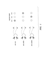
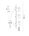
- the nucleic acid labels each contain three pre-determined subsequences (each of one nucleotide) conjugated together by a direct bond such as a phosphodiester bond.
- Each sequencing step decodes or determines one nucleotide, wherein each nucleobase (A, G, C or T) generates a distinct signal signature corresponding to the nucleobase. Consequently, a temporal order or time series of the signal signatures generated from each sequencing step corresponds to the respective probe reagent, and thus identify the target analyte.
- the number of sequencing steps performed is not necessarily equal to the number of the pre-determined sequences. In some embodiments, the number of sequencing steps performed can be less than the number of the pre-determined sequences. For example, as shown in Fig. 1 , two sequencing steps could be sufficient to identify the three different probe reagents, where each base is associated with a different color.
- n-base subsequences e.g., 3-base subsequences shown in Fig. 1 or Fig. 2
- 4 n possible unique readouts i.e., 4 n possible distinct probe reagents can be distinguished using such detection reagents described herein.
- the detection step can comprise hybridizing a decoder probe with a subsequence on the nucleic acid label of the detection reagent, wherein the decoder probe can comprise a detectable label.
- the detection method can comprise: (a) hybridizing a set of decoder probes with a subsequence of the detection reagents, wherein each subpopulation of the decoder probes can comprise a detectable label, each detectable label producing a signal signature; (b) detecting said signal signature produced by the hybridization of said set of decoder probes; and (d) repeating steps (a) and (b) for other subsequences of said detection reagents.
- each subpopulation of the decoder probes can comprise a different detectable label, each different detectable label producing a different signal signature.
- the different signal signature produced by the hybridization of the set of decoder probes can be detected.
- each subpopulation of the decoder probes can be complementary (e.g., partially complementary or completely complementary) to the subsequence of the detection reagents.
- a first subpopulation and a second subpopulation of the decoder probes can be complementary (e.g., partially complementary or completely complementary) to distinct subsequences of the detection reagents.
- at least two or more subpopulations of the decoder probes can bind to the same subsequence of the detection reagents.
- a first subpopulation and a second subpopulation of the decoder probes can be complementary (e.g., partially complementary or completely complementary) to the same subsequence of the detection reagents.
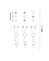
- Fig. 5 shows an exemplary detection reagent comprising anti-C. albicans probe reagents and nucleic acid labels containing three hybridization sites or pre-determined subsequences conjugated together by sequence linkers.
- a first set of decoder probes each comprising a distinct detectable label (e.g., complementary DNA readout probes shown in Fig. 5 , each comprising a distinct optical label) is hybridized with a first pre-determined subsequence (e.g., Site 1 in Fig. 5 ), followed by detection of a first signal signature produced by the hybridization.
- a first pre-determined subsequence e.g., Site 1 in Fig. 5
- a second set of decoder probes each comprising a distinct detectable label is hybridized with a second pre-determined subsequence (e.g., Site 2 in Fig. 5 ), followed by detection of a second signal signature produced by the hybridization.
- the second pre-determined subsequence can be the same or different from the first pre-determined subsequence. However, in preferred embodiments, the second pre-determined subsequence is different from the first pre-determined subsequence, e.g., to minimize cross-hybridization with each other.
- the hybridization and signal detection steps are repeated for other subsequences of the detection reagents with a different set of decoder probes, thereby producing a temporal order or time series of the signal signatures corresponding to the respective probe reagent (and detection reagent).
- the term “decoder probe” refers an oligonucleotide with a sequence complementary to a pre-determined sequence of the nucleic acid label.
- “complementary” is meant that a nucleic acid can form hydrogen bond(s) with another nucleic acid sequence by either traditional Watson-Crick or other non- traditional types.
- the decoder probe sequence can be completely or partially complementary to a pre-determined sequence.
- partial complementarity is indicated by the percentage of contiguous residues in a nucleic acid molecule that can form hydrogen bonds (e.g., Watson-Crick base pairing) with a second nucleic acid sequence (e.g., 5, 6, 7, 8, 9,10 out of 10 being 50%, 60%, 70%, 80%, 90%, and 100% complementary).
- "completely complementary" or 100% complementarity means that all the contiguous residues of a nucleic acid sequence will hydrogen bond with the same number of contiguous residues in a second nucleic acid sequence.
- Less than perfect complementarity refers to the situation in which some, but not all, nucleoside units of two strands can hydrogen bond with each other.
- the decoder probe can have a sequence of any length. In some embodiments, the decoder probe can have a sequence length of about 1 to about 100 nucleotides, about 1 to about 50 nucleotides, about 2 to about 50 nucleotides, about 5 to about 30 nucleotides, or about 5 to about 20 nucleotides.
- the decoder probe can comprise at least one detectable label described herein.
- the detectable label can be an optical label selected from the group consisting of a small-molecule dye, a fluorescent molecule or protein, a quantum dot, a colorimetric reagent, a chromogenic molecule or protein, a Raman label, and any combinations thereof.
- the detectable label or optical label can be a fluorescent molecule or protein.
- the decoder probe can be modified, e.g., base modification or activated with a functional group for linkage to a detectable label.
- the number of decoder probes in each set can vary, depending on the number of distinct subsequences in each hybridization. In some embodiments, there can be about 1 to about 100 decoder probes, about 2 to about 50 decoder probes, about 4 to about 20 decoder probes in each set. In some embodiments, there can be about 1, 2, 3, 4, 5, 6, 7, 8, 9, 10, 11, 12, 13, 14, 15, 16, 17, 18, 19, 20, 30, 40, 50 or more decoder probes in each set. In some embodiments, while each subsequence site can hybridize with a large number of decoder probes, each set of decoder probes added in each readout step to hybridize with the subsequence site within the detection reagents can generally have as many as the number of available fluorescent colors.
- each set of the decoder probes added in each readout step to hybridize with the subsequence site of the detection reagents can contain about 3-4 decoder probes, each of which is labeled with a distinct fluorescent color.
- non-overlapping nucleic acid labels or “non-overlapping SeqTag labels” (i.e., no two decoder probes will hybridize with the same spatial site of the nucleic acid label) is shown herein for illustrative purposes. Assuming there are 12 pre-determined subsequences (e.g., nucleic acid sequences) designed for minimal cross-hybridization with each other and each others' complements (e.g., A1, A2, A3, A4, B1, B2, B3, B4, C1, C2, C3, and C4).
- subsequences e.g., nucleic acid sequences
- a detection reagent comprising a nucleic acid label of the form: 5' ------A[1-4]----B[1-4]----C[1-4]---- 3' where each position (e.g., A[1-4]) holds only one of the subsequences (e.g., A2).
- this subsequence is decoded or detected by using decoder probes or complementary probes A1* - A4* each labeled in one of four fluorescent colors.
- a single site (e.g., A in the above nucleic acid label form), which can accept one of the different decoder probes, can be used.
- This single site can be read out by subjecting it to the different decoder probes, e.g., in sets of 4 using the nucleic acid label form as shown above.
- the nucleic acid label e.g., SeqTag label
- hybridization-based readout method can reduce cost, reduce reagent storage and/or simplify the process.
- the spatial movement of an analyte in a sample during a temporal detection of the detection reagents can be less than 100 ⁇ m, including less than 50 ⁇ m, less than 25 ⁇ m, less than 10 ⁇ m, less than 1 ⁇ m or smaller, over a time period, during which a temporal detection of the detection reagents occurs.
- the spatial movement of an analyte in a sample can be less than 1000 nm, including less than 500 nm, less than 250 nm, less than 100 nm, less than 50 nm, less than 10 nm or smaller, over a time period, during which a temporal detection of the detection reagents occurs. More importantly, the spatial movement limit of an analyte in a sample during a temporal detection is determined by the ability of matching distinguishable features between images taken during a temporal detection, which can be affected by imaging conditions. In some embodiments, the analyte can be fixed on a solid substrate or support.
- the location of the analyte with respect to a sample during each detection step can be determined and registered. Such spatial shift can then be corrected afterward during signal analysis using any art-recognized computer-implemented algorithms.
- the length of time required to perform a temporal detection of the detection reagents can vary, depending on the number of pre-determined subsequences to be detected or read and/or the number of available detection signals (e.g., fluorescent color and/or brightfield) to be read.
- the length of time required to perform a temporal detection of the detection reagents can be, for example, but not limited to, 5 seconds, 10 seconds, 15 seconds, 20 seconds, 30 seconds, 1 mins, 2 mins, 3 mins, 4 mins, 5 mins, 15 mins, 30 mins, 1 hour, 2 hours, 4 hours, 6 hours, or longer.
- the detection reagents can be detected by any means available in the art that is capable of detecting the specific signals on a given detection reagent generated during sequencing- or hybridization-based methods.
- the detection reagents e.g., hybridized with decoder probes
- suitable consideration of appropriate excitation sources can be readily determined. Possible sources can include but are not limited to arc lamp, xenon lamp, lasers, light emitting diodes or some combination thereof.
- the appropriate excitation source is used in conjunction with an appropriate optical detection system, for example an inverted fluorescent microscope, an epi-fluorescent microscope or a confocal microscope.
- a microscope is used that can allow for detection with enough spatial resolution to separate distinct signals from individual detection reagents.
- Exemplary methods for detection of the detection reagents that are applicable to the methods described herein include, without limitations, the methods described in U.S. Pat. No. 7,473,767 , US patent publication no. 2007/0166708 , and US application number US 2010/0261026 , all of which are incorporated by reference herein in its entirety.
- Additional methods that can be used to detect optical signatures include, but are not limited to, any spectroscopic techniques, flow cytometry, or any art-recognized methods involving an optical scanner and/or a photodetector (e.g., without limitations, a charge-coupled devices, active pixel sensors, photodiode light sensors (e.g., LEDs), optical detectors, and any combinations thereof).
- Non-limiting examples of spectroscopic techniques can include absorption spectroscopy, emission spectroscopy, elastic scattering spectroscopy, reflection spectroscopy, impedance spectroscopy, inelastic spectroscopy, coherent or resonance spectroscopy, surface plasmon fluorescence spectroscopy, Raman spectroscopy, and any combinations thereof.
- Spectroscopy techniques can be used to detect light of any wavelengths, including, but not limited to, microwave, terahertz, infrared, near infrared, visible, ultraviolet, x-ray, gamma, and any
- At least one pre-determined subsequence can correspond to no optical signature; that is the absence of color can be considered as an additional color.
- at least one pre-determined subsequence e.g., individual bases or hybridization regions
- the nucleic acid label can be designed such that any known or potential overlaps could be teased apart from the signal output.
- any probe may potentially overlap with all others, one can use the readout-by-hybridization variation and assign each probe a single unique hybridization sequence. Such approach can avoid multiple lengthy probe incubations and damaging stripping steps.
- the signal signatures produced during any readout step should be removed before advancing to the next pre-determined subsequence of the detection reagents.
- the removal of the signal signatures can be done by any methods known in the art, including, but not limited to, washing, heating, photo-bleaching, displacement, cleavage, enzymatic digestion, quenching, chemical degradation, bleaching, oxidation, and any combinations thereof.
- the decoder probes can be designed such that they can be simply washed out either with a plain buffer, or they can be modified by varying salt concentrations or using detergents or denaturants such as formamide or dimethyl sulfoxide (DMSO).
- DMSO dimethyl sulfoxide
- the fluorescence or color signature of a readout step can be attenuated or eliminated by photo-bleaching the signal using sufficient optical exposure.
- the fluorescence or color signature of a readout step can be attenuated or eliminated by subjecting the fluorescence or color signature to chemical degradation under appropriate conditions, e.g., using a reducing agent or oxidizing solution such as 0.01 M sodium periodate.
- the decoder probes can be displaced from their hybridization sites by introducing other reagents or probe displacers that have stronger binding affinities to those same sites. This can be done, for example, by using nucleic acid sequences that are longer than and/or have better complementarity than the decoder probe sequences. For example, to create "better complementarity" of the probe displacers, in some embodiments, mismatches can be seeded in the hybridization region. In other embodiments, the hybridization region can be preceded and/or post-ceded with a "toe-hold" of around 3-8 bases (e.g., 6 bases). In such embodiments, the "better complementarity" of the probe displacer can be outside of the hybridization region, which can make the design of the hybridization regions easier.
- enzymes can be used to displace, digest, cut and/or cleave the detectable labels, the decoder probe sequence, the hybridized complex (formed by the decoder probe and pre-determined subsequence), and/or the cleavable sequence linker to the hybridized complex, in order to remove the signal signatures.
- One example is to introduce a deoxyuridine into the decoder probe. This modified base can be cleaved using the enzyme mix known as USER, thereby cutting the decoder probe sequence into two parts. Since each part is now shorter, it is characterized by a lower melting temperature and can melt off the hybridization sites of the detection reagents.
- one of skill in the art can employ one of numerous art-recognized sequence-specific nucleases or restriction enzymes, which can cut either the decoder probe sequence, the pre-determined subsequence that has been hybridized with decoder probes, the cleavable sequence linker attached to the hybridized subsequence and/or the hybridized complex thereof, thereby removing the signal signature.
- thermal denaturing can be used to remove the signal signature from a previous readout.
- thermal denaturing can be reduced or avoided by using a sequencing-by-ligation approach and by setting the nucleic acid label base that immediately follows the sequencing primer (in the direction of ligation) to an adenine.
- the ligation probes should then include a (deoxy-)uracil in the primer-proximal position.
- an enzyme such as USER, which cleaves DNA at uracils can be used to remove the ligation probe and ready the system for the next sequencing step. These fragments will have lower melting temperatures than their parent probes, and these temperatures can be designed to fall below the operating temperature (or require an acceptable denaturing temperature).
- the method described herein can further comprise comparing the temporal order of the signal signatures with different identifiers of said at least one probe reagent, wherein an agreement between the temporal order of the signal signatures and a particular identifier of said at least one probe reagent identifies the analyte in the sample.
- the method can further comprise measuring the intensity of the signal signatures generated from each subpopulation of the detection reagents.
- the intensity of the signal signatures generated from each subpopulation of the detection reagents can indicate an amount of the analyte.
- the relative intensity of the signal signatures can be used in identification of each subpopulation of the detection reagents.
- the intensity of the signal signatures can be used as part of a coding scheme of the detection reagents described herein.
- the comparing and intensity measuring steps can be performed, e.g., by a computer-implemented software or algorithm.
- signal signature(s) can vary upon different embodiments of detection reagents and/or decoder probes described herein.
- the term "signal signature" refers to a change in, or occurrence of, a response or indicator that is detectable either by observation or instrumentally.
- the signal signature is fluorescence or a change in fluorescence, e.g., a change in fluorescence intensity, fluorescence excitation or emission wavelength distribution, fluorescence lifetime, and/or fluorescence polarization.
- the fluorescence can be produced by binding a fluorophore to a decoder probe, and/or by detecting the hybridization using a fluorescent dye, e.g., SYBR Gold, that lights up when nucleic acid sequence becomes double-stranded.
- the signal signature can be radioactivity (i.e., radiation), including alpha particles, beta particles, nucleons, electrons, positrons, neutrinos, and gamma rays emitted by a radioactive substance such as a radionuclide.
- the detection reagents and/or decoder probes can comprise an optical molecule or label, thus producing optical signatures. Examples of optical signatures can include, without limitations, signatures of fluorescent color, visible light, no-color, and any combinations thereof. In such embodiments, the optical signatures can be detected by optical imaging or spectroscopy.
- Detection reagents or detection molecules as used interchangeably herein
- the detection reagent comprises at least one probe reagent and at least one nucleic acid label, wherein said at least one nucleic acid label comprises at least one pre-determined subsequence to be detected in a temporally-sequential manner; wherein said at least one pre-determined subsequence forms an identifier of said at least one probe reagent; and wherein said at least one probe reagent and said at least one nucleic acid label are conjugated together.
- the detection reagent can exist in different forms.
- the detection reagent can be a detection molecule.
- the detection reagent can be a detection particle.
- the detection reagent can be multi-molecular.
- conjugated refers to two molecules being linked to each other, e.g., attaching a probe reagent to a nucleic acid label.
- the conjugation process can be performed, e.g., via a chemical reaction, or via a linker, which will be described later.
- a readout signal of a detection reagent can be amplified by increasing the number of the nucleic acid labels present in the detection reagent, e.g., by conjugating at least one probe reagent to a plurality of nucleic acid labels.
- a plurality of the nucleic acid labels present in the detection reagent can range from about 2 to about 100,000, about 2 to about 10,000, about 2 to about 1,000, or about 2 to about 100.
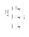
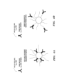
- the detection reagent comprises a particle as a hub
- the number of possible nucleic acid labels present in the detection reagent can depend on the size of a particle.
- a particle of about 1-2 ⁇ m in size can allow incorporation of about 100,000 nucleic acid labels into the detection reagent.
- One of skill in the art can determine the optimum number of nucleic acid labels present the detection reagent without any undue experimentation.
- the detection reagents described herein can be used in any biological assays for detection, identification and/or quantification of target molecules or analytes, including counting marked cells such as bacteria or cancer cells, in a sample.
- the detection reagent can be adapted for use in immunofluorescence.
- the detection reagent can be adapted for use in immunohistochemistry.
- the detection reagent can be adapted for use in fluorescence in situ hybridization.
- the detection reagent can be adapted for use in western blot.
- the detection reagent can be adapted to be in any format, e.g., immobilized on a solid support, or in a solution or suspension phase. In certain embodiments, the detection reagent can be adapted to be present in a solution or suspension phase.
- the phrase "in a solution or suspension phase" as used herein generally refers to suspending the detection reagents in a liquid fluid, e.g., an aqueous buffer solution. Additional applications of the detection reagents and/or methods described herein will be discussed.
- Probe reagents or probe molecules as used interchangeably herein
- the detection reagent can comprise one or more probe reagents, e.g., at least 1, at least 2, at least 3, at least 4, at least 5, at least 6, at least 7, at least 8, at least 9, at least 10 or more probe reagents.
- the detection reagent can comprise one probe reagent.
- the detection reagent can comprise a plurality of probe reagents, e.g., ranging from about 2 to about 100,000 probe reagents, about 2 to about 10,000 probe reagents, about 2 to about 1,000 probe reagents, or about 2 to about 100 probe reagents.
- the number of possible probe reagents present in the detection reagent can depend on the size of a particle. Generally, the larger the particle it is, the more probe reagents can be incorporated into the detection reagent. For example, a particle of about 1-2 ⁇ m in size can allow incorporation of about 100,000 probe reagents into the detection reagent. In some embodiments, there can be about 2, 3, 4, 5, 6, 7, 8, 9, 10, 11, 12, 13, 14, 15, 16, 17, 18, 19, 20, 50, 100, 500, 1000, 5000, 10000, 50000, 100000 probe reagents present in the detection reagent. One of skill in the art can determine the optimum number of probe reagents present the detection reagent without any undue experimentation.
- the term "probe,” “probe reagent” or “probe molecule” refers to an entity (e.g., but not limited to, a molecule, a particle, a composite entity, or a multi-molecular entity) that interacts with or binds to a target molecule or an analyte for the analysis of the target or the analyte.
- an entity e.g., but not limited to, a molecule, a particle, a composite entity, or a multi-molecular entity
- the nature of the interaction or binding is noncovalent, e.g., by hydrogen, electrostatic, or van der Waals interactions, however, binding can also be covalent.
- Probe reagents can be entities (e.g., but not limited to, molecules, a particles, composite entities, or multi-molecular entities) capable of undergoing binding or molecular recognition events with target molecules. Probe reagents can be naturally-occurring, recombinant or synthetic. Examples of the probe reagent can include, but are not limited to, a nucleic acid, an antibody or a portion thereof, an antibody-like molecule, an enzyme, a cell, an antigen, a small molecule, a protein, a peptide, a peptidomimetic, an aptamer, and any combinations thereof. By way of example only, in immunohistochemistry, the probe reagent can include an antibody specific to the target antigen to be analyzed.
- the probe reagent can be multi-molecular.
- the probe reagent can comprise a particle, an antibody, biotin and/or streptavidin, or any combinations thereof.
- the probe reagents can be modified by any means known to one of ordinary skill in the art. Methods to modify each type of probe reagents are well recognized in the art. Depending on the types of probe reagents, an exemplary modification includes, but is not limited to genetic modification, biotinylation, labeling (for detection purposes), chemical modification (e.g., to produce derivatives or fragments of the probe reagent), and any combinations thereof.
- an exemplary modification includes, but is not limited to genetic modification, biotinylation, labeling (for detection purposes), chemical modification (e.g., to produce derivatives or fragments of the probe reagent), and any combinations thereof.
- the probe reagent can be genetically modified.
- the probe reagent can be biotinylated.
- proteins and “peptides” are used interchangeably herein to designate a series of amino acid residues connected to the other by peptide bonds between the alpha-amino and carboxy groups of adjacent residues.
- protein and “peptide”, which are used interchangeably herein, refer to a polymer of protein amino acids, including modified amino acids (e.g., phosphorylated, glycated, etc.) and amino acid analogs, regardless of its size or function.
- modified amino acids e.g., phosphorylated, glycated, etc.
- amino acid analogs regardless of its size or function.
- peptide refers to peptides, polypeptides, proteins and fragments of proteins, unless otherwise noted.
- protein and “peptide” are used interchangeably herein when referring to a gene product and fragments thereof.
- exemplary peptides or proteins include gene products, naturally occurring proteins, homologs, orthologs, paralogs, fragments and other equivalents, variants, fragments, and analogs of the foregoing.
- peptidomimetic refers to a molecule capable of folding into a defined three-dimensional structure similar to a natural peptide
- nucleic acids refers to polymers (polynucleotides) or oligomers (oligonucleotides) of nucleotide or nucleoside monomers consisting of naturally occurring bases, sugars and intersugar linkages.
- nucleic acid also includes polymers or oligomers comprising non-naturally occurring monomers, or portions thereof, which function similarly.
- Exemplary nucleic acids include, but are not limited to, deoxyribonucleic acid (DNA), ribonucleic acid (RNA), locked nucleic acid (LNA), peptide nucleic acids (PNA), and polymers thereof in either single- or double-stranded form.
- Locked nucleic acid is a modified RNA nucleotide.
- the ribose moiety of an LNA nucleotide is modified with an extra bridge connecting the 2' oxygen and 4' carbon. The bridge "locks" the ribose in the 3'-endo conformation.
- LNA nucleotides can be mixed with DNA or RNA residues in the oligonucleotide whenever desired. Such LNA oligomers are generally synthesized chemically.
- Peptide nucleic acid (PNA) is an artificially synthesized polymer similar to DNA or RNA.
- nucleic acids encompasses nucleic acids containing known analogs of natural nucleotides, which have similar binding properties as the reference nucleic acid and are metabolized in a manner similar to naturally occurring nucleotides. Unless otherwise indicated, a particular nucleic acid sequence also implicitly encompasses conservatively modified variants thereof (e.g., degenerate codon substitutions) and complementary sequences, as well as the sequence explicitly indicated.
- degenerate codon substitutions may be achieved by generating sequences in which the third position of one or more selected (or all) codons is substituted with mixed-base and/or deoxyinosine residues ( Batzer, et al., Nucleic Acid Res. 19:5081 (1991 ); Ohtsuka, et al., J. Biol. Chem. 260:2605-2608 (1985 ), and Rossolini, et al., Mol. Cell. Probes 8:91-98 (1994 )).
- nucleic acid should also be understood to include, as equivalents, derivatives, variants and analogs of either RNA or DNA made from nucleotide analogs, and, single (sense or antisense) and double-stranded polynucleotides.
- nucleic acid can include a modified nucleic acid.
- Modified nucleic acids are well known in the art.
- a nucleic acid described herein can comprise one or more nucleic acid modifications known in the art.
- the nucleic acid can comprise one or more nucleic acid modifications selected from the group consisting of internucleotide linkage modifications (intersugar linkage modifications), sugar modifications, nucleobase modifications, backbone modifications/replacements, and any combinations thereof.
- Exemplary internucleotide linkage modifications include, but are not limited to, phosphorothioate, phosphorodithioate, phosphotriester (e.g.
- alkyl phosphotriester alkyl phosphotriester
- aminoalkylphosphotriester alkyl-phosphonate (e.g., methyl-phosphonate), selenophosphate, phosphoramidate (e.g., N-alkylphosphoramidate), boranophosphonate, and the like.
- Exemplary sugar modifications include, but are not limited to, 2'- O -Me (2'-O-methyl), 2'- O -MOE (2'- O- methoxyethyl), 2'-F, 2'-O-[2-(methylamino)-2-oxoethyl] (2'- O -NMA), 2'- S -methyl, 2'-O-CH 2 -(4'-C) (LNA), 2'-O-CH 2 CH 2 -(4'-C) (ENA), 2'-O-aminopropyl (2'-O-AP), 2'-O-dimethylaminoethyl (2'-O-DMAOE), 2'-O-dimethylaminopropyl (2'-O-DMAP), 2'-O-dimethylaminoethyloxyethyl (2'-O-DMAEOE), arabinose sugar, and the like.
- nucleobase modifications include, but are not limited to, inosine, xanthine, hypoxanthine, nubularine, isoguanisine, tubercidine, 5-methylcytosine (5-me-C); 5-hydroxymethyl cytosine; xanthine; hypoxanthine; 2-aminoadenine; 6-methyl and other 6-alkyl derivatives of adenine and guanine; 2-propyl and other 2-alkyl derivatives of adenine and guanine; 2-thiouracil; 2-thiothymine; 2-thiocytosine; 5-propynyl uracil; 5-propynyl cytosine; 6-azouracil; 6-azocytosine; 6-azothymine; 5-uracil (pseudouracil); 4-thiouracil; 8-halo, 8-amino, 8-thiol, 8-thioalkyl, 8-hydroxyl and other 8-substituted adenines and
- Exemplary backbone modifications include, but are not limited to, morpholino, cyclobutyl, pyrrolidine, peptide nucleic acid (PNA), aminoethylglycyl PNA ( aeg PNA), backnone-extended pyrrolidine PNA (bepPNA), and the like.
- PNA peptide nucleic acid
- aeg PNA aminoethylglycyl PNA
- bepPNA backnone-extended pyrrolidine PNA
- enzymes refers to a protein molecule that catalyzes chemical reactions of other substances without it being destroyed or substantially altered upon completion of the reactions.
- the term can include naturally occurring enzymes and bioengineered enzymes or mixtures thereof. Examples of enzyme families include kinases, dehydrogenases, oxidoreductases, GTPases, carboxyl transferases, acyl transferases, decarboxylases, transaminases, racemases, methyl transferases, formyl transferases, and ⁇ -ketodecarboxylases.
- aptamers means a single-stranded, partially single-stranded, partially double-stranded or double-stranded nucleotide sequence capable of specifically recognizing a selected non-oligonucleotide molecule or group of molecules. In some embodiments, the aptamer recognizes the non-oligonucleotide molecule or group of molecules by a mechanism other than Watson-Crick base pairing or triplex formation.
- Aptamers can include, without limitation, defined sequence segments and sequences comprising nucleotides, ribonucleotides, deoxyribonucleotides, nucleotide analogs, modified nucleotides and nucleotides comprising backbone modifications, branchpoints and nonnucleotide residues, groups or bridges. Methods for selecting aptamers for binding to a molecule are widely known in the art and easily accessible to one of ordinary skill in the art.
- antibody refers to an intact immunoglobulin or to a monoclonal or polyclonal antigen-binding fragment with the Fc (crystallizable fragment) region or FcRn binding fragment of the Fc region.
- antibody-like molecules such as fragments of the antibodies, e.g., antigen-binding fragments. Antigen-binding fragments can be produced by recombinant DNA techniques or by enzymatic or chemical cleavage of intact antibodies.
- Antigen-binding fragments include, inter alia, Fab, Fab', F(ab')2, Fv, dAb, and complementarity determining region (CDR) fragments, single-chain antibodies (scFv), single domain antibodies, chimeric antibodies, diabodies, and polypeptides that contain at least a portion of an immunoglobulin that is sufficient to confer specific antigen binding to the polypeptide. Linear antibodies are also included for the purposes described herein.
- the terms Fab, Fc, pFc', F(ab') 2 and Fv are employed with standard immunological meanings ( Klein, Immunology (John Wiley, New York, N.Y., 1982 ); Clark, W. R.
- Antibodies or antigen-binding fragments specific for various antigens are available commercially from vendors such as R&D Systems, BD Biosciences, e-Biosciences and Miltenyi, or can be raised against these cell-surface markers by methods known to those skilled in the art.
- CDRs Complementarity Determining Regions
- Each variable domain typically has three CDR regions identified as CDR1, CDR2 and CDR3.
- Each complementarity determining region may comprise amino acid residues from a "complementarity determining region” as defined by Kabat (i.e. about residues 24-34 (L1), 50-56 (L2) and 89-97 (L3) in the light chain variable domain and 31-35 (H1), 50-65 (H2) and 95-102 (H3) in the heavy chain variable domain; Kabat et al.
- a complementarity determining region can include amino acids from both a CDR region defined according to Kabat and a hypervariable loop.
- linear antibodies refers to the antibodies described in Zapata et al. , Protein Eng., 8(10):1057-1062 (1995 ). Briefly, these antibodies comprise a pair of tandem Fd segments (VH -CH1-VH-CH1) which, together with complementary light chain polypeptides, form a pair of antigen binding regions. Linear antibodies can be bispecific or monospecific.
- single-chain Fv or "scFv” antibody fragments, as used herein, is intended to mean antibody fragments that comprise the VH and VL domains of antibody, wherein these domains are present in a single polypeptide chain.
- the Fv polypeptide further comprises a polypeptide linker between the VH and VL domains which enables the scFv to form the desired structure for antigen binding.
- Plückthun The Pharmacology of Monoclonal Antibodies, vol. 113, Rosenburg and Moore eds., Springer- Verlag, New York, pp. 269-315 (1994 )).
- diabodies refers to small antibody fragments with two antigen-binding sites, which fragments comprise a heavy-chain variable domain (VH) Connected to a light-chain variable domain (VL) in the same polypeptide chain (VH - VL).
- VH heavy-chain variable domain
- VL light-chain variable domain
- the domains are forced to pair with the complementary domains of another chain and create two antigen-binding sites.
- small molecules refers to natural or synthetic molecules including, but not limited to, peptides, peptidomimetics, amino acids, amino acid analogs, polynucleotides, polynucleotide analogs, aptamers, nucleotides, nucleotide analogs, organic or inorganic compounds (i.e., including heteroorganic and organometallic compounds) having a molecular weight less than about 10,000 grams per mole, organic or inorganic compounds having a molecular weight less than about 5,000 grams per mole, organic or inorganic compounds having a molecular weight less than about 1,000 grams per mole, organic or inorganic compounds having a molecular weight less than about 500 grams per mole, and salts, esters, and other pharmaceutically acceptable forms of such compounds.
- organic or inorganic compounds i.e., including heteroorganic and organometallic compounds
- cells refers to any cell, prokaryotic or eukaryotic, including plant, yeast, worm, insect and mammalian.
- Mammalian cells include, without limitation; primate, human and a cell from any animal of interest, including without limitation; mouse, hamster, rabbit, dog, cat, domestic animals, such as equine, bovine, murine, ovine, canine, feline, etc.
- the cells may be a wide variety of tissue types without limitation such as; hematopoietic, neural, mesenchymal, cutaneous, mucosal, stromal, muscle spleen, reticuloendothelial, epithelial, endothelial, hepatic, kidney, gastrointestinal, pulmonary, T-cells etc.
- Stem cells, embryonic stem (ES) cells, ES- derived cells and stem cell progenitors are also included, including without limitation, hematopoeitic, neural, stromal, muscle, cardiovascular, hepatic, pulmonary, gastrointestinal stem cells, etc.
- Yeast cells may also be used as cells in some embodiments of the methods described herein.
- the cells can be ex vivo or cultured cells, e.g. in vitro.
- cells can be obtained from a subject, where the subject is healthy and/or affected with a disease.
- Cells can be obtained, as a non-limiting example, by biopsy or other surgical means know to those skilled in the art.
- the term "antigens” refers to a molecule or a portion of a molecule capable of being bound by a selective binding agent, such as an antibody, and additionally capable of being used in an animal to elicit the production of antibodies capable of binding to an epitope of that antigen.
- An antigen may have one or more epitopes.
- the term “antigen” can also refer to a molecule capable of being bound by an antibody or a T cell receptor (TCR) if presented by MHC molecules.
- TCR T cell receptor
- the term "antigen”, as used herein, also encompasses T-cell epitopes.
- An antigen is additionally capable of being recognized by the immune system and/or being capable of inducing a humoral immune response and/or cellular immune response leading to the activation of B- and/or T-lymphocytes. This may, however, require that, at least in certain cases, the antigen contains or is linked to a Th cell epitope and is given in adjuvant.
- An antigen can have one or more epitopes (B- and T-epitopes). The specific reaction referred to above is meant to indicate that the antigen will preferably react, typically in a highly selective manner, with its corresponding antibody or TCR and not with the multitude of other antibodies or TCRs which may be evoked by other antigens. Antigens as used herein may also be mixtures of several individual antigens.
- the probe reagent can be an antibody or a portion thereof, or an antibody-like molecule.
- the probe reagents can be used to, for example, detect and/or identify pathogen type or speices, the presence of cell or disease markers, cellular protein expression levels, phosphorylation or other post-translation modification state, or any combinations thereof.
- Fig. 1 shows three different embodiments of the detection reagents comprising at least one (e.g., 1, 2, 3, 4, 5 or more) pathogen-specific antibodies (e.g., anti-E. Coli, anti-S. aureus, and anti-C. albicans).
- the probe reagent can be a nucleic acid (e.g., DNA, RNA, LNA, PNA, or any combinations thereof).
- the nucleic acids can be used to determine, for example, the existence of characteristic cellular DNA or RNA sequences (such as in fluorescent in situ hybridization), RNA expression levels, miRNA presence and expression, and any combinations thereof, in various applications, e.g., for pathogen detection and/or identification.
- the probe reagent can be a protein or a peptide.
- the protein or peptide can be essentially any proteins with known binding targets. Examples include, but are not limited to, innate-immune proteins (e.g., without limitations, MBL, Dectin-1, TLR2, and TLR4 and any proteins disclosed in U.S. Provisional Application No.: 61/508,957 , the content of which is incorporated herein by reference in its entirety) and proteins comprising the chitin-binding domain.
- innate-immune proteins and chitin-binding domain proteins can be used to detect their corresponding pattern-recognition targets (e.g., microbes such as bacteria) and fungus, respectively.
- innate-immune proteins e.g., MBL
- chitin-binding domain proteins can be used as probe reagents for detection of pathogens. While such detection reagents can be used to detect pathogens, they may not be pathogen-specific, as compared to the ones using pathogen-specific antibodies as probes molecules.
- the probe reagent can be an aptamer.
- the probe reagent can be a DNA or RNA aptamer.
- the aptamers can be used in various bioassays, e.g., in the same way as antibodies or nucleic acids described herein.
- Fig. 2 shows some exemplary embodiments of the detection reagents comprising at least one (e.g., 1, 2, 3, 4, 5 or more) DNA aptamers (e.g., with a nucleotide sequence complementary to nuclear reprogramming factors, such as Oct4, Sox2, and Klf4).
- Such detection reagents can be used to determine RNA expression level of nuclear reprogramming factors in somatic cells for detecting, screening, or identifying stem cells (e.g., induced pluripotency stem cells).
- the probe reagent can be a cell surface receptor ligand.
- a "cell surface receptor ligand” refers to a molecule that can bind to the outer surface of a cell.
- cell surface receptor ligand includes, for example, a cell surface receptor binding peptide, a cell surface receptor binding glycopeptide, a cell surface receptor binding protein, a cell surface receptor binding glycoprotein, a cell surface receptor binding organic compound, and a cell surface receptor binding drug.
- Additional cell surface receptor ligands include, but are not limited to, cytokines, growth factors, hormones, antibodies, and angiogenic factors.
- the probe reagent can be an endosomolytic ligand.
- endosomolytic ligand refers to molecules having endosomolytic properties. Endosomolytic ligands can promote the lysis of and/or transport of the composition described herein, or its components, from the cellular compartments such as the endosome, lysosome, endoplasmic reticulum (ER), Golgi apparatus, microtubule, peroxisome, or other vesicular bodies within the cell, to the cytoplasm of the cell.
- Some exemplary endosomolytic ligands include, but are not limited to, imidazoles, poly or oligoimidazoles, linear or branched polyethyleneimines (PEIs), linear and branched polyamines, e.g. spermine, cationic linear and branched polyamines, polycarboxylates, polycations, masked oligo or poly cations or anions, acetals, polyacetals, ketals/polyketals, orthoesters, linear or branched polymers with masked or unmasked cationic or anionic charges, dendrimers with masked or unmasked cationic or anionic charges, polyanionic peptides, polyanionic peptidomimetics, pH-sensitive peptides, natural and synthetic fusogenic lipids, natural and synthetic cationic lipids.
- PEIs polyethyleneimines
- linear and branched polyamines e.g. spermine, cationic linear and branched poly
- the probe reagent for use in delivery of an agent (e.g., a diagnostic agent) encapsulated within the detection reagents described herein can be a PK modulating ligand.
- PK modulating ligand and “PK modulator” refers to molecules which can modulate the pharmacokinetics of the composition described herein.
- Some exemplary PK modulator include, but are not limited to, lipophilic molecules, bile acids, sterols, phospholipid analogues, peptides, protein binding agents, vitamins, fatty acids, phenoxazine, aspirin, naproxen, ibuprofen, suprofen, ketoprofen, (S)-(+)-pranoprofen, carprofen, PEGs, biotin, and transthyretia-binding ligands (e.g., tetraiidothyroacetic acid, 2, 4, 6-triiodophenol and flufenamic acid).
- lipophilic molecules bile acids, sterols, phospholipid analogues, peptides, protein binding agents, vitamins, fatty acids, phenoxazine, aspirin, naproxen, ibuprofen, suprofen, ketoprofen, (S)-(+)-pranoprofen, car
- the detection reagent described herein can comprise one kind/species of probe reagents or different kinds/species of probe reagents.
- the kind/species of the probe reagents present in the detection reagent can be the same.
- the detection reagent can include at least one different kind/species of the probe reagents (e.g., 1, 2, 3, 4, 5, or 6 probe reagent species).
- the distinct probe reagent species can be different from the others by types (e.g., antibodies vs. DNA aptamers), binding domains, and/or target analytes.
- the nucleic acid label or nucleic acid tag comprises at least one pre-determined nucleic acid subsequence, which is used to identify an analyte or target.
- the nucleic acid label or nucleic acid tag can comprise any number of the pre-determined nucleic acid subsequences, e.g., ranging from about 1 to about 100, from about 2 to about 80, or from about 3 to about 50.
- the nucleic acid label or nucleic acid tag can comprise at least about 1, at least about 2, at least about 3, at least about 4, at least about 5, at least about 6, at least about 7, at least about 8, at least about 9, at least about 10, at least about 15, at least about 20, at least about 30, at least about 40, at least about 50, at least about 60, at least about 70 or more pre-determined nucleic acid subsequences. In some embodiments, the nucleic acid label or nucleic acid tag can comprise 1, 2, 3, 4, 5, 6, 7, 8, 9, 10, 15, 20, 30, 40, 50, 60, 70 or more pre-determined nucleic acid subsequences.
- n ceiling ln X ln Y + 1 , and thus fewer pre-determined nucleic acid subsequences can be used. However, the combination in which all readout cycles are dark should not be allowed.
- the pre-determined nucleic acid subsequences can be designed such that each readout cycle can light up in multiple colors.
- 2 Y instead of Y is used in the equation for n above and fewer pre-determined nucleic acid subsequences can thus be required.
- the combination in which all readout cycles are dark should not be allowed.
- the color intensity can also be used as a parameter of a signal signature. For example, if one detectable label is allowed to light up twice as brightly in a different color than another, the multiplexing capacity can be even further expanded.
- the pre-determined nucleic acid subsequences can be constructed from any types of nucleic acids, including, but not limited to, DNA, RNA, PNA, LNA and any combinations thereof.
- each of the pre-determined subsequences of the nucleic acid label can be independently of any length.
- the pre-determined subsequences can each independently comprise a length of about 1 nucleobase to about 100 nucleobases, from about 1 nucleobase to about 50 nucleobases, from about 2 nucleobases to about 50 nucleobases, from about 5 nucleobases to about 30 nucleobases, or from about 5 nucleobases to about 20 nucleobases.
- the pre-determined subsequences can each independently comprise one or more nucleobases, e.g., 1, 2, 3, 4, 5, 6, 7, 8, 9, 10, 11, 12, 13, 14, 15, 16, 17, 18, 19, 20 or more nucleobases.
- the achievable length of the pre-determined subsequences can affect the degree of multiplexibility.
- the achievable length of the pre-determined subsequences can be a function of the decreasing fluorescence intensity over many cycles of stripped and rehybridization (i.e. diffusion of the sequenceable particles, degradation, increasing background noise).
- modified base is incorporated during its synthesis (i.e., dUTP, biotin, Acrydite, aminoallele), enabling these detection reagents to be permanently embedded in a film (regardless of the film thickness) of functionalized polyacrylamide (i.e. Acrydite streptavidin, NHS ester Acrydite).
- the embedded material can then be stripped and rehybridized for many more cycles without altering its spatial architecture and minimizing the background noise.
- Two or more pre-determined subsequences can be conjugated together within a nucleic acid label using any methods known in the art.
- two or more pre-determined subsequences can be conjugated together by a sequence linker.
- sequence linker generally refers to an entity that connects two sequences or subsequences as described herein together.
- the sequence linker can be a direct bond or an atom such as nitrogen, oxygen or sulfur; a unit such as NRi, C(O), C(O)NH, SO, SO 2 , SO 2 NH; or a chain of atoms. If needed, the two ends of the pre-determined subsequences can be linked together by providing on the two ends of the pre-determined subsequences complementary chemical functionalities that undergo a coupling reaction.
- the sequence linker is a direct bond, including, but not limited to, a phosphodiester bond.
- a 3' carbon atom of a sugar base at the 5' end nucleotide of a first pre-determined subsequence can interact with the 5' carbon atom of another sugar base at the 3' end nucleotide of a second pre-determined subsequence to form a covalent bond, e.g., a phosphodiester bond.
- a covalent bond e.g., a phosphodiester bond.
- two or more pre-determined subsequences can be bonded together to form a longer and contiguous pre-determined subsequence.
- the sequence linker can be a nucleotidic linker.
- nucleotidic linker refers to a linker of one nucleotide long or a sequence substantially comprising a plurality of nucleotides.
- the nucleotidic linker can have a sequence length of at least 1, at least 2, at least 3, at least 4, at least 5, at least 6, at least 7, at least 8, at least 9, at least 10, at least 15, at least 20, at least 30 or more nucleotides.
- the sequence length of the nucleotidic linker can vary with a number of factors, e.g., detection methods, and/or properties of optical labels.
- increasing the length of the nucleotidic linker can increase the flexibility of the nucleic acid label, e.g., to increase the binding frequency between a pre-determined sequence and a decoder probe, the term of which will be discussed later.
- too long a nucleotidic linker can result in a too long nucleic acid label, which could overlap with other nucleic acid labels of the detection reagents during an assay and thus reduce the quality and/or accuracy of the signal detection.
- One of skill in the art can determine the optimum length of the nucleotidic linker without undue experimentations.
- the nucleotidic linker can be in any structure or conformation. In some embodiments, the nucleotidic linker can be in a structure selected from the group consisting of single-stranded, double-stranded, partially double-stranded, a hairpin, or any combinations thereof.
- the sequence linker can be a bead or a nanoparticle acting as a hub. Accordingly, two or more pre-determined subsequences can be independently conjugated together via a bead or a nanoparticle.
- sequence linker can be a direct bond, an atom, a nucleotidic linker, or any combinations thereof
- the sequence linker can also include a sequence of amino acids, a polymer chain, a microbead, a nanobead, or any combinations thereof.
- different functionalities can be introduced to the ends of sequence linker and/or the pre-determined subsequences. Examples of functionalities include, but are not limited to, amide groups, including carbonic acid derivatives, ethers, esters, including organic and inorganic esters, amino, urethane, urea and any combinations thereof.
- the nucleic acid label is substantially a polynucleotide sequence containing one or more pre-determined subsequences.
- any two pre-determined subsequences are either joined or conjugated together by a direct bond such as a phosphodiester bond (to produce longer contiguous subsequence), a nucleotidic linker of any desirable length, or any combinations thereof.
- the nucleic acid label of the detection reagent can be adapted to any configuration or structure.
- the nucleic acid label can be single-stranded, double-stranded, partially double-stranded, a hairpin, linear, circular, branched, a concatemer, or any combinations thereof.
- the nucleic acid label can be a linear polynucleotide sequence.
- the nucleic acid label can be a circular polynucleotide sequence.
- the advantage of using a circular nucleic acid label is that it can be amplified using rolling-circle amplification or hyperbranched rolling-circle amplification to generate a long continuous nucleic acid molecule that contains multiple copies of the same nucleic acid label sequences linked in series (also known as concatemer), thus resulting in an amplified signal.
- detection reagents comprising the circular nucleic acid label(s) and the probe reagent(s
- detection reagents comprising linear polynucleotide(s) and the probe reagent(s) can be first synthesized.
- the circular nucleic acid labels can then added to hybridize with the linear polynucleotide(s) before or after the probe reagent(s) bind to the analytes.
- this approach can have the advantage over direct attachment of circular nucleic acid labels in that it does not require chemical modification of the nucleic acid label for conjugation with the probe reagents, thus resulting in a circular nucleic acid label that is compatible with a broader range of amplification enzymes.
- the linear polynucleotides can be smaller than the secondary circular nucleic acid labels, facilitating diffusion of the probe reagents (and the detection reagents) to their targets.
- the linear polynucleotides can be circularized using a suitable double-stranded or single-stranded ligase, with or without the addition of a suitable ligation template.
- a suitable double-stranded or single-stranded ligase with or without the addition of a suitable ligation template.
- such embodiments can avoid the extra hybridization that is required when a pre-circularized oligonucleotide is used instead.
- the nucleic acid labels and the FISH probes can be part of the same nucleic acid sequence construct, and thus the circularization can encompass the entire construct (e.g., both the nucleic acid labels and FISH probes).
- a schematic representation of exemplary FISH probes for SeqTagged FISH is shown in Fig. 13 .
- the method described herein can be used to identify a class an analyte, e.g., a pathogen belongs to.
- the method can be used to identify if a pathogen is a Gram-negative, Gram-positive, or some other class of pathogen, e.g., yeast.
- the method described herein can be used to as Gram test to determine whether a suspected pathogen is Gram positive, Gram negative or yeast. This can be useful for quickly identifying the type of infection in a subject and administering appropriate therapy.
- this can be accomplished using a detection reagent comprising a class-specific probe, also referred to as "Gram-stain like probe" herein.
- the probe can be a DNA probe for FISH.
- the FISH probe for identifying eubacteria can comprise the nucleotide sequence of SEQ ID NO: 4 (GCTGCCTCCCGTAGGAGT).
- An exemplary SeqTag labeled FISH-probe for identifying eubacteria can comprise the nucleotide sequence of SEQ ID NO: 5 (CTGCCTCCCGTAGGAGTTTTTTCGCTTTAGCCTAAGTGAAATC).
- the FISH probe for identifying yeast can comprise the nucleotide sequence of SEQ ID NO: 6 (CTCTGGCTTCACCCTATTC.
- An exemplary SeqTag labeled FISH-probe for identifying yeast can comprise the nucleotide sequence of SEQ ID NO: 7 (CTCTGGCTTCACCCTATTCTTTTTCGCTTTTTTTTGGGGAAAAGACA).
- the FISH probe for identifying firmicutes can comprise the nucleotide sequence of SEQ ID NO: 8 (CGGAAGATTCCCTACTGC).
- An exemplary SeqTag labeled FISH-probe for identifying yeast can comprise the nucleotide sequence of SEQ ID NO: 9 (CGGAAGATTCCCTACTGCTTTTTCGCTTTCTGTAATGGAGTGGA).
- each probe can be assigned a particular "signal signature" for each of the three readout steps.
- colors can be labeled at B, C, and D.
- each color can be from a different fluorophore, such as FAM, Cy3 and Cy5.
- Each signal signature can take advantage of multiple fluorophores simultaneously or even the same fluorophore multiple times, e.g., a SeqTag with signal signature corresponding to "DDDC.”
- Table 1 An exemplary signal signature for identifying eubacteria, yeast or firmicutes is shown in Table 1 .
- each class has a different assigned code for the first readout step.
- eubacteria are assigned color C' yeast color D and firmicutes color B.
- eubacteria will show up as color C, yeast as color D, firmicutes as both colors B and C.
- Table 1 Name Assigned code Effective code S1 S2 S3 S1 S2 S3 Eubacteria-Kempf C C All_yeast_Kempf D D Firmicutes-pB-00196 B BD
- the pathogens can be further probed for identifying the specific pathogen or genus using the method described herein.
- the detection reagent can comprise a pathogen specific probe that binds to a specific pathogen or genus of pathogens.
- the probe can be a FISH probe that specifically binds to a specific pathogen.
- SeqTag labeled FISH-probe for identifying a specific pathogen are shown in Table 3.
- each specific pathogen can be assigned a specific signal signature based on an assigned color for each readout step and identity of a pathogen can be decoded using this table when the method is carried out using this set of assigned coding.
- the "signal signature” can comprise multiple fluorescence "colors” in each step.
- E. coli can be marked as red in step 1 and red+green in step 2 and blue in step 3.
- the "signal signature” can also be encoded in the brightness of the color at each step. For example, E. coli could be marked as red in step 1 and 3 times brighter red in step 2.
- Table 4 SEQ ID NO: Assigned "Color” Set1 50 B CGCTTTCTGTAATGGAGTGGA 51 C CGCTTTAGCCTAAGTGAAATC 52 D CGCTTTTTTGGGGAAAAGACA Set 2 53 B CGCTTTTAGGCATTAGCATTG 54 C CGCTTTGGAAGCACCTATTCC 55 D CGCTTTCTGGAGAAAGGGCCA Set 3 56 B CGCTTTCGGTTCCAAAGACAC 57 C CGCTTTGAAGCCGGTTATAGC 58 D CGCTTTTCACGATCCCATGTA
- multiple detection reagents can be used to identify a specific analyte. For example, a first set of probes can be used to identify whether the potential pathogen is gram-position, gram-negative and then another set of probes to identify it more specifically. Both probes would need to produce a detectable signal, allowing one to use, for example, Table 3 to uniquely identify the pathogen.
- the nucleic acid label can be a concatemer, including a rolony or a DNA nanoball that is large enough to act as a particle rather than simply a strand of nucleic acid.
- a concatemer as a nucleic acid label can eliminate the need for in-situ enzymatic treatment of circular nucleic acid amplification, but it can also increase the overall molecular weight of the detection reagent and thus retard diffusion.
- an exemplary approach of hybridizing concatemers with the linear polypeptides of the detection reagents after probe reagents bind to the analytes can be used to facilitate diffusion of the probe reagents (and the detection reagents) to their targets.
- the nucleic acid label can be designed for minimal cross-hybridization of bases with each other.
- Various art-recognized computational programs or algorithms are available to design nucleic acid sequences with minimal cross-hybridization.
- one of skill in the art can optimize the nucleic acid label sequence using any methods or algorithms known in the art.
- the nucleic acid labels described herein can be synthetic nucleic acid molecules (e.g., DNA, RNA, or DNA/RNA hybrids), and can be rationally-designed to have features that optimize labeling and detection of the detection reagents, and that prevent secondary structure formation.
- a nucleic acid label is a designed polynucleotide sequence from about 50 to 50,000 bases long.
- the nucleic acid labels described herein can be designed to minimize predictable secondary structures, and/or be designed such that each nucleic acid label can hybridize only against its own target.
- the nucleic acid labels described herein can be designed to be devoid of any secondary structure.
- Putative secondary structures e.g. hairpins, folding, or internal base pairing
- MFOLD a method known in the art
- predictable secondary structure in the nucleic acid label can be minimized by avoiding inverted repeats and by skewing the backbone-specific content such that the backbone of the nucleic acid label is CT or GA-rich. Any art-recognized methods, e.g., MFOLD, can be used to verify if each nucleic acid label can hybridize only against its own target.
- Sequences can also be screened to avoid common six-base-cutter restriction enzyme recognition sites. Selected sequences can be additionally subjected to predicted secondary structure analysis, and those with the least secondary structure may be chosen for further evaluation. Any program known in the art can be used to predict secondary structure, such as the MFOLD program ( Zuker, 2003, Nucleic Acids Res. 31 (13):3406-15 ; Mathews et al., 1999, J. Mol. Biol. 288:911-940 ).
- the nucleic acid label can comprise only a subset of the A, G, C, T (and/or U) nucleotides or modified nucleotides thereof. In some embodiments, the nucleic acid label can comprise only one of the A, G, C, T (and/or U) nucleotides or modified nucleotides thereof. In some embodiments, the nucleic acid can comprise two of the A, C, T (and/or U) nucleotides or modified nucleotides thereof. In some embodiments, the nucleic acid label can comprise only three of the A, G, C, and T (and/or U) nucleotides or modified nucleotides thereof.
- the nucleic acid label can comprise only four of the A, G, C, and T (and/or U) nucleotides or modified nucleotides thereof.
- the subset of the A, G, C and T (and/or U) nucleotides or modified nucleotide thereof can be selected from the group consisting of A; G; C; T; U; (A,G); (A,C); (G,T); (G,U); (C,T); (C,U); (G,T,U); (C,T,U); (A,C,G); (A,G,T); (A,G,U); (A,C,T); (A,C,U); (C,G,T); (C,G,U); (A,C,T,U); (A,C,T,U); (A,C,T,U); (A,C,T,U); and (C,G,T,U).
- the nucleic acid label can be a modified nucleic acid label.
- An exemplary modification of the nucleic acid label includes, without limitations, attaching one or more detectable molecules to the nucleic acid label (either to one end of or along the nucleic acid label sequence).
- the detectable molecule can be any optical molecule, including, but not limited to, a small-molecule dye, a fluorescent protein, a quantum dot, or any combinations thereof.
- at least one end of the nucleic acid label can be modified to include a chemical functional group and/or a protein or peptide to facilitate the conjugation between the nucleic acid label and the probe reagent.
- nucleic acid can comprise one or more nucleic acid modifications as described herein, e.g., selected from the group consisting of internucleotide linkage modifications (intersugar linkage modifications), sugar modifications, nucleobase modifications, backbone modifications/replacements, and any combinations thereof.
- the detection reagent comprises at least one probe reagent and at least one nucleic acid label, wherein the probe reagent and the nucleic acid label can be conjugated together by any methods known in the art.
- the probe reagent and the nucleic acid label can be attached or conjugated together by a linker.
- linker generally refers to an entity that connects the probe reagent and the nucleic acid label together.
- the linker can be monovalent or multivalent.
- the term “monovalent” as used herein refers to the capacity of a linker to join one probe reagent to one nucleic acid label.
- the term “multivalent” as used herein refers to the capacity of a linker to bind with one or more probe reagents and/or nucleic acid labels.
- a multivalent linker can join at least one probe reagent to a plurality of nucleic acid labels (e.g., 2, 3, 4, 5, 6, 7, 8, 9, 10 or more nucleic acid labels).
- linker means an organic moiety that connects two parts of a compound.
- Such linkers typically comprise a direct bond or an atom such as oxygen or sulfur, a unit such as NH, C(O), C(O)NH, SO, SO 2 , SO 2 NH, SS, or a chain of atoms, such as substituted or unsubstituted C 1 -C 6 alkyl, substituted or unsubstituted C 2 -C 6 alkenyl, substituted or unsubstituted C 2 -C 6 alkynyl, substituted or unsubstituted C 6 -C 12 aryl, substituted or unsubstituted C 5 -C 12 heteroaryl, substituted or unsubstituted C 5 -C 12 heterocyclyl, substituted or unsubstituted C 3 -C 12 cycloalkyl, where one or more methylenes can be interrupted or terminated by O, S, S(O), SO 2 ,
- the linker is a branched linker.
- the branchpoint of the branched linker may be at least trivalent, but can be a tetravalent, pentavalent or hexavalent atom, or a group presenting such multiple valencies.
- the branchpoint is - N, -N(R)-C, -O-C, -S-C, -SS-C, -C(O)N(R)-C, -OC(O)N(R)-C, -N(R)C(O)-C, or -N(R)C(O)O-C; wherein R is independently for each occurrence H or optionally substituted alkyl.
- the branchpoint is glycerol or derivative thereof.
- linker comprises a cleavable linking group.
- a "cleavable linking group” is a chemical moiety which is sufficiently stable outside the cell, but which upon entry into a target cell is cleaved to release the two parts the linker is holding together.
- the cleavable linking group is cleaved at least 10 times or more, preferably at least 100 times faster in the target cell or under a first reference condition (which can, e.g., be selected to mimic or represent intracellular conditions) than in the blood or serum of a subject, or under a second reference condition (which can, e.g., be selected to mimic or represent conditions found in the blood or serum).
- Cleavable linking groups are susceptible to cleavage agents, e.g., pH, redox potential or the presence of degradative molecules. Generally, cleavage agents are more prevalent or found at higher levels or activities inside cells than in serum or blood. Examples of such degradative agents include: redox agents which are selected for particular substrates or which have no substrate specificity, including, e.g., oxidative or reductive enzymes or reductive agents such as mercaptans, present in cells, that can degrade a redox cleavable linking group by reduction; esterases; amidases; endosomes or agents that can create an acidic environment, e.g., those that result in a pH of five or lower; enzymes that can hydrolyze or degrade an acid cleavable linking group by acting as a general acid, peptidases (which can be substrate specific) and proteases, and phosphatases.
- redox agents which are selected for particular substrates or which have no substrate specificity,
- a linker can include a cleavable linking group that is cleavable by a particular enzyme.
- the type of cleavable linking group incorporated into a linker can depend on the cell to be targeted.
- cleavable linking groups can include an ester group. Liver cells are rich in esterases, and therefore the linker will be cleaved more efficiently in liver cells than in cell types that are not esterase-rich. Other cell-types rich in esterases include cells of the lung, renal cortex, and testis.
- Linkers that contain peptide bonds can be used when targeting cell types rich in peptidases, such as liver cells and synoviocytes.
- cleavable linking group is cleaved at least 1.25, 1.5, 1.75, 2, 3, 4, 5, 10, 25, 50, or 100 times faster in the cell (or under in vitro conditions selected to mimic intracellular conditions) as compared to blood or serum (or under in vitro conditions selected to mimic extracellular conditions). In some embodiments, the cleavable linking group is cleaved by less than 90%, 80%, 70%, 60%, 50%, 40%, 30%, 20%, 10%, 5%, or 1% in the blood (or in vitro conditions selected to mimic extracellular conditions) as compared to in the cell (or under in vitro conditions selected to mimic intracellular conditions).
- Exemplary cleavable linking groups include, but are not limited to, redox cleavable linking groups (e.g., -S-S- and -C(R) 2 -S-S-, wherein R is H or C 1 -C 6 alkyl and at least one R is C 1 -C 6 alkyl such as CH 3 or CH 2 CH 3 ); phosphate-based cleavable linking groups (e.g., -OP(O)(OR)-O-, -O-P(S)(OR)-O-, -O-P(S)(SR)-O-, -S-P(O)(OR)-O-, -O-P(O)(OR)-S-, -O-P(O)(OR)-S-, -O-P(S)(ORk)-S-, -S-P(S)(OR)-O-, -O-P(O)(R)-O-, -O-P(S
- a peptide based cleavable linking group comprises two or more amino acids.
- the peptide-based cleavage linkage comprises the amino acid sequence that is the substrate for a peptidase or a protease found in cells.
- an acid cleavable linking group is cleaveable in an acidic environment with a pH of about 6.5 or lower (e.g., about 6.0, 5.5, 5.0, or lower), or by agents such as enzymes that can act as a general acid.
- affinity binding pair or "binding pair” refers to first and second molecules that specifically bind to each other. One member of the binding pair is conjugated with first part to be linked while the second member is conjugated with the second part to be linked.
- specific binding refers to binding of the first member of the binding pair to the second member of the binding pair with greater affinity and specificity than to other molecules.
- binding pairs include any haptenic or antigenic compound in combination with a corresponding antibody or binding portion or fragment thereof (e.g., digoxigenin and antidigoxigenin; mouse immunoglobulin and goat antimouse immunoglobulin) and nonimmunological binding pairs (e.g., biotin-avidin, biotin-streptavidin, biotin-neutravidin, hormone [e.g., thyroxine and cortisol-hormone binding protein, receptor-receptor agonist, receptor-receptor antagonist (e.g., acetylcholine receptor-acetylcholine or an analog thereof), IgG-protein A, IgG-protein G, IgG-synthesized protein AG, lectin-carbohydrate, enzyme-enzyme cofactor, enzyme-enzyme inhibitor, and complementary oligonucleotide pairs capable of forming nucleic acid duplexes), and the like.
- the binding pair can also include a first molecule
- binding pair conjugation is the biotin-avidin, biotin-streptavidin or biotin-neutravidin conjugation.
- one of the molecule or the peptide is biotinylated and the other is conjugated with avidin or streptavidin.
- Many commercial kits are also available for biotinylating molecules, such as proteins.
- binding pair conjugation is the biotin-sandwich method. See , e.g. , example Davis et al., Proc. Natl. Acad. Sci. USA, 103: 8155-60 (2006 ).
- the two molecules to be conjugated together are biotinylated and then conjugated together using at least one tetravalent avidin-like molecule (e.g., avidin, streptavidin, or neutravidin) as a linker.
- avidin-like molecule e.g., avidin, streptavidin, or neutravidin
- both the nucleic acid label(s) and probe reagent(s) can be biotinylated and then linked together using an avidin-like molecule (e.g., avidin, streptavidin, or neutravidin).
- an avidin-like molecule e.g., avidin, streptavidin, or neutravidin
- neutravidin and/or streptavidin is used as a linker to bridge together the biotinylated nucleic acid label(s), e.g., DNA sequence(s), and biotinylated probe reagent(s), e.g., antibody.
- each avidin-like molecule e.g., avidin, streptavidin, or neutravidin
- each avidin-like molecule generally has four binding sites, so at most four molecules can be linked together.
- one biotinylated probe reagent can be linked, via an avidin-like molecule (e.g., avidin, streptavidin or neutravidin), to three biotinylated nucleic acid labels, or in any other combinations (e.g., two biotinylated probe reagents linked to two biotinylated nucleic acid labels).
- an avidin-like molecule e.g., avidin, streptavidin or neutravidin
- Biotinylation of a nucleic acid label can occur at any location of the nucleic acid label.
- the nucleic acid label can by synthesized or modified with a terminal biotin, i.e., the nucleic acid label can have a biotin at its 5' end and/or 3' end.
- the nucleic acid label can by synthesized or modified with an internal biotin, i.e., the nucleic acid label can have a biotin anywhere between its 5' and 3' ends, but not at its 5' and/or 3' ends.
- Such internal biotinylation can leave at least one end (e.g., both ends) of the nucleic acid label accessible, allowing the nucleic acid label to be circularized after the nucleic acid label has bound, which can in turn, for example, enable rolling circle amplification as described earlier.
- Chemical modification of the nucleic acid label e.g., to attach a terminal and/or an internal biotin to the nucleic acid label after synthesis, is within one of skill in the art.
- nucleic acids can include, without limitation, defined sequence segments and sequences comprising nucleotides, ribonucleotides, deoxyribonucleotides, nucleotide analogs, modified nucleotides and nucleotides comprising backbone modifications, branchpoints and nonnucleotide residues, groups or bridges.
- the linker can be a linker molecule.
- linker molecules can include, but are not limited to, a polymer, sugar, nucleic acid, peptide, protein, hydrocarbon, lipid, polyethelyne glycol, crosslinker, or combination thereof.
- Non-limiting examples of crosslinkers that can be used as linker molecules can include, but are not limited to, amine-to-amine crosslinkers (e.g., but are not limited to the ones based on NHS-ester and/or imidoester reactive groups), amine-to-sulfhydryl crosslinkers, carboxyl-to-amine crosslinkers (e.g., but are not limited to, carbodiimide crosslinking agents such as DCC, and/or EDC (EDAC); and/or N-hydroxysuccinimide (NHS)), photoreactive crosslinkers (e.g., but not limited to, aryl azide, diazirine and any art-recognized photo-reactive (light-activated) chemical crosslinking reagents), sulfhydryl-to-carbohydrate crosslinkers (e.g., but are not limited to the ones based on malemide and/or hydrazide reactive groups), sulfhydryl-to hydroxy
- any commercially available crosslinkers e.g., but not limited to the ones from Thermo Scientific or Piercenet, Rockford, IL
- any commercially available crosslinkers can be used as a linker molecule herein.
- the term "linker” as used herein can be a physical substrate.
- the physical substrate is a particle.
- the particle can be of any shape, e.g., spheres; rods; shells; beads, tubes, wires, and prisms; and these particles can be part of a network.
- the particles can be made of any materials.
- the particle can comprise a material selected from the group consisting of metal (e.g., gold, or iron), metal oxides (e.g., iron oxide), plastic, glass, polymer (e.g., polystyrene), and any combinations thereof.
- the polymer can be biocompatible and/or biodegradable.
- biocompatible polymer refers to polymers which, in the amounts employed, are non-toxic and substantially non-immunogenic when used internally in the patient and which are substantially insoluble in the body fluid of the mammal.
- non-biodegradable biocompatible polymers include, by way of example, cellulose acetates (including cellulose diacetate), ethylene vinyl alcohol copolymers, hydrogels (e.g., acrylics), polyacrylonitrile, polyvinylacetate, cellulose acetate butyrate, nitrocellulose, copolymers of urethane/carbonate, copolymers of styrene/maleic acid, and any combinations thereof.
- Biodegradable polymers are known in the art, e.g., without limitations, linear-chain polymers such as polylactides, polyglycolides, polycaprolactones, polyanhydrides, polyamides, polyurethanes, polyesteramides, polyorthoesters, polydioxanones, polyacetals, polyketals, polycarbonates, polyorthocarbonates, polyphosphazenes, polyhydroxybutyrates, polyhydroxyvalerates, polyalkylene oxalates, polyalkylene succinates, poly(malic acid), poly(amino acids), polyvinylpyrrolidone, polyethylene glycol, polyhydroxycellulose, chitin, chitosa, and copolymers, terpolymers and any combinations thereof.
- Other biodegradable polymers include, for example, fibrin, gelatin, collagen, and any combinations thereof.
- the particles can be of any size for the purpose of various aspects described herein, provided that the particle size does not significantly affect the diffusion property of the detection reagent.
- the particle size can range from 5 nm to 1 mm, from about 10 nm to about 500 ⁇ m, or from about 50 nm to about 250 ⁇ m.
- a particle described herein is a nanoparticle or a microparticle.
- the term "nanoparticle” refers to particles that are on the order of 10 -9 or one billionth of a meter and below 10 -6 or 1 millionth of a meter in size.
- microparticle refers to particles that are on the order of 10 -6 or one millionth of a meter and below 10 -3 or 1 thousandth of a meter in size.
- the particle can be selected from a group consisting of a gold nanoparticle or microparticle, a paramagnetic nanoparticle or microparticle, a magnetic nanoparticle or microparticle, a polystyrene nanoparticle or microparticle, a nanotube or a microtube, a nanowire or a microwire, and any combinations thereof.
- the particles can be adapted to possess at least one additional property, depending on various applications.
- the particles can be adapted to be magnetic responsive or paramagnetic-responsive, e.g., magnetic or paramagnetic particles.
- the particles can be adapted to be a delivery vehicle.
- the particles can be encapsulated with a therapeutic agent, e.g., for targeted drug delivery to treat a disease or disorder.
- the particles can be modified.
- the particles can be conjugated with proteins, peptides, nucleic acids, or any combinations thereof.
- the particles can be surface-conjugated or coated with one member of the binding pair as described above (e.g., for biotin-streptavidin interaction, streptavidin-coated particles can be used for the purpose of various aspects described herein).
- the particles can be surface-activated with functional groups (e.g., but not limited to, amine, carboxylic acid, epoxy, tosyl, silane, and any combinations thereof), e.g., to provide binding sites for probe reagents and/or nucleic acid labels.
- the particles described herein can be used in conjunction with the organic linker described above, to form a conjugate linker, or the particles can be used alone as a linker. Both the nucleic acid labels and the probe reagents can be coupled to the particles using a multitude of methods.
- the particles can act as hubs that facilitate a conjugation of multiple nucleic acid labels to single or multiple probes.
- particles can be used as hubs for multi-conjugation.
- the particles can allow multiple nucleic acid labels to be present at each location (e.g., location of a target molecule or analyte) where the probe binds. Accordingly, the signal generated from the detection reagent can be amplified and thus greater than if only a single label was present. This is especially desirable where antigens/targets or analytes are sparse in a sample.
- the particles can allow multiple probes to be arranged in proximity to each other.
- the particles can allow several weaker binding events to combine into strong binding. This "avidity action” can transform probe reagents with individually weak affinities into effective sensors.
- the particles can allow each probe reagent to conjugate to multiple nucleic acid labels.
- This capability can eliminate the need for other amplification methods (e.g., rolling circle amplification) as multiple nucleic acid labels corresponding to a probe reagent can be used as a form of signal amplification.
- this capability can also be used to separate the detection reagents with one nucleic acid label from the ones with different nucleic acid labels, which can, in turn, enable, for example, sequencing by single-base extension.
- different nucleic acid labels can be conjugated to the particles at controlled ratios, and the ratios can encode additional information.
- two or more different nucleic acid labels can be conjugated to the particles, wherein a subpopulation of the nucleic acid labels corresponding to the probe reagent is not cleavable, while other subpopulations of the nucleic acid labels are each adapted to be cleavable in the presence of their respective enzymes.
- the cleavable nucleic acid labels can be conjugated to the particles by cleavable peptide bonds specific for corresponding enzymes.
- the arrangement of the probe reagent(s) and/or nucleic acid label(s) on the particles can vary with a number of factors, e.g., applications, properties of probe reagents and/or nucleic acid labels, sample properties, and/or analytes of interests.
- the probe reagents and nucleic acid labels can be conjugated to the particle directly (see, e.g., Fig. 4A ).
- one component can be linked to the particle through the other.
- the probe reagents can be conjugated or linked to the particle through the nucleic acid labels (see, e.g., Fig. 4B ), e.g., to allow the probe reagents being more accessible by the corresponding analytes in a sample.
- the detection reagents can further comprise at least one substrate linker conjugated to the particle.
- the substrate linker can allow the detection reagents to be immobilized to a solid support or substrate.
- a detectable molecule or detectable label can be covalently attached to a decoder probe or complementary nucleobase before or after the decoder probe or the complementary nucleobase is attached to the pre-determined subsequences or hybridization sites of a nucleic acid label.
- the label in attaching a detectable label to a decoder probe, can be covalently attached by incorporation of a nucleotide containing a detectable label into the nucleic acid during its synthesis, but before it is hybridized the pre-determined subsequence of the nucleic acid label.
- a nucleotide containing a detectable label acceptor group can be included, and the detectable label can be added to the decoder probe after its synthesis, either before or after it is hybridized to the pre-determined subsequences of the nucleic acid label.
- the detectable label can be indirectly attached to the decoder probe, for example, by incorporating a nucleotide containing a ligand-binding molecule (e.g., biotin) into the decoder probe during synthesis, and by adding a ligand (e.g., streptavidin) that is covalently attached to the detectable molecule, or vice versa.
- the ratios of a detectable label to a decoder probe can range from about 1:1 to about 100:1, from 1:1 to about 50:1, from about 1:1 to about 25:1, from about 1:1 to about 10:1, or from about 1:1 to about 5:1.
- each detectable label can be attached to a nucleotide of the decoder probe.
- a detectable label or a detectable molecule can be attached to any nucleotide including both natural and non-natural nucleotides.
- a nucleotide contains three parts, a phosphate group, a pentose five-carbon sugar molecule, and an organic base.
- the pentose is ribose and in DNA it is deoxyribose and so nucleotides for incorporation into RNA are called ribonucleotides and nucleotides for incorporation into DNA are called deoxyribonucleotides.
- Three bases adenine, guanine, and cytosine are found in both DNA and RNA while thymine is normally found only in DNA and uracil is normally found only in RNA.
- Nucleotides can have one, two or three attached phosphate groups and are sometimes referred to as nucleoside phosphates. Nucleotides can contain modified nucleosides having modified bases (e.g., 5-methyl cytosine) and modified sugar groups (e.g., 2'O-methyl ribosyl, 2'O-methoxyethyl ribosyl, 2'fluoro ribosyl, 2'amino ribosyl, and the like).
- modified bases e.g., 5-methyl cytosine
- modified sugar groups e.g., 2'O-methyl ribosyl, 2'O-methoxyethyl ribosyl, 2'fluoro ribosyl, 2'amino ribosyl, and the like.
- An example of non-natural bases that are used in the art are isocytidine and isoguanine.
- a detectable label or a detectable molecule as used herein is intended to mean an individual measurable moiety, such as a radioisotope, fluorochrome, dye, enzyme (including its effect on a substrate), nanoparticle, chemiluminescent marker, biotin, or other moiety known in the art that is measurable by analytical methods.
- a detectable label or a detectable molecule can be attached to a nucleotide using methods well known in the art and exemplified herein.
- examples of a detectable label or a detectable molecule that can be utilized by some aspects described herein can include optical reporters or optical labels.
- Suitable optical reporters or optical labels include, but are not limited to, fluorescent reporters and chemiluminescent groups.
- a wide variety of fluorescent reporter dyes are known in the art.
- the fluorophore is an aromatic or heteroaromatic compound and can be a pyrene, anthracene, naphthalene, acridine, stilbene, indole, benzindole, oxazole, thiazole, benzothiazole, cyanine, carbocyanine, salicylate, anthranilate, coumarin, fluorescein, rhodamine or other like compound.
- Suitable fluorescent reporters include xanthene dyes, such as fluorescein or rhodamine dyes, including, but not limited to, Alexa Fluor ® dyes (InvitrogenCorp.; Carlsbad, Calif), fluorescein, fluorescein isothiocyanate (FITC), Oregon GreenTM, rhodamine, Texas red, tetrarhodamine isothiocynate (TRITC), 5-carboxyfluorescein (FAM), 2'7'-dimethoxy-4'5'-dichloro-6-carboxyfluorescein (JOE), tetrachlorofluorescein (TET), 6-carboxyrhodamine (R6G), N,N,N,N'-tetramefhyl-6-carboxyrhodamine (TAMRA), 6-carboxy-X-rhodamine (ROX).
- Alexa Fluor ® dyes Fluorescein, fluorescein iso
- Suitable fluorescent reporters also include the naphthylamine dyes that have an amino group in the alpha or beta position.
- naphthylamino compounds include 1-dimethylamino-naphthyl-5-sulfonate, 1-anilino-8-naphthalene sulfonate, 2-p-toluidinyl-6-naphthalene sulfonate, and 5-(2'-aminoethyl)aminonaphthalene-1-sulfonic acid (EDANS).
- fluorescent reporter dyes include coumarins, such as 3-phenyl-7-isocyanatocoumarin; acridines, such as 9-isothiocyanatoacridine and acridine orange; N-(p(2-benzoxazolyl)phenyl)maleimide; cyanines, such as Cy2, indodicarbocyanine 3 (Cy3), indodicarbocyanine 5 (Cy5), indodicarbocyanine 5.5 (Cy5.5), 3-(-carboxy-pentyl)-3'ethyl-5,5'-dimethyloxacarbocyanine (CyA); 1H,5H,11H, 15H-Xantheno[2,3,4-ij:5,6,7-i'j']diquinolizin-18-ium, 9-[2(or 4)-[[[6-[2,5-dioxo-1-pyrrolidinyl)oxy]-6-oxohexyl
- fluorescent proteins suitable for use as imaging agents include, but are not limited to, green fluorescent protein, red fluorescent protein (e.g., DsRed), yellow fluorescent protein, cyan fluorescent protein, blue fluorescent protein, and variants thereof (see, e.g., U.S. Pat. Nos. 6,403, 374 , 6,800,733 , and 7,157,566 ).
- GFP variants include, but are not limited to, enhanced GFP (EGFP), destabilized EGFP, the GFP variants described in Doan et al, Mol. Microbiol, 55:1767-1781 (2005 ), the GFP variant described in Crameri et al, Nat.
- DsRed variants are described in, e.g., Wang et al, Proc. Natl. Acad. Sci. U.S.A., 101:16745-16749 (2004 ) and include mRaspberry and mPlum. Further examples of DsRed variants include mRFPmars described in Fischer et al, FEBS Lett., 577:227-232 (2004 ) and mRFPruby described in Fischer et al, FEBS Lett, 580:2495-2502 (2006 ).
- nucleotides are fluorescently labeled during chemical synthesis, for example, incorporation of amines or thiols during nucleotide synthesis permit addition of fluorophores.
- Fluorescently labeled nucleotides are commercially available. For example, uridine and deoxyuridine triphosphates are available that are conjugated to ten different fluorophores that cover the spectrum.
- radioisotopes can be utilized as detectable molecules or detectable molecules for labeling nucleotides including, for example, 32 P, 33 P, 35 S, 3 H, and 125 I. These radioisotopes have different half-lives, types of decay, and levels of energy which can be tailored to match the needs of a particular experiment. For example, 3 H is a low energy emitter which results in low background levels, however this low energy also results in long time periods for autoradiography. Radioactively labeled ribonucleotides and deoxyribonucleotides are commercially available. Nucleotides are available that are radioactively labeled at the first, or ⁇ , phosphate group, or the third, or ⁇ , phosphate group. For example, both [ ⁇ - 32 P]dATP and [ ⁇ - 32 P]dATP are commercially available. In addition, different specific activities for radioactively labeled nucleotides are also available commercially and can be tailored for different experiments.
- Suitable non-metallic isotopes include, but are not limited to, 11 C, 14 C, 13 N, 18 F, 123 I, 124 I, and 125 I.
- Suitable radioisotopes include, but are not limited to, 99 mTc, 95 Tc, 111 In, 62 Cu, 64 Cu, Ga, 68 Ga, and 153 Gd.
- Suitable paramagnetic metal ions include, but are not limited to, Gd(III), Dy(III), Fe(III), and Mn(II).
- Suitable X-ray absorbers include, but are not limited to, Re, Sm, Ho, Lu, Pm, Y, Bi, Pd, Gd, La, Au, Au, Yb, Dy, Cu, Rh, Ag, and Ir.
- the radionuclide is bound to a chelating agent or chelating agent-linker attached to the aggregate.
- Suitable radionuclides for direct conjugation include, without limitation, 18 F, 124 I, 125 I, 131 I, and mixtures thereof.
- Suitable radionuclides for use with a chelating agent include, without limitation, 47 Sc, 64 Cu, 67 Cu, 89 Sr, 86 Y, 87 Y, 90 Y, 105 Rh, 111 Ag, 111 In, 117 mSn, 149 Pm, 153 Sm, 166 Ho, 177 Lu, 186 Re, 188 Re, 211 At, 212 Bi, and mixtures thereof.
- Suitable chelating agents include, but are not limited to, DOTA, BAD, TETA, DTPA, EDTA, NTA, HDTA, their phosphonate analogs, and mixtures thereof.
- Non-radioactive and non-fluorescent label monomers are also available.
- biotin can be attached directly to nucleotides and detected by specific and high affinity binding to avidin or streptavidin which has been chemically coupled to an enzyme catalyzing a colorimetric reaction (such as phosphatase, luciferase, or peroxidase).
- Digoxigenin labeled nucleotides can also similarly be used for non-isotopic detection of nucleic acids. Biotinylated and digoxigenin-labeled nucleotides are commercially available.
- enzymatic reaction on a substrate can be utilized a detection method.
- enzymes e.g., horseradish peroxidase or alkaline phosphatase
- a substrate on which the particular enzyme reacts can be added to induce a colorimetric reaction. Any enzyme-substrate reactions known in the art can be used herein.
- Nanoparticles also can be used as detectable labels or detectable molecules to label decoder probes or any nucleic acids. These particles range from 1-1000 nm in size and include diverse chemical structures such as gold and silver particles and quantum dots.
- silver or gold nanoparticles When irradiated with angled incident white light, silver or gold nanoparticles ranging from 40-120 nm will scatter monochromatic light with high intensity. The wavelength of the scattered light is dependent on the size of the particle. Four to five different particles in close proximity will each scatter monochromatic light which when superimposed will give a specific, unique color.
- the gold nanoparticles can be detected or "developed" using a silver-based developer such that the tiny nanoparticle can be detected easily, even with naked eyes.
- the particles are being manufactured by companies such as Genicon Sciences. Derivatized silver or gold particles can be generally attached to a broad array of molecular molecules including, proteins, antibodies, small molecules, receptor ligands, and nucleic acids. For example, the surface of the particle can be chemically derivatized to allow attachment to a nucleotide, which can then be incorporated into a decoder probe.
- Quantum dots are fluorescing crystals 1-5 nm in diameter that are excitable by a large range of wavelengths of light. These crystals emit light, such as monochromatic light, with a wavelength dependent on their chemical composition and size. Quantum dots such as CdSe, ZnSe, InP, or InAs possess unique optical properties.
- quantum dots can be coupled to decoder probes as described herein.
- both the decoder probe and the quantum dot require at least a reactive group of different kinds that can be reacted with each other. For example, if a decoder probe has an amino group and a quantum dot has an aldehyde group, these groups can react to form a Schiff base.
- a decoder probe can be derivatized to attach a single amino or other functional group using chemistry well known in the art. When a quantum dot is derivatized, the quantum dot can be covered with a chemical reagent which results in coating the entire surface of the nanoparticle with several functional groups.
- a detectable molecule or detectable label can be attached to a nucleotide of a decoder probe or nucleic acid using a variety of methods well known in the art and described herein.
- the detectable molecule or detectable label can be directly attached to the nucleotide in a 1:1 correspondence by incorporation of a radioactive phosphate into the phosphate backbone of the nucleotide.
- a general method for labeling phosphates with a fluorescent label that employs an imidazole derivative prepared from a BODIPY FL hydrazide has been reported ( Wang and Giese, Anal. Chem. 65: 3518 (1993 ).
- a linker can be used to attach a detectable molecule or detectable label to a nucleotide in a 1:1 correspondence.
- a fluorescently labeled nucleotide such as fluorescein-12-dUTP can have a fluorophore monomer attached via a four-atom aminoalkynyl group to the dUTP molecule.
- nucleotides attached to detectable molecules or detectable labels can be incorporated into a decoder probe or nucleic acid using several methods for labeling nucleic acids well known in the art.
- enzymes such as DNA or RNA polymerases, Taq polymerases, terminal deoxynucleotidyl transferases, or reverse transcriptases can be used to incorporate labeled nucleotides into decoder probes or nucleic acids.
- Labeled nucleotides can be incorporated into decoder probe sequences or nucleic acids, for example, by nick translation.
- DNAse I is used to create single-strand nicks in double stranded DNA and then the 5' to 3' exonuclease and 5' to 3' polymerase actions of E. coli DNA polymerase I are used to remove stretches of single stranded DNA starting at the nicks and replace them with new strands made by incorporation of labeled nucleotides.
- nick translation can utilize any labeled nucleotide including radioactively labeled nucleotides and biotinylated or digoxigenin labeled nucleotides.
- T4 DNA polymerase can be used to incorporate labeled nucleotides.
- labeled nucleotides can be incorporated into nucleic acids using the polymerase chain reaction (PCR) and Taq polymerases.
- the degree of labeling can be controlled by including one, or up to all four labeled nucleotides. In addition, the degree of labeling can be controlled by increasing or decreasing the concentration of the labeled nucleotide(s).
- RNA decoder probes or nucleic acids include generating single-stranded cDNA from RNA by using a reverse transcriptase in the presence of labeled nucleotides.
- DNA can be cloned into a vector with SP6 or T7 polymerase sites. Transcription in the presence of SP6 or T7 RNA polymerase and labeled nucleotides results in a labeled RNA transcript.
- the transcript can be labeled to different degrees by including one or more labeled nucleotides.
- several nucleotides within a nucleic acid can be labeled, for example, by cloning DNA into a bacteriophage M13 based vector. Then the Klenow fragment of DNA polymerase I and the M13 universal probe primer can be used to synthesize the complementary stand with incorporation of labeled nucleotides.
- RNA or DNA can be end-labeled with [ ⁇ -32P]ATP and T4 polynucleotide kinase.
- This kinase can be used to transfer the radioactive phosphate of ATP to a free 5' OH group in either DNA or RNA.
- the enzyme also has a phosphatase activity and so two reactions are possible. In the forward reaction, the enzyme catalyzes phosphorylation following removal of 5' terminal phosphates with alkaline phosphatase (or other phosphatase).
- the kinase catalyzes the exchange of an existing 5' phosphate with the third or ⁇ phosphate of ATP.
- the latter reaction is carried out in the presence of excess ATP and ADP for efficient phosphorylation.
- the radioactive phosphate of ATP is transferred to the end of the nucleic acid molecule.
- Decoder probes or nucleic acids can also be labeled with terminal deoxynucleotidyl transferase which adds labeled nucleotides onto the 3' end of DNA fragments. Both single and double-stranded DNAs are substrates for this enzyme.
- the large (Klenow) fragment of E. coli DNA polymerase I can also be used to label the ends of decoder probes or nucleic acids. Since this enzyme has a 5' to 3' polymerase activity it can be used to "fill in" the 3' ends of nucleic acid (e.g., DNA) fragments opposite of 5' extensions or overhangs with labeled nucleotides.
- End-labeling of decoder probes or nucleic acids using polynucleotide kinase or terminal deoxynucleotidyl transferase results in the incorporation of one detectable label or detectable molecule per nucleic acid.
- the "fill in” reaction can be used to label the decoder probe or nucleic acid at one nucleotide per nucleic acid or at more than one nucleotide per nucleic acid.
- decoder probes or nucleic acids can be labeled by modification of nucleotides within the nucleic acid sequences.
- cytidine residues in DNA and RNA can be modified by reaction with sodium bisulfite to form sulfonate intermediates that can then be directly coupled to hydrazides or aliphatic amines.
- Virtually any of the fluorescent, biotin or other hydrazides or aliphatic amines can be used in this reaction.
- the bisulfite-activated cytidylic acid can also be coupled to aliphatic diamines such as ethylenediamine.
- the amine-modified nucleic acids can then be modified with any of the amine-reactive dyes.
- phosphate groups can be targeted in nucleic acids for labeling. Although phosphate groups of nucleotides are not very reactive in aqueous solution, their terminal phosphate groups can react with carbodiimides and similar reagents in combination with nucleophiles to yield labeled phsophodiesters, phosphoramidates and phosphorothioates.
- nucleic acids e.g., DNA
- a diamine such as ethylenediamine
- phosphoramidate that has a free primary amine and that this amine can then be modified with amino-reactive reagents.
- Fluorescent or biotinylated amines have been coupled to the 5' phosphate of tRNA using dithiodipyridine and triphenylphosphine.
- the bond between detectable molecules or detectable labels and decoder probes or nucleic acids can be covalent bonds or non-covalent bonds that are stable to hybridization.
- the detectable molecules or detectable labels can be bound to a decoder probe or a nucleic acid in a sequence specific manner, for example by the incorporation of a labeled nucleotide into a sequence that has been digested by a restriction enzyme.
- the detectable molecules or detectable labels can be bound to a decoder probe or a nucleic acid in a non-sequence specific manner, for example by the incorporation of a label onto the terminal phosphate of a nucleic acid using [ ⁇ - 32 P]ATP and T4 polynucleotide kinase.
- nucleic acids described herein can be chemically synthesized using naturally occurring nucleotides or variously modified nucleotides designed to increase the biological stability of the molecules or to increase the physical stability of the duplex formed between the label attachment region and the annealed complementary polynucleotide sequences or segments, e.g., phosphorothioate derivatives and acridine substituted nucleotides can be used.
- modified nucleotides which can be used to generate the synthetic nucleic acid include 5-fluorouracil, 5-bromouracil, 5-chlorouracil, 5-iodouracil, hypoxanthine, xanthine, 4-acetylcytosine, 5-(carboxyhydroxylmethyl)uracil, 5-carboxymethylaminomethyl-2-thiouridine, 5-carboxymethylaminomethyluracil, dihydrouracil, beta-D-galactosylqueosine, inosine, N6-isopentenyladenine, 1-methylguanine, 1-methylinosine, 2,2-dimethylguanine, 2-methyladenine, 2-methylguanine, 3-methylcytosine, 5-methylcytosine, N6-adenine, 7-methylguanine, 5-methylaminomethyluracil, 5-methoxyaminomethyl-2-thiouracil, beta-D-mannosylqueosine, 5'-methoxycarboxymethyl
- the synthetic nucleic acid can be produced biologically using a vector into which a nucleic acid has been subcloned.
- a linear single-stranded DNA backbone can be made from a double stranded plasmid DNA using a four step protocol that includes (i) linearization of the dsDNA with a restriction enzyme, (ii) dephosphorylation with a thermolabile phosphatase, (iii) digestion with a second restriction enzyme to separate the cloning vector from the backbone sequence, and (iv) digestion with a strand-specific lambda exonuclease digestion, leaving only one strand of the backbone fragment intact.
- the nucleic acids can be modified at the base moiety, sugar moiety or phosphate backbone to improve, e.g., the stability, hybridization, or solubility of the molecule.
- the deoxyribose phosphate backbone of the nucleic acids can be modified to generate peptide nucleic acids (see Hyrup et al, 1996, Bioorganic & Medicinal Chemistry 40:5-23 ).
- the terms "peptide nucleic acids" or "PNAs” refer to nucleic acid mimics, e.g., DNA mimics, in which the deoxyribose phosphate backbone is replaced by a pseudopeptide backbone and only the four natural nucleobases are retained.
- PNAs The neutral backbone of PNAs has been shown to allow for specific hybridization to DNA and RNA under conditions of low ionic strength.
- the synthesis of PNA oligomers can be performed using standard solid phase peptide synthesis protocols as described in Hyrup et al, 1996, Bioorganic & Medicinal Chemistry 4(1): 5-23 ; Perry-O'Keefe et al., 1996, Proc. Natl. Acad. Sci. USA 93: 14670-675 .
- the selected novel nucleic acid sequence (e.g., DNA sequence) can be constructed synthetically as double-stranded nucleic acid by a commercial gene synthesis company and cloned in an oriented fashion into a "phagemid", a plasmid vector containing an M13 or f1 phage intergenic (IG) region which contains the cis-acting sequences necessary for nucleic acid (e.g., DNA) replication and phage encapsidation, such as pUC119.
- IG f1 phage intergenic
- the phagemid is transformed into an E. coli strain containing an F' episome. Subsequent infection of the transformed bacteria with a helper phage such as the M113 mutant K07 results in the secretion of the phagemid carrying the single-stranded nucleic acid sequence, packaged phage from which the circular, single-stranded nucleic acid is prepared using a standard protocol.
- a helper phage such as the M113 mutant K07 results in the secretion of the phagemid carrying the single-stranded nucleic acid sequence, packaged phage from which the circular, single-stranded nucleic acid is prepared using a standard protocol.
- This nucleic acid is linearized and the vector portion is excised by annealing short, complementary oligonucleotides to either end of the single-strander nucleic acid sequence to generate double-stranded restriction sites, followed by treatment with the appropriate restriction enzymes.
- analyte refers to the molecule detected, identified or measured by binding of a detection reagent described herein whose probe reagent(s) recognize (i.e., are specific binding partners) thereto.
- a target molecule or an analyte can be, but is not limited to, any of the following or any combinations of the following: nucleic acid, peptide, a polypeptide/protein (e.g., a bacterial or viral protein or an antibody), a lipid, a carbohydrate, a glycoprotein, a glycolipid, a small molecule, an organic monomer, sugar, peptidoglycan, a cell, a virus or a drug.
- Nucleic acids that can be analyzed by the methods herein include: double-stranded DNA, single-stranded DNA, single-stranded DNA hairpins, DNA/RNA hybrids, RNA (e.g. mRNA or miRNA) and RNA hairpins.
- a target molecule can be a naturally occurring molecule or a cDNA of a naturally occurring molecule or the complement of said cDNA.
- a target molecule can be modified, e.g., by mutation or chemical reaction.
- a target molecule can be synthetic or recombinant.
- an analyte can be an analyte comprising at least one post-translational modification, e.g., phosphorylations and/or glycosylations.
- a target molecule or an analyte can be part of a sample that contains other components or can be the sole or major component of the sample.
- a target molecule or an analyte can be a component of a whole cell, tissue or body fluid, a cell or tissue extract, a fractionated lysate thereof or a substantially purified molecule.
- the target molecule can be present in solution or attached to a solid substrate, including, for example, to a solid surface such as a chip, microarray, bead or a blotting membrane.
- the target molecule or analyte can have either a known or unknown structure or sequence.
- the methods, detection reagents and kits described herein can be used to analyze a sample from any sources, e.g., but not limited to biological samples (e.g., collected from organisms, animals or subjects), environmental samples, food, food byproduct, soil, archaeological samples, extraterrestrial samples, or any combinations thereof.
- biological samples e.g., collected from organisms, animals or subjects
- environmental samples e.g., food, food byproduct, soil, archaeological samples, extraterrestrial samples, or any combinations thereof.
- sample refers to a biological sample.
- biological sample denotes a sample taken or isolated from a biological organism, e.g., tissue cell culture supernatant, cell lysate, a tissue sample (e.g., biopsy), a homogenate of a tissue sample from a subject or a fluid sample from a subject.
- Exemplary biological samples include, but are not limited to, blood, sputum, urine, cerebrospinal fluid, urine, sweat, mucus, nasal discharge, vaginal fluids, spinal fluid, pleural fluid, nipple aspirates, lymph fluid, the external sections of the skin, respiratory, intestinal, and genitourinary tracts, tears, saliva, milk, feces, sperm, cells or cell cultures, serum, leukocyte fractions, smears, tissue samples of all kinds, plants and parts of plants, microorganisms (such as bacteria), viruses (such as cytomegalo virus, HIV, hepatitis B, hepatitis C, hepatitis ⁇ virus), yeasts, embryos, fungi, cell-free sample material, etc.
- microorganisms such as bacteria
- viruses such as cytomegalo virus, HIV, hepatitis B, hepatitis C, hepatitis ⁇ virus
- yeasts embryos, fungi, cell
- the term also includes both a mixture of the above-mentioned samples such as fungus-infected plants or whole human blood containing mycobacteria as well as food samples that contain free or bound nucleic acids, or proteins, or cells containing nucleic acids or proteins, environmental samples which contain free or bound nucleic acids, or proteins, or cells containing nucleic acids or proteins.
- biological sample also includes untreated or pretreated (or pre-processed) biological samples.
- a “biological sample” can contain cells from subject, but the term can also refer to non-cellular biological material, such as non-cellular fractions of blood, saliva, or urine, that can be used to measure gene expression levels.
- the sample is from a resection, bronchoscopic biopsy, or core needle biopsy of a primary or metastatic tumor, or a cell block from pleural fluid.
- fine needle aspirate samples can be used. Samples can be either paraffin-embedded or frozen tissue.
- a biological sample can comprise a biopsy, a surgically removed tissue, a swap, or any combinations thereof.
- the sample can be obtained by removing a sample of cells from a subject, but can also be accomplished by using previously isolated cells (e.g. isolated by another person).
- the biological sample can be freshly collected or a previously collected sample.
- the biological sample can be utilized for the detection of the presence and/or quantitative level of a biomolecule of interest.
- Representative target analytes include, but are not limited to nucleic acids, proteins, and derivatives and fragments thereof.
- the biological sample is an untreated biological sample.
- untreated biological sample refers to a biological sample that has not had any prior sample pre-treatment except for dilution and/or suspension in a solution.
- Exemplary methods for treating a biological sample include, but are not limited to, centrifugation, filtration, sonication, homogenization, heating, freezing and thawing, and any combinations thereof.
- a biological sample can be pre-processed, as described earlier, before employing the detection reagents and the methods described herein.
- the biological sample can be filtered before isolating a cellular material according to the methods, apparatus and kits described herein.
- the biological sample can be treated with a chemical and/or biological reagent.
- Chemical and/or biological reagents can be employed to protect and/or maintain the stability of the sample or target analytes during processing.
- chemical and/or biological reagents can be employed to release or expose target analytes from other components of the sample.
- One exemplary reagent is a protease inhibitor, which is generally used to protect or maintain the stability of the target analytes during processing.
- sample can refer to an environmental sample (including, but not limited to, air, agricultural (e.g., but not limited to hydrofarms or hydroponic samples), pond, water, wastewater, and soil samples); biological warfare agent samples; research samples including extracellular fluids.
- environmental sample including, but not limited to, air, agricultural (e.g., but not limited to hydrofarms or hydroponic samples), pond, water, wastewater, and soil samples); biological warfare agent samples; research samples including extracellular fluids.
- an environmental sample can comprise a sample collected from a working surface of an equipment or machine (e.g., but not limited to, food or pharmaceutical product processing equipment or machine), a device (e.g., but not limited to, biomedical devices, implantation devices, fluid delivery devices such as a tubing, and/or a catheter), and/or a building or dwellings (e.g., but not limited to, food processing plants, pharmaceutical manufacturing plants, hospitals, and/or clinics).
- an equipment or machine e.g., but not limited to, food or pharmaceutical product processing equipment or machine
- a device e.g., but not limited to, biomedical devices, implantation devices, fluid delivery devices such as a tubing, and/or a catheter
- a building or dwellings e.g., but not limited to, food processing plants, pharmaceutical manufacturing plants, hospitals, and/or clinics.
- a sample can comprise food (e.g., solid and/or fluid food as well as processed food) and/or food byproduct.
- the methods, detection reagents and kits described herein can be used to detect an analyte, e.g., a particular nutrient, in food and/or food byproduct, e.g., but not limited to, meat, milk, yoghurt, bread, starch-based products, vegetables, and any combinations thereof.
- the methods, detection reagents and kits described herein can be used to detect a contaminant, e.g., bacteria, fungus, spores, molds, and/or viruses, in food and/or food byproduct.
- a sample can comprise a pharmaceutical product (e.g., but not limited to pills, tablets, gel capsules, syrups, vaccines, liquids, sprays, and any combinations thereof).
- a pharmaceutical product e.g., but not limited to pills, tablets, gel capsules, syrups, vaccines, liquids, sprays, and any combinations thereof.
- the methods, detection reagents and kits described herein can be used to detect the presence and/or measure the level of a particular active agent present in a pharmaceutical product.
- the methods, detection reagents and kits described herein can be used to detect a contaminant, e.g., bacteria, fungus, spores, molds, and/or viruses, in a pharmaceutical product.
- a sample can comprise an archaeological sample.
- an archaeological sample can be obtained or collected from artifacts, architecture, biofacts (or ecofact, e.g., an object found at an archaeological site), cultural landscapes, and any combinations thereof.
- a sample can comprise an extraterrestrial sample.
- an extraterrestrial sample can be any object or specimen (e.g., rock, meteorite, and/or environmental samples) obtained or collected from outer space or universe, and/or planets beyond the planet Earth, e.g., the moon, other planets (e.g., but not limited to, Mars, and/or Jupiter) and/or non-stellar objects.
- the detection reagents, compositions, methods and kits described herein can be used for diagnostic, prognostic therapeutic and screening purposes.
- the inventions described herein provide the advantage that many different target analytes can be analyzed at one time from a single sample using the methods described herein. This allows, for example, for several diagnostic tests to be performed on one sample.
- the methods described herein can discriminate between nucleotide sequences.
- the difference between the target nucleotide sequences can be, for example, a single nucleic acid base difference, a nucleic acid deletion, a nucleic acid insertion, or rearrangement. Such sequence differences involving more than one base can also be detected.
- the oligonucleotide probe sets have substantially the same length so that they hybridize to target nucleotide sequences at substantially similar hybridization conditions. As a result, the process described herein is able to detect various kinds of diseases or disorders, e.g., but not limited to, infectious diseases, genetic diseases, and cancer.
- the detection reagents, compositions, methods and kits described herein can also be used for environmental monitoring, forensics, and food science.
- Examples of genetic analyses that can be performed on nucleic acids include e.g., SNP detection, STR detection, RNA expression analysis, promoter methylation, gene expression, virus detection, viral subtyping and drug resistance.
- some embodiments of various aspects described herein can be used for detection, identification, and monitoring of pathogenic and indigenous microorganisms in natural and engineered ecosystems and microcosms such as in municipal waste water purification systems and water reservoirs or in polluted areas undergoing bioremediation. It can also be used to detect plasmids containing genes that can metabolize xenobiotics, to monitor specific target microorganisms in population dynamic studies, or either to detect, identify, or monitor genetically modified microorganisms in the environment and in industrial plants.
- Some embodiments of various aspects described herein can also be used in a variety of forensic areas, including for human identification for military personnel and criminal investigation, paternity testing and family relation analysis, HLA compatibility typing, and screening blood, sperm, or transplantation organs for contamination.
- some embodiments of various aspects described herein have a wide variety of applications. For example, it can be used for identification and characterization of production organisms such as yeast for production of beer, wine, cheese, yogurt, bread, etc. Another area of use is related to the quality control and certification of products and processes (e.g., livestock, pasteurization, and meat processing) for contaminants. Other uses include the characterization of plants, bulbs, and seeds for breeding purposes, identification of the presence of plant-specific pathogens, and detection and identification of veterinary infections.
- production organisms such as yeast for production of beer, wine, cheese, yogurt, bread, etc.
- Another area of use is related to the quality control and certification of products and processes (e.g., livestock, pasteurization, and meat processing) for contaminants.
- Other uses include the characterization of plants, bulbs, and seeds for breeding purposes, identification of the presence of plant-specific pathogens, and detection and identification of veterinary infections.
- the methods, detection reagents and kits described herein can be used to detect the presence and/or measure the level of a particular active agent present in a pharmaceutical product. In some embodiments, the methods, detection reagents and kits described herein can be used to detect a contaminant, e.g., bacteria, fungus, spores, molds, and/or viruses, in a pharmaceutical product.
- a contaminant e.g., bacteria, fungus, spores, molds, and/or viruses
- the methods, detection reagents and kits described herein can be used to detect the presence and/or measure the level of an analyte of interest present in an archaeological sample, e.g., obtained or collected from artifacts, architecture, biofacts (or ecofact, e.g., an object found at an archaeological site), cultural landscapes, and any combinations thereof.
- the methods, detection reagents and kits described herein can be used to detect the presence and/or measure the level of an analyte of interest present in an extraterrestrial sample, e.g., any object or specimen (e.g., rock, meteorite, and/or environmental samples) obtained or collected from outer space or universe, and/or planets beyond the planet Earth, e.g., the moon, other planets (e.g., but not limited to, Mars, and/or Jupiter) and/or non-stellar objects.
- the methods, detection reagents, and kits described herein can be used to analyze the composition of an extratrerresterial sample or specimen, e.g., a rock specimen, a water sample, and/or an air sample.
- the detection reagents and methods described herein can be used to detect and/or identify analytes comprising a post-translation modification, e.g., phosphorylation or glycosylation. As such, in some embodiments, the detection reagents and methods described herein can differentiate between multiple different glycosylation forms of recombinant therapeutic proteins
- Immunohistochemistry Some embodiments of various aspects described herein can be used to perform antibody-based staining of cell or tissues for microscopic evaluation. This can involve either unfixed or unpermeabilized samples examined for surface or extracellular antigens, or permeabilized samples wherein intracellular antigens are also probed. While very common, immunohistochemistry is typically limited to probing with a small set of antibodies at a time (due to the limitation of available optical colors), and multiple staining cycles are generally avoided, since the stripping of the preceding cycle's antibodies can damage the sample.
- the detection reagents comprising antibodies as probe reagents and nucleic acid labels can allow for many antibodies to be used concurrently, which saves time and sample material, extract more data, and demand less prior knowledge of the sample.
- the detection reagents with a plurality of contiguous pre-determined subsequences (e.g., each pre-determined subsequences contain one nucleotide, as shown in Fig. 1 ) can be used.
- Fluorescence in-situ hybridization is a technique for detecting (and/or quantifying) the presence of certain cellular DNA or RNA (often ribosomal RNA). As with other fluorescence-based techniques, FISH is limited to the number of colors available to the microscopy. Using some embodiments of the detection reagents and/or methods described herein, the cells can be probed for many sequences simultaneously, thus allowing the user to save time and sample material, extract more data, and demand less prior knowledge of the sample.
- Nucleic-acid probe reagents of the detection reagents can target messenger RNA or micro RNA and provide information on the RNA-level expression state of the cell.
- the assay can capture and probe numerous mRNA and/or miRNA at once, reducing and/or eliminating potentially harmful stripping steps.
- Western blots are protein assays wherein proteins are separated electrophoretically, transferred to a membrane and stained. In many cases, the staining is done using one or a few antibodies in order to detect the corresponding antigens in the blot.
- the blot can be simultaneously probed using a large number of antibodies without antibody-stripping steps. This can allow one blot to provide significantly more information than the conventional Western blots where one antibody is usually probed at a time and can require antibody stripping for the next antibody probing, thereby saving sample material and time.
- the present methods can be applied to the analysis of biological samples obtained or derived from a patient so as to determine whether a diseased cell type is present in the sample and/or to stage the disease.
- the methods described herein are used in the diagnosis of a condition.
- diagnosis or “diagnosis” of a condition includes predicting or diagnosing the condition, determining predisposition to the condition, monitoring treatment of the condition, diagnosing a therapeutic response of the disease, and prognosis of the condition, condition progression, and response to particular treatment of the condition.
- a blood sample can be assayed according to any of the methods described herein to determine the presence and/or quantity of markers of a cancerous cell type in the sample, thereby diagnosing or staging the cancer.
- the detection reagents and methods described herein can be directly used as contrast agents for in vivo or in situ diagnosis, in which detection reagents are administered to a subject, either by oral administration or local injection.
- the probe reagents of the detection reagents can bind to the target analytes, e.g., biomarkers for specific diseases or disorders, and detection of the nucleic acid labels using the methods described herein can then locate the target analytes.
- the detection reagents and methods described herein can be used to locate microscopic tumors in a subject, where other conventional methods are not sensitive enough to detect such microscopic tumors.
- Cancers which can be detected by some embodiments of the process described herein generally involve oncogenes, tumor suppressor genes, or genes involved in DNA amplification, replication, recombination, or repair.
- oncogenes include: BRCA1 gene, p53 gene, APC gene, Her2/Neu amplification, Bcr/Ab1, K-ras gene, and human papillomavirus Types 16 and 18.
- Some embodiments of various aspects can be used to identify amplifications, large deletions as well as point mutations and small deletions/insertions of the above genes in the following common human cancers: leukemia, colon cancer, breast cancer, lung cancer, prostate cancer, brain tumors, central nervous system tumors, bladder tumors, melanomas, liver cancer, osteosarcoma and other bone cancers, testicular and ovarian carcinomas, head and neck tumors, and cervical neoplasms.
- Genetic diseases can also be detected by some embodiments of the process described herein. This can be carried out by prenatal or post-natal screening for chromosomal and genetic aberrations or for genetic diseases.
- detectable genetic diseases include: 21 hydroxylase deficiency, cystic fibrosis, Fragile X Syndrome, Turner Syndrome, Duchenne Muscular Dystrophy, Down Syndrome or other trisomies, heart disease, single gene diseases, HLA typing, phenylketonuria, sickle cell anemia, Tay-Sachs Disease, thalassemia, Klinefelter Syndrome, Huntington Disease, autoimmune diseases, lipidosis, obesity defects, hemophilia, inborn errors of metabolism, and diabetes.
- the methods described herein can be used to diagnose pathogen infections, for example infections by intracellular bacteria and viruses, by determining the presence and/or quantity of markers of bacterium or virus, respectively, in the sample.
- infectious diseases can be detected by some embodiments of the process described herein. Typically, these are caused by bacterial, viral, parasite, and fungal infectious agents. The resistance of various infectious agents to drugs can also be determined using some embodiments of various aspects described herein.
- Bacterial infectious agents which can be detected by some embodiments of various aspects described herein can include Escherichia coli, Salmonella, Shigella, Klebsiella, Pseudomonas, Listeria monocytogenes, Mycobacterium tuberculosis, Mycobacterium aviumintracellulare, Yersinia, Francisella, Pasteurella, Brucella, Clostridia, Bordetella pertussis, Bacteroides, Staphylococcus aureus, Streptococcus pneumonia, B-Hemolytic strep., Corynebacteria, Legionella, Mycoplasma, Ureaplasma, Chlamydia, Neisseria gonorrhea, Neisseria meningitides, Hemophilus influenza, Enterococcus faecalis, Proteus vulgaris, Proteus mirabilis, Helicobacter pylori, Treponema palladium, Borrelia
- Fungal infectious agents which can be detected by some embodiments of various aspects described herein can include Cryptococcus neoformans, Blastomyces dermatitidis, Histoplasma capsulatum, Coccidioides immitis, Paracoccidioides brasiliensis, Candida albicans, Aspergillus fumigautus, Phycomycetes (Rhizopus), Sporothrix schenckii, Chromomycosis, and Maduromycosis.
- Cryptococcus neoformans Blastomyces dermatitidis, Histoplasma capsulatum, Coccidioides immitis, Paracoccidioides brasiliensis, Candida albicans, Aspergillus fumigautus, Phycomycetes (Rhizopus), Sporothrix schenckii, Chromomycosis, and Maduromycosis.
- Viral infectious agents which can be detected by some embodiments of various aspects described herein can include human immunodeficiency virus, human T-cell lymphocytotrophic virus, hepatitis viruses (e.g., Hepatitis B Virus and Hepatitis C Virus), Epstein-Barr Virus, cytomegalovirus, human papillomaviruses, orthomyxo viruses, paramyxo viruses, adenoviruses, corona viruses, rhabdo viruses, polio viruses, toga viruses, bunya viruses, arena viruses, rubella viruses, and reo viruses.
- human immunodeficiency virus e.g., human T-cell lymphocytotrophic virus
- hepatitis viruses e.g., Hepatitis B Virus and Hepatitis C Virus
- Epstein-Barr Virus Epstein-Barr Virus
- cytomegalovirus e.g., human papillomaviruses
- orthomyxo viruses
- Parasitic agents which can be detected by some embodiments of various aspects described herein can include Plasmodium falciparum, Plasmodium malaria, Plasmodium vivax, Plasmodium ovale, Onchoverva volvulus, Leishmania, Trypanosoma spp., Schistosoma spp., Entamoeba histolytica, Cryptosporidum, Giardia spp., Trichimonas spp., Balatidium coli, Wuchereria bancrofti, Toxoplasma spp., Enterobius vermicularis, Ascaris lumbricoides, Trichuris trichiura, Dracunculus medinesis, trematodes, Diphyllobothrium latum, Taenia spp., Pneumocystis carinii, and Necator americanis.
- contacting a biological sample with the detection reagents described herein immune-staining, in-situ hybridization or a combination can be used for the purpose of detecting and identifying pathogens in biological samples.
- surface or intracellular antigens, DNA, ribosomal RNA and/or messenger RNA can be detected with some embodiments of the detection reagents and methods described herein.
- Detection reagents and methods described herein can be especially useful in this context since a) which pathogen may be in the sample is not known in advance, and b) each pathogen cell should respond to one or few of the probes, facilitating the detection reagent and nucleic acid label design.
- Some embodiments of various aspects described herein can also be used for detection of drug resistance by infectious agents.
- vancomycin-resistant Enterococcus faecium methicillin-resistant Staphylococcus aureus, penicillin-resistant Streptococcus pneumoniae, multi-drug resistant Mycobacterium tuberculosis, and AZT-resistant human immunodeficiency virus can all be identified with some embodiments of various aspects described herein.
- the target molecules detected using the compositions and methods described herein can be either patient markers (such as a cancer marker) or markers of infection with a foreign agent, such as bacterial or viral markers.
- compositions and methods described herein can be used to quantitate target molecules whose abundance is indicative of a biological state or disease condition, for example, blood markers that are upregulated or downregulated as a result of a disease state.
- compositions and methods described herein can be used to provide prognostic information that assists in determining a course of treatment for a patient.
- the amount of a particular marker for a tumor can be accurately quantified from even a small sample from a patient.
- overexpression of certain genes, such as Her2-neu indicate a more aggressive course of treatment will be needed.
- compositions and methods described herein can be administered to a subject for in vivo diagnosis and/or monitoring of a disease or disorder.
- Specific devices or methods known in the art for the in vivo detection of fluorescence include, but are not limited to, in vivo near-infrared fluorescence (see, e.g., Frangioni, Curr. Opin. Chem.
- Any device or method known in the art for detecting the radioactive emissions of radionuclides in a subject is suitable for use in various aspects described herein.
- methods such as Single Photon Emission Computerized Tomography (SPECT), which detects the radiation from a single photon gamma-emitting radionuclide using a rotating gamma camera, and radionuclide scintigraphy, which obtains an image or series of sequential images of the distribution of a radionuclide in tissues, organs, or body systems using a scintillation gamma camera, may be used for detecting the radiation emitted from a radiolabeled aggregate.
- Positron emission tomography PET is another suitable technique for detecting radiation in a subject.
- RNA extracted from formaldehyde- or paraformaldehyde-fixed paraffin-embedded tissue samples is typically poor in quality (fragmented) and low in yield. This makes gene expression analysis of low-expressing genes in histology samples or archival pathology tissues extremely difficult and often completely infeasible.
- the detection reagents and methods described herein can fill this unmet need by allowing the analysis of very small quantities of low-quality total RNA.
- total RNA can be extracted from formaldehyde- or paraformaldehyde-fixed paraffin-embedded tissue samples (or similar) using commercially available kits such as RecoverAll Total Nucleic Acid Isolation Kit (Ambion) following manufacturer's protocols. RNA in such samples is frequently degraded to small fragments (200 to 500 nucleotides in length), and many paraffin-embedded histology samples only yield tens of nanograms of total RNA. Small amounts (5 to 100 ng) of this fragmented total RNA can be used directly as target analyte upon contact with the detection reagents described herein, using the methods described herein described herein.
- the methods described herein can be used, among other things, for determining the effect of a perturbation, including chemical compounds, mutations, temperature changes, growth hormones, growth factors, disease, or a change in culture conditions, on various target molecules, thereby identifying target molecules whose presence, absence or levels are indicative of particular biological states.
- some aspects described herein can be used to elucidate and discover components and pathways of disease states. For example, the comparison of quantities of target molecules present in a disease tissue with "normal" tissue allows the elucidation of important target molecules involved in the disease, thereby identifying targets for the discovery/screening of new drug candidates that can be used to treat disease.
- the therapeutic and/or diagnostic agent can be encapsulated within the particles that provide conjugation sites for the probe reagents and nucleic acid labels.
- the detection reagents and methods described herein can be used to deliver a therapeutic and/or diagnostic agent to cells where the probe reagents of the detection reagents bind. Nucleic acid labels of the detection reagents can be detected to monitor the location of drug administration.
- the detection reagents described herein can be used to track cell lineage, e.g., in culture or in vivo.
- the probe reagents of the detection reagents can bind to live cells of interest, allowing labeling and/or tracking of individual cell differentiation. Examples of such application include, but are not limited to, lineage tracking in stem cell differentiation, and lineage determination of dendritic cells and/or white blood cells. In some embodiments, these cells after lineage determination can be further used accordingly.
- Kits comprising detection reagents
- kits for various assays are also provided herein.
- a kit can comprise: (a) a plurality of nucleic acid labels of the detection reagents described herein; and (b) at least one coupling reagent required to conjugate the nucleic acid labels to probe reagents of interest.
- users can attach the provided nucleic acid labels to their probe reagents of interest to form their own detection reagents described herein.
- the coupling reagent required for nucleic acid label-probe reagent conjugation can include any reagents that are generally used to perform any of the conjugation methods described herein, e.g., biotin and avidin-like molecules such as streptavidin or neutravidin.
- the kit can comprise a plurality of the detection reagents described herein that are ready for use and thus no nucleic acid label-probe reagent conjugation steps are required to be performed by users prior to use.
- the kit can further comprise at least one reagent, e.g., used in a readout method described herein.
- the kit can further comprise at least one agent used in sequencing-based readout.
- the kit can further comprise at least one set of decoder probes complementary to at least a portion of subsequences of the detection reagents, wherein each subpopulation of the decoder probes comprises a different detectable label, each different detectable label producing a different signal signature.
- a kit for hybridization-based readout can comprise: (a) a plurality of nucleic acid labels of the detection reagents described herein; (b) at least one reagent required to conjugate the nucleic acid labels to probe reagents of interest; and (c) at least one set of decoder probes complementary to at least a portion of subsequences of the detection reagents, wherein each subpopulation of the decoder probes comprises a different detectable label, each different detectable label producing a different signal signature.
- the kit can further comprise at least one reagent, e.g., used in the readout method.
- a kit for hybridization-based readout can comprise: (a) a plurality of the detection reagents described herein; (b) at least one set of decoder probes complementary to at least a portion of subsequences of the detection reagents, wherein each subpopulation of the decoder probes comprises a different detectable label, each different detectable label producing a different signal signature; and (c) at least one reagent.
- the plurality of the detection reagents can include one or more different kinds of the detection reagents.
- it is desirable to provide different kinds of the detection reagents e.g., different types of probe reagents, target binding domains, and/or target analytes) in separate containers.
- the number of different kinds of the detection reagents provided in the kit can be tailored for each application described herein.
- the kit can comprise at least 2, at least 3, at least 4, at least 5, at least 6 at least 7, at least 8, at least 9, at least 10, at least 20, at least 30, at least 40, at least 50, at least 60, at least 70, at least 80, at least 90, at least 100 or more different kinds of the detection reagents.
- the kit can comprise a plurality of sets of decoder probes, e.g., e.g., 2, 3, 4, 5, 6, 7, 8, 9, 10 or more sets of decoder probes, wherein each set of decoder probes can target a different pre-determined sequence of the nucleic acid label. In each set of the decoder probes, there can be about 2, 3, 4, 5, 6, 7, 8, 9, 10 or more distinct populations of decoder probes.
- the decoder probes can be pre-labeled with at least one detectable label or unlabeled. In some embodiments where the decoder probes are not pre-labeled, the kit can further comprise one or more distinct detectable labels provided in separate containers for labeling the decoder probes. In some embodiments, the detectable label can be an optical label described herein.
- a reagent e.g., used in a readout method
- the detection reagents can be provided in a solution phase. In other embodiments, the detection reagents can be immobilized in a solid support or substrate.
- substrate or “solid support” or other grammatical equivalents herein is meant any material that can be modified to contain discrete individual sites appropriate for the attachment or association of beads and is amenable to at least one detection method. As will be appreciated by those in the art, the number of possible substrates is very large.
- Possible substrates include, but are not limited to, glass and modified or functionalized glass, plastics (including acrylics, polystyrene and copolymers of styrene and other materials, polypropylene, polyethylene, polybutylene, polyurethanes, Teflon, etc.), polysaccharides, nylon or nitrocellulose, resins, silica or silica-based materials including silicon and modified silicon, carbon, metals, inorganic glasses, plastics, optical fiber bundles, and a variety of other polymers. In general, the substrates allow optical detection and do not themselves appreciably fluoresce.
- the substrate is flat (planar), although as will be appreciated by those in the art, other configurations of substrates may be used as well; for example, three dimensional configurations can be used, for example by embedding the detection reagents in a porous block of plastic that allows sample access to the detection reagents and using a confocal microscope for detection.
- the detection reagents can be placed on the inside surface of a tube, for flow-through sample analysis to minimize sample volume.
- Preferred substrates include optical fiber bundles, and flat planar substrates such as glass, polystyrene and other plastics and acrylics.
- the solid support or substrate can be a multi-well plate.
- the kit can include informational material.
- the informational material can be descriptive, instructional, marketing or other material that relates to the methods described herein and/or the use of the aggregates for the methods described herein.
- the informational material describes methods for administering the detection reagents to a subject, and/or includes instructions to label decoder probes with detectable labels provided therein and/or instructions to conjugate at least one nucleic acid label to a probe reagent.
- the kit can also include a delivery device.
- the informational material can include instructions to administer the formulation in a suitable manner, e.g., in a suitable dose, dosage form, or mode of administration (e.g., a dose, dosage form, or mode of administration described herein).
- the informational material can include instructions for identifying a suitable subject, e.g., a human, e.g., an adult human.
- the informational material of the kits is not limited in its form.
- the informational material, e.g., instructions is provided in printed matter, e.g., a printed text, drawing, and/or photograph, e.g., a label or printed sheet.
- the informational material can also be provided in other formats, such as Braille, computer readable material, video recording, or audio recording.
- the informational material of the kit is a link or contact information, e.g., a physical address, email address, hyperlink, website, or telephone number, where a user of the kit can obtain substantive information about the formulation and/or its use in the methods described herein.
- the informational material can also be provided in any combination of formats.
- the kit contains separate containers, dividers or compartments for each component and informational material.
- each different component can be contained in a bottle, vial, or syringe
- the informational material can be contained in a plastic sleeve or packet.
- the separate elements of the kit are contained within a single, undivided container.
- the formulation is contained in a bottle, vial or syringe that has attached thereto the informational material in the form of a label.
- the kit includes a plurality, e.g., a pack, of individual containers, each containing one or more unit dosage forms of the composition comprising the detection reagents.
- the kit includes a plurality of syringes, ampules, foil packets, or blister packs, each containing a single unit dose of the formulation.
- the containers of the kits can be air tight and/or waterproof.
- compositions, methods, and respective component(s) thereof that are essential to the invention, yet open to the inclusion of unspecified elements, whether essential or not. Additionally, the term “comprising” or “comprises” includes “consisting essentially of' and “consisting of.”
- the term "consisting essentially of' refers to those elements required for a given embodiment. The term permits the presence of additional elements that do not materially affect the basic and novel or functional characteristic(s) of that embodiment of the invention.
- compositions, methods, and respective components thereof as described herein, which are exclusive of any element not recited in that description of the embodiment.
- identifier generally refers to a unique expression to distinguish variations from one to another among a class of substances, items, or objects.
- identifier refers to association of a unique pre-determined subsequence to a specific probe reagent, thus conferring the presence and identity of the probe reagent when the pre-determined subsequence is detected.
- statically significant refers to statistical significance and generally means a two standard deviation (2SD) above or below a reference level.
- 2SD two standard deviation
- the term refers to statistical evidence that there is a difference. It is defined as the probability of making a decision to reject the null hypothesis when the null hypothesis is actually true. The decision is often made using the p-value.
- the term "substantially” means a proportion of at least about 60%, or preferably at least about 70% or at least about 80%, or at least about 90%, at least about 95%, at least about 97% or at least about 99% or more, or any integer between 70% and 100%. In some embodiments, the term “substantially” means a proportion of at least about 90%, at least about 95%, at least about 98%, at least about 99% or more, or any integer between 90% and 100%. In some embodiments, the term “substantially” can include 100%.
- sphere means a particle having an aspect ratio of at most 3:1.
- aspect ratio means the ratio of the longest axis of an object to the shortest axis of the object, where the axes are not necessarily perpendicular.
- rod means a particle having a longest dimension of at most 5000 nm, and having an aspect ratio of from 3:1 to 20:1.
- rism means a particle having at least two non-parallel faces connected by a common edge.
- the term "pharmaceutically acceptable carrier” refers to a pharmaceutically-acceptable material, composition or vehicle for administration of the detection reagents.
- Pharmaceutically acceptable carriers include any and all solvents, dispersion media, coatings, antibacterial and antifungal agents, isotonic and absorption delaying agents, and the like which are compatible with the activity of the detection reagents and are physiologically acceptable to the subject.
- materials which can serve as pharmaceutically-acceptable carriers include: (1) sugars, such as lactose, glucose and sucrose; (2) starches, such as corn starch and potato starch; (3) cellulose, and its derivatives, such as sodium carboxymethyl cellulose, methylcellulose, ethyl cellulose, microcrystalline cellulose and cellulose acetate; (4) powdered tragacanth; (5) malt; (6) gelatin; (7) lubricating agents, such as magnesium stearate, sodium lauryl sulfate and talc; (8) excipients, such as cocoa butter and suppository waxes; (9) oils, such as peanut oil, cottonseed oil, safflower oil, sesame oil, olive oil, corn oil and soybean oil; (10) glycols, such as propylene glycol; (11) polyols, such as glycerin, sorbitol, mannitol and polyethylene glycol (PEG); (12) esters, such as ethyl
- the term "pharmaceutically acceptable” refers to those compounds, materials, compositions, and/or dosage forms which are, within the scope of sound medical judgment, suitable for use in contact with the tissues of human beings and animals without excessive toxicity, irritation, allergic response, or other problem or complication, commensurate with a reasonable benefit/risk ratio.
- the term “therapeutic agent” refers to a biological or chemical agent used for treatment, curing, mitigating, or preventing deleterious conditions in a subject.
- therapeutic agent also includes substances and agents for combating a disease, condition, or disorder of a subject, and includes drugs, diagnostics, and instrumentation.
- therapeutic agent also includes anything used in medical diagnosis, or in restoring, correcting, or modifying physiological functions.
- therapeutic agent and pharmaceutically active agent are used interchangeably herein.
- a therapeutic agent can be selected according to the treatment objective and biological action desired.
- a therapeutic agent can be selected from any class suitable for the therapeutic objective.
- the therapeutic agent may be selected or arranged to provide therapeutic activity over a period of time.
- Exemplary pharmaceutically active compound include, but are not limited to, those found in Harrison's Principles of Internal Medicine, 13th Edition, Eds. T.R. Harrison McGraw-Hill N.Y., NY ; Physicians Desk Reference, 50th Edition, 1997, Oradell New Jersey, Medical Economics Co. ; Pharmacological Basis of Therapeutics, 8th Edition, Goodman and Gilman, 1990 ; United States Pharmacopeia, The National Formulary, USP XII NF XVII, 1990 ; current edition of Goodman and Oilman's The Pharmacological Basis of Therapeutics; and current edition of The Merck Index, the complete content of all of which are herein incorporated in its entirety.
- Exemplary pharmaceutically active agents include, but are not limited to, steroids and nonsteroidal anti-inflammatory agents, antirestenotic drugs, antimicrobial agents, angiogenic factors, calcium channel blockers, thrombolytic agents, antihypertensive agents, anti-coagulants, antiarrhythmic agents, cardiac glycosides, and the like.
- the therapeutic agent is selected from the group consisting of salicylic acid and derivatives (aspirin), para-aminophenol and derivatives (acetaminophen), arylpropionic acids (ibuprofen), corticosteroids, histamine receptor antagonists and bradykinin receptor antagonists, leukotriene receptor antagonists, prostaglandin receptor antagonists, platelet activating factor receptor antagonists, sulfonamides, trimethoprim-sulfamethoxazole, quinolones, penicillins, cephalosporin, basic fibroblast growth factor (FGF), acidic fibroblast growth factor, vascular endothelial growth factor, angiogenic transforming growth factor alpha and beta, tumor necrosis factor, angiopoietin, platelet-derived growth factor, dihydropyridines (e.g., nifedipine, benzothiazepines such as dilitazem, and phenylalkylamine
- fluorescent color refers to a color emitted by any fluorophore (e.g., fluorescent dye, fluorescent particles such as quantum dots, and/or fluorescent proteins) upon light excitation and/or electromagnetic radiation.
- Fluorescent dye as disclosed herein refers to a dye which exhibits energy and is visible under illumination at a predefined wavelength. Numerous fluorescent molecules are commercially available and can be adapted for use in the methods, detection reagents and kits as disclosed herein, and include those from Molecular Probes, Sigma and similar other commercial sources.
- a "fluorescent color” can be produced by phosphorescence.
- a "fluorescent color” can be produced by luminescence (including bioluminescence).
- Example 1 Exemplary hybridization-based detection methods of detection reagents
- a solution suspension of streptavidin-coated Dynabeads (of about 1 ⁇ m), each conjugated to one of six nucleic acid labels and a corresponding probe reagent specific for a target analyte, wherein each of the nucleic acid labels comprised at least one pre-determined subsequence, e.g., a first (e.g., A1, A2, and A3) and a second (B1, B2, and B3) pre-determined subsequence, was prepared.
- the nucleic acid labels can comprise a nucleic acid sequence of any length.
- the nucleic acid labels can comprise at least 15 nucleotides, at least 20 nucleotides, at least 22 nucleotides, or at least 24 nucleotides.
- a set of three decoder probes each with a distinct sequence complementary to the first pre-determined subsequences (e.g., A1*, A2*, and A3*) and a corresponding optical label (e.g., selected from red, green or blank), was added to the sample with bound Dynabeads, and then imaged with fluorescent microscopy ( Fig. 6A ). The fluorescent signal was then removed, e.g., by photobleaching, from the previous stage before the next readout.
- the first pre-determined subsequences e.g., A1*, A2*, and A3*
- a corresponding optical label e.g., selected from red, green or blank
- a different set of three decoder probes each with a distinct complementary sequence to the second pre-determined subsequences (e.g., B1*, B2*, B3*) and a corresponding optical label (e.g., selected from red, green blank), was added to the photobleached sample, and then imaged with fluorescent microscopy ( Fig. 6B ).
- the temporal sequence of the optical signatures obtained from each bead can then be analyzed to determine the identity of the probe and thus the presence of the corresponding target analyte on the sample. Based on the intensity and/or coverage of the optical signals, the amount of the target analyte can also be determined.
- a biotinylated anti- C . albicans antibody e.g., an commercially-available antibody from Pierce Antibodies PA1-27145
- a biotinylated nucleic acid labels e.g. 8 different biotinylated SeqTag label sequences
- the biotinylated anti- C . albicans antibody was conjugated with a plurality of different biotinylated nucleic acid labels (e.g., biotinylated SeqTag label sequences) using a streptavidin-like protein (e.g., Neutravidin) as a bridge.
- a streptavidin-like protein e.g., Neutravidin
- the SeqTag label sequences are shown in Table 5.
- the SeqTag label sequences can comprise at least about 24 nucleotides.
- the SeqTag label sequences can comprise DNA, RNA or a combination thereof.
- Each conjugate was incubated with a sample comprising C. albicans , washed, and readout using any readout method as described herein, e.g., the displacement-hybridization method. Table 5.
- SeqTag label DNA Sequence 59 SeqTag 1 5'-Biotin - TTTCGCTTTCTGTAATGGAGTGGA - 3' 60 SeqTag 2 5'-Biotin - TTTCGCTTTAGCCTAAGTGAAATC - 3' 61 SeqTag 3 5'-Biotin - TTTCGCTTTTTTGGGGAAAAGACA - 3' 62 SeqTag 4 5'-Biotin - TTTCGCTTTTAGGCATTAGCATTG - 3' 63 SeqTag 5 5'-Biotin - TTTCGCTTTGGAAGCACCTATTCC - 3' 64 SeqTag 6 5'-Biotin - TTTCGCTTTCTGGAGAAAGGGCCA - 3' 65 SeqTag 7 5'-Biotin - TTTCGCTTTCGGTTCCAAAGACAC - 3' 66 SeqTag 8 5'-Biotin - TTTCGCTTTGAAGCC
- the yeast was incubated in readout step 1 with a mixture of all three "set 1" decoder probes, followed by an incubation with a mixture of the "set 2" decoder probes and the "set 1 displacers" (which are nucleic acid sequences to remove "set 1" fluorescence), and similarly with appropriate sets of decoder probes and displacers for steps 3 and 4.
- the decoder probes can comprise a nucleic acid sequence (e.g., DNA, RNA or a combination thereof) and a detection label, e.g., at its 5' end.
- the nucleic acid sequence of the decoder probes can be of any length.
- the decoder probes can comprise at least about 10 nucleotides, at least about 12 nucleotides, at least about 14 nucleotides, at least about 16 nucleotides, at least about 18 nucleotides, at least about 20 nucleotides or longer.
- Detection labels can be any art-recognized label described herein, e.g., fluorescent detection labels such as FAM, Cy3, and C5, or any labels described herein.
- Probe displacers are nucleic acid sequences that are used to remove or displace the previous decoder probes hybridized to the nucleic acid labels (e.g., SeqTag labels) such that a different set of decoder probes can be added and hybridized to the nucleic acid labels.
- the probe displacers can have a nucleic acid sequence of any length. In some embodiments, the nucleic acid sequence of the probe displacers can be longer than that of the decoder probes.
- the probe displacers can comprise at least about 12 nucleotides, at least about 14 nucleotides, at least about 16 nucleotides, at least about 18 nucleotides, at least about 20 nucleotides, at least about 21 nucleotides or longer.
- Tables 2 and 3 show DNA sequences of exemplary decoder probes (or readout probes) and probe displacers, respectively. As seen in Fig. 7 , each biotinylated anti- C albicans antibody conjugated with a different SeqTag label sequence properly stained the yeast and fluorescenced only as designated by its SeqTag label with appropriate sets of decoder probes and probe displacer in each readout step. Table 6.
- DNA sequences of exemplary decoder probes SEQ ID NO: Readout probe DNA Sequence 67 Set 1 FAM 5'-FAM - TTTTCCACTCCATTACAG - 3' 68 Set 1 Cy3 5'-Cy3 - TTTGATTTCACTTAGGCT - 3' 69 Set 1 Cy5 5'-Cy5 - TTTTGTCTTTTCCCCAAA - 3' 70 Set 2 FAM 5'-FAM - TTTCAATGCTAATGCCTA - 3' 71 Set 2 Cy3 5'-Cy3 - TTTGGAATAGGTGCTTCC - 3' 72 Set 2 Cy5 5'-Cy5 - TTTTGGCCCTTTCTCCAG - 3' 73 Set 3 FAM 5'-FAM - TTTGTGTCTTTGGAACCG - 3' 74 Set 3 Cy3 5'-Cy3 - TTTGCTATAACCGGCTTC - 3' Table 7.
- Probe Displacer DNA Sequence 75 Set 1 FAM-Displacer 5' - TCCACTCCATTACAGAAAGCG - 3' 76 Set 1 Cy3-Displacer 5' - GATTTCACTTAGGCTAAAGCG - 3' 77 Set 1 Cy5-Displacer 5' - TGTCTTTTCCCCAAAAAAGCG - 3' 78 Set 2 FAM-Displacer 5' - CAATGCTAATGCCTAAAAGCG - 3' 79 Set 2 Cy3-Displacer 5' - GGAATAGGTGCTTCCAAAGCG - 3' 80 Set 2Cy5-Displacer 5' - TGGCCCTTTCTCCAGAAAGCG - 3' 81 Set 3 FAM-Displacer 5' -TGTGTCTTTGGAACCGAAAGCG - 3' 82 Set 3 Cy3-Displacer 5' - GCTATAACCGGCTTCAAAGCG - 3'
Landscapes
- Chemical & Material Sciences (AREA)
- Organic Chemistry (AREA)
- Life Sciences & Earth Sciences (AREA)
- Health & Medical Sciences (AREA)
- Zoology (AREA)
- Wood Science & Technology (AREA)
- Engineering & Computer Science (AREA)
- Proteomics, Peptides & Aminoacids (AREA)
- Immunology (AREA)
- Analytical Chemistry (AREA)
- Molecular Biology (AREA)
- Genetics & Genomics (AREA)
- Microbiology (AREA)
- General Health & Medical Sciences (AREA)
- Biochemistry (AREA)
- Biotechnology (AREA)
- General Engineering & Computer Science (AREA)
- Bioinformatics & Cheminformatics (AREA)
- Biophysics (AREA)
- Physics & Mathematics (AREA)
- Pathology (AREA)
- Measuring Or Testing Involving Enzymes Or Micro-Organisms (AREA)
- Investigating Or Analysing Materials By The Use Of Chemical Reactions (AREA)
- Apparatus Associated With Microorganisms And Enzymes (AREA)
- Investigating Or Analysing Biological Materials (AREA)
- Chemical Kinetics & Catalysis (AREA)
Applications Claiming Priority (5)
| Application Number | Priority Date | Filing Date | Title |
|---|---|---|---|
| US201161579265P | 2011-12-22 | 2011-12-22 | |
| EP18173059.9A EP3425063B9 (de) | 2011-12-22 | 2012-12-21 | Verfahren zum nachweis von analyten |
| PCT/US2012/071398 WO2013096851A1 (en) | 2011-12-22 | 2012-12-21 | Compositions and methods for analyte detection |
| EP23174395.6A EP4249605B1 (de) | 2011-12-22 | 2012-12-21 | Verfahren zum nachweis von analyten |
| EP12860433.7A EP2794928B1 (de) | 2011-12-22 | 2012-12-21 | Zusammensetzungen und verfahren zum nachweis von analyten |
Related Parent Applications (3)
| Application Number | Title | Priority Date | Filing Date |
|---|---|---|---|
| EP18173059.9A Division EP3425063B9 (de) | 2011-12-22 | 2012-12-21 | Verfahren zum nachweis von analyten |
| EP23174395.6A Division EP4249605B1 (de) | 2011-12-22 | 2012-12-21 | Verfahren zum nachweis von analyten |
| EP12860433.7A Division EP2794928B1 (de) | 2011-12-22 | 2012-12-21 | Zusammensetzungen und verfahren zum nachweis von analyten |
Publications (4)
| Publication Number | Publication Date |
|---|---|
| EP4491741A2 true EP4491741A2 (de) | 2025-01-15 |
| EP4491741A3 EP4491741A3 (de) | 2025-03-19 |
| EP4491741C0 EP4491741C0 (de) | 2025-10-15 |
| EP4491741B1 EP4491741B1 (de) | 2025-10-15 |
Family
ID=48669564
Family Applications (5)
| Application Number | Title | Priority Date | Filing Date |
|---|---|---|---|
| EP23174395.6A Active EP4249605B1 (de) | 2011-12-22 | 2012-12-21 | Verfahren zum nachweis von analyten |
| EP18173059.9A Active EP3425063B9 (de) | 2011-12-22 | 2012-12-21 | Verfahren zum nachweis von analyten |
| EP24195205.0A Active EP4491741B1 (de) | 2011-12-22 | 2012-12-21 | Zusammensetzungen und verfahren zum nachweis von analyten |
| EP22170334.1A Active EP4108782B1 (de) | 2011-12-22 | 2012-12-21 | Zusammensetzungen und verfahren zur analytdetektion |
| EP12860433.7A Active EP2794928B1 (de) | 2011-12-22 | 2012-12-21 | Zusammensetzungen und verfahren zum nachweis von analyten |
Family Applications Before (2)
| Application Number | Title | Priority Date | Filing Date |
|---|---|---|---|
| EP23174395.6A Active EP4249605B1 (de) | 2011-12-22 | 2012-12-21 | Verfahren zum nachweis von analyten |
| EP18173059.9A Active EP3425063B9 (de) | 2011-12-22 | 2012-12-21 | Verfahren zum nachweis von analyten |
Family Applications After (2)
| Application Number | Title | Priority Date | Filing Date |
|---|---|---|---|
| EP22170334.1A Active EP4108782B1 (de) | 2011-12-22 | 2012-12-21 | Zusammensetzungen und verfahren zur analytdetektion |
| EP12860433.7A Active EP2794928B1 (de) | 2011-12-22 | 2012-12-21 | Zusammensetzungen und verfahren zum nachweis von analyten |
Country Status (5)
| Country | Link |
|---|---|
| US (3) | US10227639B2 (de) |
| EP (5) | EP4249605B1 (de) |
| CA (1) | CA2859761C (de) |
| ES (2) | ES2952728T3 (de) |
| WO (1) | WO2013096851A1 (de) |
Families Citing this family (197)
| Publication number | Priority date | Publication date | Assignee | Title |
|---|---|---|---|---|
| US10787701B2 (en) | 2010-04-05 | 2020-09-29 | Prognosys Biosciences, Inc. | Spatially encoded biological assays |
| US20190300945A1 (en) | 2010-04-05 | 2019-10-03 | Prognosys Biosciences, Inc. | Spatially Encoded Biological Assays |
| GB201106254D0 (en) | 2011-04-13 | 2011-05-25 | Frisen Jonas | Method and product |
| JP2014531908A (ja) | 2011-10-14 | 2014-12-04 | プレジデント アンド フェローズ オブ ハーバード カレッジ | 構造アッセンブリによる配列決定 |
| EP4249605B1 (de) | 2011-12-22 | 2024-08-28 | President And Fellows Of Harvard College | Verfahren zum nachweis von analyten |
| ES2991004T3 (es) | 2011-12-22 | 2024-12-02 | Harvard College | Métodos para la detección de analitos |
| EP2809813B1 (de) | 2012-02-03 | 2018-08-08 | California Institute of Technology | Signalcodierung und -decodierung bei biochemischen multiplextests |
| WO2013184754A2 (en) | 2012-06-05 | 2013-12-12 | President And Fellows Of Harvard College | Spatial sequencing of nucleic acids using dna origami probes |
| CN104662172B (zh) | 2012-08-03 | 2018-07-06 | 加州理工学院 | Pcr中具有减少的硬件和要求的多重化和定量 |
| US10273541B2 (en) | 2012-08-14 | 2019-04-30 | 10X Genomics, Inc. | Methods and systems for processing polynucleotides |
| US10752949B2 (en) | 2012-08-14 | 2020-08-25 | 10X Genomics, Inc. | Methods and systems for processing polynucleotides |
| US11591637B2 (en) | 2012-08-14 | 2023-02-28 | 10X Genomics, Inc. | Compositions and methods for sample processing |
| EP2885418A4 (de) | 2012-08-14 | 2016-03-02 | 10X Genomics Inc | Mikrokapselzusammensetzungen und verfahren dafür |
| US9701998B2 (en) | 2012-12-14 | 2017-07-11 | 10X Genomics, Inc. | Methods and systems for processing polynucleotides |
| US10221442B2 (en) | 2012-08-14 | 2019-03-05 | 10X Genomics, Inc. | Compositions and methods for sample processing |
| US10400280B2 (en) | 2012-08-14 | 2019-09-03 | 10X Genomics, Inc. | Methods and systems for processing polynucleotides |
| US9951386B2 (en) | 2014-06-26 | 2018-04-24 | 10X Genomics, Inc. | Methods and systems for processing polynucleotides |
| US10323279B2 (en) | 2012-08-14 | 2019-06-18 | 10X Genomics, Inc. | Methods and systems for processing polynucleotides |
| EP3511423B2 (de) | 2012-10-17 | 2024-05-29 | Spatial Transcriptomics AB | Verfahren und produkt zur optimierung einer lokalisierten oder räumlichen detektion einer genexpression in einer gewebeprobe |
| WO2014093676A1 (en) | 2012-12-14 | 2014-06-19 | 10X Technologies, Inc. | Methods and systems for processing polynucleotides |
| US10533221B2 (en) | 2012-12-14 | 2020-01-14 | 10X Genomics, Inc. | Methods and systems for processing polynucleotides |
| KR20230003659A (ko) | 2013-02-08 | 2023-01-06 | 10엑스 제노믹스, 인크. | 폴리뉴클레오티드 바코드 생성 |
| EP2971184B1 (de) | 2013-03-12 | 2019-04-17 | President and Fellows of Harvard College | Verfahren zur erzeugung einer dreidimensionalen nukleinsäure mit matrix |
| US10457980B2 (en) | 2013-04-30 | 2019-10-29 | California Institute Of Technology | Multiplex labeling of molecules by sequential hybridization barcoding |
| LT3013983T (lt) | 2013-06-25 | 2023-05-10 | Prognosys Biosciences, Inc. | Erdviniai koduoti biologiniai tyrimai, naudojant mikrofluidinį įrenginį |
| US20150111764A1 (en) * | 2013-10-22 | 2015-04-23 | New York University | Polymerized microarrays |
| EP4219010A1 (de) | 2014-04-10 | 2023-08-02 | 10X Genomics, Inc. | Fluidische verfahren zur verkapselung und partitionierung von reagenzien |
| US9891227B2 (en) * | 2014-05-02 | 2018-02-13 | Academia Sinica | Ultrasensitive detection of a biological target by aptamer-conjugated gold nanoparticles |
| US9909167B2 (en) | 2014-06-23 | 2018-03-06 | The Board Of Trustees Of The Leland Stanford Junior University | On-slide staining by primer extension |
| MX2016016902A (es) | 2014-06-26 | 2017-03-27 | 10X Genomics Inc | Metodos para analizar acidos nucleicos de celulas individuales o poblaciones de celulas. |
| US12312640B2 (en) | 2014-06-26 | 2025-05-27 | 10X Genomics, Inc. | Analysis of nucleic acid sequences |
| WO2015200869A1 (en) | 2014-06-26 | 2015-12-30 | 10X Genomics, Inc. | Analysis of nucleic acid sequences |
| WO2016007839A1 (en) | 2014-07-11 | 2016-01-14 | President And Fellows Of Harvard College | Methods for high-throughput labelling and detection of biological features in situ using microscopy |
| EP3169812A4 (de) * | 2014-07-18 | 2017-12-20 | CDI Laboratories Inc. | Verfahren und zusammensetzungen zur identifizierung, quantifizierung und charakterisierung von zielanalyten und bindungskomponenten |
| EP4273263A3 (de) | 2014-07-30 | 2024-01-31 | President and Fellows of Harvard College | Systeme und verfahren zur bestimmung von nukleinsäuren |
| JP2017532042A (ja) | 2014-10-29 | 2017-11-02 | 10エックス ゲノミクス,インコーポレイテッド | 標的化核酸配列決定のための方法及び組成物 |
| US9975122B2 (en) | 2014-11-05 | 2018-05-22 | 10X Genomics, Inc. | Instrument systems for integrated sample processing |
| EP3244992B1 (de) | 2015-01-12 | 2023-03-08 | 10X Genomics, Inc. | Verfahren zur barcodierung von nukleinsäuresen |
| US10697000B2 (en) | 2015-02-24 | 2020-06-30 | 10X Genomics, Inc. | Partition processing methods and systems |
| CA2975958A1 (en) | 2015-02-24 | 2016-09-01 | 10X Genomics, Inc. | Methods for targeted nucleic acid sequence coverage |
| SG11201707515SA (en) | 2015-04-10 | 2017-10-30 | Spatial Transcriptomics Ab | Spatially distinguished, multiplex nucleic acid analysis of biological specimens |
| CN104807760B (zh) * | 2015-04-29 | 2017-04-19 | 鲁东大学 | 一种通用型的多信号输出的生物传感器、制备方法及应用 |
| CN108027335B (zh) * | 2015-06-25 | 2021-05-04 | 罗斯韦尔生物技术股份有限公司 | 生物分子传感器和方法 |
| GB201513167D0 (en) | 2015-07-27 | 2015-09-09 | Thermo Fisher Scient Bremen | Elemental analysis of organic samples |
| JP6882282B2 (ja) | 2015-11-03 | 2021-06-02 | プレジデント アンド フェローズ オブ ハーバード カレッジ | 三次元核酸含有マトリックスの立体撮像のための方法と装置 |
| US11371094B2 (en) | 2015-11-19 | 2022-06-28 | 10X Genomics, Inc. | Systems and methods for nucleic acid processing using degenerate nucleotides |
| PT3882357T (pt) | 2015-12-04 | 2022-09-05 | 10X Genomics Inc | Métodos e composições para análise de ácidos nucleicos |
| US10530405B2 (en) | 2015-12-24 | 2020-01-07 | Intel Corporation | Communication device and method for signal determination in radio communication |
| US20190094175A1 (en) * | 2016-01-14 | 2019-03-28 | Roswell Biotechnologies, Inc. | Molecular sensors and related methods |
| JP7280590B2 (ja) | 2016-01-28 | 2023-05-24 | ロズウェル バイオテクノロジーズ,インコーポレイテッド | 大スケールの分子電子工学センサアレイを使用する被分析物を測定するための方法および装置 |
| KR102763291B1 (ko) | 2016-01-28 | 2025-02-04 | 로스웰 엠이 아이엔씨. | 대량 병렬 dna 시퀀싱 장치 |
| EP3882616A1 (de) | 2016-02-09 | 2021-09-22 | Roswell Biotechnologies, Inc | Elektronische markierungsfreie dna- und genomsequenzierung |
| US10287635B2 (en) * | 2016-02-16 | 2019-05-14 | Vibratess, LLC | Micro-RNA based diagnoses of ovarian and other cancers via sub-terahertz vibrational spectroscopy |
| US10597767B2 (en) | 2016-02-22 | 2020-03-24 | Roswell Biotechnologies, Inc. | Nanoparticle fabrication |
| EP3449016B1 (de) | 2016-04-25 | 2025-07-23 | President and Fellows of Harvard College | Hybridisierungskettenreaktionsverfahren für in-situ-moleküldetektion |
| WO2017197343A2 (en) | 2016-05-12 | 2017-11-16 | 10X Genomics, Inc. | Microfluidic on-chip filters |
| WO2017197338A1 (en) | 2016-05-13 | 2017-11-16 | 10X Genomics, Inc. | Microfluidic systems and methods of use |
| CN109642252A (zh) | 2016-06-17 | 2019-04-16 | 加州理工学院 | 核酸反应及相关方法和组合物 |
| EP3472359B1 (de) | 2016-06-21 | 2022-03-16 | 10X Genomics, Inc. | Nukleinsäuresequenzierung |
| CN107561299B (zh) * | 2016-06-30 | 2021-08-31 | 希森美康株式会社 | 检测装置以及检测方法 |
| WO2018013579A1 (en) * | 2016-07-11 | 2018-01-18 | cARIZONA BOARD OF REGENTS ON BEHALF OF ARIZONA STATE UNIVERSITY | Sweat as a biofluid for analysis and disease identification |
| US9829456B1 (en) | 2016-07-26 | 2017-11-28 | Roswell Biotechnologies, Inc. | Method of making a multi-electrode structure usable in molecular sensing devices |
| WO2018022809A1 (en) * | 2016-07-27 | 2018-02-01 | The Board Of Trustees Of The Leland Stanford Junior University | Highly-multiplexed fluorescent imaging |
| US12421540B2 (en) * | 2016-08-01 | 2025-09-23 | California Institute Of Technology | Sequential probing of molecular targets based on pseudo-color barcodes with embedded error correction mechanism |
| JP7239465B2 (ja) | 2016-08-31 | 2023-03-14 | プレジデント アンド フェローズ オブ ハーバード カレッジ | 蛍光in situ配列決定による検出のための核酸配列ライブラリの作製法 |
| WO2018045186A1 (en) | 2016-08-31 | 2018-03-08 | President And Fellows Of Harvard College | Methods of combining the detection of biomolecules into a single assay using fluorescent in situ sequencing |
| US10415080B2 (en) | 2016-11-21 | 2019-09-17 | Nanostring Technologies, Inc. | Chemical compositions and methods of using same |
| KR102606620B1 (ko) | 2016-12-09 | 2023-11-28 | 얼티뷰, 인크. | 표지된 핵산 조영제를 사용한 다중 이미지형성을 위한 개선된 방법 |
| US10550429B2 (en) | 2016-12-22 | 2020-02-04 | 10X Genomics, Inc. | Methods and systems for processing polynucleotides |
| US10011872B1 (en) | 2016-12-22 | 2018-07-03 | 10X Genomics, Inc. | Methods and systems for processing polynucleotides |
| US10815525B2 (en) | 2016-12-22 | 2020-10-27 | 10X Genomics, Inc. | Methods and systems for processing polynucleotides |
| US10902939B2 (en) | 2017-01-10 | 2021-01-26 | Roswell Biotechnologies, Inc. | Methods and systems for DNA data storage |
| CA3052140A1 (en) | 2017-01-19 | 2018-07-26 | Roswell Biotechnologies, Inc. | Solid state sequencing devices comprising two dimensional layer materials |
| CN110214186B (zh) | 2017-01-30 | 2023-11-24 | 10X基因组学有限公司 | 用于基于微滴的单细胞条形编码的方法和系统 |
| US12264411B2 (en) | 2017-01-30 | 2025-04-01 | 10X Genomics, Inc. | Methods and systems for analysis |
| US10995333B2 (en) | 2017-02-06 | 2021-05-04 | 10X Genomics, Inc. | Systems and methods for nucleic acid preparation |
| US10508296B2 (en) | 2017-04-25 | 2019-12-17 | Roswell Biotechnologies, Inc. | Enzymatic circuits for molecular sensors |
| KR102879682B1 (ko) | 2017-04-25 | 2025-10-30 | 로스웰 엠이 아이엔씨. | 분자 센서들을 위한 효소 회로들 |
| KR102606670B1 (ko) | 2017-05-09 | 2023-11-24 | 로스웰 바이오테크놀로지스 인코포레이티드 | 분자 센서들을 위한 결합 프로브 회로들 |
| US10844372B2 (en) | 2017-05-26 | 2020-11-24 | 10X Genomics, Inc. | Single cell analysis of transposase accessible chromatin |
| CN116064732A (zh) | 2017-05-26 | 2023-05-05 | 10X基因组学有限公司 | 转座酶可接近性染色质的单细胞分析 |
| US11788123B2 (en) | 2017-05-26 | 2023-10-17 | President And Fellows Of Harvard College | Systems and methods for high-throughput image-based screening |
| US11331019B2 (en) | 2017-08-07 | 2022-05-17 | The Research Foundation For The State University Of New York | Nanoparticle sensor having a nanofibrous membrane scaffold |
| US11371955B2 (en) | 2017-08-30 | 2022-06-28 | Roswell Biotechnologies, Inc. | Processive enzyme molecular electronic sensors for DNA data storage |
| CN120210336A (zh) | 2017-10-06 | 2025-06-27 | 10X基因组学有限公司 | Rna模板化连接 |
| EP3694990A4 (de) | 2017-10-10 | 2022-06-15 | Roswell Biotechnologies, Inc. | Verfahren, vorrichtung und systeme zur amplikationsfreien dna-datenspeicherung |
| WO2019099751A1 (en) | 2017-11-15 | 2019-05-23 | 10X Genomics, Inc. | Functionalized gel beads |
| US10829815B2 (en) | 2017-11-17 | 2020-11-10 | 10X Genomics, Inc. | Methods and systems for associating physical and genetic properties of biological particles |
| WO2019152391A1 (en) | 2018-01-30 | 2019-08-08 | Rush University Medical Center | Sequential staining for multiplex analyses of tissues and cells |
| WO2019157529A1 (en) | 2018-02-12 | 2019-08-15 | 10X Genomics, Inc. | Methods characterizing multiple analytes from individual cells or cell populations |
| US12422364B2 (en) | 2018-02-28 | 2025-09-23 | Board Of Regents, The University Of Texas System | Fluorescence spectral shape analysis for analyte recognition |
| WO2019195166A1 (en) | 2018-04-06 | 2019-10-10 | 10X Genomics, Inc. | Systems and methods for quality control in single cell processing |
| US12359253B2 (en) | 2018-04-09 | 2025-07-15 | The Board Of Trustees Of The Leland Stanford Junior University | Method of in situ gene sequencing |
| WO2019204357A1 (en) | 2018-04-17 | 2019-10-24 | ChromaCode, Inc. | Methods and systems for multiplex analysis |
| US20190339362A1 (en) * | 2018-05-03 | 2019-11-07 | Mediatek Inc. | Signature-based object detection method and associated apparatus |
| WO2019217758A1 (en) | 2018-05-10 | 2019-11-14 | 10X Genomics, Inc. | Methods and systems for molecular library generation |
| KR102893736B1 (ko) | 2018-05-14 | 2025-11-28 | 브루커 스페이셜 바이올로지, 인크. | 화학 조성물 및 이의 사용 방법 |
| CN108802360B (zh) * | 2018-05-28 | 2023-08-25 | 首都医科大学 | 一种血清中可交换铜和铜蓝蛋白一步同时检测用试剂盒、制备方法及应用 |
| WO2019236841A1 (en) | 2018-06-08 | 2019-12-12 | Ultivue, Inc. | Multiplexed catalyzed reporter deposition |
| US12203129B2 (en) | 2018-07-03 | 2025-01-21 | ChromaCode, Inc. | Formulations and signal encoding and decoding methods for massively multiplexed biochemical assays |
| US20200032335A1 (en) | 2018-07-27 | 2020-01-30 | 10X Genomics, Inc. | Systems and methods for metabolome analysis |
| EP3836967A4 (de) | 2018-07-30 | 2022-06-15 | ReadCoor, LLC | Verfahren und systeme zur probenverarbeitung oder -analyse |
| US11519033B2 (en) | 2018-08-28 | 2022-12-06 | 10X Genomics, Inc. | Method for transposase-mediated spatial tagging and analyzing genomic DNA in a biological sample |
| CN109187484B (zh) * | 2018-09-13 | 2020-12-04 | 广西师范大学 | 一种用亲和反应调控碳点催化sers检测生物素的方法 |
| WO2020076976A1 (en) | 2018-10-10 | 2020-04-16 | Readcoor, Inc. | Three-dimensional spatial molecular indexing |
| US11459607B1 (en) | 2018-12-10 | 2022-10-04 | 10X Genomics, Inc. | Systems and methods for processing-nucleic acid molecules from a single cell using sequential co-partitioning and composite barcodes |
| WO2020123316A2 (en) | 2018-12-10 | 2020-06-18 | 10X Genomics, Inc. | Methods for determining a location of a biological analyte in a biological sample |
| WO2020123742A1 (en) | 2018-12-13 | 2020-06-18 | President And Fellows Of Harvard College | Amplification methods and systems for merfish and other applications |
| WO2020139757A1 (en) * | 2018-12-27 | 2020-07-02 | Bio-Rad Laboratories, Inc. | Sequential multiplex western blotting |
| US11649485B2 (en) | 2019-01-06 | 2023-05-16 | 10X Genomics, Inc. | Generating capture probes for spatial analysis |
| US11926867B2 (en) | 2019-01-06 | 2024-03-12 | 10X Genomics, Inc. | Generating capture probes for spatial analysis |
| US11845983B1 (en) | 2019-01-09 | 2023-12-19 | 10X Genomics, Inc. | Methods and systems for multiplexing of droplet based assays |
| US11851683B1 (en) | 2019-02-12 | 2023-12-26 | 10X Genomics, Inc. | Methods and systems for selective analysis of cellular samples |
| WO2020243579A1 (en) | 2019-05-30 | 2020-12-03 | 10X Genomics, Inc. | Methods of detecting spatial heterogeneity of a biological sample |
| WO2020240025A1 (en) | 2019-05-31 | 2020-12-03 | Cartana Ab | Method of detecting target nucleic acid molecules |
| US20220332764A1 (en) * | 2019-08-29 | 2022-10-20 | Eberhard Karls Universität Tübingen | T cell epitopes of hcmv and uses of thereof |
| EP4021909A4 (de) | 2019-08-29 | 2023-08-30 | David C. Martin | Biofunktionelle thiophenmonomere |
| US12146852B2 (en) | 2019-09-06 | 2024-11-19 | Roswell Biotechnologies, Inc. | Methods of fabricating nanoscale structures usable in molecular sensors and other devices |
| US12235262B1 (en) | 2019-09-09 | 2025-02-25 | 10X Genomics, Inc. | Methods and systems for single cell protein analysis |
| WO2021092433A2 (en) | 2019-11-08 | 2021-05-14 | 10X Genomics, Inc. | Enhancing specificity of analyte binding |
| GB201919032D0 (en) | 2019-12-20 | 2020-02-05 | Cartana Ab | Method of detecting an analyte |
| CN121022991A (zh) | 2019-12-23 | 2025-11-28 | 10X基因组学有限公司 | 使用rna模板化连接进行空间分析的方法 |
| CN115038794A (zh) | 2019-12-23 | 2022-09-09 | 10X基因组学有限公司 | 在基于分区的测定中使用固定生物样品的组合物和方法 |
| US12365942B2 (en) | 2020-01-13 | 2025-07-22 | 10X Genomics, Inc. | Methods of decreasing background on a spatial array |
| US12405264B2 (en) | 2020-01-17 | 2025-09-02 | 10X Genomics, Inc. | Electrophoretic system and method for analyte capture |
| US11732299B2 (en) | 2020-01-21 | 2023-08-22 | 10X Genomics, Inc. | Spatial assays with perturbed cells |
| US11702693B2 (en) | 2020-01-21 | 2023-07-18 | 10X Genomics, Inc. | Methods for printing cells and generating arrays of barcoded cells |
| US20210230681A1 (en) | 2020-01-24 | 2021-07-29 | 10X Genomics, Inc. | Methods for spatial analysis using proximity ligation |
| US12076701B2 (en) | 2020-01-31 | 2024-09-03 | 10X Genomics, Inc. | Capturing oligonucleotides in spatial transcriptomics |
| US12110541B2 (en) | 2020-02-03 | 2024-10-08 | 10X Genomics, Inc. | Methods for preparing high-resolution spatial arrays |
| US11898205B2 (en) | 2020-02-03 | 2024-02-13 | 10X Genomics, Inc. | Increasing capture efficiency of spatial assays |
| US12110548B2 (en) | 2020-02-03 | 2024-10-08 | 10X Genomics, Inc. | Bi-directional in situ analysis |
| US11732300B2 (en) | 2020-02-05 | 2023-08-22 | 10X Genomics, Inc. | Increasing efficiency of spatial analysis in a biological sample |
| WO2021158925A1 (en) | 2020-02-07 | 2021-08-12 | 10X Genomics, Inc. | Quantitative and automated permeabilization performance evaluation for spatial transcriptomics |
| US12281357B1 (en) | 2020-02-14 | 2025-04-22 | 10X Genomics, Inc. | In situ spatial barcoding |
| CN115485391A (zh) | 2020-02-17 | 2022-12-16 | 10X基因组学有限公司 | 染色质相互作用的原位分析 |
| EP4107285B1 (de) | 2020-02-21 | 2024-10-09 | 10X Genomics, Inc. | Verfahren und zusammensetzungen für integrierten räumlichen in-situ-test |
| US11891654B2 (en) | 2020-02-24 | 2024-02-06 | 10X Genomics, Inc. | Methods of making gene expression libraries |
| US12188085B2 (en) | 2020-03-05 | 2025-01-07 | 10X Genomics, Inc. | Three-dimensional spatial transcriptomics with sequencing readout |
| US11143650B1 (en) | 2020-04-15 | 2021-10-12 | ProteinSimple | Method for measurement of total protein content and detection of protein via immunoassay in a microfluidic device |
| ES2965354T3 (es) | 2020-04-22 | 2024-04-12 | 10X Genomics Inc | Métodos para análisis espacial que usan eliminación de ARN elegido como diana |
| ES2989052T3 (es) | 2020-05-22 | 2024-11-25 | 10X Genomics Inc | Medición espacio-temporal simultánea de la expresión génica y la actividad celular |
| WO2021237087A1 (en) | 2020-05-22 | 2021-11-25 | 10X Genomics, Inc. | Spatial analysis to detect sequence variants |
| US12031177B1 (en) | 2020-06-04 | 2024-07-09 | 10X Genomics, Inc. | Methods of enhancing spatial resolution of transcripts |
| ES2981265T3 (es) | 2020-06-08 | 2024-10-08 | 10X Genomics Inc | Métodos para determinar un margen quirúrgico y métodos de uso del mismo |
| US12209273B2 (en) | 2020-06-12 | 2025-01-28 | 10X Genomics, Inc. | Nucleic acid assays using click chemistry bioconjugation |
| EP4168575A1 (de) | 2020-06-18 | 2023-04-26 | Resolve BioSciences GmbH | Multiplexverfahren zum nachweis verschiedener analyten in einer probe |
| EP4172362B1 (de) | 2020-06-25 | 2024-09-18 | 10X Genomics, Inc. | Räumliche analyse der dna-methylierung |
| US11761038B1 (en) | 2020-07-06 | 2023-09-19 | 10X Genomics, Inc. | Methods for identifying a location of an RNA in a biological sample |
| US11981960B1 (en) | 2020-07-06 | 2024-05-14 | 10X Genomics, Inc. | Spatial analysis utilizing degradable hydrogels |
| US12209280B1 (en) | 2020-07-06 | 2025-01-28 | 10X Genomics, Inc. | Methods of identifying abundance and location of an analyte in a biological sample using second strand synthesis |
| US12297499B2 (en) | 2020-08-17 | 2025-05-13 | 10X Genomics, Inc. | Multicomponent nucleic acid probes for sample analysis |
| US20220049303A1 (en) | 2020-08-17 | 2022-02-17 | Readcoor, Llc | Methods and systems for spatial mapping of genetic variants |
| US11981958B1 (en) | 2020-08-20 | 2024-05-14 | 10X Genomics, Inc. | Methods for spatial analysis using DNA capture |
| US11926822B1 (en) | 2020-09-23 | 2024-03-12 | 10X Genomics, Inc. | Three-dimensional spatial analysis |
| US12071667B2 (en) | 2020-11-04 | 2024-08-27 | 10X Genomics, Inc. | Sequence analysis using meta-stable nucleic acid molecules |
| US11827935B1 (en) | 2020-11-19 | 2023-11-28 | 10X Genomics, Inc. | Methods for spatial analysis using rolling circle amplification and detection probes |
| EP4121555A1 (de) | 2020-12-21 | 2023-01-25 | 10X Genomics, Inc. | Verfahren, zusammensetzungen und systeme zur erfassung von sonden und/oder barcodes |
| US12060603B2 (en) | 2021-01-19 | 2024-08-13 | 10X Genomics, Inc. | Methods for internally controlled in situ assays using padlock probes |
| EP4302089A1 (de) * | 2021-03-01 | 2024-01-10 | Meso Scale Technologies, LLC | System und verfahren zur durchführung eines multiplex-tests |
| US12275984B2 (en) | 2021-03-02 | 2025-04-15 | 10X Genomics, Inc. | Sequential hybridization and quenching |
| CN113109288B (zh) * | 2021-03-11 | 2021-12-17 | 首都师范大学 | 一种利用太赫兹光谱快速检测自由基的方法 |
| WO2022242887A1 (en) * | 2021-05-19 | 2022-11-24 | Leica Microsystems Cms Gmbh | Method for analyzing a biological sample or a chemical compound or a chemical element |
| EP4314334A1 (de) * | 2021-04-01 | 2024-02-07 | Leica Microsystems CMS GmbH | Erfassungskonstrukt und verfahren zum nachweis mehrerer analyten |
| EP4428246B1 (de) | 2021-04-14 | 2025-12-24 | 10X Genomics, Inc. | Verfahren zur messung der fehllokalisierung eines analyten |
| WO2022236054A1 (en) | 2021-05-06 | 2022-11-10 | 10X Genomics, Inc. | Methods for increasing resolution of spatial analysis |
| US20240255497A1 (en) * | 2021-05-19 | 2024-08-01 | Leica Microsystems Cms Gmbh | Method for analyzing a biological sample or a chemical compound or a chemical element |
| EP4582555A3 (de) | 2021-06-03 | 2025-10-22 | 10X Genomics, Inc. | Verfahren, zusammensetzungen, kits und systeme zur verbesserung der analyterfassung zur räumlichen analyse |
| US12139751B2 (en) | 2021-07-30 | 2024-11-12 | 10X Genomics, Inc. | Circularizable probes for in situ analysis |
| CA3224093A1 (en) | 2021-07-30 | 2023-02-02 | Jorge Ivan Hernandez Neuta | Methods and compositions for synchronizing reactions in situ |
| US12391984B2 (en) | 2021-08-03 | 2025-08-19 | 10X Genomics, Inc. | Compositions and methods for rolling circle amplification |
| US12460251B2 (en) | 2021-08-03 | 2025-11-04 | 10X Genomics, Inc. | Stabilization and/or compaction of nucleic acid molecules |
| EP4326898B1 (de) | 2021-08-16 | 2024-07-31 | 10X Genomics, Inc. | Sonden mit einem geteilten strichcodebereich und verfahren zur verwendung |
| ES3011462T3 (en) | 2021-09-01 | 2025-04-07 | 10X Genomics Inc | Methods for blocking a capture probe on a spatial array |
| CN116087497B (zh) * | 2021-11-05 | 2024-08-16 | 湖南早晨纳米机器人有限公司 | 一种非线性杂交链式免疫检测试剂及方法 |
| EP4419707A1 (de) | 2021-11-10 | 2024-08-28 | 10X Genomics, Inc. | Verfahren, zusammensetzungen und kits zur bestimmung der position eines analyten in einer biologischen probe |
| EP4305195A2 (de) | 2021-12-01 | 2024-01-17 | 10X Genomics, Inc. | Verfahren, zusammensetzungen und systeme für verbesserten in-situ-nachweis von analyten und raumanalyse |
| EP4466376A1 (de) | 2022-01-21 | 2024-11-27 | 10X Genomics, Inc. | Mehrere auslesesignale zur analyse einer probe |
| CN118974836A (zh) | 2022-03-08 | 2024-11-15 | 10X基因组学有限公司 | 将光学拥挤最小化的原位代码设计方法 |
| AU2023237896A1 (en) | 2022-03-24 | 2024-10-17 | Digital Biology Inc. | Tissue spatial omics |
| AU2023243205A1 (en) | 2022-04-01 | 2024-09-19 | 10X Genomics, Inc. | Compositions and methods for targeted masking of autofluorescence |
| JP2025516589A (ja) | 2022-05-11 | 2025-05-30 | 10エックス ジェノミクス インコーポレイテッド | インサイチュー配列決定のための組成物及び方法 |
| US20240035071A1 (en) | 2022-06-17 | 2024-02-01 | 10X Genomics, Inc. | Catalytic de-crosslinking of samples for in situ analysis |
| CN119895052A (zh) | 2022-07-19 | 2025-04-25 | 数码生物公司 | 基于条形码扩散的空间组学 |
| CN119677873A (zh) | 2022-08-16 | 2025-03-21 | 10X基因组学有限公司 | Ap50聚合酶及其用途 |
| WO2024086277A1 (en) * | 2022-10-20 | 2024-04-25 | Ultima Genomics, Inc. | Sequencing with concatemerization |
| EP4361285A1 (de) | 2022-10-24 | 2024-05-01 | Miltenyi Biotec B.V. & Co. KG | Optische decodierung zur erkennung von rolonies in gewebe oder zellen |
| CN120604124A (zh) | 2022-11-22 | 2025-09-05 | 徕卡显微系统Cms有限公司 | 用于分析样本的方法和标志物 |
| SE546452C2 (en) * | 2022-11-29 | 2024-11-05 | Elfscience AB | In vitro detection method and device |
| CN116223432A (zh) * | 2022-12-12 | 2023-06-06 | 郑州大学 | 通过太赫兹光谱法确定二氢吡啶集体振动特征的方法 |
| WO2024192169A1 (en) * | 2023-03-13 | 2024-09-19 | LevitasBio, Inc. | Particle separator system, materials, and methods of use |
| EP4474484A1 (de) * | 2023-06-07 | 2024-12-11 | Leica Microsystems CMS GmbH | Verfahren zur analyse einer biologischen probe |
| WO2025029831A1 (en) | 2023-07-31 | 2025-02-06 | 10X Genomics, Inc. | Methods and systems for targeted rna cleavage and target rna-primed rolling circle amplification |
| WO2025054393A1 (en) * | 2023-09-08 | 2025-03-13 | 12-15 Molecular Diagnostics, Inc. | Method and device for detecting diseases, pathogens and parasites |
| EP4553500A1 (de) | 2023-11-10 | 2025-05-14 | Attomol GmbH Molekulare Diagnostika | Verfahren zum nachweis und zur bestimmung der bindungsstärke wenigstens eines analyten in einer probe |
| US20250208020A1 (en) * | 2023-12-22 | 2025-06-26 | Cytek Biosciences, Inc. | Image capture and automated regulation of liquid jet breakoff for cell sorters and flow cytometers |
| CN118421760B (zh) * | 2024-07-01 | 2024-09-17 | 聊城金歌合成材料有限公司 | 时空限域型DNA纳米探针介导SERS和FL双信号检测miRNA-21策略方法 |
Citations (9)
| Publication number | Priority date | Publication date | Assignee | Title |
|---|---|---|---|---|
| EP0404097A2 (de) | 1989-06-22 | 1990-12-27 | BEHRINGWERKE Aktiengesellschaft | Bispezifische und oligospezifische, mono- und oligovalente Rezeptoren, ihre Herstellung und Verwendung |
| WO1993011161A1 (en) | 1991-11-25 | 1993-06-10 | Enzon, Inc. | Multivalent antigen-binding proteins |
| US6403374B1 (en) | 1996-08-16 | 2002-06-11 | The Regents Of The University Of California | Long wavelength engineered fluorescent proteins |
| US6800733B2 (en) | 1994-11-10 | 2004-10-05 | The Regents Of The University Of California | Modified green fluorescent proteins |
| US7157566B2 (en) | 2001-02-26 | 2007-01-02 | The Regents Of The University Of California | Monomeric and dimeric fluorescent protein variants and methods for making same |
| US20070166708A1 (en) | 2001-07-03 | 2007-07-19 | Krassen Dimitrov | Methods for detection and quantification of analytes in complex mixtures |
| US20070231824A1 (en) | 1998-06-24 | 2007-10-04 | Illumina, Inc. | Multiplex decoding of array sensors with microspheres |
| US20100047924A1 (en) | 2008-08-14 | 2010-02-25 | Nanostring Technologies, Inc. | Stable nanoreporters |
| US20100261026A1 (en) | 2005-12-23 | 2010-10-14 | Nanostring Technologies, Inc. | Compositions comprising oriented, immobilized macromolecules and methods for their preparation |
Family Cites Families (159)
| Publication number | Priority date | Publication date | Assignee | Title |
|---|---|---|---|---|
| US4123610A (en) | 1977-03-09 | 1978-10-31 | The United States Government | Nucleic acid crosslinking agent and affinity inactivation of nucleic acids therewith |
| CA1228811A (en) | 1983-05-05 | 1987-11-03 | Robert G. Pergolizzi | Assay method utilizing polynucleotide sequences |
| US5525464A (en) | 1987-04-01 | 1996-06-11 | Hyseq, Inc. | Method of sequencing by hybridization of oligonucleotide probes |
| US4981985A (en) | 1987-10-20 | 1991-01-01 | Trustees Of The University Of Pennsylvania | Synthesis of photolabile chelators for multivalent cations |
| US4886741A (en) | 1987-12-09 | 1989-12-12 | Microprobe Corporation | Use of volume exclusion agents for the enhancement of in situ hybridization |
| US4844617A (en) | 1988-01-20 | 1989-07-04 | Tencor Instruments | Confocal measuring microscope with automatic focusing |
| US5151189A (en) | 1990-09-17 | 1992-09-29 | Gelman Sciences, Inc. | Cationic charge modified microporous membrane |
| JPH04268359A (ja) | 1991-02-22 | 1992-09-24 | Taiho Yakuhin Kogyo Kk | 刺激応答性ヒドロゲル及びその製造方法 |
| ATE192487T1 (de) | 1992-02-13 | 2000-05-15 | Surmodics Inc | Immobilisierung eines chemischen spezies in einer vernetzten matrix |
| US5594235A (en) | 1993-06-17 | 1997-01-14 | Ultrapointe Corporation | Automated surface acquisition for a confocal microscope |
| US5578832A (en) | 1994-09-02 | 1996-11-26 | Affymetrix, Inc. | Method and apparatus for imaging a sample on a device |
| SE9400522D0 (sv) | 1994-02-16 | 1994-02-16 | Ulf Landegren | Method and reagent for detecting specific nucleotide sequences |
| US5750341A (en) | 1995-04-17 | 1998-05-12 | Lynx Therapeutics, Inc. | DNA sequencing by parallel oligonucleotide extensions |
| US6284284B1 (en) | 1995-06-06 | 2001-09-04 | Advanced Tissue Sciences, Inc. | Compositions and methods for production and use of an injectable naturally secreted extracellular matrix |
| US5830708A (en) | 1995-06-06 | 1998-11-03 | Advanced Tissue Sciences, Inc. | Methods for production of a naturally secreted extracellular matrix |
| CN1195872C (zh) | 1996-06-06 | 2005-04-06 | 林克斯治疗公司 | 通过连接经编码的衔接子进行测序 |
| JP4532608B2 (ja) | 1997-02-07 | 2010-08-25 | オリンパス株式会社 | 走査型プローブ顕微鏡を用いた核酸検出方法 |
| US5948617A (en) | 1997-06-13 | 1999-09-07 | Biospeparations, Inc. | Methods of in situ hybridization |
| US6083726A (en) | 1998-02-03 | 2000-07-04 | Lucent Technologies, Inc. | Methods for polynucleotide synthesis and articles for polynucleotide hybridization |
| US6068979A (en) | 1998-04-22 | 2000-05-30 | Lumigen, Inc. | Simplified sequential chemiluminescent detection |
| ATE327345T1 (de) | 1998-08-07 | 2006-06-15 | Cellay Llc | Gel mikrotropfen für die genetische analyse |
| DE60044490D1 (de) | 1999-02-23 | 2010-07-15 | Caliper Life Sciences Inc | Manipulation von mikroteilchen in mikrofluiden systemen |
| EP1171636A2 (de) | 1999-04-22 | 2002-01-16 | Albert Einstein College Of Medicine Of Yeshiva University | Bestimmungsmethode für gen expressionsmustern durch multi fluor fish |
| US20020015952A1 (en) | 1999-07-30 | 2002-02-07 | Anderson Norman G. | Microarrays and their manufacture by slicing |
| WO2001026708A1 (en) | 1999-10-12 | 2001-04-19 | The Ohio State University | Reactive polymeric valve, dispensing devices and methods using same |
| AU1774501A (en) | 1999-11-17 | 2001-05-30 | Research Foundation Of State University Of New York, The | Three dimensional data storage device and method for reading |
| WO2001068673A1 (en) | 2000-03-14 | 2001-09-20 | Active Motif | Oligonucleotide analogues, methods of synthesis and methods of use |
| ATE364722T1 (de) | 2000-06-02 | 2007-07-15 | Bayer Corp | Verfahren zum nachweis und zur lokalisierung von genen in situ durch hybridisierung einer verzweigten dna |
| US6797152B2 (en) * | 2000-07-31 | 2004-09-28 | California Institute Of Technology | Sensors and sensing methods for detecting analytes based on changes in pKa of a sensing polymer |
| US6545758B1 (en) | 2000-08-17 | 2003-04-08 | Perry Sandstrom | Microarray detector and synthesizer |
| EP1368369A4 (de) | 2000-11-15 | 2006-02-22 | Hoffmann La Roche | Verfahren und reagenzien zur identifizierung seltener foetaler zellen im maternen kreislauf |
| JP2004537263A (ja) | 2000-12-14 | 2004-12-16 | ジェン−プローブ・インコーポレーテッド | ポリヌクレオチドの会合速度を増強するための方法およびキット |
| EP2801624B1 (de) | 2001-03-16 | 2019-03-06 | Singular Bio, Inc | Arrays und Verfahren zur Verwendung |
| DE10143757A1 (de) | 2001-09-06 | 2003-03-27 | Werner M | Methode zur Darstellung von Merkmalen in Geweben |
| US20060248349A1 (en) | 2001-09-24 | 2006-11-02 | Rathjen Alicemarie G | Method for preparing and using personal and genetic profiles |
| WO2003031591A2 (en) | 2001-10-10 | 2003-04-17 | Superarray Bioscience Corporation | Detecting targets by unique identifier nucleotide tags |
| GB2382137A (en) | 2001-11-20 | 2003-05-21 | Mats Gullberg | Nucleic acid enrichment |
| AU2002353713A1 (en) | 2001-11-23 | 2003-06-10 | Simon Fredriksson | Method and kit for proximity probing with multivalent proximity probes |
| US20090246879A1 (en) | 2002-12-20 | 2009-10-01 | Callida Genomics | Materials and Methods Relating to Nano-Tags and Nano-Brands |
| AU2004209426B2 (en) | 2003-01-29 | 2008-04-17 | 454 Life Sciences Corporation | Method for preparing single-stranded DNA libraries |
| US8105771B2 (en) | 2003-02-26 | 2012-01-31 | Callida Genomics, Inc. | Random array DNA analysis by hybridization |
| US7385043B1 (en) | 2003-04-30 | 2008-06-10 | The Public Health Research Institute Of The City Of New York, Inc. | Homogeneous multiplex screening assays and kits |
| ES2594054T3 (es) | 2003-05-20 | 2016-12-15 | Lucid, Inc. | Microscopio confocal para la formación de imágenes de secciones seleccionadas del cuerpo de un paciente |
| US7255994B2 (en) | 2003-06-10 | 2007-08-14 | Applera Corporation | Ligation assay |
| CN1580283A (zh) | 2003-08-13 | 2005-02-16 | 清华大学 | 一种检测核酸分子的方法 |
| US7193054B2 (en) | 2003-08-26 | 2007-03-20 | University Of Rochester | Nanofabrication using actin filaments |
| US20050064435A1 (en) * | 2003-09-24 | 2005-03-24 | Xing Su | Programmable molecular barcodes |
| WO2005049851A2 (en) | 2003-11-14 | 2005-06-02 | University Of Utah Research Foundation | Methods, articles, and compositions for identifying oligonucleotides |
| US7381529B2 (en) | 2003-12-31 | 2008-06-03 | Intel Corporation | Methods and compositions for detecting nucleic acids using scanning probe microscopy and nanocodes |
| US20050191687A1 (en) | 2004-02-27 | 2005-09-01 | Tianxin Wang | Method for multiplexed analyte detection |
| JP2007526772A (ja) | 2004-02-27 | 2007-09-20 | プレジデント・アンド・フェロウズ・オブ・ハーバード・カレッジ | インサイチュー配列決定用ポロニー蛍光ビーズ |
| US7427479B2 (en) | 2004-04-30 | 2008-09-23 | Applera Corporation | Methods and kits for identifying target nucleotides in mixed populations |
| US20060024711A1 (en) | 2004-07-02 | 2006-02-02 | Helicos Biosciences Corporation | Methods for nucleic acid amplification and sequence determination |
| US7253947B2 (en) | 2004-10-07 | 2007-08-07 | New York University | Portable automated confocal microscope |
| EP2003214B1 (de) | 2005-02-01 | 2013-04-10 | AB Advanced Genetic Analysis Corporation | Reagenzien, Verfahren und Bibliotheken für Partikel-basierte Sequenzierung |
| US7393665B2 (en) * | 2005-02-10 | 2008-07-01 | Population Genetics Technologies Ltd | Methods and compositions for tagging and identifying polynucleotides |
| ITRM20050068A1 (it) | 2005-02-17 | 2006-08-18 | Istituto Naz Per Le Malattie I | Metodo per la rivelazione di acidi nucleici di agenti patogeni batterici o di parassiti nelle urine. |
| US20060234261A1 (en) | 2005-03-08 | 2006-10-19 | Pierce Niles A | Colorimetric readout of hybridization chain reaction |
| US7727721B2 (en) | 2005-03-08 | 2010-06-01 | California Institute Of Technology | Hybridization chain reaction amplification for in situ imaging |
| EP1865978A4 (de) | 2005-03-21 | 2012-01-25 | Univ Columbia | Neue neuronale schmerzbahn |
| US20070020650A1 (en) * | 2005-04-01 | 2007-01-25 | Avak Kahvejian | Methods for detecting proteins |
| AU2006336403A1 (en) | 2005-04-08 | 2007-08-02 | Mycosol, Inc. | Stilbazium research assays |
| CA2615323A1 (en) | 2005-06-06 | 2007-12-21 | 454 Life Sciences Corporation | Paired end sequencing |
| US7842793B2 (en) | 2005-06-14 | 2010-11-30 | The California Institute Of Technology | Methods of making nucleic acid nanostructures |
| EP3257949A1 (de) | 2005-06-15 | 2017-12-20 | Complete Genomics Inc. | Nukleinsäureanalyse durch zufällige mischungen von nichtüberlappenden fragmenten |
| CN101495650B (zh) | 2005-06-20 | 2015-02-04 | 领先细胞医疗诊断有限公司 | 检测单个细胞中的核酸和鉴定异质大细胞群中罕见细胞的方法 |
| US8741566B2 (en) | 2005-07-01 | 2014-06-03 | Dako Denmark A/S | Immunohistochemistry and in situ hybridization detection method |
| US8486621B2 (en) | 2005-08-11 | 2013-07-16 | Cornell Research Foundation, Inc. | Nucleic acid-based matrixes |
| CN1959384A (zh) | 2005-11-01 | 2007-05-09 | 河南省生物工程技术研究中心 | 一种三维网格检测技术 |
| AU2006330830B2 (en) * | 2005-12-23 | 2013-01-10 | Bruker Spatial Biology, Inc. | Nanoreporters and methods of manufacturing and use thereof |
| US7864996B2 (en) | 2006-02-17 | 2011-01-04 | Lucid, Inc. | System for macroscopic and confocal imaging of tissue |
| US20090220968A1 (en) | 2006-03-10 | 2009-09-03 | President And Fellows Of Harvard College | Methods and Apparatus for Near Field Irradiation |
| GB0605584D0 (en) | 2006-03-20 | 2006-04-26 | Olink Ab | Method for analyte detection using proximity probes |
| WO2007111937A1 (en) | 2006-03-23 | 2007-10-04 | Applera Corporation | Directed enrichment of genomic dna for high-throughput sequencing |
| US8975216B2 (en) | 2006-03-30 | 2015-03-10 | Pacific Biosciences Of California | Articles having localized molecules disposed thereon and methods of producing same |
| EP3722409A1 (de) | 2006-03-31 | 2020-10-14 | Illumina, Inc. | Systeme und vorrichtungen zur "sequence-by-synthesis"-analyse |
| US20090062129A1 (en) | 2006-04-19 | 2009-03-05 | Agencourt Personal Genomics, Inc. | Reagents, methods, and libraries for gel-free bead-based sequencing |
| ES2620398T3 (es) | 2006-05-22 | 2017-06-28 | Nanostring Technologies, Inc. | Sistemas y métodos para analizar nanoindicadores |
| US7964347B2 (en) * | 2006-06-15 | 2011-06-21 | Krassen Dimitrov | Labels for electronic detection of individual molecules and methods for their detection |
| WO2007149696A1 (en) | 2006-06-23 | 2007-12-27 | Applera Corporation | Systems and methods for cooling in biological analysis instruments |
| US7745129B1 (en) | 2006-07-12 | 2010-06-29 | Kenneth David Schatz | Methods for sequencing of a necleic acid |
| US20100009868A1 (en) | 2006-09-11 | 2010-01-14 | The Arizona Board of Regents a body corporate acti ng for and behalf of Arizona State university | Self-Assembled Combinatorial Encoding Nanoarrays for Multiplexed Biosensing |
| US20090208965A1 (en) * | 2006-10-25 | 2009-08-20 | Ikonisys, Inc. | Automated method for detecting cancers and high grade hyperplasias |
| CA2966901C (en) | 2006-11-06 | 2021-08-17 | Clondiag Gmbh | Assays using binding members and reporter compounds |
| US9201063B2 (en) | 2006-11-16 | 2015-12-01 | General Electric Company | Sequential analysis of biological samples |
| US7883869B2 (en) | 2006-12-01 | 2011-02-08 | The Trustees Of Columbia University In The City Of New York | Four-color DNA sequencing by synthesis using cleavable fluorescent nucleotide reversible terminators |
| US20080269068A1 (en) | 2007-02-06 | 2008-10-30 | President And Fellows Of Harvard College | Multiplex decoding of sequence tags in barcodes |
| US8145677B2 (en) | 2007-03-27 | 2012-03-27 | Faleh Jassem Al-Shameri | Automated generation of metadata for mining image and text data |
| US8415102B2 (en) | 2007-04-10 | 2013-04-09 | Nanostring Technologies, Inc. | Methods and computer systems for identifying target-specific sequences for use in nanoreporters |
| WO2008144562A1 (en) | 2007-05-16 | 2008-11-27 | California Institute Of Technology | A versatile nucleic acid hairpin motif for programming biomolecular self-assembly pathways |
| ES2959127T3 (es) | 2007-06-19 | 2024-02-20 | Hoffmann La Roche | Secuenciación de ácidos nucleicos de alto rendimiento por expansión |
| EP2185725A4 (de) * | 2007-08-02 | 2011-01-12 | Biodesic | Zusammensetzungen und verfahren für den nachweis und die quantifizierung von analyten |
| WO2009046149A1 (en) | 2007-10-01 | 2009-04-09 | Applied Biosystems Inc. | Chase ligation sequencing |
| WO2009046445A1 (en) | 2007-10-04 | 2009-04-09 | Halcyon Molecular | Sequencing nucleic acid polymers with electron microscopy |
| US20090139311A1 (en) | 2007-10-05 | 2009-06-04 | Applied Biosystems Inc. | Biological Analysis Systems, Devices, and Methods |
| US20110020291A1 (en) | 2007-11-17 | 2011-01-27 | Debrabrata Banerjee | Use of stem cells for wound healing |
| CN101586150B (zh) | 2008-05-23 | 2016-09-28 | 陕西佰美基因股份有限公司 | 检测探针、通用寡核苷酸芯片及核酸检测方法及其用途 |
| EP2324045A4 (de) | 2008-08-05 | 2013-04-03 | Univ Cornell | Photovernetzte nukleinsäurehydrogele |
| US9896720B2 (en) | 2008-09-10 | 2018-02-20 | Rutgers, The State University Of New Jersey | Imaging individual mRNA molecules using multiple singly labeled probes |
| US9156010B2 (en) | 2008-09-23 | 2015-10-13 | Bio-Rad Laboratories, Inc. | Droplet-based assay system |
| US20100087325A1 (en) | 2008-10-07 | 2010-04-08 | Illumina, Inc. | Biological sample temperature control system and method |
| US8309306B2 (en) | 2008-11-12 | 2012-11-13 | Nodality, Inc. | Detection composition |
| EP3290531B1 (de) | 2008-12-19 | 2019-07-24 | President and Fellows of Harvard College | Partikelunterstützte nukleinsäuresequenzierung |
| JP4528345B2 (ja) | 2009-01-28 | 2010-08-18 | シャープ株式会社 | 動画再生装置、動画再生方法、動画再生方法をコンピュータで実現するためのプログラム及びそのプログラムを記録した記録媒体 |
| JP5277082B2 (ja) | 2009-06-15 | 2013-08-28 | 株式会社日立ハイテクノロジーズ | 蛍光分析方法 |
| FR2948475A1 (fr) | 2009-07-24 | 2011-01-28 | Bionext | Procede de caracterisation d'objets tridimensionnels |
| US8431151B2 (en) | 2009-08-06 | 2013-04-30 | Syracuse University | Antimicrobial nanostructured hydrogel web containing silver |
| US20130023433A1 (en) | 2009-09-28 | 2013-01-24 | Yuling Luo | Methods of detecting nucleic acid sequences with high specificity |
| WO2011047087A2 (en) | 2009-10-13 | 2011-04-21 | Nanostring Technologies, Inc. | Protein detection via nanoreporters |
| US9677125B2 (en) | 2009-10-21 | 2017-06-13 | General Electric Company | Detection of plurality of targets in biological samples |
| CN102712955A (zh) * | 2009-11-03 | 2012-10-03 | Htg分子诊断有限公司 | 定量核酸酶保护测序(qNPS) |
| WO2011075516A2 (en) | 2009-12-18 | 2011-06-23 | President And Fellows Of Harvard College | Active scaffolds for on-demand drug and cell delivery |
| US8632976B2 (en) | 2010-01-19 | 2014-01-21 | Agdia | Amplification of TRP1 for specific detection of Phytophthora ramorum |
| JP5914362B2 (ja) | 2010-02-11 | 2016-05-11 | ナノストリング テクノロジーズ, インコーポレイテッド | 低分子rna分子の検出のための組成物および方法 |
| WO2011100617A2 (en) * | 2010-02-12 | 2011-08-18 | Life Technologies Corporation | Nucleic acid, biomolecule and polymer identifier codes |
| US10267808B2 (en) | 2010-03-08 | 2019-04-23 | California Institute Of Technology | Molecular indicia of cellular constituents and resolving the same by super-resolution technologies in single cells |
| GB201004292D0 (en) | 2010-03-15 | 2010-04-28 | Olink Ab | Assay for localised detection of analytes |
| US9371598B2 (en) | 2010-04-05 | 2016-06-21 | Prognosys Biosciences, Inc. | Spatially encoded biological assays |
| WO2011143583A1 (en) * | 2010-05-13 | 2011-11-17 | Illumina, Inc. | Binding assays for markers |
| NO2591125T3 (de) | 2010-07-09 | 2018-09-01 | ||
| ES2562821T3 (es) | 2010-10-21 | 2016-03-08 | Advanced Cell Diagnostics, Inc. | Método ultrasensible para la detección in situ de ácidos nucleicos |
| WO2012056440A1 (en) | 2010-10-28 | 2012-05-03 | Nanodoc Ltd. | COMPOSITIONS AND METHODS FOR ACTIVATING EXPRESSION BY A SPECIFIC ENDOGENOUS miRNA |
| EP2633080B1 (de) | 2010-10-29 | 2018-12-05 | President and Fellows of Harvard College | Verfahren zur detektion von zielmolekülen durch fluoreszensmarkierte sonden bestehend aus nukleinsäure-nanotuben |
| WO2012057689A1 (en) | 2010-10-29 | 2012-05-03 | Ge Healthcare Bio-Sciences Ab | Proximity ligation technology for western blot applications |
| EP2640849B1 (de) | 2010-11-16 | 2016-04-06 | Nabsys 2.0 LLC | Verfahren zur sequenzierung von biomolekülen durch nachweis der relativen positionen von hybridisierten sonden |
| JP5881746B2 (ja) | 2011-02-15 | 2016-03-09 | ライカ バイオシステムズ ニューキャッスル リミテッド | mRNAの局在インサイチュー検出のための方法 |
| JP5687514B2 (ja) | 2011-02-17 | 2015-03-18 | 株式会社日立ハイテクノロジーズ | 核酸配列解析装置 |
| EP2500436B1 (de) | 2011-03-17 | 2016-05-25 | Institut Pasteur | Verfahren, Sonde und Kit zur In-situ-Hybridisierung von DNA und Verwendung davon |
| US20120252686A1 (en) | 2011-03-31 | 2012-10-04 | Good Start Genetics | Methods for maintaining the integrity and identification of a nucleic acid template in a multiplex sequencing reaction |
| GB201106254D0 (en) | 2011-04-13 | 2011-05-25 | Frisen Jonas | Method and product |
| US8946389B2 (en) | 2011-04-25 | 2015-02-03 | University Of Washington | Compositions and methods for multiplex biomarker profiling |
| WO2012150035A1 (de) | 2011-05-04 | 2012-11-08 | Genovoxx Gmbh | Nukleosid-triphosphat-konjugate und methoden zu deren anwendung |
| GB201108678D0 (en) | 2011-05-24 | 2011-07-06 | Olink Ab | Multiplexed proximity ligation assay |
| EP2714938B1 (de) | 2011-05-27 | 2017-11-15 | President and Fellows of Harvard College | Verfahren zur amplifikation des gesamtgenoms einer einzelzelle |
| PL222511B1 (pl) | 2011-06-08 | 2016-08-31 | Międzynarodowy Inst Biologii Molekularnej I Komórkowej W Warszawie | Endorybonukleazy dsRNA |
| JP2014531908A (ja) | 2011-10-14 | 2014-12-04 | プレジデント アンド フェローズ オブ ハーバード カレッジ | 構造アッセンブリによる配列決定 |
| EP4249605B1 (de) | 2011-12-22 | 2024-08-28 | President And Fellows Of Harvard College | Verfahren zum nachweis von analyten |
| HK1206069A1 (en) | 2012-02-27 | 2015-12-31 | Cellular Research, Inc. | Compositions and kits for molecular counting |
| WO2014014106A1 (ja) | 2012-07-20 | 2014-01-23 | 三菱化学メディエンス株式会社 | 光応答性核酸類を含むプローブを用いた光連結方法 |
| AU2013300171B2 (en) | 2012-08-09 | 2017-09-14 | The Board Of Trustees Of The Leland Stanford Junior University | Methods and compositions for preparing biological specimens for microscopic analysis |
| US10150988B2 (en) | 2012-08-13 | 2018-12-11 | William Marsh Rice University | Multiplexed in situ molecular analyses and programmable molecular probes for regulated single amplification |
| BR112015003464B1 (pt) | 2012-08-15 | 2022-01-18 | Lucid, Inc | Sistemas e processos para formação de imagens de tecido |
| CN102908119A (zh) | 2012-09-26 | 2013-02-06 | 温州医学院眼视光研究院 | 一种共焦扫描成像系统及其像差控制方法 |
| US9411930B2 (en) | 2013-02-01 | 2016-08-09 | The Regents Of The University Of California | Methods for genome assembly and haplotype phasing |
| EP2971184B1 (de) | 2013-03-12 | 2019-04-17 | President and Fellows of Harvard College | Verfahren zur erzeugung einer dreidimensionalen nukleinsäure mit matrix |
| US9330295B2 (en) | 2013-03-15 | 2016-05-03 | Brown University | Spatial sequencing/gene expression camera |
| US10457980B2 (en) | 2013-04-30 | 2019-10-29 | California Institute Of Technology | Multiplex labeling of molecules by sequential hybridization barcoding |
| US10510435B2 (en) | 2013-04-30 | 2019-12-17 | California Institute Of Technology | Error correction of multiplex imaging analysis by sequential hybridization |
| EP3058091B1 (de) | 2013-10-18 | 2020-03-25 | The Broad Institute, Inc. | Räumliche und zelluläre kartographie von biomolekülen in situ durch hochdurchsatz-sequenzierung |
| GB201401885D0 (en) | 2014-02-04 | 2014-03-19 | Olink Ab | Proximity assay with detection based on hybridisation chain reaction (HCR) |
| EP3108218A4 (de) | 2014-02-21 | 2017-11-15 | Massachusetts Institute Of Technology | Expansionsmikroskopie |
| EP3221469B1 (de) | 2014-11-21 | 2020-01-15 | Nanostring Technologies, Inc | Enzym- und amplifikationsfreie sequenzierung |
| CN107208158B (zh) | 2015-02-27 | 2022-01-28 | 贝克顿迪金森公司 | 空间上可寻址的分子条形编码 |
| EP3277843A2 (de) | 2015-03-30 | 2018-02-07 | Cellular Research, Inc. | Verfahren und zusammensetzungen für kombinatorische barcodierung |
| JP6882282B2 (ja) | 2015-11-03 | 2021-06-02 | プレジデント アンド フェローズ オブ ハーバード カレッジ | 三次元核酸含有マトリックスの立体撮像のための方法と装置 |
| US10386300B2 (en) | 2015-12-21 | 2019-08-20 | Verily Life Sciences Llc | Spectrally and spatially multiplexed fluorescent probes for in situ cell labeling |
| US10844426B2 (en) | 2016-03-17 | 2020-11-24 | President And Fellows Of Harvard College | Methods for detecting and identifying genomic nucleic acids |
| DK3481843T3 (en) | 2016-07-05 | 2022-02-07 | California Inst Of Techn | Fractional initiator hybridization chain reaction |
| CN117187346A (zh) | 2017-03-31 | 2023-12-08 | 乌尔蒂维尤股份有限公司 | Dna-抗原交换和扩增 |
-
2012
- 2012-12-21 EP EP23174395.6A patent/EP4249605B1/de active Active
- 2012-12-21 ES ES18173059T patent/ES2952728T3/es active Active
- 2012-12-21 ES ES22170334T patent/ES2953308T3/es active Active
- 2012-12-21 US US14/366,486 patent/US10227639B2/en active Active
- 2012-12-21 EP EP18173059.9A patent/EP3425063B9/de active Active
- 2012-12-21 WO PCT/US2012/071398 patent/WO2013096851A1/en not_active Ceased
- 2012-12-21 EP EP24195205.0A patent/EP4491741B1/de active Active
- 2012-12-21 EP EP22170334.1A patent/EP4108782B1/de active Active
- 2012-12-21 EP EP12860433.7A patent/EP2794928B1/de active Active
- 2012-12-21 CA CA2859761A patent/CA2859761C/en active Active
-
2019
- 2019-01-24 US US16/255,920 patent/US20190241938A1/en not_active Abandoned
- 2019-04-24 US US16/393,215 patent/US11293054B2/en active Active
Patent Citations (10)
| Publication number | Priority date | Publication date | Assignee | Title |
|---|---|---|---|---|
| EP0404097A2 (de) | 1989-06-22 | 1990-12-27 | BEHRINGWERKE Aktiengesellschaft | Bispezifische und oligospezifische, mono- und oligovalente Rezeptoren, ihre Herstellung und Verwendung |
| WO1993011161A1 (en) | 1991-11-25 | 1993-06-10 | Enzon, Inc. | Multivalent antigen-binding proteins |
| US6800733B2 (en) | 1994-11-10 | 2004-10-05 | The Regents Of The University Of California | Modified green fluorescent proteins |
| US6403374B1 (en) | 1996-08-16 | 2002-06-11 | The Regents Of The University Of California | Long wavelength engineered fluorescent proteins |
| US20070231824A1 (en) | 1998-06-24 | 2007-10-04 | Illumina, Inc. | Multiplex decoding of array sensors with microspheres |
| US7157566B2 (en) | 2001-02-26 | 2007-01-02 | The Regents Of The University Of California | Monomeric and dimeric fluorescent protein variants and methods for making same |
| US20070166708A1 (en) | 2001-07-03 | 2007-07-19 | Krassen Dimitrov | Methods for detection and quantification of analytes in complex mixtures |
| US7473767B2 (en) | 2001-07-03 | 2009-01-06 | The Institute For Systems Biology | Methods for detection and quantification of analytes in complex mixtures |
| US20100261026A1 (en) | 2005-12-23 | 2010-10-14 | Nanostring Technologies, Inc. | Compositions comprising oriented, immobilized macromolecules and methods for their preparation |
| US20100047924A1 (en) | 2008-08-14 | 2010-02-25 | Nanostring Technologies, Inc. | Stable nanoreporters |
Non-Patent Citations (29)
| Title |
|---|
| "Harrison's Principles of Internal Medicine", 1997, MCGRAW-HILL |
| "Pharmacological Basis of Therapeutics", 1990, THE NATIONAL FORMULARY |
| BATZER ET AL., NUCLEIC ACID RES., vol. 19, 1991, pages 5081 |
| CHOTHIALESK, J. MOL. BIOL., vol. 196, 1987, pages 901 - 917 |
| CLARK, W. R.: "The Experimental Foundations of Modern Immunology", 1986, WILEY & SONS |
| CRAMERI ET AL., NAT. BIOTECHNOL., vol. 14, 1996, pages 315319 |
| DAVIS ET AL., PROC. NATL. ACAD. SCI. USA, vol. 103, 2006, pages 8155 - 60 |
| DOAN ET AL., MOL. MICROBIOL, vol. 55, 2005, pages 1767 - 1781 |
| FISCHER ET AL., FEBS LETT, vol. 580, 2006, pages 2495 - 2502 |
| FISCHER ET AL., FEBS LETT., vol. 577, 2004, pages 227 - 232 |
| FRANGIONI, CURR. OPIN. CHEM. BIOL, vol. 7, 2003, pages 626 - 634 |
| HOLLINGER, PROC. NATL. ACAD. SD. USA, vol. P0, 1993, pages 6444 - 6448 |
| HYRUP ET AL., BIOORGANIC & MEDICINAL CHEMISTRY, vol. 40, no. 1, 1996, pages 5 - 23 |
| KABAT ET AL.: "Sequences of Proteins of Immunological Interest", 1991, BLACKWELL SCIENTIFIC PUBLICATIONS |
| KLEIN: "Immunology", 1982, JOHN WILEY |
| MATHEWS ET AL., J. MOL. BIOL., vol. 288, 1999, pages 911 - 940 |
| NAGAL ET AL., NAT. BIOTECHNOL., vol. 20, 2002, pages 87 - 90 |
| OHTSUKA ET AL., J. BIOL. CHEM., vol. 260, 1985, pages 2605 - 2608 |
| PERRY-O'KEEFE ET AL., PROC. NATL. ACAD. SCI. USA, vol. 93, 1996, pages 14670 - 675 |
| PLÜCKTHUN: "The Pharmacology of Monoclonal Antibodies", vol. 113, 1994, SPRINGER- VERLAG, pages: 269 - 315 |
| RAMANUJAM ET AL., IEEE TRANSACTIONS ON BIOMEDICAL ENGINEERING, vol. 48, 2001, pages 1034 - 1041 |
| RIZZO ET AL., NAT. BIOTECHNOL, vol. 22, 2004, pages 445 |
| ROSSOLINI ET AL., MOL. CELL. PROBES, vol. 8, 1994, pages 91 - 98 |
| SHANER ET AL., NAT. BIOTECHNOL., vol. 22, 2004, pages 1567 - 1572 |
| TSIEN, ANNU. REV. BIOCHEM., vol. 67, 1998, pages 509 |
| WANG ET AL., PROC. NATL. ACAD. SCI. U.S.A., vol. 101, 2004, pages 16745 - 16749 |
| WANGGIESE, ANAL. CHEM., vol. 65, 1993, pages 3518 |
| ZAPATA ET AL., PROTEIN ENG., vol. 8, no. 10, 1995, pages 1057 - 1062 |
| ZUKER, NUCLEIC ACIDS RES., vol. 31, no. 13, 2003, pages 3406 - 15 |
Also Published As
| Publication number | Publication date |
|---|---|
| EP4249605A3 (de) | 2023-11-15 |
| ES2953308T3 (es) | 2023-11-10 |
| US11293054B2 (en) | 2022-04-05 |
| CA2859761A1 (en) | 2013-06-27 |
| EP4108782C0 (de) | 2023-06-07 |
| EP4249605B1 (de) | 2024-08-28 |
| EP4249605C0 (de) | 2024-08-28 |
| ES2952728T3 (es) | 2023-11-03 |
| US20190241941A1 (en) | 2019-08-08 |
| EP3425063B9 (de) | 2023-10-04 |
| US20190241938A1 (en) | 2019-08-08 |
| EP2794928B1 (de) | 2019-02-20 |
| EP3425063C0 (de) | 2023-06-07 |
| EP4491741C0 (de) | 2025-10-15 |
| EP4108782A1 (de) | 2022-12-28 |
| EP2794928A1 (de) | 2014-10-29 |
| HK1206065A1 (en) | 2016-02-26 |
| EP4249605A2 (de) | 2023-09-27 |
| EP3425063B1 (de) | 2023-06-07 |
| US10227639B2 (en) | 2019-03-12 |
| EP2794928A4 (de) | 2015-09-16 |
| EP4491741B1 (de) | 2025-10-15 |
| EP3425063A1 (de) | 2019-01-09 |
| WO2013096851A1 (en) | 2013-06-27 |
| CA2859761C (en) | 2023-06-20 |
| EP4491741A3 (de) | 2025-03-19 |
| US20150005188A1 (en) | 2015-01-01 |
| EP4108782B1 (de) | 2023-06-07 |
Similar Documents
| Publication | Publication Date | Title |
|---|---|---|
| US20250129408A1 (en) | Compositions and Methods for Analyte Detection | |
| US11293054B2 (en) | Compositions and methods for analyte detection | |
| HK40101659A (en) | Methods for analyte detection | |
| HK40101659B (en) | Methods for analyte detection | |
| HK40086703B (en) | Compositions and methods for analyte detection | |
| HK40086703A (en) | Compositions and methods for analyte detection | |
| HK1206065B (en) | Compositions and methods for analyte detection |
Legal Events
| Date | Code | Title | Description |
|---|---|---|---|
| PUAI | Public reference made under article 153(3) epc to a published international application that has entered the european phase |
Free format text: ORIGINAL CODE: 0009012 |
|
| STAA | Information on the status of an ep patent application or granted ep patent |
Free format text: STATUS: THE APPLICATION HAS BEEN PUBLISHED |
|
| AC | Divisional application: reference to earlier application |
Ref document number: 2794928 Country of ref document: EP Kind code of ref document: P Ref document number: 3425063 Country of ref document: EP Kind code of ref document: P Ref document number: 4249605 Country of ref document: EP Kind code of ref document: P |
|
| AK | Designated contracting states |
Kind code of ref document: A2 Designated state(s): AL AT BE BG CH CY CZ DE DK EE ES FI FR GB GR HR HU IE IS IT LI LT LU LV MC MK MT NL NO PL PT RO RS SE SI SK SM TR |
|
| REG | Reference to a national code |
Ref country code: DE Ref legal event code: R079 Free format text: PREVIOUS MAIN CLASS: C12Q0001681600 Ipc: C12Q0001680400 Ref country code: DE Ref legal event code: R079 Ref document number: 602012081766 Country of ref document: DE Free format text: PREVIOUS MAIN CLASS: C12Q0001681600 Ipc: C12Q0001680400 |
|
| PUAL | Search report despatched |
Free format text: ORIGINAL CODE: 0009013 |
|
| AK | Designated contracting states |
Kind code of ref document: A3 Designated state(s): AL AT BE BG CH CY CZ DE DK EE ES FI FR GB GR HR HU IE IS IT LI LT LU LV MC MK MT NL NO PL PT RO RS SE SI SK SM TR |
|
| RIC1 | Information provided on ipc code assigned before grant |
Ipc: G01N 33/53 20060101ALI20250210BHEP Ipc: G01N 33/532 20060101ALI20250210BHEP Ipc: G01N 33/68 20060101ALI20250210BHEP Ipc: C12N 15/11 20060101ALI20250210BHEP Ipc: C12Q 1/6816 20180101ALI20250210BHEP Ipc: C12Q 1/6804 20180101AFI20250210BHEP |
|
| STAA | Information on the status of an ep patent application or granted ep patent |
Free format text: STATUS: REQUEST FOR EXAMINATION WAS MADE |
|
| 17P | Request for examination filed |
Effective date: 20250326 |
|
| GRAP | Despatch of communication of intention to grant a patent |
Free format text: ORIGINAL CODE: EPIDOSNIGR1 |
|
| STAA | Information on the status of an ep patent application or granted ep patent |
Free format text: STATUS: GRANT OF PATENT IS INTENDED |
|
| INTG | Intention to grant announced |
Effective date: 20250514 |
|
| GRAS | Grant fee paid |
Free format text: ORIGINAL CODE: EPIDOSNIGR3 |
|
| GRAA | (expected) grant |
Free format text: ORIGINAL CODE: 0009210 |
|
| STAA | Information on the status of an ep patent application or granted ep patent |
Free format text: STATUS: THE PATENT HAS BEEN GRANTED |
|
| AC | Divisional application: reference to earlier application |
Ref document number: 4249605 Country of ref document: EP Kind code of ref document: P Ref document number: 3425063 Country of ref document: EP Kind code of ref document: P Ref document number: 2794928 Country of ref document: EP Kind code of ref document: P |
|
| AK | Designated contracting states |
Kind code of ref document: B1 Designated state(s): AL AT BE BG CH CY CZ DE DK EE ES FI FR GB GR HR HU IE IS IT LI LT LU LV MC MK MT NL NO PL PT RO RS SE SI SK SM TR |
|
| REG | Reference to a national code |
Ref country code: GB Ref legal event code: FG4D Ref country code: CH Ref legal event code: F10 Free format text: ST27 STATUS EVENT CODE: U-0-0-F10-F00 (AS PROVIDED BY THE NATIONAL OFFICE) Effective date: 20251015 |
|
| REG | Reference to a national code |
Ref country code: DE Ref legal event code: R096 Ref document number: 602012081766 Country of ref document: DE |
|
| U01 | Request for unitary effect filed |
Effective date: 20251103 |
|
| U07 | Unitary effect registered |
Designated state(s): AT BE BG DE DK EE FI FR IT LT LU LV MT NL PT RO SE SI Effective date: 20251110 |