EP4474484A1 - Verfahren zur analyse einer biologischen probe - Google Patents
Verfahren zur analyse einer biologischen probe Download PDFInfo
- Publication number
- EP4474484A1 EP4474484A1 EP23178065.1A EP23178065A EP4474484A1 EP 4474484 A1 EP4474484 A1 EP 4474484A1 EP 23178065 A EP23178065 A EP 23178065A EP 4474484 A1 EP4474484 A1 EP 4474484A1
- Authority
- EP
- European Patent Office
- Prior art keywords
- affinity reagent
- marker
- biological sample
- label
- degradation agent
- Prior art date
- Legal status (The legal status is an assumption and is not a legal conclusion. Google has not performed a legal analysis and makes no representation as to the accuracy of the status listed.)
- Pending
Links
Images

Classifications
-
- C—CHEMISTRY; METALLURGY
- C12—BIOCHEMISTRY; BEER; SPIRITS; WINE; VINEGAR; MICROBIOLOGY; ENZYMOLOGY; MUTATION OR GENETIC ENGINEERING
- C12Q—MEASURING OR TESTING PROCESSES INVOLVING ENZYMES, NUCLEIC ACIDS OR MICROORGANISMS; COMPOSITIONS OR TEST PAPERS THEREFOR; PROCESSES OF PREPARING SUCH COMPOSITIONS; CONDITION-RESPONSIVE CONTROL IN MICROBIOLOGICAL OR ENZYMOLOGICAL PROCESSES
- C12Q1/00—Measuring or testing processes involving enzymes, nucleic acids or microorganisms; Compositions therefor; Processes of preparing such compositions
- C12Q1/68—Measuring or testing processes involving enzymes, nucleic acids or microorganisms; Compositions therefor; Processes of preparing such compositions involving nucleic acids
- C12Q1/6804—Nucleic acid analysis using immunogens
-
- C—CHEMISTRY; METALLURGY
- C12—BIOCHEMISTRY; BEER; SPIRITS; WINE; VINEGAR; MICROBIOLOGY; ENZYMOLOGY; MUTATION OR GENETIC ENGINEERING
- C12Q—MEASURING OR TESTING PROCESSES INVOLVING ENZYMES, NUCLEIC ACIDS OR MICROORGANISMS; COMPOSITIONS OR TEST PAPERS THEREFOR; PROCESSES OF PREPARING SUCH COMPOSITIONS; CONDITION-RESPONSIVE CONTROL IN MICROBIOLOGICAL OR ENZYMOLOGICAL PROCESSES
- C12Q1/00—Measuring or testing processes involving enzymes, nucleic acids or microorganisms; Compositions therefor; Processes of preparing such compositions
- C12Q1/68—Measuring or testing processes involving enzymes, nucleic acids or microorganisms; Compositions therefor; Processes of preparing such compositions involving nucleic acids
- C12Q1/6813—Hybridisation assays
- C12Q1/6816—Hybridisation assays characterised by the detection means
- C12Q1/6823—Release of bound markers
-
- G—PHYSICS
- G01—MEASURING; TESTING
- G01N—INVESTIGATING OR ANALYSING MATERIALS BY DETERMINING THEIR CHEMICAL OR PHYSICAL PROPERTIES
- G01N21/00—Investigating or analysing materials by the use of optical means, i.e. using sub-millimetre waves, infrared, visible or ultraviolet light
- G01N21/62—Systems in which the material investigated is excited whereby it emits light or causes a change in wavelength of the incident light
- G01N21/63—Systems in which the material investigated is excited whereby it emits light or causes a change in wavelength of the incident light optically excited
- G01N21/64—Fluorescence; Phosphorescence
- G01N21/6428—Measuring fluorescence of fluorescent products of reactions or of fluorochrome labelled reactive substances, e.g. measuring quenching effects, using measuring "optrodes"
-
- C—CHEMISTRY; METALLURGY
- C12—BIOCHEMISTRY; BEER; SPIRITS; WINE; VINEGAR; MICROBIOLOGY; ENZYMOLOGY; MUTATION OR GENETIC ENGINEERING
- C12N—MICROORGANISMS OR ENZYMES; COMPOSITIONS THEREOF; PROPAGATING, PRESERVING, OR MAINTAINING MICROORGANISMS; MUTATION OR GENETIC ENGINEERING; CULTURE MEDIA
- C12N15/00—Mutation or genetic engineering; DNA or RNA concerning genetic engineering, vectors, e.g. plasmids, or their isolation, preparation or purification; Use of hosts therefor
- C12N15/09—Recombinant DNA-technology
- C12N15/11—DNA or RNA fragments; Modified forms thereof; Non-coding nucleic acids having a biological activity
- C12N15/115—Aptamers, i.e. nucleic acids binding a target molecule specifically and with high affinity without hybridising therewith ; Nucleic acids binding to non-nucleic acids, e.g. aptamers
-
- C—CHEMISTRY; METALLURGY
- C12—BIOCHEMISTRY; BEER; SPIRITS; WINE; VINEGAR; MICROBIOLOGY; ENZYMOLOGY; MUTATION OR GENETIC ENGINEERING
- C12Q—MEASURING OR TESTING PROCESSES INVOLVING ENZYMES, NUCLEIC ACIDS OR MICROORGANISMS; COMPOSITIONS OR TEST PAPERS THEREFOR; PROCESSES OF PREPARING SUCH COMPOSITIONS; CONDITION-RESPONSIVE CONTROL IN MICROBIOLOGICAL OR ENZYMOLOGICAL PROCESSES
- C12Q1/00—Measuring or testing processes involving enzymes, nucleic acids or microorganisms; Compositions therefor; Processes of preparing such compositions
- C12Q1/68—Measuring or testing processes involving enzymes, nucleic acids or microorganisms; Compositions therefor; Processes of preparing such compositions involving nucleic acids
- C12Q1/6813—Hybridisation assays
- C12Q1/6816—Hybridisation assays characterised by the detection means
-
- G—PHYSICS
- G01—MEASURING; TESTING
- G01N—INVESTIGATING OR ANALYSING MATERIALS BY DETERMINING THEIR CHEMICAL OR PHYSICAL PROPERTIES
- G01N1/00—Sampling; Preparing specimens for investigation
- G01N1/28—Preparing specimens for investigation including physical details of (bio-)chemical methods covered elsewhere, e.g. G01N33/50, C12Q
- G01N1/30—Staining; Impregnating ; Fixation; Dehydration; Multistep processes for preparing samples of tissue, cell or nucleic acid material and the like for analysis
-
- G—PHYSICS
- G01—MEASURING; TESTING
- G01N—INVESTIGATING OR ANALYSING MATERIALS BY DETERMINING THEIR CHEMICAL OR PHYSICAL PROPERTIES
- G01N21/00—Investigating or analysing materials by the use of optical means, i.e. using sub-millimetre waves, infrared, visible or ultraviolet light
- G01N21/62—Systems in which the material investigated is excited whereby it emits light or causes a change in wavelength of the incident light
- G01N21/63—Systems in which the material investigated is excited whereby it emits light or causes a change in wavelength of the incident light optically excited
- G01N21/64—Fluorescence; Phosphorescence
- G01N21/645—Specially adapted constructive features of fluorimeters
- G01N21/6456—Spatial resolved fluorescence measurements; Imaging
- G01N21/6458—Fluorescence microscopy
-
- G—PHYSICS
- G01—MEASURING; TESTING
- G01N—INVESTIGATING OR ANALYSING MATERIALS BY DETERMINING THEIR CHEMICAL OR PHYSICAL PROPERTIES
- G01N33/00—Investigating or analysing materials by specific methods not covered by groups G01N1/00 - G01N31/00
- G01N33/48—Biological material, e.g. blood, urine; Haemocytometers
- G01N33/50—Chemical analysis of biological material, e.g. blood, urine; Testing involving biospecific ligand binding methods; Immunological testing
- G01N33/68—Chemical analysis of biological material, e.g. blood, urine; Testing involving biospecific ligand binding methods; Immunological testing involving proteins, peptides or amino acids
- G01N33/6803—General methods of protein analysis not limited to specific proteins or families of proteins
-
- C—CHEMISTRY; METALLURGY
- C12—BIOCHEMISTRY; BEER; SPIRITS; WINE; VINEGAR; MICROBIOLOGY; ENZYMOLOGY; MUTATION OR GENETIC ENGINEERING
- C12N—MICROORGANISMS OR ENZYMES; COMPOSITIONS THEREOF; PROPAGATING, PRESERVING, OR MAINTAINING MICROORGANISMS; MUTATION OR GENETIC ENGINEERING; CULTURE MEDIA
- C12N2310/00—Structure or type of the nucleic acid
- C12N2310/10—Type of nucleic acid
- C12N2310/16—Aptamers
-
- G—PHYSICS
- G01—MEASURING; TESTING
- G01N—INVESTIGATING OR ANALYSING MATERIALS BY DETERMINING THEIR CHEMICAL OR PHYSICAL PROPERTIES
- G01N1/00—Sampling; Preparing specimens for investigation
- G01N1/28—Preparing specimens for investigation including physical details of (bio-)chemical methods covered elsewhere, e.g. G01N33/50, C12Q
- G01N1/30—Staining; Impregnating ; Fixation; Dehydration; Multistep processes for preparing samples of tissue, cell or nucleic acid material and the like for analysis
- G01N2001/302—Stain compositions
-
- G—PHYSICS
- G01—MEASURING; TESTING
- G01N—INVESTIGATING OR ANALYSING MATERIALS BY DETERMINING THEIR CHEMICAL OR PHYSICAL PROPERTIES
- G01N21/00—Investigating or analysing materials by the use of optical means, i.e. using sub-millimetre waves, infrared, visible or ultraviolet light
- G01N21/62—Systems in which the material investigated is excited whereby it emits light or causes a change in wavelength of the incident light
- G01N21/63—Systems in which the material investigated is excited whereby it emits light or causes a change in wavelength of the incident light optically excited
- G01N21/64—Fluorescence; Phosphorescence
- G01N21/6428—Measuring fluorescence of fluorescent products of reactions or of fluorochrome labelled reactive substances, e.g. measuring quenching effects, using measuring "optrodes"
- G01N2021/6439—Measuring fluorescence of fluorescent products of reactions or of fluorochrome labelled reactive substances, e.g. measuring quenching effects, using measuring "optrodes" with indicators, stains, dyes, tags, labels, marks
- G01N2021/6441—Measuring fluorescence of fluorescent products of reactions or of fluorochrome labelled reactive substances, e.g. measuring quenching effects, using measuring "optrodes" with indicators, stains, dyes, tags, labels, marks with two or more labels
-
- G—PHYSICS
- G01—MEASURING; TESTING
- G01N—INVESTIGATING OR ANALYSING MATERIALS BY DETERMINING THEIR CHEMICAL OR PHYSICAL PROPERTIES
- G01N33/00—Investigating or analysing materials by specific methods not covered by groups G01N1/00 - G01N31/00
- G01N33/48—Biological material, e.g. blood, urine; Haemocytometers
- G01N33/50—Chemical analysis of biological material, e.g. blood, urine; Testing involving biospecific ligand binding methods; Immunological testing
- G01N33/53—Immunoassay; Biospecific binding assay; Materials therefor
- G01N33/536—Immunoassay; Biospecific binding assay; Materials therefor with immune complex formed in liquid phase
- G01N33/542—Immunoassay; Biospecific binding assay; Materials therefor with immune complex formed in liquid phase with steric inhibition or signal modification, e.g. fluorescent quenching
Definitions
- systems and methods are known for analysing biological samples such as tissue sections to determine the presence or spatial distribution of target analytes within the biological samples, tissue sections, environmental soil or water samples, or liquid biopsies.
- This generally relies on staining with optically detectable, in particular fluorescent, markers that are specific to the respective analytes.
- markers typically comprise an affinity reagent that specifically attaches to the target analyte and an optically detectable fluorescent dye that is either directly conjugated to the affinity reagent or attached to the affinity reagent by means of a secondary affinity reagent.
- affinity reagent that specifically attaches to the target analyte
- an optically detectable fluorescent dye that is either directly conjugated to the affinity reagent or attached to the affinity reagent by means of a secondary affinity reagent.
- antibodies are used as affinity reagents in these applications, since large numbers of antibodies with different specificities exist. At the same time, only a limited number of distinguishable optically detectable dyes exist.
- fluorescence microscopy and more specifically multiplexed fluorescence microscopy or spatial biology applications where a high or very high number of analytes is being analyzed, for example up to 50,000 analytes, generally rely on cyclical staining processes. In each cycle different analytes may be marked.
- a disadvantage frequently encountered in these cyclical staining methods is, that either the markers or the labels of one cycle often need to be removed from the sample before the next staining cycle, for example, to avoid background noise.
- affinity reagents such as antibodies are often difficult to remove from samples.
- Further disadvantages of the use of antibodies include cross-reactivity with several target analytes. This is a particular problem for application where a large number of different analytes is detected.
- large affinity reagents, such as antibodies are generally poor to penetrate and equally difficult to remove from samples, such as tissue sections.
- a method for analysing biological sample comprising the following steps: a) providing a biological sample with a plurality of target analytes; b) introducing at least a first marker into the biological sample, the first marker comprising a first affinity reagent and a first optically detectable label bound to the first affinity reagent, wherein the first affinity reagent is configured to specifically bind to one of the target analytes and configured to be degraded by a degradation agent; c) generating a first optical readout of the biological sample with the first marker; d) applying the degradation agent to degrade at least the first affinity reagent; e) introducing at least a second marker into the biological sample, the second marker comprising a second affinity reagent and a second optically detectable label bound to the second affinity reagent, wherein the second affinity reagent is configured to specifically bind to one of the target analytes and configured to be degraded by the degradation agent; f) generating
- the degradation agent causes degradation of the affinity reagents, in particular, the degradation agent may digest, fragment or unfold the affinity reagent. This degradation generally results in loss of function of the affinity reagent, in particular, in loss of function of the respective marker.
- the degradation of the affinity reagents is irreversible.
- the readout may be generated by means of an optical readout device such as a microscope.
- the biological sample may be imaged by means of the optical readout device to generate an image of the biological sample with the respective markers.
- the readout may be generated by detection of light originating from at least the respective label, after directing excitation light configured to visualise the respective label onto the biological sample.
- the method continues by iteratively repeating the steps d), e) and f), in particular, the markers used may change with each iteration in order to generate optical readouts of the biological sample with different markers and therefore visualise different target analytes.
- the target analytes may include proteins, oligonucleotides, hormones, metabolites, vitamins, lipids, metals, and neurotransmitters.
- the first affinity reagent and the second affinity reagent may be configured to be (specifically) degraded by the same degradation agent or by degradation agents of different species.
- the first affinity reagent may be configured to be degraded by a chemical degradation agent, such as hydrogen peroxide
- the second affinity reagent may be configured to be degraded by a biochemical degradation agent, such as an enzyme, or a physical degradation agent, such as UV-light.
- the first and second affinity reagent may each be configured to be degraded only by a particular species of the degradation agents.
- the biological sample is, in particular, a tissue section, a cell culture sample, an organoid, a co-culture of cells, or a liquid biopsy sample that has been immobilised, either on a solid surface or in a 3-dimensional matrix, for example.
- the first and second affinity reagents and the respective degradation agent are chosen such that the degradation agent essentially only, or specifically, degrades the first and second affinity reagents, and such that the degradation agent essentially does not degrade the target analyte.
- the affinity reagents may be oligonucleotides configured to be degraded by a degradation agent, such as an endonuclease, that does not degrade the protein-based target analyte.
- the target analyte may be an oligonucleotide of the biological sample and the respective affinity reagents may be oligonucleotide-based and comprise moieties configured to be degraded by the degradation agent.
- the moieties configured to be degraded may be UV-cleavable (non-natural) nucleotides that relative to the target analyte have a particularly high susceptibility to be degraded by the degradation agent, for example, UV-light.
- step d) after adding the degradation agent, the biological sample is washed in order to remove the first marker, in particular, parts of the first marker, from the biological sample.
- the degradation agent may be washed away.
- the first affinity reagent and the second affinity reagent comprise a nucleic acid backbone.
- the nucleic acid backbone may be an oligonucleotide, for example, comprising deoxyribonucleic acid, ribonucleic acid, and/or xeno-nucleic acid.
- the xeno-nucleic acid may have an increase susceptibility to the degradation agent compared to the rest of the affinity reagent.
- either the first affinity reagent or the second affinity reagent may comprise a nucleic acid backbone.
- the affinity reagents may comprise a nucleic acid backbone, in particular, nucleotides, comprising phosphorothioate linkages, which render the backbone resistant to nucleases, but sensitive to silver or mercury ions, which may then be used as a degradation agent.
- the first affinity reagent and the second affinity reagent are aptamers.
- the use of aptamers enables targeted degradation with the degradation agent, especially when the target analyte is protein-based. Further, this enables markers with particularly high specificity towards the target analyte, especially, since aptamers may be generated that are specific to different epitopes of the target analyte.
- a further advantage of the use of aptamers is that they may be reliably and easily produced with very low variably.
- the aptamer comprises oligonucleotides.
- the aptamers may, for example, be generated by a SELEX method.
- the aptamer consists essentially of natural and/or non-natural nucleotides.
- the first affinity reagent and the second affinity reagent have a largest spatial extent in a range of 1 to 25 nm, more preferably in a range of 1 to 10 nm.
- the affinity reagent is preferably in a range of 5 to 200 kDa, more preferably 5 to 70 kDa, even more preferably 5 to 30 kDa. This enables particularly small markers, that penetrate further into the biological sample, especially in contrast to antibody-based markers.
- the affinity reagents may be aptamers.
- the target analyte is of a different chemical nature than the marker, in particular, the respective affinity reagent.
- the target analyte may consist essentially of nucleotides and the marker, in particular, the respective affinity reagent, may consist essentially of amino acids, or vice versa. This enables easy targeted degradation of the affinity reagent by means of the degradation agent. It is particularly preferred, that the target analyte is a protein.
- the degradation agent comprises at least one of a chemical degradation agent, a biological degradation agent and a physical degradation agent.
- the chemical degradation agent may be hydrogen peroxide
- the biological degradation agent may be an enzyme such as DNase or RNase
- the physical degradation agent may be UV-light or temperature.
- the temperature may involve increasing the temperature the biological sample is at, to above a melting temperature of the affinity reagent.
- Another example of the degradation agent is a competing oligonucleotide, that is configured to cause unfolding of the affinity reagent. This enables universal applicability of the method and targeted degradation of the affinity reagent by means of the degradation agent.
- the markers introduced in steps b) and/or e) are chosen or determined based on the chemical nature of the target analyte in the biological sample.
- the marker may be chosen with the affinity reagent of the marker configured to be degraded by the particular degradation agent of step d), that does not degrade the target analyte. This enables targeted degradation of the affinity reagent by means of the degradation agent.
- aptamers comprising a nucleic acid-based backbone are used and the degradation agent comprises at least one of the following a DNase enzyme, a RNase enzyme, or a restriction endonuclease.
- the degradation agent comprises at least one of the following a DNase enzyme, a RNase enzyme, or a restriction endonuclease.
- the step b) and/or e) includes applying an electric field to the biological sample.
- an electric field may be applied.
- the removal of the marker from the sample may also be aided by applying an electric field to the biological sample.
- the first affinity reagent and the second affinity reagent are specific to the same target analyte and wherein the first label and the second label have different optical properties.
- the first affinity reagent and the second affinity reagent may be different and be specific to different epitopes of the same target analyte.
- the first affinity reagent and the second affinity reagent may be the same and be specific to the same epitope of the same target analyte.
- the optical properties may be distinguishable in the optical readout and include emission wavelength, excitation wavelength, or fluorescent lifetime.
- the same target analyte is identified in the first optical readout and the second optical readout based on determining the first label and the second label in the first optical readout and the second optical readout. This enables efficiently identifying the target analyte.
- the first affinity reagent and the second affinity reagent are specific to different target analytes and wherein the first label and the second label have the same optical properties. This enables identifying a large number of target analytes.
- a third marker comprising a third optically detectable label, and a third affinity reagent specific to one of the target analytes.
- the third affinity reagent is specific to a target analyte different to the target analyte the first affinity reagent is specific to.
- the third label may have different optical properties to the first label and the third affinity reagent is configured to be degraded by degradation agent. Additional markers may be introduced, preferably, in step b) and/or e) between 50 and 1000 markers, specific to different target analytes, are introduced.
- the first label and the second label comprise a nucleic acid backbone configured to be degraded by the degradation agent.
- the nucleic acid backbone may be an oligonucleotide with optically detectable moieties of the label bound to the oligonucleotide and the oligonucleotide backbone bound to the respective affinity reagent.
- the nucleic acid backbone of the labels is preferably configured to be degraded by respective degradation agents of the affinity reagents.
- either the first label or the second label may comprise a nucleic acid backbone.
- the nucleic acid backbone of each label comprises a unique sequence that is configured to bind to a complementary sequence of the respective affinity reagent. This enables efficient and targeted assembly of the marker.
- each label comprises at least one optically detectable moiety, for example, a fluorophore, preferably, each label comprises 1 to 200 optically detectable moieties. This enables bright markers and/or generating a diverse set of optically distinguishable markers.
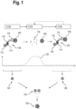
- FIG. 1 is a schematic view of a flow diagram of a first embodiment of a method for analysing a biological sample.
- the biological sample comprises at least one target analyte 106, in particular a target protein.
- the target analyte 106 is stained or marked with a first marker 108 specific to the target analyte 106, by introducing the first marker 108 into the biological sample.
- the first marker 108 comprises an affinity reagent 110, that is configured to specifically bind to the target analyte 106, in particular specifically bind to a particular epitope of the target analyte 106.
- the affinity reagent 110 is preferably an aptamer with a high affinity to the target analyte 106 comprising nucleic acids, such as DNA, RNA and/or XNA.
- the aptamer consists essentially of natural and/or non-natural nucleotides.
- the first marker 108 further comprises a label 112 with at least on optically detectable moiety, such as a fluorophore, that is bound to the affinity reagent 110.
- the label 112 may be attached to the affinity reagent 110 by means of an oligonucleotide 114 that is at least partially complementary to a respective oligonucleotide 116 of the affinity reagent 110.
- a first optical readout is generated of the biological sample with the first marker 108.
- an image of the biological sample with the first marker 108 may be generated and the location of the first marker 108, and therefore the attached target analyte 106, may be determined within that image.
- the optical readout may be generated by an optical readout device, such as a microscope.
- the first marker 108 determined in the first optical readout is schematically represented in Fig. 1 and indicated by reference sign 122.
- a step S102 the marker 108 is degraded by applying at least one degradation agent to the biological sample.
- At least the affinity reagent 110 is configured to be degraded by the degradation agent.
- the degradation agent may cause at least the affinity reagent 110 to degrade, resulting in degraded affinity reagent 118.
- the degradation agent may be a biological degradation agent, a chemical degradation agent or a physical degradation agent.
- the affinity reagent 110 degrading may include digesting, fragmenting or unfolding the affinity reagent 110.
- the degradation of the affinity reagent 110 may result in a loss of function of the affinity reagent 110, for example, the affinity reagent 110 may lose its specificity and/or affinity to the target analyte 106 and no longer specifically bind the first marker 108 to the target analyte 106.
- the affinity reagent 110 was degraded by a chemical degradation agent, for example, hydrogen peroxide, resulting in oligonucleotide fragments of the degraded affinity reagent 118.
- the hydrogen peroxide concentration 120 is increased in step S102 in order to degrade the affinity reagent 110.
- the aptamer of affinity reagent 110 may comprise non-natural nucleotides, that are particularly susceptible to the degradation agent, such as hydrogen peroxide, in order to efficiently degrade the affinity reagent 110.
- application of the degradation agent is stopped. This may involve washing of the biological sample in order to remove the hydrogen peroxide.
- the biological sample may be washed to remove the degraded affinity reagent 118 and/or other components of the marker such as the label 112.
- the label 112 may additionally be configured to be degraded at least partially by the degradation agent.
- Alternative degradation agents may include: enzymes such as endonucleases (biological degradation agents), UV-light or temperature (physical degradation agents).
- the affinity reagent 110 is configured to be degraded only by the degradation agent added in step S102. Further, it is preferred that the degradation agent applied in step S102 does not substantially degrade the target analyte 106.
- the affinity reagent 110 and the respective degradation agent may be chosen based on the target analyte 106, in particular its chemical nature, prior to step S100, such that the degradation agent is chosen to essentially not degrade the target analyte 106.
- the target analyte 106 is a protein, which is not or essentially not affected or degraded by the hydrogen peroxide degradation agent. Instead, only the affinity reagent 110 is degraded by the degradation agent.
- DNases degrade nucleic acids
- RNases restriction enzymes
- Endonucleases restriction enzymes
- Such enzymes may also be used in conjunction with each other as enzyme mixes and further combined with other degradation agents such as hydrogen peroxide to enable particular rapid and effective degradation.
- DNase I or RNase A are used at a temperature in the range of 20-50°C in conjunction with preferably a Tris-based buffer containing 0.5-5mM Mg2+ and EDTA.
- a second marker 124 is introduced into the biological sample comprising the target analyte 106.
- the second marker 124 comprises the affinity reagent 110, that is configured to specifically bind to the target analyte 106, in particular specifically bind to the particular epitope of the target analyte 106 as in step S100.
- the second marker 108 may comprise a different affinity reagent to the affinity reagent 110 that binds to a different epitope of the target analyte 106.
- the second marker 124 also further comprises a label 126 with at least on optically detectable moiety, such as a fluorophore, that is bound to the affinity reagent 110.
- the label 126 may be attached to the affinity reagent 110 by means of the oligonucleotide 114 that is at least partially complementary to the respective oligonucleotide 116 of the affinity reagent 110.
- the labels 112, 126 in particular their respective optically detectable moieties, differ in their optical properties. For example, the labels 112, 126 may differ in a fluorescent emission wavelength, in a fluorescent excitation wavelength or an emission duration.
- a second optical readout is generated of the biological sample with the second marker 124.
- an image of the biological sample with the second marker 124 may be generated and the location of the second marker 124, and therefore the attached target analyte 106, may be determined within that image.
- the second marker 124 determined in the second optical readout is schematically represented in Fig. 1 and indicated by reference sign 128.
- the method may continue by iteratively repeating steps S102 and S104 to generate further optical readouts with further markers.
- the order 130 of markers 108, 124 as they are determined or identified in the optical readouts may be used to identify a particular target analyte at that location in the biological sample.
- FIG. 2 is a schematic view of alternatives to the method according to Fig. 1 .
- the target analyte 106 may be marked in a first step with the first marker 108 and an initial optical readout generated, such as in step S100 as described for Figure 1 . Similar to step S102 the first marker 108 is then degraded.
- the first marker 108 in particular the affinity reagent 110, is degraded by a biological degradation agent 200, such as an endonuclease, in order to generate the degraded affinity reagent 118.
- the target analyte 106 is marked by the second marker 124 and a further optical readout generated, as described for Figure 1 .
- a different second marker 202 may be introduced into the biological sample after degradation of the first marker 108, similarly to the second marker 124.
- the marker 202 specifically binds to the target analyte 106 similarly to the markers 108, 124, however, the marker 202 specifically binds to a different epitope of the target analyte 106 than the markers 108, 124.
- the marker 202 comprises an affinity reagent 204, such as an aptamer, that is specific to the different epitope of the target analyte 106.
- affinity reagent 204 specifically binds to a particular area or surface of the target analyte 106 that is different to the area or surface of the target analyte 106 that the affinity reagent 110 specifically binds to.
- affinity reagent 204 specific to the different epitope of the target analyte 106, the specificity or selectivity of the method may be increased and the target analyte 106 may be identified, quantified, or its location in the optical readout be determined with particularly high confidence.
- the strategy schematically depicted in Figure 2 is particularly preferred, as it allows (spatial) proteomics assays to be designed that are compatible with analysing a high number of target analytes in the range of 100 to 50000 target analytes, for example, 100 to 500, 500 to 1000, 1000 to 10000, 1000 to 50000, while increasing the selectivity or specificity of the assay and providing a means of probing another layer of information, which are post-translational modifications (PTMs).
- PTMs post-translational modifications
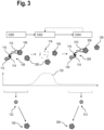
- FIG 3 is a schematic view of a flow diagram of a second embodiment of a method for analysing a biological sample.
- the biological sample comprises at least the target analyte 106 and a further target analyte 306, in particular both are target proteins.
- the target analyte 106 is stained or marked with the first marker 108 specific to the target analyte 106, by introducing the first marker 108 into the biological sample.
- the first marker 108 comprises the affinity reagent 110, that is configured to specifically bind to the target analyte 106.
- the first marker 108 further comprises the label 112 with at least on optically detectable moiety, such as a fluorophore, that is bound to the affinity reagent 110.
- the label 112 may be attached to the affinity reagent 110 by means of the oligonucleotide 114 that is at least partially complementary to the respective oligonucleotide 116 of the affinity reagent 110.
- a first optical readout is generated of the biological sample with the first marker 108.
- an image of the biological sample with the first marker 108 may be generated and the location of the first marker 108, and therefore the attached target analyte 106, may be determined within that image.
- the optical readout may be generated by an optical readout device, such as a microscope.
- the first marker 108 determined in the first optical readout is schematically represented in Fig. 1 and indicated by reference sign 122.
- a step S302 the marker 108 is degraded by applying at least one degradation agent to the biological sample.
- At least the affinity reagent 110 is configured to be degraded by the degradation agent.
- the degradation agent may cause at least the affinity reagent 110 to degrade, resulting in degraded affinity reagent 118.
- the degradation agent may be a biological degradation agent, a chemical degradation agent or a physical degradation agent.
- the affinity reagent 110 degrading may include digesting, fragmenting or unfolding the affinity reagent 110.
- the degradation of the affinity reagent 110 may result in a loss of function of the affinity reagent 110, for example, the affinity reagent 110 may lose its specificity to the target analyte 106 and no longer specifically bind the first marker 108 to the target analyte 106.
- the affinity reagent 110 is degraded by a chemical degradation agent, for example, hydrogen peroxide, resulting in oligonucleotide fragments of the degraded affinity reagent 118.
- the hydrogen peroxide concentration 120 is increased in step S302 in order to degrade the affinity reagent 110.
- the aptamer of affinity reagent 110 may comprise non-natural nucleotides, that are particularly susceptible to the degradation agent, such as hydrogen peroxide, in order to efficiently degrade the affinity reagent 110.
- application of the degradation agent is stopped. This may involve washing of the biological sample in order to remove the hydrogen peroxide.
- the biological sample may be washed to remove the degraded affinity reagent 118 and/or other components of the marker such as the label 112.
- the label 112 may additionally be configured to be degraded at least partially by the degradation agent.
- Alternative degradation agents may include: enzymes such as DNases, RNases, endonucleases (biological degradation agents), UV-light or temperature (physical degradation agents).
- the affinity reagent 110 is configured to be degraded only by the degradation agent added in step S302. Further, it is preferred that the degradation agent applied in step S302 does not substantially degrade the target analytes 106, 306.
- the affinity reagent 110 and the respective degradation agent may be chosen based on the target analyte 106, 306, in particular its chemical nature, prior to step S300, such that the degradation agent is chosen to essentially not degrade the target analyte 106, 306.
- a further marker 308 is introduced into the biological sample comprising the target analytes 106, 306.
- the further marker 308 comprises a further affinity reagent 310, that is configured to specifically bind to the target analyte 306.
- the further marker 308 also further comprises a label 126 with at least on optically detectable moiety, such as a fluorophore, that is bound to the affinity reagent 310.
- the label 126 may be attached to the affinity reagent 310 by means of the oligonucleotide 114 that is at least partially complementary to the respective oligonucleotide 116 of the affinity reagent 310.
- the labels 112, 126 in particular their respective optically detectable moieties, differ in their optical properties. For example, the labels 112, 126 may differ in a fluorescent emission wavelength, in a fluorescent excitation wavelength or an emission duration.
- a second optical readout is generated of the biological sample with the further marker 308.
- an image of the biological sample with the further marker 308 may be generated and the location of the further marker 308, and therefore the attached target analyte 306, may be determined within that image.
- the further marker 308 determined in the second optical readout is schematically represented in Fig. 3 and indicated by reference sign 312.
- the method may continue by iteratively repeating steps S302 and S304 to generate further optical readouts with further markers.
- the markers 108, 308 are determined or identified in the respective optical readouts (reference signs 122, 312).
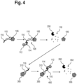
- FIG 4 is a schematic view of alternatives to the method according to Figure 3 .
- the marker 108 may be introduced into the biological sample in parts.
- the affinity reagent 110 may initially be introduced and subsequently the label 112 is introduced.
- the complementary oligonucleotides 114, 116 of the affinity reagent 110 and the label 112 enable specific binding of the label 112 to the affinity reagent 110.
- the marker 108, in particular the affinity reagent 110 may then be degraded by means of the biological degradation agent 200, as described for Figure 3 .
- the biological sample may comprise the target analyte 306. This target analyte 306 may be marked with a marker 400 by repeating the method for another iteration.
- the marker 400 may be introduced into the biological sample in parts. Initially, the affinity reagent 310 is introduced and subsequently, the label 112 is introduced that specifically binds to the affinity reagent 310 by means of the complementary oligonucleotides 114, 116. This generates the marker 400 in the biological sample.
- the marker 400 in particular the affinity reagent 310, may in turn be degraded into a fragmented affinity reagent 402. Since the individual parts of the markers 108, 400 are smaller than the respective entire markers 108, 400, the individual parts may penetrate the biological sample deeper before generating the markers 108, 400 in-situ in the biological sample.
- the markers 108, 124, 202, 308, 400 or respective parts of the markers 108, 124, 202, 308, 400 may be introduced into the biological sample by means of electrophoresis.
- an electric field may be applied to the biological sample.
- the markers 108, 124, 202, 308 comprise charged parts, such as oligonucleotides.
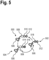
- FIG. 5 is a schematic view of markers 500, 502, 504 used iteratively in a method for analysing a biological sample.
- the markers 500, 502, 504 each comprise an affinity reagent 506 to specifically attach the marker to a target analyte of the biological sample.
- the markers 500, 502, 504 further comprise labels with at least two optically detectable moieties 508, 510, 512, 514, 516, 518, such as fluorophores.
- Each of the optically detectable moieties 508, 510, 512, 514, 516, 518 has different optical properties such as an emission wavelength, excitation wavelength or fluorescence lifetime.
- the markers 500, 502, 504 may be used in one of the methods described above.
- the markers 500, 502, 504 may be used iteratively in the steps S100, 5104, S300, S304 in order to identify or distinguish the respectively marked target analytes.
- markers 500, 502, 504 with diverse optical properties, a plurality of unique target analytes may be marked and distinguished in a particular step S100, S104, S300, S304.
- markers 500, 502, 504 comprising a plurality of optically detectable moieties described above may be detected using a readout device equipped with spectral excitation and emission detection as well as software-based unmixing algorithms.
- this enables introducing a plurality of different markers 500, 502, 504 comprising a plurality of optically detectable moieties into the biological sample at the same time, for example, during step S100, S104, 5300 or S304, in order to detect a plurality of different target analytes.
- this enables being able to distinguish the individual markers of the plurality of different markers 500, 502, 504 in the optical readouts based on optical properties of the respective optically detectable moieties 508, 510, 512, 514, 516, 518.
- the ability to distinguish a large number of markers introduced into a biological sample in one of the methods disclosed above may be further increased by applying the methods disclosed in document PCT/EP2021/073819 .
- the document discloses marking of target analytes in biological samples with markers comprising combinatorial labels, which are swapped from one round of staining to the next, and the acceptance of the presence of a marker in the readout volume based on a statistical analysis a measure of statistical confidence.
- aspects have been described in the context of an apparatus, it is clear that these aspects also represent a description of the corresponding method, where a block or device corresponds to a method step or a feature of a method step. Analogously, aspects described in the context of a method step also represent a description of a corresponding block or item or feature of a corresponding apparatus.
Landscapes
- Health & Medical Sciences (AREA)
- Life Sciences & Earth Sciences (AREA)
- Chemical & Material Sciences (AREA)
- Engineering & Computer Science (AREA)
- Organic Chemistry (AREA)
- Physics & Mathematics (AREA)
- Immunology (AREA)
- Molecular Biology (AREA)
- Biochemistry (AREA)
- General Health & Medical Sciences (AREA)
- Zoology (AREA)
- Wood Science & Technology (AREA)
- Analytical Chemistry (AREA)
- Proteomics, Peptides & Aminoacids (AREA)
- Genetics & Genomics (AREA)
- Bioinformatics & Cheminformatics (AREA)
- Biotechnology (AREA)
- Biomedical Technology (AREA)
- General Engineering & Computer Science (AREA)
- Biophysics (AREA)
- Microbiology (AREA)
- Pathology (AREA)
- General Physics & Mathematics (AREA)
- Nuclear Medicine, Radiotherapy & Molecular Imaging (AREA)
- Hematology (AREA)
- Urology & Nephrology (AREA)
- Optics & Photonics (AREA)
- Chemical Kinetics & Catalysis (AREA)
- Medicinal Chemistry (AREA)
- Plant Pathology (AREA)
- Food Science & Technology (AREA)
- Cell Biology (AREA)
- Bioinformatics & Computational Biology (AREA)
- Measuring Or Testing Involving Enzymes Or Micro-Organisms (AREA)
Priority Applications (3)
| Application Number | Priority Date | Filing Date | Title |
|---|---|---|---|
| EP23178065.1A EP4474484A1 (de) | 2023-06-07 | 2023-06-07 | Verfahren zur analyse einer biologischen probe |
| US18/733,881 US20240409982A1 (en) | 2023-06-07 | 2024-06-05 | Method for analysing a biological sample |
| CN202410723065.2A CN119104526A (zh) | 2023-06-07 | 2024-06-05 | 用于分析生物样品的方法 |
Applications Claiming Priority (1)
| Application Number | Priority Date | Filing Date | Title |
|---|---|---|---|
| EP23178065.1A EP4474484A1 (de) | 2023-06-07 | 2023-06-07 | Verfahren zur analyse einer biologischen probe |
Publications (1)
| Publication Number | Publication Date |
|---|---|
| EP4474484A1 true EP4474484A1 (de) | 2024-12-11 |
Family
ID=86732939
Family Applications (1)
| Application Number | Title | Priority Date | Filing Date |
|---|---|---|---|
| EP23178065.1A Pending EP4474484A1 (de) | 2023-06-07 | 2023-06-07 | Verfahren zur analyse einer biologischen probe |
Country Status (3)
| Country | Link |
|---|---|
| US (1) | US20240409982A1 (de) |
| EP (1) | EP4474484A1 (de) |
| CN (1) | CN119104526A (de) |
Citations (5)
| Publication number | Priority date | Publication date | Assignee | Title |
|---|---|---|---|---|
| EP3425063A1 (de) * | 2011-12-22 | 2019-01-09 | President and Fellows of Harvard College | Zusammensetzungen und verfahren zum nachweis von analyten |
| US20210262018A1 (en) * | 2020-02-21 | 2021-08-26 | 10X Genomics, Inc. | Methods and compositions for integrated in situ spatial assay |
| US20210388424A1 (en) * | 2020-06-12 | 2021-12-16 | 10X Genomics, Inc. | Methods for analyzing target nucleic acids and related compositions |
| WO2022182903A1 (en) * | 2021-02-24 | 2022-09-01 | California Institute Of Technology | Multiplexing of experimental conditions and samples in spatial genomics |
| US20220380838A1 (en) * | 2021-06-01 | 2022-12-01 | 10X Genomics, Inc. | Methods and compositions for analyte detection and probe resolution |
-
2023
- 2023-06-07 EP EP23178065.1A patent/EP4474484A1/de active Pending
-
2024
- 2024-06-05 CN CN202410723065.2A patent/CN119104526A/zh active Pending
- 2024-06-05 US US18/733,881 patent/US20240409982A1/en active Pending
Patent Citations (5)
| Publication number | Priority date | Publication date | Assignee | Title |
|---|---|---|---|---|
| EP3425063A1 (de) * | 2011-12-22 | 2019-01-09 | President and Fellows of Harvard College | Zusammensetzungen und verfahren zum nachweis von analyten |
| US20210262018A1 (en) * | 2020-02-21 | 2021-08-26 | 10X Genomics, Inc. | Methods and compositions for integrated in situ spatial assay |
| US20210388424A1 (en) * | 2020-06-12 | 2021-12-16 | 10X Genomics, Inc. | Methods for analyzing target nucleic acids and related compositions |
| WO2022182903A1 (en) * | 2021-02-24 | 2022-09-01 | California Institute Of Technology | Multiplexing of experimental conditions and samples in spatial genomics |
| US20220380838A1 (en) * | 2021-06-01 | 2022-12-01 | 10X Genomics, Inc. | Methods and compositions for analyte detection and probe resolution |
Non-Patent Citations (5)
| Title |
|---|
| IRAVANIVARMA, ENVIRON CHEM LETT., 10 March 2020 (2020-03-10), pages 1 - 25 |
| KACENAUSKAITE ET AL., J. AM. CHEM. SOC., vol. 143, 2021, pages 1377 - 1385 |
| RODRIGUEZ ET AL., TRENDS BIOCHEM SCI., vol. 42, no. 2, February 2017 (2017-02-01), pages 111 - 129 |
| SHAH SHEEL ET AL: "Dynamics and Spatial Genomics of the Nascent Transcriptome by Intron seqFISH", CELL, vol. 174, no. 2, 1 July 2018 (2018-07-01), Amsterdam NL, pages 363 - 376.e16, XP055937658, ISSN: 0092-8674, Retrieved from the Internet <URL:https://www.sciencedirect.com/science/article/pii/S0006349517303430/pdfft?md5=765444f9810c9fdc76346c728083b308&pid=1-s2.0-S0006349517303430-main.pdf> DOI: 10.1016/j.cell.2018.05.035 * |
| YAN ET AL., MICROCHIMICA ACTA, vol. 186, 2019, pages 583 |
Also Published As
| Publication number | Publication date |
|---|---|
| CN119104526A (zh) | 2024-12-10 |
| US20240409982A1 (en) | 2024-12-12 |
Similar Documents
| Publication | Publication Date | Title |
|---|---|---|
| US10267808B2 (en) | Molecular indicia of cellular constituents and resolving the same by super-resolution technologies in single cells | |
| US9677125B2 (en) | Detection of plurality of targets in biological samples | |
| EP3610242B1 (de) | Bestimmung der zielmoleküldichte auf einem fluoreszenzbild | |
| US20250244316A1 (en) | Label and marker for marking a target molecule in a biological sample | |
| WO2022242895A1 (en) | Marker, method and device for analyzing a biological sample | |
| CN115087747A (zh) | 使用时间分辨发光测量法对生物材料进行空间分析的组合物和方法 | |
| US20170234887A1 (en) | Method for detecting a spatial proximity of a first and a second epitope | |
| EP4623303A1 (de) | Verfahren und marker zur analyse einer probe | |
| EP4474484A1 (de) | Verfahren zur analyse einer biologischen probe | |
| US20240248036A1 (en) | Method and device for analyzing a biological sample | |
| US20240263181A1 (en) | Affinity reagent | |
| EP4109333B1 (de) | Verfahren zur analyse biologischer proben | |
| EP4556890A1 (de) | Label, marker und reagenzienkit zur analyse einer biologischen probe | |
| US20250361545A1 (en) | Label, marker and method for analyzing a biological sample | |
| US20240248082A1 (en) | Marker, method and device for analyzing a biological sample | |
| WO2025098646A1 (en) | Method for detecting analytes | |
| EP4589298A1 (de) | Marker und verfahren zur analyse einer biologischen probe | |
| EP4488682A1 (de) | Verfahren und system zur analyse einer biologischen probe | |
| CN117425828A (zh) | 用于分析生物样品的标记物、方法和装置 | |
| HK1179307A (en) | Detection of plurality of targets in biological samples | |
| HK40017807B (en) | Target molecule density determination in a fluorescence image |
Legal Events
| Date | Code | Title | Description |
|---|---|---|---|
| PUAI | Public reference made under article 153(3) epc to a published international application that has entered the european phase |
Free format text: ORIGINAL CODE: 0009012 |
|
| STAA | Information on the status of an ep patent application or granted ep patent |
Free format text: STATUS: THE APPLICATION HAS BEEN PUBLISHED |
|
| AK | Designated contracting states |
Kind code of ref document: A1 Designated state(s): AL AT BE BG CH CY CZ DE DK EE ES FI FR GB GR HR HU IE IS IT LI LT LU LV MC ME MK MT NL NO PL PT RO RS SE SI SK SM TR |
|
| STAA | Information on the status of an ep patent application or granted ep patent |
Free format text: STATUS: REQUEST FOR EXAMINATION WAS MADE |
|
| 17P | Request for examination filed |
Effective date: 20250611 |