EP4428246A2 - Verfahren zur messung der fehllokalisierung eines analyten - Google Patents
Verfahren zur messung der fehllokalisierung eines analyten Download PDFInfo
- Publication number
- EP4428246A2 EP4428246A2 EP24185912.3A EP24185912A EP4428246A2 EP 4428246 A2 EP4428246 A2 EP 4428246A2 EP 24185912 A EP24185912 A EP 24185912A EP 4428246 A2 EP4428246 A2 EP 4428246A2
- Authority
- EP
- European Patent Office
- Prior art keywords
- probe
- analyte
- capture
- sequence
- biological sample
- Prior art date
- Legal status (The legal status is an assumption and is not a legal conclusion. Google has not performed a legal analysis and makes no representation as to the accuracy of the status listed.)
- Pending
Links
Images


Classifications
-
- C—CHEMISTRY; METALLURGY
- C12—BIOCHEMISTRY; BEER; SPIRITS; WINE; VINEGAR; MICROBIOLOGY; ENZYMOLOGY; MUTATION OR GENETIC ENGINEERING
- C12Q—MEASURING OR TESTING PROCESSES INVOLVING ENZYMES, NUCLEIC ACIDS OR MICROORGANISMS; COMPOSITIONS OR TEST PAPERS THEREFOR; PROCESSES OF PREPARING SUCH COMPOSITIONS; CONDITION-RESPONSIVE CONTROL IN MICROBIOLOGICAL OR ENZYMOLOGICAL PROCESSES
- C12Q1/00—Measuring or testing processes involving enzymes, nucleic acids or microorganisms; Compositions therefor; Processes of preparing such compositions
- C12Q1/68—Measuring or testing processes involving enzymes, nucleic acids or microorganisms; Compositions therefor; Processes of preparing such compositions involving nucleic acids
- C12Q1/6813—Hybridisation assays
- C12Q1/6841—In situ hybridisation
-
- C—CHEMISTRY; METALLURGY
- C12—BIOCHEMISTRY; BEER; SPIRITS; WINE; VINEGAR; MICROBIOLOGY; ENZYMOLOGY; MUTATION OR GENETIC ENGINEERING
- C12Q—MEASURING OR TESTING PROCESSES INVOLVING ENZYMES, NUCLEIC ACIDS OR MICROORGANISMS; COMPOSITIONS OR TEST PAPERS THEREFOR; PROCESSES OF PREPARING SUCH COMPOSITIONS; CONDITION-RESPONSIVE CONTROL IN MICROBIOLOGICAL OR ENZYMOLOGICAL PROCESSES
- C12Q1/00—Measuring or testing processes involving enzymes, nucleic acids or microorganisms; Compositions therefor; Processes of preparing such compositions
- C12Q1/68—Measuring or testing processes involving enzymes, nucleic acids or microorganisms; Compositions therefor; Processes of preparing such compositions involving nucleic acids
- C12Q1/6806—Preparing nucleic acids for analysis, e.g. for polymerase chain reaction [PCR] assay
-
- C—CHEMISTRY; METALLURGY
- C12—BIOCHEMISTRY; BEER; SPIRITS; WINE; VINEGAR; MICROBIOLOGY; ENZYMOLOGY; MUTATION OR GENETIC ENGINEERING
- C12Q—MEASURING OR TESTING PROCESSES INVOLVING ENZYMES, NUCLEIC ACIDS OR MICROORGANISMS; COMPOSITIONS OR TEST PAPERS THEREFOR; PROCESSES OF PREPARING SUCH COMPOSITIONS; CONDITION-RESPONSIVE CONTROL IN MICROBIOLOGICAL OR ENZYMOLOGICAL PROCESSES
- C12Q1/00—Measuring or testing processes involving enzymes, nucleic acids or microorganisms; Compositions therefor; Processes of preparing such compositions
- C12Q1/68—Measuring or testing processes involving enzymes, nucleic acids or microorganisms; Compositions therefor; Processes of preparing such compositions involving nucleic acids
- C12Q1/6844—Nucleic acid amplification reactions
-
- C—CHEMISTRY; METALLURGY
- C12—BIOCHEMISTRY; BEER; SPIRITS; WINE; VINEGAR; MICROBIOLOGY; ENZYMOLOGY; MUTATION OR GENETIC ENGINEERING
- C12Q—MEASURING OR TESTING PROCESSES INVOLVING ENZYMES, NUCLEIC ACIDS OR MICROORGANISMS; COMPOSITIONS OR TEST PAPERS THEREFOR; PROCESSES OF PREPARING SUCH COMPOSITIONS; CONDITION-RESPONSIVE CONTROL IN MICROBIOLOGICAL OR ENZYMOLOGICAL PROCESSES
- C12Q2600/00—Oligonucleotides characterized by their use
- C12Q2600/16—Primer sets for multiplex assays
Definitions
- Cells within a tissue have differences in cell morphology and/or function due to varied analyte levels (e.g., gene and/or protein expression) within the different cells.
- the specific position of a cell within a tissue e.g., the cell's position relative to neighboring cells or the cell's position relative to the tissue microenvironment
- spatial analysis requires determining the sequence of the analyte sequence or a complement thereof and the sequence of the spatial barcode or a complement thereof in order to identify the spatial location of the analyte in the original biological sample.
- sequencing can be time and resource intensive. Therefore, there is a need to assess analyte (biological sample) spatial preservation and presence prior to traditional spatial analysis.
- this disclosure features methods of determining analyte mislocalization within a biological sample, the method including: (a) obtaining a first image of the biological sample; (b) hybridizing the analyte to a capture probe on an array, wherein the array includes a plurality of capture probes, wherein a capture probe of the plurality of capture probe includes a capture domain; (c) extending the capture probe using the analyte as a template, thereby generating an extended capture probe; (d) hybridizing a padlock probe or a snail probe to the extended capture probe; (e) circularizing the padlock probe or the snail probe; (f) amplifying the padlock probe or the snail probe, thereby generating an amplified circularized padlock probe or an amplified circularized snail probe; (g) hybridizing a plurality of detection probes to the amplified circularized padlock probe or the amplified circularized snail probe, wherein a detection probe from the plurality of detection probes comprises: a sequence that is substantially complementary to
- Also provided herein is a method of determining mislocalization of an analyte within a biological sample comprising: (a) obtaining a first image of the biological sample; (b) hybridizing an analyte derived molecule to a capture domain of a capture probe on an array, wherein the array comprises a plurality of capture probes, wherein the plurality of capture probes comprise a capture domain; (c) hybridizing a padlock probe or a snail probe to the analyte derived molecule; (d) circularizing the padlock probe or the snail probe; (e) amplifying the padlock probe or the snail probe, thereby generating an amplified circularized padlock probe or an amplified circularized snail probe; (f) hybridizing a plurality of detection probes to the amplified circularized padlock probe or the amplified circularized snail probe, wherein a detection probe from the plurality of detection probes comprises: a sequence that is substantially complementary to a sequence of the padlock probe or the snail probe, or
- the analyte derived molecule is a ligation product.
- the method further includes, prior to step (b): contacting a plurality of first probes and second probes to the biological sample on the array, wherein the plurality of first probes and second probes target a plurality of nucleic acids in the biological sample, wherein a first probe and a second probe of the plurality comprise sequences that are substantially complementary to the analyte, wherein the analyte is a target nucleic acid, and wherein the second probe comprises a capture probe capture domain sequence that is complementary to all or a portion of the capture domain; hybridizing the first probe and the second probe to the target nucleic acid; generating a ligation product by ligating the first probe and the second probe; and releasing the ligation product from the target nucleic acid.
- the first probe and the second probe are substantially complementary to adjacent sequences of the target nucleic acid. In some instances, the first probe and the second probe hybridize to sequences that are not adjacent to each other on the target nucleic acid, and wherein the first probe is extended with a DNA polymerase, thereby (i) filling in a gap between the first probe and the second probe and (ii) generating an extended first probe. In some instances, the first probe further comprises a primer sequence. In some instances, the first probe and/or the second probe is a DNA probe. In some instances, the releasing the ligation product from the target nucleic acid comprises contacting the biological sample with an endoribonuclease, optionally wherein the endoribonuclease is an RNase H enzyme.
- the analyte derived molecule is an analyte capture agent.
- the methods further include, prior to step (b): contacting the biological sample with a plurality of analyte capture agents, wherein the analyte capture agent comprises an analyte binding moiety and an oligonucleotide comprising an analyte binding moiety barcode and an analyte capture sequence, wherein the analyte capture sequence comprises a sequence complementary to the capture domain; and binding the analyte binding moiety of the analyte capture agent to the analyte, wherein the analyte is a protein.
- step (b) comprises hybridizing the analyte capture sequence to the capture domain.
- the padlock probe or the snail probe comprises: (i) a first sequence that is substantially complementary to a first portion of the extended capture probe, the analyte derived molecule, or a complement thereof; (ii) a backbone sequence; and (iii) a second sequence that is substantially complementary to a second portion of the extended capture probe, the analyte derived molecule, or a complement thereof.
- first sequence and the second sequence are substantially complementary to adjacent sequences of the extended capture probe, the analyte derived molecule, or a complement thereof. In some embodiments, the first sequence and the second sequence are substantially complementary to sequences of the extended capture probe, the analyte derived molecule, or a complement thereof, that are about 2 to 50 nucleotides apart.
- circularizing the padlock probe or the snail probe includes filling in a gap between the first sequence that is substantially complementary to a first portion of the analyte and the second sequence that is substantially complementary to a second portion of the analyte using a polymerase.
- the backbone sequence includes a backbone barcode sequence.
- circularizing the padlock probe or the snail probe includes ligating the padlock probe or the snail probe.
- the ligating includes enzymatic ligation or chemical ligation (e.g., click chemistry).
- the enzymatic ligation utilizes ligase.
- ligase includes a T4 DNA ligase.
- the amplifying includes rolling circle amplification (RCA). In some embodiments, the amplifying includes hybridizing one or more amplification primers to the padlock probe or the snail probe; and amplifying the padlock probe or the snail probe with a padlock probe or snail probe polymerase. In some embodiments, the padlock probe or snail probe polymerase has strand displacement activity. In some embodiments, the padlock probe or snail probe polymerase is a Phi29 DNA polymerase.
- the one or more amplification primers are substantially complementary to a portion of the padlock probe or the snail probe.
- the detection probe is about 10 to about 60 nucleotides in length.
- the detectable label includes a fluorophore.
- the detection probe is substantially complementary to a portion of the first sequence that is substantially complementary to a first portion of the analyte.
- the detection probe is substantially complementary to a portion of the second sequence that is substantially complementary to a second portion of the analyte.
- the detection probe is substantially complementary to a portion of the backbone sequence.
- the detection probe is substantially complementary to a portion of the analyte or a complement thereof.
- the extending step includes extending the capture probe using the analyte as the template. In some embodiments, the extending includes using a reverse transcriptase. In some embodiments, the reverse transcriptase includes a Moloney murine leukemia virus reverse transcriptase. In some embodiments, extending the capture probe includes using fluorescently tagged nucleotides to reverse transcribe the extended capture probe.
- the biological sample is permeabilized using a permeabilization agent.
- the permeabilization agent includes proteinase K.
- the permeabilization agent includes pepsin.
- the biological sample is permeabilized at room temperature.
- the biological sample is permeabilized at 37°C.
- the biological sample is permeabilized for about 1 minute to about 30 minutes.
- the method also includes varying permeabilization time to determine an optimal permeabilization time for minimizing analyte mislocalization in the biological sample.
- the method also includes varying permeabilization temperature to determine an optimal permeabilization temperature for minimizing analyte mislocalization in the biological sample.
- determining the mislocalization of the analyte includes measuring the signal intensity of the plurality of detection probes and comparing the signal intensity to a first image of the biological sample.
- signal intensity in the image of the biological sample includes a single cell or cell type that expresses a biomarker that is detected when the biological sample is stained.
- the biological sample is stained prior to obtaining an image of the biological sample.
- the biological sample is stained using immunofluorescence or immunohistochemistry techniques.
- the biological sample is stained using hematoxylin and eosin (H&E).
- the image is a fluorescent image or a brightfield image.
- the image is obtained using a fluorescent microscope, a confocal microscope, or a brightfield microscope.
- the capture probe is affixed to the substrate at a 5' end of the capture probe.
- the plurality of capture probes are uniformly distributed on a surface of the substrate.
- the capture domain of the capture probe includes a homopolymeric sequence. In some embodiments, the capture domain of the capture probe includes a poly(T) sequence. In some embodiments, the capture domain of the capture probe includes a non-homopolymeric sequence. In some embodiments, the non-homopolymeric sequence includes a sequence substantially complementary to the analyte.
- the biological sample includes a FFPE sample. In some embodiments, the biological sample includes a fresh frozen sample. In some embodiments, the biological sample includes fixed or live cells. In some embodiments, the biological sample is from a mammal. In some embodiments, the biological sample is from a human, a mouse, or a rat.
- the biological sample includes a tissue section.
- the tissue section is about 2.5 ⁇ m to about 20 ⁇ m in thickness.
- the method also includes varying the thickness of the tissue sample to determine an optimal tissue thickness to minimize mislocalization in the biological sample.
- the analyte is RNA. In some embodiments, the RNA is mRNA. In some embodiments, the analyte is a protein. In some embodiments, the protein is a cell surface marker. In some embodiments, the protein is an immune cell receptor.
- the method also includes obtaining a second biological sample.
- the second biological sample is a second tissue section of the biological sample.
- the method also includes determining abundance and/or location of an analyte in the second biological sample, wherein the determining includes: (a) contacting a spatial array with the second biological sample, wherein the spatial array includes a plurality of spatial capture probes, wherein a spatial capture probe of the plurality of spatial capture probes includes a capture domain and a spatial barcode; (b) hybridizing the analyte to the spatial capture probe; and (c) determining (i) all or a part of the sequence of the analyte, or a complement thereof, and (ii) the sequence of the spatial barcode, or a complement thereof, and using the determined sequence of (i) and (ii) to determine the abundance and/or the location of the analyte in the biological sample.
- the spatial capture probe further includes one or more functional domains, a unique molecular identifier (UMI), a cleavage domain, or combinations thereof.
- the method also includes extending the spatial capture probe via a nucleic acid extension reaction using the analyte as a template to generate an extended spatial capture probe including the spatial capture probe and the reverse complement of the analyte.
- the determining step (c) includes amplifying all or part of the analyte specifically bound to the spatial capture domain, or a complement thereof.
- the determining step (c) includes sequencing.
- the method can include imaging the biological sample.
- kits include: (a) an array including a plurality of capture probes, wherein a capture probe of the plurality of capture probes includes a capture domain; (b) a plurality of padlock probes and/or snail probes, wherein a padlock probe or a snail probe of the plurality of padlock probes and/or snail probes includes: (i) a first sequence that is substantially complementary to a first portion of an analyte, or a complement thereof, (ii) a backbone sequence, and (iii) a second sequence that is substantially complementary to a second portion of the analyte, or a complement thereof; (c) one or more enzymes selected from a ligase, a polymerase, a reverse transcriptase, or combinations thereof; (d) a plurality of detection probes; and (e) instructions for performing any one of the methods of described herein.
- each when used in reference to a collection of items, is intended to identify an individual item in the collection but does not necessarily refer to every item in the collection, unless expressly stated otherwise, or unless the context of the usage clearly indicates otherwise.
- a cell includes one or more cells, including mixtures thereof.
- a and/or B is used herein to include all of the following alternatives: “A”, “B”, “A or B”, and “A and B”.
- Spatial analysis methodologies and compositions described herein can provide a vast amount of analyte and/or expression data for a variety of analytes within a biological sample at high spatial resolution, while retaining native spatial context.
- Spatial analysis methods and compositions can include, e.g., the use of a capture probe including a spatial barcode (e.g., a nucleic acid sequence that provides information as to the location or position of an analyte within a cell or a tissue sample (e.g., mammalian cell or a mammalian tissue sample) and a capture domain that is capable of binding to an analyte (e.g., a protein and/or a nucleic acid) produced by and/or present in a cell.
- a spatial barcode e.g., a nucleic acid sequence that provides information as to the location or position of an analyte within a cell or a tissue sample
- a capture domain that is capable of binding to an analyte (
- Spatial analysis methods and compositions can also include the use of a capture probe having a capture domain that captures an intermediate agent for indirect detection of an analyte.
- the intermediate agent can include a nucleic acid sequence (e.g., a barcode) associated with the intermediate agent. Detection of the intermediate agent is therefore indicative of the analyte in the cell or tissue sample.
- Non-limiting aspects of spatial analysis methodologies and compositions are described in U.S. Patent Nos. 10,774,374 , 10,724,078 , 10,480,022 , 10,059,990 , 10,041,949 , 10,002,316 , 9,879,313 , 9,783,841 , 9,727,810 , 9,593,365 , 8,951,726 , 8,604,182 , 7,709,198 , U.S. Patent Application Publication Nos.
- a "barcode” is a label, or identifier, that conveys or is capable of conveying information (e.g., information about an analyte in a sample, a bead, and/or a capture probe).
- a barcode can be part of an analyte, or independent of an analyte.
- a barcode can be attached to an analyte.
- a particular barcode can be unique relative to other barcodes.
- an "analyte” can include any biological substance, structure, moiety, or component to be analyzed.
- target can similarly refer to an analyte of interest.
- Analytes can be broadly classified into one of two groups: nucleic acid analytes, and non-nucleic acid analytes.
- non-nucleic acid analytes include, but are not limited to, lipids, carbohydrates, peptides, proteins, glycoproteins (N-linked or O-linked), lipoproteins, phosphoproteins, specific phosphorylated or acetylated variants of proteins, amidation variants of proteins, hydroxylation variants of proteins, methylation variants of proteins, ubiquitylation variants of proteins, sulfation variants of proteins, viral proteins (e.g., viral capsid, viral envelope, viral coat, viral accessory, viral glycoproteins, viral spike, etc.), extracellular and intracellular proteins, antibodies, and antigen binding fragments.
- viral proteins e.g., viral capsid, viral envelope, viral coat, viral accessory, viral glycoproteins, viral spike, etc.
- the analyte(s) can be localized to subcellular location(s), including, for example, organelles, e.g., mitochondria, Golgi apparatus, endoplasmic reticulum, chloroplasts, endocytic vesicles, exocytic vesicles, vacuoles, lysosomes, etc.
- organelles e.g., mitochondria, Golgi apparatus, endoplasmic reticulum, chloroplasts, endocytic vesicles, exocytic vesicles, vacuoles, lysosomes, etc.
- analyte(s) can be peptides or proteins, including without limitation antibodies and enzymes. Additional examples of analytes can be found in Section (I)(c) of WO 2020/176788 and/or U.S. Patent Application Publication No. 2020/0277663 .
- an analyte can be detected indirectly, such as through detection of an intermediate agent, for example, an analyte derived molecule such as a connected probe (e.g., a ligation product) or an analyte capture agent (e.g., an oligonucleotide-conjugated antibody), such as those described herein.
- an intermediate agent for example, an analyte derived molecule such as a connected probe (e.g., a ligation product) or an analyte capture agent (e.g., an oligonucleotide-conjugated antibody), such as those described herein.
- a “biological sample” is typically obtained from the subject for analysis using any of a variety of techniques including, but not limited to, biopsy, surgery, and laser capture microscopy (LCM), and generally includes cells and/or other biological material from the subject.
- a biological sample can be a tissue section.
- a biological sample can be a fixed and/or stained biological sample (e.g., a fixed and/or stained tissue section).
- stains include histological stains (e.g., hematoxylin and/or eosin) and immunological stains (e.g., fluorescent stains).
- a biological sample e.g., a fixed and/or stained biological sample
- Biological samples are also described in Section (I)(d) of WO 2020/176788 and/or U.S. Patent Application Publication No. 2020/0277663 .
- a biological sample is permeabilized with one or more permeabilization reagents.
- permeabilization of a biological sample can facilitate analyte capture.
- Exemplary permeabilization agents and conditions are described in Section (I)(d)(ii)(13) or the Exemplary Embodiments Section of WO 2020/176788 and/or U.S. Patent Application Publication No. 2020/0277663 .
- Some array-based spatial analysis methods involve the transfer of one or more analytes from a biological sample to an array of features on a substrate, where typically each feature is associated with a unique spatial location on the array. Subsequent analysis of the transferred analytes includes determining the identity of the analytes and the spatial location of the analytes within the biological sample. The spatial location of an analyte within the biological sample is determined based on the feature to which the analyte is bound (e.g., directly or indirectly) on the array, and the feature's relative spatial location within the array.
- a “capture probe” refers to any molecule capable of capturing (directly or indirectly) and/or labelling an analyte (e.g., an analyte of interest) in a biological sample.
- the capture probe is a nucleic acid or a polypeptide.
- the capture probe includes a barcode (e.g., a spatial barcode and/or a unique molecular identifier (UMI)) and a capture domain).
- UMI unique molecular identifier
- a capture probe can include a cleavage domain and/or a functional domain (e.g., a primer-binding site, such as for amplification, or a flow cell attachment sequence, such as for next-generation sequencing (NGS)).
- NGS next-generation sequencing
- the capture domain is designed to detect one or more specific analytes of interest.
- a capture domain can be designed so that it comprises a sequence that is complementary or substantially complementary to one analyte of interest.
- the capture domain can be designed so that it comprises a sequence that is complementary or substantially complementary to a conserved region of multiple related analytes.
- the multiple related analytes are analytes that function in the same or similar cellular pathways or that have conserved homology and/or function.
- the design of the capture probe can be determined based on the intent of the user and can be any sequence that can be used to detect an analyte of interest.
- the capture domain sequence can therefore be random, degenerate, defined (e.g., gene or protein specific) or combinations thereof, depending on the target analyte(s) of interest.
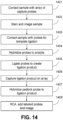
- FIG. 1 is a schematic diagram showing an exemplary capture probe, as described herein.
- the capture probe 102 is optionally coupled to a feature 101 by a cleavage domain 103, such as a disulfide linker.
- the capture probe can include a functional sequence 104 that is useful for subsequent processing.
- the functional sequence 104 can include all or a part of sequencer specific flow cell attachment sequence (e.g., a P5 or P7 sequence), all or a part of a sequencing primer sequence, (e.g., a R1 primer binding site, a R2 primer binding site), or combinations thereof.
- the capture probe can also include a spatial barcode 105.
- the capture probe can also include a unique molecular identifier (UMI) sequence 106. While FIG.
- UMI unique molecular identifier
- the capture probe can also include a capture domain 107 to facilitate capture of a target analyte.
- the capture probe comprises one or more additional functional sequences that can be located, for example between the spatial barcode 105 and the UMI sequence 106, between the UMI sequence 106 and the capture domain 107, or following the capture domain 107.
- the capture domain can have a sequence complementary to a sequence of a nucleic acid analyte.
- the capture domain can have a sequence complementary to a connected probe described herein.
- the capture domain can have a sequence complementary to a capture handle sequence present in an analyte capture agent.
- the capture domain can have a sequence complementary to a splint oligonucleotide.
- Such splint oligonucleotide in addition to having a sequence complementary to a capture domain of a capture probe, can have a sequence of a nucleic acid analyte, a sequence complementary to a portion of a connected probe described herein, and/or a capture handle sequence described herein.
- the functional sequences can generally be selected for compatibility with any of a variety of different sequencing systems, e.g., Ion Torrent Proton or PGM, Illumina sequencing instruments, PacBio, Oxford Nanopore, etc., and the requirements thereof.
- functional sequences can be selected for compatibility with non-commercialized sequencing systems. Examples of such sequencing systems and techniques, for which suitable functional sequences can be used, include (but are not limited to) Ion Torrent Proton or PGM sequencing, Illumina sequencing, PacBio SMRT sequencing, and Oxford Nanopore sequencing.
- functional sequences can be selected for compatibility with other sequencing systems, including non-commercialized sequencing systems.
- the spatial barcode 105 and functional sequence 104 is common to all of the probes attached to a given feature.
- the UMI sequence 106 of a capture probe attached to a given feature is different from the UMI sequence of a different capture probe attached to the given feature.
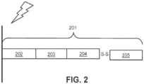
- FIG. 2 is a schematic illustrating a cleavable capture probe, wherein the cleaved capture probe can enter into a non-permeabilized cell and bind to analytes within the cell.
- the capture probe 201 contains a cleavage domain 202, a cell penetrating peptide 203, a reporter molecule 204, and a disulfide bond (-S-S-).
- 205 represents all other parts of a capture probe, for example a spatial barcode and/or a capture domain.
- FIG. 3 is a schematic diagram of an exemplary multiplexed spatially-barcoded feature.
- the feature 301 can be coupled to spatially-barcoded capture probes, wherein the spatially-barcoded probes of a particular feature can possess the same spatial barcode 302, but have different capture domains designed to associate the spatial barcode of the feature with more than one target analyte.
- a feature may be coupled to four different types of spatially-barcoded capture probes, each type of spatially-barcoded capture probe possessing the same spatial barcode 302.
- One type of capture probe associated with the feature includes the spatial barcode 302 in combination with a poly(T) capture domain 303, designed to capture mRNA target analytes.
- a second type of capture probe associated with the feature includes the same spatial barcode 302 in combination with a random N-mer capture domain 304 for gDNA analysis.
- a third type of capture probe associated with the feature includes the spatial barcode 302 in combination with a capture domain complementary to a capture handle sequence of an analyte capture agent of interest 305.
- a fourth type of capture probe associated with the feature includes the spatial barcode 302 in combination with a capture domain that can specifically bind a nucleic acid molecule 306 that can function in a CRISPR assay (e.g., CRISPR/Cas9). While only four different capture probe-barcoded constructs are shown in FIG.
- capture-probe barcoded constructs can be tailored for analyses of any given analyte associated with a nucleic acid and capable of binding with such a construct.
- the schemes shown in FIG. 3 can also be used for concurrent analysis of other analytes disclosed herein, including, but not limited to: (a) mRNA, a lineage tracing construct, cell surface or intracellular proteins and metabolites, and gDNA; (b) mRNA, accessible chromatin (e.g., ATAC-seq, DNase-seq, and/or MNase-seq) cell surface or intracellular proteins and metabolites, and a perturbation agent (e.g., a CRISPR crRNA/sgRNA, TALEN, zinc finger nuclease, and/or antisense oligonucleotide as described herein); (c) mRNA, cell surface or intracellular proteins and/or metabolites, a barcoded labelling agent (e.g., the MHC multimers
- a perturbation agent can be a small molecule, an antibody, a drug, an aptamer, a miRNA, a physical environmental (e.g., temperature change), or any other known perturbation agents. See, e.g., Section (II)(b) (e.g., subsections (i)-(vi)) of WO 2020/176788 and/or U.S. Patent Application Publication No. 2020/0277663 . Generation of capture probes can be achieved by any appropriate method, including those described in Section (II)(d)(ii) of WO 2020/176788 and/or U.S. Patent Application Publication No. 2020/0277663 .
- more than one analyte type e.g., nucleic acids and proteins
- a biological sample can be detected (e.g., simultaneously or sequentially) using any appropriate multiplexing technique, such as those described in Section (IV) of WO 2020/176788 and/or U.S. Patent Application Publication No. 2020/0277663 .
- an analyte capture agent refers to an agent that interacts with an analyte (e.g., an analyte in a biological sample) and with a capture probe (e.g., a capture probe attached to a substrate or a feature) to identify the analyte.
- the analyte capture agent includes: (i) an analyte binding moiety (e.g., that binds to an analyte), for example, an antibody or antigen-binding fragment thereof; (ii) analyte binding moiety barcode; and (iii) a capture handle sequence.
- an analyte binding moiety barcode refers to a barcode that is associated with or otherwise identifies the analyte binding moiety.
- the term “analyte capture sequence” or “capture handle sequence” refers to a region or moiety configured to hybridize to, bind to, couple to, or otherwise interact with a capture domain of a capture probe.
- a capture handle sequence is complementary to a capture domain of a capture probe.
- an analyte binding moiety barcode (or portion thereof) may be able to be removed (e.g., cleaved) from the analyte capture agent.
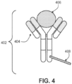
- FIG. 4 is a schematic diagram of an exemplary analyte capture agent 402 comprised of an analyte-binding moiety 404 and an analyte-binding moiety barcode domain 408.
- the exemplary analyte -binding moiety 404 is a molecule capable of binding to an analyte 406 and the analyte capture agent is capable of interacting with a spatially-barcoded capture probe.
- the analyte -binding moiety can bind to the analyte 406 with high affinity and/or with high specificity.
- the analyte capture agent can include an analyte-binding moiety barcode domain 408, a nucleotide sequence (e.g., an oligonucleotide), which can hybridize to at least a portion or entirety of a capture domain of a capture probe.
- the analyte-binding moiety barcode domain 408 can comprise an analyte binding moiety barcode and a capture handle sequence described herein.
- the analyte -binding moiety 404 can include a polypeptide and/or an aptamer.
- the analyte-binding moiety 404 can include an antibody or antibody fragment (e.g., an antigen-binding fragment).
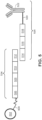
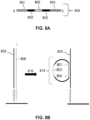
- FIG. 5 is a schematic diagram depicting an exemplary interaction between a feature-immobilized capture probe 524 and an analyte capture agent 526.
- the feature-immobilized capture probe 524 can include a spatial barcode 508 as well as functional sequences 506 and UMI 510, as described elsewhere herein.
- the capture probe can also include a capture domain 512 that is capable of binding to an analyte capture agent 526.
- the analyte capture agent 526 can include a functional sequence 518, analyte binding moiety barcode 516, and a capture handle sequence 514 that is capable of binding to the capture domain 512 of the capture probe 524.
- the analyte capture agent can also include a linker 520 that allows the capture agent barcode domain 516 to couple to the analyte binding moiety 522.
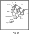
- FIGs. 6A , 6B , and 6C are schematics illustrating how streptavidin cell tags can be utilized in an array-based system to produce a spatially-barcoded cell or cellular contents.
- MHC major histocompatibility complex
- p2m biotin
- streptavidin moiety comprises multiple pMHC moieties.
- TCR T-Cell Receptor
- a capture agent barcode domain 601 can be modified with streptavidin 602 and contacted with multiple molecules of biotinylated MHC 603 such that the biotinylated MHC molecules 604 are coupled with the streptavidin conjugated capture agent barcode domain 601. The result is a barcoded MHC multimer complex 605.
- the capture agent barcode domain sequence 601 can identify the MHC as its associated label and also includes optional functional sequences such as sequences for hybridization with other oligonucleotides. As shown in FIG.
- one example oligonucleotide is capture probe 606 that comprises a complementary sequence (e.g., rGrGrG corresponding to C C C), a barcode sequence and other functional sequences, such as, for example, a UMI, an adapter sequence (e.g., comprising a sequencing primer sequence (e.g., R1 or a partial R1 ("pR1"), R2), a flow cell attachment sequence (e.g., P5 or P7 or partial sequences thereof)), etc.
- capture probe 606 may at first be associated with a feature (e.g., a gel bead) and released from the feature.
- capture probe 606 can hybridize with a capture agent barcode domain 601 of the MHC-oligonucleotide complex 605.
- the hybridized oligonucleotides (Spacer C C C and Spacer rGrGrG) can then be extended in primer extension reactions (see dashed lines adjacent to 606 and 601) such that constructs comprising sequences that correspond to each of the two spatial barcode sequences (the spatial barcode associated with the capture probe, and the barcode associated with the MHC-oligonucleotide complex) are generated.
- one or both of these corresponding sequences may be a complement of the original sequence in capture probe 606 or capture agent barcode domain 601.
- the capture probe and the capture agent barcode domain are ligated together.
- the resulting construct can be optionally further processed (e.g., to add any additional sequences and/or for clean-up) and subjected to sequencing.
- a sequence derived from the capture probe 606 spatial barcode sequence may be used to identify a feature and the sequence derived from spatial barcode sequence on the capture agent barcode domain 601 may be used to identify the particular peptide MHC complex 604 bound on the surface of the cell (e.g., when using pMHC libraries for screening immune cells or immune cell populations).
- a spatial barcode with one or more neighboring cells, such that the spatial barcode identifies the one or more cells, and/or contents of the one or more cells, as associated with a particular spatial location.
- One method is to promote analytes or analyte proxies (e.g., intermediate agents) out of a cell and towards a spatially-barcoded array (e.g., including spatially-barcoded capture probes).
- Another method is to cleave spatially-barcoded capture probes from an array and promote the spatially-barcoded capture probes towards and/or into or onto the biological sample.
- capture probes may be configured to prime, replicate, and consequently yield optionally barcoded extension products from a template (e.g., a DNA or RNA template, such as an analyte or an intermediate agent (e.g., a connected probe (e.g., a ligation product) or an analyte capture agent), or a portion thereof), or derivatives thereof (see, e.g., Section (II)(b)(vii) of WO 2020/176788 and/or U.S. Patent Application Publication No. 2020/0277663 regarding extended capture probes).
- a template e.g., a DNA or RNA template, such as an analyte or an intermediate agent (e.g., a connected probe (e.g., a ligation product) or an analyte capture agent), or a portion thereof
- a template e.g., a DNA or RNA template, such as an analyte or an intermediate agent (e.g.,
- capture probes may be configured to form a connected probe (e.g., a ligation product) with a template (e.g., a DNA or RNA template, such as an analyte or an intermediate agent, or portion thereof), thereby creating ligations products that serve as proxies for template extension.
- a connected probe e.g., a ligation product
- a template e.g., a DNA or RNA template, such as an analyte or an intermediate agent, or portion thereof
- an "extended capture probe” refers to a capture probe having additional nucleotides added to the terminus (e.g., 3' or 5' end) of the capture probe thereby extending the overall length of the capture probe.
- an "extended 3' end” indicates additional nucleotides were added to the most 3' nucleotide of the capture probe to extend the length of the capture probe, for example, by polymerization reactions used to extend nucleic acid molecules including templated polymerization catalyzed by a polymerase (e.g., a DNA polymerase or a reverse transcriptase).
- a polymerase e.g., a DNA polymerase or a reverse transcriptase
- extending the capture probe includes adding to a 3' end of a capture probe, a nucleic acid sequence that is complementary to a nucleic acid sequence of an analyte or intermediate agent specifically bound to the capture domain of the capture probe.
- the capture probe is extended using reverse transcription.
- the capture probe is extended using one or more DNA polymerases. The extended capture probes include the sequence of the capture probe and the sequence of the spatial barcode of the capture probe.
- extended capture probes are amplified (e.g., in bulk solution or on the array) to yield quantities that are sufficient for downstream analysis, e.g., via DNA sequencing.
- extended capture probes e.g., DNA molecules
- act as templates for an amplification reaction e.g., a polymerase chain reaction.
- Section (II)(a) of WO 2020/176788 and/or U.S. Patent Application Publication No. 2020/0277663 Analysis of captured analytes (and/or intermediate agents or portions thereof), for example, including sample removal, extension of capture probes, sequencing (e.g., of a cleaved extended capture probe and/or a cDNA molecule complementary to an extended capture probe), sequencing on the array (e.g., using, for example, in situ hybridization or in situ ligation approaches), temporal analysis, and/or proximity capture, is described in Section (II)(g) of WO 2020/176788 and/or U.S. Patent Application Publication No. 2020/0277663 . Some quality control measures are described in Section (II)(h) of WO 2020/176788 and/or U.S. Patent Application Publication No. 2020/0277663 .
- Spatial information can provide information of biological and/or medical importance.
- the methods and compositions described herein can allow for: identification of one or more biomarkers (e.g., diagnostic, prognostic, and/or for determination of efficacy of a treatment) of a disease or disorder; identification of a candidate drug target for treatment of a disease or disorder; identification (e.g., diagnosis) of a subject as having a disease or disorder; identification of stage and/or prognosis of a disease or disorder in a subject; identification of a subject as having an increased likelihood of developing a disease or disorder; monitoring of progression of a disease or disorder in a subject; determination of efficacy of a treatment of a disease or disorder in a subject; identification of a patient subpopulation for which a treatment is effective for a disease or disorder; modification of a treatment of a subject with a disease or disorder; selection of a subject for participation in a clinical trial; and/or selection of a treatment for a subject with a disease or disorder.
- Spatial information can provide information of biological importance.
- the methods and compositions described herein can allow for: identification of transcriptome and/or proteome expression profiles (e.g., in healthy and/or diseased tissue); identification of multiple analyte types in close proximity (e.g., nearest neighbor analysis); determination of up- and/or down-regulated genes and/or proteins in diseased tissue; characterization of tumor microenvironments; characterization of tumor immune responses; characterization of cells types and their co-localization in tissue; and identification of genetic variants within tissues (e.g., based on gene and/or protein expression profiles associated with specific disease or disorder biomarkers).
- a substrate functions as a support for direct or indirect attachment of capture probes to features of the array.
- a “feature” is an entity that acts as a support or repository for various molecular entities including capture probes, used in spatial analysis.
- some or all of the features in an array are functionalized for analyte capture.
- Exemplary substrates are described in Section (II)(c) of WO 2020/176788 and/or U.S. Patent Application Publication No. 2020/0277663 .
- analytes and/or intermediate agents can be captured when contacting a biological sample with a substrate including capture probes (e.g., a substrate with capture probes embedded, spotted, printed, fabricated on the substrate, or a substrate with features (e.g., beads, wells) comprising capture probes).
- capture probes e.g., a substrate with capture probes embedded, spotted, printed, fabricated on the substrate, or a substrate with features (e.g., beads, wells) comprising capture probes.
- contact contacted
- contacting a biological sample with a substrate refers to any contact (e.g., direct or indirect) such that capture probes can interact (e.g., bind covalently or non-covalently (e.g., hybridize)) with analytes from the biological sample.
- Capture can be achieved actively (e.g., using electrophoresis) or passively (e.g., using diffusion). Analyte capture is further described in Section (II)(e) of WO 2020/176788 and/or U.S. Patent Application Publication No. 2020/0277663 .
- spatial analysis can be performed by attaching and/or introducing a molecule (e.g., a peptide, a lipid, or a nucleic acid molecule) having a barcode (e.g., a spatial barcode) to a biological sample (e.g., to a cell in a biological sample).
- a plurality of molecules e.g., a plurality of nucleic acid molecules
- a plurality of barcodes e.g., a plurality of spatial barcodes
- a biological sample e.g., to a plurality of cells in a biological sample for use in spatial analysis.
- the biological sample after attaching and/or introducing a molecule having a barcode to a biological sample, the biological sample can be physically separated (e.g., dissociated) into single cells or cell groups for analysis.
- Some such methods of spatial analysis are described in Section (III) of WO 2020/176788 and/or U.S. Patent Application Publication No. 2020/0277663 .
- spatial analysis can be performed by detecting multiple oligonucleotides that hybridize to an analyte.
- spatial analysis can be performed using RNA-templated ligation (RTL).
- RTL RNA-templated ligation
- Methods of RTL have been described previously. See, e.g., Credle et al., Nucleic Acids Res. 2017 Aug 21;45(14):e128 .
- RTL includes hybridization of two oligonucleotides to adjacent sequences on an analyte (e.g., an RNA molecule, such as an mRNA molecule).
- the oligonucleotides are DNA molecules.
- one of the oligonucleotides includes at least two ribonucleic acid bases at the 3' end and/or the other oligonucleotide includes a phosphorylated nucleotide at the 5' end.
- one of the two oligonucleotides includes a capture domain (e.g., a poly(A) sequence, a non-homopolymeric sequence).
- a ligase e.g., SplintR ligase
- the two oligonucleotides hybridize to sequences that are not adjacent to one another. For example, hybridization of the two oligonucleotides creates a gap between the hybridized oligonucleotides.
- a polymerase e.g., a DNA polymerase
- the connected probe e.g., a ligation product
- the connected probe is released using an endonuclease (e.g., RNAse H).
- the released connected probe (e.g., a ligation product) can then be captured by capture probes (e.g., instead of direct capture of an analyte) on an array, optionally amplified, and sequenced, thus determining the location and optionally the abundance of the analyte in the biological sample.
- capture probes e.g., instead of direct capture of an analyte
- sequence information for a spatial barcode associated with an analyte is obtained, and the sequence information can be used to provide information about the spatial distribution of the analyte in the biological sample.
- Various methods can be used to obtain the spatial information.
- specific capture probes and the analytes they capture are associated with specific locations in an array of features on a substrate.
- specific spatial barcodes can be associated with specific array locations prior to array fabrication, and the sequences of the spatial barcodes can be stored (e.g., in a database) along with specific array location information, so that each spatial barcode uniquely maps to a particular array location.
- spatial barcodes can be deposited at predetermined locations in an array of features during fabrication such that at each location, only one type of spatial barcode is present so that spatial barcodes are uniquely associated with a single feature of the spatial array.
- the spatial arrays can be decoded using any of the methods described herein so that spatial barcodes are uniquely associated with array feature locations, and this mapping can be stored as described above.
- each array feature location represents a position relative to a coordinate reference point (e.g., an array location, a fiducial marker) for the array. Accordingly, each feature location has an "address" or location in the coordinate space of the array.
- Some exemplary spatial analysis workflows are described in the Exemplary Embodiments section of WO 2020/176788 and/or U.S. Patent Application Publication No. 2020/0277663 . See, for example, the Exemplary embodiment starting with "In some non-limiting examples of the workflows described herein, the sample can be immersed... " of WO 2020/176788 and/or U.S. Patent Application Publication No. 2020/0277663 . See also, e.g., the Visium Spatial Gene Expression Reagent Kits User Guide (e.g., Rev C, dated June 2020 ), and/or the Visium Spatial Tissue Optimization Reagent Kits User Guide (e.g., Rev C, dated July 2020 ).
- spatial analysis can be performed using dedicated hardware and/or software, such as any of the systems described in Sections (II)(e)(ii) and/or (V) of WO 2020/176788 and/or U.S. Patent Application Publication No. 2020/0277663 , or any of one or more of the devices or methods described in Sections Control Slide for Imaging, Methods of Using Control Slides and Substrates for , Systems of Using Control Slides and Substrates for Imaging, and/or Sample and Array Alignment Devices and Methods, Informational labels of WO 2020/123320 .
- Suitable systems for performing spatial analysis can include components such as a chamber (e.g., a flow cell or sealable, fluid-tight chamber) for containing a biological sample.
- the biological sample can be mounted for example, in a biological sample holder.
- One or more fluid chambers can be connected to the chamber and/or the sample holder via fluid conduits, and fluids can be delivered into the chamber and/or sample holder via fluidic pumps, vacuum sources, or other devices coupled to the fluid conduits that create a pressure gradient to drive fluid flow.
- One or more valves can also be connected to fluid conduits to regulate the flow of reagents from reservoirs to the chamber and/or sample holder.
- the systems can optionally include a control unit that includes one or more electronic processors, an input interface, an output interface (such as a display), and a storage unit (e.g., a solid state storage medium such as, but not limited to, a magnetic, optical, or other solid state, persistent, writeable and/or re-writeable storage medium).
- the control unit can optionally be connected to one or more remote devices via a network.
- the control unit (and components thereof) can generally perform any of the steps and functions described herein. Where the system is connected to a remote device, the remote device (or devices) can perform any of the steps or features described herein.
- the systems can optionally include one or more detectors (e.g., CCD, CMOS) used to capture images.
- the systems can also optionally include one or more light sources (e.g., LED-based, diode-based, lasers) for illuminating a sample, a substrate with features, analytes from a biological sample captured on a substrate, and various control and calibration media.
- one or more light sources e.g., LED-based, diode-based, lasers
- the systems can optionally include software instructions encoded and/or implemented in one or more of tangible storage media and hardware components such as application specific integrated circuits.
- the software instructions when executed by a control unit (and in particular, an electronic processor) or an integrated circuit, can cause the control unit, integrated circuit, or other component executing the software instructions to perform any of the method steps or functions described herein.
- the systems described herein can detect (e.g., register an image) the biological sample on the array.
- Exemplary methods to detect the biological sample on an array are described in PCT Application No. 2020/061064 and/or U.S. Patent Application Serial No. 16/951,854 .
- the biological sample Prior to transferring analytes from the biological sample to the array of features on the substrate, the biological sample can be aligned with the array. Alignment of a biological sample and an array of features including capture probes can facilitate spatial analysis, which can be used to detect differences in analyte presence and/or level within different positions in the biological sample, for example, to generate a three-dimensional map of the analyte presence and/or level. Exemplary methods to generate a two- and/or three-dimensional map of the analyte presence and/or level are described in PCT Application No. 2020/053655 and spatial analysis methods are generally described in WO 2020/061108 and/or U.S. Patent Application Serial No. 16/951,864 .
- a map of analyte presence and/or level can be aligned to an image of a biological sample using one or more fiducial markers, e.g., objects placed in the field of view of an imaging system which appear in the image produced, as described in the Substrate Attributes Section, Control Slide for Imaging Section of WO 2020/123320 , PCT Application No. 2020/061066 , and/or U.S. Patent Application Serial No. 16/951,843 .
- fiducial markers e.g., objects placed in the field of view of an imaging system which appear in the image produced, as described in the Substrate Attributes Section, Control Slide for Imaging Section of WO 2020/123320 , PCT Application No. 2020/061066 , and/or U.S. Patent Application Serial No. 16/951,843 .
- Fiducial markers can be used as a point of reference or measurement scale for alignment (e.g., to align a sample and an array, to align two substrates, to determine a location of a sample or array on a substrate relative to a fiducial marker) and/or for quantitative measurements of sizes and/or distances.
- analyte mislocalization is determined by measuring the diffusion, or the migration of an analyte (e.g., an mRNA molecule), from a biological sample to an array comprising capture probes.
- an analyte e.g., an mRNA molecule
- an analyte migrates to the closest probe in proximity to the analyte (i.e., migrating in a line that is perpendicular to the surface of the array, there is minimal to zero mislocalization.
- an analyte may migrate to probes that are not the probe closest in proximity to the analyte's original position in the biological sample, resulting in increased analyte mislocalization.
- the distance between the location of the closest probe and the probe that ultimately captures the analyte is the mislocalization distance.
- the mislocalization distance when transferring an analyte from a biological sample to the capture probe is zero or near-zero. However, in several instances, the mislocalization distance is not zero.
- limiting the mislocalization distance for each sample tested e.g., by varying parameters of permeabilization and by capturing only one or a few analytes
- Described herein are methods, kits, and compositions to determine the mislocalization distance of analytes using a fluorescent readout via rolling circle amplification of the captured analyte. The method allows a user to directly determine mislocalization of an analyte in a biological sample.
- mislocalization distance parameters are optimized, a user can utilize these parameters on a related sample (e.g., in a serial section) and look more globally (e.g., at the entire transcriptome or proteome) at analyte expression.
- tissue types e.g., porous, non-porous and dense tissue samples
- methods of determining analyte mislocalization within a biological sample including: (a) obtaining a first image of the biological sample; (b) hybridizing the analyte or analyte derived molecule to a capture probe on an array, wherein the array includes a plurality of capture probes, wherein a capture probe of the plurality of capture probe includes a capture domain; (c) optionally extending the capture probe using the analyte as a template, thereby generating an extended capture probe; (d) hybridizing a padlock probe or a snail probe to the analyte derived molecule or extended capture probe; (e) circularizing the padlock probe or the snail probe; (f) amplifying the padlock probe or the snail probe, thereby generating an amplified circularized padlock probe or an amplified circularized snail probe; (g) hybridizing a plurality of detection probes to the amplified circularized padlock probe or the amplified circularized snail probe, wherein
- compositions and kits that are used in the methods.
- RNA such as mRNA
- the analyte hybridizes to a capture domain of a capture probe on an array, which comprises a plurality of capture probes, wherein the plurality of capture probes comprise a capture domain.
- the capture probe is extended using a polymerase (e.g., a reverse transcriptase) to generate an extended capture probe.
- a polymerase e.g., a reverse transcriptase
- extending the capture probe comprises using fluorescently tagged nucleotides to reverse transcribe the analyte. Either oligonucleotides containing fluorescently tagged nucleotides or individual fluorescently tagged nucleotides can be used. For instance, in some embodiments, individual fluorescently tagged nucleotides are used.
- the fluorescently tagged nucleotides can include a mixture of dNTPs comprises dATP, dTTP, dCTP, and dGTP, wherein at least one of the dATP, dTTP, dCTP, and dGTP is labeled with a fluorophore.
- the capture probe is extended using non-labelled dNTP's to generate an extended capture probe lacking fluorescently tagged nucleotides.
- the extended capture probe includes the capture probe and a complement of the analyte (or a portion thereof).
- the analyte e.g., mRNA
- the extended capture probe-which is single stranded-can interact with a padlock probe or a snail probe via hybridization.
- the padlock probe or snail probe is circularized, amplified, and detected using labeled detection probes, as described herein.
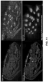
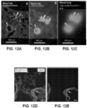
- a second image is obtained to determine the signal intensity from the plurality of detection probes in the second image. Comparing the first image and the second image allows one to determine mislocalization of the analyte within the biological sample by comparing the first image of the biological sample to the second image of the biological sample. For example, analyte mislocalization can be determined by comparing the expression of a cellular marker (e.g., for a particular cell type or morphology) found in the first image with the same, or similar, cellular marker in the second image.
- a cellular marker e.g., for a particular cell type or morphology
- Prox1 is expressed in several brain regions of mammals including the cortex, dentate gyrus (DG) region of the hippocampus, cerebellum, and hypothalamus.
- Prox1 expression is known to occur in the DG region of the hippocampus, mRNA transcripts encoding the Prox1 protein will also be present in the DG region of the hippocampus. Comparing and/or aligning the expression profile of the cellular markers allows one to determine (e.g., calculate) the relative distance an analyte has migrated from its original position in the biological sample.
- RNA-templated ligation RTL
- the methods include contacting a biological sample with array of capture probes.
- the array is on a substrate and the array includes a plurality of capture probes, wherein a capture probe of the plurality includes a capture domain.
- the biological sample is contacted with a first probe and a second probe, wherein the first probe and the second probe each include one or more sequences that are substantially complementary to sequences of the analyte, and wherein the second probe includes a capture probe capture domain; the first probe and the second probe hybridize to complementary sequences in the analyte.
- a ligation product comprising the first probe and the second probe is generated, and the ligation product is released from the analyte.
- the liberated ligation product is then available to hybridize to the capture domain of a probe on the array.
- padlock probes or snail probes described in section (II)(d) below can be used to determine mislocalization of the analyte on the array.
- a biological sample is deparaffinized, stained, and imaged. After destaining and decrosslinking, probes are added to the sample and hybridized to an analyte.
- the probes are DNA probes.
- the probes are diribo-containing probes.
- Probes are ligated to form a ligation product and released from the biological sample, for example using an endonuclease such as RNAse H.
- the ligation product can be captured on an array by a capture probe, where the ligation product can hybridize to a padlock probe or snail probe as described in section (II)(d) below.
- the ligation product includes a capture probe capture domain, which can hybridize to a capture probe (e.g., a capture probe immobilized, directly or indirectly, on a substrate).
- methods provided herein include contacting a biological sample with a substrate, wherein the capture probe is affixed to the substrate (e.g., immobilized to the substrate, directly or indirectly).
- the capture probe capture domain of the ligated product specifically binds to the capture domain.
- the capture probe can also include a unique molecular identifier (UMI), a spatial barcode, a functional sequence, a cleavage domain, or a combination thereof.
- UMI unique molecular identifier
- probe pairs or probe sets; the terms are interchangeable.
- the probe pairs are designed so that each probe hybridizes to a sequence in an analyte that is specific to the analyte (e.g., compared to the entire transcriptome). That is, in some instances, a single probe pair can be specific to a single analyte.
- probes can be designed so that one of the probes of a pair is a probe that hybridizes to a specific sequence. Then, the other probe can be designed to detect a mutation of interest. Accordingly, in some instances, multiple second probes can be designed and can vary so that each probe binds to a specific sequence. For example, a second probe can be designed to hybridize to a wild-type sequence, and another second probe can be designed to hybridize (and therefore detect) a mutated sequence. Thus, in some instances, a probe set can include one first probe and two second probes (or vice versa).
- probes can be designed so that they cover conserved regions of an analyte.
- a probe or probe pair can hybridize to similar analytes in a biological sample (e.g., to detect conserved or similar analytes) or in different biological samples (e.g., across different species).
- probe sets cover all or nearly all of a genome (e.g., human genome). In instances where probe sets are designed to cover an entire genome (e.g., the human genome), the methods disclosed herein can detect analytes in an unbiased manner.
- one probe oligonucleotide pair is designed to cover one analyte (e.g., transcript). In some instances, more than one probe oligonucleotide pair (e.g., a probe pair comprising a first probe and a second probe) is designed to cover one analyte (e.g., transcript).
- probe oligonucleotide pair does not hybridize to the entire analyte (e.g., a transcript), but instead the probe oligonucleotide pair hybridizes to a portion of the entire analyte (e.g., transcript).
- probe oligonucleotides pair e.g., a probe pair comprising a first probe and a second probe
- about 20,000 probe oligonucleotides pair are used in the methods described herein.
- the subset of analytes includes 2, 3, 4, 5, 6, 7, 8, 9, 10, 11, 12, 13, 14, 15, 16, 17, 18, 19, 20, 21, 22, 23, 24, 25, 26, 27, 28, 29, 30, 31, 32, 33, 34, 35, 36, 37, 38, 39, 40, 41, 42, 43, 44, 45, 46, 47, 48, 49, 50, about 55, about 60, about 65, about 70, about 75, about 80, about 85, about 90, about 95, about 100, about 110, about 120, about 130, about 140, about 150, about 160, about 170, about 180, about 190, about 200, about 225, about 250, about 275, about 300, about 325, about 350, about 375, about 400, about 425, about 450, about 475, about 500, about 600, about 700, about 800, about 900, or about 1000 analytes.
- a first probe and/or the second probe includes a nucleic acid sequence that is about 10 nucleotides to about 100 nucleotides (e.g., a sequence of about 10 nucleotides to about 90 nucleotides, about 10 nucleotides to about 80 nucleotides, about 10 nucleotides to about 70 nucleotides, about 10 nucleotides to about 60 nucleotides, about 10 nucleotides to about 50 nucleotides, about 10 nucleotides to about 40 nucleotides, about 10 nucleotides to about 30 nucleotides, about 10 nucleotides to about 20 nucleotides, about 20 nucleotides to about 100 nucleotides, about 20 nucleotides to about 90 nucleotides, about 20 nucleotides to about 80 nucleotides, about 20 nucleotides to about 70 nucleotides, about 20 nucleotides to about 60 nucleo
- proteins in a biological sample can be detected using analyte capture agents, as described herein.
- the analyte capture agents are contacted with the biological sample before the biological sample is contacted with an array.
- the analyte capture agents are contacted with the biological sample after the biological sample is contacted with the array.
- an analyte binding moiety of the analyte capture agent interacts (e.g., binds) with an analyte (e.g., protein) in a biological sample.
- the analyte binding moiety is an antibody, aptamer or antigen-binding fragment.
- Analyte capture agents can also include a conjugated oligonucleotide that can comprise one or more domains.
- the conjugated oligonucleotide can include an analyte binding moiety barcode and an analyte capture sequence.
- the analyte binding moiety barcode, or a complement thereof refers to (e.g., identifies) a barcode that is associated with or otherwise identifies the analyte binding moiety.
- the conjugated oligonucleotide can include an analyte capture sequence.
- the analyte capture sequence is capable of interacting with (e.g., hybridizing) a capture domain of a capture probe on a substrate.
- an extended capture probe is generated using the analyte capture sequence as a template.
- the padlock or snail probes (described in the Section (II)(d) below) can hybridize to the extended capture probe.
- no extended capture probe is generated using the analyte capture sequence as a template.
- the padlock or snail probes (described in the Section (II)(d) below) hybridize directly to the analyte capture sequence.
- analyte capture agents are capable of binding to analytes present inside a cell.
- analyte capture agents are capable of binding to cell surface analytes that can include, without limitation, a receptor, an antigen, a surface protein, a transmembrane protein, a cluster of differentiation protein, a protein channel, a protein pump, a carrier protein, a phospholipid, a glycoprotein, a glycolipid, a cell-cell interaction protein complex, an antigen-presenting complex, a major histocompatibility complex, an engineered T-cell receptor, a T-cell receptor, a B-cell receptor, a chimeric antigen receptor, an extracellular matrix protein, a posttranslational modification (e.g., phosphorylation, glycosylation, ubiquitination, nitrosylation, methylation, acetylation or lipidation) state of a cell surface protein, a gap junction, and an adherens junction.
- a posttranslational modification e.g
- the analyte capture agents are capable of binding to cell surface analytes that are post-translationally modified.
- analyte capture agents can be specific for cell surface analytes based on a given state of posttranslational modification (e.g., phosphorylation, glycosylation, ubiquitination, nitrosylation, methylation, acetylation or lipidation), such that a cell surface analyte profile can include posttranslational modification information of one or more analytes.
- the analyte capture agent includes a capture agent barcode domain that is conjugated or otherwise attached to the analyte binding moiety. In some embodiments, the capture agent barcode domain is covalently-linked to the analyte binding moiety. In some embodiments, a capture agent barcode domain is a nucleic acid sequence. In some embodiments, a capture agent barcode domain includes an analyte binding moiety barcode and an analyte capture sequence.
- analyte binding moiety barcode refers to a barcode that is associated with or otherwise identifies the analyte binding moiety. In some embodiments, by identifying an analyte binding moiety and its associated analyte binding moiety barcode, the analyte to which the analyte binding moiety binds can also be identified.
- An analyte binding moiety barcode can be a nucleic acid sequence of a given length and/or sequence that is associated with the analyte binding moiety.
- An analyte binding moiety barcode can generally include any of the variety of aspects of barcodes described herein.
- an analyte capture agent that is specific to one type of analyte can have coupled thereto a first capture agent barcode domain (e.g., that includes a first analyte binding moiety barcode), while an analyte capture agent that is specific to a different analyte can have a different capture agent barcode domain (e.g., that includes a second barcode analyte binding moiety barcode) coupled thereto.
- a capture agent barcode domain can include an analyte binding moiety barcode that permits identification of the analyte binding moiety to which the capture agent barcode domain is coupled.
- the selection of the capture agent barcode domain can allow significant diversity in terms of sequence, while also being readily attachable to most analyte binding moieties (e.g., antibodies or aptamers) as well as being readily detected, (e.g., using sequencing or array technologies).
- analyte binding moieties e.g., antibodies or aptamers
- the capture agent barcode domain of an analyte capture agent includes an analyte capture sequence.
- analyte capture sequence refers to a region or moiety configured to hybridize to, bind to, couple to, or otherwise interact with a capture domain of a capture probe.
- an analyte capture sequence includes a nucleic acid sequence that is complementary to or substantially complementary to the capture domain of a capture probe such that the analyte capture sequence hybridizes to the capture domain of the capture probe.
- an analyte capture sequence comprises a poly(A) nucleic acid sequence that hybridizes to a capture domain that comprises a poly(T) nucleic acid sequence.
- an analyte capture sequence comprises a poly(T) nucleic acid sequence that hybridizes to a capture domain that comprises a poly(A) nucleic acid sequence. In some embodiments, an analyte capture sequence comprises a non-homopolymeric nucleic acid sequence that hybridizes to a capture domain that comprises a non-homopolymeric nucleic acid sequence that is complementary (or substantially complementary) to the non-homopolymeric nucleic acid sequence of the analyte capture region.
- the capture agent barcode domain coupled to the analyte binding moiety includes a cleavable domain.
- the capture agent barcode domain can be cleaved and collected for downstream analysis according to the methods as described herein.
- analyte capture sequence hybridizes to the capture domain, it can then interact with padlock probes or snail probes as described below.
- This disclosure features a method for measuring the mislocalization of an analyte in a biological sample using rolling circle amplification (RCA). Initially, after hybridization of an analyte to a capture probe or the complement thereof, in some instances, a padlock probe hybridizes to the extended capture probe, analyte derived molecule, or the complement thereof.
- RCA rolling circle amplification
- a "padlock probe” refers to an oligonucleotide that includes, at its 5' and 3' ends, sequences (e.g., a first sequence at the 5' end and a second sequence at the 3' end) that are complementary to adjacent or nearby portions (e.g., a first portion and a second portion) of the analyte or an analyte derived molecule.
- Padlock probes are designed such that they comprise multiple substantially or fully complementary sequences to the analyte or analyte derived molecule.
- an analyte includes nucleic acids (e.g., DNA or RNA) in some instances.
- an example of an analyte derived molecule includes a molecule comprising a protein binding moiety conjugated to an oligonucleotide.
- a further example is a product of RNA-templated ligation (RTL) as disclosed in US 2021/0348221 , US 2021/0285046 , and WO 2021/133849 , each of which is incorporated by reference in its entirety.
- RTL RNA-templated ligation
- SNAIL-RCA i.e., RCA using snail probes
- SNAIL-RCA was adopted for directed amplification of RNA in cells by proximity ligation.
- two primers are used, one of which binds to its target (e.g., an extended capture probe; a ligation product; or an oligonucleotide from an analyte capture agent; or any complements thereof) at a 3' position and contains the sequence cognate to a common ligation junction.
- the second primer contains a sequence proximal, which binds slightly upstream on the target, but which also anneals to the ligation junction. After ligation of the two SNAIL primers, rolling circle amplification occurs which results in amplification of the target or complement thereof.
- the two ends of the padlock probe are either brought into contact, or an end is extended until the two ends are brought into contact, allowing circularization of the padlock probe or snail probe by ligation (e.g., ligation using any of the methods described herein).
- the ligation product can be referred to as the "circularized padlock probe” or “circularized snail probe.”
- rolling circle amplification can be used to amplify the circularized padlock probe or snail probe.
- a first sequence of a padlock probe or snail probe includes a sequence that is substantially complementary to a first portion of the analyte or analyte derived molecule.
- the first portion of the analyte or analyte derived molecule is 5' to the second portion of the analyte or the analyte derived molecule.
- the first sequence is at least 70% identical (e.g., at least 75% identical, at least 80% identical, at least 85% identical, at least 90% identical, at least 95% identical, or at least 99% identical) to the first portion.
- a backbone sequence of a padlock probe or snail probe includes a sequence that is substantially complementary to an amplification primer.
- the amplification primer can be a primer used in a rolling circle amplification reaction (RCA) of the ligated padlock probe or snail probe hybridized to the analyte or analyte derived molecules.
- Rolling circle amplification is well known in the art and includes a process by which circularized nucleic acid molecules are amplified with a DNA polymerase with strand displacement capabilities (and other necessary reagents for amplification to occur), thereby creating multiple concatenated copies of the circularized nucleic acid molecules.
- the backbone sequence includes a functional sequence.
- the backbone sequence includes a unique molecule identifier (UMI) or barcode sequence (e.g., any of the exemplary barcode sequences described herein).
- the barcode sequence includes a sequence that is substantially complementary to an amplification primer.
- the backbone UMI or backbone barcode sequence in the padlock probe or snail probe can be used to identify the padlock or snail sequence.
- a second sequence of a padlock probe or snail probe includes a sequence that is substantially complementary to a second portion of the analyte or analyte derived molecule.
- the second portion of the analyte or analyte derived molecule is 3' to the first portion of the analyte or the analyte derived molecule.
- the second sequence is at least 70% identical (e.g., at least 75% identical, at least 80% identical, at least 85% identical, at least 90% identical, at least 95% identical, or at least 99% identical) to the second portion.
- the first sequence is substantially complementary to a first portion of the analyte or analyte derived molecule that is directly adjacent to the second portion of the analyte or analyte derived molecule to which the second sequence is substantially complementary.
- the first sequence is ligated to the second sequence, thereby creating a circularized padlock probe or snail probe.
- the first sequence is substantially complementary to a first portion of the analyte or analyte derived molecule that is not directly adjacent to the second portion of the analyte or analyte derived molecule to which the second sequence is substantially complementary.
- a "gap" exists between where the first sequence is hybridized to the first potion and where the second sequence is hybridized to the second portion.
- nucleotide sequence e.g., a gap in the analyte between the first portion and the second portion of at least 1-100, 1-90, 1-80, 1-70, 1-60, 1-50, 1-40, 1-30, 1-20, 1-10, 1-9, 1-8, 1-7, 1-6, 1-5, 1-4, 1-3, 1-2 or 1 nucleotide(s).
- a first sequence having a sequence that is complementary to a sequence 5' of the gap and a second sequence having a sequence that is complementary to a sequence 3' of the gap each bind to an analyte leaving a sequence (e.g., the "gap") in between the first and second sequences that is gap-filled thereby enabling ligation and generation of the circularized padlock probe or snail probe.
- the second sequence is extended enzymatically (e.g., using a reverse transcriptase).
- the "gap" sequence between the first sequence and the second sequence include one nucleotide, two nucleotides, three nucleotides, four nucleotides, five nucleotides, six nucleotides, seven nucleotides, eight nucleotides, nine nucleotides, ten nucleotides, 11 nucleotides, 12 nucleotides, 13 nucleotides, 14 nucleotides, 15 nucleotides, 16 nucleotides, 17 nucleotides, 18 nucleotides, 19 nucleotides, 20 nucleotides, 21 nucleotides, 22 nucleotides, 23 nucleotides, 24 nucleotides, at least 25 nucleotides, at least 30 nucleotide, at least 35 nucleotides, at least 40 nucleotides, at least 45 nucleotides, or at least 50 nucleotides.
- extending the second sequence of the padlock probe or snail probe includes a nucleic acid extension reaction (e.g., any of the nucleic acid extension reactions described herein). In some embodiments, extending the second sequence of the padlock probe or snail probe includes reverse transcribing the analyte or analyte derived molecule. In some embodiments, extending the second sequence of the padlock probe or snail probe includes using a reverse transcriptase (e.g., any of the reverse transcriptases described herein). In some embodiments, extending the second sequence of the padlock probe or snail probe includes using a Moloney Murine Leukemia Virus (M-MMLV) reverse transcriptase. In some embodiments, the reverse transcriptase includes strand displacement properties.
- M-MMLV Moloney Murine Leukemia Virus
- extending the second sequence of the padlock probe generates a sequence that is complementary to the analyte or the analyte derived molecule. In some embodiments, extending the second sequence of the padlock probe or snail probe generates an extended second sequence of the padlock probe or snail probe that is complementary to the analyte or analyte derived molecule. In some embodiments, second sequence of the padlock probe or snail probe generates a sequence that is adjacent to the first sequence of the padlock probe or snail probe.
- the ligation step includes ligating the second sequence to the first sequence of the padlock probe or snail probe using enzymatic or chemical ligation.
- the ligation reaction comprises click chemistry.
- the ligase is selected from a T4 RNA ligase (Rnl2), a SplintR ligase, a single stranded DNA ligase, or a T4 DNA ligase.
- the ligase is a T4 RNA ligase (Rnl2) ligase.
- the ligase is a pre-activated T4 DNA ligase as described herein.
- a non-limiting example describing methods of generating and using pre-activated T4 DNA include U.S. Patent App. No. 8,790,873 , the entire contents of which are herein incorporated by reference.
- This disclosure features a method for determining the mislocalization of an analyte in a biological sample using amplification of a circularized padlock probe or snail probe.
- the method includes an amplifying step where one or more amplification primers are hybridized to the circularized padlock probe or snail probe and the circularized padlock probe or snail probe is amplified using a polymerase.
- DNA polymerases that can be used include, but are not limited to: E.coli DNA polymerase I, Bsu DNA polymerase, Bst DNA polymerase, Taq DNA polymerase, VENT TM DNA polymerase, DEEPVENT TM DNA polymerase, LongAmp ® Taq DNA polymerase, LongAmp ® Hot Start Taq DNA polymerase, Crimson LongAmp ® Taq DNA polymerase, Crimson Taq DNA polymerase, OneTaq ® DNA polymerase, OneTaq ® Quick-Load ® DNA polymerase, Hemo KlenTaq ® DNA polymerase, REDTaq ® DNA polymerase, Phusion ® DNA polymerase, Phusion ® High-Fidelity DNA polymerase, Platinum Pfx DNA polymerase, AccuPrime Pfx DNA polymerase, Phi29 DNA polymerase, Klenow fragment, Pwo DNA polymerase, Pfu DNA polymerase, T4
- the amplification product(s) can be detected by hybridization of detection probes to the amplification product, which identify the location of the mislocalized analyte in the biological sample.
- the location of the mislocalized analyte can then be compared against an initial (or original) location of an analyte based on imaging prior to permeabilization of the biological sample and migration of the analytes.
- the amplifying step includes rolling circle amplification (RCA).
- rolling circle amplification refers to a polymerization reaction carried out using a single-stranded circular DNA (e.g., a circularized padlock probe or snail probe) as a template and an amplification primer that is substantially complementary to the single-stranded circular DNA (e.g., the circularized padlock probe or snail probe) to synthesize multiple continuous single-stranded copies of the template DNA (e.g., the circularized padlock probe or snail probe).
- RCA includes hybridizing one or more amplification primers to the circularized padlock probe or snail probe and amplifying the circularized padlock probe or snail probe using a DNA polymerase with strand displacement activity, for example Phi29 DNA polymerase.
- a first RCA reaction includes a first padlock probe or snail probe and a first amplification primer (or plurality of first amplification primers).
- a first RCA reaction can include the first padlock probe or snail probe hybridizing to a first analyte or first analyte derived molecule.
- an RCA reaction is carried out using a DNA polymerase and a dNTP mix including uracil, adenine, guanine and cytosine. In such cases, the uracils are incorporated into the amplified padlock probe or snail probe. In some embodiments, an RCA reaction is carried out using a DNA polymerase and a dNTP mix including uracil, adenine, guanine, cytosine and thymine. In such cases, uracils and thymines are both incorporated into the amplified padlock probe or snail probe.
- the dNTP molecule is labeled.
- the label is a fluorescent label that is chemically coupled to the nucleotide.
- a fluorescent label can be attached to any of the following nucleotides dATP, dCTP, dGTP, dUTP, and dTTP.
- the fluorescent label can be any fluorescent label known in the art, including but not limited to cyanine 5 dye (Cy-5), cyanine 3 dye (Cy-3), fluorescein, rhodamine, phosphor, coumarin, polymethadine dye, fluorescent phosphoramidite, Texas red, green fluorescent protein, acridine, cyanine, and 5-(2'-aminoethyl)-aminonaphthalene-1-sulfonic acid (EDANS).
- cyanine 5 dye Cy-5
- Cy-3 cyanine 3 dye
- fluorescein fluorescein
- rhodamine fluorescein
- phosphor phosphor
- coumarin polymethadine dye
- fluorescent phosphoramidite fluorescent phosphoramidite
- Texas red green fluorescent protein
- acridine acridine
- cyanine 5-(2'-aminoethyl)-aminonaphthalene-1-sulfonic acid
- the method further includes a second RCA reaction.
- a second padlock probe or snail probe and a second amplification primer are added to the substrate.
- the second padlock probe or snail probe hybridizes to a second analyte or second analyte derived molecule and is ligated thereby creating a second circularized padlock probe or snail probe.
- the second circularized padlock probe or snail probe can then be amplified using RCA.
- the second padlock probes or snail probes include a third sequence and a fourth sequence where each are substantially complementary to sequences on a second analyte (e.g., a different analyte then was bound by the first padlock probe or snail probe in the first RCA reaction).
- the second padlock probe or snail probe includes: (i) a third sequence substantially complementary to the second analyte or second analyte derived molecule, (ii) a backbone sequence comprising a barcode, and (iii) a fourth sequence that is substantially complementary to a sequence adjacent to the third sequence in the second analyte or second analyte derived molecule.
- the third sequence is at least 70% identical (e.g., at least 75% identical, at least 80% identical, at least 85% identical, at least 90% identical, at least 95% identical, or at least 99% identical) to a portion of the second analyte or second analyte derived molecule.
- the fourth sequence is at least 70% identical (e.g., at least 75% identical, at least 80% identical, at least 85% identical, at least 90% identical, at least 95% identical, or at least 99% identical) to a sequence adjacent to the third sequence in the second analyte or second analyte derived molecule.
- the method further includes digesting the amplified circularized padlock probe or snail probe by using an enzyme that cleaves a nucleic acid sequence (e.g., an enzyme that cleaves nucleic acid sequences that include uracil). Digesting and/or removing the amplified circularized padlock probe or snail probe after detection of the detection probes can alleviate crowding that may result from the physical size of the RCA product and/or the detection probe.
- the enzymatic digestion of the amplified circularized padlock probe or snail probe includes a uracil-DNA glycosylase (UDG) that cleaves nucleic acid sequences that include uracil incorporated into the amplified padlock probe or snail probe.
- the enzyme is a combination of enzymes, such as UDG and DNA glycosylase lyase such as Endonuclease VIII.
- an amplification primer includes a sequence that is substantially complementary to one or more of the first sequence, the backbone sequence, or the second sequence of the padlock probe or snail probe.
- the amplification primer can be substantially complementary to the backbone sequence.
- the amplification primer includes a sequence that is substantially complementary to the padlock probe or snail probe and an additional portion of the analyte or analyte derived molecule.
- the amplification primer can be substantially complementary to the backbone sequence and a portion of the analyte or analyte derived molecule that does not include the first and second portion of the padlock probe or snail probe.
- the amplification primer includes a sequence that is substantially complementary to two or more of the first sequence, the backbone sequence, or the second sequence of the padlock probe or snail probe and an additional portion of the analyte or analyte derived molecule.
- substantially complementary it is meant that the amplification primer is at least 70%, at least 75%, at least 80%, at least 85%, at least 90%, at least 91%, at least 92%, at least 93%, at least 94%, at least 95%, at least 96%, at least 97%, at least 98%, at least 99%, complementary to a sequence in the circularized padlock probe or snail probe.
- the amplification primer is at least 70%, at least 75%, at least 80%, at least 85%, at least 90%, at least 91%, at least 92%, at least 93%, at least 94%, at least 95%, at least 96%, at least 97%, at least 98%, at least 99%complementary to a sequence in the analyte or analyte derived molecule.
- This disclosure features a method for determining the mislocalization of an analyte in a biological sample using detection of a signal that corresponds to an amplified circularized padlock probe or snail probe (which thereby corresponds to an analyte or analyte binding molecule).
- the method includes a step of detecting a signal corresponding to the amplified circularized padlock probe or snail probe on the substrate, thereby identifying the location of the mislocalized analyte in the biological sample.
- the method includes a detecting step that includes determining (i) all or a part of the sequence of the amplified circularized padlock probe or snail probe on the substrate and using the determined sequence of the padlock probe or snail probe to identify the location of the analyte in the biological sample.
- the detecting step includes contacting the amplified circularized padlock probe or snail probe with a plurality of detection probes.
- a detection probe of the plurality of detection probes includes a sequence that is substantially complementary to a sequence of the padlock probe or snail probe, circularized padlock probe or snail probe, or amplified circularized padlock probe or snail probe and a detectable label.
- the detection probe of the plurality of detection probes can include a sequence that is substantially complementary to a sequence of amplified circularized padlock probe or snail probe and a detectable label.
- the detection probe is at least 70%, at least 75%, at least 80%, at least 85%, at least 90%, at least 91%, at least 92%, at least 93%, at least 94%, at least 95%, at least 96%, at least 97%, at least 98%, at least 99%complementary to a sequence in the amplified circularized padlock probe or snail probe.
- the detectable label is a fluorophore.
- the fluorophore can be from a group that includes: 7-AAD (7-Aminoactinomycin D), Acridine Orange (+DNA), Acridine Orange (+RNA), Alexa Fluor ® 350, Alexa Fluor ® 430, Alexa Fluor ® 488, Alexa Fluor ® 532, Alexa Fluor ® 546, Alexa Fluor ® 555, Alexa Fluor ® 568, Alexa Fluor ® 594, Alexa Fluor ® 633, Alexa Fluor ® 647, Alexa Fluor ® 660, Alexa Fluor ® 680, Alexa Fluor ® 700, Alexa Fluor ® 750, Allophycocyanin (APC), AMCA / AMCA-X, 7-Amino-4-methylcoumarin, 6-Aminoquinoline, Aniline Blue, ANS, APC-Cy7, ATTO-TAG TM CBQCA, ATTO
- a detection probe of the plurality of detection probes includes a nucleotide sequence that is different from other detection probes, thereby enabling detection of signals from two or more detection probe sequences (e.g., two or more different amplified circularized padlock probe or snail probe).
- each of the two or more different nucleotide sequences is located on the same amplified circularized padlock probe or snail probe.
- each of the two or more different sequences is located on two or more different amplified circularized padlock probe or snail probes (e.g., each amplified circularized padlock probe or snail probe is derived from a different analyte or analyte derived molecule).
- the detecting step includes contacting the amplified circularized padlock probe or snail probe with a detectable label that can hybridize to nucleic acid sequences in a non-sequence dependent manner.
- the single-stranded copies of the template DNA produced by RCA can be detected using fluorescent dyes that non-specifically bind to the single-stranded nucleic acid (e.g., the amplified circularized padlock probe or snail probe).
- fluorescent dyes can bind, either directly or indirectly, to the single stranded nucleic acid of the RCA product.
- the dye can then be detected as a signal corresponding to the amplified circularized padlock probe or snail probe (i.e., the location and/or the abundance of the circularized padlock probe or snail probe).
- fluorescent dyes that can bind to single stranded nucleic acids include: TOTO ® -1 / TO-PRO ® -1, TOTO ® -3 / TO-PRO ® -3, TO-PRO ® -5, Ethidium bromide, Ethidium homodimer-1 (EthD-1), YOYO ® -1 / YO-PRO ® -1, YOYO ® -3 / YO-PRO ® -3, 7-AAD (7-Aminoactinomycin D), and OliGreen ® .
- the detecting step includes contacting the amplified circularized padlock probe or snail probe with a detection probe (e.g., any of the exemplary detection probes described herein (e.g., a detection probe that includes one or more fluorophores)) and a fluorescent dye that can label single-stranded DNA in a non-sequence dependent manner (e.g., any of the exemplary fluorescent dyes described herein).
- a detection probe e.g., any of the exemplary detection probes described herein (e.g., a detection probe that includes one or more fluorophores)
- a fluorescent dye that can label single-stranded DNA in a non-sequence dependent manner e.g., any of the exemplary fluorescent dyes described herein.
- the fluorescent dye can be used to normalize the fluorescent signal detected from the detection probe.
- detecting the signal or signals that correspond to the amplified circularized padlock probes or snail probes on the substrate include obtaining an image corresponding to the analyte and/or analyte derived molecule on the substrate. In some embodiments, the method further includes registering image coordinates to a fiducial marker.
- the method includes repeating the detecting step with a second plurality of detection probes.
- the method includes removing the detection probes from the first detecting step (e.g., via enzymatic digestion) and contacting the amplified circularized padlock probe or snail probe with a second plurality of detection probes.
- a detection probe of the second plurality of detection probes includes a sequence that is substantially complementary to a sequence of the padlock probe or snail probe and that is non-overlapping, partially overlapping, or completely overlapping with the sequence to which a detection probe from the first plurality of detection probes is substantially complementary.
- determining (i) all or a part of the sequence of the amplified circularized padlock probe or snail probe on the substrate includes determining the barcode sequence on the padlock probe or snail probe, wherein the location of the analyte in the biological sample can be identified.
- In situ sequencing of the analyte or analyte derived molecule, the nucleic acid molecule, the analyte binding moiety barcode, or the ligation product process is described in PCT Patent Application Publication No. WO 2020/047005 , which is incorporated by reference in its entirety.
- This disclosure features a method for identifying a location of an analyte in a biological sample using a substrate that includes a plurality of capture probes, where a capture probe of the plurality of capture probes include a capture domain.
- the capture probe is affixed to the substrate at a 5' end.
- the plurality of capture probes are uniformly distributed on a surface of the substrate, following no particular pattern.
- the plurality of capture probes are located on a surface of the substrate but are distributed on the substrate according to a pattern.
- the capture probes do not include spatial barcodes. Instead, to determine mislocalization of an analyte, spatial analysis is performed with the aid of fluorescent readout, as described herein.
- the substrate comprises a lawn of capture probes that include a capture domain, but do not include a spatial barcode.
- the substrate includes a plurality of capture probes, where a capture probe of the plurality of capture probes includes a capture domain and a spatial barcode.
- the substrate includes a fiducial marker (e.g., any of the fiducial markers described herein).
- the fiducial markers can be used as a point of reference on the substrate. For example, the spatial location of a signal detected from a detection probe can be determined by using the fiducial marker as a point of reference on the substrate.
- the fiducial marker can be present on a substrate to aid in orientation of the biological sample.
- a fiducial marker can be an immobilized molecule with which a detectable signal molecule (e.g., a detectable label from a detection probe) can interact to generate a signal.
- a detectable signal molecule e.g., a detectable label from a detection probe
- a fiducial marker can comprise a nucleic acid linked or coupled to a chemical moiety capable of fluorescing when subjected to light of a specific wavelength (or range of wavelengths) at a fixed position on the substrate.
- the biological sample comprises a tissue section. In some embodiments, the biological sample comprises a fresh frozen sample. In some embodiments, the biological sample comprises live or fixed single cells. In some embodiments, the biological sample comprises a FFPE sample.
- the method includes obtaining an image of the biological sample; registering the image data to spatial locations on an array.
- the imaging includes a brightfield image.
- the method further includes contacting the biological sample with one or more stains.
- the one or more stains comprise a histology stain (e.g., any of the histology stains described herein or known in the art).
- the one or more stains comprises hematoxylin and/or eosin.
- the one or more stains comprise one or more optical labels (e.g., any of the optical labels described herein).
- the one or more optical labels are selected from the group consisting of: fluorescent, radioactive, chemiluminescent, calorimetric, or colorimetric labels.
- the methods are applied to analyte, or analyte derived molecules (e.g., an mRNA molecule, a gDNA molecule, a product of reverse transcription (e.g., an extended capture probe), and an analyte binding moiety barcode (e.g., a binding moiety barcode that identifies that analyte binding moiety (e.g., an antibody))).
- analyte or analyte derived molecules comprise RNA and/or DNA.
- the analyte or analyte derived molecules comprise one or more proteins.
- biological samples can be stained using a wide variety of stains and staining techniques. In some instances, the biological sample is stained before adding the probes to the biological sample. In some instances, the biological sample is stained after adding the probes to the biological sample.
- the biological sample is a section of a tissue (e.g., a 10 ⁇ m section).
- the biological sample is dried after placement onto a glass slide.
- the biological sample is dried at 42°C. In some instances, drying occurs for about 1 hour, about 2, hours, about 3 hours, or until the sections become transparent.
- the biological sample can be dried overnight (e.g., in a desiccator at room temperature).
- a sample can be stained using any number of biological stains, including but not limited to, acridine orange, Bismarck brown, carmine, coomassie blue, cresyl violet, DAPI, eosin, ethidium bromide, acid fuchsine, hematoxylin, Hoechst stains, iodine, methyl green, methylene blue, neutral red, Nile blue, Nile red, osmium tetroxide, propidium iodide, rhodamine, or safranin.
- the methods disclosed herein include imaging the biological sample. In some instances, imaging the sample occurs prior to deaminating the biological sample.
- the sample can be stained using known staining techniques, including Can-Grunwald, Giemsa, hematoxylin and eosin (H&E), Jenner's, Leishman, Masson's trichrome, Papanicolaou, Romanowsky, silver, Sudan, Wright's, and/or Periodic Acid Schiff (PAS) staining techniques.
- PAS staining is typically performed after formalin or acetone fixation. In some instances, the stain is an H&E stain.
- the biological sample can be stained using a detectable label (e.g., radioisotopes, fluorophores, chemiluminescent compounds, bioluminescent compounds, and dyes) as described elsewhere herein.
- a biological sample is stained using only one type of stain or one technique.
- staining includes biological staining techniques such as H&E staining.
- staining includes identifying analytes using fluorescently-conjugated antibodies.
- a biological sample is stained using two or more different types of stains, or two or more different staining techniques.
- a biological sample can be prepared by staining and imaging using one technique (e.g., H&E staining and brightfield imaging), followed by staining and imaging using another technique (e.g., IHC/IF staining and fluorescence microscopy) for the same biological sample.
- one technique e.g., H&E staining and brightfield imaging
- another technique e.g., IHC/IF staining and fluorescence microscopy
- biological samples can be destained.
- Methods of destaining or decoloring a biological sample are known in the art, and generally depend on the nature of the stain(s) applied to the sample.
- H&E staining can be destained by washing the sample in HCl, or any other acid (e.g., selenic acid, sulfuric acid, hydroiodic acid, benzoic acid, carbonic acid, malic acid, phosphoric acid, oxalic acid, succinic acid, salicylic acid, tartaric acid, sulfurous acid, trichloroacetic acid, hydrobromic acid, hydrochloric acid, nitric acid, orthophosphoric acid, arsenic acid, selenous acid, chromic acid, citric acid, hydrofluoric acid, nitrous acid, isocyanic acid, formic acid, hydrogen selenide, molybdic acid, lactic acid, acetic acid, carbonic acid, hydrogen sulfide, or combinations thereof).
- destaining can include 1, 2, 3, 4, 5, or more washes in an acid (e.g., HCl).
- destaining can include adding HCl to a downstream solution (e.g., permeabilization solution).
- destaining can include dissolving an enzyme used in the disclosed methods (e.g., pepsin) in an acid (e.g., HCl) solution.
- other reagents can be added to the destaining solution to raise the pH for use in other applications. For example, SDS can be added to an acid destaining solution in order to raise the pH as compared to the acid destaining solution alone.
- one or more immunofluorescence stains are applied to the sample via antibody coupling.
- Such stains can be removed using techniques such as cleavage of disulfide linkages via treatment with a reducing agent and detergent washing, chaotropic salt treatment, treatment with antigen retrieval solution, and treatment with an acidic glycine buffer.
- Methods for multiplexed staining and destaining are described, for example, in Bolognesi et al., J. Histochem. Cytochem. 2017; 65(8): 431-444 , Lin et al., Nat Commun. 2015; 6:8390 , Pirici et al., J. Histochem. Cytochem. 2009; 57:567-75 , and Glass et al., J. Histochem. Cytochem. 2009; 57:899-905 , the entire contents of each of which are incorporated herein by reference.
- immunofluorescence or immunohistochemistry protocols can be performed as a part of, or in addition to, the exemplary spatial workflows presented herein.
- tissue sections can be fixed according to methods described herein.
- the biological sample can be transferred to an array (e.g., capture probe array), wherein analytes (e.g., proteins) are probed using immunofluorescence protocols.
- analytes e.g., proteins
- the sample can be rehydrated, blocked, and permeabilized (3X SSC, 2% BSA, 0.1% Triton X, 1 U/ ⁇ l RNAse inhibitor for 10 minutes at 4°C) before being stained with fluorescent primary antibodies (1: 100 in 3XSSC, 2% BSA, 0.1% Triton X, 1 U/ ⁇ l RNAse inhibitor for 30 minutes at 4°C).
- the biological sample can be washed, coverslipped (in glycerol + 1 U/ ⁇ l RNAse inhibitor), imaged (e.g., using a confocal microscope or other apparatus capable of fluorescent detection), washed, and processed according to analyte capture or spatial workflows described herein.
- a glycerol solution and a cover slip can be added to the sample.
- the glycerol solution can include a counterstain (e.g., DAPI).
- an antigen retrieval buffer can improve antibody capture in IF/IHC protocols.
- An exemplary protocol for antigen retrieval can be preheating the antigen retrieval buffer (e.g., to 95°C), immersing the biological sample in the heated antigen retrieval buffer for a predetermined time, and then removing the biological sample from the antigen retrieval buffer and washing the biological sample.
- optimizing permeabilization can be useful for identifying intracellular analytes.
- Permeabilization optimization can include selection of permeabilization agents, concentration of permeabilization agents, and permeabilization duration. Tissue permeabilization is discussed elsewhere herein.
- blocking an array and/or a biological sample in preparation of labeling the biological sample decreases nonspecific binding of the antibodies to the array and/or biological sample (decreases background).
- Some embodiments provide for blocking buffers/blocking solutions that can be applied before and/or during application of the label, wherein the blocking buffer can include a blocking agent, and optionally a surfactant and/or a salt solution.
- a blocking agent can be bovine serum albumin (BSA), serum, gelatin (e.g., fish gelatin), milk (e.g., non-fat dry milk), casein, polyethylene glycol (PEG), polyvinyl alcohol (PVA), or polyvinylpyrrolidone (PVP), biotin blocking reagent, a peroxidase blocking reagent, levamisole, Carnoy's solution, glycine, lysine, sodium borohydride, pontamine sky blue, Sudan Black, trypan blue, FITC blocking agent, and/or acetic acid.
- the blocking buffer/blocking solution can be applied to the array and/or biological sample prior to and/or during labeling (e.g., application of fluorophore-conjugated antibodies) to the biological sample.
- additional reagents are added to the biological sample, prior to the addition of the probes.
- Additional reagents can be any reagent known in the art, so long as it preserves the integrity of an analyte.
- the biological sample is deparaffinized.
- Deparaffinization can be achieved using any method known in the art.
- the biological sample is treated with a series of washes that include xylene and various concentrations of ethanol.
- methods of deparaffinization include treatment with xylene (e.g., three washes at 5 minutes each).
- the methods further include treatment with ethanol (e.g., 100% ethanol, two washes 10 minutes each; 95% ethanol, two washes 10 minutes each; 70% ethanol, two washes 10 minutes each; 50% ethanol, two washes 10 minutes each).
- the biological sample can be washed with deionized water (e.g., two washes for 5 minutes each). It is appreciated that one skilled in the art can adjust these methods to optimize deparaffinization.
- the biological sample is decrosslinked.
- the biological sample is decrosslinked in a solution containing TE buffer (comprising Tris and EDTA).
- the TE buffer is basic (e.g., at a pH of about 9).
- decrosslinking occurs at about 50°C to about 80°C.
- decrosslinking occurs at about 70°C.
- decrosslinking occurs for about 1 hour at 70°C.
- the biological sample can be treated with an acid (e.g., 0.1M HCl for about 1 minute). After the decrosslinking step, the biological sample can be washed (e.g., with 1x PBST).
- the methods of preparing a biological sample for probe application include permeabilizing the sample.
- the biological sample is permeabilized using a phosphate buffer.
- the phosphate buffer is PBS (e.g., 1x PBS).
- the phosphate buffer is PBST (e.g., 1x PBST).
- the permeabilization step is performed multiple times (e.g., 3 times at 5 minutes each).
- the method includes permeabilizing (e.g., using any of the exemplary permeabilization methods described herein) the biological sample before, contemporaneously with, or after exposing the biological sample to the capture probe.
- the method includes processing the biological sample using any of the methods described herein. For example, the method can include selecting a region of interest of the biological sample by laser-capture microdissection prior to exposing the biological sample to the capture probe.
- the methods of preparing a biological sample for probe application include steps of equilibrating and blocking the biological sample.
- equilibrating is performed using a pre-hybridization (pre-Hyb) buffer.
- pre-Hyb buffer is RNase-free.
- pre-Hyb buffer contains no bovine serum albumin (BSA), solutions like Denhardt's, or other potentially nuclease-contaminated biological materials.
- BSA bovine serum albumin
- the equilibrating step is performed multiple times (e.g., 2 times at 5 minutes each; 3 times at 5 minutes each).
- the biological sample is blocked with a blocking buffer.
- the blocking buffer includes a carrier such as tRNA, for example yeast tRNA (e.g., at a final concentration of 10-20 ⁇ g/mL). In some instances, blocking can be performed for 5, 10, 15, 20, 25, or 30 minutes.
- any of the foregoing steps can be optimized for performance. For example, one can vary the temperature.
- the pre-hybridization methods are performed at room temperature. In some instances, the pre-hybridization methods are performed at 4°C (in some instances, varying the timeframes provided herein).
- This disclosure features a method of identifying a location of an analyte in a biological sample using a substrate that includes a plurality of capture probes, where a capture probe of the plurality of capture probes includes a capture domain.
- the capture probe does not include a spatial barcode.
- the capture probe includes a spatial barcode.
- a capture probe of the plurality of capture probes is affixed to the substrate at the 5' end of the capture probe.
- the capture probe in order to prevent extension of the capture probe during the amplifying step, includes a blocking moiety on the 3' end. Blocking or modifying the capture probes, particularly at the free 3' end of the capture domain, prior to contacting the biological sample with the array, prevents (1) modification of the capture probes, e.g., prevents the addition of a poly(A) tail to the free 3' end of the capture probes and (2) extension of the capture probe during amplification of the circularized padlock probe or snail probe.
- the ability to prevent capture probe extension is temporary (e.g., blocking extension of the capture probe during the amplifying step but allowing extension during, for example, a step that purposely extends the capture probe).
- the blocking moiety on the 3' end can be removed and the capture probe can be extended.
- the method can include a reversible blocking moiety on the 3' end of the capture probe in a blocking position during amplification but in a non-blocking position (e.g., removed by cleavage) during a capture probe extension step.
- the capture probe is blocked at the 3' end.
- the capture probes can be reversibly masked or modified such that the capture domain of the capture probe does not include a free 3' end.
- the 3' end is removed, modified, or made inaccessible so that the capture domain is not susceptible to the process used to modify the nucleic acid of the biological sample, e.g., ligation or extension.
- a capture probe can be blocked at the 3' end by a blocking moiety.
- the blocking moiety can be a reversible blocking moiety.
- the 3' end of the capture probe includes a removable hairpin.
- the 3' end includes a 3' blocking group present on the sugar moiety of the nucleotide at the 3' end. In such cases, the 3' blocking group can be chemically removed from the nucleic acid.
- the free 3' end of the capture domain can be blocked by chemical modification, e.g., addition of an azidomethyl group as a chemically reversible capping moiety such that the capture probes do not include a free 3' end.
- chemical modification e.g., addition of an azidomethyl group as a chemically reversible capping moiety such that the capture probes do not include a free 3' end.
- 3' modifications include dideoxy C-3' (3'-ddC), 3' inverted dT, 3' C3 spacer, 3'Amino, and 3' phosphorylation.
- a capture probe of the plurality of capture probes is attached to the substrate at the 5' end and the 3' is blocked, additional nucleotides are added to the 3' end in order to reveal and/or restore the blocked 3' end of the capture probe, thereby creating a free 3' end.
- the capture domain includes a sequence that is at least partially complementary to the analyte or the analyte derived molecule. In some embodiments, the capture domain of the capture probe includes a poly-dT sequence. In some embodiments, the capture domain of the capture probe includes a sequence complementary to an analyte of interest (e.g., Secretoglobin Family 1A Member 1 (scgb1a1) or hippocalcin (HPCA),).
- an analyte of interest e.g., Secretoglobin Family 1A Member 1 (scgb1a1) or hippocalcin (HPCA)
- the capture domain includes a functional domain.
- the functional domain includes a primer sequence.
- the capture probe includes a cleavage domain.
- the cleavage domain includes a cleavable linker from the group consisting of a photocleavable linker, a UV-cleavable linker, an enzyme-cleavable linker, or a pH-sensitive cleavable linker.
- the capture domain of the capture probe comprises a non-homopolymeric sequence.
- the capture domain is a defined sequence.
- the defined sequence can be substantially complementary to a portion of the amplified padlock probe or snail probe.
- the capture domain can be used to capture all or a portion of the padlock probe or snail probe.
- a defined sequence is about 5 to 50 nucleotides in length (e.g., about 5 to 25, about 5 to 20, about 10 to 25, about 10 to 20).
- the length of the defined sequence is about 5, 6, 7, 8, 9, 10, 11, 12, 13, 14, 15, 16, 17, 18, 19, 20, 21, 22, 23, 24, 25, 26, 27, 28, 29, 30, 31, 32, 33, 34, 35, 36, 37, 38, 39, 40, 41, 42, 43, 44, 45, 46, 47, 48, 49 or 50 nucleotides long.
- defined sequences can include natural or synthetic nucleic acids. It is appreciated that a skilled artisan can design the defined sequence in order to capture a particular target or particular targets of interest.
- the capture probe is blocked on the 3' end using any blocking method as previously described.
- the analyte or analyte derived molecule is captured by the capture domain of the capture probe, padlock probes or snail probes are hybridized and circularized and RCA performed, and detection of the analyte or analyte derived molecule determined via one or more detection probes.
- a blocked capture probe mitigates any modification or extension of the capture probe during amplification of the circularized padlock probe or snail probe.
- RNA analysis can be performed on a related biological sample (e.g., a serial biological section) using a spatial array.
- a related biological sample e.g., a serial biological section
- the related biological sample for example a serial section taken from a tissue block that was used to determine mislocalization of analytes, is placed on a second substrate and analytes are captured using methods disclosed herein. The methods include determining abundance and/or location of a plurality of analytes in a related biological sample.
- determining the abundance and/or location includes: (a) contacting a second spatial array comprising a comprising a plurality of second spatial capture probes with the related (e.g., serial) biological section, wherein a second spatial capture probe of the plurality of second spatial capture probes comprises a second spatial barcode and a second capture domain, (b) hybridizing an analyte from the related (e.g., serial) biological section to a second capture domain, and (c) determining: (i) all or a part of a sequence of the analyte from the related (e.g., serial) biological section, or a complement thereof, and (ii) the sequence of the second spatial barcode, or a complement thereof, and using the determined sequence of (i) and (ii) to determine the abundance and/or the location of the analyte from the serial biological section.
- RNA analysis include preparation of the related biological sample, which includes but is not limited to sectioning the sample, staining and deparaffinizing the sample, imaging the sample, and permeabilizing the sample.
- permeabilization includes treatment with a solution comprising proteinase K, pepsin, or a combination of both.
- Preparation of biological samples is disclosed in WO 2020/176788 and/or U.S. Patent Application Publication No. 2020/0277663 , each of which is incorporated by reference in its entirety.
- analytes are captured by capture probes on the second spatial array. After an analyte from the sample has hybridized or otherwise been associated with a capture probe according to any of the methods described above in connection with the general spatial cell-based analytical methodology, the barcoded constructs that result from hybridization/association are analyzed.
- a removal step can optionally be performed to remove all or a portion of the biological sample from the substrate.
- the removal step includes enzymatic and/or chemical degradation of cells of the biological sample.
- the removal step can include treating the biological sample with an enzyme (e.g., a proteinase, e.g., proteinase K) to remove all or at least a portion of the biological sample from the substrate.
- the removal step can include ablation of the tissue (e.g., laser ablation).
- analyte e.g., detecting the location of an analyte, e.g., a biological analyte
- a biological sample e.g., present in a biological sample
- the method comprising: (a) optionally staining and/or imaging a biological sample on a substrate; (b) permeabilizing (e.g., providing a solution comprising a permeabilization reagent to) the biological sample on the substrate; (c) contacting the biological sample with an array comprising a plurality of capture probes, wherein a capture probe of the plurality captures the biological analyte; and (d) analyzing the captured biological analyte, thereby spatially detecting the biological analyte; wherein the biological sample is fully or partially removed from the substrate.
- a biological sample is not removed from the substrate.
- the biological sample is not removed from the substrate prior to releasing a capture probe (e.g., a capture probe bound to an analyte) from the substrate.
- a capture probe e.g., a capture probe bound to an analyte
- such releasing comprises cleavage of the capture probe from the substrate (e.g., via a cleavage domain).
- such releasing does not comprise releasing the capture probe from the substrate (e.g., a copy of the capture probe bound to an analyte can be made and the copy can be released from the substrate, e.g., via denaturation).
- the biological sample is not removed from the substrate prior to analysis of an analyte bound to a capture probe after it is released from the substrate. In some embodiments, the biological sample remains on the substrate during removal of a capture probe from the substrate and/or analysis of an analyte bound to the capture probe after it is released from the substrate. In some embodiments, the biological sample remains on the substrate during removal (e.g., via denaturation) of a copy of the capture probe (e.g., complement).
- a copy of the capture probe e.g., complement
- analysis of an analyte bound to capture probe from the substrate can be performed without subjecting the biological sample to enzymatic and/or chemical degradation of the cells (e.g., permeabilized cells) or ablation of the tissue (e.g., laser ablation).
- the biological sample e.g., permeabilized cells
- ablation of the tissue e.g., laser ablation
- At least a portion of the biological sample is not removed from the substrate.
- a portion of the biological sample can remain on the substrate prior to releasing a capture probe (e.g., a capture probe bound to an analyte) from the substrate and/or analyzing an analyte bound to a capture probe released from the substrate.
- at least a portion of the biological sample is not subjected to enzymatic and/or chemical degradation of the cells (e.g., permeabilized cells) or ablation of the tissue (e.g., laser ablation) prior to analysis of an analyte bound to a capture probe from the substrate.
- analyte e.g., detecting the location of an analyte, e.g., a biological analyte
- a biological sample e.g., present in a biological sample
- permeabilizing e.g., providing a solution comprising a permeabilization reagent to
- the biological sample on the substrate
- contacting the biological sample with an array comprising a plurality of capture probes wherein a capture probe of the plurality captures the biological analyte
- analyzing the captured biological analyte thereby spatially detecting the biological analyte; where the biological sample is not removed from the substrate.
- methods for spatially detecting a biological analyte of interest from a biological sample that include: (a) staining and imaging a biological sample on a substrate; (b) providing a solution comprising a permeabilization reagent to the biological sample on the substrate; (c) contacting the biological sample with an array on a substrate, wherein the array comprises one or more capture probe pluralities thereby allowing the one or more pluralities of capture probes to capture the biological analyte of interest; and (d) analyzing the captured biological analyte, thereby spatially detecting the biological analyte of interest; where the biological sample is not removed from the substrate.
- the method further includes subjecting a region of interest in the biological sample to spatial transcriptomic analysis.
- one or more of the capture probes includes a capture domain.
- one or more of the capture probes comprises a unique molecular identifier (UMI).
- UMI unique molecular identifier
- one or more of the capture probes comprises a cleavage domain.
- the cleavage domain comprises a sequence recognized and cleaved by a uracil-DNA glycosylase, apurinic/apyrimidinic (AP) endonuclease (APE1), U uracil-specific excision reagent (USER), and/or an endonuclease VIII.
- one or more capture probes do not comprise a cleavage domain and is not cleaved from the array.
- a capture probe can be extended (an "extended capture probe," e.g., as described herein).
- extending a capture probe can include generating cDNA from a captured (hybridized) RNA. This process involves synthesis of a complementary strand of the hybridized nucleic acid, e.g., generating cDNA based on the captured RNA template (the RNA hybridized to the capture domain of the capture probe).
- the captured (hybridized) nucleic acid e.g., RNA
- acts as a template for the extension e.g., reverse transcription, step.
- the capture probe is extended using reverse transcription.
- reverse transcription includes synthesizing cDNA (complementary or copy DNA) from RNA, e.g., (messenger RNA), using a reverse transcriptase.
- reverse transcription is performed while the tissue is still in place, generating an analyte library, where the analyte library includes the spatial barcodes from the adjacent capture probes.
- the capture probe is extended using one or more DNA polymerases.
- a capture domain of a capture probe includes a primer for producing the complementary strand of a nucleic acid hybridized to the capture probe, e.g., a primer for DNA polymerase and/or reverse transcription.
- the nucleic acid, e.g., DNA and/or cDNA, molecules generated by the extension reaction incorporate the sequence of the capture probe.
- the extension of the capture probe e.g., a DNA polymerase and/or reverse transcription reaction, can be performed using a variety of suitable enzymes and protocols.
- a full-length DNA (e.g., cDNA) molecule is generated.
- a "full-length" DNA molecule refers to the whole of the captured nucleic acid molecule. However, if a nucleic acid (e.g., RNA) was partially degraded in the tissue sample, then the captured nucleic acid molecules will not be the same length as the initial RNA in the tissue sample.
- the 3' end of the extended probes e.g., first strand cDNA molecules, is modified. For example, a linker or adaptor can be ligated to the 3' end of the extended probes.
- RNA ligase a single stranded ligation enzyme
- Circligase TM available from Lucigen, Middleton, WI.
- template switching oligonucleotides are used to extend cDNA in order to generate a full-length cDNA (or as close to a full-length cDNA as possible).
- a second strand synthesis helper probe (a partially double stranded DNA molecule capable of hybridizing to the 3' end of the extended capture probe), can be ligated to the 3' end of the extended probe, e.g., first strand cDNA, molecule using a double stranded ligation enzyme such as T4 DNA ligase.
- a polynucleotide tail e.g., a poly(A) tail, is incorporated at the 3' end of the extended probe molecules. In some embodiments, the polynucleotide tail is incorporated using a terminal transferase active enzyme.
- double-stranded extended capture probes are treated to remove any unextended capture probes prior to amplification and/or analysis, e.g., sequence analysis. This can be achieved by a variety of methods, e.g., using an enzyme to degrade the unextended probes, such as an exonuclease enzyme, or purification columns.
- extended capture probes are amplified to yield quantities that are sufficient for analysis, e.g., via DNA sequencing.
- the first strand of the extended capture probes e.g., DNA and/or cDNA molecules
- acts as a template for the amplification reaction e.g., a polymerase chain reaction.
- the amplification reaction incorporates an affinity group onto the extended capture probe (e.g., RNA-cDNA hybrid) using a primer including the affinity group.
- the primer includes an affinity group and the extended capture probes includes the affinity group.
- the affinity group can correspond to any of the affinity groups described previously.
- the extended capture probes including the affinity group can be coupled to a substrate specific for the affinity group.
- the substrate can include an antibody or antibody fragment.
- the substrate includes avidin or streptavidin and the affinity group includes biotin.
- the substrate includes maltose and the affinity group includes maltose-binding protein.
- the substrate includes maltose-binding protein and the affinity group includes maltose.
- amplifying the extended capture probes can function to release the extended probes from the surface of the substrate, insofar as copies of the extended probes are not immobilized on the substrate.
- the extended capture probe or complement or amplicon thereof is released.
- the step of releasing the extended capture probe or complement or amplicon thereof from the surface of the substrate can be achieved in a number of ways.
- an extended capture probe or a complement thereof is released from the array by nucleic acid cleavage and/or by denaturation (e.g., by heating to denature a double-stranded molecule).
- the extended capture probe, complement, or amplicon thereof is released from the surface of the substrate (e.g., array) by physical means.
- the extended capture probe is indirectly immobilized on the array substrate, e.g., via hybridization to a surface probe, it can be sufficient to disrupt the interaction between the extended capture probe and the surface probe.
- Methods for disrupting the interaction between nucleic acid molecules include denaturing double stranded nucleic acid molecules are known in the art.
- a straightforward method for releasing the DNA molecules i.e., of stripping the array of extended probes is to use a solution that interferes with the hydrogen bonds of the double stranded molecules.
- the extended capture probe is released by an applying heated solution, such as water or buffer, of at least 85°C, e.g., at least 90, 91, 92, 93, 94, 95, 96, 97, 98, or 99°C.
- a solution including salts, surfactants, etc. that can further destabilize the interaction between the nucleic acid molecules is added to release the extended capture probe from the substrate.
- the extended capture probe is released from the surface of the substrate by cleavage.
- the cleavage domain of the extended capture probe can be cleaved by any of the methods described herein.
- the extended capture probe is released from the surface of the substrate, e.g., via cleavage of a cleavage domain in the extended capture probe, prior to the step of amplifying the extended capture probe.
- probes complementary to the extended capture probe can be contacted with the substrate.
- the biological sample can be in contact with the substrate when the probes are contacted with the substrate.
- the biological sample can be removed from the substrate prior to contacting the substrate with probes.
- the probes can be labeled with a detectable label (e.g., any of the detectable labels described herein).
- probes that do not specially bind (e.g., hybridize) to an extended capture probe can be washed away.
- probes complementary to the extended capture probe can be detected on the substrate (e.g., imaging, any of the detection methods described herein).
- probes complementary to an extended capture probe can be about 4 nucleotides to about 100 nucleotides long. In some embodiments, probes (e.g., detectable probes) complementary to an extended capture probe can be about 10 nucleotides to about 90 nucleotides long. In some embodiments, probes (e.g., detectable probes) complementary to an extended capture probe can be about 20 nucleotides to about 80 nucleotides long. In some embodiments, probes (e.g., detectable probes) complementary to an extended capture probe can be about 30 nucleotides to about 60 nucleotides long.
- probes (e.g., detectable probes) complementary to an extended capture probe can be about 40 nucleotides to about 50 nucleotides long. In some embodiments, probes (e.g., detectable probes) complementary to an extended capture probe can be about 5, about 6, about 7, about 8, about 9, about 10, about 11, about 12, about 13, about 14, about 15, about 16, about 17, about 18, about 19, about 20, about 21, about 22, about 23, about 24, about 25, about 26, about 27, about 28, about 29, about 30, about 31, about 32, about 33, about 34, about 35, about 36, about 37, about 38, about 39, about 40, about 41, about 42, about 43, about 44, about 45, about 46, about 47, about 48, about 49, about 50, about 51, about 52, about 53, about 54, about 55, about 56, about 57, about 58, about 59, about 60, about 61, about 62, about 63, about 64, about 65, about 66, about 67, about 68, about 69, about 70, about 71, about
- about 1 to about 100 probes can be contacted to the substrate and specifically bind (e.g., hybridize) to an extended capture probe.
- about 1 to about 10 probes can be contacted to the substrate and specifically bind (e.g., hybridize) to an extended capture probe.
- about 10 to about 100 probes can be contacted to the substrate and specifically bind (e.g., hybridize) to an extended capture probe.
- about 20 to about 90 probes can be contacted to the substrate and specifically bind (e.g., hybridize) to an extended capture probe.
- about 30 to about 80 probes can be contacted to the substrate and specifically bind (e.g., hybridize) to an extended capture probe.
- about 40 to about 70 probes can be contacted to the substrate and specifically bind (e.g., hybridize) to an extended capture probe.
- about 50 to about 60 probes can be contacted to the substrate and specifically bind (e.g., hybridize) to an extended capture probe.
- the probes can be complementary to a single analyte (e.g., a single gene). In some embodiments, the probes can be complementary to one or more analytes (e.g., analytes in a family of genes). In some embodiments, the probes (e.g., detectable probes) can be for a panel of genes associated with a disease (e.g., cancer, Alzheimer's disease, Parkinson's disease).
- a disease e.g., cancer, Alzheimer's disease, Parkinson's disease.
- the analyte and capture probe can be amplified or copied, creating a plurality of cDNA molecules.
- cDNA can be denatured from the capture probe template and transferred (e.g., to a clean tube) for amplification, and/or library construction.
- the spatially-barcoded cDNA can be amplified via PCR prior to library construction.
- the cDNA can then be enzymatically fragmented and size-selected in order to optimize for cDNA amplicon size.
- P5 and P7 sequences directed to capturing the amplicons on a sequencing flowcell can be appended to the amplicons, i7, and i5 can be used as sample indexes, and TruSeq Read 2 can be added via End Repair, A-tailing, Adaptor Ligation, and PCR.
- the cDNA fragments can then be sequenced using paired-end sequencing using TruSeq Read 1 and TruSeq Read 2 as sequencing primer sites.
- the additional sequences are directed toward Illumina sequencing instruments or sequencing instruments that utilize those sequences; however a skilled artisan will understand that additional or alternative sequences used by other sequencing instruments or technologies are also equally applicable for use in the aforementioned methods.
- sequencing can be performed on the intact sample.
- sequenced polynucleotides can be, for example, nucleic acid molecules such as deoxyribonucleic acid (DNA) or ribonucleic acid (RNA), including variants or derivatives thereof (e.g., single stranded DNA or DNA/RNA hybrids, and nucleic acid molecules with a nucleotide analog).
- DNA deoxyribonucleic acid
- RNA ribonucleic acid
- variants or derivatives thereof e.g., single stranded DNA or DNA/RNA hybrids, and nucleic acid molecules with a nucleotide analog
- Sequencing of polynucleotides can be performed by various systems. More generally, sequencing can be performed using nucleic acid amplification, polymerase chain reaction (PCR) (e.g., digital PCR and droplet digital PCR (ddPCR), quantitative PCR, real time PCR, multiplex PCR, PCR-based single plex methods, emulsion PCR), and/or isothermal amplification.
- PCR polymerase chain reaction
- ddPCR digital PCR and droplet digital PCR
- quantitative PCR quantitative PCR
- real time PCR real time PCR
- multiplex PCR multiplex PCR
- PCR-based single plex methods emulsion PCR
- the kit includes a padlock probe or snail probe and a ligase (e.g., a T4 DNA ligase (Rnl2), a SplintR ligase, a single stranded DNA ligase, or a T4 DNA ligase).
- a ligase e.g., a T4 DNA ligase (Rnl2), a SplintR ligase, a single stranded DNA ligase, or a T4 DNA ligase.
- the kit further includes one or more primers (e.g., two or more primers, three or more primers, four or more primers, five or more primers, six or more primers, seven or more primers, eight or more primers, nine or more primers, or ten or more primers) and a polymerase (e.g., a Phi29 DNA polymerase or other strand displacing polymerase).
- primers e.g., two or more primers, three or more primers, four or more primers, five or more primers, six or more primers, seven or more primers, eight or more primers, nine or more primers, or ten or more primers
- a polymerase e.g., a Phi29 DNA polymerase or other strand displacing polymerase.
- the kit further includes a substrate including a plurality of capture probes, where a capture probe of the plurality capture probes includes a capture domain, wherein the analyte is capable of hybridizing to the capture domain.
- the kit includes a substrate including a plurality of capture probes, where a capture probe of the plurality of capture probes includes a capture domain and a spatial barcode.
- the capture probe is specific to a target analyte.
- the target analyte is Scgb1a1.
- the target analyte is HPCA. It is appreciated that the kit can include any of the elements of the substrate, array, or capture probes as described herein.
- the kit further includes a plurality of detection probes, wherein a detection probe from the plurality of detection probes comprises a sequence that is substantially complementary to a sequence of the padlock probe or snail probe and a detectable label (e.g., any of the exemplary detectable labels described herein (e.g., a fluorophore)).
- a detectable label e.g., any of the exemplary detectable labels described herein (e.g., a fluorophore)
- a kit used to carry out the methods described herein includes:
- a kit used to carry out the methods described herein includes: (a) one or more padlock probes or snail probes and a ligase; (b) one or more primers and a polymerase; (c) a substrate including a plurality of capture probes, wherein a capture probe of the plurality of capture probes includes a capture domain; (d) one or more fluorescent dyes and/or one or more detection probes; and (e) instructions for performing the methods described herein.
- a kit used to carry out the methods described herein includes: (a) one or more padlock probes or snail probes and a ligase; (b) one or more primers and a polymerase; (c) a substrate including a plurality of capture probes, wherein a capture probe of the plurality of capture probes includes a capture domain; (d) one or more fluorescent dyes and/or one or more detection probes; (e) a uracil-DNA glycosylase enzyme; and (f) instructions for performing the methods described herein.
- This example provides a method of identifying the mislocalization of an analyte (e.g., an mRNA molecule) in a biological sample (e.g., a fresh frozen tissue section) where the mRNA molecule is hybridized to a capture domain of a capture probe and the capture probe is extended using the mRNA as a template for extension.
- analyte e.g., an mRNA molecule
- a biological sample e.g., a fresh frozen tissue section
- the sample is stained using H&E, immunofluorescence, or immunohistochemistry.
- the sample is imaged to provide a first image (also referred to as a "ground truth" or "baseline reference” image).
- analytes e.g., mRNA transcripts
- an array comprising capture probes.
- the capture probe is extended using a polymerase (e.g., a reverse transcriptase) and one or more fluorescently labelled dNTPs maybe optionally incorporated into the extension product.
- a polymerase e.g., a reverse transcriptase
- fluorescently labelled dNTPs maybe optionally incorporated into the extension product.
- KOH is added to remove the mRNA molecule, and then either a padlock probe or a snail probe is hybridized to the extended capture probe.
- the hybridized padlock probe or snail probe is ligated (e.g., using T4 DNA ligase), and the padlock probe or snail probe is amplified using rolling circle amplification (RCA) (e.g., using phi29 DNA polymerase) and a signal (e.g., fluorescence) corresponding to the amplified RCA product (e.g., microscopy puncta) is detected using labeled detection probes.
- RCA rolling circle amplification
- a signal e.g., fluorescence
- a second image of the biological sample is taken to quantify and/or quantitate the signal from the detection probes hybridized to the RCA product. Comparing the first (ground truth) image to the second image after RCA product detection, one can determine the distance of mislocalization of the analyte during analyte capture on the array.
- the assay further allows determination of analyte mislocalization rates as various experimental parameters are altered. For instance, permeabilization time (e.g., 0-30 minutes), permeabilization enzymes and/or concentrations, permeabilization temperature (e.g., room temperature; 37°C), and tissue section thickness can be altered, and the effect of each experimental parameter on analyte mislocalization can be calculated.
- a biological sample 701 is contacted with a substrate 702 including a plurality of capture probes, where each capture probe includes a capture domain (in this example, a poly-dT sequence 703).
- the probe is specific for a target such as Scgb1a1 or HPCA or any other desired target.
- the biological sample is permeabilized and the mRNA molecules are released from the biological sample.
- the tissue is washed and permeabilized by adding various concentrations of proteinase K (e.g., 1mg/ml; 1.25mg/ml; 1.5mg/ml Proteinase K), incubated at various temperatures (e.g., room temperature; 37°C) for various times (e.g., 1 minute; 5 minutes; 10 minutes; 20 minutes; 30 minutes).
- various temperatures e.g., room temperature; 37°C
- the samples are washed to remove the protease.
- the released mRNA molecules 705 are allowed to hybridize to the capture domain on the capture probe 703 immobilized on the array via the polyadenylated tail on the 3' end of the mRNA molecule. Excess mRNA molecules not hybridized to a capture probe are washed off.
- the mRNA hybridized to the capture probe is contacted with a padlock probe or a snail probe under conditions that allow the padlock probe or snail probe to hybridize to the extended capture probe.
- a padlock probe (800 in FIG. 8A ) or snail probe is hybridized to an extended capture probe molecule 815 in FIG. 8B .
- a snail probe is tethered to the capture probe in order to retain the spatial location of any subsequent amplification product.
- the padlock probe or snail probe is ligated to create a circularized padlock probe or a circularized snail probe and amplified using RCA 810 in FIG. 8D .
- the amplified circularized padlock probe or snail probe is contacted with a plurality of detection probes 811 in FIG. 8E .
- the sample e.g., a tissue section
- is imaged e.g., brightfield imaging to detect H&E stain; fluorescent stain for a protein marker.
- an analyte can be associated with a stain (e.g., H&E; immunofluorescence), allowing a user to identify where the analyte is found in the biological sample.
- the sample is decrosslinked to remove formaldehyde crosslinks within the sample thereby releasing mRNA molecules from the sample.
- the tissue samples are incubated with an HCl solution for 1 minute, repeated twice for a total of 3 minutes. Following HCl incubations, the tissue sections are incubated at 70°C for 1 hour in TE pH 9.0. TE is removed and the tissues are incubated in 1xPBS-Tween for 15 minutes.
- a padlock probe 800 (or a snail probe) includes a first sequence 801 that is substantially complementary to a first portion of the extended capture probe 805, a backbone sequence 802 that includes a barcode sequence 803, and a second sequence 804 that is substantially complementary to a second portion of the extended capture probe.
- an analyte e.g., mRNA molecule
- the capture probe 703 (as shown in FIG. 7B ) is extended using reverse transcriptase and the analyte as a template, thereby generating an extended capture probe 805.
- the padlock probe 800 or snail probe is added to the substrate and hybridizes to the extended capture probe 805.
- the first sequence and second sequence are adjacent when hybridized to the extended capture probe 805.
- the second sequence 804 can be directly ligated to the first sequence 801, thereby creating a circularized padlock probe 815 or snail probe.
- a primer 807 is hybridized to the circularized padlock probe 815 or snail probe.
- the primer 807 includes a sequence that is substantially complementary to the circularized padlock probe or snail probe and, in some instances, a sequence that is substantially complementary to the extended capture probe.
- the primer 807 is ligated 809 to the free 3' end of the capture probe.
- Rolling circle amplification (RCA) is used to amplify the circularized padlock probe 815 or snail probe using a Phi29 DNA polymerase.
- the amplified circularized padlock probe 810 or snail probe remains tethered to the extended capture probe where the continuous single-stranded copies produced by RCA remains associated with the location on the substrate via the tether to the extended capture probe.
- no snail probe is used, and amplification proceeds using the circularized padlock probe 810 as shown in FIG. 8E .
- labeled detection probes are used as a proxy to detect the analyte 811 in FIG. 8E .
- the continuous single-stranded copies e.g., amplified circularized padlock probe or snail probe
- the continuous single-stranded copies e.g., amplified circularized padlock probe or snail probe
- the circularized padlock probe 810 or snail probe produced by RCA are contacted with a plurality of detection probes 811, wherein a detection probe 811 includes a sequence 813 that is substantially complementary to a sequence of the padlock probe or snail probe and a fluorophore 814.
- Fluorescent detection of the fluorophore can be measured and the distance of the fluorescent label as compared to the analyte's original position in the biological sample can be mapped to the first image taken prior to analyte capture (i.e., the ground truth H&E or IF image).
- FIG. 9 shows the detected signals from a plurality of amplified circularized padlock probes or snail probe on the substrate, each numerical area (1 through 4) representing a different analyte or analyte derived molecule (each area is indicated by a dotted line and a corresponding numeral and shape).
- the optimal parameters for a specific tissue are determined based on identifying the experimental conditions giving rise to lowest or minimalist amount of analyte mislocalization (i.e., where the detection probe signal (e.g., from the second image) and the first image of the analyte (e.g., H&E stain) substantially overlap.
- FIGs. 10A-10D shows a gene expression heat map of Scgb1a1 mRNA molecules following different permeabilization times.
- Scgb1a1 is expressed only in the cells lining the airway of mouse lung (see FIGs. 12D and 12E ).
- a mouse lung tissue section was permeabilized using proteinase K at 37°C for 1 minute, 5 minute, or 10 minutes. Referring to FIGs.
- the gene expression heat maps showed that Scgb1a1 transcripts mislocalized from their predicted locations (i.e., diffused away from the cells lining the lung airway) within the biological sample and that the amount of mislocalization (e.g., distance travelled from its predicted location) correlated with permeabilization time, where increased permeabilization time resulted in increased analyte mislocalization.
- FIG. 10D there are multiple gene expression data points (green dots) having elevated gene expression at positions distal to cells lining the lung airway.
- similar levels of gene expression were found only in area in or adjacent to cells lining the lung airway in FIG. 10B . The area between the two white dashed lines in FIGs.
- FIGs. 10A-10D indicate the predicted location for Scgb1a1 mRNA expression (i.e., the lining of the lung airway) in each of the image panes in FIGs. 10A-10D ).
- Each image pane in FIGs. 10A-10D includes two sets of two white dashed lines, each indicating the lining of a lung airway.
- Padlock probes were designed to hybridize to extended capture probes following capture of Scgb1a1 mRNA transcripts on the array.
- RCA was used to amplify the circularized padlock probe. Fluorescent probes having a sequence at least substantially complementary to a portion of the amplified circularized padlock probe were added to the sample to detect the RCA product.
- the RCA product serves as a proxy for the location of Scgb1a1 mRNA molecules and thus any mislocalization of the Scgb1a1 mRNA molecules from their original position in the biological sample.
- FIG. 11 shows the signal from the fluorescent probes hybridized to the RCA product (bottom right panel) as compared to nuclear staining of the biological sample (DAPI, upper left panel), auto fluorescence of the biological sample (bottom left panel), and a fluorescent cDNA footprint (e.g., incorporation of one or more fluorescent nucleotides into extended capture probes that hybridize to mRNAs) (upper right panel).
- the arrows point to three different airways in the mouse lung.
- FIGs. 12A-12C show magnified images of the three lung airways highlighted in FIG. 11 .
- FIGs. 12E show single molecule Fluorescent In Situ Hybridization (smFISH) images for Scgb1a1 mRNA molecules confirming that the Scgb1a1 mRNA molecules were detected only in the cells lining the airway.
- smFISH Fluorescent In situ Hybridization
- mislocalization of Prox1, which encodes for Prospero homeobox protein 1 (PROX1) was examined in fresh frozen mouse brain sections. 10 ⁇ m sections were placed on tissue optimization slides (Visium Tissue Optimization Slides, 10x Genomics, Pleasanton, CA). The fresh frozen mouse brain samples were fixed in methanol for 20 minutes at -20°C. The mouse brain samples were stained with DAPI for nuclear detection (or alternately with H&E) and imaged. The sample was treated with a permeabilization buffer comprising pepsin or proteinase K to allow migration of analytes from the mouse brain sample to the tissue optimization slides.
- a permeabilization buffer comprising pepsin or proteinase K
- mRNA was captured on the capture probes of the tissue optimization slide, and a reverse transcription reaction was performed to convert the captured mRNA into cDNA. After, the tissue optimization slide was treated with 0.8M KOH to denature and remove the mRNA from the capture probes.
- the data in the above experiments show that methods described herein can be used to measure mislocalization of analytes, including mRNA transcripts.
- these data show that the methods described herein can be used to determine analyte mislocalization as various experimental parameters are altered (e.g., permeabilization time (e.g., 0-30 minutes), permeabilization concentration, permeabilization temperature (e.g., room temperature; 37°C).
- This example provides a method of identifying the mislocalization of an analyte (e.g., an mRNA molecule) in a biological sample (e.g., a tissue section) using a templated ligation product as a proxy for detection of the analyte.
- a padlock probe or snail probe hybridizes directly to the ligation product after the ligation product hybridizes to a capture domain of a capture probe on an array (e.g., tissue optimization slide).
- the methods include 1401 contacting a biological sample with array of capture probes. The sample is stained (e.g., using H&E, immunofluorescence, or immunohistochemistry), and imaged 1402.
- Samples are destained (e.g., using HCl) and decrosslinked. Following decrosslinking, samples are treated with pre-hybridization buffer (e.g., hybridization buffer without the first and second probes). Then, the biological sample 1403 is contacted with a first probe and a second probe, wherein the first probe and the second probe each include one or more sequences that are substantially complementary to sequences of the analyte, and wherein the second probe includes a capture probe capture domain. The first probe and the second probe 1404 hybridize to complementary sequences in the analyte.
- pre-hybridization buffer e.g., hybridization buffer without the first and second probes
- a ligation product comprising the first probe and the second probe 1405 is generated, and the ligation product is released from the biological sample (e.g., using RNAse H).
- the biological sample is permeabilized and the liberated ligation product is then available 1406 to hybridize to the capture domain of a probe on the array (e.g., Visium Tissue Optimization or Visium Gene Expression slide).
- a padlock probe e.g., 800 in FIG. 8A
- snail probe hybridize 1407 directly to the captured ligation product.
- a snail probe is tethered to the capture probe.
- the padlock probe or snail probe is ligated to create a circularized padlock probe or snail probe and amplified using RCA.
- a biological sample e.g., a tissue section
- capture probes e.g., Visium Tissue Optimization or Visium Gene Expression slide
- template ligation probes are added to the biological sample, where the probes hybridize to a target and are ligated 1502.
- the ligation product is captured 1503 on the slide (e.g., hybridized to the capture domain of a capture probe).
- a snail probe and primer is added 1504 to the array, where the snail probe hybridizes 1505 to the captured ligation product.
- rolling circle amplification 1506 is performed, and labeled probes are added 1507 to hybridize to the RCA product.
- the sample is imaged for a second time 1408.
- This example provides a method of identifying the mislocalization of an analyte (e.g., a protein molecule) in a biological sample (e.g., a tissue section) after (1) binding of an analyte capture agent to a protein and (2) capture of an oligonucleotide of the analyte capture agent by a capture probe on an array.
- an analyte e.g., a protein molecule
- a biological sample e.g., a tissue section
- tissue sample is sectioned and mounted on an array slide (e.g., Visium Gene Expression Slides, 10x Genomics, CA) comprising capture probes, and is deparaffinized using a series of xylene and ethanol washes prior to drying at room temperature.
- array slides are heated, followed by a series of ethanol washes (e.g., 100%, 96%, 96%, and 70% ethanol).
- the tissue section is H&E stained and brightfield imaged.
- tissues can be stained (e.g., immunofluorescence stained) instead of H&E staining.
- tissue section After staining, the tissue section is washed and decrosslinked. After decrosslinking, analyte capture agents are added to the tissue section to allow the analyte capture agents to bind to protein targets in the tissue section. After washing to remove unbound analyte capture agents, the tissue section is permeabilized and the oligonucleotide of the analyte capture agent hybridizes to a capture domain of the capture probes on the array. Optionally, the analyte binding agent oligonucleotides are extended to create an extended capture probe.
- a padlock probe e.g., 800 in FIG. 8A
- snail probe is hybridized either directly to oligonucleotide of the analyte capture agent or to the extended capture probe.
- a second snail probe is tethered to the extended capture probe.
- the padlock probe or snail probe is ligated to create a circularized padlock probe or snail probe and amplified using RCA.
- the amplified circularized padlock probe or snail probe is contacted with a plurality of detection probes.
- the sample e.g., a tissue section
- a second time e.g., brightfield imaging to detect H&E stain; fluorescent stain for a protein marker.
- an analyte can be associated with a stain (e.g., H&E; immunofluorescence), allowing a user to identify where the analyte is located in the biological sample.
- the second image can be compared to the first image generated prior to addition of the analyte capture agents. By comparing the two images, one can measure mislocalization of the protein analyte using the analyte capture agent.
- the invention relates to the following items:
Landscapes
- Chemical & Material Sciences (AREA)
- Organic Chemistry (AREA)
- Life Sciences & Earth Sciences (AREA)
- Zoology (AREA)
- Wood Science & Technology (AREA)
- Proteomics, Peptides & Aminoacids (AREA)
- Health & Medical Sciences (AREA)
- Engineering & Computer Science (AREA)
- Microbiology (AREA)
- Biochemistry (AREA)
- Physics & Mathematics (AREA)
- Molecular Biology (AREA)
- Biotechnology (AREA)
- Biophysics (AREA)
- Analytical Chemistry (AREA)
- Immunology (AREA)
- Bioinformatics & Cheminformatics (AREA)
- General Engineering & Computer Science (AREA)
- General Health & Medical Sciences (AREA)
- Genetics & Genomics (AREA)
- Measuring Or Testing Involving Enzymes Or Micro-Organisms (AREA)
- Medicines Containing Antibodies Or Antigens For Use As Internal Diagnostic Agents (AREA)
- Investigating Or Analysing Biological Materials (AREA)
Applications Claiming Priority (3)
| Application Number | Priority Date | Filing Date | Title |
|---|---|---|---|
| US202163174789P | 2021-04-14 | 2021-04-14 | |
| EP22722598.4A EP4305196B1 (de) | 2021-04-14 | 2022-04-13 | Verfahren zur messung der fehllokalisierung eines analyten |
| PCT/US2022/024646 WO2022221425A1 (en) | 2021-04-14 | 2022-04-13 | Methods of measuring mislocalization of an analyte |
Related Parent Applications (2)
| Application Number | Title | Priority Date | Filing Date |
|---|---|---|---|
| EP22722598.4A Division EP4305196B1 (de) | 2021-04-14 | 2022-04-13 | Verfahren zur messung der fehllokalisierung eines analyten |
| EP22722598.4A Division-Into EP4305196B1 (de) | 2021-04-14 | 2022-04-13 | Verfahren zur messung der fehllokalisierung eines analyten |
Publications (2)
| Publication Number | Publication Date |
|---|---|
| EP4428246A2 true EP4428246A2 (de) | 2024-09-11 |
| EP4428246A3 EP4428246A3 (de) | 2024-11-27 |
Family
ID=81597827
Family Applications (2)
| Application Number | Title | Priority Date | Filing Date |
|---|---|---|---|
| EP24185912.3A Pending EP4428246A3 (de) | 2021-04-14 | 2022-04-13 | Verfahren zur messung der fehllokalisierung eines analyten |
| EP22722598.4A Active EP4305196B1 (de) | 2021-04-14 | 2022-04-13 | Verfahren zur messung der fehllokalisierung eines analyten |
Family Applications After (1)
| Application Number | Title | Priority Date | Filing Date |
|---|---|---|---|
| EP22722598.4A Active EP4305196B1 (de) | 2021-04-14 | 2022-04-13 | Verfahren zur messung der fehllokalisierung eines analyten |
Country Status (3)
| Country | Link |
|---|---|
| US (2) | US12203134B2 (de) |
| EP (2) | EP4428246A3 (de) |
| WO (1) | WO2022221425A1 (de) |
Families Citing this family (61)
| Publication number | Priority date | Publication date | Assignee | Title |
|---|---|---|---|---|
| US10787701B2 (en) | 2010-04-05 | 2020-09-29 | Prognosys Biosciences, Inc. | Spatially encoded biological assays |
| US20190300945A1 (en) | 2010-04-05 | 2019-10-03 | Prognosys Biosciences, Inc. | Spatially Encoded Biological Assays |
| GB201106254D0 (en) | 2011-04-13 | 2011-05-25 | Frisen Jonas | Method and product |
| DK3511423T4 (da) | 2012-10-17 | 2024-07-29 | Spatial Transcriptomics Ab | Fremgangsmåder og produkt til optimering af lokaliseret eller rumlig detektion af genekspression i en vævsprøve |
| CN118240918A (zh) | 2013-06-25 | 2024-06-25 | 普罗格诺西斯生物科学公司 | 采用微流控装置的空间编码生物分析 |
| EP3280820B1 (de) | 2015-04-10 | 2019-06-26 | Spatial Transcriptomics AB | Räumlich getrennte multiplex-nukleinsäureanalyse von biologischen proben |
| US11519033B2 (en) | 2018-08-28 | 2022-12-06 | 10X Genomics, Inc. | Method for transposase-mediated spatial tagging and analyzing genomic DNA in a biological sample |
| EP3894589A2 (de) | 2018-12-10 | 2021-10-20 | 10X Genomics, Inc. | Auflösung von räumlichen anordnungen unter verwendung von dekonvolution |
| US11926867B2 (en) | 2019-01-06 | 2024-03-12 | 10X Genomics, Inc. | Generating capture probes for spatial analysis |
| US11649485B2 (en) | 2019-01-06 | 2023-05-16 | 10X Genomics, Inc. | Generating capture probes for spatial analysis |
| WO2020243579A1 (en) | 2019-05-30 | 2020-12-03 | 10X Genomics, Inc. | Methods of detecting spatial heterogeneity of a biological sample |
| EP4055185A1 (de) | 2019-11-08 | 2022-09-14 | 10X Genomics, Inc. | Räumlich markierte analytfänger für analyt-multiplexing |
| WO2021092433A2 (en) | 2019-11-08 | 2021-05-14 | 10X Genomics, Inc. | Enhancing specificity of analyte binding |
| FI3891300T3 (fi) | 2019-12-23 | 2023-05-10 | 10X Genomics Inc | Menetelmät spatiaalista analyysiä varten rna-templatoitua ligaatiota käyttäen |
| WO2021133842A1 (en) | 2019-12-23 | 2021-07-01 | 10X Genomics, Inc. | Compositions and methods for using fixed biological samples in partition-based assays |
| US12365942B2 (en) | 2020-01-13 | 2025-07-22 | 10X Genomics, Inc. | Methods of decreasing background on a spatial array |
| US12405264B2 (en) | 2020-01-17 | 2025-09-02 | 10X Genomics, Inc. | Electrophoretic system and method for analyte capture |
| US11702693B2 (en) | 2020-01-21 | 2023-07-18 | 10X Genomics, Inc. | Methods for printing cells and generating arrays of barcoded cells |
| US11732299B2 (en) | 2020-01-21 | 2023-08-22 | 10X Genomics, Inc. | Spatial assays with perturbed cells |
| US20210230681A1 (en) | 2020-01-24 | 2021-07-29 | 10X Genomics, Inc. | Methods for spatial analysis using proximity ligation |
| US11821035B1 (en) | 2020-01-29 | 2023-11-21 | 10X Genomics, Inc. | Compositions and methods of making gene expression libraries |
| US12076701B2 (en) | 2020-01-31 | 2024-09-03 | 10X Genomics, Inc. | Capturing oligonucleotides in spatial transcriptomics |
| US11898205B2 (en) | 2020-02-03 | 2024-02-13 | 10X Genomics, Inc. | Increasing capture efficiency of spatial assays |
| US12110541B2 (en) | 2020-02-03 | 2024-10-08 | 10X Genomics, Inc. | Methods for preparing high-resolution spatial arrays |
| US11732300B2 (en) | 2020-02-05 | 2023-08-22 | 10X Genomics, Inc. | Increasing efficiency of spatial analysis in a biological sample |
| WO2021158925A1 (en) | 2020-02-07 | 2021-08-12 | 10X Genomics, Inc. | Quantitative and automated permeabilization performance evaluation for spatial transcriptomics |
| US11835462B2 (en) | 2020-02-11 | 2023-12-05 | 10X Genomics, Inc. | Methods and compositions for partitioning a biological sample |
| US12281357B1 (en) | 2020-02-14 | 2025-04-22 | 10X Genomics, Inc. | In situ spatial barcoding |
| US12399123B1 (en) | 2020-02-14 | 2025-08-26 | 10X Genomics, Inc. | Spatial targeting of analytes |
| US11891654B2 (en) | 2020-02-24 | 2024-02-06 | 10X Genomics, Inc. | Methods of making gene expression libraries |
| US11926863B1 (en) | 2020-02-27 | 2024-03-12 | 10X Genomics, Inc. | Solid state single cell method for analyzing fixed biological cells |
| US11768175B1 (en) | 2020-03-04 | 2023-09-26 | 10X Genomics, Inc. | Electrophoretic methods for spatial analysis |
| EP4139485B1 (de) | 2020-04-22 | 2023-09-06 | 10X Genomics, Inc. | Verfahren zur räumlichen analyse unter verwendung von gezieltem rna-abbau |
| CN116134308B (zh) | 2020-05-19 | 2025-05-27 | 10X基因组学有限公司 | 电泳盒和仪器 |
| EP4414459B1 (de) | 2020-05-22 | 2025-09-03 | 10X Genomics, Inc. | Simultane räumlich-zeitliche messung der genexpression und der zellaktivität |
| EP4553168A3 (de) | 2020-05-22 | 2025-08-27 | 10x Genomics, Inc. | Räumliche analyse zur erkennung von sequenzvarianten |
| EP4600376A3 (de) | 2020-06-02 | 2025-10-22 | 10X Genomics, Inc. | Räumliche transkriptomik für antigenrezeptoren |
| US12265079B1 (en) | 2020-06-02 | 2025-04-01 | 10X Genomics, Inc. | Systems and methods for detecting analytes from captured single biological particles |
| WO2021247543A2 (en) | 2020-06-02 | 2021-12-09 | 10X Genomics, Inc. | Nucleic acid library methods |
| US12031177B1 (en) | 2020-06-04 | 2024-07-09 | 10X Genomics, Inc. | Methods of enhancing spatial resolution of transcripts |
| WO2021252499A1 (en) | 2020-06-08 | 2021-12-16 | 10X Genomics, Inc. | Methods of determining a surgical margin and methods of use thereof |
| US12435363B1 (en) | 2020-06-10 | 2025-10-07 | 10X Genomics, Inc. | Materials and methods for spatial transcriptomics |
| EP4172362B1 (de) | 2020-06-25 | 2024-09-18 | 10X Genomics, Inc. | Räumliche analyse der dna-methylierung |
| US11761038B1 (en) | 2020-07-06 | 2023-09-19 | 10X Genomics, Inc. | Methods for identifying a location of an RNA in a biological sample |
| US12209280B1 (en) | 2020-07-06 | 2025-01-28 | 10X Genomics, Inc. | Methods of identifying abundance and location of an analyte in a biological sample using second strand synthesis |
| US11981960B1 (en) | 2020-07-06 | 2024-05-14 | 10X Genomics, Inc. | Spatial analysis utilizing degradable hydrogels |
| US11981958B1 (en) | 2020-08-20 | 2024-05-14 | 10X Genomics, Inc. | Methods for spatial analysis using DNA capture |
| WO2022061150A2 (en) | 2020-09-18 | 2022-03-24 | 10X Geonomics, Inc. | Sample handling apparatus and image registration methods |
| US11926822B1 (en) | 2020-09-23 | 2024-03-12 | 10X Genomics, Inc. | Three-dimensional spatial analysis |
| US11827935B1 (en) | 2020-11-19 | 2023-11-28 | 10X Genomics, Inc. | Methods for spatial analysis using rolling circle amplification and detection probes |
| EP4121555A1 (de) | 2020-12-21 | 2023-01-25 | 10X Genomics, Inc. | Verfahren, zusammensetzungen und systeme zur erfassung von sonden und/oder barcodes |
| ES2989523T3 (es) | 2021-02-19 | 2024-11-26 | 10X Genomics Inc | Método de uso de un dispositivo modular de soporte para ensayos |
| EP4524261A3 (de) | 2021-03-18 | 2025-05-14 | 10X Genomics, Inc. | Multiplex-erfassung von gen- und proteinexpression aus einer biologischen probe |
| EP4428246A3 (de) | 2021-04-14 | 2024-11-27 | 10X Genomics, Inc. | Verfahren zur messung der fehllokalisierung eines analyten |
| WO2022236054A1 (en) | 2021-05-06 | 2022-11-10 | 10X Genomics, Inc. | Methods for increasing resolution of spatial analysis |
| EP4582555A3 (de) | 2021-06-03 | 2025-10-22 | 10X Genomics, Inc. | Verfahren, zusammensetzungen, kits und systeme zur verbesserung der analyterfassung zur räumlichen analyse |
| EP4509614A3 (de) | 2021-09-01 | 2025-05-14 | 10X Genomics, Inc. | Verfahren, zusammensetzungen und kits zur blockierung einer erfassungssonde auf einer räumlichen anordnung |
| EP4419707A1 (de) | 2021-11-10 | 2024-08-28 | 10X Genomics, Inc. | Verfahren, zusammensetzungen und kits zur bestimmung der position eines analyten in einer biologischen probe |
| EP4305195A2 (de) | 2021-12-01 | 2024-01-17 | 10X Genomics, Inc. | Verfahren, zusammensetzungen und systeme für verbesserten in-situ-nachweis von analyten und raumanalyse |
| WO2023122033A1 (en) | 2021-12-20 | 2023-06-29 | 10X Genomics, Inc. | Self-test for pathology/histology slide imaging device |
| WO2025014828A1 (en) * | 2023-07-07 | 2025-01-16 | Stellaromics, Inc. | Methods, compositons, and systems for analyte detection |
Citations (38)
| Publication number | Priority date | Publication date | Assignee | Title |
|---|---|---|---|---|
| US7709198B2 (en) | 2005-06-20 | 2010-05-04 | Advanced Cell Diagnostics, Inc. | Multiplex detection of nucleic acids |
| US8790873B2 (en) | 2009-01-16 | 2014-07-29 | Affymetrix, Inc. | DNA ligation on RNA template |
| US20160108458A1 (en) | 2014-10-06 | 2016-04-21 | The Board Of Trustees Of The Leland Stanford Junior University | Multiplexed detection and quantification of nucleic acids in single-cells |
| US20170016053A1 (en) | 2015-07-17 | 2017-01-19 | Nanostring Technologies, Inc. | Simultaneous quantification of gene expression in a user-defined region of a cross-sectioned tissue |
| US20170029875A1 (en) | 2014-04-18 | 2017-02-02 | William Marsh Rice University | Competitive compositions of nucleic acid molecules for enrichment of rare-allele-bearing species |
| US20170067096A1 (en) | 2015-08-07 | 2017-03-09 | Massachusetts Institute Of Technology | Nanoscale Imaging of Proteins and Nucleic Acids via Expansion Microscopy |
| US9593365B2 (en) | 2012-10-17 | 2017-03-14 | Spatial Transcriptions Ab | Methods and product for optimising localised or spatial detection of gene expression in a tissue sample |
| US20170089811A1 (en) | 2015-08-07 | 2017-03-30 | Massachusetts Institute Of Technology | Protein Retention Expansion Microscopy |
| US9727810B2 (en) | 2015-02-27 | 2017-08-08 | Cellular Research, Inc. | Spatially addressable molecular barcoding |
| US20170241911A1 (en) | 2016-02-22 | 2017-08-24 | Miltenyi Biotec Gmbh | Automated analysis tool for biological specimens |
| US9783841B2 (en) | 2012-10-04 | 2017-10-10 | The Board Of Trustees Of The Leland Stanford Junior University | Detection of target nucleic acids in a cellular sample |
| US9879313B2 (en) | 2013-06-25 | 2018-01-30 | Prognosys Biosciences, Inc. | Methods and systems for determining spatial patterns of biological targets in a sample |
| US20180051322A1 (en) | 2013-03-12 | 2018-02-22 | President And Fellows Of Harvard College | Method for Generating A Three-Dimensional Nucleic Acid Containing Matrix |
| WO2018091676A1 (en) | 2016-11-17 | 2018-05-24 | Spatial Transcriptomics Ab | Method for spatial tagging and analysing nucleic acids in a biological specimen |
| US20180216161A1 (en) | 2017-01-23 | 2018-08-02 | Massachusetts Institute Of Technology | Multiplexed Signal Amplified FISH via Splinted Ligation Amplification and Sequencing |
| US10041949B2 (en) | 2013-09-13 | 2018-08-07 | The Board Of Trustees Of The Leland Stanford Junior University | Multiplexed imaging of tissues using mass tags and secondary ion mass spectrometry |
| US10059990B2 (en) | 2015-04-14 | 2018-08-28 | Massachusetts Institute Of Technology | In situ nucleic acid sequencing of expanded biological samples |
| US20180245142A1 (en) | 2015-07-27 | 2018-08-30 | Illumina, Inc. | Spatial mapping of nucleic acid sequence information |
| US20190055594A1 (en) | 2016-02-26 | 2019-02-21 | The Board Of Trustee Of The Leland Stanford Junior University | Multiplexed single molecule rna visualization with a two-probe proximity ligation system |
| US20190085383A1 (en) | 2014-07-11 | 2019-03-21 | President And Fellows Of Harvard College | Methods for High-Throughput Labelling and Detection of Biological Features In Situ Using Microscopy |
| US20190161796A1 (en) | 2016-06-21 | 2019-05-30 | Cartana Ab | Nucleic acid sequencing |
| US20190194709A1 (en) | 2016-08-31 | 2019-06-27 | President And Fellows Of Harvard College | Methods of Combining the Detection of Biomolecules Into a Single Assay Using Fluorescent In Situ Sequencing |
| US20190264268A1 (en) | 2011-04-13 | 2019-08-29 | Spatial Transcriptions Ab | Methods of Detecting Analytes |
| US20190330617A1 (en) | 2016-08-31 | 2019-10-31 | President And Fellows Of Harvard College | Methods of Generating Libraries of Nucleic Acid Sequences for Detection via Fluorescent in Situ Sequ |
| US10480022B2 (en) | 2010-04-05 | 2019-11-19 | Prognosys Biosciences, Inc. | Spatially encoded biological assays |
| US20200024641A1 (en) | 2016-07-27 | 2020-01-23 | The Board Of Trustees Of The Leland Stanford Junior University | Highly-multiplexed fluorescent imaging |
| WO2020047005A1 (en) | 2018-08-28 | 2020-03-05 | 10X Genomics, Inc. | Resolving spatial arrays |
| US20200080136A1 (en) | 2016-09-22 | 2020-03-12 | William Marsh Rice University | Molecular hybridization probes for complex sequence capture and analysis |
| WO2020061066A1 (en) | 2018-09-17 | 2020-03-26 | Computer World Services Corp. dba LabSavvy | Systems and methods for automated reporting and education for laboratory test results |
| WO2020061108A1 (en) | 2018-09-17 | 2020-03-26 | Schneider Electric Systems Usa, Inc. | Industrial system event detection and corresponding response |
| WO2020123320A2 (en) | 2018-12-10 | 2020-06-18 | 10X Genomics, Inc. | Imaging system hardware |
| US20200224244A1 (en) | 2017-10-06 | 2020-07-16 | Cartana Ab | Rna templated ligation |
| US10724078B2 (en) | 2015-04-14 | 2020-07-28 | Koninklijke Philips N.V. | Spatial mapping of molecular profiles of biological tissue samples |
| US20200239946A1 (en) | 2017-10-11 | 2020-07-30 | Expansion Technologies | Multiplexed in situ hybridization of tissue sections for spatially resolved transcriptomics with expansion microscopy |
| US20200256867A1 (en) | 2016-12-09 | 2020-08-13 | Ultivue, Inc. | Methods for Multiplex Imaging Using Labeled Nucleic Acid Imaging Agents |
| WO2020176788A1 (en) | 2019-02-28 | 2020-09-03 | 10X Genomics, Inc. | Profiling of biological analytes with spatially barcoded oligonucleotide arrays |
| US10774374B2 (en) | 2015-04-10 | 2020-09-15 | Spatial Transcriptomics AB and Illumina, Inc. | Spatially distinguished, multiplex nucleic acid analysis of biological specimens |
| WO2021133849A1 (en) | 2019-12-23 | 2021-07-01 | 10X Genomics, Inc. | Methods for spatial analysis using rna-templated ligation |
Family Cites Families (622)
| Publication number | Priority date | Publication date | Assignee | Title |
|---|---|---|---|---|
| US4683202A (en) | 1985-03-28 | 1987-07-28 | Cetus Corporation | Process for amplifying nucleic acid sequences |
| US4965188A (en) | 1986-08-22 | 1990-10-23 | Cetus Corporation | Process for amplifying, detecting, and/or cloning nucleic acid sequences using a thermostable enzyme |
| US4683195A (en) | 1986-01-30 | 1987-07-28 | Cetus Corporation | Process for amplifying, detecting, and/or-cloning nucleic acid sequences |
| US4883867A (en) | 1985-11-01 | 1989-11-28 | Becton, Dickinson And Company | Detection of reticulocytes, RNA or DNA |
| US4800159A (en) | 1986-02-07 | 1989-01-24 | Cetus Corporation | Process for amplifying, detecting, and/or cloning nucleic acid sequences |
| US5525464A (en) | 1987-04-01 | 1996-06-11 | Hyseq, Inc. | Method of sequencing by hybridization of oligonucleotide probes |
| GB8810400D0 (en) | 1988-05-03 | 1988-06-08 | Southern E | Analysing polynucleotide sequences |
| US4988617A (en) | 1988-03-25 | 1991-01-29 | California Institute Of Technology | Method of detecting a nucleotide change in nucleic acids |
| US5130238A (en) | 1988-06-24 | 1992-07-14 | Cangene Corporation | Enhanced nucleic acid amplification process |
| US5512439A (en) | 1988-11-21 | 1996-04-30 | Dynal As | Oligonucleotide-linked magnetic particles and uses thereof |
| US5002882A (en) | 1989-04-27 | 1991-03-26 | New England Biolabs, Inc. | Method for producing the XmaI restriction endonuclease and methylase |
| EP0450060A1 (de) | 1989-10-26 | 1991-10-09 | Sri International | Dns-sequenzierung |
| US5494810A (en) | 1990-05-03 | 1996-02-27 | Cornell Research Foundation, Inc. | Thermostable ligase-mediated DNA amplifications system for the detection of genetic disease |
| US5455166A (en) | 1991-01-31 | 1995-10-03 | Becton, Dickinson And Company | Strand displacement amplification |
| WO1993004199A2 (en) | 1991-08-20 | 1993-03-04 | Scientific Generics Limited | Methods of detecting or quantitating nucleic acids and of producing labelled immobilised nucleic acids |
| US5474796A (en) | 1991-09-04 | 1995-12-12 | Protogene Laboratories, Inc. | Method and apparatus for conducting an array of chemical reactions on a support surface |
| US6872816B1 (en) | 1996-01-24 | 2005-03-29 | Third Wave Technologies, Inc. | Nucleic acid detection kits |
| US6759226B1 (en) | 2000-05-24 | 2004-07-06 | Third Wave Technologies, Inc. | Enzymes for the detection of specific nucleic acid sequences |
| ATE152831T1 (de) | 1991-09-16 | 1997-05-15 | Molecular Probes Inc | Dimere unsymmetrische cyaninfarbstoffe |
| US5321130A (en) | 1992-02-10 | 1994-06-14 | Molecular Probes, Inc. | Unsymmetrical cyanine dyes with a cationic side chain |
| US5308751A (en) | 1992-03-23 | 1994-05-03 | General Atomics | Method for sequencing double-stranded DNA |
| US5503980A (en) | 1992-11-06 | 1996-04-02 | Trustees Of Boston University | Positional sequencing by hybridization |
| US5410030A (en) | 1993-04-05 | 1995-04-25 | Molecular Probes, Inc. | Dimers of unsymmetrical cyanine dyes containing pyridinium moieties |
| US5658751A (en) | 1993-04-13 | 1997-08-19 | Molecular Probes, Inc. | Substituted unsymmetrical cyanine dyes with selected permeability |
| US5436134A (en) | 1993-04-13 | 1995-07-25 | Molecular Probes, Inc. | Cyclic-substituted unsymmetrical cyanine dyes |
| US5837832A (en) | 1993-06-25 | 1998-11-17 | Affymetrix, Inc. | Arrays of nucleic acid probes on biological chips |
| US6401267B1 (en) | 1993-09-27 | 2002-06-11 | Radoje Drmanac | Methods and compositions for efficient nucleic acid sequencing |
| SE9400522D0 (sv) | 1994-02-16 | 1994-02-16 | Ulf Landegren | Method and reagent for detecting specific nucleotide sequences |
| US5512462A (en) | 1994-02-25 | 1996-04-30 | Hoffmann-La Roche Inc. | Methods and reagents for the polymerase chain reaction amplification of long DNA sequences |
| US5677170A (en) | 1994-03-02 | 1997-10-14 | The Johns Hopkins University | In vitro transposition of artificial transposons |
| US6015880A (en) | 1994-03-16 | 2000-01-18 | California Institute Of Technology | Method and substrate for performing multiple sequential reactions on a matrix |
| KR100230718B1 (ko) | 1994-03-16 | 1999-11-15 | 다니엘 엘. 캐시앙, 헨리 엘. 노르호프 | 등온 가닥 변위 핵산 증폭법 |
| US5552278A (en) | 1994-04-04 | 1996-09-03 | Spectragen, Inc. | DNA sequencing by stepwise ligation and cleavage |
| US5807522A (en) | 1994-06-17 | 1998-09-15 | The Board Of Trustees Of The Leland Stanford Junior University | Methods for fabricating microarrays of biological samples |
| US5641658A (en) | 1994-08-03 | 1997-06-24 | Mosaic Technologies, Inc. | Method for performing amplification of nucleic acid with two primers bound to a single solid support |
| DE69528706T2 (de) | 1994-08-19 | 2003-06-12 | Pe Corp. (Ny), Foster City | Gekoppeltes ampflikation- und ligationverfahren |
| US5750341A (en) | 1995-04-17 | 1998-05-12 | Lynx Therapeutics, Inc. | DNA sequencing by parallel oligonucleotide extensions |
| US5648245A (en) | 1995-05-09 | 1997-07-15 | Carnegie Institution Of Washington | Method for constructing an oligonucleotide concatamer library by rolling circle replication |
| US6579695B1 (en) | 1995-10-13 | 2003-06-17 | President And Fellows Of Harvard College | Phosphopantetheinyl transferases and uses thereof |
| US5763175A (en) | 1995-11-17 | 1998-06-09 | Lynx Therapeutics, Inc. | Simultaneous sequencing of tagged polynucleotides |
| US5854033A (en) | 1995-11-21 | 1998-12-29 | Yale University | Rolling circle replication reporter systems |
| US6300063B1 (en) | 1995-11-29 | 2001-10-09 | Affymetrix, Inc. | Polymorphism detection |
| US5962271A (en) | 1996-01-03 | 1999-10-05 | Cloutech Laboratories, Inc. | Methods and compositions for generating full-length cDNA having arbitrary nucleotide sequence at the 3'-end |
| EP0880598A4 (de) | 1996-01-23 | 2005-02-23 | Affymetrix Inc | Verfahren zur analyse von nukleinsäure |
| US6913881B1 (en) | 1996-01-24 | 2005-07-05 | Third Wave Technologies, Inc. | Methods and compositions for detecting target sequences |
| US6875572B2 (en) | 1996-01-24 | 2005-04-05 | Third Wave Technologies, Inc. | Nucleic acid detection assays |
| US5985557A (en) | 1996-01-24 | 1999-11-16 | Third Wave Technologies, Inc. | Invasive cleavage of nucleic acids |
| EP1958955B1 (de) | 1996-02-09 | 2013-09-04 | Cornell Research Foundation, Inc. | Räumlich adressierbare Oligonukleotidarrays und eine Methode zu deren Herstellung |
| US6852487B1 (en) | 1996-02-09 | 2005-02-08 | Cornell Research Foundation, Inc. | Detection of nucleic acid sequence differences using the ligase detection reaction with addressable arrays |
| US6013440A (en) | 1996-03-11 | 2000-01-11 | Affymetrix, Inc. | Nucleic acid affinity columns |
| US5928906A (en) | 1996-05-09 | 1999-07-27 | Sequenom, Inc. | Process for direct sequencing during template amplification |
| EP1736554B1 (de) | 1996-05-29 | 2013-10-09 | Cornell Research Foundation, Inc. | Bestimmung von Unterschieden in der Nukleinsäuresequenz mittels einer Kombination von Ligasebestimmung und Polymerasekettenreaktion |
| US20050003431A1 (en) | 1996-08-16 | 2005-01-06 | Wucherpfennig Kai W. | Monovalent, multivalent, and multimeric MHC binding domain fusion proteins and conjugates, and uses therefor |
| US5925545A (en) | 1996-09-09 | 1999-07-20 | Wisconsin Alumni Research Foundation | System for in vitro transposition |
| US5965443A (en) | 1996-09-09 | 1999-10-12 | Wisconsin Alumni Research Foundation | System for in vitro transposition |
| GB9620209D0 (en) | 1996-09-27 | 1996-11-13 | Cemu Bioteknik Ab | Method of sequencing DNA |
| US6060240A (en) | 1996-12-13 | 2000-05-09 | Arcaris, Inc. | Methods for measuring relative amounts of nucleic acids in a complex mixture and retrieval of specific sequences therefrom |
| US5837466A (en) | 1996-12-16 | 1998-11-17 | Vysis, Inc. | Devices and methods for detecting nucleic acid analytes in samples |
| GB9626815D0 (en) | 1996-12-23 | 1997-02-12 | Cemu Bioteknik Ab | Method of sequencing DNA |
| EP2256133B1 (de) | 1997-01-08 | 2016-12-14 | Sigma-Aldrich Co. LLC | Biokonjugation von Makromolekülen |
| US6309824B1 (en) | 1997-01-16 | 2001-10-30 | Hyseq, Inc. | Methods for analyzing a target nucleic acid using immobilized heterogeneous mixtures of oligonucleotide probes |
| US6327410B1 (en) | 1997-03-14 | 2001-12-04 | The Trustees Of Tufts College | Target analyte sensors utilizing Microspheres |
| US6023540A (en) | 1997-03-14 | 2000-02-08 | Trustees Of Tufts College | Fiber optic sensor with encoded microspheres |
| US20020058250A1 (en) | 1997-03-21 | 2002-05-16 | Marshall, Gerstein & Borun | Extraction and utilisation of vntr alleles |
| EP1591541B1 (de) | 1997-04-01 | 2012-02-15 | Illumina Cambridge Limited | Verfahren zur Vervielfältigung von Nukleinsäuren |
| AU6846798A (en) | 1997-04-01 | 1998-10-22 | Glaxo Group Limited | Method of nucleic acid sequencing |
| US6143496A (en) | 1997-04-17 | 2000-11-07 | Cytonix Corporation | Method of sampling, amplifying and quantifying segment of nucleic acid, polymerase chain reaction assembly having nanoliter-sized sample chambers, and method of filling assembly |
| WO1999005295A1 (en) | 1997-07-25 | 1999-02-04 | Thomas Jefferson University | Composition and method for targeted integration into cells |
| AU8908198A (en) | 1997-08-15 | 1999-03-08 | Hyseq, Inc. | Methods and compositions for detection or quantification of nucleic acid species |
| WO1999019341A1 (en) | 1997-10-10 | 1999-04-22 | President & Fellows Of Harvard College | Replica amplification of nucleic acid arrays |
| US6054274A (en) | 1997-11-12 | 2000-04-25 | Hewlett-Packard Company | Method of amplifying the signal of target nucleic acid sequence analyte |
| AU2003200718B2 (en) | 1997-12-15 | 2006-10-19 | Somalogic, Inc. | Nucleic acid ligand diagnostic biochip |
| US6242246B1 (en) | 1997-12-15 | 2001-06-05 | Somalogic, Inc. | Nucleic acid ligand diagnostic Biochip |
| US6844158B1 (en) | 1997-12-22 | 2005-01-18 | Hitachi Chemical Co., Ltd. | Direct RT-PCR on oligonucleotide-immobilized PCR microplates |
| JP3662850B2 (ja) | 1998-06-24 | 2005-06-22 | イルミナ インコーポレイテッド | 微小球を有するアレイセンサーのデコード |
| CA2321821A1 (en) | 1998-06-26 | 2000-01-06 | Visible Genetics Inc. | Method for sequencing nucleic acids with reduced errors |
| US20030022207A1 (en) | 1998-10-16 | 2003-01-30 | Solexa, Ltd. | Arrayed polynucleotides and their use in genome analysis |
| US6787308B2 (en) | 1998-07-30 | 2004-09-07 | Solexa Ltd. | Arrayed biomolecules and their use in sequencing |
| US20040106110A1 (en) | 1998-07-30 | 2004-06-03 | Solexa, Ltd. | Preparation of polynucleotide arrays |
| JP4475488B2 (ja) | 1998-09-18 | 2010-06-09 | マイクロメット アーゲー | 単一の細胞のdna増幅 |
| US6159736A (en) | 1998-09-23 | 2000-12-12 | Wisconsin Alumni Research Foundation | Method for making insertional mutations using a Tn5 synaptic complex |
| AR021833A1 (es) | 1998-09-30 | 2002-08-07 | Applied Research Systems | Metodos de amplificacion y secuenciacion de acido nucleico |
| US6573043B1 (en) | 1998-10-07 | 2003-06-03 | Genentech, Inc. | Tissue analysis and kits therefor |
| AU772281B2 (en) | 1998-12-14 | 2004-04-22 | Li-Cor Inc. | A system and methods for nucleic acid sequencing of single molecules by polymerase synthesis |
| US6830884B1 (en) | 1998-12-15 | 2004-12-14 | Molecular Staging Inc. | Method of amplification |
| ATE440148T1 (de) | 1999-01-06 | 2009-09-15 | Callida Genomics Inc | Verbesserte sequenzierung mittels hybridisierung durch verwendung von sondengemischen |
| GB9901475D0 (en) | 1999-01-22 | 1999-03-17 | Pyrosequencing Ab | A method of DNA sequencing |
| DE69913092T2 (de) | 1999-01-27 | 2004-09-09 | Commissariat à l'Energie Atomique | Microassay zur Serienanalyse der Genexpression und Anwendungen davon |
| US6153389A (en) | 1999-02-22 | 2000-11-28 | Haarer; Brian K. | DNA additives as a mechanism for unambiguously marking biological samples |
| US20050191698A1 (en) | 1999-04-20 | 2005-09-01 | Illumina, Inc. | Nucleic acid sequencing using microsphere arrays |
| EP1196630B2 (de) | 1999-04-20 | 2018-10-17 | Illumina, Inc. | Detektion von nukleinsäurereaktionen auf kügelchen-arrays |
| US20060275782A1 (en) | 1999-04-20 | 2006-12-07 | Illumina, Inc. | Detection of nucleic acid reactions on bead arrays |
| US6355431B1 (en) | 1999-04-20 | 2002-03-12 | Illumina, Inc. | Detection of nucleic acid amplification reactions using bead arrays |
| US6534266B1 (en) | 1999-04-22 | 2003-03-18 | Albert Einstein College Of Medicine Of Yeshiva University | Assay of transcription sites by multi-fluor fish |
| US7276336B1 (en) | 1999-07-22 | 2007-10-02 | Agilent Technologies, Inc. | Methods of fabricating an addressable array of biopolymer probes |
| US20010055764A1 (en) | 1999-05-07 | 2001-12-27 | Empedocles Stephen A. | Microarray methods utilizing semiconductor nanocrystals |
| US6544732B1 (en) | 1999-05-20 | 2003-04-08 | Illumina, Inc. | Encoding and decoding of array sensors utilizing nanocrystals |
| CA2374390A1 (en) | 1999-05-20 | 2000-12-14 | Illumina, Inc. | Combinatorial decoding of random nucleic acid arrays |
| ATE409236T1 (de) | 1999-07-14 | 2008-10-15 | Packard Bioscience Company | Derivatisierte nukleinsäuren und deren verwendung |
| AU775043B2 (en) | 1999-08-02 | 2004-07-15 | Wisconsin Alumni Research Foundation | Mutant Tn5 transposase enzymes and method for their use |
| ATE318932T1 (de) | 1999-08-13 | 2006-03-15 | Univ Yale | Binär kodierte sequenzmarker |
| CA2382157C (en) | 1999-08-18 | 2012-04-03 | Illumina, Inc. | Compositions and methods for preparing oligonucleotide solutions |
| US6942968B1 (en) | 1999-08-30 | 2005-09-13 | Illumina, Inc. | Array compositions for improved signal detection |
| JP3929775B2 (ja) | 1999-09-13 | 2007-06-13 | ニューゲン テクノロジーズ, インコーポレイテッド | ポリヌクレオチド配列の線形等温増幅のための方法および組成物 |
| US6274320B1 (en) | 1999-09-16 | 2001-08-14 | Curagen Corporation | Method of sequencing a nucleic acid |
| US6291180B1 (en) | 1999-09-29 | 2001-09-18 | American Registry Of Pathology | Ultrasound-mediated high-speed biological reaction and tissue processing |
| AU7537200A (en) | 1999-09-29 | 2001-04-30 | Solexa Ltd. | Polynucleotide sequencing |
| EP1226115A4 (de) | 1999-10-04 | 2006-03-15 | Univ New Jersey Med | Carbamate und harnstoffe |
| EP1235932A2 (de) | 1999-10-08 | 2002-09-04 | Protogene Laboratories, Inc. | Verfahren und vorrichtung zur durchführung von hohen zahlen von reaktionen unter benutzung von arrays |
| WO2001042796A1 (en) | 1999-12-13 | 2001-06-14 | The Government Of The United States Of America, As Represented By The Secretary, Department Of Health & Human Services, The National Institutes Of Health | High-throughput tissue microarray technology and applications |
| US6248535B1 (en) | 1999-12-20 | 2001-06-19 | University Of Southern California | Method for isolation of RNA from formalin-fixed paraffin-embedded tissue specimens |
| GB0002389D0 (en) | 2000-02-02 | 2000-03-22 | Solexa Ltd | Molecular arrays |
| JP2003521252A (ja) | 2000-02-07 | 2003-07-15 | イルミナ インコーポレイテッド | ユニバーサルプライミングを用いる核酸検出方法 |
| US7361488B2 (en) | 2000-02-07 | 2008-04-22 | Illumina, Inc. | Nucleic acid detection methods using universal priming |
| US7955794B2 (en) | 2000-09-21 | 2011-06-07 | Illumina, Inc. | Multiplex nucleic acid reactions |
| US7611869B2 (en) | 2000-02-07 | 2009-11-03 | Illumina, Inc. | Multiplexed methylation detection methods |
| US7582420B2 (en) | 2001-07-12 | 2009-09-01 | Illumina, Inc. | Multiplex nucleic acid reactions |
| US8076063B2 (en) | 2000-02-07 | 2011-12-13 | Illumina, Inc. | Multiplexed methylation detection methods |
| DE60127939T2 (de) | 2000-02-07 | 2008-01-24 | Illumina, Inc., San Diego | Nukleinsäure-Nachweisverfahren mit universellem Priming |
| US20010026919A1 (en) | 2000-02-08 | 2001-10-04 | Alex Chenchik | Nucleic acid assays employing universal arrays |
| US6770441B2 (en) | 2000-02-10 | 2004-08-03 | Illumina, Inc. | Array compositions and methods of making same |
| CA2388738A1 (en) | 2000-02-15 | 2001-08-23 | Lynx Therapeutics, Inc. | Data analysis and display system for ligation-based dna sequencing |
| CA2403296A1 (en) | 2000-04-10 | 2001-10-18 | Benjamin F. Cravatt | Proteomic analysis using activity-based probe libraries |
| US6368801B1 (en) | 2000-04-12 | 2002-04-09 | Molecular Staging, Inc. | Detection and amplification of RNA using target-mediated ligation of DNA by RNA ligase |
| US6291187B1 (en) | 2000-05-12 | 2001-09-18 | Molecular Staging, Inc. | Poly-primed amplification of nucleic acid sequences |
| US6828098B2 (en) | 2000-05-20 | 2004-12-07 | The Regents Of The University Of Michigan | Method of producing a DNA library using positional amplification based on the use of adaptors and nick translation |
| US6511809B2 (en) | 2000-06-13 | 2003-01-28 | E. I. Du Pont De Nemours And Company | Method for the detection of an analyte by means of a nucleic acid reporter |
| US7439016B1 (en) | 2000-06-15 | 2008-10-21 | Digene Corporation | Detection of nucleic acids by type-specific hybrid capture method |
| US6323009B1 (en) | 2000-06-28 | 2001-11-27 | Molecular Staging, Inc. | Multiply-primed amplification of nucleic acid sequences |
| EP1975251A3 (de) | 2000-07-07 | 2009-03-25 | Visigen Biotechnologies, Inc. | Sequenzbestimmung in Echtzeit |
| GB0018120D0 (en) | 2000-07-24 | 2000-09-13 | Fermentas Ab | Nuclease |
| AU7861301A (en) | 2000-08-15 | 2002-02-25 | Discerna Ltd | Functional protein arrays |
| AU2001293163A1 (en) | 2000-09-27 | 2002-04-08 | Lynx Therapeutics, Inc. | Method for determining relative abundance of nucleic acid sequences |
| WO2002061129A2 (en) | 2000-11-15 | 2002-08-08 | Minerva Biotechnologies Corporation | Oligonucleotide identifiers |
| EP1343973B2 (de) | 2000-11-16 | 2020-09-16 | California Institute Of Technology | Vorrichtung und verfahren zur durchführung von assays und screening mit hohem durchsatz |
| AU2002227156A1 (en) | 2000-12-01 | 2002-06-11 | Visigen Biotechnologies, Inc. | Enzymatic nucleic acid synthesis: compositions and methods for altering monomer incorporation fidelity |
| AR031640A1 (es) | 2000-12-08 | 2003-09-24 | Applied Research Systems | Amplificacion isotermica de acidos nucleicos en un soporte solido |
| US20030017451A1 (en) | 2000-12-21 | 2003-01-23 | Hui Wang | Methods for detecting transcripts |
| JP4061043B2 (ja) | 2000-12-28 | 2008-03-12 | 株式会社ポストゲノム研究所 | invitro転写/翻訳系によるペプチド等の製造方法 |
| US7135296B2 (en) | 2000-12-28 | 2006-11-14 | Mds Inc. | Elemental analysis of tagged biologically active materials |
| EP1360490B1 (de) | 2001-01-23 | 2011-12-21 | President and Fellows of Harvard College | Nukleinsäure-programmierbare protein-arrays |
| DK1364065T3 (da) | 2001-01-25 | 2012-05-14 | Luminex Molecular Diagnostics Inc | Polynukleotider til anvendelse som markeringer og markeringskomplementer, fremstilling og anvendelse deraf |
| KR20020063359A (ko) | 2001-01-27 | 2002-08-03 | 일렉트론 바이오 (주) | 핵산 및 올리거뉴클레오티드의 상보적 이중결합의 특정서열에 특이적으로 반응하는 절단기법을 이용한 핵산 혼성분석 방법 및 장치 |
| US7253341B2 (en) | 2001-03-05 | 2007-08-07 | Yissum Research Development Company Of The Hebrew University Of Jerusalem | Denaturant stable and/or protease resistant, chaperone-like oligomeric proteins, polynucleotides encoding same, their uses and methods of increasing a specific activity thereof |
| US6573051B2 (en) | 2001-03-09 | 2003-06-03 | Molecular Staging, Inc. | Open circle probes with intramolecular stem structures |
| EP1368497A4 (de) | 2001-03-12 | 2007-08-15 | California Inst Of Techn | Verfahren und vorrichtung zur analyse von polynukleotidsequenzen durch asynchrone basenverlängerung |
| AU2002322457A1 (en) | 2001-06-28 | 2003-03-03 | Illumina, Inc. | Multiplex decoding of array sensors with microspheres |
| US7473767B2 (en) | 2001-07-03 | 2009-01-06 | The Institute For Systems Biology | Methods for detection and quantification of analytes in complex mixtures |
| US20040241688A1 (en) | 2001-07-19 | 2004-12-02 | Cuneyt Bukusoglu | Human tissue specific drug screening procedure |
| US20040091857A1 (en) | 2001-07-20 | 2004-05-13 | Nallur Girish N. | Gene expression profiling |
| GB0118031D0 (en) | 2001-07-24 | 2001-09-19 | Oxford Glycosciences Uk Ltd | Self assembled protein nucleic acid complexes and self assembled protein arrays |
| US20030148335A1 (en) | 2001-10-10 | 2003-08-07 | Li Shen | Detecting targets by unique identifier nucleotide tags |
| US6942972B2 (en) | 2001-10-24 | 2005-09-13 | Beckman Coulter, Inc. | Efficient synthesis of protein-oligonucleotide conjugates |
| EP1451365A4 (de) | 2001-11-13 | 2006-09-13 | Rubicon Genomics Inc | Dna-amplifikation und -sequenzierung mit durch zufallsfragmentierung erzeugten dna-molekülen |
| GB0127564D0 (en) | 2001-11-16 | 2002-01-09 | Medical Res Council | Emulsion compositions |
| US7057026B2 (en) | 2001-12-04 | 2006-06-06 | Solexa Limited | Labelled nucleotides |
| WO2003069333A1 (en) | 2002-02-14 | 2003-08-21 | Illumina, Inc. | Automated information processing in randomly ordered arrays |
| EP1540017A4 (de) | 2002-05-09 | 2006-05-24 | Us Genomics Inc | Verfahren zur analyse einer nukleinsäure |
| US20050202433A1 (en) | 2002-06-03 | 2005-09-15 | Van Beuningen Marinus Gerardus J. | Novel high density arrays and methods for analyte analysis |
| US7108976B2 (en) | 2002-06-17 | 2006-09-19 | Affymetrix, Inc. | Complexity management of genomic DNA by locus specific amplification |
| US7205128B2 (en) | 2002-08-16 | 2007-04-17 | Agilent Technologies, Inc. | Method for synthesis of the second strand of cDNA |
| US20050118616A1 (en) | 2002-08-16 | 2005-06-02 | Kawashima Tadashi R. | Amplification of target nucleotide sequence without polymerase chain reaction |
| EP3363809B1 (de) | 2002-08-23 | 2020-04-08 | Illumina Cambridge Limited | Modifizierte nukleotide zur polynukleotidsequenzierung |
| US7662594B2 (en) | 2002-09-20 | 2010-02-16 | New England Biolabs, Inc. | Helicase-dependent amplification of RNA |
| WO2004027025A2 (en) | 2002-09-20 | 2004-04-01 | New England Biolabs, Inc. | Helicase dependent amplification of nucleic acids |
| US20040259105A1 (en) | 2002-10-03 | 2004-12-23 | Jian-Bing Fan | Multiplex nucleic acid analysis using archived or fixed samples |
| US20040067492A1 (en) | 2002-10-04 | 2004-04-08 | Allan Peng | Reverse transcription on microarrays |
| GB0302058D0 (en) | 2003-01-29 | 2003-02-26 | Univ Cranfield | Replication of nucleic acid arrays |
| CA2513541A1 (en) | 2003-01-29 | 2004-08-19 | 454 Corporation | Method for preparing single-stranded dna libraries |
| AU2004214891B2 (en) | 2003-02-26 | 2010-01-07 | Complete Genomics, Inc. | Random array DNA analysis by hybridization |
| US7718403B2 (en) | 2003-03-07 | 2010-05-18 | Rubicon Genomics, Inc. | Amplification and analysis of whole genome and whole transcriptome libraries generated by a DNA polymerization process |
| ES2428941T3 (es) | 2003-03-10 | 2013-11-12 | Expression Pathology, Inc. | Preparación líquida de tejidos a partir de muestras biológicas, tejidos y células procesadas histopatológicamente |
| FR2852317B1 (fr) | 2003-03-13 | 2006-08-04 | Biopuces a sondes et leurs methodes d'utilisation | |
| CN1300333C (zh) | 2003-04-17 | 2007-02-14 | 中国人民解放军军事医学科学院放射与辐射医学研究所 | 炭疽菌诊断基因芯片的制备及应用 |
| US7083980B2 (en) | 2003-04-17 | 2006-08-01 | Wisconsin Alumni Research Foundation | Tn5 transposase mutants and the use thereof |
| WO2004104588A1 (en) | 2003-05-23 | 2004-12-02 | Epfl - Ecole Polytechnique Federale De Lausanne | Methods for protein labeling based on acyl carrier protein |
| US7255994B2 (en) | 2003-06-10 | 2007-08-14 | Applera Corporation | Ligation assay |
| US20060216775A1 (en) | 2003-06-17 | 2006-09-28 | The Regents Of The University Of Califoenia | Compositions and methods for analysis and manipulation of enzymes in biosynthetic proteomes |
| US20050053980A1 (en) | 2003-06-20 | 2005-03-10 | Illumina, Inc. | Methods and compositions for whole genome amplification and genotyping |
| US20070128656A1 (en) | 2003-06-26 | 2007-06-07 | University Of South Florida | Direct Fluorescent Label Incorporation Via 1st Strand cDNA Synthesis |
| CA2530810A1 (en) | 2003-07-03 | 2005-01-27 | The Regents Of The University Of California | Genome mapping of functional dna elements and cellular proteins |
| CA2531105C (en) | 2003-07-05 | 2015-03-17 | The Johns Hopkins University | Method and compositions for detection and enumeration of genetic variations |
| ES2539784T3 (es) | 2003-09-02 | 2015-07-06 | Keygene N.V. | Procedimientos basados en OLA para la detección de secuencias de ácidos nucleicos objetivo |
| WO2005074417A2 (en) | 2003-09-03 | 2005-08-18 | Salk Institute For Biological Studies | Multiple antigen detection assays and reagents |
| JP5171037B2 (ja) | 2003-09-10 | 2013-03-27 | アルセア・ディーエックス,インク. | マイクロアレイを用いた発現プロファイリング |
| GB0321306D0 (en) | 2003-09-11 | 2003-10-15 | Solexa Ltd | Modified polymerases for improved incorporation of nucleotide analogues |
| US20050266417A1 (en) | 2003-09-12 | 2005-12-01 | Francis Barany | Methods for identifying target nucleic acid molecules |
| ATE447626T1 (de) | 2003-09-18 | 2009-11-15 | Nuevolution As | Methode zur gewinnung struktureller informationen kodierter moleküle und zur selektion von verbindungen |
| US20050064435A1 (en) | 2003-09-24 | 2005-03-24 | Xing Su | Programmable molecular barcodes |
| US20050136414A1 (en) | 2003-12-23 | 2005-06-23 | Kevin Gunderson | Methods and compositions for making locus-specific arrays |
| US20050147976A1 (en) | 2003-12-29 | 2005-07-07 | Xing Su | Methods for determining nucleotide sequence information |
| EP1709203A2 (de) | 2004-01-23 | 2006-10-11 | Lingvitae AS | Verbesserung von polynukleotid-ligationsreaktionen |
| US7378242B2 (en) | 2004-03-18 | 2008-05-27 | Atom Sciences, Inc. | DNA sequence detection by limited primer extension |
| KR100624420B1 (ko) | 2004-04-10 | 2006-09-19 | 삼성전자주식회사 | 마이크로어레이에 대한 정보가 스팟의 형태로 저장되어있는 마이크로어레이, 그를 제조하는 방법 및 그를이용하는 방법 |
| US20050260653A1 (en) | 2004-04-14 | 2005-11-24 | Joshua Labaer | Nucleic-acid programmable protein arrays |
| JP4592060B2 (ja) | 2004-04-26 | 2010-12-01 | キヤノン株式会社 | Pcr増幅反応装置、ならびに、該装置を利用するpcr増幅反応方法 |
| EP1756307A1 (de) | 2004-05-20 | 2007-02-28 | Trillion Genomics Limited | Verwendung massenmarkierter sonden zum nachweis von zielnukleinsäuren mittels massenspektrometrie |
| US7608434B2 (en) | 2004-08-04 | 2009-10-27 | Wisconsin Alumni Research Foundation | Mutated Tn5 transposase proteins and the use thereof |
| WO2006073504A2 (en) | 2004-08-04 | 2006-07-13 | President And Fellows Of Harvard College | Wobble sequencing |
| US20060041385A1 (en) * | 2004-08-18 | 2006-02-23 | Bauer Kenneth D | Method of quantitating proteins and genes in cells using a combination of immunohistochemistry and in situ hybridization |
| US7776547B2 (en) | 2004-08-26 | 2010-08-17 | Intel Corporation | Cellular analysis using Raman surface scanning |
| CN100396790C (zh) | 2004-09-17 | 2008-06-25 | 北京大学 | 溶液识别、表面寻址蛋白质芯片及其制备和检测方法 |
| US7527970B2 (en) | 2004-10-15 | 2009-05-05 | The United States Of America As Represented By The Department Of Health And Human Services | Method of identifying active chromatin domains |
| EP2302053A1 (de) | 2004-11-12 | 2011-03-30 | Asuragen, Inc. | Verfahren und Zusammensetzungen, die miRNAs und miRNA-inhibitorischen Molekülen verbunden sind |
| US7183119B2 (en) | 2004-11-15 | 2007-02-27 | Eastman Kodak Company | Method for sensitive detection of multiple biological analytes |
| DE602005012319D1 (de) | 2004-11-22 | 2009-02-26 | Peter Birk Rasmussen | Matrizengerichtete split-and-mix-synthese von bibliotheken kleiner moleküle |
| US7579153B2 (en) | 2005-01-25 | 2009-08-25 | Population Genetics Technologies, Ltd. | Isothermal DNA amplification |
| EP1844162B1 (de) | 2005-02-01 | 2014-10-15 | Applied Biosystems, LLC | vERFAHREN ZUR SEQUENZBESTIMMUNG IN EINEM POLYNUKLEOTID |
| US7393665B2 (en) | 2005-02-10 | 2008-07-01 | Population Genetics Technologies Ltd | Methods and compositions for tagging and identifying polynucleotides |
| US7407757B2 (en) | 2005-02-10 | 2008-08-05 | Population Genetics Technologies | Genetic analysis by sequence-specific sorting |
| US7727721B2 (en) | 2005-03-08 | 2010-06-01 | California Institute Of Technology | Hybridization chain reaction amplification for in situ imaging |
| GB0504774D0 (en) | 2005-03-08 | 2005-04-13 | Lingvitae As | Method |
| US7601498B2 (en) | 2005-03-17 | 2009-10-13 | Biotium, Inc. | Methods of using dyes in association with nucleic acid staining or detection and associated technology |
| US7776567B2 (en) | 2005-03-17 | 2010-08-17 | Biotium, Inc. | Dimeric and trimeric nucleic acid dyes, and associated systems and methods |
| US7303880B2 (en) | 2005-03-18 | 2007-12-04 | Wisconsin Alumni Research Foundation | Microdissection-based methods for determining genomic features of single chromosomes |
| US8623628B2 (en) | 2005-05-10 | 2014-01-07 | Illumina, Inc. | Polymerases |
| WO2006124771A2 (en) | 2005-05-12 | 2006-11-23 | Panomics, Inc. | Multiplex branched-chain dna assays |
| US20060263789A1 (en) | 2005-05-19 | 2006-11-23 | Robert Kincaid | Unique identifiers for indicating properties associated with entities to which they are attached, and methods for using |
| US7601499B2 (en) | 2005-06-06 | 2009-10-13 | 454 Life Sciences Corporation | Paired end sequencing |
| EP3492602A1 (de) | 2005-06-15 | 2019-06-05 | Complete Genomics, Inc. | Einzelne molekülarrays für genetische und chemische analyse |
| US20070087360A1 (en) | 2005-06-20 | 2007-04-19 | Boyd Victoria L | Methods and compositions for detecting nucleotides |
| CN105039313B (zh) | 2005-06-23 | 2018-10-23 | 科因股份有限公司 | 用于多态性的高通量鉴定和检测的策略 |
| WO2007005649A2 (en) | 2005-06-30 | 2007-01-11 | Applera Corporation | Proximity probing of target proteins comprising restriction and/or extension field |
| US7883848B2 (en) | 2005-07-08 | 2011-02-08 | Olink Ab | Regulation analysis by cis reactivity, RACR |
| JP4822753B2 (ja) | 2005-07-11 | 2011-11-24 | 一般社団法人オンチップ・セロミクス・コンソーシアム | 細胞構成物質分取チップならびに細胞構成物質解析システム及びこれらを用いる細胞構成物質解析法 |
| US20070020640A1 (en) | 2005-07-21 | 2007-01-25 | Mccloskey Megan L | Molecular encoding of nucleic acid templates for PCR and other forms of sequence analysis |
| JP2007074967A (ja) | 2005-09-13 | 2007-03-29 | Canon Inc | 識別子プローブ及びそれを用いた核酸増幅方法 |
| US7405281B2 (en) | 2005-09-29 | 2008-07-29 | Pacific Biosciences Of California, Inc. | Fluorescent nucleotide analogs and uses therefor |
| CA2910861C (en) | 2005-09-29 | 2018-08-07 | Michael Josephus Theresia Van Eijk | High throughput screening of mutagenized populations |
| WO2007041689A2 (en) | 2005-10-04 | 2007-04-12 | President And Fellows Of Harvard College | Methods of site-specific labeling of molecules and molecules produced thereby |
| GB0522310D0 (en) | 2005-11-01 | 2005-12-07 | Solexa Ltd | Methods of preparing libraries of template polynucleotides |
| US20070116612A1 (en) | 2005-11-02 | 2007-05-24 | Biopath Automation, L.L.C. | Prefix tissue cassette |
| EP1951905B1 (de) | 2005-11-10 | 2012-04-11 | Affymetrix, Inc. | Nachweis von nukleinsäuren über amplifikation von stellvertreternukleinsäuren |
| WO2007120208A2 (en) | 2005-11-14 | 2007-10-25 | President And Fellows Of Harvard College | Nanogrid rolling circle dna sequencing |
| ATE527383T1 (de) | 2005-11-22 | 2011-10-15 | Stichting Dienst Landbouwkundi | Multiplex-nukleinsäurenachweis |
| WO2007060599A1 (en) | 2005-11-25 | 2007-05-31 | Koninklijke Philips Electronics N.V. | Magnetic biosensor for determination of enzymic activity |
| US8092784B2 (en) | 2005-11-30 | 2012-01-10 | Biotium, Inc. | Enzyme substrate comprising a functional dye and associated technology and methods |
| US7803751B2 (en) | 2005-12-09 | 2010-09-28 | Illumina, Inc. | Compositions and methods for detecting phosphomonoester |
| DE102005060738A1 (de) | 2005-12-16 | 2007-06-21 | Qiagen Gmbh | Verfahren zur Extraktion von Biomolekülen aus fixierten Geweben |
| DK1966394T3 (da) | 2005-12-22 | 2012-10-29 | Keygene Nv | Forbedrede strategier til transkriptprofilering under anvendelse af high throughput-sekventeringsteknologier |
| ES2882401T3 (es) | 2005-12-22 | 2021-12-01 | Keygene Nv | Método de detección de polimorfismos basado en AFLP de alto rendimiento |
| AU2006330834B2 (en) | 2005-12-23 | 2013-09-12 | Bruker Spatial Biology, Inc. | Compositions comprising oriented, immobilized macromolecules and methods for their preparation |
| WO2007076128A2 (en) | 2005-12-23 | 2007-07-05 | Nanostring Technologies, Inc. | Nanoreporters and methods of manufacturing and use thereof |
| EP1969146B1 (de) | 2006-01-04 | 2010-10-06 | Si Lok | Verfahren zur zuordnung von nukleinsäuren und zur identifikation fein strukturierter variationen in nukleinsäuren sowie hilfsmittel dafür |
| US7537897B2 (en) | 2006-01-23 | 2009-05-26 | Population Genetics Technologies, Ltd. | Molecular counting |
| WO2007092538A2 (en) | 2006-02-07 | 2007-08-16 | President And Fellows Of Harvard College | Methods for making nucleotide probes for sequencing and synthesis |
| CN101432439B (zh) | 2006-02-24 | 2013-07-24 | 考利达基因组股份有限公司 | Dna阵列上的高通量基因组测序 |
| SG170028A1 (en) | 2006-02-24 | 2011-04-29 | Callida Genomics Inc | High throughput genome sequencing on dna arrays |
| WO2007107710A1 (en) | 2006-03-17 | 2007-09-27 | Solexa Limited | Isothermal methods for creating clonal single molecule arrays |
| GB0605584D0 (en) | 2006-03-20 | 2006-04-26 | Olink Ab | Method for analyte detection using proximity probes |
| EP2963127B1 (de) | 2006-04-04 | 2017-08-16 | Keygene N.V. | Nachweis von Molekularmarkern mit hohem Durchsatz auf der Basis von Restriktionsfragmenten |
| US8383338B2 (en) | 2006-04-24 | 2013-02-26 | Roche Nimblegen, Inc. | Methods and systems for uniform enrichment of genomic regions |
| US20070254305A1 (en) | 2006-04-28 | 2007-11-01 | Nsabp Foundation, Inc. | Methods of whole genome or microarray expression profiling using nucleic acids prepared from formalin fixed paraffin embedded tissue |
| JP2009536525A (ja) | 2006-05-10 | 2009-10-15 | ディクステリティー ダイアグノーティクス | 化学反応性オリゴヌクレオチドプローブを使用した核酸標的の検出 |
| US7941279B2 (en) | 2006-05-22 | 2011-05-10 | Nanostring Technologies, Inc. | Systems and methods for analyzing nanoreporters |
| US20080132429A1 (en) | 2006-05-23 | 2008-06-05 | Uchicago Argonne | Biological microarrays with enhanced signal yield |
| US8362242B2 (en) | 2006-06-30 | 2013-01-29 | Dh Technologies Development Pte. Ltd. | Analyte determination utilizing mass tagging reagents comprising a non-encoded detectable label |
| US7595150B2 (en) | 2006-06-30 | 2009-09-29 | Searete Llc | Method of applying an elongated molecule to a surface |
| AT503862B1 (de) | 2006-07-05 | 2010-11-15 | Arc Austrian Res Centers Gmbh | Pathogen-identifizierung anhand eines 16s oder 18s-rrna mikroarray |
| CN101522915A (zh) | 2006-08-02 | 2009-09-02 | 加州理工学院 | 用于检测和/或分选靶标的方法和系统 |
| EP2076609A1 (de) | 2006-10-10 | 2009-07-08 | Illumina Inc. | Zusammensetzungen und verfahren zur repräsentativen auswahl von nukleinsäuren aus komplexen gemischen mittels hybridisierung |
| WO2008051530A2 (en) | 2006-10-23 | 2008-05-02 | Pacific Biosciences Of California, Inc. | Polymerase enzymes and reagents for enhanced nucleic acid sequencing |
| US20080108804A1 (en) | 2006-11-02 | 2008-05-08 | Kabushiki Kaisha Dnaform | Method for modifying RNAS and preparing DNAS from RNAS |
| WO2008069906A2 (en) | 2006-11-14 | 2008-06-12 | The Regents Of The University Of California | Digital expression of gene analysis |
| US9201063B2 (en) | 2006-11-16 | 2015-12-01 | General Electric Company | Sequential analysis of biological samples |
| WO2008076406A2 (en) | 2006-12-14 | 2008-06-26 | Ion Torrent Systems Incorporated | Methods and apparatus for measuring analytes using large scale fet arrays |
| US8262900B2 (en) | 2006-12-14 | 2012-09-11 | Life Technologies Corporation | Methods and apparatus for measuring analytes using large scale FET arrays |
| CN101221182A (zh) | 2007-01-08 | 2008-07-16 | 山东司马特生物芯片有限公司 | 一种荧光蛋白质芯片血清肿瘤系列诊断新方法 |
| WO2008093098A2 (en) | 2007-02-02 | 2008-08-07 | Illumina Cambridge Limited | Methods for indexing samples and sequencing multiple nucleotide templates |
| WO2008098100A2 (en) | 2007-02-07 | 2008-08-14 | Perscitus Biosciences, Llc | Detection of molecule proximity |
| EP2145180B1 (de) | 2007-04-13 | 2013-12-04 | Sequenom, Inc. | Vergleichende sequenzanalyseverfahren und -systeme |
| CN101720359A (zh) | 2007-06-01 | 2010-06-02 | 454生命科学公司 | 从多重混合物中识别个别样本的系统和方法 |
| WO2008151127A1 (en) | 2007-06-04 | 2008-12-11 | President And Fellows Of Harvard College | Compounds and methods for chemical ligation |
| US7635566B2 (en) | 2007-06-29 | 2009-12-22 | Population Genetics Technologies Ltd. | Methods and compositions for isolating nucleic acid sequence variants |
| JP2009036694A (ja) | 2007-08-03 | 2009-02-19 | Tokyo Medical & Dental Univ | 空間分布を保った細胞内生体物質の解析方法 |
| WO2009032167A1 (en) | 2007-08-29 | 2009-03-12 | Illumina Cambridge | Method for sequencing a polynucleotide template |
| US9388457B2 (en) | 2007-09-14 | 2016-07-12 | Affymetrix, Inc. | Locus specific amplification using array probes |
| WO2009036525A2 (en) | 2007-09-21 | 2009-03-26 | Katholieke Universiteit Leuven | Tools and methods for genetic tests using next generation sequencing |
| EP2053132A1 (de) | 2007-10-23 | 2009-04-29 | Roche Diagnostics GmbH | Anreicherung und Sequenzanalyse von Genombereichen |
| US8518640B2 (en) | 2007-10-29 | 2013-08-27 | Complete Genomics, Inc. | Nucleic acid sequencing and process |
| US8906700B2 (en) | 2007-11-06 | 2014-12-09 | Ambergen, Inc. | Methods and compositions for phototransfer |
| US8592150B2 (en) | 2007-12-05 | 2013-11-26 | Complete Genomics, Inc. | Methods and compositions for long fragment read sequencing |
| EP2620511B1 (de) | 2008-01-17 | 2018-02-28 | Sequenom, Inc. | Einzelmolekül-Nukleinsäurensequenzanalyseverfahren |
| KR20090081260A (ko) | 2008-01-23 | 2009-07-28 | 삼성전자주식회사 | 마이크로 어레이 혼성화 검출 방법 |
| WO2009132028A1 (en) | 2008-04-21 | 2009-10-29 | Complete Genomics, Inc. | Array structures for nucleic acid detection |
| US20110105356A1 (en) | 2008-05-07 | 2011-05-05 | Derosier Chad F | Compositions and methods for providing substances to and from an array |
| US8093064B2 (en) | 2008-05-15 | 2012-01-10 | The Regents Of The University Of California | Method for using magnetic particles in droplet microfluidics |
| DE102008025656B4 (de) | 2008-05-28 | 2016-07-28 | Genxpro Gmbh | Verfahren zur quantitativen Analyse von Nukleinsäuren, Marker dafür und deren Verwendung |
| US20100120097A1 (en) | 2008-05-30 | 2010-05-13 | Board Of Regents, The University Of Texas System | Methods and compositions for nucleic acid sequencing |
| US20100035249A1 (en) | 2008-08-05 | 2010-02-11 | Kabushiki Kaisha Dnaform | Rna sequencing and analysis using solid support |
| AU2009281836B2 (en) | 2008-08-14 | 2015-02-19 | Bruker Spatial Biology, Inc. | Stable nanoreporters |
| EP2326732A4 (de) | 2008-08-26 | 2012-11-14 | Fluidigm Corp | Testverfahren für erhöhten durchsatz von proben und/oder targets |
| US8586310B2 (en) | 2008-09-05 | 2013-11-19 | Washington University | Method for multiplexed nucleic acid patch polymerase chain reaction |
| US8383345B2 (en) | 2008-09-12 | 2013-02-26 | University Of Washington | Sequence tag directed subassembly of short sequencing reads into long sequencing reads |
| US9080211B2 (en) | 2008-10-24 | 2015-07-14 | Epicentre Technologies Corporation | Transposon end compositions and methods for modifying nucleic acids |
| CA2742272C (en) | 2008-10-30 | 2018-05-29 | Sequenom, Inc. | Products and processes for multiplex nucleic acid identification |
| US9394567B2 (en) | 2008-11-07 | 2016-07-19 | Adaptive Biotechnologies Corporation | Detection and quantification of sample contamination in immune repertoire analysis |
| EP2370803A2 (de) | 2008-12-03 | 2011-10-05 | The United States Government As Represented By The Department Of Veterans Affairs | Druckunterstützte molekulare wiederherstellung wie von biomolekülen, druckunterstützte antigenrückgewinnung und druckunterstützte gewebehistologie |
| KR101059565B1 (ko) | 2009-02-11 | 2011-08-26 | 어플라이드 프레시젼, 인코포레이티드 | 밝은 기준점 표지를 갖는 마이크로어레이 및 그로부터 광 데이터를 수집하는 방법 |
| US8481698B2 (en) | 2009-03-19 | 2013-07-09 | The President And Fellows Of Harvard College | Parallel proximity ligation event analysis |
| JP5985390B2 (ja) | 2009-04-02 | 2016-09-06 | フリューダイム・コーポレイション | 標的核酸にバーコードを付加するためのマルチプライマー増幅方法 |
| WO2010115100A1 (en) | 2009-04-03 | 2010-10-07 | L&C Diagment, Inc. | Multiplex nucleic acid detection methods and systems |
| US9085798B2 (en) | 2009-04-30 | 2015-07-21 | Prognosys Biosciences, Inc. | Nucleic acid constructs and methods of use |
| CA2760439A1 (en) | 2009-04-30 | 2010-11-04 | Good Start Genetics, Inc. | Methods and compositions for evaluating genetic markers |
| WO2011008502A2 (en) | 2009-06-29 | 2011-01-20 | California Institute Of Technology | Isolation of unknown rearranged t-cell receptors from single cells |
| GB0912909D0 (en) | 2009-07-23 | 2009-08-26 | Olink Genomics Ab | Probes for specific analysis of nucleic acids |
| WO2011014811A1 (en) | 2009-07-31 | 2011-02-03 | Ibis Biosciences, Inc. | Capture primers and capture sequence linked solid supports for molecular diagnostic tests |
| WO2011038403A1 (en) | 2009-09-28 | 2011-03-31 | Yuling Luo | Methods of detecting nucleic acid sequences with high specificity |
| CA2775613C (en) | 2009-10-13 | 2018-06-12 | Nanostring Technologies, Inc. | Protein detection via nanoreporters |
| US9005891B2 (en) | 2009-11-10 | 2015-04-14 | Genomic Health, Inc. | Methods for depleting RNA from nucleic acid samples |
| US20120277113A1 (en) | 2009-11-18 | 2012-11-01 | Ruo-Pan Huang | Array-based proximity ligation association assays |
| CN102639716A (zh) | 2009-12-04 | 2012-08-15 | 株式会社日立制作所 | 使用二维cDNA文库的基因表达解析方法 |
| US20120202704A1 (en) | 2009-12-07 | 2012-08-09 | Illumina, Inc. | Multi-sample indexing for multiplex genotyping |
| US8835358B2 (en) | 2009-12-15 | 2014-09-16 | Cellular Research, Inc. | Digital counting of individual molecules by stochastic attachment of diverse labels |
| DE112010004821T5 (de) | 2009-12-15 | 2012-10-04 | Agency For Science, Technology And Research | Prozessierung amplifizierter DNA-Fragmente zur Sequenzierung |
| ES2719502T3 (es) | 2010-01-29 | 2019-07-10 | Advanced Cell Diagnostics Inc | Métodos de detección in situ de ácidos nucleicos |
| EP2354242A1 (de) | 2010-02-03 | 2011-08-10 | Epiontis GmbH | Assay zur Bestimmung des Typs und/oder des Zustands eines zellbasierten epigenetischen Musters und der chromatischen Struktur |
| US9714446B2 (en) | 2010-02-11 | 2017-07-25 | Nanostring Technologies, Inc. | Compositions and methods for the detection of small RNAs |
| US20190300945A1 (en) | 2010-04-05 | 2019-10-03 | Prognosys Biosciences, Inc. | Spatially Encoded Biological Assays |
| US20110245101A1 (en) | 2010-04-05 | 2011-10-06 | Prognosys Biosciences, Inc. | Co-localization affinity assays |
| US10787701B2 (en) | 2010-04-05 | 2020-09-29 | Prognosys Biosciences, Inc. | Spatially encoded biological assays |
| US10240194B2 (en) | 2010-05-13 | 2019-03-26 | Gen9, Inc. | Methods for nucleotide sequencing and high fidelity polynucleotide synthesis |
| US8828688B2 (en) | 2010-05-27 | 2014-09-09 | Affymetrix, Inc. | Multiplex amplification methods |
| US9080210B2 (en) | 2010-06-09 | 2015-07-14 | Keygene N.V. | High throughput screening using combinatorial sequence barcodes |
| US11203786B2 (en) | 2010-08-06 | 2021-12-21 | Ariosa Diagnostics, Inc. | Detection of target nucleic acids using hybridization |
| CN110878345A (zh) | 2010-09-21 | 2020-03-13 | 安捷伦科技有限公司 | 通过分子计数提高等位基因调用的置信度 |
| US10669569B2 (en) | 2010-10-15 | 2020-06-02 | Navinci Diagnostics Ab | Dynamic range methods |
| WO2012058638A2 (en) | 2010-10-29 | 2012-05-03 | President And Fellows Of Harvard College | Nucleic acid nanostructure barcode probes |
| WO2012061832A1 (en) | 2010-11-05 | 2012-05-10 | Illumina, Inc. | Linking sequence reads using paired code tags |
| US20130184184A1 (en) | 2010-11-22 | 2013-07-18 | The University Of Chicago | Methods and/or Use of Oligonucleotide Conjugates Having Varied Degrees of Labeling for Assays and Detections |
| US20140121118A1 (en) | 2010-11-23 | 2014-05-01 | Opx Biotechnologies, Inc. | Methods, systems and compositions regarding multiplex construction protein amino-acid substitutions and identification of sequence-activity relationships, to provide gene replacement such as with tagged mutant genes, such as via efficient homologous recombination |
| US8551708B2 (en) | 2011-02-15 | 2013-10-08 | Leica Biosystems Newcastle Ltd. | Methods for localized in situ detection of mRNA |
| EP3736281A1 (de) | 2011-02-18 | 2020-11-11 | Bio-Rad Laboratories, Inc. | Zusammensetzungen und verfahren für molekulare etikettierung |
| WO2012129242A2 (en) | 2011-03-23 | 2012-09-27 | Pacific Biosciences Of California, Inc. | Isolation of polymerase-nucleic acid complexes and loading onto substrates |
| US20190360034A1 (en) | 2011-04-01 | 2019-11-28 | Centrillion Technology Holdings Corporation | Methods and systems for sequencing nucleic acids |
| EP2694709B1 (de) | 2011-04-08 | 2016-09-14 | Prognosys Biosciences, Inc. | Peptidkonstrukte und testsysteme dafür |
| US8946389B2 (en) | 2011-04-25 | 2015-02-03 | University Of Washington | Compositions and methods for multiplex biomarker profiling |
| EP2705165B1 (de) | 2011-05-04 | 2016-08-24 | HTG Molecular Diagnostics, Inc. | Verbesserungen an einem quantitativen nukleaseschutz-assay und einer quantitativen nukleaseschutz-sequenzierung |
| CN103635594B (zh) | 2011-05-09 | 2018-02-13 | 富鲁达公司 | 基于探针的核酸检测 |
| AU2012255121B2 (en) | 2011-05-17 | 2017-03-09 | Dxterity Diagnostics Incorporated | Methods and compositions for detecting target nucleic acids |
| WO2012159089A1 (en) | 2011-05-19 | 2012-11-22 | Sequenom, Inc. | Products and processes for multiplex nucleic acid identification |
| US9005935B2 (en) | 2011-05-23 | 2015-04-14 | Agilent Technologies, Inc. | Methods and compositions for DNA fragmentation and tagging by transposases |
| GB201108678D0 (en) | 2011-05-24 | 2011-07-06 | Olink Ab | Multiplexed proximity ligation assay |
| WO2013019960A1 (en) | 2011-08-03 | 2013-02-07 | Bio-Rad Laboratories, Inc. | Filtering small nucleic acids using permeabilized cells |
| US10385475B2 (en) | 2011-09-12 | 2019-08-20 | Adaptive Biotechnologies Corp. | Random array sequencing of low-complexity libraries |
| CA2859660C (en) | 2011-09-23 | 2021-02-09 | Illumina, Inc. | Methods and compositions for nucleic acid sequencing |
| EP2766498B1 (de) | 2011-10-14 | 2019-06-19 | President and Fellows of Harvard College | Sequenzierung durch strukturanordnung |
| EP2788499B1 (de) | 2011-12-09 | 2016-01-13 | Illumina, Inc. | Expandierte wurzel für polymer-tags |
| EP4249605B1 (de) | 2011-12-22 | 2024-08-28 | President And Fellows Of Harvard College | Verfahren zum nachweis von analyten |
| EP2814986B1 (de) | 2012-02-14 | 2017-10-25 | Cornell University | Verfahren zur relativen quantifizierung einer nucleinsäuresequenz, expression oder kopieveränderungen anhand vom kombinierten nuclease-, ligations- und polymerasereaktionen |
| US10011871B2 (en) | 2012-02-17 | 2018-07-03 | Fred Hutchinson Cancer Research Center | Compositions and methods for accurately identifying mutations |
| NO2694769T3 (de) | 2012-03-06 | 2018-03-03 | ||
| CA2867293C (en) | 2012-03-13 | 2020-09-01 | Abhijit Ajit PATEL | Measurement of nucleic acid variants using highly-multiplexed error-suppressed deep sequencing |
| EP2828218B9 (de) | 2012-03-20 | 2021-04-07 | University Of Washington Through Its Center For Commercialization | Verfahren zur verminderung der fehlerrate einer massiv parallelen dna-sequenzierung unter verwendung einer duplex-konsens-sequenzierung |
| EP2647426A1 (de) | 2012-04-03 | 2013-10-09 | Max-Planck-Gesellschaft zur Förderung der Wissenschaften e.V. | Replikation von verteilten Nukleinsäuremolekülen mit Aufrechterhaltung ihrer relativen Verteilung durch hybridisierungsbasierte Bindung |
| WO2013184754A2 (en) | 2012-06-05 | 2013-12-12 | President And Fellows Of Harvard College | Spatial sequencing of nucleic acids using dna origami probes |
| US8895249B2 (en) | 2012-06-15 | 2014-11-25 | Illumina, Inc. | Kinetic exclusion amplification of nucleic acid libraries |
| CN110079585A (zh) | 2012-07-30 | 2019-08-02 | 株式会社日立制作所 | 基因表达解析方法、基因表达解析用设备及基因表达解析装置 |
| US10273541B2 (en) | 2012-08-14 | 2019-04-30 | 10X Genomics, Inc. | Methods and systems for processing polynucleotides |
| US10323279B2 (en) | 2012-08-14 | 2019-06-18 | 10X Genomics, Inc. | Methods and systems for processing polynucleotides |
| US10221442B2 (en) | 2012-08-14 | 2019-03-05 | 10X Genomics, Inc. | Compositions and methods for sample processing |
| CN104685071A (zh) | 2012-09-18 | 2015-06-03 | 凯杰有限公司 | 制备靶rna消除组合物的方法和试剂盒 |
| CA2889862C (en) | 2012-11-05 | 2021-02-16 | Rubicon Genomics, Inc. | Barcoding nucleic acids |
| CN104919057B (zh) | 2012-11-14 | 2018-09-25 | 欧凌科公司 | 基于rca的局部扩增方法 |
| SG11201504334WA (en) | 2012-12-10 | 2015-07-30 | Resolution Bioscience Inc | Methods for targeted genomic analysis |
| CN105102697A (zh) | 2013-02-08 | 2015-11-25 | 10X基因组学有限公司 | 多核苷酸条形码生成 |
| WO2014130576A1 (en) | 2013-02-19 | 2014-08-28 | Biodot, Inc. | Automated fish analysis of tissue and cell samples using an isolating barrier for precise dispensing of probe and other reagents on regions of interest |
| US9512422B2 (en) | 2013-02-26 | 2016-12-06 | Illumina, Inc. | Gel patterned surfaces |
| US9273349B2 (en) | 2013-03-14 | 2016-03-01 | Affymetrix, Inc. | Detection of nucleic acids |
| WO2014152397A2 (en) | 2013-03-14 | 2014-09-25 | The Broad Institute, Inc. | Selective purification of rna and rna-bound molecular complexes |
| US10983118B2 (en) | 2013-03-15 | 2021-04-20 | Arizona Board Of Regents On Behalf Of Arizona State University | Biosensor microarray compositions and methods |
| EP2971284A4 (de) | 2013-03-15 | 2017-01-18 | HTG Molecular Diagnostics, Inc. | Untertypisierung von lungenkarzinomen |
| EP3415626B1 (de) | 2013-03-15 | 2020-01-22 | Lineage Biosciences, Inc. | Verfahren und zusammensetzungen zum markieren und analysieren von proben |
| US9330295B2 (en) | 2013-03-15 | 2016-05-03 | Brown University | Spatial sequencing/gene expression camera |
| WO2014176435A2 (en) | 2013-04-25 | 2014-10-30 | Bergo Vladislav B | Microarray compositions and methods of their use |
| US10510435B2 (en) | 2013-04-30 | 2019-12-17 | California Institute Of Technology | Error correction of multiplex imaging analysis by sequential hybridization |
| SG11201508985VA (en) | 2013-05-23 | 2015-12-30 | Univ Leland Stanford Junior | Transposition into native chromatin for personal epigenomics |
| US20150000854A1 (en) | 2013-06-27 | 2015-01-01 | The Procter & Gamble Company | Sheet products bearing designs that vary among successive sheets, and apparatus and methods for producing the same |
| BR112015032512A8 (pt) | 2013-06-27 | 2020-01-07 | 10X Genomics Inc | método de implementação de código de barras de materiais de amostra |
| KR102536833B1 (ko) | 2013-08-28 | 2023-05-26 | 벡톤 디킨슨 앤드 컴퍼니 | 대량의 동시 단일 세포 분석 |
| US10287622B2 (en) | 2013-11-07 | 2019-05-14 | Agilent Technologies, Inc. | Plurality of transposase adapters for DNA manipulations |
| US9834814B2 (en) | 2013-11-22 | 2017-12-05 | Agilent Technologies, Inc. | Spatial molecular barcoding of in situ nucleic acids |
| GB2520765A (en) | 2013-12-02 | 2015-06-03 | Vanadis Diagnostics Ab | Multiplex detection of nucleic acids |
| GB201401885D0 (en) | 2014-02-04 | 2014-03-19 | Olink Ab | Proximity assay with detection based on hybridisation chain reaction (HCR) |
| US10858698B2 (en) | 2014-03-25 | 2020-12-08 | President And Fellows Of Harvard College | Barcoded protein array for multiplex single-molecule interaction profiling |
| BR112016023625A2 (pt) | 2014-04-10 | 2018-06-26 | 10X Genomics, Inc. | dispositivos fluídicos, sistemas e métodos para encapsular e particionar reagentes, e aplicações dos mesmos |
| SG11201608585RA (en) | 2014-04-15 | 2016-11-29 | Illumina Inc | Modified transposases for improved insertion sequence bias and increased dna input tolerance |
| US9909167B2 (en) | 2014-06-23 | 2018-03-06 | The Board Of Trustees Of The Leland Stanford Junior University | On-slide staining by primer extension |
| CA2953791A1 (en) | 2014-06-30 | 2016-01-07 | Illumina, Inc. | Methods and compositions using one-sided transposition |
| US20160060687A1 (en) | 2014-07-18 | 2016-03-03 | Cdi Laboratories, Inc. | Methods and compositions to identify, quantify, and characterize target analytes and binding moieties |
| ES3014093T3 (en) | 2014-07-30 | 2025-04-16 | Harvard College | Method for determining nucleic acids |
| JP6806668B2 (ja) | 2014-08-19 | 2021-01-06 | プレジデント アンド フェローズ オブ ハーバード カレッジ | 核酸をプロービングおよびマッピングするためのrna誘導型システム |
| US9957550B2 (en) | 2014-09-08 | 2018-05-01 | BioSpyder Technologies, Inc. | Attenuators |
| US11091810B2 (en) | 2015-01-27 | 2021-08-17 | BioSpyder Technologies, Inc. | Focal gene expression profiling of stained FFPE tissues with spatial correlation to morphology |
| US9856521B2 (en) | 2015-01-27 | 2018-01-02 | BioSpyder Technologies, Inc. | Ligation assays in liquid phase |
| US9938566B2 (en) | 2014-09-08 | 2018-04-10 | BioSpyder Technologies, Inc. | Profiling expression at transcriptome scale |
| WO2016044313A1 (en) | 2014-09-16 | 2016-03-24 | The Board Of Trustees Of The Leland Stanford Junior University | Methods and compositions for the removal of aldehyde adducts and crosslinks from biomolecules |
| KR20170064540A (ko) | 2014-09-26 | 2017-06-09 | 투 포어 가이즈, 인코포레이티드 | 합성 프로브의 나노포어 감지에 의한 표적 서열 검출 |
| CN114807307A (zh) | 2014-10-29 | 2022-07-29 | 10X 基因组学有限公司 | 用于靶核酸测序的方法和组合物 |
| CA2967036A1 (en) | 2014-11-13 | 2016-05-19 | The Board Of Trustees Of The University Of Illinois | Bio-engineered hyper-functional "super" helicases |
| JP6674951B2 (ja) | 2014-11-21 | 2020-04-01 | ナノストリング テクノロジーズ,インコーポレイティド | 酵素不要及び増幅不要の配列決定 |
| EP3234192B1 (de) | 2014-12-19 | 2021-07-14 | The Broad Institute, Inc. | Unbeeinflusste identifizierung von doppelstrangbrüchen und genomischer umordnung durch genomweite einsatzerfassungssequenzierung |
| US10066259B2 (en) | 2015-01-06 | 2018-09-04 | Good Start Genetics, Inc. | Screening for structural variants |
| MX381264B (es) | 2015-01-23 | 2025-03-12 | Mestek Inc | Pala con perfil alar y método de armado. |
| ES2906221T3 (es) | 2015-02-27 | 2022-04-13 | Becton Dickinson Co | Métodos para el marcado con códigos de barras de ácidos nucleicos para secuenciación |
| WO2016149422A1 (en) | 2015-03-16 | 2016-09-22 | The Broad Institute, Inc. | Encoding of dna vector identity via iterative hybridization detection of a barcode transcript |
| WO2016160844A2 (en) | 2015-03-30 | 2016-10-06 | Cellular Research, Inc. | Methods and compositions for combinatorial barcoding |
| US11408890B2 (en) | 2015-04-14 | 2022-08-09 | Massachusetts Institute Of Technology | Iterative expansion microscopy |
| CN107636169A (zh) | 2015-04-17 | 2018-01-26 | 生捷科技控股公司 | 对生物分子进行空间概况分析的方法 |
| AU2016250529A1 (en) | 2015-04-20 | 2017-11-30 | Neogenomics Laboratories, Inc. | Method to increase sensitivity of next generation sequencing |
| CA3184363C (en) | 2015-04-21 | 2024-01-02 | Isolation Bio Inc. | High resolution systems, kits, apparatus, and methods for high throughput microbiology applications |
| WO2016187224A1 (en) | 2015-05-21 | 2016-11-24 | Becton, Dickinson And Company | Methods of amplifying nucleic acids and compositions for practicing the same |
| EP3325649B1 (de) | 2015-07-17 | 2024-01-03 | Nanostring Technologies, Inc. | Gleichzeitigen quantifizierung einer vielzahl von proteinen in einer benutzerdefinierten region eines gewebequerschnitts |
| EP4249604A3 (de) | 2015-07-24 | 2024-01-17 | The Johns Hopkins University | Zusammensetzungen und verfahren zur rna-analyse |
| US11118216B2 (en) | 2015-09-08 | 2021-09-14 | Affymetrix, Inc. | Nucleic acid analysis by joining barcoded polynucleotide probes |
| EP3368668B1 (de) | 2015-10-28 | 2023-11-29 | Silicon Valley Scientific, Inc. | Verfahren und vorrichtung zur codierung von zellularen räumlichen positionsinformationen |
| ES2926495T3 (es) | 2015-12-04 | 2022-10-26 | 10X Genomics Inc | Métodos y composiciones para el análisis de ácidos nucleicos |
| US11254974B2 (en) | 2016-02-10 | 2022-02-22 | The Board Of Trustees Of The Leland Stanford Junior University | RNA fixation and detection in clarity-based hydrogel tissue |
| US10633648B2 (en) | 2016-02-12 | 2020-04-28 | University Of Washington | Combinatorial photo-controlled spatial sequencing and labeling |
| US20210207131A1 (en) | 2016-02-18 | 2021-07-08 | President And Fellows Of Harvard College | Multiplex Alteration of Cells Using a Pooled Nucleic Acid Library and Analysis Thereof |
| SG11201807444PA (en) | 2016-03-10 | 2018-09-27 | Univ Leland Stanford Junior | Transposase-mediated imaging of the accessible genome |
| US10844426B2 (en) | 2016-03-17 | 2020-11-24 | President And Fellows Of Harvard College | Methods for detecting and identifying genomic nucleic acids |
| WO2017164936A1 (en) | 2016-03-21 | 2017-09-28 | The Broad Institute, Inc. | Methods for determining spatial and temporal gene expression dynamics in single cells |
| US20190135774A1 (en) | 2016-04-21 | 2019-05-09 | Cell Data Sciences, Inc. | Biomolecule processing from fixed biological samples |
| CN109415761B (zh) | 2016-04-25 | 2022-09-20 | 哈佛学院董事及会员团体 | 用于原位分子检测的杂交链反应方法 |
| CN119574873A (zh) | 2016-05-02 | 2025-03-07 | Encodia公司 | 采用核酸编码的大分子分析 |
| US20170327876A1 (en) | 2016-05-16 | 2017-11-16 | Nanostring Technologies, Inc. | Methods for detecting target nucleic acids in a sample |
| US10894990B2 (en) | 2016-05-17 | 2021-01-19 | Shoreline Biome, Llc | High throughput method for identification and sequencing of unknown microbial and eukaryotic genomes from complex mixtures |
| US10495554B2 (en) | 2016-05-25 | 2019-12-03 | The Board Of Trustees Of The Leland Stanford Junior University | Method and system for imaging and analysis of a biological specimen |
| US10640763B2 (en) | 2016-05-31 | 2020-05-05 | Cellular Research, Inc. | Molecular indexing of internal sequences |
| CA3029625A1 (en) | 2016-07-05 | 2018-01-11 | California Institute Of Technology | Fractional initiator hybridization chain reaction |
| WO2018023068A1 (en) | 2016-07-29 | 2018-02-01 | New England Biolabs, Inc. | Methods and compositions for preventing concatemerization during template- switching |
| US12421540B2 (en) | 2016-08-01 | 2025-09-23 | California Institute Of Technology | Sequential probing of molecular targets based on pseudo-color barcodes with embedded error correction mechanism |
| CA3032649A1 (en) | 2016-08-01 | 2018-02-08 | California Institute Of Technology | Sequential probing of molecular targets based on pseudo-color barcodes with embedded error correction mechanism |
| AU2017321492A1 (en) | 2016-08-30 | 2019-03-14 | California Institute Of Technology | Immunohistochemistry via hybridization chain reaction |
| WO2018064640A1 (en) | 2016-10-01 | 2018-04-05 | Berkeley Lights, Inc. | Dna barcode compositions and methods of in situ identification in a microfluidic device |
| US12168226B2 (en) | 2016-10-17 | 2024-12-17 | BioSkryb Genomics, Inc. | High resolution spatial genomic analysis of tissues and cell aggregates |
| CN109983126A (zh) | 2016-10-19 | 2019-07-05 | 10X基因组学有限公司 | 用于条形码化单个细胞或细胞群的核酸分子的方法和系统 |
| JP7161991B2 (ja) | 2016-11-02 | 2022-10-27 | アーチャーディーエックス, エルエルシー | 免疫レパートリーシーケンシングのための核酸サンプル調製の方法 |
| EP3985122A1 (de) | 2016-11-10 | 2022-04-20 | Takara Bio USA, Inc. | Verfahren zur herstellung von verstärkten doppelsträngigen desoxyribonukleinsäuren und zusammensetzungen und kits zur verwendung darin |
| JP6730525B2 (ja) | 2016-11-21 | 2020-07-29 | ナノストリング テクノロジーズ,インコーポレイティド | 化学組成物とそれを利用する方法 |
| US10011872B1 (en) | 2016-12-22 | 2018-07-03 | 10X Genomics, Inc. | Methods and systems for processing polynucleotides |
| US20190177800A1 (en) | 2017-12-08 | 2019-06-13 | 10X Genomics, Inc. | Methods and compositions for labeling cells |
| US10550429B2 (en) | 2016-12-22 | 2020-02-04 | 10X Genomics, Inc. | Methods and systems for processing polynucleotides |
| US10815525B2 (en) | 2016-12-22 | 2020-10-27 | 10X Genomics, Inc. | Methods and systems for processing polynucleotides |
| GB2559319B (en) | 2016-12-23 | 2019-01-16 | Cs Genetics Ltd | Reagents and methods for the analysis of linked nucleic acids |
| US11492661B2 (en) | 2017-01-10 | 2022-11-08 | President And Fellows Of Harvard College | Multiplexed signal amplification |
| US10711269B2 (en) | 2017-01-18 | 2020-07-14 | Agilent Technologies, Inc. | Method for making an asymmetrically-tagged sequencing library |
| EP3545089B1 (de) | 2017-01-30 | 2022-03-09 | 10X Genomics, Inc. | Verfahren und systeme für eine einzelzellbarcodierung auf tröpfchenbasis |
| GB201701691D0 (en) | 2017-02-01 | 2017-03-15 | Illumina Inc | System and method with reflective fiducials |
| WO2018156917A1 (en) | 2017-02-23 | 2018-08-30 | The Cleveland Clinic Foundation | Anti-aging compounds |
| JP7248368B2 (ja) | 2017-03-01 | 2023-03-29 | ザ ボード オブ トラスティーズ オブ ザ レランド スタンフォード ジュニア ユニバーシティー | 高度に特異的な環状近接ライゲーションアッセイ |
| WO2018175779A1 (en) * | 2017-03-22 | 2018-09-27 | The Board Of Trustees Of The Leland Stanford Junior University | Molecular profiling using proximity ligation-in situ hybridization |
| US20180312822A1 (en) | 2017-04-26 | 2018-11-01 | 10X Genomics, Inc. | Mmlv reverse transcriptase variants |
| US11739389B2 (en) | 2017-05-17 | 2023-08-29 | Microbio Pty Ltd | Biomarkers and uses thereof |
| US10544413B2 (en) | 2017-05-18 | 2020-01-28 | 10X Genomics, Inc. | Methods and systems for sorting droplets and beads |
| JP2020523992A (ja) | 2017-06-16 | 2020-08-13 | ザ ジョンズ ホプキンス ユニバーシティ | Gタンパク質共役受容体媒介症状を治療するための組成物および方法 |
| US11180804B2 (en) | 2017-07-25 | 2021-11-23 | Massachusetts Institute Of Technology | In situ ATAC sequencing |
| WO2019032760A1 (en) | 2017-08-10 | 2019-02-14 | Rootpath Genomics, Inc. | IMPROVED METHOD OF ANALYZING NUCLEIC ACID CONTENT FROM MULTIPLE BIOLOGICAL PARTICLES |
| US10821442B2 (en) | 2017-08-22 | 2020-11-03 | 10X Genomics, Inc. | Devices, systems, and kits for forming droplets |
| US10837047B2 (en) | 2017-10-04 | 2020-11-17 | 10X Genomics, Inc. | Compositions, methods, and systems for bead formation using improved polymers |
| US10590244B2 (en) | 2017-10-04 | 2020-03-17 | 10X Genomics, Inc. | Compositions, methods, and systems for bead formation using improved polymers |
| EP3625361A1 (de) | 2017-11-15 | 2020-03-25 | 10X Genomics, Inc. | Funktionalisierte gelperlen |
| CA3084554A1 (en) | 2017-11-29 | 2019-06-06 | Xgenomes Corp. | Sequencing of nucleic acids by emergence |
| AU2018378827B2 (en) | 2017-12-07 | 2023-04-13 | Massachusetts Institute Of Technology | Single cell analyses |
| AU2018379082A1 (en) | 2017-12-08 | 2020-06-11 | California Institute Of Technology | Multiplex labeling of molecules by sequential hybridization barcoding with rapid switching and rehybridzation of probes |
| WO2019126313A1 (en) | 2017-12-22 | 2019-06-27 | The University Of Chicago | Multiplex 5mc marker barcode counting for methylation detection in cell-free dna |
| CN111954720A (zh) | 2018-01-12 | 2020-11-17 | 克拉雷特生物科学有限责任公司 | 用于分析核酸的方法和组合物 |
| CN112005115A (zh) | 2018-02-12 | 2020-11-27 | 10X基因组学有限公司 | 表征来自单个细胞或细胞群体的多种分析物的方法 |
| WO2019165318A1 (en) | 2018-02-22 | 2019-08-29 | 10X Genomics, Inc. | Ligation mediated analysis of nucleic acids |
| SG11202009889VA (en) | 2018-04-06 | 2020-11-27 | 10X Genomics Inc | Systems and methods for quality control in single cell processing |
| EP3788146A4 (de) | 2018-05-02 | 2022-06-01 | The General Hospital Corporation | Hochauflösende auswertung der räumlichen makromolekülabundanz |
| JP7407128B2 (ja) | 2018-05-03 | 2023-12-28 | ベクトン・ディキンソン・アンド・カンパニー | ハイスループットマルチオミクスサンプル解析 |
| WO2019226659A1 (en) | 2018-05-23 | 2019-11-28 | Pacific Biosciences Of California, Inc. | Enrichment of dna comprising target sequence of interest |
| CN112601823A (zh) | 2018-06-12 | 2021-04-02 | 安可济控股有限公司 | 用于形成连接产物的方法和组合物 |
| CN108949924B (zh) | 2018-06-27 | 2021-10-08 | 中国科学院宁波工业技术研究院慈溪生物医学工程研究所 | 基因缺失突变的荧光检测试剂盒和荧光检测方法 |
| WO2020028194A1 (en) | 2018-07-30 | 2020-02-06 | Readcoor, Inc. | Methods and systems for sample processing or analysis |
| KR101981301B1 (ko) | 2018-08-10 | 2019-05-22 | 대신아이브(주) | 화재진압용 비행기 |
| US10470136B1 (en) | 2018-08-10 | 2019-11-05 | At&T Intellectual Property I, L.P. | Downlink power control enhancements for multi-hop integrated access and backhaul |
| US20210324457A1 (en) | 2018-08-28 | 2021-10-21 | Eswar Prasad Ramachandran Iyer | Methods for Generating Spatially Barcoded Arrays |
| EP4464793A3 (de) | 2018-08-28 | 2025-02-19 | 10X Genomics, Inc. | Verfahren zur erzeugung räumlich strichcodierter arrays |
| US11519033B2 (en) | 2018-08-28 | 2022-12-06 | 10X Genomics, Inc. | Method for transposase-mediated spatial tagging and analyzing genomic DNA in a biological sample |
| US20220042090A1 (en) | 2018-09-14 | 2022-02-10 | Cold Spring Harbor Laboratory | PROGRAMMABLE RNA-TEMPLATED SEQUENCING BY LIGATION (rSBL) |
| US20230147726A1 (en) | 2018-09-28 | 2023-05-11 | Danmarks Tekniske Universitet | High throughput epitope identification and t cell receptor specificity determination using loadable detection molecules |
| EP3864173B1 (de) | 2018-10-10 | 2025-08-27 | 10x Genomics, Inc. | Oberflächenerfassung von zielnukleinsäuremolekülen |
| US11466310B2 (en) | 2018-10-19 | 2022-10-11 | Akoya Biosciences, Inc. | Detection of co-occurring receptor-coding nucleic acid segments |
| GB201818742D0 (en) | 2018-11-16 | 2019-01-02 | Cartana Ab | Method for detection of RNA |
| DK3887540T3 (da) | 2018-11-30 | 2023-09-18 | Illumina Inc | Analyse af multiple analytter under anvendelse af et enkelt assay |
| US12460253B2 (en) | 2018-12-04 | 2025-11-04 | Roche Sequencing Solutions, Inc. | Spatially oriented quantum barcoding of cellular targets |
| US20220049293A1 (en) | 2018-12-10 | 2022-02-17 | 10X Genomics, Inc. | Methods for determining a location of a biological analyte in a biological sample |
| US20210189475A1 (en) | 2018-12-10 | 2021-06-24 | 10X Genomics, Inc. | Imaging system hardware |
| US20230242976A1 (en) | 2018-12-10 | 2023-08-03 | 10X Genomics, Inc. | Imaging system hardware |
| DE102018132378A1 (de) | 2018-12-17 | 2020-06-18 | Hamm Ag | Bodenbearbeitungsmaschine |
| US20200199565A1 (en) | 2018-12-20 | 2020-06-25 | New England Biolabs, Inc. | Proteinases with Improved Properties |
| SG11202102710SA (en) | 2018-12-21 | 2021-04-29 | Illumina Inc | Nuclease-based rna depletion |
| US11926867B2 (en) | 2019-01-06 | 2024-03-12 | 10X Genomics, Inc. | Generating capture probes for spatial analysis |
| US20220267844A1 (en) | 2019-11-27 | 2022-08-25 | 10X Genomics, Inc. | Methods for determining a location of a biological analyte in a biological sample |
| US11649485B2 (en) | 2019-01-06 | 2023-05-16 | 10X Genomics, Inc. | Generating capture probes for spatial analysis |
| US20240026445A1 (en) | 2019-01-06 | 2024-01-25 | 10X Genomics, Inc. | Methods for determining a location of a biological analyte in a biological sample |
| US20220119871A1 (en) | 2019-01-28 | 2022-04-21 | The Broad Institute, Inc. | In-situ spatial transcriptomics |
| WO2020167862A1 (en) | 2019-02-12 | 2020-08-20 | 10X Genomics, Inc. | Systems and methods for transfer of reagents between droplets |
| US20230143569A1 (en) | 2019-02-28 | 2023-05-11 | 10X Genomics, Inc. | Profiling of biological analytes with spatially barcoded oligonucleotide arrays |
| CN113747974A (zh) | 2019-02-28 | 2021-12-03 | 10X基因组学有限公司 | 用于提高液滴形成效率的装置、系统和方法 |
| US20220145361A1 (en) | 2019-03-15 | 2022-05-12 | 10X Genomics, Inc. | Methods for using spatial arrays for single cell sequencing |
| WO2020190509A1 (en) | 2019-03-15 | 2020-09-24 | 10X Genomics, Inc. | Methods for using spatial arrays for single cell sequencing |
| EP3887542A1 (de) | 2019-03-22 | 2021-10-06 | 10X Genomics, Inc. | Dreidimensionale räumliche analyse |
| US20220017951A1 (en) | 2019-03-22 | 2022-01-20 | 10X Genomics, Inc. | Three-dimensional spatial analysis |
| WO2020206285A1 (en) | 2019-04-05 | 2020-10-08 | Board Of Regents, The University Of Texas System | Methods and applications for cell barcoding |
| US20200370095A1 (en) | 2019-05-24 | 2020-11-26 | Takara Bio Usa, Inc. | Spatial Analysis |
| WO2020243579A1 (en) | 2019-05-30 | 2020-12-03 | 10X Genomics, Inc. | Methods of detecting spatial heterogeneity of a biological sample |
| CN119351523A (zh) | 2019-05-31 | 2025-01-24 | 10X基因组学有限公司 | 检测目标核酸分子的方法 |
| EP3754028A1 (de) | 2019-06-18 | 2020-12-23 | Apollo Life Sciences GmbH | Verfahren zur signalcodierung von analyten in einer probe |
| US20220348988A1 (en) | 2019-09-30 | 2022-11-03 | Yale University | Deterministic barcoding for spatial omics sequencing |
| WO2021067514A1 (en) | 2019-10-01 | 2021-04-08 | 10X Genomics, Inc. | Systems and methods for identifying morphological patterns in tissue samples |
| US20210140982A1 (en) | 2019-10-18 | 2021-05-13 | 10X Genomics, Inc. | Identification of spatial biomarkers of brain disorders and methods of using the same |
| US12157124B2 (en) | 2019-11-06 | 2024-12-03 | 10X Genomics, Inc. | Imaging system hardware |
| WO2021092433A2 (en) | 2019-11-08 | 2021-05-14 | 10X Genomics, Inc. | Enhancing specificity of analyte binding |
| EP4055185A1 (de) * | 2019-11-08 | 2022-09-14 | 10X Genomics, Inc. | Räumlich markierte analytfänger für analyt-multiplexing |
| WO2021097255A1 (en) | 2019-11-13 | 2021-05-20 | 10X Genomics, Inc. | Generating capture probes for spatial analysis |
| CN115004260A (zh) | 2019-11-18 | 2022-09-02 | 10X基因组学有限公司 | 用于组织分类的系统和方法 |
| CN117078725A (zh) | 2019-11-21 | 2023-11-17 | 10X基因组学有限公司 | 分析物的空间分析 |
| ES2980694T3 (es) | 2019-11-22 | 2024-10-02 | 10X Genomics Inc | Sistemas y métodos para el análisis espacial de analitos mediante alineación fiducial |
| US20210199660A1 (en) | 2019-11-22 | 2021-07-01 | 10X Genomics, Inc. | Biomarkers of breast cancer |
| EP4073241A2 (de) | 2019-12-11 | 2022-10-19 | 10X Genomics, Inc. | Varianten der reversen transcriptase |
| GB201918340D0 (en) | 2019-12-12 | 2020-01-29 | Cambridge Entpr Ltd | Spatial barcoding |
| WO2021133845A1 (en) | 2019-12-23 | 2021-07-01 | 10X Genomics, Inc. | Reversible fixing reagents and methods of use thereof |
| WO2021133842A1 (en) | 2019-12-23 | 2021-07-01 | 10X Genomics, Inc. | Compositions and methods for using fixed biological samples in partition-based assays |
| US20210198741A1 (en) | 2019-12-30 | 2021-07-01 | 10X Genomics, Inc. | Identification of spatial biomarkers of heart disorders and methods of using the same |
| EP4087945B1 (de) | 2020-01-10 | 2024-03-06 | 10X Genomics, Inc. | Verfahren zum bestimmen des standortes einer ziel-nukleinsäure in einer biologischen probe |
| US20220348992A1 (en) | 2020-01-10 | 2022-11-03 | 10X Genomics, Inc. | Methods for determining a location of a target nucleic acid in a biological sample |
| US12365942B2 (en) | 2020-01-13 | 2025-07-22 | 10X Genomics, Inc. | Methods of decreasing background on a spatial array |
| US12405264B2 (en) | 2020-01-17 | 2025-09-02 | 10X Genomics, Inc. | Electrophoretic system and method for analyte capture |
| US11702693B2 (en) | 2020-01-21 | 2023-07-18 | 10X Genomics, Inc. | Methods for printing cells and generating arrays of barcoded cells |
| US11732299B2 (en) | 2020-01-21 | 2023-08-22 | 10X Genomics, Inc. | Spatial assays with perturbed cells |
| US20210222253A1 (en) | 2020-01-21 | 2021-07-22 | 10X Genomics, Inc. | Identification of biomarkers of glioblastoma and methods of using the same |
| US20210230681A1 (en) | 2020-01-24 | 2021-07-29 | 10X Genomics, Inc. | Methods for spatial analysis using proximity ligation |
| US11821035B1 (en) | 2020-01-29 | 2023-11-21 | 10X Genomics, Inc. | Compositions and methods of making gene expression libraries |
| US12076701B2 (en) | 2020-01-31 | 2024-09-03 | 10X Genomics, Inc. | Capturing oligonucleotides in spatial transcriptomics |
| US11898205B2 (en) | 2020-02-03 | 2024-02-13 | 10X Genomics, Inc. | Increasing capture efficiency of spatial assays |
| US12110541B2 (en) | 2020-02-03 | 2024-10-08 | 10X Genomics, Inc. | Methods for preparing high-resolution spatial arrays |
| US11732300B2 (en) | 2020-02-05 | 2023-08-22 | 10X Genomics, Inc. | Increasing efficiency of spatial analysis in a biological sample |
| WO2021158925A1 (en) | 2020-02-07 | 2021-08-12 | 10X Genomics, Inc. | Quantitative and automated permeabilization performance evaluation for spatial transcriptomics |
| US11835462B2 (en) | 2020-02-11 | 2023-12-05 | 10X Genomics, Inc. | Methods and compositions for partitioning a biological sample |
| US20230081381A1 (en) | 2020-02-20 | 2023-03-16 | 10X Genomics, Inc. | METHODS TO COMBINE FIRST AND SECOND STRAND cDNA SYNTHESIS FOR SPATIAL ANALYSIS |
| WO2021168287A1 (en) | 2020-02-21 | 2021-08-26 | 10X Genomics, Inc. | Methods and compositions for integrated in situ spatial assay |
| CN116157533A (zh) | 2020-02-21 | 2023-05-23 | 10X基因组学有限公司 | 使用杂交方法捕获遗传靶标 |
| US11891654B2 (en) | 2020-02-24 | 2024-02-06 | 10X Genomics, Inc. | Methods of making gene expression libraries |
| US11926863B1 (en) | 2020-02-27 | 2024-03-12 | 10X Genomics, Inc. | Solid state single cell method for analyzing fixed biological cells |
| US11768175B1 (en) | 2020-03-04 | 2023-09-26 | 10X Genomics, Inc. | Electrophoretic methods for spatial analysis |
| WO2021207610A1 (en) | 2020-04-10 | 2021-10-14 | 10X Genomics, Inc. | Cold protease treatment method for preparing biological samples |
| WO2021212042A1 (en) | 2020-04-16 | 2021-10-21 | 10X Genomics, Inc. | Compositions and methods for use with fixed samples |
| EP4139485B1 (de) | 2020-04-22 | 2023-09-06 | 10X Genomics, Inc. | Verfahren zur räumlichen analyse unter verwendung von gezieltem rna-abbau |
| CN115803454A (zh) | 2020-05-04 | 2023-03-14 | 10X基因组学有限公司 | 空间转录组学转移模式 |
| CN116134308B (zh) | 2020-05-19 | 2025-05-27 | 10X基因组学有限公司 | 电泳盒和仪器 |
| EP4414459B1 (de) | 2020-05-22 | 2025-09-03 | 10X Genomics, Inc. | Simultane räumlich-zeitliche messung der genexpression und der zellaktivität |
| WO2021237056A1 (en) | 2020-05-22 | 2021-11-25 | 10X Genomics, Inc. | Rna integrity analysis in a biological sample |
| EP4553168A3 (de) | 2020-05-22 | 2025-08-27 | 10x Genomics, Inc. | Räumliche analyse zur erkennung von sequenzvarianten |
| WO2021242834A1 (en) | 2020-05-26 | 2021-12-02 | 10X Genomics, Inc. | Method for resetting an array |
| EP4600376A3 (de) | 2020-06-02 | 2025-10-22 | 10X Genomics, Inc. | Räumliche transkriptomik für antigenrezeptoren |
| WO2021247543A2 (en) | 2020-06-02 | 2021-12-09 | 10X Genomics, Inc. | Nucleic acid library methods |
| WO2021252499A1 (en) | 2020-06-08 | 2021-12-16 | 10X Genomics, Inc. | Methods of determining a surgical margin and methods of use thereof |
| US20230279474A1 (en) | 2020-06-10 | 2023-09-07 | 10X Genomics, Inc. | Methods for spatial analysis using blocker oligonucleotides |
| WO2021252591A1 (en) | 2020-06-10 | 2021-12-16 | 10X Genomics, Inc. | Methods for determining a location of an analyte in a biological sample |
| AU2021288090A1 (en) | 2020-06-10 | 2023-01-19 | 10X Genomics, Inc. | Fluid delivery methods |
| EP4172362B1 (de) | 2020-06-25 | 2024-09-18 | 10X Genomics, Inc. | Räumliche analyse der dna-methylierung |
| US11761038B1 (en) | 2020-07-06 | 2023-09-19 | 10X Genomics, Inc. | Methods for identifying a location of an RNA in a biological sample |
| US11981960B1 (en) | 2020-07-06 | 2024-05-14 | 10X Genomics, Inc. | Spatial analysis utilizing degradable hydrogels |
| WO2022015913A1 (en) | 2020-07-17 | 2022-01-20 | The Regents Of The University Of Michigan | Materials and methods for localized detection of nucleic acids in a tissue sample |
| CN116323968A (zh) | 2020-07-31 | 2023-06-23 | 10X基因组学有限公司 | 用于空间分析的去交联化合物和使用方法 |
| US11981958B1 (en) | 2020-08-20 | 2024-05-14 | 10X Genomics, Inc. | Methods for spatial analysis using DNA capture |
| AU2021344340A1 (en) | 2020-09-15 | 2023-03-23 | 10X Genomics, Inc. | Methods of releasing an extended capture probe from a substrate and uses of the same |
| EP4214333A1 (de) | 2020-09-16 | 2023-07-26 | 10X Genomics, Inc. | Verfahren zur bestimmung der position eines analyten in einer biologischen probe unter verwendung mehrerer kavitäten |
| WO2022061152A2 (en) | 2020-09-18 | 2022-03-24 | 10X Genomics, Inc. | Sample handling apparatus and fluid delivery methods |
| US11926822B1 (en) | 2020-09-23 | 2024-03-12 | 10X Genomics, Inc. | Three-dimensional spatial analysis |
| AU2021366701A1 (en) | 2020-10-22 | 2023-05-04 | 10X Genomics, Inc. | Methods for spatial analysis using rolling circle amplification |
| CN116829733A (zh) | 2020-11-06 | 2023-09-29 | 10X基因组学有限公司 | 用于将分析物结合至捕获探针的组合物和方法 |
| WO2022103712A1 (en) | 2020-11-13 | 2022-05-19 | 10X Genomics, Inc. | Nano-partitions (encapsulated nucleic acid processing enzymes) for cell-lysis and multiple reactions in partition-based assays |
| US20230407404A1 (en) | 2020-11-18 | 2023-12-21 | 10X Genomics, Inc. | Methods and compositions for analyzing immune infiltration in cancer stroma to predict clinical outcome |
| US11827935B1 (en) | 2020-11-19 | 2023-11-28 | 10X Genomics, Inc. | Methods for spatial analysis using rolling circle amplification and detection probes |
| WO2022132645A1 (en) | 2020-12-14 | 2022-06-23 | Chan Zuckerberg Biohub, Inc. | Spatial genomics with single cell resolution |
| EP4121555A1 (de) | 2020-12-21 | 2023-01-25 | 10X Genomics, Inc. | Verfahren, zusammensetzungen und systeme zur erfassung von sonden und/oder barcodes |
| WO2022147005A1 (en) | 2020-12-30 | 2022-07-07 | 10X Genomics, Inc. | Methods for analyte capture determination |
| WO2022147296A1 (en) | 2020-12-30 | 2022-07-07 | 10X Genomics, Inc. | Cleavage of capture probes for spatial analysis |
| US20220220544A1 (en) | 2021-01-08 | 2022-07-14 | Agilent Technologies, Inc. | Spatial nucleic acid detection using oligonucleotide microarrays |
| EP4450641A3 (de) | 2021-01-29 | 2024-11-13 | 10x Genomics, Inc. | Verfahren zur transposase-vermittelten räumlichen markierung und analyse genomischer dna in einer biologischen probe |
| ES2989523T3 (es) | 2021-02-19 | 2024-11-26 | 10X Genomics Inc | Método de uso de un dispositivo modular de soporte para ensayos |
| EP4524261A3 (de) | 2021-03-18 | 2025-05-14 | 10X Genomics, Inc. | Multiplex-erfassung von gen- und proteinexpression aus einer biologischen probe |
| MX2023010814A (es) | 2021-03-29 | 2023-09-27 | Illumina Inc | Metodos mejorados de preparacion de genotecas. |
| EP4428246A3 (de) | 2021-04-14 | 2024-11-27 | 10X Genomics, Inc. | Verfahren zur messung der fehllokalisierung eines analyten |
| US20220333192A1 (en) | 2021-04-20 | 2022-10-20 | 10X Genomics, Inc. | Methods and devices for spatial assessment of rna quality |
| US20240218432A1 (en) | 2021-04-20 | 2024-07-04 | 10X Genomics, Inc. | Methods for assessing sample quality prior to spatial analysis using templated ligation |
| WO2022226372A1 (en) | 2021-04-23 | 2022-10-27 | Ultima Genomics, Inc. | Systems and methods for spatial reference sequencing |
| WO2022236054A1 (en) | 2021-05-06 | 2022-11-10 | 10X Genomics, Inc. | Methods for increasing resolution of spatial analysis |
| CN117642515A (zh) | 2021-05-19 | 2024-03-01 | 马克思-德布鲁克-分子医学中心亥姆霍兹联合会 | 组织基因表达数据三维重建的方法和系统 |
| EP4582555A3 (de) | 2021-06-03 | 2025-10-22 | 10X Genomics, Inc. | Verfahren, zusammensetzungen, kits und systeme zur verbesserung der analyterfassung zur räumlichen analyse |
| WO2022271820A1 (en) | 2021-06-22 | 2022-12-29 | 10X Genomics, Inc. | Spatial detection of sars-cov-2 using templated ligation |
| US20230014008A1 (en) | 2021-07-13 | 2023-01-19 | 10X Genomics, Inc. | Methods for improving spatial performance |
| US20240263218A1 (en) | 2021-07-13 | 2024-08-08 | 10X Genomics, Inc. | Methods for spatial analysis using targeted probe silencing |
| US20230034216A1 (en) | 2021-07-28 | 2023-02-02 | 10X Genomics, Inc. | Multiplexed spatial capture of analytes |
| US20230034039A1 (en) | 2021-08-02 | 2023-02-02 | 10X Genomics, Inc. | Methods of preserving a biological sample |
| US20230042817A1 (en) | 2021-08-04 | 2023-02-09 | 10X Genomics, Inc. | Analyte capture from an embedded biological sample |
| WO2023018799A1 (en) | 2021-08-12 | 2023-02-16 | 10X Genomics, Inc. | Methods, compositions and systems for identifying antigen-binding molecules |
| EP4509614A3 (de) | 2021-09-01 | 2025-05-14 | 10X Genomics, Inc. | Verfahren, zusammensetzungen und kits zur blockierung einer erfassungssonde auf einer räumlichen anordnung |
| WO2023044071A1 (en) | 2021-09-17 | 2023-03-23 | 10X Genomics, Inc. | Systems and methods for image registration or alignment |
| WO2023076345A1 (en) | 2021-10-26 | 2023-05-04 | 10X Genomics, Inc. | Methods for spatial analysis using targeted rna capture |
| US20230135010A1 (en) | 2021-11-03 | 2023-05-04 | 10X Genomics, Inc. | Sequential analyte capture |
| EP4419707A1 (de) | 2021-11-10 | 2024-08-28 | 10X Genomics, Inc. | Verfahren, zusammensetzungen und kits zur bestimmung der position eines analyten in einer biologischen probe |
| EP4305195A2 (de) | 2021-12-01 | 2024-01-17 | 10X Genomics, Inc. | Verfahren, zusammensetzungen und systeme für verbesserten in-situ-nachweis von analyten und raumanalyse |
| US20230175045A1 (en) | 2021-12-03 | 2023-06-08 | 10X Genomics, Inc. | Method for transposase mediated spatial tagging and analyzing genomic dna in a biological sample |
| WO2023150163A1 (en) | 2022-02-01 | 2023-08-10 | 10X Genomics, Inc. | Methods, compositions, and systems for capturing analytes from lymphatic tissue |
| US20250163501A1 (en) | 2022-02-01 | 2025-05-22 | 10X Genomics, Inc. | Methods, kits, compositions, and systems for spatial analysis |
| WO2023150171A1 (en) | 2022-02-01 | 2023-08-10 | 10X Genomics, Inc. | Methods, compositions, and systems for capturing analytes from glioblastoma samples |
| US20230304072A1 (en) | 2022-03-23 | 2023-09-28 | 10X Genomics, Inc. | Methods and compositions related to microsphere surface gelation |
| WO2023215552A1 (en) | 2022-05-06 | 2023-11-09 | 10X Genomics, Inc. | Molecular barcode readers for analyte detection |
| WO2023225519A1 (en) | 2022-05-17 | 2023-11-23 | 10X Genomics, Inc. | Modified transposons, compositions and uses thereof |
| WO2023229988A1 (en) | 2022-05-23 | 2023-11-30 | 10X Genomics, Inc. | Tissue sample mold |
| WO2023250077A1 (en) | 2022-06-22 | 2023-12-28 | 10X Genomics, Inc. | Methods, compositions, and systems for capturing probes and/or barcodes |
| US20230416850A1 (en) | 2022-06-22 | 2023-12-28 | 10X Genomics, Inc. | Methods, compositions, and systems for detecting exogenous nucleic acids |
| WO2024015578A1 (en) | 2022-07-15 | 2024-01-18 | 10X Genomics, Inc. | Methods for determining a location of a target nucleic acid in a biological sample |
| WO2024035844A1 (en) | 2022-08-12 | 2024-02-15 | 10X Genomics, Inc. | Methods for reducing capture of analytes |
| EP4573208A1 (de) | 2022-10-10 | 2025-06-25 | 10X Genomics, Inc. | In vitro transkription von räumlich aufgenommenen nukleinsäuren |
| WO2024086167A2 (en) | 2022-10-17 | 2024-04-25 | 10X Genomics, Inc. | Methods, compositions, and kits for determining the location of an analyte in a biological sample |
-
2022
- 2022-04-13 EP EP24185912.3A patent/EP4428246A3/de active Pending
- 2022-04-13 WO PCT/US2022/024646 patent/WO2022221425A1/en not_active Ceased
- 2022-04-13 EP EP22722598.4A patent/EP4305196B1/de active Active
- 2022-12-22 US US18/087,440 patent/US12203134B2/en active Active
-
2024
- 2024-12-11 US US18/976,515 patent/US20250101504A1/en active Pending
Patent Citations (44)
| Publication number | Priority date | Publication date | Assignee | Title |
|---|---|---|---|---|
| US8604182B2 (en) | 2005-06-20 | 2013-12-10 | Advanced Cell Diagnostics, Inc. | Multiplex detection of nucleic acids |
| US8951726B2 (en) | 2005-06-20 | 2015-02-10 | Advanced Cell Diagnostics, Inc. | Multiplex detection of nucleic acids |
| US7709198B2 (en) | 2005-06-20 | 2010-05-04 | Advanced Cell Diagnostics, Inc. | Multiplex detection of nucleic acids |
| US8790873B2 (en) | 2009-01-16 | 2014-07-29 | Affymetrix, Inc. | DNA ligation on RNA template |
| US10480022B2 (en) | 2010-04-05 | 2019-11-19 | Prognosys Biosciences, Inc. | Spatially encoded biological assays |
| US20190264268A1 (en) | 2011-04-13 | 2019-08-29 | Spatial Transcriptions Ab | Methods of Detecting Analytes |
| US9783841B2 (en) | 2012-10-04 | 2017-10-10 | The Board Of Trustees Of The Leland Stanford Junior University | Detection of target nucleic acids in a cellular sample |
| US9593365B2 (en) | 2012-10-17 | 2017-03-14 | Spatial Transcriptions Ab | Methods and product for optimising localised or spatial detection of gene expression in a tissue sample |
| US20180051322A1 (en) | 2013-03-12 | 2018-02-22 | President And Fellows Of Harvard College | Method for Generating A Three-Dimensional Nucleic Acid Containing Matrix |
| US9879313B2 (en) | 2013-06-25 | 2018-01-30 | Prognosys Biosciences, Inc. | Methods and systems for determining spatial patterns of biological targets in a sample |
| US10041949B2 (en) | 2013-09-13 | 2018-08-07 | The Board Of Trustees Of The Leland Stanford Junior University | Multiplexed imaging of tissues using mass tags and secondary ion mass spectrometry |
| US20170029875A1 (en) | 2014-04-18 | 2017-02-02 | William Marsh Rice University | Competitive compositions of nucleic acid molecules for enrichment of rare-allele-bearing species |
| US20190085383A1 (en) | 2014-07-11 | 2019-03-21 | President And Fellows Of Harvard College | Methods for High-Throughput Labelling and Detection of Biological Features In Situ Using Microscopy |
| US20160108458A1 (en) | 2014-10-06 | 2016-04-21 | The Board Of Trustees Of The Leland Stanford Junior University | Multiplexed detection and quantification of nucleic acids in single-cells |
| US10002316B2 (en) | 2015-02-27 | 2018-06-19 | Cellular Research, Inc. | Spatially addressable molecular barcoding |
| US9727810B2 (en) | 2015-02-27 | 2017-08-08 | Cellular Research, Inc. | Spatially addressable molecular barcoding |
| US10774374B2 (en) | 2015-04-10 | 2020-09-15 | Spatial Transcriptomics AB and Illumina, Inc. | Spatially distinguished, multiplex nucleic acid analysis of biological specimens |
| US10724078B2 (en) | 2015-04-14 | 2020-07-28 | Koninklijke Philips N.V. | Spatial mapping of molecular profiles of biological tissue samples |
| US10059990B2 (en) | 2015-04-14 | 2018-08-28 | Massachusetts Institute Of Technology | In situ nucleic acid sequencing of expanded biological samples |
| US20170016053A1 (en) | 2015-07-17 | 2017-01-19 | Nanostring Technologies, Inc. | Simultaneous quantification of gene expression in a user-defined region of a cross-sectioned tissue |
| US20180245142A1 (en) | 2015-07-27 | 2018-08-30 | Illumina, Inc. | Spatial mapping of nucleic acid sequence information |
| US20170089811A1 (en) | 2015-08-07 | 2017-03-30 | Massachusetts Institute Of Technology | Protein Retention Expansion Microscopy |
| US20170067096A1 (en) | 2015-08-07 | 2017-03-09 | Massachusetts Institute Of Technology | Nanoscale Imaging of Proteins and Nucleic Acids via Expansion Microscopy |
| US20170241911A1 (en) | 2016-02-22 | 2017-08-24 | Miltenyi Biotec Gmbh | Automated analysis tool for biological specimens |
| US20190055594A1 (en) | 2016-02-26 | 2019-02-21 | The Board Of Trustee Of The Leland Stanford Junior University | Multiplexed single molecule rna visualization with a two-probe proximity ligation system |
| US20190161796A1 (en) | 2016-06-21 | 2019-05-30 | Cartana Ab | Nucleic acid sequencing |
| US20200024641A1 (en) | 2016-07-27 | 2020-01-23 | The Board Of Trustees Of The Leland Stanford Junior University | Highly-multiplexed fluorescent imaging |
| US20190330617A1 (en) | 2016-08-31 | 2019-10-31 | President And Fellows Of Harvard College | Methods of Generating Libraries of Nucleic Acid Sequences for Detection via Fluorescent in Situ Sequ |
| US20190194709A1 (en) | 2016-08-31 | 2019-06-27 | President And Fellows Of Harvard College | Methods of Combining the Detection of Biomolecules Into a Single Assay Using Fluorescent In Situ Sequencing |
| US20200080136A1 (en) | 2016-09-22 | 2020-03-12 | William Marsh Rice University | Molecular hybridization probes for complex sequence capture and analysis |
| WO2018091676A1 (en) | 2016-11-17 | 2018-05-24 | Spatial Transcriptomics Ab | Method for spatial tagging and analysing nucleic acids in a biological specimen |
| US20200256867A1 (en) | 2016-12-09 | 2020-08-13 | Ultivue, Inc. | Methods for Multiplex Imaging Using Labeled Nucleic Acid Imaging Agents |
| US20180216161A1 (en) | 2017-01-23 | 2018-08-02 | Massachusetts Institute Of Technology | Multiplexed Signal Amplified FISH via Splinted Ligation Amplification and Sequencing |
| US20200224244A1 (en) | 2017-10-06 | 2020-07-16 | Cartana Ab | Rna templated ligation |
| US20200239946A1 (en) | 2017-10-11 | 2020-07-30 | Expansion Technologies | Multiplexed in situ hybridization of tissue sections for spatially resolved transcriptomics with expansion microscopy |
| WO2020047005A1 (en) | 2018-08-28 | 2020-03-05 | 10X Genomics, Inc. | Resolving spatial arrays |
| WO2020061108A1 (en) | 2018-09-17 | 2020-03-26 | Schneider Electric Systems Usa, Inc. | Industrial system event detection and corresponding response |
| WO2020061066A1 (en) | 2018-09-17 | 2020-03-26 | Computer World Services Corp. dba LabSavvy | Systems and methods for automated reporting and education for laboratory test results |
| WO2020123320A2 (en) | 2018-12-10 | 2020-06-18 | 10X Genomics, Inc. | Imaging system hardware |
| US20200277663A1 (en) | 2018-12-10 | 2020-09-03 | 10X Genomics, Inc. | Methods for determining a location of a biological analyte in a biological sample |
| WO2020176788A1 (en) | 2019-02-28 | 2020-09-03 | 10X Genomics, Inc. | Profiling of biological analytes with spatially barcoded oligonucleotide arrays |
| WO2021133849A1 (en) | 2019-12-23 | 2021-07-01 | 10X Genomics, Inc. | Methods for spatial analysis using rna-templated ligation |
| US20210285046A1 (en) | 2019-12-23 | 2021-09-16 | 10X Genomics, Inc. | Methods for spatial analysis using rna-templated ligation |
| US20210348221A1 (en) | 2019-12-23 | 2021-11-11 | 10X Genomics, Inc. | Methods for spatial analysis using rna-templated ligation |
Non-Patent Citations (13)
| Title |
|---|
| BOLOGNESI ET AL., J. HISTOCHEM. CYTOCHEM., vol. 65, no. 8, 2017, pages 431 - 444 |
| CHEN ET AL., SCIENCE, vol. 348, no. 6233, 2015 |
| CREDLE ET AL., NUCLEIC ACIDS RES., vol. 45, no. 14, 21 August 2017 (2017-08-21), pages e128 |
| GAO ET AL., BMC BIOL., vol. 15, 2017, pages 50 |
| GUPTA ET AL., NATURE BIOTECHNOL., vol. 36, 2018, pages 1197 - 1202 |
| LEE ET AL., NAT. PROTOC., vol. 10, no. 3, 2015, pages 442 - 458 |
| LIN ET AL., NAT COMMUN, vol. 6, 2015, pages 8390 |
| NILSSON, M. ET AL., SCIENCE, 1994 |
| O'HUALLACHAIN, M. ET AL., COMMUNICATIONS BIOLOGY, vol. 3, 2020 |
| PIRICI ET AL., J. HISTOCHEM. CYTOCHEM., vol. 57, 2009, pages 899 - 905 |
| RODRIQUES ET AL., SCIENCE, vol. 363, no. 6434, 2019, pages 1463 - 1467 |
| TREJO ET AL., PLOS ONE, vol. 14, no. 2, 2019, pages e0212031 |
| WANG, X. ET AL., SCIENCE, 2018 |
Also Published As
| Publication number | Publication date |
|---|---|
| US12203134B2 (en) | 2025-01-21 |
| EP4428246A3 (de) | 2024-11-27 |
| EP4305196B1 (de) | 2025-04-02 |
| US20230126825A1 (en) | 2023-04-27 |
| EP4305196A1 (de) | 2024-01-17 |
| EP4305196C0 (de) | 2025-04-02 |
| US20250101504A1 (en) | 2025-03-27 |
| WO2022221425A1 (en) | 2022-10-20 |
Similar Documents
| Publication | Publication Date | Title |
|---|---|---|
| EP4305196B1 (de) | Verfahren zur messung der fehllokalisierung eines analyten | |
| US11898205B2 (en) | Increasing capture efficiency of spatial assays | |
| US20240218432A1 (en) | Methods for assessing sample quality prior to spatial analysis using templated ligation | |
| US11981958B1 (en) | Methods for spatial analysis using DNA capture | |
| US12286673B2 (en) | Increasing efficiency of spatial analysis in a biological sample | |
| EP4165207B1 (de) | Verfahren zur bestimmung der position eines analyten in einer biologischen probe | |
| US20230081381A1 (en) | METHODS TO COMBINE FIRST AND SECOND STRAND cDNA SYNTHESIS FOR SPATIAL ANALYSIS | |
| US20230279477A1 (en) | Methods for spatial analysis using targeted rna capture | |
| US20240294975A1 (en) | Spatial analysis utilizing degradable hydrogels | |
| US20230014008A1 (en) | Methods for improving spatial performance | |
| EP4575500A2 (de) | Verfahren zur räumlichen analyse mittels rolling-circle-amplifikation | |
| US20250122564A1 (en) | Methods of identifying abundance and location of an analyte in a biological sample using second strand synthesis | |
| EP4320271A1 (de) | Verfahren zur erhöhung der auflösung von räumlicher analyse | |
| WO2022147005A1 (en) | Methods for analyte capture determination | |
| AU2021275906A1 (en) | Spatial analysis to detect sequence variants | |
| US20240026426A1 (en) | Decoy oligonucleotides and related methods | |
| US20250075261A1 (en) | Spatial arrays containing capture probes with different release mechanisms | |
| WO2025072119A1 (en) | Methods, compositions, and kits for detecting spatial chromosomal interactions | |
| WO2023141588A1 (en) | Multiple readout signals for analyzing a sample | |
| US12497653B2 (en) | Multiple readout signals for analyzing a sample |
Legal Events
| Date | Code | Title | Description |
|---|---|---|---|
| PUAI | Public reference made under article 153(3) epc to a published international application that has entered the european phase |
Free format text: ORIGINAL CODE: 0009012 |
|
| STAA | Information on the status of an ep patent application or granted ep patent |
Free format text: STATUS: THE APPLICATION HAS BEEN PUBLISHED |
|
| AC | Divisional application: reference to earlier application |
Ref document number: 4305196 Country of ref document: EP Kind code of ref document: P |
|
| AK | Designated contracting states |
Kind code of ref document: A2 Designated state(s): AL AT BE BG CH CY CZ DE DK EE ES FI FR GB GR HR HU IE IS IT LI LT LU LV MC MK MT NL NO PL PT RO RS SE SI SK SM TR |
|
| PUAL | Search report despatched |
Free format text: ORIGINAL CODE: 0009013 |
|
| AK | Designated contracting states |
Kind code of ref document: A3 Designated state(s): AL AT BE BG CH CY CZ DE DK EE ES FI FR GB GR HR HU IE IS IT LI LT LU LV MC MK MT NL NO PL PT RO RS SE SI SK SM TR |
|
| RIC1 | Information provided on ipc code assigned before grant |
Ipc: C12Q 1/6841 20180101AFI20241022BHEP |
|
| STAA | Information on the status of an ep patent application or granted ep patent |
Free format text: STATUS: REQUEST FOR EXAMINATION WAS MADE |
|
| 17P | Request for examination filed |
Effective date: 20250513 |
|
| GRAP | Despatch of communication of intention to grant a patent |
Free format text: ORIGINAL CODE: EPIDOSNIGR1 |
|
| STAA | Information on the status of an ep patent application or granted ep patent |
Free format text: STATUS: GRANT OF PATENT IS INTENDED |
|
| INTG | Intention to grant announced |
Effective date: 20250723 |
|
| GRAS | Grant fee paid |
Free format text: ORIGINAL CODE: EPIDOSNIGR3 |
|
| GRAA | (expected) grant |
Free format text: ORIGINAL CODE: 0009210 |
|
| STAA | Information on the status of an ep patent application or granted ep patent |
Free format text: STATUS: THE PATENT HAS BEEN GRANTED |