EP3820569B1 - Antibody molecules that bind pd-l1 and cd137 - Google Patents
Antibody molecules that bind pd-l1 and cd137 Download PDFInfo
- Publication number
- EP3820569B1 EP3820569B1 EP19740347.0A EP19740347A EP3820569B1 EP 3820569 B1 EP3820569 B1 EP 3820569B1 EP 19740347 A EP19740347 A EP 19740347A EP 3820569 B1 EP3820569 B1 EP 3820569B1
- Authority
- EP
- European Patent Office
- Prior art keywords
- antibody molecule
- human
- seq
- domain
- antibody
- Prior art date
- Legal status (The legal status is an assumption and is not a legal conclusion. Google has not performed a legal analysis and makes no representation as to the accuracy of the status listed.)
- Active
Links
Images


Classifications
-
- A—HUMAN NECESSITIES
- A61—MEDICAL OR VETERINARY SCIENCE; HYGIENE
- A61P—SPECIFIC THERAPEUTIC ACTIVITY OF CHEMICAL COMPOUNDS OR MEDICINAL PREPARATIONS
- A61P35/00—Antineoplastic agents
-
- C—CHEMISTRY; METALLURGY
- C07—ORGANIC CHEMISTRY
- C07K—PEPTIDES
- C07K16/00—Immunoglobulins [IGs], e.g. monoclonal or polyclonal antibodies
- C07K16/18—Immunoglobulins [IGs], e.g. monoclonal or polyclonal antibodies against material from animals or humans
- C07K16/28—Immunoglobulins [IGs], e.g. monoclonal or polyclonal antibodies against material from animals or humans against receptors, cell surface antigens or cell surface determinants
- C07K16/2803—Immunoglobulins [IGs], e.g. monoclonal or polyclonal antibodies against material from animals or humans against receptors, cell surface antigens or cell surface determinants against the immunoglobulin superfamily
- C07K16/2827—Immunoglobulins [IGs], e.g. monoclonal or polyclonal antibodies against material from animals or humans against receptors, cell surface antigens or cell surface determinants against the immunoglobulin superfamily against B7 molecules, e.g. CD80, CD86
-
- C—CHEMISTRY; METALLURGY
- C07—ORGANIC CHEMISTRY
- C07K—PEPTIDES
- C07K16/00—Immunoglobulins [IGs], e.g. monoclonal or polyclonal antibodies
- C07K16/18—Immunoglobulins [IGs], e.g. monoclonal or polyclonal antibodies against material from animals or humans
- C07K16/28—Immunoglobulins [IGs], e.g. monoclonal or polyclonal antibodies against material from animals or humans against receptors, cell surface antigens or cell surface determinants
- C07K16/2878—Immunoglobulins [IGs], e.g. monoclonal or polyclonal antibodies against material from animals or humans against receptors, cell surface antigens or cell surface determinants against the NGF-receptor/TNF-receptor superfamily, e.g. CD27, CD30, CD40, CD95
-
- A—HUMAN NECESSITIES
- A61—MEDICAL OR VETERINARY SCIENCE; HYGIENE
- A61K—PREPARATIONS FOR MEDICAL, DENTAL OR TOILETRY PURPOSES
- A61K39/00—Medicinal preparations containing antigens or antibodies
- A61K2039/505—Medicinal preparations containing antigens or antibodies comprising antibodies
-
- A—HUMAN NECESSITIES
- A61—MEDICAL OR VETERINARY SCIENCE; HYGIENE
- A61K—PREPARATIONS FOR MEDICAL, DENTAL OR TOILETRY PURPOSES
- A61K39/00—Medicinal preparations containing antigens or antibodies
- A61K2039/505—Medicinal preparations containing antigens or antibodies comprising antibodies
- A61K2039/507—Comprising a combination of two or more separate antibodies
-
- A—HUMAN NECESSITIES
- A61—MEDICAL OR VETERINARY SCIENCE; HYGIENE
- A61K—PREPARATIONS FOR MEDICAL, DENTAL OR TOILETRY PURPOSES
- A61K39/00—Medicinal preparations containing antigens or antibodies
- A61K2039/545—Medicinal preparations containing antigens or antibodies characterised by the dose, timing or administration schedule
-
- C—CHEMISTRY; METALLURGY
- C07—ORGANIC CHEMISTRY
- C07K—PEPTIDES
- C07K2317/00—Immunoglobulins specific features
- C07K2317/20—Immunoglobulins specific features characterized by taxonomic origin
- C07K2317/24—Immunoglobulins specific features characterized by taxonomic origin containing regions, domains or residues from different species, e.g. chimeric, humanized or veneered
-
- C—CHEMISTRY; METALLURGY
- C07—ORGANIC CHEMISTRY
- C07K—PEPTIDES
- C07K2317/00—Immunoglobulins specific features
- C07K2317/30—Immunoglobulins specific features characterized by aspects of specificity or valency
- C07K2317/31—Immunoglobulins specific features characterized by aspects of specificity or valency multispecific
-
- C—CHEMISTRY; METALLURGY
- C07—ORGANIC CHEMISTRY
- C07K—PEPTIDES
- C07K2317/00—Immunoglobulins specific features
- C07K2317/30—Immunoglobulins specific features characterized by aspects of specificity or valency
- C07K2317/33—Crossreactivity, e.g. for species or epitope, or lack of said crossreactivity
-
- C—CHEMISTRY; METALLURGY
- C07—ORGANIC CHEMISTRY
- C07K—PEPTIDES
- C07K2317/00—Immunoglobulins specific features
- C07K2317/30—Immunoglobulins specific features characterized by aspects of specificity or valency
- C07K2317/34—Identification of a linear epitope shorter than 20 amino acid residues or of a conformational epitope defined by amino acid residues
-
- C—CHEMISTRY; METALLURGY
- C07—ORGANIC CHEMISTRY
- C07K—PEPTIDES
- C07K2317/00—Immunoglobulins specific features
- C07K2317/50—Immunoglobulins specific features characterized by immunoglobulin fragments
- C07K2317/52—Constant or Fc region; Isotype
- C07K2317/526—CH3 domain
-
- C—CHEMISTRY; METALLURGY
- C07—ORGANIC CHEMISTRY
- C07K—PEPTIDES
- C07K2317/00—Immunoglobulins specific features
- C07K2317/60—Immunoglobulins specific features characterized by non-natural combinations of immunoglobulin fragments
-
- C—CHEMISTRY; METALLURGY
- C07—ORGANIC CHEMISTRY
- C07K—PEPTIDES
- C07K2317/00—Immunoglobulins specific features
- C07K2317/70—Immunoglobulins specific features characterized by effect upon binding to a cell or to an antigen
- C07K2317/75—Agonist effect on antigen
-
- C—CHEMISTRY; METALLURGY
- C07—ORGANIC CHEMISTRY
- C07K—PEPTIDES
- C07K2317/00—Immunoglobulins specific features
- C07K2317/90—Immunoglobulins specific features characterized by (pharmaco)kinetic aspects or by stability of the immunoglobulin
-
- C—CHEMISTRY; METALLURGY
- C07—ORGANIC CHEMISTRY
- C07K—PEPTIDES
- C07K2317/00—Immunoglobulins specific features
- C07K2317/90—Immunoglobulins specific features characterized by (pharmaco)kinetic aspects or by stability of the immunoglobulin
- C07K2317/92—Affinity (KD), association rate (Ka), dissociation rate (Kd) or EC50 value
-
- C—CHEMISTRY; METALLURGY
- C07—ORGANIC CHEMISTRY
- C07K—PEPTIDES
- C07K2317/00—Immunoglobulins specific features
- C07K2317/90—Immunoglobulins specific features characterized by (pharmaco)kinetic aspects or by stability of the immunoglobulin
- C07K2317/94—Stability, e.g. half-life, pH, temperature or enzyme-resistance
Definitions
- the present invention relates to antibody molecules that bind both PD-L1 and CD137 and are able to induce agonism of CD137.
- the antibody molecules comprise a CDR-based binding site for PD-L1, and a CD137 antigen-binding site that is located in a constant domain of the antibody molecule.
- the antibody molecules of the invention find application, for example, in the treatment of diseases, such as cancer.
- PD-1 Programmed cell death 1
- PD-L1 CD274, B7-H1
- PD-L2 B7-DC
- PD-L1 is transiently expressed on all immune cells and some tumour cells.
- PD-L1 is a member of the B7 protein family and shares approximately 20% amino acid sequence identity with B7.1 and B7.2.
- Human PD-L1 shares 70% and 93 % amino acid identity with the murine and cynomolgus orthologs of PD-L1, respectively.
- PD-L1 binds to its receptor PD-1 with an affinity (K D ) of 770 nM.
- PD-1 is expressed on activated T cells, B cells and myeloid cells, and modulates activation or inhibition of cellular immune responses. Binding of PD-L1 to PD-1 delivers an inhibitory signal, reducing cytokine production and proliferation of T cells. Consequently, PD-L1 expression by cells can mediate protection against cytotoxic T lymphocyte (CTL) killing and is a regulatory mechanism that dampens chronic immune responses during viral infections.
- CTL cytotoxic T lymphocyte
- Cancer as a chronic and pro-inflammatory disease, subverts this immune-protective pathway through up-regulation of PD-L1 expression to evade the host immune response. In the context of an active immune response, interferon-gamma (IFN-y) also upregulates the expression of PD-L1.
- IFN-y interferon-gamma
- PD-L1 also mediates immune suppression through interaction with another protein, B7.1 (also known as CD80), blocking its ability to deliver one of the secondary signals of activation on T cells through CD28.
- B7.1 also known as CD80
- PD-L1 expression has been shown in a wide variety of solid tumours. Of 654 samples examined in one study, spanning 19 tumours from different sites, 89 (14%) were PD-L1 positive ( ⁇ 5% frequency). The highest PD-L1 positive frequencies were seen in head and neck (17/54; 31%), cervical (10/34; 29%), cancer of unknown primary origin (CUP; 8/29; 28%), glioblastoma multiforme (GBM; 5/20; 25%), bladder (8/37; 21%), oesophageal (16/80; 20%), triple negative (TN) breast (6/33; 18%), and hepatocarcinoma (6/41; 15%) (Grosso et al, 2013). Tumour-associated expression of PD-L1 has been shown to confer immune resistance and potentially protect tumour cells from T cell mediated apoptosis.
- Atezolizumab MPDL3280A, RG7466, Tecentriq TM
- TM a humanised IgG1 antibody which binds PD-L1
- NSCLC non-small-cell lung carcinoma
- ORR objective response rates
- Avelumab (MSB0010718C, Bavencio TM ) is a fully human IgG1 antibody which binds to PD-L1 and is approved for the treatment of Merkel-cell carcinoma and second line treatment of bladder cancer, whereas the fully human IgG1 antibody durvalumab (MEDI4736, Imfinzi TM ) is approved for the treatment of second line bladder cancer. Additional trials with these antibodies and other anti-PD-L1 therapeutics are ongoing focusing on expanding the range of solid cancers that can be treated, including colorectal cancer, gastric cancer, breast cancer, head and neck, pancreatic, ovarian and renal cell carcinoma.
- CD137 (4-1 BB; TNFRSF9) is a co-stimulatory molecule of the tumour necrosis factor receptor superfamily (TNFRSF).
- CD137 is widely known to be upregulated on CD8 + T cells following activation, and can also be expressed on activated CD4 + helper T cells, B cells, regulatory T cells, natural killer (NK) cells, natural killer T (NKT) cells and dendritic cells (DCs) (Bartkowiak & Curran, 2015).
- the primary functional role of CD137 in enhancing T cell cytotoxicity was first described in 1997 (Shuford et al., 1997), and soon thereafter anti-CD137 mAbs were proposed as anti-cancer therapeutics.
- CD137 is a transmembrane protein with four extracellular cysteine-rich domains, referred to as CRD1-4, and a cytoplasmic region responsible for CD137 signalling.
- the ligand for CD137 is CD137L. Although no crystal structure exists for the CD137/CD137L complex, it is predicted that CD137 forms a trimer/trimer complex with CD137L (Won et al., 2010). Engagement of CD137L results in receptor trimer formation and subsequent clustering of multiple receptor trimers, and leads to the activation of the CD137 signalling cascade.
- This signalling cascade provides a survival signal to T cells against activation-induced cell death (Hurtado et al., 1997) thereby playing a critical role in sustaining effective T cell immune responses and generating immunological memory (Bartkowiak & Curran, 2015).
- CD137 is expressed by activated T cells and has been used as a marker to identify antigen-specific CD4 + and CD8 + T cells (Wolfl et al., 2007; Ye et al., 2014). Typically, expression of CD137 is higher on CD8 + T cells than CD4 + T cells (Wen et al., 2002). In the case of CD8 + T cells, proliferation, survival and cytotoxic effector function via the production of interferon gamma and interleukin 2 have been attributed to CD137 crosslinking. CD137 crosslinking also contributes to the differentiation and maintenance of memory CD8 + T cells (Chacon et al., 2013).
- CD137 crosslinking similarly leads to proliferation and activation and results in the release of cytokines such as interleukin 2 (Makkouk et al., 2016).
- CD137 has also been demonstrated to be expressed on tumour-reactive subsets of tumour-infiltrating lymphocytes (TILs).
- TILs tumour-infiltrating lymphocytes
- urelumab treatment was combined with radiotherapy (NCT03431948) or with other therapeutic antibodies, such as rituximab (NCT01775631), cetuximab (NCT02110082), anti-PD-1 antibody nivolumab (NCT02253992, NCT02534506, NCT02845323), and a combination of nivolumab and the anti-LAG-3 antibody BMS986016 (NCT02658981).
- rituximab NCT01775631
- cetuximab cetuximab
- anti-PD-1 antibody nivolumab NCT02253992, NCT02534506, NCT02845323
- a combination of nivolumab and the anti-LAG-3 antibody BMS986016 NCT02658981.
- dosing of urelumab in these trials had to be limited and efficacy results were disappointing (Chester et al., 2018).
- Utomilumab has been tested in combination with radiotherapy (NCT03217747) or chemotherapy, as well as in combination with other antibody therapies, including anti-PD-L1 antibody avelumab (NCT02554812), and anti-PD-1 antibody pembrolizumab (NCT02179918), to assess the safety, tolerability, dose-limiting toxicities (DLTs), maximum tolerated dose (MTD) and efficacy of the different treatment combinations. These trials are ongoing with early results showing no DLTs for doses up to 5 mg/kg and a 26% patient response rate for the combination of utomilumab and pembrolizumab. (Tolcher et al., 2016) (Pérez-Ruiz et al, 2017).
- a number of bispecific molecules targeting CD137 are also in early stage development, for example those targeting CD137 as well as FAP-alpha (Link et al., 2018; Reichen et al., 2018), HER2 (Hinner et al., 2015 and WO 2016/177802 A1 ), or EphA2 (Liu et al., 2017).
- CD137L fusion proteins which target tumours for example via FAP-alpha (Claus et al., 2017) are also being developed.
- the most clinically advanced CD137 bispecific is PRS-343, a CD137/HER2 bispecific molecule which has recently entered Phase I clinical trials for treatment of a range of solid tumours to assess its safety, tolerability and efficacy (NCT03330561).
- Both approaches are capable of binding CD137 in the absence of PD-L1 binding and induce low levels of CD137 agonism in the absence of PD-L1, this agonism is increased in the presence of high levels of PD-L1.
- a further heterodimeric bispecific antibody has been described with monovalent binding to both CD137 and PD-L1 ( WO2019/025545 A1 ), containing a humanised anti-CD137 binding region and a human anti-PD-L1 region that induces CD137 agonism in the presence of levels of PD-L1.
- CD137 agonist molecules As explained in the background section above, clinical development of CD137 agonist molecules has been held back due to treatment being either associated with dose-limiting high-grade liver inflammation (urelumab) or low clinical efficacy (utomilumab).
- urelumab dose-limiting high-grade liver inflammation
- utomilumab low clinical efficacy
- CD137 agonist molecules which exhibit high activity and where agonism can be localised to the tumour microenvironment.
- Such molecules could be administered to individuals at doses which optimize the potency and therefore efficacy of the molecule, and could be employed in the treatment of cancer as immunotherapeutic agents, for example.
- the antibody molecules of the present invention comprise a CD137 antigen-binding site that is located in a constant domain of the antibody molecule.
- the present inventors performed an extensive selection and affinity maturation program to isolate a panel of CD137 antigen-binding site containing molecules (also referred to as "Fcabs" herein) which bind to dimeric CD137 with a higher affinity than to monomeric CD137.
- 'Affinity' as referred to herein may refer to the strength of the binding interaction between an antibody molecule and its cognate antigen as measured by K D .
- the affinity, as measured by K D may also be influenced by avidity, whereby avidity refers to the overall strength of an antibody-antigen complex.
- CD137 is upregulated on activation. Without wishing to be bound by theory, it is thought that due to the high expression of CD137 on activated T cells, CD137 will be in the form of dimers, trimers and higher-order multimers on the surface of such cells. In contrast, naive immune cells, such as naive T cells, express low or negligible levels of CD137 on their cell surface and any CD137 present is therefore likely to be in monomeric form. It is therefore expected that antibody molecules comprising a CD137 antigen-binding site which bind to dimeric or multimeric CD137 with high avidity, will preferentially bind to activated immune cells, such as activated T cells, as opposed to naive immune cells, for example.
- CD137 antigen-binding site is believed to distinguish the antibody molecules of the present invention from known antibodies that bind CD137, for example, the antibody described in WO2018056821 .
- WO2018056821 describes an antibody containing a monovalent CD137 binding domain that binds to CD137 with a high affinity (low nM range, see Table 6 of WO2018056821 ). Since these antibodies do not distinguish between monomeric and dimeric or multimeric CD137, it is not expected that these prior art antibodies would display the same preferential binding to activated immune cells.
- Utomilumab is an IgG2 molecule and is dependent on crosslinking by Fc ⁇ receptors for its agonist activity.
- Urelumab is an IgG4 molecule with constitutive activity and so does not require crosslinking by Fc ⁇ receptors for activity, although its agonist activity is enhanced on crosslinking by some Fc ⁇ receptors.
- Fc ⁇ receptors are found throughout the human body. The immune cell activation activity of utomilumab and urelumab is therefore not limited to particular sites in the body and thus may occur in locations other than the tumour microenvironment, such as the liver.
- CD137 antigen-binding site present in the antibody molecules of the invention requires crosslinking in order to cluster and activate CD137.
- this is not an intrinsic feature of CD137 binders.
- CD137 binders isolated during the screening program bound to CD137 but did not require crosslinking for CD137 clustering and activation or induced limited CD137 clustering and activation in the absence of crosslinking.
- Fc ⁇ receptor-mediated crosslinking has the disadvantage that Fc ⁇ receptors are found throughout the human body and thus CD137 activation is not limited to a particular site.
- the present inventors therefore introduced mutations into the CH2 domain of the Fcabs to reduce or abrogate Fc ⁇ receptor binding.
- the antibody molecules of the invention do not exhibit CD137 agonist activity.
- these mutations will result in the antibody molecules of the present invention being unable to induce antibody cellular cytotoxicity, so the antibody molecules will not elicit killing of the immune cells they activate.
- the present inventors have demonstrated that antibody molecules which contain the CD137 antigen-binding site described above and a CDR-based binding site for PD-L1 are highly effective in activating immune cells in locations where CD137 and PD-L1 are co-expressed, for example in a tumour microenvironment.
- Co-expression in this sense encompasses situations where CD137 and PD-L1 are expressed on the same cells, e.g. a T cell, and situations where CD137 and PD-L1 are expressed on different cells, for example a T cell and a tumour cell, respectively.
- the antibody molecules are capable of binding simultaneously to CD137 and PD-L1.
- binding of the antibody molecules to PD-L1 causes crosslinking of the antibody molecules, which in turn leads to clustering and activation of bound CD137 on the T cell surface.
- the agonistic activity of the antibody molecules is dependent on both the PD-L1 and CD137 being present.
- the agonistic activity is conditional and the antibody molecules are therefore expected to be capable of only activating immune cells in locations where PD-L1 is present, such as in the tumour microenvironment. This targeted activation of immune cells is expected to be beneficial in avoiding the liver inflammation seen with urelumab treatment, for example.
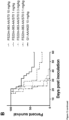
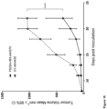
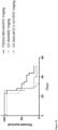
- the present inventors demonstrate that antibody molecules having the features described above do not show exhibit severe hepatoxicity when administered in a mouse model at therapeutic doses. Only minimal liver pathology was observed in mice that had been administered with these antibody molecules, which was not deemed to represent the severe hepatoxicity that has been previously reported for other anti-CD137 agonist antibodies. Preliminary studies in cynomolgus monkeys also showed that the antibody molecules are safe and well tolerated up to 30mg/kg. Without wishing to be bound by theory, it is expected that the results from these animal models will translate to the clinic in predicting the risk of hepatoxicity in human patients and therefore that the antibody molecules of the invention would have low risk of inducing hepatoxicity in human patients treated at therapeutic doses.
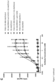
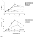
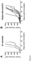
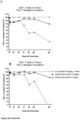
- the present inventors also provide in vitro evidence that the level of CD137 agonistic activity induced by the antibody molecule correlates with the amount of PD-L1 expression on the cell surface.
- the inventors demonstrate that the antibody molecule is capable of agonising CD137 even where there is a low level of PD-L1 expression and that as the level of PD-L1 in the system increases, so does the CD137 agonistic activity.
- This result further supports the evidence that CD137 agonistic activity is dependent on PD-L1 expression and suggests that the antibody molecules of the invention will have a broad range of activity on tumours that express varying levels of PD-L1 on the tumour cell surface.
- the CDR-based binding sites for PD-L1 described above are able to efficiently block binding of PD-L1 to its receptor PD-1.
- PD-1 is expressed on activated T cells, B cells, and myeloid cells, and modulates activation or inhibition of cellular immune responses. Binding of PD-L1 to PD-1 delivers an inhibitory signal, reducing cytokine production and proliferation of T cells, thereby dampening the immune response.
- the interaction of PD-L1 on a tumour cell with PD-1 on a T cell reduces T cell activity to prevent the immune system from attacking the tumour cells.
- the antibody molecules of the invention can prevent the tumour cells from evading the immune system in this way. Without wishing to be bound by theory, it is believed that this efficient blocking of PD-L1 binding to PD-1 functions together with the CD137 agonistic activity described above to increase anti-tumour potency of the antibody molecule.
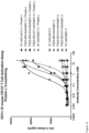
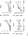
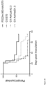
- bispecific antibody molecules comprising the CD137 antigen-binding site and CDR-based binding site for PD-L1 described above are capable of supressing tumour growth in vivo. Furthermore, more effective tumour growth suppression was observed with the bispecific antibody molecules as compared to a combination of two monospecific antibody molecules where one of the antibody molecules comprised a CDR-based antigen-binding site for PD-L1 and the other molecule comprised a CDR-based antigen-binding site for CD137, demonstrating that enhanced clustering and signalling of CD137, and thus T cell activation and corresponding anti-tumour effects, are seen with the antibody molecules of the invention.
- Antibody molecules comprising a CD137 antigen-binding site of the invention may additionally be able to bind PD-L1 bivalently, such that the antibody molecules bind both CD137 and PD-L1 bivalently.
- This is expected to be advantageous, as the bivalent binding of both targets is expected to make the bridging between the T cell expressing CD137 and the PD-L1 expressing cell more stable and thereby extend the time during which the T cell is localised at sites where PD-L1 is co-expressed with CD137, such as in the tumour microenvironment, and can act on the disease, e.g. the tumour.
- This is different to the vast majority of conventional bispecific antibody formats which are heterodimeric and bind each target antigen monovalently via one Fab arm.
- Such a monovalent interaction is expected to not only be less stable but also to be less efficient at inducing clustering of TNF receptors such as CD137 and/or to require higher expression of one or both targets to induce such clustering, and thus T cell activation.
- TNF receptors such as CD137
- mAb 2 molecules comprising a bivalent Fab binding site for PD-L1 and a monovalent binding site for CD137 in one of the CH3 domains of the molecule induced lower levels of T cell activation, as measured by IFN-y release, than a mAb 2 binding both targets bivalently.
- a further feature of the antibody molecules identified by the inventors is that the antigen-binding site for CD137 and the CDR-based binding site for PD-L1 are both contained within the antibody structure itself.
- the antibody molecules do not require other proteins to be fused to the antibody molecule via linkers or other means to result in molecule that binds bivalently to both of its targets.
- the structure is also expected to result in improved antibody stability, as linkers may degrade over time, resulting in a heterogeneous population of antibody molecules.
- those antibodies in the population having only one protein fused to them, and thus binding one target only monovalently are expected not to induce conditional agonism of TNF receptors such as CD137 as efficiently, as those antibodies which have two proteins fused to them and which are thus capable of binding both targets bivalently.
- Cleavage/degradation of the linker could take place prior to administration or after administration of the therapeutic to the patient (e.g. through enzymatic cleavage or the in vivo pH of the patient), thereby resulting in a reduction of its effectiveness whilst circulating in the patient.
- the antibody molecules are expected to retain the same number of binding sites both before and after administration.
- the structure of the antibody molecules is also preferred from the perspective of immunogenicity of the molecules, as the introduction of fused proteins or linkers or both may induce immunogenicity when the molecules are administered to a patient, resulting in reduced effectiveness of the therapeutic.
- the present invention provides:
- the present invention relates to antibody molecules which bind both to PD-L1 and CD137.
- the antibody molecules of the present invention comprise a CDR-based antigen-binding site for PD-L1 and a CD137 antigen-binding site located in a constant domain of the antibody molecule.
- the terms "PD-L1” and “CD137” may refer to human PD-L1 and human CD137, murine PD-L1 and murine CD137, and/or cynomologus monkey PD-L1 and cynomologus monkey CD137, unless the context requires otherwise.
- the terms "PD-L1” and “CD137” refer to human PD-L1 and human CD137, unless the context requires otherwise.
- antibody molecule describes an immunoglobulin whether natural or partly or wholly synthetically produced.
- the antibody molecule may be human or humanised, preferably human.
- the antibody molecule is preferably a monoclonal antibody molecule.
- Examples of antibodies are the immunoglobulin isotypes, such as immunoglobulin G, and their isotypic subclasses, such as IgG1, IgG2, IgG3 and IgG4, as well as fragments thereof.
- the antibody molecule may be isolated, in the sense of being free from contaminants, such as antibodies able to bind other polypeptides and/or serum components.
- antibody molecule thus includes antibody fragments, provided said fragments comprise a CDR-based antigen binding site for PD-L1 and a CD137 antigen binding site located in a constant domain. Unless the context requires otherwise, the term “antibody molecule”, as used herein, is thus equivalent to “antibody molecule or fragment thereof”.
- antibody molecule should be construed as covering antibody fragments, derivatives, functional equivalents and homologues of antibodies, including any polypeptide comprising an immunoglobulin binding domain, whether natural or wholly or partially synthetic. Chimeric molecules comprising an immunoglobulin binding domain, or equivalent, fused to another polypeptide are therefore included. Cloning and expression of chimeric antibodies are described in EP-A-0120694 and EP-A-0125023 .
- An example of an antibody fragment comprising both CDR sequences and CH3 domain is a minibody, which comprises an scFv joined to a CH3 domain ( Hu et al. (1996), Cancer Res., 56(13):3055-61 ).
- the antibody molecule of the present invention binds to PD-L1 and CD137. Binding in this context may refer to specific binding.
- the term “specific” may refer to the situation in which the antibody molecule will not show any significant binding to molecules other than its specific binding partner(s), here PD-L1 and CD137.
- the term “specific” is also applicable where the antibody molecule is specific for particular epitopes, such as epitopes on PD-L1 and CD137, that are carried by a number of antigens in which case the antibody molecule will be able to bind to the various antigens carrying the epitope.
- the present inventors demonstrated that antibody molecules described herein showed a high level of specificity for human PD-L1 and did not show any significant binding to other T cells targets PD-L1, CD80, PD-1 or B7-H3. See Example 9.3. Thus, in a preferred embodiment, the antibody molecule does not bind, or does not show any significant binding, to any one of, preferably all of, PD-L2, CD80, PD-1, and B7-H3. The present inventors also demonstrated that the CD137 antigen-binding site did not show any significant binding to the human TNFRSF receptors CD40, OX40 and GITR. See Example 3.6. Thus, in a more preferred embodiment, the antibody molecule does not bind, or does show any significant binding, to any one of, preferably all of, PD-L2, CD80, PD-1, B7-H3, CD40, OX40 and GITR.
- Antibodies and methods for their construction and use are well-known in the art and are described in, for example, Holliger & Hudson (2005). It is possible to take monoclonal and other antibodies and use techniques of recombinant DNA technology to produce other antibodies or chimeric molecules which retain the specificity of the original antibody. Such techniques may involve introducing CDRs or variable regions of one antibody molecule into a different antibody molecule ( EP-A-184187 , GB 2188638A and EP-A-239400 ).
- a CDR-based antigen-binding site is an antigen-binding site in an antibody variable region.
- a CDR-based antigen-binding site may be formed by three CDRs, such as the three light chain variable domain (VL) CDRs or three heavy chain variable domain (VH) CDRs.
- VL light chain variable domain
- VH heavy chain variable domain
- the CDR-based antigen-binding site is formed by six CDRs, three VL CDRs and three VH CDRs. The contributions of the different CDRs to the binding of the antigen may vary in different antigen binding sites.
- the three VH domain CDRs of the antigen-binding site may be located within an immunoglobulin VH domain and the three VL domain CDRs may be located within an immunoglobulin VL domain.
- the CDR-based antigen-binding site may be located in an antibody variable region.
- the antibody molecule has one or preferably more than one, for example two, CDR-based antigen binding sites for the first antigen.
- the antibody molecule thus comprises one VH and one VL domain but preferably comprises two VH and two VL domains, i.e. two VH/VL domain pairs, as is the case in naturally-occurring IgG molecules, for example.
- the CDR-based antigen-binding site comprises the three VH CDRs and the three VL CDRs, of antibody E12v2, E05v2, or G12v2, preferably E12v2 or E05v2, more preferably E12v2.
- VH and VL domain sequences of these antibodies are set forth as follows:
- the skilled person would have no difficulty in determining the sequences of the CDRs from the VH and VL domain sequences of the antibodies set out above.
- the CDR sequences may, for example, be determined according to Kabat ( Kabat, E.A et al. (1991). Sequences of Proteins of Immunological Interest, 5th edit., NIH Publication no. 91-3242. U.S. Department of Health and Human Services ) or the international ImMunoGeneTics information system (IMGT: Lefranc, M.-P. et al. Nucleic Acids Res. 43, D413-22 (2015 )).
- IMGT ImMunoGeneTics information system
- VH domain CDR1, CDR2 and CDR3 sequences of the antibody molecule according to Kabat numbering may be the sequences at located positions 31-35, 50-65, and 95-102 of the VH domain, respectively.
- VH domain CDR1, CDR2 and CDR3 sequences of the antibody molecule according to IMGT numbering may be the sequences located at positions 27-38, 56-65, and 105-117, of the VH domain of the antibody molecule, respectively.
- VL domain CDR1, CDR2 and CDR3 sequences of the antibody molecule according to Kabat numbering may be the sequences at located positions 24-34, 50-56, and 89-97 of the VL domain, respectively.
- VL domain CDR1, CDR2 and CDR3 sequences of the antibody molecule according to IMGT numbering may be the sequences located at positions 27-38, 56-65, and 105-117, of the VL domain, respectively.
- the antibody molecule comprises the sequence of the VH domain CDR1 of SYGIS (SEQ ID NO: 1), the VH domain CDR2 of WISAYSGGTNYAQKLQG (SEQ ID NO: 2 ), and the VH domain CDR3 of DLFPTIFGVSYYYY (SEQ ID NO: 3 ) wherein the CDR sequences are defined according to the Kabat numbering scheme.
- the antibody molecule comprises the sequence of the VH domain CDR1, CDR2 and CDR3 of:
- the CDRs of the VH domain are flanked by framework (FW) sequences (HFW1, HFW2, HFW3 and HFW4).
- the VH domain comprises the HFW1, HFW2, HFW3 and HFW4 sequences of SEQ ID NO: 55, 56, 57 and 54, respectively, wherein the FW and CDR sequences are defined according to the IMGT numbering scheme.
- the antibody molecule comprises the sequence of the VL domain CDR1 of RASQSIGNRLA (SEQ ID NO: 4 ), the VL CDR2 of EASTSET (SEQ ID NO: 5 ), and the VL CDR3 of QQSYSTPYT (SEQ ID NO: 6 ), wherein the CDR sequences are defined according to the Kabat numbering scheme.
- the VL domain is derived from a kappa VL domain.
- Each of the VL domain CDRs are flanked by framework (FW) sequences (LFW1, LFW2, LFW3 and LFW4) that are derived from a kappa VL domain.
- the VL domain may comprise the LFW1, LFW2, LFW3 and LFW4 sequences of SEQ ID NO: 58, 59, 60 and 61, respectively, wherein the FW sequences are defined according to the Kabat numbering scheme.
- the antibody molecule may comprises the sequence of the VL domain CDR1, CDR2 and CDR3 of:
- the antibody molecule comprises the sequence of the VL domain CDR1 of QSIGNR (SEQ ID NO: 10 ), the VL CDR2 of EAS (SEQ ID NO: 11 ), and the VL CDR3 of QQSYSTPYT (SEQ ID NO: 6 ), wherein the CDR sequences are defined according to the IMGT numbering scheme.
- the VL domain is derived from a kappa VL domain.
- Each of the VL domain CDRs may be flanked by framework (FW) sequences (LFW1, LFW2, LFW3 and LFW4) that are derived from a kappa VL domain.
- the VL domain may comprise the LFW1, LFW2, LFW3 and LFW4 sequences of SEQ ID NO: 62, 63, 64 and 61, respectively, wherein the FW sequences are defined according to the IMGT numbering scheme.
- the antibody molecule comprises the sequence of the VL domain CDR1, CDR2 and CDR3 of:
- the CDR-based antigen-binding site comprises the VH and VL domains of antibody E12v2, E05v2, or G12v2, preferably E12v2 or E05v2, more preferably E12v2.
- the VH domain of antibodies E12v2, E05v2, and G12v2 has the sequence set forth in SEQ ID NOs 12, 23, and 23, respectively.
- the VL domain of antibodies E12v2, E05v2, and G12v2 has the sequence set forth in SEQ ID NOs 14, 25, and 30, respectively.
- the antibody molecule of the invention comprises a CD137 antigen-binding site located in the CH3 domain.
- the CD137 antigen-binding site comprises two or more modified structural loops in a CH3 domain of the antibody molecule.
- Engineering antibody constant domain structural loops to create antigen-binding sites for target antigens is known in the art and is described, for example, Wozniak-Knopp G et al. (2010); WO2006/072620 and WO2009/132876 .
- the CD137 antigen-binding site of the antibody molecule comprises a first and second sequence, wherein the first and second sequence are located in the AB and EF structural loops of the CH3 domain, of the antibody molecule, respectively.
- the residues at positions 95 and 96 of the CH3 domain of the antibody molecule are wild-type, i.e. are arginine (R) and tryptophan (W), respectively. Both of these residues are located in the EF structural loop. Amino acid residue positions are numbered herein according to the ImMunoGeneTics (IMGT) numbering scheme, unless otherwise indicated.
- IMGT ImMunoGeneTics
- the first sequence comprises the sequence PPY (SEQ ID NO: 78 ).
- the PPY sequence is located between positions 15 and 17 of the CH3 domain of the antibody molecule. In a preferred embodiment, the PPY sequence is located at positions 16, 16.5 and 16.4 of the CH3 domain. Alternatively, the PPY sequence may be located between positions 16 and 17 of the CH3 domain. In an alternative preferred embodiment, the PPY sequence is located at positions 16.3, 16.2 and 16.1 of the CH3 domain.
- inserted residues are numbered according to the direction of the loop in which they are located. If the loop goes "up" the inserted residues take the number of the residue immediately preceding the insertion with the number of the inserted residue in the sequence being indicated by an ascending decimal number, e.g.
- the AB structural loop comprises an amino acid insertion.
- the insertion is 5 amino acids in length.
- the insertion is located between positions 16 and 17 of the CH3 domain of the antibody molecule.
- the insertion is located at positions 16.5 to 16.1 of the CH3 domain of the antibody molecule.
- Figure 1 shows Fcabs comprising a CH3 domain where the insertion is located at positions 16.5 to 16.1 of the CH3 domain.
- the second sequence comprises the sequence LE, wherein the LE sequence is located at positions 97 and 98 of the CH3 domain of the antibody molecule.
- the first sequence and second sequence may be a first and second sequence of the CH3 domain of: FS22-053-008, or FS22-053-017, preferably specific binding member FS22-053-008.
- the first sequence and second sequence is a first and second sequence of the CH3 domain of: FS22-053-008, FS22-053-017, or FS22-172-003, preferably FS22-053-008 or FS22-172-003, more preferably FS22-172-003.
- the CH3 domain sequence of FS22-053-008, FS22-053-017, and FS22-172-003 is set forth in SEQ ID NOs: 81, 90 and 115, respectively.
- the first and second sequence of FS22-053-008, FS22-053-017 and FS22-172-003 is the sequence between positions 14 and 17, and positions 91 and 99, of the CH3 domain of FS22-053-008, FS22-053-017 and FS22-172-003, respectively.
- the first and second sequence of FS22-053-008 may be the sequence between positions 14 and 17, and positions 92 and 99, of the CH3 domain of FS22-053-008.
- the CD loop sequence of the antibody molecule is preferably unmodified, i.e. wild type.
- the CD loop sequence therefore preferably has the sequence set forth in SEQ ID NO: 73.
- the CD loop sequence is preferably located at positions 43 to 78 of the CH3 domain.
- the first and second sequences may be the complete AB and EF structural loop sequences, of FS22-053-008, FS22-053-017 or FS22-172-003, respectively. Determination of the location of the AB, CD, and EF structural loops in a CH3 domain sequence, for example in accordance with the IMGT, IMGT exon, EU, or Kabat numbering systems, is within the capabilities of the skilled person and described in Hasenhindl et al. (2013).
- the AB, CD and EF structural loops according to the IMGT numbering system are located between positions 10 and 19, 42 and 79, and 91 and 102 of the CH3 domain, respectively.
- the first, second and third sequence are therefore the sequence between positions 10 and 19, 42 and 79, and 91 and 102 of the CH3 domain of FS22-053-008, FS22-053-017 or FS22-172-003, respectively.
- the first and second sequence of the CD137 antigen-binding site comprise the AB and EF loop sequence set forth in SEQ ID NOs 171 and 172 [FS22-172-003], respectively, or the AB and EF structural loop sequence set forth in SEQ ID NOs 173 and 174 [FS22-53-008], respectively.
- the first and second sequence of the CD137 antigen-binding site comprise the AB and EF structural loop set forth in SEQ ID NOs 171 and 172 [FS22-172-003], respectively.
- the first and second sequence of the CD137 antigen-binding site comprise the AB and EF structural loop sequence set forth in SEQ ID NOs 173 and 175 [FS22-053-017], respectively.
- the CD137 antigen-binding site comprises the first and second sequence set forth in:
- the CD137 antigen-binding site of the antibody molecule comprises the first and second sequence set forth in SEQ ID NOs 113 and 114 [FS22-172-003], respectively, or the first and second sequence set forth in SEQ ID NOs 79 and 80 [FS22-53-008], respectively.
- the CD137 antigen-binding site of the antibody molecule comprises the first and second sequence set forth in SEQ ID NOs 113 and 114 [FS22-172-003], respectively.
- the CD137 antigen-binding site may comprise the AB and EF structural loop sequences set forth in SEQ ID NOs 171 and 172 [FS22-172-003], respectively.
- amino acid residue positions including the position of amino acid sequences, substitutions, deletions and insertions as described herein, may be numbered according to IMGT exon numbering (also referred to as consecutive numbering), EU numbering, or Kabat numbering.
- IMGT exon numbering also referred to as consecutive numbering
- EU numbering or Kabat numbering.
- Figure 1 The concordance between IMGT numbering, IMGT exon numbering, EU numbering, and Kabat numbering of the residue positions of the CH3 domain are shown in Figure 1 .
- the first sequence is located between positions 14 and 17 of the CH3 domain of the clone, respectively, where the residue positions are numbered in accordance with the IMGT numbering scheme
- the first sequence is located between positions 18 and 21 of the CH3 domain, where the residue positions are numbered in accordance with the IMGT exon numbering scheme, as shown in Figure 1 .
- the position of amino acid residues in the CH3 domain including the position of amino acid sequences, substitutions, deletions and insertions in the CH3 domain, as described herein, may be defined by reference to their position in the wild-type CH3 domain sequence set forth in SEQ ID NO: 75.
- the concordance between IMGT numbering and the wild-type CH3 domain sequence is also shown in Figure 1 .
- the antibody molecule comprises a CH3 domain which comprises, has, or consists of the CH3 domain sequence of FS22-053-008, FS22-053-017, or FS22-172-003, wherein the CH3 domain sequence of FS22-053-008, FS22-053-017 and FS22-172-003 is set forth in SEQ ID NOs 81, 90 and 115, respectively.
- the antibody molecule comprises a CH3 domain which comprises, has, or consists of the CH3 domain sequence of FS22-172-003 or FS22-053-008 set forth in SEQ ID NO 115 and 81, respectively.
- the antibody molecule comprises a CH3 domain which comprises, has, or consists of the CH3 domain sequence of FS22-172-003 set forth in SEQ ID NO 115.
- the antibody molecule comprises a CH3 domain which comprises, has, or consists of the CH3 domain sequence of FS22-053-017 set forth in SEQ ID NO 90, respectively.
- the CH3 domain of the antibody molecule may optionally comprise an additional lysine residue (K) at the immediate C-terminus of the CH3 domain sequence.
- the antibody molecule of the invention may comprise a CH2 domain of an immunoglobulin G molecule, such as a CH2 domain of an IgG1, IgG2, IgG3, or IgG4 molecule.
- the antibody molecule of the invention comprises a CH2 domain of an IgG1 molecule.
- the CH2 domain may have the sequence set forth in SEQ ID NO: 76.
- the CH2 domain is known to bind to Fc ⁇ receptors and complement. Binding of the CH2 domain to Fc ⁇ receptors is required antibody-dependent cell-mediated cytotoxicity (ADCC), while binding to complement is required complement-dependent cytotoxicity (CDC).
- the CH2 domain of the antibody molecule preferably comprise one or more mutations that reduce or abrogate binding of the CH2 domain to one or more Fc ⁇ receptors, such as Fc ⁇ RI, FcyRlla, FcyRllb, FcyRIII, and/or to complement. The inventors postulate that reducing or abrogating binding to Fc ⁇ receptors will decrease or eliminate ADCC mediated by the antibody molecule.
- reducing or abrogating binding to complement is expected to reduce or eliminate CDC mediated by the antibody molecule. Without wishing to be bound by theory, this is expected to reduce or avoid liver toxicity when the antibody molecule is administered to a patient.
- reducing or abrogating binding to Fc ⁇ receptors is expected to be useful where the antibody molecule comprises a second antigen-binding site for an immune cell antigen, where ADCC and/or CDC-mediated killing of immune cells bound by the antibody molecule should be avoided. Mutations to decrease or abrogate binding of the CH2 domain to one or more Fc ⁇ receptors and/or complement are known in the art (Wang et al., 2018).
- complement activation (C1q binding) and ADCC are known to be reduced through mutation of the proline at position 114 of the CH2 domain to alanine or glycine (P114A or P114G) (Idusogie et al., 2000; Klein et al., 2016). These mutations may also be combined in order to generate antibody molecules with further reduced or no ADCC or CDC activity.
- the antibody molecule may comprise a CH2 domain, wherein the CH2 domain preferably comprises:
- the antibody molecule comprises a CH2 domain, wherein the CH2 domain comprises:
- the CH2 domain may have the sequence set forth in SEQ ID NO: 77.
- the antibody molecule comprises a CH2 domain, wherein the CH2 domain comprises:
- the CH2 domain may have the sequence set forth in SEQ ID NO: 176.
- the antibody molecule that binds to PD-L1 and CD137 comprises
- an antibody molecule having a CD137 antigen-binding site comprising the first sequence and a second sequence set forth in SEQ ID NOs 79 and 89, respectively [FS22-053-017] was able to bind human, cynomolgus and, unexpectedly, mouse CD137.
- the antibody molecule that binds to PD-L1 and CD137 comprises
- the antibody molecule that binds to PD-L1 and CD137 comprises
- the antibody molecule that binds to PD-L1 and CD137 comprises
- the antibody molecule that binds to PD-L1 and CD137 comprises
- the antibody molecule the antibody molecule that binds to PD-L1 and CD137 comprises:
- the antibody molecule that binds to PD-L1 and CD137 comprises a heavy chain which comprises, has, or consists of the heavy chain and light chain of antibody:
- the antibody molecule that binds to PD-L1 and CD137 comprises a heavy chain which comprises, has, or consists of the heavy chain and light chain of antibody:
- the antibody molecule that binds to PD-L1 and CD137 comprises a heavy chain which comprises, has, or consists of the heavy chain and light chain of antibody:
- the antibody molecule that binds to PD-L1 and CD137 comprises a heavy chain which comprises, has, or consists of the heavy chain and light chain of antibody FS22-172-003-AA/E12v2 set forth in SEQ ID NOs 134 and 17, respectively.
- the antibody molecule that binds to PD-L1 and CD137 comprises a heavy chain which comprises, has, or consists of the heavy chain and light chain of antibody:
- the antibody molecules of the present invention may also comprise variants of a third sequence, AB, CD or EF structural loop sequence, CH3 domain, CH2 domain, CH2 and CH3 domain, light chain and/or heavy chain sequences disclosed herein. Suitable variants can be obtained by means of methods of sequence alteration, or mutation, and screening.
- an antibody molecule comprising one or more variant sequences retains one or more of the functional characteristics of the parent antibody molecule, such as binding specificity and/or binding affinity for PD-L1 and CD137.
- an antibody molecule comprising one or more variant sequences preferably binds to PD-L1 and/or CD137 with the same affinity, or a higher affinity, than the (parent) antibody molecule.
- the parent antibody molecule is an antibody molecule which does not comprise the amino acid substitution(s), deletion(s), and/or insertion(s) which have been incorporated into the variant antibody molecule.
- an antibody molecule of the invention may comprise a third sequence, AB, CD or EF structural loop sequence, CH3 domain, CH2 domain, CH2 and CH3 domain, light chain and/or heavy chain sequence which has at least 70%, at least 75%, at least 80%, at least 85%, at least 90%, at least 95%, at least 96%, at least 97%, at least 98%, at least 99%, at least 99.1%, at least 99.2%, at least 99.3%, at least 99.4%, at least 99.5%, at least 99.6%, at least 99.7%, at least 99.8%, or at least 99.9% sequence identity to a structural loop, CH3 domain, CH2 domain, CH2 and CH3 domain, light chain or heavy chain sequence disclosed herein.
- the antibody molecule of the invention comprises a CH3 domain sequence which has at least 97%, at least 98%, at least 99%, at least 99.1%, at least 99.2%, at least 99.3%, at least 99.4%, at least 99.5%, at least 99.6%, at least 99.7%, at least 99.8%, or at least 99.9% sequence identity to the CH3 domain sequence set forth in SEQ ID NO: 115 [FS22-172-003] or 81 [FS22-053-008], preferably SEQ ID NO: 115 [FS22-172-003] .
- the antibody molecule has or comprises a CH2 domain sequence, which has at least 95%, at least 96%, at least 97%, at least 98%, at least 99%, at least 99.1%, at least 99.2%, at least 99.3%, at least 99.4%, at least 99.5%, at least 99.6%, at least 99.7%, at least 99.8%, or at least 99.9% sequence identity to the CH2 domain sequence set forth in SEQ ID NO: 76 or 77.
- GAP Garnier GCG package, Accelerys Inc, San Diego USA.
- GAP uses the Needleman and Wunsch algorithm to align two complete sequences, maximising the number of matches and minimising the number of gaps. Generally, default parameters are used, with a gap creation penalty equalling 12 and a gap extension penalty equalling 4.
- Use of GAP may be preferred but other algorithms may be used, e.g. BLAST (which uses the method of Altschul et al., 1990), FASTA (which uses the method of Pearson and Lipman, 1988), or the Smith-Waterman algorithm (Smith and Waterman, 1981), or the TBLASTN program, of Altschul et al., 1990 supra, generally employing default parameters.
- the psi-Blast algorithm Altschul et al., 1997) may be used.
- An antibody molecule of the invention may also comprise a third sequence, AB, CD or EF structural loop sequence, CH3 domain, CH2 domain, CH2 and CH3 domain, light chain and/or heavy chain which has one or more amino acid sequence alterations (addition, deletion, substitution and/or insertion of an amino acid residue), for example 20 alterations or fewer, 15 alterations or fewer, 10 alterations or fewer, 5 alterations or fewer, 4 alterations or fewer, 3 alterations or fewer, 2 alterations or fewer, or 1 alteration compared with a third sequence, AB, CD or EF structural loop sequence, CH3 domain, CH2 domain, CH2 and CH3 domain, light chain or heavy chain sequence disclosed herein.
- alterations may be made in one or more framework regions of the antibody molecule outside the VH and VL domain sequences and/or in one or more framework regions of the CH3 domain.
- the alterations may be in the CH3 domain outside of the sequences described herein as a first, second and third sequences, or as AB, CD or EF structural loop sequences.
- the antibody molecule of the invention may comprise a CH3 domain sequence with one or more amino acid sequence alterations (addition, deletion, substitution and/or insertion of an amino acid residue), preferably 20 alterations or fewer, 15 alterations or fewer, 10 alterations or fewer, 5 alterations or fewer, 4 alterations or fewer, 3 alterations or fewer, 2 alterations or fewer, or 1 alteration compared with the CH3 domain sequence set forth in SEQ ID NOs 81, 84, 87, 90, 93, 96, 99, 102, 105, 108, 111, 115, 118, 121, 124, 127, 130, or 132.
- the antibody molecule of the invention may comprise a CH3 domain sequence with one or more amino acid sequence alterations (addition, deletion, substitution and/or insertion of an amino acid residue), preferably 20 alterations or fewer, 15 alterations or fewer, 10 alterations or fewer, 5 alterations or fewer, 4 alterations or fewer, 3 alterations or fewer, 2 alterations or fewer, or 1 alteration compared with the CH3 domain sequence set forth in SEQ ID NOs: 115 [FS22-172-003] or 81 [FS22-053-008], preferably SEQ ID NO: 115 [FS22-172-003].
- the antibody molecule comprises a CH2 domain sequence, with one or more amino acid sequence alterations (addition, deletion, substitution and/or insertion of an amino acid residue), preferably 20 alterations or fewer, 15 alterations or fewer, 10 alterations or fewer, 5 alterations or fewer, 4 alterations or fewer, 3 alterations or fewer, 2 alterations or fewer, or 1 alteration compared with the CH2 domain sequence set forth in SEQ ID NO: 76 or 77.
- substitutions may conservative substitutions, for example according to the following Table.
- amino acids in the same category in the middle column are substituted for one another, i.e. a non-polar amino acid is substituted with another non-polar amino acid for example.
- amino acids in the same line in the rightmost column are substituted for one another.
- substitution(s) may be functionally conservative. That is, in some embodiments the substitution may not affect (or may not substantially affect) one or more functional properties (e.g. binding affinity) of the antibody molecule comprising the substitution as compared to the equivalent unsubstituted antibody molecule.
- the antibody molecule comprises a variant of an AB structural loop sequence, CH3 domain, or heavy chain sequence as disclosed herein, the antibody molecule retains the sequence PPY between positions 15 and 17, of the CH3 domain of the antibody molecule. In addition, the antibody molecule retains a 5 amino acid insertion between positions 16 and 17 of the CH3 domain of the antibody molecule. Also disclosed herein, the antibody molecule may retain the sequence at positions 97 and 98 of the CH3 domain of the antibody molecule.
- the variant does not comprise any amino acid alterations in the first and second sequence located in the AB and EF structural loops of the CH3 domain of the antibody molecule.
- the variant may not comprise any amino acid alterations in the AB and EF structural loops of the CH3 domain of the antibody molecule.
- the variant may not comprise any amino acid alterations in the CD structural loop of the CH3 domain of the antibody molecule. That is, the variant may not comprise any amino acid alterations in the AB and EF structural loops of the CH3 domain of the antibody molecule.
- the antibody molecule comprises a variant of a light chain or heavy chain sequence disclosed herein, the antibody molecule does not comprise any amino acid alterations in the CDR sequences.
- the variant may not comprise any amino acid alterations in the CDR1, CDR2, CDR3, CDR4, CDR5 and/or CDR6 sequences.
- the antibody molecule preferably binds to human PD-L1 and human CD137.
- the antibody molecule is capable of simultaneously binding to human PD-L1 and human CD137, wherein human CD137 and human PD-L1 are co-expressed.
- co-expression means that the two targets are expressed on the surface of a single cell, or on the surface of two separate cells.
- the antibody molecule may be capable of binding to human PD-L1 and human CD137 when human PD-L1 and human CD137 are co-expressed on a single cell, e.g.
- an immune cell as well as being capable of binding to human PD-L1 and human CD137 when human PD-L1 and human CD137 are co-expressed on two separate cells, e.g. an immune cell expressing CD137 and a separate tumour cell expressing PD-L1 in the tumour microenvironment.
- the antibody molecule preferably binds to human PD-L1 with an affinity (K D ) of 8 nM, 7 nM, 6 nM, 5 nM, 4 nM, 3 nM, 2 nM, 1 nM, 0.5 nM, 0.4 nM, or 0.3 nM or with a higher affinity.
- affinity (K D ) 8 nM, 7 nM, 6 nM, 5 nM, 4 nM, 3 nM, 2 nM, 1 nM, 0.5 nM, 0.4 nM, or 0.3 nM or with a higher affinity.
- the human PD-L1 may, for example, have the sequence set forth in SEQ ID NO: 180.
- the human PD-L1 may, for example, be recombinant human PD-L1 with an Avi Tag (hPD-L1-Avi-His), available from Aero Biosystems (catalogue number: PD1-H82E5).
- the recombinant human PD-L1 may be biotinylated.
- the antibody molecule preferably binds to dimeric human CD137 with an affinity (K D ) of 60 nM, 50 nM, 40 nM, 30 nM, 20 nM, 10 nM, 5 nM, 4 nM, 3 nM, or 2 nM, or with a higher affinity.
- affinity (K D ) of 60 nM, 50 nM, 40 nM, 30 nM, 20 nM, 10 nM, 5 nM, 4 nM, 3 nM, or 2 nM, or with a higher affinity.
- the antibody molecule binds to dimeric CD137 with a higher affinity than monomeric CD137. In a preferred embodiment, the antibody molecule binds to dimeric CD137 with an affinity which is at least 50-fold, 60-fold, 70-fold, 80-fold, 90-fold, 100-fold, 110-fold, 120-fold, 130-fold, 140-fold, 150-fold, 160-fold, 170-fold or 200-fold higher than the affinity of the antibody molecule for monomeric CD137.
- the human CD137 may, for example, have the sequence set forth in SEQ ID NO: 186. Methods for producing dimeric and monomeric CD137 antigens are described in the examples.
- the antibody molecule preferably binds to cynomolgus PD-L1 and cynomolgus CD137.
- the antibody molecule is capable of simultaneously binding to cynomolgus PD-L1 and cynomolgus CD137, wherein cynomolgus PD-L1 and cynomolgus CD137 are expressed on the surface of a single cell, or on the surface of two separate cells.
- the antibody molecule may bind to cynomolgus PD-L1 with an affinity (K D ) of 8 nM, 7 nM, 6 nM, 5 nM, 4 nM, 3 nM, 2 nM, 1 nM, 0.5 nM, 0.4 nM, or 0.3 nM or with a higher affinity.
- affinity (K D ) 8 nM, 7 nM, 6 nM, 5 nM, 4 nM, 3 nM, 2 nM, 1 nM, 0.5 nM, 0.4 nM, or 0.3 nM or with a higher affinity.
- the antibody molecule binds to cynomolgus PD-L1, with an affinity (K D ) of 1 nM, or with a higher affinity.
- the cynomolgus PD-L1 may, for example, have the sequence set forth in SEQ ID NO: 184.
- the antibody molecule may bind to dimeric cynomolgus CD137 with an affinity (K D ) of 60 nM, 50 nM, 40 nM, 30 nM, 20 nM, 10 nM, 5 nM, 4 nM, 3 nM, or 2 nM or with a higher affinity.
- affinity (K D ) of 60 nM, 50 nM, 40 nM, 30 nM, 20 nM, 10 nM, 5 nM, 4 nM, 3 nM, or 2 nM or with a higher affinity.
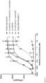
- the antibody molecule may bind to human PD-L1 and cynomolgus PD-L1 with a similar affinity, and/or bind to dimeric human CD137 and dimeric cynomolgus CD137 with similar affinity. This is thought to be beneficial for ensuring that efficacy and toxicity studies carried out with the antibody molecule in cynomolgus monkeys are predictive of the efficacy and toxicity of the antibody molecule in humans.
- the antibody molecule binds to cynomolgus PD-L1 with an affinity which is no more than 10-fold, preferably no more than 5-fold lower or higher than the affinity with which the antibody molecule binds to human PD-L1.
- the antibody molecule binds to dimeric cynomolgus CD137 with an affinity which is no more than 10-fold, preferably no more than 5-fold lower or higher than the affinity with which the antibody molecule binds dimeric human CD137.
- the binding affinity of an antibody molecule to a cognate antigen can be determined by surface plasmon resonance (SPR), such as Biacore, for example. Further details of suitable methods are described in the Examples.
- an antibody molecule to simultaneously bind to two cognate antigens, e.g. human PD-L1 and human CD137, or cynomolgus PD-L1 and cynomolgus CD137, can be determined by SPR, such as Biacore for example. Further details of suitable methods are described in the Examples.
- the antibody molecule may be capable of blocking the interaction between PD-L1 and its receptor, PD-1, preferably human PD-L1 and human PD-1.
- the PD-1/PD-L1 signalling pathway is known to be important in mediating immune suppression.
- antibody molecules that are capable of blocking this pathway are expected to be advantageous in that they may reduce immune suppression and help to increase the anti-tumour response.
- PD-1/PD-L1 signalling inhibition and CD137 activation may work together in order to increase anti-tumour potency.
- the ability of an antibody molecule to block the binding of PD-L1 to PD-1 may be determined using a bioluminescent cell-based assay, for example using a PD-1/PD-L1 Blockade Bioassay product, e.g. from Promega.
- the ability of an antibody molecule to block the binding of PD-L1 to PD-1 may be determined using ELISA. Further details of these assays are described in the Examples.
- an antibody molecule to block the binding of PD-L1 to PD-1 also referred to as the PD-1/PD-L1 blocking activity herein, may be determined by reference to an antibody molecule comprising or consisting of the heavy chain and light chain of antibody 2.14H9OPT set forth in WO2011/066389 A1 , respectively, or the heavy chain and light chain of antibody G1/280_02_G02 set forth in SEQ ID NOs 177 and 178, respectively.
- the antibody molecule may have a similar or higher level of PD-1/PD-L1 blocking activity than an antibody molecule comprising or consisting of the heavy chain and light chain of antibody 2.14H9OPT set forth in WO2011/066389 A1 , or the heavy chain and light chain of antibody G1/280_02_G02 set forth in SEQ ID NOs 177 and 178, respectively.
- the antibody molecule may have a PD-1/PD-L1 blocking activity that is at least 70%, 80%, or 90% of the PD-1/PD-L1 blocking activity of an antibody molecule comprising or consisting of the heavy chain sequence and light chain sequence of antibody 2.14H9OPT set forth in WO2011/066389 A1 , or the heavy chain and light chain of antibody G1/280_02_G02 set forth in SEQ ID NOs 177 and 178, respectively.
- the antibody molecule may have a PD-1/PD-L1 blocking activity that is between 70% and 130%, 80% and 120%, or 90% and 110% of the PD-1/PD-L1 blocking activity of an antibody molecule comprising or consisting of the heavy chain sequence and light chain sequence of antibody 2.14H9OPT set forth in WO2011/066389 A1 , or the heavy chain and light chain of antibody G1/280_02_G02 set forth in SEQ ID NOs 177 and 178, respectively.
- the antibody molecules of the invention are capable of simultaneously binding PD-L1 and CD137, which results in the activation (agonism) of CD137.
- the antibody molecule is capable of simultaneously binding to both human PD-L1 and human CD137, wherein such binding causes activation of human CD137.
- the antibody molecule is capable of simultaneously binding to both cynomolgus PD-L1 and cynomolgus CD137, wherein such binding causes activation of cynomolgus CD137.
- Exemplary methods of testing simultaneous binding and activation include T cell activation assays, as described in more detail below.
- T cells release IL-2 on activation.
- a T cell activation assay may therefore measure IL-2 release to determine the level of T cell activation induced by the antibody molecule.
- the ability of the antibody molecule to activate T cells is determined by measuring the concentration of the antibody molecule required to achieve half-maximal release of IL-2 by the T cells in a T cell activation assay. This is referred to as the EC 50 below.
- the antibody molecule has an EC 50 in a T cell activation assay which is within 50-fold, 40-fold, 30-fold, 20-fold, 10-fold, 5-fold, 4-fold, 3-fold, or 2-fold of the EC 50 of FS22-172-003-AA/E12v2 in the same assay, wherein FS22-172-003-AA/E12v2 consists of the heavy chain of SEQ ID NO: 134 and the light chain of SEQ ID NO: 17.
- the antibody molecule may have an EC 50 in a T cell activation assay of 30 nM or less, 25 nM or less, 20 nM or less, 14 nM or less, 10 nM or less, 5 nM or less, 4 nM or less, 3 nM or less, 2 nM or less, 1.5 nM or less, 1 nM or less, 0.4 nM or less, 0.4 nM or less, or 0.3 nM or less, preferably 1 nM or less, more preferably 1.5 nM or less when the antibody molecule is crosslinked.
- the ability of an antibody molecule to activate T cells may be determined by measuring the maximum concentration of IL-2 released by the T cells in a T cell activation assay in the presence of the antibody molecule (E max ).
- the antibody molecule may have a maximum concentration of IL-2 released by the T cells in a T cell activation assay in the presence of the antibody molecule of at least 1500 pg/ml, 2000 pg/ml, 2500 pg/ml, 3000 pg/ml, or 3250 pg/ml or more, preferably 2500 pg/ml or more.
- the maximum concentration of IL-2 released by the T cells in a T cell activation assay in the presence of the antibody molecule is within 20%, or 10% of the maximum concentration of IL-2 released by the T cells in the presence of FS22-172-003-AA/E12v2 in the same assay, wherein FS22-172-003-AA/E12v2 consists of the heavy chain of SEQ ID NO: 134 and the light chain of SEQ ID NO: 17.
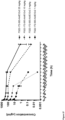
- the T cell activation assay preferably comprises T cells expressing CD137 and cells expressing PD-L1, for example, HEK293 cells overexpressing human PD-L1 (HEK.hPD-L1) may be prepared and used as described in the Examples. Alternatively, or additionally, cells expressing a different level of PD-L1 may be used, such as the human breast adenocarcinoma cell line MDA-MB-231 (ATCC HTB-26) cells and/or SKBR3 cells. As described in the Examples, HEK.hPD-L1 express a high level of human PD-L1, MDA-MB-231 cells express a medium level of human PD-L1 and SKBR3 cells express a low level of PD-L1.
- HEK.hPD-L1 express a high level of human PD-L1
- MDA-MB-231 cells express a medium level of human PD-L1
- SKBR3 cells express a low level of PD-L1.
- the T cell activation assay does not comprise any agents capable of crosslinking the antibody molecule other than CD137 and PD-L1 expressing cells.
- agents capable of crosslinking the antibody molecule include an anti-human CH2 antibody, as described in the Examples.
- the T cell activation assay may be a T cell assay as described herein, such as a pan-T cell assay as described in the present Examples.
- a T cell activation assay may be an IL-2 release assay based on T cells isolated from human Peripheral Blood Mononuclear Cells (PBMCs).
- the T cell activation assay may comprise isolating human PBMCs from leucocyte depletion cones. Methods for isolating PBMCs are known in the art and described in the present examples. The T cells may then be isolated from the PBMCs. Methods for isolating T cells (all T cells) from PBMCs are known in the art and described in the present Examples.
- the activation assay may involve preparing the required number of T cells for example in experimental media, such as a T cell medium.
- the required number of T cells may be prepared at a concentration of 1.0 ⁇ 10 6 cells/ml.
- T cells may then be stimulated using a suitable T cell activation reagent that provides the signals required for T cell activation.
- the T cell activation reagent may be a reagent comprising CD3 and CD28, such as beads comprising CD3 and CD28.
- Isolated T cells may be incubated overnight with the T cell activation reagent to activate the T cells.
- the activated T cells may be washed to separate the T cells from the T cell activation reagent and resuspended in T cell medium at a suitable concentration, such as 2.0 ⁇ 10 6 cells/ml. Activated T cells may then be added to plates coated with anti-human CD3 antibody.
- the cells may be plated at, e.g. 2 ⁇ 10 5 cells per well on to anti-CD3 antibody-coated tissue culture plates in T cell culture medium. After, e.g. 4 hours of incubation, all T cell culture medium may be removed and replaced with 100 ⁇ l T cell culture medium containing T cells, e.g. at a concentration of 5.0 ⁇ 10 5 cells/ml resulting in 5.0 ⁇ 10 4 cells/well.
- test antibody molecule A suitable dilution of each test antibody molecule may be prepared and added to the wells.
- the T cells may then be incubated at 37°C, 5% CO 2 for 24 hours with the test antibody.
- Supernatants may be collected and assayed to determine the concentration of IL-2 in the supernatant. Methods for determining the concentration of IL-2 in a solution are known in the art and described in the present examples.
- the concentration of human IL-2 may be plotted versus the log concentration of the antibody molecule. The resulting curves may be fitted using the log (agonist) versus response equation.
- the antibody molecule may be conjugated to a bioactive molecule or a detectable label.
- the antibody molecule may be referred to as a conjugate.
- conjugates find application in the treatment of diseases as described herein.
- the bioactive molecule may be an immune system modulator, such as a cytokine, preferably a human cytokine.
- the cytokine may be a cytokine which stimulates T cell activation and/or proliferation.
- cytokines for conjugation to the antibody molecule include IL-2, IL-10, IL-12, IL-15, IL-21, GM-CSF and IFN-y.
- the bioactive molecule may be a ligand trap, such as a ligand trap of a cytokine, e.g. of TGF-beta or IL-6.
- the bioactive molecule may be a therapeutic radioisotope.
- Radioimmunotherapy is used in cancer treatment, for example.
- Therapeutic radioisotopes suitable for radioimmunotherapy are known in the art and include yttrium-90, iodine-131, bismuth-213, astatine-211, lutetium 177, rhenium-188, copper-67, actinium-225, and iodine-125 and terbium-161.
- Suitable detectable labels which may be conjugated to antibody molecules include radioisotopes such as iodine-125, iodine-131, yttrium-90, indium-111 and technetium-99; fluorochromes, such as fluorescein, rhodamine, phycoerythrin, Texas Red and cyanine dye derivatives for example,Cy7 and Alexa750; chromogenic dyes, such as diaminobenzidine; latex beads; enzyme labels such as horseradish peroxidase; phosphor or laser dyes with spectrally isolated absorption or emission characteristics; and chemical moieties, such as biotin, which may be detected via binding to a specific cognate detectable moiety, e.g. labelled avidin.
- radioisotopes such as iodine-125, iodine-131, yttrium-90, indium-111 and technetium-99
- fluorochromes such as fluorescein,
- the antibody molecule may be conjugated to the bioactive molecule or detectable label by means of any suitable covalent or non-covalent linkage, such as a disulphide or peptide bond.
- the bioactive molecule is a cytokine
- the cytokine may be joined to the antibody molecule by means of a peptide linker.
- Suitable peptide linkers are known in the art and may be 5 to 25, 5 to 20, 5 to 15, 10 to 25, 10 to 20, or 10 to 15 amino acids in length.
- the bioactive molecule may be conjugated to the antibody molecule by a cleavable linker.
- the linker may allow release of the bioactive molecule from the antibody molecule at a site of therapy.
- Linkers may include amide bonds (e.g. peptidic linkers), disulphide bonds or hydrazones. Peptide linkers for example may be cleaved by site specific proteases, disulphide bonds may be cleaved by the reducing environment of the cytosol and hydrazones may be cleaved by acid-mediated hydrolysis.
- the invention also provides an isolated nucleic acid molecule or molecules encoding an antibody molecule of the invention.
- the skilled person would have no difficulty in preparing such nucleic acid molecules using methods well-known in the art.
- the nucleic acid molecule or molecules may, for example, comprise the sequence set forth in SEQ ID NO: 82, 91, or 116, which encode the CH3 domains of FS22-053-008, FS22-053-017, and FS22-172-003, respectively.
- the nucleic acid molecule or molecules comprise the sequence set forth in SEQ ID NO: 116 or 82, which encode the CH3 domain of FS22-172-003 or FS22-053-008, respectively.
- the nucleic acid molecule or molecules comprise the sequence set forth in SEQ ID NO: 116, which encodes the CH3 domain of FS22-172-003.
- the nucleic acid molecule or molecules may encode the VH domain and/or VL domain, preferably the VH domain and VL domain of antibody E12v2, E05v2, or G12v2, preferably E12v2 or E05v2, more preferably E12v2.
- VH and VL domain sequences of these antibodies are described herein.
- nucleic acid molecule(s) may comprise:
- the nucleic acid molecule or molecules may encode the heavy chain and/or light chain, preferably the heavy chain and light chain of antibody FS22-172-003-AA/E12v2, FS22-172-003-AA/E05v2, FS22-172-003-AA/G12v2, FS22-053-008-AA/E12v2, or FS22-053-008-AA/E05v2, preferably antibody FS22-172-003-AA/E12v2.
- the heavy chain and light chain sequences of these antibodies are described herein.
- nucleic acid molecule(s) may comprise:
- nucleic acid encodes the VH and VL domain, or heavy and light chain, of an antibody molecule of the invention
- the two domains or chains may be encoded on two separate nucleic acid molecules.
- nucleic acid molecule may be used to express an antibody molecule of the invention.
- the nucleic acid will generally be provided in the form of a recombinant vector for expression.
- Another aspect of the invention thus provides a vector comprising a nucleic acid as described above.
- Suitable vectors can be chosen or constructed, containing appropriate regulatory sequences, including promoter sequences, terminator fragments, polyadenylation sequences, enhancer sequences, marker genes and other sequences as appropriate.
- the vector contains appropriate regulatory sequences to drive the expression of the nucleic acid in a host cell.
- Vectors may be plasmids, viral e.g. phage, or phagemid, as appropriate.
- a nucleic acid molecule or vector as described herein may be introduced into a host cell.
- Techniques for the introduction of nucleic acid or vectors into host cells are well established in the art and any suitable technique may be employed.
- a range of host cells suitable for the production of recombinant antibody molecules are known in the art, and include bacterial, yeast, insect or mammalian host cells.
- a preferred host cell is a mammalian cell, such as a CHO, NS0, or HEK cell, for example a HEK293 cell.
- Another aspect of the invention provides a method of producing an antibody molecule of the invention comprising expressing a nucleic acid encoding the antibody molecule in a host cell and optionally isolating and/or purifying the antibody molecule thus produced.
- Methods for culturing host cells are well-known in the art.
- the method may further comprise isolating and/or purifying the antibody molecule.
- Techniques for the purification of recombinant antibody molecules are well-known in the art and include, for example HPLC, FPLC or affinity chromatography, e.g. using Protein A or Protein L. In some embodiments, purification may be performed using an affinity tag on antibody molecule.
- the method may also comprise formulating the antibody molecule into a pharmaceutical composition, optionally with a pharmaceutically acceptable excipient or other substance as described below.
- PD-L1 is known to be expressed on many cancer cells and is expressed on cell of the immune system.
- CD137 is expressed on cells of the immune system, including T cells, in particular CD8 + T cells, B cells, NK cells and tumour-infiltrating lymphocytes (TILs).
- TILs tumour-infiltrating lymphocytes
- CD137 is expressed at a lower level on CD4 + T cells than CD8 + T cells but has also been shown to be involved in inducing proliferation and activation of some subsets of CD4 + T cells.
- CD137 activation has been shown to play a role in enhancing proliferation, survival and the cytotoxic effector function of CD8 + T cells, as well as CD8 + T cell differentiation and maintenance of memory CD8 + T cells. Activation of CD137 has also been demonstrated to enhance NK cell-mediated ADCC, as well as B cell proliferation, survival and cytokine production.
- antibody molecules of the invention are capable of simultaneously binding to PD-L1 and CD137 in order to induce agonism of CD137.
- the antibody molecule drives agonism autonomously, based on the expression of the PD-L1 and CD137, and without the need for additional crosslinking agents. Since PD-L1 is expressed on many cancer cells and CD137 is expressed on immune cells, where it has a known role in enhancing the proliferation and survival of immune cells, it is expected that antibody molecules of the invention will be able to enhance an immune response at locations where PD-L1 is expressed, e.g. in the tumour microenvironment.
- the present inventors have shown that the use of an antibody molecule having these properties, is effective in suppressing tumour growth in syngeneic mouse models of cancer, and that such antibody molecules are more effective than the administration of two binding molecules which bind PD-L1 and CD137, respectively.
- the antibody molecules as described herein may thus be useful for therapeutic applications, in particular in the treatment of cancer.
- An antibody molecule as described herein may be used in a method of treatment of the human or animal body.
- Related aspects of the invention provide;
- the individual may be a patient, preferably a human patient.
- Treatment may be any treatment or therapy in which some desired therapeutic effect is achieved, for example, the inhibition or delay of the progress of the condition, and includes a reduction in the rate of progress, a halt in the rate of progress, amelioration of the condition, cure or remission (whether partial or total) of the condition, preventing, ameliorating, delaying, abating or arresting one or more symptoms and/or signs of the condition or prolonging survival of an individual or patient beyond that expected in the absence of treatment.
- Treatment as a prophylactic measure is also included.
- a prophylactic measure i.e. prophylaxis
- an individual susceptible to or at risk of the occurrence or re-occurrence of a disease such as cancer may be treated as described herein. Such treatment may prevent or delay the occurrence or re-occurrence of the disease in the individual.
- a method of treatment as described may be comprise administering at least one further treatment to the individual in addition to the antibody molecule.
- the antibody molecule described herein may thus be administered to an individual alone or in combination with one or more other treatments.
- the additional treatment may be administered to the individual concurrently with, sequentially to, or separately from the administration of the antibody molecule.
- the antibody molecule and additional treatment may be administered to the individual as a combined preparation.
- the additional therapy may be a known therapy or therapeutic agent for the disease to be treated.
- antibody molecules Whilst an antibody molecule may be administered alone, antibody molecules will usually be administered in the form of a pharmaceutical composition, which may comprise at least one component in addition to the antibody molecule.
- a pharmaceutical composition comprising an antibody molecule as described herein.
- a method comprising formulating an antibody molecule into a pharmaceutical composition is also provided.
- compositions may comprise, in addition to the antibody molecule, a pharmaceutically acceptable excipient, carrier, buffer, stabilizer or other materials well known to those skilled in the art.
- pharmaceutically acceptable as used herein pertains to compounds, materials, compositions, and/or dosage forms which are, within the scope of sound medical judgement, suitable for use in contact with the tissues of a subject (e.g., human) without excessive toxicity, irritation, allergic response, or other problem or complication, commensurate with a reasonable benefit/risk ratio.
- Each carrier, excipient, etc. must also be “acceptable” in the sense of being compatible with the other ingredients of the formulation.
- the precise nature of the carrier or other material will depend on the route of administration, which may be by infusion, injection or any other suitable route, as discussed below.
- the pharmaceutical composition comprising the antibody molecule may be in the form of a parenterally acceptable aqueous solution which is pyrogen-free and has suitable pH, isotonicity and stability.
- a parenterally acceptable aqueous solution which is pyrogen-free and has suitable pH, isotonicity and stability.
- isotonic vehicles such as Sodium Chloride Injection, Ringer's Injection, Lactated Ringer's Injection.
- buffers such as phosphate, citrate and other organic acids
- antioxidants such as ascorbic acid and methionine
- preservatives such as octadecyldimethylbenzyl ammonium chloride; hexamethonium chloride; benzalkonium chloride; benzethonium chloride; phenol, butyl or benzyl alcohol; alkyl parabens, such as methyl or propyl paraben; catechol; resorcinol; cyclohexanol; 3'-pentanol; and m-cresol); low molecular weight polypeptides; proteins, such as serum albumin, gelatin or immunoglobulins; hydrophilic polymers, such as polyvinylpyrrolidone; amino acids, such as glycine, glutamine, asparagines, histidine, arginine, or ly
- antibody molecules may be provided in a lyophilised form for reconstitution prior to administration.
- lyophilised antibody molecules may be re-constituted in sterile water and mixed with saline prior to administration to an individual.
- Administration may be in a "therapeutically effective amount", this being sufficient to show benefit to an individual.
- the actual amount administered, and rate and time-course of administration will depend on the nature and severity of what is being treated, the particular individual being treated, the clinical condition of the individual, the cause of the disorder, the site of delivery of the composition, the type of antibody molecule, the method of administration, the scheduling of administration and other factors known to medical practitioners. Prescription of treatment, e.g. decisions on dosage etc., is within the responsibility of general practitioners and other medical doctors, and may depend on the severity of the symptoms and/or progression of a disease being treated. Appropriate doses of antibody molecules are well known in the art ( Ledermann et al. (1991) Int. J.
- a therapeutically effective amount or suitable dose of an antibody molecule can be determined by comparing in vitro activity and in vivo activity in an animal model. Methods for extrapolation of effective dosages in mice and other test animals to humans are known. The precise dose will depend upon a number of factors, including whether the size and location of the area to be treated, and the precise nature of the antibody molecule.
- a typical antibody dose is in the range 100 ⁇ g to 1 g for systemic applications, and 1 ⁇ g to 1 mg for topical applications.
- An initial higher loading dose, followed by one or more lower doses, may be administered. This is a dose for a single treatment of an adult individual, which may be proportionally adjusted for children and infants, and also adjusted for other antibody formats in proportion to molecular weight.
- Treatments may be repeated at daily, twice-weekly, weekly or monthly intervals, at the discretion of the physician.
- the treatment schedule for an individual may be dependent on the pharmacokinetic and pharmacodynamic properties of the antibody composition, the route of administration and the nature of the condition being treated.
- Treatment may be periodic, and the period between administrations may be about two weeks or more, e.g. about three weeks or more, about four weeks or more, about once a month or more, about five weeks or more, or about six weeks or more. For example, treatment may be every two to four weeks or every four to eight weeks. Suitable formulations and routes of administration are described above.
- an antibody molecule as described herein may be for use in a method of treating cancer.
- Cancer may be characterised by the abnormal proliferation of malignant cancer cells. Where a particular type of cancer, such as breast cancer, is referred to, this refers to an abnormal proliferation of malignant cells of the relevant tissue, such as breast tissue.
- the cancer may be a primary or a secondary cancer.
- an antibody molecule as described herein may be for use in a method of treating cancer in an individual, wherein the cancer is a primary tumour and/or a tumour metastasis.
- a tumour of a cancer to be treated using an antibody molecule as described herein may comprise TILs that express CD137, e.g. on their cell surface.
- the tumour may have been determined to comprise TILs that express CD137.
- Methods for determining the expression of an antigen on a cell surface are known in the art and include, for example, flow cytometry.
- the cancer to be treated using an antibody molecule as described herein may be selected from the group consisting of leukaemias, such as acute myeloid leukaemia (AML), chronic myeloid leukaemia (CML), acute lymphoblastic leukaemia (ALL) and chronic lymphocytic leukaemia (CLL); lymphomas, such as Hodgkin lymphoma, non-Hodgkin lymphoma and multiple myeloma; and solid cancers, such as sarcomas (e.g. soft tissue sarcomas), skin cancer (e.g. Merkel cell carcinoma), melanoma, bladder cancer (e.g. urothelial carcinoma), brain cancer (e.g.
- leukaemias such as acute myeloid leukaemia (AML), chronic myeloid leukaemia (CML), acute lymphoblastic leukaemia (ALL) and chronic lymphocytic leukaemia (CLL); lymphomas, such as Hodgkin lymphom
- glioblastoma multiforme breast cancer, uterine/endometrial cancer, ovarian cancer (e.g. ovarian serous cystadenoma), prostate cancer, lung cancer (e.g. non-small cell lung carcinoma (NSCLC) and small cell lung cancer (SCLC), colorectal cancer (e.g. colorectal adenocarcinoma), cervical cancer (e.g. cervical squamous cell cancer and cervical adenocarcinoma), liver cancer (e.g. hepatocellular carcinoma), head and neck cancer (e.g. head and neck squamous-cell carcinoma), oesophageal cancer, pancreatic cancer, renal cancer (e.g.
- NSCLC non-small cell lung carcinoma
- SCLC small cell lung cancer
- colorectal cancer e.g. colorectal adenocarcinoma
- cervical cancer e.g. cervical squamous cell cancer and cervical adenocarcinoma
- liver cancer e
- renal cell cancer adrenal cancer
- stomach cancer e.g. stomach adenocarcinoma
- testicular cancer cancer of the gall bladder and biliary tracts (e.g. cholangiocarcinoma)
- thyroid cancer thymus cancer, bone cancer, and cerebral cancer.
- the cancer to be treated using an antibody molecule as described herein is a solid cancer.
- the cancer to be treated using an antibody molecule as described herein is a solid cancer selected from the group consisting of melanoma, bladder cancer, brain cancer, breast cancer, ovarian cancer, lung cancer, colorectal cancer, cervical cancer, liver cancer, head and neck cancer, pancreatic cancer, renal cancer, stomach cancer and Gastrointestinal Stromal tumours (GISTs).
- the cancer to be treated using an antibody molecule as described herein may be a cancer which is responsive to treatment with one or more check-point inhibitors, such as an antibody which binds PD-1, PD-L1 or CTLA4.
- check-point inhibitors such as an antibody which binds PD-1, PD-L1 or CTLA4.
- Such tumours are thought to have higher TIL levels and/or higher tumour mutational burden than tumours which are not responsive to check-point inhibitor therapy.
- Such tumours are also referred to as warm or hot tumours.
- tumours include head and neck squamous-cell carcinoma (HNSCC), melanoma, lung cancer (such as squamous lung cancer, lung adenocarcinoma, non-small cell lung carcinoma [NSCLC], or small-cell lung carcinoma [SCLC]), prostate cancer, cervical cancer (such as cervical squamous cell carcinoma or endocervical andenocarcinoma), bladder cancer, breast cancer, thyroid cancer, kidney cancer, colorectal cancer (MSI or MSS; e.g.
- HNSCC head and neck squamous-cell carcinoma
- melanoma lung cancer
- lung cancer such as squamous lung cancer, lung adenocarcinoma, non-small cell lung carcinoma [NSCLC], or small-cell lung carcinoma [SCLC]
- SLC non-small cell lung carcinoma
- SCLC small-cell lung carcinoma
- bladder cancer such as cervical squamous cell carcinoma or endocervical andenocarcinoma
- MSI or MSS colore
- the cancer is HNSCC.
- the cancer may further be a cancer which has not previously been treated with a chemotherapeutic or radiotherapeutic agent, i.e. the individual to be treated may be a cancer patient which has not received treatment with a chemotherapeutic or radiotherapeutic agent for the cancer in question.
- the cancer to be treated using an antibody molecule as described herein may be a cancer, such as pancreatic cancer or prostate cancer which is not responsive to treatment with one or more check-point inhibitors, such as an antibody which binds PD-1, PD-L1 or CTLA4.
- An individual treated with an antibody of the invention may thus be a cancer patient, such as a pancreatic cancer or prostate cancer patient, with a previous inadequate response to one or more check-point inhibitors, such as an anti-PD-L1 or anti-PD-1 antibody.
- Tumours which are not responsive to treatment with one or more check-point inhibitors are also referred to as cold tumours.
- an immunotherapeutic agent such as an immunostimulatory agent, or an anti-tumour vaccine will result in cancer cell death which in turn will result in an increase in TILs in the tumour and higher expression of immunosuppressive receptors, which in turn will make the cancer responsive to treatment with check-point inhibitors, i.e. turn a cold tumour into a warm tumour.
- the antibody molecule of the invention may be for use in a method of treating cancer in an individual, wherein the cancer is not responsive, or refractory, to treatment with one or more check-point inhibitors alone, and wherein the method comprises administering the antibody molecule to the individual in combination with a chemotherapeutic, radiotherapeutic, or immunostimulatory agent, or an anti-cancer vaccine.
- a method of treating a cancer in an individual, wherein the cancer is not responsive, or refractory, to treatment with one or more check-point inhibitors alone, and wherein the method comprises administering the antibody molecule to the individual in combination with a chemotherapeutic, radiotherapeutic, or immunostimulatory agent, or an anti-cancer vaccine is also contemplated.
- treatment may include inhibiting cancer growth, including complete cancer remission, and/or inhibiting cancer metastasis, as well as inhibiting cancer recurrence.
- Cancer growth generally refers to any one of a number of indices that indicate change within the cancer to a more developed form.
- indices for measuring an inhibition of cancer growth include a decrease in cancer cell survival, a decrease in tumour volume or morphology (for example, as determined using computed tomographic (CT), sonography, or other imaging method), a delayed tumour growth, a destruction of tumour vasculature, improved performance in delayed hypersensitivity skin test, an increase in the activity of anti-cancer immune cells or other anti-cancer immune responses, and a decrease in levels of tumour-specific antigens.
- Activating or enhancing immune responses to cancerous tumours in an individual may improve the capacity of the individual to resist cancer growth, in particular growth of a cancer already present in the subject and/or to decrease the propensity for cancer growth in the individual.
- an antibody molecule as described herein may be administered to an individual in combination with another anti-cancer therapy or therapeutic agent, such as an anti-cancer therapy or therapeutic agent which has been shown to be suitable, or potentially suitable, for the treatment of the cancer in question.
- the antibody molecule may be administered to the individual in combination with a chemotherapeutic agent, radiotherapy, a radionuclide, an immunotherapeutic agent, an anti-tumour vaccine, an oncolytic virus, an adoptive cell transfer (ACT) therapy, such as adoptive NK cell therapy or therapy with chimeric antigen receptor (CAR) T-cells, autologous TILs or gamma/delta T cells, or an agent for hormone therapy.
- ACT adoptive cell transfer
- CAR chimeric antigen receptor
- the antibody molecule described herein may act as an adjuvant in anti-cancer therapy. Specifically, it is thought that administration of the antibody molecule to an in individual in combination with chemotherapy or radiotherapy, for example, will trigger a greater immune response against the cancer than is achieved with chemotherapy or radiotherapy alone.
- One or more chemotherapeutic agents for administration in combination with an antibody molecule as described herein may be selected from the group consisting of: taxanes, cytotoxic antibiotics, tyrosine kinase inhibitors, PARP inhibitors, B-Raf enzyme inhibitors, MEK inhibitors, c-MET inhibitors, VEGFR inhibitors, PDGFR inhibitors, alkylating agents, platinum analogues, nucleoside analogues, antifolates, thalidomide derivatives, antineoplastic chemotherapeutic agents and others.
- Taxanes include docetaxel, paclitaxel and nab-paclitaxel; cytotoxic antibiotics include actinomycin, bleomycin, and anthracyclines such as doxorubicin, mitoxantrone and valrubicin; tyrosine kinase inhibitors include erlotinib, gefitinib, axitinib, PLX3397, imatinib, cobemitinib and trametinib; PARP inhibitors include piraparib; B-Raf enzyme inhibitors include vemurafenib and dabrafenib; alkylating agents include dacarbazine, cyclophosphamide and temozolomide; platinum analogues include carboplatin, cisplatin and oxaliplatin; nucleoside analogues include azacitidine, capecitabine, fludarabine, fluorouracil and gemcitabine and ; anti
- Preferred therapeutic agents for administration with an antibody molecule as described herein are doxorubicin, mitoxantrone, cyclophosphamide, cisplatin, and oxaliplatin.
- a radiotherapy for administration in combination with an antibody molecule as described herein may be external beam radiotherapy or brachytherapy.
- Radionuclides for administration with an antibody molecule as described herein may be selected from the group consisting of: yttrium-90, iodine-131, bismuth-213, astatine-211, lutetium 177, rhenium-188, copper-67, actinium-225, iodine-125 and terbium-161.
- An immunotherapeutic agent for administration in combination with an antibody molecule as described herein may be a therapeutic antibody molecule, nucleotide, cytokine, or cytokine-based therapy.
- the therapeutic antibody molecule may bind to an immune regulatory molecule, e.g. an inhibitory checkpoint molecule or an immune costimulatory molecule, a receptor of the innate immune system, or a tumour antigen, e.g. a cell surface tumour antigen or a soluble tumour antigen.
- immune regulatory molecules to which the therapeutic antibody molecule may bind include CTLA-4, LAG-3, TIGIT, TIM-3, VISTA, PD-L1, PD-1, CD47, CD73, CSF-1R, KIR, CD40, HVEM, IL-10 and CSF-1.
- receptors of the innate immune system to which the therapeutic antibody molecule may bind include TLR4, TLR7, TLR9, RIG-I-like receptors (e.g. RIG-I and MDA-5), and STING.
- tumour antigens to which the therapeutic antibody molecule may bind include HER2, EGFW, CD20 and TGF-beta.
- the nucleotide for administration in combination with an antibody molecule as described herein may be an siRNA.
- the cytokines or cytokine-based therapy may be selected from the group consisting of: IL-2, prodrug of conjugated IL-2, GM-CSF, IL-7, IL-12, IL-9, IL-15, IL-18, IL-21, and type I interferon.
- Anti-tumour vaccines for the treatment of cancer have both been implemented in the clinic and discussed in detail within scientific literature (such as Rosenberg, S. 2000 Development of Cancer Vaccines). This mainly involves strategies to prompt the immune system to respond to various cellular markers expressed by autologous or allogenic cancer cells by using those cells as a vaccination method, both with or without granulocyte-macrophage colony-stimulating factor (GM-CSF). GM-CSF provokes a strong response in antigen presentation and works particularly well when employed with said strategies.
- GM-CSF granulocyte-macrophage colony-stimulating factor
- references to the methods of treatment by therapy or surgery of this description are to be interpreted as references to compounds, pharmaceutical compositions and medicaments of the present invention for use in those methods.
- the selection and screening methods used to identify mAb 2 that are capable of binding PD-L1 and CD137 and agonising CD137 required the use of various PD-L1 and CD137 antigens. The production of these antigens is described in more detail below.
- Tumour necrosis factor receptor superfamily (TNFRSF) members such as CD137
- TNFRSF Tumour necrosis factor receptor superfamily
- CD137 Antigens Type Designation Species Soluble or cell Biotinylated Antigen Format Recombinant mCD137-mFc-Avi Mouse Soluble Yes Dimer Recombinant mCD137-Avi-His Mouse Soluble Yes Monomer Recombinant hCD137-mFc-Avi Human Soluble Yes Dimer Recombinant hCD137-Avi-His Human Soluble Yes Monomer Recombinant cCD137-mFc-Avi Cyno Soluble Yes Dimer
- Monomeric antigens were produced by cloning DNA encoding the extracellular domain of the human (SEQ ID NO: 186) or mouse CD137 (SEQ ID NO: 188) along with an Avi sequence and six C-terminal histidine residues into modified pFUSE vectors (Invivogen cat no pfuse-mg2afc2) using EcoRl-HF and BamHI-HF restriction enzymes.
- the vectors were transfected into HEK293-6E cells (National Research Council of Canada), and expressed CD137 was purified using HisTrap TM excel nickel column (GE LifeSciences 29048586) and size-exclusion chromatography (SEC) to ensure antigen was a single species and did not contain aggregates.
- DNA constructs encoding the extracellular domain of the human, mouse or cynomolgus CD137 fused with the mlgG2a Fc domain along with an Avi sequence were cloned into modified pFUSE vectors and transfected into HEK293-6E cells.
- Recombinant CD137 was purified using MabSelect SuRe TM protein A column (GE Healthcare, 11003494) and size-exclusion chromatography (SEC) to ensure antigen was a single species and did not contain aggregates.
- each of the dimeric and monomeric antigens were biotinylated using a BirA biotin-biotin protein ligase reaction kit (Avidity LLC, BirA500) to produce monomeric CD137 antigens labelled with a single biotin molecule and dimeric CD137 antigens labelled with two biotin molecules, one per each of the two monomers.
- 3 mg of antigen was mixed with 7.8 ⁇ l BirA enzyme mix to a molar ratio of enzyme to substrate of 1:50.
- Additives were then added in accordance with the manufacturer's recommendations (142 ⁇ l Biomix A, 142 ⁇ l Biomix B, 142 ⁇ Biotin) and the reaction mix was incubated for two hours at room temperature. To maintain the integrity of the biotinylated protein, the reaction mix was immediately buffer exchanged to DPBS (Life Technologies 14190-169) using Amicon 30 ⁇ m filters (Merck Millipore UFC503096).
- Proteins were further purified by SEC to ensure removal of the BirA enzyme and production of a final high quality monodispersed protein preparation with no high molecular weight aggregates.
- materials from the same production lot were mixed together and analysed for stability and purity by size-exclusion high-performance liquid chromatography (SE-HPLC), SDS polyacrylamide gel electrophoresis (SDS-PAGE), and size-exclusion chromatography with multi-angle light scattering (SEC-MALS).
- SE-HPLC size-exclusion high-performance liquid chromatography
- SDS-PAGE SDS polyacrylamide gel electrophoresis
- SEC-MALS size-exclusion chromatography with multi-angle light scattering
- DO11.10 cells (National Jewish Health) expressing full-length mouse CD137 (SEQ ID NO: 187) or human CD137 (SEQ ID NO: 185), designated 'DO11.10.mCD137' and 'DO11.10.hCD137' respectively, were produced in order to present the antigen in a membrane-bound conformation, most similar to its natural form, for selections and further characterisation of selected Fcabs, as listed in Table 2.
- Lentiviral transduction was used to generate these DO11.10 cells over-expressing human or mouse CD137 receptors using the Lenti-X HTX Packaging System (Clontech, catalogue no. 631249).
- Lenti-X expression vector (pLVX) (Clontech, catalogue no. 631253) containing cDNA encoding the human CD137 (SEQ ID NO: 185) or encoding the mouse CD137 (SEQ ID NO: 187) was co-transfected with a Lenti-X HTX Packaging Mix into the Lenti-X 293T Cell Line (Clontech, catalogue no. 632180) to generate virus.
- the DO11.10 cell line was then transduced with these lentiviral vectors.
- DO11.10 cells expressing cynomolgus CD137 (SEQ ID NO: 189), designated 'DO11.10.cCD137', were also generated using the same lentiviral transduction methodology and used to test cross-reactivity of anti-human CD137 Fcabs with cynomolgus CD137. Expression of cynomolgus CD137 was confirmed by binding of an anti-CD137 positive control antibody (MOR7480.1, US 2012/0237498 A1 ) to the cells by flow cytometry as described earlier.
- Table 2 Cell-expressed CD137 Type Designation Species Presentation Cell DO11.10.hCD137 Human Cell-expressed Cell DO11.10.mCD137 Mouse Cell-expressed Cell DO11.10.cCD137 Cyno Cell-expressed
- Human and mouse PD-L1 antigens with fusion proteins were generated for use in antibody selections and screening. Antigens were expressed with either a C-terminal His tag as well as either a monomeric rat CD4 + , domains 3 and 4 (rCD4) tag (Brown and Barclay, 1994), a dimeric human IgG1 Fc domain or, for human PD-L1 only, an Avi tag (resulting in hPD-L1-rCD4-His (SEQ ID NO: 195), hPD-L1-Fc-His (SEQ ID NO: 196), mPD-L1-rCD4-His (SEQ ID NO: 197), mPD-L1-Fc-His (SEQ ID NO: 198) and hPD-L1-His-Avi (SEQ ID NO: 199).
- a C-terminal His tag as well as either a monomeric rat CD4 + , domains 3 and 4 (rCD4) tag (Brown and
- antigens in different formats enabled the elimination of tag binders during sequential rounds of antibody phage display panning.
- Expression plasmids encoding the antigens were transfected into HEK293 cells as described by Chapple et al., 2006. Supernatants were harvested 5 days after transfection and the secreted antigens were purified by Ni-NTA sepharose affinity chromatography (Schofield et al., 2007).
- rCD4 and Fc-containing PD-L1 antigens were biotinylated using EZ-link Sulfo-NHS-Biotin reagent (Thermo Fisher Scientific, product code 21326) following the manufacturer's recommendations.
- the biotinylation reaction product was gel filtered and the monomeric fraction was collected. The monomeric fraction was used for all solution-phase phage-display selections. The average number of biotins per molecule was 1 to 3 biotins per PD-L1 monomer as determined using Fluorescence Biotin Quantitation kit (Thermo Fisher Scientific, product code 46610).
- yeast and phage display selection campaigns were employed, to maximise the diversity of Fcabs identified.
- Obtaining an Fcab which bound avidly to CD137 complexes rather than with high affinity to monomeric CD137 was deemed beneficial because such Fcab would preferentially target activated and primed T cells only, where upregulation of CD137 occurs after T cell stimulation.
- T cells with very low or negligible levels of CD137 membrane expression would be more likely to have CD137 in monomeric state, unlike activated T cells with highly upregulated CD137 where most of the protein would be in dimeric, trimeric, or higher multimeric states.
- the Fcab would preferentially bind activated T cells and not bind well to naive T cells that display only monomeric CD137.
- By selecting an avid CD137 Fcab potential off-targeted T cell activation would be reduced, with associated reduced toxicity.
- phage libraries displaying the CH3 domain of human IgG1 were used for selections by phage display. All six libraries comprised randomised AB loops (comprising residues at positions 14-18 according to IMGT numbering) and randomised EF loops (comprising residues at positions 92-101 according to IMGT numbering).
- One of the libraries comprised clones with an insertion of either two or four amino acids (encoded by two or four NNK codons) at position 101 in the EF loop (inserted residues are at positions 101.4 - 101.1 according to IMGT numbering).
- phage clones were screened by phage ELISA for binding to dimeric recombinant hCD137 antigen and recombinant Fc was used as a negative control.
- 1140 phage clones were screened by phage FACS for specific binding to DO11.10.hCD137 cells. Individual hits were then sequenced and the resulting 76 unique sequences were assigned an Fcab clone identifier and subcloned into a pTT5 expression vector (National Research Council of Canada) containing a HeID1.3 IgG1 heavy chain expression cassette for the purpose of expressing the Fcab clones in mAb 2 format (see Example 3.2).
- naive yeast libraries displaying CH1 to CH3 domains of human IgG1 were used for selection by yeast display. All four libraries comprised randomised AB loops (comprising residues at positions 14 to 18 according to IMGT numbering) and randomised EF loops (comprising residues at positions 92 to 101 according to IMGT numbering) in the CH3 domain. Two of the libraries further comprised an insertion of five amino acid residues at position 16 in the AB loop of the CH3 domain (residues at positions 16.5 to 16.1 according to IMGT numbering).
- yeast single clones identified from library selections were screened for antigen binding using a flow cytometry antigen binding assay that involved incubating the cells with biotinylated recombinant dimeric human antigen or mouse Fc fragment to discriminate against yeast clones binding to the Fc portion of the recombinant hCD137 antigen. Selections were repeated with varying antigen concentrations and conditions, such as increasing induction temperature, decreasing the selection stringency or reducing the number of rounds in order to increase the number of hits.
- “Mock” mAb 2 antibodies consisting of IgG1 molecules comprising the 76 anti-human CD137 Fcab clones isolated from phage and 9 clones isolated from yeast selections were produced to allow characterisation of the Fcabs in a mAb 2 format.
- the mock mAb 2 were prepared by substituting part of the CH3 domain Fcabs comprising the AB, CD and EF loops, for the corresponding region of the CH3 domain of the anti-hen egg lysozyme antibody HelD1.3. Generation of the HeID1.3 antibody is described in Tello et al. 1993.
- the heavy and light chain sequences of antibody HeID1.3 are shown in SEQ ID NO: 191 and 159, respectively.
- the mock mAb 2 molecules were produced by transient expression in HEK293-6E cells.
- IgG protein content was quantified by BioLayer Interferometry using the Octet QKe platform with Protein A quantitation biosensors from PALL (18-5021).
- Proteins were purified by protein A affinity chromatography using mAb SelectSure columns. 53 phage-derived CD137 mAb 2 proteins presented measurements below the detection threshold and therefore determined to be unsuitable for further analysis.
- 32 mAb 2 were purified using mAb Select SuRe protein A columns (GE Healthcare, 11003494).
- mAb 2 did not bind and 19 bound CD137-coated sensors (FS22-007, FS22-033, FS22-042, FS22-049, FS22-050, FS22-052, FS22-053, FS22-054, FS22-169, FS22-172, FS22-173, FS22-174, FS22-179, FS22-180, FS22-181, FS22-183, FS22-187, FS22-194, FS22-195).
- CD137-coated sensors FS22-007, FS22-033, FS22-042, FS22-049, FS22-050, FS22-052, FS22-053, FS22-054, FS22-169, FS22-172, FS22-173, FS22-174, FS22-179, FS22-180, FS22-181, FS22-183, FS22-187, FS22-194, FS22-195).
- CD137 Clusters and activates the NF- ⁇ B signalling pathway when it interacts with its cognate ligand, CD137L.
- Agonist molecules mimic the ligand in driving clustering and activation of CD137, thereby activating the NF- ⁇ B signalling pathway. It is known that some agonistic antibodies can inherently cause CD137 clustering upon binding for example, urelumab, whereas as others require additional crosslinking of the antibody itself to induce CD137 clustering, such as utomilumab (Fisher et al, 2012).
- Fc gamma receptors on effector cells are known to induce such crosslinking in vivo, though this is inefficient and may occur away from the site of therapeutic interest. Since dose limiting toxicities have been associated with urelumab but not utolimumab, it was decided to select for anti-CD137 binding Fcabs which did not have the ability to inherently agonise, but to select only those that required additional crosslinking in order to induce CD137 clustering. Therefore, an assay that can detect the activation of the NF- ⁇ B signalling pathway in a cell upon clustering of CD137 expressed on the cell surface by crosslinked antibodies, but that showed little activity when the antibodies were not crosslinked, was developed.
- This assay was then used to test the agonistic functional activity of 27 anti-CD137 Fcab clones in mock mAb 2 format, and 6 anti-CD137 Fcab clones in anti-CD20 mAb 2 format, irrespective of whether the Fcabs were found to bind recombinant antigen by BLI or not.
- Protein L was used as a crosslinking agent to drive cross linking of the mock mAb 2 via the Fab portions in the assay and NF- ⁇ B activation was measured.
- cDNA encoding human CD137 (SEQ ID NO: 185) was subcloned into pMSCV-neomycin vector (Takara Clontech, Cat. 634401) using EcoRl-HF and Xhol restriction enzymes. RetroPack PT67 cell line (Clontech, Cat. 631510) was used to produce retroviral particles following the manufacturer's protocol.
- This retro virus was subsequently used to transduce HEK.FRT.Iuc cells that were previously generated by transducing a Flp-In T-REx 293 HEK cell line (Life Technologies, R780-07) with Qiagen Cignal Lenti NFkB Reporter (luc) (Qiagen, cat no 336851) lentivirus containing a NF- ⁇ B-sensitive promoter controlling the expression of luciferase.
- luc Qiagen Cignal Lenti NFkB Reporter
- lentivirus containing a NF- ⁇ B-sensitive promoter controlling the expression of luciferase.
- a 2 ⁇ M dilution of each mock mAb 2 was prepared in DPBS (Life Technologies, 14190169) and further diluted 1:3 in reporter cell medium (DMEM (Gibco, Cat. 61965-026); 10% FCS (Gibco, Cat. 10270-106); 1x PennStrep (Gibco, Cat. 15140-122); Blasticidin 15 ⁇ g/ml (Melford Laboratories Ltd. Cat. B1105); Puromycin 5 ⁇ g/ml (Life technologies, Cat. A11113803); Zeocin 100 ⁇ g/ml (InvivoGen, Cat. 11006-33-0); Geneticin 500 ⁇ g/ml (Life Technologies, Cat. 10131-027).
- DMEM Gibco, Cat. 61965-026
- 10% FCS Gibco, Cat. 10270-106
- 1x PennStrep Gabco, Cat. 15140-122
- Blasticidin 15 ⁇ g/ml (Melford Laboratories Ltd. Cat. B1105)
- Protein L (Life Technologies, 21189), was used as an artificial crosslinking agent and was mixed with the mAb 2 molecules in a 1:4 molar ratio. After a 24-hour incubation, cells were treated with 100 ⁇ l Promega Bio-Gio TM luciferase assay reagent (Promega cat no G7941) according to manufacturer's instructions and luminescence was measured with an integration time of 0.5 seconds on a plate reader with the Gen5 Software, BioTek. Luminescence values are a measure of the luciferase produced in response to the activation of the NF- ⁇ B signalling pathway by the clustering of CD137 induced by crosslinked Fcabs. The luminescence values were plotted versus the log concentration of Fcab and the resulting curves were fitted using the log (agonist) vs response equation in GraphPad Prism.
- Hits were identified by having at least a 10-fold increase in luciferase signal when crosslinked with protein L as compared to when not crosslinked. These clones were determined to be capable of inducing CD137 clustering and subsequent activation of downstream signalling pathways. Of all clones tested, two were able to induce this 10-fold increase in luciferase on crosslinking, FS22-053 and FS22-172, though an EC 50 could not be determined for either. Both were selected for further characterisation in a DO11.10 T cell activation assay.
- CD137 clustering via agonist molecules on activated T cells elicits T cell activation and downstream signalling resulting in, but not limited to, IL-2 production. Since FS22-053 and FS22-172 were identified as having activity in the NF- ⁇ B reporter assay, their ability to activate CD137 was tested in a T cell activation assay. A DO11.10 T cell activation assay using DO11.10 T cells engineered to overexpress human CD137 was developed and T cell activation was assessed by measuring IL-2 release.
- DO11.10 T cells (National Jewish Health) were transduced with a lentiviral vector designed to overexpress mouse or human CD137 as described earlier. As well as FS22-053 and FS22-172, the following clones were tested in this DO11.10 T cell activation assay: FS22-007, FS22-033, FS22-042, FS22-049, FS22-050, FS22-052, FS22-054, (all in "mock” HeID1.3 mAb 2 format).
- the standard curve for calculation of cytokine concentration was based on four parameter logistic curve fit (Gens Software, BioTek). The concentration of mIL-2 was plotted vs the log concentration of mAb 2 or benchmark mAb and the resulting curves were fitted using the log (agonist) vs response equation in GraphPad Prism.
- Clones FS22-053 and FS22-172 showed significantly enhanced activity when crosslinked with Protein L in this assay.
- FS22-053 had an activity of 126 nM when not crosslinked and an activity of 21 nM when crosslinked (an improvement of 6-fold), while FS22-172 had an activity of 950 nM when not crosslinked and an activity of 44nM when crosslinked (an improvement of 22-fold).
- both clones were selected for affinity maturation.
- Example 2.4 shows that FS22-053 and FS22-172 could drive clustering and activation of CD137 on the surface of DO11.10 T cells when crosslinked by Protein L in the mAb 2 formats tested.
- yeast displayed libraries were constructed from the FS22-053 and FS22-172 Fcab clones. Seven residues (at positions 15-16.1 according to IMGT) were randomised using ELLA primers in the AB loop of the CH3 domain of each clone to make libraries FS22-053 AB and FS22-172 AB. Five residues (at positions 92-94 and 97-98 according to IMGT) were randomised using ELLA primers in the EF loop of the CH3 domain resulting in libraries FS22-053 EF and FS22-172 EF.
- a sorting gate above the parental was used to isolate affinity matured clones compared to the parental molecule. Selection pressure was increased up to 1nM of dimeric antigen.
- individual clones were spotted on agar plates to assess the progress of the selection.
- Each clone was grown and induced individually and next its binding and structural parameters were determined by flow cytometry using biotinylated dimeric antigen as well as anti-CH2 structural markers as described earlier. This screening cascade was followed to allow the determination of selection success based on a sample of clones from the selection output and to allow for early screening of individual clones that could be subsequently produced as soluble proteins.
- a conserved PPY motif may promote the formation of an extended binding region by introduction of a more rigid or exposed loop based upon the limited flexibility of proline residues.
- the PPY sequence may represent a specific conserved motif involved in binding for these clones as Proline rich sequences have been demonstrated to bind to aromatic sequences in SH3 domain proteins.
- the PPY conserved sequence has been selected for independently in two separate lineages of Fcabs, it may be important for epitope binding on CD137.
- a conserved LE or LD sequence pattern in the EF loops of the CH3 domain of the clones of both the FS22-053 and FS22-172 lineages which suggests this amino acid motif in the EF loop is required for improved binding.
- the top five unique clones from the FS22-053 EF library (FS22-053-008, FS22-053-009, FS22-053-010, FS22-053-011, FS22-053-012) ranked by presenting specific binding to 10 nM dimeric human antigen (higher than 30% APC positive cells in a flow cytometry binding assay), and the top six unique clones from the FS22-172 AB library (FS22-172-001, FS22-172-002, FS22-172-003, FS22-172-004, FS22-172- 005, FS22-172-006, all showing above 10% APC positive cells when screened with 10 nM dimeric human antigen in the same assay) were produced as mock mAb 2 (HelD1.3) and model mAb 2 (PD-L1) to assess functional and kinetic improvement of the random
- “Mock” mAb 2 antibodies comprising the anti-human CD137 Fcabs in HeID1.3 were prepared for further characterisation of the affinity matured Fcabs in mAb 2 format. These mAb 2 were prepared as described in Example 2.2.
- Model mAb 2 were also produced comprising the anti-human CD137 Fcabs and also a PD-L1 binding Fab region (clone YW243.55.S70 from US 8,217,149 B2 ). These were prepared in a similar manner to the method described in Example 2.2 by substitution of part of the CH3 of the anti-PD-L1 binding antibody containing the AB, CD and EF loops with the corresponding region of the Fcab.
- These PD-L1 model mAb 2 comprised a LALA mutation in the CH2 domain (AA). The introduction of the LALA mutation in the CH2 domain of human IgG1 is known to reduce Fc y receptor binding (Bruhns, P., et al. (2009) and Hezareh M., et al. (2001)).
- CD137/HeID1.3 "mock” and CD137-AA/PD-L1 "model” mAb 2 were produced by transient expression in HEK293-6E cells and purified using mAb Select SuRe protein A columns.
- HEK.mPD-L1 cells were produced by subcloning cDNA encoding mouse PD-L1 (SEQ ID NO: 187) into pcDNA5FRT vector (Life Technologies) using Kpnl and Notl restriction sites and transforming the vectors into the Flp-In T-REx 293 cell line (Life Technologies, R780-07) using Lipofectamine 2000 (Life Technologies, 11668-019). Cells were grown in DMEM containing 10% FBS, 100 ⁇ g/ml Hygromycin B (Melford Laboratories Ltd, Z2475) and 15 ⁇ g/ml Blasticidin (Melford Laboratories Ltd, B1105) for 3-4 weeks until colonies of stably transformed cells had formed.
- the 15 clones produced in Example 3.2 in CD137/PD-L1 mAb 2 were tested in this DO11.10 T cell activation assay. Dilutions of mAb 2 or positive control mAb were prepared and added to either DO11.10.hCD137 (7.5 ⁇ 10 3 cells per well) and HEK.mPD-L1 cells (2 ⁇ 10 4 cells per well) or to DO11.10.hCD137 (7.5 ⁇ 10 3 cells per well) and HEK cells that were not transduced to express mPD-L1 (2 ⁇ 10 4 cells per well) in a 96 well flat bottom plate that had been coated overnight with 0.1 ⁇ g/ml anti-CD3 antibody (clone 17A2, BioLegend, 100208).
- mice IL-2 ELISA kit (eBioscience, 88-7024-86) following the manufacturer's instructions. Plates were read at 450 nm using the plate reader with the Gens Software, BioTek. Absorbance values of 570 nm were subtracted from those of 450 nm (Correction). The standard curve for calculation of cytokine concentration was based on four parameter logistic curve fit (Gens Software, BioTek). The concentration of mIL-2 was plotted vs the log concentration of mAb 2 or benchmark mAb and the resulting curves were fitted using the log (agonist) vs response equation in GraphPad Prism. T cell activation was detected by measuring the release of mlL-2.
- E max which is a measure of maximum T cell activation, and might be relevant to greater T cell anti-tumour activity in vivo, observed was 7758 pg/ml which was higher than the positive control (anti-human CD137 mAb, G1-AA/20H4.9).
- Example 3.3 The ability of the Fcabs to activate CD137 on T cells overexpressing CD137 was shown in Example 3.3.
- a primary human T cell assay was needed. Activated cytotoxic CD8 + T cells are responsible for directly killing cancer cells and express CD137 on their cell surface (Ye et al, 2014). Clustering of CD137 is known to be essential to induce downstream signalling and further CD8 + T cell activation.
- a CD8 + T cell activation assay was therefore used to assess the ability of Fcabs (in mAb 2 format as detailed below) to drive clustering and subsequent downstream signalling of CD137.
- CD8 + T cell activation was determined by the release of hIL-2.
- peripheral blood mononuclear cells were collected from leucocyte depletion cones, a by-product of platelet donations. Briefly, leucocyte cone contents were flushed with PBS and overlaid on a Ficoll (Sigma-Aldrich, 1440-02) gradient. PBMCs were collected by centrifugation and the cells that did not cross the Ficoll gradient were recovered. PBMCs were further washed with PBS and remaining red blood cells were lysed through the addition of 10 ml 1X red blood cell lysis buffer (eBioscience, 00-4300-54) according to the manufacturer's instructions. CD8 + T cells were isolated from the PBMCs present in the eluant using the CD8 + T cell isolation kit II (Miltenyi Biotec Ltd, 130-096-495) according to the manufacturer's instructions.
- CD8 + T cells were isolated from the PBMCs present in the eluant using the CD8 + T cell isolation kit II (Miltenyi Biotec Ltd, 130-
- HEK293 cells overexpressing human PD-L1 were produced essentially as described in Example 5.3 but by subcloning cDNA encoding the human PD-L1 sequence (SEQ ID NO: 185) instead of mouse PD-L1.
- HEK.hPD-L1 cells were plated at 2 ⁇ 10 5 cells per well on to anti-CD3 antibody-coated (8 ⁇ g/ml) 96 well flat bottom plates in 100 ⁇ l T cell culture medium (RPMI medium (Life Technologies, 61870-044) with 10% FBS (Life Technologies), 1X Penicillin Streptomycin (Life Technologies, 15140122), 1mM Sodium Pyruvate (Gibco, 11360-070), 10mM Hepes (Sigma-Aldrich, H0887), 2mM L-Glutamine (Sigma-Aldrich, G7513) and 50 ⁇ M 2-mercaptoethanol (Gibco, M6250)).
- RPMI medium (Life Technologies, 61870-044) with 10% FBS (Life Technologies), 1X Penicillin Streptomycin (Life Technologies, 15140122), 1mM Sodium Pyruvate (Gibco, 11360-070), 10mM Hepes (Sigma-Aldrich, H0887), 2mM L
- HEK.hPD-L1 cells or HEK cells that were not transduced to express hPD-L1 had adhered after 4 hours incubation, all T cell culture medium was removed and replaced with 100 ⁇ l T cell culture medium containing T cells at a concentration of 5.0 ⁇ 10 5 cells/ml resulting in 5.0 ⁇ 10 4 cells/well.
- mAb 2 were diluted in T cell medium at a 2X final concentration starting at 500nM and a 1:3 titration was carried out. 100 ⁇ l of mAb 2 titration was added to the cells for a total assay volume of 200 ⁇ l and 1X concentration of antibody.
- Positive control anti-CD137 antibody (G1-AA/20H4.9) and negative control isotype IgG antibody (G1-AA/HeID1.3) were each diluted in T cell medium at a 2X final concentration starting at 500nM containing 500nM crosslinking agent (anti-human CH2, produced in house (Jefferis et al., 1985 and Jefferis et al., 1992) (mG1/MK1A6)) and a 1:3 titration was carried out. 100 ⁇ l of diluted positive control antibody/crosslinker mix or negative control IgG antibody/crosslinker mix was added to the cells for a total of 200 ⁇ l assay volume and 1X concentration of antibody.
- 500nM crosslinking agent anti-human CH2, produced in house (Jefferis et al., 1985 and Jefferis et al., 1992) (mG1/MK1A6)
- the assay was incubated at 37°C, 5% CO 2 for 72 hours. Supernatants were collected and assayed with human IL-2 ELISA Ready-SET-Go! kit (eBioscience, Cat. 88-7025-88) following the manufacturer's instructions. Plates were read at 450 nm using the plate reader with the Gen5 Software, BioTek. Absorbance values of 630 nm were subtracted from those of 450 nm (Correction). The standard curve for calculation of cytokine concentration was based on a four-parameter logistic curve fit (Gen5 Software, BioTek). The concentration of human IL-2 (hIL-2) was plotted vs the log concentration of antibody and the resulting curves were fitted using the log (agonist) vs response equation in GraphPad Prism.
- the positive control anti-human CD137 mAb shows an increase in hIL-2 release with an EC 50 of 0.5nM when crosslinked with the anti-hCH2 antibody (mG1/MK1A6). All clones had activity in the assay, the majority displaying good potency with sub-nanomolar EC 50 s.
- mAb 2 containing Fcabs FS22-053-007, FS22-053-008, FS22-053-010, FS22-053-011, FS22-053-012, FS22-172-003, FS22-172-004, FS22-172-005 elicited the greatest T cell response with the lowest EC 50 s in the range of 0.19 - to 0.49 nM.
- a subset of the mAb 2 (containing Fcabs FS22-053-008, FS22-053-011, FS22-053-014, FS22-173-003 and FS22-172-004) were also tested without crosslinking by PD-L1 expressed on HEK cells and showed no activity in this assay, as expected. This confirms activity seen in the NF-kB assay and DO11.10 T cell activation assay.
- the chip was regenerated by injection of 10 mM glycine pH 2.5 for 12 s at 30 ⁇ l/min. Antibodies specific to the different TNFRSF members were used as positive controls to verify Biacore chip coating. Data was double reference subtracted and analysed using BIAevaluation 3.2 software. The Fcabs did not bind to any of the TNFRSF receptors tested, demonstrating their specificity for CD137. As a result, it is not expected that the Fcabs, or mAb 2 comprising the antigen-binding sites from these Fcabs, will elicit off-target binding to anything other than CD137.
- An anti-human Fab capture antibody was immobilised on all four flow cells of a CM5 series S chip (GE Healthcare #BR-1005-30) to an average surface density of 6000 RU following the manufacturer's recommendations (GE Healthcare, human Fab capture Kit, #28958325).
- Immobilization at 25°C and 10 ⁇ l/min flow rate achieved an average final response of 6000 RUs.
- Each mAb 2 was captured to approximately 150 RU by injecting a 3 ⁇ g/ml solution of mAb 2 diluted in HBS-EP+ buffer (GE Healthcare #BR1006-69) for 60 seconds at 30 ⁇ l/min. Then different concentrations of human, cyno or mouse CD137 antigen (unbiotinylated human, cyno or mouse CD137-mFc-Avi or human CD137-Avi-His) in HBS-EP+ buffer were flowed over the chip for 3 min at 60 ⁇ l/min and then allowed to dissociate for 10 minutes.
- HBS-EP+ buffer GE Healthcare #BR1006-69
- Sequence analysis of FS22-053-014 revealed a potential sequence liability that could result in a post-translational aspartate isomerisation at position 98 in the EF loop of its CH3 domain.
- the aspartate (D) at position 98 was mutated to glutamate (E) by site-directed mutagenesis using the QuickChange II mutagenesis kit (Agilent, catalogue no. 200523) according to the manufacturer's recommendations, which yielded clone FS22-053-017.
- the equilibrium dissociation constant (K D ) of Fcab clone FS22-053-017 was compared to that of clone FS22-053-014 by SPR on a Biacore T200 system using the human CD137-mFc-Avi antigen and mouse PD-L1-mFc-Avi, respectively.
- the results showed very similar kinetic profiles to human antigen for both Fcab clones, thereby evidencing that mutating the aspartate at position 98 to a glutamate did not have a negative effect on binding to the human antigen.
- the results also showed that FS22-053-017 showed a 2-fold decrease in binding strength to the mouse CD137 antigen.
- Fcab clone FS22-053-017 was sub-cloned and expressed as a HeID1.3 "mock" mAb 2 (see Example 2.2) and then compared to the FS22-053-014 clone, also in HeID1.3 mAb 2 format, in a human CD137 DO11.10 T cell activation assay as described in Example 2.4.
- G1-AA/20H4.9 was used as an anti-CD137 positive control and G1-AA/HeID1.3 as an IgG control.
- the mAb 2 were tested either without Protein L crosslinking or were crosslinked in a 1:4 ratio with Protein L.
- affinity matured anti-CD137 Fcabs were generated and prepared in a mAb 2 format, which were then characterised.
- mAb 2 containing these CD137 antigen-binding domains in the CH3 domain showed high levels of activity in a human NF- ⁇ B Reporter Cell assay and this activity was shown to be crosslink dependent.
- mAb 2 containing the antigen-binding domain from the FS22-053-008 and FS22-172-003 Fcabs showed the best activity in this assay.
- the mAb 2 had potent T cell activity in a human DO11.10 T cell activation assay and this activity was shown to be dependent on crosslinking by binding to PD-L1 expressing cells.
- the mAb 2 were also shown to have crosslink-dependent activity in a primary human CD8 + T cell activation assay, providing further evidence that the mAb 2 containing these affinity matured CD137 antigen-binding domains are able to effectively induce agonism of CD137 and that this agonistic activity is conditional on crosslinking.
- the mAb 2 containing the anti-human CD137 antigen-binding domain were also shown to be specific for CD137 and did not bind to other TNFRSF members.
- the mAb 2 were shown to preferentially bind human dimeric CD137 antigen over monomeric human CD137 antigen.
- these mAb 2 were shown to bind dimeric cynomolgus CD137 with comparable affinity to dimeric human CD137.
- FS22-053-014 One Fcab clone, FS22-053-014, was surprisingly found to also be able to bind mouse CD137. Sequence analysis of the FS22-053-014 revealed a potential sequence liability, which was corrected by side directed mutagenesis to produce the FS22-053-017 clone. Characterisation of the FS22-053-017 revealed that it retained similar binding properties and showed similar functional activity in a human CD137 DO11.10 T cell activation assay as FS22-053-014.
- mAb 2 containing the anti-human CD137 antigen-binding domain require crosslinking in order to cluster and activate CD137
- the next aim was to prepare a mAb 2 that binds human PD-L1 in addition to CD137. It was hypothesised that binding of the mAb 2 to human PD-L1 via the Fab arms would cause crosslinking of the antibody molecules, which in turn would lead to clustering and activation of CD137 and that this activation would be dependent on the presence of human PD-L1 expression.
- Example 4 Isolation of naive anti-PD-L1 mAb 280 02 G02
- the "IONTAS 1" human antibody phage display library (IONTAS Ltd.) was employed to select for anti-PD-L1 clones.
- the antibody genes used to construct the IONTAS 1 library were derived from human lymphocytes (42 buffy coat donations) and one tonsil tissue sample. Both the buffy coats and tonsil tissue were obtained under Local Research Ethical Committee approval.
- the first, second and third selection rounds employed human PD-L1-Fc-His, mouse PD-L1-rCD4-His and human PD-L1-Fc-His, respectively (see Example 1.3 for antigen details).
- VH variable heavy
- VL na ⁇ ve variable light
- panning was performed with human PD-L1-rCD4-His (10 nM), human PD-L1-rCD4-His (200 pM) and mouse PD-L1-rCD4-His (10 nM) at rounds 1, 2 and 3, respectively, and this resulted in an output anti-PD-L1 scFv population termed "Selection 280".
- This scFv population contained human and mouse anti-PD-L1 binding scFvs, as determined by a phage polyclonal ELISA performed as described by Dyson et al., 2011, and displayed minimal cross-reactivity with human PD-1 or with rCD4 or Fc tags.
- the Selection 280 scFv population from Example 4.1 was screened by ELISA to identify the clones which bound best to human PD-L1.
- the scFv population was subcloned into the soluble scFv vector pSANG10 and E.coli cultures containing soluble scFvs were prepared as described (Martin et al., 2006; Studier, 2005). Soluble scFv were then used in a monoclonal ELISA with immobilised human PD-L1-rCD4-His as described by Schofield et al., 2007.
- Nunc Maxisorp plates (Thermo Fisher Scientific, 437111) were coated with human PD-L1-rCD4-His (5 ⁇ g/ml, PBS) overnight, blocked with 2% MPBS for 1 hour and the E. coli culture supernatant (1:2 dilution with 2x 2% MPBS) was added and scFv were allowed to bind for 1 hour at room temperature. Bound scFv were detected with anti-FLAG M2 antibody (Sigma, F1804) labelled with europium.
- a total of 470 clones were screened and this resulted in the identification of 346 anti-PD-L1 clones with a binding signal for PD-L1 at least 10-fold above background compared with "empty" blocked wells containing no antigens.
- the 192 best anti-human PD-L1 clones assessed by primary ELISA signal were selected for further analysis.
- the Nunc 96-well plates were washed 3 times with PBS, 0.1% Tween TM -20 (PBS-T) and 3 times with PBS, then the human PD-L1-Fc-His / scFv mix was added and incubated for 1 hour at room temperature.
- the plates were washed and bound human PD-L1-Fc-His was detected using goat-anti-Fc-biotin (Jackson ImmunoResearch, 109-065-098, Laboratories, 0.1 ⁇ g/ml, 3% MPBS) and Streptavidin-Europium (Perkin Elmer, 1244-360) followed by DELFIA enhancement solution (Perkin Elmer, 4001-0010).
- 183 displayed at least 90% blocking activity compared with the medium control.
- the 183 anti-PD-L1 scFv clones identified were screened further for mouse PD-L1 cross-reactivity in a primary ELISA, as described in Example 4.2.1 above, but using immobilised mouse PD-L1-rCD4-His instead of human PD-L1. This identified 50 mouse cross-reactive anti-PD-L1 clones, which were candidates for conversion to IgG1 format.
- the anti-PD-L1 scFv clones which blocked the interaction between PD-L1 and PD-1 were converted to IgG1 format by sub-cloning the VL and VH genes into the IgG1 expression plasmid pINT3-IgG1 and expressed in HEK293 at 4 ml scale as described by Chapple et al., 2006.
- the antibodies were batch affinity purified with Protein A sepharose beads (PC-A100) and Proteus "1-step batch" midi-spin columns (Generon, GEN-1SB08) according to the manufacturer's instructions. Dialysis of the purified antibodies was performed with GeBAflex maxi tubes, with an 8 kDa cut-off (Generon, D045). If necessary, the antibodies were concentrated to 2 ⁇ M by ultrafiltration.
- the functional activity of the purified anti-PD-L1 mAbs was then assessed in a co-culture reporter assay screen.
- This screen was performed using the GloResponse NFAT-luc2/PD-1 stable Jurkat cell line (Promega, CS187102) and Thaw-and-Use PD-L1 cells (Promega, CS178103) in accordance with the manufacturer's instructions.
- the PD-L1 cells were plated in HAM'S-F12 medium containing 10% FBS. The next day media was removed and in parallel PD-1 Jurkat reporter cells (Promega, CS187102) were resuspended in assay medium (90% RPMI1640, 1% FBS).
- Phage selections strategies identified more than 50 anti-human PD-L1 binding clones with potent in vitro PD-1/PD-L1 blocking activity as well as mouse PD-L1 cross-reactivity.
- G1/280_02_G02 showed potent activation in a cell-based PD-L1 reporter assay and was therefore selected for further optimisation.
- the G1/280_02_G02_NS antibody possesses a lambda light chain. As most monoclonal antibodies used in a clinical context have kappa light chains (Jain et al., 2017), it was sought, by the use of a chain-shuffling campaign, to generate clones comprising the heavy chain of the G1/280_02_G02_NS antibody but paired with kappa light chains, which retained affinity for human PD-L1 and mouse cross-reactivity.
- the IONTAS TM kappa-light-chain library in the phage display plasmid plONTAS-1 was used to prepare a light-chain-shuffled library of scFv clones comprising the heavy chain of the G1/280_02_G02_NS antibody coupled with light chain variants.
- a number of phage-display solution selections were performed in three rounds using biotinylated human PD-L1-rCD4-His and mouse PD-L1-rCD4-His antigens (see Example 1.3 for antigen details). The selections were performed by decreasing the antigen concentrations in every round (varying from 100 to 0.02 nM) and for each round of selection a "no-antigen" control was used.
- the affinity data obtained under the described screening conditions showed that the kappa light chain shuffle described in Example 5.1 allowed the heavy chain of the G1/280_02_G02_NS antibody to be paired not only with a lambda light chain but also with kappa light chains to produce antibodies with good, and in fact improved, affinity for recombinant human PD-L1.
- the ability of the anti-PD-L1 clones containing a kappa light chain, G1/887_04_E12, G1/894_08_E05 and G1/887_04_G12, to block the interaction between PD-1 and PD-L1 was assessed in a bioluminescent cell-based assay using a PD-1/PD-L1 Blockade Bioassay product (Promega, J1250/J1255) in accordance with the manufacturer's recommendations. The blocking activity was compared to the G1/280_02_G02_NS clone.
- the anti-PD-L1 mAbs diluted in HBS-EP buffer (GE Healthcare, BR100188) at 2 ⁇ g/ml, were injected individually on flows cell 2, 3 and 4 of a Protein A chip (GE Healthcare, 29127556) at 30 ⁇ l/min to achieve a final response of approximately 110 RU.
- the recombinant human, cynomolgus and mouse PD-L1-His antigens, diluted in HBS-EP buffer were injected on flow cell 1, 2, 3 or 4 as appropriate at a concentration range of 81 nM to 0.037 nM with 3-fold dilutions for 4 min at 75 ⁇ l/min and then allowed to dissociate in buffer for 10 min.
- Regeneration was achieved by injecting 10 mM glycine-HCL pH1.5 (GE Healthcare, Human Antibody Capture Kit, BR00356) for 30 sec at a rate of 30 ⁇ l/min.
- Subtracted data (flow cell 2 - flow cell 1, flow cell 3 - flow cell 1, or flow cell 4 - flow cell 1) were analysed using BIAevaluation 3.2 Software (GE Healthcare) to identify binding using the model 1:1 binding with mass transfer, with refractive index (RI) constant 0.
- RI refractive index
- the binding affinities of the G1/894_08_E05, G1/887_04_E12 and G1/887_04_G12 clones were about 1.8 to 4.8 fold higher for human and 2.7 to 4.7 fold higher for cynomolgus PD-L1.
- the affinities of the clones for recombinant mouse PD-L1 were lower, with K D values ranging from 38 to 225 nM, with the highest affinity being observed for the G1/887_04_E12 clone.
- the anti-human PD-L1 mAbs, G1/894_08_E05, G1/887_04_E12 and G1/887_04_G12 were then tested for binding to HEK293 cells expressing human PD-L1 (HEK.hPD-L1 cells) using flow cytometry. Non-specific binding was also assessed by testing binding to HEK293 parental cells lacking human PD-L1 (Flp-In T-Rex 293 cell line, Life Technologies, R780-07).
- cNDA encoding human PD-L1 (SEQ ID NO: 185) was subcloned into pcDNA TM 5/FRT vector (ThermoFisher Scientific Cat. No. V601020) using Kpnl and Notl restriction sites and the vector was then transformed into Flp-In T-REx 293 cell line (Life Technologies, R780-07) using Lipofectamine 2000 (Life Technologies, 11668-019).
- Cells were grown in DMEM containing 10% FBS, 100 ⁇ g/ml Hygromycin B (Melford Laboratories Ltd, Z2475) and 15 ⁇ g/ml Blasticidin (Melford Laboratories Ltd, B1105) for 3-4 weeks until colonies of stably transformed cells had formed.
- the G1/894_08_E05, G1/887_04_E12 and G1/887_04_G12 clones were found to bind to cell surface human PD-L1 with EC 50 values in the range of 0.26 - 0.29 nM. No binding to parental HEK293 cells was observed showing the specificity of the binding. Therefore, all mAb clones tested bound specifically to PD-L1, with no non-specific binding observed.
- MLR Mixed Lymphocyte Reaction
- PBMCs were isolated from leukocyte cones by Ficoll gradient separation.
- CD4 + T cells were isolated using a Human CD4 + T Cell Isolation Kit (Miltenyi Biotec Ltd, 130-096-533) according to the manufacturer's instructions.
- Human T-Activator CD3/CD28 Dynabeads (Life Technologies, 11131D) were resuspended by vortexing. Beads were transferred to a sterile 15ml tube and 10ml RPMI (Life Technologies, 61870044) with 10% FBS (Life Technologies, 10270106) and 1x Penicillin Streptomycin (Life Technologies, 15140122) was added to wash the Dynabeads. The supernatant was discarded.
- CD4 + T cells at 1.0 ⁇ 10 6 cells/ml in RPMI with 10% FBS and 1x Penicillin Streptomycin Solution and 50 IU/ml recombinant human IL-2 (Peprotech, 200-02-50 ⁇ g) with 3:1 bead to cell ratio was transferred to a T75 flask (Greiner Bio-one, 690195) and incubated at 37°C + 5% CO 2 . After 3 days the cells were gently resuspended and counted. The cell density was maintained between 0.8-1 ⁇ 10 6 cells/ml by adding fresh media (RPMI-10% FBS + Penicillin Streptomycin Solution 1X + 50IU/ml rhulL-2) as needed.
- CD3/28 beads were removed and CD4 + T cells were rested overnight at 1 ⁇ 10 6 cells/ml fresh media RPMI-10% FBS + Penicillin Streptomycin Solution 1X with reduced 10IU/ml rhulL-2. The cells were stored frozen until required.
- monocytes were isolated from human PBMCs using a Human Pan Monocyte Isolation Kit, (Miltenyi Biotec Ltd, 130-096-537) following the manufacturer's instructions. Monocytes were differentiated to iDCs using Human Mo-DC Differentiation Medium (Miltenyi Biotec Ltd, 130-094-812) following the manufacturer's instructions.
- Expanded T cells were thawed one day before the experiment, washed with AIM V Medium (Gibco, 12055-091) and incubated at 37 °C, 5% CO 2 in AIM V Medium overnight.
- the anti-human PD-L1 mAbs, G1/894_08_E05, G1/887_04_E12 and G1/887_04_G12 were diluted at 4x the final concentration in triplicate in 50 ⁇ l AIM V Medium in 96 well round bottom plates (VWR, 734-1797).
- An anti-FITC antibody designated 4420 (Bedzyk et al., 1989 and Bedzyk et al., 1990), containing the LALA mutation was included as negative control.
- the anti-human PD-L1 mAbs showed potent activity in the MLR assay with EC 50 values of less than 0.030 nM and a maximum level of IFN- ⁇ (E max ) of greater than 10000 pg/ml.
- the EC 50 indicates the concentration of mAb at which half of the response is achieved, whereas the E max is an absolute value that indicates the maximum concentration of IFN- ⁇ achieved in the assay. No activity was observed with the negative control G1-AA/4420 mAb as expected.
- IL-2 release is a marker of T cell activation.
- T cells expressing endogenous murine PD-1 were transfected with empty vector (pLVX). B-cells were transfected with a mouse PD-L1 construct.
- Lentiviral transduction methodology was used to generate DO11.10 cells (National Jewish Health) containing the empty lentiviral vector pLVX using the Lenti-X HTX Packaging System (Clontech, 631249).
- Lenti-X expression vector (pLVX) (Cat. No 631253) was co-transfected with a Lenti-X HTX Packaging Mix into the Lenti-X 293T Cell Line (Cat. No 632180) to generate virus.
- the DO11.10 cell line was transduced using the lentiviral particles produced with the Lenti-X HTX Packaging System.
- Lentiviral transduction methodology was used to generate LK35.2 B cell lymphoma cells (ATCC, HB-98) over-expressing mouse PD-L1 using the Lenti-X HTX Packaging System (Cat. No 631249).
- Lenti-X expression vector (pLVX) (Cat. No. 631253) containing, cDNA encoding mouse PD-L1 (SEQ ID NO: 187), was co-transfected with a Lenti-X HTX Packaging Mix into the Lenti-X 293T Cell Line (Cat. No. 632180) to generate virus.
- the LK35.2 cell line was transduced using the lentiviral vectors produced with the Lenti-X HTX Packaging System.
- Dilutions of the anti-PD-L1 mAbs G1/887_04_E12, G1/887_4_G12 and G1/894_08_E05 or the anti-FITC negative control mAb (G1-AA/4420) were prepared in experimental media (DMEM (Gibco, 61965-026), 10% FBS (Gibco, 10270-106), 1 mM Sodium Pyruvate (Gibco, 11360-070)).
- the mAbs were mixed 1:1 with 4 ⁇ 10 5 /ml LK35.2 mPD-L1 cells in experimental media in presence of 2.46 ⁇ M OVA peptide (H-ISQAVHAAHAEINEAGR-OH (SEQ ID NO: 192) (Pepscan)) (100 ⁇ L LK35.2 mPD-L1 cells (B cell hybridoma transduced with a lentiviral vector containing mPD-L1 to overexpress mouse PD-L1)/mAb mix per well in 96-round bottom plate) and incubated at 37°C, 5% CO 2 for 1 hour.
- OVA peptide H-ISQAVHAAHAEINEAGR-OH (SEQ ID NO: 192) (Pepscan)
- DO11.10 pLVX cells DO11.10 T cell hybridoma transduced with an empty lentiviral vector
- 100 ⁇ l volume experimental media were added to 100 ⁇ l of the LK35.2 mPD-L1/(mAbs) mix.
- the cells were then mixed before being incubated at 37°C, 5% CO 2 for 24 hours.
- Supernatants were collected and assayed with mouse IL-2 ELISA kit (eBioscience, 88-7024-88 or R&D systems, SM2000) following the manufacturer's instructions. Plates were read at 450 nm using the plate reader with the Gen5 Software, BioTek. Absorbance values of 570 nm were subtracted from those of 450 nm (Correction).
- the standard curve for calculation of cytokine concentration was based on a four parameter logistic curve fit (Gen5 Software, BioTek). The concentration of mouse IL-2 was plotted vs the log concentration of mAb and the resulting curves were fitted using the log (agonist) vs response equation in GraphPad Prism.
- the anti-human PD-L1 mAbs showed significant activity in the mouse T cell activation assay with potencies (EC 50 ) in the range of 1-4.4 nM. No activity was observed with the negative control mAb as expected.
- G1/887_04_E12 which showed the highest affinity for recombinant mouse PD-L1 was also the most potent clone in the T cell activation assay. The differences in potency were smaller than the measured affinities which is likely due to the high overexpression of mouse PD-L1 on the LK35.2 cells in this assay.
- the anti-PD-L1 mAbs G1/894_08_E05, G1/887_04_E12, and G1/887_04_G12, containing the selected kappa light chains demonstrated cynomolgus and mouse PD-L1 cross-reactivity, showed specific binding to cell surface-expressed PD-L1, and showed even higher affinity for recombinant human and cynomolgus PD-L1 than the lambda light chain-containing clone G1/280_02_G02.
- the anti-PD-L1 mAbs G1/894_08_E05, G1/887_04_E12, and G1/887_04_G12 were shown to be potent activators of human T cells in vitro and to have functional mouse cross-reactivity.
- the NSNT sequence was changed in the four kappa light chain-containing clones by site-directed mutagenesis to either GGST, SGGT or SGNA to produce the variant clones identified.
- the role of the proline residue at Kabat position 28 in the VH region of the G1/887_04_E12 clone was also investigated by reverting it back to a threonine residue as contained in the G1/280_02_G02_NS clone.
- the parent and resulting variant clones (all in IgG1 format) were transfected at 0.8 ml scale, and culture supernatants harvested five days after transfections were used to determine the affinities of the clones for human and cynomolgus PD-L1-rCD4-His by SPR. Cyno PD-L1-rCD4-His was generated as described in Example 1.3. With the exception of the variant clones derived from the G1/887_04_E12 clone, all variant clones retained their sub-nanomolar affinities for human and cynomolgus PD-L1 as compared to their respective parent clone.
- the modified clones derived from the G1/894_08_A05, G1/894_08_E05 and G1/887_04_G12 parent clones all showed sub-nanomolar affinities for human and cynomolgus PD-L1 comparable to those of their respective parent clone, indicating that the GGST, SGGT and SGNA substitutions were well tolerated.
- the potential deamidation site (NSNT at Kabat position 54 to 57) in the H-CDR2 loop of the G1/280_02_G02_NS clone was also modified to SGGT.
- a further potential deamidation site (NS motif) identified at Kabat positions 31 to 32 in the CDR1 of the lambda light chain of this clone was modified to NY by mutating serine 32 (Kabat numbering) to a tyrosine, as tyrosine is found at this position in several germline sequences, such as IGLV2-8-01, IGLV2-8-02, IGLV2-8-03, IGLV2-11-01, IGLV2-11-02, IGLV2-11-03 and IGLV2-14-01, IGLV2-14-02, IGLV2-14-03, IGLV2-14-04.
- the combination of these modifications yielded the lambda light chain-containing clone G1/lam-G02v3, which was also selected for further characterisation.
- the threonine residue at Kabat position 28 in the VH region of the G1/929_01_A02 "SGGT" variant clone identified in Example 6 was mutated to a proline, as is present at the same position in its parent clone G1/887_04_E12, with a view to improving its affinity for human and cynomolgus PD-L1.
- the heavy and light chain sequences respectively are shown in SEQ ID NO: 16 and SEQ ID NO: 17 for G1-AA/E12v2, SEQ ID NO: 27 and SEQ ID NO: 28 for G1-AA/E05v2, SEQ ID NO: 27 and SEQ ID NO: 33 for G1-AA/G12v2 and SEQ ID NO: 27 and SEQ ID NO: 44 for G1-AA/lam-G02v3.
- the mAbs G1-AA/lam-G02v3, G1-AA/E05v2, G1-AA/E12v2 and G1-AA/G12v2 exhibited affinities for human and cynomolgus PD-L1 similar to those observed in Example 5.3 for mAbs G1-AA/280_02_G02_NS, G1/894_08_E05, G1/887_04_E12 and G1/887_04_G12, demonstrating that the binding affinities of the mAbs and mAb 2 tested were not affected by the modification of the potential deamidation sites or the introduction of the LALA mutation.
- the G1-AA/E12v2 mAb showed the lowest K D value of all four mAbs tested (0.21 nM for human PD-L1, and 0.37 nM for cynomolgus PD-L1). The results are shown in Table 4.
- the VH of G1-AA/E12v2 mAb differs from G1/929_01_A02 region (Example 6) by one residue, E12v2 has a proline at Kabat position 28 whereas G1/929_01_A02 has a threonine at this position.
- G1/929_01_A02 had a greater than 10-fold lower affinity for both human and cynomolgus PD-L1 when compared to G1-AA/E12v2, this data demonstrates the importance of the proline residue at position 28 in the VH of clone E12v2 (Kabat nomenclature) for its affinity for human and cynomolgus PD-L1.
- the anti-PD-L1 mAbs, G1-AA/E05v2, G1-AA/E12v2 and G1-AA/G05v2 were tested in a Mixed Lymphocyte Reaction (MLR) assay as described in Example 5.3.4. G1-AA/4420 was used as a negative control. The resulting data are shown in Table 5.
- the mAbs G1-AA/E05v2, G1-AA/E12v2 and G1-AA/G12v2 showed potent activity in the MLR assay with EC 50 values of less than 0.054 nM and a maximum level of IFN- ⁇ (E max ) of greater than 600 pg/ml (Table 5).
- the EC 50 and especially the E max values were significantly different from those described in Example 5.3.4. This difference is believed to be due to donor variability, as the response depends on the allogenic reaction between T cells from one donor and the monocyte derived dendritic cells from another donor.
- the potency of the anti-human PD-L1 mAbs was consistent with the data described in Example 5.3.4, as was the ranking of the clones by order of potency. No activity was observed for the negative control G1-AA/4420 mAb as expected.
- the mAbs G1-AA/E05v2, G1-AA/E12v2 and G1-AA/G12v2 were produced at lab-scale and characterised by the standard analytical methods of SEC and Differential Scanning Calorimetry (DSC).
- DNA sequences encoding the mAbs G1-AA/E05v2, G1-AA/E12v2 and G1-AA/G12v2 were transfected into HEK293 6E (National Research Council Canada) cells using PElpro (Polyplus, France). After 5 days, cell culture fluids were harvested, and purified on MabSelect Protein-A pre-packed columns using AKTAxpress instrument (both GE Healthcare, Uppsala, Sweden). Equilibration of the columns was carried out in 50mM Tris, 250 mM NaCl at pH 7.0 followed by loading with harvested cell culture fluid. The resin was washed using 50mM Tris, 250 mM NaCl at pH 7.0 and this was followed by eluting the mAb using buffer at pH of less than 3.5.
- Post-purification SE-UPLC was performed within 24 hours of purification (material was stored at 4°C) using an Acquity H-Class Bio UPLC (Waters Corp. UK) to measure the percentage of monomer.
- An Acquity UPLC BEH200 SEC 1.7mm column (4.6 ⁇ 150mm) was used, the mobile phase consisted of 250mM sodium phosphate, 100 mM L-Arginine at pH 6.8. Quantification of monomer, low molecular and high molecular weight species was performed using Empower software (Waters Corp. UK).
- T m The melting temperature (T m ) of G1-AA/E05v2, G1-AA/E12v2 and G1-AA/G12v2 was measured using a Microcal VP-capillary differential scanning calorimeter (DSC).
- DSC Microcal VP-capillary differential scanning calorimeter
- G1-AA/lam-G02v3 was included to assess the difference between the kappa and lambda light chain-containing mAbs. Samples were measured in sample buffer at a concentration of 0.2 mg/ml. The scan rate was set at 60°C/hr and data were collected between 35°C and 100°C. Data analysis was performed with Origin 7.0 software. As the DSC peaks of the Fab and CH3 were overlapping, one value was reported.
- CD137/PD-L1 mAb 2 antibodies consisting of IgG1 molecules comprising the 3 anti-human CD137 Fcab clones FS22-053-008, FS22-053-017 and FS22-172-003 (see Example 3) were produced to allow characterisation of the Fcabs in a mAb 2 format.
- the CD137/PD-L1 mAb 2 were prepared by substituting part of the CH3 domain Fcabs comprising the AB, CD and EF loops, for the corresponding region of the CH3 domain of one of three anti-PD-L1 binding antibodies (G1-AA/E05v2, G1-AA/E12v2, G1-AA/G12v2; see Example 7.1). All resulting mAb 2 contained the LALA mutation.
- the CD137/PD-L1 mAb 2 were expressed transiently using PElpro (Polyplus, France) in HEK293 6E (NRCC, Canada) cells. After 5 days, cell culture fluids were harvested, and purified on MabSelect Protein-A pre-packed columns using AKTAxpress instrument (both GE Healthcare, Uppsala, Sweden). Equilibration of the columns was carried out in 50mM Tris-HCl, 250 mM NaCl, pH 7.0 followed by loading with harvested cell culture fluid. The resin was then subjected to wash in 50mM Tris-HCl, 250 mM NaCl, pH 7.0 followed eluting the mAb 2 using buffer at pH ⁇ 3.5.
- IgG protein content was quantified by BioLayer Interferometry using the Octet QKe (ForteBio) platform with Protein A quantitation biosensors from PALL (18-5021). Proteins were purified by Protein A affinity chromatography using mAb SelectSure columns. Nine mAb 2 were purified using mAb Select SuRe Protein A columns (GE Healthcare, 11003494). The results are shown in Table 7.
- CD137/PD-L1 mAb 2 antibodies were produced by transient expression in HEK 293 6E and obtained titres and protein A purification yields within typical range expected at the lab scale.
- FS22-172-003-AA-lam/G02v3 was also expressed and purified for further testing.
- Post-purification SE-HPLC was performed on an Agilent 1100 series HPLC (Agilent, UK), fitted with a TSK-GEL SUPERSW3000 4.6 mm ID ⁇ 30.0 cm column (Tosoh Bioscience) using 20 mM sodium phosphate, 200 mM sodium chloride, pH 6.8 as a mobile phase. Quantification of % monomer was performed using Chemstation software (Agilent, UK).
- CE-SDS analysis was performed on a 2100 Bioanalyzer Capillary Electrophoresis System (Agilent, UK), according to manufacturer's instructions. For reducing conditions, DTT was added and samples were denatured at 70° C for 5 minutes.
- the anti-human CD137/PD-L1 mAb 2 diluted in HBS-EP buffer (GE Healthcare, BR100188) at 2 ⁇ g/ml, were injected individually on flows cell 2, 3 and 4 of a Protein A chip (GE Healthcare, 29127556) at 30 ⁇ l/min to achieve a final response of approximately 110 RU.
- Two different recombinant human PD-L1 antigens were used i.e. hPD-L1-His-Avi (see Example 1.3 for antigen details) and biotinylated hPD-L1-Avi-His (Acrobiosystems, PD-1-H82E5).
- the binding of the anti-human CD137/PD-L1 mAb 2 to recombinant dimeric human and cynomolgus CD137 antigens was also measured by SPR using a Biacore T200 processing unit (GE Healthcare).
- the recombinant dimeric antigens, human CD137-mFc-Avi or cynomolgus CD137-mFc-Avi (see Example 1.1 for antigen details) diluted in HBS-EP buffer, were injected on flow cell 1, 2, 3 or 4 as appropriate at a concentration range of 81 nM to 0.037 nM with 3-fold dilutions for 4 minutes at 70 ⁇ l/min and then allowed to dissociate in buffer for 10 minutes. Regeneration was achieved by injecting 10 mM glycine pH 2.1 (GE Healthcare, Human Fab Capture kit 28958325) for 60 seconds at a rate of 30 ⁇ l/min. Data analysis was performed as described in Example 9.1.
- the binding of six mAb 2 (FS22-172-003-AA/lam-G02v3, FS22-172-003-AA/E05v2, FS22-053-008-AA/E05v2, FS22-172-003-AA/E12v2, FS22-053-008-AA/E12v2 and FS22-172-003-AA/G12v2) to other T cell targets was tested using SPR.
- the aim was to demonstrate specificity by showing no binding of the mAb 2 to the antigens PD-L2, CD80, PD-1, and B7-H3 at a concentration of 1 ⁇ M.
- Flow cells on CM5 chips were immobilised with approximately 1000 RU of either human PD-L2-Fc (R&D Biosystems, 1224-PL), CD80-Fc (R&D Biosystems, 140-B1), PD-1-His (R&D Biosystems, 8986-PD), B7-H3-His (F-star in-house production, SEQ ID NO: 193), PD-L1-Fc (R&D Biosystems, 156-B7) and PD-L1-His (Acrobiosystems, PD-1-H83F3).
- Flow cell 1 was left for blank immobilisation.
- the mAb 2 were diluted to 1 ⁇ M and 1 nM in 1x HBS-EP buffer (GE Healthcare, product code BR100188), allowed to flow over the chip for 3 min and then allowed to dissociate for 4 min. A 30-seconds injection of 10 mM glycine pH 1.5 was used for regeneration. Positive control antibodies were injected at 50-100 nM to demonstrate the coating of each antigen. Binding levels were determined at the end of the association phase and compared.
- the antibodies were captured at a flow rate of 30 ⁇ l/min to achieve a density of about 200-300 RU.
- Binding of human CD137-mFc-Avi was tested at 0 and 100 nM for 5 min at 30 ⁇ l/min, followed by a dissociation step of 2.5 min.
- the sensor chip was regenerated after each cycle with 20 mM NaOH for 60 seconds at a flow rate of 30 ⁇ l/min.
- FS22-172-003-AA/E12v2 was able to bind simultaneously to both human PD-L1 and human CD137, whereas the control G1/S70 only bound to human PD-L1.
- HEK293 cells overexpressing human PD-L1 were generated.
- HEK293 cells overexpressing human PD-L1 (HEK.hPD-L1) were produced as described in Example 5.3.3.
- HEK293 cells overexpressing cynomolgus PD-L1 (HEK.cPD-L1) were also produced as described in Example 5.3.3, except the cDNA encoding cynomolgus PD-L1 (SEQ ID NO: 189) was subcloned into the pcDNA TM 5/FRT vector instead of the human PD-L1 sequence.
- the anti-human CD137/PD-L1 mAb 2 was then tested for binding to HEK293 cells expressing human or cynomolgus PD-L1 using flow cytometry. Non-specific binding was also assessed by testing binding to HEK293 parental cells lacking human PD-L1 (Flp-In T-Rex 293 cell line, Life Technologies, R780-07).
- Binding affinities were compared with a positive control antibody G1-AA/E12v2 (SEQ ID NO: 16 and 17 for the heavy chain and light chain, respectively), the variable domains of which were cloned and expressed in the human IgG1 format comprising the LALA mutation in the CH2 domain (G1-AA format).
- HEK293, HEK.hPD-L1 and HEK.cPD-L1 suspensions were prepared in FACS buffer (DPBS (Gibco, 14190-094) containing 0.5% BSA (Sigma, A7906)) and seeded at 1 ⁇ 10 5 cell/well in 100 ⁇ l in round bottomed 96-well plates (VWR, 734-1797). Cells were washed once in FACS buffer.
- FACS buffer DPBS (Gibco, 14190-094) containing 0.5% BSA (Sigma, A7906)
- mAb 2 FS22-172-003-AA/E12v2 mock mAb 2 FS22-172-003-AA/HeID1.3
- mAb G1-AA/E12v2 and negative control mAb G1-AA/4420 were diluted (400 nM - 0.005 nM, 5-fold dilutions) in 100 ⁇ l FACS buffer. The washed cells were resuspended in the diluted antibody mixture, incubated at 4°C for 30 minutes, and then washed once in FACS buffer.
- the FS22-172-003-AA/E12v2 mAb 2 and G1-AA/E12v2 antibody were found to bind to cell surface human PD-L1 with EC 50 values in the range of 3.1 - 3.7 nM and cell surface cynomolgus PD-L1 with EC 50 values in the range 0.6 - 0.7 nM (see Table 11)
- No binding to HEK293 cells lacking PD-L1 overexpression was observed showing the specificity of the binding. Therefore, FS22-172-003-AA/E12v2 mAb 2 tested bound specifically to PD-L1, with no non-specific binding observed.
- DO11.10 cells (National Jewish Health) expressing full-length human (SEQ ID NO: 185) or cynomolgus CD137 (SEQ ID NO: 189), designated 'DO11.10.hCD137' and 'DO11.10.cCD137' respectively, were produced in order to present the antigen in a membrane-bound conformation, most similar to its natural form, for further characterisation of selected anti-human CD137/PD-L1 mAb 2 . Details of the DO11.10.hCD137 and DO11.10.cCD137 cell production and confirmation of expression are described in Example 1.2.
- the anti-human CD137/PD-L1 mAb 2 FS22-172-003-AA/E12v2 was tested for binding to cells expressing human or cynomolgus CD137 (DO11.10.hCD137 or DO11.10.cCD137) using flow cytometry. Non-specific binding was also assessed by testing binding to DO11.10 cells lacking CD137 expression.
- the variable domains of MOR7480.1 ( US Patent No. 2012/0237498 ) were cloned and expressed in human IgG1 format comprising the LALA mutation in the CH2 domain (G1-AA format) and this was used as a positive control.
- DO11.10, DO11.10.hCD137 or DO11.10.cCD137 suspensions were prepared in FACS buffer (DPBS containing (Gibco, 14190-094) containing 0.5% BSA (Sigma, A7906)) and seeded at 1 ⁇ 10 5 cell/well in 100 ⁇ l in round bottomed 96-well plates (VWR, 734-1797).
- FACS buffer DPBS containing (Gibco, 14190-094) containing 0.5% BSA (Sigma, A7906)
- PBMCs peripheral blood mononuclear cells
- PBMCs were further washed with PBS and remaining red blood cells were lysed through the addition of 10 ml 1X red blood cell lysis buffer (eBioscience, 00-4300-54) according to the manufacturer's instructions.
- Pan T cells were isolated from the PBMCs present in the eluent using the Pan T Cell Isolation kit II (Miltenyi Biotec Ltd, 130-095-130) according to the manufacturer's instructions.
- CD137 is rapidly expressed to high levels on activated CD8 + T cells and also on activated CD4 + T cells with lower levels (Xue-Zhong Yu et al, 2013), therefore Pan T cells were stimulated for 24 hours using Dynabeads TM Human T-Activator CD3/CD28 beads (Thermo, 11132D) according to manufacturer's instructions.
- Human T-Activator beads were washed from the T cells using a DynaMagTM-15 Magnet (Thermo, 12301D) following manufacturer's instructions before being using a cell binding assay to test for binding of FS22-172-003-AA/E12v2 mAb 2 , FS22-172-003-AA/HelD1.3 mock mAb 2 , G1-AA/E12v2 positive control anti-human PD-L1 antibody, G1-AA/MOR7480.1 positive control anti-human CD137 antibody, G1-AA/4420 negative control antibody.
- a DynaMagTM-15 Magnet Thermo, 12301D
- Stimulated pan human T cell suspensions were prepared in FACS buffer ((DPBS (Gibco, 14190-094) containing 0.5% BSA (Sigma, A7906)) and seeded at 1 ⁇ 10 5 cell/well in 100 ⁇ l in round bottomed 96-well plates (VWR, 734-1797).
- FACS buffer (DPBS (Gibco, 14190-094) containing 0.5% BSA (Sigma, A7906)
- the washed cells were resuspended in the diluted antibody mixture containing anti-human CD4 + FITC labelled antibody (BD Biosciences, 550628) and anti-human CD8 + eFluor450 labelled antibody (Invitrogen, 48-0087-42) to differentiate between CD4 + and CD8 + T cells, incubated at 4°C for 30 minutes, and then washed once in FACS buffer.
- anti-human CD4 + FITC labelled antibody BD Biosciences, 550628
- Anti-human CD8 + eFluor450 labelled antibody Invitrogen, 48-0087-42
- the FS22-172-003-AA/E12v2 mAb 2 was found to bind to stimulated human primary CD4 + T cells and CD8 + T cells as shown by the MFI values in Figure 2 .
- the PD-L1 positive control antibody G1-AA/E12v2 bound with similar MFI values as the mAb 2 to both CD4 + and CD8 + T cells, confirming that PD-L1 was expressed on the cells.
- the CD137 positive control was found to bind to the same cells described above though the MFI was much lower than observed for either FS22-172-003-AA/E12v2 or G1-AA/E12v2. This is likely due to there being a lower level of CD137 expression on the cells, compared to PD-L1 expression. Further, the MFI value was higher for binding of G1-AA/MOR7480.1 to CD8 + T cells than on CD4 + T cells.
- CD137 expression on stimulated CD4 + T cells As described earlier it is expected to observe lower CD137 expression on stimulated CD4 + T cells than on CD8 + T cells (Xue-Zhong Yu et al, 2013) which accounts for the low binding level of CD137 positive control antibody MOR7480.1 on CD4 + T cells as opposed to CD8 + T cells.
- This low CD137 expression seen on primary T cells, particularly on CD4 + T cells also likely accounts for seeing no or little binding of the Fcab in mock mAb 2 format (FS22-172-003-AA/HelD1.3).
- the neonatal Fc receptor (FcRn) binding is critical for antibody recycling (Martins et al., 2016).
- the binding of FS22-172-003-AA/lam-G02v3, FS22-172-003-AA/E12v2 and FS22-053-008-AA/E12v2 mAb 2 to human and mouse FcRn was measured by SPR using BIAcore T200 (GE Healthcare).
- G1-AA/HeID1.3 was included as an isotype control IgG control lacking mutations in the CH3 domain.
- mAb 2 were individually coated on flow cell 2, 3 and 4 of Series S Sensor Chip CM5 (GE Healthcare, BR100530) to achieve a response of approximately 1000 RU.
- Flow cell 1 was activated/deactivated but left blank as a reference.
- Human FcRn (Aero Biosystems, FCM-H5286) or mouse FcRn (R&D systems, 9114-Fc) was dialysed and diluted in MHBS-EP + B pH 6.0 or pH 7.4 buffer (10 mM MES, FortéBio, 18-5026; 1x HBS-EP, GE Healthcare, BR100669; 0.1 mg/ml BSA, Sigma, A7906) and injected on flow cell 1, 2, 3 and 4 at 2000-3000 nM with 2-fold dilutions for 1 min at 80 ⁇ l/min and then allowed to dissociate in buffer for 3 min. Regeneration was not required.
- Subtracted data (flow cell 2 - flow cell 1, flow cell 3 - flow cell 1, or flow cell 4 - flow cell 1) was analysed using Biacore T200 Evaluation Software 2.0 to identify binding using the model steady state affinity.
- the binding data are shown in Table 13 and demonstrated that FS22-172-003-AA/lam-G02v3, FS22-172-003-AA/E12v2 and FS22-053-008-AA/E12v2 mAb 2 bound to human or mouse FcRn at pH 6.0 with similar affinities as the control antibody G1-AA/HeID1.3. No binding was observed with human or mouse FcRn at pH7.4. This data showed that FcRn binding was retained by the mAb 2 .
- Agonistic antibodies targeting TNFRSF members require crosslinking via Fcy receptors to drive target clustering and activation to achieve in vivo activity (Li et al, 2013).
- Fcy receptors to drive target clustering and activation to achieve in vivo activity
- FcyRIIA ECACC Cat No: 15042905
- FcyRIIA ECACC Cat No: 15042903
- FcyRIIIA ECACC Cat No: 15042902
- FcyRIIIA ECACC Cat No: 15042901
- FcyRIIB ECACC Cat No: 15042907
- CHO.FcyRIIA.131H, CHO.FcyRIIA.131E, CHO.FcyRIIIA.158F, CHO.FcyRIIIA.158V, CHO.FcyRIIB, or CHO.WT suspensions were prepared in FACS buffer (DPBS containing (Gibco, 14190-094) containing 0.5% BSA (Sigma, A7906)) and seeded at 1 ⁇ 10 5 cell/well in 100 ⁇ l in round bottomed 96-well plates (VWR, 734-1797).
- FACS buffer DPBS containing (Gibco, 14190-094) containing 0.5% BSA (Sigma, A7906)
- the FS22-172-003-AA/E12v2 mAb 2 showed no binding (or bound below the level of the negative control antibody G1-AA/4420) to all the Fcy receptor expressing cells tested ( Figure 3 (A) FcgRIIA 131H, (B) FcgRIIA 131E, (C) FcgRIIB, (D) FcgRIIIA 158F, (E) FcgRIIIA 158V). This indicates that the LALA mutation decreases binding to FcyRIIA, FcyRIIIA, or F ⁇ gRIIB which would also decrease mAb 2 crosslinking via these receptors in vivo.
- Example 3.5 The human primary T cell assay described in Example 3.5 was used to test the activity of the mAb 2 .
- HEK293 cells overexpressing hPD-L1 were produced essentially as described in Example 3.5.
- HEK.hPD-L1 cells have been engineered to express a high level of human PD-L1 and were used alongside MDA-MB-231 cells which express a medium level of human PD-L1 as well as SKBR3 cells which express a low level of human PD-L1 (Table 14) as quantified by Antibody Binding Capacity beads (Quantum Simply Cellular, Bans Laboratories, Inc. cat no. 816A) following manufacturer's instructions.
- the CD8 + T cells were isolated and activated using an anti-CD3 antibody as described in Example 3.5.
- HEK.hPD-L1 cells were plated at 2 ⁇ 10 5 cells per well on to anti-CD3 antibody-coated (8 ⁇ g/ml) 96 well flat bottom plates in 100 ⁇ l T cell culture medium (RPMI medium (Life Technologies, 61870-044) with 10% FBS (Life Technologies), 1X Penicillin Streptomycin (Life Technologies, 15140122), 1mM Sodium Pyruvate (Gibco, 11360-070), and 50 ⁇ M 2-mercaptoethanol (Gibco, M6250)).
- HEK.hPD-L1 cells or HEK cells that were not transduced to express hPD-L1 used as controls had adhered after 4 hours incubation, all T cell culture medium was removed and replaced with 100 ⁇ l T cell culture medium containing T cells at a concentration of 5.0 ⁇ 10 5 cells/ml resulting in 5.0 ⁇ 10 4 cells/well.
- the human breast adenocarcinoma cell line, MDA-MB-231 (ATCC HTB-26), was used with the same methodology as described for HEK.hPD-L1 cell-based crosslinking.
- mAb 2 were diluted in T cell medium at a 2X final concentration starting at 50nM and a 1:5 titration was carried out. 100 ⁇ l of mAb 2 titration was added to the cells for a total assay volume of 200 ⁇ l and 1X concentration of antibody.
- Positive control anti-CD137 antibody (G1-AA/20H4.9) was diluted in T cell medium at a 2X final concentration starting at 50nM containing 50nM crosslinking agent (the anti-human CH2 antibody (mG1/MK1A6)) and a 1:5 titration was carried out. 100 ⁇ l of diluted positive control antibody/crosslinker mix was added to the cells for a total of 200 ⁇ l assay volume and 1X concentration of antibody.
- the assay was incubated at 37°C, 5% CO 2 for 72 hours. Supernatants were collected and assayed with human IL-2 ELISA Ready-SET-Go! kit (eBioscience, Cat. 88-7025-88) following the manufacturer's instructions. Plates were read at 450 nm using the plate reader with the Gen5 Software, BioTek. Absorbance values of 630 nm were subtracted from those of 450 nm (Correction). The standard curve for calculation of cytokine concentration was based on a four-parameter logistic curve fit (Gen5 Software, BioTek). The concentration of human IL-2 (hIL-2) was plotted vs the log concentration of antibody and the resulting curves were fitted using the log (agonist) vs response equation in GraphPad Prism.
- Table 15 shows the EC 50 values and maximum response of IL-2 release observed in the T cell activation assay in the presence of the anti-human CD137/PD-L1 mAb 2 tested with cell-based crosslinking.
- mAb 2 containing Fcab FS22-172-003 elicited the greatest T cell response with EC 50 s in the range of 0.19 to 0.31 nM in the HEK.hPD-L1 based assay and 0.01 - 0.02 nM in the MDA-MB-231 based assay.
- a subset of the mAb 2 (containing Fcabs FS22-053-008, FS22-053-011, FS22-053-014, FS22-173-003 and FS22-172-004) were also tested without crosslinking by PD-L1 expressed on HEK cells and showed no activity in this assay, as expected.
- Figure 4 and Figure 5 shows representative plots of IL-2 release for the T cell activation assay for mAb 2 listed in Table 15.
- the FS22-053 and FS22-172 Fcab lineage was also tested in this assay using the lowest PD-L1 expressing human breast adenocarcinoma cell-line, SK-BR-3 (ATCC HTB-30). As expected the lower PD-L1 expression resulted in lower CD8 + T cell activation in line with the previous two assays (data not shown).
- HEK.hPD-L1 cells and HEK cells that were not transduced to express hPD-L1 were plated in different ratios to provide the percentage of HEK.hPD-L1 cells of the total HEK cells present listed in Figure 6 (100%, 50%, 25%, 12.5%, 6.5% or 0%).
- HEK cells were plated at 2 ⁇ 10 5 total HEK cells per well onto anti-CD3 antibody-coated (8 ⁇ g/ml) 96 well flat bottom plates in 100 ⁇ l T cell culture medium (RPMI medium (Life Technologies, 61870-044) with 10% FBS (Life Technologies), 1X Penicillin Streptomycin (Life Technologies, 15140122), 1mM Sodium Pyruvate (Gibco, 11360-070), and 50 ⁇ M 2-mercaptoethanol (Gibco, M6250)).
- T cell culture medium was removed and replaced with 100 ⁇ l T cell culture medium containing T cells at a concentration of 5.0 ⁇ 10 5 cells/ml resulting in 5.0 ⁇ 10 4 cells/well.
- the assay was run with anti-human CD137/PD-L1 mAb2 FS22-172-003-AA/E12v2 but otherwise followed the protocol described in Example 10.1.
- the anti-CD137 positive control antibody G1-AA/20H4.9 was included with additional crosslinking via an anti-CH2 antibody.
- Antibody G1-AA/4420 was used as a negative control.
- the data supports the observation that the amount of PD-L1 present in the system (represented by testing cell lines expressing different levels of PD-L1 on their cell surface or by modulating the number of cells in a population which display PD-L1) controls the crosslinking of the mAb 2 and thereby dictates the level of CD137 agonism induced by the mAb 2 .
- the mAb 2 was still capable of agonising CD137.
- the mAb 2 is therefore expected to have activity where PD-L1 is expressed in a system, and activated T cells that express CD137 are present. As the level of PD-L1 in the system increases, the agonism exerted is also expected to increase.
- CD137/PD-L1 molecules which bind CD137 monovalently are expected to have low efficiency in the presence of low PD-L1 levels, as molecules bound to PD-L1 are likely to be too far apart to drive clustering of CD137, which is not expected to be an issue where the molecule can bind CD137 bivalently, as is the case in the mAb 2 of the invention.
- the activity of the anti-human CD137/PD-L1 mAb 2 was tested in a Mixed Lymphocyte Reaction (MLR) assay (see Example 5.3.4).
- MLR Mixed Lymphocyte Reaction
- the MLR assay used CD4 + T cells from one donor and monocyte derived dendritic cells (iDCs) from another donor.
- iDCs monocyte derived dendritic cells
- the MLR assay can be used to confirm that T cell activation is enhanced by the mAb 2 in a human system.
- Expanded T cells were thawed one day before the experiment, washed with AIM V Medium (Gibco, 12055-091) and incubated at 37 °C, 5% CO2 in AIM V Medium overnight.
- the anti-CD137 and anti-PD-L1 antibodies and anti-human CD137/PD-L1 mAb 2 listed in Table 16 were diluted 4x the final concentration in triplicate in 50 ⁇ l AIMV Medium in 96 well round bottom plates (VWR, 734-1797).
- An anti-Cytomegalovirus (CMV) gH glycoprotein antibody, designated MSL109 (described in WO1994/016730 A1 ), containing the LALA mutation was included as negative control.
- CMV Cytomegalovirus
- a 3-fold dilution series starting from 30 nM to 0.002 nM was tested.
- 1x10⁴ iDC cells suspended in 50 ⁇ l AIM V Medium and 1x10⁵ expanded CD4 + T cells suspended in 100 ⁇ l AIM V Medium were both added to the antibody dilutions and incubated for 5 days at 37°C + 5% CO2.
- the following negative controls were included: CD4 + T cells alone, iDC alone, CD4 + T cells + iDCs, and AIM V Medium only.
- Supernatants were harvested, samples were diluted (1:25) and interferon gamma (IFN-y) concentrations measured using Human IFN gamma ELISA Ready-SET-Go! Kit (Life Technologies, 88-7316-77).
- IFN-y interferon gamma
- the anti-human PD-L1 antibody G1/S70 showed potent activity in the MLR assay with an EC 50 value of 0.06 nM and a maximum level of IFN- ⁇ (E max ) of 1135 pg/ml (Table 16, representative Figure 7A and 7B ).
- the EC 50 indicates the concentration of mAb at which half of the agonistic response is achieved, whereas the E max is an absolute value that indicates the maximum concentration of IFN- ⁇ achieved in the assay. No activity was observed with the negative control G1-AA/MSL109 antibody, as expected.
- the positive anti-human CD137 antibody control G1-AA/20H4.9 when crosslinked with anti-hCH2 antibody, did not elicit any activity, indicating that CD137 expression may be low and that PD-L1 blockade overwhelms CD137 signalling in this assay because PD-L1 blockade is very strong.
- all anti-human CD137/PD-L1 mAb 2 tested in this assay showed very potent EC 50 values of less than 0.04 nM and high E max values in the range of 2626 to 3327 pg/ml. This indicates that the mAb 2 have increased potency in this assay above that of either of the PD-L1 and CD137 monoclonal antibodies.
- the activity of the anti-human CD137/PD-L1 mAb 2 FS22-172-003-AA/E12v2 was tested again in an MLR but this time against each component part (the anti-human FS22-172-003 Fcab in mock mAb 2 format and the anti-PD-L1 antibody G1-AA/E12v2), as well as the combination of the two.
- the anti-human PD-L1 component antibody G1-AA/E12v2 showed potent activity in the MLR assay with an EC 50 value of 0.08 nM and a maximum level of IFN- ⁇ (E max ) of 12421 pg/ml (Table 17; representative Figure 7C ). No activity was observed with the negative control G1-AA/4420 antibody, as expected, and no activity was observed with the anti-human Fcab FS22-173-003 in mock mAb 2 format.
- the anti-human CD137/PD-L1 FS22-172-003-AA/E12v2 mAb 2 showed a very potent EC 50 value of 0.07 nM and a high E max value of 17874 pg/ml. This indicates that the mAb 2 has increased potency in this assay above that of either of the anti-PD-L1 and anti-CD137 monoclonal antibodies alone or both in combination.
- anti-human CD137/PD-L1 mAb 2 The activity of anti-human CD137/PD-L1 mAb 2 was tested in an antigen recall T cell activation assay by measuring the immune response to two distinct peptide pools containing physiologically relevant antigens.
- One pool comprised MHC class I-restricted peptides from Cytomegalovirus, Epstein-Barr virus, and Influenza virus (CEF) which test CD8 + T cell activation
- the second pool comprised MHC class II-restricted peptides from Cytomegalovirus, Epstein-Barr virus, Influenza virus, and Tetanus toxin (CEFT), which tested CD4 + T cell activation.
- the results of the assay confirm that antigen-driven T cell activation is enhanced by the mAb 2 in a human system.
- PBMCs peripheral blood mononuclear cells
- RPMI Roswell Park Memorial Institute
- FBS fetal bovine serum
- Thinkpeptides cat no. PX-CEF-G
- Thinkpeptides cat no. PX-CEFT-G
- Cells were incubated at 37°C with 5% CO 2 for 3 days.
- Cells were collected and pooled together and then distributed across 96 well round bottom plates at 1 ⁇ 10 5 cells per well in 150 ⁇ l RPMI containing 10% FBS + 1 ng/ml IL-2 + 5 ng/ml IL-7 and either 1 ⁇ g/ml CEF peptide pool or 1 ⁇ g/ml CEFT peptide pool.
- mAb 2 were diluted in the same media as cells were seeded in and added to the cells.
- Each of the positive control anti-CD137 antibody (G1-AA/20H4.9) and positive control anti-PD-L1 antibody (G1-AA/E12v2) were diluted in the same media as cells were seeded in, at a 2X final concentration starting at 400 nM containing 400 nM crosslinking agent (the anti-human CH2 antibody (MK1A6)) and a 1:5 titration was carried out. 50 ⁇ l of diluted either positive control antibody/crosslinker mix was added to the cells for a total of 200 ⁇ l assay volume and 1X concentration of antibody.
- the assay was incubated at 37°C, 5% CO 2 for 72 hours. Supernatants were collected and assayed with an MSD U-PLEX kit (Meso Scale Discovery, cat no. K15067L-2) following the manufacturer's instructions. Plates were read on a MESO QuickPlex SQ 120 electrochemiluminescence plate reader instrument using a sample dilution of 1:5. A standard curve for the calculation of cytokine concentration was calculated using the MSD Discovery Workbench 4.0 software and the concentration of cytokine in the supernatant was fitted using the log (agonist) vs response equation in GraphPad Prism.
- the anti-human PD-L1 antibody G1-AA/E12v2 showed activity for both CD4 + and CD8 + T cell activation with an EC 50 value of 0.06 nM and 0.04 nM respectively (E max of 65840 pg/ml and 86613 pg/ml respectively).
- the EC 50 indicates the concentration of mAb at which half of the agonistic response is achieved, whereas the E max is an absolute value that indicates the maximum concentration of IFN- ⁇ achieved in the assay.
- the positive anti-human CD137 antibody control G1-AA/20H4.9 when crosslinked with anti-hCH2 antibody, showed activity for CD8 + T cell activation, with an EC 50 value of 0.33 nM and an E max value of 116204 pg/ml, but did not elicit any activity for CD4 + T cell activation, indicating that CD137 expression may be higher on CD8 + T cells compared to CD4 + T cells. No activity was observed with the negative control G1-AA/4420 antibody, as expected. Surprisingly, the mAb 2 showed activation in both CD4 + T cell activation and CD8 + T cell activation assays with EC 50 values of 0.08 nM and 0.03 nM respectively.
- the mAb 2 could bind PBMC-expressed PD-L1 and had equivalent PD-L1 activity on both cell types. Further, binding to the PBMC-expressed PD-L1 resulted in crosslinking of the mAb 2 such that they were able to bind to, cluster and activate CD137 on both T cell subtypes, as measured by IFN- ⁇ release.
- the maximum concentration of IFN- ⁇ released by T cells treated with mAb 2 was higher than any of the positive control antibodies with E max values of 86680 pg/ml and 188242 pg/ml for CD4 + T cell activation and CD8 + T cell activation respectively.
- the activity of FS22-172-003-AA/E12v2 was tested in a mouse DO11.10 T cell activation assay to investigate PD-L1 checkpoint blockade activity.
- the functional activity towards human PD-L1 was examined in an IL-2 release assay using DO11.10 OVA T cell and LK35.2 B cell hybridoma cells as described in Example 5.4.
- results are shown Table 20.
- the anti-human CD137/PD-L1 mAb 2 showed potent activity in the mouse DO11.10 T cell activation assay with an EC 50 value of 1.27 nM, which confirms that FS22-172-003-AA/E12v2 can bind PD-L1 and block the interaction of PD-L1 with PD-1. Its activity was similar to that of the positive control anti-human PD-L1 antibodies G1-AA/E12v2 and G1-AA/S70, indicating that no loss of anti-human PD-L1 activity occurred when G1-AA/E12v2 was incorporated in to the FS22-172-003-AA/E12v2 mAb 2 molecule.
- Example 11 Functional cross reactivity of anti-human CD137/PD-L1 mAb 2 in cynomolqus functional assays
- CD137 clustering via agonist molecules on activated T cells elicits T cell activation and downstream signalling resulting in, but not limited to, IL-2 production.
- the ability of anti-human CD137/PD-L1 mAb 2 to activate DO11.10 cells expressing cynomolgus CD137 was tested in a T cell activation assay.
- a DO11.10 T cell activation assay using DO11.10 T cells engineered to overexpress cynomolgus CD137 was developed and T cell activation was assessed by measuring IL-2 release.
- DO11.10 cells (National Jewish Health) expressing full-length cynomolgus CD137 (SEQ ID NO: 189) designated 'DO11.10.cCD137', were produced and expression of cynomolgus CD137 confirmed as described in Example 1.2.
- Dilutions of mAb 2 or G1-AA/MOR7480.1 positive control mAb either with or without anti-hCH2 antibody (mG1/MK1A6) crosslinker were prepared and added to DO11.10.cCD137 cells in a 96 well round bottom plate that had been coated overnight with 0.1 ⁇ g/ml anti-CD3 antibody (clone 17A2, BioLegend, 100208) and had been seeded with 2 ⁇ 10 5 HEK.cyPD-L1 cells to be used as cell-based crosslinking cells, which overexpress cynomolgus PD-L1.
- Cells over expressing cynomolgus PD-L1 were made as described in Example 5.3.3, using the cynomolgus PD-L1 as described in Example 9.5. After an 18-hour incubation, supernatants were collected and assayed with mouse IL-2 ELISA kit (eBioscience, 88-7024-86) following the manufacturer's instructions. Plates were read at 450 nm using the plate reader with the Gens Software, BioTek. Absorbance values of 570 nm were subtracted from those of 450 nm (Correction). The standard curve for calculation of cytokine concentration was based on four parameter logistic curve fit (Gens Software, BioTek). The concentration of mIL-2 was plotted vs the log concentration of mAb 2 or benchmark mAb and the resulting curves were fitted using the log (agonist) vs response equation in GraphPad Prism.
- All anti-human CD137/PD-L1 mAb 2 tested in this assay showed activity in the presence of HEK cells overexpressing cynomolgus PD-L1 to crosslink the mAb 2 , indicating that all mAb 2 are able to bind both cynomolgus PD-L1 and bind and cluster cynomolgus CD137 eliciting DO11.10 T cell activation as readout by IL-2 production (see Table 21 and Figure 8 ).
- All anti-human CD137/PD-L1 mAb 2 showed similar activity with EC50 values in the range of 0.06 to 0.11 nM and E max values in the range of 8060 to 12300 pg/ml IL-2 only when crosslinked via cell-expressed cynomolgus PD-L1.
- E max values in the range of 8060 to 12300 pg/ml IL-2 only when crosslinked via cell-expressed cynomolgus PD-L1.
- cynomolgus PD-L1 In the absence of cynomolgus PD-L1, when the assay was carried out using HEK cells that do not express cynomolgus PD-L1, there was no activity as a result of no crosslinking.
- the positive control CD137 antibody G1-AA/MOR7480.1 which has been shown to be cynomolgus cross-reactive (Fisher et al, 2012) shows activity in this assay, when crosslinked with anti-CH2 however all anti-human CD137/PD-L1 mAb 2 are better both in terms of lower EC 50 values and higher E max values indicating cross-reactivity and better activity in a cynomolgus CD137 expressing system.
- the activity of the anti-human CD137/PD-L1 mAb 2 was tested in a cynomolgus Mixed Lymphocyte Reaction (MLR) assay. Similar to the human MLR assay described in Example 10.3 this assay measures a cellular immune assay response that occurs between two allogeneic lymphocyte populations (same species but genetically distinct).
- the assay uses CD4 + T cells from one cynomolgus individual and CD14 + monocytes from another individual. As the immune cells contain physiological levels of immune checkpoint regulators, the MLR assay can be used to confirm that T cell activation is enhanced by the mAb 2 in a cynomolgus system.
- PBMCs were isolated from cynomolgus monkey blood samples by Ficoll gradient separation.
- CD4 + T cells were isolated using a Non-human primate CD4 + isolation kit (Miltenyi Biotec Ltd, 130-091-102) according to the manufacturer's instructions. The cells were used fresh in the MLR assay.
- Untouched monocytes were isolated from cynomolgus PBMCs using a Non-human primate CD14 isolation kit, (Miltenyi Biotec Ltd, 130-091-097) following the manufacturer's instructions. Monocytes were used fresh in the MLR assay.
- the anti-human CD137/PD-L1 mAb 2 FS22-172-003-AA/E12v2 and anti-human PD-L1 antibody G1-AA/E12v2 and isotype control antibody G1-AA/4420 were diluted 4x the final concentration in triplicate in 50 ⁇ l AIM V Medium in 96 well round bottom plates (VWR, 734-1797).
- HEL anti-hen egg lysozyme
- 7.5 ⁇ 10 4 monocyte cells suspended in 50 ⁇ l AIM V Medium and 7.5 ⁇ 10 5 CD4 + T cells suspended in 100 ⁇ l AIM V Medium were both added to the antibody dilutions and incubated for 6 days at 37°C + 5% CO2.
- the following negative controls were included: CD4 + T cells alone, CD14 + monocytes alone, CD4 + T cells + iDCs, and AIM V Medium only.
- Supernatants were harvested on day 2 and interleukin 2 (IL-2) concentrations were measured using a MILLIPLEX MAP Non-Human Primate Cytokine Magnetic Bead Panel (Merck Millipore, cat no. PRCYTOMAG-40K-01), supernatants were also harvested on day 6 and interferon gamma (IFN-y) concentrations measured using the same kit.
- Both anti-human CD137/PD-L1 mAb 2 showed activity in this assay indicating that the mAb 2 are capable of binding cynomolgus PD-L1 and subsequently bind and cluster cynomolgus CD137 at expression levels endogenous to freshly isolated cynomolgus immune cells resulting in cytokine release as a result of T cell activation (see Figure 9 ).
- the activity of anti-human CD137/PD-L1 mAb 2 was tested in a cynomolgus primary PBMC assay.
- This assay measures a cellular immune assay response that occurs in a mixed population of PBMCs that have been stimulated with anti-CD3 mAb.
- the assay uses whole PBMCs from one cynomolgus individual and, as the immune cells contain physiological levels of immune checkpoint regulators, the PBMC assay can be used to confirm that T cell activation is enhanced by the mAb 2 in a cynomolgus system.
- the positive control anti-CD137 antibody (G1-AA/MOR7480.1) was diluted in the same media as cells were seeded in, at a 2X final concentration starting at 40 nM containing 40 nM crosslinking agent (the anti-human CH2 antibody (MK1A6) and a 1:4 titration was carried out.
- the mAb 2 (FS22-172-003-AA/E12v2) and the negative control antibody (G1-AA/HelD1.3) were diluted in the same manner without the crosslinking agent. 100 ⁇ l of diluted antibody/crosslinker mix was added to the cells for a total of 200 ⁇ l assay volume and 1X concentration of antibody.
- the assay was incubated at 37°C with 5% CO 2 for 3 days. Supernatants were collected and assayed with an MSD V-Plex Proinflammatory Panel 1 (NHP) kit (Meso Scale Discovery, cat no. K15056D-2) following the manufacturer's instructions. Plates were read on a MESO QuickPlex SQ 120 electrochemiluminescence plate reader instrument. A standard curve for the calculation of cytokine concentration was calculated using the MSD Discovery Workbench 4.0 software and the concentration of cytokine in the supernatant was fitted using the log (agonist) vs response equation in GraphPad Prism.
- NHS MSD V-Plex Proinflammatory Panel 1
- Figure 28A shows a representative plot of IFN- ⁇ release from T cell activation in the cynomolgus PBMC assay and Figure 28B shows a representative plot of IFN- ⁇ release from T cell activation in the human PBMC assay.
- the mAb 2 could bind to the PBMC-expressed PD-L1 resulted in crosslinking of the mAb 2 such that they were able to bind to, cluster and activate CD137 on both species' T cells, as measured by IFN- ⁇ release.
- the maximum concentration of IFN- ⁇ released by T cells treated with mAb 2 was higher than any of the positive control antibodies, indicating that the mAb 2 has increased activity in this assay above that of anti-CD137 monoclonal antibodies.
- yeast and phage display selection campaigns were employed, similar to that previously described for selection of Fcabs binding to human CD137 (see Example 2.1).
- Recombinant mouse dimeric CD137 or cells expressing full-length mouse CD137 used as antigens see Example 1.
- the four naive yeast libraries displaying CH1 to CH3 domains of human IgG1 previously used for selection of Fcabs binding to human CD137 were used for selections of Fcabs binding to mouse CD137. A total of 53 separate rounds of selections were performed to identify anti-mouse CD137 binders.
- In-house-produced, recombinant, dimeric, biotinylated mouse CD137 (mCD137-mFc-Avi) antigen was used to select binders from the yeast naive libraries.
- the specificity of the anti-mouse CD137 Fcabs for mouse CD137 was tested in HelD1.3 "mock" mAb 2 format and measured by BLI in an Octet QKe system by testing for binding of the Fcabs to other mouse TNFRSF receptors (CD40, OX40, GITR). Streptavidin biosensors (PALL ForteBio 18-5021) to coat 10 ng/ ⁇ l mouse CD40, GITR, OX40 receptors (all obtained from R&D Systems and biotinylated using an EZ-Link Sulfo-NHS-SS-Biotin kit from Thermoscientific #21328).
- Anti-mouse CD137 Fcabs in mock mAb 2 format were diluted 1:1 in kinetic buffer (PALL 18-1092) to a final concentration of at least 1 ⁇ M. Antigen-coated sensors were dipped into the mAb 2 solutions for 180 seconds followed by 180 seconds in 1 ⁇ kinetic buffer. Antibodies for each of the TNFRSF receptors were used as positive controls.
- the Fcab clones FS22m-055, FS22m-063, FS22m-066, FS22m-075, FS22m-135, FS22m-055, FS22m-063, FS22m-066 did not bind to any of the TNFRSF receptors tested, thus demonstrating their specificity for mouse CD137.
- HEK.FRT.luc cells expressing the mouse CD137 sequence were produced following the same methodology as previously described in Example 2.3.
- the mAb 2 containing the anti-mouse CD137 Fcabs previously selected were screened using this cell line, HEK.FRT.luc.mCD137, according to the method described in Example 2.3.
- 56 mAb 2 were tested of which 29 were positive for NF- ⁇ B activity.
- HeID1.3 also containing a human IgG1 Fc with a LALA mutation, was used as a negative control human IgG isotype to rule out interference from the human IgG mock Fab in this assay.
- EC 50 s were calculated where possible and mAb 2 which did not reach a plateau in activity were disregarded in favour of mAb 2 which showed classic sigmoidal activity kinetics.
- mAb 2 were ranked in order of EC 50 and fold-change in activity upon Protein L crosslinking.
- FS22m-063 was selected based on it having the best EC 50 upon crosslinking (1.44nM) and highest fold-change in activity upon crosslinking (27-fold).
- the FS22-053-017 clone (in HelD1.3 mock mAb 2 format) was also compared against the murine CD137 binding Fcab clone FS22m-063 (also in HelD1.3 mock mAb 2 format), as well as the parental FS22-053-014 clone (in HelD1.3 mock mAb 2 format), in a mouse CD137 DO11.10 T cell activation assay as described in Example 2.4.
- the mAb 2 molecules were crosslinked with Protein L at a 4:1 molar ratio (mAb 2 :Protein L). The results are shown in Table 22.
- Figure 10 shows that the affinity-matured human and murine cross-reactive CD137 Fcabs FS22-053-014 and FS22-053-017, and anti-mouse CD137 Fcab FS22m-063, in HeID1.3 mock mAb 2 format activate CD137 when crosslinked with Protein L leading to a release of mIL-2 in a DO11.10 T cell activation assay.
- Table 22 Fcab clone (in HelD1.3 mAb 2 format) or mAb IL-2 release with or without Protein L crosslinking No XL +XL E max (IL-2 pg/ml) +XL EC 50 (nM) FS22-053-014 N/A 28071 2.04 FS22-053-017 N/A 41042 16.74 FS22m-063 N/A 24175 0.39 G1-AA/Lob12.3 + Protein L N/A 21332 2.26 N/A - not applicable as low signal did not allow EC 50 determination
- FS22m-063 was chosen as the Fcab which showed the best activity in the T cell activation assay as described in Example 12.3.
- Anti-mouse CD137/PD-L1 mAb 2 were produced comprising the anti-mouse CD137 Fcab FS22m-063 and also a PD-L1 binding Fab region (clone YW243.55.S70 from US 8,217,149 B2 ). They were prepared similarly to the method described in Example 3.2 by substitution of part of the CH3 of the anti-PD-L1 binding antibody containing the AB, CD and EF loops with the corresponding region of the Fcab.
- These PD-L1 model mAb 2 comprised a LALA mutation in the CH2 domain (AA). The introduction of the LALA mutation in the CH2 domain of human IgG1 is known to reduce Fc y receptor binding (Bruhns, P., et al. (2009) and Hezareh M., et al. (2001)).
- FS22m-063-AA/PD-L1 mAb 2 were produced by transient expression in HEK293-6E cells and purified using mAb Select SuRe protein A columns.
- a mouse primary T cell assay was needed. Activated cytotoxic CD8 + T cells are responsible for directly killing cancer cells and express CD137 on their cell surface (Ye et al, 2014). Clustering of CD137 is known to be essential to induce downstream signalling and further CD8 + T cell activation. A CD8 + T cell activation assay was therefore used to assess the ability of mAb 2 to drive clustering and subsequent downstream signalling of CD137.
- CD8 + T cell activation was achieved by antigen stimulation of genetically modified OT-1 T cells, isolated from C57BL/6 OT-1 mice (Jackson Laboratory, Cat no. 003831) having a T cell receptor specific for ovalbumin peptide 257-264, and was determined by the release of IL-2.
- splenocytes were isolated from fresh OT-1 mouse spleens. Briefly, each spleen from a C57BU6 OT-1 mouse was collected and stored in PBS before being transferred to a well of a 6-well tissue culture plate and mechanically disrupted with 2 needles. The disrupted spleen was passed through a 70 ⁇ m cell strainer and the strainer was rinsed with PBS. The cell suspension was then pelleted by centrifugation, the supernatant removed, and red blood cells were lysed through the addition of 10 ml 1X red blood cell lysis buffer (eBioscience, 00-4300-54) according to the manufacturer's instructions.
- 10 ml 1X red blood cell lysis buffer eBioscience, 00-4300-54
- Splenocytes were plated for T cell activation medium (IMDM, 5%FCS, 50 ⁇ M 2-Mercapto Ethanol, 1X Penstrep) containing 10nM SIINFEKL peptide (SEQ ID NO: 194) InvivoGen, cat no. vac-sin) in 6-well plates at 10 ⁇ 10 6 cells per well. Plates were incubated for 48 hours at 37'C with 5% CO2. After 48 hours CD8 + T cells were isolated by using a CD8 + T cell Isolation Kit (Miltenyi Biotec, 130-104-075) following manufacturer's instructions.
- IMDM 5%FCS, 50 ⁇ M 2-Mercapto Ethanol, 1X Penstrep
- 10nM SIINFEKL peptide SEQ ID NO: 194
- InvivoGen cat no. vac-sin
- Isolated and activated CD8 + T cells were plated in medium (IMDM, 5% FCS, 50 ⁇ M 2-Mercapto Ethanol, 1X Penstrep) supplemented with 30U/ml IL-2 (Peprotech, AF-200-02) and kept at less than 1 ⁇ 10 6 per ml at each daily split for 3 further days. After the three days of expansion cells were then used in the following assay.
- B16.F10 cells were first cultured in the presence of 20 ng/ml murine IFN- ⁇
- Test antibodies (FS22m-063-AA/S70, G1-AA/S70, G1-AA/Lob12.3, FS22m-063-AA/HeID1.3) were prepared in a 1:4 titration starting at 160nM (4X final concentration) and 50 ⁇ l of antibody mix was added to each well accordingly resulting in a final assay volume of 200 ⁇ l. The assay was incubated for 3 days at 37°C with 5% CO 2 . After 3 days, supernatants were harvested and an ELISA for mIFN-y (eBioscience, cat no. 88-7314-88) was performed according to manufacturer's instructions.
- mIFN-y eBioscience, cat no. 88-7314-88
- the anti-mouse CD137/PD-L1 mAb 2 has the highest potency with an EC 50 value of 0.003 nM. This assay indicates that the mAb 2 has strong potency as a result of clustering of CD137 leading to increased agonism and CD8 + T cell activation.
- a mAb 2 comprising the anti-mouse CD137 Fcab, FS22m-063, and a Fab region specific for PD-L1 using similar methodology to the model mAb 2 produced in Example 3.2 was prepared and tested for in vivo anti-tumour activity in a CT26 syngeneic mouse tumour model.
- Control antibodies for the in vivo experiments were produced by joining the variable heavy region of the anti-PD-L1 antibody S70 (clone YW243.55.S70 from US 8,217,149 B2 ) to the human IgG1 (G1m17) constant region containing the LALA mutation, and the variable light region from the S70 antibody was joined to the human constant region (Lm1) via human kappa J- region.
- the anti-mouse CD137 antibody Lob12.3 (Taraban et al., 2002) in human IgG1 format with and without the LALA mutation in the constant region was used as an anti-mouse CD137 positive control antibody.
- the mAb 2 was generated by replacing the CH3 domain of the reformatted construct described above with FS22m-063 and was designated 'FS22m-063-AA/S70'.
- CT26 syngeneic tumour model was used in this experiment as CT26 tumours are known to be highly immunogenic (Lechner et al, 2013) and partially respond to anti-CD137 antibody monotherapy (Kim et al, 2008) and express PD-L1 (Kleinovink et al., 2017).
- mice Balb/c female mice (Charles River) aged 8-10 weeks and weighing 20-25 g each were rested for one week prior to the study start. All animals were micro-chipped and given a unique identifier. Each cohort had 12 mice.
- the CT26 colon carcinoma cell line (S. Rosenberg, NIH) was initially expanded, stored, and then pre-screened by IDEXX Bioresearch for pathogens using the IMPACT I protocol and shown to be pathogen-free.
- mice received 0.1 ⁇ 10 6 cells injected subcutaneously in the left flank in 100 ⁇ l DMEM. 7 days following tumour cell inoculation, mice which did not have tumours at this point were removed from the study.
- the FS22m-063-AA/S70 mAb 2 and control antibodies (G1/Lob12.3, G1-AA/Lob12.3 (anti-CD137 positive controls), G1/S70 (positive control anti-PD-L1), G1-AA/4420 (isotype control)) were injected intraperitoneally into mice at 20 ⁇ g per mouse (a final concentration of ⁇ 1 mg/kg) in DPBS + 1mM arginine + 0.05 Tween 80.
- Each mouse received the mAb 2 molecule or the control antibody or a combination of two control antibodies by 200 ⁇ l intraperitoneal (IP) injection on days 7, 9, and 11 following tumour inoculation.
- IP intraperitoneal
- the FS22m-063-AA/S70 mAb 2 showed significant tumour growth inhibition compared to mice treated with some of the control antibodies. Statistical significance was shown pairwise for growth rates over the full time of study using the Mixed Model analysis comparing all groups. None of the mice showed signed of overt toxicity and all treatments were well tolerated indicating that a bispecific targeting CD137 and PD-L1 does not induce significant toxicity.
- FS22m-063-AA/S70 induces significant tumour growth inhibition, in a CT26 syngeneic tumour model compared to IgG control treated mice.
- No animals treated with G1-AA/4420 (IgG control) or G1/S70 (PD-L1 positive control) were tumour free by the end of the study.
- the graphs in Figure 17 show the tumour growth measurements in mm 3 for each animal over time.
- Table 24 Number and percentage of tumour-free mice by study end in the CT26 syngeneic tumour study.
- Table 25 shows a summary of median survival in days for each group and pairwise statistical analyses (Log-rank) in CT26 syngeneic tumour model grown subcutaneously in Balb/c mice treated with G1-AA/4420 (IgG control), the combination of G1/S70 plus G1-AA/Lob12.3 and FS22m-063-AA/S70 (the anti-mouse CD137 Fcab FS22m-063 in a model PD-L1 mAb 2 format).
- FS22m-063-AA/S70 resulted in significantly increased survival compared to IgG control treated mice.
- FS22m-063-AA/S70 showed no significant survival benefit over the combination of G1/S70 + G1-AA/Lob12.3.
- Table 25 Median survival times for animals treated with each compound. Compound Median Survival (Days) p-values Log-rank FS22m-063-AA/S70 30.5 G1/S70 + G1-AA/Lob12.3 27 0.2235 Ns G1-AA/4420 26 0.0058 **
- the FS22m-063-AA/S70 mAb 2 and control antibodies G1-AA/Lob12.3 (positive control anti-CD137), G1/S70 (positive control anti-PD-L1), G1-AA/HeID1.3 (isotype control)) were injected intraperitoneally into mice at 200 ⁇ g per mouse (a final concentration of ⁇ 10 mg/kg) in DPBS + 1mM arginine + 0.05 Tween 80. Each mouse received the mAb 2 molecule or the control antibody or a combination of two control antibodies by 200 ⁇ l intraperitoneal (IP) injection on days 7, 9, and 11 following tumour inoculation.
- IP intraperitoneal
- tumour volume mean in Figure 19 Treatment with FS22m-063-AA/S70 resulted in significant tumour growth inhibition (shown as tumour volume mean in Figure 19 ) compared to the control antibodies.
- Table 26 shows a summary of median survival in days for each group and pairwise statistical analyses (Log-rank) in CT26 syngeneic tumour model grown subcutaneously in Balb/c mice treated with G1-AA/HeID1.3 (IgG control), G1/S70 (anti-PD-L1 positive control), G1-AA/Lob12.3 (anti-CD137 positive control) and FS22m-063-AA/S70 (anti-mouse CD137 Fcab FS22m-063 in a model PD-L1 mAb 2 format).
- FS22m-063-AA/S70 induced significant survival in a CT26 syngeneic tumour model compared to IgG control treated mice and CD137 positive control treated mice.
- Table 26 Summary of median survival in days for each group and pairwise statistical analyses (Log-rank) in CT26 syngeneic tumour model. Compound Median Survival (Days) p-values Log-rank FS22m-063-AA/S70 48.5 G1/S70 23.5 0.0057 ** G1-AA/Lob12.3 28 0.131 Ns G1-AA/HeID1.3 27 0.0149 *
- the anti-mouse CD137/PD-L1 mAb 2 FS22m-063-AA/S70 showed anti-tumour activity in the CT26 syngeneic tumour model at a dose of 20 ⁇ g per mouse (-1mg//kg) dosed three times every two days (q2dx3).
- a range of doses (2, 6, 20 and 200 ⁇ g/mouse, equivalent to approximately 0.1, 0.3, 1, and 10 mg/kg) were tested in the CT26 model at a q2dx3 dosing schedule as described above starting 7 days after tumour cell inoculation.
- the negative control antibody G1-AA/4420 was included at a dose of 200 ⁇ g per mouse ( ⁇ 10 mg/kg) with the same scheduling.
- the positive control antibody G1/Lob12.3 was included at a sub-optimal dose of 20 ⁇ g per mouse ( ⁇ 1 mg/kg) with the same scheduling.
- mice (Charles River) aged 8-10 weeks and weighing 20-25 g each were rested for one week prior to the study start. All animals were micro-chipped and given a unique identifier. Each cohort had 12 mice.
- the CT26 colon carcinoma cell line (ATCC CRL-2638) was initially expanded, stored, and then pre-screened by IDEXX Bioresearch for pathogens using the IMPACT I protocol and shown to be pathogen-free. Each animal received 0.1 ⁇ 10 5 cells injected subcutaneously in the left flank in 100 ⁇ l DMEM. 7 days following tumour cell inoculation, mice which did not have tumours at this point were removed from the study.
- mice On study day 7, the FS22m-063-AA/S70 mAb 2 was injected intraperitoneally into mice at a final concentration according the dose range described above.
- Control antibodies (G1/Lob12.3 (CD137 positive control antibody), G1-AA/4420 (isotype control)) were injected intraperitoneally into mice at a final concentration of 20 ⁇ g per mouse ( ⁇ 1 mg/kg) and 200 ⁇ g per mouse ( ⁇ 10 mg/kg) respectively in DPBS + 1mM arginine + 0.05 Tween 80.
- Each mouse received the mAb 2 molecule or the control antibody by 200 ⁇ l intraperitoneal (IP) injection on days 7, 9, and 11 following tumour inoculation (q2dx3).
- tumour volume measurements were taken with callipers to determine the longest axis and the shortest axis of the tumour as described in Example 14.1.
- the FS22m-063-AA/S70 mAb 2 showed significant tumour growth inhibition compared to mice treated with the isotype control (G1-AA/4420) when dosed at ⁇ 0.3 mg/kg up to ⁇ 10 mg/kg Statistical significance was shown using the Mixed Model analysis comparing all groups Survival analysis, Figure 13B and Table 27, shows that FS22m-63-AA/S70 mAb 2 induces a statistically significant survival benefit compared to isotype control in CT26 syngeneic model when dosed at levels above 0.3 mg/kg. Statistical significance performed using log rank (Mantel Cox) test. * P ⁇ 0.05; ** P ⁇ 0.01; *** P ⁇ 0.001; **** P ⁇ 0.0001.
- mice None of the mice showed signed of overt toxicity and all treatments were well tolerated.
- Table 27 Treatment Median Survival (days) Statistical significance (log rank test) FS22m-063-AA/S70 10mg/kg 1mg/kg 0.3mg/kg 0.1mg/kg FS22m-063-AA/S70 10mg/kg 39 FS22m-063-AA/S70 1 mg/kg 29 ns FS22m-063-AA/S70 0.3mg/kg 24 *** * FS22m-063-AA/S70 0.1 mg/kg 21 **** *** ** G1-AA/4420 10mg/kg 21 **** ** * ns NS means not significant
- the mechanism of action of the anti-mouse CD137/PD-L1 mAb 2 was assessed in the same CT26 tumour-bearing mouse model as described in Example 14.2. Blood, spleen and tumour tissue from CT26 tumour-bearing treated with FS22m-063-AA/S70 at four different doses (2, 6, 20 and 200 ⁇ g/mouse, equivalent to approximately 0.1, 0.3, 1, 10 mg/kg) as well as negative control antibody G1-AA/4420 was tested for T cell activation and proliferation markers known to be downstream effects of CD137 agonism (Fisher et al., 2012).
- Red blood cells of the uncoagulated blood were lysed twice in red blood cell lysis buffer (eBioscience cat no 00-4300-54) according to manufacturer's instructions.
- the CD8 + :CD4 + percentage ratio increases in favour of CD8 + T cells, both in the tumour and the periphery in line with an increased dose of anti-mouse CD137/PD-L1 mAb 2 , indicating that the treatment causes a shift in the balance of the two T cell subpopulations likely to be as a result of increase CD8 + T cell numbers.
- the proliferation marker on CD8 + T cells is increased in line with treatment dose level with highest proliferation seen at the highest dose level. This would indicate that CD8 + T cells are proliferated which is a sign of activation and would account for the increased CD8 + T cell numbers resulting in an increased CD8 + :CD4 + T cell ratio.
- CD8 + T cells (often also called cytotoxic lymphocytes) are the killing of infected or malignant cells via 3 main mechanisms: 1) release of cytokines e.g. TNF ⁇ and IFN-y, 2) production and release of cytotoxic granules and 3) expression of FasL.
- cytokines e.g. TNF ⁇ and IFN-y
- production and release of cytotoxic granules e.g. TNF ⁇ and IFN-y
- FasL cytotoxic lymphocytes
- Syngeneic mouse models are accepted as appropriate murine systems for testing the anti-tumour effect of inhibiting therapeutic targets and have been used extensively to validate development of human therapeutics.
- the MC38 syngeneic tumour model was used in this experiment as MC38 tumours are known to be highly immunogenic and respond to anti-CD137 antibody monotherapy (Kocak et al, 2006) and express PD-L1 (Juneja et al, 2017).
- mice C57BL/6 female mice (The Jackson Laboratory) aged 9-10 weeks and weighing 18 to 24 g each were rested for one week prior to the study start. All animals were micro-chipped and given a unique identifier. Each cohort had 12 mice.
- the MC38 colon carcinoma cell line (National Cancer Institute, USA) was initially expanded, stored, and then pre-screened for pathogens and shown to be pathogen-free. Each animal received 1 ⁇ 10 6 cells injected subcutaneously in the right flank in 100 ⁇ l in serum-free culture medium (Dulbecco's Modified Eagle Medium). 7 days following tumour cell inoculation, mice which did not have tumours at this point were removed from the study.
- the FS22m-063-AA/S70 mAb 2 and control antibodies (G1-AA/Lob12.3 (CD137 positive control), G1-AA/S70 (positive control PD-L1), G1-AA/4420 (isotype control)) were injected intraperitoneally into mice at a fixed concentration of 20 ⁇ g per dose in DPBS + 1mM arginine + 0.05 Tween 80.
- Each mouse received the mAb 2 molecule or the control antibody by 200 ⁇ l intraperitoneal (IP) injection on days 7, 9, and 11 following tumour inoculations.
- IP intraperitoneal
- the FS22m-063-AA/S70 mAb 2 showed significant tumour growth inhibition compared to mice treated with any of the control antibodies. Statistical significance was shown pairwise for growth rates over the full time of study using a Mixed Model analysis comparing all groups. As shown in Table 29, unexpectedly all mice treated with FS22m-063-AA/S70 mAb 2 were tumour free at the end of the study, compared to only 4 of 12 mice treated with a combination of anti-PD-L1 and anti-CD137 antibodies (G1-AA/S70 + G1-AA/Lob12.3) or the PD-L1 or CD137 antibodies alone. The graphs in Figure 21 show the tumour growth measurements in mm 3 for each animal over time.
- Table 29 Group % Tumour Free Mice Number Tumour Free Mice G1-AA/Lob12.3 16% 2/12 G1-AA/4420 0% 0/12 G1-AA/S70 16% 2/12 G1-AA/S70 + G1-AA/Lob12.3 33% 4/12 FS22m-063-AA/S70 100% 12/12
- mice with a fully functioning immune system in mice with a fully functioning immune system, in a second syngeneic tumour model, MC38, which shows suboptimal response to 1 mg/kg of PD-L1 mAb, surprisingly, 1 mg/kg of CD137/PD-L1 mAb 2 induces complete tumour regression in 100 % of treated mice.
- FIG. 30 shows the summary of median survival in days for each treatment group treated with 3 doses of G1-AA/4420 (isotype control), G1-AA/S70 (anti-PD-L1 positive control), G1-AA/Lob12.3 (anti-CD137 positive control), the combination of G1-AA/S70 plus G1-AA/Lob12.3, and FS22m-063-AA/S70 (the anti-mouse CD137 Fcab FS22m-063 in a model PD-L1 mAb 2 format).
- Table 30 also shows pairwise statistical analyses (Log-rank).
- FS22m-063-AA/S70 induces full survival in a MC38 syngeneic tumour model, with 100% survival of the animals at the end of the study (labelled 'undefined'), compared to the IgG control treated mice.
- Table 30 Median survival in MC38 tumour model Compound Median Survival (Days) p-values Log-rank FS22m-063-AA/S70 undefined G1-AA/S70 + G1-AA/Lob12.3 33.5 0.0006 *** G1-AA/Lob12.3 25 0.0001 *** G1-AA/S70 28 ⁇ 0.0001 **** G1-AA/4420 20 ⁇ 0.0001 ****
- the B16.F10 syngeneic tumour model was used to test the anti-tumour activity of the anti-mouse CD137/PD-L1 mAb 2 (FS22m-063-AA/S70) in vivo. This model is seen as more challenging to treat with therapeutic antibodies (Baird, J. R., et al, 2013). Antibody G1-AA/4420 was used as an isotype control in the study.
- the B16.F10 syngeneic tumour model has not been previously shown to be sensitive to CD137 agonist antibodies. However, tumour infiltrating lymphocytes (TILs) isolated from B16.F10 tumours express CD137 (Curran. M., etal, 2013).
- TILs tumour infiltrating lymphocytes isolated from B16.F10 tumours express CD137 (Curran. M., etal, 2013).
- mice C57BL/6 female mice (Charles River) aged 8-10 weeks and weighing 20-25 g each were acclimatised for one week prior to the study start. All animals were micro-chipped and given a unique identifier. Each cohort had 10 mice.
- the B16.F10 melanoma cell line (ATCC cat no CRL-6475) was initially expanded, stored, and then pre-screened by IDEXXX Bioresearch for pathogens using the IMPACT I protocol and shown to be pathogen free.
- B16.F10 cells were thawed from -150°C storage and added to 20 ml DMEM (Gibco, 61965-026) with 10% FCS (Gibco, 10270-106) in a T175 tissue culture flask. Each animal received 1 ⁇ 10 6 cells injected subcutaneously in the left flank.
- mice 20 ⁇ g of each antibody was injected into mice ( ⁇ 1 mg/kg) in 200 ⁇ l PBS. Each mouse received the antibodies by intraperitoneal (IP) injection every 2 days for 3 doses starting on day 7 post tumour cell inoculation. Tumour volumes were determined by measuring using callipers (as described in Example 14.1) and any drug dosing due on the day in question was performed, and the mice were subjected to close observation for the remainder of the trial. Tumour volumes were calculated as described in Example 14.1.
- Table 31 Median survival in the B16.F10 syngeneic tumour model Compound Median Survival (Days) p-values Log-rank FS22m-063-AA/S70 23.5 n/a G1-AA/4420 19 n/a
- Anti-CD137 mAb treatment of solid tumour patients with urelumab in investigational clinical trials has resulted in severe treatment related immune events which have been shown to be related to the dose of urelumab administered.
- the effects of these immune events manifested in the liver as severe hepatoxicity (Segal, N. H., et al, 2017).
- mice from the CT26 syngeneic tumour study described in Example 14.1 showed no overt signs of toxicity following repeated dosing with FS22m-063-AA/S70 mAb 2 which has CD137 agonist capacity upon PD-L1 crosslinking.
- FS22m-063-AA/S70 mAb 2 which has CD137 agonist capacity upon PD-L1 crosslinking.
- liver samples were taken at necropsy for histological assessment.
- FS22m-063-AA/S70 mAb 2 and control mice were necropsied 4, 7 and 14 days after the last administration (three mice per group per timepoint) and liver samples were formalin fixed and paraffin embedded. Sections of liver were then cut and subjected to histopathological evaluation via hematoxilin and eosin staining and scoring of parameters of liver inflammation and damage by a certified pathologist.
- the methods for tissue preparation and histopathological staining by hematoxilin and eosin are highly standardised and well known within the art.
- liver pathology was assessed in the hematoxilin and eosin stained sections. Liver was scored for pathology corresponding to hepatocellular necrosis, portal tract inflammation, degenerative hepatocytes and increased mitoses. The frequency of mice showing minimal, slight, moderate and marked effects within each group are shown in Table 32.
- FS22m-063-AA/S70 mAb 2 treated animals have minimal liver pathology. Specifically:
- Example 143 the anti-mouse CD137-PD-L1 mAb 2 FS22m-063-AA/S70 showed the ability to increase CD8 + T cells in the tumour after multiple doses.
- PD-L1 blockade and the effect FS22m-063-AA/S70 has on T cell proliferation a single-dose pharmacodynamic study was run in the same CT26 syngeneic tumour model as described in Example 14.1.
- Antibody G1-AA/4420 was used as a control.
- Anti-mouse CD137/PD-L1 mAb 2 and control antibodies for the in vivo experiments were produced as described in Example 14.1.
- mice Female mice (Charles River) aged 8-10 weeks and weighing 20-25g each were rested for one week prior to the study start. All animals were micro-chipped and given a unique identifier.
- This study comprised 3 dosing groups, receiving either control antibody or mAb 2 at one of two doses over 8 time points (2h, 6h, 24h, 48h, 72h, 96h, 120h, 192h). Each dosing cohort had 64 mice (8 mice per timepoint).
- the CT26 colon carcinoma cell line (S. Rosenberg, NIH) was initially expanded, stored, and then pre-screened by IDEXX Bioresearch for pathogens using the IMPACT I protocol and shown to be pathogen-free. Each animal received 1 ⁇ 10 5 cells injected subcutaneously in the left flank in 100 ⁇ l DMEM. 7 days following tumour cell inoculation, mice which did not have tumours at this point were removed from the study.
- mice received the test sample via a 100 ⁇ l intravenous (IV) injection on day 11 following tumour inoculation.
- IV intravenous
- FS22m-063-AA/S70 mAb 2 was injected intravenously into mice at either 20 ⁇ g or 200 ⁇ g per mouse (a final concentration of ⁇ 1 mg/kg and ⁇ 10 mg/kg respectively) and in the group receiving control antibody G1-AA/4420 (isotype control) was injected intravenously into mice at 200 ⁇ g per mouse (a final concentration of ⁇ 10 mg/kg) in DPBS + 1mM arginine + 0.05 Tween 80.
- tumour and peripheral blood receptor engagement and pharmacodynamic response as a result of dosing with the anti-mouse CD137/PD-L1 mAb 2 was assessed.
- Total CD137 expression was also assessed.
- Blood (100 ⁇ l) was collected in to EDTA coated capillaries by tail vein bleeding and were lysed twice in red blood cell lysis buffer (eBioscience cat no 00-4300-54) according to manufacturer's instructions.
- Tumour tissue was collected by dissection and was disaggregated to single cell suspension by standard mechanical and enzymatic methods. Any red blood cells remaining in the disaggregated tumour tissue were lysed once in red blood cell lysis buffer (eBioscience, cat no. 00-4300-54) according to manufacturer's instructions.
- Cells were washed once with PBS and samples stained with fixable viability dye (Invitrogen, cat no. 65-0865-14) at 1:3000 dilution in PBS following manufacturer's instructions.
- the cells were stained for cell surface markers for flow cytometry with an antibody staining panel (all but Ki67 and FoxP3 antibodies) (Table 33 below) in the presence of Fc block (eBioscience cat no 14-0161-86 at 1:50) for 30 minutes at 4°C.
- the cells were then fixed and permeabilized with the eBioscience Foxp3 staining kit (eBioscience cat no 00-5523-00) according to manufacturer's instructions.
- Cells were resuspended in 100 ⁇ l permeabilization buffer with Ki67 and Foxp3 antibodies in the presence of Fc block and incubated for 30 minutes in the dark at room temperature. Cells were then washed once with permeabilization buffer and resuspended in 200 ⁇ l PBS + 0.5% BSA. The cells were then analysed in a BD Fortessa flow cytometer. Data was analysed with FlowJoX, Excel and GraphPad Prism.
- Figure 24 shows the percentage of CD4 + and CD8 + T cells isolated from either blood or tumours that were positive for anti-IgG antibody bound to the mCD137/PD-L1 mAb 2 in the sample.
- the T cells were positive for bound anti-mCD137/PD-L1 mAb 2 as soon as 2 hours after intravenous administration.
- anti-mCD137/PD-L1 mAb 2 no longer detected after 96hr on T cells isolated from blood and tumour after administration of a 1mg/kg dose, whereas anti-mCD137/PD-L1 mAb 2 was still detected between 120hr and 192h after administration of 10mg/kg dose.
- FIG. 25 shows Ki67 detection as a marker for T cell proliferation on CD4 + and CD8 + T cells isolated from blood and tumours.
- the anti-mCD137/PD-L1 mAb 2 resulted in increases in the frequency of Ki67 positive peripheral blood T cells indicating a pharmacodynamic (PD) response, i.e. that both targets are or have been at some point engaged resulting in the activation of T cells as seen by a proliferative response, at both dose levels tested. Since tumour infiltrating T cells are exposed to an inflammatory environment, the T cell populations isolated from the tumours exhibited a higher frequency of Ki67 positive proliferative T cells.
- PD pharmacodynamic
- Samples from the control antibody G1-AA/4420 treated mice were spiked with 100nM anti-mCD137/PD-L1 mAb 2 and acted as control for 100% PD-L1 receptor engagement. This was shown by the lack of binding from a competing anti-mPD-L1 antibody (clone 10F.9G2, see Table 33). Samples from tumours and blood of mice treated with anti-mCD137/PD-L1 showed near complete PD-L1 blockade at the 10mg/kg dose for the full length of the study, as shown in Figure 26 represented by 100% PD-L1 receptor occupancy.
- CD8 + T cells (often also called cytotoxic lymphocytes) are the killing of infected or malignant cells via 3 main mechanisms: 1) release of cytokines (e.g. TNF ⁇ and IFN-y), 2) production and release of cytotoxic granules and 3) expression of Fas ligand.
- cytokines e.g. TNF ⁇ and IFN-y
- the presence of more CD8 + T cells in the tumour following treatment with anti-mouse CD137/PD-L1 is expected to result in cytotoxic activity via these mechanisms against the tumour, resulting in tumour control.
- CD8 + T cells are the overriding cytotoxic T cell that account for tumour cell killing
- CD4 + T cells play a pivotal role in adaptive immunity by recognising peptides presented by MHC class II molecules, becoming activated and producing IFN- ⁇ and TNF ⁇ which both mediate protection from tumours (Zanetti, 2015, JI, 2015).
- Table 33 Target Antibody Clone Fluorophore Company Cat no.
- CD45 30-F11 AF700 eBioscience 56-0451-82 CD3 145-2C11 BUV737 BD Biosciences 564618 CD4 RM4-5 Bv786 BD Biosciences 563727 CD8 53-6.7 BUV395 BD Biosciences 563786 FoxP3 Fjk-16S PerCP-Cy5.5 Invitrogen 45-5773-82 CD4+4 IM7 BV650 BioLegend 103049 CD62L MEL-14 Bv421 BioLegend 104435 CD137 17B5 PE BioLegend 106105 Ki67 16A8 PE-Cy7 BioLegend 652426 PD-L1 10F.9G2 Bv605 BioLegend 124321 IgG MK1A6 FITC Bio-Rad MCA647F Viability n/a e780 Invitrogen 65-0865-14 n/a means not applicable
- mice C57BL/6 female mice (see Example 14.4) but without MC38 tumours, i.e. tumour-free mice, were administered 100 ⁇ l anti-CD137/PD-L1 mAb 2 at 4 doses (25mg/kg, 10mg/kg, 3mg/kg and 1mg/kg) and monitored for 384 hours.
- Microsampling of around 20 ⁇ l of whole blood was performed at 2, 6, 24, 48, 96, and 384 hours, and processed to isolate approximately 5 ⁇ l of serum.
- the amount of anti-CD137/PD-L1 mAb 2 present at each time point was determined using the Gyrolab xPlore system by Gyros Protein Technologies.
- a sandwich assay was performed using a Gyrolab Bioaffy 1000 CD (Product Number P0004253) with biotinylated goat anti-human IgG-(heavy and light chain) monkey adsorbed antibody (Cambridge Bioscience product number A80-319B) as capture antibody and goat anti-Human IgG-AlexaFluor ® 647 (Cambridge Bioscience product number 2040-31) as detection antibody.
- Figure 27 shows the pharmacokinetics of anti-CD137/PD-L1 mAb 2 , demonstrating that the mAb 2 has comparable terminal clearance to a standard human IgG in mice (Bergman et al, 1998).
- Example 16 Pharmacokinetic/Pharmacodynamic response to and tolerability of anti-human CD137/PD-L1 mAb 2 in cynomolqus monkeys
- a preliminary dose range finding study was conducted to evaluate the pharmacokinetic/pharmacodynamic (PK/PD) response to and tolerability of anti-human CD137/PD-L2 mAb 2 in cynomolgus monkeys.
- PK/PD pharmacokinetic/pharmacodynamic
- IV intravenous
- Standard toxicology parameters such as body weight, food consumption, clinical observations, haematology and blood chemistry were assessed for the evaluation of tolerability over the duration of the study.
- the FS22-172-003-AA/E12v2 mAb 2 had a terminal half-life of approximately 6 days and was generally well tolerated up to 30 mg/kg dosed weekly as determined by clinical chemistry and histopathology results). Increased serum sPD-L1 levels (an indication of direct target engagement and cell activation) were observed in all animals on day 1, with peak achieved at 168 hours post end of infusion, following which the levels declined in line with the decline in the systemic levels of the FS22-172-003-AA/E12v2 mAb 2 .
Landscapes
- Health & Medical Sciences (AREA)
- Chemical & Material Sciences (AREA)
- Immunology (AREA)
- Organic Chemistry (AREA)
- Medicinal Chemistry (AREA)
- Life Sciences & Earth Sciences (AREA)
- General Health & Medical Sciences (AREA)
- Proteomics, Peptides & Aminoacids (AREA)
- Genetics & Genomics (AREA)
- Molecular Biology (AREA)
- Biophysics (AREA)
- Biochemistry (AREA)
- Nuclear Medicine, Radiotherapy & Molecular Imaging (AREA)
- General Chemical & Material Sciences (AREA)
- Chemical Kinetics & Catalysis (AREA)
- Pharmacology & Pharmacy (AREA)
- Animal Behavior & Ethology (AREA)
- Public Health (AREA)
- Veterinary Medicine (AREA)
- Medicines Containing Antibodies Or Antigens For Use As Internal Diagnostic Agents (AREA)
- Peptides Or Proteins (AREA)
- Micro-Organisms Or Cultivation Processes Thereof (AREA)
- Preparation Of Compounds By Using Micro-Organisms (AREA)
Priority Applications (1)
| Application Number | Priority Date | Filing Date | Title |
|---|---|---|---|
| EP24213991.3A EP4512417A3 (en) | 2018-07-12 | 2019-07-12 | Antibody molecules that bind pd-l1 and cd137 |
Applications Claiming Priority (4)
| Application Number | Priority Date | Filing Date | Title |
|---|---|---|---|
| GBGB1811405.8A GB201811405D0 (en) | 2018-07-12 | 2018-07-12 | Antibody molecules that bind PD-L1 and CD137 |
| GBGB1818283.2A GB201818283D0 (en) | 2018-11-09 | 2018-11-09 | Antibody molecules that bind PD-L1 and CD137 |
| GBGB1902594.9A GB201902594D0 (en) | 2019-02-26 | 2019-02-26 | Antibody molecules that bind pd-l1 and cd137 |
| PCT/EP2019/068793 WO2020011964A1 (en) | 2018-07-12 | 2019-07-12 | Antibody molecules that bind pd-l1 and cd137 |
Related Child Applications (1)
| Application Number | Title | Priority Date | Filing Date |
|---|---|---|---|
| EP24213991.3A Division EP4512417A3 (en) | 2018-07-12 | 2019-07-12 | Antibody molecules that bind pd-l1 and cd137 |
Publications (2)
| Publication Number | Publication Date |
|---|---|
| EP3820569A1 EP3820569A1 (en) | 2021-05-19 |
| EP3820569B1 true EP3820569B1 (en) | 2024-11-20 |
Family
ID=67303457
Family Applications (2)
| Application Number | Title | Priority Date | Filing Date |
|---|---|---|---|
| EP19740347.0A Active EP3820569B1 (en) | 2018-07-12 | 2019-07-12 | Antibody molecules that bind pd-l1 and cd137 |
| EP24213991.3A Pending EP4512417A3 (en) | 2018-07-12 | 2019-07-12 | Antibody molecules that bind pd-l1 and cd137 |
Family Applications After (1)
| Application Number | Title | Priority Date | Filing Date |
|---|---|---|---|
| EP24213991.3A Pending EP4512417A3 (en) | 2018-07-12 | 2019-07-12 | Antibody molecules that bind pd-l1 and cd137 |
Country Status (18)
| Country | Link |
|---|---|
| US (2) | US12344672B2 (enExample) |
| EP (2) | EP3820569B1 (enExample) |
| JP (2) | JP7360440B2 (enExample) |
| KR (1) | KR20210030956A (enExample) |
| CN (1) | CN112423845B (enExample) |
| AU (1) | AU2019301204B2 (enExample) |
| BR (1) | BR112021000394A2 (enExample) |
| CA (1) | CA3106046A1 (enExample) |
| DK (1) | DK3820569T3 (enExample) |
| ES (1) | ES3006183T3 (enExample) |
| IL (1) | IL280004A (enExample) |
| MX (1) | MX2021000398A (enExample) |
| PL (1) | PL3820569T3 (enExample) |
| PT (1) | PT3820569T (enExample) |
| SG (1) | SG11202100170RA (enExample) |
| TW (1) | TWI861005B (enExample) |
| WO (1) | WO2020011964A1 (enExample) |
| ZA (1) | ZA202100717B (enExample) |
Families Citing this family (27)
| Publication number | Priority date | Publication date | Assignee | Title |
|---|---|---|---|---|
| US11214618B2 (en) | 2016-06-20 | 2022-01-04 | F-Star Therapeutics Limited | LAG-3 binding members |
| GB201612520D0 (en) | 2016-07-19 | 2016-08-31 | F-Star Beta Ltd | Binding molecules |
| CN111741976B (zh) | 2017-12-19 | 2024-09-17 | 英沃克斯制药有限公司 | 包括pd-l1抗原结合位点的fc结合片段 |
| MA52366A (fr) | 2018-04-25 | 2021-03-03 | Prometheus Biosciences Inc | Anticorps anti-tl1a optimisés |
| CA3106048A1 (en) | 2018-07-12 | 2020-01-16 | F-Star Beta Limited | Antibody molecules that bind cd137 and ox40 |
| GB201811450D0 (en) | 2018-07-12 | 2018-08-29 | F Star Delta Ltd | Mesothelin and CD137 binding molecules |
| GB201811408D0 (en) | 2018-07-12 | 2018-08-29 | F Star Beta Ltd | CD137 Binding Molecules |
| GB201811415D0 (en) | 2018-07-12 | 2018-08-29 | F Star Beta Ltd | Anti-Mesothelin Anti bodies |
| GB201811410D0 (en) | 2018-07-12 | 2018-08-29 | F Star Beta Ltd | OX40 Binding molecules |
| GB201811403D0 (en) | 2018-07-12 | 2018-08-29 | F Star Beta Ltd | Antibody molecules |
| EP3835321A4 (en) | 2018-08-10 | 2022-11-02 | Chugai Seiyaku Kabushiki Kaisha | ANTI-CD137 ANTIGEN-BINDING MOLECULE AND USE THEREOF |
| JP7504992B2 (ja) | 2019-10-24 | 2024-06-24 | プロメテウス バイオサイエンシーズ,インク. | Tnf様リガンド1a(tl1a)に対するヒト化抗体およびその使用 |
| CN115135344A (zh) * | 2020-02-04 | 2022-09-30 | 健玛保 | 在疗法中使用的抗体 |
| TWI895351B (zh) * | 2020-02-12 | 2025-09-01 | 日商中外製藥股份有限公司 | 用於癌症之治療的抗cd137抗原結合分子 |
| CA3171640A1 (en) * | 2020-05-14 | 2021-11-18 | Elixiron Immunotherapeutics (hong Kong) Limited | Anti-pd-l1 antibodies and anti-pd-l1/il10 fusion proteins |
| WO2022148410A1 (zh) * | 2021-01-08 | 2022-07-14 | 北京韩美药品有限公司 | 抗pd-l1/抗4-1bb天然抗体结构样异源二聚体形式双特异抗体及其制备 |
| EP4395824A4 (en) * | 2021-09-03 | 2025-10-22 | Novarock Biotherapeutics Ltd | BISPECIFIC AND TRISPECIFIC PD-L1, CD137 AND/OR TGF-BETA BINDING PROTEINS AND USES THEREOF |
| CN114702569B (zh) * | 2022-05-25 | 2023-01-10 | 深圳吉诺因生物科技有限公司 | Pd-l1相关疫苗及其应用 |
| EP4572772A1 (en) | 2022-08-17 | 2025-06-25 | Capstan Therapeutics, Inc. | Conditioning for in vivo immune cell engineering |
| US12311033B2 (en) | 2023-05-31 | 2025-05-27 | Capstan Therapeutics, Inc. | Lipid nanoparticle formulations and compositions |
| TW202519547A (zh) * | 2023-07-10 | 2025-05-16 | 美商再生元醫藥公司 | 雙特異性PD-L1x4-1BB抗體及其使用方法 |
| US20250127728A1 (en) | 2023-10-05 | 2025-04-24 | Capstan Therapeutics, Inc. | Constrained Ionizable Cationic Lipids and Lipid Nanoparticles |
| WO2025076113A1 (en) | 2023-10-05 | 2025-04-10 | Capstan Therapeutics, Inc. | Ionizable cationic lipids with conserved spacing and lipid nanoparticles |
| US20250302763A1 (en) | 2024-02-22 | 2025-10-02 | Capstan Therapeutics, Inc. | Immune engineering amplification |
| WO2025217452A1 (en) | 2024-04-11 | 2025-10-16 | Capstan Therapeutics, Inc. | Constrained ionizable cationic lipids and lipid nanoparticles |
| WO2025242843A1 (en) | 2024-05-22 | 2025-11-27 | Invox Pharma Limited | Dosage regimes for the administration of an anti-cd137/pd-l1 antigen-binding protein |
| WO2025242842A1 (en) | 2024-05-22 | 2025-11-27 | Invox Pharma Limited | Dosage regimes for the administration of an anti-cd137/pd-l1 antigen-binding protein |
Family Cites Families (112)
| Publication number | Priority date | Publication date | Assignee | Title |
|---|---|---|---|---|
| DE2245404C3 (de) | 1972-09-15 | 1978-08-31 | Robert Bosch Gmbh, 7000 Stuttgart | Massewiderstand, insbesondere für Zündkerzen, sowie Verfahren zur Herstellung desselben |
| JPS531908B2 (enExample) | 1973-11-12 | 1978-01-23 | ||
| JPS5746634B2 (enExample) | 1974-05-10 | 1982-10-04 | ||
| JPS5146628A (ja) | 1974-10-17 | 1976-04-21 | Nippon Denso Co | Teikoirisupaakupuragu |
| GB8308235D0 (en) | 1983-03-25 | 1983-05-05 | Celltech Ltd | Polypeptides |
| US4816567A (en) | 1983-04-08 | 1989-03-28 | Genentech, Inc. | Recombinant immunoglobin preparations |
| JPS61134325A (ja) | 1984-12-04 | 1986-06-21 | Teijin Ltd | ハイブリツド抗体遺伝子の発現方法 |
| GB8607679D0 (en) | 1986-03-27 | 1986-04-30 | Winter G P | Recombinant dna product |
| WO1994016730A1 (en) | 1993-01-28 | 1994-08-04 | Sandoz Pharmaceutical Corporation | Human monoclonal antibodies to cytomegalovirus |
| US5595756A (en) | 1993-12-22 | 1997-01-21 | Inex Pharmaceuticals Corporation | Liposomal compositions for enhanced retention of bioactive agents |
| CA2318576C (en) | 1997-12-01 | 2009-04-14 | The Government Of The United States Of America, Represented By The Secretary, Department Of Health And Human Services | Antibodies, including fv molecules, and immunoconjugates having high binding affinity for mesothelin and methods for their use |
| DE19818214A1 (de) | 1998-04-24 | 1999-10-28 | Bosch Gmbh Robert | Zündkerze für eine Brennkraftmaschine |
| CA2374398C (en) | 1999-05-27 | 2011-03-15 | Ira Pastan | Immunoconjugates having high binding affinity |
| DK1272647T3 (en) | 2000-04-11 | 2014-12-15 | Genentech Inc | Multivalent antibodies and uses thereof |
| JP4578025B2 (ja) | 2001-07-06 | 2010-11-10 | 日本特殊陶業株式会社 | スパークプラグ |
| US7288638B2 (en) | 2003-10-10 | 2007-10-30 | Bristol-Myers Squibb Company | Fully human antibodies against human 4-1BB |
| AU2006204459B2 (en) | 2005-01-05 | 2012-11-01 | F-Star Therapeutics Limited | Synthetic immunoglobulin domains with binding properties engineered in regions of the molecule different from the complementarity determining regions |
| EP1851244A4 (en) | 2005-02-15 | 2009-04-15 | Gtc Biotherapeutics Inc | ANTI-CD137 ANTIBODY AS A MEANS FOR THE TREATMENT OF CANCER, AND GLYCOSYLATION VARIANTS THEREOF |
| ES2429340T3 (es) | 2005-03-10 | 2013-11-14 | Morphotek, Inc. | Anticuerpos anti-mesotelina |
| KR101411165B1 (ko) | 2005-07-01 | 2014-06-25 | 메다렉스, 엘.엘.시. | 예정 사멸 리간드 1 (피디-엘1)에 대한 인간 모노클로날항체 |
| US7612181B2 (en) | 2005-08-19 | 2009-11-03 | Abbott Laboratories | Dual variable domain immunoglobulin and uses thereof |
| AT503889B1 (de) | 2006-07-05 | 2011-12-15 | Star Biotechnologische Forschungs Und Entwicklungsges M B H F | Multivalente immunglobuline |
| GB0624500D0 (en) | 2006-12-07 | 2007-01-17 | Istituto Superiore Di Sanito | A novel passive vaccine for candida infections |
| JP5602625B2 (ja) | 2007-06-26 | 2014-10-08 | エフ−スター ビオテヒノロギッシェ フォルシュングス− ウント エントヴィッケルングスゲゼルシャフト ミット ベシュレンクテル ハフツング | 結合物質のディスプレイ |
| CN104151429B (zh) | 2007-11-26 | 2018-07-10 | 拜耳知识产权有限责任公司 | 抗-间皮素抗体及其用途 |
| SG186656A1 (en) | 2007-12-14 | 2013-01-30 | Bristol Myers Squibb Co | Binding molecules to the human ox40 receptor |
| AU2009234277B2 (en) | 2008-04-11 | 2014-12-04 | Aptevo Research And Development Llc | CD37 immunotherapeutic and combination with bifunctional chemotherapeutic thereof |
| EP2113255A1 (en) | 2008-05-02 | 2009-11-04 | f-star Biotechnologische Forschungs- und Entwicklungsges.m.b.H. | Cytotoxic immunoglobulin |
| AR072999A1 (es) | 2008-08-11 | 2010-10-06 | Medarex Inc | Anticuerpos humanos que se unen al gen 3 de activacion linfocitaria (lag-3) y los usos de estos |
| KR20110083730A (ko) | 2008-11-13 | 2011-07-20 | 이머전트 프로덕트 디벨롭먼트 시애틀, 엘엘씨 | Cd37 면역치료제 병용 요법 및 이의 용도 |
| WO2010077634A1 (en) | 2008-12-09 | 2010-07-08 | Genentech, Inc. | Anti-pd-l1 antibodies and their use to enhance t-cell function |
| AU2010230063B2 (en) | 2009-03-24 | 2015-06-25 | The Government Of The United States Of America, As Represented By The Secretary Of The Department Of Health And Human Services | Anti-mesothelin antibodies |
| UY32560A (es) | 2009-04-29 | 2010-11-30 | Bayer Schering Pharma Ag | Inmunoconjugados de antimesotelina y usos de los mismos |
| NZ599405A (en) | 2009-11-24 | 2014-09-26 | Medimmune Ltd | Targeted binding agents against b7-h1 |
| EP2407487A1 (en) | 2010-07-14 | 2012-01-18 | F-Star Biotechnologische Forschungs - und Entwicklungsges. M.B.H. | Multispecific modular antibody |
| MX337040B (es) | 2010-09-09 | 2016-02-09 | Pfizer | Moleculas de union a 4-1bb. |
| UA115641C2 (uk) | 2010-12-20 | 2017-11-27 | Дженентек, Інк. | Виділене антитіло, яке зв'язує мезотелін, та імунокон'югат, що його містить |
| KR20160044598A (ko) | 2011-03-29 | 2016-04-25 | 로슈 글리카트 아게 | 항체 Fc 변이체 |
| EP2546268A1 (en) | 2011-07-13 | 2013-01-16 | F-Star Biotechnologische Forschungs - und Entwicklungsges. M.B.H. | Internalising immunoglobulin |
| EP2785375B1 (en) | 2011-11-28 | 2020-07-22 | Merck Patent GmbH | Anti-pd-l1 antibodies and uses thereof |
| CN104736168B (zh) | 2012-05-31 | 2018-09-21 | 索伦托治疗有限公司 | 与pd-l1结合的抗原结合蛋白 |
| WO2014004549A2 (en) | 2012-06-27 | 2014-01-03 | Amgen Inc. | Anti-mesothelin binding proteins |
| AR091649A1 (es) | 2012-07-02 | 2015-02-18 | Bristol Myers Squibb Co | Optimizacion de anticuerpos que se fijan al gen de activacion de linfocitos 3 (lag-3) y sus usos |
| JP5276742B1 (ja) | 2012-08-09 | 2013-08-28 | 日本特殊陶業株式会社 | 点火プラグ |
| AU2013324049B2 (en) | 2012-09-27 | 2017-09-21 | The United States Of America, As Represented By The Secretary, Department Of Health And Human Services | Mesothelin antibodies and methods for eliciting potent antitumor activity |
| DK2925350T3 (da) | 2012-12-03 | 2019-05-13 | Bristol Myers Squibb Co | Øgning af virksomheden mod cancer af immunomodulatoriske fc- fusionsproteiner |
| AU2014236769B2 (en) | 2013-03-15 | 2018-09-27 | Amgen Inc. | Heterodimeric bispecific antibodies |
| SI2970464T1 (sl) | 2013-03-15 | 2020-08-31 | Glaxosmithkline Intellectual Propety Development Limited | Anti-LAG-3 vezavni proteini |
| EP3757130A1 (en) | 2013-09-26 | 2020-12-30 | Costim Pharmaceuticals Inc. | Methods for treating hematologic cancers |
| GB201317622D0 (en) | 2013-10-04 | 2013-11-20 | Star Biotechnology Ltd F | Cancer biomarkers and uses thereof |
| TW201613635A (en) | 2014-02-04 | 2016-04-16 | Pfizer | Combination of a PD-1 antagonist and a 4-1BB agonist for treating cancer |
| ME03558B (me) | 2014-03-14 | 2020-07-20 | Novartis Ag | Molekuli anti-lag-3 antiтela i njihove upotrebe |
| US20150307620A1 (en) | 2014-04-16 | 2015-10-29 | University Of Connecticut | Linked immunotherapeutic agonists that costimulate multiple pathways |
| WO2015198312A1 (en) | 2014-06-24 | 2015-12-30 | Ccam Therapeutics Ltd. | Compositions comprising antibodies to ceacam-1 and lag-3 for cancer therapy |
| JP5902757B2 (ja) | 2014-06-24 | 2016-04-13 | 日本特殊陶業株式会社 | スパークプラグ |
| TWI693232B (zh) | 2014-06-26 | 2020-05-11 | 美商宏觀基因股份有限公司 | 與pd-1和lag-3具有免疫反應性的共價結合的雙抗體和其使用方法 |
| KR20170041249A (ko) | 2014-08-10 | 2017-04-14 | 페더럴-모굴 이그니션 컴퍼니 | 개선된 시일을 가진 점화 플러그 |
| JO3663B1 (ar) | 2014-08-19 | 2020-08-27 | Merck Sharp & Dohme | الأجسام المضادة لمضاد lag3 وأجزاء ربط الأنتيجين |
| KR20170060042A (ko) | 2014-09-13 | 2017-05-31 | 노파르티스 아게 | Alk 억제제의 조합 요법 |
| TW201619200A (zh) | 2014-10-10 | 2016-06-01 | 麥迪紐有限責任公司 | 人類化抗-ox40抗體及其用途 |
| CA2874083C (en) | 2014-12-05 | 2024-01-02 | Universite Laval | Tdp-43-binding polypeptides useful for the treatment of neurodegenerative diseases |
| HRP20191824T1 (hr) | 2015-01-08 | 2020-01-24 | BioNTech SE | Agonistička sredstva za vezivanje tnf receptora |
| GB201500319D0 (en) | 2015-01-09 | 2015-02-25 | Agency Science Tech & Res | Anti-PD-L1 antibodies |
| EP3258959A4 (en) | 2015-02-22 | 2018-10-17 | Sorrento Therapeutics, Inc. | Antibody therapeutics that bind cd137 |
| TW201642897A (zh) | 2015-04-08 | 2016-12-16 | F 星生物科技有限公司 | Her2結合劑治療 |
| CN107636014B (zh) | 2015-05-04 | 2021-12-07 | 皮里斯制药有限公司 | 抗癌融合多肽 |
| RU2017142008A (ru) | 2015-05-21 | 2019-06-24 | Эллигейтор Биосайенс Аб | Новые полипептиды |
| TWI773646B (zh) | 2015-06-08 | 2022-08-11 | 美商宏觀基因股份有限公司 | 結合lag-3的分子和其使用方法 |
| JP6025921B1 (ja) | 2015-06-22 | 2016-11-16 | 日本特殊陶業株式会社 | スパークプラグ |
| CN108348573A (zh) | 2015-07-15 | 2018-07-31 | 皮里斯制药有限公司 | Lag-3特异性的新型蛋白 |
| AR105444A1 (es) | 2015-07-22 | 2017-10-04 | Sorrento Therapeutics Inc | Anticuerpos terapéuticos que se unen a la proteína codificada por el gen de activación de linfocitos 3 (lag3) |
| CN116333138A (zh) | 2015-07-30 | 2023-06-27 | 宏观基因有限公司 | Pd-1结合分子和其使用方法 |
| EP3331901A1 (en) | 2015-08-07 | 2018-06-13 | Pieris Pharmaceuticals GmbH | Novel fusion polypeptide specific for lag-3 and pd-1 |
| CN108026169B (zh) | 2015-09-22 | 2021-05-28 | 苏州丁孚靶点生物技术有限公司 | 抗人cd137的完全人抗体及其应用 |
| KR101782487B1 (ko) | 2015-09-24 | 2017-09-27 | 재단법인 목암생명과학연구소 | 신규 항-메소텔린 항체 및 이를 포함하는 조성물 |
| KR20180053674A (ko) | 2015-10-02 | 2018-05-23 | 에프. 호프만-라 로슈 아게 | 공자극 tnf 수용체에 특이적인 이중특이성 항체 |
| TWI756187B (zh) | 2015-10-09 | 2022-03-01 | 美商再生元醫藥公司 | 抗lag3抗體及其用途 |
| GB201519481D0 (en) | 2015-11-04 | 2015-12-16 | Cancer Rec Tech Ltd | Immunomodulatory antibodies |
| SG10201911035QA (en) | 2015-11-18 | 2020-01-30 | Merck Sharp & Dohme | Pd1 and/or lag3 binders |
| US20190330336A1 (en) | 2015-11-19 | 2019-10-31 | Sutro Biopharma, Inc. | Anti-lag3 antibodies, compositions comprising anti-lag3 antibodies and methods of making and using anti-lag3 antibodies |
| JP7022993B2 (ja) | 2016-01-11 | 2022-02-21 | インヒブルクス インコーポレイテッド | 多価かつ多重特異性の41bb結合融合タンパク質 |
| CN108602371B (zh) | 2016-03-18 | 2020-03-24 | 大日本印刷株式会社 | 中间转印介质、中间转印介质与热转印片的组合、以及印刷物的形成方法 |
| KR102426765B1 (ko) | 2016-04-22 | 2022-07-29 | 엘리게이터 바이오사이언스 에이비 | Cd137에 대한 신규한 이중특이성 폴리펩타이드 |
| AR108377A1 (es) | 2016-05-06 | 2018-08-15 | Medimmune Llc | Proteínas de unión biespecíficas y sus usos |
| CA3025345A1 (en) | 2016-05-27 | 2017-11-30 | Abbvie Biotherapeutics Inc. | Bispecific binding proteins binding an immunomodulatory protein and a tumor antigen |
| US20190106494A1 (en) | 2016-06-13 | 2019-04-11 | Askgene Pharma Inc. | PD-L1 Specific Monoclonal Antibodies for Disease Treatment and Diagnosis |
| US11214620B2 (en) | 2016-06-20 | 2022-01-04 | F-Star Therapeutics Limited | Binding molecules binding PD-L1 and LAG-3 |
| CN107523546A (zh) | 2016-06-20 | 2017-12-29 | 上海细胞治疗研究院 | 一种高效稳定表达抗体的nk细胞及其用途 |
| US9567399B1 (en) | 2016-06-20 | 2017-02-14 | Kymab Limited | Antibodies and immunocytokines |
| BR112018076260A2 (pt) | 2016-06-20 | 2019-03-26 | Kymab Limited | anticorpo ou fragmento do mesmo que se liga especificamente a hpd-l1, anticorpo biespecífico ou proteína de fusão, uso de um anticorpo ou fragmento, método, composição farmacêutica, método de modulação, método de inibição, método de tratamento, ácido nucleico, vetor, hospedeiro e imunocitocina |
| US11214618B2 (en) | 2016-06-20 | 2022-01-04 | F-Star Therapeutics Limited | LAG-3 binding members |
| GB201612520D0 (en) | 2016-07-19 | 2016-08-31 | F-Star Beta Ltd | Binding molecules |
| CN109715666B (zh) | 2016-07-20 | 2023-02-21 | 斯特库比股份有限公司 | 癌症治疗方法和使用结合糖基化pd-l1的抗体的组合的疗法 |
| CN117510643A (zh) * | 2016-09-23 | 2024-02-06 | 美勒斯公司 | 调节细胞表达的生物活性的结合分子 |
| GB201616699D0 (en) | 2016-09-30 | 2016-11-16 | Mab Designs Ltd | Antibodies |
| GB201619648D0 (en) | 2016-11-21 | 2017-01-04 | Alligator Bioscience Ab | Novel antibodies and uses thereof |
| WO2018115859A1 (en) | 2016-12-20 | 2018-06-28 | Kymab Limited | Multispecific antibody with combination therapy for immuno-oncology |
| GB201700345D0 (en) | 2017-01-09 | 2017-02-22 | F-Star Beta Ltd | Conditional agonists of immune responses |
| WO2018215835A1 (en) | 2017-05-26 | 2018-11-29 | Novimmune Sa | Anti-cd47 x anti-mesothelin antibodies and methods of use thereof |
| BR112019018759A2 (pt) | 2017-05-30 | 2020-05-05 | Bristol-Myers Squibb Company | composições compreendendo uma combinação de um anticorpo anti-lag-3, um inibidor da via pd-1, e um agente imunoterápico |
| JP7290013B2 (ja) | 2017-08-04 | 2023-06-13 | ジェンマブ エー/エス | Pd-l1およびcd137に結合する結合物質ならびにその使用 |
| EP3470426A1 (en) | 2017-10-10 | 2019-04-17 | Numab Therapeutics AG | Multispecific antibody |
| CN111741976B (zh) | 2017-12-19 | 2024-09-17 | 英沃克斯制药有限公司 | 包括pd-l1抗原结合位点的fc结合片段 |
| EP3728314A1 (en) | 2017-12-19 | 2020-10-28 | Kymab Limited | Bispecific antibody for icos and pd-l1 |
| GB201811410D0 (en) | 2018-07-12 | 2018-08-29 | F Star Beta Ltd | OX40 Binding molecules |
| CA3106048A1 (en) | 2018-07-12 | 2020-01-16 | F-Star Beta Limited | Antibody molecules that bind cd137 and ox40 |
| GB201811403D0 (en) | 2018-07-12 | 2018-08-29 | F Star Beta Ltd | Antibody molecules |
| GB201811404D0 (en) | 2018-07-12 | 2018-08-29 | F Star Beta Ltd | Anti-CD137 Antibodies |
| GB201811450D0 (en) | 2018-07-12 | 2018-08-29 | F Star Delta Ltd | Mesothelin and CD137 binding molecules |
| GB201811408D0 (en) | 2018-07-12 | 2018-08-29 | F Star Beta Ltd | CD137 Binding Molecules |
| GB201811415D0 (en) | 2018-07-12 | 2018-08-29 | F Star Beta Ltd | Anti-Mesothelin Anti bodies |
| CA3139003A1 (en) | 2019-05-14 | 2020-11-19 | F-Star Therapeutics Limited | Dosage regimes for the administration of a lag-3/pd-l1 bispecific antibody |
-
2019
- 2019-07-12 CN CN201980046597.4A patent/CN112423845B/zh active Active
- 2019-07-12 SG SG11202100170RA patent/SG11202100170RA/en unknown
- 2019-07-12 CA CA3106046A patent/CA3106046A1/en active Pending
- 2019-07-12 PL PL19740347.0T patent/PL3820569T3/pl unknown
- 2019-07-12 WO PCT/EP2019/068793 patent/WO2020011964A1/en not_active Ceased
- 2019-07-12 KR KR1020217003611A patent/KR20210030956A/ko active Pending
- 2019-07-12 TW TW108124754A patent/TWI861005B/zh active
- 2019-07-12 PT PT197403470T patent/PT3820569T/pt unknown
- 2019-07-12 ES ES19740347T patent/ES3006183T3/es active Active
- 2019-07-12 DK DK19740347.0T patent/DK3820569T3/da active
- 2019-07-12 EP EP19740347.0A patent/EP3820569B1/en active Active
- 2019-07-12 JP JP2021500385A patent/JP7360440B2/ja active Active
- 2019-07-12 MX MX2021000398A patent/MX2021000398A/es unknown
- 2019-07-12 BR BR112021000394-0A patent/BR112021000394A2/pt unknown
- 2019-07-12 EP EP24213991.3A patent/EP4512417A3/en active Pending
- 2019-07-12 US US17/259,791 patent/US12344672B2/en active Active
- 2019-07-12 AU AU2019301204A patent/AU2019301204B2/en active Active
-
2021
- 2021-01-07 IL IL280004A patent/IL280004A/en unknown
- 2021-02-02 ZA ZA2021/00717A patent/ZA202100717B/en unknown
-
2023
- 2023-09-29 JP JP2023169756A patent/JP7587656B2/ja active Active
-
2025
- 2025-05-23 US US19/216,878 patent/US20250346679A1/en active Pending
Also Published As
| Publication number | Publication date |
|---|---|
| WO2020011964A1 (en) | 2020-01-16 |
| ZA202100717B (en) | 2025-09-25 |
| SG11202100170RA (en) | 2021-02-25 |
| AU2019301204A1 (en) | 2021-02-25 |
| CA3106046A1 (en) | 2020-01-16 |
| EP4512417A2 (en) | 2025-02-26 |
| JP7587656B2 (ja) | 2024-11-20 |
| US20220049007A1 (en) | 2022-02-17 |
| AU2019301204B2 (en) | 2025-10-23 |
| DK3820569T3 (da) | 2025-01-02 |
| MX2021000398A (es) | 2021-05-27 |
| PL3820569T3 (pl) | 2025-07-14 |
| PT3820569T (pt) | 2025-01-14 |
| JP2023181176A (ja) | 2023-12-21 |
| US12344672B2 (en) | 2025-07-01 |
| CN112423845A (zh) | 2021-02-26 |
| US20250346679A1 (en) | 2025-11-13 |
| EP4512417A3 (en) | 2025-09-17 |
| TWI861005B (zh) | 2024-11-11 |
| JP2021524268A (ja) | 2021-09-13 |
| IL280004A (en) | 2021-03-01 |
| ES3006183T3 (en) | 2025-03-17 |
| TW202005984A (zh) | 2020-02-01 |
| EP3820569A1 (en) | 2021-05-19 |
| CN112423845B (zh) | 2024-07-30 |
| BR112021000394A2 (pt) | 2021-04-06 |
| KR20210030956A (ko) | 2021-03-18 |
| JP7360440B2 (ja) | 2023-10-12 |
Similar Documents
| Publication | Publication Date | Title |
|---|---|---|
| US20250346679A1 (en) | Antibody molecules that bind pd-l1 and cd137 | |
| JP7701964B2 (ja) | Cd137及びox40に結合する抗体分子 | |
| EP3820894B1 (en) | Mesothelin and cd137 binding molecules | |
| US20250368730A1 (en) | Anti-mesothelin antibodies | |
| EP3820899B1 (en) | Antibody molecules | |
| HK40122804A (en) | Antibody molecules that bind pd-l1 and cd137 | |
| RU2826084C2 (ru) | МОЛЕКУЛЫ АНТИТЕЛА, КОТОРЫЕ СВЯЗЫВАЮТ PD-L1 и CD137 | |
| HK40050345B (en) | Antibody molecules that bind pd-l1 and cd137 | |
| HK40050345A (en) | Antibody molecules that bind pd-l1 and cd137 | |
| RU2815066C2 (ru) | Молекулы, связывающие мезотелин и cd137 | |
| RU2817602C2 (ru) | Молекулы антител, которые связывают cd137 и ox40 | |
| HK40050917A (en) | Mesothelin and cd137 binding molecules | |
| HK40050917B (en) | Mesothelin and cd137 binding molecules |
Legal Events
| Date | Code | Title | Description |
|---|---|---|---|
| STAA | Information on the status of an ep patent application or granted ep patent |
Free format text: STATUS: UNKNOWN |
|
| STAA | Information on the status of an ep patent application or granted ep patent |
Free format text: STATUS: THE INTERNATIONAL PUBLICATION HAS BEEN MADE |
|
| PUAI | Public reference made under article 153(3) epc to a published international application that has entered the european phase |
Free format text: ORIGINAL CODE: 0009012 |
|
| STAA | Information on the status of an ep patent application or granted ep patent |
Free format text: STATUS: REQUEST FOR EXAMINATION WAS MADE |
|
| 17P | Request for examination filed |
Effective date: 20210209 |
|
| AK | Designated contracting states |
Kind code of ref document: A1 Designated state(s): AL AT BE BG CH CY CZ DE DK EE ES FI FR GB GR HR HU IE IS IT LI LT LU LV MC MK MT NL NO PL PT RO RS SE SI SK SM TR |
|
| RAP1 | Party data changed (applicant data changed or rights of an application transferred) |
Owner name: F-STAR THERAPEUTICS LIMITED |
|
| DAV | Request for validation of the european patent (deleted) | ||
| DAX | Request for extension of the european patent (deleted) | ||
| REG | Reference to a national code |
Ref country code: HK Ref legal event code: DE Ref document number: 40050345 Country of ref document: HK |
|
| STAA | Information on the status of an ep patent application or granted ep patent |
Free format text: STATUS: EXAMINATION IS IN PROGRESS |
|
| 17Q | First examination report despatched |
Effective date: 20221201 |
|
| P01 | Opt-out of the competence of the unified patent court (upc) registered |
Effective date: 20240117 |
|
| GRAP | Despatch of communication of intention to grant a patent |
Free format text: ORIGINAL CODE: EPIDOSNIGR1 |
|
| STAA | Information on the status of an ep patent application or granted ep patent |
Free format text: STATUS: GRANT OF PATENT IS INTENDED |
|
| INTG | Intention to grant announced |
Effective date: 20240610 |
|
| GRAS | Grant fee paid |
Free format text: ORIGINAL CODE: EPIDOSNIGR3 |
|
| GRAA | (expected) grant |
Free format text: ORIGINAL CODE: 0009210 |
|
| STAA | Information on the status of an ep patent application or granted ep patent |
Free format text: STATUS: THE PATENT HAS BEEN GRANTED |
|
| RAP1 | Party data changed (applicant data changed or rights of an application transferred) |
Owner name: INVOX PHARMA LIMITED |
|
| AK | Designated contracting states |
Kind code of ref document: B1 Designated state(s): AL AT BE BG CH CY CZ DE DK EE ES FI FR GB GR HR HU IE IS IT LI LT LU LV MC MK MT NL NO PL PT RO RS SE SI SK SM TR |
|
| REG | Reference to a national code |
Ref country code: GB Ref legal event code: FG4D |
|
| REG | Reference to a national code |
Ref country code: CH Ref legal event code: EP |
|
| REG | Reference to a national code |
Ref country code: DE Ref legal event code: R096 Ref document number: 602019062236 Country of ref document: DE |
|
| REG | Reference to a national code |
Ref country code: IE Ref legal event code: FG4D |
|
| P02 | Opt-out of the competence of the unified patent court (upc) changed |
Free format text: CASE NUMBER: APP_61743/2024 Effective date: 20241119 |
|
| REG | Reference to a national code |
Ref country code: DK Ref legal event code: T3 Effective date: 20241220 |
|
| REG | Reference to a national code |
Ref country code: PT Ref legal event code: SC4A Ref document number: 3820569 Country of ref document: PT Date of ref document: 20250114 Kind code of ref document: T Free format text: AVAILABILITY OF NATIONAL TRANSLATION Effective date: 20250108 |
|
| REG | Reference to a national code |
Ref country code: SE Ref legal event code: TRGR |
|
| REG | Reference to a national code |
Ref country code: NL Ref legal event code: FP |
|
| REG | Reference to a national code |
Ref country code: ES Ref legal event code: FG2A Ref document number: 3006183 Country of ref document: ES Kind code of ref document: T3 Effective date: 20250317 |
|
| REG | Reference to a national code |
Ref country code: LT Ref legal event code: MG9D |
|
| PG25 | Lapsed in a contracting state [announced via postgrant information from national office to epo] |
Ref country code: IS Free format text: LAPSE BECAUSE OF FAILURE TO SUBMIT A TRANSLATION OF THE DESCRIPTION OR TO PAY THE FEE WITHIN THE PRESCRIBED TIME-LIMIT Effective date: 20250320 Ref country code: HR Free format text: LAPSE BECAUSE OF FAILURE TO SUBMIT A TRANSLATION OF THE DESCRIPTION OR TO PAY THE FEE WITHIN THE PRESCRIBED TIME-LIMIT Effective date: 20241120 |
|
| PG25 | Lapsed in a contracting state [announced via postgrant information from national office to epo] |
Ref country code: FI Free format text: LAPSE BECAUSE OF FAILURE TO SUBMIT A TRANSLATION OF THE DESCRIPTION OR TO PAY THE FEE WITHIN THE PRESCRIBED TIME-LIMIT Effective date: 20241120 |
|
| REG | Reference to a national code |
Ref country code: AT Ref legal event code: MK05 Ref document number: 1743056 Country of ref document: AT Kind code of ref document: T Effective date: 20241120 |
|
| PG25 | Lapsed in a contracting state [announced via postgrant information from national office to epo] |
Ref country code: BG Free format text: LAPSE BECAUSE OF FAILURE TO SUBMIT A TRANSLATION OF THE DESCRIPTION OR TO PAY THE FEE WITHIN THE PRESCRIBED TIME-LIMIT Effective date: 20241120 |
|
| PG25 | Lapsed in a contracting state [announced via postgrant information from national office to epo] |
Ref country code: NO Free format text: LAPSE BECAUSE OF FAILURE TO SUBMIT A TRANSLATION OF THE DESCRIPTION OR TO PAY THE FEE WITHIN THE PRESCRIBED TIME-LIMIT Effective date: 20250220 |
|
| PG25 | Lapsed in a contracting state [announced via postgrant information from national office to epo] |
Ref country code: LV Free format text: LAPSE BECAUSE OF FAILURE TO SUBMIT A TRANSLATION OF THE DESCRIPTION OR TO PAY THE FEE WITHIN THE PRESCRIBED TIME-LIMIT Effective date: 20241120 Ref country code: GR Free format text: LAPSE BECAUSE OF FAILURE TO SUBMIT A TRANSLATION OF THE DESCRIPTION OR TO PAY THE FEE WITHIN THE PRESCRIBED TIME-LIMIT Effective date: 20250221 Ref country code: AT Free format text: LAPSE BECAUSE OF FAILURE TO SUBMIT A TRANSLATION OF THE DESCRIPTION OR TO PAY THE FEE WITHIN THE PRESCRIBED TIME-LIMIT Effective date: 20241120 |
|
| PG25 | Lapsed in a contracting state [announced via postgrant information from national office to epo] |
Ref country code: RS Free format text: LAPSE BECAUSE OF FAILURE TO SUBMIT A TRANSLATION OF THE DESCRIPTION OR TO PAY THE FEE WITHIN THE PRESCRIBED TIME-LIMIT Effective date: 20250220 |
|
| PG25 | Lapsed in a contracting state [announced via postgrant information from national office to epo] |
Ref country code: SM Free format text: LAPSE BECAUSE OF FAILURE TO SUBMIT A TRANSLATION OF THE DESCRIPTION OR TO PAY THE FEE WITHIN THE PRESCRIBED TIME-LIMIT Effective date: 20241120 |
|
| PG25 | Lapsed in a contracting state [announced via postgrant information from national office to epo] |
Ref country code: EE Free format text: LAPSE BECAUSE OF FAILURE TO SUBMIT A TRANSLATION OF THE DESCRIPTION OR TO PAY THE FEE WITHIN THE PRESCRIBED TIME-LIMIT Effective date: 20241120 |
|
| PG25 | Lapsed in a contracting state [announced via postgrant information from national office to epo] |
Ref country code: RO Free format text: LAPSE BECAUSE OF FAILURE TO SUBMIT A TRANSLATION OF THE DESCRIPTION OR TO PAY THE FEE WITHIN THE PRESCRIBED TIME-LIMIT Effective date: 20241120 |
|
| PG25 | Lapsed in a contracting state [announced via postgrant information from national office to epo] |
Ref country code: SK Free format text: LAPSE BECAUSE OF FAILURE TO SUBMIT A TRANSLATION OF THE DESCRIPTION OR TO PAY THE FEE WITHIN THE PRESCRIBED TIME-LIMIT Effective date: 20241120 |
|
| PG25 | Lapsed in a contracting state [announced via postgrant information from national office to epo] |
Ref country code: CZ Free format text: LAPSE BECAUSE OF FAILURE TO SUBMIT A TRANSLATION OF THE DESCRIPTION OR TO PAY THE FEE WITHIN THE PRESCRIBED TIME-LIMIT Effective date: 20241120 |
|
| PGFP | Annual fee paid to national office [announced via postgrant information from national office to epo] |
Ref country code: NL Payment date: 20250721 Year of fee payment: 7 |
|
| REG | Reference to a national code |
Ref country code: DE Ref legal event code: R097 Ref document number: 602019062236 Country of ref document: DE |
|
| PLBE | No opposition filed within time limit |
Free format text: ORIGINAL CODE: 0009261 |
|
| STAA | Information on the status of an ep patent application or granted ep patent |
Free format text: STATUS: NO OPPOSITION FILED WITHIN TIME LIMIT |
|
| PGFP | Annual fee paid to national office [announced via postgrant information from national office to epo] |
Ref country code: ES Payment date: 20250827 Year of fee payment: 7 Ref country code: PT Payment date: 20250703 Year of fee payment: 7 |
|
| PGFP | Annual fee paid to national office [announced via postgrant information from national office to epo] |
Ref country code: DK Payment date: 20250725 Year of fee payment: 7 Ref country code: DE Payment date: 20250722 Year of fee payment: 7 |
|
| PGFP | Annual fee paid to national office [announced via postgrant information from national office to epo] |
Ref country code: TR Payment date: 20250707 Year of fee payment: 7 Ref country code: IT Payment date: 20250724 Year of fee payment: 7 Ref country code: PL Payment date: 20250704 Year of fee payment: 7 |
|
| PGFP | Annual fee paid to national office [announced via postgrant information from national office to epo] |
Ref country code: BE Payment date: 20250721 Year of fee payment: 7 Ref country code: GB Payment date: 20250722 Year of fee payment: 7 |
|
| PGFP | Annual fee paid to national office [announced via postgrant information from national office to epo] |
Ref country code: FR Payment date: 20250725 Year of fee payment: 7 |
|
| PGFP | Annual fee paid to national office [announced via postgrant information from national office to epo] |
Ref country code: CH Payment date: 20250801 Year of fee payment: 7 Ref country code: SE Payment date: 20250722 Year of fee payment: 7 |
|
| 26N | No opposition filed |
Effective date: 20250821 |
|
| PGFP | Annual fee paid to national office [announced via postgrant information from national office to epo] |
Ref country code: IE Payment date: 20250723 Year of fee payment: 7 |