EP3149156B1 - Methods and systems for converting precursor cells into gastric tissues through directed differentiation - Google Patents
Methods and systems for converting precursor cells into gastric tissues through directed differentiation Download PDFInfo
- Publication number
- EP3149156B1 EP3149156B1 EP15728704.6A EP15728704A EP3149156B1 EP 3149156 B1 EP3149156 B1 EP 3149156B1 EP 15728704 A EP15728704 A EP 15728704A EP 3149156 B1 EP3149156 B1 EP 3149156B1
- Authority
- EP
- European Patent Office
- Prior art keywords
- gastric
- cells
- cell
- signaling pathway
- endoderm
- Prior art date
- Legal status (The legal status is an assumption and is not a legal conclusion. Google has not performed a legal analysis and makes no representation as to the accuracy of the status listed.)
- Active
Links
Images


Classifications
-
- C—CHEMISTRY; METALLURGY
- C12—BIOCHEMISTRY; BEER; SPIRITS; WINE; VINEGAR; MICROBIOLOGY; ENZYMOLOGY; MUTATION OR GENETIC ENGINEERING
- C12N—MICROORGANISMS OR ENZYMES; COMPOSITIONS THEREOF; PROPAGATING, PRESERVING, OR MAINTAINING MICROORGANISMS; MUTATION OR GENETIC ENGINEERING; CULTURE MEDIA
- C12N5/00—Undifferentiated human, animal or plant cells, e.g. cell lines; Tissues; Cultivation or maintenance thereof; Culture media therefor
- C12N5/06—Animal cells or tissues; Human cells or tissues
- C12N5/0602—Vertebrate cells
- C12N5/0679—Cells of the gastro-intestinal tract
-
- C—CHEMISTRY; METALLURGY
- C12—BIOCHEMISTRY; BEER; SPIRITS; WINE; VINEGAR; MICROBIOLOGY; ENZYMOLOGY; MUTATION OR GENETIC ENGINEERING
- C12N—MICROORGANISMS OR ENZYMES; COMPOSITIONS THEREOF; PROPAGATING, PRESERVING, OR MAINTAINING MICROORGANISMS; MUTATION OR GENETIC ENGINEERING; CULTURE MEDIA
- C12N5/00—Undifferentiated human, animal or plant cells, e.g. cell lines; Tissues; Cultivation or maintenance thereof; Culture media therefor
- C12N5/06—Animal cells or tissues; Human cells or tissues
- C12N5/0697—Artificial constructs associating cells of different lineages, e.g. tissue equivalents
-
- G—PHYSICS
- G01—MEASURING; TESTING
- G01N—INVESTIGATING OR ANALYSING MATERIALS BY DETERMINING THEIR CHEMICAL OR PHYSICAL PROPERTIES
- G01N33/00—Investigating or analysing materials by specific methods not covered by groups G01N1/00 - G01N31/00
- G01N33/48—Biological material, e.g. blood, urine; Haemocytometers
- G01N33/50—Chemical analysis of biological material, e.g. blood, urine; Testing involving biospecific ligand binding methods; Immunological testing
- G01N33/5005—Chemical analysis of biological material, e.g. blood, urine; Testing involving biospecific ligand binding methods; Immunological testing involving human or animal cells
-
- C—CHEMISTRY; METALLURGY
- C12—BIOCHEMISTRY; BEER; SPIRITS; WINE; VINEGAR; MICROBIOLOGY; ENZYMOLOGY; MUTATION OR GENETIC ENGINEERING
- C12N—MICROORGANISMS OR ENZYMES; COMPOSITIONS THEREOF; PROPAGATING, PRESERVING, OR MAINTAINING MICROORGANISMS; MUTATION OR GENETIC ENGINEERING; CULTURE MEDIA
- C12N2501/00—Active agents used in cell culture processes, e.g. differentation
- C12N2501/10—Growth factors
- C12N2501/11—Epidermal growth factor [EGF]
-
- C—CHEMISTRY; METALLURGY
- C12—BIOCHEMISTRY; BEER; SPIRITS; WINE; VINEGAR; MICROBIOLOGY; ENZYMOLOGY; MUTATION OR GENETIC ENGINEERING
- C12N—MICROORGANISMS OR ENZYMES; COMPOSITIONS THEREOF; PROPAGATING, PRESERVING, OR MAINTAINING MICROORGANISMS; MUTATION OR GENETIC ENGINEERING; CULTURE MEDIA
- C12N2501/00—Active agents used in cell culture processes, e.g. differentation
- C12N2501/10—Growth factors
- C12N2501/113—Acidic fibroblast growth factor (aFGF, FGF-1)
-
- C—CHEMISTRY; METALLURGY
- C12—BIOCHEMISTRY; BEER; SPIRITS; WINE; VINEGAR; MICROBIOLOGY; ENZYMOLOGY; MUTATION OR GENETIC ENGINEERING
- C12N—MICROORGANISMS OR ENZYMES; COMPOSITIONS THEREOF; PROPAGATING, PRESERVING, OR MAINTAINING MICROORGANISMS; MUTATION OR GENETIC ENGINEERING; CULTURE MEDIA
- C12N2501/00—Active agents used in cell culture processes, e.g. differentation
- C12N2501/10—Growth factors
- C12N2501/115—Basic fibroblast growth factor (bFGF, FGF-2)
-
- C—CHEMISTRY; METALLURGY
- C12—BIOCHEMISTRY; BEER; SPIRITS; WINE; VINEGAR; MICROBIOLOGY; ENZYMOLOGY; MUTATION OR GENETIC ENGINEERING
- C12N—MICROORGANISMS OR ENZYMES; COMPOSITIONS THEREOF; PROPAGATING, PRESERVING, OR MAINTAINING MICROORGANISMS; MUTATION OR GENETIC ENGINEERING; CULTURE MEDIA
- C12N2501/00—Active agents used in cell culture processes, e.g. differentation
- C12N2501/10—Growth factors
- C12N2501/119—Other fibroblast growth factors, e.g. FGF-4, FGF-8, FGF-10
-
- C—CHEMISTRY; METALLURGY
- C12—BIOCHEMISTRY; BEER; SPIRITS; WINE; VINEGAR; MICROBIOLOGY; ENZYMOLOGY; MUTATION OR GENETIC ENGINEERING
- C12N—MICROORGANISMS OR ENZYMES; COMPOSITIONS THEREOF; PROPAGATING, PRESERVING, OR MAINTAINING MICROORGANISMS; MUTATION OR GENETIC ENGINEERING; CULTURE MEDIA
- C12N2501/00—Active agents used in cell culture processes, e.g. differentation
- C12N2501/10—Growth factors
- C12N2501/155—Bone morphogenic proteins [BMP]; Osteogenins; Osteogenic factor; Bone inducing factor
-
- C—CHEMISTRY; METALLURGY
- C12—BIOCHEMISTRY; BEER; SPIRITS; WINE; VINEGAR; MICROBIOLOGY; ENZYMOLOGY; MUTATION OR GENETIC ENGINEERING
- C12N—MICROORGANISMS OR ENZYMES; COMPOSITIONS THEREOF; PROPAGATING, PRESERVING, OR MAINTAINING MICROORGANISMS; MUTATION OR GENETIC ENGINEERING; CULTURE MEDIA
- C12N2501/00—Active agents used in cell culture processes, e.g. differentation
- C12N2501/10—Growth factors
- C12N2501/16—Activin; Inhibin; Mullerian inhibiting substance
-
- C—CHEMISTRY; METALLURGY
- C12—BIOCHEMISTRY; BEER; SPIRITS; WINE; VINEGAR; MICROBIOLOGY; ENZYMOLOGY; MUTATION OR GENETIC ENGINEERING
- C12N—MICROORGANISMS OR ENZYMES; COMPOSITIONS THEREOF; PROPAGATING, PRESERVING, OR MAINTAINING MICROORGANISMS; MUTATION OR GENETIC ENGINEERING; CULTURE MEDIA
- C12N2501/00—Active agents used in cell culture processes, e.g. differentation
- C12N2501/30—Hormones
- C12N2501/38—Hormones with nuclear receptors
- C12N2501/385—Hormones with nuclear receptors of the family of the retinoic acid recptor, e.g. RAR, RXR; Peroxisome proliferator-activated receptor [PPAR]
-
- C—CHEMISTRY; METALLURGY
- C12—BIOCHEMISTRY; BEER; SPIRITS; WINE; VINEGAR; MICROBIOLOGY; ENZYMOLOGY; MUTATION OR GENETIC ENGINEERING
- C12N—MICROORGANISMS OR ENZYMES; COMPOSITIONS THEREOF; PROPAGATING, PRESERVING, OR MAINTAINING MICROORGANISMS; MUTATION OR GENETIC ENGINEERING; CULTURE MEDIA
- C12N2501/00—Active agents used in cell culture processes, e.g. differentation
- C12N2501/40—Regulators of development
- C12N2501/415—Wnt; Frizzeled
-
- C—CHEMISTRY; METALLURGY
- C12—BIOCHEMISTRY; BEER; SPIRITS; WINE; VINEGAR; MICROBIOLOGY; ENZYMOLOGY; MUTATION OR GENETIC ENGINEERING
- C12N—MICROORGANISMS OR ENZYMES; COMPOSITIONS THEREOF; PROPAGATING, PRESERVING, OR MAINTAINING MICROORGANISMS; MUTATION OR GENETIC ENGINEERING; CULTURE MEDIA
- C12N2501/00—Active agents used in cell culture processes, e.g. differentation
- C12N2501/70—Enzymes
- C12N2501/72—Transferases [EC 2.]
- C12N2501/727—Kinases (EC 2.7.)
-
- C—CHEMISTRY; METALLURGY
- C12—BIOCHEMISTRY; BEER; SPIRITS; WINE; VINEGAR; MICROBIOLOGY; ENZYMOLOGY; MUTATION OR GENETIC ENGINEERING
- C12N—MICROORGANISMS OR ENZYMES; COMPOSITIONS THEREOF; PROPAGATING, PRESERVING, OR MAINTAINING MICROORGANISMS; MUTATION OR GENETIC ENGINEERING; CULTURE MEDIA
- C12N2506/00—Differentiation of animal cells from one lineage to another; Differentiation of pluripotent cells
- C12N2506/02—Differentiation of animal cells from one lineage to another; Differentiation of pluripotent cells from embryonic cells
-
- C—CHEMISTRY; METALLURGY
- C12—BIOCHEMISTRY; BEER; SPIRITS; WINE; VINEGAR; MICROBIOLOGY; ENZYMOLOGY; MUTATION OR GENETIC ENGINEERING
- C12N—MICROORGANISMS OR ENZYMES; COMPOSITIONS THEREOF; PROPAGATING, PRESERVING, OR MAINTAINING MICROORGANISMS; MUTATION OR GENETIC ENGINEERING; CULTURE MEDIA
- C12N2506/00—Differentiation of animal cells from one lineage to another; Differentiation of pluripotent cells
- C12N2506/45—Differentiation of animal cells from one lineage to another; Differentiation of pluripotent cells from artificially induced pluripotent stem cells
Definitions
- methods and systems for promoting definitive endoderm formation from human pluripotent stem cells are also disclosed.
- methods and systems for promoting gastric organoids or tissue formations from differentiated definitive endoderm are also disclosed.
- Stomach function and architecture vary widely between mammalian species, to accommodate a wide variety of habitats and diets. Consequently, non-human models of gastric development and disease have significant limitations.
- the bacterium Helicobacter Pylori infects 50% of the world's population, with 10% developing peptic ulcer disease and 1-2% 1-3 developing gastric cancer.
- Gastric diseases, including peptic ulcer disease and gastric cancer, affect 10% of the world's population and are largely due to chronic H. pylori infection.
- Current models of H. pylori- induced disease rely upon animal models that do not exhibit the same pathophysiological features as the human response to infection 4 , and gastric cell lines lack the cellular and architectural complexity of the gastric epithelium in vivo.
- totipotent stem cells are stem cells that can differentiate into embryonic and extraembryonic cell types and exclude stem cells obtained by methods which necessarily involve the destruction of human embryos. Such cells can construct a complete, viable, organism.
- pluripotent stem cells encompasses any cells that can differentiate into nearly all cell types of the body and exclude cells obtained by methods which necessarily involve the destruction of human embryos.
- PSCs encompass cells derived from any of the three germ layers (germinal epithelium), including endoderm (interior stomach lining, gastrointestinal tract, the lungs), mesoderm (muscle, bone, blood, urogenital), and ectoderm (epidermal tissues and nervous system).
- PSCs can be obtained through induction of a non-pluripotent cell, such as an adult somatic cell, by forcing the expression of certain genes.
- Pluripotent stem cells can be derived from any suitable source, as will be readily understood by one of skill in the art. Examples of sources of pluripotent stem cells include mammalian sources, including human, rodent, porcine, bovine, but are not so limited.
- iPSCs induced pluripotent stem cells
- iPS cells also commonly abbreviated as iPS cells
- iPS cells refers to a type of pluripotent stem cells artificially derived from a normally non-pluripotent cell, such as an adult somatic cell, by inducing a "forced" expression of certain genes.
- embryonic stem cells also commonly abbreviated as ES cells, refers to cells that are pluripotent and exclude cells obtained by methods which necessarily involve the destruction of human embryos
- precursor cell encompasses any cells that can be used in methods described herein, through which one or more precursor cells acquire the ability to renew itself or differentiate into one or more specialized cell types and exclude cells obtained by methods which necessarily involve the destruction of human embryos.
- a precursor cell is pluripotent or has the capacity to becoming pluripotent.
- the precursor cells are subjected to the treatment of external factors (e.g., growth factors) to acquire pluripotency.
- a precursor cell can be a totipotent (or omnipotent) stem cell; a pluripotent stem cell (induced or non-induced); a multipotent stem cell; an oligopotent stem cells and a unipotent stem cell.
- a precursor cell can be from an infant, a child, or an adult.
- a precursor cell can be a somatic cell subject to treatment such that pluripotency is conferred via genetic manipulation or protein/peptide treatment.
- cellular differentiation is the process by which a less specialized cell becomes a more specialized cell type.
- directed differentiation describes a process through which a less specialized cell becomes a particular specialized target cell type.
- the particularity of the specialized target cell type can be determined by any applicable methods that can be used to define or alter the destiny of the initial cell excluding methods which necessarily involve the destruction of human embryos. Exemplary methods include but are not limited to genetic manipulation, chemical treatment, protein treatment, and nucleic acid treatment and exclude methods which necessarily involve the destruction of human embryos.
- cellular constituents are individual genes, proteins, mRNA expressing genes, and/or any other variable cellular component or protein activities such as the degree of protein modification (e.g., phosphorylation), for example, that is typically measured in biological experiments (e.g., by microarray or immunohistochemistry) by those skilled in the art.
- Significant discoveries relating to the complex networks of biochemical processes underlying living systems, common human diseases, and gene discovery and structure determination can now be attributed to the application of cellular constituent abundance data as part of the research process.
- Cellular constituent abundance data can help to identify biomarkers, discriminate disease subtypes and identify mechanisms of toxicity.
- Stem cells are found in all multi cellular organisms. They are characterized by the ability to renew themselves through mitotic cell division and differentiate into a diverse range of specialized cell types. In adult organisms, stem cells and progenitor cells act as a repair system for the body, replenishing specialized cells, but also maintaining the normal turnover of regenerative organs, such as blood, skin, or gastric tissues.
- Stem cells can now be grown and transformed into specialized cells with characteristics consistent with cells of various tissues such as muscles or nerves through cell culture.
- stem cells are either totipotent or pluripotent, i.e. they are able to give rise to any mature cell type, although multipotent or unipotent progenitor cells may sometimes referred to as stem cells.
- Totipotent stem cells also known as omnipotent stem cells
- Pluripotent stem cells are the descendants of totipotent cells and can differentiate into nearly all cells, i.e., cells derived from any of the three germ layers, including endoderm (interior stomach lining, gastrointestinal tract, the lungs), mesoderm (muscle, bone, blood, urogenital), and ectoderm (epidermal tissues and nervous system).
- Multipotent stem cells can differentiate into a number of cells, but only those of a closely related family of cells.
- Oligopotent stem cells can differentiate into only a few cells, such as lymphoid or myeloid stem cells. Unipotent cells can produce only one cell type, their own, but have the property of self-renewal which distinguishes them from non-stem cells (e.g., muscle stem cells).
- Embryonic and induced pluripotent stem cells have had an unprecedented impact on the ability to study human diseases and to generate replacement tissues that are therapeutically effective in animal models.
- pluripotent stem cell has the potential to differentiate into any of the three germ layers: endoderm (interior stomach lining, gastrointestinal tract, the lungs), mesoderm (muscle, bone, blood, urogenital), and ectoderm (epidermal tissues and nervous system).
- endoderm internal stomach lining, gastrointestinal tract, the lungs
- mesoderm muscle, bone, blood, urogenital
- ectoderm epidermal tissues and nervous system.
- pluripotent stem cells can give rise to any fetal or adult cell type.
- the fate of the particular pluripotent stem cells is controlled by numerous cellular signaling pathway and numerous factors.
- the pluripotent stem cells alone cannot develop into a fetal or adult animal because they lack the potential to contribute to extraembryonic tissue, such as the placenta.
- gastric tissues have not been generated from human pluripotent stem cells (hPSCs).
- hPSCs human pluripotent stem cells
- Successful efforts to differentiate PSCs into lung, liver, pancreas and intestinal cells have depended on a sound molecular understanding of the embryonic development of these organs. 6-10
- a problem in the art is the many gaps in understanding of stomach development subsequent to endoderm formation. Therefore, to direct differentiation of hPSCs into gastric tissue, the signaling pathways that regulate several critical stages of early stomach development including foregut specification and patterning, gastric specification, and lastly, gastric epithelial growth and differentiation were identified by Applicant.
- Applicant aimed to induce several morphogenetic processes that occur during stomach development including morphogenesis of the foregut tube and the formation of gastric epithelial structures including glands and pits.
- methods and systems are established using a temporal series of growth factor manipulations to mimic embryonic gastric tissue development in culture.
- methods and systems are established to direct in vitro the differentiation of PSCs, both human embryonic stem cells (hESC) excluding hESCs obtained by methods which necessarily involve the destruction of human embryos, and induced pluripotent stem cells (iPSC), into gastric tissue.
- hESC human embryonic stem cells
- iPSC induced pluripotent stem cells
- Applicant has identified novel embryonic signaling pathways that allow for efficient step-wise differentiation of human PSCs into gastric cells, gastric tissues, and/or three-dimensional gastric tissue (hGOs) with complex architecture and cellular composition. Applicant has further found that the developing hGOs undergo molecular and morphological stages of differentiation that are nearly identical to the developing antrum of the mouse, and that the resulting gastric organoids may contain an array of mucous, endocrine, and progenitor cells that constitute the normal antral epithelium and a three-dimensional organization comparable to the fetal/postnatal stomach.
- hGOs three-dimensional gastric tissue
- the disclosed human gastric cells, gastric tissue and/or gastric organoids (hGOs) may be used as an in vitro system to identify new mechanisms of human stomach development, physiology, and may be used as a model of the pathophysiological response of the gastric epithelium to H. pylori.
- the disclosed gastric cells, gastric tissue and/or gastric hGOs and methods present new opportunities for drug discovery and modeling of early stages of gastric cancer.
- disclosed herein is the first three-dimensional production of a human embryonic foregut, which is a promising starting point for the generation of other foregut organ tissues including lungs and pancreas.
- an in vitro method of inducing formation of a gastric cell, gastric tissue, and or gastric hGO from a precursor cell comprises the steps of:
- the contacting of a precursor cell with retinoic acid may occur after the activating and inhibiting steps above.
- the step of contacting a gastric organoid to EGF may be at a concentration and/or length of time sufficient to increase the diameter of the gastric organoid to greater than about 1 mm in diameter, or greater than about 2 mm in diameter, or greater than about 3 mm in diameter, or greater than about 4 mm in diameter.
- the BMP inhibitor may be selected from Dorsomorphin, LDN189, DMH-1, Noggin and combinations thereof. In one aspect, the BMP inhibitor may be Noggin.
- the activating step may comprise contacting a precursor cell with Wnt3a, FGF4, and a BMP inhibitor over a specified period which is referred to as an incubation period.
- the contacting steps may occur simultaneously, or, in other aspects, the contacting steps may occur subsequently.
- a precursor cell which may comprise definitive endoderm, may be contacted by a signaling agent that may comprise 1) Wnt3a or a GSK-inhibitor (for example, CHIRON) in combination with 2) FGF4, during a first incubation period.
- the first incubation period further comprises a BMP inhibitor.
- the precursor cell is subjected to a second incubation period wherein the precursor cells are contacted with retinoic acid (RA).
- RA retinoic acid
- the first incubation period and the second incubation period overlap. In some embodiments, the first incubation period and the second incubation period do not overlap.
- the first and/or second incubation period, and/or the totality of the first and second incubation period may be between 24 and 120 hours, or from about 36 to about 108 hours, or from about 48 to about 96 hours, or from about 60 to about 84 hours. In one aspect, the first incubation period may be at least about 24 hours.
- the second incubation period (wherein the precursor cells may be contacted with RA) begins about 72 hours after the first incubation period.
- the second incubation period begins after cultures have formed foregut spheroids from the precursor cells.
- the foregut spheroids may then be transferred to a 3-dimentional matrix under growth conditions suitable for formation of a gastric organoid, for example, by application of the foregut spheroids to MatrigelTM (Corning, BD Bioscience).
- MatrigelTM MatrigelTM (Corning, BD Bioscience).
- the foregut spheroids are contacted with RA during a third incubation period in which continued 3D growth may occur.
- the spheroids are then contacted with EGF during a fourth incubation period, which may overlap with the third incubation period.
- the third incubation period may be about 24 hours.
- the precursor cell may be contacted with Wnt3a at a concentration between 50-1500 ng/ml, or from about 100 to about 1200 ng/ml, or from about 200 to about 1000 ng/ml, or from about 300 to about 900 ng/ml, or from about 400 to about 800 ng/ml, or from about 500 to about 700 ng/ml.
- the precursor cell may be selected from an embryonic stem cell, , an induced pluripotent stem cell, a mesoderm cell, a definitive endoderm cell, a posterior endoderm cell, and a hindgut cell.
- the precursor cell may be a definitive endoderm cell derived from a pluripotent stem cell.
- the precursor cell may be a pluripotent stem cell such as an embryonic stem cell, an adult stem cell, or an induced pluripotent stem cell.
- the definitive endoderm cell may be derived by contacting the pluripotent stem cell with one or more molecules selected from of Activin, the BMP subgroups of the TGF-beta superfamily of growth factors; Nodal, Activin A, Activin B, BMP4, Wnt3a, and a combination thereof.
- a gastric tissue may be produced in vitro from one or more precursor cells.
- the one or more precursor cells may be selected from an embryonic stem cell, a mesoderm cell, a definitive endoderm cell, a posterior endoderm cell, an anterior endoderm cell, a foregut cell, and a hindgut cell.
- the pluripotent stem cell may be a mammalian pluripotent stem cell, including but not limited to human pluripotent stem cell, or a mouse pluripotent stem cell.
- the human pluripotent stem cell may be selected from a human embryonic stem cell, and an induced human pluripotent stem cell.
- a kit comprising a gastric cell, tissue, or organoid produced in vitro from one or more precursor cells.
- the method may comprise the steps of contacting a gastric cell, tissue, or organoid derived from a precursor cell with a compound; and detecting a level of absorption of a compound by said gastric cells or tissues.
- the method may comprise the steps of contacting a gastric cell, tissue, or organoid derived from a precursor cell with a compound; and detecting a level of absorption of a compound by said gastric cells or tissues.
- compositions comprising three-dimensional human gastric organoids (hGOs) generated de novo, and methods of making same through directed differentiation of human pluripotent stem cells (hPSCs).
- hGOs three-dimensional human gastric organoids
- hPSCs human pluripotent stem cells
- hPSCs human pluripotent stem cells
- This human gastric tissue may be used to model human stomach development and disease.
- Methods for inducing definitive endoderm (DE) to form 3-dimensional gut tube structures are also disclosed. In one aspect, this may be carried out by activating FGF and WNT signaling, while a foregut fate may be promoted by simultaneously inhibiting BMP signaling. Foregut spheroids may then be directed into a posterior foregut and gastric fate by manipulation of retinoic acid and EGF signaling, resulting in hGOs.
- DE definitive endoderm
- hGOs may undergo molecular and morphogenetic changes nearly identical to the developing mouse antrum, forming gastric glands and pits, proliferative zones, surface and antral mucous cells, and endocrine cells expressing Gastrin, Ghrelin and Somatostatin.
- EGF signaling represses endocrine cell development upstream of the transcription factor NEUROGENIN 3.
- Applicant has further found that hGOs faithfully recapitulate early stages of gastric disease initiated by H. pylori, including rapid activation of c-Met signaling and epithelial proliferation. Together, these studies describe a novel and robust in vitro system for elucidating the mechanisms underlying human stomach development and disease.
- the methods may include the step of obtaining stem cells that are pluripotent or can be induced to become pluripotent and exclude methods which necessarily involve the destruction of human embryos.
- iPSCs Induced Pluripotent Stem Cells
- iPSCs are derived by transfection of certain stem cell-associated genes into non-pluripotent cells, such as adult fibroblasts. Transfection is typically achieved through viral vectors, such as retroviruses. Transfected genes include the master transcriptional regulators Oct-3/4 (Pouf51) and Sox2, although it is suggested that other genes enhance the efficiency of induction. After 3-4 weeks, small numbers of transfected cells begin to become morphologically and biochemically similar to pluripotent stem cells, and are typically isolated through morphological selection, doubling time, or through a reporter gene and antibiotic selection.
- iPSCs may include, but are not limited to, first generation iPSCs, second generation iPSCs in mice, and human induced pluripotent stem cells.
- a retroviral system may used to transform human fibroblasts into pluripotent stem cells using four pivotal genes: Oct3/4, Sox2, Klf4, and c-Myc.
- a lentiviral system is used to transform somatic cells with OCT4, SOX2, NANOG, and LIN28.
- Genes whose expression may be induced in iPSCs include but are not limited to Oct-3/4 (e.g., Pou5f1); certain members of the Sox gene family (e.g., Sox1, Sox2, Sox3, and Sox15); certain members of the Klf family (e.g., Klf1, Klf2, Klf4, and Klf5), certain members of the Myc family (e.g., C-myc, L-myc, and N-myc), Nanog, and LIN28.
- Oct-3/4 e.g., Pou5f1
- Sox gene family e.g., Sox1, Sox2, Sox3, and Sox15
- Klf family e.g., Klf1, Klf2, Klf4, and Klf5
- Myc family e.g., C-myc, L-myc, and N-myc
- Nanog LIN28.
- non-viral based technologies may be employed to generate iPSCs.
- an adenovirus can be used to transport the requisite four genes into the DNA of skin and liver cells of mice, resulting in cells identical to embryonic stem cells. Since the adenovirus does not combine any of its own genes with the targeted host, the danger of creating tumors is eliminated.
- reprogramming can be accomplished via plasmid without any virus transfection system at all, although at very low efficiencies.
- direct delivery of proteins is used to generate iPSCs, thus eliminating the need for viruses or genetic modification.
- generation of mouse iPSCs is possible using a similar methodology: a repeated treatment of the cells with certain proteins channeled into the cells via poly-arginine anchors was sufficient to induce pluripotency.
- the expression of pluripotency induction genes can also be increased by treating somatic cells with FGF2 under low oxygen conditions.
- embryonic stem cells More details on embryonic stem cells can be found in, for example, Kaji et al., 2009, "Virus free induction of pluripotency and subsequent excision of reprogramming factors," Nature 458:771-775 ; Woltjen et al., 2009, "piggyBac transposition reprograms fibroblasts to induced pluripotent stem cells," Nature 458:766-770 ; Okita et al., 2008, “Generation of Mouse Induced Pluripotent Stem Cells Without Viral Vectors," Science 322(5903):949-953 ; Stadtfeld et al., 2008, “Induced Pluripotent Stem Cells Generated without Viral Integration,” Science 322(5903):945-949 ; and Zhou et al., 2009, “Generation of Induced Pluripotent Stem Cells Using Recombinant Proteins," Cell Stem Cell 4(5):381-384 .
- exemplary iPS cell lines include but are not limited to iPS-DF19-9; iPS-DF19-9; iPS-DF4-3; iPS-DF6-9; iPS(Foreskin); iPS(IMR90); and iPS(IMR90).
- iPSCs were capable of differentiation in a fashion similar to ESCs into fully differentiated tissues.
- iPSCs were differentiated into neurons, expressing ⁇ III-tubulin, tyrosine hydroxylase, AADC, DAT, ChAT, LMX1B, and MAP2.
- catecholamine-associated enzymes may indicate that iPSCs, like hESCs, may be differentiable into dopaminergic neurons.
- Stem cell-associated genes were shown to be down-regulated after differentiation. It has also been shown that iPSCs may be differentiated into cardiomyocytes that spontaneously began beating. Cardiomyocytes expressed TnTc, MEF2C, MYL2A, MYHC ⁇ , and NKX2.5. Stem cell-associated genes were down-regulated after differentiation.
- PSCs such as ESCs excluding ESCs obtained by methods which necessarily involve the destruction of human embryos, and iPSCs, undergo directed differentiation in a step-wise manner first into definitive endoderm (DE) then into three dimensional gut tube structures (foregut spheroids) then into three dimensional gastric organoid (hGO) via formation of a posterior foregut/gastric tissue.
- DE definitive endoderm
- hGO three dimensional gastric organoid
- PSCs such as ESCs excluding ESCs obtained by methods which necessarily involve the destruction of human embryos, and iPSCs, undergo directed differentiation in a non step-wise manner where molecules (e.g., growth factors, ligands) for promoting DE formation and those for subsequent tissue formation are added at the same time.
- molecules e.g., growth factors, ligands
- the epithelium of the stomach is derived from a simple sheet of cells called the definitive endoderm (DE).
- the anterior DE forms the foregut and its associated organs including the lungs, esophagus, stomach, liver and pancreas and the posterior DE forms the midgut and hindgut, which forms the small and large intestines and parts of the genitourinary system.
- the DE gives rise to the epithelia of the gastrointestinal and respiratory tracts in vivo. Studies using mouse, chick and frog embryos suggest that establishing the anterior-posterior pattern in DE at the gastrula stage is a prerequisite for subsequent foregut and hindgut development.
- PSCs such as ESCsexcluding ESCs obtained by methods which necessarily involve the destruction of human embryos, and iPSCs, undergo directed differentiation in a step-wise manner first into definitive endoderm (DE) then into anterior/foregut epithelium (e.g., foregut spheroids), and then into gastric tissue.
- DE definitive endoderm
- anterior/foregut epithelium e.g., foregut spheroids
- BMP, Wnt and FGF signaling pathways are believed to be critical for this process.
- Activation of WNT and FGF act to promote gut tube morphogenesis and inhibition of BMP signaling promotes a foregut fate.
- the simple cuboidal epithelium of the foregut first develops into a pseudostratified columnar epithelium, then into glands and pits containing gastric epithelium and a proliferative zone at the base of the villi, which corresponds with the presumptive progenitor domain.
- directed differentiation is achieved by selectively activating certain signaling pathways in the iPSCs and/or DE cells.
- the signaling pathways are those active in gastric tissue development, including but not limited to the Wnt signaling pathway, Wnt/APC signaling pathway, FGF signaling pathway, TGF-beta signaling pathway, BMP signaling pathway; EGF signaling pathway, and Retinoic Acid signaling pathway.
- pluripotent stem cells are stem cells.
- Stem cells used in these methods can include, but are not limited to, embryonic stem cells.
- Embryonic stem cells can originate from a variety of animal species including, but not limited to, various mammalian species including humans.
- human embryonic stem cells excluding hESCs obtained by methods which necessarily involve the destruction of human embryos are used to produce definitive endoderm.
- iPSCs are used to produce definitive endoderm.
- one or more growth factors are used in the differentiation process from pluripotent stem cells to DE cells.
- the one or more growth factors used in the differentiation process can include growth factors from the TGF-beta superfamily.
- the one or more growth factors comprise the Nodal/Activin and/or the BMP subgroups of the TGF-beta superfamily of growth factors.
- the one or more growth factors are selected from the group consisting of Nodal, Activin A, Activin B, BMP4, Wnt3a or combinations of any of these growth factors.
- the embryonic stem cells or induced pluripotent cells and iPSCs are treated with the one or more growth factors for 6 or more hours; 12 or more hours; 18 or more hours; 24 or more hours; 36 or more hours; 48 or more hours; 60 or more hours; 72 or more hours; 84 or more hours; 96 or more hours; 120 or more hours; 150 or more hours; 180 or more hours; or 240 or more hours.
- the embryonic stem cells and iPSCs are treated with the one or more growth factors at a concentration of 10 ng/ml or higher; 20 ng/ml or higher; 50 ng/ml or higher; 75 ng/ml or higher; 100 ng/ml or higher; 120 ng/ml or higher; 150 ng/ml or higher; 200 ng/ml or higher; 500 ng/ml or higher; 1,000 ng/ml or higher; 1,200 ng/ml or higher; 1,500 ng/ml or higher; 2,000 ng/ml or higher; 5,000 ng/ml or higher; 7,000 ng/ml or higher; 10,000 ng/ml or higher; or 15,000 ng/ml or higher.
- concentration of the growth factor is maintained at a constant level throughout the treatment. In other embodiments, concentration of the growth factor is varied during the course of the treatment. In some embodiments, the growth factor is suspended in media that include fetal bovine serine (FBS) with varying HyClone concentrations.
- FBS fetal bovine serine
- concentration of each growth factor may be varied independently.
- populations of cells enriched in definitive endoderm cells are used.
- the definitive endoderm cells are isolated or substantially purified.
- the isolated or substantially purified definitive endoderm cells express the SOX17, FOXA2, and/or the CXRC4 marker to a greater extent than the OCT4, AFP, TM, SPARC and/or SOX7 markers.
- definitive endoderm cells can be isolated or substantially purified from a mixed cell population by contacting the cells with a reagent that binds to a molecule that is present on the surface of definitive endoderm cells but which is not present on the surface of other cells in the mixed cell population, and then isolating the cells bound to the reagent.
- the cellular constituent that is present on the surface of definitive endoderm cells is CXCR4.
- CXCR4 antibodies, SDF-1 ligands or other ligands for CXCR4 can be used to obtain definitive endoderm cells in an enriched, isolated or substantially purified form.
- a CXCR4 antibody, an SDF-1 ligand or another ligand for CXCR4 can be used as a reagent in a method, such as affinity-based separation or magnetic-based separation, to enrich, isolate or substantially purify preparations of definitive endoderm cells that bind to the reagent.
- definitive endoderm cells and hESCs excluding hESCs obtained by methods which necessarily involve the destruction of human embryos are treated with one or more growth factors.
- growth factors can include growth factors from the TGF-beta superfamily.
- the one or more growth factors comprise the Nodal/Activin and/or the BMP subgroups of the TGF-beta superfamily of growth factors.
- the one or more growth factors are selected from the group consisting of Nodal, Activin A, Activin B, BMP4, Wnt3a or combinations of any of these growth factors.
- activin-induced definitive endoderm can further undergo FGF/Wnt/Noggin induced anterior endoderm patterning, foregut specification and morphogenesis, and finally a pro-gastric culture system to promote gastric tissue growth, morphogenesis and cytodifferentiation into functional gastric cell types including surface mucous cells, mucous gland cells, endocrine, and progenitor cells.
- human PSCs are efficiently directed to differentiate in vitro into gastric epithelium that includes mucous, endocrine, and progenitor cell types. It will be understood that molecules such as growth factors can be added to any stage of the development to promote a particular type of gastric tissue formation.
- anteriorized endoderm cells of the DE are further developed into one or more specialized cell types.
- soluble FGF and Wnt ligands and BMP antagonists are used to mimic early foregut specification in culture to convert, through directed differentiation, DE developed from iPSCs or ESCs, excluding ESCs obtained by methods which necessarily involve the destruction of human embryos, into foregut epithelium that efficiently gives rise to all the major antrum gastric cell types.
- directed differentiation of DE is achieved through selective activating certain signaling pathways that are important to gastric development.
- Human stomach/gastric development in vitro occurs in stages that approximate fetal gut development; endoderm formation, anterior endoderm patterning, foregut morphogenesis, fetal gastric, antral and fundic development, epithelial morphogenesis, formation of a presumptive progenitor domain, and differentiation into functional cell types of the stomach.
- altering the expression of any Wnt signaling protein in combination with any FGF ligand can give rise to directed differentiation in accordance of the present invention.
- the alteration is over-expression of Wnt3, in particular Wnt3a.
- the alternation is over-expression of Wntl or other Wnt ligands..
- altering the signaling activity of the Wnt signaling pathway in combination with altering the signaling activity of the FGF signaling pathway can give rise to directed differentiation in accordance of the present invention.
- the alteration is through the use of small molecule modulators that activate the aforementioned pathways.
- Small molecule modulators of the Wnt pathway included, but is not limited to Lithium Chloride; 2-amino-4,6-disubstituted pyrimidine (hetero) arylpyrimidines; IQ1; QS11; NSC668036; DCA beta-catenin; 2-amino-4-[3,4-(methylenedioxy)-benzyl-amino]-6-(3-methoxyphenyl) pyrimidine.
- cellular constituents associated with the Wnt and/or FGF signaling pathways for example, natural inhibitors or antagonist of the pathways can be inhibited to result in activation of the Wnt and/or FGF signaling pathways.
- the cellular constituents are inhibited by other cellular constituents or extrinsic molecules.
- exemplary natural inhibitors of Wnt signaling include but are not limited to Dkk1, SFRP proteins and FrzB.
- the extrinsic molecules may include, but are not limited to, small molecules such as WAY-316606; SB-216763; or BIO (6-bromoindirubin-3'-oxime).
- Neiiendam et al. "An NCAM-derived FGF-receptor agonist, the FGL-peptide, induces neurite outgrowth and neuronal survival in primary rat neurons," J. Neurochem. 91(4):920-935 (2004 ); Shan et al., “Identification of a specific inhibitor of the dishevelled PDZ domain,” Biochemistry 44(47):15495-15503 (2005 ); Coghlan et al., “Selective small molecule inhibitors of glycogen synthase kinase-3 modulate glycogen metabolism and gene transcription,” Chem. Biol.
- siRNA and/or shRNA targeting cellular constituents associated with the Wnt and/or FGF signaling pathways are used to activate these pathways.
- target cellular constituents may include, but are not limited to, SFRP proteins; GSK3, Dkk1, and FrzB.
- RNAi based technologies More details about RNAi based technologies can be found, for example, in Couzin, 2002, Science 298:2296-2297 ; McManus et al., 2002, Nat. Rev. Genet. 3, 737-747 ; Hannon, G. J., 2002, Nature 418, 244-251 ; Paddison et al., 2002, Cancer Cell 2, 17-23 ; Elbashir et al., 2001. EMBO J. 20:6877-6888 ; Tuschl et al., 1999, Genes Dev. 13:3191-3197 ; Hutvagner et al., Sciencexpress 297:2056-2060 .
- Fibroblast growth factors are a family of growth factors involved in angiogenesis, wound healing, and embryonic development.
- the FGFs are heparin-binding proteins and interactions with cell-surface associated heparan sulfate proteoglycans have been shown to be essential for FGF signal transduction.
- FGFs are key players in the processes of proliferation and differentiation of wide variety of cells and tissues. In humans, 22 members of the FGF family have been identified, all of which are structurally related signaling molecules.
- Members FGF1 through FGF10 all bind fibroblast growth factor receptors (FGFRs).
- FGF1 is also known as acidic
- FGF2 is also known as basic fibroblast growth factor.
- FGF11, FGF12, FGF13, and FGF14 also known as FGF homologous factors 1-4 (FHF1-FHF4)
- FGF homologous factors 1-4 FGF homologous factors 1-4
- FGF16 through FGF23 are newer and not as well characterized.
- FGF15 is the mouse ortholog of human FGF19 (hence there is no human FGF15).
- Human FGF20 was identified based on its homology to Xenopus FGF-20 (XFGF-20). In contrast to the local activity of the other FGFs, FGF15/FGF19, FGF21 and FGF23 have more systemic effects.
- soluble FGFs may include, but are not limited to, FGF4, FGF2, and FGF3.
- the cellular constituents of the FGF signaling pathway are inhibited by other cellular constituents or extrinsic molecules.
- Exemplary natural inhibitors of FGF signaling may include, but are not limited to, the Sprouty family of proteins and the Spred family of proteins. As discussed above, proteins, small molecules, nucleic acids can be used to activating the FGF signaling pathway.
- DE culture may be treated with the one or more molecules of a signaling pathway described herein for 6 or more hours; 12 or more hours; 18 or more hours; 24 or more hours; 36 or more hours; 48 or more hours; 60 or more hours; 72 or more hours; 84 or more hours; 96 or more hours; 120 or more hours; 150 or more hours; 180 or more hours; 200 or more hours, 240 or more hours; 270 or more hours; 300 or more hours; 350 or more hours; 400 or more hours; 500 or more hours; 600 or more hours; 700 or more hours; 800 or more hours; 900 or more hours; 1,000 or more hours; 1,200 or more hours; or 1,500 or more hours.
- DE culture is treated with the one or more molecules of a signaling pathway described herein at a concentration of 10 ng/ml or higher; 20 ng/ml or higher; 50 ng/ml or higher; 75 ng/ml or higher; 100 ng/ml or higher; 120 ng/ml or higher; 150 ng/ml or higher; 200 ng/ml or higher; 500 ng/ml or higher; 1,000 ng/ml or higher; 1,200 ng/ml or higher; 1,500 ng/ml or higher; 2,000 ng/ml or higher; 5,000 ng/ml or higher; 7,000 ng/ml or higher; 10,000 ng/ml or higher; or 15,000 ng/ml or higher.
- concentration of signaling molecule is maintained at a constant throughout the treatment. In other embodiments, concentration of the molecules of a signaling pathway is varied during the course of the treatment.
- a signaling molecule in accordance with the present invention is suspended in media comprising DMEM and fetal bovine serine (FBS).
- the FBS can be at a concentration of 2% and more; 5% and more; 10% or more; 15% or more; 20% or more; 30% or more; or 50% or more.
- concentration of signaling molecule in accordance with the present invention is suspended in media comprising DMEM and fetal bovine serine (FBS).
- the FBS can be at a concentration of 2% and more; 5% and more; 10% or more; 15% or more; 20% or more; 30% or more; or 50% or more.
- the regiment described herein is applicable to any known molecules of the signaling pathways described herein, alone or in combination, including but not limited to any molecules in the Wnt and FGF signaling pathways.
- the signaling molecules can be added simultaneously or separately.
- the concentration of each may be varied independently.
- the representative cellular constituents may include, but are not limited to, CMKOR1, CXCR4, GPR37, RTN4RL1, SLC5A9, SLC40A1, TRPA1, AGPAT3, APOA2, C20orf56, C21orf129, CALCR, CCL2, CER1, CMKOR1, CRIP1, CXCR4, CXorf1, DIO3, DIO30S, EB-1, EHHADH, ELOVL2, EPSTI1, FGF17, FLJ10970, FLJ21195, FLJ22471, FLJ23514, FOXA2, FOXQ1, GATA4, GPR37, GSC, LOC283537, MYL7, NPPB, NTN4, PRSS2, RTN4RL1, SEMA3E, SIAT8D, SLC5A9
- expression of SOX2 is used to reveal tendency of foregut formation after DE have been incubated with FGF4 and Wnt3a plus Noggin for a period of time, for example, for 12 hours or longer; 18 hours or longer; 24 hours or longer; 36 hours or longer; 48 hours or longer; 60 hours or longer; or 90 hours or longer.
- longer periods of incubation are needed to achieve a stable anterior endoderm phenotype as measured by prolonged expressed of CDX2.
- the periods of incubation can be for 60 hours or longer; 72 hours or longer; 84 hours or longer; 96 hours or longer; 108 hours or longer; 120 hours or longer; 140 hours or longer; 160 hours or longer; 180 hours or longer; 200 hours or longer; 240 hours or longer; or 300 hours or longer.
- the absence of cellular constituents, such as hindgut markers such as CDX2 can be used to reveal directed foregut formation.
- gastric transcription factors PDX1, KLF5, and SOX9 can be used to represent gastric development.
- GATA4 and/or GATA6 protein expression can be used to represent gastric development.
- the periods of incubation can be for 12 hours or longer; 18 hours or longer; 24 hours or longer; 36 hours or longer; 48 hours or longer; 60 hours or longer; or 90 hours or longer.
- the periods of incubation can be for 60 hours or longer; 72 hours or longer; 84 hours or longer; 96 hours or longer; 108 hours or longer; 120 hours or longer; 140 hours or longer; 160 hours or longer; 180 hours or longer; 200 hours or longer; 240 hours or longer; or 300 hours or longer.
- abundance data of cellular constituents are determined by immunohistochemistry using primary and/or secondary antibodies targeting molecules in the relevant signaling pathways.
- abundance data of cellular constituents for example, protein and/or gene expression levels, are determined by microarray analyses.
- morphological changes can be used to represent the progress of directed differentiation.
- foregut spheroids may be further subject to 3-dimensional culture conditions for further maturation.
- gastric organoids can be observed in 6 days or longer; 7 days or longer; 9 days or longer; 10 days or longer; 12 days or longer; 15 days or longer; 20 days or longer; 25 days or longer; 28 days or longer; 32 days or longer; 36 days or longer; 40 days or longer; 45 days or longer; 50 days or longer; or 60 days or longer.
- pluripotent stem cells are converted into gastric cell types via a "one step" process.
- one or more molecules that can differentiate pluripotent stem cells into DE culture e.g., ActivinA
- additional molecules that can promote directed differentiation of DE culture e.g., Wnt3a/FGF4 activators and BMP inhibitors
- Gastric tissue or related cell types obtained by the methods described herein can be used to screen drugs for gastric uptake and/or mechanisms of transport and/or treatment of H.Pylori. For example, this can be done in a high throughput manner to screen for the most readily absorbed or effective drugs, and can augment Phase 1 clinical trials that are done to study drug gastric uptake and gastric toxicity. This may include pericellular and intracellular transport mechanisms of small molecules, peptides, metabolites, salts.
- the gastric tissues obtained by the methods disclosed herein may further be used to assess compatibility with any agent and/or device that is intended to come into contact with the gastric tissues to assess biocompatibility.
- a gastric cell, gastric tissue and/or gastric hGO obtained by the methods described herein can be used to identify the molecular basis of normal human gastric development.
- a gastric cell, gastric tissue and/or gastric hGO obtained by the methods described herein can be used to identify the molecular basis of congenital defects affecting human gastric development.
- a gastric cell, gastric tissue and/or gastric hGO obtained by the methods described herein can be used to correct gastric congenital defects caused by genetic mutations.
- mutation affecting human gastric development can be corrected using iPSC technology and genetically normal gastric tissue or related cell types obtained by the methods described herein.
- Gastric tissue or related cell types obtained by the methods described herein can be used to generate replacement tissue.
- genetic diseases include but are not limited to Neurog3 mutations and Enteric anendocrinosis, PTFla mutations and neonatal diabetes, PDX1 mutations that effect enteroendocrine cells of the stomach.
- a gastric cell, gastric tissue and/or gastric hGO obtained by the methods described herein can be used to generate replacement gastric tissue for diseases or conditions such as peptic ulcer disease, Ménétrier's disease, or for gastric cancer patients.
- a gastric cell, gastric tissue and/or gastric hGO obtained by the methods described herein can be used to study microbiotic interactions with the human host epithelium and host immunity.
- gastric tissue or related cell types obtained by the methods described herein, in particular the enteroendocrine cells can be used to study hormonal regulation of feeding behavior, metabolism, mediated by gastric endocrine.
- a gastric cell, gastric tissue and/or gastric hGO obtained by the methods described herein, in particular the enteroendocrine cells that produce the hormone gastrin or ghrelin can be used to study and improve, for example, metabolic control in patients with obesity, metabolic syndrome, or Type 2 diabetes.
- a gastric cell, gastric tissue and/or gastric hGO obtained by the methods described herein can be used to replace any damaged or removed gastric tissue in a subject in need thereof.
- a gastric cell, gastric tissue and/or gastric hGO obtained by the methods described herein can be used to screen for toxicity and efficacy of any drug that acts on the gastric tissues.
- a gastric cell, gastric tissue and/or gastric hGO obtained by the methods described herein are used to determine the absorption level of a compound, the compound will be contacted with the gastric cell, gastric tissue and/or gastric hGO with a compound; and a level of absorption of the compound by the gastric cell, gastric tissue and/or gastric hGO can be quantified.
- the compound may be labeled with a radio-isotope, a fluorescent label and or a primary or secondary visible marker.
- a diagnostic kit or package developed to include the gastric cell, gastric tissue and/or gastric hGO obtained by the methods described herein and based on one or more of the aforementioned utilities.
- Human embryonic stem cell lines WA01 (H1) and WA09 (H9) were obtained from WiCell.
- ESC Reference example only-Not part of the invention
- iPSC lines were maintained as colonies in feeder-free conditions on HESC-qualified Matrigel (BD Biosciences) in mTesR1 media (Stem Cell Technologies). Cells were routinely passaged every four days with dispase (Invitrogen).
- PSCs were plated as single cells in a Matrigel-coated 24-well dish using accutase (Stem Cell Technologies), at a density of 150,000 cells per well in mTesR1 with ROCK inhibitor Y-27632 (10 ⁇ M; Stemgent).
- Activin A 100 ng ml-1; Cell Guidance Systems
- RPMI 1640 media Invitrogen
- BMP4 50 ng ml-1; R&D Systems
- DE was treated for three days with noggin (200 ng ml-1; R&D Systems), FGF4 (500 ng ml-1; R&D Systems), and either WNT3A (500 ng ml-1; R&D Systems) or CHIR99021 (2 ⁇ M; Stemgent).
- CHIR99021 is a small molecule that stimulates the Wnt signaling pathway.
- RA (2 ⁇ M; Sigma Aldrich) is added on the final day. Three-dimensional growth and antral specification.
- Posterior foregut spheroids were embedded in Matrigel (BD Biosciences) as previously described 10, 12 and subsequently grown in Advanced DMEM/F12 (Invitrogen) supplemented with N2 (Invitrogen), B27 (Invitrogen), L-glutamine, 10 ⁇ M HEPES, penicillin/streptomycin, and EGF (100 ng ml-1; R&D Systems).
- Advanced DMEM/F12 Invitrogen
- N2 Invitrogen
- B27 Invitrogen
- L-glutamine 10 ⁇ M HEPES
- penicillin/streptomycin penicillin/streptomycin
- EGF 100 ng ml-1; R&D Systems
- WNT3A 500 ng ml-1; R&D Systems
- CHIR99021 2 ⁇ M; Stemgent
- FGF4 500 ng ml-1; R&D Systems
- Noggin 200 ng ml-1; R&D Systems
- spheroids were transferred to a three-dimensional in vitro culture system as previously described5,10,12. Briefly, spheroids were collected, resuspended in 50 ⁇ l Matrigel (BD Biosciences), and plated in a three-dimensional droplet. After Matrigel was allowed to solidify for 10-15 minutes in a tissue culture incubator, spheroids were overlaid with gut media: Advanced DMEM/F12 with N2 (Invitrogen), B27 (Invitrogen), L-glutamine, 10 ⁇ M HEPES, penicillin/streptomycin, and EGF (100 ng ml-1; R&D Systems). For the first three days, RA and Noggin were added to the gut media. Media was replaced every 3-4 days, as necessary. At day 20, organoids were collected and re-plated in fresh Matrigel at dilution of -1:12.
- hNEUROG3 cDNA (Dana-Farber/Harvard Cancer Center DNA Resource Core; clone HsCD00345898) was cloned into plnducer20 lentiviral vector (gift from T. Westbrook36) using Gateway Cloning (Invitrogen) methods. High-titer lentiviral particles were produced by the CCHMC Viral Vector Core. H1 hESCs (Reference example only-Not part of the invention) were dissociated with Accutase, plated as a single cell suspension in mTesR1 with 10 ⁇ M Y-27632, and exposed to lentivirus for four hours.
- G418 200 ⁇ g ml-1 was added to the media to select for integrating clones. G418-resistant cells were maintained in antibiotic indefinitely, but were otherwise cultured and passaged normally.
- HFFs Primary human foreskin fibroblasts
- EBNAl/OriP-based episomal plasmids pCLXE-hOct3/4-shp53, pCLXE-hSox2-Klf4, pCLXE-hLmyc-Lin28, and pCLXE-GFP used for this study were previously described37 and obtained from Addgene (ID #s: 27077, 27078, 27080, and 27082 respectively).
- the optimized Human Dermal Fibroblast Nucleofector Kit (VPD-1001; Lonza) was used for transfection of HFFs with episomal plasmids.
- Standard metaphase spreads and G-banded karyotypes were determined by the CCHMC Cytogenetics Laboratory.
- iPSCs from 3 wells of a 6-well dish were combined and gently resuspended in ice-cold DMEM/F12.
- matrigel was added to a final concentration of -33% and cells were injected subcutaneously into immune-compromised NOD/SCID GAMMA C-/- mice. Tumors formed within 6-12 weeks.
- Excised teratomas were fixed, embedded in paraffin, and sections were stained with hematoxylin and eosin for histological examination.
- Applicant first sought to identify genes specifically expressed in the fundus, but not in the antrum, at embryonic stages.
- the digestive tracts of E14.5 mouse embryos were micro-dissected and separated into four regions: forestomach (including esophagus), fundus, antrum, and duodenum. See FIG 15 . These regions were then analyzed by qPCR for markers of regionalization.
- FIG 15 shows expression of control genes known to be expressed in different regions.
- the fundus and antrum can be distinguished from the forestomach and duodenum by their high expression of Sox2 and Gata4, and absence of P63 and Cdx2.
- Pdx1 marker of antrum
- Bioinformatics analysis of published microarray datasets from embryonic mouse endoderm and adult human stomach tissue were used to generate a list of candidate genes that may be preferentially expressed in the fundus but not antrum. Expression of these putative markers in the E14.5 mouse segments was examined by qPCR. Irx1, Irx2, Irx3, Irx5, and Pitx1 are indeed expressed at higher levels in the fundus than in the antrum. Thus these markers may be used as indicators of fundus specification in the hPSC-derived foregut cultures. See FIG 16 .
- H. pylori strain G2738 and a mutant G27 strain lacking CagA ( ⁇ CagA)39 were grown on blood agar plates consisting of Columbia Agar Base (Fisher Scientific), 5% horse blood (Colorado Serum Company), 5 ⁇ g ml-1, vancomycin and 10 ⁇ g ml-1 trimethoprim as described previously 40.
- H. pylori were resuspended in brucella broth at a concentration of 1 x 10⁹ bacteria ml-1 and loaded onto the Nanoject II (Drummond) microinjector apparatus. Approximately 200 nl (containing 2 x 10⁵ bacteria) were injected directly in the lumen of each organoid, and injected organoids were cultured for 24 hours. Brucella broth was injected as a negative control.
- tissue All tissues were fixed in 4% paraformaldehyde for either one hour at room temperature for frozen processing or overnight at 4°C for paraffin processing.
- tissue was protected in 30% sucrose overnight at 4°C, then embedded in OCT (Tissue-Tek), and cut at 10 ⁇ m.
- OCT tissue-Tek
- paraffin sections tissue was processed through a graded ethanol series, followed by xylene, and then embedded in paraffin and cut at 7 ⁇ m.
- Tissue culture cells were fixed for 15 minutes at room temperature and stained directly. For staining, frozen slides were thawed to room temperature and rehydrated in PBS, while paraffin slides were deparaffinized and subjected to antigen retrieval.
- H. pylori-infected organoids were harvested from Matrigel in ice-cold PBS and centrifuged at 150 g for 5 minutes. Tissue was lysed in M-PER Mammalian Protein Extract Reagent (Thermo Scientific) supplemented with protease inhibitors (Roche). 10 ⁇ g total protein from the cell lysates were immunoprecipitated with anti-c-Met antibody (2 ⁇ g; Cell Signaling 4560) at 4°C for 16 hours. Protein A/G agarose beads (20 ⁇ l; Santa Cruz Biotechnology) were then added and the samples were incubated at 4°C for 16 hours.
- Immunoprecipitates were washed 3 times in PBS and then resuspended in Laemmli Loading Buffer containing ⁇ -mercaptoethanol (40 ⁇ l; BioRad). Samples were run on a 4-20% Tris-Glycine Gradient Gel (Invitrogen) and run at 80 V for 3.5 hours. Gels were transferred to nitrocellulose membranes (Whatman Protran, 0.45 ⁇ m) at 105 V for 1.5 hours. Membranes were blocked in KPL Detector Block Solution (Kirkeaard & Perry Laboratories) for one hour at room temperature and then incubated with primary antibody overnight at 4°C.
- KPL Detector Block Solution Kirkeaard & Perry Laboratories
- hPSCs were differentiated into definitive endoderm (DE) 11 , which gives rise to the epithelia of the gastrointestinal and respiratory tracts in vivo.
- DE definitive endoderm
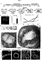
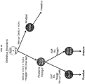
- the next two essential events in development of all endoderm organs are patterning of DE along the anterior-to-posterior (A-P) axis and gut tube morphogenesis, resulting in formation of the Sox2+ foregut in the anterior and the Cdx2+ mid-hindgut in the posterior (as highlighted in the E8.5, 14 somite stage mouse embryo, FIG. 1A ).
- This morphogenesis and the tissue interactions between endoderm and mesoderm appear to be critical for proper organogenesis both in vivo and in vitro.
- FIG 1A shows that Sox2 protein marks foregut endoderm and Cdx2 protein marks mid/hindgut endoderm in e8.5 (14 somite stage) mouse embryos.
- FIG 1B shows that inhibiting BMP repressed mid/hindgut fate and promoted expression of the foregut marker SOX2.
- FIG1C depicts that foregut spheroids generated with Wnt/FGF/Noggin have high levels of SOX2 protein by wholemount immunofluorescence staining and mRNA as compared to spheroids generated with Wnt and FGF alone, which have high levels of CDX2. *, p ⁇ 1.0x10⁻⁶.
- FIG 1D depicts that the posterior foregut in an e8.5, 14-somite stage mouse embryo gives rise to the stomach and pancreas and has high levels of Hnf1 ⁇ protein.
- FIG 1E depicts that exposing cultures to RA on the final day of the spheroid generation step induces expression of HNF1 ⁇ in SOX2-expressing epithelium, resulting in the formation of posterior foregut spheroids. *, p ⁇ 0.005.
- FIG IF depicts a lineage diagram that summarizes the patterning effects of noggin and RA in the formation of both anterior and posterior foregut endoderm. Scale bars, 100 ⁇ m. Error bars represent standard deviation. Table 1. Primary Antibodies. Antibody Species Company Product No.
- FIG 5 shows that BMP signaling is required in parallel with activation of WNT and FGF to promote a posterior fate.
- FIG 5A shows that the GSK3 ⁇ inhibitor CHIR99021 (CHIR; 2 ⁇ M) induced the same posteriorizing effects as recombinant WNT3A and these can be blocked by BMP inhibition.
- FIG 5B shows that CHIR induced gut tube morphogenesis and spheroid production occurs in a similar manner to WNT3A.
- FIG 5C depicts immunofluorescent staining of monolayer cultures which confirms the high efficiency of CDX2 induction in CHIR/FGF-treated endoderm and SOX2 induction in noggin- and CHIR/FGF/noggin-treated endoderm.
- FIG 5D shows qPCR analysis of BMP target genes MSX1/2, which indicates that BMP activity is not increased in response to Wnt/FGF, but target genes are suppressed in response to noggin, demonstrating the presence of endogenous BMP signaling.
- FIG 5E shows that addition of BMP2 (100 ng mL-1) did not substitute for or augment the ability of Wnt/FGF to posteriorize endoderm. These data indicate that the posteriorizing effect of Wnt/FGF is not mediated by up-regulation of BMP signaling but does require endogenous BMP activity.
- Scale bars 1 mm in FIG 5B ; 100 ⁇ m in FIG 5C . Error bars represent standard deviation.
- Spheroid morphogenesis is a robust process in both hESC (Reference example only-Not part of the invention) and hiPSC lines ( FIG 6A ) and >90% of spheroid cells express SOX2 ( FIG 1C ), indicating efficient specification into the foregut lineage.
- WNT, FGF and BMP have been identified by Applicant in which all three pathways cooperate to promote a mid-hindgut fate, whereas WNT and FGF act separately from BMP to drive assembly of endoderm and mesoderm into gut tube structures.
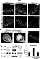
- FIGS 2A-2G depicts gastric organoid differentiation is an efficient and cell line-independent process.
- FIG 2A Table comparing spheroid formation and characteristics between two hESC lines (H1 and H9; (Reference example only-Not part of the invention)) and one iPSC line (72.3).
- FIG 2B Immunofluorescent staining of day 34 hGOs derived from H1 and iPSC 72.3 cell lines. iPSC-derived organoids exhibit the same morphological and molecular features of those derived from hESCs (Reference example only-Not part of the invention).
- FIG 2C Organ epithelial cell type quantification in day 34 hGOs.
- iPSC 72.3 Characterization of induced pluripotent stem cell line iPSC 72.3.
- FIG 2D iPSC 72.3 exhibits normal morphological characteristics of pluripotent stem cell colonies, as compared to the H1 hESC line (Reference example only-Not part of the invention) and FIG 2E , has a normal 46;XY karyotype.
- FIG 2F iPSC 72.3 expresses pluripotent markers OCT3/4 and NANOG
- FIG 2G demonstrates pluripotency by differentiation into endoderm, mesoderm, and ectoderm lineages in an in vivo teratoma assay. Scale bars, 100 ⁇ m. Error bars represent standard deviation.
- both the fundic and antral domains of the stomach arise from the posterior segment of Sox2+ foregut endoderm, along with the pancreas, liver and duodenum.
- Applicant sought to identify signaling pathways that promote posterior foregut fate.
- Applicant focused on retinoic acid (RA) signaling given its role in development of posterior foregut-derived organs. 15-17
- RA retinoic acid
- Applicant identified that a 24-hour exposure to RA on the final day (days 5-6) of the patterning/spheroid generation stage (FGF4/WNT3A/Noggin) results in robust activation of posterior foregut markers and the formation of SOX2/HNF1 ⁇ + posterior foregut spheroids ( FIG 1E and FIG 7 ).
- FGF4/WNT3A/Noggin patterning/spheroid generation stage
- FIGS 7A-7D shows that retinoic acid posteriorizes foregut endoderm.
- FIG 7A depicts a schematic illustrating of foregut patterning experiments. DE cultures were treated with Wnt(CHIR)/FGF/noggin for three days to generate Sox2-positive foregut spheroids, and RA is added for 24 hours on the third day of patterning.
- FIG 7B depicts Brightfield images that show that RA increases the number of spheroids that are produced from foregut monolayer cultures.
- FIG 7C depicts a lower power image of FIG1D showing immunofluorescent image of a 14 somite stage embryo with Hnf1 ⁇ protein localized to the posterior portion of the foregut. Boxed region of embryo is shown in FIG 1D .
- FIG 7D shows qPCR analysis of gene expression in foregut spheroids treated with RA.
- Posterior foregut markers HNF1 ⁇ and HNF6 are robustly induced by 24-hour exposure to RA. *, p ⁇ 0.05.
- Scale bars 1 mm in FIG 7B ; 100 ⁇ m in FIG 7C . Error bars represent standard deviation.
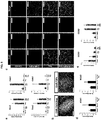
- FIG 2 generally depicts the specification and growth of human antral gastric organoids. Error bars represent standard deviation.
- FIG2A depicts schematic representation of the in vitro culture system used to direct the differentiation of hPSCs into three-dimensional gastric organoids
- FIG2B depicts the defining markers of developing posterior foregut organs by wholemount immunofluorescent staining of mouse E10.5 embryos with Sox2, Pdx1 and Cdx2.
- FIG2C shows that the posterior foregut spheroids cultured in three-dimensional matrix for three days in the presence of RA (2 ⁇ M) co-expressed high levels of PDX1 and SOX2 and did not express the pancreatic marker PTF1 ⁇ , similar to the developing antrum *, p ⁇ 0.05.
- FIG2D depicts stereomicrographs showing morphological changes during growth of posterior forgut spheroids into gastric organoids.
- the epithelium of hGOs exhibited a complex glandular architecture, scale bar, 500 ⁇ m.
- FIG2E depicts a comparison of developing mouse antrum at E14.5 and E18.5 and comparable stages of hGO development. Sox2 and Pdx1 are co-expressed in the early pseudostratified epithelia in both mouse antrum and hGOs. At later stages, Sox2 is down-regulated as the epithelia transform into more mature glandular structures. Pdx1 is maintained in the antrum throughout adulthood in vivo and at all stages examined in hGOs, scale bars 100 ⁇ m in FIG2E .
- Applicant used SOX2/PDX1 + spheroids to identify pathways that promote growth and morphogenesis of the early gastric epithelium and found that high concentrations of EGF (100 ng mL -1 ) were sufficient to promote robust outgrowth of human antral gastric organoids (hGOs). Over the course of 3-4 weeks, spheroids that were ⁇ 100 ⁇ m in diameter grew into organoids that were 2-4 mm in diameter. At the later stages of culture ( ⁇ day 27), the hGO epithelium underwent a series of morphogenetic changes reminiscent of the late stages of embryonic gastric development, during which a simple, flat pseudostratified epithelium transitions into an elaborate, convoluted glandular epithelium ( FIG 2D ).
- FIG 8 shows that EGF is required for glandular morphogenesis in gastric organoids.
- Brightfield images and immunostaining demonstrate the requirement for EGF for epithelial morphogenesis and gland formation at late stages of hGO differentiation.
- EGF is removed from the growth medium at day 27, prior to glandular morphogenesis, the hGO epithelium retains a simple, cuboidal structure that fails to form glands.
- Scale bars 100 ⁇ m.
- hGO development is astonishingly similar to in vivo stomach organogenesis.
- both epithelia are pseudostratified and contain mitotic cells that are concentrated toward the luminal face ( FIG 9 and FIG 10 ), indicating an interkinetic nuclear migration process 21 .
- the early hGOs are appropriately polarized and contain secondary lumina that are outlined by expression of the apical marker aPKC 22 ( FIG 10 ).
- the antrum transforms into a simple columnar epithelium exhibiting a highly structured organization consisting of glands and pits ( FIG 2E and FIG 9 ).
- the hGO epithelium undergoes similar transitions to form a tall columnar epithelium with a glandular structure similar to the late fetal antrum ( FIG 2E ).
- Analysis of expression of the transcription factors Sox2, Pdx1, Gata4 and Klf5 revealed a stereotypic temporospatial expression pattern that accompany these morphogenetic processes both in vivo and in vitro ( FIG 9 ). At early stages these factors are all co-expressed in the immature, pseudostratified epithelium.
- Sox2 expression is down-regulated as the epithelium forms early glands and pits, whereas the expression of the other factors is maintained indefinitely.
- 13-day hGOs represent a developmental stage similar to the E12-14 mouse antrum, whereas 34-day hGOs are more comparable to the late fetal-early postnatal antrum.
- hGOs recapitulate normal embryonic development and that the molecular and morphogenetic processes that occur during antrum development are conserved between rodents and humans.
- FIG 9 shows a comparison of transcription factor expression during development of the mouse antrum and human gastric organoids.
- Four embryonic stages (E12.5, E14.5, E16.5 and E18.5) and one postnatal stage (P12) of in vivo antrum development were analyzed for transcription factor expression: Sox2, Pdx1, Gata4, Klf5, and FoxF1.
- Sox2, Pdx1, Gata4, Klf5, and FoxF1 The same markers were analyzed at two stages (day 13 and day 34) of in vitro hGO development and revealed that organoid development parallels what occurs in vivo.
- the epithelial marker Sox2 is expressed ubiquitously but at later stages it is down-regulated, while other epithelial transcription factors, Pdx1, Gata4 and Klf5, exhibit persistent expression throughout development.
- Both early and late stage hGOs contain FoxF1-positive mesenchymal cells surrounding the epithelium. Scale bars, 100 ⁇ m.
- FIG 10 shows that early stage human gastric organoids exhibit stereotypic architecture and nuclear behavior.
- hGOs contain pseudostratified epithelia that display apicobasal polarity marked by the apical marker aPKC and the basolateral marker E-Cadherin, similar to the E12.5 mouse antrum. Further, secondary lumina lined by apical membrane (white arrows) are seen within the organoid epithelium. Both the E12.5 mouse antrum and day 7 hGOs appear to undergo interkinetic nuclear migration, indicated by the presence of mitotic nuclei, pHH3, in only the apical portions of cells. Scale bars, 50 ⁇ m.
- the foregut spheroids contained a mesenchyme component similar to the mid-hindgut spheroids that were previously described 10 .
- the mesenchyme expands and expresses key transcription factors associated with antral mesenchyme development, including FOXF1 and BAPX1 ( FIG 10 and FIG 11 ).
- FOXF1 and BAPX1 FIG 10 and FIG 11 .
- hGO mesenchyme largely consists of VIMENTIN + submucosal fibroblasts and a smaller number of ACTA2 + subepithelial myofibroblasts ( FIG 11 ), indicative of immature gastric mesenchyme.
- hGOs do not form differentiated layers of smooth muscle as occurs in vivo.
- FIG 11 shows mesenchymal differentiation in gastric organoids.
- FIG 11A shows temporal expression analysis of the antral mesenchyme transcription factor BAPX1. Similar to its known embryonic expression pattern, BAPX1 is up-regulated during the earlier stages of hGO differentiation and then down-regulated coincident with functional cell type marker expression.
- FIG 11B shows that staining for mesenchymal cell type markers reveals that day 34 hGOs contain FOXF1/VIMENTIN-positive submucosal fibroblasts and a small number of VIMENTIN/ALPHA-SM-ACTIN (SMA)-expressing subepithelial fibroblasts.
- hGOs lack a robust smooth muscle layer, indicated by SMA/Desmin-positive cells in the in vivo antrum. Scale bars, 100 ⁇ m. Error bars represent standard deviation.
- hGOs contain surface mucous cells (MUC5AC/UEAI + ) that secrete mucus into the lumen and have the same tall columnar morphology as their in vivo counterpart. hGOs also contain TFF2/GSII + antral gland cells, indicating appropriate differentiation in the antral mucous lineages ( FIG 3A ).
- hGOs develop a progenitor cell niche, indicated by basally located zones of proliferation and SOX9 expression ( FIG 4A ), although the proliferative index of the epithelium is variable and ranges between 1-10%.
- the in vitro hGOs contain a physiological gastric epithelium that comprises both progenitor and differentiated cell types.
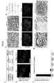
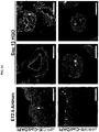
- FIG 4 shows that human gastric organoids exhibit acute responses to H. pylori infection.
- FIG 4A shows that Day 28 hGOs contained proliferative cells, marked by Ki67, and SOX9+ progenitor cells that were restricted toward the bottoms of the early glands, similar to the late embryonic and postnatal mouse antrum.
- FIG 4B shows that hGOs were used to model human-specific disease processes of H. pylori infection. Bacteria were microinjected into the lumen of hGOs and bacteria were visualized in the lumen 24 hours post-injection by brightfield microscopy (black arrow) and immunofluorescent staining.
- FIG 4C depicts immunoprecipitation for the oncogene c-Met and demonstrates that H.
- FIG 4D shows that within 24 hours, H. pylori infection caused a two-fold increase in the number of proliferating cells in the hGO epithelium, measured by EdU incorporation. *, p ⁇ 0.05. Scale bars, 100 ⁇ m in a; 25 ⁇ m in b. Error bars represent s.e.m.
- FIG3 demonstrates that human gastric organoids contain normal differentiated antral cell types and can be used to model human stomach development.
- FIG 3A demonstrates that hGOs contain all the major antral cell lineages. The 34-day hGOs have surface mucous cells (Muc5AC) and mucous gland cells (TFF2), as well as lectin staining that distinguishes surface mucous, UEAI, and mucous gland cells, GSII. hGOs also contain endocrine cells as marked by ChromograninA (CHGA).
- FIG 3B is a schematic representation of the different roles for EGF in the growth, morphogenesis, and cell type specification during development of hGOs.
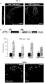
- FIG 3C shows that all major endocrine hormones are expressed in hGOs upon withdrawal of EGF including gastrin, ghrelin, and serotonin (5-HT).
- FIG 3D shows that high levels of EGF repress NEUROG3 expression.
- a reduction in EGF concentration at day 30 resulted in a significant increase in NEUROG3 expression measured at day 34 by qPCR, indicating that EGF acts upstream of NEUROG3 in endocrine specification. *, p ⁇ 0.05.
- FIG 3E shows that NEUROG3 acts downstream of EGF to induce endocrine cell fate. Forced expression of NEUROG3 using a dox-inducible system was sufficient to override the endoc rine - re pressing effects of high EGF (100 ng mL-1). hGOs were exposed to dox (1 ⁇ g mL-1) for 24 hours at day 30 and analyzed at day 34. Dox-treated organoids exhibited a robust induction of ChrA-expressing endocrine cells. Scale bars, 100 ⁇ m. Error bars represent standard deviation.
- CHROMOGRANIN-A CHROMOGRANIN-A + endocrine cells in 34-day hGOs, including the four main endocrine cell types in the antrum expressing gastrin, ghrelin, somatostatin, and serotonin ( FIG 3C and FIG 12 ).
- high levels of EGF repress endocrine cell formation such that 100 ng ml -1 resulted in ⁇ 1 endocrine cell per organoid.
- hGOs cultured in lower levels of EGF (10 ng ml -1 ) from day 30-34 developed an abundance of endocrine cells ( FIG 13 ).
- FIG 12 shows gastric antrum endocrine cell development in vivo. Endocrine cell differentiation in the antrum is first evident at E18.5, but is more definitive at postnatal stages (P12 shown). At early stages, all expected gastric endocrine subtypes are evident, including gastrin, ghrelin, somatostatin, and serotonin (5-HT). Scale bars, 100 ⁇ m.
- FIG 13 shows that EGF signaling represses a NEUROG3-dependent gastric endocrine specification program.
- FIG 13A shows that hGOs maintained in high concentrations of EGF (100 ng mL-1) had very few endocrine cells at day 34, shown by staining for the pan-endocrine marker CHGA.
- FIG 13B shows generation of hGOs from a hESC line stably transfected with a dox-inducible NEUROG3-overexpressing transgene (Reference example only-Not part of the invention), to test whether EGF repression of endocrine differentiation occurs upstream of NEUROG3.
- hGOs were maintained in high EGF (100 ng mL-1) then at day 30 were treated with doxycycline (1 ⁇ g mL-1) for 24 hours and then analyzed at day 34.
- Dox-treated hGOs show robust activation of endocrine markers CHGA, GASTRIN, GHRELIN, and SOMATOSTATIN, and they contain CHGA- ( FIG 3A ), GHRELIN-, and SOMATOSTATIN-positive cells with endocrine morphology. *, p ⁇ 0.05. Scale bars, 100 ⁇ m. Error bars represent standard deviation.
- H. pylori-mediated disease 31,32 Clinical evidence indicates that predominant colonization of the antrum has an important role in H. pylori-mediated disease 31,32 .
- H. pylori To mimic the normal host-pathogen interface, we introduced H. pylori directly to the luminal surface of the epithelium by microinjection into the lumen of the organoids and measured epithelial signaling and proliferation ( FIG 4 ). Bacteria were observed tightly associated with the hGO epithelium by immunofluorescence ( FIG 4B ). Within 24 hours, Applicant observed significant epithelial responses to H.
- H. pylori including robust activation of the gastric oncogene c-Met 33 and a 2-fold increase in epithelial cell proliferation.
- the H. pylori virulence factor CagA plays a pivotal role in the etiology of disease. Consistent with published studies 34 , Applicant demonstrated that CagA translocates into the organoid epithelial cells and forms a complex with c-Met ( FIG 4C ). Furthermore, the epithelial response was abrogated when hGOs were injected with a non-pathogenic strain of H. pylori lacking CagA, reinforcing the importance of this factor in H. pylori-mediated human pathogenesis. Thus, the pathophysiological response of hGOs to H. pylori makes them an unprecedented model for elucidating the initiating events of human gastric disease mediated by H. pylori.
Landscapes
- Health & Medical Sciences (AREA)
- Engineering & Computer Science (AREA)
- Life Sciences & Earth Sciences (AREA)
- Biomedical Technology (AREA)
- Chemical & Material Sciences (AREA)
- Biotechnology (AREA)
- Organic Chemistry (AREA)
- Bioinformatics & Cheminformatics (AREA)
- Genetics & Genomics (AREA)
- Wood Science & Technology (AREA)
- Zoology (AREA)
- Microbiology (AREA)
- Cell Biology (AREA)
- Biochemistry (AREA)
- General Health & Medical Sciences (AREA)
- General Engineering & Computer Science (AREA)
- Immunology (AREA)
- Gastroenterology & Hepatology (AREA)
- Hematology (AREA)
- Urology & Nephrology (AREA)
- Molecular Biology (AREA)
- Analytical Chemistry (AREA)
- Food Science & Technology (AREA)
- Medicinal Chemistry (AREA)
- Physics & Mathematics (AREA)
- Tropical Medicine & Parasitology (AREA)
- General Physics & Mathematics (AREA)
- Pathology (AREA)
- Micro-Organisms Or Cultivation Processes Thereof (AREA)
- Measuring Or Testing Involving Enzymes Or Micro-Organisms (AREA)
- Materials For Medical Uses (AREA)
- Apparatus Associated With Microorganisms And Enzymes (AREA)
- Medicines Containing Material From Animals Or Micro-Organisms (AREA)
Priority Applications (1)
| Application Number | Priority Date | Filing Date | Title |
|---|---|---|---|
| EP20170283.4A EP3712254A1 (en) | 2014-05-28 | 2015-05-27 | Methods and systems for converting precursor cells into gastric tissues through directed differentiation |
Applications Claiming Priority (2)
| Application Number | Priority Date | Filing Date | Title |
|---|---|---|---|
| US201462003719P | 2014-05-28 | 2014-05-28 | |
| PCT/US2015/032626 WO2015183920A2 (en) | 2014-05-28 | 2015-05-27 | Methods and systems for converting precursor cells into gastric tissues through directed differentiation |
Related Child Applications (2)
| Application Number | Title | Priority Date | Filing Date |
|---|---|---|---|
| EP20170283.4A Division EP3712254A1 (en) | 2014-05-28 | 2015-05-27 | Methods and systems for converting precursor cells into gastric tissues through directed differentiation |
| EP20170283.4A Division-Into EP3712254A1 (en) | 2014-05-28 | 2015-05-27 | Methods and systems for converting precursor cells into gastric tissues through directed differentiation |
Publications (2)
| Publication Number | Publication Date |
|---|---|
| EP3149156A2 EP3149156A2 (en) | 2017-04-05 |
| EP3149156B1 true EP3149156B1 (en) | 2021-02-17 |
Family
ID=53385964
Family Applications (2)
| Application Number | Title | Priority Date | Filing Date |
|---|---|---|---|
| EP15728704.6A Active EP3149156B1 (en) | 2014-05-28 | 2015-05-27 | Methods and systems for converting precursor cells into gastric tissues through directed differentiation |
| EP20170283.4A Pending EP3712254A1 (en) | 2014-05-28 | 2015-05-27 | Methods and systems for converting precursor cells into gastric tissues through directed differentiation |
Family Applications After (1)
| Application Number | Title | Priority Date | Filing Date |
|---|---|---|---|
| EP20170283.4A Pending EP3712254A1 (en) | 2014-05-28 | 2015-05-27 | Methods and systems for converting precursor cells into gastric tissues through directed differentiation |
Country Status (10)
| Country | Link |
|---|---|
| US (3) | US10174289B2 (enExample) |
| EP (2) | EP3149156B1 (enExample) |
| JP (4) | JP6687544B2 (enExample) |
| CN (2) | CN113249297B (enExample) |
| AU (2) | AU2015267148B2 (enExample) |
| CA (1) | CA2949834A1 (enExample) |
| ES (1) | ES2860423T3 (enExample) |
| IL (1) | IL249253B (enExample) |
| SG (2) | SG10201801654RA (enExample) |
| WO (1) | WO2015183920A2 (enExample) |
Families Citing this family (36)
| Publication number | Priority date | Publication date | Assignee | Title |
|---|---|---|---|---|
| US9719068B2 (en) | 2010-05-06 | 2017-08-01 | Children's Hospital Medical Center | Methods and systems for converting precursor cells into intestinal tissues through directed differentiation |
| KR102423106B1 (ko) | 2013-06-11 | 2022-07-21 | 프레지던트 앤드 펠로우즈 오브 하바드 칼리지 | SC-β 세포 및 조성물 그리고 그 생성 방법 |
| EP3149156B1 (en) | 2014-05-28 | 2021-02-17 | Children's Hospital Medical Center | Methods and systems for converting precursor cells into gastric tissues through directed differentiation |
| JP6804438B2 (ja) | 2014-10-17 | 2020-12-23 | チルドレンズ ホスピタル メディカル センター | 多能性幹細胞を使用するヒト小腸のin vivoモデル、並びにそれを作製、及び使用する方法 |
| WO2016100930A1 (en) | 2014-12-18 | 2016-06-23 | President And Fellows Of Harvard College | Methods for generating stem cell-derived b cells and methods of use thereof |
| WO2016100898A1 (en) | 2014-12-18 | 2016-06-23 | President And Fellows Of Harvard College | Serum-free in vitro directed differentiation protocol for generating stem cell-derived b cells and uses thereof |
| WO2016100909A1 (en) | 2014-12-18 | 2016-06-23 | President And Fellows Of Harvard College | METHODS FOR GENERATING STEM CELL-DERIVED β CELLS AND USES THEREOF |
| WO2017123806A1 (en) | 2016-01-12 | 2017-07-20 | Cedars-Sinai Medical Center | A method of non destructive monitoring of biological processes in microfluidic tissue culture systems |
| WO2017136462A2 (en) * | 2016-02-01 | 2017-08-10 | EMULATE, Inc. | Systems and methods for growth of intestinal cells in microfluidic devices |
| EP4177335A1 (en) * | 2016-05-05 | 2023-05-10 | Children's Hospital Medical Center | Methods for the in vitro manufacture of gastric fundus tissue and compositions related to same |
| EP3534976A4 (en) | 2016-11-04 | 2020-09-16 | Children's Hospital Medical Center | PATHOLOGICAL MODELS OF HEPATIC ORGANOIDS AND ASSOCIATED METHODS OF MANUFACTURE AND USE |
| EP3548507A4 (en) * | 2016-12-05 | 2020-07-15 | Children's Hospital Medical Center | COLON ORGANOIDS AND PROCESSES FOR THE PREPARATION AND USE THEREOF |
| EP3551047A1 (en) | 2016-12-07 | 2019-10-16 | Progenity, Inc. | Gastrointestinal tract detection methods, devices and systems |
| US11913022B2 (en) | 2017-01-25 | 2024-02-27 | Cedars-Sinai Medical Center | In vitro induction of mammary-like differentiation from human pluripotent stem cells |
| US11767513B2 (en) | 2017-03-14 | 2023-09-26 | Cedars-Sinai Medical Center | Neuromuscular junction |
| US11414648B2 (en) | 2017-03-24 | 2022-08-16 | Cedars-Sinai Medical Center | Methods and compositions for production of fallopian tube epithelium |
| AU2018244922A1 (en) | 2017-03-30 | 2019-09-12 | Biora Therapeutics, Inc. | Treatment of a disease of the gastrointestinal tract with live biotherapeutics |
| EP3609997A4 (en) | 2017-04-14 | 2021-03-03 | Children's Hospital Medical Center | COMPOSITIONS OF STEM CELLS FROM MULTIPLE DONOR CELLS AND THEIR PREPARATION PROCEDURES |
| US20200199537A1 (en) * | 2017-06-09 | 2020-06-25 | Children's Hospital Medical Center | Liver organoid compositions and methods of making and using same |
| CA3074901A1 (en) | 2017-09-11 | 2019-03-14 | Imba - Institut Fur Molekulare Biotechnologie Gmbh | Tumor organoid model |
| EP3694603A4 (en) * | 2017-10-10 | 2021-07-14 | Children's Hospital Medical Center | ESOPHAGUS TISSUE AND / OR ORGANOID COMPOSITIONS AND METHOD FOR MANUFACTURING THEREOF |
| AU2018370029B2 (en) | 2017-11-15 | 2024-11-07 | Vertex Pharmaceuticals Incorporated | Islet cell manufacturing compositions and methods of use |
| US12379372B2 (en) | 2017-12-21 | 2025-08-05 | Children's Hospital Medical Center | Digitalized human organoids and methods of using same |
| US12161676B2 (en) | 2018-03-23 | 2024-12-10 | Cedars-Sinai Medical Center | Methods of use of islet cells |
| US11981918B2 (en) | 2018-04-06 | 2024-05-14 | Cedars-Sinai Medical Center | Differentiation technique to generate dopaminergic neurons from induced pluripotent stem cells |
| EP3775161A4 (en) | 2018-04-06 | 2022-04-06 | Cedars-Sinai Medical Center | NEURODEGENERATIVE DISEASE MODELS DERIVED FROM HUMAN PLURIPOTENTIC STEM CELLS ON A MICROFLUIDIC CHIP |
| KR102887406B1 (ko) | 2018-07-26 | 2025-11-19 | 칠드런즈 호스피탈 메디칼 센터 | 간-담도-췌장 조직 및 이를 제조하는 방법 |
| EP3833365A4 (en) | 2018-08-10 | 2022-05-11 | Vertex Pharmaceuticals Incorporated | Stem cell derived islet differentiation |
| WO2020056158A1 (en) | 2018-09-12 | 2020-03-19 | Children's Hospital Medical Center | Organoid compositions for the production of hematopoietic stem cells and derivatives thereof |
| WO2020091020A1 (ja) * | 2018-11-02 | 2020-05-07 | 公立大学法人名古屋市立大学 | 多能性幹細胞由来腸管オルガノイドの作製法 |
| US20230002736A1 (en) * | 2019-12-12 | 2023-01-05 | The Regents Of Athe University Of California | Endoderm differentiation from pluripotent stem cell lines |
| CN115666704B (zh) | 2019-12-13 | 2025-09-26 | 比特比德科有限责任公司 | 用于将治疗剂递送至胃肠道的可摄取装置 |
| CA3194196A1 (en) * | 2020-09-25 | 2022-03-31 | Children's Hospital Medical Center | Raft cultures and methods of making thereof |
| WO2025035134A1 (en) * | 2023-08-10 | 2025-02-13 | Georgetown University | Lna gapmers inhibit growth of cck-br positive cancer |
| WO2025072803A1 (en) | 2023-09-29 | 2025-04-03 | Children's Hospital Medical Center | Ntrk2 signaling-mediated alveolar capillary injury and repair |
| WO2025085899A1 (en) * | 2023-10-19 | 2025-04-24 | The Research Foundation For The State University Of New York | Production of salivary gland epithelial progenitors and salivary gland cell organoids for regenerative medicine and drug testing purposes |
Family Cites Families (388)
| Publication number | Priority date | Publication date | Assignee | Title |
|---|---|---|---|---|
| GB462599A (en) | 1935-09-11 | 1937-03-11 | British Celanese | Improvements in the treatment of textile materials |
| US3970108A (en) | 1973-10-23 | 1976-07-20 | Cross Manufacturing, Inc. | Priority hydraulic control valve |
| WO1992007615A1 (en) | 1990-10-29 | 1992-05-14 | Regents Of The University Of Minnesota | A bioartificial liver |
| US5523226A (en) | 1993-05-14 | 1996-06-04 | Biotechnology Research And Development Corp. | Transgenic swine compositions and methods |
| US6372482B1 (en) | 1997-01-16 | 2002-04-16 | Yissum Research Development Company Of The Hebrew University Of Jerusalem | Device and method for performing a biological modification of a fluid |
| US5912227A (en) | 1995-01-27 | 1999-06-15 | North Carolina State University | Method of enhancing nutrient uptake |
| WO1998021312A1 (en) | 1996-11-08 | 1998-05-22 | Rpms Technology Limited | Human hepatocytes in three-dimensional support systems |
| EP1063289A1 (en) | 1998-03-03 | 2000-12-27 | JMS Co., Ltd. | Liver cell clones for artifical liver and extracorporeal liver assist device |
| WO1999045100A1 (en) | 1998-03-02 | 1999-09-10 | University Of Massachusetts, A Public Institution Of Higher Education Of The Commonwealth Of Massachusetts, As Represented By Its Amherst Campus | Embryonic or stem-like cell lines produced by cross-species nuclear transplantation |
| US7291626B1 (en) | 1998-04-09 | 2007-11-06 | John Hopkins University School Of Medicine | Inhibitors of hedgehog signaling pathways, compositions and uses related thereto |
| US20020035459A1 (en) * | 1998-09-14 | 2002-03-21 | George M. Grass | Pharmacokinetic-based drug design tool and method |
| US7759113B2 (en) | 1999-04-30 | 2010-07-20 | The General Hospital Corporation | Fabrication of tissue lamina using microfabricated two-dimensional molds |
| JP3725147B2 (ja) | 1999-08-25 | 2005-12-07 | 東洋紡績株式会社 | 血管網類似構造体を有する細胞培養用モジュール |
| US6607501B2 (en) | 2001-05-14 | 2003-08-19 | Reynolds G. Gorsuch | Process and apparatus for utilization of in vivo extracted plasma with tissue engineering devices, bioreactors, artificial organs, and cell therapy applications |
| AU2002367580A1 (en) | 2001-05-16 | 2003-09-22 | Tracy C. Grikscheit | Tissue-engineered organs |
| WO2003046141A2 (en) | 2001-11-26 | 2003-06-05 | Advanced Cell Technology, Inc. | Methods for making and using reprogrammed human somatic cell nuclei and autologous and isogenic human stem cells |
| GB2399823B (en) | 2001-12-07 | 2006-02-15 | Geron Corp | Islet cells from primate pluripotent stem cells |
| US20050170506A1 (en) | 2002-01-16 | 2005-08-04 | Primegen Biotech Llc | Therapeutic reprogramming, hybrid stem cells and maturation |
| US20030187515A1 (en) | 2002-03-26 | 2003-10-02 | Hariri Robert J. | Collagen biofabric and methods of preparing and using the collagen biofabric |
| US7160719B2 (en) | 2002-06-07 | 2007-01-09 | Mayo Foundation For Medical Education And Research | Bioartificial liver system |
| JP4187167B2 (ja) | 2002-08-28 | 2008-11-26 | 和守 船津 | 異形断面中空糸膜型細胞含有デバイス |
| TW571101B (en) | 2003-01-21 | 2004-01-11 | Ind Tech Res Inst | Fluid analysis apparatus |
| EP1596926A2 (en) | 2003-02-07 | 2005-11-23 | The Johns Hopkins University | Hypoxia induced mitogenic factor |
| AU2004209505B2 (en) | 2003-02-10 | 2009-07-23 | Banyu Pharmaceutical Co., Ltd. | Melanin-concentrating hormone receptor antagonists containing piperidine derivatives as the active ingredient |
| DE10362002B4 (de) | 2003-06-23 | 2006-10-12 | Fraunhofer-Gesellschaft zur Förderung der angewandten Forschung e.V. | Adulte pluripotente Stammzellen |
| US8647873B2 (en) | 2004-04-27 | 2014-02-11 | Viacyte, Inc. | PDX1 expressing endoderm |
| US7541185B2 (en) | 2003-12-23 | 2009-06-02 | Cythera, Inc. | Methods for identifying factors for differentiating definitive endoderm |
| US7985585B2 (en) | 2004-07-09 | 2011-07-26 | Viacyte, Inc. | Preprimitive streak and mesendoderm cells |
| US7625753B2 (en) | 2003-12-23 | 2009-12-01 | Cythera, Inc. | Expansion of definitive endoderm cells |
| US20050266554A1 (en) | 2004-04-27 | 2005-12-01 | D Amour Kevin A | PDX1 expressing endoderm |
| KR101221302B1 (ko) | 2003-12-23 | 2013-01-11 | 비아싸이트, 인크. | 완전 내배엽 |
| WO2007059007A2 (en) | 2005-11-14 | 2007-05-24 | Cythera, Inc. | Markers of definitive endoderm |
| WO2005081970A2 (en) | 2004-02-24 | 2005-09-09 | The Curators Of The University Of Missouri | Self-assembling cell aggregates and methods of making engineered tissue using the same |
| DE102004017476B4 (de) | 2004-04-08 | 2009-03-26 | Fraunhofer-Gesellschaft zur Förderung der angewandten Forschung e.V. | Verfahren zur Herstellung einer Epithelzellen enthaltenden Zellzusammensetzung |
| ES2751095T3 (es) * | 2004-04-27 | 2020-03-30 | Viacyte Inc | Endodermo que expresa pdx1 |
| JP4650608B2 (ja) | 2004-05-18 | 2011-03-16 | 信越化学工業株式会社 | フォトマスクブランク及びフォトマスクの製造方法 |
| US9375514B2 (en) | 2004-05-21 | 2016-06-28 | Fraunhofer-Gesellschaft Zur Forderung Der Angewandten Forschung E.V. | Multicellular tissue and organ culture systems |
| AU2005265098B2 (en) | 2004-06-17 | 2012-02-23 | Thrasos Innovation, Inc. | TDF-related compounds and analogs thereof |
| CN101023163A (zh) * | 2004-07-09 | 2007-08-22 | 赛瑟拉公司 | 鉴定用于分化定型内胚层的因子的方法 |
| AU2005272078B2 (en) * | 2004-07-09 | 2011-04-14 | Viacyte, Inc. | Methods for identifying factors for differentiating definitive endoderm |
| CA2573512C (en) | 2004-07-12 | 2014-09-23 | Emisphere Technologies, Inc. | Compositions for delivering peptide yy and pyy agonists |
| MX2007000728A (es) | 2004-07-21 | 2007-03-15 | Ambrx Inc | Polipeptidos biosinteticos que utilizan amino acidos no naturalmente codificados. |
| EP1791952A4 (en) | 2004-08-13 | 2008-06-11 | Univ Georgia Res Found | COMPOSITIONS AND METHODS OF SELF-RENEWAL AND DIFFERENTIATION IN HUMAN EMBRYONAL STEM CELLS |
| US20060236415A1 (en) | 2005-03-09 | 2006-10-19 | Silversides David W | Neural crest cells specific promoters; isolated neural crest cells; and methods of isolating and of using same |
| US7604929B2 (en) | 2005-04-21 | 2009-10-20 | In Vitro Technologies, Inc. | Cellular compositions and methods for their preparation |
| WO2006122147A2 (en) | 2005-05-10 | 2006-11-16 | Rutgers, The State University Of New Jersey | Alginate poly-l-lysine encapsulation as a technology for controlled differentiation of embryonic stem cells |
| WO2006126219A1 (en) | 2005-05-26 | 2006-11-30 | Fresenius Medical Care Deutschland G.M.B.H. | Liver progenitor cells |
| GB0517382D0 (en) | 2005-08-26 | 2005-10-05 | Plasticell Ltd | Cell culture |
| WO2007027905A2 (en) | 2005-08-31 | 2007-03-08 | Science And Technology Corporation @ Unm | Human renal stem cells |
| EP3584311A1 (en) * | 2005-10-27 | 2019-12-25 | Viacyte, Inc. | Pdx-1 expressing dorsal and ventral foregut endoderm |
| WO2007065095A2 (en) | 2005-11-29 | 2007-06-07 | Spencer Rosero | System and method for supporting a biological chip device |
| US20070239083A1 (en) | 2006-01-18 | 2007-10-11 | Axel Voss | Shock wave generators |
| US20070238169A1 (en) | 2006-04-11 | 2007-10-11 | The Board Of Trustees Of The Leland Stanford Junior University | Cell sorter and culture system |
| US8685730B2 (en) | 2006-05-02 | 2014-04-01 | Wisconsin Alumni Research Foundation | Methods and devices for differentiating pluripotent stem cells into cells of the pancreatic lineage |
| US9012419B2 (en) | 2006-05-17 | 2015-04-21 | University Of Utah Research Foundation | Methods and compositions related to eosinophil regulation |
| WO2008002573A2 (en) | 2006-06-27 | 2008-01-03 | Intercept Pharmaceuticals, Inc. | Bile acid derivatives as fxr ligands for the prevention or treatment of fxr-mediated deseases or conditions |
| WO2008011561A2 (en) | 2006-07-21 | 2008-01-24 | Massachusetts Institute Of Technology | End-modified poly(beta-amino esters) and uses thereof |
| US8497240B2 (en) | 2006-08-17 | 2013-07-30 | Amylin Pharmaceuticals, Llc | DPP-IV resistant GIP hybrid polypeptides with selectable properties |
| DE602007012293D1 (enExample) | 2006-12-08 | 2011-03-10 | Du Pont | |
| CN104391125B (zh) | 2006-12-08 | 2017-10-03 | 李尹雄 | 通过干细胞营养物进行的器官的形成和复壮以及酒精损伤的器官的再生 |
| AU2007335753B2 (en) | 2006-12-18 | 2013-12-05 | Ben Gurion University Of The Negev | Scaffolding for tissue regeneration or repair |
| US7722560B2 (en) | 2007-02-09 | 2010-05-25 | The Regents Of The University Of Michigan | Mechanical extension implants for short bowel syndrome |
| US8512697B2 (en) | 2007-05-16 | 2013-08-20 | The University Of North Carolina At Chapel Hill | Delivery of micro- and nanoparticles with blood platelets |
| FR2917425B1 (fr) | 2007-06-18 | 2010-11-19 | Univ Nancy 1 Henri Poincare | Procede de proliferation de cellules sur des multicouches de polyelectrolytes et son application, notamment a la preparation de biomateriaux cellularises |
| GB2467645B (en) | 2007-07-06 | 2011-12-21 | Univ London | A method of proliferating human hepatocyte cells |
| EP2022848A1 (en) | 2007-08-10 | 2009-02-11 | Hubrecht Institut | A method for identifying, expanding, and removing adult stem cells and cancer stem cells |
| WO2009033011A1 (en) | 2007-09-07 | 2009-03-12 | Children's Hospital Medical Center | Use of secretor, lewis and sialyl antigen levels as predictors for disease |
| US7695963B2 (en) | 2007-09-24 | 2010-04-13 | Cythera, Inc. | Methods for increasing definitive endoderm production |
| CA2708780A1 (en) | 2007-12-11 | 2009-06-18 | Research Development Foundation | Small molecules for neuronal differentiation of embryonic stem cells |
| WO2009086596A1 (en) | 2008-01-08 | 2009-07-16 | The University Of Queensland | Method of producing a population of cells |
| WO2009096049A1 (ja) * | 2008-02-01 | 2009-08-06 | Kyoto University | 人工多能性幹細胞由来分化細胞 |
| WO2009132196A2 (en) * | 2008-04-23 | 2009-10-29 | Alphagene Bioscience, Inc. | Drug discovery methods involving a preclinical, in vitro isolated gastrointestinal epithelial stem cell-like progenitor cell system |
| WO2009149179A2 (en) | 2008-06-03 | 2009-12-10 | Aethlon Medical Inc. | Enhanced antiviral therapy methods and devices |
| CN102257124B (zh) | 2008-06-04 | 2014-12-03 | 蒂斯由斯股份有限公司 | 芯片上器官装置 |
| JP5624981B2 (ja) | 2008-06-24 | 2014-11-12 | ザ・キュレーターズ・オブ・ザ・ユニバーシティ・オブ・ミズーリThe Curators Ofthe University Of Missouri | 自己集合性多細胞体、および前記多細胞体を用いて3次元の生物構造体を作製する方法 |
| ITRM20080342A1 (it) | 2008-06-26 | 2009-12-27 | Univ Degli Studi Udine | Cellule di polpa dentale midollo-simili, metodi per isolamento ed uso. |
| US20130115673A1 (en) | 2008-07-16 | 2013-05-09 | Biotime, Inc. | Methods of Screening Embryonic Progenitor Cell Lines |
| US20110305672A1 (en) | 2008-07-25 | 2011-12-15 | University Of Georgia Research Foundation, Inc. | COMPOSITIONS FOR MESODERM DERIVED ISL1+ MULTIPOTENT CELLS (IMPs), EPICARDIAL PROGENITOR CELLS (EPCs) AND MULTIPOTENT CD56C CELLS (C56Cs) AND METHODS OF PRODUCING AND USING SAME |
| WO2010051526A1 (en) * | 2008-10-31 | 2010-05-06 | The Trustees Of Columbia University In The City Of New York | Methods for making induced pluripotent stem cells from mesenchymal stem cells |
| US8895300B2 (en) | 2008-11-04 | 2014-11-25 | Viacyte, Inc. | Scalable primate pluripotent stem cell aggregate suspension culture and differentiation thereof |
| US20110294735A1 (en) | 2008-11-05 | 2011-12-01 | Merck Sharp & Dohme Corp. | Mechanism of neuromedin u action and uses thereof |
| EP2367931B1 (en) | 2008-12-03 | 2017-05-31 | International Stem Cell Corporation | Methods of deriving differentiated cells from stem cells |
| JP5351601B2 (ja) | 2008-12-26 | 2013-11-27 | 矢崎総業株式会社 | 絶縁キャップの製造方法及び絶縁キャップの製造装置 |
| US9752124B2 (en) * | 2009-02-03 | 2017-09-05 | Koninklijke Nederlandse Akademie Van Wetenschappen | Culture medium for epithelial stem cells and organoids comprising the stem cells |
| EP3061808B1 (en) * | 2009-02-03 | 2020-08-12 | Koninklijke Nederlandse Akademie van Wetenschappen | Culture medium for epithelial stem cells and organoids comprising said stem cells |
| EP2412800A1 (en) | 2010-07-29 | 2012-02-01 | Koninklijke Nederlandse Akademie van Wetenschappen | Liver organoid, uses thereof and culture method for obtaining them |
| GB201111244D0 (en) | 2011-06-30 | 2011-08-17 | Konink Nl Akademie Van Wetenschappen Knaw | Culture media for stem cells |
| WO2010094694A1 (en) | 2009-02-23 | 2010-08-26 | F. Hoffmann-La Roche Ag | Assays to predict cardiotoxicity |
| CN102421467B (zh) | 2009-03-13 | 2015-04-22 | 梅约医学教育与研究基金会 | 生物人工肝 |
| RU2011139643A (ru) | 2009-03-17 | 2013-04-27 | Акскан Фарма, Инк. | Способ лечения неалкогольного стеатогепатита повышенными дозами урсодезоксихолевой кислоты |
| WO2010127399A1 (en) | 2009-05-06 | 2010-11-11 | Walter And Eliza Hall Institute Of Medical Research | Gene expression profiles and uses thereof |
| WO2010133686A1 (en) | 2009-05-20 | 2010-11-25 | Cardio3 Biosciences S.A. | Parmaceutical composition for the treatment of heart diseases. |
| EP2435471A2 (en) | 2009-05-29 | 2012-04-04 | Novo Nordisk A/S | INDUCED DERIVATION OF SPECIFIC ENDODERM FROM hPS CELL-DERIVED DEFINITIVE ENDODERM |
| WO2010143747A1 (ja) | 2009-06-10 | 2010-12-16 | 公立大学法人奈良県立医科大学 | 人工腸管の作製法 |
| EP2443230B1 (en) | 2009-06-18 | 2017-07-26 | Takara Bio Europe AB | 3D CULTURING SYSTEMS FOR GROWTH AND DIFFERENTIATION OF HUMAN PLURIPOTENT STEM (hPS) CELLS |
| ES2778750T3 (es) | 2009-07-16 | 2020-08-11 | Lineage Cell Therapeutics Inc | Métodos y composiciones para la condrogénesis in vitro e in vivo |
| WO2011066371A2 (en) | 2009-11-24 | 2011-06-03 | Alder Biopharmaceuticals, Inc. | Antibodies to il-6 and use thereof |
| CA2921304C (en) | 2009-07-30 | 2018-06-05 | Tandem Diabetes Care, Inc. | Infusion pump system with disposable cartridge having pressure venting and pressure feedback |
| US8501476B2 (en) | 2009-10-07 | 2013-08-06 | Brown University | Assays and methods for fusing cell aggregates to form proto-tissues |
| US20120201890A1 (en) * | 2009-10-13 | 2012-08-09 | University Of Louisville Research Foundation, Inc. | Methods and compositions to support transplanted tissue integration and innosculation with adipose stromal cells |
| GB0918564D0 (en) | 2009-10-22 | 2009-12-09 | Plasticell Ltd | Nested cell encapsulation |
| CN101696396A (zh) | 2009-10-27 | 2010-04-21 | 中国人民解放军军事医学科学院卫生学环境医学研究所 | 乙型肝炎病毒体外感染模型的构建方法及应用 |
| EP2504424A1 (en) | 2009-11-25 | 2012-10-03 | Institut National De La Sante Et De La Recherche Medicale (Inserm) | Method for hepatic differentiation of definitive endoderm cells |
| PH12012501254A1 (en) | 2009-12-23 | 2012-11-05 | Janssen Biotech Inc | Differentiation of human embryonic stem cells |
| US20110231942A1 (en) | 2010-03-17 | 2011-09-22 | Xi He | Tiki1 and tiki2, wnt inhibitors |
| JP5897543B2 (ja) | 2010-03-22 | 2016-03-30 | セルアーティス アーベー | Wntシグナル経路の調節による多能性細胞から肝細胞様細胞への分化誘導と成熟 |
| EP2380920A1 (en) | 2010-04-22 | 2011-10-26 | QGel SA | Hydrogel precursor formulation and production process thereof |
| EP2563908B1 (en) | 2010-04-25 | 2019-01-09 | Icahn School of Medicine at Mount Sinai | Generation of anterior foregut endoderm from pluripotent cells |
| US9719068B2 (en) | 2010-05-06 | 2017-08-01 | Children's Hospital Medical Center | Methods and systems for converting precursor cells into intestinal tissues through directed differentiation |
| US8691574B2 (en) | 2010-06-15 | 2014-04-08 | Cellular Dynamics International, Inc. | Generation of induced pluripotent stem cells from small volumes of peripheral blood |
| US8785192B2 (en) * | 2010-07-07 | 2014-07-22 | Cellular Dynamics International, Inc. | Endothelial cell production by programming |
| SG10201914007YA (en) | 2010-08-24 | 2020-03-30 | Univ Minnesota | Non-static suspension culture of cell aggregates |
| JP6133776B2 (ja) * | 2010-08-31 | 2017-05-24 | ヤンセン バイオテツク,インコーポレーテツド | 多能性幹細胞の分化 |
| GB2502704B (en) * | 2010-11-15 | 2019-12-04 | Accelerated Biosciences Corp | Generation of neural stem cells from human trophoblast stem cells |
| ES2575605T3 (es) | 2010-12-31 | 2016-06-29 | Universität Für Bodenkultur Wien | Método de generación de células madre pluripotentes inducidas y células diferenciadas |
| US8951781B2 (en) | 2011-01-10 | 2015-02-10 | Illumina, Inc. | Systems, methods, and apparatuses to image a sample for biological or chemical analysis |
| US9200258B2 (en) | 2011-01-27 | 2015-12-01 | University Of Maryland, Baltimore | Multicellular organotypic model of human intestinal mucosa |
| EP2484750A1 (en) | 2011-02-07 | 2012-08-08 | Ecole Polytechnique Fédérale de Lausanne (EPFL) | Monitoring system for cell culture |
| CA2828110C (en) | 2011-02-28 | 2020-03-31 | President And Fellows Of Harvard College | Cell culture system |
| GB201106395D0 (en) | 2011-04-14 | 2011-06-01 | Hubrecht Inst | Compounds |
| US20140158233A1 (en) | 2011-05-09 | 2014-06-12 | President And Fellows Of Harvard College | Aerosol delivery to a microfluidic device |
| US10260039B2 (en) | 2011-05-11 | 2019-04-16 | Massachusetts Institute Of Technology | Microgels and microtissues for use in tissue engineering |
| US20120291096A1 (en) | 2011-05-12 | 2012-11-15 | Nokia Corporation | Method and apparatus for secure signing and utilization of distributed computations |
| EP2714892B1 (en) | 2011-06-02 | 2018-02-21 | President and Fellows of Harvard College | Methods and uses for ex vivo tissue culture systems |
| AU2012272586B2 (en) | 2011-06-23 | 2016-07-07 | The Children's Hospital Of Philadelphia | Self-renewing endodermal progenitor lines generated from human pluripotent stem cells and methods of use thereof |
| HK1199812A1 (en) | 2011-09-12 | 2015-07-24 | 奥加诺沃公司 | Platform for engineered implantable tissues and organs and methods of making the same |
| JP2013066414A (ja) * | 2011-09-22 | 2013-04-18 | National Institute Of Advanced Industrial Science & Technology | 胃前駆細胞の表面マーカー |
| WO2014066811A1 (en) | 2012-10-25 | 2014-05-01 | The Johns Hopkins University | Bioreducible poly (b-amino ester)s for sirna delivery |
| JP6238445B2 (ja) | 2011-10-27 | 2017-11-29 | 国立大学法人 東京医科歯科大学 | 大腸上皮幹細胞の単離・培養技術と、これを用いた大腸上皮移植技術 |
| WO2013063588A1 (en) | 2011-10-28 | 2013-05-02 | The Board Of Trustees Of The Leland Stanford Junior University | Ex vivo culture, proliferation and expansion of primary tissue organoids |
| ES2689265T3 (es) | 2011-11-04 | 2018-11-12 | Inregen | Cribado de fármacos y ensayos de potencia |
| US9834755B2 (en) | 2011-12-08 | 2017-12-05 | Fred Hutchinson Cancer Research Center | Compositions and methods for enhanced generation of hematopoietic stem/progenitor cells |
| WO2013086502A1 (en) | 2011-12-09 | 2013-06-13 | President And Fellows Of Harvard College | Organ chips and uses thereof |
| WO2013086486A1 (en) | 2011-12-09 | 2013-06-13 | President And Fellows Of Harvard College | Integrated human organ-on-chip microphysiological systems |
| MX347853B (es) | 2011-12-19 | 2017-05-16 | Umc Utrecht Holding Bv | Ensayo cuantitativo rapido para medir la funcion del regulador de conductancia de transmembrana de fibrosis quistica (cftr) en un molde de cultivo intestinal primario. |
| US20150017140A1 (en) | 2011-12-23 | 2015-01-15 | Anthrogenesis Corporation | Organoids comprising decellularized and repopulated placental vascular scaffold |
| ES2833280T3 (es) | 2012-01-13 | 2021-06-14 | Massachusetts Gen Hospital | Células progenitoras pulmonares humanas aisladas y usos de las mismas |
| US9993505B2 (en) | 2012-01-31 | 2018-06-12 | Wake Forest University Health Sciences | Innervation of engineered structures |
| CA2864702A1 (en) | 2012-02-22 | 2013-08-29 | Amgen Inc. | Autologous mammalian models derived from induced pluripotent stem cells and related methods |
| EP2634251A1 (en) | 2012-02-29 | 2013-09-04 | Technische Universität Berlin | 3D in vitro bi-phasic cartilage-bone construct |
| EP2836585A4 (en) | 2012-04-09 | 2015-12-23 | Thomas J Goodwin | ALTERNATIVE ION MAGNETIC RESONANCE MULTIPLE CHAMBER CULTURE APPARATUS (AIMR) AND METHODS OF USE |
| KR20160027218A (ko) | 2012-05-23 | 2016-03-09 | 에프. 호프만-라 로슈 아게 | 내배엽 및 간세포를 수득하고 사용하는 조성물 및 방법 |
| FI3597749T3 (fi) | 2012-05-25 | 2023-10-09 | Univ California | Menetelmiä ja koostumuksia rna-ohjattua kohde-dna-modifikaatiota varten ja rna-ohjattua transkription modulaatiota varten |
| RU2018108850A (ru) * | 2012-06-08 | 2019-02-26 | Янссен Байотек, Инк. | Дифференцировка эмбриональных стволовых клеток человека в панкреатические эндокринные клетки |
| ES2822375T3 (es) | 2012-06-19 | 2021-04-30 | Intercept Pharmaceuticals Inc | Preparación de la forma no cristalina del ácido obeticólico |
| US20140099709A1 (en) | 2012-06-19 | 2014-04-10 | Organovo, Inc. | Engineered three-dimensional connective tissue constructs and methods of making the same |
| DE102012105540A1 (de) | 2012-06-26 | 2014-04-24 | Karlsruher Institut für Technologie | Gefäßmodell, Verfahren zu seiner Herstellung und seine Verwendung |
| US20150197802A1 (en) | 2012-07-20 | 2015-07-16 | Agency For Science, Technology And Research | In vitro assay for predicting renal proximal tubular cell toxicity |
| JP2014023519A (ja) | 2012-07-24 | 2014-02-06 | Trustees Of Columbia Univ In The City Of New York | ヒト多能性幹細胞からの肺及び気道の上皮の産生並びにその使用 |
| WO2014018691A1 (en) | 2012-07-24 | 2014-01-30 | The Trustees Of Columbia University In The City Of New York | Generation of airway and lung progenitors and epithelial cells and three-dimensional anterior foregut spheres |
| GB201216796D0 (en) | 2012-09-20 | 2012-11-07 | Cambridge Entpr Ltd | In vitro pancreatic differentiation |
| PT2712918E (pt) | 2012-09-28 | 2015-02-17 | Tissuse Gmbh | Chip-multi-órgãos com melhor tempo de vida e homeostase |
| RU2636636C2 (ru) | 2012-10-01 | 2017-11-24 | Ниссан Мотор Ко., Лтд. | Устройство управления устойчивостью |
| EP2716298A1 (en) | 2012-10-03 | 2014-04-09 | Institut Pasteur | A nod2-dependant pathway of cytoprotection of stem cells |
| CN104995294A (zh) | 2012-10-19 | 2015-10-21 | 新加坡科技研究局 | 使干细胞分化成一种或多种细胞谱系的方法 |
| WO2014082096A1 (en) | 2012-11-26 | 2014-05-30 | The Trustees Of Columbia University In The City Of New York | Method for culture of human and mouse prostate organoids and uses thereof |
| PL2735326T3 (pl) | 2012-11-26 | 2017-08-31 | Gambro Lundia Ab | Układ wspomagania wątroby |
| EP2925859B1 (en) | 2012-11-29 | 2018-05-02 | Takara Bio Europe AB | Maturation of hepatocyte-like cells derived from human pluripotent stem cells |
| KR20150105956A (ko) | 2012-12-12 | 2015-09-18 | 더 브로드 인스티튜트, 인코퍼레이티드 | 서열 조작 및 치료적 적용을 위한 시스템, 방법 및 조성물의 전달, 유전자 조작 및 최적화 |
| PT2784162E (pt) | 2012-12-12 | 2015-08-27 | Broad Inst Inc | Engenharia de sistemas, métodos e composições guia otimizadas para a manipulação de sequências |
| EP4234696A3 (en) | 2012-12-12 | 2023-09-06 | The Broad Institute Inc. | Crispr-cas component systems, methods and compositions for sequence manipulation |
| EP2931898B1 (en) | 2012-12-12 | 2016-03-09 | The Broad Institute, Inc. | Engineering and optimization of systems, methods and compositions for sequence manipulation with functional domains |
| US8697359B1 (en) | 2012-12-12 | 2014-04-15 | The Broad Institute, Inc. | CRISPR-Cas systems and methods for altering expression of gene products |
| EP2743345A1 (en) | 2012-12-13 | 2014-06-18 | IMBA-Institut für Molekulare Biotechnologie GmbH | Three dimensional heterogeneously differentiated tissue culture |
| US20150359849A1 (en) | 2013-01-31 | 2015-12-17 | President And Fellows Of Harvard College | Methods of increasing neuronal connectivity and/or treating a neurodegenerative condition |
| US10913933B2 (en) | 2013-02-13 | 2021-02-09 | Wake Forest University Health Sciences | Bioengineered liver constructs and methods relating thereto |
| GB201304245D0 (en) | 2013-03-08 | 2013-04-24 | Inst Quimic De Sarria | Chemical compounds |
| WO2014165056A1 (en) | 2013-03-12 | 2014-10-09 | Board Of Regents, The University Of Texas System | High throughput mechanical strain generating system for cell cultures and applications thereof |
| CA3141731A1 (en) | 2013-03-13 | 2014-10-09 | Wisconsin Alumni Research Foundation | Methods and materials for hematoendothelial differentiation of human pluripotent stem cells under defined conditions |
| JP2016520291A (ja) | 2013-03-14 | 2016-07-14 | ザ リージェンツ オブ ザ ユニバーシティ オブ カリフォルニア | invitroでの内側神経節隆起前駆細胞の作製 |
| US8859286B2 (en) | 2013-03-14 | 2014-10-14 | Viacyte, Inc. | In vitro differentiation of pluripotent stem cells to pancreatic endoderm cells (PEC) and endocrine cells |
| CA3184040A1 (en) | 2013-03-14 | 2014-10-02 | The Brigham And Women's Hospital, Inc. | Compositions and methods for epithelial stem cell expansion comprising a wnt agonist and a histone deacetylase inhibitor |
| WO2014152906A2 (en) | 2013-03-14 | 2014-09-25 | Research Institute At Nationwide Children's Hospital, Inc. | Tissue engineered intestine |
| US20160237400A1 (en) | 2013-03-15 | 2016-08-18 | The Jackson Laboratory | Isolation of non-embryonic stem cells and uses thereof |
| US9442105B2 (en) | 2013-03-15 | 2016-09-13 | Organovo, Inc. | Engineered liver tissues, arrays thereof, and methods of making the same |
| WO2014153294A1 (en) | 2013-03-17 | 2014-09-25 | The Regents Of The University Of California | Method to expand and transduce cultured human small and large intestinal stem cells |
| US9677085B2 (en) | 2013-03-18 | 2017-06-13 | Massachusetts Institute Of Technology | Engineering a heterogeneous tissue from pluripotent stem cells |
| EP2977449B1 (en) | 2013-03-21 | 2020-02-26 | Kyoto University | Pluripotent stem cell for neuronal differentiation induction |
| EP2796873A1 (en) | 2013-04-25 | 2014-10-29 | QGel SA | Method for a cell-based drug screening assay and the use thereof |
| US20160245653A1 (en) | 2013-04-30 | 2016-08-25 | Sangtae Park | Cylindrical resonator gyroscope |
| KR102757386B1 (ko) | 2013-05-08 | 2025-01-21 | 인리젠 | 단리된 신장 세포를 포함하는 오가노이드 및 이의 용도 |
| US10545133B2 (en) | 2013-05-13 | 2020-01-28 | The Johns Hopkins University | Molecular signatures of invasive cancer subpopulations |
| WO2014197622A2 (en) | 2013-06-04 | 2014-12-11 | The Regents Of The University Of California | Liver-mimetic device and method for simulation of hepatic function using such device |
| JP6253265B2 (ja) | 2013-06-05 | 2017-12-27 | 学校法人関西医科大学 | 食道上皮幹細胞の単離方法 |
| EP3633030A1 (en) | 2013-06-10 | 2020-04-08 | Corning Incorporated | Tissue structure and manufacturing method therefor |
| WO2014197934A1 (en) | 2013-06-14 | 2014-12-18 | The University Of Queensland | Renal progenitor cells |
| CA2915795C (en) | 2013-06-17 | 2021-07-13 | The Broad Institute, Inc. | Delivery, use and therapeutic applications of the crispr-cas systems and compositions for targeting disorders and diseases using viral components |
| JP6738729B2 (ja) | 2013-06-17 | 2020-08-12 | ザ・ブロード・インスティテュート・インコーポレイテッド | 分裂終了細胞の疾患および障害をターゲティングおよびモデリングするための系、方法および組成物の送達、エンジニアリングおよび最適化 |
| CA2917333C (en) | 2013-07-23 | 2023-09-19 | Public University Corporation Yokohama City University | Method for integrating biological tissues with a vascular system |
| JP6694385B2 (ja) | 2013-08-09 | 2020-05-13 | アーデリクス,インコーポレーテッド | リン酸塩輸送阻害のための化合物及び方法 |
| WO2015028577A1 (en) | 2013-08-28 | 2015-03-05 | Promethera Biosciences S.A./N.V. | Method for producing adult liver progenitor cells |
| GB201317869D0 (en) | 2013-10-09 | 2013-11-20 | Cambridge Entpr Ltd | In vitro production of foregut stem cells |
| JP6538672B2 (ja) | 2013-10-25 | 2019-07-03 | エイジェンシー・フォー・サイエンス,テクノロジー・アンド・リサーチ | 多能性幹細胞の培養 |
| US20160298096A1 (en) | 2013-11-18 | 2016-10-13 | Crispr Therapeutics Ag | Crispr-cas system materials and methods |
| MY188836A (en) | 2013-11-22 | 2022-01-07 | Sumitomo Chemical Co | Method for manufacturing telencephalon or progenitor tissue thereof |
| AU2014351871B2 (en) | 2013-11-22 | 2020-02-13 | Cellectis | Method for generating batches of allogeneic T-cells with averaged potency |
| EP2876441B1 (en) | 2013-11-26 | 2017-10-25 | Bergen Teknologioverforing AS | Quantitative analysis of contact-depending cell-to-cell transfer and disease transmission |
| GB2545287A (en) | 2013-12-20 | 2017-06-14 | Harvard College | Organomimetic devices and methods of use and manufacturing thereof |
| US9568897B2 (en) | 2014-01-02 | 2017-02-14 | General Electric Company | Controller system for variable parameter and related program product |
| CA2935590C (en) | 2014-01-14 | 2022-07-26 | Yale University | Compositions and methods of preparing airway cells |
| US11648335B2 (en) | 2014-01-31 | 2023-05-16 | Wake Forest University Health Sciences | Organ/tissue decellularization, framework maintenance and recellularization |
| MX2016010293A (es) | 2014-02-11 | 2017-02-23 | Anthrogenesis Corp | Micro-organoides, y metodos de fabricacion y usos los mismos. |
| US10369254B2 (en) | 2014-02-26 | 2019-08-06 | The Regents Of The University Of California | Method and apparatus for in vitro kidney organogenesis |
| CA2937882A1 (en) | 2014-02-27 | 2015-09-03 | Public University Corporation Yokohama City University | Method for generating cell condensate for self-organization |
| DE102014003465A1 (de) | 2014-03-11 | 2015-09-17 | NeuroProof GmbH | Gewinnung von Gehirnregion-spezifischen neuronalen Kulturen aus dreidimensionalen Gewebekulturen von Stammzellen |
| US11066649B2 (en) | 2014-03-19 | 2021-07-20 | Institut National De La Sante Et De La Recherche Medicale (Inserm) | Method for inducing human cholangiocyte differentiation |
| RU2020119611A (ru) | 2014-03-21 | 2020-06-29 | Тобира Терапьютикс, Инк. | Сенекривирок для лечения фиброза |
| EP3808838B1 (en) | 2014-04-04 | 2024-04-10 | Organovo, Inc. | Engineered three-dimensional breast tissue, adipose tissue, and tumor disease model |
| WO2015156929A1 (en) | 2014-04-07 | 2015-10-15 | The Trustees Of Columbia University In The City Of New York | Method for culture of human bladder cell lines and organoids and uses thereof |
| WO2015157163A1 (en) | 2014-04-07 | 2015-10-15 | Rush University Medical Center | Screening assay for choice of prebiotic to prevent/treat gastrointestinal and systemic diseases |
| HRP20192299T1 (hr) | 2014-04-11 | 2020-03-06 | Cymabay Therapeutics, Inc. | Liječenje nafld i nash |
| EP3137096B1 (en) | 2014-04-27 | 2021-03-31 | The Research Foundation for The State University of New York | Method for generating enamel products in vitro |
| US10597633B2 (en) | 2014-05-16 | 2020-03-24 | Koninklijke Nederlandse Akademie Van Wetenschappen | Culture method for organoids |
| EP3149156B1 (en) | 2014-05-28 | 2021-02-17 | Children's Hospital Medical Center | Methods and systems for converting precursor cells into gastric tissues through directed differentiation |
| WO2015184273A1 (en) | 2014-05-29 | 2015-12-03 | Icahn School Of Medicine At Mount Sinai | Method and apparatus to prepare cardiac organoids in a bioreactor system |
| US10426757B2 (en) | 2014-05-29 | 2019-10-01 | Whitehead Institute For Biomedical Research | Compositions and methods for promoting intestinal stem cell and/or non-stem progenitor cell function |
| EP3152295B1 (en) | 2014-06-05 | 2020-07-29 | Cedars-Sinai Medical Center | A novel and efficient method for reprogramming immortalized lymphoblastoid cell lines to induced pluripotent stem cells |
| DK3152296T3 (da) | 2014-06-06 | 2021-11-08 | Univ Brussel Vrije | Human hepatisk 3d-samkulturmodel og anvendelser deraf |
| FR3022110B1 (fr) | 2014-06-13 | 2016-07-01 | Liphatech Inc | Appat rodonticide et procede de lutte contre des rongeurs cibles nuisibles mettant en œuvre un tel appat |
| US10028503B2 (en) | 2014-06-18 | 2018-07-24 | Children's Hospital Medical Center | Platelet storage methods and compositions for same |
| CA2953122A1 (en) | 2014-06-20 | 2015-12-23 | Rutgers, The State University Of New Jersey | Single cell-derived organoids |
| WO2015200901A1 (en) | 2014-06-26 | 2015-12-30 | The Trustees Of Columbia University In The City Of New York | Inhibition of serotonin expression in gut enteroendocrine cells results in conversion to insulin-positive cells |
| KR20170063519A (ko) | 2014-07-17 | 2017-06-08 | 셀매틱스, 인크. | 불임 및 관련 병리상태를 평가하기 위한 방법 및 시스템 |
| NZ728488A (en) | 2014-07-29 | 2023-03-31 | Shenzhen Hightide Biopharmaceutical Ltd | Berberine salts, ursodeoxycholic salts and combinations, methods of preparation and application thereof |
| US12247224B2 (en) | 2014-07-30 | 2025-03-11 | Senthil Muthuswamy | Organoids for drug screening and personalized medicine |
| CN107075472A (zh) | 2014-08-22 | 2017-08-18 | 剑桥企业有限公司 | 重置多能干细胞 |
| US10538741B2 (en) | 2014-08-28 | 2020-01-21 | Promethera Biosciences S.A./N.V. | Method for producing adult liver progenitor cells |
| US20160060707A1 (en) | 2014-08-29 | 2016-03-03 | Immunomedics, Inc. | Identification of Cancer Genes by In-Vivo Fusion of Human Cancer Cells and Animal Cells |
| CN106999593A (zh) | 2014-09-12 | 2017-08-01 | 妥必徕疗治公司 | 用于纤维化治疗的赛尼克韦罗组合疗法 |
| MA40495A (fr) | 2014-10-06 | 2016-04-14 | Organovo Inc | Tissus rénaux produits par génie génétique, réseaux correspondants, et procédés de production |
| EP3204489A4 (en) | 2014-10-08 | 2018-09-05 | Agency for Science, Technology and Research | Methods of differentiating stem cells into liver cell lineages |
| EP3207122B1 (en) | 2014-10-14 | 2019-05-08 | FUJIFILM Cellular Dynamics, Inc. | Generation of keratinocytes from pluripotent stem cells and maintenance of keratinocyte cultures |
| JP6804438B2 (ja) | 2014-10-17 | 2020-12-23 | チルドレンズ ホスピタル メディカル センター | 多能性幹細胞を使用するヒト小腸のin vivoモデル、並びにそれを作製、及び使用する方法 |
| WO2016069897A1 (en) | 2014-10-30 | 2016-05-06 | Massachusetts Institute Of Technology | Materials and methods for rescue of ischemic tissue and regeneration of tissue integrity during restriction, engraftment and transplantation |
| CN104387451B (zh) | 2014-11-07 | 2020-12-18 | 浙江海洋学院 | 一种对非酒精性脂肪肝病细胞模型具有修复作用的文蛤酶解寡肽及其制备方法 |
| WO2016073989A2 (en) | 2014-11-07 | 2016-05-12 | The Trustees Of Columbia University In The City Of New York | Osteochondroreticular stem cells for bone and cartilage regeneration |
| WO2016081742A1 (en) | 2014-11-19 | 2016-05-26 | The Trustees Of Columbia University In The City Of New York | Systems, methods, and devices for cell cycle synchronization of stem cells |
| WO2016086052A1 (en) | 2014-11-25 | 2016-06-02 | International Stem Cell Corporation | Derivation of nueral crest stem cells and uses thereof |
| GB2548740A (en) | 2014-11-25 | 2017-09-27 | Harvard College | Methods for generation of podocytes from pluripotent stem cells and cells produced by the same |
| SG11201704291QA (en) | 2014-11-26 | 2017-06-29 | Accelerated Biosciences Corp | Induced hepatocytes and uses thereof |
| GB201421092D0 (en) | 2014-11-27 | 2015-01-14 | Koninklijke Nederlandse Akademie Van Wetenschappen | Culture medium |
| GB201421094D0 (en) | 2014-11-27 | 2015-01-14 | Koninklijke Nederlandse Akademie Van Wetenschappen | Culture medium |
| AU2014277667B2 (en) | 2014-12-15 | 2022-07-14 | The University Of Queensland | Differentiation of pluripotent stem cells to form renal organoids |
| PT3237597T (pt) | 2014-12-22 | 2021-03-12 | Ecole Polytechnique Fed Lausanne Epfl | Dispositivos para agregação de alto rendimento e manipulação de células de mamífero |
| WO2016103269A1 (en) | 2014-12-23 | 2016-06-30 | Ramot At Tel-Aviv University Ltd. | Populations of neural progenitor cells and methods of producing and using same |
| BR112017014341A2 (pt) | 2015-01-09 | 2018-03-27 | Gilead Apollo, Llc | método e sistema para tratamento, estabilização, ou redução da gravidade ou progressão de uma doença de fígado gorduroso não alcoólica, e, composição. |
| WO2016114769A1 (en) | 2015-01-13 | 2016-07-21 | Eca Medical Instruments | Trocar device with detachable handle and associated methods |
| CN107429263A (zh) | 2015-01-15 | 2017-12-01 | 斯坦福大学托管董事会 | 调控基因组编辑的方法 |
| JP6281850B2 (ja) | 2015-01-28 | 2018-02-21 | 公立大学法人横浜市立大学 | 骨髄細胞凝集体の作製方法 |
| US20160256672A1 (en) | 2015-02-10 | 2016-09-08 | Cedars-Sinai Medical Center | Induced pluripotent stem cell-derived hepatocyte based bioartificial liver device |
| CN105985395A (zh) | 2015-02-13 | 2016-10-05 | 江苏奥赛康药业股份有限公司 | 一种奥贝胆酸化合物及含有该化合物的药物组合物 |
| WO2016140716A1 (en) | 2015-03-02 | 2016-09-09 | The Trustees Of Columbia University In The City Of New York | Injectable microtissue systems, devices, and methods |
| KR102598856B1 (ko) | 2015-03-03 | 2023-11-07 | 더 제너럴 하스피탈 코포레이션 | 변경된 PAM 특이성을 갖는 조작된 CRISPR-Cas9 뉴클레아제 |
| EP3265883B1 (en) | 2015-03-03 | 2023-02-15 | Soroco Private Limited | Software robots for programmatically controlling computer programs to perform tasks |
| JP6807853B2 (ja) | 2015-03-03 | 2021-01-06 | プレジデント アンド フェローズ オブ ハーバード カレッジ | 機能的ヒト組織の作製方法 |
| WO2016141084A1 (en) | 2015-03-03 | 2016-09-09 | The Board Of Trustees Of The Leland Stanford Junior University | Producing mesodermal cell types and methods of using the same |
| HK1246230A1 (zh) | 2015-03-06 | 2018-09-07 | The University Of North Carolina At Chapel Hill | 人纤维肉瘤肝细胞癌(hfl-hccs) |
| EP3265797B1 (en) | 2015-03-06 | 2022-10-05 | Micromass UK Limited | Inlet instrumentation for ion analyser coupled to rapid evaporative ionisation mass spectrometry ("reims") device |
| CA2979033A1 (en) | 2015-03-09 | 2016-09-15 | Intekrin Therapeutics, Inc. | Methods for the treatment of nonalcoholic fatty liver disease and/or lipodystrophy |
| WO2016154330A1 (en) | 2015-03-23 | 2016-09-29 | Whitehead Institute For Biomedical Research | Reporter of genomic methylation and uses thereof |
| CN107613986A (zh) | 2015-04-07 | 2018-01-19 | 英特塞普特医药品公司 | 用于组合疗法的药物组合物 |
| US10087417B2 (en) | 2015-04-22 | 2018-10-02 | William J. Freed | Three-dimensional model of human cortex |
| US10557124B2 (en) | 2015-04-22 | 2020-02-11 | The Regents Of The University Of Michigan | Compositions and methods for obtaining stem cell derived lung tissue, and related uses thereof |
| CN104877964A (zh) | 2015-04-24 | 2015-09-02 | 赵振民 | 一种唾液腺类器官和类腺泡的体外构建方法 |
| PE20180690A1 (es) | 2015-04-27 | 2018-04-23 | Intercept Pharmaceuticals Inc | Composiciones de acido obeticolico y metodos de uso |
| EP4306169A3 (en) | 2015-04-30 | 2024-04-17 | Helmholtz Zentrum München - Deutsches Forschungszentrum für Gesundheit und Umwelt (GmbH) | Means and methods for generation of breast stem cells |
| WO2016183143A1 (en) | 2015-05-11 | 2016-11-17 | The Trustees Of Columbia University Inthe City Of New York | Engineered adult-like human heart tissue |
| JP6847048B2 (ja) | 2015-06-03 | 2021-03-24 | タカラ バイオ ヨーロッパ アーベー | 哺乳動物肝細胞の成熟 |
| GB201510950D0 (en) | 2015-06-22 | 2015-08-05 | Cambridge Entpr Ltd | In vitro Production of Cholangiocytes |
| US11136548B2 (en) | 2015-06-24 | 2021-10-05 | Whitehead Institute For Biomedical Research | Culture medium for generating microglia from pluripotent stem cells and related methods |
| WO2016210416A2 (en) | 2015-06-26 | 2016-12-29 | The Trustees Of Columbia University In The City Of New York | Genetically modified ips cells that carry a marker to report expression of neurogenin3, tph2, foxo1 and/or insulin genes |
| US12065673B2 (en) | 2015-07-10 | 2024-08-20 | Etablissement Francais Du Sang | Method for obtaining human brown/beige adipocytes |
| US10449221B2 (en) | 2015-07-29 | 2019-10-22 | Trustees Of Boston University | Differentiation of stem cells into thyroid tissue |
| GB2557123B (en) | 2015-07-31 | 2021-11-03 | Univ Minnesota | Modified cells and methods of therapy |
| EP3344264B1 (en) | 2015-09-03 | 2023-03-15 | The Brigham and Women's Hospital, Inc. | Three-dimensional differentiation of epiblast spheroids to kidney organoids models stage-specific epithelial physiology, morphogenesis, and disease |
| WO2017036533A1 (en) | 2015-09-03 | 2017-03-09 | Ecole Polytechnique Federale De Lausanne (Epfl) | Three-dimensional hydrogels for culturing adult epithelial stem cells and organoids |
| US20180258400A1 (en) | 2015-09-15 | 2018-09-13 | Agency For Science, Technology And Research (A*Star) | Derivation of liver organoids from human pluripotent stem cells |
| RU2018113437A (ru) | 2015-09-16 | 2019-10-17 | Тобира Терапьютикс, Инк. | Комбинированная терапия с использованием ценикривирока для лечения фиброза |
| WO2017049243A1 (en) | 2015-09-17 | 2017-03-23 | Bonventre Joseph V | Methods of generating nephrons from human pluripotent stem cells |
| CA3000712A1 (en) | 2015-10-02 | 2017-04-06 | Wake Forest University Health Sciences | Spontaneously beating cardiac organoid constructs and integrated body-on-chip apparatus containing the same |
| LU92845B1 (en) | 2015-10-08 | 2017-05-02 | Univ Du Luxembourg Campus Belval | Means and methods for generating midbrain organoids |
| KR102783900B1 (ko) | 2015-10-15 | 2025-03-19 | 웨이크 포리스트 유니버시티 헬스 사이언시즈 | 인 비트로 간 구조체의 제조방법 및 그의 용도 |
| US10993433B2 (en) | 2015-10-15 | 2021-05-04 | Wake Forest University Health Sciences | Method of producing in vitro testicular constructs and uses thereof |
| EP3362554B1 (en) | 2015-10-16 | 2025-09-03 | Wake Forest University Health Sciences | Multi-layer airway organoids and methods of making and using the same |
| US10801068B2 (en) | 2015-10-16 | 2020-10-13 | The Trustees Of Columbia University In The City Of New York | JAG1 expression predicts therapeutic response in NASH |
| US12209253B2 (en) | 2016-08-29 | 2025-01-28 | EMULATE, Inc. | Development of spinal cord on a microfluidic chip |
| CA3002399C (en) | 2015-10-19 | 2024-06-11 | Cedars-Sinai Medical Center | Microfluidic model of the blood brain barrier |
| US10865381B2 (en) | 2015-10-20 | 2020-12-15 | FUJIFILM Cellular Dynamics, Inc. | Multi-lineage hematopoietic precursor cell production by genetic programming |
| JP7018875B2 (ja) | 2015-10-21 | 2022-02-14 | インディアナ ユニバーシティー リサーチ アンド テクノロジー コーポレーション | 多能性幹細胞からのヒト皮膚オルガノイドの誘導 |
| AU2016341982B2 (en) | 2015-10-21 | 2022-07-07 | Indiana University Research And Technology Corporation | Methods of generating human inner ear sensory epithelia and sensory neurons |
| US12043852B2 (en) | 2015-10-23 | 2024-07-23 | President And Fellows Of Harvard College | Evolved Cas9 proteins for gene editing |
| TWI812584B (zh) | 2015-10-30 | 2023-08-21 | 美國加利福尼亞大學董事會 | 從幹細胞產生t細胞之方法及使用該t細胞之免疫療法 |
| US11866409B2 (en) | 2015-11-02 | 2024-01-09 | Carmel-Haifa University Economic Corporation Ltd. | Apoptosis related protein in the tgf-beta signaling pathway (ARTS) mimetic compounds, compositions, methods and uses thereof in induction of differentiation and/or apoptosis of premalignant and malignant cells, thereby restoring their normal-like phenotype |
| WO2017079632A1 (en) | 2015-11-04 | 2017-05-11 | Cedars-Sinai Medical Center | Patient-derived ctc-xenograft models |
| KR20180073597A (ko) | 2015-11-06 | 2018-07-02 | 젬파이어 세러퓨틱스 인코포레이티드 | 혼합 이상지질혈증의 치료 |
| WO2017083696A1 (en) | 2015-11-11 | 2017-05-18 | The Regents Of The University Of California | Degradable intestinal anchor |
| WO2017083864A1 (en) | 2015-11-12 | 2017-05-18 | Biostage, Inc. | Systems and methods for producing gastrointestinal tissues at an anstomosis or other physiological location |
| WO2017083705A1 (en) | 2015-11-13 | 2017-05-18 | The Johns Hopkins University | Cell culture system and method of use thereof |
| WO2017096282A1 (en) | 2015-12-04 | 2017-06-08 | EMULATE, Inc. | Devices and methods for simulating a function of a liver tissue |
| SG11201804710TA (en) | 2015-12-04 | 2018-07-30 | Harvard College | Devices for simulating a function of a liver tissue and methods of use and manufacturing thereof |
| WO2017112901A1 (en) | 2015-12-23 | 2017-06-29 | Memorial Sloan-Kettering Cancer Center | Cell-based treatment and drug discovery in hirschsprung's disease enabled by pluripotent stem cell-derived human enteric neural crest lineages |
| KR101733137B1 (ko) | 2015-12-30 | 2017-05-08 | (주)엑셀세라퓨틱스 | 연골조직 제조를 위한 3차원 오가노이드 블록 제작 방법 |
| WO2017117333A1 (en) | 2015-12-30 | 2017-07-06 | Cellular Dynamics International, Inc. | Microtissue formation using stem cell-derived human hepatocytes |
| US20190367868A1 (en) | 2015-12-31 | 2019-12-05 | President And Fellows Of Harvard College | Neurons and compositions and methods for producing the same |
| WO2017117547A1 (en) | 2015-12-31 | 2017-07-06 | President And Fellows Of Harvard College | Methods for generating neural tissue and uses thereof |
| AU2017205194A1 (en) | 2016-01-08 | 2018-07-19 | Massachusetts Institute Of Technology | Production of differentiated enteroendocrine cells and insulin producing cells |
| EP3190176A1 (en) | 2016-01-11 | 2017-07-12 | IMBA-Institut für Molekulare Biotechnologie GmbH | Method for tissue culture development on scaffold and differentiated tissue culture |
| HK1256807A1 (zh) | 2016-01-14 | 2019-10-04 | R·阿南德 | 神经类器官组合物及使用方法 |
| US20170205396A1 (en) | 2016-01-15 | 2017-07-20 | Salk Institute For Biological Studies | Systems and methods for culturing nephron progenitor cells |
| WO2017136462A2 (en) | 2016-02-01 | 2017-08-10 | EMULATE, Inc. | Systems and methods for growth of intestinal cells in microfluidic devices |
| WO2017139455A1 (en) | 2016-02-10 | 2017-08-17 | Wake Forest University Health Sciences | Model system of liver fibrosis and method of making and using the same |
| US12156890B2 (en) | 2016-02-11 | 2024-12-03 | The Johns Hopkins University | Compositions and methods for neuralgenesis |
| AU2017219865B2 (en) | 2016-02-16 | 2023-04-13 | The Board Of Trustees Of The Leland Stanford Junior University | Novel recombinant adeno-associated virus capsids resistant to pre-existing human neutralizing antibodies |
| JP6376578B2 (ja) | 2016-02-18 | 2018-08-22 | 学校法人慶應義塾 | 細胞培養培地、培養方法、及びオルガノイド |
| FI3417073T3 (fi) | 2016-02-19 | 2023-11-01 | Procella Therapeutics Ab | Geenimarkkereita humaanien sydämen kammion progenitorisolujen siirtämiselle |
| US20170267970A1 (en) | 2016-02-29 | 2017-09-21 | Whitehead Institute For Biomedical Research | Three-Dimensional Hydrogels that Support Growth of Physiologically Relevant Tissue and Methods of Use Thereof |
| GB201603569D0 (en) | 2016-03-01 | 2016-04-13 | Koninklijke Nederlandse Akademie Van Wetenschappen | Improved differentiation method |
| CN109153962A (zh) | 2016-03-08 | 2019-01-04 | 耶路撒冷希伯来大学的益生研究开发有限公司 | 连续生物感测的方法及系统 |
| SG11201807487WA (en) | 2016-03-14 | 2018-09-27 | Agency Science Tech & Res | Generation of midbrain-specific organoids from human pluripotent stem cells |
| WO2017160671A1 (en) | 2016-03-15 | 2017-09-21 | The Johns Hopkins University | Enhanced organoid formation and intestinal stem cell renewal |
| US10772863B2 (en) | 2016-03-15 | 2020-09-15 | Yissum Research Development Company Of The Hebrew University Of Jerusalem Ltd | Methods of inducing metabolic maturation of human pluripotent stem cells— derived hepatocytes |
| US20170285002A1 (en) | 2016-03-16 | 2017-10-05 | Public University Corporation Yokohama City University | Method for reconstituting tumor with microenvironment |
| EP3436035B1 (en) | 2016-03-29 | 2024-09-04 | SmsBiotech, Inc. | Compositions and methods for using small mobile stem cells |
| US20210155895A1 (en) | 2016-04-04 | 2021-05-27 | Lineage Cell Therapeutics, Inc. | Pluripotent Stem Cell-Derived 3D Retinal Tissue and Uses Thereof |
| EP3228306A1 (en) | 2016-04-04 | 2017-10-11 | ratiopharm GmbH | Complex compound comprising obeticholic acid and cyclodextrin and pharmaceutical formulation comprising the complex compound |
| WO2017174609A1 (en) | 2016-04-04 | 2017-10-12 | Humeltis | Diagnostic methods for patient specific therapeutic decision making in cancer care |
| WO2017175876A1 (ja) | 2016-04-05 | 2017-10-12 | 学校法人自治医科大学 | 幹細胞を再樹立する方法 |
| WO2017184586A1 (en) | 2016-04-18 | 2017-10-26 | The Trustees Of Columbia University In The City Of New York | Therapeutic targets involved in the progression of nonalcoholic steatohepatitis (nash) |
| CA3021788C (en) | 2016-04-22 | 2021-08-03 | Taiwanj Pharmaceuticals Co., Ltd. | Use of nalmefene in non-alcoholic steatohepatitis |
| US20170321188A1 (en) | 2016-05-04 | 2017-11-09 | The Research Foundation For The State University Of New York | Methods of generating retinal progenitor cell preparations and uses thereof |
| EP4177335A1 (en) * | 2016-05-05 | 2023-05-10 | Children's Hospital Medical Center | Methods for the in vitro manufacture of gastric fundus tissue and compositions related to same |
| JP7178264B2 (ja) | 2016-05-25 | 2022-11-25 | ソーク インスティチュート フォー バイオロジカル スタディーズ | オルガノイド作製および疾患モデル化のための組成物および方法 |
| US20170349659A1 (en) | 2016-06-03 | 2017-12-07 | The Board Of Trustees Of The Leland Stanford Junior University | Wnt signaling agonist molecules |
| RU2739327C2 (ru) | 2016-06-14 | 2020-12-23 | Селплай С.Р.Л. | Набор и способ скрининга |
| US12129491B2 (en) | 2016-06-15 | 2024-10-29 | Children's Medical Center Corporation | Methods and compositions relating to lung cell differentiation |
| US10221395B2 (en) | 2016-06-16 | 2019-03-05 | Cedars-Sinai Medical Center | Efficient method for reprogramming blood to induced pluripotent stem cells |
| GB201610748D0 (en) | 2016-06-20 | 2016-08-03 | Koninklijke Nederlandse Akademie Van Wetenschappen | Improved diffrentation method |
| GB201611982D0 (en) | 2016-07-11 | 2016-08-24 | Cellesce Ltd | Cell culture |
| EP3275997A1 (en) | 2016-07-28 | 2018-01-31 | QGel SA | Hydrogel precursor formulation and the use thereof |
| JP7751372B2 (ja) | 2016-08-02 | 2025-10-08 | メモリアル スローン ケタリング キャンサー センター | 転移性がんの処置および転移性疾患のためのモデルシステム |
| AU2017306548B2 (en) | 2016-08-03 | 2024-02-01 | Wake Forest University Health Sciences | Cancer modeling platforms and methods of using the same |
| US11230702B2 (en) | 2016-08-04 | 2022-01-25 | Wake Forest University Health Sciences | Blood brain barrier model and methods of making and using the same |
| WO2018038042A1 (ja) | 2016-08-24 | 2018-03-01 | 学校法人慶應義塾 | ヒト下痢症ウイルスの感染・増殖培養用2dオルガノイド及びその使用 |
| KR102634265B1 (ko) | 2016-08-26 | 2024-02-08 | 더 유니버서티 어브 퀸슬랜드 | 심근세포 성숙 |
| WO2018044685A1 (en) | 2016-08-28 | 2018-03-08 | Baylor College Of Medicine | A novel chicken egg-based metastasis model for cancer |
| JP2019532096A (ja) | 2016-08-30 | 2019-11-07 | ベス イスラエル デアコネス メディカル センター インコーポレイティッド | 腫瘍抑制因子欠損がんを処置するための組成物および方法 |
| US20210277102A1 (en) | 2016-08-30 | 2021-09-09 | Beth Israel Deaconess Medical Center, Inc. | Compositions and methods for treating a tumor suppressor deficient cancer |
| ES3009605T3 (en) | 2016-10-05 | 2025-03-27 | Fujifilm Cellular Dynamics Inc | Generating mature lineages from induced pluripotent stem cells with mecp2 disruption |
| EP3534976A4 (en) | 2016-11-04 | 2020-09-16 | Children's Hospital Medical Center | PATHOLOGICAL MODELS OF HEPATIC ORGANOIDS AND ASSOCIATED METHODS OF MANUFACTURE AND USE |
| EP3323882A1 (en) | 2016-11-18 | 2018-05-23 | Ecole Polytechnique Fédérale de Lausanne | Organoid tissue engineering |
| KR102559192B1 (ko) | 2016-11-23 | 2023-07-27 | 몰포셀 테크놀로지스 인코포레이티드 | 캡슐화된 간 조직 |
| EP3548507A4 (en) | 2016-12-05 | 2020-07-15 | Children's Hospital Medical Center | COLON ORGANOIDS AND PROCESSES FOR THE PREPARATION AND USE THEREOF |
| GB201622222D0 (en) | 2016-12-23 | 2017-02-08 | Cs Genetics Ltd | Reagents and methods for molecular barcoding of nucleic acids of single cells |
| WO2018122278A1 (fr) | 2016-12-27 | 2018-07-05 | Adocia | Compositions sous forme de solution aqueuse injectable comprenant de l'amyline, un agoniste au récepteur de l'amyline ou un analogue d'amyline et un co-polyaminoacide |
| JP7333271B2 (ja) | 2017-03-15 | 2023-08-24 | ユニヴァーシティ オブ ワシントン | 心筋細胞の成熟および生着を増強するための方法および組成物 |
| EP3609997A4 (en) | 2017-04-14 | 2021-03-03 | Children's Hospital Medical Center | COMPOSITIONS OF STEM CELLS FROM MULTIPLE DONOR CELLS AND THEIR PREPARATION PROCEDURES |
| US20210115366A1 (en) | 2017-04-24 | 2021-04-22 | Children's Hospital Medical Center | Methods of making improved human intestinal organoid compositions via application of strain and human intestinal organoid compositions thereof |
| EP3395942A1 (en) | 2017-04-25 | 2018-10-31 | IMBA-Institut für Molekulare Biotechnologie GmbH | Bi- or multi-differentiated organoid |
| US11857697B2 (en) | 2017-05-31 | 2024-01-02 | The Regents Of The University Of Michigan | Compositions and methods for obtaining 3-dimensional lung-like epithelium and related uses thereof |
| US20200199537A1 (en) | 2017-06-09 | 2020-06-25 | Children's Hospital Medical Center | Liver organoid compositions and methods of making and using same |
| JP7008294B2 (ja) | 2017-06-13 | 2022-02-10 | 大日本印刷株式会社 | 腸オルガノイド及びその作製方法 |
| KR102656200B1 (ko) | 2017-06-16 | 2024-04-12 | 아이엠비에이 - 인스티튜트 퓌어 몰레쿨라레 바이오테크놀로지 게엠베하 | 혈관 오가노이드, 상기 오가노이드의 제조 및 사용 방법 |
| EP3684920A1 (en) | 2017-09-20 | 2020-07-29 | The U.S.A. as represented by the Secretary, Department of Health and Human Services | In vitro generation of thymic organoid from human pluripotent stem cells |
| EP3694603A4 (en) | 2017-10-10 | 2021-07-14 | Children's Hospital Medical Center | ESOPHAGUS TISSUE AND / OR ORGANOID COMPOSITIONS AND METHOD FOR MANUFACTURING THEREOF |
| US12379372B2 (en) | 2017-12-21 | 2025-08-05 | Children's Hospital Medical Center | Digitalized human organoids and methods of using same |
| WO2019140151A1 (en) | 2018-01-12 | 2019-07-18 | Maine Medical Center Research Institute | Methods, compositions, and kits for producing beige adipocytes and treating metabolic disorders |
| KR102887406B1 (ko) | 2018-07-26 | 2025-11-19 | 칠드런즈 호스피탈 메디칼 센터 | 간-담도-췌장 조직 및 이를 제조하는 방법 |
| WO2020056158A1 (en) | 2018-09-12 | 2020-03-19 | Children's Hospital Medical Center | Organoid compositions for the production of hematopoietic stem cells and derivatives thereof |
| NZ774153A (en) | 2018-09-27 | 2024-12-20 | Children’S Hospital Medical Center | Liver support system comprising liver organoids and methods of making and using same |
| BR112021006885A2 (pt) | 2018-10-12 | 2021-07-13 | Salk Institute For Biological Studies | células, ilhotas e organoides que escapam da detecção de imunidade e autoimunidade, métodos de produção e uso dos mesmos |
| US11649434B2 (en) | 2018-11-08 | 2023-05-16 | The Regents Of The University Of Michigan | Compositions and methods for obtaining functional basal-like cells |
| US20220010271A1 (en) | 2018-11-15 | 2022-01-13 | Jsr Corporation | Method for producing brain organoids |
| CN112805368B (zh) | 2018-12-20 | 2024-08-06 | 韩国化学研究院 | 用于制造包含人干细胞衍生的肺泡巨噬细胞的三维肺类器官的方法 |
| WO2020154374A1 (en) | 2019-01-22 | 2020-07-30 | Massachusetts Institute Of Technology | In vitro human blood brain barrier |
| US20220090011A1 (en) | 2019-02-01 | 2022-03-24 | The University Of Hong Kong | Innervated organoid compositions and methods of making same |
| AU2020268199B2 (en) | 2019-05-09 | 2025-08-21 | FUJIFILM Cellular Dynamics, Inc. | Methods for the production of hepatocytes |
| EP3976070A4 (en) * | 2019-05-31 | 2023-06-14 | Children's Hospital Medical Center | SHAPED ORGANOID COMPOSITIONS AND METHODS FOR THEIR PRODUCTION |
| WO2020247528A1 (en) | 2019-06-03 | 2020-12-10 | The Board Of Trustees Of The Leland Stanford Junior University | Uses of patient-derived intestinal organoids for celiac disease diagnosis, screening and treatment |
| CN110371967A (zh) | 2019-07-26 | 2019-10-25 | 苏州中材非金属矿工业设计研究院有限公司 | 一种石墨鳞片低质低浓度保护提取工艺 |
| WO2021021503A1 (en) | 2019-07-26 | 2021-02-04 | The Children's Medical Center Corporation | Use of alveolar or airway organoids for the treatment of lung diseases and disorders |
| WO2021030373A1 (en) | 2019-08-13 | 2021-02-18 | Children's Hospital Medical Center | Improved methods for making organoid compositions |
| JP7780423B2 (ja) | 2019-08-28 | 2025-12-04 | チルドレンズ ホスピタル メディカル センター | オルガノイド中胚葉系統多様化 |
| GB201918586D0 (en) | 2019-12-17 | 2020-01-29 | Patterson James | Engineered platelets for targeted delivery of a therapeutic agent |
| US12188051B2 (en) | 2020-05-27 | 2025-01-07 | The Regents Of The University Of Michigan | Compositions and methods for obtaining vascularized human intestinal organoid tissue, and related uses thereof |
| WO2021262676A1 (en) | 2020-06-23 | 2021-12-30 | Children's Hospital Medical Center | Model for insulin resistance |
| EP4352206A4 (en) | 2021-06-11 | 2025-12-17 | Childrens Hospital Med Ct | Liver organoid model for hyperbilirubinemia and method for its production and use |
| US11586805B2 (en) | 2021-07-26 | 2023-02-21 | Atlassian Pty Ltd. | Machine-learning-based natural language processing techniques for low-latency document summarization |
| WO2023030158A1 (en) | 2021-08-30 | 2023-03-09 | Versitech Limited | Alveolar organoids, methods of making and uses thereof |
-
2015
- 2015-05-27 EP EP15728704.6A patent/EP3149156B1/en active Active
- 2015-05-27 CA CA2949834A patent/CA2949834A1/en active Pending
- 2015-05-27 SG SG10201801654RA patent/SG10201801654RA/en unknown
- 2015-05-27 WO PCT/US2015/032626 patent/WO2015183920A2/en not_active Ceased
- 2015-05-27 CN CN202011131497.2A patent/CN113249297B/zh active Active
- 2015-05-27 EP EP20170283.4A patent/EP3712254A1/en active Pending
- 2015-05-27 CN CN201580034910.4A patent/CN106661548B/zh active Active
- 2015-05-27 JP JP2016569618A patent/JP6687544B2/ja active Active
- 2015-05-27 SG SG11201609953XA patent/SG11201609953XA/en unknown
- 2015-05-27 ES ES15728704T patent/ES2860423T3/es active Active
- 2015-05-27 US US15/312,939 patent/US10174289B2/en active Active
- 2015-05-27 AU AU2015267148A patent/AU2015267148B2/en active Active
-
2016
- 2016-11-28 IL IL249253A patent/IL249253B/en active IP Right Grant
-
2018
- 2018-11-13 US US16/188,597 patent/US11053477B2/en active Active
-
2020
- 2020-04-02 JP JP2020066495A patent/JP7059317B2/ja active Active
-
2021
- 2021-07-01 US US17/305,196 patent/US12241090B2/en active Active
- 2021-10-28 AU AU2021258007A patent/AU2021258007B2/en active Active
-
2022
- 2022-04-13 JP JP2022066353A patent/JP7420860B2/ja active Active
-
2024
- 2024-01-11 JP JP2024002404A patent/JP7698080B2/ja active Active
Non-Patent Citations (1)
| Title |
|---|
| None * |
Also Published As
| Publication number | Publication date |
|---|---|
| JP7698080B2 (ja) | 2025-06-24 |
| WO2015183920A3 (en) | 2016-01-28 |
| JP2022095886A (ja) | 2022-06-28 |
| CN113249297B (zh) | 2025-06-03 |
| US12241090B2 (en) | 2025-03-04 |
| US11053477B2 (en) | 2021-07-06 |
| JP2020115872A (ja) | 2020-08-06 |
| ES2860423T3 (es) | 2021-10-05 |
| JP2017516476A (ja) | 2017-06-22 |
| US10174289B2 (en) | 2019-01-08 |
| EP3712254A1 (en) | 2020-09-23 |
| AU2021258007B2 (en) | 2025-02-27 |
| IL249253A0 (en) | 2017-02-28 |
| SG10201801654RA (en) | 2018-04-27 |
| CN106661548B (zh) | 2020-12-11 |
| IL249253B (en) | 2020-04-30 |
| AU2015267148B2 (en) | 2021-07-29 |
| US20170240866A1 (en) | 2017-08-24 |
| CN106661548A (zh) | 2017-05-10 |
| US20220056420A1 (en) | 2022-02-24 |
| WO2015183920A2 (en) | 2015-12-03 |
| SG11201609953XA (en) | 2016-12-29 |
| EP3149156A2 (en) | 2017-04-05 |
| JP2024045206A (ja) | 2024-04-02 |
| JP7059317B2 (ja) | 2022-04-25 |
| JP7420860B2 (ja) | 2024-01-23 |
| CA2949834A1 (en) | 2015-12-03 |
| US20190153397A1 (en) | 2019-05-23 |
| CN113249297A (zh) | 2021-08-13 |
| JP6687544B2 (ja) | 2020-04-22 |
| AU2021258007A1 (en) | 2021-11-25 |
| AU2015267148A1 (en) | 2016-12-08 |
Similar Documents
| Publication | Publication Date | Title |
|---|---|---|
| US12241090B2 (en) | Methods and systems for converting precursor cells into gastric tissues through directed differentiation | |
| JP7506657B2 (ja) | 肝胆膵組織およびその作製方法 | |
| JP6355802B2 (ja) | 多能性幹細胞の膵臓内胚葉細胞(PEC)および内分泌細胞へのin vitro分化 | |
| JP6963882B2 (ja) | 胃底部組織のインビトロでの製造のための方法及び当該方法と関連した組成物 | |
| JP2019146578A (ja) | 脱分化したリプログラミングされた細胞から導出された細胞組成物 | |
| KR20210049779A (ko) | 장관 신경전구세포의 제조 방법 | |
| WO2023017848A1 (ja) | 腎間質前駆細胞の製造方法並びにエリスロポエチン産生細胞、およびレニン産生細胞の製造方法 | |
| HK40037443A (en) | Methods and systems for converting precursor cells into gastric tissues through directed differentiation | |
| HK40057032A (en) | Methods and systems for converting precursor cells into gastric tissues through directed differentiation |
Legal Events
| Date | Code | Title | Description |
|---|---|---|---|
| STAA | Information on the status of an ep patent application or granted ep patent |
Free format text: STATUS: THE INTERNATIONAL PUBLICATION HAS BEEN MADE |
|
| PUAI | Public reference made under article 153(3) epc to a published international application that has entered the european phase |
Free format text: ORIGINAL CODE: 0009012 |
|
| STAA | Information on the status of an ep patent application or granted ep patent |
Free format text: STATUS: REQUEST FOR EXAMINATION WAS MADE |
|
| 17P | Request for examination filed |
Effective date: 20161227 |
|
| AK | Designated contracting states |
Kind code of ref document: A2 Designated state(s): AL AT BE BG CH CY CZ DE DK EE ES FI FR GB GR HR HU IE IS IT LI LT LU LV MC MK MT NL NO PL PT RO RS SE SI SK SM TR |
|
| AX | Request for extension of the european patent |
Extension state: BA ME |
|
| DAV | Request for validation of the european patent (deleted) | ||
| DAX | Request for extension of the european patent (deleted) | ||
| STAA | Information on the status of an ep patent application or granted ep patent |
Free format text: STATUS: EXAMINATION IS IN PROGRESS |
|
| 17Q | First examination report despatched |
Effective date: 20170928 |
|
| GRAP | Despatch of communication of intention to grant a patent |
Free format text: ORIGINAL CODE: EPIDOSNIGR1 |
|
| STAA | Information on the status of an ep patent application or granted ep patent |
Free format text: STATUS: GRANT OF PATENT IS INTENDED |
|
| INTG | Intention to grant announced |
Effective date: 20201019 |
|
| GRAS | Grant fee paid |
Free format text: ORIGINAL CODE: EPIDOSNIGR3 |
|
| GRAA | (expected) grant |
Free format text: ORIGINAL CODE: 0009210 |
|
| STAA | Information on the status of an ep patent application or granted ep patent |
Free format text: STATUS: THE PATENT HAS BEEN GRANTED |
|
| AK | Designated contracting states |
Kind code of ref document: B1 Designated state(s): AL AT BE BG CH CY CZ DE DK EE ES FI FR GB GR HR HU IE IS IT LI LT LU LV MC MK MT NL NO PL PT RO RS SE SI SK SM TR |
|
| REG | Reference to a national code |
Ref country code: GB Ref legal event code: FG4D |
|
| REG | Reference to a national code |
Ref country code: CH Ref legal event code: EP |
|
| REG | Reference to a national code |
Ref country code: DE Ref legal event code: R096 Ref document number: 602015065579 Country of ref document: DE |
|
| REG | Reference to a national code |
Ref country code: AT Ref legal event code: REF Ref document number: 1361489 Country of ref document: AT Kind code of ref document: T Effective date: 20210315 |
|
| REG | Reference to a national code |
Ref country code: IE Ref legal event code: FG4D |
|
| REG | Reference to a national code |
Ref country code: NL Ref legal event code: FP |
|
| REG | Reference to a national code |
Ref country code: LT Ref legal event code: MG9D |
|
| PG25 | Lapsed in a contracting state [announced via postgrant information from national office to epo] |
Ref country code: NO Free format text: LAPSE BECAUSE OF FAILURE TO SUBMIT A TRANSLATION OF THE DESCRIPTION OR TO PAY THE FEE WITHIN THE PRESCRIBED TIME-LIMIT Effective date: 20210517 Ref country code: BG Free format text: LAPSE BECAUSE OF FAILURE TO SUBMIT A TRANSLATION OF THE DESCRIPTION OR TO PAY THE FEE WITHIN THE PRESCRIBED TIME-LIMIT Effective date: 20210517 Ref country code: GR Free format text: LAPSE BECAUSE OF FAILURE TO SUBMIT A TRANSLATION OF THE DESCRIPTION OR TO PAY THE FEE WITHIN THE PRESCRIBED TIME-LIMIT Effective date: 20210518 Ref country code: FI Free format text: LAPSE BECAUSE OF FAILURE TO SUBMIT A TRANSLATION OF THE DESCRIPTION OR TO PAY THE FEE WITHIN THE PRESCRIBED TIME-LIMIT Effective date: 20210217 Ref country code: HR Free format text: LAPSE BECAUSE OF FAILURE TO SUBMIT A TRANSLATION OF THE DESCRIPTION OR TO PAY THE FEE WITHIN THE PRESCRIBED TIME-LIMIT Effective date: 20210217 Ref country code: PT Free format text: LAPSE BECAUSE OF FAILURE TO SUBMIT A TRANSLATION OF THE DESCRIPTION OR TO PAY THE FEE WITHIN THE PRESCRIBED TIME-LIMIT Effective date: 20210617 Ref country code: LT Free format text: LAPSE BECAUSE OF FAILURE TO SUBMIT A TRANSLATION OF THE DESCRIPTION OR TO PAY THE FEE WITHIN THE PRESCRIBED TIME-LIMIT Effective date: 20210217 |
|
| REG | Reference to a national code |
Ref country code: AT Ref legal event code: MK05 Ref document number: 1361489 Country of ref document: AT Kind code of ref document: T Effective date: 20210217 |
|
| PG25 | Lapsed in a contracting state [announced via postgrant information from national office to epo] |
Ref country code: PL Free format text: LAPSE BECAUSE OF FAILURE TO SUBMIT A TRANSLATION OF THE DESCRIPTION OR TO PAY THE FEE WITHIN THE PRESCRIBED TIME-LIMIT Effective date: 20210217 Ref country code: LV Free format text: LAPSE BECAUSE OF FAILURE TO SUBMIT A TRANSLATION OF THE DESCRIPTION OR TO PAY THE FEE WITHIN THE PRESCRIBED TIME-LIMIT Effective date: 20210217 Ref country code: RS Free format text: LAPSE BECAUSE OF FAILURE TO SUBMIT A TRANSLATION OF THE DESCRIPTION OR TO PAY THE FEE WITHIN THE PRESCRIBED TIME-LIMIT Effective date: 20210217 Ref country code: SE Free format text: LAPSE BECAUSE OF FAILURE TO SUBMIT A TRANSLATION OF THE DESCRIPTION OR TO PAY THE FEE WITHIN THE PRESCRIBED TIME-LIMIT Effective date: 20210217 |
|
| PG25 | Lapsed in a contracting state [announced via postgrant information from national office to epo] |
Ref country code: IS Free format text: LAPSE BECAUSE OF FAILURE TO SUBMIT A TRANSLATION OF THE DESCRIPTION OR TO PAY THE FEE WITHIN THE PRESCRIBED TIME-LIMIT Effective date: 20210617 |
|
| PG25 | Lapsed in a contracting state [announced via postgrant information from national office to epo] |
Ref country code: CZ Free format text: LAPSE BECAUSE OF FAILURE TO SUBMIT A TRANSLATION OF THE DESCRIPTION OR TO PAY THE FEE WITHIN THE PRESCRIBED TIME-LIMIT Effective date: 20210217 Ref country code: EE Free format text: LAPSE BECAUSE OF FAILURE TO SUBMIT A TRANSLATION OF THE DESCRIPTION OR TO PAY THE FEE WITHIN THE PRESCRIBED TIME-LIMIT Effective date: 20210217 Ref country code: SM Free format text: LAPSE BECAUSE OF FAILURE TO SUBMIT A TRANSLATION OF THE DESCRIPTION OR TO PAY THE FEE WITHIN THE PRESCRIBED TIME-LIMIT Effective date: 20210217 Ref country code: AT Free format text: LAPSE BECAUSE OF FAILURE TO SUBMIT A TRANSLATION OF THE DESCRIPTION OR TO PAY THE FEE WITHIN THE PRESCRIBED TIME-LIMIT Effective date: 20210217 |
|
| REG | Reference to a national code |
Ref country code: DE Ref legal event code: R097 Ref document number: 602015065579 Country of ref document: DE |
|
| PG25 | Lapsed in a contracting state [announced via postgrant information from national office to epo] |
Ref country code: SK Free format text: LAPSE BECAUSE OF FAILURE TO SUBMIT A TRANSLATION OF THE DESCRIPTION OR TO PAY THE FEE WITHIN THE PRESCRIBED TIME-LIMIT Effective date: 20210217 Ref country code: RO Free format text: LAPSE BECAUSE OF FAILURE TO SUBMIT A TRANSLATION OF THE DESCRIPTION OR TO PAY THE FEE WITHIN THE PRESCRIBED TIME-LIMIT Effective date: 20210217 Ref country code: DK Free format text: LAPSE BECAUSE OF FAILURE TO SUBMIT A TRANSLATION OF THE DESCRIPTION OR TO PAY THE FEE WITHIN THE PRESCRIBED TIME-LIMIT Effective date: 20210217 |
|
| PLBE | No opposition filed within time limit |
Free format text: ORIGINAL CODE: 0009261 |
|
| STAA | Information on the status of an ep patent application or granted ep patent |
Free format text: STATUS: NO OPPOSITION FILED WITHIN TIME LIMIT |
|
| REG | Reference to a national code |
Ref country code: CH Ref legal event code: PL |
|
| 26N | No opposition filed |
Effective date: 20211118 |
|
| PG25 | Lapsed in a contracting state [announced via postgrant information from national office to epo] |
Ref country code: LU Free format text: LAPSE BECAUSE OF NON-PAYMENT OF DUE FEES Effective date: 20210527 Ref country code: LI Free format text: LAPSE BECAUSE OF NON-PAYMENT OF DUE FEES Effective date: 20210531 Ref country code: MC Free format text: LAPSE BECAUSE OF FAILURE TO SUBMIT A TRANSLATION OF THE DESCRIPTION OR TO PAY THE FEE WITHIN THE PRESCRIBED TIME-LIMIT Effective date: 20210217 Ref country code: CH Free format text: LAPSE BECAUSE OF NON-PAYMENT OF DUE FEES Effective date: 20210531 Ref country code: AL Free format text: LAPSE BECAUSE OF FAILURE TO SUBMIT A TRANSLATION OF THE DESCRIPTION OR TO PAY THE FEE WITHIN THE PRESCRIBED TIME-LIMIT Effective date: 20210217 |
|
| REG | Reference to a national code |
Ref country code: BE Ref legal event code: MM Effective date: 20210531 |
|
| PG25 | Lapsed in a contracting state [announced via postgrant information from national office to epo] |
Ref country code: SI Free format text: LAPSE BECAUSE OF FAILURE TO SUBMIT A TRANSLATION OF THE DESCRIPTION OR TO PAY THE FEE WITHIN THE PRESCRIBED TIME-LIMIT Effective date: 20210217 |
|
| PG25 | Lapsed in a contracting state [announced via postgrant information from national office to epo] |
Ref country code: IE Free format text: LAPSE BECAUSE OF NON-PAYMENT OF DUE FEES Effective date: 20210527 |
|
| PG25 | Lapsed in a contracting state [announced via postgrant information from national office to epo] |
Ref country code: IS Free format text: LAPSE BECAUSE OF FAILURE TO SUBMIT A TRANSLATION OF THE DESCRIPTION OR TO PAY THE FEE WITHIN THE PRESCRIBED TIME-LIMIT Effective date: 20210617 |
|
| PG25 | Lapsed in a contracting state [announced via postgrant information from national office to epo] |
Ref country code: BE Free format text: LAPSE BECAUSE OF NON-PAYMENT OF DUE FEES Effective date: 20210531 |
|
| PG25 | Lapsed in a contracting state [announced via postgrant information from national office to epo] |
Ref country code: HU Free format text: LAPSE BECAUSE OF FAILURE TO SUBMIT A TRANSLATION OF THE DESCRIPTION OR TO PAY THE FEE WITHIN THE PRESCRIBED TIME-LIMIT; INVALID AB INITIO Effective date: 20150527 |
|
| P01 | Opt-out of the competence of the unified patent court (upc) registered |
Effective date: 20230516 |
|
| PG25 | Lapsed in a contracting state [announced via postgrant information from national office to epo] |
Ref country code: CY Free format text: LAPSE BECAUSE OF FAILURE TO SUBMIT A TRANSLATION OF THE DESCRIPTION OR TO PAY THE FEE WITHIN THE PRESCRIBED TIME-LIMIT Effective date: 20210217 |
|
| PG25 | Lapsed in a contracting state [announced via postgrant information from national office to epo] |
Ref country code: MK Free format text: LAPSE BECAUSE OF FAILURE TO SUBMIT A TRANSLATION OF THE DESCRIPTION OR TO PAY THE FEE WITHIN THE PRESCRIBED TIME-LIMIT Effective date: 20210217 |
|
| PG25 | Lapsed in a contracting state [announced via postgrant information from national office to epo] |
Ref country code: TR Free format text: LAPSE BECAUSE OF FAILURE TO SUBMIT A TRANSLATION OF THE DESCRIPTION OR TO PAY THE FEE WITHIN THE PRESCRIBED TIME-LIMIT Effective date: 20210217 |
|
| PG25 | Lapsed in a contracting state [announced via postgrant information from national office to epo] |
Ref country code: MT Free format text: LAPSE BECAUSE OF FAILURE TO SUBMIT A TRANSLATION OF THE DESCRIPTION OR TO PAY THE FEE WITHIN THE PRESCRIBED TIME-LIMIT Effective date: 20210217 |
|
| PGFP | Annual fee paid to national office [announced via postgrant information from national office to epo] |
Ref country code: NL Payment date: 20250526 Year of fee payment: 11 |
|
| PGFP | Annual fee paid to national office [announced via postgrant information from national office to epo] |
Ref country code: DE Payment date: 20250529 Year of fee payment: 11 |
|
| PGFP | Annual fee paid to national office [announced via postgrant information from national office to epo] |
Ref country code: GB Payment date: 20250527 Year of fee payment: 11 Ref country code: ES Payment date: 20250602 Year of fee payment: 11 |
|
| PGFP | Annual fee paid to national office [announced via postgrant information from national office to epo] |
Ref country code: IT Payment date: 20250521 Year of fee payment: 11 |
|
| PGFP | Annual fee paid to national office [announced via postgrant information from national office to epo] |
Ref country code: FR Payment date: 20250526 Year of fee payment: 11 |