WO2023017848A1 - 腎間質前駆細胞の製造方法並びにエリスロポエチン産生細胞、およびレニン産生細胞の製造方法 - Google Patents
腎間質前駆細胞の製造方法並びにエリスロポエチン産生細胞、およびレニン産生細胞の製造方法 Download PDFInfo
- Publication number
- WO2023017848A1 WO2023017848A1 PCT/JP2022/030664 JP2022030664W WO2023017848A1 WO 2023017848 A1 WO2023017848 A1 WO 2023017848A1 JP 2022030664 W JP2022030664 W JP 2022030664W WO 2023017848 A1 WO2023017848 A1 WO 2023017848A1
- Authority
- WO
- WIPO (PCT)
- Prior art keywords
- cells
- producing
- inhibitor
- renal
- progenitor cells
- Prior art date
Links
- 210000004027 cell Anatomy 0.000 title claims abstract description 266
- 210000000130 stem cell Anatomy 0.000 title claims abstract description 155
- 238000004519 manufacturing process Methods 0.000 title claims abstract description 60
- 102100028255 Renin Human genes 0.000 title claims description 62
- 108090000783 Renin Proteins 0.000 title claims description 62
- 102000003951 Erythropoietin Human genes 0.000 title claims description 56
- 108090000394 Erythropoietin Proteins 0.000 title claims description 56
- 229940105423 erythropoietin Drugs 0.000 title claims description 56
- OXCMYAYHXIHQOA-UHFFFAOYSA-N potassium;[2-butyl-5-chloro-3-[[4-[2-(1,2,4-triaza-3-azanidacyclopenta-1,4-dien-5-yl)phenyl]phenyl]methyl]imidazol-4-yl]methanol Chemical compound [K+].CCCCC1=NC(Cl)=C(CO)N1CC1=CC=C(C=2C(=CC=CC=2)C2=N[N-]N=N2)C=C1 OXCMYAYHXIHQOA-UHFFFAOYSA-N 0.000 title claims description 56
- 239000003112 inhibitor Substances 0.000 claims abstract description 174
- SHGAZHPCJJPHSC-YCNIQYBTSA-N all-trans-retinoic acid Chemical compound OC(=O)\C=C(/C)\C=C\C=C(/C)\C=C\C1=C(C)CCCC1(C)C SHGAZHPCJJPHSC-YCNIQYBTSA-N 0.000 claims abstract description 62
- 229930002330 retinoic acid Natural products 0.000 claims abstract description 60
- 229960001727 tretinoin Drugs 0.000 claims abstract description 60
- 102000003693 Hedgehog Proteins Human genes 0.000 claims abstract description 59
- 108090000031 Hedgehog Proteins Proteins 0.000 claims abstract description 59
- 238000012258 culturing Methods 0.000 claims abstract description 51
- 102100029426 Homeobox protein Hox-C10 Human genes 0.000 claims abstract description 37
- 101000989027 Homo sapiens Homeobox protein Hox-C10 Proteins 0.000 claims abstract description 37
- 108091007911 GSKs Proteins 0.000 claims abstract description 6
- 102000004103 Glycogen Synthase Kinases Human genes 0.000 claims abstract description 6
- 108010009583 Transforming Growth Factors Proteins 0.000 claims abstract description 6
- 102000009618 Transforming Growth Factors Human genes 0.000 claims abstract description 6
- 108010063738 Interleukins Proteins 0.000 claims abstract description 4
- 102000015696 Interleukins Human genes 0.000 claims abstract description 4
- 239000003795 chemical substances by application Substances 0.000 claims abstract description 4
- 108010051975 Glycogen Synthase Kinase 3 beta Proteins 0.000 claims description 79
- 102000019058 Glycogen Synthase Kinase 3 beta Human genes 0.000 claims description 78
- 102000004887 Transforming Growth Factor beta Human genes 0.000 claims description 55
- 108090001012 Transforming Growth Factor beta Proteins 0.000 claims description 55
- 238000000034 method Methods 0.000 claims description 46
- 239000012190 activator Substances 0.000 claims description 36
- 210000003584 mesangial cell Anatomy 0.000 claims description 35
- 210000001778 pluripotent stem cell Anatomy 0.000 claims description 30
- 108090000379 Fibroblast growth factor 2 Proteins 0.000 claims description 28
- 102000003974 Fibroblast growth factor 2 Human genes 0.000 claims description 28
- AQGNHMOJWBZFQQ-UHFFFAOYSA-N CT 99021 Chemical group CC1=CNC(C=2C(=NC(NCCNC=3N=CC(=CC=3)C#N)=NC=2)C=2C(=CC(Cl)=CC=2)Cl)=N1 AQGNHMOJWBZFQQ-UHFFFAOYSA-N 0.000 claims description 27
- 101000899361 Homo sapiens Bone morphogenetic protein 7 Proteins 0.000 claims description 25
- 108010007726 Bone Morphogenetic Proteins Proteins 0.000 claims description 24
- 102000007350 Bone Morphogenetic Proteins Human genes 0.000 claims description 24
- 229940112869 bone morphogenetic protein Drugs 0.000 claims description 24
- VFSUUTYAEQOIMW-YHBQERECSA-N 3-chloro-N-[trans-4-(methylamino)cyclohexyl]-N-[3-(pyridin-4-yl)benzyl]-1-benzothiophene-2-carboxamide Chemical compound C1C[C@@H](NC)CC[C@@H]1N(C(=O)C1=C(C2=CC=CC=C2S1)Cl)CC1=CC=CC(C=2C=CN=CC=2)=C1 VFSUUTYAEQOIMW-YHBQERECSA-N 0.000 claims description 23
- 108010073929 Vascular Endothelial Growth Factor A Proteins 0.000 claims description 23
- 102000005789 Vascular Endothelial Growth Factors Human genes 0.000 claims description 23
- 108010019530 Vascular Endothelial Growth Factors Proteins 0.000 claims description 23
- 102100022544 Bone morphogenetic protein 7 Human genes 0.000 claims description 22
- 238000004114 suspension culture Methods 0.000 claims description 14
- 239000011435 rock Substances 0.000 claims description 13
- HIJMSZGHKQPPJS-UHFFFAOYSA-N 3-(6-methylpyridin-2-yl)-n-phenyl-4-quinolin-4-ylpyrazole-1-carbothioamide Chemical compound CC1=CC=CC(C=2C(=CN(N=2)C(=S)NC=2C=CC=CC=2)C=2C3=CC=CC=C3N=CC=2)=N1 HIJMSZGHKQPPJS-UHFFFAOYSA-N 0.000 claims description 11
- 108010059616 Activins Proteins 0.000 claims description 11
- 102100026818 Inhibin beta E chain Human genes 0.000 claims description 11
- 239000000488 activin Substances 0.000 claims description 11
- 210000000885 nephron Anatomy 0.000 claims description 11
- 206010021143 Hypoxia Diseases 0.000 claims description 10
- CDOVNWNANFFLFJ-UHFFFAOYSA-N 4-[6-[4-(1-piperazinyl)phenyl]-3-pyrazolo[1,5-a]pyrimidinyl]quinoline Chemical compound C1CNCCN1C1=CC=C(C2=CN3N=CC(=C3N=C2)C=2C3=CC=CC=C3N=CC=2)C=C1 CDOVNWNANFFLFJ-UHFFFAOYSA-N 0.000 claims description 7
- 239000008194 pharmaceutical composition Substances 0.000 claims description 6
- FYBHCRQFSFYWPY-UHFFFAOYSA-N purmorphamine Chemical compound C1CCCCC1N1C2=NC(OC=3C4=CC=CC=C4C=CC=3)=NC(NC=3C=CC(=CC=3)N3CCOCC3)=C2N=C1 FYBHCRQFSFYWPY-UHFFFAOYSA-N 0.000 claims description 5
- 230000005764 inhibitory process Effects 0.000 claims description 2
- 230000001939 inductive effect Effects 0.000 abstract description 51
- 230000004913 activation Effects 0.000 abstract description 4
- 239000001963 growth medium Substances 0.000 abstract 1
- 239000002609 medium Substances 0.000 description 104
- 230000006698 induction Effects 0.000 description 54
- 230000024245 cell differentiation Effects 0.000 description 49
- 230000004069 differentiation Effects 0.000 description 47
- 239000007640 basal medium Substances 0.000 description 24
- 229910052760 oxygen Inorganic materials 0.000 description 24
- 108090000623 proteins and genes Proteins 0.000 description 23
- QVGXLLKOCUKJST-UHFFFAOYSA-N atomic oxygen Chemical compound [O] QVGXLLKOCUKJST-UHFFFAOYSA-N 0.000 description 19
- 239000001301 oxygen Substances 0.000 description 19
- 102000004169 proteins and genes Human genes 0.000 description 19
- 102100037057 Forkhead box protein D1 Human genes 0.000 description 15
- 101001029317 Homo sapiens Forkhead box protein D1 Proteins 0.000 description 15
- 230000000694 effects Effects 0.000 description 15
- 210000003734 kidney Anatomy 0.000 description 15
- 101000762379 Homo sapiens Bone morphogenetic protein 4 Proteins 0.000 description 12
- 238000004115 adherent culture Methods 0.000 description 11
- 102100024505 Bone morphogenetic protein 4 Human genes 0.000 description 10
- -1 GJA5 Proteins 0.000 description 10
- 238000004458 analytical method Methods 0.000 description 10
- 210000004102 animal cell Anatomy 0.000 description 9
- 208000020832 chronic kidney disease Diseases 0.000 description 9
- 238000011529 RT qPCR Methods 0.000 description 8
- 238000010586 diagram Methods 0.000 description 8
- 239000012634 fragment Substances 0.000 description 8
- 210000001811 primitive streak Anatomy 0.000 description 8
- 101000598781 Homo sapiens Oxidative stress-responsive serine-rich protein 1 Proteins 0.000 description 7
- 101000613717 Homo sapiens Protein odd-skipped-related 1 Proteins 0.000 description 7
- 101001098464 Homo sapiens Serine/threonine-protein kinase OSR1 Proteins 0.000 description 7
- 102100040551 Protein odd-skipped-related 1 Human genes 0.000 description 7
- 150000001413 amino acids Chemical class 0.000 description 7
- 230000001146 hypoxic effect Effects 0.000 description 7
- 238000002360 preparation method Methods 0.000 description 7
- 210000001082 somatic cell Anatomy 0.000 description 7
- 210000001519 tissue Anatomy 0.000 description 7
- 238000011282 treatment Methods 0.000 description 7
- 241000699666 Mus <mouse, genus> Species 0.000 description 6
- 208000017169 kidney disease Diseases 0.000 description 6
- 239000003550 marker Substances 0.000 description 6
- 230000006798 recombination Effects 0.000 description 6
- 239000000126 substance Substances 0.000 description 6
- 238000002054 transplantation Methods 0.000 description 6
- 102100033793 ALK tyrosine kinase receptor Human genes 0.000 description 5
- 101000909637 Homo sapiens Transcription factor COE1 Proteins 0.000 description 5
- 102100024207 Transcription factor COE1 Human genes 0.000 description 5
- 238000000684 flow cytometry Methods 0.000 description 5
- 230000001965 increasing effect Effects 0.000 description 5
- 230000007774 longterm Effects 0.000 description 5
- 210000003716 mesoderm Anatomy 0.000 description 5
- 101000819111 Homo sapiens Trans-acting T-cell-specific transcription factor GATA-3 Proteins 0.000 description 4
- 206010028980 Neoplasm Diseases 0.000 description 4
- 102100021386 Trans-acting T-cell-specific transcription factor GATA-3 Human genes 0.000 description 4
- 201000011510 cancer Diseases 0.000 description 4
- 208000022831 chronic renal failure syndrome Diseases 0.000 description 4
- 201000010099 disease Diseases 0.000 description 4
- 208000037265 diseases, disorders, signs and symptoms Diseases 0.000 description 4
- 239000003814 drug Substances 0.000 description 4
- 238000012744 immunostaining Methods 0.000 description 4
- 238000012216 screening Methods 0.000 description 4
- 238000000926 separation method Methods 0.000 description 4
- VOXZDWNPVJITMN-ZBRFXRBCSA-N 17β-estradiol Chemical compound OC1=CC=C2[C@H]3CC[C@](C)([C@H](CC4)O)[C@@H]4[C@@H]3CCC2=C1 VOXZDWNPVJITMN-ZBRFXRBCSA-N 0.000 description 3
- 102100022526 Bone morphogenetic protein 5 Human genes 0.000 description 3
- 102100039292 Cbp/p300-interacting transactivator 1 Human genes 0.000 description 3
- 239000006144 Dulbecco’s modified Eagle's medium Substances 0.000 description 3
- 101000899388 Homo sapiens Bone morphogenetic protein 5 Proteins 0.000 description 3
- 101000888413 Homo sapiens Cbp/p300-interacting transactivator 1 Proteins 0.000 description 3
- 101000616465 Homo sapiens Sonic hedgehog protein Proteins 0.000 description 3
- 101000808011 Homo sapiens Vascular endothelial growth factor A Proteins 0.000 description 3
- 102100026547 Platelet-derived growth factor receptor beta Human genes 0.000 description 3
- 101710164680 Platelet-derived growth factor receptor beta Proteins 0.000 description 3
- 102100022762 R-spondin-1 Human genes 0.000 description 3
- 101710110302 R-spondin-1 Proteins 0.000 description 3
- 238000000692 Student's t-test Methods 0.000 description 3
- 239000000556 agonist Substances 0.000 description 3
- FPIPGXGPPPQFEQ-OVSJKPMPSA-N all-trans-retinol Chemical compound OC\C=C(/C)\C=C\C=C(/C)\C=C\C1=C(C)CCCC1(C)C FPIPGXGPPPQFEQ-OVSJKPMPSA-N 0.000 description 3
- 230000015572 biosynthetic process Effects 0.000 description 3
- 238000005516 engineering process Methods 0.000 description 3
- 230000001605 fetal effect Effects 0.000 description 3
- 102000046107 human BMP7 Human genes 0.000 description 3
- 102000044728 human SHH Human genes 0.000 description 3
- 238000000338 in vitro Methods 0.000 description 3
- 230000003993 interaction Effects 0.000 description 3
- 239000000203 mixture Substances 0.000 description 3
- 210000002220 organoid Anatomy 0.000 description 3
- 102000005962 receptors Human genes 0.000 description 3
- 108020003175 receptors Proteins 0.000 description 3
- 230000001172 regenerating effect Effects 0.000 description 3
- 210000005084 renal tissue Anatomy 0.000 description 3
- 238000011160 research Methods 0.000 description 3
- 210000002966 serum Anatomy 0.000 description 3
- 210000002536 stromal cell Anatomy 0.000 description 3
- FOORCIAZMIWALX-ULJHMMPZSA-N (z)-n-(4-benzylpiperazin-1-yl)-1-(3,5-dimethyl-1-phenylpyrazol-4-yl)methanimine Chemical compound CC1=NN(C=2C=CC=CC=2)C(C)=C1\C=N/N(CC1)CCN1CC1=CC=CC=C1 FOORCIAZMIWALX-ULJHMMPZSA-N 0.000 description 2
- FPIPGXGPPPQFEQ-UHFFFAOYSA-N 13-cis retinol Natural products OCC=C(C)C=CC=C(C)C=CC1=C(C)CCCC1(C)C FPIPGXGPPPQFEQ-UHFFFAOYSA-N 0.000 description 2
- KVQOGDQTWWCZFX-UHFFFAOYSA-N 2-[[3-[[2-(dimethylamino)phenyl]methyl]-2-pyridin-4-yl-1,3-diazinan-1-yl]methyl]-N,N-dimethylaniline Chemical compound CN(C)C1=CC=CC=C1CN1C(C=2C=CN=CC=2)N(CC=2C(=CC=CC=2)N(C)C)CCC1 KVQOGDQTWWCZFX-UHFFFAOYSA-N 0.000 description 2
- FKJSFKCZZIXQIP-UHFFFAOYSA-N 2-bromo-1-(4-bromophenyl)ethanone Chemical compound BrCC(=O)C1=CC=C(Br)C=C1 FKJSFKCZZIXQIP-UHFFFAOYSA-N 0.000 description 2
- QHNVWXUULMZJKD-UHFFFAOYSA-N 3,4-didehydroretinal Chemical compound O=CC=C(C)C=CC=C(C)C=CC1=C(C)C=CCC1(C)C QHNVWXUULMZJKD-UHFFFAOYSA-N 0.000 description 2
- JCSGFHVFHSKIJH-UHFFFAOYSA-N 3-(2,4-dichlorophenyl)-4-(1-methyl-3-indolyl)pyrrole-2,5-dione Chemical compound C12=CC=CC=C2N(C)C=C1C(C(NC1=O)=O)=C1C1=CC=C(Cl)C=C1Cl JCSGFHVFHSKIJH-UHFFFAOYSA-N 0.000 description 2
- VFSUUTYAEQOIMW-UHFFFAOYSA-N 3-chloro-n-[4-(methylamino)cyclohexyl]-n-[(3-pyridin-4-ylphenyl)methyl]-1-benzothiophene-2-carboxamide Chemical compound C1CC(NC)CCC1N(C(=O)C1=C(C2=CC=CC=C2S1)Cl)CC1=CC=CC(C=2C=CN=CC=2)=C1 VFSUUTYAEQOIMW-UHFFFAOYSA-N 0.000 description 2
- SZWKGOZKRMMLAJ-UHFFFAOYSA-N 4-{[(5,5,8,8-tetramethyl-5,6,7,8-tetrahydronaphthalen-2-yl)carbonyl]amino}benzoic acid Chemical compound C=1C=C2C(C)(C)CCC(C)(C)C2=CC=1C(=O)NC1=CC=C(C(O)=O)C=C1 SZWKGOZKRMMLAJ-UHFFFAOYSA-N 0.000 description 2
- HJCMDXDYPOUFDY-WHFBIAKZSA-N Ala-Gln Chemical compound C[C@H](N)C(=O)N[C@H](C(O)=O)CCC(N)=O HJCMDXDYPOUFDY-WHFBIAKZSA-N 0.000 description 2
- 239000012583 B-27 Supplement Substances 0.000 description 2
- 108091003079 Bovine Serum Albumin Proteins 0.000 description 2
- 108010035532 Collagen Proteins 0.000 description 2
- 102000008186 Collagen Human genes 0.000 description 2
- 108060005980 Collagenase Proteins 0.000 description 2
- 102000029816 Collagenase Human genes 0.000 description 2
- 102000010970 Connexin Human genes 0.000 description 2
- 108050001175 Connexin Proteins 0.000 description 2
- KCXVZYZYPLLWCC-UHFFFAOYSA-N EDTA Chemical compound OC(=O)CN(CC(O)=O)CCN(CC(O)=O)CC(O)=O KCXVZYZYPLLWCC-UHFFFAOYSA-N 0.000 description 2
- 102000004190 Enzymes Human genes 0.000 description 2
- 108090000790 Enzymes Proteins 0.000 description 2
- 102100038595 Estrogen receptor Human genes 0.000 description 2
- 229940116409 GLI1 inhibitor Drugs 0.000 description 2
- DHMQDGOQFOQNFH-UHFFFAOYSA-N Glycine Chemical compound NCC(O)=O DHMQDGOQFOQNFH-UHFFFAOYSA-N 0.000 description 2
- 102100039545 Homeobox protein Hox-D11 Human genes 0.000 description 2
- 102100028798 Homeodomain-only protein Human genes 0.000 description 2
- 101001052035 Homo sapiens Fibroblast growth factor 2 Proteins 0.000 description 2
- 101000962591 Homo sapiens Homeobox protein Hox-D11 Proteins 0.000 description 2
- 101000839095 Homo sapiens Homeodomain-only protein Proteins 0.000 description 2
- 101000994322 Homo sapiens Integrin alpha-8 Proteins 0.000 description 2
- 101001033249 Homo sapiens Interleukin-1 beta Proteins 0.000 description 2
- 101000978730 Homo sapiens Nephrin Proteins 0.000 description 2
- 102100032825 Integrin alpha-8 Human genes 0.000 description 2
- 210000000217 JG cell Anatomy 0.000 description 2
- 241000699670 Mus sp. Species 0.000 description 2
- 102100023195 Nephrin Human genes 0.000 description 2
- 241000288906 Primates Species 0.000 description 2
- VYGQUTWHTHXGQB-FFHKNEKCSA-N Retinol Palmitate Chemical compound CCCCCCCCCCCCCCCC(=O)OC\C=C(/C)\C=C\C=C(/C)\C=C\C1=C(C)CCCC1(C)C VYGQUTWHTHXGQB-FFHKNEKCSA-N 0.000 description 2
- 241000283984 Rodentia Species 0.000 description 2
- 239000002253 acid Substances 0.000 description 2
- 108010023082 activin A Proteins 0.000 description 2
- 239000005557 antagonist Substances 0.000 description 2
- 230000002238 attenuated effect Effects 0.000 description 2
- 229960003005 axitinib Drugs 0.000 description 2
- RITAVMQDGBJQJZ-FMIVXFBMSA-N axitinib Chemical compound CNC(=O)C1=CC=CC=C1SC1=CC=C(C(\C=C\C=2N=CC=CC=2)=NN2)C2=C1 RITAVMQDGBJQJZ-FMIVXFBMSA-N 0.000 description 2
- 239000002775 capsule Substances 0.000 description 2
- 238000004113 cell culture Methods 0.000 description 2
- 229920001436 collagen Polymers 0.000 description 2
- 229960002424 collagenase Drugs 0.000 description 2
- 210000004748 cultured cell Anatomy 0.000 description 2
- 230000003247 decreasing effect Effects 0.000 description 2
- 229940088598 enzyme Drugs 0.000 description 2
- 229960005309 estradiol Drugs 0.000 description 2
- 238000011156 evaluation Methods 0.000 description 2
- 239000012091 fetal bovine serum Substances 0.000 description 2
- 210000002950 fibroblast Anatomy 0.000 description 2
- 210000003976 gap junction Anatomy 0.000 description 2
- 102000046148 human BMP4 Human genes 0.000 description 2
- 102000058223 human VEGFA Human genes 0.000 description 2
- 238000011532 immunohistochemical staining Methods 0.000 description 2
- NOESYZHRGYRDHS-UHFFFAOYSA-N insulin Chemical compound N1C(=O)C(NC(=O)C(CCC(N)=O)NC(=O)C(CCC(O)=O)NC(=O)C(C(C)C)NC(=O)C(NC(=O)CN)C(C)CC)CSSCC(C(NC(CO)C(=O)NC(CC(C)C)C(=O)NC(CC=2C=CC(O)=CC=2)C(=O)NC(CCC(N)=O)C(=O)NC(CC(C)C)C(=O)NC(CCC(O)=O)C(=O)NC(CC(N)=O)C(=O)NC(CC=2C=CC(O)=CC=2)C(=O)NC(CSSCC(NC(=O)C(C(C)C)NC(=O)C(CC(C)C)NC(=O)C(CC=2C=CC(O)=CC=2)NC(=O)C(CC(C)C)NC(=O)C(C)NC(=O)C(CCC(O)=O)NC(=O)C(C(C)C)NC(=O)C(CC(C)C)NC(=O)C(CC=2NC=NC=2)NC(=O)C(CO)NC(=O)CNC2=O)C(=O)NCC(=O)NC(CCC(O)=O)C(=O)NC(CCCNC(N)=N)C(=O)NCC(=O)NC(CC=3C=CC=CC=3)C(=O)NC(CC=3C=CC=CC=3)C(=O)NC(CC=3C=CC(O)=CC=3)C(=O)NC(C(C)O)C(=O)N3C(CCC3)C(=O)NC(CCCCN)C(=O)NC(C)C(O)=O)C(=O)NC(CC(N)=O)C(O)=O)=O)NC(=O)C(C(C)CC)NC(=O)C(CO)NC(=O)C(C(C)O)NC(=O)C1CSSCC2NC(=O)C(CC(C)C)NC(=O)C(NC(=O)C(CCC(N)=O)NC(=O)C(CC(N)=O)NC(=O)C(NC(=O)C(N)CC=1C=CC=CC=1)C(C)C)CC1=CN=CN1 NOESYZHRGYRDHS-UHFFFAOYSA-N 0.000 description 2
- 210000003292 kidney cell Anatomy 0.000 description 2
- 108010082117 matrigel Proteins 0.000 description 2
- 238000002156 mixing Methods 0.000 description 2
- 108020004707 nucleic acids Proteins 0.000 description 2
- 102000039446 nucleic acids Human genes 0.000 description 2
- 150000007523 nucleic acids Chemical class 0.000 description 2
- 230000037361 pathway Effects 0.000 description 2
- 239000000047 product Substances 0.000 description 2
- 230000008672 reprogramming Effects 0.000 description 2
- 238000003757 reverse transcription PCR Methods 0.000 description 2
- YOZBGTLTNGAVFU-UHFFFAOYSA-N roxadustat Chemical compound C1=C2C(C)=NC(C(=O)NCC(O)=O)=C(O)C2=CC=C1OC1=CC=CC=C1 YOZBGTLTNGAVFU-UHFFFAOYSA-N 0.000 description 2
- 239000000243 solution Substances 0.000 description 2
- 229960005325 sonidegib Drugs 0.000 description 2
- VZZJRYRQSPEMTK-CALCHBBNSA-N sonidegib Chemical compound C1[C@@H](C)O[C@@H](C)CN1C(N=C1)=CC=C1NC(=O)C1=CC=CC(C=2C=CC(OC(F)(F)F)=CC=2)=C1C VZZJRYRQSPEMTK-CALCHBBNSA-N 0.000 description 2
- UCSJYZPVAKXKNQ-HZYVHMACSA-N streptomycin Chemical compound CN[C@H]1[C@H](O)[C@@H](O)[C@H](CO)O[C@H]1O[C@@H]1[C@](C=O)(O)[C@H](C)O[C@H]1O[C@@H]1[C@@H](NC(N)=N)[C@H](O)[C@@H](NC(N)=N)[C@H](O)[C@H]1O UCSJYZPVAKXKNQ-HZYVHMACSA-N 0.000 description 2
- 239000000758 substrate Substances 0.000 description 2
- 238000010257 thawing Methods 0.000 description 2
- 229940126585 therapeutic drug Drugs 0.000 description 2
- 229960004449 vismodegib Drugs 0.000 description 2
- BPQMGSKTAYIVFO-UHFFFAOYSA-N vismodegib Chemical compound ClC1=CC(S(=O)(=O)C)=CC=C1C(=O)NC1=CC=C(Cl)C(C=2N=CC=CC=2)=C1 BPQMGSKTAYIVFO-UHFFFAOYSA-N 0.000 description 2
- DGVVWUTYPXICAM-UHFFFAOYSA-N β‐Mercaptoethanol Chemical compound OCCS DGVVWUTYPXICAM-UHFFFAOYSA-N 0.000 description 2
- KIUKXJAPPMFGSW-DNGZLQJQSA-N (2S,3S,4S,5R,6R)-6-[(2S,3R,4R,5S,6R)-3-Acetamido-2-[(2S,3S,4R,5R,6R)-6-[(2R,3R,4R,5S,6R)-3-acetamido-2,5-dihydroxy-6-(hydroxymethyl)oxan-4-yl]oxy-2-carboxy-4,5-dihydroxyoxan-3-yl]oxy-5-hydroxy-6-(hydroxymethyl)oxan-4-yl]oxy-3,4,5-trihydroxyoxane-2-carboxylic acid Chemical compound CC(=O)N[C@H]1[C@H](O)O[C@H](CO)[C@@H](O)[C@@H]1O[C@H]1[C@H](O)[C@@H](O)[C@H](O[C@H]2[C@@H]([C@@H](O[C@H]3[C@@H]([C@@H](O)[C@H](O)[C@H](O3)C(O)=O)O)[C@H](O)[C@@H](CO)O2)NC(C)=O)[C@@H](C(O)=O)O1 KIUKXJAPPMFGSW-DNGZLQJQSA-N 0.000 description 1
- JLHWBVQBEGDSEZ-LFOOZZFTSA-N (4s)-5-[[(2s)-1-[(2s)-2-[(2s)-2-[[(2s)-1-[(2s)-2-[(2s)-2-[[(2s)-5-amino-1-[[(2s)-1-[(2s)-2-carbamoylpyrrolidin-1-yl]-1-oxo-3-phosphonooxypropan-2-yl]amino]-1,5-dioxopentan-2-yl]carbamoyl]pyrrolidine-1-carbonyl]pyrrolidin-1-yl]-1-oxopropan-2-yl]carbamoyl]p Chemical compound CCCCCCCCCCCCCC(=O)NCC(=O)N[C@@H](CCCCN)C(=O)N[C@@H](CCC(O)=O)C(=O)N[C@@H](C)C(=O)N1CCC[C@H]1C(=O)N1[C@H](C(=O)N[C@@H](C)C(=O)N2[C@@H](CCC2)C(=O)N2[C@@H](CCC2)C(=O)N[C@@H](CCC(N)=O)C(=O)N[C@@H](COP(O)(O)=O)C(=O)N2[C@@H](CCC2)C(N)=O)CCC1 JLHWBVQBEGDSEZ-LFOOZZFTSA-N 0.000 description 1
- 150000003923 2,5-pyrrolediones Chemical class 0.000 description 1
- IJMBOKOTALXLKS-UHFFFAOYSA-N 2-(6-morpholin-4-ylpyrimidin-4-yl)-4-(triazol-1-yl)-1h-pyrazol-3-one Chemical compound O=C1C(N2N=NC=C2)=CNN1C(N=CN=1)=CC=1N1CCOCC1 IJMBOKOTALXLKS-UHFFFAOYSA-N 0.000 description 1
- TYHRZQVUPPODPT-UHFFFAOYSA-N 2-[[1-(2-cyclopropylethyl)-6-fluoro-4-hydroxy-2-oxoquinoline-3-carbonyl]amino]acetic acid Chemical compound O=C1C(C(=O)NCC(=O)O)=C(O)C2=CC(F)=CC=C2N1CCC1CC1 TYHRZQVUPPODPT-UHFFFAOYSA-N 0.000 description 1
- CQOQDQWUFQDJMK-SSTWWWIQSA-N 2-methoxy-17beta-estradiol Chemical compound C([C@@H]12)C[C@]3(C)[C@@H](O)CC[C@H]3[C@@H]1CCC1=C2C=C(OC)C(O)=C1 CQOQDQWUFQDJMK-SSTWWWIQSA-N 0.000 description 1
- MCKLJFJEQRYRQT-APGJSSKUSA-N 20-hydroxycholesterol Chemical compound C1C=C2C[C@@H](O)CC[C@]2(C)[C@@H]2[C@@H]1[C@@H]1CC[C@H]([C@@](C)(O)CCCC(C)C)[C@@]1(C)CC2 MCKLJFJEQRYRQT-APGJSSKUSA-N 0.000 description 1
- RXZDWPYJFCAZCW-UHFFFAOYSA-N 3-chloro-4,7-difluoro-n-[4-(methylamino)cyclohexyl]-n-[(3-pyridin-4-ylphenyl)methyl]-1-benzothiophene-2-carboxamide Chemical compound C1CC(NC)CCC1N(C(=O)C1=C(C2=C(F)C=CC(F)=C2S1)Cl)CC1=CC=CC(C=2C=CN=CC=2)=C1 RXZDWPYJFCAZCW-UHFFFAOYSA-N 0.000 description 1
- ACCBNXMBIMNJHD-UHFFFAOYSA-N 3-chloro-n-[4-(methylamino)cyclohexyl]-n-[(3-pyridin-2-ylphenyl)methyl]-1-benzothiophene-2-carboxamide Chemical compound C1CC(NC)CCC1N(C(=O)C1=C(C2=CC=CC=C2S1)Cl)CC1=CC=CC(C=2N=CC=CC=2)=C1 ACCBNXMBIMNJHD-UHFFFAOYSA-N 0.000 description 1
- 229930000083 3-dehydroretinol Natural products 0.000 description 1
- UZOVYGYOLBIAJR-UHFFFAOYSA-N 4-isocyanato-4'-methyldiphenylmethane Chemical compound C1=CC(C)=CC=C1CC1=CC=C(N=C=O)C=C1 UZOVYGYOLBIAJR-UHFFFAOYSA-N 0.000 description 1
- 208000009304 Acute Kidney Injury Diseases 0.000 description 1
- 108010088751 Albumins Proteins 0.000 description 1
- 102000009027 Albumins Human genes 0.000 description 1
- 229940122531 Anaplastic lymphoma kinase inhibitor Drugs 0.000 description 1
- 108060000903 Beta-catenin Proteins 0.000 description 1
- 102000015735 Beta-catenin Human genes 0.000 description 1
- 108700031361 Brachyury Proteins 0.000 description 1
- 101100257372 Caenorhabditis elegans sox-3 gene Proteins 0.000 description 1
- 206010013142 Disinhibition Diseases 0.000 description 1
- 239000006145 Eagle's minimal essential medium Substances 0.000 description 1
- 102000016942 Elastin Human genes 0.000 description 1
- 108010014258 Elastin Proteins 0.000 description 1
- 208000020402 Enthesitis-related juvenile idiopathic arthritis Diseases 0.000 description 1
- 241000289669 Erinaceus europaeus Species 0.000 description 1
- 101150099612 Esrrb gene Proteins 0.000 description 1
- 102000010834 Extracellular Matrix Proteins Human genes 0.000 description 1
- 108010037362 Extracellular Matrix Proteins Proteins 0.000 description 1
- 241000027355 Ferocactus setispinus Species 0.000 description 1
- 102000009123 Fibrin Human genes 0.000 description 1
- 108010073385 Fibrin Proteins 0.000 description 1
- BWGVNKXGVNDBDI-UHFFFAOYSA-N Fibrin monomer Chemical compound CNC(=O)CNC(=O)CN BWGVNKXGVNDBDI-UHFFFAOYSA-N 0.000 description 1
- 102100037665 Fibroblast growth factor 9 Human genes 0.000 description 1
- 108010067306 Fibronectins Proteins 0.000 description 1
- 102000016359 Fibronectins Human genes 0.000 description 1
- 239000004471 Glycine Substances 0.000 description 1
- 102100028971 HLA class I histocompatibility antigen, C alpha chain Human genes 0.000 description 1
- 108010052199 HLA-C Antigens Proteins 0.000 description 1
- 102000006354 HLA-DR Antigens Human genes 0.000 description 1
- 108010058597 HLA-DR Antigens Proteins 0.000 description 1
- 102100027332 Homeobox protein SIX2 Human genes 0.000 description 1
- 241000282412 Homo Species 0.000 description 1
- 101001027380 Homo sapiens Fibroblast growth factor 9 Proteins 0.000 description 1
- 101000651912 Homo sapiens Homeobox protein SIX2 Proteins 0.000 description 1
- 101001002508 Homo sapiens Immunoglobulin-binding protein 1 Proteins 0.000 description 1
- 101000740205 Homo sapiens Sal-like protein 1 Proteins 0.000 description 1
- 206010020772 Hypertension Diseases 0.000 description 1
- 102100021042 Immunoglobulin-binding protein 1 Human genes 0.000 description 1
- 108090001061 Insulin Proteins 0.000 description 1
- 102000004877 Insulin Human genes 0.000 description 1
- 108010002352 Interleukin-1 Proteins 0.000 description 1
- 101150072501 Klf2 gene Proteins 0.000 description 1
- 108700021430 Kruppel-Like Factor 4 Proteins 0.000 description 1
- ZDXPYRJPNDTMRX-VKHMYHEASA-N L-glutamine Chemical compound OC(=O)[C@@H](N)CCC(N)=O ZDXPYRJPNDTMRX-VKHMYHEASA-N 0.000 description 1
- 229930182816 L-glutamine Natural products 0.000 description 1
- 108010085895 Laminin Proteins 0.000 description 1
- 102000007547 Laminin Human genes 0.000 description 1
- 241000124008 Mammalia Species 0.000 description 1
- 241001465754 Metazoa Species 0.000 description 1
- 101100310657 Mus musculus Sox1 gene Proteins 0.000 description 1
- 101100310648 Mus musculus Sox17 gene Proteins 0.000 description 1
- 101100257376 Mus musculus Sox3 gene Proteins 0.000 description 1
- 101710135898 Myc proto-oncogene protein Proteins 0.000 description 1
- 102100038895 Myc proto-oncogene protein Human genes 0.000 description 1
- 239000012580 N-2 Supplement Substances 0.000 description 1
- 108700026495 N-Myc Proto-Oncogene Proteins 0.000 description 1
- 102100030124 N-myc proto-oncogene protein Human genes 0.000 description 1
- 108010063250 N-myristoyl-glycyl-lysyl-glutamyl-alanyl-prolyl-prolyl-alanyl-prolyl-prolyl-glutaminyl-phosphoseryl-proline Proteins 0.000 description 1
- 101150072008 NR5A2 gene Proteins 0.000 description 1
- 206010029155 Nephropathy toxic Diseases 0.000 description 1
- 102100037369 Nidogen-1 Human genes 0.000 description 1
- 238000010222 PCR analysis Methods 0.000 description 1
- 108010069873 Patched Receptors Proteins 0.000 description 1
- 102000000017 Patched Receptors Human genes 0.000 description 1
- 229930182555 Penicillin Natural products 0.000 description 1
- JGSARLDLIJGVTE-MBNYWOFBSA-N Penicillin G Chemical compound N([C@H]1[C@H]2SC([C@@H](N2C1=O)C(O)=O)(C)C)C(=O)CC1=CC=CC=C1 JGSARLDLIJGVTE-MBNYWOFBSA-N 0.000 description 1
- 108091005804 Peptidases Proteins 0.000 description 1
- 108091000080 Phosphotransferase Proteins 0.000 description 1
- 239000004365 Protease Substances 0.000 description 1
- 108010067787 Proteoglycans Proteins 0.000 description 1
- 102000016611 Proteoglycans Human genes 0.000 description 1
- LCTONWCANYUPML-UHFFFAOYSA-M Pyruvate Chemical compound CC(=O)C([O-])=O LCTONWCANYUPML-UHFFFAOYSA-M 0.000 description 1
- 238000012228 RNA interference-mediated gene silencing Methods 0.000 description 1
- 238000003559 RNA-seq method Methods 0.000 description 1
- 239000012980 RPMI-1640 medium Substances 0.000 description 1
- 101100247004 Rattus norvegicus Qsox1 gene Proteins 0.000 description 1
- 208000033626 Renal failure acute Diseases 0.000 description 1
- 102100037486 Reverse transcriptase/ribonuclease H Human genes 0.000 description 1
- 101150086694 SLC22A3 gene Proteins 0.000 description 1
- 102100037204 Sal-like protein 1 Human genes 0.000 description 1
- 108020004459 Small interfering RNA Proteins 0.000 description 1
- 101150001847 Sox15 gene Proteins 0.000 description 1
- 101150111019 Tbx3 gene Proteins 0.000 description 1
- 102000007000 Tenascin Human genes 0.000 description 1
- 108010008125 Tenascin Proteins 0.000 description 1
- 101710150448 Transcriptional regulator Myc Proteins 0.000 description 1
- 102000004338 Transferrin Human genes 0.000 description 1
- 108090000901 Transferrin Proteins 0.000 description 1
- 108091008605 VEGF receptors Proteins 0.000 description 1
- 102000009484 Vascular Endothelial Growth Factor Receptors Human genes 0.000 description 1
- 102100039037 Vascular endothelial growth factor A Human genes 0.000 description 1
- 241000251539 Vertebrata <Metazoa> Species 0.000 description 1
- FPIPGXGPPPQFEQ-BOOMUCAASA-N Vitamin A Natural products OC/C=C(/C)\C=C\C=C(\C)/C=C/C1=C(C)CCCC1(C)C FPIPGXGPPPQFEQ-BOOMUCAASA-N 0.000 description 1
- XWCYDHJOKKGVHC-UHFFFAOYSA-N Vitamin A2 Chemical compound OCC=C(C)C=CC=C(C)C=CC1=C(C)C=CCC1(C)C XWCYDHJOKKGVHC-UHFFFAOYSA-N 0.000 description 1
- 102000040856 WT1 Human genes 0.000 description 1
- 108700020467 WT1 Proteins 0.000 description 1
- 101150084041 WT1 gene Proteins 0.000 description 1
- 102000013814 Wnt Human genes 0.000 description 1
- 108050003627 Wnt Proteins 0.000 description 1
- 108010088665 Zinc Finger Protein Gli2 Proteins 0.000 description 1
- 108010076089 accutase Proteins 0.000 description 1
- 150000007513 acids Chemical class 0.000 description 1
- 201000011040 acute kidney failure Diseases 0.000 description 1
- 208000012998 acute renal failure Diseases 0.000 description 1
- 230000001464 adherent effect Effects 0.000 description 1
- 239000000853 adhesive Substances 0.000 description 1
- 230000001070 adhesive effect Effects 0.000 description 1
- 210000001789 adipocyte Anatomy 0.000 description 1
- SYESMXTWOAQFET-YCNIQYBTSA-N all-trans-3,4-didehydroretinoic acid Chemical compound OC(=O)\C=C(/C)\C=C\C=C(/C)\C=C\C1=C(C)C=CCC1(C)C SYESMXTWOAQFET-YCNIQYBTSA-N 0.000 description 1
- 229930002945 all-trans-retinaldehyde Natural products 0.000 description 1
- 239000003242 anti bacterial agent Substances 0.000 description 1
- 230000000692 anti-sense effect Effects 0.000 description 1
- 229940088710 antibiotic agent Drugs 0.000 description 1
- 239000003963 antioxidant agent Substances 0.000 description 1
- 235000006708 antioxidants Nutrition 0.000 description 1
- FOIVPCKZDPCJJY-JQIJEIRASA-N arotinoid acid Chemical compound C=1C=C(C(CCC2(C)C)(C)C)C2=CC=1C(/C)=C/C1=CC=C(C(O)=O)C=C1 FOIVPCKZDPCJJY-JQIJEIRASA-N 0.000 description 1
- 210000000601 blood cell Anatomy 0.000 description 1
- 210000004204 blood vessel Anatomy 0.000 description 1
- 210000001185 bone marrow Anatomy 0.000 description 1
- 210000004958 brain cell Anatomy 0.000 description 1
- 239000000872 buffer Substances 0.000 description 1
- 125000002915 carbonyl group Chemical group [*:2]C([*:1])=O 0.000 description 1
- 239000006285 cell suspension Substances 0.000 description 1
- 238000002659 cell therapy Methods 0.000 description 1
- 208000019425 cirrhosis of liver Diseases 0.000 description 1
- 239000011248 coating agent Substances 0.000 description 1
- 150000001875 compounds Chemical class 0.000 description 1
- 238000009109 curative therapy Methods 0.000 description 1
- 210000005258 dental pulp stem cell Anatomy 0.000 description 1
- 238000011161 development Methods 0.000 description 1
- 230000018109 developmental process Effects 0.000 description 1
- 238000000502 dialysis Methods 0.000 description 1
- 235000014113 dietary fatty acids Nutrition 0.000 description 1
- 229940079593 drug Drugs 0.000 description 1
- 238000009509 drug development Methods 0.000 description 1
- 238000007876 drug discovery Methods 0.000 description 1
- 235000013601 eggs Nutrition 0.000 description 1
- 229920002549 elastin Polymers 0.000 description 1
- 210000002257 embryonic structure Anatomy 0.000 description 1
- 210000002889 endothelial cell Anatomy 0.000 description 1
- 210000001842 enterocyte Anatomy 0.000 description 1
- 210000002919 epithelial cell Anatomy 0.000 description 1
- 239000003797 essential amino acid Substances 0.000 description 1
- 235000020776 essential amino acid Nutrition 0.000 description 1
- 229930182833 estradiol Natural products 0.000 description 1
- 108010038795 estrogen receptors Proteins 0.000 description 1
- 210000002907 exocrine cell Anatomy 0.000 description 1
- 239000013604 expression vector Substances 0.000 description 1
- 210000002744 extracellular matrix Anatomy 0.000 description 1
- 229930195729 fatty acid Natural products 0.000 description 1
- 239000000194 fatty acid Substances 0.000 description 1
- 150000004665 fatty acids Chemical class 0.000 description 1
- 210000004700 fetal blood Anatomy 0.000 description 1
- 229950003499 fibrin Drugs 0.000 description 1
- 238000009472 formulation Methods 0.000 description 1
- 239000007789 gas Substances 0.000 description 1
- 210000001156 gastric mucosa Anatomy 0.000 description 1
- 230000009368 gene silencing by RNA Effects 0.000 description 1
- 210000004602 germ cell Anatomy 0.000 description 1
- 230000001434 glomerular Effects 0.000 description 1
- 239000003102 growth factor Substances 0.000 description 1
- 210000002768 hair cell Anatomy 0.000 description 1
- 210000003958 hematopoietic stem cell Anatomy 0.000 description 1
- 210000003494 hepatocyte Anatomy 0.000 description 1
- 229920002674 hyaluronan Polymers 0.000 description 1
- 229960003160 hyaluronic acid Drugs 0.000 description 1
- 230000008105 immune reaction Effects 0.000 description 1
- 229940125721 immunosuppressive agent Drugs 0.000 description 1
- 239000003018 immunosuppressive agent Substances 0.000 description 1
- 238000001727 in vivo Methods 0.000 description 1
- 239000000411 inducer Substances 0.000 description 1
- 229940125396 insulin Drugs 0.000 description 1
- 238000002955 isolation Methods 0.000 description 1
- 210000000244 kidney pelvis Anatomy 0.000 description 1
- 101150111214 lin-28 gene Proteins 0.000 description 1
- 150000002632 lipids Chemical class 0.000 description 1
- 210000005265 lung cell Anatomy 0.000 description 1
- 210000004698 lymphocyte Anatomy 0.000 description 1
- 210000001161 mammalian embryo Anatomy 0.000 description 1
- 239000000463 material Substances 0.000 description 1
- 210000002901 mesenchymal stem cell Anatomy 0.000 description 1
- 210000001704 mesoblast Anatomy 0.000 description 1
- 210000002783 mesonephros Anatomy 0.000 description 1
- BJRPPNOJYFZSLY-UHFFFAOYSA-N methyl 3-[[2-[4-(1-adamantyl)phenoxy]acetyl]amino]-4-hydroxybenzoate Chemical compound COC(=O)C1=CC=C(O)C(NC(=O)COC=2C=CC(=CC=2)C23CC4CC(CC(C4)C2)C3)=C1 BJRPPNOJYFZSLY-UHFFFAOYSA-N 0.000 description 1
- 210000000663 muscle cell Anatomy 0.000 description 1
- 229940028444 muse Drugs 0.000 description 1
- OUQVKRKGTAUJQA-UHFFFAOYSA-N n-[(1-chloro-4-hydroxyisoquinolin-3-yl)carbonyl]glycine Chemical compound C1=CC=CC2=C(O)C(C(=O)NCC(=O)O)=NC(Cl)=C21 OUQVKRKGTAUJQA-UHFFFAOYSA-N 0.000 description 1
- WXLPERVDMILVIF-UHFFFAOYSA-N n-[bis(4-methoxyphenyl)methyl]-6-oxo-2-pyridazin-3-yl-1h-pyrimidine-5-carboxamide Chemical compound C1=CC(OC)=CC=C1C(C=1C=CC(OC)=CC=1)NC(=O)C1=CN=C(C=2N=NC=CC=2)N=C1O WXLPERVDMILVIF-UHFFFAOYSA-N 0.000 description 1
- 230000007694 nephrotoxicity Effects 0.000 description 1
- 231100000417 nephrotoxicity Toxicity 0.000 description 1
- 230000001537 neural effect Effects 0.000 description 1
- 210000001178 neural stem cell Anatomy 0.000 description 1
- 108010008217 nidogen Proteins 0.000 description 1
- 102000045246 noggin Human genes 0.000 description 1
- 108700007229 noggin Proteins 0.000 description 1
- 210000000056 organ Anatomy 0.000 description 1
- 229940049954 penicillin Drugs 0.000 description 1
- 210000004976 peripheral blood cell Anatomy 0.000 description 1
- 125000001997 phenyl group Chemical group [H]C1=C([H])C([H])=C(*)C([H])=C1[H] 0.000 description 1
- 230000026731 phosphorylation Effects 0.000 description 1
- 238000006366 phosphorylation reaction Methods 0.000 description 1
- 102000020233 phosphotransferase Human genes 0.000 description 1
- 239000002504 physiological saline solution Substances 0.000 description 1
- 210000000557 podocyte Anatomy 0.000 description 1
- 230000008092 positive effect Effects 0.000 description 1
- 239000002243 precursor Substances 0.000 description 1
- 230000008569 process Effects 0.000 description 1
- 108090000765 processed proteins & peptides Proteins 0.000 description 1
- 230000002062 proliferating effect Effects 0.000 description 1
- 239000000018 receptor agonist Substances 0.000 description 1
- 229940044601 receptor agonist Drugs 0.000 description 1
- 235000020945 retinal Nutrition 0.000 description 1
- 239000011604 retinal Substances 0.000 description 1
- 230000002207 retinal effect Effects 0.000 description 1
- NCYCYZXNIZJOKI-OVSJKPMPSA-N retinal group Chemical group C\C(=C/C=O)\C=C\C=C(\C=C\C1=C(CCCC1(C)C)C)/C NCYCYZXNIZJOKI-OVSJKPMPSA-N 0.000 description 1
- 229960003471 retinol Drugs 0.000 description 1
- 235000020944 retinol Nutrition 0.000 description 1
- 239000011607 retinol Substances 0.000 description 1
- 229940108325 retinyl palmitate Drugs 0.000 description 1
- 235000019172 retinyl palmitate Nutrition 0.000 description 1
- 239000011769 retinyl palmitate Substances 0.000 description 1
- 102000000568 rho-Associated Kinases Human genes 0.000 description 1
- 108010041788 rho-Associated Kinases Proteins 0.000 description 1
- 150000003839 salts Chemical class 0.000 description 1
- 230000019491 signal transduction Effects 0.000 description 1
- 210000004927 skin cell Anatomy 0.000 description 1
- 150000003384 small molecules Chemical class 0.000 description 1
- 210000001988 somatic stem cell Anatomy 0.000 description 1
- 210000000278 spinal cord Anatomy 0.000 description 1
- 210000004988 splenocyte Anatomy 0.000 description 1
- 229960005322 streptomycin Drugs 0.000 description 1
- 238000012360 testing method Methods 0.000 description 1
- 229940035024 thioglycerol Drugs 0.000 description 1
- 239000011573 trace mineral Substances 0.000 description 1
- 235000013619 trace mineral Nutrition 0.000 description 1
- 238000012546 transfer Methods 0.000 description 1
- 239000012581 transferrin Substances 0.000 description 1
- 210000000626 ureter Anatomy 0.000 description 1
- 230000002620 ureteric effect Effects 0.000 description 1
- 229930003231 vitamin Natural products 0.000 description 1
- 239000011782 vitamin Substances 0.000 description 1
- 229940088594 vitamin Drugs 0.000 description 1
- 235000013343 vitamin Nutrition 0.000 description 1
- 235000019155 vitamin A Nutrition 0.000 description 1
- 239000011719 vitamin A Substances 0.000 description 1
- NCYCYZXNIZJOKI-UHFFFAOYSA-N vitamin A aldehyde Natural products O=CC=C(C)C=CC=C(C)C=CC1=C(C)CCCC1(C)C NCYCYZXNIZJOKI-UHFFFAOYSA-N 0.000 description 1
- 229940045997 vitamin a Drugs 0.000 description 1
Images


Classifications
-
- C—CHEMISTRY; METALLURGY
- C12—BIOCHEMISTRY; BEER; SPIRITS; WINE; VINEGAR; MICROBIOLOGY; ENZYMOLOGY; MUTATION OR GENETIC ENGINEERING
- C12N—MICROORGANISMS OR ENZYMES; COMPOSITIONS THEREOF; PROPAGATING, PRESERVING, OR MAINTAINING MICROORGANISMS; MUTATION OR GENETIC ENGINEERING; CULTURE MEDIA
- C12N5/00—Undifferentiated human, animal or plant cells, e.g. cell lines; Tissues; Cultivation or maintenance thereof; Culture media therefor
- C12N5/06—Animal cells or tissues; Human cells or tissues
Definitions
- the present invention relates to a method for inducing the differentiation of HOXC10-positive cells into renal interstitial progenitor cells.
- the present invention also relates to methods for inducing the differentiation of renal stromal progenitor cells into renin-producing cells such as renal erythropoietin-producing cells, mesangial cells and juxtaglomerular cells.
- kidney transplantation is one of the radical treatments for chronic kidney disease, including end-stage chronic renal failure, but supply cannot keep up with demand due to the severe shortage of donor organs.
- the kidney is derived from the intermediate mesoderm, a tissue in the early embryonic period. In vertebrates, three kidneys, the pronephros, mesonephros, and metanephros, are formed from the intermediate mesoderm, and in mammals, the metanephros become the adult kidneys.
- the metanephros are composed of two tissues: one is the mesenchyme, a tissue that will differentiate into the nephron and interstitium of the adult kidney, and the other is the ureteric bud, a tissue that will differentiate from the collecting duct of the adult kidney to the lower renal pelvis, ureter, etc. Occurs in tissue interaction.
- pluripotent stem cells such as induced pluripotent stem (iPS) cells and embryonic stem (ES) cells
- iPS induced pluripotent stem
- ES embryonic stem
- Patent Documents 1 and 2 The group of the present inventors reported a method for inducing the differentiation of nephron progenitor cells from pluripotent stem cells.
- Non-Patent Document 1 Non-Patent Document 1
- selective induction of renal stromal progenitor cells from human pluripotent stem cells has not been realized.
- an object of the present invention is to provide a method for efficiently inducing differentiation of renal interstitial progenitor cells.
- Another object of the present invention is to provide a method for inducing the differentiation of renal interstitial progenitor cells into renal EPO-producing cells and renin-producing cells such as mesangial cells and juxtaglomerular cells.
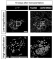
- kidney interstitial progenitor cells were generated by culturing HOXC10-positive cells in medium containing GSK-3 ⁇ inhibitor and TGF ⁇ inhibitor, preferably further SHH signal activator, IL-1 ⁇ and retinoic acid. It was found that differentiation can be induced selectively. When the induced IPCs and nephron progenitor cells (NPCs) were mixed and transplanted in vivo, a mesangial-like structure was formed.
- the present invention has the following features: [1] HOXC10-positive cells were cultured in a medium containing GSK (glycogen synthase kinase)-3 ⁇ inhibitor, SHH (Sonic Hedgehog) signal activator and TGF (Transforming growth factor) ⁇ inhibitor to form renal stromal progenitor cells.
- a method for producing renal interstitial progenitor cells comprising the step of inducing to [2]
- IL Interleukin
- the GSK-3 ⁇ inhibitor is CHIR99021
- the SHH signal activator is SHH protein
- Purmorphamine or SAG Smoothened Agonist
- the TGF ⁇ inhibitor is SB431542, A83-01 or LDN193189
- HOXC10-positive cells are HOXC10-positive cells produced by a method comprising the following steps (i) to (iv).
- bFGF basic fibroblast growth factor
- BMP bone morphogenetic protein 4
- GSK-3 ⁇ inhibitor and retinoic acid or a derivative thereof culturing the cells obtained in step (i) in a medium containing bFGF, a GSK-3 ⁇ inhibitor and BMP7
- the pluripotent stem cells are induced pluripotent stem (iPS )
- Renal interstitial progenitor cells produced by the method according to any one of [1] to [9].
- a pharmaceutical composition comprising the renal interstitial progenitor cells of [10].
- a method for producing renin-producing cells comprising the step of producing renin-producing cells by culturing renal stromal progenitor cells in a medium containing a GSK3 ⁇ inhibitor and a TGF ⁇ inhibitor but not containing an SHH signal activator.
- renal interstitial progenitor cells can be selectively obtained. Renal EPO-producing cells and renin-producing cells such as mesangial and juxtaglomerular cells can also be obtained by further culturing renal stromal progenitor cells.
- the renal interstitial progenitor cells, renal EPO-producing cells, and renin-producing cells such as mesangial cells and juxtaglomerular cells obtained by the method of the present invention are useful as cell preparations for regenerative medicine that can be applied to chronic kidney disease and the like. .
- renal interstitial progenitor cells by mixing renal interstitial progenitor cells with nephron progenitor cells to reconstruct the kidney, a highly functional cell preparation for regenerative medicine can be obtained.
- renal interstitial progenitor cells, renal EPO-producing cells, and renin-producing cells such as mesangial cells and juxtaglomerular cells obtained by the method of the present invention are also useful for preparing renal disease models. Renal disease models are also useful in terms of treatment methods and drug discovery for renal diseases.
- the concentrations are CHIR99021 (CHIR) 1 ⁇ M, SB431542 (SB) 10 ⁇ M, retinoic acid (RA) 100 nM, FGF9 200 ng/ml, Noggin 25 ng/ml.
- 11 shows the results of flow cytometry analysis (FOXD1 and OSR1) of cells cultured in two-dimensional adherent culture (2D) or three-dimensional suspension culture (3D) on day 11.
- FIG. The dots and lines in the middle of the scatter plots represent experimental data and mean values, respectively. * indicates p ⁇ 0.05, ** indicates p ⁇ 0.01 (Student's t-test).
- the concentrations are CHIR 1 ⁇ M, Wnt3a 10%, RSPO1 (R-Spondin-1) 200 ng/ml, SAG 500 nM, IL-1 ⁇ 10 ng/ml, Estradiol 50 ng/ml, TGF ⁇ 1 10 ng/ml.
- SB431542 is 1 or 10 ⁇ M
- A83-01 is 1 or 10 ⁇ M
- LDN193189 is 10 or 100 nM.
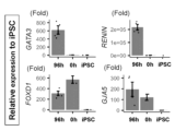
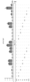
- the lower figure is an enlarged image of the frame in the upper figure. 11-14 days after obtaining renal interstitial progenitor cells, qRT-PCR analysis of EPO and renin was performed, and the results of comparing the effects of combinations of renal EPO-producing cells or renin-producing cell differentiation-inducing candidate factors are shown.
- the concentration of SAG is 500 nM
- the concentration unit of SB and CHIR is ⁇ M.
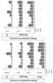
- BMP4, BMP5 or BMP7 (10 ng/mL or 100 ng/mL) in the presence of CHIR99021, RA and FG-4592 (CFR) was examined under SB431542 (1 ⁇ M) treated or untreated conditions.
- Units for SB are ⁇ M; units for TGF ⁇ 1, ACT are ng/mL.
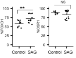
- Fig. 4 shows the results of qRT-PCR analysis of markers for JG (renin and GJA4), mesangial cells (EBF1, GJA5, ITGA8 and GATA3) and VEGFA in cells treated with minus FG-4592).
- Renal stromal progenitor cells are preferably of mammalian origin, more preferably of primate or rodent origin, preferably of human or mouse origin. Therefore, renin-producing cells such as HOXC10-positive cells used to produce renal stromal progenitor cells, renal EPO-producing cells, mesangial cells and juxtaglomerular cells obtained from renal stromal progenitor cells are used herein. Cells are also preferably of mammalian origin, more preferably of primate or rodent origin, preferably of human or mouse origin.
- Renal stromal progenitor cells are cells that can differentiate in vitro into renin-producing cells such as renal stromal cells, renal EPO-producing cells, mesangial cells and juxtaglomerular cells, and are defined by FOXD1 (Forkhead Box D1) positivity. be attached. Furthermore, it may be OSR1 positive. Furthermore, it may be PDGFRB (platelet-derived growth factor receptor ⁇ ) positive. In this specification, marker-positiveness can be confirmed by immunostaining using a marker-specific antibody, RT-PCR using a marker-specific primer, or the like.
- a method for producing renal interstitial progenitor cells comprises: A step of inducing renal stromal progenitor cells by culturing HOXC10-positive cells in a medium containing a GSK-3 ⁇ inhibitor and a TGF ⁇ inhibitor (hereinafter sometimes referred to as renal stromal progenitor cell differentiation-inducing step).
- the medium preferably further contains an SHH signal activator.
- the medium further contains IL-1 ⁇ .
- the medium further contains retinoic acid.
- a medium comprising a GSK-3 ⁇ inhibitor and a TGF ⁇ inhibitor and optionally comprising retinoic acid A medium comprising a GSK-3 ⁇ inhibitor, a TGF ⁇ inhibitor and an SHH signal activator and optionally comprising retinoic acid A GSK-3 ⁇ inhibitor, A medium comprising a TGF ⁇ inhibitor and IL-1 ⁇ and optionally comprising retinoic acid A medium comprising a GSK-3 ⁇ inhibitor, a TGF ⁇ inhibitor, an SHH signal activator and IL-1 ⁇ and optionally comprising retinoic acid
- HOXC10-positive cells are not particularly limited as long as these markers are positive cells.
- CITED1 Cbp/P300 Interacting Transactivator With Glu/Asp Rich Carboxy-Terminal Domain 1
- HOXD11 and BRACHYURY(T) may also be positive.
- HOXC10-positive cells include Posterior Primitive Streak cells, neural mesoderm progenitor cells, posterior immature mesoderm, and the like.
- HOXC10-positive cells The origin of HOXC10-positive cells is not particularly limited, but HOXC10-positive cells obtained by inducing differentiation from pluripotent stem cells are preferred.
- the method for inducing differentiation from pluripotent stem cells to HOXC10-positive cells is not particularly limited, for example, a method for inducing differentiation from pluripotent stem cells to HOXC10-positive cells, which will be described later, can be used.
- the HOXC10-positive cells to be subjected to the step of inducing renal stromal progenitor cell differentiation may be cell populations containing other cell types, or may be purified HOXC10-positive cells.
- a cell population containing 10% or more, 20% or more, 30% or more, 40% or more, 50% or more, 60%, 70%, 80% or more of HOXC10 positive cells is renal stromal progenitor cell differentiation induction step served to
- the culture in the renal interstitial progenitor cell differentiation-inducing step may be performed by either adherent culture or suspension culture, preferably by suspension culture.
- the suspension culture means that the cells are cultured in a non-adhesive state to the culture substrate.
- adherent culture means that cells are cultured in a state of being adhered to a culture substrate, for example, cultured in a coated culture dish.
- the coating agent is preferably an extracellular matrix, such as collagen, proteoglycan, fibronectin, hyaluronic acid, tenascin, entactin, elastin, fibrin and laminin, or fragments thereof.
- the medium used in the step of inducing differentiation of renal stromal progenitor cells consists of the basal medium used for culturing animal cells, a GSK-3 ⁇ inhibitor and a TGF ⁇ inhibitor, and, if necessary, an SHH signal stimulator, IL-1 ⁇ and retinoin. It can be prepared with the addition of one or more acids.
- basal media include IMDM medium, Medium 199 medium, Eagle's Minimum Essential Medium (EMEM) medium, ⁇ MEM medium, Dulbecco's Modified Eagle's Medium (DMEM) medium, Ham's F12 (F12) medium, RPMI 1640. Medium, Fischer's medium, mixed medium of these and the like are included.
- the medium may contain serum (eg, fetal bovine serum (FBS)) or may be serum-free.
- serum eg, fetal bovine serum (FBS)
- albumin, transferrin, knockout serum replacement (KSR) serum replacement during ES cell culture
- N2 supplement Invitrogen
- B27 supplement Invitrogen
- fatty acid insulin, collagen precursor , trace elements
- 2-mercaptoethanol 3′-thioglycerol
- lipids amino acids, L-glutamine, GlutaMAX (Invitrogen), non-essential amino acids (NEAA), vitamins , growth factors, antibiotics, antioxidants, pyruvate, buffers, inorganic salts, and the like.
- a medium optimized for stem cell culture in advance such as ReproFF2 (Reprocell) or Stem Fit AK02N medium (Ajinomoto Healthy Supply), may be used.
- the GSK-3 ⁇ inhibitor is not particularly limited as long as it can inhibit the function of GSK-3 ⁇ , for example, kinase activity.
- rubin-3′-oxime maleimide derivative SB216763 (3-(2,4-dichlorophenyl)-4-(1-methyl-1H-indol-3-yl)-1H-pyrrole-2,5-dione)
- GSK-3 ⁇ inhibitor VII ⁇ ,4-dibromoacetophenone
- a phenyl- ⁇ -bromomethyl ketone compound L803-mts
- a cell-permeable phosphorylated peptide also known as GSK-3 ⁇ peptide inhibitor; Myr- N-GKEAPPAPPQSpP-NH 2
- CHIR99021 with high selectivity Nature (2008) 453:519-523.
- GSK-3 ⁇ inhibitors for use in this step is CHIR99021.
- concentration of the GSK-3 ⁇ inhibitor in the medium can be appropriately selected by those skilled in the art depending on the GSK-3 ⁇ inhibitor to be used. More preferably from 0.5 ⁇ M to 1.5 ⁇ M.
- a TGF ⁇ inhibitor is a substance that inhibits the signal transduction from the binding of TGF ⁇ to a receptor to SMAD, a substance that inhibits the binding of TGF ⁇ to the ALK family of receptors, or the phosphorylation of SMAD by the ALK family.
- ALK inhibitors for example, Lefty-1 (as NCBI Accession No., mouse: NM_010094, human: NM_020997 are exemplified), SB431542, SB202190 (above, RKLindemann et al.
- the TGF ⁇ inhibitor may preferably be SB431542.
- concentration of the TGF ⁇ inhibitor in the medium can be appropriately selected by those skilled in the art depending on the TGF ⁇ inhibitor to be used.
- A83-01 is 0.1 ⁇ M to 100 ⁇ M, preferably 0.5 ⁇ M to 50 ⁇ M, more preferably 1 ⁇ M to 10 ⁇ M, LDN193189 is 0.01 ⁇ M to 1 ⁇ M, preferably 0.05 ⁇ M to 0 ⁇ M. .5 ⁇ M, more preferably 0.05 ⁇ M to 0.2 ⁇ M.
- SHH Sonic hedgehog signal stimulator
- SHH Sonic hedgehog signal stimulator
- Smo Smoothened
- Ptch1 Patched
- Gli2 activation e.g., Gli2 activation
- SAG Smoothened Agonist
- PMA Purmorphamine
- PMA 9-cyclohexyl-N-[4-(4-morpholinyl)phenyl]-2-(1-nagtal
- SHH signal activators preferably include SHH protein, Purmorphamine, and SAG. Preferred concentrations in the medium are 10-1000 nM, preferably 100-1000 nM for SAG, 10-1000 ng/ml, preferably 50-200 ng/ml for SHH protein, and Purmophamine. is preferably 0.1 to 10 ⁇ M, preferably 0.5 to 5 ⁇ M.
- retinoic acid includes not only retinoic acid itself, but also retinoic acid derivatives that retain the differentiation-inducing function of natural retinoic acid.
- retinoic acid derivatives include 3-dehydroretinoic acid, 4-[[(5,6,7,8-tetrahydro-5,5,8,8-tetramethyl-2-naphthalenyl)carbonyl]amino]-benzoic acid ( AM580) (Tamura K, et al., Cell Differ. Dev.
- retinoic acid in the medium is, for example, 1 nM to 1000 nM, preferably 10 nM to 500 nM, more preferably 50 nM to 250 nM.
- IL-1 ⁇ is not particularly limited, but human IL-1 ⁇ is preferred.
- human IL-1 ⁇ include proteins having the amino acid sequence of NCBI (National Center for Biotechnology Information) accession number: 1TOO_A.
- IL-1 ⁇ includes fragments and functional variants thereof as long as they have differentiation-inducing activity.
- Commercially available IL-1 ⁇ may be used, or proteins purified from cells or proteins produced by genetic recombination may be used.
- the concentration of IL-1 ⁇ in the medium is, for example, 1 ng/ml to 500 ng/ml, 1 ng/ml to 100 ng/ml, 5 ng/ml to 50 ng/ml, or 5 ng/ml to 25 ng/ml.
- the step of inducing renal interstitial progenitor cells from HOXC10-positive cells may be performed in the following two steps. 1) step of culturing HOXC10-positive cells in a medium containing a GSK-3 ⁇ inhibitor, a TGF ⁇ inhibitor, an SHH signal activator and IL-1 ⁇ , and optionally containing retinoic acid; 2) A step of culturing the cells (posterior unsegmented mesoderm cells) obtained in step 1) in a medium containing a GSK-3 ⁇ inhibitor, a TGF ⁇ inhibitor and an SHH signal activator and optionally containing retinoic acid.
- the efficiency of differentiation induction of renal interstitial progenitor cells can be improved by carrying out in two steps and removing IL-1 ⁇ in step 2).
- the culture days in the renal interstitial progenitor cell differentiation-inducing step are, for example, 2 days or longer, 3 days or longer, 4 days or longer, and 5 days or longer. There is no upper limit because long-term culture does not particularly affect the production efficiency of renal interstitial progenitor cells, but it is, for example, 30 days or less.
- the culture conditions are not particularly limited as long as renal interstitial progenitor cells can be obtained. concentration (eg 15-25%, preferably about 20%) and the CO 2 concentration is preferably about 2-5%.
- the resulting renal stromal progenitor cells may be isolated or enriched. Isolation or concentration can be performed, for example, by flow cytometry using an antibody against PDGFRB.
- renin-producing cells such as renal EPO-producing cells, mesangial cells, and juxtaglomerular cells can be obtained.
- One aspect of the present invention relates to a method of producing renal EPO-producing cells from renal interstitial progenitor cells. That is, in one aspect of the present invention, a step of culturing renal stromal progenitor cells in a medium containing a GSK3 ⁇ inhibitor, an SHH signal activator and a HIF inhibitor to produce renal EPO-producing cells (hereinafter sometimes referred to as renal EPO-producing cell differentiation induction step); A method for producing renal EPO-producing cells is provided, comprising: Preferably, the medium further contains a TGF ⁇ inhibitor. Preferably, the medium further contains retinoic acid.
- the following media are exemplified.
- Medium containing GSK3 ⁇ inhibitor, SHH signal activator and HIF inhibitor and optionally containing retinoic acid Medium containing GSK3 ⁇ inhibitor, SHH signal activator, HIF inhibitor and TGF ⁇ inhibitor and optionally containing retinoic acid
- the origin of renal interstitial progenitor cells is not particularly limited, and any renal interstitial progenitor cells can be used as long as they are FOXD1-positive cells.
- the renal interstitial progenitor cells obtained by the method can be used.
- Renal EPO-producing cells are characterized by the expression and/or production of EPO.
- the culture in the renal EPO-producing cell differentiation induction step may be performed by either adherent culture or suspension culture, preferably suspension culture.
- the medium used in the renal EPO-producing cell differentiation step is a basal medium used for culturing animal cells containing a GSK3 ⁇ inhibitor, an SHH signal activator and an HIF inhibitor, and, if necessary, a TGF ⁇ inhibitor and/or It can be prepared by adding retinoic acid.
- the basal medium include the medium exemplified in the renal interstitial progenitor cell differentiation induction step.
- the GSK-3 ⁇ inhibitor As the GSK-3 ⁇ inhibitor, the GSK-3 ⁇ inhibitor (CHIR99021, etc.) described in the renal stromal progenitor cell differentiation-inducing step above can be used, and the preferred concentration range is also the same.
- the SHH signal activator As the SHH signal activator, the SHH signal activator (SAG, SHH protein, Purmorphamine, etc.) described in the renal interstitial progenitor cell differentiation-inducing step can be used, and the preferred concentration range is the same.
- the HIF (hypoxia-inducible factor) inhibitor is not particularly limited as long as it is a substance that exhibits a function as a HIF inhibitor, and known HIF inhibitors can be used.
- the concentration of the HIF inhibitor may be any concentration that is effective in inducing differentiation of renal EPO-producing cells, and is, for example, 0.5 ⁇ M to 100 ⁇ M, preferably 1 ⁇ M to 50 ⁇ M, more preferably 5 ⁇ M to 25 ⁇ M.
- the TGF ⁇ inhibitor As the TGF ⁇ inhibitor, the TGF ⁇ inhibitor (SB431542, A83-01, LDN193189, etc.) described in the renal interstitial progenitor cell differentiation-inducing step can be used, and the preferred concentration range is the same.
- Retinoic acid may be a derivative thereof as described in the step of inducing differentiation of renal interstitial progenitor cells, and the preferred concentration range is the same.
- the culture temperature is, but not limited to, about 30 to 40°C, preferably about 37°C.
- culture may be performed at normal oxygen concentration (for example, 15 to 25%, preferably about 20%), but is preferably performed under hypoxic conditions.
- the cells are cultured at an oxygen concentration of 10%, preferably 3% to 8%, more preferably 4% to 6%, particularly preferably 5%.
- the CO 2 concentration is preferably about 2-5%.
- One aspect of the present invention relates to a method for producing renin-producing cells from renal interstitial progenitor cells. That is, in one aspect of the present invention, A step of culturing renal stromal progenitor cells in a medium containing a GSK3 ⁇ inhibitor and a TGF ⁇ inhibitor but not containing an SHH signal activator to produce renin-producing cells (hereinafter sometimes referred to as renin-producing cell differentiation induction step) , A method for producing renin-producing cells is provided, comprising: Preferably, the medium further contains BMP. Preferably, the medium further contains VEGF. Preferably, the medium further contains retinoic acid. Preferably, the medium contains a HIF inhibitor.
- renal stromal progenitor cells used in the step of inducing renin-producing cell differentiation is not particularly limited, and any renal stromal progenitor cells can be used as long as they are FOXD1-positive cells.
- Renal interstitial progenitor cells obtained by a method for producing renal interstitial progenitor cells can be used.
- renin-producing cells examples include mesangial cells and juxtaglomerular (JG) cells. Both of these cells produce renin, but juxtaglomerular cells express more renin.
- Mesangial cells express GJA5 (Gap junction alpha-5) and EBF1 (EBF transcription factor 1).
- Juxtaglomerular cells express GJA4 (Gap junction alpha-4).
- Cultivation in the renin-producing cell differentiation induction step may be performed by either adherent culture or suspension culture, preferably suspension culture.
- the medium used in the renin-producing cell differentiation induction process is the basal medium used for culturing animal cells with GSK3 ⁇ inhibitor, TGF ⁇ inhibitor, and, if necessary, BMP, VEGF, HIF inhibitor, retinoic acid, etc. can be prepared by Examples of the basal medium include the medium exemplified in the renal interstitial progenitor cell differentiation induction step.
- the GSK-3 ⁇ inhibitor As the GSK-3 ⁇ inhibitor, the GSK-3 ⁇ inhibitor (CHIR99021, etc.) described in the renal stromal progenitor cell differentiation-inducing step above can be used, and the preferred concentration range is also the same.
- the TGF ⁇ inhibitor As the TGF ⁇ inhibitor, the TGF ⁇ inhibitor (SB431542, A83-01, LDN193189, etc.) described in the renal interstitial progenitor cell differentiation-inducing step can be used, and the preferred concentration range is the same.
- Retinoic acid may be a derivative thereof as described in the step of inducing differentiation of renal interstitial progenitor cells, and the preferred concentration range is the same.
- BMP includes BMP4, BMP5, BMP7, etc., but BMP7 is more preferable.
- BMP is preferably human BMP, and human BMP7 includes, for example, a protein having an amino acid sequence from 293 to 431 of NCBI (National Center for Biotechnology Information) accession number: NP_001710.1.
- BMPs include fragments and functional variants thereof as long as they have renin-producing cell differentiation-inducing activity.
- a commercially available BMP may be used, or a protein purified from cells or a protein produced by genetic recombination may be used.
- the concentration of BMP used in this step is 1 ng/mL to 500 ng/mL, preferably 5 ng/mL to 200 ng/mL, more preferably 10 g/mL to 100 ng/mL.
- VEGF is not particularly limited, but human VEGF is preferred.
- human VEGF include proteins having the amino acid sequence of NCBI (National Center for Biotechnology Information) accession number: AAK95847.
- VEGF includes fragments and functional variants thereof as long as they have renin-producing cell differentiation-inducing activity.
- a commercially available VEGF may be used, or a protein purified from cells or a protein produced by genetic recombination may be used.
- the concentration of VEGF used in this step is 1 ng/mL to 500 ng/mL, preferably 10 ng/mL to 200 ng/mL, more preferably 50 ng/mL to 150 ng/mL.
- the HIF inhibitor As the HIF inhibitor, the HIF inhibitor (FG-4592, etc.) explained in the renal EPO-producing cell differentiation induction step can be used, and the preferred concentration range is the same.
- the number of days of culturing in the renin-producing cell differentiation induction step is sufficient as long as it is a period sufficient to generate renin-producing cells, and there is no upper limit because long-term culturing does not particularly affect the production efficiency of renin-producing cells. , 2 days or more, 3 days or more, 4 days or more, 5 days or more.
- the culture conditions are not limited to the following, but the culture temperature is about 30-40° C., preferably about 37° C., and the CO 2 concentration is preferably about 2-5%.
- the oxygen concentration may be a normal oxygen concentration (eg 15-25%, preferably about 20%), but a low oxygen concentration (1%-10%, preferably 3%-8%, more preferably 4%-6% , particularly preferably 5%).
- the medium preferably contains a HIF inhibitor.
- Preferred embodiments of the method for producing mesangial cells are described below as examples of the method for producing renin-producing cells.
- One aspect of the present invention relates to a method of producing mesangial cells from renal interstitial progenitor cells. That is, in a preferred embodiment, A step of producing mesangial cells by culturing renal stromal progenitor cells in a medium containing a GSK3 ⁇ inhibitor, a TGF ⁇ inhibitor, BMP and VEGF but not containing an SHH signal activator (hereinafter referred to as a mesangial cell differentiation induction step) be), A method for producing mesangial cells is provided, comprising: Preferably, the medium further contains retinoic acid.
- the origin of the renal stromal progenitor cells used in the step of inducing mesangial cell differentiation is not particularly limited, and any renal stromal progenitor cells can be used. Renal stromal progenitor cells obtained by the cell manufacturing method can be used.
- the culture in the mesangial cell differentiation induction step may be performed by either adherent culture or suspension culture, preferably by suspension culture.
- the medium used in the mesangial cell differentiation induction step can be prepared by adding a GSK3 ⁇ inhibitor, a TGF ⁇ inhibitor, BMP, VEGF, and, if necessary, retinoic acid to the basal medium used for culturing animal cells.
- a GSK3 ⁇ inhibitor a TGF ⁇ inhibitor
- BMP a TGF ⁇ inhibitor
- VEGF vascular endothelial growth factor
- retinoic acid retinoic acid
- Examples of the basal medium include the medium exemplified in the renal interstitial progenitor cell differentiation induction step.
- GSK-3 ⁇ inhibitor TGF ⁇ inhibitor
- retinoic acid BMP, and VEGF
- preferred substances and concentration ranges are also as described above.
- the number of culture days in the mesangial cell differentiation induction step is sufficient as long as it is a period sufficient to generate mesangial cells, and there is no upper limit because long-term culture does not particularly affect the production efficiency of mesangial cells, but for example, 2 days. Above, 3 days or more, 4 days or more, 5 days or more.
- the culture conditions are not limited to the following, but the culture temperature is about 30 to 40° C., preferably about 37° C., and the oxygen concentration is normal oxygen concentration (for example, 15 to 25%, preferably about 20%) or low oxygen concentration (1% to 10%, preferably 3% to 8%, more preferably 4% to 6%, particularly preferably 5 %) and a CO concentration of preferably about 2 ⁇ 5%.
- the oxygen concentration is normal oxygen concentration (for example, 15 to 25%, preferably about 20%) or low oxygen concentration (1% to 10%, preferably 3% to 8%, more preferably 4% to 6%, particularly preferably 5 %) and a CO concentration of preferably about 2 ⁇ 5%.
- One aspect of the present invention relates to a method of producing juxtaglomerular (JG) cells from renal interstitial progenitor cells. That is, in a preferred embodiment, A step of culturing renal stromal progenitor cells in a medium containing a GSK3 ⁇ inhibitor, a TGF ⁇ inhibitor, a HIF inhibitor, BMP and VEGF to produce juxtaglomerular cells (hereinafter sometimes referred to as juxtaglomerular cell differentiation induction step ), A method for producing juxtaglomerular cells is provided, comprising: Preferably, the medium further contains retinoic acid.
- the origin of the renal stromal progenitor cells used in the juxtaglomerular cell differentiation induction step is not particularly limited, and any renal stromal progenitor cells can be used. Renal stromal progenitor cells obtained by the method for producing stromal progenitor cells can be used.
- the culture in the juxtaglomerular cell differentiation induction step may be performed by either adherent culture or suspension culture, preferably by suspension culture.
- the medium used for juxtaglomerular cell differentiation induction step is the basal medium used for culturing animal cells with GSK3 ⁇ inhibitor, TGF ⁇ inhibitor, HIF inhibitor, BMP, VEGF, and, if necessary, retinoic acid.
- the basal medium include the medium exemplified in the renal interstitial progenitor cell differentiation induction step.
- GSK-3 ⁇ inhibitor TGF ⁇ inhibitor
- HIF inhibitor HIF inhibitor
- BMP VEGF inhibitor
- VEGF vascular endothelial growth factor
- retinoic acid is as described above, and preferred substances and concentration ranges are also as described above.
- the number of days of culture in the juxtaglomerular cell differentiation induction step is sufficient as long as it is a period sufficient to generate juxtaglomerular cells, and there is no upper limit because long-term culture does not particularly affect the production efficiency of juxtaglomerular cells. However, for example, 2 days or more, 3 days or more, 4 days or more, or 5 days or more.
- the culture temperature is, but not limited to, about 30 to 40°C, preferably about 37°C.
- the culture may be performed at normal oxygen concentration, but is preferably performed under hypoxic conditions, for example, 1% to 10%, preferably 3% to 8%, more preferably 4% to Cultivation is carried out at an oxygen concentration of 6%, particularly preferably 5%.
- the CO 2 concentration is preferably about 2-5%.
- the HOXC10-positive cells are HOXC10-positive cells derived from pluripotent stem cells.
- HOXC10-positive cells derived from pluripotent stem cells renal stromal progenitor cells, renal EPO-producing cells, renin-producing cells (mesangial cells, juxtaglomerular cells, etc.) can be obtained from pluripotent stem cells. be able to.
- Pluripotent stem cells are stem cells that have pluripotency that can be differentiated into many cells existing in the body, and that also have proliferative potential. Any cell that is derived into a production cell is included.
- pluripotent stem cells include, but are not limited to, embryonic stem (ES) cells, cloned embryo-derived embryonic stem (ntES) cells obtained by nuclear transfer, spermatogonial stem cells (“GS cells”), embryonic Included are germ cells (“EG cells”), induced pluripotent stem (iPS) cells, cultured fibroblasts, and pluripotent cells derived from bone marrow stem cells (Muse cells).
- ES embryonic stem
- ntES cloned embryo-derived embryonic stem
- GS cells spermatogonial stem cells
- EG cells germ cells
- iPS induced pluripotent stem
- Muse cells pluripotent stem cells derived from bone marrow stem cells
- the initialization factor is, for example, Oct3/4, Sox2, Sox1, Sox3, Sox15, Sox17, Klf4, Klf2, c-Myc, N-Myc, L-Myc, Nanog, Lin28, Fbx15, ERAs, ECAT15 -2, Tcl1, beta-catenin, Lin28b, Sall1, Sall4, Esrrb, Nr5a2, Tbx3 or Glis1 genes or gene products are exemplified, and these reprogramming factors may be used alone or in combination Also good.
- Somatic cells include, but are not limited to, fetal (pup) somatic cells, neonatal (pup) somatic cells, and mature healthy or diseased somatic cells, as well as primary cultured cells. , passaged cells, and cell lines are all included.
- somatic cells are, for example, (1) tissue stem cells (somatic stem cells) such as neural stem cells, hematopoietic stem cells, mesenchymal stem cells, dental pulp stem cells, (2) tissue progenitor cells, (3) blood cells (peripheral blood cells, umbilical cord blood cells, etc.), lymphocytes, epithelial cells, endothelial cells, muscle cells, fibroblasts (skin cells, etc.), hair cells, hepatocytes, gastric mucosa cells, enterocytes, splenocytes, pancreatic cells (pancreatic exocrine cells) etc.), differentiated cells such as brain cells, lung cells, kidney cells and adipocytes.
- tissue stem cells such as neural stem cells, hematopoietic stem cells, mesenchymal stem cells, dental pulp stem cells
- tissue progenitor cells such as lymphocytes, epithelial cells, endothelial cells, muscle cells, fibroblasts (skin cells, etc.), hair cells, he
- somatic cells when iPS cells are used as a material for cells for transplantation, it is desirable to use somatic cells that have the same or substantially the same HLA genotype as the recipient individual from the viewpoint of avoiding rejection.
- substantially identical means that the HLA genotype matches the transplanted cells to the extent that an immunosuppressive agent can suppress an immune reaction. and HLA-DR 3 loci or HLA-C plus 4 loci have matching HLA types.
- a method including the following steps can be used.
- the cells are cryopreserved. After thawing, the cells can be subjected to differentiation induction of renal interstitial progenitor cells and the like.
- pluripotent stem cells are and preferably cultured by adherent culture.
- Methods for separating pluripotent stem cells include, for example, mechanical separation and separation solutions having protease activity and collagenase activity (for example, Accutase (TM) and Accumax (TM) (Innovative Cell Technologies, Inc)). Alternatively, separation using a separation solution having only collagenase activity can be mentioned. A mixture of TrypLE Select Enzyme (Thermo Fisher Scientific) and EDTA/PBS may also be used.
- colonies cultured to 70% to 80% confluency in the dish used are preferably used.
- the medium used in step (i) can be prepared by adding bFGF, BMP4, a GSK-3 ⁇ inhibitor and retinoic acid or a derivative thereof to a basal medium used for culturing animal cells.
- a basal medium used for culturing animal cells.
- the basal medium include the medium exemplified in the renal interstitial progenitor cell differentiation induction step.
- the GSK-3 ⁇ inhibitor used in step (i) the GSK-3 ⁇ inhibitor (CHIR99021, etc.) described in the step of inducing differentiation of renal stromal progenitor cells can be used, and the concentration range is also the same. Adjustable range.
- the bFGF (basic FGF) used in step (i) is preferably human bFGF, and human bFGF is, for example, a protein having an amino acid sequence of NCBI (National Center for Biotechnology Information) accession number: ABO43041.1. mentioned. bFGF includes fragments and functional variants thereof as long as they have differentiation-inducing activity Commercially available bFGF may be used, or proteins purified from cells or proteins produced by genetic recombination may be used. You may The concentration of bFGF used in this step is 1 ng/mL to 1000 ng/mL, preferably 10 ng/mL to 500 ng/mL, more preferably 50 ng/mL to 250 ng/mL.
- the BMP4 used in step (i) is preferably human BMP4, and examples of human BMP4 include proteins having the amino acid sequence of NCBI (National Center for Biotechnology Information) accession number: AAH20546.1. BMP4 includes fragments and functional variants thereof as long as they have differentiation-inducing activity. Commercially available BMP4 may be used, or proteins purified from cells or proteins produced by genetic recombination may be used. You may The concentration of BMP4 used in this step is 0.1 ng/mL to 100 ng/mL, preferably 0.5 ng/mL to 50 ng/mL, more preferably 0.5 ng/mL to 5 ng/mL.
- the retinoic acid used in step (i) may be a derivative thereof as described in the step of inducing differentiation of renal stromal progenitor cells, and the preferred concentration range is 1 nM to 50 nM.
- the culture conditions are not limited to the following, but the culture temperature is about 30-40° C., preferably about 37° C., and the oxygen concentration is normal oxygen concentration (for example, 15-25%, preferably about 20%) and the CO 2 concentration is preferably about 2-5%.
- the culture time in step (i) is, for example, 1 to 2 days, preferably 1 day.
- step (ii) culturing the cells obtained in step (i) in a medium containing bFGF, a GSK-3 ⁇ inhibitor and BMP7.
- a cell population may be isolated and adherently cultured in a separately prepared coated culture dish, or the cells obtained by adherent culture in step (i) may be cultured as they are by exchanging the medium.
- the medium used in step (ii) can be prepared by adding bFGF, a GSK-3 ⁇ inhibitor and BMP7 to a basal medium used for culturing animal cells.
- a basal medium used for culturing animal cells.
- the basal medium include the medium exemplified in the renal interstitial progenitor cell differentiation induction step.
- step (ii) The bFGF used in step (ii) is the same as that described in step (i), and its preferred concentration range is also the same.
- the GSK-3 ⁇ inhibitor used in step (ii) the GSK-3 ⁇ inhibitor (CHIR99021, etc.) described in the step of inducing differentiation of renal stromal progenitor cells can be used, and the concentration range is also the same. Adjustable range.
- BMP7 used in step (ii) is preferably human BMP7, and examples of human BMP7 include proteins having the amino acid sequence of NCBI (National Center for Biotechnology Information) accession number: NM_001719.2. BMP7 includes fragments and functional variants thereof as long as they have a differentiation-inducing activity. Commercially available BMP7 may be used, or proteins purified from cells or proteins produced by genetic recombination may be used. You may The concentration of BMP7 used in this step is 0.1 ng/mL to 100 ng/mL, preferably 0.5 ng/mL to 50 ng/mL, more preferably 0.5 ng/mL to 5 ng/mL.
- the culture conditions are not limited to the following, but the culture temperature is about 30-40° C., preferably about 37° C., and the oxygen concentration is normal oxygen concentration (for example, 15-25%, preferably about 20%) and the CO 2 concentration is preferably about 2-5%.
- the culture time in step (ii) is, for example, 10 hours to 2 days, or 1 to 2 days, preferably 0.5 to 1 day.
- step (iii) culturing the cells obtained in step (ii) in a medium containing a GSK-3 ⁇ inhibitor and a TGF ⁇ inhibitor;
- a cell population may be isolated and adherently cultured in a separately prepared coated culture dish, or the cells obtained by adherent culture in step (ii) may be continued to be cultured by exchanging the medium.
- the medium used in step (iii) can be prepared by adding a GSK-3 ⁇ inhibitor and a TGF ⁇ inhibitor to a basal medium used for culturing animal cells.
- the medium may further contain BMP7.
- Examples of the basal medium include the medium exemplified in the renal interstitial progenitor cell differentiation induction step.
- the GSK-3 ⁇ inhibitor used in step (iii) the GSK-3 ⁇ inhibitor (CHIR99021, etc.) described in the step of inducing differentiation of renal stromal progenitor cells can be used, and the concentration range is also the same. Adjustable range. Also, when BMP7 is added, BMP7 is the same as in step (ii) and its preferred concentration range is also the same.
- TGF ⁇ inhibitor used in step (iii) one or more of the TGF ⁇ inhibitors (SB431542, A83-01, LDN193189, etc.) described in the renal stromal progenitor cell differentiation-inducing step above may be used. and the preferred concentration range is also as explained above.
- the culture conditions are not limited to the following, but the culture temperature is about 30-40° C., preferably about 37° C., and the oxygen concentration is normal oxygen concentration (for example, 15-25%, preferably about 20%) and the CO 2 concentration is preferably about 2-5%.
- the culture time in step (iii) is, for example, 0.5 to 3 days of culture, preferably about 1 day.
- step (iv) culturing the cells obtained in step (iii) in a medium containing bFGF, a GSK-3 ⁇ inhibitor, activin and a ROCK inhibitor
- HOXC10-positive posterior primitive streak cells (late metanephric lineage primitive streak) is induced.
- the cells can be cryopreserved, and after thawing, they can be subjected to differentiation induction of renal interstitial progenitor cells and the like.
- step (iv) the cell population obtained in step (iii) above may be isolated and adherently cultured in a separately prepared coated culture dish, or obtained by adherent culture in step (iii).
- the culture of the obtained cells may be continued by exchanging the medium.
- the medium used in step (iv) can be prepared by adding bFGF, a GSK-3 ⁇ inhibitor, activin and a ROCK inhibitor to a basal medium used for culturing animal cells.
- the basal medium include the medium exemplified in the renal interstitial progenitor cell differentiation induction step.
- the GSK-3 ⁇ inhibitor used in step (iv) the GSK-3 ⁇ inhibitor (CHIR99021, etc.) described in the step of inducing differentiation of renal stromal progenitor cells can be used, and the concentration range is the same. Adjustable range. bFGF is the same as in step (i), and so is its preferred concentration range.
- Activin used in step (iv) includes activin derived from humans and other animals, and functional variants thereof, and commercially available products such as R & D Systems can be used.
- the concentration of activin used in step (iv) is 1 ng/mL to 100 ng/mL, preferably 5 ng/mL to 50 ng/mL, more preferably 5 ng/mL to 25 ng/mL.
- the ROCK inhibitor used in step (iv) is not particularly limited as long as it can suppress the function of Rho-kinase (ROCK). 57, 976-983 (2000); see Narumiya et al., Methods Enzymol. , H-1152 (e.g., Sasaki et al., Pharmacol. Ther. 93: 225-232 (2002)), Wf-536 (e.g., Nakajima et al., Cancer Chemother Pharmacol. 52(4): 319-324 (2003)) and derivatives thereof, as well as antisense nucleic acids against ROCK, RNA interference-inducing nucleic acids (eg, siRNA), dominant-negative mutants, and expression vectors thereof.
- RNA interference-inducing nucleic acids eg, siRNA
- ROCK inhibitors e.g., US Patent Application Publication Nos. 2005/0209261, 2005/0192304, 2004/0014755, 2004/0002508,. , WO 2004/0002507, WO 2003/0125344, WO 2003/0087919, and WO 2003/062227, WO 2003/059913, WO 2003/062225, WO 2002/076976. No. 2004/039796).
- One or more ROCK inhibitors may be used.
- Preferred ROCK inhibitors include Y-27632.
- the concentration of the ROCK inhibitor used in step (iv) can be appropriately selected by those skilled in the art depending on the ROCK inhibitor used. is between 5 ⁇ M and 50 ⁇ M.
- the culture conditions are not limited to the following, but the culture temperature is about 30-40° C., preferably about 37° C., and the oxygen concentration is normal oxygen concentration (for example, 15-25%, preferably about 20%) and the CO 2 concentration is preferably about 2-5%.
- the culture time in step (iv) may be a period sufficient to induce the differentiation of HOXC10-positive posterior primitive streak cells, for example, culture for 1 to 5 days, preferably 3 days.
- the present invention also provides cell preparations comprising renal stromal progenitor cells, renal EPO-producing cells, renin-producing cells (mesangial cells, juxtaglomerular cells, etc.) obtained by the methods described above, or a combination thereof. Such cell preparations can be used as pharmaceutical compositions for treating renal diseases and the like.
- cells capable of reconstituting renal tissue by mixing renal stromal progenitor cells with nephron progenitor cells (OSR1-positive cells, preferably further HOXD11, WT1, SIX2 and SALL1-positive cells) as disclosed in Patent Document 2 A formulation can be provided.
- the ratio of renal interstitial progenitor cells to nephron progenitor cells is preferably in the range of 1:2 to 2:1, more preferably about 1:1.
- Such cell preparations may further include renal EPO-producing cells, renin-producing cells (mesangial cells, juxtaglomerular cells, etc.).
- Renal stromal progenitor cells and nephron progenitor cells may be mixed prior to formation of renal organoids.
- renal interstitial progenitor cells and nephron progenitor cells obtained by the above method are cultured to prepare cell aggregates, which are then combined with feeder cells such as 3T3-Wnt4 cells, mouse fetal spinal cord cells, or mouse fetal kidney cells. It can be obtained by co-culturing or semi-gas phase culture using a basal medium containing a GSK-3 ⁇ inhibitor.
- Methods of administering cell preparations to patients who require treatment include, for example, a method in which cells are formed into a sheet and attached to the patient's kidney, a cell suspension in which cells are suspended in physiological saline or the like, or a tertiary method.
- Examples include a method of directly transplanting the obtained cell mass from the original culture into the patient's kidney, a method of three-dimensional culturing on a scaffold made of Matrigel or the like, and a method of transplanting the obtained cell mass.
- the transplantation site is not particularly limited as long as it is within the kidney, but is preferably under the renal capsule.
- renal disease include acute renal failure, chronic renal failure, and chronic renal disease that does not lead to chronic renal failure.
- the number of cells such as renal interstitial progenitor cells contained in the cell preparation is not particularly limited as long as the graft can survive after administration, and may be prepared by appropriately increasing or decreasing according to the size of the affected area and body size.
- the renal interstitial progenitor cells, renal EPO-producing cells, and renin-producing cells are used for screening and evaluation of therapeutic drug candidates for renal diseases, hypertension, etc. You can also
- hiPSCs Human iPS cells (17K6:OSR1-GFP/FOXD1-tdTomato double knock-in hiPSCs) were first treated with a 1:1 mixture of TrypLE Select Enzyme (Thermo Fisher Scientific) and 0.5 mM EDTA/PBS for 5 minutes. , and washed with PBS(-). Then, detach using a cell scraper, dissociate into single cells by gentle pipetting, and add Stem Fit AK02N (Ajinomoto Healthy Supply) and 0.5 mL were seeded in a 24-well plate (Corning) at a density of 1.3 ⁇ 10 4 cells/cm 2 .
- basal medium serum-free differentiation medium
- Basal medium consisting of DMEM/F12 Glutamax (Thermo Fisher Scientific), B27 supplement minus vitamin A (Thermo Fisher Scientific), and 0.5x Penicillin/Streptomycin.
- Culturing was initiated by adding CHIR99021 (Wako), 10 nM RA (Sigma), 1 ng/mL BMP4 (Peprotech) and 100 ng/mL bFGF (Wako).
- basal medium consisting of DMEM/F12 Glutamax (Thermo Fisher Scientific), B27 supplement minus vitamin A (Thermo Fisher Scientific), and 0.5x Penicillin/Streptomycin.
- Culturing was initiated by adding CHIR99021 (Wako), 10 nM RA (Sigma), 1 ng/mL BMP4 (Peprotech) and 100 ng/mL bFGF (Wako).
- basal medium consisting of DMEM/F12 Glutamax (Thermo
- the medium was switched to basal medium containing 5 ⁇ M CHIR99021, 1 ng/mL BMP7 and 10 ⁇ M A83-01 (Wako).
- the medium was switched to basal medium containing 5 ⁇ M CHIR99021, 30 ng/mL bFGF, 10 ng/mL activin A (R&D Systems) and 30 ⁇ M Y-27632 (Wako).
- cells were washed with PBS(-), treated with Accumax (Innovative Cell Technologies), dissociated into single cells by gentle pipetting, and plated at 1.05 x 10⁵ cells/cm in 24-well plates (CORNING).
- FIG. 2 shows the results of immunohistochemical staining of PPS cells induced to differentiate from human iPS cells.
- IPC renal interstitial progenitor
- the IPC differentiation induction conditions were CHIR99021 + SB431542 + RA, and cultured in floating (3D) culture on days 4 to 11. Compared to adherent (2D) culture, FOXD1-positive cell induction efficiency was higher. (Fig. 4).
- IPA analysis suggested increased expression of WNT/Ca 2+ pathway, estrogen receptor signal, liver fibrosis, and IL-1 signal. Therefore, when WNT3a and RSPO1, ⁇ -Estradiol, TGF ⁇ 1, IL-1 ⁇ were added on days 6-8 and 8-11, respectively, IL-1 ⁇ and SAG addition on days 6-8 resulted in It was confirmed that the induction efficiency of OSR1-positive FOXD1-positive cells was improved (Fig. 6).
- IPC markers could be similarly induced by using various ALK inhibitors instead of SB431542 on days 6-11 (Fig. 7).
- Nephron progenitor cells were differentiated from iPS cells according to the method described in Patent Document 2.
- the IPC and NPC aggregates obtained above on day 11 of differentiation induction were incubated in Accumax (Innovative Cell Technologies) at 37°C for 5 minutes and dissociated into single cells by gentle pipetting.
- Dissociated cells were mixed and resuspended in basal medium containing 10 ⁇ M Y-27632, 10 ng/mL bFGF, 1 ⁇ M CHIR99021 and 0.1 ⁇ M RA and plated at 5.0 ⁇ 10 4 NPCs/well in 96-well low cell binding U-bottom plates.
- the wells were seeded at a density of 5.0 ⁇ 10 4 IPC/well to allow aggregate formation.
- mixed renal progenitor cell aggregates were harvested and transplanted.
- VEGF receptor inhibitor VEGF receptor inhibitor
Abstract
HOXC10陽性細胞をGSK(グリコーゲン合成酵素キナーゼ)-3β阻害剤、SHH(Sonic Hedgehog)シグナル活性化剤およびTGF(Transforming growth factor)β阻害剤、好ましくはさらにIL(Interleukin)-1βとレチノイン酸を含む培地で培養して腎間質前駆細胞へと誘導する工程を含む、腎間質前駆細胞の製造方法。
Description
本発明は、HOXC10陽性細胞から腎間質前駆細胞を分化誘導する方法に関する。本発明はまた、腎間質前駆細胞から腎エリスロポエチン産生細胞、メサンギウム細胞および傍糸球体細胞などのレニン産生細胞を分化誘導する方法に関する。
現在、本邦において慢性腎臓病(CKD)の患者数は、約1,300万人と推計されており、新たな国民病と呼ばれている。慢性腎臓病に対する根治的治療法は少なく、その進行によって透析療法を必要とする末期慢性腎不全患者は34万人以上であり、医学的のみならず医療経済的にも大きな問題となっている。末期慢性腎不全を含む慢性腎臓病の根治療法として腎移植が挙げられるが、深刻なドナー臓器不足のため需要に対し供給が全く追いついていない状態である。
腎臓は、胎生初期の組織である中間中胚葉に由来し、脊椎動物では中間中胚葉から前腎、中腎、後腎の3つの腎臓が形成され、哺乳類では後腎が成体の腎臓となる。後腎は、間葉と呼ばれる成体腎のネフロンと間質に将来分化する組織と尿管芽と呼ばれる成体腎の集合管から下部の腎盂、尿管などの一部に将来分化する組織の2つの組織の相互作用で発生する。
人工多能性幹(iPS)細胞や胚性幹(ES)細胞などの多能性幹細胞から腎臓を構成する細胞を高効率に分化誘導する方法が確立されれば、将来的に三次元の腎臓の再構築による腎移植のドナー不足の解決や糸球体や尿細管細胞の供給源として細胞療法に使用することが期待される。さらに、糸球体や尿細管細胞やそれらを含む腎組織を用いた薬剤腎毒性評価系や疾患特異的iPS細胞から作製される腎細胞と腎組織を用いた疾患モデル作製や治療薬開発などの研究への発展が期待される。
本発明者らのグループは、多能性幹細胞からネフロン前駆細胞を分化誘導する方法を報告した(特許文献1、2)。
一方、腎臓の複雑な樹状構造、特に集合管の樹状構造の形成には、間質細胞からのシグナルが必須と考えられている。いくつかのグループが、ヒト多能性幹細胞から分化させた前方および後方中間中胚葉の相互作用を利用して、間質細胞を含む多系統腎臓オルガノイドの作製を報告している(非特許文献1,2)。しかし、ヒト多能性幹細胞から腎間質前駆細胞を選択的に誘導することは実現されていない。
Takasato et al., Nature volume 526, pages564-568 (2015)
Uchimura et al., Cell Rep. 2020 Dec 15;33(11):108514.
近年の多能性幹細胞を用いた再生医療研究では、複数の腎臓系統の細胞やオルガノイドが開発されているが、より機能的な腎臓のセグメントを形成するためには、上皮細胞と間質細胞の多様な相互作用が必要であり、多能性幹細胞から腎間質前駆細胞を選択的に誘導する方法が待ち望まれている。
したがって、本発明の課題は、腎間質前駆細胞を効率よく分化誘導する方法を提供することにある。また、腎間質前駆細胞から腎EPO産生細胞、ならびにメサンギウム細胞および傍糸球体細胞などのレニン産生細胞を分化誘導する方法を提供することにある。
したがって、本発明の課題は、腎間質前駆細胞を効率よく分化誘導する方法を提供することにある。また、腎間質前駆細胞から腎EPO産生細胞、ならびにメサンギウム細胞および傍糸球体細胞などのレニン産生細胞を分化誘導する方法を提供することにある。
本発明者らは上記の課題を解決すべく鋭意検討を行った。その結果、HOXC10陽性細胞をGSK-3β阻害剤とTGFβ阻害剤、好ましくはさらにSHHシグナル活性化剤、IL-1βおよびレチノイン酸を含む培地で培養することにより、腎間質前駆細胞(IPC)を選択的に分化誘導できることを見出した。この誘導されたIPCとネフロン前駆細胞(NPC)を混合して生体内に移植したところ、メサンギウム様構造を形成した。さらに、分化誘導因子をスクリーニングすることにより、多能性幹細胞由来IPCをin vitroでメサンギウム細胞や傍糸球体細胞などのレニン産生細胞や腎エリスロポエチン(EPO)産生細胞系譜に分化誘導することに成功した。本発明はそのような知見を基にして完成されたものである。
すなわち、本発明は以下の特徴を有する:
[1]HOXC10陽性細胞をGSK(グリコーゲン合成酵素キナーゼ)-3β阻害剤、SHH(Sonic Hedgehog)シグナル活性化剤およびTGF(Transforming growth factor)β阻害剤を含む培地で培養して腎間質前駆細胞へと誘導する工程を含む腎間質前駆細胞の製造方法。
[2]前記培地は、さらにIL(Interleukin)-1βを含む、[1]に記載の腎間質前駆細胞の製造方法。
[3]下記の工程1)、2)を含む、[2]に記載の腎間質前駆細胞の製造方法。
1)HOXC10陽性細胞をGSK-3β阻害剤、SHHシグナル活性化剤、TGFβ阻害剤およびIL-1βを含む培地で培養する工程、次いで
2)GSK-3β阻害剤、SHHシグナル活性化剤およびTGFβ阻害剤を含む培地で培養する工程
[4]前記培地は、さらにレチノイン酸を含む、[1]~[3]のいずれかに記載の腎間質前駆細胞の製造方法。
[5]前記培養は浮遊培養で行われる、[1]~[4]のいずれかに記載の腎間質前駆細胞の製造方法。
[6]GSK-3β阻害剤がCHIR99021であり、SHHシグナル活性化剤がSHHタンパク質、PurmorphamineまたはSAG(Smoothened Agonist)であり、TGFβ阻害剤がSB431542、A83-01またはLDN193189である、[1]~[5]のいずれかに記載の腎間質前駆細胞の製造方法。
[7]HOXC10陽性細胞が多能性幹細胞から分化誘導されたHOXC10陽性細胞である、[1]~[6]のいずれかに記載の腎間質前駆細胞の製造方法。
[8]前記HOXC10陽性細胞が、以下の工程(i)~(iv)を含む方法で製造されたHOXC10陽性細胞である、[7]に記載の方法。
(i)多能性幹細胞を、bFGF(塩基性線維芽細胞増殖因子)、BMP(骨形成タンパク質)4、GSK-3β阻害剤およびレチノイン酸またはその誘導体を含む培地で培養する工程;
(ii)工程(i)で得られた細胞を、bFGF、GSK-3β阻害剤およびBMP7を含む培地で培養する工程;
(iii)工程(ii)で得られた細胞を、GSK-3β阻害剤およびTGFβ阻害剤を含む培地で培養する工程;
(iv)工程(iii)で得られた細胞を、bFGF、GSK-3β阻害剤、アクチビンおよびROCK阻害剤を含む培地で培養する工程
[9]前記多能性幹細胞が人工多能性幹(iPS)細胞である、[7]または[8]に記載の方法。
[10][1]~[9]のいずれかに記載の方法で製造された、腎間質前駆細胞。
[11][10]に記載の腎間質前駆細胞を含む、医薬組成物。
[12]さらにネフロン前駆細胞を含む、[11]に記載の医薬組成物。
[13]腎間質前駆細胞をGSK3β阻害剤、SHHシグナル活性化剤およびHIF(低酸素誘導因子)阻害剤を含む培地で培養して腎EPO(エリスロポエチン)産生細胞を製造する工程、を含む、腎EPO産生細胞の製造法。
[14]前記培地はさらにTGFβ阻害剤および/またはレチノイン酸を含む、[13]に記載の腎EPO産生細胞の製造方法。
[15]腎間質前駆細胞が[1]~[9]のいずれかに記載の方法で得られた腎間質前駆細胞である、[13]または[14]に記載の腎EPO産生細胞の製造方法。
[16]腎間質前駆細胞をGSK3β阻害剤およびTGFβ阻害剤を含み、SHHシグナル活性化剤を含まない培地で培養してレニン産生細胞を製造する工程、を含む、レニン産生細胞の製造法。
[17]レニン産生細胞がメサンギウム細胞または傍糸球体細胞である、[16]に記載のレニン産生細胞の製造方法。
[18]前記培地はさらにBMP、VEGF(血管内皮細胞増殖因子)および/またはレチノイン酸を含む、[16]または[17]に記載のレニン産生細胞の製造方法。
[19]前記培地はさらにHIF阻害剤を含む、[16]~[18]のいずれかに記載のレニン産生細胞の製造方法。
[20]腎間質前駆細胞が[1]~[9]のいずれかに記載の方法で得られた腎間質前駆細胞である、[16]~[19]のいずれかに記載のレニン産生細胞の製造方法。
本発明の方法によれば、選択的に腎間質前駆細胞を得ることができる。
また、腎間質前駆細胞をさらに培養することによって、腎EPO産生細胞、ならびにメサンギウム細胞および傍糸球体細胞などのレニン産生細胞を得ることもできる。
本発明の方法で得られる腎間質前駆細胞、腎EPO産生細胞、ならびにメサンギウム細胞および傍糸球体細胞などのレニン産生細胞は慢性腎臓病等に適用しうる再生医療用の細胞製剤として有用である。また、腎間質前駆細胞をネフロン前駆細胞と混合して腎臓を再構築することで、高機能の再生医療用の細胞製剤を得ることができる。
また、本発明の方法で得られる腎間質前駆細胞、腎EPO産生細胞、ならびにメサンギウム細胞および傍糸球体細胞などのレニン産生細胞は腎疾患モデル作製においても有用である。腎疾患モデルは腎疾患に対する治療方法や治療薬探索の観点においても有用である。
本発明を以下に詳細に説明する。
<腎間質前駆細胞>
腎間質前駆細胞は、好ましくは哺乳動物由来であり、より好ましくは霊長類またはげっ歯類由来であり、好ましくはヒトまたはマウス由来である。したがって、本明細書においては、腎間質前駆細胞を製造するために使用されるHOXC10陽性細胞や、腎間質前駆細胞から得られる腎EPO産生細胞、メサンギウム細胞および傍糸球体細胞などのレニン産生細胞も好ましくは哺乳動物由来であり、より好ましくは霊長類またはげっ歯類由来であり、好ましくはヒトまたはマウス由来である。
腎間質前駆細胞は、好ましくは哺乳動物由来であり、より好ましくは霊長類またはげっ歯類由来であり、好ましくはヒトまたはマウス由来である。したがって、本明細書においては、腎間質前駆細胞を製造するために使用されるHOXC10陽性細胞や、腎間質前駆細胞から得られる腎EPO産生細胞、メサンギウム細胞および傍糸球体細胞などのレニン産生細胞も好ましくは哺乳動物由来であり、より好ましくは霊長類またはげっ歯類由来であり、好ましくはヒトまたはマウス由来である。
腎間質前駆細胞は、in vitroで腎臓の間質細胞や腎EPO産生細胞、メサンギウム細胞および傍糸球体細胞などのレニン産生細胞へ分化し得る細胞であり、FOXD1(Forkhead Box D1)陽性によって定義づけられる。さらにOSR1陽性であってもよい。さらにPDGFRB(血小板由来増殖因子受容体β)陽性であってよい。なお、本明細書において、マーカーが陽性であることはマーカー特異的抗体を用いた免疫染色またはマーカー特異的プライマーを用いたRT-PCRなどによって確認できる。
<腎間質前駆細胞の製造方法>
本発明の一態様にかかる腎間質前駆細胞の製造方法は、
HOXC10陽性細胞をGSK-3β阻害剤およびTGFβ阻害剤を含む培地で培養して腎間質前駆細胞を誘導する工程(以下、腎間質前駆細胞分化誘導工程と呼ぶことがある)、を含む。
ここで、培地はさらにSHHシグナル活性化剤を含むことが好ましい。
培地はさらにIL-1βを含むことが好ましい。
培地はさらにレチノイン酸を含むことが好ましい。
以下のような培地が例示される。
GSK-3β阻害剤およびTGFβ阻害剤を含み、レチノイン酸を含んでもよい培地
GSK-3β阻害剤、TGFβ阻害剤およびSHHシグナル活性化剤を含み、レチノイン酸を含んでもよい培地
GSK-3β阻害剤、TGFβ阻害剤およびIL-1βを含み、レチノイン酸を含んでもよい培地
GSK-3β阻害剤、TGFβ阻害剤、SHHシグナル活性化剤およびIL-1βを含み、レチノイン酸を含んでもよい培地
HOXC10陽性細胞はこれらのマーカーが陽性の細胞であれば特に制限はされない。さらにCITED1(Cbp/P300 Interacting Transactivator With Glu/Asp Rich Carboxy-Terminal Domain 1)陽性であってもよい。さらにHOXD11およびBRACHYURY(T)も陽性であってよい。HOXC10陽性細胞には、後方原始線条(Posterior Primitive Streak)細胞、神経中胚葉前駆細胞、後方幼若中胚葉などが含まれる。
HOXC10陽性細胞の由来は特に制限されないが、多能性幹細胞から分化誘導させて得られるHOXC10陽性細胞が好ましい。多能性幹細胞からHOXC10陽性細胞への分化誘導方法は特に制限されないが、例えば、後述する多能性幹細胞からHOXC10陽性細胞への分化誘導法を用いることはできる。
腎間質前駆細胞分化誘導工程に供されるHOXC10陽性細胞は、他の細胞種が含まれる細胞集団でもよいし、純化されたHOXC10陽性細胞でもよい。例えば、10%以上、20%以上、30%以上、40%以上、50%以上、60%、70%、80%以上のHOXC10陽性細胞が含まれた細胞集団が腎間質前駆細胞分化誘導工程に供される。
腎間質前駆細胞分化誘導工程における培養は、接着培養と浮遊培養のいずれで行われてもよいが、好ましくは浮遊培養で行われる。
ここで、浮遊培養とは細胞が培養基材に非接着の状態で培養されることを意味し、例えば、低細胞接着性の培養器を用いて培養する態様が例示される。
ここで、接着培養とは細胞が培養基材に接着した状態で培養されることを意味し、例えば、コーティング処理された培養皿にて培養することを意味する。コーティング剤としては、細胞外基質が好ましく、例えば、コラーゲン、プロテオグリカン、フィブロネクチン、ヒアルロン酸、テネイシン、エンタクチン、エラスチン、フィブリンおよびラミニンといった物質またはこれらの断片が挙げられる。
ここで、浮遊培養とは細胞が培養基材に非接着の状態で培養されることを意味し、例えば、低細胞接着性の培養器を用いて培養する態様が例示される。
ここで、接着培養とは細胞が培養基材に接着した状態で培養されることを意味し、例えば、コーティング処理された培養皿にて培養することを意味する。コーティング剤としては、細胞外基質が好ましく、例えば、コラーゲン、プロテオグリカン、フィブロネクチン、ヒアルロン酸、テネイシン、エンタクチン、エラスチン、フィブリンおよびラミニンといった物質またはこれらの断片が挙げられる。
腎間質前駆細胞分化誘導工程に使用される培地は、動物細胞の培養に用いられる基礎培地へGSK-3β阻害剤およびTGFβ阻害剤、さらに必要に応じてSHHシグナル刺激剤、IL-1βおよびレチノイン酸の1種類以上を添加して調製することができる。基礎培地としては、例えば、IMDM培地、Medium 199培地、Eagle’s Minimum Essential Medium(EMEM)培地、αMEM培地、Dulbecco’s ModifiedEagle’s Medium(DMEM)培地、Ham’sF12(F12)培地、RPMI 1640培地、Fischer’s培地、およびこれらの混合培地などが包含される。培地には、血清(例えば、ウシ胎児血清(FBS))が含有されていてもよいし、または無血清でもよい。必要に応じて、例えば、アルブミン、トランスフェリン、KnockOut Serum Replacement(KSR)(ES細胞培養時の血清代替物)(Invitrogen)、N2サプリメント(Invitrogen)、B27サプリメント(Invitrogen)、脂肪酸、インスリン、コラーゲン前駆体、微量元素、2-メルカプトエタノール、3’-チオールグリセロールなどの1つ以上の血清代替物を含んでもよいし、脂質、アミノ酸、L-グルタミン、GlutaMAX(Invitrogen)、非必須アミノ酸(NEAA)、ビタミン、増殖因子、抗生物質、抗酸化剤、ピルビン酸、緩衝剤、無機塩類、およびこれらの同等物などの1つ以上の物質も含有しうる。ReproFF2(リプロセル)やStem Fit AK02N培地(味の素ヘルシーサプライ)など、あらかじめ幹細胞培養用に最適化された培地を使用してもよい。
<GSK-3β阻害剤>
GSK-3β阻害剤は、GSK-3βの機能、例えば、キナーゼ活性を阻害できるものである限り特に限定されず、例えば、インジルビン誘導体であるBIO(別名、GSK-3β阻害剤IX;6-ブロモインジルビン-3’-オキシム)、マレイミド誘導体であるSB216763(3-(2,4-ジクロロフェニル)-4-(1-メチル-1H-インドール-3-イル)-1H-ピロール-2,5-ジオン)、フェニル-α-ブロモメチルケトン化合物であるGSK-3β阻害剤VII(α,4-ジブロモアセトフェノン)、細胞膜透過型のリン酸化ペプチドであるL803-mts(別名、GSK-3βペプチド阻害剤;Myr-N-GKEAPPAPPQSpP-NH2)および高い選択性を有するCHIR99021(Nature(2008)453:519-523)が挙げられる。これらの化合物は、例えば、Stemgent社、Calbiochem社、Biomol社等から入手可能であり、また自ら作製してもよい。本工程で用いる好ましいGSK-3β阻害剤としては、CHIR99021が挙げられる。培地中のGSK-3β阻害剤の濃度は、使用するGSK-3β阻害剤に応じて当業者に適宜選択可能であるが、例えば、0.1μMから10μM、好ましくは0.5μMから3μMであり、さらに好ましくは0.5μMから1.5μMである。
GSK-3β阻害剤は、GSK-3βの機能、例えば、キナーゼ活性を阻害できるものである限り特に限定されず、例えば、インジルビン誘導体であるBIO(別名、GSK-3β阻害剤IX;6-ブロモインジルビン-3’-オキシム)、マレイミド誘導体であるSB216763(3-(2,4-ジクロロフェニル)-4-(1-メチル-1H-インドール-3-イル)-1H-ピロール-2,5-ジオン)、フェニル-α-ブロモメチルケトン化合物であるGSK-3β阻害剤VII(α,4-ジブロモアセトフェノン)、細胞膜透過型のリン酸化ペプチドであるL803-mts(別名、GSK-3βペプチド阻害剤;Myr-N-GKEAPPAPPQSpP-NH2)および高い選択性を有するCHIR99021(Nature(2008)453:519-523)が挙げられる。これらの化合物は、例えば、Stemgent社、Calbiochem社、Biomol社等から入手可能であり、また自ら作製してもよい。本工程で用いる好ましいGSK-3β阻害剤としては、CHIR99021が挙げられる。培地中のGSK-3β阻害剤の濃度は、使用するGSK-3β阻害剤に応じて当業者に適宜選択可能であるが、例えば、0.1μMから10μM、好ましくは0.5μMから3μMであり、さらに好ましくは0.5μMから1.5μMである。
<TGFβ阻害剤>
TGFβ阻害剤は、TGFβの受容体への結合からSMADへと続くシグナル伝達を阻害する物質であり、受容体であるALKファミリーへのTGFβの結合を阻害する物質、またはALKファミリーによるSMADのリン酸化を阻害する物質(ALK阻害剤とも呼ぶ)が挙げられ、例えば、Lefty-1(NCBI Accession No.として、マウス:NM_010094、ヒト:NM_020997が例示される)、SB431542、SB202190(以上、R.K.Lindemann et al., Mol. Cancer, 2003, 2:20)、SB505124 (GlaxoSmithKline)、 NPC30345、SD093、SD908、SD208 (Scios)、LY2109761、LY364947、 LY580276 (Lilly Research Laboratories)、A83-01(WO2009146408)、LDN193189およびこれらの誘導体などが例示される。TGFβ阻害剤は、好ましくは、SB431542であり得る。
培地中におけるTGFβ阻害剤の濃度は、使用するTGFβ阻害剤に応じて当業者に適宜選択可能であるが、例えば、SB431542は0.5μMから100μM、好ましくは、1μMから50μM、さらに好ましくは、5μMから25μMであり、A83-01は0.1μMから100μM、好ましくは、0.5μMから50μM、さらに好ましくは、1μMから10μMであり、LDN193189は0.01μMから1μM、好ましくは、0.05μMから0.5μM、さらに好ましくは、0.05μMから0.2μMである。
<SHHシグナル活性化剤>
SHH(Sonic hedgehog;ソニック・ヘッジホッグ)シグナル刺激剤とは、SHHがその受容体であるPatched (Ptch1)に結合して引き起こされるSmoothened (Smo)の脱抑制およびさらに続くGli2の活性化を引き起こす物質として定義され、例えば、Hedgehogファミリーに属するタンパク質、具体的にはSHHタンパク質(例えばGenBankアクセッション番号:NM_000193、NP_000184)もしくはIHH(Indian Hedgehog;インディアン・ヘッジホッグ)、SHH受容体、SHH受容体アゴニスト、Hh-Ag1.5 (Li, X., et al., Nature Biotechnology, 23, 215~ 221 (2005).)、Smoothened Agonist(SAG)(N-Methyl-N’-(3-pyridinylbenzyl)-N’-(3-chlorobenzo[b]thiophene-2-carbonyl)-1,4-diaminocyclohexane;N-メチル-N'-(3-ピリジニルベンジル)-N'-(3-クロロベンゾ[b]チオフェン-2-カルボニル)-1,4-ジアミノシクロヘキサン)、20a-hydroxycholesterol、Purmorphamine(PMA;9-シクロヘキシル-N-[4-(4-モルホリニル)フェニル]-2-(1-ナグタレニルオキシ)-9H-プリン-6-アミン)およびこれらの誘導体などが例示される(Stanton BZ, Peng LF., Mol Biosyst. 6:44-54, 2010)。SHHシグナル活性化剤として、これら一種または二種以上を適宜選択して使用してもよい。SHHシグナル活性化剤として、好ましくは、SHHタンパク質、Purmorphamine、SAGが挙げられる。
培地中の好ましい濃度は、SAGの場合、10~1000nMであり、好ましくは100~1000nMであり、SHHタンパク質の場合、10~1000ng/mlであり、好ましくは50~200ng/mlであり、Purmophamineの場合、好ましくは0.1~10μMであり、好ましくは0.5~5μMである。
<レチノイン酸>
本明細書において、レチノイン酸の定義には、レチノイン酸そのものだけではなく、天然のレチノイン酸が有する分化誘導機能を保持するレチノイン酸誘導体も含まれる。レチノイン酸誘導体として、例えば、3-デヒドロレチノイン酸、4-[[(5,6,7,8-tetrahydro-5,5,8,8-tetramethyl-2-naphthalenyl)carbonyl]amino]-Benzoic acid(AM580)(Tamura K,et al.,Cell Differ.Dev.32:17-26(1990))、4-[(1E)-2-(5,6,7,8-tetrahydro-5,5,8,8-tetramethyl-2-naphthalenyl)-1-propen-1-yl]-Benzoic acid(TTNPB)(Strickland S,et al.,Cancer Res.43:5268-5272(1983))、およびTanenaga,K.et al.,Cancer Res.40:914-919(1980)に記載されている化合物、パルミチン酸レチノール、レチノール、レチナール、3-デヒドロレチノール、3-デヒドロレチナール等が挙げられる。
培地中のレチノイン酸の濃度は、例えば、1nMから1000nM、好ましくは、10nMから500nM、より好ましくは、50nMから250nMである。
<IL-1β>
IL-1βは特に制限されないがヒトIL-1βが好ましく、ヒトIL-1βとしては、例えば、NCBI(National Center for Biotechnology Information)のアクセッション番号:1TOO_Aのアミノ酸配列を有するタンパク質が挙げられる。IL-1βは分化誘導活性を有する限りその断片及び機能的改変体が包含される。IL-1βは市販されているものを使用してもよいし、細胞から精製されたタンパク質や遺伝子組み換えで生産されたタンパク質を使用してもよい。培地中のIL-1βの濃度は、例えば、1ng/mlから500ng/ml、1ng/mlから100ng/ml、5ng/mlから50ng/ml、または5ng/mlから25ng/mlである。
IL-1βは特に制限されないがヒトIL-1βが好ましく、ヒトIL-1βとしては、例えば、NCBI(National Center for Biotechnology Information)のアクセッション番号:1TOO_Aのアミノ酸配列を有するタンパク質が挙げられる。IL-1βは分化誘導活性を有する限りその断片及び機能的改変体が包含される。IL-1βは市販されているものを使用してもよいし、細胞から精製されたタンパク質や遺伝子組み換えで生産されたタンパク質を使用してもよい。培地中のIL-1βの濃度は、例えば、1ng/mlから500ng/ml、1ng/mlから100ng/ml、5ng/mlから50ng/ml、または5ng/mlから25ng/mlである。
本発明の腎間質前駆細胞の製造方法の好ましい態様においては、HOXC10陽性細胞から腎間質前駆細胞への誘導工程は次の2段階で行ってもよい。
1)HOXC10陽性細胞をGSK-3β阻害剤、TGFβ阻害剤、SHHシグナル活性化剤およびIL-1βを含み、レチノイン酸を含んでもよい培地で培養する工程、
2)工程1)で得られた細胞(後方未分節中胚葉細胞)をGSK-3β阻害剤、TGFβ阻害剤およびSHHシグナル活性化剤を含む、レチノイン酸を含んでもよい培地で培養する工程
このように2段階で行い、工程2)でIL-1βを除くことで、腎間質前駆細胞の分化誘導効率を向上させることができる。
1)HOXC10陽性細胞をGSK-3β阻害剤、TGFβ阻害剤、SHHシグナル活性化剤およびIL-1βを含み、レチノイン酸を含んでもよい培地で培養する工程、
2)工程1)で得られた細胞(後方未分節中胚葉細胞)をGSK-3β阻害剤、TGFβ阻害剤およびSHHシグナル活性化剤を含む、レチノイン酸を含んでもよい培地で培養する工程
このように2段階で行い、工程2)でIL-1βを除くことで、腎間質前駆細胞の分化誘導効率を向上させることができる。
腎間質前駆細胞分化誘導工程における培養日数は、例えば、2日以上、3日以上、4日以上、5日以上が挙げられる。長期間培養することで腎間質前駆細胞の製造効率に特に影響がないため上限はないが、例えば、30日以下である。腎間質前駆細胞分化誘導工程において、培養条件は腎間質前駆細胞が得られる限り特に限定されないが、培養温度は約30~40℃、好ましくは約37℃であり、酸素濃度は通常の酸素濃度(例えば15~25%、好ましくは約20%)であり、CO2濃度は、好ましくは約2~5%である。
得られた腎間質前駆細胞は、単離または濃縮されてもよい。単離または濃縮は例えば、PDGFRBに対する抗体を用いたフローサイトメトリー等で行うことができる。
前記方法によって得られた腎間質前駆細胞をさらに培養することで、腎EPO産生細胞や、メサンギウム細胞、傍糸球体細胞などのレニン産生細胞を得ることができる。
<腎EPO産生細胞の製造法>
本発明の一態様は、腎間質前駆細胞から腎EPO産生細胞を製造する方法に関する。
すなわち、本発明の一態様においては、
腎間質前駆細胞をGSK3β阻害剤、SHHシグナル活性化剤およびHIF阻害剤を含む培地で培養して腎EPO産生細胞を製造する工程(以下、腎EPO産生細胞分化誘導工程ということがある)、
を含む、腎EPO産生細胞の製造法が提供される。
培地はさらにTGFβ阻害剤を含むことが好ましい。
培地はさらにレチノイン酸を含むことが好ましい。
本発明の一態様は、腎間質前駆細胞から腎EPO産生細胞を製造する方法に関する。
すなわち、本発明の一態様においては、
腎間質前駆細胞をGSK3β阻害剤、SHHシグナル活性化剤およびHIF阻害剤を含む培地で培養して腎EPO産生細胞を製造する工程(以下、腎EPO産生細胞分化誘導工程ということがある)、
を含む、腎EPO産生細胞の製造法が提供される。
培地はさらにTGFβ阻害剤を含むことが好ましい。
培地はさらにレチノイン酸を含むことが好ましい。
以下のような培地が例示される。
GSK3β阻害剤、SHHシグナル活性化剤およびHIF阻害剤を含み、レチノイン酸を含んでもよい培地
GSK3β阻害剤、SHHシグナル活性化剤、HIF阻害剤およびTGFβ阻害剤を含み、レチノイン酸を含んでもよい培地
GSK3β阻害剤、SHHシグナル活性化剤およびHIF阻害剤を含み、レチノイン酸を含んでもよい培地
GSK3β阻害剤、SHHシグナル活性化剤、HIF阻害剤およびTGFβ阻害剤を含み、レチノイン酸を含んでもよい培地
腎間質前駆細胞の由来は特に限定されず、FOXD1陽性細胞であれば、任意の腎間質前駆細胞を使用することができるが、好ましくは、上記の腎間質前駆細胞の製造方法によって得られた腎間質前駆細胞を使用することができる。
腎EPO産生細胞はEPOの発現および/または産生により特徴づけられる。
腎EPO産生細胞分化誘導工程における培養は、接着培養と浮遊培養のいずれで行われてもよいが、好ましくは浮遊培養で行われる。
腎EPO産生細胞分化誘導工程に使用される培地は、動物細胞の培養に用いられる基礎培地へGSK3β阻害剤、SHHシグナル活性化剤およびHIF阻害剤、さらには必要に応じてTGFβ阻害剤および/またはレチノイン酸を添加して調製することができる。基礎培地としては腎間質前駆細胞分化誘導工程で例示した培地が挙げられる。
GSK-3β阻害剤としては、上記の腎間質前駆細胞分化誘導工程において説明したGSK-3β阻害剤(CHIR99021等)を使用することができ、好ましい濃度範囲も同様である。
SHHシグナル活性化剤としては、上記の腎間質前駆細胞分化誘導工程において説明したSHHシグナル活性化剤(SAG、SHHタンパク質、Purmorphamine等)を使用することができ、好ましい濃度範囲も同様である。
HIF(低酸素誘導因子)阻害剤としては、HIF阻害剤としての機能を発揮する物質であれば特に限定されず、公知のHIF阻害剤を使用することができるが、例えば、N-[[4-ヒドロキシ-2-オキソ-1-(フェニルメチル)-1,2-ジヒドロ-3-キノリニル]カルボニル]グリシン、N-[[1-(2-シクロプロピルエチル)-6-フルオロ-4-ヒドロキシ-2-オキソ-1,2-ジヒドロ-3-キノリニル]カルボニル]グリシン、LW6、MK-8617、FG-2216、Molidustat(BAY85-3934)、KC7F2、Roxadustat(FG-4592)および2-Methoxyestradiol等が挙げられる。
HIF阻害剤の濃度は腎EPO産生細胞の分化誘導に効果を有する濃度であればよいが、例えば、0.5μMから100μM、好ましくは、1μMから50μM、さらに好ましくは、5μMから25μMである。
HIF阻害剤の濃度は腎EPO産生細胞の分化誘導に効果を有する濃度であればよいが、例えば、0.5μMから100μM、好ましくは、1μMから50μM、さらに好ましくは、5μMから25μMである。
TGFβ阻害剤としては、上記の腎間質前駆細胞分化誘導工程において説明したTGFβ阻害剤(SB431542、A83-01、LDN193189等)を使用することができ、好ましい濃度範囲も同様である。
レチノイン酸としては上記の腎間質前駆細胞分化誘導工程において説明したとおりその誘導体でもよく、好ましい濃度範囲も同様である。
腎EPO産生細胞分化誘導工程の培養日数は腎EPO産生細胞が生成するのに十分な期間であればよく、長期間培養することで腎EPO産生細胞の製造効率に特に影響がないため上限はないが、例えば、2日以上、3日以上、4日以上、5日以上が挙げられる。EPO産生細胞分化誘導工程において、培養温度は、以下に限定されないが、約30~40℃、好ましくは約37℃である。腎EPO産生細胞分化誘導工程において、培養は通常の酸素濃度(例えば15~25%、好ましくは約20%)で行われてもよいが、好ましくは低酸素条件で行われ、例えば、1%~10%、好ましくは3%~8%、より好ましくは4%~6%、特に好ましくは5%の酸素濃度で培養される。CO2濃度は、好ましくは約2~5%である。
<レニン産生細胞の製造法>
本発明の一態様は、腎間質前駆細胞からレニン産生細胞を製造する方法に関する。
すなわち、本発明の一態様においては、
腎間質前駆細胞をGSK3β阻害剤およびTGFβ阻害剤を含み、SHHシグナル活性化剤を含まない培地で培養してレニン産生細胞を製造する工程(以下、レニン産生細胞分化誘導工程ということがある)、
を含む、レニン産生細胞の製造法が提供される。
培地はさらにBMPを含むことが好ましい。
培地はさらにVEGFを含むことが好ましい。
培地はさらにレチノイン酸を含むことが好ましい。
培地はHIF阻害剤を含むことが好ましい。
本発明の一態様は、腎間質前駆細胞からレニン産生細胞を製造する方法に関する。
すなわち、本発明の一態様においては、
腎間質前駆細胞をGSK3β阻害剤およびTGFβ阻害剤を含み、SHHシグナル活性化剤を含まない培地で培養してレニン産生細胞を製造する工程(以下、レニン産生細胞分化誘導工程ということがある)、
を含む、レニン産生細胞の製造法が提供される。
培地はさらにBMPを含むことが好ましい。
培地はさらにVEGFを含むことが好ましい。
培地はさらにレチノイン酸を含むことが好ましい。
培地はHIF阻害剤を含むことが好ましい。
以下のような培地が例示される。
GSK3β阻害剤およびTGFβ阻害剤を含み、SHHシグナル活性化剤を含まず、レチノイン酸を含んでもよい培地
GSK3β阻害剤、TGFβ阻害剤およびBMPを含み、SHHシグナル活性化剤を含まず、レチノイン酸を含んでもよい培地
GSK3β阻害剤、TGFβ阻害剤およびVEGFを含み、SHHシグナル活性化剤を含まず、レチノイン酸を含んでもよい培地
GSK3β阻害剤、TGFβ阻害剤およびHIF阻害剤を含み、SHHシグナル活性化剤を含まず、レチノイン酸を含んでもよい培地
GSK3β阻害剤、TGFβ阻害剤、BMPおよびVEGFを含み、SHHシグナル活性化剤を含まず、レチノイン酸を含んでもよい培地
GSK3β阻害剤、TGFβ阻害剤、BMPおよびHIF阻害剤を含み、SHHシグナル活性化剤を含まず、レチノイン酸を含んでもよい培地
GSK3β阻害剤、TGFβ阻害剤、HIF阻害剤およびVEGFを含み、SHHシグナル活性化剤を含まず、レチノイン酸を含んでもよい培地
GSK3β阻害剤、TGFβ阻害剤、BMP、HIF阻害剤およびVEGFを含み、SHHシグナル活性化剤を含まず、レチノイン酸を含んでもよい培地
レニン産生細胞分化誘導工程に使用される腎間質前駆細胞の由来は特に限定されず、FOXD1陽性細胞であれば、任意の腎間質前駆細胞を使用することができるが、好ましくは、上記の腎間質前駆細胞の製造方法によって得られた腎間質前駆細胞を使用することができる。
レニン産生細胞としては、メサンギウム細胞および傍糸球体(juxtaglomerular:JG)細胞が例示される。これらの細胞はいずれもレニンを産生するが、傍糸球体細胞の方がよりレニンの発現が高い。メサンギウム細胞はGJA5(Gap junction alpha-5)およびEBF1 (EBF transcription factor 1)を発現する。傍糸球体細胞はGJA4(Gap junction alpha-4)を発現する。
レニン産生細胞分化誘導工程における培養は、接着培養と浮遊培養のいずれで行われてもよいが、好ましくは浮遊培養で行われる。
レニン産生細胞分化誘導工程に使用される培地は、動物細胞の培養に用いられる基礎培地へGSK3β阻害剤、TGFβ阻害剤、さらには必要に応じてBMP、VEGF、HIF阻害剤、レチノイン酸等を添加して調製することができる。基礎培地としては腎間質前駆細胞分化誘導工程で例示した培地が挙げられる。
GSK-3β阻害剤としては、上記の腎間質前駆細胞分化誘導工程において説明したGSK-3β阻害剤(CHIR99021等)を使用することができ、好ましい濃度範囲も同様である。
TGFβ阻害剤としては、上記の腎間質前駆細胞分化誘導工程において説明したTGFβ阻害剤(SB431542、A83-01、LDN193189等)を使用することができ、好ましい濃度範囲も同様である。
レチノイン酸としては上記の腎間質前駆細胞分化誘導工程において説明したとおりその誘導体でもよく、好ましい濃度範囲も同様である。
BMPはBMP4、BMP5、BMP7などが挙げられるが、BMP7がより好ましい。BMPはヒトのBMPが好ましく、ヒトBMP7としては、例えば、NCBI(National Center for Biotechnology Information)のアクセッション番号:NP_001710.1の293~431のアミノ酸配列を有するタンパク質が挙げられる。なお、BMPはレニン産生細胞分化誘導活性を有する限りその断片及び機能的改変体が包含される。BMPは市販されているものを使用してもよいし、細胞から精製されたタンパク質や遺伝子組み換えで生産されたタンパク質を使用してもよい。この工程で用いられるBMPの濃度は、1ng/mLから500ng/mL、好ましくは、5ng/mLから200ng/mL、より好ましくは10g/mLから100ng/mLである。
VEGFは特に制限されないがヒトのVEGFが好ましく、ヒトVEGFとしては、例えば、NCBI(National Center for Biotechnology Information)のアクセッション番号:AAK95847のアミノ酸配列を有するタンパク質が挙げられる。なお、VEGFはレニン産生細胞分化誘導活性を有する限りその断片及び機能的改変体が包含される。VEGFは市販されているものを使用してもよいし、細胞から精製されたタンパク質や遺伝子組み換えで生産されたタンパク質を使用してもよい。この工程で用いられるVEGFの濃度は、1ng/mLから500ng/mL、好ましくは、10ng/mLから200ng/mL、より好ましくは50ng/mLから150ng/mLである。
HIF阻害剤としては、上記の腎EPO産生細胞分化誘導工程において説明したHIF阻害剤(FG-4592等)を使用することができ、好ましい濃度範囲も同様である。
レニン産生細胞分化誘導工程の培養日数はレニン産生細胞が生成するのに十分な期間であればよく、長期間培養することでレニン産生細胞の製造効率に特に影響がないため上限はないが、例えば、2日以上、3日以上、4日以上、5日以上が挙げられる。レニン産生細胞分化誘導工程において、培養条件は、以下に限定されないが、培養温度は約30~40℃、好ましくは約37℃であり、CO2濃度は、好ましくは約2~5%である。
酸素濃度は通常の酸素濃度(例えば15~25%、好ましくは約20%)でもよいが、低酸素濃度(1%~10%、好ましくは3%~8%、より好ましくは4%~6%、特に好ましくは5%)であることが好ましい。低酸素培養条件では培地はHIF阻害剤を含むことが好ましい。
酸素濃度は通常の酸素濃度(例えば15~25%、好ましくは約20%)でもよいが、低酸素濃度(1%~10%、好ましくは3%~8%、より好ましくは4%~6%、特に好ましくは5%)であることが好ましい。低酸素培養条件では培地はHIF阻害剤を含むことが好ましい。
以下に、レニン産生細胞の製造方法の例として、メサンギウム細胞の製造法の好ましい態様について説明する。
<メサンギウム細胞の製造法>
本発明の一態様は、腎間質前駆細胞からメサンギウム細胞を製造する方法に関する。
すなわち、好ましい一態様においては、
腎間質前駆細胞をGSK3β阻害剤、TGFβ阻害剤、BMPおよびVEGFを含み、SHHシグナル活性化剤を含まない培地で培養してメサンギウム細胞を製造する工程(以下、メサンギウム細胞分化誘導工程ということがある)、
を含む、メサンギウム細胞の製造法が提供される。
培地はさらにレチノイン酸を含むことが好ましい。
本発明の一態様は、腎間質前駆細胞からメサンギウム細胞を製造する方法に関する。
すなわち、好ましい一態様においては、
腎間質前駆細胞をGSK3β阻害剤、TGFβ阻害剤、BMPおよびVEGFを含み、SHHシグナル活性化剤を含まない培地で培養してメサンギウム細胞を製造する工程(以下、メサンギウム細胞分化誘導工程ということがある)、
を含む、メサンギウム細胞の製造法が提供される。
培地はさらにレチノイン酸を含むことが好ましい。
上記の通り、メサンギウム細胞分化誘導工程に使用される腎間質前駆細胞の由来は特に限定されず、任意の腎間質前駆細胞を使用することができるが、好ましくは、上記の腎間質前駆細胞の製造方法によって得られた腎間質前駆細胞を使用することができる。
また、メサンギウム細胞分化誘導工程における培養は、接着培養と浮遊培養のいずれで行われてもよいが、好ましくは浮遊培養で行われる。
また、メサンギウム細胞分化誘導工程における培養は、接着培養と浮遊培養のいずれで行われてもよいが、好ましくは浮遊培養で行われる。
メサンギウム細胞分化誘導工程に使用される培地は、動物細胞の培養に用いられる基礎培地へGSK3β阻害剤、TGFβ阻害剤、BMP、VEGF、さらには必要に応じてレチノイン酸を添加して調製することができる。基礎培地としては腎間質前駆細胞分化誘導工程で例示した培地が挙げられる。
GSK-3β阻害剤、TGFβ阻害剤、レチノイン酸、BMP、VEGFについては上述した通りであり、好ましい物質や濃度範囲も上述した通りである。
メサンギウム細胞分化誘導工程の培養日数はメサンギウム細胞が生成するのに十分な期間であればよく、長期間培養することでメサンギウム細胞の製造効率に特に影響がないため上限はないが、例えば、2日以上、3日以上、4日以上、5日以上が挙げられる。メサンギウム細胞分化誘導工程において、培養条件は、以下に限定されないが、培養温度は約30~40℃、好ましくは約37℃であり、酸素濃度は通常の酸素濃度(例えば15~25%、好ましくは約20%)または低酸素濃度(1%~10%、好ましくは3%~8%、より好ましくは4%~6%、特に好ましくは5%)であり、CO2濃度は、好ましくは約2~5%である。
以下に、レニン産生細胞の製造方法の例として、傍糸球体細胞の製造法の好ましい態様について説明する。
<傍糸球体細胞の製造法>
本発明の一態様は、腎間質前駆細胞から傍糸球体(juxtaglomerular:JG)細胞を製造する方法に関する。
すなわち、好ましい一態様においては、
腎間質前駆細胞をGSK3β阻害剤、TGFβ阻害剤、HIF阻害剤、BMPおよびVEGFを含む培地で培養して傍糸球体細胞を製造する工程(以下、傍糸球体細胞分化誘導工程ということがある)、
を含む、傍糸球体細胞の製造法が提供される。
培地はさらにレチノイン酸を含むことが好ましい。
本発明の一態様は、腎間質前駆細胞から傍糸球体(juxtaglomerular:JG)細胞を製造する方法に関する。
すなわち、好ましい一態様においては、
腎間質前駆細胞をGSK3β阻害剤、TGFβ阻害剤、HIF阻害剤、BMPおよびVEGFを含む培地で培養して傍糸球体細胞を製造する工程(以下、傍糸球体細胞分化誘導工程ということがある)、
を含む、傍糸球体細胞の製造法が提供される。
培地はさらにレチノイン酸を含むことが好ましい。
上述したように、傍糸球体細胞分化誘導工程に使用される腎間質前駆細胞の由来は特に限定されず、任意の腎間質前駆細胞を使用することができるが、好ましくは、上記の腎間質前駆細胞の製造方法によって得られた腎間質前駆細胞を使用することができる。
また、傍糸球体細胞分化誘導工程における培養は、接着培養と浮遊培養のいずれで行われてもよいが、好ましくは浮遊培養で行われる。
また、傍糸球体細胞分化誘導工程における培養は、接着培養と浮遊培養のいずれで行われてもよいが、好ましくは浮遊培養で行われる。
傍糸球体細胞分化誘導工程に使用される培地は、動物細胞の培養に用いられる基礎培地へGSK3β阻害剤、TGFβ阻害剤、HIF阻害剤、BMP、VEGF、さらには必要に応じてレチノイン酸を添加して調製することができる。基礎培地としては腎間質前駆細胞分化誘導工程で例示した培地が挙げられる。
GSK-3β阻害剤、TGFβ阻害剤、HIF阻害剤、BMP、VEGF、レチノイン酸については上述した通りであり、好ましい物質や濃度範囲も上述した通りである。
傍糸球体細胞分化誘導工程の培養日数は傍糸球体細胞が生成するのに十分な期間であればよく、長期間培養することで傍糸球体細胞の製造効率に特に影響がないため上限はないが、例えば、2日以上、3日以上、4日以上、5日以上が挙げられる。傍糸球体細胞分化誘導工程において、培養温度は、以下に限定されないが、約30~40℃、好ましくは約37℃である。傍糸球体細胞分化誘導工程において、培養は通常の酸素濃度でもよいが、好ましくは低酸素条件で行われ、例えば、1%~10%、好ましくは3%~8%、より好ましくは4%~6%、特に好ましくは5%の酸素濃度で培養される。CO2濃度は、好ましくは約2~5%である。
<多能性幹細胞からHOXC10陽性細胞への分化誘導>
本発明の1つの実施形態において、上記のHOXC10陽性細胞は、多能性幹細胞から誘導されたHOXC10陽性細胞である。多能性幹細胞からから誘導されたHOXC10陽性細胞を用いることで、多能性幹細胞から腎間質前駆細胞、さらには腎EPO産生細胞、レニン産生細胞(メサンギウム細胞、傍糸球体細胞等)を得ることができる。
本発明の1つの実施形態において、上記のHOXC10陽性細胞は、多能性幹細胞から誘導されたHOXC10陽性細胞である。多能性幹細胞からから誘導されたHOXC10陽性細胞を用いることで、多能性幹細胞から腎間質前駆細胞、さらには腎EPO産生細胞、レニン産生細胞(メサンギウム細胞、傍糸球体細胞等)を得ることができる。
多能性幹細胞とは、生体に存在する多くの細胞に分化可能である多能性を有し、かつ、増殖能をも併せもつ幹細胞であり、腎間質前駆細胞および腎エリスロポエチン産生細胞、レニン産生細胞に誘導される任意の細胞が包含される。多能性幹細胞には、特に限定されないが、例えば、胚性幹(ES)細胞、核移植により得られるクローン胚由来の胚性幹(ntES)細胞、精子幹細胞(「GS細胞」)、胚性生殖細胞(「EG細胞」)、人工多能性幹(iPS)細胞、培養線維芽細胞や骨髄幹細胞由来の多能性細胞(Muse細胞)などが含まれる。好ましい多能性幹細胞は、製造工程において胚、卵子等の破壊をしないで入手可能であるという観点から、iPS細胞である。
iPS細胞の製造方法は当該分野で公知であり、任意の体細胞へ初期化因子を導入することによって製造され得る。ここで、初期化因子とは、例えば、Oct3/4、Sox2、Sox1、Sox3、Sox15、Sox17、Klf4、Klf2、c-Myc、N-Myc、L-Myc、Nanog、Lin28、Fbx15、ERas、ECAT15-2、Tcl1、beta-catenin、Lin28b、Sall1、Sall4、Esrrb、Nr5a2、Tbx3またはGlis1等の遺伝子または遺伝子産物が例示され、これらの初期化因子は、単独で用いても良く、組み合わせて用いても良い。初期化因子の組み合わせとしては、WO2007/069666、WO2008/118820、WO2009/007852、WO2009/032194、WO2009/058413、WO2009/057831、WO2009/075119、WO2009/079007、WO2009/091659、WO2009/101084、WO2009/101407、WO2009/102983、WO2009/114949、WO2009/117439、WO2009/126250、WO2009/126251、WO2009/126655、WO2009/157593、WO2010/009015、WO2010/033906、WO2010/033920、WO2010/042800、WO2010/050626、WO2010/056831、WO2010/068955、WO2010/098419、WO2010/102267、WO2010/111409、WO2010/111422、WO2010/115050、WO2010/124290、WO2010/147395、WO2010/147612、Huangfu D,et al.(2008),Nat.Biotechnol.,26:795-797、Shi Y,et al.(2008),Cell Stem Cell,2:525-528、Eminli S,et al.(2008),Stem Cells.26:2467-2474、Huangfu D,et al.(2008),Nat.Biotechnol.26:1269-1275、Shi Y,et al.(2008),Cell Stem Cell,3,568-574、Zhao Y,et al.(2008),Cell Stem Cell,3:475-479、Marson A,(2008),Cell Stem Cell,3,132-135、Feng B,et al.(2009),Nat.Cell Biol.11:197-203、R.L.Judson et al.,(2009),Nat.Biotechnol.,27:459-461、Lyssiotis CA,et al.(2009),Proc Natl Acad Sci U S A.106:8912-8917、Kim JB,et al.(2009),Nature.461:649-643、Ichida JK,et al.(2009),Cell Stem Cell.5:491-503、Heng JC,et al.(2010),Cell Stem Cell.6:167-74、Han J,et al.(2010),Nature.463:1096-100、Mali P,et al.(2010),Stem Cells.28:713-720、Maekawa M,et al.(2011),Nature.474:225-9.に記載の組み合わせが例示される。
体細胞には、非限定的に、胎児(仔)の体細胞、新生児(仔)の体細胞、および成熟した健全なもしくは疾患性の体細胞のいずれも包含されるし、また、初代培養細胞、継代細胞、および株化細胞のいずれも包含される。具体的には、体細胞は、例えば(1)神経幹細胞、造血幹細胞、間葉系幹細胞、歯髄幹細胞等の組織幹細胞(体性幹細胞)、(2)組織前駆細胞、(3)血液細胞(末梢血細胞、臍帯血細胞等)、リンパ球、上皮細胞、内皮細胞、筋肉細胞、線維芽細胞(皮膚細胞等)、毛細胞、肝細胞、胃粘膜細胞、腸細胞、脾細胞、膵細胞(膵外分泌細胞等)、脳細胞、肺細胞、腎細胞および脂肪細胞等の分化した細胞などが例示される。
また、iPS細胞を移植用細胞の材料として用いる場合、拒絶反応が起こらないという観点から、移植先の個体のHLA遺伝子型が同一または実質的に同一である体細胞を用いることが望ましい。ここで、「実質的に同一」とは、移植した細胞に対して免疫抑制剤により免疫反応が抑制できる程度にHLA遺伝子型が一致していることであり、例えば、HLA-A、HLA-BおよびHLA-DRの3遺伝子座またはHLA-Cを加えた4遺伝子座が一致するHLA型を有する体細胞である。
多能性幹細胞からHOXC10陽性細胞への分化誘導に際して、例えば、以下の工程を含む方法を用いることができる。
(i)多能性幹細胞を、bFGF、BMP(骨形成タンパク質)4、GSK-3β阻害剤およびレチノイン酸を含む培地で培養する工程;
(ii)工程(i)で得られた細胞を、bFGF、GSK-3β阻害剤およびBMP7を含む培地で培養する工程;
(iii)工程(ii)で得られた細胞を、GSK-3β阻害剤およびTGFβ阻害剤を含む培地で培養する工程;
(iv)工程(iii)で得られた細胞を、bFGF、GSK-3β阻害剤、アクチビンおよびROCK阻害剤を含む培地で培養する工程
なお、工程(iv)の途中または終わりに、細胞を凍結保存することができ、解凍後、腎間質前駆細胞等の分化誘導に供することができる。
(i)多能性幹細胞を、bFGF、BMP(骨形成タンパク質)4、GSK-3β阻害剤およびレチノイン酸を含む培地で培養する工程;
(ii)工程(i)で得られた細胞を、bFGF、GSK-3β阻害剤およびBMP7を含む培地で培養する工程;
(iii)工程(ii)で得られた細胞を、GSK-3β阻害剤およびTGFβ阻害剤を含む培地で培養する工程;
(iv)工程(iii)で得られた細胞を、bFGF、GSK-3β阻害剤、アクチビンおよびROCK阻害剤を含む培地で培養する工程
なお、工程(iv)の途中または終わりに、細胞を凍結保存することができ、解凍後、腎間質前駆細胞等の分化誘導に供することができる。
以下に各工程についてさらに説明する。
(i)多能性幹細胞を、bFGF、BMP4、GSK-3β阻害剤およびレチノイン酸またはその誘導体を含む培地で培養する工程
工程(i)では、多能性幹細胞を当該分野で公知の任意の方法で分離し、好ましくは接着培養により培養する。
多能性幹細胞の分離の方法としては、例えば、力学的分離や、プロテアーゼ活性とコラゲナーゼ活性を有する分離溶液(例えば、Accutase(TM)およびAccumax(TM)(Innovative Cell Technologies,Inc)が挙げられる)またはコラゲナーゼ活性のみを有する分離溶液を用いた分離が挙げられる。TrypLE Select Enzyme(Thermo Fisher Scientific)とEDTA/PBSの混合液でもよい。工程(i)で用いるヒト多能性幹細胞としては、使用したディッシュに対して70%~80%コンフルエントになるまで培養されたコロニーを用いることが好ましい。
工程(i)において使用される培地は、動物細胞の培養に用いられる基礎培地へbFGF、BMP4、GSK-3β阻害剤およびレチノイン酸またはその誘導体を添加して調製することができる。基礎培地としては腎間質前駆細胞分化誘導工程で例示した培地が挙げられる。
工程(i)において使用されるGSK-3β阻害剤としては、上記の腎間質前駆細胞分化誘導工程において説明したGSK-3β阻害剤(CHIR99021等)を使用することができ、濃度範囲も同様の範囲で調整可能である。
工程(i)で使用されるbFGF(塩基性FGF)はヒトbFGFが好ましく、ヒトbFGFとしては、例えば、NCBI(National Center for Biotechnology Information)のアクセッション番号:ABO43041.1のアミノ酸配列を有するタンパク質が挙げられる。bFGFは分化誘導活性を有する限りその断片及び機能的改変体が包含されるbFGFは市販されているものを使用してもよいし、細胞から精製されたタンパク質や遺伝子組み換えで生産されたタンパク質を使用してもよい。この工程で用いられるbFGFの濃度は、1ng/mLから1000ng/mL、好ましくは、10ng/mLから500ng/mL、より好ましくは、50ng/mLから250ng/mLである。
工程(i)で使用されるBMP4はヒトBMP4が好ましく、ヒトBMP4としては、例えば、NCBI(National Center for Biotechnology Information)のアクセッション番号:AAH20546.1のアミノ酸配列を有するタンパク質が挙げられる。BMP4は分化誘導活性を有する限りその断片及び機能的改変体が包含されるBMP4は市販されているものを使用してもよいし、細胞から精製されたタンパク質や遺伝子組み換えで生産されたタンパク質を使用してもよい。この工程で用いられるBMP4の濃度は、0.1ng/mLから100ng/mL、好ましくは、0.5ng/mLから50ng/mL、より好ましくは、0.5ng/mLから5ng/mLである。
工程(i)において使用されるレチノイン酸は上記の腎間質前駆細胞分化誘導工程において説明したとおりその誘導体でもよく、好ましい濃度範囲は1nM~50nMである。
工程(i)において、培養条件は、以下に限定されないが、培養温度は約30~40℃、好ましくは約37℃であり、酸素濃度は通常の酸素濃度(例えば15~25%、好ましくは約20%)であり、CO2濃度は、好ましくは約2~5%である。工程(i)の培養時間は、例えば1~2日の培養であり、好ましくは1日である。
(ii)工程(i)で得られた細胞を、bFGF、GSK-3β阻害剤およびBMP7を含む培地で培養する工程
工程(ii)では、前述の工程(i)で得られた細胞集団を単離し、別途用意したコーティング処理された培養皿にて接着培養してもよいし、工程(i)で接着培養により得られた細胞をそのまま培地の交換により培養を続けてもよい。
工程(ii)では、前述の工程(i)で得られた細胞集団を単離し、別途用意したコーティング処理された培養皿にて接着培養してもよいし、工程(i)で接着培養により得られた細胞をそのまま培地の交換により培養を続けてもよい。
工程(ii)において使用される培地は、動物細胞の培養に用いられる基礎培地へbFGF、GSK-3β阻害剤およびBMP7を添加して調製することができる。基礎培地としては腎間質前駆細胞分化誘導工程で例示した培地が挙げられる。
工程(ii)で使用されるbFGFは工程(i)で説明したものと同様であり、その好ましい濃度範囲も同様である。
工程(ii)において使用されるGSK-3β阻害剤としては、上記の腎間質前駆細胞分化誘導工程において説明したGSK-3β阻害剤(CHIR99021等)を使用することができ、濃度範囲も同様の範囲で調整可能である。
工程(ii)で使用されるBMP7はヒトBMP7が好ましく、ヒトBMP7としては、例えば、NCBI(National Center for Biotechnology Information)のアクセッション番号:NM_001719.2のアミノ酸配列を有するタンパク質が挙げられる。BMP7は分化誘導活性を有する限りその断片及び機能的改変体が包含されるBMP7は市販されているものを使用してもよいし、細胞から精製されたタンパク質や遺伝子組み換えで生産されたタンパク質を使用してもよい。この工程で用いられるBMP7の濃度は、0.1ng/mLから100ng/mL、好ましくは、0.5ng/mLから50ng/mL、より好ましくは、0.5ng/mLから5ng/mLである。
工程(ii)において、培養条件は、以下に限定されないが、培養温度は約30~40℃、好ましくは約37℃であり、酸素濃度は通常の酸素濃度(例えば15~25%、好ましくは約20%)であり、CO2濃度は、好ましくは約2~5%である。工程(ii)の培養時間は、例えば、10時間~2日、または1~2日の培養であり、好ましくは0.5~1日である。
(iii)工程(ii)で得られた細胞を、GSK-3β阻害剤およびTGFβ阻害剤を含む培地で培養する工程
工程(iii)では、前述の工程(ii)で得られた細胞集団を単離し、別途用意したコーティング処理された培養皿にて接着培養してもよいし、工程(ii)で接着培養により得られた細胞をそのまま培地の交換により培養を続けてもよい。
工程(iii)では、前述の工程(ii)で得られた細胞集団を単離し、別途用意したコーティング処理された培養皿にて接着培養してもよいし、工程(ii)で接着培養により得られた細胞をそのまま培地の交換により培養を続けてもよい。
工程(iii)において使用される培地は、動物細胞の培養に用いられる基礎培地へGSK-3β阻害剤とTGFβ阻害剤とを添加して調製することができる。培地にはBMP7がさらに含まれてもよい。基礎培地としては腎間質前駆細胞分化誘導工程で例示した培地が挙げられる。
工程(iii)において使用されるGSK-3β阻害剤としては、上記の腎間質前駆細胞分化誘導工程において説明したGSK-3β阻害剤(CHIR99021等)を使用することができ、濃度範囲も同様の範囲で調整可能である。また、BMP7を添加する場合、BMP7は工程(ii)と同様であり、その好ましい濃度範囲も同様である。
工程(iii)において使用されるTGFβ阻害剤としては、上記の腎間質前駆細胞分化誘導工程において説明したTGFβ阻害剤(SB431542、A83-01、LDN193189等)を1種類または2種類以上使用することができ、好ましい濃度範囲も上記で説明したとおりである。
工程(iii)において、培養条件は、以下に限定されないが、培養温度は約30~40℃、好ましくは約37℃であり、酸素濃度は通常の酸素濃度(例えば15~25%、好ましくは約20%)であり、CO2濃度は、好ましくは約2~5%である。工程(iii)の培養時間は、例えば0.5~3日の培養であり、好ましくは約1日である。
(iv)工程(iii)で得られた細胞を、bFGF、GSK-3β阻害剤、アクチビンおよびROCK阻害剤を含む培地で培養する工程
この工程により、HOXC10陽性の後方原始線条細胞(後腎系譜後期原始線条)が誘導される。なお、工程(iv)の途中または終わりに、細胞を凍結保存することができ、解凍後、腎間質前駆細胞等の分化誘導に供することができる。
この工程により、HOXC10陽性の後方原始線条細胞(後腎系譜後期原始線条)が誘導される。なお、工程(iv)の途中または終わりに、細胞を凍結保存することができ、解凍後、腎間質前駆細胞等の分化誘導に供することができる。
工程(iv)では、前述の工程(iii)で得られた細胞集団を単離し、別途用意したコーティング処理された培養皿にて接着培養してもよいし、工程(iii)で接着培養により得られた細胞をそのまま培地の交換により培養を続けてもよい。
工程(iv)において使用される培地は、動物細胞の培養に用いられる基礎培地へbFGF、GSK-3β阻害剤、アクチビンおよびROCK阻害剤を添加して調製することができる。基礎培地としては腎間質前駆細胞分化誘導工程で例示した培地が挙げられる。
工程(iv)において使用されるGSK-3β阻害剤としては、上記の腎間質前駆細胞分化誘導工程において説明したGSK-3β阻害剤(CHIR99021等)を使用することができ、濃度範囲も同様の範囲で調整可能である。bFGFは工程(i)と同様であり、その好ましい濃度範囲も同様である。
工程(iv)において使用されるアクチビンには、ヒトおよび他の動物由来のアクチビンならびにこれらの機能的改変体が包含され、例えば、R&D systems社等の市販されているものを使用することができる。工程(iv)で用いるアクチビンの濃度は、1ng/mLから100ng/mL、好ましくは、5ng/mLから50ng/mL、より好ましくは、5ng/mLから25ng/mLである。
工程(iv)において使用されるROCK阻害剤は、Rho-キナーゼ(ROCK)の機能を抑制できるものである限り特に限定されず、例えば、Y-27632(例、Ishizaki et al.,Mol.Pharmacol.57,976-983(2000);Narumiya et al.,Methods Enzymol.325,273-284(2000)参照)、Fasudil/HA1077(例、Uenata et al.,Nature 389:990-994(1997)参照)、H-1152(例、Sasaki et al.,Pharmacol.Ther.93:225-232(2002)参照)、Wf-536(例、Nakajima et al.,Cancer Chemother Pharmacol.52(4):319-324(2003)参照)およびそれらの誘導体、ならびにROCKに対するアンチセンス核酸、RNA干渉誘導性核酸(例、siRNA)、ドミナントネガティブ変異体、およびそれらの発現ベクターが挙げられる。また、ROCK阻害剤としては他の公知の低分子化合物も使用できる(例えば、米国特許出願公開第2005/0209261号、同第2005/0192304号、同第2004/0014755号、同第2004/0002508号、同第2004/0002507号、同第2003/0125344号、同第2003/0087919号、及び国際公開第2003/062227号、同第2003/059913号、同第2003/062225号、同第2002/076976号、同第2004/039796号参照)。1種または2種以上のROCK阻害剤が使用され得る。好ましいROCK阻害剤としてはY-27632が挙げられる。工程(iv)において使用されるROCK阻害剤の濃度は、使用するROCK阻害剤に応じて当業者に適宜選択可能であるが、例えば、0.1μMから100μM、好ましくは、1μMから75μM、さらに好ましくは、5μMから50μMである。
工程(iv)において、培養条件は、以下に限定されないが、培養温度は約30~40℃、好ましくは約37℃であり、酸素濃度は通常の酸素濃度(例えば15~25%、好ましくは約20%)であり、CO2濃度は、好ましくは約2~5%である。工程(iv)の培養時間はHOXC10陽性の後方原始線条細胞が分化誘導されるのに十分な期間であればよいが、例えば1~5日の培養であり、好ましくは3日である。
<腎間質前駆細胞等を含む医薬組成物>
本発明はまた、上述した方法により得られた腎間質前駆細胞、腎EPO産生細胞、レニン産生細胞(メサンギウム細胞、傍糸球体細胞等)またはこれらの複数の組み合わせを含む細胞製剤を提供する。このような細胞製剤は腎疾患などを治療するための医薬組成物として使用することができる。
また、腎間質前駆細胞を特許文献2に開示されたようなネフロン前駆細胞(OSR1陽性細胞、好ましくはさらにHOXD11、WT1、SIX2およびSALL1陽性細胞)と混合することで腎組織を再構成できる細胞製剤を提供することができる。腎間質前駆細胞とネフロン前駆細胞の割合は1:2~2:1の範囲が好ましく、約1:1がより好ましい。このような細胞製剤はさらに腎EPO産生細胞、レニン産生細胞(メサンギウム細胞、傍糸球体細胞等)を含んでもよい。
腎間質前駆細胞とネフロン前駆細胞を混合したのち、腎臓オルガノイドを形成させてもよい。
例えば、上記方法で得られた腎間質前駆細胞とネフロン前駆細胞を培養して細胞塊を作製し、それを、3T3-Wnt4細胞などのフィーダー細胞、マウス胎仔脊髄細胞、またはマウス胎仔腎細胞と共培養すること、またはGSK-3β阻害剤を含む基礎培地を使用した半気相培養によって得ることができる。
治療を必要とする患者への細胞製剤の投与方法としては、例えば、細胞をシート化して、患者の腎臓に貼付する方法、細胞を生理食塩水等に懸濁させた細胞懸濁液、あるいは三次元培養し、得られた細胞塊を患者の腎臓に直接移植する方法、マトリゲル等から構成されたスキャフォールド上で三次元培養し、得られた細胞塊を移植する方法などが挙げられる。移植部位は、腎臓内であれば特に限定されないが、好ましくは、腎被膜下である。腎疾患としては、急性腎障害、慢性腎不全、慢性腎不全にまで至らない慢性腎臓病が例示される。
細胞製剤に含まれる腎間質前駆細胞等の細胞数は、移植片が投与後に生着できれば特に限定されなく、患部の大きさや体躯の大きさに合わせて適宜増減して調製されてもよい。
細胞製剤に含まれる腎間質前駆細胞等の細胞数は、移植片が投与後に生着できれば特に限定されなく、患部の大きさや体躯の大きさに合わせて適宜増減して調製されてもよい。
上述した方法により得られた腎間質前駆細胞、腎EPO産生細胞、レニン産生細胞(メサンギウム細胞、傍糸球体細胞等)は腎疾患や高血圧症などの治療薬の候補物質のクリーニングや評価に使用することもできる。
以下、実施例に基づいて本発明を具体的に説明するが、本発明は以下の態様には限定されない。
<後期原始線条(PPS)誘導>
ヒトiPS細胞(hiPSC)(17K6:OSR1-GFP/ FOXD1-tdTomato double knock-in hiPSC)をまずTrypLE Select Enzyme(Thermo Fisher Scientific)と0.5 mM EDTA/PBSの1:1混合液で5分間酵素処理し、PBS(-)で洗浄した。その後、セルスクレーパーを用いて剥離し、穏やかにピペッティングして単細胞に解離させ、分化開始1日前に10 μM Y-27632と2.4 μL/mL iMatrix-511(Matrixome) を添加したStem Fit AK02N(味の素ヘルシーサプライ) 0.5 mLとともに24穴プレート(コーニング)に 1.3 × 10⁴cells/cm2 の密度で播種した。24時間後(0日目)、DMEM/F12 Glutamax(Thermo Fisher Scientific)、B27 supplement minus vitamin A(Thermo Fisher Scientific)、0.5×Penicillin/Streptomycinからなる無血清分化培地(以下、基礎培地という)に5μM CHIR99021(Wako)、10 nM RA(Sigma)、1 ng/mL BMP4 (Peprotech) および100 ng/mL bFGF (Wako) を添加して培養を開始した。さらに24時間後(1日目)、5μM CHIR99021、100 ng/mL bFGFおよび1 ng/mL BMP7(R&D Systems)を含む基礎培地で培養した。2日目に、培地を5μM CHIR99021、1 ng/mL BMP7および10μM A83-01(Wako)を含む基礎培地に切り替えた。3日目に、培地を5μM CHIR99021、30 ng/mL bFGF、10 ng/mL activin A(R&D Systems)および30 μM Y-27632(Wako)を含む基礎培地に切り替えた。4日目に、細胞をPBS(-)で洗浄し、Accumax(Innovative Cell Technologies)で処理し、穏やかにピペッティングして単細胞に解離し、24ウェルプレート(CORNING)に1.05×10⁵細胞/cm2の密度で播種(2D培養)または96ウェル低細胞付着プレート(Thermo Fisher Scientific)に2.0×10⁴細胞/cm2の密度で播種(3D培養)した。 培地は5μM CHIR99021、30 ng/mL bFGF、10 ng/mL activin A、30 μM Y-27632を含む基礎培地(2D培養時はさらに1. 2μL/ウェルiMatrix-511またはlaminin-E8断片ライブラリを含む)を用いて48時間培養した。これにより、HOXC10およびCITED1陽性のPPS細胞が誘導された。
ヒトiPS細胞から分化誘導されたPPS細胞の免疫組織染色の結果を図2に示す。
<腎間質前駆(IPC)細胞の誘導>
図1の培養プロトコールの6日目のPPS細胞に対し、各種因子を組み合わせていくつかの分化誘導条件をテストした。その結果、GSK-3β阻害剤CHIR99021、ALK阻害剤SB431542、レチノイン酸(RA)を組み合わせて5日間処理するとFOXD1陽性細胞を誘導することができた(図3)。誘導されたIPC細胞とPPS細胞をRNA-seqで比較すると、11日目のIPC細胞ではIPCマーカー遺伝子の発現が上昇し、原始線条マーカー遺伝子がダウンレギュレートしていた。
図1の培養プロトコールの6日目のPPS細胞に対し、各種因子を組み合わせていくつかの分化誘導条件をテストした。その結果、GSK-3β阻害剤CHIR99021、ALK阻害剤SB431542、レチノイン酸(RA)を組み合わせて5日間処理するとFOXD1陽性細胞を誘導することができた(図3)。誘導されたIPC細胞とPPS細胞をRNA-seqで比較すると、11日目のIPC細胞ではIPCマーカー遺伝子の発現が上昇し、原始線条マーカー遺伝子がダウンレギュレートしていた。
図1の分化誘導プロトコールにおいて、IPC分化誘導条件をCHIR99021+SB431542+RAとし、4-11日目を浮遊(3D)培養で培養したところ、接着(2D)培養と比較してFOXD1陽性細胞の誘導効率がより高いことが分かった(図4)。
ヘッジホッグ経路の活性化物質であるスムーズンド(SMO)アゴニスト(SAG)を6-8日目に添加したところ、FOXD1陽性細胞の誘導効率がさらに向上することがわかった(図5)。
IPA解析(QIAGEN社)の結果、WNT/Ca2+経路、エストロゲン受容体シグナル、肝線維化、IL-1シグナルの発現上昇が示唆された。そこで、WNT3aとRSPO1、β-Estradiol、TGFβ1、IL-1βをそれぞれ6-8日目、8-11日目に添加する試験を行ったところ、6-8日目にIL-1βとSAG添加でOSR1陽性FOXD1陽性細胞の誘導効率が向上することが確認できた(図6)。
また、6-11日目にSB431542の代わりに種々のALK阻害剤を使用することで、同様にIPCマーカーも誘導できることを確認した(図7)。
<IPCとNPCの移植>
ネフロン前駆細胞(NPC)は特許文献2に記載の方法に準じてiPS細胞から分化誘導した。
上記で得た分化誘導11日目のIPCとNPC凝集塊をAccumax(Innovative Cell Technologies)で37℃で5分間インキュベートし、穏やかにピペッティングすることにより単細胞に解離させた。解離した細胞を混合し、10 μM Y-27632、10 ng/mL bFGF、1 μM CHIR99021および0.1 μM RAを含む基礎培地に再懸濁し、96ウェル低細胞結合U底プレートに5.0×10⁴NPC/ウェルおよび5.0×104 IPC/ウェルの密度で播種して凝集体を形成させた。24時間後、混合腎前駆細胞凝集体を採取し、移植を行った。
免疫不全マウス(NSGマウス)の腎被膜下に混合腎前駆細胞凝集体を移植した後、20 μLのマトリゲルを挿入して張力を解除し、凝集体のための空間を確保した。
移植10日後、免疫染色分析により解析したところ、NPHS1(+)ポドサイトを含むhiPSC由来の糸球体がマウス血管と一体化し、さらにHOPX(+)メサンギウム様構造を含むことが示された(図8)。
移植10日後、免疫染色分析により解析したところ、NPHS1(+)ポドサイトを含むhiPSC由来の糸球体がマウス血管と一体化し、さらにHOPX(+)メサンギウム様構造を含むことが示された(図8)。
<IPCからのさらなる分化誘導>
次に、誘導されたIPCがin vitroでレニン産生細胞や腎EPO産生細胞に分化できるかどうかを検討した。
まず、11日目のIPCを用い、FG-4592(N-[(4-hydroxy-1-methyl-7-phenoxy-3-isoquinolinyl)carbonyl]-glycine)とRAを含む5%O2低酸素条件下に各種因子を添加して培養し、EPOとレニンの発現を測定することにより、レニン産生細胞や腎EPO産生細胞の誘導因子のスクリーニングを行った。
その結果、CHIR99021とSB431542との組み合わせはSAG非存在下でレニンの発現を上昇させ、SB431542とSAGの組み合わせはEPOの発現を上昇させることがわかった(図9)。
さらにBMPの関与を調べた。CHIR99021、FG-4592、RA(CFR)を含む培地に各因子を添加して評価した結果、BMP7は、SAGの非存在下でレニンの発現に正の影響を及ぼし、A83-01とSAGの組み合わせは、SB431542とSAGの組み合わせよりも高いEPO発現を生じた(図10)。
また、CHIR99021、FG-4592、RA(CFR)を含む培地にSB431542を添加または無添加でBMP4、BMP5、BMP7を加えた場合の効果を検証したところ、3種類のBMPのいずれもがSB431542存在下でレニンを高発現させることがわかった(図11)。
また、BMP7とSB431542を含むCFR培地(CFR+B7+SB培地)で96時間処理したところ、GATA3、レニン、GJA5の発現が上昇し、FOXD1の発現が低下することが確認された(図12)。
次に、20%酸素濃度のCFR+B7+SB培地からFG-4592を除いた正常酸素濃度条件下で分化誘導試験を行った。その結果、メサンギウム発生後期の重要なマーカーであるEBF1の発現が増強された。一方、5%O2のCFR+B7+SB培地を用いた低酸素条件では、レニンの発現が有意に誘導された(図13)。
さらにVEGFの効果を検証した。その結果、VEGFの添加により傍糸球体(JG)細胞とメサンギウム細胞のマーカー発現が増強されるが、その効果はAxitinib(VEGF受容体阻害剤)により減弱することがわかった(図14)。
次に、IPCからEPO産生細胞への分化誘導系において、種々のSHHシグナルモジュレーターの効果を調べた(図15)。いくつかのSMOアンタゴニスト(Sonidegib、VismodegibおよびSANT-1)およびアゴニスト(SAGおよびPurmophamine)、GLI1阻害剤(GANT61)およびヒトSHHタンパク質の試験を行った結果、SMOアゴニストとヒトSHHタンパク質の両方がEPOをアップレギュレートしたが、ヒトSHHタンパク質単独が最も高い発現を生じ、一方で、すべてのSMOアンタゴニストがSAGの効果を減弱するが、GLI1阻害剤はSAGの効果を減弱させないことが分かった。
qRT-PCRの結果、EPO発現が約96時間でピークに達し、免疫染色分析によってEPO陽性細胞が生成することも確認された。これらの結果は、我々の誘導したIPCがSHH活性化によりEPO産生細胞に分化することを示唆している。
qRT-PCRの結果、EPO発現が約96時間でピークに達し、免疫染色分析によってEPO陽性細胞が生成することも確認された。これらの結果は、我々の誘導したIPCがSHH活性化によりEPO産生細胞に分化することを示唆している。
上記の通り、hiPSC由来IPCをメサンギウム細胞やJG細胞、および腎EPO産生細胞へ別々に誘導する誘導系を開発することに成功した。好ましい分化プロトコールを図16に示す。
Claims (20)
- HOXC10陽性細胞をGSK(グリコーゲン合成酵素キナーゼ)-3β阻害剤、SHH(Sonic Hedgehog)シグナル活性化剤およびTGF(Transforming growth factor)β阻害剤を含む培地で培養して腎間質前駆細胞へと誘導する工程を含む腎間質前駆細胞の製造方法。
- 前記培地は、さらにIL(Interleukin)-1βを含む、請求項1に記載の腎間質前駆細胞の製造方法。
- 下記の工程1)、2)を含む、請求項2に記載の腎間質前駆細胞の製造方法。
1)HOXC10陽性細胞をGSK-3β阻害剤、SHHシグナル活性化剤、TGFβ阻害剤およびIL-1βを含む培地で培養する工程、次いで
2)GSK-3β阻害剤、SHHシグナル活性化剤およびTGFβ阻害剤を含む培地で培養する工程 - 前記培地は、さらにレチノイン酸を含む、請求項1~3のいずれか一項に記載の腎間質前駆細胞の製造方法。
- 前記培養は浮遊培養で行われる、請求項1~4のいずれか一項に記載の腎間質前駆細胞の製造方法。
- GSK-3β阻害剤がCHIR99021であり、SHHシグナル活性化剤がSHHタンパク質、PurmorphamineまたはSAG(Smoothened Agonist)であり、TGFβ阻害剤がSB431542、A83-01またはLDN193189である、請求項1~5のいずれか一項に記載の腎間質前駆細胞の製造方法。
- HOXC10陽性細胞が多能性幹細胞から分化誘導されたHOXC10陽性細胞である、請求項1~6のいずれか一項に記載の腎間質前駆細胞の製造方法。
- 前記HOXC10陽性細胞が、以下の工程(i)~(iv)を含む方法で製造されたHOXC10陽性細胞である、請求項7に記載の方法。
(i)多能性幹細胞を、bFGF(塩基性線維芽細胞増殖因子)、BMP(骨形成タンパク質)4、GSK-3β阻害剤およびレチノイン酸またはその誘導体を含む培地で培養する工程;
(ii)工程(i)で得られた細胞を、bFGF、GSK-3β阻害剤およびBMP7を含む培地で培養する工程;
(iii)工程(ii)で得られた細胞を、GSK-3β阻害剤およびTGFβ阻害剤を含む培地で培養する工程;
(iv)工程(iii)で得られた細胞を、bFGF、GSK-3β阻害剤、アクチビンおよびROCK阻害剤を含む培地で培養する工程 - 前記多能性幹細胞が人工多能性幹(iPS)細胞である、請求項7または8に記載の方法。
- 請求項1~9のいずれかに記載の方法で製造された、腎間質前駆細胞。
- 請求項10に記載の腎間質前駆細胞を含む、医薬組成物。
- さらにネフロン前駆細胞を含む、請求項11に記載の医薬組成物。
- 腎間質前駆細胞をGSK3β阻害剤、SHHシグナル活性化剤およびHIF(低酸素誘導因子)阻害剤を含む培地で培養して腎EPO(エリスロポエチン)産生細胞を製造する工程、を含む、腎EPO産生細胞の製造法。
- 前記培地はさらにTGFβ阻害剤および/またはレチノイン酸を含む、請求項13に記載の腎EPO産生細胞の製造方法。
- 腎間質前駆細胞が請求項1~9のいずれか一項に記載の方法で得られた腎間質前駆細胞である、請求項13または14に記載の腎EPO産生細胞の製造方法。
- 腎間質前駆細胞をGSK3β阻害剤およびTGFβ阻害剤を含み、SHHシグナル活性化剤を含まない培地で培養してレニン産生細胞を製造する工程、を含む、レニン産生細胞の製造法。
- レニン産生細胞がメサンギウム細胞または傍糸球体細胞である、請求項16に記載のレニン産生細胞の製造方法。
- 前記培地はさらにBMP、VEGF(血管内皮細胞増殖因子)および/またはレチノイン酸を含む、請求項16または17に記載のレニン産生細胞の製造方法。
- 前記培地はさらにHIF阻害剤を含む、請求項16~18のいずれか一項に記載のレニン産生細胞の製造方法。
- 腎間質前駆細胞が請求項1~9のいずれか一項に記載の方法で得られた腎間質前駆細胞である、請求項16~19のいずれか一項に記載のレニン産生細胞の製造方法。
Priority Applications (1)
| Application Number | Priority Date | Filing Date | Title |
|---|---|---|---|
| CN202280055965.3A CN117836404A (zh) | 2021-08-11 | 2022-08-10 | 肾间质前体细胞的制造方法及促红细胞生成素产生细胞和肾素产生细胞的制造方法 |
Applications Claiming Priority (4)
| Application Number | Priority Date | Filing Date | Title |
|---|---|---|---|
| US202163232102P | 2021-08-11 | 2021-08-11 | |
| US63/232,102 | 2021-08-11 | ||
| US202263362846P | 2022-04-12 | 2022-04-12 | |
| US63/362,846 | 2022-04-12 |
Publications (1)
| Publication Number | Publication Date |
|---|---|
| WO2023017848A1 true WO2023017848A1 (ja) | 2023-02-16 |
Family
ID=85200682
Family Applications (1)
| Application Number | Title | Priority Date | Filing Date |
|---|---|---|---|
| PCT/JP2022/030664 WO2023017848A1 (ja) | 2021-08-11 | 2022-08-10 | 腎間質前駆細胞の製造方法並びにエリスロポエチン産生細胞、およびレニン産生細胞の製造方法 |
Country Status (1)
| Country | Link |
|---|---|
| WO (1) | WO2023017848A1 (ja) |
Citations (42)
| Publication number | Priority date | Publication date | Assignee | Title |
|---|---|---|---|---|
| WO2002076976A2 (en) | 2001-03-23 | 2002-10-03 | Bayer Corporation | Rho-kinase inhibitors |
| US20030087919A1 (en) | 2001-03-23 | 2003-05-08 | Bayer Corporation | Rho-kinase inhibitors |
| WO2003059913A1 (en) | 2002-01-10 | 2003-07-24 | Bayer Healthcare Ag | Roh-kinase inhibitors |
| WO2003062227A1 (en) | 2002-01-23 | 2003-07-31 | Bayer Pharmaceuticals Corporation | Rho-kinase inhibitors |
| WO2003062225A1 (en) | 2002-01-23 | 2003-07-31 | Bayer Pharmaceuticals Corporation | Pyrimidine derivatives as rho-kinase inhibitors |
| WO2004039796A1 (de) | 2002-10-28 | 2004-05-13 | Bayer Healthcare Ag | Heteroaryloxy-substituierte phenylaminopyrimidine als rho-kinaseinhibitoren |
| WO2007069666A1 (ja) | 2005-12-13 | 2007-06-21 | Kyoto University | 核初期化因子 |
| WO2008118820A2 (en) | 2007-03-23 | 2008-10-02 | Wisconsin Alumni Research Foundation | Somatic cell reprogramming |
| WO2009007852A2 (en) | 2007-06-15 | 2009-01-15 | Izumi Bio, Inc | Multipotent/pluripotent cells and methods |
| WO2009032194A1 (en) | 2007-08-31 | 2009-03-12 | Whitehead Institute For Biomedical Research | Wnt pathway stimulation in reprogramming somatic cells |
| WO2009058413A1 (en) | 2007-10-29 | 2009-05-07 | Shi-Lung Lin | Generation of human embryonic stem-like cells using intronic rna |
| WO2009057831A1 (ja) | 2007-10-31 | 2009-05-07 | Kyoto University | 核初期化方法 |
| WO2009075119A1 (ja) | 2007-12-10 | 2009-06-18 | Kyoto University | 効率的な核初期化方法 |
| WO2009079007A1 (en) | 2007-12-17 | 2009-06-25 | Gliamed, Inc. | Stem-like cells and method for reprogramming adult mammalian somatic cells |
| WO2009091659A2 (en) | 2008-01-16 | 2009-07-23 | Shi-Lung Lin | Generation of tumor-free embryonic stem-like pluripotent cells using inducible recombinant rna agents |
| WO2009101084A1 (en) | 2008-02-13 | 2009-08-20 | Fondazione Telethon | Method for reprogramming differentiated cells |
| WO2009101407A2 (en) | 2008-02-11 | 2009-08-20 | Cambridge Enterprise Limited | Improved reprogramming of mammalian cells, and the cells obtained |
| WO2009102983A2 (en) | 2008-02-15 | 2009-08-20 | President And Fellows Of Harvard College | Efficient induction of pluripotent stem cells using small molecule compounds |
| WO2009114949A1 (en) | 2008-03-20 | 2009-09-24 | UNIVERSITé LAVAL | Methods for deprogramming somatic cells and uses thereof |
| WO2009117439A2 (en) | 2008-03-17 | 2009-09-24 | The Scripps Research Institute | Combined chemical and genetic approaches for generation of induced pluripotent stem cells |
| WO2009126250A2 (en) | 2008-04-07 | 2009-10-15 | Nupotential, Inc. | Reprogramming a cell by inducing a pluripotent gene through rna interference |
| WO2009146408A1 (en) | 2008-05-30 | 2009-12-03 | Summa Health Systems Llc | Methods for using tgf-b receptor inhibitors or activin-like kinase (alk) 5 inhibitors a-83-01 and sb-431542 to treat eye disease and wound healing conditions |
| WO2009157593A1 (en) | 2008-06-27 | 2009-12-30 | Kyoto University | Method of efficiently establishing induced pluripotent stem cells |
| WO2010009015A2 (en) | 2008-07-14 | 2010-01-21 | Oklahoma Medical Research Foundation | Production of pluripotent cells through inhibition of bright/arid3a function |
| WO2010033920A2 (en) | 2008-09-19 | 2010-03-25 | Whitehead Institute For Biomedical Research | Compositions and methods for enhancing cell reprogramming |
| WO2010033906A2 (en) | 2008-09-19 | 2010-03-25 | President And Fellows Of Harvard College | Efficient induction of pluripotent stem cells using small molecule compounds |
| WO2010042800A1 (en) | 2008-10-10 | 2010-04-15 | Nevada Cancer Institute | Methods of reprogramming somatic cells and methods of use for such cells |
| WO2010050626A1 (en) | 2008-10-30 | 2010-05-06 | Kyoto University | Method for producing induced pluripotent stem cells |
| WO2010056831A2 (en) | 2008-11-12 | 2010-05-20 | Nupotential, Inc. | Reprogramming a cell by inducing a pluripotent gene through use of an hdac modulator |
| WO2010068955A2 (en) | 2008-12-13 | 2010-06-17 | Dna Microarray | MICROENVIRONMENT NICHE ASSAY FOR CiPS SCREENING |
| WO2010098419A1 (en) | 2009-02-27 | 2010-09-02 | Kyoto University | Novel nuclear reprogramming substance |
| WO2010102267A2 (en) | 2009-03-06 | 2010-09-10 | Ipierian, Inc. | Tgf-beta pathway inhibitors for enhancement of cellular reprogramming of human cells |
| WO2010111409A2 (en) | 2009-03-25 | 2010-09-30 | The Salk Institute For Biological Studies | Pluripotent stem cells |
| WO2010111422A2 (en) | 2009-03-25 | 2010-09-30 | The Salk Institute For Biological Studies | Induced pluripotent stem cell generation using two factors and p53 inactivation |
| WO2010115050A2 (en) | 2009-04-01 | 2010-10-07 | The Regents Of The University Of California | Embryonic stem cell specific micrornas promote induced pluripotency |
| WO2010124290A2 (en) | 2009-04-24 | 2010-10-28 | Whitehead Institute For Biomedical Research | Compositions and methods for deriving or culturing pluripotent cells |
| WO2010147612A1 (en) | 2009-06-18 | 2010-12-23 | Lixte Biotechnology, Inc. | Methods of modulating cell regulation by inhibiting p53 |
| WO2010147395A2 (en) | 2009-06-16 | 2010-12-23 | Korea Research Institute Of Bioscience And Biotechnology | Medium composition comprising neuropeptide y for the generation, maintenance, prologned undifferentiated growth of pluripotent stem cells and method of culturing pluripotent stem cell using the same |
| WO2014200115A1 (ja) | 2013-06-11 | 2014-12-18 | 国立大学法人京都大学 | 腎前駆細胞の製造方法及び腎前駆細胞を含む医薬 |
| WO2018216743A1 (ja) | 2017-05-25 | 2018-11-29 | 国立大学法人京都大学 | 中間中胚葉細胞から腎前駆細胞への分化誘導方法、および多能性幹細胞から腎前駆細胞への分化誘導方法 |
| WO2020022261A1 (ja) * | 2018-07-23 | 2020-01-30 | 国立大学法人京都大学 | 新規腎前駆細胞マーカーおよびそれを利用した腎前駆細胞の濃縮方法 |
| WO2020250913A1 (ja) * | 2019-06-11 | 2020-12-17 | 国立大学法人京都大学 | 腎間質細胞の製造方法 |
-
2022
- 2022-08-10 WO PCT/JP2022/030664 patent/WO2023017848A1/ja active Application Filing
Patent Citations (50)
| Publication number | Priority date | Publication date | Assignee | Title |
|---|---|---|---|---|
| WO2002076976A2 (en) | 2001-03-23 | 2002-10-03 | Bayer Corporation | Rho-kinase inhibitors |
| US20030087919A1 (en) | 2001-03-23 | 2003-05-08 | Bayer Corporation | Rho-kinase inhibitors |
| US20030125344A1 (en) | 2001-03-23 | 2003-07-03 | Bayer Corporation | Rho-kinase inhibitors |
| WO2003059913A1 (en) | 2002-01-10 | 2003-07-24 | Bayer Healthcare Ag | Roh-kinase inhibitors |
| US20040014755A1 (en) | 2002-01-10 | 2004-01-22 | Dhanapalan Nagarathnam | Rho-kinase inhibitors |
| WO2003062227A1 (en) | 2002-01-23 | 2003-07-31 | Bayer Pharmaceuticals Corporation | Rho-kinase inhibitors |
| WO2003062225A1 (en) | 2002-01-23 | 2003-07-31 | Bayer Pharmaceuticals Corporation | Pyrimidine derivatives as rho-kinase inhibitors |
| US20040002508A1 (en) | 2002-01-23 | 2004-01-01 | Bayer Corporation | Rho-kinase inhibitors |
| US20040002507A1 (en) | 2002-01-23 | 2004-01-01 | Bayer Corporation | Rho-kinase inhibitors |
| US20050192304A1 (en) | 2002-01-23 | 2005-09-01 | Dhanapalan Nagarathnam | Rho-kinase inhibitors |
| US20050209261A1 (en) | 2002-01-23 | 2005-09-22 | Dhanapalan Nagarathnam | Rho-kinase inhibitors |
| WO2004039796A1 (de) | 2002-10-28 | 2004-05-13 | Bayer Healthcare Ag | Heteroaryloxy-substituierte phenylaminopyrimidine als rho-kinaseinhibitoren |
| WO2007069666A1 (ja) | 2005-12-13 | 2007-06-21 | Kyoto University | 核初期化因子 |
| WO2008118820A2 (en) | 2007-03-23 | 2008-10-02 | Wisconsin Alumni Research Foundation | Somatic cell reprogramming |
| WO2009007852A2 (en) | 2007-06-15 | 2009-01-15 | Izumi Bio, Inc | Multipotent/pluripotent cells and methods |
| WO2009032194A1 (en) | 2007-08-31 | 2009-03-12 | Whitehead Institute For Biomedical Research | Wnt pathway stimulation in reprogramming somatic cells |
| WO2009058413A1 (en) | 2007-10-29 | 2009-05-07 | Shi-Lung Lin | Generation of human embryonic stem-like cells using intronic rna |
| WO2009057831A1 (ja) | 2007-10-31 | 2009-05-07 | Kyoto University | 核初期化方法 |
| WO2009075119A1 (ja) | 2007-12-10 | 2009-06-18 | Kyoto University | 効率的な核初期化方法 |
| WO2009079007A1 (en) | 2007-12-17 | 2009-06-25 | Gliamed, Inc. | Stem-like cells and method for reprogramming adult mammalian somatic cells |
| WO2009091659A2 (en) | 2008-01-16 | 2009-07-23 | Shi-Lung Lin | Generation of tumor-free embryonic stem-like pluripotent cells using inducible recombinant rna agents |
| WO2009101407A2 (en) | 2008-02-11 | 2009-08-20 | Cambridge Enterprise Limited | Improved reprogramming of mammalian cells, and the cells obtained |
| WO2009101084A1 (en) | 2008-02-13 | 2009-08-20 | Fondazione Telethon | Method for reprogramming differentiated cells |
| WO2009102983A2 (en) | 2008-02-15 | 2009-08-20 | President And Fellows Of Harvard College | Efficient induction of pluripotent stem cells using small molecule compounds |
| WO2009117439A2 (en) | 2008-03-17 | 2009-09-24 | The Scripps Research Institute | Combined chemical and genetic approaches for generation of induced pluripotent stem cells |
| WO2009114949A1 (en) | 2008-03-20 | 2009-09-24 | UNIVERSITé LAVAL | Methods for deprogramming somatic cells and uses thereof |
| WO2009126250A2 (en) | 2008-04-07 | 2009-10-15 | Nupotential, Inc. | Reprogramming a cell by inducing a pluripotent gene through rna interference |
| WO2009126655A2 (en) | 2008-04-07 | 2009-10-15 | Nupotential, Inc. | Reprogramming a cell by inducing a pluripotent gene through use of a small molecule modulator |
| WO2009126251A2 (en) | 2008-04-07 | 2009-10-15 | Nupotential, Inc. | Reprogramming a cell by inducing a pluripotent gene through use of an hdac modulator |
| WO2009146408A1 (en) | 2008-05-30 | 2009-12-03 | Summa Health Systems Llc | Methods for using tgf-b receptor inhibitors or activin-like kinase (alk) 5 inhibitors a-83-01 and sb-431542 to treat eye disease and wound healing conditions |
| WO2009157593A1 (en) | 2008-06-27 | 2009-12-30 | Kyoto University | Method of efficiently establishing induced pluripotent stem cells |
| WO2010009015A2 (en) | 2008-07-14 | 2010-01-21 | Oklahoma Medical Research Foundation | Production of pluripotent cells through inhibition of bright/arid3a function |
| WO2010033920A2 (en) | 2008-09-19 | 2010-03-25 | Whitehead Institute For Biomedical Research | Compositions and methods for enhancing cell reprogramming |
| WO2010033906A2 (en) | 2008-09-19 | 2010-03-25 | President And Fellows Of Harvard College | Efficient induction of pluripotent stem cells using small molecule compounds |
| WO2010042800A1 (en) | 2008-10-10 | 2010-04-15 | Nevada Cancer Institute | Methods of reprogramming somatic cells and methods of use for such cells |
| WO2010050626A1 (en) | 2008-10-30 | 2010-05-06 | Kyoto University | Method for producing induced pluripotent stem cells |
| WO2010056831A2 (en) | 2008-11-12 | 2010-05-20 | Nupotential, Inc. | Reprogramming a cell by inducing a pluripotent gene through use of an hdac modulator |
| WO2010068955A2 (en) | 2008-12-13 | 2010-06-17 | Dna Microarray | MICROENVIRONMENT NICHE ASSAY FOR CiPS SCREENING |
| WO2010098419A1 (en) | 2009-02-27 | 2010-09-02 | Kyoto University | Novel nuclear reprogramming substance |
| WO2010102267A2 (en) | 2009-03-06 | 2010-09-10 | Ipierian, Inc. | Tgf-beta pathway inhibitors for enhancement of cellular reprogramming of human cells |
| WO2010111409A2 (en) | 2009-03-25 | 2010-09-30 | The Salk Institute For Biological Studies | Pluripotent stem cells |
| WO2010111422A2 (en) | 2009-03-25 | 2010-09-30 | The Salk Institute For Biological Studies | Induced pluripotent stem cell generation using two factors and p53 inactivation |
| WO2010115050A2 (en) | 2009-04-01 | 2010-10-07 | The Regents Of The University Of California | Embryonic stem cell specific micrornas promote induced pluripotency |
| WO2010124290A2 (en) | 2009-04-24 | 2010-10-28 | Whitehead Institute For Biomedical Research | Compositions and methods for deriving or culturing pluripotent cells |
| WO2010147395A2 (en) | 2009-06-16 | 2010-12-23 | Korea Research Institute Of Bioscience And Biotechnology | Medium composition comprising neuropeptide y for the generation, maintenance, prologned undifferentiated growth of pluripotent stem cells and method of culturing pluripotent stem cell using the same |
| WO2010147612A1 (en) | 2009-06-18 | 2010-12-23 | Lixte Biotechnology, Inc. | Methods of modulating cell regulation by inhibiting p53 |
| WO2014200115A1 (ja) | 2013-06-11 | 2014-12-18 | 国立大学法人京都大学 | 腎前駆細胞の製造方法及び腎前駆細胞を含む医薬 |
| WO2018216743A1 (ja) | 2017-05-25 | 2018-11-29 | 国立大学法人京都大学 | 中間中胚葉細胞から腎前駆細胞への分化誘導方法、および多能性幹細胞から腎前駆細胞への分化誘導方法 |
| WO2020022261A1 (ja) * | 2018-07-23 | 2020-01-30 | 国立大学法人京都大学 | 新規腎前駆細胞マーカーおよびそれを利用した腎前駆細胞の濃縮方法 |
| WO2020250913A1 (ja) * | 2019-06-11 | 2020-12-17 | 国立大学法人京都大学 | 腎間質細胞の製造方法 |
Non-Patent Citations (29)
| Title |
|---|
| "GenBank", Database accession no. NP 000184 |
| "NCBI", Database accession no. NM 001719.2 |
| EMINLI S ET AL., STEM CELLS, vol. 26, 2008, pages 2467 - 2474 |
| FENG B, NAT. CELL BIOL., vol. 11, 2009, pages 197 - 203 |
| HAN J ET AL., NATURE, vol. 463, 2010, pages 1096 - 100 |
| HENG JC ET AL., CELL STEM CELL, vol. 6, 2010, pages 167 - 74 |
| HUANGFU D ET AL., NAT. BIOTECHNOL., vol. 26, 2008, pages 1269 - 1275 |
| ICHIDA JK ET AL., CELL STEM CELL, vol. 5, 2009, pages 491 - 503 |
| ISHIZAKI ET AL., MOL. PHARMACOL., vol. 57, 2000, pages 976 - 983 |
| KIM JB ET AL., NATURE, vol. 461, 2009, pages 649 - 643 |
| LI, X. ET AL., NATURE BIOTECHNOLOGY, vol. 23, 2005, pages 215 - 221 |
| LYSSIOTIS CA ET AL., PROC NATL ACAD SCI U S A., vol. 106, 2009, pages 8912 - 8917 |
| MAEKAWA M, NATURE, vol. 474, 2011, pages 225 - 9 |
| MALI P ET AL., STEM CELLS, vol. 28, 2010, pages 713 - 720 |
| MATSUMOTO, KEI ET AL. : "Erythropoietin and regenerative medicine", NEPHROLOGY, vol. 13, no. 6, 28 May 2021 (2021-05-28), pages 667 - 675, XP009543339, ISSN: 2435-1903 * |
| NAKAJIMA ET AL., CANCER CHEMOTHER PHARMACOL, vol. 52, no. 4, 2003, pages 319 - 324 |
| NARUMIYA ET AL., METHODS ENZYMOL., vol. 325, 2000, pages 273 - 284 |
| NATURE, vol. 453, 2008, pages 519 - 523 |
| R. K. LINDEMANN ET AL., MOL. CANCER, vol. 2, 2003, pages 20 |
| R. L. JUDSON ET AL., NAT. BIOTECHNOL., vol. 27, 2009, pages 459 - 461 |
| SAITO, YATSUMU: "O-31-2 Regeneration to Stroma Cell Lineages, Including Mesangial Cells, from Adventitial Kidney Stroma Cells Using Fetal Nephrogenesis Signals", 19TH ANNUAL MEETING OF THE JAPANESE SOCIETY OF REGENERATIVE MEDICINE; MARCH 12-14, 2020, JAPANESE SOCIETY OF REGENERATIVE MEDICINE, JP, vol. 19, 1 January 2020 (2020-01-01) - 14 March 2020 (2020-03-14), JP, pages 581, XP009543344 * |
| SASAKI ET AL., PHARMACOL. THER., vol. 93, 2002, pages 225 - 232 |
| STANTON BZPENG LF., MOL BIOSYST, vol. 6, 2010, pages 44 - 54 |
| STRICKLAND S ET AL., CANCER RES., vol. 43, 1983, pages 5268 - 5272 |
| TAMURA K ET AL., CELL DIFFER. DEV., vol. 32, 1990, pages 17 - 26 |
| TANENAGA, K ET AL., CANCER RES., vol. 40, 1980, pages 914 - 919 |
| UCHIMURA ET AL., CELL REP, vol. 33, no. 11, 15 December 2020 (2020-12-15), pages 108514 |
| UENATA ET AL., NATURE, vol. 389, 1997, pages 990 - 994 |
| ZHAO Y ET AL., CELL STEM CELL, vol. 3, 2008, pages 132 - 135 |
Similar Documents
| Publication | Publication Date | Title |
|---|---|---|
| JP7161775B2 (ja) | 中間中胚葉細胞から腎前駆細胞への分化誘導方法、および多能性幹細胞から腎前駆細胞への分化誘導方法 | |
| EP3149156B1 (en) | Methods and systems for converting precursor cells into gastric tissues through directed differentiation | |
| EP3500664B1 (en) | Methods for differentiating pluripotent cells | |
| EP3020803B1 (en) | Method for producing renal precursor cells | |
| EP3059308B1 (en) | Method of inducing kidney from pluripotent stem cells | |
| US20190032018A1 (en) | Reprogramming cardiomyocytes with one transcription factor | |
| CN112585262B (zh) | 肠神经前体细胞的制造方法 | |
| JP7357369B2 (ja) | 新規腎前駆細胞マーカーおよびそれを利用した腎前駆細胞の濃縮方法 | |
| WO2017126551A1 (ja) | ヒト多能性幹細胞から視床下部ニューロンへの分化誘導 | |
| WO2023106122A1 (ja) | 間葉系譜への分化に特化した神経堤細胞の製造方法 | |
| EP3882342A1 (en) | Method for producing brain organoids | |
| WO2023017848A1 (ja) | 腎間質前駆細胞の製造方法並びにエリスロポエチン産生細胞、およびレニン産生細胞の製造方法 | |
| CN117836404A (zh) | 肾间质前体细胞的制造方法及促红细胞生成素产生细胞和肾素产生细胞的制造方法 | |
| WO2024070494A1 (ja) | 膵内胚葉細胞の製造方法 |
Legal Events
| Date | Code | Title | Description |
|---|---|---|---|
| 121 | Ep: the epo has been informed by wipo that ep was designated in this application |
Ref document number: 22855910 Country of ref document: EP Kind code of ref document: A1 |
|
| WWE | Wipo information: entry into national phase |
Ref document number: 2023541468 Country of ref document: JP |
|
| NENP | Non-entry into the national phase |
Ref country code: DE |
|
| ENP | Entry into the national phase |
Ref document number: 2022855910 Country of ref document: EP Effective date: 20240311 |