EP2489779A1 - Fibres micro-gel revêtues - Google Patents
Fibres micro-gel revêtues Download PDFInfo
- Publication number
- EP2489779A1 EP2489779A1 EP10823373A EP10823373A EP2489779A1 EP 2489779 A1 EP2489779 A1 EP 2489779A1 EP 10823373 A EP10823373 A EP 10823373A EP 10823373 A EP10823373 A EP 10823373A EP 2489779 A1 EP2489779 A1 EP 2489779A1
- Authority
- EP
- European Patent Office
- Prior art keywords
- microfiber
- gel
- fiber
- hydrogel
- core
- Prior art date
- Legal status (The legal status is an assumption and is not a legal conclusion. Google has not performed a legal analysis and makes no representation as to the accuracy of the status listed.)
- Granted
Links
- 239000000835 fiber Substances 0.000 title claims abstract description 155
- 229920001410 Microfiber Polymers 0.000 claims abstract description 177
- 239000003658 microfiber Substances 0.000 claims abstract description 177
- 239000000499 gel Substances 0.000 claims abstract description 160
- 239000000017 hydrogel Substances 0.000 claims abstract description 101
- FHVDTGUDJYJELY-UHFFFAOYSA-N 6-{[2-carboxy-4,5-dihydroxy-6-(phosphanyloxy)oxan-3-yl]oxy}-4,5-dihydroxy-3-phosphanyloxane-2-carboxylic acid Chemical compound O1C(C(O)=O)C(P)C(O)C(O)C1OC1C(C(O)=O)OC(OP)C(O)C1O FHVDTGUDJYJELY-UHFFFAOYSA-N 0.000 claims abstract description 71
- 229940072056 alginate Drugs 0.000 claims abstract description 71
- 235000010443 alginic acid Nutrition 0.000 claims abstract description 71
- 229920000615 alginic acid Polymers 0.000 claims abstract description 71
- 239000000512 collagen gel Substances 0.000 claims abstract description 39
- 239000002759 woven fabric Substances 0.000 claims description 36
- 238000004113 cell culture Methods 0.000 claims description 26
- 239000000463 material Substances 0.000 claims description 17
- 239000011543 agarose gel Substances 0.000 claims description 15
- 102000009123 Fibrin Human genes 0.000 claims description 11
- 108010073385 Fibrin Proteins 0.000 claims description 11
- BWGVNKXGVNDBDI-UHFFFAOYSA-N Fibrin monomer Chemical compound CNC(=O)CNC(=O)CN BWGVNKXGVNDBDI-UHFFFAOYSA-N 0.000 claims description 11
- 229950003499 fibrin Drugs 0.000 claims description 11
- 239000003102 growth factor Substances 0.000 claims description 6
- 108090000765 processed proteins & peptides Proteins 0.000 claims description 4
- 229920001661 Chitosan Polymers 0.000 claims description 3
- 108010010803 Gelatin Proteins 0.000 claims description 3
- 239000008273 gelatin Substances 0.000 claims description 3
- 229920000159 gelatin Polymers 0.000 claims description 3
- 235000019322 gelatine Nutrition 0.000 claims description 3
- 235000011852 gelatine desserts Nutrition 0.000 claims description 3
- 239000000203 mixture Substances 0.000 claims description 3
- 210000004027 cell Anatomy 0.000 description 147
- 238000000034 method Methods 0.000 description 30
- 239000012530 fluid Substances 0.000 description 23
- 239000011521 glass Substances 0.000 description 23
- 239000011258 core-shell material Substances 0.000 description 21
- 239000000243 solution Substances 0.000 description 18
- BHPQYMZQTOCNFJ-UHFFFAOYSA-N Calcium cation Chemical compound [Ca+2] BHPQYMZQTOCNFJ-UHFFFAOYSA-N 0.000 description 12
- 108010035532 Collagen Proteins 0.000 description 12
- 102000008186 Collagen Human genes 0.000 description 12
- 229910001424 calcium ion Inorganic materials 0.000 description 12
- 229920001436 collagen Polymers 0.000 description 12
- 210000002950 fibroblast Anatomy 0.000 description 12
- 239000011324 bead Substances 0.000 description 10
- 102000004169 proteins and genes Human genes 0.000 description 9
- 108090000623 proteins and genes Proteins 0.000 description 9
- 210000003710 cerebral cortex Anatomy 0.000 description 8
- 238000002032 lab-on-a-chip Methods 0.000 description 8
- 229920001296 polysiloxane Polymers 0.000 description 8
- 238000002360 preparation method Methods 0.000 description 8
- YFGAFXCSLUUJRG-WCCKRBBISA-M sodium;(2s)-2-amino-5-(diaminomethylideneamino)pentanoate Chemical compound [Na+].[O-]C(=O)[C@@H](N)CCCN=C(N)N YFGAFXCSLUUJRG-WCCKRBBISA-M 0.000 description 8
- UXVMQQNJUSDDNG-UHFFFAOYSA-L Calcium chloride Chemical compound [Cl-].[Cl-].[Ca+2] UXVMQQNJUSDDNG-UHFFFAOYSA-L 0.000 description 7
- 239000000126 substance Substances 0.000 description 7
- 239000011325 microbead Substances 0.000 description 6
- 102000009027 Albumins Human genes 0.000 description 5
- 108010088751 Albumins Proteins 0.000 description 5
- 230000000035 biogenic effect Effects 0.000 description 5
- 239000004744 fabric Substances 0.000 description 5
- IAZDPXIOMUYVGZ-UHFFFAOYSA-N Dimethylsulphoxide Chemical compound CS(C)=O IAZDPXIOMUYVGZ-UHFFFAOYSA-N 0.000 description 4
- KCXVZYZYPLLWCC-UHFFFAOYSA-N EDTA Chemical compound OC(=O)CN(CC(O)=O)CCN(CC(O)=O)CC(O)=O KCXVZYZYPLLWCC-UHFFFAOYSA-N 0.000 description 4
- 102000018233 Fibroblast Growth Factor Human genes 0.000 description 4
- 108050007372 Fibroblast Growth Factor Proteins 0.000 description 4
- 239000004372 Polyvinyl alcohol Substances 0.000 description 4
- 230000001464 adherent effect Effects 0.000 description 4
- 210000004556 brain Anatomy 0.000 description 4
- 239000002738 chelating agent Substances 0.000 description 4
- 238000012258 culturing Methods 0.000 description 4
- 229940126864 fibroblast growth factor Drugs 0.000 description 4
- 238000001879 gelation Methods 0.000 description 4
- JVTAAEKCZFNVCJ-UHFFFAOYSA-N lactic acid Chemical compound CC(O)C(O)=O JVTAAEKCZFNVCJ-UHFFFAOYSA-N 0.000 description 4
- 238000004519 manufacturing process Methods 0.000 description 4
- 210000001178 neural stem cell Anatomy 0.000 description 4
- 229920002451 polyvinyl alcohol Polymers 0.000 description 4
- 210000000130 stem cell Anatomy 0.000 description 4
- XLYOFNOQVPJJNP-UHFFFAOYSA-N water Substances O XLYOFNOQVPJJNP-UHFFFAOYSA-N 0.000 description 4
- CSCPPACGZOOCGX-UHFFFAOYSA-N Acetone Chemical compound CC(C)=O CSCPPACGZOOCGX-UHFFFAOYSA-N 0.000 description 3
- 229920000936 Agarose Polymers 0.000 description 3
- LFQSCWFLJHTTHZ-UHFFFAOYSA-N Ethanol Chemical compound CCO LFQSCWFLJHTTHZ-UHFFFAOYSA-N 0.000 description 3
- LYCAIKOWRPUZTN-UHFFFAOYSA-N Ethylene glycol Chemical compound OCCO LYCAIKOWRPUZTN-UHFFFAOYSA-N 0.000 description 3
- PEDCQBHIVMGVHV-UHFFFAOYSA-N Glycerine Chemical compound OCC(O)CO PEDCQBHIVMGVHV-UHFFFAOYSA-N 0.000 description 3
- ZMXDDKWLCZADIW-UHFFFAOYSA-N N,N-Dimethylformamide Chemical compound CN(C)C=O ZMXDDKWLCZADIW-UHFFFAOYSA-N 0.000 description 3
- DNIAPMSPPWPWGF-UHFFFAOYSA-N Propylene glycol Chemical compound CC(O)CO DNIAPMSPPWPWGF-UHFFFAOYSA-N 0.000 description 3
- 239000007864 aqueous solution Substances 0.000 description 3
- 210000003850 cellular structure Anatomy 0.000 description 3
- 239000011248 coating agent Substances 0.000 description 3
- 238000000576 coating method Methods 0.000 description 3
- 238000002073 fluorescence micrograph Methods 0.000 description 3
- 238000011534 incubation Methods 0.000 description 3
- 230000035755 proliferation Effects 0.000 description 3
- 238000005096 rolling process Methods 0.000 description 3
- VBEQCZHXXJYVRD-GACYYNSASA-N uroanthelone Chemical compound C([C@@H](C(=O)N[C@H](C(=O)N[C@@H](CS)C(=O)N[C@@H](CC(N)=O)C(=O)N[C@@H](CS)C(=O)N[C@H](C(=O)N[C@@H]([C@@H](C)CC)C(=O)NCC(=O)N[C@@H](CC=1C=CC(O)=CC=1)C(=O)N[C@@H](CO)C(=O)NCC(=O)N[C@@H](CC(O)=O)C(=O)N[C@@H](CCCNC(N)=N)C(=O)N[C@@H](CS)C(=O)N[C@@H](CCC(N)=O)C(=O)N[C@@H]([C@@H](C)O)C(=O)N[C@@H](CCCNC(N)=N)C(=O)N[C@@H](CC(O)=O)C(=O)N[C@@H](CC(C)C)C(=O)N[C@@H](CCCNC(N)=N)C(=O)N[C@@H](CC=1C2=CC=CC=C2NC=1)C(=O)N[C@@H](CC=1C2=CC=CC=C2NC=1)C(=O)N[C@@H](CCC(O)=O)C(=O)N[C@@H](CC(C)C)C(=O)N[C@@H](CCCNC(N)=N)C(O)=O)C(C)C)[C@@H](C)O)NC(=O)[C@H](CO)NC(=O)[C@H](CC(O)=O)NC(=O)[C@H](CC(C)C)NC(=O)[C@H](CO)NC(=O)[C@H](CCC(O)=O)NC(=O)[C@@H](NC(=O)[C@H](CC=1NC=NC=1)NC(=O)[C@H](CCSC)NC(=O)[C@H](CS)NC(=O)[C@@H](NC(=O)CNC(=O)CNC(=O)[C@H](CC(N)=O)NC(=O)[C@H](CC(C)C)NC(=O)[C@H](CS)NC(=O)[C@H](CC=1C=CC(O)=CC=1)NC(=O)CNC(=O)[C@H](CC(O)=O)NC(=O)[C@H](CC=1C=CC(O)=CC=1)NC(=O)[C@H](CO)NC(=O)[C@H](CO)NC(=O)[C@H]1N(CCC1)C(=O)[C@H](CS)NC(=O)CNC(=O)[C@H]1N(CCC1)C(=O)[C@H](CC=1C=CC(O)=CC=1)NC(=O)[C@H](CO)NC(=O)[C@@H](N)CC(N)=O)C(C)C)[C@@H](C)CC)C1=CC=C(O)C=C1 VBEQCZHXXJYVRD-GACYYNSASA-N 0.000 description 3
- RYGMFSIKBFXOCR-UHFFFAOYSA-N Copper Chemical compound [Cu] RYGMFSIKBFXOCR-UHFFFAOYSA-N 0.000 description 2
- 102000004190 Enzymes Human genes 0.000 description 2
- 108090000790 Enzymes Proteins 0.000 description 2
- 102000009024 Epidermal Growth Factor Human genes 0.000 description 2
- 101800003838 Epidermal growth factor Proteins 0.000 description 2
- 108090000723 Insulin-Like Growth Factor I Proteins 0.000 description 2
- 108010025020 Nerve Growth Factor Proteins 0.000 description 2
- 102000015336 Nerve Growth Factor Human genes 0.000 description 2
- 108010038512 Platelet-Derived Growth Factor Proteins 0.000 description 2
- 102000010780 Platelet-Derived Growth Factor Human genes 0.000 description 2
- 239000004793 Polystyrene Substances 0.000 description 2
- 102000013275 Somatomedins Human genes 0.000 description 2
- 108010009583 Transforming Growth Factors Proteins 0.000 description 2
- 102000009618 Transforming Growth Factors Human genes 0.000 description 2
- 239000012736 aqueous medium Substances 0.000 description 2
- 239000001110 calcium chloride Substances 0.000 description 2
- 229910001628 calcium chloride Inorganic materials 0.000 description 2
- 238000010586 diagram Methods 0.000 description 2
- 230000002708 enhancing effect Effects 0.000 description 2
- 229940116977 epidermal growth factor Drugs 0.000 description 2
- 238000010438 heat treatment Methods 0.000 description 2
- 239000012510 hollow fiber Substances 0.000 description 2
- 238000002347 injection Methods 0.000 description 2
- 239000007924 injection Substances 0.000 description 2
- 235000014655 lactic acid Nutrition 0.000 description 2
- 239000004310 lactic acid Substances 0.000 description 2
- 239000002609 medium Substances 0.000 description 2
- 239000000178 monomer Substances 0.000 description 2
- 229940053128 nerve growth factor Drugs 0.000 description 2
- 210000002569 neuron Anatomy 0.000 description 2
- 229920002223 polystyrene Polymers 0.000 description 2
- 230000005855 radiation Effects 0.000 description 2
- 238000011160 research Methods 0.000 description 2
- 239000002904 solvent Substances 0.000 description 2
- UCSJYZPVAKXKNQ-HZYVHMACSA-N streptomycin Chemical compound CN[C@H]1[C@H](O)[C@@H](O)[C@H](CO)O[C@H]1O[C@@H]1[C@](C=O)(O)[C@H](C)O[C@H]1O[C@@H]1[C@@H](NC(N)=N)[C@H](O)[C@@H](NC(N)=N)[C@H](O)[C@H]1O UCSJYZPVAKXKNQ-HZYVHMACSA-N 0.000 description 2
- 238000012800 visualization Methods 0.000 description 2
- 229920003169 water-soluble polymer Polymers 0.000 description 2
- 238000009941 weaving Methods 0.000 description 2
- 239000004475 Arginine Substances 0.000 description 1
- OYPRJOBELJOOCE-UHFFFAOYSA-N Calcium Chemical compound [Ca] OYPRJOBELJOOCE-UHFFFAOYSA-N 0.000 description 1
- 102000008946 Fibrinogen Human genes 0.000 description 1
- 108010049003 Fibrinogen Proteins 0.000 description 1
- OUVXYXNWSVIOSJ-UHFFFAOYSA-N Fluo-4 Chemical compound CC1=CC=C(N(CC(O)=O)CC(O)=O)C(OCCOC=2C(=CC=C(C=2)C2=C3C=C(F)C(=O)C=C3OC3=CC(O)=C(F)C=C32)N(CC(O)=O)CC(O)=O)=C1 OUVXYXNWSVIOSJ-UHFFFAOYSA-N 0.000 description 1
- 229930182555 Penicillin Natural products 0.000 description 1
- JGSARLDLIJGVTE-MBNYWOFBSA-N Penicillin G Chemical compound N([C@H]1[C@H]2SC([C@@H](N2C1=O)C(O)=O)(C)C)C(=O)CC1=CC=CC=C1 JGSARLDLIJGVTE-MBNYWOFBSA-N 0.000 description 1
- 229920003171 Poly (ethylene oxide) Polymers 0.000 description 1
- QOMNQGZXFYNBNG-UHFFFAOYSA-N acetyloxymethyl 2-[2-[2-[5-[3-(acetyloxymethoxy)-2,7-difluoro-6-oxoxanthen-9-yl]-2-[bis[2-(acetyloxymethoxy)-2-oxoethyl]amino]phenoxy]ethoxy]-n-[2-(acetyloxymethoxy)-2-oxoethyl]-4-methylanilino]acetate Chemical compound CC(=O)OCOC(=O)CN(CC(=O)OCOC(C)=O)C1=CC=C(C)C=C1OCCOC1=CC(C2=C3C=C(F)C(=O)C=C3OC3=CC(OCOC(C)=O)=C(F)C=C32)=CC=C1N(CC(=O)OCOC(C)=O)CC(=O)OCOC(C)=O QOMNQGZXFYNBNG-UHFFFAOYSA-N 0.000 description 1
- 230000004931 aggregating effect Effects 0.000 description 1
- 239000003242 anti bacterial agent Substances 0.000 description 1
- 229940088710 antibiotic agent Drugs 0.000 description 1
- ODKSFYDXXFIFQN-UHFFFAOYSA-N arginine Natural products OC(=O)C(N)CCCNC(N)=N ODKSFYDXXFIFQN-UHFFFAOYSA-N 0.000 description 1
- 210000000227 basophil cell of anterior lobe of hypophysis Anatomy 0.000 description 1
- 230000008827 biological function Effects 0.000 description 1
- 239000012620 biological material Substances 0.000 description 1
- 230000015572 biosynthetic process Effects 0.000 description 1
- 239000000872 buffer Substances 0.000 description 1
- 239000006172 buffering agent Substances 0.000 description 1
- 235000014121 butter Nutrition 0.000 description 1
- 239000011575 calcium Substances 0.000 description 1
- 229910052791 calcium Inorganic materials 0.000 description 1
- 150000001720 carbohydrates Chemical class 0.000 description 1
- 239000002134 carbon nanofiber Substances 0.000 description 1
- 210000004413 cardiac myocyte Anatomy 0.000 description 1
- 230000003197 catalytic effect Effects 0.000 description 1
- 230000004663 cell proliferation Effects 0.000 description 1
- 239000003153 chemical reaction reagent Substances 0.000 description 1
- 239000003795 chemical substances by application Substances 0.000 description 1
- 238000010276 construction Methods 0.000 description 1
- 238000004132 cross linking Methods 0.000 description 1
- 238000005520 cutting process Methods 0.000 description 1
- 239000008367 deionised water Substances 0.000 description 1
- 229910021641 deionized water Inorganic materials 0.000 description 1
- 238000001514 detection method Methods 0.000 description 1
- 238000003618 dip coating Methods 0.000 description 1
- 230000000694 effects Effects 0.000 description 1
- 210000000981 epithelium Anatomy 0.000 description 1
- 238000002474 experimental method Methods 0.000 description 1
- 230000002349 favourable effect Effects 0.000 description 1
- -1 fibrin monomers Chemical compound 0.000 description 1
- 229940012952 fibrinogen Drugs 0.000 description 1
- 230000006870 function Effects 0.000 description 1
- 238000009944 hand knitting Methods 0.000 description 1
- 210000003958 hematopoietic stem cell Anatomy 0.000 description 1
- 210000003494 hepatocyte Anatomy 0.000 description 1
- 238000003384 imaging method Methods 0.000 description 1
- 239000004615 ingredient Substances 0.000 description 1
- 230000001678 irradiating effect Effects 0.000 description 1
- 150000002632 lipids Chemical class 0.000 description 1
- 210000004185 liver Anatomy 0.000 description 1
- 238000012423 maintenance Methods 0.000 description 1
- 108010082117 matrigel Proteins 0.000 description 1
- 238000005259 measurement Methods 0.000 description 1
- 210000002901 mesenchymal stem cell Anatomy 0.000 description 1
- 229910021645 metal ion Inorganic materials 0.000 description 1
- VNWKTOKETHGBQD-UHFFFAOYSA-N methane Chemical class C VNWKTOKETHGBQD-UHFFFAOYSA-N 0.000 description 1
- 238000000465 moulding Methods 0.000 description 1
- 210000000107 myocyte Anatomy 0.000 description 1
- 210000005036 nerve Anatomy 0.000 description 1
- 108020004707 nucleic acids Proteins 0.000 description 1
- 102000039446 nucleic acids Human genes 0.000 description 1
- 150000007523 nucleic acids Chemical class 0.000 description 1
- 230000003287 optical effect Effects 0.000 description 1
- 239000003960 organic solvent Substances 0.000 description 1
- 239000003002 pH adjusting agent Substances 0.000 description 1
- 210000000496 pancreas Anatomy 0.000 description 1
- 229940049954 penicillin Drugs 0.000 description 1
- 229920000036 polyvinylpyrrolidone Polymers 0.000 description 1
- 239000001267 polyvinylpyrrolidone Substances 0.000 description 1
- 235000013855 polyvinylpyrrolidone Nutrition 0.000 description 1
- 239000003755 preservative agent Substances 0.000 description 1
- 230000002335 preservative effect Effects 0.000 description 1
- 230000002787 reinforcement Effects 0.000 description 1
- 210000002363 skeletal muscle cell Anatomy 0.000 description 1
- 210000004927 skin cell Anatomy 0.000 description 1
- 230000002269 spontaneous effect Effects 0.000 description 1
- 238000003860 storage Methods 0.000 description 1
- 229960005322 streptomycin Drugs 0.000 description 1
- 210000001519 tissue Anatomy 0.000 description 1
- 238000009281 ultraviolet germicidal irradiation Methods 0.000 description 1
Images


Classifications
-
- D—TEXTILES; PAPER
- D06—TREATMENT OF TEXTILES OR THE LIKE; LAUNDERING; FLEXIBLE MATERIALS NOT OTHERWISE PROVIDED FOR
- D06M—TREATMENT, NOT PROVIDED FOR ELSEWHERE IN CLASS D06, OF FIBRES, THREADS, YARNS, FABRICS, FEATHERS OR FIBROUS GOODS MADE FROM SUCH MATERIALS
- D06M15/00—Treating fibres, threads, yarns, fabrics, or fibrous goods made from such materials, with macromolecular compounds; Such treatment combined with mechanical treatment
- D06M15/01—Treating fibres, threads, yarns, fabrics, or fibrous goods made from such materials, with macromolecular compounds; Such treatment combined with mechanical treatment with natural macromolecular compounds or derivatives thereof
- D06M15/03—Polysaccharides or derivatives thereof
-
- D—TEXTILES; PAPER
- D01—NATURAL OR MAN-MADE THREADS OR FIBRES; SPINNING
- D01D—MECHANICAL METHODS OR APPARATUS IN THE MANUFACTURE OF ARTIFICIAL FILAMENTS, THREADS, FIBRES, BRISTLES OR RIBBONS
- D01D5/00—Formation of filaments, threads, or the like
- D01D5/06—Wet spinning methods
-
- D—TEXTILES; PAPER
- D01—NATURAL OR MAN-MADE THREADS OR FIBRES; SPINNING
- D01D—MECHANICAL METHODS OR APPARATUS IN THE MANUFACTURE OF ARTIFICIAL FILAMENTS, THREADS, FIBRES, BRISTLES OR RIBBONS
- D01D5/00—Formation of filaments, threads, or the like
- D01D5/28—Formation of filaments, threads, or the like while mixing different spinning solutions or melts during the spinning operation; Spinnerette packs therefor
- D01D5/30—Conjugate filaments; Spinnerette packs therefor
- D01D5/34—Core-skin structure; Spinnerette packs therefor
-
- D—TEXTILES; PAPER
- D01—NATURAL OR MAN-MADE THREADS OR FIBRES; SPINNING
- D01F—CHEMICAL FEATURES IN THE MANUFACTURE OF ARTIFICIAL FILAMENTS, THREADS, FIBRES, BRISTLES OR RIBBONS; APPARATUS SPECIALLY ADAPTED FOR THE MANUFACTURE OF CARBON FILAMENTS
- D01F8/00—Conjugated, i.e. bi- or multicomponent, artificial filaments or the like; Manufacture thereof
- D01F8/02—Conjugated, i.e. bi- or multicomponent, artificial filaments or the like; Manufacture thereof from cellulose, cellulose derivatives, or proteins
-
- D—TEXTILES; PAPER
- D01—NATURAL OR MAN-MADE THREADS OR FIBRES; SPINNING
- D01F—CHEMICAL FEATURES IN THE MANUFACTURE OF ARTIFICIAL FILAMENTS, THREADS, FIBRES, BRISTLES OR RIBBONS; APPARATUS SPECIALLY ADAPTED FOR THE MANUFACTURE OF CARBON FILAMENTS
- D01F8/00—Conjugated, i.e. bi- or multicomponent, artificial filaments or the like; Manufacture thereof
- D01F8/18—Conjugated, i.e. bi- or multicomponent, artificial filaments or the like; Manufacture thereof from other substances
-
- D—TEXTILES; PAPER
- D03—WEAVING
- D03D—WOVEN FABRICS; METHODS OF WEAVING; LOOMS
- D03D15/00—Woven fabrics characterised by the material, structure or properties of the fibres, filaments, yarns, threads or other warp or weft elements used
- D03D15/30—Woven fabrics characterised by the material, structure or properties of the fibres, filaments, yarns, threads or other warp or weft elements used characterised by the structure of the fibres or filaments
- D03D15/33—Ultrafine fibres, e.g. microfibres or nanofibres
-
- D—TEXTILES; PAPER
- D06—TREATMENT OF TEXTILES OR THE LIKE; LAUNDERING; FLEXIBLE MATERIALS NOT OTHERWISE PROVIDED FOR
- D06M—TREATMENT, NOT PROVIDED FOR ELSEWHERE IN CLASS D06, OF FIBRES, THREADS, YARNS, FABRICS, FEATHERS OR FIBROUS GOODS MADE FROM SUCH MATERIALS
- D06M15/00—Treating fibres, threads, yarns, fabrics, or fibrous goods made from such materials, with macromolecular compounds; Such treatment combined with mechanical treatment
- D06M15/01—Treating fibres, threads, yarns, fabrics, or fibrous goods made from such materials, with macromolecular compounds; Such treatment combined with mechanical treatment with natural macromolecular compounds or derivatives thereof
- D06M15/03—Polysaccharides or derivatives thereof
- D06M15/13—Alginic acid or derivatives thereof
-
- D—TEXTILES; PAPER
- D06—TREATMENT OF TEXTILES OR THE LIKE; LAUNDERING; FLEXIBLE MATERIALS NOT OTHERWISE PROVIDED FOR
- D06M—TREATMENT, NOT PROVIDED FOR ELSEWHERE IN CLASS D06, OF FIBRES, THREADS, YARNS, FABRICS, FEATHERS OR FIBROUS GOODS MADE FROM SUCH MATERIALS
- D06M15/00—Treating fibres, threads, yarns, fabrics, or fibrous goods made from such materials, with macromolecular compounds; Such treatment combined with mechanical treatment
- D06M15/19—Treating fibres, threads, yarns, fabrics, or fibrous goods made from such materials, with macromolecular compounds; Such treatment combined with mechanical treatment with synthetic macromolecular compounds
- D06M15/21—Macromolecular compounds obtained by reactions only involving carbon-to-carbon unsaturated bonds
- D06M15/263—Macromolecular compounds obtained by reactions only involving carbon-to-carbon unsaturated bonds of unsaturated carboxylic acids; Salts or esters thereof
- D06M15/277—Macromolecular compounds obtained by reactions only involving carbon-to-carbon unsaturated bonds of unsaturated carboxylic acids; Salts or esters thereof containing fluorine
-
- D—TEXTILES; PAPER
- D06—TREATMENT OF TEXTILES OR THE LIKE; LAUNDERING; FLEXIBLE MATERIALS NOT OTHERWISE PROVIDED FOR
- D06M—TREATMENT, NOT PROVIDED FOR ELSEWHERE IN CLASS D06, OF FIBRES, THREADS, YARNS, FABRICS, FEATHERS OR FIBROUS GOODS MADE FROM SUCH MATERIALS
- D06M15/00—Treating fibres, threads, yarns, fabrics, or fibrous goods made from such materials, with macromolecular compounds; Such treatment combined with mechanical treatment
- D06M15/19—Treating fibres, threads, yarns, fabrics, or fibrous goods made from such materials, with macromolecular compounds; Such treatment combined with mechanical treatment with synthetic macromolecular compounds
- D06M15/37—Macromolecular compounds obtained otherwise than by reactions only involving carbon-to-carbon unsaturated bonds
- D06M15/564—Polyureas, polyurethanes or other polymers having ureide or urethane links; Precondensation products forming them
- D06M15/576—Polyureas, polyurethanes or other polymers having ureide or urethane links; Precondensation products forming them containing fluorine
-
- D—TEXTILES; PAPER
- D06—TREATMENT OF TEXTILES OR THE LIKE; LAUNDERING; FLEXIBLE MATERIALS NOT OTHERWISE PROVIDED FOR
- D06M—TREATMENT, NOT PROVIDED FOR ELSEWHERE IN CLASS D06, OF FIBRES, THREADS, YARNS, FABRICS, FEATHERS OR FIBROUS GOODS MADE FROM SUCH MATERIALS
- D06M2101/00—Chemical constitution of the fibres, threads, yarns, fabrics or fibrous goods made from such materials, to be treated
- D06M2101/02—Natural fibres, other than mineral fibres
- D06M2101/10—Animal fibres
- D06M2101/14—Collagen fibres
-
- Y—GENERAL TAGGING OF NEW TECHNOLOGICAL DEVELOPMENTS; GENERAL TAGGING OF CROSS-SECTIONAL TECHNOLOGIES SPANNING OVER SEVERAL SECTIONS OF THE IPC; TECHNICAL SUBJECTS COVERED BY FORMER USPC CROSS-REFERENCE ART COLLECTIONS [XRACs] AND DIGESTS
- Y10—TECHNICAL SUBJECTS COVERED BY FORMER USPC
- Y10T—TECHNICAL SUBJECTS COVERED BY FORMER US CLASSIFICATION
- Y10T442/00—Fabric [woven, knitted, or nonwoven textile or cloth, etc.]
- Y10T442/60—Nonwoven fabric [i.e., nonwoven strand or fiber material]
- Y10T442/608—Including strand or fiber material which is of specific structural definition
- Y10T442/614—Strand or fiber material specified as having microdimensions [i.e., microfiber]
Definitions
- the present invention relates to a micro gel fiber covered with alginate gel or the like.
- Microbeads utilizing hydrogel ( Advanced Materials, 19, pp.2696, 2007 ; Lab on a Chip, 8, pp.259, 2008 ) and microfibers utilizing the same ( Lab on a Chip, 4, pp.576, 2004 ; Langmuir, 23, pp.9104, 2007 ; Lab on a Chip, 8, pp.1255, 2008 )have been focused because of their applicability to researches on cells and proteins.
- microfibers utilizing hydrogel as a base material are useful for construction of biochemical sensors ( Lab on a Chip, 4, pp.576, 2004 ) and artificial tissues ( Langmuir, 23, pp.9104, 2007 ; Lab on a Chip, 8, pp.1255, 2008 ), and are expected to be useful to construct a woven fabric structure and thereby produce a complicated three-dimensional structure having a large area.
- microfibers comprising hydrogel have sufficient mechanical strength.
- microfibers prepared from other hydrogel materials for example, microfibers comprising peptide hydrogel
- microfibers have a problem that they are weak in mechanical strength, and cannot be used for producing woven fabrics having a microstructure. From such points of view, means for improving strength of microfibers, those utilizing hydrogels other than alginate gel as a base material, has been highly desired.
- An object of the present invention is to provide a micro gel fiber having improved mechanical strength.
- the inventors of the present invention conducted various researches to achieve the aforementioned object, and as a result, found that when a microfiber utilizing hydrogel as a base material was covered with alginate gel, mechanical strength of the resulting microfiber having a core-shell structure was remarkably increased, and by using the coated microfiber obtained as described above, a three-dimensional structure of a woven fabric structure, a cylinder structure or the like were successfully constructed.
- the present invention was accomplished on the basis of the aforementioned findings.
- the present invention thus provides a microfiber comprising a micro gel fiber covered with a high strength hydrogel.
- the aforementioned microfiber wherein the high strength hydrogel is alginate gel or agarose gel; the aforementioned microfiber, wherein the micro gel fiber is a fiber comprising a hydrogel as a base material; the aforementioned microfiber, wherein the micro gel fiber is a fiber comprising a hydrogel selected from the group consisting of chitosan gel, collagen gel, gelatin, peptide gel, fibrin gel, and a mixture thereof as a base material; the aforementioned microfiber, wherein the hydrogel is collagen gel; and the aforementioned microfiber, wherein the micro gel fiber to be covered has an external diameter in the range of from 100 nm to 1,000 ⁇ m, and the micro gel fiber covered with the high strength hydrogel has an external diameter in the range of from 200 nm to 2,000 ⁇ m.
- the present invention provides the aforementioned microfiber, wherein cells are contained in the micro gel fiber; the aforementioned micro gel fiber, wherein a growth factor is contained in the micro gel fiber; a structure comprising any of the aforementioned micro gel fibers; and the aforementioned three-dimensional structure, which has a woven fabric structure or a helical structure.
- the present invention also provides a fiber obtainable by removing, from the microfiber comprising a micro gel fiber covered with high strength hydrogel, either of the cover with the high strength hydrogel or the covered micro gel fiber. Furthermore, the present invention also provides a structure obtainable by constructing a structure comprising any of the aforementioned microfibers, and then removing either of the cover with the high strength hydrogel or the covered micro gel fiber from the structure.
- a cell fiber obtainable by removing the cover with the high strength hydrogel from the aforementioned microfiber containing cells in the micro gel fiber.
- a method for producing a cell fiber which comprises: (a) the step of preparing a microfiber comprising a micro gel fiber covered with a high strength hydrogel wherein cells are contained in the micro gel fiber; (b) the step of culturing the microfiber to obtain a microfiber containing cell culture in the micro gel fiber; and (c) the step of removing the high strength hydrogel from the microfiber obtained in the step (c) mentioned above.
- the micro gel fiber preferably consists of collagen gel, and the high strength hydrogel is preferably alginate gel.
- the present invention further provides a cellular structure obtainable by constructing a structure comprising the aforementioned microfiber containing cells in the micro gel fiber, and then removing the cover with the high strength hydrogel.
- a method for preparing a cellular structure such as a cell sheet or a cell block, which comprises (a) the step of preparing a microfiber comprising a micro gel fiber covered with high strength hydrogel wherein cells are contained in the micro gel fiber; (b) the step of culturing the microfiber to obtain a microfiber containing cell culture in the micro gel fiber; (c) the step of obtaining a two-dimensional or three-dimensional structure by using the microfiber; and (d) the step of removing the high strength hydrogel from the two-dimensional or three-dimensional structure obtained in the step (c) mentioned above.
- the micro gel fiber preferably consists of collagen gel, and the high strength hydrogel is preferably alginate gel.
- the microfiber of the present invention has superior mechanical strength, and can be suitably used for constructing a three-dimensional structure, such as a fabric structure, a cylinder structure, or a tube structure.
- a three-dimensional structure such as a fabric structure, a cylinder structure, or a tube structure.
- a cell structure such as a cell sheet or a cell block can be easily prepared.
- the microfiber of the present invention is characterized to comprise a micro gel fiber covered with high strength hydrogel.
- the microfiber of the present invention typically has a core-shell structure comprising a core consisting of the micro gel fiber and a shell (coating) containing high strength hydrogel.
- the "micro gel fiber” means a fiber to be covered, and the “microfiber” means a covered fiber.
- the microfiber of the present invention encompasses a microfiber in which the micro gel fiber to be covered with the high strength hydrogel is formed as a fiber having a core-shell structure of two different kinds of gels, and a microfiber having a further higher multi-layer structure.
- the cover of the high strength hydrogel may also be a cover consisting of a multi-layer cover.
- two or more layers of the cover may be formed with two or more kinds of high strength hydrogel having different strengths.
- the shape of the microfiber means, for example, a fibrous shape having an external diameter of about 10 ⁇ m to 1 mm.
- the external diameter is not particularly limited to that in the aforementioned range.
- the microfiber may have various cross-sectional shapes, for example, a circular shape, an elliptic shape and a polygonal shape such as a quadrilateral shape and a pentagonal shape, and the like.
- the cross-sectional shape is preferably a circular shape.
- the length of the microfiber is not particularly limited, the length may be about several millimeters to several tens of centimeters.
- the external diameter of the micro gel fiber to be covered is also not particularly limited, the external diameter may be, for example, in the range of about 100 nm to 1,000 ⁇ m, preferably in the range of 10 to 500 ⁇ m.
- the external diameter of the microfiber after being covered with the high strength hydrogel is also not particularly limited, the diameter may be, for example, in the range of 200 nm to 2,000 ⁇ m, preferably in the range of 50 to 1,000 ⁇ m.
- a hydrogel that can be used as the high strength hydrogel may be a hydrogel having a mechanical strength substantially the same as or higher than, preferably higher than, that of the hydrogel used as the base material of the micro gel fiber to be covered.
- the type of the high strength hydrogel is not particularly limited, it is preferable to use a hydrogel having a mechanical strength substantially the same as or higher than that of hydrogel ordinarily used, for example, collagen gel or polyvinyl alcohol hydrogel.
- Hydrogel having a mechanical strength higher than that of the ordinarily used hydrogel such as collagen gel or polyvinyl alcohol hydrogel can be more preferably used.
- such gel examples include, for example, alginate gel and agarose gel, however, the gels are not limited to these examples.
- hydrogel can be preferably used which has a property of being gelled in the presence of metal ions such as calcium ions. From such a point of view, alginate gel is preferred.
- agarose gel or photocurable gel that is cured by UV irradiation or the like can also be used.
- mechanical strength of the gel tensile strength, load strength, and the like can be measured by a method of using a tensile tester in water or the like according to the methods well known to those skilled in the art.
- hydrogel As the base material of the micro gel fiber, hydrogel can be preferably used.
- hydrogel comprising chitosan gel, collagen gel, gelatin, peptide gel, fibrin gel or a mixture of these as a base material can be used, although the type of the hydrogel is not particularly limited.
- Matrigel Nippon Becton Dickinson Co., Ltd.
- hydrogel that can be formed by irradiating a water-soluble polymer such as polyvinyl alcohol, polyethylene oxide or polyvinylpyrrolidone with ultraviolet rays or radiation may also be used.
- supramolecular hydrogel may also be used as the hydrogel.
- the supramolecular hydrogel is a non-covalent hydrogel formed from self-assembled monomer molecules, and is specifically explained in, for example, " Supramolecular hydrogel as smart biomaterial", Dojin News, 118, pp.1-17, 2006 .
- a hydrophilic organic solvent having a water-miscible property for example, ethanol, acetone, ethylene glycol, propylene glycol, glycerol, dimethylformamide, and dimethyl sulfoxide
- an appropriate ingredient or a solvent can also be blended. From such a point of view, for example, it is also possible to add dimethyl sulfoxide as a solvent for the preparation of polyvinyl alcohol hydrogel.
- biogenic substances such as cells, proteins, lipids, saccharides, nucleic acids, and antibodies may be added to the micro gel fiber.
- the type of the cells is not particularly limited, and examples include, for example, ES cells and iPS cells having pluripotency, various kinds of stem cells having multipotency (hematopoietic stem cells, neural stem cells, mesenchymal stem cells and the like), stem cells having unipotency (liver stem cells, reproduction stem cells and the like), as well as various kinds of differentiated cells, for example, myocytes such as skeletal muscle cells and cardiac muscle cells, nerve cells such as cerebral cortex cells, fibroblasts, epithelium cells, hepatocytes, beta cells of pancreas, skin cells, and the like.
- myocytes such as skeletal muscle cells and cardiac muscle cells
- nerve cells such as cerebral cortex cells, fibroblasts, epithelium cells, hepatocytes, beta cells of pancreas, skin cells, and the like.
- the micro gel fiber may contain cell culture obtained by culturing cells in the micro gel fiber.
- the cells and biogenic substances are not limited to those exemplified above.
- Various kinds of growth factors suitable for culture of the aforementioned cells, maintenance and proliferation of the cells, or functional expression of the cells for example, epidermal growth factor (EGF), platelet-derived growth factor (PDGF), transforming growth factor (TGF), insulin-like growth factor (IGF), fibroblast growth factor (FGF), nerve growth factor (NGF), and the like, may be added to the micro gel fiber.
- EGF epidermal growth factor
- PDGF platelet-derived growth factor
- TGF transforming growth factor
- IGF insulin-like growth factor
- FGF fibroblast growth factor
- NGF nerve growth factor
- fibers such as carbon nanofibers, inorganic substances such as catalytic substances, beads covered with antibodies, or artifacts such as microchips.
- Biogenic substances and non-biogenic substances may also be added to the high strength hydrogel constituting a shell, if desired.
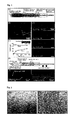
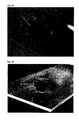
- the microfiber can be conveniently prepared by using, for example, a double coaxial microfluidic device such as that shown in Fig. 1 .
- a double coaxial microfluidic device such as that shown in Fig. 1 .
- the double coaxial microfluidic device that can separately and coaxially inject two kinds of fluids as a core and a shell is specifically explained in, for example, Lab Chip, 4, pp.576-580, 2004 , Fig. 1 , and for preparation of the microfiber of the present invention, the device described in the aforementioned publication can be preferably used.
- FIG. 1 shows a method for preparing a microfiber having a core-shell structure consisting of two kinds of alginate gels as a model experiment.
- a microfiber consisting of two kinds of gels of inner part (core) and outer part (shell as the cover) can be constructed.
- the injection speed is not particularly limited, when a coaxial microfluidic device is used which has a size in that the caliber is about 50 ⁇ m to 2 mm, two kinds of solutions can be injected at a speed of about 10 to 500 ⁇ m/minute. By controlling the injection speeds of two kinds of solutions, the diameter of the core and the cover thickness of the shell can be appropriately adjusted ( Figs. 1, (C) and (D) ).
- the introduction speed into an aqueous solution containing calcium ions is also not particularly limited, the speed may be, for example, about 1 to 10 ml/minute.
- a microfiber of a core-shell structure having the collagen gel as the core and alginate gel as the shell can be prepared.
- a microfiber of a core-shell structure containing fibroblasts in the core can be prepared ( Fig. 1, (E) ).
- the high strength hydrogel of the shell can be formed first, and then the internal core can be gelled by heating, ultraviolet irradiation, or radiation irradiation.
- a solution of a water-soluble polymer chain that is crosslinked with calcium ions, such as fibrin monomers is used for the preparation of the internal core, and a sodium arginate solution is used as the solution of the external shell, gelation of the shell and the core can also be simultaneously performed by contact with calcium ions.
- a fiber with exposed micro gel fiber can also be prepared by removing the high strength hydrogel of the shell from the microfiber of the core-shell structure obtained as described above.
- a fiber of a core-shell structure using alginate gel as a high strength hydrogel and collagen as a base material gel of the micro gel fiber, and then allowing a chelating agent such as EDTA to act on the microfiber at an appropriate concentration to remove calcium ions and thereby remove only the high strength hydrogel, a fiber consisting the collagen gel can be prepared. The aforementioned removing operation may be performed after the microfiber is prepared.
- a hollow fiber consisting of high strength gel by removing the hydrogel being the core from the microfiber having a core-shell structure, if desired.
- the alginate gel of the core can solely be removed by allowing a chelating agent such as EDTA to act on the microfiber at an appropriate concentration to remove calcium ions, and thereby prepare a hollow agarose gel fiber. The aforementioned removal may be performed after the microfiber is molded.
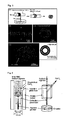
- the microfiber obtained as described above can be sucked into a silicone tube and stored in a state that the gel is stretched along the longitudinal direction of the tube. It is generally difficult to maintain a gelled microfiber in a linear shape when the gelled microfiber is stored in water, buffer, or the like.
- the microfiber is put into an aqueous medium such as water and butter, and sucked through a silicone tube having an internal diameter of about 100 ⁇ m to several millimeters, of which one end is immersed in the aqueous medium, the microfiber is sucked into the silicone tube from an end thereof in a state that the microfiber is stretched along the longitudinal direction of the tube. This state is shown in Fig. 2 .
- the gel can be stored in this state, and upon use, the silicone tube can be cut in an appropriate length to prepare the gel of a desired length.
- appropriate agents such as preservative, pH modifier and buffering agent can be added to the medium in the tube, as required.
- the microfiber of the present invention has superior mechanical strength, and can be preferably used for constructing, for example, a braid structure such as double or triple helix braid structure, a woven fabric structure, a three-dimensional structure such as a cylinder structure, a helical structure, and a tube structure.
- structure used in this specification means any structure obtainable by molding one microfiber, and any structures that can be constructed with two or more microfibers, and should be construed in the broadest sense thereof including a braid structure having a linear shape in appearance, and a structure such as a sheet that can be seen as a plane in appearance, and these terms should not be construed in any limitative way.
- the structure may be referred to as a "three-dimensional structure".
- Conceptual sketches of the three-dimensional structure are shown in Fig. 3 .
- a plurality of the microfibers of the present invention can also be used as a bundle.
- a plurality of microfibers containing cells in the micro gel fibers can be prepared, and arranged along the transverse direction as a bundle to from a sheet consisting of the microfibers in lines, and the sheet can be cultured to prepare cell culture in the shape of sheet (referred to as a "cell sheet” in the specification).
- a plurality of the aforementioned sheets can also be piled up in the shape of a block and cultured to prepare cell culture in the shape of a block (referred to as a "cell block" in the specification).
- gel having a woven fabric structure can be prepared by using a microweaving machine that provides warp intervals of about 1 to 5 mm and the aforementioned microfibers as warps and/or wefts.
- a microweaving machine that provides warp intervals of about 1 to 5 mm and the aforementioned microfibers as warps and/or wefts.
- Fig. 4 Conceptual sketches of this method and examples of the gel having a woven fabric structure are shown in Fig. 4 .
- the microfiber of the present invention can be used as the warp and the weft, or an alginate microfiber or the like can also be used as the weft or the warp.
- the alginate microfiber can be prepared by, for example, using a sodium arginate solution as an inner fluid, and a CaCl 2 solution as an outer fluid in the aforementioned coaxial micro fluid device.
- a sodium arginate solution as an inner fluid
- a CaCl 2 solution as an outer fluid in the aforementioned coaxial micro fluid device.
- the microfiber used as the weft and the warp is preferably set on a weaving machine in such a state that the microfiber is stored in a silicone tube as explained above, so that the microfiber is supplied from the inside of the silicone tube.
- Fig. 4, (A) includes conceptual sketches showing that the warp is supplied from the inside of the silicone tube.
- a tubular structure can be formed by rolling up a microfiber using a cylinder such as a glass tube as shown in Fig. 5, (A) , coating the outside with agarose gel, alginate gel, or the like, and then pulling out the cylinder.
- a cylinder such as a glass tube as shown in Fig. 5, (A)
- coating the outside with agarose gel, alginate gel, or the like and then pulling out the cylinder.
- Fig. 5, (A) is a schematic diagram showing operations of rolling up two kinds of different microfibers of the present invention, and fixing the helical structure with agarose.
- a three-dimensional structure constructed with the micro gel fiber can be manufactured.
- a three-dimensional structure is constructed by using the microfiber having a core-shell structure using alginate gel as the high strength hydrogel and collagen as a base material gel of the micro gel fiber, by allowing a chelating agent such as EDTA to act on the microfiber at an appropriate concentration to remove calcium ions, and thereby solely remove the high strength hydrogel, a three-dimensional structure constructed with collagen gel can be prepared.
- the three-dimensional structure of collagen gel obtained as described above can be preferably used for, for example, cell culture.
- a three-dimensional structure constructed with a hollow fiber consisting of high strength gel by constructing an arbitrary structure, preferably a three-dimensional structure, using the microfiber of the present invention, and then removing the hydrogel of the core, as required.
- a three-dimensional structure is constructed by using the microfiber having a core-shell structure using agarose gel as the high strength hydrogel and alginate gel as a base material gel of the micro gel fiber, by allowing a chelating agent such as EDTA to act on the structure at an appropriate concentration to remove calcium ions, and thereby solely remove the alginate gel of the core, a three-dimensional structure constructed with a hollow agarose gel fiber can be prepared.
- a cell fiber consisting of the cell culture can be obtained.
- a collagen gel fiber as the micro gel fiber
- alginate gel as the high strength hydrogel.
- the cell fiber obtained as described above is a fiber containing cell aggregates in the micro gel fiber, and has a characteristic feature that the fiber can maintain the fiber shape as it is.
- a protein for enhancing adherent property such as fibrin may be added beforehand, as required.
- the protein may be added only to the core, or the protein can be preferably added to both of the core and the shell. For example, if fibrin is added to both of the core and the shell, cells may uniformly proliferate to form a cell fiber without aggregating to form clusters.
- the type and amount of the protein to be added are not particularly limited, and appropriately chosen according to the type of the cells to be cultured.
- an arbitrary two-dimensional or three-dimensional structure can be formed by using the resulting microfiber.
- an arbitrary two-dimensional or three-dimensional structure may be formed. Then, by removing the high strength hydrogel from the resulting two-dimensional or three-dimensional structure to expose the cell culture, a two-dimensional cell sheet or a three-dimensional cell block constructed with the aforementioned cell fiber can be manufactured. A conceptual sketch of this method is shown in Fig. 12 .
- the high strength hydrogel can also be removed, if required.
- a two-dimensional cell sheet or a three-dimensional cell block containing two or more kinds of different cell fibers can be formed.
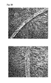
- An alginate hydrogel fiber was prepared by using a coaxial laminar flow device ( Lab. Chip, 4, pp.576, 2004 ; Langmuir, 23, pp.9104, 2007 ) according to the method shown in Fig. 6 , (A).
- the gelled alginate hydrogel fiber was received with a petri dish containing deionized water ( Fig. 7, (C) ).
- FIG. 8 (A) is a schematic view of the drawing, and Fig. 8, (B) shows the alginate hydrogel fiber drawn into the glass tube as described above. This method enables to firmly hold the end of the hydrogel fiber.
- the alginate hydrogel fiber had superior mechanical strength, and the fiber was successfully rolled up around a glass tube having a diameter of 1 mm ( Fig. 9 ).
- Fluorescent microbeads blue, green and red, diameter: 0.2 to 1.0 ⁇ m
- cells 3T3 fibroblasts (red) and Jurkat cells (green)
- alginate hydrogel fibers (diameter: 70 ⁇ m) containing fluorescent microbeads ( Fig. 10, (A) ) or cells ( Fig. 10, (B) ) were prepared in the same manner as described above.
- the hydrogel fibers to which those microbeads and cells were added had a mechanical strength of the same level.
- a braid structure was manually formed by using three hydrogel fibers containing three kinds of the aforementioned beads, respectively.
- a conceptual sketch of the structure is shown in Fig. 11, (A)
- a fluorescence microphotograph of the resulting braid structure is shown in Fig. 11, (B) .
- a fiber having a core-shell structure was prepared in the same manner as that of Example 1, except that a double coaxial laminar flow device ( Lab. Chip, 4, pp.576, 2004 , Fig. 1 ) was used.
- a double coaxial laminar flow device Lab. Chip, 4, pp.576, 2004 , Fig. 1
- As the fluid for core 1.5% w/v sodium arginate (colored in orange) was used
- As the fluid for shell 1.5% w/v sodium arginate (colored in green) was used
- the resulting fiber having a core-shell structure is shown in Fig. 1, (B) .
- the core diameter and cover thickness of the shell of the resulting fiber were varied depending on the flow rate ratio of the core fluid and the shell fluid (Q core /Q shell ) ( Figs. 1, (C) and (D) ).
- a microfiber consisting of a collagen micro gel fiber covered with alginate gel as the high strength hydrogel was prepared in the same manner as that of Example 2 by using a collagen solution (concentration: 2 mg/ml) containing the 3T3 fibroblasts (cell number: 1 to 10 x 10 6 cells/ml) as the fluid for core.
- a conceptual sketch of the method is shown in Fig. 1, (E) .
- the resulting microfiber was a fiber having a core-shell structure in which the collagen gel as the core contained the 3T3 cells and having sufficient mechanical strength ( Fig. 1, (F) ).
- a three-dimensional structure having a woven fabric structure was prepared by the method shown in Figs. 4, (A) and (B) .
- the alginate hydrogel fibers (diameter: 230 ⁇ m) obtained in Example 1 as the warps and wefts
- the woven fabric structure shown in Fig. 4, (C) was knitted.
- a three-dimensional structure having a woven fabric structure was prepared by using the alginate hydrogel fibers of different fluorescence color as a part of the warps and the wefts ( Fig. 4, (D)).
- Fig. 4, (E) is a magnified view
- (F) is a cross-sectional view.
- Example 4 In the same manner as that of Example 4, a three-dimensional structure having a woven fabric structure was prepared by using the microfibers obtained in Example 3 (core diameter: 40 ⁇ m, external diameter: 140 ⁇ m, 3T3 fibroblast density: 10 7 cells/ml) as the warps and the alginate hydrogel fibers obtained in Example 1 as the wefts.
- microfiber A core diameter: 40 ⁇ m, external diameter: 140 ⁇ m, colored with green fluorescence
- microfiber B core diameter: 40 ⁇ m, external diameter: 140 ⁇ m, colored with orange fluorescence
- Fig. 5, (A) the outer surface of the resulting helical structure was coated with agarose gel (3%) to prepare a three-dimensional structure having a helical structure.
- Fig. 5, (B) is a magnified view of the helical structure
- Fig. 5, (C) is a cross-sectional view thereof.
- Example 6 In the same manner as that of Example 6, a microfiber containing the 3T3 fibroblasts (core diameter: 40 ⁇ m, external diameter: 140 ⁇ m, cell density: 10 7 cells/ml) was rolled up around a glass tube to prepare a three-dimensional structure having a helical structure.
- Fig. 5, (D) shows a confocal image of the surface of the resulting helical structure, and a conceptual sketch of the cross-sectional view is shown on the right side thereof.
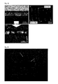
- Example 3 In the same manner as that of Example 3, a microfiber consisting of collagen gel as the core and alginate gel as the shell, and containing the 3T3 fibroblasts (cell number: 1 to 10 x 10 6 cells/ml) and polystyrene blue beads for visualization (diameter: 15 ⁇ m) in the core was prepared (core diameter: 80 ⁇ m, external diameter: 150 ⁇ m, cell density: 10 7 cells/ml, bead density: 0.5% (w/v)), and cultured at 37°C for 30 minutes, and then the appearance of the microfiber was optically observed. It was successfully confirmed that the 3T3 cells and the collagen gel of the core were covered with the alginate gel of the shell ( Fig. 13 ).
- a microfiber containing the HepG2 cells in the core was prepared in the same manner as that of Example 3 and cultured to fabricate a microfiber containing culture of the HepG2 cells in the core.
- the core consisting of the collagen gel was filled with the proliferated cells, and a microfiber of which core was fully filled with the cells (microfiber containing collagen gel and cell culture in the core and covered with alginate gel) was obtained on the day 11 ( Figs. 14, (A) to (C) ).
- the cell culture in the form of a fiber was exposed from the above microfiber by removing the alginate gel with an enzyme treatment, the shape of the cell fiber was kept as it was, and it was estimated that the cells firmly bound to one another ( Fig. 14, (D) ).
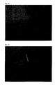
- gel fibers containing cell culture in the collagen gel of the core were prepared by using the HepG2 cell (culture on day 14), Min6 cells (culture on day 18), Hela cells (culture on day 6), and primary cerebral cortex cells of the rat brain (culture on day 8) ( Figs. 15, (A) to (D) ).
- B-29 and G-5 were added to the core as growth factors at the standard concentrations specified by the manufacturer. Then, the alginate gel of the shell was removed to pepare each cell fiber.
- Example 9 Functions of the cell fiber of the primary cerebral cortex cells derived from the rat brain (culture on day 8) obtained in Example 9 were examined. As a result, spontaneous Ca 2+ vibration was observed in a large number of cerebral cortex neurons, and it was demonstrated that a nerve network was formed in the cerebral cortex cell fiber ( Fig. 16, (D) ). Further, it was confirmed that the cell fiber of the HepG2 cells obtained in Example 9 secreted lactic acid when the fiber was cultured ( Fig. 17 ).
- a cell structure having a woven fabric structure was constructed with gel fibers in which cell culture of the Hela cells was contained in collagen gel of the core, and the shell was alginate gel.
- a conceptual sketch of the method for preparing a cell sheet having a woven fabric structure is shown in Fig. 18, (A) .
- the resulting cell sheet having a woven fabric structure was a cell structure having a size of centimeter order (about 1 to 2 cm) ( Fig. 18, (B) ).
- a cell structure having a woven fabric structure consisting of six warps and five wefts is shown in Fig. 18, (C) (visible light image) and Fig. 18, (D) (fluorescence image).
- a cell structure consisting of the cell fibers having a length of about 1.5 cm and arranged in parallel was fabricated ( Fig. 18, (E) ).
- a cell structure having a heterogenous coil structure was formed by using a gel fiber in which cell culture of the HepG2 cells was contained in collagen gel of the core and the shell consisted of alginate gel, and a microfiber in which cell culture of the Min6 cells was contained in collagen gel of the core and the shell consisted of alginate gel ( Fig. 19 ).
- the cells contained in the resulting cell structure having a coil structure continued to proliferate even after the alginate gel was removed, and thus it was demonstrated that the cells contained in the cell structure maintained biological functions ( Fig. 19, (C) ).
- a two-dimensional structure of a fabric shape was prepared by using microfibers having a core-shell structure in which a collagen gel fiber (core, containing three kinds of different fluorescent beads) was covered with alginate gel (shell), and a T-shirt-shaped three-dimensional structure was fabricated by using the fiber.
- a two-dimensional structure having a woven fabric shape was fabricated by using the microfibers, placed on a transparent film, and thinly coated with agarose gel in order to maintain the woven fabric structure ( Fig. 20 ).
- the woven fabric structure coated with agarose had sufficient mechanical strength, and the structure was successfully raised with a pair of tweezers ( Fig. 21 ).
- a hole (diameter: 1.5 mm) was made at the center of the woven fabric-shaped structure with a punch ( Fig. 22 ), a glass rod having a diameter of 1 mm was passed through the provided hole, one glass rod each was put on the right and left sides so that these glass rods perpendicularly intersected with the foregoing glass rod, and the fabric structure was folded ( Fig. 23 ). After the folding, agarose gel was cast in the gap and gelled to fix the fabric structure in the folded state ( Fig. 24 ). The glass rods and the transparent film were removed, and the excessive margin was cut off with a cutter to prepare a T-shirt-shaped three-dimensional structure ( Fig. 25 ).
- Fig. 26 The resulting three-dimensional structure (length: 6 mm x width: 6 mm) in a standing state is shown in Fig. 26 . It can be observed that a three-dimensional structure in the form of T-shirt having holes for head and arms was obtained.
- Fig. 27 is a fluorescent image of the aforementioned three-dimensional structure. Three kinds of fluorescence originating in the fluorescent beads were observed.
- a microfiber in which fibrin as an adherent protein was added (amount of added fibrinogen: 1 mg/mL) to collagen gel of the core containing cells (Hela cells or NIH/3T3 cells) and alginate gel of the shell (Type B) and a fibrin-free microfiber (Type A) were prepared and cultured. The method and the results are shown in Fig. 28 .
- the Hela cells favorably proliferated ((C), left), whereas the 3T3 cells did not proliferate and form cell fiber, but formed cell clusters ((C), center).
- the microfiber of Type B to which fibrin was added favorable proliferation and formation of a cell fiber were observed also for the 3T3 cells ((C), right).
- difference in the proliferation rate was observed depending on the type of the cells ((E)).
- a microfiber consisting of collagen gel as the core containing the HepG2 cells and the shell of alginate gel was prepared and cultured to obtain a microfiber containing a cell fiber of the HepG2 cell in the core.
- amount of albumin secreted from this microfiber by incubation was compared with amount of albumin secreted by the HepG2 cells cultured on a dish, the amount of albumin secreted from the microfiber was higher than the amount observed by the culture on a dish. The results are shown in Fig. 29 .
- HepG2 cells encapsulated in the core were maintained under a three-dimensional optimum environment, and as a result, the cells successfully secreted albumin in a larger amount compared with that observed with the two-dimensional culture condition on a dish.
- a microfiber in which fibrin as an adherent protein was added to collagen gel of the core containing the NIH/3T3 cells and alginate gel of the shell (Type B) was prepared by the method of Example 14 and cultured to obtain a microfiber containing the NIH/3T3 cells in the core.
- Mechanical strength of this microfiber was measured by the method shown in Fig. 30 before and after removal of alginate gel to confirm the mechanical strength enhancing effect of the alginate gel of the shell.
- amount of curve of a thin glass tube (diameter: 0.12 mm) according to the method shown in Figs. 30, (A) and (B) .
- tension loaded on the microfiber was calculated.
- the tension loaded when the microfiber broke was considered as mechanical strength.
- the microfiber having the shell gave higher mechanical strength compared with the microfiber of which shell was removed ( Fig. 31 , upper graph and lower graph).
- a microfiber consisting of collagen gel as the core and alginate gel (1.5%) as the shell in which neural stem cells were introduced into the core of the microfiber was prepared.
- EGF EGF
- FGF FGF
- B27 B27
- the microfiber was prepared so that the cell density became 6.8 X 10 7 cells/mL, and culture was continued for 7 days by using a medium consisting of 10 mL of Neurobasal A to which 1% antibiotics (penicillin and streptomycin), 2 ⁇ L of EGF, 20 ⁇ L of FGF, and 200 ⁇ L of B27 were added.
- the results are shown in Fig. 32 .
- the upper photograph shows the microfiber immediately after the fabrication, and the lower photograph shows the microfiber after culture of 7 days.
- the neural stem cells proliferated in the core of the microfiber, and filled the core.
Applications Claiming Priority (3)
| Application Number | Priority Date | Filing Date | Title |
|---|---|---|---|
| JP2009237087 | 2009-10-14 | ||
| JP2010143411 | 2010-06-24 | ||
| PCT/JP2010/067852 WO2011046105A1 (fr) | 2009-10-14 | 2010-10-12 | Fibres micro-gel revêtues |
Publications (3)
| Publication Number | Publication Date |
|---|---|
| EP2489779A1 true EP2489779A1 (fr) | 2012-08-22 |
| EP2489779A4 EP2489779A4 (fr) | 2015-01-21 |
| EP2489779B1 EP2489779B1 (fr) | 2019-01-09 |
Family
ID=43876155
Family Applications (1)
| Application Number | Title | Priority Date | Filing Date |
|---|---|---|---|
| EP10823373.5A Active EP2489779B1 (fr) | 2009-10-14 | 2010-10-12 | Fibres micro-gel revêtues |
Country Status (5)
| Country | Link |
|---|---|
| US (1) | US8785195B2 (fr) |
| EP (1) | EP2489779B1 (fr) |
| JP (1) | JP5633077B2 (fr) |
| ES (1) | ES2716204T3 (fr) |
| WO (1) | WO2011046105A1 (fr) |
Cited By (9)
| Publication number | Priority date | Publication date | Assignee | Title |
|---|---|---|---|---|
| WO2017102989A1 (fr) * | 2015-12-18 | 2017-06-22 | Universidad Politécnica de Madrid | Procédé pour la production de structures de forme allongée telles que des fibres à partir de solutions de polymères par fluotournage à contrainte |
| EP3147346A4 (fr) * | 2014-05-20 | 2018-01-10 | The University of Tokyo | Microfibre creuse |
| CN108474140A (zh) * | 2015-11-25 | 2018-08-31 | 纽泰克温图斯公司 | 大规模细胞生产系统 |
| EP3007882B1 (fr) | 2013-06-13 | 2019-11-20 | Aspect Biosystems Ltd. | Système de fabrication additive de structures tridimensionnelles et procédé associé |
| CN111270349A (zh) * | 2020-01-21 | 2020-06-12 | 广东省材料与加工研究所 | 基于微流体纺丝氧化石墨烯纤维及三维支架的制备方法 |
| CN113668233A (zh) * | 2021-08-24 | 2021-11-19 | 江西服装学院 | 织物整理剂及使用其整理纯棉织物的方法 |
| WO2021234386A1 (fr) * | 2020-05-20 | 2021-11-25 | University Of Leeds | Formulation comprenant un microgel protéique |
| EP3835409A4 (fr) * | 2018-08-10 | 2022-05-04 | Mochida Pharmaceutical Co., Ltd. | Microfibre creuse d'alginate |
| US11724450B2 (en) | 2017-03-15 | 2023-08-15 | Aspect Biosystems Ltd. | Systems and methods for printing a fiber structure |
Families Citing this family (36)
| Publication number | Priority date | Publication date | Assignee | Title |
|---|---|---|---|---|
| JP6074765B2 (ja) * | 2013-01-18 | 2017-02-08 | 国立大学法人 東京大学 | 移植用神経束及びその製造方法 |
| JP6215541B2 (ja) * | 2013-02-28 | 2017-10-18 | 国立大学法人 東京大学 | 束状構造を有するゲルファイバー集合体の製造方法 |
| FR3014691B1 (fr) * | 2013-12-13 | 2016-02-05 | Ets Francais Du Sang | Capsules contenant des cellules a potentialite hematopoietique |
| JP6628416B2 (ja) * | 2014-08-04 | 2020-01-08 | 国立大学法人千葉大学 | 細胞培養方法 |
| JP6439918B2 (ja) * | 2014-10-17 | 2018-12-19 | 国立大学法人 東京大学 | 3次元細胞構造体の製造方法 |
| JP6646298B2 (ja) * | 2014-12-16 | 2020-02-14 | 国立大学法人 東京大学 | ロープ状構造体の製造方法 |
| JP2016183431A (ja) * | 2015-03-26 | 2016-10-20 | 株式会社化繊ノズル製作所 | 糸状ゲルの製造方法 |
| ES2842501T5 (es) | 2015-09-21 | 2023-04-13 | Modern Meadow Inc | Materiales compuestos de tejido reforzados con fibras |
| JP6895674B2 (ja) * | 2015-10-21 | 2021-06-30 | 国立大学法人 東京大学 | マイクロチューブ、マイクロチューブの製造方法、及びマイクロチューブの製造装置 |
| JP7385273B2 (ja) * | 2015-11-30 | 2023-11-22 | 一般財団法人生産技術研究奨励会 | 細胞培養方法及びマイクロファイバ |
| JP2017099303A (ja) * | 2015-11-30 | 2017-06-08 | 一般財団法人生産技術研究奨励会 | 中空マイクロファイバを用いた細胞培養方法 |
| ES2807727T3 (es) | 2016-02-15 | 2021-02-24 | Modern Meadow Inc | Material compuesto biofabricado |
| JP6774273B2 (ja) * | 2016-08-31 | 2020-10-21 | 花王株式会社 | ハイドロゲルファイバの製造方法 |
| JP6935896B2 (ja) * | 2016-11-14 | 2021-09-15 | 一般財団法人生産技術研究奨励会 | 体内移植用のハイドロゲルファイバ及びハイドロゲルファイバを用いた体内移植方法 |
| US20200138870A1 (en) * | 2016-11-17 | 2020-05-07 | Baylor College Of Medicine | Recreation of pancreatic niche allows for novel methods for human, mature beta derivation from pluripotent stem cells |
| JP7132566B2 (ja) * | 2017-09-04 | 2022-09-07 | 学校法人早稲田大学 | チキソトロピー性を有するゲルを用いる多層3次元細胞培養足場システム |
| JP7090868B2 (ja) * | 2017-10-19 | 2022-06-27 | 国立大学法人 東京大学 | 接着剤及びその使用 |
| AU2018253595A1 (en) | 2017-11-13 | 2019-05-30 | Modern Meadow, Inc. | Biofabricated leather articles having zonal properties |
| WO2020021878A1 (fr) * | 2018-07-26 | 2020-01-30 | 株式会社カネカ | Objet à demeure in vivo et système à demeure associé |
| US11352497B2 (en) | 2019-01-17 | 2022-06-07 | Modern Meadow, Inc. | Layered collagen materials and methods of making the same |
| JP7227860B2 (ja) * | 2019-06-27 | 2023-02-22 | 株式会社カネカ | 生体内留置物送達具 |
| WO2020261828A1 (fr) * | 2019-06-27 | 2020-12-30 | 株式会社カネカ | Objet à demeure in vivo |
| JP6601931B1 (ja) * | 2019-07-17 | 2019-11-06 | 株式会社セルファイバ | 細胞ファイバ製造システム、細胞ファイバ製造方法及びプログラム |
| JP7426065B2 (ja) | 2019-07-17 | 2024-02-01 | 株式会社セルファイバ | 細胞ファイバ製造システム、細胞ファイバ製造方法及びプログラム |
| JP2023015413A (ja) * | 2019-12-18 | 2023-02-01 | 持田製薬株式会社 | 化学架橋アルギン酸ゲルファイバ |
| EP4107318A1 (fr) | 2020-02-19 | 2022-12-28 | Association for the Advancement of Tissue Engineering and Cell Based Technologies & Therapies (A4TEC) - Associacão | Fibres d'hydrogel à compartiments multiples, leur préparation et leurs utilisations |
| WO2021177455A1 (fr) * | 2020-03-05 | 2021-09-10 | 株式会社セルファイバ | Corps de stockage et procédé de cryoconservation |
| JPWO2022050282A1 (fr) * | 2020-09-01 | 2022-03-10 | ||
| JP6848145B2 (ja) * | 2020-11-09 | 2021-03-24 | 株式会社セルファイバ | ファイバ製造システム、ファイバ製造方法及びプログラム |
| AU2021414759A1 (en) | 2020-12-28 | 2023-07-13 | Mochida Pharmaceutical Co., Ltd. | Novel multilayer polymer-coated crosslinked alginate gel fiber |
| CN116710110A (zh) | 2020-12-28 | 2023-09-05 | 持田制药株式会社 | 新型的多层聚合物涂层交联海藻酸凝胶纤维 |
| WO2023286852A1 (fr) * | 2021-07-15 | 2023-01-19 | 株式会社セルファイバ | Structure et son utilisation |
| WO2023085441A1 (fr) * | 2021-11-10 | 2023-05-19 | 国立大学法人東京大学 | Structure macroporeuse |
| CN114657774B (zh) * | 2022-04-06 | 2024-01-30 | 合肥工业大学 | 一种高强度、自修复、弹性导电纤维的制备方法 |
| EP4269671A1 (fr) * | 2022-04-26 | 2023-11-01 | EMPA Eidgenössische Materialprüfungs- und Forschungsanstalt | Filage humide à base microfluidique de fibres polymères solides individuelles |
| CN114854677B (zh) * | 2022-07-04 | 2022-11-04 | 南京农业大学 | 一种用于细胞培养肉生产的微流控仿生纤维及其制备方法和应用 |
Citations (2)
| Publication number | Priority date | Publication date | Assignee | Title |
|---|---|---|---|---|
| US20060188487A1 (en) * | 2005-02-23 | 2006-08-24 | Zimmer Technology, Inc. | Blend hydrogels and methods of making |
| EP2180027A1 (fr) * | 2007-07-05 | 2010-04-28 | Nissan Chemical Industries, Ltd. | Nouvel agent de formation d'hydrogel sur la base de lipide-tripeptide et hydrogel |
Family Cites Families (4)
| Publication number | Priority date | Publication date | Assignee | Title |
|---|---|---|---|---|
| JPS62141121A (ja) * | 1985-12-07 | 1987-06-24 | Agency Of Ind Science & Technol | ゲル状バインダー繊維とその製造方法 |
| JPH0615163A (ja) * | 1992-06-30 | 1994-01-25 | Sanei Touka Kk | ファイバー入りゲルマイクロカプセル及びその製法 |
| EP1555957A4 (fr) * | 2002-10-04 | 2010-11-24 | Nanomatrix Inc | Scellants pour la peau et autres tissus |
| JP5515041B2 (ja) | 2007-03-09 | 2014-06-11 | 国立大学法人九州大学 | 超分子ナノ集合体の製造方法および超分子ナノ集合体 |
-
2010
- 2010-10-12 WO PCT/JP2010/067852 patent/WO2011046105A1/fr active Application Filing
- 2010-10-12 ES ES10823373T patent/ES2716204T3/es active Active
- 2010-10-12 EP EP10823373.5A patent/EP2489779B1/fr active Active
- 2010-10-12 JP JP2011536134A patent/JP5633077B2/ja active Active
- 2010-10-12 US US13/501,634 patent/US8785195B2/en active Active
Patent Citations (2)
| Publication number | Priority date | Publication date | Assignee | Title |
|---|---|---|---|---|
| US20060188487A1 (en) * | 2005-02-23 | 2006-08-24 | Zimmer Technology, Inc. | Blend hydrogels and methods of making |
| EP2180027A1 (fr) * | 2007-07-05 | 2010-04-28 | Nissan Chemical Industries, Ltd. | Nouvel agent de formation d'hydrogel sur la base de lipide-tripeptide et hydrogel |
Non-Patent Citations (3)
| Title |
|---|
| CHIZUKA HENMI ET AL: "Development of an effective three dimensional fabrication technique using inkjet technology for tissue model samples", JAPANESE SOCIETY FOR ALTERNATIVES TO ANIMAL EXPERIMENTS, 31 December 2008 (2008-12-31), pages 689-692, XP055155539, * |
| KWANG HO LEE ET AL: "Synthesis of Cell-Laden Alginate Hollow Fibers Using Microfluidic Chips and Microvascularized Tissue-Engineering Applications", SMALL, vol. 5, no. 11, 5 June 2009 (2009-06-05), pages 1264-1268, XP055155556, ISSN: 1613-6810, DOI: 10.1002/smll.200801667 * |
| See also references of WO2011046105A1 * |
Cited By (14)
| Publication number | Priority date | Publication date | Assignee | Title |
|---|---|---|---|---|
| US11046930B2 (en) | 2013-06-13 | 2021-06-29 | Aspect Biosystems Ltd. | System for additive manufacturing of three-dimensional structures and method for same |
| EP3007882B1 (fr) | 2013-06-13 | 2019-11-20 | Aspect Biosystems Ltd. | Système de fabrication additive de structures tridimensionnelles et procédé associé |
| US11738501B2 (en) | 2013-06-13 | 2023-08-29 | Aspect Biosystems Ltd. | System for additive manufacturing of three-dimensional structures and method for same |
| EP3670155A1 (fr) | 2013-06-13 | 2020-06-24 | Aspect Biosystems Ltd. | Système de fabrication additive de structures tridimensionnelles et procédé associé |
| EP3147346A4 (fr) * | 2014-05-20 | 2018-01-10 | The University of Tokyo | Microfibre creuse |
| CN108474140A (zh) * | 2015-11-25 | 2018-08-31 | 纽泰克温图斯公司 | 大规模细胞生产系统 |
| US11180868B2 (en) | 2015-12-18 | 2021-11-23 | Universidad Politécnica de Madrid | Method for producing elongated structures such as fibers from polymer solutions by straining flow spinning |
| WO2017102989A1 (fr) * | 2015-12-18 | 2017-06-22 | Universidad Politécnica de Madrid | Procédé pour la production de structures de forme allongée telles que des fibres à partir de solutions de polymères par fluotournage à contrainte |
| US11724450B2 (en) | 2017-03-15 | 2023-08-15 | Aspect Biosystems Ltd. | Systems and methods for printing a fiber structure |
| EP3835409A4 (fr) * | 2018-08-10 | 2022-05-04 | Mochida Pharmaceutical Co., Ltd. | Microfibre creuse d'alginate |
| CN111270349B (zh) * | 2020-01-21 | 2022-12-16 | 广东省材料与加工研究所 | 基于微流体纺丝氧化石墨烯纤维及三维支架的制备方法 |
| CN111270349A (zh) * | 2020-01-21 | 2020-06-12 | 广东省材料与加工研究所 | 基于微流体纺丝氧化石墨烯纤维及三维支架的制备方法 |
| WO2021234386A1 (fr) * | 2020-05-20 | 2021-11-25 | University Of Leeds | Formulation comprenant un microgel protéique |
| CN113668233A (zh) * | 2021-08-24 | 2021-11-19 | 江西服装学院 | 织物整理剂及使用其整理纯棉织物的方法 |
Also Published As
| Publication number | Publication date |
|---|---|
| EP2489779A4 (fr) | 2015-01-21 |
| US20120301963A1 (en) | 2012-11-29 |
| WO2011046105A1 (fr) | 2011-04-21 |
| JP5633077B2 (ja) | 2014-12-03 |
| EP2489779B1 (fr) | 2019-01-09 |
| JPWO2011046105A1 (ja) | 2013-03-07 |
| ES2716204T3 (es) | 2019-06-11 |
| US8785195B2 (en) | 2014-07-22 |
Similar Documents
| Publication | Publication Date | Title |
|---|---|---|
| US8785195B2 (en) | Covered micro gel fiber | |
| US10221382B2 (en) | Hollow microfiber | |
| US20230220330A1 (en) | Self-assembling multicellular bodies and methods of producing a three-dimensional biological structure using the same | |
| Chen et al. | Three-dimensional printed electrospun fiber-based scaffold for cartilage regeneration | |
| Attalla et al. | 3D bioprinting of heterogeneous bi-and tri-layered hollow channels within gel scaffolds using scalable multi-axial microfluidic extrusion nozzle | |
| Dolati et al. | In vitro evaluation of carbon-nanotube-reinforced bioprintable vascular conduits | |
| US9452239B2 (en) | Fabrication of interconnected model vasculature | |
| JP6439918B2 (ja) | 3次元細胞構造体の製造方法 | |
| Hirayama et al. | Cellular building unit integrated with microstrand-shaped bacterial cellulose | |
| EP2097513A2 (fr) | Préparation et utilisation de filetages de synthèse cellulaire | |
| AU2021221909B2 (en) | Engineered materials and methods of forming | |
| US9476834B2 (en) | Process for producing supramolecular fiber | |
| CN108159495B (zh) | 3d生物蛋白及其制备方法和应用 | |
| US9663874B2 (en) | Device for manufacturing polymer fibers and uses thereof | |
| US20240082462A1 (en) | High-porosity nanofiber nonwovens as a support structure for stromal tissue | |
| US11672888B2 (en) | Structures with complex geometries and controlled porosity in micrometer to meter dimensions produced at large scale | |
| EP1469724B1 (fr) | Cryoconservation sur des tissus textiles | |
| KR101483622B1 (ko) | 납작한 형태의 마이크로 섬유를 포함하는 세포 배양용 지지체 | |
| CN114773758A (zh) | 一种仿藤蔓纳米复合水凝胶纤维致动器及制备方法和应用 |
Legal Events
| Date | Code | Title | Description |
|---|---|---|---|
| PUAI | Public reference made under article 153(3) epc to a published international application that has entered the european phase |
Free format text: ORIGINAL CODE: 0009012 |
|
| 17P | Request for examination filed |
Effective date: 20120426 |
|
| AK | Designated contracting states |
Kind code of ref document: A1 Designated state(s): AL AT BE BG CH CY CZ DE DK EE ES FI FR GB GR HR HU IE IS IT LI LT LU LV MC MK MT NL NO PL PT RO RS SE SI SK SM TR |
|
| RIN1 | Information on inventor provided before grant (corrected) |
Inventor name: TAKEUCHI SHOJI Inventor name: MATSUNAGA YUKIKO Inventor name: GOJO RIHO Inventor name: ONOE HIROAKI Inventor name: NEGISHI MIDORI Inventor name: KIRIYA DAISUKE |
|
| DAX | Request for extension of the european patent (deleted) | ||
| A4 | Supplementary search report drawn up and despatched |
Effective date: 20141219 |
|
| RIC1 | Information provided on ipc code assigned before grant |
Ipc: D06M 15/277 20060101ALI20141215BHEP Ipc: D01F 8/18 20060101ALI20141215BHEP Ipc: D01F 8/02 20060101ALI20141215BHEP Ipc: D03D 15/00 20060101ALI20141215BHEP Ipc: D01F 4/00 20060101ALI20141215BHEP Ipc: D01D 5/06 20060101ALI20141215BHEP Ipc: D01F 4/02 20060101ALI20141215BHEP Ipc: D06M 101/14 20060101ALI20141215BHEP Ipc: D06M 15/576 20060101ALI20141215BHEP Ipc: D01F 9/00 20060101ALI20141215BHEP Ipc: D01D 5/34 20060101ALI20141215BHEP Ipc: D06M 15/13 20060101ALI20141215BHEP Ipc: D06M 15/03 20060101AFI20141215BHEP |
|
| STAA | Information on the status of an ep patent application or granted ep patent |
Free format text: STATUS: EXAMINATION IS IN PROGRESS |
|
| 17Q | First examination report despatched |
Effective date: 20170524 |
|
| GRAP | Despatch of communication of intention to grant a patent |
Free format text: ORIGINAL CODE: EPIDOSNIGR1 |
|
| STAA | Information on the status of an ep patent application or granted ep patent |
Free format text: STATUS: GRANT OF PATENT IS INTENDED |
|
| INTG | Intention to grant announced |
Effective date: 20180718 |
|
| GRAS | Grant fee paid |
Free format text: ORIGINAL CODE: EPIDOSNIGR3 |
|
| GRAA | (expected) grant |
Free format text: ORIGINAL CODE: 0009210 |
|
| STAA | Information on the status of an ep patent application or granted ep patent |
Free format text: STATUS: THE PATENT HAS BEEN GRANTED |
|
| AK | Designated contracting states |
Kind code of ref document: B1 Designated state(s): AL AT BE BG CH CY CZ DE DK EE ES FI FR GB GR HR HU IE IS IT LI LT LU LV MC MK MT NL NO PL PT RO RS SE SI SK SM TR |
|
| REG | Reference to a national code |
Ref country code: GB Ref legal event code: FG4D |
|
| REG | Reference to a national code |
Ref country code: CH Ref legal event code: EP Ref country code: CH Ref legal event code: NV Representative=s name: ISLER AND PEDRAZZINI AG, CH Ref country code: AT Ref legal event code: REF Ref document number: 1087427 Country of ref document: AT Kind code of ref document: T Effective date: 20190115 |
|
| REG | Reference to a national code |
Ref country code: IE Ref legal event code: FG4D |
|
| REG | Reference to a national code |
Ref country code: DE Ref legal event code: R096 Ref document number: 602010056488 Country of ref document: DE |
|
| REG | Reference to a national code |
Ref country code: NL Ref legal event code: MP Effective date: 20190109 |
|
| REG | Reference to a national code |
Ref country code: LT Ref legal event code: MG4D |
|
| REG | Reference to a national code |
Ref country code: ES Ref legal event code: FG2A Ref document number: 2716204 Country of ref document: ES Kind code of ref document: T3 Effective date: 20190611 |
|
| PG25 | Lapsed in a contracting state [announced via postgrant information from national office to epo] |
Ref country code: NL Free format text: LAPSE BECAUSE OF FAILURE TO SUBMIT A TRANSLATION OF THE DESCRIPTION OR TO PAY THE FEE WITHIN THE PRESCRIBED TIME-LIMIT Effective date: 20190109 |
|
| REG | Reference to a national code |
Ref country code: AT Ref legal event code: MK05 Ref document number: 1087427 Country of ref document: AT Kind code of ref document: T Effective date: 20190109 |
|
| PG25 | Lapsed in a contracting state [announced via postgrant information from national office to epo] |
Ref country code: NO Free format text: LAPSE BECAUSE OF FAILURE TO SUBMIT A TRANSLATION OF THE DESCRIPTION OR TO PAY THE FEE WITHIN THE PRESCRIBED TIME-LIMIT Effective date: 20190409 Ref country code: SE Free format text: LAPSE BECAUSE OF FAILURE TO SUBMIT A TRANSLATION OF THE DESCRIPTION OR TO PAY THE FEE WITHIN THE PRESCRIBED TIME-LIMIT Effective date: 20190109 Ref country code: FI Free format text: LAPSE BECAUSE OF FAILURE TO SUBMIT A TRANSLATION OF THE DESCRIPTION OR TO PAY THE FEE WITHIN THE PRESCRIBED TIME-LIMIT Effective date: 20190109 Ref country code: PT Free format text: LAPSE BECAUSE OF FAILURE TO SUBMIT A TRANSLATION OF THE DESCRIPTION OR TO PAY THE FEE WITHIN THE PRESCRIBED TIME-LIMIT Effective date: 20190509 Ref country code: PL Free format text: LAPSE BECAUSE OF FAILURE TO SUBMIT A TRANSLATION OF THE DESCRIPTION OR TO PAY THE FEE WITHIN THE PRESCRIBED TIME-LIMIT Effective date: 20190109 Ref country code: LT Free format text: LAPSE BECAUSE OF FAILURE TO SUBMIT A TRANSLATION OF THE DESCRIPTION OR TO PAY THE FEE WITHIN THE PRESCRIBED TIME-LIMIT Effective date: 20190109 |
|
| PG25 | Lapsed in a contracting state [announced via postgrant information from national office to epo] |
Ref country code: LV Free format text: LAPSE BECAUSE OF FAILURE TO SUBMIT A TRANSLATION OF THE DESCRIPTION OR TO PAY THE FEE WITHIN THE PRESCRIBED TIME-LIMIT Effective date: 20190109 Ref country code: HR Free format text: LAPSE BECAUSE OF FAILURE TO SUBMIT A TRANSLATION OF THE DESCRIPTION OR TO PAY THE FEE WITHIN THE PRESCRIBED TIME-LIMIT Effective date: 20190109 Ref country code: IS Free format text: LAPSE BECAUSE OF FAILURE TO SUBMIT A TRANSLATION OF THE DESCRIPTION OR TO PAY THE FEE WITHIN THE PRESCRIBED TIME-LIMIT Effective date: 20190509 Ref country code: GR Free format text: LAPSE BECAUSE OF FAILURE TO SUBMIT A TRANSLATION OF THE DESCRIPTION OR TO PAY THE FEE WITHIN THE PRESCRIBED TIME-LIMIT Effective date: 20190410 Ref country code: BG Free format text: LAPSE BECAUSE OF FAILURE TO SUBMIT A TRANSLATION OF THE DESCRIPTION OR TO PAY THE FEE WITHIN THE PRESCRIBED TIME-LIMIT Effective date: 20190409 Ref country code: RS Free format text: LAPSE BECAUSE OF FAILURE TO SUBMIT A TRANSLATION OF THE DESCRIPTION OR TO PAY THE FEE WITHIN THE PRESCRIBED TIME-LIMIT Effective date: 20190109 |
|
| REG | Reference to a national code |
Ref country code: DE Ref legal event code: R097 Ref document number: 602010056488 Country of ref document: DE |
|
| PG25 | Lapsed in a contracting state [announced via postgrant information from national office to epo] |
Ref country code: AT Free format text: LAPSE BECAUSE OF FAILURE TO SUBMIT A TRANSLATION OF THE DESCRIPTION OR TO PAY THE FEE WITHIN THE PRESCRIBED TIME-LIMIT Effective date: 20190109 Ref country code: DK Free format text: LAPSE BECAUSE OF FAILURE TO SUBMIT A TRANSLATION OF THE DESCRIPTION OR TO PAY THE FEE WITHIN THE PRESCRIBED TIME-LIMIT Effective date: 20190109 Ref country code: EE Free format text: LAPSE BECAUSE OF FAILURE TO SUBMIT A TRANSLATION OF THE DESCRIPTION OR TO PAY THE FEE WITHIN THE PRESCRIBED TIME-LIMIT Effective date: 20190109 Ref country code: RO Free format text: LAPSE BECAUSE OF FAILURE TO SUBMIT A TRANSLATION OF THE DESCRIPTION OR TO PAY THE FEE WITHIN THE PRESCRIBED TIME-LIMIT Effective date: 20190109 Ref country code: SK Free format text: LAPSE BECAUSE OF FAILURE TO SUBMIT A TRANSLATION OF THE DESCRIPTION OR TO PAY THE FEE WITHIN THE PRESCRIBED TIME-LIMIT Effective date: 20190109 Ref country code: CZ Free format text: LAPSE BECAUSE OF FAILURE TO SUBMIT A TRANSLATION OF THE DESCRIPTION OR TO PAY THE FEE WITHIN THE PRESCRIBED TIME-LIMIT Effective date: 20190109 Ref country code: AL Free format text: LAPSE BECAUSE OF FAILURE TO SUBMIT A TRANSLATION OF THE DESCRIPTION OR TO PAY THE FEE WITHIN THE PRESCRIBED TIME-LIMIT Effective date: 20190109 |
|
| PLBE | No opposition filed within time limit |
Free format text: ORIGINAL CODE: 0009261 |
|
| STAA | Information on the status of an ep patent application or granted ep patent |
Free format text: STATUS: NO OPPOSITION FILED WITHIN TIME LIMIT |
|
| PG25 | Lapsed in a contracting state [announced via postgrant information from national office to epo] |
Ref country code: SM Free format text: LAPSE BECAUSE OF FAILURE TO SUBMIT A TRANSLATION OF THE DESCRIPTION OR TO PAY THE FEE WITHIN THE PRESCRIBED TIME-LIMIT Effective date: 20190109 |
|
| 26N | No opposition filed |
Effective date: 20191010 |
|
| PG25 | Lapsed in a contracting state [announced via postgrant information from national office to epo] |
Ref country code: SI Free format text: LAPSE BECAUSE OF FAILURE TO SUBMIT A TRANSLATION OF THE DESCRIPTION OR TO PAY THE FEE WITHIN THE PRESCRIBED TIME-LIMIT Effective date: 20190109 |
|
| PG25 | Lapsed in a contracting state [announced via postgrant information from national office to epo] |
Ref country code: TR Free format text: LAPSE BECAUSE OF FAILURE TO SUBMIT A TRANSLATION OF THE DESCRIPTION OR TO PAY THE FEE WITHIN THE PRESCRIBED TIME-LIMIT Effective date: 20190109 |
|
| PG25 | Lapsed in a contracting state [announced via postgrant information from national office to epo] |
Ref country code: MC Free format text: LAPSE BECAUSE OF FAILURE TO SUBMIT A TRANSLATION OF THE DESCRIPTION OR TO PAY THE FEE WITHIN THE PRESCRIBED TIME-LIMIT Effective date: 20190109 |
|
| PG25 | Lapsed in a contracting state [announced via postgrant information from national office to epo] |
Ref country code: LU Free format text: LAPSE BECAUSE OF NON-PAYMENT OF DUE FEES Effective date: 20191012 |
|
| REG | Reference to a national code |
Ref country code: BE Ref legal event code: MM Effective date: 20191031 |
|
| PG25 | Lapsed in a contracting state [announced via postgrant information from national office to epo] |
Ref country code: BE Free format text: LAPSE BECAUSE OF NON-PAYMENT OF DUE FEES Effective date: 20191031 |
|
| PG25 | Lapsed in a contracting state [announced via postgrant information from national office to epo] |
Ref country code: IE Free format text: LAPSE BECAUSE OF NON-PAYMENT OF DUE FEES Effective date: 20191012 |
|
| PG25 | Lapsed in a contracting state [announced via postgrant information from national office to epo] |
Ref country code: CY Free format text: LAPSE BECAUSE OF FAILURE TO SUBMIT A TRANSLATION OF THE DESCRIPTION OR TO PAY THE FEE WITHIN THE PRESCRIBED TIME-LIMIT Effective date: 20190109 |
|
| PG25 | Lapsed in a contracting state [announced via postgrant information from national office to epo] |
Ref country code: HU Free format text: LAPSE BECAUSE OF FAILURE TO SUBMIT A TRANSLATION OF THE DESCRIPTION OR TO PAY THE FEE WITHIN THE PRESCRIBED TIME-LIMIT; INVALID AB INITIO Effective date: 20101012 Ref country code: MT Free format text: LAPSE BECAUSE OF FAILURE TO SUBMIT A TRANSLATION OF THE DESCRIPTION OR TO PAY THE FEE WITHIN THE PRESCRIBED TIME-LIMIT Effective date: 20190109 |
|
| PG25 | Lapsed in a contracting state [announced via postgrant information from national office to epo] |
Ref country code: MK Free format text: LAPSE BECAUSE OF FAILURE TO SUBMIT A TRANSLATION OF THE DESCRIPTION OR TO PAY THE FEE WITHIN THE PRESCRIBED TIME-LIMIT Effective date: 20190109 |
|
| PGFP | Annual fee paid to national office [announced via postgrant information from national office to epo] |
Ref country code: IT Payment date: 20230913 Year of fee payment: 14 Ref country code: GB Payment date: 20230831 Year of fee payment: 14 |
|
| PGFP | Annual fee paid to national office [announced via postgrant information from national office to epo] |
Ref country code: FR Payment date: 20230911 Year of fee payment: 14 |
|
| PGFP | Annual fee paid to national office [announced via postgrant information from national office to epo] |
Ref country code: ES Payment date: 20231106 Year of fee payment: 14 |
|
| PGFP | Annual fee paid to national office [announced via postgrant information from national office to epo] |
Ref country code: DE Payment date: 20230830 Year of fee payment: 14 Ref country code: CH Payment date: 20231102 Year of fee payment: 14 |