EP1905828B1 - Optical sorting method - Google Patents
Optical sorting method Download PDFInfo
- Publication number
- EP1905828B1 EP1905828B1 EP07013020A EP07013020A EP1905828B1 EP 1905828 B1 EP1905828 B1 EP 1905828B1 EP 07013020 A EP07013020 A EP 07013020A EP 07013020 A EP07013020 A EP 07013020A EP 1905828 B1 EP1905828 B1 EP 1905828B1
- Authority
- EP
- European Patent Office
- Prior art keywords
- beads
- nucleic acid
- genetic elements
- gene
- product
- Prior art date
- Legal status (The legal status is an assumption and is not a legal conclusion. Google has not performed a legal analysis and makes no representation as to the accuracy of the status listed.)
- Expired - Lifetime
Links
- 238000000034 method Methods 0.000 title claims abstract description 117
- 230000003287 optical effect Effects 0.000 title description 70
- 239000011324 bead Substances 0.000 claims abstract description 288
- 239000003094 microcapsule Substances 0.000 claims abstract description 139
- 150000007523 nucleic acids Chemical class 0.000 claims abstract description 85
- 102000039446 nucleic acids Human genes 0.000 claims abstract description 83
- 108020004707 nucleic acids Proteins 0.000 claims abstract description 83
- 230000003321 amplification Effects 0.000 claims abstract description 27
- 238000003199 nucleic acid amplification method Methods 0.000 claims abstract description 27
- 239000007762 w/o emulsion Substances 0.000 claims abstract description 14
- 239000007864 aqueous solution Substances 0.000 claims abstract description 5
- 108090000623 proteins and genes Proteins 0.000 claims description 309
- 230000002068 genetic effect Effects 0.000 claims description 292
- 239000000839 emulsion Substances 0.000 claims description 72
- 230000027455 binding Effects 0.000 claims description 55
- 238000013519 translation Methods 0.000 claims description 55
- 102000004169 proteins and genes Human genes 0.000 claims description 53
- 238000009739 binding Methods 0.000 claims description 47
- 238000013518 transcription Methods 0.000 claims description 37
- 230000035897 transcription Effects 0.000 claims description 37
- 239000011616 biotin Substances 0.000 claims description 35
- 229960002685 biotin Drugs 0.000 claims description 35
- 238000003752 polymerase chain reaction Methods 0.000 claims description 33
- 108091032973 (ribonucleotides)n+m Proteins 0.000 claims description 31
- YBJHBAHKTGYVGT-ZKWXMUAHSA-N (+)-Biotin Chemical compound N1C(=O)N[C@@H]2[C@H](CCCCC(=O)O)SC[C@@H]21 YBJHBAHKTGYVGT-ZKWXMUAHSA-N 0.000 claims description 30
- 230000014509 gene expression Effects 0.000 claims description 29
- 108010090804 Streptavidin Proteins 0.000 claims description 24
- 238000005859 coupling reaction Methods 0.000 claims description 19
- 108090001008 Avidin Proteins 0.000 claims description 17
- 230000008878 coupling Effects 0.000 claims description 17
- 238000010168 coupling process Methods 0.000 claims description 17
- 230000005298 paramagnetic effect Effects 0.000 claims description 16
- 235000020958 biotin Nutrition 0.000 claims description 15
- 230000005291 magnetic effect Effects 0.000 claims description 14
- 230000010076 replication Effects 0.000 claims description 14
- 150000001413 amino acids Chemical class 0.000 claims description 11
- 239000004793 Polystyrene Substances 0.000 claims description 10
- 235000010482 polyoxyethylene sorbitan monooleate Nutrition 0.000 claims description 10
- 229920000053 polysorbate 80 Polymers 0.000 claims description 10
- 229920002223 polystyrene Polymers 0.000 claims description 10
- 238000007834 ligase chain reaction Methods 0.000 claims description 7
- 239000000126 substance Substances 0.000 claims description 7
- 108091005461 Nucleic proteins Proteins 0.000 claims description 5
- 238000007413 biotinylation Methods 0.000 claims description 5
- 238000006073 displacement reaction Methods 0.000 claims description 5
- 239000003945 anionic surfactant Substances 0.000 claims description 4
- 108090000626 DNA-directed RNA polymerases Proteins 0.000 claims description 3
- 102000004163 DNA-directed RNA polymerases Human genes 0.000 claims description 3
- 238000012408 PCR amplification Methods 0.000 claims description 3
- 239000002299 complementary DNA Substances 0.000 claims description 3
- 108010026228 mRNA guanylyltransferase Proteins 0.000 claims description 3
- 239000002736 nonionic surfactant Substances 0.000 claims description 3
- 230000002441 reversible effect Effects 0.000 claims description 3
- ZORQXIQZAOLNGE-UHFFFAOYSA-N 1,1-difluorocyclohexane Chemical group FC1(F)CCCCC1 ZORQXIQZAOLNGE-UHFFFAOYSA-N 0.000 claims description 2
- 108020004414 DNA Proteins 0.000 claims description 2
- 239000011248 coating agent Substances 0.000 claims description 2
- 238000000576 coating method Methods 0.000 claims description 2
- KXGVEGMKQFWNSR-LLQZFEROSA-N deoxycholic acid Chemical compound C([C@H]1CC2)[C@H](O)CC[C@]1(C)[C@@H]1[C@@H]2[C@@H]2CC[C@H]([C@@H](CCC(O)=O)C)[C@@]2(C)[C@@H](O)C1 KXGVEGMKQFWNSR-LLQZFEROSA-N 0.000 claims description 2
- 229960003964 deoxycholic acid Drugs 0.000 claims description 2
- 239000000244 polyoxyethylene sorbitan monooleate Substances 0.000 claims description 2
- NRHMKIHPTBHXPF-TUJRSCDTSA-M sodium cholate Chemical group [Na+].C([C@H]1C[C@H]2O)[C@H](O)CC[C@]1(C)[C@@H]1[C@@H]2[C@@H]2CC[C@H]([C@@H](CCC([O-])=O)C)[C@@]2(C)[C@@H](O)C1 NRHMKIHPTBHXPF-TUJRSCDTSA-M 0.000 claims description 2
- JAJWGJBVLPIOOH-IZYKLYLVSA-M sodium taurocholate Chemical compound [Na+].C([C@H]1C[C@H]2O)[C@H](O)CC[C@]1(C)[C@@H]1[C@@H]2[C@@H]2CC[C@H]([C@@H](CCC(=O)NCCS([O-])(=O)=O)C)[C@@]2(C)[C@@H](O)C1 JAJWGJBVLPIOOH-IZYKLYLVSA-M 0.000 claims description 2
- 239000007790 solid phase Substances 0.000 claims description 2
- 239000001593 sorbitan monooleate Substances 0.000 claims description 2
- 235000011069 sorbitan monooleate Nutrition 0.000 claims description 2
- 229940035049 sorbitan monooleate Drugs 0.000 claims description 2
- 238000005382 thermal cycling Methods 0.000 claims description 2
- 239000003381 stabilizer Substances 0.000 claims 5
- 108010066717 Q beta Replicase Proteins 0.000 claims 1
- OABYVIYXWMZFFJ-ZUHYDKSRSA-M sodium glycocholate Chemical compound [Na+].C([C@H]1C[C@H]2O)[C@H](O)CC[C@]1(C)[C@@H]1[C@@H]2[C@@H]2CC[C@H]([C@@H](CCC(=O)NCC([O-])=O)C)[C@@]2(C)[C@@H](O)C1 OABYVIYXWMZFFJ-ZUHYDKSRSA-M 0.000 claims 1
- 230000002459 sustained effect Effects 0.000 claims 1
- 239000000047 product Substances 0.000 description 325
- 238000006243 chemical reaction Methods 0.000 description 161
- 239000000758 substrate Substances 0.000 description 96
- 229940088598 enzyme Drugs 0.000 description 69
- 102000004190 Enzymes Human genes 0.000 description 68
- 108090000790 Enzymes Proteins 0.000 description 68
- 230000000694 effects Effects 0.000 description 68
- 239000011325 microbead Substances 0.000 description 67
- 238000000338 in vitro Methods 0.000 description 64
- 101000617823 Homo sapiens Solute carrier organic anion transporter family member 6A1 Proteins 0.000 description 61
- 239000000243 solution Substances 0.000 description 61
- OKKJLVBELUTLKV-UHFFFAOYSA-N Methanol Chemical compound OC OKKJLVBELUTLKV-UHFFFAOYSA-N 0.000 description 51
- 229920001213 Polysorbate 20 Polymers 0.000 description 48
- 239000000256 polyoxyethylene sorbitan monolaurate Substances 0.000 description 48
- 235000010486 polyoxyethylene sorbitan monolaurate Nutrition 0.000 description 48
- 230000014616 translation Effects 0.000 description 48
- 235000018102 proteins Nutrition 0.000 description 46
- VLKZOEOYAKHREP-UHFFFAOYSA-N n-Hexane Chemical compound CCCCCC VLKZOEOYAKHREP-UHFFFAOYSA-N 0.000 description 45
- 239000012634 fragment Substances 0.000 description 43
- 239000003446 ligand Substances 0.000 description 37
- FAPWRFPIFSIZLT-UHFFFAOYSA-M Sodium chloride Chemical compound [Na+].[Cl-] FAPWRFPIFSIZLT-UHFFFAOYSA-M 0.000 description 36
- ZMANZCXQSJIPKH-UHFFFAOYSA-N Triethylamine Chemical compound CCN(CC)CC ZMANZCXQSJIPKH-UHFFFAOYSA-N 0.000 description 36
- 230000008859 change Effects 0.000 description 36
- 239000000203 mixture Substances 0.000 description 33
- 239000012071 phase Substances 0.000 description 33
- 150000001875 compounds Chemical class 0.000 description 31
- 229960003180 glutathione Drugs 0.000 description 31
- 239000005090 green fluorescent protein Substances 0.000 description 29
- 108090000765 processed proteins & peptides Proteins 0.000 description 28
- VYZAHLCBVHPDDF-UHFFFAOYSA-N Dinitrochlorobenzene Chemical compound [O-][N+](=O)C1=CC=C(Cl)C([N+]([O-])=O)=C1 VYZAHLCBVHPDDF-UHFFFAOYSA-N 0.000 description 26
- 239000003921 oil Substances 0.000 description 25
- 210000004027 cell Anatomy 0.000 description 23
- 239000002904 solvent Substances 0.000 description 23
- 241000588724 Escherichia coli Species 0.000 description 22
- 239000000725 suspension Substances 0.000 description 22
- QTBSBXVTEAMEQO-UHFFFAOYSA-N Acetic acid Chemical compound CC(O)=O QTBSBXVTEAMEQO-UHFFFAOYSA-N 0.000 description 21
- 230000003197 catalytic effect Effects 0.000 description 21
- RAXXELZNTBOGNW-UHFFFAOYSA-N imidazole Natural products C1=CNC=N1 RAXXELZNTBOGNW-UHFFFAOYSA-N 0.000 description 21
- VYPSYNLAJGMNEJ-UHFFFAOYSA-N Silicium dioxide Chemical compound O=[Si]=O VYPSYNLAJGMNEJ-UHFFFAOYSA-N 0.000 description 20
- 101150064419 folA gene Proteins 0.000 description 20
- 239000003068 molecular probe Substances 0.000 description 20
- 230000015572 biosynthetic process Effects 0.000 description 19
- 238000000684 flow cytometry Methods 0.000 description 19
- 239000011780 sodium chloride Substances 0.000 description 19
- 239000006228 supernatant Substances 0.000 description 19
- 102000004144 Green Fluorescent Proteins Human genes 0.000 description 18
- 108010043121 Green Fluorescent Proteins Proteins 0.000 description 18
- CSNNHWWHGAXBCP-UHFFFAOYSA-L Magnesium sulfate Chemical compound [Mg+2].[O-][S+2]([O-])([O-])[O-] CSNNHWWHGAXBCP-UHFFFAOYSA-L 0.000 description 18
- 230000008569 process Effects 0.000 description 18
- 239000011541 reaction mixture Substances 0.000 description 18
- 239000006188 syrup Substances 0.000 description 18
- 235000020357 syrup Nutrition 0.000 description 18
- 108091003079 Bovine Serum Albumin Proteins 0.000 description 17
- 229940098773 bovine serum albumin Drugs 0.000 description 17
- 238000003756 stirring Methods 0.000 description 17
- 239000013598 vector Substances 0.000 description 17
- 101100013238 Bacillus subtilis (strain 168) folB gene Proteins 0.000 description 16
- RTZKZFJDLAIYFH-UHFFFAOYSA-N Diethyl ether Chemical compound CCOCC RTZKZFJDLAIYFH-UHFFFAOYSA-N 0.000 description 16
- 101100171532 Haloferax volcanii (strain ATCC 29605 / DSM 3757 / JCM 8879 / NBRC 14742 / NCIMB 2012 / VKM B-1768 / DS2) hdrA gene Proteins 0.000 description 16
- 101100178317 Methylorubrum extorquens (strain ATCC 14718 / DSM 1338 / JCM 2805 / NCIMB 9133 / AM1) folK gene Proteins 0.000 description 16
- 241000283973 Oryctolagus cuniculus Species 0.000 description 16
- 239000007983 Tris buffer Substances 0.000 description 16
- 238000006555 catalytic reaction Methods 0.000 description 16
- 239000003153 chemical reaction reagent Substances 0.000 description 16
- LENZDBCJOHFCAS-UHFFFAOYSA-N tris Chemical compound OCC(N)(CO)CO LENZDBCJOHFCAS-UHFFFAOYSA-N 0.000 description 16
- XLYOFNOQVPJJNP-UHFFFAOYSA-N water Substances O XLYOFNOQVPJJNP-UHFFFAOYSA-N 0.000 description 16
- DFXQXFGFOLXAPO-UHFFFAOYSA-M 4-chloro-3-nitrobenzoate Chemical compound [O-]C(=O)C1=CC=C(Cl)C([N+]([O-])=O)=C1 DFXQXFGFOLXAPO-UHFFFAOYSA-M 0.000 description 15
- 102000004203 Phosphoric Triester Hydrolases Human genes 0.000 description 15
- 108090000754 Phosphoric Triester Hydrolases Proteins 0.000 description 15
- 230000004048 modification Effects 0.000 description 15
- 238000012986 modification Methods 0.000 description 15
- 238000002360 preparation method Methods 0.000 description 14
- 239000007787 solid Substances 0.000 description 14
- 101710137500 T7 RNA polymerase Proteins 0.000 description 13
- 230000007062 hydrolysis Effects 0.000 description 13
- 238000006460 hydrolysis reaction Methods 0.000 description 13
- 239000011859 microparticle Substances 0.000 description 13
- 239000002480 mineral oil Substances 0.000 description 13
- 235000010446 mineral oil Nutrition 0.000 description 13
- 102000004196 processed proteins & peptides Human genes 0.000 description 13
- 238000009281 ultraviolet germicidal irradiation Methods 0.000 description 13
- 241000894006 Bacteria Species 0.000 description 12
- LFQSCWFLJHTTHZ-UHFFFAOYSA-N Ethanol Chemical compound CCO LFQSCWFLJHTTHZ-UHFFFAOYSA-N 0.000 description 12
- KWYUFKZDYYNOTN-UHFFFAOYSA-M Potassium hydroxide Chemical compound [OH-].[K+] KWYUFKZDYYNOTN-UHFFFAOYSA-M 0.000 description 12
- HEMHJVSKTPXQMS-UHFFFAOYSA-M Sodium hydroxide Chemical compound [OH-].[Na+] HEMHJVSKTPXQMS-UHFFFAOYSA-M 0.000 description 12
- 238000003786 synthesis reaction Methods 0.000 description 12
- 108010022394 Threonine synthase Proteins 0.000 description 11
- 239000003054 catalyst Substances 0.000 description 11
- 102000004419 dihydrofolate reductase Human genes 0.000 description 11
- 239000004005 microsphere Substances 0.000 description 11
- 101000714491 Escherichia phage T7 Major capsid protein Proteins 0.000 description 10
- 238000000149 argon plasma sintering Methods 0.000 description 10
- 230000001105 regulatory effect Effects 0.000 description 10
- 239000000377 silicon dioxide Substances 0.000 description 10
- DTQVDTLACAAQTR-UHFFFAOYSA-N trifluoroacetic acid Substances OC(=O)C(F)(F)F DTQVDTLACAAQTR-UHFFFAOYSA-N 0.000 description 10
- QKNYBSVHEMOAJP-UHFFFAOYSA-N 2-amino-2-(hydroxymethyl)propane-1,3-diol;hydron;chloride Chemical compound Cl.OCC(N)(CO)CO QKNYBSVHEMOAJP-UHFFFAOYSA-N 0.000 description 9
- 108091034117 Oligonucleotide Proteins 0.000 description 9
- 239000007976 Tris-NaCl-Tween buffer Substances 0.000 description 9
- 239000000872 buffer Substances 0.000 description 9
- 238000010790 dilution Methods 0.000 description 9
- 239000012895 dilution Substances 0.000 description 9
- 238000011534 incubation Methods 0.000 description 9
- 238000002955 isolation Methods 0.000 description 9
- 229910052943 magnesium sulfate Inorganic materials 0.000 description 9
- 239000012528 membrane Substances 0.000 description 9
- 229920001184 polypeptide Polymers 0.000 description 9
- 239000000523 sample Substances 0.000 description 9
- 238000009987 spinning Methods 0.000 description 9
- XKRFYHLGVUSROY-UHFFFAOYSA-N Argon Chemical compound [Ar] XKRFYHLGVUSROY-UHFFFAOYSA-N 0.000 description 8
- 108700008625 Reporter Genes Proteins 0.000 description 8
- NWGKJDSIEKMTRX-AAZCQSIUSA-N Sorbitan monooleate Chemical compound CCCCCCCC\C=C/CCCCCCCC(=O)OC[C@@H](O)[C@H]1OC[C@H](O)[C@H]1O NWGKJDSIEKMTRX-AAZCQSIUSA-N 0.000 description 8
- 230000002255 enzymatic effect Effects 0.000 description 8
- 238000002823 phage display Methods 0.000 description 8
- 238000003260 vortexing Methods 0.000 description 8
- 101000969376 Haemophilus aegyptius Type II methyltransferase M.HaeIII Proteins 0.000 description 7
- 239000008346 aqueous phase Substances 0.000 description 7
- 238000005538 encapsulation Methods 0.000 description 7
- 102000001718 human gonad-specific transporter Human genes 0.000 description 7
- 238000011065 in-situ storage Methods 0.000 description 7
- 125000000956 methoxy group Chemical group [H]C([H])([H])O* 0.000 description 7
- 230000035772 mutation Effects 0.000 description 7
- -1 nucleoside triphosphate Chemical class 0.000 description 7
- WYMSBXTXOHUIGT-UHFFFAOYSA-N paraoxon Chemical compound CCOP(=O)(OCC)OC1=CC=C([N+]([O-])=O)C=C1 WYMSBXTXOHUIGT-UHFFFAOYSA-N 0.000 description 7
- 229960004623 paraoxon Drugs 0.000 description 7
- 239000013612 plasmid Substances 0.000 description 7
- 239000004094 surface-active agent Substances 0.000 description 7
- CSCPPACGZOOCGX-UHFFFAOYSA-N Acetone Chemical compound CC(C)=O CSCPPACGZOOCGX-UHFFFAOYSA-N 0.000 description 6
- WEVYAHXRMPXWCK-UHFFFAOYSA-N Acetonitrile Chemical compound CC#N WEVYAHXRMPXWCK-UHFFFAOYSA-N 0.000 description 6
- KCXVZYZYPLLWCC-UHFFFAOYSA-N EDTA Chemical compound OC(=O)CN(CC(O)=O)CCN(CC(O)=O)CC(O)=O KCXVZYZYPLLWCC-UHFFFAOYSA-N 0.000 description 6
- 208000034454 F12-related hereditary angioedema with normal C1Inh Diseases 0.000 description 6
- VEXZGXHMUGYJMC-UHFFFAOYSA-N Hydrochloric acid Chemical compound Cl VEXZGXHMUGYJMC-UHFFFAOYSA-N 0.000 description 6
- JLCPHMBAVCMARE-UHFFFAOYSA-N [3-[[3-[[3-[[3-[[3-[[3-[[3-[[3-[[3-[[3-[[3-[[5-(2-amino-6-oxo-1H-purin-9-yl)-3-[[3-[[3-[[3-[[3-[[3-[[5-(2-amino-6-oxo-1H-purin-9-yl)-3-[[5-(2-amino-6-oxo-1H-purin-9-yl)-3-hydroxyoxolan-2-yl]methoxy-hydroxyphosphoryl]oxyoxolan-2-yl]methoxy-hydroxyphosphoryl]oxy-5-(5-methyl-2,4-dioxopyrimidin-1-yl)oxolan-2-yl]methoxy-hydroxyphosphoryl]oxy-5-(6-aminopurin-9-yl)oxolan-2-yl]methoxy-hydroxyphosphoryl]oxy-5-(6-aminopurin-9-yl)oxolan-2-yl]methoxy-hydroxyphosphoryl]oxy-5-(6-aminopurin-9-yl)oxolan-2-yl]methoxy-hydroxyphosphoryl]oxy-5-(6-aminopurin-9-yl)oxolan-2-yl]methoxy-hydroxyphosphoryl]oxyoxolan-2-yl]methoxy-hydroxyphosphoryl]oxy-5-(5-methyl-2,4-dioxopyrimidin-1-yl)oxolan-2-yl]methoxy-hydroxyphosphoryl]oxy-5-(4-amino-2-oxopyrimidin-1-yl)oxolan-2-yl]methoxy-hydroxyphosphoryl]oxy-5-(5-methyl-2,4-dioxopyrimidin-1-yl)oxolan-2-yl]methoxy-hydroxyphosphoryl]oxy-5-(5-methyl-2,4-dioxopyrimidin-1-yl)oxolan-2-yl]methoxy-hydroxyphosphoryl]oxy-5-(6-aminopurin-9-yl)oxolan-2-yl]methoxy-hydroxyphosphoryl]oxy-5-(6-aminopurin-9-yl)oxolan-2-yl]methoxy-hydroxyphosphoryl]oxy-5-(4-amino-2-oxopyrimidin-1-yl)oxolan-2-yl]methoxy-hydroxyphosphoryl]oxy-5-(4-amino-2-oxopyrimidin-1-yl)oxolan-2-yl]methoxy-hydroxyphosphoryl]oxy-5-(4-amino-2-oxopyrimidin-1-yl)oxolan-2-yl]methoxy-hydroxyphosphoryl]oxy-5-(6-aminopurin-9-yl)oxolan-2-yl]methoxy-hydroxyphosphoryl]oxy-5-(4-amino-2-oxopyrimidin-1-yl)oxolan-2-yl]methyl [5-(6-aminopurin-9-yl)-2-(hydroxymethyl)oxolan-3-yl] hydrogen phosphate Polymers Cc1cn(C2CC(OP(O)(=O)OCC3OC(CC3OP(O)(=O)OCC3OC(CC3O)n3cnc4c3nc(N)[nH]c4=O)n3cnc4c3nc(N)[nH]c4=O)C(COP(O)(=O)OC3CC(OC3COP(O)(=O)OC3CC(OC3COP(O)(=O)OC3CC(OC3COP(O)(=O)OC3CC(OC3COP(O)(=O)OC3CC(OC3COP(O)(=O)OC3CC(OC3COP(O)(=O)OC3CC(OC3COP(O)(=O)OC3CC(OC3COP(O)(=O)OC3CC(OC3COP(O)(=O)OC3CC(OC3COP(O)(=O)OC3CC(OC3COP(O)(=O)OC3CC(OC3COP(O)(=O)OC3CC(OC3COP(O)(=O)OC3CC(OC3COP(O)(=O)OC3CC(OC3COP(O)(=O)OC3CC(OC3COP(O)(=O)OC3CC(OC3CO)n3cnc4c(N)ncnc34)n3ccc(N)nc3=O)n3cnc4c(N)ncnc34)n3ccc(N)nc3=O)n3ccc(N)nc3=O)n3ccc(N)nc3=O)n3cnc4c(N)ncnc34)n3cnc4c(N)ncnc34)n3cc(C)c(=O)[nH]c3=O)n3cc(C)c(=O)[nH]c3=O)n3ccc(N)nc3=O)n3cc(C)c(=O)[nH]c3=O)n3cnc4c3nc(N)[nH]c4=O)n3cnc4c(N)ncnc34)n3cnc4c(N)ncnc34)n3cnc4c(N)ncnc34)n3cnc4c(N)ncnc34)O2)c(=O)[nH]c1=O JLCPHMBAVCMARE-UHFFFAOYSA-N 0.000 description 6
- 235000001014 amino acid Nutrition 0.000 description 6
- 238000005842 biochemical reaction Methods 0.000 description 6
- 229940086248 biofreeze Drugs 0.000 description 6
- 239000012267 brine Substances 0.000 description 6
- PFKFTWBEEFSNDU-UHFFFAOYSA-N carbonyldiimidazole Chemical compound C1=CN=CN1C(=O)N1C=CN=C1 PFKFTWBEEFSNDU-UHFFFAOYSA-N 0.000 description 6
- GVPFVAHMJGGAJG-UHFFFAOYSA-L cobalt dichloride Chemical compound [Cl-].[Cl-].[Co+2] GVPFVAHMJGGAJG-UHFFFAOYSA-L 0.000 description 6
- 238000006911 enzymatic reaction Methods 0.000 description 6
- GNBHRKFJIUUOQI-UHFFFAOYSA-N fluorescein Chemical compound O1C(=O)C2=CC=CC=C2C21C1=CC=C(O)C=C1OC1=CC(O)=CC=C21 GNBHRKFJIUUOQI-UHFFFAOYSA-N 0.000 description 6
- 208000016861 hereditary angioedema type 3 Diseases 0.000 description 6
- 238000001727 in vivo Methods 0.000 description 6
- 230000003993 interaction Effects 0.000 description 6
- 108010045069 keyhole-limpet hemocyanin Proteins 0.000 description 6
- 238000012544 monitoring process Methods 0.000 description 6
- 239000002773 nucleotide Substances 0.000 description 6
- 125000003729 nucleotide group Chemical group 0.000 description 6
- BWHMMNNQKKPAPP-UHFFFAOYSA-L potassium carbonate Chemical compound [K+].[K+].[O-]C([O-])=O BWHMMNNQKKPAPP-UHFFFAOYSA-L 0.000 description 6
- 238000000746 purification Methods 0.000 description 6
- 238000004007 reversed phase HPLC Methods 0.000 description 6
- HPALAKNZSZLMCH-UHFFFAOYSA-M sodium;chloride;hydrate Chemical compound O.[Na+].[Cl-] HPALAKNZSZLMCH-UHFFFAOYSA-M 0.000 description 6
- 108010035563 Chloramphenicol O-acetyltransferase Proteins 0.000 description 5
- 238000001712 DNA sequencing Methods 0.000 description 5
- 108060004795 Methyltransferase Proteins 0.000 description 5
- UIIMBOGNXHQVGW-UHFFFAOYSA-M Sodium bicarbonate Chemical class [Na+].OC([O-])=O UIIMBOGNXHQVGW-UHFFFAOYSA-M 0.000 description 5
- XJLXINKUBYWONI-DQQFMEOOSA-N [[(2r,3r,4r,5r)-5-(6-aminopurin-9-yl)-3-hydroxy-4-phosphonooxyoxolan-2-yl]methoxy-hydroxyphosphoryl] [(2s,3r,4s,5s)-5-(3-carbamoylpyridin-1-ium-1-yl)-3,4-dihydroxyoxolan-2-yl]methyl phosphate Chemical compound NC(=O)C1=CC=C[N+]([C@@H]2[C@H]([C@@H](O)[C@H](COP([O-])(=O)OP(O)(=O)OC[C@@H]3[C@H]([C@@H](OP(O)(O)=O)[C@@H](O3)N3C4=NC=NC(N)=C4N=C3)O)O2)O)=C1 XJLXINKUBYWONI-DQQFMEOOSA-N 0.000 description 5
- 230000004913 activation Effects 0.000 description 5
- 230000009286 beneficial effect Effects 0.000 description 5
- 108091006004 biotinylated proteins Proteins 0.000 description 5
- 239000007795 chemical reaction product Substances 0.000 description 5
- 238000004440 column chromatography Methods 0.000 description 5
- 230000006870 function Effects 0.000 description 5
- 230000004927 fusion Effects 0.000 description 5
- 238000005286 illumination Methods 0.000 description 5
- 230000003647 oxidation Effects 0.000 description 5
- 238000007254 oxidation reaction Methods 0.000 description 5
- AJPJDKMHJJGVTQ-UHFFFAOYSA-M sodium dihydrogen phosphate Chemical compound [Na+].OP(O)([O-])=O AJPJDKMHJJGVTQ-UHFFFAOYSA-M 0.000 description 5
- 229910000162 sodium phosphate Inorganic materials 0.000 description 5
- 239000007858 starting material Substances 0.000 description 5
- UFBJCMHMOXMLKC-UHFFFAOYSA-N 2,4-dinitrophenol Chemical compound OC1=CC=C([N+]([O-])=O)C=C1[N+]([O-])=O UFBJCMHMOXMLKC-UHFFFAOYSA-N 0.000 description 4
- BTJIUGUIPKRLHP-UHFFFAOYSA-N 4-nitrophenol Chemical compound OC1=CC=C([N+]([O-])=O)C=C1 BTJIUGUIPKRLHP-UHFFFAOYSA-N 0.000 description 4
- HBAQYPYDRFILMT-UHFFFAOYSA-N 8-[3-(1-cyclopropylpyrazol-4-yl)-1H-pyrazolo[4,3-d]pyrimidin-5-yl]-3-methyl-3,8-diazabicyclo[3.2.1]octan-2-one Chemical class C1(CC1)N1N=CC(=C1)C1=NNC2=C1N=C(N=C2)N1C2C(N(CC1CC2)C)=O HBAQYPYDRFILMT-UHFFFAOYSA-N 0.000 description 4
- 241000283707 Capra Species 0.000 description 4
- 101100175482 Glycine max CG-3 gene Proteins 0.000 description 4
- 239000007836 KH2PO4 Substances 0.000 description 4
- 102000016397 Methyltransferase Human genes 0.000 description 4
- 238000002835 absorbance Methods 0.000 description 4
- 239000012190 activator Substances 0.000 description 4
- 229960003375 aminomethylbenzoic acid Drugs 0.000 description 4
- 238000004458 analytical method Methods 0.000 description 4
- 229910052786 argon Inorganic materials 0.000 description 4
- 230000008901 benefit Effects 0.000 description 4
- BNIILDVGGAEEIG-UHFFFAOYSA-L disodium hydrogen phosphate Chemical compound [Na+].[Na+].OP([O-])([O-])=O BNIILDVGGAEEIG-UHFFFAOYSA-L 0.000 description 4
- 229910000397 disodium phosphate Inorganic materials 0.000 description 4
- 230000005284 excitation Effects 0.000 description 4
- 239000013604 expression vector Substances 0.000 description 4
- 102000034287 fluorescent proteins Human genes 0.000 description 4
- 108091006047 fluorescent proteins Proteins 0.000 description 4
- RWSXRVCMGQZWBV-WDSKDSINSA-N glutathione Chemical compound OC(=O)[C@@H](N)CCC(=O)N[C@@H](CS)C(=O)NCC(O)=O RWSXRVCMGQZWBV-WDSKDSINSA-N 0.000 description 4
- 239000003112 inhibitor Substances 0.000 description 4
- 238000002372 labelling Methods 0.000 description 4
- 238000004519 manufacturing process Methods 0.000 description 4
- 230000007246 mechanism Effects 0.000 description 4
- 229910000402 monopotassium phosphate Inorganic materials 0.000 description 4
- 108010087904 neutravidin Proteins 0.000 description 4
- GNSKLFRGEWLPPA-UHFFFAOYSA-M potassium dihydrogen phosphate Chemical compound [K+].OP(O)([O-])=O GNSKLFRGEWLPPA-UHFFFAOYSA-M 0.000 description 4
- 230000002829 reductive effect Effects 0.000 description 4
- 230000000717 retained effect Effects 0.000 description 4
- 239000011734 sodium Substances 0.000 description 4
- 229960003732 tyramine Drugs 0.000 description 4
- QTBSBXVTEAMEQO-UHFFFAOYSA-M Acetate Chemical compound CC([O-])=O QTBSBXVTEAMEQO-UHFFFAOYSA-M 0.000 description 3
- 108020004705 Codon Proteins 0.000 description 3
- 230000004544 DNA amplification Effects 0.000 description 3
- 238000002965 ELISA Methods 0.000 description 3
- XEKOWRVHYACXOJ-UHFFFAOYSA-N Ethyl acetate Chemical compound CCOC(C)=O XEKOWRVHYACXOJ-UHFFFAOYSA-N 0.000 description 3
- 101150066002 GFP gene Proteins 0.000 description 3
- 102100036533 Glutathione S-transferase Mu 2 Human genes 0.000 description 3
- 101000988802 Homo sapiens Hematopoietic prostaglandin D synthase Proteins 0.000 description 3
- 108010054278 Lac Repressors Proteins 0.000 description 3
- 229910019142 PO4 Inorganic materials 0.000 description 3
- 108091005804 Peptidases Proteins 0.000 description 3
- 102000035195 Peptidases Human genes 0.000 description 3
- VMHLLURERBWHNL-UHFFFAOYSA-M Sodium acetate Chemical compound [Na+].CC([O-])=O VMHLLURERBWHNL-UHFFFAOYSA-M 0.000 description 3
- 108091081024 Start codon Proteins 0.000 description 3
- 108091023040 Transcription factor Proteins 0.000 description 3
- 102000040945 Transcription factor Human genes 0.000 description 3
- 108020004566 Transfer RNA Proteins 0.000 description 3
- DZGWFCGJZKJUFP-UHFFFAOYSA-N Tyramine Natural products NCCC1=CC=C(O)C=C1 DZGWFCGJZKJUFP-UHFFFAOYSA-N 0.000 description 3
- 238000003556 assay Methods 0.000 description 3
- 239000011230 binding agent Substances 0.000 description 3
- 230000003851 biochemical process Effects 0.000 description 3
- 150000001615 biotins Chemical class 0.000 description 3
- 230000006287 biotinylation Effects 0.000 description 3
- 239000006227 byproduct Substances 0.000 description 3
- 238000004587 chromatography analysis Methods 0.000 description 3
- 238000010367 cloning Methods 0.000 description 3
- 230000001419 dependent effect Effects 0.000 description 3
- 238000004945 emulsification Methods 0.000 description 3
- 239000003995 emulsifying agent Substances 0.000 description 3
- 238000005516 engineering process Methods 0.000 description 3
- 238000002474 experimental method Methods 0.000 description 3
- 108010032789 glutathione S-transferase Mu 2 Proteins 0.000 description 3
- 238000002649 immunization Methods 0.000 description 3
- BPHPUYQFMNQIOC-NXRLNHOXSA-N isopropyl beta-D-thiogalactopyranoside Chemical compound CC(C)S[C@@H]1O[C@H](CO)[C@H](O)[C@H](O)[C@H]1O BPHPUYQFMNQIOC-NXRLNHOXSA-N 0.000 description 3
- 239000002502 liposome Substances 0.000 description 3
- 239000007788 liquid Substances 0.000 description 3
- 238000004020 luminiscence type Methods 0.000 description 3
- 239000013642 negative control Substances 0.000 description 3
- 229930027945 nicotinamide-adenine dinucleotide Natural products 0.000 description 3
- 101150021113 opd gene Proteins 0.000 description 3
- 239000012074 organic phase Substances 0.000 description 3
- NMHMNPHRMNGLLB-UHFFFAOYSA-N phloretic acid Chemical compound OC(=O)CCC1=CC=C(O)C=C1 NMHMNPHRMNGLLB-UHFFFAOYSA-N 0.000 description 3
- 210000002729 polyribosome Anatomy 0.000 description 3
- 238000011176 pooling Methods 0.000 description 3
- 229910000027 potassium carbonate Inorganic materials 0.000 description 3
- 238000001953 recrystallisation Methods 0.000 description 3
- 238000012216 screening Methods 0.000 description 3
- 238000012163 sequencing technique Methods 0.000 description 3
- 210000002966 serum Anatomy 0.000 description 3
- 239000001632 sodium acetate Substances 0.000 description 3
- 235000017281 sodium acetate Nutrition 0.000 description 3
- CSSODSCCWLNGOG-UHFFFAOYSA-N tert-butyl 6-amino-2-hydroxyhexanoate Chemical compound CC(C)(C)OC(=O)C(O)CCCCN CSSODSCCWLNGOG-UHFFFAOYSA-N 0.000 description 3
- 238000012360 testing method Methods 0.000 description 3
- DZGWFCGJZKJUFP-UHFFFAOYSA-O tyraminium Chemical group [NH3+]CCC1=CC=C(O)C=C1 DZGWFCGJZKJUFP-UHFFFAOYSA-O 0.000 description 3
- YMXHPSHLTSZXKH-RVBZMBCESA-N (2,5-dioxopyrrolidin-1-yl) 5-[(3as,4s,6ar)-2-oxo-1,3,3a,4,6,6a-hexahydrothieno[3,4-d]imidazol-4-yl]pentanoate Chemical compound C([C@H]1[C@H]2NC(=O)N[C@H]2CS1)CCCC(=O)ON1C(=O)CCC1=O YMXHPSHLTSZXKH-RVBZMBCESA-N 0.000 description 2
- RYHBNJHYFVUHQT-UHFFFAOYSA-N 1,4-Dioxane Chemical compound C1COCCO1 RYHBNJHYFVUHQT-UHFFFAOYSA-N 0.000 description 2
- KJUGUADJHNHALS-UHFFFAOYSA-N 1H-tetrazole Chemical compound C=1N=NNN=1 KJUGUADJHNHALS-UHFFFAOYSA-N 0.000 description 2
- KIUMMUBSPKGMOY-UHFFFAOYSA-N 3,3'-Dithiobis(6-nitrobenzoic acid) Chemical compound C1=C([N+]([O-])=O)C(C(=O)O)=CC(SSC=2C=C(C(=CC=2)[N+]([O-])=O)C(O)=O)=C1 KIUMMUBSPKGMOY-UHFFFAOYSA-N 0.000 description 2
- 239000012103 Alexa Fluor 488 Substances 0.000 description 2
- IJGRMHOSHXDMSA-UHFFFAOYSA-N Atomic nitrogen Chemical compound N#N IJGRMHOSHXDMSA-UHFFFAOYSA-N 0.000 description 2
- OKTJSMMVPCPJKN-UHFFFAOYSA-N Carbon Chemical compound [C] OKTJSMMVPCPJKN-UHFFFAOYSA-N 0.000 description 2
- BVKZGUZCCUSVTD-UHFFFAOYSA-L Carbonate Chemical compound [O-]C([O-])=O BVKZGUZCCUSVTD-UHFFFAOYSA-L 0.000 description 2
- HEDRZPFGACZZDS-UHFFFAOYSA-N Chloroform Chemical compound ClC(Cl)Cl HEDRZPFGACZZDS-UHFFFAOYSA-N 0.000 description 2
- 108091026890 Coding region Proteins 0.000 description 2
- 241000724791 Filamentous phage Species 0.000 description 2
- 241000589564 Flavobacterium sp. Species 0.000 description 2
- 108010024636 Glutathione Proteins 0.000 description 2
- 108010070675 Glutathione transferase Proteins 0.000 description 2
- 102000005720 Glutathione transferase Human genes 0.000 description 2
- 102100036738 Guanine nucleotide-binding protein subunit alpha-11 Human genes 0.000 description 2
- NQTADLQHYWFPDB-UHFFFAOYSA-N N-Hydroxysuccinimide Chemical compound ON1C(=O)CCC1=O NQTADLQHYWFPDB-UHFFFAOYSA-N 0.000 description 2
- 239000004952 Polyamide Substances 0.000 description 2
- 239000004365 Protease Substances 0.000 description 2
- 241000209140 Triticum Species 0.000 description 2
- 235000021307 Triticum Nutrition 0.000 description 2
- XSQUKJJJFZCRTK-UHFFFAOYSA-N Urea Chemical compound NC(N)=O XSQUKJJJFZCRTK-UHFFFAOYSA-N 0.000 description 2
- PBCJIPOGFJYBJE-UHFFFAOYSA-N acetonitrile;hydrate Chemical compound O.CC#N PBCJIPOGFJYBJE-UHFFFAOYSA-N 0.000 description 2
- 239000004411 aluminium Substances 0.000 description 2
- 229910052782 aluminium Inorganic materials 0.000 description 2
- XAGFODPZIPBFFR-UHFFFAOYSA-N aluminium Chemical compound [Al] XAGFODPZIPBFFR-UHFFFAOYSA-N 0.000 description 2
- 230000004071 biological effect Effects 0.000 description 2
- 239000002775 capsule Substances 0.000 description 2
- HVYWMOMLDIMFJA-DPAQBDIFSA-N cholesterol Chemical compound C1C=C2C[C@@H](O)CC[C@]2(C)[C@@H]2[C@@H]1[C@@H]1CC[C@H]([C@H](C)CCCC(C)C)[C@@]1(C)CC2 HVYWMOMLDIMFJA-DPAQBDIFSA-N 0.000 description 2
- 229910017052 cobalt Inorganic materials 0.000 description 2
- 239000010941 cobalt Substances 0.000 description 2
- GUTLYIVDDKVIGB-UHFFFAOYSA-N cobalt atom Chemical compound [Co] GUTLYIVDDKVIGB-UHFFFAOYSA-N 0.000 description 2
- 239000000084 colloidal system Substances 0.000 description 2
- 230000021615 conjugation Effects 0.000 description 2
- 239000000356 contaminant Substances 0.000 description 2
- 229940068840 d-biotin Drugs 0.000 description 2
- 238000009826 distribution Methods 0.000 description 2
- VHJLVAABSRFDPM-QWWZWVQMSA-N dithiothreitol Chemical compound SC[C@@H](O)[C@H](O)CS VHJLVAABSRFDPM-QWWZWVQMSA-N 0.000 description 2
- 238000001035 drying Methods 0.000 description 2
- 238000001317 epifluorescence microscopy Methods 0.000 description 2
- 230000001747 exhibiting effect Effects 0.000 description 2
- 239000000706 filtrate Substances 0.000 description 2
- 238000007667 floating Methods 0.000 description 2
- MHMNJMPURVTYEJ-UHFFFAOYSA-N fluorescein-5-isothiocyanate Chemical compound O1C(=O)C2=CC(N=C=S)=CC=C2C21C1=CC=C(O)C=C1OC1=CC(O)=CC=C21 MHMNJMPURVTYEJ-UHFFFAOYSA-N 0.000 description 2
- 238000002875 fluorescence polarization Methods 0.000 description 2
- 238000002866 fluorescence resonance energy transfer Methods 0.000 description 2
- 238000001943 fluorescence-activated cell sorting Methods 0.000 description 2
- 239000006260 foam Substances 0.000 description 2
- 238000004108 freeze drying Methods 0.000 description 2
- 238000001502 gel electrophoresis Methods 0.000 description 2
- YPZRWBKMTBYPTK-BJDJZHNGSA-N glutathione disulfide Chemical compound OC(=O)[C@@H](N)CCC(=O)N[C@H](C(=O)NCC(O)=O)CSSC[C@@H](C(=O)NCC(O)=O)NC(=O)CC[C@H](N)C(O)=O YPZRWBKMTBYPTK-BJDJZHNGSA-N 0.000 description 2
- 238000004128 high performance liquid chromatography Methods 0.000 description 2
- 230000003301 hydrolyzing effect Effects 0.000 description 2
- 230000002209 hydrophobic effect Effects 0.000 description 2
- 150000002632 lipids Chemical class 0.000 description 2
- 230000014759 maintenance of location Effects 0.000 description 2
- 230000001404 mediated effect Effects 0.000 description 2
- 150000004702 methyl esters Chemical class 0.000 description 2
- 238000002156 mixing Methods 0.000 description 2
- 230000000051 modifying effect Effects 0.000 description 2
- 238000002703 mutagenesis Methods 0.000 description 2
- 231100000350 mutagenesis Toxicity 0.000 description 2
- 230000009871 nonspecific binding Effects 0.000 description 2
- 239000002777 nucleoside Substances 0.000 description 2
- 125000000636 p-nitrophenyl group Chemical group [H]C1=C([H])C(=C([H])C([H])=C1*)[N+]([O-])=O 0.000 description 2
- 239000002245 particle Substances 0.000 description 2
- JTJMJGYZQZDUJJ-UHFFFAOYSA-N phencyclidine Chemical compound C1CCCCN1C1(C=2C=CC=CC=2)CCCCC1 JTJMJGYZQZDUJJ-UHFFFAOYSA-N 0.000 description 2
- NBIIXXVUZAFLBC-UHFFFAOYSA-K phosphate Chemical compound [O-]P([O-])([O-])=O NBIIXXVUZAFLBC-UHFFFAOYSA-K 0.000 description 2
- 239000010452 phosphate Substances 0.000 description 2
- 239000008363 phosphate buffer Substances 0.000 description 2
- 230000002186 photoactivation Effects 0.000 description 2
- 239000004033 plastic Substances 0.000 description 2
- 229920003023 plastic Polymers 0.000 description 2
- 229920002647 polyamide Polymers 0.000 description 2
- 229920001223 polyethylene glycol Polymers 0.000 description 2
- 239000002244 precipitate Substances 0.000 description 2
- 230000000750 progressive effect Effects 0.000 description 2
- 238000010791 quenching Methods 0.000 description 2
- 230000004044 response Effects 0.000 description 2
- 210000001995 reticulocyte Anatomy 0.000 description 2
- 238000010839 reverse transcription Methods 0.000 description 2
- 238000012552 review Methods 0.000 description 2
- 210000003705 ribosome Anatomy 0.000 description 2
- 238000000527 sonication Methods 0.000 description 2
- 239000011550 stock solution Substances 0.000 description 2
- 230000004083 survival effect Effects 0.000 description 2
- DYHSDKLCOJIUFX-UHFFFAOYSA-N tert-butoxycarbonyl anhydride Chemical compound CC(C)(C)OC(=O)OC(=O)OC(C)(C)C DYHSDKLCOJIUFX-UHFFFAOYSA-N 0.000 description 2
- 239000001226 triphosphate Substances 0.000 description 2
- 235000011178 triphosphate Nutrition 0.000 description 2
- 230000007306 turnover Effects 0.000 description 2
- 238000002211 ultraviolet spectrum Methods 0.000 description 2
- 238000011144 upstream manufacturing Methods 0.000 description 2
- 238000010792 warming Methods 0.000 description 2
- UVGHPGOONBRLCX-NJSLBKSFSA-N (2,5-dioxopyrrolidin-1-yl) 6-[5-[(3as,4s,6ar)-2-oxo-1,3,3a,4,6,6a-hexahydrothieno[3,4-d]imidazol-4-yl]pentanoylamino]hexanoate Chemical compound C([C@H]1[C@H]2NC(=O)N[C@H]2CS1)CCCC(=O)NCCCCCC(=O)ON1C(=O)CCC1=O UVGHPGOONBRLCX-NJSLBKSFSA-N 0.000 description 1
- LMDZBCPBFSXMTL-UHFFFAOYSA-N 1-Ethyl-3-(3-dimethylaminopropyl)carbodiimide Substances CCN=C=NCCCN(C)C LMDZBCPBFSXMTL-UHFFFAOYSA-N 0.000 description 1
- SNBCLPGEMZEWLU-QXFUBDJGSA-N 2-chloro-n-[[(2r,3s,5r)-3-hydroxy-5-(5-methyl-2,4-dioxopyrimidin-1-yl)oxolan-2-yl]methyl]acetamide Chemical compound O=C1NC(=O)C(C)=CN1[C@@H]1O[C@H](CNC(=O)CCl)[C@@H](O)C1 SNBCLPGEMZEWLU-QXFUBDJGSA-N 0.000 description 1
- FPQQSJJWHUJYPU-UHFFFAOYSA-N 3-(dimethylamino)propyliminomethylidene-ethylazanium;chloride Chemical compound Cl.CCN=C=NCCCN(C)C FPQQSJJWHUJYPU-UHFFFAOYSA-N 0.000 description 1
- AFPHTEQTJZKQAQ-UHFFFAOYSA-N 3-nitrobenzoic acid Chemical group OC(=O)C1=CC=CC([N+]([O-])=O)=C1 AFPHTEQTJZKQAQ-UHFFFAOYSA-N 0.000 description 1
- QMAHVAFURJBOFV-UHFFFAOYSA-N 4-(bromomethyl)-3-nitrobenzoic acid Chemical compound OC(=O)C1=CC=C(CBr)C([N+]([O-])=O)=C1 QMAHVAFURJBOFV-UHFFFAOYSA-N 0.000 description 1
- QCQCHGYLTSGIGX-GHXANHINSA-N 4-[[(3ar,5ar,5br,7ar,9s,11ar,11br,13as)-5a,5b,8,8,11a-pentamethyl-3a-[(5-methylpyridine-3-carbonyl)amino]-2-oxo-1-propan-2-yl-4,5,6,7,7a,9,10,11,11b,12,13,13a-dodecahydro-3h-cyclopenta[a]chrysen-9-yl]oxy]-2,2-dimethyl-4-oxobutanoic acid Chemical compound N([C@@]12CC[C@@]3(C)[C@]4(C)CC[C@H]5C(C)(C)[C@@H](OC(=O)CC(C)(C)C(O)=O)CC[C@]5(C)[C@H]4CC[C@@H]3C1=C(C(C2)=O)C(C)C)C(=O)C1=CN=CC(C)=C1 QCQCHGYLTSGIGX-GHXANHINSA-N 0.000 description 1
- LQGKDMHENBFVRC-UHFFFAOYSA-N 5-aminopentan-1-ol Chemical compound NCCCCCO LQGKDMHENBFVRC-UHFFFAOYSA-N 0.000 description 1
- DTFZXNJFEIZTJR-UHFFFAOYSA-M 6-anilinonaphthalene-2-sulfonate Chemical compound C1=CC2=CC(S(=O)(=O)[O-])=CC=C2C=C1NC1=CC=CC=C1 DTFZXNJFEIZTJR-UHFFFAOYSA-M 0.000 description 1
- FHVDTGUDJYJELY-UHFFFAOYSA-N 6-{[2-carboxy-4,5-dihydroxy-6-(phosphanyloxy)oxan-3-yl]oxy}-4,5-dihydroxy-3-phosphanyloxane-2-carboxylic acid Chemical compound O1C(C(O)=O)C(P)C(O)C(O)C1OC1C(C(O)=O)OC(OP)C(O)C1O FHVDTGUDJYJELY-UHFFFAOYSA-N 0.000 description 1
- 229920000936 Agarose Polymers 0.000 description 1
- 101001099451 Alteromonas sp Xaa-Pro dipeptidase Proteins 0.000 description 1
- 108010006591 Apoenzymes Proteins 0.000 description 1
- 108091023037 Aptamer Proteins 0.000 description 1
- 108050001427 Avidin/streptavidin Proteins 0.000 description 1
- 241000589151 Azotobacter Species 0.000 description 1
- 102100026189 Beta-galactosidase Human genes 0.000 description 1
- BTBUEUYNUDRHOZ-UHFFFAOYSA-N Borate Chemical compound [O-]B([O-])[O-] BTBUEUYNUDRHOZ-UHFFFAOYSA-N 0.000 description 1
- 101100275473 Caenorhabditis elegans ctc-3 gene Proteins 0.000 description 1
- KXDHJXZQYSOELW-UHFFFAOYSA-M Carbamate Chemical compound NC([O-])=O KXDHJXZQYSOELW-UHFFFAOYSA-M 0.000 description 1
- CURLTUGMZLYLDI-UHFFFAOYSA-N Carbon dioxide Chemical compound O=C=O CURLTUGMZLYLDI-UHFFFAOYSA-N 0.000 description 1
- 102000014914 Carrier Proteins Human genes 0.000 description 1
- 108010078791 Carrier Proteins Proteins 0.000 description 1
- 108090000994 Catalytic RNA Proteins 0.000 description 1
- 102000053642 Catalytic RNA Human genes 0.000 description 1
- 206010010144 Completed suicide Diseases 0.000 description 1
- 102000053602 DNA Human genes 0.000 description 1
- 102000016911 Deoxyribonucleases Human genes 0.000 description 1
- 108010053770 Deoxyribonucleases Proteins 0.000 description 1
- 108091027757 Deoxyribozyme Proteins 0.000 description 1
- 241000588722 Escherichia Species 0.000 description 1
- 241000701959 Escherichia virus Lambda Species 0.000 description 1
- 241000206602 Eukaryota Species 0.000 description 1
- 108010020195 FLAG peptide Proteins 0.000 description 1
- XZWYTXMRWQJBGX-VXBMVYAYSA-N FLAG peptide Chemical group NCCCC[C@@H](C(O)=O)NC(=O)[C@H](CC(O)=O)NC(=O)[C@H](CC(O)=O)NC(=O)[C@H](CC(O)=O)NC(=O)[C@H](CC(O)=O)NC(=O)[C@H](CCCCN)NC(=O)[C@@H](NC(=O)[C@@H](N)CC(O)=O)CC1=CC=C(O)C=C1 XZWYTXMRWQJBGX-VXBMVYAYSA-N 0.000 description 1
- 108050000784 Ferritin Proteins 0.000 description 1
- 102000008857 Ferritin Human genes 0.000 description 1
- 238000008416 Ferritin Methods 0.000 description 1
- 241000287227 Fringillidae Species 0.000 description 1
- 101150066516 GST gene Proteins 0.000 description 1
- 102000005744 Glycoside Hydrolases Human genes 0.000 description 1
- 108010031186 Glycoside Hydrolases Proteins 0.000 description 1
- HVLSXIKZNLPZJJ-TXZCQADKSA-N HA peptide Chemical compound C([C@@H](C(=O)N[C@@H](CC(O)=O)C(=O)N[C@@H](C(C)C)C(=O)N1[C@@H](CCC1)C(=O)N[C@@H](CC(O)=O)C(=O)N[C@@H](CC=1C=CC(O)=CC=1)C(=O)N[C@@H](C)C(O)=O)NC(=O)[C@H]1N(CCC1)C(=O)[C@@H](N)CC=1C=CC(O)=CC=1)C1=CC=C(O)C=C1 HVLSXIKZNLPZJJ-TXZCQADKSA-N 0.000 description 1
- UFHFLCQGNIYNRP-UHFFFAOYSA-N Hydrogen Chemical compound [H][H] UFHFLCQGNIYNRP-UHFFFAOYSA-N 0.000 description 1
- 102000008394 Immunoglobulin Fragments Human genes 0.000 description 1
- 108010021625 Immunoglobulin Fragments Proteins 0.000 description 1
- 108060001084 Luciferase Proteins 0.000 description 1
- 239000005089 Luciferase Substances 0.000 description 1
- 239000007832 Na2SO4 Substances 0.000 description 1
- 239000000020 Nitrocellulose Substances 0.000 description 1
- 241000282376 Panthera tigris Species 0.000 description 1
- 102000003992 Peroxidases Human genes 0.000 description 1
- 102000045595 Phosphoprotein Phosphatases Human genes 0.000 description 1
- 108700019535 Phosphoprotein Phosphatases Proteins 0.000 description 1
- 102000004160 Phosphoric Monoester Hydrolases Human genes 0.000 description 1
- 108090000608 Phosphoric Monoester Hydrolases Proteins 0.000 description 1
- 108091000080 Phosphotransferase Proteins 0.000 description 1
- 108010039918 Polylysine Proteins 0.000 description 1
- 241000186334 Propionibacterium freudenreichii subsp. shermanii Species 0.000 description 1
- 238000012181 QIAquick gel extraction kit Methods 0.000 description 1
- 108010008281 Recombinant Fusion Proteins Proteins 0.000 description 1
- 102000007056 Recombinant Fusion Proteins Human genes 0.000 description 1
- 108010034634 Repressor Proteins Proteins 0.000 description 1
- 102000009661 Repressor Proteins Human genes 0.000 description 1
- 102000006382 Ribonucleases Human genes 0.000 description 1
- 108010083644 Ribonucleases Proteins 0.000 description 1
- KEAYESYHFKHZAL-UHFFFAOYSA-N Sodium Chemical compound [Na] KEAYESYHFKHZAL-UHFFFAOYSA-N 0.000 description 1
- PMZURENOXWZQFD-UHFFFAOYSA-L Sodium Sulfate Chemical compound [Na+].[Na+].[O-]S([O-])(=O)=O PMZURENOXWZQFD-UHFFFAOYSA-L 0.000 description 1
- 229930182558 Sterol Natural products 0.000 description 1
- 241000589500 Thermus aquaticus Species 0.000 description 1
- 108010034949 Thyroglobulin Proteins 0.000 description 1
- 102000009843 Thyroglobulin Human genes 0.000 description 1
- FJWGYAHXMCUOOM-QHOUIDNNSA-N [(2s,3r,4s,5r,6r)-2-[(2r,3r,4s,5r,6s)-4,5-dinitrooxy-2-(nitrooxymethyl)-6-[(2r,3r,4s,5r,6s)-4,5,6-trinitrooxy-2-(nitrooxymethyl)oxan-3-yl]oxyoxan-3-yl]oxy-3,5-dinitrooxy-6-(nitrooxymethyl)oxan-4-yl] nitrate Chemical compound O([C@@H]1O[C@@H]([C@H]([C@H](O[N+]([O-])=O)[C@H]1O[N+]([O-])=O)O[C@H]1[C@@H]([C@@H](O[N+]([O-])=O)[C@H](O[N+]([O-])=O)[C@@H](CO[N+]([O-])=O)O1)O[N+]([O-])=O)CO[N+](=O)[O-])[C@@H]1[C@@H](CO[N+]([O-])=O)O[C@@H](O[N+]([O-])=O)[C@H](O[N+]([O-])=O)[C@H]1O[N+]([O-])=O FJWGYAHXMCUOOM-QHOUIDNNSA-N 0.000 description 1
- 238000002679 ablation Methods 0.000 description 1
- 238000010521 absorption reaction Methods 0.000 description 1
- WETWJCDKMRHUPV-UHFFFAOYSA-N acetyl chloride Chemical compound CC(Cl)=O WETWJCDKMRHUPV-UHFFFAOYSA-N 0.000 description 1
- 239000012346 acetyl chloride Substances 0.000 description 1
- 239000002253 acid Substances 0.000 description 1
- 230000002378 acidificating effect Effects 0.000 description 1
- 230000009471 action Effects 0.000 description 1
- 238000003314 affinity selection Methods 0.000 description 1
- 239000000556 agonist Substances 0.000 description 1
- 229940072056 alginate Drugs 0.000 description 1
- 235000010443 alginic acid Nutrition 0.000 description 1
- 229920000615 alginic acid Polymers 0.000 description 1
- 230000003281 allosteric effect Effects 0.000 description 1
- 230000004075 alteration Effects 0.000 description 1
- 239000005557 antagonist Substances 0.000 description 1
- PYMYPHUHKUWMLA-WDCZJNDASA-N arabinose Chemical compound OC[C@@H](O)[C@@H](O)[C@H](O)C=O PYMYPHUHKUWMLA-WDCZJNDASA-N 0.000 description 1
- PYMYPHUHKUWMLA-UHFFFAOYSA-N arabinose Natural products OCC(O)C(O)C(O)C=O PYMYPHUHKUWMLA-UHFFFAOYSA-N 0.000 description 1
- 238000005844 autocatalytic reaction Methods 0.000 description 1
- 108010063086 avidin-agarose Proteins 0.000 description 1
- 210000003719 b-lymphocyte Anatomy 0.000 description 1
- 230000001580 bacterial effect Effects 0.000 description 1
- SRBFZHDQGSBBOR-UHFFFAOYSA-N beta-D-Pyranose-Lyxose Natural products OC1COC(O)C(O)C1O SRBFZHDQGSBBOR-UHFFFAOYSA-N 0.000 description 1
- 108010005774 beta-Galactosidase Proteins 0.000 description 1
- 230000033228 biological regulation Effects 0.000 description 1
- 230000005587 bubbling Effects 0.000 description 1
- 230000003139 buffering effect Effects 0.000 description 1
- 239000008364 bulk solution Substances 0.000 description 1
- 239000004202 carbamide Substances 0.000 description 1
- 229960004424 carbon dioxide Drugs 0.000 description 1
- 235000011089 carbon dioxide Nutrition 0.000 description 1
- 230000003915 cell function Effects 0.000 description 1
- 239000002458 cell surface marker Substances 0.000 description 1
- 230000004715 cellular signal transduction Effects 0.000 description 1
- 238000012512 characterization method Methods 0.000 description 1
- 239000003610 charcoal Substances 0.000 description 1
- PBAYDYUZOSNJGU-UHFFFAOYSA-N chelidonic acid Natural products OC(=O)C1=CC(=O)C=C(C(O)=O)O1 PBAYDYUZOSNJGU-UHFFFAOYSA-N 0.000 description 1
- 150000005829 chemical entities Chemical class 0.000 description 1
- 238000007385 chemical modification Methods 0.000 description 1
- 239000002962 chemical mutagen Substances 0.000 description 1
- 235000012000 cholesterol Nutrition 0.000 description 1
- 238000003776 cleavage reaction Methods 0.000 description 1
- 229910001429 cobalt ion Inorganic materials 0.000 description 1
- XLJKHNWPARRRJB-UHFFFAOYSA-N cobalt(2+) Chemical compound [Co+2] XLJKHNWPARRRJB-UHFFFAOYSA-N 0.000 description 1
- 238000010668 complexation reaction Methods 0.000 description 1
- 238000011109 contamination Methods 0.000 description 1
- 238000007796 conventional method Methods 0.000 description 1
- 239000006184 cosolvent Substances 0.000 description 1
- 230000006378 damage Effects 0.000 description 1
- 230000030609 dephosphorylation Effects 0.000 description 1
- 238000006209 dephosphorylation reaction Methods 0.000 description 1
- 238000001514 detection method Methods 0.000 description 1
- 230000029087 digestion Effects 0.000 description 1
- OZRNSSUDZOLUSN-LBPRGKRZSA-N dihydrofolic acid Chemical compound N=1C=2C(=O)NC(N)=NC=2NCC=1CNC1=CC=C(C(=O)N[C@@H](CCC(O)=O)C(O)=O)C=C1 OZRNSSUDZOLUSN-LBPRGKRZSA-N 0.000 description 1
- 238000007865 diluting Methods 0.000 description 1
- YWEUIGNSBFLMFL-UHFFFAOYSA-N diphosphonate Chemical compound O=P(=O)OP(=O)=O YWEUIGNSBFLMFL-UHFFFAOYSA-N 0.000 description 1
- 230000008034 disappearance Effects 0.000 description 1
- 238000004090 dissolution Methods 0.000 description 1
- 239000003814 drug Substances 0.000 description 1
- 229940079593 drug Drugs 0.000 description 1
- 239000003937 drug carrier Substances 0.000 description 1
- 238000007877 drug screening Methods 0.000 description 1
- 230000009977 dual effect Effects 0.000 description 1
- 230000005670 electromagnetic radiation Effects 0.000 description 1
- 238000007336 electrophilic substitution reaction Methods 0.000 description 1
- 239000003623 enhancer Substances 0.000 description 1
- 239000002532 enzyme inhibitor Substances 0.000 description 1
- 229940125532 enzyme inhibitor Drugs 0.000 description 1
- 238000012869 ethanol precipitation Methods 0.000 description 1
- 150000002170 ethers Chemical class 0.000 description 1
- 238000000855 fermentation Methods 0.000 description 1
- 230000004151 fermentation Effects 0.000 description 1
- 238000001914 filtration Methods 0.000 description 1
- 239000012467 final product Substances 0.000 description 1
- VANNPISTIUFMLH-UHFFFAOYSA-N glutaric anhydride Chemical compound O=C1CCCC(=O)O1 VANNPISTIUFMLH-UHFFFAOYSA-N 0.000 description 1
- PCHJSUWPFVWCPO-UHFFFAOYSA-N gold Chemical compound [Au] PCHJSUWPFVWCPO-UHFFFAOYSA-N 0.000 description 1
- 239000010931 gold Substances 0.000 description 1
- 229910052737 gold Inorganic materials 0.000 description 1
- 230000006801 homologous recombination Effects 0.000 description 1
- 238000002744 homologous recombination Methods 0.000 description 1
- 239000001257 hydrogen Substances 0.000 description 1
- 229910052739 hydrogen Inorganic materials 0.000 description 1
- 238000003384 imaging method Methods 0.000 description 1
- 238000010348 incorporation Methods 0.000 description 1
- 239000000411 inducer Substances 0.000 description 1
- 206010022000 influenza Diseases 0.000 description 1
- 239000004615 ingredient Substances 0.000 description 1
- 102000006495 integrins Human genes 0.000 description 1
- 108010044426 integrins Proteins 0.000 description 1
- 230000002452 interceptive effect Effects 0.000 description 1
- 238000005304 joining Methods 0.000 description 1
- 230000000670 limiting effect Effects 0.000 description 1
- 239000007791 liquid phase Substances 0.000 description 1
- 230000007774 longterm Effects 0.000 description 1
- 235000018977 lysine Nutrition 0.000 description 1
- 150000002669 lysines Chemical class 0.000 description 1
- 210000004962 mammalian cell Anatomy 0.000 description 1
- 239000011159 matrix material Substances 0.000 description 1
- 238000005259 measurement Methods 0.000 description 1
- 230000037353 metabolic pathway Effects 0.000 description 1
- 229910021645 metal ion Inorganic materials 0.000 description 1
- BHEWJAXNLVWPSC-NRPADANISA-N methyl 5-[(3as,4s,6ar)-2-oxo-1,3,3a,4,6,6a-hexahydrothieno[3,4-d]imidazol-4-yl]pentanoate Chemical compound N1C(=O)N[C@@H]2[C@H](CCCCC(=O)OC)SC[C@@H]21 BHEWJAXNLVWPSC-NRPADANISA-N 0.000 description 1
- 230000003278 mimic effect Effects 0.000 description 1
- 230000009456 molecular mechanism Effects 0.000 description 1
- 238000003541 multi-stage reaction Methods 0.000 description 1
- 231100000219 mutagenic Toxicity 0.000 description 1
- 239000003471 mutagenic agent Substances 0.000 description 1
- 230000003505 mutagenic effect Effects 0.000 description 1
- 229920001220 nitrocellulos Polymers 0.000 description 1
- 229910052757 nitrogen Inorganic materials 0.000 description 1
- 230000037361 pathway Effects 0.000 description 1
- 108040007629 peroxidase activity proteins Proteins 0.000 description 1
- 239000003208 petroleum Substances 0.000 description 1
- 230000000144 pharmacologic effect Effects 0.000 description 1
- 150000003904 phospholipids Chemical class 0.000 description 1
- DLYUQMMRRRQYAE-UHFFFAOYSA-N phosphorus pentoxide Inorganic materials O1P(O2)(=O)OP3(=O)OP1(=O)OP2(=O)O3 DLYUQMMRRRQYAE-UHFFFAOYSA-N 0.000 description 1
- 230000026731 phosphorylation Effects 0.000 description 1
- 238000006366 phosphorylation reaction Methods 0.000 description 1
- 102000020233 phosphotransferase Human genes 0.000 description 1
- 230000000704 physical effect Effects 0.000 description 1
- 239000013600 plasmid vector Substances 0.000 description 1
- 210000002706 plastid Anatomy 0.000 description 1
- 230000008488 polyadenylation Effects 0.000 description 1
- 229920000656 polylysine Polymers 0.000 description 1
- 239000013641 positive control Substances 0.000 description 1
- 230000003389 potentiating effect Effects 0.000 description 1
- 238000001556 precipitation Methods 0.000 description 1
- 235000019833 protease Nutrition 0.000 description 1
- 238000004451 qualitative analysis Methods 0.000 description 1
- 238000004445 quantitative analysis Methods 0.000 description 1
- 230000005855 radiation Effects 0.000 description 1
- 150000003254 radicals Chemical group 0.000 description 1
- 230000009257 reactivity Effects 0.000 description 1
- 108020003175 receptors Proteins 0.000 description 1
- 102000005962 receptors Human genes 0.000 description 1
- 238000011084 recovery Methods 0.000 description 1
- 238000010992 reflux Methods 0.000 description 1
- 238000011160 research Methods 0.000 description 1
- 108091092562 ribozyme Proteins 0.000 description 1
- 230000007017 scission Effects 0.000 description 1
- 238000007423 screening assay Methods 0.000 description 1
- 238000010187 selection method Methods 0.000 description 1
- 230000035945 sensitivity Effects 0.000 description 1
- 238000000926 separation method Methods 0.000 description 1
- 229910000033 sodium borohydride Inorganic materials 0.000 description 1
- 239000012279 sodium borohydride Substances 0.000 description 1
- 239000012312 sodium hydride Substances 0.000 description 1
- 229910000104 sodium hydride Inorganic materials 0.000 description 1
- 229910052938 sodium sulfate Inorganic materials 0.000 description 1
- 238000000638 solvent extraction Methods 0.000 description 1
- 241000894007 species Species 0.000 description 1
- 238000001228 spectrum Methods 0.000 description 1
- 230000000707 stereoselective effect Effects 0.000 description 1
- 150000003432 sterols Chemical class 0.000 description 1
- 235000003702 sterols Nutrition 0.000 description 1
- 230000009897 systematic effect Effects 0.000 description 1
- ZJXHVYSDMUKUCA-UHFFFAOYSA-N tert-butyl 3-aminopropanoate Chemical compound CC(C)(C)OC(=O)CCN ZJXHVYSDMUKUCA-UHFFFAOYSA-N 0.000 description 1
- 125000000999 tert-butyl group Chemical group [H]C([H])([H])C(*)(C([H])([H])[H])C([H])([H])[H] 0.000 description 1
- 230000001225 therapeutic effect Effects 0.000 description 1
- 239000010409 thin film Substances 0.000 description 1
- 125000003396 thiol group Chemical group [H]S* 0.000 description 1
- 229960002175 thyroglobulin Drugs 0.000 description 1
- 210000001519 tissue Anatomy 0.000 description 1
- 230000002103 transcriptional effect Effects 0.000 description 1
- 230000014621 translational initiation Effects 0.000 description 1
- 238000010396 two-hybrid screening Methods 0.000 description 1
- 238000002604 ultrasonography Methods 0.000 description 1
- 238000000870 ultraviolet spectroscopy Methods 0.000 description 1
- 241001515965 unidentified phage Species 0.000 description 1
- 239000003981 vehicle Substances 0.000 description 1
- 239000011534 wash buffer Substances 0.000 description 1
- 238000005406 washing Methods 0.000 description 1
- DGVVWUTYPXICAM-UHFFFAOYSA-N β‐Mercaptoethanol Chemical compound OCCS DGVVWUTYPXICAM-UHFFFAOYSA-N 0.000 description 1
Images










Classifications
-
- C—CHEMISTRY; METALLURGY
- C12—BIOCHEMISTRY; BEER; SPIRITS; WINE; VINEGAR; MICROBIOLOGY; ENZYMOLOGY; MUTATION OR GENETIC ENGINEERING
- C12N—MICROORGANISMS OR ENZYMES; COMPOSITIONS THEREOF; PROPAGATING, PRESERVING, OR MAINTAINING MICROORGANISMS; MUTATION OR GENETIC ENGINEERING; CULTURE MEDIA
- C12N15/00—Mutation or genetic engineering; DNA or RNA concerning genetic engineering, vectors, e.g. plasmids, or their isolation, preparation or purification; Use of hosts therefor
- C12N15/09—Recombinant DNA-technology
- C12N15/10—Processes for the isolation, preparation or purification of DNA or RNA
- C12N15/1034—Isolating an individual clone by screening libraries
- C12N15/1075—Isolating an individual clone by screening libraries by coupling phenotype to genotype, not provided for in other groups of this subclass
-
- C—CHEMISTRY; METALLURGY
- C12—BIOCHEMISTRY; BEER; SPIRITS; WINE; VINEGAR; MICROBIOLOGY; ENZYMOLOGY; MUTATION OR GENETIC ENGINEERING
- C12N—MICROORGANISMS OR ENZYMES; COMPOSITIONS THEREOF; PROPAGATING, PRESERVING, OR MAINTAINING MICROORGANISMS; MUTATION OR GENETIC ENGINEERING; CULTURE MEDIA
- C12N15/00—Mutation or genetic engineering; DNA or RNA concerning genetic engineering, vectors, e.g. plasmids, or their isolation, preparation or purification; Use of hosts therefor
- C12N15/09—Recombinant DNA-technology
- C12N15/10—Processes for the isolation, preparation or purification of DNA or RNA
- C12N15/1034—Isolating an individual clone by screening libraries
- C12N15/1062—Isolating an individual clone by screening libraries mRNA-Display, e.g. polypeptide and encoding template are connected covalently
-
- C—CHEMISTRY; METALLURGY
- C12—BIOCHEMISTRY; BEER; SPIRITS; WINE; VINEGAR; MICROBIOLOGY; ENZYMOLOGY; MUTATION OR GENETIC ENGINEERING
- C12N—MICROORGANISMS OR ENZYMES; COMPOSITIONS THEREOF; PROPAGATING, PRESERVING, OR MAINTAINING MICROORGANISMS; MUTATION OR GENETIC ENGINEERING; CULTURE MEDIA
- C12N15/00—Mutation or genetic engineering; DNA or RNA concerning genetic engineering, vectors, e.g. plasmids, or their isolation, preparation or purification; Use of hosts therefor
- C12N15/09—Recombinant DNA-technology
- C12N15/11—DNA or RNA fragments; Modified forms thereof; Non-coding nucleic acids having a biological activity
- C12N15/113—Non-coding nucleic acids modulating the expression of genes, e.g. antisense oligonucleotides; Antisense DNA or RNA; Triplex- forming oligonucleotides; Catalytic nucleic acids, e.g. ribozymes; Nucleic acids used in co-suppression or gene silencing
-
- C—CHEMISTRY; METALLURGY
- C12—BIOCHEMISTRY; BEER; SPIRITS; WINE; VINEGAR; MICROBIOLOGY; ENZYMOLOGY; MUTATION OR GENETIC ENGINEERING
- C12Q—MEASURING OR TESTING PROCESSES INVOLVING ENZYMES, NUCLEIC ACIDS OR MICROORGANISMS; COMPOSITIONS OR TEST PAPERS THEREFOR; PROCESSES OF PREPARING SUCH COMPOSITIONS; CONDITION-RESPONSIVE CONTROL IN MICROBIOLOGICAL OR ENZYMOLOGICAL PROCESSES
- C12Q1/00—Measuring or testing processes involving enzymes, nucleic acids or microorganisms; Compositions therefor; Processes of preparing such compositions
- C12Q1/68—Measuring or testing processes involving enzymes, nucleic acids or microorganisms; Compositions therefor; Processes of preparing such compositions involving nucleic acids
- C12Q1/6811—Selection methods for production or design of target specific oligonucleotides or binding molecules
Definitions
- the present invention relates to methods for use in in vitro evolution of molecular libraries.
- the present disclosure relates to methods of selecting nucleic acids encoding gene products in which the nucleic acid and the activity of the encoded gene product are linked by compartmentation.
- the present invention relates to a method of increasing the concentration of a nucleic acid and to a method for producing gene products from one or more genetic elements.
- nucleic acids that encode them.
- the selected nucleic acids can subsequently be cloned for further analysis or use, or subjected to additional rounds of mutation and selection.
- Molecules having the desired characteristics can be isolated through selection regimes that select for the desired activity of the encoded gene product, such as a desired biochemical or biological activity, for example binding activity.
- Phage display technology has been highly successful as providing a vehicle that allows for the selection of a displayed protein by providing the essential link between nucleic acid and the activity of the encoded gene product (Smith, 1985; Bass et al. , 1990; McCafferty et al., 1990; for review see Clackson and Wells, 1994).
- Filamentous phage particles act as genetic display packages with proteins on the outside and the genetic elements which encode them on the inside.
- the tight linkage between nucleic acid and the activity of the encoded gene product is a result of the assembly of the phage within bacteria. As individual bacteria are rarely multiply infected, in most cases all the phage produced from an individual bacterium will carry the same genetic element and display the same protein.
- phage display relies upon the creation of nucleic acid libraries in vivo in bacteria.
- the practical limitation on library size allowed by phage display technology is of the order of 10 7 to 10 11 , even taking advantage of ⁇ , phage vectors with excisable filamentous phage replicons.
- the technique has mainly been applied to selection of molecules with binding activity. A small number of proteins with catalytic activity have also been isolated using this technique, however, selection was not directly for the desired catalytic activity, but either for binding to a transition-state analogue (Widersten and Mannervik, 1995) or reaction with a suicide inhibitor (Soumillion et al., 1994; Janda et al., 1997).
- Specific peptide ligands have been selected for binding to receptors by affinity selection using large libraries of peptides linked to the C terminus of the lac repressor Lac I (Cull et al., 1992). When expressed in E. coli the repressor protein physically links the ligand to the encoding plasmid by binding to a lac operator sequence on the plasmid.
- RNA selection and evolution In vitro RNA selection and evolution (Ellington and Szostak, 1990), sometimes referred to as SELEX (systematic evolution of ligands by exponential enrichment) (Tuerk and Gold, 1990) allows for selection for both binding and chemical activity, but only for nucleic acids.
- SELEX systematic evolution of ligands by exponential enrichment
- Tuerk and Gold 1990
- This method can also be adapted to allow isolation of catalytic RNA and DNA (Green and Szostak, 1992; for reviews see Chapman and Szostak, 1994; Joyce, 1994; Gold et al., 1995; Moore, 1995).
- the desired activity of a gene product results in a modification of the genetic element which encoded it (and is present in the same microcapsule).
- the modified genetic element can then be selected in a subsequent step.
- WO 93/03151 discloses a method for preferentially linking two or more nucleic acid sequences from an individual cell, wherein compartmentalisation of the nucleic acid sequences helps to maintain the preferential nature of the linkage.
- a method for isolating one or more genetic elements encoding a gene product having a desired activity the expression of which may result, directly or indirectly, in the modification of an optical property of a genetic element encoding the gene product comprising the steps of:
- microcapsules according to the present invention compartmentalise genetic elements and gene products such that they remain physically linked together.
- a method for increasing the concentration of a nucleic acid molecule comprising the steps of:
- a method of producing gene product from one or more genetic elements comprising nucleic acid comprising:
- a genetic element is a molecule or molecular construct comprising a nucleic acid.
- the genetic elements of the present invention may comprise any nucleic acid (for example, DNA, RNA or any analogue, natural or artificial, thereof).
- the nucleic acid component of the genetic element may moreover be linked, covalently or non-covalently, to one or more molecules or structures, including proteins, chemical entities and groups, and solid-phase supports such as beads (including nonmagnetic, magnetic and paramagnetic beads), and the like.
- these structures or molecules can be designed to assist in the sorting and/or isolation of the genetic element encoding a gene product with the desired activity.
- expression is used in its broadest meaning, to signify that a nucleic acid contained in the genetic element is converted into its gene product.
- expression refers to the transcription of the DNA into RNA; where this RNA codes for protein, expression may also refer to the translation of the RNA into protein.
- expression may refer to the replication of this RNA into further RNA copies, the reverse transcription of the RNA into DNA and optionally the transcription of this DNA into further RNA molecule(s), as well as optionally the translation of any of the RNA species produced into protein.
- expression is performed by one or more processes selected from the group consisting of transcription, reverse transcription, replication and translation.
- Expression of the genetic element may thus be directed into either DNA, RNA or protein, or a nucleic acid or protein containing unnatural bases or amino acids (the gene product) within the microcapsule of the invention, so that the gene product is confined within the same microcapsule as the genetic element.
- the genetic element and the gene product thereby encoded are linked by confining each genetic element and the respective gene product encoded by the genetic element within the same microcapsule. In this way the gene product in one microcapsule cannot cause a change in any other microcapsules.
- further linking means may be employed to link gene products to the genetic elements encoding them, as set forth below.
- microcapsule is used herein in accordance with the meaning normally assigned thereto in the art and further described hereinbelow. In essence, however, a microcapsule is an artificial compartment whose delimiting borders restrict the exchange of the components of the molecular mechanisms described herein which allow the sorting of the genetic elements according to the function of the gene products which they encode.
- the microcapsules used in the method of the present invention will be capable of being produced in very large numbers, and thereby to compartmentalise a library of genetic elements which encodes a repertoire of gene products.
- a change in optical properties of the genetic elements refers to any change in absorption or emission of electromagnetic radiation, including changes in absorbance, luminescence, phosphorescence or fluorescence. All such properties are included in the term "optical”.
- Genetic elements can be sorted, for example, by luminescence, fluorescence or phosphorescence activated sorting.
- flow cytometry is employed to sort genetic elements, for example, light scattering (Kerker, 1983) and fluorescence polarisation (Rolland et al., 1985) can be used to trigger flow sorting.
- genetic elements are sorted using a fluorescence activated cell sorter (FACS) sorter (Norman, 1980; Mackenzie and Pinder, 1986).
- FACS fluorescence activated cell sorter
- Changes in optical properties may be direct or indirect.
- the change may result in the alteration of an optical property in the genetic element itself, or may lead indirectly to such a change.
- modification of a genetic element may alter its ability to bind an optically active ligand, thus indirectly altering its optical properties.
- imaging techniques can be used to screen thin films of genetic elements to allow enrichment for a genetic element with desirable properties, for example by physical isolation of the region where a genetic element with desirable properties is situated, or ablation of non-desired genetic elements.
- the genetic elements can be detected by luminescence, phosphorescence or fluorescence.
- the sorting of genetic elements may be performed in one of essentially two techniques.
- the "secondary encapsulation" may also be performed with genetic elements linked to gene products by other means, such as by phage display, polysome display, RNA-peptide fusion or lac repressor peptide fusion.
- the selected genetic element(s) may also be subjected to subsequent, optionally more stringent rounds of sorting in iteratively repeated steps, reapplying the method of the disclosure either in its entirety or in selected steps only.
- genetic elements encoding gene products having a better optimised activity may be isolated after each round of selection.
- the genetic elements isolated after a first round of sorting may be subjected to mutagenesis before repeating the sorting by iterative repetition of the steps of the method of the disclosure as set out above. After each round of mutagenesis, some genetic elements will have been modified in such a way that the activity of the gene products is enhanced.
- the selected genetic elements can be cloned into an expression vector to allow further characterisation of the genetic elements and their products.
- the disclosure provides a product when selected according to the first aspect of the disclosure.
- a product may refer to a gene product, selectable according to the disclosure, or the genetic element (or genetic information comprised therein).
- the disclosure provides a method for preparing a gene product, the expression of which may result, directly or indirectly, in the modification the optical properties of a genetic element encoding it, comprising the steps of:
- step (a) preferably comprises preparing a repertoire of genetic elements, wherein each genetic element encodes a potentially differing gene product.
- Repertoires may be generated by conventional techniques, such as those employed for the generation of libraries intended for selection by methods such as phage display.
- Gene products having the desired activity may be selected from the repertoire, according to the present disclosure, according to their ability to modify the optical properties of the genetic elements in a manner which differs from that of other gene products. For example, desired gene products may modify the optical properties to a greater extent than other gene products, or to a lesser extent, including not at all.
- the disclosure provides a method for screening a compound or compounds capable of modulation the activity of a gene product, the expression of which may result, directly or indirectly, in the modification of the optical properties of a genetic element encoding it, comprising the steps of
- the method further comprises the step of:
- This selection system can be configured to select for RNA, DNA or protein molecules with catalytic, regulatory or binding activity.
- Dihydrofolate reductase can be expressed from genes in vitro translated in solution and genes attached to paramagnetic beads with identical efficiency.
- the DHFR activity resulting from in vitro translation of folA genes in solution or folA genes attached to paramagnetic microbeads is determined by monitoring the oxidation of NADPH to NADP spectrophotometrically at 340 nm and activity is calculated by initial velocities under So>>K M conditions (umax).
- ⁇ translated from genes in solution
- ⁇ translated from genes attached to microbeads.2.
- FIG. 2 Epifluorescence microscopy of water-in-oil emulsions demonstrating that GFP can be translated in vitro from genes attached to single microbeads encapsulated in the aqueous compartments of the emulsions and the translated gene-product bound back the microbeads making them fluorescent.
- FIG. 3 Flow cytometric analysis of GFP expression in microcapsules and in situ binding to the genetic element (microbeads).
- A The light scattering characteristics of the beads before reaction. 75% of beads run as single beads.
- B The light scattering characteristics of the beads after in vitro translation reaction. About 50% of beads fall into the gate for single beads.
- C Fluorescence from microbeads (gated for single beads only) coated with T7-GFP gene and anti-GFP polyclonal antibody is significantly higher than the signal from the beads where either the GFP gene or the anti-GFP antibody were omitted.
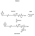
- FIG 4 Synthesis of Biotin-GS-DNP by the human glutathione S-transferase M2-2 (GST M2-2) catalysed reaction of 1-chloro-2,4-dinitrobenzene (CDNB; Sigma) with reduced biotinylated-glutathione (Biotin-GSH).
- GST M2-2 human glutathione S-transferase M2-2
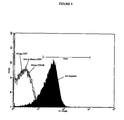
- FIG. 5 Detecting paramagnetic beads coated with the product of an enzyme catalysed reaction by flow cytometry.
- Sera-Mag TM streptavidin-coated magnetic microparticles incubated with Biotin-GS-DNP made by the GST M2-2 catalysed reaction of Biotin-GSH and CDNB.
- the captured Biotin-GS-DNP was detected by incubation of the microparticles with a mouse anti-dinitrophenol antibody followed by a (FITC)-conjugated F(ab') 2 fragment goat anti-mouse IgG, F(ab')2 fragment. After washing, 2 x 10 5 microparticles were analysed by flow cytometry.
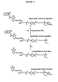
- FIG. 6 Synthesis of MeNPO-CO-Biotin- ⁇ -Ala-GSH (caged-biotin- ⁇ ala-GSH).
- Acetyl chloride (5 ml) was added to anhydrous methanol (80 ml). The stirred solution was allowed to cool down and d-biotin (4 g) was added. After over-night stirring the solvents were evaporated in vacuum to afford a white solid. The solid was triturated with ether, filtered and dried under vacuum (in the presence of phosphorus pentoxide) and stored at -20°C.
- FIG. 7 Reaction of caged-biotin- ⁇ ala-GSH with 1-chloro-2,4-dinitrobenzene (CDNB) and photochemical uncaging of the biotin group.
- CDNB 1-chloro-2,4-dinitrobenzene
- FIG. 8 Reaction of caged-biotin- ⁇ ala-GSH with 4-chloro-3-nitrobenzoate (CNB) and photochemical uncaging of the biotin group
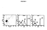
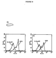
- Figure 9 Human GST M2-2 catalyses the reaction of caged-biolin- ⁇ ala-GSH with CDNB and CNB in solution and the reaction products can be uncaged by UV irradiation, captured on beads and detected using fluorescently labelled anti-product antibodies and flow cytometry.
- Panel A light scattering characteristics of beads and gate for single beads (R1).
- Panel B fluorescence from microbeads (gated through R1) from reactions with CDNB.
- Panel C fluorescence from microbeads (gated through R1) from reactions with CNB. Signals from microbeads from reactions with and without GST M2-2 are annotated +enz and -enz respectively. Signals from microbeads from reactions which were UV irradiated and those which were not are annotated +UV and -UV respectively.
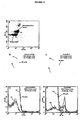
- FIG 10 Flow cytometry can be used to distinguish beads from aqueous compartments of an emulsion containing GST M2-2 from beads from compartments without GST M2-2 by using caged-biotinylated-,6Ala-GSH and CNB as substrates.
- Panel A light scattering characteristics of a mixture of a mixture of 1.0 ⁇ m diameter nonfluorescent neutravidin labelled microspheres (Molecular Probes, F-8777) or 0.93 ⁇ m diameter streptavidin-coated polystyrene beads (Bangs Laboratories) and gates set for single Bangs beads (R1) and single Molecular Probes beads (R2).
- Panel B fluorescence from microbeads taken from a non-emulsified mixture of 98% Bangs beads (without GST) and 2% Molecular Probes beads (with GST).
- Panel C fluorescence from microbeads taken from a mixture of two emulsions in a ratio of 98% emulsion containing Bangs beads (without GST) and an emulsion containing 2% Molecular Probes beads (with GST).
- Panel D fluorescence from microbeads taken from a non-emulsified mixture of 98% Molecular Probes beads (without GST) and 2% Bangs beads (with GST).
- Panel E fluorescence from microbeads taken from a mixture of two emulsions in a ratio of 98% emulsion containing Molecular Probes beads (without GST) and an emulsion containing 2% Bangs beads (with GST). Fluorescence of ungated beads (No gate), beads gated through R1 (R1) and beads gated through R2 (R2) are overlayed.
- FIG 11 Human GST M2-2 transcribed and translated in vitro in the aqueous compartments of a water-in oil emulsion catalyses a reaction which gives rise to a change in the fluorescence properties of co-compartmentalised microspheres.
- Panel A light scattering characteristics of beads and gate for single beads (R1).
- Panel B fluorescence from microbeads (gated through R1) from non-emulsified reactions.
- Panel C fluorescence from microbeads (gated through R1) emulsified reactions.
- Signals from microbeads from reactions with and without GSTM2-2.LMB2-3 DNA are annotated +DNA and -DNA respectively.
- Signals from microbeads from reactions with and without recombinant GST M2-2 are annotated +GST and -GST respectively.
- Figure 14 Preparation of protein conjugates of a PTE substrate and product for immunisation and ELISA.
- FIG. 15 PTE catalyses the reaction of EtNP-Bz-Glu-cagedBiotin in the presence of streptavidin-coated beads, and the reaction products uncaged by UV irradiation, are captured on beads and detected using fluorescently labelled anti-product antibodies and flow cytometry.
- Panel A light scattering characteristics of the beads and gate selected for single beads (R2).
- Panel B fluorescence from beads (gated through R2) from reactions with 10 ⁇ M EtNP-Bz-Glu-cagedBiotin in the presence of in vitro translated OPD.LMB3-2biotin DNA fragments (OPD) or M.Hae III. LMB3-2biotin DNA fragments (M.Hae III ).
- Panel C As B but with 20 ⁇ M EtNP-Bz-Glu-cagedBiotin.
- Panel D As B but with 50 ⁇ M EtNP-Bz-Glu-cagedBiotin.
- Figure 16 Reaction of EtNP-Bz-Glu-cagedBiotin in the presence of beads to which genetic elements encoding the phosphotriesterase tagged with the Flag peptide (N-Flag-OPD.LMB3-2biotin) or another enzyme (N-Flag-M.HaeIII.LMB3-2biotin) were attached alongside with an antibody that binds the Flag peptide.
- the beads were reacted and subsequently analysed by flow-cytometry as described in the text.
- Panel A light scattering characteristics of beads and gate for single beads (R1).
- Panel B fluorescence from microbeads (gated through R1) to which were attached N-Flag-OPD.LMB3-2biotin DNA fragments (OPD) or M.HaeIII.LMB3-2biotin DNA fragments (M. Hae III) from reactions with 12.5 ⁇ M EtNP-Bz-Glu-cagedBiotin.
- Panel C As B but with 25 ⁇ M EtNP-Bz-Glu-caged-Biotin.
- FIG. 17 E. coli BirA transcribed and translated in vitro catalyses a reaction which gives rise to a change in the fluorescence properties of substrate-labelled microspheres in the aqueous compartments of a water-in oil emulsion and in bulk solution.
- Figure 18 Flow cytometric analysis of samples prepared for the sorting experiment.
- FIG 19 Fluorescence-activated flow cytometric sorting of the genetic elements.
- Panel A Samples #1 to #4 before sorting and after sorting.
- Panel B Genes recovered from individual beads sorted from sample #3 sorted into a 96-well plate.
- Panel C Genes recovered from individual beads sorted from sample #4 sorted into a 96-well plate.
- DNA markers (M) are ⁇ X174-HaeIII digest.
- microcapsules of the present disclosure require appropriate physical properties to allow the working of the disclosure.
- the contents of each microcapsule are preferably isolated from the contents of the surrounding microcapsules, so that there is no or little exchange of the genetic elements and gene products between the microcapsules over the timescale of the experiment.
- the method of the present disclosure requires that there are only a limited number of genetic elements per microcapsule. This ensures that the gene product of an individual genetic element will be isolated from other genetic elements. Thus, coupling between genetic element and gene product will be highly specific. The enrichment factor is greatest with on average one or fewer genetic elements per microcapsule, the linkage between nucleic acid and the activity of the encoded gene product being as tight as is possible, since the gene product of an individual genetic element will be isolated from the products of all other genetic elements.
- a ratio of 5, 10, 50, 100 or 1000 or more genetic elements per microcapsule may prove beneficial in sorting a large library. Subsequent rounds of sorting, including renewed encapsulation with differing genetic element distribution, will permit more stringent sorting of the genetic elements.
- the formation and the composition of the microcapsules advantageously does not abolish the function of the machinery the expression of the genetic elements and the activity of the gene products.
- microencapsulation procedures are available (see Benita, 1996) and may be used to create the microcapsules used in accordance with the present invention.
- lipid vesicles liposomes
- non-ionic surfactant vesicles van Hal et al., 1996.
- lipid vesicles liposomes
- van Hal et al., 1996 closed-membranous capsules of single or multiple bilayers of non-covalently assembled molecules, with each bilayer separated from its neighbour by an aqueous compartment.
- liposomes the membrane is composed of lipid molecules; these are usually phospholipids but sterols such as cholesterol may also be incorporated into the membranes (New, 1990).
- RNA and DNA polymerisation can be performed within liposomes (Chakrabarti et al., 1994; Oberholzer et al., 1995a; Oberholzer et al., 1995b; Walde et al., 1994; Wick & Luisi, 1996).
- aqueous phase With a membrane-enveloped vesicle system much of the aqueous phase is outside the vesicles and is therefore non-compartmentalised. This continuous, aqueous phase is removed or the biological systems in it inhibited or destroyed (for example, by digestion of nucleic acids with DNase or RNase) in order that the reactions are limited to the microcapsules (Luisi et al., 1987).
- Enzyme-catalysed biochemical reactions have also been demonstrated in microcapsules generated by a variety of other methods. Many enzymes are active in reverse micellar solutions (Bru & Walde, 1991; Bru & Walde, 1993; Creagh et al., 1993; Haber et al., 1993; Kumar et al., 1989; Luisi & B., 1987; Mao & Walde, 1991; Mao et al., 1992; Perez et al., 1992; Walde et al., 1994; Walde et al., 1993; Walde et al., 1988) such as the AOT-isooctane-water system (Menger & Yamada, 1979).
- Microcapsules can also be generated by interfacial polymerisation and interfacial complexation (Whateley, 1996). Microcapsules of this sort can have rigid, nonpermeable membranes, or semipermeable membranes. Semipermeable microcapsules bordered by cellulose nitrate membranes, polyamide membranes and lipid-polyamide membranes can all support biochemical reactions, including multienzyme systems (Chang, 1987; Chang, 1992; Lim, 1984). Alginate/polylysine microcapsules (Lim & Sun, 1980), which can be formed under very mild conditions, have also proven to be very biocompatible, providing, for example, an effective method of encapsulating living cells and tissues (Chang, 1992; Sun et al., 1992).
- Non-membranous microencapsulation systems based on phase partitioning of an aqueous environment in a colloidal system, such as an emulsion, may also be used.
- the microcapsules of the present invention are formed from emulsions; heterogeneous systems of two immiscible liquid phases with one of the phases dispersed in the other as droplets of microscopic or colloidal size (Becher, 1957; Sherman, 1968; Lissant, 1974; Lissant, 1984).
- Emulsions may be produced from any suitable combination of immiscible liquids.
- the emulsion of the present invention has water (containing the biochemical components) as the phase present in the form of finely divided droplets (the disperse, internal or discontinuous phase) and a hydrophobic, immiscible liquid (an oil') as the matrix in which these droplets are suspended (the nondisperse, continuous or external phase).
- water-in-oil W/O
- This has the advantage that the entire aqueous phase containing the biochemical components is compartmentalised in discreet droplets (the internal phase).
- the external phase being a hydrophobic oil, generally contains none of the biochemical components and hence is inert.
- the emulsion may be stabilised by addition of one or more surface-active agents (surfactants).
- surfactants are termed emulsifying agents and act at the water/oil interface to prevent (or at least delay) separation of the phases.
- Many oils and many emulsifiers can be used for the generation of water-in-oil emulsions; a recent compilation listed over 16,000 surfactants, many of which are used as emulsifying agents (Ash and Ash, 1993). Suitable oils include light white mineral oil and non-ionic surfactants (Schick, 1966) such as sorbitan monooleate (Span TM 80; ICI) and polyoxyethylenesorbitan monooleate (Tween TM 80; ICI).
- anionic surfactants may also be beneficial.
- Suitable surfactants include sodium cholate and sodium taurocholate. Particularly preferred is sodium deoxycholate, preferably at a concentration of 0.5% w/v, or below. Inclusion of such surfactants can in some cases increase the expression of the genetic elements and/or the activity of the gene products. Addition of some anionic surfactants to a non-emulsified reaction mixture completely abolishes translation. During emulsification, however, the surfactant is transferred from the aqueous phase into the interface and activity is restored. Addition of an anionic surfactant to the mixtures to be emulsified ensures that reactions proceed only after compartmentalisation.
- stirrers such as magnetic stir-bars, propeller and turbine stirrers, paddle devices and whisks
- homogenisers including rotor-stator homogenisers, high-pressure valve homogenisers and jet homogenisers
- colloid mills ultrasound and 'membrane emulsification' devices
- Aqueous microcapsules formed in water-in-oil emulsions are generally stable with little if any exchange of genetic elements or gene products between microcapsules. Additionally, we have demonstrated that several biochemical reactions proceed in emulsion microcapsules. Moreover, complicated biochemical processes, notably gene transcription and translation are also active in emulsion microcapsules. The technology exists to create emulsions with volumes all the way up to industrial scales of thousands of litres (Becher, 1957; Sherman, 1968; Lissant, 1974; Lissant, 1984).
- the preferred microcapsule size will vary depending upon the precise requirements of any individual selection process that is to be performed according to the present invention. In all cases, there will be an optimal balance between gene library size, the required enrichment and the required concentration of components in the individual microcapsules to achieve efficient expression and reactivity of the gene products.
- the mean volume of the microcapsules is less that 5.2 x 10 -16 m 3 , (corresponding to a spherical microcapsule of diameter less than 10 ⁇ m, more preferably less than 6.5 x 10 -17 m 3 ,(5 ⁇ m diameter), more preferably about 4.2 x 10 -18 m 3 (2 ⁇ m diameter) and ideally about 9 x 10 -18 m 3 (2.6 ⁇ m diameter).
- the effective DNA or RNA concentration in the microcapsules may be artificially increased by various methods that will be well-known to those versed in the art. These include, for example, the addition of volume excluding chemicals such as polyethylene glycols (PEG) and a variety of gene amplification techniques, including transcription using RNA polymerases including those from bacteria such as E. coli (Roberts, 1969; Blattner and Dahlberg, 1972; Roberts et al., 1975; Rosenberg et al. , 1975) , eukaryotes e. g. (Weil et al.
- PEG polyethylene glycols
- thermostable for example, the coupled transcription-translation systems can be made from a thermostable organism such as Thermus aquaticus).
- microcapsule volume of about 5.2 x 10- 16 m 3 (corresponding to a sphere of diameter 10 ⁇ m).
- the microcapsule size is preferably sufficiently large to accommodate all of the required components of the biochemical reactions that are needed to occur within the microcapsule. For example, in vitro, both transcription reactions and coupled transcription-translation reactions require a total nucleoside triphosphate concentration of about 2mM.
- RNA molecules of nucleoside triphosphate per microcapsule 8.33 x 10 -22 moles.
- this number of molecules is contained within a microcapsule of volume 4.17 x 10 -19 litres (4.17 x 10 -22 m 3 which if spherical would have a diameter of 93nm.
- the ribosomes necessary for the translation to occur are themselves approximately 20nm in diameter.
- the preferred lower limit for microcapsules is a diameter of approximately 0.1 ⁇ m (100nm).
- the microcapsule volume is preferably of the order of between 5.2 x 10 -22 m 3 and 5.2 x 10 -16 m 3 corresponding to a sphere of diameter between 0.1 ⁇ m and 10 ⁇ m, more preferably of between about 5.2 x 10 -19 m3 and 6.5 x 10 -17 m 3 (1 ⁇ m and 5 ⁇ m). Sphere diameters of about 2.6 ⁇ m are most advantageous.
- compartments droplets of 2.6 ⁇ m mean diameter
- Escherichia are 1.1-1.5 x 2.0-6.0 ⁇ m rods
- Azotobacter are 1.5-2.0 ⁇ m diameter ovoid cells.
- Darwinian evolution is based on a 'one genotype one phenotype' mechanism.
- the concentration of a single compartmentalised gene, or genome drops from 0.4 nM in a compartment of 2 ⁇ m diameter, to 25 pM in a compartment of 5 ⁇ m diameter.
- the prokaryotic transcription/translation machinery has evolved to operate in compartments of ⁇ 1-2 ⁇ m diameter, where single genes are at approximately nanomolar concentrations.
- a single gene, in a compartment of 2.6 ⁇ m diameter is at a concentration of 0.2 nM. This gene concentration is high enough for efficient translation. Compartmentalisation in such a volume also ensures that even if only a single molecule of the gene product is formed it is present at about 0.2 nM, which is important if the gene product is to have a modifying activity of the genetic element itself.
- the volume of the microcapsule is thus selected bearing in mind not only the requirements for transcription and translation of the genetic element, but also the modifying activity required of the gene product in the method of the disclosure.
- the size of emulsion microcapsules may be varied simply by tailoring the emulsion conditions used to form the emulsion according to requirements of the selection system.
- the size of the microcapsules is selected not only having regard to the requirements of the transcription/translation system, but also those of the selection system employed for the genetic element.
- the components of the selection system such as a chemical modification system, may require reaction volumes and/or reagent concentrations which are not optimal for transcription/translation.
- such requirements may be accommodated by a secondary re-encapsulation step; moreover, they may be accommodated by selecting the microcapsule size in order to maximise transcription/translation and selection as a whole.
- Empirical determination of optimal microcapsule volume and reagent concentration is preferred.
- a “genetic element” in accordance with the present invention is as described above.
- a genetic element is a molecule or construct selected from the group consisting of a DNA molecule, an RNA molecule, a partially or wholly artificial nucleic acid molecule consisting of exclusively synthetic or a mixture of naturally-occurring and synthetic bases, any one of the foregoing linked to a polypeptide, and any one of the foregoing linked to any other molecular group or construct.
- the other molecular group or construct may be selected from the group consisting of nucleic acids, polymeric substances, particularly beads, for example polystyrene beads, and magnetic or paramagnetic substances such as magnetic or paramagnetic beads.
- the nucleic acid portion of the genetic element may comprise suitable regulatory sequences, such as those required for efficient expression of the gene product, for example promoters, enhancers, translational initiation sequences, polyadenylation sequences, splice sites and the like.
- the polypeptide or other molecular group or construct is a ligand or a substrate which directly or indirectly binds to or reacts with the gene product in order to alter the optical properties of the genetic element. This allows the sorting of the genetic element on the basis of the activity of the gene product.
- the ligand or substrate can be connected to the nucleic acid by a variety of means that will be apparent to those skilled in the art (see, for example, Hermanson, 1996).
- nucleic acid molecule may be linked to a ligand or substrate is through biotinylation. This can be done by PCR amplification with a 5'-biotinylation primer such that the biotin and nucleic acid are covalently linked.
- the ligand or substrate can be attached to the modified nucleic acid by a variety of means that will be apparent to those of skill in the art (see, for example, Hermanson, 1996).
- a biotinylated nucleic acid may be coupled to a polystyrene or paramagnetic microbead (0.02 to approx. 5.0 ⁇ m in diameter) that is coated with avidin or streptavidin, that will therefore bind the nucleic acid with very high affinity.
- This bead can be derivatised with substrate or ligand by any suitable method such as by adding biotinylated substrate or by covalent coupling.
- a biotinylated nucleic acid may be coupled to avidin or streptavidin complexed to a large protein molecule such as thyroglobulin (669 Kd) or ferritin (440 Kd).
- This complex can be derivatised with substrate or ligand, for example by covalent coupling to the ⁇ -amino group of lysines or through a non-covalent interaction such as biotin-avidin.
- the substrate may be present in a form unlinked to the genetic element but containing an inactive "tag” that requires a further step to activate it such as photoactivation (e.g. of a "caged" biotin analogue, (Sundberg et al., 1995; Pirrung and Huang, 1996)).
- the catalyst to be selected then converts the substrate to product.
- the "tag” is then activated and the "tagged" substrate and/or product bound by a tag-binding molecule (e.g. avidin or streptavidin) complexed with the nucleic acid.
- a tag-binding molecule e.g. avidin or streptavidin
- an alternative is to couple the nucleic acid to a product-specific antibody (or other product-specific molecule).
- the substrate or one of the substrates
- the substrate is present in each microcapsule unlinked to the genetic element, but has a molecular "tag” (for example biotin, DIG or DNP or a fluorescent group).
- a molecular "tag” for example biotin, DIG or DNP or a fluorescent group.
- Isolation refers to the process of separating an entity from a heterogeneous population, for example a mixture, such that it is free of at least one substance with which it was associated before the isolation process. In a preferred embodiment, isolation refers to purification of an entity essentially to homogeneity. Sorting of an entity refers to the process of preferentially isolating desired entities over undesired entities. In as far as this relates to isolation of the desired entities, the terms “isolating” and “sorting” are equivalent.
- the method of the present disclosure permits the sorting of desired genetic elements from pools (libraries or repertoires) of genetic elements which contain the desired genetic element. Selecting is used to refer to the process (including the sorting process) of isolating an entity according to a particular property thereof.
- the method of the present disclosure is useful for sorting libraries of genetic elements.
- the disclosure accordingly provides a method according to preceding aspects of the disclosure, wherein the genetic elements are isolated from a library of genetic elements encoding a repertoire of gene products.
- library the terms "library”, “repertoire” and “pool” are used according to their ordinary signification in the art, such that a library of genetic elements encodes a repertoire of gene products.
- libraries are constructed from pools of genetic elements and have properties which facilitate sorting.
- Libraries can also be made by introducing mutations into a genetic element or pool of genetic elements 'randomly' by a variety of techniques in vivo, including; using mutator strains of bacteria such as E. coli mutD5 (Liao et al., 1986; Yamagishi et al., 1990; Low et al., 1996); using the antibody hypermutation system of B-lymphocytes (Yelamos et al., 1995).
- mutator strains of bacteria such as E. coli mutD5 (Liao et al., 1986; Yamagishi et al., 1990; Low et al., 1996)
- B-lymphocytes Yamagishi et al., 1995.
- Random mutations can also be introduced both in vivo and in vitro by chemical mutagens, and ionising or UV irradiation (see Friedberg et al., 1995), or incorporation of mutagenic base analogues (Freese, 1959; Zaccolo et al., 1996). Random' mutations can also be introduced into genes in vitro during polymerisation for example by using error-prone polymerases (Leung et al., 1989).
- Mutations may be introduced into the genetic elements(s) as set forth above.
- the genetic elements according to the disclosure advantageously encode enzymes, preferably of pharmacological or industrial interest, activators or inhibitors, especially of biological systems, such as cellular signal transduction mechanisms, antibodies and fragments thereof, and other binding agents (e.g. transcription factors) suitable for diagnostic and therapeutic applications.
- enzymes preferably of pharmacological or industrial interest
- activators or inhibitors especially of biological systems, such as cellular signal transduction mechanisms, antibodies and fragments thereof, and other binding agents (e.g. transcription factors) suitable for diagnostic and therapeutic applications.
- binding agents e.g. transcription factors
- encapsulation conditions are desirable. Depending on the complexity and size of the library to be screened, it may be beneficial to set up the encapsulation procedure such that 1 or less than 1 genetic element is encapsulated per microcapsule. This will provide the greatest power of resolution. Where the library is larger and/or more complex, however, this may be impracticable; it may be preferable to encapsulate several genetic elements together and rely on repeated application of the method of the disclosure to achieve sorting of the desired activity. A combination of encapsulation procedures may be used to obtain the desired enrichment.
- a repertoire size of at least 10 11 can be selected using 1ml aqueous phase in a 20 ml emulsion.
- the microcapsules according to the disclosure will comprise further components required for the sorting process to take place.
- Other components of the system will for example comprise those necessary for transcription and/or translation of the genetic element. These are selected for the requirements of a specific system from the following; a suitable buffer, an in vitro transcription/replication system and/or an in vitro translation system containing all the necessary ingredients, enzymes and cofactors, RNA polymerase, nucleotides, nucleic acids (natural or synthetic), transfer RNAs, ribosomes and amino acids, and the substrates of the reaction of interest in order to allow selection of the modified gene product.
- a suitable buffer will be one in which all of the desired components of the biological system are active and will therefore depend upon the requirements of each specific reaction system. Buffers suitable for biological and/or chemical reactions are known in the art and recipes provided in various laboratory texts, such as Sambrook et al., 1989.
- the in vitro translation system will usually comprise a cell extract, typically from bacteria (Zubay, 1973; Zubay, 1980; Lesley et al., 1991; Lesley, 1995), rabbit reticulocytes (Pelham and Jackson, 1976), or wheat germ (Anderson et al., 1983).
- a cell extract typically from bacteria (Zubay, 1973; Zubay, 1980; Lesley et al., 1991; Lesley, 1995), rabbit reticulocytes (Pelham and Jackson, 1976), or wheat germ (Anderson et al., 1983).
- Many suitable systems are commercially available (for example from Promega) including some which will allow coupled transcription/translation (all the bacterial systems and the reticulocyte and wheat germ TNT TM extract systems from Promega).
- the mixture of amino acids used may include synthetic amino acids if desired, to increase the possible number or variety of proteins produced in the library. This can be accomplished by charging tRNAs with artificial amino acids and using these tRNAs for the in vitro translation of the proteins
- the enrichment of the pool of genetic elements for those encoding the molecules of interest can be assayed by non-compartmentalised in vitro transcription/replication or coupled transcription-translation reactions.
- the selected pool is cloned into a suitable plasmid vector and RNA or recombinant protein is produced from the individual clones for further purification and assay.
- the internal environment of a microcapsule may be altered by addition of reagents to the oil phase of the emulsion.
- the reagents diffuse through the oil phase to the aqueous microcapsule environment.
- the reagents are at least partly water-soluble, such that a proportion thereof is distributed from the oil phase to the aqueous microcapsule environment.
- the reagents are substantially insoluble in the oil phase.
- Reagents are preferably mixed into the oil phase by mechanical mixing, for example vortexing.
- the reagents which may be added via the oil phase include substrates, buffering components, factors and the like.
- the internal pH of microcapsules may be altered in situ by adding acidic or basic components to the oil phase.
- the disclosure moreover relates to a method for producing a gene product, once a genetic element encoding the gene product has been sorted by the method of the disclosure.
- the genetic element itself may be directly expressed by conventional means to produce the gene product.
- alternative techniques may be employed, as will be apparent to those skilled in the art.
- the genetic information incorporated in the gene product may be incorporated into a suitable expression vector, and expressed therefrom.
- gene product encoding nucleic acid is incorporated into a vector, and introduced into suitable host cells to produce transformed cell lines that express the gene product.
- the resulting cell lines can then be produced for reproducible qualitative and/or quantitative analysis of the effect(s) of potential drugs affecting gene product function.
- gene product expressing cells may be employed for the identification of compounds, particularly small molecular weight compounds, which modulate the function of gene product.
- host cells expressing gene product are useful for drug screening and it is a further object of the present disclosure to provide a method for identifying compounds which modulate the activity of the gene product, said method comprising exposing cells containing heterologous DNA encoding gene product, wherein said cells produce functional gene product, to at least one compound or mixture of compounds or signal whose ability to modulate the activity of said gene product is sought to be determined, and thereafter monitoring said cells for changes caused by said modulation.
- modulators such as agonists, antagonists and allosteric modulators
- a compound or signal that modulates the activity of gene product refers to a compound that alters the activity of gene product in such a way that the activity of the gene product is different in the presence of the compound or signal (as compared to the absence of said compound or signal).
- Cell-based screening assays can be designed by constructing cell lines in which the expression of a reporter protein, i.e. an easily assayable protein, such as ⁇ -galactosidase, chloramphenicol acetyltransferase (CAT), green fluorescent protein (GFP) or luciferase, is dependent on gene product.
- a reporter protein i.e. an easily assayable protein, such as ⁇ -galactosidase, chloramphenicol acetyltransferase (CAT), green fluorescent protein (GFP) or luciferase
- CAT chloramphenicol acetyltransferase
- GFP green fluorescent protein
- luciferase luciferase
- the present disclosure also provides a method to exogenously affect gene product dependent processes occurring in cells.
- Recombinant gene product producing host cells e.g. mammalian cells
- the disclosure relates to a method for optimising a production process which involves at least one step which is facilitated by a polypeptide.
- the step may be a catalytic step, which is facilitated by an enzyme.
- the disclosure provides a method for preparing a compound or compounds comprising the steps of:
- enzymes involved in the preparation of a compound may be optimised by selection for optimal activity.
- the procedure involves the preparation of variants of the polypeptide to be screened, which equate to a library of polypeptides as refereed to herein.
- the variants may be prepared in the same manner as the libraries discussed elsewhere herein.
- the system can be configured to select for RNA, DNA or protein gene product molecules with catalytic, regulatory or binding activity.
- the genetic element may be linked to the gene product in the microcapsule via the ligand. Only gene products with affinity for the ligand will therefore bind to the genetic element and only those genetic elements with gene product bound via the ligand will acquire the changed optical properties which enable them to be retained in the selection step.
- the genetic element will thus comprise a nucleic acid encoding the gene product linked to a ligand for the gene product.
- the change in optical properties of the genetic element after binding of the gene product to the ligand may be induced in a variety of ways, including:
- All the gene products to be selected can contain a putative binding domain, which is to be selected for, and a common feature - a tag.
- the genetic element in each microcapsule is physically linked to the ligand. If the gene product produced from the genetic element has affinity for the ligand, it will bind to it and become physically linked to the same genetic element that encoded it, resulting in the genetic element being 'tagged'. At the end of the reaction, all of the microcapsules are combined, and all genetic elements and gene products pooled together in one environment.
- Genetic elements encoding gene products exhibiting the desired binding can be selected by adding reagents which specifically bind to, or react specifically with, the "tag” and thereby induce a change in the optical properties of the genetic element allowing there sorting.
- reagents which specifically bind to, or react specifically with, the "tag” For example, a fluorescently-labelled anti-"tag” antibody can be used, or an anti-"tag” antibody followed by a second fluorescently labelled antibody which binds the first.
- genetic elements may be sorted on the basis that the gene product, which binds to the ligand, merely hides the ligand from, for example, further binding partners which would otherwise modify the optical properties of the genetic element. In this case genetic elements with unmodified optical properties would be selected.
- the disclosure provides a method according to the first aspect of the disclosure, wherein in step (b) the gene products bind to genetic elements encoding them.
- the gene products together with the attached genetic elements are then sorted as a result of binding of a ligand to gene products having the desired binding activity.
- all gene products can contain an invariant region which binds covalently or non-covalently to the genetic element, and a second region which is diversified so as to generate the desired binding activity.
- the ligand for the gene product is itself encoded by the genetic element and binds to the genetic element.
- the genetic element encodes two (or indeed more) gene products, at least one of which binds to the genetic element, and which can potentially bind each other. Only when the gene products interact in a microcapsule is the genetic element modified in a way that ultimately results in a change in a change in its optical properties that enables it to be sorted. This embodiment, for example, isused to search gene libraries for pairs of genes encoding pairs of proteins which bind each other.
- TSA TM Tyramide Signal Amplification
- TSA may be configured such that it results in a direct increase in the fluorescence of the genetic element, or such that a ligand is attached to the genetic element which is bound by a second fluorescent molecule, or a sequence of molecules, one or more of which is fluorescent.
- the genetic element in each microcapsule may comprise the substrate of the reaction. If the genetic element encodes a gene product capable of acting as a catalyst, the gene product will catalyse the conversion of the substrate into the product. Therefore, at the end of the reaction the genetic element is physically linked to the product of the catalysed reaction.
- the substrate may also be desirable, in some cases, for the substrate not to be a component of the genetic element.
- the substrate would contain an inactive "tag” that requires a further step to activate it such as photoactivation (e.g. of a "caged” biotin analogue, (Sundberg et al., 1995; Pirrung and Huang, 1996)).
- the catalyst to be selected then converts the substrate to product.
- the "tag” is then activated and the "tagged" substrate and/or product bound by a tag-binding molecule (e.g. avidin or streptavidin) complexed with the nucleic acid.
- a tag-binding molecule e.g. avidin or streptavidin
- optical properties of genetic elements with product attached and which encode gene products with the desired catalytic activity can be modified by either:
- the pooled genetic elements encoding catalytic molecules can then be enriched by selecting for the genetic elements with modified optical properties.
- an alternative is to couple the nucleic acid to a product-specific antibody (or other product-specific molecule).
- the substrate or one of the substrates
- the substrate is present in each microcapsule unlinked to the genetic element, but has a molecular "tag” (for example biotin, DIG or DNP or a fluorescent group).
- a molecular "tag” for example biotin, DIG or DNP or a fluorescent group.
- the genetic elements encoding active enzymes will be "tagged” and may already have changed optical properties, for example, if the "tag” was a fluorescent group.
- a change in optical properties of "tagged” genes can be induced by adding a fluorescently labelled ligand which binds the "tag” (for example fluorescently-labelled avidin/streptavidin, an anti"tag” antibody which is fluorescent, or a non-fluorescent anti-"tag” antibody which can be detected by a second fluorescently-labelled antibody).
- selection may be performed indirectly by coupling a first reaction to subsequent reactions that takes place in the same microcapsule.
- the product of the first reaction is reacted with, or bound by, a molecule which does not react with the substrate of the first reaction.
- a second, coupled reaction will only proceed in the presence of the product of the first reaction.
- a genetic element encoding a gene product with a desired activity can then be purified by using the properties of the product of the second reaction to induce a change in the optical properties of the genetic element as above.
- the product of the reaction being selected may be the substrate or cofactor for a second enzyme-catalysed reaction.
- the enzyme to catalyse the second reaction can either be translated in situ in the microcapsules or incorporated in the reaction mixture prior to microencapsulation. Only when the first reaction proceeds will the coupled enzyme generate a product which can be used to induce a change in the optical properties of the genetic element as above.
- This concept of coupling can be elaborated to incorporate multiple enzymes, each using as a substrate the product of the previous reaction. This allows for selection of enzymes that will not react with an immobilised substrate. It can also be designed to give increased sensitivity by signal amplification if a product of one reaction is a catalyst or a cofactor for a second reaction or series of reactions leading to a selectable product (for example, see Johannsson and Bates, 1988; Johannsson, 1991). Furthermore an enzyme cascade system can be based on the production of an activator for an enzyme or the destruction of an enzyme inhibitor (see Mize et al., 1989). Coupling also has the advantage that a common selection system can be used for a whole group of enzymes which generate the same product and allows for the selection of complicated chemical transformations that cannot be performed in a single step.
- Such a method of coupling thus enables the evolution of novel "metabolic pathways" in vitro in a stepwise fashion, selecting and improving first one step and then the next.
- the selection strategy is based on the final product of the pathway, so that all earlier steps can be evolved independently or sequentially without setting up a new selection system for each step of the reaction.
- Genetic elements encoding enzymes with substrate specificity or selectivity can be specifically enriched by carrying out a positive selection for reaction with one substrate and a negative selection for reaction with another substrate.
- Such combined positive and negative selection pressure should be of great importance in isolating regio-selective and stereo-selective enzymes (for example, enzymes that can distinguish between two enantiomers of the same substrate).
- two substrates e.g. two different enantiomers
- tags e.g. two different fluorophores
- the substrate specificity of the enzyme can be determined from the optical properties of the genetic element and those genetic elements encoding gene products with the wrong (or no) specificity rejected.
- Tags conferring no change in optical activity can also be used if tag-specific ligands with different optical properties are added (e.g. tag-specific antibodies labelled with different fluorophores).
- a similar system can be used to select for regulatory properties of enzymes.
- the components of the biochemical process can either be translated in situ in each microcapsule or can be incorporated in the reaction mixture prior to microencapsulation. If the genetic element being selected is to encode an activator, selection can be performed for the product of the regulated reaction, as described above in connection with catalysis. If an inhibitor is desired, selection can be for a chemical property specific to the substrate of the regulated reaction.
- the gene product binds back to the genetic element, for example through a common element of the gene product which binds to a ligand which is part of the genetic element. After pooling the genetic elements they can then be sorted using the optical properties of the bound gene products.
- This embodiment can be used, for example, to select variants of green fluorescent protein (GFP) (Cormack et al., 1996; Delagrave et al., 1995; Ehrig et al., 1995), with improved fluorescence and/or novel absoption and emmission spectra.
- GFP green fluorescent protein
- the genetic elements will be sorted by flow cytometry.
- a variety of optical properties can be used to trigger sorting, including light scattering (Kerker, 1983) and fluorescence polarisation (Rolland et al., 1985).
- the difference in optical properties of the genetic elements will be a difference in fluorescence and the genetic elements will be sorted using a fluorescence activated cell sorter (Norman, 1980; Mackenzie and Pinder, 1986), or similar device.
- the genetic element comprises of a nonfluorescent nonmagnetic (e.g. polystyrene) or paramagnetic microbead (see Fornusek and Vetvicka, 1986), optimally 0.6 to 1.0 ⁇ m diameter, to which are attached both the gene and the groups involved in generating a fluorescent signal:
- the selection procedure may comprise two or more steps.
- transcription/replication and/or translation of each genetic element of a genetic element library may take place in a first microcapsule.
- Each gene product is then linked to the genetic element which encoded it (which resides in the same microcapsule), for example via a gene product-specific ligand such as an antibody.
- the microcapsules are then broken, and the genetic elements attached to their respective gene products optionally purified.
- genetic elements can be attached to their respective gene products using methods which do not rely on encapsulation.
- phage display Smith, G.P.,1985), polysome display (Mattheakkis et al., 1994), RNA-peptide fusion (Roberts and Szostak, 1997) or lac repressor peptide fusion (Cull, et al., 1992).
- each purified genetic element attached to its gene product is put into a second microcapsule containing components of the reaction to be selected. This reaction is then initiated. After completion of the reactions, the microcapsules are again broken and the modified genetic elements are selected.
- one or more intervening steps may be performed between the initial step of creation and linking of gene product to genetic element, and the final step of generating the selectable change in the genetic element.
- release of the gene product from the genetic element within a secondary microcapsule can be achieved in a variety of ways, including by specific competition by a low-molecular weight product for the binding site or cleavage of a linker region joining the binding domain of the gene product from the catalytic domain either enzymatically (using specific proteases) or autocatalytically (using an integrin domain).
- the system can be configured such that the desired binding, catalytic or regulatory activity encoded by a genetic element leads, directly or indirectly to the activation of expression of a "reporter gene" that is present in all microcapsules. Only gene products with the desired activity activate expression of the reporter gene. The activity resulting from reporter gene expression allows the selection of the genetic element (or of the compartment containing it) by any of the methods described herein.
- activation of the reporter gene may be the result of a binding activity of the gene product in a manner analogous to the "two hybrid system" (Fields and Song, 1989).
- Activation can also result from the product of a reaction catalysed by a desirable gene product.
- the reaction product can be a transcriptional inducer of the reporter gene.
- arabinose may be used to induce transcription from the araBAD promoter.
- the activity of the desirable gene product can also result in the modification of a transcription factor, resulting in expression of the reporter gene.
- the desired gene product is a kinase or phosphatase the phosphorylation or dephosphorylation of a transcription factor may lead to activation of reporter gene expression.
- the method comprises the further step of amplifying the genetic elements.
- Selective amplification may be used as a means to enrich for genetic elements encoding the desired gene product.
- genetic material comprised in the genetic elements may be amplified and the process repeated in iterative steps.
- Amplification may be by the polymerase chain reaction (Saiki et al., 1988) or by using one of a variety of other gene amplification techniques including; Qb replicase amplification (Cahill, Foster and Mahan, 1991; Chetverin and Spirin, 1995; Katanaev, Kumasov and Spirin, 1995); the ligase chain reaction (LCR) (Landegren et al., 1988; Barany, 1991); the self-sustained sequence replication system (Fahy, Kwoh and Gingeras, 1991 ) and strand displacement amplification (Walker et al., 1992).
- LCR ligase chain reaction
- Enzymes can be expressed from genes in solution and genes attached to paramagnetic microbeads with identical efficiency.
- One format for the selection of genetic elements by using a change in their optical properties is one in which the genetic element comprises a microbead to which the gene is attached.
- the genetic element comprises a microbead to which the gene is attached.
- a gene for an enzyme E. coli dihydrofolate reductase
- the E. coli folA gene encoding dihydrofolate reductase (DHFR) is PCR-amplified using oligonucleotides EDHFRFo and EDHFRBa. This DNA is then cloned into the pGEM-4Z vector (Promega) digested with HindIII and Kpnl downstream of the lac promoter and the T7 RNA polymerase promoter.
- the oligonucleotide EDHFRBa appends the efficient phage T7 gene 10 translational start site upstream of the DHFR start codon.
- DNA sequencing identifies a clone which has the correct nucleotide sequence.
- Bacteria transformed with this clone (pGEM-folA) are found to over-express active DHFR (driven from the lac promoter) when induced with IPTG.
- the folA gene in pGEM-folA plasmid is then PCR-amplified using primers folA-FW and fo1A-BW, the resulting DNA fragment in HindIII and XhoI digested and subcloned into Hindlll/XhoI- digested pET23a expression vector (Novagen) to give construct pET23a/folA.
- the sequence of PCR-amplified folA gene was verified by sequencing.
- pET23a/folA was further amplified with 5'-biotinylated primers pETfor.b and pETrev.b and radio-labelled by including 10 ⁇ Ci ⁇ 35 S-dATP (Amersham Pharmacia Biotech, U.K.) in the PCR mix.
- the resulting 1765 bp double biotinylated fragment T7-folA was gel purified using a Qiagen kit and quantified spectrophotometrically.
- the specific activity of the product was 210000 CPM/pmol T7-folA DNA, as measured on the Beckman LS6000SC scintillation counter.
- the DNA fragment is bound to streptavidin-paramagnetic beads (0.74 ⁇ m diameter Sera-Mag beads, biotin-binding capacity 46nmol/mg, Seradyn, USA), partially precoated with biotinylated protein A (Sigma). 2 ⁇ l of 80 ⁇ M biotinylated protein A is added to 100 ⁇ l (1 mg) beads, allowed to bind at room temperature for 1 hour, washed once and coated for one hour at room temperature with rabbit IgG (10 ⁇ l 1 mg/ml antibody per 1 mg beads in TBS/0.1% Tween-20 (TBST)).
- streptavidin-paramagnetic beads 0.74 ⁇ m diameter Sera-Mag beads, biotin-binding capacity 46nmol/mg, Seradyn, USA
- biotinylated protein A Sigma. 2 ⁇ l of 80 ⁇ M biotinylated protein A is added to 100 ⁇ l (1 mg) beads, allowed to bind at room temperature for 1 hour, washed once and
- T7-folA DNA was added and allowed to bind for 1 hour at room temperature.
- the amount of bound T7-folA DNA was calculated by counting the radioactivity bound to an aliquot of beads. ⁇ 50% of the total DNA was bound.
- DNA fragments bound on beads or unbound DNA fragment are added directly to the S30 Extract System. Reactions are incubated for 2 hours at 37 °C.
- Dihydrofolate reductase activity is assayed by spectrophotometrically monitoring the oxidation of NADPH to NADP at 340nm over a 10 minute time course as described by (Williams et al., 1979; Ma et al., 1993). 2 ⁇ l of each quenched in vitro translation reaction is added to 150 ⁇ l Buffer A (100 mM Imidazole, pH 7.0, 10 mM ⁇ -mercaptoethanol) and 20 ⁇ l 1 mM NADPH. 20 ⁇ l dihydrofolate (1mM)(H 2 F) is added after 1 minute and the reaction monitored at 340nm using a ThermoMax microplate reader (Molecular Devices). Activity is calculated by initial velocities under So>>K M conditions (umax).
- a fluorescent protein can be translated in vitro from genes attached to single microbeads encapsulated in the aqueous compartments of a water-in-oil emulsion and the translated gene-product bound back to the microbeads making them fluorescent.
- One format for the selection of genetic elements is where the genetic element comprises a gene linked to a microbead and the product is coupled back onto the microbead within the microcapsule resulting directly, or indirectly, in a change in the optical properties of the microbead which allows it to be sorted.
- a fluorescent protein green fluorescent protein or GFP
- GFP green fluorescent protein
- the GFP in pBS/GFP6 plasmid was PCR-amplified using primers GFP-FW and GFP-BW, the resulting DNA fragment in HindIII and Xhol digested and subcloned into HindIII/XhoI- digested pET23a expression vector (Novagen) to give construct pET23a/GFP.
- the sequence of PCR-amplified GFP gene was verified by sequencing.
- pET23a/GFP was further amplified with 5'-biotinylated primers pETfor.b and pETrev.b.
- the resulting 2038 bp double biotinylated fragment T7-GFP was gel purified using a Qiagen kit and quantified spectrophotometrically. 10 nM and 1 nM dilutions of this DNA were made in 1 mg/ml HindIII digested lambda DNA to eliminate non-specific binding to the plastic.).
- This PCR fragment was used thereafter to program a prokaryotic in vitro coupled transcription/translation system designed for linear templates (Lesley, Brow and Burgess, 1991). A commercial preparation of this system is used (E. coli S30 Extract System for Linear Templates; Promega) supplemented with T7 RNA polymerase (10 3 units).
- a biotinylated 1765bp DNA fragment T7-folA (synthesised by PCR as in example 1) was used to program the synthesis of the non-fluorescent protein DHFR.
- ProActive streptavidin-coated paramagnetic beads (Bangs Laboratories, 2 ⁇ 10 7 beads/pl) were suspended in 5mM Tris 7.4/1M NaCl/0.1% Tween20 and split into three aliquots of 50 ⁇ l.
- T7-folA or T7-GFP 0.5 ⁇ l of 0.2 ⁇ M DNA (T7-folA or T7-GFP) was added to each aliquot of beads, incubated at 43°C for 15 min, washed three times in 25 mM NaH 2 PO 4 , 125 mM NaCl, 0.1% Tween20, pH 7.0 (PBS/0.1% Tween20), resuspended in 40 ⁇ l TBST and 10 ⁇ l 80 ⁇ M biotinylated protein A (Sigma) was added (to give final concentration of 15 ⁇ M).
- the beads were washed three times in PBS/0.1% Tween20 and resuspended in 20 ⁇ l 1:10 dilution rabbit anti-GFP polyclonal antibody (Clontech) or 1 mg/ml unimmunised rabbit IgG (Sigma). After incubation for 30 minutes at room temperature, the beads were washed three times in PBS/0.1% Tween20 and resuspended in 15 ⁇ l of S30 premix from an E.
- the GFP translated from genes attached to single microbeads encapsulated in the aqueous compartments of the emulsions is bound to the microbeads in situ when the microbeads are coated with an anti-GFP antibody. This binding is observed as concentration of fluorescence on the beads by epifluorescence microscopy. No bead fluorescence is observed when either the GFP gene or the anti-GFP antibody are missing.
- a fluorescent protein can be translated in vitro from genes attached to single microbeads encapsulated in the aqueous compartments of a water-in-oil emulsion, the translated gene-product bound back the microbeads and the increased fluorescence of the microbeads detected by flow cytometry.
- the beads were washed three times in PBS/0.1% Tween20 and resuspended in 20 ⁇ l 1:10 dilution rabbit anti-GFP polyclonal antibody (Clontech) or 1 mg/ml unimmunised rabbit IgG (Sigma). After incubation for 30 minutes at room temperature, the beads were washed three times in PBS/0.1% Tween20 and resuspended in 15 ⁇ l of S30 premix from an E.
- the product of an enzyme catalysed reaction can be captured on paramagnetic beads and beads derivatised with product identified by flow cytometry.
- a reaction catalysed by the enzyme human glutathione S-transferase M2-2 was performed to generate a biotinylated product ( Figure 4 ).
- the two substrates used were 1-chloro-2,4-dinitrobenzene (CDNB; Sigma) and reduced biotinylated-glutathione (Biotin-GSH).
- CDNB 1-chloro-2,4-dinitrobenzene
- Biotin-GSH biotinylated-glutathione
- the product generated (Biotin-GS-DNP) has biotin at one end to enable coupling to streptavidin-coated paramagnetic microparticles and a 2,4-dinitrophenol (DNP) group which can be bound by an anti-DNP antibody.
- DNP 2,4-dinitrophenol
- Biotin-GSH was synthesised by adding 100 mg biotinamidocaproate N-hydroxysuccinimide ester (biotin-NHS; Sigma) in 1 ml DMF to a solution of oxidised glutathione (Fluka) in 1 ml water, 30 ⁇ l 12.5N NaOH plus 1 ml DMF.
- biotin-NHS was added, on ice, in 100 ⁇ l aliquots over 20 minutes.
- the pH was then adjusted to 7.0 with 1N NaOH.
- the syrup-like precipitate which formed during the reaction was dissolved by warming to room temperature, vortexing and adding 300 ⁇ l water.
- Biotin-GS-DNP was synthesised enzymatically in a 100 ⁇ l reaction containing 1 ⁇ g purified recombinant GST M2-2, 500 ⁇ M CDNB and 200 ⁇ M Biotin-GSH in 0.1 M KH 2 PO 4 , 1 mM EDTA, pH6.5. Incubation was for 1 hour at 25°C. The reaction went essentially to completion as judged by following the increase in absorbance at 340 nm. Control reactions were also performed 1) with no GST, 2) with no CDNB, and 3) with no biotin-GSH.
- Reactions were diluted 200 times (giving a final concentration of 1 ⁇ M biotin) into 5 mM Tris-HCl, 0.5 mM EDTA, 1.0 M NaCl, pH7.4 (B/W buffer). 50 ⁇ l of the diluted reactions were mixed with 50 ⁇ l B/W buffer containing 29.3 ⁇ g (10 8 microparticles) 0.737 ⁇ m diameter Sera-Mag TM streptavidin-coated magnetic microparticles (MG-SA; Seradyn) and incubated 1 hour at room temperature.
- MG-SA Sera-Mag TM streptavidin-coated magnetic microparticles
- Microparticles were separated in a microtitre plate (Falcon 3911) using a magnet (Dynal MPC-96) and washed three times with 10 mM Tris-HCl, 1 mM EDTA, 2.0 M NaCl, pH7.4 (2xB/W buffer), then twice with PBS, 0.1% Tween 20.
- the microparticles were resuspended in a 1:2500 dilution of the mouse anti-dinitrophenol monoclonal antibody SPE 21-11 (a gift from Prof. Zelig Eshhar) in PBS/0.1% Tween 20 and incubated 45 minutes at room temperature.
- microparticles were washed three times in PBS/0.1% Tween 20, resuspended in PBS/0.1% Tween 20 containing 15 ⁇ g/ml fluorescein (FITC)-conjugated F(ab') 2 fragment goat anti-mouse IgG, F(ab')2 fragment (Jackson; 115-096-006) and incubated 30 minutes at room temperature.
- the microparticles were washed four times in PBS/0.1% Tween 20, resuspended 1 ml PBS/0.1% Tween 20 and 2 x 10 5 microparticles analysed using a FACScan flow cytometer (Becton Dickinson).
- Glutathione S-transferase M2-2 (GST M2-2) will use as a substrate caged-biotinylated-glutathione and the caged-biotinylated product generated can subsequently be uncaged by UV irradiation, captured on avidin-coated beads and detected by flow cytometry
- caged-biotin (5) and its derivatives (7) were based on the published protocols (Pirrung & Huang, 1996; Sundberg et al. (1995). However, significant modifications of these protocols were made in several steps of the synthesis as described below.
- Biotin methyl ester (3, Biotin-OMe) was prepared essentially as described in Sundberg et al. (1995) (see Fig. 6 ):
- MeNPO-CO-Biotin-OMe 940 mg; 1.73 mmol was dissolved in 25 ml of O.SN HCl and dioxane (4:6; flashed with argon). The solution was stirred at 44°C for 24 hours under argon. The solvents were reduced under vacuum to ca. 1 ml, water was added (10 ml) and the resulting mixture lyophilised. The resulting solid was dissolved in DCM with 2% methanol (20 ml) and charcoal was added. The mixture was boiled for few minutes and filtered.
- methyl ester 4 and in particular, the product of its hydrolysis (5) were found to be sensitive to oxidation. Warming or even storing solutions of 5 in the presence of air resulted in browning. Similarly, attempts to purify 5 (or derivatives of, e.g., 7) by chromatography on silica led to very high losses due to oxidation.
- MeNPO-CO-Biotin-OH 860 mg containing ⁇ 5% of MeNPO-CO-Biotin-Ome; ⁇ 1.6 mmol was dissolved in 20 ml of anhydrous DCM.
- the reaction was diluted with DCM (30 ml) and extracted three times with 1M NaH 2 PO 4 and once with saturated NaHCO 3 .
- the organic phase was dried (Na 2 SO 4 ) and the solvent removed under vacuum.
- the remnant syrup was purified by chromatography on silica (3.0-4.5% methanol in DCM) to give 640 mg of 6 (a yellow foam).
- N-(N-(O-Methylnitropiperonyl-carbonyl)-Biotin)-3-aminopropionic acid (7, MeNPO-CO-Biotin- ⁇ -Ala-OH).
- Tert-butyl ester 6 510 mg; 0.84 mmol was dissolved in 15 ml of 0.5N HCl and dioxane (4:6; flashed with argon). The solution was stirred at 52°C for 24 hours under argon. Water was added (10 ml) and the resulting solution was freeze-dried to give a solid that contained (as judged by TLC) the product of the hydrolysis (7) and starting material (6; - 10%). This mixture was purified by column chromatography on silica (10% methanol in acetone plus 0.1% acetic acid) to give 60mg of 7 (the low yields were primarily the result of oxidation of 7 on the silica).
- N-(N-(N-(O-Methylnitropiperonyl-carbonyl)-Biotin)-3-aminoproplonyl)-glutathione 8, MeNPO-CO-Blotin- ⁇ -Ala-GSH.
- Carbonyldiimidazole (20 mg, 120 ⁇ mol) was added to a solution of MeNPO-CO-Biotin- ⁇ -Ala-OH (7, 49 mg, 89 ⁇ mol) in DMF (1.5 ml).
- the concentration of 8 was determined by titrating the free thiol groups (using DTNB, 5,5'-dithiobis (2-nitrobenzoic acid), as Hermanson, 1996) derived from the glutathione, and. also by absorbance at 355nm (corresponding to the caged-biotin). Both these independent measurements gave the same result within experimental error.
- the purified 8 was also found to be a substrate for human M2-2 GST in the electrophilic substitution of CDNB (monitored by the change of absorbance at 340 nm; Habig & Jakoby, 1981) with rates that are about 10 fold slower than those observed with glutathione under similar conditions.
- the reduced MeNPO-CO-Biotin- ⁇ -Ala-GSH was reacted with either 1-chloro-2,4-dinitrobenzene (CDNB; Sigma) or 4-chloro-3-nitrobenzoate (CNB, Acros).
- CDNB 1-chloro-2,4-dinitrobenzene
- CNB 4-chloro-3-nitrobenzoate
- the caged product generated does not bind avidin or streptavidin. However, after photochemical uncaging by ultraviolet radiation the product has a biotin at one end which will bind to avidin or streptavidin-coated microparticles and either a 2,4-dinitrophenol (DNP) or a 3-nitrobenzoate group which can be bound by appropriate anti-DNP or anti-3-nitrobenzoate antibodies (see Figs. 7 & 8 )
- the 5 ⁇ l reaction mixes contained either 0.75 ⁇ g purified recombinant human GST M2-2 or no enzyme.
- Beads were then resuspend in 200 ⁇ l PBS, 0.1 % Tween 20 containing 20 ng/ ⁇ l Alexa-488 labelled rabbit anti-DNP antibody (Dako, #V0401) 20 ng/ ⁇ l Alexa-488 labelled anti-CNB antisera and incubated for 1 hour at room temperature.
- the anti-CNB antiserum was elicited in rabbits by immunisation with CNB-CH 2 -KLH conjugates prepared by adding aliquots of a 200 mM solution of 4-(bromomethyl)-3-nitrobenzoic acid (CNB-CH 2 Br) in DMF to 5 mg/ml solutions of bovine serum albumin (BSA) or keyhole limpet hemocyanin (KLH) in 50 mM borate pH 8.8 (to give 1.5 to 6 ⁇ mole of CNB-CH 2 Br per mg protein).
- BSA bovine serum albumin
- KLH keyhole limpet hemocyanin
- the reaction mixtures were stirred for 6 hours at room temperature and temperature, and the resulting protein conjugates were dialysed extensively against phosphate buffer saline (PBS) at 4°C.
- PBS phosphate buffer saline
- the level of conjugation was determined by measuring optical densities of the conjugates at 355nm. These were found to be: 7 to 11 CNB-CH2 groups per BSA molecule and 9.4 to 24.3 per KLH molecule depending on the amount of CNB-CH 2 Br added to the protein samples.
- the CNB-CH 2 -KLH conjugate with Hd of 14.2 was used to immunise rabbits using published protocols (Tawfik et al., 1993; Tawfik et al., 1997) (by Prof. Z Eshhar, Weizmann Institute of Science, Rehovot).
- the first bleed from both immunised rabbits (when diluted 50 fold or more) exhibited the desirable selectivity yielding high signal when incubated with the CNB-CH 2 -BSA conjugate and very low background ( ⁇ 5%) with BSA.
- the anti-CNB serum was purified using a HiTrap Protein A column (Pharmacia). Both anti-CDNB and anti-CNB antibodies were labelled with an Alexa Fluor 488 protein labelling kit (Molecular Probes) according to the manufacturer's instructions.
- the beads were washed three times with 200 ⁇ l PBS, 0.1 % Tween 20 as above, then resuspended in 1 ml PBS, 0.1 % Tween 20 and 10,000 events analysed using a FACScan flow cytometer (Becton Dickinson).
- the caged-biotin moiety is uncaged on UV irradiation and binds to beads.
- a 19-fold increase in mean bead fluorescence was observed after GST M2-2 catalysed reaction of caged-biotin- ⁇ ala-GSH with CDNB even in the absence of UV irradiation. This correlates with the apparent presence of ⁇ 4% biotin- ⁇ ala-GSH in the preparation of caged-biotin- ⁇ ala-GSH as determined by using fluorimetry to measure the displacement of 2-anilonaphthalene-6-sulphonic acid (2,6-ANS) from avidin (Mock et al., 1985).
- Glutathione S -transferase M2-2 compartmentalised in the aqueous droplets of a water-in-oil emulsion catalyses the reaction of caged-biotinylated-glutathione with 4-chloro-3-nitrobenzoate (CNB).
- CB 4-chloro-3-nitrobenzoate
- the caged-biotinylated product generated remains compartmentalised and can subsequently be uncaged by UV irradiation in the compartments, captured on an avidin-coated bead in the same compartment and the product-coated beads detected by flow cytometry.
- the supernatant was removed and the beads resuspended , on ice, in 20 ⁇ l 0.1 M KH 2 PO 4 , pH 6.5, 1 mM EDTA, 2 mM dithiothreitol, 50 ⁇ M caged-biotin-pala-GSH, containing either 3 ⁇ g purified recombinant human GST M2-2 or no enzyme.
- the oil phase was freshly prepared by dissolving 4.5% (v/v) Span 80 (Fluka) in mineral oil (Sigma, #M-5904) followed by 0.5% (v/v) Tween 80 (SigmaUltra; #P-8074). Ice-cooled reaction mixtures were added gradually (in 5 aliquots of 4 ⁇ l over ⁇ 2 minutes) to 0.4 ml of ice-cooled oil-phase in a 5 ml Biofreeze Vial (Costar, #2051) whilst stirring with a magnetic bar (8x3 mm with a pivot ring; Scientific Industries International, Loughborough, UK). Stirring (at 1150 rpm) was continued for an additional 1 minute on ice.
- the pH of the aqueous droplets was lowered to quench the GST catalysed reaction by vortexing the emulsions with 200 ⁇ l Sigma Mineral Oil for Molecular Biology (M-5904) containing 4.5% Span 80 (Fluka), 0.5% Tween 80 (Sigma Ultra) in Sigma Mineral Oil for Molecular Biology) and 25 mM acetic acid.
- the non-emulsified reactions were quenched by adding 25 ⁇ l 0.5 M acetic acid.
- the emulsions were transferred to 1.5 ml microfuge tubes, spun 1 min. 13.5k rpm in a microfuge and the oil phase removed leaving the concentrated (but still intact) emulsion at the bottom of the tube. 200 ⁇ l 0.1M Na acetate, pH 5.0 were added and the emulsion broken by extracting 4 times with 1 ml hexane, vortexing between each hexane addition. Residual hexane was removed by spinning for 10 min at ambient temperature under vacuum in a Speedvac (Farmingdale, NY).
- the beads were washed three times with 200 ⁇ l PBS, 0.1 % Tween 20 as above, then resuspended in 1 ml PBS, 0.1 % Tween 20 and 300,000 events analysed using a FACScan flow cytometer (Becton Dickinson).
- the ratio of Bangs to Molecular Probes beads passing through R1 is 68%:0.1% and the ratio passing through R2 is 0.08%:87%.
- the beads with high fluorescence are those which were compartmentalised with the enzyme GST.
- compartmentalisation of beads, enzyme and reaction product was obtained by emulsification and those beads present in compartments which contained enzymes can be distinguished from those which do not by their fluorescence characteristics.
- Human GST M2-2 can be transcribed and translated in vitro in the aqueous compartments of a water-in oil emulsion and catalyses a reaction which gives rise to a change in the fluorescence properties of co-compartmentalised microspheres.
- the gene encoding human glutathione S-transferase M2-2 (GST M2-2) is amplified by PCR using oligonucleotides GSTM2-2Fo and GSTM2-2Bc from a human GST M2-2 cDNA clone in pGEM-3Z (Baez et al., 1997).
- the PCR fragment is cloned into the vector pGEM-4Z (Promega) digested with Hind III and Kpn I downstream of the lac promoter and T7 RNA polymerase promoter.
- the oligonucleotide GSTM2-2Bc appends the efficient phage T7 gene 10 translational start site upstream of the methyltransferase gene start codon.
- DNA sequencing identifies a clone with the correct nucleotide sequence, termed pGEM-hGSTM2-2.
- the pGEM-hGSTM2-2 plasmid described above is amplified by PCR using primers LMB2 and LMB3 as above to create a 826 base pair PCR fragment (GSTM2-2.LMB2-3) which carries the T7 RNA polymerase promoter, the phage T7 gene 10 translational start site and the GST gene.
- the PCR fragment is purified directly using Wizard PCR Preps (Promega).
- the oil phase was freshly prepared by dissolving 4.5% (v/v) Span 80 (Fluka) in mineral oil (Sigma, #M-5904) followed by 0.5% (v/v) Tween 80 (SigmaUltra; #P-8074). Ice-cooled reaction mixtures were added gradually (in 5 aliquots of 10 ⁇ l over ⁇ 2 minutes) to 1.0 ml of ice-cooled oil-phase in a 5 ml Biofreeze Vial (Costar, #2051) whilst stirring with a magnetic bar (8x3 mm with a pivot ring; Scientific Industries International, Loughborough, UK). Stirring (at 1150 rpm) was continued for an additional 1 minute on ice.
- Both emulsions and non-emulsified reactions were incubated for 45 min at 25°C to allow translation to proceed. Then 5 ⁇ l 100 mM 1-chloro-2,4-dinitrobenzene (CDNB) (in absolute ethanol) was added to each 1.0 ml emulsion and the emulsion vortexed for 5 seconds (the CDNB is transferred through the mineral oil to the aqueous compartments). 1.0 ⁇ l 2.5 mM CDNB (in water) was added to the non-emulsified reactions. CDNB inhibits in vitro translation and adding it in this way, after translation is completed, maximises the yield of GST.
- CDNB 1-chloro-2,4-dinitrobenzene
- the emulsions were transferred to 1.5 ml microfuge tubes, spun 1 min. 13.5k rpm in a microfuge and the oil phase removed leaving the concentrated (but still intact) emulsion at the bottom of the tube. 200 ⁇ l 0.1M Na acetate, pH 5.0 were added and the emulsion broken by extracting 4 times with 1 ml hexane, vortexing between each hexane addition. Residual hexane was removed by spinning for 10 min at ambient temperature under vacuum in a Speedvac (Farmingdale, NY).
- the beads were washed three times with 200 ⁇ l PBS, 0.1 % Tween 20 as above, then resuspended in 1 ml PBS, 0.1% Tween 20 and 10,000 events analysed using a FACScan flow cytometer (Becton Dickinson).
- Genes attached to microbeads are expressed in vitro and the resulting gene-product (an enzyme) binds to the microbeads whilst retaining catalytic activity.
- One format for the selection of genetic elements is where the genetic element comprises a gene linked to a microbead, which is translated in a microcapsule, and the translated gene-product is coupled back onto the microbead within the microcapsule.
- compartmentalisation leads to the formation of complexes of gene-products (e.g., proteins or enzymes) attached to the gene encoding them. These complexes could be subsequently selected for binding a ligand (see Example 12), or for enzymatic activity via a second compartmentalised reaction.
- an enzyme phosphotriesterase or PTE
- PTE phosphotriesterase
- the translated enzyme can be modified, assembled or complemented with a cofactor whilst it is bound on the beads - in this example, metal ions are added to the apo-enzyme to give an active metalloenzyme.
- the catalytic activity of the enzyme is retained whilst it is bound to the microbead together with the gene that encodes it.
- the opd gene encoding a phosphotriesterase (PTE; also known as paraoxon hydrolase; Mulbry & Karns, 1989) is amplified from Flavobacterium sp. strain ATCC 27551 by PCR using a forward primer that appends stop codons and an EcoRI site (OPD-Fo; see Table 1), and a back primer that appends the phage T7 gene 10 transitional site (RBS) and a Hind III cloning site (OPD-Bc).
- This DNA is cloned into pGEM-4Z using the Hind III and the Eco RI sites downstream of the T7 RNA polymerise promoter.
- DNA sequencing identifies a clone which has the correct nucleotide sequence.
- Bacteria E. coli , TG1 transformed with this clone (Gem-OPD) are found to overexpress active PTE when grown in the presence of cobalt chloride and induced with IPTG (Omburo et al ., 1992).
- the OPD gene is also cloned with a FlagTM peptids (Met.Asp-Tyr-Lys-Asp-Asp-Asp-Asp-Lya (SEQ ID NO: 7); Sigma-Aldrich) appended to its N-Terminus.
- the OPD gene is amplified Flavobacterium sp. strain ATCC 27551 by PCR using a forward primer (N-Flag-OPD-Fo) that appends stop codons and a Kpn I site, and a back primer (N-Flag-OPD-Bo) appending an Nco I site, a Flag peptide and a short linker between the Flag peptide and the OPD reading frama.
- pGEM-4Z Ncol is a modification of p-GEM-4Z into which, the phage T7 gene 10 transitional site (RBS) and an ATG start codon are appended downstream to the T7 RNA polymerise promoter, to create an Nco I site that allows cloning of reading frames in the context of the RBS and ATG codon.
- RBS phage T7 gene 10 transitional site
- ATG start codon are appended downstream to the T7 RNA polymerise promoter
- the gem-OPD and gem-N-Flag-OPD plastids described above are amplified by PCR, using primers LMB2-biotin and LMB3, to create DNA fragments (OPD.LMB3-2biotin and N-Flag-OPD.LMB3-2biotin, respectively) that carry the T7 RNA polymerise promoter, the phage T7 gene 10 transitional start site and the OPD or the N-Flag-OPD genes and are labelled with biotin at the 3' end.
- the PCR fragments are purified directly using Wizard PCR Preps (Promega).
- the resulting mixture is incubated for several hours with occasional mixing.
- the beads are rinsed twice by spinning down and resuspending them in TNT buffer.
- Biotinylated DNA fragments fragments OPD.LMB3-2biotin, N-Flag-OPD.LMB3-2biotin, or fragments that carry the T7 RNA polymerise promoter, the phage T7 gene 10 transitional start site and a gene encoding a different enzyme that is also tagged with N-Flag peptide, e.g., methyltransferase Hae III - N-Flag-M.HaeIII.LMB3-2biotin) are added to a suspension of antibody-coated beads and the mixture is incubated overnight at 4°C.
- the beads are rinsed 3 times by spinning down and resuspending them in TNT buffer.
- the supernatant is removed and the beads resuspended in 100 ⁇ l of 50 mM Tris, 10 mM of Potassium Carbonate, pH 8.0.
- An aqueous solution of Cobalt Chloride is added to a concentration of 1 mM and the reactions incubated for several hours at room temperature (or overnight at 4°C).
- the beads are rinsed 4 times by spinning down and resuspending them in TNT buffer.
- microbeads carrying a gene encoding a protein with the desired catalytic activity can be clearly distinguished from microbeads carrying genes that do not encode a protein with the desired catalytic activity (methyltransferase Hae III in the above example).
- methyltransferase Hae III in the above example.
- almost no change in optical density at 405nm is observed when biotinylated DNA fragments N-Flag-OPD.LMB3-2biotin are attached to beads and reacted as described above, except that Cobalt Chloride is not added to the resuspended beads after transcription/translation.
- An enzyme catalyses a reaction with a caged-biotinylated substrate, and the caged-biotinylated product generated is uncaged by UV irradiation and captured on streptavidin-coated microbeads. Subsequently these beads are detected by flow-cytometry.
- One format for the selection of genetic elements is where the genetic element comprises a gene linked to a microbead, which is translated in a microcapsule, and the translated gene-product is coupled back onto the microbead within the microcapsule.
- compartmentalisation leads to the formation of complexes of gene-products (e.g., proteins or enzymes) attached to the gene encoding them. These complexes could be subsequently selected for binding a ligand (see Example 12), or for enzymatic activity via a second compartmentalised reaction.
- a soluble substrate should be available for the immobilised enzyme, and, once the catalytic reaction had been completed, the product of the enzymatic activity that is being selected for should become attached to the gene encoding this enzyme.
- the resulting complexes could be then sorted or selected by virtue of the product being linked to them, for example by using a fluorescently-labelled antibody that recognises the product.
- the unreacted substrate should become linked to the gene.
- an enzyme phosphotriesterase or PTE
- PTE phosphotriesterase
- the caged-biotinylated product generated can then be uncaged by UV irradiation and captured on avidin-coated beads. Subsequently, these beads are detected by flow cytometry and are clearly distinguished from beads incubated with a caged-biotinylated substrate in the presence of other enzymes or proteins that do not exhibit phosphotriesterase activity.
- a caged-biotinylated substrate for PTE (EtNP-Bz-Glu-cagedBiotin; Fig. 12 ) is synthesised as follows:
- Boc-5-aminopentanol Di-tert-butyl dicarbonate (20.8 g; 0.095 mol) is added to stirred solution of 5-aminopentanol (10.37 g; 0.1 mol) in dicholoromethane (DCM) (200 ml) on ice. Following addition, the solution becomes turbid and a syrup separates. Triethylamine is added (13.8 ml; 0.1 mol) drop-wise, and the resulting solution is stirred for 10 minutes on ice and then overnight at room temperature.
- DCM dicholoromethane
- ⁇ max at 355nm indicates the presence of the O-methylnitropiperonyl-carbonyl group of the caged-biotin (Pirrung & Huang, 1996), and a 'shoulder' at 277nm, absent in caged-biotin, indicates the presence of the p-nitrophenyl phosphate ester of 17.
- concentration of 17 is verified by hydrolysing the p-nitrophenyl phosphate ester in 0.1M potassium hydroxide and determining the amount of p-nitrophenol released (optical density at 405nm).
- the purified 17 is also found to be a substrate for PTE leading to the release of p-nitrophenol ( Fig. 13 ; monitored by the change in optical density at 405nm) with rates that are only about 6 fold slower than those observed with Paraoxon.
- the PTE-catalysed hydrolysis of 17 proceeds with significant rates only until half of the substrate has been hydrolysed.
- the second half of the substrate could also be hydrolysed, but only in the presence of much higher quantities of PTE and after long incubations (several hours to overnight).
- Antibodies are generated that would recognise ethyl-phosphodiesters that are the products of hydrolysis of the corresponding p-nitrophenyl phosphotriesters. To this end, a suitable ethylphosphodiester derivative is synthesised and conjugated to carrier proteins as described below ( Fig. 14 ).
- Carbonyldiimidazole (CDI; 32 mg, 200 ⁇ mol) is added to a solution of 18 (60 mg, 134 ⁇ mol) in DMF (1 ml). The solution is stirred for 60 minutes at room temperature. Aliquots of the activated 18 are then added to 5 mg/ml solutions of bovine serum albumin (BSA) or keyhole limpet hemocyanin (KLH) in 0.1M phosphate pH 8.0 (at 0.5 to 4 ⁇ mole of 18 per mg protein).
- BSA bovine serum albumin
- KLH keyhole limpet hemocyanin
- the reactions are stirred for 1 hour at room temperature, and the resulting protein conjugates are dialysed extensively against phosphate buffer saline (PBS) at 4°C.
- the level of conjugation (hapten density or Hd) is determined by hydrolysing a sample of the dialysed conjugates in 0.1M potassium hydroxide and monitoring the amount of released p-nitrophenol (at 405nm). These are found to be: 8.5 to 24 EtNPBG molecules per BSA molecule and 14 to 63 per KLH molecule depending on the amount of activated 18 added to the protein samples.
- EtBG-KLH and EtBG-KLH Product conjugates EtBG-KLH and EtBG-KLH.
- the EtNPBG-KLH and EtNPBGKLH conjugates described above are dialysed against 0.1M carbonate pH 11.8 for 44 hours at room temperature, and then extensively against PBS (at 4°C).
- the first bleed from one of the immunised rabbits exhibits the desirable selectivity, yielding high signal when incubated with the product conjugate and a low background ( ⁇ 20%) with the substrate conjugate.
- Diluting the sera in COVAp buffer (2M NaCl, 10g, 1 MgSO 4 7H 2 O, 0.04% Tween-20, 10 mM phosphate, 0.1 mM p-nitrophenol, pH 6.5) further increases selectivity, with background levels going below 5%.
- the anti-EtBG serum is purified using a HiTrap Protein A column (Pharmacia).
- the purified rabbit antibodies are labelled with an Alexa Fluor 488 protein labelling kit (Molecular Probes) according to the manufacturer's instructions.
- All bead samples are then incubated for 30 minutes at ambient temperature and washed three times with 200 ⁇ l PBS, 0.1 % Tween 20 in a 0.45 ⁇ m MultiScreen-HV filter plate (Millipore, MAHVN4510), thoroughly resuspending between each wash. Beads ( ⁇ 2 x 10 7 ) are then resuspended in 200 ⁇ l COVAp containing 100 ⁇ g/pl Alexa-488 labelled rabbit anti-EtBG antibodies and incubated for 1 hour at room temperature and then 1 hour at 4°C.
- the beads were washed three times with 200 ⁇ l PBS, 0.1 % Tween 20 as above, then resuspended in 1 ml PBS, 0.1 % Tween 20 and 10,000 events analysed using a FACScan flow-cytometer (Becton Dickinson).
- the 'dark' signal constitutes only 25%, or even less than 10% (e.g., at 10 ⁇ M EtNP-Bz-Glu-cagedBiotin) of the illuminated signal.
- Genes attached to beads are expressed in vitro and the resulting gene-products (enzymes) become immobilised to the microbeads whilst retaining catalytic activity.
- the immobilised enzyme catalyses a reaction with a caged-biotinylated substrate, and the resulting caged-biotinylated product is subsequently uncaged by UV irradiation and becomes attached to these beads together with the gene encoding the enzyme that led to its formation. Subsequently, these beads are detected by flow-cytometry.
- One format for the selection of genetic elements is where the genetic element comprises a gene linked to a microbead, which is translated in a microcapsule, and the translated gene-product is coupled back onto the microbead within the microcapsule.
- compartmentalisation leads to the formation of complexes of gene-products (e.g., proteins or enzymes) attached to the gene encoding them. These complexes could be subsequently selected for binding a ligand (see Example 12), or for enzymatic activity via a second compartmentalised reaction.
- gene-products e.g., proteins or enzymes
- a soluble substrate should be available for the immobilised enzyme, and, once the catalytic reaction had been completed, the product of the enzymatic activity that is being selected for should become attached to the gene encoding this enzyme.
- the resulting complexes could be then sorted or selected by virtue of the product being linked to them, for example by using a fluorescently-labelled antibody that recognises the product.
- the unreacted substrate would become linked to the gene.
- an enzyme phosphotriesterase or PTE
- PTE phosphotriesterase
- the translated enzyme can be then modified to incorporate the active-site Cobalt, and its catalytic activity is retained whilst it is bound to the microbead together the gene that encodes it.
- the immobilised PTE subsequently reacts with a caged-biotinylated substrate, and the caged-biotinylated product generated is uncaged by UV irradiation and captured onto the same avidin-coated beads to which the gene encoding the PTE is attached. Subsequently these beads are detected by flow-cytometry and are clearly distinguished from beads carrying a gene encoding a protein that does not exhibit phosphotriesterase activity.
- the beads are rinsed by spinning down and resuspending them in TNT buffer to the original volume.
- Biotinylated DNA fragments N-Flag-OPD.LMB3-2biotin, or fragments that carry the T7 RNA polymerise promoter, the phase T7 gene 10 transitional start site and a gene encoding a different enzyme (also tagged with N-Flag peptide), e.g., methyltransferase Hae III - N-Flag-M.Hae III LMB3-2biotin) are added to the suspension of antibody-coated beads at 1.6 nM concentration and the mixture is incubated overnight at 4°C.
- the beads are rinsed 3 times by spinning down and resuspending them in TNT buffer.
- the supernatant is removed and the beads resuspended in 100 ⁇ l of 50 mM Tris, 10 mM of potassium carbonate, pH 8.0.
- An aqueous solution of Cobalt Chloride is added to a concentration of 1 mM and the reactions incubated for 2 hours at room temperature.
- the beads are rinsed 4 times by spinning down and resuspending them in TNT buffer. Finally, beads are resuspended in TNT buffer to the original volume.
- 10 ⁇ l ( ⁇ 2 x 10 8 beads) of the above beads are spun in a microfuge at 10,000 g for 3 min. and the supernatant removed.
- the beads are resuspended in 10 ⁇ l of 12.5 or 25 ⁇ M EtNP-Bz-Glu-cagedBiotin in 50 mM Tris pH 8.3.
- the bead suspensions are incubated for 1.5 hour at 25°C in the dark.
- the reaction is stopped by the addition of 10 ⁇ l 0.1 M sodium acetate, pH 5.0 and transferred to ice and irradiated for 2 min with a B 100 AP UV lamp (UVP) held at a distance of ⁇ 6 cm.
- UVP B 100 AP UV lamp
- All bead samples are then incubated for 30 minutes at ambient temperature and then washed three times with 200 ⁇ l PBS, 0.1 % Tween 20 in a 0.45 ⁇ m MultiScreen-HV filter plate (Millipore, MAHVN4510), thoroughly resuspending between each wash. Beads ( ⁇ 7 x 10 7 ) are then resuspend in 125 ⁇ l of a rabbit anti-EtBG serum diluted 1:125 in COVAp and incubated for overnight at 4°C.
- the beads are washed once with 200 ⁇ l COVAp and then 3 times with 200 ⁇ l PBS, 0.1 % Tween 20 as above and are resuspended in 200 ⁇ l PBS, 0.1 % Tween 20.
- 70 ⁇ l of the above bead suspensions ( ⁇ 2 x 10 7 ) are added to 50 ⁇ l of 40 ng/ ⁇ l FITC-labelled goat anti rabbit Fab (Jackson 115-095-006) in PBS, 0.1 % Tween 20 and incubated 1 hour at room temperature.
- the beads are washed 3 times with 200 ⁇ l PBS, 0.1 % Tween 20 as above, then resuspended in 1 ml PBS, 0.1 % Tween 20 and 10,000 events analysed using a FACScan flow cytometer (Becton Dickinson). Consequently, as seen in Fig. 16 , beads to which genes encoding the phosphotriesterase tagged with the Flag peptide were attached (along with an antibody that binds the Flag peptide) could be clearly distinguished from genes to which other genes, encoding enzymes with no phosphotriesterase activity (e.g., N-Flag-M.HaeIII), were attached.
- N-Flag-M.HaeIII encoding enzymes with no phosphotriesterase activity
- the gene encoding a peptide from Propionibacterium shermanii which is biotinylated in vivo in E. coli is amplified using oligonucleotides BCCP5 and BCCP3 from the vector Pinpoint Xa-1 (Promega).
- the PCR fragment is cloned into the vector pET-23d(FLAG) digested with BamHI and HindIII, downstream of a T7 RNA polymerase promoter and the phage T7 gene 10 translational start site, and in frame with an N-terminal FLAG peptide-coding coding region; this vector is termed pET-23d(FLAG-BCCP).
- the vector-pET-23d(FLAG) is identical to the vector pET-23d (Novagen) except for the region between the unique Nco I and Bam HI sites, which has been modified to include an N-terminal FLAG peptide-coding region as shown below in Scheme 2 (DNA and amino acid sequences disclosed as SEQ ID NOS 2-3, respectively).
- SEQ ID NO: 10 DNA and amino acid sequences disclosed as SEQ ID NOS 2-3, respectively.
- FLAG-BCCP-His The protein FLAG-BCCP-His (termed FBH) is overexpressed in strain C41(DE3) (Miroux & Walker, 1996), harvested and purified with Ni-NTA agarose (Qiagen) under native conditions, following the manufacturer's protocol. Biotinylated protein is depleted by incubation with an equal volume of avidin-agarose (Sigma), pre-equilibrated with a wash buffer (50 mM NaH 2 PO 4 , pH 8.0; 300 mM NaCl; 20 mM imidazole) for 1 hour at 4°C. The suspension is then centrifuged at 10,000 g for 2 minutes and the supernatant retained, aliquoted and stored in liquid nitrogen (long-term) or at 4°C.
- a wash buffer 50 mM NaH 2 PO 4 , pH 8.0; 300 mM NaCl; 20 mM imidazole
- E. coli BirA The gene encoding E. coli BirA was amplified by PCR using oligonucleotides BirA5 and BirA3 from a pBluescript 2SK+ vector containing the E. coli BirA gene (gift from P. Wang, unpublished).
- the PCR fragment is cloned into the vector pGEM-4Z(K2) digested with Kpn I and Xho I downstream of the lac promoter, T7 RNA polymerase promoter and the efficient phage T7 gene 10 translational start site.
- the vector pGEM-4Z(K2) is identical to the vector pGEM-4Z Ncol (see Example 8, Scheme 1), except for the region between the unique Nco I and Kpn I sites, which has been modified according to Scheme 3 shown below to contain a unique Xho I site downstream of the Nco I site (DNA and amino acid sequences disclosed as SEQ ID NOs; 4-5, respectively).
- DNA sequencing identifies a clone with the correct nucleotide sequence, termed pGEM-BirA.
- the pGBM-BirA plasmid described above is amplified by PCR using primers LMB2 and LMB3 as above to create a 1139 base pair PCR fragment (BirA_LMB2-3) which carries the T7 RNA polymerase promoter, the phage T7 gene 10 translational start site and the BirA gene.
- the PCR fragment is purified directly using Wizard PCR Preps (Promega).
- the beads were spun again (2,600 g) for 3 minutes, the supernatant was removed, and the beads were resuspended in a mixture of 30 ⁇ L.
- the 60 ⁇ L aliquots of beads were spun at 2,600 g (6,000 rpm) in a microfuge for 3 minutes and the supernatant removed. They were resuspended in 60 ⁇ L, of 0.1 M Tris-HCl, pH 7.5, 0.15 M NaCl, 0.05% Tween-20, 0.5% BSA, respun and the supernatant removed. Finally they were resuspended on ice in a 54 ⁇ L aliquot of the prokaryotic in vitro coupled transcription/translation reactions described above, supplemented with 3 ⁇ L of 2 mM d-biotin and 3 ⁇ L of 0.2 M ATP.
- the oil phase was freshly prepared by dissolving 4.5% (v/v) Span 80 (Fluka) in mineral oil (Sigma, #M-5904) followed by 0.5% (v/v) Tween 80 (SigmaUltra; #P-8074). Ice-cooled reaction mixtures were added gradually (in 5 aliquots of 10 ⁇ l over ⁇ 2 minutes) to 1.0 ml of ice-cooled oil-phase in a 5 ml Biofreeze Vial (Costar, #2051) whilst stirring with a magnetic bar (8x3 mm with a pivot ring; Scientific Industries International, Loughborough, UK). Stirring (at 1150 rpm) was continued for an additional 1 minute on ice.
- the emulsions were transferred to 1.5 ml microfuge tubes, spun 1 min. 13.5k rpm in a microfuge and the oil phase removed leaving the concentrated (but still intact) emulsion at the bottom of the tube.
- 200 ⁇ l 0.1 M Tris-HCl, pH 7.5, 0.15 M NaCl, 0.05% Tween-20, 0.5% BSA were added and the emulsion broken by extracting 4 times with 1 ml hexane, vortexing between each hexane addition. Residual hexane was removed by spinning for 10 min at ambient temperature under vacuum in a Speedvac (Farmingdale, NY).
- the beads were washed twice with 100 ⁇ l 0.2 M Tris, 10 mM imidazole, pH 8.8, as above, then resuspended in 50 ⁇ L.
- 0.2 M Tris, 10 mM imidazole, pH 8.8, 0.01% H 2 O 2 . 14 of a fluorescein tyramide stock solution (made up according to the manufacturer's instructions (NEN TSA TM -Direct kit)) was added, and the reaction left to proceed for ten minutes.
- the beads were washed twice with PBS, as above, and finally resuspended in a total of 500 ⁇ L PBS, transferred to a 5 ml polystyrene round-bottomed tube (Falcon) and 10,000 events analysed using a FACScan flow cytometer (Becton Dickinson).
- a change in fluorescence of genetic elements can be used to selectively enrich genetic elements encoding peptides with a binding activity.
- the fluorescently labelled genetic elements are isolated by flow cytometric sorting.
- pET-23d(FLAG) vector encodes N-terminal FLAG-peptide fused to the polylinker region of pET23d (Novagen) pET23d was digested with Nco.
- Biotinylated FLAG-HA expression construct was prepared from the pET-23d(FLAG) vector by PCR.
- the peptide sequence YPYDVPDYA (SEQ ID NO: 38) from the influenza haemagglutinin was appended to the FLAG-tag in pET-23d(FLAG) using the primer FLAGHA and the 5'-biotinylated primer pETrev.b.
- the amplification product is 903 bases long and the coding region of the construct (DNA and amino acid sequences disclosed as SEQ ID NOS 8-9, respectively) is:
- the competitor construct in the selection process is E . coli folA gene encoding dihydrofolate reductase amplified from pET23a/folA using primers pET for and pETrev.b.
- PCR fragments were gel-purified using QIAquick Gel Extraction kit (Qiagen). DNA concentration was measured by UV spectrophotometry. Dilutions of PCR -prepared expression constructs were made in 0.5 mg/ml carrier DNA prepared from Hind III digested lambda phage DNA (40 min at 80°C, followed by ethanol-precipitation and dissolution in water).
- the beads were washed for three times with 200 ⁇ l TNTB, resuspended in 100 ⁇ l TNTB and split into 10 ⁇ l aliquots 1 and 2 and 40 ⁇ l aliquots 3 and 4.
- 0.7 nM stock solution of either, (#1) pure FLAG-HA DNA,(#2) pure folA DNA, or (#3 and #4) pure FLAG-HA DNA diluted in a 1000 fold excess of folA DNA were prepared in Hind III-digested lambda DNA and applied to the bead aliquots. The binding reaction was allowed to proceed overnight at 4°C. The maximum number of genes per bead was 2 in aliquots 1-3 and 0.2 in aliquot 4.
- the beads coated with FLAG-HA construct served as positive control and the beads coated with folA as negative control.
- the ice-cooled in vitro translation reactions were added gradually (in 5 aliquots of 10 ⁇ l over ⁇ 2 minutes) to 0.5 ml of ice-cooled oil-phase (freshly prepared by dissolving 4.5% (v/v) Span 80 (Fluka) in mineral oil (Sigma, #M-5904) followed by 0.5% (v/v) Tween 80 (SigmaUltra; #P-8074)in a 5 ml Costar Biofreeze Vial (#2051)) whilst stirring with a magnetic bar (8x3 mm with a pivot ring; Scientific Industries International, Loughborough, UK). Stirring (at 1150 rpm) was continued for an additional 3 minutes on ice. Reactions were then incubated 90 min at 30°C.
- the emulsions were transferred to 1.5 ml microfuge tubes, spun 8 min. 6.5k rpm in a microfuge and the oil phase removed leaving the concentrated (but still intact) emulsion at the bottom of the tube.
- 200 ⁇ l 0.1 M Tris-HCl, pH 7.5, 0.15 M NaCl, 0.05% Tween-20 (TNT) were added and the emulsion broken by extracting 4 times with 1 ml hexane, vortexing between each hexane addition. Residual hexane was removed by bubbling air through the suspension of beads for 1-2 min at ambient temperature.
- Beads from the broken emulsions were then washed twice with 100 ⁇ l TNT in a 0.45 ⁇ m MultiScreen-HV filter plate (Millipore, MAHVN4510), thoroughly resuspending between each wash. Beads were then resuspend in TNTB at 10 6 beads/ ⁇ l and containing 100 mU/ml rat anti-HA -Peroxidase, High Affinity (3F 10) conjugate (Boehringer Mannheim).
- the beads were incubated with the antibody for 30 minutes at ambient temperature and washed three times with 200 ⁇ l TNT before being resuspended in 2 ml of 0.2 M Tris, 10 mM imidazole, pH 8.8.
- the suspended beads were sonicated for 1 min on ice using Heat Systems sonicator at power 1, 95% cycle, 3.4 mm tip.
- the sonicated beads were resuspended at 10 8 beads/ml in 0.2 M Tris, 10 mM imidazole, pH 8.8.
- TSA tyramine signal amplification
- Fluorescein tyramine was synthesised as described by Hopman et al. ( Anthon H.N. Hopman, Frans C.S. Ramaekers, Ernst J.M. Speel, The Journal of Histochemistry and Cytochemistry vol 46(6), 771-777, 1998 ).
- the reaction is left to proceed for five minutes at room temperature and stopped by addition of 1/10 th of volume of 10% bovine serum albumin in PBS (BSA, Sigma).
- BSA bovine serum albumin
- the beads were spun down in 2 ml aliquots of the labelling reaction and washed 2 times in TNTB and once in PBS. Finally the beads were resuspended in 2 ml of PBS and sonicated as above.
- the beads coated with genes encoding folA, FLAG-HA or 1000-fold dilution of FLAGHA in folA were analysed on a Becton Dickinson FACScan flow cytometer.
- low resolution histogram A demonstrates that the beads carrying FLAG-HA DNA (sample #1) are significantly more fluorescently labelled than the negative control folA (sample #2).
- the spiked mixtures #3 and #4 run predominantly identically to negative control sample except for a small number of highly fluorescent beads (panel B). 0.04% of beads in sample #3 and 0.02% of beads in sample #4 fell into the region M1 that covers 95% of positive events.
- the beads in samples #3 and #4 that fell into region M1 were sorted using a MoFlo fluorescence-activated cell sorter. Two sets of sorted beads were acquired for both samples #3 and #4. In set one 500 beads were collected into a single tube. In set two 96 beads were collected individually into the wells of a 96-well plate. Both sets of beads were subjected to 35-cycle PCR using primers pETrev.b and FLAGrev1.
- the amplification products were analysed by gel electrophoresis ( Figure 19 ).
- the product sizes are 903 bases for FLAG-HA and 1390 bp for folA.
Landscapes
- Life Sciences & Earth Sciences (AREA)
- Health & Medical Sciences (AREA)
- Chemical & Material Sciences (AREA)
- Genetics & Genomics (AREA)
- Engineering & Computer Science (AREA)
- Organic Chemistry (AREA)
- Zoology (AREA)
- Wood Science & Technology (AREA)
- General Engineering & Computer Science (AREA)
- Bioinformatics & Cheminformatics (AREA)
- Biotechnology (AREA)
- Biomedical Technology (AREA)
- Molecular Biology (AREA)
- Biophysics (AREA)
- Microbiology (AREA)
- Physics & Mathematics (AREA)
- Biochemistry (AREA)
- General Health & Medical Sciences (AREA)
- Plant Pathology (AREA)
- Crystallography & Structural Chemistry (AREA)
- Bioinformatics & Computational Biology (AREA)
- Proteomics, Peptides & Aminoacids (AREA)
- Analytical Chemistry (AREA)
- Immunology (AREA)
- Measuring Or Testing Involving Enzymes Or Micro-Organisms (AREA)
- Micro-Organisms Or Cultivation Processes Thereof (AREA)
- Preparation Of Compounds By Using Micro-Organisms (AREA)
- Holo Graphy (AREA)
- Enzymes And Modification Thereof (AREA)
- Investigating Or Analysing Biological Materials (AREA)
- Peptides Or Proteins (AREA)
- Investigating Or Analysing Materials By The Use Of Chemical Reactions (AREA)
Applications Claiming Priority (3)
| Application Number | Priority Date | Filing Date | Title |
|---|---|---|---|
| GBGB9900298.2A GB9900298D0 (en) | 1999-01-07 | 1999-01-07 | Optical sorting method |
| EP00900080A EP1141272B1 (en) | 1999-01-07 | 2000-01-06 | Optical sorting method |
| EP04078076A EP1522582B1 (en) | 1999-01-07 | 2000-01-06 | Optical sorting method |
Related Parent Applications (3)
| Application Number | Title | Priority Date | Filing Date |
|---|---|---|---|
| EP00900080.3 Division | 2000-01-06 | ||
| EP04078076A Division EP1522582B1 (en) | 1999-01-07 | 2000-01-06 | Optical sorting method |
| EP04078076.9 Division | 2004-11-08 |
Publications (3)
| Publication Number | Publication Date |
|---|---|
| EP1905828A2 EP1905828A2 (en) | 2008-04-02 |
| EP1905828A3 EP1905828A3 (en) | 2009-05-20 |
| EP1905828B1 true EP1905828B1 (en) | 2012-08-08 |
Family
ID=10845700
Family Applications (4)
| Application Number | Title | Priority Date | Filing Date |
|---|---|---|---|
| EP07013020A Expired - Lifetime EP1905828B1 (en) | 1999-01-07 | 2000-01-06 | Optical sorting method |
| EP00900080A Expired - Lifetime EP1141272B1 (en) | 1999-01-07 | 2000-01-06 | Optical sorting method |
| EP04077799A Expired - Lifetime EP1522581B1 (en) | 1999-01-07 | 2000-01-06 | Optical sortingmethod |
| EP04078076A Expired - Lifetime EP1522582B1 (en) | 1999-01-07 | 2000-01-06 | Optical sorting method |
Family Applications After (3)
| Application Number | Title | Priority Date | Filing Date |
|---|---|---|---|
| EP00900080A Expired - Lifetime EP1141272B1 (en) | 1999-01-07 | 2000-01-06 | Optical sorting method |
| EP04077799A Expired - Lifetime EP1522581B1 (en) | 1999-01-07 | 2000-01-06 | Optical sortingmethod |
| EP04078076A Expired - Lifetime EP1522582B1 (en) | 1999-01-07 | 2000-01-06 | Optical sorting method |
Country Status (11)
| Country | Link |
|---|---|
| US (6) | US6808882B2 (https=) |
| EP (4) | EP1905828B1 (https=) |
| JP (5) | JP4851008B2 (https=) |
| AT (3) | ATE548452T1 (https=) |
| AU (1) | AU781217B2 (https=) |
| CA (3) | CA2584933A1 (https=) |
| DE (2) | DE60035442T2 (https=) |
| DK (2) | DK1141272T3 (https=) |
| ES (4) | ES2333844T3 (https=) |
| GB (1) | GB9900298D0 (https=) |
| WO (1) | WO2000040712A1 (https=) |
Cited By (55)
| Publication number | Priority date | Publication date | Assignee | Title |
|---|---|---|---|---|
| US8980333B2 (en) | 2011-06-08 | 2015-03-17 | Life Technologies Corporation | Development of novel detergents for use in PCR systems |
| US9029085B2 (en) | 2007-03-07 | 2015-05-12 | President And Fellows Of Harvard College | Assays and other reactions involving droplets |
| US9056289B2 (en) | 2009-10-27 | 2015-06-16 | President And Fellows Of Harvard College | Droplet creation techniques |
| US9238206B2 (en) | 2011-05-23 | 2016-01-19 | President And Fellows Of Harvard College | Control of emulsions, including multiple emulsions |
| US9388465B2 (en) | 2013-02-08 | 2016-07-12 | 10X Genomics, Inc. | Polynucleotide barcode generation |
| US9410201B2 (en) | 2012-12-14 | 2016-08-09 | 10X Genomics, Inc. | Methods and systems for processing polynucleotides |
| US9486757B2 (en) | 2009-03-13 | 2016-11-08 | President And Fellows Of Harvard College | Scale-up of microfluidic devices |
| US9689024B2 (en) | 2012-08-14 | 2017-06-27 | 10X Genomics, Inc. | Methods for droplet-based sample preparation |
| US9694361B2 (en) | 2014-04-10 | 2017-07-04 | 10X Genomics, Inc. | Fluidic devices, systems, and methods for encapsulating and partitioning reagents, and applications of same |
| US9701998B2 (en) | 2012-12-14 | 2017-07-11 | 10X Genomics, Inc. | Methods and systems for processing polynucleotides |
| US9797010B2 (en) | 2007-12-21 | 2017-10-24 | President And Fellows Of Harvard College | Systems and methods for nucleic acid sequencing |
| US9824068B2 (en) | 2013-12-16 | 2017-11-21 | 10X Genomics, Inc. | Methods and apparatus for sorting data |
| US9951386B2 (en) | 2014-06-26 | 2018-04-24 | 10X Genomics, Inc. | Methods and systems for processing polynucleotides |
| US9975122B2 (en) | 2014-11-05 | 2018-05-22 | 10X Genomics, Inc. | Instrument systems for integrated sample processing |
| US10011872B1 (en) | 2016-12-22 | 2018-07-03 | 10X Genomics, Inc. | Methods and systems for processing polynucleotides |
| US10221436B2 (en) | 2015-01-12 | 2019-03-05 | 10X Genomics, Inc. | Processes and systems for preparation of nucleic acid sequencing libraries and libraries prepared using same |
| US10221442B2 (en) | 2012-08-14 | 2019-03-05 | 10X Genomics, Inc. | Compositions and methods for sample processing |
| US10273541B2 (en) | 2012-08-14 | 2019-04-30 | 10X Genomics, Inc. | Methods and systems for processing polynucleotides |
| US10287623B2 (en) | 2014-10-29 | 2019-05-14 | 10X Genomics, Inc. | Methods and compositions for targeted nucleic acid sequencing |
| US10323279B2 (en) | 2012-08-14 | 2019-06-18 | 10X Genomics, Inc. | Methods and systems for processing polynucleotides |
| US10378050B2 (en) | 2011-06-08 | 2019-08-13 | Life Technologies Corporation | Polymerization of nucleic acids using proteins having low isoelectric points |
| US10395758B2 (en) | 2013-08-30 | 2019-08-27 | 10X Genomics, Inc. | Sequencing methods |
| US10400235B2 (en) | 2017-05-26 | 2019-09-03 | 10X Genomics, Inc. | Single cell analysis of transposase accessible chromatin |
| US10400280B2 (en) | 2012-08-14 | 2019-09-03 | 10X Genomics, Inc. | Methods and systems for processing polynucleotides |
| US10428326B2 (en) | 2017-01-30 | 2019-10-01 | 10X Genomics, Inc. | Methods and systems for droplet-based single cell barcoding |
| US10457977B2 (en) | 2008-12-19 | 2019-10-29 | President And Fellows Of Harvard College | Particle-assisted nucleic acid sequencing |
| US10471016B2 (en) | 2013-11-08 | 2019-11-12 | President And Fellows Of Harvard College | Microparticles, methods for their preparation and use |
| US10533221B2 (en) | 2012-12-14 | 2020-01-14 | 10X Genomics, Inc. | Methods and systems for processing polynucleotides |
| US10550429B2 (en) | 2016-12-22 | 2020-02-04 | 10X Genomics, Inc. | Methods and systems for processing polynucleotides |
| US10650912B2 (en) | 2015-01-13 | 2020-05-12 | 10X Genomics, Inc. | Systems and methods for visualizing structural variation and phasing information |
| US10683539B2 (en) | 2013-10-25 | 2020-06-16 | Life Technologies Corporation | Compounds for use in PCR systems and applications thereof |
| US10697000B2 (en) | 2015-02-24 | 2020-06-30 | 10X Genomics, Inc. | Partition processing methods and systems |
| US10745742B2 (en) | 2017-11-15 | 2020-08-18 | 10X Genomics, Inc. | Functionalized gel beads |
| US10752949B2 (en) | 2012-08-14 | 2020-08-25 | 10X Genomics, Inc. | Methods and systems for processing polynucleotides |
| US10774370B2 (en) | 2015-12-04 | 2020-09-15 | 10X Genomics, Inc. | Methods and compositions for nucleic acid analysis |
| US10815525B2 (en) | 2016-12-22 | 2020-10-27 | 10X Genomics, Inc. | Methods and systems for processing polynucleotides |
| US10829815B2 (en) | 2017-11-17 | 2020-11-10 | 10X Genomics, Inc. | Methods and systems for associating physical and genetic properties of biological particles |
| US10839939B2 (en) | 2014-06-26 | 2020-11-17 | 10X Genomics, Inc. | Processes and systems for nucleic acid sequence assembly |
| US10854315B2 (en) | 2015-02-09 | 2020-12-01 | 10X Genomics, Inc. | Systems and methods for determining structural variation and phasing using variant call data |
| US10874997B2 (en) | 2009-09-02 | 2020-12-29 | President And Fellows Of Harvard College | Multiple emulsions created using jetting and other techniques |
| US11081208B2 (en) | 2016-02-11 | 2021-08-03 | 10X Genomics, Inc. | Systems, methods, and media for de novo assembly of whole genome sequence data |
| US11084036B2 (en) | 2016-05-13 | 2021-08-10 | 10X Genomics, Inc. | Microfluidic systems and methods of use |
| US11123297B2 (en) | 2015-10-13 | 2021-09-21 | President And Fellows Of Harvard College | Systems and methods for making and using gel microspheres |
| US11155881B2 (en) | 2018-04-06 | 2021-10-26 | 10X Genomics, Inc. | Systems and methods for quality control in single cell processing |
| US11274343B2 (en) | 2015-02-24 | 2022-03-15 | 10X Genomics, Inc. | Methods and compositions for targeted nucleic acid sequence coverage |
| US11401550B2 (en) | 2008-09-19 | 2022-08-02 | President And Fellows Of Harvard College | Creation of libraries of droplets and related species |
| US11591637B2 (en) | 2012-08-14 | 2023-02-28 | 10X Genomics, Inc. | Compositions and methods for sample processing |
| US11629344B2 (en) | 2014-06-26 | 2023-04-18 | 10X Genomics, Inc. | Methods and systems for processing polynucleotides |
| US11773389B2 (en) | 2017-05-26 | 2023-10-03 | 10X Genomics, Inc. | Single cell analysis of transposase accessible chromatin |
| US11898206B2 (en) | 2017-05-19 | 2024-02-13 | 10X Genomics, Inc. | Systems and methods for clonotype screening |
| US12163191B2 (en) | 2014-06-26 | 2024-12-10 | 10X Genomics, Inc. | Analysis of nucleic acid sequences |
| US12264411B2 (en) | 2017-01-30 | 2025-04-01 | 10X Genomics, Inc. | Methods and systems for analysis |
| US12312640B2 (en) | 2014-06-26 | 2025-05-27 | 10X Genomics, Inc. | Analysis of nucleic acid sequences |
| US12421558B2 (en) | 2020-02-13 | 2025-09-23 | 10X Genomics, Inc. | Systems and methods for joint interactive visualization of gene expression and DNA chromatin accessibility |
| US12427518B2 (en) | 2016-05-12 | 2025-09-30 | 10X Genomics, Inc. | Microfluidic on-chip filters |
Families Citing this family (167)
| Publication number | Priority date | Publication date | Assignee | Title |
|---|---|---|---|---|
| ES2230701T3 (es) | 1997-07-07 | 2005-05-01 | Medical Research Council | Procedimiento de seleccion in vitro. |
| US6231521B1 (en) | 1998-12-17 | 2001-05-15 | Peter Zoth | Audiological screening method and apparatus |
| GB9900298D0 (en) | 1999-01-07 | 1999-02-24 | Medical Res Council | Optical sorting method |
| CA2290731A1 (en) | 1999-11-26 | 2001-05-26 | D. Jed Harrison | Apparatus and method for trapping bead based reagents within microfluidic analysis system |
| US6432290B1 (en) | 1999-11-26 | 2002-08-13 | The Governors Of The University Of Alberta | Apparatus and method for trapping bead based reagents within microfluidic analysis systems |
| GB0022458D0 (en) * | 2000-09-13 | 2000-11-01 | Medical Res Council | Directed evolution method |
| FR2816943B1 (fr) * | 2000-11-22 | 2004-02-13 | Inst Nat Sante Rech Med | Derives de la biotine, leurs procedes de fabrication et leurs utilisations en tant que vecteurs |
| GB0114854D0 (en) * | 2001-06-18 | 2001-08-08 | Medical Res Council | Selective gene amplification |
| GB0115841D0 (en) | 2001-06-28 | 2001-08-22 | Medical Res Council | Ligand |
| GB0127564D0 (en) | 2001-11-16 | 2002-01-09 | Medical Res Council | Emulsion compositions |
| JP2006507921A (ja) | 2002-06-28 | 2006-03-09 | プレジデント・アンド・フェロウズ・オブ・ハーバード・カレッジ | 流体分散のための方法および装置 |
| US9321832B2 (en) | 2002-06-28 | 2016-04-26 | Domantis Limited | Ligand |
| DK1534830T3 (da) | 2002-09-06 | 2011-02-14 | Isogenica Ltd | In vito-peptid-ekspressionsbibliotek |
| GB0221053D0 (en) * | 2002-09-11 | 2002-10-23 | Medical Res Council | Single-molecule in vitro evolution |
| AU2003275940A1 (en) * | 2002-10-31 | 2004-05-25 | Chemometec A/S | A method for assessment of particles |
| US7445926B2 (en) | 2002-12-30 | 2008-11-04 | The Regents Of The University Of California | Fluid control structures in microfluidic devices |
| CA2513899C (en) * | 2003-01-29 | 2013-03-26 | 454 Corporation | Methods of amplifying and sequencing nucleic acids |
| US7575865B2 (en) | 2003-01-29 | 2009-08-18 | 454 Life Sciences Corporation | Methods of amplifying and sequencing nucleic acids |
| US7041481B2 (en) | 2003-03-14 | 2006-05-09 | The Regents Of The University Of California | Chemical amplification based on fluid partitioning |
| GB0307428D0 (en) | 2003-03-31 | 2003-05-07 | Medical Res Council | Compartmentalised combinatorial chemistry |
| GB0307403D0 (en) * | 2003-03-31 | 2003-05-07 | Medical Res Council | Selection by compartmentalised screening |
| US20060078893A1 (en) * | 2004-10-12 | 2006-04-13 | Medical Research Council | Compartmentalised combinatorial chemistry by microfluidic control |
| AU2004229440B2 (en) * | 2003-04-10 | 2010-08-12 | President And Fellows Of Harvard College | Formation and control of fluidic species |
| US20040265835A1 (en) * | 2003-06-30 | 2004-12-30 | Lemaster David M. | Method of sorting vesicle-entrapped, coupled nucleic acid-protein displays |
| US8048627B2 (en) | 2003-07-05 | 2011-11-01 | The Johns Hopkins University | Method and compositions for detection and enumeration of genetic variations |
| WO2005021151A1 (en) | 2003-08-27 | 2005-03-10 | President And Fellows Of Harvard College | Electronic control of fluidic species |
| GB0325653D0 (en) | 2003-11-03 | 2003-12-10 | Medical Res Council | CST emulsions |
| EP1691792A4 (en) * | 2003-11-24 | 2008-05-28 | Yeda Res & Dev | COMPOSITIONS AND PROCESSES FOR SORTING IN VITRO OF MOLECULE AND CELL LIBRARIES |
| US7927797B2 (en) | 2004-01-28 | 2011-04-19 | 454 Life Sciences Corporation | Nucleic acid amplification with continuous flow emulsion |
| US20050221339A1 (en) * | 2004-03-31 | 2005-10-06 | Medical Research Council Harvard University | Compartmentalised screening by microfluidic control |
| US7799553B2 (en) * | 2004-06-01 | 2010-09-21 | The Regents Of The University Of California | Microfabricated integrated DNA analysis system |
| US7081227B2 (en) * | 2004-06-07 | 2006-07-25 | The Reagents Of The University Of California | Amphiphilic mediated sample preparation for micro-flow cytometry |
| US7655470B2 (en) | 2004-10-29 | 2010-02-02 | University Of Chicago | Method for manipulating a plurality of plugs and performing reactions therein in microfluidic systems |
| US9477233B2 (en) | 2004-07-02 | 2016-10-25 | The University Of Chicago | Microfluidic system with a plurality of sequential T-junctions for performing reactions in microdroplets |
| CN101073002B (zh) | 2004-09-15 | 2012-08-08 | 英特基因有限公司 | 微流体装置 |
| US7968287B2 (en) * | 2004-10-08 | 2011-06-28 | Medical Research Council Harvard University | In vitro evolution in microfluidic systems |
| EP2371390A3 (en) | 2004-10-08 | 2013-03-13 | Domantis Limited | Antagonists and methods of use therefor |
| EP2230316A1 (en) | 2005-02-01 | 2010-09-22 | AB Advanced Genetic Analysis Corporation | Nucleic acid sequencing by performing successive cycles of duplex extension |
| WO2006096571A2 (en) | 2005-03-04 | 2006-09-14 | President And Fellows Of Harvard College | Method and apparatus for forming multiple emulsions |
| US20070054119A1 (en) * | 2005-03-04 | 2007-03-08 | Piotr Garstecki | Systems and methods of forming particles |
| WO2006099579A2 (en) * | 2005-03-16 | 2006-09-21 | Applera Corporation | Compositions and methods for clonal amplification and analysis of polynucleotides |
| ES2381204T3 (es) * | 2005-10-24 | 2012-05-24 | The Johns Hopkins University | Métodos mejorados para BEAMING |
| WO2007081385A2 (en) | 2006-01-11 | 2007-07-19 | Raindance Technologies, Inc. | Microfluidic devices and methods of use in the formation and control of nanoreactors |
| DE602007009811D1 (de) * | 2006-01-27 | 2010-11-25 | Harvard College | Koaleszenz fluider tröpfchen |
| US7749365B2 (en) | 2006-02-01 | 2010-07-06 | IntegenX, Inc. | Optimized sample injection structures in microfluidic separations |
| US7846439B2 (en) | 2006-02-01 | 2010-12-07 | Cephalon Australia Pty Ltd | Domain antibody construct |
| EP1979079A4 (en) | 2006-02-03 | 2012-11-28 | Integenx Inc | MICROFLUIDIC DEVICES |
| JP2009526970A (ja) * | 2006-02-13 | 2009-07-23 | エージェンシー フォー サイエンス,テクノロジー アンド リサーチ | 生物学的試料または化学的試料の処理方法 |
| US7766033B2 (en) | 2006-03-22 | 2010-08-03 | The Regents Of The University Of California | Multiplexed latching valves for microfluidic devices and processors |
| EP2530167A1 (en) * | 2006-05-11 | 2012-12-05 | Raindance Technologies, Inc. | Microfluidic Devices |
| US9562837B2 (en) | 2006-05-11 | 2017-02-07 | Raindance Technologies, Inc. | Systems for handling microfludic droplets |
| US20080076909A1 (en) * | 2006-06-30 | 2008-03-27 | Applera Corporation | Emulsion pcr and amplicon capture |
| EP2077912B1 (en) | 2006-08-07 | 2019-03-27 | The President and Fellows of Harvard College | Fluorocarbon emulsion stabilizing surfactants |
| US8841116B2 (en) | 2006-10-25 | 2014-09-23 | The Regents Of The University Of California | Inline-injection microdevice and microfabricated integrated DNA analysis system using same |
| US20110039303A1 (en) | 2007-02-05 | 2011-02-17 | Stevan Bogdan Jovanovich | Microfluidic and nanofluidic devices, systems, and applications |
| WO2008097559A2 (en) | 2007-02-06 | 2008-08-14 | Brandeis University | Manipulation of fluids and reactions in microfluidic systems |
| EP2136786B8 (en) * | 2007-03-28 | 2012-11-14 | President and Fellows of Harvard College | Apparatus for forming droplets |
| US8592221B2 (en) | 2007-04-19 | 2013-11-26 | Brandeis University | Manipulation of fluids, fluid components and reactions in microfluidic systems |
| US8691164B2 (en) * | 2007-04-20 | 2014-04-08 | Celula, Inc. | Cell sorting system and methods |
| US7883265B2 (en) * | 2007-06-01 | 2011-02-08 | Applied Biosystems, Llc | Devices, systems, and methods for preparing emulsions |
| US8454906B2 (en) | 2007-07-24 | 2013-06-04 | The Regents Of The University Of California | Microfabricated droplet generator for single molecule/cell genetic analysis in engineered monodispersed emulsions |
| CN101842159B (zh) * | 2007-08-09 | 2014-09-24 | 赛路拉公司 | 关联的多参数单细胞测定及残余生物材料回收的方法和装置 |
| US8685675B2 (en) | 2007-11-27 | 2014-04-01 | Human Genetic Signatures Pty. Ltd. | Enzymes for amplification and copying bisulphite modified nucleic acids |
| US20090148905A1 (en) | 2007-11-30 | 2009-06-11 | Claire Ashman | Antigen-binding constructs |
| KR20110030415A (ko) | 2008-01-22 | 2011-03-23 | 인터젠엑스 인크. | 만능 샘플 제조 시스템 및 집적 분석 시스템에서의 용도 |
| US12038438B2 (en) | 2008-07-18 | 2024-07-16 | Bio-Rad Laboratories, Inc. | Enzyme quantification |
| EP4047367A1 (en) | 2008-07-18 | 2022-08-24 | Bio-Rad Laboratories, Inc. | Method for detecting target analytes with droplet libraries |
| US8633015B2 (en) | 2008-09-23 | 2014-01-21 | Bio-Rad Laboratories, Inc. | Flow-based thermocycling system with thermoelectric cooler |
| EP4512526A3 (en) | 2008-09-23 | 2025-06-04 | Bio-Rad Laboratories, Inc. | Droplet-based assay system |
| US12162008B2 (en) | 2008-09-23 | 2024-12-10 | Bio-Rad Laboratories, Inc. | Partition-based method of analysis |
| US8951939B2 (en) | 2011-07-12 | 2015-02-10 | Bio-Rad Laboratories, Inc. | Digital assays with multiplexed detection of two or more targets in the same optical channel |
| US9417190B2 (en) | 2008-09-23 | 2016-08-16 | Bio-Rad Laboratories, Inc. | Calibrations and controls for droplet-based assays |
| US9764322B2 (en) | 2008-09-23 | 2017-09-19 | Bio-Rad Laboratories, Inc. | System for generating droplets with pressure monitoring |
| US9132394B2 (en) | 2008-09-23 | 2015-09-15 | Bio-Rad Laboratories, Inc. | System for detection of spaced droplets |
| WO2011120020A1 (en) | 2010-03-25 | 2011-09-29 | Quantalife, Inc. | Droplet transport system for detection |
| US9492797B2 (en) | 2008-09-23 | 2016-11-15 | Bio-Rad Laboratories, Inc. | System for detection of spaced droplets |
| US8663920B2 (en) | 2011-07-29 | 2014-03-04 | Bio-Rad Laboratories, Inc. | Library characterization by digital assay |
| US8709762B2 (en) | 2010-03-02 | 2014-04-29 | Bio-Rad Laboratories, Inc. | System for hot-start amplification via a multiple emulsion |
| US11130128B2 (en) | 2008-09-23 | 2021-09-28 | Bio-Rad Laboratories, Inc. | Detection method for a target nucleic acid |
| US9156010B2 (en) | 2008-09-23 | 2015-10-13 | Bio-Rad Laboratories, Inc. | Droplet-based assay system |
| US9399215B2 (en) | 2012-04-13 | 2016-07-26 | Bio-Rad Laboratories, Inc. | Sample holder with a well having a wicking promoter |
| US12090480B2 (en) | 2008-09-23 | 2024-09-17 | Bio-Rad Laboratories, Inc. | Partition-based method of analysis |
| US10512910B2 (en) | 2008-09-23 | 2019-12-24 | Bio-Rad Laboratories, Inc. | Droplet-based analysis method |
| GB0822261D0 (en) | 2008-12-05 | 2009-01-14 | Merten Christoph | Assay |
| GB0822260D0 (en) | 2008-12-05 | 2009-01-14 | Merten Christoph | Assay |
| KR20110111449A (ko) | 2008-12-31 | 2011-10-11 | 인터젠엑스 인크. | 미세유체 칩을 갖는 기구 |
| WO2010097386A1 (en) | 2009-02-24 | 2010-09-02 | Glaxo Group Limited | Antigen-binding constructs |
| EP2401296A1 (en) | 2009-02-24 | 2012-01-04 | Glaxo Group Limited | Multivalent and/or multispecific rankl-binding constructs |
| CA2753287A1 (en) | 2009-02-24 | 2010-09-02 | Glaxo Group Limited | Antigen-binding constructs |
| GB0904957D0 (en) | 2009-03-23 | 2009-05-06 | Univ Erasmus Medical Ct | Tumour gene profile |
| WO2010111231A1 (en) | 2009-03-23 | 2010-09-30 | Raindance Technologies, Inc. | Manipulation of microfluidic droplets |
| US20100261230A1 (en) * | 2009-04-08 | 2010-10-14 | Applied Biosystems, Llc | System comprising dual-sided thermal cycler and emulsion pcr in pouch |
| CN102459565A (zh) | 2009-06-02 | 2012-05-16 | 尹特根埃克斯有限公司 | 具有隔膜阀的流控设备 |
| EP3586945A3 (en) | 2009-06-05 | 2020-03-04 | IntegenX Inc. | Universal sample preparation system and use in an integrated analysis system |
| JP5099455B2 (ja) * | 2009-07-16 | 2012-12-19 | 独立行政法人科学技術振興機構 | エマルジョンを用いたrnaの選択的増幅法 |
| CA2767056C (en) | 2009-09-02 | 2018-12-04 | Bio-Rad Laboratories, Inc. | System for mixing fluids by coalescence of multiple emulsions |
| EP2720060A1 (en) | 2009-09-28 | 2014-04-16 | Koninklijke Philips N.V. | A biosensor system for single particle detection |
| US10520500B2 (en) | 2009-10-09 | 2019-12-31 | Abdeslam El Harrak | Labelled silica-based nanomaterial with enhanced properties and uses thereof |
| GB0918564D0 (en) | 2009-10-22 | 2009-12-09 | Plasticell Ltd | Nested cell encapsulation |
| US10207240B2 (en) * | 2009-11-03 | 2019-02-19 | Gen9, Inc. | Methods and microfluidic devices for the manipulation of droplets in high fidelity polynucleotide assembly |
| WO2011066185A1 (en) | 2009-11-25 | 2011-06-03 | Gen9, Inc. | Microfluidic devices and methods for gene synthesis |
| US8584703B2 (en) | 2009-12-01 | 2013-11-19 | Integenx Inc. | Device with diaphragm valve |
| US10837883B2 (en) | 2009-12-23 | 2020-11-17 | Bio-Rad Laboratories, Inc. | Microfluidic systems and methods for reducing the exchange of molecules between droplets |
| US10351905B2 (en) | 2010-02-12 | 2019-07-16 | Bio-Rad Laboratories, Inc. | Digital analyte analysis |
| US9399797B2 (en) | 2010-02-12 | 2016-07-26 | Raindance Technologies, Inc. | Digital analyte analysis |
| US9366632B2 (en) | 2010-02-12 | 2016-06-14 | Raindance Technologies, Inc. | Digital analyte analysis |
| EP4484577A3 (en) | 2010-02-12 | 2025-03-26 | Bio-Rad Laboratories, Inc. | Digital analyte analysis |
| US8399198B2 (en) | 2010-03-02 | 2013-03-19 | Bio-Rad Laboratories, Inc. | Assays with droplets transformed into capsules |
| CA2767182C (en) | 2010-03-25 | 2020-03-24 | Bio-Rad Laboratories, Inc. | Droplet generation for droplet-based assays |
| JP2013524169A (ja) | 2010-03-25 | 2013-06-17 | クァンタライフ・インコーポレーテッド | 液滴によるアッセイ用の検出システム |
| US8512538B2 (en) | 2010-05-28 | 2013-08-20 | Integenx Inc. | Capillary electrophoresis device |
| WO2012024657A1 (en) | 2010-08-20 | 2012-02-23 | IntegenX, Inc. | Microfluidic devices with mechanically-sealed diaphragm valves |
| EP2606154B1 (en) | 2010-08-20 | 2019-09-25 | Integenx Inc. | Integrated analysis system |
| US9562897B2 (en) | 2010-09-30 | 2017-02-07 | Raindance Technologies, Inc. | Sandwich assays in droplets |
| EP3574990B1 (en) | 2010-11-01 | 2022-04-06 | Bio-Rad Laboratories, Inc. | System for forming emulsions |
| AU2011333738A1 (en) | 2010-11-24 | 2013-07-11 | Glaxo Group Limited | Multispecific antigen binding proteins targeting HGF |
| EP3412778A1 (en) | 2011-02-11 | 2018-12-12 | Raindance Technologies, Inc. | Methods for forming mixed droplets |
| US12097495B2 (en) | 2011-02-18 | 2024-09-24 | Bio-Rad Laboratories, Inc. | Methods and compositions for detecting genetic material |
| WO2012112804A1 (en) | 2011-02-18 | 2012-08-23 | Raindance Technoligies, Inc. | Compositions and methods for molecular labeling |
| CN103534360A (zh) | 2011-03-18 | 2014-01-22 | 伯乐生命医学产品有限公司 | 借助对信号的组合使用进行的多重数字分析 |
| EP3395957B1 (en) | 2011-04-25 | 2020-08-12 | Bio-Rad Laboratories, Inc. | Methods and compositions for nucleic acid analysis |
| GB201108259D0 (en) * | 2011-05-17 | 2011-06-29 | Cambridge Entpr Ltd | Gel beads in microfluidic droplets |
| EP3216872B1 (en) | 2011-06-02 | 2020-04-01 | Bio-Rad Laboratories, Inc. | Enzyme quantification |
| US8841071B2 (en) | 2011-06-02 | 2014-09-23 | Raindance Technologies, Inc. | Sample multiplexing |
| CN103764265A (zh) | 2011-07-06 | 2014-04-30 | 哈佛学院院长等 | 多重乳剂和用于配制多重乳剂的技术 |
| US8658430B2 (en) | 2011-07-20 | 2014-02-25 | Raindance Technologies, Inc. | Manipulating droplet size |
| US10865440B2 (en) | 2011-10-21 | 2020-12-15 | IntegenX, Inc. | Sample preparation, processing and analysis systems |
| US20150136604A1 (en) | 2011-10-21 | 2015-05-21 | Integenx Inc. | Sample preparation, processing and analysis systems |
| GB201120711D0 (en) | 2011-12-01 | 2012-01-11 | Univ Erasmus Medical Ct | Method for classifying tumour cells |
| US9701959B2 (en) | 2012-02-02 | 2017-07-11 | Invenra Inc. | High throughput screen for biologically active polypeptides |
| SG11201600853UA (en) | 2013-08-05 | 2016-03-30 | Twist Bioscience Corp | De novo synthesized gene libraries |
| CN103487420B (zh) * | 2013-09-29 | 2015-08-12 | 浙江大学 | 一种制备微胶囊pH传感器的方法 |
| US11901041B2 (en) | 2013-10-04 | 2024-02-13 | Bio-Rad Laboratories, Inc. | Digital analysis of nucleic acid modification |
| CN114471756B (zh) | 2013-11-18 | 2024-04-16 | 尹特根埃克斯有限公司 | 用于样本分析的卡盒和仪器 |
| US9944977B2 (en) | 2013-12-12 | 2018-04-17 | Raindance Technologies, Inc. | Distinguishing rare variations in a nucleic acid sequence from a sample |
| WO2015103367A1 (en) | 2013-12-31 | 2015-07-09 | Raindance Technologies, Inc. | System and method for detection of rna species |
| WO2015179098A1 (en) | 2014-05-21 | 2015-11-26 | Integenx Inc. | Fluidic cartridge with valve mechanism |
| KR101776368B1 (ko) * | 2014-10-02 | 2017-09-07 | 서울시립대학교 산학협력단 | 전령 rna 나노입자 및 그 제조방법 |
| CN107106983B (zh) | 2014-10-22 | 2021-04-16 | 尹特根埃克斯有限公司 | 用于样品制备、处理和分析的系统和方法 |
| US10669304B2 (en) | 2015-02-04 | 2020-06-02 | Twist Bioscience Corporation | Methods and devices for de novo oligonucleic acid assembly |
| CA2975855C (en) | 2015-02-04 | 2025-09-23 | Twist Bioscience Corporation | SYNTHETIC GENE COMPOSITIONS AND ASSEMBLY METHODS |
| US9981239B2 (en) | 2015-04-21 | 2018-05-29 | Twist Bioscience Corporation | Devices and methods for oligonucleic acid library synthesis |
| US10647981B1 (en) | 2015-09-08 | 2020-05-12 | Bio-Rad Laboratories, Inc. | Nucleic acid library generation methods and compositions |
| EP3350314A4 (en) | 2015-09-18 | 2019-02-06 | Twist Bioscience Corporation | OLIGONUCLEIC ACID VARIANT LIBRARIES VARIANT AND SYNTHESIS THEREOF |
| KR102794025B1 (ko) | 2015-09-22 | 2025-04-09 | 트위스트 바이오사이언스 코포레이션 | 핵산 합성을 위한 가요성 기판 |
| CN115920796A (zh) | 2015-12-01 | 2023-04-07 | 特韦斯特生物科学公司 | 功能化表面及其制备 |
| EP3500672A4 (en) | 2016-08-22 | 2020-05-20 | Twist Bioscience Corporation | DE NOVO SYNTHETIZED NUCLEIC ACID LIBRARIES |
| US10417457B2 (en) | 2016-09-21 | 2019-09-17 | Twist Bioscience Corporation | Nucleic acid based data storage |
| GB2573069A (en) | 2016-12-16 | 2019-10-23 | Twist Bioscience Corp | Variant libraries of the immunological synapse and synthesis thereof |
| EP4556433A3 (en) | 2017-02-22 | 2025-08-06 | Twist Bioscience Corporation | Nucleic acid based data storage |
| WO2018170169A1 (en) | 2017-03-15 | 2018-09-20 | Twist Bioscience Corporation | Variant libraries of the immunological synapse and synthesis thereof |
| GB201707143D0 (en) | 2017-05-04 | 2017-06-21 | Plasticell Ltd | Method for producing cells |
| WO2018231864A1 (en) | 2017-06-12 | 2018-12-20 | Twist Bioscience Corporation | Methods for seamless nucleic acid assembly |
| WO2018231872A1 (en) | 2017-06-12 | 2018-12-20 | Twist Bioscience Corporation | Methods for seamless nucleic acid assembly |
| US11407837B2 (en) | 2017-09-11 | 2022-08-09 | Twist Bioscience Corporation | GPCR binding proteins and synthesis thereof |
| CN111565834B (zh) | 2017-10-20 | 2022-08-26 | 特韦斯特生物科学公司 | 用于多核苷酸合成的加热的纳米孔 |
| KR102804057B1 (ko) | 2018-01-04 | 2025-05-07 | 트위스트 바이오사이언스 코포레이션 | Dna 기반 디지털 정보 저장 |
| US10961518B2 (en) | 2018-01-19 | 2021-03-30 | Bio-Rad Laboratories, Inc. | Mutant DNA polymerases |
| CA3100739A1 (en) | 2018-05-18 | 2019-11-21 | Twist Bioscience Corporation | Polynucleotides, reagents, and methods for nucleic acid hybridization |
| WO2020139871A1 (en) | 2018-12-26 | 2020-07-02 | Twist Bioscience Corporation | Highly accurate de novo polynucleotide synthesis |
| JP2022522668A (ja) | 2019-02-26 | 2022-04-20 | ツイスト バイオサイエンス コーポレーション | 抗体を最適化するための変異体核酸ライブラリ |
| CN113766930B (zh) | 2019-02-26 | 2025-07-22 | 特韦斯特生物科学公司 | Glp1受体的变异核酸文库 |
| JP2020143015A (ja) * | 2019-03-06 | 2020-09-10 | ソニー株式会社 | 粒子標識用試薬、粒子染色方法、及び顕微鏡システム |
| US11332738B2 (en) | 2019-06-21 | 2022-05-17 | Twist Bioscience Corporation | Barcode-based nucleic acid sequence assembly |
| EP4034566A4 (en) | 2019-09-23 | 2024-01-24 | Twist Bioscience Corporation | VARIANT NUCLEIC ACID BANKS FOR CRTH2 |
| JP2022548783A (ja) | 2019-09-23 | 2022-11-21 | ツイスト バイオサイエンス コーポレーション | 単一ドメイン抗体のバリアント核酸ライブラリー |
| BR112022011235A2 (pt) | 2019-12-09 | 2022-12-13 | Twist Bioscience Corp | Bibliotecas de variantes de ácido nucleico para receptores de adenosina |
Family Cites Families (41)
| Publication number | Priority date | Publication date | Assignee | Title |
|---|---|---|---|---|
| US4533634A (en) * | 1983-01-26 | 1985-08-06 | Amf Inc. | Tissue culture medium |
| US4675285A (en) * | 1984-09-19 | 1987-06-23 | Genetics Institute, Inc. | Method for identification and isolation of DNA encoding a desired protein |
| US4801529A (en) * | 1985-06-18 | 1989-01-31 | Brandeis University | Methods for isolating mutant microoganisms using microcapsules coated with indicator material |
| GB9107628D0 (en) * | 1991-04-10 | 1991-05-29 | Moonbrook Limited | Preparation of diagnostic agents |
| US5177961A (en) * | 1991-06-26 | 1993-01-12 | W. R. Grace & Co.-Conn. | Upstream collimator for electrically heatable catalytic converter |
| DE69228247T2 (de) * | 1991-08-10 | 1999-07-08 | Medical Research Council, London | Behandlung von Zellpopulationen |
| AU2872092A (en) * | 1991-10-15 | 1993-05-21 | Multilyte Limited | Binding assay employing labelled reagent |
| US5270170A (en) * | 1991-10-16 | 1993-12-14 | Affymax Technologies N.V. | Peptide library and screening method |
| US5413924A (en) * | 1992-02-13 | 1995-05-09 | Kosak; Kenneth M. | Preparation of wax beads containing a reagent for release by heating |
| US5489508A (en) * | 1992-05-13 | 1996-02-06 | University Of Texas System Board Of Regents | Therapy and diagnosis of conditions related to telomere length and/or telomerase activity |
| DE4223169C1 (de) * | 1992-07-10 | 1993-11-25 | Ferring Arzneimittel Gmbh | Verfahren zur Mikroverkapselung wasserlöslicher Wirkstoffe |
| US5525504A (en) * | 1993-04-30 | 1996-06-11 | Merck Patent Gesellschaft Mit Beschrankter Haftung | Cytolysin gene and gene product |
| JPH09501100A (ja) * | 1993-12-30 | 1997-02-04 | エフ エム シー コーポレーション | 高い内部相含量をもつ水/油型エマルジョン及び水/油/水型エマルジョン |
| EP0804249A2 (en) * | 1994-03-15 | 1997-11-05 | Brown University Research Foundation | Polymeric gene delivery system |
| CA2219136A1 (en) * | 1995-04-24 | 1996-10-31 | Chromaxome Corp. | Methods for generating and screening novel metabolic pathways |
| EP0748860B1 (en) * | 1995-06-14 | 2001-08-29 | Tonen Corporation | Demulsification by microorganisms |
| WO1997012991A1 (en) * | 1995-09-22 | 1997-04-10 | Terragen Diversity Inc. | Method for isolating xylanase gene sequences from soil dna, compositions useful in such method and compositions obtained thereby |
| DE19646372C1 (de) * | 1995-11-11 | 1997-06-19 | Evotec Biosystems Gmbh | Genotyp und Phänotyp koppelnde Verbindung |
| DE19543366C2 (de) * | 1995-11-21 | 1998-09-10 | Stockhausen Chem Fab Gmbh | Mit ungesättigten Aminoalkoholen vernetzte, wasserquellbare Polymerisate, deren Herstellung und Verwendung |
| US6011137A (en) * | 1996-04-03 | 2000-01-04 | University Of North Carolina At Chapel Hill | Identification and isolation of novel polypeptides having WW domains and methods of using same |
| GB9608540D0 (en) * | 1996-04-25 | 1996-07-03 | Medical Res Council | Isolation of enzymes |
| US5876711A (en) * | 1996-05-31 | 1999-03-02 | Onyx Pharmaceuticals, Inc. | Methods and compositions for determining the tumor suppressor status of cells |
| IL120466A0 (en) * | 1997-03-17 | 1997-07-13 | Intelligene Ltd | In vitro evolution of oligonucleotides |
| US5902727A (en) * | 1996-09-04 | 1999-05-11 | Washington University | Method for localization and quantitation of a substance in a biological sample |
| IL129222A0 (en) * | 1996-10-01 | 2000-02-17 | Geron Corp | Telomerase reverse transcriptase |
| US6310354B1 (en) * | 1996-12-03 | 2001-10-30 | Erkki Soini | Method and a device for monitoring nucleic acid amplification reactions |
| ATE332368T1 (de) * | 1997-01-21 | 2006-07-15 | Gen Hospital Corp | Selektion von proteinen mittels rns-protein fusionen |
| US6023540A (en) * | 1997-03-14 | 2000-02-08 | Trustees Of Tufts College | Fiber optic sensor with encoded microspheres |
| AU6571598A (en) * | 1997-03-18 | 1998-10-12 | Chromaxome Corporation | Methods for screening compounds using encapsulated cells |
| US5958738A (en) * | 1997-03-24 | 1999-09-28 | Roche Diagnostics Corporation | Procedure for subtractive hybridization and difference analysis |
| US6048551A (en) * | 1997-03-27 | 2000-04-11 | Hilfinger; John M. | Microsphere encapsulation of gene transfer vectors |
| JP2002506352A (ja) * | 1997-06-25 | 2002-02-26 | ライフ テクノロジーズ,インコーポレイテッド | 所望のヌクレオチド配列を有する標的dnaまたはrna分子を単離および回収するための改良された方法 |
| ES2230701T3 (es) * | 1997-07-07 | 2005-05-01 | Medical Research Council | Procedimiento de seleccion in vitro. |
| US6348278B1 (en) * | 1998-06-09 | 2002-02-19 | Mobil Oil Corporation | Method and system for supplying hydrogen for use in fuel cells |
| JP2981547B1 (ja) * | 1998-07-02 | 1999-11-22 | 農林水産省食品総合研究所長 | クロスフロー型マイクロチャネル装置及び同装置を用いたエマルションの生成または分離方法 |
| GB9900298D0 (en) * | 1999-01-07 | 1999-02-24 | Medical Res Council | Optical sorting method |
| CN1189159C (zh) * | 2000-05-05 | 2005-02-16 | 欧莱雅 | 含水溶性美容活性组分水性核的微胶囊及含其的组合物 |
| GB0022458D0 (en) * | 2000-09-13 | 2000-11-01 | Medical Res Council | Directed evolution method |
| US6936264B2 (en) * | 2001-03-05 | 2005-08-30 | The Procter & Gamble Company | Delivery of reactive agents via multiple emulsions for use in shelf stable products |
| GB0127564D0 (en) * | 2001-11-16 | 2002-01-09 | Medical Res Council | Emulsion compositions |
| CA2513899C (en) * | 2003-01-29 | 2013-03-26 | 454 Corporation | Methods of amplifying and sequencing nucleic acids |
-
1999
- 1999-01-07 GB GBGB9900298.2A patent/GB9900298D0/en not_active Ceased
-
2000
- 2000-01-06 EP EP07013020A patent/EP1905828B1/en not_active Expired - Lifetime
- 2000-01-06 AT AT04077799T patent/ATE548452T1/de active
- 2000-01-06 AT AT04078076T patent/ATE366307T1/de active
- 2000-01-06 DK DK00900080.3T patent/DK1141272T3/da active
- 2000-01-06 WO PCT/GB2000/000030 patent/WO2000040712A1/en not_active Ceased
- 2000-01-06 ES ES00900080T patent/ES2333844T3/es not_active Expired - Lifetime
- 2000-01-06 CA CA002584933A patent/CA2584933A1/en not_active Abandoned
- 2000-01-06 EP EP00900080A patent/EP1141272B1/en not_active Expired - Lifetime
- 2000-01-06 CA CA2357037A patent/CA2357037C/en not_active Expired - Lifetime
- 2000-01-06 CA CA002479551A patent/CA2479551A1/en not_active Abandoned
- 2000-01-06 EP EP04077799A patent/EP1522581B1/en not_active Expired - Lifetime
- 2000-01-06 ES ES04077799T patent/ES2386567T3/es not_active Expired - Lifetime
- 2000-01-06 EP EP04078076A patent/EP1522582B1/en not_active Expired - Lifetime
- 2000-01-06 DE DE60035442T patent/DE60035442T2/de not_active Expired - Lifetime
- 2000-01-06 AT AT00900080T patent/ATE448300T1/de not_active IP Right Cessation
- 2000-01-06 JP JP2000592409A patent/JP4851008B2/ja not_active Expired - Lifetime
- 2000-01-06 ES ES07013020T patent/ES2392990T3/es not_active Expired - Lifetime
- 2000-01-06 DE DE60043289T patent/DE60043289D1/de not_active Expired - Lifetime
- 2000-01-06 AU AU18844/00A patent/AU781217B2/en not_active Expired
- 2000-01-06 DK DK04077799.7T patent/DK1522581T3/da active
- 2000-01-06 ES ES04078076T patent/ES2293161T3/es not_active Expired - Lifetime
-
2001
- 2001-06-29 US US09/896,915 patent/US6808882B2/en not_active Expired - Lifetime
-
2004
- 2004-06-11 US US10/866,237 patent/US20050037392A1/en not_active Abandoned
- 2004-10-01 US US10/956,242 patent/US20050164239A1/en not_active Abandoned
-
2005
- 2005-02-17 JP JP2005041364A patent/JP2005245446A/ja not_active Withdrawn
-
2007
- 2007-03-09 JP JP2007060980A patent/JP2007143564A/ja active Pending
- 2007-06-18 US US11/820,165 patent/US7897341B2/en not_active Expired - Fee Related
-
2008
- 2008-12-19 US US12/317,123 patent/US20090325236A1/en not_active Abandoned
-
2011
- 2011-02-18 JP JP2011033218A patent/JP5198597B2/ja not_active Expired - Lifetime
- 2011-05-11 JP JP2011105768A patent/JP2011206056A/ja active Pending
-
2013
- 2013-11-21 US US14/086,787 patent/US20140187762A1/en not_active Abandoned
Cited By (151)
| Publication number | Priority date | Publication date | Assignee | Title |
|---|---|---|---|---|
| US10738337B2 (en) | 2007-03-07 | 2020-08-11 | President And Fellows Of Harvard College | Assays and other reactions involving droplets |
| US10683524B2 (en) | 2007-03-07 | 2020-06-16 | President And Fellows Of Harvard College | Assays and other reactions involving droplets |
| US10941430B2 (en) | 2007-03-07 | 2021-03-09 | President And Fellows Of Harvard College | Assays and other reactions involving droplets |
| US9816121B2 (en) | 2007-03-07 | 2017-11-14 | President And Fellows Of Harvard College | Assays and other reactions involving droplets |
| US12331287B1 (en) | 2007-03-07 | 2025-06-17 | President And Fellows Of Harvard College | Assays and other reactions involving droplets |
| US10221437B2 (en) | 2007-03-07 | 2019-03-05 | President And Fellows Of Harvard College | Assays and other reactions involving droplets |
| US9850526B2 (en) | 2007-03-07 | 2017-12-26 | President And Fellows Of Harvard College | Assays and other reactions involving droplets |
| US10508294B2 (en) | 2007-03-07 | 2019-12-17 | President And Fellows Of Harvard College | Assays and other reactions involving droplets |
| US9029085B2 (en) | 2007-03-07 | 2015-05-12 | President And Fellows Of Harvard College | Assays and other reactions involving droplets |
| US10633701B2 (en) | 2007-12-21 | 2020-04-28 | President And Fellows Of Harvard College | Systems and methods for nucleic acid sequencing |
| US9797010B2 (en) | 2007-12-21 | 2017-10-24 | President And Fellows Of Harvard College | Systems and methods for nucleic acid sequencing |
| US12116631B2 (en) | 2008-09-19 | 2024-10-15 | President And Fellows Of Harvard College | Creation of libraries of droplets and related species |
| US11401550B2 (en) | 2008-09-19 | 2022-08-02 | President And Fellows Of Harvard College | Creation of libraries of droplets and related species |
| US10457977B2 (en) | 2008-12-19 | 2019-10-29 | President And Fellows Of Harvard College | Particle-assisted nucleic acid sequencing |
| US10518230B2 (en) | 2009-03-13 | 2019-12-31 | President And Fellows Of Harvard College | Scale-up of microfluidic devices |
| US9486757B2 (en) | 2009-03-13 | 2016-11-08 | President And Fellows Of Harvard College | Scale-up of microfluidic devices |
| US11517864B2 (en) | 2009-03-13 | 2022-12-06 | President And Fellows Of Harvard College | Scale-up of microfluidic devices |
| US10874997B2 (en) | 2009-09-02 | 2020-12-29 | President And Fellows Of Harvard College | Multiple emulsions created using jetting and other techniques |
| US12121898B2 (en) | 2009-10-27 | 2024-10-22 | President And Fellows Of Harvard College | Droplet creation techniques |
| US9839911B2 (en) | 2009-10-27 | 2017-12-12 | President And Fellows Of Harvard College | Droplet creation techniques |
| US9056289B2 (en) | 2009-10-27 | 2015-06-16 | President And Fellows Of Harvard College | Droplet creation techniques |
| US11000849B2 (en) | 2009-10-27 | 2021-05-11 | President And Fellows Of Harvard College | Droplet creation techniques |
| US9238206B2 (en) | 2011-05-23 | 2016-01-19 | President And Fellows Of Harvard College | Control of emulsions, including multiple emulsions |
| US9573099B2 (en) | 2011-05-23 | 2017-02-21 | President And Fellows Of Harvard College | Control of emulsions, including multiple emulsions |
| US10202639B2 (en) | 2011-06-08 | 2019-02-12 | Life Technologies Corporation | Development of novel detergents for use in PCR systems |
| US11365443B2 (en) | 2011-06-08 | 2022-06-21 | Life Technologies Corporation | Polymerization of nucleic acids using proteins having low isoelectric points |
| US9493414B2 (en) | 2011-06-08 | 2016-11-15 | Life Technologies Corporation | Development of novel detergents for use in PCR systems |
| US8980333B2 (en) | 2011-06-08 | 2015-03-17 | Life Technologies Corporation | Development of novel detergents for use in PCR systems |
| US12241119B2 (en) | 2011-06-08 | 2025-03-04 | Life Technologies Corporation | Polymerization of nucleic acids using proteins having low isoelectric points |
| US10378050B2 (en) | 2011-06-08 | 2019-08-13 | Life Technologies Corporation | Polymerization of nucleic acids using proteins having low isoelectric points |
| US11697841B2 (en) | 2011-06-08 | 2023-07-11 | Life Technologies Corporation | Development of novel detergents for use in PCR systems |
| US10676785B2 (en) | 2011-06-08 | 2020-06-09 | Life Technologies Corporation | Development of novel detergents for use in PCR systems |
| US10053723B2 (en) | 2012-08-14 | 2018-08-21 | 10X Genomics, Inc. | Capsule array devices and methods of use |
| US11021749B2 (en) | 2012-08-14 | 2021-06-01 | 10X Genomics, Inc. | Methods and systems for processing polynucleotides |
| US11035002B2 (en) | 2012-08-14 | 2021-06-15 | 10X Genomics, Inc. | Methods and systems for processing polynucleotides |
| US10752950B2 (en) | 2012-08-14 | 2020-08-25 | 10X Genomics, Inc. | Methods and systems for processing polynucleotides |
| US10221442B2 (en) | 2012-08-14 | 2019-03-05 | 10X Genomics, Inc. | Compositions and methods for sample processing |
| US11078522B2 (en) | 2012-08-14 | 2021-08-03 | 10X Genomics, Inc. | Capsule array devices and methods of use |
| US10752949B2 (en) | 2012-08-14 | 2020-08-25 | 10X Genomics, Inc. | Methods and systems for processing polynucleotides |
| US11359239B2 (en) | 2012-08-14 | 2022-06-14 | 10X Genomics, Inc. | Methods and systems for processing polynucleotides |
| US10273541B2 (en) | 2012-08-14 | 2019-04-30 | 10X Genomics, Inc. | Methods and systems for processing polynucleotides |
| US11441179B2 (en) | 2012-08-14 | 2022-09-13 | 10X Genomics, Inc. | Methods and systems for processing polynucleotides |
| US11591637B2 (en) | 2012-08-14 | 2023-02-28 | 10X Genomics, Inc. | Compositions and methods for sample processing |
| US10323279B2 (en) | 2012-08-14 | 2019-06-18 | 10X Genomics, Inc. | Methods and systems for processing polynucleotides |
| US10669583B2 (en) | 2012-08-14 | 2020-06-02 | 10X Genomics, Inc. | Method and systems for processing polynucleotides |
| US12037634B2 (en) | 2012-08-14 | 2024-07-16 | 10X Genomics, Inc. | Capsule array devices and methods of use |
| US10626458B2 (en) | 2012-08-14 | 2020-04-21 | 10X Genomics, Inc. | Methods and systems for processing polynucleotides |
| US12098423B2 (en) | 2012-08-14 | 2024-09-24 | 10X Genomics, Inc. | Methods and systems for processing polynucleotides |
| US10597718B2 (en) | 2012-08-14 | 2020-03-24 | 10X Genomics, Inc. | Methods and systems for sample processing polynucleotides |
| US10584381B2 (en) | 2012-08-14 | 2020-03-10 | 10X Genomics, Inc. | Methods and systems for processing polynucleotides |
| US10400280B2 (en) | 2012-08-14 | 2019-09-03 | 10X Genomics, Inc. | Methods and systems for processing polynucleotides |
| US9695468B2 (en) | 2012-08-14 | 2017-07-04 | 10X Genomics, Inc. | Methods for droplet-based sample preparation |
| US10450607B2 (en) | 2012-08-14 | 2019-10-22 | 10X Genomics, Inc. | Methods and systems for processing polynucleotides |
| US9689024B2 (en) | 2012-08-14 | 2017-06-27 | 10X Genomics, Inc. | Methods for droplet-based sample preparation |
| US10227648B2 (en) | 2012-12-14 | 2019-03-12 | 10X Genomics, Inc. | Methods and systems for processing polynucleotides |
| US10612090B2 (en) | 2012-12-14 | 2020-04-07 | 10X Genomics, Inc. | Methods and systems for processing polynucleotides |
| US9856530B2 (en) | 2012-12-14 | 2018-01-02 | 10X Genomics, Inc. | Methods and systems for processing polynucleotides |
| US11473138B2 (en) | 2012-12-14 | 2022-10-18 | 10X Genomics, Inc. | Methods and systems for processing polynucleotides |
| US11421274B2 (en) | 2012-12-14 | 2022-08-23 | 10X Genomics, Inc. | Methods and systems for processing polynucleotides |
| US10253364B2 (en) | 2012-12-14 | 2019-04-09 | 10X Genomics, Inc. | Method and systems for processing polynucleotides |
| US10533221B2 (en) | 2012-12-14 | 2020-01-14 | 10X Genomics, Inc. | Methods and systems for processing polynucleotides |
| US9701998B2 (en) | 2012-12-14 | 2017-07-11 | 10X Genomics, Inc. | Methods and systems for processing polynucleotides |
| US10676789B2 (en) | 2012-12-14 | 2020-06-09 | 10X Genomics, Inc. | Methods and systems for processing polynucleotides |
| US9410201B2 (en) | 2012-12-14 | 2016-08-09 | 10X Genomics, Inc. | Methods and systems for processing polynucleotides |
| US9567631B2 (en) | 2012-12-14 | 2017-02-14 | 10X Genomics, Inc. | Methods and systems for processing polynucleotides |
| US11193121B2 (en) | 2013-02-08 | 2021-12-07 | 10X Genomics, Inc. | Partitioning and processing of analytes and other species |
| US9644204B2 (en) | 2013-02-08 | 2017-05-09 | 10X Genomics, Inc. | Partitioning and processing of analytes and other species |
| US10150963B2 (en) | 2013-02-08 | 2018-12-11 | 10X Genomics, Inc. | Partitioning and processing of analytes and other species |
| US10150964B2 (en) | 2013-02-08 | 2018-12-11 | 10X Genomics, Inc. | Partitioning and processing of analytes and other species |
| US9388465B2 (en) | 2013-02-08 | 2016-07-12 | 10X Genomics, Inc. | Polynucleotide barcode generation |
| US10395758B2 (en) | 2013-08-30 | 2019-08-27 | 10X Genomics, Inc. | Sequencing methods |
| US12131805B2 (en) | 2013-08-30 | 2024-10-29 | 10X Genomics, Inc. | Sequencing methods |
| US12249402B2 (en) | 2013-08-30 | 2025-03-11 | 10X Genomics, Inc. | Sequencing methods |
| US10683539B2 (en) | 2013-10-25 | 2020-06-16 | Life Technologies Corporation | Compounds for use in PCR systems and applications thereof |
| US11479814B2 (en) | 2013-10-25 | 2022-10-25 | Life Technologies Corporation | Compounds for use in PCR systems and applications thereof |
| US10471016B2 (en) | 2013-11-08 | 2019-11-12 | President And Fellows Of Harvard College | Microparticles, methods for their preparation and use |
| US9824068B2 (en) | 2013-12-16 | 2017-11-21 | 10X Genomics, Inc. | Methods and apparatus for sorting data |
| US10343166B2 (en) | 2014-04-10 | 2019-07-09 | 10X Genomics, Inc. | Fluidic devices, systems, and methods for encapsulating and partitioning reagents, and applications of same |
| US12005454B2 (en) | 2014-04-10 | 2024-06-11 | 10X Genomics, Inc. | Fluidic devices, systems, and methods for encapsulating and partitioning reagents, and applications of same |
| US9694361B2 (en) | 2014-04-10 | 2017-07-04 | 10X Genomics, Inc. | Fluidic devices, systems, and methods for encapsulating and partitioning reagents, and applications of same |
| US10071377B2 (en) | 2014-04-10 | 2018-09-11 | 10X Genomics, Inc. | Fluidic devices, systems, and methods for encapsulating and partitioning reagents, and applications of same |
| US10137449B2 (en) | 2014-04-10 | 2018-11-27 | 10X Genomics, Inc. | Fluidic devices, systems, and methods for encapsulating and partitioning reagents, and applications of same |
| US10150117B2 (en) | 2014-04-10 | 2018-12-11 | 10X Genomics, Inc. | Fluidic devices, systems, and methods for encapsulating and partitioning reagents, and applications of same |
| US10344329B2 (en) | 2014-06-26 | 2019-07-09 | 10X Genomics, Inc. | Methods and systems for processing polynucleotides |
| US10760124B2 (en) | 2014-06-26 | 2020-09-01 | 10X Genomics, Inc. | Methods and systems for processing polynucleotides |
| US11629344B2 (en) | 2014-06-26 | 2023-04-18 | 10X Genomics, Inc. | Methods and systems for processing polynucleotides |
| US10480028B2 (en) | 2014-06-26 | 2019-11-19 | 10X Genomics, Inc. | Methods and systems for processing polynucleotides |
| US10041116B2 (en) | 2014-06-26 | 2018-08-07 | 10X Genomics, Inc. | Methods and systems for processing polynucleotides |
| US10208343B2 (en) | 2014-06-26 | 2019-02-19 | 10X Genomics, Inc. | Methods and systems for processing polynucleotides |
| US12163191B2 (en) | 2014-06-26 | 2024-12-10 | 10X Genomics, Inc. | Analysis of nucleic acid sequences |
| US10030267B2 (en) | 2014-06-26 | 2018-07-24 | 10X Genomics, Inc. | Methods and systems for processing polynucleotides |
| US10457986B2 (en) | 2014-06-26 | 2019-10-29 | 10X Genomics, Inc. | Methods and systems for processing polynucleotides |
| US12312640B2 (en) | 2014-06-26 | 2025-05-27 | 10X Genomics, Inc. | Analysis of nucleic acid sequences |
| US10337061B2 (en) | 2014-06-26 | 2019-07-02 | 10X Genomics, Inc. | Methods and systems for processing polynucleotides |
| US11713457B2 (en) | 2014-06-26 | 2023-08-01 | 10X Genomics, Inc. | Methods and systems for processing polynucleotides |
| US9951386B2 (en) | 2014-06-26 | 2018-04-24 | 10X Genomics, Inc. | Methods and systems for processing polynucleotides |
| US10839939B2 (en) | 2014-06-26 | 2020-11-17 | 10X Genomics, Inc. | Processes and systems for nucleic acid sequence assembly |
| US11133084B2 (en) | 2014-06-26 | 2021-09-28 | 10X Genomics, Inc. | Systems and methods for nucleic acid sequence assembly |
| US10287623B2 (en) | 2014-10-29 | 2019-05-14 | 10X Genomics, Inc. | Methods and compositions for targeted nucleic acid sequencing |
| US11739368B2 (en) | 2014-10-29 | 2023-08-29 | 10X Genomics, Inc. | Methods and compositions for targeted nucleic acid sequencing |
| US11135584B2 (en) | 2014-11-05 | 2021-10-05 | 10X Genomics, Inc. | Instrument systems for integrated sample processing |
| US10245587B2 (en) | 2014-11-05 | 2019-04-02 | 10X Genomics, Inc. | Instrument systems for integrated sample processing |
| US9975122B2 (en) | 2014-11-05 | 2018-05-22 | 10X Genomics, Inc. | Instrument systems for integrated sample processing |
| US12553074B2 (en) | 2015-01-12 | 2026-02-17 | 10X Genomics, Inc. | Processes and systems for preparation of nucleic acid sequencing libraries and libraries prepared using same |
| US10221436B2 (en) | 2015-01-12 | 2019-03-05 | 10X Genomics, Inc. | Processes and systems for preparation of nucleic acid sequencing libraries and libraries prepared using same |
| US10557158B2 (en) | 2015-01-12 | 2020-02-11 | 10X Genomics, Inc. | Processes and systems for preparation of nucleic acid sequencing libraries and libraries prepared using same |
| US11414688B2 (en) | 2015-01-12 | 2022-08-16 | 10X Genomics, Inc. | Processes and systems for preparation of nucleic acid sequencing libraries and libraries prepared using same |
| US12387821B2 (en) | 2015-01-13 | 2025-08-12 | 10X Genomics, Inc. | Systems and methods for visualizing structural variation and phasing information |
| US10650912B2 (en) | 2015-01-13 | 2020-05-12 | 10X Genomics, Inc. | Systems and methods for visualizing structural variation and phasing information |
| US10854315B2 (en) | 2015-02-09 | 2020-12-01 | 10X Genomics, Inc. | Systems and methods for determining structural variation and phasing using variant call data |
| US11603554B2 (en) | 2015-02-24 | 2023-03-14 | 10X Genomics, Inc. | Partition processing methods and systems |
| US10697000B2 (en) | 2015-02-24 | 2020-06-30 | 10X Genomics, Inc. | Partition processing methods and systems |
| US11274343B2 (en) | 2015-02-24 | 2022-03-15 | 10X Genomics, Inc. | Methods and compositions for targeted nucleic acid sequence coverage |
| US11123297B2 (en) | 2015-10-13 | 2021-09-21 | President And Fellows Of Harvard College | Systems and methods for making and using gel microspheres |
| US12421539B2 (en) | 2015-12-04 | 2025-09-23 | 10X Genomics, Inc. | Methods and compositions for nucleic acid analysis |
| US11473125B2 (en) | 2015-12-04 | 2022-10-18 | 10X Genomics, Inc. | Methods and compositions for nucleic acid analysis |
| US11873528B2 (en) | 2015-12-04 | 2024-01-16 | 10X Genomics, Inc. | Methods and compositions for nucleic acid analysis |
| US11624085B2 (en) | 2015-12-04 | 2023-04-11 | 10X Genomics, Inc. | Methods and compositions for nucleic acid analysis |
| US10774370B2 (en) | 2015-12-04 | 2020-09-15 | 10X Genomics, Inc. | Methods and compositions for nucleic acid analysis |
| US11081208B2 (en) | 2016-02-11 | 2021-08-03 | 10X Genomics, Inc. | Systems, methods, and media for de novo assembly of whole genome sequence data |
| US12427518B2 (en) | 2016-05-12 | 2025-09-30 | 10X Genomics, Inc. | Microfluidic on-chip filters |
| US11084036B2 (en) | 2016-05-13 | 2021-08-10 | 10X Genomics, Inc. | Microfluidic systems and methods of use |
| US12138628B2 (en) | 2016-05-13 | 2024-11-12 | 10X Genomics, Inc. | Microfluidic systems and methods of use |
| US11180805B2 (en) | 2016-12-22 | 2021-11-23 | 10X Genomics, Inc | Methods and systems for processing polynucleotides |
| US10793905B2 (en) | 2016-12-22 | 2020-10-06 | 10X Genomics, Inc. | Methods and systems for processing polynucleotides |
| US10323278B2 (en) | 2016-12-22 | 2019-06-18 | 10X Genomics, Inc. | Methods and systems for processing polynucleotides |
| US10011872B1 (en) | 2016-12-22 | 2018-07-03 | 10X Genomics, Inc. | Methods and systems for processing polynucleotides |
| US10550429B2 (en) | 2016-12-22 | 2020-02-04 | 10X Genomics, Inc. | Methods and systems for processing polynucleotides |
| US10815525B2 (en) | 2016-12-22 | 2020-10-27 | 10X Genomics, Inc. | Methods and systems for processing polynucleotides |
| US10858702B2 (en) | 2016-12-22 | 2020-12-08 | 10X Genomics, Inc. | Methods and systems for processing polynucleotides |
| US10480029B2 (en) | 2016-12-22 | 2019-11-19 | 10X Genomics, Inc. | Methods and systems for processing polynucleotides |
| US12084716B2 (en) | 2016-12-22 | 2024-09-10 | 10X Genomics, Inc. | Methods and systems for processing polynucleotides |
| US12534760B2 (en) | 2016-12-22 | 2026-01-27 | 10X Genomics, Inc. | Methods and systems for processing polynucleotides |
| US11193122B2 (en) | 2017-01-30 | 2021-12-07 | 10X Genomics, Inc. | Methods and systems for droplet-based single cell barcoding |
| US10428326B2 (en) | 2017-01-30 | 2019-10-01 | 10X Genomics, Inc. | Methods and systems for droplet-based single cell barcoding |
| US12600961B2 (en) | 2017-01-30 | 2026-04-14 | 10X Genomics, Inc. | Methods and systems for droplet-based single cell barcoding |
| US12264316B2 (en) | 2017-01-30 | 2025-04-01 | 10X Genomics, Inc. | Methods and systems for droplet-based single cell barcoding |
| US12264411B2 (en) | 2017-01-30 | 2025-04-01 | 10X Genomics, Inc. | Methods and systems for analysis |
| US11898206B2 (en) | 2017-05-19 | 2024-02-13 | 10X Genomics, Inc. | Systems and methods for clonotype screening |
| US10927370B2 (en) | 2017-05-26 | 2021-02-23 | 10X Genomics, Inc. | Single cell analysis of transposase accessible chromatin |
| US11155810B2 (en) | 2017-05-26 | 2021-10-26 | 10X Genomics, Inc. | Single cell analysis of transposase accessible chromatin |
| US10844372B2 (en) | 2017-05-26 | 2020-11-24 | 10X Genomics, Inc. | Single cell analysis of transposase accessible chromatin |
| US11198866B2 (en) | 2017-05-26 | 2021-12-14 | 10X Genomics, Inc. | Single cell analysis of transposase accessible chromatin |
| US11773389B2 (en) | 2017-05-26 | 2023-10-03 | 10X Genomics, Inc. | Single cell analysis of transposase accessible chromatin |
| US10400235B2 (en) | 2017-05-26 | 2019-09-03 | 10X Genomics, Inc. | Single cell analysis of transposase accessible chromatin |
| US10745742B2 (en) | 2017-11-15 | 2020-08-18 | 10X Genomics, Inc. | Functionalized gel beads |
| US10876147B2 (en) | 2017-11-15 | 2020-12-29 | 10X Genomics, Inc. | Functionalized gel beads |
| US11884962B2 (en) | 2017-11-15 | 2024-01-30 | 10X Genomics, Inc. | Functionalized gel beads |
| US10829815B2 (en) | 2017-11-17 | 2020-11-10 | 10X Genomics, Inc. | Methods and systems for associating physical and genetic properties of biological particles |
| US11155881B2 (en) | 2018-04-06 | 2021-10-26 | 10X Genomics, Inc. | Systems and methods for quality control in single cell processing |
| US12421558B2 (en) | 2020-02-13 | 2025-09-23 | 10X Genomics, Inc. | Systems and methods for joint interactive visualization of gene expression and DNA chromatin accessibility |
Also Published As
Similar Documents
| Publication | Publication Date | Title |
|---|---|---|
| EP1905828B1 (en) | Optical sorting method | |
| EP1908832B1 (en) | A method for increasing the concentration of a nucleic acid molecule | |
| US20060003347A1 (en) | Single-molecule in vitro evolution | |
| WO2005049787A2 (en) | Compositions and methods for in vitro sorting of molecular and cellular libraries | |
| AU2007216702B2 (en) | Optical Sorting Method | |
| AU2004242526B2 (en) | Optical Sorting Method | |
| HK1115162A (en) | Optical sorting method |
Legal Events
| Date | Code | Title | Description |
|---|---|---|---|
| PUAI | Public reference made under article 153(3) epc to a published international application that has entered the european phase |
Free format text: ORIGINAL CODE: 0009012 |
|
| AC | Divisional application: reference to earlier application |
Ref document number: 1141272 Country of ref document: EP Kind code of ref document: P Ref document number: 1522582 Country of ref document: EP Kind code of ref document: P |
|
| AK | Designated contracting states |
Kind code of ref document: A2 Designated state(s): AT BE CH CY DE DK ES FI FR GB GR IE IT LI LU MC NL PT SE |
|
| AX | Request for extension of the european patent |
Extension state: AL LT LV MK RO SI |
|
| REG | Reference to a national code |
Ref country code: HK Ref legal event code: DE Ref document number: 1115162 Country of ref document: HK |
|
| PUAL | Search report despatched |
Free format text: ORIGINAL CODE: 0009013 |
|
| AK | Designated contracting states |
Kind code of ref document: A3 Designated state(s): AT BE CH CY DE DK ES FI FR GB GR IE IT LI LU MC NL PT SE |
|
| AX | Request for extension of the european patent |
Extension state: AL LT LV MK RO SI |
|
| 17P | Request for examination filed |
Effective date: 20091119 |
|
| AKX | Designation fees paid |
Designated state(s): AT BE CH CY DE DK ES FI FR GB GR IE IT LI LU MC NL PT SE |
|
| 17Q | First examination report despatched |
Effective date: 20100104 |
|
| GRAP | Despatch of communication of intention to grant a patent |
Free format text: ORIGINAL CODE: EPIDOSNIGR1 |
|
| GRAS | Grant fee paid |
Free format text: ORIGINAL CODE: EPIDOSNIGR3 |
|
| GRAA | (expected) grant |
Free format text: ORIGINAL CODE: 0009210 |
|
| AC | Divisional application: reference to earlier application |
Ref document number: 1141272 Country of ref document: EP Kind code of ref document: P Ref document number: 1522582 Country of ref document: EP Kind code of ref document: P |
|
| AK | Designated contracting states |
Kind code of ref document: B1 Designated state(s): AT BE CH CY DE DK ES FI FR GB GR IE IT LI LU MC NL PT SE |
|
| REG | Reference to a national code |
Ref country code: GB Ref legal event code: FG4D |
|
| REG | Reference to a national code |
Ref country code: CH Ref legal event code: EP Ref country code: AT Ref legal event code: REF Ref document number: 569813 Country of ref document: AT Kind code of ref document: T Effective date: 20120815 |
|
| REG | Reference to a national code |
Ref country code: IE Ref legal event code: FG4D |
|
| REG | Reference to a national code |
Ref country code: DE Ref legal event code: R096 Ref document number: 60047410 Country of ref document: DE Effective date: 20120927 |
|
| REG | Reference to a national code |
Ref country code: NL Ref legal event code: T3 |
|
| REG | Reference to a national code |
Ref country code: AT Ref legal event code: MK05 Ref document number: 569813 Country of ref document: AT Kind code of ref document: T Effective date: 20120808 |
|
| REG | Reference to a national code |
Ref country code: ES Ref legal event code: FG2A Ref document number: 2392990 Country of ref document: ES Kind code of ref document: T3 Effective date: 20121217 |
|
| PG25 | Lapsed in a contracting state [announced via postgrant information from national office to epo] |
Ref country code: CY Free format text: LAPSE BECAUSE OF FAILURE TO SUBMIT A TRANSLATION OF THE DESCRIPTION OR TO PAY THE FEE WITHIN THE PRESCRIBED TIME-LIMIT Effective date: 20120808 Ref country code: FI Free format text: LAPSE BECAUSE OF FAILURE TO SUBMIT A TRANSLATION OF THE DESCRIPTION OR TO PAY THE FEE WITHIN THE PRESCRIBED TIME-LIMIT Effective date: 20120808 Ref country code: AT Free format text: LAPSE BECAUSE OF FAILURE TO SUBMIT A TRANSLATION OF THE DESCRIPTION OR TO PAY THE FEE WITHIN THE PRESCRIBED TIME-LIMIT Effective date: 20120808 |
|
| PG25 | Lapsed in a contracting state [announced via postgrant information from national office to epo] |
Ref country code: GR Free format text: LAPSE BECAUSE OF FAILURE TO SUBMIT A TRANSLATION OF THE DESCRIPTION OR TO PAY THE FEE WITHIN THE PRESCRIBED TIME-LIMIT Effective date: 20121109 Ref country code: SE Free format text: LAPSE BECAUSE OF FAILURE TO SUBMIT A TRANSLATION OF THE DESCRIPTION OR TO PAY THE FEE WITHIN THE PRESCRIBED TIME-LIMIT Effective date: 20120808 Ref country code: PT Free format text: LAPSE BECAUSE OF FAILURE TO SUBMIT A TRANSLATION OF THE DESCRIPTION OR TO PAY THE FEE WITHIN THE PRESCRIBED TIME-LIMIT Effective date: 20121210 Ref country code: BE Free format text: LAPSE BECAUSE OF FAILURE TO SUBMIT A TRANSLATION OF THE DESCRIPTION OR TO PAY THE FEE WITHIN THE PRESCRIBED TIME-LIMIT Effective date: 20120808 |
|
| PG25 | Lapsed in a contracting state [announced via postgrant information from national office to epo] |
Ref country code: DK Free format text: LAPSE BECAUSE OF FAILURE TO SUBMIT A TRANSLATION OF THE DESCRIPTION OR TO PAY THE FEE WITHIN THE PRESCRIBED TIME-LIMIT Effective date: 20120808 |
|
| PLBE | No opposition filed within time limit |
Free format text: ORIGINAL CODE: 0009261 |
|
| STAA | Information on the status of an ep patent application or granted ep patent |
Free format text: STATUS: NO OPPOSITION FILED WITHIN TIME LIMIT |
|
| 26N | No opposition filed |
Effective date: 20130510 |
|
| PG25 | Lapsed in a contracting state [announced via postgrant information from national office to epo] |
Ref country code: MC Free format text: LAPSE BECAUSE OF NON-PAYMENT OF DUE FEES Effective date: 20130131 |
|
| REG | Reference to a national code |
Ref country code: CH Ref legal event code: PL |
|
| REG | Reference to a national code |
Ref country code: DE Ref legal event code: R097 Ref document number: 60047410 Country of ref document: DE Effective date: 20130510 |
|
| REG | Reference to a national code |
Ref country code: IE Ref legal event code: MM4A |
|
| PG25 | Lapsed in a contracting state [announced via postgrant information from national office to epo] |
Ref country code: CH Free format text: LAPSE BECAUSE OF NON-PAYMENT OF DUE FEES Effective date: 20130131 Ref country code: LI Free format text: LAPSE BECAUSE OF NON-PAYMENT OF DUE FEES Effective date: 20130131 |
|
| PG25 | Lapsed in a contracting state [announced via postgrant information from national office to epo] |
Ref country code: IE Free format text: LAPSE BECAUSE OF NON-PAYMENT OF DUE FEES Effective date: 20130106 |
|
| REG | Reference to a national code |
Ref country code: HK Ref legal event code: WD Ref document number: 1115162 Country of ref document: HK |
|
| PG25 | Lapsed in a contracting state [announced via postgrant information from national office to epo] |
Ref country code: LU Free format text: LAPSE BECAUSE OF NON-PAYMENT OF DUE FEES Effective date: 20130106 |
|
| REG | Reference to a national code |
Ref country code: FR Ref legal event code: PLFP Year of fee payment: 17 |
|
| REG | Reference to a national code |
Ref country code: FR Ref legal event code: PLFP Year of fee payment: 18 |
|
| REG | Reference to a national code |
Ref country code: FR Ref legal event code: PLFP Year of fee payment: 19 |
|
| REG | Reference to a national code |
Ref country code: GB Ref legal event code: 732E Free format text: REGISTERED BETWEEN 20180419 AND 20180425 |
|
| REG | Reference to a national code |
Ref country code: ES Ref legal event code: PC2A Owner name: UNITED KINGDOM RESEARCH AND INNOVATION Effective date: 20180927 |
|
| REG | Reference to a national code |
Ref country code: FR Ref legal event code: TP Owner name: UNITED KINGDOM RESEARCH AND INNOVATION, GB Effective date: 20181008 |
|
| REG | Reference to a national code |
Ref country code: NL Ref legal event code: PD Owner name: UNITED KINGDOM RESEARCH AND INNOVATION; GB Free format text: DETAILS ASSIGNMENT: CHANGE OF OWNER(S), ASSIGNMENT; FORMER OWNER NAME: MEDICAL RESEARCH COUNCIL Effective date: 20180920 |
|
| REG | Reference to a national code |
Ref country code: DE Ref legal event code: R082 Ref document number: 60047410 Country of ref document: DE Representative=s name: KRAMER BARSKE SCHMIDTCHEN PATENTANWAELTE PARTG, DE Ref country code: DE Ref legal event code: R081 Ref document number: 60047410 Country of ref document: DE Owner name: UNITED KINGDOM RESEARCH AND INNOVATION, GB Free format text: FORMER OWNER: MEDICAL RESEARCH COUNCIL, SWINDON, GB Ref country code: DE Ref legal event code: R081 Ref document number: 60047410 Country of ref document: DE Owner name: UNITED KINGDOM RESEARCH AND INNOVATION, GB Free format text: FORMER OWNER: MEDICAL RESEARCH COUNCIL, LONDON, GB |
|
| PGFP | Annual fee paid to national office [announced via postgrant information from national office to epo] |
Ref country code: FR Payment date: 20181221 Year of fee payment: 20 Ref country code: GB Payment date: 20181227 Year of fee payment: 20 |
|
| PGFP | Annual fee paid to national office [announced via postgrant information from national office to epo] |
Ref country code: IT Payment date: 20190116 Year of fee payment: 20 Ref country code: DE Payment date: 20181219 Year of fee payment: 20 Ref country code: ES Payment date: 20190201 Year of fee payment: 20 Ref country code: NL Payment date: 20190102 Year of fee payment: 20 |
|
| REG | Reference to a national code |
Ref country code: DE Ref legal event code: R071 Ref document number: 60047410 Country of ref document: DE |
|
| REG | Reference to a national code |
Ref country code: NL Ref legal event code: MK Effective date: 20200105 |
|
| REG | Reference to a national code |
Ref country code: GB Ref legal event code: PE20 Expiry date: 20200105 |
|
| PG25 | Lapsed in a contracting state [announced via postgrant information from national office to epo] |
Ref country code: GB Free format text: LAPSE BECAUSE OF EXPIRATION OF PROTECTION Effective date: 20200105 |
|
| REG | Reference to a national code |
Ref country code: ES Ref legal event code: FD2A Effective date: 20200904 |
|
| PG25 | Lapsed in a contracting state [announced via postgrant information from national office to epo] |
Ref country code: ES Free format text: LAPSE BECAUSE OF EXPIRATION OF PROTECTION Effective date: 20200107 |