EP1727833B1 - Rekombinante polypeptide der mitglieder der tnf ligandenfamilie und deren verwendung - Google Patents
Rekombinante polypeptide der mitglieder der tnf ligandenfamilie und deren verwendung Download PDFInfo
- Publication number
- EP1727833B1 EP1727833B1 EP05716360.2A EP05716360A EP1727833B1 EP 1727833 B1 EP1727833 B1 EP 1727833B1 EP 05716360 A EP05716360 A EP 05716360A EP 1727833 B1 EP1727833 B1 EP 1727833B1
- Authority
- EP
- European Patent Office
- Prior art keywords
- tnf
- components
- sctnf
- sequence
- polypeptide
- Prior art date
- Legal status (The legal status is an assumption and is not a legal conclusion. Google has not performed a legal analysis and makes no representation as to the accuracy of the status listed.)
- Active
Links
- 108090000765 processed proteins & peptides Proteins 0.000 title claims description 150
- 229920001184 polypeptide Polymers 0.000 title claims description 110
- 102000004196 processed proteins & peptides Human genes 0.000 title claims description 110
- 239000003446 ligand Substances 0.000 title description 41
- 102100040247 Tumor necrosis factor Human genes 0.000 claims description 183
- 102000004169 proteins and genes Human genes 0.000 claims description 77
- 108090000623 proteins and genes Proteins 0.000 claims description 77
- 210000004027 cell Anatomy 0.000 claims description 74
- 238000000034 method Methods 0.000 claims description 60
- 210000004369 blood Anatomy 0.000 claims description 40
- 239000008280 blood Substances 0.000 claims description 40
- 239000000178 monomer Substances 0.000 claims description 35
- 239000012634 fragment Substances 0.000 claims description 27
- 125000003275 alpha amino acid group Chemical group 0.000 claims description 26
- 230000027455 binding Effects 0.000 claims description 24
- 239000002245 particle Substances 0.000 claims description 22
- 102000008394 Immunoglobulin Fragments Human genes 0.000 claims description 14
- 108010021625 Immunoglobulin Fragments Proteins 0.000 claims description 14
- 108060008683 Tumor Necrosis Factor Receptor Proteins 0.000 claims description 12
- 102000003298 tumor necrosis factor receptor Human genes 0.000 claims description 10
- 241000124008 Mammalia Species 0.000 claims description 7
- 238000000926 separation method Methods 0.000 claims description 7
- 238000005829 trimerization reaction Methods 0.000 claims description 7
- 239000007787 solid Substances 0.000 claims description 6
- 102000003675 cytokine receptors Human genes 0.000 claims description 5
- 108010057085 cytokine receptors Proteins 0.000 claims description 5
- 108010076504 Protein Sorting Signals Proteins 0.000 claims description 4
- 239000007788 liquid Substances 0.000 claims description 4
- 241001529936 Murinae Species 0.000 claims description 3
- 102000006495 integrins Human genes 0.000 claims description 3
- 108010044426 integrins Proteins 0.000 claims description 3
- 108010067225 Cell Adhesion Molecules Proteins 0.000 claims description 2
- 102000009465 Growth Factor Receptors Human genes 0.000 claims description 2
- 108010009202 Growth Factor Receptors Proteins 0.000 claims description 2
- 230000001747 exhibiting effect Effects 0.000 claims description 2
- 102000008395 cell adhesion mediator activity proteins Human genes 0.000 claims 1
- 108060008682 Tumor Necrosis Factor Proteins 0.000 description 181
- 239000000306 component Substances 0.000 description 103
- 235000018102 proteins Nutrition 0.000 description 67
- 239000013598 vector Substances 0.000 description 48
- 206010028980 Neoplasm Diseases 0.000 description 45
- 102100031988 Tumor necrosis factor ligand superfamily member 6 Human genes 0.000 description 42
- 108010039471 Fas Ligand Protein Proteins 0.000 description 41
- 230000000694 effects Effects 0.000 description 40
- 230000008878 coupling Effects 0.000 description 39
- 238000010168 coupling process Methods 0.000 description 39
- 238000005859 coupling reaction Methods 0.000 description 39
- 101000611183 Homo sapiens Tumor necrosis factor Proteins 0.000 description 34
- 102000057041 human TNF Human genes 0.000 description 33
- 101710187830 Tumor necrosis factor receptor superfamily member 1B Proteins 0.000 description 26
- 102100033733 Tumor necrosis factor receptor superfamily member 1B Human genes 0.000 description 26
- 206010003246 arthritis Diseases 0.000 description 25
- 235000001014 amino acid Nutrition 0.000 description 22
- 229940024606 amino acid Drugs 0.000 description 22
- 238000002617 apheresis Methods 0.000 description 21
- 150000001413 amino acids Chemical class 0.000 description 20
- 208000037265 diseases, disorders, signs and symptoms Diseases 0.000 description 20
- 230000001580 bacterial effect Effects 0.000 description 19
- 239000006166 lysate Substances 0.000 description 19
- 201000010099 disease Diseases 0.000 description 18
- 239000000047 product Substances 0.000 description 18
- 150000007523 nucleic acids Chemical class 0.000 description 17
- 125000000524 functional group Chemical group 0.000 description 16
- 239000013642 negative control Substances 0.000 description 16
- 108091008146 restriction endonucleases Proteins 0.000 description 16
- 101000830565 Homo sapiens Tumor necrosis factor ligand superfamily member 10 Proteins 0.000 description 14
- 108091028043 Nucleic acid sequence Proteins 0.000 description 14
- 238000013112 stability test Methods 0.000 description 14
- 102000004190 Enzymes Human genes 0.000 description 13
- 108090000790 Enzymes Proteins 0.000 description 13
- 241000588724 Escherichia coli Species 0.000 description 13
- 101000638161 Homo sapiens Tumor necrosis factor ligand superfamily member 6 Proteins 0.000 description 13
- 241000699666 Mus <mouse, genus> Species 0.000 description 13
- 102000005962 receptors Human genes 0.000 description 13
- 108020003175 receptors Proteins 0.000 description 13
- 210000002966 serum Anatomy 0.000 description 13
- LVNMAAGSAUGNIC-BQBZGAKWSA-N Cys-His Chemical compound SC[C@H](N)C(=O)N[C@H](C(O)=O)CC1=CNC=N1 LVNMAAGSAUGNIC-BQBZGAKWSA-N 0.000 description 12
- 101710187743 Tumor necrosis factor receptor superfamily member 1A Proteins 0.000 description 12
- 102100033732 Tumor necrosis factor receptor superfamily member 1A Human genes 0.000 description 12
- 239000000427 antigen Substances 0.000 description 12
- 102000044949 human TNFSF10 Human genes 0.000 description 12
- 206010067482 No adverse event Diseases 0.000 description 11
- 102000036639 antigens Human genes 0.000 description 11
- 108091007433 antigens Proteins 0.000 description 11
- 238000010367 cloning Methods 0.000 description 11
- 238000010790 dilution Methods 0.000 description 11
- 239000012895 dilution Substances 0.000 description 11
- 239000012528 membrane Substances 0.000 description 11
- 231100000252 nontoxic Toxicity 0.000 description 11
- 238000002360 preparation method Methods 0.000 description 11
- 108020004705 Codon Proteins 0.000 description 10
- 102000004127 Cytokines Human genes 0.000 description 10
- 108090000695 Cytokines Proteins 0.000 description 10
- 108020004414 DNA Proteins 0.000 description 10
- 101000777461 Homo sapiens Disintegrin and metalloproteinase domain-containing protein 17 Proteins 0.000 description 10
- 108091034117 Oligonucleotide Proteins 0.000 description 10
- 238000001261 affinity purification Methods 0.000 description 10
- 230000000295 complement effect Effects 0.000 description 10
- 239000000470 constituent Substances 0.000 description 10
- 231100000263 cytotoxicity test Toxicity 0.000 description 10
- 210000002950 fibroblast Anatomy 0.000 description 10
- 230000028327 secretion Effects 0.000 description 10
- 108010017826 DNA Polymerase I Proteins 0.000 description 9
- 102000004594 DNA Polymerase I Human genes 0.000 description 9
- 230000004071 biological effect Effects 0.000 description 9
- BFPSDSIWYFKGBC-UHFFFAOYSA-N chlorotrianisene Chemical compound C1=CC(OC)=CC=C1C(Cl)=C(C=1C=CC(OC)=CC=1)C1=CC=C(OC)C=C1 BFPSDSIWYFKGBC-UHFFFAOYSA-N 0.000 description 9
- XUJNEKJLAYXESH-UHFFFAOYSA-N cysteine Natural products SCC(N)C(O)=O XUJNEKJLAYXESH-UHFFFAOYSA-N 0.000 description 9
- 125000000151 cysteine group Chemical group N[C@@H](CS)C(=O)* 0.000 description 9
- 210000003527 eukaryotic cell Anatomy 0.000 description 9
- 238000002474 experimental method Methods 0.000 description 9
- 238000001727 in vivo Methods 0.000 description 9
- 238000006467 substitution reaction Methods 0.000 description 9
- 210000001519 tissue Anatomy 0.000 description 9
- 239000013638 trimer Substances 0.000 description 9
- 102100031111 Disintegrin and metalloproteinase domain-containing protein 17 Human genes 0.000 description 8
- 102000003945 NF-kappa B Human genes 0.000 description 8
- 108010057466 NF-kappa B Proteins 0.000 description 8
- 108050007496 Shikimate kinase 2 Proteins 0.000 description 8
- VYPSYNLAJGMNEJ-UHFFFAOYSA-N Silicium dioxide Chemical compound O=[Si]=O VYPSYNLAJGMNEJ-UHFFFAOYSA-N 0.000 description 8
- 230000001413 cellular effect Effects 0.000 description 8
- 210000002381 plasma Anatomy 0.000 description 8
- 238000002616 plasmapheresis Methods 0.000 description 8
- 230000008569 process Effects 0.000 description 8
- 108700010070 Codon Usage Proteins 0.000 description 7
- XZWYTXMRWQJBGX-VXBMVYAYSA-N FLAG peptide Chemical compound NCCCC[C@@H](C(O)=O)NC(=O)[C@H](CC(O)=O)NC(=O)[C@H](CC(O)=O)NC(=O)[C@H](CC(O)=O)NC(=O)[C@H](CC(O)=O)NC(=O)[C@H](CCCCN)NC(=O)[C@@H](NC(=O)[C@@H](N)CC(O)=O)CC1=CC=C(O)C=C1 XZWYTXMRWQJBGX-VXBMVYAYSA-N 0.000 description 7
- 108010055717 JNK Mitogen-Activated Protein Kinases Proteins 0.000 description 7
- 238000006243 chemical reaction Methods 0.000 description 7
- 238000012217 deletion Methods 0.000 description 7
- 230000037430 deletion Effects 0.000 description 7
- 230000001965 increasing effect Effects 0.000 description 7
- 206010028851 Necrosis Diseases 0.000 description 6
- 230000008827 biological function Effects 0.000 description 6
- 230000015556 catabolic process Effects 0.000 description 6
- 238000003776 cleavage reaction Methods 0.000 description 6
- 230000001472 cytotoxic effect Effects 0.000 description 6
- 238000006731 degradation reaction Methods 0.000 description 6
- 238000010494 dissociation reaction Methods 0.000 description 6
- 230000005593 dissociations Effects 0.000 description 6
- 238000004519 manufacturing process Methods 0.000 description 6
- 239000002105 nanoparticle Substances 0.000 description 6
- 230000017074 necrotic cell death Effects 0.000 description 6
- 230000007017 scission Effects 0.000 description 6
- 230000000638 stimulation Effects 0.000 description 6
- 230000001225 therapeutic effect Effects 0.000 description 6
- 241000699670 Mus sp. Species 0.000 description 5
- 102100023832 Prolyl endopeptidase FAP Human genes 0.000 description 5
- 235000018417 cysteine Nutrition 0.000 description 5
- 231100000433 cytotoxic Toxicity 0.000 description 5
- 230000029087 digestion Effects 0.000 description 5
- 102000037865 fusion proteins Human genes 0.000 description 5
- 108020001507 fusion proteins Proteins 0.000 description 5
- OAKJQQAXSVQMHS-UHFFFAOYSA-N hydrazine group Chemical group NN OAKJQQAXSVQMHS-UHFFFAOYSA-N 0.000 description 5
- 210000000987 immune system Anatomy 0.000 description 5
- 238000011534 incubation Methods 0.000 description 5
- 239000003112 inhibitor Substances 0.000 description 5
- 238000003780 insertion Methods 0.000 description 5
- 230000037431 insertion Effects 0.000 description 5
- 230000003993 interaction Effects 0.000 description 5
- 230000035772 mutation Effects 0.000 description 5
- 239000000243 solution Substances 0.000 description 5
- 101100044298 Drosophila melanogaster fand gene Proteins 0.000 description 4
- 101150064015 FAS gene Proteins 0.000 description 4
- DHMQDGOQFOQNFH-UHFFFAOYSA-N Glycine Chemical compound NCC(O)=O DHMQDGOQFOQNFH-UHFFFAOYSA-N 0.000 description 4
- ROHFNLRQFUQHCH-YFKPBYRVSA-N L-leucine Chemical compound CC(C)C[C@H](N)C(O)=O ROHFNLRQFUQHCH-YFKPBYRVSA-N 0.000 description 4
- ROHFNLRQFUQHCH-UHFFFAOYSA-N Leucine Natural products CC(C)CC(N)C(O)=O ROHFNLRQFUQHCH-UHFFFAOYSA-N 0.000 description 4
- 101100335198 Pneumocystis carinii fol1 gene Proteins 0.000 description 4
- -1 TRAIL Proteins 0.000 description 4
- 102100032100 Tumor necrosis factor ligand superfamily member 8 Human genes 0.000 description 4
- 230000004913 activation Effects 0.000 description 4
- 238000000137 annealing Methods 0.000 description 4
- 238000003556 assay Methods 0.000 description 4
- 230000015572 biosynthetic process Effects 0.000 description 4
- 210000001124 body fluid Anatomy 0.000 description 4
- 239000010839 body fluid Substances 0.000 description 4
- 201000011510 cancer Diseases 0.000 description 4
- 230000030833 cell death Effects 0.000 description 4
- 238000002784 cytotoxicity assay Methods 0.000 description 4
- 230000007423 decrease Effects 0.000 description 4
- 230000006870 function Effects 0.000 description 4
- 210000005260 human cell Anatomy 0.000 description 4
- 230000002209 hydrophobic effect Effects 0.000 description 4
- 230000003211 malignant effect Effects 0.000 description 4
- 238000005374 membrane filtration Methods 0.000 description 4
- 208000015122 neurodegenerative disease Diseases 0.000 description 4
- 102000039446 nucleic acids Human genes 0.000 description 4
- 108020004707 nucleic acids Proteins 0.000 description 4
- 239000002773 nucleotide Substances 0.000 description 4
- 125000003729 nucleotide group Chemical group 0.000 description 4
- 230000002441 reversible effect Effects 0.000 description 4
- 239000000377 silicon dioxide Substances 0.000 description 4
- 125000003396 thiol group Chemical group [H]S* 0.000 description 4
- 238000011282 treatment Methods 0.000 description 4
- 102100032937 CD40 ligand Human genes 0.000 description 3
- 201000009030 Carcinoma Diseases 0.000 description 3
- 206010009944 Colon cancer Diseases 0.000 description 3
- 101100207070 Homo sapiens TNFSF8 gene Proteins 0.000 description 3
- 206010025323 Lymphomas Diseases 0.000 description 3
- 101100207071 Mus musculus Tnfsf8 gene Proteins 0.000 description 3
- 102100026073 Oligodendrocyte transcription factor 1 Human genes 0.000 description 3
- 101710195940 Oligodendrocyte transcription factor 1 Proteins 0.000 description 3
- BQCADISMDOOEFD-UHFFFAOYSA-N Silver Chemical compound [Ag] BQCADISMDOOEFD-UHFFFAOYSA-N 0.000 description 3
- 206010054094 Tumour necrosis Diseases 0.000 description 3
- 230000002776 aggregation Effects 0.000 description 3
- 238000004220 aggregation Methods 0.000 description 3
- 125000003172 aldehyde group Chemical group 0.000 description 3
- 125000003277 amino group Chemical group 0.000 description 3
- 230000006907 apoptotic process Effects 0.000 description 3
- 238000013459 approach Methods 0.000 description 3
- 230000002917 arthritic effect Effects 0.000 description 3
- 239000011324 bead Substances 0.000 description 3
- 210000001772 blood platelet Anatomy 0.000 description 3
- 125000003178 carboxy group Chemical group [H]OC(*)=O 0.000 description 3
- 210000000170 cell membrane Anatomy 0.000 description 3
- 210000003855 cell nucleus Anatomy 0.000 description 3
- 239000003153 chemical reaction reagent Substances 0.000 description 3
- 238000004587 chromatography analysis Methods 0.000 description 3
- 150000001875 compounds Chemical class 0.000 description 3
- 238000010924 continuous production Methods 0.000 description 3
- 238000012258 culturing Methods 0.000 description 3
- 230000003013 cytotoxicity Effects 0.000 description 3
- 231100000135 cytotoxicity Toxicity 0.000 description 3
- 238000004925 denaturation Methods 0.000 description 3
- 230000036425 denaturation Effects 0.000 description 3
- 238000006209 dephosphorylation reaction Methods 0.000 description 3
- 210000003743 erythrocyte Anatomy 0.000 description 3
- 108010052621 fas Receptor Proteins 0.000 description 3
- 102000018823 fas Receptor Human genes 0.000 description 3
- 239000000499 gel Substances 0.000 description 3
- 230000002008 hemorrhagic effect Effects 0.000 description 3
- 230000028993 immune response Effects 0.000 description 3
- 238000000338 in vitro Methods 0.000 description 3
- 230000001939 inductive effect Effects 0.000 description 3
- 210000000265 leukocyte Anatomy 0.000 description 3
- 230000001404 mediated effect Effects 0.000 description 3
- 208000030159 metabolic disease Diseases 0.000 description 3
- 239000011859 microparticle Substances 0.000 description 3
- 238000010369 molecular cloning Methods 0.000 description 3
- 230000004770 neurodegeneration Effects 0.000 description 3
- 230000003472 neutralizing effect Effects 0.000 description 3
- 238000000746 purification Methods 0.000 description 3
- 239000000376 reactant Substances 0.000 description 3
- 230000002829 reductive effect Effects 0.000 description 3
- 230000004044 response Effects 0.000 description 3
- 206010039073 rheumatoid arthritis Diseases 0.000 description 3
- 238000012163 sequencing technique Methods 0.000 description 3
- 229910052709 silver Inorganic materials 0.000 description 3
- 239000004332 silver Substances 0.000 description 3
- 238000010561 standard procedure Methods 0.000 description 3
- 238000003860 storage Methods 0.000 description 3
- 239000000126 substance Substances 0.000 description 3
- 239000000758 substrate Substances 0.000 description 3
- 238000012360 testing method Methods 0.000 description 3
- 231100000331 toxic Toxicity 0.000 description 3
- 230000002588 toxic effect Effects 0.000 description 3
- DGVVWUTYPXICAM-UHFFFAOYSA-N β‐Mercaptoethanol Chemical compound OCCS DGVVWUTYPXICAM-UHFFFAOYSA-N 0.000 description 3
- PXFBZOLANLWPMH-UHFFFAOYSA-N 16-Epiaffinine Natural products C1C(C2=CC=CC=C2N2)=C2C(=O)CC2C(=CC)CN(C)C1C2CO PXFBZOLANLWPMH-UHFFFAOYSA-N 0.000 description 2
- CSCPPACGZOOCGX-UHFFFAOYSA-N Acetone Chemical compound CC(C)=O CSCPPACGZOOCGX-UHFFFAOYSA-N 0.000 description 2
- 208000024893 Acute lymphoblastic leukemia Diseases 0.000 description 2
- 208000014697 Acute lymphocytic leukaemia Diseases 0.000 description 2
- 208000023275 Autoimmune disease Diseases 0.000 description 2
- 208000032791 BCR-ABL1 positive chronic myelogenous leukemia Diseases 0.000 description 2
- 206010006187 Breast cancer Diseases 0.000 description 2
- 208000026310 Breast neoplasm Diseases 0.000 description 2
- 208000011691 Burkitt lymphomas Diseases 0.000 description 2
- 108010029697 CD40 Ligand Proteins 0.000 description 2
- 102100025221 CD70 antigen Human genes 0.000 description 2
- 208000010833 Chronic myeloid leukaemia Diseases 0.000 description 2
- 208000035473 Communicable disease Diseases 0.000 description 2
- WSFSSNUMVMOOMR-UHFFFAOYSA-N Formaldehyde Chemical compound O=C WSFSSNUMVMOOMR-UHFFFAOYSA-N 0.000 description 2
- 239000004471 Glycine Substances 0.000 description 2
- 241000238631 Hexapoda Species 0.000 description 2
- 241000282412 Homo Species 0.000 description 2
- 101000830596 Homo sapiens Tumor necrosis factor ligand superfamily member 15 Proteins 0.000 description 2
- 101000597779 Homo sapiens Tumor necrosis factor ligand superfamily member 18 Proteins 0.000 description 2
- 238000012404 In vitro experiment Methods 0.000 description 2
- 241000235058 Komagataella pastoris Species 0.000 description 2
- AGPKZVBTJJNPAG-WHFBIAKZSA-N L-isoleucine Chemical compound CC[C@H](C)[C@H](N)C(O)=O AGPKZVBTJJNPAG-WHFBIAKZSA-N 0.000 description 2
- 206010058467 Lung neoplasm malignant Diseases 0.000 description 2
- 102000009112 Mannose-Binding Lectin Human genes 0.000 description 2
- 108010087870 Mannose-Binding Lectin Proteins 0.000 description 2
- 102000055008 Matrilin Proteins Human genes 0.000 description 2
- 108010072582 Matrilin Proteins Proteins 0.000 description 2
- 206010027476 Metastases Diseases 0.000 description 2
- 208000033761 Myelogenous Chronic BCR-ABL Positive Leukemia Diseases 0.000 description 2
- 125000001429 N-terminal alpha-amino-acid group Chemical group 0.000 description 2
- 206010030155 Oesophageal carcinoma Diseases 0.000 description 2
- 206010033128 Ovarian cancer Diseases 0.000 description 2
- 108091005804 Peptidases Proteins 0.000 description 2
- 102000035195 Peptidases Human genes 0.000 description 2
- 206010036030 Polyarthritis Diseases 0.000 description 2
- 208000006664 Precursor Cell Lymphoblastic Leukemia-Lymphoma Diseases 0.000 description 2
- 239000004365 Protease Substances 0.000 description 2
- MTCFGRXMJLQNBG-UHFFFAOYSA-N Serine Natural products OCC(N)C(O)=O MTCFGRXMJLQNBG-UHFFFAOYSA-N 0.000 description 2
- FAPWRFPIFSIZLT-UHFFFAOYSA-M Sodium chloride Chemical compound [Na+].[Cl-] FAPWRFPIFSIZLT-UHFFFAOYSA-M 0.000 description 2
- 206010042033 Stevens-Johnson syndrome Diseases 0.000 description 2
- AYFVYJQAPQTCCC-UHFFFAOYSA-N Threonine Natural products CC(O)C(N)C(O)=O AYFVYJQAPQTCCC-UHFFFAOYSA-N 0.000 description 2
- 239000004473 Threonine Substances 0.000 description 2
- 208000024770 Thyroid neoplasm Diseases 0.000 description 2
- 206010044223 Toxic epidermal necrolysis Diseases 0.000 description 2
- 231100000087 Toxic epidermal necrolysis Toxicity 0.000 description 2
- 102100024598 Tumor necrosis factor ligand superfamily member 10 Human genes 0.000 description 2
- 102100024568 Tumor necrosis factor ligand superfamily member 11 Human genes 0.000 description 2
- 102100024587 Tumor necrosis factor ligand superfamily member 15 Human genes 0.000 description 2
- 102100035283 Tumor necrosis factor ligand superfamily member 18 Human genes 0.000 description 2
- 102100026890 Tumor necrosis factor ligand superfamily member 4 Human genes 0.000 description 2
- 102100032101 Tumor necrosis factor ligand superfamily member 9 Human genes 0.000 description 2
- 108091008605 VEGF receptors Proteins 0.000 description 2
- 102100033177 Vascular endothelial growth factor receptor 2 Human genes 0.000 description 2
- 241000251539 Vertebrata <Metazoa> Species 0.000 description 2
- 230000009471 action Effects 0.000 description 2
- 230000002411 adverse Effects 0.000 description 2
- 239000000556 agonist Substances 0.000 description 2
- 125000000217 alkyl group Chemical group 0.000 description 2
- 230000004075 alteration Effects 0.000 description 2
- 239000003146 anticoagulant agent Substances 0.000 description 2
- 230000001640 apoptogenic effect Effects 0.000 description 2
- 238000010923 batch production Methods 0.000 description 2
- 230000006399 behavior Effects 0.000 description 2
- 230000008901 benefit Effects 0.000 description 2
- 230000000975 bioactive effect Effects 0.000 description 2
- 210000004899 c-terminal region Anatomy 0.000 description 2
- 229910002091 carbon monoxide Inorganic materials 0.000 description 2
- 239000013592 cell lysate Substances 0.000 description 2
- 208000019065 cervical carcinoma Diseases 0.000 description 2
- 238000001142 circular dichroism spectrum Methods 0.000 description 2
- 230000007012 clinical effect Effects 0.000 description 2
- 238000004132 cross linking Methods 0.000 description 2
- 230000007402 cytotoxic response Effects 0.000 description 2
- 230000002950 deficient Effects 0.000 description 2
- 230000001419 dependent effect Effects 0.000 description 2
- 230000030609 dephosphorylation Effects 0.000 description 2
- 238000001514 detection method Methods 0.000 description 2
- 208000035475 disorder Diseases 0.000 description 2
- 239000003814 drug Substances 0.000 description 2
- 238000002337 electrophoretic mobility shift assay Methods 0.000 description 2
- 210000003038 endothelium Anatomy 0.000 description 2
- 230000002708 enhancing effect Effects 0.000 description 2
- 239000012530 fluid Substances 0.000 description 2
- 230000012010 growth Effects 0.000 description 2
- 239000001963 growth medium Substances 0.000 description 2
- 206010073071 hepatocellular carcinoma Diseases 0.000 description 2
- 235000014304 histidine Nutrition 0.000 description 2
- 230000013632 homeostatic process Effects 0.000 description 2
- 229910052739 hydrogen Inorganic materials 0.000 description 2
- 239000001257 hydrogen Substances 0.000 description 2
- 230000036039 immunity Effects 0.000 description 2
- 230000006698 induction Effects 0.000 description 2
- 208000027866 inflammatory disease Diseases 0.000 description 2
- 230000003834 intracellular effect Effects 0.000 description 2
- 229960000310 isoleucine Drugs 0.000 description 2
- AGPKZVBTJJNPAG-UHFFFAOYSA-N isoleucine Natural products CCC(C)C(N)C(O)=O AGPKZVBTJJNPAG-UHFFFAOYSA-N 0.000 description 2
- 125000000468 ketone group Chemical group 0.000 description 2
- 230000001926 lymphatic effect Effects 0.000 description 2
- 239000000463 material Substances 0.000 description 2
- 239000011159 matrix material Substances 0.000 description 2
- 239000002609 medium Substances 0.000 description 2
- 201000001441 melanoma Diseases 0.000 description 2
- 102000006240 membrane receptors Human genes 0.000 description 2
- 108020004084 membrane receptors Proteins 0.000 description 2
- 230000004048 modification Effects 0.000 description 2
- 238000012986 modification Methods 0.000 description 2
- 210000004897 n-terminal region Anatomy 0.000 description 2
- 208000008443 pancreatic carcinoma Diseases 0.000 description 2
- 210000005259 peripheral blood Anatomy 0.000 description 2
- 239000011886 peripheral blood Substances 0.000 description 2
- 239000013641 positive control Substances 0.000 description 2
- 230000003389 potentiating effect Effects 0.000 description 2
- 238000001556 precipitation Methods 0.000 description 2
- 238000012545 processing Methods 0.000 description 2
- 208000002574 reactive arthritis Diseases 0.000 description 2
- 238000013207 serial dilution Methods 0.000 description 2
- 238000002415 sodium dodecyl sulfate polyacrylamide gel electrophoresis Methods 0.000 description 2
- 210000000130 stem cell Anatomy 0.000 description 2
- 208000011580 syndromic disease Diseases 0.000 description 2
- 238000002560 therapeutic procedure Methods 0.000 description 2
- 238000000954 titration curve Methods 0.000 description 2
- ZVEUWSJUXREOBK-DKWTVANSSA-N 2-aminoacetic acid;(2s)-2-amino-3-hydroxypropanoic acid Chemical group NCC(O)=O.OC[C@H](N)C(O)=O ZVEUWSJUXREOBK-DKWTVANSSA-N 0.000 description 1
- 108010082808 4-1BB Ligand Proteins 0.000 description 1
- 108091022885 ADAM Proteins 0.000 description 1
- 102000029791 ADAM Human genes 0.000 description 1
- 208000031261 Acute myeloid leukaemia Diseases 0.000 description 1
- 108010076365 Adiponectin Proteins 0.000 description 1
- 102000011690 Adiponectin Human genes 0.000 description 1
- LIWMQSWFLXEGMA-WDSKDSINSA-N Ala-Val Chemical compound CC(C)[C@@H](C(O)=O)NC(=O)[C@H](C)N LIWMQSWFLXEGMA-WDSKDSINSA-N 0.000 description 1
- 102000002260 Alkaline Phosphatase Human genes 0.000 description 1
- 108020004774 Alkaline Phosphatase Proteins 0.000 description 1
- 241001136792 Alle Species 0.000 description 1
- 206010061424 Anal cancer Diseases 0.000 description 1
- 206010061430 Arthritis allergic Diseases 0.000 description 1
- 206010003267 Arthritis reactive Diseases 0.000 description 1
- 206010003571 Astrocytoma Diseases 0.000 description 1
- 208000010839 B-cell chronic lymphocytic leukemia Diseases 0.000 description 1
- 102100035634 B-cell linker protein Human genes 0.000 description 1
- 101710083670 B-cell linker protein Proteins 0.000 description 1
- 208000003950 B-cell lymphoma Diseases 0.000 description 1
- 206010004146 Basal cell carcinoma Diseases 0.000 description 1
- 206010005003 Bladder cancer Diseases 0.000 description 1
- 206010005949 Bone cancer Diseases 0.000 description 1
- 208000018084 Bone neoplasm Diseases 0.000 description 1
- 208000003174 Brain Neoplasms Diseases 0.000 description 1
- 206010006417 Bronchial carcinoma Diseases 0.000 description 1
- 108010046080 CD27 Ligand Proteins 0.000 description 1
- 101100245381 Caenorhabditis elegans pbs-6 gene Proteins 0.000 description 1
- 208000017897 Carcinoma of esophagus Diseases 0.000 description 1
- 241000218645 Cedrus Species 0.000 description 1
- 102000016289 Cell Adhesion Molecules Human genes 0.000 description 1
- 206010008342 Cervix carcinoma Diseases 0.000 description 1
- 241000606161 Chlamydia Species 0.000 description 1
- 208000017667 Chronic Disease Diseases 0.000 description 1
- 102000008186 Collagen Human genes 0.000 description 1
- 108010035532 Collagen Proteins 0.000 description 1
- 102000030746 Collagen Type X Human genes 0.000 description 1
- 108010022510 Collagen Type X Proteins 0.000 description 1
- 102000004405 Collectins Human genes 0.000 description 1
- 108090000909 Collectins Proteins 0.000 description 1
- 208000001333 Colorectal Neoplasms Diseases 0.000 description 1
- 208000009798 Craniopharyngioma Diseases 0.000 description 1
- 102000053602 DNA Human genes 0.000 description 1
- 108010014303 DNA-directed DNA polymerase Proteins 0.000 description 1
- 102000016928 DNA-directed DNA polymerase Human genes 0.000 description 1
- 208000002699 Digestive System Neoplasms Diseases 0.000 description 1
- 231100000491 EC50 Toxicity 0.000 description 1
- KCXVZYZYPLLWCC-UHFFFAOYSA-N EDTA Chemical compound OC(=O)CN(CC(O)=O)CCN(CC(O)=O)CC(O)=O KCXVZYZYPLLWCC-UHFFFAOYSA-N 0.000 description 1
- 238000008157 ELISA kit Methods 0.000 description 1
- 208000017701 Endocrine disease Diseases 0.000 description 1
- 206010014733 Endometrial cancer Diseases 0.000 description 1
- 206010014759 Endometrial neoplasm Diseases 0.000 description 1
- 208000000461 Esophageal Neoplasms Diseases 0.000 description 1
- LFQSCWFLJHTTHZ-UHFFFAOYSA-N Ethanol Chemical compound CCO LFQSCWFLJHTTHZ-UHFFFAOYSA-N 0.000 description 1
- 201000008808 Fibrosarcoma Diseases 0.000 description 1
- 102000007563 Galectins Human genes 0.000 description 1
- 108010046569 Galectins Proteins 0.000 description 1
- 208000022072 Gallbladder Neoplasms Diseases 0.000 description 1
- 208000032612 Glial tumor Diseases 0.000 description 1
- 206010018338 Glioma Diseases 0.000 description 1
- BCCRXDTUTZHDEU-VKHMYHEASA-N Gly-Ser Chemical compound NCC(=O)N[C@@H](CO)C(O)=O BCCRXDTUTZHDEU-VKHMYHEASA-N 0.000 description 1
- 206010018691 Granuloma Diseases 0.000 description 1
- 208000031886 HIV Infections Diseases 0.000 description 1
- 208000037357 HIV infectious disease Diseases 0.000 description 1
- 208000030836 Hashimoto thyroiditis Diseases 0.000 description 1
- 208000032843 Hemorrhage Diseases 0.000 description 1
- 206010019799 Hepatitis viral Diseases 0.000 description 1
- 206010019851 Hepatotoxicity Diseases 0.000 description 1
- 108091027305 Heteroduplex Proteins 0.000 description 1
- 101000868215 Homo sapiens CD40 ligand Proteins 0.000 description 1
- 101000934356 Homo sapiens CD70 antigen Proteins 0.000 description 1
- 101000764535 Homo sapiens Lymphotoxin-alpha Proteins 0.000 description 1
- 101000764294 Homo sapiens Lymphotoxin-beta Proteins 0.000 description 1
- 101100537522 Homo sapiens TNFSF13B gene Proteins 0.000 description 1
- 101001050288 Homo sapiens Transcription factor Jun Proteins 0.000 description 1
- 101000830603 Homo sapiens Tumor necrosis factor ligand superfamily member 11 Proteins 0.000 description 1
- 101000830598 Homo sapiens Tumor necrosis factor ligand superfamily member 12 Proteins 0.000 description 1
- 101000830600 Homo sapiens Tumor necrosis factor ligand superfamily member 13 Proteins 0.000 description 1
- 101000764263 Homo sapiens Tumor necrosis factor ligand superfamily member 4 Proteins 0.000 description 1
- 101000638255 Homo sapiens Tumor necrosis factor ligand superfamily member 8 Proteins 0.000 description 1
- 101000638251 Homo sapiens Tumor necrosis factor ligand superfamily member 9 Proteins 0.000 description 1
- 241000714260 Human T-lymphotropic virus 1 Species 0.000 description 1
- 241000714259 Human T-lymphotropic virus 2 Species 0.000 description 1
- 108010058683 Immobilized Proteins Proteins 0.000 description 1
- 208000004575 Infectious Arthritis Diseases 0.000 description 1
- 208000005016 Intestinal Neoplasms Diseases 0.000 description 1
- 208000003456 Juvenile Arthritis Diseases 0.000 description 1
- 208000008839 Kidney Neoplasms Diseases 0.000 description 1
- 125000002066 L-histidyl group Chemical group [H]N1C([H])=NC(C([H])([H])[C@](C(=O)[*])([H])N([H])[H])=C1[H] 0.000 description 1
- FFEARJCKVFRZRR-BYPYZUCNSA-N L-methionine Chemical compound CSCC[C@H](N)C(O)=O FFEARJCKVFRZRR-BYPYZUCNSA-N 0.000 description 1
- 102000003960 Ligases Human genes 0.000 description 1
- 108090000364 Ligases Proteins 0.000 description 1
- 102100026238 Lymphotoxin-alpha Human genes 0.000 description 1
- 102100026894 Lymphotoxin-beta Human genes 0.000 description 1
- 102000002569 MAP Kinase Kinase 4 Human genes 0.000 description 1
- 108010068304 MAP Kinase Kinase 4 Proteins 0.000 description 1
- 208000032271 Malignant tumor of penis Diseases 0.000 description 1
- 208000000172 Medulloblastoma Diseases 0.000 description 1
- 206010027457 Metastases to liver Diseases 0.000 description 1
- 241001465754 Metazoa Species 0.000 description 1
- PPQNQXQZIWHJRB-UHFFFAOYSA-N Methylcholanthrene Chemical compound C1=CC=C2C3=CC4=CC=C(C)C(CC5)=C4C5=C3C=CC2=C1 PPQNQXQZIWHJRB-UHFFFAOYSA-N 0.000 description 1
- 208000037039 Monarthritis Diseases 0.000 description 1
- 208000015914 Non-Hodgkin lymphomas Diseases 0.000 description 1
- 239000004677 Nylon Substances 0.000 description 1
- 108010042215 OX40 Ligand Proteins 0.000 description 1
- 201000010133 Oligodendroglioma Diseases 0.000 description 1
- 206010061535 Ovarian neoplasm Diseases 0.000 description 1
- 206010061902 Pancreatic neoplasm Diseases 0.000 description 1
- 241001631646 Papillomaviridae Species 0.000 description 1
- 208000002471 Penile Neoplasms Diseases 0.000 description 1
- 206010034299 Penile cancer Diseases 0.000 description 1
- 208000009565 Pharyngeal Neoplasms Diseases 0.000 description 1
- 206010034811 Pharyngeal cancer Diseases 0.000 description 1
- 108091000080 Phosphotransferase Proteins 0.000 description 1
- 208000007913 Pituitary Neoplasms Diseases 0.000 description 1
- 208000007452 Plasmacytoma Diseases 0.000 description 1
- 206010060862 Prostate cancer Diseases 0.000 description 1
- 208000000236 Prostatic Neoplasms Diseases 0.000 description 1
- 102000001708 Protein Isoforms Human genes 0.000 description 1
- 108010029485 Protein Isoforms Proteins 0.000 description 1
- 201000001263 Psoriatic Arthritis Diseases 0.000 description 1
- 208000036824 Psoriatic arthropathy Diseases 0.000 description 1
- 102000007615 Pulmonary Surfactant-Associated Protein A Human genes 0.000 description 1
- 108010007100 Pulmonary Surfactant-Associated Protein A Proteins 0.000 description 1
- 102000000528 Pulmonary Surfactant-Associated Proteins Human genes 0.000 description 1
- 108010041520 Pulmonary Surfactant-Associated Proteins Proteins 0.000 description 1
- 108010025832 RANK Ligand Proteins 0.000 description 1
- 206010038389 Renal cancer Diseases 0.000 description 1
- 208000006265 Renal cell carcinoma Diseases 0.000 description 1
- 201000000582 Retinoblastoma Diseases 0.000 description 1
- 239000006146 Roswell Park Memorial Institute medium Substances 0.000 description 1
- 240000004808 Saccharomyces cerevisiae Species 0.000 description 1
- 229920005654 Sephadex Polymers 0.000 description 1
- 239000012507 Sephadex™ Substances 0.000 description 1
- 229920002684 Sepharose Polymers 0.000 description 1
- 206010040070 Septic Shock Diseases 0.000 description 1
- 208000007156 Spondylarthritis Diseases 0.000 description 1
- 201000002661 Spondylitis Diseases 0.000 description 1
- 208000005718 Stomach Neoplasms Diseases 0.000 description 1
- 101710172711 Structural protein Proteins 0.000 description 1
- 102000046283 TNF-Related Apoptosis-Inducing Ligand Human genes 0.000 description 1
- 108700012411 TNFSF10 Proteins 0.000 description 1
- 108010006785 Taq Polymerase Proteins 0.000 description 1
- 208000024313 Testicular Neoplasms Diseases 0.000 description 1
- 206010057644 Testis cancer Diseases 0.000 description 1
- 206010043515 Throat cancer Diseases 0.000 description 1
- 208000033781 Thyroid carcinoma Diseases 0.000 description 1
- 206010062129 Tongue neoplasm Diseases 0.000 description 1
- 102100023132 Transcription factor Jun Human genes 0.000 description 1
- 206010048873 Traumatic arthritis Diseases 0.000 description 1
- 102000012883 Tumor Necrosis Factor Ligand Superfamily Member 14 Human genes 0.000 description 1
- 108010065158 Tumor Necrosis Factor Ligand Superfamily Member 14 Proteins 0.000 description 1
- 102100024584 Tumor necrosis factor ligand superfamily member 12 Human genes 0.000 description 1
- 102100024585 Tumor necrosis factor ligand superfamily member 13 Human genes 0.000 description 1
- 102100036922 Tumor necrosis factor ligand superfamily member 13B Human genes 0.000 description 1
- 206010046431 Urethral cancer Diseases 0.000 description 1
- 206010046458 Urethral neoplasms Diseases 0.000 description 1
- 208000007097 Urinary Bladder Neoplasms Diseases 0.000 description 1
- 208000006105 Uterine Cervical Neoplasms Diseases 0.000 description 1
- 208000002495 Uterine Neoplasms Diseases 0.000 description 1
- 102000005789 Vascular Endothelial Growth Factors Human genes 0.000 description 1
- 108010019530 Vascular Endothelial Growth Factors Proteins 0.000 description 1
- 206010047115 Vasculitis Diseases 0.000 description 1
- 208000014070 Vestibular schwannoma Diseases 0.000 description 1
- 241000700605 Viruses Species 0.000 description 1
- 206010047741 Vulval cancer Diseases 0.000 description 1
- 208000004354 Vulvar Neoplasms Diseases 0.000 description 1
- 208000000260 Warts Diseases 0.000 description 1
- PTFCDOFLOPIGGS-UHFFFAOYSA-N Zinc dication Chemical compound [Zn+2] PTFCDOFLOPIGGS-UHFFFAOYSA-N 0.000 description 1
- JLCPHMBAVCMARE-UHFFFAOYSA-N [3-[[3-[[3-[[3-[[3-[[3-[[3-[[3-[[3-[[3-[[3-[[5-(2-amino-6-oxo-1H-purin-9-yl)-3-[[3-[[3-[[3-[[3-[[3-[[5-(2-amino-6-oxo-1H-purin-9-yl)-3-[[5-(2-amino-6-oxo-1H-purin-9-yl)-3-hydroxyoxolan-2-yl]methoxy-hydroxyphosphoryl]oxyoxolan-2-yl]methoxy-hydroxyphosphoryl]oxy-5-(5-methyl-2,4-dioxopyrimidin-1-yl)oxolan-2-yl]methoxy-hydroxyphosphoryl]oxy-5-(6-aminopurin-9-yl)oxolan-2-yl]methoxy-hydroxyphosphoryl]oxy-5-(6-aminopurin-9-yl)oxolan-2-yl]methoxy-hydroxyphosphoryl]oxy-5-(6-aminopurin-9-yl)oxolan-2-yl]methoxy-hydroxyphosphoryl]oxy-5-(6-aminopurin-9-yl)oxolan-2-yl]methoxy-hydroxyphosphoryl]oxyoxolan-2-yl]methoxy-hydroxyphosphoryl]oxy-5-(5-methyl-2,4-dioxopyrimidin-1-yl)oxolan-2-yl]methoxy-hydroxyphosphoryl]oxy-5-(4-amino-2-oxopyrimidin-1-yl)oxolan-2-yl]methoxy-hydroxyphosphoryl]oxy-5-(5-methyl-2,4-dioxopyrimidin-1-yl)oxolan-2-yl]methoxy-hydroxyphosphoryl]oxy-5-(5-methyl-2,4-dioxopyrimidin-1-yl)oxolan-2-yl]methoxy-hydroxyphosphoryl]oxy-5-(6-aminopurin-9-yl)oxolan-2-yl]methoxy-hydroxyphosphoryl]oxy-5-(6-aminopurin-9-yl)oxolan-2-yl]methoxy-hydroxyphosphoryl]oxy-5-(4-amino-2-oxopyrimidin-1-yl)oxolan-2-yl]methoxy-hydroxyphosphoryl]oxy-5-(4-amino-2-oxopyrimidin-1-yl)oxolan-2-yl]methoxy-hydroxyphosphoryl]oxy-5-(4-amino-2-oxopyrimidin-1-yl)oxolan-2-yl]methoxy-hydroxyphosphoryl]oxy-5-(6-aminopurin-9-yl)oxolan-2-yl]methoxy-hydroxyphosphoryl]oxy-5-(4-amino-2-oxopyrimidin-1-yl)oxolan-2-yl]methyl [5-(6-aminopurin-9-yl)-2-(hydroxymethyl)oxolan-3-yl] hydrogen phosphate Polymers Cc1cn(C2CC(OP(O)(=O)OCC3OC(CC3OP(O)(=O)OCC3OC(CC3O)n3cnc4c3nc(N)[nH]c4=O)n3cnc4c3nc(N)[nH]c4=O)C(COP(O)(=O)OC3CC(OC3COP(O)(=O)OC3CC(OC3COP(O)(=O)OC3CC(OC3COP(O)(=O)OC3CC(OC3COP(O)(=O)OC3CC(OC3COP(O)(=O)OC3CC(OC3COP(O)(=O)OC3CC(OC3COP(O)(=O)OC3CC(OC3COP(O)(=O)OC3CC(OC3COP(O)(=O)OC3CC(OC3COP(O)(=O)OC3CC(OC3COP(O)(=O)OC3CC(OC3COP(O)(=O)OC3CC(OC3COP(O)(=O)OC3CC(OC3COP(O)(=O)OC3CC(OC3COP(O)(=O)OC3CC(OC3COP(O)(=O)OC3CC(OC3CO)n3cnc4c(N)ncnc34)n3ccc(N)nc3=O)n3cnc4c(N)ncnc34)n3ccc(N)nc3=O)n3ccc(N)nc3=O)n3ccc(N)nc3=O)n3cnc4c(N)ncnc34)n3cnc4c(N)ncnc34)n3cc(C)c(=O)[nH]c3=O)n3cc(C)c(=O)[nH]c3=O)n3ccc(N)nc3=O)n3cc(C)c(=O)[nH]c3=O)n3cnc4c3nc(N)[nH]c4=O)n3cnc4c(N)ncnc34)n3cnc4c(N)ncnc34)n3cnc4c(N)ncnc34)n3cnc4c(N)ncnc34)O2)c(=O)[nH]c1=O JLCPHMBAVCMARE-UHFFFAOYSA-N 0.000 description 1
- 238000002835 absorbance Methods 0.000 description 1
- 239000002250 absorbent Substances 0.000 description 1
- 230000002745 absorbent Effects 0.000 description 1
- 238000010521 absorption reaction Methods 0.000 description 1
- 208000004064 acoustic neuroma Diseases 0.000 description 1
- 230000003213 activating effect Effects 0.000 description 1
- 208000026816 acute arthritis Diseases 0.000 description 1
- 230000001154 acute effect Effects 0.000 description 1
- 208000009956 adenocarcinoma Diseases 0.000 description 1
- 239000011543 agarose gel Substances 0.000 description 1
- 230000001270 agonistic effect Effects 0.000 description 1
- 125000001931 aliphatic group Chemical group 0.000 description 1
- 201000007538 anal carcinoma Diseases 0.000 description 1
- 238000004458 analytical method Methods 0.000 description 1
- 210000004102 animal cell Anatomy 0.000 description 1
- 238000005571 anion exchange chromatography Methods 0.000 description 1
- 230000000259 anti-tumor effect Effects 0.000 description 1
- 230000009830 antibody antigen interaction Effects 0.000 description 1
- 229940127090 anticoagulant agent Drugs 0.000 description 1
- 229940127219 anticoagulant drug Drugs 0.000 description 1
- 125000003118 aryl group Chemical group 0.000 description 1
- 239000013060 biological fluid Substances 0.000 description 1
- 238000010170 biological method Methods 0.000 description 1
- 230000031018 biological processes and functions Effects 0.000 description 1
- 230000033228 biological regulation Effects 0.000 description 1
- 230000003592 biomimetic effect Effects 0.000 description 1
- 230000000740 bleeding effect Effects 0.000 description 1
- 230000023555 blood coagulation Effects 0.000 description 1
- 239000012503 blood component Substances 0.000 description 1
- 210000000988 bone and bone Anatomy 0.000 description 1
- 210000001185 bone marrow Anatomy 0.000 description 1
- 208000003362 bronchogenic carcinoma Diseases 0.000 description 1
- 208000002458 carcinoid tumor Diseases 0.000 description 1
- 239000000969 carrier Substances 0.000 description 1
- 210000000845 cartilage Anatomy 0.000 description 1
- 201000010881 cervical cancer Diseases 0.000 description 1
- 230000008859 change Effects 0.000 description 1
- 239000003795 chemical substances by application Substances 0.000 description 1
- 210000004978 chinese hamster ovary cell Anatomy 0.000 description 1
- 238000002983 circular dichroism Methods 0.000 description 1
- 229920001436 collagen Polymers 0.000 description 1
- 230000009918 complex formation Effects 0.000 description 1
- 210000002808 connective tissue Anatomy 0.000 description 1
- 208000018631 connective tissue disease Diseases 0.000 description 1
- 238000010276 construction Methods 0.000 description 1
- 238000011109 contamination Methods 0.000 description 1
- 239000013078 crystal Substances 0.000 description 1
- 239000012228 culture supernatant Substances 0.000 description 1
- 230000034994 death Effects 0.000 description 1
- 230000007123 defense Effects 0.000 description 1
- 230000000593 degrading effect Effects 0.000 description 1
- 230000000779 depleting effect Effects 0.000 description 1
- 238000001212 derivatisation Methods 0.000 description 1
- 238000003745 diagnosis Methods 0.000 description 1
- 231100000673 dose–response relationship Toxicity 0.000 description 1
- 229940079593 drug Drugs 0.000 description 1
- 210000003027 ear inner Anatomy 0.000 description 1
- 238000001962 electrophoresis Methods 0.000 description 1
- 230000008030 elimination Effects 0.000 description 1
- 238000003379 elimination reaction Methods 0.000 description 1
- 201000003914 endometrial carcinoma Diseases 0.000 description 1
- 230000002255 enzymatic effect Effects 0.000 description 1
- YQGOJNYOYNNSMM-UHFFFAOYSA-N eosin Chemical compound [Na+].OC(=O)C1=CC=CC=C1C1=C2C=C(Br)C(=O)C(Br)=C2OC2=C(Br)C(O)=C(Br)C=C21 YQGOJNYOYNNSMM-UHFFFAOYSA-N 0.000 description 1
- 125000003700 epoxy group Chemical group 0.000 description 1
- 201000004101 esophageal cancer Diseases 0.000 description 1
- 201000005619 esophageal carcinoma Diseases 0.000 description 1
- 208000021045 exocrine pancreatic carcinoma Diseases 0.000 description 1
- 238000001914 filtration Methods 0.000 description 1
- 238000005194 fractionation Methods 0.000 description 1
- 230000005714 functional activity Effects 0.000 description 1
- 230000004927 fusion Effects 0.000 description 1
- 201000010175 gallbladder cancer Diseases 0.000 description 1
- 206010017758 gastric cancer Diseases 0.000 description 1
- 208000005017 glioblastoma Diseases 0.000 description 1
- YMAWOPBAYDPSLA-UHFFFAOYSA-N glycylglycine Chemical compound [NH3+]CC(=O)NCC([O-])=O YMAWOPBAYDPSLA-UHFFFAOYSA-N 0.000 description 1
- 230000003779 hair growth Effects 0.000 description 1
- 201000010536 head and neck cancer Diseases 0.000 description 1
- 208000014829 head and neck neoplasm Diseases 0.000 description 1
- 208000002085 hemarthrosis Diseases 0.000 description 1
- 210000000777 hematopoietic system Anatomy 0.000 description 1
- 208000006454 hepatitis Diseases 0.000 description 1
- 231100000283 hepatitis Toxicity 0.000 description 1
- 208000002672 hepatitis B Diseases 0.000 description 1
- 231100000844 hepatocellular carcinoma Toxicity 0.000 description 1
- 231100000334 hepatotoxic Toxicity 0.000 description 1
- 230000003082 hepatotoxic effect Effects 0.000 description 1
- 230000006266 hibernation Effects 0.000 description 1
- 238000004128 high performance liquid chromatography Methods 0.000 description 1
- HNDVDQJCIGZPNO-UHFFFAOYSA-N histidine Natural products OC(=O)C(N)CC1=CN=CN1 HNDVDQJCIGZPNO-UHFFFAOYSA-N 0.000 description 1
- 150000002411 histidines Chemical class 0.000 description 1
- 208000033519 human immunodeficiency virus infectious disease Diseases 0.000 description 1
- 125000004356 hydroxy functional group Chemical group O* 0.000 description 1
- 230000003463 hyperproliferative effect Effects 0.000 description 1
- 239000002955 immunomodulating agent Substances 0.000 description 1
- 229940121354 immunomodulator Drugs 0.000 description 1
- 238000001114 immunoprecipitation Methods 0.000 description 1
- 230000001771 impaired effect Effects 0.000 description 1
- 230000006872 improvement Effects 0.000 description 1
- 230000006882 induction of apoptosis Effects 0.000 description 1
- 230000002458 infectious effect Effects 0.000 description 1
- 230000004968 inflammatory condition Effects 0.000 description 1
- 230000002757 inflammatory effect Effects 0.000 description 1
- 210000005007 innate immune system Anatomy 0.000 description 1
- 239000011147 inorganic material Substances 0.000 description 1
- 201000009019 intestinal benign neoplasm Diseases 0.000 description 1
- 239000007928 intraperitoneal injection Substances 0.000 description 1
- 238000002955 isolation Methods 0.000 description 1
- 238000005304 joining Methods 0.000 description 1
- 201000010982 kidney cancer Diseases 0.000 description 1
- 238000000021 kinase assay Methods 0.000 description 1
- 238000011813 knockout mouse model Methods 0.000 description 1
- 208000032839 leukemia Diseases 0.000 description 1
- 230000000670 limiting effect Effects 0.000 description 1
- 210000004185 liver Anatomy 0.000 description 1
- 201000007270 liver cancer Diseases 0.000 description 1
- 208000014018 liver neoplasm Diseases 0.000 description 1
- 201000005202 lung cancer Diseases 0.000 description 1
- 201000005296 lung carcinoma Diseases 0.000 description 1
- 208000020816 lung neoplasm Diseases 0.000 description 1
- 210000001165 lymph node Anatomy 0.000 description 1
- 239000006249 magnetic particle Substances 0.000 description 1
- 208000015486 malignant pancreatic neoplasm Diseases 0.000 description 1
- 239000003550 marker Substances 0.000 description 1
- 206010027191 meningioma Diseases 0.000 description 1
- 230000002503 metabolic effect Effects 0.000 description 1
- 230000009401 metastasis Effects 0.000 description 1
- 229930182817 methionine Natural products 0.000 description 1
- 239000011325 microbead Substances 0.000 description 1
- 230000009149 molecular binding Effects 0.000 description 1
- 201000006417 multiple sclerosis Diseases 0.000 description 1
- 238000002703 mutagenesis Methods 0.000 description 1
- 231100000350 mutagenesis Toxicity 0.000 description 1
- 201000005962 mycosis fungoides Diseases 0.000 description 1
- 239000002102 nanobead Substances 0.000 description 1
- 210000000822 natural killer cell Anatomy 0.000 description 1
- 230000002956 necrotizing effect Effects 0.000 description 1
- 230000001613 neoplastic effect Effects 0.000 description 1
- 208000007538 neurilemmoma Diseases 0.000 description 1
- 201000001119 neuropathy Diseases 0.000 description 1
- 230000007823 neuropathy Effects 0.000 description 1
- 210000000440 neutrophil Anatomy 0.000 description 1
- 208000030212 nutrition disease Diseases 0.000 description 1
- 229920001778 nylon Polymers 0.000 description 1
- 238000000853 optical rotatory dispersion Methods 0.000 description 1
- 239000011368 organic material Substances 0.000 description 1
- 230000008520 organization Effects 0.000 description 1
- 230000001590 oxidative effect Effects 0.000 description 1
- 201000002528 pancreatic cancer Diseases 0.000 description 1
- 239000012188 paraffin wax Substances 0.000 description 1
- 230000001936 parietal effect Effects 0.000 description 1
- 230000036961 partial effect Effects 0.000 description 1
- 239000011236 particulate material Substances 0.000 description 1
- 244000052769 pathogen Species 0.000 description 1
- 230000001717 pathogenic effect Effects 0.000 description 1
- 230000001575 pathological effect Effects 0.000 description 1
- 102000020233 phosphotransferase Human genes 0.000 description 1
- 238000000053 physical method Methods 0.000 description 1
- 230000001766 physiological effect Effects 0.000 description 1
- 208000010916 pituitary tumor Diseases 0.000 description 1
- 239000013612 plasmid Substances 0.000 description 1
- 238000002264 polyacrylamide gel electrophoresis Methods 0.000 description 1
- 208000030428 polyarticular arthritis Diseases 0.000 description 1
- 239000011148 porous material Substances 0.000 description 1
- 230000000770 proinflammatory effect Effects 0.000 description 1
- 238000011321 prophylaxis Methods 0.000 description 1
- 208000023958 prostate neoplasm Diseases 0.000 description 1
- 238000002731 protein assay Methods 0.000 description 1
- 239000011541 reaction mixture Substances 0.000 description 1
- 206010038038 rectal cancer Diseases 0.000 description 1
- 208000020615 rectal carcinoma Diseases 0.000 description 1
- 238000004064 recycling Methods 0.000 description 1
- 230000025915 regulation of apoptotic process Effects 0.000 description 1
- 230000003252 repetitive effect Effects 0.000 description 1
- 230000000717 retained effect Effects 0.000 description 1
- 238000012552 review Methods 0.000 description 1
- 206010039667 schwannoma Diseases 0.000 description 1
- 201000001223 septic arthritis Diseases 0.000 description 1
- 230000036303 septic shock Effects 0.000 description 1
- 238000004904 shortening Methods 0.000 description 1
- 230000011664 signaling Effects 0.000 description 1
- 238000002741 site-directed mutagenesis Methods 0.000 description 1
- 208000017520 skin disease Diseases 0.000 description 1
- 201000010153 skin papilloma Diseases 0.000 description 1
- 239000007790 solid phase Substances 0.000 description 1
- 241000894007 species Species 0.000 description 1
- 230000009870 specific binding Effects 0.000 description 1
- 238000002798 spectrophotometry method Methods 0.000 description 1
- 239000012798 spherical particle Substances 0.000 description 1
- 230000006641 stabilisation Effects 0.000 description 1
- 238000011105 stabilization Methods 0.000 description 1
- 238000010186 staining Methods 0.000 description 1
- 239000007858 starting material Substances 0.000 description 1
- 201000011549 stomach cancer Diseases 0.000 description 1
- 210000002536 stromal cell Anatomy 0.000 description 1
- 230000030753 sweat gland development Effects 0.000 description 1
- 238000007910 systemic administration Methods 0.000 description 1
- 230000009885 systemic effect Effects 0.000 description 1
- 230000008685 targeting Effects 0.000 description 1
- 201000003120 testicular cancer Diseases 0.000 description 1
- 238000004809 thin layer chromatography Methods 0.000 description 1
- 150000007970 thio esters Chemical group 0.000 description 1
- 208000008732 thymoma Diseases 0.000 description 1
- 201000002510 thyroid cancer Diseases 0.000 description 1
- 208000013077 thyroid gland carcinoma Diseases 0.000 description 1
- 208000013076 thyroid tumor Diseases 0.000 description 1
- 201000006134 tongue cancer Diseases 0.000 description 1
- 230000001988 toxicity Effects 0.000 description 1
- 231100000419 toxicity Toxicity 0.000 description 1
- 230000009466 transformation Effects 0.000 description 1
- 230000001052 transient effect Effects 0.000 description 1
- 230000005945 translocation Effects 0.000 description 1
- 238000002054 transplantation Methods 0.000 description 1
- 210000004881 tumor cell Anatomy 0.000 description 1
- 239000000439 tumor marker Substances 0.000 description 1
- 241001529453 unidentified herpesvirus Species 0.000 description 1
- 201000005112 urinary bladder cancer Diseases 0.000 description 1
- 206010046766 uterine cancer Diseases 0.000 description 1
- 206010046885 vaginal cancer Diseases 0.000 description 1
- 208000013139 vaginal neoplasm Diseases 0.000 description 1
- 201000001862 viral hepatitis Diseases 0.000 description 1
- 201000005102 vulva cancer Diseases 0.000 description 1
- 238000001262 western blot Methods 0.000 description 1
Images


Classifications
-
- C—CHEMISTRY; METALLURGY
- C07—ORGANIC CHEMISTRY
- C07K—PEPTIDES
- C07K14/00—Peptides having more than 20 amino acids; Gastrins; Somatostatins; Melanotropins; Derivatives thereof
- C07K14/435—Peptides having more than 20 amino acids; Gastrins; Somatostatins; Melanotropins; Derivatives thereof from animals; from humans
- C07K14/52—Cytokines; Lymphokines; Interferons
- C07K14/525—Tumour necrosis factor [TNF]
-
- A—HUMAN NECESSITIES
- A61—MEDICAL OR VETERINARY SCIENCE; HYGIENE
- A61K—PREPARATIONS FOR MEDICAL, DENTAL OR TOILETRY PURPOSES
- A61K45/00—Medicinal preparations containing active ingredients not provided for in groups A61K31/00 - A61K41/00
- A61K45/06—Mixtures of active ingredients without chemical characterisation, e.g. antiphlogistics and cardiaca
-
- A—HUMAN NECESSITIES
- A61—MEDICAL OR VETERINARY SCIENCE; HYGIENE
- A61P—SPECIFIC THERAPEUTIC ACTIVITY OF CHEMICAL COMPOUNDS OR MEDICINAL PREPARATIONS
- A61P1/00—Drugs for disorders of the alimentary tract or the digestive system
- A61P1/16—Drugs for disorders of the alimentary tract or the digestive system for liver or gallbladder disorders, e.g. hepatoprotective agents, cholagogues, litholytics
-
- A—HUMAN NECESSITIES
- A61—MEDICAL OR VETERINARY SCIENCE; HYGIENE
- A61P—SPECIFIC THERAPEUTIC ACTIVITY OF CHEMICAL COMPOUNDS OR MEDICINAL PREPARATIONS
- A61P19/00—Drugs for skeletal disorders
- A61P19/02—Drugs for skeletal disorders for joint disorders, e.g. arthritis, arthrosis
-
- A—HUMAN NECESSITIES
- A61—MEDICAL OR VETERINARY SCIENCE; HYGIENE
- A61P—SPECIFIC THERAPEUTIC ACTIVITY OF CHEMICAL COMPOUNDS OR MEDICINAL PREPARATIONS
- A61P25/00—Drugs for disorders of the nervous system
-
- A—HUMAN NECESSITIES
- A61—MEDICAL OR VETERINARY SCIENCE; HYGIENE
- A61P—SPECIFIC THERAPEUTIC ACTIVITY OF CHEMICAL COMPOUNDS OR MEDICINAL PREPARATIONS
- A61P29/00—Non-central analgesic, antipyretic or antiinflammatory agents, e.g. antirheumatic agents; Non-steroidal antiinflammatory drugs [NSAID]
-
- A—HUMAN NECESSITIES
- A61—MEDICAL OR VETERINARY SCIENCE; HYGIENE
- A61P—SPECIFIC THERAPEUTIC ACTIVITY OF CHEMICAL COMPOUNDS OR MEDICINAL PREPARATIONS
- A61P3/00—Drugs for disorders of the metabolism
-
- A—HUMAN NECESSITIES
- A61—MEDICAL OR VETERINARY SCIENCE; HYGIENE
- A61P—SPECIFIC THERAPEUTIC ACTIVITY OF CHEMICAL COMPOUNDS OR MEDICINAL PREPARATIONS
- A61P31/00—Antiinfectives, i.e. antibiotics, antiseptics, chemotherapeutics
-
- A—HUMAN NECESSITIES
- A61—MEDICAL OR VETERINARY SCIENCE; HYGIENE
- A61P—SPECIFIC THERAPEUTIC ACTIVITY OF CHEMICAL COMPOUNDS OR MEDICINAL PREPARATIONS
- A61P35/00—Antineoplastic agents
-
- A—HUMAN NECESSITIES
- A61—MEDICAL OR VETERINARY SCIENCE; HYGIENE
- A61P—SPECIFIC THERAPEUTIC ACTIVITY OF CHEMICAL COMPOUNDS OR MEDICINAL PREPARATIONS
- A61P37/00—Drugs for immunological or allergic disorders
- A61P37/02—Immunomodulators
- A61P37/06—Immunosuppressants, e.g. drugs for graft rejection
-
- C—CHEMISTRY; METALLURGY
- C07—ORGANIC CHEMISTRY
- C07K—PEPTIDES
- C07K14/00—Peptides having more than 20 amino acids; Gastrins; Somatostatins; Melanotropins; Derivatives thereof
- C07K14/435—Peptides having more than 20 amino acids; Gastrins; Somatostatins; Melanotropins; Derivatives thereof from animals; from humans
- C07K14/705—Receptors; Cell surface antigens; Cell surface determinants
- C07K14/70575—NGF/TNF-superfamily, e.g. CD70, CD95L, CD153, CD154
-
- C—CHEMISTRY; METALLURGY
- C07—ORGANIC CHEMISTRY
- C07K—PEPTIDES
- C07K16/00—Immunoglobulins [IGs], e.g. monoclonal or polyclonal antibodies
- C07K16/40—Immunoglobulins [IGs], e.g. monoclonal or polyclonal antibodies against enzymes
-
- G—PHYSICS
- G01—MEASURING; TESTING
- G01N—INVESTIGATING OR ANALYSING MATERIALS BY DETERMINING THEIR CHEMICAL OR PHYSICAL PROPERTIES
- G01N33/00—Investigating or analysing materials by specific methods not covered by groups G01N1/00 - G01N31/00
- G01N33/48—Biological material, e.g. blood, urine; Haemocytometers
- G01N33/50—Chemical analysis of biological material, e.g. blood, urine; Testing involving biospecific ligand binding methods; Immunological testing
- G01N33/53—Immunoassay; Biospecific binding assay; Materials therefor
- G01N33/543—Immunoassay; Biospecific binding assay; Materials therefor with an insoluble carrier for immobilising immunochemicals
-
- A—HUMAN NECESSITIES
- A61—MEDICAL OR VETERINARY SCIENCE; HYGIENE
- A61K—PREPARATIONS FOR MEDICAL, DENTAL OR TOILETRY PURPOSES
- A61K38/00—Medicinal preparations containing peptides
-
- C—CHEMISTRY; METALLURGY
- C07—ORGANIC CHEMISTRY
- C07K—PEPTIDES
- C07K2317/00—Immunoglobulins specific features
- C07K2317/60—Immunoglobulins specific features characterized by non-natural combinations of immunoglobulin fragments
- C07K2317/62—Immunoglobulins specific features characterized by non-natural combinations of immunoglobulin fragments comprising only variable region components
- C07K2317/622—Single chain antibody (scFv)
-
- C—CHEMISTRY; METALLURGY
- C07—ORGANIC CHEMISTRY
- C07K—PEPTIDES
- C07K2319/00—Fusion polypeptide
-
- G—PHYSICS
- G01—MEASURING; TESTING
- G01N—INVESTIGATING OR ANALYSING MATERIALS BY DETERMINING THEIR CHEMICAL OR PHYSICAL PROPERTIES
- G01N2333/00—Assays involving biological materials from specific organisms or of a specific nature
- G01N2333/435—Assays involving biological materials from specific organisms or of a specific nature from animals; from humans
- G01N2333/52—Assays involving cytokines
- G01N2333/525—Tumor necrosis factor [TNF]
-
- G—PHYSICS
- G01—MEASURING; TESTING
- G01N—INVESTIGATING OR ANALYSING MATERIALS BY DETERMINING THEIR CHEMICAL OR PHYSICAL PROPERTIES
- G01N33/00—Investigating or analysing materials by specific methods not covered by groups G01N1/00 - G01N31/00
- G01N33/48—Biological material, e.g. blood, urine; Haemocytometers
- G01N33/50—Chemical analysis of biological material, e.g. blood, urine; Testing involving biospecific ligand binding methods; Immunological testing
- G01N33/53—Immunoassay; Biospecific binding assay; Materials therefor
- G01N33/543—Immunoassay; Biospecific binding assay; Materials therefor with an insoluble carrier for immobilising immunochemicals
- G01N33/544—Immunoassay; Biospecific binding assay; Materials therefor with an insoluble carrier for immobilising immunochemicals the carrier being organic
-
- G—PHYSICS
- G01—MEASURING; TESTING
- G01N—INVESTIGATING OR ANALYSING MATERIALS BY DETERMINING THEIR CHEMICAL OR PHYSICAL PROPERTIES
- G01N33/00—Investigating or analysing materials by specific methods not covered by groups G01N1/00 - G01N31/00
- G01N33/48—Biological material, e.g. blood, urine; Haemocytometers
- G01N33/50—Chemical analysis of biological material, e.g. blood, urine; Testing involving biospecific ligand binding methods; Immunological testing
- G01N33/68—Chemical analysis of biological material, e.g. blood, urine; Testing involving biospecific ligand binding methods; Immunological testing involving proteins, peptides or amino acids
- G01N33/6863—Cytokines, i.e. immune system proteins modifying a biological response such as cell growth proliferation or differentiation, e.g. TNF, CNF, GM-CSF, lymphotoxin, MIF or their receptors
-
- Y—GENERAL TAGGING OF NEW TECHNOLOGICAL DEVELOPMENTS; GENERAL TAGGING OF CROSS-SECTIONAL TECHNOLOGIES SPANNING OVER SEVERAL SECTIONS OF THE IPC; TECHNICAL SUBJECTS COVERED BY FORMER USPC CROSS-REFERENCE ART COLLECTIONS [XRACs] AND DIGESTS
- Y10—TECHNICAL SUBJECTS COVERED BY FORMER USPC
- Y10S—TECHNICAL SUBJECTS COVERED BY FORMER USPC CROSS-REFERENCE ART COLLECTIONS [XRACs] AND DIGESTS
- Y10S530/00—Chemistry: natural resins or derivatives; peptides or proteins; lignins or reaction products thereof
- Y10S530/81—Carrier - bound or immobilized peptides or proteins and the preparation thereof, e.g. biological cell or cell fragment as carrier
- Y10S530/811—Peptides or proteins is immobilized on, or in, an inorganic carrier
-
- Y—GENERAL TAGGING OF NEW TECHNOLOGICAL DEVELOPMENTS; GENERAL TAGGING OF CROSS-SECTIONAL TECHNOLOGIES SPANNING OVER SEVERAL SECTIONS OF THE IPC; TECHNICAL SUBJECTS COVERED BY FORMER USPC CROSS-REFERENCE ART COLLECTIONS [XRACs] AND DIGESTS
- Y10—TECHNICAL SUBJECTS COVERED BY FORMER USPC
- Y10S—TECHNICAL SUBJECTS COVERED BY FORMER USPC CROSS-REFERENCE ART COLLECTIONS [XRACs] AND DIGESTS
- Y10S530/00—Chemistry: natural resins or derivatives; peptides or proteins; lignins or reaction products thereof
- Y10S530/81—Carrier - bound or immobilized peptides or proteins and the preparation thereof, e.g. biological cell or cell fragment as carrier
- Y10S530/812—Peptides or proteins is immobilized on, or in, an organic carrier
Definitions
- the present invention relates to methods of extracorporeal manipulation, depletion and / or defunction of constituents contained in body fluids, e.g. by apheresis.
- TNF ligand family are proinflammatory cytokines.
- Cytokines in general, and in particular the members of the TNF ligand family play an important role in the stimulation and coordination of the innate immune system and the humoral (antibody-mediated) immune response, the induction of apoptosis, the formation of bones, the formation of facilities for hair growth, Tooth growth and sweat gland development, the attachment of lymph nodes and much more ( Ag-garwal, BB (2003), Nat. Rev. Immunol. 3, 745-756 ).
- a defective regulation of members of the TNF ligand family can lead to numerous pathological conditions to lead. These include, for example, the septic shock, autoimmune diseases such as rheumatoid arthritis, or neurodegenerative diseases.
- Tumor necrosis factor (TNF) is the eponymous and probably the most important member of this large cytokine family.
- the members of the TNF ligand family act in their biologically active form as homotrimers ( Banner, DW et al., (1993) Cell 73, 431-445 ). Trimere structures and also higher-order aggregations (eg, oligomers or multimers of trimers) of proteins are abundant in nature. Examples are the Cartilage Matrix Protein (CMP), a connective tissue protein ( Beck et al. (1996), J Mol Biol 256, 909-923 ), Collagen family proteins such as the Clq family, which include Clq, collagen ⁇ 1 (X), ⁇ 2 (VII), hibernation protein, ACRP30, inner ear structural protein, cellebrin and multimerine ( Kishore and Reid, (1999) Immunopharmacol.
- CMP Cartilage Matrix Protein
- VII connective tissue protein
- ACRP30 inner ear structural protein
- cellebrin and multimerine Kishore and Reid, (1999) Immunopharmacol.
- proteins of the collectin family such as the lung surfactant protein A (SP-A) and the mannose binding protein (MBP) ( Epstein et al. (1996), Current Opinion in Immunology, Vol. 8 No. 1, 29-35 ).
- SP-A lung surfactant protein A
- MBP mannose binding protein
- the aggregation of proteins into a trimer occurs on the surfaces of these trimexising proteins in solution due to interactions such as hydrophobic interactions, hydrogen bonds, covalent bonds (eg disulfide bridges), and / or Coulomb forces, but also due to structural motifs, ie, chatacteristic amino acid sequences. which cause a formation of intermolecular super secondary structures.
- the members of the TNF ligand family the three monomers in the homotrimeric structure are held together non-covalently via hydrophobic bonds. In their activated form, they in turn activate opposing members of the TNF receptor family, which as such have no enzymatic activity.
- TNF as a member of the TNF ligand family, binds to the two membrane receptors TNFR1 and TNFR2 and mediates trimerization or activation of already trimeric but signal-inactive receptors.
- the complex formation of the receptors initiates a signal cascade, which is accompanied by an association of cytoplasmic adapter proteins ( Wajant, H. et al (2003), Cell Death, Differ, 10, 45-65 ).
- the trimeric structure of TNFR1 and TNFR2 is such that the receptors each bind in the space between two of the three TNF monomers of the TNF homotrimer (Banner et al (1993) supra ). From this it is clear that both TNF and the other members of the TNF ligand family are / are biologically active only in its / their structure as a homotrimer.
- TNF ligand family or their membrane receptors can be used in a variety of ways to treat a variety of diseases, such as infectious and inflammatory diseases, metabolic diseases, diseases that are due to defective regulation of apoptosis, neurodegenerative diseases and many other diseases.
- diseases such as infectious and inflammatory diseases, metabolic diseases, diseases that are due to defective regulation of apoptosis, neurodegenerative diseases and many other diseases.
- Their use in the treatment of cancer plays a particularly important role, since the members of the TNF ligand family are generally antitumoral substances.
- TNF itself ( Eggermont, AM and ten Hagen, TL (2003), Curr. Oncol. Rep. 5, 79-80 ), TRAIL (TNF releated apoptotic inducing ligand), also called apo 2L ( Weley et al.
- WO 02/22680 Fusion proteins that allow directional and tissue or cell specific cytokine effects by fusion of the cytokine with an antigen-binding antibody. In this way it is achieved that the cytokines on tissue or cells that do not come into contact with these fusion proteins, have no effect and that side effects are reduced to these tissues or cells.
- WO 01/37873 discloses a method of treating diseases characterized by the production of soluble cytokine receptor molecules, eg, soluble tumor necrosis factor receptors (sTNFR).
- the diseases include many cancers and certain other diseases such as HIV infection.
- the patient's blood is passed through a column on which antibodies are immobilized, which bind to and remove the sTNFR molecules, and then the blood is then transferred back to the patient.
- the procedure can be performed alone or in combination with other forms of therapy.
- a method for enhancing the immune response in a mammal that facilitates the elimination of a chronic disease state involves the removal of inhibitors of the immune system from the circulation of the mammal thereby allowing a stronger immune response to the pathogen. Removal of the inhibitors of the immune system is accomplished by contacting a cell-free component of the blood, or a fraction thereof, with one or more binding partners capable of binding to the inhibitors and thereby removing them from the biological fluids .
- Particularly useful in the practice of the method is an absorbent matrix comprised of an inert, biocompatible substance covalently linked to a binding partner, eg, an antibody capable of specifically binding to the desired inhibitors.
- WO 99/61085 discloses a method of treating cancer using ultra-apheresis which has been improved to remove compounds of less than 120,000 daltons in molecular weight. This is followed by the administration of replacement fluid to stimulate the patient's immune system to attack the solid tumors.
- ultra-apheresis is performed on the patient using a capillary tube ultrafilter with a pore size of 0.02-0.05 mm and a molecular weight cut-off of 120000 daltons.
- the preferred replacement fluid is ultra-apheresis-treated plasma.
- the present invention is based on the finding that naturally occurring soluble cytokine members of the TNF ligand family exert their full bioactivity only as homotrimers but, on the other hand, tend to denature via dissociation into their monomers.
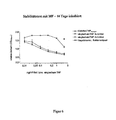
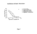
- polypeptides have the same (qualitative) activities as their corresponding wild-type soluble member of the TNF ligand family, but also that they still have bioactivities at a time when the soluble wild-type member of the TNF ligand family has already lost its activity due to their significantly higher stability that is dissociated or denatured (for this reference is made to the various stability tests described below, which are described in the examples and in the figures).
- soluble wild-type member of the TNF ligand family is meant a soluble extracellular portion of a membrane-bound member of the TNF ligand family.
- the terms “isotype”, “wt”, “soluble” and “s” are used synonymously with the term “soluble wild type”.
- it can be soluble wild-type TNF (as a member of the TNF ligand family), for which the synonymous terms “wild-type TNF”, “wtTNF”, soluble TNF and “sTNF” are used accordingly below.
- a component A according to the invention is a TNF monomer or a functional fragment or a functional variant thereof.
- a “monomer” is meant the smallest protein or polypeptide moiety that can be separated from an oligomeric protein without separation of covalent bonds.
- a polypeptide or a component A or a fragment or variant thereof is functional in the sense of the invention, provided that it has its biological activity or function, in particular its binding property to an interaction partner, e.g. a membrane-bound receptor, and also its trimerization property.
- an interaction partner e.g. a membrane-bound receptor
- trimerization property e.g. trimerization property
- these biological functions may indeed be altered, e.g. in terms of their specificity or selectivity, the basic biological function is retained.
- a fragment according to the invention is to be understood as meaning both a fragment of a monomer of a member of the TNF ligand family and a fragment of a polypeptide or protein of the present invention.
- These may be N-terminal, C-terminal or intrasequentially abbreviated amino acid sequences of the monomer, polypeptide or protein.
- intrasequential truncations of the polypeptide or protein may be truncations of the sequence of one or more of the three monomers, which in turn may occur N-terminal, C-terminal or intrasequentiell.
- the fragment of a monomer represents its extracellular domain, which corresponds to the entire extracellular domain of the soluble wild-type member of the TNF ligand family or a portion thereof.
- the fragment of a monomer represents its extracellular domain corresponding to either the wild type soluble TNF (amino acids 77-233) or the entire extracellular domain (amino acids 53-233).
- fragments of the monomers, polypeptides or proteins may be accomplished by modifying the DNA sequence encoding the native monomer, polypeptide or protein, followed by transformation of that DNA sequence into a suitable host and expression of these modified DNA fragments. Sequence, provided that the modification of the DNA, the functional activities of the monomer, polypeptide or protein are not destroyed.
- the identification of a fragment can either be done by checking its functionality by measuring its biological activity, as described above, or also by sequencing the fragment and a subsequent one Comparison of the sequence obtained with the native sequence.
- the sequencing can be done by standard methods that are numerous and well known in the art.
- Variants of biologically active monomers, polypeptides or proteins or fragments thereof or a component A are in particular those monomers, polypeptides or proteins or fragments thereof, which have sequence differences from the corresponding native sequences. These sequence deviations may be one or more insertion (s), deletion (s) and / or substitution (s) of amino acids, with a sequence homology of at least 60%, preferably 70%, more preferably 80%, also more preferred 85%, even more preferably 90%, and most preferably 97%.
- the sequences can be aligned to be compared below.
- Gaps in the sequence of the first amino acid or nucleic acid sequence are introduced and the amino acids or nucleotides are compared at the corresponding position of the second amino acid or nucleic acid sequence. If a position in the first amino acid sequence is occupied by the same amino acid or nucleotide, as is the case in a position in the second sequence, then both sequences are identical at that position.
- the percent identity between two sequences is a function of the number of identical positions shared by the sequences.
- the determination of the percentage identity of two sequences can be carried out using a mathematical algorithm.
- a preferred but not limiting example of a mathematical algorithm that can be used to compare two sequences is the algorithm of Karlin et al. (1993), PNAS USA, 90: 5873- 5877 , Such an algorithm is integrated into the NBLAST program which can identify sequences having a desired identity to the sequences of the present invention.
- the gapped BLAST program may be used, as in FIG Altschul et al. (1997) Nucleic Acids Res. 25: 3389-3402 described.
- Bioly active, ie functional, variants of monomers, polypeptides or proteins or fragments thereof within the meaning of the invention may preferably have selective receptor binding properties, the variant e.g. with regard to their specific bioactivity or other properties, in particular their stability.
- variants includes in particular those amino acid sequences which have conservative substitution with respect to the physiological sequences.
- Conservative substitutions refer to those substitutions in which amino acids derived from the same class are interchanged.
- Side chains which have a hydroxy function are also included.
- one amino acid with one polar side chain is replaced by another amino acid with a likewise polar side chain or, for example, an amino acid characterized by a hydrophobic side chain is substituted by another amino acid with likewise hydrophobic side chain (eg serine (threonine) by threonine (Serine) or leucine (isoleucine) by isoleucine (leucine)).
- an amino acid characterized by a hydrophobic side chain is substituted by another amino acid with likewise hydrophobic side chain
- eg serine (threonine) by threonine (Serine) or leucine (isoleucine) by isoleucine (leucine) Insertions and substitutions are possible in particular at those sequence positions which do not cause any change in the three-dimensional structure or affect the binding region.
- variants for example of monomers, polypeptides or proteins or fragments thereof with amino acid sequences which have substitutions with respect to the native sequences are described, for example, in the publications US 4,737,462 . US 4,588,585 . US 4,959,314 . US 5,116,943 . US 4,879,111 and US 5,017,691
- the production of variants in general is also known in particular from Maniatis et al, (2001), Molecular Cloning: A Laboratory Manual, Cold Spring Harbor Laboratory Press ). It codons can be omitted, supplemented or replaced.
- variants may also be those proteins or polypeptides which are stabilized, for example, by stabilization of the protein backbone by substitution of the amide-like bond, for example also by the use of .beta.-amino acids, in order to avoid the physiological degradation.
- Variants may also be prepared by introducing changes into the nucleic acids encoding the variants, such as insertions, deletions, and / or substitutions of one or more nucleotides. Numerous methods for such alterations of nucleic acid sequences are known in the art. One of the most widely used techniques is oligonucleotide-directed site-directed mutagenesis (see Comack B., Current Protocols in Molecular Biology, 8.01-8.5.9, Ausubel F. et al., Ed. 1991 ). In this technique, an oligonucleotide is synthesized whose sequence has a specific mutation. This oligonucleotide is then hybridized to a template containing the wild-type nucleic acid sequence.
- a single-stranded template is used in this technique.
- a DNA-dependent DNA polymerase is used to synthesize the second strand of the oligonucleotide that is complementary to the template DNA strand.
- a heteroduplex molecule containing a mismatch caused by the above-mentioned mutation in the oligonucleotide is obtained.
- the oligonucleotide sequence is introduced into a suitable plasmid, this is introduced into a host cell, and in this host cell, the oligonucleotide DNA is replicated. With this technique one obtains nucleic acid sequences with targeted changes (mutations) which can be used for the production of variants according to the invention.
- the components A comprised by the polypeptide may be identical or different.
- a polypeptide may contain 4, 5, 6 or more components A forming a tetramer, pentamer, hexamer, etc.
- a polypeptide contains an integer multiple of a trimer of components A as described above, eg, two, three, four, or more trimers arranged one behind the other.
- the components A contained in polypeptides may be separated by linkers (see below). If, as described above, two, three, four, or more trimers of the components A are arranged one behind the other, the linkers which connect the various trimers with one another may be longer than the linkers which contain the components A in a single trimer connect with each other.
- the components A can come from the same or different organisms. These may be vertebrates, in particular mammals, for example the mouse, the rat, the pig and, above all, humans.
- the components A of the polypeptide are in each case a monomer or a functional fragment or a functional variant of TNF (Tumor Necrosis Factor, Genbank Accessnt, NM_000594).
- component A of the polypeptide as defined above is a TNF monomer that is resistant to the processing enzyme TACE.
- TACE is a member of the ADAM protease family and is the physiological TNF-specific processing enzyme of naturally occurring in the cell membrane TNF.
- TNF is usually cleaved from the cell membrane and released into the environment.
- a component A resistant to TACE according to the invention is preferably lacking the cleavage site Ala-Val (eg AS 76-77 in scTNF according to SWISSPROT nomenclature for human TNF, No. PO1375).
- the cleavage site can be removed according to the invention by deletion of one or both of the relevant amino acids Ala or Val.
- the recognition sequence for TACE (eg AS 77-88 in scTNF according to SWISSPROT nomenclature for human TNF, No. PO1375) can be completely or partially deleted. This is preferably done by deleting two, three, four or more amino acids, for example all amino acids of the TACE recognition sequence.
- the TACE-resistant sequence is in the region of amino acids 89-233 (AS 89-233) of the scTNF according to SWISSPROT nomenclature for human TNF, no. PO1375.
- Such a deletion in component A prevents potential cleavage of component A, eg, scTNF, by TACE near the junctions of components A in the region of the linkers (component B, see below), thus increasing the in vivo stability of the polypeptides.
- the shortening of the sequence resulting from the deletion can be compensated by a corresponding extension of the linker.
- the deletion of the sequence of component A described by way of example for scTNF can be applied to each of the abovementioned components A. If the TACE sequence is not or only partially removed, it is preferably ensured that when using additional components B or C (see below) in the polypeptide of the invention the TACE recognition sequence and the TACE cleavage site are not restored.
- the components A of the polypeptide of the invention as defined above are modified such that the polypeptides of the invention can covalently couple to surfaces.
- This coupling is preferably carried out on planar surfaces or on spherical particles, such as magnetic particles (magnetic beads), or on nanoparticles / microparticles, preferably as a cell-mimetic therapeutic reagent with the membrane-bound TNF-like properties.
- a coupling group preferably the SH group of a cysteine residue, in the Introduced N-terminal region of at least one component A of a polypeptide. (Further preferred coupling groups according to the invention are described below).
- the coupling group may be located at any position of the N-terminal region of component A, preferably near the N-terminus. Most preferably, the coupling group is in a range of the first 1-15, and even more preferably in a range of the first 1-10 N-terminal amino acids.
- the modification in a prokaryotic-expressed scTNF, the modification can be introduced after an initial methionine (position 2) of the amino acid sequence or in a eukaryotic-expressed scTNF directly after the cleaved leader sequence, which thus represent the N-terminal amino acids of component A.
- a coupling group can be introduced, eg resulting in a CysHis tag with a cysteine at amino acid position 9.
- Another alternative involves introducing a coupling group into one or both components B (linker) of the polypeptide.
- a polypeptide Preferably, in a polypeptide only the component A located at the N-terminus or the further N-terminally located tag is / is modified / modified with a coupling group as described above.
- a polypeptide which contains 3 components A for example, 2 or 3 of the components A are each modified with a coupling group so that the total molecule (the polypeptide according to the invention) is coupled to a suitable matrix via two or three covalent bonds can be.
- the coupling of the polypeptide via one or more coupling groups of all components A of a polypeptide preferably does not impair the function of the individual components A of TNF, but rather allows an improvement in the immobilization of scTNF, to surfaces and / or particles.
- each of the polypeptides according to the invention preferably contains the subunits (components A) necessary for the biological function in a functionally relevant arrangement.
- a polypeptide By coupling a polypeptide to a surface / a particle, it is possible according to the invention to produce surfaces or particles with, for example, coupled TNF homo- or heterotrimers or multimers. On these surfaces / particles, the coupled TNF homo- or heterotrimers or multimers are stable and bio-immobilized. Due to the single-chain principle, the functionally relevant trimeric state of the components A is stabilized by the components B, which ensures that after covalent coupling to a surface / the particles, a dissociation of individual components A excluded and thus loss of biological activity is prevented.
- a preferred property of such polypeptides coupled to surfaces or particles is their biomimetic activity, which corresponds to the natural, membrane-bound ligand of the TNF family.
- biomimetic activity corresponds to the natural, membrane-bound ligand of the TNF family.
- immobilized scTNF obtains a high affinity for TNFR2 and thus high signal capability.
- Polypeptides containing components A modified with a coupling moiety as described above and coupled to a surface and / or particles can be used in the invention for detecting as well as manipulating, depleting and / or removing binding partners of TNF be used.
- methods for extracorporeal manipulation, depletion and / or removal of constituents contained in body fluids e.g. by apheresis.
- TNF TNF ligand family member TNF.
- polypeptides of the present invention comprise in addition to the described components A at least two components B, wherein the component B has the property of a peptide linker.
- a peptide linker in the context of the invention, any peptide sequence which can be expressed in a biological system is conceivable.
- peptide linkers are found to be a flexible compound in the polypeptide constructs, but preferably do not adversely affect the intrinsic trimerization properties of TNF.
- linkers having such properties, in particular flexibility and length are chosen which are able to stabilize the spontaneously formed homotrimers of the particular ligand (derivative).
- component B is accordingly peptide linkers consisting of 2 to 30, preferably 4 to 16 and particularly preferably 4 to 12 amino acids which preferably contain glycine-serine repeat structures, more preferably Gly-Gly -Gly-Ser modules (GGGS modules) contained in a repetitive arrangement.
- GGGS modules Gly-Gly -Gly-Ser modules
- the peptide linkers of the invention are preferably glycine (G) rich peptide linkers, i. in that the amino acid sequence of a peptide linker has a high glycine content, preferably from 60 to 80%, particularly preferably 75%.
- the peptide linker (component B) comprises the amino acid sequence GGGSGGGSGGGS, also called (GGGS), or L short , or the amino acid sequence GGGSGGGSGGGSGGS, also called (GGGS) 4 or L long .
- the peptide linkers are referred to inter alia as L1 and L2, so that designations such as L1 short , L2 short , L1 long and / or L2 long result. It is possible to use two different linkers to stabilize a trimeric molecule.
- the peptide linkers according to the invention ie the components B, in each case link two of the at least three components A together.
- This linkage is covalently via the C-terminus of a component A and the N-terminus of another component A.
- the polypeptide represents a single-chain molecule (also called single chain or sc molecule). This means that all components A and Components B comprising the polypeptide lie on a single polypeptide or protein strand.
- components A and components B form a trimeric protein structure. This is preferably a homotrimeric protein structure, but heterotrimeric protein structures are also encompassed by the invention.
- component B The formation of this trimeric protein structure is preferably effected and / or enhanced by the component B. Since the component B leading to the trimerization or the trimerization enhancing component B of the polypeptide should essentially not form higher aggregates, component B should typically not contain a cysteine residue which can form an intermolecular disulfide bridge. Preferably, therefore, component B in a polypeptide does not have a cysteine residue or only those cysteine residues which possess an intramolecular disulfide bridge, ie in the polypeptide itself, to avoid covalent linkage with the at least one cysteine residue of a fusion protein of another trimer under oxidizing conditions can.
- sequences of native polypeptides or fragments of these native polypeptides, which are used as peptide linkers according to the invention, may also be present in the form of biologically active functional variants thereof within the meaning of this invention and as defined above.
- Components B of the invention may be identical or different naturally occurring peptide sequences. They can come from the same or different organisms.
- the organisms may be vertebrates, in particular mammals, for example the mouse, the rat, the pig and above all humans.
- the polypeptide may have additional sequence segments in addition to components A and B.
- tag sequences for example at least one flag tag, ie the amino acid sequence DYKDDDDK, and / or, for example, at least one His tag (containing several consecutive histidines, eg. at least 5) and / or other tag or anigenic sequences.
- a particularly preferred additional sequence segment is a leader peptide sequence.
- this leader peptide sequence is a signal for secretion of the polypeptide or protein in eukaryotic cells. This leader peptide sequence is preferably N-terminal.
- a polypeptide comprises a further component C, characterized by specific interaction with a complementary cell surface molecule ("antigen").
- a complementary cell surface molecule (“antigen").
- the mode of action of the polypeptide constructs is particularly true for all those members of the TNF ligand family that are effective for certain receptors exclusively or to a particularly good extent as a membrane molecule.
- TNFSF10 TNFSF10
- FasL INFSF6
- TNF TNF
- Component C thus has "targeting" functionality.
- component C is an antibody fragment, preferably a single chain antibody fragment, so-called scfv, or an antibody derivative which recognizes a specific target molecule on a cell surface.
- component C is a non-antibody-targeting protein or peptide, but analogous to an antibody-antigen interaction or a receptor-ligand interaction also selectively recognizes a specific target molecule on a cell surface.
- the present invention comprises a polypeptide as component A described above as at least one scTNF, and as component C an antibody fragment as described above, e.g. scFv. This results in smaller polypeptides each having a "tatgeting domain". Such smaller polypeptides are advantageously less susceptible to e.g. Aggregation.
- Component C is preferably an antigen-binding antibody fragment or an antigen-binding antibody derivative of a mammal, in particular of murine or human origin, or is a humanized antibody fragment or a humanized antibody derivative, eg of mammalian origin.
- component C typically consists of one prior art single chain Fv derivative murine, CDR grafting humanized component C, or a component C of entirely human origin which has been correspondingly converted to a scFv derivative.
- component C is a protein or peptide ligand which, as monomer, undergoes a specific binding to a membrane receptor
- the component C is a protein or peptide with specificity for cell surface molecules, which is in particular a cytokine receptor, a growth factor receptor, an integrin or cell adhesion molecule. More preferably, it is a cytokine receptor selected from the group of TNFR Gene family is selected
- the component C of a polypeptide preferably has specificity for a tumor tissue selectively or dominant expressed antigen.
- a tumor antigen may generally be expressed on the malignant cells themselves or expressed in the non-malignant portion of the tumor, the stromal cells or the tumor endothelium.
- Such antigens of non-malignant tissue portions of a solid tumor are on the one hand genetically invariant, on the other hand occurring in a variety of tumor entities and thus univetselle tumor markers examples of such ligands for tumor association against which a component C of the polypeptide may be directed
- the VEGFR or the VEGFR / VEGF complex as well as the integrin ⁇ v ⁇ 3 and the vibronectin isoform ⁇ Fn are largely selective target structures of the tumor endothelium and the fibroblast activation protein (as a selective marker of the tumor stroma). All of the above examples can be effectively detected with specific scFv, making such scFv ("single chain Fv”) particularly useful as component C of a polypeptide. Galectin also comes into consideration as a tumor marker against which component C is directed
- antibody fragments in various antibody formats for example scfv, in particular scFv40, are particularly preferred as component C.
- the polypeptide recognizes via its component C as a specific target molecule a cell membrane-bound receptor of a member of the TNF ligand family, preferably different from the TNF ligand family member of component A.
- potential ligands include TNFSF1 (LTalpha), TNFSF2 (TNF), TNFSF3 (LTbefa), TNFSF4 (OX40L), TNFSF5 (CD40L), TNFSF6 (FasL), TNFSF7 (CD27L), TNFSF8 (CD30L), TNFSF9 (4-1BBL) , TNFSF10 (TRAIL), TNFSF11 (RANKL), TNFSF12 (TWEAKL), TNFSF13 (APRIL), TNFSF143B (BLYS), TNFSF14 (LIGHT), TNFSF15 (VEGI), TNFSF16 (CD30L), and TNFSF18 (AITRL), as well as EDA, the or their functional fragments or functional
- Methods of making a polypeptide to be used in the invention typically include the following steps: (a) culturing a host cell under appropriate conditions, (b) expressing a corresponding nucleic acid construct under appropriate conditions, and (c) isolating the polypeptide from the host cells and / or the culture supernatant.
- Culturing a host cell means allowing the host cell to grow in a suitable culture medium, at a suitable pH, and at suitable temperatures. Such growth conditions are dependent on the respective host cells used and are well known to the person skilled in the art. Culturing of cells is also found in Maniatis et al. (2001), supra .
- polypeptide here can typically be carried out in suitable expression systems according to the prior art, preferably as a secreted product of stable transfectants, for example CHO cells or other animal cells, such as Cos7 or SF9 (insect cells), or other eukaryotic cell systems, for example Pichia pastoris .
- the expressed polypeptides have respective leader sequences suitable for secretion in the customs system. Therefore, the vectors used for expression will also contain coding portions that code for a functional leader sequence, eg, as in Brocks et al.
- Isolation of the polypeptide from the host cell may be by standard methods such as chromatography methods, precipitation methods, etc. suitable for purification of polypeptides and proteins (see Maniatis et al. (2001), supra .
- the present invention relates to methods for extracorporeal ( ex vivo ) manipulation, depletion and / or removal of constituents contained in body fluids, such as binding partners of a component A as defined above or cells binding thereto or associated therewith.
- extracorporeal methods preferably include methods such as apheresis, especially the basic forms of apheresis, plasmapheresis, and cytapheresis.
- the plasmapheresis involves the extracorporeal manipulation, depletion and / or removal of certain soluble or suspended constituents in the plasma portion of the blood, as well as the return of the blood thus treated to the patient.
- peripheral blood is preferably taken from a patient by means of a pheresis machine, the blood is optionally added anticoagulants and the blood is separated into its main components - solid (red blood cells, white blood cells and platelets) and liquid portions (plasma). After separation into these main constituents, the soluble or suspended constituents of the blood contained in the plasma fraction thus obtained can be manipulated, depleted and / or removed in a further process step, for example using polypetides described according to the invention.
- Plasmapheresis is preferred in the present invention using methods such as therapeutic plasma exchange (TPE), immunoabsorption (IA), precipitation (HELP), differential membrane filtration, and other means
- TPE therapeutic plasma exchange
- IA immunoabsorption
- HELP precipitation
- differential membrane filtration and other means
- plasma filtration columns may be mentioned here (see, for example, US Pat US 4,619,639 Asahi Medical Company, incorporated herein by reference).
- MDF membrane filtration systems
- Filters such as, PlasmaFlo ® OP-05 (W) L and RheoFilter ® AR2000 blood filters, used (manufactured by Asahi Medical Company, Ltd. of Japan).
- any suitable surface and particle can be used in plasmapheresis procedures of the invention.
- extracellular circulating and / or marrow-bound cellular components red blood cells, white blood cells, stem cells or platelets
- specific subpopulations of these cells are manipulated, depleted and / or removed to produce a clinical Effect to achieve.
- peripheral blood is preferably taken from a patient by means of a pheresis machine.
- anticoagulant agents are then added to the blood and the blood is separated into its major components.
- the cell constituents-containing fraction of the blood these cell constituents can be manipulated, depleted and / or removed in a further process step, preferably also using polypeptides described according to the invention.
- the treated fraction can finally be reinjected to the patient.
- the separation of the blood into various fractions and optionally the separation of certain cell constituents of the blood by means of processes such as centdfugation, differential membrane filtration or other means are preferably carried out. It can continue to membrane filtration systems (MDF) using various particles and surfaces, for example. Filters such as, PlasmaFlo ® OP (W) L and RheoFilter ® AR2000 blood filters, are used (made by Asahi Medical Company, Ltd. of Japan).
- the above-described methods of plasmapheresis and cytapheresis can be combined with one another, for example, both soluble or suspended components in the plasma portion of the blood as well as in the blood circulating and / or bound to bone marrow cellular components (red blood cells, white blood cells, Stem cells or platelets) or specific subpopulations to manipulate, deplete and / or remove these cells to achieve a clinical effect.
- bone marrow cellular components red blood cells, white blood cells, Stem cells or platelets
- specific subpopulations to manipulate, deplete and / or remove these cells to achieve a clinical effect.
- Such a combination of plasmapheresis and cytapheresis is made using the described polypeptides.
- the present invention relates to an (apheresis) method according to claim 1.
- an optional blood sample may be taken from a patient before the separation of the blood, if appropriate.
- the blood treated with the method according to the invention or the blood fraction thus treated can be reinjected to the patient. For this it may be necessary to recombine the previously separated and possibly differently treated blood fractions with other untreated solid and / or liquid components of the blood. Volume losses resulting from the process according to the invention can be achieved by adding suitable liquids, e.g. by adding an isotonic saline solution.
- the step of binding the soluble, suspended or cellular constituents of the blood to a surface or particle coupled to a polypeptide according to the invention can be carried out once or several times as required to achieve a desired selectivity
- such surfaces and / or particles are used which are covalently coupled with a polypeptide according to the invention, as defined above were.
- the polypeptide is preferably covalently coupled to the surface and / or the particles via the coupling group (s) A contained in the polypeptide.
- Coupling of the polypeptides used according to the invention via the individual components A is preferably as in DE 101 44 252 A1
- the coupling of the polypeptides used according to the invention preferably takes place at the surface or particles (carrier) via a bond between first functional groups present on the carrier surface and in the polypeptide via the coupling groups contained in components A.
- These coupling groups are preferably complementary to the functional groups of the carrier and can enter into an affine, preferably covalent, bond with them.
- the functional group of component A is positioned within component A such that it is located at a suitable distance outside the domains of the polypeptide responsible for the biological activity. In this way, it is possible according to the invention to target TNF and maintain its biological activities immobilize the carrier.
- the polypeptide is preferably fixed on the support surface such that the three-dimensional structure of the domain (s) required for biological activity is unaltered over non-immobilized polypeptide, and the domain (s) are free for contact with cellular reactants is accessible / are.
- the functional group of the support surface is selected from the group consisting of amino group, carboxyl group, epoxy group, maleimido group, alkyl ketone group, aldehyde group, hydrazine group, hydrazide group, thiol group and thioester group.
- the coupling groups of the components A to be immobilized of the polypeptide are selected from a group which contains the same species as for the functional group of the support surface.
- the two with each other Bonding coupling groups and functional groups must be complementary to one another, ie be able to form a covalent bond with each other.
- the coupling group of component A of the polypeptide according to the invention is a carboxy group.
- the coupling group of the component A of the polypeptide is an amino group.
- a thiol group is selected as the functional group of the carrier, according to the invention the complementary coupling group of component A of the polypeptide is a maleimido group.
- the complementary coupling group of component A of the polypeptide is a thiol group. If, according to the invention, the alkyl ketone group, in particular the methyl ketone or aldehyde group, is used as the functional group of the carrier, the functional complementary coupling group of the component A of the polypeptide is a hydrazine or hydrazide group.
- the functional complementary coupling group of component A of the polypeptide is an alkyl ketone, in particular methyl ketone or aldehyde group.
- the functional group on the support surface is a maleimido group and the coupling group of component A of the polypeptide is a thiol group.
- the immobilization is preferably directed.
- the term “directionally immobilized” or “directed immobilization” means that a scTNF is immobilized on a support at definite positions within the scTNF such that the three-dimensional structure of the domain required for biological activity (n ) is not changed from the non-immobilized state and that these TNF domain (s), for example binding pockets for cellular reactants, are freely accessible to them upon contact with cellular reactants.
- “Directed immobilized” also means that the coupling of the polypeptide on the support surface
- the immobilized protein can not or only very slowly degraded by protein degrading enzymes in a later use in a cellular or cell-like environment.
- the immobilized TNF molecule is aligned on the support surface so as to provide as little as possible targets for proteases.
- the polypeptide may be made resistant to proteases prior to coupling by molecular biological methods, such as described above for TNF members in the protease TACE.
- the polypeptide retains, via its coupled component (s) A, its biological function and the ability to interact with other compounds / agents. For example, if a polypeptide contains three components A (trimer), typically each, two, or preferably only one of the three components A will be covalently bound to the surface / particle.
- Surfaces and / or particles suitable for coupling can include any suitable surfaces and / or particles to which the polypeptides can be coupled.
- Particularly preferred are culture plates or so-called microbeads, eg Dynabeads (Dynal Biotech GMBH), nano-beads, nanoparticles, or solid phases such as nylon wool, Sepharose, Sephadex, etc ..
- the carrier systems used according to the invention are compact or hollow nanoparticles of sizes between 25 and 1000 nm. These consist of either organic or inorganic particulate materials.
- the nature of the material can be varied according to the later application, wherein the carrier of the nanoparticles preferably consists of biocompatible and / or biodegradable materials.
- “nanoparticles” furthermore refer to binding matrices which have a carrier with a surface comprise, on which chemically reactive functional groups are arranged, which enter into complementary functional groups of molecules to be bound, in particular polypeptides, affine bonds, ie covalent and / or non-covalent bonds, and in this way can stably fix the polypeptides on their surfaces
- the carriers of the nanoparticles consist of chemically inert inorganic or organic materials and have a size of less than 1 ⁇ m, preferably from 25 to 500 nm.
- a particular embodiment of the apheresis method of the invention includes a biofunctionalized surface (planar or as a particle) with the polypeptide scTNF immobilized covalently and directionally on the surface in the manner previously described.
- a biofunctionalized surface containing for example several immobilized scTNF molecules or variants thereof, allows a high affinity for the respective complementary binding partners, for example TNFR1 and TNFR2.
- the receptors TNFR1 and in particular TNFR2 are found in greatly increased concentrations as processed, soluble molecules in the blood of tumor patients and in other diseases.
- the apheresis process according to the invention can be carried out as a continuous process or as a batch process.
- the process according to the invention is preferably carried out directly following the blood withdrawal or the fractionation of the blood withdrawn and before the return of the blood to the patient without interim storage of the blood or the blood fractions obtained.
- the blood withdrawn or the fractions of the withdrawn blood are stored after each step for a certain period of time.
- the temperature in apheresis method according to the invention is preferably 0-40 ° C.
- the temperature is preferably at 25 to 40 ° C, especially preferably in a range between 36 and 38 ° C.
- the tamperature may be between 0-40 ° C.
- the apheresis method according to the invention as described above is preferably used for diagnosis, therapy and / or prophylaxis in diseases associated with TNF, in particular in tumor diseases, in particular solid or lymphatic tumors, in particular solid and lymphatic tumors.
- the restoration of homeostasis of the immune system with its humoral and cellular components is considered to be the key to the effectiveness of apheresis procedures.
- the apheresis system according to the invention can therefore also be used for restoring immune homeostasis in non-malignant diseases.
- inflammatory diseases include inflammatory diseases, arthritic and theumatic diseases, or diseases of the immune system, and for the treatment of infectious diseases, metabolic diseases, inflammatory conditions, hyperproliferative diseases autoimmune diseases, in particular rheumatoid / arthritic diseases, toxic epidermal necrolysis (TEN), multiple sclerosis, Hashimoto's thyroiditis, GvHD, viral Hepatitis (HBV, HCV), alcohol-induced hepatitis, liver transplantation rejection, hyperapoptotic response disorders, degenerative diseases, especially neurodegenerative diseases.
- infectious diseases include inflammatory diseases, metabolic diseases, inflammatory conditions, hyperproliferative diseases autoimmune diseases, in particular rheumatoid / arthritic diseases, toxic epidermal necrolysis (TEN), multiple sclerosis, Hashimoto's thyroiditis, GvHD, viral Hepatitis (HBV, HCV), alcohol-induced hepatitis, liver transplantation rejection, hyperapoptotic response disorders, degenerative diseases,
- tumors include colon carcinomas, melanomas, renal carcinomas, lymphoma, acute myeloid leukemia (AML), acute lymphoid leukemia (ALL), chronic myeloid leukemia (CML), chronic lymphocytic leukemia (CLL), gastrointestinal tumors, lung carcinoma, glioma, thyroid tumors, Breast cancers, prostate tumors, hepatomas, various virus-induced tumors such as papillomavirus-induced carcinomas (eg cervical carcinoma), adenocarcinomas, herpesvirus-induced tumors (eg Burkitt's lymphoma, EBV-induced B cell lymphoma), hepatitis B-induced tumors (hepatocellular carcinomas), HTLV-1 and HTLV-2 induced lymphomas, acoustic neuroma, cervical cancer, lung cancer, pharyngeal cancer, anal carcinoma, glioblastoma, lymphoma, rectal carcinoma, astrocytoma, brain tumors
- Arthritic diseases include, in particular, monarthritis, oligoarthritis, polyarthritis, acute arthritis (especially septic, crystal-induced, reactive arthritis and acute arcoidosis), subacute arthritis, chronic arthritis, (especially rheumatoid arthritis, arthritis in seronegative spondylarthritis), infectious arthritis, para- or post-infectious arthritis , rheumatoid arthritis, juvenile chronic arthritis, arthritis in inflammatory connective tissue diseases and vasculitis, allergic arthritis, arthritis associated with metabolic and nutritional disorders, arthritis in endocrine disorders, arthritis in granulomatous diseases, arthritis in diseases of hematopoietic system, arthritis in joint bleeding due to disorders blood clotting, neoplastic arthritis, paraneoplastic arthritis, (post-) traumatic arthritis, arthritis in diseases of the joint cartilage, arthritis in neuropathies, arthritis in pustular abs cedar, necrotizing or tissue neutrophil dermatoses, arthritis in other extra-articular diseases, as well as arthritis allergica, ch
- a polypeptide which has increased stability in that the entire molecule consists of a protein or polypeptide strand (scTNF), so that the monomers (components A) can no longer dissociate.
- scTNF does not show any qualitative differences to the normal soluble sTNF (also wild-type TNF or wtTNF) in the nature of its bioactivity, but is much more stable and therefore shows higher bioactivity, ie it is effective at lower concentrations.
- scTNF Like normal soluble TNF (wtTNF), scTNF induces apoptosis in sensitive cells, activates the transcription factor NF- ⁇ B, activates the receptor TNFR1, but not TNFR2, and activates the receptor TNFR2 in the presence of certain antibodies (eg 80M2).
- certain antibodies eg 80M2
- scTNF is an ideal starting material for the production of new, bi-functional molecules.
- scTNF or functional fragments thereof can covalently bind to cell surfaces.
- the present invention it is possible with the present invention to stably introduce mutations in only one or else two or more of the at least three monomers (components A) owing to the covalent linking of the monomers (components A) of the polypeptide.
- point mutations are known which enforce receptor selectivity, ie, for example, in the case of TNF, binding only occurs to one of the two TNF receptors (TNFR1 and TNFR2).
- TNF TNF receptors
- scTNF variants having different length peptide linkers between the individual TNF modules (i.e., TNF monomers) were tested.
- the peptide linkers each consist of the triple or the fourfold amino acid repeat GGGS (or GlyGlyGly-Ser) (referred to as "singlechainTNF3x” or “singlechainTNF4x” or as “scTNF3x” or “scTNF4x").
- FIG. 1 shows the result of a typical cytotoxicity test of TNF.
- Target cells used were mouse fibroblasts (MF) from TNFR1 / TNFR2 double-knockout mice stably expressing TNFR1 receptor chimeras (INFR1-Fas) (MF TNFR1 Fas cells).
- MF TNFR1 Fas cells mouse fibroblasts (MF) from TNFR1 / TNFR2 double-knockout mice stably expressing TNFR1 receptor chimeras (INFR1-Fas) (MF TNFR1 Fas cells).
- MF TNFR1 Fas cells Such cells expressed a hybrid molecule that extracellularly consists of the corresponding part of TNFR1, thus binding TNF and having intracellularly the highly apoptotic intracellular domain of the Fas receptor.
- These MF TNFR1-Fas cells can be activated by TNFR1-specific stimuli, but mediate a Fas-specific death signal.
- a control bacterial lysate was used, which was a non-scTNF-expressing bacterial lysate that was processed identically to the recombinant scTNF proteins.
- both the conventional human soluble TNF (TNFhum) and the recombinant scTNF variants led to cell death.
- a comparable dose-dependent cytotoxic effect of TNFhum or scTNF can be seen (see the lower three graphs.), Where the specific activity of scTNF is at least equivalent, if not even higher than that of wild-type TNF. As expected, the negative control shows no toxic effect.
- the negative control used was the control bacterial lysate described above.
- the cytotoxic effect of soluble human wild type TNF or scTNF can be abrogated by neutralizing TNF-specific antibodies (see the top three graphs) in a wide concentration range (0.03 - 10 ng / mL). Differences between the response of scTNF with 3-fold and scTNF with 4-fold peptide linker were not found.
- FIG. 2 shows the result of a cytotoxicity test in which the effect of sTNF and scTNF variants on TNFR2 receptor chimera (TNFR2-Fas) expressing cells was examined.
- mouse fibroblasts were used which stably express receptor chimeras consisting of TNFR2 and Fas (TNFR2-Fas, where the extracellular part of TNFR2, the intracellular part of the Fas receptor, is expressed (MF TNFR2-Fas cells).
- MF TNFR2-Fas cells (as well as wild-type TNFR2) can only be activated by an adequate TNFR2 stimulus, for example by membrane-bound TNF, but not by soluble TNF.
- FIG. 3 Figure 3 shows the result of a cytotoxicity assay in which the effect of soluble wild-type human TNF and a scTNF variant in combination with a specific TNFR2-specific antibody, 80M2, on TNFR2 chimera (FNFR2-Fas) expressing cells was examined.
- the experimental approach corresponds to that FIG. 2 with the difference that the soluble wild-type TNF or the scTNF with 3-fold peptide linker (scTNF3x) were added together with the antibody 80M2.
- the antibody 80M2 is known to confer to normal soluble TNF the particular signaling capacity of membrane-bound TNF.
- FIGS. 4 to 10 show results of various stability tests, which prove that the scTNF variants according to the invention have a considerably better stability than soluble wild-type TNF because of their structure.
- sTNF3x, scTNF4x the functionality of the sTNF or scTNF variants was tested in cytotoxicity assays based on MF TNFR1-Fas cells and Kym1 cells.
- cytotoxicity test 1: 3 dilutions were used starting from a 3.0 ng / ml TNF sample.
- the negative control used was the control bacterial lysate used in the cytotoxicity tests described above (non-scTNF-expressing bacterial lysate, which was processed identically to the recombinant scTNF proteins).
- FIG. 4 shows the bioactivity of the molecules used before the stability tests on mouse fibroblasts MF TNFR1-Fas cells.
- Soluble wild-type human TNF (TNFhuman) and scTNF dilutions were freshly prepared and diluted out on the cells. Both the soluble control TNF and the scTNF variants, after incubation with the MF, resulted in their cell death.
- the control bacterial lysate was not expected to have a toxic effect.
- the ED 50 values half-maximal effective concentration
- the control bacterial lysate used as a negative control showed no toxic effect.
- FIG. 5 shows the result of a stability test with mouse fibroblasts MF TNFR1-Fas cells.
- wild-type TNF and scTNF dilutions were incubated for 8 days in the cell incubator before the test was performed to assess the stability of the samples.
- a significant decrease in the bioactivity of the soluble wild type TNF (TNFhuman) was observed after 8 days at 37 ° C, while the activity of both scTNF variants (scTNF3x, scTNF4x, explanation see above) remained unchanged.
- an ED 50 value of about 2 ng / ml (fresh used 0.2 ng / ml, cf. FIG. 4 ).
- the scTNF variants remained just as active with ED 50 values of about 0.1 ng / ml as the freshly used samples (cf. FIG. 4 ). As expected, the control bacterial lysate used as a negative control showed no toxic effect.
- FIG. 6 shows the result of a stability test with mouse fibroblasts MF TNFR1-Fas cells.
- the wild type TNF and scTNF solutions were as in FIG. 5 has now been incubated in the cell incubator for 14 days.
- a clear further decrease in the bioactivity of the wild-type TNF was measured after 14 days at 37 ° C., while the activity of the scTNF variants remained approximately the same.
- the scTNF variants remained almost as active at 0.13 ng / ml as the freshly prepared samples (0.1 ng / ml, vgL FIG. 4 ).
- the control bacterial lysate used as a negative control showed no toxic effect.
- FIG. 7 shows the result of a stability test with the human cell line Kym1.
- Kym1 cells express normal human wild-type TNF receptors and are also cytotoxic to TNF.
- the experiment was analogous to the stability tests with mouse fibroblasts according to FIGS. 4 to 6 Wild type TNF or scTNF variants (scTNF3x, scTNF4x) were incubated in serum-containing medium at concentrations of 3.0 to 0.01 ng / mL at 37 ° C and 5% CO 2 for different times. Subsequently, the functionality of the soluble sTNF or the scTNF variants was tested in cytotoxicity assays on Kym1 cells.
- FIG. 8 shows the result of a stability test with the human cell line Kym1.
- the wild type TNF and scTNF solutions were incubated for 16 days in the cell incubator. Subsequently, the Kym1 cell cytotoxicity test was performed. There was a significant decrease in the activity of sTNF after 16 days at 37 ° C, whereas the toxicity, ie the bioactivity, of the scTNF variants remained approximately the same.
- an ED 50 value of about 1.2 ng / mL was found.
- the scTNF variants remained just as active at 0.06 ng / ml as the freshly prepared samples.
- the control bacterial lysate used as a negative control showed no toxic effect
- FIG. 9 shows the result of a stability test with the human cell line Kym1.
- sTNF and scTNF solutions were incubated in the cell incubator for 22 days. There was a clear further decrease in the activity of sTNF after 22 days at 37 ° C. The scTNF variants were still stable. As expected, the control bacterial lysate used as a negative control showed no toxic effect.
- Table 1 below shows the result data of the stability tests according to FIGS. 7 and 9 compared: ⁇ b> Table 1: ⁇ / b> Comparison of the ED ⁇ sub> 50 ⁇ / sub> values of Figures 7 + 9 Titrated ED 50 fresh ( FIG. 7 ) ED 50 after 22 days ( FIG. 9 ) loss of activity sTNF 0.1 ng / mL 1.50 ng / mL > 90% scTNF 0.06 ng / mL 0.07 ng / mL Hardly detectable
- FIG. 10 shows the result of a stability test with the human cell line Kym1. Dilutions of 3 ng / ml were titrated out after 16 days at 37 ° C. Unlike the previous ones FIGS. 4 to 9 In this experiment, a titration curve was prepared starting from a 3 ng / ml TNF dilution stored for 16 days, whereas in the case of the previously shown titration curves the respective dilutions were prepared and then stored for the stated time. It remains to be noted that a comparison of the data FIG. 8 (analogous experiment with 16 days incubation period) and FIG. 10 There are no significant differences. As expected, the control bacterial lysate used as a negative control showed no toxic effect.
- FIG. 11 shows the result of a stability test in human serum.
- the soluble wild type TNF and the scTNF4x variant were diluted in 100% serum and freshly excreted.
- an ED 50 value of 0.004 ng / ml was measured for wild-type TNF and an ED 50 value of 0.007ng / ml for the scTNF variant.
- FIG. 12 shows the result of a stability test in human serum.
- wild-type TNF and scTNF4x variant were stored in 100% fresh non-heat-inactivated serum for 8 days at 37 ° C. and then titrated out on Kym1 cells , As a result, an ED 50 value of 0.40 ng / ml was measured for wild-type TNF and an ED 50 value of 0.03 ng / ml for the scTNF variant.
- the effects of 8-day storage in 100% serum were as follows: compared to freshly titrated scTNF, the 8 days stored scTNF showed a loss of activity by a factor of about 4.3, whereas soluble sTNF showed a dramatic Loss of activity by a factor of 100 during storage in 100% serum showed.
- the inventive scTNF showed in comparison to sTNF therefore a significantly higher bioactivity after 8 days incubation in serum and proved to be very stable in human serum at physiological temperatures.
- FIG. 13 shows the result of analysis by polyacrylamide gel electrophoresis under reducing and denaturing conditions. Shown is a silver gel of purified wild-type TNF and the purified scTNF variants scTNF3x and scTNF4x. The samples were each incubated with ⁇ -mercaptoethanol (final 5%) at 95 ° C for 5 min. 500 ng of sTNF and scTNF4x and about 150 ng of scTNF3x per lane were applied to the silver gel.
- FIG. 14 shows the result of a stability test under reducing and denaturing conditions.
- a Western blot of the samples of wild-type TNF separated in a 15% SDS-PAGE and the scTNF variants scTNF3x and scTNF4x was performed.
- Two parallel runs were carried out in which the samples were incubated in one batch each with ⁇ -mercaptoethanol (final 5%) at 95 ° C for 5 min, while in the other batch no ⁇ -mercaptoethanol incubation took place.
- the Detection after the course of the electrophoresis was carried out with an anti-TNF antibody.
- the antibody specifically detected the protein of both scTNF variants in distinct bands at about 50 kDa.
- the protein was specifically detected at approximately 17 kDa. This in turn confirms the stability of the scTNF3x and scTNF4x variants of the invention under reducing and denaturing conditions and is also expected to coincide with the results FIG. 13 ,
- FIG. 15 shows the result of an IkappaB degradation assay.
- IkappaB is an inhibitor of the transcription factor nuclear factor kappa B (NF- ⁇ B), which is known to be degraded by TNF, thereby activating NF- ⁇ B.
- NF- ⁇ B transcription factor nuclear factor kappa B
- For the detection of degradation of I- ⁇ B cell lysates were prepared 0, 30 and 60 minutes after stimulation with 10 ng / ml wild-type TNF, scTNF3x and scTNF4x and subsequently analyzed in Westem blot with I- ⁇ B-specific antibodies.
- the result shows that the transient degradation of I- ⁇ B was induced by both wild-type TNF and both scTNF variants, with the response of the scTNF variants to that of wild-type TNF.
- the control bacterial lysate used as a negative control (as described above, a non-scTNF-expressing bacterial lysate that was processed identically to the recombinant scTNF proteins) was expected to have no effect on I- ⁇ degradation.
- FIG. 16 shows the result of a JNK assay.
- JNK c-jun N-terminal kinase
- TNF TNF-induced kinase
- scTNF3x and scTNF4x respectively.
- cell lysates were prepared and JNK activity was established by immunoprecipitation of JNK with JNK-specific antibodies and subsequent kinase Assay with GST c-Jun as substrate checked.
- the control bacterial lysate used as a negative control was not expected to activate JNK kinase.
- the result shows that activation of JNK was carried out both by both scTNF variant and wild-type TNF, the reaction behavior of the scTNF variants corresponding to that of the wild-type TNF.
- FIG. 17 shows the result of an Electrophoretic Mobility Shift Assay (EMSA).
- ESA Electrophoretic Mobility Shift Assay
- FIG. 19 shows the nucleic acid sequence and the corresponding amino acid sequence of scTNF-L short (construct A-II)
- FIG. 20 shows the nucleic acid sequence and the corresponding amino acid sequence of cys-scTNF-L short (construct B-II)
- FIG. 21 shows the nucleic acid sequence and the corresponding amino acid sequence of scFasL (construct C)
- FIG. 22 shows the nucleic acid sequence and the corresponding amino acid sequence of scTRAIL (construct D)
- FIG. 23 shows the nucleic acid sequence and the corresponding amino acid sequence of scTNF (construct E)
- FIG. 24 shows the nucleic acid sequence and the corresponding amino acid sequence of scFasL-AMAIZe (construct F)
- FIG. 25 shows the nucleic acid sequence and the corresponding amino acid sequence of scTRAIL-AMAIZe (construct G)
- FIG. 26 shows the nucleic acid sequence and the corresponding amino acid sequence of scTNF-AMAIZe (construct H)
- FIG. 27 shows the pharmacokinetics of human wild-type TNF and human scTNF (vgL Example 2).
- the data in FIG. 27 show a clear increase in the in vivo half life of the scTNF variants. It is a significantly increased duration of action for scTNF with respect to TNF in vivo, thus underscoring the value of scTNF, especially scTNF-L 2 , as a potential therapeutic.
- FIG. 28 shows covalently linked to particle (silica) CysHis scTNF.
- This covalently linked CysHis scTNF is bioactive and has the particular activity of membrane-bound TNF, ie it activates TNFR2.
- FIG. 28 shows that cells from mouse fibroblasts transfected with construct TNFR2-Fas and treated with serial dilutions of the indicated reagents are completely resistant to soluble wtTNF. After covalent coupling of reduced CysHis scTNF to silica microparticles (beads) according to established protocols (DPA 2001, no.
- the PCR product was purified on an agarose gel and eluted and subsequently digested with the respective restriction enzymes (see specifications) in a 40 ⁇ l reaction mixture at optimal cleavage temperature (indicated by the manufacturer) for 2 hours.
- the batch of a vector digest was mixed with 33 .mu.M dNTPs and 1U / 1 .mu.g DNA Klenow fragment of the DNA polymerase I and incubated for 15 min at 25 ° C. This reaction was stopped with 10 mM EDTA for 20 min at 75 ° C
- Vector and insert were ligated in a molar ratio of 1: 5 together with 400 U ligase in a 10 ⁇ l volume overnight at 16 ° C
- the expression took place in E. coli strain XL-1 blue.
- the purification of the expressed scTNF variants was carried out by means of chromatographic methods (His Tag Affinitäs and anion exchange chromatography).
- the respective scTRAIL vectors were digested with the restriction enzymes NotI or EcoRI and XbaI, respectively, and the inserts as a cassette in the corresponding AMAIZe vectors (see DE 10045591.3 ), these vectors were also cut with the enzymes NotI or EcoRI and XbaI.
- the following constructs were produced thereunder: HA-svFv40-Flag Tag- TRAIL Module1-Linker1-TRAIL Module2-Linker2- TRAIL Module3
- Example 2 Pharmacokinetics of human wild-type TNF and human scTNF.
- Example 3 CysHis scTNF covalently coupled to particles (silica).
- Example 4 Comparison of standard recombinant human (rh) TNF and scTNF in in vivo tumor necrosis models and in vitro L929 cytotoxicity activity
- mice C3H / HeJ (female), 17-19 g from Charles River
- Tumor cells CFS-1 methylcholanthrene-induced fibrosarcoma cell line derived from C3H / HeN from mouse (Reference: Hafner M., P. Orosz, A. Kruger, and DN Gurnnel. 1996 TNF promotes metastasis by impairing natural killer cell activity. Internat. J. Cancer 66: 388-392 );
- Tumor Necrosis Experiment: Mice received 1.6 x 10 7 CFS-1 cells in 50 ⁇ l of medium (RPMI, 10% FCS) intradermally in the back; The tumors were allowed to grow for 12 days until they reached a size of about 5-6 mm in diameter, prior to intraperitoneal injection with TNF (10 ⁇ g per mouse) in 200 ⁇ l of PBS or PBS alone as a control. Tumor size was measured daily and examined macroscopically. Mice were sacrificed on day 6 after treatment and tumors were removed for histology Tumors were excised, fixed overnight in 4% PBS-buffered formalin and embedded in paraffin.
- medium RPMI, 10% FCS
- Equatorial vertical sections (4 ⁇ m) were stained with hematoxilin and eosin and examined microscopically examined for necrosis (such as described in: Lucas R. et al., 2001, Int J Cancer, 91: 543-549 ).
- TNF rhTNF specific activity 6.6 x 10 6 U / mg (48 h L929 assay without Act D) scTNF
- rhTNF induced small hemorrhagic necrosis ( ⁇ 5% of the tumor area), macroscopically seen only in 3/7 of the animals.
- scTNF induces larger hemorrhagic necroses (> 10% of the tumor area) in all tumors (7/7), 5/7 of which are macroscopically visible.
- scTNF >> rhTNF in relation to tumor cytotoxicity in vitro and induction of necrosis.
Priority Applications (1)
| Application Number | Priority Date | Filing Date | Title |
|---|---|---|---|
| PL05716360T PL1727833T3 (pl) | 2004-03-26 | 2005-03-24 | Rekombinowane polipeptydy członków rodziny ligandów TNF i ich zastosowanie |
Applications Claiming Priority (2)
| Application Number | Priority Date | Filing Date | Title |
|---|---|---|---|
| DE102004014983A DE102004014983A1 (de) | 2004-03-26 | 2004-03-26 | Rekombinante Polypeptide der Mitglieder der TNF Ligandenfamilie und deren Verwendung |
| PCT/EP2005/003158 WO2005103077A1 (de) | 2004-03-26 | 2005-03-24 | Rekombinante polypeptide der mitglieder der tnf ligandenfamilie und deren verwendung |
Publications (2)
| Publication Number | Publication Date |
|---|---|
| EP1727833A1 EP1727833A1 (de) | 2006-12-06 |
| EP1727833B1 true EP1727833B1 (de) | 2013-09-18 |
Family
ID=34962642
Family Applications (1)
| Application Number | Title | Priority Date | Filing Date |
|---|---|---|---|
| EP05716360.2A Active EP1727833B1 (de) | 2004-03-26 | 2005-03-24 | Rekombinante polypeptide der mitglieder der tnf ligandenfamilie und deren verwendung |
Country Status (11)
| Country | Link |
|---|---|
| US (1) | US8927205B2 (tr) |
| EP (1) | EP1727833B1 (tr) |
| JP (1) | JP2007530021A (tr) |
| AU (1) | AU2005235669B2 (tr) |
| CA (1) | CA2563754C (tr) |
| DE (1) | DE102004014983A1 (tr) |
| DK (1) | DK1727833T3 (tr) |
| ES (1) | ES2439896T3 (tr) |
| PL (1) | PL1727833T3 (tr) |
| PT (1) | PT1727833E (tr) |
| WO (1) | WO2005103077A1 (tr) |
Families Citing this family (45)
| Publication number | Priority date | Publication date | Assignee | Title |
|---|---|---|---|---|
| US9453251B2 (en) | 2002-10-08 | 2016-09-27 | Pfenex Inc. | Expression of mammalian proteins in Pseudomonas fluorescens |
| KR20130019457A (ko) | 2004-07-26 | 2013-02-26 | 다우 글로벌 테크놀로지스 엘엘씨 | 균주 조작에 의한 개선된 단백질 발현 방법 |
| CN101688213A (zh) | 2007-04-27 | 2010-03-31 | 陶氏环球技术公司 | 用于快速筛选微生物宿主以鉴定某些在表达异源蛋白质方面具有改善的产量和/或质量的菌株的方法 |
| US9580719B2 (en) | 2007-04-27 | 2017-02-28 | Pfenex, Inc. | Method for rapidly screening microbial hosts to identify certain strains with improved yield and/or quality in the expression of heterologous proteins |
| DK2604693T3 (en) | 2008-07-21 | 2016-05-30 | Apogenix Gmbh | Single-chain TNFSF molecules |
| AU2009308707A1 (en) | 2008-10-31 | 2010-05-06 | Biogen Idec Ma Inc. | LIGHT targeting molecules and uses thereof |
| US8461311B2 (en) | 2010-06-08 | 2013-06-11 | Washington University | TRAIL trimers, methods and uses therefor |
| EP3670536A3 (en) | 2011-04-01 | 2020-10-07 | Universität Stuttgart | Recombinant tnf ligand family member polypeptides with antibody binding domain and uses thereof |
| US9127081B2 (en) * | 2012-05-10 | 2015-09-08 | Washington University | Tumor targeted TNF-related apoptosis inducing ligand fusion polypeptide and nucleic acids encoding the same |
| US20150183846A1 (en) * | 2012-06-13 | 2015-07-02 | Scil Proteins Gmbh | Human fusion proteins comprising single chain tnfalpha and targeting domains |
| CN109324190A (zh) | 2012-12-11 | 2019-02-12 | 艾伯特叶史瓦大学爱因斯坦医学院 | 高通量受体:配体鉴定方法 |
| NO2776305T3 (tr) | 2014-04-23 | 2018-01-27 | ||
| EP3157552B1 (en) * | 2014-06-18 | 2019-10-23 | Albert Einstein College of Medicine | Syntac polypeptides and uses thereof |
| WO2016112983A1 (en) | 2015-01-15 | 2016-07-21 | Biontech Ag | Cytokine fusion proteins |
| MA41460A (fr) | 2015-02-03 | 2017-12-12 | Oncomed Pharm Inc | Agents de liaison à la tnfrsf et leurs utilisations |
| DK3253785T3 (da) | 2015-02-06 | 2019-07-08 | Navigo Proteins Gmbh | Hidtil ukendte egfr-bindingsproteiner |
| EP3292143B1 (en) | 2015-05-04 | 2019-08-21 | Apogenix AG | Single-chain cd40-receptor agonist proteins |
| DK3322721T3 (da) | 2015-07-16 | 2022-03-14 | Navigo Proteins Gmbh | Nye immunglobulin-bindende proteiner og deres anvendelse i affinitetsoprensning |
| WO2017013136A1 (en) | 2015-07-20 | 2017-01-26 | Scil Proteins Gmbh | Novel binding proteins based on di-ubiquitin muteins and methods for generation |
| CA3002741A1 (en) * | 2015-10-23 | 2017-04-27 | Apogenix Ag | Single-chain gitr-receptor agonist proteins |
| AU2016342420B2 (en) | 2015-10-23 | 2020-10-01 | Apogenix Ag | Single-chain LIGHT receptor agonist proteins |
| WO2017068192A1 (en) | 2015-10-23 | 2017-04-27 | Apogenix Ag | Single-chain cd27-receptor agonist proteins |
| WO2017072080A1 (en) | 2015-10-28 | 2017-05-04 | Apogenix Ag | Single-chain tl1a receptor agonist proteins |
| WO2017102010A1 (en) * | 2015-12-17 | 2017-06-22 | Biontech Rna Pharmaceuticals Gmbh | Novel cytokine fusion proteins |
| CA3022751A1 (en) | 2016-05-04 | 2017-11-09 | Navigo Proteins Gmbh | Targeted compounds for the site-specific coupling of chemical moieties comprising a peptide linker |
| JP2019522466A (ja) | 2016-05-18 | 2019-08-15 | アルバート アインシュタイン カレッジ オブ メディシン,インコーポレイティド | 変異pd−l1ポリペプチド、t細胞調節多量体ポリペプチド、及びそれらの使用方法 |
| CA3019005A1 (en) | 2016-05-18 | 2017-11-23 | Cue Biopharma, Inc. | T-cell modulatory multimeric polypeptides and methods of use thereof |
| EA037848B1 (ru) * | 2016-07-14 | 2021-05-27 | Общество С Ограниченной Ответственностью "Биохимический Агент" | Гибридный белок, полинуклеотид, генетическая конструкция, продуцент, препарат для регенерации хряща (варианты) |
| AU2017311542B2 (en) | 2016-08-11 | 2021-06-24 | Repligen Corporation | Alkaline stable Fc—binding proteins for affinity chromatography |
| TWI788307B (zh) | 2016-10-31 | 2023-01-01 | 美商艾歐凡斯生物治療公司 | 用於擴增腫瘤浸潤性淋巴細胞之工程化人造抗原呈現細胞 |
| CA3043630A1 (en) | 2016-12-22 | 2018-06-28 | Cue Biopharma, Inc. | T-cell modulatory multimeric polypeptides and methods of use thereof |
| US11357841B2 (en) | 2017-01-06 | 2022-06-14 | Iovance Biotherapeutics, Inc. | Expansion of tumor infiltrating lymphocytes with potassium channel agonists and therapeutic uses thereof |
| US20200121719A1 (en) | 2017-01-06 | 2020-04-23 | Iovance Biotherapeutics, Inc. | Expansion of tumor infiltrating lymphocytes (tils) with tumor necrosis factor receptor superfamily (tnfrsf) agonists and therapeutic combinations of tils and tnfrsf agonists |
| EP3565829A4 (en) | 2017-01-09 | 2021-01-27 | Cue Biopharma, Inc. | MULTIMER POLYPEPTIDES T-LYMPHOCYTE MODULATORS AND THEIR METHODS OF USE |
| CN111010875B (zh) | 2017-03-15 | 2024-04-05 | 库尔生物制药有限公司 | 用于调节免疫应答的方法 |
| AU2018247931B2 (en) * | 2017-04-06 | 2022-03-03 | Universität Stuttgart | Tumor necrosis factor receptor (TNFR) binding protein complex with improved binding and bioactivity |
| US20200224161A1 (en) | 2017-05-10 | 2020-07-16 | Iovance Biotherapeutics, Inc. | Expansion of tumor infiltrating lymphocytes from liquid tumors and therapeutic uses thereof |
| WO2019091918A1 (en) | 2017-11-07 | 2019-05-16 | Navigo Proteins Gmbh | Fusion proteins with specificity for ed-b and long serum half-life for diagnosis or treatment of cancer |
| CA3083118A1 (en) | 2017-11-22 | 2019-05-31 | Iovance Biotherapeutics, Inc. | Expansion of peripheral blood lymphocytes (pbls) from peripheral blood |
| JP2021508104A (ja) | 2017-12-15 | 2021-02-25 | アイオバンス バイオセラピューティクス,インコーポレイテッド | 腫瘍浸潤リンパ球の有益な投与を決定するシステム及び方法並びにその使用方法、並びに腫瘍浸潤リンパ球の有益な投与及びその使用方法 |
| EP3737689A4 (en) | 2018-01-09 | 2021-12-01 | Cue Biopharma, Inc. | MULTIMERIC T CELL-MODULATING POLYPEPTIDES AND METHOD OF USING THEREOF |
| CA3090795A1 (en) | 2018-02-13 | 2019-08-22 | Iovance Biotherapeutics, Inc. | Expansion of tumor infiltrating lymphocytes (tils) with adenosine a2a receptor antagonists and therapeutic combinations of tils and adenosine a2a receptor antagonists |
| KR20210136050A (ko) | 2019-03-01 | 2021-11-16 | 이오반스 바이오테라퓨틱스, 인크. | 액상 종양으로부터의 종양 침윤 림프구의 확장 및 그의 치료 용도 |
| JP2023526723A (ja) | 2020-05-12 | 2023-06-23 | キュー バイオファーマ, インコーポレイテッド | 多量体t細胞調節性ポリペプチド及びその使用方法 |
| JP2023544347A (ja) | 2020-10-01 | 2023-10-23 | イミューニコム, インコーポレイテッド | リガンドの浸出の低減 |
Citations (3)
| Publication number | Priority date | Publication date | Assignee | Title |
|---|---|---|---|---|
| WO1999061085A2 (en) * | 1998-05-22 | 1999-12-02 | Lentz M Rigdon | Method and compositions for treatment of cancers |
| WO2001037873A2 (en) * | 1999-11-10 | 2001-05-31 | Lentz M Rigdon | Method and system to remove cytokine inhibitor in patients |
| WO2001043770A2 (en) * | 1999-11-20 | 2001-06-21 | Cytologic, Llc | Method for enhancing immune responses in mammals |
Family Cites Families (15)
| Publication number | Priority date | Publication date | Assignee | Title |
|---|---|---|---|---|
| US5350836A (en) * | 1989-10-12 | 1994-09-27 | Ohio University | Growth hormone antagonists |
| US8197430B1 (en) * | 1998-05-22 | 2012-06-12 | Biopheresis Technologies, Inc. | Method and system to remove cytokine inhibitor in patients |
| EP1226173A1 (en) * | 1999-10-07 | 2002-07-31 | Maxygen Aps | Single-chain antagonist polypeptides |
| DE19963859A1 (de) * | 1999-12-30 | 2001-07-12 | Apotech Res & Dev Ltd | Bi- oder Oligomer eines Di-, Tri-, Quattro- oder Pentamers von rekombinanten Fusionsproteinen |
| US7662367B2 (en) * | 2000-03-02 | 2010-02-16 | Xencor, Inc. | Pharmaceutical compositions for the treatment of TNF-α related disorders |
| GB0015426D0 (en) | 2000-06-24 | 2000-08-16 | Univ Southampton | Method for generating soluble highly multimeric proteins |
| DE10045591A1 (de) | 2000-09-15 | 2002-04-04 | Klaus Pfizenmaier | Ortsspezifische, antikörpervermittelte Aktivierung proapoptotischer Zytokine - AMAIZe (Antibody-Mediated Apoptosis Inducing Zytokine) |
| DE10113776B4 (de) | 2001-03-21 | 2012-08-09 | "Iba Gmbh" | Isoliertes streptavidinbindendes, kompetitiv eluierbares Peptid, dieses umfassendes Fusionspeptid, dafür codierende Nukleinsäure, Expressionsvektor, Verfahren zur Herstellung eines rekombinanten Fusionsproteins und Verfahren zum Nachweis und/oder zur Gewinnung des Fusionsproteins |
| DE10144252A1 (de) | 2001-08-31 | 2003-03-27 | Fraunhofer Ges Forschung | Nanopartikel mit daran immobilisiertem biologisch aktivem TNF |
| US20030185845A1 (en) * | 2001-11-16 | 2003-10-02 | Steen Klysner | Novel immunogenic mimetics of multimer proteins |
| EP1448598A2 (en) | 2001-11-16 | 2004-08-25 | Pharmexa A/S | Novel immunogenic mimetics of multimer proteins |
| EP1560843A4 (en) * | 2002-01-04 | 2006-09-06 | Xencor Inc | NEW VARIANTS OF RANKL PROTEIN |
| FR2840307B1 (fr) * | 2002-05-30 | 2006-07-07 | Centre Nat Rech Scient | Nouvelles molecules multimeriques, leur procede de preparation, et leur utilisation pour la preparation de medicaments |
| DE10247755B4 (de) | 2002-10-14 | 2006-01-19 | Pfizenmaier, Klaus, Prof. Dr. | Selektive, lokale Aktivierung von Mitgliedern der TNF-Rezeptorfamilie durch systemisch inaktive nicht-Antikörper-TNF-Liganden-Fusionsproteine |
| EA008254B1 (ru) * | 2003-05-09 | 2007-04-27 | Фармекса А/С | Детоксифицированный tnf и способ получения |
-
2004
- 2004-03-26 DE DE102004014983A patent/DE102004014983A1/de not_active Withdrawn
-
2005
- 2005-03-24 US US10/594,189 patent/US8927205B2/en active Active
- 2005-03-24 PL PL05716360T patent/PL1727833T3/pl unknown
- 2005-03-24 DK DK05716360.2T patent/DK1727833T3/da active
- 2005-03-24 AU AU2005235669A patent/AU2005235669B2/en active Active
- 2005-03-24 WO PCT/EP2005/003158 patent/WO2005103077A1/de active Application Filing
- 2005-03-24 PT PT57163602T patent/PT1727833E/pt unknown
- 2005-03-24 CA CA2563754A patent/CA2563754C/en not_active Expired - Fee Related
- 2005-03-24 ES ES05716360.2T patent/ES2439896T3/es active Active
- 2005-03-24 JP JP2007504361A patent/JP2007530021A/ja active Pending
- 2005-03-24 EP EP05716360.2A patent/EP1727833B1/de active Active
Patent Citations (3)
| Publication number | Priority date | Publication date | Assignee | Title |
|---|---|---|---|---|
| WO1999061085A2 (en) * | 1998-05-22 | 1999-12-02 | Lentz M Rigdon | Method and compositions for treatment of cancers |
| WO2001037873A2 (en) * | 1999-11-10 | 2001-05-31 | Lentz M Rigdon | Method and system to remove cytokine inhibitor in patients |
| WO2001043770A2 (en) * | 1999-11-20 | 2001-06-21 | Cytologic, Llc | Method for enhancing immune responses in mammals |
Also Published As
| Publication number | Publication date |
|---|---|
| JP2007530021A (ja) | 2007-11-01 |
| WO2005103077A1 (de) | 2005-11-03 |
| CA2563754A1 (en) | 2005-11-03 |
| PL1727833T3 (pl) | 2014-03-31 |
| DE102004014983A1 (de) | 2005-10-20 |
| EP1727833A1 (de) | 2006-12-06 |
| PT1727833E (pt) | 2013-12-23 |
| AU2005235669B2 (en) | 2012-04-19 |
| ES2439896T3 (es) | 2014-01-27 |
| AU2005235669A1 (en) | 2005-11-03 |
| US8927205B2 (en) | 2015-01-06 |
| CA2563754C (en) | 2016-06-28 |
| DK1727833T3 (da) | 2014-01-13 |
| US20070286843A1 (en) | 2007-12-13 |
Similar Documents
| Publication | Publication Date | Title |
|---|---|---|
| EP1727833B1 (de) | Rekombinante polypeptide der mitglieder der tnf ligandenfamilie und deren verwendung | |
| EP1246925B1 (de) | Bi- oder oligomer eines di-, tri-, quattro- oder pentamers von rekombinanten fusionsproteinen | |
| JP6694409B2 (ja) | Tnfsf一本鎖分子 | |
| DE60028970T2 (de) | An her2 bindende peptidverbindungen | |
| DE69827507T2 (de) | Trimerisierendes modul | |
| DE69633175T2 (de) | Multimere proteine | |
| DE69333027T2 (de) | Fusionsprotein , das zwei rezeptoren des tumornekrose-faktors enthält | |
| DE69433552T2 (de) | An den zelloberflächenrezeptor hek bindendes cytokin | |
| EP0471701B2 (de) | Neue proteine mit tnf-hemmender wirkung und ihre herstellung | |
| DE69631331T2 (de) | Cd40l mutein | |
| EP1317488B1 (de) | ORTSSPEZIFISCHE, ANTIKÖRPERVERMITTELTE AKTIVIERUNG PROAPOPTOTISCHER ZYTOKINE: AMAIZe (ANTIBODY-MEDIATED APOPTOSIS INDUCING ZYTOKINE) | |
| DE69017753T3 (de) | Tumor-Nekrosefaktor-Bindungsprotein II, seine Reinigung und spezifische Antikörper | |
| CA2078689C (en) | Chimeric antibodies with receptor binding ligands in place of their constant region | |
| DE69334070T2 (de) | Neue zytokine die cd30 binden | |
| JP2012514616A (ja) | 三量体形成融合タンパク質 | |
| CN109563141A (zh) | 对非细胞结构的治疗性靶向 | |
| DD297188A5 (de) | Gm-csf und il-3 umfassende fusionsproteine | |
| JPH03204896A (ja) | インターフェロンレセプター細胞外断片 | |
| DE112005000737T5 (de) | Mit Heparansulfat-Zuckerketten modifizierte Heparin-bindende Proteine, Verfahren zur Erzeugung derselben und Arzneimittel, welche dieselben enthalten | |
| DE69733773T2 (de) | Fas ANTIGEN-DERIVATE | |
| DE60021188T2 (de) | Modifizierter humaner granulozyten-kolonie stimulierenderfaktor sowie verfahren zur herstellung desselben | |
| DE10247755B4 (de) | Selektive, lokale Aktivierung von Mitgliedern der TNF-Rezeptorfamilie durch systemisch inaktive nicht-Antikörper-TNF-Liganden-Fusionsproteine | |
| DE602004008146T2 (de) | Verwendung von TNF-Liganden Multimere mit reduzierter Toxicität zur Behandlung von proliferativen Krankheiten | |
| DE19640733A1 (de) | Verfahren zur Hemmung der unbegrenzten Profileration, Tumorbildung oder Metastasierung CD30 Antigen exprimierender Zellen | |
| WO2016034741A1 (de) | Fusionsprotein und aufreinigungsverfahren |
Legal Events
| Date | Code | Title | Description |
|---|---|---|---|
| PUAI | Public reference made under article 153(3) epc to a published international application that has entered the european phase |
Free format text: ORIGINAL CODE: 0009012 |
|
| 17P | Request for examination filed |
Effective date: 20060922 |
|
| AK | Designated contracting states |
Kind code of ref document: A1 Designated state(s): AT BE BG CH CY CZ DE DK EE ES FI FR GB GR HU IE IS IT LI LT LU MC NL PL PT RO SE SI SK TR |
|
| DAX | Request for extension of the european patent (deleted) | ||
| 17Q | First examination report despatched |
Effective date: 20090522 |
|
| R17C | First examination report despatched (corrected) |
Effective date: 20090529 |
|
| REG | Reference to a national code |
Ref country code: DE Ref legal event code: R079 Ref document number: 502005013979 Country of ref document: DE Free format text: PREVIOUS MAIN CLASS: C07K0014525000 Ipc: A61K0045060000 |
|
| RIC1 | Information provided on ipc code assigned before grant |
Ipc: C12N 15/63 20060101ALI20120918BHEP Ipc: C07K 14/525 20060101ALI20120918BHEP Ipc: C07K 16/40 20060101ALI20120918BHEP Ipc: A61K 45/06 20060101AFI20120918BHEP Ipc: C07K 14/705 20060101ALI20120918BHEP Ipc: C12N 15/62 20060101ALI20120918BHEP Ipc: A61K 38/19 20060101ALI20120918BHEP Ipc: A61K 38/00 20060101ALI20120918BHEP |
|
| GRAP | Despatch of communication of intention to grant a patent |
Free format text: ORIGINAL CODE: EPIDOSNIGR1 |
|
| GRAP | Despatch of communication of intention to grant a patent |
Free format text: ORIGINAL CODE: EPIDOSNIGR1 |
|
| INTG | Intention to grant announced |
Effective date: 20130410 |
|
| GRAS | Grant fee paid |
Free format text: ORIGINAL CODE: EPIDOSNIGR3 |
|
| GRAA | (expected) grant |
Free format text: ORIGINAL CODE: 0009210 |
|
| AK | Designated contracting states |
Kind code of ref document: B1 Designated state(s): AT BE BG CH CY CZ DE DK EE ES FI FR GB GR HU IE IS IT LI LT LU MC NL PL PT RO SE SI SK TR |
|
| REG | Reference to a national code |
Ref country code: GB Ref legal event code: FG4D Free format text: NOT ENGLISH |
|
| REG | Reference to a national code |
Ref country code: CH Ref legal event code: EP |
|
| REG | Reference to a national code |
Ref country code: IE Ref legal event code: FG4D Free format text: LANGUAGE OF EP DOCUMENT: GERMAN |
|
| REG | Reference to a national code |
Ref country code: AT Ref legal event code: REF Ref document number: 632380 Country of ref document: AT Kind code of ref document: T Effective date: 20131015 |
|
| REG | Reference to a national code |
Ref country code: DE Ref legal event code: R096 Ref document number: 502005013979 Country of ref document: DE Effective date: 20131107 |
|
| REG | Reference to a national code |
Ref country code: CH Ref legal event code: NV Representative=s name: BOVARD AG, CH |
|
| REG | Reference to a national code |
Ref country code: PT Ref legal event code: SC4A Free format text: AVAILABILITY OF NATIONAL TRANSLATION Effective date: 20131212 |
|
| REG | Reference to a national code |
Ref country code: NL Ref legal event code: T3 |
|
| REG | Reference to a national code |
Ref country code: SE Ref legal event code: TRGR |
|
| REG | Reference to a national code |
Ref country code: DK Ref legal event code: T3 Effective date: 20140106 |
|
| REG | Reference to a national code |
Ref country code: ES Ref legal event code: FG2A Ref document number: 2439896 Country of ref document: ES Kind code of ref document: T3 Effective date: 20140127 |
|
| PG25 | Lapsed in a contracting state [announced via postgrant information from national office to epo] |
Ref country code: CY Free format text: LAPSE BECAUSE OF FAILURE TO SUBMIT A TRANSLATION OF THE DESCRIPTION OR TO PAY THE FEE WITHIN THE PRESCRIBED TIME-LIMIT Effective date: 20130710 Ref country code: LT Free format text: LAPSE BECAUSE OF FAILURE TO SUBMIT A TRANSLATION OF THE DESCRIPTION OR TO PAY THE FEE WITHIN THE PRESCRIBED TIME-LIMIT Effective date: 20130918 |
|
| REG | Reference to a national code |
Ref country code: LT Ref legal event code: MG4D |
|
| REG | Reference to a national code |
Ref country code: GR Ref legal event code: EP Ref document number: 20130402703 Country of ref document: GR Effective date: 20140124 |
|
| PG25 | Lapsed in a contracting state [announced via postgrant information from national office to epo] |
Ref country code: SI Free format text: LAPSE BECAUSE OF FAILURE TO SUBMIT A TRANSLATION OF THE DESCRIPTION OR TO PAY THE FEE WITHIN THE PRESCRIBED TIME-LIMIT Effective date: 20130918 |
|
| PG25 | Lapsed in a contracting state [announced via postgrant information from national office to epo] |
Ref country code: CY Free format text: LAPSE BECAUSE OF FAILURE TO SUBMIT A TRANSLATION OF THE DESCRIPTION OR TO PAY THE FEE WITHIN THE PRESCRIBED TIME-LIMIT Effective date: 20130918 |
|
| PG25 | Lapsed in a contracting state [announced via postgrant information from national office to epo] |
Ref country code: SK Free format text: LAPSE BECAUSE OF FAILURE TO SUBMIT A TRANSLATION OF THE DESCRIPTION OR TO PAY THE FEE WITHIN THE PRESCRIBED TIME-LIMIT Effective date: 20130918 Ref country code: IS Free format text: LAPSE BECAUSE OF FAILURE TO SUBMIT A TRANSLATION OF THE DESCRIPTION OR TO PAY THE FEE WITHIN THE PRESCRIBED TIME-LIMIT Effective date: 20140118 Ref country code: RO Free format text: LAPSE BECAUSE OF FAILURE TO SUBMIT A TRANSLATION OF THE DESCRIPTION OR TO PAY THE FEE WITHIN THE PRESCRIBED TIME-LIMIT Effective date: 20130918 Ref country code: EE Free format text: LAPSE BECAUSE OF FAILURE TO SUBMIT A TRANSLATION OF THE DESCRIPTION OR TO PAY THE FEE WITHIN THE PRESCRIBED TIME-LIMIT Effective date: 20130918 |
|
| REG | Reference to a national code |
Ref country code: DE Ref legal event code: R097 Ref document number: 502005013979 Country of ref document: DE |
|
| PLBE | No opposition filed within time limit |
Free format text: ORIGINAL CODE: 0009261 |
|
| STAA | Information on the status of an ep patent application or granted ep patent |
Free format text: STATUS: NO OPPOSITION FILED WITHIN TIME LIMIT |
|
| 26N | No opposition filed |
Effective date: 20140619 |
|
| REG | Reference to a national code |
Ref country code: HU Ref legal event code: AG4A Ref document number: E020407 Country of ref document: HU |
|
| REG | Reference to a national code |
Ref country code: DE Ref legal event code: R097 Ref document number: 502005013979 Country of ref document: DE Effective date: 20140619 |
|
| PG25 | Lapsed in a contracting state [announced via postgrant information from national office to epo] |
Ref country code: LU Free format text: LAPSE BECAUSE OF FAILURE TO SUBMIT A TRANSLATION OF THE DESCRIPTION OR TO PAY THE FEE WITHIN THE PRESCRIBED TIME-LIMIT Effective date: 20140324 |
|
| REG | Reference to a national code |
Ref country code: FR Ref legal event code: PLFP Year of fee payment: 12 |
|
| PG25 | Lapsed in a contracting state [announced via postgrant information from national office to epo] |
Ref country code: BG Free format text: LAPSE BECAUSE OF FAILURE TO SUBMIT A TRANSLATION OF THE DESCRIPTION OR TO PAY THE FEE WITHIN THE PRESCRIBED TIME-LIMIT Effective date: 20130918 Ref country code: MC Free format text: LAPSE BECAUSE OF FAILURE TO SUBMIT A TRANSLATION OF THE DESCRIPTION OR TO PAY THE FEE WITHIN THE PRESCRIBED TIME-LIMIT Effective date: 20130918 |
|
| REG | Reference to a national code |
Ref country code: FR Ref legal event code: PLFP Year of fee payment: 13 |
|
| REG | Reference to a national code |
Ref country code: FR Ref legal event code: PLFP Year of fee payment: 14 |
|
| PGFP | Annual fee paid to national office [announced via postgrant information from national office to epo] |
Ref country code: NL Payment date: 20230215 Year of fee payment: 19 |
|
| PGFP | Annual fee paid to national office [announced via postgrant information from national office to epo] |
Ref country code: IE Payment date: 20230110 Year of fee payment: 19 Ref country code: FR Payment date: 20230208 Year of fee payment: 19 Ref country code: DK Payment date: 20230314 Year of fee payment: 19 Ref country code: CZ Payment date: 20230228 Year of fee payment: 19 Ref country code: AT Payment date: 20230227 Year of fee payment: 19 |
|
| PGFP | Annual fee paid to national office [announced via postgrant information from national office to epo] |
Ref country code: TR Payment date: 20230322 Year of fee payment: 19 Ref country code: SE Payment date: 20230110 Year of fee payment: 19 Ref country code: PL Payment date: 20230112 Year of fee payment: 19 Ref country code: IT Payment date: 20230213 Year of fee payment: 19 Ref country code: HU Payment date: 20230213 Year of fee payment: 19 Ref country code: GR Payment date: 20230215 Year of fee payment: 19 Ref country code: GB Payment date: 20230202 Year of fee payment: 19 Ref country code: DE Payment date: 20230125 Year of fee payment: 19 Ref country code: BE Payment date: 20230216 Year of fee payment: 19 |
|
| PGFP | Annual fee paid to national office [announced via postgrant information from national office to epo] |
Ref country code: PT Payment date: 20230419 Year of fee payment: 19 Ref country code: ES Payment date: 20230405 Year of fee payment: 19 Ref country code: CH Payment date: 20230401 Year of fee payment: 19 |
|
| PGFP | Annual fee paid to national office [announced via postgrant information from national office to epo] |
Ref country code: FI Payment date: 20231218 Year of fee payment: 20 |
|
| PGFP | Annual fee paid to national office [announced via postgrant information from national office to epo] |
Ref country code: NL Payment date: 20240108 Year of fee payment: 20 |
|
| PGFP | Annual fee paid to national office [announced via postgrant information from national office to epo] |
Ref country code: GR Payment date: 20240213 Year of fee payment: 20 |
|
| PGFP | Annual fee paid to national office [announced via postgrant information from national office to epo] |
Ref country code: IE Payment date: 20240103 Year of fee payment: 20 |
|
| PGFP | Annual fee paid to national office [announced via postgrant information from national office to epo] |
Ref country code: AT Payment date: 20240226 Year of fee payment: 20 |