EP1617764B1 - Röntgenscanner - Google Patents
Röntgenscanner Download PDFInfo
- Publication number
- EP1617764B1 EP1617764B1 EP04729135A EP04729135A EP1617764B1 EP 1617764 B1 EP1617764 B1 EP 1617764B1 EP 04729135 A EP04729135 A EP 04729135A EP 04729135 A EP04729135 A EP 04729135A EP 1617764 B1 EP1617764 B1 EP 1617764B1
- Authority
- EP
- European Patent Office
- Prior art keywords
- image
- ray
- produce
- source
- plane
- Prior art date
- Legal status (The legal status is an assumption and is not a legal conclusion. Google has not performed a legal analysis and makes no representation as to the accuracy of the status listed.)
- Expired - Lifetime
Links
- 238000003384 imaging method Methods 0.000 claims abstract description 31
- 238000000034 method Methods 0.000 claims description 12
- 230000008569 process Effects 0.000 claims description 8
- 238000013507 mapping Methods 0.000 claims description 3
- 230000003213 activating effect Effects 0.000 claims description 2
- 230000033001 locomotion Effects 0.000 description 8
- 230000000747 cardiac effect Effects 0.000 description 5
- 238000002594 fluoroscopy Methods 0.000 description 5
- 238000002591 computed tomography Methods 0.000 description 3
- 238000010894 electron beam technology Methods 0.000 description 3
- 230000005540 biological transmission Effects 0.000 description 2
- 238000002059 diagnostic imaging Methods 0.000 description 2
- 238000012800 visualization Methods 0.000 description 2
- ZCYVEMRRCGMTRW-UHFFFAOYSA-N 7553-56-2 Chemical compound [I] ZCYVEMRRCGMTRW-UHFFFAOYSA-N 0.000 description 1
- 101100437784 Drosophila melanogaster bocks gene Proteins 0.000 description 1
- 230000009471 action Effects 0.000 description 1
- 230000004913 activation Effects 0.000 description 1
- 238000004458 analytical method Methods 0.000 description 1
- 238000002583 angiography Methods 0.000 description 1
- 230000008901 benefit Effects 0.000 description 1
- 238000004040 coloring Methods 0.000 description 1
- 239000002872 contrast media Substances 0.000 description 1
- 238000012937 correction Methods 0.000 description 1
- 230000001351 cycling effect Effects 0.000 description 1
- 230000001419 dependent effect Effects 0.000 description 1
- 238000011161 development Methods 0.000 description 1
- 238000003745 diagnosis Methods 0.000 description 1
- 238000010586 diagram Methods 0.000 description 1
- 230000000694 effects Effects 0.000 description 1
- 230000005484 gravity Effects 0.000 description 1
- 230000006872 improvement Effects 0.000 description 1
- 229910052740 iodine Inorganic materials 0.000 description 1
- 239000011630 iodine Substances 0.000 description 1
- 230000001678 irradiating effect Effects 0.000 description 1
- 239000000463 material Substances 0.000 description 1
- 238000012544 monitoring process Methods 0.000 description 1
- 210000000056 organ Anatomy 0.000 description 1
- 230000005855 radiation Effects 0.000 description 1
- 239000004065 semiconductor Substances 0.000 description 1
- 238000004904 shortening Methods 0.000 description 1
- 238000010408 sweeping Methods 0.000 description 1
Images


Classifications
-
- G—PHYSICS
- G01—MEASURING; TESTING
- G01T—MEASUREMENT OF NUCLEAR OR X-RADIATION
- G01T1/00—Measuring X-radiation, gamma radiation, corpuscular radiation, or cosmic radiation
- G01T1/29—Measurement performed on radiation beams, e.g. position or section of the beam; Measurement of spatial distribution of radiation
- G01T1/2914—Measurement of spatial distribution of radiation
- G01T1/2985—In depth localisation, e.g. using positron emitters; Tomographic imaging (longitudinal and transverse section imaging; apparatus for radiation diagnosis sequentially in different planes, steroscopic radiation diagnosis)
-
- A—HUMAN NECESSITIES
- A61—MEDICAL OR VETERINARY SCIENCE; HYGIENE
- A61B—DIAGNOSIS; SURGERY; IDENTIFICATION
- A61B6/00—Apparatus for radiation diagnosis, e.g. combined with radiation therapy equipment
- A61B6/02—Devices for diagnosis sequentially in different planes; Stereoscopic radiation diagnosis
- A61B6/027—Devices for diagnosis sequentially in different planes; Stereoscopic radiation diagnosis characterised by the use of a particular data acquisition trajectory, e.g. helical or spiral
-
- A—HUMAN NECESSITIES
- A61—MEDICAL OR VETERINARY SCIENCE; HYGIENE
- A61B—DIAGNOSIS; SURGERY; IDENTIFICATION
- A61B6/00—Apparatus for radiation diagnosis, e.g. combined with radiation therapy equipment
- A61B6/02—Devices for diagnosis sequentially in different planes; Stereoscopic radiation diagnosis
- A61B6/03—Computerised tomographs
- A61B6/032—Transmission computed tomography [CT]
-
- A—HUMAN NECESSITIES
- A61—MEDICAL OR VETERINARY SCIENCE; HYGIENE
- A61B—DIAGNOSIS; SURGERY; IDENTIFICATION
- A61B6/00—Apparatus for radiation diagnosis, e.g. combined with radiation therapy equipment
- A61B6/40—Apparatus for radiation diagnosis, e.g. combined with radiation therapy equipment with arrangements for generating radiation specially adapted for radiation diagnosis
- A61B6/4021—Apparatus for radiation diagnosis, e.g. combined with radiation therapy equipment with arrangements for generating radiation specially adapted for radiation diagnosis involving movement of the focal spot
- A61B6/4028—Apparatus for radiation diagnosis, e.g. combined with radiation therapy equipment with arrangements for generating radiation specially adapted for radiation diagnosis involving movement of the focal spot resulting in acquisition of views from substantially different positions, e.g. EBCT
-
- A—HUMAN NECESSITIES
- A61—MEDICAL OR VETERINARY SCIENCE; HYGIENE
- A61B—DIAGNOSIS; SURGERY; IDENTIFICATION
- A61B6/00—Apparatus for radiation diagnosis, e.g. combined with radiation therapy equipment
- A61B6/40—Apparatus for radiation diagnosis, e.g. combined with radiation therapy equipment with arrangements for generating radiation specially adapted for radiation diagnosis
- A61B6/4064—Apparatus for radiation diagnosis, e.g. combined with radiation therapy equipment with arrangements for generating radiation specially adapted for radiation diagnosis specially adapted for producing a particular type of beam
- A61B6/4085—Cone-beams
-
- A—HUMAN NECESSITIES
- A61—MEDICAL OR VETERINARY SCIENCE; HYGIENE
- A61B—DIAGNOSIS; SURGERY; IDENTIFICATION
- A61B6/00—Apparatus for radiation diagnosis, e.g. combined with radiation therapy equipment
- A61B6/46—Apparatus for radiation diagnosis, e.g. combined with radiation therapy equipment with special arrangements for interfacing with the operator or the patient
- A61B6/461—Displaying means of special interest
- A61B6/463—Displaying means of special interest characterised by displaying multiple images or images and diagnostic data on one display
-
- A—HUMAN NECESSITIES
- A61—MEDICAL OR VETERINARY SCIENCE; HYGIENE
- A61B—DIAGNOSIS; SURGERY; IDENTIFICATION
- A61B6/00—Apparatus for radiation diagnosis, e.g. combined with radiation therapy equipment
- A61B6/52—Devices using data or image processing specially adapted for radiation diagnosis
- A61B6/5211—Devices using data or image processing specially adapted for radiation diagnosis involving processing of medical diagnostic data
- A61B6/5229—Devices using data or image processing specially adapted for radiation diagnosis involving processing of medical diagnostic data combining image data of a patient, e.g. combining a functional image with an anatomical image
- A61B6/5235—Devices using data or image processing specially adapted for radiation diagnosis involving processing of medical diagnostic data combining image data of a patient, e.g. combining a functional image with an anatomical image combining images from the same or different ionising radiation imaging techniques, e.g. PET and CT
-
- G—PHYSICS
- G01—MEASURING; TESTING
- G01N—INVESTIGATING OR ANALYSING MATERIALS BY DETERMINING THEIR CHEMICAL OR PHYSICAL PROPERTIES
- G01N2223/00—Investigating materials by wave or particle radiation
- G01N2223/40—Imaging
- G01N2223/419—Imaging computed tomograph
-
- G—PHYSICS
- G01—MEASURING; TESTING
- G01N—INVESTIGATING OR ANALYSING MATERIALS BY DETERMINING THEIR CHEMICAL OR PHYSICAL PROPERTIES
- G01N2223/00—Investigating materials by wave or particle radiation
- G01N2223/60—Specific applications or type of materials
- G01N2223/612—Specific applications or type of materials biological material
Definitions
- the present invention relates to X-ray scanning. It has particular application in medical computed tomography (CT) scanning, although it could equally be used in other suitable applications.
- CT computed tomography
- X-ray computed tomography scanners have been used in medical imaging for many years.
- a conventional system comprises an X-ray tube that is rotated about an axis with an arcuate X-ray detector array also rotated at the same speed around the same axis.
- the patient is placed with their centre of gravity close to the axis of rotation, and moved along the axis as the tube is rotated.
- a fan-beam of X-radiation passes from the source through the patient to the X-ray detector array.
- the X-ray detector array records the intensity of X-rays passed through the patient at each location along its length. From these recorded X-ray intensities, it is possible to form a tomographic (cross-sectional) image, typically by means of a filtered back projection algorithm, if one set of projection data is recorded at each source angle.
- a tomographic image typically by means of a filtered back projection algorithm, if one set of projection data is recorded at each source angle.
- the rate at which X-ray tomographic scans can be collected is dependent on the speed of rotation of the gantry that holds the X-ray source and detector array.
- the entire tube-detector assembly and gantry will complete two revolutions per second. This allows up to four tomographic scans to be collected per second.
- the single ring of X-ray detectors has been replaced by multiple rings of X-ray detectors.
- This allows many slices (typically up to 8) to be scanned simultaneously and reconstructed using filtered back projection methods adapted from the single scan machines.
- the patient position may be moved along the axis of the scanner such that the source describes a helical motion about the patient.
- This allows a more sophisticated cone beam image reconstruction method to be applied that can in principle offer a more accurate volume image reconstruction.
- the combination of physical motion of the patient and source rotation about the patient when combined with multiple ring X-ray detectors allows volume images of the patient to be obtained over a period of several seconds.
- swept electron beam scanners have been demonstrated whereby the mechanical scanning motion of the X-ray source and X-ray detectors is eliminated, being replaced by a continuous ring (or rings) of X-ray detectors that surrounds the patient with a moving X-ray source being generated as a result of sweeping an electron beam around an arcuate; anode.
- This allows images to be obtained more rapidly than in conventional scanners.
- volume image data may be acquired in timescales of the order of a second.
- the present invention provides an X-ray imaging system according to claim 1.
- the locus passes through substantially every plane which passes through the imaging volume.
- the system preferably further comprises control means arranged to scan the imaging volume by activating each of the X-ray source points and collecting respective image data sets, and imaging means arranged to produce a three-dimensional image of the imaging volume from the data sets.
- control means is arranged to scan the imaging volume repeatedly to produce consecutive images of the imaged volume.
- system further comprises display means arranged to display the consecutive images to produce a real-time video image of the imaged volume.
- control means is further arranged to activate one of the source points to produce a plane image of an object and to store the plane image for display. More preferably the control means is arranged to activate said one of the source points repeatedly thereby to produce a series of plane images, and to display the plane images in sequence to produce a plane video image. Still more preferably the control means is arranged to alternate between a first mode in which it produces a plane image data set and a second mode in which it produces a tomographic image data set, and to process the data sets to produce a combined image data set for producing a combined display.
- the plane image may comprise a fluoroscopic image.
- plane images especially when used to generate a real time video image, are used for a variety of purposes, including the monitoring of medical operations where the position of instruments such as catheters inside a patient can be monitored in real time.
- a plurality of source points can be used to produce a plurality of plane images in different planes.
- the control means may be arranged to activate a further one of source points close to said one of the source points whereby a pair of data sets are produced, and to combine the data sets so that the plane image or each of the plane images is a stereo image.
- control means is arranged to process the data sets by mapping features from one of the data sets onto the other of the data sets thereby to enhance the image produced from said other of the data sets.
- an X-ray scanner 10 comprises a cylindrical multielement detector array 12 formed from many hundred individual rings 14 of detector elements 16. Each ring 14 may typically be of width 1 - 3 mm with centre-to-centre spacing between individual detector elements in the ring of 1 - 3 mm. The diameter of the detector array 12 is typically in the range 60 - 80 cm.
- the individual detector elements 16 should preferably have good efficiency at detecting X-rays and can be manufactured, for example, from high density scintillators, semiconductor materials or pressurised gas ionisation chambers.
- the detector array 12 has a longitudinal central axis Z, and is arranged to enable a patient 26 to be placed inside the array 12 approximately on the central axis Z.
- a multi focus X-ray source 20 is wrapped around the outside of the X-ray sensor array 12 in a helical manner as shown in Figure 2 .
- the source 20 allows X-rays to be emitted from each of a number of source points 22 spaced along the source 20.
- X-rays from the multi focus X-ray source 20 pass through a clear helical slot 24 that is present in the detector array 12 and aligned with the source points 22 such that, for each source point 22, the X-rays irradiate a group of the X-ray detector elements 16 on the opposite side of the detector array 12.
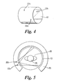
- the slot 24 in the detector array 12 is cut in a way that leads to the locus 23 of source points 22 as shown in Figure 1 .
- This helical slot 24, and the resulting helical source trajectory means that the set of data collected following X-ray transmission through the patient 26 is mathematically sufficient to form a true three dimensional image reconstruction. This is because the locus 23 of source points 22 passes through every plane passing through the scanning volume 28 which is essentially defined as the volume within the sensor array 12, i.e. radially inside the array 12 and between its two longitudinal ends 30, 32.
- the multi-focus X-ray source 20 comprises a continuous anode held at a high positive potential with respect to a plurality of grid controlled electron emitters. Each emitter is "turned on” in turn and the corresponding electron beam irradiates the target, so producing X-radiation from a respective source point 22. By changing the active grid controlled electron emitter, the effect of moving the X-ray source around the patient can be obtained.
- the X-ray source 20 is housed in a thick housing to avoid irradiating X-ray detectors 16 and other components in the system close to the X-ray source 20.
- An example of a suitable source is described in our co-pending UK patent application, Publication No. 2 418 529 X-Ray Tube Electron Sources.
- the source 20 therefore includes collimators arranged to restrict X-ray beams to only that part of the patient 26 that lies directly between the source and corresponding detectors.
- each of the X-ray source points 22 is operated in turn to scan the patient, and for each source point 22 data from the group of detector elements 16 opposite the source point 22 is used to form an image frame. All of the image frames produced in one scan are then processed to form a three-dimensional tomographic X-ray image of the patient as will be described in more detail below.
- the complete X-ray system comprises the multi-focus X-ray tube 20 and detector array 12, which is made up of a number of sensor blocks 34.
- Each sensor block comprises an array of detecting elements 16, typically 8x4 or 16x8 pixels, that are electronically coupled to suitable amplifiers, sample-and-hold amplifiers, analogue multiplexor and analogue-to-digital converter.
- Each sensor block 34 is connected to a respective data acquisition circuit (DAQ) 36 that provides gain and offset correction and, where appropriate, linearization for input to the image reconstruction process.
- DAQ data acquisition circuit
- the image reconstruction circuits are connected via a summing circuit 40 to visualisation circuit 42, which in turn is connected to a display 44.
- a system controller 46 is connected to, and controls operation of, the X-ray tube 20 and the detector bocks 34 and other circuits 36, 38, 40, 42 and display 44.
- a user interface 48 which can include, for example, a keyboard, a hand held controller, and action specific control buttons, is connected to the controller 46 to allow a user to control operation of the system.
- each scan the X-ray tube 20 is controlled so that each of the source points 22 produces a beam of X-rays in turn.
- the order of activation of the source points 22 can be sequential, or can be ordered so as to reduce the thermal load on the tube anode, as described in our co-pending UK patent No. 2 416 654 entitled X-ray Scanning.
- data from each of the detector blocks 34 is processed in the respective DAQ 36 and image reconstruction circuit 38.
- the reconstructed images from each reconstruction circuit 38 are summed and passed to a visualisation unit 42 that creates a 3D tomographic image.
- the images from subsequent scans are combined to form a real time 3D video image which is shown in the display 44.
- volume cardiac imaging single cycle
- cardiac cycle time 800 ms and a 4 ms tomographic scan time
- a single cardiac cycle movie will contain 200 volume tomographic images.
- a preferred use of this scanner is in cardiac angiography in which iodine contrast agent is passed through the heart and surrounding vessels.
- the scanner system of Figures 1 to 3 is set up for use in fluoroscopy.
- This can be single plane, bi-plane or multi-plane fluoroscopy.
- a single source point 22a is used, and a beam of X-rays passed from that source point 22a, through the patient, and onto a group 17 of the detector elements 16.
- the data from the detector elements 16 is used to form an image frame data set which represents a 2 dimensional X-ray projection image of the imaged volume.
- This process is repeated in successive imaging periods, which may be of the order of 5ms. It will be appreciated that this is significantly faster than conventional fluoroscopy for which the corresponding period is of the order of 40ms or more.
- the image frame data sets are output directly from the DAQs 36 to a frame store 50 from which they can be displayed in turn as images on the display 44 to provide a real time 2D video image of the patient.
- X-ray source points 22 Since a large number of X-ray source points 22 are present in the system, it can easily be controlled to alternate between two, three or more source points 22b, 22c spaced around the patient. For each source point 22a 22b, 22c, a corresponding group of detector elements 16 will be used to produce a respective series of fluoroscopic image frames. By cycling between the source points 22a, 22b, 22c simultaneous video images in a number of planes can be produced. These fluoroscopic images can either simply be displayed simultaneously on the display 44 or processed to provide a single video image combining features from each of the plane video images. The angle between planes may be adjusted electronically by switching the location of the emitting electron source. Applications for the system used in this mode are neuroradiology and neuroangiography.
- the fluoroscopic images produced can be improved by using the methods described in international patent application WO 2004/008 970 entitled Image Colouring and international patent application WO 2004/010 381 entitled Image Control.
- the system is set up to provide stereo imaging of the imaging volume 28.
- two source points 22d, 22e are used which are close together. Each of them is activated in turn to produce a respective transmission image data set from a corresponding group of detector elements 16 on the opposite side of the imaging volume 28.
- These image data sets are stored in the frame store 50.
- a pair of image frame data sets, one from each source point 22d, 22e, is combined to produce a stereo image data set representing an image of the imaged volume, and successive stereo images can be displayed to produce a real time stereo view video image of the imaged volume 28.
- the angle between the two sources 22d, 22e, and hence the degree of parallax, can be adjusted dynamically to suit the size of the patient or organ being imaged.
- the scanner can switch rapidly between any of the three modes of operation described above. This will reduce the rate at which data can be collected for each mode, but enables the images produced in each mode to be combined.
- the scanner is arranged to scan the object repeatedly to produce a 3D tomographic image of the object, but, between each successive pair of scans, to use one of the source points 22 to produce a 2D flouroscopic image of the object.
- the tomographic image is then analysed by the visualising unit 42 to identify specific features, which are then identified with corresponding features on the fluoroscopic image.
- the fluoroscopic image is then enhanced by mapping features from the 3D image onto the 2D image using software pointers to show the mapped features more clearly. This can be advantageous, for example where one or more features is obscured in the 2D image, or where two or more features cannot be distinguished from each other.
- features identified in the fluoroscopic image can be mapped directly onto the three-dimensional tomographic image. It will be appreciated that the automatic registration of the fluoroscopic image and volume tomographic data can be of major clinical advantage.
- the exact shape of the X-ray source can be modified substantially.
- the embodiment described above is the simplest to use in many circumstances as the regular helix with a single turn produces data which is simple to analyse.
- other shapes of source could be used.
- a helical locus 60 of X-ray source points 62 is again used, but in this case the helix has a plurality of turns around the detector array 64.
- the locus 66 of source points 68 is not in a helix, but is made up of two stepped loci 70, 71 each extending half way round the circumference of the cylindrical detector array 72 and along its full length.
- the detector array 74 is not straight cylindrical, but instead is part spherical being of larger circumference at its centre line 76 than at its longitudinal ends 78, 79.
- the locus 80 of source points 81 extends from one end 78 of the detector array 74 to the other 79 while following a single turn around its circumference.
Claims (11)
- Röntgen-Bildgebungssystem (10), das Folgendes umfasst: eine Multifokus-Röntgenquelle (20), die sich um ein Bildgebungsvolumen (28) herum erstreckt, das vom System abgebildet werden soll, und dies derart, dass eine Ortskurve (23) von Quellenpunkten (22) definiert wird, von denen aus Röntgenstrahlen durch das Bildgebungsvolumen gerichtet werden können, und ein zylindrisches Röntgendetektorarray (12), das sich ebenfalls um das Bildgebungsvolumen herum erstreckt und das so angeordnet ist, dass es Röntgenstrahlen von den Quellenpunkten (22) detektiert, die das Bildgebungsvolumen durchlaufen haben, wobei die Quellenpunkte (22) derart angeordnet sind, dass sie die Ortskurve als eine dreidimensionale Ortskurve (23) um das Bildgebungsvolumen (28) herum definieren, und wobei die Ortskurve wenigstens die Hälfte des Umfangs des zylindrischen Arrays abdeckt, und dies derart, dass Daten vom Detektorarray (12) dazu verwendet werden können, ein dreidimensionales tomographisches Bild eines stationären Objekts (26) innerhalb des Bildgebungsvolumens zu erzeugen, dadurch gekennzeichnet, dass die Ortskurve die gesamte Länge des zylindrischen Arrays abdeckt.
- System (10) nach Anspruch 1, wobei die Ortskurve (23) im Wesentlichen schraubenförmig ist.
- System (10) nach einem der beiden vorhergehenden Ansprüche, wobei die Ortskurve (23) durch im Wesentlichen jede Ebene verläuft, die durch das Bildgebungsvolumen (28) verläuft.
- System (10) nach einem der vorhergehenden Ansprüche, das außerdem Folgendes umfasst: ein Steuerungsmittel (46), das dafür eingerichtet ist, das Bildgebungsvolumen (28) abzutasten, indem jeder der Röntgenquellenpunkte (22) aktiviert und die zugehörigen Datensätze gesammelt werden, und ein Bildgebungsmittel, das dafür eingerichtet ist, aus den Datensätzen ein dreidimensionales Bild des Bildgebungsvolumens zu erzeugen.
- System (10) nach Anspruch 4, wobei das Steuerungsmittel (46) dafür eingerichtet ist, das Bildgebungsvolumen (28) wiederholt abzutasten, um aufeinanderfolgende Bilder des abgebildeten Volumens zu erzeugen.
- System (10) nach Anspruch 5, das außerdem ein Displaymittel (44) umfasst, das dafür eingerichtet ist, die aufeinanderfolgenden Bilder anzuzeigen, um ein Echtzeit-Videobild des abgebildeten Volumens zu erzeugen.
- System (10) nach einem der vorhergehenden Ansprüche, wobei das Steuerungsmittel (46) außerdem dafür eingerichtet ist, einen der Quellenpunkte (22) zu aktivieren, um ein Ebenenbild eines Objekts zu erzeugen und das Ebenenbild zur Anzeige zu speichern.
- System (10) nach Anspruch 7, wobei das Steuerungsmittel (46) dafür eingerichtet ist, den einen von den Quellenpunkten (22) wiederholt zu aktivieren, um hierdurch eine Folge von Ebenenbildern zu erzeugen, und die Ebenenbilder aufeinanderfolgend anzuzeigen, um ein Ebenen-Videobild herzustellen.
- System (10) nach Anspruch 7 oder Anspruch 8, wobei das Steuerungsmittel (46) dafür eingerichtet ist, zwischen einem ersten Modus, in dem es einen Datensatz für ein Ebenenbild erzeugt, und einem zweiten Modus, in dem es einen Datensatz für ein tomographisches Bild erzeugt, abzuwechseln und die Datensätze zu verarbeiten, um einen kombinierten Bilddatensatz zu erzeugen, zum Erstellen einer kombinierten Anzeige.
- System (10) nach Anspruch 9, wobei das Steuerungsmittel (46) dafür eingerichtet ist, einen weiteren der Quellenpunkte (22) zu aktivieren, der nahe bei dem einen Quellenpunkt (22) liegt, wobei ein Paar von Datensätzen erzeugt wird, und die Datensätze so zu kombinieren, dass das Ebenenbild oder jedes der Ebenenbilder ein Stereobild ist.
- System (10) nach Anspruch 9 oder Anspruch 10, wobei das Steuerungsmittel (46) dafür eingerichtet ist, die Datensätze zu verarbeiten, indem Merkmale aus einem der Datensätze auf den anderen der Datensätze abgebildet werden, um hierdurch das aus dem anderen der Datensätze erzeugte Bild zu verbessern.
Priority Applications (1)
| Application Number | Priority Date | Filing Date | Title |
|---|---|---|---|
| EP08016553A EP2002789B1 (de) | 2003-04-25 | 2004-04-23 | Röntgen-Scanning |
Applications Claiming Priority (2)
| Application Number | Priority Date | Filing Date | Title |
|---|---|---|---|
| GBGB0309379.6A GB0309379D0 (en) | 2003-04-25 | 2003-04-25 | X-ray scanning |
| PCT/GB2004/001747 WO2004096050A1 (en) | 2003-04-25 | 2004-04-23 | X-ray scanning system |
Related Child Applications (2)
| Application Number | Title | Priority Date | Filing Date |
|---|---|---|---|
| EP08016553A Division EP2002789B1 (de) | 2003-04-25 | 2004-04-23 | Röntgen-Scanning |
| EP08016553.3 Division-Into | 2008-09-19 |
Publications (2)
| Publication Number | Publication Date |
|---|---|
| EP1617764A1 EP1617764A1 (de) | 2006-01-25 |
| EP1617764B1 true EP1617764B1 (de) | 2011-10-26 |
Family
ID=9957202
Family Applications (2)
| Application Number | Title | Priority Date | Filing Date |
|---|---|---|---|
| EP08016553A Expired - Lifetime EP2002789B1 (de) | 2003-04-25 | 2004-04-23 | Röntgen-Scanning |
| EP04729135A Expired - Lifetime EP1617764B1 (de) | 2003-04-25 | 2004-04-23 | Röntgenscanner |
Family Applications Before (1)
| Application Number | Title | Priority Date | Filing Date |
|---|---|---|---|
| EP08016553A Expired - Lifetime EP2002789B1 (de) | 2003-04-25 | 2004-04-23 | Röntgen-Scanning |
Country Status (7)
| Country | Link |
|---|---|
| US (2) | US7684538B2 (de) |
| EP (2) | EP2002789B1 (de) |
| JP (2) | JP4377406B2 (de) |
| CN (2) | CN101569531B (de) |
| AT (1) | ATE530118T1 (de) |
| GB (2) | GB0309379D0 (de) |
| WO (1) | WO2004096050A1 (de) |
Cited By (4)
| Publication number | Priority date | Publication date | Assignee | Title |
|---|---|---|---|---|
| DE102016013533A1 (de) | 2016-11-12 | 2018-05-17 | H&P Advanced Technology GmbH | Computertomograph |
| WO2019042587A2 (de) | 2017-09-02 | 2019-03-07 | Cetteen Gmbh | Ansteuervorrichtung für eine röntgenröhre und verfahren zum betrieb einer röntgenröhre |
| US10901112B2 (en) | 2003-04-25 | 2021-01-26 | Rapiscan Systems, Inc. | X-ray scanning system with stationary x-ray sources |
| US10976271B2 (en) | 2005-12-16 | 2021-04-13 | Rapiscan Systems, Inc. | Stationary tomographic X-ray imaging systems for automatically sorting objects based on generated tomographic images |
Families Citing this family (73)
| Publication number | Priority date | Publication date | Assignee | Title |
|---|---|---|---|---|
| US8275091B2 (en) | 2002-07-23 | 2012-09-25 | Rapiscan Systems, Inc. | Compact mobile cargo scanning system |
| US7963695B2 (en) | 2002-07-23 | 2011-06-21 | Rapiscan Systems, Inc. | Rotatable boom cargo scanning system |
| GB0309379D0 (en) * | 2003-04-25 | 2003-06-04 | Cxr Ltd | X-ray scanning |
| US8451974B2 (en) | 2003-04-25 | 2013-05-28 | Rapiscan Systems, Inc. | X-ray tomographic inspection system for the identification of specific target items |
| US7949101B2 (en) | 2005-12-16 | 2011-05-24 | Rapiscan Systems, Inc. | X-ray scanners and X-ray sources therefor |
| US9113839B2 (en) | 2003-04-25 | 2015-08-25 | Rapiscon Systems, Inc. | X-ray inspection system and method |
| US8223919B2 (en) | 2003-04-25 | 2012-07-17 | Rapiscan Systems, Inc. | X-ray tomographic inspection systems for the identification of specific target items |
| GB0903198D0 (en) * | 2009-02-25 | 2009-04-08 | Cxr Ltd | X-Ray scanners |
| US8837669B2 (en) | 2003-04-25 | 2014-09-16 | Rapiscan Systems, Inc. | X-ray scanning system |
| US8804899B2 (en) | 2003-04-25 | 2014-08-12 | Rapiscan Systems, Inc. | Imaging, data acquisition, data transmission, and data distribution methods and systems for high data rate tomographic X-ray scanners |
| US6928141B2 (en) | 2003-06-20 | 2005-08-09 | Rapiscan, Inc. | Relocatable X-ray imaging system and method for inspecting commercial vehicles and cargo containers |
| JP2007534415A (ja) * | 2004-04-28 | 2007-11-29 | コーニンクレッカ フィリップス エレクトロニクス エヌ ヴィ | 3次元電子ビームコンピュータ断層撮影 |
| US7471764B2 (en) | 2005-04-15 | 2008-12-30 | Rapiscan Security Products, Inc. | X-ray imaging system having improved weather resistance |
| US8155262B2 (en) * | 2005-04-25 | 2012-04-10 | The University Of North Carolina At Chapel Hill | Methods, systems, and computer program products for multiplexing computed tomography |
| DE102005059804A1 (de) * | 2005-12-14 | 2007-07-05 | Siemens Ag | Verfahren und Vorrichtung zur Bewegungskorrektur bei der Bildgebung während einer medizinischen Intervention |
| RU2008151410A (ru) * | 2006-05-25 | 2010-06-27 | Конинклейке Филипс Электроникс, Н.В. (Nl) | Полупериодная замкнутая спиральная траектория для компьютерной томографии с коническим лучом |
| US7616731B2 (en) | 2006-08-30 | 2009-11-10 | General Electric Company | Acquisition and reconstruction of projection data using a stationary CT geometry |
| US7835486B2 (en) * | 2006-08-30 | 2010-11-16 | General Electric Company | Acquisition and reconstruction of projection data using a stationary CT geometry |
| US20080056432A1 (en) * | 2006-08-30 | 2008-03-06 | General Electric Company | Reconstruction of CT projection data |
| US7706499B2 (en) * | 2006-08-30 | 2010-04-27 | General Electric Company | Acquisition and reconstruction of projection data using a stationary CT geometry |
| CN101512379B (zh) * | 2006-08-30 | 2013-06-05 | 通用电气公司 | 使用静止计算机x射线断层造影几何结构的投影数据的采集和再现 |
| JP5539719B2 (ja) * | 2006-08-31 | 2014-07-02 | コーニンクレッカ フィリップス エヌ ヴェ | 画像形成システム |
| US7723674B2 (en) | 2006-09-21 | 2010-05-25 | Siemens Medical Solutions Usa, Inc. | Attenuation correction for SPECT imaging using non-classical orbits of many small gamma cameras |
| US7825383B2 (en) | 2006-09-21 | 2010-11-02 | Siemens Medical Solutions Usa, Inc. | Mobile camera for organ targeted imaging |
| US7813478B2 (en) * | 2007-02-08 | 2010-10-12 | Varian Medical Systems, Inc. | Method and apparatus to facilitate provision and use of multiple X-ray sources |
| ES2722181T3 (es) * | 2008-01-15 | 2019-08-07 | Sunrise R & D Holdings Llc | Sistema de rastreo de ubicación en tiempo real de compradores de tiendas usando una multi-red de comunicación |
| JP2009226198A (ja) | 2008-02-28 | 2009-10-08 | Fujifilm Corp | 放射線画像撮影システム、撮影指示情報の並べ替え装置、プログラム及び放射線画像撮影方法 |
| GB0803644D0 (en) | 2008-02-28 | 2008-04-02 | Rapiscan Security Products Inc | Scanning systems |
| GB0803641D0 (en) | 2008-02-28 | 2008-04-02 | Rapiscan Security Products Inc | Scanning systems |
| US7949089B2 (en) * | 2008-04-10 | 2011-05-24 | Arineta Ltd. | Apparatus and method for tracking feature's position in human body |
| GB0809110D0 (en) | 2008-05-20 | 2008-06-25 | Rapiscan Security Products Inc | Gantry scanner systems |
| US9773268B2 (en) | 2008-06-16 | 2017-09-26 | Sunrise R&D Holdings, Llc | System of acquiring shopper insights and influencing shopper purchase decisions |
| JP2010074823A (ja) | 2008-08-22 | 2010-04-02 | Panasonic Corp | 録画編集装置 |
| JP4693884B2 (ja) * | 2008-09-18 | 2011-06-01 | キヤノン株式会社 | マルチx線撮影装置及びその制御方法 |
| DE102008050353B3 (de) * | 2008-10-02 | 2010-05-20 | Siemens Aktiengesellschaft | Kreisförmige Multi-Strahl-Röntgenvorrichtung |
| US8600003B2 (en) | 2009-01-16 | 2013-12-03 | The University Of North Carolina At Chapel Hill | Compact microbeam radiation therapy systems and methods for cancer treatment and research |
| US7756249B1 (en) * | 2009-02-19 | 2010-07-13 | Morpho Detection, Inc. | Compact multi-focus x-ray source, x-ray diffraction imaging system, and method for fabricating compact multi-focus x-ray source |
| GB2501023B (en) | 2009-05-26 | 2014-02-12 | Rapiscan Systems Inc | X-ray tomographic inspection systems for the identification of specific target items |
| EP3686901A1 (de) | 2009-05-26 | 2020-07-29 | Rapiscan Systems, Inc. | Röntgentomographieprüfverfahren |
| JP5677738B2 (ja) * | 2009-12-24 | 2015-02-25 | 株式会社東芝 | X線コンピュータ断層撮影装置 |
| CN102804326B (zh) * | 2010-01-19 | 2016-01-20 | 拉皮斯坎系统股份有限公司 | 多视图货物扫描器 |
| US8713131B2 (en) | 2010-02-23 | 2014-04-29 | RHPiscan Systems, Inc. | Simultaneous image distribution and archiving |
| JP5782525B2 (ja) * | 2010-11-27 | 2015-09-24 | アイシイアールシイオー・インコーポレーテッド | コンピュータトモグラフィ及びトモシンセシスシステム |
| US9218933B2 (en) | 2011-06-09 | 2015-12-22 | Rapidscan Systems, Inc. | Low-dose radiographic imaging system |
| CN103308535B (zh) * | 2012-03-09 | 2016-04-13 | 同方威视技术股份有限公司 | 用于射线扫描成像的设备和方法 |
| CN104486997B (zh) * | 2012-06-05 | 2017-07-25 | 拉皮斯坎系统股份有限公司 | X射线扫描系统的射线源激发模式的最佳化 |
| WO2014121097A1 (en) | 2013-01-31 | 2014-08-07 | Rapiscan Systems, Inc. | Portable security inspection system |
| US9778391B2 (en) * | 2013-03-15 | 2017-10-03 | Varex Imaging Corporation | Systems and methods for multi-view imaging and tomography |
| DE102013206252A1 (de) * | 2013-04-09 | 2014-10-09 | Helmholtz-Zentrum Dresden - Rossendorf E.V. | Anordnung zur schnellen Elektronenstrahl-Röntgencomputertomographie |
| CN103499593A (zh) * | 2013-09-23 | 2014-01-08 | 深圳先进技术研究院 | 一种计算机断层扫描系统 |
| CN107073936B (zh) * | 2014-05-20 | 2019-12-13 | 维罗斯-纯粹数字有限公司 | 打印系统及方法 |
| CN105361900B (zh) * | 2014-08-26 | 2019-01-22 | 北京纳米维景科技有限公司 | 静态实时ct成像系统及其成像控制方法 |
| CN104720838B (zh) * | 2015-01-13 | 2018-02-09 | 乐普(北京)医疗装备有限公司 | 一种血管造影影像采集装置及方法 |
| GB2536650A (en) | 2015-03-24 | 2016-09-28 | Augmedics Ltd | Method and system for combining video-based and optic-based augmented reality in a near eye display |
| CN106547505B (zh) * | 2015-09-22 | 2021-02-05 | 同方威视技术股份有限公司 | 用于实时滑动显示扫描图像的方法及系统 |
| EP3239698A1 (de) * | 2016-04-26 | 2017-11-01 | Danmarks Tekniske Universitet | Hochpräzise computertomografie für metrologie |
| CN106526686B (zh) * | 2016-12-07 | 2019-05-07 | 同方威视技术股份有限公司 | 螺旋ct设备和三维图像重建方法 |
| JP7217847B2 (ja) * | 2017-02-27 | 2023-02-06 | 北京納米維景科技有限公司 | 広視野のニーズに適する静態リアルタイムct画像形成システム及びその画像形成方法 |
| WO2018195016A1 (en) | 2017-04-17 | 2018-10-25 | Rapiscan Systems, Inc. | X-ray tomography inspection systems and methods |
| US10585206B2 (en) | 2017-09-06 | 2020-03-10 | Rapiscan Systems, Inc. | Method and system for a multi-view scanner |
| WO2019211741A1 (en) | 2018-05-02 | 2019-11-07 | Augmedics Ltd. | Registration of a fiducial marker for an augmented reality system |
| US11766296B2 (en) | 2018-11-26 | 2023-09-26 | Augmedics Ltd. | Tracking system for image-guided surgery |
| CN110327070A (zh) * | 2019-07-12 | 2019-10-15 | 山东大骋医疗科技有限公司 | 具有储能系统的ct设备 |
| US11160523B2 (en) * | 2019-09-30 | 2021-11-02 | GE Precision Healthcare LLC | Systems and methods for cardiac imaging |
| US11604152B2 (en) * | 2019-10-09 | 2023-03-14 | Baker Hughes Oilfield Operations Llc | Fast industrial computed tomography for large objects |
| US11382712B2 (en) | 2019-12-22 | 2022-07-12 | Augmedics Ltd. | Mirroring in image guided surgery |
| US11594001B2 (en) | 2020-01-20 | 2023-02-28 | Rapiscan Systems, Inc. | Methods and systems for generating three-dimensional images that enable improved visualization and interaction with objects in the three-dimensional images |
| US11212902B2 (en) | 2020-02-25 | 2021-12-28 | Rapiscan Systems, Inc. | Multiplexed drive systems and methods for a multi-emitter X-ray source |
| US11193898B1 (en) | 2020-06-01 | 2021-12-07 | American Science And Engineering, Inc. | Systems and methods for controlling image contrast in an X-ray system |
| EP3933881A1 (de) | 2020-06-30 | 2022-01-05 | VEC Imaging GmbH & Co. KG | Röntgenquelle mit mehreren gittern |
| CN114724300A (zh) * | 2021-01-04 | 2022-07-08 | 南京造币有限公司 | 一种卷包装硬币枚数核查装置 |
| WO2022183191A1 (en) | 2021-02-23 | 2022-09-01 | Rapiscan Systems, Inc. | Systems and methods for eliminating cross-talk in scanning systems having multiple x-ray sources |
| US11896445B2 (en) | 2021-07-07 | 2024-02-13 | Augmedics Ltd. | Iliac pin and adapter |
Family Cites Families (278)
| Publication number | Priority date | Publication date | Assignee | Title |
|---|---|---|---|---|
| US2952790A (en) | 1957-07-15 | 1960-09-13 | Raytheon Co | X-ray tubes |
| US3239706A (en) | 1961-04-17 | 1966-03-08 | High Voltage Engineering Corp | X-ray target |
| US3768645A (en) | 1971-02-22 | 1973-10-30 | Sunkist Growers Inc | Method and means for automatically detecting and sorting produce according to internal damage |
| GB1497396A (en) | 1974-03-23 | 1978-01-12 | Emi Ltd | Radiography |
| DE2442809A1 (de) | 1974-09-06 | 1976-03-18 | Philips Patentverwaltung | Anordnung zur ermittlung der absorption in einem koerper |
| USRE32961E (en) | 1974-09-06 | 1989-06-20 | U.S. Philips Corporation | Device for measuring local radiation absorption in a body |
| GB1526041A (en) | 1975-08-29 | 1978-09-27 | Emi Ltd | Sources of x-radiation |
| NL7611391A (nl) | 1975-10-18 | 1977-04-20 | Emi Ltd | Roentgentoestel. |
| DE2647167A1 (de) | 1976-10-19 | 1978-04-20 | Siemens Ag | Verfahren zur herstellung von schichtaufnahmen mit roentgen- oder aehnlich durchdringenden strahlen |
| DE2705640A1 (de) | 1977-02-10 | 1978-08-17 | Siemens Ag | Rechnersystem fuer den bildaufbau eines koerperschnittbildes und verfahren zum betrieb des rechnersystems |
| US4105922A (en) | 1977-04-11 | 1978-08-08 | General Electric Company | CT number identifier in a computed tomography system |
| DE2729353A1 (de) | 1977-06-29 | 1979-01-11 | Siemens Ag | Roentgenroehre mit wanderndem brennfleck |
| DE2807735B2 (de) | 1978-02-23 | 1979-12-20 | Philips Patentverwaltung Gmbh, 2000 Hamburg | Röntgenröhre mit einem aus Metall bestehenden Röhrenkolben |
| US4228353A (en) | 1978-05-02 | 1980-10-14 | Johnson Steven A | Multiple-phase flowmeter and materials analysis apparatus and method |
| JPS5546408A (en) | 1978-09-29 | 1980-04-01 | Toshiba Corp | X-ray device |
| US4266425A (en) | 1979-11-09 | 1981-05-12 | Zikonix Corporation | Method for continuously determining the composition and mass flow of butter and similar substances from a manufacturing process |
| US4352021A (en) | 1980-01-07 | 1982-09-28 | The Regents Of The University Of California | X-Ray transmission scanning system and method and electron beam X-ray scan tube for use therewith |
| GB2089109B (en) | 1980-12-03 | 1985-05-15 | Machlett Lab Inc | X-rays targets and tubes |
| DE3107949A1 (de) | 1981-03-02 | 1982-09-16 | Siemens AG, 1000 Berlin und 8000 München | Roentgenroehre |
| JPS57175247A (en) | 1981-04-23 | 1982-10-28 | Toshiba Corp | Radiation void factor meter |
| FR2534066B1 (fr) | 1982-10-05 | 1989-09-08 | Thomson Csf | Tube a rayons x produisant un faisceau a haut rendement, notamment en forme de pinceau |
| JPS5916254A (ja) | 1983-06-03 | 1984-01-27 | Toshiba Corp | 携帯用x線装置 |
| JPS6073442A (ja) * | 1983-09-30 | 1985-04-25 | Toshiba Corp | 放射線断層測定装置 |
| US4672649A (en) * | 1984-05-29 | 1987-06-09 | Imatron, Inc. | Three dimensional scanned projection radiography using high speed computed tomographic scanning system |
| GB8521287D0 (en) | 1985-08-27 | 1985-10-02 | Frith B | Flow measurement & imaging |
| US4799247A (en) | 1986-06-20 | 1989-01-17 | American Science And Engineering, Inc. | X-ray imaging particularly adapted for low Z materials |
| JPS6321040A (ja) | 1986-07-16 | 1988-01-28 | 工業技術院長 | 超高速x線ctスキヤナ |
| JPS63109653A (ja) | 1986-10-27 | 1988-05-14 | Sharp Corp | 情報登録検索装置 |
| JPS6434333A (en) * | 1987-07-31 | 1989-02-03 | Toshiba Corp | Image processing apparatus |
| GB2212903B (en) | 1987-11-24 | 1991-11-06 | Rolls Royce Plc | Measuring two phase flow in pipes. |
| US4887604A (en) | 1988-05-16 | 1989-12-19 | Science Research Laboratory, Inc. | Apparatus for performing dual energy medical imaging |
| EP0390950B1 (de) | 1989-04-06 | 1993-01-13 | Heimann Systems GmbH & Co. KG | Materialprüfanlage |
| EP0432568A3 (en) | 1989-12-11 | 1991-08-28 | General Electric Company | X ray tube anode and tube having same |
| US5319547A (en) | 1990-08-10 | 1994-06-07 | Vivid Technologies, Inc. | Device and method for inspection of baggage and other objects |
| DE4100297A1 (de) | 1991-01-08 | 1992-07-09 | Philips Patentverwaltung | Roentgenroehre |
| DE4103588C1 (de) | 1991-02-06 | 1992-05-27 | Siemens Ag, 8000 Muenchen, De | |
| US5272627A (en) | 1991-03-27 | 1993-12-21 | Gulton Industries, Inc. | Data converter for CT data acquisition system |
| US5144191A (en) | 1991-06-12 | 1992-09-01 | Mcnc | Horizontal microelectronic field emission devices |
| DE69223884T2 (de) * | 1991-09-12 | 1998-08-27 | Toshiba Kawasaki Kk | Verfahren und Vorrichtung zur Erzeugung von Röntgencomputertomogrammen und zum Erzeugen von Schattenbildern mittels spiralförmiger Abtastung |
| JP3325301B2 (ja) * | 1991-09-12 | 2002-09-17 | 株式会社東芝 | X線ct装置 |
| US5367552A (en) | 1991-10-03 | 1994-11-22 | In Vision Technologies, Inc. | Automatic concealed object detection system having a pre-scan stage |
| US5182764A (en) | 1991-10-03 | 1993-01-26 | Invision Technologies, Inc. | Automatic concealed object detection system having a pre-scan stage |
| JP3631235B2 (ja) | 1992-05-27 | 2005-03-23 | 株式会社東芝 | X線ct装置 |
| JP3441455B2 (ja) | 1992-05-27 | 2003-09-02 | 株式会社東芝 | X線ct装置 |
| JP2005013768A (ja) | 1992-05-27 | 2005-01-20 | Toshiba Corp | X線ct装置 |
| US5966422A (en) | 1992-07-20 | 1999-10-12 | Picker Medical Systems, Ltd. | Multiple source CT scanner |
| DE4228559A1 (de) | 1992-08-27 | 1994-03-03 | Dagang Tan | Röntgenröhre mit einer Transmissionsanode |
| US5410156A (en) | 1992-10-21 | 1995-04-25 | Miller; Thomas G. | High energy x-y neutron detector and radiographic/tomographic device |
| US5557108A (en) | 1993-10-25 | 1996-09-17 | T+E,Uml U+Ee Mer; T+E,Uml U+Ee May O. | Integrated substance detection and identification system |
| US5511104A (en) | 1994-03-11 | 1996-04-23 | Siemens Aktiengesellschaft | X-ray tube |
| US5490196A (en) | 1994-03-18 | 1996-02-06 | Metorex International Oy | Multi energy system for x-ray imaging applications |
| US5467377A (en) | 1994-04-15 | 1995-11-14 | Dawson; Ralph L. | Computed tomographic scanner |
| US5606167A (en) | 1994-07-11 | 1997-02-25 | Miller; Thomas G. | Contraband detection apparatus and method |
| DE4436688A1 (de) * | 1994-10-13 | 1996-04-25 | Siemens Ag | Computertomograph |
| US5712926A (en) | 1994-10-20 | 1998-01-27 | Eberhard; Jeffrey Wayne | X-ray computed tomography (CT) system for detecting thin objects |
| AUPN226295A0 (en) | 1995-04-07 | 1995-05-04 | Technological Resources Pty Limited | A method and an apparatus for analysing a material |
| US6216540B1 (en) | 1995-06-06 | 2001-04-17 | Robert S. Nelson | High resolution device and method for imaging concealed objects within an obscuring medium |
| US5600700A (en) | 1995-09-25 | 1997-02-04 | Vivid Technologies, Inc. | Detecting explosives or other contraband by employing transmitted and scattered X-rays |
| US5642393A (en) | 1995-09-26 | 1997-06-24 | Vivid Technologies, Inc. | Detecting contraband by employing interactive multiprobe tomography |
| US6507025B1 (en) | 1995-10-23 | 2003-01-14 | Science Applications International Corporation | Density detection using real time discrete photon counting for fast moving targets |
| US7045787B1 (en) | 1995-10-23 | 2006-05-16 | Science Applications International Corporation | Density detection using real time discrete photon counting for fast moving targets |
| US6018562A (en) | 1995-11-13 | 2000-01-25 | The United States Of America As Represented By The Secretary Of The Army | Apparatus and method for automatic recognition of concealed objects using multiple energy computed tomography |
| DE19542438C1 (de) | 1995-11-14 | 1996-11-28 | Siemens Ag | Röntgenröhre |
| US6304629B1 (en) | 1996-01-11 | 2001-10-16 | Granville Todd Conway | Compact scanner apparatus and method |
| US5764683B1 (en) | 1996-02-12 | 2000-11-21 | American Science & Eng Inc | Mobile x-ray inspection system for large objects |
| US5633907A (en) | 1996-03-21 | 1997-05-27 | General Electric Company | X-ray tube electron beam formation and focusing |
| DE19618749A1 (de) * | 1996-05-09 | 1997-11-13 | Siemens Ag | Röntgen-Computertomograph |
| JPH105206A (ja) * | 1996-06-25 | 1998-01-13 | Shimadzu Corp | ディジタルx線撮影装置 |
| US5661774A (en) | 1996-06-27 | 1997-08-26 | Analogic Corporation | Dual energy power supply |
| DE69716169T2 (de) | 1996-06-27 | 2003-06-12 | Analogic Corp | Vorrichtung zum Erfassen für axiale Transversal- und Quadratur-Tomographie |
| DE69719988D1 (de) | 1996-07-12 | 2003-04-24 | American Science & Eng Inc | System für tomographie mit seitenstreuung |
| GB2315546A (en) | 1996-07-18 | 1998-02-04 | Imperial College | Luggage scanner |
| WO1998003889A1 (en) | 1996-07-22 | 1998-01-29 | American Science And Engineering, Inc. | System for rapid x-ray inspection of enclosures |
| EP0825457A3 (de) | 1996-08-19 | 2002-02-13 | Analogic Corporation | Nachweisverfahren und -system für die Tomografie unter einer Mehrzahl von Winkeln |
| US5974111A (en) | 1996-09-24 | 1999-10-26 | Vivid Technologies, Inc. | Identifying explosives or other contraband by employing transmitted or scattered X-rays |
| JPH10211196A (ja) * | 1997-01-31 | 1998-08-11 | Olympus Optical Co Ltd | X線ctスキャナ装置 |
| US6037597A (en) | 1997-02-18 | 2000-03-14 | Neutech Systems, Inc. | Non-destructive detection systems and methods |
| US5859891A (en) | 1997-03-07 | 1999-01-12 | Hibbard; Lyn | Autosegmentation/autocontouring system and method for use with three-dimensional radiation therapy treatment planning |
| US5802134A (en) | 1997-04-09 | 1998-09-01 | Analogic Corporation | Nutating slice CT image reconstruction apparatus and method |
| JP4346128B2 (ja) * | 1997-09-09 | 2009-10-21 | 株式会社東芝 | X線ct装置 |
| US7028899B2 (en) | 1999-06-07 | 2006-04-18 | Metrologic Instruments, Inc. | Method of speckle-noise pattern reduction and apparatus therefore based on reducing the temporal-coherence of the planar laser illumination beam before it illuminates the target object by applying temporal phase modulation techniques during the transmission of the plib towards the target |
| US5901196A (en) * | 1997-09-30 | 1999-05-04 | Siemens Corporate Research, Inc. | Reduction of hitlist size in spiral cone beam CT by use of local radon origins |
| US6256404B1 (en) | 1997-10-10 | 2001-07-03 | Analogic Corporation | Computed tomography scanning apparatus and method using adaptive reconstruction window |
| US5901198A (en) | 1997-10-10 | 1999-05-04 | Analogic Corporation | Computed tomography scanning target detection using target surface normals |
| US5982843A (en) | 1997-10-10 | 1999-11-09 | Analogic Corporation | Closed loop air conditioning system for a computed tomography scanner |
| US6091795A (en) | 1997-10-10 | 2000-07-18 | Analogic Corporation | Area detector array for computer tomography scanning system |
| US6149592A (en) * | 1997-11-26 | 2000-11-21 | Picker International, Inc. | Integrated fluoroscopic projection image data, volumetric image data, and surgical device position data |
| US6005918A (en) | 1997-12-19 | 1999-12-21 | Picker International, Inc. | X-ray tube window heat shield |
| US5987097A (en) | 1997-12-23 | 1999-11-16 | General Electric Company | X-ray tube having reduced window heating |
| US6076400A (en) | 1998-02-11 | 2000-06-20 | Analogic Corporation | Apparatus and method for classifying objects in computed tomography data using density dependent mass thresholds |
| US6128365A (en) | 1998-02-11 | 2000-10-03 | Analogic Corporation | Apparatus and method for combining related objects in computed tomography data |
| US6075871A (en) | 1998-02-11 | 2000-06-13 | Analogic Corporation | Apparatus and method for eroding objects in computed tomography data |
| US6078642A (en) | 1998-02-11 | 2000-06-20 | Analogice Corporation | Apparatus and method for density discrimination of objects in computed tomography data using multiple density ranges |
| US6035014A (en) | 1998-02-11 | 2000-03-07 | Analogic Corporation | Multiple-stage apparatus and method for detecting objects in computed tomography data |
| US6108396A (en) | 1998-02-11 | 2000-08-22 | Analogic Corporation | Apparatus and method for correcting object density in computed tomography data |
| US6067366A (en) | 1998-02-11 | 2000-05-23 | Analogic Corporation | Apparatus and method for detecting objects in computed tomography data using erosion and dilation of objects |
| US6317509B1 (en) | 1998-02-11 | 2001-11-13 | Analogic Corporation | Computed tomography apparatus and method for classifying objects |
| US6111974A (en) | 1998-02-11 | 2000-08-29 | Analogic Corporation | Apparatus and method for detecting sheet objects in computed tomography data |
| US6026171A (en) | 1998-02-11 | 2000-02-15 | Analogic Corporation | Apparatus and method for detection of liquids in computed tomography data |
| US6026143A (en) | 1998-02-11 | 2000-02-15 | Analogic Corporation | Apparatus and method for detecting sheet objects in computed tomography data |
| US6272230B1 (en) | 1998-02-11 | 2001-08-07 | Analogic Corporation | Apparatus and method for optimizing detection of objects in computed tomography data |
| US6218943B1 (en) | 1998-03-27 | 2001-04-17 | Vivid Technologies, Inc. | Contraband detection and article reclaim system |
| US6236709B1 (en) | 1998-05-04 | 2001-05-22 | Ensco, Inc. | Continuous high speed tomographic imaging system and method |
| US6088423A (en) | 1998-06-05 | 2000-07-11 | Vivid Technologies, Inc. | Multiview x-ray based system for detecting contraband such as in baggage |
| US6118852A (en) | 1998-07-02 | 2000-09-12 | General Electric Company | Aluminum x-ray transmissive window for an x-ray tube vacuum vessel |
| US6188745B1 (en) | 1998-09-23 | 2001-02-13 | Analogic Corporation | CT scanner comprising a spatially encoded detector array arrangement and method |
| US6183139B1 (en) | 1998-10-06 | 2001-02-06 | Cardiac Mariners, Inc. | X-ray scanning method and apparatus |
| JP2000107173A (ja) * | 1998-10-08 | 2000-04-18 | Fuji Photo Film Co Ltd | 3次元用放射線画像形成装置 |
| US6021174A (en) | 1998-10-26 | 2000-02-01 | Picker International, Inc. | Use of shaped charge explosives in the manufacture of x-ray tube targets |
| DE69937067D1 (de) | 1998-11-30 | 2007-10-18 | Invision Technologies Inc | Eindringungsfreies untersuchungssystem |
| US7050536B1 (en) | 1998-11-30 | 2006-05-23 | Invision Technologies, Inc. | Nonintrusive inspection system |
| US6181765B1 (en) | 1998-12-10 | 2001-01-30 | General Electric Company | X-ray tube assembly |
| US6195444B1 (en) | 1999-01-12 | 2001-02-27 | Analogic Corporation | Apparatus and method for detecting concealed objects in computed tomography data |
| US6345113B1 (en) | 1999-01-12 | 2002-02-05 | Analogic Corporation | Apparatus and method for processing object data in computed tomography data using object projections |
| US6687333B2 (en) | 1999-01-25 | 2004-02-03 | Vanderbilt University | System and method for producing pulsed monochromatic X-rays |
| US6429578B1 (en) | 1999-01-26 | 2002-08-06 | Mats Danielsson | Diagnostic and therapeutic detector system for imaging with low and high energy X-ray and electrons |
| US6459764B1 (en) | 1999-01-27 | 2002-10-01 | American Science And Engineering, Inc. | Drive-through vehicle inspection system |
| US6185272B1 (en) | 1999-03-15 | 2001-02-06 | Analogic Corporation | Architecture for CT scanning system |
| DE19916664A1 (de) | 1999-04-14 | 2000-10-19 | Heimann Systems Gmbh & Co | Verfahren zur Bearbeitung eines Röntgenbildes |
| US6546072B1 (en) | 1999-07-30 | 2003-04-08 | American Science And Engineering, Inc. | Transmission enhanced scatter imaging |
| US6269142B1 (en) | 1999-08-11 | 2001-07-31 | Steven W. Smith | Interrupted-fan-beam imaging |
| US6528787B2 (en) | 1999-11-30 | 2003-03-04 | Jeol Ltd. | Scanning electron microscope |
| JP2001176408A (ja) | 1999-12-15 | 2001-06-29 | New Japan Radio Co Ltd | 電子管 |
| US6418189B1 (en) | 2000-01-24 | 2002-07-09 | Analogic Corporation | Explosive material detection apparatus and method using dual energy information of a scan |
| US6459761B1 (en) | 2000-02-10 | 2002-10-01 | American Science And Engineering, Inc. | Spectrally shaped x-ray inspection system |
| JP4161513B2 (ja) | 2000-04-21 | 2008-10-08 | 株式会社島津製作所 | 二次ターゲット装置及び蛍光x線分析装置 |
| CA2348150C (en) | 2000-05-25 | 2007-03-13 | Esam M.A. Hussein | Non-rotating x-ray system for three-dimensional, three-parameter imaging |
| US20020031202A1 (en) | 2000-06-07 | 2002-03-14 | Joseph Callerame | X-ray scatter and transmission system with coded beams |
| CA2355560C (en) | 2000-08-28 | 2003-11-18 | Balza Achmad | X-ray compton scatter density measurement at a point within an object |
| DE10044357A1 (de) | 2000-09-07 | 2002-03-21 | Heimann Systems Gmbh & Co | Detektoranordnung zur Detektion von Röntgenstrahlen |
| US6737652B2 (en) | 2000-09-29 | 2004-05-18 | Massachusetts Institute Of Technology | Coded aperture imaging |
| US20040213378A1 (en) | 2003-04-24 | 2004-10-28 | The University Of North Carolina At Chapel Hill | Computed tomography system for imaging of human and small animal |
| US6876724B2 (en) * | 2000-10-06 | 2005-04-05 | The University Of North Carolina - Chapel Hill | Large-area individually addressable multi-beam x-ray system and method of forming same |
| US6980627B2 (en) | 2000-10-06 | 2005-12-27 | Xintek, Inc. | Devices and methods for producing multiple x-ray beams from multiple locations |
| AU2001294014A1 (en) | 2000-10-11 | 2002-04-22 | University Of Southampton | Gamma-ray spectrometry |
| US6748043B1 (en) | 2000-10-19 | 2004-06-08 | Analogic Corporation | Method and apparatus for stabilizing the measurement of CT numbers |
| US6735271B1 (en) | 2000-11-28 | 2004-05-11 | Ge Medical Systems Global Technology Company Llc | Electron beam computed tomographic scanner system with helical or tilted target, collimator, and detector components to eliminate cone beam error and to scan continuously moving objects |
| JP2002320610A (ja) * | 2001-02-23 | 2002-11-05 | Mitsubishi Heavy Ind Ltd | X線ct装置とx線ct装置撮影方法 |
| CA2410892A1 (en) | 2001-02-28 | 2002-11-29 | Mitsubishi Heavy Industries, Ltd. | Multi-radiation source x-ray ct apparatus |
| US6324249B1 (en) | 2001-03-21 | 2001-11-27 | Agilent Technologies, Inc. | Electronic planar laminography system and method |
| WO2002082125A1 (en) | 2001-04-03 | 2002-10-17 | L-3 Communications Security & Detection Systems | X-ray inspection system |
| US6721391B2 (en) | 2001-04-03 | 2004-04-13 | L-3 Communications Security And Detection Systems | Remote baggage screening system, software and method |
| US6813374B1 (en) | 2001-04-25 | 2004-11-02 | Analogic Corporation | Method and apparatus for automatic image quality assessment |
| US6721387B1 (en) | 2001-06-13 | 2004-04-13 | Analogic Corporation | Method of and system for reducing metal artifacts in images generated by x-ray scanning devices |
| DE10129463A1 (de) | 2001-06-19 | 2003-01-02 | Philips Corp Intellectual Pty | Röntgenstrahler mit einem Flüssigmetall-Target |
| GB0115615D0 (en) | 2001-06-27 | 2001-08-15 | Univ Coventry | Image segmentation |
| US6661876B2 (en) | 2001-07-30 | 2003-12-09 | Moxtek, Inc. | Mobile miniature X-ray source |
| US6914959B2 (en) | 2001-08-09 | 2005-07-05 | Analogic Corporation | Combined radiation therapy and imaging system and method |
| US6636623B2 (en) | 2001-08-10 | 2003-10-21 | Visiongate, Inc. | Optical projection imaging system and method for automatically detecting cells with molecular marker compartmentalization associated with malignancy and disease |
| CN1185482C (zh) | 2001-08-14 | 2005-01-19 | 清华大学 | 航空集装箱/托盘货物检查系统 |
| DE10143131B4 (de) | 2001-09-03 | 2006-03-09 | Siemens Ag | Verfahren zur Ermittlung von Dichte- und Ordnungszahlverteilungen bei radiographischen Untersuchungsverfahren |
| DE10149254B4 (de) | 2001-10-05 | 2006-04-20 | Smiths Heimann Gmbh | Verfahren und Vorrichtung zur Detektion eines bestimmten Materials in einem Objekt mittels elektromagnetischer Strahlen |
| WO2003051201A2 (en) * | 2001-12-14 | 2003-06-26 | Wisconsin Alumni Research Foundation | Virtual spherical anode computed tomography |
| JP3888156B2 (ja) * | 2001-12-26 | 2007-02-28 | 株式会社日立製作所 | 放射線検査装置 |
| US6922455B2 (en) | 2002-01-28 | 2005-07-26 | Starfire Industries Management, Inc. | Gas-target neutron generation and applications |
| US6816571B2 (en) | 2002-02-06 | 2004-11-09 | L-3 Communications Security And Detection Systems Corporation Delaware | Method and apparatus for transmitting information about a target object between a prescanner and a CT scanner |
| US6754298B2 (en) | 2002-02-20 | 2004-06-22 | The Regents Of The University Of Michigan | Method for statistically reconstructing images from a plurality of transmission measurements having energy diversity and image reconstructor apparatus utilizing the method |
| US6618466B1 (en) | 2002-02-21 | 2003-09-09 | University Of Rochester | Apparatus and method for x-ray scatter reduction and correction for fan beam CT and cone beam volume CT |
| US6654443B1 (en) | 2002-02-25 | 2003-11-25 | Ge Medical Systems Global Technology Co., Llc | Thermal sensing detector cell for a computed tomography system and method of manufacturing same |
| US6459755B1 (en) | 2002-02-26 | 2002-10-01 | Ge Medical Systems Global Technology Co. Llc | Method and apparatus for administering low dose CT scans |
| US6775348B2 (en) | 2002-02-27 | 2004-08-10 | General Electric Company | Fiber optic scintillator with optical gain for a computed tomography system and method of manufacturing same |
| JP2005520661A (ja) | 2002-03-23 | 2005-07-14 | コーニンクレッカ フィリップス エレクトロニクス エヌ ヴィ | 対象に含まれる構造のインタラクティブなセグメンテーションの方法 |
| US6647095B2 (en) | 2002-04-02 | 2003-11-11 | Ge Medical Systems Global Technology Co., Llc | Method and apparatus for optimizing dosage to scan subject |
| US6760407B2 (en) | 2002-04-17 | 2004-07-06 | Ge Medical Global Technology Company, Llc | X-ray source and method having cathode with curved emission surface |
| US7087902B2 (en) | 2002-04-19 | 2006-08-08 | Rensselaer Polytechnic Institute | Fresnel lens tomographic imaging |
| US6754300B2 (en) | 2002-06-20 | 2004-06-22 | Ge Medical Systems Global Technology Company, Llc | Methods and apparatus for operating a radiation source |
| US6770884B2 (en) | 2002-07-11 | 2004-08-03 | Triumf | High resolution 3-D position sensitive detector for gamma rays |
| GB0216891D0 (en) | 2002-07-20 | 2002-08-28 | Univ Surrey | Radiation collimation |
| GB0216889D0 (en) | 2002-07-20 | 2002-08-28 | Univ Surrey | Image control |
| GB0216893D0 (en) | 2002-07-20 | 2002-08-28 | Univ Surrey | Image colouring |
| US7103137B2 (en) | 2002-07-24 | 2006-09-05 | Varian Medical Systems Technology, Inc. | Radiation scanning of objects for contraband |
| JP2004079128A (ja) | 2002-08-22 | 2004-03-11 | Matsushita Electric Ind Co Ltd | 光ディスク記録装置 |
| US6661866B1 (en) * | 2002-08-28 | 2003-12-09 | Ge Medical Systems Global Technology Company, Llc | Integrated CT-PET system |
| US7155812B1 (en) | 2002-09-05 | 2007-01-02 | Sandia Corporation | Method for producing a tube |
| US7062009B2 (en) | 2002-09-12 | 2006-06-13 | Analogic Corporation | Helical interpolation for an asymmetric multi-slice scanner |
| KR20050083718A (ko) | 2002-10-02 | 2005-08-26 | 리빌 이미징 테크놀로지스, 인코포레이티드 | 폴디드 어레이형 ct 수화물 스캐너 |
| US7224765B2 (en) | 2002-10-02 | 2007-05-29 | Reveal Imaging Technologies, Inc. | Computed tomography system |
| US7078699B2 (en) | 2002-10-04 | 2006-07-18 | Varian Medical Systems Technologies, Inc. | Imaging apparatus and method with event sensitive photon detection |
| US7042975B2 (en) * | 2002-10-25 | 2006-05-09 | Koninklijke Philips Electronics N.V. | Four-dimensional helical tomographic scanner |
| US7099434B2 (en) | 2002-11-06 | 2006-08-29 | American Science And Engineering, Inc. | X-ray backscatter mobile inspection van |
| US7023956B2 (en) | 2002-11-11 | 2006-04-04 | Lockheed Martin Corporaiton | Detection methods and system using sequenced technologies |
| JP2004177138A (ja) | 2002-11-25 | 2004-06-24 | Hitachi Ltd | 危険物探知装置および危険物探知方法 |
| US7272429B2 (en) | 2002-11-27 | 2007-09-18 | Ge Medical Systems Global Technology Company, Llc | Methods and apparatus for facilitating a reduction in artifacts |
| US7062011B1 (en) | 2002-12-10 | 2006-06-13 | Analogic Corporation | Cargo container tomography scanning system |
| US7177387B2 (en) | 2003-11-29 | 2007-02-13 | General Electric Company | Self-aligning scintillator-collimator assembly |
| US6993115B2 (en) | 2002-12-31 | 2006-01-31 | Mcguire Edward L | Forward X-ray generation |
| US7166458B2 (en) | 2003-01-07 | 2007-01-23 | Bio Tex, Inc. | Assay and method for analyte sensing by detecting efficiency of radiation conversion |
| US7072434B1 (en) | 2003-01-16 | 2006-07-04 | Analogic Corporation | Carry-on baggage tomography scanning system |
| JP4601607B2 (ja) | 2003-01-23 | 2010-12-22 | リビール イメージング テクノロジーズ, インコーポレイテッド | 手荷物のctスキャンシステム及びctスキャン方法 |
| US7317782B2 (en) | 2003-01-31 | 2008-01-08 | Varian Medical Systems Technologies, Inc. | Radiation scanning of cargo conveyances at seaports and the like |
| US7263160B2 (en) | 2003-02-13 | 2007-08-28 | Koninklijke Philips Electronics N.V. | Method and device for examining an object |
| US7149339B2 (en) | 2003-02-25 | 2006-12-12 | Schlumberger Technology Corporation | Non-destructive inspection of downhole equipment |
| US7065175B2 (en) | 2003-03-03 | 2006-06-20 | Varian Medical Systems Technologies, Inc. | X-ray diffraction-based scanning system |
| US6947517B2 (en) | 2003-03-03 | 2005-09-20 | Ge Medical Systems Global Technology Company, Llc | Scintillator array having a reflector with integrated air gaps |
| US6907101B2 (en) | 2003-03-03 | 2005-06-14 | General Electric Company | CT detector with integrated air gap |
| US6933504B2 (en) | 2003-03-12 | 2005-08-23 | General Electric Company | CT detector having a segmented optical coupler and method of manufacturing same |
| US6859514B2 (en) | 2003-03-14 | 2005-02-22 | Ge Medical Systems Global Technology Company Llc | CT detector array with uniform cross-talk |
| US7164750B2 (en) | 2003-03-26 | 2007-01-16 | Smiths Detection, Inc. | Non-destructive inspection of material in container |
| WO2004090576A2 (en) | 2003-04-02 | 2004-10-21 | Reveal Imaging Technologies, Inc. | System and method for detection of explosives in baggage |
| DE10318194A1 (de) | 2003-04-22 | 2004-11-25 | Siemens Ag | Röntgenröhre mit Flüssigmetall-Gleitlager |
| GB0309374D0 (en) | 2003-04-25 | 2003-06-04 | Cxr Ltd | X-ray sources |
| GB0309379D0 (en) * | 2003-04-25 | 2003-06-04 | Cxr Ltd | X-ray scanning |
| GB0309383D0 (en) | 2003-04-25 | 2003-06-04 | Cxr Ltd | X-ray tube electron sources |
| US7112797B2 (en) | 2003-04-30 | 2006-09-26 | General Electric Company | Scintillator having integrated collimator and method of manufacturing same |
| US7054408B2 (en) | 2003-04-30 | 2006-05-30 | General Electric Company | CT detector array having non pixelated scintillator array |
| US6934354B2 (en) | 2003-05-02 | 2005-08-23 | General Electric Company | Collimator assembly having multi-piece components |
| US7046756B2 (en) | 2003-05-20 | 2006-05-16 | General Electric Company | Rotatable filter for a pre-subject CT collimator having multiple filtering profiles |
| US6968030B2 (en) | 2003-05-20 | 2005-11-22 | General Electric Company | Method and apparatus for presenting multiple pre-subject filtering profiles during CT data acquisition |
| US7092485B2 (en) | 2003-05-27 | 2006-08-15 | Control Screening, Llc | X-ray inspection system for detecting explosives and other contraband |
| US6937692B2 (en) | 2003-06-06 | 2005-08-30 | Varian Medical Systems Technologies, Inc. | Vehicle mounted inspection systems and methods |
| US6922460B2 (en) | 2003-06-11 | 2005-07-26 | Quantum Magnetics, Inc. | Explosives detection system using computed tomography (CT) and quadrupole resonance (QR) sensors |
| US7317390B2 (en) | 2003-06-11 | 2008-01-08 | Quantum Magnetics, Inc. | Screening checkpoint for passengers and baggage |
| US6952163B2 (en) | 2003-06-11 | 2005-10-04 | Quantum Magnetics, Inc. | Combined systems user interface for centralized monitoring of a screening checkpoint for passengers and baggage |
| US7119553B2 (en) | 2003-06-11 | 2006-10-10 | Konsulteurope Limited Limited Joint Stock Company | Security scanners with capacitance and magnetic sensor arrays |
| WO2005009206A2 (en) | 2003-06-25 | 2005-02-03 | Besson Guy M | Dynamic multi-spectral imaging system |
| US6975698B2 (en) | 2003-06-30 | 2005-12-13 | General Electric Company | X-ray generator and slip ring for a CT system |
| US7197172B1 (en) | 2003-07-01 | 2007-03-27 | Analogic Corporation | Decomposition of multi-energy scan projections using multi-step fitting |
| US7031434B1 (en) | 2003-08-06 | 2006-04-18 | General Electric Company | Method of manufacturing, and a collimator mandrel having variable attenuation characteristics for a CT system |
| US7492855B2 (en) | 2003-08-07 | 2009-02-17 | General Electric Company | System and method for detecting an object |
| US6901135B2 (en) | 2003-08-28 | 2005-05-31 | Bio-Imaging Research, Inc. | System for extending the dynamic gain of an X-ray detector |
| US7279120B2 (en) | 2003-09-04 | 2007-10-09 | Intematix Corporation | Doped cadmium tungstate scintillator with improved radiation hardness |
| JP3909048B2 (ja) | 2003-09-05 | 2007-04-25 | ジーイー・メディカル・システムズ・グローバル・テクノロジー・カンパニー・エルエルシー | X線ct装置およびx線管 |
| US7039154B1 (en) | 2003-10-02 | 2006-05-02 | Reveal Imaging Technologies, Inc. | Folded array CT baggage scanner |
| US6991371B2 (en) | 2003-10-14 | 2006-01-31 | The Boeing Company | Computed tomography image quality phantom |
| CN100437096C (zh) | 2003-10-16 | 2008-11-26 | 清华大学 | 一种用于集装箱检查系统的双辐射源框架结构 |
| CN100437097C (zh) | 2003-10-16 | 2008-11-26 | 清华大学 | 一种可调整辐射射线角度的集装货物/车辆检查系统 |
| US6990171B2 (en) | 2003-10-27 | 2006-01-24 | General Electric Company | System and method of determining a user-defined region-of-interest of an imaging subject for x-ray flux management control |
| US7068751B2 (en) | 2003-10-27 | 2006-06-27 | General Electric Company | System and method of determining a center of mass of an imaging subject for x-ray flux management control |
| US7076029B2 (en) | 2003-10-27 | 2006-07-11 | General Electric Company | Method and apparatus of radiographic imaging with an energy beam tailored for a subject to be scanned |
| US7068750B2 (en) | 2003-10-27 | 2006-06-27 | General Electric Company | System and method of x-ray flux management control |
| US6996209B2 (en) | 2003-10-27 | 2006-02-07 | Ge Medical Systems Global Technology Company, Llc | Scintillator coatings having barrier protection, light transmission, and light reflection properties |
| US7065179B2 (en) | 2003-11-07 | 2006-06-20 | General Electric Company | Multiple target anode assembly and system of operation |
| US7081628B2 (en) | 2003-11-10 | 2006-07-25 | Ge Medical Systems Global Technology Company, Llc | Spatially patterned light-blocking layers for radiation imaging detectors |
| US7099435B2 (en) | 2003-11-15 | 2006-08-29 | Agilent Technologies, Inc | Highly constrained tomography for automated inspection of area arrays |
| US7233640B2 (en) | 2003-11-26 | 2007-06-19 | General Electric Company | CT detector having an optical mask layer |
| US7280631B2 (en) | 2003-11-26 | 2007-10-09 | General Electric Company | Stationary computed tomography system and method |
| CN1627061A (zh) | 2003-12-10 | 2005-06-15 | 清华同方威视技术股份有限公司 | 一种组合移动式低靶点集装箱检查系统 |
| US7308074B2 (en) | 2003-12-11 | 2007-12-11 | General Electric Company | Multi-layer reflector for CT detector |
| US7027553B2 (en) | 2003-12-29 | 2006-04-11 | Ge Medical Systems Global Technology Company, Llc | Systems and methods for generating images by using monochromatic x-rays |
| US7133491B2 (en) | 2004-01-15 | 2006-11-07 | Bio-Imaging Research, Inc. | Traveling X-ray inspection system with collimators |
| US7039159B2 (en) | 2004-01-30 | 2006-05-02 | Science Applications International Corporation | Method and system for automatically scanning and imaging the contents of a moving target |
| US6990172B2 (en) | 2004-02-19 | 2006-01-24 | General Electric Company | Method and apparatus to determine tube current modulation profile for radiographic imaging |
| US7224769B2 (en) | 2004-02-20 | 2007-05-29 | Aribex, Inc. | Digital x-ray camera |
| US7027554B2 (en) | 2004-03-01 | 2006-04-11 | Invision Technologies, Inc. | Reduced-size apparatus for non-intrusively inspecting an object |
| US7031430B2 (en) | 2004-04-06 | 2006-04-18 | General Electric Company | System and method for detecting objects with differential operators |
| US7317195B2 (en) | 2004-04-08 | 2008-01-08 | Eikman Edward A | Quantitative transmission/emission detector system and methods of detecting concealed radiation sources |
| US7277577B2 (en) | 2004-04-26 | 2007-10-02 | Analogic Corporation | Method and system for detecting threat objects using computed tomography images |
| US6953935B1 (en) | 2004-05-11 | 2005-10-11 | General Electric Company | CT detector fabrication process |
| US7092481B2 (en) | 2004-05-19 | 2006-08-15 | General Electric Company | Direct conversion energy discriminating CT detector |
| US7190757B2 (en) | 2004-05-21 | 2007-03-13 | Analogic Corporation | Method of and system for computing effective atomic number images in multi-energy computed tomography |
| US7136450B2 (en) | 2004-05-26 | 2006-11-14 | Analogic Corporation | Method of and system for adaptive scatter correction in multi-energy computed tomography |
| US7324625B2 (en) | 2004-05-27 | 2008-01-29 | L-3 Communications Security And Detection Systems, Inc. | Contraband detection systems using a large-angle cone beam CT system |
| WO2006076038A2 (en) | 2004-05-27 | 2006-07-20 | L-3 Communications Security And Detection Systems, Inc. | Method and apparatus for detecting contraband using radiated compound signatures |
| US7327853B2 (en) | 2004-06-09 | 2008-02-05 | Analogic Corporation | Method of and system for extracting 3D bag images from continuously reconstructed 2D image slices in computed tomography |
| US7302083B2 (en) | 2004-07-01 | 2007-11-27 | Analogic Corporation | Method of and system for sharp object detection using computed tomography images |
| US7282727B2 (en) | 2004-07-26 | 2007-10-16 | Retsky Michael W | Electron beam directed energy device and methods of using same |
| US7224763B2 (en) | 2004-07-27 | 2007-05-29 | Analogic Corporation | Method of and system for X-ray spectral correction in multi-energy computed tomography |
| GB2416655A (en) | 2004-08-06 | 2006-02-08 | Jason Rudd Farmery | Float retrieval tool |
| US7149278B2 (en) | 2004-09-10 | 2006-12-12 | General Electric Company | Method and system of dynamically controlling shaping time of a photon counting energy-sensitive radiation detector to accommodate variations in incident radiation flux levels |
| US7260174B2 (en) | 2004-09-13 | 2007-08-21 | General Electric Company | Direct conversion energy discriminating CT detector with over-ranging correction |
| US7139367B1 (en) | 2004-09-29 | 2006-11-21 | Khai Minh Le | Time share digital integration method and apparatus for processing X-ray images |
| US20060067471A1 (en) | 2004-09-30 | 2006-03-30 | General Electric Company | Linear array detector system and inspection method |
| US7136451B2 (en) | 2004-10-05 | 2006-11-14 | Analogic Corporation | Method of and system for stabilizing high voltage power supply voltages in multi-energy computed tomography |
| US7260171B1 (en) | 2004-10-25 | 2007-08-21 | General Electric Company | Apparatus for acquisition of CT data with penumbra attenuation calibration |
| US7382853B2 (en) | 2004-11-24 | 2008-06-03 | General Electric Company | Method and system of CT data correction |
| CN100427368C (zh) | 2004-11-26 | 2008-10-22 | 同方威视技术股份有限公司 | 一种用于集装箱检查系统的拖动装置 |
| US7177391B2 (en) | 2005-03-29 | 2007-02-13 | Surescan Corporation | Imaging inspection apparatus |
| US7130374B1 (en) | 2005-05-11 | 2006-10-31 | University Of Florida Research Foundation, Inc. | Snapshot backscatter radiography (SBR) systems including system having dynamic collimation |
| CN100573116C (zh) | 2005-06-01 | 2009-12-23 | 同方威视技术股份有限公司 | 一种用于辐射成像的双阵列探测器模块结构 |
| US7308073B2 (en) | 2005-10-20 | 2007-12-11 | General Electric Company | X-ray filter having dynamically displaceable x-ray attenuating fluid |
| CN101013094B (zh) | 2005-11-03 | 2010-12-29 | 清华大学 | 一种用于辐射成像的双阵列固体探测器模块结构 |
| US7283609B2 (en) | 2005-11-10 | 2007-10-16 | General Electric Company | CT detector photodiode having multiple charge storage devices |
| US7330535B2 (en) | 2005-11-10 | 2008-02-12 | General Electric Company | X-ray flux management device |
| US7215731B1 (en) | 2005-11-30 | 2007-05-08 | General Electric Company | Fast backprojection/reprojection with hexagonal segmentation of image |
| US7197113B1 (en) | 2005-12-01 | 2007-03-27 | General Electric Company | Contactless power transfer system |
| CN1995993B (zh) | 2005-12-31 | 2010-07-14 | 清华大学 | 一种利用多种能量辐射扫描物质的方法及其装置 |
| US7298812B2 (en) | 2006-03-31 | 2007-11-20 | General Electric Company | Image-based material decomposition |
| US7319737B2 (en) | 2006-04-07 | 2008-01-15 | Satpal Singh | Laminographic system for 3D imaging and inspection |
-
2003
- 2003-04-25 GB GBGB0309379.6A patent/GB0309379D0/en not_active Ceased
-
2004
- 2004-04-23 JP JP2006506171A patent/JP4377406B2/ja not_active Expired - Fee Related
- 2004-04-23 GB GB0520905A patent/GB2415589B/en not_active Expired - Fee Related
- 2004-04-23 US US10/554,570 patent/US7684538B2/en not_active Expired - Fee Related
- 2004-04-23 EP EP08016553A patent/EP2002789B1/de not_active Expired - Lifetime
- 2004-04-23 WO PCT/GB2004/001747 patent/WO2004096050A1/en active Application Filing
- 2004-04-23 AT AT04729135T patent/ATE530118T1/de not_active IP Right Cessation
- 2004-04-23 CN CN200910128617.0A patent/CN101569531B/zh not_active Expired - Fee Related
- 2004-04-23 CN CN200480011227.0A patent/CN100493455C/zh not_active Expired - Fee Related
- 2004-04-23 EP EP04729135A patent/EP1617764B1/de not_active Expired - Lifetime
-
2009
- 2009-04-15 JP JP2009099015A patent/JP4996647B2/ja not_active Expired - Fee Related
-
2010
- 2010-01-29 US US12/697,073 patent/US8085897B2/en not_active Expired - Lifetime
Cited By (7)
| Publication number | Priority date | Publication date | Assignee | Title |
|---|---|---|---|---|
| US10901112B2 (en) | 2003-04-25 | 2021-01-26 | Rapiscan Systems, Inc. | X-ray scanning system with stationary x-ray sources |
| US11796711B2 (en) | 2003-04-25 | 2023-10-24 | Rapiscan Systems, Inc. | Modular CT scanning system |
| US10976271B2 (en) | 2005-12-16 | 2021-04-13 | Rapiscan Systems, Inc. | Stationary tomographic X-ray imaging systems for automatically sorting objects based on generated tomographic images |
| DE102016013533A1 (de) | 2016-11-12 | 2018-05-17 | H&P Advanced Technology GmbH | Computertomograph |
| WO2018086744A2 (de) | 2016-11-12 | 2018-05-17 | Vilicus 142 Gmbh | Computertomograph |
| US10849575B2 (en) | 2016-11-12 | 2020-12-01 | Esspen Gmbh | Computer tomograph |
| WO2019042587A2 (de) | 2017-09-02 | 2019-03-07 | Cetteen Gmbh | Ansteuervorrichtung für eine röntgenröhre und verfahren zum betrieb einer röntgenröhre |
Also Published As
| Publication number | Publication date |
|---|---|
| US20070172024A1 (en) | 2007-07-26 |
| EP1617764A1 (de) | 2006-01-25 |
| GB0309379D0 (en) | 2003-06-04 |
| JP2006524529A (ja) | 2006-11-02 |
| GB0520905D0 (en) | 2005-11-23 |
| US20100195788A1 (en) | 2010-08-05 |
| GB2415589B (en) | 2006-12-06 |
| CN100493455C (zh) | 2009-06-03 |
| WO2004096050A1 (en) | 2004-11-11 |
| JP4377406B2 (ja) | 2009-12-02 |
| GB2415589A (en) | 2005-12-28 |
| US8085897B2 (en) | 2011-12-27 |
| JP2009183742A (ja) | 2009-08-20 |
| CN101569531B (zh) | 2011-06-15 |
| JP4996647B2 (ja) | 2012-08-08 |
| EP2002789A8 (de) | 2009-02-25 |
| EP2002789A1 (de) | 2008-12-17 |
| EP2002789B1 (de) | 2012-05-30 |
| ATE530118T1 (de) | 2011-11-15 |
| CN101569531A (zh) | 2009-11-04 |
| US7684538B2 (en) | 2010-03-23 |
| CN1780585A (zh) | 2006-05-31 |
Similar Documents
| Publication | Publication Date | Title |
|---|---|---|
| EP1617764B1 (de) | Röntgenscanner | |
| US9675306B2 (en) | X-ray scanning system | |
| US6229870B1 (en) | Multiple fan beam computed tomography system | |
| EP1959835B1 (de) | Systeme und verfahren zum scannen und zur datenerfassung bei der computer-tomographie (ct) | |
| CN100563573C (zh) | Ct成像方法和系统 | |
| US20070009088A1 (en) | System and method for imaging using distributed X-ray sources | |
| JP5658449B2 (ja) | X線撮像の方法及びシステム | |
| JP4831458B2 (ja) | 放射線散乱を抑えた多重型検出器計算機式断層写真法(ct)撮像方法及び装置 | |
| WO2009115982A1 (en) | Computed tomography scanner apparatus and method for ct-based image acquisition based on spatially distributed x-ray microsources of the cone-beam type | |
| JP2009125250A (ja) | X線ct装置 | |
| JP2009006133A (ja) | X線ct装置及びその制御方法 | |
| JPH11253435A (ja) | コンピュ―タトモグラフ | |
| JP2008012206A (ja) | X線断層撮影装置 | |
| JP5022690B2 (ja) | 放射線撮影装置 | |
| JP4398525B2 (ja) | X線コンピュータ断層撮影装置 | |
| EP4021305B1 (de) | Multispektrale röntgenbildgebung mit konventioneller ausrüstung | |
| JP4623858B2 (ja) | X線ct装置 | |
| JP2005270170A (ja) | X線断層撮影装置 |
Legal Events
| Date | Code | Title | Description |
|---|---|---|---|
| PUAI | Public reference made under article 153(3) epc to a published international application that has entered the european phase |
Free format text: ORIGINAL CODE: 0009012 |
|
| 17P | Request for examination filed |
Effective date: 20051116 |
|
| AK | Designated contracting states |
Kind code of ref document: A1 Designated state(s): AT BE BG CH CY CZ DE DK EE ES FI FR GB GR HU IE IT LI LU MC NL PL PT RO SE SI SK TR |
|
| AX | Request for extension of the european patent |
Extension state: AL HR LT LV MK |
|
| DAX | Request for extension of the european patent (deleted) | ||
| 17Q | First examination report despatched |
Effective date: 20070403 |
|
| RAP1 | Party data changed (applicant data changed or rights of an application transferred) |
Owner name: CXR LIMITED |
|
| GRAP | Despatch of communication of intention to grant a patent |
Free format text: ORIGINAL CODE: EPIDOSNIGR1 |
|
| GRAS | Grant fee paid |
Free format text: ORIGINAL CODE: EPIDOSNIGR3 |
|
| RBV | Designated contracting states (corrected) |
Designated state(s): AT BE BG CH CY CZ DE DK EE ES FI FR GR HU IE IT LI LU MC NL PL PT RO SE SI SK TR |
|
| GRAA | (expected) grant |
Free format text: ORIGINAL CODE: 0009210 |
|
| AK | Designated contracting states |
Kind code of ref document: B1 Designated state(s): AT BE BG CH CY CZ DE DK EE ES FI FR GR HU IE IT LI LU MC NL PL PT RO SE SI SK TR |
|
| REG | Reference to a national code |
Ref country code: CH Ref legal event code: EP |
|
| REG | Reference to a national code |
Ref country code: IE Ref legal event code: FG4D |
|
| REG | Reference to a national code |
Ref country code: DE Ref legal event code: R096 Ref document number: 602004035010 Country of ref document: DE Effective date: 20120105 |
|
| REG | Reference to a national code |
Ref country code: NL Ref legal event code: VDEP Effective date: 20111026 |
|
| REG | Reference to a national code |
Ref country code: AT Ref legal event code: MK05 Ref document number: 530118 Country of ref document: AT Kind code of ref document: T Effective date: 20111026 |
|
| PG25 | Lapsed in a contracting state [announced via postgrant information from national office to epo] |
Ref country code: BE Free format text: LAPSE BECAUSE OF FAILURE TO SUBMIT A TRANSLATION OF THE DESCRIPTION OR TO PAY THE FEE WITHIN THE PRESCRIBED TIME-LIMIT Effective date: 20111026 |
|
| PG25 | Lapsed in a contracting state [announced via postgrant information from national office to epo] |
Ref country code: GR Free format text: LAPSE BECAUSE OF FAILURE TO SUBMIT A TRANSLATION OF THE DESCRIPTION OR TO PAY THE FEE WITHIN THE PRESCRIBED TIME-LIMIT Effective date: 20120127 Ref country code: PT Free format text: LAPSE BECAUSE OF FAILURE TO SUBMIT A TRANSLATION OF THE DESCRIPTION OR TO PAY THE FEE WITHIN THE PRESCRIBED TIME-LIMIT Effective date: 20120227 Ref country code: SE Free format text: LAPSE BECAUSE OF FAILURE TO SUBMIT A TRANSLATION OF THE DESCRIPTION OR TO PAY THE FEE WITHIN THE PRESCRIBED TIME-LIMIT Effective date: 20111026 Ref country code: SI Free format text: LAPSE BECAUSE OF FAILURE TO SUBMIT A TRANSLATION OF THE DESCRIPTION OR TO PAY THE FEE WITHIN THE PRESCRIBED TIME-LIMIT Effective date: 20111026 Ref country code: PL Free format text: LAPSE BECAUSE OF FAILURE TO SUBMIT A TRANSLATION OF THE DESCRIPTION OR TO PAY THE FEE WITHIN THE PRESCRIBED TIME-LIMIT Effective date: 20111026 Ref country code: NL Free format text: LAPSE BECAUSE OF FAILURE TO SUBMIT A TRANSLATION OF THE DESCRIPTION OR TO PAY THE FEE WITHIN THE PRESCRIBED TIME-LIMIT Effective date: 20111026 |
|
| PG25 | Lapsed in a contracting state [announced via postgrant information from national office to epo] |
Ref country code: CY Free format text: LAPSE BECAUSE OF FAILURE TO SUBMIT A TRANSLATION OF THE DESCRIPTION OR TO PAY THE FEE WITHIN THE PRESCRIBED TIME-LIMIT Effective date: 20111026 |
|
| PG25 | Lapsed in a contracting state [announced via postgrant information from national office to epo] |
Ref country code: BG Free format text: LAPSE BECAUSE OF FAILURE TO SUBMIT A TRANSLATION OF THE DESCRIPTION OR TO PAY THE FEE WITHIN THE PRESCRIBED TIME-LIMIT Effective date: 20120126 Ref country code: DK Free format text: LAPSE BECAUSE OF FAILURE TO SUBMIT A TRANSLATION OF THE DESCRIPTION OR TO PAY THE FEE WITHIN THE PRESCRIBED TIME-LIMIT Effective date: 20111026 Ref country code: SK Free format text: LAPSE BECAUSE OF FAILURE TO SUBMIT A TRANSLATION OF THE DESCRIPTION OR TO PAY THE FEE WITHIN THE PRESCRIBED TIME-LIMIT Effective date: 20111026 Ref country code: EE Free format text: LAPSE BECAUSE OF FAILURE TO SUBMIT A TRANSLATION OF THE DESCRIPTION OR TO PAY THE FEE WITHIN THE PRESCRIBED TIME-LIMIT Effective date: 20111026 Ref country code: CZ Free format text: LAPSE BECAUSE OF FAILURE TO SUBMIT A TRANSLATION OF THE DESCRIPTION OR TO PAY THE FEE WITHIN THE PRESCRIBED TIME-LIMIT Effective date: 20111026 |
|
| PG25 | Lapsed in a contracting state [announced via postgrant information from national office to epo] |
Ref country code: IT Free format text: LAPSE BECAUSE OF FAILURE TO SUBMIT A TRANSLATION OF THE DESCRIPTION OR TO PAY THE FEE WITHIN THE PRESCRIBED TIME-LIMIT Effective date: 20111026 Ref country code: RO Free format text: LAPSE BECAUSE OF FAILURE TO SUBMIT A TRANSLATION OF THE DESCRIPTION OR TO PAY THE FEE WITHIN THE PRESCRIBED TIME-LIMIT Effective date: 20111026 |
|
| PLBE | No opposition filed within time limit |
Free format text: ORIGINAL CODE: 0009261 |
|
| STAA | Information on the status of an ep patent application or granted ep patent |
Free format text: STATUS: NO OPPOSITION FILED WITHIN TIME LIMIT |
|
| 26N | No opposition filed |
Effective date: 20120727 |
|
| REG | Reference to a national code |
Ref country code: DE Ref legal event code: R097 Ref document number: 602004035010 Country of ref document: DE Effective date: 20120727 |
|
| PG25 | Lapsed in a contracting state [announced via postgrant information from national office to epo] |
Ref country code: MC Free format text: LAPSE BECAUSE OF NON-PAYMENT OF DUE FEES Effective date: 20120430 |
|
| REG | Reference to a national code |
Ref country code: CH Ref legal event code: PL |
|
| REG | Reference to a national code |
Ref country code: IE Ref legal event code: MM4A |
|
| PG25 | Lapsed in a contracting state [announced via postgrant information from national office to epo] |
Ref country code: AT Free format text: LAPSE BECAUSE OF FAILURE TO SUBMIT A TRANSLATION OF THE DESCRIPTION OR TO PAY THE FEE WITHIN THE PRESCRIBED TIME-LIMIT Effective date: 20111026 Ref country code: IE Free format text: LAPSE BECAUSE OF NON-PAYMENT OF DUE FEES Effective date: 20120423 Ref country code: CH Free format text: LAPSE BECAUSE OF NON-PAYMENT OF DUE FEES Effective date: 20120430 Ref country code: LI Free format text: LAPSE BECAUSE OF NON-PAYMENT OF DUE FEES Effective date: 20120430 |
|
| PG25 | Lapsed in a contracting state [announced via postgrant information from national office to epo] |
Ref country code: ES Free format text: LAPSE BECAUSE OF FAILURE TO SUBMIT A TRANSLATION OF THE DESCRIPTION OR TO PAY THE FEE WITHIN THE PRESCRIBED TIME-LIMIT Effective date: 20120206 |
|
| PG25 | Lapsed in a contracting state [announced via postgrant information from national office to epo] |
Ref country code: FI Free format text: LAPSE BECAUSE OF FAILURE TO SUBMIT A TRANSLATION OF THE DESCRIPTION OR TO PAY THE FEE WITHIN THE PRESCRIBED TIME-LIMIT Effective date: 20111026 |
|
| PG25 | Lapsed in a contracting state [announced via postgrant information from national office to epo] |
Ref country code: TR Free format text: LAPSE BECAUSE OF FAILURE TO SUBMIT A TRANSLATION OF THE DESCRIPTION OR TO PAY THE FEE WITHIN THE PRESCRIBED TIME-LIMIT Effective date: 20111026 |
|
| PG25 | Lapsed in a contracting state [announced via postgrant information from national office to epo] |
Ref country code: LU Free format text: LAPSE BECAUSE OF NON-PAYMENT OF DUE FEES Effective date: 20120423 |
|
| PG25 | Lapsed in a contracting state [announced via postgrant information from national office to epo] |
Ref country code: HU Free format text: LAPSE BECAUSE OF FAILURE TO SUBMIT A TRANSLATION OF THE DESCRIPTION OR TO PAY THE FEE WITHIN THE PRESCRIBED TIME-LIMIT Effective date: 20040423 |
|
| REG | Reference to a national code |
Ref country code: FR Ref legal event code: PLFP Year of fee payment: 13 |
|
| REG | Reference to a national code |
Ref country code: FR Ref legal event code: PLFP Year of fee payment: 14 |
|
| REG | Reference to a national code |
Ref country code: FR Ref legal event code: PLFP Year of fee payment: 15 |
|
| REG | Reference to a national code |
Ref country code: DE Ref legal event code: R082 Ref document number: 602004035010 Country of ref document: DE Representative=s name: SAMSON & PARTNER PATENTANWAELTE MBB, DE Ref country code: DE Ref legal event code: R081 Ref document number: 602004035010 Country of ref document: DE Owner name: CXR LTD., GB Free format text: FORMER OWNER: CXR LTD., RADLETT, HERTFORDSHIRE, GB |
|
| PGFP | Annual fee paid to national office [announced via postgrant information from national office to epo] |
Ref country code: DE Payment date: 20200422 Year of fee payment: 17 Ref country code: FR Payment date: 20200410 Year of fee payment: 17 |
|
| REG | Reference to a national code |
Ref country code: DE Ref legal event code: R119 Ref document number: 602004035010 Country of ref document: DE |
|
| PG25 | Lapsed in a contracting state [announced via postgrant information from national office to epo] |
Ref country code: FR Free format text: LAPSE BECAUSE OF NON-PAYMENT OF DUE FEES Effective date: 20210430 Ref country code: DE Free format text: LAPSE BECAUSE OF NON-PAYMENT OF DUE FEES Effective date: 20211103 |