EP2965263B1 - Segmentation multimodale dans des images intravasculaires - Google Patents
Segmentation multimodale dans des images intravasculaires Download PDFInfo
- Publication number
- EP2965263B1 EP2965263B1 EP14761138.8A EP14761138A EP2965263B1 EP 2965263 B1 EP2965263 B1 EP 2965263B1 EP 14761138 A EP14761138 A EP 14761138A EP 2965263 B1 EP2965263 B1 EP 2965263B1
- Authority
- EP
- European Patent Office
- Prior art keywords
- feature
- interest
- imaging modality
- image
- imaging
- Prior art date
- Legal status (The legal status is an assumption and is not a legal conclusion. Google has not performed a legal analysis and makes no representation as to the accuracy of the status listed.)
- Active
Links
- 230000011218 segmentation Effects 0.000 title description 5
- 238000003384 imaging method Methods 0.000 claims description 104
- 238000002608 intravascular ultrasound Methods 0.000 claims description 68
- 238000000034 method Methods 0.000 claims description 55
- 238000012014 optical coherence tomography Methods 0.000 claims description 24
- 238000002583 angiography Methods 0.000 claims description 15
- 238000001514 detection method Methods 0.000 claims description 14
- 230000002792 vascular Effects 0.000 claims description 13
- 238000012549 training Methods 0.000 claims description 11
- 230000001131 transforming effect Effects 0.000 claims description 8
- 238000002595 magnetic resonance imaging Methods 0.000 claims description 4
- 238000004422 calculation algorithm Methods 0.000 description 28
- 238000004458 analytical method Methods 0.000 description 21
- 239000000523 sample Substances 0.000 description 17
- 238000010845 search algorithm Methods 0.000 description 17
- 239000003550 marker Substances 0.000 description 14
- 238000002594 fluoroscopy Methods 0.000 description 10
- 230000006870 function Effects 0.000 description 10
- 238000002604 ultrasonography Methods 0.000 description 9
- 210000004204 blood vessel Anatomy 0.000 description 8
- 230000000875 corresponding effect Effects 0.000 description 8
- 238000007637 random forest analysis Methods 0.000 description 8
- 238000012706 support-vector machine Methods 0.000 description 8
- 239000013598 vector Substances 0.000 description 8
- 238000013459 approach Methods 0.000 description 6
- 230000008569 process Effects 0.000 description 6
- 238000000638 solvent extraction Methods 0.000 description 6
- 210000005166 vasculature Anatomy 0.000 description 6
- 230000008901 benefit Effects 0.000 description 5
- 230000000004 hemodynamic effect Effects 0.000 description 5
- 208000024172 Cardiovascular disease Diseases 0.000 description 4
- 239000008280 blood Substances 0.000 description 4
- 210000004369 blood Anatomy 0.000 description 4
- 238000007635 classification algorithm Methods 0.000 description 4
- 230000000295 complement effect Effects 0.000 description 4
- 238000002360 preparation method Methods 0.000 description 4
- 238000012545 processing Methods 0.000 description 4
- 238000012360 testing method Methods 0.000 description 4
- 239000000090 biomarker Substances 0.000 description 3
- 230000017531 blood circulation Effects 0.000 description 3
- 238000003066 decision tree Methods 0.000 description 3
- 238000010586 diagram Methods 0.000 description 3
- 238000002592 echocardiography Methods 0.000 description 3
- 238000003709 image segmentation Methods 0.000 description 3
- 238000007477 logistic regression Methods 0.000 description 3
- 238000005259 measurement Methods 0.000 description 3
- 238000010606 normalization Methods 0.000 description 3
- 230000009466 transformation Effects 0.000 description 3
- 238000010200 validation analysis Methods 0.000 description 3
- 244000208734 Pisonia aculeata Species 0.000 description 2
- 238000013528 artificial neural network Methods 0.000 description 2
- 230000001413 cellular effect Effects 0.000 description 2
- 230000008859 change Effects 0.000 description 2
- 238000013145 classification model Methods 0.000 description 2
- 238000004891 communication Methods 0.000 description 2
- 230000001419 dependent effect Effects 0.000 description 2
- 238000002059 diagnostic imaging Methods 0.000 description 2
- 238000013210 evaluation model Methods 0.000 description 2
- 230000007246 mechanism Effects 0.000 description 2
- 238000000926 separation method Methods 0.000 description 2
- 230000004083 survival effect Effects 0.000 description 2
- 238000000844 transformation Methods 0.000 description 2
- 208000030507 AIDS Diseases 0.000 description 1
- 201000001320 Atherosclerosis Diseases 0.000 description 1
- 241000274177 Juniperus sabina Species 0.000 description 1
- 206010028980 Neoplasm Diseases 0.000 description 1
- 238000009825 accumulation Methods 0.000 description 1
- 238000000540 analysis of variance Methods 0.000 description 1
- 238000013476 bayesian approach Methods 0.000 description 1
- 230000005540 biological transmission Effects 0.000 description 1
- 230000036772 blood pressure Effects 0.000 description 1
- 239000000872 buffer Substances 0.000 description 1
- 230000007211 cardiovascular event Effects 0.000 description 1
- 230000004087 circulation Effects 0.000 description 1
- 239000003086 colorant Substances 0.000 description 1
- 238000010968 computed tomography angiography Methods 0.000 description 1
- 238000004590 computer program Methods 0.000 description 1
- 239000000470 constituent Substances 0.000 description 1
- 208000029078 coronary artery disease Diseases 0.000 description 1
- 230000002596 correlated effect Effects 0.000 description 1
- 238000002790 cross-validation Methods 0.000 description 1
- 230000001186 cumulative effect Effects 0.000 description 1
- 238000013500 data storage Methods 0.000 description 1
- 238000011161 development Methods 0.000 description 1
- 230000018109 developmental process Effects 0.000 description 1
- 206010012601 diabetes mellitus Diseases 0.000 description 1
- 238000003745 diagnosis Methods 0.000 description 1
- 230000002526 effect on cardiovascular system Effects 0.000 description 1
- 238000011156 evaluation Methods 0.000 description 1
- 238000001914 filtration Methods 0.000 description 1
- 230000014509 gene expression Effects 0.000 description 1
- 230000007274 generation of a signal involved in cell-cell signaling Effects 0.000 description 1
- PCHJSUWPFVWCPO-UHFFFAOYSA-N gold Chemical compound [Au] PCHJSUWPFVWCPO-UHFFFAOYSA-N 0.000 description 1
- 238000010348 incorporation Methods 0.000 description 1
- 238000011835 investigation Methods 0.000 description 1
- 238000012886 linear function Methods 0.000 description 1
- 238000012417 linear regression Methods 0.000 description 1
- 239000004973 liquid crystal related substance Substances 0.000 description 1
- 230000007774 longterm Effects 0.000 description 1
- 238000010801 machine learning Methods 0.000 description 1
- 239000000463 material Substances 0.000 description 1
- 238000012067 mathematical method Methods 0.000 description 1
- 238000002493 microarray Methods 0.000 description 1
- 239000000203 mixture Substances 0.000 description 1
- 238000012986 modification Methods 0.000 description 1
- 230000004048 modification Effects 0.000 description 1
- 238000000491 multivariate analysis Methods 0.000 description 1
- 208000010125 myocardial infarction Diseases 0.000 description 1
- 230000003287 optical effect Effects 0.000 description 1
- 238000012634 optical imaging Methods 0.000 description 1
- 238000003909 pattern recognition Methods 0.000 description 1
- 239000013641 positive control Substances 0.000 description 1
- 238000012805 post-processing Methods 0.000 description 1
- 238000012552 review Methods 0.000 description 1
- 238000012216 screening Methods 0.000 description 1
- 230000003068 static effect Effects 0.000 description 1
- 238000007619 statistical method Methods 0.000 description 1
- 238000013179 statistical model Methods 0.000 description 1
- 238000003325 tomography Methods 0.000 description 1
Images


Classifications
-
- A—HUMAN NECESSITIES
- A61—MEDICAL OR VETERINARY SCIENCE; HYGIENE
- A61B—DIAGNOSIS; SURGERY; IDENTIFICATION
- A61B5/00—Measuring for diagnostic purposes; Identification of persons
- A61B5/48—Other medical applications
- A61B5/4887—Locating particular structures in or on the body
- A61B5/489—Blood vessels
-
- A—HUMAN NECESSITIES
- A61—MEDICAL OR VETERINARY SCIENCE; HYGIENE
- A61B—DIAGNOSIS; SURGERY; IDENTIFICATION
- A61B5/00—Measuring for diagnostic purposes; Identification of persons
- A61B5/0033—Features or image-related aspects of imaging apparatus classified in A61B5/00, e.g. for MRI, optical tomography or impedance tomography apparatus; arrangements of imaging apparatus in a room
- A61B5/0035—Features or image-related aspects of imaging apparatus classified in A61B5/00, e.g. for MRI, optical tomography or impedance tomography apparatus; arrangements of imaging apparatus in a room adapted for acquisition of images from more than one imaging mode, e.g. combining MRI and optical tomography
-
- A—HUMAN NECESSITIES
- A61—MEDICAL OR VETERINARY SCIENCE; HYGIENE
- A61B—DIAGNOSIS; SURGERY; IDENTIFICATION
- A61B5/00—Measuring for diagnostic purposes; Identification of persons
- A61B5/0033—Features or image-related aspects of imaging apparatus classified in A61B5/00, e.g. for MRI, optical tomography or impedance tomography apparatus; arrangements of imaging apparatus in a room
- A61B5/0037—Performing a preliminary scan, e.g. a prescan for identifying a region of interest
-
- A—HUMAN NECESSITIES
- A61—MEDICAL OR VETERINARY SCIENCE; HYGIENE
- A61B—DIAGNOSIS; SURGERY; IDENTIFICATION
- A61B5/00—Measuring for diagnostic purposes; Identification of persons
- A61B5/06—Devices, other than using radiation, for detecting or locating foreign bodies ; determining position of probes within or on the body of the patient
- A61B5/061—Determining position of a probe within the body employing means separate from the probe, e.g. sensing internal probe position employing impedance electrodes on the surface of the body
-
- A—HUMAN NECESSITIES
- A61—MEDICAL OR VETERINARY SCIENCE; HYGIENE
- A61B—DIAGNOSIS; SURGERY; IDENTIFICATION
- A61B8/00—Diagnosis using ultrasonic, sonic or infrasonic waves
- A61B8/44—Constructional features of the ultrasonic, sonic or infrasonic diagnostic device
- A61B8/4416—Constructional features of the ultrasonic, sonic or infrasonic diagnostic device related to combined acquisition of different diagnostic modalities, e.g. combination of ultrasound and X-ray acquisitions
-
- G—PHYSICS
- G06—COMPUTING; CALCULATING OR COUNTING
- G06F—ELECTRIC DIGITAL DATA PROCESSING
- G06F18/00—Pattern recognition
- G06F18/20—Analysing
- G06F18/25—Fusion techniques
- G06F18/253—Fusion techniques of extracted features
-
- A—HUMAN NECESSITIES
- A61—MEDICAL OR VETERINARY SCIENCE; HYGIENE
- A61B—DIAGNOSIS; SURGERY; IDENTIFICATION
- A61B90/00—Instruments, implements or accessories specially adapted for surgery or diagnosis and not covered by any of the groups A61B1/00 - A61B50/00, e.g. for luxation treatment or for protecting wound edges
- A61B90/36—Image-producing devices or illumination devices not otherwise provided for
- A61B2090/364—Correlation of different images or relation of image positions in respect to the body
-
- A—HUMAN NECESSITIES
- A61—MEDICAL OR VETERINARY SCIENCE; HYGIENE
- A61B—DIAGNOSIS; SURGERY; IDENTIFICATION
- A61B90/00—Instruments, implements or accessories specially adapted for surgery or diagnosis and not covered by any of the groups A61B1/00 - A61B50/00, e.g. for luxation treatment or for protecting wound edges
- A61B90/39—Markers, e.g. radio-opaque or breast lesions markers
- A61B2090/3966—Radiopaque markers visible in an X-ray image
-
- A—HUMAN NECESSITIES
- A61—MEDICAL OR VETERINARY SCIENCE; HYGIENE
- A61B—DIAGNOSIS; SURGERY; IDENTIFICATION
- A61B8/00—Diagnosis using ultrasonic, sonic or infrasonic waves
- A61B8/44—Constructional features of the ultrasonic, sonic or infrasonic diagnostic device
- A61B8/4444—Constructional features of the ultrasonic, sonic or infrasonic diagnostic device related to the probe
- A61B8/445—Details of catheter construction
-
- G—PHYSICS
- G06—COMPUTING; CALCULATING OR COUNTING
- G06V—IMAGE OR VIDEO RECOGNITION OR UNDERSTANDING
- G06V2201/00—Indexing scheme relating to image or video recognition or understanding
- G06V2201/03—Recognition of patterns in medical or anatomical images
Definitions
- the present invention generally relates to detecting features of interest in vascular images.
- Atherosclerosis cardiovascular disease frequently arises from the accumulation of atheromatous deposits on inner walls of vascular lumen, particularly the arterial lumen of the coronary and other vasculature, resulting in a condition known as atherosclerosis.
- These deposits can have widely varying properties, with some deposits being relatively soft and others being fibrous and/or calcified. In the latter case, the deposits are frequently referred to as plaque. These deposits can restrict blood flow, leading to myocardial infarction in more severe cases.
- Imaging modalities can include fluoroscopic imaging, optical coherence tomography (OCT) imaging, intravascular ultrasound (IVUS) imaging, and virtual histology intravascular (VH-IVUS) imaging, among others.
- OCT optical coherence tomography
- IVUS intravascular ultrasound
- VH-IVUS virtual histology intravascular Imaging
- Fluoroscopy uses x-rays to obtain real-time moving images of a structure or object.
- OCT uses reflected light to create depth-resolved images.
- IVUS utilizes ultrasonic echoes to acquire images of the blood vessel and surrounding area.
- VH-IVUS is an imaging technique that produces a color-coded map of the arterial vessel, wherein different histological constituents are assigned different colors.
- US 2008 177183 A1 provides an imaging probe for imaging mammalian tissues and structures using high resolution imaging, including high frequency ultrasound and optical coherence tomography.
- the imaging probes structures using high resolution imaging use combined high frequency ultrasound (IVUS) and optical imaging methods such as optical coherence tomography (OCT) and to accurate co-registering of images obtained from ultrasound image signals and optical image, signals during scanning a region of interest.
- IVUS high frequency ultrasound
- OCT optical coherence tomography
- US 2012 130243 A1 provides generating an ultrasound image that includes receiving a sequence of intravascular ultrasound (IVUS) data obtained as an IVUS imager moves through a body lumen; identifying at least one bifurcation of the body lumen from the sequence of IVUS data; determining a bifurcation angle between two branches of the body lumen; and displaying a longitudinal view of the body lumen using the IVUS data and incorporating the bifurcation angle to angularly align portions of the longitudinal view corresponding to the two portions of the body lumen.
- IVUS intravascular ultrasound
- the present invention provides methods for detecting features of interest in vascular images based on co-registered sets of data derived from multiple imaging modalities. Unlike conventional imaging techniques that rely on only one imaging modality to detect a feature of interest, the invention uses potentially complimentary information from multiple imaging modalities and combines the extracted information to facilitate detecting a feature of interest. The invention then uses the co-registered set of imaging data for analysis purposes.
- the feature of interest may be a cardiovascular stent, which is difficult to detect using conventional grayscale IVUS.
- the invention may involve obtaining an image of the area including and surrounding the stent using conventional grayscale IVUS. Information is then extracted from the image and transformed into positional data (i.e., a set of coordinates).
- VH-IVUS imaging modality
- stents in grayscale IVUS are difficult to detect, stents in VH-IVUS have a distinct appearance that is easily identifiable.
- VH analysis VH-IVUS and grayscale IVUS data sets are spatially co-registered, thereby providing a combined set of co-registered positional data.
- the features extracted from VH-IVUS can be combined with features extracted from the grayscale IVUS data to then train a search algorithm that can be used to identify stents back in conventional grayscale IVUS.
- a primary benefit of this multimodal detection approach is to take advantage of additional information obtained from complementary imaging modalities. This additional information may improve or facilitate the detection of features of interest in vascular sets.
- any imaging modality is useful for practicing the invention, including grayscale IVUS, VH-IVUS, OCT, MRI, X-ray angiography, and photoacoustic imaging.
- the invention can be applied to facilitate the detection of any feature of interest using the aforementioned imaging modalities.
- Features of interest may be biological, such as the border or wall of a blood vessel.
- Features of interest may also be non-biological.
- Non-biological features of interest may include medical devices that have been inserted into a bodily lumen, such as a stent, a balloon, or a catheter.
- the invention also encompasses systems for practicing the above methods. Certain aspects of the invention are particularly amenable for computer implementation, such as the receipt and transformation of information from various imaging modalities, and the alignment of positional data from the multiple modalities into a combined data set. Accordingly, systems of the invention may include computers and processors for executing methods of the invention.
- the present invention provides methods for detecting features of interest in vascular images based on co-registered sets of data derived from multiple imaging modalities.
- the invention leverages potentially complimentary information from multiple imaging modalities and combines the information extracted from modality to facilitate detecting a desired feature of interest.
- the invention may involve receiving information from a first imaging modality and transforming information from the first modality into a first coordinate space, i.e., positional data or a set of coordinates.
- the invention may also involve receiving information from a second imaging modality and transforming information from the second modality into a second coordinate space.
- the invention may further involve aligning the first coordinate space and the second coordinate space, thereby combining information from the first modality and the second modality into a combined data set.
- the invention then applies information from the combined data set to search for a feature of interest in a selected modality.
- the information may be used to train a search algorithm for detecting the feature of interest.
- the invention utilizes information derived from co-registered data sets to facilitate detecting features of interest.
- Co-registration generally refers to any method of re-aligning images, and in particular aligning or overlaying images from different modalities. Co-registration is often used to overlay structural and functional images as well as link functional scans to anatomical scans.
- the co-registration of images and positional data from multiple imaging modalities is known in the art. Details regarding image co-registration can be found in, for example, in U.S. Patent Nos. 8,298,147 and 8,620,055 ; and U.S. Pub. 2012/0155734 .
- IVUS intravascular ultrasound
- OCT optical coherence tomography
- CT Computerized Tomography
- MR Magnetic Resonance
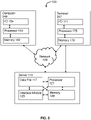
- FIG. 1 shows a system of the invention useful for the co-registration of angiogram or fluoroscopy and intravascular ultrasound images.
- the radiological and ultrasound image data acquisition sub-systems are generally well known in the art.
- a patient 10 is positioned upon an angiographic table 12.
- the angiographic table 12 is arranged to provide sufficient space for the positioning of an angiography/fluoroscopy unit c-arm 14 in an operative position in relation to the patient 10 on the table 12.
- Radiological image data acquired by the angiography/fluoroscopy c-arm 14 passes to an angiography/fluoroscopy processor 18 via transmission cable 16.
- the angiography/fluoroscopy processor 18 converts the received radiological image data received via the cable 16 into angiographic/fluoroscopic image data.
- the angiographic/fluoroscopic ("radiological") image data is initially stored within the processor 18.
- an imaging catheter 20, and in particular an IVUS catheter is inserted within the patient 10 so that its distal end, including a diagnostic probe 22 (in particular an IVUS probe), is in the vicinity of a desired imaging location of a blood vessel.
- a radiopaque material located near the probe 22 provides indicia of a current location of the probe 22 in a radiological image.
- the diagnostic probe 22 generates ultrasound waves, receives ultrasound echoes representative of a region proximate the diagnostic probe 22, and converts the ultrasound echoes to corresponding electrical signals.
- the corresponding electrical signals are transmitted along the length of the imaging catheter 20 to a proximal connector 24.
- IVUS versions of the probe 22 come in a variety of configurations including single and multiple transducer element arrangements.
- an array of transducers is potentially arranged: linearly along a lengthwise axis of the imaging catheter 20, curvilinear about the lengthwise axis of the catheter 20, circumferentially around the lengthwise axis, etc.
- the proximal connector 24 of the catheter 20 is communicatively coupled to a catheter image processor 26.
- the catheter image processor 26 converts the signals received via the proximal connector 24 into, for example, cross-sectional images of vessel segments. Additionally, the catheter image processor 26 generates longitudinal cross-sectional images corresponding to slices of a blood vessel taken along the blood vessel's length.
- the IVUS image data rendered by the catheter image processor 26 is initially stored within the processor 26.
- the type of diagnostic imaging data acquired by the diagnostic probe 22 and processed by the catheter image processor 26 varies in accordance with alternative embodiments of the invention.
- the diagnostic probe 22 is equipped with one or more sensors (e.g., Doppler and/or pressure) for providing hemodynamic information (e.g., blood flow velocity and pressure)-also referred to as functional flow measurements.
- hemodynamic information e.g., blood flow velocity and pressure
- functional flow measurements are processed by the catheter image processor 26.
- image is intended to be broadly interpreted to encompass a variety of ways of representing vascular information including blood pressure, blood flow velocity/volume, blood vessel cross-sectional composition, shear stress throughout the blood, shear stress at the blood/blood vessel wall interface, etc.
- a co-registration processor 30 receives IVUS image data from the catheter image processor 26 via line 32 and radiological image data from the radiological image processor 18 via line 34. Alternatively, the communications between the sensors and the processors are carried out via wireless media.
- the co-registration processor 30 renders a co-registration image including both radiological and IVUS image frames derived from the received image data.
- indicia e.g., a radiopaque marker artifact
- the co-registration processor 30 initially buffers angiogram image data received via line 34 from the radiological image processor 18 in a first portion 36 of image data memory 40.
- IVUS and radiopaque marker image data received via lines 32 and 34 is stored within a second portion 38 and a third portion 42, respectively, of the image data memory 40.
- the individually rendered frames of stored image data are appropriately tagged (e.g., time stamp, sequence number, etc.) to correlate IVUS image frames and corresponding radiological (radiopaque marker) image data frames.
- the hemodynamic data is stored within the second portion 38.
- markers can be placed on the surface of the patient or within the vicinity of the patient within the field of view of the angiogram/fluoroscope imaging device. The locations of these markers are then used to position the radiopaque marker artifact upon the angiographic image in an accurate location.
- the co-registration processor 30 renders a co-registration image from the data previously stored within the first portion 36, second portion 38 and third portion 42 of the image data memory 40.
- a particular IVUS image frame/slice is selected from the second portion 38.
- the co-registration processor 30 identifies fluoroscopic image data within the third portion 42 corresponding to the selected IVUS image data from the second portion 38. Thereafter, the co-registration processor 30 superimposes the fluoroscopic image data from the third portion 42 upon the angiogram image frame retrieved from the first portion 36. Thereafter, the co-registered radiological and IVUS image frames are simultaneously displayed, along-side one another, upon a graphical display device 50.
- the co-registered image data frames driving the display device 50 are also stored upon a long-term storage device 60 for later review in a session separate from a procedure that acquired the radiological and IVUS image data stored in the image data memory 40.
- a pullback device is incorporated that draws the catheter 20 from the patient at a controlled/measured manner.
- Such devices are well known in the art. Incorporation of such devices facilitates calculating a current position of the probe 22 within a field of view at points in time when fluoroscopy is not active.
- FIG. 2 presents an angiographic "roadmap" image 200 in a desired projection (patient/vessel orientation) and magnification as captured by the angiography/fluoroscopy processor 18.
- the image 200 is initially captured by an angiography procedure performed prior to tracking the IVUS catheter to the region of interest within a patient's vasculature.
- Performing the angiography procedure without the catheter 20 in the vessel provides maximal contrast flow, better vessel filling and therefore a better overall angiogram image.
- side branches such as side branch 210 and other vasculature landmarks can be displayed and seen clearly on the radiological image portion of a co-registered image displayed upon the graphical display device 50.
- FIG. 3 shows catheter radiopaque marker 300 visible in a fluoroscopic image.
- Catheter 20 is tracked to its starting position (e.g., a position where an IVUS pullback procedure begins). Typically the catheter 20 is tracked over a previously advanced guidewire (not shown). Thereafter, a fluoroscopic image is obtained. In the image as shown in FIG. 3 , the catheter radiopaque marker 300 is visualized, but the vessel lumen is not, due to the absence of contrast flow.
- a set of locating markers present in both the angiogram and fluoroscopy images enable proper positioning (superimposing) of the marker image within the previously obtained angiogram image.
- the marker artifact can be automatically adjusted (both size and position) on the superimposed image frames to correspond to the approximate position of the transducers.
- FIG. 4 presents an exemplary co-registration image that results from overlaying or superimposing the radiopaque marker artifact upon the angiogram image.
- the exemplary co-registration display 401 depicts a selected cross-sectional IVUS image 400 of a vessel.
- a radiological image 410 is simultaneously displayed along-side the IVUS image 400 on the display 50.
- the radiological image 410 includes a marker artifact 420, generated from radiological image data rendered by a fluoroscope image frame, superimposed on an angiogram background rendered from the first portion 36 of the memory 40.
- the fluoroscope image frame corresponds to the current location of the diagnostic probe 22 within a vessel under observation.
- the co-registered set of data can then be applied to facilitate detecting a feature of interest in a given modality.
- this may comprise using the co-registered data set to train a search algorithm for detecting the feature of interest in a given modality.
- the feature of interest may be a stent.
- the stent and surrounding vasculature may be imaged with two imaging modalities, such as IVUS and VH-IVUS.
- Features are extracted from both and aligned to obtain a co-registered data set.
- This combined data set can then be used to train a search algorithm for detecting the stent in conventional grayscale IVUS, for example, where the detection of stents is often problematic. Suitable methods for training the search algorithm will now be described.
- a feature of interest is identified in an imaging modality through use of a search algorithm that has been trained on a co-registered intravascular data set.
- the co-registered intravascular data set comprises information regarding a feature of interest, such as a vessel wall, from a plurality of imaging modalities.
- the search algorithm is able to identify the feature of interest in a given imaging modality. The algorithm addresses certain factors or parameters in order to make a comprehensive evaluation and identify the feature of interest based on positional and other data accumulated from multiple imaging modalities.
- the process involves obtaining an image with a first imaging modality, extracting the feature of interest from the image, and transforming the feature into positional data (i.e., a set of coordinates).
- the process further involves obtaining at least a second image of the area with a second imaging modality, extracting the feature of interest, and again transforming the feature into positional data.
- the positional data from the first and second imaging modalities are then combined into one data set via co-registration, and the combined data set is used to train a search algorithm configured to detect the feature of interest in a given imaging modality.
- Training of the algorithm may comprise a series of iterative steps, with each successive step evaluating each new data (i.e., provided from an additional imaging modality) in combination with all data submitted in the previous steps of the cycle (prior imaging modalities) and relevant information about the feature of interest until all test data (i.e., positional data) entered or submitted for analysis are evaluated comprehensively.
- the analysis function terminates, and the search result is formed upon completion of the analysis function.
- the present invention also contemplates the modification or update of positional data based upon new information received from imaging modalities, which is included as part of the algorithm, as such data becomes available and would serve to improve the accuracy of the search.
- the search algorithm of the present invention may be embodied in any suitable application, such as a computer program or code that can facilitate its use.
- the algorithm or application embodying the application may be stored in the internal or external hard drive of a computer, a portable drive or disc, a server, a temporary or permanent memory device, or any other storage means that can facilitate the use of the algorithm and/or the results derived from its use.
- the algorithm or application is preferably in communication with at least one processing device that facilitates the predictive analysis, for example, a computer or network processor.
- the algorithm or associated application may be accessed locally (e.g., on a single or networked computer) or remotely (e.g., web-based network via the internet, or via the intranet).
- This access to the algorithm or application may be facilitated via the use of any suitable equipment, including without limitation, a computer, an internet appliance, telephonic device, a wireless device, and the like.
- Access to the algorithm, the application embodying the algorithm or the results obtained from use of the algorithm may be secured or limited from general access or use via a password, encryption, biometric- or voice-activation, or other suitable means of protection.
- the functions and embodiments described above can be implemented using software, hardware, firmware, hardwiring, or any combinations of these.
- Features implementing functions can also be physically located at various positions, including being distributed such that portions of functions are implemented at different physical locations.
- a computer system or machines of the invention include one or more processors (e.g., a central processing unit (CPU) a graphics processing unit (GPU) or both), a main memory and a static memory, which communicate with each other via a bus.
- processors e.g., a central processing unit (CPU) a graphics processing unit (GPU) or both
- main memory e.g., a main memory
- static memory e.g., a main memory and static memory, which communicate with each other via a bus.
- Systems of the invention may include a computer and a processor as well as computer readable storage medium instructions that when executed, cause the computer to receive information from a first imaging modality and transform the information into a first coordinate space, receive information from a second imaging modality and transform the information into a second coordinate space, and align the first coordinate space with the second coordinate space, thereby combining information from the first modality and the second modality into a combined data set.
- the instructions may further cause the computer to detect a feature
- FIG. 5 diagrams a system 100 according to embodiments of the invention.
- System 100 preferably includes computer 249 (e.g., laptop, desktop, tablet, or smartphone).
- the computer 249 may be configured to communicate across a network 109.
- Computer 249 includes one or more processor 159 and memory 163 as well as an input/output mechanism 154.
- steps of methods of the invention may be performed using server 113, which includes one or more of processor 121 and memory 129, capable of obtaining data, instructions, etc., or providing results via interface module 125 or providing results as a file 117.
- Server 113 may be engaged over network 109 through computer 249 or terminal 267, or server 113 may be directly connected to terminal 167, including one or more processor 175 and memory 179, as well as input/output mechanism 171.
- System 100 or machines according to the invention may further include, for any of I/O 154 or 171 a video display unit (e.g., a liquid crystal display (LCD) or a cathode ray tube (CRT)).
- Computer systems or machines according to the invention can also include an alphanumeric input device (e.g., a keyboard), a cursor control device (e.g., a mouse), a disk drive unit, a signal generation device (e.g., a speaker), a touchscreen, an accelerometer, a microphone, a cellular radio frequency antenna, and a network interface device, which can be, for example, a network interface card (NIC), Wi-Fi card, or cellular modem.
- NIC network interface card
- Wi-Fi card Wireless Fidelity
- Memory 163, 179, or 129 can include a machine-readable medium on which is stored one or more sets of instructions (e.g., software) embodying any one or more of the methodologies or functions described herein.
- a computer system of the invention includes one or more memory device that is a tangible, nontransitory memory.
- the software may also reside, completely or at least partially, within the main memory and/or within the processor during execution thereof by the computer system, the main memory and the processor also constituting machine-readable media.
- the software may further be transmitted or received over a network via the network interface device.
- FIG. 6 presents steps of methods of the invention. It will be understood that of the methods described herein, as well as any portion of the systems and methods disclosed herein, can be implemented by computer, including the devices described above. Preferably, each step is performed by a processor or connected medical imaging device. An image is obtained using a given modality in which a feature of interest requires identification 201.
- the modality for example, may be grayscale IVUS, and the feature of interest is a stent, which is difficult to detect with grayscale IVUS.
- Information regarding a feature of interest is extracted from an imaging modality. The feature is the converted into a feature vector comprising positional data regarding the feature of interest. This data is then inputted into the central processing unit (CPU) of a computer 202.
- CPU central processing unit
- the CPU is coupled to a storage or memory for storing instructions for implementing methods of the present invention, such as the search algorithm.
- the instructions when executed by the CPU, cause the CPU to identify a selected feature of interest in an imaging modality.
- the CPU provides this determination by inputting data from the imaging modality into an algorithm trained on a co-registered set of data derived from a plurality of imaging modalities for positional data regarding the feature of interest is known 203.
- the co-registered set of data may be stored locally within the computer, such as within the computer memory. Alternatively, the co-registered data set may be stored in a location that is remote from the computer, such as a server. In this instance, the computer communicates across a network to access the co-registered data set.
- the CPU then identifies the feature of interest in the imaging modality based on the data entered into the algorithm 204.
- a co-registered set of positional data derived from a plurality of imaging modalities is used to train the algorithm.
- positional data regarding the feature of interest is obtained from a plurality of imaging modalities, and combined into a co-registered set of positional data, which is then used as input data (inputs into a search algorithm fitted to the co-registered positional data obtained from the selected population of individuals).
- Any formula or algorithm may be used to combine positional data based on features of interest extracted from the various imaging modalities.
- model and formula types beyond those mentioned herein and in the definitions above are well known to one skilled in the art.
- the actual model type or formula used may itself be selected from the field of potential models based on the performance and diagnostic accuracy characteristics of its results in a training population.
- the specifics of the formula itself may commonly be derived from selected parameter results in the relevant training population.
- such formula may be intended to map the feature space derived from one or more selected parameter inputs to a set of subject classes, to derive an estimation of a probability function of risk using a Bayesian approach, or to estimate the class-conditional probabilities, then use Bayes' rule to produce the class probability function as in the previous case.
- Preferred formulas include the broad class of statistical classification algorithms, and in particular the use of discriminant analysis.
- the goal of discriminant analysis is to predict class membership from a previously identified set of features.
- LDA linear discriminant analysis
- features can be identified for LDA using an eigengene based approach with different thresholds (ELDA) or a stepping algorithm based on a multivariate analysis of variance (MANOVA). Forward, backward, and stepwise algorithms can be performed that minimize the probability of no separation based on the Hotelling-Lawley statistic.
- Eigengene-based Linear Discriminant Analysis is a feature selection technique developed and reported in Shen et al., 2006, Eigengene-based linear discriminant model for tumor classification using gene expression microarray data, Bioinformatics 22(21):2635-2642 .
- the formula selects features (e.g. parameters) in a multivariate framework using a modified Eigen analysis to identify features associated with the most important eigenvectors. "Important" is defined as those eigenvectors that explain the most variance in the differences among samples that are trying to be classified relative to some threshold.
- a support vector machine is a classification formula that attempts to find a hyperplane that separates two classes.
- This hyperplane contains support vectors, data points that are exactly the margin distance away from the hyperplane.
- the dimensionality is expanded greatly by projecting the data into larger dimensions by taking non-linear functions of the original variables ( Venables and Ripley, 2002, Modern Applied Statistics with S 4Ed, Springer Verlag ).
- filtering of features for SVM often improves prediction.
- Features e.g., parameters/biomarkers
- KW Kruskal-Wallis
- a random forest or recursive partitioning ( RPART, Breiman et al., 1984 ) can also be used separately or in combination to identify parameter combinations that are most important. Random forests and recursive partitioning are discussed in Strobl et al., 2009, An Introduction to Recursive Partitioning: Rationale, Application and Characteristics of Classification and Regression Trees, Bagging and Random Forests, Psychol Methods 14(4):323-348 ; Breiman, 2001, Random forests, Machine Learning 45:5-32 ; Breiman, 1984, Classification and Regression Trees, Boca Raton: Chapman & Hall/CRC ; U.S. Pat. 8,600,917 ; and U.S. Pat. 8,187,830 . Both KW and RF require that a number of features be selected from the total. RPART creates a single classification tree using a subset of available parameters.
- an overall predictive formula for all subjects, or any known class of subjects may itself be recalibrated or otherwise adjusted based on adjustment for a population's expected prevalence and mean parameter values, according to the technique outlined in D 'Agostino et al., 2001, Validation of the Framingham coronary heart disease prediction score: results of a multiple ethnic group investigation, JAMA 286:180-187 , or other similar normalization and recalibration techniques.
- adjustment statistics may be captured, confirmed, improved and updated continuously through a registry of past data presented to the model, which may be machine readable or otherwise, or occasionally through the retrospective query of stored samples or reference to historical studies of such parameters and statistics.
- Additional examples that may be the subject of formula recalibration or other adjustments include statistics used in studies by on the limitations of odds ratios (see Pepe et al., 2004, Limitations of the odds ratio in gauging the performance of a diagnostic, prognostic, or screening marker, Am J Epidemiology 159(9):882-890 ) and receiver operating characteristic (ROC) curve in risk prediction (see Cook, 2007, Use and misuse of receiver operating characteristic curve in risk prediction, Circulation 115(7):928- 35 ; Wang et al., 2006, Multiple biomarkers for the prediction of first major cardiovascular events and death, NEJM 355:2631-2639 ; and Grund & Sabin, 2010, Analysis of biomarker data: logs, odds, ratios and ROC curves, Curr Opin HIV AIDS 5(6):473-479 ).
- the numeric result of a classifier formula itself may be transformed post-processing by its reference to an actual positive controls in which the selected feature of interest has already been identified and confirmed.
- FIG. 7 is a flow diagram representing an exemplary method for model development 300 which may be used to search for a feature of interest.
- the method 300 may be implemented using the example computing system environment 100 of FIG. 5 and will be used to explain the operation of the environment 100. However, it should be recognized that the method 300 could be implemented by a system different than the computing system environment 100.
- a co-registered set of positional data regarding a selected feature of interest is obtained from a data storage device, such as the system memory 129, an internal or external database, or other computer storage media.
- the co-registered data set may be initially derived through a variety of means, including various imaging modalities capturing the feature of interest in an image, in which the feature is extracted from the imaging modality and converted into positional data (i.e., a set of coordinates).
- co-registered data set is prepared as needed to meet the requirements of the model or analysis that will be used to train the search algorithm, as described below.
- data set preparation may include preparing the positional data from each imaging modality, or a chosen subset thereof.
- various data preparation methods may be used to prepare the data prior to training the algorithm, such as gap fill techniques (e.g., nearest neighbor interpolation or other pattern recognition), quality checks, data combination using of various formulae (e.g., statistical classification algorithms), normalization and/or transformations, such as logarithmic functions to change the distribution of data to meet model requirements (e.g., base 10, natural log, etc.).
- the particular data preparation procedures are dependent upon the model or models that will be trained using the co-registered data set.
- the particular data preparation techniques for various different model types are known, and need not be described further.
- the particular extracted features are transformed into positional data used to select parameters that are subsequently used in the training of the search model used to identify the feature of interest in a given imaging modality.
- Use of the co-registered positional data may involve utilizing only the positional data from the co-registered set that provides the most reproducible results. Examples of data set validation may include, but are not limited to, cross-validation and bootstrapping.
- the optimal search model for identifying a feature of interest in a given imaging modality may be determined and selected.
- not all models provide the same results with the same data set. For example, different models may utilize different aspects of the positional data and produce different results, thereby adding significance to the selected positional data in the optimized model.
- multiple selection models may be chosen and utilized with the co-registered data set, or subsets of the co-registered data set, in order to identify the optimal search model for identifying a feature of interest in a given modality.
- Examples of the particular models, including statistical models, algorithms, etc., which may be used for selecting the positional data from the co-registered data sets have been described above.
- the positional data is selected based on its statistical significance in the model.
- the positional data is selected based on various criteria for statistical significance, and may further involve cumulative voting and weighting.
- Tests for statistical significance may include exit-tests and analysis of variance (ANOVA).
- the model may include classification models (e.g., LDA, logistic regression, SVM, RF, tree models, etc.) and survival models (e.g., cox), many examples of which have been described above.
- the search model to be used for identifying a feature of interest is selected, trained and validated.
- leading candidate models may be selected based on one or more performance criteria, examples of which have been described above.
- the evaluation model used to evaluate risk may include one of those used as a selection model, including classification models and survival models.
- Combinations of models markers, including marker subsets may be compared and validated in subsets and individual data sets. The comparison and validation may be repeated many times to train and validate the model and to choose an appropriate model, which is then used as an evaluation model for evaluating risk of a diabetic condition.
- the invention relates to the detection of features of interest, such as vessel borders and stent struts, in vascular images derived from multiple data sets or imaging modalities.
- features of interest such as vessel borders and stent struts
- Methods of the invention leverage potentially complimentary information from multiple imaging modalities and combine the information extracted from each image to detect the target feature.
- Image segmentation is the process of partitioning a digital image into multiple segments (sets of pixels, also known as superpixels). The goal of segmentation is to simplify and/or change the representation of an image into something that is more meaningful and easier to analyze. Image segmentation is typically used to locate objects and boundaries (lines, curves, etc.) in images.
- One approach to segmentation is to compute and extract features from the image data. The features can be combined into a feature vector (e.g., transformed into positional data), that can then be matched to known feature vectors (i.e., gold standard information) or run through a trained classifier (e.g., a neural network or other algorithm) to classify the image data into predetermined classes.
- a feature vector e.g., transformed into positional data
- known feature vectors i.e., gold standard information
- a trained classifier e.g., a neural network or other algorithm
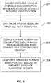
- FIG. 8 diagrams a process of partitioning a digital image into multiple segments.
- image is obtained 801, and features are extracted 802 from the image.
- the features are then combined into a feature vector 803 (e.g., transformed into positional data) that can then be used to train a search algorithm 804 to identify the selected feature of interest in a given modality 805.
- a search algorithm 804 Given a set of spatially co-registered intravascular images obtained from multiple imaging modalities, however, features can be computed and extracted from the multiple images and combined into a single feature vector. If the multimodal source a co-registered data set based on multiple imaging modalities is able to provide complementary information, the resulting feature vector contains more information than if extracted from a single source. Accordingly, the search algorithm trained on this co-registered set of data should provide more accurate results.
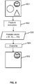
- FIG. 9 illustrates images captured by multiple imaging modalities 901 and gives an example of the image segmentation process.
- any imaging modality can be used in accordance with the invention.
- the imaging modalities depicted in FIG. 9 could comprise IVUS, x-ray angiography, and OCT.
- the features are extracted from the images 902 and transformed into positional data 903.
- the positional data is then combined and aligned into a co-registered set of data 904.
- This co-registered set of data 904 is then used to train a search algorithm 905 that can be used to identify a feature of interest in a given modality 906.
- Methods of the invention can be easily applied in the following, nonlimiting, scenarios.
- VH-IVUS Virtual Histology-Intravascular Ultrasound
- grayscale IVUS Intravascular Ultrasound
- Stents are known to be difficult to detect using conventional grayscale IVUS. Methods of the invention, however, can be used to detect non-biological features of interest by combining the complementary imaging modalities of IVUS and VH-IVUS to provide more information.
- VH analysis VH-IVUS and grayscale IVUS data are already spatially co-registered.
- Stents in VH-IVUS have a distinct appearance which can be easily identified in IVUS images. This information can be used to initialize a search method for stent detection in the grayscale IVUS data set or combined with features extracted from the grayscale IVUS data set as part of a stent detection algorithm.
- Grayscale IVUS and OCT stent detection As noted above, stents are difficult to detect using grayscale IVUS alone.
- methods of the invention utilize a co-registered set of IVUS and OCT data to assist in stent detection. Given a set of spatially co-registered grayscale IVUS and OCT data sets, features can be extracted from each modality and combined into a single feature vector as described above. This example can be expanded to an approach encompassing VH-IVUS, grayscale IVUS, and OCT.
- Grayscale IVUS and OCT vessel and lumen border detection This example is the same as the above, however, the features of interest are now biological rather than non-biological. This particular combination of imaging modalities can be significantly advantageous as detection of lumen borders with grayscale IVUS alone can be extremely challenging due to the presence of blood.
- OCT acquires image data in a flushed vessel, which facilitates imaging the lumen borders. If the feature of interest is a vessel border, then the OCT data becomes less significant, except for the fact that the vessel border is always outside the lumen and the OCT data is well-suited to identifying the lumen.
- the invention includes all imaging modalities and all features of interest, whether they be biological or non-biological.
- this vascular multimodal approach can also be applied to x-ray angiography and/or CT angiography data co-registered with IVUS and/or OCT data sets.
- the invention is not limited to IVUS and OCT as the only intravascular imaging modalities.
- Other suitable intravascular imaging modalities include MRI and photoacoustic imaging.
- a primary benefit of the multimodal approach described herein is to take advantage of additional information derived from complementary imaging modalities when available. This additional information improves or facilitates the detection of features of interest in vascular data sets when used in accordance with the provided methods.
Claims (10)
- Procédé mis en oeuvre par ordinateur pour détecter une caractéristique d'intérêt dans une image vasculaire, le procédé permettant à un processeur d'exécuter les étapes suivantes:recevoir des informations d'une première modalité d'imagerie et transformer les informations de la première modalité en un premier espace de coordonnées; recevoir des informations d'une deuxième modalité d'imagerie et transformer les informations de la deuxième modalité en un deuxième espace de coordonnées;dans lequel la modalité d'imagerie de la première et de la deuxième modalité d'imagerie est choisie dans un groupe constitué par: l'échographie intravasculaire (IVUS), l'échographie intravasculaire par histologie virtuelle (VH-IVUS), la tomographie en cohérence optique (OCT), l'angiographie aux rayons X et l'imagerie par résonance magnétique; dans lequel la première modalité d'imagerie est différente de la deuxième modalité d'imagerie;aligner le premier espace de coordonnées et le deuxième espace de coordonnées, combinant ainsi les informations provenant de la première modalité d'imagerie et de la deuxième modalité d'imagerie en un ensemble de données combinées;détecter la caractéristique d'intérêt dans l'image vasculaire d'une modalité d'imagerie sélectionnée parmi les multiples modalités d'imagerie sur la base de l'ensemble de données combinées,dans lequel la détection de la caractéristique d'intérêt comprend l'apprentissage d'un classificateur (804, 905) pour détecter la caractéristique d'intérêt sur la base de l'ensemble de données combinées; etdans lequel la caractéristique d'intérêt est détectée dans une image de l'une des première et deuxième modalités d'imagerie et cette information est utilisée pour initialiser l'apprentissage d'une détection de caractéristique dans une image de l'autre des première et deuxième modalités d'imagerie.
- Procédé selon la revendication 1, dans lequel la caractéristique d'intérêt est une paroi de vaisseau ou de lumen.
- Procédé selon la revendication 1, dans lequel la caractéristique d'intérêt est un stent.
- Procédé selon la revendication 1, dans lequel la détection de la caractéristique d'intérêt comprend l'initialisation d'une recherche de la caractéristique d'intérêt sur la base de l'ensemble de données combinées.
- Système pour déterminer une caractéristique d'intérêt dans une image vasculaire, le système comprenant:un processeur (30); etun support de stockage lisible par ordinateur (60) comprenant des instructions qui, lorsqu'elles sont exécutées, amènent le système à:recevoir des informations d'une première modalité d'imagerie et transformer les informations de la première modalité en un premier espace de coordonnées; recevoir des informations d'une deuxième modalité d'imagerie et transformer les informations de la deuxième modalité en un deuxième espace de coordonnées;dans lequel la modalité d'imagerie de la première et de la deuxième modalité d'imagerie est choisie dans un groupe constitué par: l'échographie intravasculaire (IVUS), l'échographie intravasculaire par histologie virtuelle (VH-IVUS), la tomographie en cohérence optique (OCT), l'angiographie aux rayons X et l'imagerie par résonance magnétique;dans lequel la première modalité est différente de la deuxième modalité d'imagerie;aligner le premier espace de coordonnées et le deuxième espace de coordonnées, combinant ainsi les informations de la première modalité d'imagerie et de la deuxième modalité d'imagerie; etdétecter la caractéristique d'intérêt dans l'image vasculaire d'une modalité d'imagerie sélectionnée parmi les multiples modalités d'imagerie sur la base de l'ensemble de données combinées,dans lequel la détection de la caractéristique d'intérêt comprend l'apprentissage d'un classificateur (804, 905) pour détecter la caractéristique d'intérêt sur la base de l'ensemble de données combinées; etdans lequel la caractéristique d'intérêt est détectée dans une image de l'une des première et deuxième modalités d'imagerie et cette information est utilisée pour initialiser l'apprentissage d'une détection de caractéristique dans une image de l'autre des première et deuxième modalités d'imagerie.
- Système selon la revendication 5, dans lequel la caractéristique d'intérêt comprend une caractéristique biologique d'intérêt.
- Système selon la revendication 6, dans lequel la caractéristique biologique d'intérêt est une paroi de vaisseau ou de lumen.
- Système selon la revendication 5, dans lequel la caractéristique d'intérêt comprend une caractéristique non biologique d'intérêt.
- Système selon la revendication 8, dans lequel la caractéristique non biologique d'intérêt est un stent.
- Système selon la revendication 5, dans lequel la détection de la caractéristique d'intérêt comprend l'initialisation d'une recherche de la caractéristique d'intérêt sur la base de l'ensemble de données combinées.
Applications Claiming Priority (2)
| Application Number | Priority Date | Filing Date | Title |
|---|---|---|---|
| US201361774154P | 2013-03-07 | 2013-03-07 | |
| PCT/US2014/021659 WO2014138555A1 (fr) | 2013-03-07 | 2014-03-07 | Segmentation multimodale dans des images intravasculaires |
Publications (3)
| Publication Number | Publication Date |
|---|---|
| EP2965263A1 EP2965263A1 (fr) | 2016-01-13 |
| EP2965263A4 EP2965263A4 (fr) | 2016-10-19 |
| EP2965263B1 true EP2965263B1 (fr) | 2022-07-20 |
Family
ID=51487895
Family Applications (1)
| Application Number | Title | Priority Date | Filing Date |
|---|---|---|---|
| EP14761138.8A Active EP2965263B1 (fr) | 2013-03-07 | 2014-03-07 | Segmentation multimodale dans des images intravasculaires |
Country Status (5)
| Country | Link |
|---|---|
| US (1) | US9770172B2 (fr) |
| EP (1) | EP2965263B1 (fr) |
| JP (1) | JP6243453B2 (fr) |
| CN (2) | CN113705586A (fr) |
| WO (1) | WO2014138555A1 (fr) |
Cited By (4)
| Publication number | Priority date | Publication date | Assignee | Title |
|---|---|---|---|---|
| US11648424B2 (en) | 2018-11-28 | 2023-05-16 | Histosonics Inc. | Histotripsy systems and methods |
| US11813485B2 (en) | 2020-01-28 | 2023-11-14 | The Regents Of The University Of Michigan | Systems and methods for histotripsy immunosensitization |
| US11819712B2 (en) | 2013-08-22 | 2023-11-21 | The Regents Of The University Of Michigan | Histotripsy using very short ultrasound pulses |
| US11980778B2 (en) | 2023-05-02 | 2024-05-14 | Histosonics, Inc. | Histotripsy systems and methods |
Families Citing this family (43)
| Publication number | Priority date | Publication date | Assignee | Title |
|---|---|---|---|---|
| WO2014162367A1 (fr) * | 2013-04-05 | 2014-10-09 | テルモ株式会社 | Dispositif de diagnostic par imagerie et programme associé |
| US9087370B2 (en) * | 2013-05-22 | 2015-07-21 | Siemens Aktiengesellschaft | Flow diverter detection in medical imaging |
| US10317189B2 (en) * | 2014-01-23 | 2019-06-11 | Kabushiki Kaisha Topcon | Detection of missampled interferograms in frequency domain OCT with a k-clock |
| DE102014213408B4 (de) * | 2014-07-10 | 2018-10-25 | Siemens Healthcare Gmbh | Verfahren zur Ermittlung eines dreidimensionalen Modelldatensatzes eines wenigstens ein Gefäßsegment umfassenden Blutgefäßsystems |
| KR102328269B1 (ko) * | 2014-10-23 | 2021-11-19 | 삼성전자주식회사 | 초음파 영상 장치 및 그 제어 방법 |
| US11009467B2 (en) * | 2015-02-17 | 2021-05-18 | Siemens Healthcare Diagnostics Inc. | Model-based methods and apparatus for classifying an interferent in specimens |
| US9972093B2 (en) * | 2015-03-30 | 2018-05-15 | Siemens Healthcare Gmbh | Automated region of interest detection using machine learning and extended Hough transform |
| WO2017040155A1 (fr) * | 2015-09-01 | 2017-03-09 | The Government Of The United States Of America, As Represented By The Secretary Of The Navy | Antenne miniature à onde de fuite acoustique pour l'imagerie par ultrasons |
| US9471836B1 (en) * | 2016-04-01 | 2016-10-18 | Stradvision Korea, Inc. | Method for learning rejector by forming classification tree in use of training images and detecting object in test images, and rejector using the same |
| WO2017197527A1 (fr) | 2016-05-20 | 2017-11-23 | Perimeter Medical Imaging, Inc. | Procédé et système pour combiner l'imagerie microscopique avec des rayons x |
| US20180012359A1 (en) * | 2016-07-06 | 2018-01-11 | Marinko Venci Sarunic | Systems and Methods for Automated Image Classification and Segmentation |
| GB201615051D0 (en) * | 2016-09-05 | 2016-10-19 | Kheiron Medical Tech Ltd | Multi-modal medical image procesing |
| CN110072462B (zh) * | 2016-09-16 | 2022-05-24 | 国家医疗保健研究所 | 用于对含血液的样品成像的方法和相关装置 |
| CN109982657B (zh) * | 2016-11-11 | 2023-06-30 | 直观外科手术操作公司 | 具有多模态图像显示的外科手术系统 |
| CN107194933A (zh) * | 2017-04-24 | 2017-09-22 | 天津大学 | 结合卷积神经网络和模糊推理的脑肿瘤分割方法和装置 |
| CA3213179A1 (fr) | 2017-08-09 | 2019-02-14 | Allen Institute | Systemes, dispositifs et procedes de traitement d'image pour generer une image presentant un marquage predictif |
| EP3467771A1 (fr) * | 2017-10-05 | 2019-04-10 | Koninklijke Philips N.V. | Annotation de caractéristique d'image dans l'imagerie diagnostique |
| US10762637B2 (en) * | 2017-10-27 | 2020-09-01 | Siemens Healthcare Gmbh | Vascular segmentation using fully convolutional and recurrent neural networks |
| GB2569103A (en) * | 2017-11-16 | 2019-06-12 | Univ Oslo Hf | Histological image analysis |
| CN109091167A (zh) * | 2018-06-29 | 2018-12-28 | 东南大学 | 冠状动脉粥样硬化斑块增长的预测方法 |
| US11690551B2 (en) | 2018-07-30 | 2023-07-04 | Biosense Webster (Israel) Ltd. | Left atrium shape reconstruction from sparse location measurements using neural networks |
| JP7164423B2 (ja) * | 2018-12-12 | 2022-11-01 | キヤノンメディカルシステムズ株式会社 | 医用画像処理装置、x線ct装置及び医用画像処理方法 |
| DE102018222606A1 (de) * | 2018-12-20 | 2020-06-25 | Siemens Healthcare Gmbh | Verfahren und Vorrichtung zur Detektion eines anatomischen Merkmals eines Blutgefäßabschnittes |
| WO2020146905A1 (fr) * | 2019-01-13 | 2020-07-16 | Lightlab Imaging, Inc. | Systèmes et procédés de classification de régions d'image artérielle et leurs caractéristiques |
| JP7246952B2 (ja) * | 2019-02-12 | 2023-03-28 | キヤノンメディカルシステムズ株式会社 | 医用情報処理装置、x線診断装置及びプログラム |
| CN111627529A (zh) * | 2019-02-28 | 2020-09-04 | 未艾医疗技术(深圳)有限公司 | Vrds 4d医学影像处理方法及产品 |
| CN109961490A (zh) * | 2019-04-08 | 2019-07-02 | 深圳大学 | 剪影成像方法及装置 |
| CN110232691A (zh) * | 2019-04-18 | 2019-09-13 | 浙江大学山东工业技术研究院 | 一种多模态的ct影像的分割方法 |
| CN110364256A (zh) * | 2019-06-21 | 2019-10-22 | 平安科技(深圳)有限公司 | 一种基于大数据的血管图像识别的疾病预测系统及方法 |
| US20220346885A1 (en) * | 2019-09-20 | 2022-11-03 | Canon U.S.A., Inc. | Artificial intelligence coregistration and marker detection, including machine learning and using results thereof |
| US11931107B1 (en) * | 2019-10-03 | 2024-03-19 | Smith & Nephew, Inc. | Intraoperative three-dimensional bone model generation |
| EP4121893A4 (fr) * | 2020-03-16 | 2024-04-24 | Univ Pittsburgh Commonwealth Sys Higher Education | Segmentation évolutive et de haute précision guidée par contexte de structures histologiques comprenant des conduits/glandes et une lumière, un groupe de conduits/glandes, et des noyaux individuels dans des images coulissantes entières d'échantillons de tissu à partir de plateformes d'imagerie cellulaires et sous-cellulaires à paramètres multiples spatiaux |
| EP4138672B1 (fr) | 2020-04-21 | 2023-11-22 | Philips Image Guided Therapy Corporation | Commande automatisée d'acquisition de données intraluminales et dispositifs, systèmes et procédés associés |
| EP4178446A4 (fr) * | 2020-08-10 | 2023-06-07 | Shanghai United Imaging Healthcare Co., Ltd. | Systèmes et procédés d'imagerie |
| WO2022069327A2 (fr) * | 2020-09-29 | 2022-04-07 | Philips Image Guided Therapy Corporation | Voie basée sur la tomographie assistée par ordinateur pour co-enregistrement de données intravasculaires et de mesures de vaisseau sanguin avec un modèle tridimensionnel basé sur la tomographie assistée par ordinateur |
| WO2022069303A2 (fr) * | 2020-09-29 | 2022-04-07 | Philips Image Guided Therapy Corporation | Mappage entre tomographie assistée par ordinateur et angiographie pour mise en correspondance de données intravasculaires et d'une métrique de vaisseau sanguin avec un modèle tridimensionnel de tomographie assistée par ordinateur |
| CN112294260B (zh) * | 2020-10-10 | 2022-04-05 | 浙江大学 | 一种磁兼容的光学脑功能成像方法与装置 |
| CN112370078B (zh) * | 2020-11-10 | 2024-01-26 | 安徽理工大学 | 一种基于超声成像和贝叶斯优化的图像检测方法 |
| CN113397579A (zh) * | 2021-07-23 | 2021-09-17 | 上海友脉科技有限责任公司 | 血流动力学分析装置、方法、介质及电子设备 |
| WO2023023748A1 (fr) * | 2021-08-26 | 2023-03-02 | 3P Healthcare Pty Ltd | Système et procédé d'évaluation de santé cardiovasculaire et de gestion de risque |
| WO2023129722A1 (fr) * | 2021-12-31 | 2023-07-06 | Boston Scientific Scimed, Inc. | Systèmes et procédés pour co-enregistrement d'images vasculaires |
| US20230214881A1 (en) * | 2021-12-31 | 2023-07-06 | Synamedia Limited | Methods, Devices, and Systems for Dynamic Targeted Content Processing |
| CN116863146B (zh) * | 2023-06-09 | 2024-03-08 | 强联智创(北京)科技有限公司 | 用于对血管瘤特征进行提取的方法、设备及存储介质 |
Family Cites Families (957)
| Publication number | Priority date | Publication date | Assignee | Title |
|---|---|---|---|---|
| US3301258A (en) | 1963-10-03 | 1967-01-31 | Medtronic Inc | Method and apparatus for treating varicose veins |
| US3617880A (en) | 1970-05-15 | 1971-11-02 | Northern Electric Co | Time domain reflectometer |
| US3789841A (en) | 1971-09-15 | 1974-02-05 | Becton Dickinson Co | Disposable guide wire |
| JPS584481Y2 (ja) | 1973-06-23 | 1983-01-26 | オリンパス光学工業株式会社 | ナイシキヨウシヤヘンカンコウガクケイ |
| US3841308A (en) | 1973-10-15 | 1974-10-15 | Medical Evaluation Devices & I | Distally valved catheter device |
| JPS5921495B2 (ja) | 1977-12-15 | 1984-05-21 | 株式会社豊田中央研究所 | 細管型圧力計 |
| US4344438A (en) | 1978-08-02 | 1982-08-17 | The United States Of America As Represented By The Department Of Health, Education And Welfare | Optical sensor of plasma constituents |
| US4398791A (en) | 1981-02-09 | 1983-08-16 | Litton Systems, Inc. | Single channel optical slip ring |
| US4432370A (en) | 1981-10-14 | 1984-02-21 | The Board Of Trustees Of The Leland Stanford Junior University | Method and means for minimally invasive angiography using mono-chromatized synchrotron radiation |
| US5041108A (en) | 1981-12-11 | 1991-08-20 | Pillco Limited Partnership | Method for laser treatment of body lumens |
| US4816567A (en) | 1983-04-08 | 1989-03-28 | Genentech, Inc. | Recombinant immunoglobin preparations |
| US4864578A (en) | 1983-04-12 | 1989-09-05 | Coherent, Inc. | Scannable laser with integral wavemeter |
| US4577543A (en) | 1983-08-18 | 1986-03-25 | American Hospital Supply Corporation | Construction of a monolithic reinforced catheter with flexible portions |
| US4552554A (en) | 1984-06-25 | 1985-11-12 | Medi-Tech Incorporated | Introducing catheter |
| GB8417911D0 (en) | 1984-07-13 | 1984-08-15 | British Telecomm | Connecting waveguides |
| DE3442736A1 (de) | 1984-11-23 | 1986-06-05 | Tassilo Dr.med. 7800 Freiburg Bonzel | Dilatationskatheter |
| US5188632A (en) | 1984-12-07 | 1993-02-23 | Advanced Interventional Systems, Inc. | Guidance and delivery system for high-energy pulsed laser light |
| US4682895A (en) | 1985-08-06 | 1987-07-28 | Texas A&M University | Fiber optic probe for quantification of colorimetric reactions |
| US4676980A (en) | 1985-09-23 | 1987-06-30 | The United States Of America As Represented By The Secretary Of The Department Of Health And Human Services | Target specific cross-linked heteroantibodies |
| US4733665C2 (en) | 1985-11-07 | 2002-01-29 | Expandable Grafts Partnership | Expandable intraluminal graft and method and apparatus for implanting an expandable intraluminal graft |
| US4834093A (en) | 1986-02-03 | 1989-05-30 | Littleford Phillip O | Dilation catheter and method |
| US4803639A (en) | 1986-02-25 | 1989-02-07 | General Electric Company | X-ray inspection system |
| US4794931A (en) | 1986-02-28 | 1989-01-03 | Cardiovascular Imaging Systems, Inc. | Catheter apparatus, system and method for intravascular two-dimensional ultrasonography |
| US5000185A (en) | 1986-02-28 | 1991-03-19 | Cardiovascular Imaging Systems, Inc. | Method for intravascular two-dimensional ultrasonography and recanalization |
| US4771774A (en) | 1986-02-28 | 1988-09-20 | Devices For Vascular Intervention, Inc. | Motor drive unit |
| US5040548A (en) | 1989-06-01 | 1991-08-20 | Yock Paul G | Angioplasty mehtod |
| US4821731A (en) | 1986-04-25 | 1989-04-18 | Intra-Sonix, Inc. | Acoustic image system and method |
| US4766386A (en) | 1986-05-23 | 1988-08-23 | Cabletron | Time domain reflectometer for measuring impedance discontinuities on a powered transmission line |
| US4800886A (en) | 1986-07-14 | 1989-01-31 | C. R. Bard, Inc. | Sensor for measuring the concentration of a gaseous component in a fluid by absorption |
| US4887606A (en) | 1986-09-18 | 1989-12-19 | Yock Paul G | Apparatus for use in cannulation of blood vessels |
| GB8629871D0 (en) | 1986-12-15 | 1987-01-28 | British Telecomm | Optical switch |
| US5174295A (en) | 1987-04-10 | 1992-12-29 | Cardiometrics, Inc. | Apparatus, system and method for measuring spatial average velocity and/or volumetric flow of blood in a vessel and screw joint for use therewith |
| US5163445A (en) | 1987-04-10 | 1992-11-17 | Cardiometrics, Inc. | Apparatus, system and method for measuring spatial average velocity and/or volumetric flow of blood in a vessel and screw joint for use therewith |
| US4824435A (en) | 1987-05-18 | 1989-04-25 | Thomas J. Fogarty | Instrument guidance system |
| JP2697822B2 (ja) | 1987-05-25 | 1998-01-14 | オリンパス光学工業株式会社 | 内視鏡対物レンズ |
| US4841977A (en) | 1987-05-26 | 1989-06-27 | Inter Therapy, Inc. | Ultra-thin acoustic transducer and balloon catheter using same in imaging array subassembly |
| US4917097A (en) | 1987-10-27 | 1990-04-17 | Endosonics Corporation | Apparatus and method for imaging small cavities |
| US4819740A (en) | 1987-11-16 | 1989-04-11 | Vulcan Iron Works Inc. | Vibratory hammer/extractor |
| US4830023A (en) | 1987-11-27 | 1989-05-16 | Medi-Tech, Incorporated | Medical guidewire |
| US4917085A (en) | 1987-12-14 | 1990-04-17 | Cordis Corporation | Drive cutting catheter having new and improved drive motor |
| US4948229A (en) | 1988-03-18 | 1990-08-14 | The United States Of America As Represented By The Secretary Of The Air Force | Optical switches using ferroelectric liquid crystals |
| US4932419A (en) | 1988-03-21 | 1990-06-12 | Boston Scientific Corporation | Multi-filar, cross-wound coil for medical devices |
| US5372138A (en) | 1988-03-21 | 1994-12-13 | Boston Scientific Corporation | Acousting imaging catheters and the like |
| US4951677A (en) | 1988-03-21 | 1990-08-28 | Prutech Research And Development Partnership Ii | Acoustic imaging catheter and the like |
| US4998972A (en) | 1988-04-28 | 1991-03-12 | Thomas J. Fogarty | Real time angioscopy imaging system |
| US4987412A (en) | 1988-08-25 | 1991-01-22 | The United States Of America As Represented By The United States Department Of Energy | Method and apparatus for the simultaneous display and correlation of independently generated images |
| US5178159A (en) | 1988-11-02 | 1993-01-12 | Cardiometrics, Inc. | Torqueable guide wire assembly with electrical functions, male and female connectors rotatable with respect to one another |
| US5240437A (en) | 1988-11-02 | 1993-08-31 | Cardiometrics, Inc. | Torqueable guide wire assembly with electrical functions, male and female connectors for use therewith and system and apparatus for utilizing the same |
| US5065769A (en) | 1988-11-23 | 1991-11-19 | Boston Scientific Corporation | Small diameter guidewires of multi-filar, cross-wound coils |
| US5431673A (en) | 1989-02-17 | 1995-07-11 | American Biomed, Inc. | Distal atherectomy catheter |
| US4932413A (en) | 1989-03-13 | 1990-06-12 | Schneider (Usa), Inc. | Guidewire exchange catheter |
| US4928693A (en) | 1989-03-13 | 1990-05-29 | Schneider (Usa), Inc. | Pressure monitor catheter |
| US5203779A (en) | 1989-03-17 | 1993-04-20 | Schott Glaswerke | Catheter system for vessel recanalization in the human body |
| US5120308A (en) | 1989-05-03 | 1992-06-09 | Progressive Angioplasty Systems, Inc. | Catheter with high tactile guide wire |
| US4969742A (en) | 1989-06-27 | 1990-11-13 | The Boeing Company | Integrated optic wavemeter |
| EP0483270A4 (en) | 1989-07-20 | 1992-09-09 | Devices For Vascular Intervention, Inc. | Improved guide wire systems for intravascular catheters |
| US4993412A (en) | 1989-08-02 | 1991-02-19 | Eclipse Surgical Technologies, Inc. | Method and apparatus for removal of obstructive substance from body channels |
| US5226909A (en) | 1989-09-12 | 1993-07-13 | Devices For Vascular Intervention, Inc. | Atherectomy device having helical blade and blade guide |
| US5240003A (en) | 1989-10-16 | 1993-08-31 | Du-Med B.V. | Ultrasonic instrument with a micro motor having stator coils on a flexible circuit board |
| NL8902559A (nl) | 1989-10-16 | 1991-05-16 | Du Med Bv | Intra-luminale inrichting. |
| US5024234A (en) | 1989-10-17 | 1991-06-18 | Cardiovascular Imaging Systems, Inc. | Ultrasonic imaging catheter with guidewire channel |
| US5133035A (en) | 1989-11-14 | 1992-07-21 | Hicks John W | Multifiber endoscope with multiple scanning modes to produce an image free of fixed pattern noise |
| US5025445A (en) | 1989-11-22 | 1991-06-18 | Cymer Laser Technologies | System for, and method of, regulating the wavelength of a light beam |
| US5155439A (en) | 1989-12-12 | 1992-10-13 | Tektronix, Inc. | Method of detecting and characterizing anomalies in a propagative medium |
| US5135516A (en) | 1989-12-15 | 1992-08-04 | Boston Scientific Corporation | Lubricious antithrombogenic catheters, guidewires and coatings |
| US5032123A (en) | 1989-12-28 | 1991-07-16 | Cordis Corporation | Laser catheter with radially divergent treatment beam |
| US5313957A (en) | 1990-01-05 | 1994-05-24 | Medamicus, Inc. | Guide wire mounted pressure transducer |
| US5358478A (en) | 1990-02-02 | 1994-10-25 | Ep Technologies, Inc. | Catheter steering assembly providing asymmetric left and right curve configurations |
| US5396328A (en) | 1990-02-09 | 1995-03-07 | Dr. Johannes Heidenhain Gmbh | Waveguide type displacement interferometer having two reference paths |
| US5037169A (en) | 1990-02-20 | 1991-08-06 | Unisys Corporation | High speed low loss optical switch for optical communication systems |
| DE9016985U1 (fr) | 1990-03-05 | 1991-03-07 | Schneider (Europe) Ag, Zuerich, Ch | |
| US5039193A (en) | 1990-04-03 | 1991-08-13 | Focal Technologies Incorporated | Fibre optic single mode rotary joint |
| US5158548A (en) | 1990-04-25 | 1992-10-27 | Advanced Cardiovascular Systems, Inc. | Method and system for stent delivery |
| US5095911A (en) | 1990-05-18 | 1992-03-17 | Cardiovascular Imaging Systems, Inc. | Guidewire with imaging capability |
| US5100424A (en) | 1990-05-21 | 1992-03-31 | Cardiovascular Imaging Systems, Inc. | Intravascular catheter having combined imaging abrasion head |
| US5674232A (en) | 1990-06-05 | 1997-10-07 | Halliburton; Alexander George | Catheter and method of use thereof |
| US5085221A (en) | 1990-06-14 | 1992-02-04 | Interspec, Inc. | Ultrasonic imaging probe |
| US5437778A (en) | 1990-07-10 | 1995-08-01 | Telic Technologies Corporation | Slotted cylindrical hollow cathode/magnetron sputtering device |
| US5520189A (en) | 1990-07-13 | 1996-05-28 | Coraje, Inc. | Intravascular ultrasound imaging guidewire |
| US5065010A (en) | 1990-08-30 | 1991-11-12 | Camino Laboratories | Fiber optic measurement system having a reference conductor for controlling the energy level of the light source |
| US5135486A (en) | 1990-08-31 | 1992-08-04 | Endosonics Corporation | Self-venting balloon dilitation catheter |
| US5125137A (en) | 1990-09-06 | 1992-06-30 | Cardiometrics, Inc. | Method for providing a miniature ultrasound high efficiency transducer assembly |
| US5266302A (en) | 1990-10-03 | 1993-11-30 | Peyman Gholam A | Method of performing angiography |
| US5242460A (en) | 1990-10-25 | 1993-09-07 | Devices For Vascular Intervention, Inc. | Atherectomy catheter having axially-disposed cutting edge |
| US5202745A (en) | 1990-11-07 | 1993-04-13 | Hewlett-Packard Company | Polarization independent optical coherence-domain reflectometry |
| US5054492A (en) | 1990-12-17 | 1991-10-08 | Cardiovascular Imaging Systems, Inc. | Ultrasonic imaging catheter having rotational image correlation |
| CA2098570C (fr) | 1990-12-17 | 2003-04-01 | Yue-Teh Jang | Catheter vasculaire dont la partie distale a un volume reduit |
| US5167233A (en) | 1991-01-07 | 1992-12-01 | Endosonics Corporation | Dilating and imaging apparatus |
| US5267954A (en) | 1991-01-11 | 1993-12-07 | Baxter International Inc. | Ultra-sound catheter for removing obstructions from tubular anatomical structures such as blood vessels |
| US5243988A (en) | 1991-03-13 | 1993-09-14 | Scimed Life Systems, Inc. | Intravascular imaging apparatus and methods for use and manufacture |
| US5353798A (en) | 1991-03-13 | 1994-10-11 | Scimed Life Systems, Incorporated | Intravascular imaging apparatus and methods for use and manufacture |
| US5201316A (en) | 1991-03-18 | 1993-04-13 | Cardiovascular Imaging Systems, Inc. | Guide wire receptacle for catheters having rigid housings |
| US5454788A (en) | 1991-04-24 | 1995-10-03 | Baxter International Inc. | Exchangeable integrated-wire balloon catheter |
| JP3479069B2 (ja) | 1991-04-29 | 2003-12-15 | マサチューセッツ・インステチュート・オブ・テクノロジー | 光学的イメージ形成および測定の方法および装置 |
| US6501551B1 (en) | 1991-04-29 | 2002-12-31 | Massachusetts Institute Of Technology | Fiber optic imaging endoscope interferometer with at least one faraday rotator |
| US6111645A (en) | 1991-04-29 | 2000-08-29 | Massachusetts Institute Of Technology | Grating based phase control optical delay line |
| US6134003A (en) | 1991-04-29 | 2000-10-17 | Massachusetts Institute Of Technology | Method and apparatus for performing optical measurements using a fiber optic imaging guidewire, catheter or endoscope |
| US5219335A (en) | 1991-05-23 | 1993-06-15 | Scimed Life Systems, Inc. | Intravascular device such as introducer sheath or balloon catheter or the like and methods for use thereof |
| DE4116789A1 (de) | 1991-05-23 | 1992-11-26 | Standard Elektrik Lorenz Ag | Optischer schalter |
| US5183048A (en) | 1991-06-24 | 1993-02-02 | Endosonics Corporation | Method and apparatus for removing artifacts from an ultrasonically generated image of a small cavity |
| US5630806A (en) | 1991-08-13 | 1997-05-20 | Hudson International Conductors | Spiral wrapped medical tubing |
| US5377682A (en) | 1991-09-05 | 1995-01-03 | Matsushita Electric Industrial Co., Ltd. | Ultrasonic probe for transmission and reception of ultrasonic wave and ultrasonic diagnostic apparatus including ultrasonic probe |
| CA2117088A1 (fr) | 1991-09-05 | 1993-03-18 | David R. Holmes | Dispositif tubulaire flexible presentant des applications medicales |
| US5312361A (en) | 1991-09-13 | 1994-05-17 | Zadini Filiberto P | Automatic cannulation device |
| DK0605522T3 (da) | 1991-09-23 | 2000-01-17 | Medical Res Council | Fremgangsmåde til fremstilling af humaniserede antistoffer |
| US5565332A (en) | 1991-09-23 | 1996-10-15 | Medical Research Council | Production of chimeric antibodies - a combinatorial approach |
| US5500013A (en) | 1991-10-04 | 1996-03-19 | Scimed Life Systems, Inc. | Biodegradable drug delivery vascular stent |
| WO1993008829A1 (fr) | 1991-11-04 | 1993-05-13 | The Regents Of The University Of California | Compositions induisant la destruction de cellules infectees par l'hiv |
| US5596079A (en) | 1991-12-16 | 1997-01-21 | Smith; James R. | Mimetics of senescent cell derived inhibitors of DNA synthesis |
| US5631973A (en) | 1994-05-05 | 1997-05-20 | Sri International | Method for telemanipulation with telepresence |
| US5301001A (en) | 1992-02-12 | 1994-04-05 | Center For Innovative Technology | Extrinsic fiber optic displacement sensors and displacement sensing systems |
| DE69333482T2 (de) | 1992-02-21 | 2005-03-24 | Boston Scientific Ltd., Barbados | Katheter zur Bilderzeugung mittels Ultraschall |
| US5405377A (en) | 1992-02-21 | 1995-04-11 | Endotech Ltd. | Intraluminal stent |
| US5220922A (en) | 1992-03-05 | 1993-06-22 | Barany Laszlo P | Ultrasonic non-contact motion monitoring system |
| US5226421A (en) | 1992-03-06 | 1993-07-13 | Cardiometrics, Inc. | Doppler elongate flexible member having an inflatable balloon mounted thereon |
| US5224953A (en) | 1992-05-01 | 1993-07-06 | The Beth Israel Hospital Association | Method for treatment of obstructive portions of urinary passageways |
| US5713848A (en) | 1993-05-19 | 1998-02-03 | Dubrul; Will R. | Vibrating catheter |
| US5373845A (en) | 1992-05-22 | 1994-12-20 | Echo Cath, Ltd. | Apparatus and method for forward looking volume imaging |
| DE69325692T2 (de) | 1992-08-18 | 2000-01-05 | Spectranetics Corp | Führungsdraht mit Faseroptik |
| US5257974A (en) | 1992-08-19 | 1993-11-02 | Scimed Life Systems, Inc. | Performance enhancement adaptor for intravascular balloon catheter |
| WO1994006460A1 (fr) | 1992-09-21 | 1994-03-31 | Vitaphore Corporation | Bouchons d'embolisation pour vaisseaux sanguins |
| US6086581A (en) | 1992-09-29 | 2000-07-11 | Ep Technologies, Inc. | Large surface cardiac ablation catheter that assumes a low profile during introduction into the heart |
| US5336178A (en) | 1992-11-02 | 1994-08-09 | Localmed, Inc. | Intravascular catheter with infusion array |
| US5383853A (en) | 1992-11-12 | 1995-01-24 | Medtronic, Inc. | Rapid exchange catheter |
| US5348017A (en) | 1993-01-19 | 1994-09-20 | Cardiovascular Imaging Systems, Inc. | Drive shaft for an intravascular catheter system |
| US5373849A (en) | 1993-01-19 | 1994-12-20 | Cardiovascular Imaging Systems, Inc. | Forward viewing imaging catheter |
| US5453575A (en) | 1993-02-01 | 1995-09-26 | Endosonics Corporation | Apparatus and method for detecting blood flow in intravascular ultrasonic imaging |
| US5368037A (en) | 1993-02-01 | 1994-11-29 | Endosonics Corporation | Ultrasound catheter |
| ATE178218T1 (de) | 1993-02-05 | 1999-04-15 | Joe W And Dorothy Dorsett Brow | Ultraschallballonkatheter für angioplastik |
| US5325198A (en) | 1993-03-31 | 1994-06-28 | General Electric Company | Unitary transform methods of identifying defects in imaging devices |
| US5873835A (en) | 1993-04-29 | 1999-02-23 | Scimed Life Systems, Inc. | Intravascular pressure and flow sensor |
| JPH0719965A (ja) | 1993-06-30 | 1995-01-20 | Ando Electric Co Ltd | 光波長計 |
| US20020197456A1 (en) | 1993-06-30 | 2002-12-26 | Pope Edward J. A. | Integrated electro-luminescent biochip |
| US5581638A (en) | 1993-07-26 | 1996-12-03 | E-Systems, Inc. | Method for autonomous image registration |
| US5358409A (en) | 1993-08-31 | 1994-10-25 | Cardiometrics, Inc. | Rotary connector for flexible elongate member having electrical properties |
| US5348481A (en) | 1993-09-29 | 1994-09-20 | Cardiometrics, Inc. | Rotary connector for use with small diameter flexible elongate member having electrical capabilities |
| US5423806A (en) | 1993-10-01 | 1995-06-13 | Medtronic, Inc. | Laser extractor for an implanted object |
| US5427118A (en) | 1993-10-04 | 1995-06-27 | Baxter International Inc. | Ultrasonic guidewire |
| GB9320500D0 (en) | 1993-10-05 | 1993-11-24 | Rensihaw Plc | Interferometric distance measuring apparatus |
| AU1433495A (en) | 1993-12-12 | 1995-06-27 | Asp Solutions Usa, Inc. | Apparatus and method for signal processing |
| US5496997A (en) | 1994-01-03 | 1996-03-05 | Pope; Edward J. A. | Sensor incorporating an optical fiber and a solid porous inorganic microsphere |
| US5538510A (en) | 1994-01-31 | 1996-07-23 | Cordis Corporation | Catheter having coextruded tubing |
| US5439139A (en) | 1994-01-31 | 1995-08-08 | Lanard Toys Limited | Toy water gun |
| US5387193A (en) | 1994-02-09 | 1995-02-07 | Baxter International Inc. | Balloon dilation catheter with hypotube |
| US5411016A (en) | 1994-02-22 | 1995-05-02 | Scimed Life Systems, Inc. | Intravascular balloon catheter for use in combination with an angioscope |
| US5443457A (en) | 1994-02-24 | 1995-08-22 | Cardiovascular Imaging Systems, Incorporated | Tracking tip for a short lumen rapid exchange catheter |
| DE4408108A1 (de) | 1994-03-10 | 1995-09-14 | Bavaria Med Tech | Katheter zur Injektion eines Fluid bzw. eines Arneimittelns |
| US5546717A (en) | 1994-04-20 | 1996-08-20 | Walker Systems, Inc. | Access floor trench raceway |
| US6139510A (en) | 1994-05-11 | 2000-10-31 | Target Therapeutics Inc. | Super elastic alloy guidewire |
| US5436759A (en) | 1994-06-14 | 1995-07-25 | The Regents Of The University Of California | Cross-talk free, low-noise optical amplifier |
| US5586054A (en) | 1994-07-08 | 1996-12-17 | Fluke Corporation | time-domain reflectometer for testing coaxial cables |
| US5397355A (en) | 1994-07-19 | 1995-03-14 | Stentco, Inc. | Intraluminal stent |
| DE69534748T2 (de) | 1994-09-02 | 2006-11-02 | Volcano Corp. (n.d, Ges.d.Staates Delaware), Rancho Cordova | Ultraminiatur-druckfühler und leitdraht hierfür |
| US8025661B2 (en) | 1994-09-09 | 2011-09-27 | Cardiofocus, Inc. | Coaxial catheter instruments for ablation with radiant energy |
| US5606975A (en) | 1994-09-19 | 1997-03-04 | The Board Of Trustees Of The Leland Stanford Junior University | Forward viewing ultrasonic imaging catheter |
| US5667499A (en) | 1994-10-04 | 1997-09-16 | Scimed Life Systems, Inc. | Guide catheter unibody |
| US5512044A (en) | 1994-10-11 | 1996-04-30 | Duer; Edward Y. | Embolic cutting catheter |
| US5507761A (en) | 1994-10-11 | 1996-04-16 | Duer; Edward Y. | Embolic cutting catheter |
| US5492125A (en) | 1995-02-10 | 1996-02-20 | University Of Washington | Ultrasound signal processing apparatus |
| US6176842B1 (en) | 1995-03-08 | 2001-01-23 | Ekos Corporation | Ultrasound assembly for use with light activated drugs |
| US5485845A (en) | 1995-05-04 | 1996-01-23 | Hewlett Packard Company | Rotary encoder for intravascular ultrasound catheter |
| US5702892A (en) | 1995-05-09 | 1997-12-30 | The United States Of America As Represented By The Department Of Health And Human Services | Phage-display of immunoglobulin heavy chain libraries |
| US5592939A (en) | 1995-06-14 | 1997-01-14 | Martinelli; Michael A. | Method and system for navigating a catheter probe |
| US6059738A (en) | 1995-06-30 | 2000-05-09 | Meadox Medicals, Inc. | Guidewire having a coated tip |
| US5882722A (en) | 1995-07-12 | 1999-03-16 | Partnerships Limited, Inc. | Electrical conductors formed from mixtures of metal powders and metallo-organic decompositions compounds |
| US5598844A (en) | 1995-08-03 | 1997-02-04 | Cordis Corporation | Device for flushing a guidewire receiving lumen of a monorail or rapid exchange catheter |
| US6375615B1 (en) | 1995-10-13 | 2002-04-23 | Transvascular, Inc. | Tissue penetrating catheters having integral imaging transducers and their methods of use |
| US6726677B1 (en) | 1995-10-13 | 2004-04-27 | Transvascular, Inc. | Stabilized tissue penetrating catheters |
| US6283951B1 (en) | 1996-10-11 | 2001-09-04 | Transvascular, Inc. | Systems and methods for delivering drugs to selected locations within the body |
| WO1997013471A1 (fr) | 1995-10-13 | 1997-04-17 | Transvascular, Inc. | Dispositif, systeme et procede permettant une intervention interstitielle transvasculaire |
| EP0954248B1 (fr) | 1995-10-13 | 2004-09-15 | Transvascular, Inc. | Appareils pour le pontage d'obstructions arterielles, et/ou servant a effectuer d'autres interventions transvasculaires |
| US5780958A (en) | 1995-11-03 | 1998-07-14 | Aura Systems, Inc. | Piezoelectric vibrating device |
| US5803083A (en) | 1995-11-09 | 1998-09-08 | Cordis Corporation | Guiding catheter with ultrasound imaging capability |
| US5749848A (en) | 1995-11-13 | 1998-05-12 | Cardiovascular Imaging Systems, Inc. | Catheter system having imaging, balloon angioplasty, and stent deployment capabilities, and method of use for guided stent deployment |
| US7226417B1 (en) | 1995-12-26 | 2007-06-05 | Volcano Corporation | High resolution intravascular ultrasound transducer assembly having a flexible substrate |
| US5690642A (en) | 1996-01-18 | 1997-11-25 | Cook Incorporated | Rapid exchange stent delivery balloon catheter |
| US6031071A (en) | 1996-01-24 | 2000-02-29 | Biophage, Inc. | Methods of generating novel peptides |
| US6709444B1 (en) | 1996-02-02 | 2004-03-23 | Transvascular, Inc. | Methods for bypassing total or near-total obstructions in arteries or other anatomical conduits |
| IL125415A (en) | 1996-02-02 | 2004-02-19 | Transvascular Inc | A device and system for intervening in a continuous tubular space |
| US5771895A (en) | 1996-02-12 | 1998-06-30 | Slager; Cornelis J. | Catheter for obtaining three-dimensional reconstruction of a vascular lumen and wall |
| US5798521A (en) | 1996-02-27 | 1998-08-25 | The United States Of America As Represented By The Administrator Of The National Aeronautics And Space Administration | Apparatus and method for measuring strain in bragg gratings |
| US5830224A (en) | 1996-03-15 | 1998-11-03 | Beth Israel Deaconess Medical Center | Catheter apparatus and methodology for generating a fistula on-demand between closely associated blood vessels at a pre-chosen anatomic site in-vivo |
| US5672877A (en) | 1996-03-27 | 1997-09-30 | Adac Laboratories | Coregistration of multi-modality data in a medical imaging system |
| DE19615456A1 (de) | 1996-04-19 | 1997-10-23 | Philips Patentverwaltung | Verfahren zur Detektion und Korrektur von Bildverzerrungen bei der Computertomographie |
| GB9609866D0 (en) | 1996-05-11 | 1996-07-17 | Morgan John M | Ablation catheter |
| US5851464A (en) | 1996-05-13 | 1998-12-22 | Cordis Corporation | Method of making a fuseless soft tip catheter |
| US5951586A (en) | 1996-05-15 | 1999-09-14 | Medtronic, Inc. | Intraluminal stent |
| US6068623A (en) | 1997-03-06 | 2000-05-30 | Percusurge, Inc. | Hollow medical wires and methods of constructing same |
| US5916194A (en) | 1996-05-24 | 1999-06-29 | Sarcos, Inc. | Catheter/guide wire steering apparatus and method |
| GB2315020A (en) | 1996-07-11 | 1998-01-21 | Intravascular Res Ltd | Ultrasonic visualisation catheters |
| US5745634A (en) | 1996-07-24 | 1998-04-28 | Jds Fitel Inc. | Voltage controlled attenuator |
| WO1998012667A2 (fr) | 1996-08-29 | 1998-03-26 | Johnson Steven A | Appareil et procede d'imagerie avec des champs d'ondes a l'aide de techniques de diffusion inverse |
| JPH1090603A (ja) | 1996-09-18 | 1998-04-10 | Olympus Optical Co Ltd | 内視鏡光学系 |
| US5827313A (en) | 1996-09-27 | 1998-10-27 | Boston Scientific Corporation | Device for controlled longitudinal movement of an operative element within a catheter sheath and method |
| RU2132635C1 (ru) | 1996-09-30 | 1999-07-10 | Алексеев Сергей Григорьевич | Способ диагностики онкологических заболеваний и устройство для его осуществления |
| US5800450A (en) | 1996-10-03 | 1998-09-01 | Interventional Technologies Inc. | Neovascularization catheter |
| US20050182297A1 (en) | 1996-10-04 | 2005-08-18 | Dietrich Gravenstein | Imaging scope |
| EP0835673A3 (fr) | 1996-10-10 | 1998-09-23 | Schneider (Usa) Inc. | Cathéter pour dilatation de tissue et administration de médicaments |
| US5848121A (en) | 1996-10-28 | 1998-12-08 | General Electric Company | Method and apparatus for digital subtraction angiography |
| US7591846B2 (en) | 1996-11-04 | 2009-09-22 | Boston Scientific Scimed, Inc. | Methods for deploying stents in bifurcations |
| US5872879A (en) | 1996-11-25 | 1999-02-16 | Boston Scientific Corporation | Rotatable connecting optical fibers |
| US5779731A (en) | 1996-12-20 | 1998-07-14 | Cordis Corporation | Balloon catheter having dual markers and method |
| US5857974A (en) | 1997-01-08 | 1999-01-12 | Endosonics Corporation | High resolution intravascular ultrasound transducer assembly having a flexible substrate |
| US6141089A (en) | 1997-01-16 | 2000-10-31 | Hewlett-Packard Company | Optical time domain reflectometer for measurements in optical networks with currently applied traffic signals |
| US5760901A (en) | 1997-01-28 | 1998-06-02 | Zetetic Institute | Method and apparatus for confocal interference microscopy with background amplitude reduction and compensation |
| US6480285B1 (en) | 1997-01-28 | 2002-11-12 | Zetetic Institute | Multiple layer confocal interference microscopy using wavenumber domain reflectometry and background amplitude reduction and compensation |
| EP0971626A1 (fr) | 1997-03-06 | 2000-01-19 | Massachusetts Institute Of Technology | Instrument d'analyse a balayage optique de tissu vivant |
| US5921931A (en) | 1997-04-08 | 1999-07-13 | Endosonics Corporation | Method and apparatus for creating a color blood flow image based upon ultrasonic echo signals received by an intravascular ultrasound imaging probe |
| DE69706827T2 (de) | 1997-05-02 | 2002-03-28 | Agilent Technologies Inc | Wellenlängenmessgerät und eine Einrichtung zur Regelung der Wellenlänge einer Lichtquelle |
| US5976120A (en) | 1997-05-05 | 1999-11-02 | Micro Therapeutics, Inc. | Single segment microcatheter |
| DE69840791D1 (de) | 1997-06-02 | 2009-06-10 | Joseph A Izatt | Doppler-abbildung einer strömung mittels optischer kohaerenztomografie |
| DE19731346C2 (de) | 1997-06-06 | 2003-09-25 | Lpkf Laser & Electronics Ag | Leiterbahnstrukturen und ein Verfahren zu deren Herstellung |
| US6208415B1 (en) | 1997-06-12 | 2001-03-27 | The Regents Of The University Of California | Birefringence imaging in biological tissue using polarization sensitive optical coherent tomography |
| US6050944A (en) | 1997-06-17 | 2000-04-18 | Acuson Corporation | Method and apparatus for frequency control of an ultrasound system |
| US6102938A (en) | 1997-06-17 | 2000-08-15 | Medtronic Inc. | Endoluminal prosthetic bifurcation shunt |
| US6095976A (en) | 1997-06-19 | 2000-08-01 | Medinol Ltd. | Method for enhancing an image derived from reflected ultrasound signals produced by an ultrasound transmitter and detector inserted in a bodily lumen |
| US5925055A (en) | 1997-06-23 | 1999-07-20 | Medelex, Inc | Multimodal rotary abrasion and acoustic ablation catheter |
| US5978391A (en) | 1997-07-18 | 1999-11-02 | Cymer, Inc. | Wavelength reference for excimer laser |
| US6256090B1 (en) | 1997-07-31 | 2001-07-03 | University Of Maryland | Method and apparatus for determining the shape of a flexible body |
| JP4163853B2 (ja) | 1997-08-09 | 2008-10-08 | ロシュ ダイアグノスティックス ゲーエムベーハー | 患者の身体におけるinvivo分析用の分析装置 |
| US6148095A (en) | 1997-09-08 | 2000-11-14 | University Of Iowa Research Foundation | Apparatus and method for determining three-dimensional representations of tortuous vessels |
| US6050949A (en) | 1997-09-22 | 2000-04-18 | Scimed Life Systems, Inc. | Catheher system having connectable distal and proximal portions |
| US6179809B1 (en) | 1997-09-24 | 2001-01-30 | Eclipse Surgical Technologies, Inc. | Drug delivery catheter with tip alignment |
| US6078831A (en) | 1997-09-29 | 2000-06-20 | Scimed Life Systems, Inc. | Intravascular imaging guidewire |
| CA2305375C (fr) | 1997-09-29 | 2007-10-09 | Scimed Life Systems, Inc. | Fil-guide d'imagerie intravasculaire |
| US5951480A (en) | 1997-09-29 | 1999-09-14 | Boston Scientific Corporation | Ultrasound imaging guidewire with static central core and tip |
| US6021240A (en) | 1997-10-02 | 2000-02-01 | F&S, Inc. | Optical sensor activation device |
| US6343168B1 (en) | 1997-10-02 | 2002-01-29 | Luna Innovations, Inc. | Optical sensor arrangement |
| US6842639B1 (en) | 1997-10-03 | 2005-01-11 | Intraluminal Therapeutics, Inc. | Method and apparatus for determining neovascular flow through tissue in a vessel |
| US6099471A (en) | 1997-10-07 | 2000-08-08 | General Electric Company | Method and apparatus for real-time calculation and display of strain in ultrasound imaging |
| US6097755A (en) | 1997-10-20 | 2000-08-01 | Tektronix, Inc. | Time domain reflectometer having optimal interrogating pulses |
| US6183432B1 (en) | 1997-11-13 | 2001-02-06 | Lumend, Inc. | Guidewire and catheter with rotating and reciprocating symmetrical or asymmetrical distal tip |
| US5876344A (en) | 1997-12-09 | 1999-03-02 | Endosonics Corporation | Modular imaging/treatment catheter assembly and method |
| US6140740A (en) | 1997-12-30 | 2000-10-31 | Remon Medical Technologies, Ltd. | Piezoelectric transducer |
| US6085004A (en) | 1998-02-03 | 2000-07-04 | 3M Innovative Properties Company | Optical fiber connector using photocurable adhesive |
| US20060074442A1 (en) | 2000-04-06 | 2006-04-06 | Revascular Therapeutics, Inc. | Guidewire for crossing occlusions or stenoses |
| US6099497A (en) | 1998-03-05 | 2000-08-08 | Scimed Life Systems, Inc. | Dilatation and stent delivery system for bifurcation lesions |
| EP1059878B1 (fr) | 1998-03-05 | 2005-11-09 | Gil M. Vardi | Dispositif d'imagerie opto-acoustique |
| US6186949B1 (en) | 1998-03-31 | 2001-02-13 | General Electric Company | Method and apparatus for three-dimensional flow imaging using coded excitation |
| EP1067882A1 (fr) | 1998-03-31 | 2001-01-17 | Salviac Limited | Catheter de mise en place |
| US6200266B1 (en) | 1998-03-31 | 2001-03-13 | Case Western Reserve University | Method and apparatus for ultrasound imaging using acoustic impedance reconstruction |
| US6210332B1 (en) | 1998-03-31 | 2001-04-03 | General Electric Company | Method and apparatus for flow imaging using coded excitation |
| US6312384B1 (en) | 1998-03-31 | 2001-11-06 | General Electric Company | Method and apparatus for flow imaging using golay codes |
| WO1999049910A2 (fr) | 1998-03-31 | 1999-10-07 | Transvascular, Inc. | Catheters de penetration tissulaire pourvus de transducteurs d'imagerie integres, et mode d'utilisation |
| US6094591A (en) | 1998-04-10 | 2000-07-25 | Sunnybrook Health Science Centre | Measurement of coronary flow reserve with MR oximetry |
| US6249076B1 (en) | 1998-04-14 | 2001-06-19 | Massachusetts Institute Of Technology | Conducting polymer actuator |
| US6428498B2 (en) | 1998-04-14 | 2002-08-06 | Renan Uflacker | Suction catheter for rapidly debriding abscesses |
| EP0905490B1 (fr) | 1998-04-21 | 2001-01-17 | Hewlett-Packard Company | Contrôleur de composantes optiques |
| US6231518B1 (en) | 1998-05-26 | 2001-05-15 | Comedicus Incorporated | Intrapericardial electrophysiological procedures |
| US6740113B2 (en) | 1998-05-29 | 2004-05-25 | Scimed Life Systems, Inc. | Balloon expandable stent with a self-expanding portion |
| NL1009551C2 (nl) | 1998-07-03 | 2000-01-07 | Cordis Europ | Vena cava filter met verbeteringen voor beheerste ejectie. |
| JP2000037355A (ja) | 1998-07-24 | 2000-02-08 | Fuji Photo Film Co Ltd | グルコース濃度測定方法および装置 |
| US6212308B1 (en) | 1998-08-03 | 2001-04-03 | Agilent Technologies Inc. | Thermal optical switches for light |
| EP1105765B1 (fr) | 1998-08-19 | 2003-10-08 | Boston Scientific Limited | Systeme de balayage optique et de formation d'image |
| US6626852B2 (en) | 1998-09-08 | 2003-09-30 | Scimed Life Systems, Inc. | System for intraluminal imaging |
| US6419644B1 (en) | 1998-09-08 | 2002-07-16 | Scimed Life Systems, Inc. | System and method for intraluminal imaging |
| US6426796B1 (en) | 1998-09-28 | 2002-07-30 | Luna Innovations, Inc. | Fiber optic wall shear stress sensor |
| US6120445A (en) | 1998-10-02 | 2000-09-19 | Scimed Life Systems, Inc. | Method and apparatus for adaptive cross-sectional area computation of IVUS objects using their statistical signatures |
| US6102862A (en) | 1998-10-02 | 2000-08-15 | Scimed Life Systems, Inc. | Adaptive cancellation of ring-down artifact in IVUS imaging |
| US6937696B1 (en) | 1998-10-23 | 2005-08-30 | Varian Medical Systems Technologies, Inc. | Method and system for predictive physiological gating |
| US6459844B1 (en) | 1998-10-30 | 2002-10-01 | Jds Uniphase Corporation | Tunable fiber optic filter |
| US6701176B1 (en) | 1998-11-04 | 2004-03-02 | Johns Hopkins University School Of Medicine | Magnetic-resonance-guided imaging, electrophysiology, and ablation |
| US6275628B1 (en) | 1998-12-10 | 2001-08-14 | Luna Innovations, Inc. | Single-ended long period grating optical device |
| US6152877A (en) | 1998-12-16 | 2000-11-28 | Scimed Life Systems, Inc. | Multimode video controller for ultrasound and X-ray video exchange system |
| US6373970B1 (en) | 1998-12-29 | 2002-04-16 | General Electric Company | Image registration using fourier phase matching |
| US7194294B2 (en) | 1999-01-06 | 2007-03-20 | Scimed Life Systems, Inc. | Multi-functional medical catheter and methods of use |
| GB2345543A (en) | 1999-01-06 | 2000-07-12 | Intravascular Res Ltd | Ultrasonic visualisation system with remote components |
| JP4754693B2 (ja) | 1999-01-22 | 2011-08-24 | エラン ファーマシューティカルズ,インコーポレイテッド | Vla−4関連障害を処置するアシル誘導体 |
| US6398777B1 (en) | 1999-02-01 | 2002-06-04 | Luis Navarro | Endovascular laser device and treatment of varicose veins |
| US6544217B1 (en) | 1999-02-01 | 2003-04-08 | Micro Therapeutics, Inc. | Guidewire-occluded balloon catheter |
| US6203537B1 (en) | 1999-02-04 | 2001-03-20 | Sorin Adrian | Laser-driven acoustic ablation catheter |
| US6615072B1 (en) | 1999-02-04 | 2003-09-02 | Olympus Optical Co., Ltd. | Optical imaging device |
| US6146328A (en) | 1999-02-23 | 2000-11-14 | General Electric Company | Method and apparatus for ultrasonic beamforming using golay-coded excitation |
| US6210339B1 (en) | 1999-03-03 | 2001-04-03 | Endosonics Corporation | Flexible elongate member having one or more electrical contacts |
| US6366722B1 (en) | 1999-03-04 | 2002-04-02 | Luna Innovations, Inc. | Optical waveguide sensors having high refractive index sensitivity |
| US20030032886A1 (en) | 1999-03-09 | 2003-02-13 | Elhanan Dgany | System for determining coronary flow reserve (CFR) value for a stenosed blood vessel, CFR processor therefor, and method therefor |
| US6566648B1 (en) | 1999-03-25 | 2003-05-20 | The United States Of America As Represented By The United States National Aeronautics And Space Administration | Edge triggered apparatus and method for measuring strain in bragg gratings |
| US6545760B1 (en) | 1999-03-25 | 2003-04-08 | The United States Of America As Represented By The Administrator Of The National Aeronautics And Space Administration | Apparatus and method for measuring strain in optical fibers using rayleigh scatter |
| JP2000283841A (ja) | 1999-03-30 | 2000-10-13 | Ando Electric Co Ltd | 光スペクトル分析装置における波長校正方法およびその装置 |
| JP3991497B2 (ja) | 1999-03-31 | 2007-10-17 | 横河電機株式会社 | 光スペクトラムアナライザと波長可変光源の波長トラッキング方式 |
| US6325797B1 (en) | 1999-04-05 | 2001-12-04 | Medtronic, Inc. | Ablation catheter and method for isolating a pulmonary vein |
| US6285897B1 (en) | 1999-04-07 | 2001-09-04 | Endonetics, Inc. | Remote physiological monitoring system |
| US7563267B2 (en) | 1999-04-09 | 2009-07-21 | Evalve, Inc. | Fixation device and methods for engaging tissue |
| US6396976B1 (en) | 1999-04-15 | 2002-05-28 | Solus Micro Technologies, Inc. | 2D optical switch |
| US6370217B1 (en) | 1999-05-07 | 2002-04-09 | General Electric Company | Volumetric computed tomography system for cardiac imaging |
| US6712836B1 (en) | 1999-05-13 | 2004-03-30 | St. Jude Medical Atg, Inc. | Apparatus and methods for closing septal defects and occluding blood flow |
| JP2000329534A (ja) | 1999-05-18 | 2000-11-30 | Olympus Optical Co Ltd | 光イメージング装置 |
| US7778688B2 (en) | 1999-05-18 | 2010-08-17 | MediGuide, Ltd. | System and method for delivering a stent to a selected position within a lumen |
| WO2000071389A1 (fr) | 1999-05-21 | 2000-11-30 | Volkswagen Aktiengesellschaft | Systeme d'airbag pour un vehicule automobile |
| US7628803B2 (en) | 2001-02-05 | 2009-12-08 | Cook Incorporated | Implantable vascular device |
| US6440077B1 (en) | 1999-06-02 | 2002-08-27 | Matthew T. Jung | Apparatus and method for the intravascular ultrasound-guided placement of a vena cava filter |
| US6645152B1 (en) | 1999-06-02 | 2003-11-11 | Matthew T. Jung | Apparatus for the intravascular ultrasound-guided placement of a vena cava filter |
| US6884258B2 (en) | 1999-06-04 | 2005-04-26 | Advanced Stent Technologies, Inc. | Bifurcation lesion stent delivery using multiple guidewires |
| US20010007940A1 (en) | 1999-06-21 | 2001-07-12 | Hosheng Tu | Medical device having ultrasound imaging and therapeutic means |
| US6398792B1 (en) | 1999-06-21 | 2002-06-04 | O'connor Lawrence | Angioplasty catheter with transducer using balloon for focusing of ultrasonic energy and method for use |
| US6546272B1 (en) | 1999-06-24 | 2003-04-08 | Mackinnon Nicholas B. | Apparatus for in vivo imaging of the respiratory tract and other internal organs |
| US6381350B1 (en) | 1999-07-02 | 2002-04-30 | The Cleveland Clinic Foundation | Intravascular ultrasonic analysis using active contour method and system |
| JP3447984B2 (ja) | 1999-07-21 | 2003-09-16 | 朝日インテック株式会社 | 医療用ガイドワイヤ |
| US6445939B1 (en) | 1999-08-09 | 2002-09-03 | Lightlab Imaging, Llc | Ultra-small optical probes, imaging optics, and methods for using same |
| US6611720B2 (en) | 1999-08-12 | 2003-08-26 | Irvine Biomedical Inc. | High torque catheter possessing multi-directional deflectability and methods thereof |
| US6725073B1 (en) | 1999-08-17 | 2004-04-20 | Board Of Regents, The University Of Texas System | Methods for noninvasive analyte sensing |
| US6299622B1 (en) | 1999-08-19 | 2001-10-09 | Fox Hollow Technologies, Inc. | Atherectomy catheter with aligned imager |
| US6295308B1 (en) | 1999-08-31 | 2001-09-25 | Corning Incorporated | Wavelength-locked external cavity lasers with an integrated modulator |
| JP3770527B2 (ja) | 1999-09-06 | 2006-04-26 | アンリツ株式会社 | 光パルス試験装置 |
| US6200268B1 (en) | 1999-09-10 | 2001-03-13 | The Cleveland Clinic Foundation | Vascular plaque characterization |
| US6376830B1 (en) | 1999-09-14 | 2002-04-23 | The United States Of America As Represented By The Administrator Of The National Aeronautics And Space Administration | System and method for measuring the transfer function of a guided wave device |
| US6660013B2 (en) | 1999-10-05 | 2003-12-09 | Omnisonics Medical Technologies, Inc. | Apparatus for removing plaque from blood vessels using ultrasonic energy |
| US6490476B1 (en) | 1999-10-14 | 2002-12-03 | Cti Pet Systems, Inc. | Combined PET and X-ray CT tomograph and method for using same |
| JP2001125009A (ja) | 1999-10-28 | 2001-05-11 | Asahi Optical Co Ltd | 内視鏡装置 |
| US6367984B1 (en) | 1999-11-10 | 2002-04-09 | Lucent Technologies, Inc. | Optical fiber adapter |
| US6537310B1 (en) | 1999-11-19 | 2003-03-25 | Advanced Bio Prosthetic Surfaces, Ltd. | Endoluminal implantable devices and method of making same |
| US6738144B1 (en) | 1999-12-17 | 2004-05-18 | University Of Central Florida | Non-invasive method and low-coherence apparatus system analysis and process control |
| US6417948B1 (en) | 1999-12-24 | 2002-07-09 | Corning Incorporated | Variable delay device for an optical component such as a polarization mode dispersion compensator |
| US6440125B1 (en) | 2000-01-04 | 2002-08-27 | Peter Rentrop | Excimer laser catheter |
| JP3930216B2 (ja) | 2000-01-07 | 2007-06-13 | ジーイー・メディカル・システムズ・グローバル・テクノロジー・カンパニー・エルエルシー | 血管撮影・ct装置 |
| US6429421B1 (en) | 2000-01-21 | 2002-08-06 | Luna Innovations, Inc. | Flexible fiber optic microbend device, with interlocking flexible fibers, sensors, and method use |
| US6491631B2 (en) | 2001-01-11 | 2002-12-10 | General Electric Company | Harmonic golay-coded excitation with differential pulsing for diagnostic ultrasound imaging |
| US6375618B1 (en) | 2000-01-31 | 2002-04-23 | General Electric Company | Enhanced tissue-generated harmonic imaging using coded excitation |
| AU2001252882A1 (en) | 2000-02-01 | 2001-08-14 | Soquel Technology, Inc. | Single channel m x n optical fiber switch |
| KR100362000B1 (ko) | 2000-02-01 | 2002-11-22 | 주식회사 메디슨 | 변형된 골레이 코드를 이용한 펄스 압축 방식의 초음파영상 형성 방법 및 장치 |
| US6457365B1 (en) | 2000-02-09 | 2002-10-01 | Endosonics Corporation | Method and apparatus for ultrasonic imaging |
| US6508824B1 (en) | 2000-02-18 | 2003-01-21 | Transvascular, Inc. | Catheter-based methods for enlarging blood vessels to facilitate the formation of penetration tracts, fistulas and/or blood flow channels |
| EP1128504B8 (fr) | 2000-02-23 | 2009-08-12 | Fujitsu Limited | Amplificateur optique |
| US6612992B1 (en) | 2000-03-02 | 2003-09-02 | Acuson Corp | Medical diagnostic ultrasound catheter and method for position determination |
| US6719717B1 (en) | 2000-03-17 | 2004-04-13 | Advanced Research & Technology Institute, Inc. | Thrombectomy treatment system and method |
| JP2001272331A (ja) | 2000-03-24 | 2001-10-05 | Japan Science & Technology Corp | 空間遅延型フィゾー干渉計 |
| US6454799B1 (en) | 2000-04-06 | 2002-09-24 | Edwards Lifesciences Corporation | Minimally-invasive heart valves and methods of use |
| US6671055B1 (en) | 2000-04-13 | 2003-12-30 | Luna Innovations, Inc. | Interferometric sensors utilizing bulk sensing mediums extrinsic to the input/output optical fiber |
| US6592612B1 (en) | 2000-05-04 | 2003-07-15 | Cardeon Corporation | Method and apparatus for providing heat exchange within a catheter body |
| WO2001087140A2 (fr) | 2000-05-16 | 2001-11-22 | Crosetto Dario B | Procede et appareil pour l'imagerie medicale anatomique et fonctionnelle |
| DE10025371A1 (de) | 2000-05-23 | 2001-11-29 | Hilti Ag | Handwerkzeuggerät mit elektromagnetischem Schlagwerk |
| US6468290B1 (en) | 2000-06-05 | 2002-10-22 | Scimed Life Systems, Inc. | Two-planar vena cava filter with self-centering capabilities |
| WO2001093745A2 (fr) * | 2000-06-06 | 2001-12-13 | The Research Foundation Of State University Of New York | Plan de traitement et visualisation assistes par ordinateur faisant appel au calage et a la fusion d'images |
| US6611339B1 (en) | 2000-06-09 | 2003-08-26 | Massachusetts Institute Of Technology | Phase dispersive tomography |
| US6443901B1 (en) | 2000-06-15 | 2002-09-03 | Koninklijke Philips Electronics N.V. | Capacitive micromachined ultrasonic transducers |
| GB2365127A (en) | 2000-07-20 | 2002-02-13 | Jomed Imaging Ltd | Catheter |
| DE60001139T2 (de) | 2000-08-16 | 2003-09-11 | Agilent Technologies Inc | Wellenlängenmesser mit grober und feiner Messanlage |
| GB2365994B (en) | 2000-08-18 | 2002-10-30 | Marconi Comm Ltd | Tunable optical filter |
| WO2004042546A1 (fr) * | 2002-11-04 | 2004-05-21 | V-Target Technologies Ltd. | Dispositif et procedes de formation d'images et de correction d'attenuation |
| US20020186818A1 (en) | 2000-08-29 | 2002-12-12 | Osteonet, Inc. | System and method for building and manipulating a centralized measurement value database |
| JP4177955B2 (ja) | 2000-08-30 | 2008-11-05 | 株式会社日立メディコ | 超音波診断装置及び超音波信号の処理方法 |
| US6450964B1 (en) | 2000-09-05 | 2002-09-17 | Advanced Cardiovascular Systems, Inc. | Imaging apparatus and method |
| US7775981B1 (en) | 2000-09-06 | 2010-08-17 | Siemens Medical Solutions Usa, Inc. | Contrast imaging beam sequences for medical diagnostic ultrasound |
| GB0021976D0 (en) | 2000-09-07 | 2000-10-25 | Optomed As | Multi-parameter fiber optic probes |
| JP4641340B2 (ja) | 2000-09-20 | 2011-03-02 | 日本製紙クレシア株式会社 | ワイパー基布及びその製法 |
| US7031504B1 (en) | 2000-09-26 | 2006-04-18 | Vital Images, Inc. | Image data based retrospective temporal selection of medical images |
| US7027743B1 (en) | 2000-10-05 | 2006-04-11 | Agilent Technologies, Inc. | System and method for optical heterodyne detection of an optical signal including optical pre-selection that is adjusted to accurately track a local oscillator signal |
| GB2368123A (en) | 2000-10-14 | 2002-04-24 | Jomed Imaging Ltd | Electrostrictive ultrasonic transducer array suitable for catheter |
| SE0003852D0 (sv) | 2000-10-24 | 2000-10-24 | St Jude Medical | Pressure sensor |
| US6514237B1 (en) | 2000-11-06 | 2003-02-04 | Cordis Corporation | Controllable intralumen medical device |
| US6343178B1 (en) | 2000-11-07 | 2002-01-29 | Integrated Micromachines, Inc. | Micromachined voltage controlled optical attenuator |
| EP1263536A2 (fr) | 2000-11-15 | 2002-12-11 | Koninklijke Philips Electronics N.V. | Reseaux de transducteurs ultrasonores a plusieurs dimensions |
| US6832024B2 (en) | 2000-11-20 | 2004-12-14 | David C. Gerstenberger | Method and apparatus for fiber bragg grating production |
| US6491636B2 (en) | 2000-12-07 | 2002-12-10 | Koninklijke Philips Electronics N.V. | Automated border detection in ultrasonic diagnostic images |
| US20020069676A1 (en) | 2000-12-12 | 2002-06-13 | Kopp Victor Il?Apos;Ich | Apparatus and method of manufacturing chiral fiber bragg gratings |
| US6856400B1 (en) | 2000-12-14 | 2005-02-15 | Luna Technologies | Apparatus and method for the complete characterization of optical devices including loss, birefringence and dispersion effects |
| ES2436668T3 (es) | 2000-12-20 | 2014-01-03 | Covidien Lp | Catéter para retirar material oclusivo ateromatoso o trombótico |
| US7927784B2 (en) | 2000-12-20 | 2011-04-19 | Ev3 | Vascular lumen debulking catheters and methods |
| US6511471B2 (en) | 2000-12-22 | 2003-01-28 | Biocardia, Inc. | Drug delivery catheters that attach to tissue and methods for their use |
| US6714021B2 (en) | 2001-01-11 | 2004-03-30 | Sun Microsystems, Inc. | Integrated time domain reflectometry (TDR) tester |
| US7177491B2 (en) | 2001-01-12 | 2007-02-13 | Board Of Regents The University Of Texas System | Fiber-based optical low coherence tomography |
| EP1362252A4 (fr) | 2001-01-12 | 2006-02-01 | Univ Texas | Procede et appareil pour tomographie par coherence optique a phase differentielle |
| US6830559B2 (en) | 2001-01-17 | 2004-12-14 | Datascope Investment Corp. | Intra-aortic balloon catheter having a collapsible variable diameter inner tube |
| US6602241B2 (en) | 2001-01-17 | 2003-08-05 | Transvascular, Inc. | Methods and apparatus for acute or chronic delivery of substances or apparatus to extravascular treatment sites |
| EP1358443A2 (fr) | 2001-01-22 | 2003-11-05 | Jonathan E. Roth | Procede et appareil de tomographie par coherence optique sensible a la polarisation |
| US7068852B2 (en) | 2001-01-23 | 2006-06-27 | Zoran Corporation | Edge detection and sharpening process for an image |
| US6570894B2 (en) | 2001-01-30 | 2003-05-27 | Tektronix, Inc. | Real-time wavelength calibration for swept lasers |
| US6985234B2 (en) | 2001-01-30 | 2006-01-10 | Thorlabs, Inc. | Swept wavelength meter |
| US7283975B2 (en) | 2001-02-05 | 2007-10-16 | Broughton W Curtis | System and method for tracking and managing construction projects |
| EP1233255A1 (fr) | 2001-02-15 | 2002-08-21 | Ando Electric Co., Ltd. | Appareil de mesure de longueurs d'onde |
| US6594448B2 (en) | 2001-02-24 | 2003-07-15 | Eyesee360, Inc. | Radially-oriented planar surfaces for flare reduction in panoramic cameras |
| US6856472B2 (en) | 2001-02-24 | 2005-02-15 | Eyesee360, Inc. | Panoramic mirror and system for producing enhanced panoramic images |
| US6952603B2 (en) | 2001-03-16 | 2005-10-04 | Roche Diagnostics Operations, Inc. | Subcutaneous analyte sensor |
| US6551250B2 (en) | 2001-03-29 | 2003-04-22 | Hassan Khalil | Transit time thermodilution guidewire system for measuring coronary flow velocity |
| US20050013778A1 (en) | 2001-04-03 | 2005-01-20 | Theseus Imaging Corporation | Methods and compositions for predicting the response to a therapeutic regimen in a subject having a disease associated with cell death |
| JP2002306477A (ja) | 2001-04-11 | 2002-10-22 | Ge Medical Systems Global Technology Co Llc | 超音波送受信方法、超音波送受信装置、超音波撮影方法および超音波撮影装置 |
| EP1172637B1 (fr) | 2001-04-12 | 2003-01-15 | Agilent Technologies, Inc. (a Delaware corporation) | Dispositif pour calibrer des appareils de mesure de longueurs d'onde |
| KR100393370B1 (ko) | 2001-04-25 | 2003-07-31 | 주식회사 메디슨 | 직교 골레이 코드를 이용하는 초음파 영상 형성 방법 및장치 |
| EP2333521B1 (fr) | 2001-04-30 | 2019-12-04 | The General Hospital Corporation | Procédé et appareil permettant d'améliorer la clarté et la sensibilité de l'image en tomographie à cohérence optique au moyen d'une interaction permettant de contrôler les propriétés focales et la synchronisation de cohérence |
| US6535764B2 (en) | 2001-05-01 | 2003-03-18 | Intrapace, Inc. | Gastric treatment and diagnosis device and method |
| US6615062B2 (en) | 2001-05-31 | 2003-09-02 | Infraredx, Inc. | Referencing optical catheters |
| US7329223B1 (en) | 2001-05-31 | 2008-02-12 | Abbott Cardiovascular Systems Inc. | Catheter with optical fiber sensor |
| JP2002374034A (ja) | 2001-06-14 | 2002-12-26 | Ando Electric Co Ltd | 可変波長光源装置 |
| JP2003010178A (ja) | 2001-07-03 | 2003-01-14 | Toshiba Corp | 超音波診断装置 |
| US20030069723A1 (en) | 2001-07-03 | 2003-04-10 | Datacube, Inc. | System to integrate FPGA functions into a pipeline processing environment |
| DE10132092A1 (de) | 2001-07-05 | 2003-01-23 | Lpkf Laser & Electronics Ag | Leiterbahnstrukturen und Verfahren zu ihrer Herstellung |
| US20030016604A1 (en) | 2001-07-20 | 2003-01-23 | Hanes David H. | System and method for detecting the border of recorded video data |
| JP4751534B2 (ja) | 2001-07-24 | 2011-08-17 | 大日本印刷株式会社 | 光学系及びそれを用いた装置 |
| WO2003011764A2 (fr) | 2001-08-03 | 2003-02-13 | Volker Westphal | Systeme et procede d'imagerie en temps reel |
| US6701044B2 (en) | 2001-08-10 | 2004-03-02 | Lightwave Electronics | Solid state laser generating UV radiation for writing fiber bragg gratings |
| DE60103482T2 (de) | 2001-08-17 | 2005-06-02 | Agilent Technologies, Inc. (n.d.Ges.d.Staates Delaware), Palo Alto | Lichtinterferenz |
| US7249357B2 (en) | 2001-08-20 | 2007-07-24 | Silicon Graphics, Inc. | Transparent distribution and execution of data in a multiprocessor environment |
| US6730107B2 (en) | 2001-08-23 | 2004-05-04 | Scimed Life Systems, Inc. | Single lumen rapid-exchange catheter |
| AU2002324771A1 (en) | 2001-08-25 | 2003-03-10 | Eyesee360, Inc. | Method and apparatus for encoding photogrraphic images |
| US6895106B2 (en) | 2001-09-11 | 2005-05-17 | Eastman Kodak Company | Method for stitching partial radiation images to reconstruct a full image |
| US7004963B2 (en) | 2001-09-14 | 2006-02-28 | Scimed Life Systems, Inc. | Conformable balloons |
| US6860867B2 (en) | 2001-09-20 | 2005-03-01 | The Regents Of The University Of California | Method of interventional surgery |
| US6475149B1 (en) | 2001-09-21 | 2002-11-05 | Acuson Corporation | Border detection method and system |
| US6917899B2 (en) | 2001-09-21 | 2005-07-12 | Microsoft Corporation | System and methods for providing histogram computation in a high precision rasterization data pipeline |
| US6621562B2 (en) | 2001-09-26 | 2003-09-16 | Tempo Research Corporation | Time domain reflectometer with wideband dual balanced duplexer line coupling circuit |
| WO2003027766A2 (fr) | 2001-09-27 | 2003-04-03 | Eyesee360, Inc. | Systeme et procede d'imagerie panoramique |
| US6961123B1 (en) | 2001-09-28 | 2005-11-01 | The Texas A&M University System | Method and apparatus for obtaining information from polarization-sensitive optical coherence tomography |
| JP3607231B2 (ja) | 2001-09-28 | 2005-01-05 | 有限会社日本エレクテル | 高周波加温バルーンカテーテル |
| DE10150282A1 (de) | 2001-10-12 | 2003-04-30 | Basf Ag | Haarkosmetische Zubereitungen |
| US7006231B2 (en) | 2001-10-18 | 2006-02-28 | Scimed Life Systems, Inc. | Diffraction grating based interferometric systems and methods |
| EP1221599B1 (fr) | 2001-10-22 | 2003-07-16 | Agilent Technologies, Inc. (a Delaware corporation) | Ondemètre avec une précision accrue dans un domaine étendu de longueurs d'onde |
| US6646745B2 (en) | 2001-10-22 | 2003-11-11 | Triquint Technology Holding Co. | Method and apparatus for locking the transmission wavelength in optical communication laser packages |
| US6749344B2 (en) | 2001-10-24 | 2004-06-15 | Scimed Life Systems, Inc. | Connection apparatus for optical coherence tomography catheters |
| US7058239B2 (en) | 2001-10-29 | 2006-06-06 | Eyesee360, Inc. | System and method for panoramic imaging |
| JP2003143783A (ja) | 2001-10-31 | 2003-05-16 | Mitsumi Electric Co Ltd | 小型モータ |
| US6954737B2 (en) | 2001-11-05 | 2005-10-11 | Johnsondiversey, Inc. | Method and apparatus for work management for facility maintenance |
| US6652508B2 (en) | 2001-11-09 | 2003-11-25 | Scimed Life Systems, Inc. | Intravascular microcatheter having hypotube proximal shaft with transition |
| US20030092995A1 (en) | 2001-11-13 | 2003-05-15 | Medtronic, Inc. | System and method of positioning implantable medical devices |
| TW506537U (en) | 2001-11-21 | 2002-10-11 | Hon Hai Prec Ind Co Ltd | Optical fiber collimator |
| US7488313B2 (en) | 2001-11-29 | 2009-02-10 | Boston Scientific Scimed, Inc. | Mechanical apparatus and method for dilating and delivering a therapeutic agent to a site of treatment |
| US8353945B2 (en) | 2001-12-03 | 2013-01-15 | J.W. Medical System Ltd. | Delivery catheter having active engagement mechanism for prosthesis |
| JP3869257B2 (ja) | 2001-12-07 | 2007-01-17 | オリンパス株式会社 | 光イメージング装置 |
| US7557929B2 (en) | 2001-12-18 | 2009-07-07 | Massachusetts Institute Of Technology | Systems and methods for phase measurements |
| AU2002359814A1 (en) | 2001-12-19 | 2003-07-09 | Ran Yaron | Miniature refrigeration system for cryothermal ablation catheter |
| US6947787B2 (en) | 2001-12-21 | 2005-09-20 | Advanced Cardiovascular Systems, Inc. | System and methods for imaging within a body lumen |
| US7294124B2 (en) | 2001-12-28 | 2007-11-13 | Boston Scientific Scimed, Inc. | Hypotube with improved strain relief |
| US7493156B2 (en) | 2002-01-07 | 2009-02-17 | Cardiac Pacemakers, Inc. | Steerable guide catheter with pre-shaped rotatable shaft |
| US7218811B2 (en) | 2002-01-10 | 2007-05-15 | The Furukawa Electric Co., Ltd. | Optical module, and multi-core optical collimator and lens housing therefor |
| US6855115B2 (en) | 2002-01-22 | 2005-02-15 | Cardiomems, Inc. | Implantable wireless sensor for pressure measurement within the heart |
| US7355716B2 (en) | 2002-01-24 | 2008-04-08 | The General Hospital Corporation | Apparatus and method for ranging and noise reduction of low coherence interferometry LCI and optical coherence tomography OCT signals by parallel detection of spectral bands |
| ATE541202T1 (de) | 2002-01-24 | 2012-01-15 | Gen Hospital Corp | Vorrichtung und verfahren zur ortung und verminderung des rauschens von signalen in der niedrigkohärenzinterferometrie (lci) und der optische kohärenztomografie (oct) mittels paralleldetektion von spektralbändern |
| US7024025B2 (en) | 2002-02-05 | 2006-04-04 | Scimed Life Systems, Inc. | Nonuniform Rotational Distortion (NURD) reduction |
| US6689144B2 (en) | 2002-02-08 | 2004-02-10 | Scimed Life Systems, Inc. | Rapid exchange catheter and methods for delivery of vaso-occlusive devices |
| US7050618B2 (en) | 2002-02-08 | 2006-05-23 | Eastman Kodak Company | Method for antiscatter stationary grid artifacts detection and attenuation in digital radiographic images |
| US7363927B2 (en) | 2002-02-26 | 2008-04-29 | Arvik Enterprises, Llc | Removable blood vessel occlusion device |
| JP4032776B2 (ja) | 2002-03-04 | 2008-01-16 | ソニー株式会社 | 複合現実感表示装置及び方法、記憶媒体、並びにコンピュータ・プログラム |
| ITBS20020029U1 (it) | 2002-03-05 | 2003-09-05 | Fogazzi Di Venturelli Andrea & | Catetere a fibre ottiche per termo-ablazione |
| US8043287B2 (en) | 2002-03-05 | 2011-10-25 | Kimberly-Clark Inc. | Method of treating biological tissue |
| US6682570B2 (en) | 2002-03-15 | 2004-01-27 | Arko Development Limited | Bubble generating assembly |
| EP1347279A3 (fr) | 2002-03-19 | 2005-05-25 | Fuji Photo Film Co., Ltd. | Appareil de réception à ultrasons et appareil d'imagerie à ultrasons |
| US6943939B1 (en) | 2002-03-19 | 2005-09-13 | Finisar Corporation | Optical amplifier with damped relaxation oscillation |
| US20030204248A1 (en) * | 2002-03-25 | 2003-10-30 | Murphy Kieran P. | Device viewable under an imaging beam |
| US20030187369A1 (en) | 2002-03-28 | 2003-10-02 | Lewis Stephen B. | Optical pullback sensor for measuring linear displacement of a catheter or other elongate member |
| JP2003287534A (ja) | 2002-03-28 | 2003-10-10 | Fuji Photo Film Co Ltd | 体液検査ユニットおよび体液検査装置 |
| WO2003091701A2 (fr) | 2002-03-29 | 2003-11-06 | Board Of Regents Of The University Of Texas System | Biocapteur implantable constitue de membranes nanostructurees stratifiees |
| US7016048B2 (en) | 2002-04-09 | 2006-03-21 | The Regents Of The University Of California | Phase-resolved functional optical coherence tomography: simultaneous imaging of the stokes vectors, structure, blood flow velocity, standard deviation and birefringence in biological samples |
| JP3954888B2 (ja) | 2002-04-11 | 2007-08-08 | テルモ株式会社 | 超音波カテーテル |
| US7035484B2 (en) | 2002-04-12 | 2006-04-25 | Xtellus, Inc. | Tunable optical filter |
| US20050140981A1 (en) | 2002-04-18 | 2005-06-30 | Rudolf Waelti | Measurement of optical properties |
| US6916329B1 (en) | 2002-04-30 | 2005-07-12 | Ruan Jin Zhao | Optical/electrical acupuncture needle and system |
| US7251379B2 (en) | 2002-05-10 | 2007-07-31 | 976076 Alberta Inc. | Distributed vector processing of the S transform for medical applications |
| US7134994B2 (en) | 2002-05-20 | 2006-11-14 | Volcano Corporation | Multipurpose host system for invasive cardiovascular diagnostic measurement acquisition and display |
| US6845193B2 (en) | 2002-05-21 | 2005-01-18 | Trimedyne, Inc. | Laser channeling devices |
| AU2003241129A1 (en) | 2002-06-13 | 2003-12-31 | Existent, Inc. | Guidewire system |
| CA2432067A1 (fr) | 2002-06-14 | 2003-12-14 | National Research Council Of Canada | Appareil a ultrasons et methodes de controle des procedes de fusion, de melange et de reaction chimique |
| US6679836B2 (en) | 2002-06-21 | 2004-01-20 | Scimed Life Systems, Inc. | Universal programmable guide catheter |
| KR100486972B1 (ko) | 2002-07-09 | 2005-05-03 | 신용준 | 시간-주파수 영역 반사파 처리 방법 |
| US7033347B2 (en) | 2002-12-11 | 2006-04-25 | Angiodynamics, Inc. | Endovascular laser treatment device |
| US7458967B2 (en) | 2003-10-31 | 2008-12-02 | Angiodynamics, Inc. | Endovascular treatment apparatus and method |
| US6856138B2 (en) | 2002-07-23 | 2005-02-15 | Fluke Corporation | Time-domain reflectometer for testing terminated network cable |
| US6891984B2 (en) | 2002-07-25 | 2005-05-10 | Lightlab Imaging, Llc | Scanning miniature optical probes with optical distortion correction and rotational control |
| JP2005534409A (ja) | 2002-08-05 | 2005-11-17 | ミラヴァント メディカル テクノロジーズ,インコーポレーテッド | 光伝達カテーテル |
| US6969395B2 (en) | 2002-08-07 | 2005-11-29 | Boston Scientific Scimed, Inc. | Electroactive polymer actuated medical devices |
| US6822798B2 (en) | 2002-08-09 | 2004-11-23 | Optron Systems, Inc. | Tunable optical filter |
| US6947147B2 (en) | 2002-08-21 | 2005-09-20 | Agilent Technologies, Inc. | De-embedment of optical component characteristics and calibration of optical receivers using rayleigh backscatter |
| US7359554B2 (en) | 2002-08-26 | 2008-04-15 | Cleveland Clinic Foundation | System and method for identifying a vascular border |
| US7074188B2 (en) | 2002-08-26 | 2006-07-11 | The Cleveland Clinic Foundation | System and method of characterizing vascular tissue |
| US6966891B2 (en) | 2002-08-27 | 2005-11-22 | Terumo Kabushiki Kaisha | Catheter |
| US20040054287A1 (en) | 2002-08-29 | 2004-03-18 | Stephens Douglas Neil | Ultrasonic imaging devices and methods of fabrication |
| AU2003267091A1 (en) | 2002-09-11 | 2004-04-30 | University Of Maryland, Baltimore | Optical coherence tomography probe |
| US20070167804A1 (en) | 2002-09-18 | 2007-07-19 | Byong-Ho Park | Tubular compliant mechanisms for ultrasonic imaging systems and intravascular interventional devices |
| US7063679B2 (en) | 2002-09-20 | 2006-06-20 | Flowmedica, Inc. | Intra-aortic renal delivery catheter |
| CA2497349C (fr) | 2002-09-26 | 2008-07-08 | Angiotech International Ag | Enveloppes perivasculaires |
| US20040068161A1 (en) | 2002-10-02 | 2004-04-08 | Couvillon Lucien Alfred | Thrombolysis catheter |
| US7245789B2 (en) | 2002-10-07 | 2007-07-17 | Vascular Imaging Corporation | Systems and methods for minimally-invasive optical-acoustic imaging |
| CN100401994C (zh) | 2002-10-18 | 2008-07-16 | 阿里耶·谢尔 | 配有成像导丝的动脉粥样硬化斑移除系统 |
| JP4009519B2 (ja) | 2002-10-25 | 2007-11-14 | オリンパス株式会社 | 内視鏡 |
| US20040082947A1 (en) | 2002-10-25 | 2004-04-29 | The Regents Of The University Of Michigan | Ablation catheters |
| US7921854B2 (en) | 2002-10-31 | 2011-04-12 | Cooltouch Incorporated | Endovenous laser treatment for varicose veins |
| EP1420238B1 (fr) | 2002-11-15 | 2008-02-13 | Agilent Technologies, Inc. | Détermination d' une propriété optique à l'aide des signals superposés et retardés |
| US7599730B2 (en) | 2002-11-19 | 2009-10-06 | Medtronic Navigation, Inc. | Navigation system for cardiac therapies |
| US6847449B2 (en) | 2002-11-27 | 2005-01-25 | The United States Of America As Represented By The Secretary Of The Navy | Method and apparatus for reducing speckle in optical coherence tomography images |
| US7272664B2 (en) | 2002-12-05 | 2007-09-18 | International Business Machines Corporation | Cross partition sharing of state information |
| US7547304B2 (en) | 2002-12-19 | 2009-06-16 | Gore Enterprise Holdings, Inc. | Guidewire-centering catheter tip |
| US7300460B2 (en) | 2002-12-31 | 2007-11-27 | Counter Clockwise, Inc. | Bifurcated guidewire and methods of use |
| US7075658B2 (en) | 2003-01-24 | 2006-07-11 | Duke University | Method for optical coherence tomography imaging with molecular contrast |
| US7175597B2 (en) | 2003-02-03 | 2007-02-13 | Cleveland Clinic Foundation | Non-invasive tissue characterization system and method |
| US6922498B2 (en) | 2003-02-05 | 2005-07-26 | Mvm Electronics, Inc. | Fiber-optic matrix switch using phased array acousto-optic device |
| US7534251B2 (en) | 2003-02-11 | 2009-05-19 | Boston Scientific Scimed, Inc. | Retrievable IVC filter |
| WO2004073501A2 (fr) | 2003-02-20 | 2004-09-02 | Gutin Mikhail | Tomographie par coherence optique avec balayage par coherence 3d |
| US7153299B1 (en) | 2003-02-24 | 2006-12-26 | Maxwell Sensors Inc. | Optical apparatus for detecting and treating vulnerable plaque |
| US7949385B2 (en) | 2003-03-11 | 2011-05-24 | Siemens Medical Solutions Usa, Inc. | System and method for reconstruction of the human ear canal from optical coherence tomography scans |
| JP2006520244A (ja) | 2003-03-13 | 2006-09-07 | メドトロニック バスキュラー インコーポレイテッド | 光誘導貫通カテーテルおよびその使用方法 |
| US6898337B2 (en) | 2003-03-19 | 2005-05-24 | Luna Innovations, Incorporated | Fiber-optic apparatus and method for making simultaneous multiple parameter measurements |
| US7620220B2 (en) | 2003-03-21 | 2009-11-17 | Boston Scientific Scimed, Inc. | Scan conversion of medical imaging data from polar format to cartesian format |
| WO2004090507A2 (fr) | 2003-04-02 | 2004-10-21 | Luna Technologies, Inc. | Appareil et procede permettant de corriger des erreurs generees par un laser a caracteristiques de reglage non ideales |
| US7134999B2 (en) | 2003-04-04 | 2006-11-14 | Dexcom, Inc. | Optimized sensor geometry for an implantable glucose sensor |
| US20040242990A1 (en) | 2003-04-22 | 2004-12-02 | Medtronic Vascular, Inc. | Device, system, and method for detecting vulnerable plaque in a blood vessel |
| WO2004096049A2 (fr) | 2003-04-28 | 2004-11-11 | Board Of Regents, The University Of Texas System | Sonde et procede d'imagerie de catheter |
| US8226700B2 (en) | 2003-04-30 | 2012-07-24 | Medtronic Vascular, Inc. | Dual guide-wire medical device and method |
| US7450165B2 (en) | 2003-05-02 | 2008-11-11 | Grandeye, Ltd. | Multiple-view processing in wide-angle video camera |
| US20040225220A1 (en) | 2003-05-06 | 2004-11-11 | Rich Collin A. | Ultrasound system including a handheld probe |
| US7697145B2 (en) | 2003-05-28 | 2010-04-13 | Duke University | System for fourier domain optical coherence tomography |
| US6943881B2 (en) | 2003-06-04 | 2005-09-13 | Tomophase Corporation | Measurements of optical inhomogeneity and other properties in substances using propagation modes of light |
| DE10325550B4 (de) | 2003-06-05 | 2007-02-01 | Novar Gmbh | Elektrisches Kontaktierungsverfahren |
| US7292715B2 (en) | 2003-06-09 | 2007-11-06 | Infraredx, Inc. | Display of diagnostic data |
| WO2004112615A2 (fr) | 2003-06-16 | 2004-12-29 | Galdonik Jason A | Tampon hemostatique temporaire et methode d'utilisation correspondante |
| US7248771B2 (en) | 2003-06-16 | 2007-07-24 | Brigham Young University | Integrated sensor with electrical and optical single molecule sensitivity |
| US7399095B2 (en) | 2003-07-09 | 2008-07-15 | Eyesee360, Inc. | Apparatus for mounting a panoramic mirror |
| JP4222141B2 (ja) | 2003-07-25 | 2009-02-12 | 沖電気工業株式会社 | スーパーストラクチャ・ファイバブラッググレーティングの製造方法及び製造装置 |
| US7591801B2 (en) | 2004-02-26 | 2009-09-22 | Dexcom, Inc. | Integrated delivery device for continuous glucose sensor |
| US20050031176A1 (en) | 2003-08-08 | 2005-02-10 | Hertel Sarah R. | Method and apparatus of multi-modality image fusion |
| US7398116B2 (en) | 2003-08-11 | 2008-07-08 | Veran Medical Technologies, Inc. | Methods, apparatuses, and systems useful in conducting image guided interventions |
| DE10337932B4 (de) | 2003-08-18 | 2009-02-05 | Siemens Ag | Gerät und Verfahren zur Minimierung von Streifenartefakten bei radialer oder spiralförmiger k-Raum-Abtastung in der Magnetresonanzbildgebung |
| WO2005018459A1 (fr) | 2003-08-20 | 2005-03-03 | Hansen Medical, Inc. | Systeme et procede de formation d'images tridimensionnelles |
| US7236812B1 (en) | 2003-09-02 | 2007-06-26 | Biotex, Inc. | System, device and method for determining the concentration of an analyte |
| WO2005041748A2 (fr) | 2003-09-12 | 2005-05-12 | Minnow Medical, Llc | Remodelage excentrique et/ou ablation d'une matiere atherosclereuse |
| US7559894B2 (en) | 2003-09-18 | 2009-07-14 | New Paradigm Concepts, LLC | Multiparameter whole blood monitor and method |
| DE10343808B4 (de) | 2003-09-22 | 2017-06-01 | Siemens Healthcare Gmbh | Medizinisches Untersuchungs- und/oder Behandlungssystem |
| US7645229B2 (en) | 2003-09-26 | 2010-01-12 | Armstrong David N | Instrument and method for endoscopic visualization and treatment of anorectal fistula |
| JP4362631B2 (ja) | 2003-09-26 | 2009-11-11 | 日本電信電話株式会社 | 可変波長光発生装置 |
| US8064976B2 (en) | 2003-10-03 | 2011-11-22 | Can Ince | Systems and methods for sidesstream dark field imaging |
| US20050078317A1 (en) | 2003-10-14 | 2005-04-14 | Law Joanne Y. | Synchronizing the filter wavelength of an optical filter with the wavelength of a swept local oscillator signal |
| EP2293031B8 (fr) | 2003-10-27 | 2024-03-20 | The General Hospital Corporation | Procédé et appareil pour une source de lumière à réglage de longueur d'onde |
| US7536044B2 (en) | 2003-11-19 | 2009-05-19 | Siemens Medical Solutions Usa, Inc. | System and method for detecting and matching anatomical structures using appearance and shape |
| US7095493B2 (en) | 2003-11-24 | 2006-08-22 | The Boeing Company | Optical time domain reflectometer and method of using the same |
| US7119810B2 (en) | 2003-12-05 | 2006-10-10 | Siemens Medical Solutions Usa, Inc. | Graphics processing unit for simulation or medical diagnostic imaging |
| US7027211B1 (en) | 2003-12-08 | 2006-04-11 | The United States Of America As Represented By The Secretary Of The Navy | Fiber optic switch employing optical amplifiers |
| US7359062B2 (en) | 2003-12-09 | 2008-04-15 | The Regents Of The University Of California | High speed spectral domain functional optical coherence tomography and optical doppler tomography for in vivo blood flow dynamics and tissue structure |
| US7575568B2 (en) | 2003-12-10 | 2009-08-18 | Boston Scientific Scimed, Inc. | Catheter distal tip |
| KR100499102B1 (ko) | 2003-12-15 | 2005-07-01 | 엘지전자 주식회사 | 플라즈마 디스플레이 패널의 구동장치 및 구동방법 |
| US7415145B2 (en) | 2003-12-30 | 2008-08-19 | General Electric Company | Methods and apparatus for artifact reduction |
| US7145661B2 (en) | 2003-12-31 | 2006-12-05 | Carl Zeiss Meditec, Inc. | Efficient optical coherence tomography (OCT) system and method for rapid imaging in three dimensions |
| US7610074B2 (en) | 2004-01-08 | 2009-10-27 | The Board Of Trustees Of The University Of Illinois | Multi-functional plasmon-resonant contrast agents for optical coherence tomography |
| US20070206193A1 (en) | 2004-01-13 | 2007-09-06 | Glucon, Inc. | Photoacoustic Sensor |
| WO2007010534A2 (fr) | 2005-07-19 | 2007-01-25 | Spectrum Dynamics Llc | Protocoles d'imagerie |
| WO2005070299A1 (fr) | 2004-01-16 | 2005-08-04 | The University Of Houston System | Procedes et appareils pour une imagerie medicale |
| US20050159731A1 (en) | 2004-01-16 | 2005-07-21 | Lee Don W. | Intravascular catheter |
| US20080051660A1 (en) | 2004-01-16 | 2008-02-28 | The University Of Houston System | Methods and apparatuses for medical imaging |
| US8398693B2 (en) | 2004-01-23 | 2013-03-19 | Boston Scientific Scimed, Inc. | Electrically actuated medical devices |
| US20050215942A1 (en) | 2004-01-29 | 2005-09-29 | Tim Abrahamson | Small vessel ultrasound catheter |
| JP4691512B2 (ja) | 2004-02-18 | 2011-06-01 | コーニンクレッカ フィリップス エレクトロニクス エヌ ヴィ | 脈管系におけるカテーテルの位置の確定に対する装置及び方法 |
| US8046049B2 (en) | 2004-02-23 | 2011-10-25 | Biosense Webster, Inc. | Robotically guided catheter |
| US7440087B2 (en) | 2004-02-24 | 2008-10-21 | Luna Innovations Incorporated | Identifying optical fiber segments and determining characteristics of an optical device under test based on fiber segment scatter pattern data |
| US20060195266A1 (en) | 2005-02-25 | 2006-08-31 | Yeatman Timothy J | Methods for predicting cancer outcome and gene signatures for use therein |
| US7215802B2 (en) | 2004-03-04 | 2007-05-08 | The Cleveland Clinic Foundation | System and method for vascular border detection |
| JP4755638B2 (ja) | 2004-03-05 | 2011-08-24 | ハンセン メディカル,インク. | ロボットガイドカテーテルシステム |
| US20050197585A1 (en) | 2004-03-06 | 2005-09-08 | Transoma Medical, Inc. | Vascular blood pressure monitoring system with transdermal catheter and telemetry capability |
| DE102004011154B3 (de) | 2004-03-08 | 2005-11-24 | Siemens Ag | Verfahren zur Registrierung einer Folge von 2D-Bilddaten eines Hohlraumorgans mit 3D-Bilddaten des Hohlraumorgans |
| JP4556463B2 (ja) | 2004-03-25 | 2010-10-06 | 有限会社グローバルファイバオプティックス | 複屈折率測定装置 |
| US7126693B2 (en) | 2004-03-29 | 2006-10-24 | Carl Zeiss Meditec, Inc. | Simple high efficiency optical coherence domain reflectometer design |
| US7463362B2 (en) | 2004-04-13 | 2008-12-09 | The Trustees Of Columbia University In The City Of New York | Digital signal processor-based detection system, method, and apparatus for optical tomography |
| US9178784B2 (en) | 2004-04-15 | 2015-11-03 | Raytheon Company | System and method for cluster management based on HPC architecture |
| ATE491409T1 (de) | 2004-04-16 | 2011-01-15 | Cook Inc | Entfernbarer vena cava filter mit verankerungsvorrichtung für verringerte traumata |
| EP1744671B1 (fr) | 2004-04-26 | 2013-10-23 | David F. Yankelevitz | Systeme d'imagerie medicale permettant d'apprecier avec precision l'evolution d'une lesion cible |
| US20070055224A1 (en) | 2004-04-29 | 2007-03-08 | Lee Fred T Jr | Intralumenal microwave device |
| US7397935B2 (en) | 2004-05-10 | 2008-07-08 | Mediguide Ltd. | Method for segmentation of IVUS image sequences |
| US7190464B2 (en) | 2004-05-14 | 2007-03-13 | Medeikon Corporation | Low coherence interferometry for detecting and characterizing plaques |
| US20050254059A1 (en) | 2004-05-14 | 2005-11-17 | Alphonse Gerard A | Low coherence interferometric system for optical metrology |
| US7623028B2 (en) | 2004-05-27 | 2009-11-24 | Lawrence Kates | System and method for high-sensitivity sensor |
| US7122034B2 (en) | 2004-05-27 | 2006-10-17 | St. Jude Medical, Atrial Fibrillation Division, Inc. | Curved ablation catheter |
| US7193721B2 (en) | 2004-05-28 | 2007-03-20 | Agilent Technologies, Inc. | Systems using polarization-manipulating retroreflectors |
| EP1769390B1 (fr) | 2004-06-04 | 2014-12-03 | Stereotaxis, Inc. | Interface d'utilisateur pour telecommander des dispositifs medicaux |
| US20060036167A1 (en) | 2004-07-03 | 2006-02-16 | Shina Systems Ltd. | Vascular image processing |
| US7781724B2 (en) | 2004-07-16 | 2010-08-24 | Luna Innovations Incorporated | Fiber optic position and shape sensing device and method relating thereto |
| US20060013523A1 (en) | 2004-07-16 | 2006-01-19 | Luna Innovations Incorporated | Fiber optic position and shape sensing device and method relating thereto |
| US7538946B2 (en) | 2004-07-23 | 2009-05-26 | The Regents Of The University Of California | Metamaterials |
| JP4746291B2 (ja) | 2004-08-05 | 2011-08-10 | オリンパス株式会社 | 静電容量型超音波振動子、及びその製造方法 |
| JP5053845B2 (ja) | 2004-08-06 | 2012-10-24 | ザ ジェネラル ホスピタル コーポレイション | 光学コヒーレンス断層撮影法を使用して試料中の少なくとも1つの位置を決定するための方法、システムおよびソフトウェア装置 |
| US20060029634A1 (en) | 2004-08-06 | 2006-02-09 | Berg Michael C | Porous structures |
| JP4505807B2 (ja) | 2004-08-09 | 2010-07-21 | 国立大学法人 筑波大学 | 多重化スペクトル干渉光コヒーレンストモグラフィー |
| EP4197447A1 (fr) | 2004-08-16 | 2023-06-21 | Corindus, Inc. | Navigation guidée par image pour interventions à base de cathéter |
| US7154082B2 (en) | 2004-08-20 | 2006-12-26 | Pgs Americas, Inc. | Frequency division and/or wavelength division multiplexed recursive fiber optic telemetry scheme for an optical sensor array |
| US7335161B2 (en) | 2004-08-20 | 2008-02-26 | Cardiac Pacemakers, Inc. | Techniques for blood pressure measurement by implantable device |
| EP1793731B1 (fr) | 2004-08-24 | 2013-12-25 | The General Hospital Corporation | Appareil d'imagerie comprenant un dispositif de distribution de fluide et un dispositif de rétraction |
| JP4043463B2 (ja) | 2004-09-02 | 2008-02-06 | 沖電気工業株式会社 | 光スイッチ |
| EP1787105A2 (fr) | 2004-09-10 | 2007-05-23 | The General Hospital Corporation | Systeme et procede pour l'imagerie de coherence optique |
| US8309428B2 (en) | 2004-09-15 | 2012-11-13 | Sonetics Ultrasound, Inc. | Capacitive micromachined ultrasonic transducer |
| EP1791554A2 (fr) | 2004-09-17 | 2007-06-06 | Novo Nordisk A/S | Compositions pharmaceutiques contenant de l'insuline et un peptide insulinotropique |
| DE602004020939D1 (de) | 2004-09-19 | 2009-06-10 | Bioscan Ltd | Intravaskuläre ultraschalldarstellungsvorrichtung |
| US7862508B2 (en) | 2004-09-20 | 2011-01-04 | Innervision Medical Technologies Inc. | Systems and methods for ultrasound imaging |
| US20060064009A1 (en) | 2004-09-21 | 2006-03-23 | Webler William E | Vessel imaging devices and methods |
| WO2006036842A2 (fr) | 2004-09-24 | 2006-04-06 | The University Of North Carolina At Chapel Hill | Procedes, systemes et produits-programmes informatiques pour superposition hierarchique entre un modele de vaisseau sanguin et de surface de tissu pour un sujet et une image de vaisseau sanguin et de surface de tissu pour ce sujet |
| US8277386B2 (en) | 2004-09-27 | 2012-10-02 | Volcano Corporation | Combination sensor guidewire and methods of use |
| EP1804638B1 (fr) | 2004-09-29 | 2012-12-19 | The General Hospital Corporation | Systeme et procede d'imagerie a coherence optique |
| EP1643444B1 (fr) | 2004-10-01 | 2008-08-13 | MedCom Gesellschaft für medizinische Bildverarbeitung mbH | Recalage d'une image ultra-sonique et des données d'image qui sont scannées par example par tomographie assistée par ordinateur ou imagerie par resonance magnetique |
| US20060089637A1 (en) | 2004-10-14 | 2006-04-27 | Werneth Randell L | Ablation catheter |
| WO2006050453A1 (fr) | 2004-11-02 | 2006-05-11 | The General Hospital Corporation | Dispositif rotationnel a fibres optiques, systeme optique et procede pour imager un echantillon |
| CA3061785A1 (fr) | 2004-11-05 | 2006-05-18 | Genomic Health, Inc. | Prediction de reaction a la chimiotherapie au moyen de marqueurs d'expression genique |
| US8617152B2 (en) | 2004-11-15 | 2013-12-31 | Medtronic Ablation Frontiers Llc | Ablation system with feedback |
| DE102005045071A1 (de) | 2005-09-21 | 2007-04-12 | Siemens Ag | Kathetervorrichtung mit einem Positionssensorsystem zur Behandlung eines teilweisen und/oder vollständigen Gefäßverschlusses unter Bildüberwachung |
| EP2278266A3 (fr) | 2004-11-24 | 2011-06-29 | The General Hospital Corporation | Interféromètre à chemin commun pour OCT endoscopique |
| DE102004057308A1 (de) | 2004-11-26 | 2006-07-13 | Siemens Ag | Angiographische Röntgendiagnostikeinrichtung zur Rotationsangiographie |
| EP1820157A1 (fr) | 2004-11-29 | 2007-08-22 | Koninklijke Philips Electronics N.V. | Procede de correction de distorsions geometriques dans des images 3d |
| WO2006061829A1 (fr) | 2004-12-06 | 2006-06-15 | Glucon Inc. | Sonde intravasculaire photoacoustique |
| JP2008522734A (ja) | 2004-12-07 | 2008-07-03 | ウィルソン−クック・メディカル・インコーポレーテッド | 関連構造を備えたカテーテル孔と方法 |
| US7621874B2 (en) | 2004-12-14 | 2009-11-24 | Scimed Life Systems, Inc. | Systems and methods for improved three-dimensional imaging of a body lumen |
| US8004686B2 (en) | 2004-12-14 | 2011-08-23 | Luna Innovations Inc. | Compensating for time varying phase changes in interferometric measurements |
| US8983582B2 (en) | 2004-12-20 | 2015-03-17 | Advanced Cardiovascular Systems, Inc. | Methods and apparatuses for positioning within an internal channel |
| US20060142733A1 (en) | 2004-12-23 | 2006-06-29 | Andrew Forsberg | Catheter tip and method of attaching a catheter tip to a catheter shaft |
| US20080147111A1 (en) | 2005-01-03 | 2008-06-19 | Eric Johnson | Endoluminal Filter With Fixation |
| WO2006076409A2 (fr) | 2005-01-11 | 2006-07-20 | Volcano Corporation | Co-enregistrement d'image vasculaire |
| EP1681656A1 (fr) | 2005-01-18 | 2006-07-19 | Oculus Info Inc. | Système et procédé de traitement de données d'une carte |
| US8315282B2 (en) | 2005-01-20 | 2012-11-20 | Massachusetts Institute Of Technology | Fourier domain mode locking: method and apparatus for control and improved performance |
| EP1839012B1 (fr) | 2005-01-20 | 2014-05-07 | Duke University | Procedes, systemes et produits programmes informatiques pour caracteriser des structures sur la base de donnees de phase interferometriques |
| EP2838167A1 (fr) | 2005-01-20 | 2015-02-18 | Massachusetts Institute of Technology | Procédés et appareil de verrouillage de mode |
| US7860555B2 (en) | 2005-02-02 | 2010-12-28 | Voyage Medical, Inc. | Tissue visualization and manipulation system |
| US8007440B2 (en) | 2005-02-08 | 2011-08-30 | Volcano Corporation | Apparatus and methods for low-cost intravascular ultrasound imaging and for crossing severe vascular occlusions |
| US7613886B2 (en) | 2005-02-08 | 2009-11-03 | Sony Computer Entertainment Inc. | Methods and apparatus for synchronizing data access to a local memory in a multi-processor system |
| JP2008529669A (ja) | 2005-02-10 | 2008-08-07 | ライトラボ・イメージング・インコーポレーテッド | 光学コヒーレンス断層撮影法の装置及び方法 |
| US7565021B2 (en) | 2005-03-01 | 2009-07-21 | Microsoft Corporation | Efficient implementation of block-based transform on graphics processing unit |
| US7449821B2 (en) | 2005-03-02 | 2008-11-11 | Research Triangle Institute | Piezoelectric micromachined ultrasonic transducer with air-backed cavities |
| JP2006266797A (ja) | 2005-03-23 | 2006-10-05 | Anritsu Corp | 光ヘテロダイン干渉装置 |
| CN102125430A (zh) | 2005-03-28 | 2011-07-20 | 明诺医学有限公司 | 选择性地治疗目标组织的内腔电组织表征和调谐射频能量 |
| GB0507756D0 (en) | 2005-04-16 | 2005-05-25 | Ridley Peter J | New filter press cell |
| US20060239312A1 (en) | 2005-04-23 | 2006-10-26 | Telaris Inc. | Semiconductor Lasers in Optical Phase-Locked Loops |
| US7658715B2 (en) | 2005-05-04 | 2010-02-09 | Fluid Medical | Miniature actuator mechanism for intravascular imaging |
| US8706178B2 (en) | 2005-05-04 | 2014-04-22 | Uab Research Foundation | Method and device for determining oxygen saturation of hemoglobin, for determining hematocrit of blood, and/or for detecting macular degeneration |
| US7914458B2 (en) | 2005-05-05 | 2011-03-29 | Volcano Corporation | Capacitive microfabricated ultrasound transducer-based intravascular ultrasound probes |
| DE102005021061B4 (de) | 2005-05-06 | 2011-12-15 | Siemens Ag | Verfahren zur tomographischen Darstellung eines Hohlraumes durch Optische-Kohärenz-Tomographie (OCT) und eine OCT-Vorrichtung zur Durchführung des Verfahrens |
| DE102005022120B4 (de) | 2005-05-12 | 2009-04-09 | Siemens Ag | Katheter, Kathetereinrichtung und bildgebende Diagnosevorrichtung |
| DE102005035700A1 (de) | 2005-05-13 | 2006-11-16 | Leica Microsystems Semiconductor Gmbh | Messvorrichtung und Verfahren zur Bestimmung von Relativpositionen eines in mindestens eine Richtung bewegbar angeordneten Positioniertischs |
| CA2607918A1 (fr) | 2005-05-18 | 2006-11-23 | Kolo Technologies, Inc. | Transducteurs mecaniques microelectriques |
| CA2607744C (fr) | 2005-05-24 | 2015-11-24 | Edwards Lifesciences Corporation | Prothese de valvule cardiaque a deploiement rapide |
| JP2009507208A (ja) | 2005-05-27 | 2009-02-19 | ボード オブ リージェンツ, ザ ユニバーシティ オブ テキサス システム | 細胞および組成物の光学コヒーレンス断層撮影法による検出 |
| US8036732B2 (en) | 2006-10-18 | 2011-10-11 | Board Of Regents, The University Of Texas System | Hemoglobin contrast in magneto-motive optical doppler tomography, optical coherence tomography, and ultrasound imaging methods and apparatus |
| US8355776B2 (en) | 2005-05-27 | 2013-01-15 | Board Of Regents, The University Of Texas System | Hemoglobin contrast in magneto-motive optical doppler tomography, optical coherence tomography, and ultrasound imaging methods and apparatus |
| US7801590B2 (en) | 2005-05-27 | 2010-09-21 | Board Of Regents, The University Of Texas System | Optical coherence tomographic detection of cells and killing of the same |
| US20060270976A1 (en) | 2005-05-31 | 2006-11-30 | Prorhythm, Inc. | Steerable catheter |
| EP1889037A2 (fr) | 2005-06-01 | 2008-02-20 | The General Hospital Corporation | Appareil, methode et systeme pour effectuer une imagerie de domaine de frequence optique a resolution de phase |
| EP1887946A2 (fr) | 2005-06-06 | 2008-02-20 | The Board Of Regents, The University Of Texas System | Tomographie a coherence optique utilisant une largeur de bande a resolution spectrale |
| DE102005027951A1 (de) | 2005-06-16 | 2007-01-04 | Siemens Ag | Medizinisches System zur Einführung eines Katheters in ein Gefäß |
| DE102005028746B4 (de) | 2005-06-21 | 2018-02-22 | Siemens Healthcare Gmbh | Verfahren zum Ermitteln der Position und Orientierung eines Objekts, insbesondere eines Katheters, aus zweidimonsionalen Röntgenbildern |
| US8104479B2 (en) | 2005-06-23 | 2012-01-31 | Volcano Corporation | Pleated bag for interventional pullback systems |
| JP2008543511A (ja) | 2005-06-24 | 2008-12-04 | ヴォルケイノウ・コーポレーション | 脈管の画像作製方法 |
| DE102005029897A1 (de) | 2005-06-27 | 2007-01-04 | Siemens Ag | Verbessertes OCT-basiertes Bildgebungsverfahren |
| EP1906858B1 (fr) | 2005-07-01 | 2016-11-16 | Hansen Medical, Inc. | Systeme de catheter robotique |
| US7391520B2 (en) | 2005-07-01 | 2008-06-24 | Carl Zeiss Meditec, Inc. | Fourier domain optical coherence tomography employing a swept multi-wavelength laser and a multi-channel receiver |
| EP1741469A1 (fr) | 2005-07-08 | 2007-01-10 | Engineers & Doctors Wallstén Medical A/S | Procédé de guidage de un équipement d'irradiation |
| DE102005032755B4 (de) | 2005-07-13 | 2014-09-04 | Siemens Aktiengesellschaft | System zur Durchführung und Überwachung minimal-invasiver Eingriffe |
| CH705337B1 (de) | 2005-07-14 | 2013-02-15 | Brugg Ag Kabelwerke | Elektrooptisches Kommunikations- und Energiekabel. |
| US7569015B2 (en) | 2005-07-15 | 2009-08-04 | General Electric Company | Integrated physiology and imaging workstation |
| US20070016029A1 (en) | 2005-07-15 | 2007-01-18 | General Electric Company | Physiology workstation with real-time fluoroscopy and ultrasound imaging |
| WO2007014283A2 (fr) | 2005-07-26 | 2007-02-01 | Rox Medical, Inc. | Dispositifs, systemes et procedes de creation de fistule arterio-veineuse |
| US8043232B2 (en) | 2005-08-05 | 2011-10-25 | Cook Medical Technologies Llc | High performance wire guide |
| DE102005037427A1 (de) | 2005-08-08 | 2007-02-15 | Siemens Ag | Verfahren zur Erfassung und Auswertung von vaskulären Untersuchungsdaten |
| JP2009513184A (ja) | 2005-08-09 | 2009-04-02 | ズワーン,ギル | 高分解能無線周波数医療画像化及び治療システム |
| US7831081B2 (en) | 2005-08-15 | 2010-11-09 | Boston Scientific Scimed, Inc. | Border detection in medical image analysis |
| US20070043596A1 (en) | 2005-08-16 | 2007-02-22 | General Electric Company | Physiology network and workstation for use therewith |
| US7583857B2 (en) | 2005-08-24 | 2009-09-01 | Siemens Medical Solutions Usa, Inc. | System and method for salient region feature based 3D multi modality registration of medical images |
| WO2007025230A2 (fr) | 2005-08-25 | 2007-03-01 | Fluid Medical, Inc. | Mecanismes de flexibilite tubulaire pour systemes d'imagerie a ultrasons et dispositifs intravasculaires interventionnels |
| US9042974B2 (en) | 2005-09-12 | 2015-05-26 | New York University | Apparatus and method for monitoring and treatment of brain disorders |
| DE102005045088B4 (de) | 2005-09-21 | 2007-05-16 | Siemens Ag | Optisches Kohärenztomographie-System |
| DE102005048892B4 (de) | 2005-09-22 | 2009-01-15 | Siemens Ag | Vorrichtung zur Durchführung von Rotablation sowie medizinische Behandlungseinrichtung |
| US7872759B2 (en) | 2005-09-29 | 2011-01-18 | The General Hospital Corporation | Arrangements and methods for providing multimodality microscopic imaging of one or more biological structures |
| US8417491B2 (en) | 2005-10-11 | 2013-04-09 | Koninklijke Philips Electronics N.V. | 3D tool path planning, simulation and control system |
| JP5600241B2 (ja) | 2005-10-13 | 2014-10-01 | ヴォルケイノウ・コーポレーション | コンポーネントベースのカテーテル・ラボラトリ血管内超音波システム |
| US8449465B2 (en) | 2005-10-14 | 2013-05-28 | Cleveland Clinic Foundation | System and method for characterizing vascular tissue |
| US20090177183A1 (en) | 2005-10-14 | 2009-07-09 | Kai Pinkernell | Cell delivery catheters with distal tip high fidelity sensors |
| US7378649B2 (en) | 2005-10-17 | 2008-05-27 | Varian, Inc. | Simplex optimization methods for instrumentation tuning |
| US8167932B2 (en) | 2005-10-18 | 2012-05-01 | Edwards Lifesciences Corporation | Heart valve delivery system with valve catheter |
| WO2007047974A2 (fr) | 2005-10-20 | 2007-04-26 | Board Of Regents, The University Of Texas System | Pointe de catheter optique rotative destinee a une tomographie par coherence optique |
| FR2892423B1 (fr) | 2005-10-21 | 2012-08-24 | Proskelia Sas | Lignees cellulaires recombinantes pour la production stable et a haut niveau de proteines biologiquement actives |
| US8047996B2 (en) | 2005-10-31 | 2011-11-01 | Volcano Corporation | System and method for reducing angular geometric distortion in an imaging device |
| US8275449B2 (en) | 2005-11-11 | 2012-09-25 | Visualsonics Inc. | Overlay image contrast enhancement |
| DE112006003228B4 (de) | 2005-11-22 | 2019-01-17 | Shofu Inc. | Zahnmedizinischer optischer Kohärenztomograph |
| US7801351B2 (en) | 2005-11-22 | 2010-09-21 | General Electric Company | Method and system to manage digital medical images |
| US7801343B2 (en) | 2005-11-29 | 2010-09-21 | Siemens Medical Solutions Usa, Inc. | Method and apparatus for inner wall extraction and stent strut detection using intravascular optical coherence tomography imaging |
| US7358921B2 (en) | 2005-12-01 | 2008-04-15 | Harris Corporation | Dual polarization antenna and associated methods |
| US8303505B2 (en) | 2005-12-02 | 2012-11-06 | Abbott Cardiovascular Systems Inc. | Methods and apparatuses for image guided medical procedures |
| ATE516739T1 (de) | 2005-12-06 | 2011-08-15 | Zeiss Carl Meditec Ag | Interferometrische probenmessung |
| DE102005059261B4 (de) | 2005-12-12 | 2013-09-05 | Siemens Aktiengesellschaft | Kathetervorrichtung zur Behandlung eines teilweisen und/oder vollständigen Gefässverschlusses und Röntgeneinrichtung |
| US7547277B2 (en) | 2005-12-15 | 2009-06-16 | Microvision, Inc. | Method and apparatus for calibrating an endoscope system |
| DE602006020618D1 (de) | 2005-12-22 | 2011-04-21 | Visen Medical Inc | Kombiniertes röntgen- und optisches tomographie-bildgebungssystem |
| US20080108867A1 (en) | 2005-12-22 | 2008-05-08 | Gan Zhou | Devices and Methods for Ultrasonic Imaging and Ablation |
| US7930065B2 (en) | 2005-12-30 | 2011-04-19 | Intuitive Surgical Operations, Inc. | Robotic surgery system including position sensors using fiber bragg gratings |
| EP1965851A2 (fr) | 2005-12-30 | 2008-09-10 | C.R.Bard, Inc. | Systeme de mise en place de filtre pour caillots sanguins generateurs d'embolie |
| US7684991B2 (en) | 2006-01-05 | 2010-03-23 | Alpine Electronics, Inc. | Digital audio file search method and apparatus using text-to-speech processing |
| US9066847B2 (en) | 2007-01-05 | 2015-06-30 | Aceirx Pharmaceuticals, Inc. | Storage and dispensing devices for administration of oral transmucosal dosage forms |
| US20070161963A1 (en) | 2006-01-09 | 2007-07-12 | Smalling Medical Ventures, Llc | Aspiration thrombectomy catheter system, and associated methods |
| US20070162860A1 (en) | 2006-01-11 | 2007-07-12 | General Electric Company | Remote console for observing multiple workstations |
| US20070225590A1 (en) | 2006-01-13 | 2007-09-27 | Boston Scientific Scimed, Inc. | Control panel for a medical imaging system |
| EP2289397A3 (fr) | 2006-01-19 | 2011-04-06 | The General Hospital Corporation | Procédés et systèmes pour visualiser de manière optique des organes intracavitaires épithéliaux lors d'un balayage au faisceau |
| US8605975B2 (en) | 2006-02-13 | 2013-12-10 | The University Of Chicago | Image reconstruction from limited or incomplete data |
| US8184367B2 (en) | 2006-02-15 | 2012-05-22 | University Of Central Florida Research Foundation | Dynamically focused optical instrument |
| US20070191682A1 (en) | 2006-02-15 | 2007-08-16 | Jannick Rolland | Optical probes for imaging narrow vessels or lumens |
| US7792342B2 (en) | 2006-02-16 | 2010-09-07 | Siemens Medical Solutions Usa, Inc. | System and method for detecting and tracking a guidewire in a fluoroscopic image sequence |
| US8414632B2 (en) | 2006-03-06 | 2013-04-09 | Boston Scientific Scimed, Inc. | Adjustable catheter tip |
| US20070232872A1 (en) | 2006-03-16 | 2007-10-04 | The Board Of Regents Of The University Of Texas System | Continuous noninvasive glucose monitoring in diabetic, non-diabetic, and critically ill patients with oct |
| US8604073B2 (en) | 2006-03-27 | 2013-12-10 | Ethicon, Inc. | Antimicrobial composition |
| US7785286B2 (en) | 2006-03-30 | 2010-08-31 | Volcano Corporation | Method and system for imaging, diagnosing, and/or treating an area of interest in a patient's body |
| JP4795830B2 (ja) | 2006-03-30 | 2011-10-19 | テルモ株式会社 | 画像診断装置およびその処理方法 |
| JP4838029B2 (ja) | 2006-03-30 | 2011-12-14 | テルモ株式会社 | 画像診断装置およびその処理方法 |
| JP2007268133A (ja) | 2006-03-31 | 2007-10-18 | Terumo Corp | カテーテル装置 |
| JP4768494B2 (ja) | 2006-03-31 | 2011-09-07 | テルモ株式会社 | 画像診断装置およびその処理方法 |
| US20070287914A1 (en) | 2006-04-11 | 2007-12-13 | Microfabrica Inc. | Forward-Looking Intravascular Ultrasound Devices and Methods for Making |
| US20090203991A1 (en) | 2006-04-21 | 2009-08-13 | Cedars-Sinai Medical Center | Multiple imaging and/or spectroscopic modality probe |
| US7951186B2 (en) | 2006-04-25 | 2011-05-31 | Boston Scientific Scimed, Inc. | Embedded electroactive polymer structures for use in medical devices |
| US7766896B2 (en) | 2006-04-25 | 2010-08-03 | Boston Scientific Scimed, Inc. | Variable stiffness catheter assembly |
| WO2007127395A2 (fr) | 2006-04-28 | 2007-11-08 | Bioptigen, Inc. | Procédés, systèmes et produits-programmes informatiques pour tomographie par cohérence optique (oct) à l'aide d'une compensation de dispersion automatique |
| WO2007130411A2 (fr) | 2006-05-01 | 2007-11-15 | Physical Sciences, Inc. | Ophtalmoscope hybride à balayage laser linéaire et à tomographie à cohérence optique de domaine spectral |
| JP2009536074A (ja) | 2006-05-05 | 2009-10-08 | チルドレンズ・メディカル・センター・コーポレイション | 経カテーテル人工心臓弁 |
| GB2437714A (en) | 2006-05-05 | 2007-11-07 | Sun Chemical Ltd | Printing on optical discs |
| US7792400B1 (en) | 2006-05-11 | 2010-09-07 | Princetel Inc. | Off-axis fiber optic slip ring |
| US7612773B2 (en) | 2006-05-22 | 2009-11-03 | Magnin Paul A | Apparatus and method for rendering for display forward-looking image data |
| US8379297B2 (en) | 2006-05-30 | 2013-02-19 | Weatherford/Lamb, Inc. | Wavelength swept light source and filter based on sweep function, and its method of operation |
| US7488930B2 (en) | 2006-06-02 | 2009-02-10 | Medeikon Corporation | Multi-channel low coherence interferometer |
| US8125648B2 (en) | 2006-06-05 | 2012-02-28 | Board Of Regents, The University Of Texas System | Polarization-sensitive spectral interferometry |
| US7535797B2 (en) | 2006-06-20 | 2009-05-19 | Rehabtek | High-resolution ultrasound displacement measurement apparatus and method |
| US8162836B2 (en) | 2006-06-23 | 2012-04-24 | Volcano Corporation | System and method for characterizing tissue based upon split spectrum analysis of backscattered ultrasound |
| US20080043024A1 (en) | 2006-06-26 | 2008-02-21 | Siemens Corporate Research, Inc. | Method for reconstructing an object subject to a cone beam using a graphic processor unit (gpu) |
| WO2008010996A2 (fr) | 2006-07-17 | 2008-01-24 | Bioptigen, Inc. | procédés, systèmes et produits-programmes informatiques pour enlever des artefacts indésirables dans des systèmes de tomographie à cohérence optique du domaine de Fourier (fdoct) utilisant une modulation de phase continue et modulateurs de phase associés |
| EP3338735A1 (fr) | 2006-07-19 | 2018-06-27 | Novate Medical Limited | Filtre vasculaire |
| EP2043724A2 (fr) | 2006-07-21 | 2009-04-08 | Prescient Medical, Inc. | Cathéter conformable de contact avec les tissus |
| EP3540401B1 (fr) | 2006-07-26 | 2021-03-31 | Intuitive Surgical Operations, Inc. | Réflectométrie en domaine de fréquences optiques interférométrique haute résolution (ofdr) |
| US7909844B2 (en) | 2006-07-31 | 2011-03-22 | Boston Scientific Scimed, Inc. | Catheters having actuatable lumen assemblies |
| US7777399B2 (en) | 2006-07-31 | 2010-08-17 | Boston Scientific Scimed, Inc. | Medical balloon incorporating electroactive polymer and methods of making and using the same |
| US8251825B2 (en) | 2006-08-14 | 2012-08-28 | Wms Gaming Inc. | Applying graphical characteristics to graphical objects in a wagering game machine |
| US8609016B2 (en) | 2006-08-28 | 2013-12-17 | Boston Scientific Scimed, Inc. | Refoldable balloon and method of making and using the same |
| US9314214B2 (en) | 2006-09-13 | 2016-04-19 | Brainlab Ltd. | Calibration of radiographic images |
| US8289284B2 (en) | 2007-08-09 | 2012-10-16 | Volcano Corporation | Controller user interface for a catheter lab intravascular ultrasound system |
| US7996060B2 (en) | 2006-10-09 | 2011-08-09 | Biosense Webster, Inc. | Apparatus, method, and computer software product for registration of images of an organ using anatomical features outside the organ |
| US20080095465A1 (en) | 2006-10-18 | 2008-04-24 | General Electric Company | Image registration system and method |
| CA2666663C (fr) | 2006-10-18 | 2016-02-09 | Minnow Medical, Inc. | Systeme pour induire des effets thermiques desirables sur un tissu anatomique |
| JP5312337B2 (ja) | 2006-10-18 | 2013-10-09 | べシックス・バスキュラー・インコーポレイテッド | 標的組織の選択的な処置のための調節されたrfエネルギーおよび電気的な組織の特徴付け |
| US8126239B2 (en) * | 2006-10-20 | 2012-02-28 | Siemens Aktiengesellschaft | Registering 2D and 3D data using 3D ultrasound data |
| US8108030B2 (en) | 2006-10-20 | 2012-01-31 | Board Of Regents, The University Of Texas System | Method and apparatus to identify vulnerable plaques with thermal wave imaging of heated nanoparticles |
| US7860283B2 (en) | 2006-10-25 | 2010-12-28 | Rcadia Medical Imaging Ltd. | Method and system for the presentation of blood vessel structures and identified pathologies |
| US7672790B2 (en) | 2006-11-02 | 2010-03-02 | Siemens Medical Solutions Usa, Inc. | System and method for stochastic DT-MRI connectivity mapping on the GPU |
| US8206429B2 (en) | 2006-11-02 | 2012-06-26 | Boston Scientific Scimed, Inc. | Adjustable bifurcation catheter incorporating electroactive polymer and methods of making and using the same |
| JP5266243B2 (ja) | 2006-11-03 | 2013-08-21 | ヴォルカノ コーポレイション | 検体を感知する方法及び装置 |
| US10413284B2 (en) | 2006-11-07 | 2019-09-17 | Corvia Medical, Inc. | Atrial pressure regulation with control, sensing, monitoring and therapy delivery |
| EP2628443B1 (fr) | 2006-11-08 | 2021-05-12 | Lightlab Imaging, Inc. | Dispositif d'imagerie opto-acoustique |
| WO2008061903A1 (fr) | 2006-11-22 | 2008-05-29 | Agfa Healthcate Inc. | Procédé et système pour traitement d'image distribué au niveau client-serveur |
| US20080143707A1 (en) | 2006-11-28 | 2008-06-19 | Calgary Scientific Inc. | Texture-based multi-dimensional medical image registration |
| CA2667912C (fr) | 2006-12-01 | 2014-01-21 | E. I. Du Pont De Nemours And Company | Procede servant a fabriquer du 2-butanol |
| US20080146941A1 (en) | 2006-12-13 | 2008-06-19 | Ep Medsystems, Inc. | Catheter Position Tracking for Intracardiac Catheters |
| WO2008079272A2 (fr) | 2006-12-19 | 2008-07-03 | St. Jude Medical, Inc. | Valvule cardiaque prothétique comprenant une structure de stent et des feuillets de tissu, et procédés apparentés |
| US7981041B2 (en) | 2007-01-17 | 2011-07-19 | The Regents Of The University Of California | Sonographically guided transvaginal or transrectal pelvic abscess drainage using trocar method and biopsy guide attachment |
| US8460195B2 (en) | 2007-01-19 | 2013-06-11 | Sunnybrook Health Sciences Centre | Scanning mechanisms for imaging probe |
| US7936462B2 (en) | 2007-01-19 | 2011-05-03 | Thorlabs, Inc. | Optical coherence tomography imaging system and method |
| WO2008088868A2 (fr) | 2007-01-19 | 2008-07-24 | Bioptigen, Inc. | Procédés, systèmes et produits de programme informatique pour traiter des images générées en utilisant une tomographie à cohérence optique dans le domaine de fourier (fdoct) |
| JP2010516304A (ja) | 2007-01-19 | 2010-05-20 | サニーブルック・ヘルス・サイエンシズ・センター | 超音波と光学を複合した画像手段を有する撮像プローブ |
| WO2008091961A2 (fr) | 2007-01-23 | 2008-07-31 | Volcano Corporation | Application de tomographie par cohérence optique |
| US8238624B2 (en) | 2007-01-30 | 2012-08-07 | International Business Machines Corporation | Hybrid medical image processing |
| US8036440B2 (en) | 2007-02-05 | 2011-10-11 | Siemens Medical Solutions Usa, Inc. | System and method for computer aided detection of pulmonary embolism in tobogganing in CT angiography |
| WO2008109114A1 (fr) | 2007-03-06 | 2008-09-12 | Cook Incorporated | Système d'administration d'agent thérapeutique |
| JP5639764B2 (ja) | 2007-03-08 | 2014-12-10 | シンク−アールエックス,リミティド | 運動する器官と共に使用するイメージング及びツール |
| US10716528B2 (en) | 2007-03-08 | 2020-07-21 | Sync-Rx, Ltd. | Automatic display of previously-acquired endoluminal images |
| US8781193B2 (en) | 2007-03-08 | 2014-07-15 | Sync-Rx, Ltd. | Automatic quantitative vessel analysis |
| AU2008229154B2 (en) | 2007-03-19 | 2013-12-19 | University Of Virginia Patent Foundation | Access needle pressure sensor device and method of use |
| US7936379B2 (en) | 2007-04-02 | 2011-05-03 | Research In Motion Limited | Camera with automatic fluorescent lighting mode |
| WO2008122048A2 (fr) | 2007-04-02 | 2008-10-09 | University Of Pittsburgh - Of The Commonwealth System Of Higher Education | Elimination d'agents de contraste dans le sang |
| US8187267B2 (en) | 2007-05-23 | 2012-05-29 | St. Jude Medical, Atrial Fibrillation Division, Inc. | Ablation catheter with flexible tip and methods of making the same |
| US7549975B2 (en) | 2007-04-20 | 2009-06-23 | Abbott Cardiovascular Systems, Inc. | Catheter having a readily bondable multilayer soft tip |
| US8050523B2 (en) | 2007-04-20 | 2011-11-01 | Koninklijke Philips Electronics N.V. | Optical fiber shape sensing systems |
| US8496653B2 (en) | 2007-04-23 | 2013-07-30 | Boston Scientific Scimed, Inc. | Thrombus removal |
| US8311611B2 (en) | 2007-04-24 | 2012-11-13 | Medtronic, Inc. | Method for performing multiple registrations in a navigated procedure |
| DE102007021769B4 (de) | 2007-05-09 | 2015-06-25 | Siemens Aktiengesellschaft | Angiographiegerät und zugehöriges Aufnahmeverfahren mit einem Mechansimus zur Kollisionsvermeidung |
| DE102007024256A1 (de) | 2007-05-16 | 2008-11-20 | Gelita Ag | Gefäßstent |
| WO2008148040A1 (fr) | 2007-05-24 | 2008-12-04 | Lifewave, Inc. | Système et procédé pour une mesure instantanée et continue non invasive du volume d'une chambre cardiaque |
| JP5305616B2 (ja) | 2007-06-07 | 2013-10-02 | 株式会社東芝 | 検査データ処理装置及び検査システム |
| JP2010529465A (ja) | 2007-06-08 | 2010-08-26 | プレサイエント メディカル, インコーポレイテッド | ラマン分光法を光ファイバ系低コヒーレンスリフレクトメトリと結合した光カテーテル構成 |
| US8172757B2 (en) | 2007-06-18 | 2012-05-08 | Sunnybrook Health Sciences Centre | Methods and devices for image-guided manipulation or sensing or anatomic structures |
| EP2170162B1 (fr) | 2007-06-26 | 2017-08-23 | Vasonova, Inc. | Appareil de guidage et de positionnement d'un dispositif endovasculaire à l'aide de paramètres physiologiques |
| US8057394B2 (en) | 2007-06-30 | 2011-11-15 | St. Jude Medical, Atrial Fibrillation Division, Inc. | Ultrasound image processing to render three-dimensional images from two-dimensional images |
| JP2009017059A (ja) | 2007-07-02 | 2009-01-22 | Canon Inc | 画像処理装置及び画像処理方法 |
| JP5524835B2 (ja) | 2007-07-12 | 2014-06-18 | ヴォルカノ コーポレイション | 生体内撮像用カテーテル |
| WO2009009802A1 (fr) | 2007-07-12 | 2009-01-15 | Volcano Corporation | Cathéter oct-ivus pour imagerie luminale simultanée |
| EP2171396B1 (fr) | 2007-07-12 | 2020-05-13 | Volcano Corporation | Appareil et procédés permettant la synchronisation d'échantillons de fréquence uniforme |
| US8395781B2 (en) | 2007-07-12 | 2013-03-12 | Volcano Corporation | Automatic calibration systems and methods of use |
| US9596993B2 (en) | 2007-07-12 | 2017-03-21 | Volcano Corporation | Automatic calibration systems and methods of use |
| WO2009137704A1 (fr) | 2008-05-07 | 2009-11-12 | Volcano Corporation | Cathéter d’imagerie optique pour équilibrage d’aberration |
| ES2443540T3 (es) | 2007-07-17 | 2014-02-19 | Metabolon, Inc. | Biomarcadores para pre-diabetes y métodos que usan los mismos |
| US8160322B2 (en) | 2007-08-02 | 2012-04-17 | Siemens Medical Solutions Usa, Inc. | Joint detection and localization of multiple anatomical landmarks through learning |
| WO2009020617A1 (fr) | 2007-08-06 | 2009-02-12 | Orison Corporation | Système et procédé pour une imagerie ultrasonore tridimensionnelle |
| EP2191227A4 (fr) | 2007-08-10 | 2017-04-19 | Board of Regents, The University of Texas System | Systèmes et sonde de tomographie de cohérence optique (oct) à imagerie avant |
| DE102007038381A1 (de) | 2007-08-14 | 2009-02-26 | Siemens Ag | Verfahren zur Markierung und Visualisierung eines Implantats durch eine Röntgen-Phasenkontrast-Tomographieuntersuchung und ein Implantat |
| US20090069843A1 (en) | 2007-09-10 | 2009-03-12 | Agnew Charles W | Fistula plugs including a hydration resistant component |
| US7680247B2 (en) | 2007-09-25 | 2010-03-16 | Siemens Aktiengesellschaft | Combined image processing computer for medical diagnostics in the fields of radiography and fluoroscopy |
| EP3245980B1 (fr) | 2007-09-26 | 2022-07-20 | St. Jude Medical, LLC | Valvules cardiaques prothétiques repliables |
| US9283034B2 (en) | 2007-09-26 | 2016-03-15 | Retrovascular, Inc. | Recanalization system using radiofrequency energy |
| US9289137B2 (en) | 2007-09-28 | 2016-03-22 | Volcano Corporation | Intravascular pressure devices incorporating sensors manufactured using deep reactive ion etching |
| WO2009045331A1 (fr) | 2007-09-28 | 2009-04-09 | St. Jude Medical, Inc. | Valvules cardiaques prothétiques repliables/déployables en deux temps et systèmes d'ancrage |
| JP2009085684A (ja) | 2007-09-28 | 2009-04-23 | Yokogawa Electric Corp | 光パルス試験器 |
| US9347765B2 (en) | 2007-10-05 | 2016-05-24 | Volcano Corporation | Real time SD-OCT with distributed acquisition and processing |
| US7787127B2 (en) | 2007-10-15 | 2010-08-31 | Michael Galle | System and method to determine chromatic dispersion in short lengths of waveguides using a common path interferometer |
| CA2702672C (fr) | 2007-10-15 | 2016-03-15 | Edwards Lifesciences Corporation | Valvule cardiaque transcatheter pourvue de microdispositifs d'ancrage |
| WO2009055767A2 (fr) | 2007-10-26 | 2009-04-30 | Trs Technologies, Inc. | Réseaux de transducteurs à ultrasons piézoélectriques microusinés |
| EP2455041B2 (fr) | 2007-11-05 | 2019-08-14 | St. Jude Medical, LLC | Valvules cardiaques prothétiques pliantes/extensibles dotées de supports d'endoprothèses non extensibles et de propriétés de récupération |
| US7813609B2 (en) | 2007-11-12 | 2010-10-12 | Lightlab Imaging, Inc. | Imaging catheter with integrated reference reflector |
| US20090131912A1 (en) | 2007-11-15 | 2009-05-21 | Wright-Ahn Technologies, Llc | Variable Stiffness Guidewire Systems |
| US8062226B2 (en) | 2007-12-17 | 2011-11-22 | Silicon Valley Medical Instruments, Inc. | Telescope for an imaging catheter |
| US8280484B2 (en) | 2007-12-18 | 2012-10-02 | The Invention Science Fund I, Llc | System, devices, and methods for detecting occlusions in a biological subject |
| US8369593B2 (en) | 2007-12-21 | 2013-02-05 | Siemens Medical Solutions Usa, Inc. | Systems and methods for robust learning based annotation of medical radiographs |
| FR2925896B1 (fr) | 2007-12-28 | 2010-02-05 | Messier Dowty Sa | Procede de fabrication d'une piece metallique renforcee de fibres ceramiques |
| JP4986296B2 (ja) | 2008-01-08 | 2012-07-25 | 富士フイルム株式会社 | 光断層画像化システム |
| JP2009201969A (ja) | 2008-02-01 | 2009-09-10 | Fujifilm Corp | Oct用光プローブおよび光断層画像化装置 |
| CN101527047B (zh) | 2008-03-05 | 2013-02-13 | 深圳迈瑞生物医疗电子股份有限公司 | 使用超声图像检测组织边界的方法与装置 |
| GB2458653B (en) | 2008-03-25 | 2012-11-21 | Radiodetection Ltd | Time-domain reflectometer |
| JP2009233001A (ja) | 2008-03-26 | 2009-10-15 | Newgin Co Ltd | 遊技機 |
| US20120022360A1 (en) | 2008-03-28 | 2012-01-26 | Volcano Corporation | Methods for intravascular imaging and flushing |
| WO2009121067A1 (fr) | 2008-03-28 | 2009-10-01 | Volcano Corporation | Procédé et appareil pour la mesure de la réflectivité de l’hémoglobine simultanée |
| US20110190586A1 (en) | 2008-03-28 | 2011-08-04 | Volcano Corporation | Methods and systems for intravascular imaging and flushing |
| US8062316B2 (en) | 2008-04-23 | 2011-11-22 | Avinger, Inc. | Catheter system and method for boring through blocked vascular passages |
| US9125562B2 (en) | 2009-07-01 | 2015-09-08 | Avinger, Inc. | Catheter-based off-axis optical coherence tomography imaging system |
| US7942852B2 (en) | 2008-04-23 | 2011-05-17 | Medtronic Vascular, Inc. | Aspiration catheter having an internal vacuum accumulator |
| US8398591B2 (en) | 2008-04-23 | 2013-03-19 | Medtronic Vascular, Inc. | Aspiration catheter having variable volume distal suction chamber |
| US7947012B2 (en) | 2008-04-24 | 2011-05-24 | Medtronic Vascular, Inc. | Aspiration catheter having selectively deformable tip |
| JP5438754B2 (ja) | 2008-04-25 | 2014-03-12 | スリーエム イノベイティブ プロパティズ カンパニー | スプライス要素を有する、現場終端処理可能lc形光コネクタ |
| US7743189B2 (en) | 2008-05-05 | 2010-06-22 | International Business Machines Corporation | PCI function south-side data management |
| JP2011519689A (ja) | 2008-05-07 | 2011-07-14 | インフラレデックス, インコーポレイテッド | 脈管内分析のためのマルチモーダルカテーテルシステム |
| US8564783B2 (en) | 2008-05-15 | 2013-10-22 | Axsun Technologies, Inc. | Optical coherence tomography laser with integrated clock |
| US8259303B2 (en) | 2008-05-15 | 2012-09-04 | Axsun Technologies, Inc. | OCT combining probes and integrated systems |
| US20090284332A1 (en) | 2008-05-15 | 2009-11-19 | Silicon Valley Medical Instruments, Inc. | Ivus system with rotary capacitive coupling |
| US7705689B2 (en) | 2008-05-19 | 2010-04-27 | Texas Instruments Incorporated | Synchronously stackable double-edge modulated pulse width modulation generators |
| US8492464B2 (en) | 2008-05-23 | 2013-07-23 | Sabic Innovative Plastics Ip B.V. | Flame retardant laser direct structuring materials |
| US7663105B2 (en) | 2008-05-30 | 2010-02-16 | Morpho Detection, Inc. | Apparatus and method for image reconstruction for a synthetic aperture gamma ray imager |
| US8105237B2 (en) | 2008-05-30 | 2012-01-31 | Volcano Corporation | System and method for characterizing tissue based upon homomorphic deconvolution of backscattered ultrasound |
| ES2775444T3 (es) | 2008-06-06 | 2020-07-27 | Edwards Lifesciences Corp | Válvula cardíaca transcatéter de perfil bajo |
| US20110066073A1 (en) | 2008-06-12 | 2011-03-17 | Stein Kuiper | Biopsy device with acoustic element |
| US8222906B2 (en) | 2008-06-19 | 2012-07-17 | Paul Francis Wyar | Adaptive pulse width time domain reflectometer |
| US7720322B2 (en) | 2008-06-30 | 2010-05-18 | Intuitive Surgical, Inc. | Fiber optic shape sensor |
| US8133199B2 (en) | 2008-08-27 | 2012-03-13 | Boston Scientific Scimed, Inc. | Electroactive polymer activation system for a medical device |
| AU2009289474B2 (en) | 2008-09-04 | 2015-09-03 | Curaseal Inc. | Inflatable devices for enteric fistula treatment |
| US20100063400A1 (en) | 2008-09-05 | 2010-03-11 | Anne Lindsay Hall | Method and apparatus for catheter guidance using a combination of ultrasound and x-ray imaging |
| US8386560B2 (en) | 2008-09-08 | 2013-02-26 | Microsoft Corporation | Pipeline for network based server-side 3D image rendering |
| JP5746969B2 (ja) | 2008-09-11 | 2015-07-08 | アシスト・メディカル・システムズ,インコーポレイテッド | 生理学的センサ配送装置及び流体注入システム |
| US20100061611A1 (en) | 2008-09-11 | 2010-03-11 | Siemens Corporate Research, Inc. | Co-registration of coronary artery computed tomography and fluoroscopic sequence |
| PL3248978T3 (pl) | 2008-09-19 | 2019-12-31 | Concert Pharmaceuticals Inc. | Zdeuterowane związki morfinanu |
| WO2010039950A1 (fr) | 2008-10-02 | 2010-04-08 | Eberle Michael J | Récepteur optique d’ultrasons |
| EP2344020B1 (fr) | 2008-10-14 | 2020-05-20 | Lightlab Imaging, Inc. | Détection de support d'endoprothèse et de mesure afférente, et affichage au moyen de la tomographie par cohérence optique |
| DE102008054297A1 (de) | 2008-11-03 | 2010-05-06 | Siemens Aktiengesellschaft | Katheter-Anordnung zum Einführen in ein Blutgefäß, medizinische Untersuchungs- und Behandlungseinrichtung mit einer solchen Katheter-Anordnung und Verfahren zum minimalinvasiven Eingriff an einem Blutgefäß im Gehirn |
| WO2010056238A1 (fr) | 2008-11-13 | 2010-05-20 | Hewlett-Packard Development Company, L.P. | Systèmes et procédés de détection de bord durant une opération d'imagerie |
| US20100125238A1 (en) | 2008-11-14 | 2010-05-20 | Therawire, Inc. | Iontophoretic Therapeutic Agent Delivery System |
| US8140708B2 (en) | 2008-11-17 | 2012-03-20 | Xrfiles, Inc. | System and method for the serving of extended bit depth high resolution images |
| CN102271603A (zh) | 2008-11-17 | 2011-12-07 | 明诺医学股份有限公司 | 得知或未得知组织形态的选择性能量积累 |
| US9974509B2 (en) | 2008-11-18 | 2018-05-22 | Sync-Rx Ltd. | Image super enhancement |
| US8239938B2 (en) | 2008-12-08 | 2012-08-07 | Nvidia Corporation | Centralized device virtualization layer for heterogeneous processing units |
| US8702773B2 (en) | 2008-12-17 | 2014-04-22 | The Spectranetics Corporation | Eccentric balloon laser catheter |
| US8308798B2 (en) | 2008-12-19 | 2012-11-13 | Edwards Lifesciences Corporation | Quick-connect prosthetic heart valve and methods |
| US8465686B2 (en) | 2008-12-19 | 2013-06-18 | Volcano Corporation | Method of manufacturing a rotational intravascular ultrasound probe |
| US7853104B2 (en) | 2009-01-05 | 2010-12-14 | Yokogawa Electric Corporation | Bidirectional optical module and optical time domain reflectometer |
| US8187191B2 (en) | 2009-01-08 | 2012-05-29 | Volcano Corporation | System and method for equalizing received intravascular ultrasound echo signals |
| US8317713B2 (en) | 2009-01-09 | 2012-11-27 | Volcano Corporation | Ultrasound catheter with rotatable transducer |
| FR2941541B1 (fr) | 2009-01-27 | 2011-02-25 | Draka Comteq France | Fibre optique monomode |
| US8403856B2 (en) | 2009-03-11 | 2013-03-26 | Volcano Corporation | Rotational intravascular ultrasound probe with an active spinning element |
| US8021420B2 (en) | 2009-03-12 | 2011-09-20 | Medtronic Vascular, Inc. | Prosthetic valve delivery system |
| WO2010111785A1 (fr) | 2009-03-31 | 2010-10-07 | Sunnybrook Health Science Centre | Dispositif médical doté de moyens permettant d'améliorer la transmission d'un couple le long d'un arbre d'entraînement rotatif |
| US8298149B2 (en) | 2009-03-31 | 2012-10-30 | Boston Scientific Scimed, Inc. | Systems and methods for making and using a motor distally-positioned within a catheter of an intravascular ultrasound imaging system |
| JP5738271B2 (ja) | 2009-04-03 | 2015-06-24 | エグザロス・アクチェンゲゼルシャフトExalos Ag | 光モジュール |
| US8139226B2 (en) | 2009-04-28 | 2012-03-20 | Axsun Technologies, Inc. | Soft clock delay for OCT system and method therefor |
| EP2427723B1 (fr) | 2009-05-04 | 2018-12-19 | Duke University | Procédés et produits-programmes informatiques pour correction quantitative d'image tridimensionnelle et calcul de paramètres cliniques pour la tomographie à cohérence optique |
| JP5444840B2 (ja) | 2009-05-21 | 2014-03-19 | 東レ株式会社 | バルーン付きアブレーションカテーテル及びバルーン付きアブレーションカテーテルシステム |
| US9086973B2 (en) | 2009-06-09 | 2015-07-21 | Hyperion Core, Inc. | System and method for a cache in a multi-core processor |
| US8439970B2 (en) | 2009-07-14 | 2013-05-14 | Edwards Lifesciences Corporation | Transapical delivery system for heart valves |
| JP5819823B2 (ja) | 2009-07-14 | 2015-11-24 | ザ ジェネラル ホスピタル コーポレイション | 血管の内部の流れおよび圧力を測定する装置および装置の作動方法 |
| WO2011006886A2 (fr) | 2009-07-14 | 2011-01-20 | Basf Se | Composés azole portant un substituant soufré xiv |
| EP2459987A1 (fr) | 2009-07-27 | 2012-06-06 | Helmholtz Zentrum München Deutsches Forschungszentrum für Gesundheit und Umwelt (GmbH) | Dispositif d'imagerie et procédé pour l'imagerie opto-acoustique de petits animaux |
| GB0913314D0 (en) | 2009-07-31 | 2009-09-02 | Siemens Medical Solutions | Facilitated percist evaluation |
| US9019349B2 (en) | 2009-07-31 | 2015-04-28 | Naturalpoint, Inc. | Automated collective camera calibration for motion capture |
| US8909323B2 (en) | 2009-08-06 | 2014-12-09 | Siemens Medical Solutions Usa, Inc. | System for processing angiography and ultrasound image data |
| GB0913930D0 (en) | 2009-08-07 | 2009-09-16 | Ucl Business Plc | Apparatus and method for registering two medical images |
| EP2465094B1 (fr) * | 2009-08-12 | 2016-01-06 | Koninklijke Philips N.V. | Génération de données d objet |
| JP5107322B2 (ja) | 2009-09-10 | 2012-12-26 | 東芝テック株式会社 | 印刷装置 |
| US8418117B2 (en) | 2009-09-18 | 2013-04-09 | Taiwan Semiconductor Manufacturing Company, Ltd. | Chip-level ECO shrink |
| US8412312B2 (en) | 2009-09-23 | 2013-04-02 | Lightlab Imaging, Inc. | Apparatus, systems, and methods of in-vivo blood clearing in a lumen |
| US20110071401A1 (en) | 2009-09-24 | 2011-03-24 | Boston Scientific Scimed, Inc. | Systems and methods for making and using a stepper motor for an intravascular ultrasound imaging system |
| US8665450B2 (en) | 2009-10-02 | 2014-03-04 | Axsun Technologies, Inc. | Integrated dual swept source for OCT medical imaging |
| US9808222B2 (en) | 2009-10-12 | 2017-11-07 | Acist Medical Systems, Inc. | Intravascular ultrasound system for co-registered imaging |
| US8932223B2 (en) | 2009-11-02 | 2015-01-13 | Board Of Regents, The University Of Texas System | Catheter for intravascular ultrasound and photoacoustic imaging |
| US8594757B2 (en) | 2009-11-18 | 2013-11-26 | The Board Of Trustees Of The University Of Illinois | Apparatus for biomedical imaging |
| US8329053B2 (en) | 2009-11-23 | 2012-12-11 | Avago Technologies Wireless Ip (Singapore) Pte. Ltd. | Micromachined transducers and method of fabrication |
| EP2503950B1 (fr) | 2009-11-24 | 2015-04-29 | Regents of the University of Minnesota | Systèmes d'ablation chimique |
| US20110144502A1 (en) | 2009-12-15 | 2011-06-16 | Tea Time Partners, L.P. | Imaging guidewire |
| US20110150328A1 (en) * | 2009-12-21 | 2011-06-23 | Electronics And Telecommunications Research Institute | Apparatus and method for blockiing objectionable image on basis of multimodal and multiscale features |
| JP5754022B2 (ja) | 2009-12-29 | 2015-07-22 | ボストン サイエンティフィック サイムド,インコーポレイテッドBoston Scientific Scimed,Inc. | 血管内超音波撮像システムを用いた患者組織の多重周波数撮像のためのシステム |
| US20110157597A1 (en) | 2009-12-30 | 2011-06-30 | Industrial Technology Research Institute | Swept source optical coherence tomography (ss-oct) system and method for processing optical imaging data |
| US10069668B2 (en) | 2009-12-31 | 2018-09-04 | Mediguide Ltd. | Compensation of motion in a moving organ using an internal position reference sensor |
| US8478384B2 (en) | 2010-01-19 | 2013-07-02 | Lightlab Imaging, Inc. | Intravascular optical coherence tomography system with pressure monitoring interface and accessories |
| JP2013518530A (ja) | 2010-01-29 | 2013-05-20 | リサーチ・トライアングル・インスティチュート | 圧電型超音波変換子を形成するための方法、および関連する装置 |
| WO2011106735A1 (fr) | 2010-02-26 | 2011-09-01 | The Board Of Trustees Of The Leland Stanford Junior University | Systèmes et procédés de création de valve endoluminale |
| US8601056B2 (en) | 2010-03-09 | 2013-12-03 | Avistar Communications Corporation | Scalable high-performance interactive real-time media architectures for virtual desktop environments |
| KR20110101967A (ko) | 2010-03-10 | 2011-09-16 | 삼성전자주식회사 | 반도체 소자 및 이를 제조하는 방법 |
| JP5399301B2 (ja) | 2010-03-12 | 2014-01-29 | テルモ株式会社 | カテーテル |
| US8961420B2 (en) | 2010-04-01 | 2015-02-24 | Siemens Medical Solutions Usa, Inc. | System for cardiac condition detection and characterization |
| US20110257545A1 (en) | 2010-04-20 | 2011-10-20 | Suri Jasjit S | Imaging based symptomatic classification and cardiovascular stroke risk score estimation |
| TWI407773B (zh) | 2010-04-13 | 2013-09-01 | Nat Univ Tsing Hua | 提供三維立體影像之方法及系統 |
| US8401265B2 (en) | 2010-05-10 | 2013-03-19 | Canon Kabushiki Kaisha | Processing of medical image data |
| US20110282334A1 (en) | 2010-05-11 | 2011-11-17 | Ceramoptec Industries Inc. | Device and method for fistula treatment |
| US8357981B2 (en) | 2010-05-28 | 2013-01-22 | Avago Technologies Wireless Ip (Singapore) Pte. Ltd. | Transducer devices having different frequencies based on layer thicknesses and method of fabricating the same |
| US20110301684A1 (en) | 2010-06-08 | 2011-12-08 | Svelte Medical Systems, Inc. | System and method for performing angiography and stenting |
| US8862237B2 (en) | 2010-06-14 | 2014-10-14 | Boston Scientific Neuromodulation Corporation | Programming interface for spinal cord neuromodulation |
| US9119662B2 (en) | 2010-06-14 | 2015-09-01 | Covidien Lp | Material removal device and method of use |
| US8565859B2 (en) | 2010-06-29 | 2013-10-22 | Siemens Aktiengesellschaft | Method and system for image based device tracking for co-registration of angiography and intravascular ultrasound images |
| EP2588025B1 (fr) | 2010-06-30 | 2018-03-07 | Muffin Incorporated | Introduction percutanée et guidée par ultrasons de dispositifs médicaux |
| JP5777936B2 (ja) | 2010-07-16 | 2015-09-09 | テルモ株式会社 | 吸引カテーテル |
| US8750615B2 (en) | 2010-08-02 | 2014-06-10 | Case Western Reserve University | Segmentation and quantification for intravascular optical coherence tomography images |
| US8336643B2 (en) | 2010-08-13 | 2012-12-25 | Ronald Harleman | Vibratory drilling apparatus |
| US20120071838A1 (en) | 2010-09-22 | 2012-03-22 | Control Medical Technology, Llc | Rapid exchange aspiration catheters with lumens configured for optimized flow |
| US20120071823A1 (en) | 2010-09-22 | 2012-03-22 | Boston Scientific Scimed, Inc. | Medical balloon having improved stability and strength |
| JP2012075702A (ja) | 2010-10-01 | 2012-04-19 | Fujifilm Corp | 管状構造物内画像再構成装置、管状構造物内画像再構成方法および管状構造物内画像再構成プログラム |
| US20120262720A1 (en) | 2010-10-06 | 2012-10-18 | Brown William J | Optical coherence tomography imaging system |
| US8568326B2 (en) | 2010-10-13 | 2013-10-29 | Volcano Corporation | Intravascular ultrasound pigtail catheter |
| US8585601B2 (en) | 2010-10-18 | 2013-11-19 | CardioSonic Ltd. | Ultrasound transducer |
| JP2013544296A (ja) | 2010-10-25 | 2013-12-12 | サビック・イノベーティブ・プラスチックス・アイピー・ベスローテン・フェンノートシャップ | レーザダイレクトストラクチャリング材料の改善された無電解メッキ性能 |
| CN103249572B (zh) | 2010-10-26 | 2016-06-22 | 沙特基础全球技术有限公司 | 具有全部颜色性能的激光直接结构化材料 |
| US8654321B2 (en) | 2010-11-09 | 2014-02-18 | Adc Telecommunications, Inc. | Testing of optical cable using optical time domain reflectometry |
| CN103328033B (zh) | 2010-11-09 | 2016-05-18 | 奥普森斯公司 | 具有内部压力传感器的导丝 |
| US20130030410A1 (en) | 2010-11-23 | 2013-01-31 | William Joseph Drasler | Venous heated ablation catheter |
| JP5944917B2 (ja) * | 2010-11-24 | 2016-07-05 | ボストン サイエンティフィック サイムド,インコーポレイテッドBoston Scientific Scimed,Inc. | 身体内腔分岐を検出及び表示するためのコンピュータ可読媒体及び同コンピュータ可読媒体を含むシステム |
| CN103442631A (zh) | 2010-11-27 | 2013-12-11 | Securus医药集团股份有限公司 | 消融和温度测量设备 |
| US11141063B2 (en) | 2010-12-23 | 2021-10-12 | Philips Image Guided Therapy Corporation | Integrated system architectures and methods of use |
| WO2012091903A1 (fr) | 2010-12-30 | 2012-07-05 | Boston Scientific Scimed, Inc. | Ensemble imagerie combinant l'imagerie intravasculaire ultrasonore et la tomographie par cohérence optique |
| US20120330144A1 (en) | 2010-12-31 | 2012-12-27 | Volcano Corporation | Deep Vein Thrombosis Therapeutic Methods and Associated Devices and Systems |
| US8761469B2 (en) | 2011-01-03 | 2014-06-24 | Volcano Corporation | Artifact management in rotational imaging |
| WO2012097086A1 (fr) | 2011-01-11 | 2012-07-19 | Amsel Medical Corporation | Méthode et appareil pour le traitement des varices |
| US20120184859A1 (en) | 2011-01-14 | 2012-07-19 | Pacesetter, Inc. | Systems and methods for corroborating impedance-based left atrial pressure (lap) estimates for use by an implantable medical device |
| US20120184977A1 (en) | 2011-01-18 | 2012-07-19 | Safeback Re-Entry Medical Ltd. | Device and Method for Crossing Occlusions |
| WO2012098194A1 (fr) | 2011-01-21 | 2012-07-26 | Carl Zeiss Meditec Ag | Procédés, systèmes et applications de profondeur d'imagerie variable en tomographie par cohérence optique dans le domaine de fourier |
| US20120244043A1 (en) | 2011-01-28 | 2012-09-27 | Sean Leblanc | Elastomeric gasket for fluid interface to a microfluidic chip |
| AU2012214149A1 (en) | 2011-02-11 | 2013-09-05 | Arizona Board Of Regents For And On Behalf Of Arizona State University | Methods, systems, and media for determining carotid intima-media thickness |
| US10391277B2 (en) | 2011-02-18 | 2019-08-27 | Voxel Rad, Ltd. | Systems and methods for 3D stereoscopic angiovision, angionavigation and angiotherapeutics |
| US9824302B2 (en) * | 2011-03-09 | 2017-11-21 | Siemens Healthcare Gmbh | Method and system for model-based fusion of multi-modal volumetric images |
| US9011381B2 (en) | 2011-03-17 | 2015-04-21 | Terumo Medical Corporation | Microaccess kit comprising a tapered needle |
| US8660164B2 (en) | 2011-03-24 | 2014-02-25 | Axsun Technologies, Inc. | Method and system for avoiding package induced failure in swept semiconductor source |
| JP6205344B2 (ja) | 2011-03-28 | 2017-09-27 | アビンガー・インコーポレイテッドAvinger, Inc. | 閉塞クロッシング用デバイス、撮像用デバイスおよびアテローム切除用デバイス |
| EP2692110B1 (fr) | 2011-03-29 | 2016-05-11 | Brainlab AG | Traitement de données numériques, en particulier des données médicales, par une machine virtuelle |
| US9164240B2 (en) | 2011-03-31 | 2015-10-20 | Lightlab Imaging, Inc. | Optical buffering methods, apparatus, and systems for increasing the repetition rate of tunable light sources |
| EP2695137A4 (fr) | 2011-04-08 | 2014-08-27 | Volcano Corp | Système et procédé de détection médicale répartie |
| US8600917B1 (en) | 2011-04-18 | 2013-12-03 | The Boeing Company | Coupling time evolution model with empirical regression model to estimate mechanical wear |
| EP3388005B1 (fr) | 2011-04-20 | 2020-08-19 | The Board of Trustees of the Leland Stanford Junior University | Systèmes de création de soupape endoluminale |
| US8873900B2 (en) | 2011-04-21 | 2014-10-28 | Medtronic Vascular, Inc. | Balloon catheter with integrated optical sensor for determining balloon diameter |
| US8926542B2 (en) | 2011-04-29 | 2015-01-06 | Medtronic, Inc. | Monitoring fluid volume for patients with renal disease |
| US20120274338A1 (en) | 2011-04-29 | 2012-11-01 | International Business Machines Corporation | High performance time domain reflectometry |
| US9259161B2 (en) | 2011-05-11 | 2016-02-16 | Acist Medical Systems, Inc. | Intravascular sensing method and system |
| CA2835278A1 (fr) | 2011-05-12 | 2012-11-15 | William Beaumont Hospital | Systeme et procede de detection de positionnement de catheter pour des interventions chirurgicales |
| US9144494B2 (en) | 2011-05-12 | 2015-09-29 | Medtronic, Inc. | Delivery catheter system with micro and macro movement control |
| US9687204B2 (en) * | 2011-05-20 | 2017-06-27 | Siemens Healthcare Gmbh | Method and system for registration of ultrasound and physiological models to X-ray fluoroscopic images |
| AU2012262258B2 (en) | 2011-05-31 | 2015-11-26 | Lightlab Imaging, Inc. | Multimodal imaging system, apparatus, and methods |
| CN107137114A (zh) | 2011-06-17 | 2017-09-08 | 库拉希尔公司 | 用于瘘治疗的装置及方法 |
| KR20130012500A (ko) | 2011-07-25 | 2013-02-04 | 삼성전자주식회사 | 칩 패키지 구조물 및 그 제조 방법 |
| WO2013033490A1 (fr) | 2011-08-31 | 2013-03-07 | Volcano Corporation | Systèmes d'imagerie rotatifs à stabilisateurs |
| WO2013033489A1 (fr) | 2011-08-31 | 2013-03-07 | Volcano Corporation | Raccord optique rotatif et méthodes d'utilisation |
| WO2013033414A1 (fr) | 2011-08-31 | 2013-03-07 | Volcano Corporation | Systèmes de conversion de balayage variable et méthodes d'utilisation |
| WO2013033418A1 (fr) | 2011-08-31 | 2013-03-07 | Volcano Corporation | Architectures intégrées pour système intégré |
| KR101141663B1 (ko) | 2011-10-04 | 2012-05-15 | 코아글림 주식회사 | 가시광을 이용한 수중 통신 장치 및 방법 |
| US8849375B2 (en) | 2011-11-01 | 2014-09-30 | Siemens Medical Solutions Usa, Inc. | System for detecting rotation angle of a catheter in an X-ray image |
| CN103974663B (zh) | 2011-11-28 | 2016-08-24 | 阿西斯特医疗系统有限公司 | 用于成像和消融组织的导管 |
| US8936553B2 (en) | 2011-12-08 | 2015-01-20 | Volcano Corporation | Devices, systems, and methods for visualizing an occluded vessel |
| US9504458B2 (en) | 2012-02-17 | 2016-11-29 | Cook Biotech Incorporated | Methods and systems for treating complex fistulae |
| US10180547B2 (en) | 2012-02-23 | 2019-01-15 | Taiwan Semiconductor Manufacturing Company, Ltd. | Optical bench on substrate |
| US20150238167A1 (en) * | 2012-03-22 | 2015-08-27 | Gamma Medical Technologies, Llc | Dual modality endocavity biopsy imaging system and method |
| US9717475B2 (en) | 2012-05-11 | 2017-08-01 | Volcano Corporation | Ultrasound catheter for imaging and blood flow measurement |
| CA2873391A1 (fr) | 2012-05-11 | 2013-11-14 | Volcano Corporation | Dispositif et systeme d'imagerie et de mesure de la vitesse d'ecoulement du sang |
| CN102750692B (zh) * | 2012-05-31 | 2015-05-27 | 东南大学 | 结合多模态造影剂进行图像配准的方法 |
| US9883941B2 (en) | 2012-06-19 | 2018-02-06 | Boston Scientific Scimed, Inc. | Replacement heart valve |
| DE102012213456A1 (de) | 2012-07-31 | 2014-02-06 | Siemens Aktiengesellschaft | Katheter mit Ultraschallsensor und Verfahren zum Erzeugen einer Volumengrafik mittels des Katheters |
| CA2895837A1 (fr) | 2012-12-20 | 2014-06-26 | David Goodman | Systeme de pose d'implant, et implants |
| EP2934337B1 (fr) | 2012-12-21 | 2018-05-30 | Volcano Corporation | Transducteur ivus rotatif focalisé employant un matériau composite monocristallin |
| US20140200438A1 (en) | 2012-12-21 | 2014-07-17 | Volcano Corporation | Intraluminal imaging system |
| WO2014109879A1 (fr) | 2013-01-08 | 2014-07-17 | Volcano Corporation | Procédé pour tomographie assistée par ordinateur acoustique focalisée (fact) |
-
2014
- 2014-03-07 EP EP14761138.8A patent/EP2965263B1/fr active Active
- 2014-03-07 CN CN202111141023.0A patent/CN113705586A/zh active Pending
- 2014-03-07 WO PCT/US2014/021659 patent/WO2014138555A1/fr active Application Filing
- 2014-03-07 US US14/200,512 patent/US9770172B2/en active Active
- 2014-03-07 JP JP2015561702A patent/JP6243453B2/ja active Active
- 2014-03-07 CN CN201480012790.3A patent/CN105103163A/zh active Pending
Cited By (5)
| Publication number | Priority date | Publication date | Assignee | Title |
|---|---|---|---|---|
| US11819712B2 (en) | 2013-08-22 | 2023-11-21 | The Regents Of The University Of Michigan | Histotripsy using very short ultrasound pulses |
| US11648424B2 (en) | 2018-11-28 | 2023-05-16 | Histosonics Inc. | Histotripsy systems and methods |
| US11813484B2 (en) | 2018-11-28 | 2023-11-14 | Histosonics, Inc. | Histotripsy systems and methods |
| US11813485B2 (en) | 2020-01-28 | 2023-11-14 | The Regents Of The University Of Michigan | Systems and methods for histotripsy immunosensitization |
| US11980778B2 (en) | 2023-05-02 | 2024-05-14 | Histosonics, Inc. | Histotripsy systems and methods |
Also Published As
| Publication number | Publication date |
|---|---|
| US9770172B2 (en) | 2017-09-26 |
| CN113705586A (zh) | 2021-11-26 |
| JP6243453B2 (ja) | 2017-12-06 |
| JP2016525893A (ja) | 2016-09-01 |
| EP2965263A4 (fr) | 2016-10-19 |
| WO2014138555A1 (fr) | 2014-09-12 |
| EP2965263A1 (fr) | 2016-01-13 |
| US20140254900A1 (en) | 2014-09-11 |
| CN105103163A (zh) | 2015-11-25 |
Similar Documents
| Publication | Publication Date | Title |
|---|---|---|
| EP2965263B1 (fr) | Segmentation multimodale dans des images intravasculaires | |
| US11816836B2 (en) | Method and system for assessing vessel obstruction based on machine learning | |
| JP5944917B2 (ja) | 身体内腔分岐を検出及び表示するためのコンピュータ可読媒体及び同コンピュータ可読媒体を含むシステム | |
| US8565859B2 (en) | Method and system for image based device tracking for co-registration of angiography and intravascular ultrasound images | |
| US11033250B2 (en) | Ultrasound apparatus and ultrasound medical imaging method for identifying view plane of ultrasound image based on classifiers | |
| CN107464231B (zh) | 用于确定医学成像的最佳操作参数的系统和方法 | |
| Mendizabal-Ruiz et al. | Segmentation of the luminal border in intravascular ultrasound B-mode images using a probabilistic approach | |
| US11373301B2 (en) | Image diagnostic device, image processing method, and program for obtaining diagnostic prediction models using deep learning | |
| US9619938B2 (en) | Virtual endoscopic image display apparatus, method and program | |
| US9082158B2 (en) | Method and system for real time stent enhancement on live 2D fluoroscopic scene | |
| US9198603B2 (en) | Device, method and program for searching for the shortest path in a tubular structure | |
| KR102338018B1 (ko) | 초음파 이미지상의 특징점들을 이용한 초음파 지방간 자동 진단 장치 및 이를 이용한 원격 의료 진단 방법 | |
| JP2011031040A (ja) | 2次元超音波映像に対応する2次元ct映像を提供するシステムおよび方法 | |
| US10083278B2 (en) | Method and system for displaying a timing signal for surgical instrument insertion in surgical procedures | |
| JP2010167042A (ja) | 医用診断支援装置及びその制御方法、並びに、プログラム | |
| WO2020027228A1 (fr) | Système d'aide au diagnostic et procédé d'aide au diagnostic | |
| KR20170086311A (ko) | 의료 영상 장치 및 그 동작방법 | |
| CN112447276A (zh) | 用于提示数据捐赠以用于人工智能工具开发的方法和系统 | |
| US9123163B2 (en) | Medical image display apparatus, method and program | |
| JP2019522529A (ja) | 内腔に沿った管腔内デバイスの管腔内経路の推定 | |
| EP4129197A1 (fr) | Programme informatique, procédé de traitement d'informations, dispositif de traitement d'informations et procédé de génération de modèle | |
| KR102254365B1 (ko) | 의료영상 처리 방법 | |
| CN113710166A (zh) | 一种颈动脉超声诊断系统 | |
| EP4327750A1 (fr) | Imagerie ultrasonore guidée pour stadification de point d'intervention d'états médicaux | |
| US20230017334A1 (en) | Computer program, information processing method, and information processing device |
Legal Events
| Date | Code | Title | Description |
|---|---|---|---|
| PUAI | Public reference made under article 153(3) epc to a published international application that has entered the european phase |
Free format text: ORIGINAL CODE: 0009012 |
|
| 17P | Request for examination filed |
Effective date: 20151007 |
|
| AK | Designated contracting states |
Kind code of ref document: A1 Designated state(s): AL AT BE BG CH CY CZ DE DK EE ES FI FR GB GR HR HU IE IS IT LI LT LU LV MC MK MT NL NO PL PT RO RS SE SI SK SM TR |
|
| AX | Request for extension of the european patent |
Extension state: BA ME |
|
| DAX | Request for extension of the european patent (deleted) | ||
| A4 | Supplementary search report drawn up and despatched |
Effective date: 20160920 |
|
| RIC1 | Information provided on ipc code assigned before grant |
Ipc: G06K 9/40 20060101AFI20160914BHEP |
|
| STAA | Information on the status of an ep patent application or granted ep patent |
Free format text: STATUS: EXAMINATION IS IN PROGRESS |
|
| 17Q | First examination report despatched |
Effective date: 20190508 |
|
| STAA | Information on the status of an ep patent application or granted ep patent |
Free format text: STATUS: EXAMINATION IS IN PROGRESS |
|
| REG | Reference to a national code |
Ref country code: DE Ref legal event code: R079 Ref document number: 602014084340 Country of ref document: DE Free format text: PREVIOUS MAIN CLASS: G06K0009400000 Ipc: A61B0005000000 |
|
| GRAP | Despatch of communication of intention to grant a patent |
Free format text: ORIGINAL CODE: EPIDOSNIGR1 |
|
| STAA | Information on the status of an ep patent application or granted ep patent |
Free format text: STATUS: GRANT OF PATENT IS INTENDED |
|
| RIC1 | Information provided on ipc code assigned before grant |
Ipc: A61B 8/00 20060101ALI20220127BHEP Ipc: G06K 9/62 20060101ALI20220127BHEP Ipc: A61B 5/00 20060101AFI20220127BHEP |
|
| INTG | Intention to grant announced |
Effective date: 20220217 |
|
| GRAS | Grant fee paid |
Free format text: ORIGINAL CODE: EPIDOSNIGR3 |
|
| GRAA | (expected) grant |
Free format text: ORIGINAL CODE: 0009210 |
|
| STAA | Information on the status of an ep patent application or granted ep patent |
Free format text: STATUS: THE PATENT HAS BEEN GRANTED |
|
| AK | Designated contracting states |
Kind code of ref document: B1 Designated state(s): AL AT BE BG CH CY CZ DE DK EE ES FI FR GB GR HR HU IE IS IT LI LT LU LV MC MK MT NL NO PL PT RO RS SE SI SK SM TR |
|
| REG | Reference to a national code |
Ref country code: GB Ref legal event code: FG4D |
|
| REG | Reference to a national code |
Ref country code: CH Ref legal event code: EP |
|
| REG | Reference to a national code |
Ref country code: DE Ref legal event code: R096 Ref document number: 602014084340 Country of ref document: DE |
|
| REG | Reference to a national code |
Ref country code: AT Ref legal event code: REF Ref document number: 1505021 Country of ref document: AT Kind code of ref document: T Effective date: 20220815 |
|
| REG | Reference to a national code |
Ref country code: IE Ref legal event code: FG4D |
|
| REG | Reference to a national code |
Ref country code: LT Ref legal event code: MG9D |
|
| REG | Reference to a national code |
Ref country code: NL Ref legal event code: MP Effective date: 20220720 |
|
| PG25 | Lapsed in a contracting state [announced via postgrant information from national office to epo] |
Ref country code: SE Free format text: LAPSE BECAUSE OF FAILURE TO SUBMIT A TRANSLATION OF THE DESCRIPTION OR TO PAY THE FEE WITHIN THE PRESCRIBED TIME-LIMIT Effective date: 20220720 Ref country code: RS Free format text: LAPSE BECAUSE OF FAILURE TO SUBMIT A TRANSLATION OF THE DESCRIPTION OR TO PAY THE FEE WITHIN THE PRESCRIBED TIME-LIMIT Effective date: 20220720 Ref country code: PT Free format text: LAPSE BECAUSE OF FAILURE TO SUBMIT A TRANSLATION OF THE DESCRIPTION OR TO PAY THE FEE WITHIN THE PRESCRIBED TIME-LIMIT Effective date: 20221121 Ref country code: NO Free format text: LAPSE BECAUSE OF FAILURE TO SUBMIT A TRANSLATION OF THE DESCRIPTION OR TO PAY THE FEE WITHIN THE PRESCRIBED TIME-LIMIT Effective date: 20221020 Ref country code: NL Free format text: LAPSE BECAUSE OF FAILURE TO SUBMIT A TRANSLATION OF THE DESCRIPTION OR TO PAY THE FEE WITHIN THE PRESCRIBED TIME-LIMIT Effective date: 20220720 Ref country code: LV Free format text: LAPSE BECAUSE OF FAILURE TO SUBMIT A TRANSLATION OF THE DESCRIPTION OR TO PAY THE FEE WITHIN THE PRESCRIBED TIME-LIMIT Effective date: 20220720 Ref country code: LT Free format text: LAPSE BECAUSE OF FAILURE TO SUBMIT A TRANSLATION OF THE DESCRIPTION OR TO PAY THE FEE WITHIN THE PRESCRIBED TIME-LIMIT Effective date: 20220720 Ref country code: FI Free format text: LAPSE BECAUSE OF FAILURE TO SUBMIT A TRANSLATION OF THE DESCRIPTION OR TO PAY THE FEE WITHIN THE PRESCRIBED TIME-LIMIT Effective date: 20220720 Ref country code: ES Free format text: LAPSE BECAUSE OF FAILURE TO SUBMIT A TRANSLATION OF THE DESCRIPTION OR TO PAY THE FEE WITHIN THE PRESCRIBED TIME-LIMIT Effective date: 20220720 |
|
| REG | Reference to a national code |
Ref country code: AT Ref legal event code: MK05 Ref document number: 1505021 Country of ref document: AT Kind code of ref document: T Effective date: 20220720 |
|
| PG25 | Lapsed in a contracting state [announced via postgrant information from national office to epo] |
Ref country code: PL Free format text: LAPSE BECAUSE OF FAILURE TO SUBMIT A TRANSLATION OF THE DESCRIPTION OR TO PAY THE FEE WITHIN THE PRESCRIBED TIME-LIMIT Effective date: 20220720 Ref country code: IS Free format text: LAPSE BECAUSE OF FAILURE TO SUBMIT A TRANSLATION OF THE DESCRIPTION OR TO PAY THE FEE WITHIN THE PRESCRIBED TIME-LIMIT Effective date: 20221120 Ref country code: HR Free format text: LAPSE BECAUSE OF FAILURE TO SUBMIT A TRANSLATION OF THE DESCRIPTION OR TO PAY THE FEE WITHIN THE PRESCRIBED TIME-LIMIT Effective date: 20220720 Ref country code: GR Free format text: LAPSE BECAUSE OF FAILURE TO SUBMIT A TRANSLATION OF THE DESCRIPTION OR TO PAY THE FEE WITHIN THE PRESCRIBED TIME-LIMIT Effective date: 20221021 |
|
| REG | Reference to a national code |
Ref country code: DE Ref legal event code: R097 Ref document number: 602014084340 Country of ref document: DE |
|
| PG25 | Lapsed in a contracting state [announced via postgrant information from national office to epo] |
Ref country code: SM Free format text: LAPSE BECAUSE OF FAILURE TO SUBMIT A TRANSLATION OF THE DESCRIPTION OR TO PAY THE FEE WITHIN THE PRESCRIBED TIME-LIMIT Effective date: 20220720 Ref country code: RO Free format text: LAPSE BECAUSE OF FAILURE TO SUBMIT A TRANSLATION OF THE DESCRIPTION OR TO PAY THE FEE WITHIN THE PRESCRIBED TIME-LIMIT Effective date: 20220720 Ref country code: DK Free format text: LAPSE BECAUSE OF FAILURE TO SUBMIT A TRANSLATION OF THE DESCRIPTION OR TO PAY THE FEE WITHIN THE PRESCRIBED TIME-LIMIT Effective date: 20220720 Ref country code: CZ Free format text: LAPSE BECAUSE OF FAILURE TO SUBMIT A TRANSLATION OF THE DESCRIPTION OR TO PAY THE FEE WITHIN THE PRESCRIBED TIME-LIMIT Effective date: 20220720 Ref country code: AT Free format text: LAPSE BECAUSE OF FAILURE TO SUBMIT A TRANSLATION OF THE DESCRIPTION OR TO PAY THE FEE WITHIN THE PRESCRIBED TIME-LIMIT Effective date: 20220720 |
|
| PLBE | No opposition filed within time limit |
Free format text: ORIGINAL CODE: 0009261 |
|
| STAA | Information on the status of an ep patent application or granted ep patent |
Free format text: STATUS: NO OPPOSITION FILED WITHIN TIME LIMIT |
|
| PG25 | Lapsed in a contracting state [announced via postgrant information from national office to epo] |
Ref country code: SK Free format text: LAPSE BECAUSE OF FAILURE TO SUBMIT A TRANSLATION OF THE DESCRIPTION OR TO PAY THE FEE WITHIN THE PRESCRIBED TIME-LIMIT Effective date: 20220720 Ref country code: EE Free format text: LAPSE BECAUSE OF FAILURE TO SUBMIT A TRANSLATION OF THE DESCRIPTION OR TO PAY THE FEE WITHIN THE PRESCRIBED TIME-LIMIT Effective date: 20220720 |
|
| PGFP | Annual fee paid to national office [announced via postgrant information from national office to epo] |
Ref country code: GB Payment date: 20230321 Year of fee payment: 10 Ref country code: DE Payment date: 20230328 Year of fee payment: 10 |
|
| 26N | No opposition filed |
Effective date: 20230421 |
|
| PG25 | Lapsed in a contracting state [announced via postgrant information from national office to epo] |
Ref country code: AL Free format text: LAPSE BECAUSE OF FAILURE TO SUBMIT A TRANSLATION OF THE DESCRIPTION OR TO PAY THE FEE WITHIN THE PRESCRIBED TIME-LIMIT Effective date: 20220720 |
|
| PG25 | Lapsed in a contracting state [announced via postgrant information from national office to epo] |
Ref country code: SI Free format text: LAPSE BECAUSE OF FAILURE TO SUBMIT A TRANSLATION OF THE DESCRIPTION OR TO PAY THE FEE WITHIN THE PRESCRIBED TIME-LIMIT Effective date: 20220720 |
|
| PG25 | Lapsed in a contracting state [announced via postgrant information from national office to epo] |
Ref country code: MC Free format text: LAPSE BECAUSE OF FAILURE TO SUBMIT A TRANSLATION OF THE DESCRIPTION OR TO PAY THE FEE WITHIN THE PRESCRIBED TIME-LIMIT Effective date: 20220720 |
|
| REG | Reference to a national code |
Ref country code: CH Ref legal event code: PL |
|
| REG | Reference to a national code |
Ref country code: BE Ref legal event code: MM Effective date: 20230331 |
|
| PG25 | Lapsed in a contracting state [announced via postgrant information from national office to epo] |
Ref country code: LU Free format text: LAPSE BECAUSE OF NON-PAYMENT OF DUE FEES Effective date: 20230307 |
|
| REG | Reference to a national code |
Ref country code: IE Ref legal event code: MM4A |
|
| PG25 | Lapsed in a contracting state [announced via postgrant information from national office to epo] |
Ref country code: LI Free format text: LAPSE BECAUSE OF NON-PAYMENT OF DUE FEES Effective date: 20230331 Ref country code: IE Free format text: LAPSE BECAUSE OF NON-PAYMENT OF DUE FEES Effective date: 20230307 Ref country code: FR Free format text: LAPSE BECAUSE OF NON-PAYMENT OF DUE FEES Effective date: 20230331 Ref country code: CH Free format text: LAPSE BECAUSE OF NON-PAYMENT OF DUE FEES Effective date: 20230331 |
|
| PG25 | Lapsed in a contracting state [announced via postgrant information from national office to epo] |
Ref country code: BE Free format text: LAPSE BECAUSE OF NON-PAYMENT OF DUE FEES Effective date: 20230331 |
|
| PGFP | Annual fee paid to national office [announced via postgrant information from national office to epo] |
Ref country code: DE Payment date: 20240328 Year of fee payment: 11 Ref country code: GB Payment date: 20240319 Year of fee payment: 11 |