US9011965B2 - Gradient coating for biomedical applications - Google Patents
Gradient coating for biomedical applications Download PDFInfo
- Publication number
- US9011965B2 US9011965B2 US12/919,927 US91992709A US9011965B2 US 9011965 B2 US9011965 B2 US 9011965B2 US 91992709 A US91992709 A US 91992709A US 9011965 B2 US9011965 B2 US 9011965B2
- Authority
- US
- United States
- Prior art keywords
- silver
- coating
- implant
- agent
- active agent
- Prior art date
- Legal status (The legal status is an assumption and is not a legal conclusion. Google has not performed a legal analysis and makes no representation as to the accuracy of the status listed.)
- Active, expires
Links
Images


Classifications
-
- A—HUMAN NECESSITIES
- A61—MEDICAL OR VETERINARY SCIENCE; HYGIENE
- A61L—METHODS OR APPARATUS FOR STERILISING MATERIALS OR OBJECTS IN GENERAL; DISINFECTION, STERILISATION OR DEODORISATION OF AIR; CHEMICAL ASPECTS OF BANDAGES, DRESSINGS, ABSORBENT PADS OR SURGICAL ARTICLES; MATERIALS FOR BANDAGES, DRESSINGS, ABSORBENT PADS OR SURGICAL ARTICLES
- A61L27/00—Materials for grafts or prostheses or for coating grafts or prostheses
- A61L27/28—Materials for coating prostheses
- A61L27/30—Inorganic materials
- A61L27/306—Other specific inorganic materials not covered by A61L27/303 - A61L27/32
-
- A—HUMAN NECESSITIES
- A61—MEDICAL OR VETERINARY SCIENCE; HYGIENE
- A61L—METHODS OR APPARATUS FOR STERILISING MATERIALS OR OBJECTS IN GENERAL; DISINFECTION, STERILISATION OR DEODORISATION OF AIR; CHEMICAL ASPECTS OF BANDAGES, DRESSINGS, ABSORBENT PADS OR SURGICAL ARTICLES; MATERIALS FOR BANDAGES, DRESSINGS, ABSORBENT PADS OR SURGICAL ARTICLES
- A61L27/00—Materials for grafts or prostheses or for coating grafts or prostheses
- A61L27/50—Materials characterised by their function or physical properties, e.g. injectable or lubricating compositions, shape-memory materials, surface modified materials
- A61L27/54—Biologically active materials, e.g. therapeutic substances
-
- A—HUMAN NECESSITIES
- A61—MEDICAL OR VETERINARY SCIENCE; HYGIENE
- A61P—SPECIFIC THERAPEUTIC ACTIVITY OF CHEMICAL COMPOUNDS OR MEDICINAL PREPARATIONS
- A61P19/00—Drugs for skeletal disorders
-
- A—HUMAN NECESSITIES
- A61—MEDICAL OR VETERINARY SCIENCE; HYGIENE
- A61P—SPECIFIC THERAPEUTIC ACTIVITY OF CHEMICAL COMPOUNDS OR MEDICINAL PREPARATIONS
- A61P25/00—Drugs for disorders of the nervous system
- A61P25/04—Centrally acting analgesics, e.g. opioids
-
- A—HUMAN NECESSITIES
- A61—MEDICAL OR VETERINARY SCIENCE; HYGIENE
- A61P—SPECIFIC THERAPEUTIC ACTIVITY OF CHEMICAL COMPOUNDS OR MEDICINAL PREPARATIONS
- A61P31/00—Antiinfectives, i.e. antibiotics, antiseptics, chemotherapeutics
-
- A—HUMAN NECESSITIES
- A61—MEDICAL OR VETERINARY SCIENCE; HYGIENE
- A61P—SPECIFIC THERAPEUTIC ACTIVITY OF CHEMICAL COMPOUNDS OR MEDICINAL PREPARATIONS
- A61P43/00—Drugs for specific purposes, not provided for in groups A61P1/00-A61P41/00
-
- A—HUMAN NECESSITIES
- A61—MEDICAL OR VETERINARY SCIENCE; HYGIENE
- A61L—METHODS OR APPARATUS FOR STERILISING MATERIALS OR OBJECTS IN GENERAL; DISINFECTION, STERILISATION OR DEODORISATION OF AIR; CHEMICAL ASPECTS OF BANDAGES, DRESSINGS, ABSORBENT PADS OR SURGICAL ARTICLES; MATERIALS FOR BANDAGES, DRESSINGS, ABSORBENT PADS OR SURGICAL ARTICLES
- A61L2300/00—Biologically active materials used in bandages, wound dressings, absorbent pads or medical devices
- A61L2300/10—Biologically active materials used in bandages, wound dressings, absorbent pads or medical devices containing or releasing inorganic materials
- A61L2300/102—Metals or metal compounds, e.g. salts such as bicarbonates, carbonates, oxides, zeolites, silicates
- A61L2300/104—Silver, e.g. silver sulfadiazine
-
- A—HUMAN NECESSITIES
- A61—MEDICAL OR VETERINARY SCIENCE; HYGIENE
- A61L—METHODS OR APPARATUS FOR STERILISING MATERIALS OR OBJECTS IN GENERAL; DISINFECTION, STERILISATION OR DEODORISATION OF AIR; CHEMICAL ASPECTS OF BANDAGES, DRESSINGS, ABSORBENT PADS OR SURGICAL ARTICLES; MATERIALS FOR BANDAGES, DRESSINGS, ABSORBENT PADS OR SURGICAL ARTICLES
- A61L2300/00—Biologically active materials used in bandages, wound dressings, absorbent pads or medical devices
- A61L2300/10—Biologically active materials used in bandages, wound dressings, absorbent pads or medical devices containing or releasing inorganic materials
- A61L2300/112—Phosphorus-containing compounds, e.g. phosphates, phosphonates
-
- A—HUMAN NECESSITIES
- A61—MEDICAL OR VETERINARY SCIENCE; HYGIENE
- A61L—METHODS OR APPARATUS FOR STERILISING MATERIALS OR OBJECTS IN GENERAL; DISINFECTION, STERILISATION OR DEODORISATION OF AIR; CHEMICAL ASPECTS OF BANDAGES, DRESSINGS, ABSORBENT PADS OR SURGICAL ARTICLES; MATERIALS FOR BANDAGES, DRESSINGS, ABSORBENT PADS OR SURGICAL ARTICLES
- A61L2300/00—Biologically active materials used in bandages, wound dressings, absorbent pads or medical devices
- A61L2300/40—Biologically active materials used in bandages, wound dressings, absorbent pads or medical devices characterised by a specific therapeutic activity or mode of action
- A61L2300/402—Anaestetics, analgesics, e.g. lidocaine
-
- A—HUMAN NECESSITIES
- A61—MEDICAL OR VETERINARY SCIENCE; HYGIENE
- A61L—METHODS OR APPARATUS FOR STERILISING MATERIALS OR OBJECTS IN GENERAL; DISINFECTION, STERILISATION OR DEODORISATION OF AIR; CHEMICAL ASPECTS OF BANDAGES, DRESSINGS, ABSORBENT PADS OR SURGICAL ARTICLES; MATERIALS FOR BANDAGES, DRESSINGS, ABSORBENT PADS OR SURGICAL ARTICLES
- A61L2300/00—Biologically active materials used in bandages, wound dressings, absorbent pads or medical devices
- A61L2300/40—Biologically active materials used in bandages, wound dressings, absorbent pads or medical devices characterised by a specific therapeutic activity or mode of action
- A61L2300/404—Biocides, antimicrobial agents, antiseptic agents
- A61L2300/406—Antibiotics
-
- A—HUMAN NECESSITIES
- A61—MEDICAL OR VETERINARY SCIENCE; HYGIENE
- A61L—METHODS OR APPARATUS FOR STERILISING MATERIALS OR OBJECTS IN GENERAL; DISINFECTION, STERILISATION OR DEODORISATION OF AIR; CHEMICAL ASPECTS OF BANDAGES, DRESSINGS, ABSORBENT PADS OR SURGICAL ARTICLES; MATERIALS FOR BANDAGES, DRESSINGS, ABSORBENT PADS OR SURGICAL ARTICLES
- A61L2300/00—Biologically active materials used in bandages, wound dressings, absorbent pads or medical devices
- A61L2300/40—Biologically active materials used in bandages, wound dressings, absorbent pads or medical devices characterised by a specific therapeutic activity or mode of action
- A61L2300/404—Biocides, antimicrobial agents, antiseptic agents
- A61L2300/408—Virucides, spermicides
-
- A—HUMAN NECESSITIES
- A61—MEDICAL OR VETERINARY SCIENCE; HYGIENE
- A61L—METHODS OR APPARATUS FOR STERILISING MATERIALS OR OBJECTS IN GENERAL; DISINFECTION, STERILISATION OR DEODORISATION OF AIR; CHEMICAL ASPECTS OF BANDAGES, DRESSINGS, ABSORBENT PADS OR SURGICAL ARTICLES; MATERIALS FOR BANDAGES, DRESSINGS, ABSORBENT PADS OR SURGICAL ARTICLES
- A61L2300/00—Biologically active materials used in bandages, wound dressings, absorbent pads or medical devices
- A61L2300/40—Biologically active materials used in bandages, wound dressings, absorbent pads or medical devices characterised by a specific therapeutic activity or mode of action
- A61L2300/412—Tissue-regenerating or healing or proliferative agents
-
- A—HUMAN NECESSITIES
- A61—MEDICAL OR VETERINARY SCIENCE; HYGIENE
- A61L—METHODS OR APPARATUS FOR STERILISING MATERIALS OR OBJECTS IN GENERAL; DISINFECTION, STERILISATION OR DEODORISATION OF AIR; CHEMICAL ASPECTS OF BANDAGES, DRESSINGS, ABSORBENT PADS OR SURGICAL ARTICLES; MATERIALS FOR BANDAGES, DRESSINGS, ABSORBENT PADS OR SURGICAL ARTICLES
- A61L2300/00—Biologically active materials used in bandages, wound dressings, absorbent pads or medical devices
- A61L2300/60—Biologically active materials used in bandages, wound dressings, absorbent pads or medical devices characterised by a special physical form
- A61L2300/606—Coatings
- A61L2300/608—Coatings having two or more layers
-
- A—HUMAN NECESSITIES
- A61—MEDICAL OR VETERINARY SCIENCE; HYGIENE
- A61L—METHODS OR APPARATUS FOR STERILISING MATERIALS OR OBJECTS IN GENERAL; DISINFECTION, STERILISATION OR DEODORISATION OF AIR; CHEMICAL ASPECTS OF BANDAGES, DRESSINGS, ABSORBENT PADS OR SURGICAL ARTICLES; MATERIALS FOR BANDAGES, DRESSINGS, ABSORBENT PADS OR SURGICAL ARTICLES
- A61L2420/00—Materials or methods for coatings medical devices
- A61L2420/06—Coatings containing a mixture of two or more compounds
-
- A—HUMAN NECESSITIES
- A61—MEDICAL OR VETERINARY SCIENCE; HYGIENE
- A61L—METHODS OR APPARATUS FOR STERILISING MATERIALS OR OBJECTS IN GENERAL; DISINFECTION, STERILISATION OR DEODORISATION OF AIR; CHEMICAL ASPECTS OF BANDAGES, DRESSINGS, ABSORBENT PADS OR SURGICAL ARTICLES; MATERIALS FOR BANDAGES, DRESSINGS, ABSORBENT PADS OR SURGICAL ARTICLES
- A61L2420/00—Materials or methods for coatings medical devices
- A61L2420/08—Coatings comprising two or more layers
Definitions
- the present invention relates to drug delivery and body treating compositions and more particularly to surgical implants having a body treating composition.
- Biomaterial-centered infection is a prevalent cause of revision surgery in general and particularly in total joint replacement. Revision surgeries due to infection are risky and invasive. Conventionally, if an infection occurs after a total joint replacement, bone ingrowth is inhibited by bacteria, and the responsible implant must be removed and replaced with a temporary implant composed of anti-infection agents. During this time, a patient has an extensive stay in a hospital or is otherwise rendered immobile because the temporary infection-fighting implant is typically non-load bearing. In some instances, a patient may be confined to a bed or wheelchair for weeks before the infection is stopped. When the infection subsides, the temporary infection-fighting implant is removed and a revision implant is inserted in its place. This procedure is costly to the patient in both time and money. Infections associated with inserting a medical device can be devastating, painful, and cause prolonged disability. Treating the infection may cost tens of thousands of dollars. Moreover, surgeons are presented with greater risks if post-operative infection occurs.
- antibiotics are provided to a patient before and after surgery to reduce the risk of infection.
- Antibiotics are typically given within one hour of the start of surgery (usually once in the operating room) and continued for a short period following the procedure.
- Short operating time and minimal Operating Room (OR) traffic may further reduce the risk of infection during surgery, such as a total joint replacement procedure or an intramedullary nailing procedure.
- OR Operating Room
- a surgeon may effectively lower the risk of infection by limiting the time the anatomy is exposed. Limiting the number of operating room personnel entering and leaving the operating room is also thought to decrease risk of infection.
- Another precaution is the strict adherence to sophisticated sterilization techniques. However, despite the aforementioned precautions being taken, post-surgery infection remains a real and serious threat.
- the present invention discusses an implant which aims to improve at least one of the problems of the prior art.
- a sol-gel process for preparing a silver-containing calcium derivative, preferably a crystallized, silver-containing calcium phosphate, the process comprising the steps of: (a) mixing a calcium precursor and a silver precursor to obtain a homogenous sol-gel solution; (b) aging the homogenous sol-gel solution; and (c) calcining the homogenous sol-gel solution.
- step (a) further comprises mixing at least one phosphorus precursor, fluorine precursor, and/or carbonate precursor to obtain a homogenous sol-gel solution.
- step (b) further comprises aging the homogenous sol-gel solution at about or above room temperature.
- the skilled person is well aware of a sufficient time for aging, but it is generally a number of days to a few weeks, for example about 8 days.
- the calcining step (c) further comprises calcining the homogenous sol-gel solution at a temperature higher than room temperature.
- a sol-gel process for preparing a crystallized, silver-containing calcium phosphate comprising the steps of: (a) mixing calcium precursor, silver precursor, phosphorus precursor, fluorine precursor, and/or carbonate precursor to obtain a homogenous sol-gel solution; (b) aging the homogenous sol-gel solution at room temperature for about 8 days; and (c) calcining the homogenous sol-gel solution at a temperature higher than room temperature.
- the calcium phosphate comprises hydroxyapatite. In another embodiment of the invention, the calcium phosphate comprises tricalcium phosphate.
- the calcium precursor comprises calcium nitrate
- the silver precursor comprises silver nitrate
- the phosphorus precursor comprises ammonium dihydrogen phosphate
- the fluorine precursor comprises ammonium fluoride
- the carbonate precursor comprises ammonium carbonate.
- the homogenous sol-gel solution comprises a silver precursor concentration from about 0.1 wt % to about 10 wt %, from about 0.1 wt % and to 7 wt %, from about 0.1 wt % to about 5 wt %, from about 0.5 wt % to about 3 wt %, or from about 0.5 wt % to about 2 wt %.
- the homogenous sol-gel solution comprises a fluorine precursor concentration of about 0.001 to 0.1M, preferably about 0.01M.
- the homogenous sol-gel solution comprises a carbonate precursor concentration of about 0.004 to 0.4M, preferably about 0.042M.
- a sol-gel thin film process for applying a silver-containing calcium phosphate thin film coating to a medical implant, the process comprising the steps of: (a) coating the implant with a homogeneous sol-gel solution comprising one or more of a calcium precursor, silver precursor, phosphorus precursor, fluorine precursor, and/or carbonate precursor; and (b) calcining the thin film.
- the coating step comprises a dipping step, and before the film is calcined the implant is withdrawn from the solution.
- the implant can be dipped for any suitable amount of time to allow for a suitable coating on the implant, e.g. 30 seconds, the withdrawal of the implant can be carried out in such a manner that a uniform coating is achieved, for example withdrawing the implant from the sol-gel solution vertically at a control rate of approximately 30 cm/min, and/or calcining the thin film can be carried out at an elevated temperature.
- the step of coating the implant comprises dipping the implant at least twice to form at least two layers of the sol-gel solution on the implant, and wherein the step of calcining the thin film comprises a heat treatment of from about 50° C. to about 1000° C., from about 100° C. to about 400° C., from about 150° C. to about 250° C., approximately 210 degrees C. for from about 2 minutes to 1 hour, from about 10 minutes to about 30 minutes, about 15 minutes in air between coats.
- the step of calcining the thin film comprises a final heat treatment at a temperature of approximately 400 degrees C. in air for approximately 30 minutes.
- the silver-containing calcium phosphate thin film coating has a concentration gradient of silver in the coating, with the coating having a higher silver concentration at an outer coating surface and the coating having a lower silver concentration at an inner surface, wherein the higher silver concentration is achieved by coating the implant with a sol-gel solution having a higher concentration of silver, and wherein the lower silver concentration is achieved by coating the implant into a sol-gel solution having a lower silver concentration.
- the silver concentration at an outermost surface of the thin film coating is from about 2 wt % to about 10 wt %, from about 2 wt % to about 7 wt %, from about 2 wt % to about 5 wt %, approximately about 2 wt %, and wherein the silver concentration at an innermost surface is from about 0.1 wt % to less than about 2 wt %, from about 0.1 wt % to about 1 wt %, approximately 0.4 wt %.
- the step of coating comprises dipping the implant into a sol-gel solution at least twice in order to obtain a two-layer thin film coating having a single homogeneous silver concentration, wherein the single homogeneous silver concentration may be either about 0.4 wt % or about 2 wt %.
- a reduced silver ion release at the inner surface of the silver-containing calcium phosphate thin film coating is achieved by first using a sol-gel dip coating method, and an increased silver ion release at the outer coating surface is achieved by subsequently immersing the implant in silver fluoride or silver nitrate (10-3-10-4M) solution at room temperature for a time, e.g. for about 24 hours.
- the silver concentration profile has at least two different silver concentrations, each at different coating depths.
- a process for preparing an analgesic-eluting coating comprising: (a) dissolving at least one analgesic in a liquid to form a homogenous solution; (b) coating (e.g. dipping) a silver-containing calcium phosphate-coated implant with the homogeneous solution to form an analgesic antimicrobial implant; (c) withdrawing the analgesic antimicrobial implant, for example at a control rate of about 20-30 cm/min; and (d) drying the analgesic antimicrobial implant in air.
- the homogenous solution comprises a polymer selected from the group consisting of: Polylactic acid (PLA), poly(lactic-co-glycolic acid) (PLGA), polyglycolic acid (PGA), Polycaprolactone (PCL), equivalents thereof, and/or combinations thereof.
- PLA Polylactic acid
- PLGA poly(lactic-co-glycolic acid)
- PGA polyglycolic acid
- PCL Polycaprolactone
- the homogenous solution comprises a calcium phosphate solution, wherein the solution contains any one or more of the following: simulated body fluid (SBF), modified simulated body fluid, a calcium phosphate solution which is able to form an apatite coating at room temperature, and a silver salt such as silver nitrate or silver fluoride.
- SBF simulated body fluid
- modified simulated body fluid a calcium phosphate solution which is able to form an apatite coating at room temperature
- a silver salt such as silver nitrate or silver fluoride.
- the silver-containing calcium phosphate-coated implant is immersed into the homogeneous solution for about 24-48 hours before drying e.g. in air.
- the analgesic is selected from one or more of the following local anesthetics (e.g. bupivacaine), narcotic or non-narcotic analgesics: opioids, morphine, codeine, oxycodone (Percodan), levorphanol (levodromoran), propoxyphene (Darvon), and pentazocine (Talwin); or non-narcotic analgesics, such as, acetylsalicylic acid (aspirin), phenylbutazone (Butazolidine), indomethacin (Indocin), acetaminophen, and phenacetin.
- local anesthetics e.g. bupivacaine
- narcotic or non-narcotic analgesics opioids, morphine, codeine, oxycodone (Percodan), levorphanol (levodromoran), propoxyphene (Darvon), and pentazocine (
- the analgesic antimicrobial implant has a gradient distribution of analgesic concentration within its coating, the analgesic concentration being higher adjacent an outer coating surface than at an inner surface, where the analgesic concentration is lower.
- the analgesic antimicrobial implant comprises a narcotic analgesic adjacent an outer coating surface and a non-narcotic analgesic adjacent an inner surface.
- the analgesic concentration profile has at least two different analgesic concentrations, each at different coating depths.
- a process for preparing an analgesic-eluting coating comprising: (a) dissolving at least one analgesic in a liquid to form a homogenous solution, the homogeneous solution further comprising a polymer selected from the group consisting of: PLA, PLGA, PGA, PCL, equivalents thereof, and/or combinations thereof and the homogeneous solution optionally further comprising silver salt(s) such as silver nitrate, silver fluoride, silver chloride and/or combinations thereof; (b) dipping a silver-containing calcium phosphate-coated implant (optionally being devoid of an analgesic) into the homogeneous solution to form an analgesic antimicrobial implant; (c) withdrawing the analgesic antimicrobial implant, e.g. at a controlled rate of about 20-30 cm/min; and (d) drying the analgesic antimicrobial implant e.g. in air.
- a polymer selected from the group consisting of: PLA, PLGA, PGA,
- a process for preparing an analgesic-eluting coating comprising: (a) dissolving at least one analgesic in a liquid to form a homogenous solution, the homogenous solution further comprising a calcium phosphate solution, wherein the solution contains any one or more of the following: simulated body fluid (SBF), modified simulated body fluid, a calcium phosphate solution which is able to form an apatite coating at room temperature, and a silver salt such as silver nitrate, silver fluoride, silver chloride and combinations thereof; (b) dipping a silver-containing calcium phosphate-coated implant (optionally being devoid of an analgesic) into the homogeneous solution to form an analgesic antimicrobial implant; (c) withdrawing the analgesic antimicrobial implant e.g. at a control rate of about 20-30 cm/min; and (d) drying the analgesic antimicrobial implant e.g. in air.
- SBF simulated body fluid
- a biologically active surface for an implant comprising a silver-substituted calcium phosphate (Ag—CaP) thin film coating which substantially reduces the risk of periprosthetic infection in a first instance and/or is able to substantially eradicate a developing infection in a second instance.
- Ag—CaP silver-substituted calcium phosphate
- controlled dissolution of the Ag—CaP coating simultaneously provides an osteostimulation surface for faster bone ingrowth an/or ongrowth depending upon the design of the implant (e.g. ingrowth structure). Therefore, the present invention may benefit the patient by both reducing the potential for infection and promoting osseointegration.
- composition of an antimicrobial/antibacterial thin film coating comprises biphasic calcium phosphate (e.g., 70% HA and 30% ⁇ -TCP).
- Biphasic calcium phosphate e.g., 70% HA and 30% ⁇ -TCP.
- Silver, fluoride, and/or carbonate may further be incorporated into the 70% HA structure alone or in any combination to form a silver, fluoride, and/or carbonate-substituted apatite material.
- BMPs bone morphogenic proteins
- proteins bioactives, antibacterials, or analgesics
- Such an antimicrobial bioceramic may be prepared using a sol-gel process. While the thin film coating is preferably applied to an implant using a dip-coating method to allow penetration deep into porous ingrowth structures, it may alternatively be applied as one or more layers using subsequent processes including, but not limited to, electro-deposition, plasma-spraying, magnetron sputtering, sol-gel, electrostatic spraying, and other biomimetic techniques.
- a method of applying an antimicrobial/antibacterial coating to a porous ingrowth structure is provided.
- a hydroxyapatite coating comprising a gradient, the gradient comprising varying concentrations of a bone-stimulating agent at different distances from an implant-coating interface.
- a hydroxyapatite coating comprising a gradient, the gradient comprising varying concentrations of an antimicrobial/antibacterial agent at different distances from an implant-coating interface.
- a hydroxyapatite layer comprising a gradient, the gradient comprising varying concentrations of any one or more of an antimicrobial/antibacterial agent, a bone-stimulating agent, a dissolution-controlling element, a bonding strength increasing element, a protein, a BMP, a bioactive, or an analgesic.
- an antimicrobial coating for an implant comprising at least one gradient of varying composition.
- a functionally graded antimicrobial coating for application to the surface or plurality of surfaces of a medical implant comprising:
- the bone conducting material is a calcium phosphate material, such as hydroxyapatite and/or ⁇ tricalcium phosphate.
- the concentration of the antimicrobial agent is preferably greater in the outer coating layer than in the inner coating layer.
- the inner coating layer does not contain an antimicrobial agent.
- the antimicrobial agent is or comprises silver, copper, zinc, or any combination thereof. If silver is present, then the maximum silver concentration within the functionally graded coating ranges from about 0.1 to about 10 weight percent, such as from about 0.5 to about 3 weight percent.
- the antimicrobial agent is or comprises an antibiotic including vancomycin, gentamycin, penicillins, cephalosporins, aminoglycoside, macrolides, clindamycin, tetracyclines, chloramphenicol, spectinomycin, polypeptide antibiotics, fluoroquinolones, or any combination thereof.
- the antimicrobial agent is or comprises an anti-fungal including amphotericin B, nystatin, liposomal amphotericin B, flucytosine, or any combination thereof.
- the antimicrobial agent is or comprises an anti-viral including acyclovir, ganciclovir, idoxuridine, amantadin, interferons, azidothymidine, or any combination thereof.
- At least one of the bone conducting coating layers contains a bone stimulating agent.
- a bone stimulating agent can be carbonate, fluoride, silicon, magnesium, strontium, vanadium, lithium, a growth factor (BMP, etc), a biomimetic peptide, or any combination thereof.
- the concentration of the bone stimulating agent is different for at least two coating layers.
- the coating further comprising an analgesic agent in at least one layer.
- Analgesics can be or comprise either local anesthetics, such as bupivacaine (e.g. Marcain, Marcaine, Sensorcaine and Vivacaine), a narcotic analgesic, such as opioids, morphine, codeine, oxycodone (Percodan), levorphanol (levodromoran), propoxyphene (Darvon), and pentazocine (Talwin); and/or is or comprises normarcotic analgesics, such as, acetylsalicylic acid (aspirin), phenylbutazone (Butazolidine), indomethacin (Indocin), acetaminophen, and phenacetin, etc, or any combination thereof.
- normarcotic analgesics such as, acetylsalicylic acid (aspirin), phenylbutazone (Butazolidine), indome
- the concentration of the analgesic agent can be different in at least two coating layers.
- a narcotic analgesic is in the outer surface and a normarcotic analgesic is in the inner surface.
- the analgesic is contained within a layer further comprising a polymer selected from the group consisting of: PLA, PLGA, PGA, PCL, equivalents thereof, and/or combinations thereof.
- an implant having a customized biologically active surface.
- the implant may be covered with a gradient coating adapted to provide less metal ion release for patients having a metal sensitivity.
- the gradient coating may be further optimized to be quickly absorbed at a constant rate or absorbed at different rates over time, depending on the needs of the patient.
- a medical implant comprising at least one surface, wherein said implant has a coating positioned on at least a part of said at least one surface, said coating comprising a bioactive material and an antimicrobial agent, wherein the concentration of said antimicrobial agent varies with distance from an implant-coating interface.
- the medical implant has a coating which comprises a number of layers, and wherein the concentration of antimicrobial agent is different in at least two coating layers.
- the concentration of antimicrobial agent is greater further from the surface of the implant than nearer to the surface.
- the layer of the coating that is adjacent to the at least one surface of the implant does not contain an antimicrobial agent.
- the antimicrobial agent is or comprises silver, copper, zinc, or any combination thereof.
- the maximum silver concentration within the coating ranges from about 0.1 to about 10 weight percent, or from about 0.5 to about 3 weight percent.
- weight percent refers to the weight % of the coating, or to a layer of the coating, rather than to the entire weight of the implant+coating.
- the antimicrobial agent is or comprises an antibiotic selected from a group comprising vancomycin, gentamycin, penicillins, cephalosporins, aminoglycoside, macrolides, clindamycin, tetracyclines, chloramphenicol, spectinomycin, polypeptide antibiotics, fluoroquinolones, or any combination thereof.
- the antimicrobial agent is or comprises an anti-fungal including amphotericin B, nystatin, liposomal amphotericin B, flucytosine, or any combination thereof.
- the antimicrobial agent is or comprises an anti-viral including acyclovir, ganciclovir, idoxuridine, amantadin, interferons, azidothymidine, or any combination thereof.
- the bioactive material in the coating is a bone stimulating material, such as calcium phosphate, hydroxyapatite, ⁇ tricalcium phosphate, a mixture of hydroxyapatite and ⁇ tricalcium phosphate, resorbable polymers, bioglass, derivatised phosphate-based compound, orthophosphates, monocalcium phosphates, octacalcium phosphates, dicalcium phosphate hydrate (brushite), dicalcium phosphate anhydrous (monetite), anhydrous tricalcium phosphates, whitlocktite, tetracalcium phosphate, amorphous calcium phosphates, fluoroapatiete, chloroapatite, non-stoichiometric apatites, carbonate apatites, biologically-derived apatite, calcium hydrogen phosphate, calcium hydrogen apatite, water insoluble ceramics, phosphates, polyphosphates, carbonates, silicate
- the bone stimulating material contains calcium, phosphate, carbonate, fluoride, silicon, magnesium, strontium, vanadium, lithium, a growth factor (BMP, etc), a biomimetic peptide, or any combination thereof.
- the concentration of the bone stimulating material varies with distance from an implant-coating interface.
- the bone stimulating material can be hydroxyapatite and/or ⁇ tricalcium phosphate.
- the coating can further comprises an analgesic agent.
- the analgesic agent comprises one or more local anesthetics, such as bupivacaine (e.g. Marcain, Marcaine, Sensorcaine and Vivacaine), narcotic analgesic, such as opioids, morphine, codeine, oxycodone (Percodan), levorphanol (levodromoran), propoxyphene (Darvon), and pentazocine (Talwin); and/or one or more non-narcotic analgesic, such as, acetylsalicylic acid (aspirin), phenylbutazone (Butazolidine), indomethacin (Indocin), acetaminophen, and phenacetin, etc, or any combination thereof.
- bupivacaine e.g. Marcain, Marcaine, Sensorcaine and Vivacaine
- narcotic analgesic such as opioids, morphine, codeine, oxy
- the concentration of the analgesic agent can vary with distance from an implant-coating interface.
- the narcotic analgesic is preferably in the outer surface of the coating and the normarcotic analgesic is in the inner surface of the coating.
- the biologically active implant surface of the present invention may provide significant advantages over the prior art.
- the implant surface may generally inhibit a broad-spectrum of bacterial activity.
- the implant surface may be adapted to substantially eradicate bacterial activity when an infection develops.
- the implant surface may further promote faster bone ingrowth and accelerated osseointegration.
- the biologically active implant surface may at least partially safeguard against partial exposure of the implant due to shear-off. This is a significant improvement over the prior art because previously portions of the antimicrobial/antibacterial/infection-reducing agent could be sheared off during implantation, thereby leading to uncovered areas that may render the implant open to bacterial attack.
- the present invention aims to solve this problem by incorporating a gradient HA coating that possesses improved bonding characteristics adjacent an implant-coating interface, while still delivering improved infection resistance and, in some embodiments, promoting osseointegration.
- sol-gel dip coating method provides the advantage of being cost efficient as compared to most line-of-sight processes, such as plasma spraying, PVD, and IBAD, etc.
- the sol-gel dip coating is also preferable for use with ingrowth structures due to its pore-filling capability.
- a medical implant having at least one surface and a coating thereon, wherein said coating comprises one or more layers and wherein the bonding strength at each layer interface decreases with distance from an implant-coating interface.
- a medical device having at least one surface and a coating at least partly covering said at least one surface, wherein the coating is configured such that it allows the elution rate of each of the agents within the coating to be controlled.
- a method of reducing the risk or preventing infection after a surgical procedure comprising utilizing a medical implant as disclosed herein during the surgical procedure such that on completion of the surgical procedure the implant is retained in situ at the site of the procedure.
- the implant is one than can be used during dentistry, orthodontic, orthopaedic work etc., and on a larger scale for e.g. joint replacements.
- FIG. 1 is a schematic of a conventional implant of the prior art having antimicrobial properties
- FIG. 2 is a schematic of an implant having improved antimicrobial and fixation properties
- FIG. 3 is another embodiment of an implant of the present invention.
- FIG. 4 is yet another embodiment of an implant of the present invention.
- FIG. 5 is a graph of controlled ion and agent release rates over time
- FIG. 6 is another graph showing alternative ion and agent release rates over time
- FIG. 7 is yet another graph showing alternative ion and agent release rates over time
- FIG. 8 demonstrates a drawback of using a conventional implant of the prior art having antimicrobial properties
- FIG. 9 demonstrates one possible advantage of using an implant of the present invention.
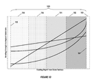
- FIG. 10 shows bonding strength gradients in a coating of the implant
- FIG. 11 is a schematic of an embodiment of the implant having at least one discontinuous layer
- FIG. 12 is another schematic view of FIG. 11 , showing an embodiment having at least one discontinuous layer
- FIG. 13 shows a process of determining the antimicrobial effect of an agent
- FIG. 14 is a schematic of an exemplary embodiment of the present invention utilizing at least one analgesic.
- FIG. 15 is a schematic of a further embodiment of the gradient coating of the present invention.
- FIG. 16 is a low magnification of the top PLGA coating containing silver modified beta-TCP and Bupivacaine.
- FIG. 17 is a high magnification of the top PLGA coating containing silver modified beta-TCP and Bupivacaine.
- FIG. 18 is a schematic of another embodiment of the present invention.
- FIG. 19 is a low magnification of the top view of the PLGA beads on a PLGA coating.
- FIG. 20 is a high magnification of the top view of the PLGA beads on a PLGA coating.
- FIG. 21 is a high magnification of the PLGA coating.
- FIGS. 22 ( a ) and ( b ) are SEM images from 9-day pulled out implant: Low Ag-modified calcium phosphate-coated implant from Rabbit #1A.
- FIGS. 23 ( a ) and ( b ) are SEM images from 9-day pulled out implant: non-calcium phosphate-coated implant from Rabbit #1A.
- FIGS. 24 ( a ) and ( b ) are SEM images from 9-day pulled out implant: High Ag-modified calcium phosphate-coated implant from Rabbit #1B.
- FIGS. 25 ( a ) and ( b ) are SEM images from 9-day pulled out implant: non-calcium phosphate-coated implant from Rabbit #1B.
- FIG. 26 is a back-scattering SEM of a ‘low’ S-CP, 9 days. (Sample 4A Right). Small regions of mineralized tissue (bone) (arrows) within regions of the porous coat. Dashed lines shows the position of host bone after site drilling.
- FIG. 27 is a back-scattering SEM of a ‘high’ S-CP, 9 days. (Sample 5B Left). Small regions of mineralized tissue (bone) (arrows) within regions of the porous coat. Dashed lines shows the position of host bone after site drilling.
- FIG. 28 is a back-scattering SEM of a ‘control’ (no CP), 9 days. (Sample 5B Right). Small regions of mineralized tissue (bone) (arrows) within regions of the porous coat.
- FIG. 29 is a back-scattering SEM of a ‘low’ S-CP, 16 days. (Sample 9C Right). Extensive bone ingrowth throughout full porous coat depth. Dashed lines shows initial drilled bone border.
- FIG. 30 is a back-scattering SEM of a ‘high’ S-CP, 16 days. (Sample 8D Right). Extensive bone ingrowth throughout full porous coat depth. Dashed lines show probable initial drilled bone border.
- FIG. 31 is a back-scattering SEM of a ‘control’ (no CP), 16 days. (Sample 2C Left). Bone ingrowth throughout depth of porous coating; difficult to identify initial drilled bone border.
- FIGS. 32 ( a ) and ( b ) show a 9-day sintered porous-coated Ti6Al4V ‘control’ implant—(a) and (b) Sample 5B Right—the blue-green stained areas are bone (old and newly-formed). Due to the section thickness, some bone does not show the staining effect and appears grey. A small amount of fibrous tissue is present near the interface in some regions (arrow).
- FIGS. 33 ( a ) and ( b ) show the 9-day sintered porous-coated Ti6Al4V implant with ‘low’ S-CP over-layer—(a) Sample 8A Left, (b) sample 4A Right—In (b), the extent of original bone loss due to drilling (and possibly some bone die-back) is evident by the truncated trabeculae.
- FIGS. 34 ( a ) and ( b ) show the 9-day sintered porous-coated Ti6Al4V implant with ‘High’ S-CP over-layer—(a) & (b) Sample 8B Left—Both the high and low magnification images show the extent of bone loss due to site preparation (drilling) and possibly subsequent bone die-back (dashed line in (b). Nevertheless, a suitable press-fit was achieved allowing early bone formation within the interface zone and into the porous coat (arrow).
- FIGS. 35 ( a ) and ( b ) show the 16-day sintered porous-coated Ti6Al4V implant ‘control’ implant—(a) & (b) Sample 2C Left—Extensive new bone formation and ingrowth throughout the porous coat (blue-green stained areas).
- FIGS. 36 ( a ) and ( b ) show the 16-day sintered porous-coated Ti6Al4V implant with ‘Low’ S-CP over-layer—(a) & (b) Sample 9C Right—Extensive new bone formation and ingrowth. [Sample embedding artifacts (air bubbles) seen in (a)].
- FIGS. 37 ( a ) and ( b ) show the 16-day sintered porous-coated Ti6Al4V implant with ‘High’ S-CP over-layer—(a) & (b) Sample 8D Right—Good bone ingrowth along implant length.
- FIG. 38 shows antimicrobial activity measured by log reduction against S. aureus.
- FIG. 1 represents a coated implant ( 900 ) of the prior art.
- a hydroxyapatite coating ( 920 ) is normally applied to an implant substrate ( 910 ) and sintered thereto in a conventional manner.
- Such a coating may comprise a mixture layer ( 912 ) of hydroxyapatite and a silver additive for an antimicrobial effect.
- the silver additive is not homogeneously incorporated into the HA lattice crystal structure.
- the gradient is generally composed of at least two portions having a different chemical/material composition.
- the gradient may be configured to increase the bond strength of the coating nearest an implant-coating interface, or the gradient may be configured such that an infection-reducing agent in the coating ( 20 ) is more prevalent on an outer surface ( 18 ) than an inner surface ( 12 , 14 ).
- the coating ( 20 ) may be an antimicrobial hydroxyapatite coating.
- the implant substrate ( 10 ) may be comprised of any number of biocompatible materials.
- the implant substrate ( 10 ) may be selected from a biocompatible material known in the art such as cobalt chromium, zirconium, titanium, stainless steel, ceramic, PEEK, polyurethane, etc.
- a biocompatible material such as cobalt chromium, zirconium, titanium, stainless steel, ceramic, PEEK, polyurethane, etc.
- suitable substrate materials may be utilized without limitation.
- the coating ( 20 ) comprises a first layer ( 12 ) having a first composition and at least one other second layer ( 14 ) having a second composition.
- the first layer ( 12 ) may be formed with a composition more compatible with the material of the implant substrate ( 10 ) for improved bonding characteristics and resistance to shear-off/delamination during press fit.
- the first layer ( 12 ) may be comprised of substantially pure HA.
- the first layer ( 12 ) may be formed with a composition having a higher content of bone-stimulating agents than the second layer ( 14 ).
- bone-stimulating agents may be any one or more of, but not limited to, fluoride, calcium, BMPs, proteins, bone void fillers, absorbable materials, and bioactives.
- the second layer ( 14 ) may be desirable to provide the second layer ( 14 ) with a composition having more infection-reducing agents than the first layer ( 12 ) because the greatest risk of infection generally occurs within the first few weeks after surgery.
- the coating ( 20 ) provides a strategic “time-release” of an antibacterial agent to provide the greatest anti-infection effect during initial bone remodeling.
- infection-reducing agents may be selected from any one or more of the following: copper, zinc, silver, gold, other metals having antimicrobial effects, or antibacterials, such as vancomycin. It should be understood that other materials may also be used.
- Additions of carbonates or other dissolution-controlling materials may be added to each layer in predetermined quantities to control the sequential decomposition rate of the HA coating ( 20 ) over time. For instance, it may be desired to allow outer layer ( 14 ) to absorb and deteriorate quickly, thereby allowing a burst release of silver ions adjacent the bone interface to fight off and inhibit infection during the most critical days after surgery.
- Inner layer ( 12 ) may be provided with a low carbonate content to achieve a slower, less dramatic release of silver ions in vivo for maintenance and infection-preventative purposes during the less critical period after early post-operative recovery.
- inner layer ( 12 ) may be provided with more additives for encouraging bone growth than an outer layer ( 14 ) because there may be some delay in bone reformation soon after surgery.
- the coating ( 20 ) provides both a way to selectively control the release of ions over time to combat infection.
- the coating ( 20 ) also may selectively control the exposure and release of bone-stimulating agents over time to improve biologic fixation and ingrowth.
- the coating ( 20 ) may provide a sacrificial layer of an antimicrobial coating so as to ensure that the entire implant remains coated, even if some of the coating shears off during implantation. Hence, the coating ( 20 ) may provide full bone ingrowth/ongrowth/biologic fixation to occur without the threat of infection.
- Each layer ( 12 , 14 , 16 , 18 ) may be composed of different compositions, or the layers may be formed with alternating compositions.
- each layer ( 12 , 14 , 16 , 18 ) has an amount of an antimicrobial and/or antibacterial agent that is effective at guarding against infection or otherwise killing bacteria which have formed during or after surgical implantation.
- each layer ( 12 , 14 , 16 , 18 ) may be provided with an optimum content level of bone-stimulating agents.
- Each layer ( 12 , 14 , 16 , 18 ) of the antimicrobial coating ( 20 ) may be applied to any portion of the implant substrate ( 10 ).
- the substrate ( 10 ) may be a core material, or the substrate ( 10 ) may be a separate ingrowth structure of similar or dissimilar material bonded to a core material.
- Such an ingrowth structure may be any one or more of, but not limited to, a porous structure, sintered beads, sintered particles of non-spherical shape, metal reticulated foam structures, fiber metal mesh, porous plasma spray, etc.
- the manner in which the layers ( 12 , 14 , 16 , 18 ) are applied to the substrate may vary. Some examples of acceptable application methods are hot-dipping, electro-deposition, plasma-spraying, magnetron sputtering, sol-gel, electrostatic spraying, and low temperature solution precipitation biomimetic techniques. It will be appreciated that application methods may be alternated between layers of the gradient coating of the present invention. For instance, a sol-gel dip process may be used to apply the first inner layer ( 12 ), electro-deposition may be used to apply the second layer ( 14 ), and plasma-spraying may be used to apply the third layer ( 16 ). It will also be appreciated that any number of layers ( 12 , 14 , 16 , 18 ) may be incorporated into the coating ( 20 ) in order to provide the optimum release of antimicrobial and bone-stimulating agents.
- the concentration gradient of coating ( 20 ) may be achieved by sequentially applying different solutions to the substrate ( 10 ) to form individual layers ( 12 , 14 , 16 , 18 ), each layer corresponding to a single application of solution, at least two of the layers ( 12 , 14 , 16 , 18 ) having dissimilar concentrations of any one or more of a bone stimulating agent, an infection-reducing agent, a catalyst for controlled release of the agents, or other material such as a protein, BMP, bioactive, antibiotic, or analgesic.
- the silver or zinc content may be higher in an outer layer ( 18 ) than in an inner layer ( 12 ), wherein the average silver or zinc content gradually changes in concentration over any intermediate layers ( 14 , 16 ) therebetween.
- the protein or carbonate content in an outer layer ( 18 ) may be greater than or less than an inner layer ( 12 ).
- fluoride content may gradually increase from an outer layer ( 18 ) to an inner layer ( 12 ).
- the predetermined release rate of infection-reducing ions over time may be advantageously controlled so as to avoid over-exposure of the ions to a patient, while still providing maximum protection against infection.
- a predetermined release rate may be optimized using simple experimentation so as to provide the most effectiveness in preventing infection without compromising ingrowth and long term ion release.
- the predetermined release rate of osseoinductive agents may further be designed so as to ramp up several days after surgery, when the risk of infection may be reduced relative to the immediate post-operative period.
- the initial ion release rate for the antimicrobial agent ( 22 ) is greater than a minimum initial ion release rate ( 32 ) necessary to provide sufficient anti-infection properties.
- numeral identifier ( 22 ) may represent the silver ion release rate of the outer layer ( 18 ) of the HA coating ( 20 ), and numeral identifier ( 32 ) may represent the minimum initial silver ion release rate necessary to effectively kill bacteria, reduce infection, and/or prevent future infection.
- the initial ion release rate for the antimicrobial agent ( 22 ) may be high as shown so as to provide a “burst release” to kill any existing bacteria during and immediately after surgery.
- the initial ion release rate for the antimicrobial agent ( 22 ) may then decrease over time to a lower, safer ion release rate ( 24 ) for infection-prevention maintenance and to prevent new bacteria from forming. It is preferred that the lowest ion release rate ( 24 ) of the coating ( 20 ) remains greater than or equal to the minimum ion release rate ( 32 , 34 ) necessary to maintain a reduced risk of infection, at least for a time period following surgery. This minimum ion release rate ( 32 , 34 ) may be determined by finding the greatest minimally inhibitory concentration (MIC) value for a bacteria group of which the patient is at risk for exposure during a particular surgery.
- MIC minimally inhibitory concentration
- MIC is generally defined herein as the lowest concentration of an antimicrobial agent that will inhibit the visible growth of a particular microorganism after incubation overnight.
- concentration of an antimicrobial agent which may be present in one or more layers ( 12 , 14 , 16 , 18 ) of the gradient coating ( 20 , 80 , 120 , 2000 ) of the present invention is preferably greater than the largest MIC for its bacterial counterpart. It is also preferred that the release rate ( 22 , 24 ) stay below human toxicity levels ( 54 ) to ensure that a patient is not overexposed to the heavy metal ions or other antibacterial agents. Surgeries take place in different geographical locations of the world have different postoperative infection-causing bacteria groups, and, therefore, the types and levels of antimicrobial agents within the gradient coating ( 20 ) may vary accordingly.
- all release rates may generally be controlled by altering the concentration gradient in the HA coating ( 20 ).
- the ion release period may be shortened by: (1) accelerating HA degradation using a carbonate or other dissolution catalyst; (2) decreasing the thicknesses of the layers ( 12 , 14 , 16 , 18 ) in the coating ( 20 ); (3) decreasing the number of layers ( 12 , 14 , 16 , 18 ) within the coating ( 20 ); and/or (4) increasing the change in concentration of agents between layers in such a way that agent release diminishes soon after the initial burst release ( 22 ).
- Reference numeral ( 42 ) indicates an initial release rate of a bone stimulating agent that promotes osseointegration. This initial release rate ( 42 ) may be high or low, depending on the individual patient's needs. In the embodiment illustrated in FIG. 5 , the release rate of a bone stimulating agent increases from initial rate ( 42 ) to a maintenance rate ( 44 ), at which point it levels off to promote osseointegration at a constant level.
- FIGS. 6 and 7 show alternative release rates for the most-preferred coating additives over time.
- FIG. 6 shows one embodiment where bone-promoting agents are initially released at a high rate ( 42 ), which increases to higher rate ( 46 ), and then decreases and levels off at maintenance rate ( 44 ).
- FIG. 7 shows another embodiment in which the rate of release of a bone-promoting agent ( 42 , 44 ) is always greater than the release rate of antimicrobial agents ( 22 , 24 ).
- an outer layer ( 18 ) of a gradient coating ( 20 ) may initially provide a burst release rate of fluoride bone-stimulating agents ( 42 ) that is greater than the initial release rate of silver ion antimicrobial agents ( 22 ).
- the lowest release rate of anti-infection agents ( 22 , 24 ) remains greater than or equal to the minimum release rate ( 32 , 34 ) sufficient for reducing the risk of infection. It is to be understood that substances other than bone-promoting agents, antimicrobial, and/or antibacterial agents may be present in any one of layers ( 12 , 14 , 16 , 18 ) of the HA coating ( 20 ). It is therefore anticipated that additional controllable release rates (not shown) may be utilized.
- prior art coatings such as that shown in FIG. 1 , may be vulnerable to shearing off of an implant substrate when implanted into a bony structure (e.g., a femoral stem implanted into a prepared femoral canal) because the silver additive may lessen the bonding strength of the layer ( 912 ).
- a bone ( 950 ) is prepared to form a bony surface ( 952 ) suitable for scratch fit with the implant ( 900 ). Normally, for cementless fixation, the bony surface ( 952 ) is dimensioned so as to provide an interference press fit with the implant ( 100 ) to provide initial stabilization.
- the implant ( 900 ) generally consists of a core substrate ( 960 ) and a single composition coating ( 962 ), such as a hydroxyapatite coating with a silver additive.
- the coating ( 962 ) of the implant is susceptible to shearing. Shearing is disadvantageous because if any material ( 966 ) from the coating ( 962 ) is removed from the core substrate ( 960 ), it may leave a portion ( 964 ) of the core substrate ( 960 ) exposed and more susceptible to infection.
- any portion ( 964 ) of the core substrate ( 960 ) that is left without a coating ( 962 ) may exhibit reduced bone ingrowth due to a lack of hydroxyapatite in the portion ( 964 ).
- a coated implant ( 100 ) having a gradient coating ( 80 ) is shown to have improved antimicrobial and osseointegration characteristics over prior art implants using conventional coatings.
- the gradient coating ( 80 ) shown in FIG. 9 is applied to an implant substrate ( 82 ) of the implant ( 100 ) and comprises at least two layers ( 84 , 88 ) that are generally formed of different compositions.
- an outer layer ( 88 ) has a higher metal content than at least one inner layer ( 84 ).
- the at least one inner layer ( 84 ) may exhibit an equal or greater amount of metal than the outer layer ( 88 ).
- the metal may be selected from one or more of silver, zinc, copper, or other known metals having antimicrobial effects.
- antibiotics such as vancomycin and/or penicillin, may be utilized.
- the at least one inner layer ( 84 ) may be free of metal or may comprise a metal different than the outer layer ( 88 ).
- the at least one inner layer ( 84 ) may comprise a low concentration of zinc, whereas the outer layer ( 88 ) may comprise a lower, equal, or greater concentration of another metal, such as silver.
- the outer layer ( 88 ) may comprise a first zinc and silver concentration, whereas the at least one inner layer ( 84 ) may exhibit a second silver and/or copper concentration.
- each layer ( 12 , 14 , 16 , 18 , 84 , 88 ) of the gradient coating ( 20 , 80 ) is able to readily modify the additives and compositions of each layer ( 12 , 14 , 16 , 18 , 84 , 88 ) of the gradient coating ( 20 , 80 ) to satisfy the needs of a patient on an individual basis.
- at least one inner layer ( 84 ) may comprise any number of layers, each of which may have similar or different compositions and/or additives.
- the at least one inner layer ( 84 ) and/or outer layer ( 88 ) may further comprise at least one bone-stimulating agent, such as fluoride or calcium.
- the concentration of the bone-stimulating agent in the at least one inner layer ( 84 ) may be greater than, equal to, or less than that of the outer layer ( 88 ). However, it is preferred that the at least one inner layer ( 84 ) has a greater concentration of bone-stimulating agent than the outer layer ( 88 ).
- the outer layer ( 88 ) may be advantageously utilized as a protective layer so as to allow at least one or more inner infection-reducing layers ( 84 ) to remain bonded to the implant substrate ( 82 ) after implantation.
- a coated implant ( 100 ) may be press-fitted into a prepared bony surface ( 52 ) of a bone ( 50 ) to provide initial stabilization. Press-fitting is normally achieved by impacting the implant ( 100 ) so as to form an interference fit with the prepared bony surface ( 52 ).
- the prepared bony surface ( 52 ) is dimensioned so closely to the implant, or slightly smaller than the implant, there is a possibility that at least some coating material ( 87 ) may shear off of or be displaced from the outer layer ( 88 ) thereby forming an outer layer deficient zone ( 83 ) of very thin or no outer layer ( 88 ).
- One unique aspect of the present invention is that because the coating is formed as a gradient coating ( 80 ), the at least one outer layer deficient zone ( 83 ) is still protected by an underlying at least one inner layer ( 84 ) having infection-resistant properties and/or higher bonding strength characteristics. Any displaced material ( 87 ) may serve as a “barrier” to prevent bacteria from entering the bone-implant interface ( 52 ) and/or promote local fixation.
- inner layer ( 84 ) may be formulated with a composition that possesses improved bonding characteristics with an implant substrate ( 82 ).
- outermost layer ( 88 ) as a “first defense” protective antimicrobial layer
- the at least one inner layer ( 84 ) as a backup antimicrobial layer allows for some margin for shear during implantation, while still maintaining one-hundred percent coverage of the implant substrate ( 82 ) with the antimicrobial properties necessary to kill and prevent formation of bacteria.
- the gradient coating ( 80 ) may comprise a bonding strength gradient, such that the coating composition gradually increases in bonding strength from the outermost surface ( 88 ) to the implant substrate ( 82 ), wherein the greatest bond strength is preferably achieved at the coating-substrate interface ( 117 , FIG. 10 ).
- This bonding strength gradient may be achieved by adding substrates to the at least one inner layer ( 84 ) which are most compatible with the material of the implant substrate ( 110 ). The addition of such substrates may be controlled independently of the antimicrobial agents, bone-stimulating agents, and/or dissolution catalysts discussed herein. Alternatively, materials known to have good bonding characteristics with implants, such as pure HA, may be utilized in the layer ( 112 ) most adjacent to the implant substrate ( 110 ).
- FIG. 10 illustrates four possible examples of bonding strength gradients.
- the average bonding strength of the gradient coating ( 20 , 80 , 120 ) may generally increase with coating depth. This allows for some external coating shear to take place without exposing the implant substrate ( 110 ).
- the bonding strength of the coating composition near an outside surface ( 115 ) of a gradient coating ( 120 ) may be less than at the implant-coating interface ( 117 ) of a substrate or ingrowth structure ( 110 ).
- layers ( 112 ) and ( 114 ) may comprise a composition having greater bonding characteristics with the material of the substrate ( 110 ) than layers ( 116 ) and ( 118 ), in order to optimize bonding at the implant-coating interface ( 117 ).
- the inner layers ( 112 , 114 ) may be configured with a higher concentration of bone-stimulating agents than layers ( 116 , 118 ) for improved osseointegration with the implant substrate or ingrowth structure ( 110 ).
- the outer layers ( 116 , 118 ) may be provided with a higher concentration of ion-producing antimicrobials such as silver, zinc, or copper-based elements.
- any layer ( 1002 , 1004 , 1006 , 1008 ) within a multi-layer gradient antimicrobial coating ( 2000 ) may include discontinuous layer portions ( 1007 ).
- an outer layer ( 1008 ) of coating ( 2000 ) may comprise a discontinuous “speckled” coating formed from layer portions ( 1007 ).
- These layer portions ( 1007 ) may be strategically placed within the coating ( 2000 ) such that the inhibitory zones ( 1003 ) for each of the layer portions ( 1007 ) overlap ( 1005 ). In doing so, the amount of antimicrobial and/or antibacterial agents used in the coating ( 2000 ) may be decreased, thereby minimizing patient exposure, while still maintaining adequate protection against infection.
- Providing layer portions ( 1007 ) may further serve to increase the surface texture/roughness of the coating ( 2000 ).
- the antimicrobial bioceramic gradient coating may be prepared using a sol-gel process. At least three silver concentrations have been prepared and evaluated for use, in order to determine the minimum preferred silver content for any one layer ( 12 , 14 , 16 , 18 ). Each silver concentration was calculated based on the Ag/Ca ratio, which may be 0.1, 0.01, or 0.001. Other silver contents were chosen to cover a broad range of silver concentrations in order to determine the minimum silver content in the coating to have a sufficient antibacterial effect but not so high as to inhibit or kill tissue cells, such as osteoblasts. The optimum range of silver concentration has been determined based on both in vitro and in vivo results.
- the silver concentration of samples 2 and 4 was approximately twenty times greater than the silver concentration of samples 1 and 3. This resulted in an MBC approximately four times greater for samples 2 and 4 than the MBC of samples 1 and 3. In all instances, powders having a silver concentration greater than or equal to about 0.1% were deemed to be capable of killing S. aureus at 105 cfu/ml.
- FIG. 13 illustrates the process used in testing the sample powders.
- the bacteria is added to each well per row.
- a two-fold decreasing dilutions of sample is suspended in broth.
- the tray is incubated overnight.
- Samples from each well are placed on agar plates.
- the samples are again incubated overnight.
- the MBC is identified for each sample.
- the Ca/P ratio is kept at 1.60 to prepare 70% HA and 30% ⁇ -TCP biphasic calcium phosphate (BCP). It is to be understood; however, that this ratio may be varied to obtain different biphasic HA/ ⁇ -TCP compositions.
- ⁇ -TCP is generally desirable during the initial bone healing process, because it provides calcium ions that accelerate bone ingrowth.
- fluoride also may be incorporated in the HA composition in order to create fluoride ions/fluorine, which are known to favorably promote mineralization and faster osseointegration.
- the fluoride may be introduced into a coating alone such that the coating has a concentration gradient of only fluoride, or the fluoride may be introduced into a coating in combination with other elements and agents disclosed herein.
- a suitable fluoride concentration may be calculated based on F ⁇ /PO 4 3 — ratio, which is 0.2.
- a carbonate function group may be added to the HA structure, the carbonate function group preferably being designed to strategically destabilize and degrade the HA structure in such a way as to optimally release Ag, Ca, and F ions.
- the carbonate function group generally serves as a mechanism for controlling the rate of Ag, Ca, and F ion release in vivo.
- the carbonate content may be calculated based on the ratio of CO 3 2 ⁇ /PO 4 3 ⁇ , which is 1; however, this content may vary.
- the method steps involved with synthesizing the antimicrobial thin film coating generally include first dissolving calcium nitrate and silver nitrate into de-ionized distilled water to form a first solution (DDH 2 O). Next, ammonium dihydrogen phosphate is dissolved into the first solution (DDH 2 O) to form a second solution. The appropriate amount of ammonium fluoride and ammonium carbonate is then subsequently dissolved into another ammonium dihydrogen phosphate solution to form a third solution. Concentrated ammonium hydroxide is used to increase the pH levels of both the second and third solutions to about 11. The second calcium nitrate-based solution is slowly added to the third ammonium dihydrogen phosphate-based solution under vigorous stifling. The sol-gel solution is stirred for 24 hours and then allowed to age at room temperature for 2 days. This aged solution may then be applied to an implant via a thin film dip coating or be prepared for use in an antimicrobial powder.
- implants utilizing the antimicrobial coating are dipped into the sol-gel solution and then gradually withdrawn vertically at a controlled rate.
- the withdrawal rate is about 30 cm/min; however, it should be noted that this rate may be increased or decreased to provide the desired thickness.
- the thickness of the coating may also be controlled by varying the overall viscosity of the sol-gel solution. While any number of dips may be required, a total of 3-4 dips has been shown to produce a preferred coating thickness about 1-2 ⁇ m.
- the thin film coating may be sintered to an implant using a vacuum furnace, an in-air muffle furnace, or any other equivalent devices for sintering known in the art.
- a temperature of about 400-800 degrees C. is held for about 10-30 minutes, although other temperatures and times may be required depending on the composition of the coating and its thickness.
- the furnace is cooled to room temperature. Crystallinity and/or the composition of the coating, which affects resorption rate, may be controlled by varying the sintering temperature. For example, a lower sintering temperature generally results in a more degradable coating, and biphasic calcium phosphate can only be developed at temperatures above about 750 degrees C.
- an antibacterial thin film coating applied to an implant by way of an antimicrobial bioceramic powder.
- an aged sol-gel solution is dried at about 90 degrees C. for about 48 hours and then sintered between about 400-800 degrees C. for about 1-2 hours.
- the resulting substance after sintering may then be ground into powder and sieved and sorted by size for different applications.
- coarser powders may be used to make dense bulk antibacterial bioceramics or porous scaffolds.
- finer powders may advantageously be used as feeding powders for plasma-sprayed HA coatings.
- the antimicrobial bioceramic powder may have many valuable uses and may come in many forms.
- a coating for a medical implant configured for reducing infection and promoting biologic fixation
- the antimicrobial coating comprising a gradient.
- a gradient may comprise, for example, a change in composition or a variation in content of at least one substance with respect to coating depth.
- the antimicrobial coating may provide an initial burst release of the antimicrobial agent at a bone-coating interface by having a higher concentration of silver additive at an exterior portion of the coating.
- the antimicrobial coating may provide a predetermined controlled release of fluoride over time in order to optimally promote bone ingrowth and biologic fixation.
- the antimicrobial coating may provide a sustained preventative level of ion release that is greater than or equal to the minimum level required to have an effective antibacterial effect.
- a coating for an implant having a concentration gradient of pain reducers or analgesics having a concentration gradient of pain reducers or analgesics.
- the gradient may be configured for an initial burst release of a pain reducer or analgesic, followed by a slow release of the pain reducers or analgesics, although actual release rate may vary to address the needs of an individual patient or patient population subset.
- Such a pain-reducing element gradient within a coating may be present alone or in combination with other gradients, elements, or agents discussed herein.
- the pain reducers or analgesics may be any known in the art, such as opioids and/or non-steroidal anti-inflammatory drugs (NTHES).
- an antimicrobial coating having calcium phosphate and a gradient distribution of at least one metal content, such as silver, zinc, copper, in the coating.
- the at least one metal content may be high in the outer surface of the coating and low in the inner surface.
- This metal gradient-CaP coating may be achieved by dipping the implant in sequence into different sol-gel solutions, each solution having a different gradient metal concentration.
- the metal gradient calcium phosphate coating may include single phase calcium phosphate, such as HA, and/or may also include multiple calcium phosphate phases, such as biphasic calcium phosphates.
- the advantage of using a sol-gel process is that the coating may be applied to a porous ingrowth structure without leaving any portions left uncoated. While at least one sol-gel process is preferred, other primary or subsequent processes known in the art may be used.
- a metallic substrate such as silver, zinc, copper, etc., and calcium phosphate, such as HA, TCP, Ag—CaP, etc.
- the composite coating may be applied to an implant through the use of a plasma-spraying process, although other processes discussed herein may be used.
- the metallic substrate may be in the form of a powder that may be mixed together with calcium phosphate powders and co-sprayed onto an implant surface simultaneously.
- the metallic substrate and calcium phosphate powders may be applied individually, layer by layer, to an implant surface. The order in which each layer is applied may be predetermined to provide the best solution for an individual patient's needs.
- the gradient in the plasma-sprayed coating may be accomplished by feeding the metallic substrate powders and calcium phosphate powders into a hopper at different rates to obtain a gradient metal content in the final coating.
- the implants may undergo several separate plasma-spraying processes, each process using a different composition of pre-mixed metal and calcium phosphate powders.
- an antimicrobial calcium phosphate coating is provided, the coating having at least one metal, such as copper, silver, zinc, etc.
- the antimicrobial coating further comprises a gradient degradation profile.
- an outer layer of the coating may have a faster dissolution rate than an inner layer of the coating.
- Such a dissolution rate profile may be employed to force an outer portion of the coating to dissolve quicker, thereby creating a burst release of an antimicrobial agent.
- an outer layer might utilize a more degradable calcium phosphate, whereas the inner surface layer might utilize a more stable calcium phosphate.
- some calcium phosphates used may or may not have a metal in their compositions.
- some calcium phosphates used in the coating layers may or may not have osseoinductive elements.
- at least one calcium phosphate phase has a metal such as silver, in order to provide the antibacterial function necessary to fight infection.
- at least one bone-stimulating agent is included within at least one of the coating layers to promote biologic fixation. While it is preferred that an outer portion of the coating resorbs quickly, the coating may alternatively be configured to initially resorb slowly and then quickly. This may be achieved by providing an inner layer with more carbonate than an outer layer.
- a coated implant having a metal or metal-ion adsorbed outer surface.
- the outer surface is configured to increase metal ion release at the bone-implant interface immediately after implantation to ward off infection and kill any residing bacteria.
- the metal ion-enriched outer coating on the implant may be formed by first immersing the implant into calcium phosphate to form one or more layers.
- the one or more layers may or may not comprise a metal, such as silver, zinc, or copper, as previously discussed and also may or may not comprise a bone-stimulating agent or dissolution catalyst.
- the coated implant is then finally immersed into a metal-soluble solution, such as silver nitrate or silver fluoride, to adsorb metal ions onto the outer surface of the coated implant and increase the metal concentration at the outer surface of the coated implant.
- a metal-soluble solution such as silver nitrate or silver fluoride
- a silver-soluble solution may be advantageously utilized to form an outer surface layer having an increased concentration of silver ions at the outer surface of the coating.
- an insoluble metal solution may be used to provide a metal outer surface to the coating.
- a calcium phosphate-coated implant is first submersed into the insoluble metal solution. Once the insoluble metal solution is absorbed onto the outer surface of the coated implant, the implant may then be removed from the insoluble metal solution and exposed to light. Exposure to light reduces the absorbed metal ions at the outer surface to metal.
- the multi-layer gradient antimicrobial coating of the present invention may include one or more discontinuous layer portions.
- an outer layer of the multi-layer gradient antimicrobial coating may comprise a discontinuous “speckled” coating formed from layer portions.
- These layer portions may be strategically placed within the multi-layer gradient antimicrobial coating such that the inhibitory zones for each of the layer portions overlap. In doing so, the amount of antimicrobial and/or antibacterial agents used in the multi-layer gradient antimicrobial coating may be decreased, thereby minimizing patient exposure, while still maintaining adequate protection against infection.
- Providing layer portions may further serve to increase the surface texture/roughness of the multi-layer gradient antimicrobial coating.
- a coating having a gradient analgesic concentration preferably comprises calcium phosphate (CaP); however, other base coating materials known in the art may be equally employed.
- the analgesic concentration is preferably higher in an outer surface portion of the coating than in an inner portion of the coating, in order to: (1) provide the maximum amount of relief to a patient immediately after surgery; and then (2) maintain a lower, safe dosage to provide long-term relief.
- Such an analgesic gradient CaP coating may be formed using several coating techniques. However, for porous-surfaced implants, a non-line-of-sight coating process is preferred. Examples of such non-line-of-sight coating processes include, but are not limited to sol-gel dip coating, electrodeposition, electrophorosis, biomimetic methods, and/or combinations thereof.
- the gradient analgesic concentration may be formed by first applying a pure thin film calcium phosphate layer to an implant surface to form a coated implant. This layer may be applied using a sol-gel dip coating method and then hardened by sintering at an elevated temperature to achieve a sufficient bonding strength at the coating/implant interface. The coated implant may then be immersed into a first solution containing both calcium phosphate and a first concentration of at least one analgesic agent.
- the first solution may be simulated body fluid (SBF) solution, modified SBF solution, or any solution which may be able to form an apatite coating onto an implant surface.
- SBF simulated body fluid
- modified SBF solution or any solution which may be able to form an apatite coating onto an implant surface.
- an elevated temperature is preferred (e.g., sintering); however, it is important that the elevated temperature is not so high as to denature the at least one analgesic agent. If desirable, this process may be repeated for additional second, third, and fourth solutions having other concentrations of the at least one analgesic agent to form a coating having a gradient concentration of the at least one analgesic agent, the gradient extending in a direction perpendicular to the implant surface.
- additives may be present within the coating, the additives being in uniform or gradient concentrations throughout the coating depth.
- the additives may include antimicrobial agents, bone-promoting agents, antibiotics, steroids, anti-inflammatories, antispasmodics, bioactives (e.g., BMP, bisphosphonates), hemostats, or any other agent discussed herein.
- the analgesics described herein may be either local anesthetics, narcotic or non-narcotic in nature.
- An example of a local anesthetic is local anesthetics is bupivacaine (e.g. Marcain, Marcaine, Sensorcaine and Vivacaine).
- Some examples of narcotic analgesics that may be advantageously utilized are opioids, morphine, codeine, oxycodone (Percodan), levorphanol (levodromoran), propoxyphene (Darvon), hydrocodone (Vicodin), pentazocine (Talwin) and/or combinations thereof.
- non-narcotic analgesics examples include acetylsalicylic acid (aspirin), ibuprofen, phenylbutazone (Butazolidine), indomethacin (Indocin), acetaminophen, phenacetin, and combinations thereof.
- the analgesics may be topical and may include ibuprofen- or diclofenac-containing gels, capsaicin, Lidocaine, anti-inflammatories, and steroids without limitation.
- analgesics may be advantageously utilized either alone or in combination, so long as adverse reactions from combining are avoided.
- suitable analgesics may include, but are not limited to, natural opium alkaloids, such as morphine, opium, hydromorphone, nicomorphine, oxycodone, dihydrocodeine, diamorphine, papavereturn, morphine combinations, dihydrocodeine combinations, codeine combinations excluding psycholeptics, and codeine combinations with psycholeptics; phenylpiperidine derivatives including ketobemidone, pethidine, fentanyl, pethidine, combinations excluding psycholeptics, pethidine, and combinations with psycholeptics; diphenylpropylamine derivatives such as dextromoramide, piritramide, dextropropoxyphene, bezitramide, methadone combinations excluding psycholeptics, dextropropoxyphene combinations excluding psycholeptics; dextropropoxyphene combinations with
- any one or more of the above mentioned analgesics could be used in the gradient coating.
- the analgesics may vary in concentration in a direction extending perpendicular to the implant surface. If narcotic analgesics and non-narcotic analgesics are to be used together, it is preferred that the narcotic analgesics are used at outer coating portions and the non-narcotic analgesics are used at inner coating portions. While non-narcotic analgesics are generally recognized to be effective in relieving skeletal pain due to inflammation (such as arthritis), their analgesic properties are not nearly as strong as those of narcotic analgesics, such as morphine and synthetic opioids.
- a stronger analgesic such as morphine
- the stronger analgesic preferably reducing in concentration from an outer surface of the coating as it approaches the coating/implant interface.
- a patient may be less-susceptible to addiction, and is essentially “weaned” from the high initial dosage that is required after surgery but is not necessary during long-term recovery.
- the gradient coating may employ weaker analgesics at the inner portions of the gradient coating. These weaker analgesics may be provided in the form of a gradient—generally increasing in concentration from an outer portion of the coating towards the coating/implant interface.
- narcotic analgesics mainly at outer portions of the gradient coating
- narcotic drugs might prevent platelet agglutination, which may inhibit or delay the normal bone healing process and osseointegration.
- the inner coating portions are more proximate to the implant surface and/or the ingrowth structure, it is therefore, desirable to provide these coating portions with either non-narcotic analgesics or very low doses of narcotic analgesics to avoid interference with platelet agglutination.
- the gradients may be staggered and may vary in dosage so as to provide smooth, controlled transitions in a pre-defined pain management plan designed for an individual patient or a population subset.
- the pre-defined pain management plan could be, for instance, included as part of a prescription for an implant.
- the implant may be first coated with a pure calcium phosphate (CaP) in a manner discussed above to obtain good implant bonding strength, cohesion, and ingrowth characteristics.
- the coated implant may then be immersed into a first solution containing calcium phosphate, a first concentration of a non-narcotic analgesic, and a first concentration of a narcotic analgesic.
- the first solution may be applied in any manner described herein.
- the twice-coated implant may then be immersed into a second solution containing calcium phosphate, a second concentration of a non-narcotic analgesic, and a second concentration of a narcotic analgesic.
- the second concentration of a narcotic analgesic be higher than the first concentration of a narcotic analgesic.
- the second concentration of a non-narcotic analgesic be lower than the first concentration of a non-narcotic analgesic. This process may be done iteratively to produce finer and finer graduations within the coating.
- additives may be present within the coating, the additives being in the form of a uniform or gradient concentration throughout the coating depth.
- the additives may include antimicrobial agents, bone-promoting agents, antibiotics, steroids, anti-inflammatories, antispasmodics, bioactives (e.g., BMP, bisphosphonates), hemostats, or any other agent discussed herein.
- a gradient coating is provided by first coating an implant or ingrowth structure with calcium phosphate using any one of the aforementioned techniques.
- a pure thin-film calcium phosphate layer is preferred at the coating/implant interface to improve the bonding strength of the gradient coating.
- the once-coated implant is then dipped into a first biodegradable polymer solution which contains a first concentration of at least one analgesic agent.
- the polymer solution is prepared by first dissolving at least one biodegradable and/or biocompatible polymer, such as PCL, PLGA, PLLA, PGA, etc. or combinations thereof, into a solvent and then adding a pre-measured amount of the at least one analgesic agent. After the implant is dipped into the first biodegradable polymer solution, it is allowed to dry out in air. In order to expedite and facilitate solvent evaporation of the first biodegradable polymer solution, a vacuum treatment at room temperature may be advantageously utilized.
- the polymer(s) within the coating serve as a carrier for the at least one analgesic in the formed coating. If desired, additional second, third, and fourth, etc.
- polymer solutions may be subsequently provided, each polymer solution having a different concentration of the at least one analgesic agent and forming another layer within the gradient coating. It is to be understood that multiple analgesic agents and suitable carriers other than polymers may be present within each solution without departing from the scope of the invention.
- additives may also be present within the coating, the additives being provided in a uniform or gradient concentration throughout the coating depth.
- the additives may include antimicrobial agents, bone-promoting agents, antibiotics, steroids, anti-inflammatories, antispasmodics, bioactives (e.g., BMP, bisphosphonates), hemostats, or any other agent discussed herein.
- a coating for an implant having a first layer adjacent the implant surface.
- the first layer includes a thin film calcium phosphate (CaP) having a first low concentration of an antimicrobial agent. While this first layer may also contain additional additives such as analgesics, bioactives, or bone-stimulating agents, it is preferred that only a small amount of an antimicrobial be present in order to provide optimal cohesion and bonding strength with the substrate material of the implant without sintering.
- CaP thin film calcium phosphate
- a second layer is then provided over the first layer, the second layer preferably comprising a biomimetic calcium phosphate, a second slightly higher concentration of an antimicrobial agent than the first layer, a bioactive (e.g., BMP), and a non-narcotic agent for long-term pain relief and/or reducing inflammation.
- a bioactive e.g., BMP
- a non-narcotic agent for long-term pain relief and/or reducing inflammation.
- a third outer layer is provided over the second layer, the third outer layer comprising a biomimetic calcium phosphate, an even higher concentration of an antimicrobial agent than the first and second layers (for immediate post-op infection resistance), a similar or lower concentration of a bioactive (e.g., BMP) than the second layer, and a narcotic agent (e.g., opioid) for immediate, highly-effective pain relief.
- Numeral identifier ( 1010 ) represents an implant or ingrowth structure formed from a substrate material, such as titanium, titanium alloy, cobalt-chromium, zirconium, stainless steel, or any other biocompatible metal known in the art.
- a gradient coating ( 1020 ) is applied to the implant ( 1010 ), the gradient coating ( 1020 ) has a first thin film inner layer ( 1012 ) of pure hydroxyapatite. Because this layer ( 1012 ) is generally free from additives that may reduce the bonding strength characteristics with respect to the material of the implant ( 1010 ), it may be thought of as a “primer” for the gradient coating ( 1020 ).
- the first layer ( 1012 ) may be sintered to the implant ( 1010 ) by conventional ceramic firing methods.
- a second layer ( 1014 ) is also provided, the layer preferably containing a substantial concentration of a bone-stimulating agent, such as fluoride, a non-narcotic analgesic, such as aspirin, and a low dose of a bioactive (e.g., BMP or bisphosphonate).
- the second layer may be fired or not fired to the first layer, but is preferably not fired in order to maintain the virgin properties of the additives.
- a third layer ( 1016 ) may further be provided, the third layer ( 1016 ) has, for example, a moderate concentration of an antimicrobial, such as silver, zinc, or copper, a lower dose of a non-narcotic analgesic than the second layer ( 1014 ), a higher concentration of a bone stimulating agent than the second layer ( 1014 ), and a low dose of a narcotic analgesic.
- the third layer ( 1016 ) may be applied by methods other than ceramic firing.
- a fourth layer ( 1018 ) may be for example, a polymer-based carrier coating and may comprise a higher level of an antimicrobial than the third layer ( 1016 ), a higher dose or concentration of a narcotic analgesic than the third layer ( 1016 ), and a lower dose of a bone-stimulating agent than both the second ( 1014 ) and third ( 1016 ) layers.
- a medical implant having one or more surfaces comprising a functionally-graded coating disposed thereon, wherein said functionally-graded coating comprises multiple coating layers and an antimicrobial agent, and wherein a concentration of said antimicrobial agent in is different in at least two of said multiple coating layers.
- a medical implant having one or more surfaces comprising a functionally-graded coating disposed thereon, wherein said functionally-graded coating comprises multiple coating layers and an antimicrobial agent, and wherein an outer of said multiple coating layers has a higher concentration of said antimicrobial agent than an inner of said multiple coating layers.
- a medical implant having one or more surfaces comprising a functionally-graded coating disposed thereon, wherein said functionally-graded coating comprises multiple coating layers and an antimicrobial agent disposed within an outer of said multiple coating layers, wherein an inner of said multiple coating layers does not contain an antimicrobial agent.
- a medical implant having one or more surfaces comprising a functionally-graded coating disposed thereon, wherein said functionally-graded coating comprises multiple coating layers and an antimicrobial agent, wherein a concentration of said antimicrobial agent is different in at least two of said multiple coating layers, and wherein the antimicrobial agent may be selected from any one of silver, copper, zinc, manganese, gold, iron, nickel, cobalt, cadmium, platinum, and combinations thereof.
- a medical implant having one or more surfaces comprising a functionally-graded coating disposed thereon, wherein said functionally-graded coating comprises multiple coating layers and an antimicrobial agent, wherein a concentration of said antimicrobial agent is different in at least two of said multiple coating layers, wherein the antimicrobial agent may be selected from silver, copper, zinc, manganese, gold, iron, nickel, cobalt, cadmium, platinum, and combinations thereof, and wherein the maximum silver concentration within the functionally-graded coating is generally between about 0.1 to about 10 weight percent, and more preferably between about 0.5 to about 3 weight percent.
- a medical implant having one or more surfaces comprising a functionally-graded coating disposed thereon, wherein said functionally-graded coating comprises multiple coating layers and an antimicrobial agent, wherein a concentration of said antimicrobial agent is different in at least two of said multiple coating layers, and wherein the antimicrobial agent is an antibiotic such as vancomycin, gentamycin, penicillins, cephalosporins, aminoglycoside, macrolides, clindamycin, tetracyclines, chloramphenicol, spectinomycin, polypeptide antibiotics, fluoroquinolones, and combinations thereof.
- an antibiotic such as vancomycin, gentamycin, penicillins, cephalosporins, aminoglycoside, macrolides, clindamycin, tetracyclines, chloramphenicol, spectinomycin, polypeptide antibiotics, fluoroquinolones, and combinations thereof.
- a medical implant having one or more surfaces comprising a functionally-graded coating disposed thereon, wherein said functionally-graded coating comprises multiple coating layers and an antimicrobial agent, wherein a concentration of said antimicrobial agent is different in at least two of said multiple coating layers, and wherein the antimicrobial agent is an anti-fungal such as amphotericin B, nystatin, liposomal amphotericin B, flucytosine, and combinations thereof.
- a medical implant having one or more surfaces comprising a functionally-graded coating disposed thereon, wherein said functionally-graded coating comprises multiple coating layers and an antimicrobial agent, wherein at a concentration of said antimicrobial agent is different in at least two of said multiple coating layers, and wherein the antimicrobial agent is an anti-viral such as acyclovir, ganciclovir, idoxuridine, amantadin, interferons, azidothymidine, and combinations thereof.
- an anti-viral such as acyclovir, ganciclovir, idoxuridine, amantadin, interferons, azidothymidine, and combinations thereof.
- a medical implant having one or more surfaces comprising a functionally-graded coating disposed thereon, wherein said functionally-graded coating comprises multiple coating layers and an antimicrobial agent, wherein a concentration of said antimicrobial agent is different in at least two of said multiple coating layers, wherein at least one of said multiple coating layers contains a bone stimulating material, wherein said bone stimulating material may be selected from any of calcium, phosphate, carbonate, fluoride, silicon, magnesium, strontium, vanadium, lithium, a growth factor (BMP, etc), a biomimetic peptide, hydroxyapatite, ⁇ tricalcium phosphate, mixtures of hydroxyapatite and ⁇ tricalcium phosphate, and combinations thereof, and wherein a concentration of said bone stimulating material may be different in at least two of said multiple coating layers.
- said bone stimulating material may be selected from any of calcium, phosphate, carbonate, fluoride, silicon, magnesium, strontium, vanadium, lithium, a growth factor (BMP, etc
- a medical implant having one or more surfaces comprising a functionally-graded coating disposed thereon, wherein said functionally-graded coating comprises multiple coating layers and an antimicrobial agent, wherein a concentration of said antimicrobial agent is different in at least two of said multiple coating layers, wherein at least one of said multiple coating layers contains an analgesic agent such as a local anesthetics, such as bupivacaine (e.g.
- a medical implant having one or more surfaces comprising a functionally-graded coating disposed thereon, wherein said functionally-graded coating comprises multiple coating layers and an antimicrobial agent, wherein the concentration of said antimicrobial agent is different in at least two of said multiple coating layers, wherein at least one of said multiple coating layers contains an analgesic agent, and wherein a concentration of said analgesic agent is different in at least two of said multiple coating layers.
- a medical implant having one or more surfaces comprising a functionally-graded coating disposed thereon, wherein said functionally-graded coating comprises multiple coating layers and an antimicrobial agent, wherein the concentration of said antimicrobial agent is different in at least two of said multiple coating layers, and wherein an outer of said multiple coating layers comprises a narcotic analgesic agent and an inner of said multiple coating layers comprises a non-narcotic analgesic agent.
- a medical implant having one or more surfaces comprising a functionally-graded coating disposed thereon, wherein said functionally-graded coating comprises multiple coating layers and an antimicrobial agent, wherein at the concentration of said antimicrobial agent is different in at least two of said multiple layers, and wherein said antimicrobial agent comprises a silver specie including, but not limited to: a silver salt, silver nitrate, silver perchlorate, silver acetate, silver tetrafluoroborate, silver trifoliate, silver fluoride, silver oxide, silver hydroxide, other silver-“oxo” species, silver sulfadiazine, mixtures of silver with other stabilizing ligands other than sulfadiazine, and combinations thereof.
- a silver salt silver nitrate, silver perchlorate, silver acetate, silver tetrafluoroborate, silver trifoliate, silver fluoride, silver oxide, silver hydroxide, other silver-“oxo” species, silver sulfadiazine, mixtures
- a functionally-graded antimicrobial coating applied to one or more surfaces of a medical implant, wherein said functionally-graded coating comprises multiple layers of a bone conducting material and an antimicrobial agent disposed within at least one of said multiple layers, wherein a concentration of said antimicrobial agent is different in at least two of said multiple layers, and wherein the bone conducting material may be selected from calcium phosphate, hydroxyapatite, ⁇ tricalcium phosphate, a mixture of hydroxyapatite and ⁇ tricalcium phosphate, resorbable polymers, bioglass, derivatised phosphate-based compound, orthophosphates, monocalcium phosphates, octacalcium phosphates, dicalcium phosphate hydrate (brushite), dicalcium phosphate anhydrous (monetite), anhydrous tricalcium phosphates, whitlocktite, tetracalcium phosphate, a
- a functionally-graded antimicrobial coating applied to one or more surfaces of a medical implant, wherein said functionally-graded coating comprises multiple layers of a bone conducting material and an antimicrobial agent disposed within at least one of said multiple layers, wherein a concentration of the antimicrobial agent is greater in an outer of said multiple layers than a concentration of the antimicrobial agent in an inner of said multiple layers.
- a functionally-graded antimicrobial coating applied to one or more surfaces of a medical implant, wherein said functionally-graded coating comprises multiple layers of a bone conducting material and an antimicrobial agent disposed within an outer of said multiple layers, wherein the bone conducting material may be selected from calcium phosphate, hydroxyapatite, ⁇ tricalcium phosphate, a mixture of hydroxyapatite and ⁇ tricalcium phosphate, resorbable polymers, bioglass, derivatised phosphate-based compound, orthophosphates, monocalcium phosphates, octacalcium phosphates, dicalcium phosphate hydrate (brushite), dicalcium phosphate anhydrous (monetite), anhydrous tricalcium phosphates, whitlocktite, tetracalcium phosphate, amorphous calcium phosphates, fluoroapatiete, chloroapatite,
- the bone conducting material may be selected from calcium phosphate, hydroxya
- a functionally-graded antimicrobial coating applied to one or more surfaces of a medical implant, wherein said functionally-graded coating comprises multiple layers of a bone conducting material and an antimicrobial agent disposed within at least one of said multiple layers, wherein a concentration of said antimicrobial agent is different in at least two of said multiple layers, wherein the bone conducting material may be selected from calcium phosphate, hydroxyapatite, ⁇ tricalcium phosphate, a mixture of hydroxyapatite and ⁇ tricalcium phosphate, resorbable polymers, bioglass, derivatised phosphate-based compound, orthophosphates, monocalcium phosphates, octacalcium phosphates, dicalcium phosphate hydrate (brushite), dicalcium phosphate anhydrous (monetite), anhydrous tricalcium phosphates, whitlocktite, tetracalcium phosphate,
- a functionally-graded antimicrobial coating applied to one or more surfaces of a medical implant, wherein said functionally-graded coating comprises multiple layers of a bone conducting material and an antimicrobial agent disposed within at least one of said multiple layers, wherein a concentration of said antimicrobial agent is different in at least two of said multiple layers, wherein the bone conducting material may be selected from calcium phosphate, hydroxyapatite, ⁇ tricalcium phosphate, a mixture of hydroxyapatite and ⁇ tricalcium phosphate, resorbable polymers, bioglass, derivatised phosphate-based compound, orthophosphates, monocalcium phosphates, octacalcium phosphates, dicalcium phosphate hydrate (brushite), dicalcium phosphate anhydrous (monetite), anhydrous tricalcium phosphates, whitlocktite, tetracalcium phosphate,
- a functionally-graded antimicrobial coating applied to one or more surfaces of a medical implant, wherein said functionally-graded coating comprises multiple layers of a bone conducting material and an antimicrobial agent disposed within at least one of said multiple layers, wherein a concentration of said antimicrobial agent is different in at least two of said multiple layers, and wherein the antimicrobial agent is an antibiotic such as vancomycin, gentamycin, penicillins, cephalosporins, aminoglycoside, macrolides, clindamycin, tetracyclines, chloramphenicol, spectinomycin, polypeptide antibiotics, fluoroquinolones, and combinations thereof.
- an antibiotic such as vancomycin, gentamycin, penicillins, cephalosporins, aminoglycoside, macrolides, clindamycin, tetracyclines, chloramphenicol, spectinomycin, polypeptide antibiotics, fluoroquinolones, and combinations thereof.
- a functionally-graded antimicrobial coating applied to one or more surfaces of a medical implant, wherein said functionally-graded coating comprises multiple layers of a bone conducting material and an antimicrobial agent disposed within at least one of said multiple layers, wherein a concentration of said antimicrobial agent is different in at least two of said multiple layers, and wherein the antimicrobial agent is an anti-fungal selected from amphotericin B, nystatin, liposomal amphotericin B, flucytosine, and combinations thereof.
- a functionally-graded antimicrobial coating applied to one or more surfaces of a medical implant, wherein said functionally-graded coating comprises multiple layers of a bone conducting material and an antimicrobial agent disposed within at least one of said multiple layers, wherein a concentration of said antimicrobial agent is different in at least two of said multiple layers, and wherein the antimicrobial agent is an anti-viral such as acyclovir, ganciclovir, idoxuridine, amantadin, interferons, azidothymidine, and combinations thereof.
- a functionally-graded antimicrobial coating applied to one or more surfaces of a medical implant, wherein said functionally-graded coating comprises multiple layers of a bone conducting material and an antimicrobial agent disposed within at least one of said multiple layers, wherein a concentration of said antimicrobial agent is different in at least two of said multiple layers, and wherein at least one of said multiple layers contains a bone stimulating agent such as carbonate, fluoride, silicon, magnesium, strontium, vanadium, lithium, a growth factor (e.g., BMP), a biomimetic peptide, and combinations thereof.
- a bone stimulating agent such as carbonate, fluoride, silicon, magnesium, strontium, vanadium, lithium, a growth factor (e.g., BMP), a biomimetic peptide, and combinations thereof.
- a functionally-graded antimicrobial coating applied to one or more surfaces of a medical implant, wherein said functionally-graded coating comprises multiple layers of a bone conducting material, an antimicrobial agent disposed within at least one of said multiple layers, and a bone stimulating agent disposed within at least one of said multiple layers; wherein a concentration of said antimicrobial agent is different in at least two of said multiple layers, and wherein a concentration of the bone stimulating agent is different in at least two of said multiple layers.
- a functionally-graded antimicrobial coating applied to one or more surfaces of a medical implant, wherein said functionally-graded coating comprises multiple layers of a bone conducting material and an antimicrobial agent disposed within at least one of said multiple layers, wherein a concentration of said antimicrobial agent is different in at least two of said multiple coating layers, wherein at least one of the multiple layers contains an analgesic agent such as a narcotic an opioid, morphine, codeine, oxycodone (Percodan), levorphanol (levodromoran), propoxyphene (Darvon), pentazocine (Talwin), a normarcotic analgesic, acetylsalicylic acid (aspirin), phenylbutazone (Butazolidine), indomethacin (Indocin), acetaminophen, phenacetin, etc, and combinations thereof.
- an analgesic agent such as a narcotic an opioid, morphine, codeine, oxy
- a functionally-graded antimicrobial coating applied to one or more surfaces of a medical implant, wherein said functionally-graded coating comprises multiple layers of a bone conducting material and an antimicrobial agent disposed within at least one of said multiple layers, wherein a concentration of said antimicrobial agent is different in at least two of said multiple coating layers, wherein at least one of the multiple layers contains an analgesic agent, and wherein a concentration of the analgesic agent is different in at least two of said multiple layers.
- a functionally-graded antimicrobial coating applied to one or more surfaces of a medical implant, wherein said functionally-graded coating comprises multiple layers of a bone conducting material and an antimicrobial agent disposed within at least one of said multiple layers, wherein a concentration of said antimicrobial agent is different in at least two of said multiple coating layers, wherein an outer of said multiple layers comprises a narcotic analgesic agent, and wherein an inner of said multiple layers comprises a non-narcotic analgesic agent.
- a functionally-graded antimicrobial coating applied to one or more surfaces of a medical implant, wherein said functionally-graded coating comprises multiple layers of a bone conducting material and an antimicrobial agent disposed within at least one of said multiple layers, wherein a concentration of said antimicrobial agent is different in at least two of said multiple coating layers, and wherein said antimicrobial agent comprises a silver specie including, but not limited to a silver salt, silver nitrate, silver perchlorate, silver acetate, silver tetrafluoroborate, silver trifoliate, silver fluoride, silver oxide, silver hydroxide, other silver-“oxo” species, silver sulfadiazine, mixtures of silver with other stabilizing ligands, and combinations thereof.
- a medical implant having one or more surfaces comprising a functionally-graded coating disposed thereon, wherein said functionally-graded coating comprises multiple coating layers and a bone stimulating agent, and wherein a concentration of said bone stimulating agent is different in at least two of said multiple coating layers.
- a medical implant having one or more surfaces comprising a functionally-graded coating disposed thereon, wherein said functionally-graded coating comprises multiple coating layers and a bone stimulating agent, and wherein an outer of said multiple coating layers has a higher concentration of said bone stimulating agent than an inner of said multiple coating layers.
- a medical implant having one or more surfaces comprising a functionally-graded coating disposed thereon, wherein said functionally-graded coating comprises multiple coating layers and a bone stimulating agent disposed within an outer of said multiple coating layers, wherein an inner of said multiple coating layers does not contain a bone stimulating agent.
- a medical implant having one or more surfaces comprising a functionally-graded coating disposed thereon, wherein said functionally-graded coating comprises multiple coating layers and an bone stimulating agent, wherein a concentration of said bone stimulating agent is different in at least two of said multiple coating layers, and wherein the bone stimulating agent may be selected from a carbonate, fluoride, silicon, magnesium, strontium, vanadium, lithium, a growth factor (e.g., BMP), a biomimetic peptide, a bioactive, and combinations thereof.
- a growth factor e.g., BMP
- a medical implant having one or more surfaces comprising a functionally-graded coating disposed thereon, wherein said functionally-graded coating comprises multiple coating layers and a bone stimulating agent, wherein a concentration of said bone stimulating agent is different in at least two of said multiple coating layers, wherein the bone stimulating agent may be selected from carbonate, fluoride, silicon, magnesium, strontium, vanadium, lithium, a growth factor (e.g., BMP), a biomimetic peptide, a bioactive, and combinations thereof; wherein the functionally-graded coating further comprises an antimicrobial agent such as silver, copper, zinc, manganese, gold, iron, nickel, cobalt, cadmium, and platinum, and combinations thereof; and, wherein a maximum concentration of the antimicrobial agent within the functionally-graded coating may generally be between about 0.1 to about 10 weight percent, and more preferably between about 0.5 to about 3 weight percent.
- a medical implant having one or more surfaces comprising a functionally-graded coating disposed thereon, wherein said functionally-graded coating comprises multiple coating layers and a bone stimulating agent, wherein a concentration of said bone stimulating agent is different in at least two of said multiple coating layers, and wherein the functionally-graded coating further comprises an antibiotic such as vancomycin, gentamycin, penicillins, cephalosporins, aminoglycoside, macrolides, clindamycin, tetracyclines, chloramphenicol, spectinomycin, polypeptide antibiotics, fluoroquinolones, and combinations thereof.
- an antibiotic such as vancomycin, gentamycin, penicillins, cephalosporins, aminoglycoside, macrolides, clindamycin, tetracyclines, chloramphenicol, spectinomycin, polypeptide antibiotics, fluoroquinolones, and combinations thereof.
- a medical implant having one or more surfaces comprising a functionally-graded coating disposed thereon, wherein said functionally-graded coating comprises multiple coating layers and a bone stimulating agent, wherein a concentration of said bone stimulating agent is different in at least two of said multiple coating layers, and wherein the functionally-graded coating further comprises an anti-fungal such as amphotericin B, nystatin, liposomal amphotericin B, flucytosine, and combinations thereof.
- an anti-fungal such as amphotericin B, nystatin, liposomal amphotericin B, flucytosine, and combinations thereof.
- a medical implant having one or more surfaces comprising a functionally-graded coating disposed thereon, wherein said functionally-graded coating comprises multiple coating layers and a bone stimulating agent, wherein a concentration of said bone stimulating agent is different in at least two of said multiple coating layers, and wherein the functionally-graded coating further comprises an anti-viral such as acyclovir, ganciclovir, idoxuridine, amantadin, interferons, azidothymidine, and combinations thereof.
- an anti-viral such as acyclovir, ganciclovir, idoxuridine, amantadin, interferons, azidothymidine, and combinations thereof.
- a medical implant having one or more surfaces comprising a functionally-graded coating disposed thereon, wherein said functionally-graded coating comprises multiple coating layers and a bone stimulating agent, wherein a concentration of said bone stimulating agent is different in at least two of said multiple coating layers, wherein the functionally-graded coating further comprises an antimicrobial agent such as silver, copper, zinc, manganese, gold, iron, nickel, cobalt, cadmium, and platinum, or combinations thereof; and, wherein a concentration of said antimicrobial agent is different in at least two of said multiple coating layers.
- an antimicrobial agent such as silver, copper, zinc, manganese, gold, iron, nickel, cobalt, cadmium, and platinum, or combinations thereof.
- a medical implant having one or more surfaces comprising a functionally-graded coating disposed thereon, wherein said functionally-graded coating comprises multiple coating layers and a bone stimulating agent, wherein a concentration of said bone stimulating agent is different in at least two of said multiple coating layers, and wherein at least one of said multiple coating layers contains an analgesic agent which may be a local anesthetic, such as bupivacaine (e.g.
- a narcotic such as an opioid, morphine, codeine, oxycodone (Percodan), levorphanol (levodromoran), propoxyphene (Darvon), pentazocine (Talwin); or non-narcotic, such as acetylsalicylic acid (aspirin), phenylbutazone (Butazolidine), indomethacin (Indocin), acetaminophen, phenacetin, and combinations thereof.
- a narcotic such as an opioid, morphine, codeine, oxycodone (Percodan), levorphanol (levodromoran), propoxyphene (Darvon), pentazocine (Talwin); or non-narcotic, such as acetylsalicylic acid (aspirin), phenylbutazone (Butazolidine), indomethacin (Indocin), acetaminophen, phenacetin, and combinations thereof.
- a medical implant having one or more surfaces comprising a functionally-graded coating disposed thereon, wherein said functionally-graded coating comprises multiple coating layers and a bone stimulating agent, wherein the concentration of said bone stimulating agent in is different in at least two of said multiple coating layers, wherein at least one of said multiple coating layers contains an analgesic agent, and wherein a concentration of said analgesic agent is different in at least two of said multiple coating layers.
- a medical implant having one or more surfaces comprising a functionally-graded coating disposed thereon, wherein said functionally-graded coating comprises multiple coating layers and a bone stimulating agent, wherein the concentration of said bone stimulating agent is different in at least two of said multiple coating layers; and, wherein an outer of said multiple coating layers comprises a narcotic analgesic agent and an inner of said multiple coating layers comprises a non-narcotic analgesic agent.
- a medical implant having one or more surfaces comprising a functionally-graded coating disposed thereon, wherein said functionally-graded coating comprises multiple coating layers and a bone stimulating agent, wherein the concentration of said bone stimulating agent is different in at least two of said multiple coating layers; and, wherein said functionally-graded coating further comprises a silver specie including, but not limited to: silver salts, silver nitrate, silver perchlorate, silver acetate, silver tetrafluoroborate, silver trifoliate, silver fluoride, silver oxide, silver hydroxide, other silver-“oxo” species, silver sulfadiazine, mixtures of silver with other stabilizing ligands other than sulfadiazine, and combinations thereof.
- a silver specie including, but not limited to: silver salts, silver nitrate, silver perchlorate, silver acetate, silver tetrafluoroborate, silver trifoliate, silver fluoride, silver oxide, silver hydroxide, other silver-“oxo
- a functionally-graded coating applied to one or more surfaces of a medical implant wherein said functionally-graded coating comprises multiple layers of a bone conducting material and a bone stimulating agent disposed within at least one of said multiple layers, wherein a concentration of said bone stimulating agent is different in at least two of said multiple layers, and wherein the bone conducting material may optionally be selected from any one or more of calcium phosphate, hydroxyapatite, ⁇ tricalcium phosphate, a mixture of hydroxyapatite and ⁇ tricalcium phosphate, resorbable polymers, bioglass, derivatised phosphate-based compound, orthophosphates, monocalcium phosphates, octacalcium phosphates, dicalcium phosphate hydrate (brushite), dicalcium phosphate anhydrous (monetite), anhydrous tricalcium phosphates, whitlocktite, tetracalcium
- a functionally-graded coating applied to one or more surfaces of a medical implant wherein said functionally-graded coating comprises multiple layers of a bone conducting material and a bone stimulating agent disposed within at least one of said multiple layers, wherein a concentration of the bone stimulating agent is greater in an outer of said multiple layers than a concentration of the bone stimulating agent in an inner of said multiple coating layers.
- a functionally-graded coating applied to one or more surfaces of a medical implant wherein said functionally-graded coating comprises multiple layers of a bone conducting material and a bone stimulating agent disposed within an outer of said multiple layers, wherein the bone conducting material may be selected from the group of calcium phosphate, hydroxyapatite, ⁇ tricalcium phosphate, a mixture of hydroxyapatite and ⁇ tricalcium phosphate, resorbable polymers, bioglass, derivatised phosphate-based compound, orthophosphates, monocalcium phosphates, octacalcium phosphates, dicalcium phosphate hydrate (brushite), dicalcium phosphate anhydrous (monetite), anhydrous tricalcium phosphates, whitlocktite, tetracalcium phosphate, amorphous calcium phosphates, fluoroapatiete, chloroapatite, non-
- a functionally-graded coating applied to one or more surfaces of a medical implant wherein said functionally-graded coating comprises multiple layers of a bone conducting material and a bone stimulating agent disposed within at least one of said multiple layers, wherein a concentration of said bone stimulating agent is different in at least two of said multiple layers, wherein the bone conducting material may be selected from the group of calcium phosphate, hydroxyapatite, ⁇ tricalcium phosphate, a mixture of hydroxyapatite and ⁇ tricalcium phosphate, resorbable polymers, bioglass, derivatised phosphate-based compounds, orthophosphates, monocalcium phosphates, octacalcium phosphates, dicalcium phosphate hydrate (brushite), dicalcium phosphate anhydrous (monetite), anhydrous tricalcium phosphates, whitlocktite, tetracalcium phosphate, amorphous
- a functionally-graded coating applied to one or more surfaces of a medical implant wherein said functionally-graded coating comprises multiple layers of a bone conducting material and a bone stimulating agent disposed within at least one of said multiple layers, wherein a concentration of said bone stimulating agent is different in at least two of said multiple layers, wherein the bone conducting material may be selected from the group of calcium phosphate, hydroxyapatite, ⁇ tricalcium phosphate, a mixture of hydroxyapatite and ⁇ tricalcium phosphate, resorbable polymers, bioglass, derivatised phosphate-based compound, orthophosphates, monocalcium phosphates, octacalcium phosphates, dicalcium phosphate hydrate (brushite), dicalcium phosphate anhydrous (monetite), anhydrous tricalcium phosphates, whitlocktite, tetracalcium phosphate, amorphous
- a functionally-graded coating applied to one or more surfaces of a medical implant, wherein said functionally-graded coating comprises multiple layers of a bone conducting material and a bone stimulating agent disposed within at least one of said multiple layers, wherein a concentration of said bone stimulating agent is different in at least two of said multiple layers, and wherein the functionally-graded coating further comprises an antibiotic such as vancomycin, gentamycin, penicillins, cephalosporins, aminoglycoside, macrolides, clindamycin, tetracyclines, chloramphenicol, spectinomycin, polypeptide antibiotics, fluoroquinolones, and combinations thereof.
- an antibiotic such as vancomycin, gentamycin, penicillins, cephalosporins, aminoglycoside, macrolides, clindamycin, tetracyclines, chloramphenicol, spectinomycin, polypeptide antibiotics, fluoroquinolones, and combinations thereof.
- a functionally-graded coating applied to one or more surfaces of a medical implant, wherein said functionally-graded coating comprises multiple layers of a bone conducting material and a bone stimulating agent disposed within at least one of said multiple layers, wherein a concentration of said bone stimulating agent is different in at least two of said multiple layers, and wherein the functionally-graded coating further comprises an anti-fungal such as amphotericin B, nystatin, liposomal amphotericin B, flucytosine, and combinations thereof.
- an anti-fungal such as amphotericin B, nystatin, liposomal amphotericin B, flucytosine, and combinations thereof.
- a functionally-graded coating applied to one or more surfaces of a medical implant, wherein said functionally-graded coating comprises multiple layers of a bone conducting material and a bone stimulating agent disposed within at least one of said multiple layers, wherein a concentration of said bone stimulating agent is different in at least two of said multiple coating layers, and wherein the functionally-graded coating further comprises an anti-viral such as acyclovir, ganciclovir, idoxuridine, amantadin, interferons, azidothymidine, and combinations thereof.
- an anti-viral such as acyclovir, ganciclovir, idoxuridine, amantadin, interferons, azidothymidine, and combinations thereof.
- a functionally-graded coating applied to one or more surfaces of a medical implant wherein said functionally-graded coating comprises multiple layers of a bone conducting material and a bone stimulating agent disposed within at least one of said multiple layers, wherein a concentration of said bone stimulating agent is different in at least two of said multiple layers; and, wherein the bone stimulating agent is selected from carbonate, fluoride, silicon, magnesium, strontium, vanadium, lithium, a growth factor (e.g., BMP), a biomimetic peptide, a bioactive, and combinations thereof.
- a growth factor e.g., BMP
- a functionally-graded coating applied to one or more surfaces of a medical implant wherein said functionally-graded coating comprises multiple layers of a bone conducting material and a bone stimulating agent disposed within at least one of said multiple layers, wherein a concentration of said bone stimulating agent is different in at least two of said multiple layers, wherein at least one of said multiple layers contains an antimicrobial agent, and wherein a concentration of the antimicrobial agent is different for at least two of said multiple layers.
- a functionally-graded coating applied to one or more surfaces of a medical implant wherein said functionally-graded coating comprises multiple layers of a bone conducting material and a bone stimulating agent disposed within at least one of said multiple layers, wherein a concentration of said bone stimulating agent is different in at least two of said multiple layers; and, wherein at least one of the multiple layers contains an analgesic agent which may be selected from local anesthetics, such as bupivacaine (e.g.
- narcotic analgesics opioids, morphine, codeine, oxycodone (Percodan), levorphanol (levodromoran), propoxyphene (Darvon), and pentazocine (Talwin), non-narcotic analgesics, acetylsalicylic acid (aspirin), phenylbutazone (Butazolidine), indomethacin (Indocin), acetaminophen, phenacetin, etc, and combinations thereof.
- a functionally-graded coating applied to one or more surfaces of a medical implant wherein said functionally-graded coating comprises multiple layers of a bone conducting material and a bone stimulating agent disposed within at least one of said multiple layers, wherein a concentration of said bone stimulating agent is different in at least two of said multiple layers; wherein at least one of the multiple layers contains an analgesic agent; and, wherein a concentration of the analgesic agent is different in at least two of said multiple layers.
- a functionally-graded coating applied to one or more surfaces of a medical implant, wherein said functionally-graded coating comprises multiple layers of a bone conducting material and a bone stimulating agent disposed within at least one of said multiple layers, wherein a concentration of said bone stimulating agent is different in at least two of said multiple layers; and, wherein an outer of said at multiple layers comprises a narcotic analgesic agent, and an inner of said at multiple layers comprises a non-narcotic analgesic agent.
- a functionally-graded coating applied to one or more surfaces of a medical implant, wherein said functionally-graded coating comprises multiple layers of a bone conducting material and a bone stimulating agent disposed within at least one of said multiple layers, wherein a concentration of said bone stimulating agent is different in at least two of said multiple layers; and, wherein said functionally-graded coating further comprises a silver specie in at least one of said multiple layers, said silver specie including, but not limited to a silver salt, silver nitrate, silver perchlorate, silver acetate, silver tetrafluoroborate, silver trifoliate, silver fluoride, silver oxide, silver hydroxide, other silver-“oxo” species, silver sulfadiazine, mixtures of silver with other stabilizing ligands, and combinations thereof.
- a medical implant having one or more surfaces comprising a functionally-graded coating disposed thereon, wherein said functionally-graded coating comprises multiple coating layers and an analgesic agent, and wherein a concentration of said analgesic agent is different in at least two of said multiple coating layers.
- a medical implant having one or more surfaces comprising a functionally-graded coating disposed thereon, wherein said functionally-graded coating comprises multiple coating layers and an analgesic agent, and wherein an outer of said multiple coating layers has a higher concentration of said analgesic agent than an inner of said multiple coating layers.
- a medical implant having one or more surfaces comprising a functionally-graded coating disposed thereon, wherein said functionally-graded coating comprises multiple coating layers and an analgesic agent disposed within an outer of said multiple coating layers, wherein an inner of said multiple coating layers does not contain an analgesic agent.
- a medical implant having one or more surfaces comprising a functionally-graded coating disposed thereon, wherein said functionally-graded coating comprises multiple coating layers and an analgesic agent, wherein a concentration of said analgesic agent is different in at least two of said multiple coating layers, and wherein the analgesic agent may be selected from a local anesthetic, such as bupivacaine (e.g.
- a narcotic analgesic such as an opioid, morphine, codeine, oxycodone (Percodan), levorphanol (levodromoran), propoxyphene (Darvon), and pentazocine (Talwin), a non-narcotic analgesic such as acetylsalicylic acid (aspirin), phenylbutazone (Butazolidine), indomethacin (Indocin), acetaminophen, phenacetin, etc, and combinations thereof.
- a narcotic analgesic such as an opioid, morphine, codeine, oxycodone (Percodan), levorphanol (levodromoran), propoxyphene (Darvon), and pentazocine (Talwin)
- a non-narcotic analgesic such as acetylsalicylic acid (aspirin), phenylbutazone (Butazolidine), indomethacin (
- a medical implant having one or more surfaces comprising a functionally-graded coating disposed thereon, wherein said functionally-graded coating comprises multiple coating layers and an analgesic agent, wherein a concentration of said analgesic agent is different in at least two of said multiple coating layers, wherein the analgesic agent may be selected from a narcotic analgesic such as an opioid, morphine, codeine, oxycodone (Percodan), levorphanol (levodromoran), propoxyphene (Darvon), and pentazocine (Talwin), a non-narcotic analgesic such as acetylsalicylic acid (aspirin), phenylbutazone (Butazolidine), indomethacin (Indocin), acetaminophen, phenacetin, etc, and combinations thereof; wherein the functionally-graded coating further comprises an antimicrobial agent such as
- a medical implant having one or more surfaces comprising a functionally-graded coating disposed thereon, wherein said functionally-graded coating comprises multiple coating layers and an analgesic agent, wherein a concentration of said analgesic agent is different in at least two of said multiple coating layers, and wherein the functionally-graded coating further comprises an antibiotic such as vancomycin, gentamycin, penicillins, cephalosporins, aminoglycoside, macrolides, clindamycin, tetracyclines, chloramphenicol, spectinomycin, polypeptide antibiotics, fluoroquinolones, and combinations thereof.
- an antibiotic such as vancomycin, gentamycin, penicillins, cephalosporins, aminoglycoside, macrolides, clindamycin, tetracyclines, chloramphenicol, spectinomycin, polypeptide antibiotics, fluoroquinolones, and combinations thereof.
- a medical implant having one or more surfaces comprising a functionally-graded coating disposed thereon, wherein said functionally-graded coating comprises multiple coating layers and an analgesic agent, wherein a concentration of said analgesic agent is different in at least two of said multiple coating layers, and wherein the functionally-graded coating further comprises an anti-fungal such as amphotericin B, nystatin, liposomal amphotericin B, flucytosine, and combinations thereof.
- an anti-fungal such as amphotericin B, nystatin, liposomal amphotericin B, flucytosine, and combinations thereof.
- a medical implant having one or more surfaces comprising a functionally-graded coating disposed thereon, wherein said functionally-graded coating comprises multiple coating layers and an analgesic agent, wherein a concentration of said analgesic agent is different in at least two of said multiple coating layers, and wherein the functionally-graded coating further comprises an anti-viral such as acyclovir, ganciclovir, idoxuridine, amantadin, interferons, azidothymidine, and combinations thereof.
- an anti-viral such as acyclovir, ganciclovir, idoxuridine, amantadin, interferons, azidothymidine, and combinations thereof.
- a medical implant having one or more surfaces comprising a functionally-graded coating disposed thereon, wherein said functionally-graded coating comprises multiple coating layers and an analgesic agent, wherein a concentration of said analgesic agent is different in at least two of said multiple coating layers, wherein the functionally-graded coating further comprises an antimicrobial agent such as a silver salt, silver nitrate, silver perchlorate, silver acetate, silver tetrafluoroborate, silver trifoliate, silver fluoride, silver oxide, silver hydroxide, other silver-“oxo” species, silver sulfadiazine, mixtures of silver with other stabilizing ligands other than sulfadiazine, and combinations thereof, silver, copper, zinc, manganese, gold, iron, nickel, cobalt, cadmium, and platinum, or combinations thereof; and, wherein a concentration of said antimicrobial agent is different in at least two of said multiple
- a medical implant having one or more surfaces comprising a functionally-graded coating disposed thereon, wherein said functionally-graded coating comprises multiple coating layers and an analgesic agent, wherein a concentration of said analgesic agent is different in at least two of said multiple coating layers, and wherein at least one of said multiple coating layers contains an bone stimulating agent selected from a carbonate, fluoride, silicon, magnesium, strontium, vanadium, lithium, a growth factor (e.g., BMP), a biomimetic peptide, a bioactive, and combinations thereof.
- an bone stimulating agent selected from a carbonate, fluoride, silicon, magnesium, strontium, vanadium, lithium, a growth factor (e.g., BMP), a biomimetic peptide, a bioactive, and combinations thereof.
- a medical implant having one or more surfaces comprising a functionally-graded coating disposed thereon, wherein said functionally-graded coating comprises multiple coating layers and an analgesic agent, wherein the concentration of said analgesic agent in is different in at least two of said multiple coating layers, wherein at least one of said multiple coating layers contains a bone stimulating agent, and wherein a concentration of said bone stimulating agent is different in at least two of said multiple coating layers.
- a medical implant having one or more surfaces comprising a functionally-graded coating disposed thereon, wherein said functionally-graded coating comprises multiple coating layers and an analgesic agent, wherein the concentration of said analgesic agent is different in at least two of said multiple coating layers; and, wherein an outer of said multiple coating layers comprises a higher concentration of a bone stimulating agent than an inner of said multiple coating layers.
- a medical implant having one or more surfaces comprising a functionally-graded coating disposed thereon, wherein said functionally-graded coating comprises multiple coating layers and an analgesic agent, wherein the concentration of said analgesic agent is different in at least two of said multiple coating layers; and, wherein said functionally-graded coating further comprises a silver specie including, but not limited to: silver salts, silver nitrate, silver perchlorate, silver acetate, silver tetrafluoroborate, silver trifoliate, silver fluoride, silver oxide, silver hydroxide, other silver-“oxo” species, silver sulfadiazine, mixtures of silver with other stabilizing ligands other than sulfadiazine, and combinations thereof.
- a silver specie including, but not limited to: silver salts, silver nitrate, silver perchlorate, silver acetate, silver tetrafluoroborate, silver trifoliate, silver fluoride, silver oxide, silver hydroxide, other silver-“
- a functionally-graded coating applied to one or more surfaces of a medical implant wherein said functionally-graded coating comprises multiple layers of a bone conducting material and an analgesic agent disposed within at least one of said multiple layers, wherein a concentration of said analgesic agent is different in at least two of said multiple layers, and wherein the bone conducting material may optionally be selected from any one or more of calcium phosphate, hydroxyapatite, ⁇ tricalcium phosphate, a mixture of hydroxyapatite and ⁇ tricalcium phosphate, resorbable polymers, bioglass, derivatised phosphate-based compound, orthophosphates, monocalcium phosphates, octacalcium phosphates, dicalcium phosphate hydrate (brushite), dicalcium phosphate anhydrous (monetite), anhydrous tricalcium phosphates, whitlocktite, t
- a functionally-graded coating applied to one or more surfaces of a medical implant wherein said functionally-graded coating comprises multiple layers of a bone conducting material and an analgesic agent disposed within at least one of said multiple layers, wherein a concentration of the analgesic agent is greater in an outer of said multiple layers than a concentration of the analgesic agent in an inner of said multiple coating layers.
- a functionally-graded coating applied to one or more surfaces of a medical implant wherein said functionally-graded coating comprises multiple layers of a bone conducting material and an analgesic agent disposed within an outer of said multiple layers, wherein the bone conducting material may be selected from the group of calcium phosphate, hydroxyapatite, ⁇ tricalcium phosphate, a mixture of hydroxyapatite and ⁇ tricalcium phosphate, resorbable polymers, bioglass, derivatised phosphate-based compound, orthophosphates, monocalcium phosphates, octacalcium phosphates, dicalcium phosphate hydrate (brushite), dicalcium phosphate anhydrous (monetite), anhydrous tricalcium phosphates, whitlocktite, tetracalcium phosphate, amorphous calcium phosphates, fluoroapatiete, chloroapati
- a functionally-graded coating applied to one or more surfaces of a medical implant wherein said functionally-graded coating comprises multiple layers of a bone conducting material and an analgesic agent disposed within at least one of said multiple layers, wherein a concentration of said analgesic agent is different in at least two of said multiple layers
- the bone conducting material may be selected from the group of calcium phosphate, hydroxyapatite, ⁇ tricalcium phosphate, a mixture of hydroxyapatite and ⁇ tricalcium phosphate, resorbable polymers, bioglass, derivatised phosphate-based compounds, orthophosphates, monocalcium phosphates, octacalcium phosphates, dicalcium phosphate hydrate (brushite), dicalcium phosphate anhydrous (monetite), anhydrous tricalcium phosphates, whitlocktite, tetracalcium phosphate,
- a functionally-graded coating applied to one or more surfaces of a medical implant wherein said functionally-graded coating comprises multiple layers of a bone conducting material and an analgesic agent disposed within at least one of said multiple layers, wherein a concentration of said analgesic agent is different in at least two of said multiple layers
- the bone conducting material may be selected from the group of calcium phosphate, hydroxyapatite, ⁇ tricalcium phosphate, a mixture of hydroxyapatite and ⁇ tricalcium phosphate, resorbable polymers, bioglass, derivatised phosphate-based compound, orthophosphates, monocalcium phosphates, octacalcium phosphates, dicalcium phosphate hydrate (brushite), dicalcium phosphate anhydrous (monetite), anhydrous tricalcium phosphates, whitlocktite, tetracalcium phosphate,
- a functionally-graded coating applied to one or more surfaces of a medical implant, wherein said functionally-graded coating comprises multiple layers of a bone conducting material and an analgesic agent disposed within at least one of said multiple layers, wherein a concentration of said analgesic agent is different in at least two of said multiple layers, and wherein the functionally-graded coating further comprises an antibiotic such as vancomycin, gentamycin, penicillins, cephalosporins, aminoglycoside, macrolides, clindamycin, tetracyclines, chloramphenicol, spectinomycin, polypeptide antibiotics, fluoroquinolones, and combinations thereof.
- an antibiotic such as vancomycin, gentamycin, penicillins, cephalosporins, aminoglycoside, macrolides, clindamycin, tetracyclines, chloramphenicol, spectinomycin, polypeptide antibiotics, fluoroquinolones, and combinations thereof.
- a functionally-graded coating applied to one or more surfaces of a medical implant, wherein said functionally-graded coating comprises multiple layers of a bone conducting material and an analgesic agent disposed within at least one of said multiple layers, wherein a concentration of said analgesic agent is different in at least two of said multiple layers, and wherein the functionally-graded coating further comprises an anti-fungal such as amphotericin B, nystatin, liposomal amphotericin B, flucytosine, and combinations thereof.
- an anti-fungal such as amphotericin B, nystatin, liposomal amphotericin B, flucytosine, and combinations thereof.
- a functionally-graded coating applied to one or more surfaces of a medical implant, wherein said functionally-graded coating comprises multiple layers of a bone conducting material and an analgesic agent disposed within at least one of said multiple layers, wherein a concentration of said analgesic agent is different in at least two of said multiple coating layers, and wherein the functionally-graded coating further comprises an anti-viral such as acyclovir, ganciclovir, idoxuridine, amantadin, interferons, azidothymidine, and combinations thereof.
- an anti-viral such as acyclovir, ganciclovir, idoxuridine, amantadin, interferons, azidothymidine, and combinations thereof.
- a functionally-graded coating applied to one or more surfaces of a medical implant wherein said functionally-graded coating comprises multiple layers of a bone conducting material and an analgesic agent disposed within at least one of said multiple layers, wherein a concentration of said analgesic agent is different in at least two of said multiple layers; wherein the functionally-graded coating further comprises a bone stimulating agent selected from carbonate, fluoride, silicon, magnesium, strontium, vanadium, lithium, a growth factor (e.g., BMP), a biomimetic peptide, a bioactive, and combinations thereof; wherein a concentration of said bone stimulating agent is different in at least two of said multiple layers; and, wherein the concentration of either or both of the analgesic agent and bone stimulating agent may be greater within an outer of said multiple layers than in an inner of said multiple layers.
- a bone stimulating agent selected from carbonate, fluoride, silicon, magnesium, strontium, vanadium, lithium, a growth factor (e.g., BMP),
- a functionally-graded coating applied to one or more surfaces of a medical implant wherein said functionally-graded coating comprises multiple layers of a bone conducting material and an analgesic agent disposed within at least one of said multiple layers, wherein a concentration of said analgesic agent is different in at least two of said multiple layers, wherein at least one of said multiple layers contains an antimicrobial agent, wherein a concentration of the antimicrobial agent is different for at least two of said multiple layers; and, wherein the concentration of the antimicrobial agent may be greater within an outer of said multiple layers than a concentration of the antimicrobial agent in an inner of said multiple layers.
- a functionally-graded coating applied to one or more surfaces of a medical implant wherein said functionally-graded coating comprises multiple layers of a bone conducting material and an analgesic agent disposed within at least one of said multiple layers, wherein a concentration of said analgesic agent is different in at least two of said multiple layers; wherein the analgesic agent may be selected from local anesthetics, such as bupivacaine (e.g.
- narcotic analgesics opioids, morphine, codeine, oxycodone (Percodan), levorphanol (levodromoran), propoxyphene (Darvon), and pentazocine (Talwin), non-narcotic analgesics, acetylsalicylic acid (aspirin), phenylbutazone (Butazolidine), indomethacin (Indocin), acetaminophen, phenacetin, etc, and combinations thereof; wherein at least one of the multiple layers contains a bone stimulating agent which may be selected from a carbonate, fluoride, silicon, magnesium, strontium, vanadium, lithium, a growth factor (e.g., BMP), a biomimetic peptide, a bioactive, and combinations thereof; wherein the functionally-graded coating further comprises an antimicrobial agent such as a silver salt, silver nitrate, silver perch
- a functionally-graded coating applied to one or more surfaces of a medical implant wherein said functionally-graded coating comprises multiple layers of a bone conducting material and an analgesic agent disposed within at least one of said multiple layers, wherein a concentration of said analgesic agent is different in at least two of said multiple layers; wherein at least one of the multiple layers contains a bone stimulating agent; and, wherein a concentration of either the bone stimulating agent is different in at least two of said multiple layers.
- a functionally-graded coating applied to one or more surfaces of a medical implant, wherein said functionally-graded coating comprises multiple layers of a bone conducting material and an analgesic agent disposed within at least one of said multiple layers, wherein a concentration of said analgesic agent is different in at least two of said multiple layers; and, wherein an outer of said at multiple layers comprises a narcotic analgesic agent, and an inner of said at multiple layers comprises a non-narcotic analgesic agent.
- a functionally-graded coating applied to one or more surfaces of a medical implant, wherein said functionally-graded coating comprises multiple layers of a bone conducting material and an analgesic agent disposed within at least one of said multiple layers, wherein a concentration of said analgesic agent is different in at least two of said multiple layers; and, wherein said functionally-graded coating further comprises a silver specie in at least one of said multiple layers, said silver specie including, but not limited to a silver salt, silver nitrate, silver perchlorate, silver acetate, silver tetrafluoroborate, silver trifoliate, silver fluoride, silver oxide, silver hydroxide, other silver-“oxo” species, silver sulfadiazine, mixtures of silver with other stabilizing ligands, and combinations thereof.
- the gradient coating ( 20 , 80 , 120 , 2000 ) of the present invention may further comprise a gradient of any substances including, but not limited to: analgesics, anesthetics, antimicrobial agents, antibodies, anticoagulants, antifibrinolytic agents, anti-inflammatory agents, antiparasitic agents, antiviral agents, cytokines, cytotoxins or cell proliferation inhibiting agents, chemotherapeutic agents, hormones, interferons, and combinations thereof, BMPs), angiogenesis promoters, antibiotics, and combinations thereof.
- the methods for providing the gradient coating may vary.
- Examples include but are not limited to: first firing a ceramic to a metal implant, then subsequently applying another non-fired ceramic, and then applying a polymer layer; or, first firing a ceramic to a metal implant, then subsequently applying two different polymer layers; or, first firing a ceramic to a metal implant, then subsequently applying a first non-fired ceramic layer, and then applying a second non-fired ceramic layer; or, first firing a ceramic to a metal implant, then subsequently applying a polymer layer, and then applying a non-fired ceramic; and obvious variant combinations thereof.
- concentration where used herein embodies weight percentages that include zero percent and one-hundred percent, as well as all weight percentages therebetween.
- FIG. 15 shows an example of an embodiment of the present invention (an implant substrate ( 1510 ) [e.g. Ti6Al4V] with a gradient coating containing VPS HA ( 1512 ) and VPS AgHA ( 1514 ), and a top layer of PLGA coating contains ⁇ -TCP, Ag, and Bupivacaine ( 1516 )) prepared by the following method:
- HA/Ag-HA coating preparation The Ag-HA powders (45-125 ⁇ m) were modified using an ion exchange reaction. The coating process parameters were the same as the standard vacuum plasma sprayed HA coatings produced at our manufacturing facility for medical implants. VPS HA coating was first applied and then followed by the VPS Ag-HA coating. The coated sample was ready for the PLGA coating.
- the dry powder was then ground.
- the silver modified ⁇ -TCP can also be freeze dried to remove the water and the grinding step is not necessary.
- the silver modified powder was subsequently sintered at 400° C. for 2 hours.
- PLGA coating application The VPS HA/VPS Ag-HA coated Ti6Al4V substrate was dipped into the PLGA solution and withdrawn vertically and then dry in air overnight.
- Results The surface morphology of the top PLGA layer is shown in FIGS. 16 and 17 .
- a quantitative analysis obtained from an EDXA spectrum is shown in Table 2.
- FIG. 18 shows another embodiment of the present invention (an implant substrate ( 1810 ) [e.g. Ti6Al4V] with a gradient coating containing VPS HA ( 1812 ) and VPS AgHA ( 1814 ), and a layer of PLGA coating contains ⁇ -TCP, Ag, and Bupivacaine ( 1816 ), and a PLGA beads layer containing ⁇ -TCP, Ag, and Bupivacaine ( 1818 )).
- an implant substrate 1810
- VPS HA VPS HA
- VPS AgHA VPS AgHA
- 1814 VPS AgHA
- a layer of PLGA coating contains ⁇ -TCP, Ag, and Bupivacaine ( 1816 )
- a PLGA beads layer containing ⁇ -TCP, Ag, and Bupivacaine
- Results The surface morphology is shown in FIGS. 19 , 20 , and 21 .
- the surface composition was analyzed using EDXA and the result was shown in Table 3.
- the coated samples were immersed in 3 mL PBS for 24 and 48 hours at 37° C. At each time point, the release of bupivacaine was measured spectrophotometrically (Nanodrop, Thermo) at 265 nm.
- the Bupivacaine standard was prepared by dissolving appropriate amounts of the drug in PBS. PBS was used as blank. The Bupivacaine concentration is shown in Table 4.
- the Ag, Ca, and P concentration in PBS at 24 and 48 hours were shown in Table 5.
- Implant positioning and implantation procedures were similar to those described in Tache et al (2004), Int J Oral Maxillofac Implants, 19:19-29; Gan et al (2004), Part II: Short-term in vivo studies, Biomaterials, 25:5313-5321; Simmons et al (1999), J Biomed Mater Res., 47:127-138, all of which are herein incorporated by reference.
- ‘Test’ implants one per animal in either the right or left leg—random placement) were prepared with Ag-modified calcium phosphate coatings overlaying the sintered porous surface of the Ti alloy implants.
- the porous surface region consisted of approximately three layers of Ti6Al4V alloy powders (44 to 150 micron particle size) sintered so as to form a porous layer approximately 300 micron thick with 35 volume percent porosity (approximate) and with average pore size in the 75 to 100 microns range.
- the interconnected open-pored structure was suitable for achieving implant fixation by uninhibited bone ingrowth. It is noteworthy that this particle and pore size is somewhat smaller than that conventionally used with orthopedic implants but has proved acceptable and, in fact, is preferred for dental implant applications where dimensional constraints arise.
- the sol-gel-formed calcium phosphate overlayer had been studied previously (but minus the Ag+ modification) and, in the unmodified form, was observed to promote faster bone ingrowth (i.e. enhanced osseointegration).
- Ag-modified calcium phosphate coatings were proposed and developed by Smith & Nephew as antimicrobial and osteoconductive coatings that would both increase bone ingrowth into porous-surfaced implants as well as reduce the possibility of infection at an implant site during the early post-implantation period.
- This increased infection resistance during the crucial early post-implantation healing period is desirable since microbial ingress resulting in local infection and inflammatory response would inhibit bone ingrowth and potentially result in implant failure. Therefore, reducing the probability of bacterial infections during this early period would be of considerable benefit in improving the reliability of orthopaedic implants designed for fixation through bone ingrowth.
- Implant performance in terms of effective bone ingrowth leading to secure implant fixation was assessed by mechanical pull-out testing (as in the previously reported studies as discussed above) as well as histological examination and assessment of some of the implant-tissue samples after animal sacrifice. Additionally, some of the pulled out implants were examined by secondary electron imaging in the scanning electron microscope to characterize the implant-tissue interface region and to identify any bone-like or fibrous tissue features that might be present. The virtue of the mechanical pull-out testing is that this test provides information on the complete interface rather than the selected area that is observed through microscopic examination. All specimens for mechanical testing were stored in saline solution following animal euthanization and dissection of the femoral condyle region and tested within 2 hours of sacrifice.
- Pull-out testing involved mounting the bone-implant samples in a custom-made fixture that ensured proper alignment of the implant and applying a pull-out force under displacement control at a rate of 1 mm/min.
- the tapered shape of the porous-surfaced implant and the careful sample alignment ensured that frictional forces acting at the bone-implant junction that might have contributed to measured pull-out force and interface stiffness were avoided.
- Maximum pull-out force and maximum tangential slope of the load-displacement curve were used to determine pull-out resistance and the interface zone stiffness.
- the Ag+-modified calcium phosphate overlayer resulted in interface stiffness values after the 16-day implantation period that, while higher on average than the values for the 9-day implants, were not significantly different.
- the resistance to implant removal by 9 days for both the as-sintered, non-calcium phosphate-coated and the Ag+-modified calcium phosphate coating indicated that tissue (bone) ingrowth had occurred for the coated implants.
- FIGS. 22 through 25 show eight of the collected images.
- FIGS. 22 a & b show images of a calcium phosphate-coated, lower Ag+ implant (CL-9) extracted from the 9-day implanted rabbit #1A. While this implant exhibited lower interface stiffness and pull-out force, the secondary electron images nevertheless show extensive tissue attachment and ingrowth with areas displaying the characteristics of mineralized tissue.
- FIGS. 23 a & b are images of the noncoated ‘control’ implant (NCL-9) extracted from the other knee of the same animal. This implant displayed higher stiffness and pull-out values compared to the coated implant (CL) from the contralateral limb and showed the expected extensive tissue attachment and mineralized tissue ingrowth by the 9-day implant period.
- FIGS. 24 a & b and 25 a & b are images of the extracted higher Ag+-containing calcium phosphate coated implant ( FIGS. 24 a & b ) and the corresponding non-coated ‘control’ implant ( FIGS. 25 a & b ); (Rabbit #1B i.e. containing implants CH-9 and NCH-9 respectively).
- BS-SEM was used to collect images of the tissue-implant interface zone with quantitative image analyses being performed on the examined sections.
- an envelope approximately 220 micrometers wide from the implant substrate along the length of its porous-coated region was selected (i.e. an envelope width that approached the extremity of the porous coat along the implant length but excluded more peripheral regions; the implant ends were also excluded).
- This region was analyzed using the Quantimet image analysis software.
- the percent area of bone within the pores was determined (i.e. % [bone area/pore area]).
- the program also allowed a determination of the percent porosity of the porous coat that was nominally designed to be 35 to 40 volume percent.
- FIGS. 26 to 31 show typical BS-SEM images for all sample types.
- the BS-SEM images clearly show mineralized tissue (bone) ingrowth (light grey regions) at the two time periods for implants with CP over-layers as well as ‘control’ implants.
- the results of the quantitative image analysis for percent bone within available porosity are presented in Table 7.
- the implant length was divided into four sections for analysis thereby allowing higher magnification images for the analysis. The four measurements were then averaged to give a percent bone ingrowth (and percent porosity) for each implant.
- the data from all the sections is included in Table 7 and indicates the variation that was observed along the implant length. This is not surprising in view of the structure of the cancellous bone into which the implants were placed.
- the 9-day data indicates that in two rabbits (8A and 8B), % bone ingrowth was significantly higher for the S-CP-modified implants (8A, low S-CP and 8B high S-CP) compared to their respective ‘control’ implants (no CP over-layer). The other two 9-day rabbits that were analyzed did not show significant differences.
- Quantitative image analysis was also used to confirm the percent porosity of the implants.
- the percent porosity as determined using the Quantimet software for the 17 sections analyzed was equal to 43.1 ⁇ 2.7%.
- the sections examined were prepared from 16 tissue-implant blocks harvested from 8 rabbits selected from the 40 rabbits and used in the study. Of these 16 samples for histology section preparation, the implant was not present in one block. That implant (sample 2C, 16-day ‘Low’ Ag+), presumably, had not osseointegrated but had migrated from the implant site after placement. The remaining 64 implants were mechanically tested (pull-out tests) to determine the shear strength and interface stiffness of the implant-bone interface zone as discussed above.
- FIGS. 32 to 37 show representative micrographs of each condition indicating regions of bone ingrowth for all implants. This finding is consistent with the mechanical pull-out test results reported above.
- the pull-out test results and the SEM images of the pulled out implants confirm that tissue ingrowth resulting in secure implant fixation occurs by 9 days for porous-surfaced implants with an overlayer of Ag+-modified calcium phosphate sol-gel-formed coatings.
- test organism S. aureus ATCC 25923 containing approximately 10 6 cfu/ml
- the test coupons were tested according to SOP/MB/251; five coupons were placed into each 24 well sterile tissue culture plate flat bottom with low evaporation lid, the coupons were placed in the middle of the plate to lower the risk of evaporation.
- Acticoat an antimicrobial silver-nanocrystalline wound dressing
- Acticoat was used as a positive control and demonstrated a decrease in counts of around 6 log/cfu sample at 4 hours with only two of the six replicate counts remaining above the detection limit. By 24 hours the counts from all replicates were below the detection limit, this continued to be the case at 72 hours and 168 hours.
- PS-Ag-HA result can be seen in Table 8 and FIG. 38 , a large decrease was seen at 4 hours of around 5 log/cfu ml with slight variability between replicates, this continued to decrease at 24 hours by a further mean 2 log/cfu sample taking all replicates below the detection limit. At 72 hours all replicates maintained this low level and this continued at 168 hours.
- Acticoat had shown a large decrease in S. aureus counts and by 24 hours had killed S. aureus to a level below the detection limit. No grow back had occur red by 168 hours.
- the PS-Ag-HA coupons showed a large decrease in counts at 4 hours with slight variation between replicates. This could have been caused by the available surface area of silver variability. Counts continued to decrease and by 24 hours all replicates were below the detection limit where they stayed up to 168 hours.
Landscapes
- Health & Medical Sciences (AREA)
- Chemical & Material Sciences (AREA)
- Medicinal Chemistry (AREA)
- Life Sciences & Earth Sciences (AREA)
- Veterinary Medicine (AREA)
- Animal Behavior & Ethology (AREA)
- General Health & Medical Sciences (AREA)
- Public Health (AREA)
- Epidemiology (AREA)
- Transplantation (AREA)
- Oral & Maxillofacial Surgery (AREA)
- Dermatology (AREA)
- Engineering & Computer Science (AREA)
- Biomedical Technology (AREA)
- Nuclear Medicine, Radiotherapy & Molecular Imaging (AREA)
- Pharmacology & Pharmacy (AREA)
- Chemical Kinetics & Catalysis (AREA)
- General Chemical & Material Sciences (AREA)
- Organic Chemistry (AREA)
- Inorganic Chemistry (AREA)
- Molecular Biology (AREA)
- Bioinformatics & Cheminformatics (AREA)
- Neurosurgery (AREA)
- Physical Education & Sports Medicine (AREA)
- Oncology (AREA)
- Communicable Diseases (AREA)
- Pain & Pain Management (AREA)
- Neurology (AREA)
- Materials For Medical Uses (AREA)
- Paints Or Removers (AREA)
Priority Applications (1)
| Application Number | Priority Date | Filing Date | Title |
|---|---|---|---|
| US12/919,927 US9011965B2 (en) | 2008-02-29 | 2009-02-27 | Gradient coating for biomedical applications |
Applications Claiming Priority (3)
| Application Number | Priority Date | Filing Date | Title |
|---|---|---|---|
| US3276708P | 2008-02-29 | 2008-02-29 | |
| US12/919,927 US9011965B2 (en) | 2008-02-29 | 2009-02-27 | Gradient coating for biomedical applications |
| PCT/US2009/035444 WO2009111300A2 (en) | 2008-02-29 | 2009-02-27 | Gradient coating for biomedical applications |
Related Child Applications (1)
| Application Number | Title | Priority Date | Filing Date |
|---|---|---|---|
| US12/896,692 Continuation US20110020336A1 (en) | 2005-05-03 | 2010-10-01 | Combination Of A Beta-Glucan And An EGF Receptor Antagonist For The Treatment Of Cancer And Infection |
Publications (2)
| Publication Number | Publication Date |
|---|---|
| US20110014258A1 US20110014258A1 (en) | 2011-01-20 |
| US9011965B2 true US9011965B2 (en) | 2015-04-21 |
Family
ID=41056558
Family Applications (1)
| Application Number | Title | Priority Date | Filing Date |
|---|---|---|---|
| US12/919,927 Active 2029-03-20 US9011965B2 (en) | 2008-02-29 | 2009-02-27 | Gradient coating for biomedical applications |
Country Status (8)
| Country | Link |
|---|---|
| US (1) | US9011965B2 (ja) |
| EP (2) | EP2259805B1 (ja) |
| JP (4) | JP2011512957A (ja) |
| CN (2) | CN102014975A (ja) |
| AU (2) | AU2009222165B2 (ja) |
| CA (1) | CA2716896C (ja) |
| ES (1) | ES2603961T3 (ja) |
| WO (1) | WO2009111300A2 (ja) |
Cited By (10)
| Publication number | Priority date | Publication date | Assignee | Title |
|---|---|---|---|---|
| US20140005796A1 (en) * | 2010-11-17 | 2014-01-02 | Zimmer, Inc. | Ceramic monoblock implants with osseointegration fixation surfaces |
| US20150050618A1 (en) * | 2012-03-23 | 2015-02-19 | Elos Medtech Pinol A/S | Coating comprising strontium for body implants |
| US20150056264A1 (en) * | 2008-02-29 | 2015-02-26 | Smith & Nephew, Inc. | Coating and Coating Method |
| US9713655B2 (en) | 2014-06-13 | 2017-07-25 | Acuitive Technologies, Inc. | Joint replacement or joint resurfacing devices, systems and methods |
| US10064273B2 (en) | 2015-10-20 | 2018-08-28 | MR Label Company | Antimicrobial copper sheet overlays and related methods for making and using |
| RU2699093C1 (ru) * | 2018-11-01 | 2019-09-03 | Федеральное государственное бюджетное учреждение науки Институт высокотемпературной электрохимии Уральского отделения Российской Академии наук | Способ получения остеопластического керамического материала на основе фосфата кальция |
| US10945430B2 (en) * | 2010-07-09 | 2021-03-16 | Oerlikon Surface Solutions Ag, Pfäffikon | Antibacterial medical product and method for producing same |
| US11135339B2 (en) * | 2016-01-25 | 2021-10-05 | Smith & Nephew, Inc. | Orthopaedic implant |
| US11202754B2 (en) | 2017-10-06 | 2021-12-21 | Foundry Therapeutics, Inc. | Implantable depots for the controlled release of therapeutic agents |
| US11964076B2 (en) | 2015-03-31 | 2024-04-23 | Foundry Therapeutics, Inc. | Multi-layered polymer film for sustained release of agents |
Families Citing this family (57)
| Publication number | Priority date | Publication date | Assignee | Title |
|---|---|---|---|---|
| EP2022447A1 (en) * | 2007-07-09 | 2009-02-11 | Astra Tech AB | Nanosurface |
| ES2603961T3 (es) | 2008-02-29 | 2017-03-02 | Smith & Nephew, Inc. | Recubrimiento de gradiente para aplicaciones biomédicas |
| IT1394800B1 (it) * | 2009-07-10 | 2012-07-13 | Univ Degli Studi Torino | Cementi ossei compositi a matrice di pmma, contenenti vetri e vetroceramici bioattivi ed antibatterici |
| US8275516B2 (en) * | 2009-07-21 | 2012-09-25 | Trimble Navigation Limited | Agricultural vehicle autopilot rollover risk assessment system |
| EP2338535B1 (en) * | 2009-12-18 | 2012-08-01 | Dentsply IH AB | Medical device for short time use with quickly releasable antibacterial agent |
| KR20120113265A (ko) | 2010-01-15 | 2012-10-12 | 유니버시티 오브 메디신 앤드 덴티스트리 오브 뉴 저지 | 골 치료를 촉진시키기 위한 바나듐 화합물의 용도 |
| US9453289B2 (en) | 2010-04-13 | 2016-09-27 | Lawrence Livermore National Security, Llc | Methods of three-dimensional electrophoretic deposition for ceramic and cermet applications and systems thereof |
| CN101797399A (zh) * | 2010-04-15 | 2010-08-11 | 四川大学 | 具有含银离子的羟基磷灰石涂层的生物医用材料及制法 |
| EP2382960A1 (de) * | 2010-04-19 | 2011-11-02 | DERU GmbH Entwicklung von medizinischen Produkten | Implantat mit antimikrobieller Beschichtung |
| US9852824B2 (en) | 2010-08-24 | 2017-12-26 | Lawrence Livermore National Security, Llc | Methods for controlling pore morphology in aerogels using electric fields and products thereof |
| CN103781358A (zh) * | 2010-12-10 | 2014-05-07 | 新泽西医科和牙科大学 | 涂布有胰岛素模拟剂复合材料的可植入装置及其方法 |
| US20140322292A1 (en) | 2010-12-10 | 2014-10-30 | Rutgers, The State University Of New Jersey | Insulin-mimetics as therapeutic adjuncts for bone regeneration |
| WO2012135107A2 (en) * | 2011-04-01 | 2012-10-04 | Washington State University Research Foundation | Materials with modified surfaces and methods of manufacturing |
| US9290855B2 (en) * | 2011-04-22 | 2016-03-22 | Lawrence Livermore National Security, Llc | Stabilization of green bodies via sacrificial gelling agent during electrophoretic deposition |
| ES2608695T3 (es) * | 2011-06-03 | 2017-04-12 | Waldemar Link Gmbh & Co. Kg | Procedimiento para la obtención de un revestimiento de implante e implante correspondiente |
| US9931348B2 (en) | 2011-07-06 | 2018-04-03 | Rutgers, The State University Of New Jersey | Vanadium compounds as therapeutic adjuncts for cartilage injury and repair |
| CN102294052B (zh) * | 2011-07-27 | 2013-09-25 | 东南大学 | 医用高分子基的纳米银材料的制备方法 |
| CA2857487C (en) * | 2011-11-29 | 2021-09-21 | Rutgers, The State University Of New Jersey | Insulin-mimetics as therapeutic adjuncts for bone regeneration |
| US9649338B2 (en) * | 2012-03-02 | 2017-05-16 | Rowan University | Use of silver-containing layers at implant surfaces |
| CN103520779B (zh) * | 2012-07-02 | 2014-12-24 | 李亚屏 | 含多种活性离子的多孔复合生物材料及制备方法 |
| CN102776499B (zh) * | 2012-07-30 | 2014-04-02 | 同济大学 | 一种制备镁合金-钙磷涂层复合材料的仿生矿化方法 |
| WO2014040059A1 (en) * | 2012-09-10 | 2014-03-13 | Kim Keun-Young Anthony | Stimulating bone growth and controlling spinal cord pain |
| DE102013211175A1 (de) * | 2013-06-14 | 2014-12-18 | R + K CAD/CAM Technologie GmbH & Co. KG | Herstellung von Halbzeugen für Implantate auf Basis von Kunststoff |
| CN103668397B (zh) * | 2014-01-07 | 2016-01-20 | 景德镇陶瓷学院 | 一种用于钛合金表面的高活性生物梯度涂层 |
| US11039620B2 (en) | 2014-02-19 | 2021-06-22 | Corning Incorporated | Antimicrobial glass compositions, glasses and polymeric articles incorporating the same |
| US9622483B2 (en) | 2014-02-19 | 2017-04-18 | Corning Incorporated | Antimicrobial glass compositions, glasses and polymeric articles incorporating the same |
| US11039621B2 (en) | 2014-02-19 | 2021-06-22 | Corning Incorporated | Antimicrobial glass compositions, glasses and polymeric articles incorporating the same |
| EP3122556B1 (en) * | 2014-03-26 | 2020-02-19 | Nanovis, LLC | Anti-microbial device and method for its manufacture |
| FR3020759B1 (fr) * | 2014-05-06 | 2017-10-13 | Rv Finances | Materiau de revetement pour dispositif medical implantable, dispositif medical implantable revetu d'un tel materiau et procede de fabrication correspondant |
| CN104857563A (zh) * | 2015-05-25 | 2015-08-26 | 苏州奥芮济医疗科技有限公司 | 一种含Ag氟羟基磷灰石涂层及其制备方法和应用 |
| WO2017017167A1 (en) * | 2015-07-28 | 2017-02-02 | Ceramtec Gmbh | Metallic and/or ceramic components with at least one osseointegrative and osteoinductive surface (multi)layer structure |
| DE102015115878B4 (de) * | 2015-09-21 | 2018-03-29 | Aap Implantate Ag | Verfahren zur Behandlung einer metallischen Oberfläche und Körper mit einer behandelten metallischen Oberfläche |
| US10226244B2 (en) | 2015-10-02 | 2019-03-12 | Conmed Corporation | Regulating degradation of surgical implants |
| CN109069188B (zh) * | 2016-02-29 | 2021-08-03 | 美敦力索发摩尔丹耐克有限公司 | 用于抗菌性体内植入装置的固定螺钉 |
| EP3424467A4 (en) * | 2016-02-29 | 2019-10-09 | Medtronic Sofamor Danek, Co., Ltd. | ANTIBACTERIAL APPARATUS FOR IN VIVO IMPLANTATION |
| TR201614287A1 (tr) * | 2016-10-11 | 2018-04-24 | Asghari Sana Farzin | Anti̇bakteri̇yel nanohi̇droksi̇apati̇t nanokompozi̇ti̇ni̇n anti̇bi̇yoti̇klerle bi̇rli̇kte kullanimi |
| CN106492271B (zh) * | 2016-12-13 | 2019-11-01 | 温州医科大学附属口腔医院 | 抗菌促骨结合双功能引导骨再生可吸收膜的制备 |
| CN110234365A (zh) * | 2016-12-16 | 2019-09-13 | 菲尔德整形外科有限公司 | 医学植入物和涂覆医学植入物的方法 |
| US10537661B2 (en) | 2017-03-28 | 2020-01-21 | DePuy Synthes Products, Inc. | Orthopedic implant having a crystalline calcium phosphate coating and methods for making the same |
| US10537658B2 (en) | 2017-03-28 | 2020-01-21 | DePuy Synthes Products, Inc. | Orthopedic implant having a crystalline gallium-containing hydroxyapatite coating and methods for making the same |
| WO2019036759A1 (en) * | 2017-08-24 | 2019-02-28 | Boulos & Cooper Pharmaceuticals Pty Ltd | ANTIMICROBIAL COATING |
| CN108030960B (zh) * | 2017-12-22 | 2021-04-13 | 重庆医科大学附属永川医院 | 一种促进骨创伤愈合的植入材料及其制备方法 |
| WO2019149890A1 (en) * | 2018-02-02 | 2019-08-08 | Evonik Degussa Gmbh | Functionally graded polymer knee implant for enhanced fixation, wear resistance, and mechanical properties and the fabrication thereof |
| CN108339160B (zh) * | 2018-02-06 | 2021-07-02 | 中国科学院金属研究所 | 具有含铜涂层的可降解镁合金心血管支架及其制备方法 |
| CN108379669B (zh) * | 2018-02-06 | 2021-04-30 | 中国科学院金属研究所 | 具有含铜涂层的医用导管及其制备方法 |
| ES2776887T3 (es) | 2018-02-12 | 2020-08-03 | Helmholtz Zentrum Geesthacht | Método para producir un implante metálico |
| US20200405908A1 (en) * | 2019-06-28 | 2020-12-31 | DePuy Synthes Products, Inc. | Ion incorporated plasma sprayed hydroxyapatite coatings and method of making the same |
| AU2020310168A1 (en) | 2019-07-10 | 2022-03-03 | David KEETON | Medical device solutions for treating dental disease and methods for the treatment of dental disease |
| US11638776B1 (en) * | 2019-10-07 | 2023-05-02 | Smith & Nephew, Inc. | Medical devices and methods for forming medical devices having a porous structure |
| TW202136179A (zh) * | 2019-12-12 | 2021-10-01 | 美商尼藍寶股份有限公司 | 功能化織物材料之總成及其使用方法 |
| WO2021117051A1 (en) * | 2019-12-14 | 2021-06-17 | Meril Healthcare Pvt. Ltd | Dual coating on medical implants |
| EP4090262A4 (en) * | 2020-01-16 | 2024-02-07 | Univ Johns Hopkins | COMPOSITE MATERIALS AND EMBOLIZATION METHODS |
| US11730862B2 (en) | 2020-05-08 | 2023-08-22 | DePuy Synthes Products, Inc. | Identifier-based application of therapeutic coatings to medical implant devices |
| CN112603603A (zh) * | 2021-01-29 | 2021-04-06 | 郑州大学第一附属医院 | 一种兼具有抗菌和促骨作用的个性化假体及其制造方法 |
| CN114366849B (zh) * | 2021-12-07 | 2022-11-22 | 中山大学 | 骨修复材料及其制备方法和应用 |
| CN115569239A (zh) * | 2022-09-16 | 2023-01-06 | 张皓轩 | 一种载万古霉素的材料的制备方法 |
| CN116036385B (zh) * | 2023-03-31 | 2023-07-04 | 艾柯医疗器械(北京)股份有限公司 | 一种自膨式编织植入物及其制备方法及包含其的血流导向系统 |
Citations (36)
| Publication number | Priority date | Publication date | Assignee | Title |
|---|---|---|---|---|
| US5009898A (en) * | 1988-09-29 | 1991-04-23 | Kabushiki Kaisha Sangi | Antimicrobial hydroxyapatite powders and methods for preparing them |
| JPH03161429A (ja) | 1989-11-20 | 1991-07-11 | Olympus Optical Co Ltd | 薬物徐放材 |
| US5151122A (en) | 1989-11-14 | 1992-09-29 | Kabushiki Kaisha Sangi | Process for producing an antibacterial ceramic material |
| US5266534A (en) | 1991-11-05 | 1993-11-30 | Kabushiki Kaisha Sangi | Antibacterial calcium phosphate ceramic |
| US5268174A (en) | 1988-09-29 | 1993-12-07 | Kabushiki Kaisha Sangi | Antimicrobial hydroxyapatite powders containing hinokitiol, protamine or sorbic acid |
| US5348577A (en) | 1991-10-31 | 1994-09-20 | Kabushiki Kaisha Sangi | Antibacterial calcium tertiary phosphate |
| CA2187512A1 (en) | 1995-02-10 | 1996-08-15 | Dieter Scharnweber | Process for producing a graduated coating of calcium phosphate phases and metallic oxide phases on metal implants |
| US5723038A (en) | 1995-02-10 | 1998-03-03 | Jurgen Hofinger | Process for producing a gradient coating made of calcium phosphate phases and metal oxide phase on metallic implants |
| JPH10151187A (ja) | 1996-11-22 | 1998-06-09 | Shigeo Maruno | インプラント材料及びその製法 |
| WO2000023124A1 (en) | 1998-10-22 | 2000-04-27 | Edwards Lifesciences Corporation | Multi-functional coatings for medical devices |
| JP2000507218A (ja) | 1996-03-05 | 2000-06-13 | ゲープフェリッヒ アヒム | 薬剤物質を段階放出するインプラント |
| US6127352A (en) * | 1988-02-01 | 2000-10-03 | Uribe; Jose R. | Pharmaceutical compositions with analgesics containing codeine |
| US6129928A (en) | 1997-09-05 | 2000-10-10 | Icet, Inc. | Biomimetic calcium phosphate implant coatings and methods for making the same |
| US6136369A (en) * | 1996-05-10 | 2000-10-24 | Isotis B.V. | Device for incorporation and release of biologically active agents |
| WO2000076486A1 (en) | 1999-06-14 | 2000-12-21 | Imperial College Innnovations | Silver-containing, sol-gel derived bioglass compositions |
| US20020018798A1 (en) | 2000-06-21 | 2002-02-14 | Merck Patent Gesellschaft Mit Beschrankter Haftung | Coating for metallic implant materials |
| WO2002018003A1 (en) | 2000-08-30 | 2002-03-07 | Biocoat Incorporated | Bi-laminar, hyaluronan coatings with silver-based anti-microbial properties |
| US6419708B1 (en) | 1997-04-30 | 2002-07-16 | Nobel Biocare Ab | Calcium-phosphate coated implant element |
| US20030099762A1 (en) * | 2001-10-12 | 2003-05-29 | Zongtao Zhang | Coatings, coated articles and methods of manufacture thereof |
| US6582715B1 (en) | 1999-04-27 | 2003-06-24 | Agion Technologies, Inc. | Antimicrobial orthopedic implants |
| WO2004026361A1 (en) | 2002-09-18 | 2004-04-01 | Medtronic Vascular, Inc. | Controllable drug releasing gradient coatings for medical devices |
| US6719897B1 (en) | 1999-10-12 | 2004-04-13 | Christopher Maltin | Fluid treatment apparatus |
| US6719987B2 (en) | 2000-04-17 | 2004-04-13 | Nucryst Pharmaceuticals Corp. | Antimicrobial bioabsorbable materials |
| US20040074568A1 (en) | 1997-11-24 | 2004-04-22 | Jennissen Herbert Peter | Method for the immobilization of mediator molecules on inorganic and metallic implant materials |
| US20040153165A1 (en) | 2003-01-31 | 2004-08-05 | Depuy Products, Inc. | Biological agent-containing ceramic coating and method |
| DE102004011293A1 (de) | 2004-03-09 | 2005-09-22 | Aesculap Ag & Co. Kg | Antimikrobielles medizintechnisches Produkt |
| JP2005329060A (ja) | 2004-05-20 | 2005-12-02 | Kobe Steel Ltd | インプラント材料およびその製造方法 |
| JP2006502762A (ja) | 2002-09-17 | 2006-01-26 | ビオサー−エントヴィックルングス−ゲーエムベーハー | 感染に対して抵抗性を有する生体適合性のインプラント用酸化チタンコーティング及びその調製方法 |
| US20060210494A1 (en) * | 2005-03-16 | 2006-09-21 | North Carolina State University | Functionally graded biocompatible coating and coated implant |
| US20060286140A1 (en) | 2003-09-15 | 2006-12-21 | Eric Wickstrom | Implants with attached silylated therapeutic agents |
| WO2007022211A2 (en) | 2005-08-15 | 2007-02-22 | New York University | Zinc-releasing zinc-calcium phosphate dental coating |
| EP1764119A1 (en) | 2005-09-09 | 2007-03-21 | NOLabs AB | Implants with improved osteointegration |
| WO2007048812A1 (de) | 2005-10-27 | 2007-05-03 | X-Fab Semiconductor Foundries Ag | Layoutverfahren für vertikale leistungstransistoren variierbarer kanalweite |
| US20070141112A1 (en) | 2005-12-15 | 2007-06-21 | Robert Falotico | Drug-eluting articles with improved drug release profiles |
| WO2008011169A2 (en) | 2006-07-19 | 2008-01-24 | Watson Laboratories, Inc. | Controlled release formulations and associated methods |
| WO2009111307A2 (en) | 2008-02-29 | 2009-09-11 | Smith & Nephew, Inc. | Coating and coating method |
Family Cites Families (7)
| Publication number | Priority date | Publication date | Assignee | Title |
|---|---|---|---|---|
| FR2673843B1 (fr) | 1991-03-14 | 1995-01-13 | Centre Nat Rech Scient | Composition pharmaceutique implantable, bioresorbable a base de poly(acide lactique), destinee a mettre en óoeuvre une antibiotherapie interne locale. |
| CA2441443A1 (en) * | 2001-01-30 | 2002-08-08 | Isotis S.A. | A method for applying a bioactive coating on a medical device |
| MX2008010126A (es) * | 2006-02-08 | 2010-02-22 | Tyrx Pharma Inc | Protesis de malla temporalmente rigidizadas. |
| US7951412B2 (en) * | 2006-06-07 | 2011-05-31 | Medicinelodge Inc. | Laser based metal deposition (LBMD) of antimicrobials to implant surfaces |
| DE602007011922D1 (de) * | 2006-06-12 | 2011-02-24 | Accentus Medical Plc | Metallimplantate |
| JP5069888B2 (ja) * | 2006-09-19 | 2012-11-07 | 国立大学法人佐賀大学 | 生体インプラント |
| ES2603961T3 (es) | 2008-02-29 | 2017-03-02 | Smith & Nephew, Inc. | Recubrimiento de gradiente para aplicaciones biomédicas |
-
2009
- 2009-02-27 ES ES09718375.0T patent/ES2603961T3/es active Active
- 2009-02-27 EP EP09718375.0A patent/EP2259805B1/en active Active
- 2009-02-27 AU AU2009222165A patent/AU2009222165B2/en not_active Ceased
- 2009-02-27 EP EP16184407.1A patent/EP3159018B1/en active Active
- 2009-02-27 US US12/919,927 patent/US9011965B2/en active Active
- 2009-02-27 CN CN2009801158999A patent/CN102014975A/zh active Pending
- 2009-02-27 CN CN201610037147.7A patent/CN105688276A/zh active Pending
- 2009-02-27 JP JP2010548897A patent/JP2011512957A/ja active Pending
- 2009-02-27 CA CA2716896A patent/CA2716896C/en not_active Expired - Fee Related
- 2009-02-27 WO PCT/US2009/035444 patent/WO2009111300A2/en active Application Filing
-
2014
- 2014-12-10 JP JP2014249826A patent/JP6596200B2/ja not_active Expired - Fee Related
-
2015
- 2015-10-08 AU AU2015238865A patent/AU2015238865B2/en not_active Ceased
-
2017
- 2017-01-12 JP JP2017003210A patent/JP2017060889A/ja active Pending
-
2019
- 2019-05-20 JP JP2019094720A patent/JP2019150648A/ja active Pending
Patent Citations (44)
| Publication number | Priority date | Publication date | Assignee | Title |
|---|---|---|---|---|
| US6127352A (en) * | 1988-02-01 | 2000-10-03 | Uribe; Jose R. | Pharmaceutical compositions with analgesics containing codeine |
| US5268174A (en) | 1988-09-29 | 1993-12-07 | Kabushiki Kaisha Sangi | Antimicrobial hydroxyapatite powders containing hinokitiol, protamine or sorbic acid |
| US5009898A (en) * | 1988-09-29 | 1991-04-23 | Kabushiki Kaisha Sangi | Antimicrobial hydroxyapatite powders and methods for preparing them |
| US5151122A (en) | 1989-11-14 | 1992-09-29 | Kabushiki Kaisha Sangi | Process for producing an antibacterial ceramic material |
| JPH03161429A (ja) | 1989-11-20 | 1991-07-11 | Olympus Optical Co Ltd | 薬物徐放材 |
| US5348577A (en) | 1991-10-31 | 1994-09-20 | Kabushiki Kaisha Sangi | Antibacterial calcium tertiary phosphate |
| US5266534A (en) | 1991-11-05 | 1993-11-30 | Kabushiki Kaisha Sangi | Antibacterial calcium phosphate ceramic |
| JPH10503108A (ja) | 1995-02-10 | 1998-03-24 | テヒニッシェ ウニヴェルジテート ドレスデン | リン酸カルシウム相及び金属酸化物相から成る勾配被覆を金属インプラント上に形成する方法 |
| US5723038A (en) | 1995-02-10 | 1998-03-03 | Jurgen Hofinger | Process for producing a gradient coating made of calcium phosphate phases and metal oxide phase on metallic implants |
| CA2187512A1 (en) | 1995-02-10 | 1996-08-15 | Dieter Scharnweber | Process for producing a graduated coating of calcium phosphate phases and metallic oxide phases on metal implants |
| JP2000507218A (ja) | 1996-03-05 | 2000-06-13 | ゲープフェリッヒ アヒム | 薬剤物質を段階放出するインプラント |
| US6086908A (en) | 1996-03-05 | 2000-07-11 | Achim Goepferich | Implants with phased release of medicaments |
| US6136369A (en) * | 1996-05-10 | 2000-10-24 | Isotis B.V. | Device for incorporation and release of biologically active agents |
| US6344061B1 (en) | 1996-05-10 | 2002-02-05 | Isotis N.V. | Device for incorporation and release of biologically active agents |
| JPH10151187A (ja) | 1996-11-22 | 1998-06-09 | Shigeo Maruno | インプラント材料及びその製法 |
| US6419708B1 (en) | 1997-04-30 | 2002-07-16 | Nobel Biocare Ab | Calcium-phosphate coated implant element |
| US6129928A (en) | 1997-09-05 | 2000-10-10 | Icet, Inc. | Biomimetic calcium phosphate implant coatings and methods for making the same |
| US20040074568A1 (en) | 1997-11-24 | 2004-04-22 | Jennissen Herbert Peter | Method for the immobilization of mediator molecules on inorganic and metallic implant materials |
| WO2000023124A1 (en) | 1998-10-22 | 2000-04-27 | Edwards Lifesciences Corporation | Multi-functional coatings for medical devices |
| US6582715B1 (en) | 1999-04-27 | 2003-06-24 | Agion Technologies, Inc. | Antimicrobial orthopedic implants |
| WO2000076486A1 (en) | 1999-06-14 | 2000-12-21 | Imperial College Innnovations | Silver-containing, sol-gel derived bioglass compositions |
| US6719897B1 (en) | 1999-10-12 | 2004-04-13 | Christopher Maltin | Fluid treatment apparatus |
| US6719987B2 (en) | 2000-04-17 | 2004-04-13 | Nucryst Pharmaceuticals Corp. | Antimicrobial bioabsorbable materials |
| US20020018798A1 (en) | 2000-06-21 | 2002-02-14 | Merck Patent Gesellschaft Mit Beschrankter Haftung | Coating for metallic implant materials |
| WO2002018003A1 (en) | 2000-08-30 | 2002-03-07 | Biocoat Incorporated | Bi-laminar, hyaluronan coatings with silver-based anti-microbial properties |
| US20030099762A1 (en) * | 2001-10-12 | 2003-05-29 | Zongtao Zhang | Coatings, coated articles and methods of manufacture thereof |
| WO2003070288A2 (en) | 2001-10-12 | 2003-08-28 | Inframat Corporation | Coated implants and methods of coating implants |
| CN1589161A (zh) | 2001-10-12 | 2005-03-02 | 美国英佛曼公司 | 涂层,涂布体及其制造方法 |
| US20060161256A1 (en) | 2002-09-17 | 2006-07-20 | Gunter Ziegler | Anti-infectious, biocompatible titanium coating for implants, and method for the production thereof |
| JP2006502762A (ja) | 2002-09-17 | 2006-01-26 | ビオサー−エントヴィックルングス−ゲーエムベーハー | 感染に対して抵抗性を有する生体適合性のインプラント用酸化チタンコーティング及びその調製方法 |
| JP2005538809A (ja) | 2002-09-18 | 2005-12-22 | メドトロニック ヴァスキュラー インコーポレイテッド | 医療装置のための制御可能な薬剤放出勾配コーティング |
| WO2004026361A1 (en) | 2002-09-18 | 2004-04-01 | Medtronic Vascular, Inc. | Controllable drug releasing gradient coatings for medical devices |
| US20040153165A1 (en) | 2003-01-31 | 2004-08-05 | Depuy Products, Inc. | Biological agent-containing ceramic coating and method |
| US20060286140A1 (en) | 2003-09-15 | 2006-12-21 | Eric Wickstrom | Implants with attached silylated therapeutic agents |
| DE102004011293A1 (de) | 2004-03-09 | 2005-09-22 | Aesculap Ag & Co. Kg | Antimikrobielles medizintechnisches Produkt |
| JP2005329060A (ja) | 2004-05-20 | 2005-12-02 | Kobe Steel Ltd | インプラント材料およびその製造方法 |
| US20060210494A1 (en) * | 2005-03-16 | 2006-09-21 | North Carolina State University | Functionally graded biocompatible coating and coated implant |
| WO2007022211A2 (en) | 2005-08-15 | 2007-02-22 | New York University | Zinc-releasing zinc-calcium phosphate dental coating |
| EP1764119A1 (en) | 2005-09-09 | 2007-03-21 | NOLabs AB | Implants with improved osteointegration |
| WO2007048812A1 (de) | 2005-10-27 | 2007-05-03 | X-Fab Semiconductor Foundries Ag | Layoutverfahren für vertikale leistungstransistoren variierbarer kanalweite |
| US20070141112A1 (en) | 2005-12-15 | 2007-06-21 | Robert Falotico | Drug-eluting articles with improved drug release profiles |
| JP2007190369A (ja) | 2005-12-15 | 2007-08-02 | Cordis Corp | 改善された薬剤放出特性を備えた薬剤溶出物品 |
| WO2008011169A2 (en) | 2006-07-19 | 2008-01-24 | Watson Laboratories, Inc. | Controlled release formulations and associated methods |
| WO2009111307A2 (en) | 2008-02-29 | 2009-09-11 | Smith & Nephew, Inc. | Coating and coating method |
Non-Patent Citations (21)
| Title |
|---|
| Chinese First Office Action issued in Chinese Application No. 200980115899.9, mailed Jan. 25, 2013, 23 pages. |
| Chinese Search Report issued in Chinese Application No. 200980115899.9, mailed Jan. 17, 2013, 6 pages. |
| Decision on Rejection issued in Chinese Application No. 200980115899.9, mailed Nov. 18, 2013, 18 pages. |
| First European Office Action; European Patent Office; European Patent Application No. 09718375.0; Jul. 9, 2013; 9 pages. |
| Giuseppe Pezzotti, et al., "Study of the toughening mechanisms in bone and biomimetic hydroxyapatite materials using Raman microprobe spectroscopy," Journal of Biomedical Materials Research Part A, vol. 65A, Issue 2, 2003, pp. 229-236 (8 pages). |
| International Search Report cited in PCT/US2009/035444 dated Oct. 19, 2009 (4 pages). |
| Japanese Decision of Rejection; Japanese Patent Office; Japanese Application No. 2010-548897; mailed Aug. 11, 2014; 5 pages. |
| Japanese First Office Action; Japanese Patent Office; Japanese Application No. 2010-548897, mailed Oct. 8, 2013; 13 pages. |
| M. Shirkhanzadeh, et al., "Bioactive delivery systems for the slow release of antibiotics: incorporation of Ag+ ions into micro-porous hydroxyapatite coatings," Materials Letters, vol. 24, Issues 1-3, Jun. 1995, pp. 7-12 (6 pages). |
| N. Rameshbabu et al., "Antibacterial Nanosized Silver Substituted Hydroxyapatite: Synthesis and Characterization," 2006 Wiley Periodicals, Inc., pp. 581-591 (11 pages). |
| Patent Examination Report No. 1; Australian Patent Office; Australian Patent Application No. 2009222165; dated Sep. 20, 2013; 6 pages. |
| Q. L. Feng, et al., "Antibacterial effects of Ag-HAp thin film on alumina substrates," Thin Solid Films 335 (1998) 214-219 (6 pages). |
| Q.L. Feng, et al., "Ag-Substituted Hydroxyapatite Coatings with Both Antimicrobial Effects and Biocompatibility," Journal of Materials Science Letters, Publisher Springer Netherlands, Issue vol. 18, No. 7 / Apr. 1999, pp. 559-561 (3 pages). |
| Roger J. Narayan et al., The Use of Functionally Gradient Materials in Medicine, JOM, Jul. 2006, pp. 52-56. |
| Second European Office Action; European Patent Office; European Patent Application No. 09718375.0; Jan. 8, 2015; 9 pages. |
| T. K. Chaki, et al., "Densification and strengthening of silver-reinforced hydroxyapatite-matrix composite prepared by sintering," Journal of Materials Science: Materials in Medicine, Issue vol. 5, No. 8 / Aug. 1994, pp. 533-542 (10 pages). |
| T. N. Kim, et al., "Antimicrobial effects of metal ions (Ag+, Cu2+, Zn2+) in hydroxyapatite," Journal of Materials Science: Materials in Medicine, Chapman & Hall, Issue vol. 9, No. 3 / Mar. 1998, pp. 129-134 (6 pages). |
| W. Chen et al., "Antibacterial and Osteogenic Properties of Silver-Containing Hydroxyapatite Coatings Produced Using a Sol Gel Process", Journal of Biomedical Material Research Part A, Sep. 15, 2007, pp. 899-906, vol. 82A Issue 4, Copyright 2007 Wiley Periodicals, Inc. |
| W. Chen, et al., "Antibacterial and osteogenic properties of silver-containing hydroxyapatite coatings produced using a sol gel process," J Biomed Mater Res A. Sep. 15, 2007;82(4): pp. 899-906 (8 pages). |
| W. Chen, et al., "In vitro anti-bacterial and biological properties of magnetron co-sputtered silver-containing hydroxyapatite coating," Biomaterials, vol. 27, Issue 32, Nov. 2006, pp. 5512-5517 (6 pages). |
| X. Zhang, et al., "Toughening of calcium hydroxyapatite with silver particles," Journal of Materials Science, Publisher Springer Netherlands, Issue vol. 32, No. 1 / Jan. 1997, pp. 235-243 (9 pages). |
Cited By (17)
| Publication number | Priority date | Publication date | Assignee | Title |
|---|---|---|---|---|
| US20150056264A1 (en) * | 2008-02-29 | 2015-02-26 | Smith & Nephew, Inc. | Coating and Coating Method |
| US9839720B2 (en) * | 2008-02-29 | 2017-12-12 | Smith & Nephew, Inc. | Coating and coating method |
| US10945430B2 (en) * | 2010-07-09 | 2021-03-16 | Oerlikon Surface Solutions Ag, Pfäffikon | Antibacterial medical product and method for producing same |
| US9248020B2 (en) * | 2010-11-17 | 2016-02-02 | Zimmer, Inc. | Ceramic monoblock implants with osseointegration fixation surfaces |
| US20140005796A1 (en) * | 2010-11-17 | 2014-01-02 | Zimmer, Inc. | Ceramic monoblock implants with osseointegration fixation surfaces |
| US10213524B2 (en) * | 2012-03-23 | 2019-02-26 | Elos Medtech Pinol A/S | Coating comprising strontium for body implants |
| US20150050618A1 (en) * | 2012-03-23 | 2015-02-19 | Elos Medtech Pinol A/S | Coating comprising strontium for body implants |
| US10272177B2 (en) | 2014-06-13 | 2019-04-30 | Acuitive Technologies, Inc. | Joint replacement or joint resurfacing devices, systems and methods |
| US9713655B2 (en) | 2014-06-13 | 2017-07-25 | Acuitive Technologies, Inc. | Joint replacement or joint resurfacing devices, systems and methods |
| US11964076B2 (en) | 2015-03-31 | 2024-04-23 | Foundry Therapeutics, Inc. | Multi-layered polymer film for sustained release of agents |
| US10064273B2 (en) | 2015-10-20 | 2018-08-28 | MR Label Company | Antimicrobial copper sheet overlays and related methods for making and using |
| US11135339B2 (en) * | 2016-01-25 | 2021-10-05 | Smith & Nephew, Inc. | Orthopaedic implant |
| US20210386908A1 (en) * | 2016-01-25 | 2021-12-16 | Smith & Nephew, Inc. | Orthopaedic implant |
| US11202754B2 (en) | 2017-10-06 | 2021-12-21 | Foundry Therapeutics, Inc. | Implantable depots for the controlled release of therapeutic agents |
| US11224570B2 (en) | 2017-10-06 | 2022-01-18 | Foundry Therapeutics, Inc. | Implantable depots for the controlled release of therapeutic agents |
| US11969500B2 (en) | 2017-10-06 | 2024-04-30 | Foundry Therapeutics, Inc. | Implantable depots for the controlled release of therapeutic agents |
| RU2699093C1 (ru) * | 2018-11-01 | 2019-09-03 | Федеральное государственное бюджетное учреждение науки Институт высокотемпературной электрохимии Уральского отделения Российской Академии наук | Способ получения остеопластического керамического материала на основе фосфата кальция |
Also Published As
| Publication number | Publication date |
|---|---|
| AU2009222165B2 (en) | 2015-07-09 |
| JP2017060889A (ja) | 2017-03-30 |
| JP2011512957A (ja) | 2011-04-28 |
| ES2603961T3 (es) | 2017-03-02 |
| EP2259805B1 (en) | 2016-08-17 |
| AU2015238865B2 (en) | 2016-11-17 |
| JP6596200B2 (ja) | 2019-10-23 |
| EP3159018A3 (en) | 2017-05-10 |
| EP3159018B1 (en) | 2022-04-20 |
| CA2716896C (en) | 2016-08-09 |
| US20110014258A1 (en) | 2011-01-20 |
| AU2009222165A1 (en) | 2009-09-11 |
| WO2009111300A3 (en) | 2009-12-10 |
| CA2716896A1 (en) | 2009-09-11 |
| JP2015057126A (ja) | 2015-03-26 |
| CN105688276A (zh) | 2016-06-22 |
| CN102014975A (zh) | 2011-04-13 |
| EP2259805A2 (en) | 2010-12-15 |
| JP2019150648A (ja) | 2019-09-12 |
| EP3159018A2 (en) | 2017-04-26 |
| WO2009111300A2 (en) | 2009-09-11 |
| EP2259805A4 (en) | 2013-08-07 |
| AU2015238865A1 (en) | 2015-10-29 |
Similar Documents
| Publication | Publication Date | Title |
|---|---|---|
| AU2015238865B2 (en) | Gradient coating for biomedical applications | |
| US8821911B2 (en) | Coating and coating method | |
| Lyndon et al. | Metallic implant drug/device combinations for controlled drug release in orthopaedic applications | |
| US8309536B2 (en) | Implant improving local bone formation | |
| US20100286790A1 (en) | Implant and method for coating an implant | |
| CN110234365A (zh) | 医学植入物和涂覆医学植入物的方法 | |
| AU2015227489B2 (en) | Coating and Coating Method | |
| O'Sullivan | Novel coatings for hard tissue implants |
Legal Events
| Date | Code | Title | Description |
|---|---|---|---|
| AS | Assignment |
Owner name: SMITH & NEPHEW, INC., TENNESSEE Free format text: ASSIGNMENT OF ASSIGNORS INTEREST;ASSIGNORS:GAN, LU;SCOTT, MARCUS L.;JANI, SHILESH C;AND OTHERS;SIGNING DATES FROM 20100811 TO 20100812;REEL/FRAME:024901/0670 |
|
| STCF | Information on status: patent grant |
Free format text: PATENTED CASE |
|
| MAFP | Maintenance fee payment |
Free format text: PAYMENT OF MAINTENANCE FEE, 4TH YEAR, LARGE ENTITY (ORIGINAL EVENT CODE: M1551); ENTITY STATUS OF PATENT OWNER: LARGE ENTITY Year of fee payment: 4 |
|
| MAFP | Maintenance fee payment |
Free format text: PAYMENT OF MAINTENANCE FEE, 8TH YEAR, LARGE ENTITY (ORIGINAL EVENT CODE: M1552); ENTITY STATUS OF PATENT OWNER: LARGE ENTITY Year of fee payment: 8 |