JP5346654B2 - 放射線撮影装置及びその制御方法 - Google Patents
放射線撮影装置及びその制御方法 Download PDFInfo
- Publication number
- JP5346654B2 JP5346654B2 JP2009087836A JP2009087836A JP5346654B2 JP 5346654 B2 JP5346654 B2 JP 5346654B2 JP 2009087836 A JP2009087836 A JP 2009087836A JP 2009087836 A JP2009087836 A JP 2009087836A JP 5346654 B2 JP5346654 B2 JP 5346654B2
- Authority
- JP
- Japan
- Prior art keywords
- radiation
- electron source
- electron
- breast
- images
- Prior art date
- Legal status (The legal status is an assumption and is not a legal conclusion. Google has not performed a legal analysis and makes no representation as to the accuracy of the status listed.)
- Expired - Fee Related
Links
- 230000005855 radiation Effects 0.000 title claims description 68
- 238000003384 imaging method Methods 0.000 title claims description 45
- 238000000034 method Methods 0.000 title claims description 34
- 210000000481 breast Anatomy 0.000 claims description 42
- 238000001574 biopsy Methods 0.000 claims description 23
- 238000003780 insertion Methods 0.000 claims description 21
- 230000037431 insertion Effects 0.000 claims description 21
- 230000002308 calcification Effects 0.000 claims description 16
- 210000005075 mammary gland Anatomy 0.000 claims description 12
- 230000005540 biological transmission Effects 0.000 claims description 11
- 230000001678 irradiating effect Effects 0.000 claims description 7
- 238000001514 detection method Methods 0.000 claims description 5
- 239000011888 foil Substances 0.000 claims description 3
- 238000009607 mammography Methods 0.000 description 33
- 230000003902 lesion Effects 0.000 description 16
- 208000004434 Calcinosis Diseases 0.000 description 15
- 206010028980 Neoplasm Diseases 0.000 description 12
- 230000006835 compression Effects 0.000 description 7
- 238000007906 compression Methods 0.000 description 7
- 238000010894 electron beam technology Methods 0.000 description 5
- OKTJSMMVPCPJKN-UHFFFAOYSA-N Carbon Chemical compound [C] OKTJSMMVPCPJKN-UHFFFAOYSA-N 0.000 description 4
- 238000004364 calculation method Methods 0.000 description 4
- 230000006870 function Effects 0.000 description 4
- 239000002041 carbon nanotube Substances 0.000 description 3
- 229910021393 carbon nanotube Inorganic materials 0.000 description 3
- 238000010586 diagram Methods 0.000 description 3
- 230000000694 effects Effects 0.000 description 3
- 201000011510 cancer Diseases 0.000 description 2
- 238000004891 communication Methods 0.000 description 2
- 238000003745 diagnosis Methods 0.000 description 2
- 230000036210 malignancy Effects 0.000 description 2
- 230000003211 malignant effect Effects 0.000 description 2
- 230000002093 peripheral effect Effects 0.000 description 2
- 210000001519 tissue Anatomy 0.000 description 2
- BUGBHKTXTAQXES-UHFFFAOYSA-N Selenium Chemical compound [Se] BUGBHKTXTAQXES-UHFFFAOYSA-N 0.000 description 1
- 230000001133 acceleration Effects 0.000 description 1
- 229910021417 amorphous silicon Inorganic materials 0.000 description 1
- 210000004204 blood vessel Anatomy 0.000 description 1
- 229910052799 carbon Inorganic materials 0.000 description 1
- 230000000052 comparative effect Effects 0.000 description 1
- 239000002772 conduction electron Substances 0.000 description 1
- 239000000470 constituent Substances 0.000 description 1
- 230000007423 decrease Effects 0.000 description 1
- 230000005684 electric field Effects 0.000 description 1
- 238000000605 extraction Methods 0.000 description 1
- 239000011521 glass Substances 0.000 description 1
- 230000005484 gravity Effects 0.000 description 1
- 239000004973 liquid crystal related substance Substances 0.000 description 1
- 239000000463 material Substances 0.000 description 1
- 239000011159 matrix material Substances 0.000 description 1
- 239000002184 metal Substances 0.000 description 1
- 230000000877 morphologic effect Effects 0.000 description 1
- 230000035699 permeability Effects 0.000 description 1
- 229910052711 selenium Inorganic materials 0.000 description 1
- 239000011669 selenium Substances 0.000 description 1
- 239000000758 substrate Substances 0.000 description 1
Images


Classifications
-
- A—HUMAN NECESSITIES
- A61—MEDICAL OR VETERINARY SCIENCE; HYGIENE
- A61B—DIAGNOSIS; SURGERY; IDENTIFICATION
- A61B6/00—Apparatus for radiation diagnosis, e.g. combined with radiation therapy equipment
- A61B6/50—Clinical applications
- A61B6/502—Clinical applications involving diagnosis of breast, i.e. mammography
-
- A—HUMAN NECESSITIES
- A61—MEDICAL OR VETERINARY SCIENCE; HYGIENE
- A61B—DIAGNOSIS; SURGERY; IDENTIFICATION
- A61B6/00—Apparatus for radiation diagnosis, e.g. combined with radiation therapy equipment
- A61B6/02—Devices for diagnosis sequentially in different planes; Stereoscopic radiation diagnosis
- A61B6/025—Tomosynthesis
-
- A—HUMAN NECESSITIES
- A61—MEDICAL OR VETERINARY SCIENCE; HYGIENE
- A61B—DIAGNOSIS; SURGERY; IDENTIFICATION
- A61B6/00—Apparatus for radiation diagnosis, e.g. combined with radiation therapy equipment
- A61B6/40—Apparatus for radiation diagnosis, e.g. combined with radiation therapy equipment with arrangements for generating radiation specially adapted for radiation diagnosis
- A61B6/4007—Apparatus for radiation diagnosis, e.g. combined with radiation therapy equipment with arrangements for generating radiation specially adapted for radiation diagnosis characterised by using a plurality of source units
-
- A—HUMAN NECESSITIES
- A61—MEDICAL OR VETERINARY SCIENCE; HYGIENE
- A61B—DIAGNOSIS; SURGERY; IDENTIFICATION
- A61B6/00—Apparatus for radiation diagnosis, e.g. combined with radiation therapy equipment
- A61B6/46—Apparatus for radiation diagnosis, e.g. combined with radiation therapy equipment with special arrangements for interfacing with the operator or the patient
- A61B6/467—Apparatus for radiation diagnosis, e.g. combined with radiation therapy equipment with special arrangements for interfacing with the operator or the patient characterised by special input means
- A61B6/469—Apparatus for radiation diagnosis, e.g. combined with radiation therapy equipment with special arrangements for interfacing with the operator or the patient characterised by special input means for selecting a region of interest [ROI]
-
- A—HUMAN NECESSITIES
- A61—MEDICAL OR VETERINARY SCIENCE; HYGIENE
- A61B—DIAGNOSIS; SURGERY; IDENTIFICATION
- A61B6/00—Apparatus for radiation diagnosis, e.g. combined with radiation therapy equipment
- A61B6/48—Diagnostic techniques
- A61B6/488—Diagnostic techniques involving pre-scan acquisition
-
- A—HUMAN NECESSITIES
- A61—MEDICAL OR VETERINARY SCIENCE; HYGIENE
- A61B—DIAGNOSIS; SURGERY; IDENTIFICATION
- A61B6/00—Apparatus for radiation diagnosis, e.g. combined with radiation therapy equipment
- A61B6/52—Devices using data or image processing specially adapted for radiation diagnosis
- A61B6/5258—Devices using data or image processing specially adapted for radiation diagnosis involving detection or reduction of artifacts or noise
- A61B6/5282—Devices using data or image processing specially adapted for radiation diagnosis involving detection or reduction of artifacts or noise due to scatter
-
- A—HUMAN NECESSITIES
- A61—MEDICAL OR VETERINARY SCIENCE; HYGIENE
- A61B—DIAGNOSIS; SURGERY; IDENTIFICATION
- A61B6/00—Apparatus for radiation diagnosis, e.g. combined with radiation therapy equipment
- A61B6/42—Apparatus for radiation diagnosis, e.g. combined with radiation therapy equipment with arrangements for detecting radiation specially adapted for radiation diagnosis
- A61B6/4291—Apparatus for radiation diagnosis, e.g. combined with radiation therapy equipment with arrangements for detecting radiation specially adapted for radiation diagnosis the detector being combined with a grid or grating
Landscapes
- Health & Medical Sciences (AREA)
- Life Sciences & Earth Sciences (AREA)
- Engineering & Computer Science (AREA)
- Medical Informatics (AREA)
- Heart & Thoracic Surgery (AREA)
- Molecular Biology (AREA)
- Biophysics (AREA)
- Nuclear Medicine, Radiotherapy & Molecular Imaging (AREA)
- Optics & Photonics (AREA)
- Pathology (AREA)
- Radiology & Medical Imaging (AREA)
- Biomedical Technology (AREA)
- Physics & Mathematics (AREA)
- High Energy & Nuclear Physics (AREA)
- Surgery (AREA)
- Animal Behavior & Ethology (AREA)
- General Health & Medical Sciences (AREA)
- Public Health (AREA)
- Veterinary Medicine (AREA)
- Human Computer Interaction (AREA)
- Computer Vision & Pattern Recognition (AREA)
- Dentistry (AREA)
- Oral & Maxillofacial Surgery (AREA)
- Apparatus For Radiation Diagnosis (AREA)
Description
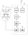
図1は、本発明の一実施の形態に係わる乳房撮影装置100の構成の一例を示す図である。
次に、実施形態2について説明する。実施形態2においては、乳房撮影装置100のX線源とグリッド81との配置関係について説明する。なお、実施形態2に係わる乳房撮影装置100の構成や処理は、上述した実施形態1と同様となるため、その説明については省略する。
次に、実施形態3について説明する。実施形態3においては、左右の乳房を撮影する際の処理の流れについて説明する。なお、実施形態3に係わる乳房撮影装置100の構成や処理は、上述した実施形態1と同様となるため、その説明については省略する。
次に、実施形態4について説明する。実施形態4においては、穿刺吸引細胞診機能を備えた乳房撮影装置100について説明する。具体的には、悪性を疑われる集ぞく性、線状、区域性等の分布を示す石灰化などが発見された場合、それが良性か悪性か質的判断をする必要がある。このような場合に、穿刺吸引細胞診を行なう。穿刺吸引細胞診では、乳房に吸引式の針(生検針)を挿入し、石灰化などを含めた悪性病変を疑う組織を採取する。なお、実施形態4に係わる乳房撮影装置100の構成や処理は、基本的に、上述した実施形態1と同様となるため、ここでは、相違点を重点的に挙げて説明する。
Claims (11)
- 放射線撮影装置であって、
電子を放出する複数の電子源と、前記複数の電子源と対応する1次元状又は2次元状に配列された複数の透過型ターゲットとを有する放射線発生手段と、
前記複数の電子源の駆動および電子放出量を制御する制御手段と、
前記複数の透過型ターゲットから被写体に照射された照射角度の異なる複数の放射線の検出に基づいて複数の第1の放射線画像を撮影する放射線検出手段と、
前記放射線検出手段により撮影された複数の第1の放射線画像に基づいて対象物領域を特定する領域特定手段と、
前記領域特定手段により特定された対象物領域に基づいて前記複数の電子源の中から駆動すべき電子源を決定する決定手段と、を備え、
前記制御手段は、前記決定手段により決定された電子源を用いて発生した放射線に基づく第2の放射線画像を撮影する際の放射線量よりも前記複数の第1の放射線画像を撮影する際の放射線量が小さくなるように前記電子源を駆動することを特徴とする放射線撮影装置。 - 前記複数の第1の放射線画像に対して再構成処理を実施して複数の断層画像を生成する画像処理手段
を具備し、
前記領域特定手段は、
前記画像処理手段により生成された複数の断層画像に基づいて前記対象物領域を特定する
ことを特徴する請求項1記載の放射線撮影装置。 - 前記決定手段は、
前記領域特定手段により特定された対象物領域に基づいて各照射方向に対応する対象物領域の照射面積を算出し、該照射面積に基づいて駆動すべき電子源を決定する
ことを特徴とする請求項1記載の放射線撮影装置。 - 前記決定手段は、
前記照射面積が最大又は最小となる照射方向から放射線を照射可能な電子源を駆動すべき電子源に決定する
ことを特徴とする請求項3記載の放射線撮影装置。 - 前記決定手段は、
前記領域特定手段により特定された対象物領域と、その対象物領域から所定範囲内の領域との平均画素値の差を比較し、平均画素値の差が最も大きくなる方向から放射線を照射可能な電子源を駆動すべき電子源に決定する
ことを特徴とする請求項1又は3記載の放射線撮影装置。 - 前記被写体と前記放射線検出手段との間に配置され、前記被写体で散乱した散乱放射線を縞状の鉛箔で吸収するグリッド
を更に具備し、
前記1次元状に配列された複数の電子源の配列方向と、前記グリッドの縞方向とが平行である
ことを特徴とする請求項1記載の放射線撮影装置。 - 前記被写体は、乳房であり、
前記決定手段は、
一方の乳房に対する前記第2の放射線画像の撮影に際しては、もう一方の乳房の撮影時に駆動された電子源と対称位置に設けられた電子源を駆動すべき電子源に決定する
ことを特徴とする請求項1記載の放射線撮影装置。 - 前記被写体は、乳房であり、
前記放射線検出手段により撮影された複数の第1の放射線画像に基づいて生検針を挿入する方向を決定する挿入方向決定手段と、
前記挿入方向決定手段により決定された方向に向けて前記乳房への前記生検針の挿入を制御する生検針制御手段と
を更に具備することを特徴とする請求項1記載の放射線撮影装置。 - 前記第2の放射線画像は、
前記第1の放射線画像の撮影時よりも大きな管電流又は長い照射時間により撮影される
ことを特徴とする請求項1から8いずれか1項に記載の放射線撮影装置。 - 前記被写体は、乳房であり、
前記対象物領域は、
乳せん分布、腫瘤形状、石灰化分布の少なくともいずれかを含む
ことを特徴とする請求項1から9いずれか1項に記載の放射線撮影装置。 - 放射線撮影装置の制御方法であって、
電子を放出する複数の電子源と、前記複数の電子源と対応する1次元状又は2次元状に配列された複数の透過型ターゲットとを有する放射線発生手段から複数の放射線を被写体に向けて照射する工程と、
前記複数の透過型ターゲットから被写体に照射された照射角度の異なる複数の放射線の検出に基づいて複数の第1の放射線画像を撮影する工程と、
前記撮影された複数の第1の放射線画像に基づいて対象物領域を特定する工程と、
前記特定された対象物領域に基づいて前記複数の電子源の中から駆動すべき電子源を決定する工程と、を有し、
前記決定する工程で決定された電子源を用いて発生した放射線に基づく第2の放射線画像を撮影する際の放射線量よりも前記複数の第1の放射線画像を撮影する際の放射線量が小さくなるように前記電子源を駆動する、ことを特徴とする放射線撮影装置の制御方法。
Priority Applications (5)
| Application Number | Priority Date | Filing Date | Title |
|---|---|---|---|
| JP2009087836A JP5346654B2 (ja) | 2009-03-31 | 2009-03-31 | 放射線撮影装置及びその制御方法 |
| EP10155071A EP2236087A1 (en) | 2009-03-31 | 2010-03-01 | Radiation imaging apparatus and control method for the same |
| KR1020100022618A KR101234670B1 (ko) | 2009-03-31 | 2010-03-15 | 방사선 촬영장치 |
| US12/749,239 US8149987B2 (en) | 2009-03-31 | 2010-03-29 | Radiation imaging apparatus and control method for the same |
| US13/411,360 US8873706B2 (en) | 2009-03-31 | 2012-03-02 | Radiation imaging apparatus and control method for the same |
Applications Claiming Priority (1)
| Application Number | Priority Date | Filing Date | Title |
|---|---|---|---|
| JP2009087836A JP5346654B2 (ja) | 2009-03-31 | 2009-03-31 | 放射線撮影装置及びその制御方法 |
Related Child Applications (1)
| Application Number | Title | Priority Date | Filing Date |
|---|---|---|---|
| JP2013169923A Division JP5677534B2 (ja) | 2013-08-19 | 2013-08-19 | 放射線撮影装置及びその制御方法 |
Publications (3)
| Publication Number | Publication Date |
|---|---|
| JP2010233962A JP2010233962A (ja) | 2010-10-21 |
| JP2010233962A5 JP2010233962A5 (ja) | 2012-05-10 |
| JP5346654B2 true JP5346654B2 (ja) | 2013-11-20 |
Family
ID=42200056
Family Applications (1)
| Application Number | Title | Priority Date | Filing Date |
|---|---|---|---|
| JP2009087836A Expired - Fee Related JP5346654B2 (ja) | 2009-03-31 | 2009-03-31 | 放射線撮影装置及びその制御方法 |
Country Status (4)
| Country | Link |
|---|---|
| US (2) | US8149987B2 (ja) |
| EP (1) | EP2236087A1 (ja) |
| JP (1) | JP5346654B2 (ja) |
| KR (1) | KR101234670B1 (ja) |
Families Citing this family (42)
| Publication number | Priority date | Publication date | Assignee | Title |
|---|---|---|---|---|
| US8768026B2 (en) | 2003-11-26 | 2014-07-01 | Hologic, Inc. | X-ray imaging with x-ray markers that provide adjunct information but preserve image quality |
| JP5346654B2 (ja) * | 2009-03-31 | 2013-11-20 | キヤノン株式会社 | 放射線撮影装置及びその制御方法 |
| DE102010026434B4 (de) * | 2010-07-08 | 2019-02-21 | Siemens Healthcare Gmbh | Mammographiegerät und Mammographieverfahren |
| KR101687971B1 (ko) * | 2010-07-19 | 2016-12-21 | 삼성전자주식회사 | 유방 촬영 장치 및 그 방법 |
| US8378302B2 (en) * | 2010-10-11 | 2013-02-19 | National Central University | Bidirectional optical scanner assisting in mammography |
| IT1404617B1 (it) * | 2011-02-25 | 2013-11-29 | I M S Internaz Medicoscientifica S R L | Apparecchiatura per la tomosintesi e la mammografia. |
| ITBO20110086A1 (it) * | 2011-02-25 | 2012-08-26 | I M S Internaz Medicoscienti Fica S R L | Apparecchiatura per la mammografia e/o la tomosintesi con dispositivo di rimozione della radiazione diffusa. |
| US8395120B2 (en) * | 2011-02-25 | 2013-03-12 | National Central University | Bidirectional optical scanner assisting in mammography |
| ITBO20110084A1 (it) * | 2011-02-25 | 2012-08-26 | I M S Internaz Medicoscienti Fica S R L | Apparecchiatura per la tomosintesi e la mammografia. |
| JP5501290B2 (ja) * | 2011-05-23 | 2014-05-21 | 富士フイルム株式会社 | 画像処理装置、放射線画像撮影システム、及び画像処理プログラム |
| US10849574B2 (en) * | 2011-06-22 | 2020-12-01 | Medtronic Navigation, Inc. | Interventional imaging |
| CN102327122B (zh) * | 2011-07-08 | 2013-01-02 | 赵福元 | 双x射线管带校正的导向穿刺乳腺x射线机 |
| KR101477543B1 (ko) | 2011-07-22 | 2014-12-31 | 삼성전자주식회사 | 엑스선 촬영 장치 및 방법 |
| ES2658965T3 (es) | 2012-02-22 | 2018-03-13 | Carestream Health, Inc. | Aparatos/procedimientos radiográficos móviles con capacidad de tomosínteis |
| KR101972474B1 (ko) | 2012-03-21 | 2019-04-26 | 삼성디스플레이 주식회사 | 엑스선 검출 장치 |
| KR102001926B1 (ko) | 2012-09-11 | 2019-07-30 | 삼성디스플레이 주식회사 | 엑스레이 검출기, 이를 포함하는 엑스레이 검출 시스템 및 엑스레이 검출 방법 |
| KR101447544B1 (ko) * | 2013-02-19 | 2014-10-13 | 디케이메디칼시스템(주) | 이동형 x선 투시촬영장치 |
| JP6080610B2 (ja) * | 2013-02-26 | 2017-02-15 | キヤノン株式会社 | マルチ放射線発生装置および放射線撮影システム |
| KR20150001184A (ko) * | 2013-06-26 | 2015-01-06 | 삼성전자주식회사 | 엑스선 촬영 장치 및 그 동작 방법 |
| KR20150001179A (ko) * | 2013-06-26 | 2015-01-06 | 삼성전자주식회사 | 엑스선 촬영 장치 및 그 동작 방법 |
| KR101588574B1 (ko) * | 2013-11-06 | 2016-01-26 | 주식회사 레이언스 | 입체정위 생검을 위한 생검 니들 가이딩 장치, 이를 구비한 영상 촬영 장치 및 이를 이용한 생검 채취 방법 |
| KR101606746B1 (ko) | 2014-03-14 | 2016-03-28 | 주식회사 레이언스 | 디지털 엑스레이 영상 시스템, 엑스레이 조사 조절 장치 및 그 방법 |
| CN104274201B (zh) * | 2014-10-10 | 2016-05-18 | 深圳先进技术研究院 | 乳腺层析成像方法和系统及成像设备和图像采集处理方法 |
| JP6497912B2 (ja) * | 2014-12-01 | 2019-04-10 | キヤノン株式会社 | 画像処理装置、放射線撮影システム、制御方法、及びプログラム |
| KR101677172B1 (ko) | 2015-04-06 | 2016-11-17 | 주식회사 디알텍 | 방사선 디텍터와 이를 포함하는 방사선 촬영장치, 그 자동노출제어방법 및 방사선 촬영방법 |
| JP6439581B2 (ja) * | 2015-05-25 | 2018-12-19 | 株式会社島津製作所 | 放射線透視装置 |
| JP6577257B2 (ja) * | 2015-06-18 | 2019-09-18 | 株式会社日立製作所 | X線診断装置、及びその作動方法 |
| JP6502188B2 (ja) * | 2015-06-18 | 2019-04-17 | 株式会社日立製作所 | X線検査装置、及び作動方法 |
| KR101798939B1 (ko) * | 2015-09-08 | 2017-11-17 | 삼성전자주식회사 | 엑스선 영상 장치 및 그 제어방법 |
| KR101875847B1 (ko) * | 2016-12-07 | 2018-07-06 | 주식회사 디알텍 | 방사선 촬영 장치 및 이를 이용한 방사선 촬영 방법 |
| KR101914255B1 (ko) | 2017-03-20 | 2018-11-01 | 주식회사 디알텍 | 방사선 촬영 장치 및 이를 이용한 방사선 촬영 방법 |
| JP6937163B2 (ja) * | 2017-05-29 | 2021-09-22 | キヤノンメディカルシステムズ株式会社 | 乳房x線撮影装置及び穿刺支援システム |
| US11020066B2 (en) * | 2018-12-10 | 2021-06-01 | KUB Technologies, Inc. | System and method for cabinet x-ray systems with stationary x-ray source array |
| JP7214557B2 (ja) | 2019-04-26 | 2023-01-30 | 富士フイルム株式会社 | 放射線撮影装置 |
| JP7221825B2 (ja) * | 2019-07-26 | 2023-02-14 | 富士フイルム株式会社 | トモシンセシス撮影制御装置、トモシンセシス撮影制御装置の作動方法、トモシンセシス撮影制御装置の作動プログラム |
| GB2589086B (en) * | 2019-11-12 | 2023-09-13 | Adaptix Ltd | A method of obtaining x-ray images |
| CN113116364B (zh) * | 2019-12-31 | 2023-07-14 | 上海联影医疗科技股份有限公司 | 一种乳腺x射线成像装置 |
| EP4064993A4 (en) * | 2019-12-28 | 2023-05-03 | Shanghai United Imaging Healthcare Co., Ltd. | IMAGING SYSTEMS AND METHODS |
| KR20220129013A (ko) * | 2020-01-16 | 2022-09-22 | 더 리전츠 오브 더 유니버시티 오브 캘리포니아 | 유방 영상 촬영을 위한 다중 모드 시스템 |
| US11771387B2 (en) * | 2020-01-29 | 2023-10-03 | Aixscan Inc. | Fast 3D radiography using multiple pulsed X-ray sources in motion |
| CN111616728A (zh) * | 2020-06-05 | 2020-09-04 | 上海联影医疗科技有限公司 | 一种基于面光源的x射线成像方法和系统 |
| CN111631742A (zh) * | 2020-06-05 | 2020-09-08 | 上海联影医疗科技有限公司 | 一种基于面光源的x射线成像方法和系统 |
Family Cites Families (33)
| Publication number | Priority date | Publication date | Assignee | Title |
|---|---|---|---|---|
| JPS6096229A (ja) * | 1983-10-31 | 1985-05-29 | 株式会社島津製作所 | X線多軌道断層撮影装置 |
| JPH08263641A (ja) | 1995-01-23 | 1996-10-11 | Fuji Photo Film Co Ltd | アイリスフィルターの演算方法 |
| JPH08264139A (ja) | 1995-03-22 | 1996-10-11 | Hamamatsu Photonics Kk | X線発生装置 |
| JP3731760B2 (ja) | 1995-03-29 | 2006-01-05 | 富士写真フイルム株式会社 | 画像処理方法 |
| JPH0966054A (ja) | 1995-09-01 | 1997-03-11 | Canon Inc | X線撮影装置 |
| JP3439590B2 (ja) | 1995-12-22 | 2003-08-25 | 株式会社荏原製作所 | X線源 |
| JP2001061833A (ja) | 1999-08-23 | 2001-03-13 | Shimadzu Corp | X線ct装置 |
| US6333968B1 (en) * | 2000-05-05 | 2001-12-25 | The United States Of America As Represented By The Secretary Of The Navy | Transmission cathode for X-ray production |
| JP2002320610A (ja) | 2001-02-23 | 2002-11-05 | Mitsubishi Heavy Ind Ltd | X線ct装置とx線ct装置撮影方法 |
| DE10132816A1 (de) * | 2001-05-31 | 2002-12-05 | Philips Corp Intellectual Pty | Vorrichtung und Verfahren zur Anpassung der Strahlungsdosis einer Röntgenstrahlungsquelle |
| JP3639825B2 (ja) * | 2002-04-03 | 2005-04-20 | キヤノン株式会社 | 動画像表示方法、プログラム、コンピュータ可読記憶媒体、及び動画像表示装置 |
| US7218766B2 (en) * | 2002-04-15 | 2007-05-15 | General Electric Company | Computer aided detection (CAD) for 3D digital mammography |
| JP3848911B2 (ja) * | 2002-10-01 | 2006-11-22 | 三菱重工業株式会社 | 放射線照射装置 |
| WO2004080309A2 (en) * | 2003-03-10 | 2004-09-23 | Philips Intellectual Property & Standards Gmbh | Device and method for adapting the recording parameters of a radiograph |
| JP4596748B2 (ja) | 2003-05-07 | 2010-12-15 | キヤノン株式会社 | 放射線画像撮影装置及び放射線画像撮影装置における再構成方法 |
| JP4002984B2 (ja) | 2003-05-12 | 2007-11-07 | 株式会社エーイーティー | X線ct装置 |
| DE10337935A1 (de) * | 2003-08-18 | 2005-03-17 | Siemens Ag | Vorrichtung für die Aufnahme von Strukturdaten eines Objekts |
| US7394924B2 (en) * | 2003-10-14 | 2008-07-01 | Mirada Solutions Limited | Scatter correction in scanning imaging systems |
| JP4202906B2 (ja) * | 2003-12-25 | 2008-12-24 | 株式会社東芝 | X線コンピュータ断層撮影装置 |
| US7330529B2 (en) * | 2004-04-06 | 2008-02-12 | General Electric Company | Stationary tomographic mammography system |
| DE102004043693A1 (de) * | 2004-09-09 | 2006-03-30 | Siemens Ag | Detektoranordnung für ein medizinisches Diagnosegerät sowie medizinisches bildgebendes Diagnoseverfahren |
| US8155262B2 (en) * | 2005-04-25 | 2012-04-10 | The University Of North Carolina At Chapel Hill | Methods, systems, and computer program products for multiplexing computed tomography |
| DE202007019497U1 (de) * | 2006-02-15 | 2013-03-06 | Hologic, Inc. | Brustbiopsie und Nadellokalisierung unter Verwendung von Tomosynthesesystemen |
| FR2897461A1 (fr) * | 2006-02-16 | 2007-08-17 | Gen Electric | Dispositif de rayonnement x et procede de traitement d'images |
| JP4878311B2 (ja) * | 2006-03-03 | 2012-02-15 | キヤノン株式会社 | マルチx線発生装置 |
| US20070237287A1 (en) * | 2006-03-28 | 2007-10-11 | Predrag Sukovic | Ct scanner with automatic determination of volume of interest |
| FR2902218A1 (fr) * | 2006-06-07 | 2007-12-14 | Gen Electric | Procede de traitement d'images de tomosynthese pour une detection de signes radiologiques |
| JP4833785B2 (ja) | 2006-09-29 | 2011-12-07 | 富士フイルム株式会社 | 放射線撮影装置および放射線撮影方法 |
| JP4857070B2 (ja) | 2006-10-11 | 2012-01-18 | キヤノン株式会社 | 乳房撮影用x線ct装置 |
| CN103948395A (zh) * | 2007-07-19 | 2014-07-30 | 北卡罗来纳大学查珀尔希尔分校 | 固定 x 射线数字化断层合成或断层摄影系统和相关方法 |
| DE102008004473A1 (de) * | 2008-01-15 | 2009-07-23 | Siemens Aktiengesellschaft | Verfahren und Vorrichtung zur Erzeugung eines tomosynthetischen 3D-Röntgenbildes |
| DE102008030698B3 (de) * | 2008-06-27 | 2010-02-18 | Siemens Aktiengesellschaft | Mammographieanlage |
| JP5346654B2 (ja) * | 2009-03-31 | 2013-11-20 | キヤノン株式会社 | 放射線撮影装置及びその制御方法 |
-
2009
- 2009-03-31 JP JP2009087836A patent/JP5346654B2/ja not_active Expired - Fee Related
-
2010
- 2010-03-01 EP EP10155071A patent/EP2236087A1/en not_active Withdrawn
- 2010-03-15 KR KR1020100022618A patent/KR101234670B1/ko active IP Right Grant
- 2010-03-29 US US12/749,239 patent/US8149987B2/en not_active Expired - Fee Related
-
2012
- 2012-03-02 US US13/411,360 patent/US8873706B2/en not_active Expired - Fee Related
Also Published As
| Publication number | Publication date |
|---|---|
| KR101234670B1 (ko) | 2013-02-19 |
| US8149987B2 (en) | 2012-04-03 |
| US20100246759A1 (en) | 2010-09-30 |
| EP2236087A1 (en) | 2010-10-06 |
| US20120163533A1 (en) | 2012-06-28 |
| KR20100109381A (ko) | 2010-10-08 |
| US8873706B2 (en) | 2014-10-28 |
| JP2010233962A (ja) | 2010-10-21 |
Similar Documents
| Publication | Publication Date | Title |
|---|---|---|
| JP5346654B2 (ja) | 放射線撮影装置及びその制御方法 | |
| JP5677534B2 (ja) | 放射線撮影装置及びその制御方法 | |
| JP5384612B2 (ja) | コーンビームボリュームctマンモグラフィー撮像に使用するための、管が連続して移動している間、焦点を動かさない回転式のステップ・アンド・シュート画像取得に基づく、高速トモシンセシススキャナ装置及びctベースの方法 | |
| KR101477543B1 (ko) | 엑스선 촬영 장치 및 방법 | |
| JP5914625B2 (ja) | 放射線撮影装置及びその制御方法 | |
| Puett et al. | An update on carbon nanotube‐enabled X‐ray sources for biomedical imaging | |
| US20120224664A1 (en) | Tomosynthesis mammography system with enlarged field of view | |
| JP7294592B2 (ja) | トモシンセシス、蛍光透視、及び定位イメージングのためのコンパクトx線デバイス、システム、及び方法 | |
| US20120008739A1 (en) | Mammography apparatus with x-ray sources arranged at different distances from the chest | |
| JP2008086760A (ja) | 二重照射方式の乳房撮影装置及びその装置を用いる乳房撮影方法 | |
| JP2010075338A (ja) | X線治療機能を備える乳房用画像撮影及び治療装置 | |
| US9629594B2 (en) | Contrast-enhanced imaging of objects | |
| JP2012110719A (ja) | 小型マンモグラフィ装置、及び関連するマンモグラフィ方法 | |
| JP6475138B2 (ja) | 制御装置、放射線画像撮影装置、放射線画像撮影方法、及び放射線画像撮影プログラム | |
| US11241207B2 (en) | Hybrid CT system with additional detectors in close proximity to the body | |
| US10111628B2 (en) | X-ray imaging apparatus and method for marking a location of a surgical tool on a displayed image | |
| JPWO2006129462A1 (ja) | デジタル放射線画像撮影システム | |
| WO2007007473A1 (ja) | デジタル放射線画像撮影システム | |
| CN111000574B (zh) | 医用图像处理装置、方法、及记录介质 | |
| JP2019103752A (ja) | 医用画像処理装置、x線ct装置及び医用画像処理プログラム | |
| KR101588574B1 (ko) | 입체정위 생검을 위한 생검 니들 가이딩 장치, 이를 구비한 영상 촬영 장치 및 이를 이용한 생검 채취 방법 | |
| JP7223517B2 (ja) | 医用画像診断装置 | |
| JP2022046946A (ja) | X線ct装置 | |
| JP2024035136A (ja) | 医用画像処理装置、方法、プログラム及びx線診断装置 | |
| CN116137028A (zh) | 医用图像处理装置、医用图像处理方法及存储介质 |
Legal Events
| Date | Code | Title | Description |
|---|---|---|---|
| A521 | Request for written amendment filed |
Free format text: JAPANESE INTERMEDIATE CODE: A523 Effective date: 20120314 |
|
| A621 | Written request for application examination |
Free format text: JAPANESE INTERMEDIATE CODE: A621 Effective date: 20120314 |
|
| A131 | Notification of reasons for refusal |
Free format text: JAPANESE INTERMEDIATE CODE: A131 Effective date: 20130430 |
|
| A977 | Report on retrieval |
Free format text: JAPANESE INTERMEDIATE CODE: A971007 Effective date: 20130430 |
|
| A521 | Request for written amendment filed |
Free format text: JAPANESE INTERMEDIATE CODE: A523 Effective date: 20130701 |
|
| TRDD | Decision of grant or rejection written | ||
| A01 | Written decision to grant a patent or to grant a registration (utility model) |
Free format text: JAPANESE INTERMEDIATE CODE: A01 Effective date: 20130719 |
|
| A61 | First payment of annual fees (during grant procedure) |
Free format text: JAPANESE INTERMEDIATE CODE: A61 Effective date: 20130819 |
|
| R151 | Written notification of patent or utility model registration |
Ref document number: 5346654 Country of ref document: JP Free format text: JAPANESE INTERMEDIATE CODE: R151 |
|
| LAPS | Cancellation because of no payment of annual fees |