EP4576103A2 - Neoantigenidentifizierung mit hotspots - Google Patents
Neoantigenidentifizierung mit hotspots Download PDFInfo
- Publication number
- EP4576103A2 EP4576103A2 EP25159694.6A EP25159694A EP4576103A2 EP 4576103 A2 EP4576103 A2 EP 4576103A2 EP 25159694 A EP25159694 A EP 25159694A EP 4576103 A2 EP4576103 A2 EP 4576103A2
- Authority
- EP
- European Patent Office
- Prior art keywords
- allele
- presentation
- peptide
- mhc
- neoantigen
- Prior art date
- Legal status (The legal status is an assumption and is not a legal conclusion. Google has not performed a legal analysis and makes no representation as to the accuracy of the status listed.)
- Pending
Links
Images


Classifications
-
- G—PHYSICS
- G16—INFORMATION AND COMMUNICATION TECHNOLOGY [ICT] SPECIALLY ADAPTED FOR SPECIFIC APPLICATION FIELDS
- G16B—BIOINFORMATICS, i.e. INFORMATION AND COMMUNICATION TECHNOLOGY [ICT] SPECIALLY ADAPTED FOR GENETIC OR PROTEIN-RELATED DATA PROCESSING IN COMPUTATIONAL MOLECULAR BIOLOGY
- G16B40/00—ICT specially adapted for biostatistics; ICT specially adapted for bioinformatics-related machine learning or data mining, e.g. knowledge discovery or pattern finding
-
- A—HUMAN NECESSITIES
- A61—MEDICAL OR VETERINARY SCIENCE; HYGIENE
- A61K—PREPARATIONS FOR MEDICAL, DENTAL OR TOILETRY PURPOSES
- A61K39/00—Medicinal preparations containing antigens or antibodies
- A61K39/0005—Vertebrate antigens
- A61K39/0011—Cancer antigens
-
- A—HUMAN NECESSITIES
- A61—MEDICAL OR VETERINARY SCIENCE; HYGIENE
- A61P—SPECIFIC THERAPEUTIC ACTIVITY OF CHEMICAL COMPOUNDS OR MEDICINAL PREPARATIONS
- A61P35/00—Antineoplastic agents
-
- C—CHEMISTRY; METALLURGY
- C12—BIOCHEMISTRY; BEER; SPIRITS; WINE; VINEGAR; MICROBIOLOGY; ENZYMOLOGY; MUTATION OR GENETIC ENGINEERING
- C12Q—MEASURING OR TESTING PROCESSES INVOLVING ENZYMES, NUCLEIC ACIDS OR MICROORGANISMS; COMPOSITIONS OR TEST PAPERS THEREFOR; PROCESSES OF PREPARING SUCH COMPOSITIONS; CONDITION-RESPONSIVE CONTROL IN MICROBIOLOGICAL OR ENZYMOLOGICAL PROCESSES
- C12Q1/00—Measuring or testing processes involving enzymes, nucleic acids or microorganisms; Compositions therefor; Processes of preparing such compositions
- C12Q1/68—Measuring or testing processes involving enzymes, nucleic acids or microorganisms; Compositions therefor; Processes of preparing such compositions involving nucleic acids
- C12Q1/6876—Nucleic acid products used in the analysis of nucleic acids, e.g. primers or probes
- C12Q1/6883—Nucleic acid products used in the analysis of nucleic acids, e.g. primers or probes for diseases caused by alterations of genetic material
- C12Q1/6886—Nucleic acid products used in the analysis of nucleic acids, e.g. primers or probes for diseases caused by alterations of genetic material for cancer
-
- G—PHYSICS
- G01—MEASURING; TESTING
- G01N—INVESTIGATING OR ANALYSING MATERIALS BY DETERMINING THEIR CHEMICAL OR PHYSICAL PROPERTIES
- G01N33/00—Investigating or analysing materials by specific methods not covered by groups G01N1/00 - G01N31/00
- G01N33/48—Biological material, e.g. blood, urine; Haemocytometers
- G01N33/50—Chemical analysis of biological material, e.g. blood, urine; Testing involving biospecific ligand binding methods; Immunological testing
- G01N33/53—Immunoassay; Biospecific binding assay; Materials therefor
- G01N33/569—Immunoassay; Biospecific binding assay; Materials therefor for microorganisms, e.g. protozoa, bacteria, viruses
- G01N33/56966—Animal cells
- G01N33/56977—HLA or MHC typing
-
- G—PHYSICS
- G01—MEASURING; TESTING
- G01N—INVESTIGATING OR ANALYSING MATERIALS BY DETERMINING THEIR CHEMICAL OR PHYSICAL PROPERTIES
- G01N33/00—Investigating or analysing materials by specific methods not covered by groups G01N1/00 - G01N31/00
- G01N33/48—Biological material, e.g. blood, urine; Haemocytometers
- G01N33/50—Chemical analysis of biological material, e.g. blood, urine; Testing involving biospecific ligand binding methods; Immunological testing
- G01N33/53—Immunoassay; Biospecific binding assay; Materials therefor
- G01N33/574—Immunoassay; Biospecific binding assay; Materials therefor for cancer
-
- G—PHYSICS
- G01—MEASURING; TESTING
- G01N—INVESTIGATING OR ANALYSING MATERIALS BY DETERMINING THEIR CHEMICAL OR PHYSICAL PROPERTIES
- G01N33/00—Investigating or analysing materials by specific methods not covered by groups G01N1/00 - G01N31/00
- G01N33/48—Biological material, e.g. blood, urine; Haemocytometers
- G01N33/50—Chemical analysis of biological material, e.g. blood, urine; Testing involving biospecific ligand binding methods; Immunological testing
- G01N33/68—Chemical analysis of biological material, e.g. blood, urine; Testing involving biospecific ligand binding methods; Immunological testing involving proteins, peptides or amino acids
- G01N33/6878—Chemical analysis of biological material, e.g. blood, urine; Testing involving biospecific ligand binding methods; Immunological testing involving proteins, peptides or amino acids in epitope analysis
-
- G—PHYSICS
- G06—COMPUTING OR CALCULATING; COUNTING
- G06N—COMPUTING ARRANGEMENTS BASED ON SPECIFIC COMPUTATIONAL MODELS
- G06N3/00—Computing arrangements based on biological models
- G06N3/02—Neural networks
- G06N3/04—Architecture, e.g. interconnection topology
- G06N3/0464—Convolutional networks [CNN, ConvNet]
-
- G—PHYSICS
- G06—COMPUTING OR CALCULATING; COUNTING
- G06N—COMPUTING ARRANGEMENTS BASED ON SPECIFIC COMPUTATIONAL MODELS
- G06N3/00—Computing arrangements based on biological models
- G06N3/02—Neural networks
- G06N3/08—Learning methods
- G06N3/09—Supervised learning
-
- G—PHYSICS
- G16—INFORMATION AND COMMUNICATION TECHNOLOGY [ICT] SPECIALLY ADAPTED FOR SPECIFIC APPLICATION FIELDS
- G16B—BIOINFORMATICS, i.e. INFORMATION AND COMMUNICATION TECHNOLOGY [ICT] SPECIALLY ADAPTED FOR GENETIC OR PROTEIN-RELATED DATA PROCESSING IN COMPUTATIONAL MOLECULAR BIOLOGY
- G16B30/00—ICT specially adapted for sequence analysis involving nucleotides or amino acids
-
- G—PHYSICS
- G16—INFORMATION AND COMMUNICATION TECHNOLOGY [ICT] SPECIALLY ADAPTED FOR SPECIFIC APPLICATION FIELDS
- G16B—BIOINFORMATICS, i.e. INFORMATION AND COMMUNICATION TECHNOLOGY [ICT] SPECIALLY ADAPTED FOR GENETIC OR PROTEIN-RELATED DATA PROCESSING IN COMPUTATIONAL MOLECULAR BIOLOGY
- G16B40/00—ICT specially adapted for biostatistics; ICT specially adapted for bioinformatics-related machine learning or data mining, e.g. knowledge discovery or pattern finding
- G16B40/10—Signal processing, e.g. from mass spectrometry [MS] or from PCR
-
- G—PHYSICS
- G16—INFORMATION AND COMMUNICATION TECHNOLOGY [ICT] SPECIALLY ADAPTED FOR SPECIFIC APPLICATION FIELDS
- G16B—BIOINFORMATICS, i.e. INFORMATION AND COMMUNICATION TECHNOLOGY [ICT] SPECIALLY ADAPTED FOR GENETIC OR PROTEIN-RELATED DATA PROCESSING IN COMPUTATIONAL MOLECULAR BIOLOGY
- G16B40/00—ICT specially adapted for biostatistics; ICT specially adapted for bioinformatics-related machine learning or data mining, e.g. knowledge discovery or pattern finding
- G16B40/20—Supervised data analysis
-
- G—PHYSICS
- G16—INFORMATION AND COMMUNICATION TECHNOLOGY [ICT] SPECIALLY ADAPTED FOR SPECIFIC APPLICATION FIELDS
- G16B—BIOINFORMATICS, i.e. INFORMATION AND COMMUNICATION TECHNOLOGY [ICT] SPECIALLY ADAPTED FOR GENETIC OR PROTEIN-RELATED DATA PROCESSING IN COMPUTATIONAL MOLECULAR BIOLOGY
- G16B5/00—ICT specially adapted for modelling or simulations in systems biology, e.g. gene-regulatory networks, protein interaction networks or metabolic networks
- G16B5/20—Probabilistic models
-
- G—PHYSICS
- G16—INFORMATION AND COMMUNICATION TECHNOLOGY [ICT] SPECIALLY ADAPTED FOR SPECIFIC APPLICATION FIELDS
- G16B—BIOINFORMATICS, i.e. INFORMATION AND COMMUNICATION TECHNOLOGY [ICT] SPECIALLY ADAPTED FOR GENETIC OR PROTEIN-RELATED DATA PROCESSING IN COMPUTATIONAL MOLECULAR BIOLOGY
- G16B50/00—ICT programming tools or database systems specially adapted for bioinformatics
- G16B50/30—Data warehousing; Computing architectures
-
- C—CHEMISTRY; METALLURGY
- C12—BIOCHEMISTRY; BEER; SPIRITS; WINE; VINEGAR; MICROBIOLOGY; ENZYMOLOGY; MUTATION OR GENETIC ENGINEERING
- C12Q—MEASURING OR TESTING PROCESSES INVOLVING ENZYMES, NUCLEIC ACIDS OR MICROORGANISMS; COMPOSITIONS OR TEST PAPERS THEREFOR; PROCESSES OF PREPARING SUCH COMPOSITIONS; CONDITION-RESPONSIVE CONTROL IN MICROBIOLOGICAL OR ENZYMOLOGICAL PROCESSES
- C12Q2600/00—Oligonucleotides characterized by their use
- C12Q2600/156—Polymorphic or mutational markers
-
- C—CHEMISTRY; METALLURGY
- C12—BIOCHEMISTRY; BEER; SPIRITS; WINE; VINEGAR; MICROBIOLOGY; ENZYMOLOGY; MUTATION OR GENETIC ENGINEERING
- C12Q—MEASURING OR TESTING PROCESSES INVOLVING ENZYMES, NUCLEIC ACIDS OR MICROORGANISMS; COMPOSITIONS OR TEST PAPERS THEREFOR; PROCESSES OF PREPARING SUCH COMPOSITIONS; CONDITION-RESPONSIVE CONTROL IN MICROBIOLOGICAL OR ENZYMOLOGICAL PROCESSES
- C12Q2600/00—Oligonucleotides characterized by their use
- C12Q2600/158—Expression markers
Definitions
- neoantigen-recognizing T-cells are a major component of TIL 84,96,113,114 and circulate in the peripheral blood of cancer patients 107
- current methods for identifying neoantigen-reactive T-cells have some combination of the following three limitations: (1) they rely on difficult-to-obtain clinical specimens such as TIL 97,98 or leukaphereses 107 (2) they require screening impractically large libraries of peptides 95 or (3) they rely on MHC multimers, which may practically be available for only a small number of MHC alleles.
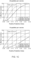
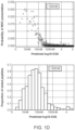
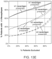
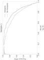
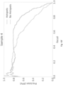
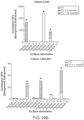
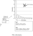
- This low positive predictive value (PPV) of existing methods for predicting presentation presents a problem for neoantigen-based vaccine design and for neoantigen-based T-cell therapy. If vaccines are designed using predictions with a low PPV, most patients are unlikely to receive a therapeutic neoantigen and fewer still are likely to receive more than one (even assuming all presented peptides are immunogenic). Similarly, if therapeutic T-cells are designed based on predictions with a low PPV, most patients are unlikely to receive T-cells that are reactive to tumor neoantigens and the time and physical resource cost of identifying predictive neoantigens using downstream laboratory techniques post-prediction may be unduly high. Thus, neoantigen vaccination and T-cell therapy with current methods is unlikely to succeed in a substantial number of subjects having tumors. ( FIG. 1C )
- previous approaches generated candidate neoantigens using only cis- acting mutations, and largely neglected to consider additional sources of neo-ORFs, including mutations in splicing factors, which occur in multiple tumor types and lead to aberrant splicing of many genes 13 , and mutations that create or remove protease cleavage sites.
- standard approaches to tumor genome and transcriptome analysis can miss somatic mutations that give rise to candidate neoantigens due to suboptimal conditions in library construction, exome and transcriptome capture, sequencing, or data analysis.
- standard tumor analysis approaches can inadvertently promote sequence artifacts or germline polymorphisms as neoantigens, leading to inefficient use of vaccine capacity or auto-immunity risk, respectively.
- neoantigen candidate identification using next-generation sequencing are addressed. These methods build on standard approaches for NGS tumor analysis to ensure that the highest sensitivity and specificity neoantigen candidates are advanced, across all classes of genomic alteration.
- NGS next-generation sequencing
- novel approaches for high-PPV neoantigen selection are presented to overcome the specificity problem and ensure that neoantigens advanced for vaccine inclusion and/or as targets for T-cell therapy are more likely to elicit anti-tumor immunity.
- the model disclosed herein outperforms state-of-the-art predictors trained on binding affinity and early predictors based on MS peptide data by up to an order of magnitude. By more reliably predicting presentation of peptides, the model enables more time- and cost-effective identification of neoantigen-specific or tumor antigen-specific T-cells for personlized therapy using a clinically practical process that uses limited volumes of patient peripheral blood, screens few peptides per patient, and does not necessarily rely on MHC multimers.
- the model disclosed herein can be used to enable more time- and cost-effective identification of tumor antigen-specific T-cells using MHC multimers, by decreasing the number of peptides bound to MHC multimers that need to be screened in order to identify neoantigen- or tumor antigen-specific T-cells
- the term "antigen" is a substance that induces an immune response.
- neoantigen is an antigen that has at least one alteration that makes it distinct from the corresponding wild-type, parental antigen, e.g., via mutation in a tumor cell or post-translational modification specific to a tumor cell.
- a neoantigen can include a polypeptide sequence or a nucleotide sequence.
- a mutation can include a frameshift or nonframeshift indel, missense or nonsense substitution, splice site alteration, genomic rearrangement or gene fusion, or any genomic or expression alteration giving rise to a neoORF.
- a mutations can also include a splice variant.
- Post-translational modifications specific to a tumor cell can include aberrant phosphorylation.
- Post-translational modifications specific to a tumor cell can also include a proteasome-generated spliced antigen. See Liepe et al., A large fraction of HLA class I ligands are proteasome-generated spliced peptides; Science. 2016 Oct 21;354(6310):354-358 .
- tumor neoantigen is a neoantigen present in a subject's tumor cell or tissue but not in the subject's corresponding normal cell or tissue.
- neoantigen-based vaccine is a vaccine construct based on one or more neoantigens, e.g., a plurality of neoantigens.
- candidate neoantigen is a mutation or other aberration giving rise to a new sequence that may represent a neoantigen.
- coding region is the portion(s) of a gene that encode protein.
- coding mutation is a mutation occurring in a coding region.
- ORF means open reading frame
- NEO-ORF is a tumor-specific ORF arising from a mutation or other aberration such as splicing.
- missense mutation is a mutation causing a substitution from one amino acid to another.
- nonsense mutation is a mutation causing a substitution from an amino acid to a stop codon.
- frameshift mutation is a mutation causing a change in the frame of the protein.
- the term “indel” is an insertion or deletion of one or more nucleic acids.
- the term percent "identity,” in the context of two or more nucleic acid or polypeptide sequences, refer to two or more sequences or subsequences that have a specified percentage of nucleotides or amino acid residues that are the same, when compared and aligned for maximum correspondence, as measured using one of the sequence comparison algorithms described below (e.g., BLASTP and BLASTN or other algorithms available to persons of skill) or by visual inspection.
- the percent “identity” can exist over a region of the sequence being compared, e.g., over a functional domain, or, alternatively, exist over the full length of the two sequences to be compared.
- sequence comparison typically one sequence acts as a reference sequence to which test sequences are compared.
- test and reference sequences are input into a computer, subsequence coordinates are designated, if necessary, and sequence algorithm program parameters are designated.
- sequence comparison algorithm then calculates the percent sequence identity for the test sequence(s) relative to the reference sequence, based on the designated program parameters.
- sequence similarity or dissimilarity can be established by the combined presence or absence of particular nucleotides, or, for translated sequences, amino acids at selected sequence positions (e.g., sequence motifs).
- Optimal alignment of sequences for comparison can be conducted, e.g., by the local homology algorithm of Smith & Waterman, Adv. Appl. Math. 2:482 (1981 ), by the homology alignment algorithm of Needleman & Wunsch, J. Mol. Biol. 48:443 (1970 ), by the search for similarity method of Pearson & Lipman, Proc. Nat'l. Acad. Sci. USA 85:2444 (1988 ), by computerized implementations of these algorithms (GAP, BESTFIT, FASTA, and TFASTA in the Wisconsin Genetics Software Package, Genetics Computer Group, 575 Science Dr., Madison, Wis.), or by visual inspection (see generally Ausubel et al., infra).
- BLAST algorithm One example of an algorithm that is suitable for determining percent sequence identity and sequence similarity is the BLAST algorithm, which is described in Altschul et al., J. Mol. Biol. 215:403-410 (1990 ). Software for performing BLAST analyses is publicly available through the National Center for Biotechnology Information.
- non-stop or read-through is a mutation causing the removal of the natural stop codon.
- epitopope is the specific portion of an antigen typically bound by an antibody or T-cell receptor.
- immunogenic is the ability to elicit an immune response, e.g., via T-cells, B cells, or both.
- HLA binding affinity means affinity of binding between a specific antigen and a specific MHC allele.
- the term "bait” is a nucleic acid probe used to enrich a specific sequence of DNA or RNA from a sample.
- variable is a difference between a subject's nucleic acids and the reference human genome used as a control.
- variant call is an algorithmic determination of the presence of a variant, typically from sequencing.
- polymorphism is a germline variant, i.e., a variant found in all DNA-bearing cells of an individual.
- somatic variant is a variant arising in non-germline cells of an individual.
- allele is a version of a gene or a version of a genetic sequence or a version of a protein.
- HLA type is the complement of HLA gene alleles.
- nonsense-mediated decay or "NMD” is a degradation of an mRNA by a cell due to a premature stop codon.
- truncal mutation is a mutation originating early in the development of a tumor and present in a substantial portion of the tumor's cells.
- subclonal mutation is a mutation originating later in the development of a tumor and present in only a subset of the tumor's cells.
- exome is a subset of the genome that codes for proteins.
- An exome can be the collective exons of a genome.
- logistic regression is a regression model for binary data from statistics where the logit of the probability that the dependent variable is equal to one is modeled as a linear function of the dependent variables.
- neural network is a machine learning model for classification or regression consisting of multiple layers of linear transformations followed by element-wise nonlinearities typically trained via stochastic gradient descent and back-propagation.
- proteome is the set of all proteins expressed and/or translated by a cell, group of cells, or individual.
- peptidome is the set of all peptides presented by MHC-I or MHC-II on the cell surface.
- the peptidome may refer to a property of a cell or a collection of cells (e.g., the tumor peptidome, meaning the union of the peptidomes of all cells that comprise the tumor).
- ELISPOT Enzyme-linked immunosorbent spot assay - which is a common method for monitoring immune responses in humans and animals.
- extract is a dextran-based peptide-MHC multimers used for antigen-specific T-cell staining in flow cytometry.
- MHC multimers is a peptide-MHC complex comprising multiple peptide- MHC monomer units.
- MHC tetramers is a peptide-MHC complex comprising four peptide- MHC monomer units.
- tolerance or immune tolerance is a state of immune non-responsiveness to one or more antigens, e.g. self-antigens.
- central tolerance is a tolerance affected in the thymus, either by deleting self-reactive T-cell clones or by promoting self-reactive T-cell clones to differentiate into immunosuppressive regulatory T-cells (Tregs).
- peripheral tolerance is a tolerance affected in the periphery by downregulating or anergizing self-reactive T-cells that survive central tolerance or promoting these T-cells to differentiate into Tregs.
- sample can include a single cell or multiple cells or fragments of cells or an aliquot of body fluid, taken from a subject, by means including venipuncture, excretion, ejaculation, massage, biopsy, needle aspirate, lavage sample, scraping, surgical incision, or intervention or other means known in the art.
- subject encompasses a cell, tissue, or organism, human or non-human, whether in vivo, ex vivo, or in vitro, male or female.
- subject is inclusive of mammals including humans.
- mammal encompasses both humans and non-humans and includes but is not limited to humans, non-human primates, canines, felines, murines, bovines, equines, and porcines.
- Clinical factor refers to a measure of a condition of a subject, e.g., disease activity or severity.
- “Clinical factor” encompasses all markers of a subject's health status, including non-sample markers, and/or other characteristics of a subject, such as, without limitation, age and gender.
- a clinical factor can be a score, a value, or a set of values that can be obtained from evaluation of a sample (or population of samples) from a subject or a subject under a determined condition.
- a clinical factor can also be predicted by markers and/or other parameters such as gene expression surrogates.
- Clinical factors can include tumor type, tumor sub-type, and smoking history.
- MHC major histocompatibility complex
- HLA human leukocyte antigen, or the human MHC gene locus
- NGS next-generation sequencing
- PPV positive predictive value
- TSNA tumor-specific neoantigen
- FFPE formalin-fixed, paraffin-embedded
- NMD nonsense-mediated decay
- NSCLC non-small-cell lung cancer
- DC dendritic cell.
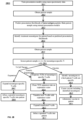
- the method includes obtaining exome, transcriptome, and/or whole genome nucleotide sequencing data from the tumor cells as well as normal cells of the subject. This nucleotide sequencing data is used to obtain a peptide sequence of each neoantigen in a set of neoantigens. The set of neoantigens is identified by comparing the nucleotide sequencing data from the tumor cells and the nucleotide sequencing data from the normal cells.
- the peptide sequence of each neoantigen in the set of neoantigens comprises at least one alteration that makes it distinct from the corresponding wild-type peptide sequence identified from the normal cells of the subject.
- the method further includes encoding the peptide sequence of each neoantigen in the set of neoantigens into a corresponding numerical vector.
- Each numerical vector includes information describing the amino acids that make up the peptide sequence and the positions of the amino acids in the peptide sequence.
- the method further comprises associating the peptide sequence of each of the neoantigens with one or more k-mer blocks of a plurality of k-mer blocks of the nucleotide sequencing data of the subject;.
- the method further comprises inputting the numerical vectors and the associated k-mer blocks into a machine-learned presentation model to generate a presentation likelihood for each neoantigen in the set of neoantigens.
- Each presentation likelihood represents the likelihood that the corresponding neoantigen is presented by MHC alleles on the surface of the tumor cells of the subject.
- the machine-learned presentation model comprises a plurality of parameters and a function. The plurality of parameters are identified based on a training data set.
- the training data set comprises, for each sample in a plurality of samples, a label obtained by mass spectrometry measuring presence of peptides bound to at least one MHC allele in a set of MHC alleles identified as present in the sample, training peptide sequences encoded as numerical vectors that include information describing the amino acids that make up the peptides and the positions of the amino acids in the peptides, and, for each of the training peptide sequences of the sample, associations between the training peptide sequence and one or more k-mer blocks of a plurality of k-mer blocks of the nucleotide sequencing data of the training peptide sequences.
- the function represents a relation between the numerical vector and the associated k-mer blocks received as input by the machine-learned presentation model and the presentation likelihood generated as output by the machine-learned presentation model based on the numerical vector, the associated k-mer blocks, and the plurality of parameters.
- the method further includes selecting a subset of the set of neoantigens, based on the presentation likelihoods, to generate a set of selected neoantigens, and returning the set of selected neoantigens.
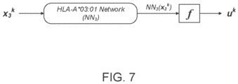
- inputting the numerical vector into the machine-learned presentation model comprises applying the machine-learned presentation model to the peptide sequence of the neoantigen to generate a dependency score for each of the MHC alleles.
- the dependency score for an MHC allele indicates whether the MHC allele will present the neoantigen, based on the particular amino acids at the particular positions of the peptide sequence.
- inputting the numerical vector into the machine-learned presentation model further comprises transforming the dependency scores to generate a corresponding per-allele likelihood for each MHC allele indicating a likelihood that the corresponding MHC allele will present the corresponding neoantigen, and combining the per-allele likelihoods to generate the presentation likelihood of the neoantigen.
- transforming the dependency scores models the presentation of the neoantigen as mutually exclusive across the MHC alleles.
- inputting the numerical vector into the machine-learned presentation model further comprises transforming a combination of the dependency scores to generate the presentation likelihood.
- transforming the combination of the dependency scores models the presentation of the neoantigen as interfering between the MHC alleles.
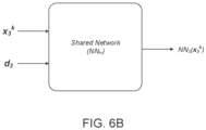
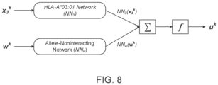
- the set of presentation likelihoods are further identified by one or more allele noninteracting features.
- the method further comprises applying the machine-learned presentation model to the allele noninteracting features to generate a dependency score for the allele noninteracting features.
- the dependency score indicates whether the peptide sequence of the corresponding neoantigen will be presented based on the allele noninteracting features.
- the one or more allele noninteracting features comprises the values indicating one of presence or absence of a presentation hotspot for each k-mer block of the peptide sequence of each neoantigen.
- the method further comprises combining the dependency score for each MHC allele with the dependency score for the allele noninteracting features, transforming the combined dependency score for each MHC allele to generate a per-allele likelihood for each MHC allele, and combining the per-allele likelihoods to generate the presentation likelihood.
- the per-allele likelihood for a MHC allele indicates a likelihood that the MHC allele will present the corresponding neoantigen.
- the method further comprises combining the dependency scores for the MHC alleles and the dependency score for the allele noninteracting features, and transforming the combined dependency scores to generate the presentation likelihood.
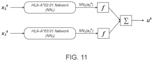
- the MHC alleles include two or more different MHC alleles.
- the peptide sequences comprise peptide sequences having lengths other than 9 amino acids.
- encoding the peptide sequence comprises encoding the peptide sequence using a one-hot encoding scheme.
- the plurality of samples comprise at least one of cell lines engineered to express a single MHC allele, cell lines engineered to express a plurality of MHC alleles, human cell lines obtained or derived from a plurality of patients, fresh or frozen tumor samples obtained from a plurality of patients, and fresh or frozen tissue samples obtained from a plurality of patients.
- the training data set further comprises at least one of data associated with peptide-MHC binding affinity measurements for at least one of the peptides, and data associated with peptide-MHC binding stability measurements for at least one of the peptides.
- the set of presentation likelihoods are further identified by expression levels of the MHC alleles in the subject, as measured by RNA-seq or mass spectrometry.
- the set of numerical likelihoods are further identified by features comprising at least one of the C-terminal sequences flanking the neoantigen encoded peptide sequence within its source protein sequence, and the N-terminal sequences flanking the neoantigen encoded peptide sequence within its source protein sequence.
- selecting the set of selected neoantigens comprises selecting neoantigens that have an increased likelihood of being presented on the tumor cell surface relative to unselected neoantigens, based on the machine-learned presentation model.
- selecting the set of selected neoantigens comprises selecting neoantigens that have an increased likelihood of being capable of inducing a tumor-specific immune response in the subject relative to unselected neoantigens, based on the machine-learned presentation model.
- selecting the set of selected neoantigens comprises selecting neoantigens that have an increased likelihood of being capable of being presented to naive T-cells by professional antigen presenting cells (APCs) relative to unselected neoantigens, based on the presentation model.
- the APC is optionally a dendritic cell (DC).
- selecting the set of selected neoantigens comprises selecting neoantigens that have a decreased likelihood of being subject to inhibition via central or peripheral tolerance relative to unselected neoantigens, based on the machine-learned presentation model.
- selecting the set of selected neoantigens comprises selecting neoantigens that have a decreased likelihood of being capable of inducing an autoimmune response to normal tissue in the subject relative to unselected neoantigens, based on the machine-learned presentation model.
- the one or more tumor cells are selected from the group consisting of: lung cancer, melanoma, breast cancer, ovarian cancer, prostate cancer, kidney cancer, gastric cancer, colon cancer, testicular cancer, head and neck cancer, pancreatic cancer, brain cancer, B-cell lymphoma, acute myelogenous leukemia, chronic myelogenous leukemia, chronic lymphocytic leukemia, and T-cell lymphocytic leukemia, non-small cell lung cancer, and small cell lung cancer.
- the method further comprises generating an output for constructing a personalized cancer vaccine from the set of selected neoantigens.
- the output for the personalized cancer vaccine may comprise at least one peptide sequence or at least one nucleotide sequence encoding the set of selected neoantigens.
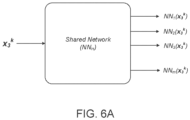
- the machine-learned presentation model is a neural network model.
- the neural network model may include a plurality of network models for the MHC alleles, each network model assigned to a corresponding MHC allele of the MHC alleles and including a series of nodes arranged in one or more layers.
- the neural network model may be trained by updating the parameters of the neural network model, the parameters of at least two network models being jointly updated for at least one training iteration.
- the machine-learned presentation model may be a deep learning model that includes one or more layers of nodes.
- the MHC alleles are class I MHC alleles.
- Also disclosed herein are computer systems comprising a computer processor and a memory storing computer program instructions.
- the instructions When the computer program instructions are executed by the computer processor, the instructions cause the computer processor to carry out any of the methods discussed above..
- mutations e.g., the variants or alleles that are present in cancer cells.
- these mutations can be present in the genome, transcriptome, proteome, or exome of cancer cells of a subject having cancer but not in normal tissue from the subject.
- Genetic mutations in tumors can be considered useful for the immunological targeting of tumors if they lead to changes in the amino acid sequence of a protein exclusively in the tumor.
- Useful mutations include: (1) non-synonymous mutations leading to different amino acids in the protein; (2) read-through mutations in which a stop codon is modified or deleted, leading to translation of a longer protein with a novel tumor-specific sequence at the C-terminus; (3) splice site mutations that lead to the inclusion of an intron in the mature mRNA and thus a unique tumor-specific protein sequence; (4) chromosomal rearrangements that give rise to a chimeric protein with tumor-specific sequences at the junction of 2 proteins (i.e., gene fusion); (5) frameshift mutations or deletions that lead to a new open reading frame with a novel tumor-specific protein sequence. Mutations can also include one or more of nonframeshift indel, missense or nonsense substitution, splice site alteration, genomic rearrangement or gene fusion, or any genomic or
- Peptides with mutations or mutated polypeptides arising from for example, splice-site, frameshift, readthrough, or gene fusion mutations in tumor cells can be identified by sequencing DNA, RNA or protein in tumor versus normal cells.
- mutations can include previously identified tumor specific mutations. Known tumor mutations can be found at the Catalogue of Somatic Mutations in Cancer (COSMIC) database.
- a variety of methods are available for detecting the presence of a particular mutation or allele in an individual's DNA or RNA. Advancements in this field have provided accurate, easy, and inexpensive large-scale SNP genotyping. For example, several techniques have been described including dynamic allele-specific hybridization (DASH), microplate array diagonal gel electrophoresis (MADGE), pyrosequencing, oligonucleotide-specific ligation, the TaqMan system as well as various DNA "chip” technologies such as the Affymetrix SNP chips. These methods utilize amplification of a target genetic region, typically by PCR.
- DASH dynamic allele-specific hybridization
- MADGE microplate array diagonal gel electrophoresis
- pyrosequencing pyrosequencing
- oligonucleotide-specific ligation oligonucleotide-specific ligation
- TaqMan system as well as various DNA "chip” technologies such as the Affymetrix SNP chips.
- PCR based detection means can include multiplex amplification of a plurality of markers simultaneously. For example, it is well known in the art to select PCR primers to generate PCR products that do not overlap in size and can be analyzed simultaneously. Alternatively, it is possible to amplify different markers with primers that are differentially labeled and thus can each be differentially detected. Of course, hybridization based detection means allow the differential detection of multiple PCR products in a sample. Other techniques are known in the art to allow multiplex analyses of a plurality of markers.
- RNA molecules obtained from genomic DNA or cellular RNA.
- a single base polymorphism can be detected by using a specialized exonuclease-resistant nucleotide, as disclosed, e.g., in Mundy, C. R. (U.S. Pat. No. 4,656,127 ).
- a primer complementary to the allelic sequence immediately 3' to the polymorphic site is permitted to hybridize to a target molecule obtained from a particular animal or human.
- the polymorphic site on the target molecule contains a nucleotide that is complementary to the particular exonuclease-resistant nucleotide derivative present, then that derivative will be incorporated onto the end of the hybridized primer. Such incorporation renders the primer resistant to exonuclease, and thereby permits its detection. Since the identity of the exonuclease-resistant derivative of the sample is known, a finding that the primer has become resistant to exonucleases reveals that the nucleotide(s) present in the polymorphic site of the target molecule is complementary to that of the nucleotide derivative used in the reaction. This method has the advantage that it does not require the determination of large amounts of extraneous sequence data.
- a solution-based method can be used for determining the identity of a nucleotide of a polymorphic site.
- Cohen, D. et al. (French Patent 2,650,840 ; PCT Appln. No. WO91/02087 ).
- a primer is employed that is complementary to allelic sequences immediately 3' to a polymorphic site. The method determines the identity of the nucleotide of that site using labeled dideoxynucleotide derivatives, which, if complementary to the nucleotide of the polymorphic site will become incorporated onto the terminus of the primer.
- Goelet, P. et al. An alternative method, known as Genetic Bit Analysis or GBA is described by Goelet, P. et al. (PCT Appln. No. 92/15712 ).
- the method of Goelet, P. et al. uses mixtures of labeled terminators and a primer that is complementary to the sequence 3' to a polymorphic site.
- the labeled terminator that is incorporated is thus determined by, and complementary to, the nucleotide present in the polymorphic site of the target molecule being evaluated.
- the method of Goelet, P. et al. can be a heterogeneous phase assay, in which the primer or the target molecule is immobilized to a solid phase.
- oligonucleotides 30-50 bases in length are covalently anchored at the 5' end to glass cover slips. These anchored strands perform two functions. First, they act as capture sites for the target template strands if the templates are configured with capture tails complementary to the surface-bound oligonucleotides. They also act as primers for the template directed primer extension that forms the basis of the sequence reading.
- the capture primers function as a fixed position site for sequence determination using multiple cycles of synthesis, detection, and chemical cleavage of the dye-linker to remove the dye. Each cycle consists of adding the polymerase/labeled nucleotide mixture, rinsing, imaging and cleavage of dye.
- polymerase is modified with a fluorescent donor molecule and immobilized on a glass slide, while each nucleotide is color-coded with an acceptor fluorescent moiety attached to a gamma-phosphate.
- the system detects the interaction between a fluorescently-tagged polymerase and a fluorescently modified nucleotide as the nucleotide becomes incorporated into the de novo chain.
- Other sequencing-by-synthesis technologies also exist.
- any suitable sequencing-by-synthesis platform can be used to identify mutations.
- four major sequencing-by-synthesis platforms are currently available: the Genome Sequencers from Roche/454 Life Sciences, the 1G Analyzer from Illumina/Solexa, the SOLiD system from Applied BioSystems, and the Heliscope system from Helicos Biosciences. Sequencing-by-synthesis platforms have also been described by Pacific BioSciences and VisiGen Biotechnologies.
- a plurality of nucleic acid molecules being sequenced is bound to a support (e.g., solid support).
- a capture sequence/universal priming site can be added at the 3' and/or 5' end of the template.
- the nucleic acids can be bound to the support by hybridizing the capture sequence to a complementary sequence covalently attached to the support.
- the capture sequence also referred to as a universal capture sequence
- the capture sequence is a nucleic acid sequence complementary to a sequence attached to a support that may dually serve as a universal primer.
- a member of a coupling pair (such as, e.g., antibody/antigen, receptor/ligand, or the avidin-biotin pair as described in, e.g., US Patent Application No. 2006/0252077 ) can be linked to each fragment to be captured on a surface coated with a respective second member of that coupling pair.
- sequence can be analyzed, for example, by single molecule detection/sequencing, e.g., as described in the Examples and in U.S. Pat. No. 7,283,337 , including template-dependent sequencing-by-synthesis.
- sequencing-by-synthesis the surface-bound molecule is exposed to a plurality of labeled nucleotide triphosphates in the presence of polymerase.
- the sequence of the template is determined by the order of labeled nucleotides incorporated into the 3' end of the growing chain. This can be done in real time or can be done in a step-and-repeat mode. For real-time analysis, different optical labels to each nucleotide can be incorporated and multiple lasers can be utilized for stimulation of incorporated nucleotides.
- Sequencing can also include other massively parallel sequencing or next generation sequencing (NGS) techniques and platforms. Additional examples of massively parallel sequencing techniques and platforms are the Illumina HiSeq or MiSeq, Thermo PGM or Proton, the Pac Bio RS II or Sequel, Qiagen's Gene Reader, and the Oxford Nanopore MinION. Additional similar current massively parallel sequencing technologies can be used, as well as future generations of these technologies.
- NGS next generation sequencing
- a DNA or RNA sample can be obtained from a tumor or a bodily fluid, e.g., blood, obtained by known techniques (e.g. venipuncture) or saliva.
- nucleic acid tests can be performed on dry samples (e.g. hair or skin).
- a sample can be obtained for sequencing from a tumor and another sample can be obtained from normal tissue for sequencing where the normal tissue is of the same tissue type as the tumor.
- a sample can be obtained for sequencing from a tumor and another sample can be obtained from normal tissue for sequencing where the normal tissue is of a distinct tissue type relative to the tumor.
- Tumors can include one or more of lung cancer, melanoma, breast cancer, ovarian cancer, prostate cancer, kidney cancer, gastric cancer, colon cancer, testicular cancer, head and neck cancer, pancreatic cancer, brain cancer, B-cell lymphoma, acute myelogenous leukemia, chronic myelogenous leukemia, chronic lymphocytic leukemia, and T-cell lymphocytic leukemia, non-small cell lung cancer, and small cell lung cancer.
- protein mass spectrometry can be used to identify or validate the presence of mutated peptides bound to MHC proteins on tumor cells.
- Peptides can be acid-eluted from tumor cells or from HLA molecules that are immunoprecipitated from tumor, and then identified using mass spectrometry.
- Neoantigens can include nucleotides or polypeptides.
- a neoantigen can be an RNA sequence that encodes for a polypeptide sequence.
- Neoantigens useful in vaccines can therefore include nucleotide sequences or polypeptide sequences.
- Neoantigen peptides can be described in the context of their coding sequence where a neoantigen includes the nucleotide sequence (e.g., DNA or RNA) that codes for the related polypeptide sequence.
- One or more polypeptides encoded by a neoantigen nucleotide sequence can comprise at least one of: a binding affinity with MHC with an IC50 value of less than 1000nM, for MHC Class I peptides a length of 8-15, 8, 9, 10, 11, 12, 13, 14, or 15 amino acids, presence of sequence motifs within or near the peptide promoting proteasome cleavage, and presence or sequence motifs promoting TAP transport.
- MHC Class II peptides a length 6-30, 6, 7, 8, 9, 10, 11, 12, 13, 14, 15, 16, 17, 18,19, 20, 21, 22, 23, 24, 25, 26, 27, 28, 29, or 30 amino acids, presence of sequence motifs within or near the peptide promoting cleavage by extracellular or lysosomal proteases (e.g., cathepsins) or HLA-DM catalyzed HLA binding.
- extracellular or lysosomal proteases e.g., cathepsins
- HLA-DM catalyzed HLA binding e.g., HLA-DM catalyzed HLA binding.
- One or more neoantigens can be presented on the surface of a tumor.
- One or more neoantigens can be is immunogenic in a subject having a tumor, e.g., capable of eliciting a T-cell response or a B cell response in the subject.
- One or more neoantigens that induce an autoimmune response in a subject can be excluded from consideration in the context of vaccine generation for a subject having a tumor.
- the size of at least one neoantigenic peptide molecule can comprise, but is not limited to, about 5, about 6, about 7, about 8, about 9, about 10, about 11, about 12, about 13, about 14, about 15, about 16, about 17, about 18, about 19, about 20, about 21, about 22, about 23, about 24, about 25, about 26, about 27, about 28, about 29, about 30, about 31, about 32, about 33, about 34, about 35, about 36, about 37, about 38, about 39, about 40, about 41, about 42, about 43, about 44, about 45, about 46, about 47, about 48, about 49, about 50, about 60, about 70, about 80, about 90, about 100, about 110, about 120 or greater amino molecule residues, and any range derivable therein.
- the neoantigenic peptide molecules are equal to or less than 50 amino acids.
- Neoantigenic peptides and polypeptides can be: for MHC Class I 15 residues or less in length and usually consist of between about 8 and about 11 residues, particularly 9 or 10 residues; for MHC Class II, 6-30 residues, inclusive.
- a longer peptide can be designed in several ways.
- a longer peptide could consist of either: (1) individual presented peptides with an extensions of 2-5 amino acids toward the N- and C-terminus of each corresponding gene product; (2) a concatenation of some or all of the presented peptides with extended sequences for each.
- sequencing reveals a long (>10 residues) neoepitope sequence present in the tumor (e.g.
- a longer peptide would consist of: (3) the entire stretch of novel tumor-specific amino acids--thus bypassing the need for computational or in vitro test-based selection of the strongest HLA-presented shorter peptide. In both cases, use of a longer peptide allows endogenous processing by patient-cells and may lead to more effective antigen presentation and induction of T-cell responses.
- Neoantigenic peptides and polypeptides can be presented on an HLA protein. In some aspects neoantigenic peptides and polypeptides are presented on an HLA protein with greater affinity than a wild-type peptide. In some aspects, a neoantigenic peptide or polypeptide can have an IC50 of at least less than 5000 nM, at least less than 1000 nM, at least less than 500 nM, at least less than 250 nM, at least less than 200 nM, at least less than 150 nM, at least less than 100 nM, at least less than 50 nM or less.
- neoantigenic peptides and polypeptides do not induce an autoimmune response and/or invoke immunological tolerance when administered to a subject.
- compositions comprising at least two or more neoantigenic peptides.
- the composition contains at least two distinct peptides. At least two distinct peptides can be derived from the same polypeptide. By distinct polypeptides is meant that the peptide vary by length, amino acid sequence, or both.
- the peptides are derived from any polypeptide known to or have been found to contain a tumor specific mutation. Suitable polypeptides from which the neoantigenic peptides can be derived can be found for example in the COSMIC database. COSMIC curates comprehensive information on somatic mutations in human cancer.
- the peptide contains the tumor specific mutation. In some aspects the tumor specific mutation is a driver mutation for a particular cancer type.
- Neoantigenic peptides and polypeptides having a desired activity or property can be modified to provide certain desired attributes, e.g., improved pharmacological characteristics, while increasing or at least retaining substantially all of the biological activity of the unmodified peptide to bind the desired MHC molecule and activate the appropriate T-cell.
- desired attributes e.g., improved pharmacological characteristics
- neoantigenic peptide and polypeptides can be subject to various changes, such as substitutions, either conservative or non-conservative, where such changes might provide for certain advantages in their use, such as improved MHC binding, stability or presentation.
- conservative substitutions is meant replacing an amino acid residue with another which is biologically and/or chemically similar, e.g., one hydrophobic residue for another, or one polar residue for another.
- substitutions include combinations such as Gly, Ala; Val, Ile, Leu, Met; Asp, Glu; Asn, Gln; Ser, Thr; Lys, Arg; and Phe, Tyr.
- the effect of single amino acid substitutions may also be probed using D-amino acids.
- Such modifications can be made using well known peptide synthesis procedures, as described in e.g., Merrifield, Science 232:341-347
- Modifications of peptides and polypeptides with various amino acid mimetics or unnatural amino acids can be particularly useful in increasing the stability of the peptide and polypeptide in vivo. Stability can be assayed in a number of ways. For instance, peptidases and various biological media, such as human plasma and serum, have been used to test stability. See, e.g., Verhoef et al., Eur. J. Drug Metab Pharmacokin. 11:291-302 (1986 ). Half-life of the peptides can be conveniently determined using a 25% human serum (v/v) assay. The protocol is generally as follows. Pooled human serum (Type AB, non-heat inactivated) is delipidated by centrifugation before use.
- Type AB non-heat inactivated
- the serum is then diluted to 25% with RPMI tissue culture media and used to test peptide stability. At predetermined time intervals a small amount of reaction solution is removed and added to either 6% aqueous trichloracetic acid or ethanol. The cloudy reaction sample is cooled (4 degrees C) for 15 minutes and then spun to pellet the precipitated serum proteins. The presence of the peptides is then determined by reversed-phase HPLC using stability-specific chromatography conditions.
- the peptides and polypeptides can be modified to provide desired attributes other than improved serum half-life. For instance, the ability of the peptides to induce CTL activity can be enhanced by linkage to a sequence which contains at least one epitope that is capable of inducing a T helper cell response.
- Immunogenic peptides/T helper conjugates can be linked by a spacer molecule.
- the spacer is typically comprised of relatively small, neutral molecules, such as amino acids or amino acid mimetics, which are substantially uncharged under physiological conditions.
- the spacers are typically selected from, e.g., Ala, Gly, or other neutral spacers of nonpolar amino acids or neutral polar amino acids.
- the optionally present spacer need not be comprised of the same residues and thus can be a hetero- or homo-oligomer.
- the spacer will usually be at least one or two residues, more usually three to six residues.
- the peptide can be linked to the T helper peptide without a spacer.
- a neoantigenic peptide can be linked to the T helper peptide either directly or via a spacer either at the amino or carboxy terminus of the peptide.
- the amino terminus of either the neoantigenic peptide or the T helper peptide can be acylated.
- Exemplary T helper peptides include tetanus toxoid 830-843, influenza 307-319, malaria circumsporozoite 382-398 and 378-389.
- Proteins or peptides can be made by any technique known to those of skill in the art, including the expression of proteins, polypeptides or peptides through standard molecular biological techniques, the isolation of proteins or peptides from natural sources, or the chemical synthesis of proteins or peptides.
- the nucleotide and protein, polypeptide and peptide sequences corresponding to various genes have been previously disclosed, and can be found at computerized databases known to those of ordinary skill in the art.
- One such database is the National Center for Biotechnology Information's Genbank and GenPept databases located at the National Institutes of Health website.
- the coding regions for known genes can be amplified and/or expressed using the techniques disclosed herein or as would be known to those of ordinary skill in the art.
- various commercial preparations of proteins, polypeptides and peptides are known to those of skill in the art.
- a neoantigen includes a nucleic acid (e.g. polynucleotide) that encodes a neoantigenic peptide or portion thereof.
- the polynucleotide can be, e.g., DNA, cDNA, PNA, CNA, RNA (e.g., mRNA), either single- and/or double-stranded, or native or stabilized forms of polynucleotides, such as, e.g., polynucleotides with a phosphorothiate backbone, or combinations thereof and it may or may not contain introns.
- a still further aspect provides an expression vector capable of expressing a polypeptide or portion thereof.
- Expression vectors for different-cell types are well known in the art and can be selected without undue experimentation.
- DNA is inserted into an expression vector, such as a plasmid, in proper orientation and correct reading frame for expression. If necessary, DNA can be linked to the appropriate transcriptional and translational regulatory control nucleotide sequences recognized by the desired host, although such controls are generally available in the expression vector.
- the vector is then introduced into the host through standard techniques. Guidance can be found e.g. in Sambrook et al. (1989) Molecular Cloning, A Laboratory Manual, Cold Spring Harbor Laboratory, Cold Spring Harbor, N.Y .
- an immunogenic composition e.g., a vaccine composition, capable of raising a specific immune response, e.g., a tumor-specific immune response.
- Vaccine compositions typically comprise a plurality of neoantigens, e.g., selected using a method described herein. Vaccine compositions can also be referred to as vaccines.
- a vaccine can contain between 1 and 30 peptides, 2, 3, 4, 5, 6, 7, 8, 9, 10, 11, 12, 13, 14, 15, 16, 17, 18, 19, 20, 21, 22, 23, 24, 25, 26, 27, 28, 29, or 30 different peptides, 6, 7, 8, 9, 10 11, 12, 13, or 14 different peptides, or 12, 13 or 14 different peptides.
- Peptides can include post-translational modifications.
- a vaccine can contain between 1 and 100 or more nucleotide sequences, 2, 3, 4, 5, 6, 7, 8, 9, 10, 11, 12, 13, 14, 15, 16, 17, 18, 19, 20, 21, 22, 23, 24, 25, 26, 27, 28, 29, 30, 31, 32, 33, 34, 35, 36, 37, 38, 39, 40, 41, 42, 43, 44, 45, 46, 47, 48, 49, 50, 51, 52, 53, 54, 55, 56, 57, 58, 59, 60, 61, 62, 63, 64, 65, 66, 67, 68, 69, 70, 71, 72, 73, 74, 75, 76, 77, 78, 79, 80, 81, 82, 83, 84, 85, 86, 87, 88, 89, 90, 91, 92, 93, 94,95, 96, 97, 98, 99, 100 or more different nucleotide sequences, 6, 7, 8, 9, 10 11, 12, 13, or 14 different nucleotide sequences, or 12, 13 or 14 different nu
- a vaccine can contain between 1 and 30 neoantigen sequences, 2, 3, 4, 5, 6, 7, 8, 9, 10, 11, 12, 13, 14, 15, 16, 17, 18, 19, 20, 21, 22, 23, 24, 25, 26, 27, 28, 29, 30, 31, 32, 33, 34, 35, 36, 37, 38, 39, 40, 41, 42, 43, 44, 45, 46, 47, 48, 49, 50, 51, 52, 53, 54, 55, 56, 57, 58, 59, 60, 61, 62, 63, 64, 65, 66, 67, 68, 69, 70, 71, 72, 73, 74, 75, 76, 77, 78, 79, 80, 81, 82, 83, 84, 85, 86, 87, 88, 89, 90, 91, 92, 93, 94,95, 96, 97, 98, 99, 100 or more different neoantigen sequences, 6, 7, 8, 9, 10 11, 12, 13, or 14 different neoantigen sequences, or 12, 13 or 14 different
- different peptides and/or polypeptides or nucleotide sequences encoding them are selected so that the peptides and/or polypeptides capable of associating with different MHC molecules, such as different MHC class I molecules and/or different MHC class II molecules.
- one vaccine composition comprises coding sequence for peptides and/or polypeptides capable of associating with the most frequently occurring MHC class I molecules and/or MHC class II molecules.
- vaccine compositions can comprise different fragments capable of associating with at least 2 preferred, at least 3 preferred, or at least 4 preferred MHC class I molecules and/or MHC class II molecules.
- the vaccine composition can be capable of raising a specific cytotoxic T-cells response and/or a specific helper T-cell response.
- a vaccine composition can further comprise an adjuvant and/or a carrier.
- an adjuvant and/or a carrier examples of useful adjuvants and carriers are given herein below.
- a composition can be associated with a carrier such as e.g. a protein or an antigen-presenting cell such as e.g. a dendritic cell (DC) capable of presenting the peptide to a T-cell.
- a carrier such as e.g. a protein or an antigen-presenting cell such as e.g. a dendritic cell (DC) capable of presenting the peptide to a T-cell.
- DC dendritic cell
- Adjuvants are any substance whose admixture into a vaccine composition increases or otherwise modifies the immune response to a neoantigen.
- Carriers can be scaffold structures, for example a polypeptide or a polysaccharide, to which a neoantigen, is capable of being associated.
- adjuvants are conjugated covalently or non-covalently.
- an adjuvant to increase an immune response to an antigen is typically manifested by a significant or substantial increase in an immune-mediated reaction, or reduction in disease symptoms.
- an increase in humoral immunity is typically manifested by a significant increase in the titer of antibodies raised to the antigen
- an increase in T-cell activity is typically manifested in increased cell proliferation, or cellular cytotoxicity, or cytokine secretion.
- An adjuvant may also alter an immune response, for example, by changing a primarily humoral or Th response into a primarily cellular, or Th response.
- Adjuvants such as incomplete Freund's or GM-CSF are useful.
- GM-CSF Several immunological adjuvants (e.g., MF59) specific for dendritic cells and their preparation have been described previously ( Dupuis M, et al., Cell Immunol. 1998; 186(1):18-27 ; Allison A C; Dev Biol Stand. 1998; 92:3-11 ).
- cytokines can be used.
- cytokines have been directly linked to influencing dendritic cell migration to lymphoid tissues (e.g., TNF-alpha), accelerating the maturation of dendritic cells into efficient antigen-presenting cells for T-lymphocytes (e.g., GM-CSF, IL-1 and IL-4) ( U.S. Pat. No. 5,849,589 , specifically incorporated herein by reference in its entirety) and acting as immunoadjuvants (e.g., IL-12) ( Gabrilovich D I, et al., J Immunother Emphasis Tumor Immunol. 1996 (6):414-418 ).
- useful adjuvants include, but are not limited to, chemically modified CpGs (e.g. CpR, Idera), Poly(I:C)(e.g. polyi:CI2U), non-CpG bacterial DNA or RNA as well as immunoactive small molecules and antibodies such as cyclophosphamide, sunitinib, bevacizumab, celebrex, NCX-4016, sildenafil, tadalafil, vardenafil, sorafinib, XL-999, CP-547632, pazopanib, ZD2171, AZD2171, ipilimumab, tremelimumab, and SC58175, which may act therapeutically and/or as an adjuvant.
- CpGs e.g. CpR, Idera
- non-CpG bacterial DNA or RNA as well as immunoactive small molecules and
- adjuvants and additives can readily be determined by the skilled artisan without undue experimentation.
- Additional adjuvants include colony-stimulating factors, such as Granulocyte Macrophage Colony Stimulating Factor (GM-CSF, sargramostim).
- GM-CSF Granulocyte Macrophage Colony Stimulating Factor
- the carrier is generally a physiologically acceptable carrier acceptable to humans and safe.
- tetanus toxoid and/or diptheria toxoid are suitable carriers.
- the carrier can be dextrans for example sepharose.
- Cytotoxic T-cells recognize an antigen in the form of a peptide bound to an MHC molecule rather than the intact foreign antigen itself.
- the MHC molecule itself is located at the cell surface of an antigen presenting cell.
- an activation of CTLs is possible if a trimeric complex of peptide antigen, MHC molecule, and APC is present.
- it may enhance the immune response if not only the peptide is used for activation of CTLs, but if additionally APCs with the respective MHC molecule are added. Therefore, in some embodiments a vaccine composition additionally contains at least one antigen presenting cell.
- Neoantigens can also be included in viral vector-based vaccine platforms, such as vaccinia, fowlpox, self-replicating alphavirus, marabavirus, adenovirus (See, e.g., Tatsis et al., Adenoviruses, Molecular Therapy (2004) 10, 616-629 ), or lentivirus, including but not limited to second, third or hybrid second/third generation lentivirus and recombinant lentivirus of any generation designed to target specific cell types or receptors ( See, e.g., Hu et al., Immunization Delivered by Lentiviral Vectors for Cancer and Infectious Diseases, Immunol Rev.
- this approach can deliver one or more nucleotide sequences that encode one or more neoantigen peptides.
- the sequences may be flanked by non-mutated sequences, may be separated by linkers or may be preceded with one or more sequences targeting a subcellular compartment (See, e.g., Gros et al., Prospective identification of neoantigen-specific lymphocytes in the peripheral blood of melanoma patients, Nat Med. (2016) 22 (4):433-8 , Stronen et al., Targeting of cancer neoantigens with donor-derived T-cell receptor repertoires, Science.
- infected cells Upon introduction into a host, infected cells express the neoantigens, and thereby elicit a host immune (e.g., CTL) response against the peptide(s).
- Vaccinia vectors and methods useful in immunization protocols are described in, e.g., U.S. Pat. No. 4,722,848 .
- Another vector is BCG (Bacille Calmette Guerin). BCG vectors are described in Stover et al.
- Truncal peptides meaning those presented by all or most tumor subclones, will be prioritized for inclusion into the vaccine. 53
- further peptides can be prioritized by estimating the number and identity of tumor subclones and choosing peptides so as to maximize the number of tumor subclones covered by the vaccine. 54
- an integrated multidimensional model can be considered that places candidate neoantigens in a space with at least the following axes and optimizes selection using an integrative approach.
- a subject has been diagnosed with cancer or is at risk of developing cancer.
- a subject can be a human, dog, cat, horse or any animal in which a tumor specific immune response is desired.
- a tumor can be any solid tumor such as breast, ovarian, prostate, lung, kidney, gastric, colon, testicular, head and neck, pancreas, brain, melanoma, and other tumors of tissue organs and hematological tumors, such as lymphomas and leukemias, including acute myelogenous leukemia, chronic myelogenous leukemia, chronic lymphocytic leukemia, T-cell lymphocytic leukemia, and B cell lymphomas.
- a neoantigen can be administered in an amount sufficient to induce a CTL response.
- a neoantigen can be administered alone or in combination with other therapeutic agents.
- the therapeutic agent is for example, a chemotherapeutic agent, radiation, or immunotherapy. Any suitable therapeutic treatment for a particular cancer can be administered.
- a subject can be further administered an anti-immunosuppressive/immunostimulatory agent such as a checkpoint inhibitor.
- an anti-immunosuppressive/immunostimulatory agent such as a checkpoint inhibitor.
- the subject can be further administered an anti-CTLA antibody or anti-PD-1 or anti-PD-L1.
- Blockade of CTLA-4 or PD-L1 by antibodies can enhance the immune response to cancerous cells in the patient.
- CTLA-4 blockade has been shown effective when following a vaccination protocol.
- a neoantigen or its variant can be prepared for intravenous (i.v.) injection, sub-cutaneous (s.c.) injection, intradermal (i.d.) injection, intraperitoneal (i.p.) injection, intramuscular (i.m.) injection.
- Methods of injection include s.c., i.d., i.p., i.m., and i.v.
- Methods of DNA or RNA injection include i.d., i.m., s.c., i.p. and i.v.
- Other methods of administration of the vaccine composition are known to those skilled in the art.
- a vaccine can be compiled so that the selection, number and/or amount of neoantigens present in the composition is/are tissue, cancer, and/or patient-specific. For instance, the exact selection of peptides can be guided by expression patterns of the parent proteins in a given tissue. The selection can be dependent on the specific type of cancer, the status of the disease, earlier treatment regimens, the immune status of the patient, and, of course, the HLA-haplotype of the patient. Furthermore, a vaccine can contain individualized components, according to personal needs of the particular patient. Examples include varying the selection of neoantigens according to the expression of the neoantigen in the particular patient or adjustments for secondary treatments following a first round or scheme of treatment.
- neoantigens with similar normal self-peptides that are expressed in high amounts in normal tissues can be avoided or be present in low amounts in a composition described herein.
- the respective pharmaceutical composition for treatment of this cancer can be present in high amounts and/or more than one neoantigen specific for this particularly neoantigen or pathway of this neoantigen can be included.
- compositions comprising a neoantigen can be administered to an individual already suffering from cancer.
- compositions are administered to a patient in an amount sufficient to elicit an effective CTL response to the tumor antigen and to cure or at least partially arrest symptoms and/or complications.
- An amount adequate to accomplish this is defined as "therapeutically effective dose.” Amounts effective for this use will depend on, e.g., the composition, the manner of administration, the stage and severity of the disease being treated, the weight and general state of health of the patient, and the judgment of the prescribing physician. It should be kept in mind that compositions can generally be employed in serious disease states, that is, life-threatening or potentially life threatening situations, especially when the cancer has metastasized. In such cases, in view of the minimization of extraneous substances and the relative nontoxic nature of a neoantigen, it is possible and can be felt desirable by the treating physician to administer substantial excesses of these compositions.
- administration can begin at the detection or surgical removal of tumors. This is followed by boosting doses until at least symptoms are substantially abated and for a period thereafter.
- compositions for therapeutic treatment are intended for parenteral, topical, nasal, oral or local administration.
- a pharmaceutical compositions can be administered parenterally, e.g., intravenously, subcutaneously, intradermally, or intramuscularly.
- the compositions can be administered at the site of surgical excision to induce a local immune response to the tumor.
- compositions for parenteral administration which comprise a solution of the neoantigen and vaccine compositions are dissolved or suspended in an acceptable carrier, e.g., an aqueous carrier.
- aqueous carriers can be used, e.g., water, buffered water, 0.9% saline, 0.3% glycine, hyaluronic acid and the like. These compositions can be sterilized by conventional, well known sterilization techniques, or can be sterile filtered. The resulting aqueous solutions can be packaged for use as is, or lyophilized, the lyophilized preparation being combined with a sterile solution prior to administration.
- compositions may contain pharmaceutically acceptable auxiliary substances as required to approximate physiological conditions, such as pH adjusting and buffering agents, tonicity adjusting agents, wetting agents and the like, for example, sodium acetate, sodium lactate, sodium chloride, potassium chloride, calcium chloride, sorbitan monolaurate, triethanolamine oleate, etc.
- auxiliary substances such as pH adjusting and buffering agents, tonicity adjusting agents, wetting agents and the like, for example, sodium acetate, sodium lactate, sodium chloride, potassium chloride, calcium chloride, sorbitan monolaurate, triethanolamine oleate, etc.
- Neoantigens can also be administered via liposomes, which target them to a particular cells tissue, such as lymphoid tissue. Liposomes are also useful in increasing half-life. Liposomes include emulsions, foams, micelles, insoluble monolayers, liquid crystals, phospholipid dispersions, lamellar layers and the like. In these preparations the neoantigen to be delivered is incorporated as part of a liposome, alone or in conjunction with a molecule which binds to, e.g., a receptor prevalent among lymphoid cells, such as monoclonal antibodies which bind to the CD45 antigen, or with other therapeutic or immunogenic compositions.
- a receptor prevalent among lymphoid cells such as monoclonal antibodies which bind to the CD45 antigen, or with other therapeutic or immunogenic compositions.
- liposomes filled with a desired neoantigen can be directed to the site of lymphoid cells, where the liposomes then deliver the selected therapeutic/immunogenic compositions.
- Liposomes can be formed from standard vesicle-forming lipids, which generally include neutral and negatively charged phospholipids and a sterol, such as cholesterol. The selection of lipids is generally guided by consideration of, e.g., liposome size, acid lability and stability of the liposomes in the blood stream. A variety of methods are available for preparing liposomes, as described in, e.g., Szoka et al., Ann. Rev. Biophys. Bioeng. 9; 467 (1980 ), U.S. Pat. Nos. 4,235,871 , 4,501,728 , 4,501,728 , 4,837,028 , and 5,019,369 .
- a ligand to be incorporated into the liposome can include, e.g., antibodies or fragments thereof specific for cell surface determinants of the desired immune system cells.
- a liposome suspension can be administered intravenously, locally, topically, etc. in a dose which varies according to, inter alia, the manner of administration, the peptide being delivered, and the stage of the disease being treated.
- nucleic acids encoding a peptide and optionally one or more of the peptides described herein can also be administered to the patient.
- a number of methods are conveniently used to deliver the nucleic acids to the patient.
- the nucleic acid can be delivered directly, as "naked DNA". This approach is described, for instance, in Wolff et al., Science 247: 1465-1468 (1990 ) as well as U.S. Pat. Nos. 5,580,859 and 5,589,466 .
- the nucleic acids can also be administered using ballistic delivery as described, for instance, in U.S. Pat. No. 5,204,253 .
- Particles comprised solely of DNA can be administered.
- DNA can be adhered to particles, such as gold particles.
- Approaches for delivering nucleic acid sequences can include viral vectors, mRNA vectors, and DNA vectors with or without electroporation.
- the nucleic acids can also be delivered complexed to cationic compounds, such as cationic lipids.
- cationic compounds such as cationic lipids.
- Lipid-mediated gene delivery methods are described, for instance, in 9618372WOAWO 96/18372; 9324640WOAWO 93/24640; Mannino & Gould-Fogerite, BioTechniques 6(7): 682-691 (1988 ); U.S. Pat. No. 5,279,833 Rose U.S. Pat. No. 5,279,833 ; 9106309 WOAWO 91/06309 ; and Felgner et al., Proc. Natl. Acad. Sci. USA 84: 7413-7414 (1987 ).
- Neoantigens can also be included in viral vector-based vaccine platforms, such as vaccinia, fowlpox, self-replicating alphavirus, marabavirus, adenovirus (See, e.g., Tatsis et al., Adenoviruses, Molecular Therapy (2004) 10, 616-629 ), or lentivirus, including but not limited to second, third or hybrid second/third generation lentivirus and recombinant lentivirus of any generation designed to target specific cell types or receptors ( See, e.g., Hu et al., Immunization Delivered by Lentiviral Vectors for Cancer and Infectious Diseases, Immunol Rev.
- this approach can deliver one or more nucleotide sequences that encode one or more neoantigen peptides.
- the sequences may be flanked by non-mutated sequences, may be separated by linkers or may be preceded with one or more sequences targeting a subcellular compartment (See, e.g., Gros et al., Prospective identification of neoantigen-specific lymphocytes in the peripheral blood of melanoma patients, Nat Med. (2016) 22 (4):433-8 , Stronen et al., Targeting of cancer neoantigens with donor-derived T-cell receptor repertoires, Science.
- infected cells Upon introduction into a host, infected cells express the neoantigens, and thereby elicit a host immune (e.g., CTL) response against the peptide(s).
- Vaccinia vectors and methods useful in immunization protocols are described in, e.g., U.S. Pat. No. 4,722,848 .
- Another vector is BCG (Bacille Calmette Guerin). BCG vectors are described in Stover et al.
- a means of administering nucleic acids uses minigene constructs encoding one or multiple epitopes.
- These epitope-encoding DNA sequences are directly adjoined, creating a continuous polypeptide sequence.
- additional elements can be incorporated into the minigene design. Examples of amino acid sequence that could be reverse translated and included in the minigene sequence include: helper T lymphocyte, epitopes, a leader (signal) sequence, and an endoplasmic reticulum retention signal.
- MHC presentation of CTL epitopes can be improved by including synthetic (e.g. poly-alanine) or naturally-occurring flanking sequences adjacent to the CTL epitopes.
- the minigene sequence is converted to DNA by assembling oligonucleotides that encode the plus and minus strands of the minigene. Overlapping oligonucleotides (30-100 bases long) are synthesized, phosphorylated, purified and annealed under appropriate conditions using well known techniques. The ends of the oligonucleotides are joined using T4 DNA ligase. This synthetic minigene, encoding the CTL epitope polypeptide, can then cloned into a desired expression vector.
- Purified plasmid DNA can be prepared for injection using a variety of formulations. The simplest of these is reconstitution of lyophilized DNA in sterile phosphate-buffer saline (PBS). A variety of methods have been described, and new techniques can become available. As noted above, nucleic acids are conveniently formulated with cationic lipids. In addition, glycolipids, fusogenic liposomes, peptides and compounds referred to collectively as protective, interactive, non-condensing (PINC) could also be complexed to purified plasmid DNA to influence variables such as stability, intramuscular dispersion, or trafficking to specific organs or cell types.
- PINC protective, interactive, non-condensing
- Also disclosed is a method of manufacturing a tumor vaccine comprising performing the steps of a method disclosed herein; and producing a tumor vaccine comprising a plurality of neoantigens or a subset of the plurality of neoantigens.
- Neoantigens disclosed herein can be manufactured using methods known in the art.
- a method of producing a neoantigen or a vector (e.g., a vector including at least one sequence encoding one or more neoantigens) disclosed herein can include culturing a host cell under conditions suitable for expressing the neoantigen or vector wherein the host cell comprises at least one polynucleotide encoding the neoantigen or vector, and purifying the neoantigen or vector.
- Standard purification methods include chromatographic techniques, electrophoretic, immunological, precipitation, dialysis, filtration, concentration, and chromatofocusing techniques.
- Host cells can include a Chinese Hamster Ovary (CHO) cell, NS0 cell, yeast, or a HEK293 cell.
- Host cells can be transformed with one or more polynucleotides comprising at least one nucleic acid sequence that encodes a neoantigen or vector disclosed herein, optionally wherein the isolated polynucleotide further comprises a promoter sequence operably linked to the at least one nucleic acid sequence that encodes the neoantigen or vector.
- the isolated polynucleotide can be cDNA.
- Improvements in analysis methods address the suboptimal sensitivity and specificity of common research mutation calling approaches, and specifically consider customizations relevant for neoantigen identification in the clinical setting. These include:
- RNA CoMPASS 44 In samples with poly-adenylated RNA, the presence of viral and microbial RNA in the RNA-seq data will be assessed using RNA CoMPASS 44 or a similar method, toward the identification of additional factors that may predict patient response.
- IP immunoprecipitation
- Immunoprecipitation was performed using antibodies coupled to beads where the antibody is specific for HLA molecules.
- a pan-Class I HLA immunoprecipitation a pan-Class I CR antibody is used, for Class II HLA - DR, an HLA-DR antibody is used.
- Antibody is covalently attached to NHS-sepharose beads during overnight incubation. After covalent attachment, the beads were washed and aliquoted for IP. 59, 60 Immunoprecipitations can also be performed with antibodies that are not covalently attached to beads. Typically this is done using sepharose or magnetic beads coated with Protein A and/or Protein G to hold the antibody to the column.
- the clarified tissue lysate is added to the antibody beads for the immunoprecipitation.
- the beads are removed from the lysate and the lysate stored for additional experiments, including additional IPs.
- the IP beads are washed to remove non-specific binding and the HLA/peptide complex is eluted from the beads using standard techniques.
- the protein components are removed from the peptides using a molecular weight spin column or C18 fractionation. The resultant peptides are taken to dryness by SpeedVac evaporation and in some instances are stored at -20C prior to MS analysis.
- Dried peptides are reconstituted in an HPLC buffer suitable for reverse phase chromatography and loaded onto a C-18 microcapillary HPLC column for gradient elution in a Fusion Lumos mass spectrometer (Thermo).
- MS1 spectra of peptide mass/charge (m/z) were collected in the Orbitrap detector at high resolution followed by MS2 low resolution scans collected in the ion trap detector after HCD fragmentation of the selected ion.
- MS2 spectra can be obtained using either CID or ETD fragmentation methods or any combination of the three techniques to attain greater amino acid coverage of the peptide.
- MS2 spectra can also be measured with high resolution mass accuracy in the Orbitrap detector.
- MS2 spectra from each analysis are searched against a protein database using Comet 61,62 and the peptide identification are scored using Percolator 63-65 . Additional sequencing is performed using PEAKS studio (Bioinformatics Solutions Inc.) and other search engines or sequencing methods can be used including spectral matching and de novo sequencing 75 .
- FIG. 2A is an overview of an environment 100 for identifying likelihoods of peptide presentation in patients, in accordance with an embodiment.
- the environment 100 provides context in order to introduce a presentation identification system 160, itself including a presentation information store 165.
- the presentation identification system 160 is one or computer models, embodied in a computing system as discussed below with respect to FIG. 29 , that receives peptide sequences associated with a set of MHC alleles and determines likelihoods that the peptide sequences will be presented by one or more of the set of associated MHC alleles.
- the presentation identification system 160 may be applied to both class I and class II MHC alleles. This is useful in a variety of contexts.
- the presentation identification system 160 is able to receive nucleotide sequences of candidate neoantigens associated with a set of MHC alleles from tumor cells of a patient 110 and determine likelihoods that the candidate neoantigens will be presented by one or more of the associated MHC alleles of the tumor and/or induce immunogenic responses in the immune system of the patient 110.
- Those candidate neoantigens with high likelihoods as determined by system 160 can be selected for inclusion in a vaccine 118, such an anti-tumor immune response can be elicited from the immune system of the patient 110 providing the tumor cells.
- T-cells with TCRs that are responsive to candidate neoantigens with high presentation likelihoods can be produced for use in T-cell therapy, thereby also eliciting an anti-tumor immune response from the immune system of the patient 110.
- the presentation identification system 160 determines presentation likelihoods through one or more presentation models. Specifically, the presentation models generate likelihoods of whether given peptide sequences will be presented for a set of associated MHC alleles, and are generated based on presentation information stored in store 165. For example, the presentation models may generate likelihoods of whether a peptide sequence "YVYVADVAAK" will be presented for the set of alleles HLA-A*02:01, HLA-A*03:01, HLA-B*07:02, HLA-B*08:03, HLA-C*01:04 on the cell surface of the sample.
- the presentation information 165 contains information on whether peptides bind to different types of MHC alleles such that those peptides are presented by MHC alleles, which in the models is determined depending on positions of amino acids in the peptide sequences.
- the presentation model can predict whether an unrecognized peptide sequence will be presented in association with an associated set of MHC alleles based on the presentation information 165.
- the presentation models may be applied to both class I and class II MHC alleles.
- FIG. 2 illustrates a method of obtaining presentation information, in accordance with an embodiment.
- the presentation information 165 includes two general categories of information: allele-interacting information and allele-noninteracting information. Allele-interacting information includes information that influence presentation of peptide sequences that are dependent on the type of MHC allele. Allele-noninteracting information includes information that influence presentation of peptide sequences that are independent on the type of MHC allele.
- Allele-interacting information primarily includes identified peptide sequences that are known to have been presented by one or more identified MHC molecules from humans, mice, etc. Notably, this may or may not include data obtained from tumor samples.
- the presented peptide sequences may be identified from cells that express a single MHC allele. In this case the presented peptide sequences are generally collected from single-allele cell lines that are engineered to express a predetermined MHC allele and that are subsequently exposed to synthetic protein. Peptides presented on the MHC allele are isolated by techniques such as acid-elution and identified through mass spectrometry. FIG.
- 2B shows an example of this, where the example peptide YEMFNDKSQRAPDDKMF, presented on the predetermined MHC allele HLA-DRB1*12:01, is isolated and identified through mass spectrometry. Since in this situation peptides are identified through cells engineered to express a single predetermined MHC protein, the direct association between a presented peptide and the MHC protein to which it was bound to is definitively known.
- the presented peptide sequences may also be collected from cells that express multiple MHC alleles. Typically in humans, 6 different types of MHC-I and up to 12 different types of MHC-II molecules are expressed for a cell. Such presented peptide sequences may be identified from multiple-allele cell lines that are engineered to express multiple predetermined MHC alleles. Such presented peptide sequences may also be identified from tissue samples, either from normal tissue samples or tumor tissue samples. In this case particularly, the MHC molecules can be immunoprecipitated from normal or tumor tissue. Peptides presented on the multiple MHC alleles can similarly be isolated by techniques such as acid-elution and identified through mass spectrometry. FIG.
- 2C shows an example of this, where the six example peptides, YEMFNDKSF, HROEIFSHDFJ, FJIEJFOESS, NEIOREIREI, JFKSIFEMMSJDSSUIFLKSJFIEIFJ, and KNFLENFIESOFI, are presented on identified class I MHC alleles HLA-A*01:01, HLA-A*02:01, HLA-B*07:02, HLA-B*08:01, and class II MHC alleles HLA-DRB1* 10:01, HLA-DRB1:11:01and are isolated and identified through mass spectrometry.
- the direct association between a presented peptide and the MHC protein to which it was bound to may be unknown since the bound peptides are isolated from the MHC molecules before being identified.
- Allele-interacting information can also include mass spectrometry ion current which depends on both the concentration of peptide-MHC molecule complexes, and the ionization efficiency of peptides.
- the ionization efficiency varies from peptide to peptide in a sequence-dependent manner. Generally, ionization efficiency varies from peptide to peptide over approximately two orders of magnitude, while the concentration of peptide-MHC complexes varies over a larger range than that.
- Allele-interacting information can also include measurements or predictions of binding affinity between a given MHC allele and a given peptide. (72, 73, 74)
- One or more affinity models can generate such predictions.
- presentation information 165 may include a binding affinity prediction of 1000nM between the peptide YEMFNDKSF and the class I allele HLA-A*01:01. Few peptides with IC50 > 1000nm are presented by the MHC, and lower IC50 values increase the probability of presentation.
- Presentation information 165 may include a binding affinity prediction between the peptide KNFLENFIESOFI and the class II allele HLA-DRB1:11:01.
- Allele-interacting information can also include measurements or predictions of stability of the MHC complex.
- One or more stability models that can generate such predictions. More stable peptide-MHC complexes (i.e., complexes with longer half-lives) are more likely to be presented at high copy number on tumor cells and on antigen-presenting cells that encounter vaccine antigen.
- presentation information 165 may include a stability prediction of a half-life of 1h for the class I molecule HLA-A*01:01. Presentation information 165 may also include a stability prediction of a half-life for the class II molecule HLA-DRB1:11:01.
- Allele-interacting information can also include the measured or predicted rate of the formation reaction for the peptide-MHC complex. Complexes that form at a higher rate are more likely to be presented on the cell surface at high concentration.
- Allele-interacting information can also include the sequence and length of the peptide.
- MHC class I molecules typically prefer to present peptides with lengths between 8 and 15 peptides. 60-80% of presented peptides have length 9.
- MHC class II molecules typically prefer to present peptides with lengths between 6-30 peptides.
- Allele-interacting information can also include the presence of kinase sequence motifs on the neoantigen encoded peptide, and the absence or presence of specific post-translational modifications on the neoantigen encoded peptide.
- the presence of kinase motifs affects the probability of post-translational modification, which may enhance or interfere with MHC binding.
- Allele-interacting information can also include the expression or activity levels of proteins involved in the process of post-translational modification, e.g., kinases (as measured or predicted from RNA seq, mass spectrometry, or other methods).
- proteins involved in the process of post-translational modification e.g., kinases (as measured or predicted from RNA seq, mass spectrometry, or other methods).
- Allele-interacting information can also include the probability of presentation of peptides with similar sequence in cells from other individuals expressing the particular MHC allele as assessed by mass-spectrometry proteomics or other means.
- Allele-interacting information can also include the expression levels of the particular MHC allele in the individual in question (e.g. as measured by RNA-seq or mass spectrometry). Peptides that bind most strongly to an MHC allele that is expressed at high levels are more likely to be presented than peptides that bind most strongly to an MHC allele that is expressed at a low level.
- Allele-interacting information can also include the overall neoantigen encoded peptide-sequence-independent probability of presentation by the particular MHC allele in other individuals who express the particular MHC allele.
- Allele-interacting information can also include the overall peptide-sequence-independent probability of presentation by MHC alleles in the same family of molecules (e.g., HLA-A, HLA-B, HLA-C, HLA-DQ, HLA-DR, HLA-DP) in other individuals.
- HLA-C molecules are typically expressed at lower levels than HLA-A or HLA-B molecules, and consequently, presentation of a peptide by HLA-C is a priori less probable than presentation by HLA-A or HLA-B.
- HLA-DP is typically expressed at lower levels than HLA-DR or HLA-DQ; consequently, presentation of a peptide by HLA-DP is a prior less probable than presentation by HLA-DR or HLA-DQ.
- Allele-interacting information can also include the protein sequence of the particular MHC allele.
- Any MHC allele-noninteracting information listed in the below section can also be modeled as an MHC allele-interacting information.
- Allele-noninteracting information can include C-terminal sequences flanking the neoantigen encoded peptide within its source protein sequence.
- C-terminal flanking sequences may impact proteasomal processing of peptides.
- the C-terminal flanking sequence is cleaved from the peptide by the proteasome before the peptide is transported to the endoplasmic reticulum and encounters MHC alleles on the surfaces of cells. Consequently, MHC molecules receive no information about the C-terminal flanking sequence, and thus, the effect of the C-terminal flanking sequence cannot vary depending on MHC allele type.
- presentation information 165 may include the C-terminal flanking sequence FOEIFNDKSLDKFJI of the presented peptide FJIEJFOESS identified from the source protein of the peptide.
- Allele-noninteracting information can also include mRNA quantification measurements.
- mRNA quantification data can be obtained for the same samples that provide the mass spectrometry training data. As later described in reference to FIG. 13H, RNA expression was identified to be a strong predictor of peptide presentation.
- the mRNA quantification measurements are identified from software tool RSEM. Detailed implementation of the RSEM software tool can be found at Bo Li and Colin N. Dewey. RSEM: accurate transcript quantification from RNA-Seq data with or without a reference genome. BMC Bioinformatics, 12:323, August 2011. In one embodiment, the mRNA quantification is measured in units of fragments per kilobase of transcript per Million mapped reads (FPKM).
- FPKM Million mapped reads
- Allele-noninteracting information can also include the N-terminal sequences flanking the peptide within its source protein sequence.
- Allele-noninteracting information can also include the source gene of the peptide sequence.
- the source gene may be defined as the Ensembl protein family of the peptide sequence.
- the source gene may be defined as the source DNA or the source RNA of the peptide sequence.
- the source gene can, for example, be represented as a string of nucleotides that encode for a protein, or alternatively be more categorically represented based on a named set of known DNA or RNA sequences that are known to encode specific proteins.
- allele-noninteracting information can also include the source transcript or isoform or set of potential source transcripts or isoforms of the peptide sequence drawn from a database such as Ensembl or RefSeq.
- Allele-noninteracting information can also include the tissue type, cell type or tumor type of cells of origin of the peptide sequence.
- Allele-noninteracting information can also include the presence of protease cleavage motifs in the peptide, optionally weighted according to the expression of corresponding proteases in the tumor cells (as measured by RNA-seq or mass spectrometry). Peptides that contain protease cleavage motifs are less likely to be presented, because they will be more readily degraded by proteases, and will therefore be less stable within the cell.
- Allele-noninteracting information can also include the turnover rate of the source protein as measured in the appropriate cell type. Faster turnover rate (i.e., lower half-life) increases the probability of presentation; however, the predictive power of this feature is low if measured in a dissimilar cell type.
- Allele-noninteracting information can also include the level of expression of the proteasome, immunoproteasome, thymoproteasome, or other proteases in the tumor cells (which may be measured by RNA-seq, proteome mass spectrometry, or immunohistochemistry). Different proteasomes have different cleavage site preferences. More weight will be given to the cleavage preferences of each type of proteasome in proportion to its expression level.
- Allele-noninteracting information can also include features describing the properties of the domain of the source protein containing the peptide, for example: secondary or tertiary structure (e.g., alpha helix vs beta sheet); Alternative splicing.
- Allele-noninteracting information can also include associations between a peptide sequence of the neoantigen and one or more k-mer blocks of a plurality of k-mer blocks of a source gene of the neoantigen (as present in the nucleotide sequencing data of the subject).
- these associations between the peptide sequence of the neoantigen and the k-mer blocks of the nucleotide sequencing data of the neoantigen are input into the model, and are used in part by the model to learn model parameters that represent presence or absence of a presentation hotspot for the k-mer blocks associated with the training peptide sequences.
- association between a test peptide sequence and one or more k-mer blocks of a source gene of test peptide sequence are input into the model, and the parameters learned by the model during training enable the presentation model to make more accurate predictions regarding the presentation likelihood of the test peptide sequence.
- the parameters of the model that represent presence or absence of a presentation hotspot for a k-mer block represent the residual propensity that the k-mer block will give rise to presented peptides, after controlling for all other variables (e.g., peptide sequence, RNA expression, amino acids commonly found in HLA-binding peptides, etc.).
- the parameters representing presence or absence of a presentation hotspot for a k-mer block may be a binary coefficient (e.g., 0 or 1), or an analog coefficient along a scale (e.g., between 0 and 1, inclusive).
- a greater coefficient (e.g., closer to 1 or 1) represents a greater likelihood that the k-mer block will give rise to presented peptides controlling for other factors
- lower coefficient (e.g., closer to 0 or 0) represents a lower likelihood that the k-mer block will give rise to presented peptides.
- a k-mer block with a low hotspot coefficient might be a k-mer block from a gene with high RNA expression, with amino acids commonly found in HLA-binding peptides, where the source gene gives rise to lots of other presented peptides, but where presented peptides are rarely seen in the k-mer block.
- these hotspot parameters provide new, separate information that does not "double count" information captured by other parameters.
- a gene expression assay such as RNASeq, microarray(s), targeted panel(s) such as Nanostring, or single/multi- gene representatives of gene modules measured by assays such as RT-PCR (which need not contain the source protein of the peptide) that are informative about the state of the tumor cells, stroma, or tumor-infiltrating lymphocytes (TILs).
- Allele-noninteracting information can also include the copy number of the source gene of the peptide in the tumor cells.
- peptides from genes that are subject to homozygous deletion in tumor cells can be assigned a probability of presentation of zero.
- Allele-noninteracting information can also include the probability that the peptide binds to the TAP or the measured or predicted binding affinity of the peptide to the TAP. Peptides that are more likely to bind to the TAP, or peptides that bind the TAP with higher affinity are more likely to be presented by MHC-I.
- Allele-noninteracting information can also include the expression level of TAP in the tumor cells (which may be measured by RNA-seq, proteome mass spectrometry, immunohistochemistry). For MHC-I, higher TAP expression levels increase the probability of presentation of all peptides.
- Allele-noninteracting information can also include the presence or absence of tumor mutations, including, but not limited to:
- germline polymorphisms including, but not limited to:
- Allele-noninteracting information can also include tumor type (e.g., NSCLC, melanoma).
- tumor type e.g., NSCLC, melanoma
- Allele-noninteracting information can also include known functionality of HLA alleles, as reflected by, for instance HLA allele suffixes.
- HLA allele suffixes For example, the N suffix in the allele name HLA-A*24:09N indicates a null allele that is not expressed and is therefore unlikely to present epitopes; the full HLA allele suffix nomenclature is described at https://www.ebi.ac.uk/ipd/imgt/hlalnomenclature/suffixes.html.
- Allele-noninteracting information can also include smoking history.
- Allele-noninteracting information can also include history of sunburn, sun exposure, or exposure to other mutagens.
- Allele-noninteracting information can also include the typical expression of the source gene of the peptide in the relevant tumor type or clinical subtype, optionally stratified by driver mutation. Genes that are typically expressed at high levels in the relevant tumor type are more likely to be presented.
- Allele-noninteracting information can also include the frequency of the mutation in all tumors, or in tumors of the same type, or in tumors from individuals with at least one shared MHC allele, or in tumors of the same type in individuals with at least one shared MHC allele.
- the list of features used to predict a probability of presentation may also include the annotation of the mutation (e.g., missense, read-through, frameshift, fusion, etc.) or whether the mutation is predicted to result in nonsense-mediated decay (NMD).
- NMD nonsense-mediated decay
- peptides from protein segments that are not translated in tumor cells due to homozygous early-stop mutations can be assigned a probability of presentation of zero. NMD results in decreased mRNA translation, which decreases the probability of presentation.
- FIG. 3 is a high-level block diagram illustrating the computer logic components of the presentation identification system 160, according to one embodiment.
- the presentation identification system 160 includes a data management module 312, an encoding module 314, a training module 316, and a prediction module 320.
- the presentation identification system 160 is also comprised of a training data store 170 and a presentation models store 175.
- Some embodiments of the model management system 160 have different modules than those described here. Similarly, the functions can be distributed among the modules in a different manner than is described here.
- the data management module 312 generates sets of training data 170 from the presentation information 165.
- Each set of training data contains a plurality of data instances, in which each data instance i contains a set of independent variables z i that include at least a presented or non-presented peptide sequence p i , one or more associated MHC alleles a i associated with the peptide sequence p i , and a dependent variable y i that represents information that the presentation identification system 160 is interested in predicting for new values of independent variables.
- the dependent variable y i is a binary label indicating whether peptide p i was presented by the one or more associated MHC alleles a i .
- the dependent variable y i can represent any other kind of information that the presentation identification system 160 is interested in predicting dependent on the independent variables z i .
- the dependent variable y i may also be a numerical value indicating the mass spectrometry ion current identified for the data instance.
- the encoding module 314 encodes information contained in the training data 170 into a numerical representation that can be used to generate the one or more presentation models.
- the encoding module 314 one-hot encodes sequences (e.g., peptide sequences or C-terminal flanking sequences) over a predetermined 20-letter amino acid alphabet.
- a peptide sequence p i with k i amino acids is represented as a row vector of 20 ⁇ k i elements, where a single element among p i 20 ⁇ ( j- 1 ) + 1 , p i 20 ⁇ (j-1)+2 , ..., p i 20 ⁇ j that corresponds to the alphabet of the amino acid at the j-th position of the peptide sequence has a value of 1. Otherwise, the remaining elements have a value of 0.
- the encoding module 314 may represent a pair of allele-interacting variables x h i for peptide p i and an associated MHC allele h as a row vector in which numerical representations of allele-interacting variables are concatenated one after the other.
- the encoding module 314 may represent x h i as a row vector equal to [ p i ] , [ p i b h i ], [ p i s h i ], or [ p i b h i s h i ] , where b h i is the binding affinity prediction for peptide p i and associated MHC allele h, and similarly for s h i for stability.
- one or more combination of allele-interacting variables may be stored individually (e.g., as individual vectors or matrices).
- the encoding module 314 represents binding affinity information by incorporating measured or predicted values for binding affinity in the allele-interacting variables x h i .
- the encoding module 314 represents binding on-rate information by incorporating measured or predicted values for binding on-rate in the allele-interacting variables x h i .
- the encoding module 314 represents peptide length as a vector where is the indicator function, and L k denotes the length of peptide p k .
- the vector T k can be included in the allele-interacting variables x h i .
- the encoding module 314 represents peptide length as a vector where is the indicator function, and L k denotes the length of peptide p k .
- the vector T k can be included in the allele-interacting variables x h i .
- the encoding module 314 represents RNA expression information of MHC alleles by incorporating RNA-seq based expression levels of MHC alleles in the allele-interacting variables x h i .
- the encoding module 314 may represent the allele-noninteracting variables w i as a row vector in which numerical representations of allele-noninteracting variables are concatenated one after the other.
- w i may be a row vector equal to [ c i ] or [ c i m i w i ] in which w i is a row vector representing any other allele-noninteracting variables in addition to the C-terminal flanking sequence of peptide p i and the mRNA quantification measurement m i associated with the peptide.
- one or more combination of allele-noninteracting variables may be stored individually (e.g., as individual vectors or matrices).
- the encoding module 314 represents turnover rate of source protein for a peptide sequence by incorporating the turnover rate or half-life in the allele-noninteracting variables w i .
- the encoding module 314 represents length of source protein or isoform by incorporating the protein length in the allele-noninteracting variables w i .
- the encoding module 314 represents activation of immunoproteasome by incorporating the mean expression of the immunoproteasome-specific proteasome subunits including the ⁇ 1 i , ⁇ 2 i , ⁇ 5 i subunits in the allele-noninteracting variables w i .
- the encoding module 314 represents the RNA-seq abundance of the source protein of the peptide or gene or transcript of a peptide (quantified in units of FPKM, TPM by techniques such as RSEM) can be incorporating the abundance of the source protein in the allele-noninteracting variables w i .
- the encoding module 314 represents the probability that the transcript of origin of a peptide will undergo nonsense-mediated decay (NMD) as estimated by the model in, for example, Rivas et. al. Science, 2015 by incorporating this probability in the allele-noninteracting variables w i .
- NMD nonsense-mediated decay
- the encoding module 314 represents the activation status of a gene module or pathway assessed via RNA-seq by, for example, quantifying expression of the genes in the pathway in units of TPM using e.g., RSEM for each of the genes in the pathway then computing a summary statistics, e.g., the mean, across genes in the pathway.
- the mean can be incorporated in the allele-noninteracting variables w i .
- the encoding module 314 represents the copy number of the source gene by incorporating the copy number in the allele-noninteracting variables w i .
- the encoding module 314 represents the TAP binding affinity by including the measured or predicted TAP binding affinity (e.g., in nanomolar units) in the allele-noninteracting variables w i .
- the encoding module 314 represents TAP expression levels by including TAP expression levels measured by RNA-seq (and quantified in units of TPM by e.g., RSEM) in the allele-noninteracting variables w i .
- the encoding module 314 represents tumor type as a length-one one-hot encoded vector over the alphabet of tumor types (e.g., NSCLC, melanoma, colorectal cancer, etc). These one-hot-encoded variables can be included in the allele-noninteracting variables w i .
- the encoding module 314 represents MHC allele suffixes by treating 4-digit HLA alleles with different suffixes.
- HLA-A*24:09N is considered a different allele from HLA-A*24:09 for the purpose of the model.
- the probability of presentation by an N-suffixed MHC allele can be set to zero for all peptides, because HLA alleles ending in the N suffix are not expressed.
- the encoding module 314 represents tumor subtype as a length-one one-hot encoded vector over the alphabet of tumor subtypes (e.g., lung adenocarcinoma, lung squamous cell carcinoma, etc). These one-hot encoded variables can be included in the allele-noninteracting variables w i .
- the presentation model may be a parametric model in which one or more parameters ⁇ mathematically specify the dependence between the independent variables and dependent variables.
- various parameters of parametric-type presentation models that minimize the loss function are determined through gradient-based numerical optimization algorithms, such as batch gradient algorithms, stochastic gradient algorithms, and the like.
- the presentation model may be a non-parametric model in which the model structure is determined from the training data 170 and is not strictly based on a fixed set of parameters.
- the training module 316 may construct the presentation models to predict presentation likelihoods of peptides on a per-allele basis. In this case, the training module 316 may train the presentation models based on data instances S in the training data 170 generated from cells expressing single MHC alleles.
- g h ( ⁇ ) is any function, is herein throughout referred to as a dependency function for convenience of description, and generates dependency scores for the allele-interacting variables x h k based on a set of parameters ⁇ h determined for MHC allele h.
- the values for the set of parameters ⁇ h for each MHC allele h can be determined by minimizing the loss function with respect to ⁇ h , where i is each instance in the subset S of training data 170 generated from cells expressing the single MHC allele h.
- the output of the dependency function g h ( x h k ; ⁇ h ) represents a dependency score for the MHC allele h indicating whether the MHC allele h will present the corresponding neoantigen based on at least the allele interacting features x h k , and in particular, based on positions of amino acids of the peptide sequence of peptide p k .
- the dependency score for the MHC allele h may have a high value if the MHC allele h is likely to present the peptide p k , and may have a low value if presentation is not likely.
- the transformation function f ( ⁇ ) transforms the input, and more specifically, transforms the dependency score generated by g h ( x h k ; ⁇ h ) in this case, to an appropriate value to indicate the likelihood that the peptide p k will be presented by an MHC allele.
- f ( ⁇ ) is a function having the range within [0, 1] for an appropriate domain range.
- f ( ⁇ ) can be any function such as the identity function, the exponential function, the log function, and the like.
- the per-allele likelihood that a peptide sequence p k will be presented by a MHC allele h can be generated by applying the dependency function g h ( ⁇ ) for the MHC allele h to the encoded version of the peptide sequence p k to generate the corresponding dependency score.
- the dependency score may be transformed by the transformation function f ( ⁇ ) to generate a per-allele likelihood that the peptide sequence p k will be presented by the MHC allele h.
- a node may be connected to other nodes through connections each having an associated parameter in the set of parameters ⁇ h .
- a value at one particular node may be represented as a sum of the values of nodes connected to the particular node weighted by the associated parameter mapped by an activation function associated with the particular node.
- network models are advantageous because the presentation model can incorporate non-linearity and process data having different lengths of amino acid sequences. Specifically, through non-linear modeling, network models can capture interaction between amino acids at different positions in a peptide sequence and how this interaction affects peptide presentation.
- FIG. 6B illustrates an example network model NN H ( ⁇ ) shared by MHC alleles.
- the output of the dependency function g w ( w k ; ⁇ w ) represents a dependency score for the allele noninteracting variables indicating whether the peptide p k will be presented by one or more MHC alleles based on the impact of allele noninteracting variables.
- the dependency score for the allele noninteracting variables may have a high value if the peptide p k is associated with a C-terminal flanking sequence that is known to positively impact presentation of the peptide p k , and may have a low value if the peptide p k is associated with a C-terminal flanking sequence that is known to negatively impact presentation of the peptide p k .
- the dependency function g w ( ⁇ ) for allele noninteracting variables may be an affine function or a network function in which a separate network model is associated with allele-noninteracting variables w k .
- the network function may also include one or more network models each taking different allele noninteracting variables as input.
- h ( ⁇ ) is the log function, however in practice h ( ⁇ ) may be any one of a variety of different functions.
- a parameter regularization term such as ⁇ ⁇ ⁇ w o , where ⁇ represents L1 norm, L2 norm, a combination, or the like, can be added to the loss function when determining the value of the parameters.
- the optimal value of the hyperparameter ⁇ can be determined through appropriate methods.
- the dependency function g w ( ⁇ ) for the allele-noninteracting variables can be given by: where g' w ( w k ; ⁇ ' w ) is the affine function, the network function with the set of allele noninteracting parameters ⁇ ' w , or the like, is the indicator function that equals to 1 if peptide p k is from source gene l as described above in reference to allele noninteracting variables, and ⁇ w l is a parameter indicating "antigenicity" of source gene l.
- a parameter regularization term such as ⁇ ⁇ ⁇ w l , where ⁇ represents L1 norm, L2 norm, a combination, or the like, can be added to the loss function when determining the value of the parameters.
- the optimal value of the hyperparameter ⁇ can be determined through appropriate methods.
- the dependency function g w ( ⁇ ) for the allele-noninteracting variables can be given by: where g' w ( w k ; ⁇ ' w ) is the affine function, the network function with the set of allele noninteracting parameters ⁇ ' w , or the like, is the indicator function that equals to 1 if peptide p k is from source gene l as described above in reference to allele noninteracting variables, and ⁇ w l is a parameter indicating "antigenicity" of source gene l , and is the indicator function that equals to 1 if peptide p k is from proteomic location m , and ⁇ w m is a parameter indicating the extent to which proteomic location m is a presentation "hotspot".
- a proteomic location can comprise a block of n adjacent peptides from the same protein, where n is a hyperparameter of the model determined via appropriate methods such as grid-search cross-validation.
- equations (10), (11), (12a), (12b) and (12c) may be combined to generate the dependency function g w ( ⁇ ) for allele noninteracting variables.
- the term h ( ⁇ ) indicating mRNA quantification measurement in equation (10) and the term indicating source gene antigenicity in equation (12) may be summed together along with any other affine or network function to generate the dependency function for allele noninteracting variables.
- the training module 316 models the estimated presentation likelihood u k for peptide p k in association with a set of multiple MHC alleles H as a function of the presentation likelihoods u k h ⁇ H determined for each of the MHC alleles h in the set H determined based on cells expressing single-alleles, as described above in conjunction with equations (2)-(11).
- the presentation likelihood u k can be any function of u k h ⁇ H .
- the function is the maximum function, and the presentation likelihood u k can be determined as the maximum of the presentation likelihoods for each MHC allele h in the set H.
- the values for the set of parameters ⁇ h for each MHC allele h can be determined by minimizing the loss function with respect to ⁇ h , where i is each instance in the subset S of training data 170 generated from cells expressing single MHC alleles and/or cells expressing multiple MHC alleles.
- the dependency function g h may be in the form of any of the dependency functions g h introduced above in sections VIII.B.1.
- the presentation likelihood that a peptide sequence p k will be presented by one or more MHC alleles h can be generated by applying the dependency function g h ( ⁇ ) to the encoded version of the peptide sequence p k for each of the MHC alleles H to generate the corresponding score for the allele interacting variables.
- the scores for each MHC allele h are combined, and transformed by the transformation function f ( ⁇ ) to generate the presentation likelihood that peptide sequence p k will be presented by the set of MHC alleles H.
- the values for the set of parameters ⁇ h for each MHC allele h and the set of parameters ⁇ w for allele-noninteracting variables can be determined by minimizing the loss function with respect to ⁇ h and ⁇ w , where i is each instance in the subset S of training data 170 generated from cells expressing single MHC alleles and/or cells expressing multiple MHC alleles.
- the dependency function g w may be in the form of any of the dependency functions g w introduced above in sections VIII.B.3.
- the presentation likelihood that a peptide sequence p k will be presented by one or more MHC alleles H can be generated by applying the function g h ( ⁇ ) to the encoded version of the peptide sequence p k for each of the MHC alleles H to generate the corresponding dependency score for allele interacting variables for each MHC allele h .
- the function g w ( ⁇ ) for the allele noninteracting variables is also applied to the encoded version of the allele noninteracting variables to generate the dependency score for the allele noninteracting variables.
- the scores are combined, and the combined score is transformed by the transformation function f ( ⁇ ) to generate the presentation likelihood that peptide sequence p k will be presented by the MHC alleles H .
- the number of associated alleles for each peptide p k can be greater than 1. In other words, more than one element in a h k can have values of 1 for the multiple MHC alleles H associated with peptide sequence p k .
- the network model NN w ( ⁇ ) receives the allele-noninteracting variables w k for peptide p k and generates the output NN w ( w k ) .
- the outputs are combined and mapped by function f ( ⁇ ) to generate the estimated presentation likelihood u k .
- the training module 316 may include allele-noninteracting variables w k in the prediction by adding the allele-noninteracting variables w k to the allele-interacting variables x h k in equation (15).
- the presentation likelihood in the presentation model of equation (17) is modeled as a function of implicit per-allele presentation likelihoods u ' k h that each correspond to the likelihood peptide p k will be presented by an individual MHC allele h.
- the implicit per-allele likelihood is distinct from the per-allele presentation likelihood of section VIII.B in that the parameters for implicit per-allele likelihoods can be learned from multiple allele settings, in which direct association between a presented peptide and the corresponding MHC allele is unknown, in addition to single-allele settings.
- the presentation model can estimate not only whether peptide p k will be presented by a set of MHC alleles H as a whole, but can also provide individual likelihoods u ' k h ⁇ H that indicate which MHC allele h most likely presented peptide p k .
- An advantage of this is that the presentation model can generate the implicit likelihoods without training data for cells expressing single MHC alleles.
- r ( ⁇ ) is a function having the range [0, 1].
- Each output is mapped by function f ( ⁇ ) and combined to generate the estimated presentation likelihood u k .
- the presentation likelihood that a peptide sequence p k will be presented by one or more MHC alleles H can be generated by applying the function g h ( ⁇ ) to the encoded version of the peptide sequence p k for each of the MHC alleles H to generate the corresponding dependency score for allele interacting variables for each MHC allele h .
- the function g w ( ⁇ ) for the allele noninteracting variables is also applied to the encoded version of the allele noninteracting variables to generate the dependency score for the allele noninteracting variables.
- the score for the allele noninteracting variables are combined to each of the dependency scores for the allele interacting variables.
- Each of the combined scores are transformed by the function f ( ⁇ ) to generate the implicit per-allele presentation likelihoods.
- the network model NN w ( ⁇ ) receives the allele-noninteracting variables w k for peptide p k and generates the output NN w ( w k ) .
- the outputs are combined and mapped by function f ( ⁇ ).
- s ( ⁇ ) is a second-order function
- the estimated presentation likelihood u k for peptide p k is given by:
- elements u ' k h are the implicit per-allele presentation likelihood for MHC allele h.
- the values for the set of parameters ⁇ for the implicit per-allele likelihoods can be determined by minimizing the loss function with respect to ⁇ , where i is each instance in the subset S of training data 170 generated from cells expressing single MHC alleles and/or cells expressing multiple MHC alleles.
- the implicit per-allele presentation likelihoods may be in any form shown in equations (18), (20), and (22) described above.
- the prediction module 320 may process the given sequence "IEFROEIFJEF into three peptide sequences having 9 amino acids "IEFROEIFJ,” “EFROEIFJE,” and "FROEIFJEF.” In one embodiment, the prediction module 320 may identify candidate neoantigens that are mutated peptide sequences by comparing sequence data extracted from normal tissue cells of a patient with the sequence data extracted from tumor tissue cells of the patient to identify portions containing one or more mutations.
- the patient selection module 324 selects a subset of patients for vaccine treatment and/or T-cell therapy based on whether the patients satisfy inclusion criteria.
- the inclusion criteria is determined based on the presentation likelihoods of patient neoantigen candidates as generated by the presentation models. By adjusting the inclusion criteria, the patient selection module 324 can adjust the number of patients that will receive the vaccine and/or T-cell therapy based on his or her presentation likelihoods of neoantigen candidates.
- a stringent inclusion criteria results in a fewer number of patients that will be treated with the vaccine and/or T-cell therapy, but may result in a higher proportion of vaccine and/or T-cell therapy-treated patients that receive effective treatment (e.g., 1 or more tumor-specific neoantigens (TSNA) and/or 1 or more neoantigen-responsive T-cells).
- TSNA tumor-specific neoantigens
- a lenient inclusion criteria results in a higher number of patients that will be treated with the vaccine and/or with T-cell therapy, but may result in a lower proportion of vaccine and/or T-cell therapy-treated patients that receive effective treatment.
- the patient selection module 324 modifies the inclusion criteria based on the desired balance between target proportion of patients that will receive treatment and proportion of patients that receive effective treatment.
- the patient selection module 324 determines that a patient satisfies the inclusion criteria if the tumor mutation burden of the patient is equal to or above a minimum mutation burden.
- the tumor mutation burden (TMB) of a patient indicates the total number of nonsynonymous mutations in the tumor exome.
- the patient selection module 324 may select a patient for vaccine treatment if the absolute number of TMB of the patient is equal to or above a predetermined threshold. In another implementation, the patient selection module 324 may select a patient for vaccine treatment if the TMB of the patient is within a threshold percentile among the TMB's determined for the set of patients.
- the patient selection module 324 determines that a patient satisfies the inclusion criteria if a utility score of the patient based on the treatment subset of the patient is equal to or above a minimum utility score.
- the utility score is a measure of the estimated number of presented neoantigens from the treatment subset.
- the patient selection module 324 selects a subset of patients having utility scores equal to or above a minimum utility for vaccine treatment.
- the utility score for patient i is the probability that at least a threshold number of neoantigens k will be presented.
- the number of presented neoantigens in the treatment subset S i of neoantigen candidates is modeled as a Poisson Binomial random variable, in which the probabilities of presentation (successes) are given by the presentation likelihoods of each of the epitopes.
- PBD( ⁇ ) denotes the Poisson Binomial distribution.
- the probability that at least a threshold number of neoantigens k will be presented is given by the summation of the probabilities that the number of presented neoantigens N i will be equal to or above k.
- the patient selection module 324 selects a subset of patients having the utility score equal to or above a minimum utility for vaccine treatment.
- the utility score for patient i is the number of neoantigens in the treatment subset S i of neoantigen candidates having binding affinity or predicted binding affinity below a fixed threshold (e.g., 500nM) to one or more of the patient's HLA alleles.
- a fixed threshold e.g. 500nM
- the fixed threshold is a range from 1000nM to 10nM.
- the utility score may count only those neoantigens detected as expressed via RNA-seq.
- patients can receive T-cell therapy.
- the patient may be associated with a corresponding treatment subset of v neoantigen candidates as described above.
- This treatment subset of v neoantigen candidates can be used for in vitro identification of T cells from the patient that are responsive to one or more of the v neoantigen candidates. These identified T cells can then be expanded and infused into the patient for customized T-cell therapy.
- Patients may be selected to receive T-cell therapy at two different time points.
- the first point is after the treatment subset of v neoantigen candidates have been predicted for a patient using the models, but before in vitro screening for T cells that are specific to the predicted treatment subset of v neoantigen candidates.
- the second point is after in vitro screening for T cells that are specific to the predicted treatment subset of v neoantigen candidates.
- patients may be selected to receive T-cell therapy after the treatment subset of v neoantigen candidates have been predicted for the patient, but before in vitro identification of T-cells from the patient that are specific to the predicted subset of v neoantigen candidates.
- in vitro screening for neoantigen-specific T-cells from the patient can be expensive, it may be desirable to only select patients to screen for neoantigen-specific T-cells if the patients are likely to have neoantigen-specific T-cells.
- the same criteria that are used to select patients for vaccine treatment may be used.
- the patient selection module 324 may select a patient to receive T-cell therapy if the tumor mutation burden of the patient is equal to or above a minimum mutation burden as described above. In another embodiment, the patient selection module 324 may select a patient to receive T-cell therapy if a utility score of the patient based on the treatment subset of v neoantigen candidates for the patient is equal to or above a minimum utility score, as described above.
- a patient may be selected to receive T-cell therapy only if at least two neoantigen-specific TCRs are identified for the patient, or only if neoantigen-specific TCRs are identified for two distinct neoantigens.
- a patient may be selected to receive T-cell therapy only if at least a threshold quantity of neoantigens of the treatment subset of v neoantigen candidates for the patient are recognized by the patient's TCRs. For example, a patient may be selected to receive T-cell therapy only if at least one neoantigen of the treatment subset of v neoantigen candidates for the patient are recognized by the patient's TCRs. In further embodiments, a patient may be selected to receive T-cell therapy only if at least a threshold quantity of TCRs for the patient are identified as neoantigen-specific to neoantigen peptides of a particular HLA restriction class. For example, a patient may be selected to receive T-cell therapy only if at least one TCR for the patient is identified as neoantigen-specific HLA class I restricted neoantigen peptides.
- a patient may be selected to receive T-cell therapy only if at least a threshold quantity of neoantigen peptides of a particular HLA restriction class are recognized by the patient's TCRs.
- a patient may be selected to receive T-cell therapy only if at least one HLA class I restricted neoantigen peptide is recognized by the patient's TCRs.
- a patient may be selected to receive T-cell therapy only if at least two HLA class II restricted neoantigen peptides are recognized by the patient's TCRs. Any combination of the above criteria may also be used for selecting patients to receive T-cell therapy after in vitro identification of T-cells that are specific to the predicted treatment subset of v neoantigen candidates for the patient.
- each simulated neoantigen candidate in the test set is associated with a label indicating whether the neoantigen was presented in a multiple-allele JY cell line HLA-A *02:01 and HLA-B*07:02 mass spectrometry data set from the Bassani-Stemberg data set (data set "D1") (data can be found at www.ebi.ac.uk/pride/archive/projects/PXD0000394).
- a number of neoantigen candidates for the simulated patients are sampled from the human proteome based on the known frequency distribution of mutation burden in non-small cell lung cancer (NSCLC) patients.
- NSCLC non-small cell lung cancer
- Per-allele presentation models for the same HLA alleles are trained using a training set that is a subset of the single-allele HLA-A *02:01 and HLA-B*07:02 mass spectrometry data from the IEDB data set (data set "D2") (data can be found at http://www.iedb.org/doc/mhc _ ligand _ full.zip ).
- the presentation model for each allele was the per-allele model shown in equation (8) that incorporated N-terminal and C-terminal flanking sequences as allele-noninteracting variables, with network dependency functions g h ( ⁇ ) and g w ( ⁇ ) , and the expit function f( ⁇ ) .
- the presentation model for allele HLA-A*02:01 generates a presentation likelihood that a given peptide will be presented on allele HLA-A*02:01, given the peptide sequence as an allele-interacting variable, and the N-terminal and C-terminal flanking sequences as allele-noninteracting variables.
- the presentation model for allele HLA-B*07:02 generates a presentation likelihood that a given peptide will be presented on allele HLA-B*07:02, given the peptide sequence as an allele-interacting variable, and the N-terminal and C-terminal flanking sequences as allele-noninteracting variables.
- various models such as the trained presentation models and current state-of-the-art models for peptide binding prediction, are applied to the test set of neoantigen candidates for each simulated patient to identify different treatment subsets for patients based on the predictions.
- Patients that satisfy inclusion criteria are selected for vaccine treatment, and are associated with customized vaccines that include epitopes in the treatment subsets of the patients.
- the size of the treatment subsets are varied according to different vaccine capacities. No overlap is introduced between the training set used to train the presentation model and the test set of simulated neoantigen candidates.
- the proportion of selected patients having at least a certain number of presented neoantigens among the epitopes included in the vaccines are analyzed. This statistic indicates the effectiveness of the simulated vaccines to deliver potential neoantigens that will elicit immune responses in patients.
- a simulated neoantigen in a test set is presented if the neoantigen is presented in the mass spectrometry data set D2.
- a high proportion of patients with presented neoantigens indicate potential for successful treatment via neoantigen vaccines by inducing immune responses.
- FIG. 13A illustrates a sample frequency distribution of mutation burden in NSCLC patients.
- Mutation burden and mutations in different tumor types, including NSCLC can be found, for example, at the cancer genome atlas (TCGA) ( https://cancergenome.nih.gov ).
- the x-axis represents the number of non-synonymous mutations in each patient, and the y-axis represents the proportion of sample patients that have the given number of non-synonymous mutations.
- the sample frequency distribution in FIG. 13A shows a range of 3-1786 mutations, in which 30% of the patients have fewer than 100 mutations.
- mutation burden is higher in smokers compared to that of non-smokers, and that mutation burden may be a strong indicator of neoantigen load in patients.
- each of a number of simulated patients are associated with a test set of neoantigen candidates.
- the test set for each patient is generated by sampling a mutation burden m i from the frequency distribution shown in FIG. 13A for each patient.
- a 21-mer peptide sequence from the human proteome is randomly selected to represent a simulated mutated sequence.
- a test set of neoantigen candidate sequences are generated for patient i by identifying each (8, 9, 10, 11)-mer peptide sequence spanning the mutation in the 21-mer.
- Each neoantigen candidate is associated with a label indicating whether the neoantigen candidate sequence was present in the mass spectrometry D1 data set.
- neoantigen candidate sequences present in data set D1 may be associated with a label "1," while sequences not present in data set D1 may be associated with a label "0.”
- FIGS. 13B through 13E illustrate experimental results on patient selection based on presented neoantigens of the patients in the test set.
- FIG. 13B illustrates the number of presented neoantigens in simulated vaccines for patients selected based on an inclusion criteria of whether the patients satisfy a minimum mutation burden. The proportion of selected patients that have at least a certain number of presented neoantigens in the corresponding test is identified.
- the x-axis indicates the proportion of patients excluded from vaccine treatment based on the minimum mutation burden, as indicated by the label "minimum # of mutations.” For example, a data point at 200 "minimum # of mutations" indicates that the patient selection module 324 selected only the subset of simulated patients having a mutation burden of at least 200 mutations. As another example, a data point at 300 "minimum # of mutations" indicates that the patient selection module 324 selected a lower proportion of simulated patients having at least 300 mutations.
- the y-axis indicates the proportion of selected patients that are associated with at least a certain number of presented neoantigens in the test set without any vaccine capacity v .
- the top plot shows the proportion of selected patients that present at least 1 neoantigen
- the middle plot shows the proportion of selected patients that present at least 2 neoantigens
- the bottom plot shows the proportion of selected patients that present at least 3 neoantigens.
- the proportion of selected patients with presented neoantigens increases significantly with higher mutation burden. This indicates that mutation burden as an inclusion criteria can be effective in selecting patients for whom neoantigen vaccines are more likely to induce successful immune responses.
- the treatment subset for each patient is identified by applying the NETMHCpan model to the sequences in the test set, and identifying the v neoantigen candidates that have the highest estimated binding affinities.
- the x-axis of both plots indicates the proportion of patients excluded from vaccine treatment based on expectation utility scores indicating the expected number of presented neoantigens in treatment subsets identified based on presentation models. The expectation utility score is determined as described in reference to equation (25) in Section X.
- the y-axis indicates the proportion of selected patients that present at least a certain number of neoantigens (1, 2, or 3 neoantigens) included in the vaccine.
- patients associated with vaccines including treatment subsets based on presentation models receive vaccines containing presented neoantigens at a significantly higher rate than patients associated with vaccines including treatment subsets based on state-of-the-art models.
- 80% of selected patients associated with vaccines based on presentation models receive at least one presented neoantigen in the vaccine, compared to only 40% of selected patients associated with vaccines based on current state-of-the-art models.
- presentation models as described herein are effective for selecting neoantigen candidates for vaccines that are likely to elicit immune responses for treating tumors.
- the solid lines indicate patients associated with vaccines including treatment subsets based on both presentation models for HLA alleles HLA-A*02:01 and HLA-B*07:02.
- the treatment subset for each patient is identified by applying each of the presentation models to the sequences in the test set, and identifying the v neoantigen candidates that have the highest presentation likelihoods.
- the dotted lines indicate patients associated with vaccines including treatment subsets based on a single presentation model for HLA allele HLA-A*02:01.
- the treatment subset for each patient is identified by applying the presentation model for only the single HLA allele to the sequences in the test set, and identifying the v neoantigen candidates that have the highest presentation likelihoods.
- FIG. 13E compares the number of presented neoantigens in simulated vaccines between patients selected based on mutation burden and patients selected by expectation utility score.
- the solid lines indicate patients selected based on expectation utility score associated with vaccines including treatment subsets identified by presentation models.
- the expectation utility score is determined based on the presentation likelihoods of the identified treatment subset based on equation (25) in section X.
- the dotted lines indicate patients selected based on mutation burden associated with vaccines also including treatment subsets identified by presentation models.
- the x-axis indicates the proportion of patients excluded from vaccine treatment based on expectation utility scores for solid line plots, and proportion of patients excluded based on mutation burden for dotted line plots.
- the y-axis indicates the proportion of selected patients who receive a vaccine containing at least a certain number of presented neoantigens (1, 2, or 3 neoantigens).
- patients selected based on expectation utility scores receive a vaccine containing presented neoantigens at a higher rate than patients selected based on mutation burden.
- patients selected based on mutation burden receive a vaccine containing presented neoantigens at a higher rate than unselected patients.
- mutation burden is an effective patient selection criteria for successful neoantigen vaccine treatment, though expectation utility scores are more effective.
- HLA peptide presentation by tumor cells is a key requirement for anti-tumor immunity 91,96,97 .
- Samples were chosen among several tumor types of interest for immunotherapy development and based on tissue availability. Mass spectrometry identified an average of 3,704 peptides per sample at peptide-level FDR ⁇ 0.1 (range 344-11,301).
- the peptides followed the characteristic class I HLA length distribution: lengths 8-15aa, with a modal length of 9 (56% of peptides). Consistent with previous reports, a majority of peptides (median 79%) were predicted to bind at least one patient HLA allele at the standard 500nM affinity threshold by MHCflurry 90 , but with substantial variability across samples (e.g., 33% of peptides in one sample had predicted affinities >500nM). The commonly used 101 "strong binder" threshold of 50nM captured a median of only 42% of presented peptides.
- a neural network (NN) model was trained to predict HLA antigen presentation.
- NN neural network
- the positive-labeled data points were peptides detected via mass spectrometry
- the negative-labeled data points were peptides from the reference proteome (SwissProt) that were not detected via mass spectrometry in that sample.
- the training set consisted of 142,844 HLA presented peptides (FDR ⁇ 0.02) from 101 samples (69 newly described in this study and 32 previously published).
- the validation set (used for early stopping) consisted of 18,004 presented peptides from the same 101 samples.
- Two mass spectrometry datasets were used for testing: (1) A tumor sample test set consisting of 571 presented peptides from 5 additional tumor samples (2 lung, 2 colon, 1 ovarian) that were held out of the training data, and (2) a single-allele cell line test set consisting of 2,128 presented peptides from genomic location windows (blocks) adjacent to, but distinct from, the locations of single-allele peptides included in the training data (see Methods for additional details on the train/test splits).
- the training data identified predictive models for 53 HLA alleles.
- these models captured the dependence of HLA presentation on each sequence position for peptides of multiple lengths.
- the model also correctly learned the critical dependencies on gene RNA expression and gene-specific presentation propensity, with the mRNA abundance and learned per-gene propensity of presentation combining independently to yield up to a ⁇ 60-fold difference in rate of presentation between the lowest-expressed, least presentation-prone and the highest expressed, most presentation-prone genes. It was further observed that the model predicted the measured stability of HLA/peptide complexes in IEDB 88 (p ⁇ 1e-10 for 10 alleles), even after controlling for predicted binding affinity (p ⁇ 0.05 for 8/10 alleles tested). Collectively, these features form the basis for improved prediction of immunogenic HLA class I peptides.
- the NN model achieved significantly improved prediction of HLA peptide presentation, with a PPV up to 9-fold higher than standard binding affinity + gene expression on the tumor test set.
- the large PPV advantage of the MS-based NN model persisted across various recall thresholds and was statistically significant (p ⁇ 10 -6 for all tumor samples).
- the positive predictive value of standard binding affinity + gene expression for HLA peptide presentation reached as low as 6%, in line with previous estimates 87,93 .
- this ⁇ 6% PPV still represents a >100-fold enrichment over baseline prevalence, because only a small proportion of peptides are detected as presented (e.g., ⁇ 1 in 2500 in the tumor MS test dataset).
- the new model architecture enabled learning of allele-specific models via an end-to-end training process that does not require ex ante assignment of peptides to purported presenting alleles using binding affinity predictions or hard-clustering approaches 104-106 . Importantly, it also avoided imposing accuracy-reducing restrictions on the allele-specific sub-models as a prerequisite to deconvolution, such as linearity, or separate consideration of each peptide length 104 . The full model outperforms several simplified models and previously published approaches that impose these restrictions.
- the performance of a neural network presentation model that incorporates presentation hotspot parameters was compared with the performance of a neural network presentation model that does not incorporate presentation hotspot parameters.
- the base neural network architecture was the same for both models and was identical to the presentation model described above in Section VII.
- the models included peptide and flanking amino acid sequence parameters, RNA-sequencing transcription data (TPM), protein family data, per-sample identification, and HLA-A, B, C types. Ensembles of 5 networks were used for each model.
- the model that included the presentation hotspot parameters used Equation 12c described above in Section VIII.B.3., with a per-gene proteomic block size of 10, and peptide lengths 8-12.
- the two models were compared by performing experiments using the mass spectrometry dataset described above in Section XII. Specifically, five samples were held-out from model training and validation for the purpose of fairly evaluating the competing models. The remaining samples were randomly divided 90% for model training and 10% for validating the training.
- FIG. 15A compares the average positive predictive values (PPVs) across recall of the presentation model that used presentation hotspot parameters and the presentation model that did not use presentation hotspot parameters, when the models were tested on the five held-out test samples.
- the model that incorporated presentation hotspot parameters outperformed the model that did not incorporate presentation hotspot parameters on each of the samples individually, with a mean average precision of 0 .82 with presentation hotspot parameters and 0.77 without presentation hotspot parameters.
- FIGS. 15B-F compare precision and recall curves for the presentation model that used presentation hotspot parameters and the presentation model that did not use presentation hotspot parameters, when the models were tested on each of the five held-out test samples.
- study A 96 examined TIL in 9 patients with gastrointestinal tumors and reported T-cell recognition of 12/1,053 somatic SNV mutations tested by IFN-y ELISPOT using the tandem minigene (TMG) method in autologous DCs.
- Study B 84 also used TMGs and reported T-cell recognition of 6/574 SNVs by CD8+PD-1+ circulating lymphocytes from 5 melanoma patients.
- Study C 97 assessed TIL from 3 melanoma patients using pulsed peptide stimulation and found responses to 5/381 tested SNV mutations.
- Study D 108 assessed TIL from one breast cancer patient using a combination of TMG assays and pulsing with minimal epitope peptides and reported recognition of 2/62 SNVs.
- Study E 160 assessed TIL in 17 patients from the National Cancer Institute with 52 TSNA.
- the combined dataset included 4,843 assayed SNVs from 33 patients, including 75 TSNA with pre-existing T-cell responses.
- successful prediction on this data set demonstrates that the model has the ability to identify not just neoantigens that are able to prime T-cells as in the previous section, but also neoantigens presented to T cells by tumors.
- the traditional MS model without the hotspot feature As capacities of antigen-specific immunotherapies are limited in the number of specificities targeted (e.g., current personalized vaccines encode ⁇ 10-20 mutations 6, 81-82 ), predictive methods were compared by counting the number of pre-existing T-cell responses in the top 5, 10, 20, or 30-ranked peptides for each patient. The results are depicted in FIG. 16 .
- FIG. 16 compares the proportion of peptides that span somatic mutations recognized by T-cells for the top 5, 10, 20, and 30-ranked peptides identified by a presentation model that uses presentation hotspot parameters and by a presentation model that does not use presentation hotspot parameters, for a test set comprising test samples taken from patients with at least one pre-existing T-cell response.
- the model with the hotspot feature performed comparably to the model without the feature, where both models predicted 45 and 31 T-cell responses in the top 20 and 10 ranked peptides, respectively.
- the hotspot model showed improvement when predicting the top 30 and top 5 peptides, where the hotspot model included 6 and 4 more T-cell responses, respectively.
- the positive-labeled datapoints for Gros et al., Tran et al., Zacharakis et al 108 ., and Ko alo lu-Yal n et al. 160 were mutations recognized by patient T-cells in both the TMG assay or the minimal epitope peptide-pulsing assays.
- the negative-labeled datapoints were all other mutations tested in TMG assays.
- the positive labeled mutations were mutations spanned by at least one recognized peptide, and the negative datapoints were all mutations tested but not recognized in the tetramer assays.
- mutations were ranked either by summing probabilities of presentation or taking the minimum binding affinity across all mutation-spanning peptides, as the mutated-25mer TMG assay tests the T-cell recognition of all peptides spanning the mutation.
- mutations were ranked either by summing probabilities of presentation or taking the minimum binding affinity across all mutation-spanning peptides tested in the tetramer assays. The full list of mutations and features is available in Supplementary Table 1.
- the positive-labeled datapoints were all minimal epitopes recognized by patient T-cells in peptide-pulsing or tetramer assays, and the negative datapoints were all minimal epitopes not recognized by T-cells in peptide-pulsing or tetramer assays and all mutation-spanning peptides from tested TMGs that were not recognized by patient T-cells.
- This example demonstrates that improved prediction can enable neoantigen identification from routine patient samples.
- Tumor whole exome sequencing, tumor transcriptome sequencing, and matched normal exome sequencing resulted in an average of 198 somatic mutations per patient (SNVs and short indel), of which an average of 118 were expressed (Methods, Supplementary Table 2).
- the full MS model was applied to prioritize 20 neoepitopes per patient for testing against pre-existing anti-tumor T-cell responses.
- the prioritized peptides were synthesized as 8-11mer minimal epitopes (Methods), and then peripheral blood mononuclear cells (PBMCs) were cultured with the synthesized peptides in short in vitro stimulation (IVS) cultures to expand neoantigen-reactive T-cells (Supplementary Table 3). After two weeks the presence of antigen-specific T-cells was assessed using IFN-gamma ELISpot against the prioritized neoepitopes. In seven patients for whom sufficient PBMCs were available, separate experiments were also performed to fully or partially deconvolve the specific antigens recognized. The results are depicted in FIGS. 17A-C and 18A-21 .
- FIG. 17A depicts detection of T-cell responses to patient-specific neoantigen peptide pools for nine patients. For each patient, predicted neoantigens were combined into 2 pools of 10 peptides each according to model ranking and any sequence homologies (homologous peptides were separated into different pools). Then, for each patient, the in vitro expanded PBMCs for the patient were stimulated with the 2 patient-specific neoantigen peptide pools in IFN-gamma ELISpot. Data in FIG. 17A are presented as spot forming units (SFU) per 10 5 plated cells with background (corresponding DMSO negative controls) subtracted. Background measurements (DMSO negative controls) are shown in FIG. 21 .
- SFU spot forming units
- 17C depicts photographs of ELISpot wells with in vitro expanded PBMCs from patient CU04, stimulated in IFN-gamma ELISpot with DMSO negative control, PHA positive control, CU04-specific neoantigen peptide pool #1, CU04-specific peptide 1, CU04-specific peptide 6, and CU04-specific peptide 8.
- FIGS. 18A-B depict results from control experiments with patient neoantigens in HLA-matched healthy donors. The results of these experiments verify that in vitro culture conditions expanded only pre-existing in vivo primed memory T-cells, rather than enabling de novo priming in vitro.
- FIG. 19 depicts detection of T-cell responses to PHA positive control for each donor and each in vitro expansion depicted in FIG. 17A .
- the in vitro expanded patient PBMCs were stimulated with PHA for maximal T-cell activation.
- Data in FIG. 19 are presented as spot forming units (SFU) per 10 5 plated cells with background (corresponding DMSO negative controls) subtracted.
- SFU spot forming units
- Responses of single wells or biological replicates are shown for patients 1-038-001, 1-050-001, 1-001-002, CU04, 1-024-001, 1-024-002, CU05 and CU03. Testing with PHA was not conducted for patient CU02.
- FIG. 17A Cells from patient CU02 were included into analyses, as a positive response against peptide pool #1 ( FIG. 17A ) indicated viable and functional T-cells.
- donors that were responsive to peptide pools include patients 1-038-001, CU04, 1-024-001, and 1-024-002.
- donors that were unresponsive to peptide pools include patients 1-050-001, 1-001-002, CU05, and CU03.
- FIG. 20A depicts detection of T-cell responses to each individual patient-specific neoantigen peptide in pool #2 for patient CU04.
- FIG. 20A also depicts detection of T-cell responses to PHA positive control for patient CU04. (This is positive control data is also shown in FIG. 19 .)
- the in vitro expanded PBMCs for the patient were stimulated in IFN-gamma ELISpot with patient-specific individual neoantigen peptides from pool #2 for patient CU04.
- the in vitro expanded PBMCs for the patient were also stimulated in IFN-gamma ELISpot with PHA as a positive control.
- Data are presented as spot forming units (SFU) per 10 5 plated cells with background (corresponding DMSO negative controls) subtracted.
- SFU spot forming units
- FIG. 20B depicts detection of T-cell responses to individual patient-specific neoantigen peptides for each of three visits of patient CU04 and for each of two visits of patient 1-024-002, each visit occurring at a different time point.
- the in vitro expanded PBMCs for the patient were stimulated in IFN-gamma ELISpot with patient-specific individual neoantigen peptides.
- data for each visit are presented as cumulative (added) spot forming units (SFU) per 10 5 plated cells with background (corresponding DMSO controls) subtracted.
- SFU spot forming units
- background subtracted SFU are shown for the initial visit (T0) and subsequent visits 2 months (T0 + 2 months) and 14 months (T0 + 14 months) after the initial visit (T0).
- Data for patient 1-024-002 are shown as background subtracted cumulative SFU from 2 visits.
- background subtracted SFU are shown for the initial visit (T0) and a subsequent visit 1 month (T0 + 1 month) after the initial visit (T0). Samples with values >2-fold increase above background were considered positive and are designated with a star.
- FIG. 20C depicts detection of T-cell responses to individual patient-specific neoantigen peptides and to patient-specific neoantigen peptide pools for each of two visits of patient CU04 and for each of two visits of patient 1-024-002, each visit occurring at a different time point.
- the in vitro expanded PBMCs for the patient were stimulated in IFN-gamma ELISpot with patient-specific individual neoantigen peptides as well as with patient-specific neoantigen peptide pools.
- the in vitro expanded PBMCs for patient CU04 were stimulated in IFN-gamma ELISpot with CU04-specific individual neoantigen peptides 6 and 8 as well as with CU04-specific neoantigen peptide pools
- the in vitro expanded PBMCs for patient 1-024-002 were stimulated in IFN-gamma ELISpot with 1-024-002-specific individual neoantigen peptide 16 as well as with 1-024-002-specific neoantigen peptide pools.
- SFU 20C are presented as spot forming units (SFU) per 10 5 plated cells with background (corresponding DMSO controls) subtracted for each technical replicate with mean and range.
- Data for patient CU04 are shown as background subtracted SFU from 2 visits.
- background subtracted SFU are shown for the initial visit (T0; technical triplicates) and a subsequent visit at 2 months (T0 + 2 months; technical triplicates) after the initial visit (T0).
- Data for patient 1-024-002 are shown as background subtracted SFU from 2 visits.
- FIG. 21 depicts detection of T-cell responses to the two patient-specific neoantigen peptide pools and to DMSO negative controls for the patients of FIG. 17A .
- the in vitro expanded PBMCs for the patient were stimulated with the two patient-specific neoantigen peptide pools in IFN-gamma ELISpot.
- the in vitro expanded patient PBMCs were also stimulated in IFN-gamma ELISpot with DMSO as a negative control.
- Data in FIG. 21 are presented as spot forming units (SFU) per 10 5 plated cells with background (corresponding DMSO negative controls) included for patient-specific neoantigen peptide pools and corresponding DMSO controls.
- SFU spot forming units
- neoantigen-reactive T-cells were identified in the majority (5/9, 56%) of patients tested with patient-specific peptide pools ( FIGS. 17A and 19-21 ) using IFN-gamma ELISpot.
- FIG. 20C Additional PBMC samples from the same visits were also available for patients 1-024-002 and CU04, and repeat IVS culture and ELISpot confirmed responses to patient-specific neoantigens.
- the number of recognized neoepitopes averaged at least 2 per patient (minimum of 10 epitopes identified in 5 patients, counting a recognized pool that could not be deconvolved as 1 recognized peptide).
- culture supernatants were also tested for granzyme B by ELISA and for TNF-alpha, IL-2 and IL-5 by MSD cytokine multiplex assay.
- PBMCs Human Peripheral Blood Mononuclear Cells
- HLA-typed PBMCs from healthy donors (confirmed HIV, HCV and HBV seronegative) were purchased from Precision for Medicine (Gladstone, NJ, USA) or Cellular Technology, Ltd. (Cleveland, OH, USA) and stored in liquid nitrogen until use. Fresh blood samples were purchased from Research Blood Components (Boston, MA, USA), leukopaks from AllCells (Boston, MA, USA) and PBMCs were isolated by Ficoll-Paque density gradient (GE Healthcare Bio, Marlborough, MA, USA) prior to cryopreservation. Patient PBMCs were processed at local clinical processing centers according to local clinical standard operating procedures (SOPs) and IRB approved protocols.
- SOPs local clinical standard operating procedures
- PBMCs were isolated through density gradient centrifugation, washed, counted, and cryopreserved in CryoStor CS10 (STEMCELL Technologies, Vancouver, BC, V6A 1B6, Canada) at 5 x 10 6 cells/ml. Cryopreserved cells were shipped in cryoports and transferred to storage in LN 2 upon arrival. Patient demographics are listed in Supplementary Table 2. Cryopreserved cells were thawed and washed twice in OpTmizer T-cell Expansion Basal Medium (Gibco, Gaithersburg, MD, USA) with Benzonase (EMD Millipore, Billerica, MA, USA) and once without Benzonase.
- Pre-existing T-cells from healthy donor or patient samples were expanded in the presence of cognate peptides and IL-2 in a similar approach to that applied by Ott et al. 81 Briefly, thawed PBMCs were rested overnight and stimulated in the presence of peptide pools (10 ⁇ M per peptide, 10 peptides per pool) in ImmunoCult TM -XF T-cell Expansion Medium (STEMCELL Technologies) with 10 IU/ml rhIL-2 (R&D Systems Inc., Minneapolis, MN) for 14 days in 24-well tissue culture plates. Cells were seeded at 2 x 10 6 cells/well and fed every 2-3 days by replacing 2/3 of the culture media.
- Patient CU03 did not yield sufficient numbers of cells post thawing and cells were seeded at 2 x 10 5 cells per peptide pool (10-fold fewer than per protocol).
- PBMCs (ex vivo or post in vitro expansion) were harvested, washed in serum free RPMI (VWR International) and cultured in the presence of controls or cognate peptides in OpTmizer T-cell Expansion Basal Medium (ex vivo) or in ImmunoCult TM -XF T-cell Expansion Medium (expanded cultures) in ELISpot Multiscreen plates (EMD Millipore) coated with anti-human IFN ⁇ capture antibody (Mabtech, Cincinatti, OH, USA).
- Detection of secreted IL-2, IL-5 and TNF-alpha in ELISpot supernatants was performed using using a 3-plex assay MSD U-PLEX Biomarker assay (catalog number K15067L-2). Assays were performed according to the manufacturer's instructions. Analyte concentrations (pg/ml) were calculated using serial dilutions of known standards for each cytokine. For graphical data representation, values below the minimum range of the standard curve were represented equals zero. Detection of Granzyme B in ELISpot supernatants was performed using the Granzyme B DuoSet ® ELISA (R & D Systems, Minneapolis, MN) according to the manufacturer's instructions.
- ELISpot supernatants were diluted 1:4 in sample diluent and run alongside serial dilutions of Granzyme B standards to calculate concentrations (pg/ml). For graphical data representation, values below the minimum range of the standard curve were represented equals zero.
- FIG. 18A illustrates negative control experiments for IVS assay for neoantigens from tumor cell lines tested in healthy donors. Healthy donor PBMCs were stimulated in IVS culture with peptide pools containing positive control peptides (previous exposure to infectious diseases), HLA-matched neoantigens originating from tumor cell lines (unexposed), and peptides derived from pathogens for which the donors were seronegative.
- Expanded cells were subsequently analyzed by IFN ⁇ ELISpot (10 5 cells/well) following stimulation with DMSO (negative controls, black circles), PHA and common infectious diseases peptides (positive controls, red circles), neoantigens (unexposed, light blue circles), or HIV and HCV peptides (donors were confirmed to be seronegative, navy blue, A and B). Data are shown as spot forming units (SFU) per 10 5 seeded cells. Biological replicates with mean and SEM are shown. No responses were observed to neoantigens or to peptides deriving from pathogens to which the donors have not been exposed (seronegative).
- SFU spot forming units
- Key fields include source mutation, peptide sequence, and pool and individual peptide responses observed.
- the "most_probable_restriction" column indicates which allele the model predicted was most likely to present each peptide.
- the ranks of these peptides among all mutated peptides for each patient as computed with binding affinity prediction (Methods) are also included.
- the online NetMHCpan 4.0 (BA) predictor a pan-specific binding affinity predictor that in principle covers all alleles, ranks SSAAAPFPL as a strong binder to HLA-C*03:04 (23.2nM, ranked 2nd for patient 1-024-002), predicts weak binding of FVSTSDIKSM to HLA-C*03:04 (943.4nM, ranked 39th for patient 1-024-002) and weak binding of YEHEDVKEA to HLA-B*49:01 (3387.8nM), but stronger binding to HLA-B*41:01 (208.9nM, ranked 11th for patient 1-038-001), which is also present in this patient but is not covered by the model.
- FVSTSDIKSM would have been missed by binding affinity prediction
- SSAAAPFPL would have been captured, and the HLA restriction of YEHEDVKEA is uncertain.
- the remaining five peptides for which a peptide-specific T-cell response was deconvolved came from patients where the most probable presenting allele as determined by the model was also covered by MHCflurry 1.2.0.
- 4/5 had predicted binding affinities stronger than the standard 500nM threshold and ranked in the top 20, though with somewhat lower ranks than the ranks from the model (peptides DENITTIQF, QDVSVQVER, EVADAATLTM, DTVEYPYTSF were ranked 0, 4, 5, 7 by the model respectively vs 2, 14, 7, and 9 by MHCflurry).
- Peptide GTKKDVDVLK was recognized by CD8 T-cells and ranked 1 by the model, but had rank 70 and predicted binding affinity 2169 nM by MHCflurry.
- FIGS. 18A-B Details on tumor cell line neoantigen and viral peptides tested in IVS control experiments shown in FIGS. 18A-B .
- Key fields include source cell line or virus, peptide sequence, and predicted presenting HLA allele.
- the MS peptide dataset used to train and test the prediction model ( FIG. 16 ) is available at the MassIVE Archive (massive.ucsd.edu), accession number MSV000082648.
- Neoantigen peptides tested by ELISpot ( FIGS. 17A-C and 18A-B ) are included with the manuscript (Supplementary Tables 3 and 5).
- Archived frozen tissue specimens for mass spectrometry analysis were obtained from commercial sources, including BioServe (Beltsville, MD), ProteoGenex (Culver City, CA), iSpecimen (Lexington, MA), and Indivumed (Hamburg, Germany).
- BioServe Beltsville, MD
- ProteoGenex Culver City, CA
- iSpecimen LiSpecimen
- Indivumed Hamburg, Germany.
- a subset of specimens was also collected prospectively from patients at Hopital Marie Lannelongue (Le Plessis-Robinson, France) under a research protocol approved by the Comotti de Protection des Personnes, Ile-de-France VII.
- Isolation of HLA-peptide molecules was performed using established immunoprecipitation (IP) methods after lysis and solubilization of the tissue sample 87 ' 124-126 .
- Immunoprecipitation was performed as previously described using the antibody W6/32 127 .
- the lysate is added to the antibody beads and rotated at 4C overnight for the immunoprecipitation. After immunoprecipitation, the beads were removed from the lysate. The IP beads were washed to remove non-specific binding and the HLA/peptide complex was eluted from the beads with 2N acetic acid. The protein components were removed from the peptides using a molecular weight spin column. The resultant peptides were taken to dryness by SpeedVac evaporation and stored at -20C prior to MS analysis.
- Dried peptides were reconstituted in HPLC buffer A and loaded onto a C-18 microcapillary HPLC column for gradient elution in to the mass spectrometer.
- a gradient of 0-40%B solvent A - 0.1% formic acid, solvent B- 0.1% formic acid in 80% acetonitrile
- MS1 spectra of peptide mass/charge (m/z) were collected in the Orbitrap detector with 120,000 resolution followed by 20 MS2 low resolution scans collected in the either the Orbitrap or ion trap detector after HCD fragmentation of the selected ion.
- MS2 ions were performed using data dependent acquisition mode and dynamic exclusion of 30 seconds after MS2 selection of an ion.
- Automatic gain control (AGC) for MS1 scans was set to 4x10⁵ and for MS2 scans was set to 1x10⁴.
- AGC Automatic gain control
- MS2 spectra from each analysis were searched against a protein database using Comet 128,129 and the peptide identification were scored using Percolator 130-132 .
- the training data points were all 8-11mer (inclusive) peptides from the reference proteome that mapped to exactly one gene expressed in the sample.
- the overall training dataset was formed by concatenating the training datasets from each training sample. Lengths 8-11 were chosen as this length range captures ⁇ 95% of all HLA class I presented peptides; however, adding lengths 12-15 to the model could be accomplished using the same methodology, at the cost of a modest increase in computational demands.
- Peptides and flanking sequence were vectorized using a one-hot encoding scheme. Peptides of multiple lengths (8-11) were represented as fixed-length vectors by augmenting the amino acid alphabet with a pad character and padding all peptides to the maximum length of 11.
- Pr(peptide i presented by allele a ) sigmoid ⁇ NN a (peptide i ) + NN flanking (flanking i ) + NN RNA (log(TPM i )) + ⁇ sample ( i ) + ⁇ protein(i) ⁇ , where the variables have the following meanings: sigmoid is the sigmoid (aka expit) function, peptide i is the onehot-encoded middle-padded amino acid sequence of peptide i, NN a is a neural network with linear last-layer activation modeling the contribution of the peptide sequence to the probability of presentation, flanking i is the onehot-encoded flanking sequence of peptide i in its source protein, NN flanking is a neural network with linear last-layer activation modeling the contribution of the flanking sequence to the probability of presentation, TPM i is the expression of the source mRNAs of peptide i in TPM
- the component neural networks have the following architectures:
- NN a some components of the model (e.g., NN a ) depend on a particular HLA allele, but many components ( NN flanking , NN RNA , ⁇ sample( i ) , ⁇ protein(i) ) do not.
- the former is referred to as “allele-interacting” and the latter as “allele-noninteracting”.
- the peptide MS model uses the same features as binding affinity prediction, but the weights of the model are trained on a different data type (i.e., mass spectrometry data vs HLA-peptide binding affinity data). Therefore, comparing the predictive performance of the peptide MS model to the full MS model reveals the contribution of non-peptide features (i.e., RNA abundance, flanking sequence, gene ID) to the overall predictive performance, and comparing the predictive performance of the peptide MS model to the binding affinity models reveals the importance of improved modeling of the peptide sequence to the overall predictive performance.
- mass spectrometry data vs HLA-peptide binding affinity data
- the tumor sample test data in FIGS. 14-16 represent test set peptides (i.e., peptides from the blocks of adjacent peptides assigned uniquely to the test set) from five tumor samples that were held out of the training and validation sets entirely.
- the class balance was adjusted by removing 90% of the negative-labeled training data at random, yielding an overall training set class balance of one presented peptide per ⁇ 2000 non-presented peptides.
- Model weights were initialized using the Glorot uniform procedure61 and trained using the ADAM62 stochastic optimizer with standard parameters on Nvidia Maxwell TITAN X GPUs.
- a validation set consisting of 10% of the total data was used for early stopping. The model was evaluated on the validation set every quarter-epoch and model training was stopped after the first quarter-epoch where the validation loss (i.e., the negative Bernoulli log-likelihood on the validation set) failed to decrease.
- the full presentation model was an ensemble of 10 model replicates, with each replicate trained independently on a shuffled copy of the same training data with a different random initialization of the model weights for every model within the ensemble. At test time, predictions were generated by taking the mean of the probabilities output by the model replicates.
- Motif logos were generated using the weblogolib Python API v3.5.0 138 .
- the mhc_ligand_full.csv file was downloaded from the Immune Epitope Database (IEDB 88 ) in July, 2017 and peptides meeting the following criteria were retained: measurement in nanomolar (nM) units, reference date after 2000, object type equal to "linear peptide" and all residues in the peptide drawn from the canonical 20-letter amino acid alphabet.
- logos were generated using the subset of the filtered peptides with measured binding affinity below the conventional binding threshold of 500nM. For alleles pair with too few binders in IEDB, logos were not generated.
- model predictions for 2,000,000 random peptides were predicted for each allele and each peptide length.
- the logos were generated using the peptides ranked in the top 1% (i.e., the top 20,000) by the learned presentation model.
- this binding affinity data from IEDB was not used in model training or testing, but rather used only for the comparison of motifs learned.
- Equation 1 To combine probabilities of presentation for a single peptide across multiple HLA alleles, the sum of the probabilities was identified, as in Equation 1. To combine probabilities of presentation across multiple peptides (i.e., in order to rank mutations spanned by multiple peptides as in FIG. 16 ), the sum of the probabilities of presentation was identified. Probabilistically, if presentation of the peptides is viewed as i.i.d.
- n i denotes the number of mutated epitopes spanning mutation i.
- RNA was obtained from same tissue specimens (tumor or adjacent normal) as used for MS analyses.
- DNA and RNA was obtained from archival FFPE tumor biopsies. Adjacent normal, matched blood or PBMCs were used to obtain normal DNA for normal exome and HLA typing.
- DNA sequencing libraries were generated by acoustic shearing (Covaris, Woburn, MA) followed by DNA Ultra II (NEB, Beverly, MA) library preparation kit following the manufacturers recommended protocols. Tumor RNA sequencing libraries were generated by heat fragmentation and library construction with RNA Ultra II (NEB). The resulting libraries were quantitated by Picogreen (Molecular Probes).
- Exon enrichment for both DNA and RNA sequencing libraries was performed using xGEN Whole Exome Panel (Integrated DNA Technologies). One to 1.5 ⁇ g of normal DNA or tumor DNA or RNA-derived libraries were used as input and allowed to hybridize for greater than 12 hours followed by streptavidin purification. The captured libraries were minimally amplified by PCR and quantitated by NEBNext Library Quant Kit (NEB). Captured libraries were pooled at equimolar concentrations and clustered using the c-bot (Illumina) and sequenced at 75 base paired-end on a HiSeq4000 (Illumina) to a target unique average coverage of >500x tumor exome, >100x normal exome, and >100M reads tumor transcriptome.
- NEB NEBNext Library Quant Kit
- Exome reads (FFPE tumor and matched normals) were aligned to the reference human genome (hg38) using BWA-MEM 144 (v. 0.7.13-r1126).
- RNA-seq reads (FFPE and frozen tumor tissue samples) were aligned to the genome and GENCODE transcripts (v. 25) using STAR (v. 2.5.1b).
- RNA expression was quantified using RSEM 133 (v. 1.2.31) with the same reference transcripts.
- Picard (v. 2.7.1) was used to mark duplicate alignments and calculate alignment metrics.
- FIGS. 18A-B Tumor Cell Lines and Matched Normals for IVS Control
- Tumor cell lines H128, H122, H2009, H2126, Colo829 and their normal donor matched control cell lines BL128, BL2122, BL2009, BL2126 and Colo829BL were all purchased from ATCC (Manassas, VA) were grown to 10 83 -10 84 cells per seller's instructions then snap frozen for nucleic acid extraction and sequencing. NGS processing was performed generally as described above, except that MuTect 149 (3.1-0) was used for substitution mutation detection only. Peptides used in the IVS control assays are listed in Supplementary Table 5.
- class II HLA peptide presentation when using presentation hotspot parameters and when not using presentation hotspot parameters. While class I complexes present cytosolic proteins and are found on the surface of all nucleated cells in humans, class II complexes are found mostly on antigen-presenting cells and are primarily responsible for presenting extracelluar (exogenous) proteins. There are also differences between classes I and II in their binding mechanisms and peptide lengths.
- class II mass spectrometry data was obtained for two cell lines, each of which expressed a single HLA class I allele.
- One cell line expressed HLA-DRB1*15:01 and the other expressed HLA-DRB5*01:01 150 .
- These two cell lines were used for training data.
- class II mass spectrometry data was obtained from a separate cell line expressing both HLA-DRB1*15:01 and HLA-DRB5*01:01.
- 151 RNA sequencing data was not available either the training or testing cell lines, therefore RNA-sequencing data from a different B-cell line, B721.22 1 92 , was substituted.
- the peptide sets were split into training, validation and testing sets using the same procedure as for the HLA class I data, except that for the class II data peptides with lengths between 9 and 20 were included.
- the training data included 330 peptides presented by HLA-DRB1*15:01, and 103 peptides presented by HLA-DRB5*01:01.
- the test dataset included 223 peptides presented by either HLA-DRB1*15:01 or HLA-DRB5*01:01 along with 4708 non-presented peptides.
- the presentation model used to generate the results depicted in FIG. 22 is the MHC class II presentation prediction model disclosed herein.
- the presentation model was an ensemble of 10 models trained on the training dataset to predict HLA class II peptide presentation.
- the architecture and training procedures for these models were identical to those used to predict class I presentation, with the exception that class II models took as input peptides sequences one hot-encoded and zero-padded to length 20 rather than 11.
- FIG. 22 compares the predictive performance of the presentation model that used presentation hotspot parameters with the presentation model that did not use presentation hotspot parameters, when predicting presentation of neoepitopes by MHC class II molecules.
- FIG. 22 depicts receiver operating characteristic (ROC) curves for these two version of the presentation model.
- the hotspots model yielded improved performance, attaining an the area under the ROC curve (ROC AUC) of 0.96, while the model without hotspots yielded a ROC AUC of just 0.93.
- FIG. 23 depicts a method for sequencing TCRs of neoantigen-specific memory T-cells from the peripheral blood of a NSCLC patient.
- Peripheral blood mononuclear cells (PBMCs) from NSCLC patient CU04 (described above with regard to FIGS. 17A-21 ) were collected after ELISpot incubation.
- PBMCs Peripheral blood mononuclear cells
- the in vitro expanded PBMCs from 2 visits of patient CU04 were stimulated in IFN-gamma ELISpot with the CU04-specific individual neoantigen peptides ( FIG. 20C ), with the CU04-specific neoantigen peptide pool ( FIG. 20C ), and with DMSO negative control ( FIG. 21 ).
- the PBMCs were transferred to a new culture plate and maintained in an incubator during completion of the ELISpot assay.
- Positive (responsive) wells were identified based on ELISpot results. As shown in FIG. 20 , the positive wells identified include the wells stimulated with CU04-specific individual neoantigen peptide 8 and the wells simulated with the CU04-specific neoantigen peptide pool. Cells from these positive wells and negative control (DMSO) wells were combined and stained for CD137 with magnetically-labelled antibodies for enrichment using Miltenyi magnetic isolation columns.
- DMSO negative control
- CD137-enriched and -depleted T-cell fractions isolated and expanded as described above were sequenced using 10x Genomics single cell resolution paired immune TCR profiling approach. Specifically, live T cells were partitioned into single cell emulsions for subsequent single cell cDNA generation and full-length TCR profiling (5' UTR through constant region - ensuring alpha and beta pairing).
- One approach utilizes a molecularly barcoded template switching oligo at the 5'end of the transcript
- a second approach utilizes a molecularly barcoded constant region oligo at the 3' end
- a third approach couples an RNA polymerase promoter to either the 5' or 3' end of a TCR.
- TCR T-cell receptor
- Supplementary table 6 further lists the alpha and beta variable (V), joining (J), constant (C), and beta diversity (D) regions, and CDR3 amino acid sequence of the most prevalent TCR clonotypes.
- Clonotypes were defined as alpha, beta chain pairs of unique CDR3 amino acid sequences. Clonotypes were filtered for single alpha and single beta chain pairs present at frequency above 2 cells to yield the final list of clonotypes per target peptide in patient CU04 (Supplementary Table 6).
- memory CD8+ T-cells from the peripheral blood of patient CU04 that are neoantigen-specific to patient CU04's tumor neoantigens identified as discussed above with regard to Example 10 in Section XIV., were identified.
- the TCRs of these identified neoantigen-specific T-cells were sequenced.
- sequenced TCRs that are neoantigen-specific to patient CU04's tumor neoantigens as identified by the above presentation models were identified.
- these identified neoantigen-specific T-cells and/or TCRs can be used for T-cell therapy in the patient. Specifically, these identified neoantigen-specific T-cells and/or TCRs can be used to produce a therapeutic quantity of neoantigen-specific T-cells for infusion into a patient during T-cell therapy. Two methods for producing a therapeutic quantity of neoantigen specific T-cells for use in T-cell therapy in a patient are discussed herein in Sections XVII.A. and XVII.B.
- the first method comprises expanding the identified neoantigen-specific T-cells from a patient sample (Section XVII.A.).
- the second method comprises sequencing the TCRs of the identified neoantigen-specific T-cells and cloning the sequenced TCRs into new T-cells (Section XVII.B.).
- Alternative methods for producing neoantigen specific T-cells for use in T-cell therapy that are not explicitly mentioned herein can also be used to produce a therapeutic quantity of neoantigen specific T-cells for use in T-cell therapy. Once the neoantigen-specific T-cells are obtained via one or more of these methods, these neoantigen-specific T-cells may be infused into the patient for T-cell therapy.
- XVIII.A Identification and Expansion of Neoantigen-Specific Memory T-Cells from a Patient Sample for T-Cell Therapy
- a first method for producing a therapeutic quantity of neoantigen specific T-cells for use in T-cell therapy in a patient comprises expanding identified neoantigen-specific T-cells from a patient sample.
- neoantigen-specific T-cells to a therapeutic quantity for use in T-cell therapy in a patient, a set of neoantigen peptides that are most likely to be presented by a patient's cancer cells are identified using the presentation models as described above. Additionally, a patient sample containing T-cells is obtained from the patient.
- the patient sample may comprise the patient's peripheral blood, tumor-infiltrating lymphocytes (TIL), or lymph node cells.
- TIL tumor-infiltrating lymphocytes
- the following methods may be used to expand neoantigen-specific T-cells to a therapeutic quantity.
- priming may be performed.
- already-activated T-cells may be identified using one or more of the methods described above.
- both priming and identification of already-activated T-cells may be performed.
- the advantage to both priming and identifying already-activated T-cells is to maximize the number of specificities represented.
- the disadvantage both priming and identifying already-activated T-cells is that this approach is difficult and time-consuming.
- neoantigen-specific cells that are not necessarily activated may be isolated.
- antigen-specific or non-specific expansion of these neoantigen-specific cells may also be performed.
- the primed T-cells can be subjected to rapid expansion protocol.
- the primed T-cells can be subjected to the Rosenberg rapid expansion protocol (https://www.ncbi.nlm.nih.gov/pmc/articles/PMC2978753/, https://www.ncbi.nlm.nih.gov/pmc/articles/PMC2305721/) 153, 154 .
- neoantigen-specific TIL can be tetramer/multimer sorted ex vivo, and then the sorted TIL can be subjected to a rapid expansion protocol as described above.
- neoantigen-nonspecific expansion of the TIL may be performed, then neoantigen-specific TIL may be tetramer sorted, and then the sorted TIL can be subjected to a rapid expansion protocol as described above.
- antigen-specific culturing may be performed prior to subjecting the TIL to the rapid expansion protocol.
- the Rosenberg rapid expansion protocol may be modified.
- anti-PD1 and/or anti-41BB may be added to the TIL culture to simulate more rapid expansion. (httys://jitc.biomedcentral.com/articles/10.1186/s40425-016-0164-7) 157 .
- XVIII.B Identification of Neoantigen-Specific T Cells, Sequencing TCRs of Identified Neoantigen-Specific T Cells, and Cloning of Sequenced TCRs into new T-Cells
- a second method for producing a therapeutic quantity of neoantigen specific T-cells for use in T-cell therapy in a patient comprises identifying neoantigen-specific T-cells from a patient sample, sequencing the TCRs of the identified neoantigen-specific T-cells, and cloning the sequenced TCRs into new T-cells.
- neoantigen-specific T-cells are identified from a patient sample, and the TCRs of the identified neoantigen-specific T-cells are sequenced.
- the patient sample from which T cells can be isolated may comprise one or more of blood, lymph nodes, or tumors. More specifically, the patient sample from which T cells can be isolated may comprise one or more of peripheral blood mononuclear cells (PBMCs), tumor-infiltrating cells (TILs), dissociated tumor cells (DTCs), in vitro primed T cells, and/or cells isolated from lymph nodes. These cells may be fresh and/or frozen.
- PBMCs and the in vitro primed T cells may be obtained from cancer patients and/or healthy subjects.
- the sample may be expanded and/or primed.
- Various methods may be implemented to expand and prime the patient sample.
- fresh and/or frozen PBMCs may be simulated in the presence of peptides or tandem mini-genes.
- fresh and/or frozen isolated T-cells may be simulated and primed with antigen-presenting cells (APCs) in the presence of peptides or tandem mini-genes.
- APCs antigen-presenting cells
- APCs examples include B-cells, monocytes, dendritic cells, macrophages or artificial antigen presenting cells (such as cells or beads presenting relevant HLA and co-stimulatory molecules, reviewed in https://www.ncbi.nlm.nih.gov/pmc/articles/PMC2929753).
- PBMCs, TILs, and/or isolated T-cells may be stimulated in the presence of cytokines (e.g., IL-2, IL-7, and/or IL-15).
- TILs and/or isolated T-cells can be stimulated in the presence of maximal stimulus, cytokine(s), and/or feeder cells.
- T cells can be isolated by activation markers and/or multimers (e.g., tetramers).
- TILs and/or isolated T cells can be stimulated with stimulatory and/or co-stimulatory markers (e.g., CD3 antibodies, CD28 antibodies, and/or beads (e.g., DynaBeads).
- DTCs can be expanded using a rapid expansion protocol on feeder cells at high dose of IL-2 in rich media.
- neoantigen-specific T cells are identified and isolated.
- T cells are isolated from a patient sample ex vivo without prior expansion.
- the methods described above with regard to Section XVI. may be used to identify neoantigen-specific T cells from a patient sample.
- isolation is carried out by enrichment for a particular cell population by positive selection, or depletion of a particular cell population, by negative selection.
- positive or negative selection is accomplished by incubating cells with one or more antibodies or other binding agent that specifically bind to one or more surface markers expressed or expressed (marker+) at a relatively higher level (marker high ) on the positively or negatively selected cells, respectively.
- T cells are separated from a PBMC sample by negative selection of markers expressed on non-T cells, such as B cells, monocytes, or other white blood cells, such as CD14.
- a CD4+ or CD8+ selection step is used to separate CD4+ helper and CD8+ cytotoxic T-cells.
- Such CD4+ and CD8+ populations can be further sorted into sub-populations by positive or negative selection for markers expressed or expressed to a relatively higher degree on one or more naive, memory, and/or effector T-cell subpopulations.
- CD8+ cells are further enriched for or depleted of naive, central memory, effector memory, and/or central memory stem cells, such as by positive or negative selection based on surface antigens associated with the respective subpopulation.
- enrichment for central memory T (TCM) cells is carried out to increase efficacy, such as to improve long-term survival, expansion, and/or engraftment following administration, which in some aspects is particularly robust in such sub-populations. See Terakura et al. (2012) Blood. 1:72-82 ; Wang et al. (2012) J Immunother. 35(9):689-701 .
- combining TCM-enriched CD8+ T-cells and CD4+ T-cells further enhances efficacy.
- memory T cells are present in both CD62L+ and CD62L- subsets of CD8+ peripheral blood lymphocytes.
- PBMC can be enriched for or depleted of CD62L-CD8+ and/or CD62L+CD8+ fractions, such as using anti-CD8 and anti-CD62L antibodies.
- the enrichment for central memory T (TCM) cells is based on positive or high surface expression of CD45RO, CD62L, CCR7, CD28, CD3, and/or CD 127; in some aspects, it is based on negative selection for cells expressing or highly expressing CD45RA and/or granzyme B. In some aspects, isolation of a CD8+ population enriched for TCM cells is carried out by depletion of cells expressing CD4, CD 14, CD45RA, and positive selection or enrichment for cells expressing CD62L.
- enrichment for central memory T (TCM) cells is carried out starting with a negative fraction of cells selected based on CD4 expression, which is subjected to a negative selection based on expression of CD14 and CD45RA, and a positive selection based on CD62L.
- Such selections in some aspects are carried out simultaneously and in other aspects are carried out sequentially, in either order.
- the same CD4 expression-based selection step used in preparing the CD8+ cell population or subpopulation also is used to generate the CD4+ cell population or sub-population, such that both the positive and negative fractions from the CD4-based separation are retained and used in subsequent steps of the methods, optionally following one or more further positive or negative selection steps.
- a sample of PBMCs or other white blood cell sample is subjected to selection of CD4+ cells, where both the negative and positive fractions are retained.
- the negative fraction then is subjected to negative selection based on expression of CD14 and CD45RA or ROR1, and positive selection based on a marker characteristic of central memory T-cells, such as CD62L or CCR7, where the positive and negative selections are carried out in either order.
- CD4+ T helper cells are sorted into naive, central memory, and effector cells by identifying cell populations that have cell surface antigens.
- CD4+ lymphocytes can be obtained by standard methods.
- naive CD4+ T lymphocytes are CD45RO-, CD45RA+, CD62L+, CD4+ T-cells.
- central memory CD4+ cells are CD62L+ and CD45RO+.
- effector CD4+ cells are CD62L- and CD45RO-.
- a monoclonal antibody cocktail typically includes antibodies to CD14, CD20, CD11b, CD16, HLA-DR, and CD8.
- the antibody or binding partner is bound to a solid support or matrix, such as a magnetic bead or paramagnetic bead, to allow for separation of cells for positive and/or negative selection.
- the cells and cell populations are separated or isolated using immune-magnetic (or affinity-magnetic) separation techniques (reviewed in Methods in Molecular Medicine, vol. 58: Metastasis Research Protocols , Vol. 2: Cell Behavior In Vitro and In Vivo, p 17-25 Edited by: S. A. Brooks and U. Schumacher Humana Press Inc., Totowa, N.J.).
- the sample or composition of cells to be separated is incubated with small, magnetizable or magnetically responsive material, such as magnetically responsive particles or microparticles, such as paramagnetic beads (e.g., such as Dynabeads or MACS beads).
- the magnetically responsive material, e.g., particle generally is directly or indirectly attached to a binding partner, e.g., an antibody, that specifically binds to a molecule, e.g., surface marker, present on the cell, cells, or population of cells that it is desired to separate, e.g., that it is desired to negatively or positively select.
- the magnetic particle or bead comprises a magnetically responsive material bound to a specific binding member, such as an antibody or other binding partner.
- a specific binding member such as an antibody or other binding partner.
- Suitable magnetic particles include those described in Molday, U.S. Pat. No. 4,452,773 , and in European Patent Specification EP 452342 B , which are hereby incorporated by reference.
- Colloidal sized particles such as those described in Owen U.S. Pat. No. 4,795,698 , and Liberti et al., U.S. Pat. No. 5,200,084 are other examples.
- the incubation generally is carried out under conditions whereby the antibodies or binding partners, or molecules, such as secondary antibodies or other reagents, which specifically bind to such antibodies or binding partners, which are attached to the magnetic particle or bead, specifically bind to cell surface molecules if present on cells within the sample.
- the antibodies or binding partners, or molecules, such as secondary antibodies or other reagents which specifically bind to such antibodies or binding partners, which are attached to the magnetic particle or bead, specifically bind to cell surface molecules if present on cells within the sample.
- the sample is placed in a magnetic field, and those cells having magnetically responsive or magnetizable particles attached thereto will be attracted to the magnet and separated from the unlabeled cells.
- positive selection cells that are attracted to the magnet are retained; for negative selection, cells that are not attracted (unlabeled cells) are retained.
- negative selection cells that are not attracted (unlabeled cells) are retained.
- a combination of positive and negative selection is performed during the same selection step, where the positive and negative fractions are retained and further processed or subject to further separation steps.
- the magnetically responsive particles are left attached to the cells that are to be subsequently incubated, cultured and/or engineered; in some aspects, the particles are left attached to the cells for administration to a patient.
- the magnetizable or magnetically responsive particles are removed from the cells. Methods for removing magnetizable particles from cells are known and include, e.g., the use of competing non-labeled antibodies, magnetizable particles or antibodies conjugated to cleavable linkers, etc. In some embodiments, the magnetizable particles are biodegradable.
- the affinity-based selection is via magnetic-activated cell sorting (MACS) (Miltenyi Biotech, Auburn, Calif.). Magnetic Activated Cell Sorting (MACS) systems are capable of high-purity selection of cells having magnetized particles attached thereto.
- MACS operates in a mode wherein the non-target and target species are sequentially eluted after the application of the external magnetic field. That is, the cells attached to magnetized particles are held in place while the unattached species are eluted. Then, after this first elution step is completed, the species that were trapped in the magnetic field and were prevented from being eluted are freed in some manner such that they can be eluted and recovered.
- the non-large T cells are labelled and depleted from the heterogeneous population of cells.
- the isolation or separation is carried out using a system, device, or apparatus that carries out one or more of the isolation, cell preparation, separation, processing, incubation, culture, and/or formulation steps of the methods.
- the system is used to carry out each of these steps in a closed or sterile environment, for example, to minimize error, user handling and/or contamination.
- the system is a system as described in International Patent Application, Publication Number WO2009/072003 , or US 20110003380 A1 .
- the system or apparatus carries out one or more, e.g., all, of the isolation, processing, engineering, and formulation steps in an integrated or self-contained system, and/or in an automated or programmable fashion.
- the system or apparatus includes a computer and/or computer program in communication with the system or apparatus, which allows a user to program, control, assess the outcome of, and/or adjust various aspects of the processing, isolation, engineering, and formulation steps.
- the separation and/or other steps is carried out using CliniMACS system (Miltenyi Biotic), for example, for automated separation of cells on a clinical-scale level in a closed and sterile system.
- Components can include an integrated microcomputer, magnetic separation unit, peristaltic pump, and various pinch valves.
- the integrated computer in some aspects controls all components of the instrument and directs the system to perform repeated procedures in a standardized sequence.
- the magnetic separation unit in some aspects includes a movable permanent magnet and a holder for the selection column.
- the peristaltic pump controls the flow rate throughout the tubing set and, together with the pinch valves, ensures the controlled flow of buffer through the system and continual suspension of cells.
- the CliniMACS system in some aspects uses antibody-coupled magnetizable particles that are supplied in a sterile, non-pyrogenic solution.
- the cells after labelling of cells with magnetic particles the cells are washed to remove excess particles.
- a cell preparation bag is then connected to the tubing set, which in turn is connected to a bag containing buffer and a cell collection bag.
- the tubing set consists of pre-assembled sterile tubing, including a pre-column and a separation column, and are for single use only. After initiation of the separation program, the system automatically applies the cell sample onto the separation column. Labelled cells are retained within the column, while unlabeled cells are removed by a series of washing steps.
- the cell populations for use with the methods described herein are unlabeled and are not retained in the column. In some embodiments, the cell populations for use with the methods described herein are labeled and are retained in the column. In some embodiments, the cell populations for use with the methods described herein are eluted from the column after removal of the magnetic field, and are collected within the cell collection bag.
- separation and/or other steps are carried out using the CliniMACS Prodigy system (Miltenyi Biotec).
- the CliniMACS Prodigy system in some aspects is equipped with a cell processing unity that permits automated washing and fractionation of cells by centrifugation.
- the CliniMACS Prodigy system can also include an onboard camera and image recognition software that determines the optimal cell fractionation endpoint by discerning the macroscopic layers of the source cell product. For example, peripheral blood may be automatically separated into erythrocytes, white blood cells and plasma layers.
- the CliniMACS Prodigy system can also include an integrated cell cultivation chamber which accomplishes cell culture protocols such as, e.g., cell differentiation and expansion, antigen loading, and long-term cell culture.
- Input ports can allow for the sterile removal and replenishment of media and cells can be monitored using an integrated microscope. See, e.g., Klebanoff et al. (2012) J Immunother. 35(9): 651-660 , Terakura et al. (2012) Blood. 1:72-82 , and Wang et al. (2012) J Immunother. 35(9):689-701 .
- a cell population described herein is collected and enriched (or depleted) via flow cytometry, in which cells stained for multiple cell surface markers are carried in a fluidic stream.
- a cell population described herein is collected and enriched (or depleted) via preparative scale (FACS)-sorting.
- a cell population described herein is collected and enriched (or depleted) by use of microelectromechanical systems (MEMS) chips in combination with a FACS-based detection system (see, e.g., WO 2010/033140 , Cho et al. (2010) Lab Chip 10, 1567-1573 ; and Godin et al. (2008) J Biophoton. 1(5):355-376 . In both cases, cells can be labeled with multiple markers, allowing for the isolation of well-defined T-cell subsets at high purity.
- MEMS microelectromechanical systems
- the antibodies or binding partners are labeled with one or more detectable marker, to facilitate separation for positive and/or negative selection.
- separation may be based on binding to fluorescently labeled antibodies.
- separation of cells based on binding of antibodies or other binding partners specific for one or more cell surface markers are carried in a fluidic stream, such as by fluorescence-activated cell sorting (FACS), including preparative scale (FACS) and/or microelectromechanical systems (MEMS) chips, e.g., in combination with a flow-cytometric detection system.
- FACS fluorescence-activated cell sorting
- MEMS microelectromechanical systems
- the preparation methods include steps for freezing, e.g., cryopreserving, the cells, either before or after isolation, incubation, and/or engineering.
- the freeze and subsequent thaw step removes granulocytes and, to some extent, monocytes in the cell population.
- the cells are suspended in a freezing solution, e.g., following a washing step to remove plasma and platelets. Any of a variety of known freezing solutions and parameters in some aspects may be used.
- a freezing solution e.g., following a washing step to remove plasma and platelets.
- Any of a variety of known freezing solutions and parameters in some aspects may be used.
- PBS containing 20% DMSO and 8% human serum albumin (HSA), or other suitable cell freezing media. This can then be diluted 1:1 with media so that the final concentration of DMSO and HSA are 10% and 4%, respectively.
- Other examples include Cryostor ® , CTL-Cryo TM ABC freezing media, and the like.
- the cells are then frozen to
- the provided methods include cultivation, incubation, culture, and/or genetic engineering steps.
- the cell populations are incubated in a culture-initiating composition.
- the incubation and/or engineering may be carried out in a culture vessel, such as a unit, chamber, well, column, tube, tubing set, valve, vial, culture dish, bag, or other container for culture or cultivating cells.
- the cells are incubated and/or cultured prior to or in connection with genetic engineering.
- the incubation steps can include culture, cultivation, stimulation, activation, and/or propagation.
- the compositions or cells are incubated in the presence of stimulating conditions or a stimulatory agent. Such conditions include those designed to induce proliferation, expansion, activation, and/or survival of cells in the population, to mimic antigen exposure, and/or to prime the cells for genetic engineering, such as for the introduction of a recombinant antigen receptor.
- the conditions can include one or more of particular media, temperature, oxygen content, carbon dioxide content, time, agents, e.g., nutrients, amino acids, antibiotics, ions, and/or stimulatory factors, such as cytokines, chemokines, antigens, binding partners, fusion proteins, recombinant soluble receptors, and any other agents designed to activate the cells.
- agents e.g., nutrients, amino acids, antibiotics, ions, and/or stimulatory factors, such as cytokines, chemokines, antigens, binding partners, fusion proteins, recombinant soluble receptors, and any other agents designed to activate the cells.
- the stimulating conditions or agents include one or more agent, e.g., ligand, which is capable of activating an intracellular signaling domain of a TCR complex.
- the agent turns on or initiates TCR/CD3 intracellular signaling cascade in a T-cell.
- agents can include antibodies, such as those specific for a TCR component and/or costimulatory receptor, e.g., anti-CD3, anti-CD28, for example, bound to solid support such as a bead, and/or one or more cytokines.
- the expansion method may further comprise the step of adding anti-CD3 and/or anti CD28 antibody to the culture medium (e.g., at a concentration of at least about 0.5 ng/ml).
- the stimulating agents include IL-2 and/or IL-15, for example, an IL-2 concentration of at least about 10 units/mL.
- incubation is carried out in accordance with techniques such as those described in U.S. Pat. No. 6,040,177 to Riddell et al. , Klebanoff et al. (2012) J Immunother. 35(9): 651-660 , Terakura et al. (2012) Blood. 1:72-82 , and/or Wang et al. (2012) J Immunother. 35(9):689-701 .
- the T-cells are expanded by adding to the culture-initiating composition feeder cells, such as non-dividing peripheral blood mononuclear cells (PBMC), (e.g., such that the resulting population of cells contains at least about 5, 10, 20, or 40 or more PBMC feeder cells for each T lymphocyte in the initial population to be expanded); and incubating the culture (e.g. for a time sufficient to expand the numbers of T-cells).
- the non-dividing feeder cells can comprise gamma-irradiated PBMC feeder cells.
- the PBMC are irradiated with gamma rays in the range of about 3000 to 3600 rads to prevent cell division.
- the PBMC feeder cells are inactivated with Mytomicin C.
- the feeder cells are added to culture medium prior to the addition of the populations of T-cells.
- the stimulating conditions include temperature suitable for the growth of human T lymphocytes, for example, at least about 25 degrees Celsius, generally at least about 30 degrees, and generally at or about 37 degrees Celsius.
- the incubation may further comprise adding non-dividing EBV-transformed lymphoblastoid cells (LCL) as feeder cells.
- LCL can be irradiated with gamma rays in the range of about 6000 to 10,000 rads.
- the LCL feeder cells in some aspects is provided in any suitable amount, such as a ratio of LCL feeder cells to initial T lymphocytes of at least about 10:1.
- antigen-specific T-cells such as antigen-specific CD4+ and/or CD8+ T-cells
- antigen-specific T-cells are obtained by stimulating naive or antigen specific T lymphocytes with antigen.
- antigen-specific T-cell lines or clones can be generated to cytomegalovirus antigens by isolating T-cells from infected subjects and stimulating the cells in vitro with the same antigen.
- neoantigen-specific T-cells are identified and/or isolated following stimulation with a functional assay (e.g., ELISpot). In some embodiments, neoantigen-specific T-cells are isolated by sorting polyfunctional cells by intracellular cytokine staining. In some embodiments, neoantigen-specific T-cells are identified and/or isolated using activation markers (e.g., CD137, CD38, CD38/HLA-DR double-positive, and/or CD69).
- activation markers e.g., CD137, CD38, CD38/HLA-DR double-positive, and/or CD69.
- neoantigen-specific CD8+, natural killer T-cells, memory T-cells, and/or CD4+ T-cells are identified and/or isolated using class I or class II multimers and/or activation markers.
- neoantigen-specific CD8+ and/or CD4+ T-cells are identified and/or isolated using memory markers (e.g., CD45RA, CD45RO, CCR7, CD27, and/or CD62L).
- proliferating cells are identified and/or isolated.
- activated T-cells are identified and/or isolated.
- the neoantigen-specific TCRs of the identified neoantigen-specific T-cells are sequenced.
- the TCR must first be identified.
- One method of identifying a neoantigen-specific TCR of a T-cell can include contacting the T-cell with an HLA-multimer (e.g., a tetramer) comprising at least one neoantigen; and identifying the TCR via binding between the HLA-multimer and the TCR.
- an HLA-multimer e.g., a tetramer
- Another method of identifying a neoantigen-specific TCR can include obtaining one or more T-cells comprising the TCR; activating the one or more T-cells with at least one neoantigen presented on at least one antigen presenting cell (APC); and identifying the TCR via selection of one or more cells activated by interaction with at least one neoantigen.
- APC antigen presenting cell
- the TCR can be sequenced.
- the methods described above with regard to Section XVI. may be used to sequence TCRs.
- TCRa and TCRb of a TCR can be bulk-sequenced and then paired based on frequency.
- TCRs can be sequenced and paired using the method of Howie et al., Science Translational Medicine 2015 (doi: 10.1126/scitranslmed.aac5624 ).
- TCRs can be sequenced and paired using the method of Han et al., Nat Biotech 2014 (PMID 24952902, doi 10.1038/nbt.2938 ).
- paired TCR sequences can be obtained using the method described by https://www.biorxiv.org/content/early/2017/05/01/134841 and https://patents.google.com/patent/US20160244825A1/. 158, 159
- clonal populations of T cells can be produced by limiting dilution, and then the TCRa and TCRb of the clonal populations of T cells can be sequenced.
- T-cells can be sorted onto a plate with wells such that there is one T cell per well, and then the TCRa and TCRb of each T cell in each well can be sequenced and paired.
- neoantigen-specific T-cells are identified from a patient sample and the TCRs of the identified neoantigen-specific T-cells are sequenced, the sequenced TCRs are cloned into new T-cells.
- These cloned T-cells contain neoantigen-specific receptors, e.g., contain extracellular domains including TCRs. Also provided are populations of such cells, and compositions containing such cells.
- the cells generally are eukaryotic cells, such as mammalian cells, and typically are human cells.
- the cells are derived from the blood, bone marrow, lymph, or lymphoid organs, are cells of the immune system, such as cells of the innate or adaptive immunity, e.g., myeloid or lymphoid cells, including lymphocytes, typically T-cells and/or NK cells.
- Other exemplary cells include stem cells, such as multipotent and pluripotent stem cells, including induced pluripotent stem cells (iPSCs).
- the cells typically are primary cells, such as those isolated directly from a subject and/or isolated from a subject and frozen.
- the cells include one or more subsets of T-cells or other cell types, such as whole T-cell populations, CD4+ cells, CD8+ cells, and subpopulations thereof, such as those defined by function, activation state, maturity, potential for differentiation, expansion, recirculation, localization, and/or persistence capacities, antigen-specificity, type of antigen receptor, presence in a particular organ or compartment, marker or cytokine secretion profile, and/or degree of differentiation.
- the cells may be allogeneic and/or autologous.
- the methods include off-the-shelf methods.
- T-cells and/or of CD4+ and/or of CD8+ T-cells are naive T (TN) cells, effector T-cells (TEFF), memory T-cells and sub-types thereof, such as stem cell memory T (TSCM), central memory T (TCM), effector memory T (TEM), or terminally differentiated effector memory T-cells, tumor-infiltrating lymphocytes (TIL), immature T-cells, mature T-cells, helper T-cells, cytotoxic T-cells, mucosa-associated invariant T (MALT) cells, naturally occurring and adaptive regulatory T (Treg) cells, helper T-cells, such as TH1 cells, TH2 cells, TH3 cells, TH17 cells, TH9 cells, TH22 cells, follicular helper T-cells, alpha/beta T-cells, and delta/gamma T-cells.
- TN naive T
- TSCM stem cell memory T
- the cells are natural killer (NK) cells.
- the cells are monocytes or granulocytes, e.g., myeloid cells, macrophages, neutrophils, dendritic cells, mast cells, eosinophils, and/or basophils.
- the cells may be genetically modified to reduce expression or knock out endogenous TCRs. Such modifications are described in Mol Ther Nucleic Acids. 2012 Dec; 1(12): e63 ; Blood. 2011 Aug 11;118(6): 1495-503 ; Blood. 2012 Jun 14; 119(24): 5697-5705 ; Torikai, Hiroki et al "HLA and TCR Knockout by Zinc Finger Nucleases: Toward "off-the-Shelf” Allogeneic T-Cell Therapy for CD19+ Malignancies..” Blood 116.21 (2010): 3766 ; Blood. 2018 Jan 18;131(3):311-322. doi: 10.1182/blood-2017-05-787598 ; and WO2016069283 , which are incorporated by reference in their entirety.
- the cells may be genetically modified to promote cytokine secretion. Such modifications are described in Hsu C, Hughes MS, Zheng Z, Bray RB, Rosenberg SA, Morgan RA. Primary human T lymphocytes engineered with a codon-optimized IL-15 gene resist cytokine withdrawal-induced apoptosis and persist long-term in the absence of exogenous cytokine. J Immunol. 2005;175:7226-34 ; Quintarelli C, Vera JF, Savoldo B, Giordano Attianese GM, Pule M, Foster AE, Co-expression of cytokine and suicide genes to enhance the activity and safety of tumor-specific cytotoxic T lymphocytes. Blood.
- the cells may be genetically modified to increase recognition of chemokines in tumor micro environment. Examples of such modifications are described in Moon, EKCarpenito, CSun, JWang, LCKapoor, VPredina, J Expression of a functional CCR2 receptor enhances tumor localization and tumor eradication by retargeted human T-cells expressing a mesothelin-specific chimeric antibody receptor.Clin Cancer Res. 2011; 17: 4719-4730 ; and.
- the cells may be genetically modified to enhance expression of co stimulatory/enhancing receptors, such as CD28 and 41BB.
- Adverse effects of T-cell therapy can include cytokine release syndrome and prolonged B-cell depletion.
- Introduction of a suicide/safety switch in the recipient cells may improve the safety profile of a cell-based therapy.
- the cells may be genetically modified to include a suicide/safety switch.
- the suicide/safety switch may be a gene that confers sensitivity to an agent, e.g., a drug, upon the cell in which the gene is expressed, and which causes the cell to die when the cell is contacted with or exposed to the agent.
- Exemplary suicide/safety switches are described in Protein Cell. 2017 Aug; 8(8): 573-589 .
- the suicide/safety switch may be HSV-TK.
- the suicide/safety switch may be cytosine daminase, purine nucleoside phosphorylase, or nitroreductase.
- the suicide/safety switch may be RapaCIDe TM , described in U.S. Patent Application Pub. No. US20170166877A1 .
- the suicide/safety switch system may be CD20/Rituximab, described in Haematologica. 2009 Sep; 94(9): 1316-1320 . These references are incorporated by reference in their entirety.
- the TCR may be introduced into the recipient cell as a split receptor which assembles only in the presence of a heterodimerizing small molecule.
- split receptor which assembles only in the presence of a heterodimerizing small molecule.
- the cells include one or more nucleic acids, e.g., a polynucleotide encoding a TCR disclosed herein, wherein the polynucleotide is introduced via genetic engineering, and thereby express recombinant or genetically engineered TCRs as disclosed herein.
- the nucleic acids are heterologous, i.e., normally not present in a cell or sample obtained from the cell, such as one obtained from another organism or cell, which for example, is not ordinarily found in the cell being engineered and/or an organism from which such cell is derived.
- the nucleic acids are not naturally occurring, such as a nucleic acid not found in nature, including one comprising chimeric combinations of nucleic acids encoding various domains from multiple different cell types.
- the nucleic acids may include a codon-optimized nucleotide sequence. Without being bound to a particular theory or mechanism, it is believed that codon optimization of the nucleotide sequence increases the translation efficiency of the mRNA transcripts. Codon optimization of the nucleotide sequence may involve substituting a native codon for another codon that encodes the same amino acid, but can be translated by tRNA that is more readily available within a cell, thus increasing translation efficiency. Optimization of the nucleotide sequence may also reduce secondary mRNA structures that would interfere with translation, thus increasing translation efficiency.
- a construct or vector may be used to introduce the TCR into the recipient cell. Exemplary constructs are described herein.
- Polynucleotides encoding the alpha and beta chains of the TCR may in a single construct or in separate constructs.
- the polynucleotides encoding the alpha and beta chains may be operably linked to a promoter, e.g., a heterologous promoter.
- the heterologous promoter may be a strong promoter, e.g., EF1alpha, CMV, PGK1, Ubc, beta actin, CAG promoter, and the like.
- the heterologous promoter may be a weak promoter.
- the heterologous promoter may be an inducible promoter.
- Exemplary inducible promoters include, but are not limited to TRE, NFAT, GAL4, LAC, and the like.
- Other exemplary inducible expression systems are described in U.S. Patent Nos. 5,514,578 ; 6,245,531 ; 7,091,038 and European Patent No. 0517805 , which are incorporated by reference in their entirety.
- the construct for introducing the TCR into the recipient cell may also comprise a polynucleotide encoding a signal peptide (signal peptide element).
- the signal peptide may promote surface trafficking of the introduced TCR.
- Exemplary signal peptides include, but are not limited to CD8 signal peptide, immunoglobulin signal peptides, where specific examples include GM-CSF and IgG kappa. Such signal peptides are described in Trends Biochem Sci. 2006 Oct;31(10):563-71. Epub 2006 Aug 21 ; and An, et al.
- the construct may comprise a ribosomal skip sequence.
- the ribosomal skip sequence may be a 2A peptide, e.g., a P2A or T2A peptide. Exemplary P2A and T2A peptides are described in Scientific Reports volume 7, Article number: 2193 (2017 ), hereby incorporated by reference in its entirety.
- a FURIN/PACE cleavage site is introduced upstream of the 2A element.
- FURIN/PACE cleavage sites are described in, e.g., http://www.nuolan.net/substrates.html.
- the cleavage peptide may also be a factor Xa cleavage site.
- the construct may comprise an internal ribosome entry site (IRES).
- the construct may further comprise one or more marker genes.
- Exemplary marker genes include but are not limited to GFP, luciferase, HA, lacZ.
- the marker may be a selectable marker, such as an antibiotic resistance marker, a heavy metal resistance marker, or a biocide resistant marker, as is known to those of skill in the art.
- the marker may be a complementation marker for use in an auxotrophic host. Exemplary complementation markers and auxotrophic hosts are described in Gene. 2001 Jan 24;263(1-2):159-69 . Such markers may be expressed via an IRES, a frame shift sequence, a 2A peptide linker, a fusion with the TCR, or expressed separately from a separate promoter.
- Exemplary vectors or systems for introducing TCRs into recipient cells include, but are not limited to Adeno-associated virus, Adenovirus, Adenovirus + Modified vaccinia, Ankara virus (MVA), Adenovirus + Retrovirus, Adenovirus + Sendai virus, Adenovirus + Vaccinia virus, Alphavirus (VEE) Replicon Vaccine, Antisense oligonucleotide, Bifidobacterium longum, CRISPR-Cas9, E.
- Adeno-associated virus Adenovirus
- Adenovirus + Modified vaccinia Ankara virus (MVA)
- Adenovirus + Retrovirus Adenovirus + Retrovirus
- Adenovirus + Sendai virus Adenovirus + Vaccinia virus
- Alphavirus (VEE) Replicon Vaccine Alphavirus
- Antisense oligonucleotide Bifidobacterium longum
- CRISPR-Cas9 E.
- coli Flavivirus, Gene gun, Herpesviruses, Herpes simplex virus, Lactococcus lactis, Electroporation, Lentivirus, Lipofection, Listeria monocytogenes, Measles virus, Modified Vaccinia Ankara virus (MVA), mRNA Electroporation, Naked/Plasmid DNA, Naked/Plasmid DNA + Adenovirus, Naked/Plasmid DNA + Modified Vaccinia Ankara virus (MVA), Naked/Plasmid DNA + RNA transfer, Naked/Plasmid DNA + Vaccinia virus, Naked/Plasmid DNA + Vesicular stomatitis virus, Newcastle disease virus, Non-viral, PiggyBac TM (PB) Transposon, nanoparticle-based systems, Poliovirus, Poxvirus, Poxvirus + Vaccinia virus, Retrovirus, RNA transfer, RNA transfer + Naked/Plasmid DNA, RNA virus, Saccharomyces
- the TCR is introduced into the recipient cell via adeno associated virus (AAV), adenovirus, CRISPR-CAS9, herpesvirus, lentivirus, lipofection, mRNA electroporation, PiggyBac TM (PB) Transposon, retrovirus, RNA transfer, or Sleeping Beauty transposon.
- AAV adeno associated virus
- CRISPR-CAS9 herpesvirus
- lentivirus lentivirus
- lipofection mRNA electroporation
- mRNA electroporation mRNA electroporation
- PiggyBac TM PiggyBac TM Transposon
- retrovirus RNA transfer
- Sleeping Beauty transposon adeno associated virus
- a vector for introducing a TCR into a recipient cell is a viral vector.
- viral vectors include adenoviral vectors, adeno-associated viral (AAV) vectors, lentiviral vectors, herpes viral vectors, retroviral vectors, and the like. Such vectors are described herein.
- a TCR construct includes, from the 5'-3' direction, the following polynucleotide sequences: a promoter sequence, a signal peptide sequence, a TCR ⁇ variable (TCR ⁇ v) sequence, a TCR ⁇ constant (TCR ⁇ c) sequence, a cleavage peptide (e.g., P2A), a signal peptide sequence, a TCR ⁇ variable (TCR ⁇ v) sequence, and a TCR ⁇ constant (TCR ⁇ c) sequence.
- the TCR ⁇ c and TCR ⁇ c sequences of the construct include one or more murine regions, e.g., full murine constant sequences or human ⁇ murine amino acid exchanges as described herein.
- the construct further includes, 3' of the TCR ⁇ c sequence, a cleavage peptide sequence (e.g., T2A) followed by a reporter gene.
- the construct includes, from the 5'-3' direction, the following polynucleotide sequences: a promoter sequence, a signal peptide sequence, a TCR ⁇ variable (TCR ⁇ v) sequence, a TCR ⁇ constant ((TCR ⁇ c) sequence containing one or more murine regions, a cleavage peptide (e.g., P2A), a signal peptide sequence, a TCR ⁇ variable (TCR ⁇ v) sequence, and a TCR ⁇ constant (TCR ⁇ c) sequence containing one or more murine regions, a cleavage peptide (e.g., T2A), and a reporter gene.
- a promoter sequence e.g., a signal peptide sequence, a TCR ⁇ variable (TCR ⁇ v) sequence, a TCR ⁇ constant ((TCR ⁇ c) sequence containing one or more murine regions, a cleavage peptide (e.g., P2A), a signal peptide sequence
- FIG. 25 depicts an exemplary P526 construct backbone nucleotide sequence for cloning TCRs into expression systems for therapy development.
- FIG. 26 depicts an exemplary construct sequence for cloning patient neoantigen-specific TCR, clonotype 1 into expression systems for therapy development.
- FIG. 27 depicts an exemplary construct sequence for cloning patient neoantigen-specific TCR, clonotype 3 into expression systems for therapy development.
- isolated nucleic acids encoding TCRs comprising the nucleic acids, vectors comprising the nucleic acids, and host cells comprising the vectors and nucleic acids, as well as recombinant techniques for the production of the TCRs.
- the nucleic acids may be recombinant.
- the recombinant nucleic acids may be constructed outside living cells by joining natural or synthetic nucleic acid segments to nucleic acid molecules that can replicate in a living cell, or replication products thereof.
- the replication can be in vitro replication or in vivo replication.
- the nucleic acid(s) encoding it may be isolated and inserted into a replicable vector for further cloning (i.e., amplification of the DNA) or expression.
- the nucleic acid may be produced by homologous recombination, for example as described in U.S. Patent No. 5,204,244 , incorporated by reference in its entirety.
- the vector components generally include one or more of the following: a signal sequence, an origin of replication, one or more marker genes, an enhancer element, a promoter, and a transcription termination sequence, for example as described in U.S. Patent No. 5,534,615 , incorporated by reference in its entirety.
- Exemplary vectors or constructs suitable for expressing a TCR, antibody, or antigen binding fragment thereof include, e.g., the pUC series (Fermentas Life Sciences), the pBluescript series (Stratagene, LaJolla, CA), the pET series (Novagen, Madison, WI), the pGEX series (Pharmacia Biotech, Uppsala, Sweden), and the pEX series (Clontech, Palo Alto, CA).
- Bacteriophage vectors such as AGTlO, AGTI1, AZapII (Stratagene), AEMBL4, and ANMl 149, are also suitable for expressing a TCR disclosed herein.
- FIG. 28 is a flow chart of a method for providing a customized, neoantigen-specific treatment to a patient, in accordance with an embodiment.
- the method may include different and/or additional steps than those shown in FIG. 28 .
- steps of the method may be performed in different orders than the order described in conjunction with FIG. 28 in various embodiments.
- the presentation models are trained 2801 using mass spectrometry data as described above.
- a patient sample is obtained 2802.
- the patient sample comprises a tumor biopsy and/or the patient's peripheral blood.
- the patient sample obtained in step 2802 is sequenced to identify data to input into the presentation models to predict the likelihoods that tumor antigen peptides from the patient sample will be presented.
- Presentation likelihoods of tumor antigen peptides from the patient sample obtained in step 2802 are predicted 2803 using the trained presentation models.
- Treatment neoantigens are identified 2804 for the patient based on the predicted presentation likelihoods.
- another patient sample is obtained 2805.
- the patient sample may comprise the patient's peripheral blood, tumor-infiltrating lymphocytes (TIL), lymph, lymph node cells, and/or any other source of T-cells.
- TIL tumor-infiltrating lymphocytes
- the patient sample obtained in step 2805 is screened 2806 in vivo for neoantigen-specific T-cells.
- the patient can either receive T-cell therapy and/or a vaccine treatment.
- a vaccine treatment the neoantigens to which the patient's T-cells are specific are identified 2814. Then, a vaccine including the identified neoantigens is created 2815. Finally, the vaccine is administered 2816 to the patient.
- the neoantigen-specific T-cells undergo expansion and/or new neoantigen-specific T-cells are genetically engineered.
- the cells are simply expanded 2807 and infused 2808 into the patient.
- the TCRs of the neoantigen-specific T-cells that were identified in vivo are sequenced 2809. Next, these TCR sequences are cloned 2810 into an expression vector. The expression vector 2810 is then transfected 2811 into new T-cells. The transfected T-cells are 2812 expanded. And finally, the expanded T-cells are infused 2813 into the patient.
- a patient may receive both T-cell therapy and vaccine therapy.
- the patient first receives vaccine therapy then receives T-cell therapy.
- the vaccine therapy may increase the number of tumor-specific T-cells and the number of neoantigens recognized by detectable levels of T-cells.
- a patient may receive T-cell therapy followed by vaccine therapy, wherein the set of epitopes included in the vaccine comprises one or more of the epitopes targeted by the T-cell therapy.
- the set of epitopes included in the vaccine comprises one or more of the epitopes targeted by the T-cell therapy.
- FIG. 29 illustrates an example computer 2900 for implementing the entities shown in FIGS. 1 and 3 .
- the computer 2900 includes at least one processor 2902 coupled to a chipset 2904.
- the chipset 2904 includes a memory controller hub 2920 and an input/output (I/O) controller hub 2922.
- a memory 2906 and a graphics adapter 2912 are coupled to the memory controller hub 2920, and a display 2918 is coupled to the graphics adapter 2912.
- a storage device 2908, an input device 2914, and network adapter 2916 are coupled to the I/O controller hub 2922.
- Other embodiments of the computer 2900 have different architectures.
- the storage device 2908 is a non-transitory computer-readable storage medium such as a hard drive, compact disk read-only memory (CD-ROM), DVD, or a solid-state memory device.
- the memory 2906 holds instructions and data used by the processor 2902.
- the input interface 2914 is a touch-screen interface, a mouse, track ball, or other type of pointing device, a keyboard, or some combination thereof, and is used to input data into the computer 2900.
- the computer 2900 may be configured to receive input (e.g., commands) from the input interface 2914 via gestures from the user.
- the graphics adapter 2912 displays images and other information on the display 2918.
- the network adapter 2916 couples the computer 2900 to one or more computer networks.
- the computer 2900 is adapted to execute computer program modules for providing functionality described herein.
- module refers to computer program logic used to provide the specified functionality.
- a module can be implemented in hardware, firmware, and/or software.
- program modules are stored on the storage device 2908, loaded into the memory 2906, and executed by the processor 2902.
Landscapes
- Health & Medical Sciences (AREA)
- Life Sciences & Earth Sciences (AREA)
- Engineering & Computer Science (AREA)
- Chemical & Material Sciences (AREA)
- Immunology (AREA)
- Physics & Mathematics (AREA)
- Molecular Biology (AREA)
- Biomedical Technology (AREA)
- Hematology (AREA)
- Urology & Nephrology (AREA)
- General Health & Medical Sciences (AREA)
- Biotechnology (AREA)
- Analytical Chemistry (AREA)
- Pathology (AREA)
- Microbiology (AREA)
- Cell Biology (AREA)
- Biochemistry (AREA)
- General Physics & Mathematics (AREA)
- Medicinal Chemistry (AREA)
- Proteomics, Peptides & Aminoacids (AREA)
- Food Science & Technology (AREA)
- Medical Informatics (AREA)
- Biophysics (AREA)
- Bioinformatics & Cheminformatics (AREA)
- Organic Chemistry (AREA)
- Theoretical Computer Science (AREA)
- Zoology (AREA)
- Spectroscopy & Molecular Physics (AREA)
- Data Mining & Analysis (AREA)
- Oncology (AREA)
- Evolutionary Biology (AREA)
- Bioinformatics & Computational Biology (AREA)
- Wood Science & Technology (AREA)
- Genetics & Genomics (AREA)
- Hospice & Palliative Care (AREA)
- Software Systems (AREA)
- Evolutionary Computation (AREA)
- Artificial Intelligence (AREA)
- Public Health (AREA)
- Databases & Information Systems (AREA)
Applications Claiming Priority (3)
| Application Number | Priority Date | Filing Date | Title |
|---|---|---|---|
| US201762570569P | 2017-10-10 | 2017-10-10 | |
| PCT/US2018/055283 WO2019075112A1 (en) | 2017-10-10 | 2018-10-10 | IDENTIFICATION OF NEO-ANTIGENS USING HOT POINTS |
| EP18866639.0A EP3694532B1 (de) | 2017-10-10 | 2018-10-10 | Neoantigen-identifizierung mittels hotspots |
Related Parent Applications (1)
| Application Number | Title | Priority Date | Filing Date |
|---|---|---|---|
| EP18866639.0A Division EP3694532B1 (de) | 2017-10-10 | 2018-10-10 | Neoantigen-identifizierung mittels hotspots |
Publications (2)
| Publication Number | Publication Date |
|---|---|
| EP4576103A2 true EP4576103A2 (de) | 2025-06-25 |
| EP4576103A3 EP4576103A3 (de) | 2025-08-27 |
Family
ID=66101647
Family Applications (2)
| Application Number | Title | Priority Date | Filing Date |
|---|---|---|---|
| EP18866639.0A Active EP3694532B1 (de) | 2017-10-10 | 2018-10-10 | Neoantigen-identifizierung mittels hotspots |
| EP25159694.6A Pending EP4576103A3 (de) | 2017-10-10 | 2018-10-10 | Neoantigenidentifizierung mit hotspots |
Family Applications Before (1)
| Application Number | Title | Priority Date | Filing Date |
|---|---|---|---|
| EP18866639.0A Active EP3694532B1 (de) | 2017-10-10 | 2018-10-10 | Neoantigen-identifizierung mittels hotspots |
Country Status (11)
| Country | Link |
|---|---|
| US (2) | US11264117B2 (de) |
| EP (2) | EP3694532B1 (de) |
| JP (3) | JP7227237B2 (de) |
| KR (1) | KR20200087143A (de) |
| CN (2) | CN118888004A (de) |
| AU (1) | AU2018348165B2 (de) |
| CA (1) | CA3078744A1 (de) |
| ES (1) | ES3029080T3 (de) |
| IL (1) | IL273799A (de) |
| TW (1) | TWI816702B (de) |
| WO (1) | WO2019075112A1 (de) |
Families Citing this family (52)
| Publication number | Priority date | Publication date | Assignee | Title |
|---|---|---|---|---|
| WO2014180490A1 (en) | 2013-05-10 | 2014-11-13 | Biontech Ag | Predicting immunogenicity of t cell epitopes |
| WO2016128060A1 (en) | 2015-02-12 | 2016-08-18 | Biontech Ag | Predicting t cell epitopes useful for vaccination |
| MX2018007204A (es) | 2015-12-16 | 2018-12-11 | Gritstone Oncology Inc | Identificacion, fabricacion y uso de neoantigeno. |
| WO2017194170A1 (en) | 2016-05-13 | 2017-11-16 | Biontech Rna Pharmaceuticals Gmbh | Methods for predicting the usefulness of proteins or protein fragments for immunotherapy |
| WO2018224166A1 (en) | 2017-06-09 | 2018-12-13 | Biontech Rna Pharmaceuticals Gmbh | Methods for predicting the usefulness of disease specific amino acid modifications for immunotherapy |
| AU2018348165B2 (en) | 2017-10-10 | 2025-09-04 | Seattle Project Corp. | Neoantigen identification using hotspots |
| US11885815B2 (en) | 2017-11-22 | 2024-01-30 | Gritstone Bio, Inc. | Reducing junction epitope presentation for neoantigens |
| AU2020204779B2 (en) | 2019-01-03 | 2024-12-05 | Evaxion Biotech A/S | Vaccines targeting neoepitopes |
| AU2020234003A1 (en) | 2019-03-11 | 2021-11-11 | Evaxion Biotech A/S | Nucleic acid vaccination using neo-epitope encoding constructs |
| EP3739589A1 (de) * | 2019-05-17 | 2020-11-18 | NEC OncoImmunity AS | Verfahren und system zur bindungsaffinitätsvorhersage und verfahren zur erzeugung eines kandidatenproteinbindenden peptids |
| DE102019114735A1 (de) * | 2019-06-02 | 2020-12-03 | PMCR GmbH | HLA-Tumorantigenpeptiden der Klasse I und II zur Behandlung von Mamma-/Brustkarzinomen |
| EP3999107A1 (de) | 2019-07-16 | 2022-05-25 | Gilead Sciences, Inc. | Hiv-impfstoffe und verfahren zur herstellung und verwendung |
| EP4028763A1 (de) | 2019-09-13 | 2022-07-20 | Evaxion Biotech A/S | Verfahren zur identifizierung von t-zell-epitopen |
| CN112633307A (zh) * | 2019-10-08 | 2021-04-09 | 中强光电股份有限公司 | 用于光谱仪的自动化模型训练装置和自动化模型训练方法 |
| CN112629659A (zh) * | 2019-10-08 | 2021-04-09 | 中强光电股份有限公司 | 用于训练用于不同的光谱仪的管线的自动化模型训练装置和自动化模型训练方法 |
| KR102184720B1 (ko) * | 2019-10-11 | 2020-11-30 | 한국과학기술원 | 암 세포 표면의 mhc-펩타이드 결합도 예측 방법 및 분석 장치 |
| WO2021077094A1 (en) * | 2019-10-18 | 2021-04-22 | The Regents Of The University Of California | Discovering, validating, and personalizing transposable element cancer vaccines |
| CN110752041B (zh) * | 2019-10-23 | 2023-11-07 | 深圳裕策生物科技有限公司 | 基于二代测序的新生抗原预测方法、装置和存储介质 |
| CN114651001B (zh) * | 2019-11-07 | 2024-12-13 | 武汉华大吉诺因生物科技有限公司 | 肿瘤免疫治疗多肽及其应用 |
| WO2021092436A1 (en) * | 2019-11-08 | 2021-05-14 | The Regents Of The University Of California | Identification of splicing-derived antigens for treating cancer |
| KR20220108045A (ko) * | 2019-11-22 | 2022-08-02 | 내셔널 유니버시티 오브 싱가포르 | 공유 후보 항원 및 공유 항원-특이적 t 림프구 쌍을 확인 및 검증하기 위한 방법 및 시스템 |
| WO2021123232A1 (en) | 2019-12-18 | 2021-06-24 | Evaxion Biotech Aps | Nucleic acid vaccination using neo-epitope encoding constructs |
| CN111191841B (zh) * | 2019-12-30 | 2020-08-25 | 润联软件系统(深圳)有限公司 | 一种电力负荷预测方法、装置、计算机设备及存储介质 |
| CN111415707B (zh) * | 2020-03-10 | 2023-04-25 | 四川大学 | 临床个体化肿瘤新抗原的预测方法 |
| GB202003669D0 (en) * | 2020-03-13 | 2020-04-29 | Univ Oxford Innovation Ltd | Method for identifying neo-antigens |
| WO2021188743A2 (en) * | 2020-03-20 | 2021-09-23 | Neo7Logix, Llc | Precision-based immuno-molecular augmentation (pbima) computerized system, method and therapeutic vaccine |
| KR102159921B1 (ko) * | 2020-03-24 | 2020-09-25 | 주식회사 테라젠바이오 | 펩타이드 서열 및 hla 대립유전자 서열을 이용하여 신생항원을 예측하는 방법 및 컴퓨터 프로그램 |
| CN115698051A (zh) | 2020-04-07 | 2023-02-03 | 伊沃逊生物科技股份公司 | 用apc靶向单元进行新表位免疫治疗 |
| CA3176401A1 (en) * | 2020-04-21 | 2021-10-28 | Wen Zhang | Methods and systems for analysis of receptor interaction |
| US20230355733A1 (en) | 2020-07-14 | 2023-11-09 | Evaxion Biotech A/S | APC targeting units for immunotherapy |
| CN116323920A (zh) | 2020-08-07 | 2023-06-23 | 荷兰商新基因治疗公司 | 富集基因工程t细胞的方法 |
| WO2022043881A1 (en) * | 2020-08-28 | 2022-03-03 | Immunitybio, Inc. | Computerized tool for prediction of proteasomal cleavage |
| WO2022072722A1 (en) * | 2020-09-30 | 2022-04-07 | The Board Of Regents Of The University Of Texas System | Deep learning system for predicting the t cell receptor binding specificity of neoantigens |
| CN112002378B (zh) * | 2020-10-29 | 2021-02-23 | 武汉金开瑞生物工程有限公司 | 基于几何图神经网络的蛋白质信号肽的预测方法及装置 |
| CN112509641B (zh) * | 2020-12-04 | 2022-04-08 | 河北环境工程学院 | 一种基于深度学习监测抗生素与金属联合产物的智能方法 |
| CN112391477B (zh) * | 2020-12-07 | 2021-04-09 | 北京求臻医疗器械有限公司 | 一种检测游离核酸微卫星不稳定性的方法及装置 |
| WO2022140191A1 (en) * | 2020-12-22 | 2022-06-30 | Indiana University Research And Technology Corporation | Methods for identifying and using cell behavior in drug screening and diagnosis |
| JP7693813B2 (ja) | 2021-01-14 | 2025-06-17 | ギリアード サイエンシーズ, インコーポレイテッド | Hivワクチン及び使用方法 |
| JP2024512462A (ja) * | 2021-03-15 | 2024-03-19 | アマゾン テクノロジーズ インコーポレイテッド | 不均一な悪性腫瘍についての腫瘍ワクチン抗原カバレッジを最適化するための方法 |
| US20220327425A1 (en) * | 2021-04-05 | 2022-10-13 | Nec Laboratories America, Inc. | Peptide mutation policies for targeted immunotherapy |
| CN113160887B (zh) * | 2021-04-23 | 2022-06-14 | 哈尔滨工业大学 | 一种融合了单细胞tcr测序数据的肿瘤新生抗原筛选方法 |
| CA3216276A1 (en) | 2021-04-29 | 2022-11-03 | Yardena Samuels | T cell receptors directed against ras-derived recurrent neoantigens and methods of identifying same |
| CN115798592A (zh) * | 2021-09-09 | 2023-03-14 | 深圳吉诺因生物科技有限公司 | 抗原呈递预测模型的训练方法及其预测方法、设备和介质 |
| CN113762417B (zh) * | 2021-10-15 | 2022-05-27 | 南京澄实生物科技有限公司 | 基于深度迁移的对hla抗原呈递预测系统的增强方法 |
| CN113851190B (zh) * | 2021-11-01 | 2023-07-21 | 四川大学华西医院 | 一种异种mRNA序列优化方法 |
| CN114601923B (zh) * | 2022-04-25 | 2023-08-22 | 福建医科大学孟超肝胆医院(福州市传染病医院) | 一种新生抗原组合物及在制备治疗肿瘤的药物中的应用 |
| CN115240773B (zh) * | 2022-09-06 | 2023-07-28 | 深圳新合睿恩生物医疗科技有限公司 | 肿瘤特异环状rna的新抗原鉴定方法及装置、设备、介质 |
| WO2025010357A2 (en) * | 2023-07-03 | 2025-01-09 | Duke University | Systems and methods for predicting variant pathogenicity based on amino acid signal-to-noise ratios |
| CN119541638B (zh) * | 2023-08-31 | 2025-07-29 | 深圳智药信息科技有限公司 | 信使核糖核酸序列设计方法、装置、计算设备及存储介质 |
| TWI852774B (zh) * | 2023-09-21 | 2024-08-11 | 緯創資通股份有限公司 | 分類方法及其分類裝置 |
| EP4550335A1 (de) | 2023-10-31 | 2025-05-07 | Tempus AI, Inc. | T-zell-rezeptor-screening-verfahren |
| CN118351934B (zh) * | 2024-04-26 | 2025-01-28 | 广州润生细胞医药科技有限责任公司 | 一种基于二代测序数据的肿瘤新抗原识别方法及系统 |
Citations (31)
| Publication number | Priority date | Publication date | Assignee | Title |
|---|---|---|---|---|
| US4235871A (en) | 1978-02-24 | 1980-11-25 | Papahadjopoulos Demetrios P | Method of encapsulating biologically active materials in lipid vesicles |
| US4452773A (en) | 1982-04-05 | 1984-06-05 | Canadian Patents And Development Limited | Magnetic iron-dextran microspheres |
| US4501728A (en) | 1983-01-06 | 1985-02-26 | Technology Unlimited, Inc. | Masking of liposomes from RES recognition |
| US4656127A (en) | 1983-04-22 | 1987-04-07 | Amersham International Plc. | Method of detecting mutations in DNA and RNA |
| US4722848A (en) | 1982-12-08 | 1988-02-02 | Health Research, Incorporated | Method for immunizing animals with synthetically modified vaccinia virus |
| US4795698A (en) | 1985-10-04 | 1989-01-03 | Immunicon Corporation | Magnetic-polymer particles |
| US4837028A (en) | 1986-12-24 | 1989-06-06 | Liposome Technology, Inc. | Liposomes with enhanced circulation time |
| FR2650840A1 (fr) | 1989-08-11 | 1991-02-15 | Bertin & Cie | Procede rapide de detection et/ou d'identification d'une seule base sur une sequence d'acide nucleique, et ses applications |
| US5019369A (en) | 1984-10-22 | 1991-05-28 | Vestar, Inc. | Method of targeting tumors in humans |
| WO1992015712A1 (en) | 1991-03-05 | 1992-09-17 | Molecular Tool, Inc. | Nucleic acid typing by polymerase extension of oligonucleotides using terminator mixtures |
| EP0517805A1 (de) | 1990-02-26 | 1992-12-16 | The Board Of Trustees Of The Leland Stanford Junior University | Identifizierung und expression von insekten-steroidrezeptor-dns-sequenzen |
| US5200084A (en) | 1990-09-26 | 1993-04-06 | Immunicon Corporation | Apparatus and methods for magnetic separation |
| US5204253A (en) | 1990-05-29 | 1993-04-20 | E. I. Du Pont De Nemours And Company | Method and apparatus for introducing biological substances into living cells |
| US5204244A (en) | 1987-10-27 | 1993-04-20 | Oncogen | Production of chimeric antibodies by homologous recombination |
| US5279833A (en) | 1990-04-04 | 1994-01-18 | Yale University | Liposomal transfection of nucleic acids into animal cells |
| EP0452342B1 (de) | 1988-12-28 | 1994-11-30 | MILTENYI, Stefan | Verfahren sowie materialien zur hochgraduierten magnetischen abspaltung biologischer materialien |
| US5534615A (en) | 1994-04-25 | 1996-07-09 | Genentech, Inc. | Cardiac hypertrophy factor and uses therefor |
| US5580859A (en) | 1989-03-21 | 1996-12-03 | Vical Incorporated | Delivery of exogenous DNA sequences in a mammal |
| US5849589A (en) | 1996-03-11 | 1998-12-15 | Duke University | Culturing monocytes with IL-4, TNF-α and GM-CSF TO induce differentiation to dendric cells |
| US6040177A (en) | 1994-08-31 | 2000-03-21 | Fred Hutchinson Cancer Research Center | High efficiency transduction of T lymphocytes using rapid expansion methods ("REM") |
| US7091038B2 (en) | 2000-03-22 | 2006-08-15 | Rheogene, Inc. | Ecdysone receptor-based inducible gene expression system |
| US20060252077A1 (en) | 2004-12-30 | 2006-11-09 | Helicos Biosciences Corporation | Stabilizing a nucleic acid for nucleic acid sequencing |
| US7283337B2 (en) | 2005-03-04 | 2007-10-16 | Headway Technologies, Inc. | Abutted exchange bias design for sensor stabilization |
| WO2009072003A2 (en) | 2007-12-07 | 2009-06-11 | Miltenyi Biotec Gmbh | Sample processing system and methods |
| WO2010033140A2 (en) | 2008-05-06 | 2010-03-25 | Innovative Micro Technology | Removable/disposable apparatus for mems particle sorting device |
| US20110293637A1 (en) | 2010-05-14 | 2011-12-01 | The General Hospital Corporation | Compositions and methods of identifying tumor specific neoantigens |
| US20120208706A1 (en) | 2010-12-30 | 2012-08-16 | Foundation Medicine, Inc. | Optimization of multigene analysis of tumor samples |
| WO2016069283A1 (en) | 2014-10-31 | 2016-05-06 | The Trustees Of The University Of Pennsylvania | Altering gene expression in cart cells and uses thereof |
| US20160244825A1 (en) | 2014-09-15 | 2016-08-25 | Abvitro, Inc. | High-throughput nucleotide library sequencing |
| US9587020B2 (en) | 2013-02-15 | 2017-03-07 | The Regents Of The University Of California | Chimeric antigen receptor and methods of use thereof |
| US20170166877A1 (en) | 2015-12-14 | 2017-06-15 | Bellicum Pharmaceuticals, Inc. | Dual controls for therapeutic cell activation or elimination |
Family Cites Families (208)
| Publication number | Priority date | Publication date | Assignee | Title |
|---|---|---|---|---|
| US5130538A (en) | 1989-05-19 | 1992-07-14 | John B. Fenn | Method of producing multiply charged ions and for determining molecular weights of molecules by use of the multiply charged ions of molecules |
| JPH04501468A (ja) | 1989-05-19 | 1992-03-12 | フェン,ジョン,ビー | 多重帯電イオンおよび大分子の分子量の決定方法 |
| US5581080A (en) | 1989-05-19 | 1996-12-03 | Fenn; John B. | Method for determining molecular weight using multiply charged ions |
| WO1991006309A1 (en) | 1989-11-03 | 1991-05-16 | Vanderbilt University | Method of in vivo delivery of functioning foreign genes |
| IL105914A0 (en) | 1992-06-04 | 1993-10-20 | Univ California | Methods and compositions for in vivo gene therapy |
| DE4444229C2 (de) | 1994-03-10 | 1996-07-25 | Bruker Franzen Analytik Gmbh | Verfahren und Vorrichtungen zur Elektrosprüh-Ionisierung für speichernde Massenspektometer |
| US5608217A (en) | 1994-03-10 | 1997-03-04 | Bruker-Franzen Analytik Gmbh | Electrospraying method for mass spectrometric analysis |
| US8114414B2 (en) | 1994-11-08 | 2012-02-14 | The Trustees Of The University Of Pennsylvania | Compositions and methods for treatment of cervical cancer |
| US8956621B2 (en) | 1994-11-08 | 2015-02-17 | The Trustees Of The University Of Pennsylvania | Compositions and methods for treatment of cervical dysplasia |
| US6071890A (en) | 1994-12-09 | 2000-06-06 | Genzyme Corporation | Organ-specific targeting of cationic amphiphile/DNA complexes for gene therapy |
| JP2001520013A (ja) | 1997-10-10 | 2001-10-30 | ビーエーエスエフ アクチェンゲゼルシャフト | T細胞受容体−会合分子(tarms)及びその使用方法 |
| BR9813981A (pt) * | 1997-11-06 | 2000-09-26 | Roche Diagnostics Gmbh | Antìgenos de tumor especìficos, métodos para a sua produção e seu uso imunização de diagnóstico |
| DE19937828C1 (de) | 1999-08-11 | 2000-10-05 | Smb Schwede Maschinenbau Gmbh | Schweißkopf für eine Bandumreifungsmaschine |
| AU7508100A (en) | 1999-10-04 | 2001-05-10 | Jens Andersen | Human seizure related proteins |
| US9012141B2 (en) | 2000-03-27 | 2015-04-21 | Advaxis, Inc. | Compositions and methods comprising KLK3 of FOLH1 antigen |
| CA2413211A1 (en) | 2000-04-04 | 2001-10-11 | University Of Rochester | A gene differentially expressed in breast and bladder cancer and encoded polypeptides |
| EP1290450A2 (de) | 2000-06-09 | 2003-03-12 | MDS Proteomics, Inc. | Markierung von proteomischeproben bei proteolyse zur quantifikation und multiplexverfahren |
| GB0018901D0 (en) | 2000-08-03 | 2000-09-20 | Biovation Ltd | Peptides presented by cells |
| AU2002234171A1 (en) | 2000-10-25 | 2002-05-15 | Mds Proteomics, Inc. | Detection of modified amino acids by mass spectrometry |
| EP1355666B1 (de) | 2000-12-22 | 2012-06-13 | Max-Planck-Gesellschaft zur Förderung der Wissenschaften e.V. | Verwendung von "repulsive guidance molecule" (rgm) und von dessen modulatoren |
| US8771702B2 (en) | 2001-03-26 | 2014-07-08 | The Trustees Of The University Of Pennsylvania | Non-hemolytic LLO fusion proteins and methods of utilizing same |
| AU2002256173A1 (en) | 2001-04-09 | 2002-10-21 | Mds Proteomics, Inc. | Methods and systems for searching genomic databases |
| US7731648B2 (en) | 2001-07-25 | 2010-06-08 | Aduro Biotech | Magnetic nanoscale particle compositions, and therapeutic methods related thereto |
| WO2003033675A2 (en) * | 2001-10-19 | 2003-04-24 | Dgi Bio Technologies, Inc. | Identification of binding partners for specific proteins |
| WO2003038055A2 (en) | 2001-10-31 | 2003-05-08 | Mds Proteomics, Inc. | Proteins involved in regulation of adipocytes and uses related thereto |
| DE10211088A1 (de) | 2002-03-13 | 2003-09-25 | Ugur Sahin | Differentiell in Tumoren exprimierte Genprodukte und deren Verwendung |
| US8053552B2 (en) | 2002-04-18 | 2011-11-08 | Mtm Laboratories, Ag | Neopeptides and methods useful for detection and treatment of cancer |
| US20050221350A1 (en) | 2002-05-29 | 2005-10-06 | Toni Weinschenk | Method for identifying immunoreactive peptides |
| US7176022B2 (en) | 2002-12-20 | 2007-02-13 | Cell Genesys, Inc. | Directly injectable formulations which provide enhanced cryoprotection of cell products |
| KR101192652B1 (ko) | 2003-02-06 | 2012-10-19 | 앤저 테라퓨틱스 인코퍼레이티드 | 비포식 세포로의 침투가 약화된 리스테리아, 리스테리아를 포함하는 백신, 및 그것의 사용방법 |
| US7833775B2 (en) | 2003-02-06 | 2010-11-16 | Aduro Biotech | Modified free-living microbes, vaccine compositions and methods of use thereof |
| US7695725B2 (en) | 2003-02-06 | 2010-04-13 | Aduro Biotech | Modified free-living microbes, vaccine compositions and methods of use thereof |
| KR20060026004A (ko) | 2003-02-28 | 2006-03-22 | 안티제닉스 아이엔씨 | 당단백질 및 항원성 분자의 올리고머화를 조장하는 렉틴의용도 |
| US20040197312A1 (en) | 2003-04-02 | 2004-10-07 | Marina Moskalenko | Cytokine-expressing cellular vaccine combinations |
| DE10341812A1 (de) | 2003-09-10 | 2005-04-07 | Ganymed Pharmaceuticals Ag | Differentiell in Tumoren exprimierte Genprodukte und deren Verwendung |
| DE10344799A1 (de) | 2003-09-26 | 2005-04-14 | Ganymed Pharmaceuticals Ag | Identifizierung von Oberflächen-assoziierten Antigenen für die Tumordiagnose und -therapie |
| AU2004314347B2 (en) | 2003-12-24 | 2011-05-19 | Aduro Biotech, Inc. | Recombinant nucleic acid molecules encoding fusion proteins comprising antigens and bacterial secretory signal polypeptides, expression cassettes, and bacteria, and methods of use thereof |
| US7842289B2 (en) | 2003-12-24 | 2010-11-30 | Aduro Biotech | Recombinant nucleic acid molecules, expression cassettes, and bacteria, and methods of use thereof |
| DE102004023187A1 (de) | 2004-05-11 | 2005-12-01 | Ganymed Pharmaceuticals Ag | Identifizierung von Oberflächen-assoziierten Antigenen für die Tumordiagnose und -therapie |
| US20070265818A1 (en) | 2004-08-24 | 2007-11-15 | Searete Llc, A Limited Liability Corporation Of The State Of Delaware | Computational methods and systems for heightening cell-mediated immune response |
| US20060095241A1 (en) * | 2004-10-29 | 2006-05-04 | Microsoft Corporation | Systems and methods that utilize machine learning algorithms to facilitate assembly of aids vaccine cocktails |
| DE102005013846A1 (de) | 2005-03-24 | 2006-10-05 | Ganymed Pharmaceuticals Ag | Identifizierung von Oberflächen-assoziierten Antigenen für die Tumordiagnose und -therapie |
| DE602005020046D1 (de) | 2005-09-05 | 2010-04-29 | Immatics Biotechnologies Gmbh | Tumor-assoziierte Peptide, welche an unterschiedliche menschliche Leukozytenantigene der Klasse II binden |
| EP1762575A1 (de) | 2005-09-12 | 2007-03-14 | Ganymed Pharmaceuticals AG | Identifizierung Tumor-assoziierter Antigene für Diagnoseund Therapie |
| US9732131B2 (en) | 2006-02-27 | 2017-08-15 | Calviri, Inc. | Identification and use of novopeptides for the treatment of cancer |
| US20090186042A1 (en) | 2006-02-27 | 2009-07-23 | Arizona Board Of Regents For And On Behalf Of Arizona State University | Identification and use of novopeptides for the treatment of cancer |
| JP5347135B2 (ja) | 2006-03-01 | 2013-11-20 | アドゥロ バイオテック | 遺伝子工学操作されたlisteriaおよびその使用方法 |
| US7935804B2 (en) | 2006-03-01 | 2011-05-03 | Aduro Biotech | Engineered Listeria and methods of use thereof |
| US7919079B2 (en) | 2006-03-31 | 2011-04-05 | Biosante Pharmaceuticals, Inc. | Cancer immunotherapy compositions and methods of use |
| US8768629B2 (en) | 2009-02-11 | 2014-07-01 | Caris Mpi, Inc. | Molecular profiling of tumors |
| DE102006032362A1 (de) | 2006-07-13 | 2008-01-17 | Khd Humboldt Wedag Gmbh | Rollenpresse insbesondere zur Gutbettzerkleinerung |
| US8926993B2 (en) | 2006-07-17 | 2015-01-06 | Aduro Biotech | Methods and compositions using Listeria for enhancing immunogenicity by prime boost |
| US8121797B2 (en) | 2007-01-12 | 2012-02-21 | Microsoft Corporation | T-cell epitope prediction |
| US9085638B2 (en) | 2007-03-07 | 2015-07-21 | The Johns Hopkins University | DNA vaccine enhancement with MHC class II activators |
| US20090004213A1 (en) | 2007-03-26 | 2009-01-01 | Immatics Biotechnologies Gmbh | Combination therapy using active immunotherapy |
| JP5352586B2 (ja) | 2007-07-27 | 2013-11-27 | イマティクス バイオテクノロジーズ ゲーエムベーハー | 腫瘍関連ペプチドおよび関連抗癌ワクチンの組成物 |
| PL2183361T3 (pl) | 2007-07-27 | 2015-10-30 | Immatics Biotechnologies Gmbh | Innowacyjna immunoterapia przeciwko nowotworom mózgu |
| PL2562182T3 (pl) | 2007-07-27 | 2016-03-31 | Immatics Biotechnologies Gmbh | Nowe immunogenne epitopy do immunoterapii |
| EP2201023A2 (de) | 2007-09-14 | 2010-06-30 | GENimmune N.V. | Affinitäts-tag |
| EP2060583A1 (de) | 2007-10-23 | 2009-05-20 | Ganymed Pharmaceuticals AG | Identifikation Tumor-assoziierter Marker für Diagnose und Therapie |
| EP2091046A1 (de) | 2008-02-15 | 2009-08-19 | Thomson Licensing | Präsentationssystem und Steuerverfahren dafür |
| ES2342506T3 (es) | 2008-04-30 | 2010-07-07 | Immatics Biotechnologies Gmbh | Novedosas formulaciones para vacunas de peptidos asociados a tumores, unidos a moleculas del antigeno de leucocito humano (hla) de clase i o ii. |
| DK2119726T5 (en) | 2008-05-14 | 2018-03-26 | Immatics Biotechnologies Gmbh | Novel and powerful MHC class II peptides derived from survivin and neurocan |
| US20150366955A9 (en) | 2009-11-11 | 2015-12-24 | Advaxis, Inc. | Compositions and methods for prevention of escape mutation in the treatment of her2/neu over-expressing tumors |
| WO2009143167A2 (en) | 2008-05-19 | 2009-11-26 | Advaxis | Dual delivery system for heterologous antigens |
| US9650639B2 (en) | 2008-05-19 | 2017-05-16 | Advaxis, Inc. | Dual delivery system for heterologous antigens |
| US9017660B2 (en) | 2009-11-11 | 2015-04-28 | Advaxis, Inc. | Compositions and methods for prevention of escape mutation in the treatment of Her2/neu over-expressing tumors |
| US20140234370A1 (en) | 2009-11-11 | 2014-08-21 | Advaxis, Inc. | Compositions and methods for prevention of escape mutation in the treatment of her2/neu over-expressing tumors |
| EP2283112B1 (de) | 2008-05-19 | 2016-10-12 | Aduro Biotech | Zusammensetzungen mit prfa-mutanten von listerien und anwendungsverfahren dafür |
| US8840881B2 (en) | 2008-08-28 | 2014-09-23 | Aduro Gvax Inc. | Methods and compositions for treating prostate cancer or inducing a humoral immune response against prostate cancer |
| EP3216874A1 (de) | 2008-09-05 | 2017-09-13 | TOMA Biosciences, Inc. | Verfahren zum stratifizieren und annotieren der behandlungsoptionen eines krebsmedikaments |
| RS53782B1 (sr) | 2008-10-01 | 2015-06-30 | Immatics Biotechnologies Gmbh | Preparati tumor-asociranih peptida i odgovarajuća antikancerska vakcina za tretman glioblastoma (gbm) i drugih kancera |
| EP2865387B1 (de) | 2008-11-21 | 2019-06-12 | Københavns Universitet (University Of Copenhagen) | Priming einer Immunantwort |
| PT2403528E (pt) | 2009-03-02 | 2016-06-08 | Aduro Biotech Holdings Europe B V | Anticorpos contra um ligando indutor da proliferação (april) |
| CA2754906C (en) | 2009-03-10 | 2021-02-09 | Baylor Research Institute | Antigen presenting cell targeted fusion proteins comprising a linker for increased stability |
| WO2010106535A1 (en) | 2009-03-15 | 2010-09-23 | Technion Research And Development Foundation Ltd. | Soluble hla complexes for use in disease diagnosis |
| RU2015117604A (ru) | 2009-04-03 | 2015-10-27 | Эйдженус Инк. | Способы получения и применения мультишаперон-антигенных комплексов |
| EP2309262A1 (de) | 2009-10-06 | 2011-04-13 | Max-Planck-Gesellschaft zur Förderung der Wissenschaften e.V. | Verfahren zur Quantifizierung von Biomolekülen |
| EP2498808A4 (de) | 2009-11-11 | 2014-01-08 | Advaxis | Zusammensetzungen und verfahren zur verhinderung von fluchtmutationen bei der behandlung von tumoren mit her2/neu-überexpression |
| US20110223187A1 (en) | 2010-02-15 | 2011-09-15 | Vafa Shahabi | Live listeria-based vaccines for central nervous system therapy |
| GB201004551D0 (en) | 2010-03-19 | 2010-05-05 | Immatics Biotechnologies Gmbh | NOvel immunotherapy against several tumors including gastrointestinal and gastric cancer |
| GB201004575D0 (en) | 2010-03-19 | 2010-05-05 | Immatics Biotechnologies Gmbh | Composition of tumor associated peptides and related anti cancer vaccine for the treatment of gastric cancer and other cancers |
| EP2576791B8 (de) | 2010-05-23 | 2016-12-21 | Aduro BioTech, Inc. | Verfahren und zusammensetzungen mit listeria zur hilfsbehandlung von krebs |
| CA2800535A1 (en) | 2010-05-24 | 2011-12-01 | Phosimmune, Inc. | Class i mhc phosphopeptides for cancer immunotherapy and diagnosis |
| AU2011275749C1 (en) | 2010-07-09 | 2015-09-17 | Aduro Biotech Holdings, Europe B.V. | Agonistic antibody to CD27 |
| US20130303594A1 (en) | 2010-09-14 | 2013-11-14 | Max-Planok-Gesellschaft Zur Forderung Der Wissenschaften E.V. | Hot1 and uses thereof |
| GB201015765D0 (en) | 2010-09-21 | 2010-10-27 | Immatics Biotechnologies Gmbh | Use of myeloid cell biomarkers for the diagnosis of cancer |
| JP5981436B2 (ja) | 2010-10-01 | 2016-08-31 | ザ トラスティーズ オブ ザ ユニバーシティ オブ ペンシルバニア | 寄生的に感染している対象におけるワクチン不応答性を反転させるためのリステリアワクチンベクターの使用 |
| SI2640842T1 (sl) | 2010-11-17 | 2018-09-28 | Aduro Biotech, Inc. | Postopki in sestavki za induciranje imunskega odziva na EGFRvIII |
| GB201021289D0 (en) | 2010-12-15 | 2011-01-26 | Immatics Biotechnologies Gmbh | Novel biomarkers for a prediction of the outcome of an immunotherapy against cancer |
| WO2012125551A1 (en) | 2011-03-11 | 2012-09-20 | Advaxis | Listeria-based adjuvants |
| EP2508537A1 (de) | 2011-04-04 | 2012-10-10 | Max-Planck-Gesellschaft zur Förderung der Wissenschaften e.V. | Quantitativer Standard für die Massenspektrometrie von Proteinen |
| LT3892295T (lt) | 2011-05-24 | 2023-07-10 | BioNTech SE | Individualizuotos vakcinos nuo vėžio |
| HK1205944A1 (en) | 2012-03-12 | 2015-12-31 | 阿德瓦希斯公司 | Suppressor cell function inhibition following listeria vaccine treatment |
| WO2013158611A1 (en) | 2012-04-16 | 2013-10-24 | Agenus Inc. | Methods and compositions for the treatment of glioblastomas |
| EP2872653B1 (de) | 2012-07-12 | 2019-03-13 | Persimmune, Inc. | Personalisierte krebsimpfstoffe und adaptive immunzelltherapien |
| GB2520885A (en) | 2012-09-05 | 2015-06-03 | Univ Arizona | Methods for discovering therapeutic targets |
| HK1209434A1 (en) | 2012-11-13 | 2016-04-01 | Astellas Pharma Inc. | Agents for treatment of claudin expressing cancer diseases |
| JP6484558B2 (ja) | 2012-11-28 | 2019-03-13 | バイオエヌテック エールエヌアー ファーマシューティカルズ ゲーエムベーハーBiontech Rna Pharmaceuticals Gmbh | 癌ワクチンの組み合せ物 |
| AU2013358892B2 (en) | 2012-12-13 | 2018-06-21 | Aduro Biotech, Inc. | Compositions comprising cyclic purine dinucleotides having defined stereochemistries and methods for their preparation and use |
| US9791355B2 (en) | 2012-12-19 | 2017-10-17 | MAX-PLANCK-Gesellschaft zur Förderung der Wissenschaften e.V. | Reaction vessel for sample preparation |
| MX2015008329A (es) | 2012-12-27 | 2016-03-01 | Aduro Biotech Inc | Compañeros de fusion de peptido, señal que facilita la expresion listeral de secuencias antigenicas y metodos de preparacion y uso de los mismos. |
| BR112015025460B1 (pt) * | 2013-04-07 | 2024-01-02 | The Broad Institute, Inc. | Método para a produção de uma vacina personalizada contra a neoplasia para um indivíduo diagnosticado como tendo uma neoplasia, vacina personalizada e uso da mesma |
| WO2014180490A1 (en) | 2013-05-10 | 2014-11-13 | Biontech Ag | Predicting immunogenicity of t cell epitopes |
| DK2996473T3 (da) | 2013-05-18 | 2019-11-04 | Aduro Biotech Inc | Sammensætninger og fremgangsmåder til aktivering af "stimulator af interferon-gen"-afhængig signalering |
| EP3008204B1 (de) | 2013-06-10 | 2024-03-13 | Iogenetics, LLC. | Mathematische verfahren zur bestimmung von peptidasespaltung |
| CA2919567A1 (en) | 2013-07-30 | 2015-02-05 | Biontech Ag | Tumor antigens for determining cancer therapy |
| EP3777882A1 (de) | 2013-07-30 | 2021-02-17 | BioNTech SE | Tumorantigene zur bestimmung von krebstherapien |
| AU2014296887A1 (en) | 2013-08-02 | 2016-01-28 | Aduro Biotech Holdings, Europe B.V. | Combining CD27 agonists and immune checkpoint inhibition for immune stimulation |
| TWI819228B (zh) | 2013-08-05 | 2023-10-21 | 德商伊瑪提克斯生物科技有限公司 | 新穎肽類,細胞及其用於治療多種腫瘤的用途,其製造方法及包含其等之醫藥組成物(八) |
| EP3415639B8 (de) | 2013-08-19 | 2023-07-12 | Shuwen Biotech Co., Ltd. | Verfahren und kits zur molekularen subtypisierung von tumoren |
| MX2016002051A (es) | 2013-08-19 | 2016-08-17 | Biontech Diagnostics Gmbh | Metodos y kits para la subtipificacion molecular de tumores. |
| WO2015030585A2 (en) | 2013-08-27 | 2015-03-05 | Academisch Ziekenhuis Leiden H.O.D.N. Lumc | Methods for detecting post-translationally modified lysines in a polypeptide |
| WO2015032906A2 (en) | 2013-09-05 | 2015-03-12 | Bionovion Holding B.V. | Cd70-binding peptides and method, process and use relating thereto |
| NL2011406C2 (en) | 2013-09-06 | 2015-03-10 | Bionovion Holding B V | Method for obtaining april-binding peptides, process for producing the peptides, april-binding peptides obtainable with said method/process and use of the april-binding peptides. |
| WO2015043613A1 (en) | 2013-09-26 | 2015-04-02 | Biontech Ag | Particles comprising a shell with rna |
| US11041203B2 (en) * | 2013-10-18 | 2021-06-22 | Molecular Loop Biosolutions, Inc. | Methods for assessing a genomic region of a subject |
| EP3060679B1 (de) | 2013-10-25 | 2019-06-12 | BioNTech Diagnostics GmbH | Verfahren und kit zur bestimmung des vorhandenseins einer immunreaktion einer person |
| WO2015058780A1 (en) | 2013-10-25 | 2015-04-30 | Biontech Ag | Method and kit for determining whether a subject shows an immune response |
| MY182168A (en) | 2013-11-01 | 2021-01-18 | Pfizer | Vectors for expression of prostate-associated antigens |
| GB201319446D0 (en) | 2013-11-04 | 2013-12-18 | Immatics Biotechnologies Gmbh | Personalized immunotherapy against several neuronal and brain tumors |
| WO2015085147A1 (en) | 2013-12-05 | 2015-06-11 | The Broad Institute Inc. | Polymorphic gene typing and somatic change detection using sequencing data |
| EP3082853A2 (de) | 2013-12-20 | 2016-10-26 | The Broad Institute, Inc. | Kombinationstherapie mit neoantigen-impfstoff |
| BR112016015399A2 (pt) | 2014-01-02 | 2017-10-24 | Memorial Sloan Kettering Cancer Center | método, método de tratamento de um sujeito, método de melhorar a eficácia da terapia do câncer, método para tratamento do câncer, método de tratamento de um câncer e método de definição de uma assinatura de resposta para uma terapia de modulador do ponto de checagem imunológico |
| KR102500408B1 (ko) | 2014-01-27 | 2023-02-16 | 몰레큘러 템플레이츠, 인코퍼레이션. | 포유류에 적용하기 위한 탈면역된 시가 독소 a 서브유닛 작동체 폴리펩티드 |
| US10858415B2 (en) | 2014-01-29 | 2020-12-08 | Tron—Translationale Onkologie An Der Universitatsmedizin Der Johannes Guttenberg-Universitat Mainz Gemeinnuizige Gmbh | Peptide mimotopes of claudin 18.2 and uses thereof |
| US10654907B2 (en) | 2014-01-29 | 2020-05-19 | University Health Network | Methods and compositions for producing a cell expressing a T cell receptor |
| CA2947358A1 (en) | 2014-02-18 | 2015-08-27 | Advaxis, Inc. | Biomarker directed multi-target immunotherapy |
| EP3110942A4 (de) | 2014-02-25 | 2017-08-30 | Advaxis, Inc. | Zusammensetzungen und verfahren zur behandlung von her2/neu-überexprimierenden tumoren |
| WO2015134722A2 (en) | 2014-03-05 | 2015-09-11 | Advaxis, Inc. | Methods and compositions for increasing a t-effector cell to regulatory t cell ratio |
| US20150278441A1 (en) | 2014-03-25 | 2015-10-01 | Nec Laboratories America, Inc. | High-order semi-Restricted Boltzmann Machines and Deep Models for accurate peptide-MHC binding prediction |
| US10258679B2 (en) | 2014-04-24 | 2019-04-16 | Advaxis, Inc. | Recombinant Listeria vaccine strains and methods of producing the same |
| GB201408255D0 (en) | 2014-05-09 | 2014-06-25 | Immatics Biotechnologies Gmbh | Novel immunotherapy against several tumours of the blood, such as acute myeloid leukemia (AML) |
| EP2944955A1 (de) | 2014-05-13 | 2015-11-18 | Max-Planck-Gesellschaft zur Förderung der Wissenschaften e.V. | Bezugspunkt für LC-MS-Systeme |
| WO2015172843A1 (en) | 2014-05-16 | 2015-11-19 | Biontech Diagnostics Gmbh | Methods and kits for the diagnosis of cancer |
| GB201411037D0 (en) | 2014-06-20 | 2014-08-06 | Immatics Biotechnologies Gmbh | Novel immunotherapy against several tumors of the blood, in particular chronic lymphoid leukemai (CLL) |
| US10000533B2 (en) | 2014-06-20 | 2018-06-19 | Immatics Biotechnologies Gmbh | Immunotherapy against several tumors of the blood, in particular chronic lymphoid leukemia (CLL) |
| WO2016011320A1 (en) | 2014-07-18 | 2016-01-21 | Advaxis, Inc. | Bivalent listeria-based delivery system of heterologous antigens |
| WO2016011362A1 (en) | 2014-07-18 | 2016-01-21 | Advaxis, Inc. | Listeria-based immunogenic compositions for eliciting anti-tumor responses |
| SG10201913696YA (en) | 2014-07-18 | 2020-03-30 | Advaxis Inc | Combination of a pd-1 antagonist and a listeria-based vaccine for treating prostate cancer |
| WO2016040682A1 (en) | 2014-09-10 | 2016-03-17 | Genentech, Inc. | Immunogenic mutant peptide screening platform |
| CA2961179A1 (en) | 2014-09-14 | 2016-03-17 | Washington University | Personalized cancer vaccines and methods therefor |
| US10987412B2 (en) | 2014-09-17 | 2021-04-27 | The John Hopkins University | Reagents and methods for identifying, enriching, and/or expanding antigen-specific T cells |
| WO2016054013A1 (en) | 2014-10-03 | 2016-04-07 | Yale University | Innate immune system modification for anticancer therapy |
| WO2016062323A1 (en) | 2014-10-20 | 2016-04-28 | Biontech Ag | Methods and compositions for diagnosis and treatment of cancer |
| DE102014116335A1 (de) | 2014-11-10 | 2016-05-12 | Thyssenkrupp Ag | Verbundwerkstoff, Verbundwerkstoffprodukt, Verfahren zu deren Herstellung und Verwendungen dafür |
| MA40737A (fr) | 2014-11-21 | 2017-07-04 | Memorial Sloan Kettering Cancer Center | Déterminants de la réponse d'un cancer à une immunothérapie par blocage de pd-1 |
| ES3049406T3 (en) | 2014-12-19 | 2025-12-16 | Broad Inst Inc | Methods for profiling the t-cell-receptor repertoire |
| EP3234193B1 (de) | 2014-12-19 | 2020-07-15 | Massachusetts Institute of Technology | Molekulare biomarker für krebsimmuntherapie |
| MA41218A (fr) | 2014-12-19 | 2017-10-24 | Advaxis Inc | Combinaison de vaccin à base de listeria comportant des anticorps anti-ox40 ou anti-gitr |
| GB201501017D0 (en) | 2014-12-23 | 2015-03-04 | Immatics Biotechnologies Gmbh | Novel peptides and combination of peptides for use in immunotherapy against hepatocellular carcinoma (HCC) and other cancers |
| HUE048532T2 (hu) | 2015-01-08 | 2020-07-28 | BioNTech SE | Agonista TNF receptor kötõ ágensek |
| NL2014108B1 (en) | 2015-01-09 | 2016-09-30 | Aduro Biotech Holdings Europe B V | Altered april binding antibodies. |
| WO2016126876A2 (en) | 2015-02-03 | 2016-08-11 | Advaxis, Inc. | Listeria-based adjuvants |
| US20160220652A1 (en) | 2015-02-03 | 2016-08-04 | Advaxis, Inc. | Methods of using recombinant listeria vaccine strains in disease immunotherapy |
| US20180031528A1 (en) | 2015-02-09 | 2018-02-01 | MAX-PLANCK-Gesellschaft zur Förderung der Wissenschaften e.V. | Means and methods for minimizing swept and dead volumes in chromatographic applications |
| WO2016128060A1 (en) | 2015-02-12 | 2016-08-18 | Biontech Ag | Predicting t cell epitopes useful for vaccination |
| CN107889509B (zh) | 2015-02-17 | 2022-02-11 | 百欧恩泰诊断有限责任公司 | 用于膀胱癌分子亚型分型的方法和试剂盒 |
| MA41644A (fr) | 2015-03-03 | 2018-01-09 | Advaxis Inc | Compositions à base de listeria comprenant un système d'expression de minigènes codant pour des peptides, et leurs procédés d'utilisation |
| US20160317654A1 (en) | 2015-03-09 | 2016-11-03 | Kings College London | Combination therapy with rar alpha agonists for enhancing th1 response |
| WO2016145578A1 (en) | 2015-03-13 | 2016-09-22 | Syz Cell Therapy Co. | Methods of cancer treatment using activated t cells |
| GB201504502D0 (en) | 2015-03-17 | 2015-04-29 | Immatics Biotechnologies Gmbh | Novel peptides and combination of peptides for use in immunotherapy against pancreatic cancer and other cancers |
| WO2016154412A2 (en) | 2015-03-26 | 2016-09-29 | The Trustees Of The University Of Pennsylvania | Combination of a pd-1 antagonist and a listeria based vaccine for treating pancreatic cancer |
| GB201505305D0 (en) | 2015-03-27 | 2015-05-13 | Immatics Biotechnologies Gmbh | Novel Peptides and combination of peptides for use in immunotherapy against various tumors |
| WO2016155809A1 (en) | 2015-03-31 | 2016-10-06 | Biontech Rna Pharmaceuticals Gmbh | Lipid particle formulations for delivery of rna and water-soluble therapeutically effective compounds to a target cell |
| GB201505585D0 (en) | 2015-03-31 | 2015-05-13 | Immatics Biotechnologies Gmbh | Novel peptides and combination of peptides and scaffolds for use in immunotherapy against renal cell carinoma (RCC) and other cancers |
| MA44378A (fr) | 2015-04-13 | 2019-01-23 | Aduro Biotech Inc | Protéines de fusion immunogènes pour le traitement du cancer |
| AU2016247887A1 (en) | 2015-04-13 | 2017-11-02 | Aduro Biotech, Inc. | Epidermal growth factor receptor variant III-mesothelin fusions and methods of using the same |
| WO2016172624A1 (en) | 2015-04-22 | 2016-10-27 | Agenus Inc. | Methods for treating cancer |
| GB201507030D0 (en) | 2015-04-24 | 2015-06-10 | Immatics Biotechnologies Gmbh | Immunotherapy against lung cancers, in particular NSCLC |
| EP3603665B1 (de) | 2015-04-27 | 2025-12-31 | Cancer Research Technology Limited | Methoden zur herstellung personalisierter t-zellen für die behandlung von krebs |
| GB201507719D0 (en) | 2015-05-06 | 2015-06-17 | Immatics Biotechnologies Gmbh | Novel peptides and combination of peptides and scaffolds thereof for use in immunotherapy against colorectal carcinoma (CRC) and other cancers |
| PH12017502013B1 (en) | 2015-05-07 | 2022-07-22 | Agenus Inc | Anti-ox40 antibodies and methods of use thereof |
| WO2016180467A1 (en) | 2015-05-11 | 2016-11-17 | Biontech Cell & Gene Therapies Gmbh | Enhancing the effect of car-engineered t cells by means of nucleic acid vaccination |
| SG10201912485PA (en) | 2015-05-13 | 2020-02-27 | Agenus Inc | Vaccines for treatment and prevention of cancer |
| WO2016183361A1 (en) | 2015-05-13 | 2016-11-17 | Advaxis, Inc. | Immunogenic listeria-based compositions comprising truncated acta-antigen fusions and methods of use thereof |
| CN108025048B (zh) | 2015-05-20 | 2022-10-25 | 博德研究所 | 共有的新抗原 |
| JP2018515588A (ja) | 2015-05-26 | 2018-06-14 | アドバクシス, インコーポレイテッド | 個別化送達ベクターに基づく免疫療法とその使用 |
| HUE050750T2 (hu) | 2015-05-29 | 2021-01-28 | Agenus Inc | CTLA-4 elleni antitestek és eljárások alkalmazásukra |
| GB201510771D0 (en) | 2015-06-19 | 2015-08-05 | Immatics Biotechnologies Gmbh | Novel peptides and combination of peptides for use in immunotherapy and methods for generating scaffolds for the use against pancreatic cancer |
| MA42263A (fr) | 2015-06-24 | 2021-03-31 | Advaxis Inc | Dispositif de fabrication et procédé pour immunothérapie fondée sur un vecteur d'administration personnalisé |
| GB201511191D0 (en) | 2015-06-25 | 2015-08-12 | Immatics Biotechnologies Gmbh | T-cell epitopes for the immunotherapy of myeloma |
| GB201511546D0 (en) | 2015-07-01 | 2015-08-12 | Immatics Biotechnologies Gmbh | Novel peptides and combination of peptides for use in immunotherapy against ovarian cancer and other cancers |
| GB201511792D0 (en) | 2015-07-06 | 2015-08-19 | Immatics Biotechnologies Gmbh | Novel peptides and combination of peptides for use in immunotherapy against esopageal cancer and other cancers |
| EP3115369A1 (de) | 2015-07-09 | 2017-01-11 | Max-Planck-Gesellschaft zur Förderung der Wissenschaften e.V. | Peptidreinigung mittels mischphasen-festphasenextraktionsmaterial |
| MY189596A (en) | 2015-07-15 | 2022-02-18 | Immatics Biotechnologies Gmbh | A novel peptides for use in immunotherapy against epithelial ovarian cancer and other cancers |
| EP3328828A1 (de) | 2015-07-29 | 2018-06-06 | Max-Planck-Gesellschaft zur Förderung der Wissenschaften e.V. | Mittel und verfahren zur probenaufbereitung, insbesondere zur massenspektrometrie |
| WO2017024006A1 (en) | 2015-08-03 | 2017-02-09 | The Johns Hopkins University | Personalized, allogeneic cell therapy of cancer |
| GB201513921D0 (en) | 2015-08-05 | 2015-09-23 | Immatics Biotechnologies Gmbh | Novel peptides and combination of peptides for use in immunotherapy against prostate cancer and other cancers |
| WO2017030956A1 (en) | 2015-08-14 | 2017-02-23 | Agenus Inc. | Method of inducing a t-cell response to phosphopeptides using nucleic acids encoding phosphopeptide mimetics |
| GB201515321D0 (en) | 2015-08-28 | 2015-10-14 | Immatics Biotechnologies Gmbh | Novel peptides, combination of peptides and scaffolds for use in immunotherapeutic treatment of various cancers |
| US10130693B2 (en) | 2015-08-28 | 2018-11-20 | Immatics Biotechnologies Gmbh | Peptides, combination of peptides and scaffolds for use in immunotherapeutic treatment of various cancers |
| CN114605548A (zh) | 2015-09-01 | 2022-06-10 | 艾吉纳斯公司 | 抗-pd-1抗体及其使用方法 |
| WO2017066290A1 (en) | 2015-10-12 | 2017-04-20 | Nantomics, Llc | Viral neoepitopes and uses thereof |
| CA3003302A1 (en) * | 2015-10-12 | 2017-04-20 | Nantomics, Llc | Compositions and methods for viral cancer neoepitopes |
| KR20180087246A (ko) | 2015-10-12 | 2018-08-01 | 난토믹스, 엘엘씨 | 네오에피토프의 반복적 발견 및 이에 대한 적응성 면역치료 및 방법 |
| US10546650B2 (en) | 2015-10-23 | 2020-01-28 | Google Llc | Neural network for processing aptamer data |
| MX2018007204A (es) | 2015-12-16 | 2018-12-11 | Gritstone Oncology Inc | Identificacion, fabricacion y uso de neoantigeno. |
| US10497089B2 (en) | 2016-01-29 | 2019-12-03 | Fotonation Limited | Convolutional neural network |
| EP3446119A1 (de) | 2016-04-18 | 2019-02-27 | The Broad Institute Inc. | Verbesserte hla-epitopvorhersage |
| CN106432490B (zh) * | 2016-09-14 | 2020-01-07 | 北京大学 | 一种abcg2单克隆抗体及其用途 |
| AU2017363308B2 (en) | 2016-11-23 | 2024-08-08 | Seattle Project Corp. | Viral delivery of neoantigens |
| AU2018254526B2 (en) | 2017-04-19 | 2024-02-15 | Seattle Project Corp. | Neoantigen identification, manufacture, and use |
| KR102729393B1 (ko) | 2017-06-09 | 2024-11-13 | 그릿스톤 바이오, 인코포레이티드 | 신생항원 동정, 제조, 및 용도 |
| IL273030B2 (en) | 2017-09-05 | 2024-03-01 | Gritstone Bio Inc | Neoantigen identification for t-cell therapy |
| AU2018348165B2 (en) | 2017-10-10 | 2025-09-04 | Seattle Project Corp. | Neoantigen identification using hotspots |
| US11885815B2 (en) | 2017-11-22 | 2024-01-30 | Gritstone Bio, Inc. | Reducing junction epitope presentation for neoantigens |
| EP3759131A4 (de) | 2018-02-27 | 2021-12-01 | Gritstone bio, Inc. | Neoantigen-identifizierung mit pan-allelmodellen |
-
2018
- 2018-10-10 AU AU2018348165A patent/AU2018348165B2/en active Active
- 2018-10-10 CN CN202410763652.4A patent/CN118888004A/zh active Pending
- 2018-10-10 EP EP18866639.0A patent/EP3694532B1/de active Active
- 2018-10-10 ES ES18866639T patent/ES3029080T3/es active Active
- 2018-10-10 EP EP25159694.6A patent/EP4576103A3/de active Pending
- 2018-10-10 CA CA3078744A patent/CA3078744A1/en active Pending
- 2018-10-10 KR KR1020207013168A patent/KR20200087143A/ko active Pending
- 2018-10-10 CN CN201880079601.2A patent/CN111465989B/zh active Active
- 2018-10-10 WO PCT/US2018/055283 patent/WO2019075112A1/en not_active Ceased
- 2018-10-10 JP JP2020520270A patent/JP7227237B2/ja active Active
- 2018-10-11 TW TW107135870A patent/TWI816702B/zh active
-
2019
- 2019-05-03 US US16/403,331 patent/US11264117B2/en active Active
-
2020
- 2020-04-05 IL IL273799A patent/IL273799A/en unknown
-
2022
- 2022-01-21 US US17/581,788 patent/US20220148681A1/en active Pending
-
2023
- 2023-02-09 JP JP2023018098A patent/JP7712970B2/ja active Active
-
2025
- 2025-07-11 JP JP2025116924A patent/JP2025143480A/ja active Pending
Patent Citations (36)
| Publication number | Priority date | Publication date | Assignee | Title |
|---|---|---|---|---|
| US4235871A (en) | 1978-02-24 | 1980-11-25 | Papahadjopoulos Demetrios P | Method of encapsulating biologically active materials in lipid vesicles |
| US4452773A (en) | 1982-04-05 | 1984-06-05 | Canadian Patents And Development Limited | Magnetic iron-dextran microspheres |
| US4722848A (en) | 1982-12-08 | 1988-02-02 | Health Research, Incorporated | Method for immunizing animals with synthetically modified vaccinia virus |
| US4501728A (en) | 1983-01-06 | 1985-02-26 | Technology Unlimited, Inc. | Masking of liposomes from RES recognition |
| US4656127A (en) | 1983-04-22 | 1987-04-07 | Amersham International Plc. | Method of detecting mutations in DNA and RNA |
| US5019369A (en) | 1984-10-22 | 1991-05-28 | Vestar, Inc. | Method of targeting tumors in humans |
| US4795698A (en) | 1985-10-04 | 1989-01-03 | Immunicon Corporation | Magnetic-polymer particles |
| US4837028A (en) | 1986-12-24 | 1989-06-06 | Liposome Technology, Inc. | Liposomes with enhanced circulation time |
| US5204244A (en) | 1987-10-27 | 1993-04-20 | Oncogen | Production of chimeric antibodies by homologous recombination |
| EP0452342B1 (de) | 1988-12-28 | 1994-11-30 | MILTENYI, Stefan | Verfahren sowie materialien zur hochgraduierten magnetischen abspaltung biologischer materialien |
| US5580859A (en) | 1989-03-21 | 1996-12-03 | Vical Incorporated | Delivery of exogenous DNA sequences in a mammal |
| US5589466A (en) | 1989-03-21 | 1996-12-31 | Vical Incorporated | Induction of a protective immune response in a mammal by injecting a DNA sequence |
| WO1991002087A1 (fr) | 1989-08-11 | 1991-02-21 | Bertin & Cie | Procede rapide de detection et/ou d'identification d'une seule base sur une sequence d'acide nucleique, et ses applications |
| FR2650840A1 (fr) | 1989-08-11 | 1991-02-15 | Bertin & Cie | Procede rapide de detection et/ou d'identification d'une seule base sur une sequence d'acide nucleique, et ses applications |
| EP0517805A1 (de) | 1990-02-26 | 1992-12-16 | The Board Of Trustees Of The Leland Stanford Junior University | Identifizierung und expression von insekten-steroidrezeptor-dns-sequenzen |
| US5514578A (en) | 1990-02-26 | 1996-05-07 | The Board Of Trustees Of Leland Stanford University | Polynucleotides encoding insect steroid hormone receptor polypeptides and cells transformed with same |
| US6245531B1 (en) | 1990-02-26 | 2001-06-12 | Board Of Trustees Of Leland Stanford University | Polynucleotide encoding insect ecdysone receptor |
| US5279833A (en) | 1990-04-04 | 1994-01-18 | Yale University | Liposomal transfection of nucleic acids into animal cells |
| US5204253A (en) | 1990-05-29 | 1993-04-20 | E. I. Du Pont De Nemours And Company | Method and apparatus for introducing biological substances into living cells |
| US5200084A (en) | 1990-09-26 | 1993-04-06 | Immunicon Corporation | Apparatus and methods for magnetic separation |
| WO1992015712A1 (en) | 1991-03-05 | 1992-09-17 | Molecular Tool, Inc. | Nucleic acid typing by polymerase extension of oligonucleotides using terminator mixtures |
| US5534615A (en) | 1994-04-25 | 1996-07-09 | Genentech, Inc. | Cardiac hypertrophy factor and uses therefor |
| US6040177A (en) | 1994-08-31 | 2000-03-21 | Fred Hutchinson Cancer Research Center | High efficiency transduction of T lymphocytes using rapid expansion methods ("REM") |
| US5849589A (en) | 1996-03-11 | 1998-12-15 | Duke University | Culturing monocytes with IL-4, TNF-α and GM-CSF TO induce differentiation to dendric cells |
| US7091038B2 (en) | 2000-03-22 | 2006-08-15 | Rheogene, Inc. | Ecdysone receptor-based inducible gene expression system |
| US20060252077A1 (en) | 2004-12-30 | 2006-11-09 | Helicos Biosciences Corporation | Stabilizing a nucleic acid for nucleic acid sequencing |
| US7283337B2 (en) | 2005-03-04 | 2007-10-16 | Headway Technologies, Inc. | Abutted exchange bias design for sensor stabilization |
| US20110003380A1 (en) | 2007-12-07 | 2011-01-06 | Stefan Miltenyi | Sample Processing System and Methods |
| WO2009072003A2 (en) | 2007-12-07 | 2009-06-11 | Miltenyi Biotec Gmbh | Sample processing system and methods |
| WO2010033140A2 (en) | 2008-05-06 | 2010-03-25 | Innovative Micro Technology | Removable/disposable apparatus for mems particle sorting device |
| US20110293637A1 (en) | 2010-05-14 | 2011-12-01 | The General Hospital Corporation | Compositions and methods of identifying tumor specific neoantigens |
| US20120208706A1 (en) | 2010-12-30 | 2012-08-16 | Foundation Medicine, Inc. | Optimization of multigene analysis of tumor samples |
| US9587020B2 (en) | 2013-02-15 | 2017-03-07 | The Regents Of The University Of California | Chimeric antigen receptor and methods of use thereof |
| US20160244825A1 (en) | 2014-09-15 | 2016-08-25 | Abvitro, Inc. | High-throughput nucleotide library sequencing |
| WO2016069283A1 (en) | 2014-10-31 | 2016-05-06 | The Trustees Of The University Of Pennsylvania | Altering gene expression in cart cells and uses thereof |
| US20170166877A1 (en) | 2015-12-14 | 2017-06-15 | Bellicum Pharmaceuticals, Inc. | Dual controls for therapeutic cell activation or elimination |
Non-Patent Citations (194)
| Title |
|---|
| "Cancer Genome Atlas Research Network. Comprehensive molecular profiling of lung adenocarcinoma.", NATURE, vol. 511, 2014, pages 543 - 550 |
| "cytotoxic T-cell epitopes: insights obtained from improved predictions of proteasomal cleavage", IMMUNOGENETICS, vol. 57, 2005, pages 33 - 41 |
| "The problem with neoantigen prediction.", NAT. BIOTECHNOL., vol. 35, 2017, pages 97 - 97 |
| ABELIN JGTRANTHAM PDPENNY SAPATTERSON AMWARD STHILDEBRAND WHCOBBOLD MBAI DLSHABANOWITZ JHUNT DF: "Complementary IMAC enrichment methods for HLA-associated phosphopeptide identification by mass spectrometry", NAT PROTOC., vol. 10, no. 9, 6 August 2015 (2015-08-06), pages 1308 - 18 |
| ABELIN, J. G. ET AL.: "Complementary IMAC enrichment methods for HLA-associated phosphopeptide identification by mass spectrometry.", NAT. PROTOC., vol. 10, 2015, pages 1308 - 1318 |
| ABELIN, J. G. ET AL.: "Mass Spectrometry Profiling of HLA-Associated Peptidomes in Mono-allelic Cells Enables More Accurate Epitope Prediction.", IMMUNITY, vol. 46, 2017, pages 315 - 326, XP029929858, DOI: 10.1016/j.immuni.2017.02.007 |
| ALLISON A C, DEV BIOL STAND., vol. 92, 1998, pages 3 - 11 |
| ALTSCHUL ET AL., J. MOL. BIOL., vol. 215, 1990, pages 403 - 410 |
| AN ET AL.: "Construction of a New Anti-CD19 Chimeric Antigen Receptor and the Anti-Leukemia Function Study of the Transduced T-cells.", ONCOTARGET, vol. 7, no. 9, 2016, XP055709916, DOI: 10.18632/oncotarget.7079 |
| ANAGNOSTOU, V. ET AL.: "Evolution of Neoantigen Landscape during Immune Checkpoint Blockade in Non-Small Cell Lung Cancer.", CANCER DISCOV., vol. 7, 2017, pages 264 - 276, XP055598352, DOI: 10.1158/2159-8290.CD-16-0828 |
| ANDERS, S.PYL, P. T.HUBER, W.: "HTSeq--a Python framework to work with high-throughput sequencing data.", BIOINFORMA. OXF. ENGL., vol. 31, 2015, pages 166 - 169 |
| ANDREATTA M.KAROSIENE E.RASMUSSEN M.STRYHN A.BUUS S.NIELSEN M.: "Accurate pan-specific prediction of peptide-MHC class II binding affinity with improved binding core identification", IMMUNOGENETICS, vol. 67, no. 11-12, November 2015 (2015-11-01), pages 641 - 650 |
| ANDREATTA, M. & NIELSEN, M.: "Gapped sequence alignment using artificial neural networks:application to the MHC class I system", BIOINFORMA. OXF. ENGL., vol. 32, 2016, pages 511 - 517, XP055566384, DOI: 10.1093/bioinformatics/btv639 |
| ANDREATTA, M., NIELSEN, M.: "Gapped sequence alignment using artificial neural networks:application to the MHC class I system", BIOINFORMA. OXF. ENGL., 2015 |
| ANDREATTA, M.ALVAREZ, B.NIELSEN, M.: "GibbsCluster: unsupervised clustering and alignment of peptide sequences", NUCLEIC ACIDS RES, 2017 |
| ANDREATTA, M.LUND, O.NIELSEN, M.: "Simultaneous alignment and clustering of peptide data using a Gibbs sampling approach.", BIOINFORMA. OXF. ENGL., vol. 29, 2013, pages 8 - 14, XP093085185, DOI: 10.1093/bioinformatics/bts621 |
| AUSUBEL ET AL., INFRA |
| BARANYMERRIFIELD: "The Peptides", 1979, ACADEMIC PRESS, pages: 1 - 284 |
| BARNSTABLE CJBODMER WFBROWN GGALFRE GMILSTEIN CWILLIAMS AFZIEGLER A: "Production of monoclonal antibodies to group A erythrocytes, HLA and other human cell surface antigens-new tools for genetic analysis.", CELL, vol. 14, no. 1, 1978, pages 9 - 20, XP023909919, DOI: 10.1016/0092-8674(78)90296-9 |
| BASSANI-STERNBERG, M. ET AL.: "Deciphering HLA-I motifs across HLA peptidomes improves neoantigen predictions and identifies allostery regulating HLA specificity.", PLOS COMPUT. BIOL., vol. 13, 2017, pages e1005725 |
| BASSANI-STERNBERG, M. ET AL.: "Direct identification of clinically relevant neoepitopes presented on native human melanoma tissue by mass spectrometry.", NAT. COMMUN., vol. 7, 2016, pages 13404, XP055533501, DOI: 10.1038/ncomms13404 |
| BASSANI-STERNBERG, M.PLETSCHER-FRANKILD, S.JENSEN, L. J.MANN, M.: "Mass spectrometry of human leukocyte antigen class I peptidomes reveals strong effects of protein abundance and turnover on antigen presentation.", MOL. CELL. PROTEOMICS MCP, vol. 14, no. 3, March 2015 (2015-03-01), pages 658 - 673, XP055272560, DOI: 10.1074/mcp.M114.042812 |
| BASTIEN, F. ET AL.: "Understanding the difficulty of training deep feedforward neural networks.", PROC. THIRTEEN. INT. CONF. ARTIF. INTELL. STAT., 2010, pages 249 - 256 |
| BENTZEN, A. K. ET AL.: "Large-scale detection of antigen-specific T-cells using peptide-MHC-I multimers labeled with DNA barcodes.", NAT. BIOTECHNOL., vol. 34, 2016, pages 1037 - 1045, XP055372429, DOI: 10.1038/nbt.3662 |
| BLOOD, vol. 131, no. 3, 18 January 2018 (2018-01-18), pages 311 - 322 |
| BLOOD., vol. 118, no. 6, 11 August 2011 (2011-08-11), pages 1495 - 503 |
| BLOOD., vol. 119, no. 24, 14 June 2012 (2012-06-14), pages 5697 - 5705 |
| BO LIC. OLINN. DEWEY: "RSEM: accurate transcript quantification from RNA-Seq data with or without a reference genome.", BMC BIOINFORMATICS, vol. 12, 2011, pages 323, XP021104619, DOI: 10.1186/1471-2105-12-323 |
| BODINI, M. ET AL.: "The hidden genomic landscape of acute myeloid leukemia: subclonal structure revealed by undetected mutations.", BLOOD, vol. 125, 2015, pages 600 - 605 |
| BOEGEL, S. ET AL.: "HLA typing from RNA-Seq sequence reads.", GENOME MED., vol. 4, 2012, pages 102, XP055090627, DOI: 10.1186/gm403 |
| BOEGEL, S.LOWER, M.BUKUR, T.SAHIN, U.CASTLE, J. C. A: "catalog of HLA type, HLA expression, and neo-epitope candidates in human cancer cell lines.", ONCOIMMUNOLOGY, vol. 3, 2014, pages e954893, XP002759685, DOI: 10.4161/21624011.2014.954893 |
| BOISVERT, F.-M. ET AL.: "A Quantitative Spatial Proteomics Analysis of Proteome Turnover in Human Cells.", MOL. CELL. PROTEOMICS, vol. 11, 2012 |
| BRIGGS AGOLDFLESS STIMBERLAKE S ET AL.: "Tumor-infiltrating immune repertoires captured by single-cell barcoding in emulsion", BIORXIV, 2017 |
| CALIS, J. J. A. ET AL.: "Properties of MHC Class I Presented Peptides That Enhance Immunogenicity", PLOS COMPUT. BIOL., vol. 9, 2013, pages e1003266, XP055930370, DOI: 10.1371/journal.pcbi.1003266 |
| CARITHERS, L. J. ET AL.: "A Novel Approach to High-Quality Postmortem Tissue Procurement: The GTEx Project.", BIOPRESERVATION BIOBANKING, vol. 13, 2015, pages 311 - 319 |
| CARRENO, B. M. ET AL.: "Cancer immunotherapy. A dendritic cell vaccine increases the breadth and diversity of melanoma neoantigen-specific T-cells.", SCIENCE, vol. 348, 2015, pages 803 - 808, XP002772056, DOI: 10.1126/science.aaa3828 |
| CHO ET AL., LAB CHIP, vol. 10, 2010, pages 1567 - 1573 |
| CHOLLET, KERAS, 2015 |
| CHUDLEY, L. ET AL.: "Harmonisation of short-term in vitro culture for the expansion of antigen-specific CD8+ T-cells with detection by ELISPOT and HLA-multimer staining.", CANCER IMMUNOL. IMMUNOTHER., vol. 63, 2014, pages 1199 - 1211, XP055763102, DOI: 10.1007/s00262-014-1593-0 |
| CIBULSKIS, K. ET AL.: "Sensitive detection of somatic point mutations in impure and heterogeneous cancer samples.", NAT. BIOTECHNOL., vol. 31, 2013, pages 213 - 219, XP055256219, DOI: 10.1038/nbt.2514 |
| CIESLIK, M. ET AL.: "The use of exome capture RNA-seq for highly degraded RNA with application to clinical cancer sequencing.", GENOME RES., vol. 25, 2015, pages 1372 - 1381 |
| CINGOLANI, P. ET AL.: "A program for annotating and predicting the effects of single nucleotide polymorphisms, SnpEff: SNPs in the genome of Drosophila melanogaster strain w1118; iso-2; iso-3", FLY (AUSTIN, vol. 6, 2012, pages 80 - 92, XP055942858, DOI: 10.4161/fly.19695 |
| COHEN CJGARTNER JJHOROVITZ-FRIED M ET AL.: "Isolation of neoantigen-specific T cells from tumor and peripheral lymphocytes", THE JOURNAL OF CLINICAL INVESTIGATION., vol. 125, no. 10, 2015, pages 3981 - 3991, XP055365967, DOI: 10.1172/JCI82416 |
| COOPER ET AL.: "Rescue of splicing-mediated intron loss maximizes expression in lentiviral vectors containing the human ubiquitin C promoter", NUCL. ACIDS RES., vol. 43, no. 1, 2015, pages 682 - 690, XP093110292, DOI: 10.1093/nar/gku1312 |
| CRADDOCKJALUABEARAPULEMBRENNERMKROONEY, CM ET AL.: "Enhanced tumor trafficking of GD2 chimeric antigen receptor T-cells by expression of the chemokine receptor CCR2b.", J IMMUNOTHER., vol. 33, 2010, pages 780 - 788 |
| DASH, P. ET AL.: "Quantifiable predictive features define epitope-specific T-cell receptor repertoires", NATURE, vol. 547, 2017, pages 89 - 93, XP093003452, DOI: 10.1038/nature22383 |
| DEPRISTO, M. A. ET AL.: "A framework for variation discovery and genotyping using next-generation DNA sequencing data.", NAT. GENET., vol. 43, 2011, pages 491 - 498, XP055046798, DOI: 10.1038/ng.806 |
| DESRICHARD, A.SNYDER, A.CHAN, T. A.: "Cancer Neoantigens and Applications for Immunotherapy.", CLIN. CANCER RES. OFF. J. AM. ASSOC. CANCER RES., 2015 |
| DI MARCO, M. ET AL.: "Unveiling the Peptide Motifs of HLA-C and HLA-G from Naturally Presented Peptides and Generation of Binding Prediction Matrices.", J. IMMUNOL. BALTIM. MD, vol. 199, 1950, pages 2639 - 2651 |
| DUAN, F. ET AL.: "Genomic and bioinformatic profiling of mutational neoepitopes reveals new rules to predict anticancer immunogenicity.", J. EXP. MED., vol. 211, 2014, pages 2231 - 2248, XP002759684, DOI: 10.1084/jem.20141308 |
| DUDLEY MEGROSS CALANGHAN MM ET AL.: "CD8+ enriched ''young'' tumor infiltrating lymphocytes can mediate regression of metastatic melanoma.", CLINICAL CANCER RESEARCH : AN OFFICIAL JOURNAL OF THE AMERICAN ASSOCIATION FOR CANCER RESEARCH, vol. 16, no. 24, 2010, pages 6122 - 6131, XP055871484, DOI: 10.1158/1078-0432.CCR-10-1297 |
| DUDLEY MEWUNDERLICH JRSHELTON TEEVEN JROSENBERG SA: "Generation of Tumor-Infiltrating Lymphocyte Cultures for Use in Adoptive Transfer Therapy for Melanoma Patients.", JOURNAL OF IMMUNOTHERAPY, vol. 26, no. 4, 2003, pages 332 - 342, XP093156354, DOI: 10.1097/00002371-200307000-00005 |
| DUPUIS M ET AL., CELL IMMUNOL., vol. 186, no. 1, 1998, pages 18 - 27 |
| ENG JKHOOPMANN MRJAHAN TAEGERTSON JDNOBLE WSMACCOSS MJ: "A deeper look into Comet--implementation and features.", J. AM. SOC. MASS SPECTROM., vol. 26, no. 11, 2015, pages 1865 - 1874, XP035991008, DOI: 10.1007/s13361-015-1179-x |
| ENG JKJAHAN TA: "Hoopmann MR. Comet: an open-source MS/MS sequence database search tool.", PROTEOMICS., vol. 13, no. 1, 4 December 2012 (2012-12-04), pages 22 - 4 |
| ENG, J. K.JAHAN, T. A.HOOPMANN, M. R.: "Comet: an open-source MS/MS sequence database search tool.", PROTEOMICS, vol. 13, 2013, pages 22 - 24 |
| FELGNER ET AL., PROC. NATL. ACAD. SCI. USA, vol. 84, 1987, pages 7413 - 7414 |
| FORTIER, M.-H. ET AL.: "The MHC class I peptide repertoire is molded by the transcriptome.", J. EXP. MED., vol. 205, 2008, pages 595 - 610, XP055006000, DOI: 10.1084/jem.20071985 |
| FRAMPTON, G. M. ET AL.: "Development and validation of a clinical cancer genomic profiling test based on massively parallel DNA sequencing.", NAT. BIOTECHNOL., vol. 31, 2013, pages 1023 - 1031, XP055511777, DOI: 10.1038/nbt.2696 |
| FURNEY, S. J. ET AL.: "SF3B 1 mutations are associated with alternative splicing in uveal melanoma", CANCER DISCOV., 2013 |
| GABRILOVICH D I ET AL., J IMMUNOTHER EMPHASIS TUMOR IMMUNOL., no. 6, 1996, pages 414 - 418 |
| GARRISON, E.MARTH, G.: "Haplotype-based variant detection from short-read sequencing", ARXIV, 2012 |
| GENE, vol. 263, no. 1-2, 24 January 2001 (2001-01-24), pages 159 - 69 |
| GILLETTE, M. A.CARR, S. A.: "Quantitative analysis of peptides and proteins in biomedicine by targeted mass spectrometry.", NAT. METHODS, vol. 10, 2013, pages 28 - 34, XP037015505, DOI: 10.1038/nmeth.2309 |
| GLANVILLE, J. ET AL.: "Identifying specificity groups in the T-cell receptor repertoire.", NATURE, vol. 547, 2017, pages 94 - 98, XP055889149, DOI: 10.1038/nature22976 |
| GLOROT, X.BENGIO, Y.: "Understanding the difficulty of training deep feedforward neural networks.", IN PROCEEDINGS OF THE THIRTEENTH INTERNATIONAL CONFERENCE ON ARTIFICIAL INTELLIGENCE AND STATISTICS, 2010, pages 249 - 256, XP055399702 |
| GODIN ET AL., J BIOPHOTON., vol. 1, no. 5, 2008, pages 355 - 376 |
| GOLDMAN JMHIBBIN JKEARNEY LORCHARD KTH'NG KH: "HLA-DR monoclonal antibodies inhibit the proliferation of normal and chronic granulocytic leukaemia myeloid progenitor cells.", BR J HAEMATOL., vol. 52, no. 3, November 1982 (1982-11-01), pages 411 - 20 |
| GROS, A. ET AL.: "Prospective identification of neoantigen-specific lymphocytes in the peripheral blood of melanoma patients.", NAT. MED., vol. 22, no. 4, 2016, pages 433 - 438, XP093240141, DOI: 10.1038/nm.4051 |
| GUBIN, M. M.ARTYOMOV, M. N.MARDIS, E. R.SCHREIBER, R. D.: "Tumor neoantigens: building a framework for personalized cancer immunotherapy.", J. CLIN. INVEST., vol. 125, 2015, pages 3413 - 3421, XP055280211, DOI: 10.1172/JCI80008 |
| HAEMATOLOGICA, vol. 94, no. 9, September 2009 (2009-09-01), pages 1316 - 1320 |
| HALL MLIU HMALAFA M ET AL.: "Expansion of tumor-infiltrating lymphocytes (TIL) from human pancreatic tumors.", JOURNAL FOR IMMUNOTHERAPY OF CANCER., vol. 4, 2016, pages 61 |
| HAN ET AL., NAT BIOTECH, 2014 |
| HILLARY PEARSONTARIQ DAOUDADIANA PAOLA GRANADOSCHANTAL DURETTEERIC BONNEILMATHIEU COURCELLESANJA RODENBROCKJEAN-PHILIPPE LAVERDURE: "MHC class I-associated peptides derive from selective regions of the human genome.", THE JOURNAL OF CLINICAL INVESTIGATION, 2016 |
| HOWIE ET AL., SCIENCE TRANSLATIONAL MEDICINE, 2015 |
| HSU CHUGHES MSZHENG ZBRAY RBROSENBERG SAMORGAN RA.: "Primary human T lymphocytes engineered with a codon-optimized IL-15 gene resist cytokine withdrawal-induced apoptosis and persist long-term in the absence of exogenous cytokine.", J IMMUNOL., vol. 175, 2005, pages 7226 - 34, XP002435509 |
| HSU CJONES SACOHEN CJZHENG ZKERSTANN KZHOU J: "Cytokine-independent growth and clonal expansion of a primary human CD8+ T-cell clone following retroviral transduction with the IL-15 gene.", BLOOD., vol. 109, 2007, pages 5168 - 77 |
| HU ET AL.: "Immunization Delivered by Lentiviral Vectors for Cancer and Infectious Diseases", IMMUNOL REV., vol. 239, no. 1, 2011, pages 45 - 61, XP002726300, DOI: 10.1111/j.1600-065X.2010.00967.x |
| HU ET AL.: "mmunization Delivered by Lentiviral Vectors for Cancer and Infectious Diseases", IMMUNOL REV., vol. 239, no. 1, 2011, pages 45 - 61 |
| HUNT DFHENDERSON RASHABANOWITZ JSAKAGUCHI KMICHEL HSEVILIR NCOX ALAPPELLA EENGELHARD VH: "Characterization of peptides bound to the class I MHC molecule HLA-A2.1 by mass spectrometry.", SCIENCE, vol. 255, 1992, pages 1261 - 1263, XP002970509, DOI: 10.1126/science.1546328 |
| HUNT, D. F. ET AL.: "Pillars article: Characterization of peptides bound to the class I MHC molecule HLA-A2.1 by mass spectrometry.", SCIENCE, vol. 255, 1992, pages 1261 - 1263 |
| J. IMMUNOL. BALTIM. MD, vol. 1950, no. 179, 2007, pages 2669 - 2671 |
| JANETZKI, S. ET AL.: "Guidelines for the automated evaluation of Elispot assays.", NAT. PROTOC., vol. 10, 2015, pages 1098 - 1115, XP037549224, DOI: 10.1038/nprot.2015.068 |
| JANETZKI, S.COX, J. H.ODEN, N.FERRARI, G.: "Standardization and validation issues of the ELISPOT assay.", METHODS MOL. BIOL. CLIFTON NJ, vol. 302, 2005, pages 51 - 86, XP009095347 |
| JANEWAY'S IMMUNOBIOLOGY: 9780815345312: MEDICINE & HEALTH SCIENCE BOOKS, Retrieved from the Internet <URL:http://www.amazon.com/Janeways-Immunobiology-Kenneth-Murphy/dp/0815345313> |
| JOHNSON, D. B. ET AL.: "Melanoma-specific MHC-II expression represents a tumour-autonomous phenotype and predicts response to anti-PD-1/PD-L1 therapy.", NAT. COMMUN., vol. 7, 2016, pages 10582, XP055423708, DOI: 10.1038/ncomms10582 |
| JORGENSEN, K. W.RASMUSSEN, M.BUUS, S.NIELSEN, M.: "NetMHCstab - predicting stability of peptide-MHC-I complexes; impacts for cytotoxic T lymphocyte epitope discovery", IMMUNOLOGY, vol. 141, 2014, pages 18 - 26, XP055417630, DOI: 10.1111/imm.12160 |
| JULIANE LIEPEFABIO MARINOJOHN SIDNEYANITA JEKODANIEL E. BUNTINGALESSANDRO SETTEPETER M. KLOETZELMICHAEL P. H. STUMPFALBERT J. R. H: "A large fraction of HLA class I ligands are proteasome-generated spliced peptides", SCIENCE, vol. 354, no. 6310, 21 October 2016 (2016-10-21), pages 354 - 358, XP055925896, DOI: 10.1126/science.aaf4384 |
| KALL, L.CANTERBURY, J. D.WESTON, J.NOBLE, W. S.MACCOSS, M. J.: "Semi-supervised learning for peptide identification from shotgun proteomics datasets.", NAT. METHODS, vol. 4, 2007, pages 923 - 925 |
| KALL, L.STOREY, J. D.MACCOSS, M. J.NOBLE, W. S.: "Assigning significance to peptides identified by tandem mass spectrometry using decoy databases.", J. PROTEOME RES., vol. 7, 2008, pages 29 - 34, XP055652905, DOI: 10.1021/pr700600n |
| KALL, L.STOREY, J. D.NOBLE, W. S.: "Non-parametric estimation of posterior error probabilities associated with peptides identified by tandem mass spectrometry.", BIOINFORMA. OXF. ENGL., vol. 24, 2008, pages i42 - 48 |
| KAROSIENE, E. ET AL.: "NetMHCIIpan-3.0, a common pan-specific MHC class II prediction method including all three human MHC class II isotypes, HLA-DR, HLA-DP and HLA-DQ.", IMMUNOGENETICS, vol. 65, 2013, pages 711 - 724, XP035332584, DOI: 10.1007/s00251-013-0720-y |
| KELDERMAN, S. , HEEMSKERK, B. , FANCHI, L. , PHILIPS, D. , TOEBES, M. , KVISTBORG, P. , BUUREN, M.M., ROOIJ, N. , MICHELS, S. , GE: "Antigen-specific TIL therapy for melanoma: A flexible platform for personalized cancer immunotherapy.", EUR. J. IMMUNOL., vol. 46, 2016, pages 1351 - 1360, XP055431343, DOI: 10.1002/eji.201545849 |
| KINGMA, D.BA, J: "Adam: A method for stochastic optimization", ARXIV14126980, 2014 |
| KLEBANOFF ET AL., J IMMUNOTHER., vol. 35, no. 9, 2012, pages 651 - 660 |
| KO$ALOGLU-YALQIN, Z.: "Predicting T cell recognition of MHC class I restricted neoepitopes", ONCOIMMUNOLOGY, vol. 1-15, 2018 |
| KOMHER, J. S. ET AL., NUCL. ACIDS. RES., vol. 17, 1989, pages 7779 - 7784 |
| KUPPUSWAMY, M. N. ET AL., PROC. NATL. ACAD. SCI. (U.S.A., vol. 88, 1991, pages 1143 - 1147 |
| LAM, H. Y. K. ET AL.: "Nucleotide-resolution analysis of structural variants using BreakSeq and a breakpoint library.", NAT. BIOTECHNOL., vol. 28, 2010, pages 47 - 55, XP055517313, DOI: 10.1038/nbt.1600 |
| LARSEN, M. V. ET AL.: "An integrative approach to CTL epitope prediction: a combined algorithm integrating MHC class I binding, TAP transport efficiency, and proteasomal cleavage predictions.", EUR., vol. 35, 2005, pages 2295 - 2303 |
| LI, H.DURBIN, R.: "Fast and accurate short read alignment with Burrows-Wheeler transform", BIOINFORMA. OXF. ENGL., vol. 25, 2009, pages 1754 - 1760, XP055553969, DOI: 10.1093/bioinformatics/btp324 |
| LIU, C. ET AL.: "ATHLATES: accurate typing of human leukocyte antigen through exome sequencing.", NUCLEIC ACIDS RES., vol. 41, 2013, pages e142, XP055171229, DOI: 10.1093/nar/gkt481 |
| LU ET AL.: "Efficient identification of mutated cancer antigens recognized by T-cells associated with durable tumor regressions", CLIN CANCER RES., vol. 20, no. 13, 2014, pages 3401 - 10, XP055161431, DOI: 10.1158/1078-0432.CCR-14-0433 |
| LUKAS KÄLLJESSE CANTERBURYJASON WESTONWILLIAM STAFFORD NOBLEMICHAEL J. MACCOSS: "Semi-supervised learning for peptide identification from shotgun proteomics datasets", NATURE METHODS, vol. 4, November 2007 (2007-11-01), pages 923 - 925 |
| LUKAS KALLJOHN D. STOREYMICHAEL J. MACCOSSWILLIAM STAFFORD NOBLE: "Assigning confidence measures to peptides identified by tandem mass spectrometry", JOURNAL OF PROTEOME RESEARCH, vol. 7, no. 1, January 2008 (2008-01-01), pages 29 - 34 |
| LUKAS KÄLLJOHN D. STOREYWILLIAM STAFFORD NOBLE: "Nonparametric estimation of posterior error probabilities associated with peptides identified by tandem mass spectrometry", BIOINFORMATICS, vol. 24, no. 16, August 2008 (2008-08-01), pages i42 - i48 |
| LUNDEGAARD, C.HOOF, I.LUND, O.NIELSEN, M.: "State of the art and challenges in sequence based T-cell epitope prediction.", IMMUNOME RES., vol. 6, 2010, pages S3, XP021080945, DOI: 10.1186/1745-7580-6-S2-S3 |
| MAGUIRE, S. L. ET AL.: "SF3B1 mutations constitute a novel therapeutic target in breast cancer.", J. PATHOL., vol. 235, 2015, pages 571 - 580 |
| MANNINOGOULD-FOGERITE, BIOTECHNIQUES, vol. 6, no. 7, 1988, pages 682 - 691 |
| MARETTY, L.SIBBESEN, J. A.KROGH, A.: "Bayesian transcriptome assembly.", GENOME BIOL., vol. 15, 2014, pages 501, XP021206420, DOI: 10.1186/s13059-014-0501-4 |
| MAYOR, N. P. ET AL.: "HLA Typing for the Next Generation.", PLOS ONE, vol. 10, 2015, pages e0127153, XP093057309, DOI: 10.1371/journal.pone.0127153 |
| MERRIFIELD, SCIENCE, vol. 232, 1986, pages 341 - 347 |
| METASTASIS RESEARCH PROTOCOLS, vol. 2 |
| METHODS IN MOLECULAR MEDICINE, vol. 58 |
| MOL THER NUCLEIC ACIDS., vol. 1, no. 12, December 2012 (2012-12-01), pages e63 |
| MOMMEN GP.MARINO, F.MEIRING HD.POELEN, MC.VAN GAANS-VAN DEN BRINK, JA.MOHAMMED S.HECK AJ.VAN ELS CA: "Sampling From the Proteome to the Human Leukocyte Antigen-DR (HLA-DR) Ligandome Proceeds Via High Specificity", MOL CELL PROTEOMICS, vol. 15, no. 4, April 2016 (2016-04-01), pages 1412 - 1423 |
| MOONEKCARPENITOCSUNJWANGLCKAPOORVPREDINA: "J Expression of a functional CCR2 receptor enhances tumor localization and tumor eradication by retargeted human T-cells expressing a mesothelin-specific chimeric antibody receptor.", CLIN CANCER RES., vol. 17, 2011, pages 4719 - 4730, XP055067031, DOI: 10.1158/1078-0432.CCR-11-0351 |
| MOSE, L. E.WILKERSON, M. D.HAYES, D. N.PEROU, C. M.PARKER, J. S.: "ABRA: improved coding indel detection via assembly-based realignment.", BIOINFORMA. OXF. ENGL., vol. 30, 2014, pages 2813 - 2815, XP055449323, DOI: 10.1093/bioinformatics/btu376 |
| NEEDLEMANWUNSCH, J. MOL. BIOL., vol. 48, 1970, pages 443 |
| NIELSEN, M.LUND, O.: "NN-align. An artificial neural network-based alignment algorithm for MHC class II peptide binding prediction.", BMC BIOINFORMATICS, vol. 10, September 2009 (2009-09-01), pages 296, XP021055730, DOI: 10.1186/1471-2105-10-296 |
| NIELSEN, M.LUNDEGAARD, C.LUND, O.: "Prediction of MHC class II binding affinity using SMM-align, a novel stabilization matrix alignment method.", BMC BIOINFORMATICS, vol. 8, July 2007 (2007-07-01), pages 238, XP021027565, DOI: 10.1186/1471-2105-8-238 |
| NYREN, P. ET AL., ANAL. BIOCHEM., vol. 208, 1993, pages 171 - 175 |
| O'DONNELL, T. J. ET AL.: "MHCflurry: Open-Source Class I MHC Binding Affinity Prediction.", CELL SYST., 2018 |
| OOI, J. D. ET AL.: "Dominant protection from HLA-linked autoimmunity by antigen-specific regulatory T-cells.", NATURE, vol. 545, 2017, pages 243 - 247 |
| OTT, P. A. ET AL.: "An immunogenic personal neoantigen vaccine for patients with melanoma.", NATURE, vol. 547, 2017, pages 217 - 221, XP037340557, DOI: 10.1038/nature22991 |
| PASETTO, A. ET AL.: "Tumor- and Neoantigen-Reactive T-cell Receptors Can Be Identified Based on Their Frequency in Fresh Tumor.", CANCER IMMUNOL. RES., vol. 4, 2016, pages 734 - 743, XP055613292, DOI: 10.1158/2326-6066.CIR-16-0001 |
| PEARSON, H. ET AL.: "MHC class I-associated peptides derive from selective regions of the human genome.", J. CLIN. INVEST., vol. 126, 2016, pages 4690 - 4701, XP093140447, DOI: 10.1172/JCI88590 |
| PEARSONLIPMAN, PROC. NAT'L. ACAD. SCI. USA, vol. 85, 1988, pages 2444 |
| PERTEA, M. ET AL.: "StringTie enables improved reconstruction of a transcriptome from RNA-seq reads.", NAT. BIOTECHNOL., vol. 33, 2015, pages 290 - 295, XP055928440, DOI: 10.1038/nbt.3122 |
| PREZANT, T. R. ET AL., HUM. MUTAT., vol. 1, 1992, pages 159 - 164 |
| PROTEIN CELL., vol. 8, no. 8, August 2017 (2017-08-01), pages 573 - 589 |
| QUINTARELLI CVERA JFSAVOLDO BGIORDANO ATTIANESE GMPULE MFOSTER AE: "Co-expression of cytokine and suicide genes to enhance the activity and safety of tumor-specific cytotoxic T lymphocytes.", BLOOD., vol. 110, 2007, pages 2793 - 802, XP055421458, DOI: 10.1182/blood-2007-02-072843 |
| RAJASAGI, M. ET AL.: "Systematic identification of personal tumor-specific neoantigens in chronic lymphocytic leukemia.", BLOOD, vol. 124, 2014, pages 453 - 462, XP055322841, DOI: 10.1182/blood-2014-04-567933 |
| RIVAS, M. A. ET AL.: "Human genomics. Effect of predicted protein-truncating genetic variants on the human transcriptome.", SCIENCE, vol. 348, 2015, pages 666 - 669 |
| RIZVI, N. A. ET AL.: "Cancer immunology. Mutational landscape determines sensitivity to PD-1 blockade in non-small cell lung cancer", SCIENCE, vol. 348, 2015, pages 124 - 128 |
| RIZVI, N. A. ET AL.: "Cancer immunology. Mutational landscape determines sensitivity to PD-1 blockade in non-small cell lung cancer.", SCIENCE, vol. 348, 2015, pages 124 - 128 |
| ROBBINS, P. F. ET AL.: "A Pilot Trial Using Lymphocytes Genetically Engineered with an NY-ESO-1-Reactive T-cell Receptor: Long-term Follow-up and Correlates with Response.", CLIN. CANCER RES., vol. 21, 2015, pages 1019 - 1027, XP055212606, DOI: 10.1158/1078-0432.CCR-14-2708 |
| ROBERTS, A.PIMENTEL, H.TRAPNELL, C.PACHTER, L.: "Identification of novel transcripts in annotated genomes using RNA-Seq.", BIOINFORMA. OXF. ENGL., 2011 |
| ROY, C. K.OLSON, S.GRAVELEY, B. R.ZAMORE, P. D.MOORE, M. J.: "Assessing long-distance RNA sequence connectivity via RNA-templated DNA-DNA ligation.", ELIFE, vol. 4, 2015 |
| RUBINSTEYN, A.O'DONNELL, T.DAMARAJU, N.HAMMERBACHER, J.: "Predicting Peptide-MHC Binding Affinities With Imputed Training Data.", BIORXIV, 2016 |
| SAHIN, U. ET AL.: "Personalized RNA mutanome vaccines mobilize poly-specific therapeutic immunity against cancer.", NATURE, vol. 547, 2017, pages 222 - 226, XP002780019, DOI: 10.1038/nature23003 |
| SAKUMA ET AL.: "Lentiviral vectors: basic to translational", BIOCHEM J., vol. 443, no. 3, 2012, pages 603 - 18, XP055258613, DOI: 10.1042/BJ20120146 |
| SAUNDERS, C. T. ET AL.: "Strelka: accurate somatic small-variant calling from sequenced tumor-normal sample pairs.", BIOINFORMA. OXF. ENGL., vol. 28, 2012, pages 1811 - 1817, XP055257165, DOI: 10.1093/bioinformatics/bts271 |
| SCHNEIDER, T. D.STEPHENS, R. M.: "Sequence logos: a new way to display consensus sequences", NUCLEIC ACIDS RES., vol. 18, 1990, pages 6097 - 6100, XP000608250 |
| SCHOLZ, E. M. ET AL.: "Human Leukocyte Antigen (HLA)-DRB1*15:01 and HLA-DRB5*01:01 Present Complementary Peptide Repertoires.", FRONT. IMMUNOL., vol. 8, 2017, pages 984 |
| SCHUMACHER, T. N.SCHREIBER, R. D.: "Neoantigens in cancer immunotherapy.", SCIENCE, vol. 348, 2015, pages 69 - 74, XP055866872, DOI: 10.1126/science.aaa4971 |
| SCIENCE, vol. 350, no. 6258, 16 October 2015 (2015-10-16), pages aab4077 |
| SCIENTIFIC REPORTS, vol. 7, 2017 |
| SEBASTIAN KREITERMATHIAS VORMEHRNIELS VAN DE ROEMERMUSTAFA DIKENMARTIN LOWERJAN DIEKMANNSEBASTIAN BOEGELBARBARA SCHRORSFULVIA VASC: "Mutant MHC class II epitopes drive therapeutic immune responses to caner.", NATURE, vol. 520, April 2015 (2015-04-01), pages 692 - 696, XP055231810, DOI: 10.1038/nature14426 |
| SETTE, A. ET AL.: "The relationship between class I binding affinity and immunogenicity of potential cytotoxic T-cell epitopes", J. IMMUNOL. BALTIM. MD, vol. 153, 1994, pages 5586 - 5592, XP002088728 |
| SHUKLA, S. A. ET AL.: "Comprehensive analysis of cancer-associated somatic mutations in class I HLA genes.", NAT. BIOTECHNOL., vol. 33, 2015, pages 1152 - 1158, XP055932615, DOI: 10.1038/nbt.3344 |
| SKELLY, D. A.JOHANSSON, M.MADEOY, J.WAKEFIELD, J.AKEY, J. M.: "A powerful and flexible statistical framework for testing hypotheses of allele-specific gene expression from RNA-seq data", GENOME RES., vol. 21, 2011, pages 1728 - 1737 |
| SMITHWATERMAN, ADV. APPL. MATH., vol. 2, 1981, pages 482 |
| SNYDER, A. ET AL.: "Genetic basis for clinical response to CTLA-4 blockade in melanoma.", N. ENGL. J. MED., vol. 371, 2014, pages 2189 - 2199, XP055262135, DOI: 10.1056/NEJMoa1406498 |
| SOKOLOV, B. P., NUCL. ACIDS RES., vol. 18, 1990, pages 3671 |
| SONG, L.FLOREA, L.: "CLASS: constrained transcript assembly of RNA-seq reads", BMC BIOINFORMATICS, vol. 14, 2013, pages S14, XP021146338, DOI: 10.1186/1471-2105-14-S5-S14 |
| STEVANOVIÉ, S. ET AL.: "Landscape of immunogenic tumor antigens in successful immunotherapy of virally induced epithelial cancer.", SCIENCE, vol. 356, 2017, pages 200 - 205, XP055659875, DOI: 10.1126/science.aak9510 |
| STEWARTYOUNG: "Solid Phase Peptide Synthesis", 1984 |
| STOVER ET AL., NATURE, vol. 351, 1991, pages 456 - 460 |
| STRANZL, T.LARSEN, M. V.LUNDEGAARD, C.NIELSEN, M.: "NetCTLpan: pan-specific MHC class I pathway epitope predictions.", IMMUNOGENETICS, vol. 62, 2010, pages 357 - 368, XP019841457 |
| STRONEN, E. ET AL.: "Targeting of cancer neoantigens with donor-derived T-cell receptor repertoires.", SCIENCE, vol. 352, no. 6291, 2016, pages 1337 - 1341, XP055553229, DOI: 10.1126/science.aaf2288 |
| SYVANEN, A.-C. ET AL., AMER. J. HUM. GENET., vol. 52, 1993, pages 46 - 59 |
| SYVANEN, A.-C. ET AL., GENOMICS, vol. 8, 1990, pages 684 - 692 |
| SZOKA ET AL., ANN. REV. BIOPHYS. BIOENG., vol. 9, 1980, pages 467 |
| SZOLEK, A. ET AL.: "OptiType: precision HLA typing from next-generation sequencing data", BIOINFORMA. OXF. ENGL., vol. 30, 2014, pages 3310 - 3316, XP055346451, DOI: 10.1093/bioinformatics/btu548 |
| TARGET CAPTURE FOR NEXTGEN SEQUENCING - IDT, Retrieved from the Internet <URL:http://www.idtdna.com/pages/products/nextgen/target-capture> |
| TATSIS ET AL., ADENOVIRUSES, MOLECULAR THERAPY, vol. 10, 2004, pages 616 - 629 |
| TATSIS ET AL.: "Adenoviruses", MOLECULAR THERAPY, vol. 10, 2004, pages 616 - 629, XP004660606, DOI: 10.1016/j.ymthe.2004.07.013 |
| TERAKURA ET AL., BLOOD, vol. 1, 2012, pages 72 - 82 |
| TORIKAI, HIROKI ET AL.: "HLA and TCR Knockout by Zinc Finger Nucleases: Toward ''off-the-Shelf'' Allogeneic T-Cell Therapy for CD19+ Malignancies..", BLOOD, vol. 116, no. 21, 2010, pages 3766, XP086609511, DOI: 10.1182/blood.V116.21.3766.3766 |
| TRAN E.TURCOTTE S.GROS A.ROBBINS P.F.LU Y.C.DUDLEY M.E.WUNDERLICH J.R.SOMERVILLE R.P.HOGAN K.HINRICHS C.S.: "Cancer immunotherapy based on mutation-specific CD4+ T-cells in a patient with epithelial cancer.", SCIENCE, vol. 344, no. 6184, May 2014 (2014-05-01), pages 641 - 645, XP055547527, DOI: 10.1126/science.1251102 |
| TRAN, E. ET AL.: "Immunogenicity of somatic mutations in human gastrointestinal cancers.", SCIENCE, vol. 350, 2015, pages 1387 - 1390, XP055791889, DOI: 10.1126/science.aad1253 |
| TRAN, E. ET AL.: "T-Cell Transfer Therapy Targeting Mutant KRAS in Cancer.", N. ENGL. J. MED., vol. 375, 2016, pages 2255 - 2262, XP055414549, DOI: 10.1056/NEJMoa1609279 |
| TRENDS BIOCHEM SCI., vol. 31, no. 10, 21 August 2006 (2006-08-21), pages 563 - 71 |
| TROLLE, T. ET AL.: "The Length Distribution of Class I-Restricted T-cell Epitopes Is Determined by Both Peptide Supply and MHC Allele-Specific Binding Preference.", J. IMMUNOL. BALTIM. MD, vol. 1950, no. 196, 2016, pages 1480 - 1487 |
| UGOZZOLI, L. ET AL., GATA, vol. 9, 1992, pages 107 - 112 |
| VAN ALLEN, E. M. ET AL.: "Genomic correlates of response to CTLA-4 blockade in metastatic melanoma.", SCIENCE, vol. 350, 2015, pages 207 - 211, XP055586974, DOI: 10.1126/science.aad0095 |
| VERHOEF ET AL., EUR. J. DRUG METAB PHARMACOKIN, vol. 11, 1986, pages 291 - 302 |
| VITA, R. ET AL.: "The immune epitope database (IEDB) 3.0.", NUCLEIC ACIDS RES., vol. 43, 2015, pages D405 - 412, XP055645090, DOI: 10.1093/nar/gku938 |
| VITIELLO, A.ZANETTI, M.: "Neoantigen prediction and the need for validation.", NAT. BIOTECHNOL., vol. 35, 2017, pages 815 - 817, XP038003003, DOI: 10.1038/nbt.3932 |
| VITTING-SEERUP, K.PORSE, B. T.SANDELIN, A.WAAGE, J.: "spliceR: an R package for classification of alternative splicing and prediction of coding potential from RNA-seq data.", BMC BIOINFORMATICS, vol. 15, 2014, pages 81, XP021180410, DOI: 10.1186/1471-2105-15-81 |
| WALTER, M. J. ET AL.: "Clonal architecture of seconda y acute myeloid leukemia.", N. ENGL. J. MED., vol. 366, 2012, pages 1090 - 1098 |
| WILKERSON, M. D. ET AL.: "Integrated RNA and DNA sequencing improves mutation detection in low purity tumors.", NUCLEIC ACIDS RES., vol. 42, 2014, pages e107, XP055150656, DOI: 10.1093/nar/gku489 |
| WOLFF ET AL., SCIENCE, vol. 247, 1990, pages 1465 - 1468 |
| XU, G. ET AL.: "RNA CoMPASS: a dual approach for pathogen and host transcriptome analysis of RNA-seq datasets.", PLOS ONE, vol. 9, 2014, pages e89445 |
| YADAV, M. ET AL.: "Predicting immunogenic tumour mutations by combining mass spectrometry and exome sequencing.", NATURE, vol. 515, 2014, pages 572 - 576, XP055514976, DOI: 10.1038/nature14001 |
| YE, K.SCHULZ, M. H.LONG, Q.APWEILER, R.NING, Z.: "Pindel: a pattern growth approach to detect break points of large deletions and medium sized insertions from paired-end short reads", BIOINFORMA. OXF. ENGL., vol. 25, 2009, pages 2865 - 2871, XP055367945, DOI: 10.1093/bioinformatics/btp394 |
| YOSHIDA, K.OGAWA, S.: "Rev. RNA", vol. 5, 2014, WILEY INTERDISCIP, article "Splicing factor mutations and cancer", pages: 445 - 459 |
| ZACHARAKIS, N. ET AL.: "Immune recognition of somatic mutations leading to complete durable regression in metastatic breast cancer.", NAT. MED., vol. 24, 2018, pages 724 - 730, XP036519592, DOI: 10.1038/s41591-018-0040-8 |
| ZARLING ALPOLEFRONE JMEVANS AMMIKESH LMSHABANOWITZ JLEWIS STENGELHARD VHHUNT DF: "Identification of class I MHC-associated phosphopeptides as targets for cancer immunotherapy.", PROC. NATL. ACAD. SCI. U. S. A., vol. 103, no. 40, 2006, pages 14889 - 14894, XP055079726, DOI: 10.1073/pnas.0604045103 |
| ZHANG, J. ET AL.: "Intratumor heterogeneity in localized lung adenocarcinomas delineated by multiregion sequencing.", SCIENCE, vol. 346, 2014, pages 256 - 259 |
| ZHANG, J. ET AL.: "PEAKS DB: de novo sequencing assisted database search for sensitive and accurate peptide identification.", MOLECULAR & CELLULAR PROTEOMICS, vol. 11, no. 4, 2 January 2012 (2012-01-02), pages 1 - 8 |
| ZHOU, Q. ET AL.: "A chemical genetics approach for the functional assessment of novel cancer genes.", CANCER RES., 2015 |
| ZUFFEREY ET AL.: "Self-Inactivating Lentivirus Vector for Safe and Efficient In Vivo Gene Delivery", J. VIROL., vol. 72, no. 12, 1998, pages 9873 - 9880 |
Also Published As
| Publication number | Publication date |
|---|---|
| TW201923637A (zh) | 2019-06-16 |
| CA3078744A1 (en) | 2019-04-18 |
| TWI816702B (zh) | 2023-10-01 |
| AU2018348165B2 (en) | 2025-09-04 |
| EP4576103A3 (de) | 2025-08-27 |
| JP7227237B2 (ja) | 2023-02-21 |
| JP2020536553A (ja) | 2020-12-17 |
| CN111465989A (zh) | 2020-07-28 |
| IL273799A (en) | 2020-05-31 |
| US20190279742A1 (en) | 2019-09-12 |
| US11264117B2 (en) | 2022-03-01 |
| EP3694532A4 (de) | 2021-07-14 |
| WO2019075112A1 (en) | 2019-04-18 |
| JP2023065425A (ja) | 2023-05-12 |
| ES3029080T3 (en) | 2025-06-23 |
| AU2018348165A1 (en) | 2020-05-21 |
| JP7712970B2 (ja) | 2025-07-24 |
| KR20200087143A (ko) | 2020-07-20 |
| EP3694532A1 (de) | 2020-08-19 |
| TW202429469A (zh) | 2024-07-16 |
| US20220148681A1 (en) | 2022-05-12 |
| EP3694532B1 (de) | 2025-02-26 |
| JP2025143480A (ja) | 2025-10-01 |
| CN118888004A (zh) | 2024-11-01 |
| CN111465989B (zh) | 2024-06-25 |
Similar Documents
| Publication | Publication Date | Title |
|---|---|---|
| US20220148681A1 (en) | Neoantigen identification using hotspots | |
| AU2019227813B2 (en) | Neoantigen identification with pan-allele models | |
| TWI894138B (zh) | 利用ii 類mhc 模型鑑別新抗原 | |
| JP7763588B2 (ja) | T細胞療法用の新生抗原の特定法 | |
| HK40034414B (en) | Neoantigen identification using hotspots | |
| HK40034414A (en) | Neoantigen identification using hotspots |
Legal Events
| Date | Code | Title | Description |
|---|---|---|---|
| PUAI | Public reference made under article 153(3) epc to a published international application that has entered the european phase |
Free format text: ORIGINAL CODE: 0009012 |
|
| STAA | Information on the status of an ep patent application or granted ep patent |
Free format text: STATUS: THE APPLICATION HAS BEEN PUBLISHED |
|
| AC | Divisional application: reference to earlier application |
Ref document number: 3694532 Country of ref document: EP Kind code of ref document: P |
|
| AK | Designated contracting states |
Kind code of ref document: A2 Designated state(s): AL AT BE BG CH CY CZ DE DK EE ES FI FR GB GR HR HU IE IS IT LI LT LU LV MC MK MT NL NO PL PT RO RS SE SI SK SM TR |
|
| REG | Reference to a national code |
Ref country code: DE Ref legal event code: R079 Free format text: PREVIOUS MAIN CLASS: G16B0050300000 Ipc: G16B0040200000 |
|
| PUAL | Search report despatched |
Free format text: ORIGINAL CODE: 0009013 |
|
| AK | Designated contracting states |
Kind code of ref document: A3 Designated state(s): AL AT BE BG CH CY CZ DE DK EE ES FI FR GB GR HR HU IE IS IT LI LT LU LV MC MK MT NL NO PL PT RO RS SE SI SK SM TR |
|
| RIC1 | Information provided on ipc code assigned before grant |
Ipc: G16B 40/20 20190101AFI20250721BHEP Ipc: A61K 35/13 20150101ALI20250721BHEP Ipc: C12Q 1/68 20180101ALI20250721BHEP Ipc: G01N 33/50 20060101ALI20250721BHEP Ipc: G01N 33/574 20060101ALI20250721BHEP Ipc: G16B 50/30 20190101ALI20250721BHEP |