CROSS-REFERENCE TO RELATED APPLICATIONS
This application claims the benefit of U.S. Provisional Application No. 60/905,476, filed on Mar. 7, 2007, the content of which is specifically incorporated by reference herein in its entirety.
GOVERNMENTAL SUPPORT
This invention was made with government support under grant numbers P50 CA098252 and RO1 CA114425, awarded by the U.S. National Institutes of Health. The government has certain rights in this invention.
BACKGROUND
Although chemotherapeutic regimens have been useful in treating cancer, their success is limited by the often severe systemic toxicity frequently associated with their use. Similarly, cancer immunotherapeutics have shown promise for the treatment of a number of tumors and hyperproliferative diseases, but their utility is limited in situations where the tumor is relatively large or rapidly growing.
The present inventors have developed a number of DNA vaccine systems for HPV-associated cervical neoplasia as well as HPV-associated head and neck cancers. Cervical cancer can serve as a model of how a viral infection can progress through a multistep process from initial infection to premalignant dysplasia, called cervical intraepithelial neoplasia (CIN), to invasive cancer. Human papilloma virus (HPV), particularly HPV-16, is associated with a majority of cervical cancers and a subset of head and neck cancers. HPV-16 E7, one of its oncoproteins, is essential for the induction and maintenance of cellular transformation. Thus, HPV-16 E7 is an ideal target for developing vaccine and immunotherapeutic strategies for the control of HPV infections and HPV-associated lesions. However, the antigen-specific immune responses and antitumor effects generated by DNA vaccines encoding wild type E7 is weak and not enough to be effective in controlling tumor growth. To overcome the weak antigenicity of E7, the present inventors have previously created a DNA vaccine encoding HPV-16 E7 linked to the sorting signal of the lysosome-associated membrane protein 1 (LAMP-1). The encoded chimeric protein (Sig/E7/LAMP-1) also includes the signal peptide derived from LAMP-1 protein. Vaccination with Sig/E7/LAMP-1 DNA led to a significantly enhanced E7-specific CD4+ and CD8+ T cell-mediated immune responses, resulting in potent antitumor effects against E7-expressing tumors in vaccinated mice.
In addition to the Sig/E7/LAMP-1 construct described above, the present inventors and their colleagues have also previously developed several additional intracellular targeting and intercellular spreading strategies to enhance DNA vaccine potency using various immunogenicity-potentiating polypeptides (IPPs), described in further detail below. See for example, publications of the present inventors and their colleagues: Hung, C F et al., J Virol 76:2676-82, 2002; Cheng, W F et al., J Clin Invest 108:669-78, 2001; Hung, C F et al., J Immunol 166:5733-40, 2001; Chen, C H et al., Gene Ther 6:1972-81, 1999; Ji, H et al., Hum Gene Ther 10:2727-40, 1999; Chen, C H et al., Cancer Res 60:1035-42, 2000; U.S. Pat. No. 6,734,173, WO 01/29233; WO03/085085; WO 02/012281; WO 02/061113.
Among these strategies was the linkage of antigen to the intracellular targeting moiety calreticulin (CRT). The present inventors and their colleagues were the first to provide naked DNA and self-replicating RNA vaccines that incorporated CRT (or other IPPs). The present inventors and their colleagues also demonstrated that linking antigen to Mycobacterium tuberculosis heat shock protein 70 (HSP70) or its C-terminal domain, domain II of Pseudomonas aeruginosa exotoxin A (ETA(dII)) enhanced DNA vaccine potency compared to compositions comprising only DNA encoding the antigen of interest. As discussed above, to enhance MHC class II antigen processing, the present inventors' colleagues (Lin, K Y et al., Cancer Res 56: 21-6, 1996) linked the sorting signals of the lysosome-associated membrane protein (LAMP-1) to the cytoplasmic/nuclear human papilloma virus (HPV-16) E7 antigen, creating a chimera (Sig/E7/LAMP-1). These findings point to the importance of adding an additional “element” to an antigenic composition at the DNA level to enhance in vivo potency of a recombinant DNA vaccine.
Intradermal administration of DNA vaccines via gene gun in vivo has proven to be an effective means to deliver such vaccines into professional antigen-presenting cells (APCs), primarily dendritic cells (DCs), which function in the uptake, processing, and presentation of antigen to T cells. The interaction between APCs and T cells is crucial for developing a potent specific immune response.
Even if current cancer therapies are effective, there remains a need for anticancer therapies that are yet more effective.
SUMMARY OF THE INVENTION
Provided herein are nucleic acids encoding a protein that comprises an amino acid sequence that is at least about 90% identical to the amino acid sequence of a fusion protein comprising an Ii protein, wherein the class II-associated Ii peptide (CLIP) region is replaced with a promiscuous CD4+ T cell epitope, e.g., the Pan HLA-DR reactive epitope (PADRE), wherein the protein stimulates an immune response. The nucleic acid may encode a fusion protein comprising a human Ii protein, wherein the class II-associated Ii peptide (CLIP) region is replaced with the Pan HLA-DR reactive epitope (PADRE), e.g., a fusion protein comprising the amino acid sequence set forth in SEQ ID NO: 91. A nucleic acid may be present in a composition which also comprises a nucleic acid encoding an antigen. The antigen may be linked to an immunogenicity potentiating peptide (IPP), e.g., a protein selected from the group consisting of a cytoplasmic chaperone protein, an endoplasmic reticulumn chaperone protein, a viral intercellular spreading protein, a cytoplasmic translocation polypeptide domain of a pathogenic toxin, and a polypeptide that targets the centrosome compartment of a cell, a protein involved in sorting of the lysosome-associated membrane protein type 1, or a functional homolog of any of these. The IPP may be calreticulin (CRT), N-CRT, P-CRT, C-CRT, Mycobacterium tuberculosis HSP70, a protein consisting of amino acids 517-625 of M. tuberculosis HSP70, a protein consisting of amino acids 161-370 and 517-625 of M. tuberculosis HSP70, γ-tubulin, Sig/LAMP-1, VP22 or a functional homolog of any of these.
Also provided herein are compositions comprising (i) a nucleic acid encoding a protein that comprises an amino acid sequence that is at least about 90% identical to the amino acid sequence of a fusion protein comprising an Ii protein, wherein the class II-associated Ii peptide (CLIP) region is replaced with a promiscuous CD4+ T cell epitope, e.g., the Pan HLA-DR reactive epitope (PADRE), wherein the protein stimulates an immune response, and (ii) a nucleic acid encoding a second protein comprising an amino acid sequence that is at least about 90% identical to the amino acid sequence of CIITA, which is set forth in SEQ ID NO: 95, wherein the second protein enhances an immune response. The composition may further comprise a nucleic acid encoding an antigen.
Also provided are compositions comprising (i) a nucleic acid encoding a protein comprising an amino acid sequence that is at least about 90% identical to the amino acid sequence of CIITA, which is set forth in SEQ ID NO: 95, wherein the protein enhances an immune response; and (ii) a nucleic acid encoding an antigen.
Compositions may further comprise a nucleic acid that inhibits the expression of a pro-apoptotic protein and/or a nucleic acid that encoding an anti-apoptotic protein.
Also provided herein are compositions, e.g., as described in this Summary section, further comprising a chemotherapeutic drug, e.g., an apoptosis inducing chemotherapeutic drug. Exemplary drugs that may be used in combination with the nucleic acids include epigallocatechin-3-gallate (EGCG), 5,6 di-methylxanthenone-4-acetic acid (DMXAA), cisplatin, apigenin, doxorubicin, an anti-death receptor 5 antibody, a proteasome inhibitor, an inhibitor of DNA methylation, genistein, celecoxib and biologically active analogs thereof.
Also provided are proteins that are encoded by the nucleic acids described herein, e.g., those described in this Summary section, as well as cells including these nucleic acids.
Further provided herein are methods for treating or preventing cancer in a subject and/or for enhancing an immune response in a subject, e.g., an antigen specific immune response. A method may comprise administering to a subject in need thereof a nucleic acid encoding a protein that comprises an amino acid sequence that is at least about 90% identical to the amino acid sequence of a fusion protein comprising an Ii protein, wherein the class II-associated Ii peptide (CLIP) region is replaced with a promiscuous CD4+ T cell epitope, e.g., the Pan HLA-DR reactive epitope (PADRE), wherein the protein stimulates an immune response. The cancer may be a head and neck cancer or cervical cancer. Other methods comprise administering to a subject in need thereof two or more of the following agents: (i) 90% identical to the amino acid sequence of a fusion protein comprising an Ii protein, wherein the class II-associated Ii peptide (CLIP) region is replaced with a promiscuous CD4+ T cell epitope, e.g., the Pan HLA-DR reactive epitope (PADRE), wherein the protein stimulates an immune response; (ii) a nucleic acid encoding a protein comprising an amino acid sequence that is at least about 90% identical to the amino acid sequence of CIITA, which is set forth in SEQ ID NO: 95, wherein the protein enhances an immune response; (iii) a nucleic acid encoding an antigen; and (iv) a chemotherapeutic drug. Methods may further comprise administering a nucleic acid that inhibits the expression of a pro-apoptotic protein and/or a nucleic acid that encoding an anti-apoptotic protein.
Also provided herein are kits, e.g., a kit for therapeutic purposes. A kit may comprise two or more of the following agents: (i) 90% identical to the amino acid sequence of a fusion protein comprising an Ii protein, wherein the class II-associated Ii peptide (CLIP) region is replaced with a promiscuous CD4+ T cell epitope, e.g., the Pan HLA-DR reactive epitope (PADRE), wherein the protein stimulates an immune response; (ii) a nucleic acid encoding a protein comprising an amino acid sequence that is at least about 90% identical to the amino acid sequence of CIITA, which is set forth in SEQ ID NO: 95, wherein the protein enhances an immune response; (iii) a nucleic acid encoding an antigen; and (iv) a chemotherapeutic drug. Kits may further comprise a nucleic acid that inhibits the expression of a pro-apoptotic protein and/or a nucleic acid that encoding an anti-apoptotic protein.
BRIEF DESCRIPTION OF THE DRAWINGS
FIG. 1. Schematic diagrams of the Ii chain and the chimeric Ii-PADRE. (A) Diagram of the Invariant (Ii) chain. The blue region indicates the location of the CLIP (aa81-102) (SEQ ID NO: 92). (B) Diagram of the Ii-PADRE chimeric protein. The red region indicates the location of the PADRE (SEQ ID NO: 93), which replaces the CLIP region of the Ii chain. (C) Diagram of a typical MHC class II molecule associated with the Ii chain. The CLIP region of the Ii chain occupies the peptide binding site and is eventually replaced by an antigenic peptide in the endosomal/lysosomal compartments (D) Diagram of a MHC class II molecule associated with the Ii-PADRE chimeric protein. The PADRE peptide remains attached to the peptide binding site of the MHC class II molecule.
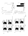
FIG. 2. Flow cytometry analysis of IFN-γ-secreting CD4+ T cells in vaccinated mice. C57BL/6 mice (five per group) were immunized twice with 2 μg/mouse of at one-week interval. Splenocytes from vaccinated mice were harvested 1 week after the last vaccination and stimulated overnight with the PADRE peptide. (A) Representative figure of the flow cytometry data. The numbers on the right upper corner represent the number of IFN-γ-secreting CD4+ T cells per 3×10⁵ splenocytes acquired. (B) Bar graph depicting the number of PADRE-specific CD4+ T cells per 3×10⁵ splenocytes (means±s.e.). The data presented in this figure are from one representative experiment of two performed.
FIG. 3. Intracellular cytokine staining followed by flow cytometry analysis to determine the number of E6-specific CD8+ T cells and PADRE-specific CD4+ T cells in vaccinated mice. C57BL/6 mice (5 per group) were immunized twice intradermally via gene gun with 2 μg/mouse of Ii DNA, Ii DNA+SCT-E6 DNA, Ii-PADRE DNA or Ii-PADRE DNA+SCT-E6 DNA at one-week interval. Splenocytes from vaccinated mice were harvested 1 week after the last vaccination and stimulated with E6 or PADRE peptide. Splenocytes without peptide stimulation were used as a negative control. The splenocytes were stained for CD8 or CD4 and intracellular IFN-γ. (A) & (C) Representative figures of the flow cytometry data. The numbers on the right upper corner represent the number of E6-specific CD8+ T cells (A) or PADRE-specific CD4+ T cells (C) per 3×10⁵ splenocytes acquired. (B) & (D). Bar graph depicting the numbers of E6-specific CD8+ T-cells (B) or PADRE-specific CD4+ T cells (D) per 3×10⁵ splenocytes (mean±s.e.). The data presented in this figure are from one representative experiment of two performed.
FIG. 4. In vivo tumor protection experiments. C57BL/6 mice (five per group) were immunized twice via gene gun with 2 μg/mouse of Ii DNA, Ii-PADRE DNA, Ii DNA+SCT-E6 DNA or Ii-PADRE DNA+SCT-E6 DNA at one-week interval. One week after the last vaccination, the vaccinated mice were challenged subcutaneously with 5×10⁴ TC-1 cells/mouse. The mice were monitored for evidence of tumor growth by inspection and palpation twice a week. The data shown here are from one representative experiment of two performed.
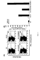
FIG. 5. Characterization of E7-specific IFN-γ-secreting CD8+ T cells and PADRE-specific CD4+ T cells by flow cytometry analysis in vaccinated mice. C57BL/6 mice (five per group) were immunized twice intradermally via gene gun with 2 μg/mouse of Ii DNA+CRT-E7 DNA or Ii-PADRE DNA+CRT-E7 DNA at one-week interval. Splenocytes from vaccinated mice were harvested 1 week after the last vaccination and stimulated with E7 peptide or PADRE peptide. Splenocytes without peptide stimulation were used as a negative control. The splenocytes were stained for both CD8 and intracellular IFN-γ. (A) & (C). Representative figures of the flow cytometry data. The numbers on the right upper corner represent the number of E7-specific IFN-γ-secreting CD8+ T cells (A) or PADRE-specific CD4+ T cells (C) per 3×10⁵ splenocytes acquired. (B) & (D) Bar graphs depicting the number of E7-specific T-cells (B) or PADRE-specific CD4+ T cells (D) per 3×10⁵ splenocytes (means±s.e.). The data presented in this figure are from one representative experiment of two performed.
FIG. 6. Flow cytometry analysis to characterize PADRE-specific CD4+ T cells in mice vaccinated with PADRE peptide or Ii-PADRE DNA. C57BL/6 mice (five per group) were immunized twice with 100 μg/mouse of PADRE peptide subcutaneously or 2 μg/mouse of Ii-PADRE DNA intradermally at one-week interval. Splenocytes from vaccinated mice were harvested 1 week after the last vaccination and stimulated with PADRE peptide. Splenocytes without peptide stimulation were used as a negative control. The splenocytes were stained for both CD4 and intracellular IFN-γ. (A) Representative figure of the flow cytometry data. The numbers on the right upper corner represent the number of PADRE-specific CD4+ T cells per 3×10⁵ splenocytes acquired. (B) Bar graph depicting the number of PADRE-specific CD4+ T cells per 3×10⁵ splenocytes (means±s.e.). The data presented in this figure are from one representative experiment of two performed.
FIG. 7. Characterization of E7-specific IFN-γ-secreting CD8+ T cells and PADRE-specific CD4+ T cells in mice vaccinated with PADRE peptide and CRT-E7. C57BL/6 mice (five per group) were immunized twice with 2 μg of CRT-E7 DNA intradermally via gene gun and 100 g of PADRE in 200 μL incomplete Freund's adjuvant by subcutaneous tail base injection at one-week interval. Mice vaccinated with 2 μg CRT-E7 DNA by gene gun and 200 μL incomplete Freund's adjuvant by subcutaneous tail base injection were used as a negative control. Splenocytes were harvested 1 week after the last vaccination and stimulated with E7 peptide or PADRE peptide. Splenocytes without peptide stimulation were used as a negative control. The splenocytes were stained for both CD8 and intracellular IFN-γ. (A) & (C) Representative figures of the flow cytometry data. The numbers on the upper right corner represent the numbers of E7-specific IFN-γ-secreting CD8+ T cells (A) or PADRE-specific CD4+ T cells (C) per 3×10⁵ splenocytes acquired. (B) & (D) Bar graphs depicting the numbers of E7-specific CD8+ T cells (B) or PADRE-specific CD4+ T cells (D) per 3×10⁵ splenocytes (means±s.e.).
FIG. 8. Flow cytometry analysis to characterize E7-specific CD8+ T cells in vaccinated mice. C57BL/6 mice (five per group) were immunized twice with CRT-E7 DNA (i.d.)+Ii DNA (i.d.), CRT-E7 DNA (i.d.)+Ii-PADRE DNA (i.d.), CRT-E7 DNA (i.d.)+adjuvant (s.c.) or CRT-E7 DNA (i.d.)+PADRE peptide (s.c.) at one-week interval. Splenocytes were harvested 1 week after the last vaccination and stimulated with E7 peptide. The splenocytes were stained for both CD8 and intracellular IFN-γ. Bar graph depicting the number of E7-specific CD8+ T cells/3×105 splenocytes (means±s.e.).
FIG. 9. Intracellular cytokine staining with flow cytometry analysis to determine the number of PADRE-specific CD4+ T cells in mice vaccinated with various DNA constructs. C57BL/6 mice (five per group) were immunized intradermally via gene gun with 2 μg/mouse of Ii DNA, Sig/PADRE DNA, Sig/PADRE/LAMP-1 DNA or Ii-PADRE DNA at one-week intervals. The vaccinated mice received a booster with the same dose and regimen one week later. Splenocytes from vaccinated mice were harvested 1 week after the last vaccination and stimulated with PADRE peptide. Splenocytes without peptide stimulation were used as a negative control. The splenocytes were stained for both CD4 and intracellular IFN-γ. (A) Representative figure of the flow cytometry data. The numbers on the upper right corner represent the number of PADRE-specific IFN-γ-secreting CD4+ T cells per 3×10⁵ splenocytes acquired. (B) Bar graph demonstrating the number of PADRE-specific CD4+ T cells per 3×10⁵ splenocytes (means±s.e.). The data presented in this figure are from one representative experiment of two performed.
FIG. 10. In vivo tumor treatment experiments to compare the anti-tumor effects of various DNA vaccines in mice. C57BL/6 mice (5 per group) were subcutaneously challenged with 1×10⁴ TC-1 tumor cells/mouse. Three days later, the mice were immunized twice with 2 μg/mouse of Ii DNA, Ii-PADRE DNA, Ii DNA+SCT-E6 DNA or Ii-PADRE DNA+SCT-E6 DNA at one-week interval. Data are expressed at means±s.e. The data presented in this figure are from one representative of two performed.
FIG. 11. Flow cytometry analysis to characterize TRP2-specific CD8+ T-cell responses in vaccinated mice. C57BL/6 mice (five per group) were immunized twice intradermally via gene gun with 2 μg/mouse of Ii DNA+SCT-TRP2 DNA or Ii-PADRE DNA+SCT-TRP2 DNA at one-week interval. Splenocytes from vaccinated mice were harvested 1 week after the last vaccination and were stimulated with TRP2 peptide. Splenocytes without peptide stimulation were used as a negative control. The splenocytes were stained for CD8 and intracellular IFN-γ. (A) Representative figure of the flow cytometry data. The numbers on the upper right corner represent the number of TRP2-specific CD8+ T cells per 3×10⁵ splenocytes acquired. (B) Bar graph depicting the number of TRP2-specific T cells per 3×10⁵ splenocytes (means±s.e.). The data presented in this figure are from one representative experiment of two performed.
FIG. 12. Flow cytometry analysis of E7-specific CD8+ T cells in mice vaccinated with CRT/E7 and Ii-PADRE DNA. C57BL/6 mice (5 per group) were vaccinated intradermally via gene gun with the CRT/E7 DNA and Ii-PADRE DNA, either administered together at the same location (CRT/E7+ Ii-PADRE (S)) or each of the DNA vaccines administered separately at different locations (on opposites sides) of the mouse abdominal wall (CRT/E7+Ii-PADRE (D)). Mice received DNA vaccination with the same dose and regimen one week later. The splenocytes were obtained from vaccinated mice and cultured with E7 peptide (aa 49-57) overnight. The cells were then analyzed for CD8 and intracellular IFNγ staining by flow cytometry. A) Representative flow cytometry data showing the number of E7-specific IFNγ+ CD8+ T cells in the mice vaccinated with CRT/E7+Ii-PADRE (D) or CRT/E7+Ii-PADRE (S). B) Bar graph showing the number of E7-specific IFNγ+ CD8+ T cells from each group with (shaded bars) or without (empty bars) stimulation by the E7 peptide (p<0.01). The data was shown as mean±s.d.
FIG. 13. Flow cytometry analysis of cytokine profile of PADRE-specific CD4+ T cells in mice vaccinated with DNA encoding Ii-PADRE. C57BL/6 mice (5 per group) were vaccinated intradermally via gene gun with DNA encoding Ii-PADRE or Ii chain twice with a one-week interval. The splenocytes were obtained from vaccinated mice one week after the last vaccination and cultured with PADRE peptide overnight. The cells were then analyzed for expression of CD4 and IL-2, IFN-γ, TNF-α, IL-4 and IL-10 by intracellular cytokine staining followed by flow cytometry analysis. A) Representative flow cytometry data showing the number of cytokine secreting CD4+ T cells/3×105 splenocytes in the mice vaccinated with DNA encoding Ii-PADRE or Ii chain. B) Bar graph showing the number of cytokine secreting CD4+ T cells in mice vaccinated with DNA encoding Ii-PADRE (shaded bars) or Ii chain (empty bars) (p<0.01). The data was shown as mean±s.d.
FIG. 14. Flow cytometry analysis of E7-specific CD8+ T cells in mice vaccinated with DCs loaded with PADRE or OVA in conjunction with E7 peptide. C57BL/6 mice (5 per group) were vaccinated subcutaneously with 2.5×10⁵/mouse of DCs pulsed with both E7 and PADRE (DC-PADRE/E7) or with 2.5×10⁵/mouse of DCs pulsed with both E7 and OVA (DC-OVA/E7). Mice received vaccination with the same dose and regimen one week later. As controls, C57BL/6 mice were vaccinated with 2.5×10⁵/mouse of DCs alone or DCs loaded with E7 (DC-E7). The splenocytes were obtained from vaccinated mice and cultured with E7 peptide (aa 49-57) overnight. The cells were then analyzed for CD8 and intracellular IFNγ staining by flow cytometry. A) Representative flow cytometry data showing the number of E7-specific IFNγ+ CD8+ T cells/3×105 splenocytes in the mice vaccinated with the various DC-based vaccines. B) Bar graph showing the number of IFNγ+ CD8+ T cells from each group with (shaded bars) or without (empty bars) stimulation by the E7 peptide (p<0.05). The data was shown as mean±s.d.
FIG. 15. Flow cytometry analysis of E7-specific CD8+ T cells in mice vaccinated with DCs loaded with E7 and DCs loaded with PADRE. C57BL/6 mice (5 per group) were vaccinated subcutaneously with DCs loaded with E7 on the right side of the abdominal wall and DCs loaded with PADRE on the left side of the abdominal wall (E7(R)+PADRE(L)). Another group of mice was vaccinated with the DC-based vaccine combining the E7-loaded DCs and the PADRE-loaded DCs administered on each side of the abdominal wall (E7/PADRE (mixed DC). Mice received vaccination with the same dose and regimen one week later. As a control, one group of mice was vaccinated with DCs without peptide. The splenocytes were obtained from vaccinated mice and cultured with E7 peptide (aa 49-57) overnight. The cells were then analyzed for CD8 and intracellular IFNγ staining by flow cytometry. A) Representative flow cytometry data showing the number of E7-specific IFNγ+ CD8+ T cells in the mice vaccinated with the different DC-based vaccines. B) Bar graph showing the number of IFNγ+ CD8+ T cells/3×105 splenocytes from each group with (shaded bars) or without (empty bars) stimulation by the E7 peptide (p<0.05). The data was shown as mean±s.d.
FIG. 16. Characterization of cytokine profile of the PADRE-specific CD4+ T cells following in vitro stimulation with PADRE pulsed DCs. Representative flow cytometry data showing the number of cytokine secreting cells/5×104 CD4+ T cells stimulated in vitro with DCs pulsed with PADRE (DC+PADRE, right panel) or DCs without PADRE (DC, left panel). The PADRE-specific CD4+ T cell line was stimulated in vitro with DCs pulsed with or without PADRE peptide. The cells were then analyzed for expression of IL-2, IFN-γ, TNF-α, IL-4 and IL-10 by intracellular cytokine staining followed by flow cytometry.
FIG. 17. Characterization of the proliferation of luciferase expressing E7-specific CD8+ T cells in the presence of PADRE-specific CD4+ T cells. A 24-well plate was loaded with 2×10⁴/well of irradiated TC-1 cells and 2×10⁵/well of E7-specific CD8+ T cells expressing luciferase (E7T-LUC). 1×10⁵/well of PADRE-specific CD4+ T cells and 1×10⁵/well of DCs pulsed with PADRE (DC-PADRE) or DCs without PADRE were added to the indicated wells. As a positive control, 10 U/ml IL-2 was added to the indicated wells. The wells without DCs or CD4+ T cells were used negative controls. The plates were incubated for 3 days and the degree of proliferation of E7-specific CD8+ T cells was characterized using bioluminescence imaging. A) Representative luminescence images of 24-well plates containing E7T-LUC cells at day 3 after in vitro simulation. B) Bar graph showing the bioluminescence of the E7T-LUC cells at day 0 (empty bars) and day 3 (shaded bars). As an alternative to assess the proliferation, E7-specific CD8+ T cells were pulsed with CFSE and incubated with the various cells as depicted. C) Flow cytometry analysis demonstrating CFSE expression in the CD8+ T cells in the different wells. The data was shown as mean±s.d. D) Flow cytometry analysis demonstrating IL-2 blocking using CFSE expression in the CD8+ T cells in the different wells.
FIGS. 18 A and B. Anti-tumor effects generated by treatment with doxorubicin and/or CRT/E6 DNA vaccine in vaccinated mice.
FIG. 19. Sequence of the pcDNA3 plasmid vector (SEQ ID NO: 1).
FIG. 20. Sequence of the pNGVL4a plasmid vector (SEQ ID NO: 2).
FIG. 21. Sequence of the pcDNA3-E7-Hsp70 plasmid (SEQ ID NO: 3).
FIG. 22. Sequence of the pcDNA3-ETA(dII)/E7 plasmid (SEQ ID NO: 4).
FIG. 23. Sequence of the pNGVL4a-CRT/E7(detox) plasmid (SEQ ID NO: 5).
FIG. 24. Nucleotide sequence of VP22/E7 DNA as it appears in the pCDNA3 vector (SEQ ID NO: 6 (encoding SEQ ID NO: 39)) which is 1254 nucleotides (+stop codon). SEQ ID NO: 7 includes nucleotides 1-903 encoding VP22 (SEQ ID NO: 38). Nucleotides 904-921 and the corresponding amino acids 302-307 are a “linker” sequence. Nucleotides 922-1209 (lower case) encode 96 of the 98 amino acids of wild-type E7 protein (SEQ ID NO: 41). Also shown is a stretch of vector sequence (underscored) from nucleotides 1210-1257 (including stop codon).
FIG. 25: Flow cytometry analysis to demonstrate the expression of murine MHC molecules in CIITA-transfected DC-1 cells. Flow cytometry data demonstrating the levels of H-2 Kb, H-2 Db, and I-Ab expression in CIITA-transfected cells and controls. The legend shows the vector plasmids with which the DCs were transfected. An immortalized dendritic cell line (DC-1) was transfected with CIITA or mutant CIITA (mtCIITA). Untransfected cells and cells transfected with the pcDNA3 vector backbone were used as a control. The expression of MHC I and II molecules was characterized using antibodies to MHC I H-2 Kb, H-2 Db, and MHC II I-Ab by flow cytometry analysis.
FIG. 26: Characterization of the MHC class I and II presentation of DCs transfected with CIITA DNA. DCs were cotransfected with CIITA DNA and CRT/E6 DNA (A & B) or Ii-PADRE DNA (C & D). The DCs were then incubated with E6-specific CD8+ T cells (A & B) or PADRE-specific CD4+ T cells (C & D) overnight. The activation of antigen-specific T cells was characterized by intracellular cytokine staining followed by flow cytometry analysis using IFN-γ and CD4 or CD8-specific antibodies. A and C. Representative flow cytometry data showing the numbers of activated E6-specific CD8+ T cells (A) and PADRE-specific CD4+ T cells (C) after incubation with the cotransfected DCs. B and D. Bar graphs depicting the numbers of E6-specific CD8+ T cells (B) and PADRE-specific CD4+ T cells (D) (means±s.d.). The data presented in this figure are from one representative experiment of two performed.
FIG. 27: Characterization of the E6-specific CD8+ T cell immune responses in mice vaccinated with CIITA DNA and CRT/E6 DNA. C57BL/6 mice (5 per group) were immunized with 2 μg/mouse of CIITA and/or CRT/E6 DNA twice with a 1-week interval. Splenocytes from vaccinated mice were harvested 1 week after the last vaccination and characterized for E6-specific CD8+ T cells using intracellular IFN-γ staining followed by flow cytometry analysis. A. Representative flow cytometry data for the E6-specific CD8+ T cell immune responses. The numbers in the upper right-hand corner represent the number of E6-specific IFN-γ-secreting CD8+ T cells per 5×10⁶ pooled splenocytes. B. Bar graphs depicting the numbers of E6-specific IFN-γ-secreting CD8+ T cells per 5×10⁶ pooled splenocytes (means±s.d.). The data presented in this figure are from one representative experiment of two performed.
FIG. 28: Characterization of the E6-specific CD8+ T cells in mice vaccinated with CRT/E6, CIITA DNA and Ii-PADRE DNA vaccines. C57BL/6 mice (5 per group) were immunized with 2 μg/mouse twice with a 1-wk interval of the DNA combinations listed in Table 1. Splenocytes from vaccinated mice were harvested 1 week after the last vaccination and characterized for E6-specific CD8+ T cells using intracellular IFN-γ staining followed by flow cytometry analysis. A. Representative flow cytometry data. The numbers in the upper right-hand corner represent the number of E6-specific IFN-γ-secreting CD8+ T cells per 5×10⁶ pooled splenocytes. B. Bar graphs depicting the numbers of E6-specific IFN-γ-secreting CD8+ T cells per 5×10⁶ pooled splenocytes (means±s.d.). The data presented in this figure are from one representative experiment of two performed.
FIG. 29: In vivo tumor treatment experiments. C57BL/6 mice (5 per group) were first challenged with 5×10⁴/mouse of TC-1 tumor cells by subcutaneous injection. Three days after tumor challenge, the mice were administered 2 μg DNA/mouse 3 times with 4-day intervals of the various DNA vaccine mixtures listed in Table 1. The mice were monitored for evidence of tumor growth by inspection and palpation twice a week. Tumor volumes were measured starting from day 7 after tumor challenge. (A) Line graph depicting the tumor volumes in mice of different tumor treatments (means±s.d.). (B) Kaplan & Meier survival analysis in mice of the tumor treatment experiments. The data shown here are from one representative experiment of two performed.
FIG. 30: Characterization of the long-term E6-specific CD8+ T cell immune response in mice vaccinated with CRT/E6, CIITA DNA and Ii-PADRE DNA vaccines. C57BL/6 mice (5 per group) were immunized with 2 μg/mouse twice with a 1-wk interval of the DNA combinations listed in Table 1. Splenocytes from vaccinated mice were harvested 60 days after the last vaccination and characterized for E6-specific CD8+ T cells using intracellular IFN-γ staining followed by flow cytometry analysis. A. Representative flow cytometry data. The numbers in the upper right-hand corner represent the number of memory E6-specific IFN-γ-secreting CD8+ T per 5×10⁶ pooled splenocytes. B. Bar graphs depicting the numbers of memory E6-specific IFN-γ-secreting CD8+ T cells per 5×10⁶ pooled splenocytes (means±s.d.). The data presented in this figure are from one representative experiment of two performed.
FIG. 31: Long-term in vivo tumor protection experiments. C57BL/6 mice (5 per group) were immunized with 2 μg DNA/mouse twice with a 1-week interval of the various DNA vaccine mixtures listed in Table 1. Two months after the last vaccination, the mice were challenged by subcutaneous injection of 1×10⁵/mouse of TC-1 cells. The mice were monitored for evidence of tumor growth by inspection and palpation twice a week. Tumor volumes were measured starting from day 7 after tumor challenge. (A) Line graph depicting tumor volume in mice challenged with TC-1 cells (means±s.d.). (B) Kaplan & Meier survival analysis in mice challenged with TC-1 cells. The data shown here are from one representative experiment of two performed.
FIG. 32: Characterization of the PADRE-specific CD4+ T cell immune responses in mice vaccinated with CIITA and Ii-PADRE DNA vaccines. C57BL/6 mice (5 per group) were immunized with 2 μg/mouse of CIITA and/or Ii-PADRE DNA. Splenocytes from vaccinated mice were harvested 1 week after the last vaccination and were characterized for PADRE-specific CD4+ T cells using intracellular IFN-γ staining followed by flow cytometry analysis. A. Representative flow cytometry data for the PADRE-specific CD4+ T cell immune responses. The numbers in the upper right-hand corner represent the number of PADRE-specific IFN-γ-secreting CD4+ T cells per 5×10⁶ pooled splenocytes. B. Bar graphs depicting the numbers of PADRE-specific IFN-γ-secreting CD4+ T cells per 5×10⁶ pooled splenocytes (means+/−s.d.). The data presented in this figure are from one representative experiment of two performed.
FIG. 33: Characterization of the PADRE-specific CD4+ T cell immune responses in mice vaccinated with CRT6/E6, CIITA DNA and Ii-PADRE DNA vaccines. C57BL/6 mice (5 per group) were immunized with 2 μg/mouse twice with a 1-week interval of the DNA combinations listed in Table 1. Splenocytes from vaccinated mice were harvested 1 week after the last vaccination and were characterized for PADRE-specific CD4+ T cells using intracellular IFN-γ staining followed by flow cytometry analysis. A. Representative flow cytometry data. The numbers in the upper right-hand corner represent the number of PADRE-specific CD4+ T cells per 5×10⁶ pooled splenocytes. B. Bar graphs depicting the numbers of PADRE-specific CD4+ T cells per 5×10⁶ pooled splenocytes (means+/−s.d.). The data presented in this figure are from one representative experiment of two performed.
FIG. 34: Characterization of the long-term PADRE-specific CD4+ T cell immune response in mice vaccinated with CRT6/E6, CIITA DNA and Ii-PADRE DNA vaccines. C57BL/6 mice (5 per group) were immunized with 2 μg/mouse twice with a 1-week interval of the DNA combinations listed in Table 1. Splenocytes from vaccinated mice were harvested 60 days after the last vaccination and were characterized for PADRE-specific CD4+ T cells using intracellular IFN-γ staining followed by flow cytometry analysis. A. Representative flow cytometry data. The numbers in the upper right-hand corner represent the number of memory PADRE-specific CD4+ T cells per 5×10⁶ pooled splenocytes. B. Bar graphs depicting the numbers of memory PADRE-specific CD4+ T cells per 5×10⁶ pooled splenocytes (means+/−s.d.). The data presented in this figure are from one representative experiment of two performed.
FIG. 35 shows the nucleotide (SEQ ID NO: 90) and amino acid (SEQ ID NO: 91) sequences of a human Ia-PADRE.
FIG. 36 shows the nucleotide (SEQ ID NO: 94) and amino acid (SEQ ID NO: 95) sequences of human CIITA.
DETAILED DESCRIPTION
Partial List of Abbreviations
APC, antigen presenting cell; CRT, calreticulin; CTL, cytotoxic T lymphocyte; DC, dendritic cell; ECD, extracellular domain; EGCG, epigallocatechin-3-gallate; E6, HPV oncoprotein E6; E7, HPV oncoproteinE7; ELISA, enzyme-linked immunosorbent assay; HPV, human papillomavirus; HSP, heat shock protein; Hsp70, mycobacterial heat shock protein 70; IFN γ, interferon-γ; i.m., intramuscular(ly); i.v., intravenous(ly); MHC, major histocompatibility complex; PBS, phosphate-buffered saline; PCR, polymerase chain reaction; β-gal, β-galactosidase.
General
Provided herein are methods and compositions for increasing or stimulating an immune response, e.g., for treating a hyperproliferating disease, e.g., cancer. In one embodiment, a method comprises administering to a subject in need thereof a nucleic acid, e.g., a DNA vaccine, encoding an MHC class I and/or II (“class I/II”) activator. Such vaccines may be therapeutic vaccines or preventative vaccines. A method may also comprise administering to a subject in need thereof a nucleic acid encoding an MHC class I/II activator and one or more nucleic acid vaccines, e.g., a nucleic acid encoding an antigen or a biologically active homolog thereof. Other nucleic acid vaccines that may be administered include nucleic acids encoding a protein that enhances the immune system, but do not comprise an antigen, e.g., those that prolong the life of antigen presenting cells, as further described herein. Methods for stimulating an immune response may also comprise administering to a subject in need thereof a nucleic acid encoding an MHC class I/II activator; a nucleic vaccine encoding an antigen; and a nucleic acid vaccine that does not encode an antigen. Other methods may comprise administering a nucleic acid encoding an MHC class I/II activator and an agent or drug, e.g., a drug that is not a nucleic acid vaccine, such as a drug that induces apoptosis of cancer cells, e.g., a chemotherapeutic agent. Yet other methods may comprise administering a nucleic acid encoding an MHC class I/II activator; a nucleic acid vaccine encoding an antigen; and a chemotherapeutic agent. Yet other methods may comprise administering a nucleic acid encoding an MHC class I/II activator; a nucleic acid vaccine encoding an antigen; a nucleic acid vaccine that does not encode an antigen; and a chemotherapeutic agent. Any other combinations of one or more of a nucleic acid encoding an MHC class I/II activator; one or more nucleic vaccines encoding an antigen; one or more nucleic vaccines that do not encode an antigen; and one or more drugs, e.g., chemotherapeutic drugs, may also be used for stimulating an immune response in a subject. These methods may be used for treating a subject in need thereof, e.g., a subject having or likely to develop a hyperproliferative disease, e.g., cancer, e.g., an HPV-associated malignancy.
At least some of the methods may also be used to enhance the efficacy of another treatment, e.g., a treatment that comprises administering a nucleic acid vaccine.
Administration of an MHC class I/II activator may be done at the same time, before or after administration of one or more other agents, such as nucleic acid vaccines or drugs.
MHC Class I/II Activators
“MHC class I/II activators” refers to molecules or complexes thereof that increase immune responses by increasing MHC class I or II (“I/II”) antigen presentation, such as by increasing MHC class I, class II or class I and class II activity or gene expression. In one embodiment, an MHC class I/II activator is a nucleic acid encoding a protein that enhances MHC class I/II antigen presentation. Exemplary MHC class I/II activators include nucleic acids encoding an MHC class II associated invariant chain (Ii), in which the CLIP region is replaced with a T cell epitope, e.g., a promiscuous T cell epitope, such as the Pan HLA-DR reactive epitope (PADRE), or a variant thereof. Other MHC class I/II activators are nucleic acids encoding the MHC class II transactivator CIITA or a variant thereof.
In one embodiment, an MHC class I/II activator is a nucleic acid, e.g., an isolated nucleic acid, encoding a protein comprising, consisting or consisting essentially of an invariant (Ii) chain, wherein the CLIP region is replaced with a promiscuous CD4+ T cell epitope. A “promiscuous CD4+ T cell epitope” is used interchangeably with “universal CD4+ T cell epitope” and refers to peptides that bind to numerous histocompatibility alleles, e.g., human MHC class II molecules. In one embodiment, the promiscuous CD4+ T cell epitope is a Pan HLA-DR reactive epitope (PADRE), thereby forming an Ii-PADRE protein that is encoded by an Ii-PADRE nucleic acid. In one embodiment, a nucleic acid encodes an Ii chain, wherein amino acids 81-102 (KPVSQMRMATPLLMRPM (SEQ ID NO: 92) are replaced with the PADRE sequence AKFVAAWTLKAAA (SEQ ID NO: 93). An exemplary human Ii-PADRE amino acid sequence is set forth as SEQ ID NO: 91, and is encoded by nucleotide sequence SEQ ID NO: 90 (see FIG. 35).
Also provided herein are variants of a protein consisting of SEQ ID NO: 91. A protein may comprise, consist essentially of, or consist of an amino acid sequence that is at least about 80%, 85%, 90%, 95%, 96%, 97%, 98% or 99% identical to SEQ ID NO: 91. A protein may comprise a PADRE that is identical to the PADRE of SEQ ID NO: 91, i.e., consisting of SEQ ID NO: 93. A protein may comprise a PADRE sequence that is at least about 80%, 85%, 90%, 95%, 96%, 97%, 98% or 99% identical to SEQ ID NO: 93; and/or an Ii sequence that is at least about 80%, 85%, 90%, 95%, 96%, 97%, 98% or 99% identical to the Ii sequence of SEQ ID NO: 91.
An amino acid sequence may differ from that of SEQ ID NO: 91 or the Ii or PADRE sequences thereof by the addition, deletion or substitution of at least about 1, 2, 3, 4, 5, 6, 7, 8, 9, 10, 11, 12, 13, 14, 15, 16, 17, 18, 19, 20, 25, 30 or more amino acids. In certain embodiments, a protein lacks one or more, e.g., 2, 3, 4, 5, 6, 7, 8, 9, 10 or more amino acids at the C- and/or N-terminus and/or internal relative to that of SEQ ID NO: 91 or the Ii or PADRE region thereof. In certain embodiments, an amino acid sequence differs from that of SEQ ID NO: 93 or from that of the Ii sequence by the addition, deletion or substitution of at least about 1, 2, 3, 4, or 5 amino acids.
Variants of SEQ ID NOs: 91 or the PADRE or Ii regions thereof preferably have a biological activity. Such variants are referred to as “functional homologs” or “functional variants.” Functional homologs include variants of SEQ ID NOs: 91 that increase an immune response, e.g., an antigen specific immune response, in a subject to whom it is administered, or has any of the biological activities set forth in the Examples pertaining to Ii-PADRE. Variants of the PADRE sequence or the Ii sequence may have a biological activity that is associated with that of the wildtype PADRE or Ii sequences, respectively. Biological activities can be determined as know in the art or as set forth in the Examples. In addition, comparison (or alignment) of the Ii and PADRE sequences from different species is expected to be helpful in determining which amino acids may be varied and which ones should preferably not be varied.
Other proteins provided herein comprise a PADRE amino acid sequence that replaces a larger portion of Ii, e.g., wherein Ii is lacking about amino acids 81-103, 81-104, 81-105, 81-106, 81-107, 81-108, 81-109, 81-110 or more; is lacking about amino acids 70-102, 71-102, 72-102, 73-102, 74-102, 75-102, 76-102, 77-102, 78-102, 79-102, 80-102 or more.
Other promiscuous CD4+ T cell epitopes that may be used instead of PADRE are listed in Table 2.
| TABLE 2 |
| |
| Exemplary promiscuous CD4+ T cell epitopes |
| (SEQ ID NOS 96-120, respectively in order of appearance) |
| Promiscuous CD4+ T cell epitopes |
Reference |
| |
| EBV-latent membrane protein 1(LMP1159-175) YLQQNWWTLLVDLLWLL |
(1) |
| MAGF-A6172-187; IGHVYIFATCLGLSYD |
(2) |
| Mycoplasma penetrans HF-2219-226; IYIFAACL |
|
| six-transmembrane epithelial antigen of prostate (STEAP) |
(3) |
| STEAP102-116 HQQYFYKIPILVINK |
|
| STEAP192-206 LLNWAYQQVQQNKED |
|
| Taxol-resistance-associated gene-3 (TRAG3)35-48 |
(4) |
| EFHACW PAFTVLGE |
|
| Survivin10-24 WQPFLKDHRISTFKN |
(5) |
| HPV 18-E652-66; LFVVYRDSIPHAACH |
(6) |
| HPV 18-E697-111; GLYNLLIRCLRCQKP |
|
| Carcinoembryonic antigen177-189; LWWVNNQSLPVSP |
(7) |
| mycobacterial antigen MPB70 |
(8) |
| MPB70106-130; FSKLPASTIDELKTNSSLLTSILTY |
|
| MPB70166-193; GNADVVCGGVSTANATVYMIDSVLMPPA |
|
| HER-2776-788 GSPYVSRLLGICL |
(9) |
| HER-2833-849 KVPIKWMALESILRRRF |
(10) |
| NY-ESO-1119-143 PGVLLKEFTVSGNILTIRLTAADHR |
(11) |
| Tetanus toxin1084-1099 VSIDKFRIFCKANPK |
(12) |
| Tetanus toxin1174-1189 LKFIIKRYTPNNEIDS |
|
| Tetanus toxin1064-1079 IREDNNITLKLDRCN |
|
| Tetanus toxin947-967 FNNFTVSFWLRVPKVSASHLE |
|
| Tetanus toxin830-843 QYIKANSKFIGITE |
|
| HBV nuclear capside50-69 PHHTALRQAILCWGELMTLA |
|
| Influenza haemagglutinin307-319 PKYVKQNTLKLAT |
|
| HBV surface antigen19-33-FFLLTRILTIPQSLD |
|
| Influenza marix17-31 YSGPLKAEIAQRLEDV |
|
| P. falciparum CSP380-398 EKKIAKMEKASSVFNVVN |
| |
| (1). Kobayashi, H., T. Nagato, M. Takahara, K. Sato, S. Kimura, N. Aoki, M. Azumi, M. Tateno, Y. Harabuchi, and E. Celis. 2008. Induction of EBV-latent membrane protein 1-specific MHC class II-restricted T-cell responses against natural killer lymphoma cells. Cancer Res 68: 901-908. |
| (2). Vujanovic, L., M. Mandic, W. C. Olson, J. M. Kirkwood, and W. J. Storkus. 2007. A mycoplasma peptide elicits heteroclitic CD4+ T cell responses against tumor antigen MAGE-A6. Clin Cancer Res 13: 6796-6806. |
| (3). Kobayashi, H., T. Nagato, K. Sato, N. Aoki, S. Kimura, M. Murakami, H. Iizuka, M. Azumi, H. Kakizaki, M. Tateno, and E. Celis. 2007. Recognition of prostate and melanoma tumor cells by six-transmembrane epithelial antigen of prostate-specific helper T lymphocytes in a human leukocyte antigen class II-restricted manner. Cancer Res 67: 54985504 |
| (4). Janjic, B., P. Andrade, X. F. Wang, J. Fourcade, C. Almunia, P. Kudela, A. Brufsky, S. Jacobs, D. Friedland, R. Stoller, D. Gillet, R. B. Herberman, J. M. Kirkwood, B. Maillere, and H. M. Zarour. 2006. Spontaneous CD4+ T cell responses against TRAG-3 in patients with melanoma and breast cancers. J Immunol 177: 2717-2727. |
| (5). Piesche, M., Y. Hildebrandt, F. Zettl, B. Chapuy, M. Schmitz, G. Wulf, L. Trumper, and R. Schroers. 2007. Identification of a promiscuous HLA DR-restricted T-cell epitope derived from the inhibitor of apoptosis protein survivin. Hum Immunol 68: 572-576. |
| (6). Facchinetti, V., S. Seresini, R. Longhi, C. Garavaglia, G. Casorati, and M. P. Protti. 2005. CD4+ T cell immunity against the human papillomavirus-18 E6 transforming protein in healthy donors: identification of promiscuous naturally processed epitopes. Eur J Immunol 35: 806-815. |
| (7). Campi, G., M. Crosti, G. Consogno, V. Facchinetti, B. M. Conti-Fine, R. Longhi, G. Casorati, P. Dellabona, and M. P. Protti. 2003. CD4(+) T cells from healthy subjects and colon cancer patients recognize a carcinoembryonic antigen-specific immunodominant epitope. Cancer Res 63: 8481-8486. |
| (8). Al-Attiyah, R., F. A. Shaban, H. G. Wiker, F. Oftung, and A. S. Mustafa. 2003. Synthetic peptides identify promiscuous human Th1 cell epitopes of the secreted mycobacterial antigen MPB70. Infect Immun 71: 1953-1960. |
| (9). Sotiriadou, R., S. A. Perez, A. D. Gritzapis, P. A. Sotiropoulou, H. Echner, S. Heinzel, A. Mamalaki, G. Pawelec, W. Voelter, C. N. Baxevanis, and M. Papamichail. 2001. Peptide HER2(776-788) represents a naturally processed broad MHC class II-restricted T cell epitope. Br J Cancer 85: 1527-1534. |
| (10). Kobayashi, H., M. Wood, Y. Song, E. Appella, and E. Celis. 2000. Defining promiscuous MHC class II helper T-cell epitopes for the HER2/neu tumor antigen. Cancer Res 60: 5228-5236 |
| (11). Zarour, H. M., B. Maillere, V. Brusic, K. Coval, E. Williams, S. Pouvelle-Moratille, F. Castelli, S. Land, J. Bennouna, T. Logan, and J. M. Kirkwood. 2002. NY-ESO-1 119-143 is a promiscuous major histocompatibility complex class II T-helper epitope recognized by Th1-and Th2-type tumor-reactive CD4+ T cells. Cancer Res 62: 213-218. |
| (12). Falugi, F., R. Petracca, M. Mariani, F. Luzzi, S. Mancianti, V. Carinci, M. L. Melli, O. Finco, A. Wack, A. Di Tommaso, M. T. De Magistris, P. Costantino, G. Del Giudice, S. Abrignani, R. Rappuoli, and G. Grandi. 2001. Rationally designed strings of promiscuous CD4(+) T cell epitopes provide help to Haemophilus influenzae type b oligosaccharide: a model for new conjugate vaccines. Eur J Immunol 31: 3816-3824. |
The CLIP region in an Ii molecule, e.g., having the amino acid sequence of the Ii portion set forth in SEQ ID NO: 91, may be replaced with any of the peptides in Table 2 or other promiscuous epitopes set forth in the references of Table 2, or functional variants thereof. Preferred epitopes include those from tetanus toxin and influenza. Any other promiscuous CD4+ T cell epitopes may be used, e.g., those described in the following references:
- 1. Campi, G., M. Crosti, G. Consogno, V. Facchinetti, B. M. Conti-Fine, R. Longhi, G. Casorati, P. Dellabona, and M. P. Protti. 2003. CD4(+) T cells from healthy subjects and colon cancer patients recognize a carcinoembryonic antigen-specific immunodominant epitope. Cancer Res 63:8481-8486.
- 2. Castelli, F. A., M. Leleu, S. Pouvelle-Moratille, S. Farci, H. M. Zarour, M. Andrieu, C. Auriault, A. Menez, B. Georges, and B. Maillere. 2007. Differential capacity of T cell priming in naive donors of promiscuous CD4+ T cell epitopes of HCV NS3 and Core proteins. Eur J Immunol 37:1513-1523.
- 3. Consogno, G., S. Manici, V. Facchinetti, A. Bachi, J. Hammer, B. M. Conti-Fine, C. Rugarli, C. Traversari, and M. P. Protti. 2003. Identification of immunodominant regions among promiscuous HLA-DR-restricted CD4+ T-cell epitopes on the tumor antigen MAGE-3. Blood 101:1038-1044.
- 4. Depil, S., O. Morales, F. A. Castelli, N. Delhem, V. Francois, B. Georges, F. Dufosse, F. Morschhauser, J. Hammer, B. Maillere, C. Auriault, and V. Pancre. 2007. Determination of a HLA II promiscuous peptide cocktail as potential vaccine against EBV latency II malignancies. J Immunother (1997) 30:215-226.
- 5. Facchinetti, V., S. Seresini, R. Longhi, C. Garavaglia, G. Casorati, and M. P. Protti. 2005. CD4+ T cell immunity against the human papillomavirus-18 E6 transforming protein in healthy donors: identification of promiscuous naturally processed epitopes. Eur J Immunol 35:806-815.
- 6. Kobayashi, H., T. Nagato, K. Sato, N. Aoki, S. Kimura, M. Murakami, H. Iizuka, M. Azumi, H. Kakizaki, M. Tateno, and E. Celis. 2007. Recognition of prostate and melanoma tumor cells by six-transmembrane epithelial antigen of prostate-specific helper T lymphocytes in a human leukocyte antigen class II-restricted manner. Cancer Res 67:5498-5504.
- 7. Kobayashi, H., M. Wood, Y. Song, E. Appella, and E. Celis. 2000. Defining promiscuous MHC class II helper T-cell epitopes for the HER2/neu tumor antigen. Cancer Res 60:5228-5236.
- 8. Mandic, M., C. Almunia, S. Vicel, D. Gillet, B. Janjic, K. Coval, B. Maillere, J. M. Kirkwood, and H. M. Zarour. 2003. The alternative open reading frame of LAGE-1 gives rise to multiple promiscuous HLA-DR-restricted epitopes recognized by T-helper 1-type tumor-reactive CD4+ T cells. Cancer Res 63:6506-6515.
- 9. Neumann, F., C. Wagner, S. Stevanovic, B. Kubuschok, C. Schormann, A. Mischo, K. Ertan, W. Schmidt, and M. Pfreundschuh. 2004. Identification of an HLA-DR-restricted peptide epitope with a promiscuous binding pattern derived from the cancer testis antigen HOM-MEL-40/SSX2. Int J Cancer 112:661-668.
- 10. Ohkuri, T., M. Sato, H. Abe, K. Tsuji, Y. Yamagishi, H. Ikeda, N. Matsubara, H. Kitamura, and T. Nishimura. 2007. Identification of a novel NY-ESO-1 promiscuous helper epitope presented by multiple MHC class II molecules found frequently in the Japanese population. Cancer Sci 98:1092-1098.
- 11. Piesche, M., Y. Hildebrandt, F. Zettl, B. Chapuy, M. Schmitz, G. Wulf, L. Trumper, and R. Schroers. 2007. Identification of a promiscuous HLA DR-restricted T-cell epitope derived from the inhibitor of apoptosis protein survivin. Hum Immunol 68:572-576.
- 12. Sotiriadou, R., S. A. Perez, A. D. Gritzapis, P. A. Sotiropoulou, H. Echner, S. Heinzel, A. Mamalaki, G. Pawelec, W. Voelter, C. N. Baxevanis, and M. Papamichail. 2001. Peptide HER2(776-788) represents a naturally processed broad MHC class II-restricted T cell epitope. Br J Cancer 85:1527-1534.
- 13. Texier, C., S. Pouvelle-Moratille, C. Buhot, F. A. Castelli, C. Pecquet, A. Menez, F. Leynadier, and B. Maillere. 2002. Emerging principles for the design of promiscuous HLA-DR-restricted peptides: an example from the major bee venom allergen. Eur J Immunol 32:3699-3707.
- 14. Vujanovic, L., M. Mandic, W. C. Olson, J. M. Kirkwood, and W. J. Storkus. 2007. A mycoplasma peptide elicits heteroclitic CD4+ T cell responses against tumor antigen MAGE-A6. Clin Cancer Res 13:6796-6806.
- 15. Zarour, H. M., B. Maillere, V. Brusic, K. Coval, E. Williams, S. Pouvelle-Moratille, F. Castelli, S. Land, J. Bennouna, T. Logan, and J. M. Kirkwood. 2002. NY-ESO-1 119-143 is a promiscuous major histocompatibility complex class II T-helper epitope recognized by Th1- and Th2-type tumor-reactive CD4+ T cells. Cancer Res 62:213-218.
- 16. Gao, M., H. P. Wang, Y. N. Wang, Y. Zhou, and Q. L. Wang. 2006. HCV-NS3 Th1 minigene vaccine based on invariant chain CLIP genetic substitution enhances CD4(+) Th1 cell responses in vivo. Vaccine 24:5491-5497.
- 17. Nagata, T., T. Aoshi, M. Suzuki, M. Uchijima, Y. H. Kim, Z. Yang, and Y. Koide. 2002. Induction of protective immunity to Listeria monocytogenes by immunization with plasmid DNA expressing a helper T-cell epitope that replaces the class II-associated invariant chain peptide of the invariant chain. Infect Immun 70:2676-2680.
- 18. Nagata, T., T. Higashi, T. Aoshi, M. Suzuki, M. Uchijima, and Y. Koide. 2001. Immunization with plasmid DNA encoding MHC class II binding peptide/CLIP-replaced invariant chain (Ii) induces specific helper T cells in vivo: the assessment of Ii p31 and p41 isoforms as vehicles for immunization. Vaccine 20:105-114.
- 19. Toda, M., M. Kasai, H. Hosokawa, N. Nakano, Y. Taniguchi, S. Inouye, S. Kaminogawa, T. Takemori, and M. Sakaguchi. 2002. DNA vaccine using invariant chain gene for delivery of CD4+ T cell epitope peptide derived from Japanese cedar pollen allergen inhibits allergen-specific IgE response. Eur J Immunol 32:1631-1639.
- 20. van Bergen, J., M. Camps, R. Offringa, C. J. Melief, F. Ossendorp, and F. Koning. 2000. Superior tumor protection induced by a cellular vaccine carrying a tumor-specific T helper epitope by genetic exchange of the class II-associated invariant chain peptide. Cancer Res 60:6427-6433.
- 21. van Tienhoven, E. A., C. T. ten Brink, J. van Bergen, F. Koning, W. van Eden, and C. P. Broeren. 2001. Induction of antigen specific CD4+ T cell responses by invariant chain based DNA vaccines. Vaccine 19:1515-1519.
In certain embodiments, the CLIP region of Ii is replaced with a T cell epitope, e.g., a CD4+ T cell epitope, such as a promiscuous CD4+ T cell epitope, with the proviso that the resulting construct is not one that has been publicly disclosed previously, e.g., one year prior to the filing of the priority application of the instant application. For example, in certain embodiments, the epitope that replaces the CLIP region is not a promiscuous CD4+ T cell epitope from an HCV antigen, Listeria LLO antigen, ovalbumin antigen, Japanese cedar pollen allergen, MuLV env/gp70-derived helper epitope, and Heat Shock Protein 60 (described in references 16-21 above), or epitopes replacing CLIP regions that are described in publications that are referenced to in the Examples.
In certain embodiments, a nucleic acid comprises, consists essentially of, or consists of the nucleotide sequence set forth in SEQ ID NO: 90, or comprises a nucleotide sequence sequence encoding the PADRE or Ii portion thereof. A nucleic acid may also comprise a nucleotide sequence that is at least about 80%, 85%, 90%, 95%, 96%, 97%, 98% or 99% identical to SEQ ID NO: 90 and/or to the PADRE and/or to the Ii portion thereof. Nucleic acids may differ by the addition, deletion or substitution of one or more, e.g., 1, 3, 5, 10, 15, 20, 25, 30 or more nucleotides, which may be located at the 5′ end, 3′ end, and/or internally to the sequence.
In certain embodiments, a nucleic acid encodes a protein that is a functional homolog of an Ii-PADRE protein, with the proviso that the Ii sequence and/or PADRE sequence is (or are) not the wild-type or a naturally-occurring sequence, e.g., the wild-type or naturally-occurring human sequence.
In another embodiment, an MHC class I/II activator is a protein that enhances MHC class II expression, e.g., an MHC class II transactivator (CIITA). The nucleotide and amino acid sequences of human CIITA are set forth as GenBank Accession Nos. P33076, NM—000246.3 and NP—000237.2 and set forth as SEQ ID NOs: 94 and 95, respectively (GeneID: 4261)). The nucleotide and amino acid sequences are set forth in FIG. 36.
Variants of the protein may also be used. Exemplary variants comprise, consist essentially of, or consist of an amino acid sequence that is at least about 80%, 85%, 90%, 95%, 96%, 97%, 98% or 99% identical to SEQ ID NO: 95. An amino acid sequence may differ from that of SEQ ID NO: 95 by the addition, deletion or substitution of at least about 1, 2, 3, 4, 5, 6, 7, 8, 9, 10, 11, 12, 13, 14, 15, 16, 17, 18, 19, 20, 25, 30 or more amino acids. In certain embodiments, a protein lacks one or more, e.g., 2, 3, 4, 5, 6, 7, 8, 9, 10 or more amino acids at the C- and/or N-terminus and/or internally relative to that of SEQ ID NO: 95. The locations at which amino acid changes (i.e., deletions, additions or substitutions) may be made may be determined by comparing, i.e., aligning, the amino acid sequences of CIITA homologues, e.g., those from various animal species.
Exemplary amino acids that may be changed include S286, S288 and S293. Indeed, as described in Greer et al., mutation of these amino acids results in a stronger transactivation function relative to the wild-type protein. Changes are preferably not made in the guanine-nucleotide binding motifs within residues 420-561, as these appear to be necessary for CIITA activity (see Chin et al. (1997) PNAS 94:2501). Amino acids 59-94 have also been shown to be necessary for CIITA activity, as further described herein. Additional structure/function data are provided, e.g., in Chin et al., supra.
In certain embodiments, a nucleic acid comprises, consists essentially of, or consists of the nucleotide sequence set forth in SEQ ID NO: 94. A nucleic acid may also comprise a nucleotide sequence that is at least about 80%, 85%, 90%, 95%, 96%, 97%, 98% or 99% identical to SEQ ID NO: 94. Nucleic acids may differ by the addition, deletion or substitution of one or more, e.g., 1, 3, 5, 10, 15, 20, 25, 30 or more nucleotides, which may be located at the 5′ end, 3′ end, and/or internally to the sequence.
In certain embodiments, a nucleic acid encodes a protein that is a functional homolog of a CIITA protein, with the proviso that the sequence is not the wild-type or a naturally-occurring sequence, e.g., the wild-type or naturally-occurring human sequence.
Other nucleic acids encoding MHC class I/II activators that may be used include those that hybridize, e.g., under stringent hybridization conditions to a nucleic acid encoding an MHC class I/II activator described herein, e.g., consisting of SEQ ID NO: 90 or 94 or portions thereof. Hybridization conditions are further described herein.
Nucleic acids encoding an MHC class I/II activator may be included in plasmids or expression vectors, such as those further described herein in the context of DNA vaccines.
In one embodiment, a nucleic acid encoding an Ii-PADRE protein or functional homolog thereof is administered to a subject who is also receiving a nucleic acid encoding a CIITA protein or functional homolog thereof. The nucleic acids may be administered simultaneously or consecutively. The nucleic acids may also be linked, i.e., forming one nucleic acid molecule. For example, one or more nucleotide sequences encoding an Ii-PADRE protein or a functional variant thereof; one or more nucleotide sequences encoding an antigen or a fusion protein comprising an antigen; one or more nucleotide sequences encoding a CIITA protein of a functional variant thereof may be linked to each other, i.e., present on one nucleic acid molecule.
Nucleic Acid Vaccines
Vaccines that may be administered to a subject who is receiving an MHC class I/II activator include any vaccine, e.g., a nucleic acid vaccine (e.g., a DNA vaccine). A nucleic acid vaccine may encode an antigen, e.g., an antigen against which an immune response is desired. Other nucleic acids that may be used are those that increase or enhance an immune reaction, but which do not encode an antigen against which an immune reaction is desired. These vaccines are further described below.
Exemplary antigens include proteins or fragments thereof from a pathogenic organism, e.g., a bacterium or virus or other microorganism, as well as proteins or fragments thereof from a cell, e.g., a cancer cell. In one embodiment, the antigen is from a virus, such as human papilloma virus (HPV), e.g., E7 or E6. These proteins are also oncogenic proteins, which are important in the induction and maintenance of cellular transformation and co-expressed in most HPV-containing cervical cancers and their precursor lesions. Therefore, cancer vaccines, such as the compositions of the invention, that target E7 or E6 can be used to control of HPV-associated neoplasms (Wu, T-C, Curr Opin Immunol. 6:746-54, 1994).
However, as noted, the present invention is not limited to the exemplified antigen(s). Rather, one of skill in the art will appreciate that the same results are expected for any antigen (and epitopes thereof) for which a T cell-mediated response is desired. The response so generated will be effective in providing protective or therapeutic immunity, or both, directed to an organism or disease in which the epitope or antigenic determinant is involved—for example as a cell surface antigen of a pathogenic cell or an envelope or other antigen of a pathogenic virus, or a bacterial antigen, or an antigen expressed as or as part of a pathogenic molecule.
Exemplary antigens and their sequences are set forth below.
E7 Protein from HPV-16
The E7 nucleic acid sequence (SEQ ID NO: 8) and amino acid sequence (SEQ ID NO: 9) from HPV-16 are shown below (see GenBank Accession No. NC—001526)
| atg cat gga gat aca cct aca ttg cat gaa tat atg tta gat ttg caa cca gag aca act |
60 |
|
| Met His Gly Asp Thr Pro Thr Leu His Glu Tyr Met Leu Asp Leu Gln Pro Glu Thr Thr |
20 |
| |
| gat ctc tac t gt tat g a g caa tta aat gac agc tca gag gag gag gat gaa ata gat ggt |
120 |
| Asp Leu Tyr Cys Tyr Glu Gln Leu Asn Asp Ser Ser Glu Glu Glu Asp Glu Ile Asp Gly |
40 |
| |
| cca gct gga caa gca gaa ccg gac aga gcc cat tac aat att gta acc ttt tgt tgc aag |
180 |
| Pro Ala Gly Gln Ala Glu Pro Asp Arg Ala His Tyr Asn Ile Val Thr Phe Cys Cys Lys |
60 |
| |
| tgt gac tct acg ctt cgg ttg tgc gta caa agc aca cac gta gac att cgt act ttg gaa |
240 |
| Cys Asp Ser Thr Leu Arg Leu Cys Val Gln Ser Thr His Val Asp Ile Arg Thr Leu Glu |
80 |
| |
| gac ctg tta atg ggc aca cta gga att gtg t gc ccc atc tgt tct cag gat aag ctt |
297 |
| Asp Leu Leu Met Gly Thr Leu Gly Ile Val Cys Pro Ile Cys Ser Gln Asp Lys Leu |
99 |
In single letter code, the wild type E7 amino acid sequence is:
| MHGDTPTLHE YMLDLQPETT DLYCYEQLND SSEEEDEIDG |
99 |
| |
| PAGQAEPDRA HYNIVTFCCK CDSTLRLCVQ STHVDIRTLE |
|
| |
| DLLMGTLGIV CPICSQDKL |
|
| (SEQ ID NO: 9 above) |
|
In another embodiment (See GenBank Accession No. AF125673, nucleotides 562-858 and the E7 amino acid sequence), the C-terminal four amino acids QDKL (SEQ ID NO: 121) (and their codons) above are replaced with the three amino acids QKP (and the codons cag aaa cca), yielding a protein of 98 residues.
When an oncoprotein or an epitope thereof is the immunizing moiety, it is preferable to reduce the tumorigenic risk of the vaccine itself. Because of the potential oncogenicity of the HPV E7 protein, the E7 protein is preferably used in a “detoxified” form.
To reduce oncogenic potential of E7 in a construct of this invention, one or more of the following positions of E7 is mutated:
| |
| |
|
Preferred |
nt Position |
Amino acid |
| Original |
Mutant |
codon |
(in SEQ ID |
(in SEQ ID |
| residue |
residue |
mutation |
NO: 8) |
NO: 9) |
| |
| Cys |
Gly |
TGT→GGT |
70 |
24 |
| |
(or Ala) |
|
|
|
| |
| Glu |
Gly |
GAG→GGG |
77 |
26 |
| |
(or Ala) |
(or GCG) |
|
|
| |
| Cys |
Gly |
TGC→GGC |
271 |
91 |
| |
(or Ala) |
| |
The preferred E7 (detox) mutant sequence has the following two mutations:
a TGT→GGT mutation resulting in a Cys→Gly substitution at position 24 of SEQ ID NO: 9 a and GAG→GGG mutation resulting in a Glu→Gly substitution at position 26 of SEQ ID NO: 9. This mutated amino acid sequence is shown below with the replacement residues underscored:
| MHGDTPTLHE YMLDLQPETT DLYGYEGLND SSEEEDEIDG | 97 |
| |
| PAGQAEPDRA HYNIVTFCCK CDSTLRLCVQ STHVDIRTLE | |
| |
| DLLMGTLGIV CPICSQKP | |
These substitutions completely eliminate the capacity of the E7 to bind to Rb, and thereby nullify its transforming activity. Any nucleotide sequence that encodes the above E7 or E7(detox) polypeptide, or an antigenic fragment or epitope thereof, can be used in the present compositions and methods, though the preferred E7 and E7(detox) sequences are shown above.
E6 Protein from HPV-16
The wild type E6 nucleotide (SEQ ID NO: 11) and amino acid (SEQ ID NO: 12) sequences are shown below (see GenBank accession Nos. K02718 and NC—001526)):
| atg cac caa aag aga act gca atg ttt cag gac cca cag gag cga ccc aga aag tta cca |
60 |
|
| Met His Gln Lys Arg Thr Ala Met Phe Gln Asp Pro Gln Glu Arg Pro Arg Lys Leu Pro |
20 |
| |
| cag tta tgc aca gag ctg caa aca act ata cat gat ata ata tta gaa tgt gtg tac tgc |
120 |
| Gln Leu Cys Thr Glu Leu Gln Thr Thr Ile His Asp Ile Ile Leu Glu Cys Val Tyr Cys |
40 |
| |
| aag caa cag tta ctg cga cgt gag gta tat gac ttt gct ttt cgg gat tta tgc ata gta |
180 |
| Lys Gln Gln Leu Leu Arg Arg Glu Val Tyr Asp Phe Ala Phe Arg Asp Leu Cys Ile Val |
60 |
| |
| tat aga gat ggg aat cca tat gct gta tgt gat aaa tgt tta aag ttt tat tct aaa att |
240 |
| Tyr Arg Asp Gly Asn Pro Tyr Ala Val Cys Asp Lys Cys Leu Lys Phe Tyr Ser Lys Ile |
80 |
| |
| agt gag tat aga cat tat tgt tat agt ttg tat gga aca aca tta gaa cag caa tac aac |
300 |
| Ser Glu Tyr Arg His Tyr Cys Tyr Ser Leu Tyr Gly Thr Thr Leu Glu Gln Gln Tyr Asn |
100 |
| |
| aaa ccg ttg tgt gat ttg tta att agg tgt att aac tgt caa aag cca ctg tgt cct gaa |
360 |
| Lys Pro Leu Cys Asp Leu Leu Ile Arg Cys Ile Asn Cys Gln Lys Pro Leu Cys Pro Glu |
120 |
| |
| gaa aag caa aga cat ctg gac aaa aag caa aga ttc cat aat ata agg ggt cgg tgg acc |
420 |
| Glu Lys Gln Arg His Leu Asp Lys Lys Gln Arg Phe His Asn Ile Arg Gly Arg Trp Thr |
140 |
| |
| ggt cga tgt atg tct tgt tgc aga tca tca aga aca cgt aga gaa acc cag ctg taa |
474 |
| Gly Arg Cys Met Ser Cys Cys Arg Ser Ser Arg Thr Arg Arg Glu Thr Gln Leu stop |
158 |
This polypeptide has 158 amino acids and is shown below in single letter code:
| MHQKRTAMFQ DPQERPRKLP QLCTELQTTI HDIILECVYC |
158 |
| |
| KQQLLRREVY DFAFRDLCIV YRDGNPYAVC DKCLKFYSKI |
|
| |
| SEYRHYCYSL YGTTLEQQYN KPLCDLLIRC INCQKPLCPE |
|
| |
| EKQRHLDKKQ RFHNIRGRWT GRCMSCCRSS RTRRETQL |
|
| [SEQ ID NO: 12, above] |
|
E6 proteins from cervical cancer-associated HPV types such as HPV-16 induce proteolysis of the p53 tumor suppressor protein through interaction with E6-AP. Human mammary epithelial cells (MECs) immortalized by E6 display low levels of p53. HPV-16 E6, as well as other cancer-related papillomavirus E6 proteins, also binds the cellular protein E6BP (ERC-55). As with E7, described below, it is preferred to use a non-oncogenic mutated form of E6, referred to as “E6(detox).” Several different E6 mutations and publications describing them are discussed below.
The preferred amino acid residues to be mutated are underscored in the E6 amino acid sequence above. Some studies of E6 mutants are based upon a shorter E6 protein of 151 nucleic acids, wherein the N-terminal residue was considered to be the Met at position 8 in SEQ ID NO: 12 above. That shorter version of E6 is shown below as SEQ ID NO: 13.
| |
MFQDPQERPR KLPQLCTELQ TTIHDIILEC VYCKQQLLRR |
| |
| |
EVYDFAFRDL CIVYRDGNPY AV C DKCLKFY SKISEYRHYC |
| |
| |
YSLYGTTLEQ QYNKPLCDLL IRCIN C QKPL CPEEKQRHLD |
| |
| |
KKQRFHN I RG RWTGRCMSCC RSSRTRRETQ L |
To reduce oncogenic potential of E6 in a construct of this invention, one or more of the following positions of E6 is mutated:
| |
| Original |
Mutant |
aa position in |
aa position in |
| residue |
residue |
SEQ ID NO: 12 |
SEQ ID NO: 13 |
| |
| |
| Cys |
Gly (or Ala) |
70 |
63 |
| |
| Cys |
Gly (or Ala) |
113 |
106 |
| |
| Ile |
Thr |
135 |
128 |
| |
Nguyen et al., J Virol. 6:13039-48, 2002, described a mutant of HPV-16 E6 deficient in binding α-helix partners which displays reduced oncogenic potential in vivo. This mutant, which includes a replacement of Ile with Thr as position 128 (of SEQ ID NO: 13), may be used in accordance with the present invention to make an E6 DNA vaccine that has a lower risk of being oncogenic. This E6(I128T) mutant is defective in its ability to bind at least a subset of α-helix partners, including E6AP, the ubiquitin ligase that mediates E6-dependent degradation of the p53 protein.
Cassetti M C et al., Vaccine 22:520-52, 2004, examined the effects of mutations four or five amino acid positions in E6 and E7 to inactivate their oncogenic potential. The following mutations were examined: E6-C63G and E6 C106G (positions based on SEQ ID NO: 13); E7-C24G, E7-E26G, and E7 C91G (positions based on SEQ ID NO: 9). Venezuelan equine encephalitis virus replicon particle (VRP) vaccines encoding mutant or wild type E6 and E7 proteins elicited comparable CTL responses and generated comparable antitumor responses in several HPV16 E6(+)E7(+) tumor challenge models: protection from either C3 or TC-1 tumor challenge was observed in 100% of vaccinated mice. Eradication of C3 tumors was observed in approximately 90% of the mice. The predicted inactivation of E6 and E7 oncogenic potential was confirmed by demonstrating normal levels of both p53 and Rb proteins in human mammary epithelial cells infected with VRPs expressing mutant E6 and E7 genes.
The HPV16 E6 protein contains two zinc fingers important for structure and function; one cysteine (C) amino acid position in each pair of C-X-X-C (where X is any amino acid) zinc finger motifs are preferably was mutated at E6 positions 63 and 106 (based on SEQ ID NO: 13). Mutants are created, for example, using the Quick Change Site-Directed Mutagenesis Kit (Stratagene, La Jolla, Calif.). HPV16 E6 containing a single point mutation in the codon for Cys106 in SEQ ID NO: 13 (=Cys 113 in SEQ ID NO: 12). Cys106 neither binds nor facilitates degradation of p53 and is incapable of immortalizing human mammary epithelial cells (MEC), a phenotype dependent upon p53 degradation. A single amino acid substitution at position Cys63 of SEQ ID NO: 13 (=Cys70 in SEQ ID NO: 12) destroys several HPV16 E6 functions: p53 degradation, E6TP-1 degradation, activation of telomerase, and, consequently, immortalization of primary epithelial cells.
Any nucleotide sequence that encodes these E6 polypeptides, or preferably, one of the mutants thereof, or an antigenic fragment or epitope thereof, can be used in the present invention. Other mutations can be tested and used in accordance with the methods described herein including those described in Cassetti et al., supra. These mutations can be produced from any appropriate starting sequences by mutation of the coding DNA.
The present invention also includes the use of a tandem E6-E7 vaccine, using one or more of the mutations described herein to render the oncoproteins inactive with respect to their oncogenic potential in vivo. VRP vaccines (described in Cassetti et al., supra) comprised fused E6 and E7 genes in one open reading frame which were mutated at four or five amino acid positions (see below). Thus, the present constructs may include one or more epitopes of E6 and E7, which may be arranged in their native order or shuffled in any way that permits the expressed protein to bear the E6 and E7 antigenic epitopes in an immunogenic form. DNA encoding amino acid spacers between E6 and E7 or between individual epitopes of these proteins may be introduced into the vector, provided again, that the spacers permit the expression or presentation of the epitopes in an immunogenic manner after they have been expressed by transduced host cells.
Influenza Hemagglutinin (HA)
A nucleic acid sequence encoding HA [SEQ ID NO: 14] is shown below.
| atgaaggcaaacctactggtcctgttaagtgcacttgcagctgcagatgc |
| |
| agacacaatatgtataggctaccatgcgaacaattcaaccgacactgttg |
| |
| acacagtactcgagaagaatgtgacagtgacacactctgttaacctgctc |
| |
| gaagacagccacaacggaaaactatgtagattaaaaggaatagccccact |
| |
| acaattggggaaatgtaacatcgccggatggctcttgggaaacccagaat |
| |
| gcgacccactgcttccagtgagatcatggtcctacattgtagaaacacca |
| |
| aactctgagaatggaatatgttatccaggagatttcatcgactatgagga |
| |
| gctgagggagcaattgagctcagtgtcatcattcgaaagattcgaaatat |
| |
| ttcccaaagaaagctcatggcccaaccacaacacaaacggagtaacggca |
| |
| gcatgctcccatgaggggaaaagcagtttttacagaaatttgctatggct |
| |
| gacggagaaggagggctcatacccaaagctgaaaaattcttatgtgaaca |
| |
| aaaaagggaaagaagtccttgtactgtggggtattcatcacccgcctaac |
| |
| agtaaggaacaacagaatatctatcagaatgaaaatgcttatgtctctgt |
| |
| agtgacttcaaattataacaggagatttaccccggaaatagcagaaagac |
| |
| ccaaagtaagagatcaagctgggaggatgaactattactggaccttgcta |
| |
| aaacccggagacacaataatatttgaggcaaatggaaatctaatagcacc |
| |
| aatgtatgctttcgcactgagtagaggctttgggtccggcatcatcacct |
| |
| caaacgcatcaatgcatgagtgtaacacgaagtgtcaaacacccctggga |
| |
| gctataaacagcagtctcccttaccagaatatacacccagtcacaatagg |
| |
| agagtgcccaaaatacgtcaggagtgccaaattgaggatggttacaggac |
| |
| taaggaacactccgtccattcaatccagaggtctatttggagccattgcc |
| |
| ggttttattgaagggggatggactggaatgatagatggatggtatggtta |
| |
| tcatcatcagaatgaacagggatcaggctatgcagcggatcaaaaaagca |
| |
| cacaaaatgccattaacgggattacaaacaaggtgaacactgttatcgag |
| |
| aaaatgaacattcaattcacagctgtgggtaaagaattcaacaaattaga |
| |
| aaaaaggatggaaaatttaaataaaaaagttgatgatggatttctggaca |
| |
| tttggacatataatgcagaattgttagttctactggaaaatgaaaggact |
| |
| ctggatttccatgactcaaatgtgaagaatctgtatgagaaagtaaaaag |
| |
| ccaattaaagaataatgccaaagaaatcggaaatggatgttttgagttct |
| |
| accacaagtgtgacaatgaatgcatggaaagtgtaagaaatgggacttat |
| |
| gattatcccaaatattcagaagagtcaaagttgaacagggaaaaggtaga |
| |
| tggagtgaaattggaatcaatggggatctatcagattctggcgatctact |
| |
| caactgtcgccagttcactggtgcttttggtctccctgggggcaatcagt |
| |
| ttctggatgtgttctaatggatctttgcagtgcagaatatgcatctga |
The amino acid sequence of HA [SEQ ID NO: 15; immunodominant epitope underscored, is:
| MKANLLVLLS ALAAADADTI CIGYHANNST DTVDTVLEKN |
| |
| VTVTHSVNLL EDSHNGKLCR LKGIAPLQLG KCNIAGWLLG |
| |
| NPECDPLLPV RSWSYIVETP NSENGICYPG DFIDYEELRE |
| |
| QLSSVSSFER FEIFPKESSW PNHNTNGVTA ACSHEGKSSF |
| |
| YRNLLWLTEK EGSYPKLKNS YVNKKGKEVL VLWGIHHPPN |
| |
| SKEQQNIYQN ENAYVSVVTS NYNRRFTPEI AERPKVRDQA |
| |
| GRMNYYWTLL KPGDTIIFEA NGNLIAPMYA FALSRGFGSG |
| |
| IITSNASMHE CNTKCQTPLG AINSSLPYQN IHPVTIGECP |
| |
| KYVRSAKLRM VTGLRNTPSI QSRGLFGAIA GFIEGGWTGM |
| |
| IDGWYGYHHQ NEQGSGYAAD QKSTQNAING ITNKVNTVIE |
| |
| KMNIQFTAVG KEFNKLEKRM ENLNKKVDDG FLDIWTYNAE |
| |
| LLVLLENERT LDFHDSNVKN LYEKVKSQLK NNAKEIGNGC |
| |
| FEFYHKCDNE CMESVRNGTY DYPKYSEESK LNREKVDGVK |
| |
| LESMGIYQIL AIYSTVASSL VLLVSLGAIS FWMCSNGSLQ |
| |
| CRICI |
Other Exemplary Antigens
Exemplary antigens are epitopes of pathogenic microorganisms against which the host is defended by effector T cells responses, including CTL and delayed type hypersensitivity. These typically include viruses, intracellular parasites such as malaria, and bacteria that grow intracellularly such as Mycobacterium and Listeria species. Thus, the types of antigens included in the vaccine compositions of this invention may be any of those associated with such pathogens as well as tumor-specific antigens. It is noteworthy that some viral antigens are also tumor antigens in the case where the virus is a causative factor in the tumor.
In fact, the two most common cancers worldwide, hepatoma and cervical cancer, are associated with viral infection. Hepatitis B virus (HBV) (Beasley, R. P. et al., Lancet 2:1129-1133 (1981) has been implicated as etiologic agent of hepatomas. About 80-90% of cervical cancers express the E6 and E7 antigens (discussed above and exemplified herein) from one of four “high risk” human papillomavirus types: HPV-16, HPV-18, HPV-31 and HPV-45 (Gissmann, L. et al., Ciba Found Symp. 120:190-207, 1986; Beaudenon, S., et al. Nature 321:246-9, 1986). The HPV E6 and E7 antigens are the most promising targets for virus associated cancers in immunocompetent individuals because of their ubiquitous expression in cervical cancer. In addition to their importance as targets for therapeutic cancer vaccines, virus-associated tumor antigens are also ideal candidates for prophylactic vaccines. Indeed, introduction of prophylactic HBV vaccines in Asia have decreased the incidence of hepatoma (Chang, M H et al. New Engl. J. Med. 336, 1855-1859 (1997), representing a great impact on cancer prevention.
Among the most important viruses in chronic human viral infections are HPV, HBV, hepatitis C Virus (HCV), retroviruses such as human immunodeficiency virus (HIV-1 and HIV-2), herpesviruses such as Epstein Barr Virus (EBV), cytomegalovirus (CMV), HSV-1 and HSV-2, and influenza virus. Useful antigens include HBV surface antigen or HBV core antigen; ppUL83 or pp89 of CMV; antigens of gp120, gp41 or p24 proteins of HIV-1; ICP27, gD2, gB of HSV; or influenza hemagglutinin or nucleoprotein (Anthony, L S et al., Vaccine 1999; 17:373-83). Other antigens associated with pathogens that can be utilized as described herein are antigens of various parasites, includes malaria, preferably malaria peptide based on repeats of NANP.
In alternative embodiments, the antigen is from a pathogen that is a bacterium, such as Bordetella pertussis; Ehrlichia chaffeensis; Staphylococcus aureus; Toxoplasma gondii; Legionella pneumophila; Brucella suis; Salmonella enterica; Mycobacterium avium; Mycobacterium tuberculosis; Listeria monocytogenes; Chlamydia trachomatis; Chlamydia pneumoniae; Rickettsia rickettsii; or, a fungus, such as, e.g., Paracoccidioides brasiliensis; or other pathogen, e.g., Plasmodium falciparum.
In addition to its applicability to human cancer and infectious diseases, the present invention is also intended for use in treating animal diseases in the veterinary medicine context. Thus, the approaches described herein may be readily applied by one skilled in the art to treatment of veterinary herpesvirus infections including equine herpesviruses, bovine viruses such as bovine viral diarrhea virus (for example, the E2 antigen), bovine herpesviruses, Marek's disease virus in chickens and other fowl; animal retroviral and lentiviral diseases (e.g., feline leukemia, feline immunodeficiency, simian immunodeficiency viruses, etc.); pseudorabies and rabies; and the like.
As for tumor antigens, any tumor-associated or tumor-specific antigen (or tumor cell derived epitope) that can be recognized by T cells, preferably by CTL, can be used. These include, without limitation, mutant p53, HER2/neu or a peptide thereof, or any of a number of melanoma-associated antigens such as MAGE-1, MAGE-3, MART-1/Melan-A, tyrosinase, gp75, gp100, BAGE, GAGE-1, GAGE-2, GnT-V, and p15 (see, for example, U.S. Pat. No. 6,187,306).
It is not necessary to include a full length antigen in a nucleic acid vaccine; it suffices to include a fragment that will be presented by MHC class I and/or II. A nucleic acid may include 1, 2, 3, 4, 5 or more antigens, which may be the same or different ones.
Approaches for Mutagenesis of E6, E7, and Other Antigens
Mutants of the antigens described here may be created, for example, using the Quick Change Site-Directed Mutagenesis Kit (Stratagene, La Jolla, Calif.). Generally, antigens that may be used herein may be proteins or peptides that differ from the naturally-occurring proteins or peptides but yet retain the necessary epitopes for functional activity. An antigen may comprise, consist essentially of, or consist of an amino acid sequence that is at least about 50%, 55%, 60%, 65%, 70%, 75%, 80%, 85%, 90%, 95%, 96%, 97%, 98%, or 99% identical to that of the naturally-occurring antigen or a fragment thereof. An antigen may also comprise, consist essentially of, or consist of an amino acid sequence that is encoded by a nucleotide sequence that is at least about 50%, 55%, 60%, 65%, 70%, 75%, 80%, 85%, 90%, 95%, 96%, 97%, 98%, or 99% identical to a nucleotide sequence encoding the naturally-occurring antigen or a fragment thereof. An antigen may also comprise, consist essentially of, or consist of an amino acid sequence that is encoded by a nucleic acid that hybridizes under high stringency conditions to a nucleic acid encoding the naturally-occurring antigen or a fragment thereof. Hybridization conditions are further described herein.
An exemplary protein may comprise, consist essentially of, or consist of, an amino acid sequence that is at least about 50%, 55%, 60%, 65%, 70%, 75%, 80%, 85%, 90%, 95%, 96%, 97%, 98%, or 99% identical to that of a viral protein, such as E6 or E7, such as an E6 or E7 sequence provided herein. Where the E6 or E7 protein is a detox E6 or E7 protein, the amino acid sequence of the protein may comprise, consist essentially of, or consist of an amino acid sequence that is at least about 50%, 55%, 60%, 65%, 70%, 75%, 80%, 85%, 90%, 95%, 96%, 97%, 98%, or 99% identical to that of an E6 or E7 protein, wherein the amino acids that render the protein a “detox” protein are present.
Exemplary DNA Vaccines Encoding an Immunogenicity-Potentiating Polypeptide (IPP) and an Antigen
In one embodiment, a nucleic vaccine encodes a fusion protein comprising an antigen and a second protein, e.g., an IPP. An IPP may act in potentiating an immune response by promoting: processing of the linked antigenic polypeptide via the MHC class I pathway or targeting of a cellular compartment that increases the processing. This basic strategy may be combined with an additional strategy pioneered by the present inventors and colleagues, that involve linking DNA encoding another protein, generically termed a “targeting polypeptide,” to the antigen-encoding DNA. Again, for the sake of simplicity, the DNA encoding such a targeting polypeptide will be referred to herein as a “targeting DNA.” That strategy has been shown to be effective in enhancing the potency of the vectors carrying only antigen-encoding DNA. See for example, the following PCT publications by Wu et al: WO 01/29233; WO 02/009645; WO 02/061113; WO 02/074920; and WO 02/12281, all of which are incorporated by reference in their entirety. The other strategies include the use of DNA encoding polypeptides that promote or enhance:
- (a) development, accumulation or activity of antigen presenting cells or targeting of antigen to compartments of the antigen presenting cells leading to enhanced antigen presentation;
- (b) intercellular transport and spreading of the antigen; or
- (c) any combination of (a) and (b).
- (d) sorting of the lysosome-associated membrane protein type 1 (Sig/LAMP-1).
The strategy includes use of:
- (e) a viral intercellular spreading protein selected from the group of herpes simplex virus-1 VP22 protein, Marek's disease virus UL49 (see WO 02/09645 and U.S. Pat. No. 7,318,928), protein or a functional homologue or derivative thereof;
- (f) calreticulin (CRT) and other endoplasmic reticulum chaperone polypeptides selected from the group of CRT-like molecules ER60, GRP94, gp96, or a functional homologue or derivative thereof (see WO 02/12281 and U.S. Pat. No. 7,3442,002);
- (g) a cytoplasmic translocation polypeptide domains of a pathogen toxin selected from the group of domain II of Pseudomonas exotoxin ETA or a functional homologue or derivative thereof (see published US application 20040086845);
- (h) a polypeptide that targets the centrosome compartment of a cell selected from γ-tubulin or a functional homologue or derivative thereof; or
- (i) a polypeptide that stimulates dendritic cell precursors or activates dendritic cell activity selected from the group of GM-CSF, Flt3-ligand extracellular domain, or a functional homologue or derivative thereof; or.
- (j) a costimulatory signal, such as a B7 family protein, including B7-DC (see U.S. Ser. No. 09/794,210), B7.1, B7.2, soluble CD40, etc.).
- (k) an anti-apoptotic polypeptide preferably selected from the group consisting of (1) BCL-xL, (2) BCL2, (3) XIAP, (4) FLICEc-s, (5) dominant-negative caspase-8, (6) dominant negative caspase-9, (7) SPI-6, and (8) a functional homologue or derivative of any of (1)-(7). (See WO 2005/047501).
The following publications, all of which are incorporated by reference in their entirety, describe IPPs: Kim T W et al., J Clin Invest 112: 109-117, 2003; Cheng W F et al., J Clin Invest 108: 669-678, 2001; Hung C F et al., Cancer Res 61:3698-3703, 2001; Chen C H et al., 2000, supra; U.S. Pat. No. 6,734,173; published patent applications WO05/081716, WO05/047501, WO03/085085, WO02/12281, WO02/074920, WO02/061113, WO02/09645, and WO01/29233. Comparative studies of these IPPs using HPV E6 as the antigen are described in Peng, S. et al., J Biomed Sci. 12:689-700 2005.
An antigen may be linked N-terminally or C-terminally to an IPP. Exemplary IPPs and fusion constructs encoding such are described below.
Lysosomal Associated Membrane Protein 1 (LAMP-1)
The DNA sequence encoding the E7 protein fused to the translocation signal sequence and LAMP-1 domain (Sig-E7-LAMP-1) [SEQ ID NO: 16] is:
| ATGGCGGCCCCCGGCGCCCGGCGGCCGCTGCTCCTGCTGCTGCTGGCAGG |
| |
| CCTTGCACATGGCGCCTCAGCACTCTTTGAGGATCTAATCATGCATGGAG |
| |
| ATACACCTACATTGCATGAATATATGTTAGATTTGCAACCAGAGACAACT |
| |
| GATCTCTACTGTTATGAGCAATTAAATGACAGCTCAGAGGAGGAGGATGA |
| |
| AATAGATGGTCCAGCTGGACAAGCAGAACCGGACAGAGCCCATTACAATA |
| |
| TTGTTACCTTTTGTTGCAAGTGTGACTCTACGCTTCGGTTGTGCGTACAA |
| |
| AGCACACACGTAGACATTCGTACTTTGGAAGACCTGTTAATGGGCACACT |
| |
| AGGAATTGTGTGCCCCATCTGTTCTCAGGATCTTAACAACATGTTGATCC |
| |
| CCATTGCTGTGGGCGGTGCCCTGGCAGGGCTGGTCCTCATCGTCCTCATT |
| |
| GCCTACCTCATTGGCAGGAAGAGGAGTCACGCCGGCTATCAGACCATCTA |
| |
| G |
The amino acid sequence of Sig/E7/LAMP-1 [SEQ ID NO: 17] is:
| |
MAAPGARRPL LLLLLAGLAH GASALFEDLI MHGDTPTLHE |
| |
| |
YMLDLQPETT DLYCYEQLND SSEEEDEIDG PAGQAEPDRA |
| |
| |
HYNIVTFCCK CDSTLRLCVQ STHVDIRTLE DLLMGTLGIV |
| |
| |
CPICSQDLNN MLIPIAVGGA LAGLVLIVLI AYLIGRKRSH |
| |
| |
AGYQTI |
The nucleotide sequence of the immunogenic vector pcDNA3-Sig/E7/LAMP-1 [SEQ ID NO: 18] is shown below with the SigE7-LAMP-1 coding sequence in lower case and underscored:
| GACGGATCGGGAGATCTCCCGATCCCCTATGGTCGACTCTCAGTACAATC |
| |
| TGCTCTGATGCCGCATAGTTAAGCCAGTATCTGCTCCCTGCTTGTGTGTT |
| |
| GGAGGTCGCTGAGTAGTGCGCGAGCAAAATTTAAGCTACAACAAGGCAAG |
| |
| GCTTGACCGACAATTGCATGAAGAATCTGCTTAGGGTTAGGCGTTTTGCG |
| |
| CTGCTTCGCGATGTACGGGCCAGATATACGCGTTGACATTGATTATTGAC |
| |
| TAGTTATTAATAGTAATCAATTACGGGGTCATTAGTTCATAGCCCATATA |
| |
| TGGAGTTCCGCGTTACATAACTTACGGTAAATGGCCCGCCTGGCTGACCG |
| |
| CCCAACGACCCCCGCCCATTGACGTCAATAATGACGTATGTTCCCATAGT |
| |
| AACGCCAATAGGGACTTTCCATTGACGTCAATGGGTGGACTATTTACGGT |
| |
| AAACTGCCCACTTGGCAGTACATCAAGTGTATCATATGCCAAGTACGCCC |
| |
| CCTATTGACGTCAATGACGGTAAATGGCCCGCCTGGCATTATGCCCAGTA |
| |
| CATGACCTTATGGGACTTTCCTACTTGGCAGTACATCTACGTATTAGTCA |
| |
| TCGCTATTACCATGGTGATGCGGTTTTGGCAGTACATCAATGGGCGTGGA |
| |
| TAGCGGTTTGACTCACGGGGATTTCCAAGTCTCCACCCCATTGACGTCAA |
| |
| TGGGAGTTTGTTTTGGCACCAAAATCAACGGGACTTTCCAAAATGTCGTA |
| |
| ACAACTCCGCCCCATTGACGCAAATGGGCGGTAGGCGTGTACGGTGGGAG |
| |
| GTCTATATAAGCAGAGCTCTCTGGCTAACTAGAGAACCCACTGCTTACTG |
| |
| GCTTATCGAAATTAATACGACTCACTATAGGGAGACCCAAGCTGGCTAGC |
| |
| GTTTAAACGGGCCCTCTAGACTCGAGCGGCCGCCACTGTGCTGGATATCT |
| |
| GCAGAATTCatggcggcccccggcgcccggcggccgctgctcctgctgct |
| |
| gctggcaggccttgcacatggcgcctcagcactctttgaggatctaatca |
| |
| tgcatggagatacacctacattgcatgaatatatgttagatttgcaacca |
| |
| gagacaactgatctctactgttatgagcaattaaatgacagctcagagga |
| |
| ggaggatgaaatagatggtccagctggacaagcagaaccggacagagccc |
| |
| attacaatattgttaccttttgttgcaagtgtgactctacgcttcggttg |
| |
| tgcgtacaaagcacacacgtagacattcgtactttggaagacctgttaat |
| |
| gggcacactaggaattgtgtgccccatctgttctcaggatcttaacaaca |
| |
| tgttgatccccattgctgtgggcggtgccctggcagggctggtcctcatc |
| |
| gtcctcattgcctacctcattggcaggaagaggagtcacgccggctatca |
| |
| gaccatctagGGATCCGAGCTCGGTACCAAGCTTAAGTTTAAACCGCTGA |
| |
| TCAGCCTCGACTGTGCCTTCTAGTTGCCAGCCATCTGTTGTTTGCCCCTC |
| |
| CCCCGTGCCTTCCTTGACCCTGGAAGGTGCCACTCCCACTGTCCTTTCCT |
| |
| AATAAAATGAGGAAATTGCATCGCATTGTCTGAGTAGGTGTCATTCTATT |
| |
| CTGGGGGGTGGGGTGGGGCAGGACAGCAAGGGGGAGGATTGGGAAGACAA |
| |
| TAGCAGGCATGCTGGGGATGCGGTGGGCTCTATGGCTTCTGAGGCGGAAA |
| |
| GAACCAGCTGGGGCTCTAGGGGGTATCCCCACGCGCCCTGTAGCGGCGCA |
| |
| TTAAGCGCGGCGGGTGTGGTGGTTACGCGCAGCGTGACCGCTACACTTGC |
| |
| CAGCGCCCTAGCGCCCGCTCCTTTCGCTTTCTTCCCTTCCTTTCTCGCCA |
| |
| CGTTCGCCGGCTTTCCCCGTCAAGCTCTAAATCGGGGCATCCCTTTAGGG |
| |
| TTCCGATTTAGTGCTTTACGGCACCTCGACCCCAAAAAACTTGATTAGGG |
| |
| TGATGGTTCACGTAGTGGGCCATCGCCCTGATAGACGGTTTTTCGCCCTT |
| |
| TGACGTTGGAGTCCACGTTCTTTAATAGTGGACTCTTGTTCCAAACTGGA |
| |
| ACAACACTCAACCCTATCTCGGTCTATTCTTTTGATTTATAAGGGATTTT |
| |
| GGGGATTTCGGCCTATTGGTTAAAAAATGAGCTGATTTAACAAAAATTTA |
| |
| ACGCGAATTAATTCTGTGGAATGTGTGTCAGTTAGGGTGTGGAAAGTCCC |
| |
| CAGGCTCCCCAGGCAGGCAGAAGTATGCAAAGCATGCATCTCAATTAGTC |
| |
| AGCAACCAGGTGTGGAAAGTCCCCAGGCTCCCCAGCAGGCAGAAGTATGC |
| |
| AAAGCATGCATCTCAATTAGTCAGCAACCATAGTCCCGCCCCTAACTCCG |
| |
| CCCATCCCGCCCCTAACTCCGCCCAGTTCCGCCCATTCTCCGCCCCATGG |
| |
| CTGACTAATTTTTTTTATTTATGCAGAGGCCGAGGCCGCCTCTGCCTCTG |
| |
| AGCTATTCCAGAAGTAGTGAGGAGGCTTTTTTGGAGGCCTAGGCTTTTGC |
| |
| AAAAAGCTCCCGGGAGCTTGTATATCCATTTTCGGATCTGATCAAGAGAC |
| |
| AGGATGAGGATCGTTTCGCATGATTGAACAAGATGGATTGCACGCAGGTT |
| |
| CTCCGGCCGCTTGGGTGGAGAGGCTATTCGGCTATGACTGGGCACAACAG |
| |
| ACAATCGGCTGCTCTGATGCCGCCGTGTTCCGGCTGTCAGCGCAGGGGCG |
| |
| CCCGGTTCTTTTTGTCAAGACCGACCTGTCCGGTGCCCTGAATGAACTGC |
| |
| AGGACGAGGCAGCGCGGCTATCGTGGCTGGCCACGACGGGCGTTCCTTGC |
| |
| GCAGCTGTGCTCGACGTTGTCACTGAAGCGGGAAGGGACTGGCTGCTATT |
| |
| GGGCGAAGTGCCGGGGCAGGATCTCCTGTCATCTCACCTTGCTCCTGCCG |
| |
| AGAAAGTATCCATCATGGCTGATGCAATGCGGCGGCTGCATACGCTTGAT |
| |
| CCGGCTACCTGCCCATTCGACCACCAAGCGAAACATCGCATCGAGCGAGC |
| |
| ACGTACTCGGATGGAAGCCGGTCTTGTCGATCAGGATGATCTGGACGAAG |
| |
| AGCATCAGGGGCTCGCGCCAGCCGAACTGTTCGCCAGGCTCAAGGCGCGC |
| |
| ATGCCCGACGGCGAGGATCTCGTCGTGACCCATGGCGATGCCTGCTTGCC |
| |
| GAATATCATGGTGGAAAATGGCCGCTTTTCTGGATTCATCGACTGTGGCC |
| |
| GGCTGGGTGTGGCGGACCGCTATCAGGACATAGCGTTGGCTACCCGTGAT |
| |
| ATTGCTGAAGAGCTTGGCGGCGAATGGGCTGACCGCTTCCTCGTGCTTTA |
| |
| CGGTATCGCCGCTCCCGATTCGCAGCGCATCGCCTTCTATCGCCTTCTTG |
| |
| ACGAGTTCTTCTGAGCGGGACTCTGGGGTTCGAAATGACCGACCAAGCGA |
| |
| CGCCCAACCTGCCATCACGAGATTTCGATTCCACCGCCGCCTTCTATGAA |
| |
| AGGTTGGGCTTCGGAATCGTTTTCCGGGACGCCGGCTGGATGATCCTCCA |
| |
| GCGCGGGGATCTCATGCTGGAGTTCTTCGCCCACCCCAACTTGTTTATTG |
| |
| CAGCTTATAATGGTTACAAATAAAGCAATAGCATCACAAATTTCACAAAT |
| |
| AAAGCATTTTTTTCACTGCATTCTAGTTGTGGTTTGTCCAAACTCATCAA |
| |
| TGTATCTTATCATGTCTGTATACCGTCGACCTCTAGCTAGAGCTTGGCGT |
| |
| AATCATGGTCATAGCTGTTTCCTGTGTGAAATTGTTATCCGCTCACAATT |
| |
| CCACACAACATACGAGCCGGAAGCATAAAGTGTAAAGCCTGGGGTGCCTA |
| |
| ATGAGTGAGCTAACTCACATTAATTGCGTTGCGCTCACTGCCCGCTTTCC |
| |
| AGTCGGGAAACCTGTCGTGCCAGCTGCATTAATGAATCGGCCAACGCGCG |
| |
| GGGAGAGGCGGTTTGCGTATTGGGCGCTCTTCCGCTTCCTCGCTCACTGA |
| |
| CTCGCTGCGCTCGGTCGTTCGGCTGCGGCGAGCGGTATCAGCTCACTCAA |
| |
| AGGCGGTAATACGGTTATCCACAGAATCAGGGGATAACGCAGGAAAGAAC |
| |
| ATGTGAGCAAAAGGCCAGCAAAAGGCCAGGAACCGTAAAAAGGCCGCGTT |
| |
| GCTGGCGTTTTTCCATAGGCTCCGCCCCCCTGACGAGCATCACAAAAATC |
| |
| GACGCTCAAGTCAGAGGTGGCGAAACCCGACAGGACTATAAAGATACCAG |
| |
| GCGTTTCCCCCTGGAAGCTCCCTCGTGCGCTCTCCTGTTCCGACCCTGCC |
| |
| GCTTACCGGATACCTGTCCGCCTTTCTCCCTTCGGGAAGCGTGGCGCTTT |
| |
| CTCAATGCTCACGCTGTAGGTATCTCAGTTCGGTGTAGGTCGTTCGCTCC |
| |
| AAGCTGGGCTGTGTGCACGAACCCCCCGTTCAGCCCGACCGCTGCGCCTT |
| |
| ATCCGGTAACTATCGTCTTGAGTCCAACCCGGTAAGACACGACTTATCGC |
| |
| CACTGGCAGCAGCCACTGGTAACAGGATTAGCAGAGCGAGGTATGTAGGC |
| |
| GGTGCTACAGAGTTCTTGAAGTGGTGGCCTAACTACGGCTACACTAGAAG |
| |
| GACAGTATTTGGTATCTGCGCTCTGCTGAAGCCAGTTACCTTCGGAAAAA |
| |
| GAGTTGGTAGCTCTTGATCCGGCAAACAAACCACCGCTGGTAGCGGTGGT |
| |
| TTTTTTGTTTGCAAGCAGCAGATTACGCGCAGAAAAAAAGGATCTCAAGA |
| |
| AGATCCTTTGATCTTTTCTACGGGGTCTGACGCTCAGTGGAACGAAAACT |
| |
| CACGTTAAGGGATTTTGGTCATGAGATTATCAAAAAGGATCTTCACCTAG |
| |
| ATCCTTTTAAATTAAAAATGAAGTTTTAAATCAATCTAAAGTATATATGA |
| |
| GTAAACTTGGTCTGACAGTTACCAATGCTTAATCAGTGAGGCACCTATCT |
| |
| CAGCGATCTGTCTATTTCGTTCATCCATAGTTGCCTGACTCCCCGTCGTG |
| |
| TAGATAACTACGATACGGGAGGGCTTACCATCTGGCCCCAGTGCTGCAAT |
| |
| GATACCGCGAGACCCACGCTCACCGGCTCCAGATTTATCAGCAATAAACC |
| |
| AGCCAGCCGGAAGGGCCGAGCGCAGAAGTGGTCCTGCAACTTTATCCGCC |
| |
| TCCATCCAGTCTATTAATTGTTGCCGGGAAGCTAGAGTAAGTAGTTCGCC |
| |
| AGTTAATAGTTTGCGCAACGTTGTTGCCATTGCTACAGGCATCGTGGTGT |
| |
| CACGCTCGTCGTTTGGTATGGCTTCATTCAGCTCCGGTTCCCAACGATCA |
| |
| AGGCGAGTTACATGATCCCCCATGTTGTGCAAAAAAGCGGTTAGCTCCTT |
| |
| CGGTCCTCCGATCGTTGTCAGAAGTAAGTTGGCCGCAGTGTTATCACTCA |
| |
| TGGTTATGGCAGCACTGCATAATTCTCTTACTGTCATGCCATCCGTAAGA |
| |
| TGCTTTTCTGTGACTGGTGAGTACTCAACCAAGTCATTCTGAGAATAGTG |
| |
| TATGCGGCGACCGAGTTGCTCTTGCCCGGCGTCAATACGGGATAATACCG |
| |
| CGCCACATAGCAGAACTTTAAAAGTGCTCATCATTGGAAAACGTTCTTCG |
| |
| GGGCGAAAACTCTCAAGGATCTTACCGCTGTTGAGATCCAGTTCGATGTA |
| |
| ACCCACTCGTGCACCCAACTGATCTTCAGCATCTTTTACTTTCACCAGCG |
| |
| TTTCTGGGTGAGCAAAAACAGGAAGGCAAAATGCCGCAAAAAAGGGAATA |
| |
| AGGGCGACACGGAAATGTTGAATACTCATACTCTTCCTTTTTCAATATTA |
| |
| TTGAAGCATTTATCAGGGTTATTGTCTCATGAGCGGATACATATTTGAAT |
| |
| GTATTTAGAAAAATAAACAAATAGGGGTTCCGCGCACATTTCCCCGAAAA |
| |
| GTGCCACCTGACGTC |
HSP70 from
M. tuberculosis
The nucleotide sequence encoding HSP70 (SEQ ID NO: 19) is (nucleotides 10633-12510 of the M. tuberculosis genome in GenBank NC—000962):
| |
atggctcg tgcggtcggg atcgacctcg ggaccaccaa |
| |
| |
ctccgtcgtc tcggttctgg aaggtggcga cccggtcgtc |
| |
| |
gtcgccaact ccgagggctc caggaccacc ccgtcaattg |
| |
| |
tcgcgttcgc ccgcaacggt gaggtgctgg tcggccagcc |
| |
| |
cgccaagaac caggcagtga ccaacgtcga tcgcaccgtg |
| |
| |
cgctcggtca agcgacacat gggcagcgac tggtccatag |
| |
| |
agattgacgg caagaaatac accgcgccgg agatcagcgc |
| |
| |
ccgcattctg atgaagctga agcgcgacgc cgaggcctac |
| |
| |
ctcggtgagg acattaccga cgcggttatc acgacgcccg |
| |
| |
cctacttcaa tgacgcccag cgtcaggcca ccaaggacgc |
| |
| |
cggccagatc gccggcctca acgtgctgcg gatcgtcaac |
| |
| |
gagccgaccg cggccgcgct ggcctacggc ctcgacaagg |
| |
| |
gcgagaagga gcagcgaatc ctggtcttcg acttgggtgg |
| |
| |
tggcactttc gacgtttccc tgctggagat cggcgagggt |
| |
| |
gtggttgagg tccgtgccac ttcgggtgac aaccacctcg |
| |
| |
gcggcgacga ctgggaccag cgggtcgtcg attggctggt |
| |
| |
ggacaagttc aagggcacca gcggcatcga tctgaccaag |
| |
| |
gacaagatgg cgatgcagcg gctgcgggaa gccgccgaga |
| |
| |
aggcaaagat cgagctgagt tcgagtcagt ccacctcgat |
| |
| |
caacctgccc tacatcaccg tcgacgccga caagaacccg |
| |
| |
ttgttcttag acgagcagct gacccgcgcg gagttccaac |
| |
| |
ggatcactca ggacctgctg gaccgcactc gcaagccgtt |
| |
| |
ccagtcggtg atcgctgaca ccggcatttc ggtgtcggag |
| |
| |
atcgatcacg ttgtgctcgt gggtggttcg acccggatgc |
| |
| |
ccgcggtgac cgatctggtc aaggaactca ccggcggcaa |
| |
| |
ggaacccaac aagggcgtca accccgatga ggttgtcgcg |
| |
| |
gtgggagccg ctctgcaggc cggcgtcctc aagggcgagg |
| |
| |
tgaaagacgt tctgctgctt gatgttaccc cgctgagcct |
| |
| |
gggtatcgag accaagggcg gggtgatgac caggctcatc |
| |
| |
gagcgcaaca ccacgatccc caccaagcgg tcggagactt |
| |
| |
tcaccaccgc cgacgacaac caaccgtcgg tgcagatcca |
| |
| |
ggtctatcag ggggagcgtg agatcgccgc gcacaacaag |
| |
| |
ttgctcgggt ccttcgagct gaccggcatc ccgccggcgc |
| |
| |
cgcgggggat tccgcagatc gaggtcactt tcgacatcga |
| |
| |
cgccaacggc attgtgcacg tcaccgccaa ggacaagggc |
| |
| |
accggcaagg agaacacgat ccgaatccag gaaggctcgg |
| |
| |
gcctgtccaa ggaagacatt gaccgcatga tcaaggacgc |
| |
| |
cgaagcgcac gccgaggagg atcgcaagcg tcgcgaggag |
| |
| |
gccgatgttc gtaatcaagc cgagacattg gtctaccaga |
| |
| |
cggagaagtt cgtcaaagaa cagcgtgagg ccgagggtgg |
| |
| |
ttcgaaggta cctgaagaca cgctgaacaa ggttgatgcc |
| |
| |
gcggtggcgg aagcgaaggc ggcacttggc ggatcggata |
| |
| |
tttcggccat caagtcggcg atggagaagc tgggccagga |
| |
| |
gtcgcaggct ctggggcaag cgatctacga agcagctcag |
| |
| |
gctgcgtcac aggccactgg cgctgcccac cccggcggcg |
| |
| |
agccgggcgg tgcccacccc ggctcggctg atgacgttgt |
| |
| |
ggacgcggag gtggtcgacg acggccggga ggccaagtga |
The amino acid sequence of HSP70 [SEQ ID NO: 20] is:
| |
MARAVGIDLG TTNSVVSVLE GGDPVVVANS EGSRTTPSIV |
| |
| |
AFARNGEVLV GQPAKNQAVT NVDRTVRSVK RHMGSDWSIE |
| |
| |
IDGKKYTAPE ISARILMKLK RDAEAYLGED ITDAVITTPA |
| |
| |
YFNDAQRQAT KDAGQIAGLN VLRIVNEPTA AALAYGLDKG |
| |
| |
EKEQRILVFD LGGGTFDVSL LEIGEGVVEV RATSGDNHLG |
| |
| |
GDDWDQRVVD WLVDKFKGTS GIDLTKDKMA MQRLREAAEK |
| |
| |
AKIELSSSQS TSINLPYITV DADKNPLFLD EQLTFAEFQR |
| |
| |
ITQDLLDRTR KPFQSVIADT GISVSEIDHV VLVGGSTRMP |
| |
| |
AVTDLVKELT GGKEPNKGVN PDEVVAVGAA LQAGVLKGEV |
| |
| |
KDVLLLDVTP LSLGIETKGG VMTRLIERNT TIPTKRSETF |
| |
| |
TTADDNQPSV QIQVYQGERE IAAHNKLLGS FELTGIPPAP |
| |
| |
RGIPQIEVTF DIDANGIVHV TAKDKGTGKE NTIRIQEGSG |
| |
| |
LSKEDIDRMI KDAEAHAEED RKRREEADVR NQAETLVYQT |
| |
| |
EKFVKEQREA EGGSKVPEDT LNKVDAAVAE AKAALGGSDI |
| |
| |
SAIKSAMEKL GQESQALGQA IYEAAQAASQ ATGAAHPGGE |
| |
| |
PGGAHPGSAD DVVDAEVVDD GREAK |
The E7-Hsp70 chimera/fusion polypeptide sequences (Nucleotide sequence SEQ ID NO: 21 and amino acid sequence SEQ ID NO: 22) are provided below. The E7 coding sequence is shown in upper case and underscored.
| 1/1 31/11 | |
| ATG CAT GGA GAT ACA CCT ACA TTG CAT GAA TAT ATG TTA GAT TTG CAA CCA GAG ACA ACT |
| Met His Gly Asp Thr Pro Thr Leu His Glu Tyr Met Leu Asp Leu Gln Pro Glu Thr Thr |
| |
| 61/21 91/31 |
| GAT CTC TAC TGT TAT GAG CAA TTA AAT GAC AGC TCA GAG GAG GAG GAT GAA ATA GAT GGT |
| Asp Leu Tyr Cys Tyr Glu Gln Leu Asn Asp Ser Ser Glu Glu Glu Asp Glu Ile Asp Gly |
| |
| 121/41 151/51 |
| CCA GCT GGA CAA GCA GAA CCG GAC AGA GCC CAT TAC AAT ATT GTA ACC TTT TGT TGC AAG |
| Pro Ala Gly Gln Ala Glu Pro Asp Arg Ala His Tyr Asn Ile Val Thr Phe Cys Cys Lys |
| |
| 181/61 211/71 |
| TGT GAC TCT ACG CTT CGG TTG TGC GTA CAA AGC ACA CAC GTA GAC ATT CGT ACT TTG GAA |
| Cys Asp Ser Thr Leu Arg Leu Cys Val Gln Ser Thr His Val Asp Ile Arg Thr Leu Glu |
| |
| 241/81 271/91 |
| GAC CTG TTA ATG GGC ACA CTA GGA ATT GTG TGC CCC ATC TGT TCT CAA GGA TCC atg gct |
| Asp Leu Leu Met Gly Thr Leu Gly Ile Val Cys Pro Ile Cys Ser Gln Gly Ser Met Ala |
| |
| 301/101 331/111 |
| cgt gcg gtc ggg atc gac ctc ggg acc acc aac tcc gtc gtc tcg gtt ctg gaa ggt ggc |
| Arg Ala Val Gly Ile Asp Leu Gly Thr Thr Asn Ser Val Val Ser Val Leu Glu Gly Gly |
| |
| 361/121 391/131 |
| gac ccg gtc gtc gtc gcc aac tcc gag ggc tcc agg acc acc ccg tca att gtc gcg ttc |
| Asp Pro Val Val Val Ala Asn Ser Glu Gly Ser Arg Thr Thr Pro Ser Ile Val Ala Phe |
| |
| 421/141 451/151 |
| gcc cgc aac ggt gag gtg ctg gtc ggc cag ccc gcc aag aac cag gca gtg acc aac gtc |
| Ala Arg Asn Gly Glu Val Leu Val Gly Gln Pro Ala Lys Asn Gln Ala Val Thr Asn Val |
| |
| 481/161 511/171 |
| gat cgc acc gtg cgc tcg gtc aag cga cac atg ggc agc gac tgg tcc ata gag att gac |
| Asp Arg Thr Val Arg Ser Val Lys Arg His Met Gly Ser Asp Trp Ser Ile Glu Ile Asp |
| |
| 541/181 571/191 |
| ggc aag aaa tac acc gcg ccg gag atc agc gcc cgc att ctg atg aag ctg aag cgc gac |
| Gly Lys Lys Tyr Thr Ala Pro Glu Ile Ser Ala Arg Ile Leu Met Lys Leu Lys Arg Asp |
| |
| 601/201 631/211 |
| gcc gag gcc tac ctc ggt gag gac att acc gac gcg gtt atc acg acg ccc gcc tac ttc |
| Ala Glu Ala Tyr Leu Gly Glu Asp Ile Thr Asp Ala Val Ile Thr Thr Pro Ala Tyr Phe |
| |
| 661/221 691/231 |
| aat gac gcc cag cgt cag gcc acc aag gac gcc ggc cag atc gcc ggc ctc aac gtg ctg |
| Asn Asp Ala Gln Arg Gln Ala Thr Lys Asp Ala Gly Gln Ile Ala Gly Leu Asn Val Leu |
| |
| 721/241 751/251 |
| cgg atc gtc aac gag ccg acc gcg gcc gcg ctg gcc tac ggc ctc gac aag ggc gag aag |
| Arg Ile Val Asn Glu Pro Thr Ala Ala Ala Leu Ala Tyr Gly Leu Asp Lys Gly Glu Lys |
| |
| 781/261 811/271 |
| gag cag cga atc ctg gtc ttc gac ttg ggt ggt ggc act ttc gac gtt tcc ctg ctg gag |
| Glu Gln Arg Ile Leu Val Phe Asp Leu Gly Gly Gly Thr Phe Asp Val Ser Leu Leu Glu |
| |
| 841/281 871/291 |
| atc ggc gag ggt gtg gtt gag gtc cgt gcc act tcg ggt gac aac cac ctc ggc ggc gac |
| Ile Gly Glu Gly Val Val Glu Val Arg Ala Thr Ser Gly Asp Asn His Leu Gly Gly Asp |
| |
| 901/301 931/311 |
| gac tgg gac cag cgg gtc gtc gat tgg ctg gtg gac aag ttc aag ggc acc agc ggc atc |
| Asp Trp Asp Gln Arg Val Val Asp Trp Leu Val Asp Lys Phe Lys Gly Thr Ser Gly Ile |
| |
| 961/321 991/331 |
| gat ctg acc aag gac aag atg gcg atg cag cgg ctg cgg gaa gcc gcc gag aag gca aag |
| Asp Leu Thr Lys Asp Lys Met Ala Met Gln Arg Leu Arg Glu Ala Ala Glu Lys Ala Lys |
| |
| 1021/341 1051/351 |
| atc gag ctg agt tcg agt cag tcc acc tcg atc aac ctg ccc tac atc acc gtc gac gcc |
| Ile Glu Leu Ser Ser Ser Gln Ser Thr Ser Ile Asn Leu Pro Tyr Ile Thr Val Asp Ala |
| |
| 1081/361 1111/371 |
| gac aag aac ccg ttg ttc tta gac gag cag ctg acc cgc gcg gag ttc caa cgg atc act |
| Asp Lys Asn Pro Leu Phe Leu Asp Glu Gln Leu Thr Arg Ala Glu Phe Gln Arg Ile Thr |
| |
| 1141/381 1171/391 |
| cag gac ctg ctg gac cgc act cgc aag ccg ttc cag tcg gtg atc gct gac acc ggc att |
| Gln Asp Leu Leu Asp Arg Thr Arg Lys Pro Phe Gln Ser Val Ile Ala Asp Thr |
| Gly Ile |
| 1201/401 1231/411 |
| tcg gtg tcg gag atc gat cac gtt gtg ctc gtg ggt ggt tcg acc cgg atg ccc gcg gtg |
| Ser Val Ser Glu Ile Asp His Val Val Leu Val Gly Gly Ser Thr Arg Met Pro Ala Val |
| |
| 1261/421 1291/431 |
| acc gat ctg gtc aag gaa ctc acc ggc ggc aag gaa ccc aac aag ggc gtc aac ccc gat |
| Thr Asp Leu Val Lys Glu Leu Thr Gly Gly Lys Glu Pro Asn Lys Gly Val Asn Pro Asp |
| |
| 1321/441 1351/451 |
| gag gtt gtc gcg gtg gga gcc gct ctg cag gcc ggc gtc ctc aag ggc gag gtg aaa gac |
| Glu Val Val Ala Val Gly Ala Ala Leu Gln Ala Gly Val Leu Lys Gly Glu Val Lys Asp |
| |
| 1381/461 1411/471 |
| gtt ctg ctg ctt gat gtt acc ccg ctg agc ctg ggt atc gag acc aag ggc ggg gtg atg |
| Val Leu Leu Leu Asp Val Thr Pro Leu Ser Leu Gly Ile Glu Thr Lys Gly Gly Val Met |
| |
| 1441/481 1471/491 |
| acc agg ctc atc gag cgc aac acc acg atc ccc acc aag cgg tcg gag act ttc acc acc |
| Thr Arg Leu Ile Glu Arg Asn Thr Thr Ile Pro Thr Lys Arg Ser Glu Thr Phe Thr Thr |
| |
| 1501/501 1531/ 511 |
| gcc gac gac aac caa ccg tcg gtg cag atc cag gtc tat cag ggg gag cgt gag atc gcc |
| Ala Asp Asp Asn Gln Pro Ser Val Gln Ile Gln Val Tyr Gln Gly Glu Arg Glu Ile Ala |
| |
| 1561/521 1591/531 |
| gcg cac aac aag ttg ctc ggg tcc ttc gag ctg acc ggc atc ccg ccg gcg ccg cgg ggg |
| Ala His Asn Lys Leu Leu Gly Ser Phe Glu Leu Thr Gly Ile Pro Pro Ala Pro Arg Gly |
| |
| 1621/541 1651/551 |
| att ccg cag atc gag gtc act ttc gac atc gac gcc aac ggc att gtg cac gtc acc gcc |
| Ile Pro Gln Ile Glu Val Thr Phe Asp Ile Asp Ala Asn Gly Ile Val His Val Thr Ala |
| |
| 1681/561 1711/571 |
| aag gac aag ggc acc ggc aag gag aac acg atc cga atc cag gaa ggc tcg ggc ctg tcc |
| Lys Asp Lys Gly Thr Gly Lys Glu Asn Thr Ile Arg Ile Gln Glu Gly Ser Gly Leu Ser |
| |
| 1741/581 1771/591 |
| aag gaa gac att gac cgc atg atc aag gac gcc gaa gcg cac gcc gag gag gat cgc aag |
| Lys Glu Asp Ile Asp Arg Met Ile Lys Asp Ala Glu Ala His Ala Glu Glu Asp Arg Lys |
| |
| 1801/601 1831/611 |
| cgt cgc gag gag gcc gat gtt cgt aat caa gcc gag aca ttg gtc tac cag acg gag aag |
| Arg Arg Glu Glu Ala Asp Val Arg Asn Gln Ala Glu Thr Leu Val Tyr Gln Thr Glu Lys |
| |
| 1861/621 1891/631 |
| ttc gtc aaa gaa cag cgt gag gcc gag ggt ggt tcg aag gta ect gaa gac acg ctg aac |
| Phe Val Lys Glu Gln Arg Glu Ala Glu Gly Gly Ser Lys Val Pro Glu Asp Thr Leu Asn |
| |
| 1921/641 1951/651 |
| aag gtt gat gcc gcg gtg gcg gaa gcg aag gcg gca ctt ggc gga tcg gat att tcg gcc |
| Lys Val Asp Ala Ala Val Ala Glu Ala Lys Ala Ala Leu Gly Gly Ser Asp Ile Ser Ala |
| |
| 1981/661 2011/671 |
| atc aag tcg gcg atg gag aag ctg ggc cag gag tcg cag gct ctg ggg caa gcg atc tac |
| Ile Lys Ser Ala Met Glu Lys Leu Gly Gln Glu Ser Gln Ala Leu Gly Gln Ala Ile Tyr |
| |
| 2041/681 2071/691 |
| gaa gca gct cag gct gcg tca cag gcc act ggc gct gcc cac ccc ggc tcg gct gat gaA |
| GLU ALA ALA GLN ALA ALA SER GLN ALA THR GLY ALA ALA HIS PRO GLY SER ALA ASP GLU |
| |
| 2101/701 |
| AGC a |
| Ser |
ETA(dII) from
Pseudomonas aeruginosa
The complete coding sequence for Pseudomonas aeruginosa exotoxin type A (ETA)—SEQ ID NO: 23—GenBank Accession No. K01397, is shown below:
| ctgcagctgg tcaggccgtt tccgcaacgc ttgaagtcct |
2760 |
| |
| ggccgatata ccggcagggc cagccatcgt tcgacgaata |
|
| |
| aagccacctc agccatgatg ccctttccat ccccagcgga |
|
| |
| accccgacat ggacgccaaa gccctgctcc tcggcagcct |
|
| |
| ctgcctggcc gccccattcg ccgacgcggc gacgctcgac |
|
| |
| aatgctctct ccgcctgcct cgccgcccgg ctcggtgcac |
|
| |
| cgcacacggc ggagggccag ttgcacctgc cactcaccct |
|
| |
| tgaggcccgg cgctccaccg gcgaatgcgg ctgtacctcg |
|
| |
| gcgctggtgc gatatcggct gctggccagg ggcgccagcg |
|
| |
| ccgacagcct cgtgcttcaa gagggctgct cgatagtcgc |
|
| |
| caggacacgc cgcgcacgct gaccctggcg gcggacgccg |
|
| |
| gcttggcgag cggccgcgaa ctggtcgtca ccctgggttg |
|
| |
| tcaggcgcct gactgacagg ccgggctgcc accaccaggc |
|
| |
| cgagatggac gccctgcatg tatcctccga tcggcaagcc |
|
| |
| tcccgttcgc acattcacca ctctgcaatc cagttcataa |
|
| |
| atcccataaa agccctcttc cgctccccgc cagcctcccc |
|
| |
| gcatcccgca ccctagacgc cccgccgctc tccgccggct |
|
| |
| cgcccgacaa gaaaaaccaa ccgctcgatc agcctcatcc |
|
| |
| ttcacccatc acaggagcca tcgcgatgca cctgataccc |
|
| |
| cattggatcc ccctggtcgc cagcctcggc ctgctcgccg |
|
| |
| gcggctcgtc cgcgtccgcc gccgaggaag ccttcgacct |
|
| |
| ctggaacgaa tgcgccaaag cctgcgtgct cgacctcaag |
|
| |
| gacggcgtgc gttccagccg catgagcgtc gacccggcca |
|
| |
| tcgccgacac caacggccag ggcgtgctgc actactccat |
|
| |
| ggtcctggag ggcggcaacg acgcgctcaa gctggccatc |
|
| |
| gacaacgccc tcagcatcac cagcgacggc ctgaccatcc |
|
| |
| gcctcgaagg cggcgtcgag ccgaacaagc cggtgcgcta |
|
| |
| cagctacacg cgccaggcgc gcggcagttg gtcgctgaac |
|
| |
| tggctggtac cgatcggcca cgagaagccc tcgaacatca |
|
| |
| aggtgttcat ccacgaactg aacgccggca accagctcag |
|
| |
| ccacatgtcg ccgatctaca ccatcgagat gggcgacgag |
|
| |
| ttgctggcga agctggcgcg cgatgccacc ttcttcgtca |
|
| |
| gggcgcacga gagcaacgag atgcagccga cgctcgccat |
|
| |
| cagccatgcc ggggtcagcg tggtcatggc ccagacccag |
|
| |
| ccgcgccggg aaaagcgctg gagcgaatgg gccagcggca |
|
| |
| aggtgttgtg cctgctcgac ccgctggacg gggtctacaa |
|
| |
| ctacctcgcc cagcaacgct gcaacctcga cgatacctgg |
|
| |
| gaaggcaaga tctaccgggt gctcgccggc aacccggcga |
|
| |
| agcatgacct ggacatcaaa cccacggtca tcagtcatcg |
|
| |
| cctgcacttt cccgagggcg gcagcctggc cgcgctgacc |
|
| |
| gcgcaccagg cttgccacct gccgctggag actttcaccc |
|
| |
| gtcatcgcca gccgcgcggc tgggaacaac tggagcagtg |
|
| |
| cggctatccg gtgcagcggc tggtcgccct ctacctggcg |
|
| |
| gcgcggctgt cgtggaacca ggtcgaccag gtgatccgca |
|
| |
| acgccctggc cagccccggc agcggcggcg acctgggcga |
|
| |
| agcgatccgc gagcagccgg agcaggcccg tctggccctg |
|
| |
| accctggccg ccgccgagag cgagcgcttc gtccggcagg |
|
| |
| gcaccggcaa cgacgaggcc ggcgcggcca acgccgacgt |
|
| |
| ggtgagcctg acctgcccgg tcgccgccgg tgaatgcgcg |
|
| |
| ggcccggcgg acagcggcga cgccctgctg gagcgcaact |
|
| |
| atcccactgg cgcggagttc ctcggcgacg gcggcgacgt |
|
| |
| cagcttcagc acccgcggca cgcagaactg gacggtggag |
|
| |
| cggctgctcc aggcgcaccg ccaactggag gagcgcggct |
|
| |
| atgtgttcgt cggctaccac ggcaccttcc tcgaagcggc |
|
| |
| gcaaagcatc gtcttcggcg gggtgcgcgc gcgcagccag |
|
| |
| gacctcgacg cgatctggcg cggtttctat atcgccggcg |
|
| |
| atccggcgct ggcctacggc tacgcccagg accaggaacc |
|
| |
| cgacgcacgc ggccggatcc gcaacggtgc cctgctgcgg |
|
| |
| gtctatgtgc cgcgctcgag cctgccgggc ttctaccgca |
|
| |
| ccagcctgac cctggccgcg ccggaggcgg cgggcgaggt |
|
| |
| cgaacggctg atcggccatc cgctgccgct gcgcctggac |
|
| |
| gccatcaccg gccccgagga ggaaggcggg cgcctggaga |
|
| |
| ccattctcgg ctggccgctg gccgagcgca ccgtggtgat |
|
| |
| tccctcggcg atccccaccg acccgcgcaa cgtcggcggc |
|
| |
| gacctcgacc cgtccagcat ccccgacaag gaacaggcga |
|
| |
| tcagcgccct gccggactac gccagccagc ccggcaaacc |
|
| |
| gccgcgcgag gacctgaagt aactgccgcg accggccggc |
|
| |
| tcccttcgca ggagccggcc ttctcggggc ctggccatac |
|
| |
| atcaggtttt cctgatgcca gcccaatcga atatgaattc |
|
The amino acid sequence of ETA (SEQ ID NO: 24), GenBank Accession No. K01397, is:
| MHLIPHWIPL VASLGLLAGG SSASA A EEAF DLWNECAKAC |
638 |
| |
| VLDLKDGVRS SRMSVDPAIA DTNGQGVLHY SMVLEGGNDA |
|
| |
| LKLAIDNALS ITSDGLTIRL EGGVEPNKPV RYSYTRQARG |
|
| |
| SWSLNWLVPI GHEKPSNIKV FIHELNAGNQ LSHMSPIYTI |
|
| |
| EMGDELLAKL ARDATFFVRA HESNEMQPTL AISHAGVSVV |
|
| |
| MAQTQPRREK RWSEWASGKV LCLLDPLDGV YNYLAQQRCN |
|
| |
| LDDTWEGKIY RVLAGNPAKH DLDIKPTVIS HRLHFPEGGS |
|
| |
|
LAALTAHQAC HLPLETFTRH RQPRGWEQLE QCGYPVQRLV
|
|
| |
|
ALYLAARLSW NQVDQVIRNA LASPGSGGDL GEAIREQPEQ
|
|
| |
|
ARLALTLAAA ESERFVRQGT GNDEAGAANA DVVSLTCPVA
|
|
| |
|
AGECAGPADS GDALLERNYP TGAEFLGDGG DVSFSTRGTQ
|
|
| |
| NWTVERLLQA HRQLEERGYV FVGYHGTFLE AAQSIVFGGV |
|
| |
| RARSQDLDAI WRGFYIAGDP ALAYGYAQDQ EPDARGRIRN |
|
| |
| GALLRVYVPR SSLPGFYRTS LTLAAPEAAG EVERLIGHPL |
|
| |
| PLRLDAITGP EEEGGRLETI LGWPLAERTV VIPSAIPTDP |
|
| |
| RNVGGDLDPS SIPDKEQAIS ALPDYASQPG KPPREDLK |
|
Residues 1-25 (italicized) above represent the signal peptide. The first residue of the mature polypeptide, Ala, is bolded/underscored. The mature polypeptide is residues 26-638 of SEQ ID NO: 24.
Domain II (ETA(II)), translocation domain (underscored above) spans residues 247-417 of the mature polypeptide (corresponding to residues 272-442 of SEQ ID NO: 24) and is presented below separately as SEQ ID NO: 25.
| RLHFPEGGSL AALTAHQACH LPLETFTRHR QPRGWEQLEQ |
171 |
| |
| CGYPVQRLVA LYLAARLSWN QVDQVIRNAL ASPGSGGDLG |
|
| |
| EAIREQPEQA RLALTLAAAE SERFVRQGTG NDEAGAANAD |
|
| |
| VVSLTCPVAA GECAGPADSG DALLERNYPT GAEFLGDGGD |
|
| |
| VSFSTRGTQN W |
|
The construct in which ETA(dII) is fused to HPV-16 E7 is shown below (nucleotides; SEQ ID NO: 26 and amino acids; SEQ ID NO: 27). The ETA(dII) sequence appears in plain font, extra codons from plasmid pcDNA3 are italicized. Nucleotides between ETA(dII) and E7 are also bolded (and result in the interposition of two amino acids between ETA(dII) and E7). The E7 amino acid sequence is underscored (ends with Gln at position 269).
| 1/1 31/11 |
|
| atg cgc ctg cac ttt ccc gag ggc ggc agc ctg gcc gcg ctg acc gcg cac cag gct tgc |
| Met arg leu his phe pro glu gly gly ser leu ala ala leu thr ala his gln ala cys |
| |
| 61/21 91/31 |
| cac ctg ccg ctg gag act ttc acc cgt cat cgc cag ccg cgc ggc tgg gaa caa ctg gag |
| His Leu Pro Leu Glu Thr Phe Thr Arg His Arg Gln Pro Arg Gly Trp Glu Gln Leu Glu |
| |
| 121/41 151/51 |
| cag tgc ggc tat ccg gtg cag cgg ctg gtc gcc ctc tac ctg gcg gcg cgg ctg tcg tgg |
| Gln Cys Gly Tyr Pro Val Gln Arg Leu Val Ala Leu Tyr Leu Ala Ala Arg Leu Ser Trp |
| |
| 181/61 211/71 |
| aac cag gtc gac cag gtg atc cgc aac gcc ctg gcc agc ccc ggc agc ggc ggc gac ctg |
| Asn Gln Val Asp Gln Val Ile Arg Asn Ala Leu Ala Ser Pro Gly Ser Gly Gly Asp Leu |
| |
| 241/81 271/91 |
| ggc gaa gcg atc cgc gag cag ccg gag cag gcc cgt ctg gcc ctg acc ctg gcc gcc gcc |
| Gly Glu Ala Ile Arg Glu Gln Pro Glu Gln Ala Arg Leu Ala Leu Thr Leu Ala Ala Ala |
| |
| 301/101 331/111 |
| gag agc gag cgc ttc gtc cgg cag ggc acc ggc aac gac gag gcc ggc gcg gcc aac gcc |
| Glu Ser Glu Arg Phe Val Arg Gln Gly Thr Gly Asn Asp Glu Ala Gly Ala Ala Asn Ala |
| |
| 361/121 391/131 |
| gac gtg gtg agc ctg acc tgc ccg gtc gcc gcc ggt gaa tgc gcg ggc ccg gcg gac agc |
| Asp Val Val Ser Leu Thr Cys Pro Val Ala Ala Gly Glu Cys Ala Gly Pro Ala Asp Ser |
| |
| 421/141 451/151 |
| ggc gac gcc ctg ctg gag cgc aac tat ccc act ggc gcg gag ttc ctc ggc gac ggc ggc |
| Gly Asp Ala Leu Leu Glu Arg Asn Tyr Pro Thr Gly Ala Glu Phe Leu Gly Asp Gly Gly |
| |
| 481/161 511/171 |
| gac gtc agc ttc agc acc cgc ggc acg cag atg cat gga gat aca cct aca |
| Asp Val Ser Phe Ser Thr Arg Gly Thr Gln Met His Gly Asp Thr Pro Thr |
| |
| 541/181 571/191 |
| ttg cat gaa tat atg tta gat ttg caa eca gag aca act gat ctc tac tgt tat gag caa |
|
Leu His Glu Tyr Met Leu Asp Leu Gln Pro Glu Thr Thr Asp Leu Tyr Cys Tyr Glu Gln
|
| |
| 601/201 631/211 |
| tta aat gac agc tca gag gag gag gat gaa ata gat ggt cca gct gga caa gca gaa ccg |
|
Leu Asn Asp Ser Ser Glu Glu Glu Asp Glu Ile Asp Gly Pro Ala Gly Gln Ala Glu Pro
|
| |
| 661/221 691/231 |
| gac aga gcc cat tac aat att gta acc ttt tgt tgc aag tgt gac tct acg ctt cgg ttg |
|
Asp Arg Ala His Tyr Asn Ile Val Thr Phe Cys Cys Lys Cys Asp Ser Thr Leu Arg Leu
|
| |
| 721/241 751/251 |
| tgc gta caa agc aca cac gta gac att cgt act ttg gaa gac ctg tta atg ggc aca cta |
|
Cys Val Gln Ser Thr His Val Asp Ile Arg Thr Leu Glu Asp Leu Leu Met Gly Thr Leu
|
| |
| 781/261 811/271 |
| gga att gtg tgc ccc atc tgt tct caa gga tcc gag ctc ggt acc aag ctt aag ttt aaa |
| Gly Ile Val Cys Pro Ile Cys Ser Gln Gly Ser Glu Leu Gly Thr Lys Leu Lys Phe Lys |
| |
| 841/281 |
| ccg ctg atc agc ctc gac tgt gcc ttc tag |
|
Pro Leu Ile Ser Leu Asp Cys Ala Phe AMB
|
The nucleotide sequence of the pcDNA3 vector encoding E7 and HSP70 (pcDNA3-E7-Hsp70) (SEQ ID NO: 3) is shown in FIG. 24. The E7-Hsp70 fusion sequence is shown in upper case, underscored. Plasmid sequences are in lower case.
The nucleic acid sequence of plasmid construct pcDNA3-ETA(dII)/E7 (SEQ ID NO: 4) is shown in FIG. 25. ETA(dII)/E7 is ligated into the EcoRI/BamHI sites of pcDNA3 vector. The nucleotides encoding ETA(dII)/E7 are shown in upper case and underscored. Plasmid sequence is lower case.
Calreticulin (CRT)
Calreticulin (CRT), a well-characterized ˜46 kDa protein was described briefly above, as were a number of its biological and biochemical activities. As used herein, “calreticulin” or “CRT” refers to polypeptides and nucleic acids molecules having substantial identity (defined herein) to the exemplary human CRT sequences as described herein or homologues thereof, such as rabbit and rat CRT—well-known in the art. A CRT polypeptide is a polypeptides comprising a sequence identical to or substantially identical (defined herein) to the amino acid sequence of CRT. An exemplary nucleotide and amino acid sequence for a CRT used in the present compositions and methods are presented below. The terms “calreticulin” or “CRT” encompass native proteins as well as recombinantly produced modified proteins that, when fused with an antigen (at the DNA or protein level) promote the induction of induce immune responses and, promote angiogenesis, including a CTL response. Thus, the terms “calreticulin” or “CRT” encompass homologues and allelic variants of human CRT, including variants of native proteins constructed by in vitro techniques, and proteins isolated from natural sources. The CRT polypeptides of the invention, and sequences encoding them, also include fusion proteins comprising non-CRT sequences, particularly MHC class I-binding peptides; and also further comprising other domains, e.g., epitope tags, enzyme cleavage recognition sequences, signal sequences, secretion signals and the like.
A human CRT coding sequence is shown below (SEQ ID NO: 28):
| 1 atgctgctat ccgtgccgct gctgctcggc ctcctcggcc tggccgtcgc cgagcccgcc |
|
| |
| 61 gtctacttca aggagcagtt tctggacgga gacgggtgga cttcccgctg gatcgaatcc |
| |
| 121 aaacacaagt cagattttgg caaattcgtt ctcagttccg gcaagttcta cggtgacgag |
| |
| 181 gagaaagata aaggtttgca gacaagccag gatgcacgct tttatgctct gtcggccagt |
| |
| 241 ttcgagcctt tcagcaacaa aggccagacg ctggtggtgc agttcacggt gaaacatgag |
| |
| 301 cagaacatcg actgtggggg cggctatgtg aagctgtttc ctaatagttt ggaccagaca |
| |
| 361 gacatgcacg gagactcaga atacaacatc atgtttggtc ccgacatctg tggccctggc |
| |
| 421 accaagaagg ttcatgtcat cttcaactac aagggcaaga acgtgctgat caacaaggac |
| |
| 481 atccgttgca aggatgatga gtttacacac ctgtacacac tgattgtgcg gccagacaac |
| |
| 541 acctatgagg tgaagattga caacagccag gtggagtccg gctccttgga agacgattgg |
| |
| 601 gacttcctgc cacccaagaa gataaaggat cctgatgctt caaaaccgga agactgggat |
| |
| 661 gagcgggcca agatcgatga tcccacagac tccaagcctg aggactggga caagcccgag |
| |
| 721 catatccctg accctgatgc taagaagccc gaggactggg atgaagagat ggacggagag |
| |
| 781 tgggaacccc cagtgattca gaaccctgag tacaagggtg agtggaagcc ccggcagatc |
| |
| 841 gacaacccag attacaaggg cacttggatc cacccagaaa ttgacaaccc cgagtattct |
| |
| 901 cccgatccca gtatctatgc ctatgataac tttggcgtgc tgggcctgga cctctggcag |
| |
| 961 gtcaagtctg gcaccatctt tgacaacttc ctcatcacca acgatgaggc atacgctgag |
| |
| 1021 gagtttggca acgagacgtg gggcgtaaca aaggcagcag agaaacaaat agaggacaaa |
| |
| 1081 caggacgagg agcagaggct taaggaggag gaagaagaca agaaacgcaa agaggaggag |
| |
| 1141 gaggcagagg acaaggagga tgatgaggac aaagatgagg atgaggagga tgaggaggac |
| |
| 1201 aaggaggaag atgaggagga agatgtcccc ggccaggcca aggacgagct g tag |
| |
| 1251 |
The amino acid sequence of the human CRT protein encoded by SEQ ID NO: 28 is set forth below (SEQ ID NO: 29). This amino acid sequence is highly homologous to GenBank Accession No. NM 004343.
| 1 MLLSVPLLLG LLGLAVAEPA VYFKEQFLDG DGWTSRWIES KHKSDFGKFV LSSGKFYGDE |
|
| |
| 61 EKDKGLQTSQ DARFYALSAS FEPFSNKGQT LVVQFTVKHE QNIDCGGGYV KLFPNSLDQT |
| |
| 121 DMHGDSEYNI MFGPDICGPG TKKVHVIFNY KGKNVLINKD IRCKDDEFTH LYTLIVRPDN |
| |
| 181 TYEVKIDNSQ VESGSLEDDW DFLPPKKIKD PDASKPEDWD ERAKIDDPTD SKPEDWDKPE |
| |
| 241 HIPDPDAKKP EDWDEEMDGE WEPPVIQNPE YKGEWKPRQI DNPDYKGTWI HPEIDNPEYS |
| |
| 301 PDPSIYAYDN FGVLGLDLWQ VKSGTIFDNF LITNDEAYAE EFGNETWGVT KAAEKQMKDK |
| |
| 361 QDEEQRLKEE EEDKKRKEEE EAEDKEDDED KDEDEEDEED KEEDEEEDVP GQAKDEL |
| |
| 417 |
The amino acid sequence of the rabbit and rat CRT proteins are set forth in GenBank Accession Nos. P15253 and NM 022399, respectively). An alignment of human, rabbit and rat CRT shows that these proteins are highly conserved, and most of the amino acid differences between species are conservative in nature. Most of the variation is found in the alignment of the approximately 36 C-terminal residues. Thus, for the present invention, although human CRT is preferred, DNA encoding any homologue of CRT from any species that has the requisite biological activity (as an IPP) or any active domain or fragment thereof, may be used in place of human CRT or a domain thereof.
The present inventors and colleagues (Cheng et al., supra; incorporated by reference in its entirety) that DNA vaccines encoding each of the N, P, and C domains of CRT chimerically linked to HPV-16 E7 elicited potent antigen-specific CD8+ T cell responses and antitumor immunity in mice vaccinated i.d., by gene gun administration. N-CRT/E7, P-CRT/E7 or C-CRT/E7 DNA each exhibited significantly increased numbers of E7-specific CD8+ T cell precursors and impressive antitumor effects against E7-expressing tumors when compared with mice vaccinated with E7 DNA (antigen only). N-CRT DNA administration also resulted in anti-angiogenic antitumor effects. Thus, cancer therapy using DNA encoding N-CRT linked to a tumor antigen may be used for treating tumors through a combination of antigen-specific immunotherapy and inhibition of angiogenesis.
The constructs comprising CRT or one of its domains linked to E7 is illustrated schematically below.
The amino acid sequences of the 3 human CRT domains are shown as annotations of the full length protein (SEQ ID NO: 29). The N domain comprises residues 1-170 (normal text); the P domain comprises residues 171-269 (underscored); and the C domain comprises residues 270-417 (bold/italic)
| 1 MLLSVPLLLG LLGLAVAEPA VYFKEQFLDG DGWTSRWIES KHKSDFGKFV LSSGKFYGDE |
|
| |
| 61 EKDKGLQTSQ DARFYALSAS FEPFSNKGQT LVVQFTVKHE QNIDCGGGYV KLFPNSLDQT |
| |
| 121 DMHGDSEYNI MFGPDICGPG TKKVHVIFNY KGKNVLINKD IRCKDDEFTH LYTLIVRPDN |
| |
| 181 TYEVKIDNSQ VESGSLEDDW DFLPPKKIKD PDASKPEDWD ERAKIDDPTD SKPEDWDKPE |
| |
| 241 HIPDPDAKKP EDWDEEMDGE WEPPVIQNPE YKGEWKPRQ |
| |
| 301 |
| |
| 361 |
| |
| 417 |
The sequences of the three domains are shown as separate polypeptides below:
Human N-CRT (SEQ ID NO: 30)
| 1 MLLSVPLLLG LLGLAVAEPA VYFKEQFLDG DGWTSRWTES KHKSDFGKFV LSSGKFYGDE | |
| |
| 61 EKDKGLQTSQ DARFYALSAS FEPFSNKGQT LVVQFTVKHE QNIDCGGGYV KLFPNSLDQT |
| |
| 121 DMHGDSEYNI MFGPDICGPG TKKVHVIFNY KGKNVLINKD IRCKDDEFTH |
| |
| 170 |
Human P-CRT (SEQ ID NO: 31)
| 1 LYTLIVRPDN TYEVKIDNSQ VESGSLEDDW DFLPPKKIKD PDASKPEDWD ERAKIDDPTD | |
| |
| 61 SKPEDWDKPE HIPDPDAKKP EDWDEEMDGE WEPPVIQNPE YKGEWKPRQ |
| |
| 109 |
Human C-CRT (SEQ ID NO: 32)
| 1 IDNPDYKGTW IHPEIDNPEY SPDPSIYAYD NFGVLGLDLW QVKSGTIFDN FLITNDEAYA |
|
| |
| 61 EEFGNETWGV TKAAEKQMKD KQDEEQRLKE EEEDKKRKEE EEAEDKEDDE DKDEDEEDEE |
| |
| 121 DKEEDEEEDV PGQAKDEL |
| |
| 138 |
The present vectors may comprises DNA encoding one or more of these domain sequences, which are shown by annotation of SEQ ID NO: 28, below, wherein the N-domain sequence is upper case, the P-domain sequence is lower case/italic/underscored, and the C domain sequence is lower case. The stop codon is also shown but not counted.
| 1 ATGCTGCTAT CCGTGCCGCT GCTGCTCGGC CTCCTCGGCC TGGCCGTCGC CGAGCCCGCC |
|
| |
| 61 GTCTACTTCA AGGAGCAGTT TCTGGACGGA GACGGGTGGA CTTCCCGCTG GATCGAATCC |
| |
| 121 AAACACAAGT CAGATTTTGG CAAATTCGTT CTCAGTTCCG GCAAGTTCTA CGGTGACGAG |
| |
| 181 GAGAAAGATA AAGGTTTGCA GACAAGCCAG GATGCACGCT TTTATGCTCT GTCGGCCAGT |
| |
| 241 TTCGAGCCTT TCAGCAACAA AGGCCAGACG CTGGTGGTGC AGTTCACGGT GAAACATGAG |
| |
| 301 CAGAACATCG ACTGTGGGGG CGGCTATGTG AAGCTGTTTC CTAATAGTTT GGACCAGACA |
| |
| 361 GACATGCACG GAGACTCAGA ATACAACATC ATGTTTGGTC CCGACATCTG TGGCCCTGGC |
| |
| 421 ACCAAGAAGG TTCATGTCAT CTTCAACTAC AAGGGCAAGA ACGTGCTGAT CAACAAGGAC |
| |
| 481 ATCCGTTGCA AGGATGATGA GTTTACACAC CTGTACACAC TGATTGTGCG GCCAGACAAC |
| |
| 541 acctatgagg tgaagattga caacagccag gtggagtccg gctccttgga agacgattgg |
| |
| 601 gacttcctgc cacccaagaa gataaaggat cctgatgctt caaaaccgga agactgggat |
| |
| 661 gagcgggcca agatcgatga tcccacagac tccaagcctg aggactggga caagcccgag |
| |
| 721 catatccctg accctgatgc taagaagccc gaggactggg atgaagagat ggacggagag |
| |
| 781 tgggaacccc cagtgattca gaaccct gag tacaagggtg agtggaagcc ccggcagatc |
| |
| 841 gacaacccag attacaaggg cacttggatc cacccagaaa ttgacaaccc cgagtattct |
| |
| 901 cccgatccca gtatctatgc ctatgataac tttggcgtgc tgggcctgga cctctggcag |
| |
| 961 gtcaagtctg gcaccatctt tgacaacttc ctcatcacca acgatgaggc atacgctgag |
| |
| 1021 gagtttggca acgagacgtg gggcgtaaca aaggcagcag agaaacaaat gaaggacaaa |
| |
| 1081 caggacgagg agcagaggct taaggaggag gaagaagaca agaaacgcaa agaggaggag |
| |
| 1141 gaggcagagg acaaggagga tgatgaggac aaagatgagg atgaggagga tgaggaggac |
| |
| 1201 aaggaggaag atgaggagga agatgtcccc ggccaggcca aggacgagct g tag |
| |
| 1251 |
The coding sequence for each separate domain is provided below:
Human N-CRT DNA (SEQ ID NO: 33)
| 1 ATGCTGCTAT CCGTGCCGCT GCTGCTCGGC CTCCTCGGCC TGGCCGTCGC CGAGCCCGCC | |
| |
| 61 GTCTACTTCA AGGAGCAGTT TCTGGACGGA GACGGGTGGA CTTCCCGCTG GATCGAATCC |
| |
| 121 AAACACAAGT CAGATTTTGG CAAATTCGTT CTCAGTTCCG GCAAGTTCTA CGGTGACGAG |
| |
| 181 GAGAAAGATA AAGGTTTGCA GACAAGCCAG GATGCACGCT TTTATGCTCT GTCGGCCAGT |
| |
| 241 TTCGAGCCTT TCAGCAACAA AGGCCAGACG CTGGTGGTGC AGTTCACGGT GAAACATGAG |
| |
| 301 CAGAACATCG ACTGTGGGGG CGGCTATGTG AAGCTGTTTC CTAATAGTTT GGACCAGACA |
| |
| 361 GACATGCACG GAGACTCAGA ATACAACATC ATGTTTGGTC CCGACATCTG TGGCCCTGGC |
| |
| 421 ACCAAGAAGG TTCATGTCAT CTTCAACTAC AAGGGCAAGA ACGTGCTGAT CAACAAGGAC |
| |
| 481 ATCCGTTGCA AGGATGATGA GTTTACACAC CTGTACACAC TGATTGTGCG GCCAGACAAC |
Human P-CRT DNA (SEQ ID NO: 34)
| 1 acctatgagg tgaagattga caacagccag gtggagtccg gctccttgga agacgattgg | |
| |
| 61 gacttcctgc cacccaagaa gataaaggat cctgatgctt caaaaccgga agactgggat |
| |
| 121 gagcgggcca agatcgatga tcccacagac tccaagcctg aggactggga caagcccgag |
| |
| 181 catatccctg accctgatgc taagaagccc gaggactggg atgaagagat ggacggagag |
| |
| 241 tgggaacccc cagtgattca gaaccct |
| |
| 267 |
Human C-CRT DNA (SEQ ID NO: 35)
| 1 gagtacaagg gtgagtggaa gccccggcag atcgacaacc cagattacaa gggcacttgg | |
| |
| 61 atccacccag aaattgacaa ccccgagtat tctcccgatc ccagtatcta tgcctatgat |
| |
| 121 aactttggcg tgctgggcct ggacctctgg caggtcaagt ctggcaccat ctttgacaac |
| |
| 181 ttcctcatca ccaacgatga ggcatacgct gaggagtttg gcaacgagac gtggggcgta |
| |
| 241 acaaaggcag cagagaaaca aatgaaggac aaacaggacg aggagcagag gcttaaggag |
| |
| 301 gaggaagaag acaagaaacg caaagaggag gaggaggcag aggacaagga ggatgatgag |
| |
| 361 gacaaagatg aggatgagga ggatgaggag gacaaggagg aagatgagga ggaagatgtc |
| |
| 421 cccggccagg ccaaggacga gctg |
| |
| 444 |
Alternatively, any nucleotide sequences that encodes these domains may be used in the present constructs. Thus, for use in humans, the sequences may be further codon-optimized.
The present construct may employ combinations of one or more CRT domains, in any of a number of orientations. Using the designations NCRT, PCRT and CCRT to designate the domains, the following are but a few examples of the combinations that may be used in the DNA vaccine vectors of the present invention (where it is understood that Ag can be any antigen, preferably E7(detox) or E6 (detox).
| |
| NCRT-PCRT- |
NCRT-PCRT-Ag; |
NCRT-CCRT-Ag; |
NCRT-NCRT-Ag; |
| Ag; |
|
|
|
| NCRT-NCRT- |
PCRT-PCRT-Ag; |
PCRT-CCRT-Ag; |
PCRT-NCRT-Ag; |
| NCRT-Ag; |
|
|
|
| CCRT-PCRT-Ag; |
NCRT-PCRT-Ag; |
etc. |
| |
The present invention may employ shorter polypeptide fragments of CRT or CRT domains provided such fragments can enhance the immune response to an antigen with which they are paired. Shorter peptides from the CRT or domain sequences shown above that have the ability to promote protein processing via the MHC-1 class I pathway are also included, and may be defined by routine experimentation.
The present invention may also employ shorter nucleic acid fragments that encode CRT or CRT domains provided such fragments are functional, e.g., encode polypeptides that can enhance the immune response to an antigen with which they are paired (e.g., linked). Nucleic acids that encode shorter peptides from the CRT or domain sequences shown above and are functional, e.g., have the ability to promote protein processing via the MHC-1 class I pathway, are also included, and may be defined by routine experimentation.
A polypeptide fragment of CRT may include at least or about 50, 100, 200, 300, or 400 amino acids. A polypeptide fragment of CRT may also include at least or about 25, 50, 75, 100, 25-50, 50-100, or 75-125 amino acids from a CRT domain selected from the group consisting of the N-CRT, P-CRT, and C-CRT. A polypeptide fragment of CRT may include residues 1-50, 50-75, 75-100, 100-125, 125-150, 150-170 of the N-domain (e.g., of SEQ ID NO: 30). A polypeptide fragment of CRT may include residues 1-50, 50-75, 75-100, 100-109 of the P-domain (e.g., of SEQ ID NO: 31). A polypeptide fragment of CRT may include residues 1-50, 50-75, 75-100, 100-125, 125-138 of the C-domain (e.g., of SEQ ID NO: 32).
A nucleic acid fragment of CRT may encode at least or about 50, 100, 200, 300, or 400 amino acids. A nucleic acid fragment of CRT may also encode at least or about 25, 50, 75, 100, 25-50, 50-100, or 75-125 amino acids from a CRT domain selected from the group consisting of the N-CRT, P-CRT, and C-CRT. A nucleic acid fragment of CRT may encode residues 1-50, 50-75, 75-100, 100-125, 125-150, 150-170 of the N-domain (e.g., of SEQ ID NO: 30). A nucleic acid fragment of CRT may encode residues 1-50, 50-75, 75-100, 100-109 of the P-domain (e.g., of SEQ ID NO: 31). A nucleic acid fragment of CRT may encode residues 1-50, 50-75, 75-100, 100-125, 125-138 of the C-domain (e.g., of SEQ ID NO: 32).
Polypeptide “fragments” of CRT, as provided herein, do not include full-length CRT. Likewise, nucleic acid “fragments” of CRT, as provided herein, do not include a full-length CRT nucleic acid sequence and do not encode a full-length CRT polypeptide.
A most preferred vector construct of a complete chimeric nucleic acid of the invention, is shown below (SEQ ID NO: 36). The sequence is annotated to show plasmid-derived nucleotides (lower case letters), CRT-derived nucleotides (upper case bold letters), and HPV-E7-derived nucleotides (upper case, italicized/underlined letters). Note that 5 plasmid nucleotides are found between the CRT and E7 coding sequences and that the stop codon for the E7 sequence is double underscored. This plasmid is also referred to as pNGVL4a-CRT/E7(detox).
| 1 gctccgcccc cctgacgagc atcacaaaaa tcgacgctca agtcagaggt ggcgaaaccc |
|
| |
| 61 gacaggacta taaagatacc aggcgtttcc ccctggaagc tccctcgtgc gctctcctgt |
| |
| 121 tccgaccctg ccgcttaccg gatacctgtc cgcctttctc ccttcgggaa gcgtggcgct |
| |
| 181 ttctcatagc tcacgctgta ggtatctcag ttcggtgtag gtcgttcgct ccaagctggg |
| |
| 241 ctgtgtgcac gaaccccccg ttcagcccga ccgctgcgcc ttatccggta actatcgtct |
| |
| 301 tgagtccaac ccggtaagac acgacttatc gccactggca gcagccactg gtaacaggat |
| |
| 361 tagcagagcg aggtatgtag gcggtgctac agagttcttg aagtggtggc ctaactacgg |
| |
| 421 ctacactaga agaacagtat ttggtatctg cgctctgctg aagccagtta ccttcggaaa |
| |
| 481 aagagttggt agctcttgat ccggcaaaca aaccaccgct ggtagcggtg gtttttttgt |
| |
| 541 ttgcaagcag cagattacgc gcagaaaaaa aggatctcaa gaagatcctt tgatcttttc |
| |
| 601 tacggggtct gacgctcagt ggaacgaaaa ctcacgttaa gggattttgg tcatgagatt |
| |
| 661 atcaaaaagg atcttcacct agatcctttt aaattaaaaa tgaagtttta aatcaatcta |
| |
| 721 aagtatatat gagtaaactt ggtctgacag ttaccaatgc ttaatcagtg aggcacctat |
| |
| 781 ctcagcgatc tgtctatttc gttcatccat agttgcctga ctcggggggg gggggcgctg |
| |
| 841 aggtctgcct cgtgaagaag gtgttgctga ctcataccag ggcaacgttg ttgccattgc |
| |
| 901 tacaggcatc gtggtgtcac gctcgtcgtt tggtatggct tcattcagct ccggttccca |
| |
| 961 acgatcaagg cgagttacat gatcccccat gttgtgcaaa aaagcggtta gctccttcgg |
| |
| 1021 tcctccgatc gttgtcagaa gtaagttggc cgcagtgtta tcactcatgg ttatggcagc |
| |
| 1081 actgcataat tctcttactg tcatgccatc cgtaagatgc ttttctgtga ctggtgagta |
| |
| 1141 ctcaaccaag tcattctgag aatagtgtat gcggcgaccg agttgctctt gcccggcgtc |
| |
| 1201 aatacgggat aataccgcgc cacatagcag aactttaaaa gtgctcatca ttggaaaacg |
| |
| 1261 ttcttcgggg cgaaaactct caaggatctt accgctgttg agatccagtt cgatgtaacc |
| |
| 1321 cactcgtgca cctgaatcgc cccatcatcc agccagaaag tgagggagcc acggttgatg |
| |
| 1381 agagctttgt tgtaggtgga ccagttggtg attttgaact tttgctttgc cacggaacgg |
| |
| 1441 tctgcgttgt cgggaagatg cgtgatctga tccttcaact cagcaaaagt tcgatttatt |
| |
| 1501 caacaaagcc gccgtcccgt caagtcagcg taatgctctg ccagtgttac aaccaattaa |
| |
| 1561 ccaattctga ttagaaaaac tcatcgagca tcaaatgaaa ctgcaattta ttcatatcag |
| |
| 1621 gattatcaat accatatttt tgaaaaagcc gtttctgtaa tgaaggagaa aactcaccga |
| |
| 1681 ggcagttcca taggatggca agatcctggt atcggtctgc gattccgact cgtccaacat |
| |
| 1741 caatacaacc tattaatttc ccctcgtcaa aaataaggtt atcaagtgag aaatcaccat |
| |
| 1801 gagtgacgac tgaatccggt gagaatggca aaagcttatg catttctttc cagacttgtt |
| |
| 1861 caacaggcca gccattacgc tcgtcatcaa aatcactcgc atcaaccaaa ccgttattca |
| |
| 1921 ttcgtgattg cgcctgagcg agacgaaata cgcgatcgct gttaaaagga caattacaaa |
| |
| 1981 caggaatcga atgcaaccgg cgcaggaaca ctgccagcgc atcaacaata ttttcacctg |
| |
| 2041 aatcaggata ttcttctaat acctggaatg ctgttttccc ggggatcgca gtggtgagta |
| |
| 2101 accatgcatc atcaggagta cggataaaat gcttgatggt cggaagaggc ataaattccg |
| |
| 2161 tcagccagtt tagtctgacc atctcatctg taacatcatt ggcaacgcta cctttgccat |
| |
| 2221 gtttcagaaa caactctggc gcatcgggct tcccatacaa tcgatagatt gtcgcacctg |
| |
| 2281 attgcccgac attatcgcga gcccatttat acccatataa atcagcatcc atgttggaat |
| |
| 2341 ttaatcgcgg cctcgagcaa gacgtttccc gttgaatatg gctcataaca ccccttgtat |
| |
| 2401 tactgtttat gtaagcagac agttttattg ttcatgatga tatattttta tcttgtgcaa |
| |
| 2461 tgtaacatca gagattttga gacacaacgt ggctttcccc ccccccccat tattgaagca |
| |
| 2521 tttatcaggg ttattgtctc atgagcggat acatatttga atgtatttag aaaaataaac |
| |
| 2581 aaataggggt tccgcgcaca tttccccgaa aagtgccacc tgacgtctaa gaaaccatta |
| |
| 2641 ttatcatgac attaacctat aaaaataggc gtatcacgag gccctttcgt ctcgcgcgtt |
| |
| 2701 tcggtgatga cggtgaaaac ctctgacaca tgcagctccc ggagacggtc acagcttgtc |
| |
| 2761 tgtaagcgga tgccgggagc agacaagccc gtcagggcgc gtcagcgggt gttggcgggt |
| |
| 2821 gtcggggctg gcttaactat gcggcatcag agcagattgt actgagagtg caccatatgc |
| |
| 2881 ggtgtgaaat accgcacaga tgcgtaagga gaaaataccg catcagattg gctattggcc |
| |
| 2941 attgcatacg ttgtatccat atcataatat gtacatttat attggctcat gtccaacatt |
| |
| 3001 accgccatgt tgacattgat tattgactag ttattaatag taatcaatta cggggtcatt |
| |
| 3061 agttcatagc ccatatatgg agttccgcgt tacataactt acggtaaatg gcccgcctgg |
| |
| 3121 ctgaccgccc aacgaccccc gcccattgac gtcaataatg acgtatgttc ccatagtaac |
| |
| 3181 gccaataggg actttccatt gacgtcaatg ggtggagtat ttacggtaaa ctgcccactt |
| |
| 3241 ggcagtacat caagtgtatc atatgccaag tacgccccct attgacgtca atgacggtaa |
| |
| 3301 atggcccgcc tggcattatg cccagtacat gaccttatgg gactttccta cttggcagta |
| |
| 3361 catctacgta ttagtcatcg ctattaccat ggtgatgcgg ttttggcagt acatcaatgg |
| |
| 3421 gcgtggatag cggtttgact cacggggatt tccaagtctc caccccattg acgtcaatgg |
| |
| 3481 gagtttgttt tggcaccaaa atcaacggga ctttccaaaa tgtcgtaaca actccgcccc |
| |
| 3541 attgacgcaa atgggcggta ggcgtgtacg gtgggaggtc tatataagca gagctcgttt |
| |
| 3601 agtgaaccgt cagatcgcct ggagacgcca tccacgctgt tttgacctcc atagaagaca |
| |
| 3661 ccgggaccga tccagcctcc gcggccggga acggtgcatt ggaacgcgga ttccccgtgc |
| |
| 3721 caagagtgac gtaagtaccg cctatagact ctataggcac acccctttgg ctcttatgca |
| |
| 3781 tgctatactg tttttggctt ggggcctata cacccccgct tccttatgct ataggtgatg |
| |
| 3841 gtatagctta gcctataggt gtgggttatt gaccattatt gaccactcca acggtggagg |
| |
| 3901 gcagtgtagt ctgagcagta ctcgttgctg ccgcgcgcgc caccagacat aatagctgac |
| |
| 3961 agactaacag actgttcctt tccatgggtc ttttctgcag tcaccgtcgt cgacATGCTG |
| |
| 4021 CTATCCGTGC CGCTGCTGCT CGGCCTCCTC GGCCTGGCCG TCGCCGAGCC TGCCGTCTAC |
| |
| 4081 TTCAAGGAGC AGTTTCTGGA CGGGGACGGG TGGACTTCCC GCTGGATCGA ATCCAAACAC |
| |
| 4141 AAGTCAGATT TTGGCAAATT CGTTCTCAGT TCCGGCAAGT TCTACGGTGA CGAGGAGAAA |
| |
| 4201 GATAAAGGTT TGCAGACAAG CCAGGATGCA CGCTTTTATG CTCTGTCGGC CAGTTTCGAG |
| |
| 4261 CCTTTCAGCA ACAAAGGCCA GACGCTGGTG GTGCAGTTCA CGGTGAAACA TGAGCAGAAC |
| |
| 4321 ATCGACTGTG GGGGCGGCTA TGTGAAGCTG TTTCCTAATA GTTTGGACCA GACAGACATG |
| |
| 4381 CACGGAGACT CAGAATACAA CATCATGTTT GGTCCCGACA TCTGTGGCCC TGGCACCAAG |
| |
| 4441 AAGGTTCATG TCATCTTCAA CTACAAGGGC AAGAACGTGC TGATCAACAA GGACATCCGT |
| |
| 4501 TGCAAGGATG ATGAGTTTAC ACACCTGTAC ACACTGATTG TGCGGCCAGA CAACACCTAT |
| |
| 4561 GAGGTGAAGA TTGACAACAG CCAGGTGGAG TCCGGCTCCT TGGAAGACGA TTGGGACTTC |
| |
| 4621 CTGCCACCCA AGAAGATAAA GGATCCTGAT GCTTCAAAAC CGGAAGACTG GGATGAGCGG |
| |
| 4681 GCCAAGATCG ATGATCCCAC AGACTCCAAG CCTGAGGACT GGGACAAGCC CGAGCATATC |
| |
| 4741 CCTGACCCTG ATGCTAAGAA GCCCGAGGAC TGGGATGAAG AGATGGACGG AGAGTGGGAA |
| |
| 4801 CCCCCAGTGA TTCAGAACCC TGAGTACAAG GGTGAGTGGA AGCCCCGGCA GATCGACAAC |
| |
| 4861 CCAGATTACA AGGGCACTTG GATCCACCCA GAAATTGACA ACCCCGAGTA TTCTCCCGAT |
| |
| 4921 CCCAGTATCT ATGCCTATGA TAACTTTGGC GTGCTGGGCC TGGACCTCTG GCAGGTCAAG |
| |
| 4981 TCTGGCACCA TCTTTGACAA CTTCCTCATC ACCAACGATG AGGCATACGC TGAGGAGTTT |
| |
| 5041 GGCAACGAGA CGTGGGGCGT AACAAAGGCA GCAGAGAAAC AAATGAAGGA CAAACAGGAC |
| |
| 5101 GAGGAGCAGA GGCTTAAGGA GGAGGAAGAA GACAAGAAAC GCAAAGAGGA GGAGGAGGCA |
| |
| 5161 GAGGACAAGG AGGATGATGA GGACAAAGAT GAGGATGAGG AGGATGAGGA GGACAAGGAG |
| |
| 5221 GAAGATGAGG AGGAAGATGT CCCCGGCCAG GCCAAGGACG AGCTG |
| |
| |
| |
| |
| |
| |
| |
| |
| |
| TAAgg atccagatct |
| |
| 5581 ttttccctct gccaaaaatt atggggacat catgaagccc cttgagcatc tgacttctgg |
| |
| 5641 ctaataaagg aaatttattt tcattgcaat agtgtgttgg aattttttgt gtctctcact |
| |
| 5701 cggaaggaca tatgggaggg caaatcattt aaaacatcag aatgagtatt tggtttagag |
| |
| 5761 tttggcaaca tatgcccatt cttccgcttc ctcgctcact gactcgctgc gctcggtcgt |
| |
| 5821 tcggctgcgg cgagcggtat cagctcactc aaaggcggta atacggttat ccacagaatc |
| |
| 5881 aggggataac gcaggaaaga acatgtgagc aaaaggccag caaaaggcca ggaaccgtaa |
| |
| 5941 aaaggccgcg ttgctggcgt ttttccatag |
| |
| 5970 |
Table 3 below describes the structure of the above plasmid.
| TABLE 3 |
| |
| Plasmid |
|
|
| Position |
Genetic Construct |
Source of Construct |
| |
| 5970-0823 |
E. coli ORI (ColEl) |
pBR/E. coli-derived |
| 0837-0881 |
portion of transposase (tpnA) |
Common plasmid |
| |
|
sequence Tn5/Tn903 |
| 0882-1332 |
β-Lactamase (AmpR) |
pBRpUC derived plasmid |
| 1331-2496 |
AphA (KanR) |
Tn903 |
| 2509-2691 |
P3 Promoter DNA binding site |
Tn3/pBR322 |
| 2692-2926 |
pUC backbone |
Common plasmid sequence |
| |
|
pBR322-derived |
| 2931-4009 |
NF1 binding and promoter |
HHV-5(HCMV UL-10 |
| |
|
lE1 gene) |
| 4010-4014 |
Poly-cloning site |
Common plasmid sequence |
| 4015-5265 |
Calreticulin (CRT) |
Human Calreticulin |
| 5266-5271 |
GAATTC plasmid sequence |
Remain after cloning |
| 5272-5568 |
dE7 gene (detoxified partial) |
HPV-16 (E7 gene) incl. |
| |
|
stop codon |
| 5569-5580 |
Poly-cloning site |
Common plasmid sequence |
| 551-5970 |
Poly-Adenylation site |
Mammalian signal, |
| |
|
pHCMV-derived |
| |
In some embodiments, an alternative to CRT is one the other ER chaperone polypeptide exemplified by ER60, GRP94 or gp96, well-characterized ER chaperone polypeptide that representatives of the HSP90 family of stress-induced proteins (see WO 02/012281). The term “endoplasmic reticulum chaperone polypeptide” as used herein means any polypeptide having substantially the same ER chaperone function as the exemplary chaperone proteins CRT, tapasin, ER60 or calnexin. Thus, the term includes all functional fragments or variants or mimics thereof. A polypeptide or peptide can be routinely screened for its activity as an ER chaperone using assays known in the art. While the invention is not limited by any particular mechanism of action, in vivo chaperones promote the correct folding and oligomerization of many glycoproteins in the ER, including the assembly of the MHC class I heterotrimeric molecule (heavy (H) chain, β2m, and peptide). They also retain incompletely assembled MHC class I heterotrimeric complexes in the ER (Hauri FEBS Lett. 476:32-37, 2000).
Intercellular Spreading Proteins
The potency of naked DNA vaccines may be enhanced by their ability to amplify and spread in vivo. VP22, a herpes simplex virus type 1 (HSV-1) protein and its “homologues” in other herpes viruses, such as the avian Marek's Disease Virus (MDV) have the property of intercellular transport that provide an approach for enhancing vaccine potency. The present inventors have previously created novel fusions of VP22 with a model antigen, human papillomavirus type 16 (HPV-16) E7, in a DNA vaccine which generated enhanced spreading and MHC class I presentation of antigen. These properties led to a dramatic increase in the number of E7-specific CD8+ T cell precursors in vaccinated mice (at least 50-fold) and converted a less effective DNA vaccine into one with significant potency against E7-expressing tumors. In comparison, a non-spreading mutant, VP22(1-267), failed to enhance vaccine potency. Results presented in U.S. Patent Application publication No. 20040028693 (U.S. Pat. No. 7,318,928), hereby incorporated by reference in its entirety, show that the potency of DNA vaccines is dramatically improved through enhanced intercellular spreading and MHC class I presentation of the antigen.
A similar study linking MDV-1 UL49 to E7 also led to a dramatic increase in the number of E7-specific CD8+ T cell precursors and potency response against E7-expressing tumors in vaccinated mice. Mice vaccinated with a MDV-1 UL49 DNA vaccine stimulated E7-specific CD8+ T cell precursor at a level comparable to that induced by HSV-1 VP22/E7. Thus, fusion of MDV-1UL49 DNA to DNA encoding a target antigen gene significantly enhances the DNA vaccine potency.
The spreading protein is preferably a viral spreading protein, most preferably a herpesvirus VP22 protein. Exemplified herein are fusion constructs that comprise herpes simplex virus-1 (HSV-1) VP22 (abbreviated HVP22) and its homologue from Marek's disease virus (MDV) termed MDV-VP22 or MVP-22). Also included in the invention are homologues of VP22 from other members of the herpesviridae or polypeptides from nonviral sources that are considered to be homologous and share the functional characteristic of promoting intercellular spreading of a polypeptide or peptide that is fused or chemically conjugated thereto.
DNA encoding HVP22 has the sequence SEQ ID NO: 7 which is shown in FIG. 24 as nucleotides 1-903 of the longer sequence SEQ ID NO: 6 (which is the full length nucleotide sequence of a vector that comprises HVP22). DNA encoding MDV-VP22 is SEQ ID NO: 37 shown below:
| 1 atg ggg gat tct gaa agg cgg aaa tcg gaa cgg cgt cgt tcc ctt gga |
|
| |
| 48 tat ccc tct gca tat gat gac gtc tcg att cct gct cgc aga cca tca |
| |
| 96 aca cgt act cag cga aat tta aac cag gat gat ttg tca aaa cat gga |
| |
| 144 cca ttt acc gac cat cca aca caa aaa cat aaa tcg gcg aaa gcc gta |
| |
| 192 tcg gaa gac gtt tcg tct acc acc cgg ggt ggc ttt aca aac aaa ccc |
| |
| 240 cgt acc aag ccc ggg gtc aga gct gta caa agt aat aaa ttc gct ttc |
| |
| 288 agt acg gct cct tca tca gca tct agc act tgg aga tca aat aca gtg |
| |
| 336 gca ttt aat cag cgt atg ttt tgc gga gcg gtt gca act gtg gct caa |
| |
| 384 tat cac gca tac caa ggc gcg ctc gcc ctt tgg cgt caa gat cct ccg |
| |
| 432 cga aca aat gaa gaa tta gat gca ttt ctt tcc aga gct gtc att aaa |
| |
| 480 att acc att caa gag ggt cca aat ttg atg ggg gaa gcc gaa acc tgt |
| |
| 528 gcc cgc aaa cta ttg gaa gag tct gga tta tcc cag ggg aac gag aac |
| |
| 576 gta aag tcc aaa tct gaa cgt aca acc aaa tct gaa cgt aca aga cgc |
| |
| 624 ggc ggt gaa att gaa atc aaa tcg cca gat ccg gga tct cat cgt aca |
| |
| 672 cat aac cct cgc act ccc gca act tcg cgt cgc cat cat tca tcc gcc |
| |
| 720 cgc gga tat cgt agc agt gat agc gaa taa |
| |
| 747 |
The amino acid sequence of HVP22 polypeptide is SEQ ID NO: 38 which is shown in FIG. 24 as amino acid residues 1-301 of SEQ ID NO: 39 (the full length amino acid encoded by the vector).
The amino acid sequence of the MDV-VP22, SEQ ID NO: 40, is below:
| 2 Met Gly Asp Ser Glu Arg Arg Lys Ser Glu Arg Arg Arg Ser Leu Gly |
|
| |
| 16 Tyr Pro Ser Ala Tyr Asp Asp Val Ser Ile Pro Ala Arg Arg Pro Ser |
| |
| 32 Thr Arg Thr Gln Arg Asn Leu Asn Gln Asp Asp Leu Ser Lys His Gly |
| |
| 48 Pro Phe Thr Asp His Pro Thr Gln Lys His Lys Ser Ala Lys Ala Val |
| |
| 64 Ser Glu Asp Val Ser Ser Thr Thr Arg Gly Gly Phe Thr Asn Lys Pro |
| |
| 80 Arg Thr Lys Pro Gly Val Arg Ala Val Gln Ser Asn Lys Phe Ala Phe |
| |
| 96 Ser Thr Ala Pro Ser Ser Ala Ser Ser Thr Trp Arg Ser Asn Thr Val |
| |
| 112 Ala Phe Asn Gln Arg Met Phe Cys Gly Ala Val Ala Thr Val Ala Gln |
| |
| 128 Tyr His Ala Tyr Gln Gly Ala Leu Ala Leu Trp Arg Gln Asp Pro Pro |
| |
| 144 Arg Thr Asn Glu Glu Leu Asp Ala Phe Leu Ser Arg Ala Val Ile Lys |
| |
| 160 Ile Thr Ile Gln Glu Gly Pro Asn Leu Met Gly Glu Ala Glu Thr Cys |
| |
| 176 Ala Arg Lys Leu Leu Glu Glu Ser Gly Leu Ser Gln Gly Asn Glu Asn |
| |
| 192 Val Lys Ser Lys Ser Glu Arg Thr Thr Lys Ser Glu Arg Thr Arg Arg |
| |
| 208 Gly Gly Glu Ile Glu Ile Lys Ser Pro Asp Pro Gly Ser His Arg Thr |
| |
| 224 His Asn Pro Arg Thr Pro Ala Thr Ser Arg Arg His His Ser Ser Ala |
| |
| 240 Arg Gly Tyr Arg Ser Ser Asp Ser Glu |
| |
| 249 |
A DNA clone pcDNA3 VP22/E7, that includes the coding sequence for HVP22 and the HPV-16 protein, E7 (plus some additional vector sequence) is SEQ ID NO: 6.
The amino acid sequence of E7 (SEQ ID NO: 41) is residues 308-403 of SEQ ID NO: 39. This particular clone has only 96 of the 98 residues present in E7. The C-terminal residues of wild-type E7, Lys and Pro, are absent from this construct. This is an example of a deletion variant as the term is described below. Such deletion variants (e.g., terminal truncation of two or a small number of amino acids) of other antigenic polypeptides are examples of the embodiments intended within the scope of the fusion polypeptides of this invention.
Homologues of IPPs
Homologues or variants of IPPs described herein, may also be used, provided that they have the requisite biological activity. These include various substitutions, deletions, or additions of the amino acid or nucleic acid sequences. Due to code degeneracy, for example, there may be considerable variation in nucleotide sequences encoding the same amino acid sequence.
A functional derivative of an IPP retains measurable IPP-like activity, preferably that of promoting immunogenicity of one or more antigenic epitopes fused thereto by promoting presentation by class I pathways. “Functional derivatives” encompass “variants” and “fragments” regardless of whether the terms are used in the conjunctive or the alternative herein.
The term “chimeric” or “fusion” polypeptide or protein refers to a composition comprising at least one polypeptide or peptide sequence or domain that is chemically bound in a linear fashion with a second polypeptide or peptide domain. One embodiment of this invention is an isolated or recombinant nucleic acid molecule encoding a fusion protein comprising at least two domains, wherein the first domain comprises an IPP and the second domain comprises an antigenic epitope, e.g., an MHC class I-binding peptide epitope. The “fusion” can be an association generated by a peptide bond, a chemical linking, a charge interaction (e.g., electrostatic attractions, such as salt bridges, H-bonding, etc.) or the like. If the polypeptides are recombinant, the “fusion protein” can be translated from a common mRNA. Alternatively, the compositions of the domains can be linked by any chemical or electrostatic means. The chimeric molecules of the invention (e.g., targeting polypeptide fusion proteins) can also include additional sequences, e.g., linkers, epitope tags, enzyme cleavage recognition sequences, signal sequences, secretion signals, and the like. Alternatively, a peptide can be linked to a carrier simply to facilitate manipulation or identification/location of the peptide.
Also included is a “functional derivative” of an IPP, which refers to an amino acid substitution variant, a “fragment,” etc., of the protein, which terms are defined below. A functional derivative of an IPP retains measurable activity, preferably that is manifest as promoting immunogenicity of one or more antigenic epitopes fused thereto or co-administered therewith. “Functional derivatives” encompass “variants” and “fragments” regardless of whether the terms are used in the conjunctive or the alternative herein.
A functional homologue must possess the above biochemical and biological activity. In view of this functional characterization, use of homologous proteins including proteins not yet discovered, fall within the scope of the invention if these proteins have sequence similarity and the recited biochemical and biological activity.
To determine the percent identity of two amino acid sequences or of two nucleic acid sequences, the sequences are aligned for optimal comparison purposes (e.g., gaps can be introduced in one or both of a first and a second amino acid or nucleic acid sequence for optimal alignment and non-homologous sequences can be disregarded for comparison purposes). In a preferred method of alignment, Cys residues are aligned.
In a preferred embodiment, the length of a sequence being compared is at least 30%, preferably at least 40%, more preferably at least 50%, even more preferably at least 60%, and even more preferably at least 70%, 80%, 90%, 95%, 96%, 97%, 98%, or 99% of the length of the IPP reference sequence. The amino acid residues (or nucleotides) at corresponding amino acid (or nucleotide) positions are then compared. When a position in the first sequence is occupied by the same amino acid residue (or nucleotide) as the corresponding position in the second sequence, then the molecules are identical at that position (as used herein amino acid or nucleic acid “identity” is equivalent to amino acid or nucleic acid “homology”). The percent identity between the two sequences is a function of the number of identical positions shared by the sequences, taking into account the number of gaps, and the length of each gap, which need to be introduced for optimal alignment of the two sequences.
The comparison of sequences and determination of percent identity between two sequences can be accomplished using a mathematical algorithm. In a preferred embodiment, the percent identity between two amino acid sequences is determined using the Needleman and Wunsch (J. Mol. Biol. 48:444-453 (1970) algorithm which has been incorporated into the GAP program in the GCG software package (available at http://www.gcg.com), using either a Blossom 62 matrix or a PAM250 matrix, and a gap weight of 16, 14, 12, 10, 8, 6, or 4 and a length weight of 1, 2, 3, 4, 5, or 6. In yet another preferred embodiment, the percent identity between two nucleotide sequences is determined using the GAP program in the GCG software package (available at http://www.gcg.com), using a NWSgapdna.CMP matrix and a gap weight of 40, 50, 60, 70, or 80 and a length weight of 1, 2, 3, 4, 5, or 6. In another embodiment, the percent identity between two amino acid or nucleotide sequences is determined using the algorithm of E. Meyers and W. Miller (CABIOS, 4:11-17 (1989)) which has been incorporated into the ALIGN program (version 2.0), using a PAM120 weight residue table, a gap length penalty of 12 and a gap penalty of 4.
The nucleic acid and protein sequences of the present invention can further be used as a “query sequence” to perform a search against public databases, for example, to identify other family members or related sequences. Such searches can be performed using the NBLAST and XBLAST programs (version 2.0) of Altschul et al. (1990) J. Mol. Biol. 215:403-10. BLAST nucleotide searches can be performed with the NBLAST program, score=100, wordlength=12 to obtain nucleotide sequences homologous to IPP nucleic acid molecules. BLAST protein searches can be performed with the XBLAST program, score=50, wordlength=3 to obtain amino acid sequences homologous to IPP protein molecules. To obtain gapped alignments for comparison purposes, Gapped BLAST can be utilized as described in Altschul et al. (1997) Nucleic Acids Res. 25:3389-3402. When utilizing BLAST and Gapped BLAST programs, the default parameters of the respective programs (e.g., XBLAST and NBLAST) can be used. See http://www.ncbi.nlm.nih.gov.
Thus, a homologue of an IPP or of an IPP domain described above is characterized as having (a) functional activity of native IPP or domain thereof and (b) amino acid sequence similarity to a native IPP protein or domain thereof when determined as above, of at least about 50%, 55%, 60%, 65%, 70%, 75%, 80%, 85%, 90%, 95%, 96%, 97%, 98%, or 99%.
It is within the skill in the art to obtain and express such a protein using DNA probes based on the disclosed sequences of an IPP. Then, the fusion protein's biochemical and biological activity can be tested readily using art-recognized methods such as those described herein, for example, a T cell proliferation, cytokine secretion or a cytolytic assay, or an in vivo assay of tumor protection or tumor therapy. A biological assay of the stimulation of antigen-specific T cell reactivity will indicate whether the homologue has the requisite activity to qualify as a “functional” homologue.
A “variant” refers to a molecule substantially identical to either the full protein or to a fragment thereof in which one or more amino acid residues have been replaced (substitution variant) or which has one or several residues deleted (deletion variant) or added (addition variant). A “fragment” of an IPP refers to any subset of the molecule, that is, a shorter polypeptide of the full-length protein.
A number of processes can be used to generate fragments, mutants and variants of the isolated DNA sequence. Small subregions or fragments of the nucleic acid encoding the spreading protein, for example 1-30 bases in length, can be prepared by standard, chemical synthesis. Antisense oligonucleotides and primers for use in the generation of larger synthetic fragment.
A preferred group of variants are those in which at least one amino acid residue and preferably, only one, has been substituted by different residue. For a detailed description of protein chemistry and structure, see Schulz, G E et al., Principles of Protein Structure, Springer-Verlag, New York, 1978, and Creighton, T. E., Proteins: Structure and Molecular Properties, W.H. Freeman & Co., San Francisco, 1983, which are hereby incorporated by reference. The types of substitutions that may be made in the protein molecule may be based on analysis of the frequencies of amino acid changes between a homologous protein of different species, such as those presented in Table 1-2 of Schulz et al. (supra) and FIG. 3-9 of Creighton (supra). Based on such an analysis, conservative substitutions are defined herein as exchanges within one of the following five groups:
| |
| 1. |
Small aliphatic, nonpolar or slightly |
Ala, Ser, Thr (Pro, Gly); |
| |
polar residues |
|
| 2. |
Polar, negatively charged residues and |
Asp, Asn, Glu, Gln; |
| |
their amides |
|
| 3. |
Polar, positively charged residues |
His, Arg, Lys; |
| 4. |
Large aliphatic, nonpolar residues |
Met, Leu, Ile, Val (Cys) |
| 5. |
Large aromatic residues |
Phe, Tyr, Trp. |
| |
The three amino acid residues in parentheses above have special roles in protein architecture. Gly is the only residue lacking a side chain and thus imparts flexibility to the chain. Pro, because of its unusual geometry, tightly constrains the chain. Cys can participate in disulfide bond formation, which is important in protein folding.
More substantial changes in biochemical, functional (or immunological) properties are made by selecting substitutions that are less conservative, such as between, rather than within, the above five groups. Such changes will differ more significantly in their effect on maintaining (a) the structure of the peptide backbone in the area of the substitution, for example, as a sheet or helical conformation, (b) the charge or hydrophobicity of the molecule at the target site, or (c) the bulk of the side chain. Examples of such substitutions are (i) substitution of Gly and/or Pro by another amino acid or deletion or insertion of Gly or Pro; (ii) substitution of a hydrophilic residue, e.g., Ser or Thr, for (or by) a hydrophobic residue, e.g., Leu, Ile, Phe, Val or Ala; (iii) substitution of a Cys residue for (or by) any other residue; (iv) substitution of a residue having an electropositive side chain, e.g., Lys, Arg or His, for (or by) a residue having an electronegative charge, e.g., Glu or Asp; or (v) substitution of a residue having a bulky side chain, e.g., Phe, for (or by) a residue not having such a side chain, e.g., Gly.
Most acceptable deletions, insertions and substitutions according to the present invention are those that do not produce radical changes in the characteristics of the wild-type or native protein in terms of its relevant biological activity, e.g., its ability to stimulate antigen specific T cell reactivity to an antigenic epitope or epitopes that are fused to the protein. However, when it is difficult to predict the exact effect of the substitution, deletion or insertion in advance of doing so, one skilled in the art will appreciate that the effect can be evaluated by routine screening assays such as those described here, without requiring undue experimentation.
Exemplary fusion proteins provided herein comprise an IPP protein or homolog thereof and an antigen. For example, a fusion protein may comprise, consists essentially of, or consists of an IPP or a an IPP fragment, e.g., N-CRT, P-CRT and/or C-CRT, or an amino acid sequence that is at least about 50%, 55%, 60%, 65%, 70%, 75%, 80%, 85%, 90%, 95%, 96%, 97%, 98%, or 99% identical to the amino acid sequence of the IPP or IPP fragment, wherein the IPP fragment is functionally active as further described herein, linked to an antigen. A fusion protein may also comprise an IPP or an IPP fragment and at least 1, 2, 3, 4, 5, 6, 7, 8, 9, 10 or more amino acids, or about 1-5, 1-10, 1-15, 1-20, 1-25, 1-30, 1-50 amino acids, at the N- and/or C-terminus of the IPP fragment. These additional amino acids may have an amino acid sequence that is unrelated to the amino acid sequence at the corresponding position in the IPP protein.
Homologs of an IPP or an IPP fragments may also comprise, consist essentially of, or consist of an amino acid sequence that differs from that of an IPP or IPP fragment by the addition, deletion, or substitution, e.g., conservative substitution, of at least about 1, 2, 3, 4, 5, 6, 7, 8, 9 or 10 amino acids, or from about 1-5, 1-10, 1-15 or 1-20 amino acids. Homologs of an IPP or IPP fragments may be encoded by nucleotide sequences that are at least about 50%, 55%, 60%, 65%, 70%, 75%, 80%, 85%, 90%, 95%, 96%, 97%, 98%, or 99% identical to the nucleotide sequence encoding an IPP or IPP fragment, such as those described herein.
Yet other homologs of an IPP or IPP fragments are encoded by nucleic acids that hybridize under stringent hybridization conditions to a nucleic acid that encodes an IPP or IPP fragment. For example, homologs may be encoded by nucleic acids that hybridize under high stringency conditions of 0.2 to 1×SSC at 65° C. followed by a wash at 0.2×SSC at 65° C. to a nucleic acid consisting of a sequence described herein. Nucleic acids that hybridize under low stringency conditions of 6×SSC at room temperature followed by a wash at 2×SSC at room temperature to nucleic acid consisting of a sequence described herein or a portion thereof can be used. Other hybridization conditions include 3×SSC at 40 or 50° C., followed by a wash in 1 or 2×SSC at 20, 30, 40, 50, 60, or 65° C. Hybridizations can be conducted in the presence of formaldehyde, e.g., 10%, 20%, 30% 40% or 50%, which further increases the stringency of hybridization. Theory and practice of nucleic acid hybridization is described, e.g., in S. Agrawal (ed.) Methods in Molecular Biology, volume 20; and Tijssen (1993) Laboratory Techniques in biochemistry and molecular biology-hybridization with nucleic acid probes, e.g., part I chapter 2 “Overview of principles of hybridization and the strategy of nucleic acid probe assays,” Elsevier, N.Y. provide a basic guide to nucleic acid hybridization.
A fragment of a nucleic acid sequence is defined as a nucleotide sequence having fewer nucleotides than the nucleotide sequence encoding the full length CRT polypeptide, antigenic polypeptide, or the fusion thereof. This invention includes such nucleic acid fragments that encode polypeptides which retain (1) the ability of the fusion polypeptide to induce increases in frequency or reactivity of T cells, preferably CD8+ T cells, that are specific for the antigen part of the fusion polypeptide.
Nucleic acid sequences of this invention may also include linker sequences, natural or modified restriction endonuclease sites and other sequences that are useful for manipulations related to cloning, expression or purification of encoded protein or fragments. For example, a fusion protein may comprise a linker between the antigen and the IPP protein.
Other nucleic acid vaccines that may be used include single chain trimers (SCT), as further described in the Examples and in references cited therein, all of which are specifically incorporated by reference herein.
Backbone of Nucleic Acid Vaccine
A nucleic acid, e.g., DNA vaccine may comprise an “expression vector” or “expression cassette,” i.e., a nucleotide sequence which is capable of affecting expression of a protein coding sequence in a host compatible with such sequences. Expression cassettes include at least a promoter operably linked with the polypeptide coding sequence; and, optionally, with other sequences, e.g., transcription termination signals. Additional factors necessary or helpful in effecting expression may also be included, e.g., enhancers.
“Operably linked” means that the coding sequence is linked to a regulatory sequence in a manner that allows expression of the coding sequence. Known regulatory sequences are selected to direct expression of the desired protein in an appropriate host cell. Accordingly, the term “regulatory sequence” includes promoters, enhancers and other expression control elements. Such regulatory sequences are described in, for example, Goeddel, Gene Expression Technology. Methods in Enzymology, vol. 185, Academic Press, San Diego, Calif. (1990)).
A promoter region of a DNA or RNA molecule binds RNA polymerase and promotes the transcription of an “operably linked” nucleic acid sequence. As used herein, a “promoter sequence” is the nucleotide sequence of the promoter which is found on that strand of the DNA or RNA which is transcribed by the RNA polymerase. Two sequences of a nucleic acid molecule, such as a promoter and a coding sequence, are “operably linked” when they are linked to each other in a manner which permits both sequences to be transcribed onto the same RNA transcript or permits an RNA transcript begun in one sequence to be extended into the second sequence. Thus, two sequences, such as a promoter sequence and a coding sequence of DNA or RNA are operably linked if transcription commencing in the promoter sequence will produce an RNA transcript of the operably linked coding sequence. In order to be “operably linked” it is not necessary that two sequences be immediately adjacent to one another in the linear sequence.
The preferred promoter sequences of the present invention must be operable in mammalian cells and may be either eukaryotic or viral promoters. Although preferred promoters are described in the Examples, other useful promoters and regulatory elements are discussed below. Suitable promoters may be inducible, repressible or constitutive. A “constitutive” promoter is one which is active under most conditions encountered in the cell's environmental and throughout development. An “inducible” promoter is one which is under environmental or developmental regulation. A “tissue specific” promoter is active in certain tissue types of an organism. An example of a constitutive promoter is the viral promoter MSV-LTR, which is efficient and active in a variety of cell types, and, in contrast to most other promoters, has the same enhancing activity in arrested and growing cells. Other preferred viral promoters include that present in the CMV-LTR (from cytomegalovirus) (Bashart, M. et al., Cell 41:521, 1985) or in the RSV-LTR (from Rous sarcoma virus) (Gorman, C. M., Proc. Natl. Acad. Sci. USA 79:6777, 1982). Also useful are the promoter of the mouse metallothionein I gene (Hamer, D, et al., J. Mol. Appl. Gen. 1:273-88, 1982; the TK promoter of Herpes virus (McKnight, S, Cell 31:355-65, 1982); the SV40 early promoter (Benoist, C., et al., Nature 290:304-10, 1981); and the yeast gal4 gene promoter (Johnston, S A et al., Proc. Natl. Acad. Sci. USA 79:6971-5, 1982); Silver, P A, et al., Proc. Natl. Acad. Sci. (USA) 81:5951-5, 1984)). Other illustrative descriptions of transcriptional factor association with promoter regions and the separate activation and DNA binding of transcription factors include: Keegan et al., Nature 231:699, 1986; Fields et al., Nature 340:245, 1989; Jones, Cell 61:9, 1990; Lewin, Cell 61:1161, 1990; Ptashne et al., Nature 346:329, 1990; Adams et al., Cell 72:306, 1993.
The promoter region may further include an octamer region which may also function as a tissue specific enhancer, by interacting with certain proteins found in the specific tissue. The enhancer domain of the DNA construct of the present invention is one which is specific for the target cells to be transfected, or is highly activated by cellular factors of such target cells. Examples of vectors (plasmid or retrovirus) are disclosed, e.g., in Roy-Burman et al., U.S. Pat. No. 5,112,767. For a general discussion of enhancers and their actions in transcription, see, Lewin, B M, Genes IV, Oxford University Press pp. 552-576, 1990 (or later edition). Particularly useful are retroviral enhancers (e.g., viral LTR) that is preferably placed upstream from the promoter with which it interacts to stimulate gene expression. For use with retroviral vectors, the endogenous viral LTR may be rendered enhancer-less and substituted with other desired enhancer sequences which confer tissue specificity or other desirable properties such as transcriptional efficiency.
Thus, expression cassettes include plasmids, recombinant viruses, any form of a recombinant “naked DNA” vector, and the like. A “vector” comprises a nucleic acid which can infect, transfect, transiently or permanently transduce a cell. It will be recognized that a vector can be a naked nucleic acid, or a nucleic acid complexed with protein or lipid. The vector optionally comprises viral or bacterial nucleic acids and/or proteins, and/or membranes (e.g., a cell membrane, a viral lipid envelope, etc.). Vectors include replicons (e.g., RNA replicons), bacteriophages) to which fragments of DNA may be attached and become replicated. Vectors thus include, but are not limited to RNA, autonomous self-replicating circular or linear DNA or RNA, e.g., plasmids, viruses, and the like (U.S. Pat. No. 5,217,879), and includes both the expression and nonexpression plasmids. Where a recombinant cell or culture is described as hosting an “expression vector” this includes both extrachromosomal circular and linear DNA and DNA that has been incorporated into the host chromosome(s). Where a vector is being maintained by a host cell, the vector may either be stably replicated by the cells during mitosis as an autonomous structure, or is incorporated within the host's genome.
Exemplary virus vectors that may be used include recombinant adenoviruses (Horowitz, M S, In: Virology, Fields, B N et al., eds, Raven Press, NY, 1990, p. 1679; Berkner, K L, Biotechniques 6:616-29, 1988; Strauss, S E, In: The Adenoviruses, Ginsberg, H S, ed., Plenum Press, NY, 1984, chapter 11) and herpes simplex virus (HSV). Advantages of adenovirus vectors for human gene delivery include the fact that recombination is rare, no human malignancies are known to be associated with such viruses, the adenovirus genome is double stranded DNA which can be manipulated to accept foreign genes of up to 7.5 kb in size, and live adenovirus is a safe human vaccine organisms. Adeno-associated virus is also useful for human therapy (Samulski, R J et al., EMBO J. 10:3941, 1991) according to the present invention.
Another vector which can express the DNA molecule of the present invention, and is useful in the present therapeutic setting is vaccinia virus, which can be rendered non-replicating (U.S. Pat. Nos. 5,225,336; 5,204,243; 5,155,020; 4,769,330; Fuerst, T R et al., Proc. Natl. Acad. Sci. USA 86:2549-53, 1992; Chakrabarti, S et al., Mol Cell Biol 5:3403-9, 1985). Descriptions of recombinant vaccinia viruses and other viruses containing heterologous DNA and their uses in immunization and DNA therapy are reviewed in: Moss, B, Curr Opin Genet Dev 3:86-90, 1993; Moss, B, Biotechnol. 20:345-62, 1992).
Other viral vectors that may be used include viral or non-viral vectors, including adeno-associated virus vectors, retrovirus vectors, lentivirus vectors, and plasmid vectors. Exemplary types of viruses include HSV (herpes simplex virus), AAV (adeno associated virus), HIV (human immunodeficiency virus), BIV (bovine immunodeficiency virus), and MLV (murine leukemia virus).
A DNA vaccine may also use a replicon, e.g., an RNA replicon, a self-replicating RNA vector. A preferred replicon is one based on a Sindbis virus RNA replicon, e.g., SINrep5. The present inventors tested E7 in the context of such a vaccine and showed (see Wu et al, U.S. patent application Ser. No. 10/343,719) that a Sindbis virus RNA vaccine encoding HSV-1 VP22 linked to E7 significantly increased activation of E7-specific CD8 T cells, resulting in potent antitumor immunity against E7-expressing tumors. The Sindbis virus RNA replicon vector used in these studies, SINrep5, has been described (Bredenbeek, P J et al., 1993, J. Virol. 67:6439-6446).
Generally, RNA replicon vaccines may be derived from alphavirus vectors, such as Sindbis virus (Hariharan, M J et al., 1998. J Virol 72:950-8.), Semliki Forest virus (Berglund, P M et al., 1997. AIDS Res Hum Retroviruses 13:1487-95; Ying, H T et al., 1999. Nat Med 5:823-7) or Venezuelan equine encephalitis virus (Pushko, P M et al., 1997. Virology 239:389-401). These self-replicating and self-limiting vaccines may be administered as either (1) RNA or (2) DNA which is then transcribed into RNA replicons in cells transfected in vitro or in vivo (Berglund, P C et al., 1998. Nat Biotechnol 16:562-5; Leitner, W W et al., 2000. Cancer Res 60:51-5). An exemplary Semliki Forest virus is pSCA1 (DiCiommo, D P et al., J Biol Chem 1998; 273:18060-6).
The plasmid vector pcDNA3 or a functional homolog thereof, which is shown in FIG. 22 (SEQ ID NO: 1) may be used in a DNA vaccine. In other embodiments, pNGVL4a, shown in FIG. 23 (SEQ ID NO: 2) is used.
pNGVL4a, one preferred plasmid backbone for the present invention was originally derived from the pNGVL3 vector, which has been approved for human vaccine trials. The pNGVL4a vector includes two immunostimulatory sequences (tandem repeats of CpG dinucleotides) in the noncoding region. Whereas any other plasmid DNA that can transform either APCs, preferably DC's or other cells which, via cross-priming, transfer the antigenic moiety to DCs, is useful in the present invention, pNGFVLA4a is preferred because of the fact that it has already been approved for human therapeutic use.
The following references set forth principles and current information in the field of basic, medical and veterinary virology and are incorporated by reference: Fields Virology, Fields, B N et al., eds., Lippincott Williams & Wilkins, NY, 1996; Principles of Virology: Molecular Biology, Pathogenesis, and Control, Flint, S. J. et al., eds., Amer Soc Microbiol, Washington D.C., 1999; Principles and Practice of Clinical Virology, 4th Edition, Zuckerman. A. J. et al., eds, John Wiley & Sons, NY, 1999; The Hepatitis C Viruses, by Hagedorn, C H et al., eds., Springer Verlag, 1999; Hepatitis B Virus: Molecular Mechanisms in Disease and Novel Strategies for Therapy, Koshy, R. et al., eds, World Scientific Pub Co, 1998; Veterinary Virology, Murphy, F. A. et al., eds., Academic Press, NY, 1999; Avian Viruses: Function and Control, Ritchie, B. W., Iowa State University Press, Ames, 2000; Virus Taxonomy: Classification and Nomenclature of Viruses: Seventh Report of the International Committee on Taxonomy of Viruses, by M. H. V. Van Regenmortel, M H V et al., eds., Academic Press; NY, 2000.
In addition to naked DNA or viral vectors, engineered bacteria may be used as vectors. A number of bacterial strains including Salmonella, BCG and Listeria monocytogenes(LM) (Hoiseth et al., Nature 291:238-9, 1981; Poirier, T P et al., J Exp Med 168:25-32, 1988); Sadoff, J C et al., Science 240:336-8, 1988; Stover, C K et al., Nature 351:456-60, 1991; Aldovini, A et al., Nature 351:479-82, 1991). These organisms display two promising characteristics for use as vaccine vectors: (1) enteric routes of infection, providing the possibility of oral vaccine delivery; and (2) infection of monocytes/macrophages thereby targeting antigens to professional APCs.
In addition to virus-mediated gene transfer in vivo, physical means well-known in the art can be used for direct transfer of DNA, including administration of plasmid DNA (Wolff et al., 1990, supra) and particle-bombardment mediated gene transfer (Yang, N-S, et al., Proc Natl Acad Sci USA 87:9568, 1990; Williams, R S et al., Proc Natl Acad Sci USA 88:2726, 1991; Zelenin, A V et al., FEBS Lett 280:94, 1991; Zelenin, A V et al., FEBS Lett 244:65, 1989); Johnston, S A et al., In Vitro Cell Dev Biol 27:11, 1991). Furthermore, electroporation, a well-known means to transfer genes into cell in vitro, can be used to transfer DNA molecules according to the present invention to tissues in vivo (Titomirov, A V et al., Biochim Biophys Acta 1088:131, 1991).
“Carrier mediated gene transfer” has also been described (Wu, C H et al., J Biol Chem 264:16985, 1989; Wu, G Y et al., J Biol Chem 263:14621, 1988; Soriano, P et al., Proc Nat. Acad Sci USA 80:7128, 1983; Wang, C-Y et al., Pro. Natl Acad Sci USA 84:7851, 1982; Wilson, J M et al., J Biol Chem 267:963, 1992). Preferred carriers are targeted liposomes (Nicolau, C et al., Proc Natl Acad Sci USA 80:1068, 1983; Soriano et al., supra) such as immunoliposomes, which can incorporate acylated mAbs into the lipid bilayer (Wang et al., supra). Polycations such as asialoglycoprotein/polylysine (Wu et al., 1989, supra) may be used, where the conjugate includes a target tissue-recognizing molecule (e.g., asialo-orosomucoid for liver) and a DNA binding compound to bind to the DNA to be transfected without causing damage, such as polylysine. This conjugate is then complexed with plasmid DNA of the present invention.
Plasmid DNA used for transfection or microinjection may be prepared using methods well-known in the art, for example using the Quiagen procedure (Quiagen), followed by DNA purification using known methods, such as the methods exemplified herein.
Such expression vectors may be used to transfect host cells (in vitro, ex vivo or in vivo) for expression of the DNA and production of the encoded proteins which include fusion proteins or peptides. In one embodiment, a DNA vaccine is administered to or contacted with a cell, e.g., a cell obtained from a subject (e.g., an antigen presenting cell), and administered to a subject, wherein the subject is treated before, after or at the same time as the cells are administered to the subject.
The term “isolated” as used herein, when referring to a molecule or composition, such as a translocation polypeptide or a nucleic acid coding therefor, means that the molecule or composition is separated from at least one other compound (protein, other nucleic acid, etc.) or from other contaminants with which it is natively associated or becomes associated during processing. An isolated composition can also be substantially pure. An isolated composition can be in a homogeneous state and can be dry or in aqueous solution. Purity and homogeneity can be determined, for example, using analytical chemical techniques such as polyacrylamide gel electrophoresis (PAGE) or high performance liquid chromatography (HPLC). Even where a protein has been isolated so as to appear as a homogenous or dominant band in a gel pattern, there are trace contaminants which co-purify with it.
Host cells transformed or transfected to express the fusion polypeptide or a homologue or functional derivative thereof are within the scope of the invention. For example, the fusion polypeptide may be expressed in yeast, or mammalian cells such as Chinese hamster ovary cells (CHO) or, preferably human cells. Preferred cells for expression according to the present invention are APCs most preferably, DCs. Other suitable host cells are known to those skilled in the art.
Other Nucleic Acids for Potentiating Immune Responses
Methods of administrating a chemotherapeutic drug and a vaccine may further comprise administration of one or more other constructs, e.g., to prolong the life of antigen presenting cells. Exemplary constructs are described in the following two sections. Such constructs may be administered simultaneously or at the same time as a DNA vaccine. Alternatively, they may be administered before or after administration of the DNA vaccine or chemotherapeutic drug.
Potentiation of Immune Responses Using siRNA Directed at Apoptotic Pathways
Administration to a subject of a DNA vaccine and a chemotherapeutic drug may accompanied by administration of one or more other agents, e.g., constructs. In one embodiment, a method comprises further administering to a subject an siRNA directed at an apoptotic pathway, such as described in WO 2006/073970, which is incorporated herein in its entirety.
The present inventors have previously designed siRNA sequences that hybridize to, and block expression of the activation of Bak and Bax proteins that are central players in the apoptosis signalling pathway. The present invention is also directed to the methods of treating tumors or hyperproliferative disease involving the administration of siRNA molecules (sequences), vectors containing or encoding the siRNA, expression vectors with a promoter operably linked to the siRNA coding sequence that drives transcription of siRNA sequences that are “specific” for sequences Bak and Bax nucleic acid. siRNAs may include single stranded “hairpin” sequences because of their stability and binding to the target mRNA.
Since Bak and Bax are involved, among other death proteins, in apoptosis of APCs, particularly DCs, the present siRNA sequences may be used in conjunction with a broad range of DNA vaccine constructs encoding antigens to enhance and promote the immune response induced by such DNA vaccine constructs, particularly CD8+ T cell mediated immune responses typified by CTL activation and action. This is believed to occur as a result of the effect of the siRNA in prolonging the life of antigen-presenting DCs which may otherwise be killed in the course of a developing immune response by the very same CTLs that the DCs are responsible for inducing.
In addition to Bak and Bax, additional targets for siRNAs designed in an analogous manner include caspase 8, caspase 9 and caspase 3. The present invention includes compositions and methods in which siRNAs targeting any two or more of Bak, Bax, caspase 8, caspase 9 and caspase 3 are used in combination, optionally simultaneously (along with a DNA immunogen that encodes an antigen), to administer to a subject. Such combinations of siRNAs may also be used to transfect DCs (along with antigen loading) to improve the immunogenicity of the DCs as cellular vaccines by rendering them resistant to apoptosis.
siRNAs suppress gene expression through a highly regulated enzyme-mediated process called RNA interference (RNAi) (Sharp, P. A., Genes Dev. 15:485-90, 2001; Bernstein, E et al., Nature 409:363-66, 2001; Nykanen, A et al., Cell 107:309-21, 2001; Elbashir et al., Genes Dev. 15:188-200, 2001). RNA interference is the sequence-specific degradation of homologues in an mRNA of a targeting sequence in an siNA. As used herein, the term siNA (small, or short, interfering nucleic acid) is meant to be equivalent to other terms used to describe nucleic acid molecules that are capable of mediating sequence specific RNAi (RNA interference), for example short (or small) interfering RNA (siRNA), double-stranded RNA (dsRNA), micro-RNA (miRNA), short hairpin RNA (shRNA), short interfering oligonucleotide, short interfering nucleic acid, short interfering modified oligonucleotide, chemically-modified siRNA, post-transcriptional gene silencing RNA (ptgsRNA), translational silencing, and others. RNAi involves multiple RNA-protein interactions characterized by four major steps: assembly of siRNA with the RNA-induced silencing complex (RISC), activation of the RISC, target recognition and target cleavage. These interactions may bias strand selection during siRNA-RISC assembly and activation, and contribute to the overall efficiency of RNAi (Khvorova, A et al., Cell 115:209-216 (2003); Schwarz, D S et al. 115:199-208 (2003)))
Considerations to be taken into account when designing an RNAi molecule include, among others, the sequence to be targeted, secondary structure of the RNA target and binding of RNA binding proteins. Methods of optimizing siRNA sequences will be evident to the skilled worker. Typical algorithms and methods are described in Vickers et al (2003) J Biol Chem 278:7108-7118; Yang et al. (2003) Proc Natl Acad Sci USA 99:9942-9947; Far et al. (2003) Nuc. Acids Res. 31:4417-4424; and Reynolds et al. (2004) Nature Biotechnology 22:326-330, all of which are incorporated by reference in their entirety.
The methods described in Far et al., supra, and Reynolds et al., supra, may be used by those of ordinary skill in the art to select targeted sequences and design siRNA sequences that are effective at silencing the transcription of the relevant mRNA. Far et al. suggests options for assessing target accessibility for siRNA and supports the design of active siRNA constructs. This approach can be automated, adapted to high throughput and is open to include additional parameters relevant to the biological activity of siRNA. To identify siRNA-specific features likely to contribute to efficient processing at each of the steps of RNAi noted above. Reynolds et al., supra, present a systematic analysis of 180 siRNAs targeting the mRNA of two genes. Eight characteristics associated with siRNA functionality were identified: low G/C content, a bias towards low internal stability at the sense strand 3′-terminus, lack of inverted repeats, and sense strand base preferences ( positions 3, 10, 13 and 19). Application of an algorithm incorporating all eight criteria significantly improves potent siRNA selection. This highlights the utility of rational design for selecting potent siRNAs that facilitate functional gene knockdown.
Candidate siRNA sequences against mouse and human Bax and Bak are selected using a process that involves running a BLAST search against the sequence of Bax or Bak (or any other target) and selecting sequences that “survive” to ensure that these sequences will not be cross matched with any other genes.
siRNA sequences selected according to such a process and algorithm may be cloned into an expression plasmid and tested for their activity in abrogating Bak/Bax function cells of the appropriate animal species. Those sequences that show RNAi activity may be used by direct administration bound to particles, or recloned into a viral vector such as a replication-defective human adenovirus serotype 5 (Ad5).
One advantage of this viral vector is the high titer obtainable (in the range of 1010) and therefore the high multiplicities-of infection that can be attained. For example, infection with 100 infectious units/cell ensures all cells are infected. Another advantage of this virus is the high susceptibility and infectivity and the host range (with respect to cell types). Even if expression is transient, cells would survive, possibly replicate, and continue to function before Bak/Bax activity would recover and lead to cell death. Preferred constructs include the following:
For Bak:
| |
5′P-UGCCUACGAACUCUUCACCdTdT-3′ (sense) |
| |
| |
5′P-GGUGAAGAGUUCGUAGGCAdTdT-3′ (antisense), |
The nucleotide sequence encoding the Bak protein (including the stop codon) (GenBank accession No. NM—007523 is shown below (SEQ ID NO: 44) with the targeted sequence in upper case, underscored.
| atggcatctggacaaggaccaggtcccccgaaggtgggctgcgatgagtc |
| |
| cccgtccccttctgaacagcaggttgcccaggacacagaggaggtctttc |
| |
| gaagctacgttttttacctccaccagcaggaacaggagacccaggggcgg |
| |
| ccgcctgccaaccccgagatggacaacttgcccctggaacccaacagcat |
| |
| cttgggtcaggtgggtcggcagcttgctctcatcggagatgatattaacc |
| |
| ggcgctacgacacagagttccagaatttactagaacagcttcagcccaca |
| |
| gccgggaa TGCCTACGAACTCTTCACC aagatcgcctccagcctatttaa |
| |
| gagtggcatcagctggggccgcgtggtggctctcctgggctttggctacc |
| |
| gtctggccctgtacgtctaccagcgtggtttgaccggcttcctgggccag |
| |
| gtgacctgctttttggctgatatcatactgcatcattacatcgccagatg |
| |
| gatcgcacagagaggcggttgggtggcagccctgaatttgcgtagagacc |
| |
| ccatcctgaccgtaatggtgatttttggtgtggttctgttgggccaattc |
| |
| gtggtacacagattcttcagatcatga 637 |
The targeted sequence of Bak, TGCCTACGAACTCTTCACC is SEQ ID NO: 45
For Bax:
| |
5′P-UAUGGAGCUGCAGAGGAUGdTdT-3′ (sense) |
| |
| |
5′P-CAUCCUCUGCAGCUCCAUAdTdT-3′ (antisense) |
The nucleotide sequence encoding Bax (including the stop codon) (GenBank accession No. L22472 is shown below (SEQ ID NO: 48) with the targeted sequence shown in upper case and underscored
| atggacgggtccggggagcagcttgggagcggcgggcccaccagctctga |
| |
| acagatcatgaagacaggggcctttttgctacagggtttcatccaggatc |
| |
| gagcagggaggatggctggggagacacctgagctgaccttggagcagccg |
| |
| ccccaggatgcgtccaccaagaagctgagcgagtgtctccggcgaattgg |
| |
| agatgaactggatagcaa TATGGAGCTGCAGAGGATG attgctgacgtgg |
| |
| acacggactccccccgagaggtcttcttccgggtggcagctgacatgttt |
| |
| gctgatggcaacttcaactggggccgcgtggttgccctcttctactttgc |
| |
| tagcaaactggtgctcaaggccctgtgcactaaagtgcccgagctgatca |
| |
| gaaccatcatgggctggacactggacttcctccgtgagcggctgcttgtc |
| |
| tggatccaagaccagggtggctgggaaggcctcctctcctacttcgggac |
| |
| ccccacatggcagacagtgaccatctttgtggctggagtcctcaccgcct |
| |
| cgctcaccatctggaagaagatgggctga 589 |
The targeted sequence of Bax, TATGGAGCTGCAGAGGATG is SEQ ID NO: 49
In a preferred embodiment, the inhibitory molecule is a double stranded nucleic acid (preferably an RNA), used in a method of RNA interference. The following show the “paired” 19 nucleotide structures of the siRNA sequences shown above, where the symbol
:
Other Pro-Apoptotic Proteins to be Targeted
1. Caspase 8: The nucleotide sequence of human caspase-8 is shown below (SEQ ID NO: 50). GenBank Access. # NM—001228. One target sequence for RNAi is underscored. Others may be identified using methods such as those described herein (and in reference cited herein, primarily Far et al., supra and Reynolds et al., supra).
| atg gac ttc agc aga aat ctt tat gat att ggg gaa |
| |
| caa ctg gac agt gaa gat ctg gcc tcc ctc aag ttc |
| |
| ctg agc ctg gac tac att ccg caa agg aag caa gaa |
| |
| ccc atc aag gat gcc ttg atg tta ttc cag aga ctc |
| |
| cag gaa aag aga atg ttg gag gaa agc aat ctg tcc |
| |
| ttc ctg aag gag ctg ctc ttc cga att aat aga ctg |
| |
| gat ttg ctg att acc tac cta aac act aga aag gag |
| |
| gag atg gaa agg gaa ctt cag aca cca ggc agg gct |
| |
| caa att tct gcc tac agg ttc cac ttc tgc cgc atg |
| |
| agc tgg gct gaa gca aac agc cag tgc cag aca cag |
| |
| tct gta cct ttc tgg cgg agg gtc gat cat cta tta |
| |
| ata agg gtc atg ctc tat cag att tca gaa gaa gtg |
| |
| agc aga tca gaa ttg agg tct ttt aag ttt ctt ttg |
| |
| caa gag gaa atc tcc aaa tgc aaa ctg gat gat gac |
| |
| atg aac ctg ctg gat att ttc ata gag atg gag aag |
| |
| agg gtc atc ctg gga gaa gga aag ttg gac atc ctg |
| |
| aaa aga gtc tgt gcc caa atc aac aag agc ctg ctg |
| |
| aag ata atc aac gac tat gaa gaa ttc agc aaa ggg |
| |
| gag gag ttg tgt ggg gta atg aca atc tcg gac tct |
| |
| cca aga gaa cag gat agt gaa tca cag act ttg gac |
| |
| aaa gtt tac caa atg aaa agc aaa cct cgg gga tac |
| |
| tgt ctg atc atc aac aat cac aat ttt gca aaa gca |
| |
| cgg gag aaa gtg ccc aaa ctt cac agc att agg gac |
| |
| agg aat gga aca cac ttg gat gca ggg gct ttg acc |
| |
| acg acc ttt gaa gag ctt cat ttt gag atc aag ccc |
| |
| cac gat gac tgc aca gta gag caa atc tat gag att |
| |
| ttg aaa atc tac caa ctc atg gac cac agt aac atg |
| |
| gac tgc ttc atc tgc tgt atc ctc tcc cat gga gac |
| |
| aag ggc atc atc tat ggc act gat gga cag gag gcc |
| |
| ccc atc tat gag ctg aca tct cag ttc act ggt ttg |
| |
| aag tgc cct tcc ctt gct gga aaa ccc aaa gtg ttt |
| |
| ttt att cag gct tgt cag ggg gat aac tac cag aaa |
| |
| ggt ata cct gtt gag act gat tca gag gag caa ccc |
| |
| tat tta gaa atg gat tta tca tca cct caa acg aga |
| |
| tat atc ccg gat gag gct gac ttt ctg ctg ggg atg |
| |
| gcc act gtg aat aac tgt gtt tcc tac cga aac cct |
| |
| gca gag gga acc tgg tac atc cag tca ctt tgc cag |
| |
| agc ctg aga gag cga tgt cct cga ggc gat gat att |
| |
| ctc acc atc ctg act gaa gtg aac tat gaa gta agc |
| |
| aac aag gat gac aag aaa aac atg ggg aaa cag atg |
| |
| cct cag cct act ttc aca cta aga aaa aaa ctt gtc |
| |
| ttc cct tct gat tga 1491 |
The sequences of sense and antisense siRNA strands for targeting this sequence (including
dTdT 3′ overhangs, are:
| |
5′-AACCUCGGGGAUACUGUCUGAdTdT-3′ (sense) |
| |
| |
5′-UCAGACAGUAUCCCCGAGGUUdTdT-3′ (antisense) |
2. Caspase 9: The nucleotide sequence of human caspase-9 is shown below (SEQ ID NO: 53). See GenBank Access. # NM—001229. The sequence below is of “variant α” which is longer than a second alternatively spliced variant β, which lacks the underscored part of the sequence shown below (and which is anti-apoptotic). Target sequences for RNAi, expected to fall in the underscored segment, are identified using known methods such as those described herein and in Far et al., supra and Reynolds et al., supra). and siNAs, such as siRNAs, are designed accordingly.
|
atg gac gaa gcg gat cgg cgg ctc ctg cgg cgg tgc
|
| |
|
cgg ctg cgg ctg gtg gaa gag ctg cag gtg gac cag
|
| |
|
ctc tgg gac gcc ctg ctg agc cgc gag ctg ttc agg
|
| |
|
ccc cat atg atc gag gac atc cag cgg gca ggc tct
|
| |
|
gga tct cgg cgg gat cag gcc agg cag ctg atc ata
|
| |
|
gat ctg gag act cga ggg agt cag gct ctt cct ttg
|
| |
|
ttc atc tcc tgc tta gag gac aca ggc cag gac atg
|
| |
|
ctg gct tcg ttt ctg cga act aac agg caa gca gca
|
| |
|
aag ttg tcg aag cca acc cta gaa aac ctt acc cca
|
| |
|
gtg gtg ctc aga cca gag att cgc aaa cca gag gtt
|
| |
|
ctc aga ccg gaa aca ccc aga cca gtg gac att ggt
|
| |
|
tct gga gga ttt ggt gat gtc
ggt gct ctt gag agt
|
| |
|
ttg agg gga aat gca gat ttg gct tac atc ctg agc
|
| |
|
atg gag ccc tgt ggc cac tgc ctc att atc aac aat
|
| |
|
gtg aac ttc tgc cgt gag tcc ggg ctc cgc acc cgc
|
| |
|
act ggc tcc aac atc gac tgt gag aag ttg cgg cgt
|
| |
|
cgc ttc tcc tcg ctg cat ttc atg gtg gag gtg aag
|
| |
|
ggc gac ctg act gcc aag aaa atg gtg ctg gct ttg
|
| |
|
ctg gag ctg gcg cag cag gac cac ggt gct ctg gac
|
| |
|
tgc tgc gtg gtg gtc att ctc tct cac ggc tgt cag
|
| |
|
gcc agc cac ctg cag ttc cca ggg gct gtc tac ggc
|
| |
|
aca gat gga tgc cct gtg tcg gtc gag aag att gtg
|
| |
|
aac atc ttc aat ggg acc agc tgc ccc agc ctg gga
|
| |
|
ggg aag ccc aag ctc ttt ttc atc cag gcc tgt ggt
|
| |
| ggg gag cag aaa gac cat ggg ttt gag gtg gcc tcc |
| |
|
act tcc cct gaa gac gag tcc cct ggc agt aac ccc
|
| |
|
gag cca gat gcc acc ccg ttc cag gaa ggt ttg agg
|
| |
|
acc ttc gac cag ctg gac gcc ata tct agt ttg ccc
|
| |
|
aca ccc agt gac atc ttt gtg tcc tac tct act ttc
|
| |
|
cca ggt ttt gtt tcc tgg agg gac ccc aag agt ggc
|
| |
|
tcc tgg tac gtt gag acc ctg gac gac atc ttt gag
|
| |
|
cag tgg gct cac tct gaa gac ctg cag tcc ctc ctg
|
| |
|
ctt agg gtc gct aat gct gtt tcg gtg aaa ggg att
|
| |
|
tat aaa cag atg cct ggt tgc ttt aat ttc ctc cgg
|
| |
| aaa aaa ctt ttc ttt aaa aca tca taa 1191 |
3. Caspase 3: The nucleotide sequence of human caspase-3 is shown below (SEQ ID NO: 54). See GenBank Access. # NM—004346. The sequence below is of “variant α” which is the longer of two alternatively spliced variants, all of which encode the full protein. Target sequences for RNAi are identified using known methods such as those described herein and in Far et al., supra and Reynolds et al., supra) and siNAs, such as siRNAs, are designed accordingly.
| atg gag aac act gaa aac tca gtg gat tca aaa tcc |
| |
| att aaa aat ttg gaa cca aag atc ata cat gga agc |
| |
| gaa tca atg gac tct gga ata tcc ctg gac aac agt |
| |
| tat aaa atg gat tat cct gag atg ggt tta tgt ata |
| |
| ata att aat aat aag aat ttt cat aaa agc act gga |
| |
| atg aca tct cgg tct ggt aca gat gtc gat gca gca |
| |
| aac ctc agg gaa aca ttc aga aac ttg aaa tat gaa |
| |
| gtc agg aat aaa aat gat ctt aca cgt gaa gaa att |
| |
| gtg gaa ttg atg cgt gat gtt tct aaa gaa gat cac |
| |
| agc aaa agg agc agt ttt gtt tgt gtg ctt ctg agc |
| |
| cat ggt gaa gaa gga ata att ttt gga aca aat gga |
| |
| cct gtt gac ctg aaa aaa ata aca aac ttt ttc aga |
| |
| ggg gat cgt tgt aga agt cta act gga aaa ccc aaa |
| |
| ctt ttc att att cag gcc tgc cgt ggt aca gaa ctg |
| |
| gac tgt ggc att gag aca gac agt ggt gtt gat gat |
| |
| gac atg gcg tgt cat aaa ata cca gtg gag gcc gac |
| |
| ttc ttg tat gca tac tcc aca gca cct ggt tat tat |
| |
| tct tgg cga aat tca aag gat ggc tcc tgg ttc atc |
| |
| cag tcg ctt tgt gcc atg ctg aaa cag tat gcc gac |
| |
| aag ctt gaa ttt atg cac att ctt acc cgg gtt aac |
| |
| cga aag gtg gca aca gaa ttt gag tcc ttt tcc ttt |
| |
| gac gct act ttt cat gca aag aaa cag att cca tgt |
| |
| att gtt tcc atg ctc aca aaa gaa ctc tat ttt tat |
| |
| cac taa 834 |
Long double stranded interfering RNAs, such a miRNAs, appear to tolerate mismatches more readily than do short double stranded RNAs. In addition, as used herein, the term RNAi is meant to be equivalent to other terms used to describe sequence specific RNA interference, such as post transcriptional gene silencing, or an epigenetic phenomenon. For example, siNA molecules of the invention can be used to epigenetically silence genes at both the post-transcriptional level or the pre-transcriptional level. In a non-limiting example, epigenetic regulation of gene expression by siNA molecules of the invention can result from siNA mediated modification of chromatin structure and thereby alter gene expression (see, for example, Allshire Science 297:1818-19, 2002; Volpe et al., Science 297:1833-37, 2002; Jenuwein, Science 297:2215-18, 2002; and Hall et al., Science 297, 2232-2237, 2002.)
An siNA can be designed to target any region of the coding or non-coding sequence of an mRNA. An siNA is a double-stranded polynucleotide molecule comprising self-complementary sense and antisense regions, wherein the antisense region comprises nucleotide sequence that is complementary to nucleotide sequence in a target nucleic acid molecule or a portion thereof and the sense region has a nucleotide sequence corresponding to the target nucleic acid sequence or a portion thereof. The siNA can be assembled from two separate oligonucleotides, where one strand is the sense strand and the other is the antisense strand, wherein the antisense and sense strands are self-complementary. The siNA can be assembled from a single oligonucleotide, where the self-complementary sense and antisense regions of the siNA are linked by means of a nucleic acid based or non-nucleic acid-based linker(s). The siNA can be a polynucleotide with a hairpin secondary structure, having self-complementary sense and antisense regions. The siNA can be a circular single-stranded polynucleotide having two or more loop structures and a stem comprising self-complementary sense and antisense regions, wherein the circular polynucleotide can be processed either in vivo or in vitro to generate an active siNA molecule capable of mediating RNAi. The siNA can also comprise a single stranded polynucleotide having nucleotide sequence complementary to nucleotide sequence in a target nucleic acid molecule or a portion thereof (or can be an siNA molecule that does not require the presence within the siNA molecule of nucleotide sequence corresponding to the target nucleic acid sequence or a portion thereof), wherein the single stranded polynucleotide can further comprise a terminal phosphate group, such as a 5′-phosphate (see for example Martinez et al. (2002) Cell 110, 563-574 and Schwarz et al. (2002) Molecular Cell 10, 537-568), or 5′,3′-diphosphate.
In certain embodiments, the siNA molecule of the invention comprises separate sense and antisense sequences or regions, wherein the sense and antisense regions are covalently linked by nucleotide or non-nucleotide linkers molecules as is known in the art, or are alternately non-covalently linked by ionic interactions, hydrogen bonding, Van der Waal's interactions, hydrophobic interactions, and/or stacking interactions. Some preferred siRNAs are discussed above and in the Examples.
As used herein, siNA molecules need not be limited to those molecules containing only ribonucleotides but may also further encompass deoxyribonucleotides (as in the preferred siRNAs which each include a dTdT dinucleotide) chemically-modified nucleotides, and non-nucleotides. In certain embodiments, the siNA molecules of the invention lack 2′-hydroxy (2′-OH) containing nucleotides. In certain embodiments, siNAs do not require the presence of nucleotides having a 2′-hydroxy group for mediating RNAi and as such, siNAs of the invention optionally do not include any ribonucleotides (e.g., nucleotides having a 2′-OH group). Such siNA molecules that do not require the presence of ribonucleotides within the siNA molecule to support RNAi can however have an attached linker or linkers or other attached or associated groups, moieties, or chains containing one or more nucleotides with 2′-OH groups. Optionally, siNA molecules can comprise ribonucleotides at about 5, 10, 20, 30, 40, or 50% of the nucleotide positions. If modified, the siNAs of the invention can also be referred to as “short interfering modified oligonucleotides” or “siMON.” Other chemical modifications, e.g., as described in Int'l Patent Publications WO 03/070918 and WO 03/074654, can be applied to any siNA sequence of the invention.
Preferably a molecule mediating RNAi has a 2 nucleotide 3′ overhang (dTdT in the preferred sequences disclosed herein). If the RNAi molecule is expressed in a cell from a construct, for example from a hairpin molecule or from an inverted repeat of the desired sequence, then the endogenous cellular machinery will create the overhangs.
Methods of making siRNAs are conventional. In vitro methods include processing the polyribonucleotide sequence in a cell-free system (e.g., digesting long dsRNAs with RNAse III or Dicer), transcribing recombinant double stranded DNA in vitro, and, preferably, chemical synthesis of nucleotide sequences homologous to Bak or Bax sequences. See, e.g., Tuschl et al., Genes & Dev. 13:3191-3197, 1999. In vivo methods include
- (1) transfecting DNA vectors into a cell such that a substrate is converted into siRNA in vivo. See, for example, Kawasaki et al., Nucleic Acids Res 31:700-07, 2003; Miyagishi et al., Nature Biotechnol 20:497-500, 2003; Lee et al., Nature Biotechnol 20:500-05, 2002; Brummelkamp et al., Science 296:550-53, 2002; McManus et al., RNA 8:842-50, 2002; Paddison et al, Genes Dev 16:948-58, 2002; Paddison et al., Proc Natl Acad Sci USA 99:1443-48, 2002; Paul et al., Nature Biotechnol 20:505-08, 2002; Sui et al., Proc Natl Acad Sci USA 99:5515-20, 2002; Yu et al., Proc Natl Acad Sci USA 99:6047-52, 2002)
- (2) expressing short hairpin RNAs from plasmid systems using RNA polymerase III (pol III) promoters. See, for example, Kawasaki et al., supra; Miyagishi et al., supra; Lee et al., supra; Brummelkamp et al., supra; McManus et al., supra), Paddison et al., supra (both); Paul et al., supra, Sui et al., supra; and Yu et al., supra; and/or
- (3) expressing short RNA from tandem promoters. See, for example, Miyagishi et al., supra; Lee et al., supra).
When synthesized in vitro, a typical micromolar scale RNA synthesis provides about 1 mg of siRNA, which is sufficient for about 1000 transfection experiments using a 24-well tissue culture plate format. In general, to inhibit Bak or Bax expression in cells in culture, one or more siRNAs can be added to cells in culture media, typically at about 1 ng/ml to about 10 μg siRNA/ml.
For reviews and more general description of inhibitory RNAs, see Lau et al., Sci Amer August 2003: 34-41; McManus et al., Nature Rev Genetics 3, 737-47, 2002; and Dykxhoorn et al., Nature Rev Mol Cell Bio 4:457-467, 2003. For further guidance regarding methods of designing and preparing siRNAs, testing them for efficacy, and using them in methods of RNA interference (both in vitro and in vivo), see, e.g., Allshire, Science 297:1818-19, 2002; Volpe et al., Science 297:1833-37, 2002; Jenuwein, Science 297:2215-18, 2002; Hall et al., Science 2972232-37, 2002; Hutvagner et al., Science 297:2056-60, 2002; McManus et al. RNA 8:842-850, 2002; Reinhart et al., Genes Dev. 16:1616-26, 2002; Reinhart et al., Science 297:1831, 2002; Fire et al. (1998) Nature 391:806-11, 2002; Moss, Curr Biol 11:R772-5, 2002: Brummelkamp et al., supra; Bass, Nature 411 428-9, 2001; Elbashir et al., Nature 411:494-8; U.S. Pat. No. 6,506,559; Published US Pat App. 20030206887; and PCT applications WO99/07409, WO99/32619, WO 00/01846, WO 00/44914, WO00/44895, WO01/29058, WO01/36646, WO01/75164, WO01/92513, WO 01/29058, WO01/89304, WO01/90401, WO02/16620, and WO02/29858.
Ribozymes and siNAs can take any of the forms, including modified versions, described for antisense nucleic acid molecules; and they can be introduced into cells as oligonucleotides (single or double stranded), or in the form of an expression vector.
In a preferred embodiment, an antisense nucleic acid, siNA (e.g., siRNA) or ribozyme comprises a single stranded polynucleotide comprising a sequence that is at least about 90% (e.g., at least about 93%, 95%, 97%, 98% or 99%) identical to a target segment (such as those indicted for Bak and Bax above) or a complement thereof. As used herein, a DNA and an RNA encoded by it are said to contain the same “sequence,” taking into account that the thymine bases in DNA are replaced by uracil bases in RNA.
Active variants (e.g., length variants, including fragments; and sequence variants) of the nucleic acid-based inhibitors discussed herein are also within the scope of the invention. An “active” variant is one that retains an activity of the inhibitor from which it is derived (preferably the ability to inhibit expression). It is routine to test a variant to determine for its activity using conventional procedures.
As for length variants, an antisense nucleic acid or siRNA may be of any length that is effective for inhibition of a gene of interest. Typically, an antisense nucleic acid is between about 6 and about 50 nucleotides (e.g., at least about 12, 15, 20, 25, 30, 35, 40, 45 or 50 nt), and may be as long as about 100 to about 200 nucleotides or more. Antisense nucleic acids having about the same length as the gene or coding sequence to be inhibited may be used. When referring to length, the terms bases and base pairs (bp) are used interchangeably, and will be understood to correspond to single stranded (ss) and double stranded (ds) nucleic acids. The length of an effective siNA is generally between about 15 bp and about 29 bp in length, preferably between about 19 and about 29 bp (e.g., about 15, 17, 19, 21, 23, 25, 27 or 29 bp), with shorter and longer sequences being acceptable. Generally, siNAs are shorter than about 30 bases to prevent eliciting interferon effects. For example, an active variant of an siRNA having, for one of its strands, the 19 nucleotide sequence of any of SEQ ID NOs: 42, 43, 46, and 47 herein can lack base pairs from either, or both, of ends of the dsRNA; or can comprise additional base pairs at either, or both, ends of the ds RNA, provided that the total of length of the siRNA is between about 19 and about 29 bp, inclusive. One embodiment of the invention is an siRNA that “consists essentially of” sequences represented by SEQ ID NOs: 42, 43, 46, and 47 or complements of these sequence. The term “consists essentially of” is an intermediate transitional phrase, and in this case excludes, for example, sequences that are long enough to induce a significant interferon response. An siRNA of the invention may consist essentially of between about 19 and about 29 bp in length.
As for sequence variants, it is generally preferred that an inhibitory nucleic acid, whether an antisense molecule, a ribozyme (the recognition sequences), or an siNA, comprise a strand that is complementary (100% identical in sequence) to a sequence of a gene that it is designed to inhibit. However, 100% sequence identity is not required to practice the present invention. Thus, the invention has the advantage of being able to tolerate naturally occurring sequence variations, for example, in human c-met, that might be expected due to genetic mutation, polymorphism, or evolutionary divergence. Alternatively, the variant sequences may be artificially generated. Nucleic acid sequences with small insertions, deletions, or single point mutations relative to the target sequence can be effective inhibitors.
The degree of sequence identity may be optimized by sequence comparison and alignment algorithms well-known in the art (see Gribskov and Devereux, Sequence Analysis Primer, Stockton Press, 1991, and references cited therein) and calculating the percent difference between the nucleotide sequences by, for example, the Smith-Waterman algorithm as implemented in the BESTFIT software program using default parameters (e.g., University of Wisconsin Genetic Computing Group). At least about 90% sequence identity is preferred (e.g., at least about 92%, 95%, 98% or 99%), or even 100% sequence identity, between the inhibitory nucleic acid and the targeted sequence of targeted gene.
Alternatively, an active variant of an inhibitory nucleic acid of the invention is one that hybridizes to the sequence it is intended to inhibit under conditions of high stringency. For example, the duplex region of an siRNA may be defined functionally as a nucleotide sequence that is capable of hybridizing with a portion of the target gene transcript under high stringency conditions (e.g., 400 mM NaCl, 40 mM PIPES pH 6.4, 1 mM EDTA, 50° C. or 70° C., hybridization for 12-16 hours), followed generally by washing.
DC-1 cells or BM-DCs presenting a given antigen X, when not treated with the siRNAs of the invention, respond to sufficient numbers X-specific CD8+ CTL by apoptotic cell death. In contrast, the same cells transfected with the siRNA or infected with a viral vector encoding the present siRNA sequences survive better despite the delivery of killing signals.
Delivery and expression of the siRNA compositions of the present invention inhibit the death of DCs in vivo in the process of a developing T cell response, and thereby promote and stimulate the generation of an immune response induced by immunization with an antigen-encoding DNA vaccine vector. These capabilities have been exemplified by showing that:
- (1) co-administration of DNA vaccines encoding HPV-16 E7 with siRNA targeted to Bak and Bax prolongs the lives of antigen-presenting DCs in the draining lymph nodes, thereby enhancing antigen-specific CD8+ T cell responses, and eliciting potent antitumor effects against an E7-expressing tumor in vaccinated subjects.
- (2) DCs transfected with siRNA targeting Bak and Bax resist killing by T cells in vivo. E7-loaded DCs transfected with Bak/Bax siRNA so that Bak and Bax protein expression is downregulated resist apoptotic death induced by T cells in vivo. When administered to subjects, these DCs generate stronger antigen-specific immune responses and manifest therapeutic effects (compared to DCs transfected with control siRNA).
Thus, siRNA constructs are useful as a part of the nucleic acid vaccination and chemotherapy regimen described in this application.
Potentiation of Immune Responses Using Anti-Apoptotic Proteins
Administration to a subject of a DNA vaccine and a chemotherapeutic drug may also be accompanied by administration of a nucleic acid encoding an anti-apoptotic protein, as described in WO2005/047501 and in U.S. Patent Application Publication No. 20070026076.
The present inventors have previously designed and disclosed an immunotherapeutic strategy that combines antigen-encoding DNA vaccine compositions with additional DNA vectors comprising anti-apoptotic genes including bcl-2, bc-1xL, XIAP, dominant negative mutants of caspase-8 and caspase-9, the products of which are known to inhibit apoptosis (Wu, et al. U.S. Patent Application Publication No. 20070026076). Serine protease inhibitor 6 (SPI-6) which inhibits granzyme B, may also be employed in compositions and methods to delay apoptotic cell death of DCs. The present inventors have shown that the harnessing of an additional biological mechanism, that of inhibiting apoptosis, significantly enhances T cell responses to DNA vaccines comprising antigen-coding sequences, as well as linked sequences encoding such IPPs.
Intradermal vaccination by gene gun efficiently delivers a DNA vaccine into DCs of the skin, resulting in the activation and priming of antigen-specific T cells in vivo. DCs, however, have a limited life span, hindering their long-term ability to prime antigen-specific T cells. According to the present invention, a strategy that combines combination therapy with methods to prolong the survival of DNA-transduced DCs enhances priming of antigen-specific T cells and thereby, increase DNA vaccine potency. Co-delivery of DNA encoding inhibitors of apoptosis (BCL-xL, BCL-2, XIAP, dominant negative caspase-9, or dominant negative caspase-8) with DNA encoding an antigen (exemplified as HPV-16 E7 protein) prolongs the survival of transduced DCs. More importantly, vaccinated subjects exhibited significant enhancement in antigen-specific CD8+ T cell immune responses, resulting in a potent antitumor effect against antigen-expressing tumors. Among these anti-apoptotic factors, BCL-XL demonstrated the greatest enhancement of both antigen-specific immune responses and antitumor effects. Thus, co-administration of a combination therapy including a DNA vaccine with one or more DNA constructs encoding anti-apoptotic proteins provides a way to enhance DNA vaccine potency.
Serine protease inhibitor 6 (SPI-6), also called Serpinb9, inhibits granzyme B, and may thereby delay apoptotic cell death in DCs. Intradermal co-administration of DNA encoding SPI-6 with DNA constructs encoding E7 linked to various IPPs significantly increased E7-specific CD8+ T cell and CD4+ Th1 cell responses and enhanced anti-tumor effects when compared to vaccination without SPI-6. Thus it is preferred to combine methods that enhance MHC class I and II antigen processing with delivery of SPI-6 to potentiate immunity
A similar approach employs DNA-based alphaviral RNA replicon vectors, also called suicidal DNA vectors. To enhance the immune response to an antigen, e.g., HPV E7, a DNA-based Semliki Forest virus vector, pSCA1, the antigen DNA is fused with DNA encoding an anti-apoptotic polypeptide such BCL-xL, a member of the BCL-2 family. pSCA1 encoding a fusion protein of an antigen polypeptide and/BCL-xL delays cell death in transfected DCs and generates significantly higher antigen-specific CD8+ T-cell-mediated immunity. The antiapoptotic function of BCL-xL is important for the enhancement of antigen-specific CD8+ T-cell responses. Thus, in one embodiment, delaying cell death induced by an otherwise desirable suicidal DNA vaccine enhances its potency.
Thus, the present invention is also directed to combination therapies including administering a chemotherapeutic drug with a nucleic acid composition useful as an immunogen, comprising a combination of: (a) first nucleic acid vector comprising a first sequence encoding an antigenic polypeptide or peptide, which first vector optionally comprises a second sequence linked to the first sequence, which second sequence encodes an immunogenicity-potentiating polypeptide (IPP); b) a second nucleic acid vector encoding an anti-apoptotic polypeptide, wherein, when the second vector is administered with the first vector to a subject, a T cell-mediated immune response to the antigenic polypeptide or peptide is induced that is greater in magnitude and/or duration than an immune response induced by administration of the first vector alone. The first vector above may comprises a promoter operatively linked the first and/or the second sequence.
In the above compositions the anti-apoptotic polypeptide is preferably selected from the group consisting of (a) BCL-xL, (b) BCL2, (c) XIAP, (d) FLICEc-s, (e) dominant-negative caspase-8, (f) dominant negative caspase-9, (g) SPI-6, and (h) a functional homologue or derivative of any of (a)-(g). The anti-apoptotic DNA may be physically linked to the antigen-encoding DNA. Examples of this are provided in U.S. Patent Application publication No. 20070026076, primarily in the form of suicidal DNA vaccine vectors. Alternatively, the anti-apoptotic DNA may be administered separately from, but in combination with the antigen-endcoding DNA molecule. Even more examples of the co-administration of these two types of vectors are provided in U.S. patent application Ser. No. 10/546,810.
Exemplary nucleotide and amino acid sequences of anti-apoptotic and other proteins are provided in the sequence listing. Biologically active homologs of these proteins and constructs may also be used. Biologically active homologs is to be understood as described herein in the context of other proteins, e.g., IPPs.
The coding sequence for BCL-xL as present in the pcDNA3 vector of the present invention is SEQ ID NO:55; the amino acid sequence of BCL-xL is SEQ ID NO:56; the sequence pcDNA3-BCL-xL is SEQ ID NO:57 (the BCL-xL coding sequence corresponds to nucleotides 983 to 1732); a pcDNA3 vector combining E7 and BCL-xL, designated pcDNA3-E7/BCL-xL is SEQ ID NO:58 (the E7 and BCL-xL sequences correspond to nucleotides 960 to 2009); the amino acid sequence of the E7-BCL-xL chimeric or fusion polypeptide is SEQ ID NO: 59; a mutant BCL-xL (“mtBCL-xL”) DNA sequence is SEQ ID NO:60; the amino acid sequence of mtBCL-xL is SEQ ID NO:61; the amino acid sequence of the E7-mtBCL-xL chimeric or fusion polypeptide is SEQ ID NO:62; in the pcDNA-mtBCL-xL [SEQ ID NO:63] vector, this mutant sequence is inserted in the same position that BCL-xL is inserted in SEQ ID NO:57 and in the pcDNA-E7/mtBCL-XL [SEQ ID NO:64], this sequence is inserted in the same position as the BCL-xL sequence is in SEQ ID NO:58; the sequence of the suicidal DNA vector pSCA1-BCL-xL is SEQ ID NO:65 (the BCL-xL sequence corresponds to nucleotides 7484 to 8233); the sequence of the “combined” vector, pSCA1-E7/BCL-xL is SEQ ID NO:66 (the sequence of E7 and BCL-xL corresponds to nucleotides 7461 to 8510); the sequence of pSCA1-mtBCL-xL [SEQ ID NO:67] is the same as that for the wild type BCL-xL except that the mtBCL-xL sequence is inserted in the same position as the wild type sequence in the pSCA1-mtBCL-xL vector; the sequence pSCA1-E7/mtBCL-xL [SEQ ID NO:68] is the same as that for the wild type pSCA1-E7/BCL-xL above, except that the mtBCL-xL sequence is inserted in the same position as the wild type sequence; the sequence of the vector pSG5-BCL-xL is SEQ ID NO:69 (the BCL-xL coding sequence corresponds to nucleotides 1061 to 1810); the sequenced of the vector pSG5-mtBCL-xL is SEQ ID NO:70 with the mutant BCL-xL sequence has the mtBCL-xL, shown above, inserted in the same location as for the wild type vector immediately above; the nucleotide sequence of the DNA encoding the XIAP anti-apoptotic protein is SEQ ID NO:71; the amino acid of the vector comprising the XIAP anti-apoptotic protein coding sequence is SEQ ID NO:72; the nucleotide sequence of the vector comprising the XIAP anti-apoptotic protein coding sequence, designated PSG5-XIAP is shown in SEQ ID NO:73 (with the XIAP corresponding to nucleotides 1055 to 2553); the sequence of DNA encoding the anti-apoptotic protein FLICEc-s is SEQ ID NO:74; the amino acid sequence of the anti-apoptotic protein FLICEc-s is SEQ ID NO:75; the PSG5 vector encoding the anti-apoptotic protein FLICEc-s, designated PSG5-FLICEc-s, has the sequence SEQ ID NO:76 (with the FLICEc-s sequence corresponding to nucleotides 1049 to 2443); the sequence of DNA encoding the anti-apoptotic protein Bcl2 is SEQ ID NO:77; the amino acid sequence of Bcl2 is SEQ ID NO:78; the PSG5 vector encoding Bcl2, designated PSG5-BCL2, has the sequence SEQ ID NO:79 (with the Bcl2 sequence corresponding to nucleotides 1061 to 1678); the pSG5-dn-caspase-8 vector is SEQ ID NO:80 (encoding the dominant-negative caspase-8 corresponding to nucleotides 1055 to 2449); the amino acid sequence of dn-caspase-8 is SEQ ID NO:81; the pSG5-dn-caspase-9 vector is SEQ ID NO:82 (encoding the dominant-negative caspase-9 as nucleotides 1055 to 2305); the amino acid sequence of dn-caspase-9 is SEQ ID NO:83); the nucleotide sequence of murine serine protease inhibitor 6 (SPI-6, deposited in GENEBANK as NM 009256) is SEQ ID NO:84; the amino acid sequence of the SPI-6 protein is SEQ ID NO:85; the nucleic acid sequence of the mutant SPI-6 (mtSPI6) is SEQ ID NO:86; the amino acid sequence of the mutant SPI-6 protein (mtSPI-6) is SEQ ID NO:87; the sequence of the pcDNA3-Spi6 vector is SEQ ID NO:88 (the SPI-6 sequence corresponds to nucleotides 960 to 2081); and the sequence of the mutant vector pcDNA3-mtSpi6 vector [SEQ ID NO:89] is the same as that above, except that the mtSPI-6 sequence is inserted in the same location in place of the wild type SPI-6.
Biologically active homologs of these nucleic acids and proteins may be used. Biologically active homologs are to be understood as described in the context of other proteins, e.g., IPPs, herein. For example, a vector may encode an anti-apoptotic protein that is at least about 90%, 95%, 98% or 99% identical to that of a sequence set forth herein.
Chemotherapeutic Drugs
Drugs may also be administered to a subject to whom an MHC class I/II activator with or without a nucleic acid encoding an antigen is administered. Generally, any drug that reduces the growth of cells without significantly affecting the immune system may be used, or at least not suppressing the immune system to the extent of eliminating the positive effects of a DNA vaccine that is administered to the subject. Preferred drugs are chemotherapeutic drugs.
A wide variety of chemotherapeutic drugs may be used, provided that the drug stimulates the effect of a vaccine, e.g., DNA vaccine. In certain embodiments, a chemotherapeutic drug may be a drug that (a) induces apoptosis of cells, in particular, cancer cells, when contacted therewith; (b) reduces tumor burden; and/or (c) enhances CD8+ T cell-mediated antitumor immunity. In certain embodiments, the drug must also be on that does not inhibit the immune system, or at least not at certain concentrations.
In one embodiment, the chemotherapeutic drug is epigallocatechin-3-gallate (EGCG) or a chemical derivative or pharmaceutically acceptable salt thereof. Epigallocatechin gallate (EGCG) is the major polyphenol component found in green tea (for reviews, see (12-17)). EGCG has demonstrated antitumor effects in various human and animal models, including cancers of the breast, prostate, stomach, esophagus, colon, pancreas, skin, lung, and other sites (for reviews, see (18, 19, 12)). EGCG has been shown to act on different pathways to regulate cancer cell growth, survival, angiogenesis and metastasis (for review see (12, 13, 20)). For example, some studies suggest that EGCG protects against cancer by causing cell cycle arrest and inducing apoptosis (21). It is also reported that telomerase inhibition might be one of the major mechanisms underlying the anticancer effects of EGCG (22, 23). In comparison with commonly-used antitumor agents, including retinoids and doxorubicin, EGCG has a relatively low toxicity and is convenient to administer due to its oral bioavailability (24, 25). Thus, EGCG has been used in clinical trials (26) and appears to be a potentially ideal antitumor agent (27, 28).
Exemplary analogs or derivatives of EGCG include (−)-EGCG, (+)-EGCG, (−)-EGCG-amide, (−)-GCG, (+)-GCG, (+)-EGCG-amide, (−)-ECG, (−)-CG, genistein, GTP-1, GTP-2, GTP-3, GTP-4, GTP-5, Bn-(+)-epigallocatechin gallate (US 2004/0186167), and dideoxy-epigallocatechin gallate (Furuta, et al., Bioorg. Med. Chem. Letters, 2007, 11: 3095-3098), For additional examples, see US 2004/0186167 (incorporated by reference in its entirety); Waleh, et al., Anticancer Res., 2005, 25: 397-402; Wai, et al., Bioorg. Med. Chem., 2004, 12: 5587-5593; Smith, et al., Proteins: Struc. Func. & Bioinform., 2003, 54: 58-70; U.S. Pat. No. 7,109,236 (incorporated by reference in its entirety); Landis-Piwowar, et al., Int. J. Mol. Med., 2005, 15: 735-742; Landis-Piwowar, et al., J. Cell. Phys., 2007, 213: 252-260; Daniel, et al., Int. J. Mol. Med., 2006, 18: 625-632; Tanaka, et al., Ang. Chemie Int., 2007, 46: 5934-5937.
Another chemotherapeutic drug that may be used is (a) 5,6 di-methylxanthenone-4-acetic acid (DMXAA), or a chemical derivative or analog thereof or a pharmaceutically acceptable salt thereof. Exemplary analogs or derivatives include xanthenone-4-acetic acid, flavone-8-acetic acid, xanthen-9-one-4-acetic acid, methyl (2,2-dimethyl-6-oxo-1,2-dihydro-6H-3,1-dioxacyclopenta[α]anthracen-10-yl)acetate, methyl (2-methyl-6-oxo-1,2-dihydro-6H-3,11-dioxacyclopenta[α]anthracen-10-yl)acetate, methyl (3,3-dimethyl-7-oxo-3H,7H-4,12-dioxabenzo[α]anthracen-10-yl)acetate, methyl-6-alkyloxyxanthen-9-one-4-acetates (Gobbi, et al., 2002, J. Med. Chem., 45: 4931) or a. For additional examples, see WO 2007/023302 A1, WO 2007/023307 A1, US 2006/9505, WO 2004/39363 A1, WO 2003/80044 A1, AU 2003/217035 A1, and AU 2003/282215 A1, each incorporated by reference in their entirety.
A chemotherapeutic drug may also be cisplatin, or a chemical derivative or analog thereof or a pharmaceutically acceptable salt thereof. Exemplary analogs or derivatives include dichloro[4,4′-bis(4,4,4-trifluorobutyl)-2,2′-bipyridine]platinum (Kyler et al., Bioorganic & Medicinal Chemistry, 2006, 14: 8692-8700), cis-[Rh2(—O2CCH3)2(CH3CN)6]2+ (Lutterman et al., J. Am. Chem. Soc., 2006, 128: 738-739), (+)-cis-(1,1-Cyclobutanedicarboxylato)((2R)-2-methyl-1,4-butanediamine-N,N′)platinum (O'Brien et al., Cancer Res., 1992, 52: 4130-4134), cis-bisneodecanoato-trans-R,R-1,2-diaminocyclohexane platinum(II) (Lu et al., J. of Clin. Oncol., 2005, 23: 3495-3501), carboplatin (Woloschuk, Drug Intell. Clin. Pharm., 1988, 22: 843-849), sebriplatin (Kanazawa et al., Head & Neck, 2006, 14: 38-43), satraplatin (Amorino et al., Cancer Chemother. and Pharmacol., 2000, 46: 423-426), azane (dichloroplatinum) (CID: 11961987), azanide (CID: 6712951), platinol (CID: 5702198), lopac-P-4394 (CID: 5460033), MOLI001226 (CID: 450696), trichloroplatinum (CID: 420479), platinate(1-), amminetrichloro-, ammonium (CID: 160995), triammineplatinum (CID: 119232), biocisplatinum (CID: 84691), platiblastin (CID: 2767) and pharmaceutically acceptable salts thereof. For additional examples, see U.S. Pat. No. 5,922,689, U.S. Pat. No. 4,996,337, U.S. Pat. No. 4,937,358, U.S. Pat. No. 4,808,730, U.S. Pat. No. 6,130,245, U.S. Pat. No. 7,232,919, and U.S. Pat. No. 7,038,071, each incorporated by reference in their entirety.
Another chemotherapeutic drug that may be used is apigenin, or a chemical derivative or analog thereof or a pharmaceutically acceptable salt thereof. Exemplary analogs or derivatives include acacetin, chrysin, kampherol, luteolin, myricetin, naringenin, quercetin (Wang et al., Nutrition and Cancer, 2004, 48: 106-114), puerarin (US 2006/0276458, incorporated by reference in its entirety) and pharmaceutically acceptable salts thereof. For additional examples, see US 2006/189680 A1, incorporated by reference in its entirety).
Another chemotherapeutic drug that may be used is doxorubicin, or a chemical derivative or analog thereof or a pharmaceutically acceptable salt thereof. Exemplary analogs or derivatives include anthracyclines, 3′-deamino-3′-(3-cyano-4-morpholinyl)doxorubicin, WP744 (Faderl, et al., Cancer Res., 2001, 21: 3777-3784), annamycin (Zou, et al., Cancer Chemother. Pharmacol., 1993, 32:190-196), 5-imino-daunorubicin, 2-pyrrolinodoxorubicin, DA-125 (Lim, et al., Cancer Chemother. Pharmacol., 1997, 40: 23-30), 4-demethoxy-4′-O-methyldoxorubicin, PNU 152243 and pharmaceutically acceptable salts thereof (Yuan, et al., Anti-Cancer Drugs, 2004, 15: 641-646). For additional examples, see EP 1242438 B1, U.S. Pat. No. 6,630,579, AU 2001/29066 B2, U.S. Pat. No. 4,826,964, U.S. Pat. No. 4,672,057, U.S. Pat. No. 4,314,054, AU 2002/358298 A1, and U.S. Pat. No. 4,301,277, each incorporated by reference in their entirety);
Other chemotherapeutic drugs that may be used are anti-death receptor 5 antibodies and binding proteins, and their derivatives, including antibody fragments, single-chain antibodies (scFvs), Avimers, chimeric antibodies, humanized antibodies, human antibodies and peptides binding death receptor 5. For examples, see US 2007/31414 and US 2006/269554, each incorporated by reference in their entirety.
Another chemotherapeutic drug that may be used is bortezomib, or a chemical derivative or analog thereof or a pharmaceutically acceptable salt thereof. Exemplary analogs or derivatives include MLN-273 and pharmaceutically acceptable salts thereof (Witola, et al., Eukaryotic Cell, 2007, doi:10.128/EC.00229-07). For additional possibilities, see Groll, et al., Structure, 14:451.
Another chemotherapeutic drug that may be used is 5-aza-2-deoxycytidine, or a chemical derivative or analog thereof or a pharmaceutically acceptable salt thereof. Exemplary analogs or derivatives include other deoxycytidine derivatives and other nucleotide derivatives, such as deoxyadenine derivatives, deoxyguanine derivatives, deoxythymidine derivatives and pharmaceutically acceptable salts thereof.
Another chemotherapeutic drug that may be used is genistein, or a chemical derivative or analog thereof or a pharmaceutically acceptable salt thereof. Exemplary analogs or derivatives include 7-O-modified genistein derivatives (Zhang, et al., Chem. & Biodiv., 2007, 4: 248-255), 4′,5,7-tri[3-(2-hydroxyethylthio)propoxy]isoflavone, genistein glycosides (Polkowski, Cancer Letters, 2004, 203: 59-69), other genistein derivatives (Li, et al., Chem & Biodiv., 2006, 4: 463-472; Sarkar, et al., Mini. Rev. Med. Chem., 2006, 6: 401-407) or pharmaceutically acceptable salts thereof. For additional examples, see U.S. Pat. No. 6,541,613, U.S. Pat. No. 6,958,156, and WO/2002/081491, each incorporated by reference in their entirety.
Another chemotherapeutic drug that may be used is celecoxib, or a chemical derivative or analog thereof or a pharmaceutically acceptable salt thereof. Exemplary analogs or derivatives include N-(2-aminoethyl)-4-[5-(4-tolyl)-3-(trifluoromethyl)-1H-pyrazol-1-yl]benzenesulfonamide, 4-[5-(4-aminophenyl)-3-(trifluoromethyl)-1H-pyrazol-1-yl]benzenesulfonamide, OSU03012 (Johnson, et al., Blood, 2005, 105: 2504-2509), OSU03013 (Tong, et. al, Lung Cancer, 2006, 52: 117-124), dimethyl celecoxib (Backhus, et al., J. Thorac. and Cardiovasc. Surg., 2005, 130: 1406-1412), and other derivatives or pharmaceutically acceptable salts thereof (Ding, et al., Int. J. Cancer, 2005, 113: 803-810; Zhu, et al., Cancer Res., 2004, 64: 4309-4318; Song, et al., J. Natl. Cancer Inst., 2002, 94: 585-591). For additional examples, see U.S. Pat. No. 7,026,346, incorporated by reference in its entirety.
One of skill in the art will readily recognize that other chemotherapeutics can be used with the methods and kits disclosed in the present invention, including proteasome inhibitors (in addition to bortezomib) and inhibitors of DNA methylation. Other drugs that may be used include Paclitaxel; selenium compounds; SN38, etoposide, 5-Fluorouracil; VP-16, cox-2 inhibitors, Vioxx, cyclooxygenase-2 inhibitors, curcumin, MPC-6827, tamoxifen or flutamide, etoposide, PG490, 2-methoxyestradiol, AEE-788, aglycon protopanaxadiol, aplidine, ARQ-501, arsenic trioxide, BMS-387032, canertinib dihydrochloride, canfosfamide hydrochloride, combretastatin A-4 prodrug, idronoxil, indisulam, INGN-201, mapatumumab, motexafin gadolinium, oblimersen sodium, OGX-011, patupilone, PXD-101, rubitecan, tipifamib, trabectedin PXD-101, methotrexate, Zerumbone, camptothecin, MG-98, VX-680, Ceflatonin, Oblimersen sodium, motexafin gadolinium, 1D09C3, PCK-3145, ME-2 and apoptosis-inducing-ligand (TRAIL/Apo-2 ligand). Others are provided in a report entitled “competitive outlook on apoptosis in oncology, December 2006, published by Bioseeker, and available, e.g., at http://bizwiz.bioseeker.com/bw/Archives/Files/TOC_BSG0612193.pdf.
Generally, any drug that affects an apoptosis target may also be used. Apoptosis targets include the tumour-necrosis factor (TNF)-related apoptosis-inducing ligand (TRAIL) receptors, the BCL2 family of anti-apoptotic proteins (such as Bcl-2), inhibitor of apoptosis (IAP) proteins, MDM2, p53, TRAIL and caspases. Exemplary targets include B-cell CLL/lymphoma 2, Caspase 3, CD4 molecule, Cytosolic ovarian carcinoma antigen 1, Eukaryotic translation elongation factor 2, Farnesyltransferase, CAAX box, alpha; Fc fragment of IgE; Histone deacetylase 1; Histone deacetylase 2; Interleukin 13 receptor, alpha 1; Phosphodiesterase 2A, cGMP-stimulated Phosphodiesterase 5A, cGMP-specific; Protein kinase C, beta 1; Steroid 5-alpha-reductase, alpha polypeptide 1; 8.1.15 Topoisomerase (DNA) I; Topoisomerase (DNA) II alpha; Tubulin, beta polypeptide; and p53 protein.
In certain embodiments, the compounds described herein, e.g., EGCG, are naturally-occurring and may, e.g., be isolated from nature. Accordingly, in certain embodiments, a compound is used in an isolated or purified form, i.e., it is not in a form in which it is naturally occurring. For example, an isolated compound may contain less than about 50%, 30%, 10%, 1%, 0.1% or 0.01% of a molecule that is associated with the compound in nature. A purified preparation of a compound may comprise at least about 50%, 70%, 80%, 90%, 95%, 97%, 98% or 99% of the compound, by molecule number or by weight. Compositions may comprise, consist essentially of consist of one or more compounds described herein. Some compounds that are naturally occurring may also be synthesized in a laboratory and may be referred to as “synthetic.” Yet other compounds described herein are non-naturally occurring.
In certain embodiments, the chemotherapeutic drug is in a preparation from a natural source, e.g., a preparation from green tea.
Pharmaceutical compositions comprising 1, 2, 3, 4, 5 or more chemotherapeutic drugs or pharmaceutically acceptable salts thereof are also provided herein. A pharmaceutical composition may comprise a pharmaceutically acceptable carrier. A composition, e.g., a pharmaceutical composition, may also comprise a vaccine, e.g., a DNA vaccine, and optionally 1, 2, 3, 4, 5 or more vectors, e.g., other DNA vaccines or other constructs, e.g., described herein.
Compounds may be provided with a pharmaceutically acceptable salt. The term “pharmaceutically acceptable salts” is art-recognized, and includes relatively non-toxic, inorganic and organic acid addition salts of compositions, including without limitation, therapeutic agents, excipients, other materials and the like. Examples of pharmaceutically acceptable salts include those derived from mineral acids, such as hydrochloric acid and sulfuric acid, and those derived from organic acids, such as ethanesulfonic acid, benzenesulfonic acid, p-toluenesulfonic acid, and the like. Examples of suitable inorganic bases for the formation of salts include the hydroxides, carbonates, and bicarbonates of ammonia, sodium, lithium, potassium, calcium, magnesium, aluminum, zinc and the like. Salts may also be formed with suitable organic bases, including those that are non-toxic and strong enough to form such salts. For purposes of illustration, the class of such organic bases may include mono-, di-, and trialkylamines, such as methylamine, dimethylamine, and triethylamine; mono-, di- or trihydroxyalkylamines such as mono-, di-, and triethanolamine; amino acids, such as arginine and lysine; guanidine; N-methylglucosamine; N-methylglucamine; L-glutamine; N-methylpiperazine; morpholine; ethylenediamine; N-benzylphenethylamine; (trihydroxymethyl)aminoethane; and the like. See, for example, J. Pharm. Sci., 66:1-19 (1977).
Also provided herein are compositions and kits comprising one or more DNA vaccines and one or more chemotherapeutic drugs, and optionally one or more other constructs described herein.
Therapeutic Compositions and their Administration
A vaccine composition comprising a nucleic acid, a particle comprising the nucleic acid or a cell expressing this nucleic acid, may be administered to a mammalian subject. The vaccine composition may be administered in a pharmaceutically acceptable carrier in a biologically-effective and/or a therapeutically-effective amount.
Certain preferred conditions are disclosed in the Examples. The composition may be given alone or in combination with another protein or peptide such as an immunostimulatory molecule. Treatment may include administration of an adjuvant, used in its broadest sense to include any nonspecific immune stimulating compound such as an interferon. Adjuvants contemplated herein include resorcinols, non-ionic surfactants such as polyoxyethylene oleyl ether and n-hexadecyl polyethylene ether.
A therapeutically effective amount is a dosage that, when given for an effective period of time, achieves the desired immunological or clinical effect.
A therapeutically active amount of a nucleic acid encoding the fusion polypeptide may vary according to factors such as the disease state, age, sex, and weight of the individual, and the ability of the peptide to elicit a desired response in the individual. Dosage regimes may be adjusted to provide the optimum therapeutic response. For example, several divided doses may be administered daily or the dose may be proportionally reduced as indicated by the exigencies of the therapeutic situation. A therapeutically effective amounts of the protein, in cell associated form may be stated in terms of the protein or cell equivalents.
Thus an effective amount of the vaccine may be between about 1 nanogram and about 1 gram per kilogram of body weight of the recipient, more preferably between about 0.1 μg/kg and about 10 mg/kg, more preferably between about 1 μg/kg and about 1 mg/kg. Dosage forms suitable for internal administration preferably contain (for the latter dose range) from about 0.1 μg to 100 μg of active ingredient per unit. The active ingredient may vary from 0.5 to 95% by weight based on the total weight of the composition. Alternatively, an effective dose of cells transfected with the DNA vaccine constructs of the present invention is between about 104 and 108 cells. Those skilled in the art of immunotherapy will be able to adjust these doses without undue experimentation.
Preferred routes of administration of the DNA include (a) intradermal “gene gun” delivery wherein DNA-coated gold particles in an effective amount are delivered using a helium-driven gene gun (BioRad, Hercules, Calif.) with a discharge pressure set at a known level, e.g., of 400 p.s.i.; (b) intramuscularly (i.m.) injection using a conventional syringe needle; and (c) use of a needle-free biojector such as the Biojector 2000 (Bioject Inc., Portland, Oreg.) which is an injection device consisting of an injector and a disposable syringe. The orifice size controls the depth of penetration. For example, 50 μg of DNA may be delivered using the Biojector with no. 2 syringe nozzle.
Other routes of administration include the following. The term “systemic administration” refers to administration of a composition or agent such as a DNA vaccine as described herein, in a manner that results in the introduction of the composition into the subject's circulatory system or otherwise permits its spread throughout the body. “Regional” administration refers to administration into a specific, and somewhat more limited, anatomical space, such as intraperitoneal, intrathecal, subdural, or to a specific organ. “Local administration” refers to administration of a composition or drug into a limited, or circumscribed, anatomic space, such as intratumoral injection into a tumor mass, subcutaneous injections, intradermal or intramuscular injections. Those of skill in the art will understand that local administration or regional administration may also result in entry of a composition into the circulatory system—i.e., rendering it systemic to one degree or another. Other routes of administration include oral, intranasal or rectal or any other route known in the art.
For accomplishing the objectives of the present invention, nucleic acid therapy may be accomplished by direct transfer of a functionally active DNA into mammalian somatic tissue or organ in vivo. DNA transfer can be achieved using a number of approaches described below. These systems can be tested for successful expression in vitro by use of a selectable marker (e.g., G418 resistance) to select transfected clones expressing the DNA, followed by detection of the presence of the antigen-containing expression product (after treatment with the inducer in the case of an inducible system) using an antibody to the product in an appropriate immunoassay.
The DNA molecules, e.g., encoding a fusion polypeptides, may also be packaged into retrovirus vectors using packaging cell lines that produce replication-defective retroviruses, as is well-known in the art (e.g., Cone, R. D. et al., Proc Natl Acad Sci USA 81:6349-53, 1984; Mann, R F et al., Cell 33:153-9, 1983; Miller, A D et al., Molec Cell Biol 5:431-7, 1985; Sorge, J, et al., Molec Cell Biol 4:1730-7, 1984; Hock, R A et al., Nature 320:257, 1986; Miller, A D et al., Molec Cell Biol 6:2895-2902 (1986). Newer packaging cell lines which are efficient an safe for gene transfer have also been described (Bank et al., U.S. Pat. No. 5,278,056).
The above approach can be utilized in a site specific manner to deliver the retroviral vector to the tissue or organ of choice. Thus, for example, a catheter delivery system can be used (Nabel, E G et al., Science 244:1342 (1989)). Such methods, using either a retroviral vector or a liposome vector, are particularly useful to deliver the nucleic acid to be expressed to a blood vessel wall, or into the blood circulation of a tumor.
Depending on the route of administration, the composition may be coated in a material to protect the compound from the action of enzymes, acids and other natural conditions which may inactivate the compound. Thus it may be necessary to coat the composition with, or co-administer the composition with, a material to prevent its inactivation. For example, an enzyme inhibitors of nucleases or proteases (e.g., pancreatic trypsin inhibitor, diisopropylfluorophosphate and trasylol). or in an appropriate carrier such as liposomes (including water-in-oil-in-water emulsions as well as conventional liposomes (Strejan et al., J. Neuroimmunol 7:27, 1984).
Other pharmaceutically acceptable carriers for the nucleic acid vaccine compositions according to the present invention are liposomes, pharmaceutical compositions in which the active protein is contained either dispersed or variously present in corpuscles consisting of aqueous concentric layers adherent to lipidic layers. The active protein is preferably present in the aqueous layer and in the lipidic layer, inside or outside, or, in any event, in the non-homogeneous system generally known as a liposomic suspension. The hydrophobic layer, or lipidic layer, generally, but not exclusively, comprises phospholipids such as lecithin and sphingomyelin, steroids such as cholesterol, more or less ionic surface active substances such as dicetylphosphate, stearylamine or phosphatidic acid, and/or other materials of a hydrophobic nature. Those skilled in the art will appreciate other suitable embodiments of the present liposomal formulations.
A chemotherapeutic drug may be administered in doses that are similar to the doses that the chemotherapeutic drug is used to be administered for cancer therapy. Alternatively, it may be possible to use lower doses, e.g., doses that are lower by 10%, 30%, 50%, or 2, 5, or 10 fold lower. Generally, the dose of chemotherapeutic agent is a dose that is effective to increase the effectiveness of a DNA vaccine, but less than a dose that results in significant immunosuppression or immunosuppression that essentially cancels out the effect of the DNA vaccine.
The route of administration of chemotherapeutic drugs may depend on the drug. For use in the methods described herein, a chemotherapeutic drug may be used as it is commonly used in known methods. Generally, the drugs will be administered orally or they may be injected. The regimen of administration of the drugs may be the same as it is commonly used in known methods. For example, certain drugs are administered one time, other drugs are administered every third day for a set period of time, yet other drugs are administered every other day or every third, fourth, fifth, sixth day or weekly. The Examples provide exemplary regimens for administrating the drugs, as well as DNA vaccines.
One or more of a nucleic acid encoding an MHC class I/II activator, a nucleic acid vaccine, a drug, such as a chemotherapeutic drug, and any other agents of interest, may be administered simultaneously or subsequently. When administered simultaneously, the different components may be administered as one composition. Accordingly, also provided herein are compositions, e.g., pharmaceutical compositions comprising one or more agents, e.g., one or more MHC class I/II activator, one or more nucleic acid vaccines, and one or more drugs.
In one embodiment, a subject first receives one or more doses of chemotherapeutic drug and then one or more doses of DNA vaccine. In the case of DMXAA, it may be preferable to administer to the subject a dose of DNA vaccine first and then a dose of chemotherapeutic drug. One may administer 1, 2, 3, 4, 5 or more doses of DNA vaccine and 1, 2, 3, 4, 5 or more doses of chemotherapeutic agent.
A method may further comprise subjecting a subject to another cancer treatment, e.g., radiotherapy, an anti-angiogenesis agent and/or a hydrogel-based system.
As used herein “pharmaceutically acceptable carrier” includes any and all solvents, dispersion media, coatings, antibacterial and antifungal agents, isotonic and absorption delaying agents, and the like. The use of such media and agents for pharmaceutically active substances is well known in the art. Except insofar as any conventional media or agent is incompatible with the active compound, use thereof in the therapeutic compositions is contemplated. Supplementary active compounds can also be incorporated into the compositions.
Preferred pharmaceutically acceptable diluents include saline and aqueous buffer solutions. Pharmaceutical compositions suitable for injection include sterile aqueous solutions (where water soluble) or dispersions and sterile powders for the extemporaneous preparation of sterile injectable solutions or dispersion. Isotonic agents, for example, sugars, polyalcohols such as mannitol, sorbitol, sodium chloride may be included in the pharmaceutical composition. In all cases, the composition should be sterile and should be fluid. It should be stable under the conditions of manufacture and storage and must include preservatives that prevent contamination with microorganisms such as bacteria and fungi. Dispersions can also be prepared in glycerol, liquid polyethylene glycols, and mixtures thereof and in oils. Under ordinary conditions of storage and use, these preparations may contain a preservative to prevent the growth of microorganisms.
The carrier can be a solvent or dispersion medium containing, for example, water, ethanol, polyol (for example, glycerol, propylene glycol, and liquid polyethylene glycol, and the like), and suitable mixtures thereof. The proper fluidity can be maintained, for example, by the use of a coating such as lecithin, by the maintenance of the required particle size in the case of dispersion and by the use of surfactants.
Prevention of the action of microorganisms in the pharmaceutical composition can be achieved by various antibacterial and antifungal agents, for example, parabens, chlorobutanol, phenol, ascorbic acid, thimerosal, and the like.
Compositions are preferably formulated in dosage unit form for ease of administration and uniformity of dosage. Dosage unit form refers to physically discrete units suited as unitary dosages for a mammalian subject; each unit contains a predetermined quantity of active material (e.g., the nucleic acid vaccine) calculated to produce the desired therapeutic effect, in association with the required pharmaceutical carrier. The specification for the dosage unit forms of the invention are dictated by and directly dependent on (a) the unique characteristics of the active material and the particular therapeutic effect to be achieved, and (b) the limitations inherent in the art of compounding such an active compound for the treatment of, and sensitivity of, individual subjects
For lung instillation, aerosolized solutions are used. In a sprayable aerosol preparations, the active protein may be in combination with a solid or liquid inert carrier material. This may also be packaged in a squeeze bottle or in admixture with a pressurized volatile, normally gaseous propellant. The aerosol preparations can contain solvents, buffers, surfactants, and antioxidants in addition to the protein of the invention.
Diseases that may be treated as described herein include hyperproliferative diseases, e.g., cancer, whether localized or having metastasized. Exemplary cancers include head and neck cancers and cervical cancer. Any cancer can be treated provided that there is a tumor associated antigen that is associated with the particular cancer. Other cancers include skin cancer, lung cancer, colon cancer, kidney cancer, breast cancer, prostate cancer, pancreatic cancer, bone cancer, brain cancer, as well as blood cancers, e.g., myeloma, leukemia and lymphoma. Generally, any cell growth can be treated provided that there is an antigen associated with the cell growth, which antigen or homolog thereof can be encoded by a DNA vaccine.
Treating a subject includes curing a subject or improving at least one symptom of the disease or preventing or reducing the likelihood of the disease to return. For example, treating a subject having cancer could be reducing the tumor mass of a subject, e.g., by about 10%, 30%, 50%, 75%, 90% or more, eliminating the tumor, preventing or reducing the likelihood of the tumor to return, or partial or complete remission.
The present description is further illustrated by the following examples, which should not be construed as limiting in any way.
EXAMPLES
Example 1
Co-Administration of DNA Vaccines with DNA Encoding Ii-PADRE Generates Potent PADRE-Specific CD4+ T Cell Immune Responses and Enhances Vaccine Potency
This Example is published as Hung et al. (June 2007) Mol. Therapy. 15:1211, which is specifically incorporated by reference herein.
Abstract
It is now clear that CD4+ T cells play a crucial role in the generation of CD8+ T effector and memory T cell immune responses. In the current study, we enhanced the CD4+ T cell immune responses in mice by constructing a DNA vaccine encoding an Ii chain in which the CLIP region is replaced with a CD4+ T helper epitope, PADRE (Ii-PADRE). C57BL/6 mice vaccinated with DNA encoding Ii-PADRE showed significantly greater PADRE-specific CD4+ T cell immune responses compared to mice vaccinated with DNA encoding Ii chain alone (Ii DNA). More importantly, co-administration of DNA encoding HPV E6 or E7 antigen and DNA encoding Ii-PADRE led to significantly stronger E6- or E7-specific CD8+ T cell immune responses and more potent protective and therapeutic antitumor effects against an E6/E7-expressing tumor model in mice compared to co-administration of E6 or E7 DNA and Ii DNA. Taken together, our data indicate that co-administration of DNA vaccines with Ii-PADRE DNA represents an effective approach for enhancing the generation of CD4+ T cells and eliciting stronger antigen-specific CD8+ T cell immune responses. Therefore, it is expected that this strategy may have significant potential for clinical translation.
Introduction
DNA vaccines have emerged as a potentially important form of antigen-specific immunotherapy because of their safety, ease of production, and stability. Intradermal administration of DNA vaccines using a gene gun represents an efficient means of directly delivering DNA into dendritic cells, the most potent professional antigen-presenting cells. The DNA-expressing dendritic cells mature and migrate to the draining lymph nodes, where they prime helper and killer T cells in vivo [1, 2]. We have previously used this system to modify the properties of dendritic cells for enhancing DNA vaccine potency (for a review, see reference [3]).
It is now clear that CD4+ T cells play a crucial role in the generation of CD8+ T effector and memory T cell populations [4]. CD4+ T cells at tumor sites can also interact with natural killer cells and macrophages to enhance tumor destruction [5, 6]. Thus, it is desirable to design an immunization regimen that is capable of generating antigen-specific CD4+ T cells, and for this task it is important to understand the mechanisms of antigen presentation to the CD4+ T cells through the MHC class II pathway.
CD4+ T cells recognize antigens in the context of MHC class II molecules. In general, exogenous antigens are taken up by APCs through phagocytosis or endocytosis and are degraded into antigenic peptides by acid proteases in low pH endosomal or lysosome-like compartments [7-10]. The antigenic peptides later associate with the MHC class II molecules on the surface of the APCs for recognition by the CD4+ T cells. An essential component of this MHC class II-mediated antigen presentation is the Invariant (Ii) chain molecule. In the endoplasmic reticulum, MHC class II molecules assemble and then bind with the Ii chain. The class II-associated Ii peptide (CLIP) region of the Ii chain occupies the MHC class II peptide-binding grove. The Ii chain is then degraded until only the CLIP region remains; this region prevents premature binding of the antigenic peptide into the MHC class II peptide-binding groove. In the lysosomes, CLIP is later replaced by one of the antigenic peptides.
Previous studies have demonstrated that transfection of MHC class II-positive cells with DNA encoding an Ii chain in which CLIP is replaced with a CD4+ T helper epitope of an antigen of interest can lead to the presentation CD4+ T cell epitope through the MHC class II pathway [11-18]. We reasoned that the replacement of the CLIP region of the Ii chain with a high-affinity and “promiscuous” CD4+ T cell epitope such as the Pan HLA-DR reactive epitope (PADRE) [19] may lead to the stable presentation of the PADRE epitope through MHC class II molecules. We also hypothesized that immunization with DNA vaccines encoding an Ii chain in which the CLIP region is replaced with PADRE (Ii-PADRE DNA) may lead to the generation of PADRE-specific CD4+ T cell immune responses in mice.
In the current study, we created a DNA vaccine encoding an Ii chain in which the CLIP region is replaced with PADRE (Ii-PADRE DNA). Vaccination of mice with Ii-PADRE DNA increased the number of PADRE-specific CD4+ T cells in the immunized C57BL/6 mice. In addition, co-administration of Ii-PADRE DNA and DNA vaccines containing either the E6 or E7 protein of Human Papillomavirus Type 16 (HPV-16) led to enhanced HPV antigen-specific CD8+ T cell immune responses and potent protective and therapeutic anti-tumor effects against an E6/E7-expressing tumor model, TC-1, in mice. These findings have clinical implications for enhancing the potency of DNA vaccines and for improving antigen-specific immune responses in many antigenic systems.
Results
Vaccination with Ii-PADRE DNA Generated the Highest PADRE-Specific CD4+ T Cell Immune Responses in Mice Among all the DNA Vaccines Tested
We constructed a DNA vaccine encoding an Ii chain in which the CLIP sequence (aa81-102, KPVSQMRMATPLLMRPM) (SEQ ID NO: 92) was replaced with the PADRE sequence (AKFVAAWTLKAAA) (SEQ ID NO: 93) to form Ii-PADRE DNA. FIGS. 1A and 1B show the schematic diagrams of the Ii-chain protein and Ii-PADRE chimeric protein. FIG. 1C shows schematic diagram of a typical MHC class II molecule associated with the Ii chain. The CLIP region of the Ii chain occupies the peptide binding site and is eventually replaced by an antigenic peptide in the endosomal/lysosomal compartments. FIG. 1D shows schematic diagram of a MHC class II molecule associated with the Ii-PADRE chimeric protein. The PADRE peptide remains attached to the peptide binding site of the MHC class II molecule.
To demonstrate whether Ii-PADRE DNA can generate PADRE-specific CD4+ T cells in vaccinated mice, we immunized C57BL/6 mice with Ii DNA or Ii-PADRE DNA intradermally via gene gun. Splenocytes from vaccinated mice were harvested, stimulated with PADRE peptide and characterized for the presence of PADRE-specific CD4+ T cells by intracellular cytokine staining for IFN-γ and staining for CD4+ followed by flow cytometry analysis (see FIG. 9). Mice vaccinated with Ii-PADRE DNA generated a significantly higher number of PADRE-specific CD4+ T cells compared to mice vaccinated with wild-type Ii DNA. Thus, these data suggested that the replacement of the CLIP region with the PADRE peptide sequence in the Ii chain DNA vaccine could lead to the presentation of PADRE through the MHC class II pathway to activate PADRE-specific CD4+ T cells in vaccinated mice.
We next compared the various PADRE-containing DNA vaccines for their ability to generate PADRE-specific CD4+ T cells in vaccinated mice. These DNA vaccines included Sig/PADRE, Sig/PADRE/LAMP-1 and Ii-PADRE. We have previously shown that linkage of an antigen to the sorting signal of the lysosome-associated membrane protein type I (LAMP-1) can enhance presentation of the linked antigen to the MHC class II-restricted antigen-specific CD4+ T cells [20]. In the current study, we created a DNA vaccine encoding a chimeric protein linking the signal peptide, PADRE antigen and LAMP-1 (Sig/PADRE/LAMP-1). In addition, we created a DNA vaccine encoding a signal peptide linked to PADRE (Sig/PADRE) for comparison. Furthermore, Ii DNA was used as a negative control. As shown in FIG. 2, vaccination with Ii-PADRE DNA generated the most potent PADRE-specific CD4+ T cell immune responses in mice among all the DNA vaccines tested. Our data indicated that vaccination with Ii-PADRE DNA represents an effective approach to enhance PADRE-specific CD4+ T cell immune responses in mice.
Co-Administration of Ii-PADRE DNA and SCT-E6 DNA Intradermally Via Gene Gun Generated Both E6-Specific CD8+ T Cells and PADRE-Specific CD4+ T Cells in Vaccinated Mice
We have previously constructed a DNA vaccine encoding an SCT of β2-microglobulin, MHC class I heavy chain and immunodominant CTL epitope of HPV-16 E6 antigen (SCT-E6 DNA). C57BL/6 mice vaccinated with SCT-E6 DNA exhibited significantly increased E6 peptide-specific CD8+ T cell immune responses and more potent anti-tumor effects against E6-expressing tumors compared to mice vaccinated with DNA encoding wild-type E6 [21]. Although vaccination with the SCT-E6 DNA could enhance E6-specific CD8+ T cell immune responses, it could not generate antigen-specific CD4+ T cell immune responses. It is now clear that CD4+ T cells are important for facilitating the activation of antigen-specific CD8+ T cells and generation of long-term memory T cells. Since we have shown that vaccination with the Ii-PADRE DNA could generate a significantly increased number of PADRE-specific CD4+ T cells (see FIG. 9), we next explored whether co-administration of the Ii-PADRE DNA and the SCT-E6 DNA can further enhance E6-specific CD8+ T cell immune responses in vaccinated mice. We immunized mice with Ii-PADRE DNA+SCT-E6 DNA or Ii DNA+SCT-E6 DNA. In addition, we vaccinated mice with Ii-PADRE DNA or Ii DNA alone as controls. Splenocytes from vaccinated mice were harvested, stimulated with either E6 or PADRE peptide and characterized for the presence of E6-specific CD8+ T cells or PADRE-specific CD4+ T cells by intracellular IFN-γ staining followed by flow cytometry analysis. As shown in FIGS. 3A and 3B, co-administration of Ii-PADRE DNA and SCT-E6 DNA intradermally via gene gun significantly enhanced E6-specific CD8+ T cell immune responses compared to vaccination with Ii DNA+SCT-E6. Vaccination with Ii DNA or Ii-PADRE DNA alone failed to generate E6-specific CD8+ T cells in immunized mice. In addition, vaccination with Ii-PADRE DNA alone or together with SCT-E6 DNA was able to generate PADRE-specific CD4+ T cells in immunized mice while immunization with Ii DNA alone or in conjunction with SCT-E6 DNA failed to generate appreciable number of PADRE-specific CD4+ T cells (FIGS. 3C and 3D). Thus, these data indicated that co-administration of the Ii-PADRE DNA and the SCT-E6 DNA were capable of further enhancing the generation of E6-specific CD8+ T cells by SCT-E6 DNA vaccines.
Intradermal Vaccination with Ii-PADRE DNA in Conjunction with SCT-E6 DNA Generated the Most Potent Antitumor Effects Against an E6-Expressing TC-1 Tumor
To determine whether the observed increase in the number of E6-specific CD8+ T cell generated by vaccination with Ii-PADRE DNA+SCT-E6 DNA can be translated into better antitumor effects, we performed an in vivo protection experiment using a previously characterized E6-expressing tumor model, TC-1 [7]. As shown in FIG. 4, 100% of mice vaccinated with Ii-PADRE DNA+SCT-E6 DNA remained tumor-free 63 days after subcutaneous challenge with TC-1 tumor cells. In contrast, only 60% of mice vaccinated with Ii DNA+SCT-E6 DNA remained tumor-free 63 days after TC-1 tumor challenge, and all of the mice immunized with Ii DNA or Ii-PADRE DNA developed tumors within 14 days after TC-1 tumor challenge.
We further assessed the therapeutic potential of each vaccine by performing an in vivo tumor treatment experiment using a subcutaneous TC-1 tumor challenge model. Mice were challenged with TC-1 tumor cells and then treated with the various DNA vaccines three days later. While 60% of the mice treated with Ii-PADRE DNA+SCT-E6 DNA remained tumor-free 42 days after TC-1 tumor challenge, all of the mice treated with Ii DNA, Ii-PADRE DNA or Ii DNA+SCT-E6 DNA exhibited tumor growth 14 days after the tumor challenge (see FIG. 10). Taken together, these data indicated that co-administration of Ii-PADRE DNA and SCT-E6 DNA could elicit potent antitumor effects against challenge with an E6-expressing tumor cell line.
Vaccination with Ii-PADRE DNA in Conjunction with SCT-TRP2 DNA Significantly Enhanced TRP2-Specific CD8+ T Cell-Mediated Immune Responses
We further explored if the Ii-PADRE DNA vaccine could enhance the generation of antigen-specific CD8+ T cell immune responses in mice vaccinated with DNA vaccine employing an SCT technology targeting other tumor antigenic peptides. Tyrosine-related protein 2 (TRP2) has been shown to be a tumor-associated antigen that is highly expressed in murine melanomas such as B-16. Furthermore, the immunodominant epitope has been identified to be located at the aa181-188, VYDFFVWL (SEQ ID NO: 122). We therefore created a DNA vaccine encoding an SCT linking 82-microglobulin, MHC class I heavy chain and immunodominant CTL epitope of TRP2 antigen (SCT-TRP2). To determine if the co-administration of the Ii-PADRE DNA and SCT-TRP2 intradermally via gene gun could further enhance TRP2-specific CD8+ T cell-mediated immune responses, C57BL/6 mice were vaccinated with either Ii DNA+SCT-TRP2 DNA or Ii-PADRE DNA+SCT-TRP2 DNA. We then characterized the presence of TRP2-specific CD8+ T cell precursors using splenocytes from vaccinated mice by flow cytometry analysis. Co-administration of Ii-PADRE DNA and SCT-TRP2 DNA generated a significantly higher frequency of TRP2-specific CD8+ T cell precursors compared to the co-administration of Ii DNA and SCT-TRP2 DNA (see FIG. 11, P<0.01). These data indicated that co-administration of the Ii-PADRE DNA and the SCT DNA could also enhance antigen-specific CD8+ T cell immune responses in other antigenic systems.
Co-Administration of the Ii-PADRE DNA and the CRT-E7 DNA Via Gene Gun Significantly Enhanced E7-Specific CD8+ T Cell-Mediated Immune Responses
We have previously shown that vaccination with DNA encoding Calreticulin (CRT) linked to E7 antigen (CRT-E7) intradermally via gene gun could significantly enhance E7-specific CD8+ T cells in mice compared to vaccination with wild-type E7 DNA [22]. To determine if co-administration of Ii-PADRE DNA with CRT-E7 DNA could further enhance E7-specific CD8+ T cells, we vaccinated mice with Ii-PADRE DNA+CRT-E7 DNA or Ii DNA+CRT-E7 DNA. As shown in FIGS. 5A and 5B, gene gun administration of Ii-PADRE DNA+CRT-E7 DNA generated a higher number of E7-specific CD8+ T cells in mice compared to vaccination with Ii DNA+CRT-E7 DNA (P<0.01). Vaccination with Ii-PADRE DNA+CRT-E7 DNA also generated a significantly increased number of PADRE-specific CD4+ T cells in mice (FIGS. 5C and 5D). Our data indicated that antigen-specific CD8+ T cells generated by DNA vaccines employing an intracellular targeting strategy could further be enhanced by co-administration with the Ii-PADRE DNA.
Vaccination with Ii-PADRE DNA Generates More PADRE-Specific CD4+ T Cells than Vaccination with PADRE Peptide
To determine if vaccination with Ii-PADRE DNA could elicit better PADRE-specific CD4+ T cell immune responses in vaccinated mice than vaccination with PADRE peptide, we vaccinated C57BL/6 mice with Ii-PADRE DNA intradermally via gene gun or with PADRE peptide mixed with incomplete Freund's adjuvant subcutaneously. Splenocytes from vaccinated mice were isolated and characterized for the presence of PADRE-specific CD4+ T cell precursors by intracellular cytokine staining with flow cytometry analysis. As shown in FIG. 6, vaccination with Ii-PADRE DNA generated a significantly higher number of PADRE-specific CD4+ T cells than immunization with a PADRE peptide.
Co-Administration of PADRE Peptide and CRT-E7 DNA Failed to Enhance E7-Specific CD8+ T Cell Immune Responses
We also assessed if the E7-specific CD8+ T cells generated by vaccination with CRT-E7 DNA intradermally can be enhanced by PADRE peptide mixed with incomplete Freund's adjuvant administered subcutaneously. As shown in FIGS. 7A and 7B, mice vaccinated with CRT-E7 DNA in conjunction with PADRE peptide (mixed with incomplete Freund's adjuvant) failed to generate a significantly higher number of E7-specific CD8+ T cells compared to mice vaccinated with CRT-E7 DNA in conjunction with incomplete Freund's adjuvant. However, the combination of CRT-E7 DNA and PADRE peptide led to an increase of PADRE-specific CD4+ T cells compared to vaccination with CRT-E7 DNA with incomplete Freund's adjuvant (FIGS. 7C and 7D).
Co-Administration of Ii-PADRE DNA with DNA Vaccines is More Effective in Enhancing Antigen-Specific CD8+ T Cells Compared to Co-Administration of PADRE Peptide
We next determined whether C57BL/6 mice vaccinated with CRT-E7 DNA+Ii-PADRE DNA could generate better E7-specific CD8+ T cell immune responses than mice immunized with CRT-E7 DNA+PADRE peptide. The mice were immunized twice with CRT-E7 DNA (i.d.)+Ii DNA (i.d.), CRT-E7 DNA (i.d.)+Ii-PADRE DNA (i.d.), CRT-E7 DNA (i.d.)+adjuvant (s.c.) or CRT-E7 DNA (i.d.)+PADRE peptide (s.c.) at one-week interval. Splenocytes from immunized mice were harvested 1 week after the last vaccination and stimulated with E7 peptide. The splenocytes were characterized for the presence of E7-specific CD8+ T cells using intracellular staining for IFN-γ and staining for CD8 followed by flow cytometry analysis. As shown in FIG. 8, mice vaccinated with CRT-E7 DNA+Ii-PADRE DNA generated a significantly higher number of E7-specific CD8+ T cells than mice immunized with CRT-E7 DNA+PADRE peptide. CRT-E7 DNA co-administered with PADRE peptide generated similar levels of E7-specific CD8+ T cells compared to co-administration with adjuvant alone or DNA encoding Ii chain. These data indicated that DNA vaccines in conjunction with Ii-PADRE DNA is more effective in enhancing antigen-specific CD8+ T cells compared to combination with PADRE peptide.
Discussion
Our study demonstrated that vaccination with the Ii-PADRE DNA could increase the number of PADRE-specific CD4+ T cells in immunized mice. In addition, DNA vaccines co-administered with Ii-PADRE DNA led to an increase in the number of antigen-specific CD8+ T cells, resulting in potent protective and therapeutic anti-tumor effects. Furthermore, we showed that this strategy to enhance CD4+ T cell immune responses by Ii-PADRE DNA not only applied to other antigenic systems, but could also be used to enhance DNA vaccine potency when employed in conjunction with other DNA vaccination strategies such as intracellular targeting strategy.
In the current study, we observed that DNA vaccines co-administered with Ii-PADRE DNA led to the generation of PADRE-specific CD4+ T cells, resulting in the enhancement of antigen-specific CD8+ T cell immune responses. Several models have been proposed to illustrate the roles of CD4+ T cells in facilitating the generation of antigen-specific CD8+ T cell immune responses. In the “Three Cell Interaction” model, APCs deliver costimulatory signals to the CD4+ Th cells, which in turn generate IL-2. This IL-2 production is necessary for CTL activation [23-25]. On the other hand, the “Sequential Two-Cell Interactions” model proposes that the engagement of CD4+ T cells to APCs lead to the maturation of APCs, which subsequently activate CD8+ T cells [25-27]. A recently proposed model suggests that APCs can directly transfer MHC class I-Ag complexes and costimulatory molecules to expanding populations of IL-2-producing T helper cells, which then function as APCs to directly stimulate CTL activation [25]. More recently, dendritic cell-CD4 T cell interaction has been shown to lead to the production of CCL3 and CCL4 chemokines (also known as MIP-1alpha and MIP-1beta). These chemokines may be important for attracting antigen-specific CD8+ T cells to the antigen-expressing dendritic cells [28]. All these models suggest that CD4+ T helper cells are important for the activation and proliferation of CD8+ T cells.
One concern was raised that the PADRE epitope might activate CD4+ CD25+ T regulatory cells. In a study done by Phan, et al., it was found that in patients with metastatic melanoma that were immunized with an MHC class II-restricted peptide in addition to MHC class I-restricted peptides, the immunologic response of circulating PBMC to a class I-restricted peptide was diminished [29]. Their results raised the possibility that class II-restricted peptides may activate CD4+ CD25+ T regulatory cells. To rule out that possibility, we examined the PADRE-specific CD4+ T cells generated by intradermal vaccination with DNA encoding Ii-PADRE for their expression of FoxP3, a marker for CD4+ CD25+ T regulatory cells. We found that the PADRE-specific CD4+ T cells did not express FoxP3 (data not shown). Furthermore, we have observed that co-administration of Ii-PADRE DNA with the DNA vaccine led to significant enhancement rather than suppression of the antigen-specific CD8+ T cells. The discrepancy of the observed results compared to previous reports may be due to the form of the PADRE-related vaccine or the antigen used in the study. Thus, these results indicate that the PADRE epitope in the context of the Ii-PADRE DNA vaccine does not activate CD4+ CD25+ T regulatory cells.
The success of Ii-PADRE DNA in enhancing antigen-specific CD8+ T cell immune responses warrants further exploration of innovative strategies that are capable of generating CD4+ T cell immune responses to enhance CD8+ T cell immune responses. One potential strategy is to generate a single chain MHC class II molecule linking a CD4+ T helper epitope. For instance, Thayer et al described the design of a single chain I-Ab:antigenic peptide complex, with a linker that connects α chain, β chain and, peptide. The chimeric molecule was used to stabilize antigenic peptide in the peptide binding groove of MHC class II molecule [18]. Cells transfected with DNA encoding such chimeric molecule have been shown to stimulate an IL-2 response from an antigen-specific T cell hybridoma [18]. Thus, it will be of interest to generate a DNA construct encoding a similar chimeric molecule that targets PADRE and to explore if such DNA vaccine can lead to the activation of PADRE-specific CD4+ T cells in immunized mice.
Materials and Methods
Mice: C57BL/6 mice (6 to 8 weeks old) were purchased from the National Cancer Institute (Frederick, Md.). All animals were maintained under specific pathogen-free conditions at the Johns Hopkins Hospital (Baltimore, Md.). All procedures were performed according to approved protocols and in accordance with recommendations for the proper care of laboratory animals.
Cells: Briefly, TC-1 cells were obtained by co-transformation of primary C57BL/6 mouse lung epithelial cells with HPV-16 E6 and E7 and an activated ras oncogene as described previously [7]. The expression of E6 in TC-1 cells has also been characterized previously by He et al [30].
DNA Constructs
A DNA vaccine encoding an SCT composed of an immunodominant CTL epitope of HPV-16 E6, β2-microglobulin, and H-2Kb heavy chain (SCT-E6) was previously described [21]. A DNA vaccine encoding an SCT composed of an CTL epitope aa 181-188 (VYDFFVWL (SEQ ID NO: 122)) of TRP2, β2-microglobulin, and H-2Kb heavy chain (SCT-TRP2) [31] was constructed. Briefly, an insert containing the immunodominant TRP2 aa 181-188 epitope and flanking AgeI/NheI restriction enzyme sites was made by annealing two single-stranded oligo-nucleotides (5′-CCGGTTTGTATGCTGTGTATGACTTTTTTGTGTGGCTCGGAGGAGGTG-3′ (SEQ ID NO: 123) and 5′-CTAGCACCTCCTCCGAGCCACACAAAAAAGTCATACACAGCATACAAA-3′ (SEQ ID NO: 124)). It was then cloned into pIRES-OVA-Kb [21] using AgeI/NheI sites to replace the OVA epitope, generating pIRES-E6-β2m-Kb.
A DNA vaccine encoding an Ii-chain was constructed by RT-PCR amplification using RNA isolated from dendritic cells and primers (5′aaagaattcatggatgaccaacgcgacctc3′ (SEQ ID NO: 125) and 5′aaaggatcctcacagggtgacttgacccag3′ (SEQ ID NO: 126)). The RT-PCR product was cloned into the EcoRI/BamHI sites of pcDNA3.1(−) to generate pcDNA3-Ii. For the generation of pcDNA3-Ii-PADRE (Ii-PADRE DNA), we first generated Ii-PADRE DNA fragment in which the CLIP epitope in the Ii chain was replaced by PADRE epitope. Briefly, the DNA fragments encoding Ii chain 1-80 amino acids and PADRE epitope were amplified by PCR with a set of primers (5′aaagaattcatggatgaccaacgcgacctc3′ (SEQ ID NO: 125) and 5′ tccaggcagccacgaacttggcaagcttcatgcgaaggctct3′ (SEQ ID NO: 127)). The DNA fragment encoding the PADRE epitope and Ii chain 103-279 amino acids were amplified by PCR with a set of primers (5′aaaggatcctcacagggtgacttgacccag3′ (SEQ ID NO: 126), 5′ctggaccctgaaggctgccgctatggataacatgctccttgg3′ (SEQ ID NO: 128), and 5′gccaagttcgtggctgcctggaccctgaaggctgccgct3′ (SEQ ID NO: 129)). The overlapping PCR fragments were then used as template to create Ii-PADRE using PCR with a set of primers (5′aaagaattcatggatgaccaacgcgacctc3′ (SEQ ID NO: 125) and 5′aaaggatcctcacagggtgacttgacccag3′ (SEQ ID NO: 126)). The DNA fragment encoding Ii-PADRE was further cloned into EcoRI/BamHI of pcDNA3.1(−) vector to generate pcDNS3-Ii-PADRE. To generate pcDNA3-sig, a signal peptide of LAMP-1 was PCR amplified by using template pcDNA3-sigE7L1 and a set of primers (5′AAATCTAGAATGGCGGCCCCCGGCGCCCG3′ (SEQ ID NO: 130) AND 5′GGGGAATTCTAGATCCTCAAAGAGTGCTG3′ (SEQ ID NO: 131)) and cloned into the XbaI/EcoRI sites of pcDNA3.1(−). To generate pcDNA3-sigPADRE, a set of oligos encoding PADRE (5′AATTCGCCAAGTTCGTGGCTGCCTGGACCCTGAAGGCTGCCGCTTGAA3′ (SEQ ID NO: 132) and 5′AGCTTTCAAGCGGCAGCCTTCAGGGTCCAGGCAGCCACGAACTTGGCG3′ (SEQ ID NO: 133)) was cloned into EcoRI/HindIII sites of pcDNA3-sig. To generate pcDNA3/sig/PADRE/LAMP-1, PADRE-LAMP-1 fragment was amplified by using pcDNA3/sigE7L1 as a template and a set of primers (5′AAAGAATTCGCCAAGTTCGTGGCTGCCTGGACCCTGAAGGCTGCCGCTCTTAACAACAT GTTGATCCCC3′ (SEQ ID NO: 134) and 5′ TTTGGATCCCTAGATGGTCTGATAGCCGG3′ (SEQ ID NO: 135)) and cloned into EcoRI/BamHI sites of pcDNA3/sig.
DNA Vaccination by Gene Gun
DNA-coated gold particles were prepared, and gene gun particle-mediated DNA vaccination was performed, according to a protocol described previously [32]. Gold particles coated with DNA vaccines were delivered to the shaved abdominal regions of mice by using a helium-driven gene gun (Bio-Rad Laboratories Inc., Hercules, Calif.) with a discharge pressure of 400 lb/in2. Mice were immunized with 2 μg of the DNA vaccine and received one boost with the same dose at 1-week interval. Splenocytes were harvested 1 week after the last vaccination.
Peptide Vaccination
Mice were immunized twice with 100 μg of the peptide emulsified in 200 μL incomplete Freund's adjuvant (IFA) by subcutaneous injection at the base of their tails at one-week intervals.
Intracellular Cytokine Staining and Flow Cytometry Analysis
Pooled splenocytes from the vaccinated mice were harvested 1 week after the last vaccination and incubated overnight with 1 μg/ml E6 peptide (aa50-57) or PADRE peptide (AKFVAAWTLKAAA) (SEQ ID NO: 93) in the presence of GolgiPlug (BD Pharmingen, San Diego, Calif., USA) (1 μl/ml). The stimulated splenocytes were then washed once with FACScan buffer and stained with phycoerythrin-conjugated monoclonal rat anti-mouse CD8a (clone 53.6.7). Cells were subjected to intracellular cytokine staining using the Cytofix/Cytoperm kit according to the manufacturer's instruction (BD Pharmingen, San Diego, Calif., USA). Intracellular IFN-γ was stained with FITC-conjugated rat anti-mouse IFN-γ. All antibodies were purchased from BD Pharmingen. Flow cytometry analysis was performed using FACSCalibur with CELLQuest software (BD Biosciences, Mountain View, Calif., USA).
In Vivo Tumor Protection Experiment
For in vivo tumor protection experiment, C57BL/6 mice (five per group) were immunized via gene gun with 1 μg pcDNA3+1 μg pcDNA3-Ii (Ii DNA), 1 μg of pcDNA3+1 μg of pcDNA3-Ii-PADRE (Ii-PADRE DNA), 1 μg pcDNA3-Ii+SCT-E6 (Ii DNA+SCT-E6 DNA), or 1 μg pcDNA3-Ii-PADRE+SCT-E6 (Ii-PADRE DNA+SCT-E6 DNA). Mice were boosted once using the dose and vaccination regimen. One week after the last vaccination, mice were challenged with 5×10⁴ TC-1 tumor cells/mouse subcutaneously in the right leg and monitored twice a week by inspection and palpation.
In Vivo Tumor Treatment Experiment
For in vivo tumor treatment experiment, 1×10⁴ TC-1 tumor cells were injected into 5-8 week-old C57BL/6 mice (five per group) subcutaneously in the right leg. After 3 days, the mice were immunized with the DNA vaccines as described above. After 1 week, these mice were boosted once with the same immunization regimen. Mice were monitored once a week by inspection and palpation.
Statistical Analysis
All data expressed as means±s.e. are representative of at least two different experiments. Data for intracellular cytokine staining with flow cytometry analysis were evaluated by ANOVA. Comparisons between individual data points were made using a Student's t-test. For statistical analysis of the tumor protection experiment, we used Kaplan-Meier analysis.
REFERENCES
- 1. Condon, C., Watkins, S. C., Celluzzi, C. M., Thompson, K., and Falo, L. D., Jr. (1996). DNA-based immunization by in vivo transfection of dendritic cells. Nat. Med. 2: 1122-1128.
- 2. Porgador, A., Irvine, K. R., Iwasaki, A., Barber, B. H., Restifo, N. P., and Germain, R. N. (1998). Predominant role for directly transfected dendritic cells in antigen presentation to CD8+ T cells after gene gun immunization. J. Exp. Med. 188: 1075-1082.
- 3. Hung, C. F., and Wu, T. C. (2003). Improving DNA vaccine potency via modification of professional antigen presenting cells. Curr. Opin. Mol. Ther. 5: 20-24.
- 4. Castellino, F., and Germain, R. N. (2006). Cooperation between CD4+ and CD8+ T cells: when, where, and how. Ann. Rev. Immunol. 24: 519-540.
- 5. Hung, K., Hayashi, R., Lafond-Walker, A., Lowenstein, C., Pardoll, D., and Levitsky, H. (1998). The central role of CD4(+) T cells in the antitumor immune response. J. Exp. Med. 188: 2357-2368.
- 6. Marzo, A. L., Kinnear, B. F., Lake, R. A., Frelinger, J. J., Collins, E. J., Robinson, B. W., et al. (2000). Tumor-specific CD4+ T cells have a major “post-licensing” role in CTL mediated anti-tumor immunity. J. Immunol. 165: 6047-6055.
- 7. Lin, K.-Y., Guarnieri, F. G., Staveley-O'Carroll, K. F., Levitsky, H. I., August, T., Pardoll, D. M., et al. (1996). Treatment of established tumors with a novel vaccine that enhances major histocompatibility class II presentation of tumor antigen. Cancer Research 56: 21-26.
- 8. Yewdell, J. W., and Bennink, J. R. (1990). The binary logic of antigen processing and presentation to T cells. [Review]. Cell 62: 203-206.
- 9. Neefjes, J. J., Stollorz, V., Peters, P. J., Geuze, H. J., and Ploegh, H. L. (1990). The biosynthetic pathway of MHC class II but not class 1 molecules intersects the endocytic route. Cell 61: 171-183.
- 10. Blum, J. S., and Cresswell, P. (1988). Role for intracellular proteases in the processing and transport of class II HLA antigens. Proc. Natl. Acad. Sci. USA 85: 3975-3979.
- 11. Fujii, S., Senju, S., Chen, Y. Z., Ando, M., Matsushita, S., and Nishimura, Y. (1998). The CLIP-substituted invariant chain efficiently targets an antigenic peptide to HLA class II pathway in L cells. Hum. Immunol. 59: 607-614.
- 12. Malcherek, G., Wirblich, C., Willcox, N., Rammensee, H. G., Trowsdale, J., and Melms, A. (1998). MHC class II-associated invariant chain peptide replacement by T cell epitopes: engineered invariant chain as a vehicle for directed and enhanced MHC class II antigen processing and presentation. Eur. J. Immunol. 28: 1524-1533.
- 13. van Bergen, J., Schoenberger, S. P., Verreck, F., Amons, R., Offringa, R., and Koning, F. (1997). Efficient loading of HLA-DR with a T helper epitope by genetic exchange of CLIP. Proc Natl. Acad. Sci. USA 94: 7499-7502.
- 14. van Tienhoven, E. A., ten Brink, C. T., van Bergen, J., Koning, F., van Eden, W., and Broeren, C. P. (2001). Induction of antigen specific CD4+ T cell responses by invariant chain based DNA vaccines. Vaccine 19: 1515-1519.
- 15. van Bergen, J., Camps, M., Offringa, R., Melief, C. J., Ossendorp, F., and Koning, F. (2000). Superior tumor protection induced by a cellular vaccine carrying a tumor-specific T helper epitope by genetic exchange of the class II-associated invariant chain peptide. Cancer Res. 60: 6427-6433.
- 16. Nagata, T., Higashi, T., Aoshi, T., Suzuki, M., Uchijima, M., and Koide, Y. (2001). Immunization with plasmid DNA encoding MHC class II binding peptide/CLIP-replaced invariant chain (Ii) induces specific helper T cells in vivo: the assessment of Ii p31 and p41 isoforms as vehicles for immunization. Vaccine 20: 105-114.
- 17. Nagata, T., Aoshi, T., Suzuki, M., Uchijima, M., Kim, Y.-H., Yang, Z., et al. (2002). Induction of Protective Immunity to Listeria monocytogenes by Immunization with Plasmid DNA Expressing a Helper T-cell Epitope That Replaces the Class II-Associated Invariant Chain Peptide of the Invariant Chain. Infection and Immunity 70: 2676-2680.
- 18. Thayer, W. P., Dao, C. T., Ignatowicz, L., and Jensen, P. E. (2003). A novel single chain I-A(b) molecule can stimulate and stain antigen-specific T cells. Mol. Immunol. 39: 861-870.
- 19. Alexander, J., Sidney, J., Southwood, S., Ruppert, J., Oseroff, C., Maewal, A., et al. (1994). Development of high potency universal DR-restricted helper epitopes by modification of high affinity DR-blocking peptides. Immunity 1: 751-761.
- 20. Wu, T.-C., Guarnieri, F. G., Staveley-O'Carroll, K. F., Viscidi, R. P., Levitsky, H. I., Hedrick, L., et al. (1995). Engineering an intracellular pathway for MHC class II presentation of HPV-16 E7. Proc. Natl. Acad. Sci. 92: 11671-11675.
- 21. Huang, C. H., Peng, S., He, L., Tsai, Y. C., Boyd, D. A., Hansen, T. H., et al. (2005). Cancer immunotherapy using a DNA vaccine encoding a single-chain trimer of MHC class I linked to an HPV-16 E6 immunodominant CTL epitope. Gene Ther. 12: 1180-1186.
- 22. Cheng, W. F., Hung, C. F., Chai, C. Y., Hsu, K. F., He, L., Ling, M., et al. (2001). Tumor-specific immunity and antiangiogenesis generated by a DNA vaccine encoding calreticulin linked to a tumor antigen. J. Clin. Invest. 108: 669-678.
- 23. Mitchison, N. A., and O'Malley, C. (1987). Three-cell-type clusters of T cells with antigen-presenting cells best explain the epitope linkage and noncognate requirements of the in vivo cytolytic response. Eur. J. Immunol. 17: 1579-1583.
- 24. Bennett, S. R., Carbone, F. R., Karamalis, F., Miller, J. F., and Heath, W. R. (1997). Induction of a CD8+ cytotoxic T lymphocyte response by cross-priming requires cognate CD4+ T cell help. J. Exp. Med. 186: 65-70.
- 25. Xiang, J., Huang, H., and Liu, Y. (2005). A new dynamic model of CD8+ T effector cell responses via CD4+ T helper-antigen-presenting cells. J. Immunol. 174: 7497-7505.
- 26. Bousso, P., and Robey, E. (2003). Dynamics of CD8+ T cell priming by dendritic cells in intact lymph nodes. Nat. Immunol. 4: 579-585.
- 27. Bevan, M. J. (2004). Helping the CD8(+) T-cell response. Nat. Rev. Immunol. 4: 595-602.
- 28. Castellino, F., Huang, A. Y., Altan-Bonnet, G., Stoll, S., Scheinecker, C., and Germain, R. N. (2006). Chemokines enhance immunity by guiding naive CD8+ T cells to sites of CD4+ T cell-dendritic cell interaction. Nature 440: 890-895.
- 29. Phan, G. Q., Touloukian, C. E., Yang, J. C., Restifo, N. P., Sherry, R. M., Hwu, P., et al. (2003). Immunization of patients with metastatic melanoma using both class I- and class II-restricted peptides from melanoma-associated antigens. J Immunother. 26: 349-356.
- 30. He, Z., Wlazlo, A. P., Kowalczyk, D. W., Cheng, J., Xiang, Z. Q., Giles-Davis, W., et al. (2000). Viral recombinant vaccines to the E6 and E7 antigens of HPV-16. Virology 270: 146-161.
- 31. Bloom, M. B., Perry-Lalley, D., Robbins, P. F., Li, Y., el-Gamil, M., Rosenberg, S. A., et al. (1997). Identification of tyrosinase-related protein 2 as a tumor rejection antigen for the B16 melanoma. J Exp Med 185: 453-459.
- 32. Chen, C.-H., Wang, T.-L., Hung, C.-F., Yang, Y., Young, R. A., Pardoll, D. M., et al. (2000). Enhancement of DNA vaccine potency by linkage of antigen gene to an HSP70 gene. Cancer Research 60: 1035-1042.
Example 2
Role of IL-2 Secreted by PADRE-Specific CD4+ T Cells in Enhancing E7-Specific CD8+ T Cell Immune Responses
Abstract:
CD4+ T helper cells are known to play an integral role in the generation of CD8+ T cell immune responses. We have previously shown that co-administration of DNA vaccines containing E6 or E7 protein of Human Papillomavirus 16 (HPV-16) combined with DNA encoding Invariant chain in which CLIP (class II-associated invariant peptide) region is replaced with the CD4+ T helper epitope, PADRE (Pan-DR-epitope) (Ii-PADRE DNA) enhanced HPV antigen-specific CD8+ T cell immune responses in vaccinated mice. In the current study, we investigated the enhancement of HPV E7-specific CD8+ T cell immune responses by PADRE-specific CD4+ T cells. We showed that intradermal administration of Ii-PADRE DNA at the same location as E7-expressing DNA is necessary to generate strong E7-specific CD8+ T cell immune responses. We also showed that PADRE-specific CD4+ T cells generated by Ii-PADRE DNA vaccination expressed Th1 cytokine profile. Furthermore, our in vitro study demonstrated that PADRE-specific CD4+ T cells stimulated with PADRE-loaded DCs secrete IL-2 that leads to the proliferation of E7-specific CD8+ T cells. Thus, our data suggest that activated PADRE-specific CD4+ T helper cells may be required at the vicinity of E7-specific CD8+ T cells where they secrete IL-2, which enhances the E7-specific CD8+ T cell immune responses generated by DNA vaccination.
Introduction:
DNA vaccines have become an attractive and potentially effective approach for antigen-specific immunotherapy. Naked DNA has numerous advantages including safety, stability, simplicity in production and the ability to generate sustain levels of antigen expression in cells (for a review, see 1,2). Furthermore, multiple kinds of DNA vaccines can be repeatedly administered without the concerns associated with viral vectors. In addition, DNA can be efficiently delivered into DCs via intradermal administration using a gene gun. These DNA-expressing DCs mature and migrate to the draining lymph nodes, where they prime CD4+ and CD8+ T cells in vivo.3,4 However, DNA vaccines suffer from the drawback of low immunogenicity. The potency of DNA vaccines may be enhanced by using strategies to modify the properties of DCs in order to boost vaccine-elicited immune responses (For review, see 5,6).
CD4+ T helper cells have been shown to play an important role in the priming of CD8+ T effector and the generation of memory T cell populations (For review, see 7). Furthermore, CD4+ T cells can interact with macrophages and NK cells to enhance tumor destruction at tumor sites.8,9 Thus, it is desirable to generate antigen-specific CD4+ T cells in addition to antigen-specific CD8+ T cells in vaccination strategies.
In our previous study, we successfully generated antigen-specific CD4+ T cell immune responses in mice by employing a DNA vaccine encoding the Invariant chain molecule, an essential component of the MHC class II-mediated antigen presentation process.10 In the endoplasmic reticulum, MHC class II molecules assemble and bind with the Ii chain. The class II-associated Ii peptide (CLIP) region of the Ii chain occupies the MHC class II peptide-binding grove. The Ii chain is then degraded until only the CLIP region remains. This region prevents premature binding of the antigenic peptide into the MHC class II peptide-binding groove. CLIP is later replaced in the lysosome by one of the antigenic peptides. We have employed a DNA vaccine encoding the Ii chain in which the CLIP region is replaced with a CD4+ T helper epitope, PADRE (Pan-DR-epitope) (Ii-PADRE DNA). Vaccination of mice with the Ii-PADRE DNA increased the number of PADRE-specific CD4+ T cells in the immunized C57BL/6 mice. In addition, co-administration of Ii-PADRE DNA and DNA vaccines containing either the E6 or E7 protein of Human Papillomavirus Type 16 (HPV-16) led to enhanced HPV antigen-specific CD8+ T cell immune responses and potent protective and therapeutic anti-tumor effects against an E6/E7-expressing tumor model, TC-1, in mice.10 These results indicate that co-administration of DNA vaccines with Ii-PADRE DNA represents an effective approach for enhancing the generation of PADRE-specific CD4+ T cells and eliciting stronger antigen-specific CD8+ T cell immune responses.
In the current study, we aim to gain insights into the enhancement of HPV E7-specific CD8+ T cell immune responses by PADRE-specific CD4+ T cells in vaccinated mice. We showed that intradermal administration of Ii-PADRE DNA are required to be at the same location as E7 expressing DNA in order to generate strong E7-specific CD8+ T cell immune responses. Furthermore, we showed that the PADRE-specific CD4+ T cells generated by vaccination with Ii-PADRE DNA expressed Th1 cytokine profile. Our in vitro study demonstrated that PADRE-specific CD4+ T cells stimulated with PADRE-loaded DCs secrete IL-2 that leads to the proliferation of E7-specific CD8+ T cells.
Results:
Intradermal Administration of CRT/E7 and Ii-PADRE DNA at the Same Location Generates Stronger E7-Specific CD8+ T Cell Immune Responses Compared to Administration of CRT/E7 and Ii-PADRE DNA Separately at Different Locations
We have previously demonstrated that vaccination with CRT/E7 and Ii-PADRE DNA significantly enhanced the E7-specific CD8+ T cell immune responses.10 However, it was not clear whether the Ii-PADRE DNA needed to be administered in the same location as CRT/E7 DNA to achieve the observed enhancement in E7-specific CD8+ T cells. In order to address this issue, we vaccinated C57BL/6 mice intradermally via gene gun with the DNA vaccine encoding CRT/E7 with Ii-PADRE either administered together at the same location (CRT/E7+Ii-PADRE (S)) or each of the DNA vaccines administered separately on different locations (opposite sides) of the mouse abdominal wall (CRT/E7+Ii-PADRE (D)). Splenocytes from vaccinated mice were harvested and characterized for the presence of E7-specific CD8+ T cells by intracellular IFN-γ staining and flow cytometry analysis. As shown in FIG. 12A, a significantly higher number of E7-specific CD8+ T cells was observed in mice vaccinated with DNA encoding CRT/E7+Ii-PADRE (S) compared to mice vaccinated with DNA encoding CRT/E7+Ii-PADRE (D). A graphical representation of the number of E7-specific CD8+ T cells is depicted in FIG. 12B. Thus, our data indicates that mice vaccinated with the DNA vaccine encoding CRT/E7 and Ii-PADRE administered together at the same location induce a stronger E7-specific CD8+ T cell immune response compared to administration of CRT/E7 and Ii-PADRE DNA separately.
We also determined the number of PADRE-specific CD4+ T cells using splenocytes from vaccinated mice stimulated with PADRE peptide using intracellular IFN-γ staining followed by flow cytometry analysis. A similar number of PADRE-specific CD4+ T cells was observed in mice vaccinated with DNA encoding CRT/E7+Ii-PADRE (S) compared to mice vaccinated with DNA encoding CRT/E7+Ii-PADRE (D). Thus, our data indicates that the location of administration of CRT/E7 DNA and Ii-PADRE DNA does not influence the generation of PADRE-specific CD4+ T cell immune response.
Taken together, our data suggest that vaccination with DNA encoding CRT/E7 and Ii-PADRE at the same location is required to induce a stronger E7-specific CD8+ T cell immune responses but does not influence the PADRE-specific CD4+ T cell immune responses in vaccinated mice.
Vaccination with Ii-PADRE DNA Generate PADRE-Specific CD4+ T Cells Expressing Th1 Phenotype
It is now clear that CD4+ T cells are important for the generation of antigen-specific CD8+ T cells. CD4+ T helper cells, particularly T helper type 1 cells (Th1) are important for the generation of cell-mediated immunity. In order to determine whether the cytokine profile expressed by the PADRE-specific CD4+ T cells in mice vaccinated with Ii-PADRE DNA represents the Th1 or Th2 phenotype, we performed intracellular cytokine staining for the Th1 cytokines; IL-2, IFN-γ, TNF-α and Th2 cytokines; IL-4 or IL-10 followed by flow cytometry analyses. C57BL/6 mice were vaccinated intradermally via gene gun with DNA encoding Ii-PADRE or Ii chain. Splenocytes from vaccinated mice were stimulated with PADRE peptide. As shown in FIG. 13A, mice vaccinated with DNA encoding Ii-PADRE showed significantly higher numbers of CD4+ T cells secreting IFN-γ, TNF-α and IL-2 compared to mice vaccinated with DNA encoding Ii chain. Furthermore, mice vaccinated Ii-PADRE DNA generate PADRE-specific CD4+ T cells mainly expressing Th1 cytokine profiles including IL-2, IFN-γ and TNF-α. A graphical representation of the percentage of cytokine secretion in the CD4+ T cells is depicted in FIG. 13B. Thus, our data indicate that vaccination with Ii-PADRE DNA is capable of generating PADRE-specific CD4+ T cells with Th1 cytokine profile.
DCs Loaded with E7 and PADRE Peptide Generate a Stronger E7-Specific CD8+ T Cell Immune Response Compared to DCs Loaded with E7 Alone in Vaccinated Mice.
The observations from the DNA vaccination via gene gun in mice suggest that dendritic cells expressing both E7 and PADRE may be important for the enhancement in E7-specific CD8+ T cell responses (FIG. 12). In order to confirm this, we employed a dendritic cell line and loaded the DCs with E7 in conjunction with PADRE peptide for in vivo vaccination studies. Furthermore, in order to determine whether the enhancement of E7-specific CD8+ T cell immune responses are unique to PADRE, we included DCs pulsed with E7 and OVA peptides for comparison. C57BL/6 mice were vaccinated subcutaneously with DCs pulsed with both E7 and PADRE (DC-PADRE/E7) or DCs pulsed with both E7 and OVA (DC-OVA/E7). In addition, mice vaccinated with DCs pulsed with E7 (DC-E7) were included for comparison. As a negative control, one group of mice was vaccinated with DCs without peptide. Mice were vaccinated with the same dose and regimen one week later. Splenocytes from vaccinated mice were harvested one week after the last vaccination and characterized for the presence of E7-specific CD8+ T cells by intracellular IFN-γ staining and flow cytometry analysis. As shown in FIG. 14A, a significantly higher number of E7-specific CD8+ T cells was observed in mice vaccinated with DC-PADRE/E7 compared to mice vaccinated with DC-OVA/E7 or mice vaccinated with DC-E7(p<0.05). In contrast, only background levels of E7-specific CD8+ T cells were observed in mice vaccinated with DCs alone. A graphical representation of the number of E7-specific IFN-γ+ CD8+ T cells in each group is depicted in FIG. 14B. Thus, our data indicate that the vaccination with DCs pulsed with E7 and PADRE induces a stronger E7-specific CD8+ T cell immune response than DCs pulsed with E7 alone. In addition, the enhancement of E7-specific CD8+ T cell immune responses appears to be unique to PADRE peptide since vaccination with DCs pulsed with E7 and OVA does not increase the E7-specific CD8+ T cell immune responses.
Administration of DCs Loaded with E7 and DCs Loaded with PADRE Mixed Together at the Same Location Generates Stronger E7-Specific CD8+ T Cell Immune Responses Compared to DCs Loaded with E7 and DCs Loaded with PADRE Administered at Different Locations
In our DNA vaccination studies, we observed that intradermal administration of CRT/E7 and Ii-PADRE DNA at the same location generated stronger E7-specific CD8+ T cell immune responses compared to administration of CRT/E7 and Ii-PADRE DNA separately (FIG. 12). These data suggest that DCs expressing PADRE are required to be in the vicinity of DCs expressing E7 in order to generate the observed enhancement of E7-specific CD8+ T cell responses. In order to test this, we employed DCs pulsed with either PADRE or E7 administered at the same location or at different locations and examined their ability to generate E7-specific CD8+ T cell responses. C57BL/6 mice were injected with DCs loaded with E7 on the right footpad and DCs loaded with PADRE on the left footpad (E7(R)+PADRE(L)). Another group of mice was vaccinated with the DC-based vaccine combining the E7-loaded DCs and the PADRE-loaded DCs administered on each footpad (E7/PADRE (mixed DC). As a negative control, one group of mice was vaccinated with DCs without peptide. Mice were vaccinated with the same dose and regimen one week later. Splenocytes from vaccinated mice were harvested one week after the last vaccination and characterized for the presence of E7-specific CD8+ T cells by intracellular IFN-γ staining and flow cytometry analysis. As shown in FIG. 15A, a significantly higher number of E7-specific CD8+ T cells was observed in mice vaccinated with E7/PADRE (mixed DC) compared to mice vaccinated with E7(R)+PADRE(L) (p<0.05). In contrast, only background levels of E7-specific CD8+ T cells were observed in mice vaccinated with the DCs alone. A graphical representation of the number of E7-specific IFN-γ+ CD8+ T cells is depicted in FIG. 15B. Thus, our data indicates that the vaccination with a DC-based vaccine combining the E7-loaded DCs and the PADRE-loaded DCs at the same location induces a stronger E7-specific CD8+ T cell immune response.
We also determined the number of PADRE-specific CD4+ T cells generated by vaccination with the various DC-based vaccines including E7(R)+PADRE(L), E7/PADRE (mixed DC) and DCs alone. Splenocytes from vaccinated mice were examined for PADRE-specific CD4+ T cells using intracellular IFN-γ staining followed by flow cytometry analysis. We observed a comparable number of PADRE-specific IFN-γ secreting CD4+ T cells was observed in mice vaccinated with E7/PADRE (mixed DC) compared to mice vaccinated with E7(R)+PADRE(L). In contrast, only background levels of PADRE-specific CD4+ T cells were observed in mice vaccinated with the DCs alone. Thus, our data indicates that the location of vaccination with DCs loaded with E7 and DCs loaded with PADRE does not influence the generation of PADRE-specific CD4+ T cell immune response.
Taken together, our data suggest that PADRE-loaded DCs are required to be in the vicinity of E7-loaded DCs in order to generate a stronger E7-specific CD8+ T cell immune response. In comparison, the generation of PADRE-specific CD4+ T cell responses is not influenced by the location of administration of PADRE-loaded DCs and E7-loaded DCs.
In Vitro Stimulation of the PADRE-Specific CD4+ T Cell Line with DCs Pulsed with PADRE Resulted in Th1 Phenotype
As mentioned above, CD4+ T helper cells, particularly T helper type 1 cells (Th1) are important for the generation of cell-mediated immunity. We generated a PADRE-specific CD4+ T cell line as described in the Materials and Methods. Thus, in order to determine whether the cytokine profile expressed by the PADRE-specific CD4+ T cell line stimulated in vitro with DCs loaded with PADRE peptide represents Th1 or Th2 phenotype, the PADRE-specific CD4+ T cell line was then stimulated in vitro with DCs loaded with PADRE peptide (DC+PADRE) or DCs alone and characterized for the cytokine profile including IL-2, IFN-γ, TNF-α, IL-4 or IL-10 using intracellular cytokine staining followed by flow cytometry analysis. As shown in FIG. 16, the PADRE-specific CD4+ T cells stimulated with PADRE-pulsed DCs showed significantly higher numbers of CD4+ T cells secreting IL-2, IFN-γ and TNF-α compared to those stimulated with DCs alone. Thus, our data indicates that in vitro stimulation of the PADRE-specific CD4+ T cell line with PADRE-pulsed DCs skews the cytokine profile of the CD4+ T cells to a Th1 phenotype.
Stimulation of PADRE-Specific CD4+ T Cells with PADRE-Loaded DCs Leads to the IL-2 Mediated Proliferation of E7-Specific CD8+ T Cells
We observed that stimulation of PADRE-specific CD4+ T cells with PADRE-loaded DCs led to expression of Th1 cytokine profile, including IL-2 (FIG. 16). Thus, in order to determine the role of activated PADRE-specific CD4+ T cells in the proliferation of E7-specific CD8+ T cells, we employed an E7-specific CD8+ T cell line that expresses luciferase (E7T-LUC).11 We have previously shown that the luminescence intensity correlates with the number of E7T-LUC cells.11 We then performed a proliferation assay using E7T-LUC cells incubated with irradiated TC-1 cells into 24-well plates. PADRE-specific CD4+ T cells (PADRE-CD4) and DCs loaded with or without PADRE were added in wells as indicated in the Table of FIG. 17A. We also included the addition of IL-2 as a positive control. The proliferation of the E7-specific CD8+ T cells was characterized using bioluminescence imaging. As shown in FIG. 17A, there was significantly higher luciferase activity in the wells containing both PADRE-specific CD4+ T cells and PADRE-loaded DCs compared to the wells containing PADRE-specific CD4+ T cells and DCs alone (p<0.05). Furthermore, the wells containing PADRE-loaded DCs without PADRE-specific CD4+ T cells showed only background levels of luciferase activity (data not shown). A bar graph representing the bioluminescence activity of the E7-specific CD8+ T cells is depicted in FIG. 17B. As an alternative to assess the proliferation of E7-specific CD8+ T cells, these cells were pulsed with CFSE and incubated with PADRE-specific CD4+ T cells together with DCs pulsed with or without PADRE. E7-specific CD8+ T cells alone were included as a negative control and E7-specific CD8+ T cells incubated with IL-2 were included as a positive control. We then characterized the proliferation of the E7-specific CD8+ T cells by flow cytometry analysis in the presence of irradiated TC-1 cells. As shown in FIG. 17C, a high proliferation of E7-specific CD8+ T cells (E7-CD8) was observed when incubated with PADRE-specific CD4+ T cells (PADRE-CD4) and PADRE-loaded DCs (DC-PADRE) compared to those incubated with PADRE-specific CD4+ T cells and DCs alone as depicted by the CFSE staining pattern. Furthermore, the proliferation of E7-specific CD8+ T cells incubated with PADRE-specific CD4+ T cells and PADRE-loaded DCs was abolished when incubated with an IL-2 blocking antibody (FIG. 17D). Taken together, our data indicate that PADRE-specific CD4+ T cells activated by PADRE-loaded DCs secrete IL-2 that can lead to the proliferation of E7-specific CD8+ T cells in vitro.
Discussion
In the current study, we investigated the mechanisms by which PADRE-specific CD4+ T cells enhance the HPV E7-specific CD8+ T cell immune responses in vaccinated mice. We observed that intradermal administration of CRT/E7 and Ii-PADRE DNA at the same location generated stronger E7-specific CD8+ T cell immune responses compared to administration of CRT/E7 and Ii-PADRE DNA separately. Furthermore, we observed that vaccination with Ii-PADRE DNA generated PADRE-specific CD4+ T cells expressing Th1 phenotype. Our experiments using DC-based vaccines demonstrated that DCs loaded with E7 and PADRE peptide generated a stronger E7-specific CD8+ T cell immune response compared to DCs loaded with E7 alone in vaccinated mice. In addition, administration of DCs loaded with E7 and DCs loaded with PADRE mixed together at the same location generated stronger E7-specific CD8+ T cell immune responses compared to DCs loaded with E7 and DCs loaded with PADRE administered at different locations. In vitro stimulation of the PADRE-specific CD4+ T cell line with DCs pulsed with PADRE was shown to generate PADRE-specific CD4+ T cells expressing Th1 phenotype. Finally, we showed that PADRE-specific CD4+ T cells stimulated with PADRE-loaded DCs secreted IL-2 that led to the proliferation of E7-specific CD8+ T cells. Thus, our data suggest that activated PADRE-specific CD4+ T helper cells may be required at the vicinity of the antigen-specific CD8+ T cells where they secrete IL-2, which enhances the antigen-specific CD8+ T cell immune response.
The current study serves to explore the role of PADRE-specific CD4+ T cells in the generation of increased number of antigen-specific CD8+ T cells via CD4+ T cell-mediated help mechanisms. Several models have been proposed to illustrate this role of CD4+ T cells in enhancing the antigen-specific CD8+ T cell immune responses.12-16 Among these models, the ‘Three Cell Interaction’ model, which proposes that APCs deliver costimulatory signals to the CD4+ T helper cells, which in turn generate IL-2. This IL-2 production is thought to be necessary for CTL activation.12,13,17,18 In our study, we show that the PADRE-specific CD4+ T cell line can be activated by PADRE-loaded DCs to release Th1 type of cytokines including IL-2 (FIG. 16). Furthermore, incubation of E7-specific CD8+ T cells with activated PADRE-specific CD4+ T cells stimulated by PADRE-loaded DCs led to proliferation of E7-specific CD8+ T cells and this proliferation was abolished by incubation with an IL-2 blocking antibody (FIG. 17). Thus, although we cannot exclude the possible contributions of the other models, our data is most consistent with the ‘Three Cell Interaction’ model illustrating the role of CD4+ T cells in enhancing antigen-specific CD8+ T cell immune responses.
The Ii-PADRE DNA vaccination strategy to enhance antigen-specific CD8+ T cell responses appears to be quite specific. We observed in our study that the enhancement of E7-specific CD8+ T cell immune responses by co-administration of Ii-PADRE DNA are unique to PADRE since vaccination with DNA encoding Ii-OVA (CLIP replaced by OVA MHC class II epitope in the invariant chain) or Ii-E7 (CLIP replaced by E7 MHC class II epitope in the invariant chain) does not increase the E7-specific CD8+ T cell immune responses (data not shown). Furthermore, in the current study, we showed that only DCs loaded with E7 and PADRE but not DCs loaded with E7 and OVA, were able to generate enhanced E7-specific CD8+ T cell responses (FIG. 14). Such discrepancy may be accounted for by the affinity of the PADRE peptide to MHC class II molecule. PADRE has been shown to be approximately 1000 times more powerful in its capacity to elicit CD4+ T helper cells compared to some natural T cell epitopes.19
The innovative strategy to enhance antigen-specific CD8+ T cell immune responses by co-administration of DNA vaccines with Ii-PADRE DNA may prove to be a highly useful approach that can be widely applied in different antigenic systems. It has been shown that PADRE can be presented by different kinds of MHC class II molecules including different species. Thus, the Ii-PADRE DNA can potentially be used in different individuals with different MHC haplotypes.19 Furthermore, we have shown that Ii-PADRE DNA can be used in DNA vaccines targeting different antigenic systems such as HPV-16 E6.10 Thus, Ii-PADRE DNA technology may represent a platform technology to enhance DNA vaccine potency in different individuals with different antigenic systems.
Strategies employing Ii-PADRE DNA can also be potentially be used in conjunction with other strategies that modify the properties of DCs to further enhance DNA vaccine potency (For reviews see 5,6). These various strategies work by employing different mechanisms that are not mutually exclusive to each other. Thus, DNA vaccines combining these strategies generate a significant enhancement in DNA vaccine potency. For example, DNA vaccines employing intracellular targeting strategies can be further enhanced by co-administration with Ii-PADRE DNA (Kim et al., unpublished data). Thus, it is conceivable that such combination strategies may be employed for future clinical translation in order to generate most potent antigen-specific immune responses.
Materials and Methods
Mice
C57BL/6 mice (6-8 weeks old) were purchased from the National Cancer Institute Frederick, Md.). All animals were maintained under specific pathogen-free conditions at the Johns Hopkins Hospital (Baltimore, Md.). All procedures were performed according to approved protocols and in accordance with recommendations for the proper use and care of laboratory animals.
Cells, Antibodies, and Reagents
The HPV-16 E7-expressing murine tumor model, TC-1, has been described previously20. In brief, HPV-16 E6 and E7 and the ras oncogene were used to transform primary C57BL/6 mouse lung epithelial cells to generate TC-1. Dendritic cells were generated from the dendritic cell line21 provided by Dr. Kenneth Rock (University of Massachusetts, Boston, Mass.). The production and maintenance of E7-specific CD8+ T cells has been described in a previous paper.22 Firefly luciferase-expressing E7-specific CD8+ T cells (E7T-LUC) were generated using retrovirus containing luciferase.11 The retrovirus was produced using a pLuci-thy1.1 construct expressing both luciferase and thy1.1. The pLuci-thy1.1 was transfected into Phoenix packaging cell line and the virion-containing supernatant was collected 48 h after transfection. The supernatant was immediately treated using a 0.45-mm cellulose acetate syringe filter (Nalgene, Rochester, N.Y.) and used to infect E7-specific CD8+ T cells in the presence of 8 mg/ml Polybrene (Sigma, St Louis, Mo.). E7T-LUC cells were isolated using preparative flow cytometry of stained cells with Thy1.1 antibody using methods similar to our previous study.23 All cells were maintained in RPMI medium (Invitrogen, Carlsbad, Calif.) supplemented with 2 mM glutamine, 1 mM sodium pyruvate, 20 mM HEPES, 50 μM β-mercaptoethanol, 100 IU/ml penicillin, 100 μg/ml streptomycin, and 10% fetal bovine serum (Gemini Bio-Products, Woodland, Calif.). Anti-mouse CD8a mAb (clone 53.6.7), CD4 (clone GK1.5), IL-2, IL-4, IL-10, IFN-γ and TNF (BD Pharmingen, San Diego, Calif.) was used for intracellular cytokines analysis. PE-conjugated Thy1.1 mAb was used to sort the E7T-LUC. CFSE was purchased from Molecular Probes (Carlsbad, Calif.).
Plasmid DNA Constructs and DNA Preparation.
The generation of pcDNA3-CRT/E7 Ii-PADRE have been described previously.10,24 The DNA construct encoding an Ii chain was constructed by real-time polymerase chain reaction (PCR) amplification using RNA isolated from dendritic cells and primers (5′aaagaattcatggatgaccaacgcgacctc3′ (SEQ ID NO: 125)) and 5′aaaggatcctcacagggtgacttgacccag3′ (SEQ ID NO: 126)). The RT-PCR product was cloned into the EcoRI/BamHI sites of pcDNA3.1(−) to generate pcDNA3-Ii. The DNA fragment encoding the PADRE epitope and Ii chain 103-279 amino acids were amplified by PCR with a set of primers (5′aaaggatcctcacagggtgacttgacccag3′ (SEQ ID NO: 126), 5′ctggaccctgaaggctgccgctatggataacatgctccttgg3′ (SEQ ID NO: 128), and 5′gccaagttcgtggctgcctggaccctgaaggctgccgct3′ (SEQ ID NO: 129)). The overlapping PCR fragments were then used as template to create Ii-PADRE using PCR with a set of primers (5′aaagaattcatggatgaccaacgcgacctc3′ (SEQ ID NO: 125)) and 5′aaaggatcctcacagggtgacttgacccag3′ (SEQ ID NO: 126)). The DNA fragment encoding Ii-PADRE was further cloned into EcoRI/BamHI of pcDNA3.1(−) vector to generate pcDNA3-Ii-PADRE. The accuracy of these constructs was confirmed by DNA sequencing. DNA was amplified in Escherichia coli DH5α and purified as described previously.25
DNA Vaccination
DNA-coated gold particles were prepared according to a protocol described previously.25 DNA-coated gold particles were delivered to the shaved abdominal region of mice using a helium-driven gene gun (Bio-Rad Laboratories Inc., Hercules, Calif.) with a discharge pressure of 400 psi. C57BL/6 mice were immunized with 2 μg of the plasmid encoding pcDNA3-CRT/E7 mixed with pcDNA3-Ii-PADRE for the vaccination strategy at the same location. The mice received a booster with the same dose and regimen 1 week later. For the vaccination strategy at different locations, mice were immunized intradermally via gene gun with pcDNA3-CRT/E7 (1 μg/mouse, right side of the abdomen) and pcDNA3-Ii-PADRE (1 μg/mouse, left side of the abdomen), and boosted with same regimen at same side 1 week later.
DC Immunization
DCs were co-pulsed with HPV-16 E7 (aa49-57) peptide (RAHYNIVTF (SEQ ID NO: 136), 10 ug/ml) and PADRE peptide (AKFVAAWTLKAAA (SEQ ID NO: 93), 10 ug/ml) or OVA (aa323-339) peptide (ISQAVHAAHAEINEAGR (SEQ ID NO: 137), 10 ug/ml) at 37° C. for 6 hours. DCs were then washed with RPMI-1640, supplemented with 10% FBS and HBSS, and re-suspended in HBSS at the final concentration of 5×10⁶/ml. 100 μl/mouse of DCs were injected into mice via footpad injection. One week later, the mice were boosted once with the same dose and immunization regimen. For comparing the effect of PADRE on the vaccination strategy at different locations v/s with mixed DCs, one group of DCs was pulsed with E7 aa49-57 peptide (RAHYNIVTF (SEQ ID NO: 136), 5 ug/ml), which was injected to the right footpad. The other group of DCs was pulsed with PADRE peptide (AKFVAAWTLKAAA (SEQ ID NO: 93), 5 ug/ml), which was injected to the left footpad. For the vaccination strategy using mixed DCs, each group of pulsed DCs was put together just after washing process before immunization, and then those cells were vaccinated with equal number and volume.
Intracellular Cytokine Staining and Flow Cytometry Analysis
Splenocytes were harvested from mice 1 week after the last vaccination. Prior to intracellular cytokine staining, 5×10⁶ pooled splenocytes from each vaccination group were incubated for 16 hours with 1 μg/mL HPV-16 E7 H-2Db epitope (RAHYNIVTF (SEQ ID NO: 136)), and PADRE peptide (AKFVAAWTLKAAA (SEQ ID NO: 93)) or OVA peptide (ISQAVHAAHAEINEAGR (SEQ ID NO: 137)) in the presence of GolgiPlug (BD Pharmingen, San Diego, Calif.) (1 μl/ml). The stimulated splenocytes were then washed once with FACScan buffer and stained with phycoerythrin-conjugated monoclonal rat anti-mouse CD8a or CD4. Cells were subjected to intracellular cytokine staining using the Cytofix/Cytoperm kit according to the manufacturer's instructions (BD Pharmingen, San Diego, Calif.). Intracellular IFN-γ was stained with FITC-conjugated rat anti-mouse IFN-γ to identify the immune response and cytokines level. FITC-conjugated rat anti-mouse IL-2, IL-4, IL-10, and TNF were used to detect intracellular cytokine levels. Flow cytometry analysis was performed using FACSCalibur with CELLQuest software (BD Biosciences, Mountain View, Calif.).
Generation of a PADRE-Specific CD4+ T Cell Line
Six-week-old female C57BL/6 mice were immunized with pcDNA3-Ii-PADRE by gene gun. After prime and booster vaccination at 1 week interval, splenocytes were harvested 1 week after the last vaccination. For initial in vitro stimulation, 5×10⁶ splenocytes were pulsed with IL-2 (10 U/ml) and PADRE peptide (1 μg/ml) in RPMI media containing 10% FBS for 6 days. Propagation of the PADRE-specific CD4+ T cell line was performed in 24-well plates by mixing 1×10⁶ splenocytes containing PADRE-specific CD4+ T cells with 1×10⁶ irradiated DCs that were pulsed with PADRE peptide, and then cultured in RPMI media containing IL-2 (10 U/ml) for 6 days. This procedure was repeated weekly. Flow cytometry was performed to demonstrate the expression of the CD4 marker.
DCs and PADRE-Specific CD4 T Cells Co-Culture Assay
PADRE-specific CD4+ T cells (1×10⁶/well) were cultured with irradiated DCs or PADRE pulsed DCs (1×10⁵/well) in 24-well plates for 16 hours. Cells were then stained for both surface CD4 and the indicated cytokines and analyzed by flow cytometry analysis as described above.
In Vitro Bioluminescence Imaging
For in vitro proliferation assay, we used the bioluminescence imaging system. Luciferase transfected E7-specific T cells (E7T-LUC) (2×10⁵/well) were mixed with irradiated TC-1 cells (2×10⁴/well) in 24-well plates with complete media. Irradiated DCs or PADRE peptide pulsed DCs (1×10⁵/well) and PADRE-specific CD4+ T cells (1×10⁵/well) were added to each well according to the indicated conditions. The plates were incubated at 37° C. 5% CO2 for 3 days. At day 3, D-luciferin (potassium salt; Xenogen Corp. Alameda, Calif.) at 150 ug/ml in media was added to each well 7-8 min before imaging. Imaging process was conducted on a cryogenically cooled IVIS system (Xenogen Corp., Alameda, Calif.) using Living Image acquisition and analysis software (Xenogen Corp. Alameda, Calif.). Imaging time was 1 min/plate.
CFSE Labeling of T Cells and IL-2 Blocking Experiment
E7-specific CD8+ T cells were labeled at 1×10⁷ cells/ml with 5 μM CFSE (Molecular Probes, Carlsbad, Calif.) in PBS for 5 min at room temperature followed by incubation with 5% FBS-PBS (5 mM EDTA) for 10 min at 37° C. After three washes with 5% FBS-PBS, 1×10⁶/ml of the labeled cells in 200 μl of media were mixed at the indicated conditions in a 24-well plate. IL-2 blocking was achieved by anti-IL-2 mAb (10 μg/ml; close JES6-1A12, R&D Systems, Minneapolis, Minn.). Rat IgG2a mAb (10 μg/ml; close 54447, R&D Systems) was used as isotype control. All wells contained 2×10⁴ irradiated TC-1 cells. After 4 days culture, flow cytometry acquisition was done as described above.
Statistical Analysis
All data expressed as means+/−standard deviation (s.d.) are representative of at least two different experiments. Data for intracellular cytokine staining with flow cytometry analysis and tumor treatment experiments were evaluated by analysis of variance. Comparisons between individual data points were made using Student's t test. All p values <0.05 were considered significant.
REFERENCES
- 1 Donnelly J J, Ulmer J B, Shiver J W, Liu M A. DNA vaccines. Annu Rev Immunol 1997; 15: 617-648.
- 2 Gurunathan S, Klinman D M, Seder R A. DNA vaccines: immunology, application, and optimization. Annu Rev Immunol 2000; 18: 927-974.
- 3 Condon C, Watkins S C, Celluzzi C M, Thompson K, Falo L D, Jr. DNA-based immunization by in vivo transfection of dendritic cells. Nat Med 1996; 2: 1122-1128.
- 4 Porgador A, Irvine K R, Iwasaki A, Barber B H, Restifo N P, Germain R N. Predominant role for directly transfected dendritic cells in antigen presentation to CD8+ T cells after gene gun immunization. J Exp Med 1998; 188: 1075-1082.
- 5 Hung C F, Wu T C. Improving DNA vaccine potency via modification of professional antigen presenting cells. Curr Opin Mol Ther 2003; 5: 20-24.
- 6 Tsen S W, Paik A H, Hung C F, Wu T C. Enhancing DNA vaccine potency by modifying the properties of antigen-presenting cells. Expert Rev Vaccines 2007; 6: 227-239.
- 7 Castellino F, Germain R N. Cooperation between CD4+ and CD8+ T cells: when, where, and how. Ann Rev Immunol 2006; 24: 519-540.
- 8 Hung K, Hayashi R, Lafond-Walker A, Lowenstein C, Pardoll D, Levitsky H. The central role of CD4(+) T cells in the antitumor immune response. J Exp Med 1998; 188: 2357-2368.
- 9 Marzo A L, Kinnear B F, Lake R A, Frelinger J J, Collins E J, Robinson B W et al. Tumor-specific CD4+ T cells have a major “post-licensing” role in CTL mediated anti-tumor immunity. J Immunol 2000; 165: 6047-6055.
- 10 Hung C F, Tsai Y C, He L, Wu T C. DNA Vaccines Encoding Ii-PADRE Generates Potent PADRE-specific CD4(+) T-Cell Immune Responses and Enhances Vaccine Potency. Mol Ther 2007.
- 11 Lin K Y, Lu D, Hung C F, Peng S, Huang L, Jie C et al. Ectopic expression of vascular cell adhesion molecule-1 as a new mechanism for tumor immune evasion. Cancer Res 2007; 67: 1832-1841.
- 12 Mitchison N A, O'Malley C. Three-cell-type clusters of T cells with antigen-presenting cells best explain the epitope linkage and noncognate requirements of the in vivo cytolytic response. Eur J Immunol 1987; 17: 1579-1583.
- 13 Bennett S R, Carbone F R, Karamalis F, Miller J F, Heath W R. Induction of a CD8+ cytotoxic T lymphocyte response by cross-priming requires cognate CD4+ T cell help. J Exp Med 1997; 186: 65-70.
- 14 Bousso P, Robey E. Dynamics of CD8+ T cell priming by dendritic cells in intact lymph nodes. Nat Immunol 2003; 4: 579-585.
- 15 Bevan M J. Helping the CD8(+) T-cell response. Nat Rev Immunol 2004; 4: 595-602.
- 16 Xiang J, Huang H, Liu Y. A new dynamic model of CD8+ T effector cell responses via CD4+ T helper-antigen-presenting cells. J Immunol 2005; 174: 7497-7505.
- 17 Keene J A, Forman J. Helper activity is required for the in vivo generation of cytotoxic T lymphocytes. J Exp Med 1982; 155: 768-782.
- 18 Cassell D, Forman J. Linked recognition of helper and cytotoxic antigenic determinants for the generation of cytotoxic T lymphocytes. Ann NY Acad Sci 1988; 532: 51-60.
- 19 Alexander J, Sidney J, Southwood S, Ruppert J, Oseroff C, Maewal A et al. Development of high potency universal DR-restricted helper epitopes by modification of high affinity DR-blocking peptides. Immunity 1994; 1: 751-761.
- 20 Lin K-Y, Guarnieri F G, Staveley-O'Carroll K F, Levitsky H I, August T, Pardoll D M et al. Treatment of established tumors with a novel vaccine that enhances major histocompatibility class II presentation of tumor antigen. Cancer Res 1996; 56: 21-26.
- 21 Shen Z, Reznikoff G, Dranoff G, Rock K L. Cloned dendritic cells can present exogenous antigens on both MHC class I and class II molecules. J Immunol 1997; 158: 2723-2730.
- 22 Ji H, Wang T-L, Chen C-H, Hung C-F, Pai S, Lin K-Y et al. Targeting HPV-16 E7 to the endosomal/lysosomal compartment enhances the antitumor immunity of DNA vaccines against murine HPV-16 E7-expressing tumors. Hum Gene Ther 1999; 10: 2727-2740.
- 23 Hung C F, Tsai Y C, He L, Coukos G, Fodor I, Qin L et al. Vaccinia virus preferentially infects and controls human and murine ovarian tumors in mice. Gene Ther 2007; 14: 20-29.
- 24 Cheng W F, Hung C F, Chai C Y, Hsu K F, He L, Ling M et al. Tumor-specific immunity and antiangiogenesis generated by a DNA vaccine encoding calreticulin linked to a tumor antigen. J Clin Invest 2001; 108: 669-678.
- Chen C H, Wang T L, Hung C F, Yang Y, Young R A, Pardoll D M et al. Enhancement of DNA vaccine potency by linkage of antigen gene to an HSP70 gene. Cancer Res 2000; 60: 1035-1042.
Example 3
Treatment with CRT-E6, Ii-PADRE, and Doxorubicin
C57BL/6 mice (5 per group) were challenged subcutaneously with 5×10⁴/mouse of TC-1 cells. Three days later, the mice were treated with doxorubicin (10 mg/kg body weight). Ten days after tumor challenge, mice were immunized via gene gun with 2 ug/mouse of the CRT/E6 DNA with Ii-PADRE or Ii DNA three times at 3-day intervals.
The results are shown in FIG. 18. Panel A shows the Treatment regimen and panel B shows the Kaplan-Meier survival analysis of tumor-challenged mice treated with doxorubicin and/or the CRT/E6 DNA with Ii-PADRE or Ii DNA vaccine. The results indicate that the combination of Doxorubicin with CRT/E6 DNA and Ii-PADRE is efficient in enhancing the survival of the mice challenged with TC-1 cells.
Example 4
Enhancement of DNA Vaccine Potency Through Co-Administration of MHC Class II Transactivator (CIITA) DNA with DNA Vaccines Via Gene Gun
Administration of DNA vaccines via gene gun has emerged as an important form of antigen-specific immunotherapy. The MHC class II transactivator (CIITA) is a master regulator of MHC II expression and also induces expression of class I molecules. We reasoned that the gene gun administration of CIITA DNA with DNA vaccines employing different strategies to improve MHC I and II processing could enhance DNA vaccine potency. We observed that DC-1 cells transfected with CIITA DNA lead to higher expression of MHC I and II molecules, leading to enhanced antigen presentation through the MHC class I/II pathways. Furthermore, our data suggested that co-administration of DNA encoding calreticulin (CRT) linked to HPV-16 E6 antigen (CRT/E6) with CIITA DNA leads to enhanced E6-specific CD8+ T cell immune responses in vaccinated mice. In addition, co-administration of the combination of CRT/E6 DNA with CIITA DNA and DNA encoding the invariant chain (Ii) linked to the pan HLA-DR reactive epitope (Ii-PADRE) further enhanced E6-specific CD8+ T cell immune responses in vaccinated mice. Treatment with the combination vaccine was also shown to enhance the antitumor effects and prolong survival in TC-1 tumor-bearing mice. Vaccination with the combination vaccine also led to enhanced E6-specific CD8+ memory T cells and led to long-term protection against TC-1 tumors and prolonged survival in vaccinated mice. Thus, our findings suggest that the combination of CIITA DNA with CRT/E6 and Ii-PADRE DNA vaccines represents a potentially effective means to combat tumors in the clinical setting.
Introduction
DNA vaccines have emerged as an interesting approach for antigen-specific immunotherapy because they are safe, stable and easy to produce. Gene gun administration of DNA vaccines represents an effective means of directly delivering antigenic DNA into dendritic cells (DCs), the most potent of the professional antigen-presenting cells. The antigen-expressing DCs mature and migrate to the draining lymph nodes, where they activate naive T lymphocytes in vivo to differentiate into activated, antigen-specific T cells (1, 2). Gene gun administration enables us to test the strategies that require direct delivery of the DNA vaccines into DCs to improve the potency of these DNA vaccines. We have previously used the gene gun delivery system for the development of several innovative strategies to enhance DNA vaccine potency (for review, see (3, 4)).
One of these strategies involves intracellular targeting of the encoded antigen to subcellular compartments to enhance MHC class I and class II antigen presentation in DCs. For example, DNA vaccines encoding antigen linked to calreticulin (CRT) are able to target the linked antigen to the endoplasmic reticulum, resulting in enhanced MHC class I presentation of the linked antigen (5). CRT is an abundant Ca2+-binding protein that resides in the endoplasmic reticulum (for review see (6)) and has been shown to aid in antigen presentation by associating with peptides delivered to the ER by transporter associated with antigen processing (TAP) molecules (7) and with MHC class I molecules (8). We have demonstrated that DNA vaccines encoding CRT linked to model antigen HPV-16 E6 and E7 generated increased HPV antigen-specific CD8+ T cell responses and antitumor effects (5, 9). Therefore, DNA vaccines encoding CRT linked to a tumor-specific antigen present the opportunity to enhance vaccine potency via enhancing MHC class I processing and presentation.
Another strategy to enhance DNA vaccine potency involves the induction of CD4+ T cell help. The activation of CD8+ T cells can be significantly enhanced by CD4+ T helper cells (for review see (10)). Thus, strategies to induce CD4+ T helper cells at sites of CD8+ T cell priming can potentially enhance CTL immune responses. In previous studies, it has been shown that DNA vaccines encoding invariant (Ii) chain in which the CLIP region is replaced with a high-affinity and “promiscuous” CD4+ T-cell epitope such as the Pan HLA-DR reactive epitope (PADRE) (11) leads to the stable, accelerated presentation of PADRE through MHC class II molecules. More recently, we have shown that immunization with DNA vaccines encoding Ii-PADRE DNA leads to the generation of high numbers of PADRE-specific CD4+ T-cell immune responses in mice (12). Furthermore, co-administration of E7 DNA vaccines with Ii-PADRE DNA has led to enhanced E7-specific CD8+ T cell immune responses and antitumor effects in vaccinated mice (12, 13). Therefore, DNA vaccination encoding antigen of interest with Ii-PADRE DNA serves as a potentially useful means to improve DNA vaccine potency through the induction of CD4+ T cells.
We reasoned that a strategy that is capable of enhancing the MHC class I and II expression on DCs may further enhance DNA vaccine potency. The major histocompatability complex (MHC) class II transactivator (CIITA) is known as a master control factor for the genes required for MHC class II antigen-presentation (14, 15). It has also been shown that CIITA induces the surface expression of MHC 1 molecules (16, 17). Thus, we reasoned that co-administration of DNA vaccines with CIITA DNA would increase the levels of MHC class I/II molecules and lead to enhanced presentation of the antigen via the MHC class I and II processing pathways, resulting in enhanced DNA vaccine potency. Therefore, the employment of CIITA DNA in DNA vaccines represents a potential strategy to improve vaccine potency through enhancing antigen presentation via the MHC class I and II processing pathways.
In the current study, we employed a combination of DNA vaccines encoding CRT/E6, Ii-PADRE DNA and CIITA DNA to further improve DNA vaccine potency. We showed that DC-1 cells transfected with CIITA DNA exhibited increased MHC I/II expression resulting in enhanced antigen presentation through the MHC class I/II pathways. Furthermore, we found that co-administration of the combination of CRT/E6 DNA with CIITA DNA and Ii-PADRE further enhanced E6-specific CD8+ T cell immune responses and improved the antitumor effects against E7-expressing tumors. Thus, the combination of CIITA DNA with CRT/E6 and Ii-PADRE DNA vaccines represents a potentially effective means to enhance the potency of DNA vaccines. The clinical implications of the study are discussed.
Materials and Methods
Antibodies, Peptides, Cell Lines and Mice
The HPV-16 E6 (YDFAFRDL (SEQ ID NO: 138)) and PADRE (AKFVAAWTLKAAA (SEQ ID NO: 93)) peptides were synthesized by Macromolecular Resources (Denver, Colo.) at a purity of ≧70%. Antibodies against mouse CD4 (PE-conjugated, clone L3T4), IFN-γ (FITC-conjugated, clone XMG1.2), CD8a (PE-conjugated, clone Ly-1), I-Ab (PE-conjugated, clone AF6-120.1), H-2Kb (PE-conjugated, clone KH95), and H-2Db (PE-conjugated, clone AF6-88.5) were purchased from BD Pharmingen (San Jose, Calif.).
The immortalized DC line was kindly provided by Dr. Kenneth Rock (University of Massachusetts, Worcester, Mass.) (18). With continued passage, we have generated subclones of dendritic cells (DC-1) that are easily transfected using Lipofectamine 2000 (Invitrogen) (19). The production and maintenance of TC-1 have been described previously (20).
Six- to eight-week-old female C57BL/6 mice were purchased from the National Cancer Institute (Frederick, Md.) and housed in the oncology animal facility of the Johns Hopkins Hospital (Baltimore, Md.). All animal procedures were performed according to approved protocols and in accordance with recommendations for the proper use and care of laboratory animals.
Plasmid DNA Constructs and DNA Preparation
The generation of pcDNA-Ii and pcDNA-Ii-PADRE has been described previously (12). pcDNA3-CRT/E6 (9) was generated as described previously. The generation of the pcDNA3/F-CIITA and pcDNA3/F-CIITAΔ59-94 (pcDNA3-mtCIITA) expression vectors have been previously described (21). The DNA were amplified and purified as described previously (22).
Generation of PADRE-Specific CD4+ T Cell Line and E6-Specific CD8+ T Cell Line
Six-week-old female C57BL/6 mice were immunized with pcDNA3-Ii-PADRE by gene gun. After prime and booster vaccination at 1 week interval, splenocytes were harvested 1 week after the last vaccination. For initial in vitro stimulation, 5×10⁶ splenocytes were pulsed with IL-2 (10 U/ml) and PADRE peptide (1 μg/ml) in RPMI media containing 10% FBS for 6 days. Propagation of the PADRE-specific CD4+ T cell line was performed in 24-well plates by mixing 1×10⁶ splenocytes containing PADRE-specific CD4+ T cells with 1×10⁶ irradiated DCs that were pulsed with PADRE peptide, and then cultured in RPMI media containing IL-2 (10 U/ml) for 6 days. This procedure was repeated weekly. Flow cytometry was performed to demonstrate the expression of the CD4 marker. The preparation of E6-specific CD8+ T cell line was performed similarly.
Transfection and Detection of MHC Class Molecules
DC-1 cells (1.0×10⁶) were transiently transfected with pcDNA3, pcDNA3-mtCIITA or pcDNA3-CIITA plasmid constructs using Lipfectamine 2000 according to the vendor's manual. Cells were grown at 37° C. and 5% CO2. At 24 h after transfection, the effects of CIITA on the expression of MHC class I and II molecules were characterized by flow cytometry. Naïve DC-1 cells or DC-1 cells cotransfected with the same plasmid constructs (pcDNA3, pcDNA3-mtCIITA or pcDNA3-CIITA) and pcDNA-Ii or pcDNA-Ii-PADRE (5×10⁴/well) were mixed with PADRE-specific CD4+ T cells (5×10⁵/well) in 24 well plates.
Naïve DC-1 cells or DC-1 cells transiently cotransfected with the plasmid constructs pcDNA3, pcDNA3-mtCIITA or pcDNA3-CIITA and pcDNA3 or pcDNA3-CRT/E6 (5×10⁴/well) were mixed with E6-specific CD8+ T cells (5×10⁵/well) in 24 well plates. Cells were cultured at 37° C. and 5% CO2 for 24 hrs. After mixed culture, the cells were subjected to intracellular cytokine staining with fluorescein-conjugated anti-mouse IFN-γ.
DNA Vaccination Using a Gene Gun
DNA-coated gold particles were prepared according to a previously described protocol (22). DNA-coated gold particles were delivered to the shaved abdominal region of mice, using a helium-driven gene gun (Bio-Rad, Hercules, Calif.) with a discharge pressure of 400 lb/in2. C57BL/6 mice were immunized with various combinations of the DNA constructs illustrated in Table 1. Each cartridge contained 1 μg of plasmid DNA mixture and mice received 2 shots/mouse of the DNA mixtures by gene gun bombardment for a total of 2 μg/mouse. Each mouse received a booster of the same regimen 1 week later.
Intracellular Cytokine Staining and Flow Cytometry Analysis
Splenocytes were harvested from mice (5 per group) 1 week or 60 days after the last vaccination. Prior to intracellular cytokine staining, 5×10⁶/mouse of pooled splenocytes from each vaccination group were incubated for 16 h with 1 μl/ml of E6 peptide (YDFAFRDL (SEQ ID NO: 138)) containing an MHC class I (H-2Kb or Db) epitope (aa 50-57) for detecting antigen-specific CD8+ T-cell precursors or MHC class II (I-Ab) PADRE peptide (AKFVAAWTLKAAA (SEQ ID NO: 93)) for detecting antigen-specific CD4+ T cell precursors in the presence of GolgiPlug (BD Pharmingen, San Diego, Calif.). Intracellular IFN-γ staining and flow cytometry analysis were performed as described previously (22). Analysis was performed on a Becton-Dickinson FACScan with CELLQuest software (Becton Dickinson Immunocytometry System, Mountain View, Calif., USA).
In Vivo Tumor Treatment Experiment
For the tumor treatment experiment, C57BL/6 mice (5 per group) were challenged with 5×10⁴/mouse of TC-1 tumor cells by subcutaneous injection in the right leg. At 3 days after challenge with TC-1 tumor cells, mice were administered via gene gun 2 μg/mouse of each designated plasmid DNA mixture three times at 4-day intervals. Tumor growth was monitored by visual inspection and palpation twice weekly as described previously (20).
Long-Term In Vivo Tumor Protection Experiment
For long-term tumor protection experiment, mice (5 per group) were vaccinated via gene gun with 2 μg/mouse of each designated plasmid DNA mixture. After 1 week, mice were boosted with the same regimen as the first vaccination. At day 60 after the last vaccination, mice were subcutaneously challenged with 1×10⁵/mouse of TC-1 tumor cells in the right leg. Tumor growth was monitored by visual inspection and palpation twice weekly as described previously (20).
Tumor Measurement and Conditional Survival
Three dimensional tumor sizes were measured two or three times per week with Vernier calipers. Tumor sizes were approximated by multiplying the measured lengths. From day 25 after challenging tumor cells, tumors were measured every other day, and mice with tumor sizes >19 mm in diameter or projected tumor volumes >10% body weight or >2700 mm3 were considered moribund and sacrificed. Tumor volume was calculated using the following formula: V=(L×W×D), where V is tumor volume, L is length, W is width, and D is depth. All of the animal studies were approved by the Institutional Animal Care and use Committee at Johns Hopkins Hospital (Baltimore, Md.).
Statistical Analysis
All data expressed as mean±s.d. are representative of at least two different experiments. Comparisons between individual data points were made using a Student's t-test. Kaplan-Meier survival curves for tumor treatment and protection experiments were applied; for differences between curves, p-values were calculated using the log-rank test. The value of p<0.05 was considered significant.
Results
DC-1 Cells Transfected with CIITA DNA Lead to Higher Expression of MHC I and II Molecules.
We have previously developed a dendritic cell line, DC-1 that can be transfected with DNA with high efficiency (19). To characterize the expression of MHC class I and II molecules in DC-1 cells transfected with CIITA DNA, we performed flow cytometry analysis using antibodies specific for MHC I H-2 Kb, Db or MHC II I-Ab. DC-1 cells transfected with mutant CIITA, vector backbone DNA or untransfected were used as controls. The expression of MHC class I and II molecules on transfected DC-1 cells were characterized by flow cytometry, 24 hrs after transfection. As shown in FIG. 25, DC-1 cells transfected with CIITA DNA expressed higher levels of MHC I and II molecules compared to DC-1 cells transfected with the control constructs or nontransfected cells. Thus, our data indicate that transfection of DC-1 cells with CIITA DNA leads to increased expression of MHC class I and II molecules.
DCs Transfected with CIITA DNA Lead to Enhanced Antigen Presentation Through the MHC Class I and II Pathways.
We then characterized the antigen presentation through MHC class I pathways in DCs cotransfected with CRT/E6 DNA and CIITA DNA. The transfected DCs were then incubated with E6-specific CD8+ T cells overnight. The degree of MHC class I presentation of E6 would correlate with the number of IFN-γ secreting activated E6-specific T cells. The activation of E6-specific CD8+ T cells was characterized by intracellular cytokine staining followed by flow cytometry analysis. As shown in FIG. 26A, DC-1 cells cotransfected with CRT/E6 DNA and CIITA DNA generated significantly higher numbers of activated E6-specific CD8+ T cells compared to DC-1 cells transfected with CRT/E6 DNA and mutant CIITA. A graphical representation of the number of activated E6-specific CD8+ T cells is depicted in FIG. 26B. Taken together, our data indicate that the increased MHC class I expression mediated by CIITA has led to enhanced MHC class I presentation of E6 in DC-1 cells transfected with CRT/E6 DNA.
We also characterized the antigen presentation through MHC class II pathway in DCs cotransfected with Ii-PADRE DNA and CIITA DNA. The transfected DCs were incubated with PADRE-specific CD4+ T cells overnight. The increased in MHC class II presentation of PADRE would lead to an increased number of IFN-γ secreting activated PADRE-specific T cells. We characterized the activation of PADRE-specific CD4+ T cells using intracellular cytokine staining followed by flow cytometry analysis. As shown in FIG. 26C, DC-1 cells cotransfected with CIITA DNA and Ii-PADRE DNA generated significantly higher numbers of activated PADRE-specific CD4+ T cells compared to DC-1 cells transfected with Ii-PADRE DNA and mutant CIITA. A graphical representation of the number of PADRE-specific CD4+ T cells is depicted in FIG. 26D. Thus, our data similarly suggest that the increased MHC class II expression mediated by CIITA led to enhanced MHC class II presentation of PADRE in DC-1 cells transfected with Ii-PADRE DNA.
Co-Administration of CRT/E6 DNA with CIITA DNA Leads to Enhanced E6-Specific CD8+ T Cell Immune Responses in Vaccinated Mice.
We further characterized the antigen-specific CD8+ T cell immune responses in C57BL/6 mice vaccinated with CRT/E6 DNA in conjunction with CIITA DNA or pcDNA3. Mice vaccinated with pcDNA3 with CIITA DNA were used as a negative control. One week after the last vaccination, we measured the E6-specific CD8+ cell immune responses in vaccinated mice using intracellular IFN-γ staining followed by flow cytometry analysis. As shown in FIG. 27, mice vaccinated with both CRT/E6 DNA and CIITA DNA generated the highest numbers of E6-specific CD8+ T cells among the various groups. We also characterized the antigen-specific CD4+ T cell immune responses in mice vaccinated with Ii-PADRE DNA in conjunction with CIITA DNA or pcDNA3. Mice vaccinated with Ii-chain DNA with CIITA DNA were used as a negative control. We observed that mice vaccinated with Ii-PADRE DNA and CIITA DNA generated the highest numbers of PADRE-specific CD4+ T cells among all the vaccinated groups (FIG. 32). Thus, our data indicate that the increased MHC class I and II presentation by CIITA is capable of enhancing both the antigen-specific CD8+ and CD4+ T cell immune responses in vaccinated mice.
Coadministration of CRT/E6 DNA with CIITA DNA and Ii-PADRE Further Enhances E6-Specific CD8+ T Cell Immune Responses in Vaccinated Mice.
We recently demonstrated that DNA vaccines encoding HPV-16 E6 and/or E7 antigens co-administered with Ii-PADRE could improve the HPV antigen-specific CD8+ T cell immune responses in vaccinated mice (12). Since the strategy to enhance CD4+ T cell help represents a different strategy to enhance DNA vaccine potency, we explored if this strategy (Ii-PADRE) can be combined with the strategies to enhance MHC class I/II presentation (CIITA) to further enhance the antigen-specific T cell immune responses in vaccinated mice. Thus, we vaccinated C57BL/6 mice with the combinations of DNA constructs illustrated in Table 1. One week after the last vaccination, we measured the E6-specific CD8+ cell immune responses in vaccinated mice using intracellular IFN-γ staining followed by flow cytometry analysis. As shown in FIG. 28, mice vaccinated with CRT/E6, Ii-PADRE and CIITA DNA generated significantly higher numbers of E6-specific CD8+ T cells compared to mice vaccinated with CRT/E6, Ii-PADRE and with pcDNA3 DNA (p=0.00343) or with mutant CIITA DNA (p=0.00201). We also characterized the PADRE-specific CD4+ T cell immune responses in vaccinated mice. We found that mice vaccinated with CRT/E6, Ii-PADRE and CIITA DNA generated significantly higher number of PADRE-specific CD4+ T cells compared to vaccination with CRT/E6, Ii-PADRE and mtCIITA DNA (FIG. 33). Thus, our results suggest that co-administration of CRT/E6 DNA with CIITA DNA and Ii-PADRE DNA further enhances E6-specific CD8+ T cell immune responses in vaccinated mice.
| TABLE 1 |
| |
| Vaccinations of Mice with various DNA combination with |
| pcDNA3-CIITA** |
| Mouse | Names of the DNA | |
| Group # | Constructs used in the Mixtures* | Notes |
| |
| 1 | pcDNA3-CRT/E6 | pcDNA3 | pcDNA3-Ii | |
| 2 | pcDNA3-CRT/E6 | pcDNA3- | pcDNA3-Ii | |
| | | CIITA |
| | | |
| 3 | pcDNA3-CRT/E6 | pcDNA3 | pcDNA3-Ii-PADRE | |
| 4 | pcDNA3-CRT/E6 | pcDNA3- | pcDNA3-Ii-PADRE | |
| | | mtCIITA |
| | | |
| 5 | pcDNA3-CRT/E6 | pcDNA3- | pcDNA3-Ii-PADRE | |
| | | CIITA |
| | | |
| 6 | pcDNA3 | pcDNA3- | pcDNA3-Ii-PADRE | |
| | | CITTA | | |
| Amount of | 0.33 μg | 0.33 μg | 0.33 μg | Total |
| DNA in | | | | DNA = |
| one bullet | | | | 1.0 μg/ |
| | | | | bullet |
| |
| **C57BL/6 mice (5 per group) were administered 2 bullets of the DNA mixtures twice with a 1-wk interval. |
| *pcDNA3 Vector backbone for all plasmids; CRT calreticulin E6 HPV type-16 E6 protein; CIITA MHC class II transactivator; Ii Invariant chain; PADRE Pan HLA DR-binding epitope |
Treatment with a Combination of CIITA DNA, CRT/E6 and Ii-PADRE DNA Leads to Enhanced Antitumor Effects and Prolonged Survival in TC-1 Tumor-Bearing Mice.
In order to determine if the enhanced E6-specific T cell response generated by co-administration of the combination of CIITA, CRT/E6 and Ii-PADRE DNA translates into therapeutic antitumor effects, we performed in vivo tumor treatment experiments using an HPV-16 E6/E7-expressing tumor model, TC-1. C57BL/6 mice were first challenged subcutaneously with TC-1 tumor cells and then, three days later, treated with the combinations of DNA constructs illustrated in Table 1. The treated mice were monitored for tumor growth. As shown in FIG. 29A, tumor-bearing mice treated with the combination of CRT/E6, CIITA DNA and Ii-PADRE DNA exhibited significantly decreased tumor growth compared to the tumor-bearing mice treated with CRT/E6, Ii-PADRE DNA and the mutant CIITA DNA (p=0.00136). We also performed Kaplan-Meier survival analysis of the treated mice. As shown in FIG. 29B, tumor-challenged mice treated with the combination of CRT/E6, CIITA DNA and Ii-PADRE DNA also exhibited significantly prolonged survival compared to the other treatment groups. Thus, our data indicates that treatment with a combination of CIITA DNA, CRT/E6 and Ii-PADRE DNA leads to enhanced antitumor effects and prolonged survival in TC-1 tumor-bearing mice.
Co-Administration of CRT/E6 DNA with CIITA DNA and Ii-PADRE Leads to Enhanced E6-Specific CD8+ Memory T Cells in Vaccinated Mice.
We also characterized the long-term antigen specific immune responses to vaccination with the combination of CIITA, CRT/E6 and Ii-PADRE DNA. C57BL/6 mice were vaccinated with the combinations of DNA constructs illustrated in Table 1. Sixty days later, we measured the E6-specific CD8+ cell immune responses in vaccinated mice using intracellular IFN-γ staining followed by flow cytometry analysis. As shown in FIG. 30, vaccination with CRT/E6, Ii-PADRE and CIITA DNA generated higher numbers of E6-specific CD8+ memory T cells compared to vaccination with CRT/E6 and Ii-PADRE with mutant CIITA DNA (p=0.00756). We also determined the PADRE-specific CD4+ T cell long-term immune responses in mice vaccinated with CRT/E6, Ii-PADRE and CIITA DNA compared to the other DNA construct combinations. Mice vaccinated with CRT/E6, Ii-PADRE and CIITA DNA generated significantly higher number of PADRE-specific CD4+ T memory cells compared to vaccination with CRT/E6, Ii-PADRE and mtCIITA DNA (data not shown). Thus, our data indicate that co-administration of CRT/E6 DNA with CIITA DNA and Ii-PADRE leads to enhanced E6-specific CD8+ memory T cells in vaccinated mice.
Co-Administration of CRT/E6 DNA with CIITA DNA and Ii-PADRE Leads to Long-Term Protection Against TC-1 Tumors and Prolonged Survival in Vaccinated Mice.
To determine whether the observed increase in E6-specific CD8+ memory T cells generated by the combination of CIITA DNA, CRT/E6 and Ii-PADRE DNA could be translated into long-term protective anti-tumor effects, we performed long-term in vivo tumor protection experiments. C57BL/6 mice were vaccinated with the various DNA constructs illustrated in Table 1. Two months after the last vaccination, immunized mice were subcutaneously challenged with TC-1 tumor cells and then monitored for tumor growth. As shown in FIG. 31A, mice vaccinated with CIITA DNA, CRT/E6 DNA and Ii-PADRE DNA demonstrated almost complete inhibition of tumor growth compared to mice vaccinated with CRT/E6 DNA, Ii-PADRE DNA and the mutant CIITA DNA. We also performed Kaplan Meier survival analysis. As shown in FIG. 31, we observed significantly prolonged survival in these mice. These data suggest that the co-administration of the combination of CIITA DNA and Ii-PADRE DNA can further enhance the ability of CRT/E6 DNA to generate long-term protective antitumor effects against TC-1 tumors in vaccinated mice.
Discussion
In the current study, we employed a combination of DNA vaccines encoding CRT/E6, Ii-PADRE DNA and CIITA DNA to further improve DNA vaccine potency. We showed that DC-1 cells transfected with CIITA DNA exhibited enhanced MHC I/II expression leading to improved antigen presentation through the MHC class I/II pathways. Co-administration of the combination of CRT/E6 DNA with CIITA DNA and Ii-PADRE further enhanced E6-specific CD8+ T cell immune responses and the antitumor effects in TC-1 tumor-bearing mice. Vaccination with the combination vaccine also led to enhanced E6-specific CD8+ memory T cell response, long-term protection against TC-1 tumors and prolonged survival in vaccinated mice. Thus, the combination of CIITA DNA with CRT/E6 and Ii-PADRE DNA vaccines represents a promising approach to further enhance the potency of DNA vaccines.
The employment of gene gun administration is important for the success of the current strategy. All these strategies most likely require the direct delivery of DNA into the DCs in order to effectively influence the priming of the T cells. For example, intracellular targeting strategies using CRT require the linked antigen to be directly targeted to the endoplasmic reticulum in order enhance the antigen processing. Furthermore, the Ii-PADRE DNA strategy requires the induction of CD4+ T helper cells in the vicinity of antigen-specific CD8+ T cells in order to enhance T cell activation. Finally, the strategy employing CIITA DNA is required to be delivered directly to the DCs so that it can increased expression of MHC class I/II molecules on its surface, leading to enhanced MHC class I/II antigen processing and presentation. Thus, all the strategies employed in the current study rely heavily on the intradermal delivery of antigen via gene gun.
In our study, we have successfully employed DNA vaccines encoding the CIITA DNA to enhance DNA vaccine potency. Previously, the CIITA has been used in other vaccine systems, particularly tumor-cell based vaccines to successfully improve vaccine potency (23-26). Furthermore, tumor cells transfected with CIITA and/or CD80 has been shown to activate tumor-specific CD4+ T cells (25, 26). Thus, the employment CIITA has been shown to be promising in the enhancement of cancer vaccine potency.
It is important to identify the best vaccine for future clinical translation. DNA vaccines employing different strategies to improve vaccine potency modify the properties of DCs through different, complementary mechanisms. In our study, we showed that the intradermal administration of the combination of CRT/E6, Ii-PADRE and CIITA DNA vaccines generated the best E6-specific CD8+ T cell immune responses and antitumor effects against TC-1 tumors. Each of the DNA constructs employs strategies to modify the properties of DCs through different mechanisms. It is likely that the potency of this combination of DNA vaccines can be further enhanced by the addition of a DNA vaccine employing a strategy that operates through another mechanism. For example, one potential strategy is to modify the properties of DCs using co-administration of DNA encoding antiapoptotic proteins. We have previously shown that co-administration of DNA vaccines with DNA encoding antiapoptotic proteins such Bcl-xL has led to the prolonged life of DCs, resulting in significant enhancement of antigen-specific CD8+ T cell immune responses (27). It would be of interest to see if this antiapoptotic strategy can be combined with the aforementioned DNA vaccine strategies to further enhance DNA vaccine potency for eventual clinical translation.
In summary, we have identified an innovative strategy to increase the expression of MHC class I/II molecules on DCs to enhance DNA vaccine potency. This strategy can potentially be used in other antigenic systems for the control of infectious diseases and cancer.
REFERENCES
- 1. Porgador, A., K. R. Irvine, A. Iwasaki, B. H. Barber, N. P. Restifo, and R. N. Germain. 1998. Predominant role for directly transfected dendritic cells in antigen presentation to CD8+ T cells after gene gun immunization. J Exp Med 188:1075-1082.
- 2. Condon, C., S. C. Watkins, C. M. Celluzzi, K. Thompson, and L. D. Falo, Jr. 1996. DNA-based immunization by in vivo transfection of dendritic cells. Nat Med 2:1122-1128.
- 3. Hung, C. F., M. Yang, and T. C. Wu. 2006. Modifying professional antigen-presenting cells to enhance DNA vaccine potency. Methods Mol Med 127:199-220.
- 4. Tsen, S. W., A. H. Paik, C. F. Hung, and T. C. Wu. 2007. Enhancing DNA vaccine potency by modifying the properties of antigen-presenting cells. Expert Rev Vaccines 6:227-239.
- 5. Cheng, W. F., C. F. Hung, C. Y. Chai, K. F. Hsu, L. He, M. Ling, and T. C. Wu. 2001. Tumor-specific immunity and antiangiogenesis generated by a DNA vaccine encoding calreticulin linked to a tumor antigen. J Clin Invest 108:669-678.
- 6. Nash, P. D., M. Opas, and M. Michalak. 1994. Calreticulin: not just another calcium-binding protein. Mol Cell Biochem 135:71-78.
- 7. Spee, P., and J. Neefjes. 1997. TAP-translocated peptides specifically bind proteins in the endoplasmic reticulum, including gp96, protein disulfide isomerase and calreticulin. Eur J Immunol 27:2441-2449.
- 8. Sadasivan, B., P. J. Lehner, B. Ortmann, T. Spies, and P. Cresswell. 1996. Roles for calreticulin and a novel glycoprotein, tapasin, in the interaction of MHC class 1 molecules with TAP. Immunity 5:103-114.
- 9. Peng, S., H. Ji, C. Trimble, L. He, Y. C. Tsai, J. Yeatermeyer, D. A. Boyd, C. F. Hung, and T. C. Wu. 2004. Development of a DNA vaccine targeting human papillomavirus type 16 oncoprotein E6. J Virol 78:8468-8476.
- 10. Castellino, F., and R. N. Germain. 2006. Cooperation between CD4+ and CD8+ T cells: when, where, and how. Ann Rev Immunol 24:519-540.
- 11. Alexander, J., J. Sidney, S. Southwood, J. Ruppert, C. Oseroff, A. Maewal, K. Snoke, H. M. Serra, R. T. Kubo, A. Sette, and et al. 1994. Development of high potency universal DR-restricted helper epitopes by modification of high affinity DR-blocking peptides. Immunity 1:751-761.
- 12. Hung, C. F., Y. C. Tsai, L. He, and T. C. Wu. 2007. DNA vaccines encoding Ii-PADRE generates potent PADRE-specific CD4+ T-cell immune responses and enhances vaccine potency. Mol Ther 15:1211-1219.
- 13. Kim, D., T. Hoory, T. C. Wu, and C. F. Hung. 2007. Enhancing DNA vaccine potency by combining a strategy to prolong dendritic cell life and intracellular targeting strategies with a strategy to boost CD4+ T cell. Human gene therapy 18:1129-1139.
- 14. Ting, J. P., and J. Trowsdale. 2002. Genetic control of MHC class II expression. Cell 109 Suppl:S21-33.
- 15. Reith, W., S. LeibundGut-Landmann, and J. M. Waldburger. 2005. Regulation of MHC class II gene expression by the class II transactivator. Nat Rev Immunol 5:793-806.
- 16. Martin, B. K., K. C. Chin, J. C. Olsen, C. A. Skinner, A. Dey, K. Ozato, and J. P. Ting. 1997. Induction of MHC class I expression by the MHC class II transactivator CIITA. Immunity 6:591-600.
- 17. Gobin, S. J., A. Peijnenburg, V. Keijsers, and P. J. van den Elsen. 1997. Site alpha is crucial for two routes of IFN gamma-induced MHC class I transactivation: the ISRE-mediated route and a novel pathway involving CIITA. Immunity 6:601-611.
- 18. Shen, Z., G. Reznikoff, G. Dranoff, and K. L. Rock. 1997. Cloned dendritic cells can present exogenous antigens on both MHC class I and class II molecules. J Immunol 158:2723-2730.
- 19. Kim, T. W., C. F. Hung, D. A. Boyd, L. He, C. T. Lin, D. Kaiserman, P. I. Bird, and T. C. Wu. 2004. Enhancement of DNA vaccine potency by coadministration of a tumor antigen gene and DNA encoding serine protease inhibitor-6. Cancer Res 64:400-405.
- 20. Lin, K. Y., F. G. Guarnieri, K. F. Staveley-O'Carroll, H. I. Levitsky, J. T. August, D. M. Pardoll, and T. C. Wu. 1996. Treatment of established tumors with a novel vaccine that enhances major histocompatibility class II presentation of tumor antigen. Cancer Res 56:21-26.
- 21. Greer, S. F., J. A. Harton, M. W. Linhoff, C. A. Janczak, J. P. Ting, and D. E. Cressman. 2004. Serine residues 286, 288, and 293 within the CIITA: a mechanism for down-regulating CIITA activity through phosphorylation. J Immunol 173:376-383.
- 22. Chen, C. H., T. L. Wang, C. F. Hung, Y. Yang, R. A. Young, D. M. Pardoll, and T. C. Wu. 2000. Enhancement of DNA vaccine potency by linkage of antigen gene to an HSP70 gene. Cancer Res 60:1035-1042.
- 23. Armstrong, T. D., V. K. Clements, B. K. Martin, J. P. Ting, and S. Ostrand-Rosenberg. 1997. Major histocompatibility complex class I-transfected tumor cells present endogenous antigen and are potent inducers of tumor-specific immunity. Proceedings of the National Academy of Sciences of the United States of America 94:6886-6891.
- 24. Dissanayake, S. K., J. A. Thompson, J. J. Bosch, V. K. Clements, P. W. Chen, B. R. Ksander, and S. Ostrand-Rosenberg. 2004. Activation of tumor-specific CD4(+) T lymphocytes by major histocompatibility complex class II tumor cell vaccines: a novel cell-based immunotherapy. Cancer Res 64:1867-1874.
- 25. Thompson, J. A., S. K. Dissanayake, B. R. Ksander, K. L. Knutson, M. L. Disis, and S. Ostrand-Rosenberg. 2006. Tumor cells transduced with the MHC class II Transactivator and CD80 activate tumor-specific CD4+ T cells whether or not they are silenced for invariant chain. Cancer Res 66:1147-1154.
- 26. Thompson, J. A., M. K. Srivastava, J. J. Bosch, V. K. Clements, B. R. Ksander, and S. Ostrand-Rosenberg. 2008. The absence of invariant chain in MHC II cancer vaccines enhances the activation of tumor-reactive type 1 CD4(+) T lymphocytes. Cancer Immunol Immunother 57:389-398.
- 27. Kim, T. W., C. F. Hung, M. Ling, J. Juang, L. He, J. M. Hardwick, S. Kumar, and T. C. Wu. 2003. Enhancing DNA vaccine potency by coadministration of DNA encoding antiapoptotic proteins. J Clin Invest 112:109-117.
All references cited above are all incorporated by reference herein, in their entirety, whether specifically incorporated or not. All publications, patents, patent applications, GenBank sequences and ATCC deposits, cited herein are hereby expressly incorporated by reference for all purposes. In particular, all nucleotide sequences, amino acid sequences, nucleic construes, DNA vaccines, methods of administration, particular orders of administration of DNA vaccines and agents that are described in the patents, patent applications and other publications referred to herein or authored by one or more of the inventors of this application are specifically incorporated by reference herein. In case of conflict, the definitions within the instant application govern.
Having now fully described this invention, it will be appreciated by those skilled in the art that the same can be performed within a wide range of equivalent parameters, concentrations, and conditions without departing from the spirit and scope of the invention and without undue experimentation.