EP0763740A1 - Method for determining autoimmune phenomena - Google Patents
Method for determining autoimmune phenomena Download PDFInfo
- Publication number
- EP0763740A1 EP0763740A1 EP96114749A EP96114749A EP0763740A1 EP 0763740 A1 EP0763740 A1 EP 0763740A1 EP 96114749 A EP96114749 A EP 96114749A EP 96114749 A EP96114749 A EP 96114749A EP 0763740 A1 EP0763740 A1 EP 0763740A1
- Authority
- EP
- European Patent Office
- Prior art keywords
- calreticulin
- autoimmune
- diagnosis
- diseases
- human
- Prior art date
- Legal status (The legal status is an assumption and is not a legal conclusion. Google has not performed a legal analysis and makes no representation as to the accuracy of the status listed.)
- Withdrawn
Links
Images

Classifications
-
- G—PHYSICS
- G01—MEASURING; TESTING
- G01N—INVESTIGATING OR ANALYSING MATERIALS BY DETERMINING THEIR CHEMICAL OR PHYSICAL PROPERTIES
- G01N33/00—Investigating or analysing materials by specific methods not covered by groups G01N1/00 - G01N31/00
- G01N33/48—Biological material, e.g. blood, urine; Haemocytometers
- G01N33/50—Chemical analysis of biological material, e.g. blood, urine; Testing involving biospecific ligand binding methods; Immunological testing
- G01N33/68—Chemical analysis of biological material, e.g. blood, urine; Testing involving biospecific ligand binding methods; Immunological testing involving proteins, peptides or amino acids
-
- G—PHYSICS
- G01—MEASURING; TESTING
- G01N—INVESTIGATING OR ANALYSING MATERIALS BY DETERMINING THEIR CHEMICAL OR PHYSICAL PROPERTIES
- G01N33/00—Investigating or analysing materials by specific methods not covered by groups G01N1/00 - G01N31/00
- G01N33/48—Biological material, e.g. blood, urine; Haemocytometers
- G01N33/50—Chemical analysis of biological material, e.g. blood, urine; Testing involving biospecific ligand binding methods; Immunological testing
- G01N33/53—Immunoassay; Biospecific binding assay; Materials therefor
- G01N33/564—Immunoassay; Biospecific binding assay; Materials therefor for pre-existing immune complex or autoimmune disease, i.e. systemic lupus erythematosus, rheumatoid arthritis, multiple sclerosis, rheumatoid factors or complement components C1-C9
Definitions
- the present invention relates to a method for determining autoimmune phenomena. This procedure can be used to diagnose autoimmune diseases in general and autoimmune liver diseases and inflammatory bowel diseases in particular.
- Autoimmune diseases are diseases in whose parthogenesis auto-sensitization plays a crucial role.
- autoimmune diseases are: autoimmune hemolytic anemia, Hashimoto's thyroiditis, type A gastritis with pernicious anemia, juvenile diabetes mellitus, most forms of adrenal insufficiency (Addison's disease), presumably multiple sclerosis, rheumatoid arthritis, the so-called collupusosis (system erythematosus, scleroderma, panarteritis nodosa, etc.) and autoimmune liver diseases (especially autoimmune chronic active hepatitis).
- autoimmune hemolytic anemia Hashimoto's thyroiditis
- type A gastritis with pernicious anemia juvenile diabetes mellitus
- adrenal insufficiency presumably multiple sclerosis
- rheumatoid arthritis the so-called collupusosis (system erythematosus, scleroderma, panarteritis nodosa, etc.)
- autoimmune liver diseases especially autoimmune chronic active hepatitis.
- T lymphocytes cell-mediated immune response
- B lymphocytes humoral immunity
- autoimmune phenomena occur has not yet been clearly clarified. It is believed that at the beginning of autoimmunity there is a change in the body's own substances, mostly proteins, which are then viewed and attacked by the immune system as foreign to the body. In context with post-infectious autoimmune phenomena it is assumed that components of bacteria or viruses are recognized by the immune system as foreign to the body, whereupon the immune system is activated. The then activated T lymphocytes or the antibodies that are then formed, which are primarily directed against the pathogens, but also recognize structurally similar human proteins ("molecular mimicry").
- autoimmune phenomena are detected in a disease, this does not necessarily mean that they are also causally involved in causing the disease. They can also be an accompanying phenomenon ("epiphenomenon"). Regardless of whether an autoimmune phenomenon is only an accompanying phenomenon or whether it is causally involved in causing the disease, the evidence has e.g. of special autoantibodies very often have a high diagnostic value, or it can be used as a course parameter of a disease.
- Autoimmune liver diseases play a major role in the field of gastroenterology and hepatology.
- the most common causes of chronic liver disease are alcohol damage or chronic hepatitis virus infection (hepatitis B virus; hepatitis C virus, hepatitis delta virus).
- hepatitis B virus hepatitis C virus
- hepatitis delta virus hepatitis delta virus
- autoimmune liver diseases must also be taken into account. These are: 1. Autoimmune chronic active hepatitis (AIH), 2. Primary biliary cirrhosis (PBC), 3. Primary sclerosing cholangitis (PSC), and 4. Still other unclearly classified autoimmune liver diseases.
- the protein calreticulin is excellent as a marker for the diagnosis of autoimmune phenomena, in particular certain ones autoimmune liver disease and inflammatory bowel disease.
- Calreticulin is known per se as a protein of the endoplasmic reticulum of numerous cells (skeletal muscle cell, smooth muscle cell, heart muscle, hepatocyte, etc.) that occurs in the body of organisms, see. Review article by M. Michalak et al. in Biochem. J. 285, pp. 681-692 (1992). It is the most important calcium-binding protein of these cell organelles. This protein is also found in the cell nucleus and in the nuclear membrane. In addition to calcium binding, other functions are ascribed to this protein: binding of zinc, protein-protein interactions like chaperonins, participation in iron absorption in the small intestine, binding to steroid hormone receptors.
- a particularly preferred embodiment of the present invention is that native, undenatured, human calreticulin is used as calreticulin.
- the native calreticulin which was isolated from the human liver, is particularly suitable. Very good results have been obtained with a calreticulin starting material which is obtained from the human liver was isolated. It is assumed that the autoantigen epitope of calreticulin for the reactions of the autoantibody against calreticulin is particularly well presented by this isolation method, which has an advantageous effect on both the specificity and the sensitivity of the autoantibody detection according to the method according to the invention.
- native, undenatured, human calreticulin improved the determination of autoimmune phenomena, in particular by achieving higher test sensitivities and more specific antigen reactions, compared to a method in which genetically engineered calreticulin, calreticulin isolated from cell cultures or calreticulin from non-human cells or cell cultures were used.
- native, undenatured, human calreticulin as the starting material, which is used as an antigen for the detection reaction with the suspected calreticulin autoantibody, enables quantitative detection by the known ELISA method.
- the autoimmune phenomena previously investigated in routine clinical diagnosis are based only on a qualitative detection of the antibodies.
- the ELISA detection test made possible by the present invention can therefore provide a quantifiable measurement variable. Of course, this improves the informative value in the diagnosis of various types of autoimmune phenomena.
- the principle of the ELISA detection reaction is described, for example, in Kemeny and Challacombe: "ELISA and other solid phase immunoassay. Theoretical and practical aspects", Chichester: Wiley (1988).
- calreticulin which, as described above is preferably in the form of a native, undenatured, human calreticulin, is first bound to a solid phase in a manner known per se.
- a solid phase for example, the adsorption of the protein on a polymeric carrier material, for example polystyrene, is suitable.
- Pre-fabricated microtiter test plates with a certain number of wells are favorable here.
- it is expedient to saturate free binding sites of the polymeric solid phase support for example by treatment with an inert carrier protein such as human serum albumin.
- the solid phase pretreated in this way, to which the calreticulin is bound, is incubated with a liquid which comprises constituents of the material to be examined. That is, a sample intended for examination is taken from the subject's body and placed in a liquid form suitable for incubation. Because of the simplicity of implementation, blood serum or urine are particularly suitable as starting materials, these body fluids usually being diluted for examination. Well-detectable autoantibody titers were also obtained when ascites was used as the starting material for the method according to the invention.
- any autoantibodies against calreticulin are detected.
- any known types of immunochemical detection systems can be used for these detection reactions, e.g. the radioimmunoassay, the fluoroimmunassay, the enzyme immunoassay, etc.
- the ELISA method is as above described.
- the method according to the invention can be used for the investigation or diagnosis of any autoimmune phenomena, be it in those conditions in which an autoimmune phenomenon is only an accompanying phenomenon ("epiphenomenon") of certain diseases, or that the autoimmune phenomena are causally involved in the occurrence of the disease
- Methods according to the invention are for example: see above the autoimmune diseases mentioned above.
- autoimmune liver diseases such as the AIH, the PBC, the PSC and other autoimmune liver diseases.
- AIH type 1 chronic active autoimmune hepatitis
- Anti-CR antibodies can also be detected well in primary biliary cirrhosis (PBC), even if the anti-CR antibodies are not found in such high concentrations in this clinical picture as in AIH type 1.
- the precise diagnosis of the cause of the liver damage made possible by the invention has serious advantages for the patient, since differentiation of the respective autoimmune disease is better than is possible with the currently available tests and can be distinguished from one another. This is also of great therapeutic importance for the patient. Because for the type of treatment, it is important to recognize what is manifesting the liver disease, i.e. whether viral hepatitis, medicinal liver damage or alcoholic liver damage is present. Chronic autoimmune hepatitis, for example, is usually easy to treat with immunosuppressive therapy. In contrast, treatment with ursodeoxycholic acid is much more promising in primary biliary cirrhosis.
- the course can also be influenced favorably by immunosuppression, but only if the disease is diagnosed as early as possible. Also important are the autoimmune phenomena that occur in viral hepatitis, particularly in the case of infection with the hepatitis C virus, less so in the case of infection with the hepatitis B virus.
- An example of other, but not yet classified autoimmune liver diseases is autoimmune, AMA-negative cholangitis.
- the method described above makes an excellent contribution to the clarification and diagnosis of inflammatory bowel diseases.
- Autoimmune phenomena also play a role in inflammatory bowel diseases, which include, in particular, the clinical picture of Crohn's disease and that of ulcerative colitis.
- the titer of anti-calreticulin autoantibodies in Crohn's disease and in ulcerative colitis could be detected by the method according to the invention.
- the method according to the invention is therefore also well suited for the differential diagnosis of inflammatory or infectious bowel diseases.
- Another surprising result of the present invention is that the occurrence of anti-calreticulin autoantibodies, which can be determined by the method according to the invention, correlates excellently with the presence of the bacterium Yersinia enterocolitica in the patient's body.
- the method according to the invention can thus also be used particularly well for simple diagnosis of yersiniosis.
- Omni-Mixer (Sorvall) position 10 in large metal beakers, in ice water. Homogenize for 30 seconds, cool for 1 minute, homogenize again for 30 seconds, cool for 1 minute.
- Ammonium sulfate is added with constant stirring.
- the cup is in the ice water.
- DEAE column is equilibrated with this buffer.
- the sample is applied, it is already in this buffer. Elution by a NaCl gradient from 50 mM NaCl to 1000 mM NaCl (in the buffer as above), temperature 4 ° C, 80 drops per tube.
- Calreticulin is eluted at approximately 300 mM NaCl.
- the calreticulin content of the individual fractions is checked by SDS-PAGE. Calreticulin is identified by its molecular weight (60 kDa).
- identification by Western blot with anti-calreticulin antibody is carried out. The appropriate fractions are pooled and dialyzed against Hepes buffer (50 mM Hepes pH 6.9 (adjusted with NaOH)).
- the DEAE column chromatography samples are applied, the column is rinsed with buffer A / D converter part. Then elution through a NaCl gradient (mixed from buffer A / D converter part and buffer B). The calreticulin content of the individual fractions is checked by SDS-PAGE. The appropriate fractions are pooled and dialyzed against PBS.
- calreticulin is separated from the remaining proteins according to its molecular weight. Some fractions containing pure calreticulin are usually obtained. The calreticulin content of the individual fractions is checked by SDS-PAGE. The fractions containing pure calreticulin are pooled and concentrated. The protein content is determined using the Lowry method.
- anti-calreticulin autoantibodies were carried out analogously to example 2, only step 4 in the procedure of example 2 differing in each case by using different serum samples which are used for the analysis.
- the following material samples were used to demonstrate the presence of the suspected anti-calreticulin autoantibody: control sera and serum samples from patients in whom the following clinical pictures were found by conventional diagnosis: AIH, PBC, PSC, ulcerative colitis, Crohn's disease and infections with camphylobacter jejuni, Salmonella spp. and Yersinia enterocolitica.
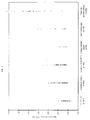
- FIGS. 1 and 2 The results of the respective examinations are shown in FIGS. 1 and 2.
- the symbol n stands for the number of samples examined in each case.
- the method according to the invention which is based on the detection of anti-calreticulin autoantibodies, is excellently suitable for the determination of autoimmune phenomena.
- Various autoimmune liver diseases and inflammatory bowel diseases, in particular AIH, PBC and Crohn's disease can be clearly diagnosed with the method according to the invention.
- the correlation between the detection of anti-calreticulin autoantibodies and the presence of the bacterium Yersinia enterocolitica is also noteworthy. This finding shows that the method according to the invention is additionally excellently suitable for the diagnosis of infectious diseases which lead to autoimmune phenomena.
- the invention provides a method for determining autoimmune phenomena, in which: the calreticulin protein is bound to a solid phase, the calreticulin bound to the solid phase is incubated with a liquid comprising components of the material to be examined, and any autoantibodies to calreticulin that may be present are detected.
Abstract
Description
Die vorliegende Erfindung bezieht sich auf ein Verfahren zur Bestimmung von Autoimmunphänomenen. Mit diesem Verfahren können Autoimmunerkrankungen im allgemeinen sowie autoimmune Lebererkrankungen und chronisch entzündliche Darmerkrankungen im besonderen diagnostiziert werden.The present invention relates to a method for determining autoimmune phenomena. This procedure can be used to diagnose autoimmune diseases in general and autoimmune liver diseases and inflammatory bowel diseases in particular.
Autoimmungkrankheiten (synonym: Autoaggressionskrankheiten) sind Erkrankungen, an deren Parthogenese eine Autosensibilisierung eine entscheidende Rolle spielt.Autoimmune diseases (synonym: autoaggressive diseases) are diseases in whose parthogenesis auto-sensitization plays a crucial role.
Beispiele für Autoimmunkrankheiten sind: autoimmunhämolytische Anämie, Hashimoto-Thyreoiditis, Typ-A-Gastritis mit perniziöser Anämie, juveniler Diabetes mellitus, die meisten Formen der Nebenniereninsuffizienz (Morbus Addison), vermutlich die multiple Sklerose, die rheumatoide Arthritis, die sogenannten Kollagenosen (systemischer Lupus erythematodes, Sklerodermie, Panarteriitis nodosa u.a.m.) und die autoimmunen Lebererkrankungen (insbesondere die autoimmune chronisch aktive Hepatitis). Bei mehreren Erkrankungen ergaben sich Hinweise darauf, daß im Gefolge einer Infektion mit Bakterien oder Viren sekundär eine Autoimmunkrankheit entsteht, wie z.B. bei der postinfektiösen Arthritis, der Poststreptokokken-Glomerulonephritis, beim rheumatischen Fieber Streptokokken), der Riesenzellhepatitis des Erwachsenen (Paramyxoviren).Examples of autoimmune diseases are: autoimmune hemolytic anemia, Hashimoto's thyroiditis, type A gastritis with pernicious anemia, juvenile diabetes mellitus, most forms of adrenal insufficiency (Addison's disease), presumably multiple sclerosis, rheumatoid arthritis, the so-called collupusosis (system erythematosus, scleroderma, panarteritis nodosa, etc.) and autoimmune liver diseases (especially autoimmune chronic active hepatitis). There were indications in several diseases that secondary to an infection with bacteria or viruses an autoimmune disease develops, e.g. in post-infectious arthritis, post-streptococcal glomerulonephritis, in rheumatic fever streptococci), giant cell hepatitis in adults (paramyxoviruses).
Diese Autosensibilisierung kann sich auf die zellvermittelte Immunreaktion (T-Lymphozyten) oder auf die humorale Immunität (B-Lymphozyten, Produktion von Antikörpern) erstrecken. Wie es zum Auftreten von Autoimmunphänomenen kommt, ist noch nicht eindeutig geklärt. Man vermutet, daß am Beginn der Autoimmunität eine Veränderung körpereigener Substanzen, meist Proteine, steht, die dann von dem Immunsystem als körperfremd betrachtet und attackiert werden. In Zusammenhang mit postinfektiösen Autoimmunphänomenen wird angenommen, daß vom Immunsystem Bestandteile von Bakterien oder Viren als körperfremd erkannt werden, woraufhin es zur Aktivierung des Immunsystems kommt. Die dann aktivierten T-Lymphozyten bzw. die dann gebildeten Antikörper, die primär gegen die Erreger gerichtet sind, erkennen aber auch strukturell ähnliche Proteine des Menschen ("Molecular Mimikry").This auto-sensitization can extend to the cell-mediated immune response (T lymphocytes) or to humoral immunity (B lymphocytes, production of antibodies). How autoimmune phenomena occur has not yet been clearly clarified. It is believed that at the beginning of autoimmunity there is a change in the body's own substances, mostly proteins, which are then viewed and attacked by the immune system as foreign to the body. In context with post-infectious autoimmune phenomena it is assumed that components of bacteria or viruses are recognized by the immune system as foreign to the body, whereupon the immune system is activated. The then activated T lymphocytes or the antibodies that are then formed, which are primarily directed against the pathogens, but also recognize structurally similar human proteins ("molecular mimicry").
Wenn Autoimmunphänomene bei einer Erkrankung nachgewiesen werden, bedeutet dies allerdings noch nicht unbedingt, daß sie auch am Zustandekommen der Erkrankung ursächlich beteiligt sind. Sie können auch Begleitphänomen ("Epiphänomen") sein. Unabhängig davon, ob ein Autoimmunphänomen nur ein Begleitphänomen ist oder ob es ursächlich am Zustandekommen der Krankheit beteiligt ist, besitzt der Nachweis z.B. von speziellen Autoantikörpern sehr oft einen hohen diagnostischen Wert, oder er kann als Verlaufparameter einer Krankheit verwendet werden.However, if autoimmune phenomena are detected in a disease, this does not necessarily mean that they are also causally involved in causing the disease. They can also be an accompanying phenomenon ("epiphenomenon"). Regardless of whether an autoimmune phenomenon is only an accompanying phenomenon or whether it is causally involved in causing the disease, the evidence has e.g. of special autoantibodies very often have a high diagnostic value, or it can be used as a course parameter of a disease.
Im Bereich der Gastroenterologie und der Hepatologie spielen die autoimmunen Lebererkrankungen eine große Rolle. Die häufigsten Ursachen einer chronischen Lebererkrankung sind die Schädigung durch Alkohol oder einer chronischen Infektion durch Hepatitis Viren (Hepatitis-B-Virus; Hepatitis-C-Virus, Hepatitis-Delta-Virus). Bei der Abklärung eines Leberschadens müssen aber zusätzlich die sogenannten autoimmunen Lebererkrankungen in Betracht gezogen werden. Dies sind: 1. die autoimmune chronisch aktive Hepatitis (AIH), 2. die primär biliäre Zirrhose (PBC), 3. die primär sklerosierende Cholangitis (PSC), und 4. noch weitere nicht eindeutig klassifizierte autoimmune Leberkrankheiten.Autoimmune liver diseases play a major role in the field of gastroenterology and hepatology. The most common causes of chronic liver disease are alcohol damage or chronic hepatitis virus infection (hepatitis B virus; hepatitis C virus, hepatitis delta virus). When diagnosing liver damage, however, the so-called autoimmune liver diseases must also be taken into account. These are: 1. Autoimmune chronic active hepatitis (AIH), 2. Primary biliary cirrhosis (PBC), 3. Primary sclerosing cholangitis (PSC), and 4. Still other unclearly classified autoimmune liver diseases.
Bisher sind in der klinischen Routine zahlreiche Autoantikörper zur Untersuchung von Autoimmunphänomenen bei autoimmunen Lebererkrankungen und chronisch entzündlichen Darmerkrankungen verwendet worden. Hierzu wurden folgende Autoantikörper eingesetzt:
- ANA =
- Antinukleäre Antikörper
- SMA =
- Antikörper gegen glatte Muskulatur
- LKM-1-Antikörper =
- Liver-Kidney-Membrane Antibodies
- AMA =
- Antimitochondriale Antikörper, mehrere Subtypen sind bekannt
- SLA =
- Antikörper gegen das "Soluble liver antigen"
- ASGPR-Antikörper =
- Antikörper gegen den Asialoglykoproteinrezeptor
- Anti-GOR-Antikörper pANCA =
- perinukleäre Antineutrophilenzytoplasma-Antikörper
- ANA =
- Antinuclear antibodies
- SMA =
- Antibodies against smooth muscles
- LKM-1 antibody
- Liver-Kidney-Membrane Antibodies
- AMA =
- Antimitochondrial antibodies, several subtypes are known
- SLA =
- Antibodies against the "Soluble liver antigen"
- ASGPR antibody
- Antibodies against the asialoglycoprotein receptor
- Anti-GOR antibody pANCA =
- perinuclear antineutrophil cytoplasmic antibodies
Bisher einzeln verwendete Autoantikörper ließen sich häufig nur für ganz bestimmte Autoimmunerkrankungen einsetzen. Um ein eindeutiges Krankheitsbild zu diagnostizieren, mußten daher in der Regel ein ganzes Spektrum von verschiedenen Autoantikörpern getestet werden, was einen relativ hohen Aufwand erfordert und längere Zeit in Anspruch nimmt.Autoantibodies previously used individually can often only be used for very specific autoimmune diseases. In order to diagnose a clear clinical picture, a whole spectrum of different autoantibodies had to be tested, which requires a relatively high effort and takes a long time.
Es bestand daher die Aufgabe der vorliegenden Erfindung, ein Verfahren zur Bestimmung von Autoimmunphänomenen bereitzustellen, mit dem die diagnostische Aussage, insbesondere beim Auftreten von autoimmunen Lebererkrankungen, verbessert werden kann.It was therefore the object of the present invention to provide a method for determining autoimmune phenomena by means of which the diagnostic information, in particular when autoimmune liver diseases occur, can be improved.
Die Aufgabe wird gelöst durch ein Verfahren zur Bestimmung von Autoimmunphänomenen, welches durch folgende Schritte gekennzeichnet ist:
- Bindung des Proteins Calreticulin an eine festen Phase,
- Inkubation des an die feste Phase gebundenen Calreticulins mit einer Flüssigkeit, die Bestandteile des zu untersuchenden Materials umfaßt, und
- Nachweis von gegebenenfalls vorhandenen Autoantikörpern gegen Calreticulin.
- Binding of the protein calreticulin to a solid phase,
- Incubation of the calreticulin bound to the solid phase with a liquid comprising components of the material to be examined, and
- Detection of autoantibodies against calreticulin, if present.
Es wurde im Rahmen der vorliegenden Erfindung gefunden, daß das Protein Calreticulin ausgezeichnet als Marker zur Diagnostik von Autoimmunphänomenen, insbesondere bestimmter autoimmuner Lebererkrankungen und chronisch entzündlicher Darmerkrankungen, geeignet ist.It has been found in the context of the present invention that the protein calreticulin is excellent as a marker for the diagnosis of autoimmune phenomena, in particular certain ones autoimmune liver disease and inflammatory bowel disease.
Calreticulin ist an sich bekannt als ein im Körper von Organismen vorkommendes Protein des endoplasmatischen Retikulums zahlreicher Zellen (Skelettmuskelzelle, glatte Muskelzellen, Herzmuskel, Hepatozyten u.a.m.), s. Übersichtsartikel von M. Michalak et al. in Biochem. J. 285, S. 681-692 (1992). Es ist das wichtigste Calcium-bindende Protein dieser Zellorganellen. Darüber hinaus kommt dieses Protein im Zellkern und in der Kernmembran vor. Neben der Calcium-Bindung werden diesem Protein weitere Funktionen zugeschrieben: Bindung von Zink, Protein-Protein-Interaktionen nach Art der sogenannten Chaperonine, Beteiligung an der Eisenresorption im Dünndarm, Bindung an Steroidhormonrezeptoren. Eine Rolle von Calreticulin an autoimmunen Erkrankungen ist zwar postuliert worden, und zwar aufgrund von Spekulationen, daß Calreticulin dem sogenannten Ro/SSA-Autoantigen entspräche, welches beim Krankheitsbild der systemischen Lupus erythematodes als Autoantigen identifiziert wurde (s. den oben bezeichneten Übersichtsartikel von M. Michalak et al.). Diese Spekulationen wurden daraufhin jedoch experimentell widerlegt mit den Arbeiten von J. Lu et al., veröffentlicht in Clin. Exp. Immunol. 94, S. 429-434 (1993), die gezeigt haben, daß ein Calretikulin-artiges Protein nicht mit Blutseren von Autoimmunerkrankten reagieren. Der zuvor gehegte Verdacht, daß Calreticulin in einem Zusammenhang mit der systemischen Lupus erythematodes steht, wurde auf einen Artefakt zurückgeführt, der sich dadurch ergab, daß Calretikulin mit dem Ro/SSA-Autoantigen mitgereinigt wurde.Calreticulin is known per se as a protein of the endoplasmic reticulum of numerous cells (skeletal muscle cell, smooth muscle cell, heart muscle, hepatocyte, etc.) that occurs in the body of organisms, see. Review article by M. Michalak et al. in Biochem. J. 285, pp. 681-692 (1992). It is the most important calcium-binding protein of these cell organelles. This protein is also found in the cell nucleus and in the nuclear membrane. In addition to calcium binding, other functions are ascribed to this protein: binding of zinc, protein-protein interactions like chaperonins, participation in iron absorption in the small intestine, binding to steroid hormone receptors. A role of calreticulin in autoimmune diseases has been postulated due to speculation that calreticulin corresponds to the so-called Ro / SSA autoantigen, which was identified as autoantigen in the clinical picture of systemic lupus erythematosus (see the review article by M. described above) Michalak et al.). However, these speculations were then experimentally refuted with the work of J. Lu et al., Published in Clin. Exp. Immunol. 94, pp. 429-434 (1993), which have shown that a calreticulin-like protein does not react with blood sera from autoimmune patients. The previous suspicion that calreticulin was associated with systemic lupus erythematosus was attributed to an artifact that resulted from the fact that calreticulin was also purified with the Ro / SSA autoantigen.
Eine besonders bevorzugte Ausführungsform der vorliegenden Erfindung besteht darin, daß als Calreticulin natives, nicht denaturiertes, humanes Calreticulin verwendet wird. Besonders geeignet ist das native Calreticulin, welches aus der menschlichen Leber isoliert wurde. Sehr gute Ergebnisse wurden mit einem Calreticulin-Ausgangsmaterial erzielt, welches auf die untenstehend beschriebene Weise aus der menschlichen Leber isoliert wurde. Es wird angenommen, daß durch dieses Isolierungsverfahren die Autoantigen-Epitope des Calreticulins für die Reaktionen des Auto-Antikörpers gegen Calreticulin besonders gut präsentiert wird, was sich vorteilhaft sowohl auf die Spezifität als auch auf die Sensitivität des Autoantikörper-Nachweises gemäß dem erfindungsgemäßen Verfahren auswirkt.A particularly preferred embodiment of the present invention is that native, undenatured, human calreticulin is used as calreticulin. The native calreticulin, which was isolated from the human liver, is particularly suitable. Very good results have been obtained with a calreticulin starting material which is obtained from the human liver was isolated. It is assumed that the autoantigen epitope of calreticulin for the reactions of the autoantibody against calreticulin is particularly well presented by this isolation method, which has an advantageous effect on both the specificity and the sensitivity of the autoantibody detection according to the method according to the invention.
Es wurde überraschend gefunden, daß natives, nicht denaturiertes, humanes Calreticulin die Bestimmung von Autoimmunphänomenen, insbesondere durch die Erzielung höherer Testempfindlichkeiten sowie spezifischerer Antigenreaktionen, verbesserten gegenüber einem Verfahren, bei dem gentechnologisch hergestelltes Calreticulin, aus Zellkulturen isoliertes Calreticulin oder Calreticulin aus nicht-humanen Zellen oder Zellkulturen eingesetzt wurden. Vor allem ermöglicht gerade die Verwendung von nativen, nicht denaturiertem, humanem Calreticulin als Ausgangsmaterial, welches als Antigen zur Nachweisreaktion mit dem vermuteten Calreticulin-Autoantikörper eingesetzt wird, einen quantitativen Nachweis durch das an sich bekannte ELISA-Verfahren. Die bisher in der klinischen Routinediagnostik untersuchten Autoimmunphänomene basieren lediglich auf einem qualitativen Nachweis der Antikörper.It was surprisingly found that native, undenatured, human calreticulin improved the determination of autoimmune phenomena, in particular by achieving higher test sensitivities and more specific antigen reactions, compared to a method in which genetically engineered calreticulin, calreticulin isolated from cell cultures or calreticulin from non-human cells or cell cultures were used. Above all, the use of native, undenatured, human calreticulin as the starting material, which is used as an antigen for the detection reaction with the suspected calreticulin autoantibody, enables quantitative detection by the known ELISA method. The autoimmune phenomena previously investigated in routine clinical diagnosis are based only on a qualitative detection of the antibodies.
Eigene Untersuchungen haben gezeigt, daß Calreticulin-Ausgangsmaterialien, die nicht in Form eines nativen, nicht denaturierten, humanen Calreticulins vorlagen, nicht über das ELISA-Verfahren nachweisbar waren. Der durch die vorliegende Erfindung erst ermöglichte ELISA-Nachweistest kann demnach im Gegensatz zu herkömmlichen Testverfahren zur Diagnose von autoimmunen Lebererkrankungen sowie chronischen Darmerkrankungen eine quantifizierbare Meßgröße liefern. Dies verbessert selbstverständlich die Aussagekraft bei der Diagnostik von Autoimmunphänomenen unterschiedlichster Art. Das Prinzip der ELISA-Nachweisreaktion ist beispielsweise beschrieben in Kemeny und Challacombe: "ELISA and other solid phase immunoassay. Theoretical and practical aspects", Chichester: Wiley (1988).Our own investigations have shown that calreticulin starting materials which were not in the form of a native, undenatured, human calreticulin could not be detected using the ELISA method. In contrast to conventional test methods for the diagnosis of autoimmune liver diseases and chronic intestinal diseases, the ELISA detection test made possible by the present invention can therefore provide a quantifiable measurement variable. Of course, this improves the informative value in the diagnosis of various types of autoimmune phenomena. The principle of the ELISA detection reaction is described, for example, in Kemeny and Challacombe: "ELISA and other solid phase immunoassay. Theoretical and practical aspects", Chichester: Wiley (1988).
Im Verfahren zur Bestimmung von Autoimmunphänomenen gemäß der vorliegenden Erfindung wird zunächst Calreticulin, welches wie oben beschrieben vorzugsweise in Form eines nativen, nicht denaturierten, humanen Calreticulin vorliegt, an eine feste Phase auf eine an sich bekannte Art und Weise gebunden. Hierzu ist beispielsweise die Adsorption des Proteins an ein polymeres Trägermaterial, beispielsweise Polystyrol, geeignet. Günstig sind hier bereits vorgefertigte Mikrotiter-Testplatten, die eine bestimmte Anzahl von Vertiefungen (in der Regel 96 sogenannte "Wells") aufweisen. Im Anschluß an die Bindung von Calreticulin ist es zur Verminderung unspezifischer Reaktionen günstig, noch freie Bindungsstellen des polymeren Festphasenträgers abzusättigen, beispielsweise durch Behandlung mit einem inerten Trägerprotein wie Humanserumalbumin.In the method for determining autoimmune phenomena according to the present invention, calreticulin, which, as described above is preferably in the form of a native, undenatured, human calreticulin, is first bound to a solid phase in a manner known per se. For this purpose, for example, the adsorption of the protein on a polymeric carrier material, for example polystyrene, is suitable. Pre-fabricated microtiter test plates with a certain number of wells (usually 96 so-called "wells") are favorable here. Following the binding of calreticulin, in order to reduce non-specific reactions, it is expedient to saturate free binding sites of the polymeric solid phase support, for example by treatment with an inert carrier protein such as human serum albumin.
Anschließend wird die so vorbehandelte feste Phase, an dem das Calreticulin gebunden ist, mit einer Flüssigkeit inkubiert, die Bestandteile des zu untersuchenden Materials umfaßt. D.h., eine zur Untersuchung bestimmte Probe wird dem Körper des Probanden entnommen und in eine zur Inkubation geeignete Flüssigkeitsform gebracht. Wegen der Einfachheit der Durchführung sind als Ausgangsmaterialien besonders Blutserum oder Urin geeignet, wobei diese Körperflüssigkeiten zur Untersuchung in der Regel auf eine geeignete Verdünnung gebracht werden. Gut nachweisbare Autoantikörper-Titer ergaben sich auch, wenn Ascites als Ausgangsmaterial für das erfindungsgemäße Verfahren eingesetzt wurde.The solid phase pretreated in this way, to which the calreticulin is bound, is incubated with a liquid which comprises constituents of the material to be examined. That is, a sample intended for examination is taken from the subject's body and placed in a liquid form suitable for incubation. Because of the simplicity of implementation, blood serum or urine are particularly suitable as starting materials, these body fluids usually being diluted for examination. Well-detectable autoantibody titers were also obtained when ascites was used as the starting material for the method according to the invention.
Im Anschluß an diese Inkubation erfolgt der Nachweis der gegebenenfalls vorhandenen Autoantikörper gegen Calreticulin. In diesem Schritt wird untersucht, ob, und wenn, wieviel, vermutete Autoantikörper gegen Calreticulin in der zu untersuchenden Probe in einer Antikörper-Antigen-Reaktion mit dem an der festen Phase gebundenen Antigen-Liganden, nämlich dem gebundenen Calreticulin, reagiert hat.Following this incubation, any autoantibodies against calreticulin are detected. In this step, it is investigated whether, and if so how much, suspected autoantibodies against calreticulin in the sample to be examined reacted in an antibody-antigen reaction with the antigen ligand bound to the solid phase, namely the bound calreticulin.
Für diese Nachweisreaktionen sind jegliche bekannte Arten von immunchemischen Nachweissystemen verwendbar, z.B. das Radioimmunassay, das Fluoroimmunassay, das Enzymimmunassay usw.. Besonders gut geeignet, da auf einfache Weise quantifizierbar und in Verbindung mit dem bevorzugt eingesetzten nativen, nicht denaturierten, humanen Calreticulin eine hohe Sensitivität und Spezifität bewirkend, ist hierbei das ELISA-Verfahren, wie oben beschrieben.Any known types of immunochemical detection systems can be used for these detection reactions, e.g. the radioimmunoassay, the fluoroimmunassay, the enzyme immunoassay, etc. Particularly suitable, since it can be quantified in a simple manner and, in conjunction with the preferred, undenatured, human calreticulin used, it has a high sensitivity and specificity, the ELISA method is as above described.
Das erfindungsgemäße Verfahren kann zur Untersuchung bzw. Diagnose jeglicher Autoimmunphänomenen eingesetzt werden, sei es bei solchen Zuständen, bei denen ein Autoimmunphänomen nur ein Begleitphänomen ("Epiphänomen") bestimmter Krankheiten ist, oder daß die Autoimmunphänomene ursächlich am Zustandekommen der Erkrankung beteiligt sind Einsatzgebiete für das erfindungsgemäße Verfahren sind beispielsweise: siehe oben die eingangs genannten Autoimmunerkrankungen.The method according to the invention can be used for the investigation or diagnosis of any autoimmune phenomena, be it in those conditions in which an autoimmune phenomenon is only an accompanying phenomenon ("epiphenomenon") of certain diseases, or that the autoimmune phenomena are causally involved in the occurrence of the disease Methods according to the invention are for example: see above the autoimmune diseases mentioned above.
Besondere Vorteile ergehen sich aus der Verwendung des erfindungsgemäßen Verfahrens zur Diagnose von autoimmunen Lebererkrankungen, wie der AIH, der PBC, der PSC und anderen autoimmunen Lebererkrankungen. Es wurde festgestellt, daß die häufigste chronische aktive autoimmune Hepatitis, die AIH Typ 1, mit dem erfindungsgemäßen Verfahren leichter und eindeutiger diagnostiziert werden kann, als es mit den herkömmlichen Testmethoden möglich ist. Aber auch bei der primär biliären Zirrhose (PBC) lassen sich Anti-CR-Antikörper gut nachweisen, auch wenn die Anti-CR-Antikörper bei diesem Krankheitsbild nicht in so hohen Konzentrationen wie bei der AIH Typ 1 vorkommen.Particular advantages result from the use of the method according to the invention for the diagnosis of autoimmune liver diseases, such as the AIH, the PBC, the PSC and other autoimmune liver diseases. It was found that the most common chronic active autoimmune hepatitis,
Unerwartet vorteilhafte Ergebnisse werden dadurch erzielt, daß der Nachweis der gegebenenfalls vorhandenen Auto-Antikörper gegen Calreticulin nicht wie üblich über Sekundär-Antikörper erfolgt, die polyvalent mit Humanantikörpern reagieren, also z.B. sowohl Antihuman-Antikörper vom IgG- als auch vom IgM-Typ einschließen. Vielmehr hat sich überraschend gezeigt, daß der Einsatz von Immuntyp-spezifischen Sekundär-Antikörpern eine weitere Differenzierung von Krankheiten zuläßt. So wird unter Einsatz von Anti-Calreticulin-Antikörpern vom speziellen IgG-Typ als Sekundär-Antikörper spezifisch das Vorliegen einer Autoimmunhepatitis (AIH) diagnostiziert, während unter Einsatz von Anti-Calreticulin-Antikörpern vom spezifischen IgM-Typ als Sekundär-Antikörper spezifisch das Vorliegen einer primär biliären Zirrhose (PBC) diagnostiziert wird. Dadurch können durch diese bevorzugte Weiterbildung des erfindungsgemäßen Verfahrens beispielsweise die unterschiedlichen autoimmunen Lebererkrankungen AIH und PBC differenziert werden.Unexpectedly advantageous results are achieved in that the detection of any autoantibodies against calreticulin which may be present is not carried out, as usual, via secondary antibodies which react polyvalently with human antibodies, that is to say, for example, include both anti-human antibodies of the IgG and of the IgM type. Rather, it has surprisingly been found that the use of immune-type-specific secondary antibodies further differentiates diseases allows. The use of anti-calreticulin antibodies of the special IgG type as secondary antibodies specifically diagnoses the presence of autoimmune hepatitis (AIH), while the use of anti-calreticulin antibodies of the specific IgM type as secondary antibodies specifically diagnoses the presence of autoimmune hepatitis is diagnosed with primary biliary cirrhosis (PBC). As a result, this preferred development of the method according to the invention can be used to differentiate, for example, the different autoimmune liver diseases AIH and PBC.
Die durch die Erfindung ermöglichte genaue Diagnose der Ursache der Leberschädigung hat für den Patienten gravierende Vorteile, da damit eine Differenzierung der jeweiligen Autoimmunerkrankung besser als mit den derzeit verfügbaren Tests möglich ist und gegeneinander abgrenzbar ist. Dies hat auch eine große therapeutische Bedeutung für den Patienten. Denn für die Art und Weise der Behandlung ist es wichtig, zu erkennen, worin sich die Lebererkrankung manifestiert, d.h. ob Virushepatitiden, eine medikamentöse Leberschädigung oder eine alkoholische Leberschädigung vorliegt. Die chronischen Autoimmunhepatitiden beispielsweise sind durch eine immunsuppressive Therapie meist gut zu behandeln. Demgegenüber ist bei der primär biliären Zirrhose die Therapie mit Ursodeoxycholsäure wesentlich erfolgversprechender. Bei der primär sklerosierenden Cholangitis kann der Verlauf eventuell durch eine Immunsuppression ebenfalls günstig beeinflußt werden, jedoch nur, wenn die Erkrankung möglichst frühzeitig diagnostiziert wird. Bedeutsam sind ferner die Autoimmunphänomene, die bei Virushepatitiden auftreten, besonders bei der Infektion mit dem Hepatitis-C-Virus, weniger bei der Infektion mit dem Hepatitis-B-Virus. Ein Beispiel für weitere, bisher jedoch nicht klassifizierte autoimmune Lebererkrankungen ist die autoimmune, AMA-negative Cholangitis.The precise diagnosis of the cause of the liver damage made possible by the invention has serious advantages for the patient, since differentiation of the respective autoimmune disease is better than is possible with the currently available tests and can be distinguished from one another. This is also of great therapeutic importance for the patient. Because for the type of treatment, it is important to recognize what is manifesting the liver disease, i.e. whether viral hepatitis, medicinal liver damage or alcoholic liver damage is present. Chronic autoimmune hepatitis, for example, is usually easy to treat with immunosuppressive therapy. In contrast, treatment with ursodeoxycholic acid is much more promising in primary biliary cirrhosis. In primary sclerosing cholangitis, the course can also be influenced favorably by immunosuppression, but only if the disease is diagnosed as early as possible. Also important are the autoimmune phenomena that occur in viral hepatitis, particularly in the case of infection with the hepatitis C virus, less so in the case of infection with the hepatitis B virus. An example of other, but not yet classified autoimmune liver diseases is autoimmune, AMA-negative cholangitis.
Im Rahmen der vorliegenden Erfindung hat sich ferner überraschend gezeigt, daß das oben beschriebene Verfahren hervorragend zur Abklärung und Diagnose von chronisch entzündlichen Darmerkrankungen beiträgt. Bei solchen chronisch entzündlichen Darmerkrankungen, worunter insbesondere das Krankheitsbild des Morbus Crohn und das der Colitis ulcerosa zählt, spielen Autoimmunphänomene ebenfalls eine Rolle. Titer von Anti-Calreticulin-Autoantikörpern konnten bei Morbus Crohn und bei Colitis ulcerosa durch das erfindungsgemäße Verfahren nachgewiesen werden. Somit ist das erfindungsgemäße Verfahren auch zur Differentialdiagnose entzündlicher oder infektiöser Darmerkrankungen gut geeignet.In the context of the present invention, it has also surprisingly been found that the method described above makes an excellent contribution to the clarification and diagnosis of inflammatory bowel diseases. In such Autoimmune phenomena also play a role in inflammatory bowel diseases, which include, in particular, the clinical picture of Crohn's disease and that of ulcerative colitis. The titer of anti-calreticulin autoantibodies in Crohn's disease and in ulcerative colitis could be detected by the method according to the invention. The method according to the invention is therefore also well suited for the differential diagnosis of inflammatory or infectious bowel diseases.
Ein weiteres überraschendes Ergebnis der vorliegenden Erfindung besteht darin, daß das Auftreten von Anti-Calreticulin-Autoantikörpern, welches durch das erfindungsgemäße Verfahren bestimmt werden kann, hervorragend korreliert mit dem Vorhandensein des Bakteriums Yersinia enterocolitica im Körper des Patienten. Somit kann das erfindungsgemäße Verfahren besonders gut ebenfalls zur einfachen Diagnose der Yersiniose verwendet werden.Another surprising result of the present invention is that the occurrence of anti-calreticulin autoantibodies, which can be determined by the method according to the invention, correlates excellently with the presence of the bacterium Yersinia enterocolitica in the patient's body. The method according to the invention can thus also be used particularly well for simple diagnosis of yersiniosis.
Die Erfindung wird nachstehend anhand von Beispielen unter Bezugnahme auf die anbeiliegenden Figuren näher erläutert, wobei
- Fig. 1 die erhaltenen Ergebnisse des Verfahrens zur Bestimmung von Autoimmunphänomenen zur Diagnose verschiedener autoimmuner Lebererkrankungen, wie der AIH, der PBC und der PSC, und
- Fig. 2 die Ergebnisse des erfindungsgemäßen Verfahrens zur Bestimmung von Autoimmunphänomenen zur Diagnose verschiedentlicher chronisch entzündlicher Darmerkrankungen, wie der ulzerativen Colitis und dem Morbus Crohn sowie die Korrelation des Nachweises von Anti-Calreticulin-Autoantikörpern mit dem Vorhandensein verschiedener Bakterienspezies zeigen.
- 1 shows the results obtained from the method for determining autoimmune phenomena for diagnosing various autoimmune liver diseases, such as the AIH, the PBC and the PSC, and
- 2 shows the results of the method according to the invention for determining autoimmune phenomena for the diagnosis of various inflammatory bowel diseases, such as ulcerative colitis and Crohn's disease, and the correlation of the detection of anti-calreticulin autoantibodies with the presence of different bacterial species.
-
0,2 M PMSF (Phenylmethansulfonylfluorid)
3,484 g in 100 Äthanol0.2 M PMSF (phenylmethanesulfonyl fluoride)
3.484 g in 100 ethanol -
2 M Benzamidiniumchlorid
31,324 g in 1000 ml Äthanol2 M benzamidinium chloride
31.324 g in 1000 ml of ethanol - Aprotinin: 10 mg Aprotinin/ml H2OAprotinin: 10 mg aprotinin / ml H 2 O
- Leupeptin: 2 mg/ml H2OLeupeptin: 2 mg / ml H 2 O
-
50 mM NaCl
2,92 g in 1 l H2O50 mM NaCl
2.92 g in 1 L H 2 O -
1000 mM NaCl
58,44 g in 1 l H2O1000 mM NaCl
58.44 g in 1 L H 2 O
100 g Humanleber werden mit einer Schere kleingeschnitten. Es werden zugegeben:
- 400 ml Puffer I (4 ml pro g Leber)
- 300 µl Leupeptin-Lösung
- 133,3 µl Aprotinin-Lösung
- 250 µl PMSF-Lösung
- 100 µl Benzamidiniumchlorid-Lösung
- 100 µl Mercaptoäthanol
- 400 ml buffer I (4 ml per g liver)
- 300 µl leupeptin solution
- 133.3 µl aprotinin solution
- 250 µl PMSF solution
- 100 µl benzamidinium chloride solution
- 100 µl mercaptoethanol
Omni-Mixer (Fa. Sorvall) Stellung 10, in großen Metallbechern, in Eiswasser.
30 Sekunden homogenisieren, 1 Minute kühlen, erneut 30 Sekunden homogenisieren, 1 Minute kühlen.Omni-Mixer (Sorvall)
Homogenize for 30 seconds, cool for 1 minute, homogenize again for 30 seconds, cool for 1 minute.
Zentrifugation: 30 Minuten bei 9000 U/min in GSA-Rotor.Centrifugation: 30 minutes at 9000 rpm in a GSA rotor.
Überstand I = 360 ml, kühl stellenSupernatant I = 360 ml, refrigerate
Sediment mit der halben Menge des obigen Puffers einschließlich Zusätze versehen. Erneut homogenisieren wie oben und zentrifugieren wie oben. Ergibt Überstand II = 180 ml.Add half of the above buffer, including additives, to the sediment. Homogenize again as above and centrifuge as above. Results in supernatant II = 180 ml.
Überstand I und Überstand II werden vereinigt = 540 ml.Supernatant I and supernatant II are combined = 540 ml.
Zu diesen 540 ml werden zugegeben:
- 1,35 ml PMSF-Lösung
- 0,135 ml Benzamidiniumchlorid
- 0,1 ml Mercaptoäthanol
- 108,1 g (NH4)2SO4 in fester Form (entspricht 200 g Ammoniumsulfat pro 1 l Überstand)
- 1.35 ml PMSF solution
- 0.135 ml benzamidinium chloride
- 0.1 ml mercaptoethanol
- 108.1 g (NH 4 ) 2 SO 4 in solid form (corresponds to 200 g ammonium sulfate per 1 l of supernatant)
Ammoniumsulfat wird unter ständigem Rühren zugegeben. Der Becher steht im Eiswasser.Ammonium sulfate is added with constant stirring. The cup is in the ice water.
30 Minuten langsam rühren lassen.Let it stir slowly for 30 minutes.
Lösung mit ortho-Phosphorsäure (98%) auf pH 4,7 bringen.Bring the solution to pH 4.7 with orthophosphoric acid (98%).
Über Nacht im Eiswasser rühren lassen.Let it stir overnight in ice water.
30 Minuten bei 9000 U/min abzentrifugieren. GSA-Rotor.Spin down at 9000 rpm for 30 minutes. GSA rotor.
Jedes der Sedimente in je 20 ml Puffer II resuspendieren.Resuspend each of the sediments in 20 ml of Buffer II.
Dialyse gegen Puffer III über Nacht (Dialysemedium soll zweimal gewechselt werden).Dialysis against buffer III overnight (dialysis medium should be changed twice).
Zentrifugieren 10 Minuten bei 3000 U/min in SS 34-Rotor. Überstand auf DEAE-Säulen aufgetragen.Centrifuge for 10 minutes at 3000 rpm in SS 34 rotor. Supernatant applied to DEAE columns.
DEAE-Säule wird mit diesem Puffer äquilibriert. Die Probe wird aufgetragen, sie befindet sich bereits in diesem Puffer. Elution durch einen NaCl-Gradienten von 50 mM NaCl bis 1000 mM NaCl (in dem Puffer wie oben), Temperatur 4°C, 80 Tropfen pro Röhrchen.
Calreticulin wird bei etwa 300 mM NaCl eluiert.
Der Calreticulin-Gehalt der einzelnen Fraktionen wird durch SDS-PAGE überprüft. Calreticulin wird an seinem Molekulargewicht identifiziert (60 kDa). Zusätzlich wird eine Identifizierung durch Western-Blot mit Anti-Calreticulin-Antikörper durchgeführt. Die geeigneten Fraktionen werden vereinigt und gegen Hepes-Puffer dialysiert (50 mM Hepes pH 6,9 (mit NaOH eingestellt)).DEAE column is equilibrated with this buffer. The sample is applied, it is already in this buffer. Elution by a NaCl gradient from 50 mM NaCl to 1000 mM NaCl (in the buffer as above), temperature 4 ° C, 80 drops per tube.
Calreticulin is eluted at approximately 300 mM NaCl.
The calreticulin content of the individual fractions is checked by SDS-PAGE. Calreticulin is identified by its molecular weight (60 kDa). In addition, identification by Western blot with anti-calreticulin antibody is carried out. The appropriate fractions are pooled and dialyzed against Hepes buffer (50 mM Hepes pH 6.9 (adjusted with NaOH)).
- A: 10 mM Tris pH 7,2A: 10 mM Tris pH 7.2
- B: 10 mM Tris pH 7,2 + 1 M NaClB: 10mM Tris pH 7.2 + 1M NaCl
Die Proben der DEAE-Säulen-Chromatographie werden aufgetragen, die Säule wird mit Puffer A/D-Konverterteil gespült. Danach Elution durch einen NaCl-Gradienten (gemischt aus Puffer A/D-Konverterteil und Puffer B).
Der Calreticulin-Gehalt der einzelnen Fraktionen wird durch SDS-PAGE überprüft. Die entsprechenden Fraktionen werden vereinigt und gegen PBS dialysiert.The DEAE column chromatography samples are applied, the column is rinsed with buffer A / D converter part. Then elution through a NaCl gradient (mixed from buffer A / D converter part and buffer B).
The calreticulin content of the individual fractions is checked by SDS-PAGE. The appropriate fractions are pooled and dialyzed against PBS.
- 0,05 M Na-Phosphat pH 7,20.05 M Na phosphate pH 7.2
- 0,15 M NaCl0.15 M NaCl
Mit diesem Trennmaterial wird Calreticulin entsprechend seinem Molekulargewicht von den restlichen Proteinen abgetrennt. Überlicherweise werden einige Fraktionen erhalten, die reines Calreticulin enthalten. Der Calreticulin-Gehalt der einzelnen Fraktionen wird durch SDS-PAGE überprüft. Die Fraktionen, die reines Calreticulin enthalten, werden gepoolt und eingeengt. Der Proteingehalt wird nach der Methode von Lowry bestimmt.With this separation material, calreticulin is separated from the remaining proteins according to its molecular weight. Some fractions containing pure calreticulin are usually obtained. The calreticulin content of the individual fractions is checked by SDS-PAGE. The fractions containing pure calreticulin are pooled and concentrated. The protein content is determined using the Lowry method.
Es hat sich gezeigt, daß sich insbesondere die Ammoniumsulfat-Fällung positiv auf die späteren Ergebnisse bei der Durchführung des erfindungsgemäßen Verfahrens auswirkte. Offensichtlich wurde durch die Salzbildung das Calreticulin in seiner nativen Form derart stabilisiert, daß der Calreticulin-Autoantikörper das Autoantigen für die Antikörper-Antigen-Reaktion gut erkennen konnte.It has been shown that the ammonium sulfate precipitation in particular had a positive effect on the later results when carrying out the process according to the invention. Obviously, the salt formation of the calreticulin was stabilized in its native form in such a way that the calreticulin autoantibody could recognize the autoantigen for the antibody-antigen reaction.
Durchführung des Nachweises von Anti-Calreticulin-Autoantikörper mittels ELISACarrying out the detection of anti-calreticulin autoantibodies by means of ELISA
-
1. PBS
9,55 g PBS in 1 l Aqua bidest. lösen (entspricht 5 Tabletten)1. PBS
9.55 g PBS in 1 l aqua bidist. dissolve (corresponds to 5 tablets) -
2. PBS + Tween 20
0,5 ml Tween 20 in 1 l PBS lösen2. PBS + Tween 20
Dissolve 0.5 ml Tween 20 in 1 l PBS -
3. Coating buffer
1,59 g Na2CO3; 2,93 g NaHCO3; 0,20 g NaN3 in 1 Aqua bidest. lösen3. Coating buffer
1.59 g Na 2 CO 3 ; 2.93 g NaHCO 3 ; 0.20 g NaN 3 in 1 aqua bidist. to solve -
4. Blocking solution
1 g Humanalbumin in 100 ml PBS lösen (1%ige HA-PBS-Lösung)4. Blocking solution
Dissolve 1 g human albumin in 100 ml PBS (1% HA-PBS solution) -
5. Lösung zur Serumverdünnung
1 g Humanalbumin in 100 ml PBS + Tween 20 lösen (1%ige HA-PBS-Lösung + Tween 20)5. Serum dilution solution
Dissolve 1 g human albumin in 100 ml PBS + Tween 20 (1% HA-PBS solution + Tween 20)
-
1. Coaten mit Calreticulin
ELISA-Platte (96 wells, Flat bottom) mit Coating buffer spülen, darauf achte, daß alle wells benetzt sind, dann gut ausklopfen. Beschichten mit 1000 ng Protein/well: bei Proteinkonzentration 10 mg/ml Verdünnung 1:500: d.h. 20 µl Calreticulin-Lösung auf 10 ml Coating buffer. 50 µl/well. Inkubationsdauer: über Nacht bei 4°. Alternativ: 4 Stunden bei Raumtemperatur.1. Coating with calreticulin
Rinse ELISA plate (96 wells, flat bottom) with coating buffer, make sure that all wells are wetted, then tap well. Coating with 1000 ng protein / well: atprotein concentration 10 mg / ml dilution 1: 500: ie 20 µl calreticulin solution in 10 ml coating buffer. 50 µl / well. Incubation period: overnight at 4 °. Alternatively: 4 hours at room temperature. -
2. Blocken
Mit 1% (w/v) Humanalbumin in PBS. 100 µl/well, Platte abdecken. Dauer des Blockens: 1 Stunde bei Raumtemperatur.2. Block
With 1% (w / v) human albumin in PBS. 100 µl / well, cover plate. Blocking time: 1 hour at room temperature. -
3. Waschen
4x mit PBS-Tween, 150 µl/well, danach gut ausklopfen.3. Wash
4x with PBS-Tween, 150 µl / well, then tap well. -
4. Binden des Serum-Antikörpers
Serum in einzelnen Eppedorf-Reaktionsgefäßen in 1% Humanalbumin-PBS-Tween verdünnen: 1:250, 1:500, 1:750, 1:1000.- Blanks in Reihe 1 und 12.
- Menge: 50 µl/well.
- Dauer der Inkubation: 2 Stunden bei Raumtemperatur.
Dilute serum in individual eppedorf tubes in 1% human albumin PBS tween: 1: 250, 1: 500, 1: 750, 1: 1000.- Blanks in
rows 1 and 12. - Amount: 50 µl / well.
- Incubation time: 2 hours at room temperature.
-
5. Waschen
4x mit PBS-Tween, 150 µl/well, gut ausklopfen.5. Wash
Knock out 4x with PBS-Tween, 150 µl / well. - 6. Binden von peroxidase-gekoppeltem Anti-Human-IgG Anti-Human-IgG from sheep, linked with horseradish peroxidase (Fa. Amersham), Verdünnung 1:3000 in PBS-Tween (3 µl Anti-Human-IgG auf 9 ml PBS-Tween). Menge 50 µl/well. Inkubationsdauer: 1 Stunde bei Raumtemperatur, abdecken.6. Binding of peroxidase-coupled anti-human IgG anti-human IgG from sheep, linked with horseradish peroxidase (from Amersham), dilution 1: 3000 in PBS-Tween (3 μl anti-human IgG to 9 ml PBS -Tween). Amount 50 µl / well. Incubation period: 1 hour at room temperature, cover.
-
7. Waschen
4x mit PBS-Tween, 150 µl/well, gut ausklopfen.7. Wash
Knock out 4x with PBS-Tween, 150 µl / well. -
8. Substrat
TMP Peroxidase EIA Substrate kit, Fa. BioRad. Erst ganz kurz vor Gebrauch zusammenmischen.- Lösung A: 9 ml
- Lösung B: 1 ml
- sehr gut mischen
- Menge: 50 µl/well
- Inkubationsdauer: 8 Minuten bei Raumtemperatur.
TMP Peroxidase EIA Substrate kit, BioRad. Mix together just before use.- Solution A: 9 ml
- Solution B: 1 ml
- mix very well
- Amount: 50 µl / well
- Incubation period: 8 minutes at room temperature.
-
9. Stoppen
Abstoppen der Reaktion mit 2M H2SO4.
Menge: 50 µl/well.9. Stop
Stop the reaction with 2M H 2 SO 4 .
Amount: 50 µl / well. -
10. Ablesen
Extinktionsdifferenz bei 450 nm gemessen.10. Read
Absorbance difference measured at 450 nm.
- 1. Die Bindung des Proteins an die Matrix bedarf einer gewissen Zeit, um den Sättigungswert zu erreichen. Polystyrolplatten können etwa 400 ng Protein pro cm2 absorbieren. Um den Sättigungsprozeß zu beschleunigen, wurde eine höhere Proteinkonzentration von 1000 ng Calreticulin/well gewählt.1. The binding of the protein to the matrix takes a certain time to reach the saturation value. Polystyrene plates can absorb about 400 ng of protein per cm 2 . In order to accelerate the saturation process, a higher protein concentration of 1000 ng calreticulin / well was chosen.
- 2. Die so gecoateten Platten werden über Nacht bei 4°C inkubiert. Unter diesen Bedingungen kann man davon ausgehen, daß eine Sättigung der Matrix erreicht wird. Um aber auszuschließen, daß eventuell noch frei gebliebene Bindungsstellen unspezifisch binden, wird im Anschluß an diesen Schritt mit einem Protein inkubiert, das an die Matrix bindet, den Test aber nicht stört. In diesem Fall werden die Platten eine Stunde lang mit einer 1% Humanalbuminlösung in PBS inkubiert.2. The plates coated in this way are incubated at 4 ° C. overnight. Under these conditions it can be assumed that the matrix is saturated. However, in order to rule out that binding sites which may still remain free bind non-specifically, the step followed by incubation with a protein which binds to the matrix but does not interfere with the test. In this case the plates are incubated for one hour with a 1% human albumin solution in PBS.
- 3. Die Seren wurden in Verdünnungen von 1:100 bis 1:2000 gemessen. In mehreren Verdünnungsreihen erwiesen sich die Verdünnungen 1:250, 1:500, 1:750 und 1:1000 als am günstigsten. Es werden jeweils Doppelbestimmungen durchgeführt. Bei jeder Platte wird eine Negativkontrolle mitgemessen und eine Positivkontrolle. Als Positivkontrolle diente Serum einer Patientin mit systemischem Lupus erythematodes, das Anti-Ro/SS-A-positiv war.3. The sera were measured in dilutions of 1: 100 to 1: 2000. In several dilution series, the dilutions 1: 250, 1: 500, 1: 750 and 1: 1000 proved to be the cheapest. Duplicate determinations are carried out in each case. A negative control and a positive control are measured for each plate. Serum from a patient with systemic lupus erythematosus who was anti-Ro / SS-A positive served as a positive control.
- 4. Als Substrat wurde einerseits 1,2-Phenylendiamin, gelöst in Phosphatpuffer pH 5,6, verwendet. Da sich die Herstellung dieser Lösung sowie deren weitere Verarbeitung jedoch als zusätzliche Fehlerquellen erwiesen, wurde auf das standardisierte TMB-Peroxidase-EIA-Substrate-Kit der Fa. BioRad zurückgegriffen. Als optimale Reaktionszeit wurde eine Inkubationsdauer von 8 Minuten ermittelt.4. On the one hand, 1,2-phenylenediamine, dissolved in phosphate buffer pH 5.6, was used as the substrate. However, since the production of this solution and its further processing prove to be additional sources of error proven, the standardized TMB peroxidase EIA substrate kit from BioRad was used. An incubation period of 8 minutes was determined as the optimal reaction time.
Der Nachweis von Anti-Calreticulin-Autoantikörpern wurde analog Beispiel 2 durchgeführt, wobei lediglich der Schritt 4 in der Vorgehensweise des Beispiels 2 sich jeweils unterschied durch Verwendung verschiedener, zur Untersuchung dienender Serumproben. Folgende Materialproben wurden zum Nachweis des Vorliegens des vermuteten Anti-Calreticulin-Autoantikörpers eingesetzt: Kontrollseren sowie Serumproben von Patienten, bei denen durch herkömmliche Diagnose folgende Krankheitsbilder festgestellt wurden: AIH, PBC, PSC, ulzerative Colitis, Morbus Crohn sowie Infektionen mit Camphylobakter jejuni, Salmonella spp. sowie Yersinia enterocolitica.The detection of anti-calreticulin autoantibodies was carried out analogously to example 2, only step 4 in the procedure of example 2 differing in each case by using different serum samples which are used for the analysis. The following material samples were used to demonstrate the presence of the suspected anti-calreticulin autoantibody: control sera and serum samples from patients in whom the following clinical pictures were found by conventional diagnosis: AIH, PBC, PSC, ulcerative colitis, Crohn's disease and infections with camphylobacter jejuni, Salmonella spp. and Yersinia enterocolitica.
Die Ergebnisse der jeweiligen Untersuchungen sind in den Figuren 1 und 2 dargestellt. Das Symbol n steht für die Anzahl der jeweils untersuchten Proben.The results of the respective examinations are shown in FIGS. 1 and 2. The symbol n stands for the number of samples examined in each case.
Wie die Ergebnisse der Figuren 1 und 2 zeigen, ist das erfindungsgemäße Verfahren, das auf dem Nachweis von Anti-Calreticulin-Autoantikörpern basiert, zur Bestimmung von Autoimmunphänomenen ausgezeichnet geeignet. Verschiedene autoimmune Lebererkrankungen und chronisch entzündliche Darmerkrankungen, insbesondere die AIH, die PBC und das Morbus Crohn, können mit dem erfindungsgemäßen Verfahren eindeutig diagnostiziert werden. Bemerkenswert ist auch die Korrelation des Nachweises von Anti-Calreticulin-Autoantikörpern mit dem Vorliegen des Bakteriums Yersinia enterocolitica. Dieser Befund zeigt, daß das erfindungsgemäße Verfahren zusätzlich ausgezeichnet zur Diagnose von Infektionserkrankungen, die zu Autoimmunphänomenen führen, geeignet ist.As the results of FIGS. 1 and 2 show, the method according to the invention, which is based on the detection of anti-calreticulin autoantibodies, is excellently suitable for the determination of autoimmune phenomena. Various autoimmune liver diseases and inflammatory bowel diseases, in particular AIH, PBC and Crohn's disease, can be clearly diagnosed with the method according to the invention. The correlation between the detection of anti-calreticulin autoantibodies and the presence of the bacterium Yersinia enterocolitica is also noteworthy. This finding shows that the method according to the invention is additionally excellently suitable for the diagnosis of infectious diseases which lead to autoimmune phenomena.
Die Erfindung stellt ein Verfahren zur Bestimmung von Autoimmunphänomenen zur Verfügung, bei dem:
das Protein Calreticulin an eine feste Phase gebunden wird, das an die feste Phase gebundene Calreticulin mit einer Flüssigkeit inkubiert wird, die Bestandteile des zu untersuchenden Materials umfaßt, und
gegebenenfalls vorhandener Autoantikörper gegen Calreticulin nachgewiesen wird.The invention provides a method for determining autoimmune phenomena, in which:
the calreticulin protein is bound to a solid phase, the calreticulin bound to the solid phase is incubated with a liquid comprising components of the material to be examined, and
any autoantibodies to calreticulin that may be present are detected.
Claims (14)
Applications Claiming Priority (2)
| Application Number | Priority Date | Filing Date | Title |
|---|---|---|---|
| DE1995134348 DE19534348C2 (en) | 1995-09-15 | 1995-09-15 | Method for the determination of autoimmune phenomena |
| DE19534348 | 1995-09-15 |
Publications (1)
| Publication Number | Publication Date |
|---|---|
| EP0763740A1 true EP0763740A1 (en) | 1997-03-19 |
Family
ID=7772334
Family Applications (1)
| Application Number | Title | Priority Date | Filing Date |
|---|---|---|---|
| EP96114749A Withdrawn EP0763740A1 (en) | 1995-09-15 | 1996-09-13 | Method for determining autoimmune phenomena |
Country Status (2)
| Country | Link |
|---|---|
| EP (1) | EP0763740A1 (en) |
| DE (1) | DE19534348C2 (en) |
Cited By (7)
| Publication number | Priority date | Publication date | Assignee | Title |
|---|---|---|---|---|
| WO1999009413A1 (en) * | 1997-08-18 | 1999-02-25 | Arris Pharmaceutical Corporation | Anti-tryptase detection as a diagnostic for inflammatory diseases |
| WO2003050541A2 (en) * | 2001-12-12 | 2003-06-19 | Wieland, Heinrich | Diagnosis of malaria by identifying autoantibodies that react against calreticulin |
| US8007781B2 (en) | 2000-08-03 | 2011-08-30 | The Johns Hopkins University | Molecular vaccine linking an endoplasmic reticulum chaperone polypeptide to an antigen |
| US8128922B2 (en) | 1999-10-20 | 2012-03-06 | Johns Hopkins University | Superior molecular vaccine linking the translocation domain of a bacterial toxin to an antigen |
| US9011866B2 (en) | 2005-01-06 | 2015-04-21 | The Johns Hopkins University | RNA interference that blocks expression of pro-apoptotic proteins potentiates immunity induced by DNA and transfected dendritic cell vaccines |
| US9085638B2 (en) | 2007-03-07 | 2015-07-21 | The Johns Hopkins University | DNA vaccine enhancement with MHC class II activators |
| US9701725B2 (en) | 2003-05-05 | 2017-07-11 | The Johns Hopkins University | Anti-cancer DNA vaccine employing plasmids encoding signal sequence, mutant oncoprotein antigen, and heat shock protein |
-
1995
- 1995-09-15 DE DE1995134348 patent/DE19534348C2/en not_active Expired - Fee Related
-
1996
- 1996-09-13 EP EP96114749A patent/EP0763740A1/en not_active Withdrawn
Non-Patent Citations (5)
| Title |
|---|
| A. BAHLER ET AL.: "Autoantibodies against calreticulin in patiens with autoimmune hepatitis.", GASTROENTEROLOGY, vol. 108, no. 4, May 1995 (1995-05-01), pages A1028, XP000613066 * |
| C. SPAMER ET AL.: "anti-calreticulin autoantibodies: a novel marker for autoimmune hepatitis?.", JOURNAL OF HEPATOLOGY, vol. 23, August 1995 (1995-08-01), pages 129, XP000613060 * |
| J. BOEHM ET AL.: "Systemic lupus erythematosus is associated with increased auto-antibody titers against calreticulin and Grp94, but calreticulin is not the Ro/SS-A antigen.", EUROPEAN JOURNAL OF CLINICAL INVESTIGATION, vol. 24, 1994, pages 248 - 257, XP000613064 * |
| T. YOKOI ET AL.: "Identification of protein disulfide isomerase and calreticulin as autoimmune antigens in LEC strain of rats.", BIOCHIMICA ET BIOPHYSICA ACTA, vol. 1158, 1993, pages 339 - 344, XP000613068 * |
| W. KREISEL ET AL.: "Calreticulin a new target for circulating autoantibodies in Crohn's disease?", GASTROENTEROLOGY, vol. 106, no. 4, May 1994 (1994-05-01), pages - A716, XP000613067 * |
Cited By (9)
| Publication number | Priority date | Publication date | Assignee | Title |
|---|---|---|---|---|
| WO1999009413A1 (en) * | 1997-08-18 | 1999-02-25 | Arris Pharmaceutical Corporation | Anti-tryptase detection as a diagnostic for inflammatory diseases |
| US8128922B2 (en) | 1999-10-20 | 2012-03-06 | Johns Hopkins University | Superior molecular vaccine linking the translocation domain of a bacterial toxin to an antigen |
| US9758551B2 (en) | 1999-10-20 | 2017-09-12 | The Johns Hopkins University | Superior molecular vaccine linking the translocation domain of a bacterial toxin to an antigen |
| US8007781B2 (en) | 2000-08-03 | 2011-08-30 | The Johns Hopkins University | Molecular vaccine linking an endoplasmic reticulum chaperone polypeptide to an antigen |
| WO2003050541A2 (en) * | 2001-12-12 | 2003-06-19 | Wieland, Heinrich | Diagnosis of malaria by identifying autoantibodies that react against calreticulin |
| WO2003050541A3 (en) * | 2001-12-12 | 2004-07-29 | Wieland Heinrich | Diagnosis of malaria by identifying autoantibodies that react against calreticulin |
| US9701725B2 (en) | 2003-05-05 | 2017-07-11 | The Johns Hopkins University | Anti-cancer DNA vaccine employing plasmids encoding signal sequence, mutant oncoprotein antigen, and heat shock protein |
| US9011866B2 (en) | 2005-01-06 | 2015-04-21 | The Johns Hopkins University | RNA interference that blocks expression of pro-apoptotic proteins potentiates immunity induced by DNA and transfected dendritic cell vaccines |
| US9085638B2 (en) | 2007-03-07 | 2015-07-21 | The Johns Hopkins University | DNA vaccine enhancement with MHC class II activators |
Also Published As
| Publication number | Publication date |
|---|---|
| DE19534348C2 (en) | 1997-07-10 |
| DE19534348A1 (en) | 1997-03-20 |
Similar Documents
| Publication | Publication Date | Title |
|---|---|---|
| DE2851150C2 (en) | Method for the immunological examination of a serum sample | |
| DE69709434T3 (en) | Immunoassay for H. pylori in feces samples | |
| EP0544869B1 (en) | Immunological test for the presence of antibodies in biological fluids, and a kit for carrying out the test | |
| DE3002973C2 (en) | ||
| EP0794196A2 (en) | Antibody for the selective immuno assay of bile in biological matrices | |
| WO2004040310A1 (en) | Means and method for diagnosing a treponema infection | |
| EP0763740A1 (en) | Method for determining autoimmune phenomena | |
| DE60035286T2 (en) | Method for detecting cell death by cytochrome C. | |
| DE60025605T2 (en) | SYNTHETIC ANTIGENES FOR DETECTING SYPHILIS | |
| DE102005057920A1 (en) | Immunoassay for the simultaneous immunochemical determination of an analyte (antigen) and a therapeutic antibody directed against the analyte in samples | |
| AT395076B (en) | DEVICE AND METHOD FOR DIAGNOSTIC DETERMINATION OF CLINICAL PARAMETERS | |
| DE3018593A1 (en) | METHOD FOR THE QUANTITATIVE DETERMINATION OF CYCLIC NUCLEOTIDES | |
| DE69715014T2 (en) | GLYCOLIPID COMPLEXES AND THEIR USE. | |
| DE4218257A1 (en) | Method for immunochemical determination of an analyte | |
| CH685959A5 (en) | Diagnostic test kit for the determination of proteins. | |
| EP0809805B1 (en) | Use of polyclonal human anti-htg auto-antibodies as a reagent for the clinical diagnosis of thyroid auto-immune diseases and reagent additive for detecting anti-htg auto-antibodies in patients' sera | |
| EP0554657B1 (en) | Method for the determination of antigens or antibodies in the presence of an immune complex | |
| DE19542549C2 (en) | Method for the determination of the antistreptolysin O antibody | |
| DE2916783A1 (en) | BALIC ACID TEST | |
| WO1989007764A1 (en) | Method for detecting antibodies specific for rheumatic diseases, in particular chronic polyarthritis, and reagent therefor | |
| DE69533586T2 (en) | METHOD FOR TESTING AN ASIALOGLYCOPROTEIN RECEPTOR AND TEST REAGENT THEREFOR | |
| EP0453060B1 (en) | Method for determination of antinuclear antibodies | |
| DE3513915A1 (en) | IMMUNOLOGICAL TEST KIT | |
| AT395786B (en) | Diagnostic means, in particular for the diagnosis of chronic polyarthritis | |
| EP0332021A2 (en) | Agent containing amino oxide for use in immuno assays |
Legal Events
| Date | Code | Title | Description |
|---|---|---|---|
| PUAI | Public reference made under article 153(3) epc to a published international application that has entered the european phase |
Free format text: ORIGINAL CODE: 0009012 |
|
| AK | Designated contracting states |
Kind code of ref document: A1 Designated state(s): DE FR GB IT |
|
| 17P | Request for examination filed |
Effective date: 19970909 |
|
| 17Q | First examination report despatched |
Effective date: 20000425 |
|
| STAA | Information on the status of an ep patent application or granted ep patent |
Free format text: STATUS: THE APPLICATION IS DEEMED TO BE WITHDRAWN |
|
| 18D | Application deemed to be withdrawn |
Effective date: 20001107 |