EP3677905A1 - Integrated nucleic acid analysis - Google Patents
Integrated nucleic acid analysis Download PDFInfo
- Publication number
- EP3677905A1 EP3677905A1 EP20157682.4A EP20157682A EP3677905A1 EP 3677905 A1 EP3677905 A1 EP 3677905A1 EP 20157682 A EP20157682 A EP 20157682A EP 3677905 A1 EP3677905 A1 EP 3677905A1
- Authority
- EP
- European Patent Office
- Prior art keywords
- detection
- separation
- nucleic acid
- dyes
- light
- Prior art date
- Legal status (The legal status is an assumption and is not a legal conclusion. Google has not performed a legal analysis and makes no representation as to the accuracy of the status listed.)
- Pending
Links
Images


Classifications
-
- B—PERFORMING OPERATIONS; TRANSPORTING
- B01—PHYSICAL OR CHEMICAL PROCESSES OR APPARATUS IN GENERAL
- B01L—CHEMICAL OR PHYSICAL LABORATORY APPARATUS FOR GENERAL USE
- B01L7/00—Heating or cooling apparatus; Heat insulating devices
- B01L7/52—Heating or cooling apparatus; Heat insulating devices with provision for submitting samples to a predetermined sequence of different temperatures, e.g. for treating nucleic acid samples
-
- G—PHYSICS
- G01—MEASURING; TESTING
- G01N—INVESTIGATING OR ANALYSING MATERIALS BY DETERMINING THEIR CHEMICAL OR PHYSICAL PROPERTIES
- G01N33/00—Investigating or analysing materials by specific methods not covered by groups G01N1/00 - G01N31/00
- G01N33/48—Biological material, e.g. blood, urine; Haemocytometers
- G01N33/483—Physical analysis of biological material
-
- B—PERFORMING OPERATIONS; TRANSPORTING
- B01—PHYSICAL OR CHEMICAL PROCESSES OR APPARATUS IN GENERAL
- B01L—CHEMICAL OR PHYSICAL LABORATORY APPARATUS FOR GENERAL USE
- B01L3/00—Containers or dishes for laboratory use, e.g. laboratory glassware; Droppers
- B01L3/50—Containers for the purpose of retaining a material to be analysed, e.g. test tubes
- B01L3/502—Containers for the purpose of retaining a material to be analysed, e.g. test tubes with fluid transport, e.g. in multi-compartment structures
- B01L3/5027—Containers for the purpose of retaining a material to be analysed, e.g. test tubes with fluid transport, e.g. in multi-compartment structures by integrated microfluidic structures, i.e. dimensions of channels and chambers are such that surface tension forces are important, e.g. lab-on-a-chip
- B01L3/502715—Containers for the purpose of retaining a material to be analysed, e.g. test tubes with fluid transport, e.g. in multi-compartment structures by integrated microfluidic structures, i.e. dimensions of channels and chambers are such that surface tension forces are important, e.g. lab-on-a-chip characterised by interfacing components, e.g. fluidic, electrical, optical or mechanical interfaces
-
- B—PERFORMING OPERATIONS; TRANSPORTING
- B01—PHYSICAL OR CHEMICAL PROCESSES OR APPARATUS IN GENERAL
- B01L—CHEMICAL OR PHYSICAL LABORATORY APPARATUS FOR GENERAL USE
- B01L3/00—Containers or dishes for laboratory use, e.g. laboratory glassware; Droppers
- B01L3/50—Containers for the purpose of retaining a material to be analysed, e.g. test tubes
- B01L3/502—Containers for the purpose of retaining a material to be analysed, e.g. test tubes with fluid transport, e.g. in multi-compartment structures
- B01L3/5027—Containers for the purpose of retaining a material to be analysed, e.g. test tubes with fluid transport, e.g. in multi-compartment structures by integrated microfluidic structures, i.e. dimensions of channels and chambers are such that surface tension forces are important, e.g. lab-on-a-chip
- B01L3/50273—Containers for the purpose of retaining a material to be analysed, e.g. test tubes with fluid transport, e.g. in multi-compartment structures by integrated microfluidic structures, i.e. dimensions of channels and chambers are such that surface tension forces are important, e.g. lab-on-a-chip characterised by the means or forces applied to move the fluids
-
- B—PERFORMING OPERATIONS; TRANSPORTING
- B01—PHYSICAL OR CHEMICAL PROCESSES OR APPARATUS IN GENERAL
- B01L—CHEMICAL OR PHYSICAL LABORATORY APPARATUS FOR GENERAL USE
- B01L3/00—Containers or dishes for laboratory use, e.g. laboratory glassware; Droppers
- B01L3/50—Containers for the purpose of retaining a material to be analysed, e.g. test tubes
- B01L3/502—Containers for the purpose of retaining a material to be analysed, e.g. test tubes with fluid transport, e.g. in multi-compartment structures
- B01L3/5027—Containers for the purpose of retaining a material to be analysed, e.g. test tubes with fluid transport, e.g. in multi-compartment structures by integrated microfluidic structures, i.e. dimensions of channels and chambers are such that surface tension forces are important, e.g. lab-on-a-chip
- B01L3/502753—Containers for the purpose of retaining a material to be analysed, e.g. test tubes with fluid transport, e.g. in multi-compartment structures by integrated microfluidic structures, i.e. dimensions of channels and chambers are such that surface tension forces are important, e.g. lab-on-a-chip characterised by bulk separation arrangements on lab-on-a-chip devices, e.g. for filtration or centrifugation
-
- C—CHEMISTRY; METALLURGY
- C12—BIOCHEMISTRY; BEER; SPIRITS; WINE; VINEGAR; MICROBIOLOGY; ENZYMOLOGY; MUTATION OR GENETIC ENGINEERING
- C12Q—MEASURING OR TESTING PROCESSES INVOLVING ENZYMES, NUCLEIC ACIDS OR MICROORGANISMS; COMPOSITIONS OR TEST PAPERS THEREFOR; PROCESSES OF PREPARING SUCH COMPOSITIONS; CONDITION-RESPONSIVE CONTROL IN MICROBIOLOGICAL OR ENZYMOLOGICAL PROCESSES
- C12Q1/00—Measuring or testing processes involving enzymes, nucleic acids or microorganisms; Compositions therefor; Processes of preparing such compositions
- C12Q1/68—Measuring or testing processes involving enzymes, nucleic acids or microorganisms; Compositions therefor; Processes of preparing such compositions involving nucleic acids
- C12Q1/6844—Nucleic acid amplification reactions
- C12Q1/686—Polymerase chain reaction [PCR]
-
- C—CHEMISTRY; METALLURGY
- C12—BIOCHEMISTRY; BEER; SPIRITS; WINE; VINEGAR; MICROBIOLOGY; ENZYMOLOGY; MUTATION OR GENETIC ENGINEERING
- C12Q—MEASURING OR TESTING PROCESSES INVOLVING ENZYMES, NUCLEIC ACIDS OR MICROORGANISMS; COMPOSITIONS OR TEST PAPERS THEREFOR; PROCESSES OF PREPARING SUCH COMPOSITIONS; CONDITION-RESPONSIVE CONTROL IN MICROBIOLOGICAL OR ENZYMOLOGICAL PROCESSES
- C12Q1/00—Measuring or testing processes involving enzymes, nucleic acids or microorganisms; Compositions therefor; Processes of preparing such compositions
- C12Q1/68—Measuring or testing processes involving enzymes, nucleic acids or microorganisms; Compositions therefor; Processes of preparing such compositions involving nucleic acids
- C12Q1/6869—Methods for sequencing
-
- G—PHYSICS
- G01—MEASURING; TESTING
- G01N—INVESTIGATING OR ANALYSING MATERIALS BY DETERMINING THEIR CHEMICAL OR PHYSICAL PROPERTIES
- G01N21/00—Investigating or analysing materials by the use of optical means, i.e. using sub-millimetre waves, infrared, visible or ultraviolet light
- G01N21/62—Systems in which the material investigated is excited whereby it emits light or causes a change in wavelength of the incident light
- G01N21/63—Systems in which the material investigated is excited whereby it emits light or causes a change in wavelength of the incident light optically excited
- G01N21/64—Fluorescence; Phosphorescence
- G01N21/6402—Atomic fluorescence; Laser induced fluorescence
-
- G—PHYSICS
- G01—MEASURING; TESTING
- G01N—INVESTIGATING OR ANALYSING MATERIALS BY DETERMINING THEIR CHEMICAL OR PHYSICAL PROPERTIES
- G01N21/00—Investigating or analysing materials by the use of optical means, i.e. using sub-millimetre waves, infrared, visible or ultraviolet light
- G01N21/62—Systems in which the material investigated is excited whereby it emits light or causes a change in wavelength of the incident light
- G01N21/63—Systems in which the material investigated is excited whereby it emits light or causes a change in wavelength of the incident light optically excited
- G01N21/64—Fluorescence; Phosphorescence
- G01N21/6428—Measuring fluorescence of fluorescent products of reactions or of fluorochrome labelled reactive substances, e.g. measuring quenching effects, using measuring "optrodes"
-
- G—PHYSICS
- G01—MEASURING; TESTING
- G01N—INVESTIGATING OR ANALYSING MATERIALS BY DETERMINING THEIR CHEMICAL OR PHYSICAL PROPERTIES
- G01N21/00—Investigating or analysing materials by the use of optical means, i.e. using sub-millimetre waves, infrared, visible or ultraviolet light
- G01N21/62—Systems in which the material investigated is excited whereby it emits light or causes a change in wavelength of the incident light
- G01N21/63—Systems in which the material investigated is excited whereby it emits light or causes a change in wavelength of the incident light optically excited
- G01N21/64—Fluorescence; Phosphorescence
- G01N21/645—Specially adapted constructive features of fluorimeters
- G01N21/6452—Individual samples arranged in a regular 2D-array, e.g. multiwell plates
-
- G—PHYSICS
- G01—MEASURING; TESTING
- G01N—INVESTIGATING OR ANALYSING MATERIALS BY DETERMINING THEIR CHEMICAL OR PHYSICAL PROPERTIES
- G01N21/00—Investigating or analysing materials by the use of optical means, i.e. using sub-millimetre waves, infrared, visible or ultraviolet light
- G01N21/62—Systems in which the material investigated is excited whereby it emits light or causes a change in wavelength of the incident light
- G01N21/63—Systems in which the material investigated is excited whereby it emits light or causes a change in wavelength of the incident light optically excited
- G01N21/64—Fluorescence; Phosphorescence
- G01N21/6486—Measuring fluorescence of biological material, e.g. DNA, RNA, cells
-
- G—PHYSICS
- G01—MEASURING; TESTING
- G01N—INVESTIGATING OR ANALYSING MATERIALS BY DETERMINING THEIR CHEMICAL OR PHYSICAL PROPERTIES
- G01N27/00—Investigating or analysing materials by the use of electric, electrochemical, or magnetic means
- G01N27/26—Investigating or analysing materials by the use of electric, electrochemical, or magnetic means by investigating electrochemical variables; by using electrolysis or electrophoresis
- G01N27/416—Systems
- G01N27/447—Systems using electrophoresis
- G01N27/44704—Details; Accessories
- G01N27/44717—Arrangements for investigating the separated zones, e.g. localising zones
- G01N27/44721—Arrangements for investigating the separated zones, e.g. localising zones by optical means
- G01N27/44726—Arrangements for investigating the separated zones, e.g. localising zones by optical means using specific dyes, markers or binding molecules
-
- G—PHYSICS
- G01—MEASURING; TESTING
- G01N—INVESTIGATING OR ANALYSING MATERIALS BY DETERMINING THEIR CHEMICAL OR PHYSICAL PROPERTIES
- G01N27/00—Investigating or analysing materials by the use of electric, electrochemical, or magnetic means
- G01N27/26—Investigating or analysing materials by the use of electric, electrochemical, or magnetic means by investigating electrochemical variables; by using electrolysis or electrophoresis
- G01N27/416—Systems
- G01N27/447—Systems using electrophoresis
- G01N27/44704—Details; Accessories
- G01N27/44743—Introducing samples
-
- G—PHYSICS
- G01—MEASURING; TESTING
- G01N—INVESTIGATING OR ANALYSING MATERIALS BY DETERMINING THEIR CHEMICAL OR PHYSICAL PROPERTIES
- G01N27/00—Investigating or analysing materials by the use of electric, electrochemical, or magnetic means
- G01N27/26—Investigating or analysing materials by the use of electric, electrochemical, or magnetic means by investigating electrochemical variables; by using electrolysis or electrophoresis
- G01N27/416—Systems
- G01N27/447—Systems using electrophoresis
- G01N27/44756—Apparatus specially adapted therefor
- G01N27/44782—Apparatus specially adapted therefor of a plurality of samples
-
- G—PHYSICS
- G01—MEASURING; TESTING
- G01N—INVESTIGATING OR ANALYSING MATERIALS BY DETERMINING THEIR CHEMICAL OR PHYSICAL PROPERTIES
- G01N27/00—Investigating or analysing materials by the use of electric, electrochemical, or magnetic means
- G01N27/26—Investigating or analysing materials by the use of electric, electrochemical, or magnetic means by investigating electrochemical variables; by using electrolysis or electrophoresis
- G01N27/416—Systems
- G01N27/447—Systems using electrophoresis
- G01N27/44756—Apparatus specially adapted therefor
- G01N27/44791—Microapparatus
-
- G—PHYSICS
- G01—MEASURING; TESTING
- G01N—INVESTIGATING OR ANALYSING MATERIALS BY DETERMINING THEIR CHEMICAL OR PHYSICAL PROPERTIES
- G01N33/00—Investigating or analysing materials by specific methods not covered by groups G01N1/00 - G01N31/00
- G01N33/48—Biological material, e.g. blood, urine; Haemocytometers
- G01N33/50—Chemical analysis of biological material, e.g. blood, urine; Testing involving biospecific ligand binding methods; Immunological testing
- G01N33/53—Immunoassay; Biospecific binding assay; Materials therefor
- G01N33/531—Production of immunochemical test materials
- G01N33/532—Production of labelled immunochemicals
- G01N33/533—Production of labelled immunochemicals with fluorescent label
-
- B—PERFORMING OPERATIONS; TRANSPORTING
- B01—PHYSICAL OR CHEMICAL PROCESSES OR APPARATUS IN GENERAL
- B01L—CHEMICAL OR PHYSICAL LABORATORY APPARATUS FOR GENERAL USE
- B01L2200/00—Solutions for specific problems relating to chemical or physical laboratory apparatus
- B01L2200/06—Fluid handling related problems
- B01L2200/0684—Venting, avoiding backpressure, avoid gas bubbles
-
- B—PERFORMING OPERATIONS; TRANSPORTING
- B01—PHYSICAL OR CHEMICAL PROCESSES OR APPARATUS IN GENERAL
- B01L—CHEMICAL OR PHYSICAL LABORATORY APPARATUS FOR GENERAL USE
- B01L2200/00—Solutions for specific problems relating to chemical or physical laboratory apparatus
- B01L2200/10—Integrating sample preparation and analysis in single entity, e.g. lab-on-a-chip concept
-
- B—PERFORMING OPERATIONS; TRANSPORTING
- B01—PHYSICAL OR CHEMICAL PROCESSES OR APPARATUS IN GENERAL
- B01L—CHEMICAL OR PHYSICAL LABORATORY APPARATUS FOR GENERAL USE
- B01L2200/00—Solutions for specific problems relating to chemical or physical laboratory apparatus
- B01L2200/14—Process control and prevention of errors
- B01L2200/143—Quality control, feedback systems
- B01L2200/147—Employing temperature sensors
-
- B—PERFORMING OPERATIONS; TRANSPORTING
- B01—PHYSICAL OR CHEMICAL PROCESSES OR APPARATUS IN GENERAL
- B01L—CHEMICAL OR PHYSICAL LABORATORY APPARATUS FOR GENERAL USE
- B01L2300/00—Additional constructional details
- B01L2300/06—Auxiliary integrated devices, integrated components
- B01L2300/0627—Sensor or part of a sensor is integrated
- B01L2300/0654—Lenses; Optical fibres
-
- B—PERFORMING OPERATIONS; TRANSPORTING
- B01—PHYSICAL OR CHEMICAL PROCESSES OR APPARATUS IN GENERAL
- B01L—CHEMICAL OR PHYSICAL LABORATORY APPARATUS FOR GENERAL USE
- B01L2300/00—Additional constructional details
- B01L2300/06—Auxiliary integrated devices, integrated components
- B01L2300/069—Absorbents; Gels to retain a fluid
-
- B—PERFORMING OPERATIONS; TRANSPORTING
- B01—PHYSICAL OR CHEMICAL PROCESSES OR APPARATUS IN GENERAL
- B01L—CHEMICAL OR PHYSICAL LABORATORY APPARATUS FOR GENERAL USE
- B01L2300/00—Additional constructional details
- B01L2300/08—Geometry, shape and general structure
- B01L2300/0809—Geometry, shape and general structure rectangular shaped
- B01L2300/0816—Cards, e.g. flat sample carriers usually with flow in two horizontal directions
-
- B—PERFORMING OPERATIONS; TRANSPORTING
- B01—PHYSICAL OR CHEMICAL PROCESSES OR APPARATUS IN GENERAL
- B01L—CHEMICAL OR PHYSICAL LABORATORY APPARATUS FOR GENERAL USE
- B01L2300/00—Additional constructional details
- B01L2300/08—Geometry, shape and general structure
- B01L2300/0809—Geometry, shape and general structure rectangular shaped
- B01L2300/0819—Microarrays; Biochips
-
- B—PERFORMING OPERATIONS; TRANSPORTING
- B01—PHYSICAL OR CHEMICAL PROCESSES OR APPARATUS IN GENERAL
- B01L—CHEMICAL OR PHYSICAL LABORATORY APPARATUS FOR GENERAL USE
- B01L2300/00—Additional constructional details
- B01L2300/08—Geometry, shape and general structure
- B01L2300/0861—Configuration of multiple channels and/or chambers in a single devices
- B01L2300/0864—Configuration of multiple channels and/or chambers in a single devices comprising only one inlet and multiple receiving wells, e.g. for separation, splitting
-
- B—PERFORMING OPERATIONS; TRANSPORTING
- B01—PHYSICAL OR CHEMICAL PROCESSES OR APPARATUS IN GENERAL
- B01L—CHEMICAL OR PHYSICAL LABORATORY APPARATUS FOR GENERAL USE
- B01L2300/00—Additional constructional details
- B01L2300/08—Geometry, shape and general structure
- B01L2300/0887—Laminated structure
-
- B—PERFORMING OPERATIONS; TRANSPORTING
- B01—PHYSICAL OR CHEMICAL PROCESSES OR APPARATUS IN GENERAL
- B01L—CHEMICAL OR PHYSICAL LABORATORY APPARATUS FOR GENERAL USE
- B01L2300/00—Additional constructional details
- B01L2300/16—Surface properties and coatings
-
- B—PERFORMING OPERATIONS; TRANSPORTING
- B01—PHYSICAL OR CHEMICAL PROCESSES OR APPARATUS IN GENERAL
- B01L—CHEMICAL OR PHYSICAL LABORATORY APPARATUS FOR GENERAL USE
- B01L2300/00—Additional constructional details
- B01L2300/18—Means for temperature control
- B01L2300/1805—Conductive heating, heat from thermostatted solids is conducted to receptacles, e.g. heating plates, blocks
- B01L2300/1822—Conductive heating, heat from thermostatted solids is conducted to receptacles, e.g. heating plates, blocks using Peltier elements
-
- B—PERFORMING OPERATIONS; TRANSPORTING
- B01—PHYSICAL OR CHEMICAL PROCESSES OR APPARATUS IN GENERAL
- B01L—CHEMICAL OR PHYSICAL LABORATORY APPARATUS FOR GENERAL USE
- B01L2300/00—Additional constructional details
- B01L2300/18—Means for temperature control
- B01L2300/1838—Means for temperature control using fluid heat transfer medium
- B01L2300/1844—Means for temperature control using fluid heat transfer medium using fans
-
- B—PERFORMING OPERATIONS; TRANSPORTING
- B01—PHYSICAL OR CHEMICAL PROCESSES OR APPARATUS IN GENERAL
- B01L—CHEMICAL OR PHYSICAL LABORATORY APPARATUS FOR GENERAL USE
- B01L2300/00—Additional constructional details
- B01L2300/18—Means for temperature control
- B01L2300/1894—Cooling means; Cryo cooling
-
- B—PERFORMING OPERATIONS; TRANSPORTING
- B01—PHYSICAL OR CHEMICAL PROCESSES OR APPARATUS IN GENERAL
- B01L—CHEMICAL OR PHYSICAL LABORATORY APPARATUS FOR GENERAL USE
- B01L2400/00—Moving or stopping fluids
- B01L2400/04—Moving fluids with specific forces or mechanical means
- B01L2400/0403—Moving fluids with specific forces or mechanical means specific forces
- B01L2400/0415—Moving fluids with specific forces or mechanical means specific forces electrical forces, e.g. electrokinetic
- B01L2400/0421—Moving fluids with specific forces or mechanical means specific forces electrical forces, e.g. electrokinetic electrophoretic flow
-
- G—PHYSICS
- G01—MEASURING; TESTING
- G01N—INVESTIGATING OR ANALYSING MATERIALS BY DETERMINING THEIR CHEMICAL OR PHYSICAL PROPERTIES
- G01N21/00—Investigating or analysing materials by the use of optical means, i.e. using sub-millimetre waves, infrared, visible or ultraviolet light
- G01N21/62—Systems in which the material investigated is excited whereby it emits light or causes a change in wavelength of the incident light
- G01N21/63—Systems in which the material investigated is excited whereby it emits light or causes a change in wavelength of the incident light optically excited
- G01N21/64—Fluorescence; Phosphorescence
- G01N21/6428—Measuring fluorescence of fluorescent products of reactions or of fluorochrome labelled reactive substances, e.g. measuring quenching effects, using measuring "optrodes"
- G01N2021/6439—Measuring fluorescence of fluorescent products of reactions or of fluorochrome labelled reactive substances, e.g. measuring quenching effects, using measuring "optrodes" with indicators, stains, dyes, tags, labels, marks
- G01N2021/6441—Measuring fluorescence of fluorescent products of reactions or of fluorochrome labelled reactive substances, e.g. measuring quenching effects, using measuring "optrodes" with indicators, stains, dyes, tags, labels, marks with two or more labels
-
- G—PHYSICS
- G01—MEASURING; TESTING
- G01N—INVESTIGATING OR ANALYSING MATERIALS BY DETERMINING THEIR CHEMICAL OR PHYSICAL PROPERTIES
- G01N2201/00—Features of devices classified in G01N21/00
- G01N2201/06—Illumination; Optics
- G01N2201/061—Sources
- G01N2201/06113—Coherent sources; lasers
-
- Y—GENERAL TAGGING OF NEW TECHNOLOGICAL DEVELOPMENTS; GENERAL TAGGING OF CROSS-SECTIONAL TECHNOLOGIES SPANNING OVER SEVERAL SECTIONS OF THE IPC; TECHNICAL SUBJECTS COVERED BY FORMER USPC CROSS-REFERENCE ART COLLECTIONS [XRACs] AND DIGESTS
- Y10—TECHNICAL SUBJECTS COVERED BY FORMER USPC
- Y10T—TECHNICAL SUBJECTS COVERED BY FORMER US CLASSIFICATION
- Y10T436/00—Chemistry: analytical and immunological testing
- Y10T436/25—Chemistry: analytical and immunological testing including sample preparation
- Y10T436/2575—Volumetric liquid transfer
Definitions
- This invention is in the field of microfluidics for the analysis of nucleic acids.
- Focused nucleic acid sequencing will enable end-users to make real-time clinical, forensic, or other decisions. For example, many common human diseases can be diagnosed based on less than 1000 base pairs of DNA sequence, orders of magnitude less than required to generate a complete human genome. Similarly, precise determination of the sizes of sets of less than 20 specific DNA fragments generated by short tandem repeat analysis is sufficient to identify a given individual.
- focused nucleic analysis can be performed in a variety of settings, including hospital laboratories, physician's offices, the bedside, or, in the case of forensic or environmental applications, in the field.
- the DNA analysis instrument must be operable in the field. Accordingly, the instrument must be capable of transport whether carried on a soldier's back, driven in a police vehicle, or dropped from a helicopter into a battlefield. Similarly, the instrument must be able to withstand and function under environmental extremes, including temperature, humidity and airborne particulates (e.g., sand).
- environmental extremes including temperature, humidity and airborne particulates (e.g., sand).
- microfluidics also referred to as micro total analysis systems ( ⁇ TAS) or lab-on-a-chip technologies, see, Manz et al., Sens. Actuators B 1990, 1, 244-248 ) who are seeking to condense complex series of laboratory manipulations onto biochips have recognized certain of these unmet needs, but to date, have been unable to design integrated biochips and instruments that perform all of the biochemical and physical processes necessary or desirable to allow microfluidic nucleic acid analysis to address these needs. As a result, focused nucleic acid analysis has not entered into widespread use in society today.
- ⁇ TAS micro total analysis systems
- microfluidic systems involves the integration of microfabricated components, such as microscale separations, reactions, microvalves and pumps and various detection schemes into fully functional devices (see, e.g., Pal et al., Lab Chip 2005, 5, 1024-1032 ). Since Manz et al. (supra), demonstrated capillary electrophoresis on a chip in the early 1990's, others have sought to improve upon it. Several groups have demonstrated integration of DNA processing functionality with biochip separation and detection.
- Integrated devices in a glass-PDMS (polydimethylsiloxane) hybrid structure have been reported ( Blazej et al., Proc Natl Acad Sci U S A 2006, 103, 7240-5 ; Easley et al., Proc. Natl. Acad. Sci. USA 2006, 103, 19272-7 ; and Liu et al., Anal. Chem. 2007, 79, 1881-9 ). Liu coupled multiplex polymerase chain reaction (PCR), separation and four dye detection for human identification by short tandem repeat (STR) sizing. Blazej coupled Sanger sequencing reaction, Sanger reaction cleanup, electrophoretic separation and four dye detection for DNA sequencing of pUC18 amplicon.
- PCR polymerase chain reaction
- STR short tandem repeat
- the device is not useful for the processing of large samples for the isolation and analysis of nucleic acids, especially in highly parallel fashion, because the fluid samples must be applied to the device while stationary, which is, the disc must be able to contain all the fluids required for operation prior to centrifugation (potentially up to 100s of mL for a highly-parallel device).
- the device is limited to sample preparation and cycle sequencing, of bacterial clones (e.g. , plasmid DNA).
- STR analysis for human identification is an example of DNA fragment sizing based on color multiplexing and allows simultaneous analysis up to 16 loci (AmpFlSTR Identifiler kit; Applied Biosystems, Foster City, CA) and PowerPlex16 kit (Promega Corporation, Madison, WI).
- Using four or five fluorescent dyes, a single separation channel can discriminate among the sizes of the many allelic variants of each locus.
- Several fragment sizing applications would require more than 16 fragments to be separated and detected on a single lane.
- the identification of pathogens by fingerprinting i.e., the separation and detection of a large number of characteristic DNA fragments
- diagnosis of aneuploidy by surveying the entire human genome can be accomplished by looking at several dozen or several hundred loci, respectively.
- One approach to increasing the number of loci that can be detected in a single separation channel is to broaden the range of fragment sizes generated, in part by increasing the fragment sizes of additional loci.
- the use of longer fragments for additional loci is not ideal, however, as amplification of larger fragments is more susceptible to inhibitors and DNA degradation, leading to lower yields of longer fragments relative to shorter fragments.
- the generation of longer fragments also requires an increase in the extension time and hence, an increase in the total assay time.
- One approach to increasing the capacity of Sanger separation channels and developing the ability to interpret mixed sequences is to increase the number of dye colors utilized in the sequencing reactions.
- multiple fragments labeled with different dyes can be detected at the same time.
- the separation between peak emission wavelengths of adjacent dyes must be large enough relative to peak width of the dyes. Accordingly, the throughput of each separation channel can, for example, be doubled by utilizing two sets of 4 dyes in two independent sequencing reactions, and combining the products, and separating them on a single channel.
- This methodology requires the use of a total of 8 dye colors, with the first sequence reaction using a set of 4 dye colors applied to label the dideoxynucleuotide terminators, and the second reaction another set of 4 dye colors applied to the label the terminators; each set of dye colors is independent so that no overlap in interpretation of the two sequences is possible.
- a set of 12 dyes can be utilized to allow simultaneous analysis of the sequence of three DNA fragments in a single channel
- a set of 16 dyes allows the analysis of four sequences, and so on, dramatically increasing the information capacity of Sanger separations.
- This invention provides fully integrated microfluidic systems to perform nucleic acid analysis. These processes include sample collection, DNA extraction and purification, amplification (that can be highly multiplexed), sequencing, and separation and detection of the DNA products.
- the separation and detection modules of this invention are ruggedized and capable of better than single base resolution. They are capable of detecting six or more colors and as such are useful for generating high information content from sequencing and fragment sizing applications.
- the invention provides optical detectors comprising one or more light sources positioned for illuminating one or a plurality of detection positions on a substrate; one or a plurality of first optical elements positioned for collecting and directing light emanating from the detection positions on the substrate; and a light detector positioned to accept light from the first optical elements, wherein the light detector comprises a wavelength dispersive element to separate the light from the first optical elements according to light wavelength and positioned to provide a portion of the separated light to the detection elements, wherein each of the detection elements are in communication with a first control element for simultaneously collecting detection information from each of the detection elements and, wherein said light detector detects fluorescence from at least 6 dyes labeled to one or more biological molecules, each dye having a unique peak emission wavelength.
- the invention provides systems for separation and detection of biological molecules comprising, a component for simultaneously separating a plurality of biological molecules in one or a plurality of channels on a substrate, wherein each channel comprises a detection position; one or more light sources positioned to illuminate the detection positions on the substrate; one or a plurality of first optical elements positioned for collecting and directing light emanating from the detection positions; and a light detector positioned to accept light directed from the first optical elements, wherein the light detector comprises a wavelength dispersive element to separate the light from the first optical elements according to light wavelength and positioned to provide a portion of the separated light to the detection elements, wherein each of the detection elements are in communication with a first control element for simultaneously collecting detection information from each of the detection elements and, wherein said light detector detects fluorescence from at least 6 dyes labeled to one or more biological molecules, each dye having a unique peak wavelength.
- the invention provides methods for separating and detecting a plurality of biological molecules comprising, providing one or a plurality of analysis samples into one or a plurality of microfluidic channels on a substrate, wherein each microfluidic channel comprises a detection position, and each analysis sample independently comprises a plurality of biological molecules, each independently labeled with one of at least 6 dyes, each dye having a unique peak wavelength ; simultaneously separating the plurality of labeled biological molecules in each microfluidic channel; and detecting the plurality of separated target analytes in each microfluidic channel by, illuminating each detection position with a light source; collecting the light emanating from each detection position; directing the collected light to a light detector; and (i) separating the collected light by light wavelength; and (ii) simultaneously detecting the fluorescence from at least 6 dyes labeled to one or more biological molecules, each dye having a unique peak wavelength.
- the invention provides integrated biochip systems comprising (a) a biochip comprising one or a plurality microfluidic systems, wherein each microfluidic system comprises a first reaction chamber in microfluidic communication with a separation chamber, wherein the first reaction chamber is adapted for nucleic acid extraction; nucleic acid purification; pre-nucleic acid amplification cleanup; nucleic acid amplification; post- nucleic acid amplification cleanup; pre- nucleic acid sequencing cleanup; nucleic acid sequencing; post- nucleic acid sequencing cleanup; reverse transcription; pre-reverse transcription cleanup; post-reverse transcription cleanup; nucleic acid ligation; nucleic acid hybridization; or quantification; and the separation chamber comprises a detection position; and (b) a separation and detection system comprising, (i) a separation element for simultaneously separating a plurality of target analytes in the separation chambers; (ii) one or more light sources positioned to illuminate the detection positions on the biochip; (iii) one or
- the invention provides integrated biochip systems biochip system comprising (a) a biochip comprising one or a plurality microfluidic systems, wherein each microfluidic system comprises a first reaction chamber in microfluidic communication with a separation chamber, wherein the first reaction chamber is adapted for nucleic acid extraction; nucleic acid purification; pre-nucleic acid amplification cleanup; nucleic acid amplification; post- nucleic acid amplification cleanup; pre- nucleic acid sequencing cleanup; nucleic acid sequencing; post- nucleic acid sequencing cleanup; reverse transcription; pre-reverse transcription cleanup; post-reverse transcription cleanup; nucleic acid ligation; nucleic acid hybridization; or quantification; and the separation chamber comprises a detection position; and (b) a separation and detection system comprising, (i) a separation element for simultaneously separating a plurality of biological molecules comprising DNA sequences, in the separation chambers; (ii) one or more light sources positioned to illuminate the detection positions on the biochip; (i
- microfluidics allows fabrication of features to perform more than one function on a single biochip. Two or more of these functions can be connected microfluidically to enable sequential processing of a sample; this coupling is termed integration.
- Processes that can be integrated include, but are not limited to, the following:
- a pre-processing component of the biochip accepts samples, performs initial removal of particulates and foreign nucleic acid containing cells, and concentrates the cells of interest into small volumes.
- a sample tube that can accept a swab (e.g. , resembling a "Q-tip") and that is filled with lysis solution to perform the lysis and extraction step.
- the swab can be placed in contact with a number of cell-containing sites, including a bloodstain, a fingerprint, water, an air filter, or a clinical site (e.g. , buccal swab, wound swab, nasal swab).
- a bloodstain e.g. , buccal swab, wound swab, nasal swab.
- the interface of these tubes with other components of the biochip may include a filter for removal of foreign matter.
- Another approach is to use a large-volume blood or environmental sample acquisition cartridge, which processes a 1-100 mL of sample.
- a leukocyte reduction medium can remove human white blood cells and interfering DNA while passing microbes containing nucleic acids of interest.
- large-mesh filters can be used to remove dust and dirt
- small-mesh filters e.g., filters of ⁇ 20 ⁇ m, ⁇ 10 ⁇ m, ⁇ 5 ⁇ m, ⁇ 2.5 ⁇ m, ⁇ 1 ⁇ m, ⁇ 0.5 ⁇ m, ⁇ 0.2 ⁇ m, ⁇ 0.1 ⁇ m
- These pre-processing components can be separate consumables or can be attached to the integrated biochip at time of manufacture.
- the biochip can be designed to perform differential lysis to separate cells by type (e.g. , sperm from vaginal epithelial cells or red blood cells from bacteria).
- lysis and extraction methods can be employed. For example, a typical procedure involves the application of heat after mixing of the sample with a small quantity of a degradative enzyme such as proteinase-K, which breaks down cell walls and releases nucleic acids. Other useful methods are sonication and ultrasonication, either or both performed sometimes in the presence of beads.
- a degradative enzyme such as proteinase-K
- lysis and extraction can be performed on a sample containing 10 6 cells or less.
- a smaller number of starting cells can be utilized in the biochips and methods of the invention, less than 10 5 , less, than 10 4 , less than 10 3 , less than, 102, less than 10, and, in cases when multi-copy sequences are to be analyzed, less than 1.
- nucleic acid purification can be achieved by inserting a purification medium between an input and output channel.
- This purification medium can be silica fiber based and use chaotropic-salt reagents to lyse the biological sample, expose the DNA (and RNA) and bind the DNA (and RNA) to the purification media.
- the lysate is then transported via the input channel through the purification medium to bind the nucleic acids.
- Bound nucleic acid is washed by an ethanol based buffer to remove contaminants. This can be accomplished by flowing wash reagents via the input channel through the purification membrane.
- Bound nucleic acid is then eluted from the membrane by flowing an appropriate low salt buffer (e.g. , Boom US 5,234,809 ).
- silica gel can be employed to bind nucleic acid.
- Paramagnetic silica beads can be used, and their magnetic properties employed to immobilize them against a channel or chamber wall during binding, wash, and elution steps.
- Non-magnetized silica beads may also be employed, either packed within a tight 'column' where they are retained by frits (typically manufactured into the plastic of the device, but these may also be inserted during assembly) or "free" during certain phases of their operation: Free beads can be mixed with nucleic acids and then flowed against a frit or a weir in the device to trap them so that they do not interfere with downstream processes.
- nucleic acid amplification methods can be employed, such as PCR and reverse-transcription PCR, which required thermal cycling between at least two, and more typically, three temperatures. Isothermal methods such as strand displacement amplification can be used, and multiple displacement amplification can be used for whole genome amplification.
- strand displacement amplification can be used, and multiple displacement amplification can be used for whole genome amplification.
- a reaction chamber is fabricated between an input and output channel.
- the reaction chamber is coupled to a thermal cycler and an optical excitation and detection system is coupled to the reaction chamber to allow fluorescence from the reaction solution to be measured.
- the amount of DNA in the sample is correlated to the intensity of the fluorescence from the reaction chamber per cycle. See, e.g., Heid et al., Genome Research 1996, 6, 986-994 .
- Other quantitation methods include the use of intercalating dyes such as picoGreen, SYBR, or ethidium bromide, either prior to or after amplification, which may then be detected using either fluorescence or absorbance.
- STR analysis multiplex-amplified and labeled PCR product can be used directly for analysis.
- electrophoretic separation performance can be greatly improved by purification of the product to remove ions necessary for PCR that interfere with the separation or other subsequent steps.
- purification following cycle sequencing or other nucleic acid manipulations can be useful.
- any purification step following the initial extraction or purification of nucleic acid can be considered a secondary purification.
- a variety of methods can be employed, including ultrafiltration, in that small ions/primers/unincorporated dye labels are driven through a filter, leaving the desired product on the filter that then can be eluted and applied directly to the separation or subsequent module.
- Ultrafiltration media include polyethersulfone and regenerated cellulose "woven" filters, as well as track-etch membranes, in which pores of highly-uniform size are formed in an extremely thin (1-10 ⁇ m) membrane.
- the latter have the advantage of collecting product of size larger than the pore size on the surface of the filter, rather than capturing the product at some depth below the surface.
- the amplified nucleic acids may also be purified using the same methods outlined above (i.e., classic solid phase purification on silica). Still further methods include hydrogels, cross-linked polymers that have the property of pore size variability, that is, the pore size changes in response to environmental variables such as heat and pH. In one state, the pores are tight and PCR product cannot pass through.
- Classic cycle sequencing requires thermal cycling, much as PCR.
- the preferred methods are those employing dye-labeled terminators, such that each extension product bears a single fluorescent label corresponding to the final base of the extension reaction.
- Injection, separation and detection of labeled nucleic acid fragments into the electrophoresis channel can be performed in a variety of ways, which have been described in the U.S. patent application entitled “PLASTIC MICROFLUIDIC SEPARATION AND DETECTION PLATFORMS", filed on even date herewith and given Attorney Docket No. 07-865-US, which is hereby incorporated by reference in its entirety.
- cross-injectors as discussed therein can be used to inject a portion of the sample.
- electrokinetic injection EKI
- EKI electrokinetic injection
- DNA By charging the counterelectrode positively relative to the separation electrode, DNA will be drawn to the counterelectrode and concentrated near the opening of the separation channel.
- concentrated product By floating the counterelectrode and injecting using the separation electrode and the anode at the far end of the separation channels, concentrated product is electrokinetically injected.
- the biochip also contains several different means for integrating the functional modules. These means involve the transport of liquids from point to point on the biochip, the control of flow rates for processes that are flow-rate dependent, (e.g. , some washing steps, particle separation, and elution), the gating of fluid motion in time and space on the biochip ( e.g. , through the use of some form of valve), and the mixing of fluids.
- a variety of methods can be used for fluid transport and controlled fluid flow.
- One method is positive-displacement pumping, in that a plunger in contact with either the fluid or an interposing gas or fluid drives the fluid a precise distance based on the volume displaced by the plunger during the motion.
- An example of such a method is a syringe pump.
- Another method is the use of integrated elastomeric membranes that are pneumatically, magnetically, or otherwise actuated. Singly, these membranes can be used as valves to contain fluids in a defined space and/or prevent premature mixing or delivery of fluids. When used in series, however, these membranes can form a pump analogous to a peristaltic pump.
- the biochip is comprised of fluidic layers, at least one of that has membranes, one side of that is exposed within the fluid channels and chambers of the device. The other side of the membrane is exposed to a pneumatic manifold layer that is plumbed to a pressure source.
- the membranes are opened or closed by the application of pressure or vacuum. Valves that are normally open or normally closed can be used, changing state under the application of pressure or vacuum. Note that any gas can be used for actuation, as the gas does not contact the fluids under analysis.
- Yet another method for driving fluids and controlling flow rates is to apply vacuum or pressure directly on the fluids themselves, by altering the pressure at the leading, trailing, or both menisci of the fluid. Appropriate pressures (typically in the range of 0.05-3 psig) are applied. Flow rates also can be controlled by properly sizing the fluidic channels, as the flow rate is proportional to the pressure differential across the fluid and the hydraulic diameter to the fourth power and inversely proportional to the length of the channel or the liquid plug and the viscosity.
- Fluid gating can be achieved using a variety of active valves.
- the former can include piezoelectric valves or solenoid valves that can be directly incorporated into the chip, or applied to the biochip such that ports on the main chip body communicate with the valves, directing fluid into the valves and then back into the chip.
- One drawback to these types of valves is that for many applications, they are likely to be difficult to manufacture and too expensive to incorporate into disposable integrated devices.
- a preferable approach is to use of membranes as valves, as discussed above. For example, membranes actuated by 10 psig can be used to successfully contain fluids undergoing PCR.
- capillary microvalves which are passive valves, can be preferable.
- microvalves are constrictions in the flow path.
- surface energy and/or geometric features such as sharp edges can be used to impede flow when the pressure applied to the fluid is below a critical valve, termed the burst pressure, which is generally given by the relation: P valve ⁇ ⁇ / d H * sin ⁇ c
- ⁇ is the surface tension of the liquid
- d H is the hydraulic diameter of the valve (defined as 4 * (cross-sectional area)/cross-sectional perimeter)
- ⁇ c is the contact angle of the liquid with the valve surface.
- properties that make passive valves preferable for certain applications include: extremely low dead-volume (typically in the picoliter range), and small physical extent (each being only slightly larger than the channels leading to and from the valve). Small physical extent allows for a high density of valves on a given surface of the biochip. Additionally, certain capillary valves are very simple to manufacture, consisting essentially of a small hole in a sheet of plastic, with or without a surface treatment. Judicious use of capillary valves can reduce the total number of membrane valves required, simplify the overall manufacture and create a robust system.
- Capillary valves implemented in devices of the invention are of two types: In-plane valves, in that the small channels and sharp corners of the valves are formed by creating "troughs" in one layer and bonding this layer to a featureless lid (typically another layer of the device); and through-hole valves, in that small (typically 250 ⁇ m or less) holes are made in an intermediate layer between two fluidics-carrying layers of the device.
- In-plane valves in that the small channels and sharp corners of the valves are formed by creating "troughs" in one layer and bonding this layer to a featureless lid (typically another layer of the device); and through-hole valves, in that small (typically 250 ⁇ m or less) holes are made in an intermediate layer between two fluidics-carrying layers of the device.
- treatment with fluoropolymer can be used to increase the contact angle of fluids in contact with the valves.
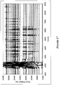
- Figure 7 shows the valving performance of these valves for liquids of interest, namely, deionized water and cycle sequencing reagent, as a function of valve size for the case of fluoropolymer treatment. In both cases, the expected dependence of valving pressure on valve dimension is observed (Pressure ⁇ 1/diameter).
- Through-hole valves have significant advantages over in-plane valves. First, they are easier to manufacture, in that small through-holes can be readily made in a sheet of plastic, either by molding around posts, punching, diecutting, drilling, or laser-drilling after the valve layer has been created.
- In-plane valves require fairly precise fabrication, and very fine valves (with high valving pressures) necessitate the use of lithographic techniques to create the required molding or embossing tools.
- Second, through-hole valves can be more completely coated with fluoropolymer on "all sides.” The application of low-surface-tension fluoropolymer solution to a hole results in complete coating of the internal walls of the hole by capillary action. Coating of all sides of an in-plane valve requires application of fluoropolymer to both the valve as well as the region of the mating layer that seals over the valve. As a result, typical in-plane valves are formed without coating on the "roof' of the valve.
- Mixing can be accomplished in a variety of ways.
- this type of mixing is typically inadequate for mixing large volumes quickly, because the diffusion or mixing time scales with the channel width squared Mixing can be enhanced in a variety of ways, such as lamination, in that the fluid stream is divided and recombined. ( Campbell and Grzybowski Phil. Trans. R. Soc. Lond.
- mixing can be accomplished by cycling fluid between two points on the device multiple times.
- capillary valves A capillary valve disposed between two channels or chambers acts as a pivot for fluid flow; as fluid flows from one channel into the other through the capillary, the trailing meniscus is trapped if sufficiently low pressure is used to pump the fluid. Reversal of the pressure drives the fluid back into the first channel, and it is again pinned at the capillary. Multiple cycles can be used to efficiently mix components.
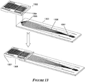
- FIG. 13 shows the construction of the integrated biochip (1301) from two components which are bonded in or during manufacture.
- a 16-sample biochip (1302) combining the lysis, amplification, and sequencing features of the biochip of Figure 1 with the sequencing product purification features of the biochip of Figure 11 and second, a 16-lane plastic separation biochip (1303).
- Purified sequencing product can also be electrokinetically injected prior to separation.
- the devices of the invention can be primarily composed of plastics.
- plastics include, but are not limited to: cyclic olefin polymer (COP); cyclic olefin copolymer (COC); (both of that have excellent optical quality, low hygroscopicity, and high operating temperatures when of sufficient molecular weight); poly(methyl methacrylate) (PMMA) (readily machinable and can be obtained with excellent optical properties); and polycarbonate (PC) (highly-moldable with good impact resistance and a high operating temperature). More information about materials and fabrication methods are contained in the U.S. patent application entitled “METHODS FOR RAPID MULTIPLEXED AMPLIFICATION OF TARGET NUCLEIC ACIDS" (Attorney Docket No. 08-318-US) that has been incorporated by reference ( supra ).
- a variety of methods can be used to fabricate the individual parts of the biochip and to assemble them into a final device. Because the biochip can be composed of one or more types of plastic, with the possible inclusion of inserted components, the methods of interest pertain to creation of individual parts followed by post-processing of parts and assembly.
- Plastic components can be fabricated in several ways, including injection molding, hot embossing and machining.
- Injection molded parts can be comprised of both gross features (such as fluid reservoirs) as well as fine features (such as capillary valves). In some cases, it can be preferable to create fine features on one set of parts and larger features on another set, because the approaches to injection molding of these differently-sized features can vary.
- large reservoirs measuring several (about 1-50 mm) mm on a side and with depths of several mm (about 1-10 mm) and capable of accommodating 100s of ⁇ L
- conventional molding can be employed using machined injection molding tools, or tools created by burning into a steel or other metal using a graphite electrode that has been machined to be a negative of the tool.
- both tool creation and molding process can be varied.
- Tools are typically created using a lithographic process on a substrate of interest (for example, isotropic etch of glass, or deep reactive ion etching or other processes on silicon).
- the substrate can then be electroplated with nickel (usually after deposition of a chromium layer to promote adhesion) and the substrate removed, for example, by etching in an acid.
- This nickel "daughter" plate is the injection molding tool.
- the molding process can be somewhat different than above, as well: For fine, shallow features, compression-injection molding, in which the mold is physically compressed slightly after plastic has been injected into the cavity, has been found to be superior to standard injection molding in terms of fidelity, precision, and reproducibility.
- plastic resin in the form of pellets, or as a pre-formed blank of material created through molding or embossing can be applied to the tool surface or a flat substrate.
- a second tool may then brought into contact at precisely controlled temperature and pressure in order to raise the plastic above its glass transition temperature and to cause material flow to fill the cavities of the tool(s). Embossing in a vacuum can avoid the problem of air becoming trapped between tool and plastic.
- Machining also can be employed to create parts.
- High-speed computer numerical controlled (CNC) machines can be used to create many individual parts per day from either molded, extruded, or solvent-cast plastic.
- CNC computer numerical controlled
- Proper choice of milling machine, operating parameters, and cutting tools can achieve high surface quality (surface roughnesses of 50 nm are achievable in high-speed milling of COC ( Bundgaard et al., Proceedings of IMechE Part C: J. Mech. Eng.Sci. 2006, 220,1625-1632 ).
- Milling can also be used to create geometries that can be difficult to achieve in molding or embossing and to readily mix feature sizes on a single part (for example, large reservoirs and fine capillary valves can be machined into the same substrate).
- Another advantage of milling over molding or embossing is that no mold-release agents are needed to release the fabricated part from a molding tool.
- Post-processing of individual parts includes optical inspection (that can be automated), cleaning operations to remove defects such as burrs or hanging plastic, and surface treatment. If optical-quality surfaces are required in machined plastic, polishing with a vapor of a solvent for the plastic can be used.
- polishing with a vapor of a solvent for the plastic can be used.
- PMMA dichloromethane can be used
- COC and COP cyclohexane or toluene can be used.
- Coatings that reduce wettability include fluoropolymers and/or molecules with fluorine moieties that are exposed to the fluid when the molecules are adsorbed or bonded to the surfaces of the device. Coatings can be adsorbed or otherwise deposited, or they can be covalently linked to the surface.
- the methods that can be used to make such coatings include dip coating, passing coating reagent through the channels of the assembled device, inking, chemical vapor deposition, and inkjet deposition.
- Covalent bonds between coating molecules and the surface can be formed by treatment with oxygen or other plasma or UV-ozone to create an activated surface, with either subsequent deposition or co-deposition of the surface treatment molecule on the surface (see, Lee et al. Electrophoresis 2005, 26, 1800-1806 ; and Cheng et al., Sensors and Actuators B 2004, 99, 186-196 .)
- Inserted devices such as filters
- Assembly of component parts into the final device can be performed in a variety of ways. Inserted devices, such as filters, can be die-cut and then placed with a pick-and-place machine.
- Thermal diffusion bonding can be used, for example for the bonding of two or more layers of the same material, each of that is of uniform thickness.
- the parts can be stacked and the stack placed into a hot press, where the temperature can be raised to the vicinity of the glass transition temperature of the material comprising the parts, to cause fusion at the interfaces between the parts.
- An advantage of this method is that the bonding is "general", i.e., any two stacks of layers of roughly the same dimensions can be bonded, regardless of the internal structure of the layers, because heat and pressure are applied uniformly across the layers.
- Thermal diffusion bonding may also be used to bond more complex parts, such as those that are not planar on their bonding or opposing surfaces, by using specially-created bonding cradles. Such cradles conform to the outer surface of the layers to be bonded.
- bonding variations include solvent-assisted thermal bonding, in that a solvent such as methanol partially solubilizes the plastic surface, enhancing bond strength at a lower bonding temperature.
- a further variation is the use of spin-coated layers of lower-molecular weight material.
- a polymer of the same chemical structure but of a lower molecular weight than the substrate components can be spun onto at least one layer to be bonded, the components assembled, and the resulting stack bonded, by diffusion bonding.
- the low-molecular weight components can pass through their glass transition temperature at a lower temperature than the components and diffuse into the substrate plastic.
- Adhesives and epoxies can be used to bond dissimilar materials and are likely to be used when bonding components fabricated in different ways.
- Adhesive films can be die cut and placed on components.
- Liquid adhesive may also be applied through spin-coating. Inking of adhesive onto structured parts (such as in nanocontact printing) can be successfully used to apply adhesive to structured surfaces without a need to "direct" the adhesive onto particular areas.
- a biochip of the invention can be assembled as shown in Figure 6 .
- Layers 1 and 2 can be aligned by included features (e.g., pins and sockets); separately, layers 3 and 4 can be similarly aligned by included features.
- the layer 1 plus layer 2 stack can be inverted and applied to the layer 3 plus layer 4 stack and then entire stack can be bonded.
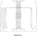
- FIG. 1 An integrated biochip for DNA extraction and amplification by PCR is shown in Figure 1 .
- This 4-sample device integrates the functions of reagent distribution and metering; mixing of reagents with samples; delivery of samples to a thermal cycling portion of the chip; and thermal cycling.
- the same biochip is used in Example 2 below and has additional structures for performance of cycle sequencing.
- the biochip was constructed of 4 layers of thermoplastic as shown in Figures 2 - 5 .
- the 4 layers are machined PMMA and have thicknesses of the layers are 0.76 mm, 1.9 mm, 0.38 mm, and 0.76 mm, respectively, and the lateral size of the biochip was 124 mm X 60 mm.
- biochips of at three or more layers allow the use of an indefinite number of common reagents to be divided among multiple assays: two fluidic layers and one layer that at least contains through-holes, enabling fluidic channels in the outer layers to 'cross-over' one another.
- the channels of the biochip were of cross-sectional dimensions ranging from 127 ⁇ m X 127 ⁇ m to 400 ⁇ m X 400 ⁇ m, while reservoirs ranged from 400 ⁇ m X 400 ⁇ m in cross-section to 1.9 X 1.6 mm; both channels and reservoirs extend for distances as short as 0.5 mm to several 10s of mm.
- the capillary valves used in the biochip were of 127 ⁇ m X 127 ⁇ m size for "in-plane" valves and 100 ⁇ m in diameter for through-hole capillary valves.
- Certain channels, reservoirs, and capillary valves of the four machined layers were treated with a hydrophobic/olephobic material, PFC 502A (Cytonix, Beltsville, MD).
- Surface treatment was performed by coating with a wetted Q-tip followed by air-drying at room temperature.
- the dried fluoropolymer layer was less than 10 ⁇ m thick as determined by optical microscopy.
- Surface treatment serves two purposes: to prevent the formation of bubbles within liquids, especially within low-surface-tension liquids, such as cycle sequencing reagent, which can occur as the liquid rapidly wets the walls of channels or chambers (and "closes off' a bubble before the air can be displaced), and to enhance the capillary burst pressure at that capillary valves resist liquid flow.
- the regions left untreated were the thermal cycling chambers for PCR and cycle sequencing.
- Bonding was performed using thermal diffusive bonding, in that the stack of components was heated under pressure to a temperature near the glass transition temperature (T g ) of the plastic. A force of 45 lbs was applied over the entire 11.5 square inch biochip for 15 minutes during a thermal bonding profile consisting of a ramp from ambient temp to 130 °C in 7.5 minutes, a hold at 130 °C for 7.5 minutes, and rapid cooling to room temperature.
- T g glass transition temperature
- Pneumatic instrumentation was developed for driving fluids within the biochips of the invention.
- Two small peristaltic pumps provided pressure and vacuum. Positive pressure output was divided among three regulators that have the range of approximately 0.05 - 3 psig.
- the vacuum was ported to a regulator with an output vacuum of approximately (-0.1) - (-3) psig.
- a fourth, higher pressure was taken from a cylinder of N 2 to a further regulator or alternatively from a higher-capacity pump.
- the positive and negative pressures were applied to a series of 8 pressure-selector modules. Each module was equipped with solenoid valves that could choose an output pressure to be transmitted to the biochip from among the 5 inputs.
- the output pressure lines terminated on at least one pneumatic interface. This interface clamped to the chip with O-rings positioned over the chip ports on the input side of the chip (the ports along the top of the figures).
- Some fluidic control events could be performed that required pulses of pressure as short as 30 msec and/or complex pressure profiles could be utilized where pressure could be switched from one value to another (i.e., one regulator to another) rapidly (that is, with time lags of no more than 10-20 msec).
- the samples consisted of a bacterial suspension of approximately 10 6 cells/mL of E. coli DH5 transformed with pGEM sequencing plasmid insert (pUC18 sequencing target).
- PCR reagent consisted of dNTPs KOD Taq Polymerase (Novagen, Madison, WI) at concentration 0.1 ⁇ M
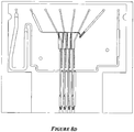
- a 1.23 ⁇ L sample of the bacterial suspension was added to each of the four ports 104, each comprising through holes 202 and 336 in layers 1 and 2, respectively. The sample then resided in sample channels 303 in layer 2.
- 10 ⁇ L of PCR reagent was added to port 105, comprising of through holes 217 and 306 in layers 1 and 2.
- the PCR reagent then resided in chamber 307 in layer 2 (see, Figure 8a ).
- a port for the evacuation of displaced air for the PCR reagent was port 107, comprising 109 and through-holes 203+305.
- the biochip was placed in the pneumatic manifold described above.
- the following automated pressure profile was performed with no delays between steps.
- a pressure of 0.12 psig was applied to ports 104 for 15 sec to drive the samples down channels 303 to through-hole 304.
- the samples passed through through-hole 304 and emerged on the other side of layer 2 in sample chamber 204 of layer 1 and were driven to the first mixing junction 205.
- the samples were retained by capillary valves 210 (see, Figures 8b-c ).
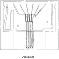
- a pressure of 0.12 psig was applied to port 105 for 10 sec to drive the PCR reagent through through-hole 320.
- the PCR reagent emerged on the other side of layer 2 in distribution channel 208, and moved into the metering chambers 209, which define a volume of reagent equal to the sample volume, where they were retained by capillary valves 211 at mixing junction 205. (see, Figure 8d ).
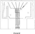
- a pressure of 0.12 psig was applied to port 107 (comprised of through-holes 203 and 305) with port 105 open to atmosphere for 3 sec to empty channel 208 (see, Figure 8e ).
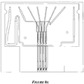
- a pressure of 0.8 psig was applied to ports 107 and 105 for 0.03 sec and a pressure of 0.7 psig was simultaneously applied to ports 104 for 0.03 sec to initiate mixing of the samples and PCR reagents by bursting liquids past the capillary valves 210 and 211 (see, Figure 8f ).
- a pressure of 0.12 psig was applied to ports 104 and 107 for 10 sec to pump the samples and PCR reagents into mixing channels 214, with retention at capillary valves 210 and 211. Passage through the mixing bulbs 212 into the constrictions 213 created added hydraulic resistance to flow, decreasing the high velocity imparted by the previous high pressure pulse.
- a pressure of 0.7 psig was applied to ports 104 and 107 for 0.03 sec to detach the liquid from capillary valves 210 and 211 (see Figure 8g ).
- a pressure of 0.12 psig was applied for 3 sec to ports 104 and 107 to pump liquid through mixing channel 214 to capillary valves 219, where they were retained (see, Figure 8h ).
- a pressure of 0.7 was psig applied for 0.1 sec to ports 104 and 107 to drive the mixture of the samples and PCR reagents through through-holes 315 and 402 and through the body of layers 2 and 3, and into PCR chamber 502 (see, Figure 8i ).
- a pressure of 0.12 psig was applied for 3 sec to ports 104 and 107 to complete pumping of the mixture of the samples and PCR reagents into chamber 502.
- the leading edge of the mixture of the samples and PCR reagents then passed through through-holes 403 and 316, emerged into layer 1, and was pinned at capillary valve 220 (see, Figure 8j ).
- the biochip was then pressurized to 30 psig N 2 and thermally cycled for PCR amplification via a Peltier using a gas bladder compression mechanism as described the U.S. patent application entitled, "METHODS FOR RAPID MULTIPLEXED AMPLIFICATION OF TARGET NUCLEIC ACIDS", Attorney Docket No. 08-318-US, filed concurrently herewith; and in International Patent Application Serial No. PCT/US08/53234 , Attorney Docket No. 07-084-WO, filed 6 Feb 2008 and entitled, "DEVICES AND METHODS FOR THE PERFORMANCE OF MINIATURIZED IN VITRO ASSAYS,” each of which are hereby incorporated by reference in their entirety.
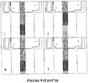
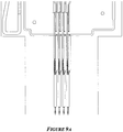
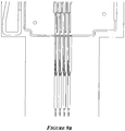
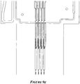
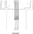
- Example 1 The biochips described in Example 1 were used. PCR product generated in tubes using the protocol outlined in Example 1 was added to both sample and PCR reagent ports of the biochip as described above. 50 ⁇ L of a cycle sequencing reagent (BigDyeTM3.1/BDX64, MCLab, San Francisco) was added to port 106 (comprised of through-holes 215 and 308 ) and chamber 309. After installation of two pneumatic interfaces (one for the input and one for the output end of the chip), the PCR product was processed as described in Example 1 through to the PCR chamber, but without the PCR thermal cycling step. The disposition of the fluids in the chip was as shown in Figure 9a .
- a cycle sequencing reagent BigDyeTM3.1/BDX64, MCLab, San Francisco
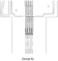
- Steps 9-10 were repeated an additional two times to effect mixing of the sequencing reagent and PCR product.
- the biochip was then pressurized to 30 psig N 2 and thermally cycled using the following temperature profile:
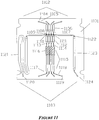
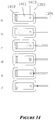
- a 4-sample biochip for the performance of sequencing product purification was constructed of four layers, as discussed in Example 1, and is shown in Figure 11 .
- One additional element in construction was the ultra-filtration (UF) filter 1116, which is cut to size and placed between layers 3 and 4 prior to thermal bonding.
- UF ultra-filtration
- the creation of a good bond around the UF filter necessitated the use of layer 3.
- Layers 3 and 4 create uninterrupted perimeters around the filter, because all channels leading to and from the filter are in the bottom of layer 2.
- a regenerated cellulose (RC) filter of molecular weight cut-off (MWCO) 30 kD was used (Sartorius, Goettingen, Germany).
- MWCO molecular weight cut-off
- a variety of other MWCOs (10 kD, 50 kD, and 100 kD) have been examined, as has an alternative material, polyethersulfone (Pall Corporation, East Hills, NY).
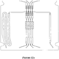
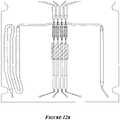
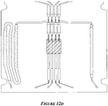
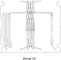
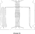
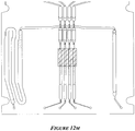
- Steps 8-12 were repeated to partially-fill chambers 1115 with a final volume of water used for elution (see, Figure 12k ).
- Vacuum of 1.6 psig was applied to ports 1104 with all other ports closed for 1sec, drawing some water from chambers 1115 into chambers 1112 (the maximum motion being dictated by the creation of a vacuum of equal magnitude in the dead-space between the meniscus of the liquid and the solenoid valves corresponding to ports 1119 ), (see, Figure 12l ).
- Ports 1104 were opened to atmosphere for 1 sec, allowing the liquid to move back into chamber 1115 due to the partial vacuum generated between the liquid and the valves corresponding to ports 1119 (see, Figure 12m ).
- a pressure of 0.09 psig was applied to port 1124 for 10 sec with ports 1119 open to atmosphere to drive liquids such that its trailing meniscus was pinned at 1113.
- a pressure of 0.7 psig/0.05sec was applied to port 1124 with ports 1119 open to atmosphere to detach the eluent (see, Figure 12n ).
- Figure 13 illustrates an embodiment of a 16-sample biochip, 1301, which combines the lysis and extraction, template amplification, and cycle sequencing functions of the biochip of Figure 1 ; the ultrafiltration function of the chip of Figure 11 ; and electrophoretic separation and detection.
- the process through ultrafiltration is carried out by sub-component 1302 and can be performed as described in examples 1, 2, and 3; transfer points 1304 on the bottom surface of 1302 are aligned with input wells 1305 on the separation sub-component 1303.
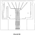
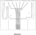
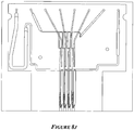
- the input well 1305, illustrated in Figure 14 consists of a liquid receiving well 1401; a main separation electrode, 1402; and a counterelectrode 1403.
- Separation channel 1306 opens into the bottom of well reservoir 1401.
- the separation electrode is typically platinum or gold coated, and is preferably a planar gold-coated electrode that substantially covers 1, 3, or 4 of the internal surfaces of 1401.
- the counterelectrode is a thin gold, steel, or platinum wire (typically 0.25 mm in diameter) that has been coated with a thin layer (-10 ⁇ m) of cross-linked polyacrylamide. This forms a hydrogel protection layer on the electrode.
- this scheme allows the concentration of sequencing product in the vicinity of the end of channel 1306 to be increased significantly relative to the concentration with that it is delivered from ultrafiltration. While such concentration is desirable for some applications, it is not necessary in all cases.
- the well of Figure 14 without the counterelectrode 1403 can be used to perform EKI directly.
- the single electrode in the loading well may be one half of a cross-T or double-T injector (see, for example, the U.S. patent application entitled, "PLASTIC MICROFLUIDIC SEPARATION AND DETECTION PLATFORMS", Attorney Docket No. 07-865-US, filed concurrently herewith).
- a recess 1308 is provided to allow, for example, a Peltier block (not shown) to mate with the lower surface of 1301 to provide thermal cycling for PCR and cycle sequencing. Pneumatic interfaces (not shown) within the instrument clamp to the ends of the chip to provide microfluidic control.
- DNA separation is carried out on a biochip and instrumentation as described in U.S. Patent Application Publication No. US2006-0260941-A1 .
- Separation chips can be glass (see, U.S. Patent Application Publication No. US2006-0260941-A1 ) or plastic (the U.S. patent application entitled, "PLASTIC MICROFLUIDIC SEPARATION AND DETECTION PLATFORMS", Attorney Docket No. 07-865-US, filed concurrently herewith), each of which are hereby incorporated by reference in their entirety.
- the instrument comprises excitation and detection subsystems for interacting with and interrogating a sample.
- Samples typically include one or more biological molecules (including but not limited to DNA, RNA, and proteins) that are labeled with dyes (e.g., fluorescent dyes).
- the excitation subsystem comprises an excitation source or sources and an excitation beam path with optical elements including lenses, pinholes, mirrors and objectives, to condition and focus the excitation source in an excitation/detection window.
- Optical excitation of a sample can be accomplished by a series of laser types, with emission wavelengths in the visible region, between 400 to 650 nm. Solid state lasers can provide emission wavelengths of approximately 460 nm, 488 nm, and 532 nm.
- lasers include, for example, the Compass, Sapphire and Verdi products from Coherent (Santa Clara, CA).
- Gas lasers include argon-ion and helium neon with emission in the visible wavelengths at approximately 488 nm, 514 nm, 543 nm, 595 nm, and 632 nm.
- Other lasers with emission wavelengths in the visible region are available from CrystaLaser (Reno, NV).
- a 488 nm solid state laser Sapphire 488-200 can be utilized.
- a light source with wavelength beyond the visible range can be used for exciting dyes having absorption and/or emission spectra beyond the visible range (e.g. , infrared or ultra-violet emitting dyes).
- optical excitation can be achieved by the use of non-laser light sources with emission wavelengths appropriate for dye excitation, including light emitting diodes, and lamps, .
- the detection subsystem comprises one or more optical detectors, a wavelength dispersion device (which performs wavelength separation), and one or a series of optical elements including, but not limited to, lenses, pinholes, mirrors and objectives to collect emitted fluorescence from fluorophore-labeled DNA fragments that are present at the excitation/detection window.
- the fluorescence emitted can be from a single dye or a combination of dyes.
- wavelength separation of the fluorescence can be utilized. This can be achieved by the use of dichroic mirrors and bandpass filter elements (available from numerous vendors including Chroma, Rockingham, VT; and Omega Optical, Brattleboro, VT).
- the emitted fluorescence passes through a series of dichroic mirrors where one portion of the wavelength will be reflected by the mirror to continue traveling down the path, and the other portion will pass through.
- a series of discrete photodetectors each one positioned at the end of the dichroic mirror will detect light over a specific range of wavelengths.
- a bandpass filter can be positioned between the dichroic mirror and photodetector to further narrow the wavelength range prior to detection.
- Optical detectors that can be utilized to detect the wavelength-separated signals include photodiodes, avalanche photodiodes, photomultiplier modules, and CCD cameras. These optical detectors are available from suppliers such as Hamamatsu (Bridgewater, NJ).
- wavelength components are separated by the use of dichroic mirrors and bandpass filters and these wavelength components are detected with Photomultiplier Tube (PMT) detectors (H7732-10, Hamamatsu).
- the dichroic mirror and bandpass components can be selected such that incident light on each of the PMTs consists of a narrow wavelength band corresponding to the emission wavelength of the fluorescent dye.
- the band pass is typically selected to be centered about the fluorescent emission peak with a band pass of wavelength range of between 1 and 50 nm.
- the system is capable of eight color detection and can be designed with eight PMTs and a corresponding set of dichroic mirrors and bandpass filters to divide the emitted fluorescence into eight distinct colors.
- More than eight dyes can be detected by applying additional dichroic mirrors, bandpass filters and PMT.
- Figure 15 shows the beam path for discrete bandpass filter and dichroic filter implementation.
- An integrated version of this wavelength discrimination and detection configuration is the H9797R, Hamamatsu, Bridgewater, NJ.
- wavelength dispersive elements and systems such as prisms, diffraction gratings, transmission gratings (available from numerous vendors including ThorLabs, Newton, NJ; and Newport, Irvine, CA; and spectrographs (available from numerous vendors including Horiba Jobin-Yvon, Edison, NJ).
- the wavelength components of the fluorescence are dispersed over a physical space.
- Detector elements placed along this physical space detect light and allow the correlation of the physical location of the detector element with the wavelength.
- Detectors suitable for this function are array-based and include multi-element photodiodes, CCD cameras, back-side thinned CCD cameras, multi-anode PMT.
- One skilled in the art will be able to apply a combination of wavelength dispersion elements and optical detector elements to yield a system that is capable of discriminating wavelengths from the dyes used in the system.
- a spectrograph is used in place of the dichroic and bandpass filters to separate the wavelength components from the excited fluorescence. Details on spectrograph design is available in John James, Spectrograph Design Fundamental. Cambridge, UK: Cambridge University Press, 2007 .
- the spectrograph P/N MF-34 with a concave holographic grating with a spectral range of 505 - 670 nm (P/N 532.00.570) HORIBA Jobin Yvon Inc, Edison, NJ
- Detection can be accomplished with a linear 32-element PMT detector array (H7260-20, Hamamatsu, Bridgewater, NJ).
- collected fluorescence is imaged on the pinhole, reflected, dispersed, and imaged by the concave holographic grating onto the linear PMT detector that is mounted at the output port of the spectrograph.
- the use of a PMT-based detector takes advantage of the low dark noise, high sensitivity, high dynamic range, and rapid response characteristic of PMT detectors.
- the use of a spectrograph and multi-element PMT detector for detection of excited fluorescence allows for flexibility in the number of dyes and the emission wavelength of dyes that can be applied within the systems and within the lane, without the need for physically reconfiguring the detection system (dichroic, bandpass and detectors) of the instrument.
- the data collected from this configuration is a wavelength dependent spectra across the visible wavelength range for each scan for each lane. Generating a full spectrum per scan provides dye flexibility both in terms of dye emission wavelength and number of dyes that can be present within a sample.
- the use of the spectrometer and linear multi-element PMT detector also allows for extremely fast read-out rates as all the PMT elements in the array are read-out in parallel.
- Figure 16 shows the beam path for multi-element PMT and spectrograph implementation.
- Instruments may employ a staring mode of operation, to detect multiple lanes simultaneously and multiple wavelengths simultaneously.
- the excitation beam is simultaneously impinged on all lanes at the same time.
- the fluorescence from this is collected by a two dimensional detector such as a CCD camera or array.
- a wavelength dispersive element is used.
- One dimension of the detector represents the physical wavelength separation, while the other dimension represents the spatial or lane-lane separation.
- a scanning mirror system (62) (P/N 6240HA, 67124-H-0 and 6M2420X40S100S1, Cambridge technology, Cambridge MA) is utilized to steer both the excitation and detection beam paths in order to image each of the lanes of the biochip.
- the scanning mirror steers the beam paths, scanning sequentially from lane to lane from the first lane to the last lane, and the repeating the process again from the first lane to the last lane again.
- a lane-finding algorithm such as that described in U.S. Patent Application Publication No. US2006-0260941-A1 is used to identify location of lane.
- FIG. 16 An embodiment of an optical detection system for simultaneous multiple lane and multi dye detection is shown in Figure 16 .
- the fluorescence excitation and detection system 40 excites the components separated by electrophoresis of a DNA sample (e.g. , containing DNA fragments following amplification of a set of STR loci) by scanning an energy source (e.g. a laser beam) through a portion of each of the microchannels while collecting and transmitting the induced fluorescence from the dye to one or more light detectors for recordation, and ultimately analysis.
- an energy source e.g. a laser beam
- the fluorescence excitation and detection assembly 40 includes a laser 60, a scanner 62, one or more light detectors 64, and various mirrors 68, spectrograph, and lenses 72 for transmitting a laser beam emitted from the laser 60 through opening 42 to the test module 55 and back to the light detectors 64.
- the scanner 62 moves the incoming laser beam to various scanning positions relative to the test module 55. Specifically, the scanner 62 moves the laser beam to a pertinent portion of each micro channel within the test module 55 to detect respective separate components.
- the multi element PMT 64 collects data (e.g. the fluorescent signals from DNA fragments of varying length) from the test module 55 and provide the data electronically through a cable attached to port 75 to a data acquisition and storage system located outside the protective cover 50.
- the data acquisition and storage system can include a ruggedized computer available from Option Industrial Computers (13 audreuil-Dorion, Quebec, Canada).
- the excitation source is incident on all the detection spots simultaneously, and fluorescence from all detection spots is collected simultaneously.
- Simultaneous spectral dispersion (wavelength spectra of detected fluorescence) and spatial dispersion (detection spots) can be performed with a two dimensional detector array.
- the 2-dimensional detector array is positioned in the system such that spectral components are imaged and detected across one dimension of the array detector (row), while spatial components are imaged and detected across the other dimension of the array detector.
- a preferred instrument utilizes a scanning mode of operation, rather than a "staring" mode.
- scanning mode signal for each channel is required to be collected, integrated, and read-out while the scanner is coincident with the lane being interrogated and before it is incident on the next channel.
- a detector with fast readout allows for optimal light collection and integration, translating into higher signal to noise performance.
- the read-out time of the detector should be significantly less than the total time which the scanner is coincident with the channel.
- the multi-element PMT can be read-out in less than 0.7 ms and this read-out time is far less than the integration time for detection for each individual channel.
- Fluorescence incident on the pinhole can be dispersed by the grating according to its wavelength composition and focused onto the linear multi-anode PMT detector array.
- the detector provides 32 current outputs, one for each of the elements in the array that correspond to the number of photons incident on the element.
- the integrator circuitry will integrate the PMT output current to generate an output voltage proportional to the integrated PMT current.
- the single ended output voltage is converted to differential mode using the Analog Devices (Norwood, MA) differential driver IC (P/N SSM2142).
- the data acquisition system will read the differential signal and save the data in its buffer. After the data have been saved, the data acquisition system will move the scanner to shift the laser beam to the next selected lane, at the same time resetting the integrator circuitry.
- Each single element PMT module has its own integrator circuitry.
- For an 8 color detection system there are 8 PMT modules and 8 integrator circuitries. Additional colors can be added using corresponding numbers of PMT modules and integrator circuitry.
- this detector Since each of the PMT elements (H77260-20, Hamamatsu, Japan) has a similar or more rapid signal response as a single PMT tube (H7732-10, Hamamatsu, Japan), and the readout is in parallel, this detector is able to operate very rapidly. When coupled with the spectrometer, this spectrometer and multi-anode detector system is able to provide full spectral scans across the visible spectrum (450 nm to 650 nm) with readout-times of less than 0.1 ms.
- the ability to provide fast refresh rates allows this spectrometer/detector system to be applied to scanning mode implementation of detection of multiple lanes sequentially within a single run.
- the use of PMT based detectors provides for low noise, high sensitivity and high dynamic range, and fast response.
- the 140 mm spectrometer with a concave holographic grating (Horiba Jobin-Yvon) and multianode PMT detector is the H7260-20 detector (Hamamatsu, Japan). Other spectrometers configurations and multi-anode PMT detectors can also be used for this application.
- Determination of nucleotide bases from the electrophoregrams was achieved using signal processing algorithms to correct, filter, and analyze the data. This process consisted of locating a callable signal, correcting the signal baseline, filtering out noise, removing color cross-talk, identifying signal peaks, and determining associated bases. Locating the callable signal was performed to remove extraneous data from the beginning and end of the signal and accomplished by employing a threshold. Next, the background was removed from the signal, so that the signal had a common baseline for all detected colors. Finally, a lowpass filter was applied to remove high frequency noise from the signal.
- a weighted matrix was calculated and applied to the signal to amplify the color-space of the nucleotide-dye spectrum. Calculation of this color separation matrix was accomplished using the methods of Li et al. Electrophoresis 1999, 20, 1433-1442 . In this adaptation, a "m x n" color separation matrix is calculated from correlating the "m" number of dyes utilized in the assay with the "n” number of detector elements.
- D CSM x PMT, where D is the signal in dye space for each of the m dyes, CSM is the color separation matrix, and PMT is a matrix with the signal from each of the n elements of the detector.
- the corrected traces were allele-called to identify each fragment and to assign a fragment size based on a sizing standard.
- the corrected traces were base-called to associate one of the four nucleotides with each peak in the trace. A detailed description of base calling can be found in Ewing et al. Genome Research, 1998, 8, 175-185 , and Ewing et al., Genome Research, 1998, 8, 186-194 , the disclosures of that are hereby incorporated by reference in their entirety.
- Dye labels attached to oligonucleotides and modified oligonucleotides can be synthesized or obtained commercially (e.g. Operon Biotechnologies, Huntsville, Alabama).
- a large number of dyes are available for application in fluorescence excitation applications. These dyes include those from the fluorescein, rhodamine AlexaFluor, Biodipy, Coumarin, and Cyanine dye families.
- quenchers are also available for labeling oligo sequences to minimize background fluorescence. Dyes with emission maxima from 410 nm (Cascade Blue) to 775 nm (Alexa Fluor 750) are available and can be used.
- Dyes ranging between 500 nm to 700 nm have the advantage of being in the visible spectrum and can be detected using conventional photomultiplier tubes.
- the broad range of available dyes allows selection of dye sets that have emission wavelengths that are spread across the detection range. Detection systems capable of distinguishing many dyes have been reported for flow cytometry applications (see, Perfetto et al., Nat. Rev. Immunol. 2004. 4, 648-55 ; and Robinson et al., Proc ofSPIE 2005, 5692, 359-365 ).
- Fluorescent dyes have peak excitation wavelengths that are typically 20 to 50 nm blue-shifted from their peak emission wavelength. As a result, use of dyes over a wide range of emission wavelengths may require the use of multiple excitation sources, with excitation wavelengths to achieve efficient excitation of the dyes over the emission wavelength range.
- energy transfer dyes can be utilized to enable a single laser, with a single emission wavelength, to be used for exciting all dyes of interest. This is achieved by attaching an energy transfer moiety to the dye label. This moiety is typically another fluorescent dye with an absorption wavelength that is compatible with the excitation wavelength of the light source (e.g. laser).
- Placement of this absorber in close proximity with an emitter allows the absorbed energy to be transferred from the absorber to the emitter, allowing for more efficient excitation of the long wavelength dyes ( Ju et al., Proc Natl Acad Sci USA 1995, 92, 4347-51 ).
- the following example illustrates the separation and detection of nucleic acid fragments labeled with 6 fluorescent dyes, and demonstrates the color resolution capability of the spectrometer/multi element excitation/detection system.
- DNA fragments were labeled with 6-FAM, VIC, NED, PET dyes by applying fluorescently labeled primers in a multiplexed PCR amplification reaction.
- 1 ng of human genomic DNA 9947A was amplified in a 25 ⁇ L reaction in according to the manufacturers recommended conditions (AmpFISTR Identifiler, Applied Biosystems).
- Electrophoretic separation of DNA using Genebench consists of a series of four operations: pre-electrophoresis, loading, injection and separation. These operations are carried out on a microfluidic biochip, which is heated to a uniform temperature of 50 °C.
- the biochip contains 16 channel systems for separation and detection multiple, each consisting of an injector channel and a separation channel. DNA for analysis is separated by electrophoretic transport of the DNA through a sieving matrix along the separation channel.
- the separation length of the biochips is ranges from 160 to 180 mm.
- the first step is pre-electrophoresis, which is accomplished by applying a 160 V/cm field along the length of the channel for six (6) minutes.
- Separation buffer TTE1X
- Samples for analysis are pipetted into the sample wells and a 175 V applied from the sample well to the waste well for 18 seconds, followed by the application of 175 V across the sample and waste well and 390 V at the cathode for 72 seconds, to load the sample into the separation channel.
- Injection of the sample is accomplished by applying a 160 V/cm field along the length of the separation channel while fields of 50 V/cm and 40 V/cm are applied across the sample and waste wells respectively. Separation is continued with the injection voltage parameters for 30 min during that an optical system detects the separating bands of DNA.
- the data collection rate is 5 Hz and PMT gains are set to -800 V.
- the loci selected are CSF1P0, FGA, THO1, TPOX, vWA, D3S1358, D5S818 and D7S820, although any of the loci and hence primer pairs described in the paper can also be used in this example.
- Each of the forward primers for the primer pairs is labeled with a separate fluorescent dye (Operon Biotechnologies, Huntsville, Alabama).
- Dyes selected for attachement to the primers include Alexa Fluor Dyes 488, 430, 555, 568, 594, 633, 647, and Tamra. Numerous other dyes are available and can also be used as labels.
- Each locus is amplified separately following the PCR reaction protocols of (Butler, 2003, Id. ) to yield a reaction solution with fragments labeled with their respective dyes.
- Template for PCR reaction is 1 ng of human gemonic DNA (type 9947A from Promega, Madison WI).
- Each PCR reaction was purified by cleaning up through a PCR cleanup column, where primers (labeled and dye-labeled primers) and enzymes are removed, and the PCR buffer is exchanged by the DI eluant.
- the resulting product of clean is a solution of labeled DNA fragments in DI water. Cleanup of dye labeled products follows the protocol of Smith using MinEluteTM columns (Qiagen, Valencia, CA). A total of eight reactions are performed. Eight cleaned up PCR reactions were mixed together in a ratio to generate peaks of equivalent signal strengths, generating a mixture containing fragments labeled with 8 different dyes. Alternatively, primers for 8 loci can be mixed together to form a master primer mix for multiplexed amplification.
- This solution is separated and detected with the instrument and protocol as described in Example 1.
- the grating of the spectrograph is adjusted such that the emission of the 8 dyes falls across the 32 pixels of the detector elements.
- the amount of sample loaded for analysis is to be adjusted such that detected signals fall within the dynamic range of the detection system.
- the following example illustrates separation/detection of labeled DNA fragments with the spectrometer/multi-element PMT system of Figure 16 , specifically for identifying sequence of a DNA template.
- 0.1 pmol of DNA template M13 and M13 sequencing primer was amplified with the GE Amersham BigDyeTM sequencing kit, according to the recommended reaction conditions.
- the reaction mix was cleaned up by ethanol precipitation and resuspended in 13 ⁇ L of DI water.
- the sample was separated following the electrophoretic separation condition as described in example 5. Sample loading conditions were modified and was carried out by applying 175 V across the sample well to the waste well for 105 seconds.
- Figure 21 shows an electrophoregram for the DNA sequence, with colored traces representing the detector element corresponding to the spectral maximum for each of the 4 dyes used.
- the sequence obtained was base called with Phred quality score of > 20 for 519 bases and QV30 of 435 bases ( Figure 22 ).
- cycle sequencing reactions can be prepared by either dye labeled terminator reactions or dye labeled primer reactions as follows:
- Cycle sequencing reaction for each template fragment consisting of a sequencing primer appropriate for the template sequence of interest, and reagents for conducting DNA sequencing including cycle sequencing buffer, polymerase, oligonucleotides, dideoxynucleotides and labeled dideoxynucleotides is prepared. Eight different dyes are utilized for the labeling. In the first cycle sequencing reaction, one set of 4 dye labeled dideoxy nucleotides is used. In the second cycle sequencing reaction, another set of 4 dye labeled dideoxy nucleotides (with emission wavelengths different than those of the four used in the first cycle sequencing reaction) is used. Each cycle sequencing reaction is carried out separately following a protocol that thermally cycles each reaction multiple times.
- Each thermal cycle includes a denature, anneal and extension step with temperatures and times following the protocols of Sanger (see, Sanger et al., Proc Natl Acad Sci U S A 1977, 74, 5463-7 ).
- the cycle sequencing product from the two reactions are combined to form a sample that consist of labeled DNA fragments, with a total of eight unique dyes, from each of the two DNA templates.
- the sample for separation and detection can be fabricated by using primer labeled cycle sequencing.
- Four cycle sequencing reactions are carried out for each DNA template.
- Each reaction is a cycle sequencing reaction consisting of a labeled sequencing primer, and reagents for conducting DNA sequencing including cycle sequencing buffer, polymerase, oligonucleuotides.
- each reaction will include one of the dideoxynucleuotides (ddATP, ddTTP, ddCTP, or ddGTP) and one labeled primer.
- ddATP, ddTTP, ddCTP, or ddGTP dideoxynucleuotides
- Each dye associated with the primer is unique in emission wavelength and is correlated with the type of dideoxy nucleotide in the cycle sequencing solution (ddATP, ddTTP, ddCTP, or ddGTP).
- Each cycle sequencing reaction is carried out separately following a protocol that thermally cycles each reaction multiple times.
- Each thermal cycle includes a denature, anneal and extension step with temperatures and times following the protocols of Sanger (see, Sanger, 1977, Id. ).
- Sanger see, Sanger, 1977, Id.
- Another set of 4 dyes (with emission wavelength different to that of the four used in the first cycle sequencing reaction) is applied.
- the product of all eight reactions (each with a different dye) are mixed together to form a sample that consist of DNA fragments from each of the two DNA templates.
- Each of the sequencing reactions is cleaned up by ethanol precipitation. Separation and detection of the sample follows the protocol of Example 8. The result of the separation and detection is the generation of two distinct DNA sequences, corresponding to each of the two template DNA fragments.
- the methods of this example can be modified to allow the use of dyes in multiples of four to allow detection of that multiple of DNA sequences in a single separation channel (e.g. 12 dyes for the detection of 3 sequences simultaneously, 16 dyes for the detection of 4 sequences simultaneously, 20 dyes for the detection of five sequences simultaneously, and so on).
- separation of the labeled fragments need not be limited to electrophoresis.
- nucleic acid analysis there are several applications of nucleic acid analysis that can be applied to clinical diagnostics, including DNA and RNA sequencing and fragment size determination.
- DNA and RNA sequencing and fragment size determination the use of simultaneous detection of 10 colors allows the interrogation of up to 500 loci. Analysis of the size of large number of fragments can be utilized to identify pathogens or to characterize many loci within an individual's genome, for example.
- aneuploidy is currently diagnosed by karyotyping and by fluorescent in situ hybridization (FISH).
- FISH fluorescent in situ hybridization
- FISH typically utilizes approximately 10 probes to assess whether or not a cell contains a normal chromosomal complement. This approach does not allow a detailed view of the entire genome, however, and cells that appear normal by FISH may well have major abnormalities that are not detected by the technique.
- primer pair sequences for approximately 500 loci are selected from published sequences, with each locus present as a single copy per haploid genome.
- 10 sets of 50 primer pairs are selected such that each set defines a corresponding set of DNA fragments such that none of the fragments are of the identical size.
- the forward primers for the primer pairs are labeled with one fluorescent dye, and no two sets share the same dye.
- Dyes selected for attachment to the primers are Alexa Fluor Dyes 488, 430, 555, 568, 594, 633, 647, 680, 700, and Tamra. Numerous other dyes are available and can also be used as labels.
- the loci can be amplified in one or several parallel PCR reactions as described in "METHODS FOR RAPID MULTIPLEXED AMPLIFICATION OF TARGET NUCLEIC ACIDS", supra.
- the amplified primers are separated and detected using the methods described herein. In a single separation channel, all 500 fragments can be precisely identified by size, 50 for each of ten dyes.
- the number of loci, dyes, and separation channels can be varied based on the desired application. Smaller numbers of fragments can be detected if desired by utilizing a smaller number of dye labels or generating fewer DNA fragments per label; in this way, less than 500, less than 400, less than 300, less than 200, less than 100, less than 75, less than 50, less than 40, less than 30, or less than 20 fragments can be detected as desired.
- the maximum number of loci that can be identified per lane is based on the read length and resolution of the separation system (e.g. , single base pair resolution of DNA fragments ranging from 20 to 1500 base pairs results in hundreds of fragments) multiplied by the number of distinct dyes that can be detected (as noted supra, dozens are available). Accordingly, thousands of loci can be identified in a single separation channel, and the number will increase as additional dyes are developed.
- the invention is further embodied by the following items:
Applications Claiming Priority (5)
| Application Number | Priority Date | Filing Date | Title |
|---|---|---|---|
| US92180207P | 2007-04-04 | 2007-04-04 | |
| US96450207P | 2007-08-13 | 2007-08-13 | |
| US2807308P | 2008-02-12 | 2008-02-12 | |
| EP08727302.5A EP2137522B1 (en) | 2007-04-04 | 2008-04-04 | Integrated nucleic acid analysis |
| PCT/US2008/004462 WO2008124104A1 (en) | 2007-04-04 | 2008-04-04 | Integrated nucleic acid analysis |
Related Parent Applications (2)
| Application Number | Title | Priority Date | Filing Date |
|---|---|---|---|
| EP08727302.5A Division EP2137522B1 (en) | 2007-04-04 | 2008-04-04 | Integrated nucleic acid analysis |
| EP08727302.5A Division-Into EP2137522B1 (en) | 2007-04-04 | 2008-04-04 | Integrated nucleic acid analysis |
Publications (1)
| Publication Number | Publication Date |
|---|---|
| EP3677905A1 true EP3677905A1 (en) | 2020-07-08 |
Family
ID=39591889
Family Applications (7)
| Application Number | Title | Priority Date | Filing Date |
|---|---|---|---|
| EP15191406.6A Active EP3032251B1 (en) | 2007-04-04 | 2008-04-04 | Plastic microfluidic separation and detection platforms |
| EP17179620.4A Withdrawn EP3284538A1 (en) | 2007-04-04 | 2008-04-04 | Methods for rapid multiplexed amplification of target nucleic acids |
| EP08742557A Ceased EP2137523A1 (en) | 2007-04-04 | 2008-04-04 | Plastic microfluidic separation and detection platforms |
| EP08727302.5A Active EP2137522B1 (en) | 2007-04-04 | 2008-04-04 | Integrated nucleic acid analysis |
| EP08742602.9A Active EP2155393B1 (en) | 2007-04-04 | 2008-04-04 | Systems for rapid multiplexed amplification of target nucleic acids |
| EP19194060.0A Withdrawn EP3626345A1 (en) | 2007-04-04 | 2008-04-04 | Systems for rapid multiplexed amplification of target nucleic acids |
| EP20157682.4A Pending EP3677905A1 (en) | 2007-04-04 | 2008-04-04 | Integrated nucleic acid analysis |
Family Applications Before (6)
| Application Number | Title | Priority Date | Filing Date |
|---|---|---|---|
| EP15191406.6A Active EP3032251B1 (en) | 2007-04-04 | 2008-04-04 | Plastic microfluidic separation and detection platforms |
| EP17179620.4A Withdrawn EP3284538A1 (en) | 2007-04-04 | 2008-04-04 | Methods for rapid multiplexed amplification of target nucleic acids |
| EP08742557A Ceased EP2137523A1 (en) | 2007-04-04 | 2008-04-04 | Plastic microfluidic separation and detection platforms |
| EP08727302.5A Active EP2137522B1 (en) | 2007-04-04 | 2008-04-04 | Integrated nucleic acid analysis |
| EP08742602.9A Active EP2155393B1 (en) | 2007-04-04 | 2008-04-04 | Systems for rapid multiplexed amplification of target nucleic acids |
| EP19194060.0A Withdrawn EP3626345A1 (en) | 2007-04-04 | 2008-04-04 | Systems for rapid multiplexed amplification of target nucleic acids |
Country Status (9)
| Country | Link |
|---|---|
| US (12) | US8018593B2 (ja) |
| EP (7) | EP3032251B1 (ja) |
| JP (9) | JP2010523115A (ja) |
| KR (4) | KR101530943B1 (ja) |
| AU (5) | AU2008236691B2 (ja) |
| CA (6) | CA2682734C (ja) |
| ES (3) | ES2797951T3 (ja) |
| NZ (5) | NZ602697A (ja) |
| WO (4) | WO2009011942A2 (ja) |
Families Citing this family (123)
| Publication number | Priority date | Publication date | Assignee | Title |
|---|---|---|---|---|
| EP2261650A3 (en) | 2004-09-15 | 2011-07-06 | IntegenX Inc. | Microfluidic devices |
| GB0421529D0 (en) | 2004-09-28 | 2004-10-27 | Landegren Gene Technology Ab | Microfluidic structure |
| JP5063616B2 (ja) | 2006-02-03 | 2012-10-31 | インテジェニックス インコーポレイテッド | マイクロ流体デバイス |
| US9839909B2 (en) | 2006-07-28 | 2017-12-12 | Diagnostics For The Real World, Ltd. | Device, system and method for processing a sample |
| WO2008115626A2 (en) | 2007-02-05 | 2008-09-25 | Microchip Biotechnologies, Inc. | Microfluidic and nanofluidic devices, systems, and applications |
| CA2682734C (en) * | 2007-04-04 | 2017-11-07 | Network Biosystems, Inc. | Plastic microfluidic separation and detection platforms |
| WO2009024773A1 (en) | 2007-08-17 | 2009-02-26 | Diagnostics For The Real World, Ltd | Device, system and method for processing a sample |
| EP2234916A4 (en) * | 2008-01-22 | 2016-08-10 | Integenx Inc | UNIVERSAL SPECIMEN PREPARATION SYSTEM AND ITS USE IN AN INTEGRATED ANALYSIS SYSTEM |
| WO2009126826A1 (en) | 2008-04-11 | 2009-10-15 | Fluidigm Corporation | Multilevel microfluidic systems and methods |
| GB0818609D0 (en) | 2008-10-10 | 2008-11-19 | Univ Hull | apparatus and method |
| CN102341691A (zh) * | 2008-12-31 | 2012-02-01 | 尹特根埃克斯有限公司 | 具有微流体芯片的仪器 |
| US8058630B2 (en) * | 2009-01-16 | 2011-11-15 | Fluidigm Corporation | Microfluidic devices and methods |
| EP2394152B1 (en) | 2009-02-03 | 2019-05-01 | ANDE Corporation | Nucleic acid purification |
| EP2438154A1 (en) * | 2009-06-02 | 2012-04-11 | Integenx Inc. | Fluidic devices with diaphragm valves |
| AU2010257118B2 (en) | 2009-06-04 | 2014-08-28 | Lockheed Martin Corporation | Multiple-sample microfluidic chip for DNA analysis |
| KR20120031218A (ko) | 2009-06-05 | 2012-03-30 | 인터젠엑스 인크. | 만능 샘플 제조 시스템 및 집적 분석 시스템에서의 사용 |
| EP2443254A2 (en) | 2009-06-15 | 2012-04-25 | NetBio, Inc. | Improved methods for forensic dna quantitation |
| US8584703B2 (en) | 2009-12-01 | 2013-11-19 | Integenx Inc. | Device with diaphragm valve |
| AU2011208946B2 (en) | 2010-01-28 | 2014-10-02 | Ellume Pty Ltd | Sampling and testing device for the human or animal body |
| ITMI20100163A1 (it) * | 2010-02-03 | 2011-08-04 | Consiglio Nazionale Ricerche | Procedimento per la identificazione e la tracciabilità di componenti vegetali |
| US8720036B2 (en) | 2010-03-09 | 2014-05-13 | Netbio, Inc. | Unitary biochip providing sample-in to results-out processing and methods of manufacture |
| AU2013204367A1 (en) * | 2010-03-09 | 2013-05-16 | Netbio, Inc. | Unitary biochip providing sample-in to results-out processing and methods of manufacture |
| EP3690433B9 (en) * | 2010-03-09 | 2022-12-21 | ANDE Corporation | Biochip with reagent storage |
| JP5841315B2 (ja) | 2010-04-28 | 2016-01-13 | ソニー株式会社 | 微小粒子分析装置 |
| US8512538B2 (en) | 2010-05-28 | 2013-08-20 | Integenx Inc. | Capillary electrophoresis device |
| US20110312676A1 (en) * | 2010-06-17 | 2011-12-22 | Geneasys Pty Ltd | Loc device with integral driver for excitation of electrochemiluminescent luminophores |
| WO2012024658A2 (en) | 2010-08-20 | 2012-02-23 | IntegenX, Inc. | Integrated analysis system |
| WO2012024657A1 (en) | 2010-08-20 | 2012-02-23 | IntegenX, Inc. | Microfluidic devices with mechanically-sealed diaphragm valves |
| US10114020B2 (en) * | 2010-10-11 | 2018-10-30 | Mbio Diagnostics, Inc. | System and device for analyzing a fluidic sample |
| CA2814720C (en) | 2010-10-15 | 2016-12-13 | Lockheed Martin Corporation | Micro fluidic optic design |
| NZ735051A (en) * | 2011-05-12 | 2022-11-25 | Netbio Inc | Methods and compositions for rapid multiplex amplification of str loci |
| WO2012166913A1 (en) | 2011-06-01 | 2012-12-06 | Streck, Inc. | Rapid thermocycler system for rapid amplification of nucleic acids and related methods |
| US20130048487A1 (en) * | 2011-08-25 | 2013-02-28 | The University Of British Columbia | Systems and methods for enrichment and detection of particles |
| US9207690B2 (en) * | 2011-09-23 | 2015-12-08 | Analogic Corporation | Thermal cycling |
| US10865440B2 (en) | 2011-10-21 | 2020-12-15 | IntegenX, Inc. | Sample preparation, processing and analysis systems |
| US20150136604A1 (en) | 2011-10-21 | 2015-05-21 | Integenx Inc. | Sample preparation, processing and analysis systems |
| CN103160430A (zh) | 2011-12-13 | 2013-06-19 | 三星电子株式会社 | 具有高的长宽比的细胞捕获过滤器 |
| TWI470224B (zh) * | 2011-12-22 | 2015-01-21 | Univ Chang Gung | 檢測系統及檢測方法 |
| US9322054B2 (en) | 2012-02-22 | 2016-04-26 | Lockheed Martin Corporation | Microfluidic cartridge |
| JP6027321B2 (ja) * | 2012-03-06 | 2016-11-16 | 公益財団法人神奈川科学技術アカデミー | 高速遺伝子増幅検出装置 |
| WO2013169915A1 (en) * | 2012-05-08 | 2013-11-14 | Brian Root | Self-coating sieving copolymers and plastic devices for nucleic acid separations on such devices |
| KR102054678B1 (ko) * | 2012-07-12 | 2020-01-22 | 삼성전자주식회사 | 유체 분석 카트리지 |
| US10088477B2 (en) * | 2012-07-30 | 2018-10-02 | The Regents Of The University Of California | Biomolecular detection test strip design |
| US20140200167A1 (en) | 2012-08-01 | 2014-07-17 | Nanomdx, Inc. | Functionally integrated device for multiplex genetic identification |
| EP2883039A1 (en) | 2012-08-10 | 2015-06-17 | Streck Inc. | Real-time optical system for polymerase chain reaction |
| US10890590B2 (en) | 2012-09-27 | 2021-01-12 | Ellume Limited | Diagnostic devices and methods |
| US9739714B2 (en) | 2012-10-29 | 2017-08-22 | Mbio Diagnostics, Inc. | Particle identification system, cartridge and associated methods |
| EP2932268A4 (en) | 2012-12-17 | 2016-10-19 | Leukodx Ltd | SYSTEMS AND METHOD FOR RECOGNIZING A BIOLOGICAL CONDITION |
| JP6257148B2 (ja) * | 2013-02-20 | 2018-01-10 | キヤノン株式会社 | 画像形成装置 |
| KR101780885B1 (ko) | 2013-03-14 | 2017-10-11 | 카리부 바이오사이언시스 인코포레이티드 | 핵산-표적화 핵산의 조성물 및 방법 |
| US9146248B2 (en) | 2013-03-14 | 2015-09-29 | Intelligent Bio-Systems, Inc. | Apparatus and methods for purging flow cells in nucleic acid sequencing instruments |
| US9591268B2 (en) | 2013-03-15 | 2017-03-07 | Qiagen Waltham, Inc. | Flow cell alignment methods and systems |
| EP3495803A1 (en) | 2013-06-28 | 2019-06-12 | Streck, Inc. | Devices for real-time polymerase chain reaction |
| US9259823B2 (en) | 2013-08-26 | 2016-02-16 | Lawrence Livermore National Security, Llc | Boron nitride composites |
| JP2015053893A (ja) * | 2013-09-11 | 2015-03-23 | 公益財団法人神奈川科学技術アカデミー | 高速遺伝子増幅検出装置 |
| US10761056B2 (en) * | 2013-09-24 | 2020-09-01 | The Regents Of The University Of California | Producing a separation medium using grayscale mask |
| WO2015073999A1 (en) | 2013-11-18 | 2015-05-21 | Integenx Inc. | Cartridges and instruments for sample analysis |
| US9827566B2 (en) * | 2013-11-19 | 2017-11-28 | IDEA machine development design AND production ltd. | Multi-well plates and methods of use thereof |
| US9630182B2 (en) | 2013-12-04 | 2017-04-25 | Leidos Innovations Technology, Inc. | Non-contact infrared thermocycling |
| JP2017503175A (ja) * | 2013-12-31 | 2017-01-26 | キヤノン ユー.エス. ライフ サイエンシズ, インコーポレイテッドCanon U.S. Life Sciences, Inc. | 現場配置可能な小型フォーマットの迅速一次結果マイクロ流体システム |
| US20150203893A1 (en) * | 2014-01-22 | 2015-07-23 | Analogic Corporation | Concurrently Evaluating Multiple Disease States Via Processing A Bio-Sample With A Single Multi-Channel Micro-Channel Device |
| US10195610B2 (en) | 2014-03-10 | 2019-02-05 | Click Diagnostics, Inc. | Cartridge-based thermocycler |
| GB2591198B (en) * | 2014-04-04 | 2021-10-27 | It Is Int Ltd | Biochemical reaction system |
| EP3132073B1 (en) | 2014-04-14 | 2021-06-30 | SRI International | Portable nucleic acid analysis system and high-performance microfluidic electroactive polymer actuators |
| US10208332B2 (en) | 2014-05-21 | 2019-02-19 | Integenx Inc. | Fluidic cartridge with valve mechanism |
| CN105277725B (zh) * | 2014-07-01 | 2017-03-29 | 清华大学 | 一种用于核酸分析检测的集成化微流控系统 |
| EP3209410A4 (en) | 2014-10-22 | 2018-05-02 | IntegenX Inc. | Systems and methods for sample preparation, processing and analysis |
| GB201419879D0 (en) | 2014-11-07 | 2014-12-24 | Ibm | Self-limited, anisotropic wet etching of transverse vias in microfluidic chips |
| CA2972587A1 (en) | 2014-12-31 | 2016-07-07 | Click Diagnostics, Inc. | Devices and methods for molecular diagnostic testing |
| WO2016115608A1 (en) | 2015-01-22 | 2016-07-28 | Ellume Pty Ltd | Diagnostic devices and methods for mitigating hook effect and use thereof |
| GB2531616B (en) * | 2015-02-02 | 2017-11-22 | Atlas Genetics Ltd | Instrument for performing a diagnostic test on a fluidic cartridge |
| US10815524B2 (en) | 2015-02-23 | 2020-10-27 | Hitachi High-Tech Corporation | Nucleic acid analyzer |
| EP3298395B1 (en) * | 2015-05-20 | 2020-07-29 | ProteinSimple | System and methods for electrophoretic separation and analysis of analytes |
| US10233491B2 (en) | 2015-06-19 | 2019-03-19 | IntegenX, Inc. | Valved cartridge and system |
| JP2017009435A (ja) * | 2015-06-22 | 2017-01-12 | 富士電機株式会社 | 加熱冷却機構及び加熱冷却システム |
| KR101751913B1 (ko) * | 2015-09-08 | 2017-07-03 | (주) 하임바이오텍 | 전기영동용 미세유체칩이 결합된 pcr 기반 현장분석장치 |
| EP3353538B1 (en) | 2015-09-22 | 2024-04-10 | Bio-Rad Laboratories, Inc. | Electrophoresis receptacle and method |
| US11135583B2 (en) | 2015-10-13 | 2021-10-05 | University Of Virginia Patent Foundation | Devices and methods for extraction, separation and thermocycling |
| CN109415722A (zh) * | 2016-01-29 | 2019-03-01 | 普瑞珍生物系统公司 | 用于核酸纯化的等速电泳 |
| US20170282178A1 (en) * | 2016-03-30 | 2017-10-05 | Credo Biomedical Pte Ltd. | Portable qpcr and qrt-pcr apparatus |
| US10987674B2 (en) | 2016-04-22 | 2021-04-27 | Visby Medical, Inc. | Printed circuit board heater for an amplification module |
| WO2017197040A1 (en) | 2016-05-11 | 2017-11-16 | Click Diagnostics, Inc. | Devices and methods for nucleic acid extraction |
| KR101913208B1 (ko) * | 2016-05-17 | 2018-10-30 | 울산대학교 산학협력단 | 고형상 대상물을 이용한 핵산 추출방법 |
| CN106066303B (zh) * | 2016-05-24 | 2019-04-19 | 中国科学院重庆绿色智能技术研究院 | 一种增强溶液中生物分子太赫兹特征信号的纳流道棱镜波导及其制备方法 |
| USD800331S1 (en) | 2016-06-29 | 2017-10-17 | Click Diagnostics, Inc. | Molecular diagnostic device |
| WO2018005710A1 (en) | 2016-06-29 | 2018-01-04 | Click Diagnostics, Inc. | Devices and methods for the detection of molecules using a flow cell |
| USD800913S1 (en) | 2016-06-30 | 2017-10-24 | Click Diagnostics, Inc. | Detection window for molecular diagnostic device |
| USD800914S1 (en) | 2016-06-30 | 2017-10-24 | Click Diagnostics, Inc. | Status indicator for molecular diagnostic device |
| EP3269444A1 (en) * | 2016-07-14 | 2018-01-17 | Base4 Innovation Ltd | Method of identifying droplets in a stack and an associated sequencer |
| DE102016222035A1 (de) * | 2016-11-10 | 2018-05-17 | Robert Bosch Gmbh | Mikrofluidische Vorrichtung und Verfahren zur Analyse von Proben |
| DE102017100189A1 (de) * | 2017-01-06 | 2018-07-12 | Technische Universität Braunschweig | Verfahren zur zweidimensionalen Proteintrennung und Protein-Trennvorrichtung |
| CA3053380A1 (en) | 2017-02-15 | 2018-08-23 | Ande Corporation | Systems and processes for rapid nucleic acid extraction, purification, and analysis |
| US11602750B2 (en) * | 2017-05-30 | 2023-03-14 | Roche Molecular Systems, Inc. | Customizable sample processing device |
| WO2019014455A2 (en) | 2017-07-12 | 2019-01-17 | Ande Corporation | PATTERN RECOGNITION SYSTEM |
| EP3707276A4 (en) | 2017-11-09 | 2022-02-23 | Visby Medical, Inc. | PORTABLE MOLECULAR DIAGNOSTIC DEVICE AND METHODS FOR DETECTING TARGET VIRUSES |
| KR102114148B1 (ko) * | 2018-01-03 | 2020-05-22 | 재단법인 구미전자정보기술원 | 식품으로부터 미생물을 검출하는 방법 |
| CN108220412B (zh) * | 2018-01-03 | 2021-10-01 | 京东方科技集团股份有限公司 | 一种基因测序基板及其制备方法、基因测序装置 |
| US11150180B1 (en) * | 2018-01-16 | 2021-10-19 | Redshift Bioanalytics Inc. | Actuated multi-chamber cells for fluid analysis |
| US11255770B2 (en) * | 2018-01-30 | 2022-02-22 | Kyocera Corporation | Measurement apparatus |
| US10046322B1 (en) | 2018-03-22 | 2018-08-14 | Talis Biomedical Corporation | Reaction well for assay device |
| US20200347437A1 (en) * | 2018-03-23 | 2020-11-05 | Hewlett-Packard Development Company, L.P. | Nucleic acid tests with temporal fluorescent signals |
| US11752500B2 (en) | 2018-04-27 | 2023-09-12 | Corning Incorporated | Microfluidic devices and methods for manufacturing microfluidic devices |
| CN108645850A (zh) * | 2018-05-24 | 2018-10-12 | 惠州法路源基因科技有限公司 | 一种拭子及其制备方法和应用 |
| WO2020028273A1 (en) | 2018-07-30 | 2020-02-06 | Ande Corporation | Multiplexed fuel analysis |
| CN112566875A (zh) * | 2018-08-06 | 2021-03-26 | 康宁股份有限公司 | 微流体装置和制造微流体装置的方法 |
| DK3833466T3 (da) * | 2018-08-06 | 2023-12-18 | Siemens Healthcare Diagnostics Inc | Fremgangsmåde og indretning til bestemmelse af koncentrationen af analytter i helblod |
| CN109813434B (zh) * | 2018-12-19 | 2020-07-10 | 厦门赢科光电有限公司 | 一种基于温度检测的人体识别方法、装置及终端设备 |
| WO2020142938A1 (zh) * | 2019-01-09 | 2020-07-16 | 京东方科技集团股份有限公司 | 用于聚合酶链式反应的芯片及其操作方法、反应设备 |
| RU2703776C9 (ru) * | 2019-01-25 | 2020-02-18 | Российская Федерация в лице Министерства здравоохранения | Одноразовый чип для проведения пцр анализа |
| JP6741100B2 (ja) * | 2019-02-28 | 2020-08-19 | ソニー株式会社 | 微小粒子分析装置及び微小粒子分析方法 |
| EP3941630A4 (en) * | 2019-03-18 | 2023-04-05 | Cellular Research, Inc. | PRECISE DISTRIBUTION OF CONSTITUENTS INTO FLUIDS |
| US20220112453A1 (en) * | 2019-04-30 | 2022-04-14 | Hewlett-Packard Development Company, L.P. | Cell lysis with a microbead and thermal resistor |
| WO2021138544A1 (en) | 2020-01-03 | 2021-07-08 | Visby Medical, Inc. | Devices and methods for antibiotic susceptibility testing |
| WO2021188877A1 (en) * | 2020-03-19 | 2021-09-23 | Mayo Foundation For Medical Education And Research | Point-of-care digital microfluidics for sepsis diagnosis |
| KR102288369B1 (ko) * | 2020-12-23 | 2021-08-10 | 주식회사 미코바이오메드 | 용해 및 핵산 증폭이 가능한 통합칩, 이를 포함하는 용해 모듈, 이를 포함하는 현장진단을 위한 장치 및 이를 이용한 용해 및 핵산 증폭 방법 |
| CN112782395B (zh) | 2020-12-30 | 2021-11-09 | 北京中科生仪科技有限公司 | 一种pcr检测仪及其方法 |
| CN113278509B (zh) * | 2021-01-15 | 2022-04-26 | 北京中科生仪科技有限公司 | 用于核酸检测的芯片装置 |
| CN112903613B (zh) * | 2021-02-24 | 2022-11-08 | 南昌大学 | 一种基于Labview的瞬态吸收光谱控制系统设计方法 |
| WO2023019105A1 (en) * | 2021-08-08 | 2023-02-16 | Keydev Inc. | Cartridge gel electrophoresis device for separating biomolecules |
| CN113702317B (zh) * | 2021-08-30 | 2023-10-27 | 中国农业科学院农业信息研究所 | 一种流域面源污染组分传感器、监测系统及方法 |
| CN114231401B (zh) * | 2022-02-21 | 2022-05-03 | 北京芯迈微生物技术有限公司 | 一种核酸检测试剂盒及检测方法 |
| WO2023168085A1 (en) * | 2022-03-04 | 2023-09-07 | Catalog Technologies, Inc. | Dna microarrays and component level sequencing for nucleic acid-based data storage and processing |
| KR20230155183A (ko) * | 2022-05-03 | 2023-11-10 | 경북대학교 산학협력단 | 혈액 내 핵산 분리를 위한 휴대 가능 장치 |
Citations (10)
| Publication number | Priority date | Publication date | Assignee | Title |
|---|---|---|---|---|
| US4855225A (en) | 1986-02-07 | 1989-08-08 | Applied Biosystems, Inc. | Method of detecting electrophoretically separated oligonucleotides |
| US5234809A (en) | 1989-03-23 | 1993-08-10 | Akzo N.V. | Process for isolating nucleic acid |
| US5332666A (en) | 1986-07-02 | 1994-07-26 | E. I. Du Pont De Nemours And Company | Method, system and reagents for DNA sequencing |
| US5800996A (en) | 1996-05-03 | 1998-09-01 | The Perkin Elmer Corporation | Energy transfer dyes with enchanced fluorescence |
| US5847162A (en) | 1996-06-27 | 1998-12-08 | The Perkin Elmer Corporation | 4, 7-Dichlororhodamine dyes |
| US6485625B1 (en) * | 1995-05-09 | 2002-11-26 | Curagen Corporation | Apparatus and method for the generation, separation, detection, and recognition of biopolymer fragments |
| US20030146145A1 (en) | 2001-12-10 | 2003-08-07 | Jainamma Krotz | Mesoporous permeation layers for use on active electronic matrix devices |
| WO2006032044A2 (en) * | 2004-09-15 | 2006-03-23 | Microchip Biotechnologies, Inc. | Microfluidic devices |
| WO2006124842A2 (en) * | 2005-05-19 | 2006-11-23 | Network Biosystems, Inc. | Ruggedized apparatus for analysis of nucleic acid and proteins |
| US7332126B2 (en) | 1999-12-23 | 2008-02-19 | Gyros Patent Ab | Integrated microfluidic disc |
Family Cites Families (282)
| Publication number | Priority date | Publication date | Assignee | Title |
|---|---|---|---|---|
| US2817264A (en) | 1953-10-19 | 1957-12-24 | Beckman Instruments Inc | Electrophoresis apparatus |
| US4170616A (en) | 1978-05-30 | 1979-10-09 | Rca Corporation | Method of fabrication of a Fresnel lens |
| US5821058A (en) | 1984-01-16 | 1998-10-13 | California Institute Of Technology | Automated DNA sequencing technique |
| US5171534A (en) | 1984-01-16 | 1992-12-15 | California Institute Of Technology | Automated DNA sequencing technique |
| US5038852A (en) * | 1986-02-25 | 1991-08-13 | Cetus Corporation | Apparatus and method for performing automated amplification of nucleic acid sequences and assays using heating and cooling steps |
| DE3531570A1 (de) * | 1985-09-04 | 1987-03-05 | Glotur Trust | Zuendeinrichtung fuer brennkraftmaschinen |
| US4811218A (en) | 1986-06-02 | 1989-03-07 | Applied Biosystems, Inc. | Real time scanning electrophoresis apparatus for DNA sequencing |
| US4865707A (en) | 1986-10-21 | 1989-09-12 | Northeastern University | Capillary gel electrophoresis columns |
| US5112460A (en) * | 1986-10-21 | 1992-05-12 | Northeastern University | High performance microcapillary gel electrophoresis |
| JPH0799353B2 (ja) | 1987-03-31 | 1995-10-25 | 株式会社島津製作所 | 塩基配列決定装置 |
| JP2550106B2 (ja) * | 1987-10-30 | 1996-11-06 | 株式会社日立製作所 | 光分散検出型電気泳動装置 |
| GB8807297D0 (en) * | 1988-03-26 | 1988-04-27 | Dean P D G | Intelligent heating block |
| US4945135A (en) * | 1988-07-25 | 1990-07-31 | California Institute Of Technology | Ring opening metathesis polymerization of strained cyclic ethers |
| US5281516A (en) * | 1988-08-02 | 1994-01-25 | Gene Tec Corporation | Temperature control apparatus and method |
| CA1339731C (en) | 1988-10-12 | 1998-03-17 | Charles T. Caskey | Multiplex genomic dna amplification for deletion detection |
| FR2654552A1 (fr) | 1989-11-14 | 1991-05-17 | Radiotechnique Compelec | Tube photomultiplicateur segmente a haute efficacite de collection et a diaphotie limitee. |
| US5164055A (en) | 1990-01-29 | 1992-11-17 | Applied Biosystems, Inc. | High-viscosity polymer matrix and methods |
| US5750015A (en) | 1990-02-28 | 1998-05-12 | Soane Biosciences | Method and device for moving molecules by the application of a plurality of electrical fields |
| US5770029A (en) | 1996-07-30 | 1998-06-23 | Soane Biosciences | Integrated electrophoretic microdevices |
| US5126022A (en) | 1990-02-28 | 1992-06-30 | Soane Tecnologies, Inc. | Method and device for moving molecules by the application of a plurality of electrical fields |
| US5858188A (en) * | 1990-02-28 | 1999-01-12 | Aclara Biosciences, Inc. | Acrylic microchannels and their use in electrophoretic applications |
| US5307148A (en) | 1990-04-05 | 1994-04-26 | Hitachi, Ltd. | Fluorescence detection type electrophoresis apparatus |
| JP2826366B2 (ja) * | 1990-04-12 | 1998-11-18 | 株式会社日立製作所 | 蛍光検出型電気泳動装置 |
| US5085756A (en) * | 1990-08-21 | 1992-02-04 | Hewlett-Packard Company | Column separation system for electrophoresis with sample pretreatment |
| KR100236506B1 (ko) * | 1990-11-29 | 2000-01-15 | 퍼킨-엘머시터스인스트루먼츠 | 폴리머라제 연쇄 반응 수행 장치 |
| US5332481A (en) | 1991-01-29 | 1994-07-26 | Beckman Instruments, Inc. | Capillary electrophoresis using replaceable gels |
| US5994056A (en) | 1991-05-02 | 1999-11-30 | Roche Molecular Systems, Inc. | Homogeneous methods for nucleic acid amplification and detection |
| DE69213450T2 (de) | 1991-06-11 | 1997-04-17 | Nippon Zeon Co | Hydrogeniertes thermoplastisches norbornen polymer, seine herstellung, und daraus geformte grundkörper für optische elemente, optische elemente und linsen |
| US5334424A (en) | 1991-06-17 | 1994-08-02 | Nippon Zeon Co., Ltd. | Thermoplastic norbornene resin formed articles and sustrates for liquid crystal display |
| US5273638A (en) * | 1991-09-30 | 1993-12-28 | Beckman Instruments, Inc. | Nucleotide sequence determination employing matched dideoxynucleotide terminator concentrations |
| US5274602A (en) * | 1991-10-22 | 1993-12-28 | Florida Atlantic University | Large capacity solid-state memory |
| US5198511A (en) | 1991-12-20 | 1993-03-30 | Minnesota Mining And Manufacturing Company | Polymerizable compositions containing olefin metathesis catalysts and cocatalysts, and methods of use therefor |
| US5468803A (en) * | 1992-03-03 | 1995-11-21 | Nippon Zeon Co. Ltd. | Medical implement, polymer composition, and optical material |
| AU3816993A (en) * | 1992-03-19 | 1993-10-21 | Regents Of The University Of California, The | Multiple tag labeling method for DNA sequencing |
| US5312940A (en) * | 1992-04-03 | 1994-05-17 | California Institute Of Technology | Ruthenium and osmium metal carbene complexes for olefin metathesis polymerization |
| US5587128A (en) | 1992-05-01 | 1996-12-24 | The Trustees Of The University Of Pennsylvania | Mesoscale polynucleotide amplification devices |
| US5290418A (en) | 1992-09-24 | 1994-03-01 | Applied Biosystems, Inc. | Viscous electrophoresis polymer medium and method |
| CA2110463C (en) | 1992-12-17 | 2005-02-08 | Jacob Emert | Gel-free ethylene interpolymer dispersant additives useful in oleaginous compositions |
| US5364790A (en) | 1993-02-16 | 1994-11-15 | The Perkin-Elmer Corporation | In situ PCR amplification system |
| EP0620432B1 (en) | 1993-04-15 | 2004-08-25 | Zeptosens AG | Method for controlling sample introduction in microcolumn separation techniques and sampling device |
| DE69413470T2 (de) * | 1993-07-08 | 1999-03-04 | Canon Kk | Verfahren und Vorrichtung zum Trennen von Teilchen |
| US5925517A (en) | 1993-11-12 | 1999-07-20 | The Public Health Research Institute Of The City Of New York, Inc. | Detectably labeled dual conformation oligonucleotide probes, assays and kits |
| US6403367B1 (en) | 1994-07-07 | 2002-06-11 | Nanogen, Inc. | Integrated portable biological detection system |
| US5840338A (en) * | 1994-07-18 | 1998-11-24 | Roos; Eric J. | Loading of biologically active solutes into polymer gels |
| US6001229A (en) | 1994-08-01 | 1999-12-14 | Lockheed Martin Energy Systems, Inc. | Apparatus and method for performing microfluidic manipulations for chemical analysis |
| US5596226A (en) | 1994-09-06 | 1997-01-21 | International Business Machines Corporation | Semiconductor chip having a chip metal layer and a transfer metal and corresponding electronic module |
| US6479235B1 (en) * | 1994-09-30 | 2002-11-12 | Promega Corporation | Multiplex amplification of short tandem repeat loci |
| US5843660A (en) * | 1994-09-30 | 1998-12-01 | Promega Corporation | Multiplex amplification of short tandem repeat loci |
| CA2118048C (en) | 1994-09-30 | 2003-04-08 | James W. Schumm | Multiplex amplification of short tandem repeat loci |
| US7008771B1 (en) | 1994-09-30 | 2006-03-07 | Promega Corporation | Multiplex amplification of short tandem repeat loci |
| US6017434A (en) | 1995-05-09 | 2000-01-25 | Curagen Corporation | Apparatus and method for the generation, separation, detection, and recognition of biopolymer fragments |
| US5882856A (en) | 1995-06-07 | 1999-03-16 | Genzyme Corporation | Universal primer sequence for multiplex DNA amplification |
| US5856174A (en) * | 1995-06-29 | 1999-01-05 | Affymetrix, Inc. | Integrated nucleic acid diagnostic device |
| US5994066A (en) | 1995-09-11 | 1999-11-30 | Infectio Diagnostic, Inc. | Species-specific and universal DNA probes and amplification primers to rapidly detect and identify common bacterial pathogens and associated antibiotic resistance genes from clinical specimens for routine diagnosis in microbiology laboratories |
| US20020068357A1 (en) | 1995-09-28 | 2002-06-06 | Mathies Richard A. | Miniaturized integrated nucleic acid processing and analysis device and method |
| US6709869B2 (en) | 1995-12-18 | 2004-03-23 | Tecan Trading Ag | Devices and methods for using centripetal acceleration to drive fluid movement in a microfluidics system |
| US5830657A (en) | 1996-05-01 | 1998-11-03 | Visible Genetics Inc. | Method for single-tube sequencing of nucleic acid polymers |
| US5723294A (en) | 1996-03-05 | 1998-03-03 | Gull Laboratories | Methods for detection and discrimination of multiple analytes using fluorescent technology |
| EP0892808B1 (en) | 1996-04-12 | 2008-05-14 | PHRI Properties, Inc. | Detection probes, kits and assays |
| US5885470A (en) | 1997-04-14 | 1999-03-23 | Caliper Technologies Corporation | Controlled fluid transport in microfabricated polymeric substrates |
| US5942443A (en) | 1996-06-28 | 1999-08-24 | Caliper Technologies Corporation | High throughput screening assay systems in microscale fluidic devices |
| US6706875B1 (en) | 1996-04-17 | 2004-03-16 | Affyemtrix, Inc. | Substrate preparation process |
| US5994064A (en) | 1996-04-24 | 1999-11-30 | Identigene, Inc. | Simple and complex tandem repeats with DNA typing method |
| US5981956A (en) * | 1996-05-16 | 1999-11-09 | Affymetrix, Inc. | Systems and methods for detection of labeled materials |
| US6156512A (en) | 1996-05-23 | 2000-12-05 | Promega Corp | Allelic ladders for short tandem repeat loci |
| US6361672B1 (en) * | 1996-06-10 | 2002-03-26 | Transgenomic, Inc. | Multiple laser diode electromagnetic radiation source in multiple electrophoresis channel systems |
| BR9710054A (pt) * | 1996-06-28 | 2000-01-11 | Caliper Techn Corp | Aparelhos para separar compostos de teste para um efeito sobre um sistema bioquìmico e para detectar ummefeito de um composto de teste sobre um sistema bioquìmico, processos de determinação de se uma amostra contém um composto capaz de afetar um sistema bioquìmico, de separação de uma pluralidade de compostos de teste para um efeito sobre um sistema bioquìmico e usos de um sistema microfluido e de um substrato de ensaio. |
| US5779868A (en) | 1996-06-28 | 1998-07-14 | Caliper Technologies Corporation | Electropipettor and compensation means for electrophoretic bias |
| US5858187A (en) | 1996-09-26 | 1999-01-12 | Lockheed Martin Energy Systems, Inc. | Apparatus and method for performing electrodynamic focusing on a microchip |
| WO1998017992A2 (en) * | 1996-10-25 | 1998-04-30 | Applied Imaging, Inc. | Multifluor-fluorescence in situ hybridization (m-fish) imaging techniques using multiple multiband filters with image registration |
| US6372142B1 (en) * | 1996-11-13 | 2002-04-16 | Transgenomic, Inc. | Column for DNA separation by matched ion polynucleotide chromatography |
| US6312886B1 (en) | 1996-12-06 | 2001-11-06 | The Secretary Of State For Defence In Her Brittanic Majesty's Government Of The United Kingdom Of Great Britain And Northern Ireland | Reaction vessels |
| EP0854362A3 (en) | 1997-01-16 | 2000-12-20 | Japan Science and Technology Corporation | Multi-capillary electrophoresis apparatus |
| JPH10239278A (ja) * | 1997-02-24 | 1998-09-11 | Hitachi Ltd | 電気泳動装置 |
| US6074868A (en) | 1997-03-03 | 2000-06-13 | Regents Of The University Of Minnesota | Alumina plate method and device for controlling temperature |
| US6391622B1 (en) * | 1997-04-04 | 2002-05-21 | Caliper Technologies Corp. | Closed-loop biochemical analyzers |
| BR9808963A (pt) | 1997-04-22 | 2000-08-01 | Bavarian Nordic Res Inst As | Taqman tm-pcr para a detecção de cepas de e. coli patogênicas |
| DE19717085C2 (de) | 1997-04-23 | 1999-06-17 | Bruker Daltonik Gmbh | Verfahren und Geräte für extrem schnelle DNA-Vervielfachung durch Polymerase-Kettenreaktionen (PCR) |
| AU727083B2 (en) | 1997-04-25 | 2000-11-30 | Caliper Life Sciences, Inc. | Microfluidic devices incorporating improved channel geometries |
| US5976336A (en) * | 1997-04-25 | 1999-11-02 | Caliper Technologies Corp. | Microfluidic devices incorporating improved channel geometries |
| US7033474B1 (en) * | 1997-04-25 | 2006-04-25 | Caliper Life Sciences, Inc. | Microfluidic devices incorporating improved channel geometries |
| US5989499A (en) | 1997-05-02 | 1999-11-23 | Biomerieux, Inc. | Dual chamber disposable reaction vessel for amplification reactions |
| US6429007B1 (en) | 1997-05-02 | 2002-08-06 | BIOMéRIEUX, INC. | Nucleic acid amplification reaction station for disposable test devices |
| US5786182A (en) | 1997-05-02 | 1998-07-28 | Biomerieux Vitek, Inc. | Dual chamber disposable reaction vessel for amplification reactions, reaction processing station therefor, and methods of use |
| US6410275B1 (en) | 1997-05-02 | 2002-06-25 | Biomerieux, Inc. | Disposable test devices for performing nucleic acid amplification reactions |
| US6632619B1 (en) | 1997-05-16 | 2003-10-14 | The Governors Of The University Of Alberta | Microfluidic system and methods of use |
| WO1998053311A2 (en) | 1997-05-23 | 1998-11-26 | Gamera Bioscience Corporation | Devices and methods for using centripetal acceleration to drive fluid movement in a microfluidics system |
| US5876675A (en) | 1997-08-05 | 1999-03-02 | Caliper Technologies Corp. | Microfluidic devices and systems |
| WO1999014368A2 (en) | 1997-09-15 | 1999-03-25 | Whitehead Institute For Biomedical Research | Methods and apparatus for processing a sample of biomolecular analyte using a microfabricated device |
| AU1517999A (en) * | 1997-10-15 | 1999-05-03 | Aclara Biosciences, Inc. | Laminate microstructure device and method for making same |
| US6143152A (en) | 1997-11-07 | 2000-11-07 | The Regents Of The University Of California | Microfabricated capillary array electrophoresis device and method |
| ES2309022T3 (es) | 1997-12-24 | 2008-12-16 | Cepheid | Dispositivo y procedimiento para lisis. |
| US6826524B1 (en) | 1998-01-08 | 2004-11-30 | Purdue Research Foundation | Sample-adaptive product quantization |
| US6167910B1 (en) | 1998-01-20 | 2001-01-02 | Caliper Technologies Corp. | Multi-layer microfluidic devices |
| US6857449B1 (en) | 1998-01-20 | 2005-02-22 | Caliper Life Sciences, Inc. | Multi-layer microfluidic devices |
| US6080868A (en) * | 1998-01-23 | 2000-06-27 | The Perkin-Elmer Corporation | Nitro-substituted non-fluorescent asymmetric cyanine dye compounds |
| US6210882B1 (en) * | 1998-01-29 | 2001-04-03 | Mayo Foundation For Medical Education And Reseach | Rapid thermocycling for sample analysis |
| US6420143B1 (en) | 1998-02-13 | 2002-07-16 | Caliper Technologies Corp. | Methods and systems for performing superheated reactions in microscale fluidic systems |
| US6100541A (en) * | 1998-02-24 | 2000-08-08 | Caliper Technologies Corporation | Microfluidic devices and systems incorporating integrated optical elements |
| CA2230653A1 (en) | 1998-02-27 | 1999-08-27 | The Governors Of The University Of Alberta | Microchip based enzymatic analysis |
| US6251247B1 (en) | 1998-04-01 | 2001-06-26 | Hitachi Chemical Co., Ltd. | Detection of degradation of RNA using microchannel electrophoresis |
| US6200531B1 (en) | 1998-05-11 | 2001-03-13 | Igen International, Inc. | Apparatus for carrying out electrochemiluminescence test measurements |
| US6172997B1 (en) | 1998-06-16 | 2001-01-09 | Aculight Corporation | Integrated semiconductor diode laser pumped solid state laser |
| JP2002522097A (ja) | 1998-08-14 | 2002-07-23 | ザ リージェンツ オブ ザ ユニバーシティ オブ カリフォルニア | ヒト第20染色体の20q13領域における新規アンプリコンおよびその使用 |
| US6605472B1 (en) | 1998-10-09 | 2003-08-12 | The Governors Of The University Of Alberta | Microfluidic devices connected to glass capillaries with minimal dead volume |
| JP3613032B2 (ja) | 1998-10-26 | 2005-01-26 | 株式会社日立製作所 | キャピラリーアレイ電気泳動装置 |
| DE19856064C2 (de) | 1998-12-04 | 2000-11-30 | Invitek Gmbh | Universelles Verfahren zur Isolierung von DNA aus beliebigen Ausgangsmaterialien |
| US6887693B2 (en) | 1998-12-24 | 2005-05-03 | Cepheid | Device and method for lysing cells, spores, or microorganisms |
| US6431476B1 (en) | 1999-12-21 | 2002-08-13 | Cepheid | Apparatus and method for rapid ultrasonic disruption of cells or viruses |
| US6261431B1 (en) | 1998-12-28 | 2001-07-17 | Affymetrix, Inc. | Process for microfabrication of an integrated PCR-CE device and products produced by the same |
| US7261859B2 (en) | 1998-12-30 | 2007-08-28 | Gyros Ab | Microanalysis device |
| US6249341B1 (en) | 1999-01-25 | 2001-06-19 | Amnis Corporation | Imaging and analyzing parameters of small moving objects such as cells |
| CA2297352A1 (en) | 1999-02-05 | 2000-08-05 | Robert J. Lipshutz | Multiplex genotyping of populations of individuals |
| US6558945B1 (en) * | 1999-03-08 | 2003-05-06 | Aclara Biosciences, Inc. | Method and device for rapid color detection |
| US6225061B1 (en) | 1999-03-10 | 2001-05-01 | Sequenom, Inc. | Systems and methods for performing reactions in an unsealed environment |
| US6303343B1 (en) | 1999-04-06 | 2001-10-16 | Caliper Technologies Corp. | Inefficient fast PCR |
| US6235479B1 (en) | 1999-04-13 | 2001-05-22 | Bio Merieux, Inc. | Methods and devices for performing analysis of a nucleic acid sample |
| US20050191698A1 (en) | 1999-04-20 | 2005-09-01 | Illumina, Inc. | Nucleic acid sequencing using microsphere arrays |
| JP3597729B2 (ja) | 1999-05-11 | 2004-12-08 | 日立ソフトウエアエンジニアリング株式会社 | 蛍光測光方法及び蛍光測光装置 |
| DE60014676T2 (de) | 1999-05-28 | 2005-11-17 | Cepheid, Sunnyvale | Vorrichtung und verfahren zur analyse flüssiger proben |
| US6472155B1 (en) | 1999-06-08 | 2002-10-29 | The Regents Of The University Of California | Species specific identification of spore-producing microbes using the gene sequence of small acid-soluble spore coat proteins for amplification based diagnostics |
| US6706519B1 (en) * | 1999-06-22 | 2004-03-16 | Tecan Trading Ag | Devices and methods for the performance of miniaturized in vitro amplification assays |
| GB9917307D0 (en) | 1999-07-23 | 1999-09-22 | Sec Dep Of The Home Department | Improvements in and relating to analysis of DNA |
| US6864050B2 (en) | 1999-07-30 | 2005-03-08 | Affymetrix, Inc. | Single-phase amplification of nucleic acids |
| US6472156B1 (en) | 1999-08-30 | 2002-10-29 | The University Of Utah | Homogeneous multiplex hybridization analysis by color and Tm |
| US6633785B1 (en) * | 1999-08-31 | 2003-10-14 | Kabushiki Kaisha Toshiba | Thermal cycler and DNA amplifier method |
| GB9922971D0 (en) | 1999-09-29 | 1999-12-01 | Secr Defence | Reaction system |
| DE19947495C2 (de) | 1999-10-01 | 2003-05-28 | Agilent Technologies Inc | Mikrofluidischer Mikrochip |
| US6630063B1 (en) * | 1999-10-06 | 2003-10-07 | I. Reich Family Limited Partnership | Uniform laser excitation and detection in capillary array electrophoresis system and method |
| DE60040205D1 (de) | 1999-10-20 | 2008-10-23 | Shigeori Takenaka | Gen-detektionschip, sensor und nachweisverfahren |
| US6471916B1 (en) | 1999-11-09 | 2002-10-29 | Packard Instrument Company | Apparatus and method for calibration of a microarray scanning system |
| US6875619B2 (en) | 1999-11-12 | 2005-04-05 | Motorola, Inc. | Microfluidic devices comprising biochannels |
| CA2290731A1 (en) | 1999-11-26 | 2001-05-26 | D. Jed Harrison | Apparatus and method for trapping bead based reagents within microfluidic analysis system |
| US6432290B1 (en) | 1999-11-26 | 2002-08-13 | The Governors Of The University Of Alberta | Apparatus and method for trapping bead based reagents within microfluidic analysis systems |
| US6497977B1 (en) * | 1999-11-30 | 2002-12-24 | Ballard Power Systems Inc. | Method of making stripped porous polymer films |
| EP1237655A2 (en) * | 1999-12-09 | 2002-09-11 | Motorola, Inc. | Multilayered microfluidic devices for analyte reactions |
| US7285384B2 (en) * | 2000-02-16 | 2007-10-23 | Illuminia, Inc. | Parallel genotyping of multiple patient samples |
| US6407395B1 (en) * | 2000-02-29 | 2002-06-18 | The University Of Chicago | Portable biochip scanner device |
| US6329661B1 (en) * | 2000-02-29 | 2001-12-11 | The University Of Chicago | Biochip scanner device |
| ITCA20000004A1 (it) | 2000-03-07 | 2001-09-07 | Mario Raimondo Caria | Metodo e sistema per la rivelazione simultanea e multipla con quantificazione della ibridizzazione di composti molecolari quali acidi nuclei |
| US8288128B2 (en) | 2004-11-18 | 2012-10-16 | Eppendorf Array Technologies S.A. | Real-time quantification of multiple targets on a micro-array |
| US6358387B1 (en) | 2000-03-27 | 2002-03-19 | Caliper Technologies Corporation | Ultra high throughput microfluidic analytical systems and methods |
| EP1142544B1 (de) | 2000-04-04 | 2008-03-26 | Link Spine Group, Inc. | Zwischenwirbelkunststoffimplantat |
| US6759235B2 (en) | 2000-04-06 | 2004-07-06 | Quantum Dot Corporation | Two-dimensional spectral imaging system |
| US7306924B2 (en) | 2000-04-17 | 2007-12-11 | Purdue Research Foundation | Biosensor and related method |
| US6733645B1 (en) | 2000-04-18 | 2004-05-11 | Caliper Technologies Corp. | Total analyte quantitation |
| US6448014B2 (en) | 2000-04-19 | 2002-09-10 | Research Development Foundation | PCR-hybridization assays specific for integrated retroviruses |
| US6787016B2 (en) | 2000-05-01 | 2004-09-07 | Aclara Biosciences, Inc. | Dynamic coating with linear polymer mixture for electrophoresis |
| DE60135912D1 (de) | 2000-05-12 | 2008-11-06 | Caliper Life Sciences Inc | Nachweis der hybridisierung von nukleinsäuren durch fluoreszenzpolarisation |
| WO2001088204A1 (en) | 2000-05-15 | 2001-11-22 | Biomicro Systems, Inc. | Air flow regulation in microfluidic circuits for pressure control and gaseous exchange |
| US6635487B1 (en) * | 2000-05-17 | 2003-10-21 | Caliper Technologies Corp. | Fluorescence standard for use in microfluidic instruments |
| US6531282B1 (en) | 2000-05-30 | 2003-03-11 | Oligotrail, Llc | Multiplex amplification and analysis of selected STR loci |
| CA2352639A1 (en) | 2000-07-14 | 2002-01-14 | John Joseph Cullen | A method and apparatus for monitoring a condition in chlorophyll containing matter |
| JP4896349B2 (ja) | 2000-07-21 | 2012-03-14 | アクララ バイオサイエンシーズ, インコーポレイテッド | ノルボルネンベースの表面コーティングを用いるキャピラリー電気泳動のための方法およびデバイス。 |
| ATE402760T1 (de) | 2000-08-15 | 2008-08-15 | Bioforce Nanosciences Inc | Vorrichtung zur bildung von nanomolekularen netzwerken |
| US6849407B2 (en) | 2000-08-31 | 2005-02-01 | Mayo Foundation For Medical Education And Research | Detection of varicella-zoster virus |
| ATE316581T1 (de) | 2000-09-05 | 2006-02-15 | Zeltz Patrick Dr | Verfahren zur spezifischen bestimmung von dna- sequenzen mittels paralleler amplifikation |
| US6939451B2 (en) | 2000-09-19 | 2005-09-06 | Aclara Biosciences, Inc. | Microfluidic chip having integrated electrodes |
| US6482641B2 (en) | 2000-10-02 | 2002-11-19 | Boris Chen | DNA-based integrated circuit |
| US7150815B2 (en) * | 2000-10-05 | 2006-12-19 | E. I. Du Pont De Nemours And Company | Polymeric microfabricated fluidic device suitable for ultraviolet detection |
| US7033475B2 (en) * | 2000-10-25 | 2006-04-25 | Shimadzu Corporation | Electrophoretic apparatus |
| US20020187485A1 (en) | 2000-10-25 | 2002-12-12 | Jakobsen Mogens Hausteen | Open substrate platforms suitable for analysis of biomolecules |
| WO2002035223A1 (en) | 2000-10-25 | 2002-05-02 | Xiamen University | Electrophoretic separating device and method for using the device |
| US6824024B2 (en) | 2000-11-17 | 2004-11-30 | Tecan Trading Ag | Device for the take-up and/or release of liquid samples |
| JP3663125B2 (ja) * | 2000-11-28 | 2005-06-22 | 日立ソフトウエアエンジニアリング株式会社 | 蛍光読み取り方法及び蛍光読み取り装置 |
| US6630680B2 (en) * | 2000-12-25 | 2003-10-07 | Fuji Photo Film Co., Ltd. | Scanner having confocal optical system, method for producing focus position data of confocal optical system of scanner having confocal optical system and method for producing digital data of scanner having confocal optical system |
| US6800438B2 (en) | 2000-12-28 | 2004-10-05 | Xerox Corporation | Imager for DNA sequencer |
| CA2434469A1 (en) | 2001-01-12 | 2002-07-18 | Syngenta Participations Ag | Thin film electrophoresis apparatus and method |
| EP1364063B1 (en) | 2001-01-31 | 2010-04-21 | Mayo Foundation For Medical Education And Research | Detection of herpes simplex virus |
| AU2002243764A1 (en) | 2001-01-31 | 2002-08-19 | Sau Lan Tang Staats | Microfluidic devices |
| US6790952B2 (en) | 2001-02-13 | 2004-09-14 | Cincinnati Children's Hospital Research Foundation | Quantitative epstein barr virus PCR rapid assay |
| AU2002324422A1 (en) * | 2001-02-21 | 2002-12-23 | Amnis Corporation | Method and apparatus for labeling and analyzing cellular components |
| DE10111457B4 (de) | 2001-03-09 | 2006-12-14 | Siemens Ag | Diagnoseeinrichtung |
| DE10116674C2 (de) | 2001-04-04 | 2003-08-14 | Eppendorf Ag | Vorrichtung zur Detektion von Fluiden in einem mikrofluidischen Bauteil |
| US6598545B2 (en) | 2001-04-10 | 2003-07-29 | Newport Corporation | Vibration damper for optical tables and other structures |
| AU2002359234B2 (en) | 2001-04-25 | 2007-11-15 | Cornell Research Foundation, Inc. | Devices and methods for pharmacokinetic-based cell culture system |
| FR2823999B1 (fr) | 2001-04-27 | 2003-06-27 | Commissariat Energie Atomique | Dispositif miniature de separation et d'isolement d'objets biologiques et utilisations |
| US6596140B2 (en) * | 2001-05-01 | 2003-07-22 | Applera Corporation | Multi-channel capillary electrophoresis device and method |
| US6929730B2 (en) | 2001-05-01 | 2005-08-16 | Cheng Sheng Lee | Two dimensional microfluidic gene scanner |
| US20030007895A1 (en) * | 2001-06-19 | 2003-01-09 | Fuji Photo Film Co., Ltd. | Biochemical analysis unit |
| US7179423B2 (en) * | 2001-06-20 | 2007-02-20 | Cytonome, Inc. | Microfluidic system including a virtual wall fluid interface port for interfacing fluids with the microfluidic system |
| US6762049B2 (en) * | 2001-07-05 | 2004-07-13 | Institute Of Microelectronics | Miniaturized multi-chamber thermal cycler for independent thermal multiplexing |
| EP1413860A4 (en) | 2001-07-05 | 2009-02-25 | Hamamatsu Photonics Kk | SPECTROSCOPIC DEVICE |
| JP2003028797A (ja) | 2001-07-11 | 2003-01-29 | Hitachi Software Eng Co Ltd | 蛍光読み取り装置 |
| US20030021016A1 (en) | 2001-07-27 | 2003-01-30 | Grier David G. | Parallel scanned laser confocal microscope |
| JP2004537719A (ja) * | 2001-08-03 | 2004-12-16 | セテク コーポレイション | 微小規模アフィニティー精製システム |
| US6995841B2 (en) * | 2001-08-28 | 2006-02-07 | Rice University | Pulsed-multiline excitation for color-blind fluorescence detection |
| US6919058B2 (en) * | 2001-08-28 | 2005-07-19 | Gyros Ab | Retaining microfluidic microcavity and other microfluidic structures |
| US6617137B2 (en) | 2001-10-15 | 2003-09-09 | Molecular Staging Inc. | Method of amplifying whole genomes without subjecting the genome to denaturing conditions |
| ATE458811T1 (de) | 2001-10-15 | 2010-03-15 | Nissin Foods Holdings Co Ltd | Primer und verfahren zum nachweis von bakterien |
| WO2003044221A1 (en) | 2001-10-19 | 2003-05-30 | West Virginia University Research Corporation | Microfluidic system for proteome analysis |
| US7069952B1 (en) * | 2001-11-14 | 2006-07-04 | Caliper Life Sciences, Inc. | Microfluidic devices and methods of their manufacture |
| JP3537802B2 (ja) | 2001-12-18 | 2004-06-14 | 株式会社日立製作所 | 電気泳動チップ |
| JP2003180350A (ja) | 2001-12-21 | 2003-07-02 | Terumo Corp | 温度制御装置 |
| JP2003194775A (ja) * | 2001-12-28 | 2003-07-09 | Japan Science & Technology Corp | タンパク質の電気泳動法 |
| US7141372B2 (en) | 2002-01-18 | 2006-11-28 | Health Research Incorporated | Universal RT-coupled PCR method for the specific amplification of mRNA |
| US7846315B2 (en) | 2002-01-28 | 2010-12-07 | Qiagen Sciences, Llc | Integrated bio-analysis and sample preparation system |
| US20040109793A1 (en) | 2002-02-07 | 2004-06-10 | Mcneely Michael R | Three-dimensional microfluidics incorporating passive fluid control structures |
| US20030155966A1 (en) | 2002-02-20 | 2003-08-21 | Harrison Reid R. | Low-power, low-noise CMOS amplifier |
| DE10208770A1 (de) | 2002-02-28 | 2003-09-04 | Febit Ag | Erhöhung der Sensitivität und Spezifität von Hybridisierungsexperimenten mit Nukleinsäure-Chips |
| KR100459896B1 (ko) | 2002-03-06 | 2004-12-04 | 삼성전자주식회사 | Pcr 칩을 구동하기 위한 온도 제어 방법 및 그 장치 |
| JP2003271735A (ja) | 2002-03-12 | 2003-09-26 | Yokogawa Electric Corp | 遺伝子診断分析装置およびそれを用いた遺伝子診断支援システム |
| WO2003078586A2 (en) | 2002-03-12 | 2003-09-25 | The Regents Of The University Of California | Vascularized tissue for transplantation |
| EP2442102B1 (en) * | 2002-04-02 | 2019-06-26 | Caliper Life Sciences Inc. | Methods, systems and apparatus for separation and isolation of one or more sample components of a sample biological material |
| US6606273B1 (en) | 2002-04-11 | 2003-08-12 | Advanced Micro Devices, Inc. | Methods and systems for flash memory tunnel oxide reliability testing |
| US20040009518A1 (en) | 2002-05-14 | 2004-01-15 | The Chinese University Of Hong Kong | Methods for evaluating a disease condition by nucleic acid detection and fractionation |
| CA2486812A1 (en) * | 2002-05-22 | 2004-05-21 | Platypus Technologies, Llc | Substrates, devices, and methods for cellular assays |
| US6806460B2 (en) | 2002-05-31 | 2004-10-19 | Agilent Technologies, Inc. | Fluorescence detection with increased dynamic range |
| JP4423189B2 (ja) * | 2002-05-31 | 2010-03-03 | ユィロス・パテント・アクチボラグ | 表面プラズモン共鳴に基づく検出装置 |
| US6952651B2 (en) * | 2002-06-17 | 2005-10-04 | Intel Corporation | Methods and apparatus for nucleic acid sequencing by signal stretching and data integration |
| US20040018611A1 (en) | 2002-07-23 | 2004-01-29 | Ward Michael Dennis | Microfluidic devices for high gradient magnetic separation |
| US7452507B2 (en) | 2002-08-02 | 2008-11-18 | Sandia Corporation | Portable apparatus for separating sample and detecting target analytes |
| US20040197845A1 (en) | 2002-08-30 | 2004-10-07 | Arjang Hassibi | Methods and apparatus for pathogen detection, identification and/or quantification |
| AU2003267065A1 (en) | 2002-09-02 | 2004-03-19 | Pamgene B.V. | Novel integrated microarray analysis |
| US6923895B2 (en) * | 2002-09-09 | 2005-08-02 | Beckman Coulter, Inc. | Coated capillary electrophoresis tubes and system |
| US6737742B2 (en) | 2002-09-11 | 2004-05-18 | International Business Machines Corporation | Stacked package for integrated circuits |
| JPWO2004029241A1 (ja) * | 2002-09-24 | 2006-01-26 | 松下電器産業株式会社 | 電磁誘導加熱による核酸の増幅方法並びにそれに用いる反応容器及び反応装置 |
| US7074598B2 (en) | 2002-09-25 | 2006-07-11 | Mayo Foundation For Medical Education And Research | Detection of vancomycin-resistant enterococcus spp. |
| US7074599B2 (en) | 2002-09-27 | 2006-07-11 | Mayo Foundation For Medical Education And Research | Detection of mecA-containing Staphylococcus spp. |
| TWI230257B (en) | 2002-10-01 | 2005-04-01 | Univ Nat Cheng Kung | Integrated analytical biochip and manufacturing method thereof |
| JP5695287B2 (ja) | 2002-10-02 | 2015-04-01 | カリフォルニア インスティテュート オブ テクノロジー | 微小流体の核酸解析 |
| JP3813566B2 (ja) | 2002-10-15 | 2006-08-23 | 株式会社エンプラス | 樹脂チップ |
| DE10258398A1 (de) | 2002-12-11 | 2004-08-12 | Hans-Knöll-Institut für Naturstoff-Forschung e.V. | Verfahren zur Amplifikation von DNA |
| US20050042639A1 (en) | 2002-12-20 | 2005-02-24 | Caliper Life Sciences, Inc. | Single molecule amplification and detection of DNA length |
| JP2006512092A (ja) | 2002-12-30 | 2006-04-13 | ザ・リージェンツ・オブ・ジ・ユニバーシティ・オブ・カリフォルニア | 病原体の検出および分析のための方法および装置 |
| US20040150217A1 (en) * | 2003-01-23 | 2004-08-05 | Heffelfinger David M. | Identifying indicia and focusing target |
| US20040178971A1 (en) | 2003-03-10 | 2004-09-16 | Goodart Joseph Edgar | Enhanced brightness, color and contrast control for cathode ray tube monitor |
| DE10315074A1 (de) * | 2003-04-02 | 2004-10-14 | Clondiag Chip Technologies Gmbh | Vorrichtung zur Vervielfältigung und zum Nachweis von Nukleinsäuren |
| JP4758891B2 (ja) | 2003-06-06 | 2011-08-31 | マイクロニクス, インコーポレイテッド | 微小流体デバイス上の加熱、冷却および熱サイクリングのためのシステムおよび方法 |
| US6864571B2 (en) | 2003-07-07 | 2005-03-08 | Gelcore Llc | Electronic devices and methods for making same using nanotube regions to assist in thermal heat-sinking |
| US7205111B2 (en) | 2003-07-24 | 2007-04-17 | Marshfield Clinic | Rapid identification of bacteria from positive blood cultures |
| US7150299B2 (en) | 2003-09-12 | 2006-12-19 | Air Products And Chemicals, Inc. | Assembly and method for containing, receiving and storing fluids and for dispensing gas from a fluid control and gas delivery assembly having an integrated fluid flow restrictor |
| US7541166B2 (en) | 2003-09-19 | 2009-06-02 | Microfluidic Systems, Inc. | Sonication to selectively lyse different cell types |
| JP2007506092A (ja) * | 2003-09-22 | 2007-03-15 | 株式会社島津製作所 | 電気泳動装置及び方法、並びに電気泳動部材及び試料分注プローブ |
| US20050074784A1 (en) | 2003-10-07 | 2005-04-07 | Tuan Vo-Dinh | Integrated biochip with continuous sampling and processing (CSP) system |
| JP2005117988A (ja) * | 2003-10-17 | 2005-05-12 | Thermogen Kk | Dna増幅装置 |
| US20050109621A1 (en) * | 2003-11-26 | 2005-05-26 | Peter C. Hauser | Method and apparatus for performing high-voltage contactless conductivity (HV-CCD) electrophoresis |
| GB2416030B (en) | 2004-01-28 | 2008-07-23 | Norchip As | A diagnostic system for carrying out a nucleic acid sequence amplification and detection process |
| CN1942590B (zh) * | 2004-02-18 | 2012-09-05 | 周小川 | 多元化学和生化反应的流体装置和方法 |
| US7312036B2 (en) | 2004-03-22 | 2007-12-25 | Isis Pharmaceuticals, Inc. | Compositions for use in identification of viral hemorrhagic fever viruses |
| WO2005104323A2 (en) | 2004-03-30 | 2005-11-03 | Purdue Research Foundation | Improved microchannel heat sink |
| WO2005098379A1 (en) | 2004-04-08 | 2005-10-20 | Purdue Research Foundation | Multi-spectral detector and analysis system |
| US7741042B2 (en) | 2004-04-19 | 2010-06-22 | Genomictree, Inc. | Method for preparing a DNA chip and use thereof |
| US20060009106A1 (en) | 2004-05-20 | 2006-01-12 | Daiwbo Co., Ltd. | Wiping sheet |
| US7799553B2 (en) | 2004-06-01 | 2010-09-21 | The Regents Of The University Of California | Microfabricated integrated DNA analysis system |
| EP2701194B1 (en) * | 2004-06-07 | 2018-08-29 | Fluidigm Corporation | Method and apparatus for imaging a microfluidic device under temperature control |
| EP1614466A3 (de) | 2004-07-08 | 2006-05-10 | Tecan Trading AG | System mit Einrichtung zum Verhindern von Luftblasen in einer Hybridisierkammer und entsprechendes Verfahren |
| US7611840B2 (en) | 2004-08-03 | 2009-11-03 | Agency For Science, Technology And Research | Method and device for the treatment of biological samples |
| JP4483469B2 (ja) * | 2004-08-05 | 2010-06-16 | 日立電線株式会社 | 電気泳動装置 |
| WO2006018044A1 (en) | 2004-08-18 | 2006-02-23 | Agilent Technologies, Inc. | Microfluidic assembly with coupled microfluidic devices |
| US8053214B2 (en) | 2004-09-09 | 2011-11-08 | Microfluidic Systems, Inc. | Apparatus and method of extracting and optically analyzing an analyte from a fluid-based sample |
| KR100668304B1 (ko) | 2004-09-16 | 2007-01-12 | 삼성전자주식회사 | Pcr 혼합액을 pcr 칩의 각 pcr 채널에 주입하기위한 장치 및 상기 장치를 포함하는 pcr 칩 유니트 |
| TWI273240B (en) * | 2004-09-27 | 2007-02-11 | Univ Nat Cheng Kung | Reverse transcription polymerase chain reaction chip |
| US20060166223A1 (en) | 2005-01-26 | 2006-07-27 | Reed Michael W | DNA purification and analysis on nanoengineered surfaces |
| WO2006099164A2 (en) * | 2005-03-10 | 2006-09-21 | Applera Corporation | Methods for multiplex amplification |
| US20060207880A1 (en) * | 2005-03-15 | 2006-09-21 | Joyce Timothy H | Microfluidic devices and methods of using microfluidic devices |
| US7275682B2 (en) | 2005-03-24 | 2007-10-02 | Varian, Inc. | Sample identification utilizing RFID tags |
| TWI252920B (en) * | 2005-03-25 | 2006-04-11 | Ind Tech Res Inst | Method of fabricating an integral device of a biochip integrated with micro thermo-electric elements and the apparatus thereof |
| ES2401437T3 (es) * | 2005-04-04 | 2013-04-19 | Roche Diagnostics Gmbh | Termociclado de un bloque que comprende múltiples muestras |
| TW200637919A (en) * | 2005-04-21 | 2006-11-01 | Tang Chun Nan | With an intelligent controller PCR reactor |
| WO2006116362A2 (en) | 2005-04-25 | 2006-11-02 | The Trustees Of Boston University | Structured substrates for optical surface profiling |
| KR100695148B1 (ko) | 2005-05-02 | 2007-03-14 | 삼성전자주식회사 | 중합효소 연쇄반응용 모듈 및 이를 채용한 다중 중합효소 연쇄반응 장치 |
| US20060257958A1 (en) | 2005-05-13 | 2006-11-16 | Pronucleotein Biotechnologies, Llc | Magnetically-assisted test strip cartridge and method for using same |
| WO2006128321A1 (en) | 2005-06-02 | 2006-12-07 | Capitalbio Corporation | Laser confocal microarray scanner |
| US7939252B2 (en) | 2005-06-17 | 2011-05-10 | University Of South Carolina | Direct quantification of gene expression using capillary electrophoresis with laser-induced fluorescence |
| US20070014699A1 (en) * | 2005-06-23 | 2007-01-18 | Beckman Coulter, Inc, | Methods and apparatus for improving the sensitivity of capillary zone electrophoresis |
| US20070009382A1 (en) * | 2005-07-05 | 2007-01-11 | William Bedingham | Heating element for a rotating multiplex fluorescence detection device |
| WO2007021814A2 (en) | 2005-08-11 | 2007-02-22 | Eksigent Technologies, Llc | Plastic surfaces and apparatuses for reduced adsorption of solutes and methods of preparing the same |
| WO2007021809A2 (en) * | 2005-08-11 | 2007-02-22 | Eksigent Technologies, Llc | Microfluidic systems, devices and methods for reducing background autofluorescence and the effects thereof |
| WO2007044917A2 (en) | 2005-10-11 | 2007-04-19 | Handylab, Inc. | Polynucleotide sample preparation device |
| US20070116607A1 (en) | 2005-11-23 | 2007-05-24 | Pharmacom Microlelectronics, Inc. | Microsystems that integrate three-dimensional microarray and multi-layer microfluidics for combinatorial detection of bioagent at single molecule level |
| US8097421B2 (en) | 2005-12-29 | 2012-01-17 | Intel Corporation | Method for performing a multiplex immunoassay using label disassociation and an integrated substrate |
| US20070154895A1 (en) | 2005-12-30 | 2007-07-05 | Caliper Life Sciences, Inc. | Multi-assay microfluidic chips |
| US7715001B2 (en) | 2006-02-13 | 2010-05-11 | Pacific Biosciences Of California, Inc. | Methods and systems for simultaneous real-time monitoring of optical signals from multiple sources |
| TW200808975A (en) | 2006-04-28 | 2008-02-16 | Basf Ag | Molecular beacons for DNA-photography |
| US7629124B2 (en) | 2006-06-30 | 2009-12-08 | Canon U.S. Life Sciences, Inc. | Real-time PCR in micro-channels |
| CA2682734C (en) | 2007-04-04 | 2017-11-07 | Network Biosystems, Inc. | Plastic microfluidic separation and detection platforms |
| JP5523327B2 (ja) | 2007-10-12 | 2014-06-18 | レオニックス,インコーポレイテッド | 統合型マイクロ流体デバイスおよび方法 |
| EP2234916A4 (en) | 2008-01-22 | 2016-08-10 | Integenx Inc | UNIVERSAL SPECIMEN PREPARATION SYSTEM AND ITS USE IN AN INTEGRATED ANALYSIS SYSTEM |
| EP2394152B1 (en) | 2009-02-03 | 2019-05-01 | ANDE Corporation | Nucleic acid purification |
| EP2443254A2 (en) | 2009-06-15 | 2012-04-25 | NetBio, Inc. | Improved methods for forensic dna quantitation |
| EP3690433B9 (en) | 2010-03-09 | 2022-12-21 | ANDE Corporation | Biochip with reagent storage |
-
2008
- 2008-04-04 CA CA2682734A patent/CA2682734C/en active Active
- 2008-04-04 JP JP2010502152A patent/JP2010523115A/ja active Pending
- 2008-04-04 CA CA2682758A patent/CA2682758C/en active Active
- 2008-04-04 EP EP15191406.6A patent/EP3032251B1/en active Active
- 2008-04-04 EP EP17179620.4A patent/EP3284538A1/en not_active Withdrawn
- 2008-04-04 KR KR1020097023091A patent/KR101530943B1/ko active IP Right Grant
- 2008-04-04 NZ NZ602697A patent/NZ602697A/en unknown
- 2008-04-04 NZ NZ580374A patent/NZ580374A/en unknown
- 2008-04-04 JP JP2010502155A patent/JP5675338B2/ja active Active
- 2008-04-04 EP EP08742557A patent/EP2137523A1/en not_active Ceased
- 2008-04-04 US US12/080,751 patent/US8018593B2/en active Active
- 2008-04-04 AU AU2008236691A patent/AU2008236691B2/en active Active
- 2008-04-04 NZ NZ580375A patent/NZ580375A/en unknown
- 2008-04-04 KR KR1020157033925A patent/KR20150143860A/ko not_active Application Discontinuation
- 2008-04-04 ES ES08727302T patent/ES2797951T3/es active Active
- 2008-04-04 NZ NZ605885A patent/NZ605885A/en unknown
- 2008-04-04 ES ES08742602T patent/ES2748134T3/es active Active
- 2008-04-04 WO PCT/US2008/059508 patent/WO2009011942A2/en not_active Application Discontinuation
- 2008-04-04 AU AU2008236739A patent/AU2008236739B2/en active Active
- 2008-04-04 EP EP08727302.5A patent/EP2137522B1/en active Active
- 2008-04-04 US US12/080,745 patent/US8858770B2/en active Active
- 2008-04-04 NZ NZ580373A patent/NZ580373A/xx unknown
- 2008-04-04 ES ES15191406.6T patent/ES2674101T3/es active Active
- 2008-04-04 CA CA2682761A patent/CA2682761C/en active Active
- 2008-04-04 WO PCT/US2008/004462 patent/WO2008124104A1/en active Application Filing
- 2008-04-04 US US12/080,746 patent/US8425861B2/en active Active
- 2008-04-04 KR KR1020097022841A patent/KR20100021565A/ko not_active Application Discontinuation
- 2008-04-04 CA CA3135182A patent/CA3135182C/en active Active
- 2008-04-04 JP JP2010502139A patent/JP5557733B2/ja active Active
- 2008-04-04 AU AU2008236612A patent/AU2008236612B2/en active Active
- 2008-04-04 EP EP08742602.9A patent/EP2155393B1/en active Active
- 2008-04-04 KR KR1020097022842A patent/KR101523260B1/ko active IP Right Grant
- 2008-04-04 WO PCT/US2008/004405 patent/WO2008124064A1/en active Application Filing
- 2008-04-04 EP EP19194060.0A patent/EP3626345A1/en not_active Withdrawn
- 2008-04-04 EP EP20157682.4A patent/EP3677905A1/en active Pending
- 2008-04-04 WO PCT/US2008/004487 patent/WO2008124116A1/en active Application Filing
- 2008-04-04 CA CA2894945A patent/CA2894945C/en active Active
- 2008-04-04 CA CA2984820A patent/CA2984820C/en active Active
-
2011
- 2011-03-03 US US13/040,156 patent/US9494519B2/en active Active
- 2011-07-27 US US13/191,952 patent/US9366631B2/en active Active
-
2012
- 2012-08-08 US US13/569,971 patent/US8961765B2/en active Active
-
2013
- 2013-04-22 JP JP2013089612A patent/JP2013215195A/ja active Pending
- 2013-10-21 JP JP2013218594A patent/JP2014039562A/ja active Pending
-
2014
- 2014-07-15 AU AU2014204426A patent/AU2014204426B2/en active Active
- 2014-08-06 JP JP2014160681A patent/JP6105525B2/ja active Active
- 2014-12-04 AU AU2014271310A patent/AU2014271310B2/en active Active
-
2015
- 2015-01-13 US US14/595,785 patent/US9994895B2/en active Active
- 2015-07-23 JP JP2015146045A patent/JP6265948B2/ja active Active
-
2016
- 2016-05-10 US US15/151,147 patent/US9889449B2/en active Active
- 2016-11-14 US US15/350,605 patent/US20170225164A1/en not_active Abandoned
-
2017
- 2017-08-17 JP JP2017157619A patent/JP2018029586A/ja active Pending
-
2018
- 2018-02-12 US US15/894,630 patent/US11110461B2/en active Active
- 2018-06-07 US US16/002,079 patent/US11052398B2/en active Active
-
2019
- 2019-05-14 US US16/411,908 patent/US20200070173A1/en not_active Abandoned
- 2019-12-26 JP JP2019236739A patent/JP2020078301A/ja active Pending
Patent Citations (11)
| Publication number | Priority date | Publication date | Assignee | Title |
|---|---|---|---|---|
| US4855225A (en) | 1986-02-07 | 1989-08-08 | Applied Biosystems, Inc. | Method of detecting electrophoretically separated oligonucleotides |
| US5332666A (en) | 1986-07-02 | 1994-07-26 | E. I. Du Pont De Nemours And Company | Method, system and reagents for DNA sequencing |
| US5234809A (en) | 1989-03-23 | 1993-08-10 | Akzo N.V. | Process for isolating nucleic acid |
| US6485625B1 (en) * | 1995-05-09 | 2002-11-26 | Curagen Corporation | Apparatus and method for the generation, separation, detection, and recognition of biopolymer fragments |
| US5800996A (en) | 1996-05-03 | 1998-09-01 | The Perkin Elmer Corporation | Energy transfer dyes with enchanced fluorescence |
| US5847162A (en) | 1996-06-27 | 1998-12-08 | The Perkin Elmer Corporation | 4, 7-Dichlororhodamine dyes |
| US7332126B2 (en) | 1999-12-23 | 2008-02-19 | Gyros Patent Ab | Integrated microfluidic disc |
| US20030146145A1 (en) | 2001-12-10 | 2003-08-07 | Jainamma Krotz | Mesoporous permeation layers for use on active electronic matrix devices |
| WO2006032044A2 (en) * | 2004-09-15 | 2006-03-23 | Microchip Biotechnologies, Inc. | Microfluidic devices |
| WO2006124842A2 (en) * | 2005-05-19 | 2006-11-23 | Network Biosystems, Inc. | Ruggedized apparatus for analysis of nucleic acid and proteins |
| US20060260941A1 (en) | 2005-05-19 | 2006-11-23 | Eugene Tan | Ruggedized apparatus for analysis of nucleic acid and proteins |
Non-Patent Citations (24)
| Title |
|---|
| ASOGOWA M: "Development of portable and rapid human DNA Analysis System Aiming on-site Screening", 18TH INTERNATIONAL SYMPOSIUM ON HUMAN IDENTIFICATION, 1 October 2007 (2007-10-01) |
| BLAZEJ ET AL., PROC NATL ACAD SCI USA, vol. 103, 2006, pages 7240 - 5 |
| BUNDGAARD ET AL., PROCEEDINGS OF IMECHE PART C: J. MECH. ENG.SCI., vol. 220, 2006, pages 1625 - 1632 |
| BUTLER ET AL., J FORENSIC SCI, vol. 48, 2003, pages 1054 - 64 |
| CAMPBELLGRZYBOWSKI, PHIL. TRANS. R. SOC. LOND. A, vol. 362, 2004, pages 1069 - 1086 |
| CHENG ET AL., SENSORS AND ACTUATORS B, vol. 99, 2004, pages 186 - 196 |
| EASLEY ET AL., PROC. NATL. ACAD. SCI. USA, vol. 103, 2006, pages 19272 - 7 |
| EWING ET AL., GENOME RESEARCH, vol. 8, 1998, pages 186 - 194 |
| GERSTNER ANDREAS O H ET AL: "Near-infrared dyes for six-color immunophenotyping by laser scanning cytometry", CYTOMETRY, ALAN LISS, NEW YORK, US, vol. 48, no. 3, 27 June 2002 (2002-06-27) - 1 July 2002 (2002-07-01), pages 115 - 123, XP002489569, ISSN: 0196-4763, DOI: 10.1002/CYTO.10119 * |
| HEID ET AL., GENOME RESEARCH, vol. 6, 1996, pages 986 - 994 |
| HUANG ET AL., ELECTROPHORESIS, vol. 27, 2006, pages 3297 - 305 |
| JOHN JAMES: "Spectrograph Design Fundamental", 2007, CAMBRIDGE UNIVERSITY PRESS |
| JU ET AL., PROC NATL ACAD SCI USA, vol. 92, 1995, pages 4347 - 51 |
| KOH ET AL., ANAL. CHEM., vol. 75, 2003, pages 4591 - 8 |
| LEE ET AL., ELECTROPHORESIS, vol. 26, 2005, pages 1800 - 1806 |
| LI ET AL., ELECTROPHORESIS, vol. 20, 1999, pages 1433 - 1442 |
| LIU ET AL., ANAL. CHEM., vol. 79, 2007, pages 1881 - 9 |
| MANZ ET AL., SENS. ACTUATORS B, vol. 1, 1990, pages 244 - 248 |
| PAL ET AL., LAB CHIP, vol. 5, 2005, pages 1024 - 1032 |
| PERFETTO ET AL., NAT. REV. IMMUNOL., vol. 4, 2004, pages 648 - 55 |
| ROBINSON ET AL., PROC OFSPIE, vol. 5692, 2005, pages 359 - 365 |
| SANGER ET AL., PROC NATL ACAD SCI U S A, vol. 74, 1977, pages 5463 - 7 |
| STROOCK ET AL., ANAL. CHEM., vol. 74, 2002, pages 5306 - 4312 |
| SWEET ET AL., J. FORENSIC SCI., vol. 42, 1997, pages 320 - 2 |
Also Published As
Similar Documents
| Publication | Publication Date | Title |
|---|---|---|
| US11110461B2 (en) | Integrated nucleic acid analysis | |
| TWI684644B (zh) | 利用電泳偵測核酸及生物分子之整合微流體系統、生物晶片及方法 | |
| EP3132073B1 (en) | Portable nucleic acid analysis system and high-performance microfluidic electroactive polymer actuators | |
| JP6429940B2 (ja) | 試料導入から結果出力までのプロセス化を提供する単一構造バイオチップおよび製造方法 | |
| Sundberg | Microfluidic techniques for DNA melting analysis and digital polymerase chain reaction |
Legal Events
| Date | Code | Title | Description |
|---|---|---|---|
| PUAI | Public reference made under article 153(3) epc to a published international application that has entered the european phase |
Free format text: ORIGINAL CODE: 0009012 |
|
| STAA | Information on the status of an ep patent application or granted ep patent |
Free format text: STATUS: REQUEST FOR EXAMINATION WAS MADE |
|
| 17P | Request for examination filed |
Effective date: 20200217 |
|
| AC | Divisional application: reference to earlier application |
Ref document number: 2137522 Country of ref document: EP Kind code of ref document: P |
|
| AK | Designated contracting states |
Kind code of ref document: A1 Designated state(s): AT BE BG CH CY CZ DE DK EE ES FI FR GB GR HR HU IE IS IT LI LT LU LV MC MT NL NO PL PT RO SE SI SK TR |
|
| STAA | Information on the status of an ep patent application or granted ep patent |
Free format text: STATUS: EXAMINATION IS IN PROGRESS |
|
| 17Q | First examination report despatched |
Effective date: 20221206 |