EP3612218B1 - Trem2 antigen binding proteins and uses thereof - Google Patents
Trem2 antigen binding proteins and uses thereof Download PDFInfo
- Publication number
- EP3612218B1 EP3612218B1 EP18787743.6A EP18787743A EP3612218B1 EP 3612218 B1 EP3612218 B1 EP 3612218B1 EP 18787743 A EP18787743 A EP 18787743A EP 3612218 B1 EP3612218 B1 EP 3612218B1
- Authority
- EP
- European Patent Office
- Prior art keywords
- trem2
- seq
- antigen binding
- antibody
- human
- Prior art date
- Legal status (The legal status is an assumption and is not a legal conclusion. Google has not performed a legal analysis and makes no representation as to the accuracy of the status listed.)
- Active
Links
Images


Classifications
-
- C—CHEMISTRY; METALLURGY
- C07—ORGANIC CHEMISTRY
- C07K—PEPTIDES
- C07K16/00—Immunoglobulins [IGs], e.g. monoclonal or polyclonal antibodies
- C07K16/18—Immunoglobulins [IGs], e.g. monoclonal or polyclonal antibodies against material from animals or humans
- C07K16/28—Immunoglobulins [IGs], e.g. monoclonal or polyclonal antibodies against material from animals or humans against receptors, cell surface antigens or cell surface determinants
- C07K16/2803—Immunoglobulins [IGs], e.g. monoclonal or polyclonal antibodies against material from animals or humans against receptors, cell surface antigens or cell surface determinants against the immunoglobulin superfamily
-
- A—HUMAN NECESSITIES
- A61—MEDICAL OR VETERINARY SCIENCE; HYGIENE
- A61P—SPECIFIC THERAPEUTIC ACTIVITY OF CHEMICAL COMPOUNDS OR MEDICINAL PREPARATIONS
- A61P25/00—Drugs for disorders of the nervous system
- A61P25/28—Drugs for disorders of the nervous system for treating neurodegenerative disorders of the central nervous system, e.g. nootropic agents, cognition enhancers, drugs for treating Alzheimer's disease or other forms of dementia
-
- A—HUMAN NECESSITIES
- A61—MEDICAL OR VETERINARY SCIENCE; HYGIENE
- A61P—SPECIFIC THERAPEUTIC ACTIVITY OF CHEMICAL COMPOUNDS OR MEDICINAL PREPARATIONS
- A61P9/00—Drugs for disorders of the cardiovascular system
- A61P9/10—Drugs for disorders of the cardiovascular system for treating ischaemic or atherosclerotic diseases, e.g. antianginal drugs, coronary vasodilators, drugs for myocardial infarction, retinopathy, cerebrovascula insufficiency, renal arteriosclerosis
-
- A—HUMAN NECESSITIES
- A61—MEDICAL OR VETERINARY SCIENCE; HYGIENE
- A61K—PREPARATIONS FOR MEDICAL, DENTAL OR TOILETRY PURPOSES
- A61K39/00—Medicinal preparations containing antigens or antibodies
- A61K2039/505—Medicinal preparations containing antigens or antibodies comprising antibodies
-
- C—CHEMISTRY; METALLURGY
- C07—ORGANIC CHEMISTRY
- C07K—PEPTIDES
- C07K2317/00—Immunoglobulins specific features
- C07K2317/20—Immunoglobulins specific features characterized by taxonomic origin
- C07K2317/21—Immunoglobulins specific features characterized by taxonomic origin from primates, e.g. man
-
- C—CHEMISTRY; METALLURGY
- C07—ORGANIC CHEMISTRY
- C07K—PEPTIDES
- C07K2317/00—Immunoglobulins specific features
- C07K2317/30—Immunoglobulins specific features characterized by aspects of specificity or valency
- C07K2317/34—Identification of a linear epitope shorter than 20 amino acid residues or of a conformational epitope defined by amino acid residues
-
- C—CHEMISTRY; METALLURGY
- C07—ORGANIC CHEMISTRY
- C07K—PEPTIDES
- C07K2317/00—Immunoglobulins specific features
- C07K2317/50—Immunoglobulins specific features characterized by immunoglobulin fragments
- C07K2317/52—Constant or Fc region; Isotype
-
- C—CHEMISTRY; METALLURGY
- C07—ORGANIC CHEMISTRY
- C07K—PEPTIDES
- C07K2317/00—Immunoglobulins specific features
- C07K2317/50—Immunoglobulins specific features characterized by immunoglobulin fragments
- C07K2317/52—Constant or Fc region; Isotype
- C07K2317/53—Hinge
-
- C—CHEMISTRY; METALLURGY
- C07—ORGANIC CHEMISTRY
- C07K—PEPTIDES
- C07K2317/00—Immunoglobulins specific features
- C07K2317/50—Immunoglobulins specific features characterized by immunoglobulin fragments
- C07K2317/55—Fab or Fab'
-
- C—CHEMISTRY; METALLURGY
- C07—ORGANIC CHEMISTRY
- C07K—PEPTIDES
- C07K2317/00—Immunoglobulins specific features
- C07K2317/50—Immunoglobulins specific features characterized by immunoglobulin fragments
- C07K2317/56—Immunoglobulins specific features characterized by immunoglobulin fragments variable (Fv) region, i.e. VH and/or VL
- C07K2317/565—Complementarity determining region [CDR]
-
- C—CHEMISTRY; METALLURGY
- C07—ORGANIC CHEMISTRY
- C07K—PEPTIDES
- C07K2317/00—Immunoglobulins specific features
- C07K2317/70—Immunoglobulins specific features characterized by effect upon binding to a cell or to an antigen
- C07K2317/74—Inducing cell proliferation
-
- C—CHEMISTRY; METALLURGY
- C07—ORGANIC CHEMISTRY
- C07K—PEPTIDES
- C07K2317/00—Immunoglobulins specific features
- C07K2317/70—Immunoglobulins specific features characterized by effect upon binding to a cell or to an antigen
- C07K2317/75—Agonist effect on antigen
-
- C—CHEMISTRY; METALLURGY
- C07—ORGANIC CHEMISTRY
- C07K—PEPTIDES
- C07K2317/00—Immunoglobulins specific features
- C07K2317/90—Immunoglobulins specific features characterized by (pharmaco)kinetic aspects or by stability of the immunoglobulin
- C07K2317/92—Affinity (KD), association rate (Ka), dissociation rate (Kd) or EC50 value
Definitions
- TREM2 is a member of the Ig superfamily of receptors that is expressed on cells of myeloid lineage, including macrophages, dendritic cells, and microglia ( Schmid et al., Journal of Neurochemistry, Vol. 83: 1309-1320, 2002 ; Colonna, Nature Reviews Immunology, Vol. 3: 445-453, 2003 ; Kiialainen et al., Neurobiology of Disease, Vol. 18: 314-322, 2005 ).
- TREM2 is an orphan immune receptor with a short intracellular domain and functions by signaling through the adaptor protein DAP12, the cytoplasmic domain of which comprises an ITAM motif ( Bouchon et al., The Journal of Experimental Medicine, Vol. 194: 1111-1122, 2001 ).
- variants in the TREM2 gene have been linked to increased risk for Alzheimer's disease (AD) and other forms of dementia including frontotemporal dementia ( Jonsson et al., New England Journal of Medicine, Vol. 368: 107-116, 2013 ; Guerreiro et al., JAMA Neurology, Vol. 70:78-84, 2013 ; Jay et al., Journal of Experimental Medicine, Vol. 212: 287-295, 2015 ).
- the R47H variant has been identified in genome-wide studies as being associated with increased risk for late-onset AD with an overall adjusted odds ratio (for populations of all ages) of 2.3, second only to the strong genetic association of ApoE to Alzheimer's.
- the R47H mutation resides on the extracellular Ig V-set domain of the TREM2 protein and has been shown to impact lipid binding and uptake of apoptotic cells and Abeta ( Wang et al., Cell, Vol. 160: 1061-1071, 2015 ; Yeh et al., Neuron, Vol. 91: 328-340, 2016 ), suggestive of a loss-of-function linked to disease. Further, postmortem comparison of AD patients' brains with and without the R47H mutation are supportive of a novel loss-of-microglial barrier function for the carriers of the mutation, with the R47H carrier microglia putatively demonstrating a reduced ability to compact plaques and limit their spread ( Yuan et al., Neuron, Vol.
- WO 2016/023019 relates to anti-TREM2 antibodies and methods of use thereof.
- WO 2017/019846 relates to PD-1-binding molecules and methods of use thereof.
- WO 2017/062672 relates to anti-TREM2 antibodies and methods of use thereof.
- the present invention is based, in part, on the design and generation of antigen binding proteins (e.g. antibodies) that specifically bind to and activate human TREM2 without the need for additional cross-linking.
- antigen binding proteins e.g. antibodies
- the agonist antigen binding proteins of the invention are capable of activating TREM2/DAP12 signaling in myeloid cells in the absence of aggregation, clustering, and/or Fc-mediated cross-linking of the antigen binding proteins.
- the present invention provides an isolated agonist antigen binding protein that specifically binds to human TREM2, wherein the agonist antigen binding protein is a monoclonal antibody or antigen-binding fragment thereof, comprising a light chain variable region comprising the amino acid sequence of SEQ ID NO: 330, and a heavy chain variable region comprising the amino acid sequence of SEQ ID NO: 331.
- the TREM2 agonist antigen binding protein is a monoclonal antibody or binding fragment thereof.
- the monoclonal antibody or binding fragment thereof is a chimeric antibody or binding fragment thereof.
- the monoclonal antibody or binding fragment thereof is a humanized antibody or binding fragment thereof.
- the monoclonal antibody or binding fragment thereof is a fully human antibody or binding fragment thereof.
- the monoclonal antibody can be of any isotype, such as a human IgG1, IgG2, IgG3, or IgG4.
- the monoclonal antibody is a human IgG1 antibody.
- the monoclonal antibody is a human IgG2 antibody.
- the antibody may contain one or more modifications that affect the glycosylation of the antibody.
- the antibody comprises one or more mutations to reduce or eliminate glycosylation.
- the aglycosylated antibody may comprise a mutation at amino acid position N297 (according to the EU numbering scheme), such as a N297G mutation, in its heavy chain.
- the aglycosylated antibody may comprise further mutations to stabilize the antibody structure.
- Such mutations can include pairs of cysteine substitutions, such as A287C and L306C, V259C and L306C, R292C and V302C, and V323C and I332C (amino acid positions according to the EU numbering scheme).
- the TREM2 agonist antigen binding protein is a human IgG2 antibody (e.g. monoclonal antibody) or comprises a CH1 region and hinge region from a human IgG2 antibody
- the antibody may contain one or more modifications that affect the hinge structure of the antibody.
- the anti-TREM2 agonist antibody comprises a C131S mutation (according to the EU numbering scheme) in its heavy chain.
- the anti-TREM2 agonist antibody comprises a C214S mutation (according to the EU numbering scheme) in its light chain and a C219S mutation (according to the EU numbering scheme) in its heavy chain.
- the anti-TREM2 agonist antibody comprises a C214S mutation (according to the EU numbering scheme) in its light chain and a C220S mutation (according to the EU numbering scheme) in its heavy chain.
- the TREM2 agonist antigen binding proteins of the invention may comprise a CH1 region and hinge region from a human IgG2 antibody (e.g. the amino acid of SEQ ID NO: 207), and an Fc region from a human IgG1 antibody.
- the TREM2 agonist antigen binding protein comprises a CH1 region and hinge region from a human IgG2 antibody (e.g. the amino acid sequence of SEQ ID NO: 207) and an Fc region from a human IgG1 antibody, wherein the Fc region comprises the amino acid sequence of SEQ ID NO: 281.
- the present invention also provides polynucleotides and expression vectors encoding the TREM2 agonist antigen binding proteins as specified in the claims as well as host cells, such as CHO cells, comprising the encoding polynucleotides and expression vectors.
- the present invention includes methods for producing the anti-TREM2 agonist monoclonal antibodies and binding fragments thereof. The method comprises culturing a host cell comprising an expression vector encoding the antigen binding protein under conditions that allow expression of the antigen binding protein, and recovering the antigen binding protein from the culture medium or host cell, as defined in the claims.
- the TREM2 agonist antigen binding proteins described herein can be used in the manufacture of a pharmaceutical composition or medicament for the treatment or prevention of conditions associated with TREM2 deficiency or loss of TREM2 biological activity, such as Alzheimer's disease, Nasu-Hakola disease, frontotemporal dementia, multiple sclerosis, prion disease, or stroke.
- the present invention also provides a pharmaceutical composition comprising a TREM2 agonist antigen binding protein as defined in the claims and a pharmaceutically acceptable excipient.
- the present invention provides the agonist antigen binding protein as defined in the claims for use in methods for treating, preventing, or reducing the risk of developing conditions associated with TREM2 deficiency or loss of TREM2 biological activity in a patient in need thereof.
- the method may comprise administering to the patient an effective amount of the TREM2 agonist antigen binding proteins of the invention.
- the condition to be treated, prevented, or ameliorated is Alzheimer's disease. In other embodiments, the condition to be treated, prevented, or ameliorated is multiple sclerosis.
- the patient in need of treatment may be determined to have one or more genotypes associated with an increased risk of developing a disease or condition that can be treated according to the methods of the invention.
- the present invention relates to isolated antigen binding proteins that specifically bind to TREM2, particularly human TREM2, as defined in the claims.
- the TREM2 gene is located within a TREM gene cluster at chromosome 6p21.1.
- the TREM gene cluster encodes four TREM proteins (TREM1, TREM2, TREM4, and TREM5) as well as two TREM-like proteins (TLT-1 and TLT-2).
- the TREM2 gene encodes a 230 amino acid protein consisting of an extracellular domain, a transmembrane region, and a short cytoplasmic tail ( Paradowska-Gorycka et al., Human Immunology, Vol. 74: 730-737, 2013 ).
- the extracellular domain contains a single type V Ig-super family domain, with three potential N-glycosylation sites.
- the wild-type human TREM2 amino acid sequence (NCBI Reference Sequence: NP_061838.1) is provided below as SEQ ID NO: 1.
- Amino acids 1 to 18 of the wild-type human TREM2 protein is a signal peptide, which is generally removed from the mature protein.
- the mature human TREM2 protein comprises an extracellular domain at amino acids 19-174 of SEQ ID NO: 1, a transmembrane domain at amino acids 175-195 of SEQ ID NO: 1, and a cytoplasmic domain at amino acids 196-230 of SEQ ID NO: 1.
- the amino acid sequence of the extracellular domain (including the signal peptide) of human TREM2 is provided below as SEQ ID NO: 2.
- human triggering receptor expressed on myeloid cells-2 or "human TREM2” can refer to a polypeptide of SEQ ID NO: 1, a polypeptide of SEQ ID NO: 2, polypeptides of SEQ ID NO: 1 or SEQ ID NO: 2 minus the signal peptide (amino acids 1-18), allelic variants of human TREM2, or splice variants of human TREM2.
- the term “human TREM2” includes naturally occurring variants of TREM2, such as mutations R47H, Q33X (X is a stop codon), Y38C, T66M, D87N, H157Y, R98W, and S116C.
- DAP12 DNAX-activating protein of 12 kDa (DAP12).
- DAP12 is also known as killer cell activating receptor-associated protein (KARAP) and tyrosine kinases binding protein (TYROBP).
- KARAP killer cell activating receptor-associated protein
- TYROBP tyrosine kinases binding protein
- DAP12 is a type I transmembrane adaptor protein that comprises an ITAM motif in its cytoplasmic domain.
- the ITAM motif mediates signal propagation by activation of the ZAP70 and Syk tyrosine kinases, which in turn activate several downstream signaling cascades, including PI3K, PKC, ERK, and elevation of intracellular calcium ( Colonna, Nature Reviews Immunology, Vol. 3: 445-453, 2003 ; Ulrich and Holtzman, ACS Chem. Neurosci., Vol. 7: 420-427, 2016 ).
- DAP12 and TREM2 associate through their transmembrane domains; a charged lysine residue within the transmembrane domain of TREM2 interacts with a charged aspartic acid residue within the transmembrane domain of DAP12.
- human DAP12 can refer to a polypeptide of SEQ ID NO: 3, a polypeptide of SEQ ID NO: 3 minus the leader peptide (amino acids 1-27), allelic variants of human DAP12, or splice variants of human DAP12.
- Antigen-binding fragments may compete for binding of a target antigen with an intact antibody and the fragments may be produced by the modification of intact antibodies (e.g. enzymatic or chemical cleavage) or synthesized de novo using recombinant DNA technologies or peptide synthesis.
- the antigen binding protein of the invention is a monoclonal antibody or binding fragment thereof, as defined in the claims.
- an antigen binding protein can also include a protein comprising one or more antigen-binding fragments incorporated into a single polypeptide chain or into multiple polypeptide chains.
- antigen binding proteins can include, but are not limited to, a diabody ( see, e.g., EP 404,097 ; WO 93/11161 ; and Hollinger et al., Proc. Natl. Acad. Sci. USA, Vol. 90:6444-6448, 1993 ); an intrabody; a domain antibody (single VL or VH domain or two or more VH domains joined by a peptide linker; see Ward et al., Nature, Vol.
- a peptibody one or more peptides attached to an Fc region, see WO 00/24782 ); a linear antibody (a pair of tandem Fd segments (VH-CH1-VH-CH1) which, together with complementary light chain polypeptides, form a pair of antigen binding regions, see Zapata et al., Protein Eng., Vol. 8:1057-1062, 1995 ); a small modular immunopharmaceutical ( see U.S. Patent Publication No. 20030133939 ); and immunoglobulin fusion proteins (e.g.
- IgG-scFv IgG-Fab, 2scFv-IgG, 4scFv-IgG, VH-IgG, IgG-VH, and Fab-scFv-Fc; see, e.g., Spiess et al., Mol. Immunol., Vol. 67(2 Pt A):95-106, 2015 ).
- isolated molecule (where the molecule is, for example, a polypeptide, a polynucleotide, antigen binding protein or an antibody) is a molecule that by virtue of its origin or source of derivation (1) is not associated with naturally associated components that accompany it in its native state, (2) is substantially free of other molecules from the same species (3) is expressed by a cell from a different species, or (4) does not occur in nature.
- a molecule that is chemically synthesized, or expressed in a cellular system different from the cell from which it naturally originates will be “isolated” from its naturally associated components.
- a molecule also may be rendered substantially free of naturally associated components by isolation, using purification techniques well known in the art.
- Molecule purity or homogeneity may be assayed by a number of means well known in the art.
- the purity of a polypeptide sample may be assayed using polyacrylamide gel electrophoresis and staining of the gel to visualize the polypeptide using techniques well known in the art.
- higher resolution may be provided by using HPLC or other means well known in the art for purification.
- the antigen binding proteins of the invention bind to human TREM2 with a K D of ⁇ 5 ⁇ 10 -10 M. In another particular embodiment, the antigen binding proteins of the invention bind to human TREM2 with a K D of ⁇ 1 ⁇ 10 -10 M.
- affinity is determined using a variety of techniques, an example of which is an affinity ELISA assay.
- affinity is determined by a surface plasmon resonance assay (e.g., BIAcore ® -based assay). Using this methodology, the association rate constant (k a in M -1 s -1 ) and the dissociation rate constant (k d in s -1 ) can be measured. The equilibrium dissociation constant (K D in M) can then be calculated from the ratio of the kinetic rate constants (k d /k a ).
- affinity is determined by a kinetic method, such as a Kinetic Exclusion Assay (KinExA) as described in Rathanaswami et al.
- KinExA Kinetic Exclusion Assay
- an "epitope” refers to any determinant capable of being specifically bound by an antigen binding protein, such as an antibody or fragment thereof.
- An epitope is a region of an antigen that is bound by, or interacts with, an antigen binding protein that targets that antigen, and when the antigen is a protein, includes specific amino acids that directly contact, or interact with, the antigen binding protein.
- An epitope can be formed both by contiguous amino acids or non-contiguous amino acids juxtaposed by tertiary folding of a protein.
- a "linear epitope” is an epitope where an amino acid primary sequence comprises the recognized epitope.
- Epitope determinants can include chemically active surface groupings of molecules such as amino acids, sugar side chains, phosphoryl or sulfonyl groups, and can have specific three dimensional structural characteristics, and/or specific charge characteristics.
- antigen binding proteins specific for a particular target molecule will preferentially recognize an epitope on the target molecule in a complex mixture of proteins and/or macromolecules.
- the antigen binding proteins described herein may bind to human TREM2 at an epitope within the extracellular domain of human TREM2 (SEQ ID NO: 2).
- the antigen binding proteins may bind to human TREM2 at an epitope within amino acids 19-174 of SEQ ID NO: 1.
- the antigen binding proteins may bind to human TREM2 at an epitope within amino acids 23-128 of SEQ ID NO: 1.
- the antigen binding proteins of the invention do not specifically bind to human TREM1.
- TREM1 is a transmembrane glycoprotein that is expressed on myeloid cells and signals through DAP12. Activation of TREM1 signaling results in inflammatory effects, such as pro-inflammatory cytokine production, degranulation of neutrophils, and phagocytosis ( Arts et al., Journal of Leukocyte Biology, Vol. 93: 209-215, 2013 ).
- TREM1 is encoded by the TREM1 gene, which is located in the TREM gene cluster along with the TREM2 gene at chromosome 6p21.1.
- the wild-type human TREM1 amino acid sequence (NCBI Reference Sequence: NP_061113.1) is provided below as SEQ ID NO: 4.
- tyrosine phosphorylation within the ITAM motif within the DAP12 cytoplasmic domain Syk phosphorylation; Src phosphorylation/activation; activation/phosphorylation of extracellular regulated kinase (ERK1/2); translocation of activated phosphatidylinositol 3-kinase (PI3K) to the membrane; activation of protein kinase B (PKB, also known as Akt); activation of NF- ⁇ B and NF- ⁇ B-mediated transcription; activation of nuclear factor of activated T-cells (NFAT)-mediated transcription; activation of protein kinase C (PKC); elevation of intracellular inositol (1,4,5)-triphosphate (IP3); elevation of intracellular calcium levels; increase in survival or proliferation of myeloid cells, such as macrophages, microglia, and dendritic cells; reduction of apoptosis of myeloid cells, such as macrophages, microglia, and dendritic cells
- TNF- ⁇ , IL-6, IL-10, IL-12p70, and IFN-y production from myeloid cells (e.g. macrophages), and increase in phagocytosis by macrophages and microglia of necrotic and/or apoptotic cells (e.g. neuronal cells), cellular debris, and misfolded peptides.
- myeloid cells e.g. macrophages
- necrotic and/or apoptotic cells e.g. neuronal cells
- apoptotic cells e.g. neuronal cells
- the agonist TREM2 antigen binding proteins of the invention are capable of inducing or activating TREM2-mediated functions in the absence of aggregation, clustering, and/or Fc-mediated cross-linking of the antigen binding proteins. Accordingly, in vitro, the agonist activity of the antigen binding proteins can be detected with soluble (i.e. not bound to a solid support), monomeric, bivalent forms of the antigen binding proteins or antibodies. In vivo, the agonist activity of the antigen binding proteins of the invention can occur in the absence of the antigen binding proteins binding to receptors (e.g. Fc receptors) on adjacent cells to cluster or aggregate the antigen binding protein.
- receptors e.g. Fc receptors
- a cross-linking agent can be any agent that interacts with antigen binding proteins at a site other than the antigen-binding site to cluster two or more antigen binding proteins together.
- the antigen-binding protein comprises an Fc region (e.g. an antibody)
- a cross-linking agent can be a protein that binds to or interacts with the Fc region, such as protein A, protein G, an anti-Fc antibody, or Fcy receptor.
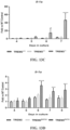
- the TREM2 agonist antigen binding proteins increase pSyk levels with an EC50 less than about 100 nM, less than about 80 nM, less than about 60 nM, less than about 50 nM, less than about 40 nM, less than about 30 nM, less than about 20 nM, less than about 10 nM, less than about 5 nM, less than about 1 nM, less than about 500 pM, less than about 300 pM, or less than about 100 pM.
- the TREM2 agonist antigen binding proteins increase pSyk levels with an EC50 from about 1 pm to about 100 nM, from about 10 pM to about 50 nM, from about 50 pM to about 5 nM, from about 100 pM to about 1 nM, or from about 150 pM to about 500 pM.
- An "EC50” or “half maximal effective concentration” is a measure of potency of the antigen binding protein and refers to the concentration of antigen binding protein required to induce a response halfway between baseline and maximal response after a particular exposure period.
- the EC50 of any particular agonist can be determined by constructing a dose-response curve and examining the effect of different concentrations of the agonist in inducing activity in a particular functional assay (e.g. pSyk levels).
- the EC50 is the concentration of the agonist at which 50% of its maximal effect is observed.
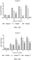
- Increases in intracellular pSyk levels induced by the TREM2 agonist antigen binding proteins of the invention can be assessed by various methods, such as the cell-based assays described in Examples 2 and 6. For instance, cells expressing TREM2 (e.g.
- human TREM2 are contacted with one or more concentrations of an agonist antigen binding protein, the cells are lysed, and pSyk levels in the cell lysates are assessed, for example by Western blot, FRET-based assay or chemiluminescent assay (e.g. AlphaLISA-based assay).
- the cells in the cell-based assay may be cells, such as HEK293T cells or CHO cells, which recombinantly express TREM2 (e.g. human TREM2).
- the cells in the cell-based assay are cells that natively express TREM2 (e.g. human TREM2), such as THP-1 cells, macrophage, microglial cells, or dendritic cells.
- the potency of the TREM2 agonist antigen binding proteins for inducing or increasing pSyk levels in a cell expressing TREM2 is retained in the absence of a cross-linking agent.
- the TREM2 agonist antigen binding proteins of the invention increase pSyk levels with an EC50 from about 1 pM to about 100 nM, from about 10 pM to about 50 nM, from about 50 pM to about 5 nM, from about 100 pM to about 1 nM, or from about 150 pM to about 500 pM in the absence of a cross-linking agent as measured by a cell-based pSyk assay.
- the TREM2 agonist antigen binding protein increases pSyk levels with an EC50 less than 300 pM in the absence of a cross-linking agent as measured by a cell-based pSyk assay. In yet another embodiment, the TREM2 agonist antigen binding protein increases pSyk levels with an EC50 less than 100 pM in the absence of a cross-linking agent as measured by a cell-based pSyk assay.
- the TREM2 agonist antigen binding proteins of the invention comprise one or more complementarity determining regions (CDRs) from the light and heavy chain variable regions of antibodies that specifically bind to human TREM2, as defined in the claims.
- CDR refers to the complementarity determining region (also termed “minimal recognition units” or “hypervariable region") within antibody variable sequences.
- CDRH1, CDRH2 and CDRH3 There are three heavy chain variable region CDRs (CDRH1, CDRH2 and CDRH3) and three light chain variable region CDRs (CDRL1, CDRL2 and CDRL3).
- CDR region refers to a group of three CDRs that occur in a single variable region (i.e. the three light chain CDRs or the three heavy chain CDRs).
- the CDRs in each of the two chains typically are aligned by the framework regions (FRs) to form a structure that binds specifically with a specific epitope or domain on the target protein (e.g., human TREM2).
- FRs framework regions
- a numbering system has been devised for assigning numbers to amino acids that occupy positions in each of these domains. This numbering system is defined in Kabat Sequences of Proteins of Immunological Interest (1987 and 1991, NIH, Bethesda, MD ), or Chothia & Lesk, 1987, J. Mol.
- Such structures can be a naturally occurring polypeptide or polypeptide "fold” (a structural motif), or can have one or more modifications, such as additions, deletions or substitutions of amino acids, relative to a naturally occurring polypeptide or fold.
- These scaffolds can be derived from a polypeptide of any species (or of more than one species), such as a human, other mammal, other vertebrate, invertebrate, plant, bacteria or virus.
- the TREM2 agonist antigen binding proteins of the invention comprise an immunoglobulin heavy chain variable region (VH) and an immunoglobulin light chain variable region (VL) from an antibody that specifically binds to human TREM2, as defined in the claims.
- VH immunoglobulin heavy chain variable region
- VL immunoglobulin light chain variable region
- the "variable region,” used interchangeably herein with “variable domain” refers to the region in each of the light and heavy immunoglobulin chains which is involved directly in binding the antibody to the antigen.
- the regions of variable light and heavy chains have the same general structure and each region comprises four framework (FR) regions, the sequences of which are widely conserved, connected by three CDRs.
- the framework regions adopt a beta-sheet conformation and the CDRs may form loops connecting the beta-sheet structure.
- the CDRs in each chain are held in their three-dimensional structure by the framework regions and form, together with the CDRs from the other chain, the antigen binding site.
- identity refers to a relationship between the sequences of two or more polypeptide molecules or two or more nucleic acid molecules, as determined by aligning and comparing the sequences.
- Percent identity means the percent of identical residues between the amino acids or nucleotides in the compared molecules and is calculated based on the size of the smallest of the molecules being compared. For these calculations, gaps in alignments (if any) must be addressed by a particular mathematical model or computer program (i.e., an "algorithm”). Methods that can be used to calculate the identity of the aligned nucleic acids or polypeptides include those described in Computational Molecular Biology, (Lesk, A.
- sequence identity can be determined by standard methods that are commonly used to compare the similarity in position of the amino acids of two polypeptides.
- BLAST or FASTA two polypeptide or two polynucleotide sequences are aligned for optimal matching of their respective residues (either along the full length of one or both sequences, or along a pre-determined portion of one or both sequences).
- the programs provide a default opening penalty and a default gap penalty, and a scoring matrix such as PAM 250 ( Dayhoff et al., in Atlas of Protein Sequence and Structure, vol. 5, supp. 3, 1978 ) or BLOSUM62 ( Henikoff et al., 1992, Proc. Natl. Acad. Sci.
- the percent identity can then be calculated as: the total number of identical matches multiplied by 100 and then divided by the sum of the length of the longer sequence within the matched span and the number of gaps introduced into the longer sequences in order to align the two sequences.
- the sequences being compared are aligned in a way that gives the largest match between the sequences.
- the GCG program package is a computer program that can be used to determine percent identity, which package includes GAP ( Devereux et al., 1984, Nucl. Acid Res. 12:387; Genetics Computer Group, University of Wisconsin, Madison, WI ).
- GAP Devereux et al., 1984, Nucl. Acid Res. 12:387; Genetics Computer Group, University of Wisconsin, Madison, WI ).
- the computer algorithm GAP is used to align the two polypeptides or two polynucleotides for which the percent sequence identity is to be determined. The sequences are aligned for optimal matching of their respective amino acid or nucleotide (the "matched span", as determined by the algorithm).
- a gap opening penalty (which is calculated as 3x the average diagonal, wherein the "average diagonal” is the average of the diagonal of the comparison matrix being used; the “diagonal” is the score or number assigned to each perfect amino acid match by the particular comparison matrix) and a gap extension penalty (which is usually 1/10 times the gap opening penalty), as well as a comparison matrix such as PAM 250 or BLOSUM 62 are used in conjunction with the algorithm.
- a standard comparison matrix (see, Dayhoff et al., 1978, Atlas of Protein Sequence and Structure 5:345-352 for the PAM 250 comparison matrix; Henikoff et al., 1992, Proc. Natl. Acad. Sci. U.S.A. 89:10915-10919 for the BLOSUM 62 comparison matrix) is also used by the algorithm.
- Certain alignment schemes for aligning two amino acid sequences may result in matching of only a short region of the two sequences, and this small aligned region may have very high sequence identity even though there is no significant relationship between the two full-length sequences. Accordingly, the selected alignment method (GAP program) can be adjusted if so desired to result in an alignment that spans at least 50 contiguous amino acids of the target polypeptide.
- Variants of the anti-TREM2 antibodies described herein can be generated by substituting one or more amino acids in the light chain or heavy chain variable regions to address chemical liabilities (e.g. aspartate isomerization, asparagine deamidation, tryptophan and methionine oxidation) or correct covariance violations (see WO 2012/125495 ) as described in Example 7.
- Such variants can have improved biophysical, expression, and/or stability properties as compared with the parental antibody.
- the TREM2 agonist antigen binding proteins described herein may comprise a light chain variable region and/or heavy chain variable region having one or more of the amino acid substitutions set forth in any of Tables 13-18.
- the Kabat numbering scheme is typically used when referring to the position of an amino acid within the variable regions, whereas the EU numbering scheme is generally used when referring to the position of an amino acid with an immunoglobulin constant region.
- a chart summarizing correspondence between Kabat and EU numbering schemes with other numbering schemes is available on the IMGT ® website (the international ImMunoGeneTics information system).
- the TREM2 agonist antigen binding proteins of the invention comprise a light chain variable region comprising the amino acid sequence of SEQ ID NO: 330 and a heavy chain variable region comprising the amino acid sequence of SEQ ID NO: 331, as defined in the claims.
- the TREM2 agonist antigen binding proteins described herein may comprise a light chain variable region and/or heavy chain variable region from an affinity-modulated variant of the 6E7 antibody (Example 8).
- the TREM2 agonist antigen binding proteins described herein comprise a light chain variable region and/or a heavy chain variable region having one or more of the amino acid substitutions set forth in Table 23.
- the TREM2 agonist antigen binding protein described herein may comprise a light chain variable region comprising the sequence of SEQ ID NO: 61 with a mutation at one or more amino acid positions 24, 31, 50, 52, 54, 56, 89, 92, 93, 94 and/or 96.
- the mutation may be selected from R24A, S31R, A50S, A50G, S52G, L54R, N56K, N56R, N56L, N56T, Q89G, D92V, S93R, F94Y, F94L, R96H, R96L, or combinations thereof.
- the TREM2 agonist antigen binding protein may comprise a heavy chain variable region comprising the sequence of SEQ ID NO: 124 with a mutation at one or more amino acid positions 27, 28, 30, 32, 50, 54, 58, 60, 61, 63, 66, 99, 101, 103, 104, and/or 110.
- the mutation may be selected from Y27S, S28G, S28H, T30N, T30G, T30E, T30A, Y32E, I50T, G54S, T58V, Y60L, S61A, S63G, S63E, G66D, Q99G, Q99S, Q99M, T101G, Y103R, Y104G, F110S, or combinations thereof.
- Amino acid sequences for light chain and heavy chain variable regions and associated CDRs of exemplary variants of the 6E7 antibody with improved affinity are set forth below in Tables 2A and 2B, respectively.
- Amino acid sequences for light chain and heavy chain variable regions and associated CDRs of exemplary variants of the 6E7 antibody with reduced affinity are set forth below in Tables 3A and 3B, respectively. The corresponding sequences for the 6E7 antibody are listed for comparison.
- Table 2A Light Chain Variable Region Amino Acid Sequences for Improved Affinity TREM2 Antibodies Variant Ab ID.
- the TREM2 agonist antigen binding proteins described herein may comprise one or more of the CDRs from the reduced affinity variants presented in Table 3A (light chain CDRs; i.e. CDRLs) and Table 3B (heavy chain CDRs, i.e. CDRHs), which are useful for understanding the claimed invention.
- the immunoglobulin light chain constant domain can be a human kappa ( ⁇ ) or human lambda ( ⁇ ) constant domain.
- the term "heavy chain” or “immunoglobulin heavy chain” refers to a polypeptide comprising, from amino terminus to carboxyl terminus, a single immunoglobulin heavy chain variable region (VH), an immunoglobulin heavy chain constant domain 1 (CH1), an immunoglobulin hinge region, an immunoglobulin heavy chain constant domain 2 (CH2), an immunoglobulin heavy chain constant domain 3 (CH3), and optionally an immunoglobulin heavy chain constant domain 4 (CH4).
- the immunoglobulin heavy chain constant domains can be from any immunoglobulin isotype, including subtypes.
- the antibody chains are linked together via inter-polypeptide disulfide bonds between the CL domain and the CH1 domain (i.e. between the light and heavy chain) and between the hinge regions of the antibody heavy chains.
- the anti-TREM2 antibodies of the invention can comprise any immunoglobulin constant region.
- the term "constant region” as used herein refers to all domains of an antibody other than the variable region.
- the constant region is not involved directly in binding of an antigen, but exhibits various effector functions.
- antibodies are divided into particular isotypes (IgA, IgD, IgE, IgG, and IgM) and subtypes (IgG1, IgG2, IgG3, IgG4, IgA1 IgA2) depending on the amino acid sequence of the constant region of their heavy chains.
- the light chain constant region can be, for example, a kappa- or lambda-type light chain constant region, e.g., a human kappa- or lambda-type light chain constant region, which are found in all five antibody isotypes.
- Examples of human immunoglobulin light chain constant region sequences are shown in the following table. Table 4. Exemplary Human Immunoglobulin Light Chain Constant Regions Designation SEQ ID NO: CL Domain Amino Acid Sequence Human lambda v1 191 Human lambda v2 192 Human lambda v3 193 Human lambda v4 194 Human lambda v5 195 Human kappa v1 196 Human kappa v2 197
- the human IgG1 immunoglobulin constant region may comprise one or more mutations to prevent glycosylation of the antibody as described in more detail herein.
- the anti-TREM2 antibody comprises a heavy chain constant region from a human IgG2 immunoglobulin.
- the anti-TREM2 antibody comprises a heavy chain constant region from a human IgG4 immunoglobulin. Examples of human IgG1, IgG2, and IgG4 heavy chain constant region sequences are shown below in Table 5. Table 5.
- Exemplary Human Immunoglobulin Heavy Chain Constant Regions Ig isotype SEQ ID NO: Heavy Chain Constant Region Amino Acid Sequence Human IgG1z 198 Human IgG1za 199 Human IgG1f 200 Human IgG1fa 201 Human IgG1z aglycosylated v1 202 Human IgG1z aglycosylated v2 203 Human IgG2 204 Human IgG4 205
- Each of the light chain variable regions disclosed in Tables 1A, 2A, and 3A and each of the heavy chain variable regions disclosed in Tables 1B, 2B, and 3B may be attached to the above light chain constant regions (Table 4) and heavy chain constant regions (Table 5) to form complete antibody light and heavy chains, respectively. Further, each of the so generated heavy and light chain sequences may be combined to form a complete antibody structure. It should be understood that the heavy chain and light chain variable regions provided herein can also be attached to other constant domains having different sequences than the exemplary sequences listed above.
- the TREM2 agonist antigen binding protein of the invention is a monoclonal antibody or antigen-binding fragment thereof, as defined in the claims.
- the anti-TREM2 antibody may be a chimeric antibody, a humanized antibody, or a fully human antibody having a human immunoglobulin constant domain.
- the anti-TREM2 antibody is a human IgG1, IgG2, IgG3, or IgG4 antibody.
- the anti-TREM2 antibody may, in some embodiments, have a human IgG1, IgG2, IgG3, or IgG4 constant domain.
- the anti-TREM2 antibody is a monoclonal human IgG1 antibody.
- the anti-TREM2 antibody is a monoclonal human IgG2 antibody.
- the anti-TREM2 antibody is a monoclonal human IgG4 antibody.
- monoclonal antibody refers to an antibody obtained from a population of substantially homogeneous antibodies, i.e., the individual antibodies comprising the population are identical except for possible naturally occurring mutations that may be present in minor amounts.
- Monoclonal antibodies are highly specific, being directed against an individual antigenic site or epitope, in contrast to polyclonal antibody preparations that typically include different antibodies directed against different epitopes.
- Monoclonal antibodies may be produced using any technique known in the art, e.g., by immortalizing spleen cells harvested from an animal after completion of the immunization schedule.
- the spleen cells can be immortalized using any technique known in the art, e.g., by fusing them with myeloma cells to produce hybridomas. See, for example, Antibodies; Harlow and Lane, Cold Spring Harbor Laboratory Press, 1st Edition, e.g. from 1988, or 2nd Edition, e.g. from 2014 .
- Myeloma cells for use in hybridoma-producing fusion procedures preferably are non-antibody-producing, have high fusion efficiency, and enzyme deficiencies that render them incapable of growing in certain selective media, which support the growth of only the desired fused cells (hybridomas).
- suitable cell lines for use in fusions with mouse cells include, but are not limited to, Sp-20, P3-X63/Ag8, P3-X63-Ag8.653, NS1/1.Ag 4 1, Sp210-Ag14, FO, NSO/U, MPC-11, MPC11-X45-GTG 1.7 and S194/5XXO Bul.
- suitable cell lines used for fusions with rat cells include, but are not limited to, R210.RCY3, Y3-Ag 1.2.3, IR983F and 4B210.
- Other cell lines useful for cell fusions are U-266, GM1500-GRG2, LICR-LON-HMy2 and UC729-6.
- a hybridoma cell line is produced by immunizing an animal (e.g., a rabbit, rat, mouse, or a transgenic animal having human immunoglobulin sequences) with a TREM2 immunogen (such as the immunogens described in Example 1); harvesting spleen cells from the immunized animal; fusing the harvested spleen cells to a myeloma cell line, thereby generating hybridoma cells; establishing hybridoma cell lines from the hybridoma cells, and identifying a hybridoma cell line that produces an antibody that binds to human TREM2.
- TREM2 immunogen such as the immunogens described in Example 1
- Another useful method for producing monoclonal antibodies is the SLAM method described in Babcook et al., Proc. Natl. Acad. Sci. USA, Vol. 93: 7843-7848, 1996 .
- Monoclonal antibodies secreted by a hybridoma cell line can be purified using any technique known in the art, such as protein A-Sepharose, hydroxylapatite chromatography, gel electrophoresis, dialysis, or affinity chromatography.
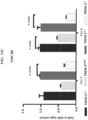
- Hybridoma supernatants or mAbs may be further screened to identify mAbs with particular properties, such as the ability to bind human TREM2, cross-reactivity to TREM2 proteins from other species (e.g., mouse TREM2, rat TREM2, and cynomologus monkey TREM2), cross-reactivity to other TREM family members (e.g. human TREM1), ability to induce or increase TREM2-mediated signaling, e.g. using a pSyk assay as described herein, or ability to induce or increase TREM2-mediated function or activities as described herein (e.g. proliferation or survival of TREM2-expressing myeloid cells).
- the goal of making a chimeric antibody is to create a chimera in which the number of amino acids from the intended species is maximized.
- One example is the "CDR-grafted" antibody, in which the antibody comprises one or more CDRs from a particular species or belonging to a particular antibody class or subclass, while the remainder of the antibody chain(s) is/are identical with or homologous to a corresponding sequence in antibodies derived from another species or belonging to another antibody class or subclass.
- CDR grafting is described, for example, in United States Patent No. 6,180,370 , No. 5,693,762 , No. 5,693,761 , No. 5,585,089 , and No. 5,530,101 .
- the variable region or selected CDRs from a rodent or rabbit antibody often are grafted into a human antibody, replacing the naturally-occurring variable regions or CDRs of the human antibody.
- a humanized antibody is produced from a monoclonal antibody raised initially in a non-human animal, such as a rodent or rabbit. Certain amino acid residues in this monoclonal antibody, typically from non-antigen recognizing portions of the antibody, are modified to be homologous to corresponding residues in a human antibody of corresponding isotype. Humanization can be performed, for example, using various methods by substituting at least a portion of a rodent or rabbit variable region for the corresponding regions of a human antibody (see, e.g., United States Patent No. 5,585,089 , and No.
- the CDRs of the light and heavy chain variable regions of the antibodies described herein are grafted to framework regions (FRs) from antibodies from the same, or a different, phylogenetic species.
- FRs framework regions
- the CDRs of the heavy and light chain variable regions listed in Tables 1A, 1B, 2A, 2B, 3A, and 3B can be grafted to consensus human FRs.
- consensus human FRs FRs from several human heavy chain or light chain amino acid sequences may be aligned to identify a consensus amino acid sequence.
- the grafted variable regions from the one heavy or light chain may be used with a constant region that is different from the constant region of that particular heavy or light chain as disclosed herein.
- the grafted variable regions may be part of a single chain Fv antibody.
- Ig loci introduction of human immunoglobulin (Ig) loci into mice in which the endogenous Ig genes have been inactivated is one means of producing fully human monoclonal antibodies (mAbs) in mouse, an animal that can be immunized with any desirable antigen.
- mAbs monoclonal antibodies
- Using fully human antibodies can minimize the immunogenic and allergic responses that can sometimes be caused by administering mouse or mouse-derived mAbs to humans as therapeutic agents.
- Fully human antibodies can be produced by immunizing transgenic animals (usually mice) that are capable of producing a repertoire of human antibodies in the absence of endogenous immunoglobulin production.
- Antigens for this purpose typically have six or more contiguous amino acids, and optionally are conjugated to a carrier, such as a hapten. See, e.g., Jakobovits et al., 1993, Proc. Natl. Acad. Sci. USA 90:2551-2555 ; Jakobovits et al., 1993, Nature 362:255-258 ; and Bruggermann et al., 1993, Year in Immunol. 7:33 .
- transgenic animals are produced by incapacitating the endogenous mouse immunoglobulin loci encoding the mouse heavy and light immunoglobulin chains therein, and inserting into the mouse genome large fragments of human genome DNA containing loci that encode human heavy and light chain proteins.
- Partially modified animals which have less than the full complement of human immunoglobulin loci, are then cross-bred to obtain an animal having all of the desired immune system modifications.
- these transgenic animals produce antibodies that are immunospecific for the immunogen but have human rather than murine amino acid sequences, including the variable regions.
- HuMab mice The preparation of HuMab mice is described in detail in Taylor et al., 1992, Nucleic Acids Research 20:6287-6295 ; Chen et al., 1993, International Immunology 5:647-656 ; Tuaillon et al., 1994, J. Immunol. 152:2912-2920 ; Lonberg et al., 1994, Nature 368:856-859 ; Lonberg, 1994, Handbook of Exp.
- Effector functions can be introduced by one of two strategies:
- the fragments can be engineered either into complete antibodies for expression in mammalian cells, or into bispecific antibody fragments with a second binding site capable of triggering an effector function, if desired.
- the Fd fragment (VH-CH1) and light chain (VL-CL) of antibodies are separately cloned by PCR and recombined randomly in combinatorial phage display libraries, which can then be selected for binding to a particular antigen.
- the antibody fragments are expressed on the phage surface, and selection of Fv or Fab (and therefore the phage containing the DNA encoding the antibody fragment) by antigen binding is accomplished through several rounds of antigen binding and re-amplification, a procedure termed panning.
- solid phase direct-labeled assay solid phase direct-labeled sandwich assay (see, e.g., Harlow and Lane, 1988, Antibodies, A Laboratory Manual, Cold Spring Harbor Press ); solid phase direct label RIA using I-125 label (see, e.g., Morel et al., 1988, Molec. Immunol. 25:7-15 ); solid phase direct biotin-avidin EIA (see, e.g., Cheung, et al., 1990, Virology 176:546-552 ); surface plasmon resonance-based assays (e.g. using Biacore ® systems); bio-layer interferometry-based assays (e.g.
- a competitive binding assay involves the use of purified antigen bound to a solid surface or cells bearing the antigen, an unlabeled test antibody or other antigen binding protein, and a labeled reference antibody or other antigen binding protein. Competitive inhibition is measured by determining the amount of label bound to the solid surface or cells in the presence of the test antibody or other antigen binding protein. Usually the test antibody or other antigen binding protein is present in excess. Antibodies or other antigen binding proteins identified by competition assay (i.e.
- competing antibodies and antigen binding proteins include antibodies and antigen binding proteins binding to the same epitope as the reference antibody or antigen binding protein.
- a competing antibody or other antigen binding protein when it is present in excess, it will inhibit specific binding of a reference antibody or other antigen binding protein to a target antigen by at least 40%, 45%, 50%, 55%, 60%, 65%, 70% or 75%. In some instances, binding of the reference antibody or other antigen binding protein is inhibited by at least 80%, 85%, 90%, 95%, or 97% or more.
- a competing antigen binding protein e.g. antibody or binding fragment thereof
- a particularly suitable quantitative assay for detecting competitive binding uses a Biacore ® machine which measures the extent of interactions using surface plasmon resonance technology.
- An exemplary Biacore ® -based competitive binding assay involves the immobilization of a reference antibody to a sensor chip. The target antigen is then contacted with the sensor chip where the target antigen is captured by the immobilized reference antibody. Test antibodies are then injected over the captured target antigen. If the injected test antibody recognizes a distinct epitope from that recognized by the immobilized antibody, then a second binding event is observed and the test antibody would be considered not to compete for binding to the target antigen with the reference antibody.
- Another particularly suitable assay for detecting competitive binding employs kinetic sensors used with Octet ® systems (Pall ForteBio), which measures binding interactions using bio-layer interferometry methodology.
- Octet ® systems Pall ForteBio
- Such an assay is described in Example 4, in which each of sixteen different anti-TREM2 antibodies described herein were evaluated against each other for the ability to compete for binding to human TREM2.
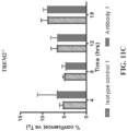
- the results of the analysis provided in Table 9 show that the sixteen different antibodies could be grouped into four distinct epitope bins. That is, one group of antibodies (antibodies 10E3, 13E7, 24F4, 4C5, 4G10, 32E3, and 6E7) competed with each other for binding to human TREM2, indicating that they share the same or similar epitope on human TREM2.
- Antibodies 16B8, 26A10, 26C10, 26F2, 33B12, and 5E3 competed with each other for TREM2 binding, but did not compete with antibodies in the first group or antibodies 24A10, 24G6, or 25F12, indicating that this second group of antibodies bind to a distinct epitope on human TREM2.
- Antibodies 24A10 and 24G6 share a similar epitope on human TREM2 as these two antibodies competed with each other for human TREM2 binding, but did not compete with any other antibody.
- Antibody 25F12 did not compete with any of the other tested antibodies for human TREM2 binding, indicating that this antibody binds to yet another epitope.
- the TREM2 agonist antigen binding proteins of the invention may comprise one or more mutations or modifications to a constant region.
- the TREM2 agonist antigen binding proteins comprise an Fc region (e.g. monoclonal antibodies)
- the heavy chain constant regions or the Fc regions of the antigen binding proteins may comprise one or more amino acid substitutions that affect the glycosylation, effector function, and/or Fcy receptor binding of the antigen binding protein.
- the term "Fc region” refers to the C-terminal region of an immunoglobulin heavy chain which may be generated by papain digestion of an intact antibody.
- the Fc region of an immunoglobulin generally comprises two constant domains, a CH2 domain and a CH3 domain, and optionally comprises a CH4 domain.
- the Fc region is an Fc region from an IgG1, IgG2, IgG3, or IgG4 immunoglobulin.
- the Fc region comprises CH2 and CH3 domains from a human IgG1 or human IgG2 immunoglobulin.
- the TREM2 agonist antigen binding proteins (e.g. monoclonal antibodies) of the invention comprise one or more amino acid substitutions in a heavy chain constant region to reduce effector function.
- Exemplary amino acid substitutions (according to EU numbering scheme) that can reduce effector function include, but are not limited to, C220S, C226S, C229S, E233P, L234A, L234V, V234A, L234F, L235A, L235E, G237A, P238S, S267E, H268Q, N297A, N297G, V309L, E318A, L328F, A330S, A331S, P331S or combinations of any of the foregoing.
- the TREM2 agonist antigen binding proteins of the invention may comprise one or more amino acid substitutions that affect the level or type of glycosylation of the binding proteins.
- Glycosylation of polypeptides is typically either N-linked or O-linked. N-linked refers to the attachment of the carbohydrate moiety to the side chain of an asparagine residue.
- the tri-peptide sequences asparagine-X-serine and asparagine-X-threonine, where X is any amino acid except proline, are the recognition sequences for enzymatic attachment of the carbohydrate moiety to the asparagine side chain.
- O-linked glycosylation refers to the attachment of one of the sugars N-acetylgalactosamine, galactose, or xylose, to a hydroxyamino acid, most commonly serine or threonine, although 5-hydroxyproline or 5-hydroxylysine may also be used.
- glycosylation of the TREM2 agonist antigen binding proteins described herein is increased by adding one or more glycosylation sites, e.g., to the Fc region of the binding protein.
- Addition of glycosylation sites to the antigen binding protein can be conveniently accomplished by altering the amino acid sequence such that it contains one or more of the above-described tri-peptide sequences (for N-linked glycosylation sites). The alteration may also be made by the addition of, or substitution by, one or more serine or threonine residues to the starting sequence (for O-linked glycosylation sites).
- the antigen binding protein amino acid sequence may be altered through changes at the DNA level, particularly by mutating the DNA encoding the target polypeptide at preselected bases such that codons are generated that will translate into the desired amino acids.
- TREM2 antigen binding protein molecules with altered carbohydrate structure resulting in altered effector activity, including antigen binding proteins with absent or reduced fucosylation that exhibit improved ADCC activity.
- Various methods are known in the art to reduce or eliminate fucosylation.
- ADCC effector activity is mediated by binding of the antibody molecule to the Fc ⁇ RIII receptor, which has been shown to be dependent on the carbohydrate structure of the N-linked glycosylation at the N297 residue of the CH2 domain.
- Non-fucosylated antibodies bind this receptor with increased affinity and trigger Fc ⁇ RIII-mediated effector functions more efficiently than native, fucosylated antibodies.
- the TREM2 agonist antigen binding proteins described herein comprise a mutation at position N297 (according to EU numbering scheme), such as N297Q, N297A, or N297G.
- the TREM2 agonist antigen binding proteins of the invention comprise an Fc region from a human IgG1 antibody with a mutation at position N297.
- the TREM2 agonist antigen binding proteins of the invention comprise an Fc region from a human IgG1 antibody with a N297G mutation.
- the TREM2 agonist antigen binding proteins of the invention comprise a heavy chain constant region comprising the sequence of SEQ ID NO: 202.
- the Fc region of the TREM2 agonist antigen binding proteins may be further engineered.
- one or more amino acids in the Fc region are substituted with cysteine to promote disulfide bond formation in the dimeric state.
- Residues corresponding to V259, A287, R292, V302, L306, V323, or I332 (according to EU numbering scheme) of an IgG1 Fc region may thus be substituted with cysteine.
- specific pairs of residues are substituted with cysteine such that they preferentially form a disulfide bond with each other, thus limiting or preventing disulfide bond scrambling.
- the TREM2 agonist antigen binding proteins described herein comprise an Fc region from a human IgG1 antibody with mutations R292C and V302C.
- the Fc region may also comprise a N297 mutation, such as a N297G mutation.
- the TREM2 agonist antigen binding proteins of the invention comprise a heavy chain constant region comprising the sequence of SEQ ID NO: 203.
- Modifications to the hinge region and/or CH1 domain of the heavy chain and/or the constant region of the light chain of the TREM2 agonist antigen binding proteins (e.g. monoclonal antibodies) of the invention can be made to reduce or eliminate disulfide heterogeneity.
- Structural hetereogeneity of IgG2 antibodies has been observed where the disulfide bonds in the hinge and CH1 regions of IgG2 antibodies can be shuffled to create different structural disulfide isoforms (IgG2A, IgG2B, and IgG2A-B), which can have different levels of activity. See, e.g., Dillon et al., J. Biol. Chem., Vol.
- Amino acid substitutions can be made in the hinge region, CH1 domain, and/or light chain constant region to promote the formation of a single disulfide isoform or lock the antigen binding protein (e.g. monoclonal antibody) into a particular disulfide isoform (e.g. IgG2A or IgG2B).
- antigen binding protein e.g. monoclonal antibody
- disulfide isoform e.g. IgG2A or IgG2B
- the TREM2 agonist antigen binding proteins of the invention are human IgG2 anti-TREM2 agonist antibodies.
- the TREM2 agonist antibodies comprise a C131S mutation (according to the EU numbering scheme) in their heavy chains.
- the TREM2 agonist antibodies comprise a C214S mutation (according to the EU numbering scheme) in their light chains and a C220S mutation (according to the EU numbering scheme) in their heavy chains.
- the TREM2 agonist antibodies comprise a C214S mutation (according to the EU numbering scheme) in their light chains and a C219S mutation (according to the EU numbering scheme) in their heavy chains.
- the TREM2 agonist antigen binding proteins of the invention are anti-TREM2 agonist antibodies comprising a CH1 region and hinge region from a human IgG2 antibody and an Fc region from a human IgG1 antibody.
- the unique arrangement of the disulfide bonds in the hinge region of IgG2 antibodies has been reported to impart enhanced stimulatory activity for certain anticancer antibodies ( White et al., Cancer Cell, Vol. 27: 138-148, 2015 ). This enhanced activity could be transferred to IgG1-type antibodies by exchanging the CH1 and hinge regions of the IgG1 antibody for those in the IgG2 antibody (White et al., 2015).
- the IgG2 hinge region includes the amino acid sequence ERKCCVECPPCP (SEQ ID NO: 206).
- the amino acid sequence of the CH1 and hinge regions from a human IgG2 antibody may comprise the amino acid sequence of ASTKGPSVFP LAPCSRSTSE STAALGCLVK DYFPEPVTVS WNSGALTSGV HTFPAVLQSS GLYSLSSVVT VPSSNFGTQT YTCNVDHKPS NTKVDKTVER KCCVECPPCP (SEQ ID NO: 207).
- the anti-TREM2 agonist antibodies comprise the sequence of SEQ ID NO: 207 in combination with an Fc region from a human IgG1 antibody.
- the anti-TREM2 antibodies can comprise one or more of the mutations described above to lock the anti-TREM2 antibodies into a particular disulfide isoform.
- the anti-TREM2 antibody comprises a CH1 region and hinge region from a human IgG2 antibody and an Fc region from a human IgG1 antibody and comprises a C131S mutation (according to the EU numbering scheme) in its heavy chain.
- the anti-TREM2 antibodies may comprise any of the mutations in the Fc region described above to modulate the glycosylation of the antibodies.
- the human IgG1 Fc region of such anti-TREM2 antibodies may comprise a mutation at amino acid position N297 (according to the EU numbering scheme) in its heavy chain.
- the N297 mutation is a N297G mutation.
- the Fc region may further comprise R292C and V302C mutations (according to the EU numbering scheme) in its heavy chain.
- the anti-TREM2 antibodies of the invention comprise a CH1 region and hinge region from a human IgG2 antibody and an Fc region from a human IgG1 antibody, wherein the Fc region comprises the amino acid sequence of:
- Modifications of the TREM2 agonist antigen binding proteins of the invention to increase serum half-life also may desirable, for example, by incorporation of or addition of a salvage receptor binding epitope (e.g., by mutation of the appropriate region or by incorporating the epitope into a peptide tag that is then fused to the antigen binding protein at either end or in the middle, e.g., by DNA or peptide synthesis; see, e.g., WO96/32478 ) or adding molecules such as PEG or other water soluble polymers, including polysaccharide polymers.
- a salvage receptor binding epitope e.g., by mutation of the appropriate region or by incorporating the epitope into a peptide tag that is then fused to the antigen binding protein at either end or in the middle, e.g., by DNA or peptide synthesis; see, e.g., WO96/32478
- adding molecules such as PEG or other water soluble polymers, including polysaccharide poly
- the salvage receptor binding epitope preferably constitutes a region wherein any one or more amino acid residues from one or two loops of an Fc region are transferred to an analogous position in the antigen binding protein. Even more preferably, three or more residues from one or two loops of the Fc region are transferred. Still more preferred, the epitope is taken from the CH2 domain of the Fc region (e.g., an IgG Fc region) and transferred to the CH1, CH3, or VH region, or more than one such region, of the antigen binding protein. Alternatively, the epitope is taken from the CH2 domain of the Fc region and transferred to the CL region or VL region, or both, of the antigen binding protein. See International applications WO 97/34631 and WO 96/32478 for a description of Fc variants and their interaction with the salvage receptor.
- the TREM2 agonist antigen binding proteins of the invention comprise a light chain comprising the sequence of SEQ ID NO: 339 and a heavy chain comprising the sequence of SEQ ID NO: 340.
- the TREM2 agonist antigen binding proteins of the invention comprise a light chain and a heavy chain, wherein the light chain consists of or consists essentially of the amino acid sequence of SEQ ID NO: 339 and the heavy chain consists of or consists essentially of the amino acid sequence of SEQ ID NO: 340.
- the TREM2 agonist antigen binding proteins of the invention are "bispecific" meaning that they are capable of specifically binding to two different antigens, human TREM2 and a second antigen.
- the second antigen is a protein that facilitates transport across the blood-brain barrier, such as a receptor that mediates blood-brain barrier transport.
- receptors include, but are not limited to, the insulin receptor, the transferrin receptor, the leptin receptor, the insulin-like growth factor (IGF) receptor, low density lipoprotein receptors (e.g.
- the second antigen is the human insulin receptor.
- the second antigen is the human insulin-like growth receptor.
- the second antigen is the human transferrin receptor.
- the second antigen is TMEM30A.
- the human TREM2 binding domain could be at the N-terminal end or the C-terminal end of the multivalent bispecific (IgG-Fab, IgG-scFv), or expressed in the multi-specific binding formats described in Spiess, C. et al., Molecular Immunology 67, 95-106 (2015 ) and Brinkman, U. et al., MABS 9(2)182-212 (2017 ).
- the TREM2 agonist antigen binding proteins are multivalent.
- the valency of the binding protein denotes the number of individual antigen binding domains within the binding protein.
- the terms "monovalent,” “bivalent,” and “tetravalent” with reference to the antigen binding proteins of the invention refer to binding proteins with one, two, and four antigen binding domains, respectively.
- a multivalent antigen binding protein comprises two or more antigen binding domains.
- the bispecific antigen binding proteins of the invention are bivalent.
- bispecific, bivalent antigen binding proteins contain two antigen binding domains: one antigen-binding domain binding to human TREM2 and one antigen-binding domain binding to a second antigen, such as an antigen that facilitates transport across the blood-brain barrier.
- the bispecific antigen binding proteins are multivalent.
- the bispecific antigen binding proteins are trivalent or tetravalent comprising three or four antigen-binding domains: one or two antigen-binding domains binding to human TREM2 and one or two antigen-binding domains binding to a second antigen, such as an antigen that facilitates transport across the blood-brain barrier.
- binding domain refers to the region of the antigen binding protein that contains the amino acid residues that interact with the antigen and confer on the antigen binding protein its specificity and affinity for the antigen.
- the binding domain may be derived from an antibody or functional fragment thereof that specifically binds to the antigen.
- the bispecific antigen binding proteins of the invention comprise one antigen-binding domain binding to human TREM2 and one antigen-binding domain binding to the human insulin receptor.
- the bispecific antigen binding proteins of the invention comprise one antigen-binding domain binding to human TREM2 and one antigen-binding domain binding to the human transferrin receptor.
- the bispecific antigen binding proteins of the invention comprise two antigen-binding domains binding to human TREM2 and two antigen-binding domains binding to the human insulin receptor. In other embodiments, the bispecific antigen binding proteins of the invention comprise two antigen-binding domains binding to human TREM2 and two antigen-binding domains binding to the human transferrin receptor. In one embodiment, the bispecific antigen binding proteins of the invention comprise one or two antigen-binding domains binding to human TREM2 and one or two antigen-binding domains binding to the human insulin-like growth receptor.
- the bispecific antigen binding proteins of the invention comprise one or two antigen-binding domains binding to human TREM2 and one or two antigen-binding domains binding to TMEM30A.
- the antigen binding domains binding to human TREM2 of the bispecific TREM2 agonist antigen binding proteins can be derived from any of the anti-TREM2 agonist antibodies described herein.
- the antigen binding domains binding to the human insulin receptor, the human insulin like growth receptor, TMEM30A, or the human transferrin receptor can be derived from monoclonal antibodies to these receptors known in the art, such as those described in US Patent No. 7,388,079 ; US Patent No. 8,663,598 ; and US Patent Publication No.
- bispecific antibodies are known in the art.
- One such method of making a "bispecific" antigen binding protein or antibody involves the fusion of hybridomas or linking of Fab' fragments. See, e.g., Songsivilai and Lachmann, 1990, Clin. Exp. Immunol. 79:315-321 ; Kostelny et al., 1992, J. Immunol. 148:1547-1553 .
- Another method involves engineering the Fc portion of the heavy chains such as to create "knobs" and "holes” which facilitate heterodimer formation of the heavy chains when co-expressed in a cell. See, e.g., WO 96/027011 .
- the nucleic acids comprise, for example, polynucleotides that encode all or part of an antigen binding protein, for example, one or both chains of an antibody of the invention, or a fragment, derivative, mutein, or variant thereof, polynucleotides sufficient for use as hybridization probes, PCR primers or sequencing primers for identifying, analyzing, mutating or amplifying a polynucleotide encoding a polypeptide, anti-sense oligonucleotides for inhibiting expression of a polynucleotide, and complementary sequences of the foregoing.
- the nucleic acids can be any length as appropriate for the desired use or function, and can comprise one or more additional sequences, for example, regulatory sequences, and/or be part of a larger nucleic acid, for example, a vector.
- Nucleic acid molecules of the invention include DNA and RNA in both single-stranded and double-stranded form, as well as the corresponding complementary sequences.
- DNA includes, for example, cDNA, genomic DNA, chemically synthesized DNA, DNA amplified by PCR, and combinations thereof.
- the nucleic acid molecules of the invention include full-length genes or cDNA molecules as well as a combination of fragments thereof.
- the nucleic acids of the invention can be derived from human sources as well as non-human species.
- amino acid sequences from an immunoglobulin or region thereof e.g. variable region, Fc region, etc.
- polypeptide of interest may be determined by direct protein sequencing, and suitable encoding nucleotide sequences can be designed according to a universal codon table.
- genomic or cDNA encoding monoclonal antibodies or binding fragments thereof of the invention can be isolated and sequenced from cells producing such antibodies (e.g. hybridomas) using conventional procedures, such as the methods described in Example 3.
- isolated nucleic acid which is used interchangeably herein with “isolated polynucleotide,” is a nucleic acid that has been separated from adjacent genetic sequences present in the genome of the organism from which the nucleic acid was isolated, in the case of nucleic acids isolated from naturally-occurring sources.
- nucleic acids synthesized enzymatically from a template or chemically such as PCR products, cDNA molecules, or oligonucleotides for example
- nucleic acids resulting from such processes are isolated nucleic acids.
- An isolated nucleic acid molecule refers to a nucleic acid molecule in the form of a separate fragment or as a component of a larger nucleic acid construct.
- the nucleic acids are substantially free from contaminating endogenous material.
- the nucleic acid molecule has preferably been derived from DNA or RNA isolated at least once in substantially pure form and in a quantity or concentration enabling identification, manipulation, and recovery of its component nucleotide sequences by standard biochemical methods (such as those outlined in Sambrook et al., Molecular Cloning: A Laboratory Manual, 2nd ed., Cold Spring Harbor Laboratory, Cold Spring Harbor, NY (1989 )).
- Such sequences are preferably provided and/or constructed in the form of an open reading frame uninterrupted by internal non-translated sequences, or introns, that are typically present in eukaryotic genes.
- Sequences of non-translated DNA can be present 5' or 3' from an open reading frame, where the same do not interfere with manipulation or expression of the coding region. Unless specified otherwise, the left-hand end of any single-stranded polynucleotide sequence discussed herein is the 5' end; the left-hand direction of double-stranded polynucleotide sequences is referred to as the 5' direction.
- RNA transcripts The direction of 5' to 3' production of nascent RNA transcripts is referred to as the transcription direction; sequence regions on the DNA strand having the same sequence as the RNA transcript that are 5' to the 5' end of the RNA transcript are referred to as "upstream sequences"; sequence regions on the DNA strand having the same sequence as the RNA transcript that are 3' to the 3' end of the RNA transcript are referred to as "downstream sequences.”
- nucleic acids that hybridize under moderately stringent conditions, and more preferably highly stringent conditions, to nucleic acids encoding polypeptides as described herein.
- the basic parameters affecting the choice of hybridization conditions and guidance for devising suitable conditions are set forth by Sambrook,, Fritsch, and Maniatis (1989, Molecular Cloning: A Laboratory Manual, Cold Spring Harbor Laboratory Press, Cold Spring Harbor, N.Y., chapters 9 and 11 ; and Current Protocols in Molecular Biology, 1995, Ausubel et al., eds., John Wiley & Sons, Inc. , sections 2.10 and 6.3-6.4), and can be readily determined by those having ordinary skill in the art based on, for example, the length and/or base composition of the DNA.
- One way of achieving moderately stringent conditions involves the use of a prewashing solution containing 5 x SSC, 0.5% SDS, 1.0 mM EDTA (pH 8.0), hybridization buffer of about 50% formamide, 6 x SSC, and a hybridization temperature of about 55°C (or other similar hybridization solutions, such as one containing about 50% formamide, with a hybridization temperature of about 42°C), and washing conditions of about 60°C, in 0.5 x SSC, 0.1% SDS.
- highly stringent conditions are defined as hybridization conditions as above, but with washing at approximately 68°C, 0.2 x SSC, 0.1% SDS.
- SSPE (l x SSPE is 0.15M NaCl, 10 mM NaH 2 PO 4 , and 1.25 mM EDTA, pH 7.4) can be substituted for SSC (l x SSC is 0.15M NaCl and 15 mM sodium citrate) in the hybridization and wash buffers; washes are performed for 15 minutes after hybridization is complete.
- wash temperature and wash salt concentration can be adjusted as necessary to achieve a desired degree of stringency by applying the basic principles that govern hybridization reactions and duplex stability, as known to those skilled in the art and described further below (see, e.g., Sambrook et al., 1989).
- the hybrid length is assumed to be that of the hybridizing nucleic acid.
- the hybrid length can be determined by aligning the sequences of the nucleic acids and identifying the region or regions of optimal sequence complementarity.
- each such hybridizing nucleic acid has a length that is at least 15 nucleotides (or more preferably at least 18 nucleotides, or at least 20 nucleotides, or at least 25 nucleotides, or at least 30 nucleotides, or at least 40 nucleotides, or most preferably at least 50 nucleotides), or at least 25% (more preferably at least 50%, or at least 60%, or at least 70%, and most preferably at least 80%) of the length of the nucleic acid of the present invention to which it hybridizes, and has at least 60% sequence identity (more preferably at least 70%, at least 75%, at least 80%, at least 81%, at least 82%, at least 83%, at least 84%, at least 85%, at least 86%, at least 87%, at least 88%, at least 89%, at least 90%, at least 91%, at least 92%, at least 93%, at least 94%, at least 95%, at least 96%, at least 97%, at least
- Variants of the antigen binding proteins can be prepared by site-specific mutagenesis of nucleotides in the DNA encoding the polypeptide, using cassette or PCR mutagenesis or other techniques well known in the art, to produce DNA encoding the variant, and thereafter expressing the recombinant DNA in cell culture as outlined herein.
- antigen binding proteins comprising variant CDRs having up to about 100-150 residues may be prepared by in vitro synthesis using established techniques.
- the variants typically exhibit the same qualitative biological activity as the naturally occurring analogue, e.g., binding to antigen.
- Such variants include, for example, deletions and/or insertions and/or substitutions of residues within the amino acid sequences of the antigen binding proteins.
- any combination of deletion, insertion, and substitution is made to arrive at the final construct, provided that the final construct possesses the desired characteristics.
- the amino acid changes also may alter post-translational processes of the antigen binding protein, such as changing the number or position of glycosylation sites.
- antigen binding protein variants are prepared with the intent to modify those amino acid residues which are directly involved in epitope binding.
- modification of residues which are not directly involved in epitope binding or residues not involved in epitope binding in any way is desirable, for purposes discussed herein. Mutagenesis within any of the CDR regions, framework regions, and/or constant regions is contemplated, in accordance with the claims.
- Table 6 shows exemplary nucleic acid sequences encoding the light and heavy chain variable regions of anti-TREM2 antibodies described herein. Polynucleotides encoding the anti-TREM2 antibody variable regions can be used to construct the antigen binding proteins described herein. Table 6. Exemplary Anti-TREM2 Antibody Variable Region Nucleic Acid Sequences Ab ID.
- VL or VH Group Designation Nucleic Acid Sequence SEQ ID NO: Light chain variable regions 12G10 LV-01 208 26A10 LV-02 209 26C10 LV-03 210 26F2 LV-04 211 33B12 LV-05 212 24C12 LV-06 213 24G6 LV-07 214 24A10 LV-08 215 10E3 LV-09 216 13E7 LV-10 217 25F12 LV-11 218 32E3 LV-12 219 24F4 LV-13 220 16B8 LV-14 221 4C5 LV-15 222 6E7 V9 V30 V33 V44 V68 LV-16 223 5E3 LV-17 224 4G10 LV-18 225 V3 LV-101 226 V24 LV-102 227 V27 LV-103 228 V40 LV-104 229 V48 LV-105 230 V49 LV-106 231 V52 LV-107 232 V60 LV-108 233 V73 LV-106
- Isolated nucleic acids encoding the anti-TREM2 binding domain of the antigen binding proteins disclosed herein may comprise a nucleotide sequence that is at least 80% identical, at least 90% identical, at least 95% identical, or at least 98% identical to any of the nucleotide sequences listed in Table 6.
- An isolated nucleic acid encoding an anti-TREM2 antibody light chain variable region disclosed herein may comprise a sequence that is at least 80% identical, at least 90% identical, at least 95% identical, or at least 98% identical to a sequence selected from SEQ ID NOs: 208-236 and 313-318.
- nucleic acid sequences provided in Table 6 are exemplary only. As will be appreciated by those in the art, due to the degeneracy of the genetic code, an extremely large number of nucleic acids may be made, all of which encode the CDRs, variable regions, and heavy and light chains or other components of the antigen binding proteins described herein. Thus, having identified a particular amino acid sequence, those skilled in the art could make any number of different nucleic acids, by simply modifying the sequence of one or more codons in a way which does not change the amino acid sequence of the encoded protein.
- the present invention also includes vectors comprising one or more nucleic acids encoding one or more components of the antigen binding proteins of the invention (e.g. variable regions, light chains, and heavy chains).
- vector refers to any molecule or entity (e.g., nucleic acid, plasmid, bacteriophage or virus) used to transfer protein coding information into a host cell.
- vectors include, but are not limited to, plasmids, viral vectors, non-episomal mammalian vectors and expression vectors, for example, recombinant expression vectors.
- expression vector refers to a recombinant DNA molecule containing a desired coding sequence and appropriate nucleic acid control sequences necessary for the expression of the operably linked coding sequence in a particular host cell.
- An expression vector can include, but is not limited to, sequences that affect or control transcription, translation, and, if introns are present, affect RNA splicing of a coding region operably linked thereto.
- Nucleic acid sequences necessary for expression in prokaryotes include a promoter, optionally an operator sequence, a ribosome binding site and possibly other sequences. Eukaryotic cells are known to utilize promoters, enhancers, and termination and polyadenylation signals.
- a secretory signal peptide sequence can also, optionally, be encoded by the expression vector, operably linked to the coding sequence of interest, so that the expressed polypeptide can be secreted by the recombinant host cell, for more facile isolation of the polypeptide of interest from the cell, if desired.
- signal peptide sequences may be appended/fused to the amino terminus of any of the variable region polypeptide sequences listed in Tables 1A, 1B, 2A, 2B, 3A, and 3B.
- a signal peptide having the amino acid sequence of MTCSPLLLTLLIHCTGSWA (SEQ ID NO: 267) is fused to the amino terminus of any of the variable region polypeptide sequences in Tables 1A, 1B, 2A, 2B, 3A, and 3B.
- signal or secretory peptides are known to those of skill in the art and may be fused to any of the variable region polypeptide chains listed in Tables 1A, 1B, 2A, 2B, 3A, and 3B, for example, to facilitate or optimize expression in particular host cells.
- the vector may contain a "tag"-encoding sequence, i.e., an oligonucleotide molecule located at the 5' or 3' end of the polypeptide coding sequence; the oligonucleotide tag sequence encodes polyHis (such as hexaHis), FLAG, HA (hemaglutinin influenza virus), myc, or another "tag" molecule for which commercially available antibodies exist.
- This tag is typically fused to the polypeptide upon expression of the polypeptide, and can serve as a means for affinity purification or detection of the polypeptide from the host cell. Affinity purification can be accomplished, for example, by column chromatography using antibodies against the tag as an affinity matrix.
- the tag can subsequently be removed from the purified polypeptide by various means such as using certain peptidases for cleavage.
- Flanking sequences may be homologous (i.e., from the same species and/or strain as the host cell), heterologous (i.e., from a species other than the host cell species or strain), hybrid (i.e., a combination of flanking sequences from more than one source), synthetic or native.
- the source of a flanking sequence may be any prokaryotic or eukaryotic organism, any vertebrate or invertebrate organism, or any plant, provided that the flanking sequence is functional in, and can be activated by, the host cell machinery.
- Flanking sequences useful in the vectors of this invention may be obtained by any of several methods well known in the art. Typically, flanking sequences useful herein will have been previously identified by mapping and/or by restriction endonuclease digestion and can thus be isolated from the proper tissue source using the appropriate restriction endonucleases. In some cases, the full nucleotide sequence of a flanking sequence may be known. Here, the flanking sequence may be synthesized using routine methods for nucleic acid synthesis or cloning.
- a transcription termination sequence is typically located 3' to the end of a polypeptide coding region and serves to terminate transcription.
- a transcription termination sequence in prokaryotic cells is a G-C rich fragment followed by a poly-T sequence. While the sequence is easily cloned from a library or even purchased commercially as part of a vector, it can also be readily synthesized using known methods for nucleic acid synthesis.
- selectable genes may be used to amplify the gene that will be expressed. Amplification is the process wherein genes that are required for production of a protein critical for growth or cell survival are reiterated in tandem within the chromosomes of successive generations of recombinant cells. Examples of suitable selectable markers for mammalian cells include dihydrofolate reductase (DHFR) and promoterless thymidine kinase genes. Mammalian cell transformants are placed under selection pressure wherein only the transformants are uniquely adapted to survive by virtue of the selectable gene present in the vector.
- DHFR dihydrofolate reductase
- promoterless thymidine kinase genes Mammalian cell transformants are placed under selection pressure wherein only the transformants are uniquely adapted to survive by virtue of the selectable gene present in the vector.
- Selection pressure is imposed by culturing the transformed cells under conditions in which the concentration of selection agent in the medium is successively increased, thereby leading to the amplification of both the selectable gene and the DNA that encodes another gene, such as one or more components of the antigen binding proteins described herein. As a result, increased quantities of a polypeptide are synthesized from the amplified DNA.
- a ribosome-binding site is usually necessary for translation initiation of mRNA and is characterized by a Shine-Dalgarno sequence (prokaryotes) or a Kozak sequence (eukaryotes).
- the element is typically located 3' to the promoter and 5' to the coding sequence of the polypeptide to be expressed.
- one or more coding regions may be operably linked to an internal ribosome binding site (IRES), allowing translation of two open reading frames from a single RNA transcript.
- IRS internal ribosome binding site
- the final protein product may have, in the -1 position (relative to the first amino acid of the mature protein) one or more additional amino acids incident to expression, which may not have been totally removed.
- the final protein product may have one or two amino acid residues found in the peptidase cleavage site, attached to the amino-terminus.
- use of some enzyme cleavage sites may result in a slightly truncated form of the desired polypeptide, if the enzyme cuts at such area within the mature polypeptide.
- Promoters are non-transcribed sequences located upstream (i.e., 5') to the start codon of a structural gene (generally within about 100 to 1000 bp) that control transcription of the structural gene. Promoters are conventionally grouped into one of two classes: inducible promoters and constitutive promoters. Inducible promoters initiate increased levels of transcription from DNA under their control in response to some change in culture conditions, such as the presence or absence of a nutrient or a change in temperature. Constitutive promoters, on the other hand, uniformly transcribe a gene to which they are operably linked, that is, with little or no control over gene expression. A large number of promoters, recognized by a variety of potential host cells, are well known.
- Suitable promoters for use with yeast hosts are also well known in the art.
- Yeast enhancers are advantageously used with yeast promoters.
- Suitable promoters for use with mammalian host cells are well known and include, but are not limited to, those obtained from the genomes of viruses such as polyoma virus, fowlpox virus, adenovirus (such as Adenovirus serotypes 2, 8 or 9), bovine papilloma virus, avian sarcoma virus, cytomegalovirus, retroviruses, hepatitis-B virus and Simian Virus 40 (SV40).
- viruses such as polyoma virus, fowlpox virus, adenovirus (such as Adenovirus serotypes 2, 8 or 9), bovine papilloma virus, avian sarcoma virus, cytomegalovirus, retroviruses, hepatitis-B virus and Simian Virus 40 (SV40).
- Additional promoters which may be of interest include, but are not limited to: SV40 early promoter ( Benoist and Chambon, 1981, Nature 290:304-310 ); CMV promoter ( Thornsen et al., 1984, Proc. Natl. Acad. U.S.A. 81:659-663 ); the promoter contained in the 3' long terminal repeat of Rous sarcoma virus ( Yamamoto et al., 1980, Cell 22:787-797 ); herpes thymidine kinase promoter ( Wagner et al., 1981, Proc. Natl. Acad. Sci. U.S.A.
- mice mammary tumor virus control region that is active in testicular, breast, lymphoid and mast cells ( Leder et al., 1986, Cell 45:485-495 ); the albumin gene control region that is active in liver ( Pinkert et al., 1987, Genes and Devel. 1:268-276 ); the alpha-feto-protein gene control region that is active in liver ( Krumlauf et al., 1985, Mol. Cell. Biol. 5: 1639-1648 ; Hammer et al., 1987, Science 253:53-58 ); the alpha 1-antitrypsin gene control region that is active in liver ( Kelsey et al., 1987, Genes and Devel.
- the beta-globin gene control region that is active in myeloid cells Mogram et al, 1985, Nature 315:338-340 ; Kollias et al, 1986, Cell 46:89-94 ); the myelin basic protein gene control region that is active in oligodendrocyte cells in the brain ( Readhead et al., 1987, Cell 48:703-712 ); the myosin light chain-2 gene control region that is active in skeletal muscle ( Sani, 1985, Nature 314:283-286 ); and the gonadotropic releasing hormone gene control region that is active in the hypothalamus ( Mason et al., 1986, Science 234: 1372-1378 ).
- Enhancers may be inserted into the vector to increase transcription of DNA encoding a component of the antigen binding proteins (e.g., light chain, heavy chain, or variable regions) by higher eukaryotes.
- Enhancers are cis-acting elements of DNA, usually about 10-300 bp in length, that act on the promoter to increase transcription. Enhancers are relatively orientation and position independent, having been found at positions both 5' and 3' to the transcription unit.
- enhancer sequences available from mammalian genes are known (e.g., globin, elastase, albumin, alpha-feto-protein and insulin). Typically, however, an enhancer from a virus is used.
- the SV40 enhancer, the cytomegalovirus early promoter enhancer, the polyoma enhancer, and adenovirus enhancers known in the art are exemplary enhancing elements for the activation of eukaryotic promoters. While an enhancer may be positioned in the vector either 5' or 3' to a coding sequence, it is typically located at a site 5' from the promoter.
- a sequence encoding an appropriate native or heterologous signal sequence (leader sequence or signal peptide) can be incorporated into an expression vector, to promote extracellular secretion of the antigen binding protein. The choice of signal peptide or leader depends on the type of host cells in which the antigen binding protein is to be produced, and a heterologous signal sequence can replace the native signal sequence.
- signal peptides examples include the signal sequence for interleukin-7 (IL-7) described in US Patent No. 4,965,195 ; the signal sequence for interleukin-2 receptor described in Cosman et al.,1984, Nature 312:768 ; the interleukin-4 receptor signal peptide described in EP Patent No. 0367 566 ; the type I interleukin-1 receptor signal peptide described in U.S. Patent No. 4,968,607 ; the type II interleukin-1 receptor signal peptide described in EP Patent No. 0 460 846 .
- IL-7 interleukin-7
- the expression vectors may be constructed from a starting vector such as a commercially available vector. Such vectors may or may not contain all of the desired flanking sequences. Where one or more of the flanking sequences described herein are not already present in the vector, they may be individually obtained and ligated into the vector. Methods used for obtaining each of the flanking sequences are well known to one skilled in the art.
- the expression vectors can be introduced into host cells to thereby produce proteins, including fusion proteins, encoded by nucleic acids as described herein.
- nucleic acids encoding the different components of the TREM2 agonist antigen binding proteins of the invention may be inserted into the same expression vector.
- the nucleic acid encoding an anti-TREM2 antibody light chain or variable region can be cloned into the same vector as the nucleic acid encoding an anti-TREM2 antibody heavy chain or variable region.
- the two nucleic acids may be separated by an internal ribosome entry site (IRES) and under the control of a single promoter such that the light chain and heavy chain are expressed from the same mRNA transcript.
- the two nucleic acids may be under the control of two separate promoters such that the light chain and heavy chain are expressed from two separate mRNA transcripts.
- the nucleic acid encoding the anti-TREM2 antibody light chain or variable region is cloned into one expression vector and the nucleic acid encoding the anti-TREM2 antibody heavy chain or variable region is cloned into a second expression vector.
- a host cell may be co-transfected with both expression vectors to produce complete antigen binding proteins of the invention.
- the completed vector(s) may be inserted into a suitable host cell for amplification and/or polypeptide expression.
- the present invention encompasses an isolated host cell or cell line comprising one or more expression vectors encoding the components of the TREM2 agonist antigen binding proteins as defined in the claims.
- the term "host cell” as used herein refers to a cell that has been transformed, or is capable of being transformed, with a nucleic acid and thereby expresses a gene of interest.
- transformation of an expression vector for an antigen binding protein into a selected host cell may be accomplished by well-known methods including transfection, infection, calcium phosphate co-precipitation, electroporation, microinjection, lipofection, DEAE-dextran mediated transfection, or other known techniques.
- the method selected will in part be a function of the type of host cell to be used.
- a host cell when cultured under appropriate conditions, synthesizes an antigen binding protein that can subsequently be collected from the culture medium (if the host cell secretes it into the medium) or directly from the host cell producing it (if it is not secreted).
- the selection of an appropriate host cell will depend upon various factors, such as desired expression levels, polypeptide modifications that are desirable or necessary for activity (such as glycosylation or phosphorylation) and ease of folding into a biologically active molecule.
- Exemplary host cells include prokaryote, yeast, or higher eukaryote cells.
- Prokaryotic host cells include eubacteria, such as Gram-negative or Gram-positive organisms, for example, Enterobacteriaceae such as Escherichia, e.g., E. coli, Enterobacter, Erwinia, Klebsiella, Proteus, Salmonella, e.g., Salmonella typhimurium, Serratia, e.g., Serratia marcescans, and Shigella, as well as Bacillus, such as B. subtilis and B. licheniformis, Pseudomonas, and Streptomyces.
- Enterobacteriaceae such as Escherichia, e.g., E. coli, Enterobacter, Erwinia, Klebsiella, Proteus
- Salmonella e.g., Salmonella typhimurium
- Serratia e.g.,
- Eukaryotic microbes such as filamentous fungi or yeast are suitable cloning or expression hosts for recombinant polypeptides.
- Saccharomyces cerevisiae, or common baker's yeast is the most commonly used among lower eukaryotic host microorganisms.
- a number of other genera, species, and strains are commonly available and useful herein, such as Pichia, e.g. P.
- yeast pastoris Schizosaccharomyces pombe; Kluyveromyces, Yarrowia; Candida; Trichoderma reesia ; Neurospora crassa; Schwanniomyces, such as Schwanniomyces occidentalis; and filamentous fungi, such as, e.g., Neurospora, Penicillium, Tolypocladium, and Aspergillus hosts such as A. nidulans and A. niger.
- Host cells for the expression of glycosylated antigen binding proteins can be derived from multicellular organisms.
- invertebrate cells include plant and insect cells.
- Numerous baculoviral strains and variants and corresponding permissive insect host cells from hosts such as Spodoptera frugiperda (caterpillar), Aedes aegypti (mosquito), Aedes albopictus (mosquito), Drosophila melanogaster (fruitfly), and Bombyx mori have been identified.
- a variety of viral strains for transfection of such cells are publicly available, e.g., the L-1 variant of Autographa californica NPV and the Bm-5 strain of Bombyx mori NPV.
- Vertebrate host cells are also suitable hosts, and recombinant production of antigen binding proteins from such cells has become routine procedure.
- Mammalian cell lines available as hosts for expression are well known in the art and include, but are not limited to, immortalized cell lines available from the American Type Culture Collection (ATCC), including but not limited to Chinese hamster ovary (CHO) cells, including CHOK1 cells (ATCC CCL61), DXB-11, DG-44, and Chinese hamster ovary cells/-DHFR (CHO, Urlaub et al., Proc. Natl. Acad. Sci.
- monkey kidney CV1 line transformed by SV40 (COS-7, ATCC CRL 1651); human embryonic kidney line (293 or 293 cells subcloned for growth in suspension culture, ( Graham et al., J. Gen Virol. 36: 59, 1977 ); baby hamster kidney cells (BHK, ATCC CCL 10); mouse sertoli cells (TM4, Mather, Biol. Reprod.
- monkey kidney cells (CV1 ATCC CCL 70); African green monkey kidney cells (VERO-76, ATCC CRL-1587); human cervical carcinoma cells (HELA, ATCC CCL 2); canine kidney cells (MDCK, ATCC CCL 34); buffalo rat liver cells (BRL 3A, ATCC CRL 1442); human lung cells (W138, ATCC CCL 75); human hepatoma cells (Hep G2, HB 8065); mouse mammary tumor (MMT 060562, ATCC CCL51); TRI cells ( Mather et al., Annals N.Y Acad. Sci.
- cell lines may be selected through determining which cell lines have high expression levels and constitutively produce antigen binding proteins with human TREM2 binding properties.
- a cell line from the B cell lineage that does not make its own antibody but has a capacity to make and secrete a heterologous antibody can be selected.
- CHO cells are preferred host cells in some embodiments for expressing the TREM2 agonist antigen binding proteins of the invention.
- Host cells are transformed or transfected with the above-described nucleic acids or vectors for production of TREM2 agonist antigen binding proteins and are cultured in conventional nutrient media modified as appropriate for inducing promoters, selecting transformants, or amplifying the genes encoding the desired sequences.
- novel vectors and transfected cell lines with multiple copies of transcription units separated by a selective marker are particularly useful for the expression of antigen binding proteins.
- the present invention also provides a method for producing a TREM2 agonist antigen binding protein as defined in the claims, such as an anti-TREM2 agonist monoclonal antibody or binding fragment thereof, comprising culturing a host cell comprising one or more expression vectors described herein in a culture medium under conditions permitting expression of the antigen binding protein encoded by the one or more expression vectors; and recovering the antigen binding protein from the culture medium or host cell.
- a TREM2 agonist antigen binding protein as defined in the claims, such as an anti-TREM2 agonist monoclonal antibody or binding fragment thereof, comprising culturing a host cell comprising one or more expression vectors described herein in a culture medium under conditions permitting expression of the antigen binding protein encoded by the one or more expression vectors; and recovering the antigen binding protein from the culture medium or host cell.
- the host cells used to produce the antigen binding proteins of the invention may be cultured in a variety of media.
- Commercially available media such as Ham's F10 (Sigma), Minimal Essential Medium (MEM, Sigma), RPMI-1640 (Sigma), and Dulbecco's Modified Eagle's Medium (DMEM, Sigma) are suitable for culturing the host cells.
- the antigen binding protein can be produced intracellularly, in the periplasmic space, or directly secreted into the medium. If the antigen binding protein is produced intracellularly, as a first step, the particulate debris, either host cells or lysed fragments, is removed, for example, by centrifugation or ultrafiltration.
- the antigen binding protein can be purified using, for example, hydroxyapatite chromatography, cation or anion exchange chromatography, size-exclusion chromatography, or preferably affinity chromatography, using the antigen(s) of interest or protein A or protein G as an affinity ligand.
- the Bakerbond ABX TM resin J. T. Baker, Phillipsburg, N.J.
- Other techniques for protein purification such as ethanol precipitation, Reverse Phase HPLC, chromatofocusing, SDS-PAGE, and ammonium sulfate precipitation are also possible depending on the particular antigen binding protein to be recovered.
- the invention provides a composition (e.g. a pharmaceutical composition) comprising one or a plurality of the TREM2 agonist antigen binding proteins of the invention (anti-TREM2 agonist monoclonal antibodies or binding fragments thereof) together with pharmaceutically acceptable diluents, carriers, excipients, solubilizers, emulsifiers, preservatives, and/or adjuvants.
- Pharmaceutical compositions of the invention include, but are not limited to, liquid, frozen, and lyophilized compositions.
- “Pharmaceutically-acceptable” refers to molecules, compounds, and compositions that are non-toxic to human recipients at the dosages and concentrations employed and/or do not produce allergic or adverse reactions when administered to humans.
- the pharmaceutical composition may contain formulation materials for modifying, maintaining or preserving, for example, the pH, osmolarity, viscosity, clarity, color, isotonicity, odor, sterility, stability, rate of dissolution or release, adsorption or penetration of the composition.
- formulation materials for modifying, maintaining or preserving for example, the pH, osmolarity, viscosity, clarity, color, isotonicity, odor, sterility, stability, rate of dissolution or release, adsorption or penetration of the composition.
- suitable formulation materials include, but are not limited to, amino acids (such as glycine, glutamine, asparagine, arginine or lysine); antimicrobials; antioxidants (such as ascorbic acid, sodium sulfite or sodium hydrogensulfite); buffers (such as borate, bicarbonate, Tris-HCl, citrates, phosphates or other organic acids); bulking agents (such as mannitol or glycine); chelating agents (such as ethylenediamine tetraacetic acid (EDTA)); complexing agents (such as caffeine, polyvinylpyrrolidone, beta-cyclodextrin or hydroxypropyl-beta-cyclodextrin); fillers; monosaccharides; disaccharides; and other carbohydrates (such as glucose, mannose or dextrins); proteins (such as serum albumin, gelatin or immunoglobulins); coloring, flavoring and diluting agents; emulsifying agents; hydro
- amino acids
- the pharmaceutical composition of the invention comprises a standard pharmaceutical carrier, such as a sterile phosphate buffered saline solution, bacteriostatic water, and the like.
- a standard pharmaceutical carrier such as a sterile phosphate buffered saline solution, bacteriostatic water, and the like.
- aqueous carriers may be used, e.g., water, buffered water, 0.4% saline, 0.3% glycine and the like, and may include other proteins for enhanced stability, such as albumin, lipoprotein, globulin, etc., subjected to mild chemical modifications or the like.
- Exemplary concentrations of the antigen binding proteins in the formulation may range from about 0.1 mg/ml to about 200 mg/ml or from about 0.1 mg/mL to about 50 mg/mL, or from about 0.5 mg/mL to about 25 mg/mL, or alternatively from about 2 mg/mL to about 10 mg/mL.
- An aqueous formulation of the antigen binding protein may be prepared in a pH-buffered solution, for example, at pH ranging from about 4.5 to about 6.5, or from about 4.8 to about 5.5, or alternatively about 5.0.
- buffers that are suitable for a pH within this range include acetate (e.g.
- the buffer concentration can be from about 1 mM to about 200 mM, or from about 10 mM to about 60 mM, depending, for example, on the buffer and the desired isotonicity of the formulation.
- a tonicity agent which may also stabilize the antigen binding protein, may be included in the formulation.
- exemplary tonicity agents include polyols, such as mannitol, sucrose or trehalose.
- the aqueous formulation is isotonic, although hypertonic or hypotonic solutions may be suitable.
- concentrations of the polyol in the formulation may range from about 1% to about 15% w/v.
- a surfactant may also be added to the antigen binding protein formulation to reduce aggregation of the formulated antigen binding protein and/or minimize the formation of particulates in the formulation and/or reduce adsorption.
- exemplary surfactants include nonionic surfactants such as polysorbates (e.g. polysorbate 20 or polysorbate 80) or poloxamers (e.g. poloxamer 188).
- Exemplary concentrations of surfactant may range from about 0.001% to about 0.5%, or from about 0.005% to about 0.2%, or alternatively from about 0.004% to about 0.01% w/v.
- the formulation contains the above-identified agents (i.e. antigen binding protein, buffer, polyol and surfactant) and is essentially free of one or more preservatives, such as benzyl alcohol, phenol, m-cresol, chlorobutanol and benzethonium chloride.
- a preservative may be included in the formulation, e.g., at concentrations ranging from about 0.1% to about 2%, or alternatively from about 0.5% to about 1%.
- One or more other pharmaceutically acceptable carriers, excipients or stabilizers such as those described in REMINGTON' S PHARMACEUTICAL SCIENCES, 18th Edition, (A.R. Genrmo, ed.), 1990, Mack Publishing Company , may be included in the formulation provided that they do not adversely affect the desired characteristics of the formulation.
- Therapeutic formulations of the antigen binding protein are prepared for storage by mixing the antigen binding protein having the desired degree of purity with optional physiologically acceptable carriers, excipients or stabilizers ( REMINGTON'S PHARMACEUTICAL SCIENCES, 18th Edition, (A.R. Genrmo, ed.), 1990, Mack Publishing Company ), in the form of lyophilized formulations or aqueous solutions.
- Acceptable carriers, excipients, or stabilizers are nontoxic to recipients at the dosages and concentrations employed, and include buffers (e.g. phosphate, citrate, and other organic acids); antioxidants (e.g.
- preservatives such as octadecyldimethylbenzyl ammonium chloride, hexamethonium chloride, benzalkonium chloride, benzethonium chloride, phenol, butyl or benzyl alcohol, alkyl parabens such as methyl or propyl paraben, catechol; resorcinol, cyclohexanol, 3-pentanol, and m-cresol); low molecular weight (e.g. less than about 10 residues) polypeptides; proteins (such as serum albumin, gelatin, or immunoglobulins); hydrophilic polymers (e.g.
- polyvinylpyrrolidone amino acids (e.g. glycine, glutamine, asparagine, histidine, arginine, or lysine); monosaccharides, disaccharides, and other carbohydrates including glucose, mannose, maltose, or dextrins; chelating agents such as EDTA; sugars such as sucrose, mannitol, trehalose or sorbitol; salt-forming counter-ions such as sodium; metal complexes (e.g., Zn-protein complexes); and/or non-ionic surfactants, such as polysorbates (e.g. polysorbate 20 or polysorbate 80) or poloxamers (e.g. poloxamer 188); or polyethylene glycol (PEG).
- amino acids e.g. glycine, glutamine, asparagine, histidine, arginine, or lysine
- a suitable formulation of the claimed invention contains an isotonic buffer such as a phosphate, acetate, or TRIS buffer in combination with a tonicity agent, such as a polyol, sorbitol, sucrose or sodium chloride, which tonicifies and stabilizes.
- a tonicity agent such as a polyol, sorbitol, sucrose or sodium chloride
- the formulation could optionally include a surfactant at 0.01% to 0.02% wt/vol, for example, to prevent aggregation or improve stability.
- the pH of the formulation may range from 4.5 to 6.5 or 4.5 to 5.5.
- Other exemplary descriptions of pharmaceutical formulations for antigen binding proteins may be found in US Patent Publication No. 2003/0113316 and US Patent No. 6,171,586 .
- a lyophilization cycle is usually composed of three steps: freezing, primary drying, and secondary drying (see Williams and Polli, Journal of Parenteral Science and Technology, Volume 38, Number 2, pages 48-59, 1984 ).
- freezing step the solution is cooled until it is adequately frozen.
- Bulk water in the solution forms ice at this stage.
- the ice sublimes in the primary drying stage, which is conducted by reducing chamber pressure below the vapor pressure of the ice, using a vacuum.
- sorbed or bound water is removed at the secondary drying stage under reduced chamber pressure and an elevated shelf temperature.
- the process produces a material known as a lyophilized cake. Thereafter the cake can be reconstituted prior to use.
- Excipients have been noted in some cases to act as stabilizers for freeze-dried products (see Carpenter et al., Volume 74: 225-239, 1991).
- known excipients include polyols (including mannitol, sorbitol and glycerol); sugars (including glucose and sucrose); and amino acids (including alanine, glycine and glutamic acid).
- the pharmaceutical formulation and/or medicament may be a powder suitable for reconstitution with an appropriate solution as described above.
- these include, but are not limited to, freeze dried, rotary dried or spray dried powders, amorphous powders, granules, precipitates, or particulates.
- the formulations may optionally contain stabilizers, pH modifiers, surfactants, bioavailability modifiers and combinations of these.
- Sustained-release preparations may be prepared. Suitable examples of sustained-release preparations include semipermeable matrices of solid hydrophobic polymers containing the antigen binding protein, which matrices are in the form of shaped articles, e.g., films, or microcapsule. Examples of sustained-release matrices include polyesters, hydrogels (for example, poly(2-hydroxyethyl-methacrylate), or poly(vinylalcohol)), polylactides ( U.S. Patent No.
- encapsulated polypeptides When encapsulated polypeptides remain in the body for a long time, they may denature or aggregate as a result of exposure to moisture at 37°C, resulting in a loss of biological activity and possible changes in immunogenicity. Rational strategies can be devised for stabilization depending on the mechanism involved. For example, if the aggregation mechanism is discovered to be intermolecular S--S bond formation through thio-disulfide interchange, stabilization may be achieved by modifying sulfhydryl residues, lyophilizing from acidic solutions, controlling moisture content, using appropriate additives, and developing specific polymer matrix compositions.
- the TREM2 agonist antigen binding proteins of the invention can be administered by any suitable means, including parenteral, subcutaneous, intraperitoneal, intrapulmonary, intrathecal, intracerebral, intracerebroventricular, and intranasal, and, if desired for local treatment, intralesional administration.
- Parenteral administration includes intravenous, intraarterial, intraperitoneal, intramuscular, intradermal or subcutaneous administration.
- the antigen binding protein is suitably administered by pulse infusion, particularly with declining doses of the antigen binding protein.
- the dosing is given by injections, most preferably intravenous or subcutaneous injections, depending in part on whether the administration is brief or chronic.
- the TREM2 agonist antigen binding protein of the invention is administered intravenously or subcutaneously in a physiological solution at a dose ranging between 0.01 mg/kg to 100 mg/kg at a frequency ranging from daily to weekly to monthly (e.g. every day, every other day, every third day, or 2, 3, 4, 5, or 6 times per week), preferably a dose ranging from 0.1 to 45 mg/kg, 0.1 to 15 mg/kg or 0.1 to 10 mg/kg at a frequency of once per week, once every two weeks, or once a month.
- TREM2 agonist antigen binding proteins described herein are useful for use in methods for preventing, treating, or ameliorating a condition associated with TREM2 deficiency or loss of biological function of TREM2 in a patient in need thereof.
- the term “treating” or “treatment” is an intervention performed with the intention of preventing the development or altering the pathology of a disorder. Accordingly, “treatment” refers to both therapeutic treatment and prophylactic or preventative measures.
- Patients in need of treatment include those already diagnosed with or suffering from the disorder or condition as well as those in which the disorder or condition is to be prevented, such as patients who are at risk of developing the disorder or condition based on, for example, genetic markers.
- Treatment includes any indicia of success in the amelioration of an injury, pathology or condition, including any objective or subjective parameter such as abatement, remission, diminishing of symptoms, or making the injury, pathology or condition more tolerable to the patient, slowing in the rate of degeneration or decline, making the final point of degeneration less debilitating, or improving a patient's physical or mental well-being.
- the treatment or amelioration of symptoms can be based on objective or subjective parameters, including the results of a physical examination, self-reporting by a patient, cognitive tests, motor function tests, neuropsychiatric exams, and/or a psychiatric evaluation.
- TREM2 biological activity has been implicated in various physiological processes, including myeloid cell processes, such as phagocytosis, proliferation, survival, and regulation of inflammatory cytokine production; osteoclastogenesis; osteoclast differentiation; negative regulation of autoimmunity; inflammatory responses; bone remodeling and repair; bone resorption; tissue repair, microgliosis, and brain homeostasis.
- myeloid cell processes such as phagocytosis, proliferation, survival, and regulation of inflammatory cytokine production; osteoclastogenesis; osteoclast differentiation; negative regulation of autoimmunity; inflammatory responses; bone remodeling and repair; bone resorption; tissue repair, microgliosis, and brain homeostasis.
- myeloid cell processes such as phagocytosis, proliferation, survival, and regulation of inflammatory cytokine production; osteoclastogenesis; osteoclast differentiation; negative regulation of autoimmunity; inflammatory responses; bone remodeling and repair; bone resorption; tissue repair, microgliosis, and brain
- the TREM2 agonist antigen binding proteins of the invention can be administered to patients to prevent, ameliorate, or treat any of these diseases or disorders or other conditions associated with TREM2 deficiency or loss of TREM2 biological function or activity.
- the agonist antigen binding proteins of the invention are for use in preventing, treating, or ameliorating a condition associated with TREM2 deficiency or loss of TREM2 function in a patient in need thereof comprising administering to the patient an effective amount of a TREM2 agonist antigen binding protein as defined in the claims.
- the TREM2 agonist antigen binding protein is an anti-TREM2 agonist monoclonal antibody or binding fragment thereof.
- patient includes human patients and is used interchangeably with the term "subject.”
- an “effective amount” is generally an amount sufficient to reduce the severity and/or frequency of symptoms, eliminate the symptoms and/or underlying cause, prevent the occurrence of symptoms and/or their underlying cause, and/or improve or remediate the damage that results from or is associated with a particular condition.
- the effective amount is a therapeutically effective amount or a prophylactically effective amount.
- a “therapeutically effective amount” is an amount sufficient to remedy a disease state or symptom(s), particularly a state or symptom(s) associated with the disease state, or otherwise prevent, hinder, retard or reverse the progression of the disease state or any other undesirable symptom associated with the disease in any way whatsoever (i.e. that provides "therapeutic efficacy").
- a “prophylactically effective amount” is an amount of antigen binding protein, when administered to a subject, will have the intended prophylactic effect, e.g., preventing or delaying the onset (or reoccurrence) of the condition, or reducing the likelihood of the onset (or reoccurrence) of the condition.
- the full therapeutic or prophylactic effect does not necessarily occur by administration of one dose, and may occur only after administration of a series of doses.
- a therapeutically or prophylactically effective amount may be administered in one or more administrations.
- Conditions or disorders associated with TREM2 deficiency or loss of TREM2 function that may be prevented, treated, or ameliorated according to the methods of the invention include, but are not limited to, Nasu-Hakola disease, Alzheimer's disease, frontotemporal dementia, multiple sclerosis, Guillain-Barre syndrome, amyotrophic lateral sclerosis, Parkinson's disease, traumatic brain injury, spinal cord injury, systemic lupus erythematosus, rheumatoid arthritis, prion disease, stroke, osteoporosis, osteopetrosis, and osteosclerosis.
- the condition or disorder to be prevented, treated, or ameliorated according to the methods of the invention is Alzheimer's disease, Nasu-Hakola disease, frontotemporal dementia, multiple sclerosis, prion disease, or stroke.
- the patient has a genotype of CT at rs75932628.
- the patient at risk of developing Alzheimer's disease is a patient who has been determined to carry a TREM2 variant allele that encodes a histidine in place of arginine at position 47 in SEQ ID NO: 1.
- the patient has been determined to have at least one allele containing the rs143332484-T mutation in the TREM2 gene, e.g. the patient has a genotype of CT at rs143332484.
- the patient at risk of developing Alzheimer's disease is a patient who has been determined to carry a TREM2 variant allele that encodes a histidine in place of arginine at position 62 in SEQ ID NO: 1.
- a patient at risk of developing Alzheimer's disease has been determined to have at least one allele containing the rs6910730-G mutation in the TREM1 gene, at least one allele containing the rs7759295-C mutation upstream of the TREM2 gene, and/or at least one ⁇ 4 allele of the APOE gene.
- the present invention provides an agonist antigen binding protein for use in a method for preventing, treating, or ameliorating frontotemporal dementia or Nasu-Hakola disease in a patient in need thereof comprising administering to the patient an effective amount of a TREM2 agonist antigen binding protein as defined in the claims.
- the TREM2 agonist antigen binding protein administered to the patient is an anti-TREM2 agonist monoclonal antibody or antigen-binding fragment thereof as defined in the claims.
- the patient to be administered a TREM2 agonist antigen binding protein is a patient at risk of developing frontotemporal dementia or Nasu-Hakola disease.
- the patient has been determined to have at least one allele containing the rs104894002-A mutation in the TREM2 gene, e.g. the patient has a genotype of GA or AA at rs104894002.
- the patient at risk of developing frontotemporal dementia or Nasu-Hakola disease is a patient who has been determined to carry a TREM2 variant allele that encodes a truncated TREM2 protein as a result of the substitution of a stop codon in place of glutamine at position 33 in SEQ ID NO: 1.
- the patient has been determined to have at least one allele containing the rs201258663-A mutation in the TREM2 gene, e.g.
- the patient at risk of developing frontotemporal dementia or Nasu-Hakola disease is a patient who has been determined to carry a TREM2 variant allele that encodes a methionine in place of threonine at position 66 in SEQ ID NO: 1.
- the patient at risk of developing frontotemporal dementia or Nasu-Hakola disease is a patient who has been determined to carry a TREM2 variant allele that encodes a cysteine in place of tyrosine at position 38 in SEQ ID NO: 1.
- the present invention provides an agonist antigen binding protein for use in a method for preventing, treating, or ameliorating multiple sclerosis in a patient in need thereof comprising administering to the patient an effective amount of a TREM2 agonist antigen binding protein as defined in the claims.
- the TREM2 agonist antigen binding protein administered to the patient is an anti-TREM2 agonist monoclonal antibody or antigen-binding fragment thereof as defined in the claims.
- the patient to be administered a TREM2 agonist antigen binding protein is a patient at risk of developing multiple sclerosis.
- a TREM2 agonist antigen binding protein as described herein may increase survival or proliferation of myeloid cells, such as microglia, macrophages, or dendritic cells, in a patient in need thereof when used in a method comprising administering to the patient an effective amount of the TREM2 agonist antigen binding protein described herein.
- the TREM2 agonist antigen binding protein administered to the patient is an anti-TREM2 agonist monoclonal antibody as defined in the claims.
- the patient in need of treatment is at risk for, suffers from, or has been diagnosed with a neurodegenerative disorder.
- the neurodegenerative disorder is Alzheimer's disease.
- the patient in need of treatment is at risk for, suffers from, or has been diagnosed with an autoimmune disorder.
- the autoimmune disorder is multiple sclerosis.
- the TREM2 agonist antigen binding proteins of the invention are also useful for detecting human TREM2 in biological samples and identification of cells or tissues that express human TREM2.
- the antigen binding proteins can be used in diagnostic assays, e.g., immunoassays to detect and/or quantify TREM2 expressed in a tissue or cell (macrophages or microglia) or presence of soluble forms of TREM2 in a bodily fluid, such as cerebrospinal fluid, blood, serum, or plasma.
- the TREM2 agonist antigen binding proteins described herein can be used to activate TREM2/DAP12 signaling in myeloid cells, thereby modulating the biological activity of these cells. Such biological activities include cytokine release, phagocytosis, and microgliosis.
- the antigen binding protein can be labeled with a detectable labeling group.
- Suitable labeling groups include, but are not limited to, the following: radioisotopes or radionuclides (e.g., 3 H, 14 C, 15 N, 35 S, 90 Y, 99 Tc, 111 In, 125 I, 131 I), fluorescent groups (e.g., FITC, rhodamine, lanthanide phosphors), enzymatic groups (e.g., horseradish peroxidase, ⁇ -galactosidase, luciferase, alkaline phosphatase), chemiluminescent groups, biotinyl groups, or predetermined polypeptide epitopes recognized by a secondary reporter (e.g., leucine zipper pair sequences, binding sites for secondary antibodies, metal binding domains, epitope tags).
- the labeling group is coupled to the antigen binding protein via spacer arms of various lengths to reduce potential ster
- the antigen binding proteins described herein can be used to identify a cell or cells that express TREM2.
- the antigen binding protein is labeled with a labeling group and the binding of the labeled antigen binding protein to TREM2 is detected.
- the antigen binding proteins can also be used in immunoprecipitation assays in biological samples.
- the binding of the antigen binding protein to TREM2 is detected in vivo.
- the antigen binding protein is isolated and measured using techniques known in the art. See, for example, Harlow and Lane, 1988, Antibodies: A Laboratory Manual, New York: Cold Spring Harbor (ed. 1991 and periodic supplements ); John E. Coligan, ed., 1993, Current Protocols In Immunology New York: John Wiley & Sons .
- mice of the XenoMouse ® strains XMG4-K and XMG4-KL produce fully human IgG4 antibodies with kappa light chains (XMG4-K) or both kappa and lambda light chains (XMG4-KL).
- mice were immunized with a soluble TREM2 protein, which was a fusion protein comprising the extracellular domain (ECD) of human TREM2 (amino acids 1-174; SEQ ID NO: 2) fused to the N-terminus of a human IgG1 Fc region through a Gly-Ser-Ser linker.
- ECD extracellular domain
- the immunogen for genetic immunization was created by coating gold beads (BioRad, Hercules, California) with mouse GM-CSF, CpG-ODN, and expression vectors encoding wild-type human TREM2 (SEQ ID NO: 1) and wild-type human DAP12 (SEQ ID NO: 3).
- the genetic immunogen was delivered to the epidermis of a shaved mouse abdomen using the Helios Gene Gun system according to the manufacturer's instructions (BioRad, Hercules, California). Mice were immunized with the genetic immunogen 12-16 times over 6-8 weeks.
- TREM2 Immunization Groups Group Immunogen Adjuvant XenoMouse ® Strain Harvest Harvest 1 Human TREM2 soluble protein CpG G2K 3 mice G2KL 3 mice Harvest 3 Human TREM2 soluble protein CpG + Poly I:C + QS-21 G2K 6 mice G2KL 4 mice Harvest 4 pTT5 vector/human TREM2 + pTT5 vector/human DAP 12 CpG + mGM-CSF G2K 10 mice G2KL 4 mice Harvest 5 pTT5 vector/human TREM2 + pTT5 vector/human DAP12 CpG + mGM-CSF G4K 4 mice G4KL 4 mice
- lymphocytes were obtained from spleen and/or draining lymph nodes. Pooled lymphocytes (from each harvest) were dissociated from lymphoid tissue by grinding in a suitable medium (for example, Dulbecco's Modified Eagle Medium (DMEM); Invitrogen, Carlsbad, CA). B cells were selected and/or expanded using standard methods, and fused with a suitable fusion partner (e.g. non-secretory myeloma P3X63Ag8.653 cells) using conventional techniques.
- a suitable medium for example, Dulbecco's Modified Eagle Medium (DMEM); Invitrogen, Carlsbad, CA.
- B cells were selected and/or expanded using standard methods, and fused with a suitable fusion partner (e.g. non-secretory myeloma P3X63Ag8.653 cells) using conventional techniques.
- fused hybridoma pools from select immune tissue harvest were plated as polyclonal wells, exhausted to generate conditioned media, and then screened for binding to the soluble human TREM2 protein (fusion protein described above). The hits from this plating were pooled and then clonally FACS-sorted to obtain one live cell per well.
- fused hybridoma pools from select immune tissue were used as a source of material for FACS-based enrichments. Specifically, hybridoma cells were thawed and cultured in DMEM selection media for 3-4 days. Media was changed to BDQY hybridoma media a day before bulk sort.
- Antibodies produced from the hybridomas described in Example 1 were screened for binding to human TREM2, agonist activity of human TREM2, and cross-reactivity to other TREM proteins. The methods and results of these screens are described below.
- the capture sample was biotinylated soluble human TREM2 protein (TREM2 ECD-huIgG1 Fc fusion protein) and was added at 0.5 ⁇ g/mL in 1% milk/1X PBS at a volume of 40 ⁇ L/well.
- the plates were then incubated at RT for 1 hour to immobilize the TREM2 protein to the wells of the plates. Following the incubation, the plates were again washed with 1X PBS using the plate washer. 10 ⁇ L of each hybridoma supernatant to be tested and 40 ⁇ L of 1% milk/1X PBS (1:5 dilution) were added to each well of the plates and incubated at RT for 1 hour.
- the primary ELISA screen identified 2,523 antibodies that were positive for TREM2 binding from the four separate harvests groups. These identified antibodies were advanced to the functional assay screens.
- a cell-based AlphaLISA (Perkins Elmer) assay to detect phosphorylated forms of Syk in response to TREM2/DAP12 modulation was developed.
- the assay employs a rabbit-anti-pSyk antibody, a biotinylated mouse anti-total Syk antibody, acceptor beads conjugated to anti-rabbit IgG antibodies, and donor beads coated with streptavidin. Phosphorylated forms of Syk present in cell lysates are bound by both the rabbit-anti-pSyk antibody and the mouse anti-total Syk antibody.
- Potency screening was initially performed on unpurified, quantitated clonal hybridoma-derived anti-TREM2 antibodies and later on purified hybridoma-derived and/or recombinant monoclonal antibodies.
- the anti-TREM2 antibodies were serially titrated at 3-fold from 100 ⁇ g/mL to 0.005 ⁇ g/mL and 10 ⁇ L of each dilution was added to the cells (final antibody concentrations from 10 ⁇ g/mL to 0.0005 ⁇ g/mL, 66.67 nM to 0.003 nM).
- the culture medium was completely removed from the cells.
- Figure 1A shows the dose-response curves for the top agonist anti-TREM2 antibodies from harvest 1
- Figure 1B shows the dose-response curves for the top agonist anti-TREM2 antibodies from harvests 3, 4, and 5.
- EC50 values for the top 18 antibodies from all four harvests are provided in Table 8 below. As evident from the low nanomolar or subnanomolar EC50 values, the majority of the anti-TREM2 antibodies are potent agonists of TREM2/DAP12 signaling.
- rat anti-human/mouse TREM2 antibody mAb17291; Rat IgG2b Clone #237920, R&D Systems
- mAb17291 Rat IgG2b Clone #237920, R&D Systems
- the agonist activity of the anti-TREM2 antibodies was observed in the absence of a cross-linking agent (e.g. protein G, protein A, or anti-human secondary antibody) or immobilization of the antibodies (e.g. plate-bound antibodies), suggesting the antibodies can effectively engage and activate human TREM2 in a soluble, monomeric form.
- a cross-linking agent e.g. protein G, protein A, or anti-human secondary antibody
- immobilization of the antibodies e.g. plate-bound antibodies
- TREM2 antibodies For many agonist antibodies, cross-linking of their Fc regions is required to cluster the antibodies to activate effectively the target receptor. See, e.g., Natoni et al., British Journal of Haematology, Vol. 139: 568-577, 2007 ; Vonderheide and Glennie, Clin. Cancer Res., Vol. 19: 1035-1043, 2013 . Such a cross-linking requirement to achieve agonist activity for other TREM2 antibodies has been observed. See, e.g., U.S. Patent No. 8,981,061 and WO 2016/023019 .
- the TREM2 antibodies described herein have an advantage over these other TREM2 antibodies in that they are cross-linking independent agonists of human TREM2 (e.g. they do not require cross-linking or oligomerization via their Fc domains for agonistic activity).
- the anti-TREM2 antibodies demonstrating agonist activity in the potency screen were further evaluated for cross-reactivity to mouse and cynomologus TREM2 as well as cross-reactivity to human TREM1 protein.
- HEK293 cells were transfected with expression vectors comprising either mouse TREM2 and mouse DAP12 cDNA or cynomolgus TREM2 and cynomolgus DAP12 cDNA, Gibco TM Opti-MEM ® media (Gibco, Cat. No. 31985088) and 293Fectin TM reagent (Invitrogen, Cat. No. 12347019) following the protocol set out by the manufacturer.
- TREM1 cross-reactivity was assessed using a cell line stably expressing human TREM1/DAP12. Hybridoma supernatants were screened for the presence of monoclonal antibodies binding to human TREM1, mouse TREM2 or cynomolgus TREM2 by incubating the supernatants on each of the transfected cells for 1 hour, followed by wash steps. The cells were then incubated with a goat anti-human Fc antibody conjugated to Alexa Fluor 647 (Jackson Immunochemicals 109-605-098) for 15 minutes. The binding was detected by FACS using the Accuri FACS machine with Intellicyt autosampler. Irrelevant isotype control antibodies were included in the FACS analysis.
- RNA total or mRNA was purified from wells containing the TREM2 agonist antibody-producing hybridoma cells using a Qiagen RNeasy mini or the Invitrogen mRNA catcher plus kit. Purified RNA was used to amplify the antibody heavy and light chain variable region (V) genes using cDNA synthesis via reverse transcription, followed by a polymerase chain reaction (RT-PCR). The fully human antibody gamma heavy chain was obtained using the Qiagen One Step Reverse Transcriptase PCR kit (Qiagen). This kit was used to generate the first strand cDNA from the RNA template and then to amplify the variable region of the gamma heavy chain using multiplex PCR.
- Qiagen Qiagen One Step Reverse Transcriptase PCR kit
- Epitope bins of a subset of agonist anti-human TREM2 antibodies were determined using anti-human Fc (Kinetic) sensors (18-5090) on an Octet ® HTX instrument (Pall ForteBio). Each of sixteen different anti-TREM2 antibodies produced from hybridomas were quantitated and loaded onto one of the anti-human Fc sensors at 5 ⁇ g/mL for 2 minutes ("Load Antibody”). The sensor was then blocked with 100 ⁇ g/mL of an irrelevant human IgG2 antibody for 5 minutes.
- FIG. 5 depicts binding data from a sensor loaded with the 6E7 antibody and exposed to either the 5E3 antibody or the 6E7 antibody as the sandwich antibody. As expected, an increase in binding was observed with the addition of the soluble human TREM2 protein indicating that the 6E7 antibody specifically bound an epitope on human TREM2.
- Antibodies 10E3, 13E7, 24F4, 4C5, 4G10, 32E3, and 6E7 appear to share a similar epitope (bin A), which is different than the epitope bound by antibodies 16B8, 26A10, 26C10, 26F2, 33B 12, and 5E3 (bin B).
- Antibodies 24A10 and 24G6 share a similar epitope on human TREM2 (bin C), whereas antibody 25F12 has a distinct binding epitope (bin D) from any of the other tested antibodies.
- association and dissociation rate constants as well as the equilibrium dissociation constant were determined using anti-human Fc (Kinetic) sensors on an Octet ® HTX instrument (Pall ForteBio).
- Agonist anti-TREM2 antibodies were made up in DMEM null media, 250 ⁇ L at 10 ⁇ g/mL.
- THP-1 cell line is a human leukemia monocytic cell line, which is commonly used as an in vitro model of human monocyte and macrophage function ( Chanput et al., International Immunopharmacology, Vol. 23: 37-45, 2014 ).
- Suspension THP-1 cells (1 ⁇ 10 6 cells/mL) were differentiated by incubation in growth media (RPMI, 10% FBS (heat inactivated, 1% Glutamax, 1% Hepes, 1% Pen/Strep) containing 20 nM Phorbol 12-myristate 13-acetate (PMA) at 37°C /5% CO 2 for 72 hours. After 72 hours stimulation, the cells attached to the surface of tissue culture-treated dishes. PMA was gently washed off with PBS, and replenished in fresh growth media containing 10 ng/mL IL-4. The cells were continually incubated at 37°C /5% CO 2 for another 72 hours.
- growth media RPMI, 10% FBS (heat inactivated, 1% Glutamax, 1% Hepes, 1% Pen/Strep) containing 20 nM Phorbol 12-myristate 13-acetate (PMA) at 37°C /5% CO 2 for 72 hours. After 72 hours stimulation, the cells attached to the surface of tissue culture
- a subset of the anti-TREM2 antibodies were selected for subsequent engineering to improve the biophysical, expression, and/or stability properties of the antibodies.
- Light and heavy chain variable region sequences of antibodies 10E3, 13E7, 4C5, 6E7, 5E3, and 24G6 were analyzed for potential chemical hotspots (e.g. aspartate isomerization, asparagine deamidation, and tryptophan oxidation) and covariance violations. Correction of covariance violations can improve thermal stability, expression, and biophysical properties of antibodies (see, e.g., WO 2012/125495 ).
- Light chain variable sequence (SEQ ID NO:54) 64 FR3 Covariance violator V G, A 79 FR3 Covariance violator Q E, D 80 FR3 Covariance violator S P, A 85 FR3 Covariance violator F V, L, A, D, I, L, M, T 94 CDR3 Potential Tryptophan Oxidation Site W F, Y, S, T, A, H, I, Q 100 FR4 Covariance violator P R, Q, G Heavy chain variable sequence (SEQ ID NO: 117) 19 FR1 Covariance violator M K, R, T, E, N, Q 55-56 CDR2 Potential Isomerization Site DS ES, QS, DA, NS, DQ, TS, DV 57-58 CDR2 Potential Isomerization Site DT ST, ET, DA,
- Light chain variable sequence (SEQ ID NO: 60) 60 FR3 Covariance violator L S, P, D, A 92-93 CDR3 Potential Isomerization Site DS ES, QS, DA, DN, DQ, TS, NS, DV Heavy chain variable sequence (SEQ ID NO: 123) 27 FR1 Covariance violator H Y, D, F, N 55-56 CDR2 Potential Isomerization Site DS ES, QS, DA, DQ, DV, TS, NS 57-58 CDR2 Potential Isomerization Site DT ST, ET, DA, DV, QT 105-106 CDR3 Potential Isomerization Site DS ES, QS, DA, DQ, DV, TS, NS, GT Table 16.
- Light chain variable sequence (SEQ ID NO: 61) 56-57 CDR2/FR3 boundary Potential Deamidation Site NG SG, TG, QG, NA, EG, NV 92-93 CDR3 Potential Isomerization Site DS ES, QS, DA, DN, DQ, DV, TS, NS Heavy chain variable sequence (SEQ ID NO: 124) 55-56 CDR2 Potential Isomerization Site DS ES, QS, DA, DQ, DV, TS, NS 57-58 CDR2 Potential Isomerization Site DT ST, ET, DA, DV, QT 105-106 CDR3 Potential Isomerization Site DS ES, QS, DA, DQ, DV, TS, NS, GT Table 17.
- LC variable region sequence HC variable region sequence 24G6 (SST28 347 and SST20 4812) 6E7 (SST29 857) 13E7 (SST20 2443) 5E3 (SST29 825) Table 20.
- Exemplary Variable Nucleotide Sequences of Engineered Antibodies Ab ID LC variable region HC variable region 24G6 (SST 2834 7 and SST2 0481 2) 6E7 (SST 2985 7) 13E7 (SST 2024 43) 5E3 (SST 2982 5)
- Select anti-TREM2 antibodies which were isolated from hybridomas as human IgG2 antibodies, were converted to human IgG1 antibodies or aglycosylated variants of human IgG1 antibodies (mutations N297G, R292C, V302C according to EU numbering) by transferring the variable regions of the antibodies onto human IgG1 constant regions.
- the IgG1-type antibodies were evaluated for agonist activity using the AlphaLISA pSyk activation assay described in Example 2. Surprisingly, conversion of these select antibodies from an IgG2 isotype to an IgG1 isotype resulted in a partial loss of agonist activity ( Figure 7 ).
- IgG2 antibodies The unique arrangement of the disulfide bonds in the hinge region of IgG2 antibodies has been reported to impart enhanced stimulatory activity for certain anticancer antibodies ( White et al., Cancer Cell, Vol. 27: 138-148, 2015 ). This enhanced activity could be transferred to IgG1-type antibodies by exchanging the CH1 and hinge regions of the IgG1 antibody for those in the IgG2 antibody (White et al., 2015).
- constructs were made in which the heavy chain variable region sequences from each of the 24G6, 6E7 and 5E3 antibodies were inserted in frame to sequences encoding the CH1 and hinge regions from a human IgG2 antibody and sequences encoding the Fc region (CH2 and CH3 regions) from an aglycosylated human IgG1 antibody.
- the aglycosylated human IgG1 antibody Fc region comprised the sequence of a human IgG1z Fc region with N297G, R292C, and V302C mutations according to EU numbering (SEQ ID NO: 282).
- the IgG2-hinge modified IgG1 versions of the 6E7 and 5E3 antibodies containing the additional point mutations are expected to show equivalent or superior agonist activity in the AlphaLISA pSyk activation assay as the parental IgG2 molecules.
- Table 21 Light Chain and Heavy Chain Amino Acid Sequences of Exemplary Antibodies Ab ID. Sequence 24G6 (SST28347) LC HC 24G6 (SST204812) LC HC 6E7 (SST29857) LC HC 13E7 (SST202443) LC HC 5E3 (SST29825) LC HC Table 22. Light Chain and Heavy Chain Nucleotide Sequences of Exemplary Antibodies Ab ID.
- 6E7 Antibody Affinity Modulation Variants Substitutions with respect to 6E7 VH sequence (SEQ ID NO: 124) Substitutions with respect to 6E7 VL sequence (SEQ ID NO: 61) Binding Signal (fold over 6E7 parental antibody) Variant Ab ID HC FR1-CDR1 HC CDR2 HC CDR3 LC CDR1 LC CDR2 LC CDR3 1 st screen 110 nM or 10nM a 2 nd screen 2 nM 2 nd screen 10 nM 2 nd screen 100 nM V1 Y32S Q99S Q55T F94Y 1.68 1.29 1.92 V2 Y27S S56G Q99S L54R S93R 2.55 2.23 2.90 V3 T30A G66D Q99G L54R S93R 1.97 1.95 2.24 V4 T30G Y60V Q99S S53R F94Y 6.00 5.88 5.51 V5 I50T F94H 2.73 1.25 2.84 V6 Y32
- Trem2 - / - and Trem2 R47H mice confirmed a loss of the gene in the knockouts and Trem2 expression comparable to wild-type age-matched controls for the Trem R47H mice (data not shown). No significant differences were observed in other TREM genes in the locus ( Trem1, TremL1, and TremL2 ) under basal or LPS stimulated conditions in wild-type, Trem2 - / - or Trem2 R47H mice (data not shown).
- TREM2 -/- bone marrow derived macrophages BMDMs
- adult and neonatal microglia were compared with wild-type macrophages and microglia in limiting conditions of CSF-1. Consistent with recent studies on TREM2 -/- microglia and macrophages that suffer from a survival deficiency at low levels of CSF-1 ( Wang et al., Cell, Vol. 160; 1061-1071, 2015 ; Wu et al., Journal of Experimental Medicine, Vol.
- TREM2 activation was assessed in TREM2 R47H BMDMs.
- a commercially available rat anti-human/mouse TREM2 antibody (mAb17291; Rat IgG2b Clone #237920, R&D Systems) increased pSyk levels in both R47H and wild-type BMDMs as measured by Western Blot with the effect being more pronounced in the wild-type BMDMs ( Figure 9A ).
- the antibodies had no effect on the TREM2 -/- macrophages supportive of specific activation of TREM2 ( Figure 9B ).
- TREM2 R47H BMDMs were treated with the anti-TREM2 agonist antibody (mAb17291 antibody) or isotype control and the cell confluence was monitored using Incucyte Xoom Imaging System. Strikingly, a rescue in cell survival of TREM2 R47H BMDMs was observed when the macrophages were treated with the anti-TREM2 agonist antibody with almost a complete restoration to wild-type levels ( Figures 10A-10C ).
- an anti-TREM2 agonist antibody that activated Syk signaling but did not compete with the commercial antibody boosted survival of macrophages harvested from aged (18-month old) wildtype and R47H animals ( Figures 10E and 10F ), whereas an equivalent rescue in cell survival was not observed when the microglia were treated with an isotype control antibody ( Figures 10E and 10F ).
- RNA purified from bone marrow-derived ex vivo macrophages was used for cDNA library preparation by using a modified protocol based on the Illumina Truseq RNA Sample Preparation Kit (Illumina, San Diego, CA) and the published methods for strand-specific RNA-Seq ( Perkins, T. T. et al., PLoS genetics, Vol. 5: e1000569, 2009 ; Parkhomchuk, D. et al. Nucleic acids research, Vol. 37: e123, 2009 ).
- RNA-seq sequencing reads were aligned using OSA aligner ( Hu et al., Bioinformatics, Vol. 28: 1933-1934, 2012 ) embedded in the Omicsoft ArrayStudio pipeline (Omicsoft Inc., USA).
- OSA aligner Hu et al., Bioinformatics, Vol. 28: 1933-1934, 2012
- Mouse genome version GRCm38 and UCSC gene annotation were used in the alignment and quantification. Quantification was performed to the gene and transcript level based on RSEM ( Li et al., BMC Bioinformatics, Vol. 12: 323, 2011 ). Normalized gene expression level was calculated by fragments per kilobase per million reads (FPKM) then quantile normalized at 70 percentile to 10 (FPKQ). Only genes with at least one sample expressed at FPKQ ⁇ 1 were used in the following statistical analysis.
- chemokines/chemokine receptors including Cx3cr1 ( Figures 13E and 13F ), Ccl5 ( Figures 13K and 13L ), Ccl22 ( Figures 13M and 13N ), Ccr5, , Ccr2, and Ccl3 as well as complement genes including C1qa ( Figures 13I and 13J ) and C3 ( Figures 13O and 13P ).
- these transgenic data are supportive of the hypothesis that a deficit in TREM2, either in the form of a loss-of-function variant or reduced expression on the cell surface, contributes to a fundamental proliferation/survival deficit resulting in the subsequent inability to function efficiently with respect to phagocytosis of plaques/apoptotic cells, cytokine modulation or potentially novel barrier function.
- the direct effect on reduced secretion of migratory chemokines like CCL2 and CCR2 also likely reduce the ability of macrophages/microglia to migrate efficiently towards apoptotic cells and plaques and can further contribute to increased plaque burden early on in disease course.
- EAE experimental autoimmune encephalitis
- mice groups of 7-9 week-old female TREM2 wild-type (C57BL/6 strain), TREM2 -/- and TREM2 R47H mice are injected subcutaneously with 100 ⁇ g of MOG peptide 35-55 (MEVGWYRSPFSRVVHLYRNGK (SEQ ID NO: 283)) emulsified in complete Freund's adjuvant containing 4 mg/mL of Mycobacterium tuberculosis H37RA. Pertussis toxin is injected intraperitoneally at 200 ng/mouse in 200 ⁇ L of saline on day 0 and day 2.
- Anti-TREM2 antibodies (30 mg/kg and 100 mg/kg) are dosed at days 0, 7 and 14 to determine the effect of antibody treatment at different points of time in disease progression.
- Multiple cytokines and inflammation endpoints including soluble TREM2, MCP-1/2, MIP1a and b, CCL2, CCR2 and additional chemokines/cytokines are measured in the periphery, CNS and CSF to assess the effect of the anti-TREM2 antibodies.
- neurological impairment of the animals is evaluated by clinical score as follows: 0, no clinical signs of EAE; 1, limp tail; 2, flaccid tail and abnormal gait (ataxia and/or paresis of hind limbs); 3, severe hind limb paresis; 4, complete paralysis with hind body; and 5, moribund or death.
- Zymosan a polysaccharide cell wall component derived from Saccharomyces cerevisiae, can be injected into the peritoneal cavity of animals to reproduce the inflammatory response associated with peritonitis (see Cash et al., Methods in Enzymology, Vol. 461: 379-396, 2009 ).
- Zymosan (1 mg/kg) is administered intraperitoneally concurrently or 24 hours following administration of anti-TREM2 antibodies (20 mg/kg), isotype control antibody, or vehicle.
- Plasma, CSF, CNS and peritoneal lavage fluid and macrophages are collected 4 and 24 hours post treatment.
- Multiple cytokines and inflammation endpoints including a quantitative assessment of different myeloid cell types, as well as soluble TREM2, MCP-1/2, MIP1a and b, CCL2, CCR2 and additional chemokines/cytokines are measured in the periphery, CNS and CSF to assess the effect of the anti-TREM2 antibodies on the inflammatory response.
- LPS lipopolysaccharide
- PepSpot peptides JPT Peptide Technologies
- human soluble Trem2 were designed to cover the entire extracellular domain (beginning at histidine 21), generating 74 ⁇ 10 meric linear peptides, including an additonal 6 control peptides, for a total of 80 pepspots per cellulose membrane.
- the 10 mer peptide sequences were selected by walking along the protein by 2 amino acids, resulting in an overlap of 8 amino acids.
- Membranes were washed with 40 ml of 100 % methanol at room temperature for 10 minutes with gentle shaking, followed immediately with 3 washes of 40 ml TBST (TBS + 0.05% Tween 20) for 10 minutes each.
- Membranes were blocked with undiluted LICOR blocking buffer (Odyssey ® Blocking Buffer 927-40000) overnight at room temperature with gentle shaking and incubated with 1 ⁇ g/ml 24G6 (PL-52705, Lot date 2.24.2017, [hu anti- ⁇ huTrem2> 21-191_24G6 VK4 (1-242) VL]::huKLC-CL + [hu anti- ⁇ huTrem2> 21-191_24G6 VH3 (1-471) VH]::huIgG1zSEFL2-2 (monoclonal antibody), iPS:536553, SS-28346) overnight at 4oC with gentle shaking in Licor blocking buffer containing 1% Tween 20.
- LICOR blocking buffer Odyssey ® Blocking Buffer 927-40000
- blots were washed 4X with TBST Buffer (Tris Buffered Saline + 0.05% Tween 20) for 15 minutes each and probed with a secondary antibody (Licor cat # 925-32232 IRDye ® 800CW Goat anti-human Lot# C70419-05) at a 1:20,000 dilution in Licor blocking buffer with with 1% Tween 20 and 0.1% SDS.
- the blots were incubated for 1 hour at room temperature with gentle shaking, protected from light, followed by another 4 washes in TBST and dried for 1 hour at room temperature.
- Western blot was scanned on the 800 channel using the Licor Odyssey infrared fluorescence imager.
- the 24G6 antibody was found to bind to the following peptides (HRDAGDLWFP (SEQ ID NO: 360), AGDLWFPGE (SEQ ID NO: 361) and GDLWFPGESE (SEQ ID NO: 362)). This provided data that 24G6 recognized the following peptide (HRDAGDLWFPGESE (SEQ ID NO: 363)) in TREM2. Alanine scanning of a slightly longer peptide PLDHRDAGDLWFPGESE (SEQ ID NO: 364) was also performed to identify key contact sites.
- TREM2 +/+ , TREM2 -/- , and TREM2 R47H male B6 mice 60-67 days old were used for single-cell RNA-seq studies.
- Antibody treatment with either anti-murine TREM2 stable effector functionless (SEFL) antibody or anti-humanHer isotype control was administered intravenously at a dose of 30 mg/kg at 5 ml/kg.
- CD11b + cells were positively enriched using microbeads (Miltenyi Biotec 130-049-601), washed twice in freshly prepared ice cold PBS + 0.04% BSA and resuspended in freshly PBS + 0.04% BSA at 500-1000 cells/ul at >70% viability.
- RNA sequencing was performed using the Chromium Single Cell 3' Kit with V2 Chemistry (10x Genomics). Enriched CD11b+ cells were loaded and encapsulated on the Chromium Single Cell Chip A (10x Genomics) to achieve a cell recovery of 5,000 cells per sample. A sequencing read depth of >2x of the Keren-Shaul et al. study, with roughly 2095 genes identified per cell and xUMIs per cell was achieved. T-distributed stochastic neighbor embedding (t-SNE) was applied to visualize single cell gene expression profiles and cells were grouped into cell types using dbscan clustering.
- t-SNE stochastic neighbor embedding
- the most dominant cluster of cells were as expected microglia as defined by a Trem2+, Tmem119+, P2ry 12+, Hexb+, Lyz2-gene signature, with microglial cells accounting for 60% of the total number of cells analyzed across all treatment groups and genotypes. Minor residual populations of endothelial cells, astrocytes, NK cells and oligodendrocytes that persisted after magnetic enrichment were also identified.
- the dominant microglial clusters distinctly separated out into two groups based on LPS treatment - a more homeostatic microglia cluster and an activated microglia cluster. Within the LPS activated state, the more subtle effects of the genotype differences (WT vs.
- antibody treatment raised the level of homeostatic genes including Cx3cr1, Tmem119, Ctsd and P2ry12 in both WT and R47H microglia, WT microglia alone and R47H microglia alone ( Figure 17 A , B and C ).
- antibody treatment decreased the transcript levels of pro-inflamamtory chemokines and cytokines including Il1a, Il1b, Il27, Il12b, Ccr7, Ccl2, Ccl3, Ccl4 and Ccl5 ( Figure 17 D , E and F ).
Landscapes
- Health & Medical Sciences (AREA)
- Chemical & Material Sciences (AREA)
- Organic Chemistry (AREA)
- Immunology (AREA)
- Engineering & Computer Science (AREA)
- Bioinformatics & Cheminformatics (AREA)
- General Health & Medical Sciences (AREA)
- Medicinal Chemistry (AREA)
- Life Sciences & Earth Sciences (AREA)
- Neurology (AREA)
- Neurosurgery (AREA)
- Biomedical Technology (AREA)
- Veterinary Medicine (AREA)
- Nuclear Medicine, Radiotherapy & Molecular Imaging (AREA)
- Chemical Kinetics & Catalysis (AREA)
- Animal Behavior & Ethology (AREA)
- Pharmacology & Pharmacy (AREA)
- Public Health (AREA)
- General Chemical & Material Sciences (AREA)
- Proteomics, Peptides & Aminoacids (AREA)
- Hospice & Palliative Care (AREA)
- Biophysics (AREA)
- Molecular Biology (AREA)
- Genetics & Genomics (AREA)
- Psychiatry (AREA)
- Biochemistry (AREA)
- Vascular Medicine (AREA)
- Urology & Nephrology (AREA)
- Heart & Thoracic Surgery (AREA)
- Cardiology (AREA)
- Peptides Or Proteins (AREA)
- Medicines Containing Antibodies Or Antigens For Use As Internal Diagnostic Agents (AREA)
- Medicines That Contain Protein Lipid Enzymes And Other Medicines (AREA)
- Micro-Organisms Or Cultivation Processes Thereof (AREA)
- Preparation Of Compounds By Using Micro-Organisms (AREA)
Priority Applications (1)
| Application Number | Priority Date | Filing Date | Title |
|---|---|---|---|
| EP25169221.6A EP4617286A2 (en) | 2017-04-21 | 2018-04-20 | Trem2 antigen binding proteins and uses thereof |
Applications Claiming Priority (4)
| Application Number | Priority Date | Filing Date | Title |
|---|---|---|---|
| US201762488691P | 2017-04-21 | 2017-04-21 | |
| US201762530753P | 2017-07-10 | 2017-07-10 | |
| US201762580400P | 2017-11-01 | 2017-11-01 | |
| PCT/US2018/028691 WO2018195506A1 (en) | 2017-04-21 | 2018-04-20 | Trem2 antigen binding proteins and uses thereof |
Related Child Applications (1)
| Application Number | Title | Priority Date | Filing Date |
|---|---|---|---|
| EP25169221.6A Division EP4617286A2 (en) | 2017-04-21 | 2018-04-20 | Trem2 antigen binding proteins and uses thereof |
Publications (3)
| Publication Number | Publication Date |
|---|---|
| EP3612218A1 EP3612218A1 (en) | 2020-02-26 |
| EP3612218A4 EP3612218A4 (en) | 2021-07-28 |
| EP3612218B1 true EP3612218B1 (en) | 2025-04-09 |
Family
ID=63856168
Family Applications (2)
| Application Number | Title | Priority Date | Filing Date |
|---|---|---|---|
| EP18787743.6A Active EP3612218B1 (en) | 2017-04-21 | 2018-04-20 | Trem2 antigen binding proteins and uses thereof |
| EP25169221.6A Pending EP4617286A2 (en) | 2017-04-21 | 2018-04-20 | Trem2 antigen binding proteins and uses thereof |
Family Applications After (1)
| Application Number | Title | Priority Date | Filing Date |
|---|---|---|---|
| EP25169221.6A Pending EP4617286A2 (en) | 2017-04-21 | 2018-04-20 | Trem2 antigen binding proteins and uses thereof |
Country Status (21)
| Country | Link |
|---|---|
| US (2) | US11186636B2 (enExample) |
| EP (2) | EP3612218B1 (enExample) |
| JP (2) | JP7252134B2 (enExample) |
| KR (2) | KR20250027855A (enExample) |
| CN (1) | CN110709100A (enExample) |
| AU (2) | AU2018254600B2 (enExample) |
| BR (1) | BR112019021991A2 (enExample) |
| CA (1) | CA3060409A1 (enExample) |
| CL (2) | CL2019003000A1 (enExample) |
| CO (1) | CO2019012998A2 (enExample) |
| CR (2) | CR20210171A (enExample) |
| ES (1) | ES3035028T3 (enExample) |
| JO (1) | JOP20190248A1 (enExample) |
| MA (1) | MA52273A (enExample) |
| MX (2) | MX2019012364A (enExample) |
| PE (2) | PE20200485A1 (enExample) |
| PH (1) | PH12019502356A1 (enExample) |
| SG (1) | SG11201909646TA (enExample) |
| TW (2) | TWI812619B (enExample) |
| UY (1) | UY37698A (enExample) |
| WO (1) | WO2018195506A1 (enExample) |
Families Citing this family (40)
| Publication number | Priority date | Publication date | Assignee | Title |
|---|---|---|---|---|
| SG11201700901SA (en) | 2014-08-08 | 2017-03-30 | Alector Llc | Anti-trem2 antibodies and methods of use thereof |
| KR20180068999A (ko) * | 2015-10-06 | 2018-06-22 | 알렉터 엘엘씨 | 항-trem2 항체 및 그의 사용방법 |
| JOP20190248A1 (ar) * | 2017-04-21 | 2019-10-20 | Amgen Inc | بروتينات ربط مولد ضد trem2 واستخداماته |
| IL270235B2 (en) * | 2017-08-03 | 2025-02-01 | Alector Llc | Anti-TREM2 antibodies and methods of using them |
| UY38407A (es) | 2018-10-15 | 2020-05-29 | Novartis Ag | Anticuerpos estabilizadores de trem2 |
| EP3887401A2 (en) * | 2018-11-26 | 2021-10-06 | Denali Therapeutics Inc. | Methods for treating dysregulated lipid metabolism |
| CA3122725A1 (en) | 2018-12-10 | 2020-06-18 | Mor Research Applications | Trem2 antibodies and uses thereof |
| CA3127808A1 (en) * | 2019-02-01 | 2020-08-06 | Avrobio, Inc. | Compositions and methods for treating neurocognitive disorders |
| MX2021009722A (es) * | 2019-02-20 | 2021-09-14 | Denali Therapeutics Inc | Anticuerpos anti-trem2 y metodos para utilizarlos. |
| WO2020194317A1 (en) | 2019-03-28 | 2020-10-01 | Yeda Research And Development Co. Ltd. | Method of treating lipid-related disorders |
| US20220267444A1 (en) * | 2019-08-02 | 2022-08-25 | CTTQ-Akeso (ShangHai) Biomed. Tech. Co., Ltd. | Anti-pd-1 antibody and medical use thereof |
| WO2021113655A1 (en) | 2019-12-05 | 2021-06-10 | Alector Llc | Methods of use of anti-trem2 antibodies |
| WO2021146256A1 (en) * | 2020-01-13 | 2021-07-22 | Denali Therapeutics Inc. | Anti-trem2 antibodies and methods of use thereof |
| EP4090682A1 (en) * | 2020-01-13 | 2022-11-23 | Denali Therapeutics Inc. | Anti-trem2 antibodies and methods of use thereof |
| JP2023510994A (ja) * | 2020-01-20 | 2023-03-16 | 中外製薬株式会社 | リガンド結合融合タンパク質 |
| JP7745557B2 (ja) | 2020-02-24 | 2025-09-29 | アレクトル エルエルシー | 抗trem2抗体の使用方法 |
| MX2022012182A (es) | 2020-04-03 | 2022-12-08 | Alector Llc | Metodos de uso de anticuerpos anti-trem2. |
| AU2021267161A1 (en) * | 2020-05-04 | 2022-12-08 | Amgen Inc. | Heterocyclic compounds as triggering receptor expressed on myeloid cells 2 agonists and methods of use |
| KR20230061386A (ko) * | 2020-08-05 | 2023-05-08 | 비질 뉴로사이언스 인코포레이티드 | Trem2 길항제를 사용하는 콜로니-자극 인자 1 수용체 기능장애와 관련된 질환의 치료 |
| BR112023010521A2 (pt) * | 2020-12-03 | 2023-09-26 | Amgen Inc | Biomarcadores de agonista de trem2 e métodos de uso dos mesmos |
| US20230082623A1 (en) * | 2020-12-04 | 2023-03-16 | Vigil Neuroscience, Inc. | Treatment of diseases related to atp-binding cassette transporter 1 dysfunction using trem2 agonists |
| US20240390422A1 (en) * | 2021-09-21 | 2024-11-28 | Quell Therapeutics Limited | Anti-trem2 chimeric antigen receptor |
| CA3239455A1 (en) * | 2021-11-22 | 2023-05-25 | Vigil Neuroscience, Inc. | Anti-trem2 antibody and uses thereof |
| MX2024007263A (es) | 2021-12-17 | 2024-08-09 | Denali Therapeutics Inc | Modificación de polipéptidos, bibliotecas y cadena pesada cd98 modificada y polipéptidos de unión al receptor de transferrina. |
| CN118765204A (zh) | 2022-02-23 | 2024-10-11 | 艾利妥 | 抗trem2抗体的使用方法 |
| US20250215077A1 (en) | 2022-03-28 | 2025-07-03 | Denali Therapeutics Inc. | Monovalent anti-trem2 binding molecules and methods of use thereof |
| EP4499695A1 (en) | 2022-03-28 | 2025-02-05 | Denali Therapeutics Inc. | Methods for treating brain glucose hypometabolism |
| WO2024040194A1 (en) | 2022-08-17 | 2024-02-22 | Capstan Therapeutics, Inc. | Conditioning for in vivo immune cell engineering |
| JP2025529287A (ja) | 2022-09-06 | 2025-09-04 | アンスティチュ ナショナル ドゥ ラ サンテ エ ドゥ ラ ルシェルシュ メディカル | マルファン症候群の処置のためのtrem-2アゴニスト |
| US12311033B2 (en) | 2023-05-31 | 2025-05-27 | Capstan Therapeutics, Inc. | Lipid nanoparticle formulations and compositions |
| WO2024253420A1 (ko) * | 2023-06-09 | 2024-12-12 | 재단법인대구경북과학기술원 | 인간 trem2 단백질에 특이적으로 결합하는 인간 항체 및 이의 용도 |
| WO2025015713A1 (zh) * | 2023-07-19 | 2025-01-23 | 成都优洛生物科技有限公司 | 抗trem2人源化抗体、其抗原结合片段及其应用 |
| TW202515919A (zh) | 2023-09-01 | 2025-04-16 | 比利時商艾托斯比利時公司 | 抗trem2抗體及使用方法 |
| WO2025076127A1 (en) | 2023-10-05 | 2025-04-10 | Capstan Therapeutics, Inc. | Constrained ionizable cationic lipids and lipid nanoparticles |
| WO2025076113A1 (en) | 2023-10-05 | 2025-04-10 | Capstan Therapeutics, Inc. | Ionizable cationic lipids with conserved spacing and lipid nanoparticles |
| WO2025166058A1 (en) * | 2024-01-31 | 2025-08-07 | Alector Llc | Multi-specific proteins that bind to trem2 and transferrin receptor and uses therefor |
| WO2025179294A2 (en) | 2024-02-22 | 2025-08-28 | Capstan Therapeutics, Inc. | Immune engineering amplification |
| CN118420764A (zh) * | 2024-04-02 | 2024-08-02 | 上海宏成药业有限公司 | 抗trem2抗体及其用途 |
| WO2025217452A1 (en) | 2024-04-11 | 2025-10-16 | Capstan Therapeutics, Inc. | Constrained ionizable cationic lipids and lipid nanoparticles |
| WO2025217454A2 (en) | 2024-04-11 | 2025-10-16 | Capstan Therapeutics, Inc. | Ionizable cationic lipids and lipid nanoparticles |
Family Cites Families (239)
| Publication number | Priority date | Publication date | Assignee | Title |
|---|---|---|---|---|
| US30985A (en) | 1860-12-18 | Thomas l | ||
| US3773919A (en) | 1969-10-23 | 1973-11-20 | Du Pont | Polylactide-drug mixtures |
| US4560655A (en) | 1982-12-16 | 1985-12-24 | Immunex Corporation | Serum-free cell culture medium and process for making same |
| US4657866A (en) | 1982-12-21 | 1987-04-14 | Sudhir Kumar | Serum-free, synthetic, completely chemically defined tissue culture media |
| US4816567A (en) | 1983-04-08 | 1989-03-28 | Genentech, Inc. | Recombinant immunoglobin preparations |
| US4767704A (en) | 1983-10-07 | 1988-08-30 | Columbia University In The City Of New York | Protein-free culture medium |
| GB8516415D0 (en) | 1985-06-28 | 1985-07-31 | Celltech Ltd | Culture of animal cells |
| US4927762A (en) | 1986-04-01 | 1990-05-22 | Cell Enterprises, Inc. | Cell culture medium with antioxidant |
| US4965195A (en) | 1987-10-26 | 1990-10-23 | Immunex Corp. | Interleukin-7 |
| US4968607A (en) | 1987-11-25 | 1990-11-06 | Immunex Corporation | Interleukin-1 receptors |
| GB8823869D0 (en) | 1988-10-12 | 1988-11-16 | Medical Res Council | Production of antibodies |
| AU643427B2 (en) | 1988-10-31 | 1993-11-18 | Immunex Corporation | Interleukin-4 receptors |
| US5530101A (en) | 1988-12-28 | 1996-06-25 | Protein Design Labs, Inc. | Humanized immunoglobulins |
| DE3920358A1 (de) | 1989-06-22 | 1991-01-17 | Behringwerke Ag | Bispezifische und oligospezifische, mono- und oligovalente antikoerperkonstrukte, ihre herstellung und verwendung |
| US6713610B1 (en) | 1990-01-12 | 2004-03-30 | Raju Kucherlapati | Human antibodies derived from immunized xenomice |
| WO1991010741A1 (en) | 1990-01-12 | 1991-07-25 | Cell Genesys, Inc. | Generation of xenogeneic antibodies |
| US6673986B1 (en) | 1990-01-12 | 2004-01-06 | Abgenix, Inc. | Generation of xenogeneic antibodies |
| US5427908A (en) | 1990-05-01 | 1995-06-27 | Affymax Technologies N.V. | Recombinant library screening methods |
| WO1991018982A1 (en) | 1990-06-05 | 1991-12-12 | Immunex Corporation | Type ii interleukin-1 receptors |
| GB9015198D0 (en) | 1990-07-10 | 1990-08-29 | Brien Caroline J O | Binding substance |
| US5545806A (en) | 1990-08-29 | 1996-08-13 | Genpharm International, Inc. | Ransgenic non-human animals for producing heterologous antibodies |
| US5877397A (en) | 1990-08-29 | 1999-03-02 | Genpharm International Inc. | Transgenic non-human animals capable of producing heterologous antibodies of various isotypes |
| US5661016A (en) | 1990-08-29 | 1997-08-26 | Genpharm International Inc. | Transgenic non-human animals capable of producing heterologous antibodies of various isotypes |
| US5625126A (en) | 1990-08-29 | 1997-04-29 | Genpharm International, Inc. | Transgenic non-human animals for producing heterologous antibodies |
| US5770429A (en) | 1990-08-29 | 1998-06-23 | Genpharm International, Inc. | Transgenic non-human animals capable of producing heterologous antibodies |
| US5874299A (en) | 1990-08-29 | 1999-02-23 | Genpharm International, Inc. | Transgenic non-human animals capable of producing heterologous antibodies |
| US6300129B1 (en) | 1990-08-29 | 2001-10-09 | Genpharm International | Transgenic non-human animals for producing heterologous antibodies |
| US5789650A (en) | 1990-08-29 | 1998-08-04 | Genpharm International, Inc. | Transgenic non-human animals for producing heterologous antibodies |
| ATE158021T1 (de) | 1990-08-29 | 1997-09-15 | Genpharm Int | Produktion und nützung nicht-menschliche transgentiere zur produktion heterologe antikörper |
| US6255458B1 (en) | 1990-08-29 | 2001-07-03 | Genpharm International | High affinity human antibodies and human antibodies against digoxin |
| US5814318A (en) | 1990-08-29 | 1998-09-29 | Genpharm International Inc. | Transgenic non-human animals for producing heterologous antibodies |
| US5633425A (en) | 1990-08-29 | 1997-05-27 | Genpharm International, Inc. | Transgenic non-human animals capable of producing heterologous antibodies |
| US5122469A (en) | 1990-10-03 | 1992-06-16 | Genentech, Inc. | Method for culturing Chinese hamster ovary cells to improve production of recombinant proteins |
| EP0590076A4 (en) | 1991-06-14 | 1997-02-12 | Dnx Corp | Production of human hemoglobin in transgenic pigs |
| DE69224906T2 (de) | 1991-07-08 | 1998-10-29 | Univ Massachusetts | Thermotropes flüssig-kristallines segment-blockcopolymer |
| AU3178993A (en) | 1991-11-25 | 1993-06-28 | Enzon, Inc. | Multivalent antigen-binding proteins |
| AU675661B2 (en) | 1992-07-24 | 1997-02-13 | Abgenix, Inc. | Generation of xenogeneic antibodies |
| US5731168A (en) | 1995-03-01 | 1998-03-24 | Genentech, Inc. | Method for making heteromultimeric polypeptides |
| US6130364A (en) | 1995-03-29 | 2000-10-10 | Abgenix, Inc. | Production of antibodies using Cre-mediated site-specific recombination |
| US6096871A (en) | 1995-04-14 | 2000-08-01 | Genentech, Inc. | Polypeptides altered to contain an epitope from the Fc region of an IgG molecule for increased half-life |
| DE69637481T2 (de) | 1995-04-27 | 2009-04-09 | Amgen Fremont Inc. | Aus immunisierten Xenomäusen stammende menschliche Antikörper gegen IL-8 |
| WO1997034631A1 (en) | 1996-03-18 | 1997-09-25 | Board Of Regents, The University Of Texas System | Immunoglobin-like domains with increased half lives |
| DE69738539T2 (de) | 1996-12-03 | 2009-03-26 | Amgen Fremont Inc. | Vollkommen humane Antikörper die EGFR binden |
| US6878687B1 (en) | 1997-03-07 | 2005-04-12 | Human Genome Sciences, Inc. | Protein HMAAD57 |
| WO1998039446A2 (en) | 1997-03-07 | 1998-09-11 | Human Genome Sciences, Inc. | 70 human secreted proteins |
| US6171586B1 (en) | 1997-06-13 | 2001-01-09 | Genentech, Inc. | Antibody formulation |
| WO2000000610A2 (en) | 1998-06-26 | 2000-01-06 | Incyte Pharmaceuticals, Inc. | Human signal peptide-containing proteins |
| US6660843B1 (en) | 1998-10-23 | 2003-12-09 | Amgen Inc. | Modified peptides as therapeutic agents |
| US6833268B1 (en) | 1999-06-10 | 2004-12-21 | Abgenix, Inc. | Transgenic animals for producing specific isotypes of human antibodies via non-cognate switch regions |
| AUPQ873300A0 (en) | 2000-07-12 | 2000-08-03 | Medvet Science Pty. Ltd. | A binding motif of a receptor (2) |
| US20020004587A1 (en) | 2000-04-11 | 2002-01-10 | Genentech, Inc. | Multivalent antibodies and uses therefor |
| EP1351702B1 (en) | 2000-12-08 | 2015-03-04 | Bioprovar Corporation | Trem-1 splice variant for use in modifying immune responses |
| US20030133939A1 (en) | 2001-01-17 | 2003-07-17 | Genecraft, Inc. | Binding domain-immunoglobulin fusion proteins |
| CA2342376C (en) | 2001-03-20 | 2013-11-12 | Marco Colonna | A receptor trem (triggering receptor expressed on myeloid cells) and uses thereof |
| US8231878B2 (en) | 2001-03-20 | 2012-07-31 | Cosmo Research & Development S.P.A. | Receptor trem (triggering receptor expressed on myeloid cells) and uses thereof |
| US8981061B2 (en) | 2001-03-20 | 2015-03-17 | Novo Nordisk A/S | Receptor TREM (triggering receptor expressed on myeloid cells) and uses thereof |
| US20030113316A1 (en) | 2001-07-25 | 2003-06-19 | Kaisheva Elizabet A. | Stable lyophilized pharmaceutical formulation of IgG antibodies |
| EP2316485A1 (en) | 2001-10-12 | 2011-05-04 | Schering Corporation | Use of bispecific antibodies to regulate immune responses |
| US20060263770A1 (en) | 2002-03-22 | 2006-11-23 | Marco Colonna | Novel receptor trem (triggering receptor expressed on myeloid cells) and uses thereof |
| US7556926B2 (en) | 2002-05-24 | 2009-07-07 | Takeda Pharmaceuticals Company Ltd. | Methods for screening insulin-sensitizing agents |
| US7388079B2 (en) | 2002-11-27 | 2008-06-17 | The Regents Of The University Of California | Delivery of pharmaceutical agents via the human insulin receptor |
| US20090010920A1 (en) | 2003-03-03 | 2009-01-08 | Xencor, Inc. | Fc Variants Having Decreased Affinity for FcyRIIb |
| US9714282B2 (en) | 2003-09-26 | 2017-07-25 | Xencor, Inc. | Optimized Fc variants and methods for their generation |
| GB0401730D0 (en) | 2004-01-27 | 2004-03-03 | Bioxell Spa | Diagnosis method |
| PT2314621E (pt) * | 2004-05-27 | 2014-06-23 | Crucell Holland Bv | Moléculas de ligação capazes de neutralizar o vírus da raiva, e suas utilizações |
| US7700099B2 (en) | 2005-02-14 | 2010-04-20 | Merck & Co., Inc. | Non-immunostimulatory antibody and compositions containing the same |
| US8101345B1 (en) | 2005-03-25 | 2012-01-24 | Isis Pharmaceuticals, Inc. | Proinflammatory nucleic acids |
| KR20060135280A (ko) | 2005-06-24 | 2006-12-29 | 전남대학교산학협력단 | 선천성 면역에 관여하는 트렘-2의 클로닝 |
| EP1959969A2 (en) | 2005-07-01 | 2008-08-27 | The Johns Hopkins University | Compositions and methods for the treatment or prevention of disorders relating to oxidative stress |
| US20070041905A1 (en) | 2005-08-19 | 2007-02-22 | Hoffman Rebecca S | Method of treating depression using a TNF-alpha antibody |
| CN103073639A (zh) | 2006-03-17 | 2013-05-01 | 比奥根艾迪克Ma公司 | 稳定的多肽组合物 |
| DE202006007499U1 (de) | 2006-05-11 | 2007-01-11 | Osaka University | Verwendung einer Zusammensetzung, die Plexin-A1-DAP12-Interaktion inhibiert, zur Behandlung von Entzündungs-, Autoimmun- oder Knochenresorptionserkrankungen |
| MX363905B (es) | 2006-06-12 | 2019-04-08 | Aptevo Res & Development Llc | Proteinas de union multivalentes monocatenarias con funcion efectora. |
| CA2551163A1 (en) | 2006-07-17 | 2008-01-17 | Boehringer Ingelheim International Gmbh | Methods for treating inflammatory, autoimmune or bone resorption diseases |
| US7553936B2 (en) | 2006-12-04 | 2009-06-30 | The United States of America as represented by Secretary Department of Health and Human Services | Anti-TREM-like transcript-1 (TLT-1) antibodies and compositions |
| BRPI0806680A2 (pt) | 2007-01-16 | 2015-06-16 | Wyeth Corp | Tratamento, detecção e monitoramento de inflamação via trem-1 |
| IN2009DN05758A (enExample) | 2007-03-12 | 2015-07-24 | Esbatech Ag | |
| US8793074B2 (en) | 2007-06-21 | 2014-07-29 | Saint Louis University | Sequence covariance networks, methods and uses therefor |
| JP5611820B2 (ja) | 2007-06-25 | 2014-10-22 | エスバテック − ア ノバルティスカンパニー エルエルシー | 抗体の修飾方法並びに改善された機能特性を有する修飾された抗体 |
| GR1007327B (el) | 2007-08-08 | 2011-06-27 | Ευαγγελος Γιαμαρελλος-Μπουρμπουλης | Ο διαλυτος υποδοχεας ενεργοποιησης των μυελοκυτταρων (strem-1) ως δεικτης αποτελεσματικοτητας της θεραπειας του πεπτικου ελκους |
| US20100226925A1 (en) | 2007-09-14 | 2010-09-09 | Amgen Inc. | Homogeneous Antibody Populations |
| TW200918553A (en) * | 2007-09-18 | 2009-05-01 | Amgen Inc | Human GM-CSF antigen binding proteins |
| EP3663318A1 (en) | 2008-01-07 | 2020-06-10 | Amgen Inc. | Method for making antibody fc-heterodimeric molecules using electrostatic steering effects |
| KR100960684B1 (ko) | 2008-02-22 | 2010-05-31 | 전남대학교산학협력단 | Ga733-2 발현 억제제로서의 trem-2 유전자, 및 이를포함하는 형질전환 동물 및 이의 사용방법 |
| WO2010045479A1 (en) | 2008-10-15 | 2010-04-22 | The Board Of Trustees Of The University Of Illinois | Phospholipid micellar and liposomal compositions and uses thereof |
| EP2424976B1 (en) | 2009-04-28 | 2013-07-31 | Life & Brain GmbH | Method for obtaining human microglial precursor cells from pluripotent stem cells |
| WO2010132370A2 (en) | 2009-05-11 | 2010-11-18 | Government Of The U.S.A., As Represented By The Secretary, Department Of Health And Human Services | Soluble tlt-1 for the treatment and diagnosis of sepsis |
| AU2010270979B2 (en) | 2009-06-22 | 2015-04-23 | Medimmune, Llc | Engineered Fc regions for site-specific conjugation |
| WO2011047097A2 (en) | 2009-10-13 | 2011-04-21 | Sigalov Alexander B | Inhibition of trem receptor signaling with peptide variants |
| CN102038698A (zh) | 2009-10-14 | 2011-05-04 | 山东绿叶天然药物研究开发有限公司 | 莫诺苷的新用途 |
| JO3244B1 (ar) * | 2009-10-26 | 2018-03-08 | Amgen Inc | بروتينات ربط مستضادات il – 23 البشرية |
| US20120316071A1 (en) * | 2009-11-04 | 2012-12-13 | Vaughn Smider | Methods for affinity maturation-based antibody optimization |
| CN102127168A (zh) | 2010-01-19 | 2011-07-20 | 中国人民解放军第二军医大学 | 一种髓样细胞表达激发受体-1的重组融合蛋白及其应用 |
| KR20110086460A (ko) | 2010-01-22 | 2011-07-28 | 한국교원대학교산학협력단 | 수용성 수용체 라이브러리 및 이를 이용한 사이토카인 발현 조절 유전자의 스크리닝 방법 |
| AU2011237851B2 (en) | 2010-04-08 | 2015-02-05 | Inserm (Institut National De La Sante Et De La Recherche Medicale) | Inhibiting peptides derived from TREM-Like Transcript 1 (TLT-1) and uses thereof |
| KR20130098161A (ko) | 2010-04-30 | 2013-09-04 | 알렉시온 파마슈티칼스, 인코포레이티드 | 인간에서 감소된 면역원성을 갖는 항체 |
| WO2012016333A1 (en) | 2010-08-06 | 2012-02-09 | University Health Network | Biomarkers for malaria |
| US20130130354A1 (en) | 2010-09-28 | 2013-05-23 | CTI Salud S.A. | Novel genes and uses thereof, expression profile of colon, gastric and pancreatic cancer |
| UY33826A (es) | 2010-12-22 | 2012-07-31 | Abbott Lab | Proteínas de unión con dominios trivariables y sus usos |
| EP2654792A4 (en) | 2010-12-22 | 2016-05-11 | Abbvie Inc | HALF IMMUNOGLOBULIN BINDING PROTEINS AND USES THEREOF |
| US10689447B2 (en) | 2011-02-04 | 2020-06-23 | Genentech, Inc. | Fc variants and methods for their production |
| WO2012125495A2 (en) | 2011-03-11 | 2012-09-20 | Amgen Inc. | Method of correlated mutational analysis to improve therapeutic antibodies |
| KR20120112973A (ko) | 2011-04-04 | 2012-10-12 | 전남대학교산학협력단 | 비만 또는 제 2형 당뇨의 예방 또는 치료용 조성물 |
| US20130058947A1 (en) | 2011-09-02 | 2013-03-07 | Stem Centrx, Inc | Novel Modulators and Methods of Use |
| ES2682081T3 (es) | 2011-10-11 | 2018-09-18 | F. Hoffmann-La Roche Ag | Ensamblaje mejorado de anticuerpos biespecíficos |
| KR20190077124A (ko) | 2011-10-21 | 2019-07-02 | 스템제닉스 인코포레이티드 | 생물학적 활성 분자의 세포내 전달을 위한 작용화 나노입자 |
| DK2773671T3 (da) | 2011-11-04 | 2021-11-15 | Zymeworks Inc | Udformning af stabilt heterodimert antistof med mutationer i fc-domænet |
| EP2776061B1 (en) | 2011-11-07 | 2019-08-14 | MedImmune, LLC | Multispecific and multivalent binding proteins and uses thereof |
| WO2013093809A1 (en) | 2011-12-23 | 2013-06-27 | Pfizer Inc. | Engineered antibody constant regions for site-specific conjugation and methods and uses therefor |
| TWI450727B (zh) | 2011-12-29 | 2014-09-01 | Ind Tech Res Inst | 抗人類輸鐵蛋白受體的單域抗體與其應用 |
| CN102517287B (zh) | 2012-01-10 | 2013-09-11 | 广西大学 | 抑制水牛TREM1基因表达的shRNA、慢病毒表达载体及其构建方法和应用 |
| KR20130114951A (ko) | 2012-04-10 | 2013-10-21 | 한양대학교 산학협력단 | 폴리감마글루탐산을 포함하는 Th17 매개성 질환 예방 또는 치료용 조성물 |
| CA2884121C (en) | 2012-09-07 | 2021-10-12 | Inserm (Institut National De La Sante Et De La Recherche Medicale) | Inhibiting peptides derived from triggering receptor expressed on myeloid cells-1 (trem-1) trem-like transcript 1 (tlt-1) and uses thereof |
| US9828434B2 (en) | 2012-10-04 | 2017-11-28 | Dana-Farber Cancer Institute, Inc. | Human monoclonal anti-PD-L1 antibodies and methods of use |
| CA2887825A1 (en) | 2012-10-23 | 2014-05-01 | The Board Of Regents Of The University Of Texas System | Engineered igg fc domains |
| WO2014074942A1 (en) | 2012-11-08 | 2014-05-15 | Illumina, Inc. | Risk variants of alzheimer's disease |
| UY35148A (es) | 2012-11-21 | 2014-05-30 | Amgen Inc | Immunoglobulinas heterodiméricas |
| US9066928B1 (en) * | 2012-12-04 | 2015-06-30 | University Of Kentucky Research Foundation | Method, composition, and kit useful for treatment of alzheimer's disease |
| US9546203B2 (en) | 2013-03-14 | 2017-01-17 | Amgen Inc. | Aglycosylated Fc-containing polypeptides with cysteine substitutions |
| AR096364A1 (es) | 2013-05-20 | 2015-12-23 | Genentech Inc | Anticuerpos receptores de antitransferina y métodos de uso |
| KR20150010326A (ko) | 2013-07-19 | 2015-01-28 | 전남대학교산학협력단 | 비만 및 제 2형 당뇨 또는 지방간의 예방 또는 치료용 바이아릴아마이드를 포함한 유도체 화합물 |
| KR101402921B1 (ko) | 2013-08-28 | 2014-06-02 | 전남대학교산학협력단 | 비만 또는 제 2형 당뇨의 치료를 위한 레스베라톨 유도체의 이용 |
| CA2825821A1 (en) | 2013-08-28 | 2015-02-28 | Chiesi Farmaceutici S.P.A. | 1-phenylalkanecarboxylic acid derivatives for the treatment of alzheimer's disease and multiple sclerosis |
| WO2015037000A1 (en) | 2013-09-11 | 2015-03-19 | Compugen Ltd | Vstm5 polypeptides and uses thereof as a drug for treatment of cancer, infectious diseases and immune related diseases |
| CN103555829B (zh) | 2013-10-12 | 2015-10-21 | 厦门人瑞生物医药科技有限公司 | 一种检测trem2基因r47h突变的探针、试剂盒和方法 |
| US20160327572A1 (en) | 2014-01-06 | 2016-11-10 | Children's Medical Center Corporation | Biomarkers for Dementia and Dementia Related Neurological Disorders |
| JOP20200096A1 (ar) | 2014-01-31 | 2017-06-16 | Children’S Medical Center Corp | جزيئات جسم مضاد لـ tim-3 واستخداماتها |
| CN103866041A (zh) | 2014-04-04 | 2014-06-18 | 厦门大学 | 帕金森病和脑白质疏松症的突变检测试剂盒及其检测方法 |
| ES2719584T3 (es) | 2014-04-11 | 2019-07-11 | Medimmune Llc | Compuestos conjugados que comprenden anticuerpos modificados por ingeniería genética con cisteína |
| CN105218669A (zh) | 2014-06-10 | 2016-01-06 | 河北翰林生物科技有限公司 | 制备抗人r47h-trem2突变体的人单克隆抗体的方法 |
| US20170218091A1 (en) | 2014-07-03 | 2017-08-03 | Abbvie Inc. | Monovalent binding proteins |
| DK3172232T5 (en) | 2014-07-17 | 2024-11-18 | Novo Nordisk As | Site directed mutagenesis of trem-1 antibodies for decreasing viscosity |
| SG11201700901SA (en) * | 2014-08-08 | 2017-03-30 | Alector Llc | Anti-trem2 antibodies and methods of use thereof |
| KR101756417B1 (ko) | 2014-08-21 | 2017-07-10 | 한국유나이티드제약 주식회사 | 금제제를 포함하는 간 섬유화 또는 간 경화의 예방 또는 치료용 약학적 조성물 |
| KR102506663B1 (ko) | 2014-09-28 | 2023-03-08 | 더 리전트 오브 더 유니버시티 오브 캘리포니아 | 자극성 및 비자극성 골수성 세포의 조절 |
| US20170334977A1 (en) | 2014-10-20 | 2017-11-23 | The Brigham And Women's Hospital, Inc. | Targeting Apolipoprotein E (APOE) in Neurologic Disease |
| US10081792B2 (en) | 2014-12-31 | 2018-09-25 | Wisconsin Alumni Research Foundation | Derivation of human microglia from pluripotent stem cells |
| CA2982246A1 (en) | 2015-04-17 | 2016-10-20 | Alpine Immune Sciences, Inc. | Immunomodulatory proteins with tunable affinities |
| AU2016264228B2 (en) | 2015-05-18 | 2019-06-13 | Synaptec Development Llc | Galantamine clearance of amyloidß |
| WO2016201389A2 (en) | 2015-06-12 | 2016-12-15 | Alector Llc | Anti-cd33 antibodies and methods of use thereof |
| JP7376977B2 (ja) | 2015-06-12 | 2023-11-09 | アレクトル エルエルシー | 抗cd33抗体及びその使用方法 |
| WO2017007712A1 (en) | 2015-07-04 | 2017-01-12 | Emory University | Use of trem-1 inhibitors for treatment, elimination and eradication of hiv-1 infection |
| WO2017011342A1 (en) | 2015-07-10 | 2017-01-19 | Abbvie Inc. | Igm-or-ige-modified binding proteins and uses thereof |
| IL287916B2 (en) * | 2015-07-30 | 2023-09-01 | Macrogenics Inc | Molecules that bind pd-1 and methods of using them |
| HK1252696A1 (zh) | 2015-08-28 | 2019-05-31 | 艾利妥 | 抗siglec-7抗体及其使用方法 |
| WO2017058866A1 (en) | 2015-09-28 | 2017-04-06 | Precision Immune, Inc. | Anti-trem2 antibodies and uses thereof |
| KR20180068999A (ko) * | 2015-10-06 | 2018-06-22 | 알렉터 엘엘씨 | 항-trem2 항체 및 그의 사용방법 |
| WO2017075432A2 (en) | 2015-10-29 | 2017-05-04 | Alector Llc | Anti-siglec-9 antibodies and methods of use thereof |
| US20170218058A1 (en) | 2016-01-28 | 2017-08-03 | Alector, LLC | Anti-apoe4 antigen-binding proteins and methods of use thereof |
| US12156890B2 (en) | 2016-02-11 | 2024-12-03 | The Johns Hopkins University | Compositions and methods for neuralgenesis |
| US11066456B2 (en) | 2016-02-25 | 2021-07-20 | Washington University | Compositions comprising TREM2 and methods of use thereof |
| EP3423493A2 (en) | 2016-03-04 | 2019-01-09 | Alector LLC | Anti-trem1 antibodies and methods of use thereof |
| US10875919B2 (en) | 2016-04-26 | 2020-12-29 | Alector Llc | Chimeric receptors and methods of use thereof |
| EP3452101A2 (en) | 2016-05-04 | 2019-03-13 | CureVac AG | Rna encoding a therapeutic protein |
| WO2017210343A1 (en) | 2016-06-01 | 2017-12-07 | The University Of Virginia Patent Foundation | Methods and compositions for modulating lymphatic vessels in the central nervous system |
| CA3030785A1 (en) | 2016-07-22 | 2018-01-25 | Deutsches Zentrum Fur Neurodegenerative Erkrankungen E.V. (Dzne) | Trem2 cleavage modulators and uses thereof |
| CN106244682B (zh) | 2016-07-29 | 2019-11-26 | 中山大学 | Trem-2基因在鉴别猪对蓝耳病毒易感性中的应用 |
| CN106636330B (zh) | 2016-09-21 | 2017-12-15 | 郁金泰 | 阿尔茨海默病的诊断和治疗药物 |
| CN110621774A (zh) | 2016-10-14 | 2019-12-27 | 儿童医疗中心有限公司 | 用于治疗中枢神经系统疾病和病症的组合物和方法 |
| KR20240039236A (ko) | 2016-12-09 | 2024-03-26 | 알렉터 엘엘씨 | 항-sirp-알파 항체 및 그의 사용 방법 |
| WO2018119018A1 (en) | 2016-12-23 | 2018-06-28 | Genervon Biopharmaceuticals, LLC | Methods of using gm6 in diagnosing and treating alzheimer's disease |
| CN110573167A (zh) | 2017-01-12 | 2019-12-13 | 杜克大学 | 用于治疗脱髓鞘疾病的组合物和方法 |
| WO2018134815A2 (en) | 2017-01-17 | 2018-07-26 | Yeda Research And Development Co. Ltd. | Methods of treating neurodegenerative diseases by inducing disease-associated microglia (dam) cells |
| EP3570857A1 (en) | 2017-01-18 | 2019-11-27 | Evelo Biosciences, Inc. | Methods of treating cancer |
| US20190336615A1 (en) | 2017-01-27 | 2019-11-07 | Silverback Therapeutics, Inc. | Tumor targeting conjugates and methods of use thereof |
| US10143187B2 (en) | 2017-02-17 | 2018-12-04 | Denali Therapeutics Inc. | Transferrin receptor transgenic models |
| WO2018157014A1 (en) | 2017-02-24 | 2018-08-30 | Duke University | Metabolic biomarkers for the identification and characterization of alzheimer's disease |
| JP2020513794A (ja) | 2017-02-28 | 2020-05-21 | ザ リージェンツ オブ ザ ユニバーシティ オブ カリフォルニア | 多能性幹細胞および造血始原細胞からのヒトミクログリア様細胞の分化と使用 |
| AU2018239433B2 (en) | 2017-03-21 | 2024-02-01 | Indiana University Research And Technology Corporation | A genetically modified mouse expressing human APOE4p and mouse Trem2 p.R47H and methods of use thereof |
| KR101936249B1 (ko) | 2017-04-04 | 2019-01-08 | 경희대학교 산학협력단 | 쿠마타케닌을 포함하는 암의 예방, 개선 또는 치료용 조성물 |
| JOP20190248A1 (ar) | 2017-04-21 | 2019-10-20 | Amgen Inc | بروتينات ربط مولد ضد trem2 واستخداماته |
| US20180318379A1 (en) | 2017-05-01 | 2018-11-08 | The Board Of Trustees Of The Leland Stanford Junior University | Inhibition of triggering receptor expressed on myeloid cells 1 (trem1) to treat central nervous system disorders |
| WO2018213316A1 (en) | 2017-05-16 | 2018-11-22 | Alector Llc | Anti-siglec-5 antibodies and methods of use thereof |
| JP7399712B2 (ja) | 2017-05-24 | 2023-12-18 | ナノソミックス・インコーポレイテッド | 疾病および障害の診断および予後判定のための小胞上のバイオマーカーの検出 |
| EP3634486A4 (en) | 2017-06-05 | 2021-03-31 | Janssen Biotech, Inc. | GENETICALLY PRODUCED MULTISPECIFIC ANTIBODIES AND OTHER MULTIMERIC PROTEINS WITH ASYMMETRIC MUTATIONS OF THE CH2-CH3 REGION |
| CN111148761B (zh) | 2017-06-25 | 2024-02-13 | 西雅图免疫公司 | 多特异性抗体及其制备和使用方法 |
| US20190008812A1 (en) | 2017-07-07 | 2019-01-10 | Washington University | Methods for treating microglial dysfunction |
| CN109206503A (zh) | 2017-07-07 | 2019-01-15 | 厦门大学 | 一种蛋白用于制备防治阿尔茨海默病药物的用途 |
| WO2019016784A1 (en) | 2017-07-21 | 2019-01-24 | Universidade De Coimbra | ANTI-NUCLEOLIN ANTIBODIES |
| CA3067509A1 (en) | 2017-07-27 | 2019-01-31 | Novartis Ag | Sheddase resistant trem2 variants |
| AU2018310985A1 (en) | 2017-08-03 | 2019-11-07 | Alector Llc | Anti-CD33 antibodies and methods of use thereof |
| IL270235B2 (en) | 2017-08-03 | 2025-02-01 | Alector Llc | Anti-TREM2 antibodies and methods of using them |
| EP3664845A4 (en) | 2017-08-08 | 2021-03-10 | Pionyr Immunotherapeutics, Inc. | COMPOSITIONS AND METHODS FOR DEACTIVATING MEYLOID CELLS EXPRESSING TREM1 |
| US20200277373A1 (en) | 2017-09-14 | 2020-09-03 | Denali Therapeutics Inc. | Anti-trem2 antibodies and methods of use thereof |
| US12276670B2 (en) | 2017-09-29 | 2025-04-15 | The General Hospital Corporation | Methods for identifying and treating adrenomyeloneuropathy (AMN) |
| US11364303B2 (en) | 2017-09-29 | 2022-06-21 | Pfizer Inc. | Cysteine engineered antibody drug conjugates |
| EP3692158A4 (en) | 2017-10-03 | 2021-08-04 | Prevail Therapeutics, Inc. | GENE THERAPIES FOR LYSOSOMAL DISEASES |
| WO2019079529A1 (en) | 2017-10-17 | 2019-04-25 | The Translational Genomics Research Institute | TREM2 AGONISTS FOR STIMULATING MICROGLIA AND IDENTIFICATION METHODS |
| US10597462B1 (en) | 2017-11-08 | 2020-03-24 | University Of Puerto Rico | Anti-TLT-1 antibody and uses thereof |
| US11530258B2 (en) | 2017-11-15 | 2022-12-20 | UNIVERSITé LAVAL | Use of SRSF3 agents for the treatment and/or prevention of neurological conditions, cancer, bacterial infections or viral infections |
| CN109837330A (zh) | 2017-11-28 | 2019-06-04 | 上海中优精准医疗科技股份有限公司 | 一种检测trem2基因多态性的探针及方法 |
| AU2018383709B2 (en) | 2017-12-12 | 2025-09-11 | Pionyr Immunotherapeutics, Inc. | Anti-TREM2 antibodies and related methods |
| US11470827B2 (en) | 2017-12-12 | 2022-10-18 | Alector Llc | Transgenic mice expressing human TREM proteins and methods of use thereof |
| WO2019152715A1 (en) | 2018-01-31 | 2019-08-08 | Alector Llc | Anti-ms4a4a antibodies and methods of use thereof |
| JP2021513969A (ja) | 2018-02-14 | 2021-06-03 | イェール ユニバーシティーYale University | Tremまたはtremlタンパク質を調節するための組成物および使用方法 |
| NL2020520B1 (en) | 2018-03-02 | 2019-09-12 | Labo Bio Medical Invest B V | Multispecific binding molecules for the prevention, treatment and diagnosis of neurodegenerative disorders |
| EP3768261A1 (en) | 2018-03-22 | 2021-01-27 | NSC THERAPEUTICS GmbH | Compounds and methods for use in the treatment of microglia-mediated disorders |
| AU2019248547B2 (en) | 2018-04-02 | 2025-07-17 | Bristol-Myers Squibb Company | Anti-TREM-1 antibodies and uses thereof |
| KR20200143710A (ko) | 2018-04-09 | 2020-12-24 | 더 제너럴 하스피탈 코포레이션 | 근위축 측삭 경화증 및 관련 장애의 치료를 위한 조합 요법 |
| BR112020023844A2 (pt) | 2018-05-25 | 2021-04-13 | Alector Llc | Anticorpos, métodos de tratamento do câncer e de produção de um anticorpo, ácido nucleico, vetor, células hospedeiras e composição farmacêutica |
| AU2019282778A1 (en) | 2018-06-08 | 2020-12-10 | Alector Llc | Anti-Siglec-7 antibodies and methods of use thereof |
| CN108752482B (zh) | 2018-06-12 | 2019-04-30 | 南京卡提医学科技有限公司 | 携带截短或未截短的髓样细胞触发性受体信号结构的嵌合抗原受体及其应用 |
| EP3809838A4 (en) | 2018-06-21 | 2022-03-23 | The Jackson Laboratory | GENETICALLY MODIFIED MOUSE MODELS OF ALZHEIMER'S DISEASE |
| WO2020006374A2 (en) | 2018-06-29 | 2020-01-02 | Alector Llc | Anti-sirp-beta1 antibodies and methods of use thereof |
| KR102767594B1 (ko) | 2018-07-12 | 2025-02-12 | 코버스 파마슈티칼스, 인크. | 아데노신 경로 활성화를 갖는 암의 검출 및 치료 방법 |
| US11278575B2 (en) | 2018-07-12 | 2022-03-22 | Duke University | Compositions and methods for the treatment of demyelinating conditions |
| US12352767B2 (en) | 2018-07-16 | 2025-07-08 | University Of Virginia Patent Foundation | Method of treating cognitive decline associated with neurological diseases based on a decrease in FLT4 expression or activity |
| CN109112214A (zh) | 2018-07-27 | 2019-01-01 | 复旦大学附属中山医院 | 模式识别受体trem2在肝癌预后和治疗中的应用 |
| EP3830123A1 (en) | 2018-07-27 | 2021-06-09 | Alector LLC | Anti-siglec-5 antibodies and methods of use thereof |
| BR112021003016A2 (pt) | 2018-08-31 | 2021-05-18 | Alector Llc | anticorpos isolados, ácido nucleico isolado, vetor, célula hospedeira, métodos para produzir um anticorpo e para prevenir, reduzir o risco ou tratar uma doença e composição farmacêutica |
| AU2019336698A1 (en) | 2018-09-05 | 2021-03-25 | Aztherapies, Inc. | Methods of treating cytokine release syndrome |
| BR112021004316A2 (pt) | 2018-09-11 | 2021-11-03 | Washington University St Louis | Anticorpos de agonistas anti-trem-2 |
| WO2020056114A1 (en) | 2018-09-12 | 2020-03-19 | Board Of Regents Of The University Of Nebraska | Antimicrobial compositions and methods |
| CN113164546A (zh) | 2018-09-28 | 2021-07-23 | 伊诺特伦公司 | 可溶的trem-1水平用于鉴定对抗炎疗法应答敏感的受试者的用途 |
| KR102096282B1 (ko) | 2018-10-15 | 2020-04-02 | 재단법인대구경북과학기술원 | 재조합 베큘로바이러스를 이용한 인간 trem2 세포막 단백질의 효율적인 정제방법 |
| UY38407A (es) | 2018-10-15 | 2020-05-29 | Novartis Ag | Anticuerpos estabilizadores de trem2 |
| KR102156165B1 (ko) | 2018-10-29 | 2020-09-15 | 재단법인대구경북과학기술원 | 인간 trem2 세포막 단백질에 대한 단일클론항체, 이를 생산하는 하이브리도마 세포주 및 이의 용도 |
| CN111135185A (zh) | 2018-11-02 | 2020-05-12 | 杭州星鳌生物科技有限公司 | 天然免疫激活通过调控髓系细胞触发受体trem2抑制大脑神经慢性炎症 |
| EP3876956A4 (en) | 2018-11-08 | 2022-12-28 | The Regents of The University of California | SYSTEMS AND METHODS FOR TARGETING CANCER CELLS |
| EP3887401A2 (en) | 2018-11-26 | 2021-10-06 | Denali Therapeutics Inc. | Methods for treating dysregulated lipid metabolism |
| CA3122725A1 (en) | 2018-12-10 | 2020-06-18 | Mor Research Applications | Trem2 antibodies and uses thereof |
| WO2020132230A2 (en) | 2018-12-20 | 2020-06-25 | Genentech, Inc. | Modified antibody fcs and methods of use |
| US20220089768A1 (en) | 2019-01-04 | 2022-03-24 | Trio Pharmaceuticals, Inc. | Multi-specific protein molecules and uses thereof |
| CN109646668B (zh) | 2019-01-04 | 2019-10-18 | 厦门大学 | 一种多肽用于制备防治阿尔茨海默病药物的用途 |
| US20220119532A1 (en) | 2019-01-14 | 2022-04-21 | University Of Virginia Patent Foundation | Use of integrin inhibitors for treatment or prevention of a neurological immunity disorder and/or nervous system injury |
| US20220104468A1 (en) | 2019-01-22 | 2022-04-07 | St. Jude Children's Research Hospital | Modulation of lc3-associated endocytosis pathway and genetically modified non-human animals as a model of neuroinflammation and neurodegeneration |
| WO2020154348A1 (en) | 2019-01-23 | 2020-07-30 | The Regents Of The University Of California | Human genomic construct reporter cells and mouse models to screen therapeutics against microglia-expressed disease associated genes |
| CA3127808A1 (en) | 2019-02-01 | 2020-08-06 | Avrobio, Inc. | Compositions and methods for treating neurocognitive disorders |
| US20220125951A1 (en) | 2019-02-01 | 2022-04-28 | Seattle Children's Hospital (dba Seattle Children's Research Institute) | Microenvironment sensors to regulate engineered gene expression |
| CA3127804A1 (en) | 2019-02-01 | 2020-08-06 | Avrobio, Inc. | Compositions and methods for treating neurocognitive disorders |
| MX2021009722A (es) | 2019-02-20 | 2021-09-14 | Denali Therapeutics Inc | Anticuerpos anti-trem2 y metodos para utilizarlos. |
| WO2020194317A1 (en) | 2019-03-28 | 2020-10-01 | Yeda Research And Development Co. Ltd. | Method of treating lipid-related disorders |
| CN110320368A (zh) | 2019-05-30 | 2019-10-11 | 广州医科大学附属第一医院(广州呼吸中心) | 基于trem2的诊断试剂盒及其在帕金森病诊断产品上的应用 |
| CN110241208A (zh) | 2019-08-02 | 2019-09-17 | 辽宁中医药大学 | Trem2作为早期诊断冠心病的分子标志物的应用 |
| CN110511240B (zh) | 2019-08-27 | 2022-03-08 | 南京医科大学 | 一种髓细胞触发受体2的内源性配体及其应用 |
| CN110882262A (zh) | 2019-11-06 | 2020-03-17 | 南京医科大学 | 一种髓细胞触发受体2的激活用药及其应用 |
| CA3159055A1 (en) | 2019-11-22 | 2021-05-27 | Forest Hoyt ANDREWS | Trem2 antibodies and uses thereof |
| WO2021113655A1 (en) | 2019-12-05 | 2021-06-10 | Alector Llc | Methods of use of anti-trem2 antibodies |
| CN111184742B (zh) | 2020-02-13 | 2022-06-24 | 中山大学附属第五医院 | Trem-2+t细胞在制备用于治疗结核病药物中的应用 |
-
2017
- 2017-06-16 JO JOP/2019/0248A patent/JOP20190248A1/ar unknown
-
2018
- 2018-04-20 EP EP18787743.6A patent/EP3612218B1/en active Active
- 2018-04-20 ES ES18787743T patent/ES3035028T3/es active Active
- 2018-04-20 PE PE2019002073A patent/PE20200485A1/es unknown
- 2018-04-20 PE PE2024003140A patent/PE20250782A1/es unknown
- 2018-04-20 AU AU2018254600A patent/AU2018254600B2/en active Active
- 2018-04-20 JP JP2019556704A patent/JP7252134B2/ja active Active
- 2018-04-20 CA CA3060409A patent/CA3060409A1/en active Pending
- 2018-04-20 US US16/500,378 patent/US11186636B2/en active Active
- 2018-04-20 SG SG11201909646T patent/SG11201909646TA/en unknown
- 2018-04-20 MX MX2019012364A patent/MX2019012364A/es unknown
- 2018-04-20 TW TW107113629A patent/TWI812619B/zh active
- 2018-04-20 CN CN201880035763.6A patent/CN110709100A/zh active Pending
- 2018-04-20 EP EP25169221.6A patent/EP4617286A2/en active Pending
- 2018-04-20 CR CR20210171A patent/CR20210171A/es unknown
- 2018-04-20 WO PCT/US2018/028691 patent/WO2018195506A1/en not_active Ceased
- 2018-04-20 KR KR1020257005597A patent/KR20250027855A/ko active Pending
- 2018-04-20 KR KR1020197034297A patent/KR102772851B1/ko active Active
- 2018-04-20 MA MA052273A patent/MA52273A/fr unknown
- 2018-04-20 TW TW112127920A patent/TW202423975A/zh unknown
- 2018-04-20 CR CR20190525A patent/CR20190525A/es unknown
- 2018-04-20 BR BR112019021991-9A patent/BR112019021991A2/pt unknown
- 2018-04-24 UY UY0001037698A patent/UY37698A/es active IP Right Grant
-
2019
- 2019-10-16 MX MX2024000156A patent/MX2024000156A/es unknown
- 2019-10-16 PH PH12019502356A patent/PH12019502356A1/en unknown
- 2019-10-18 CL CL2019003000A patent/CL2019003000A1/es unknown
- 2019-11-20 CO CONC2019/0012998A patent/CO2019012998A2/es unknown
-
2021
- 2021-09-28 US US17/449,242 patent/US20220204611A1/en active Pending
-
2022
- 2022-08-26 AU AU2022221560A patent/AU2022221560B2/en active Active
- 2022-10-14 JP JP2022165420A patent/JP7559025B2/ja active Active
-
2024
- 2024-11-28 CL CL2024003661A patent/CL2024003661A1/es unknown
Also Published As
Similar Documents
| Publication | Publication Date | Title |
|---|---|---|
| EP3612218B1 (en) | Trem2 antigen binding proteins and uses thereof | |
| US11780914B2 (en) | PACAP antibodies and uses thereof | |
| JP5930968B2 (ja) | ヒトil−23抗原結合タンパク質 | |
| US9133273B2 (en) | Nucleic acids encoding neutralizing anti-CCL20 antibodies | |
| US20240150448A1 (en) | Pac1 antibodies and uses thereof | |
| HK40025990B (en) | Trem2 antigen binding proteins and uses thereof | |
| HK40025990A (en) | Trem2 antigen binding proteins and uses thereof | |
| EA048688B1 (ru) | Антигенсвязывающие белки, связывающие trem2, и пути их применения | |
| HK40016006A (en) | Trem2 antigen binding proteins and uses thereof |
Legal Events
| Date | Code | Title | Description |
|---|---|---|---|
| STAA | Information on the status of an ep patent application or granted ep patent |
Free format text: STATUS: THE INTERNATIONAL PUBLICATION HAS BEEN MADE |
|
| PUAI | Public reference made under article 153(3) epc to a published international application that has entered the european phase |
Free format text: ORIGINAL CODE: 0009012 |
|
| STAA | Information on the status of an ep patent application or granted ep patent |
Free format text: STATUS: REQUEST FOR EXAMINATION WAS MADE |
|
| 17P | Request for examination filed |
Effective date: 20191121 |
|
| AK | Designated contracting states |
Kind code of ref document: A1 Designated state(s): AL AT BE BG CH CY CZ DE DK EE ES FI FR GB GR HR HU IE IS IT LI LT LU LV MC MK MT NL NO PL PT RO RS SE SI SK SM TR |
|
| AX | Request for extension of the european patent |
Extension state: BA ME |
|
| REG | Reference to a national code |
Ref country code: HK Ref legal event code: DE Ref document number: 40025990 Country of ref document: HK |
|
| RIC1 | Information provided on ipc code assigned before grant |
Ipc: A61K 39/00 20060101AFI20210121BHEP Ipc: A61K 39/395 20060101ALI20210121BHEP Ipc: C07K 16/18 20060101ALI20210121BHEP Ipc: C07K 16/28 20060101ALI20210121BHEP |
|
| A4 | Supplementary search report drawn up and despatched |
Effective date: 20210624 |
|
| RIC1 | Information provided on ipc code assigned before grant |
Ipc: A61K 39/00 20060101AFI20210618BHEP Ipc: A61K 39/395 20060101ALI20210618BHEP Ipc: C07K 16/18 20060101ALI20210618BHEP Ipc: C07K 16/28 20060101ALI20210618BHEP |
|
| STAA | Information on the status of an ep patent application or granted ep patent |
Free format text: STATUS: EXAMINATION IS IN PROGRESS |
|
| 17Q | First examination report despatched |
Effective date: 20221128 |
|
| GRAP | Despatch of communication of intention to grant a patent |
Free format text: ORIGINAL CODE: EPIDOSNIGR1 |
|
| STAA | Information on the status of an ep patent application or granted ep patent |
Free format text: STATUS: GRANT OF PATENT IS INTENDED |
|
| INTG | Intention to grant announced |
Effective date: 20241105 |
|
| GRAS | Grant fee paid |
Free format text: ORIGINAL CODE: EPIDOSNIGR3 |
|
| GRAA | (expected) grant |
Free format text: ORIGINAL CODE: 0009210 |
|
| STAA | Information on the status of an ep patent application or granted ep patent |
Free format text: STATUS: THE PATENT HAS BEEN GRANTED |
|
| AK | Designated contracting states |
Kind code of ref document: B1 Designated state(s): AL AT BE BG CH CY CZ DE DK EE ES FI FR GB GR HR HU IE IS IT LI LT LU LV MC MK MT NL NO PL PT RO RS SE SI SK SM TR |
|
| REG | Reference to a national code |
Ref country code: GB Ref legal event code: FG4D |
|
| REG | Reference to a national code |
Ref country code: CH Ref legal event code: EP |
|
| P01 | Opt-out of the competence of the unified patent court (upc) registered |
Free format text: CASE NUMBER: APP_12266/2025 Effective date: 20250317 |
|
| REG | Reference to a national code |
Ref country code: DE Ref legal event code: R096 Ref document number: 602018080980 Country of ref document: DE |
|
| REG | Reference to a national code |
Ref country code: IE Ref legal event code: FG4D |
|
| PGFP | Annual fee paid to national office [announced via postgrant information from national office to epo] |
Ref country code: GB Payment date: 20250630 Year of fee payment: 8 |
|
| PGFP | Annual fee paid to national office [announced via postgrant information from national office to epo] |
Ref country code: FR Payment date: 20250630 Year of fee payment: 8 |
|
| REG | Reference to a national code |
Ref country code: NL Ref legal event code: MP Effective date: 20250409 |
|
| REG | Reference to a national code |
Ref country code: ES Ref legal event code: FG2A Ref document number: 3035028 Country of ref document: ES Kind code of ref document: T3 Effective date: 20250828 |
|
| PG25 | Lapsed in a contracting state [announced via postgrant information from national office to epo] |
Ref country code: NL Free format text: LAPSE BECAUSE OF FAILURE TO SUBMIT A TRANSLATION OF THE DESCRIPTION OR TO PAY THE FEE WITHIN THE PRESCRIBED TIME-LIMIT Effective date: 20250409 |
|
| REG | Reference to a national code |
Ref country code: AT Ref legal event code: MK05 Ref document number: 1782965 Country of ref document: AT Kind code of ref document: T Effective date: 20250409 |
|
| PG25 | Lapsed in a contracting state [announced via postgrant information from national office to epo] |
Ref country code: PT Free format text: LAPSE BECAUSE OF FAILURE TO SUBMIT A TRANSLATION OF THE DESCRIPTION OR TO PAY THE FEE WITHIN THE PRESCRIBED TIME-LIMIT Effective date: 20250811 Ref country code: FI Free format text: LAPSE BECAUSE OF FAILURE TO SUBMIT A TRANSLATION OF THE DESCRIPTION OR TO PAY THE FEE WITHIN THE PRESCRIBED TIME-LIMIT Effective date: 20250409 |
|
| PGFP | Annual fee paid to national office [announced via postgrant information from national office to epo] |
Ref country code: ES Payment date: 20250630 Year of fee payment: 8 |
|
| PGFP | Annual fee paid to national office [announced via postgrant information from national office to epo] |
Ref country code: DE Payment date: 20250630 Year of fee payment: 8 |
|
| REG | Reference to a national code |
Ref country code: LT Ref legal event code: MG9D |
|
| PG25 | Lapsed in a contracting state [announced via postgrant information from national office to epo] |
Ref country code: GR Free format text: LAPSE BECAUSE OF FAILURE TO SUBMIT A TRANSLATION OF THE DESCRIPTION OR TO PAY THE FEE WITHIN THE PRESCRIBED TIME-LIMIT Effective date: 20250710 Ref country code: NO Free format text: LAPSE BECAUSE OF FAILURE TO SUBMIT A TRANSLATION OF THE DESCRIPTION OR TO PAY THE FEE WITHIN THE PRESCRIBED TIME-LIMIT Effective date: 20250709 |
|
| PG25 | Lapsed in a contracting state [announced via postgrant information from national office to epo] |
Ref country code: PL Free format text: LAPSE BECAUSE OF FAILURE TO SUBMIT A TRANSLATION OF THE DESCRIPTION OR TO PAY THE FEE WITHIN THE PRESCRIBED TIME-LIMIT Effective date: 20250409 |
|
| PGFP | Annual fee paid to national office [announced via postgrant information from national office to epo] |
Ref country code: IT Payment date: 20250708 Year of fee payment: 8 |
|
| PG25 | Lapsed in a contracting state [announced via postgrant information from national office to epo] |
Ref country code: BG Free format text: LAPSE BECAUSE OF FAILURE TO SUBMIT A TRANSLATION OF THE DESCRIPTION OR TO PAY THE FEE WITHIN THE PRESCRIBED TIME-LIMIT Effective date: 20250409 |
|
| PG25 | Lapsed in a contracting state [announced via postgrant information from national office to epo] |
Ref country code: HR Free format text: LAPSE BECAUSE OF FAILURE TO SUBMIT A TRANSLATION OF THE DESCRIPTION OR TO PAY THE FEE WITHIN THE PRESCRIBED TIME-LIMIT Effective date: 20250409 |
|
| PG25 | Lapsed in a contracting state [announced via postgrant information from national office to epo] |
Ref country code: AT Free format text: LAPSE BECAUSE OF FAILURE TO SUBMIT A TRANSLATION OF THE DESCRIPTION OR TO PAY THE FEE WITHIN THE PRESCRIBED TIME-LIMIT Effective date: 20250409 |
|
| PGFP | Annual fee paid to national office [announced via postgrant information from national office to epo] |
Ref country code: CH Payment date: 20250703 Year of fee payment: 8 |
|
| PG25 | Lapsed in a contracting state [announced via postgrant information from national office to epo] |
Ref country code: RS Free format text: LAPSE BECAUSE OF FAILURE TO SUBMIT A TRANSLATION OF THE DESCRIPTION OR TO PAY THE FEE WITHIN THE PRESCRIBED TIME-LIMIT Effective date: 20250709 |
|
| PG25 | Lapsed in a contracting state [announced via postgrant information from national office to epo] |
Ref country code: IS Free format text: LAPSE BECAUSE OF FAILURE TO SUBMIT A TRANSLATION OF THE DESCRIPTION OR TO PAY THE FEE WITHIN THE PRESCRIBED TIME-LIMIT Effective date: 20250809 |
|
| PG25 | Lapsed in a contracting state [announced via postgrant information from national office to epo] |
Ref country code: LV Free format text: LAPSE BECAUSE OF FAILURE TO SUBMIT A TRANSLATION OF THE DESCRIPTION OR TO PAY THE FEE WITHIN THE PRESCRIBED TIME-LIMIT Effective date: 20250409 |