EP3423105B1 - Eribulin-based antibody-drug conjugates and methods of use - Google Patents
Eribulin-based antibody-drug conjugates and methods of use Download PDFInfo
- Publication number
- EP3423105B1 EP3423105B1 EP17711475.8A EP17711475A EP3423105B1 EP 3423105 B1 EP3423105 B1 EP 3423105B1 EP 17711475 A EP17711475 A EP 17711475A EP 3423105 B1 EP3423105 B1 EP 3423105B1
- Authority
- EP
- European Patent Office
- Prior art keywords
- antibody
- seq
- linker
- moiety
- cancer
- Prior art date
- Legal status (The legal status is an assumption and is not a legal conclusion. Google has not performed a legal analysis and makes no representation as to the accuracy of the status listed.)
- Active
Links
- 239000000611 antibody drug conjugate Substances 0.000 title claims description 360
- 229940049595 antibody-drug conjugate Drugs 0.000 title claims description 360
- 229960003649 eribulin Drugs 0.000 title claims description 197
- 238000000034 method Methods 0.000 title claims description 96
- UFNVPOGXISZXJD-XJPMSQCNSA-N eribulin Chemical compound C([C@H]1CC[C@@H]2O[C@@H]3[C@H]4O[C@H]5C[C@](O[C@H]4[C@H]2O1)(O[C@@H]53)CC[C@@H]1O[C@H](C(C1)=C)CC1)C(=O)C[C@@H]2[C@@H](OC)[C@@H](C[C@H](O)CN)O[C@H]2C[C@@H]2C(=C)[C@H](C)C[C@H]1O2 UFNVPOGXISZXJD-XJPMSQCNSA-N 0.000 title claims 4
- 206010028980 Neoplasm Diseases 0.000 claims description 232
- 239000000427 antigen Substances 0.000 claims description 206
- 108091007433 antigens Proteins 0.000 claims description 206
- 102000036639 antigens Human genes 0.000 claims description 206
- 230000027455 binding Effects 0.000 claims description 165
- 239000012634 fragment Substances 0.000 claims description 140
- 125000003275 alpha amino acid group Chemical group 0.000 claims description 136
- 108010047041 Complementarity Determining Regions Proteins 0.000 claims description 104
- 201000011510 cancer Diseases 0.000 claims description 95
- 102000010451 Folate receptor alpha Human genes 0.000 claims description 92
- 108050001931 Folate receptor alpha Proteins 0.000 claims description 92
- 241000282414 Homo sapiens Species 0.000 claims description 84
- 238000011282 treatment Methods 0.000 claims description 74
- 239000000203 mixture Substances 0.000 claims description 68
- 230000021615 conjugation Effects 0.000 claims description 39
- NFGXHKASABOEEW-UHFFFAOYSA-N 1-methylethyl 11-methoxy-3,7,11-trimethyl-2,4-dodecadienoate Chemical compound COC(C)(C)CCCC(C)CC=CC(C)=CC(=O)OC(C)C NFGXHKASABOEEW-UHFFFAOYSA-N 0.000 claims description 33
- 206010033128 Ovarian cancer Diseases 0.000 claims description 31
- 206010061535 Ovarian neoplasm Diseases 0.000 claims description 27
- 208000002154 non-small cell lung carcinoma Diseases 0.000 claims description 25
- 206010014733 Endometrial cancer Diseases 0.000 claims description 24
- 206010014759 Endometrial neoplasm Diseases 0.000 claims description 24
- 208000029729 tumor suppressor gene on chromosome 11 Diseases 0.000 claims description 20
- 208000003721 Triple Negative Breast Neoplasms Diseases 0.000 claims description 18
- 208000022679 triple-negative breast carcinoma Diseases 0.000 claims description 18
- 206010017758 gastric cancer Diseases 0.000 claims description 14
- 208000005718 Stomach Neoplasms Diseases 0.000 claims description 13
- 239000008194 pharmaceutical composition Substances 0.000 claims description 13
- 201000011549 stomach cancer Diseases 0.000 claims description 13
- 206010009944 Colon cancer Diseases 0.000 claims description 11
- 210000004072 lung Anatomy 0.000 claims description 11
- 201000008968 osteosarcoma Diseases 0.000 claims description 11
- 208000001333 Colorectal Neoplasms Diseases 0.000 claims description 10
- 239000012472 biological sample Substances 0.000 claims description 9
- 208000002458 carcinoid tumor Diseases 0.000 claims description 8
- 201000003914 endometrial carcinoma Diseases 0.000 claims description 8
- 239000004052 folic acid antagonist Substances 0.000 claims description 8
- 230000003432 anti-folate effect Effects 0.000 claims description 7
- 229940127074 antifolate Drugs 0.000 claims description 7
- 239000003937 drug carrier Substances 0.000 claims description 7
- 102000005962 receptors Human genes 0.000 claims description 7
- 108020003175 receptors Proteins 0.000 claims description 7
- 238000001574 biopsy Methods 0.000 claims description 5
- 125000005647 linker group Chemical group 0.000 description 368
- 210000004027 cell Anatomy 0.000 description 253
- 239000003814 drug Substances 0.000 description 211
- 229940079593 drug Drugs 0.000 description 201
- 239000002202 Polyethylene glycol Substances 0.000 description 185
- 229920001223 polyethylene glycol Polymers 0.000 description 185
- UFNVPOGXISZXJD-JBQZKEIOSA-N eribulin Chemical compound C([C@H]1CC[C@@H]2O[C@@H]3[C@H]4O[C@@H]5C[C@](O[C@H]4[C@H]2O1)(O[C@@H]53)CC[C@@H]1O[C@H](C(C1)=C)CC1)C(=O)C[C@@H]2[C@@H](OC)[C@@H](C[C@H](O)CN)O[C@H]2C[C@@H]2C(=C)[C@H](C)C[C@H]1O2 UFNVPOGXISZXJD-JBQZKEIOSA-N 0.000 description 127
- 125000006850 spacer group Chemical group 0.000 description 73
- 230000002147 killing effect Effects 0.000 description 64
- 241000699670 Mus sp. Species 0.000 description 60
- 230000002829 reductive effect Effects 0.000 description 58
- 101100314454 Caenorhabditis elegans tra-1 gene Proteins 0.000 description 51
- 108090000765 processed proteins & peptides Proteins 0.000 description 46
- 239000002953 phosphate buffered saline Substances 0.000 description 42
- 210000004881 tumor cell Anatomy 0.000 description 42
- LOKCTEFSRHRXRJ-UHFFFAOYSA-I dipotassium trisodium dihydrogen phosphate hydrogen phosphate dichloride Chemical compound P(=O)(O)(O)[O-].[K+].P(=O)(O)([O-])[O-].[Na+].[Na+].[Cl-].[K+].[Cl-].[Na+] LOKCTEFSRHRXRJ-UHFFFAOYSA-I 0.000 description 41
- 238000011068 loading method Methods 0.000 description 39
- 238000004458 analytical method Methods 0.000 description 38
- 238000003776 cleavage reaction Methods 0.000 description 38
- 230000007017 scission Effects 0.000 description 38
- PZBFGYYEXUXCOF-UHFFFAOYSA-N TCEP Chemical compound OC(=O)CCP(CCC(O)=O)CCC(O)=O PZBFGYYEXUXCOF-UHFFFAOYSA-N 0.000 description 37
- 210000001519 tissue Anatomy 0.000 description 37
- DNIAPMSPPWPWGF-UHFFFAOYSA-N Propylene glycol Chemical compound CC(O)CO DNIAPMSPPWPWGF-UHFFFAOYSA-N 0.000 description 36
- 229950001537 amatuximab Drugs 0.000 description 36
- 150000001875 compounds Chemical class 0.000 description 36
- 230000000981 bystander Effects 0.000 description 35
- 108090000623 proteins and genes Proteins 0.000 description 34
- 102000003735 Mesothelin Human genes 0.000 description 33
- 108090000015 Mesothelin Proteins 0.000 description 33
- 231100000135 cytotoxicity Toxicity 0.000 description 33
- 229940024606 amino acid Drugs 0.000 description 32
- 102000004169 proteins and genes Human genes 0.000 description 32
- 230000003013 cytotoxicity Effects 0.000 description 31
- 230000001225 therapeutic effect Effects 0.000 description 31
- 235000018102 proteins Nutrition 0.000 description 30
- 229960000575 trastuzumab Drugs 0.000 description 30
- 241001465754 Metazoa Species 0.000 description 29
- 238000003556 assay Methods 0.000 description 27
- 230000036515 potency Effects 0.000 description 26
- 235000001014 amino acid Nutrition 0.000 description 25
- 238000004220 aggregation Methods 0.000 description 24
- 230000002776 aggregation Effects 0.000 description 24
- 239000004472 Lysine Substances 0.000 description 23
- -1 p-aminobenzyloxycarbonyl Chemical group 0.000 description 23
- 230000037396 body weight Effects 0.000 description 22
- 230000000694 effects Effects 0.000 description 22
- 235000018977 lysine Nutrition 0.000 description 22
- 230000009467 reduction Effects 0.000 description 22
- 150000001413 amino acids Chemical class 0.000 description 21
- 239000000562 conjugate Substances 0.000 description 21
- 231100000599 cytotoxic agent Toxicity 0.000 description 21
- 239000002619 cytotoxin Substances 0.000 description 21
- 230000001965 increasing effect Effects 0.000 description 21
- 150000003573 thiols Chemical class 0.000 description 21
- BWGNESOTFCXPMA-UHFFFAOYSA-N Dihydrogen disulfide Chemical compound SS BWGNESOTFCXPMA-UHFFFAOYSA-N 0.000 description 20
- KDXKERNSBIXSRK-UHFFFAOYSA-N Lysine Natural products NCCCCC(N)C(O)=O KDXKERNSBIXSRK-UHFFFAOYSA-N 0.000 description 20
- 230000004614 tumor growth Effects 0.000 description 20
- 239000003795 chemical substances by application Substances 0.000 description 19
- 229940127089 cytotoxic agent Drugs 0.000 description 19
- 238000005304 joining Methods 0.000 description 19
- 150000007523 nucleic acids Chemical class 0.000 description 19
- 238000013461 design Methods 0.000 description 18
- 229940124530 sulfonamide Drugs 0.000 description 18
- 239000003981 vehicle Substances 0.000 description 18
- 125000000151 cysteine group Chemical group N[C@@H](CS)C(=O)* 0.000 description 17
- 238000000338 in vitro Methods 0.000 description 17
- 230000003834 intracellular effect Effects 0.000 description 17
- IAZDPXIOMUYVGZ-UHFFFAOYSA-N Dimethylsulphoxide Chemical compound CS(C)=O IAZDPXIOMUYVGZ-UHFFFAOYSA-N 0.000 description 16
- 239000002246 antineoplastic agent Substances 0.000 description 16
- 238000001990 intravenous administration Methods 0.000 description 16
- 238000006243 chemical reaction Methods 0.000 description 15
- 229960002173 citrulline Drugs 0.000 description 15
- 235000018417 cysteine Nutrition 0.000 description 15
- 102000035195 Peptidases Human genes 0.000 description 14
- 108091005804 Peptidases Proteins 0.000 description 14
- 238000011579 SCID mouse model Methods 0.000 description 14
- 239000013078 crystal Substances 0.000 description 14
- 230000001976 improved effect Effects 0.000 description 14
- 238000001727 in vivo Methods 0.000 description 14
- 239000003053 toxin Substances 0.000 description 14
- 239000012591 Dulbecco’s Phosphate Buffered Saline Substances 0.000 description 13
- XUJNEKJLAYXESH-REOHCLBHSA-N L-Cysteine Chemical compound SC[C@H](N)C(O)=O XUJNEKJLAYXESH-REOHCLBHSA-N 0.000 description 13
- 241000699666 Mus <mouse, genus> Species 0.000 description 13
- ZMXDDKWLCZADIW-UHFFFAOYSA-N N,N-Dimethylformamide Chemical compound CN(C)C=O ZMXDDKWLCZADIW-UHFFFAOYSA-N 0.000 description 13
- 241000894007 species Species 0.000 description 13
- 102000004190 Enzymes Human genes 0.000 description 12
- 108090000790 Enzymes Proteins 0.000 description 12
- FAPWRFPIFSIZLT-UHFFFAOYSA-M Sodium chloride Chemical compound [Na+].[Cl-] FAPWRFPIFSIZLT-UHFFFAOYSA-M 0.000 description 12
- 229940088598 enzyme Drugs 0.000 description 12
- 238000002360 preparation method Methods 0.000 description 12
- 102000004196 processed proteins & peptides Human genes 0.000 description 12
- 210000002966 serum Anatomy 0.000 description 12
- 238000006467 substitution reaction Methods 0.000 description 12
- 229930188224 Cryptophycin Natural products 0.000 description 11
- 101710112752 Cytotoxin Proteins 0.000 description 11
- 108060003951 Immunoglobulin Proteins 0.000 description 11
- 208000037265 diseases, disorders, signs and symptoms Diseases 0.000 description 11
- 102000018358 immunoglobulin Human genes 0.000 description 11
- 238000002415 sodium dodecyl sulfate polyacrylamide gel electrophoresis Methods 0.000 description 11
- 239000002904 solvent Substances 0.000 description 11
- 239000000126 substance Substances 0.000 description 11
- 208000031648 Body Weight Changes Diseases 0.000 description 10
- 101000576802 Homo sapiens Mesothelin Proteins 0.000 description 10
- 239000002253 acid Substances 0.000 description 10
- 230000004579 body weight change Effects 0.000 description 10
- 108010006226 cryptophycin Proteins 0.000 description 10
- PSNOPSMXOBPNNV-UHFFFAOYSA-N cryptophycin-327 Natural products C1=C(Cl)C(OC)=CC=C1CC1C(=O)NCC(C)C(=O)OC(CC(C)C)C(=O)OC(C(C)C2C(O2)C=2C=CC=CC=2)CC=CC(=O)N1 PSNOPSMXOBPNNV-UHFFFAOYSA-N 0.000 description 10
- XUJNEKJLAYXESH-UHFFFAOYSA-N cysteine Natural products SCC(N)C(O)=O XUJNEKJLAYXESH-UHFFFAOYSA-N 0.000 description 10
- 231100000433 cytotoxic Toxicity 0.000 description 10
- 230000001472 cytotoxic effect Effects 0.000 description 10
- 230000006334 disulfide bridging Effects 0.000 description 10
- 125000002228 disulfide group Chemical group 0.000 description 10
- 238000011534 incubation Methods 0.000 description 10
- 230000036961 partial effect Effects 0.000 description 10
- 229920001184 polypeptide Polymers 0.000 description 10
- 239000011780 sodium chloride Substances 0.000 description 10
- 150000003456 sulfonamides Chemical class 0.000 description 10
- XLYOFNOQVPJJNP-UHFFFAOYSA-N water Substances O XLYOFNOQVPJJNP-UHFFFAOYSA-N 0.000 description 10
- 108020004414 DNA Proteins 0.000 description 9
- 102000004243 Tubulin Human genes 0.000 description 9
- 108090000704 Tubulin Proteins 0.000 description 9
- 230000022534 cell killing Effects 0.000 description 9
- PSNOPSMXOBPNNV-VVCTWANISA-N cryptophycin 1 Chemical compound C1=C(Cl)C(OC)=CC=C1C[C@@H]1C(=O)NC[C@@H](C)C(=O)O[C@@H](CC(C)C)C(=O)O[C@H]([C@H](C)[C@@H]2[C@H](O2)C=2C=CC=CC=2)C/C=C/C(=O)N1 PSNOPSMXOBPNNV-VVCTWANISA-N 0.000 description 9
- 238000002474 experimental method Methods 0.000 description 9
- 125000005439 maleimidyl group Chemical group C1(C=CC(N1*)=O)=O 0.000 description 9
- WKPWGQKGSOKKOO-RSFHAFMBSA-N maytansine Chemical compound CO[C@@H]([C@@]1(O)C[C@](OC(=O)N1)([C@H]([C@@H]1O[C@@]1(C)[C@@H](OC(=O)[C@H](C)N(C)C(C)=O)CC(=O)N1C)C)[H])\C=C\C=C(C)\CC2=CC(OC)=C(Cl)C1=C2 WKPWGQKGSOKKOO-RSFHAFMBSA-N 0.000 description 9
- 210000002901 mesenchymal stem cell Anatomy 0.000 description 9
- 125000003729 nucleotide group Chemical group 0.000 description 9
- 239000000523 sample Substances 0.000 description 9
- 238000012216 screening Methods 0.000 description 9
- YBJHBAHKTGYVGT-ZKWXMUAHSA-N (+)-Biotin Chemical compound N1C(=O)N[C@@H]2[C@H](CCCCC(=O)O)SC[C@@H]21 YBJHBAHKTGYVGT-ZKWXMUAHSA-N 0.000 description 8
- DJQYYYCQOZMCRC-UHFFFAOYSA-N 2-aminopropane-1,3-dithiol Chemical group SCC(N)CS DJQYYYCQOZMCRC-UHFFFAOYSA-N 0.000 description 8
- 229930126263 Maytansine Natural products 0.000 description 8
- 239000004365 Protease Substances 0.000 description 8
- 150000001412 amines Chemical class 0.000 description 8
- 125000003277 amino group Chemical group 0.000 description 8
- 230000006907 apoptotic process Effects 0.000 description 8
- 229960002685 biotin Drugs 0.000 description 8
- 239000011616 biotin Substances 0.000 description 8
- 230000001413 cellular effect Effects 0.000 description 8
- 230000003247 decreasing effect Effects 0.000 description 8
- 230000002349 favourable effect Effects 0.000 description 8
- 238000004128 high performance liquid chromatography Methods 0.000 description 8
- 238000002513 implantation Methods 0.000 description 8
- BDAGIHXWWSANSR-UHFFFAOYSA-N methanoic acid Natural products OC=O BDAGIHXWWSANSR-UHFFFAOYSA-N 0.000 description 8
- 239000011541 reaction mixture Substances 0.000 description 8
- 239000000243 solution Substances 0.000 description 8
- 238000012360 testing method Methods 0.000 description 8
- 230000001988 toxicity Effects 0.000 description 8
- 231100000419 toxicity Toxicity 0.000 description 8
- 231100000765 toxin Toxicity 0.000 description 8
- 108700012359 toxins Proteins 0.000 description 8
- 206010006187 Breast cancer Diseases 0.000 description 7
- 208000026310 Breast neoplasm Diseases 0.000 description 7
- 102000014914 Carrier Proteins Human genes 0.000 description 7
- 108010021625 Immunoglobulin Fragments Proteins 0.000 description 7
- 102000008394 Immunoglobulin Fragments Human genes 0.000 description 7
- 206010058467 Lung neoplasm malignant Diseases 0.000 description 7
- 108091008324 binding proteins Proteins 0.000 description 7
- 230000004071 biological effect Effects 0.000 description 7
- 230000000875 corresponding effect Effects 0.000 description 7
- 230000001085 cytostatic effect Effects 0.000 description 7
- 238000002784 cytotoxicity assay Methods 0.000 description 7
- 230000034994 death Effects 0.000 description 7
- 231100000517 death Toxicity 0.000 description 7
- 238000011033 desalting Methods 0.000 description 7
- 239000000975 dye Substances 0.000 description 7
- 230000002055 immunohistochemical effect Effects 0.000 description 7
- 230000002401 inhibitory effect Effects 0.000 description 7
- 238000004895 liquid chromatography mass spectrometry Methods 0.000 description 7
- 201000005202 lung cancer Diseases 0.000 description 7
- 208000020816 lung neoplasm Diseases 0.000 description 7
- 125000003588 lysine group Chemical group [H]N([H])C([H])([H])C([H])([H])C([H])([H])C([H])([H])C([H])(N([H])[H])C(*)=O 0.000 description 7
- 238000004519 manufacturing process Methods 0.000 description 7
- 102000039446 nucleic acids Human genes 0.000 description 7
- 108020004707 nucleic acids Proteins 0.000 description 7
- 239000002773 nucleotide Substances 0.000 description 7
- 235000019419 proteases Nutrition 0.000 description 7
- KZNICNPSHKQLFF-UHFFFAOYSA-N succinimide Chemical group O=C1CCC(=O)N1 KZNICNPSHKQLFF-UHFFFAOYSA-N 0.000 description 7
- 125000003396 thiol group Chemical group [H]S* 0.000 description 7
- LFQSCWFLJHTTHZ-UHFFFAOYSA-N Ethanol Chemical compound CCO LFQSCWFLJHTTHZ-UHFFFAOYSA-N 0.000 description 6
- 241000124008 Mammalia Species 0.000 description 6
- 102000029749 Microtubule Human genes 0.000 description 6
- 108091022875 Microtubule Proteins 0.000 description 6
- 108091028043 Nucleic acid sequence Proteins 0.000 description 6
- 125000000217 alkyl group Chemical group 0.000 description 6
- 125000000539 amino acid group Chemical group 0.000 description 6
- 229960001230 asparagine Drugs 0.000 description 6
- 230000010261 cell growth Effects 0.000 description 6
- 239000003153 chemical reaction reagent Substances 0.000 description 6
- 239000003638 chemical reducing agent Substances 0.000 description 6
- 239000002254 cytotoxic agent Substances 0.000 description 6
- 231100000263 cytotoxicity test Toxicity 0.000 description 6
- 208000035475 disorder Diseases 0.000 description 6
- VHJLVAABSRFDPM-QWWZWVQMSA-N dithiothreitol Chemical compound SC[C@@H](O)[C@H](O)CS VHJLVAABSRFDPM-QWWZWVQMSA-N 0.000 description 6
- 238000001962 electrophoresis Methods 0.000 description 6
- 238000011156 evaluation Methods 0.000 description 6
- 230000006870 function Effects 0.000 description 6
- 239000000499 gel Substances 0.000 description 6
- 230000005764 inhibitory process Effects 0.000 description 6
- 239000007924 injection Substances 0.000 description 6
- 238000002347 injection Methods 0.000 description 6
- 230000003902 lesion Effects 0.000 description 6
- 230000002132 lysosomal effect Effects 0.000 description 6
- 239000000463 material Substances 0.000 description 6
- 210000004688 microtubule Anatomy 0.000 description 6
- 230000035772 mutation Effects 0.000 description 6
- 239000000047 product Substances 0.000 description 6
- 125000000565 sulfonamide group Chemical group 0.000 description 6
- 229940124597 therapeutic agent Drugs 0.000 description 6
- 102000007469 Actins Human genes 0.000 description 5
- 108010085238 Actins Proteins 0.000 description 5
- 108090000712 Cathepsin B Proteins 0.000 description 5
- 102000004225 Cathepsin B Human genes 0.000 description 5
- 102000005600 Cathepsins Human genes 0.000 description 5
- 108010084457 Cathepsins Proteins 0.000 description 5
- KCXVZYZYPLLWCC-UHFFFAOYSA-N EDTA Chemical compound OC(=O)CN(CC(O)=O)CCN(CC(O)=O)CC(O)=O KCXVZYZYPLLWCC-UHFFFAOYSA-N 0.000 description 5
- RHGKLRLOHDJJDR-BYPYZUCNSA-N L-citrulline Chemical compound NC(=O)NCCC[C@H]([NH3+])C([O-])=O RHGKLRLOHDJJDR-BYPYZUCNSA-N 0.000 description 5
- JGFZNNIVVJXRND-UHFFFAOYSA-N N,N-Diisopropylethylamine (DIPEA) Chemical compound CCN(C(C)C)C(C)C JGFZNNIVVJXRND-UHFFFAOYSA-N 0.000 description 5
- RHGKLRLOHDJJDR-UHFFFAOYSA-N Ndelta-carbamoyl-DL-ornithine Natural products OC(=O)C(N)CCCNC(N)=O RHGKLRLOHDJJDR-UHFFFAOYSA-N 0.000 description 5
- 102000025171 antigen binding proteins Human genes 0.000 description 5
- 108091000831 antigen binding proteins Proteins 0.000 description 5
- 238000013459 approach Methods 0.000 description 5
- 239000000872 buffer Substances 0.000 description 5
- 230000015556 catabolic process Effects 0.000 description 5
- 210000000170 cell membrane Anatomy 0.000 description 5
- 238000004587 chromatography analysis Methods 0.000 description 5
- 235000013477 citrulline Nutrition 0.000 description 5
- 238000003501 co-culture Methods 0.000 description 5
- 230000009137 competitive binding Effects 0.000 description 5
- 125000000113 cyclohexyl group Chemical group [H]C1([H])C([H])([H])C([H])([H])C([H])(*)C([H])([H])C1([H])[H] 0.000 description 5
- 239000000824 cytostatic agent Substances 0.000 description 5
- 238000006731 degradation reaction Methods 0.000 description 5
- 238000012217 deletion Methods 0.000 description 5
- 230000037430 deletion Effects 0.000 description 5
- 230000004069 differentiation Effects 0.000 description 5
- 238000010790 dilution Methods 0.000 description 5
- 239000012895 dilution Substances 0.000 description 5
- 201000010099 disease Diseases 0.000 description 5
- 210000002950 fibroblast Anatomy 0.000 description 5
- 238000009472 formulation Methods 0.000 description 5
- 239000001963 growth medium Substances 0.000 description 5
- 230000002209 hydrophobic effect Effects 0.000 description 5
- 229940127121 immunoconjugate Drugs 0.000 description 5
- 239000007788 liquid Substances 0.000 description 5
- 231100000682 maximum tolerated dose Toxicity 0.000 description 5
- 230000007246 mechanism Effects 0.000 description 5
- 230000001394 metastastic effect Effects 0.000 description 5
- 206010061289 metastatic neoplasm Diseases 0.000 description 5
- 210000003205 muscle Anatomy 0.000 description 5
- 238000002703 mutagenesis Methods 0.000 description 5
- 231100000350 mutagenesis Toxicity 0.000 description 5
- 238000005457 optimization Methods 0.000 description 5
- 230000003389 potentiating effect Effects 0.000 description 5
- 235000019833 protease Nutrition 0.000 description 5
- 238000000746 purification Methods 0.000 description 5
- JJAHTWIKCUJRDK-UHFFFAOYSA-N succinimidyl 4-(N-maleimidomethyl)cyclohexane-1-carboxylate Chemical compound C1CC(CN2C(C=CC2=O)=O)CCC1C(=O)ON1C(=O)CCC1=O JJAHTWIKCUJRDK-UHFFFAOYSA-N 0.000 description 5
- 238000004704 ultra performance liquid chromatography Methods 0.000 description 5
- AXKGIPZJYUNAIW-UHFFFAOYSA-N (4-aminophenyl)methanol Chemical compound NC1=CC=C(CO)C=C1 AXKGIPZJYUNAIW-UHFFFAOYSA-N 0.000 description 4
- DHPRQBPJLMKORJ-XRNKAMNCSA-N (4s,4as,5as,6s,12ar)-7-chloro-4-(dimethylamino)-1,6,10,11,12a-pentahydroxy-6-methyl-3,12-dioxo-4,4a,5,5a-tetrahydrotetracene-2-carboxamide Chemical compound C1=CC(Cl)=C2[C@](O)(C)[C@H]3C[C@H]4[C@H](N(C)C)C(=O)C(C(N)=O)=C(O)[C@@]4(O)C(=O)C3=C(O)C2=C1O DHPRQBPJLMKORJ-XRNKAMNCSA-N 0.000 description 4
- OSWFIVFLDKOXQC-UHFFFAOYSA-N 4-(3-methoxyphenyl)aniline Chemical compound COC1=CC=CC(C=2C=CC(N)=CC=2)=C1 OSWFIVFLDKOXQC-UHFFFAOYSA-N 0.000 description 4
- IJGRMHOSHXDMSA-UHFFFAOYSA-N Atomic nitrogen Chemical compound N#N IJGRMHOSHXDMSA-UHFFFAOYSA-N 0.000 description 4
- 206010005003 Bladder cancer Diseases 0.000 description 4
- 108091003079 Bovine Serum Albumin Proteins 0.000 description 4
- QIHHTRNUHMRKQK-MNFMTBGBSA-N CC[C@H](C)[C@@H]([C@@H](CC(=O)N1CCC[C@H]1[C@H](OC)[C@@H](C)C(=O)N[C@H](CN=[N+]=[N-])Cc1ccc(N)cc1)OC)N(C)C(=O)[C@@H](NC(=O)[C@H](C(C)C)N(C)C)C(C)C Chemical compound CC[C@H](C)[C@@H]([C@@H](CC(=O)N1CCC[C@H]1[C@H](OC)[C@@H](C)C(=O)N[C@H](CN=[N+]=[N-])Cc1ccc(N)cc1)OC)N(C)C(=O)[C@@H](NC(=O)[C@H](C(C)C)N(C)C)C(C)C QIHHTRNUHMRKQK-MNFMTBGBSA-N 0.000 description 4
- 208000000461 Esophageal Neoplasms Diseases 0.000 description 4
- 206010027476 Metastases Diseases 0.000 description 4
- NQTADLQHYWFPDB-UHFFFAOYSA-N N-Hydroxysuccinimide Chemical compound ON1C(=O)CCC1=O NQTADLQHYWFPDB-UHFFFAOYSA-N 0.000 description 4
- 206010030155 Oesophageal carcinoma Diseases 0.000 description 4
- 206010061902 Pancreatic neoplasm Diseases 0.000 description 4
- 108010029485 Protein Isoforms Proteins 0.000 description 4
- 102000001708 Protein Isoforms Human genes 0.000 description 4
- IQFYYKKMVGJFEH-XLPZGREQSA-N Thymidine Chemical compound O=C1NC(=O)C(C)=CN1[C@@H]1O[C@H](CO)[C@@H](O)C1 IQFYYKKMVGJFEH-XLPZGREQSA-N 0.000 description 4
- 208000007097 Urinary Bladder Neoplasms Diseases 0.000 description 4
- 238000007792 addition Methods 0.000 description 4
- 230000008901 benefit Effects 0.000 description 4
- 239000011230 binding agent Substances 0.000 description 4
- 230000015572 biosynthetic process Effects 0.000 description 4
- 235000020958 biotin Nutrition 0.000 description 4
- 230000005754 cellular signaling Effects 0.000 description 4
- 230000001268 conjugating effect Effects 0.000 description 4
- 238000010494 dissociation reaction Methods 0.000 description 4
- 230000005593 dissociations Effects 0.000 description 4
- 150000002019 disulfides Chemical class 0.000 description 4
- 238000005516 engineering process Methods 0.000 description 4
- 238000011067 equilibration Methods 0.000 description 4
- 201000004101 esophageal cancer Diseases 0.000 description 4
- 235000019253 formic acid Nutrition 0.000 description 4
- 229910052739 hydrogen Inorganic materials 0.000 description 4
- 230000007062 hydrolysis Effects 0.000 description 4
- 238000006460 hydrolysis reaction Methods 0.000 description 4
- 230000001900 immune effect Effects 0.000 description 4
- 239000003112 inhibitor Substances 0.000 description 4
- 208000015486 malignant pancreatic neoplasm Diseases 0.000 description 4
- 238000004949 mass spectrometry Methods 0.000 description 4
- 230000001404 mediated effect Effects 0.000 description 4
- 239000002609 medium Substances 0.000 description 4
- 238000012544 monitoring process Methods 0.000 description 4
- ZVEZMVFBMOOHAT-UHFFFAOYSA-N nonane-1-thiol Chemical compound CCCCCCCCCS ZVEZMVFBMOOHAT-UHFFFAOYSA-N 0.000 description 4
- 210000000056 organ Anatomy 0.000 description 4
- 201000002528 pancreatic cancer Diseases 0.000 description 4
- 208000008443 pancreatic carcinoma Diseases 0.000 description 4
- 239000011148 porous material Substances 0.000 description 4
- 230000000069 prophylactic effect Effects 0.000 description 4
- 238000010186 staining Methods 0.000 description 4
- 238000003860 storage Methods 0.000 description 4
- 238000002560 therapeutic procedure Methods 0.000 description 4
- 150000003568 thioethers Chemical class 0.000 description 4
- 201000005112 urinary bladder cancer Diseases 0.000 description 4
- 239000004474 valine Substances 0.000 description 4
- WEVYAHXRMPXWCK-UHFFFAOYSA-N Acetonitrile Chemical compound CC#N WEVYAHXRMPXWCK-UHFFFAOYSA-N 0.000 description 3
- SJUXYGVRSGTPMC-IMJSIDKUSA-N Asn-Ala Chemical compound OC(=O)[C@H](C)NC(=O)[C@@H](N)CC(N)=O SJUXYGVRSGTPMC-IMJSIDKUSA-N 0.000 description 3
- DCXYFEDJOCDNAF-UHFFFAOYSA-N Asparagine Natural products OC(=O)C(N)CC(N)=O DCXYFEDJOCDNAF-UHFFFAOYSA-N 0.000 description 3
- WVDDGKGOMKODPV-UHFFFAOYSA-N Benzyl alcohol Chemical compound OCC1=CC=CC=C1 WVDDGKGOMKODPV-UHFFFAOYSA-N 0.000 description 3
- 102100024940 Cathepsin K Human genes 0.000 description 3
- 206010008342 Cervix carcinoma Diseases 0.000 description 3
- 206010011906 Death Diseases 0.000 description 3
- 108010016626 Dipeptides Proteins 0.000 description 3
- 238000012286 ELISA Assay Methods 0.000 description 3
- PEDCQBHIVMGVHV-UHFFFAOYSA-N Glycerine Chemical compound OCC(O)CO PEDCQBHIVMGVHV-UHFFFAOYSA-N 0.000 description 3
- DHMQDGOQFOQNFH-UHFFFAOYSA-N Glycine Chemical compound NCC(O)=O DHMQDGOQFOQNFH-UHFFFAOYSA-N 0.000 description 3
- 101001012157 Homo sapiens Receptor tyrosine-protein kinase erbB-2 Proteins 0.000 description 3
- KZSNJWFQEVHDMF-BYPYZUCNSA-N L-valine Chemical compound CC(C)[C@H](N)C(O)=O KZSNJWFQEVHDMF-BYPYZUCNSA-N 0.000 description 3
- 206010025323 Lymphomas Diseases 0.000 description 3
- 206010027406 Mesothelioma Diseases 0.000 description 3
- AFVFQIVMOAPDHO-UHFFFAOYSA-N Methanesulfonic acid Chemical class CS(O)(=O)=O AFVFQIVMOAPDHO-UHFFFAOYSA-N 0.000 description 3
- 238000011887 Necropsy Methods 0.000 description 3
- 108010043958 Peptoids Proteins 0.000 description 3
- 102100030086 Receptor tyrosine-protein kinase erbB-2 Human genes 0.000 description 3
- 208000006105 Uterine Cervical Neoplasms Diseases 0.000 description 3
- KZSNJWFQEVHDMF-UHFFFAOYSA-N Valine Natural products CC(C)C(N)C(O)=O KZSNJWFQEVHDMF-UHFFFAOYSA-N 0.000 description 3
- IEDXPSOJFSVCKU-HOKPPMCLSA-N [4-[[(2S)-5-(carbamoylamino)-2-[[(2S)-2-[6-(2,5-dioxopyrrolidin-1-yl)hexanoylamino]-3-methylbutanoyl]amino]pentanoyl]amino]phenyl]methyl N-[(2S)-1-[[(2S)-1-[[(3R,4S,5S)-1-[(2S)-2-[(1R,2R)-3-[[(1S,2R)-1-hydroxy-1-phenylpropan-2-yl]amino]-1-methoxy-2-methyl-3-oxopropyl]pyrrolidin-1-yl]-3-methoxy-5-methyl-1-oxoheptan-4-yl]-methylamino]-3-methyl-1-oxobutan-2-yl]amino]-3-methyl-1-oxobutan-2-yl]-N-methylcarbamate Chemical compound CC[C@H](C)[C@@H]([C@@H](CC(=O)N1CCC[C@H]1[C@H](OC)[C@@H](C)C(=O)N[C@H](C)[C@@H](O)c1ccccc1)OC)N(C)C(=O)[C@@H](NC(=O)[C@H](C(C)C)N(C)C(=O)OCc1ccc(NC(=O)[C@H](CCCNC(N)=O)NC(=O)[C@@H](NC(=O)CCCCCN2C(=O)CCC2=O)C(C)C)cc1)C(C)C IEDXPSOJFSVCKU-HOKPPMCLSA-N 0.000 description 3
- 230000004075 alteration Effects 0.000 description 3
- 238000010171 animal model Methods 0.000 description 3
- 230000001640 apoptogenic effect Effects 0.000 description 3
- 235000009582 asparagine Nutrition 0.000 description 3
- AFYNADDZULBEJA-UHFFFAOYSA-N bicinchoninic acid Chemical compound C1=CC=CC2=NC(C=3C=C(C4=CC=CC=C4N=3)C(=O)O)=CC(C(O)=O)=C21 AFYNADDZULBEJA-UHFFFAOYSA-N 0.000 description 3
- 210000004369 blood Anatomy 0.000 description 3
- 239000008280 blood Substances 0.000 description 3
- 125000003178 carboxy group Chemical group [H]OC(*)=O 0.000 description 3
- 201000010881 cervical cancer Diseases 0.000 description 3
- 238000007796 conventional method Methods 0.000 description 3
- 229960005542 ethidium bromide Drugs 0.000 description 3
- ZMMJGEGLRURXTF-UHFFFAOYSA-N ethidium bromide Chemical compound [Br-].C12=CC(N)=CC=C2C2=CC=C(N)C=C2[N+](CC)=C1C1=CC=CC=C1 ZMMJGEGLRURXTF-UHFFFAOYSA-N 0.000 description 3
- 238000000684 flow cytometry Methods 0.000 description 3
- OVBPIULPVIDEAO-LBPRGKRZSA-N folic acid Chemical compound C=1N=C2NC(N)=NC(=O)C2=NC=1CNC1=CC=C(C(=O)N[C@@H](CCC(O)=O)C(O)=O)C=C1 OVBPIULPVIDEAO-LBPRGKRZSA-N 0.000 description 3
- RWSXRVCMGQZWBV-WDSKDSINSA-N glutathione Chemical compound OC(=O)[C@@H](N)CCC(=O)N[C@@H](CS)C(=O)NCC(O)=O RWSXRVCMGQZWBV-WDSKDSINSA-N 0.000 description 3
- 230000012010 growth Effects 0.000 description 3
- 238000010348 incorporation Methods 0.000 description 3
- 238000003780 insertion Methods 0.000 description 3
- 230000037431 insertion Effects 0.000 description 3
- 208000032839 leukemia Diseases 0.000 description 3
- 230000000670 limiting effect Effects 0.000 description 3
- 208000014018 liver neoplasm Diseases 0.000 description 3
- 201000005249 lung adenocarcinoma Diseases 0.000 description 3
- 210000003712 lysosome Anatomy 0.000 description 3
- 230000001868 lysosomic effect Effects 0.000 description 3
- 201000001441 melanoma Diseases 0.000 description 3
- 210000004379 membrane Anatomy 0.000 description 3
- 239000012528 membrane Substances 0.000 description 3
- 230000009401 metastasis Effects 0.000 description 3
- 230000000394 mitotic effect Effects 0.000 description 3
- 108010093470 monomethyl auristatin E Proteins 0.000 description 3
- 230000017074 necrotic cell death Effects 0.000 description 3
- 229910052757 nitrogen Inorganic materials 0.000 description 3
- 238000007254 oxidation reaction Methods 0.000 description 3
- 239000008188 pellet Substances 0.000 description 3
- 239000000816 peptidomimetic Chemical group 0.000 description 3
- 239000003755 preservative agent Substances 0.000 description 3
- 238000010188 recombinant method Methods 0.000 description 3
- 239000001488 sodium phosphate Substances 0.000 description 3
- 229910000162 sodium phosphate Inorganic materials 0.000 description 3
- 238000012289 standard assay Methods 0.000 description 3
- 238000007920 subcutaneous administration Methods 0.000 description 3
- 230000004083 survival effect Effects 0.000 description 3
- 208000024891 symptom Diseases 0.000 description 3
- 238000004448 titration Methods 0.000 description 3
- 230000032258 transport Effects 0.000 description 3
- RYFMWSXOAZQYPI-UHFFFAOYSA-K trisodium phosphate Chemical compound [Na+].[Na+].[Na+].[O-]P([O-])([O-])=O RYFMWSXOAZQYPI-UHFFFAOYSA-K 0.000 description 3
- 230000003442 weekly effect Effects 0.000 description 3
- 230000004580 weight loss Effects 0.000 description 3
- 230000036642 wellbeing Effects 0.000 description 3
- JWDFQMWEFLOOED-UHFFFAOYSA-N (2,5-dioxopyrrolidin-1-yl) 3-(pyridin-2-yldisulfanyl)propanoate Chemical compound O=C1CCC(=O)N1OC(=O)CCSSC1=CC=CC=N1 JWDFQMWEFLOOED-UHFFFAOYSA-N 0.000 description 2
- JSHOVKSMJRQOGY-UHFFFAOYSA-N (2,5-dioxopyrrolidin-1-yl) 4-(pyridin-2-yldisulfanyl)butanoate Chemical compound O=C1CCC(=O)N1OC(=O)CCCSSC1=CC=CC=N1 JSHOVKSMJRQOGY-UHFFFAOYSA-N 0.000 description 2
- GKSPIZSKQWTXQG-UHFFFAOYSA-N (2,5-dioxopyrrolidin-1-yl) 4-[1-(pyridin-2-yldisulfanyl)ethyl]benzoate Chemical compound C=1C=C(C(=O)ON2C(CCC2=O)=O)C=CC=1C(C)SSC1=CC=CC=N1 GKSPIZSKQWTXQG-UHFFFAOYSA-N 0.000 description 2
- MFRNYXJJRJQHNW-DEMKXPNLSA-N (2s)-2-[[(2r,3r)-3-methoxy-3-[(2s)-1-[(3r,4s,5s)-3-methoxy-5-methyl-4-[methyl-[(2s)-3-methyl-2-[[(2s)-3-methyl-2-(methylamino)butanoyl]amino]butanoyl]amino]heptanoyl]pyrrolidin-2-yl]-2-methylpropanoyl]amino]-3-phenylpropanoic acid Chemical compound CN[C@@H](C(C)C)C(=O)N[C@@H](C(C)C)C(=O)N(C)[C@@H]([C@@H](C)CC)[C@H](OC)CC(=O)N1CCC[C@H]1[C@H](OC)[C@@H](C)C(=O)N[C@H](C(O)=O)CC1=CC=CC=C1 MFRNYXJJRJQHNW-DEMKXPNLSA-N 0.000 description 2
- IOOMXAQUNPWDLL-UHFFFAOYSA-N 2-[6-(diethylamino)-3-(diethyliminiumyl)-3h-xanthen-9-yl]-5-sulfobenzene-1-sulfonate Chemical compound C=12C=CC(=[N+](CC)CC)C=C2OC2=CC(N(CC)CC)=CC=C2C=1C1=CC=C(S(O)(=O)=O)C=C1S([O-])(=O)=O IOOMXAQUNPWDLL-UHFFFAOYSA-N 0.000 description 2
- GOJUJUVQIVIZAV-UHFFFAOYSA-N 2-amino-4,6-dichloropyrimidine-5-carbaldehyde Chemical group NC1=NC(Cl)=C(C=O)C(Cl)=N1 GOJUJUVQIVIZAV-UHFFFAOYSA-N 0.000 description 2
- VPFUWHKTPYPNGT-UHFFFAOYSA-N 3-(3,4-dihydroxyphenyl)-1-(5-hydroxy-2,2-dimethylchromen-6-yl)propan-1-one Chemical compound OC1=C2C=CC(C)(C)OC2=CC=C1C(=O)CCC1=CC=C(O)C(O)=C1 VPFUWHKTPYPNGT-UHFFFAOYSA-N 0.000 description 2
- KDCGOANMDULRCW-UHFFFAOYSA-N 7H-purine Chemical compound N1=CNC2=NC=NC2=C1 KDCGOANMDULRCW-UHFFFAOYSA-N 0.000 description 2
- 108010066676 Abrin Proteins 0.000 description 2
- 208000010507 Adenocarcinoma of Lung Diseases 0.000 description 2
- 206010052747 Adenocarcinoma pancreas Diseases 0.000 description 2
- 241000283707 Capra Species 0.000 description 2
- 201000009030 Carcinoma Diseases 0.000 description 2
- 102000003902 Cathepsin C Human genes 0.000 description 2
- 108090000267 Cathepsin C Proteins 0.000 description 2
- 102100035654 Cathepsin S Human genes 0.000 description 2
- 108090000613 Cathepsin S Proteins 0.000 description 2
- AOJJSUZBOXZQNB-TZSSRYMLSA-N Doxorubicin Chemical compound O([C@H]1C[C@@](O)(CC=2C(O)=C3C(=O)C=4C=CC=C(C=4C(=O)C3=C(O)C=21)OC)C(=O)CO)[C@H]1C[C@H](N)[C@H](O)[C@H](C)O1 AOJJSUZBOXZQNB-TZSSRYMLSA-N 0.000 description 2
- 102000001301 EGF receptor Human genes 0.000 description 2
- 108060006698 EGF receptor Proteins 0.000 description 2
- 101150029707 ERBB2 gene Proteins 0.000 description 2
- 108090000371 Esterases Proteins 0.000 description 2
- LYCAIKOWRPUZTN-UHFFFAOYSA-N Ethylene glycol Chemical compound OCCO LYCAIKOWRPUZTN-UHFFFAOYSA-N 0.000 description 2
- WSFSSNUMVMOOMR-UHFFFAOYSA-N Formaldehyde Chemical compound O=C WSFSSNUMVMOOMR-UHFFFAOYSA-N 0.000 description 2
- WHUUTDBJXJRKMK-UHFFFAOYSA-N Glutamic acid Natural products OC(=O)C(N)CCC(O)=O WHUUTDBJXJRKMK-UHFFFAOYSA-N 0.000 description 2
- 108010024636 Glutathione Proteins 0.000 description 2
- 239000004471 Glycine Substances 0.000 description 2
- 102100031573 Hematopoietic progenitor cell antigen CD34 Human genes 0.000 description 2
- 241000282412 Homo Species 0.000 description 2
- 101000777663 Homo sapiens Hematopoietic progenitor cell antigen CD34 Proteins 0.000 description 2
- 101001130286 Homo sapiens Rab GTPase-binding effector protein 2 Proteins 0.000 description 2
- 108010054477 Immunoglobulin Fab Fragments Proteins 0.000 description 2
- 102000001706 Immunoglobulin Fab Fragments Human genes 0.000 description 2
- 108010067060 Immunoglobulin Variable Region Proteins 0.000 description 2
- 102000017727 Immunoglobulin Variable Region Human genes 0.000 description 2
- KFZMGEQAYNKOFK-UHFFFAOYSA-N Isopropanol Chemical compound CC(C)O KFZMGEQAYNKOFK-UHFFFAOYSA-N 0.000 description 2
- ONIBWKKTOPOVIA-BYPYZUCNSA-N L-Proline Chemical compound OC(=O)[C@@H]1CCCN1 ONIBWKKTOPOVIA-BYPYZUCNSA-N 0.000 description 2
- QNAYBMKLOCPYGJ-REOHCLBHSA-N L-alanine Chemical compound C[C@H](N)C(O)=O QNAYBMKLOCPYGJ-REOHCLBHSA-N 0.000 description 2
- DCXYFEDJOCDNAF-REOHCLBHSA-N L-asparagine Chemical compound OC(=O)[C@@H](N)CC(N)=O DCXYFEDJOCDNAF-REOHCLBHSA-N 0.000 description 2
- CKLJMWTZIZZHCS-REOHCLBHSA-N L-aspartic acid Chemical compound OC(=O)[C@@H](N)CC(O)=O CKLJMWTZIZZHCS-REOHCLBHSA-N 0.000 description 2
- WHUUTDBJXJRKMK-VKHMYHEASA-N L-glutamic acid Chemical compound OC(=O)[C@@H](N)CCC(O)=O WHUUTDBJXJRKMK-VKHMYHEASA-N 0.000 description 2
- PEEHTFAAVSWFBL-UHFFFAOYSA-N Maleimide Chemical compound O=C1NC(=O)C=C1 PEEHTFAAVSWFBL-UHFFFAOYSA-N 0.000 description 2
- 206010048723 Multiple-drug resistance Diseases 0.000 description 2
- FXHOOIRPVKKKFG-UHFFFAOYSA-N N,N-Dimethylacetamide Chemical compound CN(C)C(C)=O FXHOOIRPVKKKFG-UHFFFAOYSA-N 0.000 description 2
- 108091034117 Oligonucleotide Proteins 0.000 description 2
- 102000015636 Oligopeptides Human genes 0.000 description 2
- 108010038807 Oligopeptides Proteins 0.000 description 2
- 102000000447 Peptide-N4-(N-acetyl-beta-glucosaminyl) Asparagine Amidase Human genes 0.000 description 2
- 108010055817 Peptide-N4-(N-acetyl-beta-glucosaminyl) Asparagine Amidase Proteins 0.000 description 2
- ONIBWKKTOPOVIA-UHFFFAOYSA-N Proline Natural products OC(=O)C1CCCN1 ONIBWKKTOPOVIA-UHFFFAOYSA-N 0.000 description 2
- 206010060862 Prostate cancer Diseases 0.000 description 2
- 208000000236 Prostatic Neoplasms Diseases 0.000 description 2
- 102000007056 Recombinant Fusion Proteins Human genes 0.000 description 2
- 108010008281 Recombinant Fusion Proteins Proteins 0.000 description 2
- 108010039491 Ricin Proteins 0.000 description 2
- 239000002262 Schiff base Substances 0.000 description 2
- 150000004753 Schiff bases Chemical class 0.000 description 2
- PXIPVTKHYLBLMZ-UHFFFAOYSA-N Sodium azide Chemical compound [Na+].[N-]=[N+]=[N-] PXIPVTKHYLBLMZ-UHFFFAOYSA-N 0.000 description 2
- 208000024770 Thyroid neoplasm Diseases 0.000 description 2
- 229940122803 Vinca alkaloid Drugs 0.000 description 2
- PBCJIPOGFJYBJE-UHFFFAOYSA-N acetonitrile;hydrate Chemical compound O.CC#N PBCJIPOGFJYBJE-UHFFFAOYSA-N 0.000 description 2
- DPKHZNPWBDQZCN-UHFFFAOYSA-N acridine orange free base Chemical compound C1=CC(N(C)C)=CC2=NC3=CC(N(C)C)=CC=C3C=C21 DPKHZNPWBDQZCN-UHFFFAOYSA-N 0.000 description 2
- 230000004913 activation Effects 0.000 description 2
- 210000001789 adipocyte Anatomy 0.000 description 2
- 235000004279 alanine Nutrition 0.000 description 2
- 239000002168 alkylating agent Substances 0.000 description 2
- 229940100198 alkylating agent Drugs 0.000 description 2
- 230000006229 amino acid addition Effects 0.000 description 2
- 239000003708 ampul Substances 0.000 description 2
- 230000001093 anti-cancer Effects 0.000 description 2
- 239000003080 antimitotic agent Substances 0.000 description 2
- 239000003972 antineoplastic antibiotic Substances 0.000 description 2
- 230000009286 beneficial effect Effects 0.000 description 2
- DZBUGLKDJFMEHC-UHFFFAOYSA-N benzoquinolinylidene Natural products C1=CC=CC2=CC3=CC=CC=C3N=C21 DZBUGLKDJFMEHC-UHFFFAOYSA-N 0.000 description 2
- 230000002146 bilateral effect Effects 0.000 description 2
- 230000008827 biological function Effects 0.000 description 2
- 238000013378 biophysical characterization Methods 0.000 description 2
- 210000001124 body fluid Anatomy 0.000 description 2
- 239000010839 body fluid Substances 0.000 description 2
- 229940098773 bovine serum albumin Drugs 0.000 description 2
- 210000004899 c-terminal region Anatomy 0.000 description 2
- 238000004364 calculation method Methods 0.000 description 2
- 230000005880 cancer cell killing Effects 0.000 description 2
- 239000003560 cancer drug Substances 0.000 description 2
- 125000002843 carboxylic acid group Chemical group 0.000 description 2
- 230000030833 cell death Effects 0.000 description 2
- 230000032823 cell division Effects 0.000 description 2
- 239000006285 cell suspension Substances 0.000 description 2
- 230000003833 cell viability Effects 0.000 description 2
- 238000005119 centrifugation Methods 0.000 description 2
- 238000009833 condensation Methods 0.000 description 2
- 230000005494 condensation Effects 0.000 description 2
- 238000009295 crossflow filtration Methods 0.000 description 2
- 238000012258 culturing Methods 0.000 description 2
- 230000003413 degradative effect Effects 0.000 description 2
- 238000004807 desolvation Methods 0.000 description 2
- 238000001514 detection method Methods 0.000 description 2
- HPNMFZURTQLUMO-UHFFFAOYSA-N diethylamine Chemical compound CCNCC HPNMFZURTQLUMO-UHFFFAOYSA-N 0.000 description 2
- 239000012470 diluted sample Substances 0.000 description 2
- 239000003085 diluting agent Substances 0.000 description 2
- 238000009826 distribution Methods 0.000 description 2
- 238000009509 drug development Methods 0.000 description 2
- 238000007876 drug discovery Methods 0.000 description 2
- 238000000132 electrospray ionisation Methods 0.000 description 2
- 230000002255 enzymatic effect Effects 0.000 description 2
- 230000008029 eradication Effects 0.000 description 2
- 229960000439 eribulin mesylate Drugs 0.000 description 2
- 102000015694 estrogen receptors Human genes 0.000 description 2
- 108010038795 estrogen receptors Proteins 0.000 description 2
- 239000012091 fetal bovine serum Substances 0.000 description 2
- 239000012530 fluid Substances 0.000 description 2
- 102000006815 folate receptor Human genes 0.000 description 2
- 108020005243 folate receptor Proteins 0.000 description 2
- 235000019152 folic acid Nutrition 0.000 description 2
- 238000013467 fragmentation Methods 0.000 description 2
- 238000006062 fragmentation reaction Methods 0.000 description 2
- 125000000524 functional group Chemical group 0.000 description 2
- 208000005017 glioblastoma Diseases 0.000 description 2
- 235000013922 glutamic acid Nutrition 0.000 description 2
- 239000004220 glutamic acid Substances 0.000 description 2
- 201000010536 head and neck cancer Diseases 0.000 description 2
- 208000014829 head and neck neoplasm Diseases 0.000 description 2
- 206010073071 hepatocellular carcinoma Diseases 0.000 description 2
- 102000051894 human RABEP2 Human genes 0.000 description 2
- 229940116978 human epidermal growth factor Drugs 0.000 description 2
- 210000004408 hybridoma Anatomy 0.000 description 2
- 150000007857 hydrazones Chemical class 0.000 description 2
- 238000004191 hydrophobic interaction chromatography Methods 0.000 description 2
- 125000002887 hydroxy group Chemical group [H]O* 0.000 description 2
- 230000028993 immune response Effects 0.000 description 2
- 230000005847 immunogenicity Effects 0.000 description 2
- 238000011532 immunohistochemical staining Methods 0.000 description 2
- 229940051026 immunotoxin Drugs 0.000 description 2
- 239000002596 immunotoxin Substances 0.000 description 2
- 230000002637 immunotoxin Effects 0.000 description 2
- 231100000608 immunotoxin Toxicity 0.000 description 2
- 239000007943 implant Substances 0.000 description 2
- 230000003993 interaction Effects 0.000 description 2
- 230000002452 interceptive effect Effects 0.000 description 2
- 238000007918 intramuscular administration Methods 0.000 description 2
- 238000007912 intraperitoneal administration Methods 0.000 description 2
- 238000002955 isolation Methods 0.000 description 2
- 150000002540 isothiocyanates Chemical class 0.000 description 2
- 239000007951 isotonicity adjuster Substances 0.000 description 2
- YFVGRULMIQXYNE-UHFFFAOYSA-M lithium;dodecyl sulfate Chemical compound [Li+].CCCCCCCCCCCCOS([O-])(=O)=O YFVGRULMIQXYNE-UHFFFAOYSA-M 0.000 description 2
- 201000007270 liver cancer Diseases 0.000 description 2
- 239000008176 lyophilized powder Substances 0.000 description 2
- 150000002669 lysines Chemical class 0.000 description 2
- 229920002521 macromolecule Polymers 0.000 description 2
- 238000013507 mapping Methods 0.000 description 2
- 230000010534 mechanism of action Effects 0.000 description 2
- GTCAXTIRRLKXRU-UHFFFAOYSA-N methyl carbamate Chemical compound COC(N)=O GTCAXTIRRLKXRU-UHFFFAOYSA-N 0.000 description 2
- 230000008880 microtubule cytoskeleton organization Effects 0.000 description 2
- 108010010621 modeccin Proteins 0.000 description 2
- 230000004048 modification Effects 0.000 description 2
- 238000012986 modification Methods 0.000 description 2
- 239000003607 modifier Substances 0.000 description 2
- 108010059074 monomethylauristatin F Proteins 0.000 description 2
- 230000004660 morphological change Effects 0.000 description 2
- MBHINSULENHCMF-UHFFFAOYSA-N n,n-dimethylpropanamide Chemical compound CCC(=O)N(C)C MBHINSULENHCMF-UHFFFAOYSA-N 0.000 description 2
- 230000007935 neutral effect Effects 0.000 description 2
- FEMOMIGRRWSMCU-UHFFFAOYSA-N ninhydrin Chemical compound C1=CC=C2C(=O)C(O)(O)C(=O)C2=C1 FEMOMIGRRWSMCU-UHFFFAOYSA-N 0.000 description 2
- 231100000956 nontoxicity Toxicity 0.000 description 2
- 231100001221 nontumorigenic Toxicity 0.000 description 2
- 238000010899 nucleation Methods 0.000 description 2
- 230000000269 nucleophilic effect Effects 0.000 description 2
- 238000011580 nude mouse model Methods 0.000 description 2
- 230000000771 oncological effect Effects 0.000 description 2
- 230000002611 ovarian Effects 0.000 description 2
- 230000003647 oxidation Effects 0.000 description 2
- 201000002094 pancreatic adenocarcinoma Diseases 0.000 description 2
- 239000012188 paraffin wax Substances 0.000 description 2
- 238000007911 parenteral administration Methods 0.000 description 2
- 210000003668 pericyte Anatomy 0.000 description 2
- 230000035699 permeability Effects 0.000 description 2
- 239000000546 pharmaceutical excipient Substances 0.000 description 2
- 239000000843 powder Substances 0.000 description 2
- 230000002335 preservative effect Effects 0.000 description 2
- 125000002924 primary amino group Chemical group [H]N([H])* 0.000 description 2
- 238000012545 processing Methods 0.000 description 2
- 102000003998 progesterone receptors Human genes 0.000 description 2
- 108090000468 progesterone receptors Proteins 0.000 description 2
- 230000000750 progressive effect Effects 0.000 description 2
- 230000035755 proliferation Effects 0.000 description 2
- 230000002035 prolonged effect Effects 0.000 description 2
- 230000017854 proteolysis Effects 0.000 description 2
- 230000002685 pulmonary effect Effects 0.000 description 2
- 238000011946 reduction process Methods 0.000 description 2
- 230000001105 regulatory effect Effects 0.000 description 2
- 230000000717 retained effect Effects 0.000 description 2
- 239000012146 running buffer Substances 0.000 description 2
- 239000012679 serum free medium Substances 0.000 description 2
- 230000009870 specific binding Effects 0.000 description 2
- 206010041823 squamous cell carcinoma Diseases 0.000 description 2
- 210000000130 stem cell Anatomy 0.000 description 2
- UCSJYZPVAKXKNQ-HZYVHMACSA-N streptomycin Chemical compound CN[C@H]1[C@H](O)[C@@H](O)[C@H](CO)O[C@H]1O[C@@H]1[C@](C=O)(O)[C@H](C)O[C@H]1O[C@@H]1[C@@H](NC(N)=N)[C@H](O)[C@@H](NC(N)=N)[C@H](O)[C@H]1O UCSJYZPVAKXKNQ-HZYVHMACSA-N 0.000 description 2
- 229960002317 succinimide Drugs 0.000 description 2
- 238000011806 swiss nude mouse Methods 0.000 description 2
- 238000010189 synthetic method Methods 0.000 description 2
- 239000008399 tap water Substances 0.000 description 2
- 235000020679 tap water Nutrition 0.000 description 2
- 230000008685 targeting Effects 0.000 description 2
- 201000002510 thyroid cancer Diseases 0.000 description 2
- 230000000451 tissue damage Effects 0.000 description 2
- 231100000827 tissue damage Toxicity 0.000 description 2
- 206010044412 transitional cell carcinoma Diseases 0.000 description 2
- 239000003643 water by type Substances 0.000 description 2
- 238000005303 weighing Methods 0.000 description 2
- 239000000080 wetting agent Substances 0.000 description 2
- UFIVODCEJLHUTQ-UHFFFAOYSA-N (2,5-dioxopyrrolidin-1-yl) 2-(1-phenylethyldisulfanyl)-2h-pyridine-1-carboxylate Chemical compound C=1C=CC=CC=1C(C)SSC1C=CC=CN1C(=O)ON1C(=O)CCC1=O UFIVODCEJLHUTQ-UHFFFAOYSA-N 0.000 description 1
- VQZYZXLBKBUOHE-UHFFFAOYSA-N (2,5-dioxopyrrolidin-1-yl) 3-(pyridin-2-yldisulfanyl)butanoate Chemical compound C=1C=CC=NC=1SSC(C)CC(=O)ON1C(=O)CCC1=O VQZYZXLBKBUOHE-UHFFFAOYSA-N 0.000 description 1
- LIAOYFMFNCCCGC-STYFAFABSA-N (2S)-2-amino-3-phenylpropanoic acid (2S)-2,6-diaminohexanoic acid Chemical compound NCCCC[C@H](N)C(O)=O.N[C@@H](Cc1ccccc1)C(O)=O.N[C@@H](Cc1ccccc1)C(O)=O LIAOYFMFNCCCGC-STYFAFABSA-N 0.000 description 1
- LGNCNVVZCUVPOT-FUVGGWJZSA-N (2s)-2-[[(2r,3r)-3-[(2s)-1-[(3r,4s,5s)-4-[[(2s)-2-[[(2s)-2-(dimethylamino)-3-methylbutanoyl]amino]-3-methylbutanoyl]-methylamino]-3-methoxy-5-methylheptanoyl]pyrrolidin-2-yl]-3-methoxy-2-methylpropanoyl]amino]-3-phenylpropanoic acid Chemical compound CC(C)[C@H](N(C)C)C(=O)N[C@@H](C(C)C)C(=O)N(C)[C@@H]([C@@H](C)CC)[C@H](OC)CC(=O)N1CCC[C@H]1[C@H](OC)[C@@H](C)C(=O)N[C@H](C(O)=O)CC1=CC=CC=C1 LGNCNVVZCUVPOT-FUVGGWJZSA-N 0.000 description 1
- KQRHTCDQWJLLME-XUXIUFHCSA-N (2s)-2-[[(2s)-2-[[(2s)-2-[[(2s)-2-aminopropanoyl]amino]-4-methylpentanoyl]amino]propanoyl]amino]-4-methylpentanoic acid Chemical compound CC(C)C[C@@H](C(O)=O)NC(=O)[C@H](C)NC(=O)[C@H](CC(C)C)NC(=O)[C@H](C)N KQRHTCDQWJLLME-XUXIUFHCSA-N 0.000 description 1
- ALBODLTZUXKBGZ-JUUVMNCLSA-N (2s)-2-amino-3-phenylpropanoic acid;(2s)-2,6-diaminohexanoic acid Chemical compound NCCCC[C@H](N)C(O)=O.OC(=O)[C@@H](N)CC1=CC=CC=C1 ALBODLTZUXKBGZ-JUUVMNCLSA-N 0.000 description 1
- 125000003088 (fluoren-9-ylmethoxy)carbonyl group Chemical group 0.000 description 1
- ZOHXWSHGANNQGO-DSIKUUPMSA-N 1-amino-4-[[5-[[(2S)-1-[[(1S,2R,3S,5S,6S,16E,18E,20R,21S)-11-chloro-21-hydroxy-12,20-dimethoxy-2,5,9,16-tetramethyl-8,23-dioxo-4,24-dioxa-9,22-diazatetracyclo[19.3.1.110,14.03,5]hexacosa-10,12,14(26),16,18-pentaen-6-yl]oxy]-1-oxopropan-2-yl]-methylamino]-2-methyl-5-oxopentan-2-yl]disulfanyl]-1-oxobutane-2-sulfonic acid Chemical compound CO[C@@H]([C@@]1(O)C[C@H](OC(=O)N1)[C@@H](C)[C@@H]1O[C@@]1(C)[C@@H](OC(=O)[C@H](C)N(C)C(=O)CCC(C)(C)SSCCC(C(N)=O)S(O)(=O)=O)CC(=O)N1C)\C=C\C=C(C)\CC2=CC(OC)=C(Cl)C1=C2 ZOHXWSHGANNQGO-DSIKUUPMSA-N 0.000 description 1
- VZSRBBMJRBPUNF-UHFFFAOYSA-N 2-(2,3-dihydro-1H-inden-2-ylamino)-N-[3-oxo-3-(2,4,6,7-tetrahydrotriazolo[4,5-c]pyridin-5-yl)propyl]pyrimidine-5-carboxamide Chemical compound C1C(CC2=CC=CC=C12)NC1=NC=C(C=N1)C(=O)NCCC(N1CC2=C(CC1)NN=N2)=O VZSRBBMJRBPUNF-UHFFFAOYSA-N 0.000 description 1
- WEZDRVHTDXTVLT-GJZGRUSLSA-N 2-[[(2s)-2-[[(2s)-2-[(2-aminoacetyl)amino]-3-phenylpropanoyl]amino]-4-methylpentanoyl]amino]acetic acid Chemical compound OC(=O)CNC(=O)[C@H](CC(C)C)NC(=O)[C@@H](NC(=O)CN)CC1=CC=CC=C1 WEZDRVHTDXTVLT-GJZGRUSLSA-N 0.000 description 1
- FBUTXZSKZCQABC-UHFFFAOYSA-N 2-amino-1-methyl-7h-purine-6-thione Chemical compound S=C1N(C)C(N)=NC2=C1NC=N2 FBUTXZSKZCQABC-UHFFFAOYSA-N 0.000 description 1
- MFZCCULJNROUEF-PZMMFNHRSA-N 2-aminoacetic acid;(2s)-2-amino-3-phenylpropanoic acid;(2s)-2,6-diaminohexanoic acid Chemical compound NCC(O)=O.NCCCC[C@H](N)C(O)=O.OC(=O)[C@@H](N)CC1=CC=CC=C1 MFZCCULJNROUEF-PZMMFNHRSA-N 0.000 description 1
- BMUXBWLKTHLRQC-UHFFFAOYSA-N 2-azanylethanoic acid Chemical compound NCC(O)=O.NCC(O)=O.NCC(O)=O BMUXBWLKTHLRQC-UHFFFAOYSA-N 0.000 description 1
- JUIKUQOUMZUFQT-UHFFFAOYSA-N 2-bromoacetamide Chemical compound NC(=O)CBr JUIKUQOUMZUFQT-UHFFFAOYSA-N 0.000 description 1
- KGIGUEBEKRSTEW-UHFFFAOYSA-N 2-vinylpyridine Chemical compound C=CC1=CC=CC=N1 KGIGUEBEKRSTEW-UHFFFAOYSA-N 0.000 description 1
- FWBHETKCLVMNFS-UHFFFAOYSA-N 4',6-Diamino-2-phenylindol Chemical compound C1=CC(C(=N)N)=CC=C1C1=CC2=CC=C(C(N)=N)C=C2N1 FWBHETKCLVMNFS-UHFFFAOYSA-N 0.000 description 1
- STQGQHZAVUOBTE-UHFFFAOYSA-N 7-Cyan-hept-2t-en-4,6-diinsaeure Natural products C1=2C(O)=C3C(=O)C=4C(OC)=CC=CC=4C(=O)C3=C(O)C=2CC(O)(C(C)=O)CC1OC1CC(N)C(O)C(C)O1 STQGQHZAVUOBTE-UHFFFAOYSA-N 0.000 description 1
- VJVQKGYHIZPSNS-FXQIFTODSA-N Ala-Ser-Arg Chemical compound C[C@H](N)C(=O)N[C@@H](CO)C(=O)N[C@H](C(O)=O)CCCN=C(N)N VJVQKGYHIZPSNS-FXQIFTODSA-N 0.000 description 1
- 102100022749 Aminopeptidase N Human genes 0.000 description 1
- HCAUEJAQCXVQQM-ACZMJKKPSA-N Asn-Glu-Asp Chemical compound [H]N[C@@H](CC(N)=O)C(=O)N[C@@H](CCC(O)=O)C(=O)N[C@@H](CC(O)=O)C(O)=O HCAUEJAQCXVQQM-ACZMJKKPSA-N 0.000 description 1
- 101000669426 Aspergillus restrictus Ribonuclease mitogillin Proteins 0.000 description 1
- NOWKCMXCCJGMRR-UHFFFAOYSA-N Aziridine Chemical class C1CN1 NOWKCMXCCJGMRR-UHFFFAOYSA-N 0.000 description 1
- DWRXFEITVBNRMK-UHFFFAOYSA-N Beta-D-1-Arabinofuranosylthymine Natural products O=C1NC(=O)C(C)=CN1C1C(O)C(O)C(CO)O1 DWRXFEITVBNRMK-UHFFFAOYSA-N 0.000 description 1
- 241000283690 Bos taurus Species 0.000 description 1
- 208000003174 Brain Neoplasms Diseases 0.000 description 1
- KXDHJXZQYSOELW-UHFFFAOYSA-M Carbamate Chemical compound NC([O-])=O KXDHJXZQYSOELW-UHFFFAOYSA-M 0.000 description 1
- BVKZGUZCCUSVTD-UHFFFAOYSA-L Carbonate Chemical compound [O-]C([O-])=O BVKZGUZCCUSVTD-UHFFFAOYSA-L 0.000 description 1
- 108010078791 Carrier Proteins Proteins 0.000 description 1
- 108010076667 Caspases Proteins 0.000 description 1
- 102000011727 Caspases Human genes 0.000 description 1
- 102000003908 Cathepsin D Human genes 0.000 description 1
- 108090000258 Cathepsin D Proteins 0.000 description 1
- 108090000610 Cathepsin F Proteins 0.000 description 1
- 102000004176 Cathepsin F Human genes 0.000 description 1
- 108090000619 Cathepsin H Proteins 0.000 description 1
- 108090000625 Cathepsin K Proteins 0.000 description 1
- 108090000624 Cathepsin L Proteins 0.000 description 1
- 102000004172 Cathepsin L Human genes 0.000 description 1
- 102100026540 Cathepsin L2 Human genes 0.000 description 1
- 101710177066 Cathepsin O Proteins 0.000 description 1
- 108010061112 Cathepsin W Proteins 0.000 description 1
- 102000011933 Cathepsin W Human genes 0.000 description 1
- 108091026890 Coding region Proteins 0.000 description 1
- 108020004705 Codon Proteins 0.000 description 1
- 108700032819 Croton tiglium crotin II Proteins 0.000 description 1
- ZGERHCJBLPQPGV-ACZMJKKPSA-N Cys-Ser-Gln Chemical compound C(CC(=O)N)[C@@H](C(=O)O)NC(=O)[C@H](CO)NC(=O)[C@H](CS)N ZGERHCJBLPQPGV-ACZMJKKPSA-N 0.000 description 1
- 102000010831 Cytoskeletal Proteins Human genes 0.000 description 1
- 108010037414 Cytoskeletal Proteins Proteins 0.000 description 1
- FBPFZTCFMRRESA-FSIIMWSLSA-N D-Glucitol Natural products OC[C@H](O)[C@H](O)[C@@H](O)[C@H](O)CO FBPFZTCFMRRESA-FSIIMWSLSA-N 0.000 description 1
- FBPFZTCFMRRESA-KVTDHHQDSA-N D-Mannitol Chemical compound OC[C@@H](O)[C@@H](O)[C@H](O)[C@H](O)CO FBPFZTCFMRRESA-KVTDHHQDSA-N 0.000 description 1
- FBPFZTCFMRRESA-JGWLITMVSA-N D-glucitol Chemical compound OC[C@H](O)[C@@H](O)[C@H](O)[C@H](O)CO FBPFZTCFMRRESA-JGWLITMVSA-N 0.000 description 1
- 239000012626 DNA minor groove binder Substances 0.000 description 1
- 108010002156 Depsipeptides Proteins 0.000 description 1
- 108010053187 Diphtheria Toxin Proteins 0.000 description 1
- 102000016607 Diphtheria Toxin Human genes 0.000 description 1
- 238000002965 ELISA Methods 0.000 description 1
- 241000196324 Embryophyta Species 0.000 description 1
- 108010042407 Endonucleases Proteins 0.000 description 1
- 102000004533 Endonucleases Human genes 0.000 description 1
- 108010037362 Extracellular Matrix Proteins Proteins 0.000 description 1
- 102000010834 Extracellular Matrix Proteins Human genes 0.000 description 1
- 230000004668 G2/M phase Effects 0.000 description 1
- 206010017993 Gastrointestinal neoplasms Diseases 0.000 description 1
- 108700004714 Gelonium multiflorum GEL Proteins 0.000 description 1
- FTIJVMLAGRAYMJ-MNXVOIDGSA-N Gln-Ile-Leu Chemical compound CC(C)C[C@@H](C(O)=O)NC(=O)[C@H]([C@@H](C)CC)NC(=O)[C@@H](N)CCC(N)=O FTIJVMLAGRAYMJ-MNXVOIDGSA-N 0.000 description 1
- CBEUFCJRFNZMCU-SRVKXCTJSA-N Glu-Met-Leu Chemical compound [H]N[C@@H](CCC(O)=O)C(=O)N[C@@H](CCSC)C(=O)N[C@@H](CC(C)C)C(O)=O CBEUFCJRFNZMCU-SRVKXCTJSA-N 0.000 description 1
- WQZGKKKJIJFFOK-GASJEMHNSA-N Glucose Natural products OC[C@H]1OC(O)[C@H](O)[C@@H](O)[C@@H]1O WQZGKKKJIJFFOK-GASJEMHNSA-N 0.000 description 1
- 108010009504 Gly-Phe-Leu-Gly Proteins 0.000 description 1
- ZZJVYSAQQMDIRD-UWVGGRQHSA-N Gly-Pro-His Chemical compound NCC(=O)N1CCC[C@H]1C(=O)N[C@@H](Cc1cnc[nH]1)C(O)=O ZZJVYSAQQMDIRD-UWVGGRQHSA-N 0.000 description 1
- 229940123011 Growth factor receptor antagonist Drugs 0.000 description 1
- 241000353756 Halichondria okadai Species 0.000 description 1
- ZBLLGPUWGCOJNG-UHFFFAOYSA-N Halichondrin B Natural products CC1CC2(CC(C)C3OC4(CC5OC6C(CC5O4)OC7CC8OC9CCC%10OC(CC(C(C9)C8=C)C%11%12CC%13OC%14C(OC%15CCC(CC(=O)OC7C6C)OC%15C%14O%11)C%13O%12)CC%10=C)CC3O2)OC%16OC(CC1%16)C(O)CC(O)CO ZBLLGPUWGCOJNG-UHFFFAOYSA-N 0.000 description 1
- 108091005886 Hemoglobin subunit gamma Proteins 0.000 description 1
- 102100038617 Hemoglobin subunit gamma-2 Human genes 0.000 description 1
- RXVOMIADLXPJGW-GUBZILKMSA-N His-Asp-Glu Chemical compound [H]N[C@@H](CC1=CNC=N1)C(=O)N[C@@H](CC(O)=O)C(=O)N[C@@H](CCC(O)=O)C(O)=O RXVOMIADLXPJGW-GUBZILKMSA-N 0.000 description 1
- 208000017604 Hodgkin disease Diseases 0.000 description 1
- 208000010747 Hodgkins lymphoma Diseases 0.000 description 1
- 101000757160 Homo sapiens Aminopeptidase N Proteins 0.000 description 1
- 101000761509 Homo sapiens Cathepsin K Proteins 0.000 description 1
- 101000983577 Homo sapiens Cathepsin L2 Proteins 0.000 description 1
- 101000910979 Homo sapiens Cathepsin Z Proteins 0.000 description 1
- 101001023230 Homo sapiens Folate receptor alpha Proteins 0.000 description 1
- PMMYEEVYMWASQN-DMTCNVIQSA-N Hydroxyproline Chemical compound O[C@H]1CN[C@H](C(O)=O)C1 PMMYEEVYMWASQN-DMTCNVIQSA-N 0.000 description 1
- RENBRDSDKPSRIH-HJWJTTGWSA-N Ile-Phe-Met Chemical compound N[C@@H]([C@@H](C)CC)C(=O)N[C@@H](CC1=CC=CC=C1)C(=O)N[C@@H](CCSC)C(=O)O RENBRDSDKPSRIH-HJWJTTGWSA-N 0.000 description 1
- 102000009786 Immunoglobulin Constant Regions Human genes 0.000 description 1
- 108010009817 Immunoglobulin Constant Regions Proteins 0.000 description 1
- 229910000673 Indium arsenide Inorganic materials 0.000 description 1
- PIWKPBJCKXDKJR-UHFFFAOYSA-N Isoflurane Chemical compound FC(F)OC(Cl)C(F)(F)F PIWKPBJCKXDKJR-UHFFFAOYSA-N 0.000 description 1
- 208000007766 Kaposi sarcoma Diseases 0.000 description 1
- 208000008839 Kidney Neoplasms Diseases 0.000 description 1
- AHLPHDHHMVZTML-BYPYZUCNSA-N L-Ornithine Chemical compound NCCC[C@H](N)C(O)=O AHLPHDHHMVZTML-BYPYZUCNSA-N 0.000 description 1
- ZDXPYRJPNDTMRX-VKHMYHEASA-N L-glutamine Chemical compound OC(=O)[C@@H](N)CCC(N)=O ZDXPYRJPNDTMRX-VKHMYHEASA-N 0.000 description 1
- JTTHKOPSMAVJFE-VIFPVBQESA-N L-homophenylalanine Chemical compound OC(=O)[C@@H](N)CCC1=CC=CC=C1 JTTHKOPSMAVJFE-VIFPVBQESA-N 0.000 description 1
- ROHFNLRQFUQHCH-YFKPBYRVSA-N L-leucine Chemical compound CC(C)C[C@H](N)C(O)=O ROHFNLRQFUQHCH-YFKPBYRVSA-N 0.000 description 1
- LRQKBLKVPFOOQJ-YFKPBYRVSA-N L-norleucine Chemical compound CCCC[C@H]([NH3+])C([O-])=O LRQKBLKVPFOOQJ-YFKPBYRVSA-N 0.000 description 1
- COLNVLDHVKWLRT-QMMMGPOBSA-N L-phenylalanine Chemical compound OC(=O)[C@@H](N)CC1=CC=CC=C1 COLNVLDHVKWLRT-QMMMGPOBSA-N 0.000 description 1
- TYYLDKGBCJGJGW-UHFFFAOYSA-N L-tryptophan-L-tyrosine Natural products C=1NC2=CC=CC=C2C=1CC(N)C(=O)NC(C(O)=O)CC1=CC=C(O)C=C1 TYYLDKGBCJGJGW-UHFFFAOYSA-N 0.000 description 1
- 239000000232 Lipid Bilayer Substances 0.000 description 1
- 229910052765 Lutetium Inorganic materials 0.000 description 1
- YKIRNDPUWONXQN-GUBZILKMSA-N Lys-Asn-Gln Chemical compound C(CCN)C[C@@H](C(=O)N[C@@H](CC(=O)N)C(=O)N[C@@H](CCC(=O)N)C(=O)O)N YKIRNDPUWONXQN-GUBZILKMSA-N 0.000 description 1
- 239000007993 MOPS buffer Substances 0.000 description 1
- 229930195725 Mannitol Natural products 0.000 description 1
- 102000018697 Membrane Proteins Human genes 0.000 description 1
- 108010052285 Membrane Proteins Proteins 0.000 description 1
- 229930192392 Mitomycin Natural products 0.000 description 1
- 241001529936 Murinae Species 0.000 description 1
- 241000699660 Mus musculus Species 0.000 description 1
- OVBPIULPVIDEAO-UHFFFAOYSA-N N-Pteroyl-L-glutaminsaeure Natural products C=1N=C2NC(N)=NC(=O)C2=NC=1CNC1=CC=C(C(=O)NC(CCC(O)=O)C(O)=O)C=C1 OVBPIULPVIDEAO-UHFFFAOYSA-N 0.000 description 1
- 238000005481 NMR spectroscopy Methods 0.000 description 1
- 208000015914 Non-Hodgkin lymphomas Diseases 0.000 description 1
- AHLPHDHHMVZTML-UHFFFAOYSA-N Orn-delta-NH2 Natural products NCCCC(N)C(O)=O AHLPHDHHMVZTML-UHFFFAOYSA-N 0.000 description 1
- UTJLXEIPEHZYQJ-UHFFFAOYSA-N Ornithine Natural products OC(=O)C(C)CCCN UTJLXEIPEHZYQJ-UHFFFAOYSA-N 0.000 description 1
- 208000007571 Ovarian Epithelial Carcinoma Diseases 0.000 description 1
- 229930012538 Paclitaxel Natural products 0.000 description 1
- 206010033799 Paralysis Diseases 0.000 description 1
- 229930182555 Penicillin Natural products 0.000 description 1
- JGSARLDLIJGVTE-MBNYWOFBSA-N Penicillin G Chemical compound N([C@H]1[C@H]2SC([C@@H](N2C1=O)C(O)=O)(C)C)C(=O)CC1=CC=CC=C1 JGSARLDLIJGVTE-MBNYWOFBSA-N 0.000 description 1
- BQMFWUKNOCJDNV-HJWJTTGWSA-N Phe-Val-Ile Chemical compound [H]N[C@@H](CC1=CC=CC=C1)C(=O)N[C@@H](C(C)C)C(=O)N[C@@H]([C@@H](C)CC)C(O)=O BQMFWUKNOCJDNV-HJWJTTGWSA-N 0.000 description 1
- 206010035039 Piloerection Diseases 0.000 description 1
- 206010035226 Plasma cell myeloma Diseases 0.000 description 1
- 229920001213 Polysorbate 20 Polymers 0.000 description 1
- 102100025974 Pro-cathepsin H Human genes 0.000 description 1
- 102000006010 Protein Disulfide-Isomerase Human genes 0.000 description 1
- 108700033844 Pseudomonas aeruginosa toxA Proteins 0.000 description 1
- 239000012979 RPMI medium Substances 0.000 description 1
- 239000012980 RPMI-1640 medium Substances 0.000 description 1
- 108020004511 Recombinant DNA Proteins 0.000 description 1
- 206010038389 Renal cancer Diseases 0.000 description 1
- 241000283984 Rodentia Species 0.000 description 1
- 206010061934 Salivary gland cancer Diseases 0.000 description 1
- 206010039491 Sarcoma Diseases 0.000 description 1
- 208000000453 Skin Neoplasms Diseases 0.000 description 1
- 206010041067 Small cell lung cancer Diseases 0.000 description 1
- 108010090804 Streptavidin Proteins 0.000 description 1
- 229940123237 Taxane Drugs 0.000 description 1
- 201000000331 Testicular germ cell cancer Diseases 0.000 description 1
- GQPQJNMVELPZNQ-GBALPHGKSA-N Thr-Ser-Trp Chemical compound C[C@H]([C@@H](C(=O)N[C@@H](CO)C(=O)N[C@@H](CC1=CNC2=CC=CC=C21)C(=O)O)N)O GQPQJNMVELPZNQ-GBALPHGKSA-N 0.000 description 1
- GLNADSQYFUSGOU-GPTZEZBUSA-J Trypan blue Chemical compound [Na+].[Na+].[Na+].[Na+].C1=C(S([O-])(=O)=O)C=C2C=C(S([O-])(=O)=O)C(/N=N/C3=CC=C(C=C3C)C=3C=C(C(=CC=3)\N=N\C=3C(=CC4=CC(=CC(N)=C4C=3O)S([O-])(=O)=O)S([O-])(=O)=O)C)=C(O)C2=C1N GLNADSQYFUSGOU-GPTZEZBUSA-J 0.000 description 1
- 206010050283 Tumour ulceration Diseases 0.000 description 1
- LUMQYLVYUIRHHU-YJRXYDGGSA-N Tyr-Ser-Thr Chemical compound [H]N[C@@H](CC1=CC=C(O)C=C1)C(=O)N[C@@H](CO)C(=O)N[C@@H]([C@@H](C)O)C(O)=O LUMQYLVYUIRHHU-YJRXYDGGSA-N 0.000 description 1
- FZADUTOCSFDBRV-RNXOBYDBSA-N Tyr-Tyr-Trp Chemical compound C([C@H](N)C(=O)N[C@@H](CC=1C=CC(O)=CC=1)C(=O)N[C@@H](CC=1C2=CC=CC=C2NC=1)C(O)=O)C1=CC=C(O)C=C1 FZADUTOCSFDBRV-RNXOBYDBSA-N 0.000 description 1
- 208000002495 Uterine Neoplasms Diseases 0.000 description 1
- GVJUTBOZZBTBIG-AVGNSLFASA-N Val-Lys-Arg Chemical compound CC(C)[C@@H](C(=O)N[C@@H](CCCCN)C(=O)N[C@@H](CCCN=C(N)N)C(=O)O)N GVJUTBOZZBTBIG-AVGNSLFASA-N 0.000 description 1
- 102100035071 Vimentin Human genes 0.000 description 1
- 108010065472 Vimentin Proteins 0.000 description 1
- JXLYSJRDGCGARV-WWYNWVTFSA-N Vinblastine Natural products O=C(O[C@H]1[C@](O)(C(=O)OC)[C@@H]2N(C)c3c(cc(c(OC)c3)[C@]3(C(=O)OC)c4[nH]c5c(c4CCN4C[C@](O)(CC)C[C@H](C3)C4)cccc5)[C@@]32[C@H]2[C@@]1(CC)C=CCN2CC3)C JXLYSJRDGCGARV-WWYNWVTFSA-N 0.000 description 1
- 206010047741 Vulval cancer Diseases 0.000 description 1
- JLCPHMBAVCMARE-UHFFFAOYSA-N [3-[[3-[[3-[[3-[[3-[[3-[[3-[[3-[[3-[[3-[[3-[[5-(2-amino-6-oxo-1H-purin-9-yl)-3-[[3-[[3-[[3-[[3-[[3-[[5-(2-amino-6-oxo-1H-purin-9-yl)-3-[[5-(2-amino-6-oxo-1H-purin-9-yl)-3-hydroxyoxolan-2-yl]methoxy-hydroxyphosphoryl]oxyoxolan-2-yl]methoxy-hydroxyphosphoryl]oxy-5-(5-methyl-2,4-dioxopyrimidin-1-yl)oxolan-2-yl]methoxy-hydroxyphosphoryl]oxy-5-(6-aminopurin-9-yl)oxolan-2-yl]methoxy-hydroxyphosphoryl]oxy-5-(6-aminopurin-9-yl)oxolan-2-yl]methoxy-hydroxyphosphoryl]oxy-5-(6-aminopurin-9-yl)oxolan-2-yl]methoxy-hydroxyphosphoryl]oxy-5-(6-aminopurin-9-yl)oxolan-2-yl]methoxy-hydroxyphosphoryl]oxyoxolan-2-yl]methoxy-hydroxyphosphoryl]oxy-5-(5-methyl-2,4-dioxopyrimidin-1-yl)oxolan-2-yl]methoxy-hydroxyphosphoryl]oxy-5-(4-amino-2-oxopyrimidin-1-yl)oxolan-2-yl]methoxy-hydroxyphosphoryl]oxy-5-(5-methyl-2,4-dioxopyrimidin-1-yl)oxolan-2-yl]methoxy-hydroxyphosphoryl]oxy-5-(5-methyl-2,4-dioxopyrimidin-1-yl)oxolan-2-yl]methoxy-hydroxyphosphoryl]oxy-5-(6-aminopurin-9-yl)oxolan-2-yl]methoxy-hydroxyphosphoryl]oxy-5-(6-aminopurin-9-yl)oxolan-2-yl]methoxy-hydroxyphosphoryl]oxy-5-(4-amino-2-oxopyrimidin-1-yl)oxolan-2-yl]methoxy-hydroxyphosphoryl]oxy-5-(4-amino-2-oxopyrimidin-1-yl)oxolan-2-yl]methoxy-hydroxyphosphoryl]oxy-5-(4-amino-2-oxopyrimidin-1-yl)oxolan-2-yl]methoxy-hydroxyphosphoryl]oxy-5-(6-aminopurin-9-yl)oxolan-2-yl]methoxy-hydroxyphosphoryl]oxy-5-(4-amino-2-oxopyrimidin-1-yl)oxolan-2-yl]methyl [5-(6-aminopurin-9-yl)-2-(hydroxymethyl)oxolan-3-yl] hydrogen phosphate Polymers Cc1cn(C2CC(OP(O)(=O)OCC3OC(CC3OP(O)(=O)OCC3OC(CC3O)n3cnc4c3nc(N)[nH]c4=O)n3cnc4c3nc(N)[nH]c4=O)C(COP(O)(=O)OC3CC(OC3COP(O)(=O)OC3CC(OC3COP(O)(=O)OC3CC(OC3COP(O)(=O)OC3CC(OC3COP(O)(=O)OC3CC(OC3COP(O)(=O)OC3CC(OC3COP(O)(=O)OC3CC(OC3COP(O)(=O)OC3CC(OC3COP(O)(=O)OC3CC(OC3COP(O)(=O)OC3CC(OC3COP(O)(=O)OC3CC(OC3COP(O)(=O)OC3CC(OC3COP(O)(=O)OC3CC(OC3COP(O)(=O)OC3CC(OC3COP(O)(=O)OC3CC(OC3COP(O)(=O)OC3CC(OC3COP(O)(=O)OC3CC(OC3CO)n3cnc4c(N)ncnc34)n3ccc(N)nc3=O)n3cnc4c(N)ncnc34)n3ccc(N)nc3=O)n3ccc(N)nc3=O)n3ccc(N)nc3=O)n3cnc4c(N)ncnc34)n3cnc4c(N)ncnc34)n3cc(C)c(=O)[nH]c3=O)n3cc(C)c(=O)[nH]c3=O)n3ccc(N)nc3=O)n3cc(C)c(=O)[nH]c3=O)n3cnc4c3nc(N)[nH]c4=O)n3cnc4c(N)ncnc34)n3cnc4c(N)ncnc34)n3cnc4c(N)ncnc34)n3cnc4c(N)ncnc34)O2)c(=O)[nH]c1=O JLCPHMBAVCMARE-UHFFFAOYSA-N 0.000 description 1
- 239000003070 absorption delaying agent Substances 0.000 description 1
- 238000010521 absorption reaction Methods 0.000 description 1
- DHKHKXVYLBGOIT-UHFFFAOYSA-N acetaldehyde Diethyl Acetal Natural products CCOC(C)OCC DHKHKXVYLBGOIT-UHFFFAOYSA-N 0.000 description 1
- 150000001241 acetals Chemical class 0.000 description 1
- 230000002378 acidificating effect Effects 0.000 description 1
- 150000007513 acids Chemical class 0.000 description 1
- 229930183665 actinomycin Natural products 0.000 description 1
- 239000004480 active ingredient Substances 0.000 description 1
- 208000009956 adenocarcinoma Diseases 0.000 description 1
- 210000000577 adipose tissue Anatomy 0.000 description 1
- 239000002671 adjuvant Substances 0.000 description 1
- 238000001042 affinity chromatography Methods 0.000 description 1
- 238000013019 agitation Methods 0.000 description 1
- 125000003295 alanine group Chemical group N[C@@H](C)C(=O)* 0.000 description 1
- 108010054982 alanyl-leucyl-alanyl-leucine Proteins 0.000 description 1
- 150000001299 aldehydes Chemical class 0.000 description 1
- HAXFWIACAGNFHA-UHFFFAOYSA-N aldrithiol Chemical compound C=1C=CC=NC=1SSC1=CC=CC=N1 HAXFWIACAGNFHA-UHFFFAOYSA-N 0.000 description 1
- 108010001818 alpha-sarcin Proteins 0.000 description 1
- 150000001408 amides Chemical class 0.000 description 1
- 150000003862 amino acid derivatives Chemical class 0.000 description 1
- BFNBIHQBYMNNAN-UHFFFAOYSA-N ammonium sulfate Chemical compound N.N.OS(O)(=O)=O BFNBIHQBYMNNAN-UHFFFAOYSA-N 0.000 description 1
- 229910052921 ammonium sulfate Inorganic materials 0.000 description 1
- 235000011130 ammonium sulphate Nutrition 0.000 description 1
- 238000000540 analysis of variance Methods 0.000 description 1
- 230000033115 angiogenesis Effects 0.000 description 1
- 229940045799 anthracyclines and related substance Drugs 0.000 description 1
- 239000003242 anti bacterial agent Substances 0.000 description 1
- 230000000844 anti-bacterial effect Effects 0.000 description 1
- 230000000340 anti-metabolite Effects 0.000 description 1
- 230000000259 anti-tumor effect Effects 0.000 description 1
- 230000009830 antibody antigen interaction Effects 0.000 description 1
- 229940125644 antibody drug Drugs 0.000 description 1
- 238000011394 anticancer treatment Methods 0.000 description 1
- 239000003429 antifungal agent Substances 0.000 description 1
- 229940121375 antifungal agent Drugs 0.000 description 1
- 230000000890 antigenic effect Effects 0.000 description 1
- 229940100197 antimetabolite Drugs 0.000 description 1
- 239000002256 antimetabolite Substances 0.000 description 1
- 238000003782 apoptosis assay Methods 0.000 description 1
- 125000000637 arginyl group Chemical group N[C@@H](CCCNC(N)=N)C(=O)* 0.000 description 1
- 108010062796 arginyllysine Proteins 0.000 description 1
- 235000003704 aspartic acid Nutrition 0.000 description 1
- 238000002820 assay format Methods 0.000 description 1
- 108010044540 auristatin Proteins 0.000 description 1
- 230000001580 bacterial effect Effects 0.000 description 1
- 230000003385 bacteriostatic effect Effects 0.000 description 1
- 235000019445 benzyl alcohol Nutrition 0.000 description 1
- 208000036815 beta tubulin Diseases 0.000 description 1
- WQZGKKKJIJFFOK-VFUOTHLCSA-N beta-D-glucose Chemical compound OC[C@H]1O[C@@H](O)[C@H](O)[C@@H](O)[C@@H]1O WQZGKKKJIJFFOK-VFUOTHLCSA-N 0.000 description 1
- IQFYYKKMVGJFEH-UHFFFAOYSA-N beta-L-thymidine Natural products O=C1NC(=O)C(C)=CN1C1OC(CO)C(O)C1 IQFYYKKMVGJFEH-UHFFFAOYSA-N 0.000 description 1
- OQFSQFPPLPISGP-UHFFFAOYSA-N beta-carboxyaspartic acid Natural products OC(=O)C(N)C(C(O)=O)C(O)=O OQFSQFPPLPISGP-UHFFFAOYSA-N 0.000 description 1
- 230000001588 bifunctional effect Effects 0.000 description 1
- 238000002306 biochemical method Methods 0.000 description 1
- 239000012620 biological material Substances 0.000 description 1
- 230000031018 biological processes and functions Effects 0.000 description 1
- 229960000074 biopharmaceutical Drugs 0.000 description 1
- 238000007413 biotinylation Methods 0.000 description 1
- 230000006287 biotinylation Effects 0.000 description 1
- OWMVSZAMULFTJU-UHFFFAOYSA-N bis-tris Chemical compound OCCN(CCO)C(CO)(CO)CO OWMVSZAMULFTJU-UHFFFAOYSA-N 0.000 description 1
- 201000000053 blastoma Diseases 0.000 description 1
- 230000000903 blocking effect Effects 0.000 description 1
- 238000010504 bond cleavage reaction Methods 0.000 description 1
- 210000000988 bone and bone Anatomy 0.000 description 1
- 210000001185 bone marrow Anatomy 0.000 description 1
- 210000004271 bone marrow stromal cell Anatomy 0.000 description 1
- 210000000481 breast Anatomy 0.000 description 1
- 208000014581 breast ductal adenocarcinoma Diseases 0.000 description 1
- 201000010983 breast ductal carcinoma Diseases 0.000 description 1
- 239000006172 buffering agent Substances 0.000 description 1
- 239000006227 byproduct Substances 0.000 description 1
- 229930195731 calicheamicin Natural products 0.000 description 1
- 238000002619 cancer immunotherapy Methods 0.000 description 1
- 208000035269 cancer or benign tumor Diseases 0.000 description 1
- 230000005773 cancer-related death Effects 0.000 description 1
- 150000001718 carbodiimides Chemical class 0.000 description 1
- 150000001720 carbohydrates Chemical class 0.000 description 1
- 235000014633 carbohydrates Nutrition 0.000 description 1
- 229910052799 carbon Inorganic materials 0.000 description 1
- 239000000969 carrier Substances 0.000 description 1
- 238000012219 cassette mutagenesis Methods 0.000 description 1
- 238000004113 cell culture Methods 0.000 description 1
- 239000006143 cell culture medium Substances 0.000 description 1
- 230000025084 cell cycle arrest Effects 0.000 description 1
- 230000006369 cell cycle progression Effects 0.000 description 1
- 230000003915 cell function Effects 0.000 description 1
- 230000004663 cell proliferation Effects 0.000 description 1
- 230000004700 cellular uptake Effects 0.000 description 1
- 239000000919 ceramic Substances 0.000 description 1
- 230000008859 change Effects 0.000 description 1
- 238000007385 chemical modification Methods 0.000 description 1
- 238000002512 chemotherapy Methods 0.000 description 1
- 230000010428 chromatin condensation Effects 0.000 description 1
- 210000000349 chromosome Anatomy 0.000 description 1
- DQLATGHUWYMOKM-UHFFFAOYSA-L cisplatin Chemical compound N[Pt](N)(Cl)Cl DQLATGHUWYMOKM-UHFFFAOYSA-L 0.000 description 1
- 229960004316 cisplatin Drugs 0.000 description 1
- 239000007979 citrate buffer Substances 0.000 description 1
- 238000011260 co-administration Methods 0.000 description 1
- 238000000576 coating method Methods 0.000 description 1
- 208000029742 colonic neoplasm Diseases 0.000 description 1
- 230000002860 competitive effect Effects 0.000 description 1
- 230000000295 complement effect Effects 0.000 description 1
- 230000001010 compromised effect Effects 0.000 description 1
- 239000012141 concentrate Substances 0.000 description 1
- 230000008878 coupling Effects 0.000 description 1
- 239000007822 coupling agent Substances 0.000 description 1
- 238000010168 coupling process Methods 0.000 description 1
- 238000005859 coupling reaction Methods 0.000 description 1
- 210000000805 cytoplasm Anatomy 0.000 description 1
- 210000000172 cytosol Anatomy 0.000 description 1
- 230000001086 cytosolic effect Effects 0.000 description 1
- 230000006378 damage Effects 0.000 description 1
- 230000000254 damaging effect Effects 0.000 description 1
- 229960000975 daunorubicin Drugs 0.000 description 1
- STQGQHZAVUOBTE-VGBVRHCVSA-N daunorubicin Chemical compound O([C@H]1C[C@@](O)(CC=2C(O)=C3C(=O)C=4C=CC=C(C=4C(=O)C3=C(O)C=21)OC)C(C)=O)[C@H]1C[C@H](N)[C@H](O)[C@H](C)O1 STQGQHZAVUOBTE-VGBVRHCVSA-N 0.000 description 1
- 230000002939 deleterious effect Effects 0.000 description 1
- 238000002716 delivery method Methods 0.000 description 1
- 230000001419 dependent effect Effects 0.000 description 1
- 230000000368 destabilizing effect Effects 0.000 description 1
- 229910052805 deuterium Inorganic materials 0.000 description 1
- 238000011161 development Methods 0.000 description 1
- 239000008121 dextrose Substances 0.000 description 1
- 238000003745 diagnosis Methods 0.000 description 1
- 238000000502 dialysis Methods 0.000 description 1
- 238000009792 diffusion process Methods 0.000 description 1
- 239000000539 dimer Substances 0.000 description 1
- 125000000118 dimethyl group Chemical group [H]C([H])([H])* 0.000 description 1
- 239000006185 dispersion Substances 0.000 description 1
- 239000002612 dispersion medium Substances 0.000 description 1
- PMMYEEVYMWASQN-UHFFFAOYSA-N dl-hydroxyproline Natural products OC1C[NH2+]C(C([O-])=O)C1 PMMYEEVYMWASQN-UHFFFAOYSA-N 0.000 description 1
- 239000003534 dna topoisomerase inhibitor Substances 0.000 description 1
- 229930188854 dolastatin Natural products 0.000 description 1
- 239000002552 dosage form Substances 0.000 description 1
- 239000012154 double-distilled water Substances 0.000 description 1
- 229960004679 doxorubicin Drugs 0.000 description 1
- 229960005501 duocarmycin Drugs 0.000 description 1
- 229930184221 duocarmycin Natural products 0.000 description 1
- 238000000835 electrochemical detection Methods 0.000 description 1
- 238000002330 electrospray ionisation mass spectrometry Methods 0.000 description 1
- 238000003379 elimination reaction Methods 0.000 description 1
- 201000008184 embryoma Diseases 0.000 description 1
- 239000003995 emulsifying agent Substances 0.000 description 1
- 230000002357 endometrial effect Effects 0.000 description 1
- 210000001163 endosome Anatomy 0.000 description 1
- 239000002158 endotoxin Substances 0.000 description 1
- 210000003238 esophagus Anatomy 0.000 description 1
- VJJPUSNTGOMMGY-MRVIYFEKSA-N etoposide Chemical compound COC1=C(O)C(OC)=CC([C@@H]2C3=CC=4OCOC=4C=C3[C@@H](O[C@H]3[C@@H]([C@@H](O)[C@@H]4O[C@H](C)OC[C@H]4O3)O)[C@@H]3[C@@H]2C(OC3)=O)=C1 VJJPUSNTGOMMGY-MRVIYFEKSA-N 0.000 description 1
- 229960005420 etoposide Drugs 0.000 description 1
- 210000002744 extracellular matrix Anatomy 0.000 description 1
- 210000000416 exudates and transudate Anatomy 0.000 description 1
- 229950009929 farletuzumab Drugs 0.000 description 1
- 238000001914 filtration Methods 0.000 description 1
- MHMNJMPURVTYEJ-UHFFFAOYSA-N fluorescein-5-isothiocyanate Chemical compound O1C(=O)C2=CC(N=C=S)=CC=C2C21C1=CC=C(O)C=C1OC1=CC(O)=CC=C21 MHMNJMPURVTYEJ-UHFFFAOYSA-N 0.000 description 1
- 239000007850 fluorescent dye Substances 0.000 description 1
- 229910052731 fluorine Inorganic materials 0.000 description 1
- 229940014144 folate Drugs 0.000 description 1
- 239000011724 folic acid Substances 0.000 description 1
- 229960000304 folic acid Drugs 0.000 description 1
- 235000013305 food Nutrition 0.000 description 1
- 239000012537 formulation buffer Substances 0.000 description 1
- 238000004108 freeze drying Methods 0.000 description 1
- 239000012737 fresh medium Substances 0.000 description 1
- 230000002538 fungal effect Effects 0.000 description 1
- 108020001507 fusion proteins Proteins 0.000 description 1
- 102000037865 fusion proteins Human genes 0.000 description 1
- 239000007789 gas Substances 0.000 description 1
- 208000010749 gastric carcinoma Diseases 0.000 description 1
- 239000003862 glucocorticoid Substances 0.000 description 1
- 125000000291 glutamic acid group Chemical group N[C@@H](CCC(O)=O)C(=O)* 0.000 description 1
- 108010042598 glutamyl-aspartyl-glycine Proteins 0.000 description 1
- 229960003180 glutathione Drugs 0.000 description 1
- 230000013595 glycosylation Effects 0.000 description 1
- 238000006206 glycosylation reaction Methods 0.000 description 1
- FXNFULJVOQMBCW-VZBLNRDYSA-N halichondrin b Chemical compound O([C@@H]1[C@@H](C)[C@@H]2O[C@@H]3C[C@@]4(O[C@H]5[C@@H](C)C[C@@]6(C[C@@H]([C@@H]7O[C@@H](C[C@@H]7O6)[C@@H](O)C[C@@H](O)CO)C)O[C@H]5C4)O[C@@H]3C[C@@H]2O[C@H]1C[C@@H]1C(=C)[C@H](C)C[C@@H](O1)CC[C@H]1C(=C)C[C@@H](O1)CC1)C(=O)C[C@H](O2)CC[C@H]3[C@H]2[C@H](O2)[C@@H]4O[C@@H]5C[C@@]21O[C@@H]5[C@@H]4O3 FXNFULJVOQMBCW-VZBLNRDYSA-N 0.000 description 1
- 230000036541 health Effects 0.000 description 1
- 230000002440 hepatic effect Effects 0.000 description 1
- 125000000487 histidyl group Chemical group [H]N([H])C(C(=O)O*)C([H])([H])C1=C([H])N([H])C([H])=N1 0.000 description 1
- 102000053180 human FOLR1 Human genes 0.000 description 1
- 229940098197 human immunoglobulin g Drugs 0.000 description 1
- 239000001257 hydrogen Substances 0.000 description 1
- 125000001165 hydrophobic group Chemical group 0.000 description 1
- WGCNASOHLSPBMP-UHFFFAOYSA-N hydroxyacetaldehyde Natural products OCC=O WGCNASOHLSPBMP-UHFFFAOYSA-N 0.000 description 1
- 229910052588 hydroxylapatite Inorganic materials 0.000 description 1
- 229960002591 hydroxyproline Drugs 0.000 description 1
- 150000002463 imidates Chemical class 0.000 description 1
- 125000001841 imino group Chemical group [H]N=* 0.000 description 1
- 239000012642 immune effector Substances 0.000 description 1
- 210000000987 immune system Anatomy 0.000 description 1
- 230000036039 immunity Effects 0.000 description 1
- 238000003018 immunoassay Methods 0.000 description 1
- 230000002163 immunogen Effects 0.000 description 1
- 230000002998 immunogenetic effect Effects 0.000 description 1
- 229940072221 immunoglobulins Drugs 0.000 description 1
- 238000003364 immunohistochemistry Methods 0.000 description 1
- 238000013388 immunohistochemistry analysis Methods 0.000 description 1
- 229940121354 immunomodulator Drugs 0.000 description 1
- 230000006872 improvement Effects 0.000 description 1
- 238000000099 in vitro assay Methods 0.000 description 1
- 238000005462 in vivo assay Methods 0.000 description 1
- 238000011065 in-situ storage Methods 0.000 description 1
- RPQDHPTXJYYUPQ-UHFFFAOYSA-N indium arsenide Chemical compound [In]#[As] RPQDHPTXJYYUPQ-UHFFFAOYSA-N 0.000 description 1
- 230000006882 induction of apoptosis Effects 0.000 description 1
- 230000001939 inductive effect Effects 0.000 description 1
- 230000002757 inflammatory effect Effects 0.000 description 1
- 238000001802 infusion Methods 0.000 description 1
- 238000011850 initial investigation Methods 0.000 description 1
- 229940102223 injectable solution Drugs 0.000 description 1
- 238000007689 inspection Methods 0.000 description 1
- 239000000138 intercalating agent Substances 0.000 description 1
- 210000004347 intestinal mucosa Anatomy 0.000 description 1
- 230000002601 intratumoral effect Effects 0.000 description 1
- 206010073095 invasive ductal breast carcinoma Diseases 0.000 description 1
- 238000011835 investigation Methods 0.000 description 1
- PGLTVOMIXTUURA-UHFFFAOYSA-N iodoacetamide Chemical compound NC(=O)CI PGLTVOMIXTUURA-UHFFFAOYSA-N 0.000 description 1
- 238000004255 ion exchange chromatography Methods 0.000 description 1
- 150000002500 ions Chemical class 0.000 description 1
- 239000012948 isocyanate Substances 0.000 description 1
- 150000002513 isocyanates Chemical class 0.000 description 1
- 229960002725 isoflurane Drugs 0.000 description 1
- 125000000741 isoleucyl group Chemical group [H]N([H])C(C(C([H])([H])[H])C([H])([H])C([H])([H])[H])C(=O)O* 0.000 description 1
- 229950003188 isovaleryl diethylamide Drugs 0.000 description 1
- 201000010982 kidney cancer Diseases 0.000 description 1
- 125000001909 leucine group Chemical group [H]N(*)C(C(*)=O)C([H])([H])C(C([H])([H])[H])C([H])([H])[H] 0.000 description 1
- 150000002632 lipids Chemical class 0.000 description 1
- 239000002502 liposome Substances 0.000 description 1
- 238000004811 liquid chromatography Methods 0.000 description 1
- 239000008297 liquid dosage form Substances 0.000 description 1
- 239000006193 liquid solution Substances 0.000 description 1
- 210000004185 liver Anatomy 0.000 description 1
- OHSVLFRHMCKCQY-UHFFFAOYSA-N lutetium atom Chemical compound [Lu] OHSVLFRHMCKCQY-UHFFFAOYSA-N 0.000 description 1
- 150000002678 macrocyclic compounds Chemical class 0.000 description 1
- 239000006249 magnetic particle Substances 0.000 description 1
- 238000012423 maintenance Methods 0.000 description 1
- 230000003211 malignant effect Effects 0.000 description 1
- 210000004962 mammalian cell Anatomy 0.000 description 1
- 239000000594 mannitol Substances 0.000 description 1
- 235000010355 mannitol Nutrition 0.000 description 1
- 239000003550 marker Substances 0.000 description 1
- 108010082117 matrigel Proteins 0.000 description 1
- 238000005259 measurement Methods 0.000 description 1
- 230000004060 metabolic process Effects 0.000 description 1
- 125000002496 methyl group Chemical group [H]C([H])([H])* 0.000 description 1
- 238000000386 microscopy Methods 0.000 description 1
- 230000005012 migration Effects 0.000 description 1
- 238000013508 migration Methods 0.000 description 1
- 230000011278 mitosis Effects 0.000 description 1
- 238000012434 mixed-mode chromatography Methods 0.000 description 1
- 238000002156 mixing Methods 0.000 description 1
- 239000012120 mounting media Substances 0.000 description 1
- 210000002200 mouth mucosa Anatomy 0.000 description 1
- 238000002887 multiple sequence alignment Methods 0.000 description 1
- 201000000050 myeloid neoplasm Diseases 0.000 description 1
- 239000006199 nebulizer Substances 0.000 description 1
- 238000007857 nested PCR Methods 0.000 description 1
- PGSADBUBUOPOJS-UHFFFAOYSA-N neutral red Chemical compound Cl.C1=C(C)C(N)=CC2=NC3=CC(N(C)C)=CC=C3N=C21 PGSADBUBUOPOJS-UHFFFAOYSA-N 0.000 description 1
- 125000004433 nitrogen atom Chemical group N* 0.000 description 1
- 231100001083 no cytotoxicity Toxicity 0.000 description 1
- 210000000633 nuclear envelope Anatomy 0.000 description 1
- 238000013421 nuclear magnetic resonance imaging Methods 0.000 description 1
- 210000004940 nucleus Anatomy 0.000 description 1
- 239000003921 oil Substances 0.000 description 1
- 238000012803 optimization experiment Methods 0.000 description 1
- 229960003104 ornithine Drugs 0.000 description 1
- 150000002905 orthoesters Chemical class 0.000 description 1
- 201000004228 ovarian endometrial cancer Diseases 0.000 description 1
- 230000001590 oxidative effect Effects 0.000 description 1
- 229910052760 oxygen Inorganic materials 0.000 description 1
- 239000003002 pH adjusting agent Substances 0.000 description 1
- 238000010979 pH adjustment Methods 0.000 description 1
- 230000035515 penetration Effects 0.000 description 1
- 229940049954 penicillin Drugs 0.000 description 1
- XYJRXVWERLGGKC-UHFFFAOYSA-D pentacalcium;hydroxide;triphosphate Chemical compound [OH-].[Ca+2].[Ca+2].[Ca+2].[Ca+2].[Ca+2].[O-]P([O-])([O-])=O.[O-]P([O-])([O-])=O.[O-]P([O-])([O-])=O XYJRXVWERLGGKC-UHFFFAOYSA-D 0.000 description 1
- KHIWWQKSHDUIBK-UHFFFAOYSA-N periodic acid Chemical compound OI(=O)(=O)=O KHIWWQKSHDUIBK-UHFFFAOYSA-N 0.000 description 1
- 201000002628 peritoneum cancer Diseases 0.000 description 1
- 229940124531 pharmaceutical excipient Drugs 0.000 description 1
- 230000003285 pharmacodynamic effect Effects 0.000 description 1
- 238000009521 phase II clinical trial Methods 0.000 description 1
- COLNVLDHVKWLRT-UHFFFAOYSA-N phenylalanine Natural products OC(=O)C(N)CC1=CC=CC=C1 COLNVLDHVKWLRT-UHFFFAOYSA-N 0.000 description 1
- 125000000405 phenylalanyl group Chemical group 0.000 description 1
- 230000004962 physiological condition Effects 0.000 description 1
- 239000006187 pill Substances 0.000 description 1
- 230000005371 pilomotor reflex Effects 0.000 description 1
- 238000006116 polymerization reaction Methods 0.000 description 1
- 108091033319 polynucleotide Proteins 0.000 description 1
- 102000040430 polynucleotide Human genes 0.000 description 1
- 239000002157 polynucleotide Substances 0.000 description 1
- 239000000256 polyoxyethylene sorbitan monolaurate Substances 0.000 description 1
- 235000010486 polyoxyethylene sorbitan monolaurate Nutrition 0.000 description 1
- 229910052700 potassium Inorganic materials 0.000 description 1
- 210000000229 preadipocyte Anatomy 0.000 description 1
- 238000001556 precipitation Methods 0.000 description 1
- 239000002243 precursor Substances 0.000 description 1
- 229940071643 prefilled syringe Drugs 0.000 description 1
- 230000002028 premature Effects 0.000 description 1
- 230000002265 prevention Effects 0.000 description 1
- 230000005522 programmed cell death Effects 0.000 description 1
- XJMOSONTPMZWPB-UHFFFAOYSA-M propidium iodide Chemical compound [I-].[I-].C12=CC(N)=CC=C2C2=CC=C(N)C=C2[N+](CCC[N+](C)(CC)CC)=C1C1=CC=CC=C1 XJMOSONTPMZWPB-UHFFFAOYSA-M 0.000 description 1
- 125000006239 protecting group Chemical group 0.000 description 1
- 238000000159 protein binding assay Methods 0.000 description 1
- 108020003519 protein disulfide isomerase Proteins 0.000 description 1
- 239000000649 purine antagonist Substances 0.000 description 1
- 239000003790 pyrimidine antagonist Substances 0.000 description 1
- 238000011002 quantification Methods 0.000 description 1
- 230000002285 radioactive effect Effects 0.000 description 1
- 238000001959 radiotherapy Methods 0.000 description 1
- 238000002708 random mutagenesis Methods 0.000 description 1
- 230000035484 reaction time Effects 0.000 description 1
- 238000006722 reduction reaction Methods 0.000 description 1
- 208000016691 refractory malignant neoplasm Diseases 0.000 description 1
- 230000010076 replication Effects 0.000 description 1
- 230000002441 reversible effect Effects 0.000 description 1
- 238000012552 review Methods 0.000 description 1
- 201000003804 salivary gland carcinoma Diseases 0.000 description 1
- 150000003839 salts Chemical class 0.000 description 1
- 239000012723 sample buffer Substances 0.000 description 1
- 238000005070 sampling Methods 0.000 description 1
- 150000007659 semicarbazones Chemical class 0.000 description 1
- 239000008299 semisolid dosage form Substances 0.000 description 1
- 230000035945 sensitivity Effects 0.000 description 1
- 238000000926 separation method Methods 0.000 description 1
- 238000012163 sequencing technique Methods 0.000 description 1
- 125000003607 serino group Chemical group [H]N([H])[C@]([H])(C(=O)[*])C(O[H])([H])[H] 0.000 description 1
- 238000002741 site-directed mutagenesis Methods 0.000 description 1
- 238000001542 size-exclusion chromatography Methods 0.000 description 1
- 201000000849 skin cancer Diseases 0.000 description 1
- 208000000587 small cell lung carcinoma Diseases 0.000 description 1
- 150000003384 small molecules Chemical class 0.000 description 1
- 239000007909 solid dosage form Substances 0.000 description 1
- 239000000600 sorbitol Substances 0.000 description 1
- 238000001228 spectrum Methods 0.000 description 1
- 230000007046 spindle assembly involved in mitosis Effects 0.000 description 1
- 230000019130 spindle checkpoint Effects 0.000 description 1
- 230000002269 spontaneous effect Effects 0.000 description 1
- 208000017572 squamous cell neoplasm Diseases 0.000 description 1
- 239000012192 staining solution Substances 0.000 description 1
- 238000010561 standard procedure Methods 0.000 description 1
- 238000000528 statistical test Methods 0.000 description 1
- 239000008223 sterile water Substances 0.000 description 1
- 239000011550 stock solution Substances 0.000 description 1
- 210000002784 stomach Anatomy 0.000 description 1
- 201000000498 stomach carcinoma Diseases 0.000 description 1
- 229960005322 streptomycin Drugs 0.000 description 1
- 239000000758 substrate Substances 0.000 description 1
- 235000000346 sugar Nutrition 0.000 description 1
- 150000005846 sugar alcohols Polymers 0.000 description 1
- 150000008163 sugars Chemical class 0.000 description 1
- 125000000472 sulfonyl group Chemical group *S(*)(=O)=O 0.000 description 1
- 229910052717 sulfur Inorganic materials 0.000 description 1
- 239000000829 suppository Substances 0.000 description 1
- 239000000725 suspension Substances 0.000 description 1
- 230000008961 swelling Effects 0.000 description 1
- 238000003786 synthesis reaction Methods 0.000 description 1
- 230000002194 synthesizing effect Effects 0.000 description 1
- 230000009897 systematic effect Effects 0.000 description 1
- 230000009885 systemic effect Effects 0.000 description 1
- 231100000057 systemic toxicity Toxicity 0.000 description 1
- 239000003826 tablet Substances 0.000 description 1
- 150000004579 taxol derivatives Chemical class 0.000 description 1
- NRUKOCRGYNPUPR-QBPJDGROSA-N teniposide Chemical compound COC1=C(O)C(OC)=CC([C@@H]2C3=CC=4OCOC=4C=C3[C@@H](O[C@H]3[C@@H]([C@@H](O)[C@@H]4O[C@@H](OC[C@H]4O3)C=3SC=CC=3)O)[C@@H]3[C@@H]2C(OC3)=O)=C1 NRUKOCRGYNPUPR-QBPJDGROSA-N 0.000 description 1
- 229960001278 teniposide Drugs 0.000 description 1
- 231100001274 therapeutic index Toxicity 0.000 description 1
- SRVJKTDHMYAMHA-WUXMJOGZSA-N thioacetazone Chemical compound CC(=O)NC1=CC=C(\C=N\NC(N)=S)C=C1 SRVJKTDHMYAMHA-WUXMJOGZSA-N 0.000 description 1
- 125000000341 threoninyl group Chemical group [H]OC([H])(C([H])([H])[H])C([H])(N([H])[H])C(*)=O 0.000 description 1
- 229940104230 thymidine Drugs 0.000 description 1
- 229940044693 topoisomerase inhibitor Drugs 0.000 description 1
- 231100000331 toxic Toxicity 0.000 description 1
- 230000002588 toxic effect Effects 0.000 description 1
- 230000002110 toxicologic effect Effects 0.000 description 1
- 231100000027 toxicology Toxicity 0.000 description 1
- FGMPLJWBKKVCDB-UHFFFAOYSA-N trans-L-hydroxy-proline Natural products ON1CCCC1C(O)=O FGMPLJWBKKVCDB-UHFFFAOYSA-N 0.000 description 1
- 239000001003 triarylmethane dye Substances 0.000 description 1
- 230000001960 triggered effect Effects 0.000 description 1
- 239000003656 tris buffered saline Substances 0.000 description 1
- 108010044292 tryptophyltyrosine Proteins 0.000 description 1
- 239000003744 tubulin modulator Substances 0.000 description 1
- 230000005740 tumor formation Effects 0.000 description 1
- 206010046766 uterine cancer Diseases 0.000 description 1
- 125000002987 valine group Chemical group [H]N([H])C([H])(C(*)=O)C([H])(C([H])([H])[H])C([H])([H])[H] 0.000 description 1
- 229910052720 vanadium Inorganic materials 0.000 description 1
- 230000035899 viability Effects 0.000 description 1
- 210000005048 vimentin Anatomy 0.000 description 1
- 229960003048 vinblastine Drugs 0.000 description 1
- JXLYSJRDGCGARV-XQKSVPLYSA-N vincaleukoblastine Chemical compound C([C@@H](C[C@]1(C(=O)OC)C=2C(=CC3=C([C@]45[C@H]([C@@]([C@H](OC(C)=O)[C@]6(CC)C=CCN([C@H]56)CC4)(O)C(=O)OC)N3C)C=2)OC)C[C@@](C2)(O)CC)N2CCC2=C1NC1=CC=CC=C21 JXLYSJRDGCGARV-XQKSVPLYSA-N 0.000 description 1
- OGWKCGZFUXNPDA-XQKSVPLYSA-N vincristine Chemical compound C([N@]1C[C@@H](C[C@]2(C(=O)OC)C=3C(=CC4=C([C@]56[C@H]([C@@]([C@H](OC(C)=O)[C@]7(CC)C=CCN([C@H]67)CC5)(O)C(=O)OC)N4C=O)C=3)OC)C[C@@](C1)(O)CC)CC1=C2NC2=CC=CC=C12 OGWKCGZFUXNPDA-XQKSVPLYSA-N 0.000 description 1
- 229960004528 vincristine Drugs 0.000 description 1
- OGWKCGZFUXNPDA-UHFFFAOYSA-N vincristine Natural products C1C(CC)(O)CC(CC2(C(=O)OC)C=3C(=CC4=C(C56C(C(C(OC(C)=O)C7(CC)C=CCN(C67)CC5)(O)C(=O)OC)N4C=O)C=3)OC)CN1CCC1=C2NC2=CC=CC=C12 OGWKCGZFUXNPDA-UHFFFAOYSA-N 0.000 description 1
- 238000012800 visualization Methods 0.000 description 1
- 201000005102 vulva cancer Diseases 0.000 description 1
- 238000009736 wetting Methods 0.000 description 1
- 239000012224 working solution Substances 0.000 description 1
- 238000002424 x-ray crystallography Methods 0.000 description 1
- 238000012447 xenograft mouse model Methods 0.000 description 1
Images
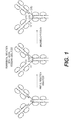

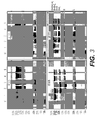
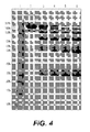

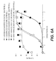


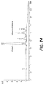
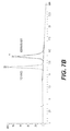


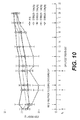
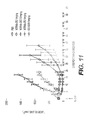
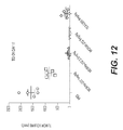
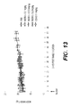

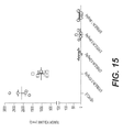
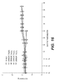
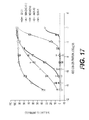

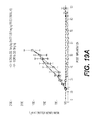
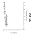

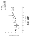
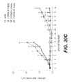
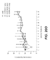
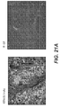

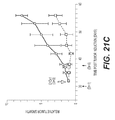
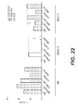

Classifications
-
- A—HUMAN NECESSITIES
- A61—MEDICAL OR VETERINARY SCIENCE; HYGIENE
- A61K—PREPARATIONS FOR MEDICAL, DENTAL OR TOILETRY PURPOSES
- A61K47/00—Medicinal preparations characterised by the non-active ingredients used, e.g. carriers or inert additives; Targeting or modifying agents chemically bound to the active ingredient
- A61K47/50—Medicinal preparations characterised by the non-active ingredients used, e.g. carriers or inert additives; Targeting or modifying agents chemically bound to the active ingredient the non-active ingredient being chemically bound to the active ingredient, e.g. polymer-drug conjugates
- A61K47/51—Medicinal preparations characterised by the non-active ingredients used, e.g. carriers or inert additives; Targeting or modifying agents chemically bound to the active ingredient the non-active ingredient being chemically bound to the active ingredient, e.g. polymer-drug conjugates the non-active ingredient being a modifying agent
- A61K47/68—Medicinal preparations characterised by the non-active ingredients used, e.g. carriers or inert additives; Targeting or modifying agents chemically bound to the active ingredient the non-active ingredient being chemically bound to the active ingredient, e.g. polymer-drug conjugates the non-active ingredient being a modifying agent the modifying agent being an antibody, an immunoglobulin or a fragment thereof, e.g. an Fc-fragment
- A61K47/6801—Drug-antibody or immunoglobulin conjugates defined by the pharmacologically or therapeutically active agent
- A61K47/6803—Drugs conjugated to an antibody or immunoglobulin, e.g. cisplatin-antibody conjugates
-
- A—HUMAN NECESSITIES
- A61—MEDICAL OR VETERINARY SCIENCE; HYGIENE
- A61K—PREPARATIONS FOR MEDICAL, DENTAL OR TOILETRY PURPOSES
- A61K31/00—Medicinal preparations containing organic active ingredients
- A61K31/33—Heterocyclic compounds
- A61K31/335—Heterocyclic compounds having oxygen as the only ring hetero atom, e.g. fungichromin
- A61K31/357—Heterocyclic compounds having oxygen as the only ring hetero atom, e.g. fungichromin having two or more oxygen atoms in the same ring, e.g. crown ethers, guanadrel
-
- A—HUMAN NECESSITIES
- A61—MEDICAL OR VETERINARY SCIENCE; HYGIENE
- A61K—PREPARATIONS FOR MEDICAL, DENTAL OR TOILETRY PURPOSES
- A61K47/00—Medicinal preparations characterised by the non-active ingredients used, e.g. carriers or inert additives; Targeting or modifying agents chemically bound to the active ingredient
- A61K47/50—Medicinal preparations characterised by the non-active ingredients used, e.g. carriers or inert additives; Targeting or modifying agents chemically bound to the active ingredient the non-active ingredient being chemically bound to the active ingredient, e.g. polymer-drug conjugates
- A61K47/51—Medicinal preparations characterised by the non-active ingredients used, e.g. carriers or inert additives; Targeting or modifying agents chemically bound to the active ingredient the non-active ingredient being chemically bound to the active ingredient, e.g. polymer-drug conjugates the non-active ingredient being a modifying agent
- A61K47/62—Medicinal preparations characterised by the non-active ingredients used, e.g. carriers or inert additives; Targeting or modifying agents chemically bound to the active ingredient the non-active ingredient being chemically bound to the active ingredient, e.g. polymer-drug conjugates the non-active ingredient being a modifying agent the modifying agent being a protein, peptide or polyamino acid
- A61K47/65—Peptidic linkers, binders or spacers, e.g. peptidic enzyme-labile linkers
-
- A—HUMAN NECESSITIES
- A61—MEDICAL OR VETERINARY SCIENCE; HYGIENE
- A61K—PREPARATIONS FOR MEDICAL, DENTAL OR TOILETRY PURPOSES
- A61K47/00—Medicinal preparations characterised by the non-active ingredients used, e.g. carriers or inert additives; Targeting or modifying agents chemically bound to the active ingredient
- A61K47/50—Medicinal preparations characterised by the non-active ingredients used, e.g. carriers or inert additives; Targeting or modifying agents chemically bound to the active ingredient the non-active ingredient being chemically bound to the active ingredient, e.g. polymer-drug conjugates
- A61K47/51—Medicinal preparations characterised by the non-active ingredients used, e.g. carriers or inert additives; Targeting or modifying agents chemically bound to the active ingredient the non-active ingredient being chemically bound to the active ingredient, e.g. polymer-drug conjugates the non-active ingredient being a modifying agent
- A61K47/68—Medicinal preparations characterised by the non-active ingredients used, e.g. carriers or inert additives; Targeting or modifying agents chemically bound to the active ingredient the non-active ingredient being chemically bound to the active ingredient, e.g. polymer-drug conjugates the non-active ingredient being a modifying agent the modifying agent being an antibody, an immunoglobulin or a fragment thereof, e.g. an Fc-fragment
- A61K47/6835—Medicinal preparations characterised by the non-active ingredients used, e.g. carriers or inert additives; Targeting or modifying agents chemically bound to the active ingredient the non-active ingredient being chemically bound to the active ingredient, e.g. polymer-drug conjugates the non-active ingredient being a modifying agent the modifying agent being an antibody, an immunoglobulin or a fragment thereof, e.g. an Fc-fragment the modifying agent being an antibody or an immunoglobulin bearing at least one antigen-binding site
- A61K47/6849—Medicinal preparations characterised by the non-active ingredients used, e.g. carriers or inert additives; Targeting or modifying agents chemically bound to the active ingredient the non-active ingredient being chemically bound to the active ingredient, e.g. polymer-drug conjugates the non-active ingredient being a modifying agent the modifying agent being an antibody, an immunoglobulin or a fragment thereof, e.g. an Fc-fragment the modifying agent being an antibody or an immunoglobulin bearing at least one antigen-binding site the antibody targeting a receptor, a cell surface antigen or a cell surface determinant
-
- A—HUMAN NECESSITIES
- A61—MEDICAL OR VETERINARY SCIENCE; HYGIENE
- A61K—PREPARATIONS FOR MEDICAL, DENTAL OR TOILETRY PURPOSES
- A61K47/00—Medicinal preparations characterised by the non-active ingredients used, e.g. carriers or inert additives; Targeting or modifying agents chemically bound to the active ingredient
- A61K47/50—Medicinal preparations characterised by the non-active ingredients used, e.g. carriers or inert additives; Targeting or modifying agents chemically bound to the active ingredient the non-active ingredient being chemically bound to the active ingredient, e.g. polymer-drug conjugates
- A61K47/51—Medicinal preparations characterised by the non-active ingredients used, e.g. carriers or inert additives; Targeting or modifying agents chemically bound to the active ingredient the non-active ingredient being chemically bound to the active ingredient, e.g. polymer-drug conjugates the non-active ingredient being a modifying agent
- A61K47/68—Medicinal preparations characterised by the non-active ingredients used, e.g. carriers or inert additives; Targeting or modifying agents chemically bound to the active ingredient the non-active ingredient being chemically bound to the active ingredient, e.g. polymer-drug conjugates the non-active ingredient being a modifying agent the modifying agent being an antibody, an immunoglobulin or a fragment thereof, e.g. an Fc-fragment
- A61K47/6889—Conjugates wherein the antibody being the modifying agent and wherein the linker, binder or spacer confers particular properties to the conjugates, e.g. peptidic enzyme-labile linkers or acid-labile linkers, providing for an acid-labile immuno conjugate wherein the drug may be released from its antibody conjugated part in an acidic, e.g. tumoural or environment
-
- A—HUMAN NECESSITIES
- A61—MEDICAL OR VETERINARY SCIENCE; HYGIENE
- A61P—SPECIFIC THERAPEUTIC ACTIVITY OF CHEMICAL COMPOUNDS OR MEDICINAL PREPARATIONS
- A61P35/00—Antineoplastic agents
-
- C—CHEMISTRY; METALLURGY
- C07—ORGANIC CHEMISTRY
- C07K—PEPTIDES
- C07K16/00—Immunoglobulins [IGs], e.g. monoclonal or polyclonal antibodies
- C07K16/18—Immunoglobulins [IGs], e.g. monoclonal or polyclonal antibodies against material from animals or humans
- C07K16/28—Immunoglobulins [IGs], e.g. monoclonal or polyclonal antibodies against material from animals or humans against receptors, cell surface antigens or cell surface determinants
- C07K16/30—Immunoglobulins [IGs], e.g. monoclonal or polyclonal antibodies against material from animals or humans against receptors, cell surface antigens or cell surface determinants from tumour cells
-
- C—CHEMISTRY; METALLURGY
- C07—ORGANIC CHEMISTRY
- C07K—PEPTIDES
- C07K16/00—Immunoglobulins [IGs], e.g. monoclonal or polyclonal antibodies
- C07K16/18—Immunoglobulins [IGs], e.g. monoclonal or polyclonal antibodies against material from animals or humans
- C07K16/32—Immunoglobulins [IGs], e.g. monoclonal or polyclonal antibodies against material from animals or humans against translation products of oncogenes
-
- C—CHEMISTRY; METALLURGY
- C07—ORGANIC CHEMISTRY
- C07K—PEPTIDES
- C07K2317/00—Immunoglobulins specific features
- C07K2317/10—Immunoglobulins specific features characterized by their source of isolation or production
- C07K2317/14—Specific host cells or culture conditions, e.g. components, pH or temperature
-
- C—CHEMISTRY; METALLURGY
- C07—ORGANIC CHEMISTRY
- C07K—PEPTIDES
- C07K2317/00—Immunoglobulins specific features
- C07K2317/40—Immunoglobulins specific features characterized by post-translational modification
-
- C—CHEMISTRY; METALLURGY
- C07—ORGANIC CHEMISTRY
- C07K—PEPTIDES
- C07K2317/00—Immunoglobulins specific features
- C07K2317/50—Immunoglobulins specific features characterized by immunoglobulin fragments
- C07K2317/56—Immunoglobulins specific features characterized by immunoglobulin fragments variable (Fv) region, i.e. VH and/or VL
- C07K2317/565—Complementarity determining region [CDR]
-
- C—CHEMISTRY; METALLURGY
- C07—ORGANIC CHEMISTRY
- C07K—PEPTIDES
- C07K2317/00—Immunoglobulins specific features
- C07K2317/70—Immunoglobulins specific features characterized by effect upon binding to a cell or to an antigen
- C07K2317/77—Internalization into the cell
-
- C—CHEMISTRY; METALLURGY
- C07—ORGANIC CHEMISTRY
- C07K—PEPTIDES
- C07K2317/00—Immunoglobulins specific features
- C07K2317/90—Immunoglobulins specific features characterized by (pharmaco)kinetic aspects or by stability of the immunoglobulin
- C07K2317/92—Affinity (KD), association rate (Ka), dissociation rate (Kd) or EC50 value
Definitions
- the present disclosure relates to antibody drug conjugates (ADCs) that bind human oncology antigen targets such as folate receptor alpha and/or provide anti-tubulin drug activity.
- ADCs antibody drug conjugates
- the disclosure further relates to methods and compositions useful in the treatment and diagnosis of cancers that express folate receptor alpha and/or are amenable to treatment by disrupting tubulin.
- Cancer is among the leading causes of morbidity and mortality worldwide, with approximately 14 million new cases and 8.2 million cancer-related deaths in 2012. The most common causes of cancer death are cancers of: lung (1.59 million deaths); liver (745,000 deaths); stomach (723,000 deaths); colorectal (694,000 deaths); breast (521,000 deaths); and esophagus (400,000 deaths). The number of new cancer cases is expected to rise by about 70% over the next two decades, to approximately 22 million new cancer cases per year (World Cancer Report 2014).
- Microtubules are dynamic filamentous cytoskeletal proteins that are involved in a variety of cellular functions, including intracellular migration and transport, cell signaling, and the maintenance of cell shape. Microtubules also play a critical role in mitotic cell division by forming the mitotic spindle required to segregate chromosomes into two daughter cells. The biological functions of microtubules in all cells are regulated in large part by their polymerization dynamics, which occurs by the reversible, non-covalent addition of ⁇ and ⁇ tubulin dimers at both ends of microtubules. This dynamic behavior and resulting control over microtubule length is vital to the proper functioning of the mitotic spindle.
- Folate receptor alpha is a glycophosphatidylinositol (GPI)-linked membrane protein that binds folate. While the role of FRA in the biology of normal and cancerous tissue is not fully understood, it is highly over-expressed on a high percentage of ovarian cancers of epithelial origin ( O'Shannessy et al. (2013) Int. J. Gynecol. Pathol. 32(3):258-68 ), as well as in a percentage of non-small cell lung carcinomas ( Christoph et al. (2014) Clin. Lung Cancer 15(5):320-30 ). FRA also has limited expression in normal tissues. These properties make FRA an attractive target for cancer immunotherapy.
- GPI glycophosphatidylinositol
- the present invention provides an antibody-drug conjugate of Formula (I): Ab-(L-D) p (I) wherein (i) Ab is an internalizing anti-folate receptor alpha antibody or internalizing antigen-binding fragment thereof comprising three heavy chain complementarity determining regions (HCDRs) comprising amino acid sequences of SEQ ID NO:2 (HCDR1), SEQ ID NO:3 (HCDR2), and SEQ ID NO:4 (HCDR3); and three light chain complementarity determining regions (LCDRs) comprising amino acid sequences of SEQ ID NO:7 (LCDR1), SEQ ID NO:8 (LCDR2), and SEQ ID NO:9 (LCDR3), as defined by the Kabat numbering system; or three heavy chain complementarity determining regions (HCDRs) comprising amino acid sequences of SEQ ID NO:13 (HCDR1), SEQ ID NO:14 (HCDR2), and SEQ ID NO:15 (HCDR3); and three light chain complementarity determining regions (LCDRs) comprising
- the antibody or antigen-binding fragment may comprise a heavy chain variable region comprising an amino acid sequence of SEQ ID NO:23, and a light chain variable region comprising an amino acid sequence of SEQ ID NO:24.
- p may be an integer from 3 to 4.
- the antibody or antigen-binding fragment may comprise a human IgG1 heavy chain constant domain and a human Ig kappa light chain constant domain.
- the present invention provides a composition comprising multiple copies of the antibody-drug conjugate of the first aspect, wherein the average p of the antibody-drug conjugates in the composition is from about 3.2 to about 3.8 or from about 3.6 to about 4.4.
- the present invention provides the first aspect or the second aspect may be for use in the treatment of a cancer that expresses folate receptor alpha.
- the cancer is a gastric cancer, a serous ovarian cancer, a clear cell ovarian cancer, a non-small cell lung cancer, a colorectal cancer, a triple negative breast cancer, an endometrial cancer, a serous endometrial carcinoma, a lung carcinoid, or an osteosarcoma.
- the present invention provides a pharmaceutical composition
- a pharmaceutical composition comprising the antibody-drug conjugate of the first aspect or the composition of the second aspect, and a pharmaceutically acceptable carrier.
- the present invention provides a method of producing the antibody-drug conjugate of the first aspect or the composition of the second aspect, comprising reacting an antibody or antigen-binding fragment with a cleavable linker joined to eribulin under conditions that allow conjugation.
- the present invention provides a method of determining whether a patient will be responsive to treatment with the antibody-drug conjugate of any one of the first aspect or the composition of the second aspect, comprising providing a biological sample from the patient and contacting the biological sample with the antibody-drug conjugate of any one of claims 1 to 4 or the composition of claim 5.
- the biological sample may be a tumor biopsy from a gastric cancer, a serous ovarian cancer, a clear cell ovarian cancer, a non-small cell lung cancer, a colorectal cancer, a triple negative breast cancer, an endometrial cancer, a serous endometrial carcinoma, a lung carcinoid, or an osteosarcoma.
- the present invention provides A composition comprising multiple copies of an antibody-drug conjugate of Formula (I): Ab-(L-D) p (I) wherein (i) Ab is an internalizing anti-folate receptor alpha antibody or internalizing antigen-binding fragment thereof comprising three heavy chain complementarity determining regions (HCDRs) comprising amino acid sequences of SEQ ID NO:2 (HCDR1), SEQ ID NO:3 (HCDR2), and SEQ ID NO:4 (HCDR3); and three light chain complementarity determining regions (LCDRs) comprising amino acid sequences of SEQ ID NO:7 (LCDR1), SEQ ID NO:8 (LCDR2), and SEQ ID NO:9 (LCDR3), as defined by the Kabat numbering system; or three heavy chain complementarity determining regions (HCDRs) comprising amino acid sequences of SEQ ID NO: 13 (HCDR1), SEQ ID NO:14 (HCDR2), and SEQ ID NO:15 (HCDR3); and three light chain complement
- the present disclosure describes, in part, novel compounds with biological activity against tumor cells.
- the compounds may inhibit tumor growth in mammals, and may be useful for treating human cancer patients.
- ADC antibody-drug conjugate compounds
- the linker is stable outside a cell, such that the ADC remains intact when present in extracellular conditions but is capable of being cleaved on internalization in a cell, e.g., a cancer cell.
- the eribulin drug moiety is cleaved from the antibody moiety when the ADC enters a cell that expresses an antigen specific for the antibody moiety of the ADC, and cleavage releases an unmodified form of eribulin.
- the linker comprises a cleavable moiety that is positioned such that no part of the linker or the antibody moiety remains bound to the eribulin drug moiety upon cleavage.
- the cleavable moiety in the linker is a cleavable peptide moiety.
- an ADC that comprises a cleavable peptide moiety demonstrates lower aggregation levels, improved antibody:drug ratio, increased on-target killing of cancer cells, decreased off-target killing of non-cancer cells, and/or higher drug loading ( p ) relative to an ADC that comprises an alternate cleavable moiety.
- adding a cleavable moiety increases cytotoxicity and/or potency relative to a non-cleavable linker.
- the increased potency and/or cytotoxicity is in a cancer expressing moderate levels of the antigen targeted by the antibody moiety of the ADC (e.g., moderate FRA expression).
- the cleavable peptide moiety is cleavable by an enzyme, and the linker is an enzyme-cleavable linker.
- the enzyme is cathepsin, and the linker is a cathepsin-cleavable linker.
- the enzyme-cleavable linker e.g., the cathepsin-cleavable linker
- the cleavable peptide moiety in the linker comprises an amino acid unit.
- the amino acid unit comprises valine-citrulline (Val-Cit).
- an ADC that comprises Val-Cit demonstrates increased stability, decreased off-target cell killing, increased on-target cell killing, lower aggregation levels, and/or higher drug loading relative to an ADC that comprises an alternate amino acid unit or alternate cleavable moiety.
- the linker comprises at least one spacer unit joining the antibody moiety to the cleavable moiety.
- the spacer unit in the linker may comprise at least one polyethylene glycol (PEG) moiety.
- the PEG moiety may, for example, comprise -(PEG) m -, wherein m is an integer from 1 to 10.
- the spacer unit in the linker comprises (PEG) 2 .
- an ADC that comprises a shorter spacer unit e.g., (PEG) 2
- the spacer unit in the linker attaches to the antibody moiety of the ADC via a maleimide moiety (Mal).
- Mal maleimide moiety
- an ADC that comprises a linker attached to the antibody moiety via a Mal demonstrates higher drug loading relative to an ADC that comprises a linker attached to the antibody moiety via an alternate moiety.
- the Mal in the linker is reactive with a cysteine residue on the antibody moiety.
- the Mal in the linker is joined to the antibody moiety via a cysteine residue.
- the Mal-spacer unit comprises a PEG moiety.
- the linker comprises Mal-(PEG) m , e.g., Mal-(PEG) 2 .
- the linker comprises Mal-(PEG) 2 .
- the Mal-spacer unit attaches the antibody moiety to the cleavable moiety in the linker.
- the cleavable moiety in the linker is a cleavable peptide moiety, e.g., an amino acid unit.
- the linker comprises Mal-(PEG) 2 -Val-Cit.
- the cleavable moiety in the linker is directly joined to the eribulin drug moiety of the ADC, and the cleavable moiety is either directly connected to the antibody moiety or connected through a spacer unit.
- a spacer unit also attaches the cleavable moiety in the linker to the eribulin drug moiety.
- the spacer unit that attaches the cleavable moiety in the linker to the eribulin drug moiety is self-immolative.
- the self-immolative spacer is capable of releasing unmodified eribulin in a target cell.
- the self-immolative spacer unit comprises a p-aminobenzyl alcohol.
- the self-immolative spacer unit comprises p-aminobenzyloxycarbonyl (pAB).
- the pAB in the linker attaches the cleavable moiety to the eribulin drug moiety.
- the cleavable moiety is a cleavable peptide moiety, e.g., an amino acid unit.
- the linker comprises Val-Cit-pAB.
- the linker comprises Val-Cit-pAB and a PEG spacer unit joining the linker to the antibody moiety through a Mal.
- p is an integer from 1 to 6, from 2 to 5, or preferably, from 3 to 4. As described herein, p is 4. As described herein, a pool of ADCs are provided, and the average p in the pool is about 4 (e.g., 3.5-4.5, such as about 3.8). As described herein, the linker comprises Mal-(PEG) 2 -Val-Cit-pAB. As described herein, the linker comprises Mal-(PEG) 2 -Val-Cit-pAB and p is 4.
- each ADC comprises a Mal-(PEG) 2 -Val-Cit-pAB linker, and the average p in the pool is about 4 (e.g., 3.5-4.5, such as about 3.8).
- the internalizing antibody or internalizing antigen-binding fragment (Ab or Ab moiety) of the ADC is an anti-folate receptor alpha (FRA) antibody or internalizing antibody fragment, and can bind FRA-expressing tumor cells (i.e., the ADC targets FRA-expressing cells).
- FRA anti-folate receptor alpha
- the ADC comprising an anti-FRA Ab moiety and a cleavable peptide moiety demonstrates lower aggregation levels, improved antibody:drug ratio, increased on-target killing of cancer cells, decreased off-target killing of non-cancer cells, higher drug loading (p), increased cytotoxicity, and/or potency relative to a non-cleavable linker or an alternate cleavage mechanism.
- the increased potency and/or cytotoxicity is in a cancer expressing moderate levels of the antigen targeted by the antibody moiety of the ADC (e.g., moderate FRA expression).
- the cleavable peptide moiety is cleavable by an enzyme
- the linker is an enzyme-cleavable linker.
- the enzyme is cathepsin
- the linker is a cathepsin-cleavable linker.
- the enzyme-cleavable linker e.g., the cathepsin-cleavable linker
- the linker is a Mal-(PEG) m -Val-Cit-pAB.
- the internalizing antibody or internalizing antigen-binding fragment binds to folate receptor alpha (FRA) and targets FRA-expressing tumor cells.
- the internalizing antibody or internalizing antigen-binding fragment comprises human framework sequences. As described herein, the internalizing antibody or internalizing antigen-binding fragment comprises a heavy chain variable domain of SEQ ID NO:23 and a light chain variable domain of SEQ ID NO:24. As described herein, the internalizing antibody or internalizing antigen-binding fragment comprises a human IgG1 heavy chain constant domain and an Ig kappa light chain constant domain. As described herein, the internalizing antibody or internalizing antigen-binding competes for binding and/or binds the same epitope as an antibody comprising a heavy chain variable domain of SEQ ID NO:23 and a light chain variable domain of SEQ ID NO:24.
- the internalizing antibody or internalizing antigen-binding fragment binds to an epitope comprising alanine-histadine-lysine-aspartic acid (AHKD) (SEQ ID NO:365) ( O'Shannessy et al., (2011) Oncotarget 2:1227-43 ). As described herein, the internalizing antibody or internalizing antigen-binding fragment binds to an epitope comprising NTSQEAHKDVSYL (SEQ ID NO:366).
- the internalizing antibody or internalizing antigen-binding fragment is an internalizing anti-FRA antibody or internalizing antigen-binding fragment.
- the internalizing antibody or internalizing antigen-binding fragment comprises three heavy chain CDRs and three light chain CDRs, wherein the heavy chain CDRs comprise heavy chain CDR1 consisting of SEQ ID NO:2, heavy chain CDR2 consisting of SEQ ID NO:3, and heavy chain CDR3 consisting of SEQ ID NO:4; and the three light chain CDRs comprise light chain CDR1 consisting of SEQ ID NO:7, light chain CDR2 consisting of SEQ ID NO:8, and light chain CDR3 consisting of SEQ ID NO:9, as defined by the Kabat numbering system; or wherein the heavy chain CDRs comprise heavy chain CDR1 consisting of SEQ ID NO:13, heavy chain CDR2 consisting of SEQ ID NO:14, and heavy chain CDR3 consisting of SEQ ID NO:15; and the light chain CDRs comprise light chain CDR
- the internalizing antibody or internalizing antigen-binding fragment comprises a heavy chain variable domain of SEQ ID NO:23 and a light chain variable domain of SEQ ID NO:24.
- the internalizing antibody or internalizing antigen-binding fragment comprises a human IgG1 heavy chain constant domain and an Ig kappa light chain constant domain.
- the internalizing antibody or internalizing antigen-binding competes for binding and/or binds the same epitope as an antibody comprising a heavy chain variable domain of SEQ ID NO:23 and a light chain variable domain of SEQ ID NO:24.
- the internalizing antibody or internalizing antigen-binding fragment binds to an epitope comprising SEQ ID NO:365. As described herein, the internalizing antibody or internalizing antigen-binding fragment binds to an epitope comprising SEQ ID NO:366.
- the internalizing antibody or internalizing antigen-binding fragment binds to human epidermal growth factor receptor 2 (her2) and targets her2-expressing tumor cells.
- the internalizing antibody or internalizing antigen-binding fragment comprises three heavy chain complementarity determining regions (CDRs) and three light chain CDRs, wherein the heavy chain CDRs comprise heavy chain CDR1 consisting of SEQ ID NO:71 heavy chain CDR2 consisting of SEQ ID NO:72, and heavy chain CDR3 consisting of SEQ ID NO:73; and the three light chain CDRs comprise light chain CDR1 consisting of SEQ ID NO:74, light chain CDR2 consisting of SEQ ID NO:75, and light chain CDR3 consisting of SEQ ID NO:76, as defined by the Kabat numbering system; or wherein the heavy chain CDRs comprise heavy chain CDR1 consisting of SEQ ID NO:191, heavy chain CDR2 consisting of SEQ ID NO:192, and heavy chain CDR3 consisting of SEQ ID NO:
- the antibody or internalizing antigen-binding fragment comprises human framework sequences.
- the internalizing antibody or internalizing antigen-binding fragment comprises a heavy chain variable domain of SEQ ID NO:27 and a light chain variable domain of SEQ ID NO:28.
- the internalizing antibody or internalizing antigen-binding fragment comprises a human IgG1 heavy chain constant domain and an Ig kappa light chain constant domain.
- the internalizing antibody or internalizing antigen-binding competes for binding and/or binds the same epitope as an antibody comprising a heavy chain variable domain of SEQ ID NO:27 and a light chain variable domain of SEQ ID NO:28.
- the internalizing antibody or internalizing antigen-binding fragment is an internalizing anti-her2 antibody or internalizing antigen-binding fragment.
- the internalizing antibody or internalizing antigen-binding fragment comprises three heavy chain CDRs and three light chain CDRs, wherein the heavy chain CDRs comprise heavy chain CDR1 consisting of SEQ ID NO:71 heavy chain CDR2 consisting of SEQ ID NO:72, and heavy chain CDR3 consisting of SEQ ID NO:73; and the three light chain CDRs comprise light chain CDR1 consisting of SEQ ID NO:74, light chain CDR2 consisting of SEQ ID NO:75, and light chain CDR3 consisting of SEQ ID NO:76, as defined by the Kabat numbering system; or wherein the heavy chain CDRs comprise heavy chain CDR1 consisting of SEQ ID NO:191, heavy chain CDR2 consisting of SEQ ID NO:192, and heavy chain CDR3 consisting of SEQ ID NO: 193; and the light chain CDRs comprise light
- the internalizing antibody or internalizing antigen-binding fragment comprises a heavy chain variable domain of SEQ ID NO:27 and a light chain variable domain of SEQ ID NO:28.
- the internalizing antibody or internalizing antigen-binding fragment comprises a human IgG1 heavy chain constant domain and an Ig kappa light chain constant domain.
- the internalizing antibody or internalizing antigen-binding competes for binding and/or binds the same epitope as an antibody comprising a heavy chain variable domain of SEQ ID NO:27 and a light chain variable domain of SEQ ID NO:28.
- the internalizing antibody or internalizing antigen-binding fragment binds to mesothelin (MSLN) and targets MSLN-expressing tumor cells.
- the internalizing antibody or internalizing antigen-binding fragment comprises three heavy chain complementarity determining regions (CDRs) and three light chain CDRs, wherein the heavy chain CDRs comprise heavy chain CDR1 consisting of SEQ ID NO:65 heavy chain CDR2 consisting of SEQ ID NO:66, and heavy chain CDR3 consisting of SEQ ID NO:67; and the three light chain CDRs comprise light chain CDR1 consisting of SEQ ID NO:68, light chain CDR2 consisting of SEQ ID NO:69, and light chain CDR3 consisting of SEQ ID NO:70, as defined by the Kabat numbering system; or wherein the heavy chain CDRs comprise heavy chain CDR1 consisting of SEQ ID NO: 185, heavy chain CDR2 consisting of SEQ ID NO:186, and heavy chain CDR3 consisting of SEQ
- the internalizing antibody or internalizing antigen-binding fragment comprises a heavy chain variable domain of SEQ ID NO:25 and a light chain variable domain of SEQ ID NO:26.
- the internalizing antibody or internalizing antigen-binding fragment comprises a human IgG1 heavy chain constant domain and an Ig kappa light chain constant domain.
- the internalizing antibody or internalizing antigen-binding competes for binding and/or binds the same epitope as an antibody comprising a heavy chain variable domain of SEQ ID NO:25 and a light chain variable domain of SEQ ID NO:26.
- the internalizing antibody or internalizing antigen-binding fragment is an internalizing anti-MSLN antibody or internalizing antigen-binding fragment.
- the internalizing antibody or internalizing antigen-binding fragment comprises three heavy chain CDRs and three light chain CDRs, wherein the heavy chain CDRs comprise heavy chain CDR1 consisting of SEQ ID NO:65 heavy chain CDR2 consisting of SEQ ID NO:66, and heavy chain CDR3 consisting of SEQ ID NO:67; and the three light chain CDRs comprise light chain CDR1 consisting of SEQ ID NO:68, light chain CDR2 consisting of SEQ ID NO:69, and light chain CDR3 consisting of SEQ ID NO:70, as defined by the Kabat numbering system; or wherein the heavy chain CDRs comprise heavy chain CDR1 consisting of SEQ ID NO:185, heavy chain CDR2 consisting of SEQ ID NO:186, and heavy chain CDR3 consisting of SEQ ID NO: 187; and the light chain CDRs comprise
- the internalizing antibody or internalizing antigen-binding fragment comprises a heavy chain variable domain of SEQ ID NO:25 and a light chain variable domain of SEQ ID NO:26.
- the internalizing antibody or internalizing antigen-binding fragment comprises a human IgG1 heavy chain constant domain and an Ig kappa light chain constant domain.
- the internalizing antibody or internalizing antigen-binding competes for binding and/or binds the same epitope as an antibody comprising a heavy chain variable domain of SEQ ID NO:25 and a light chain variable domain of SEQ ID NO:26.
- compositions comprising multiple copies of any of the described ADCs, wherein the average drug loading (average p ) of the ADCs in the composition is between about 3 and 4, or about 3.5 to about 4.5, or about 4.
- the average p is between about 3.2 and 3.8.
- the average p is between about 3.6 and 4.4.
- compositions comprising -L-D, wherein D is eribulin; and L is a cleavable linker that covalently attaches to D.
- the cleavable linker covalently attaches to the C-35 amine on eribulin.
- the cleavable linker comprises Val-Cit.
- the cleavable linker comprises a PEG spacer unit.
- the cleavable linker comprises Mal-(PEG) 2 -Val-Cit-pAB.
- compositions comprising an ADC and a pharmaceutically acceptable diluent, carrier, and/or excipient.
- Another aspect of the present disclosure includes therapeutic and diagnostic uses for the described ADC compounds and compositions, e.g., in treating cancer.
- Another aspect includes methods of treating a cancer that expresses an antigen targeted by the antibody moiety of the ADC, such as FRA.
- FRA an antigen targeted by the antibody moiety of the ADC
- methods of killing or inhibiting the proliferation of tumor cells or cancer cells by administering a therapeutically effective amount and/or regimen of any one of the described ADCs are described.
- Another aspect includes methods for detecting tumor cells or cancer cells that express FRA using the disclosed ADCs, and methods of screening for cancer patients that will be responsive to treatment with the described ADCs.
- the cancer is a gastric cancer, a serous ovarian cancer, a clear cell ovarian cancer, a non-small cell lung cancer, a colorectal cancer, a triple negative breast cancer, an endometrial cancer, a serous endometrial carcinoma, a lung carcinoid, or an osteosarcoma.
- Methods of producing the described ADCs are also disclosed.
- compositions and methods may be understood more readily by reference to the following detailed description taken in connection with the accompanying figures, which form a part of this disclosure. It is to be understood that the discribed compositions and methods are not limited to the specific compositions and methods described and/or shown herein. The scope of the invention is defined by the claims.
- compositions and methods of using said compositions refer to compositions and methods of using said compositions.
- a feature or disclosure associated with a composition such a feature or disclsoure is equally applicable to the methods of using said composition.
- the disclosure describes or claims a feature or disclosure associated with a method of using a composition, such a feature or disclosure is equally applicable to the composition.
- compositions and methods which are, for clarity, described herein in the context of separate disclosures, may also be provided in combination in a single disclosure.
- antibody is used in the broadest sense to refer to an immunoglobulin molecule that recognizes and specifically binds to a target, such as a protein, polypeptide, carbohydrate, polynucleotide, lipid, or combinations of the foregoing through at least one antigen recognition site within the variable region of the immunoglobulin molecule.
- the heavy chain of an antibody is composed of a heavy chain variable domain (V H ) and a heavy chain constant region (C H ).
- the light chain is composed of a light chain variable domain (V L ) and a light chain constant domain (C L ).
- the mature heavy chain and light chain variable domains each comprise three complementarity determining regions (CDR1, CDR2 and CDR3) within four framework regions (FR1, FR2, FR3 and FR4) arranged from N-terminus to C-terminus: FR1, CDR1, FR2, CDR2, FR3, CDR3 and FR4.
- An "antibody” can be naturally occurring or man-made, such as monoclonal antibodies produced by conventional hybridoma technology.
- the term “antibody” includes full-length monoclonal antibodies and full-length polyclonal antibodies, as well as antibody fragments such as Fab, Fab', F(ab')2, Fv, and single chain antibodies.
- An antibody can be any one of the five major classes of immunoglobulins: IgA, IgD, IgE, IgG, and IgM, or subclasses thereof (e.g., isotypes IgG1, IgG2, IgG3, IgG4).
- the term further encompasses human antibodies, chimeric antibodies, humanized antibodies and any modified immunoglobulin molecule containing an antigen recognition site, so long as it demonstrates the desired biological activity.
- the term "monoclonal antibody,” as used herein, refers to an antibody obtained from a population of substantially homogeneous antibodies, i.e., the individual antibodies comprising the population are identical except for possible naturally occurring mutations that may be present in minor amounts. Monoclonal antibodies are highly specific, being directed against a single antigenic epitope. In contrast, conventional (polyclonal) antibody preparations typically include a multitude of antibodies directed against (or specific for) different epitopes. The modifier “monoclonal” indicates the character of the antibody as being obtained from a substantially homogeneous population of antibodies, and is not to be construed as requiring production of the antibody by any particular method.
- the monoclonal antibodies to be used in accordance with the present disclosure may be made by the hybridoma method first described by Kohler et al. (1975) Nature 256:495 , or may be made by recombinant DNA methods (see, e.g., U.S. Pat. No. 4,816,567 ).
- Monoclonal antibodies may also be isolated from phage antibody libraries using the techniques described in Clackson et al. (1991) Nature 352:624-8 , and Marks et al. (1991) J. Mol. Biol. 222:581-97 , for example.
- the monoclonal antibodies described herein specifically include "chimeric" antibodies, in which a portion of the heavy and/or light chain is identical with or homologous to corresponding sequences in antibodies derived from a particular species or belonging to a particular antibody class or subclass, while the remainder of the chain(s) is identical with or homologous to corresponding sequences in antibodies derived from another species or belonging to another antibody class or subclass, as well as fragments of such antibodies, so long as they specifically bind the target antigen and/or exhibit the desired biological activity.
- human antibody refers an antibody produced by a human or an antibody having an amino acid sequence of an antibody produced by a human.
- chimeric antibody refers to antibodies wherein the amino acid sequence of the immunoglobulin molecule is derived from two or more species.
- the variable regions of both heavy and light chains corresponds to the variable regions of antibodies derived from one species with the desired specificity, affinity, and activity while the constant regions are homologous to antibodies derived from another species (e.g., human) to minimize an immune response in the latter species.
- humanized antibody refers to forms of antibodies that contain sequences from non-human (e.g., murine) antibodies as well as human antibodies. Such antibodies are chimeric antibodies which contain minimal sequence derived from non-human immunoglobulin.
- the humanized antibody will comprise substantially all of at least one, and typically two, variable domains, in which all or substantially all of the hypervariable loops correspond to those of a non-human immunoglobulin and all or substantially all of the framework (FR) regions are those of a human immunoglobulin sequence.
- the humanized antibody optionally also will comprise at least a portion of an immunoglobulin constant region (Fc), typically that of a human immunoglobulin.
- the humanized antibody can be further modified by the substitution of residues, either in the Fv framework region and/or within the replaced non-human residues to refine and optimize antibody specificity, affinity, and/or activity.
- antigen-binding fragment or "antigen-binding portion" of an antibody, as used herein, refers to one or more fragments of an antibody that retain the ability to specifically bind to an antigen (e.g., FRA). Antigen-binding fragments preferably also retain the ability to internalize into an antigen-expressing cell. In some disclosures, antigen-binding fragments also retain immune effector activity. It has been shown that fragments of a full-length antibody can perform the antigen-binding function of a full-length antibody.
- binding fragments encompassed within the term "antigen-binding fragment” or "antigen-binding portion” of an antibody include (i) a Fab fragment, a monovalent fragment consisting of the V L , V H , C L , and C H1 domains; (ii) a F(ab') 2 fragment, a bivalent fragment comprising two Fab fragments linked by a disulfide bridge at the hinge region; (iii) a Fd fragment consisting of the V H and C H1 domains; (iv) a Fv fragment consisting of the V L and V H domains of a single arm of an antibody; (v) a dAb fragment, which comprises a single variable domain, e.g., a V H domain (see, e.g., Ward et al.
- V L and V H are coded for by separate genes, they can be joined, using recombinant methods, by a synthetic linker that enables them to be made as a single protein chain in which the V L and V H regions pair to form monovalent molecules (known as single chain Fv (scFv)).
- scFv single chain Fv
- single chain antibodies are also intended to be encompassed within the term "antigen-binding fragment” or "antigen-binding portion” of an antibody, and are known in the art as an exemplary type of binding fragment that can internalize into cells upon binding. See, e.g., Zhu et al. (2010) 9:2131-41; He et al. (2010) J. Nucl. Med. 51:427-32 ; and Fitting et al. (2015) MAbs 7:390-402 . As described herein, scFv molecules may be incorporated into a fusion protein. Other forms of single chain antibodies, such as diabodies are also encompassed.
- Diabodies are bivalent, bispecific antibodies in which V H and V L domains are expressed on a single polypeptide chain, but using a linker that is too short to allow for pairing between the two domains on the same chain, thereby forcing the domains to pair with complementary domains of another chain and creating two antigen binding sites (see e.g., Holliger et al. (1993) Proc. Natl. Acad. Sci. USA 90:6444-8 ; and Poljak et al. (1994) Structure 2:1121-3 ).
- Antigen-binding fragments are obtained using conventional techniques known to those of skill in the art, and the binding fragments are screened for utility (e.g., binding affinity, internalization) in the same manner as are intact antibodies.
- Antigen-binding fragments may be prepared by cleavage of the intact protein, e.g., by protease or chemical cleavage.
- Internalizing as used herein in reference to an antibody or antigen-binding fragment refers to an antibody or antigen-binding fragment that is capable of being taken through the cell's lipid bilayer membrane to an internal compartment (i.e., "internalized") upon binding to the cell, preferably into a degradative compartment in the cell.
- an internalizing anti-FRA antibody is one that is capable of being taken into the cell after binding to FRA on the cell membrane.
- FRA farnesoid receptor alpha
- FRA farnesoid receptor alpha
- the term encompasses full-length FRA (e.g., NCBI Reference Sequence: NP_000793; SEQ ID NO: 19), as well as any form of human FRA that results from cellular processing.
- the term also encompasses naturally occurring variants of FRA, including but not limited to splice variants, allelic variants, and isoforms.
- FRA can be isolated from a human, or may be produced recombinantly or by synthetic methods.
- anti-FRA antibody or “antibody that specifically binds FRA” refers to any form of antibody or fragment thereof that specifically binds FRA, and encompasses monoclonal antibodies (including full length monoclonal antibodies), polyclonal antibodies, and biologically functional antibody fragments so long as they specifically bind FRA.
- the anti-FRA antibody used in the ADCs disclosed herein is an internalizing antibody or internalizing antibody fragment.
- MORAb-003 is an exemplary internalizing anti-human FRA antibody.
- the terms “specific,” “specifically binds,” and “binds specifically” refer to the selective binding of the antibody to the target antigen epitope.
- Antibodies can be tested for specificity of binding by comparing binding to appropriate antigen to binding to irrelevant antigen or antigen mixture under a given set of conditions. If the antibody binds to the appropriate antigen with at least 2, 5, 7, and preferably 10 times more affinity than to irrelevant antigen or antigen mixture, then it is considered to be specific. As described herein, a specific antibody is one that only binds the FRA antigen, but does not bind (or exhibits minimal binding) to other antigens.
- human epidermal growth factor receptor 2 refers to any native form of human her2.
- the term encompasses full-length her2 (e.g., NCBI Reference Sequence: NP_004439.2; SEQ ID NO: 21), as well as any form of human her2 that results from cellular processing.
- the term also encompasses naturally occurring variants of her2, including but not limited to splice variants, allelic variants, and isoforms.
- Her2 can be isolated from human, or may be produced recombinantly or by synthetic methods.
- anti-her2 antibody or “antibody that specifically binds her2” refers to any form of antibody or fragment thereof that specifically binds her2, and encompasses monoclonal antibodies (including full length monoclonal antibodies), polyclonal antibodies, and biologically functional antibody fragments so long as they specifically bind her2.
- U.S. Pat. No. 5,821,337 provides exemplary her2-binding sequences, including exemplary anti-her2 antibody sequences.
- the anti-her2 antibody used in the ADCs disclosed herein is an internalizing antibody or internalizing antibody fragment.
- Trastuzumab is an exemplary internalizing anti-human her2 antibody.
- epitope refers to the portion of an antigen capable of being recognized and specifically bound by an antibody.
- the antigen is a polypeptide
- epitopes can be formed from contiguous amino acids or noncontiguous amino acids juxtaposed by tertiary folding of the polypeptide.
- the epitope bound by an antibody may be identified using any epitope mapping technique known in the art, including X-ray crystallography for epitope identification by direct visualization of the antigen-antibody complex, as well as monitoring the binding of the antibody to fragments or mutated variations of the antigen, or monitoring solvent accessibility of different parts of the antibody and the antigen.
- Exemplary strategies used to map antibody epitopes include, but are not limited to, array-based oligo-peptide scanning, limited proteolysis, site-directed mutagenesis, high-throughput mutagenesis mapping, hydrogen-deuterium exchange, and mass spectrometry (see, e.g., Gershoni et al. (2007) 21:145-56; and Hager-Braun and Tomer (2005) Expert Rev. Proteomics 2:745-56 ).
- competitive binding is identified when a test antibody or binding protein reduces binding of a reference antibody or binding protein to a target antigen such as FRA or her2 (e.g., a binding protein comprising CDRs and/or variable domains selected from those identified in Tables 2, 4, and 6), by at least about 50% in the cross-blocking assay (e.g., 50%, 60%, 70%, 80%, 90%, 95%, 99%, 99.5%, or more, or any percentage in between), and/or vice versa.
- competitive binding can be due to shared or similar (e.g., partially overlapping) epitopes, or due to steric hindrance where antibodies or binding proteins bind at nearby epitopes.
- competitive binding can be used to sort groups of binding proteins that share similar epitopes, e.g., those that compete for binding can be "binned" as a group of binding proteins that have overlapping or nearby epitopes, while those that do not compete are placed in a separate group of binding proteins that do not have overlapping or nearby epitopes.
- k on or "k a” refers to the on rate constant for association of an antibody to the antigen to form the antibody/antigen complex.
- the rate can be determined using standard assays, such as a Biacore or ELISA assay.
- k off or "k d” refers to the off rate constant for dissociation of an antibody from the antibody/antigen complex.
- the rate can be determined using standard assays, such as a Biacore or ELISA assay.
- K D refers to the equilibrium dissociation constant of a particular antibody-antigen interaction. K D is calculated by k a /k d . The rate can be determined using standard assays, such as a Biacore or ELISA assay.
- p or "antibody:drug ratio” or “drug-to-antibody ratio” or “DAR” refers to the number of drug moieties per antibody moiety, i.e., drug loading, or the number of -L-D moieties per antibody or antigen-binding fragment (Ab) in ADCs of Formula I.
- p refers to the average number of -L-D moieties per antibody or antigen-binding fragment, also referred to as average drug loading.
- linker or “linker moiety” is any chemical moiety that is capable of covalently joining a compound, usually a drug moiety such as a chemotherapeutic agent, to another moiety such as an antibody moiety.
- Linkers can be susceptible to or substantially resistant to acid-induced cleavage, peptidase-induced cleavage, light-based cleavage, esterase-induced cleavage, and/or disulfide bond cleavage, at conditions under which the compound or the antibody remains active.
- agent is used herein to refer to a chemical compound, a mixture of chemical compounds, a biological macromolecule, or an extract made from biological materials.
- therapeutic agent refers to an agent that is capable of modulating a biological process and/or has biological activity.
- chemotherapeutic agent or "anti-cancer agent” is used herein to refer to all chemical compounds that are effective in treating cancer regardless of mechanism of action. Inhibition of metastasis or angiogenesis is frequently a property of a chemotherapeutic agent.
- Non-limiting examples of chemotherapeutic agents include alkylating agents, for example, nitrogen mustards, ethyleneimine compounds, and alkyl sulphonates; antimetabolites, for example, folic acid, purine or pyrimidine antagonists; anti-mitotic agents, for example, anti-tubulin agents such as eribulin or eribulin mesylate (HalavenTM) or derivatives thereof, vinca alkaloids, and auristatins; cytotoxic antibiotics; compounds that damage or interfere with DNA expression or replication, for example, DNA minor groove binders; and growth factor receptor antagonists.
- chemotherapeutic agents include antibodies, biological molecules, and small molecules.
- a chemotherapeutic agent may be a cytotoxic or cytostatic agent.
- cytostatic agent refers to an agent that inhibits or suppresses cell growth and/or multiplication of cells.
- cytotoxic agent refers to a substance that causes cell death primarily by interfering with a cell's expression activity and/or functioning.
- examples of cytotoxic agents include, but are not limited to, anti-mitotic agents, such as eribulin, auristatins (e.g., monomethyl auristatin E (MMAE), monomethyl auristatin F (MMAF)), maytansinoids (e.g., maytansine), dolastatins, duostatins, cryptophycins, vinca alkaloids (e.g., vincristine, vinblastine), taxanes, taxols, and colchicines; anthracyclines (e.g., daunorubicin, doxorubicin, dihydroxyanthracindione); cytotoxic antibiotics (e.g., mitomycins, actinomycins, duocarmycins (e.g., CC-1065), auromycins, duomycins, caliche
- eribulin refers to a synthetic analog of halichondrin B, a macrocyclic compound that was originally isolated from the marine sponge Halichondria okadais.
- eribulin drug moiety refers to the component of an ADC that has the structure of eribulin, and is attached to the linker of the ADC via its C-35 amine. Eribulin is a microtubule dynamics inhibitor, which is thought to bind tubulin and induce cell cycle arrest at the G2/M phase by inhibiting mitotic spindle assembly.
- eribulin mesylate refers to the mesylate salt of eribulin, which is marketed under the trade name HalavenTM.
- homolog refers to a molecule which exhibits homology to another molecule, by for example, having sequences of chemical residues that are the same or similar at corresponding positions.
- inhibitor means to reduce by a measurable amount, and can include but does not require complete prevention or inhibition.
- target-negative or “target antigen-negative” refers to the absence of target antigen expression by a cell or tissue.
- target-positive or “target antigen-positive” refers to the presence of target antigen expression.
- a cell or a cell line that does not express a target antigen may be described as target-negative, whereas a cell or cell line that expresses a target antigen may be described as target-positive.
- bystander killing or “bystander effect” refers to the killing of target-negative cells in the presence of target-positive cells, wherein killing of target-negative cells is not observed in the absence of target-positive cells.
- Cell-to-cell contact, or at least proximity between target-positive and target-negative cells enables bystander killing. This type of killing is distinguishable from “off-target killing,” which refers to the indiscriminate killing of target-negative cells. "Off-target killing" may be observed in the absence of target-positive cells.
- cancer refers to the physiological condition in mammals in which a population of cells is characterized by unregulated cell growth.
- examples of cancers include, but are not limited to, carcinoma, lymphoma, blastoma, sarcoma, and leukemia.
- cancers include squamous cell cancer, small cell lung cancer, nonsmall cell lung cancer, adenocarcinoma of the lung, squamous carcinoma of the lung, cancer of the peritoneum, hepatocellular cancer, gastrointestinal cancer, pancreatic cancer, glioblastoma, cervical cancer, ovarian cancer, liver cancer, bladder cancer, hepatoma, breast cancer (e.g., triple negative breast cancer), osteosarcoma, melanoma, colon cancer, colorectal cancer, endometrial (e.g., serous) or uterine cancer, salivary gland carcinoma, kidney cancer, liver cancer, prostate cancer, vulval cancer, thyroid cancer, hepatic carcinoma, and various types of head and neck cancers.
- Triple negative breast cancer refers to breast cancer that is negative for expression of the genes for estrogen receptor (ER), progesterone receptor (PR), or Her2/neu.
- tumor and “neoplasm” refer to any mass of tissue that results from excessive cell growth or proliferation, either benign or malignant, including precancerous lesions.
- cancer cell and “tumor cell” refer to individual cells or the total population of cells derived from a tumor, including both non-tumorigenic cells and cancer stem cells.
- tumor cell will be modified by the term “non-tumorigenic” when referring solely to those tumor cells lacking the capacity to renew and differentiate to distinguish those tumor cells from cancer stem cells.
- subject and “patient” are used interchangeably herein to refer to any animal, such as any mammal, including but not limited to, humans, non-human primates, rodents, and the like.
- mammal is a mouse.
- the mammal is a human.
- co-administration or administration "in combination with” one or more therapeutic agents includes concurrent and consecutive administration in any order.
- a “pharmaceutical composition” refers to a preparation which is in such form as to permit administration and subsequently provide the intended biological activity of the active ingredient(s) and/or to achieve a therapeutic effect, and which contains no additional components which are unacceptably toxic to a subj ect to which the formulation would be administered.
- the pharmaceutical composition may be sterile.
- a “pharmaceutical excipient” comprises a material such as an adjuvant, a carrier, pH-adjusting and buffering agents, tonicity adjusting agents, wetting agents, preservative, and the like.
- “Pharmaceutically acceptable” means approved or approvable by a regulatory agency of the Federal or a state government, or listed in the U.S. Pharmacopeia or other generally recognized pharmacopeia, for use in animals, and more particularly in humans.
- an “effective amount” of an ADC as disclosed herein is an amount sufficient to perform a specifically stated purpose, for example to produce a therapeutic effect after administration, such as a reduction in tumor growth rate or tumor volume, a reduction in a symptom of cancer, or some other indicia of treatment efficacy.
- An effective amount can be determined in a routine manner in relation to the stated purpose.
- the term "therapeutically effective amount” refers to an amount of an ADC effective to treat a disease or disorder in a subject.
- a therapeutically effective amount of ADC can reduce the number of cancer cells, reduce tumor size, inhibit (e.g., slow or stop) tumor metastasis, inhibit (e.g., slow or stop) tumor growth, and/or relieve one or more symptoms.
- prophylactically effective amount refers to an amount effective, at dosages and for periods of time necessary, to achieve the desired prophylactic result. Typically, since a prophylactic dose is used in subjects prior to or at an earlier stage of disease, the prophylactically effective amount will be less than the therapeutically effective amount.
- to treat or "therapeutic” and grammatically related terms, refer to any improvement of any consequence of disease, such as prolonged survival, less morbidity, and/or a lessening of side effects which are the byproducts of an alternative therapeutic modality. As is readily appreciated in the art, full eradication of disease is a preferred but albeit not a requirement for a treatment act.
- Treatment or “treat,” as used herein, refers to the administration of a described ADC to a subject, e.g., a patient.
- the treatment can be to cure, heal, alleviate, relieve, alter, remedy, ameliorate, palliate, improve or affect the disorder, the symptoms of the disorder or the predisposition toward the disorder, e.g., a cancer.
- labeled ADC As described herein, a labeled ADC is used. Suitable “labels” include radionuclides, enzymes, substrates, cofactors, inhibitors, fluorescent moieties, chemiluminescent moieties, magnetic particles, and the like.
- protein is meant at least two covalently attached amino acids.
- the term encompasses polypeptides, oligopeptides, and peptides. As described herein, the two or more covalently attached amino acids are attached by a peptide bond.
- the protein may be made up of naturally occurring amino acids and peptide bonds, for example when the protein is made recombinantly using expression systems and host cells.
- the protein may include synthetic amino acids (e.g., homophenylalanine, citrulline, ornithine, and norleucine), or peptidomimetic structures, i.e., "peptide or protein analogs,” such as peptoids.
- Peptoids are an exemplary class of peptidomimetics whose side chains are appended to the nitrogen atom of the peptide backbone, rather than to the ⁇ -carbons (as they are in amino acids), and have different hydrogen bonding and conformational characteristics in comparison to peptides (see, e.g., Simon et al. (1992) Proc. Natl. Acad. Sci. USA 89:9367 ). As such, peptoids can be resistant to proteolysis or other physiological or storage conditions, and effective at permeating cell membranes. Such synthetic amino acids may be incorporated in particular when the antibody is synthesized in vitro by conventional methods well known in the art.
- amino acid also includes imino acid residues, such as proline and hydroxyproline.
- the amino acid “R group” or “side chain” may be in either the (L)- or the (S)-configuration. As described herein, the amino acids are in the (L)- or (S)-configuration.
- a "recombinant protein” is a protein made using recombinant techniques using any techniques and methods known in the art, i.e., through the expression of a recombinant nucleic acid. Methods and techniques for the production of recombinant proteins are well known in the art.
- an "isolated" protein is unaccompanied by at least some of the material with which it is normally associated in its natural state, for example constituting at least about 5%, or at least about 50% by weight of the total protein in a given sample. It is understood that the isolated protein may constitute from 5 to 99.9% by weight of the total protein content depending on the circumstances. For example, the protein may be made at a significantly higher concentration through the use of an inducible promoter or high expression promoter, such that the protein is made at increased concentration levels.
- the definition includes the production of an antibody in a wide variety of organisms and/or host cells that are known in the art.
- sequence identity and/or similarity may be determined using standard techniques known in the art, including, but not limited to, the local sequence identity algorithm of Smith and Waterman (1981) Adv. Appl. Math. 2:482 , the sequence identity alignment algorithm of Needleman and Wunsch (1970) J. Mol. Biol. 48:443 , the search for similarity method of Pearson and Lipman (1988) Proc. Nat. Acad. Sci. USA 85:2444 , computerized implementations of these algorithms (GAP, BESTFIT, FASTA, and TFASTA in the Wisconsin Genetics Software Package, Genetics Computer Group, 575 Science Drive, Madison, Wis.), the Best Fit sequence program described by Devereux et al. (1984) Nucl. Acid Res.
- percent identity is calculated by FastDB based upon the following parameters: mismatch penalty of 1; gap penalty of 1; gap size penalty of 0.33; and joining penalty of 30 (" Current Methods in Sequence Comparison and Analysis," Macromolecule Sequencing and Synthesis, Selected Methods and Applications, pp. 127-149 (1988), Alan R. Liss, Inc ).
- PILEUP creates a multiple sequence alignment from a group of related sequences using progressive, pairwise alignments. It can also plot a tree showing the clustering relationships used to create the alignment. PILEUP uses a simplification of the progressive alignment method of Feng & Doolittle (1987) J. Mol. Evol. 35:351-60 ; the method is similar to that described by Higgins and Sharp (1989) CABIOS 5:151-3 .
- Useful PILEUP parameters including a default gap weight of 3.00, a default gap length weight of 0.10, and weighted end gaps.
- BLAST Altschul et al. (1990) J. Mol. Biol. 215:403-10 ; Altschul et al. (1997) Nucleic Acids Res. 25:3389-402 ; and Karin et al. (1993) Proc. Natl. Acad. Sci. USA 90:5873-87 .
- a particularly useful BLAST program is the WU-BLAST-2 program which was obtained from Altschul et al. (1996) Methods in Enzymology 266:460-80 .
- WU-BLAST-2 uses several search parameters, most of which are set to the default values.
- the HSP S and HSP S2 parameters are dynamic values and are established by the program itself depending upon the composition of the particular sequence and composition of the particular database against which the sequence of interest is being searched; however, the values may be adjusted to increase sensitivity.
- Gapped BLAST uses BLOSUM-62 substitution scores; threshold T parameter set to 9; the two-hit method to trigger ungapped extensions, charges gap lengths of k a cost of 10+k; Xu set to 16, and Xg set to 40 for database search stage and to 67 for the output stage of the algorithms. Gapped alignments are triggered by a score corresponding to about 22 bits.
- amino acid homology, similarity, or identity between proteins disclosed herein and variants thereof, including variants of FRA, variants of her2, variants of tubulin sequences, and variants of antibody variable domains (including individual variant CDRs), are at least 80% to the sequences depicted herein, and more typically with preferably increasing homologies or identities of at least 85%, 90%, 91%, 92%, 93%, 94%, 95%, 96%, 97%, 98%, 99%, and almost 100% or 100%.
- percent (%) nucleic acid sequence identity with respect to the nucleic acid sequence of the antibodies and other proteins identified herein is defined as the percentage of nucleotide residues in a candidate sequence that are identical with the nucleotide residues in the coding sequence of the antigen binding protein.
- a specific method utilizes the BLASTN module of WU-BLAST-2 set to the default parameters, with overlap span and overlap fraction set to 1 and 0.125, respectively.
- the mutation per se need not be predetermined.
- random mutagenesis may be conducted at the target codon or region and the expressed antigen binding protein CDR variants screened for the optimal combination of desired activity.
- Techniques for making substitution mutations at predetermined sites in DNA having a known sequence are well known, for example, MI3 primer mutagenesis and PCR mutagenesis.
- the compounds of the present disclosure include those with anti-cancer activity.
- the compounds include an antibody moiety (including an antigen-binding fragment thereof) conjugated (i.e., covalently attached by a linker) to a drug moiety, wherein the drug moiety when not conjugated to an antibody moiety has a cytotoxic or cytostatic effect.
- the drug moiety exhibits reduced or no cytotoxicity when bound in a conjugate but resumes cytotoxicity after cleavage from the linker and antibody moiety.
- the drug moiety exhibits reduced or no bystander killing when bound in a conjugate (e.g., using a non-cleavable linker) but exhibits increased bystander killing after cleavage from a conjugate (e.g., a conjugate having a cleavable Val-Cit cleavable moiety).
- an ADC for use as a human therapeutic agent may require more than the identification of an antibody capable of binding to a desired target or targets and attaching to a drug used on its own to treat cancer.
- Linking the antibody to the drug may have significant and unpredictable effects on the activity of one or both of the antibody and the drug, effects which will vary depending on the type of linker and/or drug chosen.
- the components of the ADC are selected to (i) retain one or more therapeutic properties exhibited by the antibody and drug moieties in isolation, (ii) maintain the specific binding properties of the antibody moiety; (iii) optimize drug loading and drug-to-antibody ratios; (iv) allow delivery, e.g., intracellular delivery, of the drug moiety via stable attachment to the antibody moiety; (v) retain ADC stability as an intact conjugate until transport or delivery to a target site; (vi) minimize aggregation of the ADC prior to or after administration; (vii) allow for the therapeutic effect, e.g., cytotoxic effect, of the drug moiety after cleavage in the cellular environment; (viii) exhibit in vivo anti-cancer treatment efficacy comparable to or superior to that of the antibody and drug moieties in isolation; (ix) minimize off-target killing by the drug moiety; and/or (x) exhibit desirable pharmacokinetic and pharmacodynamics properties, formulatability, and toxicologic/i
- ADC constructs comprising a Mal attachment to an antibody, a PEG spacer unit (preferably a short PEG spacer unit), and/or peptide cleavable linker (e.g., a Val-Cit linker) exhibit surprisingly favorable drug loading, aggregation, and/or stability profiles, and/or preserve antibody binding function, drug activity, and/or improved bystander killing, while reducing off-target killing, as compared to ADCs using other cleavable or non-cleavable linker structures.
- a PEG spacer unit preferably a short PEG spacer unit
- peptide cleavable linker e.g., a Val-Cit linker
- an ADC comprising a Mal-(PEG) 2 -Val-Cit-pAB linker joining eribulin to an antibody (e.g., an anti-FRA antibody such as MORAb-003) exhibits particularly favorable properties across the listed categories, as compared to other cleavable or non-cleavable linkers joining eribulin to an antibody moiety.
- an ADC comprising a Mal-(PEG) 2 -Val-Cit-pAB linker joining eribulin to an antibody (e.g., an anti-FRA antibody such as MORAb-003) exhibits particularly favorable bystander killing properties as compared to an uncleavable ADC.
- an ADC comprising a Mal-(PEG) 2 -Val-Cit-pAB linker joining eribulin to an antibody (e.g., an anti-FRA antibody such as MORAb-003) exhibits particularly favorable bystander killing properties as compared to an ADC using alternate cleavable linker structures.
- an antibody e.g., an anti-FRA antibody such as MORAb-003
- an ADC comprising a Mal-(PEG) 2 -Val-Cit-pAB linker joining eribulin to MORAb-003 exhibits a higher and more desirable drug:antibody ratio (i.e., a ratio of about 3-4) relative to an ADC, e.g., comprising a linker attached to the antibody via an alternate moiety (e.g., a succinimide moiety).
- an ADC comprising a Mal-(PEG) 2 -Val-Cit-pAB linker joining eribulin to MORAb-003 exhibits a higher and more desirable drug:antibody ratio, and/or lower aggregation levels, relative to an ADC, e.g., comprising a longer spacer unit (e.g., (PEG) 8 ).
- a longer spacer unit e.g., (PEG) 8
- an ADC comprising a Mal-(PEG) 2 -Val-Cit-pAB linker joining eribulin to MORAb-003 demonstrates a higher and more desirable drug:antibody ratio, lower aggregation levels, increased on-target killing, and/or decreased off-target killing relative to an ADC, e.g., comprising an alternate cleavable moiety (i.e., a non-peptide cleavable moiety, such as a cleavable disulfide or sulfonamide).
- an alternate cleavable moiety i.e., a non-peptide cleavable moiety, such as a cleavable disulfide or sulfonamide.
- an ADC comprising a Mal-(PEG) 2 -Val-Cit-pAB linker joining eribulin to MORAb-003 demonstrates increased stability, increased on-target killing, decreased off-target killing, lower aggregation levels, and/or a higher and more desirable drug:antibody ratio relative to an ADC, e.g., comprising an alternate amino acid unit (e.g., Ala-Ala-Asn) or alternate cleavable moiety (e.g., a cleavable disulfide or sulfonamide).
- an alternate amino acid unit e.g., Ala-Ala-Asn
- alternate cleavable moiety e.g., a cleavable disulfide or sulfonamide
- ADCs comprising a Mal-(PEG) 2 -Val-Cit-pAB linker joining eribulin to MORAb-003 may be observed with ADCs comprising the Mal-(PEG) 2 -Val-Cit-pAB-eribulin linker-toxin conjugated to an anti-her2 antibody such as trastuzumab, or an anti-mesothelin antibody.
- the ADC compounds of the present disclosure may selectively deliver an effective dose of a cytotoxic or cytostatic agent to cancer cells or to tumor tissue. It has been discovered that the disclosed ADCs have potent cytotoxic and/or cytostatic activity against cells expressing the respective target antigen (e.g., FRA or her2). As described herein, the cytotoxic and/or cytostatic activity of the ADC is dependent on the target antigen expression level in a cell. As described herein, the disclosed ADCs are particularly effective at killing cancer cells expressing a high level of target antigen, as compared to cancer cells expressing the same antigen at a low level.
- a target antigen e.g., FRA or her2
- the disclosed ADCs are particularly effective at killing cancer cells expressing the target antigen at a moderate level, as compared to cancer cells expressing the same antigen at a low level.
- Exemplary high FRA-expressing cancers include but are not limited to ovarian cancer (e.g., serous ovarian cancer, clear cell ovarian cancer), lung carcinoid, triple negative breast cancer, endometrial cancer, and nonsmall cell lung cancer (e.g., adenocarcinoma).
- Exemplary moderate FRA-expressing cancers include but are not limited to gastric cancer and colorectal cancer.
- Exemplary low FRA-expressing cancers include but are not limited to melanoma and lymphoma.
- Exemplary high her2-expressing cancers include but are not limited to breast cancer, gastric cancer, esophageal cancer, ovarian cancer, and endometrial cancer.
- Exemplary moderate her2-expressing cancers include but are not limited to lung cancer and bladder cancer.
- an ADC comprising a cleavable linker is particularly effective at killing cancer cells, including bystander killing, as compared to comparable treatment with an ADC comprising a non-cleavable linker.
- an ADC comprising a cleavable linker e.g., a Val-Cit linker
- a non-cleavable linker e.g., a non-cleavable (PEG) 2 or (PEG) 4 linker
- the disclosed ADCs also demonstrate bystander killing activity, but low off-target cytotoxicity.
- the bystander killing activity of an ADC may be particularly beneficial where its penetration into a solid tumor is limited and/or target antigen expression among tumor cells is heterogeneous.
- an ADC comprising a cleavable linker is particularly effective at bystander killing and/or demonstrates improved bystander killing activity, as compared to comparable treatment with an ADC comprising a non-cleavable linker.
- ADC compounds comprising an antibody or antigen-binding fragment thereof (Ab) which targets a tumor cell, a drug moiety (D), and a linker moiety (L) that covalently attaches Ab to D.
- the antibody or antigen-binding fragment is able to bind to a tumor-associated antigen (e.g., FRA or her2) with high specificity and high affinity.
- a tumor-associated antigen e.g., FRA or her2
- the antibody or antigen-binding fragment is internalized into a target cell upon binding, e.g., into a degradative compartment in the cell.
- Preferred ADCs are thus those that internalize upon binding to a target cell, undergo degradation, and release the drug moiety to kill cancer cells.
- the drug moiety may be released from the antibody and/or the linker moiety of the ADC by enzymatic action, hydrolysis, oxidation, or any other mechanism.
- the antibody moiety (Ab) of Formula I includes within its scope any antibody or antigen-binding fragment that specifically binds to a target antigen on a cancer cell.
- the antibody or antigen-binding fragment may bind to a target antigen with a dissociation constant (K D ) of ⁇ 1 mM, ⁇ 100 nM or ⁇ 10 nM, or any amount in between, as measured by, e.g., BIAcore® analysis.
- K D is 1 pM to 500 pM.
- the K D is between 500 pM to 1 ⁇ M, 1 ⁇ M to 100 nM, or 100 mM to 10 nM.
- the antibody moiety is a four-chain antibody (also referred to as an immunoglobulin), comprising two heavy chains and two light chains.
- the antibody moiety is a two-chain half body (one light chain and one heavy chain), or an antigen-binding fragment of an immunoglobulin.
- the antibody moiety is an internalizing antibody or internalizing antigen-binding fragment thereof.
- the internalizing antibody binds to a target cancer antigen expressed on the surface of a cell and enters the cell upon binding.
- the drug moiety of the ADC is released from the antibody moiety of the ADC after the ADC enters and is present in a cell expressing the target cancer antigen (i.e., after the ADC has been internalized).
- Amino acid and nucleic acid sequences of exemplary antibodies of the present disclosure are set forth in Tables 1-9.
- Table 1 Antibodies mAb Class/Isotype Target MORAb-003 humanized human folate receptor alpha MORAb-009 mouse-human chimeric human mesothelin trastuzumab humanized human her2/neu 33011-xi rabbit-human chimeric human mesothelin 33011-zu humanized human mesothelin 111B10-xi rabbit-human chimeric human mesothelin 111B10-zu humanized human mesothelin 201C15-xi rabbit-human chimeric human mesothelin 201C15-zu humanized human mesothelin 346C6-xi rabbit-human chimeric human mesothelin 346C6-zu humanized human mesothelin Abbreviations: xi - chimeric; zu - humanized.
- an ADC disclosed herein may comprise any set of heavy and light chain variable domains listed in the tables above (e.g., MORAb-003 heavy and light chain variable domains, or trastuzumab heavy and light chain variable domains), or the set of six CDR sequences from the heavy and light chain set.
- the ADC further comprises human heavy and light chain constant domains or fragments thereof.
- the ADC may comprise a human IgG heavy chain constant domain (such as an IgG1) and a human kappa or lambda light chain constant domain.
- the antibody moiety of the described ADCs comprises a human immunoglobulin G subtype 1 (IgG1) heavy chain constant domain with a human Ig kappa light chain constant domain.
- the target cancer antigen for an ADC is folate receptor alpha ("FRA").
- the anti-FRA antibody or antigen-binding fragment thereof comprises three heavy chain CDRs and three light chain CDRs as follows: heavy chain CDR1 (HCDR1) consisting of SEQ ID NO:2, heavy chain CDR2 (HCDR2) consisting of SEQ ID NO:3, heavy chain CDR3 (HCDR3) consisting of SEQ ID NO:4; light chain CDR1 (LCDR1) consisting of SEQ ID NO:7, light chain CDR2 (LCDR2) consisting of SEQ ID NO:8, and light chain CDR3 (LCDR3) consisting of SEQ ID NO:9, as defined by the Kabat numbering system ( Kabat, Sequences of Proteins of Immunological Interest (National Institutes of Health, Bethesda, Md. (1987 and 1991 ))).
- the anti-FRA antibody or antigen-binding fragment thereof comprises three heavy chain CDRs and three light chain CDRs as follows: heavy chain CDR1 consisting of SEQ ID NO:13, heavy chain CDR2 consisting of SEQ ID NO: 14, heavy chain CDR3 consisting of SEQ ID NO: 15; light chain CDR1 consisting of SEQ ID NO: 16, light chain CDR2 consisting of SEQ ID NO: 17, and light chain CDR3 consisting of SEQ ID NO: 18, as defined by the IMGT numbering system (International ImMunoGeneTics Information System (IMGT®)).
- IMGT numbering system International ImMunoGeneTics Information System (IMGT®)
- the anti-FRA antibody or antigen-binding fragment thereof comprises a heavy chain variable region comprising the amino acid sequence of SEQ ID NO:23, and a light chain variable region comprising the amino acid sequence of SEQ ID NO:24.
- the anti-FRA antibody or antigen-binding fragment thereof comprises the heavy chain variable region amino acid sequence of SEQ ID NO:23 and the light chain variable region amino acid sequence of SEQ ID NO:24, or sequences that are at least 95% identical to the above-mentioned sequences.
- the anti-FRA antibody or antigen-binding fragment thereof has a heavy chain variable region amino acid sequence that is at least 96%, at least 97%, at least 98%, or at least 99% identical to SEQ ID NO:23 and a light chain variable region amino acid sequence that is at least 96%, at least 97%, at least 98%, or at least 99% identical to SEQ ID NO:24.
- the anti-FRA antibody comprises a human IgG1 heavy chain constant domain with a human Ig kappa light chain constant domain.
- the anti-FRA antibody comprises the heavy chain amino acid sequence of SEQ ID NO:1 or a sequence that is at least 95% identical to SEQ ID NO:1, and the light chain amino acid sequence of SEQ ID NO:6 or a sequence that is at least 95% identical to SEQ ID NO:6.
- the antibody comprises the heavy chain amino acid sequence of SEQ ID NO:1 and the light chain amino acid sequence of SEQ ID NO:6, or sequences that are at least 95% identical to the above-mentioned sequences.
- the anti-FRA antibody has a heavy chain amino acid sequence that is at least 96%, at least 97%, at least 98%, or at least 99% identical to SEQ ID NO:1 and/or a light chain amino acid sequence that is at least 96%, at least 97%, at least 98%, or at least 99% identical to SEQ ID NO:6.
- the anti-FRA antibody comprises a heavy chain encoded by the nucleotide sequence of SEQ ID NO:11 (with the nucleotides encoding the leader sequence), or SEQ ID NO:345 (without the nucleotides encoding the leader sequence); and a light chain encoded by the nucleotide sequence of SEQ ID NO:12 (with the nucleotides encoding the leader sequence), or SEQ ID NO:346 (without the nucleotides encoding the leader sequence).
- the heavy chain amino acid sequence lacks the C-terminal lysine.
- the anti-FRA antibody has the amino acid sequence of the antibody produced by a cell line deposited under terms in accordance with the Budapest Treaty with the American Type Culture Collection (ATCC, 10801 University Boulevard., Manassas, Va. 20110-2209) on Apr. 24, 2006, under the Accession No. PTA-7552, or such sequences lacking the heavy chain C-terminal lysine.
- the anti-FRA antibody is MORAb-003 (USAN name: farletuzumab) ( Ebel et al. (2007) Cancer Immunity 7:6 ), or an antigen-binding fragment thereof.
- the target cancer antigen for an ADC is human epidermal growth factor receptor 2 ("her2").
- the anti-her2 antibody or antigen-binding fragment thereof comprises three heavy chain CDRs and three light chain CDRs as follows: heavy chain CDR1 (HCDR1) consisting of SEQ ID NO:71, heavy chain CDR2 (HCDR2) consisting of SEQ ID NO:72, heavy chain CDR3 (HCDR3) consisting of SEQ ID NO:73; light chain CDR1 (LCDR1) consisting of SEQ ID NO:74, light chain CDR2 (LCDR2) consisting of SEQ ID NO:75, and light chain CDR3 (LCDR3) consisting of SEQ ID NO:76, as defined by the Kabat numbering system.
- the anti-her2 antibody or antigen-binding fragment thereof comprises three heavy chain CDRs and three light chain CDRs as follows: heavy chain CDR1 consisting of SEQ ID NO:191, heavy chain CDR2 consisting of SEQ ID NO: 192, heavy chain CDR3 consisting of SEQ ID NO: 193; light chain CDR1 consisting of SEQ ID NO: 194, light chain CDR2 consisting of SEQ ID NO: 195, and light chain CDR3 consisting of SEQ ID NO: 196, as defined by the IMGT numbering system.
- the anti-her2 antibody or antigen-binding fragment thereof comprises a heavy chain variable region comprising the amino acid sequence of SEQ ID NO:27, and a light chain variable region comprising the amino acid sequence of SEQ ID NO:28.
- the anti-her2 antibody or antigen-binding fragment thereof comprises the heavy chain variable region amino acid sequence of SEQ ID NO:27 and the light chain variable region amino acid sequence of SEQ ID NO:28, or sequences that are at least 95% identical to the above-mentioned sequences.
- the anti-her2 antibody or antigen-binding fragment thereof has a heavy chain variable region amino acid sequence that is at least 96%, at least 97%, at least 98%, or at least 99% identical to SEQ ID NO:27 and/or a light chain variable region amino acid sequence that is at least 96%, at least 97%, at least 98%, or at least 99% identical to SEQ ID NO:28.
- the anti-her2 antibody comprises a human IgG1 heavy chain constant domain and a human Ig kappa light chain constant domain.
- the anti-her2 antibody comprises the heavy chain amino acid sequence of SEQ ID NO:327 or a sequence that is at least 95% identical to SEQ ID NO:327, and the light chain amino acid sequence of SEQ ID NO:328 or a sequence that is at least 95% identical to SEQ ID NO:328.
- the antibody comprises the heavy chain amino acid sequence of SEQ ID NO:327 and the light chain amino acid sequence of SEQ ID NO:328, or sequences that are at least 95% identical to the above-mentioned sequences.
- the anti-her2 antibody has a heavy chain amino acid sequence that is at least 96%, at least 97%, at least 98%, or at least 99% identical to SEQ ID NO:327 and a light chain amino acid sequence that is at least 96%, at least 97%, at least 98%, or at least 99% identical to SEQ ID NO:328.
- the anti-her2 antibody is trastuzumab, or an antigen-binding fragment thereof.
- the anti-FRA antibody or antigen-binding fragment thereof comprises the three heavy chain CDRs and three light chain CDRs of MORAb-003 or wherein the CDRs include no more than one, two, three, four, five, or six amino acid additions, deletions or substitutions of HCDR1 (SEQ ID NO:2 according to Kabat, or SEQ ID NO:13 according to IMGT), HCDR2 (SEQ ID NO:3 according to Kabat, or SEQ ID NO:14 according to IMGT), HCDR3 (SEQ ID NO:4 according to Kabat, or SEQ ID NO:15 according to IMGT); LCDR1 (SEQ ID NO:7 according to Kabat, or SEQ ID NO:16 according to IMGT), LCDR2 (SEQ ID NO:8 according to Kabat, or SEQ ID NO:17 according to IMGT), and LCDR3 (SEQ ID NO:9 according to Kabat, or SEQ ID NO:18 according to IMGT).
- HCDR1 SEQ ID NO:2
- the anti-her2 antibody or antigen-binding fragment thereof comprises the three heavy chain CDRs and three light chain CDRs of trastuzumab or wherein the CDRs include no more than one, two, three, four, five, or six amino acid additions, deletions or substitutions of HCDR1 (SEQ ID NO:71 according to Kabat, or SEQ ID NO: 191 according to IMGT), HCDR2 (SEQ ID NO:72 according to Kabat, or SEQ ID NO: 192 according to IMGT), HCDR3 (SEQ ID NO:73 according to Kabat, or SEQ ID NO: 193 according to IMGT); LCDR1 (SEQ ID NO:74 according to Kabat, or SEQ ID NO: 194 according to IMGT), LCDR2 (SEQ ID NO:75 according to Kabat, or SEQ ID NO: 195 according to IMGT), and LCDR3 (SEQ ID NO:76 according to Kabat, or SEQ ID NO: 196 according to IMGT).
- HCDR1
- amino acid substitutions are of single residues. Insertions usually will be on the order of from about 1 to about 20 amino acid residues, although considerably larger insertions may be tolerated as long as biological function is retained (e.g., binding to FRA or her2). Deletions usually range from about 1 to about 20 amino acid residues, although in some cases deletions may be much larger. Substitutions, deletions, insertions, or any combination thereof may be used to arrive at a final derivative or variant. Generally these changes are done on a few amino acids to minimize the alteration of the molecule, particularly the immunogenicity and specificity of the antigen binding protein. However, larger changes may be tolerated in certain circumstances. Conservative substitutions are generally made in accordance with the following chart depicted as Table 10.
- substitutions that are less conservative than those shown in Table 10.
- substitutions may be made which more significantly affect: the structure of the polypeptide backbone in the area of the alteration, for example the alpha-helical or beta-sheet structure; the charge or hydrophobicity of the molecule at the target site; or the bulk of the side chain.
- substitutions which in general are expected to produce the greatest changes in the polypeptide's properties are those in which (a) a hydrophilic residue, e.g., seryl or threonyl, is substituted for (or by) a hydrophobic residue, e.g., leucyl, isoleucyl, phenylalanyl, valyl or alanyl; (b) a cysteine or proline is substituted for (or by) any other residue; (c) a residue having an electropositive side chain, e.g., lysyl, arginyl, or histidyl, is substituted for (or by) an electronegative residue, e.g., glutamyl or aspartyl; or (d) a residue having a bulky side chain, e.g., phenylalanine, is substituted for (or by) one not having a side chain, e.g., glycine.
- a hydrophilic residue
- variant antibody sequences typically exhibit the same qualitative biological activity and will elicit the same immune response, although variants may also be selected to modify the characteristics of the antigen binding proteins as needed.
- variants may be designed such that the biological activity of the antigen binding protein is altered. For example, glycosylation sites may be altered or removed, as discussed herein.
- the linker-toxins in the ADCs disclosed herein are surprisingly effective with different tumor antigen-targeting antibodies.
- Suitable antigens expressed on tumor cells but not healthy cells, or expressed on tumor cells at a higher level than on healthy cells, are known in the art, as are antibodies directed against them.
- These antibodies may be used with the linkers and toxin (e.g., eribulin) disclosed herein.
- the antibody moiety targets FRA.
- the FRA-targeting antibody moiety is MORAb-003.
- FRA-targeting antibody moieties such as MORAb-003 provided particularly improved drug:antibody ratio, tumor targeting, bystander killing, treatment efficacy, and reduced off-target killing. Improved treatment efficacy can be measured in vitro or in vivo, and may include reduced tumor growth rate and/or reduced tumor volume.
- antibodies to other antigen targets are used and provide at least some of the favorable functional properties of an ADC comprising an FRA-targeting antibody moiety such as MORAb-003 (e.g., improved drug:antibody ratio, improved treatment efficacy, reduced off-target killing, etc.).
- FRA-targeting antibody moiety such as MORAb-003
- some or all of these favorable functional properties are observed when the disclosed linkers and toxin (eribulin) are conjugated to a her2-targeting antibody moiety such as trastuzumab.
- the antibody moiety targets her2.
- the her2-targeting antibody moiety is trastuzumab.
- the disclosed linkers and toxin are conjugated to a MSLN-targeting antibody moiety such as MORAb-009.
- the antibody moiety targets MSLN.
- the MSLN-targeting antibody moiety is MORAb-009.
- the linker in an ADC is stable extracellularly in a sufficient manner to be therapeutically effective. As described herein, the linker is stable outside a cell, such that the ADC remains intact when present in extracellular conditions (e.g., prior to transport or delivery into a cell).
- the term "intact,” used in the context of an ADC, means that the antibody moiety remains attached to the drug moiety.
- stable in the context of a linker or ADC comprising a linker, means that no more than 20%, no more than about 15%, no more than about 10%, no more than about 5%, no more than about 3%, or no more than about 1% of the linkers (or any percentage in between) in a sample of ADC are cleaved (or in the case of an overall ADC are otherwise not intact) when the ADC is present in extracellular conditions.
- Whether a linker is stable extracellularly can be determined, for example, by including an ADC in plasma for a predetermined time period (e.g., 2, 4, 6, 8, 16, or 24 hours) and then quantifying the amount of free drug moiety present in the plasma. Stability may allow the ADC time to localize to target tumor cells and prevent the premature release of the drug, which could lower the therapeutic index of the ADC by indiscriminately damaging both normal and tumor tissues.
- the linker is stable outside of a target cell and releases the drug moiety from the ADC once inside of the cell, such that the drug moiety can bind to its target (e.g., to microtubules).
- an effective linker will: (i) maintain the specific binding properties of the antibody moiety; (ii) allow delivery, e.g., intracellular delivery, of the drug moiety via stable attachment to the antibody moiety; (iii) remain stable and intact until the ADC has been transported or delivered to its target site; and (iv) allow for the therapeutic effect, e.g., cytotoxic effect, of the drug moiety after cleavage.
- Linkers may impact the physico-chemical properties of an ADC. As many cytotoxic agents are hydrophobic in nature, linking them to the antibody with an additional hydrophobic moiety may lead to aggregation. ADC aggregates are insoluble and often limit achievable drug loading onto the antibody, which can negatively affect the potency of the ADC. Protein aggregates of biologics, in general, have also been linked to increased immunogenicity. As shown below, linkers disclosed herein result in ADCs with low aggregation levels and desirable levels of drug loading.
- a linker may be "cleavable” or “non-cleavable” ( Ducry and Stump, Bioconjugate Chem. (2010) 21:5-13 ). Cleavable linkers are designed to release the drug when subjected to certain environment factors, e.g., when internalized into the target cell, whereas non-cleavable linkers generally rely on the degradation of the antibody moiety itself.
- the linker is a non-cleavable linker.
- the drug moiety of the ADC is released by degradation of the antibody moiety.
- Non-cleavable linkers tend to remain covalently associated with at least one amino acid of the antibody and the drug upon internalization by and degradation within the target cell.
- Non-cleavable linkers commonly include a thioether linkage, which is prepared by the conjugation of a thiol group on the drug or the antibody with a maleimide or haloacetamide group on the antibody or drug, respectively ( Goldmacher et. al., In Cancer Drug Discovery and Development: Antibody-Drug Conjugates and Immunotoxins (G. L. Phillips ed., Springer, 2013 )).
- An exemplary non-cleavable linker comprises thioether, cyclohexyl, N-succinimidyl 4-(N-maleimidomethyl) cyclohexane-1 carboxylate (SMCC), N-hydroxysuccinimide (NHS), or one or more polyethylene glycol (PEG) moieties, e.g., 1, 2, 3, 4, 5, or 6 PEG moieties.
- the non-cleavable linker comprises (PEG) 2 .
- the non-cleavable linker comprises (PEG) 4 .
- the linker is a cleavable linker.
- a cleavable linker refers to any linker that comprises a cleavable moiety.
- the term "cleavable moiety" refers to any chemical bond that can be cleaved. Suitable cleavable chemical bonds are well known in the art and include, but are not limited to, acid labile bonds, protease/peptidase labile bonds, photolabile bonds, disulfide bonds, and esterase labile bonds.
- Linkers comprising a cleavable moiety can allow for the release of the drug moiety from the ADC via cleavage at a particular site in the linker.
- an ADC comprising a cleavable linker e.g., a Val-Cit linker
- a non-cleavable linker e.g., a non-cleavable (PEG) 2 or (PEG) 4 linker
- an ADC comprising a cleavable linker exhibits improved treatment efficacy relative to an ADC comprising a non-cleavable linker when the cells and/or the cancer treated with the ADC does not express high levels of the target antigen (e.g., FRA or her2).
- the target antigen e.g., FRA or her2
- cleavage of the antibody from the linked toxin is required to achieve improved treatment efficacy of an ADC, as measured in vitro and/or in vivo.
- the linker is cleavable under intracellular conditions, such that cleavage of the linker sufficiently releases the drug moiety from the antibody moiety in the intracellular environment to activate the drug and/or render the drug therapeutically effective.
- the drug moiety is not cleaved from the antibody moiety until the ADC enters a cell that expresses an antigen specific for the antibody moiety of the ADC, and the drug moiety is cleaved from the antibody moiety upon entering the cell.
- the linker comprises a cleavable moiety that is positioned such that no part of the linker or the antibody moiety remains bound to the drug moiety upon cleavage.
- Exemplary cleavable linkers include acid labile linkers, protease/peptidase-sensitive linkers, photolabile linkers, dimethyl-, disulfide-, or sulfonamide-containing linkers.
- the linker is a pH-sensitive linker, and is sensitive to hydrolysis at certain pH values.
- the pH-sensitive linker is cleavable under acidic conditions. This cleavage strategy generally takes advantage of the lower pH in the endosomal (pH ⁇ 5-6) and lysosomal (pH ⁇ 4.8) intracellular compartments, as compared to the cytosol (pH ⁇ 7.4), to trigger hydrolysis of an acid labile group in the linker, such as a hydrazone ( Jain et al. (2015) Pharm Res 32:3526-40 ).
- the linker is an acid labile and/or hydrolyzable linker.
- an acid labile linker that is hydrolyzable in the lysosome, and contains an acid labile group (e.g., a hydrazone, a semicarbazone, a thiosemicarbazone, a cis-aconitic amide, an orthoester, an acetal, a ketal, or the like) can be used.
- an acid labile group e.g., a hydrazone, a semicarbazone, a thiosemicarbazone, a cis-aconitic amide, an orthoester, an acetal, a ketal, or the like
- an acid labile linker e.g., a hydrazone, a semicarbazone, a thiosemicarbazone, a cis-aconitic amide, an orthoester, an acetal, a ketal, or the like
- an acid labile linker e.g.
- the hydrolyzable linker is a thioether linker (such as, e.g., a thioether attached to the therapeutic agent via an acylhydrazone bond). See, e.g., U.S. Pat. No. 5,622,929 .
- the linker is cleavable under reducing conditions. As described herein, the linker is cleavable in the presence of a reducing agent, such as glutathione or dithiothreitol. As described herein, the linker is a cleavable disulfide linker or a cleavable sulfonamide linker.
- a reducing agent such as glutathione or dithiothreitol.
- the linker is a cleavable disulfide linker or a cleavable sulfonamide linker.
- the linker is a cleavable disulfide linker.
- disulfide linkers are known in the art, including, for example, those that can be formed using SATA (N-succinimidyl-5-acetylthioacetate), SPDP (N-succinimidyl-3-(2-pyridyldithio)propionate), SPDB (N-succinimidyl-3-(2-pyridyldithio)butyrate) and SMPT (N-succinimidyloxycarbonyl-alpha-methyl-alpha-(2-pyridyl-dithio)toluene), SPDB and SMPT.
- SATA N-succinimidyl-5-acetylthioacetate
- SPDP N-succinimidyl-3-(2-pyridyldithio)propionate
- SPDB N-succinimidyl-3-(2-pyridyl
- Disulfide linkers are typically used to exploit the abundance of intracellular thiols, which can facilitate the cleavage of their disulfide bonds.
- the intracellular concentrations of the most abundance intracellular thiol, reduced glutathione, are generally in the range of 1-10 nM, which is about 1,000-fold higher than that of the most abundant low-molecular thiol in the blood (i.e., cysteine) at about 5 ⁇ M ( Goldmacher et. al., In Cancer Drug Discovery and Development: Antibody-Drug Conjugates and Immunotoxins (G. L. Phillips ed., Springer, 2013 )).
- the intracellular enzymes of the protein disulfide isomerase family may also contribute to the intracellular cleavage of a disulfide linker.
- a cleavable disulfide linker refers to any linker that comprises a cleavable disulfide moiety.
- cleavable disulfide moiety refers to a disulfide bond that can be cleaved and/or reduced, e.g., by a thiol or enzyme. As described herein, the cleavable disulfide moiety is disulfidyl-dimethyl.
- the linker is a cleavable sulfonamide linker.
- a cleavable sulfonamide linker refers to any linker that comprises a cleavable sulfonamide moiety.
- the term "cleavable sulfonamide moiety" refers to a sulfonamide group, i.e., sulfonyl group connected to an amine group, wherein the sulfur-nitrogen bond can be cleaved.
- the linker may be a dendritic type linker for covalent attachment of more than one drug moiety to an antibody moiety through a branching, multifunctional linker moiety.
- Dendritic linkers can increase the molar ratio of drug to antibody, i.e., drug loading, which is related to the potency of the ADC.
- an antibody moiety bears only one reactive cysteine thiol group, for example, a multitude of drug moieties may be attached through a dendritic linker.
- linker moiety or linker-drug moiety may be attached to the antibody via reduced disulfide bridging chemistry or limited lysine utilization technology. See, e.g., Intl. Publ. Nos. WO2013173391 and WO2013173393 .
- the linker is cleavable by a cleaving agent, e.g., an enzyme, that is present in the intracellular environment (e.g., within a lysosome or endosome or caveolea).
- the linker can be, e.g., a peptide linker that is cleaved by an intracellular peptidase or protease enzyme, including, but not limited to, a lysosomal or endosomal protease.
- the linker is a cleavable peptide linker.
- a cleavable peptide linker refers to any linker that comprises a cleavable peptide moiety.
- cleavable peptide moiety refers to any chemical bond linking amino acids (natural or synthetic amino acid derivatives) that can be cleaved by an agent that is present in the intracellular environment.
- a linker may comprise an alanine-alanine-asparagine (Ala-Ala-Asn) sequence or a valine-citrulline (Val-Cit) sequence that is cleavable by a peptidase such as cathepsin, e.g., cathepsin B.
- the linker is an enzyme-cleavable linker and a cleavable peptide moiety in the linker is cleavable by the enzyme.
- the cleavable peptide moiety is cleavable by a lysosomal enzyme, e.g., cathepsin.
- the linker is a cathepsin-cleavable linker.
- the cleavable peptide moiety in the linker is cleavable by a lysosomal cysteine cathepsin, such as cathepsin B, C, F, H, K, L, O, S, V, X, or W.
- the cleavable peptide moiety is cleavable by cathepsin B.
- An exemplary dipeptide that may be cleaved by cathepsin B is valine-citrulline (Val-Cit) ( Dubowchik et al. (2002) Bioconjugate Chem. 13:855-69 ).
- an ADC that comprises a cleavable peptide moiety demonstrates lower aggregation levels and/or higher drug loading ( p ) relative to an ADC that comprises an alternate cleavable moiety (e.g., a cleavable disulfide moiety or a cleavable sulfonamide moiety).
- the linker or the cleavable peptide moiety in the linker comprises an amino acid unit.
- the amino acid unit allows for cleavage of the linker by a protease, thereby facilitating release of the drug moiety from the ADC upon exposure to one or more intracellular proteases, such as one or more lysosomal enzymes ( Doronina et al. (2003) Nat. Biotechnol. 21:778-84 ; Dubowchik and Walker (1999) Pharm. Therapeutics 83:67-123 ).
- Exemplary amino acid units include, but are not limited to, dipeptides, tripeptides, tetrapeptides, and pentapeptides.
- Exemplary dipeptides include, but are not limited to, valine-citrulline (Val-Cit), alanine-asparagine (Ala-Asn), alanine-phenylalanine (Ala-Phe), phenylalanine-lysine (Phe-Lys), alanine-lysine (Ala-Lys), alanine-valine (Ala-Val), valine-alanine (Val-Ala), valine-lysine (Val-Lys), lysine-lysine (Lys-Lys), phenylalanine-citrulline (Phe-Cit), leucine-citrulline (Leu-Cit), isoleucine-citrulline (Ile-Cit), tryptophan-citrulline (Trp-Cit), and phenylalanine-alanine (Phe-Ala).
- Exemplary tripeptides include, but are not limited to, alanine-alanine-asparagine (Ala-Ala-Asn), glycine-valine-citrulline (Gly-Val-Cit), glycine-glycine-glycine (Gly-Gly-Gly), phenylalanine-phenylalanine-lysine (Phe-Phe-Lys), and glycine-phenylalanine-lysine (Gly-Phe-Lys).
- amino acid units include, but are not limited to, Gly-Phe-Leu-Gly, Ala-Leu-Ala-Leu, Phe-N 9 -tosyl-Arg, and Phe-N 9 -Nitro-Arg, as described in, e.g., U.S. Pat. No. 6,214,345 .
- the amino acid unit in the linker comprises Val-Cit.
- the amino acid unit in the linker comprises Ala-Ala-Asn.
- an ADC that comprises Val-Cit demonstrates decreased off-target cell killing, increased on-target cell killing, lower aggregation levels, and/or higher drug loading ( p ) relative to an ADC that comprises an alternate amino acid unit or an alternate cleavable moiety.
- An amino acid unit may comprise amino acid residues that occur naturally and/or minor amino acids and/or non-naturally occurring amino acid analogs, such as citrulline.
- Amino acid units can be designed and optimized for enzymatic cleavage by a particular enzyme, for example, a tumor-associated protease, a lysosomal protease such as cathepsin B, C, D, or S, or a plasmin protease.
- the linker in any of the ADCs disclosed herein may comprise at least one spacer unit joining the antibody moiety to the drug moiety.
- the spacer unit joins a cleavage site (e.g., a cleavable peptide moiety) in the linker to the antibody moiety.
- the linker, and/or spacer unit in the linker is substantially hydrophilic.
- a hydrophilic linker may be used to reduce the extent to which the drug may be pumped out of resistant cancer cells through multiple drug resistance (MDR) or functionally similar transporters.
- the linker includes one or more polyethylene glycol (PEG) moieties, e.g., 1, 2, 3, 4, 5, or 6 PEG moieties.
- PEG polyethylene glycol
- the linker is a shorter PEG linker, and provides improved stability and reduced aggregation over longer PEG linkers.
- the spacer unit in the linker comprises one or more PEG moieties.
- the spacer unit comprises -(PEG) m -, and m is an integer from 1 to 10.
- m ranges from 1 to 10; from 2 to 8; from 2 to 6; from 2 to 5; from 2 to 4; or from 2 to 3.
- m is 8.
- m is 4.
- m is 3.
- m is 2.
- the spacer unit comprises (PEG) 2 , (PEG) 4 , (PEG) 8 , (PEG) 9 , (PEG) 3 -triazole-(PEG) 3 , (PEG) 4 -triazole-(PEG) 3 , or dibenzylcyclooctene-triazole-(PEG) 3 .
- the spacer unit comprises (PEG) 2 .
- an ADC that comprises a shorter spacer unit e.g., (PEG) 2
- the spacer unit in the linker comprises an alkyl moiety.
- the spacer unit comprises -(CH 2 ) n -, and n is an integer from 1 to 10 (i.e., n may be 1, 2, 3, 4, 5, 6, 7, 8, 9, or 10).
- n is 5.
- an ADC that comprises a shorter spacer unit e.g., (CH 2 ) 5
- a spacer unit may be used, for example, to link the antibody moiety to the drug moiety, either directly or indirectly.
- the spacer unit links the antibody moiety to the drug moiety directly.
- the antibody moiety and the drug moiety are attached via a spacer unit comprising one or more PEG moieties (e.g., (PEG) 2 or (PEG) 4 ).
- the spacer unit links the antibody moiety to the drug moiety indirectly.
- the spacer unit links the antibody moiety to the drug moiety indirectly through a cleavable moiety (e.g., a cleavable peptide, a cleavable disulfide, or a cleavable sulfonamide) and/or an attachment moiety to join the spacer unit to the antibody moiety, e.g., a maleimide moiety.
- a cleavable moiety e.g., a cleavable peptide, a cleavable disulfide, or a cleavable sulfonamide
- the spacer unit attaches to the antibody moiety (i.e., the antibody or antigen-binding fragment) via a maleimide moiety (Mal).
- a maleimide moiety Mal
- an ADC that comprises a linker attached to the antibody moiety via a maleimide moiety demonstrates higher drug loading ( p ) relative to an ADC that comprises a linker attached to the antibody moiety via an alternate attachment moiety such as a succinimide moiety.
- a spacer unit that attaches to the antibody or antigen-binding fragment via a Mal is referred to herein as a "Mal-spacer unit.”
- the term "maleimide moiety,” as used herein, means a compound that contains a maleimide group and that is reactive with a sulfhydryl group, e.g., a sulfhydryl group of a cysteine residue on the antibody moiety.
- Other functional groups that are reactive with sulfhydryl groups (thiols) include, but are not limited to, iodoacetamide, bromoacetamide, vinyl pyridine, disulfide, pyridyl disulfide, isocyanate, and isothiocyanate.
- the Mal-spacer unit is reactive with a cysteine residue on the antibody or antigen-binding fragment. As described herein, the Mal-spacer unit is joined to the antibody or antigen-binding fragment via the cysteine residue. As described herein, the Mal-spacer unit comprises a PEG moiety. As described herein, the Mal-spacer unit comprises an alkyl moiety.
- the linker comprises the Mal-spacer unit and a cleavable peptide moiety.
- the cleavable peptide moiety comprises an amino acid unit.
- the amino acid unit comprises Val-Cit.
- the amino acid unit comprises Ala-Ala-Asn.
- the linker comprises the Mal-spacer unit and Val-Cit.
- the linker comprises Mal-(PEG) 2 and Val-Cit.
- the linker comprises Mal-(PEG) m and Val-Cit, where m is 2 to 8 or 2 to 5, or 2, 3, 4, or 5.
- the linker comprises Mal-(PEG) 8 and Val-Cit.
- the linker comprises Mal-(CH 2 ) 5 and Val-Cit. As described herein, the linker comprises the Mal-spacer unit and Ala-Ala-Asn. As described herein, the linker comprises Mal-(PEG) 2 and Ala-Ala-Asn.
- the linker comprises the Mal-spacer unit and a cleavable disulfide moiety.
- the cleavable disulfide moiety is disulfidyl-dimethyl.
- the linker comprises the Mal-spacer unit and disulfidyl-dimethyl.
- the linker comprises Mal-(PEG) 4 -triazole-(PEG) 3 and disulfidyl-dimethyl.
- the linker comprises the Mal-spacer unit and a cleavable sulfonamide moiety.
- the linker comprises Mal-(PEG) 4 -triazole-(PEG) 3 and sulfonamide.
- the spacer unit attaches to the antibody or antigen-binding fragment via a succinimide moiety (OSu).
- a spacer unit that attaches to the antibody or antigen-binding fragment via an OSu is referred to herein as an "OSu-spacer unit.”
- succinimide moiety means a compound that contains a succinimide compound that is reactive with an amine group, e.g., an amine group of a lysine residue on the antibody moiety.
- An exemplary succinimide moiety is N-hydroxysuccinimide (NHS).
- the OSu-spacer unit is reactive with a lysine residue on the antibody or antigen-binding fragment.
- the OSu-spacer unit is joined to the antibody or antigen-binding fragment via the lysine residue.
- the OSu-spacer unit comprises a PEG moiety.
- the OSu-spacer unit comprises an alkyl moiety.
- the linker comprises the OSu-spacer unit and a cleavable peptide moietyAs described herein, the cleavable peptide moiety comprises an amino acid unit.
- the amino acid unit comprises Val-Cit.
- the amino acid unit comprises Ala-Ala-Asn.
- the linker comprises the OSu-spacer unit and Val-Cit.
- the linker comprises OSu-(PEG) 2 and Val-Cit.
- the linker comprises OSu-(PEG) 9 and Val-Cit.
- the linker comprises OSu-(CH 2 ) 5 and Val-Cit.
- the linker comprises OSu-(PEG) 3 -triazole-(PEG) 3 and Val-Cit. As described herein, the linker comprises the OSu-spacer unit and Ala-Ala-Asn. As described herein, the linker comprises OSu-(PEG) 2 and Ala-Ala-Asn.
- the linker comprises the OSu-spacer unit and a cleavable disulfide moiety.
- the cleavable disulfide moiety is disulfidyl-dimethyl.
- the linker comprises the OSu-spacer unit and disulfidyl-dimethyl.
- the linker comprises OSu-(PEG) 3 -triazole-(PEG) 3 and disulfidyl-dimethyl.
- the linker comprises OSu-dibenzylcyclooctene-triazole-(PEG) 3 and disulfidyl-dimethyl.
- the linker comprises the OSu-spacer unit and a cleavable sulfonamide moiety.
- the linker comprises OSu-(PEG) 3 -triazole-(PEG) 3 and sulfonamide.
- the linker comprises OSu-dibenzylcyclooctene-triazole-(PEG) 3 and sulfonamide.
- the Mal-spacer unit or the OSu-spacer unit attaches the antibody moiety (i.e., the antibody or antigen-binding fragment) to the cleavable moiety in the linker.
- the Mal-spacer unit or the OSu-spacer unit attaches the antibody or antigen-binding fragment to a cleavable peptide moiety.
- the cleavable peptide moiety comprises an amino acid unit.
- the linker comprises Mal-spacer unit-amino acid unit or OSu-spacer unit-amino acid unit.
- the Mal-spacer unit or the OSu-spacer unit comprises a PEG moiety.
- the Mal-spacer-unit or the OSu-spacer unit comprises an alkyl moiety.
- the amino acid unit comprises Val-Cit.
- the amino acid unit comprises Ala-Ala-Asn.
- the linker comprises the structure: Mal-spacer unit-Val-Cit. As described herein, the linker comprises the structure: Mal-(PEG) 2 -Val-Cit. As described herein, the linker comprises the structure: Mal-(PEG) 2 -Val-Cit-pAB. As described herein, the linker comprises Mal-(PEG) 8 -Val-Cit. As described herein, the linker comprises Mal-(CH 2 ) 5 -Val-Cit. As described herein, the linker comprises the Mal-spacer unit-Ala-Ala-Asn. As described herein, the linker comprises Mal-(PEG) 2 -Ala-Ala-Asn.
- the linker comprises OSu-spacer unit-Val-Cit. As described herein, the linker comprises OSu-(PEG) 2 -Val-Cit. As described herein, the linker comprises OSu-(PEG) 9 -Val-Cit. As described herein, the linker comprises OSu-(CH 2 ) 5 -Val-Cit. As described herein, the linker comprises OSu-(PEG) 3 -triazole-(PEG) 3 -Val-Cit. As described herein, the linker comprises the OSu-spacer unit-Ala-Ala-Asn. As described herein, the linker comprises OSu-(PEG) 2 -Ala-Ala-Asn.
- the Mal-spacer unit or the OSu-spacer unit attaches the antibody or antigen-binding fragment to a cleavable disulfide moiety.
- the linker comprises Mal-spacer unit-disulfide or OSu-spacer unit-disulfide.
- the disulfide is disulfidyl-dimethyl.
- the linker comprises Mal-spacer unit-disulfidyl-dimethyl.
- the linker comprises Mal-(PEG) 4 -triazole-(PEG) 3 -disulfidyl-dimethyl.
- the linker comprises OSu-spacer unit-disulfidyl-dimethyl. As described herein, the linker comprises OSu-(PEG) 3 -triazole-(PEG) 3 -disulfidyl-dimethyl. As described herein, the linker comprises OSu-dibenzylcyclooctene-triazole-(PEG) 3 -disulfidyl-dimethyl.
- the Mal-spacer unit or the OSu-spacer unit attaches the antibody or antigen-binding fragment to a cleavable sulfonamide moiety.
- the linker comprises Mal-spacer unit-sulfonamide or OSu-spacer unit-sulfonamide.
- the linker comprises Mal-(PEG) 4 -triazole-(PEG) 3 -sulfonamide.
- the linker comprises OSu-(PEG) 3 -triazole-(PEG) 3 -sulfonamide.
- the linker comprises OSu-dibenzylcyclooctene-triazole-(PEG) 3 -sulfonamide.
- the cleavable moiety in the linker is joined directly to the drug moiety.
- another spacer unit is used to attach the cleavable moiety in the linker to the drug moiety.
- the drug moiety is eribulin.
- the eribulin is attached to the cleavable moiety in the linker by a spacer unit.
- the eribulin is attached to the cleavable moiety in the linker by a self-immolative spacer unit.
- the eribulin is attached to the cleavable moiety in the linker by a self-immolative spacer unit, the cleavable moiety comprises Val-Cit, and a further spacer unit comprising PEG joins the cleavable moiety to the antibody moiety.
- the eribulin is joined to an anti-FRA antibody via a Mal-spacer unit in the linker joined to a Val-Cit cleavable moiety and a pAB self-immolative spacer unit.
- the eribulin is joined to an anti-her2 antibody via a Mal-spacer unit in the linker joined to a Val-Cit cleavable moiety and a pAB self-immolative spacer unit.
- a spacer unit may be "self-immolative" or "non-self-immolative.”
- a "non-self-immolative" spacer unit is one in which part or all of the spacer unit remains bound to the drug moiety upon cleavage of the linker. Examples of non-self-immolative spacer units include, but are not limited to, a glycine spacer unit and a glycine-glycine spacer unit. Non-self-immolative spacer units may eventually degrade over time but do not readily release a linked native drug entirely under cellular conditions. A “self-immolative" spacer unit allows for release of the native drug moiety under intracellular conditions.
- a “native drug” is one where no part of the spacer unit or other chemical modification remains after cleavage/degradation of the spacer unit.
- the spacer unit attaching the cleavable moiety in the linker to the drug moiety is self-immolative, and undergoes self-immolation concurrently with or shortly before/after cleavage of the cleavable moiety under intracellular conditions.
- the self-immolative spacer unit in the linker comprises a p-aminobenzyl unit.
- a p-aminobenzyl alcohol pABOH
- pABOH p-aminobenzyl alcohol
- the self-immolative spacer unit is or comprises p-aminobenzyloxycarbonyl (pAB).
- pAB p-aminobenzyloxycarbonyl
- the self-immolative spacer unit attaches the cleavable moiety in the linker to the C-35 amine on eribulin.
- the self-immolative spacer unit is pAB.
- the pAB attaches the cleavable moiety in the linker to the C-35 amine on eribulin.
- the pAB undergoes self-immolation upon cleavage of the cleavable moiety, and eribulin is released from the ADC in its native, active form.
- an anti-FRA antibody e.g., MORAb-003
- an anti-her2 antibody e.g., trastuzumab
- an anti-her2 antibody is joined to the C-35 amine of eribulin by a linker comprising Mal-(PEG) 2 -Val-Cit-pAB.
- the pAB undergoes self-immolation upon cleavage of a cleavable peptide moiety in the linker.
- the cleavable peptide moiety comprises an amino acid unit.
- the linker comprises amino acid unit-pAB.
- the amino acid unit is Val-Cit.
- the linker comprises Val-Cit-pAB (VCP).
- the amino acid unit is Ala-Ala-Asn.
- the linker comprises Ala-Ala-Asn-pAB.
- the linker comprises disulfide-pAB.
- the linker comprises disulfidyl-dimethyl-pAB.
- the pAB undergoes self-immolation upon cleavage of a cleavable sulfonamide moiety in the linker.
- the linker comprises sulfonamide-pAB.
- the antibody moiety of the ADC is conjugated to the drug moiety via a linker, wherein the linker comprises a Mal-spacer unit, a cleavable amino acid unit, and a pAB.
- the spacer unit comprises a PEG moiety.
- the spacer unit comprises an alkyl moiety.
- the linker comprises Mal-(PEG) 2 -amino acid unit-pAB.
- the linker comprises Mal-(PEG) 2 -Val-Cit-pAB.
- the linker comprises Mal-(PEG) 2 -Ala-Ala-Asn-pAB.
- the linker comprises, Mal-(PEG) 8 -amino acid unit-pAB. As described herein, the linker comprises Mal-(PEG) 8 -Val-Cit-pAB. As described herein, the linker comprises Mal-(CH 2 ) 5 -amino acid unit-pAB. As described herein, the linker comprises Mal-(CH 2 ) 5 -Val-Cit-pAB.
- the antibody moiety of the ADC is conjugated to the drug moiety via a linker, wherein the linker comprises Mal-spacer unit-disulfide-pAB.
- the spacer unit comprises a PEG moiety.
- the linker comprises Mal-(PEG) 4 -triazole-(PEG) 3 -disulfide-pAB.
- the linker comprises Mal-(PEG) 4 -triazole-(PEG) 3 -disulfidyl-dimethyl-pAB.
- the antibody moiety of the ADC is conjugated to the drug moiety via a linker, wherein the linker comprises Mal-spacer unit-sulfonamide-pAB.
- the spacer unit comprises a PEG moiety.
- the linker comprises Mal-(PEG) 4 -triazole-(PEG) 3 -sulfonamide-pAB.
- the antibody moiety of the ADC is conjugated to the drug moiety via a linker, wherein the linker comprises OSu-spacer unit-amino acid unit-pAB.
- the spacer unit comprises a PEG moiety.
- the spacer unit comprises an alkyl moiety.
- the linker comprises OSu-(PEG) 2 -amino acid unit-pAB.
- the linker comprises OSu-(PEG) 2 -Val-Cit-pAB.
- the linker comprises OSu-(PEG) 2 -Ala-Ala-Asn-pAB.
- the linker comprises, OSu-(PEG) 9 -amino acid unit-pAB. As described herein, the linker comprises OSu-(PEG) 9 -Val-Cit-pAB. As described herein, the linker comprises OSu-(CH 2 ) 5 -amino acid unit-pAB. As described herein, the linker comprises OSu-(CH 2 ) 5 -Val-Cit-pAB. As described herein, the linker comprises OSu-(PEG) 3 -triazole-(PEG) 3 -amino acid unit-pAB. As described herein, the linker comprises OSu-(PEG) 3 -triazole-(PEG) 3 -Val-Cit-pAB.
- the antibody moiety of the ADC is conjugated to the drug moiety via a linker, wherein the linker comprises OSu-spacer unit-disulfide-pAB.
- the spacer unit comprises a PEG moiety.
- the linker comprises OSu-(PEG) 3 -triazole-(PEG) 3 -disulfide-pAB.
- the linker comprises OSu-(PEG) 3 -triazole-(PEG) 3 -disulfidyl-dimethyl-pAB.
- the linker comprises OSu-dibenzylcyclooctene-triazole-(PEG) 3 -disulfide-pAB. As described herein, the linker comprises OSu-dibenzylcyclooctene-triazole-(PEG) 3 -disulfidyl-dimethyl-pAB.
- the antibody moiety of the ADC is conjugated to the drug moiety via a linker, wherein the linker comprises OSu-spacer unit-sulfonamide-pAB.
- the spacer unit comprises a PEG moiety.
- the linker comprises OSu-(PEG) 3 -triazole-(PEG) 3 -sulfonamide-pAB.
- the linker comprises OSu-dibenzylcyclooctene-triazole-(PEG) 3 -sulfonamide-pAB.
- the linker is designed to facilitate bystander killing (the killing of neighboring cells) through cleavage after cellular internalization and diffusion of the linker-drug moiety and/or the drug moiety alone to neighboring cells.
- the linker promotes cellular internalization.
- the linker is designed to minimize cleavage in the extracellular environment and thereby reduce toxicity to off-target tissue (e.g., non-cancerous tissue), while preserving ADC binding to target tissue and bystander killing of cancerous tissue that does not express an antigen targeted by the antibody moiety of an ADC, but surrounds target cancer tissue expressing that antigen.
- a linker comprising a maleimide moiety (Mal), a polyethylene glycol (PEG) moiety, valine-citrulline (Val-Cit or "vc"), and a pAB provides these functional features.
- a linker comprising Mal-(PEG) 2 -Val-Cit-pAB is particularly effective in providing these functional features, e.g., when joining an anti-FRA antibody moiety such as MORAb-003 and a drug moiety such as eribulin.
- at least some of these functional features may also be observed without an anti-FRA antibody moiety, and/or without MORAb-003.
- a linker comprising Mal-(PEG) 2 -Val-Cit-pAB is effective in providing some or all of these functional features , e.g., when joining an anti-her2 antibody moiety such as trastuzumab and a drug moiety such as eribulin.
- the antibody moiety is conjugated to the drug moiety via a linker comprising a maleimide moiety (Mal), a polyethylene glycol (PEG) moiety, valine citrulline (Val-Cit or "vc”), and a pAB.
- a linker comprising a maleimide moiety (Mal), a polyethylene glycol (PEG) moiety, valine citrulline (Val-Cit or "vc”), and a pAB.
- the maleimide moiety covalently attaches the linker-drug moiety to the antibody moiety, and the pAB acts as a self-immolative spacer unit.
- Such linker may be referred to as the "m-vc-pAB” linker, the "Mal-VCP” linker, the "Mal-(PEG) 2 -VCP” linker, or the “Mal-(PEG) 2 -Val-Cit-pAB” linker.
- the drug moiety is eribulin.
- the structure of Mal-(PEG) 2 -Val-Cit-pAB-eribulin is provided in Table 46.
- the pAB of the Mal-(PEG) 2 -Val-Cit-pAB linker is attached to the C-35 amine on eribulin.
- ADCs comprising Mal-(PEG) 2 -Val-Cit-pAB-eribulin demonstrate a particular combination of desirable properties, particularly when paired with an anti-FRA antibody such as MORAb-003 or an antigen-binding fragment thereof.
- These properties include, but are not limited to, effective levels of drug loading (p ⁇ 4), low aggregation levels, stability under storage conditions or when in circulation in the body (e.g., serum stability), retained affinity for target-expressing cells comparable to unconjugated antibody, potent cytotoxicity against target-expressing cells, low levels of off-target cell killing, high levels of bystander killing, and/or effective in vivo anti-cancer activity, all as compared to ADCs using other linker-toxin and/or antibody moieties.
- the good or superior functional properties provided by the particular combination of a Mal-(PEG) 2 -Val-Cit-pAB linker joining eribulin to an antibody moiety may be observed with this linker-toxin conjugated to, e.g., an anti-her 2 antibody such as trastuzumab.
- the ADC comprises Mal-(PEG) 2 -Val-Cit-pAB-eribulin and an antibody moiety comprising an internalizing antibody or an antigen-binding fragment thereof that retains the ability to target and internalize in a tumor cell.
- the ADC comprises Mal-(PEG) 2 -Val-Cit-pAB-eribulin and an internalizing antibody or internalizing antigen-binding fragment thereof that targets an FRA-expressing tumor cell.
- the internalizing antibody or internalizing antigen-binding fragment thereof that targets an FRA-expressing tumor cell comprises three heavy chain complementarity determining regions (HCDRs) comprising amino acid sequences of SEQ ID NO:2 (HCDR1), SEQ ID NO:3 (HCDR2), and SEQ ID NO:4 (HCDR3); and three light chain complementarity determining regions (LCDRs) comprising amino acid sequences of SEQ ID NO:7 (LCDR1), SEQ ID NO:8 (LCDR2), and SEQ ID NO:9 (LCDR3), as defined by the Kabat numbering system; or three heavy chain complementarity determining regions (HCDRs) comprising amino acid sequences of SEQ ID NO: 13 (HCDR1), SEQ ID NO:14 (HCDR2), and SEQ ID NO: 15 (HCDR3); and three light chain complementarity determining regions (LCDRs) comprising amino acid sequences of SEQ ID NO: 16 (LCDR1), SEQ ID NO:17 (LCDR2), and SEQ ID NO
- the internalizing antibody or internalizing antigen-binding fragment thereof that targets an FRA-expressing tumor cell comprises a heavy chain variable region comprising an amino acid sequence of SEQ ID NO:23, and a light chain variable region comprising an amino acid sequence of SEQ ID NO:24.
- the internalizing antibody or internalizing antigen-binding fragment thereof that targets an FRA-expressing tumor cell comprises a human IgG1 heavy chain constant domain and an Ig kappa light chain constant domain.
- the ADC has Formula I: Ab-(L-D) p (I) wherein:
- the internalizing antibody or internalizing antigen-binding fragment thereof comprises a heavy chain variable region comprising an amino acid sequence of SEQ ID NO:23, and a light chain variable region comprising an amino acid sequence of SEQ ID NO:24.
- the internalizing antibody is MORAb-003.
- p is from 1 to 8, or 1 to 6.
- p is from 2 to 8, or 2 to 5.
- p is from 3 to 4.
- p is 4.
- the ADC comprises Mal-(PEG) 2 -Val-Cit-pAB-eribulin and an internalizing antibody or internalizing antigen-binding fragment thereof that targets a her2-expressing tumor cell.
- the internalizing antibody or internalizing antigen-binding fragment thereof that targets a her2-expressing tumor cell comprises three heavy chain complementarity determining regions (HCDRs) comprising amino acid sequences of SEQ ID NO:71 (HCDR1), SEQ ID NO:72 (HCDR2), and SEQ ID NO:73 (HCDR3); and three light chain complementarity determining regions (LCDRs) comprising amino acid sequences of SEQ ID NO:74 (LCDR1), SEQ ID NO:75 (LCDR2), and SEQ ID NO:76 (LCDR3), as defined by the Kabat numbering system; or three heavy chain complementarity determining regions (HCDRs) comprising amino acid sequences of SEQ ID NO:191 (HCDR1), SEQ ID NO:192 (HCDR2), and SEQ ID NO
- the internalizing antibody or internalizing antigen-binding fragment thereof that targets a her2-expressing tumor cell comprises a heavy chain variable region comprising an amino acid sequence of SEQ ID NO:27, and a light chain variable region comprising an amino acid sequence of SEQ ID NO:28.
- the internalizing antibody or internalizing antigen-binding fragment thereof that targets a her2-expressing tumor cell comprises a human IgG1 heavy chain constant domain and an Ig kappa light chain constant domain.
- the ADC has Formula I: Ab-(L-D) p (I) wherein:
- the internalizing antibody or internalizing antigen-binding fragment thereof comprises a heavy chain variable region comprising an amino acid sequence of SEQ ID NO:27, and a light chain variable region comprising an amino acid sequence of SEQ ID NO:28.
- the internalizing antibody is trastuzumab.
- p is from 1 to 8, or 1 to 6.
- p is from 2 to 8, or 2 to 5.
- p is from 3 to 4.
- p is 4.
- the ADC comprises Mal-(PEG) 2 -Val-Cit-pAB-eribulin and an internalizing antibody or internalizing antigen-binding fragment thereof that targets a mesothelin (MSLN)-expressing tumor cell.
- MSLN mesothelin
- the internalizing antibody or internalizing antigen-binding fragment thereof that targets a MSLN-expressing tumor cell comprises three heavy chain complementarity determining regions (HCDRs) comprising amino acid sequences of SEQ ID NO:65 (HCDR1), SEQ ID NO:66 (HCDR2), and SEQ ID NO:67 (HCDR3); and three light chain complementarity determining regions (LCDRs) comprising amino acid sequences of SEQ ID NO:68 (LCDR1), SEQ ID NO:69 (LCDR2), and SEQ ID NO:70 (LCDR3), as defined by the Kabat numbering system; or three heavy chain complementarity determining regions (HCDRs) comprising amino acid sequences of SEQ ID NO: 185 (HCDR1), SEQ ID NO:186 (HCDR2), and SEQ ID NO:187 (HCDR3); and three light chain complementarity determining regions (LCDRs) comprising amino acid sequences of SEQ ID NO:188 (LCDR1), SEQ ID NO:189 (LCDR2), and S
- the internalizing antibody or internalizing antigen-binding fragment thereof that targets a MSLN-expressing tumor cell comprises a heavy chain variable region comprising an amino acid sequence of SEQ ID NO:25, and a light chain variable region comprising an amino acid sequence of SEQ ID NO:26.
- the internalizing antibody or internalizing antigen-binding fragment thereof that targets a MSLN-expressing tumor cell comprises a human IgG1 heavy chain constant domain and an Ig kappa light chain constant domain.
- the ADC has Formula I: Ab-(L-D) p (I) wherein:
- the internalizing antibody or internalizing antigen-binding fragment thereof comprises a heavy chain variable region comprising an amino acid sequence of SEQ ID NO:25, and a light chain variable region comprising an amino acid sequence of SEQ ID NO:26.
- the internalizing antibody is MORAb-003, MORAb-009, or trastuzumab.
- p is from 1 to 8, or 1 to 6.
- p is from 2 to 8, or 2 to 5.
- p is from 3 to 4.
- p is 4.
- the drug moiety (D) of the ADCs described herein can be any chemotherapeutic agent.
- Useful classes of chemotherapeutic agents include, for example, anti-tubulin agents.
- the drug moiety is an anti-tubulin agent.
- anti-tubulin agents include cryptophycin and eribulin.
- the preferred drug moiety for use in the described ADCs is eribulin.
- the drug moiety is eribulin.
- the linker of the ADC is attached via the C-35 amine on eribulin.
- an intermediate which is the precursor of the linker, is reacted with the drug moiety under appropriate conditions.
- reactive groups are used on the drug and/or the intermediate or linker.
- the product of the reaction between the drug and the intermediate, or the derivatized drug is subsequently reacted with the antibody or antigen-binding fragment under appropriate conditions.
- the linker or intermediate may first be reacted with the antibody or a derivatized antibody, and then reacted with the drug or derivatized drug.
- a number of different reactions are available for covalent attachment of drugs and/or linkers to the antibody moiety. This is often accomplished by reaction of one or more amino acid residues of the antibody molecule, including the amine groups of lysine, the free carboxylic acid groups of glutamic acid and aspartic acid, the sulfhydryl groups of cysteine, and the various moieties of the aromatic amino acids.
- non-specific covalent attachment may be undertaken using a carbodiimide reaction to link a carboxy (or amino) group on a compound to an amino (or carboxy) group on an antibody moiety.
- bifunctional agents such as dialdehydes or imidoesters may also be used to link the amino group on a compound to an amino group on an antibody moiety.
- Schiff base reaction also available for attachment of drugs to binding agents.
- This method involves the periodate oxidation of a drug that contains glycol or hydroxy groups, thus forming an aldehyde which is then reacted with the binding agent. Attachment occurs via formation of a Schiff base with amino groups of the binding agent.
- Isothiocyanates may also be used as coupling agents for covalently attaching drugs to binding agents. Other techniques are known to the skilled artisan and within the scope of the present disclosure.
- Drug loading is represented byp, and is also referred to herein as the drug-to-antibody ratio (DAR). Drug loading may range from 1 to 20 drug moieties per antibody moiety.
- p is an integer from 1 to 20.
- p is an integer from 1 to 10, 1 to 9, 1 to 8, 1 to 7, 1 to 6, 1 to 5, 1 to 4, 1 to 3, or 1 to 2.
- p is an integer from 2 to 10, 2 to 9, 2 to 8, 2 to 7, 2 to 6, 2 to 5, 2 to 4, or 2 to 3.
- p is an integer from 3 to 4.
- p is 1, 2, 3, 4, 5, or 6, preferably 3 or 4.
- Drug loading may be limited by the number of attachment sites on the antibody moiety.
- the linker moiety (L) of the ADC attaches to the antibody moiety through a chemically active group on one or more amino acid residues on the antibody moiety.
- the linker may be attached to the antibody moiety via a free amino, imino, hydroxyl, thiol, or carboxyl group (e.g., to the N- or C-terminus, to the epsilon amino group of one or more lysine residues, to the free carboxylic acid group of one or more glutamic acid or aspartic acid residues, or to the sulfhydryl group of one or more cysteine residues).
- the site to which the linker is attached can be a natural residue in the amino acid sequence of the antibody moiety, or it can be introduced into the antibody moiety, e.g., by DNA recombinant technology (e.g., by introducing a cysteine residue into the amino acid sequence) or by protein biochemistry (e.g., by reduction, pH adjustment, or hydrolysis).
- the number of drug moieties that can be conjugated to an antibody moiety is limited by the number of free cysteine residues.
- an antibody may have only one or a few cysteine thiol groups, or may have only one or a few sufficiently reactive thiol groups through which a linker may be attached.
- antibodies do not contain many free and reactive cysteine thiol groups that may be linked to a drug moiety. Indeed, most cysteine thiol residues in antibodies exist as disulfide bridges. Over-attachment of linker-toxin to an antibody may destabilize the antibody by reducing the cysteine residues available to form disulfide bridges. Therefore, an optimal drug:antibody ratio should increase potency of the ADC (by increasing the number of attached drug moieties per antibody) without destabilizing the antibody moiety. As described herein, an optimal ratio may be about 3-4.
- a linker attached to an antibody moiety through a Mal moiety provides a ratio of about 3-4.
- a linker attached to an antibody moiety through an alternate moiety may provide a less optimal ratio (e.g., a lower ratio, such as about 0-3).
- a linker comprising a short spacer unit e.g., a short PEG spacer unit such as (PEG) 2 or (PEG) 4 , or a short alkyl spacer unit such as (CH 2 ) 5
- a short spacer unit e.g., a short PEG spacer unit such as (PEG) 2 or (PEG) 4
- a short alkyl spacer unit such as (CH 2 ) 5
- a linker that comprises a longer spacer unit may provide a less optimal ratio (e.g., a lower ratio, such as about 0-3).
- a linker comprising a peptide cleavable moiety provides a ratio of about 3-4.
- a linker that comprises an alternate cleavable moiety e.g., a cleavable disulfide or a cleavable sulfonamide
- a less optimal ratio e.g., a lower ratio, such as about 0-3).
- an ADC comprising Mal-(PEG) 2 -Val-Cit-pAB-eribulin joined to an antibody such as an anti-FRA antibody (e.g., MORAb-003) has a ratio of about 3-4.
- a ratio of about 3-4 is observed with an ADC comprising Mal-(PEG) 2 -Val-Cit-pAB-eribulin joined to a different antibody, such as an anti-her2 antibody (e.g., trastuzumab).
- the optimal ratio observed with ADCs comprising the Mal-(PEG) 2 -Val-Cit-pAB-eribulin linker-toxin is antibody-independent.
- an antibody moiety is exposed to reducing conditions prior to conjugation in order to generate one or more free cysteine residues.
- An antibody as described herein, may be reduced with a reducing agent such as dithiothreitol (DTT) or tris(2-carboxyethyl)phosphine (TCEP), under partial or total reducing conditions, to generate reactive cysteine thiol groups.
- DTT dithiothreitol
- TCEP tris(2-carboxyethyl)phosphine
- Unpaired cysteines may be generated through partial reduction with limited molar equivalents of TCEP, which preferentially reduces the interchain disulfide bonds which link the light chain and heavy chain (one pair per H-L pairing) and the two heavy chains in the hinge region (two pairs per H-H pairing in the case of human IgG1) while leaving the intrachain disulfide bonds intact ( Stefano et al. (2013) Methods Mol. Biol. 1045:145-71 ).
- disulfide bonds within the antibodies are reduced electrochemically, e.g., by employing a working electrode that applies an alternating reducing and oxidizing voltage.
- This approach can allow for on-line coupling of disulfide bond reduction to an analytical device (e.g., an electrochemical detection device, an NMR spectrometer, or a mass spectrometer) or a chemical separation device (e.g., a liquid chromatograph (e.g., an HPLC) or an electrophoresis device (see, e.g., U.S. Publ. No. 20140069822 )).
- an antibody is subjected to denaturing conditions to reveal reactive nucleophilic groups on amino acid residues, such as lysine or cysteine.
- the drug loading of an ADC may be controlled in different ways, e.g., by: (i) limiting the molar excess of drug-linker intermediate or linker reagent relative to antibody; (ii) limiting the conjugation reaction time or temperature; (iii) partial or limiting reductive conditions for cysteine thiol modification; and/or (iv) engineering by recombinant techniques the amino acid sequence of the antibody such that the number and position of cysteine residues is modified for control of the number and/or position of linker-drug attachments.
- cysteine engineered antibodies can be prepared wherein one or more amino acids of a parent antibody are replaced with a cysteine amino acid. Any form of antibody may be so engineered, i.e. mutated.
- a parent Fab antibody fragment may be engineered to form a cysteine engineered Fab referred to as a "ThioFab.”
- a parent monoclonal antibody may be engineered to form a "ThioMab.”
- a single site mutation yields a single engineered cysteine residue in a ThioFab, whereas a single site mutation yields two engineered cysteine residues in a ThioMab, due to the dimeric nature of the IgG antibody.
- DNA encoding an amino acid sequence variant of the parent polypeptide can be prepared by a variety of methods known in the art (see, e.g., the methods described in WO2006/034488 ).
- ADCs of Formula I include, but are not limited to, antibodies that have 1, 2, 3, or 4 engineered cysteine amino acids ( Lyon et al. (2012) Methods Enzymol. 502:123-38 ). As described herein, one or more free cysteine residues are already present in an antibody moiety, without the use of engineering, in which case the existing free cysteine residues may be used to conjugate the antibody moiety to a drug moiety.
- higher drug loading e.g., p > 5
- higher drug loading may cause aggregation, insolubility, toxicity, or loss of cellular permeability of certain antibody-drug conjugates.
- Higher drug loading may also negatively affect the pharmacokinetics (e.g., clearance) of certain ADCs.
- lower drug loading e.g., p ⁇ 3
- the drug loading for an ADC of the present disclosure ranges from 1 to about 8; from about 2 to about 6; from about 2 to about 5; from about 3 to about 5; or from about 3 to about 4.
- the resulting product can be a mixture of ADC compounds with a distribution of one or more drug moieties attached to each copy of the antibody moiety in the mixture.
- the drug loading in a mixture of ADCs resulting from a conjugation reaction ranges from 1 to 20 drug moieties attached per antibody moiety.
- the average number of drug moieties per antibody moiety may be calculated by any conventional method known in the art, e.g., by mass spectrometry (e.g., reverse-phase LC-MS), and/or high-performance liquid chromatography (e.g., HIC-HPLC). As described herein, the average number of drug moieties per antibody moiety is determined by hydrophobic interaction chromatography-high performance liquid chromatography (HIC-HPLC). As described herein, the average number of drug moieties per antibody moiety is determined by reverse-phase liquid chromatography-mass spectrometry (LC-MS).
- mass spectrometry e.g., reverse-phase LC-MS
- HIC-HPLC high-performance liquid chromatography
- the average number of drug moieties per antibody moiety is determined by hydrophobic interaction chromatography-high performance liquid chromatography (HIC-HPLC).
- HIC-HPLC hydrophobic interaction chromatography-high performance liquid chromatography
- the average number of drug moieties per antibody moiety is from about 3 to about 4; from about 3.1 to about 3.9; from about 3.2 to about 3.8; from about 3.2 to about 3.7; from about 3.2 to about 3.6; from about 3.3 to about 3.8; or from about 3.3 to about 3.7.
- the average number of drug moieties per antibody moiety is from about 3.2 to about 3.8.
- the average number of drug moieties per antibody moiety is about 3.8.
- the average number of drug moieties per antibody moiety is from 3 to 4; from 3.1 to 3.9; from 3.2 to 3.8; from 3.2 to 3.7; from 3.2 to 3.6; from 3.3 to 3.8; or from 3.3 to 3.7.
- the average number of drug moieties per antibody moiety is from 3.2 to 3.8.
- the average number of drug moieties per antibody moiety is 3.8.
- the average number of drug moieties per antibody moiety is from about 3.5 to about 4.5; from about 3.6 to about 4.4; from about 3.7 to about 4.3; from about 3.7 to about 4.2; or from about 3.8 to about 4.2.
- the average number of drug moieties per antibody moiety is from about 3.6 to about 4.4As described herein, the average number of drug moieties per antibody moiety is about 4.0.
- the average number of drug moieties per antibody moiety is from 3.5 to 4.5; from 3.6 to 4.4; from 3.7 to 4.3; from 3.7 to 4.2; or from 3.8 to 4.2.
- the average number of drug moieties per antibody moiety is from 3.6 to 4.4.
- the average number of drug moieties per antibody moiety is 4.0.
- ADC compounds may be identified in the mixture by mass spectroscopy and separated by UPLC or HPLC, e.g. hydrophobic interaction chromatography (HIC-HPLC). As described herein, a homogeneous or nearly homogenous ADC with a single loading value may be isolated from the conjugation mixture, e.g., by electrophoresis or chromatography.
- HPLC hydrophobic interaction chromatography
- a drug loading and/or an average drug loading of about 4 provides beneficial properties. As described herein, a drug loading and/or an average drug loading of less than about 4 may result in an unacceptably high level of unconjugated antibody species, which can compete with the ADC for binding to a target antigen and/or provide for reduced treatment efficacy. As described herein, a drug loading and/or average drug loading of more than about 4 may result in an unacceptably high level of product heterogeneity and/or ADC aggregation. A drug loading and/or average drug loading of more than about 4 may also affect stability of the ADC, due to loss of one or more chemical bonds required to stabilize the antibody moiety.
- an ADC has Formula I: Ab-(L-D) p (I) wherein:
- an ADC has Formula I: Ab-(L-D) p (I) wherein:
- p is 4.
- the ADCs comprise an antibody or antigen-binding fragment as the antibody moiety, a drug moiety, and a linker that joins the drug moiety and the antibody moiety.
- the ADCs can be prepared using a linker having reactive functionalities for covalently attaching to the drug moiety and to the antibody moiety.
- a cysteine thiol of an antibody moiety can form a bond with a reactive functional group of a linker or a drug-linker intermediate (e.g., a maleimide moiety) to make an ADC.
- the generation of the ADCs can be accomplished by any technique known to the skilled artisan.
- an ADC is produced by contacting an antibody moiety with a linker and a drug moiety in a sequential manner, such that the antibody moiety is covalently linked to the linker first, and then the pre-formed antibody-linker intermediate reacts with the drug moiety.
- the antibody-linker intermediate may or may not be subjected to a purification step prior to contacting the drug moiety.
- an ADC is produced by contacting an antibody moiety with a linker drug compound pre-formed by reacting a linker with a drug moiety.
- the pre-formed linker-drug compound may or may not be subjected to a purification step prior to contacting the antibody moiety.
- the antibody moiety contacts the linker and the drug moiety in one reaction mixture, allowing simultaneous formation of the covalent bonds between the antibody moiety and the linker, and between the linker and the drug moiety.
- This method of producing ADCs may include a reaction, wherein the antibody moiety contacts the antibody moiety prior to the addition of the linker to the reaction mixture, and vice versa.
- an ADC is produced by reacting an antibody moiety with a linker joined to a drug moiety, such as Mal-(PEG) 2 -Val-Cit-pAB-eribulin, under conditions that allow conjugation.
- the ADCs prepared according to the methods described above may be subjected to a purification step.
- the purification step may involve any biochemical methods known in the art for purifying proteins, or any combination of methods thereof. These include, but are not limited to, tangential flow filtration (TFF), affinity chromatography, ion exchange chromatography, any charge or isoelectric point-based chromatography, mixed mode chromatography, e.g., CHT (ceramic hydroxyapatite), hydrophobic interaction chromatography, size exclusion chromatography, dialysis, filtration, selective precipitation, or any combination thereof.
- TMF tangential flow filtration
- affinity chromatography affinity chromatography
- ion exchange chromatography any charge or isoelectric point-based chromatography
- mixed mode chromatography e.g., CHT (ceramic hydroxyapatite)
- hydrophobic interaction chromatography size exclusion chromatography
- dialysis filtration, selective precipitation, or any combination thereof.
- ADCs may be administered alone or in combination with a second therapeutic agent, and may be administered in any pharmaceutically acceptable formulation, dosage, and dosing regimen.
- ADC treatment efficacy may be evaluated for toxicity as well as indicators of efficacy and adjusted accordingly. Efficacy measures include, but are not limited to, a cytostatic and/or cytotoxic effect observed in vitro or in vivo, reduced tumor volume, tumor growth inhibition, and/or prolonged survival.
- the cytotoxic or cytostatic activity of an ADC can be measured by: exposing mammalian cells expressing a target protein of the ADC in a cell culture medium; culturing the cells for a period from about 6 hours to about 5 days; and measuring cell viability.
- Cell-based in vitro assays may also be used to measure viability (proliferation), cytotoxicity, and induction of apoptosis (caspase activation) of the ADC.
- a thymidine incorporation assay may be used.
- cancer cells expressing a target antigen at a density of 5,000 cells/well of a 96-well plated can be cultured for a 72-hour period and exposed to 0.5 ⁇ Ci of 3 H-thymidine during the final 8 hours of the 72-hour period.
- the incorporation of 3 H-thymidine into cells of the culture is measured in the presence and absence of the ADC.
- necrosis or apoptosis may be measured.
- necrosis is typically accompanied by increased permeability of the plasma membrane; swelling of the cell, and rupture of the plasma membrane.
- Apoptosis is typically characterized by membrane blebbing, condensation of cytoplasm, and the activation of endogenous endonucleases. Determination of any of these effects on cancer cells indicates that an ADC is useful in the treatment of cancers.
- Cell viability may be measured, e.g., by determining in a cell the uptake of a dye such as neutral red, trypan blue, Crystal Violet, or ALAMARTM blue (see, e.g., Page et al. (1993) Intl. J. Oncology 3:473-6 ).
- a dye such as neutral red, trypan blue, Crystal Violet, or ALAMARTM blue
- the cells are incubated in media containing the dye, the cells are washed, and the remaining dye, reflecting cellular uptake of the dye, is measured spectrophotometrically.
- in vitro potency of prepared ADCs is assessed using a Crystal Violet assay.
- Crystal Violet is a triarylmethane dye that accumulates in the nucleus of viable cells.
- cells are exposed to the ADCs or control agents for a defined period of time, after which, cells are stained with crystal violet, washed copiously with water, then solubilized with 1% SDS and read spectrophotometrically.
- the protein-binding dye sulforhodamine B (SRB) can also be used to measure cytoxicity ( Skehan et al. (1990) J. Natl. Cancer Inst. 82:1107-12 ).
- Apoptosis can be quantitated, for example, by measuring DNA fragmentation.
- Commercial photometric methods for the quantitative in vitro determination of DNA fragmentation are available. Examples of such assays, including TUNEL (which detects incorporation of labeled nucleotides in fragmented DNA) and ELISA-based assays, are described in Biochemica (1999) No. 2, pp. 34-37 (Roche Molecular Biochemicals ).
- Apoptosis may also be determined by measuring morphological changes in a cell. For example, as with necrosis, loss of plasma membrane integrity can be determined by measuring uptake of certain dyes (e.g., a fluorescent dye such as, for example, acridine orange or ethidium bromide).
- a fluorescent dye such as, for example, acridine orange or ethidium bromide.
- Cells also can be labeled with a DNA dye (e.g., acridine orange, ethidium bromide, or propidium iodide) and the cells observed for chromatin condensation and margination along the inner nuclear membrane.
- a DNA dye e.g., acridine orange, ethidium bromide, or propidium iodide
- Other morphological changes that can be measured to determine apoptosis include, e.g., cytoplasmic condensation, increased membrane blebbing, and cellular shrinkage.
- the disclosed ADCs may also be evaluated for bystander killing activity.
- Bystander killing activity may be determined, e.g., by an assay employing two cell lines, one positive for target antigen and one negative for target antigen.
- the cell lines are preferably labeled to differentiate them.
- IGROV1 cells (FRA+) labeled with NuclightTM Green (NLG) and HL-60 (FRA-) labeled with NuclightTM Red (NLR) may be co-cultured, treated with an anti-FRA ADC followed by monitoring of cytotoxicity. Killing of the target antigen negative cells when mixed with target antigen positive cells is indicative of bystander killing, whereas killing of the target antigen negative cells in the absence of the target antigen positive cells is indicative of off-target killing.
- the present disclosure features a method of killing, inhibiting or modulating the growth of, or interfering with the metabolism of, a cancer cell or tissue by disrupting tubulin.
- the method may be used with any subject where disruption of tubulin provides a therapeutic benefit.
- Subjects that may benefit from disrupting tubulin include, but are not limited to, those having or at risk of having a gastric cancer, ovarian cancer (e.g., serous ovarian cancer), lung cancer (e.g., non-small cell lung cancer), breast cancer (e.g., triple negative breast cancer), endometrial cancer (e.g., serous endometrial carcinoma), osteosarcoma, Kaposi's sarcoma, testicular germ cell cancer, leukemia, lymphoma (e.g., Hodgkin's disease, non-Hodgkin's lymphoma), myeloma, head and neck cancer, esophageal cancer, pancreatic cancer, prostate cancer, brain cancer (e.g., glioblastoma), thyroid cancer, colorectal cancer, and/or skin cancer (e.g., melanoma), or any metastases thereof ( Dumontet and Jordan (2010) Nat.
- ovarian cancer e.g., serous
- the disclosed ADCs may be administered in any cell or tissue that expresses FRA, such as an FRA-expressing cancer cell or tissue.
- An exemplary disclosure includes a method of inhibiting FRA-mediated cell signaling or a method of killing a cell.
- the method may be used with any cell or tissue that expresses FRA, such as a cancerous cell or a metastatic lesion.
- FRA-expressing cancers include gastric cancer, serous ovarian cancer, clear cell ovarian cancer, non-small cell lung cancer, colorectal cancer, triple negative breast cancer, endometrial cancer, serous endometrial carcinoma, lung carcinoid, and osteosarcoma.
- Non-limiting examples of FRA-expressing cells include IGROV1 and OVCAR3 human ovarian carcinoma cells, NCI-H2110 human non-small cell lung carcinoma cells, and cells comprising a recombinant nucleic acid encoding FRA or a portion thereof.
- the disclosed ADCs may be administered in any cell or tissue that expresses her2, such as a her2-expressing cancer cell or tissue.
- An exemplary disclosure includes a method of inhibiting her2-mediated cell signaling or a method of killing a cell.
- the method may be used with any cell or tissue that expresses her2, such as a cancerous cell or a metastatic lesion.
- her2-expressing cancers include breast cancer, gastric cancer, bladder cancer, urothelial cell carcinoma, esophageal cancer, lung cancer, cervical cancer, endometrial cancer, and ovarian cancer ( English et al. (2013) Mol. Diagn. Ther. 17:85-99 ).
- Non-limiting examples of her2-expressing cells include NCI-N87-luc human gastric carcinoma cells, ZR75 and BT-474 human breast ductal carcinoma cells, and cells comprising a recombinant nucleic acid encoding her2 or a portion thereof.
- the disclosed ADCs may be administered in any cell or tissue that expresses mesothelin (MSLN), such as a MSLN-expressing cancer cell or tissue.
- MSLN mesothelin
- An exemplary disclosure includes a method of inhibiting MSLN-mediated cell signaling or a method of killing a cell.
- the method may be used with any cell or tissue that expresses MSLN, such as a cancerous cell or a metastatic lesion.
- MSLN-expressing cancers include mesothelioma, pancreatic cancer (e.g., pancreatic adenocarcinoma), ovarian cancer, and lung cancer (e.g., lung adenocarcinoma) ( Wang et al.
- Non-limiting examples of MSLN-expressing cells include OVCAR3 human ovarian carcinoma cells, HEC-251 human endometroid cells, H226 human lung squamous cell mesothelioma cells, and cells comprising a recombinant nucleic acid encoding MSLN or a portion thereof.
- Exemplary methods include the steps of contacting the cell with an ADC, as described herein, in an effective amount, i.e., amount sufficient to kill the cell.
- the method can be used on cells in culture, e.g. in vitro, in vivo, ex vivo, or in situ.
- cells that express FRA, her2, and/or MSLN e.g., cells collected by biopsy of a tumor or metastatic lesion; cells from an established cancer cell line; or recombinant cells
- the contacting step can be effected by adding the ADC to the culture medium.
- the method will result in killing of cells expressing FRA, her2, and/or MSLN, including in particular tumor cells expressing FRA, her2, and/or MSLN.
- the ADC can be administered to a subject by any suitable administration route (e.g., intravenous, subcutaneous, or direct contact with a tumor tissue) to have an effect in vivo.
- the in vivo effect of a disclosed ADC therapeutic composition can be evaluated in a suitable animal model.
- xenogenic cancer models can be used, wherein cancer explants or passaged xenograft tissues are introduced into immune compromised animals, such as nude or SCID mice ( Klein et al. (1997) Nature Med. 3:402-8 ). Efficacy may be predicted using assays that measure inhibition of tumor formation, tumor regression or metastasis, and the like.
- xenografts from tumor bearing mice treated with the therapeutic composition can be examined for the presence of apoptotic foci and compared to untreated control xenograft-bearing mice. The extent to which apoptotic foci are found in the tumors of the treated mice provides an indication of the therapeutic efficacy of the composition.
- the ADCs disclosed herein can be administered to a non-human mammal or human subject for therapeutic purposes.
- the therapeutic methods entail administering to a mammal having a tumor a biologically effective amount of an ADC comprising a selected chemotherapeutic agent (e.g., eribulin) linked to a targeting antibody that binds to an antigen expressed, that is accessible to binding, or is localized on a cancer cell surface.
- a selected chemotherapeutic agent e.g., eribulin
- An exemplary disclosure is a method of delivering a chemotherapeutic agent to a cell expressing FRA, comprising conjugating the chemotherapeutic agent to an antibody that immunospecifically binds to an FRA epitope and exposing the cell to the ADC.
- Exemplary tumor cells that express FRA for which the ADCs of the present disclosure are indicated include cells from a gastric cancer, a serous ovarian cancer, a nonsmall cell lung cancer, a colorectal cancer, a breast cancer (e.g., a triple negative breast cancer), a lung carcinoid, an osteosarcoma, an endometrial cancer, and an endometrial carcinoma with serous histology.
- Another exemplary disclosure is a method of delivering a chemotherapeutic agent to a cell expressing her2, comprising conjugating the chemotherapeutic agent to an antibody that immunospecifically binds to a her2 epitope and exposing the cell to the ADC.
- Exemplary tumor cells that express her2 for which the ADCs of the present disclosure are indicated include cells from a breast cancer, a gastric cancer, a bladder cancer, an urothelial cell carcinoma, an esophageal cancer, a lung cancer, a cervical cancer, an endometrial cancer, and an ovarian cancer.
- Another exemplary disclosure is a method of delivering a chemotherapeutic agent to a cell expressing MSLN, comprising conjugating the chemotherapeutic agent to an antibody that immunospecifically binds to a MSLN epitope and exposing the cell to the ADC.
- Exemplary tumor cells that express MSLN for which the ADCs of the present disclosure are indicated include cells from a mesothelioma, a pancreatic cancer (e.g., an pancreatic adenocarcinoma), an ovarian cancer, and a lung cancer (e.g., lung adenocarcinoma).
- Another exemplary disclosure is a method of treating a patient having or at risk of having a cancer that expresses a target antigen for the antibody moiety of the ADC, such as FRA, her2, or MSLN, comprising administering to the patient a therapeutically effective amount of an ADC of the present disclosure.
- the patient is non-responsive or poorly responsive to treatment with an anti-FRA antibody when administered alone, and/or treatment with a drug moiety (e.g., eribulin) when administered alone.
- a drug moiety e.g., eribulin
- the patient is non-responsive or poorly responsive to treatment with an anti-her2 antibody when administered alone, and/or treatment with a drug moiety (e.g., eribulin) when administered alone.
- the patient is non-responsive or poorly responsive to treatment with an anti-MSLN antibody when administered alone, and/or treatment with a drug moiety (e.g., eribulin) when administered alone.
- a drug moiety e.g., eribulin
- the patient is intolerant to treatment with a drug moiety (e.g., eribulin) when administered alone.
- a patient may require doses of eribulin to treat a cancer that lead to systemic toxicity, which are overcome by targeted delivery to a cancer expressing a target antigen for the antibody moiety of the ADC such as FRA, her2, or MSLN, thereby reducing off-target killing.
- Another exemplary disclosure is a method of reducing or inhibiting growth of an target antigen-expressing tumor (e.g., an FRA-expressing tumor, a her2-expressing tumor, or a MSLN-expressing tumor), comprising administering a therapeutically effective amount of an ADC.
- the treatment is sufficient to reduce or inhibit the growth of the patient's tumor, reduce the number or size of metastatic lesions, reduce tumor load, reduce primary tumor load, reduce invasiveness, prolong survival time, and/or maintain or improve the quality of life.
- the tumor is resistant or refractory to treatment with an anti-FRA antibody when administered alone, and/or treatment with a drug moiety (e.g., eribulin) when administered alone.
- a drug moiety e.g., eribulin
- the tumor is resistant or refractory to treatment with an anti-her2 antibody when administered alone, and/or treatment with a drug moiety (e.g., eribulin) when administered alone.
- a drug moiety e.g., eribulin
- the tumor is resistant or refractory to treatment with an anti-MSLN antibody when administered alone, and/or treatment with a drug moiety (e.g., eribulin) when administered alone.
- antibodies of the present disclosure may be administered to a non-human mammal expressing an antigen with which the ADC is capable of binding for veterinary purposes or as an animal model of human disease. Regarding the latter, such animal models may be useful for evaluating the therapeutic efficacy of the disclosed ADCs (e.g., testing of dosages and time courses of administration).
- An exemplary disclosure is the use of an ADC in the treatment of a target antigen-expressing cancer (e.g., an FRA-expressing cancer, a her2-expressing cancer, or a MSLN-expressing cancer).
- ADCs for use in the treatment of an target antigen-expressing cancer e.g., an FRA-expressing cancer, a her2-expressing cancer, or a MSLN-expressing cancer
- Methods for identifying subjects having cancers that express FRA, her2, and/or MSLN are known in the art and may be used to identify suitable patients for treatment with a disclosed ADC.
- Another exemplary disclosure is the use of an ADC in a method of manufacturing a medicament for the treatment of a target antigen-expressing cancer (e.g., an FRA-expressing cancer, a her2-expressing cancer, or a MSLN-expressing cancer).
- a target antigen-expressing cancer e.g., an FRA-expressing cancer, a her2-expressing cancer, or a MSLN-expressing cancer.
- compositions used in the practice of the foregoing methods may be formulated into pharmaceutical compositions comprising a pharmaceutically acceptable carrier suitable for the desired delivery method.
- An exemplary disclosure is a pharmaceutical composition comprising an ADC of the present disclosure and a pharmaceutically acceptable carrier.
- Suitable carriers include any material that, when combined with the therapeutic composition, retains the anti-tumor function of the therapeutic composition and is generally non-reactive with the patient's immune system.
- Pharmaceutically acceptable carriers include any and all solvents, dispersion media, coatings, antibacterial and antifungal agents, isotonic and absorption delaying agents, and the like that are physiologically compatible.
- Examples of pharmaceutically acceptable carriers include one or more of water, saline, phosphate buffered saline, dextrose, glycerol, ethanol, mesylate salt, and the like, as well as combinations thereof. In many cases, it will be preferable to include isotonic agents, for example, sugars, polyalcohols such as mannitol, sorbitol, or sodium chloride in the composition. Pharmaceutically acceptable carriers may further comprise minor amounts of auxiliary substances such as wetting or emulsifying agents, preservatives or buffers, which enhance the shelf life or effectiveness of the ADC.
- Therapeutic formulations may be solubilized and administered via any route capable of delivering the therapeutic composition to the tumor site.
- Potentially effective routes of administration include, but are not limited to, intravenous, parenteral, intraperitoneal, intramuscular, intratumor, intradermal, intraorgan, orthotopic, and the like.
- Therapeutic protein preparations can be lyophilized and stored as sterile powders, preferably under vacuum, and then reconstituted in bacteriostatic water (containing for example, benzyl alcohol preservative) or in sterile water prior to injection.
- Therapeutic formulations may comprise an ADC or a pharmaceutically acceptable salt thereof, e.g., a mesylate salt.
- the ADCs disclosed herein may be administered at a dosage ranging from about 0.2 mg/kg to about 10 mg/kg to a patient in need thereof. As described herein, the ADC is administered to the patient daily, bimonthly, or any time period in between. Dosages and administration protocols for the treatment of cancers using the foregoing methods will vary with the method and the target cancer, and will generally depend on a number of other factors appreciated in the art.
- ADCs Various delivery systems are known and may be used to administer one or more ADCs of the present disclosure.
- Methods of administering the ADCs include, but are not limited to, parenteral administration (e.g., intradermal, intramuscular, intraperitoneal, intravenous and subcutaneous), epidural administration, intratumoral administration, and mucosal administration (e.g., intranasal and oral routes).
- parenteral administration e.g., intradermal, intramuscular, intraperitoneal, intravenous and subcutaneous
- epidural administration e.g., intratumoral administration
- mucosal administration e.g., intranasal and oral routes
- pulmonary administration may be employed, e.g., by use of an inhaler or nebulizer, and formulation with an aerosolizing agent. See, e.g., the compositions and methods for pulmonary administration described in U.S. Pat. Nos.
- the ADCs may be administered by any convenient route, for example, by infusion or bolus injection, or by absorption through epithelial or mucocutaneous linings (e.g., oral mucosa, rectal and intestinal mucosa, etc.). Administration can be either systemic or local.
- compositions disclosed herein may be sterile and stable under the conditions of manufacture and storage.
- one or more of the ADCs, or pharmaceutical compositions is supplied as a dry sterilized lyophilized powder or water free concentrate in a hermetically sealed container and can be reconstituted (e.g., with water or saline) to the appropriate concentration for administration to a subject.
- one or more of the prophylactic or therapeutic agents or pharmaceutical compositions is supplied as a dry sterile lyophilized powder in a hermetically sealed container at a unit dosage of at least 5 mg, at least 10 mg, at least 15 mg, at least 25 mg, at least 35 mg, at least 45 mg, at least 50 mg, at least 75 mg, or at least 100 mg, or any amount in between.
- the lyophilized ADCs or pharmaceutical compositions is stored at between 2°C and 8°C in the original container.
- one or more of the ADCs or pharmaceutical compositions described herein is supplied in liquid form in a hermetically sealed container, e.g., a container indicating the quantity and concentration of the agent.
- the liquid form of the administered composition is supplied in a hermetically sealed container of at least 0.25 mg/mL, at least 0.5 mg/mL, at least 1 mg/mL, at least 2.5 mg/mL, at least 5 mg/mL, at least 8 mg/mL, at least 10 mg/mL, at least 15 mg/mL, at least 25 mg/mL, at least 50 mg/mL, at least 75 mg/mL, or at least 100 mg/mL ADC.
- the liquid form may be stored at between 2°C and 8°C in the original container.
- the disclosed ADCs can be incorporated into a pharmaceutical composition suitable for parenteral administration.
- the injectable solution may be composed of either a liquid or lyophilized dosage form in a flint or amber vial, ampule, or pre-filled syringe, or other known delivery or storage device.
- compositions described herein may be in a variety of forms. These include, for example, liquid, semi-solid, and solid dosage forms, such as liquid solutions (e.g., injectable and infusible solutions), dispersions or suspensions, tablets, pills, powders, liposomes, and suppositories.
- liquid solutions e.g., injectable and infusible solutions
- dispersions or suspensions tablets, pills, powders, liposomes, and suppositories.
- the preferred form depends on the intended mode of administration and therapeutic application.
- treatment involves single bolus or repeated administration of the ADC preparation via an acceptable route of administration.
- An exemplary disclosure is a method of determining whether a patient will be responsive to treatment with an ADC of the present disclosure, comprising providing a biological sample from the patient and contacting the biological sample with the ADC.
- Exemplary biological samples include tissue or body fluid, such as an inflammatory exudate, blood, serum, bowel fluid, stool sample, or tumor biopsy (e.g., a tumor biopsy derived from a patient having or at risk of a target antigen-expressing cancer, e.g., an FRA-expressing cancer, a her2-expressing cancer, or a MSLN-expressing cancer).
- a sample e.g., a tissue and/or body fluid
- a suitable immunological method can be used to detect and/or measure protein expression of the target antigen (e.g., FRA, her2, or MSLN).
- FRA target antigen
- MSLN protein expression of the target antigen
- an ADC may be evaluated by contacting a tumor sample from a subject with the ADC and evaluating tumor growth rate or volume. As described herein, when an ADC has been determined to be effective, it may be administered to the subject.
- kits for use in the laboratory and therapeutic applications described herein are within the scope of the present disclosure.
- Such kits may comprise a carrier, package, or container that is compartmentalized to receive one or more containers such as vials, tubes, and the like, each of the container(s) comprising one of the separate elements to be used in a method disclosed herein, along with a label or insert comprising instructions for use, such as a use described herein.
- Kits may comprise a container comprising a drug moiety.
- the present disclosure also describes one or more of the ADCs, or pharmaceutical compositions thereof, packaged in a hermetically sealed container, such as an ampoule or sachette, indicating the quantity of the agent.
- Kits may comprise the container described above and one or more other containers associated therewith that comprise materials desirable from a commercial and user standpoint, including buffers, diluents, filters, needles, syringes; carrier, package, container, vial and/or tube labels listing contents and/or instructions for use, and package inserts with instructions for use.
- a label may be present on or with the container to indicate that the composition is used for a specific therapy or non-therapeutic application, such as a prognostic, prophylactic, diagnostic, or laboratory application.
- a label may also indicate directions for either in vivo or in vitro use, such as those described herein. Directions and or other information may also be included on an insert(s) or label(s), which is included with or on the kit.
- the label may be on or associated with the container.
- a label may be on a container when letters, numbers, or other characters forming the label are molded or etched into the container itself.
- a label may be associated with a container when it is present within a receptacle or carrier that also holds the container, e.g., as a package insert.
- the label may indicate that the composition is used for diagnosing or treating a condition, such as a cancer a described herein.
- MORAb-003 used for the preparation of ADCs was from Lot #AA0312.
- conjugatable cytotoxins Structures of conjugatable cytotoxins are shown in Table 11.
- Table 11. Conjugatable cytotoxins Compound name Linker Cytotoxin Cleavability Structure PEG3-Bz-disulfidyl-dimethyl-cryptophycin maleimido-PEG3-Benzyl-disulfidyl-dimethyl cryptophycin yes LL2-cryptophycin LL2 cryptophycin yes LL3 -cryptophycin LL3 cryptophycin yes VCP-cryptophycin maleimido-PEG2-Val-Cit-pAB cryptophycin yes VCP-eribulin (ER-001159569) maleimido-PEG2-Val-Cit-pAB eribulin yes ER-001161318 maleimido-(CH 2 ) 5 -Val-Cit-pAB ER-001150828 (aziridino-maytanzine- P3) yes ER-001161319
- MORAb-003 Partial reduction conditions for MORAb-003 were established by varying concentration of the non-thiol reducing agent tris(2-carboxyethyl)phosphine (TCEP), antibody concentration, and time of reduction.
- MORAb-003 was buffer-exchanged into Dulbecco's Phosphate-Buffered Saline (DPBS) containing 1 mM ethylenediaminetetraacetic acid (EDTA), then concentrated to 10 mg/mLusing centrifugal concentration with 10 kD molecular weight cut-off (MWCO) centrifugal filters.
- Antibodies were diluted to the appropriate concentration and TCEP was added at the indicated final concentration, and gently mixed for 1 hour at room temperature.
- TCEP was removed by desalting using 5 or 10 mL ZebaTM spin desalting columns with DPBS/1mM EDTA as buffer (Thermo Fisher, 40 kD MWCO), according to the manufacturer's protocol. Samples were analyzed for free thiol content using the Thiol fluorometric quantification kit (Abeam), according to the manufacturer's protocol. SDS-PAGE analysis under non-reducing conditions was performed to determine extent and location of disulfide bond breakage, as described in section 1.3.3. In some cases, desalted MAbs were brought to 1-2 mg/mL by dilution in DPBS and subjected to biotinylation to determine conjugatability and drug-to-antibody (DAR) ratio.
- DAR drug-to-antibody
- Partially-reduced antibody was brought to 2.5 mg/mL in 0.5X DPBS, 0.5 mM EDTA, and mixed thoroughly. Organic co-solvents, if used, were then added and mixed thoroughly. Co-solvents examined were propylene glycol (20% and 50% final concentration), dimethylsulfoxide (DMSO) (10%), N,N -dimethylformamide (20%), N,N -dimethylacetamide (20%), and N,N -dimethylpropionamide (20%). Maleimido-modified cytotoxin (6 mM stock in DMSO) was added to antibodies at a molar ratio of 1:6 (mAb:compound) and mixed thoroughly. Conjugation proceeded at room temperature for 3.5 hours, with gentle mixing. 50% propylene glycol at 50% was chosen as the final organic modifier and was used in all subsequent conjugation reactions.
- DMSO dimethylsulfoxide
- N,N -dimethylformamide 20%)
- Conjugated antibody was purified using 26/10 HiTrap® desalting column(s) (GE Healthcare) with chromatography performed on a fast protein liquid chromatography (FPLC) (GE Healthcare), in order to remove unreacted maleimido-cytotoxin and propylene glycol.
- MORAb-003 ADCs including MORAb-003-mal-VCP-eribulin (MORAb-202), were formulated in DPBS (formulation buffer was used as running buffer during FPLC chromatography).
- BCA bicinchoninic acid
- the antibody aggregation was analyzed by size-exclusion, high-performance liquid chromatography (SEC-HPLC) using an Agilent 1100.
- the mAb was diluted to 1 mg/mL in DPBS.
- Protein samples (0.1-10 ⁇ g) were brought to IX with lithium dodecylsulfate (LDS) sample buffer. For non-reduced samples, incubation was performed at room temperature for 10 min prior to electrophoresis. For reduced samples, dithiothreitol (DTT) was added to a final concentration of 20 mM and samples were heated to 95°C for 10 min and placed on ice prior to electrophoresis. Samples were loaded on to 10-, 12-, or 15-well Bis-Tris SDS-PAGE gels (Thermo Fisher) with IX MOPS or IX MES as running buffer. Electrophoresis was performed at 185 V (constant voltage) for 1 hour. Gels were stained with InstantBlue staining solution (Expedeon) and destained in water. Documentation was performed on an UltraLum gel documentation system using 600 nm orange filters.
- LDS lithium dodecylsulfate
- ADCs were deglycosylated using PNGase F (New England BioLabs). G7 buffer (10 ⁇ L) and PNGase F (2 ⁇ L) were added to the mAb (90 ⁇ L, 1 mg/mL in DPBS). The reaction was incubated in a Discover microwave (CEM) for 2 cycles: (1) microwave power 10 W, 37°C, 10 min, followed by a 5-min pause; (2) microwave power 2 W, 37°C, 10 min. A portion of the sample was reduced by adding DTT to a final concentration of 20 mM, followed by incubation at 60°C for 3 min.
- CEM Discover microwave
- Samples were then analyzed using a Waters Acquity Ultra Performance Liquid Chromatography (UPLC) and quadrupole time of flight (Q-Tof) Premier mass spectrometer. Samples (0.5-2 ⁇ g each) were injected onto a MassPrepTM micro desalting column at 65°C, eluted from the column with a 5 min equilibration in 95% of mobile phase A, a 10 min gradient (5-90% B), and a 10 min re-equilibration in 95% of mobile phase A, at 0.05 mL/min. Mobile phase A was 0.1% formic acid in water. Mobile phase B was 0.1% formic acid in acetonitrile.
- UPLC Waters Acquity Ultra Performance Liquid Chromatography
- Q-Tof quadrupole time of flight
- the Q-Tof mass spectrometer was run in positive ion, V-mode with detection in the range of 500-4000 m/z.
- the source parameters were as follows: capillary voltage, 2.25 kV (intact antibody)-2.50 kV (reduced antibody); sampling cone voltage, 65.0 V (intact antibody) or 50.0 V (reduced antibody); source temperature, 100°C; desolvation temperature, 250°C; desolvation gas flow, 550 L/hr.
- the protein peak was deconvoluted using the MassLynx® MaxEnt 1 function.
- MORAb-003-vcp-eribulin DAR and MORAb-003-0285 DAR were also analyzed using hydrophobic interaction HPLC (HIC-HPLC).
- DAR was determined by the formula: AUC + 1 + 2 AUC + 2 + 3 AUC + 3 + ... n AUC + n / ⁇ AUC tot ] where AUC +1 is the area under the curve for the mAb peak corresponding to ADC conjugated with one cytotoxin, AUC +2 is the area under the curve for the mAb peak corresponding to ADC conjugated with two cytotoxins, etc. ⁇ AUC tot is the combined area under the curve for all peaks.
- IGROV1 (FR hi ) and SJSA-1 (FR neg ) cells were sub-cultured and seeded at 10,000 cells/well in complete growth medium in 96-well tissue culture plates, incubated at 37°C, 5% CO 2 overnight (16 hours).
- test reagents were serial diluted 1:4 in 2 mL deep-well dilution plates, starting at 1 ⁇ M (10 dilutions total). 100 ⁇ L of diluted samples were added to the cell plates (starting concentration of test samples at 500 nM). Plates were incubated at 37°C, 5% CO 2 for an additional 48 hours.
- NuclightTM Green (NLG) IGROV1 cells were seeded at 5,000 cells/ well into 96-well round bottom plates, followed by centrifugation at 1,000 rpm for 3 min at room temperature to ensure formation of a cell pellet.
- the plate was placed in the vessel of an Incucyte Zoom® (EssenBio science) and incubated at 37°C/5% CO 2 overnight.
- the program was set to collect images of cell growth, and to determine total numbers of nuclear green-stained and nuclear red-stained cells as well as phase-confluency of the cells every two hours.
- MORAb-003 ADC or free drug was diluted in complete RPMI medium and serially-diluted, starting at 400 nM.
- NLR-HL-60 FR neg
- 50 ⁇ L of the NLR-HL60 cell suspension or medium alone was added to the NLG-IGROV1 wells, followed by centrifugation at 1,000 rpm for 3 min at room temperature to ensure re-formation of the cell pellet.
- the plate was placed back into the vessel of Incucyte Zoom (EssenBio science) and incubated at 37°C/5% CO 2 for up to 5 days.
- NLG-IGROV1 Relative cell growth of NLG-IGROV1 was determined by comparison to no ADC or free drug alone added samples using green cell counts. Relative cell growth of HL60 was done similarly, except that red cell count was determined. Determination of IC 50 values for both NLG-IGROV1 and NLR-HL-60 was determined using Prism (GraphPad).
- MORAb-003 ADCs 20 ⁇ L of MORAb-003 ADCs were thoroughly mixed with 80 ⁇ L of DPBS, normal pooled human serum (Bioreclamation, Lot BRH552911), or normal pooled mouse serum (Bioreclamation, Lot MSE152591), and incubated at 37°C for 0, 4, 24, and 48 hours. Following incubation, samples were frozen and stored at -20°C until evaluation in cytotoxicity and binding assays. For cytotoxicity analyses, samples were evaluated on IGROV1 and SJSA-1 cells, as detailed in section 1.4.1. For binding assessment, samples were evaluated using a solution-based MSD ECL assay.
- Samples were incubated with biotinylated folate receptor alpha and sulfo-tag anti-MORAb-003 before capture on a streptavidin plate and detected using electrochemiluminescense with a MSD Sector Imager 2400.
- ADCs were prepared using three methodologies. According to the conjugation strategy shown in Figure 1 , unpaired cysteines are generated through partial reduction with limited molar equivalents of the non-thiol reducing agent TCEP. This strategy preferentially reduces the interchain disulfide bonds which link the light chain and heavy chain (one pair per H-L pairing) and the two heavy chains in the hinge region (two pairs per H-H pairing in the case of human IgG1), while leaving the intrachain disulfide bonds intact.
- the second conjugation strategy for preparing MORAb-003 ADCs utilized reduced disulfide bridging chemistry.
- Reduced disulfide bridging chemistry rebridges the free thiols of the cysteine residues released during the partial reduction process, mimicking the role of the disulfide bond and thus retaining the stability and function of the ADC.
- the third conjugation strategy for preparing MORAb-003 ADCs employed limited lysine utilization.
- Limited lysine utilization results in the conjugation of a very limited number of the estimated 70+ solvent-exposed lysines available on a typical human IgG molecule, and can potentially afford mixtures of ADC product with lower homogeneity relative to strategies involving cysteine modification.
- N,N -diisopropylethylamine Hunig's Base or i Pr 2 NEt
- Fmoc-Val-Cit- para -aminobenzyl- para -nitrophenol Fmoc-VCP-PNP
- the reaction mixture was stirred at room temperature for 4-16 hours, monitored using a ninhydrin test kit (Anaspec, cat# 25241) until the reaction was completed. Diethylamine (Et 2 NH) (0.014 mL, 0.14 mmol) was then added to the reaction mixture, stirred for 2 hours at 18-25°C to remove the Fmoc protecting group. The reaction was monitored using a ninhydrin test kit.
- VCP-eribulin ( 3 ) (16 mg), purified using a ZOBAX SB-C18 column (5 ⁇ m pore size, 9.4 x 150mm) on an Waters Alliance e2695 HPLC system in the mobile phase of H 2 O-CH 3 CN containing 0.1% formic acid, through a gradient of 15-70%B.
- VCP-eribulin ( 3 ) (16 mg) was dissolved in DMF (1 mL).
- Hunig's Base 7.2 ⁇ L, 41 ⁇ mol
- maleimido-PEG 2 -NHS ( 4 ) (9.7 mg, 27 ⁇ mol) were added. The reaction mixture was stirred at 18-25°C for 3 hours.
- reaction mixture was purified by HPLC (H 2 O-CH 3 CN) containing 0.1% formic acid) through a gradient of 15-70%B. Solvent was removed by lyophilization to yield mal-(PEG) 2 -Val-Cit-p-aminobenzyloxycarbonyl (pAB)-eribulin (mal-(PEG) 2 -VCP-eribulin) ( 5 ).
- MORAb-003 ADCs were prepared by generating unpaired cysteines through partial reduction with limited molar equivalents of the non-thiol reducing agent tris(2-carboxyethyl)phosphine (TCEP).
- TCEP tris(2-carboxyethyl)phosphine
- An initial investigation was performed on MORAb-003, whereby antibody concentration, TCEP concentration, and incubation time were varied, with the goal to generate an average of 4 conjugatable sites per antibody molecule.
- the number of free thiol sites was determined using a fluorometric thiol quantitation assay. The results of this analysis are shown in Table 12.
- the extent of H-H and H-L bond breakage following a 10 min, 30 min, 60 min, or 120 min incubation was also analyzed by SDS-PAGE ( Figure 3 ).
- lane M corresponds to protein standard.
- Lane 1 corresponds to untreated, non-reduced MORAb-003.
- Lane 2 corresponds to MORAb-003 (5.3 mg/mL) reduced in 70.6 ⁇ M TCEP.
- Lane 3 corresponds to MORAb-003 (5.3 mg/mL reduced) in 141.2 ⁇ M TCEP.
- Lane 4 corresponds to MORAb-003 (1.5 mg/mL) reduced in 20 ⁇ M TCEP.
- Lane 5 corresponds to MORAb-003 (1.5 mg/mL) reduced in 40 ⁇ M TCEP.
- Lane 2 corresponds to untreated, non-reduced MORAb-003.
- Lane 3 corresponds to MORAb-003 treated at a ratio of MORAb-003 :TCEP of 1:1.
- Lane 4 corresponds to MORAb-003 treated at a ratio of MORAb-003 :TCEP of 1:2.
- Lane 5 corresponds to MORAb-003 treated at a ratio of MORAb-003 :TCEP of 1:3.
- Lane 6 corresponds to MORAb-003 treated at a ratio of MORAb-003 :TCEP of 1:4.
- Conjugation using maleimido-PEG2-biotin was also performed subsequent to reduction and TCEP removal, in order to simulate conjugation of cytotoxin for ADC preparation.
- cytotoxin used for ADC preparation was cryptophycin, which is a hydrophobic compound
- initial conjugation optimization experiments were performed with a "surrogate" anti-human mesothelin antibody having two unpaired cysteines available for conjugation (one per light chain) at specific locations. This greatly facilitates the analysis of conjugation efficiency by mass spectrometry, as only the light chain needs to be analyzed. Titration of propylene glycol during conjugation of maleimido-LL3-cryptophycin to the surrogate antibody was performed followed by analysis of conjugation efficiency of the light chain by LC-MS (Table 14). Table 14.
- the final conjugation reaction conditions chosen were 2.5 mg/mL mAb final, 6:1 molar ratio of maleimido-linker-cytotoxin:mAb in 0.5X DPBS (final concentration after propylene glycol addition), 0.5 mM EDTA, 50% propylene glycol, pH 7.2 for 3.5-4 hours at room temperature. In these reactions, propylene glycol is added prior to addition of maleimido-linker-cytotoxin.
- Reduced disulfide bridging chemistry bridges across the free thiols produced during the partial reduction process, giving one cytotoxin per disulfide reduced.
- Limited lysine utilization results in the conjugation of a very limited number of the estimated 70+ solvent-exposed lysines available on a typical human IgG molecule.
- MORAb-003 conjugates prepared using this method resulted in a DAR of 2.0, suggesting that a single lysine was utilized per H-L pair.
- DAR values for all ADCs were in the pre-determined range (DAR between 3 and 4). Aggregate levels for the cryptophycin-based ADCs were significantly higher than desired (>10%), whereas the eribulin-based (VCP-eribulin) and the maytansine-based maleimido-linker-cytotoxins (ER-001161318, ER-001161319, and M-MMAE) all demonstrated acceptable aggregate levels.
- VCP-eribulin eribulin-based
- M-MMAE maytansine-based maleimido-linker-cytotoxins
- Co-solvents tested were DMSO (10%), N,N -dimethylformamide (20%), N,N -dimethylacetamide (20%), and N,N -dimethylpropionamide (20%). Aggregate levels following conjugation using these co-solvents were all equal to, or higher than, 50% propylene glycol.
- a non-reducing SDS-PAGE analysis was performed on a subset of the ADCs ( Figure 5 ). As DAR for all these ADCs was determined to be 4, it was thought that these ADCs should migrate as intact IgG of ⁇ 160 kD, as both H-L and both hinge disulfides should be re-bridged.
- This subset of ADCs included M-MMAE (lane 2), M-DM1 (lane 3), M-0026 (lane 4), M-0260 (lane 5), M-0267 (lane 6), M-0272 (lane 7), M-0285 (lane 8), M-292 (lane 9), M-027-0381 (lane 10), and M-0384 (lane 11) ( Figure 5 ).
- lane 1 corresponds to protein standard.
- IGROV1 FR hi(+++)
- SJSA-1 FR neg(-)
- IGROV1 cells are of human ovarian epithelial carcinoma origin and express high levels of folate receptor alpha (FR), the target antigen of MORAb-003.
- SJSA-1 cells are a human osteosarcoma tumor cell line that are negative for folate receptor alpha.
- Screening of selected ADCs was also performed in CaOV3 (human ovarian carcinoma, FR med(++) ), NCI-H2110 (human non-small cell lung carcinoma, FR med(++) ), and/or OVCAR3 (human ovarian carcinoma, FR med(++) ) cells.
- VCP-eribulin ADC was potent (54 pM) on IGROV1 cells and had little killing on SJSA-1 cells. For these cell lines, the VCP-eribulin ADC demonstrated higher potency and specificity relative to ADCs with equivalent DAR values, such as M-MMAE and M-DM1. VCP-eribulin ADC also demonstrated potent cytotoxicity on additional FR-expressing tumor cell lines of ovarian (CaOV3 and OVCAR3) and non-small cell lung carcinoma (NC-H2110) origin.
- ADCs VCP-eribulin, LL2-cryptophycin, LL3-cryptophycin, VCP-cryptophycin, ER-001161318, ER-001161319, and ER-001159200 displayed specific cytotoxicity (> 2-logs of specificity) in CaOV3 (FR med(++) ) cells.
- a number of these ADCs displayed sub-nanomolar potency.
- Cryptophycin conjugates also demonstrated high levels of potency (23 pM - 86 pM) in IGROV1 cells, but, with the exception of the VCP-cryptophycin, also demonstrated measurable cytotoxicity on SJSA-1 cells.
- Cleavable maytansine conjugates ER-001161318 and ER-001161319 had intermediate potency on IGROV1 (0.26 nM and 0.49 nM), and little off-target killing of SJSA-1 cells.
- Potency of select MORAb-003 ADCs was also determined on human ovarian tumor cell lines OVCAR3 and CaOV3, as well as the human NSCLC cell line NCI-H2110 (Table 16).
- the cryptophycin conjugates demonstrated measurably higher potency than the VCP-eribulin conjugate, unlike that observed in IGROV1 cells. This may be due to the lower expression level of folate receptor alpha on CaOV3 cells compared with IGROV1, or the higher potency of cryptophycin on these cells, compared with eribulin.
- the maytansine-based conjugates ER-001161318, ER-001161319, and ER-001159200 all had potencies similar to, or lower than, VCP-eribulin.
- an assay was set up using two labeled cell lines.
- IGROV1 cells FR hi
- NuclightTM Green and HL-60 (FR neg ) labeled with NuclightTM Red were co-cultured in different cell number ratios, and treated with titrations of MORAb-003 ADCs VCP-eribulin, ER-001161318, or M-0285.
- VCP-eribulin is an eribulin-based ADC comprising a maleimido-PEG 2 -Val-Cit-pAB cleavable linker
- ER-001161318 is maytansine-based ADC comprising a maleimido-(CH 2 ) 5 -Val-Cit-pAB cleavable linker
- M-0285 is a duostatin-based ADC comprising a PEG-pAB cleavable linker. Cytotoxicity was monitored by an Incucyte Zoom® cell imager. The results of this bystander cytotoxicity assay are shown in Table 17 and Figures 6A-C . Table 17.
- MORAb003-VCP-eribulin may be cleaved in or near FR-positive IGROV1 cells, which also undergo apoptosis and release free eribulin into culture. The released cytotoxin may kill FR-negative HL-60 cells.
- MORAb003-ER-001161318 In contrast, only a slight shift was observed for MORAb003-ER-001161318 ( Figure 6B ), and no shift was observed with MORAb003-0285 ( Figure 6C ).
- MORAb003-0285 Figure 6C .
- HL-60:IGROV1 ratio was lowered from 2:1 to 1:2, measurable killing of the HL-60 cells was observed, relative to HL-60 cells alone, for MORAb003-ER-001161318, while bystander effect still remained low, albeit detectable, for MORAb003-0285.
- ADCs should demonstrate stability in serum.
- MORAb-003 ADCs VCP-eribulin, ER-001161319, and M-0285 were preincubated in human or mouse serum at 37°C for up to 48 hours, then evaluated in a cytotoxicity assay with IGROV1 and SJSA-1 cells.
- ER-001161319 is maytansine-based ADC comprising the same cleavable linker as VCP-eribulin, maleimido-PEG 2 -Val-Cit-pAB.
- PBS and serum controls were included to correct for any serum effects on assay performance.
- MORAb003-VCP-eribulin and MORAb003-0285 were analyzed by HIC-HPLC in order to evaluate DAR by an alternate method and examine product heterogeneity and content of unconjugated antibody (competitor).
- MORAb003-0285 migrated as a single peak by HIC-HPLC, indicating a single DAR species ( Figure 7B ). This was assigned as DAR 4.0.
- Unconjugated MORAb-003 provided a 2-log shift in potency on IGROV1 cells, similar to results obtained with IMGN853, the anti-human folate receptor alpha-maytansine ADC from Immunogen now in Phase II clinical trials, on KB cells ( Moore et al., 2015 American Society of Clinical Oncology (ASCO) Annual Meeting, Abstract 5518 ).
- Na ⁇ ve CD-1 mice were injected intravenously with 200 ⁇ L of MORAb-202 according to the schedule in Table 19. Body weight was measured prior to dose on the dosing day, 24 hours post dose, and three times a week thereafter. The animals were observed for clinical well-being throughout the study duration. Two weeks after dosing, the terminal body weight was measured and recorded. Euthanized mice at the end of the study (and if any mouse euthanized or found dead during the study) were processed for necropsy. Organs were examined for signs of tissue damage. Table 19. Study design Group # Mice Treatment Dose (mg/kg) Regimen Route 1 3 Vehicle* 0 single bolus i.v. 2 MORAb-202 10 3 20 4 40 5 80
- mice Na ⁇ ve CD-1 mice were injected intravenously with 200 ⁇ L of eribulin according to the schedule in Table 22. Body weight was measured three times a week including prior to dose on each dosing day and 24 hours following each dose. The animals were observed for clinical well-being throughout the study duration (two weeks after the last dose). The terminal body weight was measured and recorded. Euthanized mice at the end of the study (and if any mouse euthanized or found dead during the study) were processed for necropsy. Organs were examined for signs of tissue damage. Table 22. Study design Group # Mice Treatment Dose (mg/kg) Regimen Route 1 3 PBS 0 q4dx3 i.v. 2 Eribulin 0.4 3 0.8 4 1.6 5 3.2
- Table 23 Day PBS eribulin 0.4mg/kg eribulin 0.8mg/kg eribulin 1.6mg/kg eribulin 3.2mg/kg X A:Y1 A:Y2 A:Y3 B:Y1 B:Y2 B:Y3 C:Y1 C:Y2 C:Y3 D:Y1 D:Y2 D:Y3 E:Y1 E:Y2 E:Y3 0 26.50 24.40 23.40 22.40 24.60 23.10 23.90 25.50 25.10 21.90 24.40 23.70.
- mice Human NSCLC, NCI-H2110 cells, passage 47 were implanted subcutaneously in 30 CB17 SCID mice (female, 5 to 6 weeks old, weighing 20 grams). After 14 days post-implantation, mice were randomized into five groups. Average tumor volume in each group on the treatment day (Day 0) ranged between 154-175 mm 3 (Table 27). The enrolled mice were treated with MORAb003-VCP-eribulin (MORAb-202) (Lot# NB2900-87E 10/07/15) at 1, 2.5, or 5 mg/kg, with MORAb-003-0285 (Lot# 042-150-002) as control at 5 mg/kg, or with PBS, according to the study design (Table 25).
- MORAb003-VCP-eribulin MORAb-202
- MORAb-003-0285 Lit# 042-150-00285
- the tumor volumes in individual mice are shown in Table 26, and the group average and SEM is shown in Table 27.
- Tumor growth kinetics for each group (group average and standard error of the mean, SEM) are shown in Figure 11
- TGI tumor growth inhibition
- Body weight of individual mice is shown in Table 28, and the group average and SEM is shown in Table 29.
- Body weight change kinetics for each group (group average and SEM) are shown in Figure 13 .
- MORAb-202 showed significant effect on NCI-H2110 tumor growth. Tumor regression was achieved by a bolus treatment at 2.5 mg/kg with TGI of 94% (vs. PBS). Therefore, the minimum efficacious dose of MORAb-202 is 2.5 mg/kg, tested in this model. Complete tumor eradication was achieved by a single dose at 5 mg/kg. No tumor growth was observed for over 60 days.
- mice Human NSCLC, H2110 cells, passage 46 were implanted subcutaneously in 30 CB17 SCID mice (female, 5 to 6 weeks old, weighing 20 grams). After 11 days post-implantation, mice were randomized into five groups. The five animals with the tumor volumes deviating the most from the average were excluded. Average tumor volume in each group on the treatment day (Day 0) ranged between 87.6-89.4 mm 3 (Table 32). The enrolled mice were treated with eribulin (Lot# N1201193) at 0.05, 0.2, 0.8, or 1.6 mg/kg, or with PBS, according to the study design (Table 30). Each group was terminated, respectively, when tumor volume >2000 mm 3 was first observed within the group.
- the tumor volumes in individual mice are shown in Table 31, and the group average and SEM is shown in Table 32.
- Tumor growth kinetics for each group are shown in Figure 14
- tumor volumes in individual mice, as well as group average and SEM on Day 24 are shown in Figure 15 .
- the minimum efficacious dose that induced tumor regression is 0.2 mg/kg.
- mice majority of the regressed tumors in these mice (3/5 in 0.2 mg/kg group, 4/5 in 0.8 mg/kg group, and 2/5 in 1.6 mg/kg group) re-grew or remained measurable throughout the study duration (30 days after the last dose).
- Body weight of individual mice is shown in Table 33, and the group average and SEM is shown in Table 34.
- Body weight change kinetics for each group (group average and SEM) are shown in Figure 16 .
- Eribulin at 0.2 mg/kg and higher, administered q4dX3 i.v., showed significant effect on H2110 tumor growth. Tumor regression was achieved. When a lower dose was administered (at 0.05 mg/kg), no tumor regression was achieved. Therefore, the minimum efficacious dose tested in this study is 0.2 mg/kg.
- MORAb003-VCP-eribulin (MORAb-202) was synthesized by conjugating MORAb-003 (humanized anti-human folate receptor alpha) to the MAL-PEG2-Val-Cit-PAB-eribulin (ER-001159569) compound described in section 1.1 of Example 3. The conjugation method is described in section 1.4.1 of Example 4.
- Human tumor cell lines used in the additional in vitro evaluation of MORAb-202 include IGROV1 (human ovarian carcinoma, FR hi (+++) ), OVCAR3 (human ovarian carcinoma, FR med(++) ), NCI-H2110 (human non-small cell lung carcinoma, FR med(++) ), A431-A3 (A431 parental cell line stabily transfected with human mesothelin, FR lo(+/-) ), SJSA-1 (human osteosarcoma, FR neg(-) ), and HL-60 (human leukemia, FR neg(-) ). All of these cell lines were obtained directly from the American Type Culture Collection (ATCC).
- ATCC American Type Culture Collection
- non-small cell lung cancer, triple negative breast cancer, and endometrial cancer patient-derived xenograft mouse models were established and maintained at Oncotest GmbH (Freiburg, Germany), Oncodesign (Dijon, France), and EPO Berlin-Buch GmbH (Berlin, Germany), respectively.
- IGROV1 (FR hi(+++) ), A431-A3 (FR lo(+/-) ), and SJSA-1 (FR neg(-) ) cells were sub-cultured and seeded at 10,000 cells/well in complete growth medium in 96-well tissue culture plates, incubated at 37°C, 5% CO 2 overnight (16 hours).
- test reagents were serially-diluted 1:4 in 2 mL deep-well dilution plates, starting at 1 ⁇ M (10 dilutions total). 100 ⁇ L of diluted samples were added to the cell plates (starting concentration of test samples at 100 nM). Plates were incubated at 37°C, 5% CO 2 for an additional 48 hours.
- mice Male, 6 weeks old were housed at 5 mice per ventilated cage. Sterilized food pellets and water bottle were available, ad lib, to the animals. Animals were acclimated for 5-7 days prior to tumor implantation.
- Tumor implantation The cell suspension in serum-free medium was mixed with ice-cold matrigel at 1:1 (v:v) to a final concentration of 1.0 x 10 8 cells/mL. Each mouse was injected subcutaneously with 100 ⁇ L of the mixture at 1.0 x 10 7 cells/mouse. A 27G needle was used for all injections. Mice were monitored for clinical well-being and tumors were measured by digital caliper three times weekly, beginning on Day 3 post-implantation. Tumor volume (mm 3 ) was calculated using the formula: W (mm) ⁇ L (mm) ⁇ D (mm) ⁇ ⁇ /6. When the tumors reached ⁇ 100 mm 3 (in an average of >70 to ⁇ 130 mm 3 ), the animals were randomized to 4-5 per group. The 5 animals with the tumor volumes deviating greatest from the average were excluded.
- Study design The enrolled experimental mice were injected intravenously with 200 ⁇ L of vehicle or MORAb-202 at 1.0, 2.5, and 5 mg/kg, according to the study design (Table 35), on the day of randomization. Body weight was measured prior to dose, and two times per week during the study. At the end of the study, terminal body weight was measured and recorded. Animals were euthanized when the individual tumor volume exceeded 2000 mm 3 . Early termination criteria prior to reaching the maximum allowed tumor volume included: (1) tumor ulceration greater than 50% of the tumor (v:v); (2) paralysis; (3) body weight loss >20%; and (4) 50% of the animals within the group had met termination. Any mouse euthanized or found dead during the study was processed following the terminal procedure described above. Table 35. Study design Group # Mice Treatment Dose (mg/kg) Regimen Route 1 5 Vehicle 0 single bolus i.v. 2 MORAb-202 1 3 2.5 4 5
- NSCLC Non-small cell lung cancer
- NSCLC tumor fragments were obtained from LXFA-737 tumor xenografts serially passaged in nude mice. After removal from donor mice, tumors were cut into fragments (3-4 mm edge length) and placed in phosphate-buffered saline (PBS) containing 10% penicillin/streptomycin. Recipient animals were anesthetized by inhalation of isoflurane and received unilateral or bilateral tumor implants subcutaneously in the flank. Tumor xenografts were implanted with one or two tumors per mouse at a take rate ⁇ 65%. In the case of a bilateral take, one of these tumors was explanted prior to randomization.
- PBS phosphate-buffered saline
- Animals and tumor implants were monitored daily until solid tumor growth was detectable in a sufficient number of animals. At randomization, the volume of growing tumors was determined. Animals fulfilling the randomization criteria (i.e. bearing tumors of 50-250 mm 3 , preferably 80-200 mm 3 ) were distributed into experimental groups consisting of 5-6 animals per group, aiming at comparable median and mean group tumor volumes of approximately 100-120 mm 3 . Animals not used for experiments were euthanized. The day of randomization was designated as Day 0 of the experiment.
- Study design The enrolled experimental mice were injected intravenously with vehicle, MORAb-003 at 5 mg/kg, or MORAb-202 at 5 mg/kg, according to the study design (Table 36), on the day of randomization. Body weight was measured prior to dose on each dosing day, and two times per week during the study. At the end of the study, the terminal body weight was measured and recorded. Animals were euthanized when the individual tumor volume exceeded 2000 mm 3 . Table 36. Study design Group # Mice Treatment Dose (mg/kg) Regimen Route 1 6 Vehicle 0 single bolus i.v. 2 6 MORAb-003 5 3 6 MORAb-202 5
- TNBC Triple negative breast cancer
- Tumor implantation Nine female SWISS nude mice were injected subcutaneously into the right flank with patient-derived TNBC tumor fragments. Tumor-bearing mice were euthanized when tumor volume reached 500-1000 mm 3 , and tumors were surgically excised. Tumor fragments (30-50 mg) were orthotopically implanted into the mammary fat pad region of 34 female SWISS nude mice 24 to 72 hours after a whole-body irradiation with a gamma-source (2 Gy, 60Co, BioMEP, France).
- IHC staining of formalin-fixed, paraffin-embedded tumor tissues were performed in order to evaluate both MORAb-202 occupation and cancer associated fibroblast expression. Prior to staining, slides were dewaxed and antigen was retreived in a Lab VisionTM PT Module (Thermo Scientific), in citrate buffer (pH 6.0) pre-warmed to 85°C, using the following program: warm to 97°C; incubate at 97°C for 30 min; and cool to 60°C. Slides were then transferred to double distilled water at room temperature for 5 min. Staining was performed in a Lab VisionTM Autostainer 360 (Thermo Scientific).
- Endometrial cancer PDx models Endo-12961 and Endo-10590 (EPO Berlin)
- Tumor implantation Endometrial cancer tumor fragments were obtained from serially passaged Endo-12961 and Endo-10590 tumor xenografts, and stored as stock in fluid nitrogen. Tumor fragments were implanted subcutaneously into the left flank of 40 NMRI nu/nu female mice, and tumor volume was monitored. Mice with a tumor volume of 100-160 mm 3 were randomized into one of four groups (Groups A-D, Table 39). Satellite mice for randomization were included in a fifth group (Group E, Table 39). Each group consisted of 8 animals. The day of randomization was designated as Day 0 of the experiment.
- Study design The enrolled experimental mice were injected intravenously with PBS, eribulin at 3.2 mg/kg or 0.1 mg/kg, or MORAb-202 at 5 mg/kg, according to the study design (Table 39), on the day of randomization. Tumor growth was evaluated by the measurement of two perpendicular diameters twice weekly, and tumor volume (TV), relative tumor volume (RTV) and treated/control (T/C) values were calculated. Body weight was also evaluated twice weekly as a parameter for toxicity, with the calculation of the body weight per group and body weight changes (BWC) relative to the start of treatment. Animals were sacrificed when the individual tumor volume exceeded 1 cm 3 , or at the end of the study. Table 39.
- BM-MSCs bone marrow mesenchymal stem cells
- FR folate receptor alpha
- MSCs Mesenchymal stem cells
- Stro-1 + /CD105 + Pre-adipocytes
- Oil red Cancer associated fibroblasts (CAFs) Alpha-smooth muscle actin ( ⁇ SMA), vimentin Pre-pericytes/pericytes NG2 + , CD13 + , CD146 + All FRA
- rStomachTM cultures were treated with either MORAb-202, unconjugated MORAb-003 antibody, eribulin, or control, as described in Table 41.
- Controls included untreated and vehicle-treated (PBS and DMSO) cultures. MSC differentiation was monitored by light microscopy. Once visible differentiation was observed, samples were harvested for staining and flow cytometry analysis. Table 41. Co-culture treatments Agent Working Concentration(s) MORAb-202 10 nM MORAb-003 (unconjugated antibody) 10 nM Eribulin 1.7 nM and 0.2 nM PBS DMSO 0.1% Untreated control
- Subcutaneous H2110 xenograft tumor-bearing mice were prepared as described in section 1.3.1. Tumor samples were harvested at Days 0, 3, 5, 7, and 9 following administration of vehicle, or MORAb-202 at 5 mg/kg. Collected tumor samples were processed on slides, and the expression of cancer associated fibroblasts was analyzed by IHC as described in section 1.3.2.2.
- Cytotoxicity (EC 50 ) screening of MORAb-202 on various tumor cell lines EC 50 (nM) IGROV I OVCAR3 NCI-H2110 A431-A3 SJSA-1 (FR+++) (FR++) (FR++) (FR+/-) (FR-) 0.01 0.16 0.74 23 > 100
- MORAb-202 exhibited folate receptor alpha expression-dependent cytotoxicity against tumor cell lines, and low levels of off-target killing. MORAb-202 demonstrated the highest level of potency (0.01 nM) on IGROV1 cells, with little cytotoxicity (> 100 nM) on folate receptor alpha-negative SJSA-1 cells. Intermediate potency was observed in OVCAR3 and NCI-H2110 cells (0.16 nM and 0.74 nM).
- Subcutaneous H2110 tumor-bearing mice were were injected intravenously with vehicle or MORAb-202 at 1, 2.5, and 5 mg/kg. Significant tumor regression was observed following a single dose of MORAb-202 at 5 mg/kg ( Figure 18 and Table 43).
- the therapeutic window for MORAb-202 was shown to be 1 mg/kg for tumor growth delay (with stable disease) and ⁇ 2.5 mg/kg for tumor regression.
- MORAb-202 at a dose of 2.5 mg/kg resulted in a partial response
- MORAb-202 at a dose of 5 mg/kg resulted in a complete response. Table 43.
- Subcutaneous NSCLC PDx tumor-bearing mice were injected intravenously with vehicle, MORAb-003 at 5 mg/kg, or MORAb-202 at 5 mg/kg.
- a single dose of MORAb-202 resulted in significant tumor regression in this model, in contrast to a single dose of unconjugated MORAb-003 antibody (5 mg/kg), which did not demonstrate significant anti-tumor activity ( Figure 19A ).
- Five of the six total mice treated with MORAb-202 were considered to be tumor-free at Day 32 of the study (Table 44), and four remained tumor-free through Day 74 (termination of the study).
- Endo-12961 and Endo-10590 xenografts express high levels of folate receptor alpha.
- Subcutaneous endometrial cancer PDx tumor-bearing mice were injected intravenously with PBS, eribulin at 3.2 mg/kg or 0.1 mg/kg, or MORAb-202 at 5 mg/kg.
- the maximum tolerated dose (MTD) of eribulin in this model is 3.2 mg/kg, whereas 0.1 mg/kg is equivalent to the dosage of eribulin provided by MORAb-202 administered at 5 mg/kg.
- Subcutaneous TNBC PDx tumor-bearing mice were injected intravenously with vehicle or MORAb-202 at 5 mg/kg. Tumor tissue was collected from mice in each group prior to treatment (Day 1) and after treatment (Day 8). IHC analyses of the collected tumor tissues revealed that MORAb-202 occupies folate receptor alpha-expressing tumor cells five days post-treatment (Day 8), following administration on Day 3 as a single dose (5 mg/kg). Cell occupation was evaluated using an anti-human IgG antibody ( Figure 21A ). MORAb-202 treatment was also shown to diminish the structure of cancer associated fibroblasts, as shown by IHC staining with an anti- ⁇ -smooth muscle actin (SMA)-FITC antibody ( Figure 21B ). In terms of efficacy, MORAb-202 treatment resulted in maximum tumor regression at 11 days post-treatment, measured by a relative tumor volume (RTV) of 0.62 ( Figure 21C ).
- RTV relative tumor volume
- BM-MSCs Bone marrow mesenchymal stem cells
- rStomachTM zPredicta
- BM-MSCs bone marrow mesenchymal stem cells
- rStomachTM cultures were then treated with either MORAb-202, unconjugated MORAb-003 antibody, eribulin, or control. Once visible MSC differentiation was observed by light microscopy, samples were harvested for staining and flow cytometry analysis. The results of these analyses are shown in Figure 22 .
- Tumor samples were harvested from subcutaneous H2110 xenograft tumor-bearing mice at Days 0, 3, 5, 7, and 9 following administration of vehicle, or MORAb-202 at 5 mg/kg. Collected tumor samples were processed on slides, and cancer associated fibroblast (CAF) expression was analyzed by IHC.
- CAF cancer associated fibroblast
- SMA anti- ⁇ -smooth muscle actin
- Nuclear magnetic resonance (NMR) spectra were taken using deuterated chloroform (CDCl 3 ) unless otherwise stated, and were recorded at 400 or 500 MHz using a Varian instrument (Agilent Technologies). Mass spectra were taken using a Waters Acquity Ultra Performance LC/MS.
- the term "inerted” refers to replacement of the air in a reactor (e.g., a reaction vessel, a flask, a glass reactor) with an essentially moisture-free, inert gas, such as nitrogen or argon.
- Eribulin (ER-000086526) (61.5 mg, 0.074 mmol) was dissolved in N,N- dimethylformamide (DMF) (6.0 mL) and then mixed with Hunig Base (0.027 mL, 0.156 mmol) and Fmoc-Val-Cit-PAB-PNP (86 mg, 0.112 mmol). The reaction was stirred at room temperature for 18 hours until the coupling was complete, as determined by high performance liquid chromatography (HPLC) analysis. Diethylamine (0.078 mL, 0.745 mmol) was added to the mixture, and the mixture was stirred for an additional 2 hours until the reaction was complete.
- HPLC high performance liquid chromatography
- Val-Cit-PAB-eribulin (ER-001228950) (16 mg, 14 ⁇ mol) was dissolved in DMF (1 mL). N,N -diisopropylethylamine (7.2 ⁇ L, 41 ⁇ mol) and Mal-PEG2-NHS (9.7 mg, 27 ⁇ mol) were then added to this solution at room temperature, and the reaction mixture was stirred at room temperature for 1 hour. Upon completion of the reaction, the crude mixture was purified by reverse-phase HPLC using an acetonitrile-water mobile phase containing 0.1% formic acid.
- Val-Cit-PAB-eribulin (ER-001228950) (45 mg, 0.04 mmol) and bis(2,5-dioxopyrrolidin-1-yl) 3,3'-(ethane-1,2-diylbis(oxy))dipropanoate (79 mg, 0.198 mmol) were mixed in DMF (1.5 mL), and Et 3 N (44.2 ⁇ l, 0.317 mmol) was then added. The mixture was stirred for 18 hours until the reaction was complete, as determined by HPLC analysis.
- Heptanedioic acid (1.6 g, 9.99 mmol) was dissolved in tetrahydrofuran (THF) (100 mL), and 1-hydroxypyrrolidine-2,5-dione (2.299 g, 19.98 mmol) was then added, followed by the addition of DCC (4.12 g, 19.98 mmol). The mixture was stirred at room temperature for 18 hours until HPLC analysis indicated the completion of the reaction. The solid was removed by filtration through a celite pad, and washed with THF (3 x 2 mL).
- THF tetrahydrofuran
- NHS-(CH 2 ) 5 -Val-Cit-PAB-eribulin (ER-001236941) was prepared (8.5 mg, 47% yield) from VCP-eribulin (ER-001228950) and bis(2,5-dioxopyrrolidin-1-yl) heptanedioate (ER-001236140) using the same procedure as described above for the preparation of NHS-PEG2-Val-Cit-PAB-eribulin (ER-001236940).
- Eribulin (ER-000086526) (10 mg, 0.012 mmol) was dissolved in DMF (1 mL), and mixed with MC-Val-Cit-PAB-PNP (9.02 mg, 0.012 mmol) and Hunig's Base (4.44 ⁇ L, 0.025 mmol). The mixture was then stirred at room temperature for 12 hours until HPLC analysis indicated the completion of the reaction. The reaction mixture was concentrated and purified by flash chromatography to yield Mal-(CH 2 ) 5 -Val-Cit-PAB-eribulin (ER-001235638) as a white solid (11.3 mg, 63% yield).
- VCP-eribulin (ER-001228950) (10 mg, 8.808 ⁇ mol) and 2,5-dioxopyrrolidin-1-yl 1-(2,5-dioxo-2,5-dihydro-1H-pyrrol-1-yl)-3-oxo-7,10,13,16,19,22,25,28-octaoxa-4-azahentriacontan-31-oate (6.07 mg, 8.808 ⁇ mol) were mixed in DMF (1 mL), followed by the addition of Et 3 N (9.82 ⁇ l, 0.07 mmol). The reaction mixture was stirred at room temperature for 18 hours until HPLC analysis indicated the completion of the reaction.
- NHS-PEG9-Val-Cit-PAB-eribulin (ER-001242288) was prepared (13 mg, 85% yield) from VCP-eribulin (ER-001228950) and BisNHS-PEG9 using the same procedure as described above for the preparation of NHS-PEG2-Val-Cit-PAB-eribulin (ER-001236940).
- VCP-eribulin (ER-001228950) (25 mg, 0.022 mmol) was dissolved in DMF (2.5 mL), and then mixed with Et 3 N (24.55 ⁇ l, 0.176 mmol) and Azide-PEG3-NHS (8.34 mg, 0.024 mmol). The mixture was stirred at room temperature for 18 hours until HPLC analysis indicated the completion of the reaction. The mixture was concentrated under vacuum, and the residue was purified by prep-HPLC (MeCN and water with 0.1% formic acid).
- Azide-PEG3-VCP-eribulin (ER-001243116) (9.6 mg, 7.035 ⁇ mol) and 2,5-dioxopyrrolidin-1-yl 3-(2-(2-(prop-2-yn-1-yloxy)ethoxy)ethoxy)propanoate (6.61 mg, 0.021 mmol) were mixed in water (0.6 mL) and t-Butanol (1.8 mL). The mixture was bubbled with N 2 was for 45 min. Copper iodide on amberlyst-21 (1.23 mmol/g, 10 mg) was added to the mixture and N 2 was bubbled through the mixture for another 30 min.
- Acid-PEG3-triazole-PEG3-Val-Cit-PAB-eribulin (ER-001243701) (3.0 mg, 1.898 ⁇ mol) was dissolved in DMF (200 ⁇ L) and 1-hydroxypyrrolidine-2,5-dione (0.437 mg, 3.796 ⁇ mol) was added, followed by the addition of 1-ethyl-3-(3-dimethylaminopropyl)carbodiimide (EDC) (0.728 mg, 3.796 ⁇ mol). The reaction was approximately 50% complete after stirring at room temperature for 18 hours.
- EDC 1-ethyl-3-(3-dimethylaminopropyl)carbodiimide
- Eribulin (ER-000086526) (10 mg, 0.014 mmol) was dissolved in DMF (0.5 mL), and mixed with Hunig's Base (3.59 ⁇ L, 0.021 mmol).
- Hunig's Base (3.59 ⁇ L, 0.021 mmol).
- (9H-fluoren-9-yl)methyl ((S)-1-(((S)-1-(((S)-4-amino-1-((4-((((4-nitrophenoxy)carbonyl)oxy)methyl)phenyl)amino)-1,4-dioxobutan-2-yl)amino)-1-oxopropan-2-yl)amino)-1-oxopropan-2-yl)carbamate (15.76 mg, 0.021 mmol) was then added, and the resulting yellow solution was stirred at room temperature for 3 days until HPLC analysis indicated the complete consumption of the starting material.
- the reaction mixture was stirred at room temperature for 16 hours until the reaction was complete, as determined by LCMS analysis.
- the mixture was concentrated under high vacuum, and purified by flash chromatography to obtain Mal-PEG2-Ala-Ala-Asn-PAB-eribulin (ER-001231679) (9.2 mg, 49% yield) and Mal-PEG2-(Ala-Ala-Asn-PAB)2-eribulin (ER-001231690) (6.0 mg, 18% yield) as colorless oils.
- Ala-Ala-Asn-PAB-eribulin (ER-001231678) was prepared (15 mg, quantitative yield) from eribulin (ER-000086526) and Fmoc-Ala-Ala-Asn-PAB-PNP using the same procedure as described above for the preparation of Val-Cit-PAB-eribulin (ER-001228950).
- LCMS (M+H) 1135.5.
- NHS-PEG2-Ala-Ala-Asn-PAB-eribulin (ER-001231691) was prepared (12.4 mg, 64% yield) from Ala-Ala-Asn-PAB-eribulin (ER-001231678) and BisNHS-PEG2 using the same procedure as described above for the preparation of NHS-PEG2-Val-Cit-PAB-eribulin (ER-001236940).
- Triethylamine (0.099 mL, 0.709 mmol) was added to a solution of tert-butyl((4-isocyanatobenzyl)oxy)dimethylsilane (165 mg, 0.626 mmol) in toluene (5 mL), followed by alcohol (90.0 mg, 0.591 mmol), and the reaction mixture was stirred for 6 hours at 36°C. Progress of the reaction was monitored by UPLC/MS. A saturated solution of sodium hydrogen carbonate (NaHCO 3 ) (10 mL) was then added, extracted with EtOAc/Hep (1:1, 60 mL), washed with brine, dried over sodium sulfate, and concentrated.
- NaHCO 3 sodium hydrogen carbonate
- reaction mixture was then slowly warmed to room temperature, stirred for 3 hours, concentrated under vacuum, and purified by flash chromatography (EtOAc/Hep 50% to 100%, followed by MeOH/EtOAc 3% to 8%) to obtain 45.0 mg of pure azide-PEG3-disulfide-PAB-eribulin (ER-001237508).
- reaction mixture was then concentrated and purified on preparative TLC (DCM/ i -propanol, 8%) to yield 2.5 mg of NHS-PEG3-triazole-PEG3-disulfide-PAB-eribulin (ER-001244129) as a colorless oil.
- N-(2-(2-(2-(2-azidoethoxy)ethoxy)ethoxy)ethyl)-5-cyano-N-(4-(hydroxymethyl)phenyl)pyridine-2-sulfonamide (ER-001138455) (45 mg, 0.092 mmol) was dissolved in DCM (3 mL), and cooled to 0°C following the addition of pyridine (0.015 mL, 0.183 mmol). 4-nitrophenyl carbonochloridate (20.3 mg, 0.101 mmol) in DCM (2 mL) and DMAP (2.3 mg, 0.018 mmol) was then added. The reaction mixture was slowly warmed to room temperature and stirred for 2 hours. UPLC/MS indicated that some starting material remained.
- reaction mixture was then concentrated under vacuum, and purified by flash chromatography (EtOAc/Hep 12% to 40%) to obtain 35 mg of 4-((N-(2-(2-(2-(2-azidoethoxy)ethoxy)ethoxy)ethyl)-5-cyanopyridine)-2-sulfonamido)benzyl (4-nitrophenyl) carbonate (ER-001235286) (58%), and 20 mg of starting material.
- reaction mixture was stirred for 30 min at 0°C, and then stirred for an additional 6 hours at room temperature.
- the reaction mixture was concentrated under vacuum, and purified by flash chromatography (EtOAc/Hep 50% to 100%, followed by MeOH/EtOAc 3% to 8%) to obtain 61.0 mg of pure azide-PEG3-sulfonamide-PAB-eribulin (ER-001138856).
- reaction mixture was then separated from resin by extraction with DCM (2 x 10 mL).
- DCM 2 x 10 mL
- the DCM layer was washed with brine (4 x 5 mL), dried over sodium sulfate, and concentrated to the desired product (which was used in the next step without any further purification).
- Eribulin (5 mg, 7 ⁇ mol) was dissolved in DMF (0.5 mL), and mixed with maleimido-PEG2-NHS (5 mg, 14 ⁇ mol; Broadpharm, Cat No. BP-21680) and Hunig's base (2.4 ⁇ L, 14 ⁇ mol). The reaction mixture was stirred at room temperature for 2 hours. The reaction mixture was then purified by HPLC (water-acetonitrile gradient 30-70% containing 0.1% formic acid). Eluent was collect by mass, and lyophilized to dryness. Final yield was 3.7 mg (3.8 ⁇ mol, 54%). Predicted exact mass was 968.5 Da. Measured mass was 969.6 Da [M+H].
- Eribulin (5 mg, 7 ⁇ mol) was dissolved in DMF (0.5 mL), and mixed with maleimido-PEG4-NHS (6.2 mg, 14 ⁇ mol; Broadpharm, Cat No. BP-20554) and Hunig's base (2.4 ⁇ L, 14 ⁇ mol). The reaction mixture was stirred at room temperature for 2 hours. The reaction mixture was then purified by HPLC (water-acetonitrile gradient 30-70% containing 0.1% formic acid). Eluent was collect by mass, and lyophilized to dryness. Final yield was 3.7 mg (3.5 ⁇ mol, 50%). Predicted exact mass was 1056.5 Da. Measured mass was 1057.7 Da [M+H].
- Eribulin (5 mg, 7 ⁇ mol) was dissolved in DMF (0.5 mL), and mixed with azido-PEG2-NHS (4.2 mg, 14 ⁇ mol; Broadpharm, Cat No. BP-20524) and Hunig's base (2.4 ⁇ L, 14 ⁇ mol). The reaction mixture was stirred at room temperature for 2 hours. The reaction mixture was then purified by HPLC (water-acetonitrile gradient 30-70% containing 0.1% formic acid). Eluent was collect by mass, and lyophilized to dryness. Final yield was 2.2 mg (2.4 ⁇ mol, 34%). Predicted exact mass was 914.5 Da. Measured mass was 915.7 Da [M+H].
- Eribulin (5 mg, 7 ⁇ mol) was dissolved in DMF (0.5 mL), and mixed with azido-PEG4-NHS (5.5 mg, 14 ⁇ mol; Broadpharm, Cat No. BP-20518) and Hunig's base (2.4 ⁇ L, 14 ⁇ mol). The reaction mixture was stirred at room temperature for 2 hours. The reaction mixture was then purified by HPLC (water-acetonitrile gradient 30-70% containing 0.1% formic acid). Eluent was collect by mass, and lyophilized to dryness. Final yield was 3.0 mg (3.0 ⁇ mol, 43%). Predicted exact mass was 1002.5 Da. Measured mass was 1003.7 Da [M+H].
- Eribulin (15 mg, 21 ⁇ mol) was dissolved in DMF (1.5 mL), and mixed well. Hunig's base (5.5 ⁇ L, 32 ⁇ mol) and Fmoc-VCP-PNP (24 mg, 22 ⁇ mol; Levena Biopharma, Cat No. VC1003) were then added. The reaction mixture was stirred at room temperature overnight (16 hours). Upon completion of the reaction, diethylamine (20 ⁇ L, 0.21 mmol) was added to the reaction mixture, and stirred for 2 hours at room temperature to remove the Fmoc protecting group. The deprotection reaction was monitored using a Waters SQD mass spectrometer.
- reaction mixture was transferred to a pre-weighed 1.5mL microcentrifuge tube.
- the solvent was evaporated under vacuum using a refrigerated Centrivap concentrator with the temperature set at 30°C. Yield was 16 mg (14 ⁇ mol) of crude NH2-Val-Cit-pAB-eribulin (exact mass 1134.6 Da, 67% yield).
- MORAb-003 humanized anti-human folate receptor alpha, 25 mg/mL
- MORAb-009 mouse-human chimeric anti-human mesothelin, 25 mg/mL
- Trastuzumab was obtained commercially (Clingen), and was from Lot #503345.
- Conjugatable eribulin compounds were synthesized as described in Example 3 (Table 46). Stocks (10 mM) were prepared in DMSO and stored at -20°C until use.
- Human tumor cell lines used in the analyses of MORAb-003, MORAb-009, and trastuzumab ADCs prepared with maleimido/succinimide (OSu)/azido-linker-eribulin compounds include IGROV1 (human ovarian carcinoma, FR hi , MSLN neg ), NCI-H2110 (human non-small cell lung carcinoma, FR med , MSLN med ), A431 (FR neg , MSLN neg ), NCI-N87-luc (human gastric carcinoma, FR lo , MSLN med , her2 hi ), NUGC3 (human gastric adenocarcinoma, FR neg , MSLN neg , her2 neg ), ZR75 (human breast ductal carcinoma, FR neg , MSLN neg , her2 med ), and BT-474 (human breast ductal carcinoma, FR neg , MSLN neg , her2 hi ).
- IGROV1 human
- Human tumor cell lines used in the analyses of rabbit-human chimeric and humanized anti-human mesothelin LCcys80 antibodies conjugated with MAL-PEG2-Val-Cit-PAB-eribulin were A3 (A431 stabily transfected with human mesothelin, MSLN hi ), OVCAR3 (human ovarian carcinoma, MSLN hi ), HEC-251 (human endometroid, MSLN med ), H226 (human lung squamous cell mesothelioma, MSLN lo ), and A431 parental (MSLN neg ). All cell lines used were obtained directly from the American Type Culture Collection (ATCC), with the exceptions of IGROV1 (obtained from the National Cancer Institute, with permission) and A3 (generated at Morphotek from parental A431).
- ATCC American Type Culture Collection
- MORAb-003 and MORAb-009 were buffer-exchanged into Dulbecco's phosphate-buffered saline (DPBS), and then concentrated to 20 mg/mL using centrifugal concentration.
- DPBS Dulbecco's phosphate-buffered saline
- An equal volume of 270 ⁇ M tris(2-carboxyethyl)phosphine (TCEP) in 1X DPBS with 2 mM EDTA was added, and the reduction was carried out by gentle mixing for 80 min at room temperature.
- Trastuzumab was partially-reduced in a similar manner, except the reduction was carried out by gentle mixing for 40 min at room temperature.
- Maleimido-linker-eribulin compound (in DMSO) was conjugated to the partially reduced antibodies at a molar ratio of 1:6 (mAb:compound). The compound was added to 50% propylene glycol in DPBS and mixed well. An equal volume of partially-reduced antibody was then added, and mixed gently (final propylene glycol concentration of 25%). Conjugation proceeded for 3.5 to 4 hours at room temperature.
- a protein A column (GE Healthcare) was equilibrated with 10 column volumes (CV) of 20 mM sodium phosphate, 10 mM EDTA, pH 7.2 (equilibration buffer). Conditioned medium was then loaded, followed by the washing of unbound material with 10 CV of equilibration buffer. The column was washed with 16 CV of 20 mM sodium phosphate, 10 mM EDTA, 5 mM cysteine, pH 7.2 at 0.5 mL/min for 16 hours to remove the capping group. The column was then washed with 60 CV of 20 mM Tris, pH 7.5 at 0.5 mL/min for 60 hours.
- CV column volumes
- the decysteinylated antibody was eluted using 5 CV of 0.1 M glycine, pH 2.9 and immediately neutralized using 5% volume of 2 M Tris, pH 9.0.
- the fractions containing the antibodies were pooled and dialyzed in DPBS using a MWCO 20K Slide-A-Lyzer (Thermo Fisher).
- Decysteinylated antibody was brought to 5.0 mg/mL in DPBS, 1 mM EDTA, and 50% propylene glycol was prepared in DPBS, ImM EDTA.
- MAL-PEG2-Val-Cit-PAB-eribulin (ER-001159569) (12 mM in DMSO) was added to the 50% propylene glycol and mixed thoroughly.
- An equal volume of decysteinylated antibody was then added at a molar ratio of 1:4 (mAb:compound), and mixed gently. Conjugation proceeded for 3.5 to 4 hours at room temperature.
- Antibody (MORAb-003 or MORAb-009, non-reduced) was brought to 10.0 mg/mL in 0.1 M sodium bicarbonate, pH 8.3. 50% propylene glycol was prepared in 0.1 M sodium bicarbonate, pH 8.3. Succinimide (OSu)-linker-eribulin (in DMSO) was added to the 50% propylene glycol and mixed thoroughly. An equal volume of antibody was then added at a molar ratio of 1:4 (mAb:compound), and mixed thoroughly. Conjugation proceeded for 1 hour at room temperature. The conjugation reaction was quenched with the addition of 1:20 volume of 1 M Tris, pH 8.0, and the ADC was purified as described in section 1.4.4.
- Antibody (MORAb-003 or MORAb-009, non-reduced) was brought to 10.0 mg/mL in 0.1 M sodium bicarbonate, pH 8.3. 50% propylene glycol was prepared in 0.1 M sodium bicarbonate, pH 8.3. NHS-PEG 4 -DBCO (Click Chemistry Tools, 50 mM in DMSO) was added to the 50% propylene glycol and mixed thoroughly. An equal volume of antibody was then added at a molar ratio of 1:4 (mAb:compound), and mixed thoroughly. Conjugation proceeded for 1 hour at room temperature. Unreacted NHS-PEG 4 -DBCO was removed, as described in section 1.4.4.
- Conjugated antibody was purified using HiTrap desalting column(s) (GE Healthcare). Chromatography was performed on a fast protein liquid chromatogaphy (FPLC) (GE Healthcare), using IX DPBS as running buffer, in order to remove maleimido/OSu/azido-linker-eribulin and propylene glycol. Final protein content was determined by BCA assay, as described in section 1.3.1 of Example 1.
- ADC The aggregation of ADCs was analyzed by size-exclusion, high-performance liquid chromatography (SEC-HPLC) using an Agilent 1260 HPLC.
- ADC was diluted to 1 mg/mL in DPBS.
- ADC (10 ⁇ L) was then injected onto an Advanced SEC 300A guard column (4.6 mm x 3.5 cm, 2.7 ⁇ m pore size, Agilent), followed by a AdvancedBio 300A column (4.6 mm x 30 cm, 2.7 ⁇ m pore size).
- DAR was analyzed using hydrophobic interaction HPLC (HIC-HPLC).
- Samples were injected onto a TSKgel® Butyl-NP5, 4.6 mm ID x 3.5 cm, 2.5 ⁇ M nonporous size column (Tosoh Bioscience), and eluted with a 3 min equilibration in 100% of mobile phase A, a 15 min gradient (0-100% B), a 5 min hold in 100% B, a 1 min change to 100% A, and a 5 min re-equilibration in 100% of mobile phase A, at 0.7 mL/min.
- Mobile phase A was 25 mM sodium phosphate, 1.5 M ammonium sulfate, pH 7.0.
- Mobile phase B was 25 mM sodium phosphate, 25% isopropanol, pH 7.0. Detection was performed at 280 nm (reference 320 nm). DAR was determined by the formula: AUC + 1 + 2 AUC + 2 + 3 AUC + 3 + ... n AUC + n / ⁇ AUC tot ] where AUC +1 is the area under the curve for the antibody peak corresponding to ADC conjugated with one cytotoxin, AUC +2 is the area under the curve for the antibody peak corresponding to ADC conjugated with two cytotoxins, etc. ⁇ AUC tot is the combined area under the curve for all peaks.
- DAR was also analyzed using an LC-MS method with a Waters Alliance HPLC with SQD/PDA detection.
- Samples were injected onto a Proteomix RP-1000 column (5 ⁇ M, 1000A, 4.6 mm x 15 cm, Sepax) at 65°C, and eluted with a 3 min equilibration in 25%B, a 27 min linear gradient from 25%-55%B, a 5 min hold at 55%B, a 1 min change to 90%B, a 5 min hold at 90%B, a 1 min change back to 25%B, and a 5 min reequilibration at 25%B.
- Mobile phase A was 0.1% TFA in water
- mobile phase B was 0.1% TFA in acetonitrile.
- the elute was then split (10:1) into PDA and SQD detectors.
- the SQD detector was set up as ES positive, capillary voltage at 3.2 kV, cone voltage at 40 V, extractor at 3 V, and RF lens at 0.2 V, source temperature at 150°C, and desolvation temperature at 250°C.
- Mass data was acquired at 200-2000m/z for 40 min, continuum mode, scan time 1 second. Data was analyzed and deconvoluted offline using MassLynx and MaxEnt1.
- DAR was calculated using the formula: 2 AUC LC + 1 + 2 AUC LC + 2 + 3 AUC LC + 3 + ... n AUC LC + n / ⁇ I LC tot + 2 AUC HC + 1 + 2 AUC HC + 2 + 3 AUC HC + 3 + ... n AUC HC + n / ⁇ AUC HC tot
- AUC LC+1 is the area under the curve of the light chain peak conjugated with one cytotoxin
- AUC LC+2 is the area under the curve of the light chain peak conjugated with two cytotoxins, etc.
- AUC HC is the area under the curve of the corresponding heavy chains
- ⁇ AUC LC tot and ⁇ AUC HC tot are the combined area under the curve of all unconjugated and conjugated light chains and heavy chains, respectively.
- ADC (1 mg/mL) was reduced by adding DTT to a final concentration of 20 mM, followed by incubation at 60°C for 3 min. Samples were then analyzed using a Waters Acquity Ultra Performance Liquid Chromatography and Q-Tof Premier mass spectrometer. Samples (0.5-2 ⁇ g each) were injected onto a MassPrep micro desalting column at 65°C, eluted from the column with a 5 min equilibration in 95% of mobile phase A, a 10 min gradient (5-90% B), and a 10 min re-equilibration in 95% of mobile phase A, at 0.05 mL/min. Mobile phase A was 0.1% formic acid in water.
- Mobile phase B was 0.1% formic acid in acetonitrile.
- the Q-Tof mass spectrometer was run in positive ion, V-mode with detection in the range of 500-4000 m/z.
- the source parameters were as follows: capillary voltage, 2.25 kV (intact antibody)-2.50 kV (reduced antibody); sampling cone voltage, 65.0 V (intact antibody) or 50.0 V (reduced antibody); source temperature, 105°C; desolvation temperature, 250°C; desolvation gas flow, 550 L/hr.
- the light chain protein peak was deconvoluted using the MassLynx MaxEnt 1 function.
- Relative intensities of unconjugated and singly-conjugated light chain masses were used to calculate the overall DAR using the formula: 2 LC + 1 / ⁇ LC tot where LC +1 is mass intensity of light chain conjugated with one cytotoxin, and ⁇ LC tot is the combined intensities of unconjugated and conjugated light chain.
- Antibody concentrations were adjusted to 2 ⁇ g/mL in HBS-P+ buffer (GE Healthcare). Unmodified antibodies, or ADCs, were injected over an anti-human IgG sensor on a BIAcore T100 (GE Healthcare) for 1 min at a flow rate of 10 ⁇ L/min. To record the antigen association to the captured antibody, a series of increasing concentrations of antigen was injected for 300 sec at a flow rate of 30 ⁇ L/min. For anti-mesothelin antibodies, the range of concentrations was 10 nM - 0.041 nM. For MORAb-003 and MORAb-009 ADCs, the range of concentrations was 100 nM - 0.41 nM.
- the dissociation of antigen was monitored for 30 min at the same flow rate.
- the sensor surface was regenerated by injecting 3 M MgCl 2 for 2 x 30 sec at a flow rate of 30 ⁇ L/min. Sensograms were analyzed with Biacore T100 Evaluation Software using a 1:1 Langmuir binding model.
- Recombinant human folate receptor alpha was diluted to 115 ng/mL in coating buffer (50 mM carbonate-bicarbonate buffer, pH 9.6), and coated onto 96-well Maxisorp black plates (Thermo, Cat No. 43711, 100 ⁇ L/well) at 4°C, overnight. Coating solution was discarded and the plates were washed three times using IX PBS with 0.05% Tween-20 (PBST) buffer. Plates were blocked in 300 ⁇ L blocking buffer (1% BSA in PBST) at room temperature for 2 hours on an orbital shaker.
- coating buffer 50 mM carbonate-bicarbonate buffer, pH 9.6
- Coating solution was discarded and the plates were washed three times using IX PBS with 0.05% Tween-20 (PBST) buffer. Plates were blocked in 300 ⁇ L blocking buffer (1% BSA in PBST) at room temperature for 2 hours on an orbital shaker.
- MORAb-003 and MORAb-003 ADCs were diluted to 1000 ng/mL in blocking buffer, then serially-diluted 2-fold to obtain a range from 1000 ng/mL to 0.98 ng/mL.
- Blocking buffer was discarded and 100 ⁇ L/well of diluted antibody was added to the plates. Plates were incubated at room temperature for 2 hours on an orbital shaker. Antibody solution was discarded and plates were washed three times using PBST. 100 ⁇ L/well of goat-anti-human IgG (H+L)-HRP (1:10,000 dilution in blocking buffer) solution was added to the plates, and plates were incubated at room temperature for 1 hour on an orbital shaker.
- Recombinant human mesothelin was diluted to 1 ⁇ g/mL in coating buffer (50 mM carbonate-bicarbonate buffer, pH 9.6), and coated onto 96-well Maxisorp black plates (Thermo, Cat No. 43711, 100 ⁇ L/well) at 4 °C, overnight. Coating solution was discarded and the plates were washed three times using IX PBS with 0.05% Tween-20 (PBST) buffer. Plates were blocked in 300 ⁇ L blocking buffer (1% BSA in PBST) at room temperature for 2 hours on an orbital shaker.
- coating buffer 50 mM carbonate-bicarbonate buffer, pH 9.6
- Coating solution was discarded and the plates were washed three times using IX PBS with 0.05% Tween-20 (PBST) buffer. Plates were blocked in 300 ⁇ L blocking buffer (1% BSA in PBST) at room temperature for 2 hours on an orbital shaker.
- MORAb009 and MORAb-009 ADCs were diluted to 1000 ng/mL in blocking buffer, then serially-diluted 2.5-fold to obtain a range from 1000 ng/mL to 0.105 ng/mL.
- Blocking buffer was discarded and 100 ⁇ L/well of diluted antibody was added to the plates. Plates were incubated at room temperature for 2 hours on an orbital shaker. Antibody solution was discarded and plates were washed three times using PBST. 100 ⁇ L/well of goat-anti-human IgG (H+L)-HRP (1:10,000 dilution in blocking buffer) solution was added to the plates, and plates were incubated at room temperature for 1 hour on an orbital shaker.
- IGROV1 (FR hi , MSLN neg ), NCI-H2110 (FR med , MSLN med ), and A431 (FR neg , MSLN neg ) cells were sub-cultured and seeded at 5,000 cells/well in complete growth medium in 96 well tissue culture plates, incubated at 37°C, 5% CO 2 overnight (16 hours).
- Test reagents were serial diluted 1:3 in 2 mL deep-well dilution plates, starting at 200 nM (10 dilutions total). Diluted samples (100 ⁇ L) were added to the cell plates (starting concentration of test samples at 100 nM). Plates were incubated at 37°C, 5% CO 2 for an additional 5 days. Medium was then discarded.
- the plates were washed once with 200 ⁇ L DPBS, stained with 50 ⁇ L of 0.2% Crystal Violet solution at room temperature for 15 min, and then washed extensively with tap water. Plates were air-dried, and Crystal Violet was dissolved with 200 ⁇ L of 1% SDS solution. Plates were read at 570 nm. Data was analyzed using GraphPad Prism 6.
- MORAb-003 humanized anti-human folate receptor alpha
- MORAb-009 mouse-human chimeric anti-human mesothelin
- trastuzumab humanized anti-human her2
- ADCs were prepared using the conjugatable eribulin compounds listed in Table 46 according to one of three conjugation methods, including: (1) partial reduction of antibody interchain disulfides using the non-thiol reductant TCEP, followed by conjugation using thiol-reactive maleimido-spacer-linker-eribulin constructs; (2) direct conjugation to antibody lysine residues using succinimide (OSu)-spacer-linker-eribulin constructs; and (3) conjugation to antibody lysine residues using a two-step approach, whereby OSu-PEG4-dibenzylcyclooctyne was first conjugated to lysine residues, then orthogonal conjugation of azido-spacer-linker-eribulin constructs
- aggregation levels for all MORAb-003, MORAb-009, and trastuzumab ADCs were determined by SEC-HPLC and the drug-to-antibody ratio (DAR) was analyzed using reverse-phase LC-MS and/or HIC-HPLC.
- the DAR for all maleimide-based ADCs was analyzed using both reverse-phase LC-MS and HIC-HPLC. A difference in DAR values of less than 0.3 was typically observed between the two methods.
- the DAR for all ADCs prepared via conjugation through lysine residues was analyzed only by LC-MS, since the high degree of heterogeneity of these ADCs prevents the resolution of individual DAR species by HIC-HPLC.
- MORAb003-ER1159569 (Lot NB3073-88L) MORAb-003 maleimide PEG2 val-cit-pAB 3.58 3.91 3.12 96.88 0 22.60 0.15 MORAb009-ER1159569 (Lot NB3073-88F) MORAb-009 maleimide PEG2 val-cit-pAB 3.63 3.93 3.23 96.77 0 43.70 0.29 MORAb003-ER1159569 (Lot NB3142-62A) MORAb-003 maleimide P
- MORAb-003, MORAb-009, and trastuzumab were observed, in terms of both conjugation efficiency and biophysical parameters. All ADCs demonstrated similar DAR values and levels of aggregrate formation.
- both pentyl and PEG 2 spacers paired with a val-cit-pAB cleavage site, and a PEG 2 spacer paired with an ala-ala-asn-pAB cleavage site provided DAR values between 3.5 and 4.0 by reverse-phase LC-MS and HIC-HPLC, in addition to low ( ⁇ 5%) aggregate levels.
- DAR values between 3.5 and 4.0 by reverse-phase LC-MS and HIC-HPLC
- MORAb-009 ADCs with these non-cleavable linkers also demonstrated low aggregation levels ( ⁇ 2%), while slightly higher aggregation levels were observed for the corresponding MORAb-003 ADCs (4% and 10% for PEG 2 and PEG 4 , respectively).
- MORAb-003 ADCs For MORAb-003 ADCs, no significant differences were observed between non-cleavable maleimide-based linker-eribulin ADCs and parental MORAb-003 in terms of target antigen binding. For other maleimide-based linker-eribulin MORAb-003 ADCs, a 2- to 3-fold loss in target antigen binding relative to parental MORAb-003 was typically observed by ELISA analysis. However, there was no apparent correlation between either linker length or linker composition and lower EC 50 values. Similarly, for succinimide-based linker-eribulin MORAb-003 ADCs, a 0- to 3-fold loss in target antigen binding relative to unconjugated MORAb-003 was generally observed. Again, no correlation between either linker length or linker composition and lower EC 50 values was apparent. For MORAb-009 ADCs, all ADCs had less than a 2-fold decrease in EC 50 values, relative to parental MORAb
- MORAb-003, MORAb-009, and trastuzumab ADCs were evaluated using a Crystal Violet cell-based cytotoxicity assay.
- the cell lines selected for screening MORAb-003 and MORAb-009 ADCs were IGROV1, NCI-H2110, and A431.
- IGROV1 cells are of human ovarian epithelial carcinoma origin and express high levels of folate receptor alpha, but no mesothelin (i.e., MORAb-003-reactive).
- NCI-H2110 cells are of human non-small cell lung carcinoma origin and express moderate levels of both folate receptor alpha and mesothelin (i.e., MORAb-003- and MORAb-009-reactive).
- A431 control cells are of human epidermal carcinoma origin and do not express either target antigen. The results of this screening are shown in Table 48.
- MORAb-003, MORAb-009, and trastuzumab ADCs comprising the linker-toxin maleimido-PEG2-val-cit-pAB-eribulin (VCP-eribulin) were also evaluated in additional gastric and breast cancer cell lines, including NCI-N87 (FR lo , MSLN med , her2 hi ), BT-474 (FR neg , MSLN neg , her2 hi ), ZR-75 (FR neg , MSLN neg , her2 med ), and NUGC3 (FR neg , MSLN neg , her2 neg ). The results of this screening are shown in Table 49. Table 48.
- MORAb-003 and MORAb-009 ADCs displayed specific cytotoxicity on IGROV1 cells, with a 2-3 orders of magnitude difference in potency observed between antibodies.
- the val-cit-pAB-eribulin MORAb-003 ADCs demonstrated higher potency on the IGROV1 cell line than either the PEG 2 or PEG 4 non-cleavable MORAb-003 ADCs, but fold-specificity was unchanged. Similar trends were observed for MORAb-009 ADCs, with the non-cleavable MORAb-009 ADCs demonstrating lower cytotoxicity on IGROV1 cells than val-cit-pAB-eribulin MORAb-009 ADCs.
- ADCs with a val-cit-pAB enzyme-cleavable linker or a non-cleavable linker demonstrated low levels of off-target killing on A431 control cells (IC 50 > 100 nM), whereas ADCs with an ala-ala-asn-pAB enzyme-cleavable linker displayed weak but detectable killing of these control cells. This indicates that val-cit-pAB enzyme-cleavable linkers may be more stable in culture ala-ala-asn-pAB enzyme-cleavable linkers.
- MORAb-009 ADCs with a shorter PEG 2 spacer demonstrated higher cytoxicity in IGROV1 cells than corresponding ADCs with a longer PEG 8 spacer. This same trend was observed in NCI-H2110 cells for both MORAb-003 and MORAb-009 ADCs, with shorter spacer lengths resulting in higher cytotoxicity.
- ADCs with sulfonamide-based linkers generally demonstrated higher DAR values and lower aggregate levels than the corresponding ADCs with disulfidyl-based linkers.
- nM-level killing of A431 control cells was observed in both of these categories of ADCs, suggesting that the disulfidyl- and sulfonamide-based linkers were less stable in culture than the enzyme-cleavable linkers under the assay conditions examined.
- VCP-eribulin The specific linker-toxin maleimido-PEG 2 -val-cit-pAB-eribulin (VCP-eribulin) was further examined for specificity and potency on different gastric and breast cancer cell lines.
- VCP-eribulin was conjugated to MORAb-003 and MORAb-009, in addition to the anti-human her2 antibody trastuzumab.
- MORAb-003-VCP-eribulin demonstrated weak but specific killing on NCI-N87 cells, which express low levels of folate receptor alpha (FR), and little killing on the remaining three FR-negative cell lines.
- MORAb-009-VCP-eribulin also demonstrated potent cytotoxicity on NCI-N87 cells, which express moderate levels of mesothelin.
- Trastuzumab-VCP-eribulin was very potent (3 - 6 pM, IC 50 ) on NCI-N87 and BT-474 cells, the two cell lines that express high levels of her2, and also potent on ZR-75 breast cancer cells, which only moderately express her2.
- MORAb-003, MORAb-009, and trastuzumab VCP-eribulin ADCs all demonstrated low cytotoxicity on NUGC3 cells, with do not express FR, mesothelin, or her2, the respective target antigens.
- non-cleavable MORAb-003 ADCs prepared using the two-step method were slightly less potent than the corresponding maleimide-based ADCs on the IGROV1 cell line, which may be due to their lower DAR values. Similar to their maleimide-based counterparts, non-cleavable ADCs prepared using the two-step method also lost nearly all cytotoxicity on NCI-2110 cells.
- MAL-PEG2-Val-Cit-PAB-eribulin (ER-001159569) was conjugated to eight different anti-human mesothelin antibodies (Table 1). Binding affinities of the parental antibodies were determined by BIAcore analysis, as described above in section 1.6.1. Aggregation levels for all anti-human mesothelin ADCs were determined by SEC-HPLC and the DAR was analyzed using HIC-HPLC.
- All anti-human mesothelin ADCs retained low aggregation levels ( ⁇ 10% aggregate) and demonstrated high potency on target cell lines. High potency was observed on A3 and OVCAR3, whereas HEC-251 and H226 cells were relatively resistant to ADC cytotoxicity.
Landscapes
- Health & Medical Sciences (AREA)
- Chemical & Material Sciences (AREA)
- Life Sciences & Earth Sciences (AREA)
- General Health & Medical Sciences (AREA)
- Medicinal Chemistry (AREA)
- Immunology (AREA)
- Bioinformatics & Cheminformatics (AREA)
- Engineering & Computer Science (AREA)
- Organic Chemistry (AREA)
- Pharmacology & Pharmacy (AREA)
- Public Health (AREA)
- Veterinary Medicine (AREA)
- Animal Behavior & Ethology (AREA)
- Epidemiology (AREA)
- Proteomics, Peptides & Aminoacids (AREA)
- Biophysics (AREA)
- Molecular Biology (AREA)
- Genetics & Genomics (AREA)
- Biochemistry (AREA)
- Cell Biology (AREA)
- Nuclear Medicine, Radiotherapy & Molecular Imaging (AREA)
- Chemical Kinetics & Catalysis (AREA)
- General Chemical & Material Sciences (AREA)
- Oncology (AREA)
- Peptides Or Proteins (AREA)
- Medicines Containing Antibodies Or Antigens For Use As Internal Diagnostic Agents (AREA)
- Medicinal Preparation (AREA)
- Pharmaceuticals Containing Other Organic And Inorganic Compounds (AREA)
- Medicines That Contain Protein Lipid Enzymes And Other Medicines (AREA)
Priority Applications (5)
| Application Number | Priority Date | Filing Date | Title |
|---|---|---|---|
| PL17711475T PL3423105T3 (pl) | 2016-03-02 | 2017-03-02 | Koniugaty przeciwciało-lek oparte na erybulinie i sposoby zastosowania |
| EP20204172.9A EP3824909A1 (en) | 2016-03-02 | 2017-03-02 | Eribulin-based antibody-drug conjugates and methods of use |
| RS20210889A RS62108B1 (sr) | 2016-03-02 | 2017-03-02 | Antitelo-lek konjugati na bazi eribulina i postupci primene |
| SI201730837T SI3423105T1 (sl) | 2016-03-02 | 2017-03-02 | Konjugati protitelo-zdravilo na osnovi eribulina in postopki uporabe |
| HRP20211125TT HRP20211125T1 (hr) | 2016-03-02 | 2021-07-14 | Konjugati protutijelo-lijek koji se temelje na eribulinu i postupci uporabe |
Applications Claiming Priority (2)
| Application Number | Priority Date | Filing Date | Title |
|---|---|---|---|
| US201662302562P | 2016-03-02 | 2016-03-02 | |
| PCT/US2017/020529 WO2017151979A1 (en) | 2016-03-02 | 2017-03-02 | Eribulin-based antibody-drug conjugates and methods of use |
Related Child Applications (2)
| Application Number | Title | Priority Date | Filing Date |
|---|---|---|---|
| EP20204172.9A Division EP3824909A1 (en) | 2016-03-02 | 2017-03-02 | Eribulin-based antibody-drug conjugates and methods of use |
| EP20204172.9A Division-Into EP3824909A1 (en) | 2016-03-02 | 2017-03-02 | Eribulin-based antibody-drug conjugates and methods of use |
Publications (2)
| Publication Number | Publication Date |
|---|---|
| EP3423105A1 EP3423105A1 (en) | 2019-01-09 |
| EP3423105B1 true EP3423105B1 (en) | 2021-05-05 |
Family
ID=58347964
Family Applications (2)
| Application Number | Title | Priority Date | Filing Date |
|---|---|---|---|
| EP17711475.8A Active EP3423105B1 (en) | 2016-03-02 | 2017-03-02 | Eribulin-based antibody-drug conjugates and methods of use |
| EP20204172.9A Pending EP3824909A1 (en) | 2016-03-02 | 2017-03-02 | Eribulin-based antibody-drug conjugates and methods of use |
Family Applications After (1)
| Application Number | Title | Priority Date | Filing Date |
|---|---|---|---|
| EP20204172.9A Pending EP3824909A1 (en) | 2016-03-02 | 2017-03-02 | Eribulin-based antibody-drug conjugates and methods of use |
Country Status (34)
| Country | Link |
|---|---|
| US (4) | US10548986B2 (pt) |
| EP (2) | EP3423105B1 (pt) |
| JP (5) | JP6599019B2 (pt) |
| KR (4) | KR20240007722A (pt) |
| CN (4) | CN108883198B (pt) |
| AR (3) | AR107787A1 (pt) |
| AU (2) | AU2017225982B2 (pt) |
| BR (1) | BR112018067379A2 (pt) |
| CA (1) | CA3013791A1 (pt) |
| CL (3) | CL2018002456A1 (pt) |
| CO (1) | CO2018008667A2 (pt) |
| CY (1) | CY1124628T1 (pt) |
| DK (1) | DK3423105T3 (pt) |
| ES (1) | ES2880402T3 (pt) |
| HR (1) | HRP20211125T1 (pt) |
| HU (1) | HUE054726T2 (pt) |
| IL (2) | IL292946A (pt) |
| JO (3) | JOP20170053B1 (pt) |
| LT (1) | LT3423105T (pt) |
| MA (1) | MA45280B1 (pt) |
| MD (1) | MD3423105T2 (pt) |
| MX (4) | MX2018010562A (pt) |
| MY (1) | MY189113A (pt) |
| PE (3) | PE20231050A1 (pt) |
| PH (1) | PH12018501847A1 (pt) |
| PL (1) | PL3423105T3 (pt) |
| PT (1) | PT3423105T (pt) |
| RS (1) | RS62108B1 (pt) |
| RU (2) | RU2754369C2 (pt) |
| SG (2) | SG11201806515RA (pt) |
| SI (1) | SI3423105T1 (pt) |
| TW (2) | TWI825834B (pt) |
| UA (1) | UA125024C2 (pt) |
| WO (1) | WO2017151979A1 (pt) |
Families Citing this family (41)
| Publication number | Priority date | Publication date | Assignee | Title |
|---|---|---|---|---|
| EP1879922A2 (en) * | 2005-04-22 | 2008-01-23 | Morphotek, Inc. | Antibodies with immune effector activity and that internalize in folate receptor alpha-positive cells |
| CN114409783A (zh) | 2015-06-24 | 2022-04-29 | Jcr制药股份有限公司 | 通过血脑屏障的抗人转铁蛋白受体抗体 |
| CN107849150B (zh) | 2015-06-24 | 2021-12-14 | Jcr制药股份有限公司 | 含有bdnf的融合蛋白 |
| AR110586A1 (es) | 2016-12-26 | 2019-04-10 | Japan Chem Res | Anticuerpo de receptor antitransferrina humana que penetra la barrera hematoencefálica |
| PL3606928T3 (pl) | 2017-04-05 | 2023-02-06 | President And Fellows Of Harvard College | Związek makrocykliczny i jego zastosowania |
| US11498892B2 (en) | 2017-07-06 | 2022-11-15 | President And Fellows Of Harvard College | Fe/Cu-mediated ketone synthesis |
| IL271660B (en) | 2017-07-06 | 2022-09-01 | Harvard College | Synthesis of the Lichondrins |
| EP3706803A4 (en) * | 2017-11-08 | 2021-08-04 | Yafei Shanghai Biolog Medicine Science & Technology Co., Ltd. | CONJUGATES OF BIOMOLECULES AND THEIR USES |
| CN111566113B (zh) | 2017-11-15 | 2024-01-09 | 哈佛大学的校长及成员们 | 大环化合物及其用途 |
| CN112367996A (zh) | 2018-06-01 | 2021-02-12 | 卫材R&D管理有限公司 | 使用剪接调节剂的方法 |
| JP7390315B2 (ja) | 2018-06-01 | 2023-12-01 | エーザイ・アール・アンド・ディー・マネジメント株式会社 | スプライシング調節薬抗体-薬物コンジュゲート及びその使用方法 |
| AU2019320336A1 (en) | 2018-08-06 | 2021-03-04 | Daiichi Sankyo Company, Limited | Combination of antibody-drug conjugate and tubulin inhibitor |
| JP2022515760A (ja) * | 2018-12-21 | 2022-02-22 | ノバルティス アーゲー | Pmel17に対する抗体及びその結合体 |
| CN110568121A (zh) * | 2019-07-11 | 2019-12-13 | 山东省药学科学院 | 一种艾日布林及含艾日布林的制剂中有关物质的检测方法 |
| EP4055053A1 (en) | 2019-11-07 | 2022-09-14 | Eisai R&D Management Co., Ltd. | Anti-mesothelin eribulin antibody-drug conjugates and methods of use |
| AU2020414996A1 (en) | 2019-12-23 | 2022-07-14 | Eisai R&D Management Co., Ltd. | Method for producing eribulin-based antibody-drug conjugate |
| MX2022009044A (es) | 2020-01-22 | 2022-08-11 | Shanghai Senhui Medicine Co Ltd | Conjugado de farmaco de derivado de eribulin, metodo de preparacion y aplicacion del mismo en medicina. |
| WO2022031150A1 (ko) * | 2020-08-07 | 2022-02-10 | 주식회사 피노바이오 | 데옥시사이티딘계 항암제 및 실릴 에테르 함유 링커를 포함하는 접합체 및 이의 용도 |
| EP4210751A1 (en) * | 2020-09-11 | 2023-07-19 | Alexion Pharmaceuticals, Inc. | Anti-ceruloplasmin antibodies and uses thereof |
| MX2023003599A (es) * | 2020-09-28 | 2023-04-05 | Navrogen Inc | Composicion y uso de anticuerpos anti-mesotelina alternativamente formateados para el tratamiento del cancer. |
| US20240024502A1 (en) * | 2020-10-18 | 2024-01-25 | Ardeagen Corporation | Anti-msln binding agents, conjugates thereof and methods of using the same |
| CN114560940B (zh) * | 2020-11-27 | 2023-07-14 | 缔码生物科技(武汉)有限公司 | 一种抗SIRPα兔重组单克隆抗体及其制备方法和应用 |
| CN117279664A (zh) * | 2021-04-10 | 2023-12-22 | 普方生物制药美国公司 | Folr1结合剂、其偶联物及其使用方法 |
| CN117295524A (zh) * | 2021-06-02 | 2023-12-26 | 百奥泰生物制药股份有限公司 | 药物偶联物及其用途 |
| WO2023001300A1 (zh) * | 2021-07-22 | 2023-01-26 | 上海森辉医药有限公司 | 艾日布林衍生物的药物偶联物 |
| WO2023016488A1 (zh) * | 2021-08-13 | 2023-02-16 | 昆山新蕴达生物科技有限公司 | 一种基于微管抑制剂的抗体偶联药物 |
| CN117881431A (zh) * | 2021-08-24 | 2024-04-12 | 昆山新蕴达生物科技有限公司 | 一种由可断裂连接子偶联的抗体偶联药物 |
| CN117677400A (zh) * | 2021-09-16 | 2024-03-08 | 正大天晴药业集团股份有限公司 | 抗her3抗体药物偶联物及其组合物和用途 |
| WO2023055376A1 (en) * | 2021-09-30 | 2023-04-06 | Virtuoso Binco, Inc. | Multispecific antibodies for targeting cd47 and icam1 and methods of use thereof |
| TW202317088A (zh) * | 2021-10-14 | 2023-05-01 | 大陸商江蘇恒瑞醫藥股份有限公司 | 艾日布林衍生物的製備方法 |
| WO2023098691A1 (zh) * | 2021-12-01 | 2023-06-08 | 上海生物制品研究所有限责任公司 | 抗体药物偶联物及其用途 |
| WO2023103854A1 (zh) * | 2021-12-09 | 2023-06-15 | 昆山新蕴达生物科技有限公司 | 一种亲和力改善的抗体-药物偶联物、其制备方法及应用 |
| WO2023124963A1 (zh) * | 2021-12-27 | 2023-07-06 | 昆山新蕴达生物科技有限公司 | 一种可逆反应减少的抗体-药物偶联物,其制备方法及应用 |
| WO2023143320A1 (zh) * | 2022-01-28 | 2023-08-03 | 博瑞生物医药(苏州)股份有限公司 | 用于制备抗体药物偶联物的连接子、化合物及用途 |
| WO2023143319A1 (zh) * | 2022-01-28 | 2023-08-03 | 启德医药科技(苏州)有限公司 | 抗体药物偶联物、药物组合物及用途 |
| WO2023155808A1 (zh) * | 2022-02-16 | 2023-08-24 | 苏州宜联生物医药有限公司 | 抗体-艾日布林或其衍生物的偶联物、其中间体、制备方法、药物组合物和用途 |
| WO2023170247A1 (en) | 2022-03-11 | 2023-09-14 | Mablink Bioscience | Antibody-drug conjugates and their uses |
| TW202400243A (zh) * | 2022-04-12 | 2024-01-01 | 日商衛材R&D企管股份有限公司 | 基於艾日布林的抗體–藥物軛合物及其使用方法 |
| WO2023198079A1 (zh) * | 2022-04-12 | 2023-10-19 | 百奥泰生物制药股份有限公司 | 治疗her2阳性实体瘤的方法 |
| WO2024078612A1 (en) * | 2022-10-14 | 2024-04-18 | Genequantum Healthcare (Suzhou) Co., Ltd. | Linker-payload compound, conjugates and applications thereof |
| CN117917248A (zh) * | 2022-10-20 | 2024-04-23 | 英诺湖医药(杭州)有限公司 | 酶裂解连接子及包含其的配体-艾瑞布林偶联物 |
Family Cites Families (222)
| Publication number | Priority date | Publication date | Assignee | Title |
|---|---|---|---|---|
| US4816567A (en) | 1983-04-08 | 1989-03-28 | Genentech, Inc. | Recombinant immunoglobin preparations |
| US4880935A (en) | 1986-07-11 | 1989-11-14 | Icrf (Patents) Limited | Heterobifunctional linking agents derived from N-succinimido-dithio-alpha methyl-methylene-benzoates |
| US4880078A (en) | 1987-06-29 | 1989-11-14 | Honda Giken Kogyo Kabushiki Kaisha | Exhaust muffler |
| FI102355B1 (fi) | 1988-02-11 | 1998-11-30 | Bristol Myers Squibb Co | Menetelmä yhdistävän välikappaleen omaavien antrasykliini-immunokonjugaattien valmistamiseksi |
| AU634186B2 (en) | 1988-11-11 | 1993-02-18 | Medical Research Council | Single domain ligands, receptors comprising said ligands, methods for their production, and use of said ligands and receptors |
| ES2181673T3 (es) | 1991-05-01 | 2003-03-01 | Jackson H M Found Military Med | Procedimiento de tratamiento de las enfermedades respiratorias infecciosas. |
| JP4124480B2 (ja) | 1991-06-14 | 2008-07-23 | ジェネンテック・インコーポレーテッド | 免疫グロブリン変異体 |
| US5622929A (en) | 1992-01-23 | 1997-04-22 | Bristol-Myers Squibb Company | Thioether conjugates |
| US5934272A (en) | 1993-01-29 | 1999-08-10 | Aradigm Corporation | Device and method of creating aerosolized mist of respiratory drug |
| US6214345B1 (en) | 1993-05-14 | 2001-04-10 | Bristol-Myers Squibb Co. | Lysosomal enzyme-cleavable antitumor drug conjugates |
| US6019968A (en) | 1995-04-14 | 2000-02-01 | Inhale Therapeutic Systems, Inc. | Dispersible antibody compositions and methods for their preparation and use |
| WO1997023243A1 (en) | 1995-12-22 | 1997-07-03 | Bristol-Myers Squibb Company | Branched hydrazone linkers |
| JP2000506165A (ja) | 1996-03-04 | 2000-05-23 | ザ ペン ステイト リサーチ ファウンデーション | 細胞インターナリゼーションを増強するための物質および方法 |
| US5874064A (en) * | 1996-05-24 | 1999-02-23 | Massachusetts Institute Of Technology | Aerodynamically light particles for pulmonary drug delivery |
| US5985309A (en) | 1996-05-24 | 1999-11-16 | Massachusetts Institute Of Technology | Preparation of particles for inhalation |
| US5855913A (en) | 1997-01-16 | 1999-01-05 | Massachusetts Instite Of Technology | Particles incorporating surfactants for pulmonary drug delivery |
| PT954282E (pt) | 1997-01-16 | 2005-06-30 | Massachusetts Inst Technology | Preparacao de particulas para inalacao |
| ES2198922T3 (es) | 1998-06-24 | 2004-02-01 | Advanced Inhalation Research, Inc. | Particulas porosas grandes emitadas por un inhalador. |
| US6019963A (en) | 1998-11-20 | 2000-02-01 | D.S.C. Products, Inc. | Deodorizing composition containing tea tree and eucalyptus oils |
| US7109019B2 (en) | 1999-01-06 | 2006-09-19 | The Regents Of The University Of California | Gene cluster for production of the enediyne antitumor antibiotic C-1027 |
| JP4868698B2 (ja) | 2001-05-02 | 2012-02-01 | パーデュー・リサーチ・ファウンデーション | マクロファージが仲介する疾患の治療および診断 |
| US7439043B2 (en) | 2001-10-10 | 2008-10-21 | Neose Technologies, Inc. | Galactosyl nucleotide sugars |
| US7173003B2 (en) | 2001-10-10 | 2007-02-06 | Neose Technologies, Inc. | Granulocyte colony stimulating factor: remodeling and glycoconjugation of G-CSF |
| US7795210B2 (en) | 2001-10-10 | 2010-09-14 | Novo Nordisk A/S | Protein remodeling methods and proteins/peptides produced by the methods |
| US7125843B2 (en) | 2001-10-19 | 2006-10-24 | Neose Technologies, Inc. | Glycoconjugates including more than one peptide |
| US7399613B2 (en) | 2001-10-10 | 2008-07-15 | Neose Technologies, Inc. | Sialic acid nucleotide sugars |
| US7179617B2 (en) | 2001-10-10 | 2007-02-20 | Neose Technologies, Inc. | Factor IX: remolding and glycoconjugation of Factor IX |
| US7226903B2 (en) | 2001-10-10 | 2007-06-05 | Neose Technologies, Inc. | Interferon beta: remodeling and glycoconjugation of interferon beta |
| US7214660B2 (en) | 2001-10-10 | 2007-05-08 | Neose Technologies, Inc. | Erythropoietin: remodeling and glycoconjugation of erythropoietin |
| US7696163B2 (en) | 2001-10-10 | 2010-04-13 | Novo Nordisk A/S | Erythropoietin: remodeling and glycoconjugation of erythropoietin |
| US7265085B2 (en) | 2001-10-10 | 2007-09-04 | Neose Technologies, Inc. | Glycoconjugation methods and proteins/peptides produced by the methods |
| US7297511B2 (en) | 2001-10-10 | 2007-11-20 | Neose Technologies, Inc. | Interferon alpha: remodeling and glycoconjugation of interferon alpha |
| US8008252B2 (en) | 2001-10-10 | 2011-08-30 | Novo Nordisk A/S | Factor VII: remodeling and glycoconjugation of Factor VII |
| US7157277B2 (en) | 2001-11-28 | 2007-01-02 | Neose Technologies, Inc. | Factor VIII remodeling and glycoconjugation of Factor VIII |
| US7265084B2 (en) | 2001-10-10 | 2007-09-04 | Neose Technologies, Inc. | Glycopegylation methods and proteins/peptides produced by the methods |
| EP1531846A4 (en) | 2002-02-27 | 2006-04-19 | Us Gov Health & Human Serv | CONJUGATES OF LIGAND, LINK AND CYTOTOXIC AGENS, AND RELATED COMPOSITIONS AND USE PROCESSES |
| EP2357006B1 (en) * | 2002-07-31 | 2015-09-16 | Seattle Genetics, Inc. | Drug conjugates and their use for treating cancer, an autoimmune disease or an infectious disease |
| JP2004119755A (ja) | 2002-09-27 | 2004-04-15 | Alps Electric Co Ltd | 磁気検出素子及びその製造方法 |
| US20040071540A1 (en) | 2002-10-15 | 2004-04-15 | Lucas Philip J. | Disposable/recyclable pallet system and method |
| US20080025989A1 (en) | 2003-02-20 | 2008-01-31 | Seattle Genetics, Inc. | Anti-cd70 antibody-drug conjugates and their use for the treatment of cancer and immune disorders |
| PL1615945T3 (pl) | 2003-04-09 | 2012-03-30 | Ratiopharm Gmbh | Sposoby glikopegylacji i białka/peptydy wytwarzane tymi sposobami |
| WO2005027842A2 (en) | 2003-09-18 | 2005-03-31 | Combinatorx, Incorporated | Combinations of drugs for the treatment of neoplasms |
| US6997024B2 (en) | 2003-10-01 | 2006-02-14 | Truth Hardware Corporation | Pull door lock |
| US8012944B2 (en) | 2003-10-30 | 2011-09-06 | Pharmascience Inc. | Method for treating cancer using IAP antisense oligomer and chemotherapeutic agent |
| US20050148535A1 (en) | 2003-10-30 | 2005-07-07 | Lacasse Eric | IAP nucleobase oligomers and oligomeric complexes and uses thereof |
| JP4805848B2 (ja) | 2004-02-12 | 2011-11-02 | モルフォテック、インク. | 腫瘍抗原の生物活性を特異的に阻止するモノクローナル抗体 |
| US7117807B2 (en) | 2004-02-17 | 2006-10-10 | University Of Florida Research Foundation, Inc. | Dynamically modifiable polymer coatings and devices |
| EP1778298A4 (en) | 2004-04-01 | 2010-03-31 | Univ Pennsylvania | LIPOPROTEIN-NANO PLATFORMS |
| WO2005115454A2 (en) | 2004-05-07 | 2005-12-08 | Massachusetts Institute Of Technology | Methods and compositions for cancer treatment relating to brca1 brct domain recognition of phosphorylated bach1 |
| EP2949652B1 (en) | 2004-06-03 | 2019-08-07 | Eisai R&D Management Co., Ltd. | Intermediates for the preparation of halichondrin B |
| NZ580115A (en) | 2004-09-23 | 2010-10-29 | Genentech Inc | Cysteine engineered antibody light chains and conjugates |
| JO3000B1 (ar) * | 2004-10-20 | 2016-09-05 | Genentech Inc | مركبات أجسام مضادة . |
| EP1828776A4 (en) | 2004-12-09 | 2010-03-17 | Eisai R&D Man Co Ltd | TUBULINISOTYPE SEARCH IN CANCER THERAPY WITH HEMIASTERLINANALOGES |
| JP2008522623A (ja) | 2004-12-09 | 2008-07-03 | エーザイ株式会社 | ハリコンドリンbアナログを使用した癌治療でのチューブリンアイソタイプのスクリーニング |
| WO2007008848A2 (en) | 2005-07-07 | 2007-01-18 | Seattle Genetics, Inc. | Monomethylvaline compounds having phenylalanine carboxy modifications at the c-terminus |
| EP2722051B1 (en) | 2005-07-07 | 2018-11-07 | Seattle Genetics, Inc. | Monomethylvaline compounds having phenylalanine side-chain modifications at the C-terminus |
| GB0515025D0 (en) | 2005-07-21 | 2005-08-31 | Novartis Ag | Organic compounds |
| BRPI0615354A2 (pt) | 2005-08-19 | 2011-05-17 | Endocyte Inc | conjugado de liberação de fármaco de ligação de receptor, composição farmacêutica que o compreende, bem como seu uso |
| ES2382879T3 (es) | 2005-09-14 | 2012-06-14 | Ucb Pharma, S.A. | Conjugado de anticuerpo - polímero de peine. |
| EP1968607B1 (en) | 2005-12-02 | 2014-01-15 | Nabil Habib Lab | Treatment of cancer and other diseases |
| US7901885B2 (en) | 2006-05-09 | 2011-03-08 | Dsm Ip Assets B.V. | Genes and markers in type 2 diabetes and obesity |
| US20140037539A1 (en) | 2012-07-27 | 2014-02-06 | Rutgers, The State University Of New Jersey | Antibody cocktails for breast cancer radioimmunotherapy |
| US20150030534A1 (en) | 2013-07-26 | 2015-01-29 | Rutgers, The State University Of New Jersey | Antibody cocktails for breast cancer radioimmunotherapy |
| US8398956B2 (en) | 2007-01-11 | 2013-03-19 | Immunomedics, Inc. | In vivo copper-free click chemistry for delivery of therapeutic and/or diagnostic agents |
| US20100104626A1 (en) | 2007-02-16 | 2010-04-29 | Endocyte, Inc. | Methods and compositions for treating and diagnosing kidney disease |
| NZ599239A (en) | 2007-03-14 | 2013-10-25 | Endocyte Inc | Binding ligand linked drug delivery conjugates of tubulysins |
| EP1977765A1 (en) | 2007-04-03 | 2008-10-08 | Diatos | Peptide prodrugs |
| WO2009046308A1 (en) | 2007-10-03 | 2009-04-09 | Eisai R & D Management Co., Ltd. | Intermediates and methods for the synthesis of halichondrin b analogs |
| WO2009057304A1 (ja) | 2007-10-30 | 2009-05-07 | Fujitsu Limited | 携帯端末装置 |
| EP2842575B1 (en) | 2008-03-18 | 2017-09-27 | Seattle Genetics, Inc. | Auristatin drug linker conjugates |
| EP2279267A4 (en) | 2008-03-27 | 2012-01-18 | Vascular Biosciences Inc | METHOD FOR IDENTIFICATION OF NEW THERAPEUTIC CANDIDATES VIA GENE EXPRESSION ANALYSIS IN VASCULAR DISEASES |
| CA2722466A1 (en) | 2008-04-29 | 2009-11-05 | Tariq Ghayur | Dual variable domain immunoglobulins and uses thereof |
| US20100260668A1 (en) | 2008-04-29 | 2010-10-14 | Abbott Laboratories | Dual Variable Domain Immunoglobulins and Uses Thereof |
| EP2276506A4 (en) * | 2008-04-30 | 2014-05-07 | Immunogen Inc | EFFICIENT CONJUGATES AND HYDROPHILIC BINDER |
| AU2009256250B2 (en) | 2008-06-03 | 2013-05-30 | Abbvie Inc. | Dual variable domain immunoglobulins and uses thereof |
| CA2725666A1 (en) | 2008-06-03 | 2009-12-10 | Abbott Laboratories | Dual variable domain immunoglobulins and uses thereof |
| NZ590074A (en) | 2008-07-08 | 2012-12-21 | Abbott Lab | Prostaglandin e2 dual variable domain immunoglobulins and uses thereof |
| BRPI0924571A2 (pt) | 2008-09-12 | 2016-08-09 | Santen Pharmaceutical Co Ltd | agonista de receptor de glicocorticóide, composição farmacêutica, agente terapêutico para doença inflamatória, agente terapêutico para doença inflamatória ocular, agente terapêutico para distúrbio osso-junta inflamatório, método para tratar doença inflamatória, método para tratar doença inflamatória ocular, e método para tratar distúrbio oss-junta inflamatório |
| US20100233079A1 (en) | 2008-12-04 | 2010-09-16 | Abbott Laboratories | Dual Variable Domain Immunoglobulins and Uses Thereof |
| NZ594514A (en) | 2009-03-05 | 2013-06-28 | Abbott Lab | Interleukin-17 BINDING PROTEINS |
| US20140339088A1 (en) | 2009-03-09 | 2014-11-20 | Virginia Tech Intellectual Properties, Inc. | Dielectrophoresis methods for determining a property of a plurality of cancer cells |
| KR20120044294A (ko) | 2009-05-01 | 2012-05-07 | 아보트 러보러터리즈 | 이원 가변 도메인 면역글로불린 및 이의 용도 |
| US8686520B2 (en) * | 2009-05-29 | 2014-04-01 | International Business Machines Corporation | Spin-torque magnetoresistive structures |
| UY32808A (es) | 2009-07-29 | 2011-02-28 | Abbott Lab | Inmunoglobulinas como dominio variable dual y usos de las mismas |
| TW201431558A (zh) * | 2009-08-15 | 2014-08-16 | 建南德克公司 | 用於治療先前治療過之乳癌之抗-血管新生療法 |
| IN2012DN02737A (pt) | 2009-09-01 | 2015-09-11 | Abbott Lab | |
| GB0917044D0 (en) | 2009-09-29 | 2009-11-18 | Cytoguide As | Agents, uses and methods |
| KR20140015139A (ko) | 2009-10-15 | 2014-02-06 | 애브비 인코포레이티드 | 이원 가변 도메인 면역글로불린 및 이의 용도 |
| UY32979A (es) | 2009-10-28 | 2011-02-28 | Abbott Lab | Inmunoglobulinas con dominio variable dual y usos de las mismas |
| BR112012012156A2 (pt) | 2009-11-06 | 2015-09-08 | Plexxikon Inc | compostos e métodos para modulação de cinase, e indicações para esta |
| RU2579511C2 (ru) | 2010-01-26 | 2016-04-10 | Эйсай Ар Энд Ди Менеджмент Ко., Лтд. | Производные фуро[3,2-в]пирана, применимые в синтезе аналогов |
| US8710035B2 (en) | 2010-03-24 | 2014-04-29 | The University Of Chicago | Methods and compositions related to glucocorticoid receptor antagonists and breast cancer |
| EP2549863A4 (en) | 2010-03-24 | 2014-01-01 | Univ Ohio | COMPOSITIONS AND METHODS FOR GLUCOSE TRANSPORTATION |
| EP2571532B1 (en) | 2010-05-14 | 2017-05-03 | Abbvie Inc. | Il-1 binding proteins |
| CN106084053B (zh) | 2010-06-15 | 2020-01-17 | 根马布股份公司 | 针对组织因子的人抗体药物缀合物 |
| UY33492A (es) | 2010-07-09 | 2012-01-31 | Abbott Lab | Inmunoglobulinas con dominio variable dual y usos de las mismas |
| CA2807014A1 (en) | 2010-08-03 | 2012-02-09 | Abbvie Inc. | Dual variable domain immunoglobulins and uses thereof |
| TW201211252A (en) | 2010-08-26 | 2012-03-16 | Abbott Lab | Dual variable domain immunoglobulins and uses thereof |
| US9089570B2 (en) | 2010-09-03 | 2015-07-28 | Tactical Therapeutics Inc | Compositions for treating cancers having acquired resitance to prior chemotherapeutic and targeted drugs using carboxyamidotriazole orotate |
| WO2012031609A1 (en) | 2010-09-07 | 2012-03-15 | Johannes Kepler Universität Linz | Biodegradable, water soluble and ph responsive poly(organo)phosphazenes |
| WO2012065019A2 (en) | 2010-11-12 | 2012-05-18 | Exelixis, Inc. | Pyridopyrimidinone inhibitors of p13k alpha |
| WO2012065057A2 (en) | 2010-11-12 | 2012-05-18 | Exelixis, Inc. | Phosphatidylinositol 3-kinase inhibitors and methods of their use |
| CA2814962A1 (en) | 2010-12-02 | 2012-06-07 | Immunomedics, Inc. | In vivo copper-free click chemistry for delivery of therapeutic and/or diagnostic agents |
| RU2627171C2 (ru) | 2010-12-21 | 2017-08-03 | Эббви Инк. | Il-1 альфа и бета биспецифические иммуноглобулины с двойными вариабельными доменами и их применение |
| WO2012088290A2 (en) | 2010-12-22 | 2012-06-28 | Abbott Laboratories | Tri-variable domain binding proteins and uses thereof |
| WO2012088302A2 (en) | 2010-12-22 | 2012-06-28 | Abbott Laboratories | Half immunoglobulin binding proteins and uses thereof |
| WO2012106559A1 (en) | 2011-02-02 | 2012-08-09 | Translational Genomics Research Institute | Biomarkers and methods of use thereof |
| JP6199746B2 (ja) | 2011-02-15 | 2017-09-20 | バキシオン セラピューティクス,リミテッド ライアビリティ カンパニー | 抗体およびFc含有標的化分子に基づく生理活性分子の標的送達のための細菌ミニ細胞による治療用組成物および方法 |
| WO2012116277A1 (en) | 2011-02-25 | 2012-08-30 | Arena Pharmaceuticals, Inc. | Cannabinoid receptor modulators |
| JP2014508165A (ja) * | 2011-03-02 | 2014-04-03 | モルフォテック、インク. | 乳癌の治療のためのエリブリンおよびファーレツズマブの共投与 |
| WO2012118978A1 (en) | 2011-03-03 | 2012-09-07 | The Regents Of The University Of Colorado, A Body Corporate | Methods for treating oncovirus positive cancers |
| EP2966061B1 (en) | 2011-03-04 | 2017-05-03 | Life Technologies Corporation | Compounds and methods for conjugation of biomolecules |
| CA2830240A1 (en) | 2011-03-15 | 2012-09-20 | The University Of North Carolina At Chapell Hill | Methods of treating breast cancer with anthracycline therapy |
| SG193489A1 (en) | 2011-03-18 | 2013-10-30 | Eisai R&D Man Co Ltd | Methods and compositions for predicting response to eribulin |
| EP2508525A1 (en) | 2011-04-05 | 2012-10-10 | Bayer Pharma Aktiengesellschaft | Substituted 2,3-dihydroimidazo[1,2-c]quinazoline salts |
| US9132131B2 (en) | 2011-04-21 | 2015-09-15 | Saint Louis University | Use of adenosine A3 receptor agonists for treatment of neuropathic pain |
| JP6581774B2 (ja) | 2011-05-27 | 2019-09-25 | アンブルックス, インコーポレイテッドAmbrx, Inc. | 非天然アミノ酸結合ドラスタチン誘導体を含有する化合物またはその化合物の溶媒和物、およびドラスタチンアナログを誘導体化するための方法 |
| EP3170821B1 (en) | 2011-05-27 | 2021-09-15 | Ambrx, Inc. | Compositions containing, methods involving, and uses of non-natural amino acid linked dolastatin derivatives |
| WO2012170640A1 (en) | 2011-06-07 | 2012-12-13 | The Trustees Of Columbia University In The City Of New York | Methods and compositions for trail-drug combination therapy |
| WO2012171020A1 (en) | 2011-06-10 | 2012-12-13 | Mersana Therapeutics, Inc. | Protein-polymer-drug conjugates |
| US8815226B2 (en) | 2011-06-10 | 2014-08-26 | Mersana Therapeutics, Inc. | Protein-polymer-drug conjugates |
| EP2739649B1 (en) | 2011-08-05 | 2017-09-27 | Bioasis Technologies Inc. | P97 fragments with transfer activity |
| WO2013078559A1 (en) | 2011-11-30 | 2013-06-06 | Alphora Research Inc. | Process for preparation of (3r)-2,4-di-leaving group-3-methylbut-1-ene |
| EP2791123B1 (en) | 2011-12-16 | 2018-07-18 | Sandoz AG | Process for preparation of 3-((2s,5s)-4-methylene-5-(3-oxopropyl)tetrahydrofuran-2-yl) propanol derivatives and intermediates useful thereof |
| CA2859755C (en) | 2011-12-23 | 2021-04-20 | Pfizer Inc. | Engineered antibody constant regions for site-specific conjugation and methods and uses therefor |
| US9717803B2 (en) | 2011-12-23 | 2017-08-01 | Innate Pharma | Enzymatic conjugation of polypeptides |
| EP2797945B1 (en) | 2011-12-29 | 2016-03-16 | Alphora Research Inc. | 2-((2s,3s,4r,5r)-5-((s)-3-amino-2-hydroxyprop-1-yl)-4-methoxy-3-(phenylsulfonylmethyl)tetrahydrofuran-2-yl)acetaldehyde derivatives and process for their preparation |
| TWI573792B (zh) | 2012-02-01 | 2017-03-11 | 歐陸斯迪公司 | 新穎治療劑 |
| WO2013130093A1 (en) * | 2012-03-02 | 2013-09-06 | Genentech, Inc. | Biomarkers for treatment with anti-tubulin chemotherapeutic compounds |
| AU2013201584A1 (en) | 2012-03-12 | 2013-09-26 | Merrimack Pharmaceuticals, Inc. | Methods for treating pancreatic cancer using combination therapies comprising an anti-ErbB3 antibody |
| US9358235B2 (en) | 2012-03-19 | 2016-06-07 | Plexxikon Inc. | Kinase modulation, and indications therefor |
| AU2013240191B2 (en) | 2012-03-29 | 2018-02-22 | Altor Bioscience Corporation | Methods for treating neoplasia |
| JP6531911B2 (ja) | 2012-03-30 | 2019-06-19 | サンド アーゲー | ハリコンドリンbの大環状c1−ケト類似体を調製するための合成プロセスおよびそれに有用な中間体 |
| EP2833915A1 (en) | 2012-04-02 | 2015-02-11 | Merrimack Pharmaceuticals, Inc. | Dosage and administration of monospecific and bispecific anti-igf-1r and anti-erbb3 antibodies |
| EP3925627A1 (en) | 2012-05-15 | 2021-12-22 | Concortis Biosystems, Corp | Drug-conjugates and uses thereof |
| BR112014028396A2 (pt) | 2012-05-15 | 2018-05-29 | Diatech Oncology | processo de isolamento/purificação de célula de tumor e métodos para uso da mesma |
| AU2013270686B2 (en) | 2012-06-07 | 2016-11-10 | Ambrx, Inc. | Prostate-specific membrane antigen antibody drug conjugates |
| BR112014028376A2 (pt) | 2012-06-08 | 2018-04-24 | Hoffmann La Roche | métodos para o tratamento de um distúrbio hiperproliferativo, para a determinação dos compostos, para monitorar, para optimizar a eficácia terapêutica e de identificação de um biomarcador; formulação farmacêutica; utilização de uma combinação terapêutica e de gdc-0032, artigo de manufatura, produto e invenção |
| EP2861262B1 (en) | 2012-06-19 | 2018-11-28 | Ambrx, Inc. | Anti-cd70 antibody drug conjugates |
| AU2013282391A1 (en) | 2012-06-29 | 2015-01-22 | Nanostring Technologies, Inc. | Methods of treating breast cancer with gemcitabine therapy |
| US9220695B2 (en) | 2012-06-29 | 2015-12-29 | The Research Foundation For The State University Of New York | Polyenolic zinc-binding agents (pezbins) actively promote inactivation of cancer stem cells and potentiate cytotoxic anti-tumor drug substances |
| HUE048574T2 (hu) | 2012-07-12 | 2020-08-28 | Hangzhou Dac Biotech Co Ltd | Sejthez kötõdõ molekulák citotoxikus szerekkel képzett konjugátumai |
| US20140069822A1 (en) | 2012-09-10 | 2014-03-13 | Antec Leyden B.V. | Electrochemical reduction of disulfide bonds in proteinaceous substances and electrochemical cell for carrying out such reduction |
| US9872919B2 (en) | 2012-09-19 | 2018-01-23 | The Research Foundation For The State University Of New York | Prodrugs for selective anticancer therapy |
| SI2903616T1 (en) | 2012-10-04 | 2018-02-28 | Ab Science | The use of masitinib in combination with gemcitabine for the treatment of a subgroup of patients suffering from foot-and-mouth disease |
| WO2014058317A1 (en) | 2012-10-10 | 2014-04-17 | Stichting Het Nederlands Kanker Instituut-Antoni van Leeuwenhoek Ziekenhuis | Methods and means for predicting resistance to anti-cancer treatment |
| CN103775148A (zh) | 2012-10-22 | 2014-05-07 | 张玉良 | 自冷式热力做功方法 |
| CN110201186A (zh) | 2012-10-23 | 2019-09-06 | 西纳福克斯股份有限公司 | 经修饰的抗体、抗体-缀合物及其制备方法 |
| MX340090B (es) | 2012-11-05 | 2016-06-23 | Pfizer | Analogos de spliceostatina. |
| JP6133431B2 (ja) | 2012-11-24 | 2017-05-24 | ハンジョウ ディーエーシー バイオテック シーオー.,エルティディ.Hangzhou Dac Biotech Co.,Ltd. | 親水性連結体及び薬物分子と細胞結合分子との共役反応における親水性連結体の使用 |
| KR20150091389A (ko) | 2012-12-03 | 2015-08-10 | 칼리테라 바이오사이언시즈 인코포레이티드 | 글루타미나제의 헤테로사이클릭 억제제에 의한 암 치료 |
| TW201425336A (zh) * | 2012-12-07 | 2014-07-01 | Amgen Inc | Bcma抗原結合蛋白質 |
| US9243058B2 (en) | 2012-12-07 | 2016-01-26 | Amgen, Inc. | BCMA antigen binding proteins |
| US10226535B2 (en) | 2012-12-10 | 2019-03-12 | Mersana Therapeutics, Inc. | Auristatin compounds and conjugates thereof |
| AU2013359506B2 (en) | 2012-12-10 | 2018-05-24 | Mersana Therapeutics, Inc. | Protein-polymer-drug conjugates |
| WO2014093640A1 (en) | 2012-12-12 | 2014-06-19 | Mersana Therapeutics,Inc. | Hydroxy-polmer-drug-protein conjugates |
| EP2934596A1 (en) | 2012-12-21 | 2015-10-28 | Glykos Finland Oy | Linker-payload molecule conjugates |
| US20150182634A1 (en) | 2012-12-28 | 2015-07-02 | Cody P. Coyne | Molecular Design and Chemical Synthesis of Pharmaceutical-Ligands and Pharmaceutical-Pharmaceutical Analogs with Multiple Mechanisms of Action |
| AU2014207311A1 (en) | 2013-01-19 | 2015-08-20 | New York University | Oligooxopiperazines for p53 reactivation |
| US20140205681A1 (en) | 2013-01-19 | 2014-07-24 | New York University | HYDROGEN-BOND SURROGATE PEPTIDES AND PEPTIDOMIMETICS FOR p53 REACTIVATION |
| ES2964512T3 (es) | 2013-02-01 | 2024-04-08 | Celator Pharmaceuticals Inc | Carga remota de fármacos escasamente solubles en agua en liposomas |
| WO2014121235A2 (en) | 2013-02-01 | 2014-08-07 | Zoneone Pharma, Inc. | Transformation of drug cyclodextrin complex compositions into compositions of mixtures of lipid vesicle encapsulated drug and cyclodextrin drug complexes |
| WO2014124329A1 (en) | 2013-02-08 | 2014-08-14 | University Of Louisville Research Foundation, Inc. | Nanoparticles for drug delivery |
| JP6382231B2 (ja) | 2013-02-22 | 2018-08-29 | ユニバーシティ オブ ヒューストンUniversity Of Houston | 神経保護剤としてのホスファプラチン |
| US9376437B2 (en) | 2013-03-13 | 2016-06-28 | Oncoceutics, Inc | 7-benzyl-4-(2-methylbenzyl)-2,4,6,7,8,9-hexahydroimidazo[1,2-a]pyrido[3,4-e]pyrimidin-5(1H)-one, salts thereof and methods of using the same in combination therapy |
| WO2015073072A1 (en) | 2013-11-15 | 2015-05-21 | Oncoceutics, Inc. | 7-benzyl-4-(2-methylbenzyl)-2,4,6,7,8,9-hexahydroimidazo[1,2-a]pyrido[3,4-e]pyrimidin-5(1h)-one |
| HUE044238T2 (hu) | 2013-03-13 | 2019-10-28 | Oncoceutics Inc | 7-benzil-10-(2-metilbenzil)-2,6,7,8,9,10-hexahidroimidazo[l,2-a]pirido[4,3-d]pirimidin-5(3h)-on rák kezelésénél történõ alkalmazásra |
| US20160022829A1 (en) | 2013-03-14 | 2016-01-28 | Mersana Therapeutics, Inc. | Tubulysin compounds and conjugates thereof |
| US20140271923A1 (en) | 2013-03-14 | 2014-09-18 | Christopher Brian Reid | Compositions & formulations for preventing and treating chronic diseases that cluster in patients such as cardiovascular disease, diabetes, obesity, polycystic ovary syndrome, hyperlipidemia and hypertension, as well as for preventing and treating other diseases and conditions |
| CN105377252A (zh) | 2013-03-14 | 2016-03-02 | 佛罗里达大学研究基金会 | 利用天然化合物和/或饮食调控癌症 |
| PT2968440T (pt) | 2013-03-15 | 2019-07-31 | Zymeworks Inc | Compostos citotóxicos e antimitóticos e métodos de utilização dos mesmos |
| US9295731B2 (en) | 2013-04-01 | 2016-03-29 | Mark Quang Nguyen | Cleavable drug conjugates, compositions thereof and methods of use |
| US10233219B2 (en) | 2013-04-12 | 2019-03-19 | Tufts Medical Center | Methods and systems for designing and/or characterizing soluble lipidated ligand agents |
| WO2014176284A1 (en) | 2013-04-22 | 2014-10-30 | Avelas Biosciences, Inc. | Selective drug delivery compositions and methods of use |
| GB2513405A (en) | 2013-04-26 | 2014-10-29 | Adc Biotechnology Ltd | Method of synthesising ADCs using affinity resins |
| LT2991683T (lt) | 2013-05-02 | 2019-12-27 | Glykos Finland Oy | Glikoproteino arba glikano konjugatai su toksine medžiaga |
| WO2014183211A1 (en) | 2013-05-15 | 2014-11-20 | Alphora Research Inc. | 3-((2s,5s)-4-methylene-5-(3-oxopropyl)tetrahydrofuran-2-yl)propanol derivatives, their preparation and intermediates useful thereof |
| GB201309807D0 (en) | 2013-05-31 | 2013-07-17 | Pharma Mar Sau | Antibody drug conjugates |
| US11229711B2 (en) | 2013-06-06 | 2022-01-25 | Magenta Therapeutics, Inc. | Linkers for antibody-drug conjugates and related compounds, compositions, and methods of use |
| US20140363454A1 (en) | 2013-06-06 | 2014-12-11 | Igenica Biotherapeutics, Inc. | Antibody-Drug Conjugates, Compositions and Methods of Use |
| US10071169B2 (en) | 2013-06-20 | 2018-09-11 | Innate Pharma | Enzymatic conjugation of polypeptides |
| AU2014283185B2 (en) | 2013-06-21 | 2019-05-02 | Innate Pharma | Enzymatic conjugation of polypeptides |
| WO2014206466A1 (en) | 2013-06-26 | 2014-12-31 | Afg Technologies S.À.R.L | Screening, diagnosis, prognostication and treatment of ovarian cancer |
| EP3016957A4 (en) | 2013-07-03 | 2016-11-30 | Alphora Res Inc | SYNTHETIC METHOD FOR THE PRODUCTION OF CYCLIC C1-KETO ANALOGUE OF HALICHONDRIN B AND USEFUL INTERMEDIATE PRODUCTS, INCLUDING INTERMEDIATE PRODUCTS WITH -SO2- (P-TOLYL) GROUPS |
| WO2015017729A1 (en) | 2013-07-31 | 2015-02-05 | Virginia Tech Intellectual Properties, Inc. | Dielectrophoresis methods for determining a property of a plurality of cancer cells |
| US20160166637A1 (en) | 2013-08-02 | 2016-06-16 | Virginia Tech Intellectual Properties, Inc. | Methods of treating a cancer through targeted disruption of alpha connexin 43-zonula occludens-1 (zo-1) interaction |
| EP3338793A1 (en) | 2013-08-28 | 2018-06-27 | AbbVie Stemcentrx LLC | Novel sez6 modulators and methods of use |
| EP3038657A2 (en) | 2013-08-28 | 2016-07-06 | Bioasis Technologies Inc. | Cns-targeted conjugates having modified fc regions and methods of use thereof |
| EP3892294A1 (en) | 2013-08-28 | 2021-10-13 | AbbVie Stemcentrx LLC | Site-specific antibody conjugation methods and compositions |
| WO2015035377A1 (en) | 2013-09-09 | 2015-03-12 | British Columbia Cancer Agency Branch | Methods and kits for predicting outcome and methods and kits for treating breast cancer with radiation therapy |
| WO2015038426A1 (en) | 2013-09-13 | 2015-03-19 | Asana Biosciences, Llc | Self-immolative linkers containing mandelic acid derivatives, drug-ligand conjugates for targeted therapies and uses thereof |
| US20150093331A1 (en) | 2013-10-01 | 2015-04-02 | OncoLock Co., Ltd. | Biomarkers for breast cancer |
| US9878049B2 (en) | 2013-10-09 | 2018-01-30 | The University Of Akron | High drug loading system to co-deliver anticancer drugs and nucleic acids for cancer therapy |
| KR102087850B1 (ko) | 2013-10-11 | 2020-03-12 | 메르사나 테라퓨틱스, 인코포레이티드 | 단백질-고분자-약물 접합체 |
| KR102355745B1 (ko) | 2013-10-11 | 2022-01-26 | 아사나 바이오사이언시스 엘엘씨 | 단백질-폴리머-약물 접합체 |
| MX2016003744A (es) | 2013-10-11 | 2016-08-11 | Us Health | Anticuerpos tem8 y su uso. |
| EP3060654B1 (en) | 2013-10-21 | 2023-03-15 | Hemoshear, LLC | In vitro model for a tumor microenvironment |
| HUE049387T2 (hu) | 2013-11-04 | 2020-09-28 | Eisai R&D Man Co Ltd | A halichondrin B analógjainak szintézisében hasznos makrociklizációs reakciók és köztitermékek |
| US9457019B2 (en) | 2013-11-07 | 2016-10-04 | Deciphera Pharmaceuticals, Llc | Methods for inhibiting tie-2 kinase useful in the treatment of cancer |
| KR20160070188A (ko) | 2013-11-07 | 2016-06-17 | 데시페라 파마슈티칼스, 엘엘씨. | 암치료에 유용한 tie2 키나아제의 억제 방법 |
| DK3546461T3 (da) | 2013-11-15 | 2021-09-13 | Oncoceutics Inc | 7-benzyl-4-(2-methylbenzyl)-2,4,6,7,8,9-hexahydroimidazo[1,2-a]pyrido[3,4-e]pyrimidin-5(1h)-one, salts thereof and methods of using the same in combination therapy |
| US9493413B2 (en) | 2013-11-27 | 2016-11-15 | Redwood Bioscience, Inc. | Hydrazinyl-pyrrolo compounds and methods for producing a conjugate |
| US9303039B2 (en) | 2013-12-06 | 2016-04-05 | Eisai R&D Management Co., Ltd. | Methods useful in the synthesis of halichondrin B analogs |
| WO2015095784A1 (en) | 2013-12-19 | 2015-06-25 | Luminus Biosciences, Inc. | Solid nanoparticle formulation of microtubule inhibitors with reduced ostwald ripening for oral administration |
| EP3092027A4 (en) | 2014-01-10 | 2017-09-06 | Atossa Genetics Inc. | Transpapillary methods and compositions for diagnosing and treating breast conditions |
| AU2015204572B2 (en) | 2014-01-10 | 2020-07-30 | Inspirna, Inc. | LXR agonists and uses thereof |
| CA2936377A1 (en) | 2014-01-10 | 2015-07-16 | Shanghai Birdie Biotech, Inc. | Compounds and compositions for treating egfr expressing tumors |
| CN104784699B (zh) | 2014-01-20 | 2019-05-03 | 博瑞生物医药(苏州)股份有限公司 | 叶酸受体结合配体-药物偶联物 |
| EP3103811A4 (en) * | 2014-02-03 | 2017-11-29 | National Cancer Center | Anti-tissue factor monoclonal antibody |
| EP3928796A1 (en) | 2014-02-03 | 2021-12-29 | Eidgenöessische Technische Hochschule Zurich | Small molecule drug conjugates |
| EP2913064A1 (en) | 2014-02-26 | 2015-09-02 | celares GmbH | Branched drug-linker conjugates for the coupling to biological targeting molecules |
| TW201617326A (zh) | 2014-03-06 | 2016-05-16 | Alphora研發股份有限公司 | (s)-1-((2r,3r,4s,5s)-5-烯丙-3-甲氧-4-(對甲苯磺醯甲基)四氫呋喃-2-基)-3-氨基丙-2-醇之結晶衍生物 |
| SG11201607339VA (en) | 2014-03-13 | 2016-10-28 | Hoffmann La Roche | Methods and compositions for modulating estrogen receptor mutants |
| US9260478B2 (en) * | 2014-04-04 | 2016-02-16 | Shanghui Hu | Potent and efficient cytotoxic peptides and antibody-drug conjugates thereof and their synthesis |
| CN106572988B (zh) | 2014-04-08 | 2022-04-08 | 卫理公会医院 | Inos抑制性组合物及其作为乳腺癌治疗剂的用途 |
| EA201691964A1 (ru) | 2014-04-14 | 2017-04-28 | Эндосайт, Инк. | Конъюгаты для доставки лекарственного средства для лечения устойчивой формы рака и для применения в комбинированной терапии |
| WO2015161247A1 (en) * | 2014-04-17 | 2015-10-22 | Igenica Biotherapeutics, Inc. | Humanized anti-c16orf54 antibodies and methods of use thereof |
| TWI695011B (zh) * | 2014-06-18 | 2020-06-01 | 美商梅爾莎納醫療公司 | 抗her2表位之單株抗體及其使用之方法 |
| US9808528B2 (en) * | 2014-06-18 | 2017-11-07 | Mersana Therapeutics, Inc. | Protein-polymer-drug conjugates and methods of using same |
| EP3069734A1 (en) * | 2015-03-17 | 2016-09-21 | Exiris S.r.l. | Cryptophycin-based antibody-drug conjugates with novel self-immolative linkers |
| US10975112B2 (en) * | 2015-06-16 | 2021-04-13 | Hangzhou Dac Biotech Co., Ltd. | Linkers for conjugation of cell-binding molecules |
| CN116726190A (zh) | 2015-06-20 | 2023-09-12 | 杭州多禧生物科技有限公司 | 澳瑞他汀类似物及其与细胞结合分子的共轭偶联物 |
-
2017
- 2017-03-02 HU HUE17711475A patent/HUE054726T2/hu unknown
- 2017-03-02 KR KR1020247000482A patent/KR20240007722A/ko not_active Application Discontinuation
- 2017-03-02 PL PL17711475T patent/PL3423105T3/pl unknown
- 2017-03-02 KR KR1020227022690A patent/KR102456433B1/ko active IP Right Grant
- 2017-03-02 CA CA3013791A patent/CA3013791A1/en active Pending
- 2017-03-02 DK DK17711475.8T patent/DK3423105T3/da active
- 2017-03-02 KR KR1020227022691A patent/KR20220101204A/ko not_active Application Discontinuation
- 2017-03-02 RS RS20210889A patent/RS62108B1/sr unknown
- 2017-03-02 SG SG11201806515RA patent/SG11201806515RA/en unknown
- 2017-03-02 RU RU2018134331A patent/RU2754369C2/ru active
- 2017-03-02 MD MDE20190061T patent/MD3423105T2/ro unknown
- 2017-03-02 WO PCT/US2017/020529 patent/WO2017151979A1/en active Application Filing
- 2017-03-02 CN CN201780022381.5A patent/CN108883198B/zh active Active
- 2017-03-02 LT LTEP17711475.8T patent/LT3423105T/lt unknown
- 2017-03-02 CN CN202111360555.3A patent/CN114191428A/zh active Pending
- 2017-03-02 SG SG10202007520WA patent/SG10202007520WA/en unknown
- 2017-03-02 JO JOP/2017/0053A patent/JOP20170053B1/ar active
- 2017-03-02 AR ARP170100524A patent/AR107787A1/es unknown
- 2017-03-02 BR BR112018067379A patent/BR112018067379A2/pt active Search and Examination
- 2017-03-02 TW TW111125421A patent/TWI825834B/zh active
- 2017-03-02 CN CN202111360093.5A patent/CN114191563A/zh active Pending
- 2017-03-02 KR KR1020187028407A patent/KR102445255B1/ko active IP Right Grant
- 2017-03-02 JP JP2018545210A patent/JP6599019B2/ja active Active
- 2017-03-02 TW TW106106901A patent/TWI772288B/zh active
- 2017-03-02 EP EP17711475.8A patent/EP3423105B1/en active Active
- 2017-03-02 RU RU2021125492A patent/RU2021125492A/ru unknown
- 2017-03-02 US US15/448,497 patent/US10548986B2/en active Active
- 2017-03-02 MY MYPI2018001424A patent/MY189113A/en unknown
- 2017-03-02 PT PT177114758T patent/PT3423105T/pt unknown
- 2017-03-02 EP EP20204172.9A patent/EP3824909A1/en active Pending
- 2017-03-02 AU AU2017225982A patent/AU2017225982B2/en active Active
- 2017-03-02 PE PE2023001172A patent/PE20231050A1/es unknown
- 2017-03-02 CN CN202111360563.8A patent/CN114272389B/zh active Active
- 2017-03-02 PE PE2023001171A patent/PE20231049A1/es unknown
- 2017-03-02 PE PE2018001578A patent/PE20181953A1/es unknown
- 2017-03-02 SI SI201730837T patent/SI3423105T1/sl unknown
- 2017-03-02 MA MA45280A patent/MA45280B1/fr unknown
- 2017-03-02 UA UAA201809739A patent/UA125024C2/uk unknown
- 2017-03-02 ES ES17711475T patent/ES2880402T3/es active Active
- 2017-03-02 MX MX2018010562A patent/MX2018010562A/es unknown
- 2017-03-02 IL IL292946A patent/IL292946A/en unknown
-
2018
- 2018-02-09 US US15/892,921 patent/US10322192B2/en active Active
- 2018-08-16 CO CONC2018/0008667A patent/CO2018008667A2/es unknown
- 2018-08-28 CL CL2018002456A patent/CL2018002456A1/es unknown
- 2018-08-28 IL IL261428A patent/IL261428A/en unknown
- 2018-08-30 PH PH12018501847A patent/PH12018501847A1/en unknown
- 2018-08-31 MX MX2023003808A patent/MX2023003808A/es unknown
- 2018-08-31 MX MX2023003806A patent/MX2023003806A/es unknown
- 2018-08-31 MX MX2023003809A patent/MX2023003809A/es unknown
-
2019
- 2019-10-01 JP JP2019181469A patent/JP6870051B2/ja active Active
- 2019-12-04 US US16/702,720 patent/US20200297860A1/en not_active Abandoned
-
2020
- 2020-05-13 JP JP2020084451A patent/JP2020128413A/ja active Pending
-
2021
- 2021-01-07 CL CL2021000049A patent/CL2021000049A1/es unknown
- 2021-01-07 CL CL2021000048A patent/CL2021000048A1/es unknown
- 2021-02-10 AR ARP210100338A patent/AR121301A2/es unknown
- 2021-02-10 AR ARP210100339A patent/AR121302A2/es unknown
- 2021-04-13 JO JOP/2021/0073A patent/JOP20210073A1/ar unknown
- 2021-04-13 JO JOP/2021/0074A patent/JOP20210074A1/ar unknown
- 2021-07-14 HR HRP20211125TT patent/HRP20211125T1/hr unknown
- 2021-08-02 CY CY20211100686T patent/CY1124628T1/el unknown
- 2021-08-25 JP JP2021137504A patent/JP7254861B2/ja active Active
-
2022
- 2022-12-07 US US18/062,996 patent/US20230398228A1/en active Pending
-
2023
- 2023-03-29 JP JP2023053129A patent/JP2023082096A/ja active Pending
- 2023-12-20 AU AU2023285804A patent/AU2023285804A1/en active Pending
Also Published As
Similar Documents
| Publication | Publication Date | Title |
|---|---|---|
| EP3423105B1 (en) | Eribulin-based antibody-drug conjugates and methods of use | |
| US20210128742A1 (en) | CD123 Antibodies and Conjugates Thereof | |
| DK2694111T3 (en) | Antibody pharmaceutical conjugates | |
| ES2795818T3 (es) | Anticuerpos CD48 y conjugados de los mismos | |
| AU2020219732A1 (en) | Anti-CD228 antibodies and antibody-drug conjugates | |
| US11572414B2 (en) | Eribulin antibody-drug conjugates and methods of use | |
| CN107849146A (zh) | 卡奇霉素构建体和使用方法 | |
| US20230295293A1 (en) | BINDING MOLECULES AGAINST FRa | |
| NZ785232A (en) | Eribulin-based Antibody-Drug Conjugates and Methods of Use |
Legal Events
| Date | Code | Title | Description |
|---|---|---|---|
| STAA | Information on the status of an ep patent application or granted ep patent |
Free format text: STATUS: UNKNOWN |
|
| STAA | Information on the status of an ep patent application or granted ep patent |
Free format text: STATUS: THE INTERNATIONAL PUBLICATION HAS BEEN MADE |
|
| PUAI | Public reference made under article 153(3) epc to a published international application that has entered the european phase |
Free format text: ORIGINAL CODE: 0009012 |
|
| STAA | Information on the status of an ep patent application or granted ep patent |
Free format text: STATUS: REQUEST FOR EXAMINATION WAS MADE |
|
| 17P | Request for examination filed |
Effective date: 20180926 |
|
| AK | Designated contracting states |
Kind code of ref document: A1 Designated state(s): AL AT BE BG CH CY CZ DE DK EE ES FI FR GB GR HR HU IE IS IT LI LT LU LV MC MK MT NL NO PL PT RO RS SE SI SK SM TR |
|
| AX | Request for extension of the european patent |
Extension state: BA ME |
|
| RIN1 | Information on inventor provided before grant (corrected) |
Inventor name: UENAKA, TOSHIMITSU Inventor name: MAJUMDER, UTPAL Inventor name: CUSTAR, DANIEL, W. Inventor name: ALBONE, EARL, F. Inventor name: LI, JING Inventor name: CHENG, XIN Inventor name: FURUUCHI, KEIJI |
|
| STAA | Information on the status of an ep patent application or granted ep patent |
Free format text: STATUS: EXAMINATION IS IN PROGRESS |
|
| REG | Reference to a national code |
Ref country code: HK Ref legal event code: DE Ref document number: 1261001 Country of ref document: HK |
|
| 17Q | First examination report despatched |
Effective date: 20200107 |
|
| GRAP | Despatch of communication of intention to grant a patent |
Free format text: ORIGINAL CODE: EPIDOSNIGR1 |
|
| STAA | Information on the status of an ep patent application or granted ep patent |
Free format text: STATUS: GRANT OF PATENT IS INTENDED |
|
| INTG | Intention to grant announced |
Effective date: 20201125 |
|
| GRAS | Grant fee paid |
Free format text: ORIGINAL CODE: EPIDOSNIGR3 |
|
| GRAA | (expected) grant |
Free format text: ORIGINAL CODE: 0009210 |
|
| STAA | Information on the status of an ep patent application or granted ep patent |
Free format text: STATUS: THE PATENT HAS BEEN GRANTED |
|
| AK | Designated contracting states |
Kind code of ref document: B1 Designated state(s): AL AT BE BG CH CY CZ DE DK EE ES FI FR GB GR HR HU IE IS IT LI LT LU LV MC MK MT NL NO PL PT RO RS SE SI SK SM TR |
|
| REG | Reference to a national code |
Ref country code: GB Ref legal event code: FG4D |
|
| REG | Reference to a national code |
Ref country code: CH Ref legal event code: EP |
|
| REG | Reference to a national code |
Ref country code: AT Ref legal event code: REF Ref document number: 1388975 Country of ref document: AT Kind code of ref document: T Effective date: 20210515 |
|
| REG | Reference to a national code |
Ref country code: IE Ref legal event code: FG4D |
|
| REG | Reference to a national code |
Ref country code: DE Ref legal event code: R096 Ref document number: 602017038033 Country of ref document: DE |
|
| REG | Reference to a national code |
Ref country code: HR Ref legal event code: TUEP Ref document number: P20211125T Country of ref document: HR |
|
| REG | Reference to a national code |
Ref country code: FI Ref legal event code: FGE |
|
| REG | Reference to a national code |
Ref country code: PT Ref legal event code: SC4A Ref document number: 3423105 Country of ref document: PT Date of ref document: 20210719 Kind code of ref document: T Free format text: AVAILABILITY OF NATIONAL TRANSLATION Effective date: 20210713 |
|
| REG | Reference to a national code |
Ref country code: NL Ref legal event code: FP |
|
| REG | Reference to a national code |
Ref country code: DK Ref legal event code: T3 Effective date: 20210720 |
|
| REG | Reference to a national code |
Ref country code: SE Ref legal event code: TRGR |
|
| REG | Reference to a national code |
Ref country code: MA Ref legal event code: VAGR Ref document number: 45280 Country of ref document: MA Kind code of ref document: B1 |
|
| REG | Reference to a national code |
Ref country code: GR Ref legal event code: EP Ref document number: 20210401880 Country of ref document: GR Effective date: 20210813 |
|
| REG | Reference to a national code |
Ref country code: EE Ref legal event code: FG4A Ref document number: E021104 Country of ref document: EE Effective date: 20210714 |
|
| REG | Reference to a national code |
Ref country code: SK Ref legal event code: T3 Ref document number: E 37732 Country of ref document: SK |
|
| REG | Reference to a national code |
Ref country code: NO Ref legal event code: T2 Effective date: 20210505 |
|
| REG | Reference to a national code |
Ref country code: HU Ref legal event code: AG4A Ref document number: E054726 Country of ref document: HU |
|
| REG | Reference to a national code |
Ref country code: HR Ref legal event code: T1PR Ref document number: P20211125 Country of ref document: HR |
|
| REG | Reference to a national code |
Ref country code: MD Ref legal event code: VAGR Ref document number: 3423105 Country of ref document: MD Date of ref document: 20211031 Kind code of ref document: T2 |
|
| REG | Reference to a national code |
Ref country code: ES Ref legal event code: FG2A Ref document number: 2880402 Country of ref document: ES Kind code of ref document: T3 Effective date: 20211124 |
|
| REG | Reference to a national code |
Ref country code: AT Ref legal event code: UEP Ref document number: 1388975 Country of ref document: AT Kind code of ref document: T Effective date: 20210505 |
|
| REG | Reference to a national code |
Ref country code: DE Ref legal event code: R097 Ref document number: 602017038033 Country of ref document: DE |
|
| PLBE | No opposition filed within time limit |
Free format text: ORIGINAL CODE: 0009261 |
|
| STAA | Information on the status of an ep patent application or granted ep patent |
Free format text: STATUS: NO OPPOSITION FILED WITHIN TIME LIMIT |
|
| REG | Reference to a national code |
Ref country code: HR Ref legal event code: ODRP Ref document number: P20211125 Country of ref document: HR Payment date: 20220221 Year of fee payment: 6 |
|
| 26N | No opposition filed |
Effective date: 20220208 |
|
| VSFP | Annual fee paid to validation state [announced via postgrant information from national office to epo] |
Ref country code: MD Payment date: 20220221 Year of fee payment: 6 |
|
| REG | Reference to a national code |
Ref country code: HR Ref legal event code: ODRP Ref document number: P20211125 Country of ref document: HR Payment date: 20230220 Year of fee payment: 7 |
|
| PGFP | Annual fee paid to national office [announced via postgrant information from national office to epo] |
Ref country code: NO Payment date: 20230329 Year of fee payment: 7 Ref country code: FR Payment date: 20230328 Year of fee payment: 7 Ref country code: DK Payment date: 20230329 Year of fee payment: 7 |
|
| PGFP | Annual fee paid to national office [announced via postgrant information from national office to epo] |
Ref country code: TR Payment date: 20230221 Year of fee payment: 7 Ref country code: SE Payment date: 20230315 Year of fee payment: 7 Ref country code: RS Payment date: 20230221 Year of fee payment: 7 Ref country code: PL Payment date: 20230221 Year of fee payment: 7 Ref country code: LV Payment date: 20230222 Year of fee payment: 7 Ref country code: IT Payment date: 20230321 Year of fee payment: 7 Ref country code: HR Payment date: 20230220 Year of fee payment: 7 Ref country code: CY Payment date: 20230222 Year of fee payment: 7 Ref country code: BE Payment date: 20230327 Year of fee payment: 7 |
|
| VSFP | Annual fee paid to validation state [announced via postgrant information from national office to epo] |
Ref country code: MD Payment date: 20230301 Year of fee payment: 7 |
|
| P01 | Opt-out of the competence of the unified patent court (upc) registered |
Effective date: 20230510 |
|
| PGFP | Annual fee paid to national office [announced via postgrant information from national office to epo] |
Ref country code: MT Payment date: 20230220 Year of fee payment: 7 |
|
| PGFP | Annual fee paid to national office [announced via postgrant information from national office to epo] |
Ref country code: ES Payment date: 20230403 Year of fee payment: 7 Ref country code: CH Payment date: 20230402 Year of fee payment: 7 |
|
| PGFP | Annual fee paid to national office [announced via postgrant information from national office to epo] |
Ref country code: AL Payment date: 20230306 Year of fee payment: 7 |
|
| PGFP | Annual fee paid to national office [announced via postgrant information from national office to epo] |
Ref country code: MK Payment date: 20230221 Year of fee payment: 7 |
|
| REG | Reference to a national code |
Ref country code: HR Ref legal event code: ODRP Ref document number: P20211125 Country of ref document: HR Payment date: 20240220 Year of fee payment: 8 |
|
| PGFP | Annual fee paid to national office [announced via postgrant information from national office to epo] |
Ref country code: GR Payment date: 20240327 Year of fee payment: 8 |
|
| PGFP | Annual fee paid to national office [announced via postgrant information from national office to epo] |
Ref country code: IS Payment date: 20240228 Year of fee payment: 8 |
|
| PGFP | Annual fee paid to national office [announced via postgrant information from national office to epo] |
Ref country code: LT Payment date: 20240220 Year of fee payment: 8 |
|
| PGFP | Annual fee paid to national office [announced via postgrant information from national office to epo] |
Ref country code: NL Payment date: 20240326 Year of fee payment: 8 Ref country code: IE Payment date: 20240327 Year of fee payment: 8 Ref country code: LU Payment date: 20240327 Year of fee payment: 8 |
|
| PGFP | Annual fee paid to national office [announced via postgrant information from national office to epo] |
Ref country code: AT Payment date: 20240221 Year of fee payment: 8 |
|
| PGFP | Annual fee paid to national office [announced via postgrant information from national office to epo] |
Ref country code: MC Payment date: 20240226 Year of fee payment: 8 |
|
| PGFP | Annual fee paid to national office [announced via postgrant information from national office to epo] |
Ref country code: SM Payment date: 20240223 Year of fee payment: 8 Ref country code: RO Payment date: 20240223 Year of fee payment: 8 Ref country code: HU Payment date: 20240222 Year of fee payment: 8 Ref country code: FI Payment date: 20240325 Year of fee payment: 8 Ref country code: EE Payment date: 20240221 Year of fee payment: 8 Ref country code: DE Payment date: 20240327 Year of fee payment: 8 Ref country code: CZ Payment date: 20240223 Year of fee payment: 8 Ref country code: BG Payment date: 20240305 Year of fee payment: 8 Ref country code: GB Payment date: 20240327 Year of fee payment: 8 Ref country code: PT Payment date: 20240221 Year of fee payment: 8 Ref country code: SK Payment date: 20240220 Year of fee payment: 8 |
|
| VSFP | Annual fee paid to validation state [announced via postgrant information from national office to epo] |
Ref country code: MD Payment date: 20240221 Year of fee payment: 8 |
|
| PGFP | Annual fee paid to national office [announced via postgrant information from national office to epo] |
Ref country code: SI Payment date: 20240221 Year of fee payment: 8 |