EP3207130B1 - Compositions of adenosine deaminase-2 (ada2), variants thereof and methods of using same - Google Patents
Compositions of adenosine deaminase-2 (ada2), variants thereof and methods of using same Download PDFInfo
- Publication number
- EP3207130B1 EP3207130B1 EP15797486.6A EP15797486A EP3207130B1 EP 3207130 B1 EP3207130 B1 EP 3207130B1 EP 15797486 A EP15797486 A EP 15797486A EP 3207130 B1 EP3207130 B1 EP 3207130B1
- Authority
- EP
- European Patent Office
- Prior art keywords
- seq
- catalytically active
- protein
- active portion
- ada2 protein
- Prior art date
- Legal status (The legal status is an assumption and is not a legal conclusion. Google has not performed a legal analysis and makes no representation as to the accuracy of the status listed.)
- Active
Links
- 102100025976 Adenosine deaminase 2 Human genes 0.000 title claims description 427
- 101710142940 Adenosine deaminase 2 Proteins 0.000 title claims description 427
- 238000000034 method Methods 0.000 title claims description 36
- 239000000203 mixture Substances 0.000 title description 8
- 101150015374 ADA2 gene Proteins 0.000 title description 2
- 150000001413 amino acids Chemical class 0.000 claims description 187
- 108090000765 processed proteins & peptides Proteins 0.000 claims description 118
- 102000004196 processed proteins & peptides Human genes 0.000 claims description 103
- 229920001184 polypeptide Polymers 0.000 claims description 101
- 230000004048 modification Effects 0.000 claims description 97
- 238000012986 modification Methods 0.000 claims description 97
- 102220530054 Testis-expressed protein 10_E179V_mutation Human genes 0.000 claims description 95
- 230000000694 effects Effects 0.000 claims description 91
- 239000000539 dimer Substances 0.000 claims description 88
- OIRDTQYFTABQOQ-KQYNXXCUSA-N adenosine Chemical compound C1=NC=2C(N)=NC=NC=2N1[C@@H]1O[C@H](CO)[C@@H](O)[C@H]1O OIRDTQYFTABQOQ-KQYNXXCUSA-N 0.000 claims description 84
- 102220530094 Linker for activation of T-cells family member 2_R20E_mutation Human genes 0.000 claims description 79
- 102100036664 Adenosine deaminase Human genes 0.000 claims description 71
- 125000003275 alpha amino acid group Chemical group 0.000 claims description 51
- 230000027455 binding Effects 0.000 claims description 50
- 101710169336 5'-deoxyadenosine deaminase Proteins 0.000 claims description 47
- 125000000539 amino acid group Chemical group 0.000 claims description 45
- 239000002126 C01EB10 - Adenosine Substances 0.000 claims description 42
- 229960005305 adenosine Drugs 0.000 claims description 42
- 206010028980 Neoplasm Diseases 0.000 claims description 41
- 108090000623 proteins and genes Proteins 0.000 claims description 38
- 102000004169 proteins and genes Human genes 0.000 claims description 36
- 238000012217 deletion Methods 0.000 claims description 33
- 230000037430 deletion Effects 0.000 claims description 33
- 210000004027 cell Anatomy 0.000 claims description 31
- 239000008194 pharmaceutical composition Substances 0.000 claims description 26
- 238000003780 insertion Methods 0.000 claims description 25
- 230000037431 insertion Effects 0.000 claims description 25
- 201000010099 disease Diseases 0.000 claims description 24
- 208000037265 diseases, disorders, signs and symptoms Diseases 0.000 claims description 24
- 239000013598 vector Substances 0.000 claims description 22
- 102000005962 receptors Human genes 0.000 claims description 21
- 108020003175 receptors Proteins 0.000 claims description 21
- 150000007523 nucleic acids Chemical class 0.000 claims description 20
- 102000039446 nucleic acids Human genes 0.000 claims description 20
- 108020004707 nucleic acids Proteins 0.000 claims description 20
- 201000011510 cancer Diseases 0.000 claims description 18
- HTTJABKRGRZYRN-UHFFFAOYSA-N Heparin Chemical compound OC1C(NC(=O)C)C(O)OC(COS(O)(=O)=O)C1OC1C(OS(O)(=O)=O)C(O)C(OC2C(C(OS(O)(=O)=O)C(OC3C(C(O)C(O)C(O3)C(O)=O)OS(O)(=O)=O)C(CO)O2)NS(O)(=O)=O)C(C(O)=O)O1 HTTJABKRGRZYRN-UHFFFAOYSA-N 0.000 claims description 16
- 229920000669 heparin Polymers 0.000 claims description 16
- 229960002897 heparin Drugs 0.000 claims description 16
- 239000000178 monomer Substances 0.000 claims description 16
- 102220271762 rs146066553 Human genes 0.000 claims description 15
- 102220578763 Mapk-regulated corepressor-interacting protein 1_R20D_mutation Human genes 0.000 claims description 14
- 230000001965 increasing effect Effects 0.000 claims description 13
- 102220493796 HLA class II histocompatibility antigen, DRB1 beta chain_D86A_mutation Human genes 0.000 claims description 12
- 102220493797 HLA class II histocompatibility antigen, DRB1 beta chain_D86S_mutation Human genes 0.000 claims description 12
- 102220519562 Uroporphyrinogen decarboxylase_D86E_mutation Human genes 0.000 claims description 12
- 102220532812 Villin-1_D86L_mutation Human genes 0.000 claims description 12
- 102200143877 c.257A>G Human genes 0.000 claims description 12
- 230000002829 reductive effect Effects 0.000 claims description 12
- 102220005376 rs33915947 Human genes 0.000 claims description 12
- 102220005405 rs33915947 Human genes 0.000 claims description 12
- 102200071614 rs41331747 Human genes 0.000 claims description 12
- 102220053624 rs573892607 Human genes 0.000 claims description 12
- 102220105494 rs879254567 Human genes 0.000 claims description 12
- 102220584011 Non-receptor tyrosine-protein kinase TYK2_K26D_mutation Human genes 0.000 claims description 11
- 102220612902 Small EDRK-rich factor 1_K13A_mutation Human genes 0.000 claims description 11
- 239000000427 antigen Substances 0.000 claims description 11
- 108091007433 antigens Proteins 0.000 claims description 11
- 102000036639 antigens Human genes 0.000 claims description 11
- 239000000710 homodimer Substances 0.000 claims description 11
- UGQMRVRMYYASKQ-KQYNXXCUSA-N Inosine Chemical compound O[C@@H]1[C@H](O)[C@@H](CO)O[C@H]1N1C2=NC=NC(O)=C2N=C1 UGQMRVRMYYASKQ-KQYNXXCUSA-N 0.000 claims description 10
- 229930010555 Inosine Natural products 0.000 claims description 10
- 229960003786 inosine Drugs 0.000 claims description 10
- 208000035473 Communicable disease Diseases 0.000 claims description 8
- 210000001744 T-lymphocyte Anatomy 0.000 claims description 7
- 230000003176 fibrotic effect Effects 0.000 claims description 7
- 230000003463 hyperproliferative effect Effects 0.000 claims description 7
- 238000011282 treatment Methods 0.000 claims description 7
- 108010019670 Chimeric Antigen Receptors Proteins 0.000 claims description 6
- 208000015181 infectious disease Diseases 0.000 claims description 6
- 238000004113 cell culture Methods 0.000 claims description 5
- 210000001151 cytotoxic T lymphocyte Anatomy 0.000 claims description 4
- 239000000833 heterodimer Substances 0.000 claims description 4
- 210000003171 tumor-infiltrating lymphocyte Anatomy 0.000 claims description 4
- 210000004881 tumor cell Anatomy 0.000 claims description 2
- 102220496571 MAGUK p55 subfamily member 3_R20A_mutation Human genes 0.000 claims 10
- 102220479490 NAD(+) hydrolase SARM1_K11A_mutation Human genes 0.000 claims 10
- 102000055025 Adenosine deaminases Human genes 0.000 claims 3
- 208000029462 Immunodeficiency disease Diseases 0.000 claims 1
- 102220580541 Non-receptor tyrosine-protein kinase TYK2_K26E_mutation Human genes 0.000 claims 1
- 102220479663 Phosphatidylinositol 3,4,5-trisphosphate 3-phosphatase and dual-specificity protein phosphatase PTEN_K13E_mutation Human genes 0.000 claims 1
- 102220467025 Tubulin monoglutamylase TTLL4_K26A_mutation Human genes 0.000 claims 1
- 238000012258 culturing Methods 0.000 claims 1
- 235000001014 amino acid Nutrition 0.000 description 212
- 229940024606 amino acid Drugs 0.000 description 172
- 102220516987 Double homeobox protein 4_R20A_mutation Human genes 0.000 description 62
- 102220521975 THAP domain-containing protein 1_K11A_mutation Human genes 0.000 description 62
- 230000000875 corresponding effect Effects 0.000 description 54
- 230000013595 glycosylation Effects 0.000 description 35
- 238000006206 glycosylation reaction Methods 0.000 description 35
- 235000018102 proteins Nutrition 0.000 description 34
- 239000002243 precursor Substances 0.000 description 26
- 230000003197 catalytic effect Effects 0.000 description 24
- 125000005647 linker group Chemical group 0.000 description 17
- 102000004190 Enzymes Human genes 0.000 description 16
- 108090000790 Enzymes Proteins 0.000 description 16
- 229940088598 enzyme Drugs 0.000 description 16
- 239000012634 fragment Substances 0.000 description 14
- 229920001223 polyethylene glycol Polymers 0.000 description 14
- 101710142939 Adenosine deaminase 1 Proteins 0.000 description 13
- 108050000203 Adenosine receptors Proteins 0.000 description 13
- 102000009346 Adenosine receptors Human genes 0.000 description 13
- 239000002202 Polyethylene glycol Substances 0.000 description 13
- OLXZPDWKRNYJJZ-UHFFFAOYSA-N 5-(6-aminopurin-9-yl)-2-(hydroxymethyl)oxolan-3-ol Chemical compound C1=NC=2C(N)=NC=NC=2N1C1CC(O)C(CO)O1 OLXZPDWKRNYJJZ-UHFFFAOYSA-N 0.000 description 12
- 241000282414 Homo sapiens Species 0.000 description 12
- 230000003993 interaction Effects 0.000 description 12
- 239000002773 nucleotide Substances 0.000 description 11
- 102220251280 rs1555165811 Human genes 0.000 description 11
- 238000006467 substitution reaction Methods 0.000 description 11
- 229940076838 Immune checkpoint inhibitor Drugs 0.000 description 10
- 102000037984 Inhibitory immune checkpoint proteins Human genes 0.000 description 10
- 108091008026 Inhibitory immune checkpoint proteins Proteins 0.000 description 10
- 102220581881 RNA N6-adenosine-methyltransferase METTL16_K26A_mutation Human genes 0.000 description 10
- 239000002246 antineoplastic agent Substances 0.000 description 10
- 102220355380 c.76A>G Human genes 0.000 description 10
- 230000007423 decrease Effects 0.000 description 10
- 239000012274 immune-checkpoint protein inhibitor Substances 0.000 description 10
- 125000003729 nucleotide group Chemical group 0.000 description 10
- 102220214523 rs942019524 Human genes 0.000 description 10
- 241000894007 species Species 0.000 description 10
- 241000282326 Felis catus Species 0.000 description 9
- 238000003556 assay Methods 0.000 description 9
- -1 modified forms Substances 0.000 description 9
- 101000720051 Homo sapiens Adenosine deaminase 2 Proteins 0.000 description 8
- 108010003272 Hyaluronate lyase Proteins 0.000 description 8
- 102000009066 Hyaluronoglucosaminidase Human genes 0.000 description 8
- XUJNEKJLAYXESH-REOHCLBHSA-N L-Cysteine Chemical compound SC[C@H](N)C(O)=O XUJNEKJLAYXESH-REOHCLBHSA-N 0.000 description 8
- 102000056434 human ADA2 Human genes 0.000 description 8
- 229960002773 hyaluronidase Drugs 0.000 description 8
- 229920000642 polymer Polymers 0.000 description 8
- 239000000758 substrate Substances 0.000 description 8
- 108010076504 Protein Sorting Signals Proteins 0.000 description 7
- 239000003795 chemical substances by application Substances 0.000 description 7
- 239000003814 drug Substances 0.000 description 7
- 230000001976 improved effect Effects 0.000 description 7
- CDAISMWEOUEBRE-GPIVLXJGSA-N inositol Chemical compound O[C@H]1[C@H](O)[C@@H](O)[C@H](O)[C@H](O)[C@@H]1O CDAISMWEOUEBRE-GPIVLXJGSA-N 0.000 description 7
- CDAISMWEOUEBRE-UHFFFAOYSA-N scyllo-inosotol Natural products OC1C(O)C(O)C(O)C(O)C1O CDAISMWEOUEBRE-UHFFFAOYSA-N 0.000 description 7
- 229940124597 therapeutic agent Drugs 0.000 description 7
- 150000001720 carbohydrates Chemical class 0.000 description 6
- 229920002674 hyaluronan Polymers 0.000 description 6
- 238000001727 in vivo Methods 0.000 description 6
- 206010010099 Combined immunodeficiency Diseases 0.000 description 5
- 239000004037 angiogenesis inhibitor Substances 0.000 description 5
- 238000006243 chemical reaction Methods 0.000 description 5
- 229940127089 cytotoxic agent Drugs 0.000 description 5
- 230000014509 gene expression Effects 0.000 description 5
- 238000004519 manufacturing process Methods 0.000 description 5
- 230000035772 mutation Effects 0.000 description 5
- NYXRHTOXAYVBMV-UHFFFAOYSA-N (2,5-dioxopyrrolidin-1-yl) 2-(2-hydroxyethoxy)acetate;2-methoxyethanol Chemical compound COCCO.OCCOCC(=O)ON1C(=O)CCC1=O NYXRHTOXAYVBMV-UHFFFAOYSA-N 0.000 description 4
- MTCFGRXMJLQNBG-REOHCLBHSA-N (2S)-2-Amino-3-hydroxypropansäure Chemical compound OC[C@H](N)C(O)=O MTCFGRXMJLQNBG-REOHCLBHSA-N 0.000 description 4
- 102000009027 Albumins Human genes 0.000 description 4
- 108010088751 Albumins Proteins 0.000 description 4
- DHMQDGOQFOQNFH-UHFFFAOYSA-N Glycine Chemical compound NCC(O)=O DHMQDGOQFOQNFH-UHFFFAOYSA-N 0.000 description 4
- 241000282412 Homo Species 0.000 description 4
- 230000004988 N-glycosylation Effects 0.000 description 4
- MWUXSHHQAYIFBG-UHFFFAOYSA-N Nitric oxide Chemical compound O=[N] MWUXSHHQAYIFBG-UHFFFAOYSA-N 0.000 description 4
- MTCFGRXMJLQNBG-UHFFFAOYSA-N Serine Natural products OCC(N)C(O)=O MTCFGRXMJLQNBG-UHFFFAOYSA-N 0.000 description 4
- 230000008859 change Effects 0.000 description 4
- XUJNEKJLAYXESH-UHFFFAOYSA-N cysteine Natural products SCC(N)C(O)=O XUJNEKJLAYXESH-UHFFFAOYSA-N 0.000 description 4
- 235000018417 cysteine Nutrition 0.000 description 4
- SSJJWVREPZVNBF-DGXVIIAXSA-N dG10 Chemical compound C1=NC(C(NC(N)=N2)=O)=C2N1[C@H](O[C@@H]1COP(O)(=O)O[C@@H]2[C@H](O[C@H](C2)N2C3=C(C(NC(N)=N3)=O)N=C2)COP(O)(=O)O[C@@H]2[C@H](O[C@H](C2)N2C3=C(C(NC(N)=N3)=O)N=C2)COP(O)(=O)O[C@@H]2[C@H](O[C@H](C2)N2C3=C(C(NC(N)=N3)=O)N=C2)COP(O)(=O)O[C@@H]2[C@H](O[C@H](C2)N2C3=C(C(NC(N)=N3)=O)N=C2)COP(O)(=O)O[C@@H]2[C@H](O[C@H](C2)N2C3=C(C(NC(N)=N3)=O)N=C2)COP(O)(=O)O[C@@H]2[C@H](O[C@H](C2)N2C3=C(C(NC(N)=N3)=O)N=C2)COP(O)(=O)O[C@@H]2[C@H](O[C@H](C2)N2C3=C(C(NC(N)=N3)=O)N=C2)COP(O)(=O)O[C@@H]2[C@H](O[C@H](C2)N2C3=C(C(NC(N)=N3)=O)N=C2)CO)C[C@@H]1OP(O)(=O)OC[C@@H](O1)[C@@H](O)C[C@@H]1N1C(N=C(NC2=O)N)=C2N=C1 SSJJWVREPZVNBF-DGXVIIAXSA-N 0.000 description 4
- 229940099552 hyaluronan Drugs 0.000 description 4
- 238000000338 in vitro Methods 0.000 description 4
- 210000004962 mammalian cell Anatomy 0.000 description 4
- 230000001404 mediated effect Effects 0.000 description 4
- 210000002381 plasma Anatomy 0.000 description 4
- 125000002924 primary amino group Chemical group [H]N([H])* 0.000 description 4
- 230000009467 reduction Effects 0.000 description 4
- 238000006722 reduction reaction Methods 0.000 description 4
- 102220049003 rs587783671 Human genes 0.000 description 4
- 102220173368 rs74740454 Human genes 0.000 description 4
- 102100022464 5'-nucleotidase Human genes 0.000 description 3
- DCXYFEDJOCDNAF-UHFFFAOYSA-N Asparagine Natural products OC(=O)C(N)CC(N)=O DCXYFEDJOCDNAF-UHFFFAOYSA-N 0.000 description 3
- 101800001415 Bri23 peptide Proteins 0.000 description 3
- 101800000655 C-terminal peptide Proteins 0.000 description 3
- 241000282575 Gorilla Species 0.000 description 3
- 101000678236 Homo sapiens 5'-nucleotidase Proteins 0.000 description 3
- 101001012447 Homo sapiens Ectonucleoside triphosphate diphosphohydrolase 1 Proteins 0.000 description 3
- ONIBWKKTOPOVIA-BYPYZUCNSA-N L-Proline Chemical compound OC(=O)[C@@H]1CCCN1 ONIBWKKTOPOVIA-BYPYZUCNSA-N 0.000 description 3
- DCXYFEDJOCDNAF-REOHCLBHSA-N L-asparagine Chemical compound OC(=O)[C@@H](N)CC(N)=O DCXYFEDJOCDNAF-REOHCLBHSA-N 0.000 description 3
- AYFVYJQAPQTCCC-GBXIJSLDSA-N L-threonine Chemical compound C[C@@H](O)[C@H](N)C(O)=O AYFVYJQAPQTCCC-GBXIJSLDSA-N 0.000 description 3
- 241000282567 Macaca fascicularis Species 0.000 description 3
- 241000882862 Nomascus leucogenys Species 0.000 description 3
- 241000282576 Pan paniscus Species 0.000 description 3
- 241000282577 Pan troglodytes Species 0.000 description 3
- 241000282405 Pongo abelii Species 0.000 description 3
- ONIBWKKTOPOVIA-UHFFFAOYSA-N Proline Natural products OC(=O)C1CCCN1 ONIBWKKTOPOVIA-UHFFFAOYSA-N 0.000 description 3
- 108091008874 T cell receptors Proteins 0.000 description 3
- 102000016266 T-Cell Antigen Receptors Human genes 0.000 description 3
- AYFVYJQAPQTCCC-UHFFFAOYSA-N Threonine Natural products CC(O)C(N)C(O)=O AYFVYJQAPQTCCC-UHFFFAOYSA-N 0.000 description 3
- 239000004473 Threonine Substances 0.000 description 3
- 230000001093 anti-cancer Effects 0.000 description 3
- 235000009582 asparagine Nutrition 0.000 description 3
- 229960001230 asparagine Drugs 0.000 description 3
- 230000002255 enzymatic effect Effects 0.000 description 3
- 102000037865 fusion proteins Human genes 0.000 description 3
- 108020001507 fusion proteins Proteins 0.000 description 3
- 229930004094 glycosylphosphatidylinositol Natural products 0.000 description 3
- 239000003102 growth factor Substances 0.000 description 3
- 230000002519 immonomodulatory effect Effects 0.000 description 3
- 230000001506 immunosuppresive effect Effects 0.000 description 3
- 238000002844 melting Methods 0.000 description 3
- 230000008018 melting Effects 0.000 description 3
- 239000000047 product Substances 0.000 description 3
- 230000002797 proteolythic effect Effects 0.000 description 3
- KIUKXJAPPMFGSW-DNGZLQJQSA-N (2S,3S,4S,5R,6R)-6-[(2S,3R,4R,5S,6R)-3-Acetamido-2-[(2S,3S,4R,5R,6R)-6-[(2R,3R,4R,5S,6R)-3-acetamido-2,5-dihydroxy-6-(hydroxymethyl)oxan-4-yl]oxy-2-carboxy-4,5-dihydroxyoxan-3-yl]oxy-5-hydroxy-6-(hydroxymethyl)oxan-4-yl]oxy-3,4,5-trihydroxyoxane-2-carboxylic acid Chemical compound CC(=O)N[C@H]1[C@H](O)O[C@H](CO)[C@@H](O)[C@@H]1O[C@H]1[C@H](O)[C@@H](O)[C@H](O[C@H]2[C@@H]([C@@H](O[C@H]3[C@@H]([C@@H](O)[C@H](O)[C@H](O3)C(O)=O)O)[C@H](O)[C@@H](CO)O2)NC(C)=O)[C@@H](C(O)=O)O1 KIUKXJAPPMFGSW-DNGZLQJQSA-N 0.000 description 2
- QGZKDVFQNNGYKY-UHFFFAOYSA-N Ammonia Chemical compound N QGZKDVFQNNGYKY-UHFFFAOYSA-N 0.000 description 2
- 241000255789 Bombyx mori Species 0.000 description 2
- 125000001433 C-terminal amino-acid group Chemical group 0.000 description 2
- 102400000107 C-terminal peptide Human genes 0.000 description 2
- 241000867607 Chlorocebus sabaeus Species 0.000 description 2
- UDMBCSSLTHHNCD-UHFFFAOYSA-N Coenzym Q(11) Natural products C1=NC=2C(N)=NC=NC=2N1C1OC(COP(O)(O)=O)C(O)C1O UDMBCSSLTHHNCD-UHFFFAOYSA-N 0.000 description 2
- HMFHBZSHGGEWLO-SOOFDHNKSA-N D-ribofuranose Chemical compound OC[C@H]1OC(O)[C@H](O)[C@@H]1O HMFHBZSHGGEWLO-SOOFDHNKSA-N 0.000 description 2
- 102100029722 Ectonucleoside triphosphate diphosphohydrolase 1 Human genes 0.000 description 2
- 101100178973 Homo sapiens SPAM1 gene Proteins 0.000 description 2
- 229920001612 Hydroxyethyl starch Polymers 0.000 description 2
- 206010062016 Immunosuppression Diseases 0.000 description 2
- 102100037850 Interferon gamma Human genes 0.000 description 2
- 108010074328 Interferon-gamma Proteins 0.000 description 2
- AGPKZVBTJJNPAG-WHFBIAKZSA-N L-isoleucine Chemical compound CC[C@H](C)[C@H](N)C(O)=O AGPKZVBTJJNPAG-WHFBIAKZSA-N 0.000 description 2
- 125000000769 L-threonyl group Chemical group [H]N([H])[C@]([H])(C(=O)[*])[C@](O[H])(C([H])([H])[H])[H] 0.000 description 2
- 241000282560 Macaca mulatta Species 0.000 description 2
- 229920001734 PEG propionaldehyde Polymers 0.000 description 2
- 206010057249 Phagocytosis Diseases 0.000 description 2
- 229920003171 Poly (ethylene oxide) Polymers 0.000 description 2
- 108010029485 Protein Isoforms Proteins 0.000 description 2
- 102000001708 Protein Isoforms Human genes 0.000 description 2
- OUUQCZGPVNCOIJ-UHFFFAOYSA-M Superoxide Chemical compound [O-][O] OUUQCZGPVNCOIJ-UHFFFAOYSA-M 0.000 description 2
- 102000004338 Transferrin Human genes 0.000 description 2
- 108090000901 Transferrin Proteins 0.000 description 2
- 241000269368 Xenopus laevis Species 0.000 description 2
- 239000002253 acid Substances 0.000 description 2
- 150000007513 acids Chemical class 0.000 description 2
- 238000007792 addition Methods 0.000 description 2
- 239000000654 additive Substances 0.000 description 2
- 230000000996 additive effect Effects 0.000 description 2
- UDMBCSSLTHHNCD-KQYNXXCUSA-N adenosine 5'-monophosphate Chemical compound C1=NC=2C(N)=NC=NC=2N1[C@@H]1O[C@H](COP(O)(O)=O)[C@@H](O)[C@H]1O UDMBCSSLTHHNCD-KQYNXXCUSA-N 0.000 description 2
- LNQVTSROQXJCDD-UHFFFAOYSA-N adenosine monophosphate Natural products C1=NC=2C(N)=NC=NC=2N1C1OC(CO)C(OP(O)(O)=O)C1O LNQVTSROQXJCDD-UHFFFAOYSA-N 0.000 description 2
- 230000004071 biological effect Effects 0.000 description 2
- 238000004422 calculation algorithm Methods 0.000 description 2
- 235000014633 carbohydrates Nutrition 0.000 description 2
- 230000015556 catabolic process Effects 0.000 description 2
- 239000003153 chemical reaction reagent Substances 0.000 description 2
- 230000021615 conjugation Effects 0.000 description 2
- 231100000135 cytotoxicity Toxicity 0.000 description 2
- 230000009615 deamination Effects 0.000 description 2
- 238000006481 deamination reaction Methods 0.000 description 2
- 210000004443 dendritic cell Anatomy 0.000 description 2
- 238000006911 enzymatic reaction Methods 0.000 description 2
- 210000003527 eukaryotic cell Anatomy 0.000 description 2
- 238000009472 formulation Methods 0.000 description 2
- 229960003160 hyaluronic acid Drugs 0.000 description 2
- 230000003301 hydrolyzing effect Effects 0.000 description 2
- 229940050526 hydroxyethylstarch Drugs 0.000 description 2
- 210000002865 immune cell Anatomy 0.000 description 2
- 239000003446 ligand Substances 0.000 description 2
- 210000002540 macrophage Anatomy 0.000 description 2
- 210000000822 natural killer cell Anatomy 0.000 description 2
- 102000037831 nucleoside transporters Human genes 0.000 description 2
- 108091006527 nucleoside transporters Proteins 0.000 description 2
- 108010028584 nucleotidase Proteins 0.000 description 2
- 210000001672 ovary Anatomy 0.000 description 2
- 230000008782 phagocytosis Effects 0.000 description 2
- 230000000704 physical effect Effects 0.000 description 2
- 102220027007 rs267607740 Human genes 0.000 description 2
- 102220007514 rs376960358 Human genes 0.000 description 2
- 210000002966 serum Anatomy 0.000 description 2
- 230000011664 signaling Effects 0.000 description 2
- 238000002560 therapeutic procedure Methods 0.000 description 2
- 239000012581 transferrin Substances 0.000 description 2
- VEEGZPWAAPPXRB-BJMVGYQFSA-N (3e)-3-(1h-imidazol-5-ylmethylidene)-1h-indol-2-one Chemical compound O=C1NC2=CC=CC=C2\C1=C/C1=CN=CN1 VEEGZPWAAPPXRB-BJMVGYQFSA-N 0.000 description 1
- MZOFCQQQCNRIBI-VMXHOPILSA-N (3s)-4-[[(2s)-1-[[(2s)-1-[[(1s)-1-carboxy-2-hydroxyethyl]amino]-4-methyl-1-oxopentan-2-yl]amino]-5-(diaminomethylideneamino)-1-oxopentan-2-yl]amino]-3-[[2-[[(2s)-2,6-diaminohexanoyl]amino]acetyl]amino]-4-oxobutanoic acid Chemical compound OC[C@@H](C(O)=O)NC(=O)[C@H](CC(C)C)NC(=O)[C@H](CCCN=C(N)N)NC(=O)[C@H](CC(O)=O)NC(=O)CNC(=O)[C@@H](N)CCCCN MZOFCQQQCNRIBI-VMXHOPILSA-N 0.000 description 1
- OLXZPDWKRNYJJZ-RRKCRQDMSA-N 2'-deoxyadenosine Chemical compound C1=NC=2C(N)=NC=NC=2N1[C@H]1C[C@H](O)[C@@H](CO)O1 OLXZPDWKRNYJJZ-RRKCRQDMSA-N 0.000 description 1
- VGONTNSXDCQUGY-RRKCRQDMSA-N 2'-deoxyinosine Chemical compound C1[C@H](O)[C@@H](CO)O[C@H]1N1C(N=CNC2=O)=C2N=C1 VGONTNSXDCQUGY-RRKCRQDMSA-N 0.000 description 1
- KAWIOCMUARENDQ-UHFFFAOYSA-N 2-(4-chlorophenyl)sulfanyl-n-(4-pyridin-2-yl-1,3-thiazol-2-yl)acetamide Chemical compound C1=CC(Cl)=CC=C1SCC(=O)NC1=NC(C=2N=CC=CC=2)=CS1 KAWIOCMUARENDQ-UHFFFAOYSA-N 0.000 description 1
- WLAMNBDJUVNPJU-UHFFFAOYSA-N 2-methylbutyric acid Chemical compound CCC(C)C(O)=O WLAMNBDJUVNPJU-UHFFFAOYSA-N 0.000 description 1
- HVCOBJNICQPDBP-UHFFFAOYSA-N 3-[3-[3,5-dihydroxy-6-methyl-4-(3,4,5-trihydroxy-6-methyloxan-2-yl)oxyoxan-2-yl]oxydecanoyloxy]decanoic acid;hydrate Chemical compound O.OC1C(OC(CC(=O)OC(CCCCCCC)CC(O)=O)CCCCCCC)OC(C)C(O)C1OC1C(O)C(O)C(O)C(C)O1 HVCOBJNICQPDBP-UHFFFAOYSA-N 0.000 description 1
- ZKHQWZAMYRWXGA-KQYNXXCUSA-J ATP(4-) Chemical compound C1=NC=2C(N)=NC=NC=2N1[C@@H]1O[C@H](COP([O-])(=O)OP([O-])(=O)OP([O-])([O-])=O)[C@@H](O)[C@H]1O ZKHQWZAMYRWXGA-KQYNXXCUSA-J 0.000 description 1
- 229930024421 Adenine Natural products 0.000 description 1
- GFFGJBXGBJISGV-UHFFFAOYSA-N Adenine Chemical compound NC1=NC=NC2=C1N=CN2 GFFGJBXGBJISGV-UHFFFAOYSA-N 0.000 description 1
- 208000003200 Adenoma Diseases 0.000 description 1
- 206010001233 Adenoma benign Diseases 0.000 description 1
- ZKHQWZAMYRWXGA-UHFFFAOYSA-N Adenosine triphosphate Natural products C1=NC=2C(N)=NC=NC=2N1C1OC(COP(O)(=O)OP(O)(=O)OP(O)(O)=O)C(O)C1O ZKHQWZAMYRWXGA-UHFFFAOYSA-N 0.000 description 1
- 108091023037 Aptamer Proteins 0.000 description 1
- 102100038080 B-cell receptor CD22 Human genes 0.000 description 1
- 102100024222 B-lymphocyte antigen CD19 Human genes 0.000 description 1
- 102100022005 B-lymphocyte antigen CD20 Human genes 0.000 description 1
- 241000283690 Bos taurus Species 0.000 description 1
- 208000026310 Breast neoplasm Diseases 0.000 description 1
- FERIUCNNQQJTOY-UHFFFAOYSA-M Butyrate Chemical compound CCCC([O-])=O FERIUCNNQQJTOY-UHFFFAOYSA-M 0.000 description 1
- 108700012434 CCL3 Proteins 0.000 description 1
- 108010021064 CTLA-4 Antigen Proteins 0.000 description 1
- 102000008203 CTLA-4 Antigen Human genes 0.000 description 1
- 101100510617 Caenorhabditis elegans sel-8 gene Proteins 0.000 description 1
- 241000288950 Callithrix jacchus Species 0.000 description 1
- 201000009030 Carcinoma Diseases 0.000 description 1
- 208000014392 Cat-eye syndrome Diseases 0.000 description 1
- 102000000013 Chemokine CCL3 Human genes 0.000 description 1
- 102000011022 Chorionic Gonadotropin Human genes 0.000 description 1
- 108010062540 Chorionic Gonadotropin Proteins 0.000 description 1
- 108091035707 Consensus sequence Proteins 0.000 description 1
- 241000699802 Cricetulus griseus Species 0.000 description 1
- 102000004127 Cytokines Human genes 0.000 description 1
- 108090000695 Cytokines Proteins 0.000 description 1
- 241000701022 Cytomegalovirus Species 0.000 description 1
- 230000004568 DNA-binding Effects 0.000 description 1
- 241000702421 Dependoparvovirus Species 0.000 description 1
- 229920002307 Dextran Polymers 0.000 description 1
- 241000255601 Drosophila melanogaster Species 0.000 description 1
- 238000012286 ELISA Assay Methods 0.000 description 1
- 241000196324 Embryophyta Species 0.000 description 1
- 206010015866 Extravasation Diseases 0.000 description 1
- 206010018338 Glioma Diseases 0.000 description 1
- 102100041003 Glutamate carboxypeptidase 2 Human genes 0.000 description 1
- 229930186217 Glycolipid Natural products 0.000 description 1
- 101000884305 Homo sapiens B-cell receptor CD22 Proteins 0.000 description 1
- 101000980825 Homo sapiens B-lymphocyte antigen CD19 Proteins 0.000 description 1
- 101000897405 Homo sapiens B-lymphocyte antigen CD20 Proteins 0.000 description 1
- 101000892862 Homo sapiens Glutamate carboxypeptidase 2 Proteins 0.000 description 1
- 101001103039 Homo sapiens Inactive tyrosine-protein kinase transmembrane receptor ROR1 Proteins 0.000 description 1
- 101000934338 Homo sapiens Myeloid cell surface antigen CD33 Proteins 0.000 description 1
- 101001103036 Homo sapiens Nuclear receptor ROR-alpha Proteins 0.000 description 1
- 101001012157 Homo sapiens Receptor tyrosine-protein kinase erbB-2 Proteins 0.000 description 1
- 241000192019 Human endogenous retrovirus K Species 0.000 description 1
- 102100021102 Hyaluronidase PH-20 Human genes 0.000 description 1
- 206010021143 Hypoxia Diseases 0.000 description 1
- 102000037982 Immune checkpoint proteins Human genes 0.000 description 1
- 108091008036 Immune checkpoint proteins Proteins 0.000 description 1
- 108010009817 Immunoglobulin Constant Regions Proteins 0.000 description 1
- 102000009786 Immunoglobulin Constant Regions Human genes 0.000 description 1
- 102100039615 Inactive tyrosine-protein kinase transmembrane receptor ROR1 Human genes 0.000 description 1
- 206010061218 Inflammation Diseases 0.000 description 1
- 102000003814 Interleukin-10 Human genes 0.000 description 1
- 108090000174 Interleukin-10 Proteins 0.000 description 1
- 102000013462 Interleukin-12 Human genes 0.000 description 1
- 108010065805 Interleukin-12 Proteins 0.000 description 1
- 102000000588 Interleukin-2 Human genes 0.000 description 1
- 108010002350 Interleukin-2 Proteins 0.000 description 1
- 102000004388 Interleukin-4 Human genes 0.000 description 1
- 108090000978 Interleukin-4 Proteins 0.000 description 1
- 102000004889 Interleukin-6 Human genes 0.000 description 1
- 108090001005 Interleukin-6 Proteins 0.000 description 1
- 108010044467 Isoenzymes Proteins 0.000 description 1
- 241000713666 Lentivirus Species 0.000 description 1
- 102000043131 MHC class II family Human genes 0.000 description 1
- 108091054438 MHC class II family Proteins 0.000 description 1
- 241000124008 Mammalia Species 0.000 description 1
- 102000000440 Melanoma-associated antigen Human genes 0.000 description 1
- 102000003735 Mesothelin Human genes 0.000 description 1
- 108090000015 Mesothelin Proteins 0.000 description 1
- 206010027476 Metastases Diseases 0.000 description 1
- 241001465754 Metazoa Species 0.000 description 1
- 241000699666 Mus <mouse, genus> Species 0.000 description 1
- 102100025243 Myeloid cell surface antigen CD33 Human genes 0.000 description 1
- 230000004989 O-glycosylation Effects 0.000 description 1
- 102000035195 Peptidases Human genes 0.000 description 1
- 108091005804 Peptidases Proteins 0.000 description 1
- 102100040678 Programmed cell death protein 1 Human genes 0.000 description 1
- 101710089372 Programmed cell death protein 1 Proteins 0.000 description 1
- XBDQKXXYIPTUBI-UHFFFAOYSA-N Propionic acid Chemical class CCC(O)=O XBDQKXXYIPTUBI-UHFFFAOYSA-N 0.000 description 1
- 239000004365 Protease Substances 0.000 description 1
- 108010001267 Protein Subunits Proteins 0.000 description 1
- 102000002067 Protein Subunits Human genes 0.000 description 1
- 241000485664 Protortonia cacti Species 0.000 description 1
- 102100030086 Receptor tyrosine-protein kinase erbB-2 Human genes 0.000 description 1
- 108020004511 Recombinant DNA Proteins 0.000 description 1
- PYMYPHUHKUWMLA-LMVFSUKVSA-N Ribose Natural products OC[C@@H](O)[C@@H](O)[C@@H](O)C=O PYMYPHUHKUWMLA-LMVFSUKVSA-N 0.000 description 1
- 101150055528 SPAM1 gene Proteins 0.000 description 1
- 240000004808 Saccharomyces cerevisiae Species 0.000 description 1
- 206010039491 Sarcoma Diseases 0.000 description 1
- 241000257190 Sarcophaga <genus> Species 0.000 description 1
- 241000304160 Sarcophaga carnaria Species 0.000 description 1
- 229920002472 Starch Polymers 0.000 description 1
- 230000017274 T cell anergy Effects 0.000 description 1
- 241000255588 Tephritidae Species 0.000 description 1
- 108060008682 Tumor Necrosis Factor Proteins 0.000 description 1
- 102000000852 Tumor Necrosis Factor-alpha Human genes 0.000 description 1
- JLPULHDHAOZNQI-JLOPVYAASA-N [(2r)-3-hexadecanoyloxy-2-[(9e,12e)-octadeca-9,12-dienoyl]oxypropyl] 2-(trimethylazaniumyl)ethyl phosphate Chemical compound CCCCCCCCCCCCCCCC(=O)OC[C@H](COP([O-])(=O)OCC[N+](C)(C)C)OC(=O)CCCCCCC\C=C\C\C=C\CCCCC JLPULHDHAOZNQI-JLOPVYAASA-N 0.000 description 1
- 230000001594 aberrant effect Effects 0.000 description 1
- 238000000862 absorption spectrum Methods 0.000 description 1
- 230000035508 accumulation Effects 0.000 description 1
- 238000009825 accumulation Methods 0.000 description 1
- 230000004913 activation Effects 0.000 description 1
- 229960000643 adenine Drugs 0.000 description 1
- 208000009956 adenocarcinoma Diseases 0.000 description 1
- 201000001256 adenosarcoma Diseases 0.000 description 1
- HMFHBZSHGGEWLO-UHFFFAOYSA-N alpha-D-Furanose-Ribose Natural products OCC1OC(O)C(O)C1O HMFHBZSHGGEWLO-UHFFFAOYSA-N 0.000 description 1
- 229910021529 ammonia Inorganic materials 0.000 description 1
- 230000033115 angiogenesis Effects 0.000 description 1
- 229940121369 angiogenesis inhibitor Drugs 0.000 description 1
- 238000010171 animal model Methods 0.000 description 1
- 239000003242 anti bacterial agent Substances 0.000 description 1
- 229940088710 antibiotic agent Drugs 0.000 description 1
- 229940121375 antifungal agent Drugs 0.000 description 1
- 239000013059 antihormonal agent Substances 0.000 description 1
- 230000003115 biocidal effect Effects 0.000 description 1
- 230000008827 biological function Effects 0.000 description 1
- 239000000090 biomarker Substances 0.000 description 1
- 230000015572 biosynthetic process Effects 0.000 description 1
- 210000004369 blood Anatomy 0.000 description 1
- 239000008280 blood Substances 0.000 description 1
- 210000000988 bone and bone Anatomy 0.000 description 1
- 210000001185 bone marrow Anatomy 0.000 description 1
- 210000004556 brain Anatomy 0.000 description 1
- 150000004648 butanoic acid derivatives Chemical class 0.000 description 1
- 210000004899 c-terminal region Anatomy 0.000 description 1
- 230000009702 cancer cell proliferation Effects 0.000 description 1
- 230000021523 carboxylation Effects 0.000 description 1
- 238000006473 carboxylation reaction Methods 0.000 description 1
- 238000006555 catalytic reaction Methods 0.000 description 1
- 230000012292 cell migration Effects 0.000 description 1
- 210000003679 cervix uteri Anatomy 0.000 description 1
- 238000004587 chromatography analysis Methods 0.000 description 1
- 239000003593 chromogenic compound Substances 0.000 description 1
- 210000000349 chromosome Anatomy 0.000 description 1
- 238000002983 circular dichroism Methods 0.000 description 1
- 210000001072 colon Anatomy 0.000 description 1
- 238000002648 combination therapy Methods 0.000 description 1
- 239000013068 control sample Substances 0.000 description 1
- 230000002596 correlated effect Effects 0.000 description 1
- 125000000151 cysteine group Chemical group N[C@@H](CS)C(=O)* 0.000 description 1
- 230000001461 cytolytic effect Effects 0.000 description 1
- 239000002254 cytotoxic agent Substances 0.000 description 1
- 231100000599 cytotoxic agent Toxicity 0.000 description 1
- 230000003013 cytotoxicity Effects 0.000 description 1
- 230000007547 defect Effects 0.000 description 1
- 230000000593 degrading effect Effects 0.000 description 1
- VGONTNSXDCQUGY-UHFFFAOYSA-N desoxyinosine Natural products C1C(O)C(CO)OC1N1C(NC=NC2=O)=C2N=C1 VGONTNSXDCQUGY-UHFFFAOYSA-N 0.000 description 1
- 235000014113 dietary fatty acids Nutrition 0.000 description 1
- 238000000113 differential scanning calorimetry Methods 0.000 description 1
- 238000002022 differential scanning fluorescence spectroscopy Methods 0.000 description 1
- 230000004069 differentiation Effects 0.000 description 1
- 238000006471 dimerization reaction Methods 0.000 description 1
- 238000002296 dynamic light scattering Methods 0.000 description 1
- 108010047482 ectoATPase Proteins 0.000 description 1
- 239000012636 effector Substances 0.000 description 1
- 239000000839 emulsion Substances 0.000 description 1
- 210000002472 endoplasmic reticulum Anatomy 0.000 description 1
- 108010087914 epidermal growth factor receptor VIII Proteins 0.000 description 1
- 230000017188 evasion or tolerance of host immune response Effects 0.000 description 1
- 230000036251 extravasation Effects 0.000 description 1
- 229930195729 fatty acid Natural products 0.000 description 1
- 239000000194 fatty acid Substances 0.000 description 1
- 150000004665 fatty acids Chemical class 0.000 description 1
- 238000001506 fluorescence spectroscopy Methods 0.000 description 1
- 230000006870 function Effects 0.000 description 1
- 230000005714 functional activity Effects 0.000 description 1
- 150000002334 glycols Chemical class 0.000 description 1
- 102000035122 glycosylated proteins Human genes 0.000 description 1
- 108091005608 glycosylated proteins Proteins 0.000 description 1
- XKUKSGPZAADMRA-UHFFFAOYSA-N glycyl-glycyl-glycine Chemical compound NCC(=O)NCC(=O)NCC(O)=O XKUKSGPZAADMRA-UHFFFAOYSA-N 0.000 description 1
- 230000012010 growth Effects 0.000 description 1
- 210000003128 head Anatomy 0.000 description 1
- 210000002216 heart Anatomy 0.000 description 1
- 210000005260 human cell Anatomy 0.000 description 1
- 229940084986 human chorionic gonadotropin Drugs 0.000 description 1
- KIUKXJAPPMFGSW-MNSSHETKSA-N hyaluronan Chemical compound CC(=O)N[C@H]1[C@H](O)O[C@H](CO)[C@@H](O)C1O[C@H]1[C@H](O)[C@@H](O)[C@H](O[C@H]2[C@@H](C(O[C@H]3[C@@H]([C@@H](O)[C@H](O)[C@H](O3)C(O)=O)O)[C@H](O)[C@@H](CO)O2)NC(C)=O)[C@@H](C(O)=O)O1 KIUKXJAPPMFGSW-MNSSHETKSA-N 0.000 description 1
- 230000002209 hydrophobic effect Effects 0.000 description 1
- 125000002768 hydroxyalkyl group Chemical group 0.000 description 1
- DNZMDASEFMLYBU-RNBXVSKKSA-N hydroxyethyl starch Chemical compound OC[C@H]1O[C@H](O)[C@H](O)[C@@H](O)[C@@H]1O.OCCOC[C@H]1O[C@H](OCCO)[C@H](OCCO)[C@@H](OCCO)[C@@H]1OCCO DNZMDASEFMLYBU-RNBXVSKKSA-N 0.000 description 1
- 230000033444 hydroxylation Effects 0.000 description 1
- 238000005805 hydroxylation reaction Methods 0.000 description 1
- 230000007954 hypoxia Effects 0.000 description 1
- 230000036737 immune function Effects 0.000 description 1
- 210000000987 immune system Anatomy 0.000 description 1
- 230000036039 immunity Effects 0.000 description 1
- 230000003308 immunostimulating effect Effects 0.000 description 1
- 239000003018 immunosuppressive agent Substances 0.000 description 1
- 229940125721 immunosuppressive agent Drugs 0.000 description 1
- 230000004054 inflammatory process Effects 0.000 description 1
- 230000002401 inhibitory effect Effects 0.000 description 1
- 208000014674 injury Diseases 0.000 description 1
- 229940076144 interleukin-10 Drugs 0.000 description 1
- 230000006917 intersubunit interaction Effects 0.000 description 1
- 238000001990 intravenous administration Methods 0.000 description 1
- 229960005386 ipilimumab Drugs 0.000 description 1
- 208000028867 ischemia Diseases 0.000 description 1
- 238000002955 isolation Methods 0.000 description 1
- 229960000310 isoleucine Drugs 0.000 description 1
- AGPKZVBTJJNPAG-UHFFFAOYSA-N isoleucine Natural products CCC(C)C(N)C(O)=O AGPKZVBTJJNPAG-UHFFFAOYSA-N 0.000 description 1
- 150000002519 isoleucine derivatives Chemical class 0.000 description 1
- 210000003734 kidney Anatomy 0.000 description 1
- 229940043355 kinase inhibitor Drugs 0.000 description 1
- 150000002605 large molecules Chemical class 0.000 description 1
- 230000000670 limiting effect Effects 0.000 description 1
- 239000007788 liquid Substances 0.000 description 1
- 210000004185 liver Anatomy 0.000 description 1
- 210000004072 lung Anatomy 0.000 description 1
- 239000008176 lyophilized powder Substances 0.000 description 1
- 229920002521 macromolecule Polymers 0.000 description 1
- 239000003550 marker Substances 0.000 description 1
- 241001515942 marmosets Species 0.000 description 1
- 238000005259 measurement Methods 0.000 description 1
- 230000002503 metabolic effect Effects 0.000 description 1
- 230000009401 metastasis Effects 0.000 description 1
- 206010061289 metastatic neoplasm Diseases 0.000 description 1
- 238000012737 microarray-based gene expression Methods 0.000 description 1
- 238000012544 monitoring process Methods 0.000 description 1
- 238000012243 multiplex automated genomic engineering Methods 0.000 description 1
- MRWXACSTFXYYMV-FDDDBJFASA-N nebularine Chemical compound O[C@@H]1[C@H](O)[C@@H](CO)O[C@H]1N1C2=NC=NC=C2N=C1 MRWXACSTFXYYMV-FDDDBJFASA-N 0.000 description 1
- 210000003739 neck Anatomy 0.000 description 1
- 230000007935 neutral effect Effects 0.000 description 1
- 210000000440 neutrophil Anatomy 0.000 description 1
- 229960003301 nivolumab Drugs 0.000 description 1
- 230000000174 oncolytic effect Effects 0.000 description 1
- 229920000620 organic polymer Polymers 0.000 description 1
- 230000001151 other effect Effects 0.000 description 1
- 238000010525 oxidative degradation reaction Methods 0.000 description 1
- 238000004806 packaging method and process Methods 0.000 description 1
- 210000000496 pancreas Anatomy 0.000 description 1
- 230000003285 pharmacodynamic effect Effects 0.000 description 1
- 229950004354 phosphorylcholine Drugs 0.000 description 1
- PYJNAPOPMIJKJZ-UHFFFAOYSA-N phosphorylcholine chloride Chemical compound [Cl-].C[N+](C)(C)CCOP(O)(O)=O PYJNAPOPMIJKJZ-UHFFFAOYSA-N 0.000 description 1
- 239000003757 phosphotransferase inhibitor Substances 0.000 description 1
- 230000004962 physiological condition Effects 0.000 description 1
- 230000035790 physiological processes and functions Effects 0.000 description 1
- 229950010773 pidilizumab Drugs 0.000 description 1
- 229920001481 poly(stearyl methacrylate) Polymers 0.000 description 1
- 102000040430 polynucleotide Human genes 0.000 description 1
- 108091033319 polynucleotide Proteins 0.000 description 1
- 239000002157 polynucleotide Substances 0.000 description 1
- 230000004481 post-translational protein modification Effects 0.000 description 1
- 238000002360 preparation method Methods 0.000 description 1
- 210000001236 prokaryotic cell Anatomy 0.000 description 1
- 210000002307 prostate Anatomy 0.000 description 1
- 235000019419 proteases Nutrition 0.000 description 1
- 238000000159 protein binding assay Methods 0.000 description 1
- 230000002685 pulmonary effect Effects 0.000 description 1
- 238000000746 purification Methods 0.000 description 1
- 239000002212 purine nucleoside Substances 0.000 description 1
- 238000001959 radiotherapy Methods 0.000 description 1
- 238000006479 redox reaction Methods 0.000 description 1
- 230000001105 regulatory effect Effects 0.000 description 1
- 230000004044 response Effects 0.000 description 1
- 102200074035 rs111033550 Human genes 0.000 description 1
- 102200016463 rs1449216377 Human genes 0.000 description 1
- 102220328196 rs1555593511 Human genes 0.000 description 1
- 102220137369 rs200935960 Human genes 0.000 description 1
- 102200006098 rs34968276 Human genes 0.000 description 1
- 102200105055 rs4790235 Human genes 0.000 description 1
- 102220185905 rs61754326 Human genes 0.000 description 1
- 102220059224 rs786202312 Human genes 0.000 description 1
- 102220151559 rs886060514 Human genes 0.000 description 1
- 230000028327 secretion Effects 0.000 description 1
- 230000007781 signaling event Effects 0.000 description 1
- 210000003491 skin Anatomy 0.000 description 1
- 210000000813 small intestine Anatomy 0.000 description 1
- 150000003384 small molecules Chemical class 0.000 description 1
- 238000002798 spectrophotometry method Methods 0.000 description 1
- 210000000952 spleen Anatomy 0.000 description 1
- 229940032147 starch Drugs 0.000 description 1
- 239000008107 starch Substances 0.000 description 1
- 235000019698 starch Nutrition 0.000 description 1
- 238000001370 static light scattering Methods 0.000 description 1
- 210000000130 stem cell Anatomy 0.000 description 1
- 235000000346 sugar Nutrition 0.000 description 1
- 230000002195 synergetic effect Effects 0.000 description 1
- 238000007910 systemic administration Methods 0.000 description 1
- 230000008685 targeting Effects 0.000 description 1
- 238000012360 testing method Methods 0.000 description 1
- 210000001550 testis Anatomy 0.000 description 1
- 229940126585 therapeutic drug Drugs 0.000 description 1
- 230000001225 therapeutic effect Effects 0.000 description 1
- 210000001541 thymus gland Anatomy 0.000 description 1
- 239000003053 toxin Substances 0.000 description 1
- 231100000765 toxin Toxicity 0.000 description 1
- 230000008733 trauma Effects 0.000 description 1
- 229950007217 tremelimumab Drugs 0.000 description 1
- 241000701161 unidentified adenovirus Species 0.000 description 1
- 241001529453 unidentified herpesvirus Species 0.000 description 1
- 241001430294 unidentified retrovirus Species 0.000 description 1
- 210000003932 urinary bladder Anatomy 0.000 description 1
- 210000004291 uterus Anatomy 0.000 description 1
- 239000003981 vehicle Substances 0.000 description 1
- 239000013603 viral vector Substances 0.000 description 1
- 210000005253 yeast cell Anatomy 0.000 description 1
- 230000004572 zinc-binding Effects 0.000 description 1
Images


Classifications
-
- C—CHEMISTRY; METALLURGY
- C12—BIOCHEMISTRY; BEER; SPIRITS; WINE; VINEGAR; MICROBIOLOGY; ENZYMOLOGY; MUTATION OR GENETIC ENGINEERING
- C12N—MICROORGANISMS OR ENZYMES; COMPOSITIONS THEREOF; PROPAGATING, PRESERVING, OR MAINTAINING MICROORGANISMS; MUTATION OR GENETIC ENGINEERING; CULTURE MEDIA
- C12N9/00—Enzymes; Proenzymes; Compositions thereof; Processes for preparing, activating, inhibiting, separating or purifying enzymes
- C12N9/14—Hydrolases (3)
- C12N9/78—Hydrolases (3) acting on carbon to nitrogen bonds other than peptide bonds (3.5)
-
- A—HUMAN NECESSITIES
- A61—MEDICAL OR VETERINARY SCIENCE; HYGIENE
- A61K—PREPARATIONS FOR MEDICAL, DENTAL OR TOILETRY PURPOSES
- A61K38/00—Medicinal preparations containing peptides
- A61K38/16—Peptides having more than 20 amino acids; Gastrins; Somatostatins; Melanotropins; Derivatives thereof
- A61K38/43—Enzymes; Proenzymes; Derivatives thereof
- A61K38/46—Hydrolases (3)
- A61K38/48—Hydrolases (3) acting on peptide bonds (3.4)
-
- A—HUMAN NECESSITIES
- A61—MEDICAL OR VETERINARY SCIENCE; HYGIENE
- A61P—SPECIFIC THERAPEUTIC ACTIVITY OF CHEMICAL COMPOUNDS OR MEDICINAL PREPARATIONS
- A61P19/00—Drugs for skeletal disorders
- A61P19/04—Drugs for skeletal disorders for non-specific disorders of the connective tissue
-
- A—HUMAN NECESSITIES
- A61—MEDICAL OR VETERINARY SCIENCE; HYGIENE
- A61P—SPECIFIC THERAPEUTIC ACTIVITY OF CHEMICAL COMPOUNDS OR MEDICINAL PREPARATIONS
- A61P31/00—Antiinfectives, i.e. antibiotics, antiseptics, chemotherapeutics
-
- A—HUMAN NECESSITIES
- A61—MEDICAL OR VETERINARY SCIENCE; HYGIENE
- A61P—SPECIFIC THERAPEUTIC ACTIVITY OF CHEMICAL COMPOUNDS OR MEDICINAL PREPARATIONS
- A61P35/00—Antineoplastic agents
-
- A—HUMAN NECESSITIES
- A61—MEDICAL OR VETERINARY SCIENCE; HYGIENE
- A61P—SPECIFIC THERAPEUTIC ACTIVITY OF CHEMICAL COMPOUNDS OR MEDICINAL PREPARATIONS
- A61P37/00—Drugs for immunological or allergic disorders
- A61P37/02—Immunomodulators
- A61P37/04—Immunostimulants
-
- A—HUMAN NECESSITIES
- A61—MEDICAL OR VETERINARY SCIENCE; HYGIENE
- A61P—SPECIFIC THERAPEUTIC ACTIVITY OF CHEMICAL COMPOUNDS OR MEDICINAL PREPARATIONS
- A61P43/00—Drugs for specific purposes, not provided for in groups A61P1/00-A61P41/00
-
- A—HUMAN NECESSITIES
- A61—MEDICAL OR VETERINARY SCIENCE; HYGIENE
- A61P—SPECIFIC THERAPEUTIC ACTIVITY OF CHEMICAL COMPOUNDS OR MEDICINAL PREPARATIONS
- A61P9/00—Drugs for disorders of the cardiovascular system
-
- C—CHEMISTRY; METALLURGY
- C12—BIOCHEMISTRY; BEER; SPIRITS; WINE; VINEGAR; MICROBIOLOGY; ENZYMOLOGY; MUTATION OR GENETIC ENGINEERING
- C12Y—ENZYMES
- C12Y305/00—Hydrolases acting on carbon-nitrogen bonds, other than peptide bonds (3.5)
- C12Y305/04—Hydrolases acting on carbon-nitrogen bonds, other than peptide bonds (3.5) in cyclic amidines (3.5.4)
- C12Y305/04004—Adenosine deaminase (3.5.4.4)
-
- A—HUMAN NECESSITIES
- A61—MEDICAL OR VETERINARY SCIENCE; HYGIENE
- A61K—PREPARATIONS FOR MEDICAL, DENTAL OR TOILETRY PURPOSES
- A61K38/00—Medicinal preparations containing peptides
Definitions
- adenosine deaminase 2 ADA2 proteins.
- ADA2 conjugates and compositions containing an ADA2 protein or ADA2 conjugate are also provided.
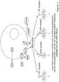
- Adenosine is a well-known effector of immune function. In T-cells, adenosine decreases T-cell receptor induced activation of NF-KB,and inhibits IL-2, IL-4, and IFN- ⁇ . Adenosine decreases T-cell cytotoxicity, increases T-cell anergy, and increases T-cell differentiation to Fop3+ or Lag-3+ regulatory (T-reg) T-cells. On NK cells, adenosine is known to decrease IFN- ⁇ production, and suppress NK cell cytoxicity. Adenosine is known to block neutrophil adhesion and extravasation, decrease phagocytosis, and attenuate levels of superoxide and nitric oxide.
- Adenosine also decreases the expression of TNF- ⁇ , IL-12, and MIP-1 ⁇ on macrophages, attenuates MHC Class II expression, and increases levels of IL-10 and IL-6. In addition, adenosine decreases phagocytosis and superoxide and nitric oxide levels on macrophages. Through these immune-related activities, and others, aberrant or accumulated levels of adenosine is associated with a number of diseases and conditions, including those in which the adenosine-mediated immunosuppressive effects play a role. Hence, there is a need for treatments of such diseases and conditions.
- WO 98/35039 describes a dendritic cell-derived growth factor polypeptide having 99.6% identity to the sequence of amino acids of the unmodified ADA2 protein as set forth in SEQ ID NO: 5 of the present application, and describes the use of the polypeptide in treating immune system defects due to dendritic cells failing to produce sufficient amounts.
- WO 2008/131208 describes adenosine deaminase variants of ADA1 that show improved stability against oxidative degradation.
- WO 2005/000099 describes an immunomodulatory gene, known as 'cat eye syndrome chromosome region candidate 1 (CECR1)', having the same sequence of amino acids as the unmodified ADA2 protein as set forth in SEQ ID NO: 5 of the present application, and proposes the use of this gene sequence for eliciting cytolytic responses, for example for the treatment of cancer.
- CECR1 'cat eye syndrome chromosome region candidate 1
- ADA2 Adenosine Deaminase 2
- the unmodified ADA2 protein is selected from among an ADA protein that comprises: the sequence of amino acids set forth in SEQ ID NO:5; a sequence of amino acids that exhibits at least 95% sequence identity to the sequence of amino acids set forth in SEQ ID NO:5; or is a catalytically active portion of the sequence of amino acids that exhibits at least 95% sequence identity to the sequence of amino acids set forth in SEQ ID NO:5; or comprises the sequence of amino acids set forth in any of SEQ ID NOS:5, 326-334, 340, 375 or 380-383 or is a catalytically active portion thereof;
- the modified ADA2 protein has up to 2, 3, 4, 5, 6, 7, 8, 9 or 10 amino acid modifications compared to the unmodified ADA2 protein; the amino acid modification
- ADA2 protein that, when in dimer form, can exhibit one or more properties selected from among increased adenosine deaminase activity, reduced heparin binding, longer serum half-life, altered pH optimum, increased thermal stability, altered receptor binding and hyperglycosylation compared to the corresponding dimer form of the unmodified ADA2 protein.
- a variety of amino acid modifications, including replacements, deletions and insertions are provided. It is understood that the discreet modifications that confer a particular activity or property can be combined; as in proteins effects of mutation or modifications generally are additive.
- any of the variant ADA2 or catalytically active portion thereof provided herein that contains modifications, including replacements, deletions and insertions, and nucleic acids encoding the variant ADA2 or catalytically active portion thereof can be used in any of the methods, compositions, conjugates, modified forms, vectors, cells, combinations, uses and compositions for use, and combinations for use, provided herein.
- the unmodified ADA2 protein is a homodimer, and the monomer form comprises the sequence of amino acid residues set forth in SEQ ID NO:5.
- the variant ADA2 is a catalytically active portion of the variant ADA2 protein as provided herein, wherein the unmodified ADA2 protein is a homodimer of corresponding catalytically active portions the polypeptide whose sequence is set forth in of SEQ ID NO:5, wherein corresponding portions are determined by alignment.
- the ADA2 protein or catalytically active portion thereof does not contain a modification selected from among an amino acid replacement corresponding to H7R, G18A, G18R, G18V, I64T, A80D, H83Q, V90A, C108G, A120V, H121R, W133G, R125C, R140Q, K141R, R142W, P164L, P222L, W235S, H306R, E330G, W333G, V365L, Y424C, F464S or a deletion corresponding to R8-K14del ⁇ --, with numbering with reference to amino acid residues set forth in SEQ ID NO:5.
- the unmodified ADA2 protein can include a sequence of amino acids that has at least 95%, 96%, 97%, 98%, 99% or more sequence identity to the sequence of amino acids set forth in SEQ ID NO:5 or is a catalytically active portion thereof.
- the unmodified ADA2 protein has at least 95% sequence identity with the sequence of amino acids set forth in SEQ ID NO:5 or with the corresponding catalytically active portion thereof.
- the unmodified ADA2 protein includes the sequence of amino acids set forth in any of SEQ ID NOS:5, 326-334, 340, 375 or 380-383 or is a catalytically active portion thereof, or the unmodified ADA2 protein has a sequence of amino acids is set forth in any of SEQ ID NOS:5, 326-334, 340, 375 and 380-383 or is a catalytically active portion thereof.
- the unmodified ADA2 protein includes the sequence of amino acids set forth in SEQ ID NO:5 or is a catalytically active portion thereof.
- the catalytically active portion of the ADA2 protein can be an ADA2 protein that lacks all or a portion of the putative receptor binding (PRB) domain.
- the catalytically active portion of the ADA2 protein can include the sequence of amino acids set forth in SEQ ID NOS:548-550.
- the catalytically active portion of the unmodified ADA2 protein has the sequence set forth as residues 77-473 of the protein set forth in SEQ ID NO:5.
- the variant ADA2 protein includes up to 2, 3, 4, 5, 6, 7, 8, 9 or 10 amino acid modifications.
- the variant ADA2 protein does not contain the sequence of amino acids set forth in any of SEQ ID NOS:1, 5, 68, 286-302, 326-342 or 374-383 or catalytically active fragment thereof.
- the primary amino acid sequence of the ADA2 protein variant is not the sequence of amino acids set forth in any of SEQ ID NOS:1, 5, 68, 286-302, 326-342 or 374-383.
- the variant ADA2 protein when in dimer form, exhibits adenosine deaminase activity to convert adenosine to inosine.
- the variant ADA2 protein when in dimer form, can exhibit a catalytic efficiency ( k cat /K M ) that is at least or at least about 5 x 10 3 M -1 s -1 , 6 x 10 3 M -1 s -1 , 7 x 10 3 M -1 s -1 , 8 x 10 3 M -1 s -1 , 9 x 10 3 M -1 s -1 , 1 x 10 4 M -1 s -1 , 2 x 10 4 M -1 s -1 , 3 x 10 4 M -1 s -1 , 4 x 10 4 M -1 s -1 , 5 x 10 4 M -1 s -1 , 6 x 10 4 M -1 s -1 , 7 x 10 4 M -1 s -1 -1
- the variant ADA2 protein when in dimer form, can exhibit a thermal stability with a melting temperature (Tm) of at least 58°C.
- Tm melting temperature
- the Tm of the ADA2 protein is at least 59°C, 60°C, 61°C, 62°C, 63°C, 64°C, 65°C, 66°C, 68°C, 69°C, 70°C, 71°C, 72°C or greater.
- the variant ADA2 proteins according to the present claims may contain a modification(s) that is an amino acid replacement(s); and the variant ADA2 protein may include one or more amino acid replacement(s) at an amino acid position corresponding to amino acid residue 11, 13, 20, 22, 26, 86, 179, 217, 219, 221, 258, 262, 264, 266, 267, 277, 283, 296, 309, 317, 321, 352, 366, 371, 372, 373, 374, 403, 404, 405, 406, 441, 444, 452, 461, 469 or 470, with reference to amino acid positions set forth in SEQ ID NO:5.
- the amino acid replacement(s) are positions corresponding to amino acid residue 11, 20, 219, 221, 262, 264, 366, 371, 372 or 452, with reference to amino acid positions set forth in SEQ ID NO:5.
- the variant ADA2 protein can include one or more amino acid replacement(s) selected from among K11A, K11D, K11E, K13A, K13D, K13E, R20A, R20D, R20E, R20N, V22S, K26A, K26D, K26E, D86A, D86C, D86E, D86F, D86G, D86H, D86I, D86K, D86L, D86M, D86N, D86P, D86Q, D86R, D86S, D86T, D86V, D86W, D86Y, E179A, E179C, E179D, E179F, E179G, E179H, E179I, E179K, E179L, E179M, E179N, E179P,
- variant ADA2 protein may contain one or more amino acid replacement(s) selected from from among replacements corresponding to H264A; H264Q; H264N; H264G; R219K; R219Q; R219N; R219A; L221A; L221V; L221G; E179D; E179A; E179S; E179T; E179V; E179G; S262A; S262V; S262M; S262N; D86A; D86C; D86E; D86F; D86G; D86H; D86I; D86K; D86L; D86M; D86N; D86P; D86Q; D86R; D86S; D86T; D86V; D86W; D86Y; E179C; E179F; E179H; E179I; E179K; E179L; E179M; E179N; E179P; E179Q; E179R; E179W; E179Y; R219C
- the variant ADA2 protein contains an amino acid replacement at one or both of positions corresponding to amino acid residue 219 and 262, with reference to amino acid positions set forth in SEQ ID NO:5.
- the variant ADA2 protein or catalytically active portion thereof contains the replacement corresponding to S262N or S262Q.
- the variant ADA2 contains the replacement corresponding to S262N.
- the variant ADA2 contains the replacement corresponding to R219K, R219Q, R219N or R219A.
- the variant ADA2 contains the replacement corresponding to R219Q or the replacements R219Q/R20E.
- the variant ADA2 contains the replacement corresponding to R219Q/S262N.
- the variant ADA2 protein or catalytically active portion thereof contains modification(s) selected from among any of R219Q/S262N/ins(NAS)I1, R219Q/S262N/R20N/V22S, R219Q/S262N/K371N/D373S, R219Q/S262N/K372N/I374S, R219Q/S262N/T403N/H405S, R219Q/S262N/G404N/P406S, R219Q/S262N/C105_T147delins(G) 15 , R219Q/S262N/C105_T147delins(G) 10 , R219Q/S262N/C105_T147delins(G) 7 , R219Q/S262N/C105_T147delins(G) 5 , R219Q/S262N/C105_T147delins(G
- the variant ADA2 protein comprises the modifications selected from among R219Q/K11A/R20A, R219Q/K11A/R20A/K371A, R219Q/R20A/K371A, 219Q/K11A/K371A, S262N/K11A/R20A, S262N/K11A/R20A/K371A, S262N/R20A/K371A, S262N/K11A/K371A, R219Q/C105_T147delins(G) n , R219Q/V99_Q144delins(GGGGS) n , R219Q/C105_T147delins(GGGGS) n , R219Q/N98_N156del, R219Q/C105_E148del, R219Q/C105_T147del, R219Q/V99_Q144del, S262N/
- the variant ADA2 protein when in dimer form, exhibits increased adenosine deaminase activity.
- the variant ADA2 protein when in dimer form, can exhibit at least 110%, 120%, 130%, 140%, 150%, 160%, 170%, 180%, 190%, 200%, 225%, 250%, 300%, 350%, 400%, 450%, 500%, 600%, 700%, 800% or more activity of the corresponding dimer form of the unmodified ADA2 protein, wherein adenosine deaminase activity is assessed under the same conditions.
- the variant ADA2 protein when in dimer form, can exhibit a catalytic efficiency ( k cat /K M ) that is at least or at least about 1.2-fold, 1.3-fold, 1.4-fold, 1.5-fold, 1.6-fold, 1.7-fold, 1.8-fold, 1.9-fold, 2.0-fold, 2.2-fold, 2.5-fold, 3.0-fold, 3.5-fold, 4-fold, 4.5-fold, 5.0-fold, 6.0-fold, 7.0-fold, 8.0-fold, 9.0-fold, 10.0-fold or more compared to the catalytic efficiency ( k cat /K M ) of the corresponding dimer form of the unmodified ADA2 protein, wherein catalytic efficiency of adenosine deaminase activity is assessed under the same conditions.
- the variant ADA2 protein when in dimer form, can exhibit a catalytic efficiency ( k cat /K M ) that is at least or at least about 2 x 10 4 M -1 s -1 , 3 x 10 4 M -1 s -1 , 4 x 10 4 M -1 s -1 , 5 x 10 4 M -1 s -1 , 6 x 10 4 M -1 s -1 , 7 x 10 4 M -1 s -1 , 8 x 10 4 M -1 s -1 , 9 x 10 4 M -1 s -1 , 1 x 10 5 M -1 s -1 , 2 x 10 5 M -1 s -1 , 3 x 10 5 M -1 s -1 , 4 x 10 5 M -1 s -1 , 5 x 10 5 M -1 s -1 or greater.
- k cat /K M catalytic efficiency
- ADA2 proteins in accordance with the present claims are any that include one or more amino acid replacement(s) at an amino acid position corresponding to amino acid residue 11, 20, 219, 221, 262, 264, 366, 371, 372 or 452, with reference to amino acid positions set forth in SEQ ID NO:5.
- the variant ADA2 protein can include one or more amino acid replacement(s) selected from among K11A, K11E, R20A, R20E, R219K, R219Q, L221A, L221V, L221G, S262N, H264Q, H264G, R366E, K371A, K371D, K371E, K372D, K372E, K452D and K452E, with reference to amino acid positions set forth in SEQ ID NO:5.
- amino acid replacement(s) selected from among K11A, K11E, R20A, R20E, R219K, R219Q, L221A, L221V, L221G, S262N, H264Q, H264G, R366E, K371A, K371D, K371E, K372D, K372E, K452D and K452E, with reference to amino acid positions set forth in SEQ ID NO:5.
- the variant ADA2 protein can include amino acid replacements selected from among S262N/K371D, S262N/K371E, S262N/R20E, S262N/R20E/K371D, S262N/R20E/K371E, R219Q/K371E, R219Q/K371D, R219Q/R20E, R219Q/K371E/R20E, R219Q/K371D/R20E, R219Q/S262N/K371E, R219Q/S262N/K371D, R219Q/S262N/R20E, R219Q/S262N/K371E, R219Q/S262N/K371D, R219Q/S262N/R20E, R219Q/S262N/K371E/R20E, R219Q/S262N/K371D/R20E
- the variant ADA2 protein when in dimer form, can exhibit reduced heparin binding.
- the variant ADA2 protein when in dimer form, can exhibit no more than 1%, 2%, 3%, 4%, 5%, 6%, 7%, 8%, 9%, 10%, 15%, 20%, 25%, 30%, 35%, 40%, 45%, 50%, 55%, 60%, 65%, 70%, 75%, 80%, 85%, 90%, 95% of the heparin binding of the corresponding dimer form of the unmodified ADA2 protein, wherein heparin binding is assessed under the same conditions.
- the variant ADA2 protein when in dimer form, can exhibit a longer serum half-life (t 1/2 ).
- the variant ADA2 when in dimer form, can exhibit a half-life that is at least or at least about 110%, 120%, 130%, 140%, 150%, 160%, 170%, 180%, 190%, 200%, 225%, 250%, 300%, 350%, 400%, 450%, 500%, 600%, 700%, 800% or more longer than the half-life of the corresponding dimer form of the unmodified ADA2 protein, wherein half-life is assessed under the same conditions.
- the variant ADA2 protein when in dimer form, can exhibit increased thermal stability.
- the variant ADA2 protein when in dimer form, can exhibit thermal stability with a melting temperature (Tm) that is increased at least or at least about 0.5°C, 1.0°C, 2.0°C, 3.0°C, 4.0°C, 5.0°C, 6.0°C, 7.0°C, 8.0°C, 9.0°C, 10.0°C or more compared to the Tm of the corresponding dimer form of the unmodified ADA2 protein, wherein Tm is assessed under the same conditions.
- Tm melting temperature
- the variant ADA2 protein can have a melting temperature (Tm) of at least or at least about 67.6°C, 67.8°C, 68.0°C, 68.2°C, 68.4°C, 68.6°C, 68.8°C, 69.0°C, 69.2°C, 69.4°C, 69.6°C, 69.8°C, 70.0°C, 70.2°C, 70.4°C, 70.6°C, 70.8°C, 71.0°C, 71.2°C, 71.4°C, 71.6°C, 71.8°C or higher.
- Tm melting temperature
- the adenosine deaminase activity of the variant ADA2 protein can be assessed or exhibited at or about pH 6.5 ⁇ 0.2.
- the variant ADA2 protein when in dimer form, can exhibit an altered pH optimum for adenosine deaminase activity.
- the variant ADA2 protein when in dimer form, can exhibit a pH optimum for adenosine deaminase activity that is at a higher pH compared to the pH optimum of the corresponding dimer form of the unmodified ADA2 protein.
- the variant ADA2 protein when in dimer form, can have a pH optimum with a pH that is at least or at least about 6.6, 6.7, 6.8, 6.9, 7.0, 7.1, 7.2, 7.3, 7.4, 7.5 or higher.
- the variant ADA2 protein when in dimer form, can exhibit a pH optimum for adenosine deaminase activity that is at a lower pH compared to the pH optimum of the corresponding dimer form of the unmodified ADA2 protein.
- the variant ADA2 protein when in dimer form, can have a pH optimum with a pH that is less than or less than about 6.5, 6.4, 6.3, 6.3, 6.2, 6.1, 6.0 or less.
- the variant ADA2 protein can include a modification of one or more amino acids in the putative receptor binding (PRB), wherein the modification is an amino acid deletion, insertion or replacement.
- the variant ADA2 protein does not contain a modification that is an amino acid replacement corresponding to amino acid replacement C108G, A120V, H121R, R125C, R140Q, K141R or R142W, with reference to amino acid positions set forth in SEQ ID NO:5.
- the variant ADA2 protein or catalytically active portion thereof lacks all or a portion of the putative receptor binding (PRB) domain or has a modification of the PRB, whereby any receptor binding or growth factor activity is reduced or eliminated or other activity of ADA2 other than deaminase activity is reduced or eliminated or interaction with the ADA domain is reduced or eliminated and the PRB domain corresponds to residues 98 to 156 set forth in SEQ ID NO:5.
- the variant ADA2 lacks residues 105-148 or 105 to 147 or 99 to 144, with reference to amino acid positions set forth in SEQ ID NO:5.
- variant ADA2 protein or catalytically active portion thereof contains the sequence of amino acids set forth in any of SEQ ID NOS:548-550 and 579. In some embodiments, variant ADA2 protein or catalytically active portion thereof contains a deletion of all or a portion of the PRB domain and optionally an insertion of peptide linker.
- the variant ADA2 protein can have a deletion of one or more contiguous amino acid residues corresponding to any one or more contiguous amino acid residues between or between about amino acid residues 98 and 156 or amino acid residues 105 and 148, inclusive, with reference to amino acid positions set forth in SEQ ID NO:5.
- the variant of ADA2 polypeptide can further include substitution of the deleted region with a peptide linker.
- the peptide linker can be selected from among (Gly)n (SEQ ID NO:368), where n is 2 to 20; (GGGGS)n (SEQ ID NO:343), where n is 1 to 6; (SSSSG)n (SEQ ID NO:344), where n is 1 to 6; (AlaAlaProAla)n (SEQ ID NO:350), where n is 1 to 6; GKSSGSGSESKS (SEQ ID NO:345); GGSTSGSGKSSEGKG (SEQ ID NO:346); GSTSGSGKSSSEGSGSTKG (SEQ ID NO:347); GSTSGSGKPGSGEGSTKG (SEQ ID NO:348); and EGKSSGSGSESKEF (SEQ ID NO:349).
- the peptide linker can be selected from among GGG (SEQ ID NO:369); GGGGG (SEQ ID NO:360); GGGGGGG (SEQ ID NO:370); GGGGGGGGGG (SEQ ID NO:371); and GGGGGGGGGGGGG (SEQ ID NO:372).
- the variant ADA2 can contain the variant ADA2 polypeptide that can include a modification in the PRB domain that corresponds to C105_T147delins(G) n , where n is 2 to 20, such as C105_T147delins(G) 15 , C105_T147delins(G) 10 , C105_T147delins(G) 7 , C105_T147delins(G) 5 or C105_T147delins(G) 3 , with reference to amino acid positions set forth in SEQ ID NO:5.
- the variant ADA2 protein or catalytically active portion thereof contains amino acid replacements selected from among replacements corresponding to R219Q/S262N/F119S; R219Q/S262N/F119K; R219Q/S262N/Y224R; R219Q/S262N/Y224N; R219Q/S262N/Y191S; R219Q/S262N/Y191D; R219Q/S262N/F183K; R219Q/S262N/Y191D/Y224R; R219Q/S262N/F109S; R219Q/S262N/F109A; R219Q/S262N/R118D; R219Q/S262N/R118A; R219Q/S262N/Y139T; R219Q/S262N/Y139A; R219Q/S262N/W133S; R219Q/S
- the variant ADA2 contains modifications selected from among R219Q/S262N/C105_T147delins(G) 15 ; R219Q/S262N/C105_T147delins(G) 10 ; R219Q/S262N/C105_T147delins(G) 7 ; R219Q/S262N/C105_T147delins(G) 5 ; R219Q/S262N/C105_T147delins(G) 3 ; R219Q/S262N/V99_Q144delins(GGGGS) 1 ; R219Q/S262N/V99_Q144delins(GGGGS) 2 ; R219Q/S262N/V99_ Q144delins(GGGGS) 3 ; R219Q/S262N/C105_T147delins(GGGGS) 1 ; R219Q/S262N/C105_T147delins(GG
- the variant ADA2 protein when in dimer form, can exhibit reduced binding to any one or more adenosine receptor (ADR) selected from among A 1 , A 2A , A 2B and A 3 compared to binding of the unmodified ADA2 protein to the same receptor when assessed under the same conditions.
- ADR adenosine receptor
- the variant ADA2 protein has a binding that is reduced at least or at least about 0.5-fold, 1-fold, 1.5-fold, 2-fold, 3-fold, 4-fold, 5-fold, 6-fold, 7-fold, 8-fold, 9-fold, 10-fold or more.
- the variant ADA2 can be glycosylated, for example, at a native or a non-native glycosylation site.
- the variant ADA2 protein can include a modification(s) that alters glycosylation by introduction of a non-native glycosylation site, whereby, when expressed in a cell capable of glycosylation, the variant ADA2 protein is hyperglycosylated compared to the unmodified ADA2 polypeptide.
- the non-native glycosylation site is introduced by amino acid replacement(s) or insertion of one, two or three amino acids.
- the modifications are selected from among modifications corresponding to ins(NAS)I1, R20N/V22S, K371N/D373S, K372N/I374S, T403N/H405S and G404N/P406S, with reference to amino acid positions set forth in SEQ ID NO:5.
- the variant ADA2 or catalytically active portion thereof contains modifications corresponding to R219Q/S262N/ins(NAS)I1 ; R219Q/S262N/R20N/V22S; R219Q/S262N/K371N/D373S; R219Q/S262N/K372N/I374S; R219Q/S262N/T403N/H405S; or R219Q/S262N/G404N/P406S.
- the variant ADA2 protein or catalytically active portion thereof contains a modification in the putative receptor binding domain (PRB) corresponding to one or more of the modifications selected from among: R125N/P126A; S127N/K129S; P126N/E128T; R112N/I114T; I134N/L135C/L136T; I134N/L135S/L136T; R142N/Q144S; E137N/Y139T; and P111N/G113S.
- PRB putative receptor binding domain
- the variant ADA2 protein or catalytically active portion thereof contains amino acid replacements corresponding to R219Q/S262N/R125N/P126A; R219Q/S262N/S127N/K129S; R219Q/S262N/P126N/E128T; R219Q/S262N/R112N/I114T; R219Q/S262N/I134N/L135C/L136T; R219Q/S262N/I134N/L135S/L136T; R219Q/S262N/R142N/Q144S; R219Q/S262N/E137N/Y139T; or R219Q/S262N/P111N/G113S.
- the variant ADA2 protein can be a human ADA2. In some embodiments, the variant ADA2 protein can be isolated or purified.
- the variant ADA2 protein can contain a polypeptide that exhibits at least 65% sequence identity to SEQ ID NO:5 or a catalytically active portion thereof.
- the variant ADA2 protein can contain a polypeptide that exhibits at least 70%, 75%, 76%, 77%, 78%, 79%, 80%, 81%, 82%, 83%, 84%, 85%, 86%, 87%, 88%, 89%, 90%, 91%, 92%, 93%, 94%, 95%, 96%, 97%, 98%, 99% or more sequence identity to SEQ ID NO:5 or a catalytically active portion thereof.
- the variant ADA2 protein contains a polypeptide that has the sequence of amino acids set forth in any of SEQ ID NOS: 13-63 or 71-285 or a catalytically active portion thereof.
- the variant ADA2 protein or catalytically active portion thereof contains the sequence of amino acids set forth in any of SEQ ID NOS: 551-579 or 581-993 or a catalytically active portion thereof.
- the variant ADA2 protein or a catalytically active portion thereof can contain amino acid replacements selected from among replacements corresponding to S262N/K371D; S262N/K371E; S262N/R20E; S262N/R20E/K371D; S262N/R20E/K371E; R219Q/K371E; R219Q/K371D; R219Q/R20E; R219Q/K371E/R20E; R219Q/S262N/K371E; R219Q/S262N/K371E; R219Q/S262N/K371D; R219Q/S262N/R20E; R219Q/S262N/K371E; R219Q/S262N/K371D; R219Q/S262N/R20E; R219Q/S262N/K371E/R20E; R2
- the variant ADA2 protein can be a monomer or a dimer.
- variant ADA2 proteins provided herein are dimers of a variant ADA2 protein, that can include any of the variant ADA2 proteins provided.
- the dimer can be a homodimer. In other examples, the dimer can be a heterodimer.
- nucleic acid molecules encoding the variant ADA2 proteins provided herein are also provided herein.
- the vector can be eukaryotic or a prokaryotic vector, such as a mammalian vector or a viral vector.
- the vector is an adenovirus vector, an adeno-associated-virus vector, a retrovirus vector, a herpes virus vector, a lentivirus vector, a poxvirus vector, or a cytomegalovirus vector.
- the vector is an oncolytic vector.
- the vector can also encode a soluble hyaluronidase.
- the cell can be a eukaryotic cell, such as a mammalian cell. If human, the cells are isolated or are provided as a cell culture.
- the cell is a mammalian cell, such as a Chinese Hamster Ovary (CHO) cell.
- the cell can expresses the variant ADA2 protein, such as a dimer.
- variant ADA2 proteins such as variant ADA2 dimers, that are produced by the cell provided herein.
- the cell is an isolated cell or a cell culture, such as a eukaryotic cell, a non-human cell, a mammalian cell, a cell that is not a human stem cell.
- the cell is an immune cell, such as a T cell, a tumor-infiltrating lymphocyte (TIL), a cytotoxic T lymphocyte (CTL), a natural killer (NK) cell or a lymphokine-activated killer (LAK) cell.
- TIL tumor-infiltrating lymphocyte
- CTL cytotoxic T lymphocyte
- NK natural killer
- LAK lymphokine-activated killer
- the cell is a T cell that encodes and expresses chimeric antigen receptor (CAR) and the variant ADA2 protein or dimer.
- CAR chimeric antigen receptor
- the CAR is specific for a tumor cell antigen
- the tumor antigen is selected from among HER2, CD19, HERV-K, CD20, CD22, ROR1, mesothelin, CD33/IL3Ra, c-Met, PSMA, Glycolipid F77, EGFRvIII, GD-2, NY- ESO-1 TCR, MAGE A3 TCR and GD2 and combinations thereof.
- conjugates include a variant ADA2 protein or a catalytically active portion of any ADA2 protein provided herein, such as a variant ADA2 dimer provided in any examples herein, linked directly or indirectly via a linker to a heterologous moiety, such as a toxin or therapeutic drug.
- conjugates that include an ADA2 protein linked directly or indirectly via a linker to a heterologous moiety.
- the ADA2 protein can be a monomer or a dimer.
- the dimer is a homodimer; in other it is a heterodimer.
- the heterologous moiety is conjugated to one or both subunits in the dimer.
- the heterologous moiety for example, can be selected from among a peptide, small molecule, nucleic acid, carbohydrate and polymer.
- the heterologous moiety is a half-life extending moiety.
- the half-life extending moiety is selected from among biocompatible fatty acids and derivatives thereof, hydroxy alkyl starch (HAS), polyethylene glycol (PEG), Poly (Gly x - Ser y ) n , homo-amino-acid polymers (HAP), hyaluronic acid (HA), heparosan polymers (HEP), phosphorylcholine-based polymers (PC polymer), Fleximers, dextran, polysialic acids (PSA), Fc domain, Transferrin, Albumin, elastin-like peptides, XTEN sequences, albumin binding peptides, a CTP peptide, and any combination thereof.
- the half-life extending moiety is a PEG and the ADA2 protein is PEGylated.
- the PEG can be selected from among methoxypolyethylene glycols (mPEG), PEG-glycidyl ethers (Epox-PEG), PEG-oxycarbonylimidazole (CDI-PEG), branched PEGs and polyethylene oxide (PEO).
- mPEG methoxypolyethylene glycols
- Epox-PEG PEG-glycidyl ethers
- CDI-PEG PEG-oxycarbonylimidazole
- branched PEGs and polyethylene oxide (PEO).
- PEG has a molecular weight of from or from about 1 kDa to about 100 kDa.
- the PEG can be linear or branched.
- the PEG moieties result from reaction with a PEG reagent selected from among mPEG-Succinimidyl Propionates (mPEG-SPA), mPEG Succinimidyl Carboxymethyl Ester (mPEG-SCM), mPEG-Succinimidyl Butanoates (mPEG-SBA), mPEG2-N-Hydroxylsuccinimide, mPEG-succinimidyl butanoate (mPEG-SBA), mPEG-succinimidyl ⁇ -methylbutanoate (mPEG-SMB), mPEG-butyrldehyde, PEG-p-nitrophenyl-carbonate and PEG-propionaldehyde.
- mPEG-SPA mPEG-Succinimidyl Propionates
- mPEG-SCM mPEG Succinimidyl Carboxymethyl Ester
- the PEG moieities result from reaction with a PEG reagent selected from among mPEG-SCM (20kDa), mPEG-SCM (30kDa), mPEG-SBA (5kDa), mPEG-SBA (20kDa), mPEG-SBA (30kDa), mPEG-SMB (20kDa), mPEG-SMB (30kDa), mPEG-butyrldehyde (30kDa), mPEG-SPA (20kDa), mPEG-SPA (30kDa), mPEG2-NHS (10kDa branched), mPEG2-NHS (20kDa branched), mPEG2-NHS (40kDa branched), mPEG2-NHS (60kDa branched), PEG-NHS-biotin (5kDa biotinylated), PEG-p-nitrophenyl-carbonate (30kDa) and PEG-propionaldehy
- the ADA2 protein is a variant ADA2 protein in accordance with the present claims that contains a sequence of amino acids that includes a modification(s) in the sequence of amino acids of an unmodified ADA2 protein or a catalytically active portion thereof, where the unmodified ADA2 protein contains the sequence of amino acids set forth in SEQ ID NO:5, or a sequence of amino acids that exhibits at least 95% sequence identity to the sequence of amino acids set forth in SEQ ID NO:5, or is a catalytically active portion thereof.
- the amino acid modification(s) are selected from among amino acid replacement(s), deletion(s) and insertion(s); and the variant ADA2 protein, when in dimer form, can exhibit adenosine deaminase activity to convert adenosine to inosine.
- the ADA2 protein when in dimer form, can exhibit a catalytic efficiency ( k cat /K M ) that is at least or at least about 5 x 10 3 M -1 s -1 , 6 x 10 3 M -1 s -1 , 7 x 10 3 M -1 s -1 , 8 x 10 3 M -1 s -1 , 9 x 10 3 M -1 s -1 , 1 x 10 4 M -1 s -1 , 2 x 10 4 M -1 s -1 , 3 x 10 4 M -1 s -1 , 4 x 10 4 M -1 s -1 , 5 x 10 4 M -1 s -1 , 6 x 10 4 M -1 s -1 , 7 x 10 4 M -1 s -1 , 8 x 10 4 M -1 s -1 , 9 x 10 4 M -1 s -1 , 1 x 10 5 M -1 s s , 6 x 10 3 M
- the modification(s) of the ADA2 protein can be an amino acid replacement(s); and the variant ADA2 protein can include one or more amino acid replacement(s) at an amino acid position corresponding to amino acid residue 11, 13, 20, 22, 26, 86, 179, 217, 219, 221, 258, 262, 264, 266, 267, 277, 283, 296, 309, 317, 321, 352, 366, 371, 372, 373, 374, 403, 404, 405, 406, 441, 444, 452, 461, 469 or 470, with reference to amino acid positions set forth in SEQ ID NO:5.
- the variant ADA2 protein can include one or more amino acid replacement(s) selected from among K11A, K11D, K11E, K13A, K13D, K13E, R20A, R20D, R20E, R20N, V22S, K26A, K26D, K26E, D86A, D86C, D86E, D86F, D86G, D86H, D86I, D86K, D86L, D86M, D86N, D86P, D86Q, D86R, D86S, D86T, D86V, D86W, D86Y, E179A, E179C, E179D, E179F, E179G, E179H, E179I, E179K, E179L, E179M, E179N, E179P, E179Q, E179R, E179S, E179T, E179V, E179W, E179Y, R217A, R217D, R
- the variant ADA2 protien can include one or more amino acid replacement(s) selected from among K11A, K11E, R20A, R20D, R20E, R219K, R219Q, L221A, L221V, L221G, S262N, H264Q, H264G, R366A, R366D, R366E, K371A, K371D, K371E, K372A, K372D, K372E, K452D and K452E, with reference to amino acid positions set forth in SEQ ID NO:5.
- amino acid replacement(s) selected from among K11A, K11E, R20A, R20D, R20E, R219K, R219Q, L221A, L221V, L221G, S262N, H264Q, H264G, R366A, R366D, R366E, K371A, K371D, K372A, K372D, K
- the variant ADA2 protein can include amino acid replacements selected from among S262N/K371D, S262N/K371E, S262N/R20E, S262N/R20E/K371D, S262N/R20E/K371E, R219Q/K371E, R219Q/K371D, R219Q/R20E, R219Q/K371E/R20E, R219Q/K371D/R20E, R219Q/S262N/K371E, R219Q/S262N/K371D, R219Q/S262N/R20E, R219Q/S262N/K371E, R219Q/S262N/K371D, R219Q/S262N/R20E, R219Q/S262N/K371E/R20E, R219Q/S262N/K371D/R20E
- the variant ADA2 protein can include a modification of one or more amino acids in the putative receptor binding (PRB) domain that is an amino acid deletion, insertion or replacement.
- the variant ADA2 protein can include deletion of one or more contiguous amino acid residues corresponding to any one or more contiguous amino acid residues between or between about amino acid residues 98 and 156 or amino acid residues 105 and 148, inclusive, with reference to amino acid positions set forth in SEQ ID NO:5.
- the variant ADA2 protein in the conjugate can further include substitution of the deleted region with a peptide linker.
- the peptide linker can be selected from among (Gly)n (SEQ ID NO:368), where n is 2 to 20; (GGGGS)n (SEQ ID NO:343), where n is 1 to 6; (SSSSG)n (SEQ ID NO:344), where n is 1 to 6; (AlaAlaProAla)n (SEQ ID NO:350), where n is 1 to 6; GKSSGSGSESKS (SEQ ID NO:345); GGSTSGSGKSSEGKG (SEQ ID NO:346); GSTSGSGKSSSEGSGSTKG (SEQ ID NO:347); GSTSGSGKPGSGEGSTKG (SEQ ID NO:348); and EGKSSGSGSESKEF (SEQ ID NO:349).
- the peptide linker is selected from among GGG (SEQ ID NO:369); GGGGG (SEQ ID NO:360); GGGGGGG (SEQ ID NO:370); GGGGGGGGGG (SEQ ID NO:371); and GGGGGGGGGGGGG (SEQ ID NO:372).
- the modification in the PRB domain can corresponds to C105_T147delins(G) n , where n is 2 to 20, such as, C105_T147delins(G) 15 , C105_T148delins(G) 10 , C105_T147delins(G) 7 , C105_T147delins(G) 5 or C105_T147delins(G) 3 , with reference to amino acid positions set forth in SEQ ID NO:5.
- the ADA2 protein in the conjugate can be glycosylated at one or more native or non-native glycosylation site.
- the variant ADA2 protein in the conjugate can include a modification(s) that alters glycosylation by introduction of a non-native glycosylation site.
- the non-native glycosylation site can be introduced by creating the canonical glycosylation sequence (NXT/S, where X is not Pro for N-linked carbohydrates, S/T for O-linked) by introducing amino acid replacement(s), insertions or deletions of one, two or three amino acids.
- modifications that alter glycosylation are selected from among modifications corresponding to ins(NAS)I1, R20N/V22S, K371N/D373S, K372N/I374S, T403N/H405S and G404N/P406S, with reference to amino acid positions set forth in SEQ ID NO:5.
- the variant ADA2 protein in the conjugate can have the sequence of amino acid set forth in any of SEQ ID NOS: 13-63 or 71-285 or a catalytically active portion thereof.
- the conjugate retains the adenosine deaminase activity compared to the ADA2 protein that is not conjugated.
- the conjugate can exhibit from or from about 50% to 500%, 50% to 200%, 50% to 100%, 50% to 80%, 80% to 500%, 80% to 200%, 80% to 100%, 100% to 500% or 100% to 200%, each inclusive, of the adenosine deaminase activity of the ADA2 protein that is not conjugated, such as at least 50%, 60%, 70%, 80%, 90%, 100%, 110%, 120%, 130%, 140%, 150%, 160%, 170%, 180%, 190%, 200%, 300%, 400%, 500% or more the adenosine deaminase activity of the ADA2 protein that is not conjugated.
- the ADA2 in the conjugate can exhibit a catalytic efficiency ( k cat /K M ) that is at least or at least about 5 x 10 3 M -1 s -1 , 6 x 10 3 M -1 s -1 , 7 x 10 3 M -1 s -1 , 8 x 10 3 M -1 s -1 , 9 x 10 3 M -1 s -1 , 1 x 10 4 M -1 s -1 , 2 x 10 4 M -1 s -1 , 3 x 10 4 M -1 s -1 , 4 x 10 4 M -1 s -1 , 5 x 10 4 M -1 s -1 , 6 x 10 4 M -1 s -1 , 7 x 10 4 M -1 s -1 , 8 x 10 4 M -1 s -1 , 9 x 10 4 M -1 s , 1 x 10 5 M -1 s -1 ,
- ADA2 protein can be a monomer or a dimer.
- ADA2 protein can be a dimer, such as a homodimer.
- the therapeutic agent can be selected from among an antibody, cytotoxic agent, chemotherapeutic agents, cytokine, growth inhibitory agent, anti-hormonal agent, kinase inhibitor, anti-angiogenic agent, cardioprotectant, immunostimulatory agent, immunosuppressive agent, immune checkpoint inhibitor, antibiotic and angiogenesis inhibitor.
- the therapeutic agent can be an anti-cancer agent.
- the anti-cancer agent can be an anti-cancer antibody, a chemotherapeutic agent, a radioimmunotherapeutic, an anti-angiogenic agent or an immune checkpoint inhibitor.
- the anti-cancer agent can be an immune checkpoint inhibitor; and the target of the immune checkpoint inhibitor can be selected from among CTLA4, PD-1, and PD-L1.
- the immune checkpoint inhibitor can be an antibody, a fusion protein, an aptamer, or an immune checkpoint protein-binding fragment thereof.
- the immune checkpoint inhibitor is an anti-immune checkpoint protein antibody or antigen-binding fragment thereof.
- the immune checkpoint inhibitor is selected from among: an anti-CTLA4 antibody, derivative thereof, or antigen-binding fragment thereof; an anti-PD-Ll antibody, derivative thereof, or antigen-binding fragment thereof; and an anti-PD-1 antibody, derivative thereof, or antigen-binding fragment thereof.
- the immune checkpoint inhibitor can be selected from among: Ipilimumab, a derivative thereof, or an antigen-binding fragment thereof; Tremelimumab, a derivative thereof, or an antigen-binding fragment thereof; Nivolumab, a derivative thereof, or an antigen-binding fragment thereof; and Pidilizumab, a derivative thereof, or an antigen-binding fragment thereof.
- the therapeutic agent can be an anti-hyaluronan agent.
- the anti-hyaluronan agent can be a soluble hyaluronidase.
- the soluble hyaluronidase can exhibit hyaluronidase activity at neutral pH.
- the soluble hyaluronidase can be selected from among bovine, ovine or a C-terminal truncated human PH20 that lacks all or a portion of the glycosylphosphatidylinositol (GPI) anchor attachment sequence.
- GPI glycosylphosphatidylinositol
- the soluble hyaluronidase is a C-terminally truncated human PH20 that lacks all or a portion of the GPI anchor attachment sequence, such as those set forth in any of SEQ ID NOS:481-488, 493-514, or 526-532, or that has a sequence of amino acids that has at least 85%, 86%, 87%, 88%, 89%, 90%, 91%, 92%, 93%, 94%, 95%, 96%, 97%, 98% or 99% sequence identity to a sequence of amino acids set forth in any of SEQ ID NOS:481-488, 493-514, or 526-532 and is soluble and retains hyaluronidase activity.
- the anti-hyaluronan agent or soluble hyaluronidase can be conjugated to a polymer, such as a PEG moiety.
- the ADA2 protein can include a polypeptide having the sequence of amino acids set forth in any of SEQ ID NOS:5, 326-334, 340, 375 or 380-383, a sequence that can exhibit at least 85% sequence identity to the sequence of amino acids set forth in SEQ ID NOS:5, 326-334, 340, 375 or 380-383 or a catalytically active form thereof.
- the ADA2 protein can include a protein having a sequence of amino acids that can exhibit at least 86%, 87%, 88%, 89%, 90%, 91%, 92%, 93%, 94%, 95%, 96%, 97%, 98%, 99% or more sequence identity to the sequence of amino acids set forth in SEQ ID NOS:5, 326-334, 340, 375 or 380-383 or a catalytically active portion thereof.
- the ADA2 protein can contain a sequence of amino acids that can exhibit at least 86%, 87%, 88%, 89%, 90%, 91%, 92%, 93%, 94%, 95%, 96%, 97%, 98%, 99% or more sequence identity to the sequence of amino acids set forth in SEQ ID NO:5.
- the ADA2 protein can contain the sequence of amino acids set forth in SEQ ID NO:5.
- the ADA2 protein is a variant ADA2 protein having a sequence of amino acids that includes a modification(s) in the sequence of amino acids of an unmodified ADA2 polypeptide or a catalytically active portion thereof.
- the unmodified ADA2 protein can include the sequence of amino acids set forth in SEQ ID NO:5, or a sequence of amino acids that can exhibit at least 85% sequence identity to the sequence of amino acids set forth in SEQ ID NO:5, or is a catalytically active portion thereof; the amino acid modification(s) are selected from among amino acid replacement(s), deletion(s) and insertion(s); and the variant ADA2 protein, when in dimer form, can exhibit adenosine deaminase activity to convert adenosine to inosine.
- the ADA2 protein when in dimer form, can exhibit a catalytic efficiency ( k cat /K M ) that is at least or at least about 5 x 10 3 M -1 s -1 , 6 x 10 3 M -1 s -1 , 7 x 10 3 M -1 s -1 , 8 x 10 3 M -1 s -1 , 9 x 10 3 M -1 s -1 , 1 x 10 4 M -1 s -1 , 2 x 10 4 M -1 s -1 , 3 x 10 4 M -1 s -1 , 4 x 10 4 M -1 s -1 , 5 x 10 4 M -1 s -1 , 6 x 10 4 M -1 s -1 , 7 x 10 4 M -1 s -1 , 8 x 10 4 M -1 s -1 , 9 x 10 4 M -1 s -1 , 1 x 10 5 M -1 s -1 , 6 x 10 3 M
- the modification(s) in the sequence of amino acids of an unmodified ADA2 protein can include an amino acid replacement(s); and the variant ADA2 protein can include one or more amino acid replacement(s) at an amino acid position corresponding to amino acid residue 11, 13, 20, 22, 26, 86, 179, 217, 219, 221, 258, 262, 264, 266, 267, 277, 283, 296, 309, 317, 321, 352, 366, 371, 372, 373, 374, 403, 404, 405, 406, 441, 444, 452, 461, 469 or 470, with reference to amino acid positions set forth in SEQ ID NO:5.
- the variant ADA2 protein can include one or more amino acid replacement(s) selected from among K11A, K11D, K11E, K13A, K13D, K13E, R20A, R20D, R20E, R20N, V22S, K26A, K26D, K26E, D86A, D86C, D86E, D86F, D86G, D86H, D86I, D86K, D86L, D86M, D86N, D86P, D86Q, D86R, D86S, D86T, D86V, D86W, D86Y, E179A, E179C, E179D, E179F, E179G, E179H, E1791, E179K, E179L, E179M, E179N, E179P, E179Q, E179R, E179S, E179T, E179V, E179W, E179Y, R217A, R217D, R217E, R219A, R219C,
- the variant ADA2 protein can include one or more amino acid replacement(s) selected from among K11A, K11E, R20A, R20D, R20E, R219K, R219Q, L221A, L221V, L221G, S262N, H264Q, H264G, R366A, R366D, R366E, K371A, K371D, K371E, K372A, K372D, K372E, K452D and K452E, with reference to amino acid positions set forth in SEQ ID NO:5.
- amino acid replacement(s) selected from among K11A, K11E, R20A, R20D, R20E, R219K, R219Q, L221A, L221V, L221G, S262N, H264Q, H264G, R366A, R366D, R366E, K371A, K371D, K372A, K372D, K37
- the variant ADA2 protein can include amino acid replacements selected from among K11A/R20A, K11A/R20A/K371A, R20A/K371A, K11A/K371A, S262N/K371D, S262N/K371E, S262N/R20E, S262N/R20E/K371D, S262N/R20E/K371E, R219Q/K371E, R219Q/K371D, R219Q/R20E, R219Q/K371E/R20E, R219Q/K371D/R20E, R219Q/S262N/K371E, R219Q/S262N/K371D, R219Q/S262N/K371D, R219Q/S262N/K371D, R219Q/S262N/K371D, R219Q/S262N/K
- the variant ADA2 protein can include a modification of one or more amino acids in the putative receptor binding (PRB) domain, wherein the modification is an amino acid deletion, insertion or replacement.
- the variant ADA2 can include deletion of one or more contiguous amino acid residues corresponding to any one or more contiguous amino acid residues between or between about amino acid residues 98 and 156 or amino acid residues 105 and 148, inclusive, with reference to amino acid positions set forth in SEQ ID NO:5.
- the variant of ADA2 polypeptide can further include substitution of the deleted region with a peptide linker.
- the peptide linker can be selected from among (Gly)n (SEQ ID NO:368), where n is 2 to 20; (GGGGS)n (SEQ ID NO:343), where n is 1 to 6; (SSSSG)n (SEQ ID NO:344), where n is 1 to 6; (AlaAlaProAla)n (SEQ ID NO:350), where n is 1 to 6; GKSSGSGSESKS (SEQ ID NO:345); GGSTSGSGKSSEGKG (SEQ ID NO:346); GSTSGSGKSSSEGSGSTKG (SEQ ID NO:347); GSTSGSGKPGSGEGSTKG (SEQ ID NO:348); and EGKSSGSGSESKEF (SEQ ID NO:349).
- the peptide linker is selected from among GGG (SEQ ID NO:369); GGGGG (SEQ ID NO:360); GGGGGGG (SEQ ID NO:370); GGGGGGGGGG (SEQ ID NO:371); and GGGGGGGGGGGGG (SEQ ID NO:372).
- the modification in the PRB domain of the variant ADA2 polypeptide corresponds to C105_T147delins(G) n , where n is 2 to 20, such as C105_T147delins(G) 15 , C105_T147delins(G) 10 , C105_T147delins(G) 7 , C105_T147delins(G) 5 or C105_T147delins(G) 3 with reference to amino acid positions set forth in SEQ ID NO:5.
- the ADA2 protein in the combination can be glycosylated at one or more native or non-native glycosylation site.
- the variant ADA2 protein in the combination include a modification(s) that alters glycosylation by introduction of a non-native glycosylation site.
- the non-native glycosylation site is introduced by amino acid replacement(s) or insertion of one, two or three amino acids.
- the modifications that alter hyperglycosylation can be selected from among modifications corresponding to ins(NAS)I1, R20N/V22S, K371N/D373S, K372N/I374S, T403N/H405S and G404N/P406S, with reference to amino acid positions set forth in SEQ ID NO:5.
- the variant ADA2 polypeptide has the sequence of amino acids set forth in any of SEQ ID NOS: 13-63 or 71-285 or a catalytically active portion thereof.
- compositions that can include any of the variant ADA2 proteins or a catalytically active portion thereof provided herein, any variant ADA2 dimer provided herein or any conjugate provided herein, in a pharmaceutically acceptable vehicle.
- the pharmaceutical composition can be formulated for local or systemic administration.
- the pharmaceutical composition is formulated for intravenous administration.
- any of the variant ADA2 proteins or a catalytically active portion thereof provided herein, any variant ADA2 dimer provided herein or any conjugate provided herein for use in treating a tumor or a cancer in a subject are also provided. Also provided are combinations of any of the combinations provided herein for use in treating a tumor or cancer.
- ADA2 protein or pharmaceutical composition containing an ADA2 protein for use in treating a tumor or a cancer. Also described are combinations containing an ADA2 protein and a therapeutic agent for use in treating a tumor or cancer.
- the tumor can be a solid tumor or a metastatic tumor.
- the tumor can be a carcinoma, gliomas, sarcoma, adenocarcinoma, adenosarcoma, or adenoma.
- the tumor can be a tumor of the breast, heart, lung, small intestine, colon, spleen, kidney, bladder, head and neck, ovary, prostate, brain, pancreas, skin, bone, bone marrow, blood, thymus, uterus, testicles, cervix or liver.
- the subject can be selected for treatment based on elevated levels of plasma adenosine, tumor-associated expression of adenosine receptor (ADR) or tumor-associated expression of a nucleotidase.
- ADR is A2A or A2B.
- the nucleotidase is CD39 or CD73.
- the elevated level is at least 0.5-fold, 1-fold, 2-fold, 3-fold, 4-fold, 5-fold, 6-fold, 7-fold, 8-fold, 9-fold, 10-fold, 15-fold, 20-fold, 30-fold, 40-fold, 50-fold, 60-fold, 70-fold, 80-fold, 90-fold, 100-fold, 150-fold, 200-fold, 500-fold, 1000-fold or more, compared to the predetermined level or predetermined amount or control sample.
- the method of treating a tumor or cancer in a subject can further include administration of one or more anticancer agents or treatments.
- the anti-cancer agent can be selected from among an anti-cancer antibody, a chemotherapeutic agent, a radioimmunotherapeutic, an anti-angiogenic agent and an immune checkpoint inhibitor.
- SCID Severe Combined Immunodeficiency
- SCID Severe Combined Immunodeficiency
- a disease or condition that is a non-cancer hyperproliferative disease, a fibrotic disease, an infectious disease, a vasculopathy or Severe Combined Immunodeficiency (SCID)
- the ADA2 protein can be a monomer or a dimer.
- the ADA2 protein can be a dimer, in particular, a homodimer.
- the ADA2 protein can contain the sequence of amino acids set forth in any of SEQ ID NOS:5, 326-334, 340, 375 or 380-383, a sequence that can exhibit at least 85% sequence identity to the sequence of amino acids set forth in SEQ ID NOS:5, 326-334, 340, 375 or 380-383 or a catalytically active form thereof.
- the ADA2 protein can contain a sequence of amino acids that can exhibit at least 86%, 87%, 88%, 89%, 90%, 91%, 92%, 93%, 94%, 95%, 96%, 97%, 98%, 99% or more sequence identity to the sequence of amino acids set forth in SEQ ID NOS:5, 326-334, 340, 375 or 380-383 or a catalytically active portion thereof.
- the ADA2 protein can contain a sequence of amino acids that can exhibit at least 86%, 87%, 88%, 89%, 90%, 91%, 92%, 93%, 94%, 95%, 96%, 97%, 98%, 99% or more sequence identity to the sequence of amino acids set forth in SEQ ID NOS:5 or a catalytically active portion thereof.
- the ADA2 protein can contain the sequence of amino acids set forth in SEQ ID NO:5.
- the ADA2 protein is a variant ADA2 protein that includes modification(s) in the sequence of amino acids of an unmodified ADA2 polypeptide or a catalytically active portion thereof.
- the unmodified ADA2 protein can include the sequence of amino acids set forth in SEQ ID NO:5, or a sequence of amino acids that can exhibit at least 95% sequence identity to the sequence of amino acids set forth in SEQ ID NO:5, or is a catalytically active portion thereof; the amino acid modification(s) are selected from among amino acid replacement(s), deletion(s) and insertion(s); and the variant ADA2 protein, when in dimer form, can exhibit adenosine deaminase activity to convert adenosine to inosine.
- the ADA2 protein when in dimer form, can exhibit a catalytic efficiency ( k cat /K M ) that is at least or at least about 5 x 10 3 M -1 s -1 , 6 x 10 3 M -1 s -1 , 7 x 10 3 M -1 s -1 , 8 x 10 3 M -1 s -1 , 9 x 10 3 M -1 s -1 , 1 x 10 4 M -1 s -1 , 2 x 10 4 M -1 s -1 , 3 x 10 4 M -1 s -1 , 4 x 10 4 M -1 s -1 , 5 x 10 4 M -1 s -1 , 6 x 10 4 M -1 s -1 , 7 x 10 4 M -1 s -1 , 8 x 10 4 M -1 s -1 , 9 x 10 4 M -1 s -1 , 1 x 10 4 M -1 s -1 , 6 x 10 3 M
- the modification(s) in the sequence of amino acids of an unmodified ADA2 polypeptide can include an amino acid replacement(s); and the variant ADA2 protein can include one or more amino acid replacement(s) at an amino acid position corresponding to amino acid residue 11, 13, 20, 22, 26, 86, 179, 217, 219, 221, 258, 262, 264, 266, 267, 277, 283, 296, 309, 317, 321, 352, 366, 371, 372, 373, 374, 403, 404, 405, 406, 441, 444, 452, 461, 469 or 470, with reference to amino acid positions set forth in SEQ ID NO:5.
- the variant ADA2 protein can include one or more amino acid replacement selected from among K11A, K11D, K11E, K13A, K13D, K13E, R20A, R20D, R20E, R20N, V22S, K26A, K26D, K26E, D86A, D86C, D86E, D86F, D86G, D86H, D86I, D86K, D86L, D86M, D86N, D86P, D86Q, D86R, D86S, D86T, D86V, D86W, D86Y, E179A, E179C, E179D, E179F, E179G, E179H, E179I, E179K, E179L, E179M, E179N, E179P, E179Q, E179R, E179S, E179T, E179V, E179W, E179Y, R217A, R217D, R217E, R219A, R219C, R221
- the variant ADA2 protein can include one or more amino acid replacement(s) selected from among K11A, K11E, R20A, R20D, R20E, R219K, R219Q, L221A, L221V, L221G, S262N, H264Q, H264G, R366A, R366D, R366E, K371A, K371D, K371E, K372A, K372D, K372E, K452D and K452E, with reference to amino acid positions set forth in SEQ ID NO:5.
- amino acid replacement(s) selected from among K11A, K11E, R20A, R20D, R20E, R219K, R219Q, L221A, L221V, L221G, S262N, H264Q, H264G, R366A, R366D, R366E, K371A, K371D, K372A, K372D, K37
- the variant ADA2 protein can include amino acid replacements selected from among K11A/R20A, K11A/R20A/K371A, R20A/K371A, K11A/K371A, S262N/K371D, S262N/K371E, S262N/R20E, S262N/R20E/K371D, S262N/R20E/K371E, R219Q/K371E, R219Q/K371D, R219Q/R20E, R219Q/K371E/R20E, R219Q/K371D/R20E, R219Q/S262N/K371E, R219Q/S262N/K371D, R219Q/S262N/K371D, R219Q/S262N/K371D, R219Q/S262N/K371D, R219Q/S262N/K
- the variant ADA2 protein can include a modification of one or more amino acids in the putative receptor binding (PRB) domain, such as an amino acid deletion, insertion or replacement.
- the variant ADA2 can include deletion of one or more contiguous amino acid residues corresponding to any one or more contiguous amino acid residues between or between about amino acid residues 98 and 156 or amino acid residues 105 and 148, inclusive, with reference to amino acid positions set forth in SEQ ID NO:5.
- the variant of ADA2 protein can further include substitution of the deleted region with a peptide linker.
- the peptide linker is selected from among (Gly)n (SEQ ID NO:368), where n is 2 to 20; (GGGGS)n (SEQ ID NO:343), where n is 1 to 6; (SSSSG)n (SEQ ID NO:344), where n is 1 to 6; (AlaAlaProAla)n (SEQ ID NO:350), where n is 1 to 6; GKSSGSGSESKS (SEQ ID NO:345); GGSTSGSGKSSEGKG (SEQ ID NO:346); GSTSGSGKSSSEGSGSTKG (SEQ ID NO:347); GSTSGSGKPGSGEGSTKG (SEQ ID NO:348); and EGKSSGSGSESKEF (SEQ ID NO:349).
- the peptide linker is selected from among GGG (SEQ ID NO:369); GGGGG (SEQ ID NO:360); GGGGGGG (SEQ ID NO:370); GGGGGGGGGG (SEQ ID NO:371); and GGGGGGGGGGGGG (SEQ ID NO:372).
- the modification in the PRB domain of the ADA2 protein corresponds to C105_T147delins(G) n , where n is 2 to 20, such as C105_T147delins(G) 15 , C105_T147delins(G) 10 , C105_T147delins(G) 7 , C105_T147delins(G) 5 or C105_T147delins(G) 3 , with reference to amino acid positions set forth in SEQ ID NO:5.
- the ADA2 protein can be glycosylated at one or more native or non-native glycosylation sites.
- the variant ADA2 protein includes a modification(s) that alters glycosylation by introduction of a non-native glycosylation site.
- the non-native glycosylation site is introduced by amino acid replacement(s) or insertion of one, two or three amino acids.
- the modifications that alter glycosylation are selected from among modifications corresponding to -- ⁇ N1/-- ⁇ A2/-- ⁇ S3, R20N/V22S, K371N/D373S, K372N/I374S, T403N/H405S and G404N/P406S, with reference to amino acid positions set forth in SEQ ID NO:5.
- the variant ADA2 can include a polypeptide that has the sequence of amino acid set forth in any of SEQ ID NOS: 13-63 or 71-285 or a catalytically active portion thereof.
- the subject can be a mammal, in particular a human.
- the pharmaceutical composition can be administered parenterally, locally, or systemically.
- the pharmaceutical composition can be administered intranasally, intramuscularly, intradermally, intraperitoneally, intravenously, subcutaneously, orally, or by pulmonary administration.
- the variant ADA2 protein in the methods, compositions, conjugates, modified forms, vectors, cells, combinations, uses and compositions for use, and the nucleic acids encoding the variant ADA2 provided herein and vectors that include the nucleic acids can be from any one or more of the following amino acid replacement(s), insertion(s), deletion(s), and any combination thereof.
- the modification(s) listed below are with reference to mature numbering, as set forth in the amino acid positions set forth in SEQ ID NO:5.
- ADA2 variants provided herein are the following; it is understood that the different types of mutants (amino acid modifications) can be combined to exploit the properties of each type of mutation. It is understood by those of skill in the art that, in generally, the effects of mutations in proteins are at least additive, and can be synergistic.
- ADA2 variants can increase the bioavailability and pharmacokinetics of the administered ADA2: K11A; K11D; K11E; K13A; K13D; K13E; K371A; K371D; K371E; K372A; K372D; K372E; K452A; K452D; K452E; R20A; R20D; R20E; R366A; R366D; R366E; K26A; K26D; K26E; R217A; R217D; R217E; K258A; K258D; K258E; R277A; R277D; R277E; R283A; R283D; R283E; K309A; K309D; K309E; K317A; K317D; K317D; K3
- K11A SEQ ID NO:13
- K11D SEQ ID NO:14
- K11E SEQ ID NO:15
- K13A SEQ ID NO:16
- K13D SEQ ID NO:17
- K13E SEQ ID NO:18
- K371A SEQ ID NO:19
- K371D SEQ ID NO:20
- K371E SEQ ID NO:21
- K372A SEQ ID NO:22
- K372D SEQ ID NO:23
- K372E SEQ ID NO:24
- K452A SEQ ID NO:25
- K452D SEQ ID NO:26
- K452E SEQ ID NO:27
- R20A SEQ ID NO:28
- R20D SEQ ID NO:29
- R20E SEQ ID NO:30
- R366A SEQ ID NO:31
- R366D SEQ ID NO:32
- R366E SEQ ID NO:33
- K26A SEQ ID NO:71
- the following modifications can confer increased catalytic efficiency.
- the modifications are in select residues of the active site, and can effect improved catalytic efficiency ( k cat / K m ) for adenosine. Binding to heparin can deplete circulating levels of administered ADA2.
- ADA2 variants can confer increased adenosine deaminase activity: H264A; H264Q; H264N; H264G; R219K; R219Q; R219N; R219A; L221A; L221V; L221G; E179D; E179A; E179S; E179T; E179V; E179G; S262A; S262V; S262M; S262N; D86A; D86C; D86E; D86F; D86G; D86H; D86I; D86K; D86L; D86M; D86N; D86P; D86Q; D86R; D86S; D86T; D86V; D86W; D86Y; E179C; E179F; E179H; E179I; E179K; E179L; E179M; E179N; E179P; E179Q; E179R; E179W; E179Y; R219C; R
- H264A (SEQ ID NO:34); H264Q (SEQ ID NO:35); H264N (SEQ ID NO:36); H264G (SEQ ID NO:37); R219K (SEQ ID NO:38); R219Q (SEQ ID NO:39); R219N (SEQ ID NO:40); R219A (SEQ ID NO:41); L221A (SEQ ID NO:42); L221V (SEQ ID NO:43); L221G (SEQ ID NO:44); E179D (SEQ ID NO:45); E179A (SEQ ID NO:46); E179S (SEQ ID NO:47); E179T (SEQ ID NO:48); E179V (SEQ ID NO:49); E179G (SEQ ID NO:50); S262A (SEQ ID NO:51); S262V (SEQ ID NO:52); S262M (SEQ ID NO:53); S262N (SEQ ID NO:54); D86A (SEQ ID NO:113); D
- ADA2 non-native glycosylation site
- Introduction of non-native glycosylation sites, such as N-linked glycosylation sites, can confer an increase in stability and pharmacokinetic profiles.
- the following ADA2 variants can effect hyperglycosylation of ADA2, and increase the stability and pharmacokinetic profiles of the administered ADA2: -- ⁇ +N1/-- ⁇ A2/-- ⁇ S3 ; R20N/V22S; K371N/D373S; K372N/I374S; T403N/H405S; and G404N/P406S.
- the following variants contain a modified PRB domain.
- the modifications of the PRB domain can include deletion of all or a portion of the PRB domain (i.e. deletion of one or more residues of the PRB domain), insertion of one or more amino acid residues into the PRB domain, amino acid replacement of one or more residues of the PRB domain or a combination thereof.
- Deletion and/or substitution of the PRB domain can confer altered activity, e.g., reduction in binding to a receptor and/or the activity mediated by the receptor.
- the following modifications can introduce a non-native glycosylation site in the PRB domain.
- Introduction of non-native glycosylation sites, such as N-linked glycosylation sites, in the PRB domain can confer an increase in stability and pharmacokinetic profiles and/or other activities, e.g., reduction in binding to a receptor.
- the following ADA2 variants can effect hyperglycosylation of the ADA2 in the PRB domain, reduce receptor binding, and increase the stability and pharmacokinetic profiles of the administered ADA2: R125N/P126A; S127N/K129S; P126N/E128T; R112N/I114T; I134N/L135C/L136T; I134N/L135S/L136T; R142N/Q144S; E137N/Y139T; and P111N/G113S.
- R125N/P126A SEQ ID NO:552; S127N/K129S (SEQ ID NO:553); P126N/E128T (SEQ ID NO:554); R112N/I114T (SEQ ID NO:555); I134N/L135C/L136T (SEQ ID NO:556); I134N/L135S/L136T (SEQ ID NO:557); R142N/Q144S (SEQ ID NO:558); E137N/Y139T (SEQ ID NO:559); and P111N/G113S (SEQ ID NO:560).
- ADA2 e.g., the adenosine deaminase (ADA) domain
- ADA adenosine deaminase
- Altering the interaction between PRB domain and the rest of ADA2, such as the ADA domain, can confer an activity, e.g., an increase in the adenosine deaminse activity and a reduction in receptor binding: F119S; F119K; Y224R; Y224N; Y191S; Y191D; F183K; Y191D/Y224R; F109S; F109A; R118D; R118A; Y139T; Y139A; W133S; W133T; P124A; and P124S.
- an activity e.g., an increase in the adenosine deaminse activity and a reduction in receptor binding: F119S; F119K; Y224R; Y224N; Y191S; Y191D; F183
- F119S (SEQ ID NO:561); F119K (SEQ ID NO:562); Y224R (SEQ ID NO:563); Y224N (SEQ ID NO:564); Y191S (SEQ ID NO:565); Y191D (SEQ ID NO:566); F183K (SEQ ID NO:567); Y191D/Y224R (SEQ ID NO:568); F109S (SEQ ID NO:569); F109A (SEQ ID NO:570); R118D (SEQ ID NO:571); R118A (SEQ ID NO:572); Y139T (SEQ ID NO:573); Y139A (SEQ ID NO:574); W133S (SEQ ID NO:575); W133T (SEQ ID NO:576); P124A (SEQ ID NO:577); and P124S (SEQ ID NO:578).
- the following variants combine modifications that effect improved catalytic efficiency ( k cat / K m ) for adenosine, such as R219Q and/or S262N, with modifications that introduce non-native glycosylation sites: R219Q/S262N/ins(NAS)I1; R219Q/S262N/R20N/V22S; R219Q/S262N/K371N/D373S; R219Q/S262N/K372N/I374S; R219Q/S262N/T403N/H405S; and R219Q/S262N/G404N/P406S.
- modifications that effect improved catalytic efficiency ( k cat / K m ) for adenosine such as R219Q and/or S262N, with modifications that introduce non-native glycosylation sites: R219Q/S262N/ins(NAS)I1; R219Q
- R219Q/S262N/ins(NAS)I1 SEQ ID NO:596; R219Q/S262N/R20N/V22S (SEQ ID NO:597); R219Q/S262N/K371N/D373S (SEQ ID NO:598); R219Q/S262N/K372N/I374S (SEQ ID NO:599); R219Q/S262N/T403N/H405S (SEQ ID NO:600); and R219Q/S262N/G404N/P406S (SEQ ID NO:601).
- the following variants combine modifications that effect improved catalytic efficiency ( k cat / K m ) for adenosine, such as R219Q and/or S262N, with modifications that introduce non-native glycosylation sites in the PRB domain: R219Q/S262N/R125N/P126A; R219Q/S262N/S127N/K129S; R219Q/S262N/P126N/E128T; R219Q/S262N/R112N/I114T; R219Q/S262N/I134N/L135C/L136T; R219Q/S262N/I134N/L135S/L136T; R219Q/S262N/R142N/Q144S; R219Q/S262N/E137N/Y139T; and R219Q/S262N/P111N/G113S.
- R219Q/S262N/R125N/P126A SEQ ID NO:607; R219Q/S262N/S127N/K129S (SEQ ID NO:608); R219Q/S262N/P126N/E128T (SEQ ID NO:609); R219Q/S262N/R112N/I114T (SEQ ID NO:610); R219Q/S262N/I134N/L135C/L136T (SEQ ID NO:611); R219Q/S262N/I134N/L135S/L136T (SEQ ID NO:612); R219Q/S262N/R142N/Q144S (SEQ ID NO:613); R219Q/S262N/E137N/Y139T (SEQ ID NO:614); and R219Q/S262N/P111N/G113S (SEQ ID NO:615).
- adenosine deaminase domain R219Q/S262N/F119S; R219Q/S262N/F119K; R219Q/S262N/Y224R; R219Q/S262N/Y224N; R219Q/S262N/Y191S; R219Q/S262N/Y191D; R219Q/S262N/F183K; R219Q/S262N/Y191D/Y224R; R219Q/S262N/F109S; R219Q/S262N/F109A; R219Q/S262N/R118D; R219Q//
- R219Q/S262N/F119S SEQ ID NO:616); R219Q/S262N/F119K (SEQ ID NO:617); R219Q/S262N/Y224R (SEQ ID NO:618); R219Q/S262N/Y224N (SEQ ID NO:619); R219Q/S262N/Y191S (SEQ ID NO:620); R219Q/S262N/Y191D (SEQ ID NO:621); R219Q/S262N/F183K (SEQ ID NO:622); R219Q/S262N/Y191D/Y224R (SEQ ID NO:623); R219Q/S262N/F109S (SEQ ID NO:624); R219Q/S262N/F109A (SEQ ID NO:625); R219Q/S262N/R118D (SEQ ID NO:626); R219Q/S26
- the following variants combine modifications that effect improved catalytic efficiency ( k cat / K m ) for adenosine, such as R219Q and/or S262N, and/or modifications that confer reduced heparin binding, such as K371D, with modifications, e.g., deletions, insertions, substitutions, and/or replacements, in the PRB domain: K371D/V99_Q144delins(GGGGS) 1 ; K371D/V99_Q144delins(GGGGS) 2 ; K371D/V99_Q144delins(GGGGS) 3 ; K371D/C105_T147delins(GGGGS) 1 ; K371D/C105_T147delins(GGGGS) 2 ; K371D/C105_T147delins(GGGGS) 3 ; R219Q/S262N/C105_T147delins(G) 15 ;R219Q/S26
- K371D/V99_Q144delins(GGGGS) 1 (SEQ ID NO:589); K371D/V99_Q144delins(GGGGS) 2 (SEQ ID NO:590); K371D/V99_Q144delins(GGGGS) 3 (SEQ ID NO:591); K371D/C105_T147delins(GGGGS) 1 (SEQ ID NO:592); K371D/C105_T147delins(GGGGS) 2 (SEQ ID NO:593); K371D/C105_T147delins(GGGGS) 3 (SEQ ID NO:594); R219Q/S262N/C105_T147delins(G) 15 (SEQ ID NO:602); R219Q/S262N/C105_T147delins(G) 10 (SEQ ID NO:603); R219Q/S262N/C105_T147delins(G) 7 (S
- modifications that effect improved catalytic efficiency ( k cat / K m ) for adenosine such as R219Q and/or S262N
- modifications that confer reduced heparin binding such as K371D
- K11A/R20A (SEQ ID NO:55); K11A/R20A/K371A (SEQ ID NO:56); R20A/K371A (SEQ ID NO:57); K11A/K371A (SEQ ID NO:58); S262N/K371D (SEQ ID NO:59); S262N/K371E (SEQ ID NO:60); S262N/R20E (SEQ ID NO:61); S262N/R20E/K371D (SEQ ID NO:62); S262N/R20E/K371E (SEQ ID NO:63); R219Q/K371E (SEQ ID NO:263); R219Q/K371D (SEQ ID NO:264); R219Q/R20E (SEQ ID NO:265); R219Q/K371E/R20E (SEQ ID NO:266); R219Q/K371D/R20E (SEQ ID NO:267)
- adenosine refers to a purine nucleoside that is composed of a molecule of adenine attached to a ribose sugar molecule (ribofuranose) moiety via a ⁇ -N 9 -glycosidic bond. Adenosine can modulate a variety of physiological processes through its interaction with adenosine receptors.
- K m is a measure of the substrate concentration required for effective catalysis to occur.
- an enzyme with a high K m can require a higher substrate concentration to achieve a given reaction velocity than an enzyme with a lower K m for the substrate.
- K m can represent the affinity of the enzyme for a substrate.
- catalytic efficiency is the efficiency with which an enzyme reacts with a substrate to form a product. It is represented by the k cat / K m (M -1 s -1 or 1/Ms). Methods to assess kinetic parameters of catalytic activity, including k cat / K m , are well known to a skilled artisan. Generally, k cat / K m is measured under steady state condition.
- adenosine deaminase or "ADA” refers to an enzyme that catalyzes the hydrolytic deamination of adenosine to form inosine.
- An ADA also can deaminate 2'deoxyadenosine to 2'deoxyinosine, and hence include enzymes that have 2'deoxyadenosine deaminase activity.
- ADA1 and ADA2 are two ADA isozymes, designated ADA1 and ADA2, that differ in their molecular weight, catalytic parameters and other properties.
- adenosine deaminase 1 refers to an ADA that lacks a signal peptide and is ubiquitously expressed inside cells. It is produced as a monomer.
- ADA1 is human ADA1 having the sequence of nucleotides set forth in SEQ ID NO:11 and encoding the sequence of amino acids set forth in SEQ ID NO: 12.
- wild-type ADA1 is characterized by a K m of or of about 5.2 x 10 -5 M, has a pH optimum of from or from about 7 to 7.5, and exhibits a similar affinity for both adenosine and 2'deoxyadenosine.
- ADA1 has a 2'deoxyadenosine/adenosine deaminase ratio of at least or at least about 0.70, such at least or at least about 0.75.
- Reference to ADA1 includes wild-type or native ADA1 present in mammalian, including human and non-human, subjects.
- reference to ADA1 includes human ADA1 that contains a polypeptide having the sequence of amino acids set forth in SEQ ID NO: 12.
- Reference to ADA1 also includes variants thereof, such as allelic variants, species variants, splice variants and other variants that include a polypeptide having a sequence of amino acids that has at least 65%, 70%, 75%, 80%, 85%, 85%, 87%, 88%, 89%, 90%, 91%, 92%, 93%, 94%, 95%, 96%, 97%, 98%, 99% or more sequence identity to SEQ ID NO:12, and that exhibit adenosine deaminase activity.
- ADA2 adenosine deaminase 2
- ADA2 is produced from a precursor polypeptide that contains a signal peptide (e.g. ADA2 set forth in SEQ ID NO:2), which is removed to yield a mature protein lacking the signal peptide (e.g. ADA2 set forth in SEQ ID NO:5).
- the secreted ADA2 is a homodimer containing two identical polypeptide chains that interact via non-polar interactions between residues of each subunit.
- wild-type ADA2 is characterized with a Km that is or is about 200 x 10 -5 M, has a pH optimum of or of about 6.5 ⁇ 0.2, and exhibits a weak affinity for 2'deoxyadenosine.
- ADA2 has a 2'deoxyadenosine/adenosine deaminase ratio of less than 0.40, such less than or about 0.30 or less than or about 0.25.
- Reference to ADA2 includes wild-type or native ADA2 present in mammalian, including human and non-human, subjects.
- ADA2 includes human ADA2 that contains a polypeptide having the sequence of amino acids set forth in SEQ ID NO:2, the mature form set forth in SEQ ID NO:5, catalytically active portions of SEQ ID NO:5, and dimer forms thereof.
- Reference to ADA2 also includes precursor, mature, catalytically active forms, and dimer forms that are variants thereof, such as allelic variants, species variants, splice variants and other variants that include a polypeptide having a sequence of amino acids that has at least 40%, 50%, 60%, 65%, 70%, 75%, 80%, 85%, 85%, 87%, 88%, 89%, 90%, 91%, 92%, 93%, 94%, 95%, 96%, 97%, 98%, 99% or more sequence identity to the precursor polypeptide set forth in SEQ ID NO:2 or the mature form thereof set forth in SEQ ID NO:5, and that, when in active form, exhibit adenosine deaminase activity.
- variants when in active form, exhibit at least 40%, 50%, 70%, 90%, 90%, 100%, 110%, 120%, 130%, 140%, 150%, 160%, 170%, 180%, 190%, 200% 300%, 400%, 500%, 600%, 700%, 800%, 900%, 1000% or more activity than the activity of a native or wildtype ADA2 polypeptide.
- wild-type or “native” with reference to ADA2 refers to a ADA2 protein containing a polypeptide encoded by a native or naturally occurring ADA2 gene, including allelic variants, that is present in an organism, including a human and other animals, in nature.
- Wild-type ADA2 without reference to a species is intended to encompass any species of a wild-type ADA2.
- wild-type ADA2 polypeptides include the encoded precursor polypeptide, fragments thereof, and processed forms thereof, such as a mature form lacking the signal peptide, as well as any pre- or post- translationally processed or modified forms thereof.
- native ADA2 proteins include those that are post-translationally modified, including, but not limited to, modification by glycosylation, carboxylation and hydroxylation.
- Native ADA2 proteins also include the polypeptide monomer as well as dimer forms. For example, humans express native ADA2.
- Wild-type human ADA2 is set forth in SEQ ID NO:2 (precursor) and SEQ ID NO:5 (mature), and includes catalytically active forms thereof as described herein, and allelic variants (precursor or mature) set forth in any of SEQ ID NOS:376-383, or isoforms of an ADA2 such as ADA2 set forth in SEQ ID NO:68.
- Wildtype or native ADA2 from non-human species include, but are not limited to, ADA2 from Pan troglodytes (chimpanzee; precursor form SEQ ID NO:286, mature form SEQ ID NO:326; NCBI Acc. No.
- XP_003317127.1 Gorilla gorilla (gorilla; precursor form SEQ ID NO:287, mature form SEQ ID NO:327; NCBI Acc. No. XP_004063024.1); Pan paniscus (pygmy chimpanzee; precursor form SEQ ID NO:288, mature form SEQ ID NO:328; NCBI Acc. No. XP_003828345.1); Pongo abelii (Sumatran orangutan; precursor form SEQ ID NO:289, mature form SEQ ID NO:329; NCBI Acc. No.
- NP_001125360.1 Nomascus leucogenys (Northern white-cheeked gibbon; precursor form SEQ ID NO:290, mature form SEQ ID NO:330; NCBI Acc. No. XP_004088517.1); Macaca fascicularis (crab-eating macaque; precursor form SEQ ID NO:291, mature form SEQ ID NO:331; NCBI Acc. No. XP_005568111.1); Chlorocebus sabaeus (green monkey; precursor form SEQ ID NO:292, mature form SEQ ID NO:332; NCBI Acc. No.
- XP_007972990.1 Macaca mulatta (Rhesus macaque; precursor form SEQ ID NOS:293, 337, mature form SEQ ID NOS:333, 340; GenBank Acc. Nos. AFH32795.1, EHH20002.1); Callithrix jacchus (marmoset; precursor form SEQ ID NOS:294, 374, mature form SEQ ID NO:334, 375; NCBI Acc. No. XP_009004591.1, XP_009004586.1); Xenopus laevis (African clawed frog; precursor form SEQ ID NO:295, mature form SEQ ID NO:335; NCBI Acc. No.
- NP_001090531.1 Drosophila melanogaster (fruit fly; precursor form SEQ ID NOS:296-300, mature form SEQ ID NOS:336, 338, 339; AAL40913.1, AAL40920.1, AAL40911.1, AAL40912.1, and AAL40910.1); Bombyx mori (silk moth; precursor form SEQ ID NO:301, mature form SEQ ID NO:341; NCBI Acc. No. NP_001098698.1); and Sarcophaga perigrina (flesh fly; precursor form SEQ ID NO:302, mature form SEQ ID NO:342; GenBank Acc. No. BAA11812.1).
- a precursor ADA2 refers to a non-secreted form of an ADA2 that contains an N-terminal signal peptide that targets the protein for secretion.
- the signal peptide is cleaved off in the endoplasmic reticulum.
- exemplary of an ADA2 precursor polypeptide is the polypeptide set forth in SEQ ID NO:2, or an allelic or species variant or other variant thereof such as those set forth in any of SEQ ID NOS:286-302, 337 or 376-379.
- a "mature ADA2" refers to an ADA2 that lacks a signal sequence.
- An exemplary mature ADA2 is set forth in SEQ ID NO:5, and also includes variants thereof such as species and allelic variants and other variants, such as those set forth in any of SEQ ID NOS:326-336, 338-342, 375, and 380-383.
- Reference to mature ADA2 includes dimer forms thereof.
- species variants refer to variants in polypeptides among different species, including different mammalian species, such as mouse and human.
- allelic variants refer to variations in proteins among members of the same species.
- domain refers to a portion of a molecule, such as proteins or the encoding nucleic acids, that is structurally and/or functionally distinct from other portions of the molecule and is identifiable.
- domains include those portions of a polypeptide chain that can form an independently folded structure within a protein made up of one or more structural motifs and/or that is recognized by virtue of a functional activity, such as proteolytic activity.
- a protein can have one, or more than one, distinct domains.
- a domain can be identified, defined or distinguished by homology of the sequence therein to related family members, such as homology to motifs that define a protease domain.
- a domain can be distinguished by its function, such as by proteolytic activity, or an ability to interact with a biomolecule, such as DNA binding, ligand binding, and dimerization.
- a domain independently can exhibit a biological function or activity such that the domain independently or fused to another molecule can perform an activity, such as, for example proteolytic activity or ligand binding.
- a domain can be a linear sequence of amino acids or a non-linear sequence of amino acids.
- Many polypeptides contain a plurality of domains. Such domains are known, and can be identified by those of skill in the art. For exemplification herein, definitions are provided, but it is understood that it is well within the skill in the art to recognize particular domains by name. If needed appropriate software can be employed to identify domains.
- catalytic domain or "ADA domain” refers to the domain that confers adenosine deaminase activity.
- the catalytic domain of an enzyme contains all of the requisite properties of that protein required for its enzymatic, such as adenosine deaminase activity.
- the ADA domain is structurally composed of an eight-stranded, parallel ⁇ -sheet that closes into a barrel and is surrounded by classical ⁇ / ⁇ -TIM barrel motif helices and five additional, located between ⁇ 1 and ⁇ 1 (H1, H2, and H3) and at the C terminus (H4 and H5) ( Zavialov et al. (2010) J. Biol. Chem. 285:12367-12377 ).
- Loops between ⁇ -strands and ⁇ -helices contain many of the active site residues required for activity.
- Active site residues include residues that coordinate zinc binding, active site proton donor and acceptor residues, and substrate binding residues.
- Exemplary of such residues in human ADA2 are set forth in Table 4.
- the ADA domain is contained in the region corresponding to residues 106-502 of the precursor sequence of amino acids set forth in SEQ ID NO:2 (corresponding to residues 77-473 of the mature protein set forth in SEQ ID NO:5), except that residues corresponding to the putative receptor binding (PRB) domain contained therein are not required for catalytic activity.
- PRB putative receptor binding
- a catalytically active portion thereof or “a catalytically active fragment thereof” refers to an ADA2 polypeptide that contains less than the full-length sequence of a mature ADA2 polypeptide, but contains a contiguous portion of amino acids of an ADA2, including all or part of the catalytic domain, sufficient for adenosine deaminase activity.
- a catalytically active portion of ADA2 is one that includes a polypeptide containing a contiguous sequence of amino acids of the mature sequence of an ADA2 polypeptide that includes amino acid residues corresponding to residues 83, 85, 327, 330, 355, 412 with reference to amino acid residues set forth in SEQ ID NO:5, but does not include the full amino acid sequence of the mature ADA2 polypeptide.
- a catalytically active portion is one that includes a polypeptide containing a contiguous sequence of amino acids of the mature sequence of ADA2 set forth in SEQ ID NO:5 that includes amino acid residues 83, 85, 327, 330, 355, 412, but that does not include the full length sequence of amino acids set forth in SEQ ID NO:5.
- An ADA2 that contains a catalytically active portion of an ADA2 polypeptide when in active form, exhibits at least 1%, 2%, 3%, 4%, 5%, 6%, 7%, 8%, 9%, 10%, 20%, 30%, 40%, 50%, 60%, 70%, 80%, 90%, or more of the activity, such as at least 120%, 130%, 140%, 150%, 200%, 300%, 400%, 500% or more of the activity, compared to the ADA2 containing a full-length mature ADA2 polypeptide.
- a catalytically active portion of an ADA2 polypeptide includes a polypeptide that lacks all or a portion of the putative receptor binding (PRB) domain.
- a catalytically active portion of an ADA2 polypeptide includes a polypeptide that lacks one or more C-terminal amino acids of the mature polypeptide, i.e. is truncated at the C-terminus, by up to 1, 2, 3, 4, 5, 6, 7, 8, 9, 10, 11, 12, 13, 14, 15, 16, 17, 18, 19, 20, 21, 22, 23, 24, 25, 26, 27, 28, 29, 30, 31, 32, 33, 34, 35, 36, 37, 38, 39, 40, 41, 42, 43, 44, 45, 46, 47, 48, 49, 50, 51, 52, 53, 54, 55, 56, 57, 58, 59, 60, 61, 62, 63, 64, 65, 66, 67, 68, 69, 70, 71, 72, 73, 74, 75, 76, 77, 78, 79, 80, 81, 82, 83, 84, 85, 86, 87, 88, 89, 90, 91, 92, 93, 94, 95,
- SR1-SR2-SR3 a three-stranded antiparallel ⁇ -sheets designated SR1-SR2-SR3, surrounded by the HR and partially H2 ⁇ -helices on one side and the proline-rich SR2-SR3 loop on the other side ( Zavialov et al. (2010) J. Biol. Chem. 285:12367-12377 ).
- the PRB domain contains a conserved cysteine residues that form a disulfide bond between C137 and C159 of precursor ADA2 set forth in SEQ ID NO:2 (positions C108 and C130 of mature ADA2 set forth in SEQ ID NO:5).
- the PRB domain is reported to be involved in binding of ADA2 to its receptors. It is understood that the particular residues that make up the domain can vary (e.g. longer or shorter), for example, depending on methods used to identify the domain.
- the PRB domain is reported to correspond to residues 127-185 or 134-177 of precursor ADA2 set forth in SEQ ID NO:2 (residues 98-156 or 105-148, respectively, of mature ADA2 set forth in SEQ ID NO:5).
- a protein lacking all or a portion of a domain refers a polypeptide that has a deletion of one or more amino acids or all of the amino acids of the domain, such as the PRB domain, compared to a reference or unmodified protein.
- Amino acids deleted in a polypeptide lacking all or part of a domain can be contiguous, but need not be contiguous amino acids within the domain of the cognate polypeptide.
- Polypeptides that lack all or a part of a domain can include the loss or reduction of an activity of the polypeptide compared to the activity of a reference or unmodified protein.
- active form refers to any ADA2 enzyme that exhibits adenosine deaminase activity.
- An active form of an enzyme can contain a full-length sequence of amino acids or can be a catalytically active portion thereof.
- An active form of an enzyme can be a monomer or a dimer. Typically, an active enzyme is a dimer.
- An active enzyme is any form that exhibits a catalytic efficiency ( k cat /K M ) that is at least or at least about 5 x 10 3 M -1 s -1 , 6 x 10 3 M -1 s -1 , 7 x 10 3 M -1 s -1 , 8 x 10 3 M -1 s -1 , 9 x 10 3 M -1 s -1 , 1 x 10 4 M -1 s -1 , 2 x 10 4 M -1 s -1 , 3 x 10 4 M -1 s -1 , 4 x 10 4 M -1 s -1 , 5 x 10 4 M -1 s -1 , 6 x 10 4 M -1 s -1 , 7 x 10 4 M -1 s -1 , 8 x 10 4 M -1 s -1 , 9 x 10 4 M -1 s , 1 x 10 5 M -1 s -1 , 2 x 10 5 M -1 s s
- a "multimer” refers to a molecule composed of several identical or different subunits held together or associated, for example, by non-covalent interactions.
- a "dimer” refers to a molecule that contains two polypeptides linked together. Typically, the polypeptides are non-covalently linked. For example, an ADA2 dimer is formed by nonpolar intersubunit interactions, including hydrophobic interaction, between residues of two polypeptides.
- a “homodimer” refers to a dimer that is formed by two identical polypeptides.
- heterodimer refers to a dimer that is formed by two different polypeptides.
- a "monomer” refers to a single protein or polypeptide unit.
- a monomer has a relatively low molecular weight compared to dimers or other multimers.
- a monomer can exist independently, or it can associate with other molecules to form a dimer or other multimer.
- a "corresponding form" with reference to an ADA2 protein means that when comparing a property or activity of two ADA2 proteins, the property is compared using the same structural form of the protein. For example, if its stated that an ADA2 protein has less activity compared to the activity of the corresponding form of a first ADA2 protein, that means that a particular form, such as a dimer, has less activity compared to the dimer of the first ADA2 protein.
- polypeptide refers to a linear organic polymer containing a large number of amino acid residues bonded together in a chain, forming part of, or the whole of, a protein molecule.
- a "protein” or “protein molecule” or variations thereof refers to a large molecule composed of one or more polypeptide chains made up of a linear sequence of amino acids.
- a protein can be a monomer, or can be a dimer or other multimer.
- a protein can exhibit structural, mechanical, biochemical or signaling activities.
- polypeptide subunit or “protein subunit” refers to a single polypeptide or monomer that is capable of assembling with other polypeptides or monomers to form a protein molecule that is a multimeric complex.
- One subunit is made up of one polypeptide chain.
- variant ADA2 protein refers to an ADA2 protein, including any form thereof such as a full-length, catalytically active portion, monomer, or dimer, that has one or more amino acid differences compared to an unmodified ADA2 protein.
- the one or more amino acid differences can be amino acid mutations, such as one or more amino acid replacements (substitutions), insertions or deletions, or can be insertions or deletions of entire domains, and any combination thereof.
- a variant ADA2 protein has one or more modifications in primary sequence compared to an unmodified ADA2 protein.
- a variant ADA2 provided herein can have at least 1, 2, 3, 4, 5, 6, 7, 8, 9, 10, 11, 12, 13, 14, 15, 16, 17, 18, 19, 20, 21, 22, 23, 24, 25, 26, 27, 28, 29, 30, 35, 40, 45, 50, 55, 60, 65, 70, 75, 80, 85 or more amino acid differences compared to an unmodified ADA2 protein. Any modification is contemplated as long as the resulting protein exhibits adenosine deaminase activity.
- modification refers to modification of a sequence of amino acid residues of a polypeptide or a sequence of nucleotides in a nucleic acid molecule and includes deletions, insertions, and replacements of amino acids and nucleotides, respectively. Modifications also can include post-translational modifications or other changes to the molecule that can occur due to conjugation or linkage, directly or indirectly, to another moiety. Methods of modifying a polypeptide are routine to those of skill in the art, such as by using recombinant DNA methodologies.
- deletion when referring to modification of a nucleic acid or polypeptide sequence, refers to the removal of one or more nucleotides or amino acids compared to a sequence, such as a target polynucleotide or polypeptide or a native or wild-type sequence.
- a sequence such as a target polynucleotide or polypeptide or a native or wild-type sequence.
- an amino acid sequence or nucleic acid molecule that contains one or more deletions compared to a wild-type sequence contains one or more fewer amino acids nucleotides within the linear length of the sequence.
- insertion when referring to modification of a nucleic acid or amino acid sequence, describes the inclusion of one or more additional nucleotides or amino acids, within a target, native, wild-type or other related sequence.
- an amino acid or nucleic acid molecule that contains one or more insertions compared to a wild-type sequence contains one or more additional amino acids or nucleotides within the linear length of the sequence.
- additions to nucleic acid and amino acid sequences describe addition of nucleotides or amino acids onto either termini compared to another sequence.
- substitution or “replacement” with respect to a modification refers to the replacing of one or more nucleotides or amino acids in a native, target, wild-type or other nucleic acid or polypeptide sequence with an alternative nucleotide or amino acid, without changing the length (as described in numbers of residues) of the molecule.
- one or more substitutions in a molecule does not change the number of amino acid residues or nucleotides of the molecule.
- Amino acid replacements compared to a particular polypeptide can be expressed in terms of the number of the amino acid residue along the length of the polypeptide sequence or a reference polypeptide sequence.
- a modified polypeptide having a modification in the amino acid at the 19 th position of the amino acid sequence that is a substitution of Isoleucine (Ile; I) for cysteine (Cys; C) can be expressed as "replacement with Cys or C at a position corresponding to position 19," I19C, Ile19Cys, or simply C19, to indicate that the amino acid at the modified 19 th position is a cysteine.
- the molecule having the substitution has a modification at Ile 19 of the unmodified polypeptide.
- unmodified polypeptide or "unmodified ADA2" and grammatical variations thereof refer to a starting polypeptide that is selected for modification as provided herein.
- the starting polypeptide can be a naturally-occurring, wild-type form of a polypeptide.
- exemplary of an unmodified ADA2 polypeptide is human ADA2 set forth in SEQ ID NO:5, or a catalytically active portion thereof.
- the starting polypeptide can be altered or mutated, such that it differs from a native wild type isoform but is nonetheless referred to herein as a starting unmodified polypeptide relative to the subsequently modified polypeptides produced herein.
- existing proteins known in the art that have been modified to have a desired increase or decrease in a particular activity or property compared to an unmodified reference protein can be selected and used as the starting unmodified polypeptide.
- a protein that has been modified from its native form by one or more single amino acid changes and possesses either an increase or decrease in a desired property, such as a change in an amino acid residue or residues to alter glycosylation can be a target protein, referred to herein as unmodified, for further modification of either the same or a different property.
- corresponding residues refers to residues that occur at aligned loci.
- the amino acid sequence of a protein is aligned to precursor ADA2 set forth in SEQ ID NO:2 or its mature form set forth in SEQ ID NO:5 (see Figure 1 ) or to the ADA2 sequence used for Zavialov numbering, the numbering of residues used in Zavialov et al. (2010) J. Biol. Chem. 285:12367-12377 and in PDB accession Nos. 3LGG and 3LGD, set forth in SEQ ID NO:4.
- Related or variant polypeptides are aligned by any method known to those of skill in the art.
- Such methods typically maximize matches, and include methods such as using manual alignments and by using the numerous alignment programs available (for example, BLASTP) and others known to those of skill in the art.
- BLASTP the numerous alignment programs available
- By aligning the sequences of ADA2 polypeptides one of skill in the art can identify corresponding residues, using conserved and identical amino acid residues as guides.
- amino acids of a polypeptide correspond to amino acids in a disclosed sequence refers to amino acids identified upon alignment of the polypeptide with the disclosed sequence to maximize identity or homology (where conserved amino acids are aligned) using a standard alignment algorithm, such as the GAP algorithm.
- a "property" of an ADA2 refers to a physical or structural property, such as three-dimensional structure, pI, half-life, conformation and other such physical characteristics.
- an "activity" of an ADA2 or “ADA2 activity” refers to any activity exhibited by the active form of an ADA2 protein, typically the dimer form. Such activities can be tested in vitro and/or in vivo and include, but are not limited to, adenosine deaminase activity, growth factor activity, ability to bind heparin and/or ability to bind to an adenosine receptor (ADR). Activity can be assessed in vitro or in vivo using recognized assays, for example, by measuring adenosine deaminase activity in vitro or in vivo.
- a polypeptide exhibits an activity that can be correlated to activity of the polypeptide in vivo, in which in vivo activity can be referred to as biological activity.
- Assays to determine functionality or activity of modified forms of ADA2 are known to those of skill in the art, and exemplary assays are described herein.
- adenosine deaminase activity refers to the ability of an enzyme to catalyze the hydrolytic deamination of adenosine to form inosine.
- ADA2 activity can be assessed, directly or indirectly, by measuring the rate of production of a product of the enzymatic reaction. For example, the production of inosine or ammonia can be directly or indirectly measured. In other examples, the decrease of the substrate of the enzyme, e.g., adenosine or 2-deoxyadenosine, is measured.
- Assays to assess adenosine deaminase activity are known to those of skill in the art, and include but are not limited to, assays in which the decrease of the substrate, or the increase of the product, is measured directly by spectrophotometry, or indirectly, by subsequent enzymatic or oxidation-reduction reactions that use chromogenic substrates or change the absorbance spectra of the reaction.
- adenosine deaminase activity refers to an enhanced ability of an ADA2 protein, for example a variant ADA2 protein, to exhibit adenosine deaminase activity as compared with a reference protein.
- the ability of a variant ADA2 protein exhibit adenosine deaminase activity can be greater than the adenosine deaminase activity of the unmodified ADA2 protein.
- the adenosine deaminase activity can be increased by at least about 1%, 2%, 3%, 4%, 5%, 6%, 7%, 8%, 9%, 10%, 20%, 30%, 40%, 50%, 60%, 70%, 80%, 90%, 100%, 200%, 300%, 400%, 500%, 600%, 700%, 800%, 900%, 1000%, or more compared to the adenosine deaminase activity of reference or an unmodified protein.
- glycosylation site refers to an amino position in a polypeptide to which a carbohydrate moiety can be attached.
- a glycosylated protein contains one or more amino acid residues, such as asparagine or serine, for the attachment of the carbohydrate moieties.
- a native glycosylation site refers to an amino position to which a carbohydrate moiety is attached in a wild-type polypeptide.
- a non-native glycosylation site refers to an amino position to which a carbohydrate moiety is attached in a modified polypeptide that is not present in a wild-type polypeptide.
- Non-native glycosylation sites can be introduced into a ADA2 polypeptide by amino acid replacement.
- O-glycosylation sites can be created, for example, by amino acid replacement of a native residue with a serine or threonine.
- N-glycosylation sites can be created, for example, by establishing the motif Asn-Xaa-Ser/Thr/Cys, where Xaa is not proline.
- Creation of this consensus sequence by amino acid modification can involve, for example, a single amino acid replacement of a native amino acid residue with an asparagine, a single amino acid replacement of a native amino acid residue with a serine, threonine or cysteine, or a double amino acid replacement involving a first amino acid replacement of a native residue with an asparagine and a second amino acid replacement of native residue with a serine, threonine or cysteine, or an insertion of a non-native N-glycosylation motif, such as the motif Asn-Xaa-Ser/Thr/Cys, where Xaa is not proline.
- a non-native N-glycosylation motif such as the motif Asn-Xaa-Ser/Thr/Cys, where Xaa is not proline.
- level of glycosylation refers to the number of glycosylation sites capable of being occupied by a glycan, for example, upon expression in a host cell capable of glycosylation.
- increases with reference to the level of glycosylation means that there is a greater number of glycosylation sites capable of being occupied by a glycan with reference to an unmodified or wildtype ADA2.
- a variant ADA2 that exhibits an increased level of glycosylation can be hyperglycosylated if there is a greater number of glycosylation sites occupied by a glycan compared to the unmodified or wildtype ADA2.
- protein stability refers to a measure of the maintenance of one or more physical properties of a protein in response to an environmental condition (e . g . an elevated temperature).
- the physical property is the maintenance of the covalent structure of the protein (e . g . the absence of proteolytic cleavage, unwanted oxidation or deamidation).
- the physical property is the presence of the protein in a properly folded state ( e . g . the absence of soluble or insoluble aggregates or precipitates).
- stability of a protein is measured by assaying a biophysical property of the protein, for example thermal stability, pH unfolding profile, stable removal of glycosylation, solubility, biochemical function ( e .
- biochemical function is demonstrated by the binding affinity of an interaction. Stability can be measured using methods known in the art and/or described herein.
- half-life refers to the time that a living body requires to eliminate one half of the quantity of an administered substance through its normal channels of elimination.
- the normal channels of elimination generally include the kidneys and liver, but can include other excretion pathways.
- a half-life can be described as the time it takes the concentration of a substance to halve its concentration from steady state or from a certain point on the elimination curve.
- a half-life typically is measured in the plasma and can be determined by giving a single dose of drug, and then measuring the concentration of the drug in the plasma at times to determine the relationship between time and decline in concentration as the substance is eliminated.
- increased half-life refers to a longer half-life of a protein molecule compared to a reference protein. Hence, it means that the time that it takes the concentration of a substance to halve its concentration is longer than for the time it takes the concentration of a reference protein to halve.
- the half-life can be increased by at least 1%, 2%, 3%, 4%, 5%, 6%, 7%, 8%, 9%, 10%, 20%, 30%, 40%, 50%, 60%, 70%, 80%, 90%, 100%, 200%, 300%, 400%, 500%, 600%, 700%, 800%, 900%, 1000%, 1100%, 1200%, 1300%, 1400%, 1500%, 1600%, 1700%, 1800%, 1900%, 2000%, 3000%, 4000%, 5000%, 6000%, 7000%, 8000%, 9000%, 10000% or more compared to the half-life of an unmodified polypeptide. Assays to assess half-life are known and standard in the art.
- thermal stability refers to the measure of the resistance to denaturation of a protein that occurs upon exposure to high or elevated temperatures, and hence is the ability of a protein to function at a particular temperature.
- a polypeptide is thermally stable at a temperature if it retains at least 50%, 55%, 60%, 65%, 70%, 75%, 80%, 85%, 90%, 95% or more of an activity or a property of the polypeptide at the temperature.
- Thermal stability can be measured either by known procedures or by the methods described herein. In certain embodiments, thermal stability is evaluated by measuring the melting temperature (Tm) of a protein or by a thermal challenge assay (Tc).
- increased thermal stability refers to a higher degree of resistance to denaturation of a protein. For example, it can mean that a protein is thermally stable at a higher temperature than a reference proteins. It also can mean that a protein exhibits greater retained activity at a particular temperature compared to the activity of a reference protein at the same temperature. In some cases, an increased thermal stability means that a protein has a greater melting temperature Tm compared to a reference protein.
- the thermal stability is increased if the Tm of a protein is at least 0.1°C, 0.2°C, 0.3°C, 0.4°C, 0.5°C, 0.6°C, 0.7°C, 0.8°C, 0.9°C, 1.0°C, 1.5°C, 2.0°C. 2.5°C. 3.0°C, 4.0°C, 5.0°C or more greater than a reference or unmodified protein.
- the melting temperature (Tm; also called transition temperature) is the temperature at the midpoint of a thermal transition curve where 50% of molecules of a composition are in a folded state. Hence, it is the temperature at which 50% of a macromolecule becomes denatured, and is a standard parameter for describing the thermal stability of a protein.
- Methods to determine Tm are well-known to a skilled artisan and include, for example, analytical spectroscopy methods such as, but are not limited to, differential scanning calorimetry (DSC), circular dichroism (CD) spectroscopy), fluorescence emission spectroscopy or nuclear magnetic resonance (NMR) spectroscopy.
- pH optima or “pH optimum” refers to the pH at which any enzymatic reaction, such as adenosine deaminase activity, is most effective under a given set of conditions. With respect to its adenosine deaminase activity, ADA2 exhibits a pH optima that is or is about 6.5.
- altered pH optima or “altered pH optimum” refers to a change (increased or decreased) in the pH that is the optimal pH for adenosine deaminase activity.
- An increased pH optimum occurs if the pH optimum is greater than 0.1, 0.2, 0.3, 0.4, 0.5, 0.6, 0.7, 0.8, 0.9, 1.0, 1.1, 1.2, 1.3, 1.4, 1.5, 1.0, 2.5 or more compared to the pH optimum of a reference or unmodified protein.
- a decreased pH optimum occurs if the pH optimum is lower than or less than 0.1, 0.2, 0.3, 0.4, 0.5, 0.6, 0.7, 0.8, 0.9, 1.0, 1.1, 1.2, 1.3, 1.4, 1.5, 1.0, 2.5 or more less to the pH optimum of a reference or unmodified protein.
- Binding refers to the participation of a molecule in any attractive interaction with another molecule, resulting in a stable association in which the two molecules are in close proximity to one another. Binding includes, but is not limited to, non-covalent bonds, covalent bonds (such as reversible and irreversible covalent bonds), and includes interactions between molecules such as, but not limited to, proteins, nucleic acids, carbohydrates, lipids, and small molecules, such as chemical compounds including drugs. Typically, bind involves the association of two or more molecules by one or more noncovalent bonds.
- Binding can be assessed by standard methods known in the art, including but not limited to, equilibrium dialysis, radioimmunoassay radiolabeled target antigen, immunoassays (e.g. enzyme linked immunosorbent assay (ELISA)), surface plasmon resonance (SPR), isothermal titration calorimetry (ITC) and other methods well known to a skilled artisan.
- immunoassays e.g. enzyme linked immunosorbent assay (ELISA)
- SPR surface plasmon resonance
- ITC isothermal titration calorimetry
- binding activity refers to characteristics of a molecule, e.g. a polypeptide, relating to whether or not, and how, it binds one or more binding partners. Binding activities include the ability to bind the binding partner(s), the affinity with which it binds to the binding partner (e.g. high affinity), the avidity with which it binds to the binding partner, the strength of the bond with the binding partner and/or specificity for binding with the binding partner.
- heparin binding refers to the ability of ADA2 to bind heparin, which is a highly sulfated glycosaminoglycan made up of variably sulfated repeating disaccharide units.
- a heparin disaccharide unit is made up of a 2-O-sulfated iduronic acid and 6-O-sulfated, N-sulfated glucosamine, IdoA(2S)-GlcNS(6S).
- reduced heparin binding or “attenuated heparin binding” refers to a lessening or reduced binding activity for heparin. For example, it can mean that the level or degree of binding of an ADA2 protein, such as a variant ADA2, is less than a reference protein.
- the heparin binding is reduced if the level or degree of binding of an ADA2 protein to heparin is no more than 1%, 2%, 3%, 4%, 5%, 6%, 7%, 8%, 9%, 10%, 15%, 20%, 25%, 30%, 35%, 40%, 45%, 50%, 55%, 60%, 65%, 70%, 75%, 80%, 85%, 90%, 95% of the binding of a reference or unmodified ADA2 protein to heparin.
- heparin binding is reduced at least or at least about 0.5-fold, 1-fold, 1.5-fold, 2-fold, 3-fold, 4-fold, 5-fold, 6-fold, 7-fold, 8-fold, 9-fold, 10-fold or more compared to the heparin binding of a reference or unmodified ADA2 protein.
- Adenosine receptor refers to a class of G-protein coupled receptors that bind adenosine. Adenosine receptors also can bind to ADA2. There are four types of adenosine receptors. For example, in humans, the ADRs are designated A 1 (SEQ ID NO:533), A 2A (SEQ ID NO:534), A 2B (SEQ ID NO:535) and A 3 (SEQ ID NOS:536-538).
- receptor binding refers to the ability of ADA2 to bind an adenosine receptor.
- reduced receptor binding refers to a lessening or reduced binding activity for an adenosine receptor.
- level or degree of binding of an ADA2 protein, such as a variant ADA2 is less than the binding of a reference protein for the same adenosine receptor.
- receptor binding is reduced if the level or degree of an ADA2 protein for an adenosine receptor is no more than 1%, 2%, 3%, 4%, 5%, 6%, 7%, 8%, 9%, 10%, 15%, 20%, 25%, 30%, 35%, 40%, 45%, 50%, 55%, 60%, 65%, 70%, 75%, 80%, 85%, 90%, 95% of the binding of a reference or unmodified ADA2 protein for the same adenosine receptor.
- receptor binding is reduced at least or at least about 0.5-fold, 1-fold, 1.5-fold, 2-fold, 3-fold, 4-fold, 5-fold, 6-fold, 7-fold, 8-fold, 9-fold, 10-fold or more compared to the receptor binding of a reference or unmodified ADA2 protein for the same adenosine receptor.
- proteins are "compared under the same conditions" means that different proteins are treated identically or substantially identically such that any one or more conditions that can influence the activity or properties of a protein or agent are not varied or not substantially varied between the test agents.
- any one or more conditions such as the amount or concentration of the protein; presence, including amount, of excipients, carriers or other components in a formulation other than the active agent; temperature; pH and/or other conditions are identical or substantially identical between and among the compared polypeptides.
- immune checkpoints refer to inhibitory pathways of the immune system that are responsible for maintaining self-tolerance and modulating the duration and amplitude of physiological immune responses in peripheral tissues in order to minimize collateral tissue damage. Immune checkpoints are regulated by immune checkpoint proteins.
- an "immune checkpoint protein” is a protein, typically a receptor (e . g ., CTLA4 or PD-1) or a ligand ( e . g ., PD-L1) that regulates or modulates the extent of an immune response.
- the immune checkpoint proteins can be inhibitory or stimulatory.
- the immune checkpoint proteins are inhibitory to the activation of the immune response.
- inhibition of an inhibitory immune checkpoint protein acts to stimulate or activate an immune response, such as T cell activation and proliferation.
- an “immune checkpoint inhibitor” or “immune checkpoint inhibiting agent,” or “immune checkpoint blocking agent” refers to an agent that binds an inhibitory immune checkpoint protein and blocks its activity. The inhibition can be competitive or non-competitive inhibition that can be steric or allosteric.
- an immune checkpoint inhibitor acts to promote the activity of the immune stimulating protein, such as by binding an activating the stimulatory immune checkpoint protein or by inhibiting by interfering with, such as by binding or deactivating, inhibitors of the stimulatory immune checkpoint protein.
- An example of an immune checkpoint inhibitor is an anti-immune checkpoint antibody.
- a "target" of an immune checkpoint inhibitor is the immune checkpoint protein to which the immune checkpoint inhibitor or immune checkpoint inhibiting agent binds to block activity.
- the immune checkpoint inhibitor specifically binds to the target.
- the target of the exemplary anti-CTLA4 antibody designated ipilimumab is CTLA4.
- an anti-immune checkpoint protein antibody refers to any antibody that specifically binds to an immune checkpoint protein or a soluble fragment thereof and blocks.
- An anti-immune checkpoint protein antibody typically binds an immune checkpoint ligand protein or an immune checkpoint receptor protein and blocks the binding of a receptor to the target immune checkpoint ligand protein or a ligand to the target immune checkpoint receptor protein, thereby preventing the inhibitory signal transduction that suppresses an immune response.
- anti-immune checkpoint protein antibodies are immune checkpoint inhibitors.
- Reference to anti-immune checkpoint protein antibodies herein include full-length antibodies and antigen-binding fragments thereof that specifically bind to an immune checkpoint ligand or receptor protein.
- Exemplary anti-immune checkpoint protein antibodies include, but are not limited to, anti-cytotoxic T-lymphocyte-associated protein 4 (CTLA4) antibodies and anti-programmed cell death protein 1 (PD-1) antibodies.
- CTLA4 anti-cytotoxic T-lymphocyte-associated protein 4
- an antigen-binding fragment of an anti-immune checkpoint protein antibody refers to an antibody derived from an anti-immune checkpoint protein antibody but that is less than the full length sequence of the anti-immune checkpoint protein antibody but contains at least a portion of the variable regions (heavy and light) of the antibody sufficient to form an antigen binding site (e . g ., one or more CDRs, and generally all CDRs) and thus retains the binding specificity and/or activity of the anti-immune checkpoint protein antibody.
- an antigen binding site e . g ., one or more CDRs, and generally all CDRs
- anti-CTLA4 antibody refers to any antibody that specifically binds to cytotoxic T-lymphocyte-associated protein 4 (CTLA4) or a soluble fragment thereof and blocks the binding of ligands to CTLA4, thereby resulting in competitive inhibition of CTLA4 and inhibition of CTLA4-mediated inhibition of T cell activation.
- CTLA4 antibodies are CTLA4 inhibitors.
- Reference to anti-CTLA4 antibodies herein include a full-length antibody and derivatives thereof, such as antigen-binding fragments thereof that specifically bind to CTLA4.
- Exemplary anti-CTLA4 antibodies include, but are not limited to, Ipilimumab or Tremelimumab, or a derivative or antigen-binding fragment thereof.
- anti-PD-1 antibody refers to any antibody that specifically binds to programmed cell death protein 1 (PD-1) or a soluble fragment thereof and blocks the binding of ligands to PD-1, thereby resulting in competitive inhibition of PD-1 and inhibition of PD-1-mediated inhibition of T cell activation.
- anti-PD-1 antibodies are PD-1 inhibitors.
- Reference to anti-PD-1 antibodies herein include a full-length antibody and derivatives thereof, such as antigen-binding fragments thereof that specifically bind to PD-1.
- Exemplary anti-PD-1 antibodies include, but are not limited to, Nivolumab, MK-3475, Pidilizumab, or a derivative or antigen-binding fragment thereof.
- anti-PD-Ll antibody refers to an antibody that specifically binds to programed death-ligand 1 (PD-L1) or a soluble fragment thereof and blocking the binding of the ligand to PD-1, thereby resulting in competitive inhibition of PD-1 and inhibition of PD-1-mediated inhibition of T cell activity.
- anti-PD-L1 antibodies are PD-1 inhibitors.
- Reference to anti-PD-Ll antibodies herein include a full-length antibody and derivatives thereof, such as antigen-binding fragments thereof that specifically bind to PD-L1.
- Exemplary anti-PD-Ll antibodies include, but are not limited to, BMS-936559, MPDL3280A, MEDI4736 or a derivative or antigen-binding fragment thereof.
- antibody refers to immunoglobulins and immunoglobulin fragments, whether natural or partially or wholly synthetically, such as recombinantly, produced, including any fragment thereof containing at least a portion of the variable heavy chain and light region of the immunoglobulin molecule that is sufficient to form an antigen binding site and, when assembled, to specifically bind antigen.
- an antibody includes any protein having a binding domain that is homologous or substantially homologous to an immunoglobulin antigen-binding domain (antibody combining site).
- an antibody refers to an antibody that contains two heavy chains (which can be denoted H and H') and two light chains (which can be denoted L and L'), where each heavy chain can be a full-length immunoglobulin heavy chain or a portion thereof sufficient to form an antigen binding site (e . g ., heavy chains include, but are not limited to, VH chains, VH-CH1 chains and VH-CH1-CH2-CH3 chains), and each light chain can be a full-length light chain or a portion thereof sufficient to form an antigen binding site (e.g., light chains include, but are not limited to, VL chains and VL-CL chains). Each heavy chain (H and H') pairs with one light chain (L and L', respectively).
- antibodies minimally include all or at least a portion of the variable heavy (VH) chain and/or the variable light (VL) chain.
- the antibody also can include all or a portion of the constant region.
- antibody includes full-length antibodies and portions thereof including antibody fragments.
- Antibody fragments include, but are not limited to, Fab fragments, Fab' fragments, F(ab') 2 fragments, Fv fragments, disulfide-linked Fvs (dsFv), Fd fragments, Fd' fragments, single-chain Fvs (scFv), single-chain Fabs (scFab), diabodies, anti-idiotypic (anti-Id) antibodies, or antigen-binding fragments of any of the above.
- Antibody also includes synthetic antibodies, recombinantly produced antibodies, multispecific antibodies ( e .
- Antibodies provided herein include members of any immunoglobulin type (e . g ., IgG, IgM, IgD, IgE, IgA and IgY), any class ( e . g ., IgG1, IgG2, IgG3, IgG4, IgA1 and IgA2) or subclass ( e . g ., IgG2a and IgG2b).
- immunoglobulin type e . g ., IgG, IgM, IgD, IgE, IgA and IgY
- any class e . g ., IgG1, IgG2, IgG3, IgG4, IgA1 and IgA2
- subclass e . g ., IgG2a and IgG2b.
- Such fragments can be derived by a variety of methods known in the art, including, but not limited to, enzymatic cleavage, chemical crosslinking, recombinant means or combinations thereof.
- the derived antibody fragment shares the identical or substantially identical heavy chain variable region (V H ) and light chain variable region (V L ) of the parent antibody, such that the antibody fragment and the parent antibody bind the same epitope.
- an anti-hyaluronan agent refers to any agent that modulates hyaluronan (HA) synthesis or degradation, thereby altering hyaluronan levels in a tissue or cell.
- anti-hyaluronan agents reduce hyaluronan levels in a tissue or cell compared to the absence of the agent.
- agents include compounds that modulate the expression of genetic material encoding HA synthase (HAS) and other enzymes or receptors involved in hyaluronan metabolism, or that modulate the proteins that synthesize or degrade hyaluronan including HAS function or activity.
- the agents include small-molecules, nucleic acids, peptides, proteins or other compounds.
- anti-hyaluronan agents include, but are not limited to, antisense or sense molecules, antibodies, enzymes, small molecule inhibitors and HAS substrate analogs.
- a hyaluronan degrading enzyme refers to an enzyme that catalyzes the cleavage of a hyaluronan polymer (also referred to as hyaluronic acid or HA) into smaller molecular weight fragments.
- hyaluronan degrading enzymes are hyaluronidases, and particular chondroitinases and lyases that have the ability to depolymerize hyaluronan.
- chondroitinases that are hyaluronan degrading enzymes include, but are not limited to, chondroitin ABC lyase (also known as chondroitinase ABC), chondroitin AC lyase (also known as chondroitin sulfate lyase or chondroitin sulfate eliminase) and chondroitin C lyase.
- chondroitin ABC lyase also known as chondroitinase ABC
- chondroitin AC lyase also known as chondroitin sulfate lyase or chondroitin sulfate eliminase
- chondroitin C lyase chondroitin C lyase
- hyaluronidase refers to a class of hyaluronan degrading enzymes.
- Hyaluronidases include bacterial hyaluronidases (EC 4.2.2.1 or EC 4.2.99.1), hyaluronidases from leeches, other parasites, and crustaceans (EC 3.2.1.36), and mammalian-type hyaluronidases (EC 3.2.1.35).
- Hyaluronidases include any of non-human origin including, but not limited to, murine, canine, feline, leporine, avian, bovine, ovine, porcine, equine, piscine, ranine, bacterial, and any from leeches, other parasites, and crustaceans.
- hyaluronidases include those of human origin.
- Exemplary human hyaluronidases include HYAL1, HYAL2, HYAL3, HYAL4, and PH20 (SEQ ID NO:480 and 551).
- soluble hyaluronidases include, ovine and bovine PH20, soluble human PH20 and soluble rHuPH20.
- bovine or ovine soluble hyaluronidases include Vitrase® (ovine hyaluronidase), Amphadase® (bovine hyaluronidase) and HydaseTM (bovine hyaluronidase).
- Reference to hyaluronan degrading enzymes or hyaluronidase includes precursor hyaluronan degrading enzyme polypeptides and mature hyaluronan degrading enzyme polypeptides (such as those in which a signal sequence has been removed), truncated forms thereof that have activity, and includes allelic variants and species variants, variants encoded by splice variants, and other variants, including polypeptides that have at least 40%, 45%, 50%, 55%, 65%, 70%, 75%, 80%, 85%, 90%, 95%, 96%, 97%, 98%, 99% or more sequence identity to the precursor polypeptides, or the mature forms thereof.
- Hyaluronan degrading enzymes and hyaluronidase also include those that contain chemical or posttranslational modifications and those that do not contain chemical or posttranslational modifications. Such modifications include, but are not limited to, PEGylation, albumination, glycosylation, farnesylation, carboxylation, hydroxylation, phosphorylation, and other polypeptide modifications known in the art.
- a truncated PH20 hyaluronidase is any C-terminal shortened form thereof, particularly forms that are truncated and neutral active when N-glycosylated.
- bovine PH20 refers to a bovine hyaluronidase purified from bovine testicular extracts (see U.S. Patent Nos. 2,488,564 , 2,488,565 , 2,806,815 , 2,808,362 , 2,676,139 , 2,795,529 , 5,747,027 and 5,827,721 ).
- bovine testicular hyaluronidases examples include Amphadase® and HydaseTM, and bovine hyaluronidases, including, but not limited to, those available from Sigma Aldrich, Abnova, EMD Chemicals, GenWay Biotech, Inc., Raybiotech, Inc., and Calzyme. Also included are recombinantly produced bovine hyaluronidases.
- ovine PH20 refers to an ovine hyaluronidase purified from ovine testicular extracts (see U.S. Patent Nos. 2,488,564 , 2,488,565 and 2,806,815 and International PCT Publication No. WO2005/118799 ).
- examples of commercially available purified ovine testicular extract include Vitrase®, and ovine hyaluronidases, including, but not limited to, those available from Sigma Aldrich, Cell Sciences, EMD Chemicals, GenWay Biotech, Inc., Mybiosource.com and Raybiotech, Inc. Also included are recombinantly produced ovine hyaluronidases.
- PH20 refers to a type of hyaluronidase that occurs in sperm and is neutral-active. PH-20 occurs on the sperm surface, and in the lysosome-derived acrosome, where it is bound to the inner acrosomal membrane. PH20 includes those of any origin including, but not limited to, human, chimpanzee, Cynomolgus monkey, Rhesus monkey, murine, bovine, ovine, guinea pig, rabbit and rat origin. Exemplary PH20 polypeptides include those from human (precursor set forth in SEQ ID NO:551 and mature set forth in SEQ ID NO:480).
- a "soluble PH20” refers to any form of PH20 that is soluble under physiologic conditions.
- a soluble PH20 can be identified, for example, by its partitioning into the aqueous phase of a Triton® X-114 solution at 37 °C ( Bordier et al., (1981) J. Biol. Chem., 256:1604-7 ).
- Membrane-anchored PH20 such as lipid-anchored PH20, including GPI-anchored PH20, will partition into the detergent-rich phase, but will partition into the detergent-poor or aqueous phase following treatment with Phospholipase-C.
- soluble PH20 include membrane-anchored PH20 in which one or more regions associated with anchoring of the PH20 to the membrane has been removed or modified, where the soluble form retains hyaluronidase activity.
- Soluble PH20 also includes recombinant soluble PH20 and those contained in or purified from natural sources, such as, for example, testes extracts from sheep or cows. An example of such soluble PH20 is soluble human PH20.
- Exemplary soluble human PH20 polypeptides are set forth in any of SEQ ID NOS:481-488, 493-514, or 526-532, or has a sequence of amino acids that is at least 85%, 86%, 87%, 88%, 89%, 90%, 91%, 92%, 93%, 94%, 95%, 96%, 97%, 98% or 99% sequence identity to a sequence of amino acids set forth in any of SEQ ID NOS:481-488, 493-514, or 526-532 and is soluble and retains hyaluronidase activity.
- soluble recombinant human PH20 refers to a composition containing soluble form of human PH20 as recombinantly expressed and secreted in Chinese Hamster Ovary (CHO) cells.
- Soluble rHuPH20 is encoded by nucleic acid molecule that includes the signal sequence and encodes the polypeptide set forth in SEQ ID NO:481.
- the nucleic acid encoding soluble rHuPH20 is expressed in CHO cells which secrete the mature polypeptide. As produced in the culture medium, there is heterogeneity at the C-terminus so that the product includes a mixture of species that can include any one or more of SEQ ID NO:481-486 in various abundance.
- hyaluronidase activity refers to the ability to enzymatically catalyze the cleavage of hyaluronic acid.
- USP United States Pharmacopeia
- HA hyaluronan
- a Reference Standard solution can be used in an assay to ascertain the relative activity, in units, of any hyaluronidase.
- hyaluronidase activity of hyaluronidases, such as PH20, including soluble PH20 and esPH20, are known in the art and described herein.
- Exemplary assays include the microturbidity assay that measures cleavage of hyaluronic acid by hyaluronidase indirectly by detecting the insoluble precipitate formed when the uncleaved hyaluronic acid binds with serum albumin and the biotinylated-hyaluronic acid assay that measures the cleavage of hyaluronic acid indirectly by detecting the remaining biotinylated-hyaluronic acid non-covalently bound to microtiter plate wells with a streptavidin-horseradish peroxidase conjugate and a chromogenic substrate.
- Reference Standards can be used, for example, to generate a standard curve to determine the activity in Units of the hyaluronidase being tested.
- neutral active refers to the ability of a PH20 polypeptide to enzymatically catalyze the cleavage of hyaluronic acid at neutral pH ( e . g ., at or about pH 7.0).
- an anti-cancer agent or chemotherapeutic agent refers to an agent that is capable of killing cells that divide rapidly, such as cancer cells.
- an anti-cancer agent or chemotherapeutic agent refers to an agent that is capable of killing cells that divide rapidly, such as cancer cells.
- One of skill in the art is familiar with anti-cancer agents, including chemotherapeutic agents. Exemplary agents are described herein.
- biological activity refers to the in vivo activities of a compound or physiological responses that result upon in vivo administration of a compound, composition or other mixture. Biological activity, thus, encompasses therapeutic effects and pharmaceutical activity of such compounds, compositions and mixtures. Biological activities can be observed in in vitro systems designed to test or use such activities. Thus, for purposes herein a biological activity of a ADA2 encompasses adenosine deaminase activity.
- the term "assess”, and grammatical variations thereof, is intended to include quantitative and qualitative determination in the sense of obtaining an absolute value for the activity of a polypeptide, and also of obtaining an index, ratio, percentage, visual or other value indicative of the level of the activity. Assessment can be direct or indirect. For example, detection of cleavage of a substrate by a polypeptide can be by direct measurement of the product, or can be indirectly measured by determining the resulting activity of the cleaved substrate.
- mature numbering or “standard numbering” refers to the numbering of residues in order based on a mature ADA2 polypeptide.
- mature numbering is based on the numbering of residues of mature ADA2 set forth in SEQ ID NO:5.
- Zavialov numbering refers to the numbering of residues used in Zavialov et al. (2010) J. Biol. Chem. 285:12367-12377 and in PDB accession Nos. 3LGG and 3LGD. Zavialov numbering is based on the numbering of residues of ADA2 as set forth in SEQ ID NO:4. Hence Zavialov numbering can be determined by alignment with SEQ ID NO:4. Table 1 below sets forth the corresponding position numbers between mature numbering and Zavialov numbering. Table 1 provides the sequence of amino acids set forth in SEQ ID NO:4 (reference sequence for Zavialov numbering), its position numbers and the corresponding position numbers for SEQ ID NO:5 (reference sequence for mature numbering as used herein).
- a “conjugate” refers to a polypeptide linked directly or indirectly to one or more other polypeptides or chemical moieties. Such conjugates include fusion proteins, those produced by chemical conjugates and those produced by any other methods.
- a conjugate refers to an ADA2 protein linked directly or indirectly to one or more other polypeptides or chemical moieties, whereby at least one ADA2 polypeptide subunit is linked, directly or indirectly to another polypeptide or chemical moiety so long as the conjugate retains adenosine deaminase activity.
- Coupled means attached via a covalent or noncovalent interaction.
- a chimeric polypeptide refers to a polypeptide that contains portions from at least two different polypeptides or from two non-contiguous portions of a single polypeptide.
- a chimeric polypeptide generally includes a sequence of amino acid residues from all or part of one polypeptide and a sequence of amino acids from all or part of another different polypeptide.
- the two portions can be linked directly or indirectly and can be linked via peptide bonds, other covalent bonds covalent interactions of sufficient strength to maintain the integrity of a substantial portion of the chimeric polypeptide under equilibrium conditions and physiologic conditions, such as in isotonic pH 7 buffered saline.
- a fusion protein is a polypeptide engineered to contain sequences of amino acids corresponding to two distinct polypeptides, which are joined together, such as by expressing the fusion protein from a vector containing two nucleic acids, encoding the two polypeptides, in close proximity, e.g., adjacent, to one another along the length of the vector.
- a fusion protein refers to a chimeric protein containing two, or portions from two, or more proteins or peptides that are linked directly or indirectly via peptide bonds. The two molecules can be adjacent in the construct or separated by a linker, or spacer polypeptide.
- linker or “spacer” peptide refers to short sequences of amino acids that join two polypeptide sequences (or nucleic acid encoding such an amino acid sequence).
- “Peptide linker” refers to the short sequence of amino acids joining the two polypeptide sequences.
- Exemplary of polypeptide linkers are linkers joining a peptide transduction domain to an antibody or linkers joining two antibody chains in a synthetic antibody fragment such as an scFv fragment. Linkers are well-known and any known linkers can be used in the provided methods.
- Exemplary of polypeptide linkers are (Gly-Ser) n amino acid sequences, with some Glu or Lys residues dispersed throughout to increase solubility. Other exemplary linkers are described herein; any of these and other known linkers can be used with the provided compositions and methods.
- a multimerization domain refers to a sequence of amino acids that promotes stable interaction of a polypeptide molecule with one or more additional polypeptide molecules, each containing a complementary multimerization domain, which can be the same or a different multimerization domain to form a stable multimer with the first domain.
- a polypeptide is joined directly or indirectly to the multimerization domain.
- Exemplary multimerization domains include the immunoglobulin sequences or portions thereof, leucine zippers, hydrophobic regions, hydrophilic regions, and compatible protein-protein interaction domains.
- the multimerization domain can be an immunoglobulin constant region or domain, such as, for example, the Fc domain or portions thereof from IgG, including IgG1, IgG2, IgG3 or IgG4 subtypes, IgA, IgE, IgD and IgM and modified forms thereof.
- a “moiety” or “heterologous moiety” refers to a molecule that is capable of associating with another molecule, either directly or indirectly by a covalent or non-covalent interaction. Typically, the molecule is derived from a distinct entity from that of the entity to which it is being associated.
- a heterologous moiety can be a polypeptide fused to another polypeptide to produce a fusion polypeptide or protein.
- a heterologous moiety can be a non-polypeptide such as a polymer, such as a PEG conjugated to a polypeptide or protein.
- a "half-life extending moiety" is heterologous moiety that facilitates the increased half-life of the molecule to which it is conjugated.
- Fc or “Fc region” or “Fc domain” refers to a polypeptide containing the constant region of an antibody heavy chain, excluding the first constant region immunoglobulin domain.
- Fc refers to the last two constant region immunoglobulin domains of IgA, IgD, and IgE, or the last three constant region immunoglobulin domains of IgE and IgM.
- an Fc domain can include all or part of the flexible hinge N-terminal to these domains.
- Fc can include the J chain.
- Fc contains immunoglobulin domains C ⁇ 2 and C ⁇ 3, and optionally, all or part of the hinge between C ⁇ 1 and C ⁇ 2.
- the boundaries of the Fc region can vary, but typically, include at least part of the hinge region.
- Fc also includes any allelic or species variant or any variant or modified form, such as any variant or modified form that alters the binding to an FcR or alters an Fc-mediated effector function.
- Fc chimera refers to a chimeric polypeptide in which one or more polypeptides is linked, directly or indirectly, to an Fc region or a derivative thereof. Typically, an Fc chimera combines the Fc region of an immunoglobulin with another polypeptide. Derivatives of or modified Fc polypeptides are known to those of skill in the art.
- a "polymer” refers to any high molecular weight natural or synthetic moiety that is conjugated to, i.e. stably linked directly or indirectly via a linker, to a polypeptide.
- Such polymers typically increase serum half-life, and include, but are not limited to sialic moieties, PEGylation moieties, dextran, and sugar and other moieties, such as for glycosylation.
- ADA2 proteins such as a variant ADA2, can be conjugated to a polymer.
- PEGylated refers to covalent or other stable attachment of polymeric molecules, such as polyethylene glycol (PEGylation moiety PEG) to proteins, including an ADA2, such as a variant ADA2. PEGylation can increase half-life of the ADA2.
- nucleic acids include DNA, RNA and analogs thereof, including peptide nucleic acids (PNA) and mixtures thereof. Nucleic acids can be single or double-stranded. When referring to probes or primers, which are optionally labeled, such as with a detectable label, such as a fluorescent or radiolabel, single-stranded molecules are contemplated. Such molecules are typically of a length such that their target is statistically unique or of low copy number (typically less than 5, generally less than 3) for probing or priming a library. Generally a probe or primer contains at least 14, 16 or 30 contiguous nucleotides of sequence complementary to or identical to a gene of interest. Probes and primers can be 10, 20, 30, 50, 100 or more nucleic acids long.
- a peptide refers to a polypeptide that is from 2 to 40 amino acids in length.
- amino acids that occur in the various sequences of amino acids provided herein are identified according to their known, three-letter or one-letter abbreviations (Table 2).
- the nucleotides which occur in the various nucleic acid fragments are designated with the standard single-letter designations used routinely in the art.
- amino acid is an organic compound containing an amino group and a carboxylic acid group.
- a polypeptide contains two or more amino acids.
- amino acids include the twenty naturally-occurring amino acids, non-natural amino acids and amino acid analogs (i.e., amino acids wherein the ⁇ -carbon has a side chain).
- amino acid residue sequences represented herein by formulae have a left to right orientation in the conventional direction of amino-terminus to carboxyl-terminus.
- amino acid residue is broadly defined to include the amino acids listed in the Table of Correspondence (Table 2) and modified and unusual amino acids, such as those referred to in 37 C.F.R. ⁇ 1.821-1.822.
- a dash at the beginning or end of an amino acid residue sequence indicates a peptide bond to a further sequence of one or more amino acid residues, to an amino-terminal group such as NH 2 or to a carboxyl-terminal group such as COOH.
- hydrophobic amino acid includes any one of the amino acids determined to be hydrophobic using the Eisenberg hydrophobicity consensus scale.
- exemplary are the naturally occurring hydrophobic amino acids, such as isoleucine, phenylalanine, valine, leucine, tryptophan, methionine, alanine, glycine, cysteine and tyrosine ( Eisenberg et al., (1982) Faraday Symp. Chem. Soc. 17:109-120 ).
- Non-naturally-occurring hydrophobic amino acids also are included.
- an “acidic amino acid” includes among the naturally-occurring amino acids aspartic acid and glutamic acid residues. Non-naturally-occurring acidic amino acids also are included.
- a "polar amino acid” refers to an amino acid that is a hydrophile, such that the side chains prefer to reside in an aqueous ( i . e . water) environment.
- Such amino acids generally are located on the surface of a protein.
- Such amino acids generally are classified if they include those with polar side chains that have a functional group such as an acid, amide, alcohol or amine that contains oxygens or nitrogens that can participate in hydrogen bonding with water.
- Exemplary of such amino acids are Arg (R), Asn (N), Asp (D), Glu (E), Gln (Q), His (H), Lys (K), Ser (S), Thr (T), and Tyr (Y). Cys (C) and Trp (W) are also considered to be weakly polar.
- a polar and neutral amino acid is a polar amino acid that contains a neutral side chain.
- amino acid residues for replacement are Asn (N), Gln (Q), Ser (S), Thr (T), Cys (C) or Tyr (Y).
- naturally occurring amino acids refer to the 20 L-amino acids that occur in polypeptides.
- non-natural amino acid refers to an organic compound containing an amino group and a carboxylic acid group that is not one of the naturally-occurring amino acids listed in Table 2.
- Non-naturally occurring amino acids thus include, for example, amino acids or analogs of amino acids other than the 20 naturally-occurring amino acids and include, but are not limited to, the D-stereoisomer of amino acids.
- Exemplary non-natural amino acids are known to those of skill in the art and can be included in a modified factor VII polypeptide.
- suitable conservative substitutions of amino acids are known to those of skill in the art and can be made generally without altering the biological activity of the resulting molecule.
- Those of skill in the art recognize that, in general, single amino acid substitutions in non-essential regions of a polypeptide do not substantially alter biological activity (see, e . g ., Watson et al. Molecular Biology of the Gene, 4th Edition, 1987, The Benjamin/Cummings Pub. co., p.224 ).
- substitutions can be made in accordance with those set forth in Table 3 as follows: Table 3 Original residue Exemplary conservative substitution Ala (A) Gly; Ser Arg (R) Lys Asn (N) Gln; His Cys (C) Ser Gln (Q) Asn Glu (E) Asp Gly (G) Ala; Pro His (H) Asn; Gln He (I) Leu; Val Leu (L) Ile; Val Lys (K) Arg; Gln; Glu Met (M) Leu; Tyr; Ile Phe (F) Met; Leu; Tyr Ser (S) Thr Thr (T) Ser Trp (W) Tyr Tyr (Y) Trp; Phe Val (V) Ile; Leu; Ser (S) Thr Thr (T) Ser Trp (W) Tyr Tyr (Y) Trp; Phe Val (V) Ile; Leu; Ser Arg (R) Lys Asn (N) Gln; His Cys (C) Ser Gln (Q) Asn Glu (
- DNA construct is a single or double stranded, linear or circular DNA molecule that contains segments of DNA combined and juxtaposed in a manner not found in nature.
- DNA constructs exist as a result of human manipulation, and include clones and other copies of manipulated molecules.
- a DNA segment is a portion of a larger DNA molecule having specified attributes.
- a DNA segment encoding a specified polypeptide is a portion of a longer DNA molecule, such as a plasmid or plasmid fragment, which, when read from the 5' to 3' direction, encodes the sequence of amino acids of the specified polypeptide.
- polynucleotide means a single- or double-stranded polymer of deoxyribonucleotides or ribonucleotide bases read from the 5' to the 3' end.
- Polynucleotides include RNA and DNA, and can be isolated from natural sources, synthesized in vitro, or prepared from a combination of natural and synthetic molecules.
- the length of a polynucleotide molecule is given herein in terms of nucleotides (abbreviated "nt") or base pairs (abbreviated "bp").
- nt nucleotides
- bp base pairs
- double-stranded molecules When the term is applied to double-stranded molecules it is used to denote overall length and will be understood to be equivalent to the term base pairs. It will be recognized by those skilled in the art that the two strands of a double-stranded polynucleotide can differ slightly in length and that the ends thereof can be staggered; thus all nucleotides within a double-stranded polynucleotide molecule cannot be paired. Such unpaired ends will, in general, not exceed 20 nucleotides in length.
- primary sequence refers to the sequence of amino acid residues in a polypeptide.
- similarity between two proteins or nucleic acids refers to the relatedness between the sequence of amino acids of the proteins or the nucleotide sequences of the nucleic acids. Similarity can be based on the degree of identity and/or homology of sequences of residues and the residues contained therein. Methods for assessing the degree of similarity between proteins or nucleic acids are known to those of skill in the art. For example, in one method of assessing sequence similarity, two amino acid or nucleotide sequences are aligned in a manner that yields a maximal level of identity between the sequences. "Identity” refers to the extent to which the amino acid or nucleotide sequences are invariant.
- Alignment of amino acid sequences, and to some extent nucleotide sequences, also can take into account conservative differences and/or frequent substitutions in amino acids (or nucleotides). Conservative differences are those that preserve the physico-chemical properties of the residues involved. Alignments can be global (alignment of the compared sequences over the entire length of the sequences and including all residues) or local (the alignment of a portion of the sequences that includes only the most similar region or regions).
- homology As used herein, the terms “homology” and “identity”” are used interchangeably, but homology for proteins can include conservative amino acid changes. In general to identify corresponding positions the sequences of amino acids are aligned so that the highest order match is obtained (see, e .
- sequence identity refers to the number of identical amino acids (or nucleotide bases) in a comparison between a test and a reference polypeptide or polynucleotide.
- Homologous polypeptides refer to a pre-determined number of identical or homologous amino acid residues. Homology includes conservative amino acid substitutions as well identical residues. Sequence identity can be determined by standard alignment algorithm programs used with default gap penalties established by each supplier.
- Homologous nucleic acid molecules refer to a pre-determined number of identical or homologous nucleotides. Homology includes substitutions that do not change the encoded amino acid ( i.e ., "silent substitutions") as well identical residues.
- Substantially homologous nucleic acid molecules hybridize typically at moderate stringency or at high stringency all along the length of the nucleic acid or along at least about 70%, 80% or 90% of the full-length nucleic acid molecule of interest. Also contemplated are nucleic acid molecules that contain degenerate codons in place of codons in the hybridizing nucleic acid molecule. (For determination of homology of proteins, conservative amino acids can be aligned as well as identical amino acids; in this case, percentage of identity and percentage homology varies).
- nucleic acid molecules have nucleotide sequences (or any two polypeptides have amino acid sequences) that are at least 80%, 85%, 90%, 95%, 96%, 97%, 98% or 99% "identical” can be determined using known computer algorithms such as the "FAST A” program, using for example, the default parameters as in Pearson et al. Proc. Natl. Acad. Sci. USA 85: 2444 (1988 ) (other programs include the GCG program package ( Devereux, J., et al., Nucleic Acids Research 12(1): 387 (1984 )), BLASTP, BLASTN, FASTA ( Altschul, S.F., et al., J. Molec. Biol.
- a GAP program defines similarity as the number of aligned symbols (i . e ., nucleotides or amino acids) that are similar, divided by the total number of symbols in the shorter of the two sequences.
- Default parameters for the GAP program can include: (1) a unary comparison matrix (containing a value of 1 for identities and 0 for non identities) and the weighted comparison matrix of Gribskov et al. Nucl. Acids Res.
- the term "identity” represents a comparison between a test and a reference polypeptide or polynucleotide.
- "at least 90% identical to” refers to percent identities from 90 to 100% relative to the reference polypeptides. Identity at a level of 90% or more is indicative of the fact that, assuming for exemplification purposes a test and reference polynucleotide length of 100 amino acids are compared, no more than 10% ( i.e ., 10 out of 100) of amino acids in the test polypeptide differs from that of the reference polypeptides. Similar comparisons can be made between a test and reference polynucleotides.
- differences can be represented as point mutations randomly distributed over the entire length of an amino acid sequence or they can be clustered in one or more locations of varying length up to the maximum allowable, e.g., 10/100 amino acid difference (approximately 90% identity). Differences are defined as nucleic acid or amino acid substitutions, insertions or deletions. At the level of homologies or identities above about 85-90%, the result should be independent of the program and gap parameters set; such high levels of identity can be assessed readily, often without relying on software.
- an aligned sequence refers to the use of homology (similarity and/or identity) to align corresponding positions in a sequence of nucleotides or amino acids. Typically, two or more sequences that are related by 50% or more identity are aligned.
- An aligned set of sequences refers to 2 or more sequences that are aligned at corresponding positions and can include aligning sequences derived from RNAs, such as ESTs and other cDNAs, aligned with genomic DNA sequence.
- telomere sequence As used herein, “specifically hybridizes” refers to annealing, by complementary base-pairing, of a nucleic acid molecule (e.g. an oligonucleotide) to a target nucleic acid molecule.
- a nucleic acid molecule e.g. an oligonucleotide
- Parameters particularly relevant to in vitro hybridization further include annealing and washing temperature, buffer composition and salt concentration.
- Exemplary washing conditions for removing non-specifically bound nucleic acid molecules at high stringency are 0.1 x SSPE, 0.1% SDS, 65°C, and at medium stringency are 0.2 x SSPE, 0.1% SDS, 50°C. Equivalent stringency conditions are known in the art. The skilled person can readily adjust these parameters to achieve specific hybridization of a nucleic acid molecule to a target nucleic acid molecule appropriate for a particular application.
- isolated or purified polypeptide or protein or biologically-active portion thereof is substantially free of cellular material or other contaminating proteins from the cell or tissue from which the protein is derived, or substantially free from chemical precursors or other chemicals when chemically synthesized. Preparations can be determined to be substantially free if they appear free of readily detectable impurities as determined by standard methods of analysis, such as thin layer chromatography (TLC), gel electrophoresis and high performance liquid chromatography (HPLC), used by those of skill in the art to assess such purity, or sufficiently pure such that further purification would not detectably alter the physical and chemical properties, such as enzymatic and biological activities, of the substance.
- TLC thin layer chromatography
- HPLC high performance liquid chromatography
- reference to an isolated or purified protein or catalytically active protein thereof means that it is substantially free of cellular material or other contaminating proteins from the cell of tissue from which the protein is derived, or substantially free from chemical precursors or other chemicals when chemically synthesized. Preparations can be determined to be substantially free if they appear free of readily detectable impurities as determined by standard methods of analysis, such as thin layer chromatography (TLC), gel electrophoresis and high performance liquid chromatography (HPLC), used by those of skill in the art to assess such purity, or sufficiently pure such that further purification would not detectably alter the physical and chemical properties, such as proteolytic and biological activities, of the substance. Methods for purification of the proteins to produce substantially pure polypeptides are known to those of skill in the art.
- TLC thin layer chromatography
- HPLC high performance liquid chromatography
- substantially free of cellular material includes preparations of proteins in which the protein is separated from cellular components of the cells from which it is isolated or recombinantly-produced.
- the term substantially free of cellular material includes preparations of protease proteins having less than about 30% (by dry weight) of non-protease proteins (also referred to herein as a contaminating protein), generally less than about 20% of non-protease proteins or 10% of non-protease proteins or less that about 5% of non-protease proteins.
- non-protease proteins also referred to herein as a contaminating protein
- culture medium represents less than, about, or equal to 20%, 10% or 5% of the volume of the protease protein preparation.
- the term substantially free of chemical precursors or other chemicals includes preparations of protease proteins in which the protein is separated from chemical precursors or other chemicals that are involved in the synthesis of the protein.
- the term includes preparations of protease proteins having less than about 30% (by dry weight), 20%, 10%, 5% or less of chemical precursors or non-protease chemicals or components.
- production by recombinant methods by using recombinant DNA methods refers to the use of the well-known methods of molecular biology for expressing proteins encoded by cloned DNA.
- vector refers to discrete elements that are used to introduce heterologous nucleic acid into cells for either expression or replication thereof.
- the vectors typically remain episomal, but can be designed to effect integration of a gene or portion thereof into a chromosome of the genome.
- vectors that are artificial chromosomes such as bacterial artificial chromosomes, yeast artificial chromosomes and mammalian artificial chromosomes. Selection and use of such vehicles are well known to those of skill in the art.
- expression refers to the process by which nucleic acid is transcribed into mRNA and translated into peptides, polypeptides, or proteins. If the nucleic acid is derived from genomic DNA, expression can, if an appropriate eukaryotic host cell or organism is selected, include processing, such as splicing of the mRNA.
- an expression vector includes vectors capable of expressing DNA that is operatively linked with regulatory sequences, such as promoter regions, that are capable of effecting expression of such DNA fragments. Such additional segments can include promoter and terminator sequences, and optionally can include one or more origins of replication, one or more selectable markers, an enhancer, a polyadenylation signal, and the like. Expression vectors are generally derived from plasmid or viral DNA, or can contain elements of both. Thus, an expression vector refers to a recombinant DNA or RNA construct, such as a plasmid, a phage, recombinant virus or other vector that, upon introduction into an appropriate host cell, results in expression of the cloned DNA. Appropriate expression vectors are well known to those of skill in the art and include those that are replicable in eukaryotic cells and/or prokaryotic cells and those that remain episomal or those which integrate into the host cell genome.
- vector also includes "virus vectors” or “viral vectors.”
- viral vectors are engineered viruses that are operatively linked to exogenous genes to transfer (as vehicles or shuttles) the exogenous genes into cells.
- operably or “operatively linked” when referring to DNA segments means that the segments are arranged so that they function in concert for their intended purposes, e . g ., transcription initiates downstream of the promoter and upstream of any transcribed sequences.
- the promoter is usually the domain to which the transcriptional machinery binds to initiate transcription and proceeds through the coding segment to the terminator.
- a human protein is one encoded by a nucleic acid molecule, such as DNA, present in the genome of a human, including all allelic variants and conservative variations thereof.
- a variant or modification of a protein is a human protein if the modification is based on the wildtype or prominent sequence of a human protein.
- composition refers to any mixture of two or more products or compounds. It can be a solution, a suspension, liquid, powder, a paste, aqueous, non-aqueous, or any combination thereof.
- a combination refers to any association between two or among more items or elements, for example, two or more items that can be used together.
- a combination can include an ADA2 protein and another therapeutic agent.
- Such combinations can be packaged as kits.
- kits are packaged combinations, optionally, including instructions for use of the combination and/or other reactions and components for such use.
- an "article of manufacture” is a product that is made and sold. As used throughout this application, the term is intended to encompass ADA2 proteins, for example variant ADA2 proteins contained in articles of packaging.
- direct administration refers to a composition that is administered without dilution.
- a single dosage formulation refers to a formulation for use only once. Typically, a single dosage formulation is for direct administration.
- a multiple dosage formulation refers to a formulation for use in repeat administrations.
- an average human subject when referencing dosage based on mg/kg of the subject, is considered to have a mass of about 70 kg-75 kg, such as 70 kg and a body surface area (BSA) of 1.73 m 2 .
- BSA body surface area
- disease or disorder refers to a pathological condition in an organism resulting from cause or condition including, but not limited to, infections, acquired conditions, genetic conditions, and characterized by identifiable symptoms.
- Diseases and disorders of interest herein any associated with aberrant or high adenosine levels.
- a tumor also known as a neoplasm, is an abnormal mass of tissue that results when cells proliferate at an abnormally high rate. Tumors can show partial or total lack of structural organization and functional coordination with normal tissue. Tumors can be benign (not cancerous), or malignant (cancerous). As used herein, a tumor is intended to encompass hematopoietic tumors as well as solid tumors.
- Carcinomas are malignant tumors arising from epithelial structures (e . g . breast, prostate, lung, colon, pancreas).
- Sarcomas are malignant tumors that originate from connective tissues, or mesenchymal cells, such as muscle, cartilage, fat or bone.
- Leukemias and lymphomas are malignant tumors affecting hematopoietic structures (structures pertaining to the formation of blood cells) including components of the immune system.
- Other malignant tumors include, but are not limited to, tumors of the nervous system (e . g . neurofibromatomas), germ cell tumors, and blastic tumors.
- neoplastic disease refers to any disorder involving cancer, including tumor development, growth, metastasis and progression.
- cancer is a term for diseases caused by or characterized by any type of malignant tumor, including metastatic cancers, lymphatic tumors, and blood cancers.
- exemplary cancers include, but are not limited to, cancers of the bladder, brain, breast, bone marrow, cervix, colon/rectum, kidney, liver, lung/bronchus, ovary, pancreas, prostate, skin, stomach, thyroid, or uterus.
- intravenous administration refers to delivery of a therapeutic directly into a vein.
- a control refers to a sample that is substantially identical to the test sample, except that it is not treated with a test parameter, or, if it is a plasma sample, it can be from a normal volunteer not affected with the condition of interest.
- a control also can be an internal control.
- normal levels or values can be defined in a variety of ways known to one of skill in the art.
- normal levels refer to the expression levels of a marker (e.g. adenosine, ADR or nucleosidase) across a healthy population.
- the normal levels (or reference levels) are based on measurements of healthy subjects, such as from a specified source (i . e ., blood, serum, tissue, or other source).
- a normal level will be specified as a "normal range”, which typically refers to the range of values of the median 95% of the healthy population.
- Reference value is used interchangeably herein with normal level but can be different from normal levels depending on the subjects or the source. Reference levels are typically dependent on the normal levels of a particular segment of the population.
- a normal or reference level is a predetermined standard or control by which a test patient can be compared.
- elevated level refers to the any level of amount or expression of a marker above a recited or normal threshold.
- biological sample refers to any sample obtained from a living or viral source or other source of macromolecules and biomolecules, and includes any cell type or tissue of a subject from which nucleic acid or protein or other macromolecule can be obtained.
- the biological sample can be a sample obtained directly from a biological source or to sample that is processed.
- isolated nucleic acids that are amplified constitute a biological sample.
- Biological samples include, but are not limited to, body fluids, such as blood, plasma, serum, cerebrospinal fluid, synovial fluid, urine and sweat, tissue and organ samples from animals, including biopsied tumor samples.
- detection includes methods that permit visualization (by eye or equipment) of a protein or marker.
- a protein can be visualized using an antibody specific to the protein.
- Detection of a protein can also be facilitated by fusion of a protein with a tag including an epitope tag or label.
- treating means that the subject's symptoms are partially or totally alleviated, or remain static following treatment.
- treatment encompasses prophylaxis, therapy and/or cure.
- Prophylaxis refers to prevention of a potential disease and/or a prevention of worsening of symptoms or progression of a disease.
- a pharmaceutically effective agent includes any therapeutic agent or bioactive agents, including, but not limited to, for example, chemotherapeutics, anesthetics, vasoconstrictors, dispersing agents, conventional therapeutic drugs, including small molecule drugs and therapeutic proteins.
- treatment means any manner in which the symptoms of a condition, disorder or disease or other indication, are ameliorated or otherwise beneficially altered.
- therapeutic effect means an effect resulting from treatment of a subject that alters, typically improves or ameliorates the symptoms of a disease or condition or that cures a disease or condition.
- a therapeutically effective amount refers to the amount of a composition, molecule or compound which results in a therapeutic effect following administration to a subject.
- amelioration of the symptoms of a particular disease or disorder by a treatment refers to any lessening, whether permanent or temporary, lasting or transient, of the symptoms or, adverse effects of a condition, such as, for example, reduction of adverse effects associated with or that occur upon administration of an ADA2, such as a variant ADA2.
- prevention or prophylaxis refers to reduction in the risk of developing a disease or condition.
- a “therapeutically effective amount” or a “therapeutically effective dose” refers to the quantity of an agent, compound, material, or composition containing a compound that is at least sufficient to produce a therapeutic effect. Hence, it is the quantity necessary for preventing, curing, ameliorating, arresting or partially arresting a symptom of a disease or disorder.
- unit dose form refers to physically discrete units suitable for human and animal subjects and packaged individually as is known in the art.
- the term "subject" refers to an animal, including a mammal, such as a human being.
- the subject can include any animal, such as, but are not limited to primates including humans, gorillas and monkeys; rodents, such as mice and rats; fowl, such as chickens; ruminants, such as goats, cows, deer, sheep; pigs and other animals.
- Non-human animals exclude humans as the contemplated animal.
- a patient refers to a human subject exhibiting symptoms of a disease or disorder.
- about the same means within an amount that one of skill in the art would consider to be the same or to be within an acceptable range of error. For example, typically, for pharmaceutical compositions, within at least 1%, 2%, 3%, 4%, 5% or 10% is considered about the same. Such amount can vary depending upon the tolerance for variation in the particular composition by subjects.
- ranges and amounts can be expressed as “about” a particular value or range. About also includes the exact amount. Hence “about 5 bases” means “about 5 bases” and also “5 bases.”
- an optionally substituted group means that the group is unsubstituted or is substituted.
- Adenosine Deaminase 2 (ADA2) proteins including variants or conjugates thereof for use in methods of treating diseases or conditions such as a cancer or a tumor by administering any Adenosine Deaminase 2 (ADA2) protein, including variants or conjugates thereof, to a subject.
- Extracellular adenosine is responsible for the regulation of critical biological processes, such as immunomodulation ( Blay, J. (2012) Encyclopedia of Cancer pp.49-52 ).
- TME tumor microenvironment
- extracellular adenosine concentration rapidly increases in certain parts of the TME, generating an immunosuppressive niche that promotes tumor growth.
- ADA2 modulates adenosine levels in the extracellular environment, thereby affecting adenosine signaling and adenosine-dependent immunosuppression.
- ADA2 can decrease the extracellular adenosine levels by converting adenosine to inosine, to overcome the immunosuppressive effects in the TME.
- administration of ADA2 can reverse the adenosine-dependent immunosuppression and can reduce tumor growth in vivo.
- Cancerous cells contain tumor-specific antigens that are recognized by the immune system.
- the goal of the immune system is to attack and eradicate these cancerous cells through the actions of immune cells, including cytotoxic T cells, Natural Killer (NK) cells and macrophages.
- cytotoxic T cells include cytotoxic T cells, Natural Killer (NK) cells and macrophages.
- NK Natural Killer
- CD4+ and CD8+ T cells can become activated upon recognition of antigenic peptides presented on antigen-presenting cells on major histocompatibility complex (MHC) class I or class II molecules, respectively.
- MHC major histocompatibility complex
- Activated CD8+ cells, or cytotoxic T cells can kill tumor cells expressing the antigen, which can be helped by the presence of CD4+ T cells.
- T cells In addition to the direct killing effects of cytotoxic T cells, T cells also produce various cytokines and chemokines that can recruit other effector immune cells, such as neutrophils, macrophages or NK cells to the tumor microenvironment. NK cells also can directly kill cancer cells.
- mice develop more cancers than wild-type mice ( Dunn et al. (2004) Immunity, 21:137-48 ). Lymphocytes and IFN-gamma have been shown to collaborate to prevent the formation of carcinogen-induced sarcoma and spontaneous epithelial carcinomas ( Shankaran et al. (2001) Nature, 410:1107-1111 ). Further, gene-targeted and lymphocyte subset-depleted mice have demonstrated a role for NK cells in tumor rejection.
- mice depleted for both NK and NK1.1+ T cells were found to have increased susceptibility to tumor formation than control mice, and a similar result was observed upon treatment of mice with antisialo-GM1, which selectively eliminates NK cells ( Smyth et al. (2001) Int Immunol., 13;459-63 ).
- antisialo-GM1 which selectively eliminates NK cells.
- the number, type and location of tumor immune infiltrates in primary tumors are prognostic factors for survival of cancer in human patients ( Pages et al. (2005) N Engl J Med, 353:2654-2666 ).
- the tumor microenvironment is complex, and includes a variety of immunosuppressive mechanisms that can be intrinsic to tumor cells or mediated by other cells or molecules. Through these mechanisms, alone or in combination, the immune system can promote tumor progression.
- These mechanisms include, but are not limited to, eliminating tumor cell antigens that elicit immune response; preventing or downregulating the expression of ligands required for immune activation, such as major histocompatibility complex class I (MHC I); production of immunosuppressive mediators, such as interleukin-10 (IL-10), transforming growth factor- ⁇ or adenosine; recruitment of immune cell subsets that suppress effector immune cell function, such as T regulatory cells (Tregs) or myeloid-derived suppressor cell (MDSC); or the upregulation of checkpoint inhibitors, such as cytotoxic T-lymphocyte antigen 4 (CTLA4), that can attenuate effector T-cell function.
- adenosine is a prominent immunosuppressive agent in the tumor microenvironment.
- Adenosine (adenine-9- ⁇ -D-ribofuranoside; Ado) is a nucleoside that exists as a part of adenine nucleotides (AMP, ADP, and ATP) which participate widely in cellular energy metabolism and act as precursor molecules in many processes.
- Adenosine can exist in the free form both inside and outside of cells.
- Adenosine is an important in vivo signaling molecule, especially for the immune system.
- adenosine is a well-known effector of immune function.
- T-cells adenosine decreases T-cell receptor induced activation of NF- ⁇ B, inhibits IL-2, IL-4, and IFN- ⁇ .
- Adenosine decreases T-cell cytotoxicity, increases T-cell anergy, and increases T-cell differentiation to Foxp3 + or Lag-3 + regulatory T cells.
- Adenosine decreases IFN- ⁇ production by NK cells and suppresses NK cell cytotoxicity.
- Adenosine blocks neutrophil adhesion and extravasation, decrease phagocytosis, and attenuate levels of superoxide and nitric oxide. Adenosine also decreases the expression of TNF- ⁇ , IL-12, and MIP-1 ⁇ on macrophages, attenuates MHC Class II expression, and increases levels of IL-10 and IL-6. In addition, adenosine decreases phagocytosis and superoxide and nitric oxide levels on macrophages ( Stagg and Smyth (2010) Oncogene 29:5346-5358 ).
- Figure 2 sets forth the biosynthesis and catabolism of adenosine.
- Extracellular adenosine is produced by the sequential activities of membrane associated ectoenzymes, CD39 and CD73, which together produce adenosine by phosphohydrolysis of ATP or ADP produced from dead or dying cells.
- CD39 also called ectonucleoside triphosphate diphosphohydrolase; SEQ ID NO:3708 converts extracellular ATP (or ADP) to 5'AMP.
- CD73 also called 5'nucleotidase; SEQ ID NO:379) converts 5'AMP to adenosine.
- CD39 and CD73 are expressed on tumor stromal cells, including endothelial cells and Tregs, and also on many cancer cells.
- endothelial cells and Tregs are expressed on tumor stromal cells, including endothelial cells and Tregs, and also on many cancer cells.
- CD39 and CD73 on endothelial cells is increased under the hypoxic conditions of the tumor microenvironment. Tumor hypoxia can result from inadequate blood supply and disorganized tumor vasculature, impairing delivery of oxygen ( Carroll and Ashcroft (2005), Expert. Rev. Mol. Med. 7(6):1-16 ).
- hypoxia also inhibits adenylate kinase (AK), which converts adenosine to AMP, leading to very high extracellular adenosine concentration.
- AK adenylate kinase
- adenosine is released at high concentrations in response to hypoxia, which is a condition that frequently occurs the tumor microenvironment (TME), in or around solid tumors.
- the concentration of adenosine is typically low in the tissues and blood, but the local adenosine concentration can increase significantly as a result of damage or stress, e.g., inflammation, ischemia, and hypoxia.
- the extracellular concentration of adenosine in the hypoxic tumor microenvironment can be up to 10 ⁇ M adenosine, which is up to about 100-fold higher than typical extracellular adenosine concentration of approximately 0.1 ⁇ M ( Antonioli et al. (2013) Nat Rev Can 13:842-857 ). Since the hypoxic regions in tumors is centered around the microvessels, the local concentration of adenosine in regions of the tumor can actually be higher.
- Adenosine immunomodulation activity occurs after its release into the extracellular space of the tumor and activation of adenosine receptors (ADRs) on the surface of target immune cells, cancer cells or endothelial cells.
- ADRs adenosine receptors
- a 1 and A 3 receptors decrease intracellular cyclic AMP (cAMP) levels, and the activation of A 2A and A 2B receptors increase cAMP levels through the activation of adenylyl cyclase.
- cAMP cyclic AMP
- Each of the A 1 , A 2A , and A 3 can be activated at physiological concentrations of adenosine (e.g. 30-300 nM), but A 2B has a lower affinity for adenosine and requires higher levels of adenosine for activation ( Stagg et al. (2010) Oncogene, 29:5346-5358 ).
- the result of activation of the ADRs differs depending on the cell type and the receptor type: it can lead to activation or suppression of cell function and cell death ( Antonioli et al. (2013) Nat Rev Can 13:842-857 ). All four types of receptors can exist on cells in the tumor microenvironment, including on cancer cells, stromal cells, endothelial cells, and inflammatory and immune cells, and all can be activated at adenosine concentrations present in the tumor microenvironment.
- adenosine can suppress various functions of T lymphocytes, natural killer (NK) cells, polymorphonuclear granulocytes, and phagocytic cells such as tissue macrophages.
- NK natural killer
- phagocytic cells such as tissue macrophages.
- the A 2A receptor is known to be expressed on monocytes, macrophages, mast cells, granulocytes, lymphocytes, dendritic cells (DCs), NK cells and endothelial cells, and its expression on many cell types is induced by hypoxia ( Stagg and Smyth (2010) Oncogene, 29:5346-5358 ).
- a 2A has been shown to suppress NK cell functions, inhibit T-cell proliferation, inhibits T cell cytotoxicity and cytokine production, and inhibits macrophage activation (Stagg and Smyth (2010); Antonioli et al. (2013)).
- Activation of A 2B has been shown to suppress DC differentiation to limit T cell activation and to promote expansion and accumulation of MSDC (Stagg and Smyth (2010); Antonioli et al. (2013)).
- adenosine In addition to direct effects to inhibit the immune system, adenosine also can control cancer cell growth and dissemination by effects on cancer cell proliferation, apoptosis and angiogenesis.
- adenosine can promote angiogenesis, primarily through the stimulation of A 2A and A 2B receptors. Stimulation of the receptors on endothelial cells can regulate the expression of intercellular adhesion molecule 1 (ICAM-1) and E-selectin on endothelial cells, maintain vascular integrity, promote vessel growth (Antonioli et al. (2013)).
- IAM-1 intercellular adhesion molecule 1
- E-selectin E-selectin
- a 2A , A 2B or A 3 on various cells by adenosine can stimulate the production of the pro-angiogenic factors, such as vascular endothelial growth factor (VEGF), interleukin-8 (IL-8) or angiopoietin 2 (Antonioli et al. (2013)).
- VEGF vascular endothelial growth factor
- IL-8 interleukin-8
- angiopoietin 2 angiopoietin 2
- Adenosine also can directly regulate tumor cell proliferation, apoptosis and metastasis through interaction with receptors on cancer cells. For example, studies have shown that the activation of A 1 and A 2A receptors promote tumor cell proliferation in some breast cancer cell lines, and activation of A 2B receptors have cancer growth-promoting properties in colonic carcinoma cells (Antonioli et al. (2013)). Adenosine also can trigger apoptosis of cancer cells, and various studies have correlated this activity to activation of the extrinsic apoptotic pathway through A 3 or the intrinsic apoptotic pathway through A 2A and A 2B (Antonioli et al. (2013)). In addition, adenosine can promote tumor cell migration and metastasis, by increasing cell motility, adhesion to the extracellular matrix, and expression of cell attachment proteins and receptors to promote cell movement motility.
- adenosine deaminase adenosine deaminase
- ADA adenosine deaminase
- the increase in ammonia in the local tumor microenvironment can increase the pH.
- ADA1 is ubiquitously present inside cells, and exhibits a similar binding affinity for adenosine and 2'deoxyadenosine with a Km of about 5.2 x 10 -5 M.
- ADA1 principally functions intracellularly, to reduce the levels of adenosine that can be toxic to cells, such as lymphocytes.
- adenosine deaminase 1 is associated with mild immunodeficiency to severe combined immunodeficiency (SCID), due to the toxic accumulation of adenosine in immature lymphoid cells, thereby resulting in apoptotic death of lymphocytes and a profound depletion of T, B, and NK cells ( Hershfield, M.S. (2005) Eur. J. Immunol. 35:25-30 ).
- SCID severe combined immunodeficiency
- ADA2 contains a secretion signal sequence, and is the predominant extracellular ADA.
- ADA2 has a much lower binding affinity for adenosine with a Km of about 200 x 10 -5 M, and exhibits even weaker affinity for 2'deoxyadenosine. Also, unlike ADA1, ADA2 has an acidic pH optimum.
- ADA2 is an extracellular adenosine deaminase adapted for high adenosine concentrations.
- adenosine is actively produced in the TME, and regions of the TME can have up to about a 100-fold higher adenosine concentration than other tissue environments. Because of the hydrophobic subpocket for substrate binding, discussed further below, the K m of ADA2 for adenosine is approximately 100 times higher than that of ADA1. The turnover rate (k cat ), however, is similar to that of ADA1. Because ADA2 has a similar turnover rate but a lower affinity to adenosine, it can be specifically active in environments with high adenosine concentrations, such as the TME or site of inflammation, without affecting adenosine metabolism in normal microenvironments that have lower adenosine concentrations.
- results herein demonstrate that recombinant ADA2 is selectively targeted to the tumor environment.
- results provided herein confirm that adenosine-mediates immunosuppression in T cells and NK cells, and that this suppression can be reversed by administered adenosine deaminase 2 (ADA2).
- ADA2 adenosine deaminase 2
- the selective activity of ADA2 for decreasing adenosine levels in the TME can limit undesired or unwanted side effects, which can occur if the activity of ADA2 were more ubiquitous.
- many existing tumor therapeutics are limited because they can result in adverse side effects in the subject due to lack of specificity or selectivity.
- the use of ADA2, or variants or conjugates thereof, in methods provided herein can result in fewer or lesser undesirable side-effects and/or exhibit improved efficacy by virtue of the ability to dose higher.
- ADA2 offers advantages compared to ADA1.
- ADA1 In addition to differences in binding affinity for adenosine that permits the use of ADA2 as a selective tumor-targeting molecule, ADA1 also is not adapted for use in an extracellular environment.
- ADA1 is primarily intracellular in vivo and is substantially less stable in the extracellular environment, such as in the plasma, as shown in the results provided herein.
- ADA2 shows increased stability in the extracellular environment due to the extensive glycosylation that protect the molecule from proteolysis in the extracellular environment and conserved disulfide bonds.
- ADA2 also is substantially more stable at higher temperature compared to ADA1 ( Daddona and Kelley (1981) Biochim. Biophys. Acta 658:280-290 ). It is found herein that ADA2 has a higher thermal stability, and that ADA2 is also more stable than ADA1 in extracellular environments, such as the plasma.
- ADA2 also shows optimal activity in environments commonly found in the TME, such as environments having an acidic pH.
- the optimal pH of wildtype ADA2 is approximately pH 6.5, whereas it is pH 7.5 for ADA1.
- the TME is a complex microenvironment in and surrounding the tumor that is made of diverse cell types and extracellular conditions.
- the TME commonly has regions where the extracellular environment is acidic, caused by lactic acid and other acidic metabolites produced by anaerobic glycolysis in hypoxic conditions of the tumor ( Kato et al. (2013) Cancer Cell International 13:89 ).
- ADA2 also overcomes other problems encountered with existing therapeutics, including those that target adenosine. For example, since adenosine has multiple receptors, it is difficult to target adenosine using an anti-ADR antibody, since all four ADR receptors are present in the TME and can be activated by adenosine. Hence, targeting of a single receptor would not achieve complete attenuation of adenosine immunomodulation activity.
- ADA2 recombinant human ADA2
- rHuADA2 recombinant human ADA2
- variants and/or conjugates thereof for use in methods of treatment using any ADA2, for example recombinant human ADA2 (rHuADA2) or variants and/or conjugates thereof, for treatment of diseases or conditions, such as a cancer or a tumor and other diseases or conditions involving aberrant or accumulated production of adenosine.
- ADA2 variants and modified forms that possess altered properties such as decreased heparin binding, increased catalytic efficiency, increased stability, altered glycosylation state and/or altered pH optimum. Any of the ADA2 proteins can be used in the methods of treatment provided herein.
- ADA2 and other immunomodulatory agents such as a soluble hyaluronidase or polymer-conjugated soluble hyaluronidase ( e . g . PEGPH20).
- a soluble hyaluronidase or polymer-conjugated soluble hyaluronidase e . g . PEGPH20
- ADA2 variants and/or conjugates or other modified forms thereof for use in methods of treatment using an adenosine deaminase 2 (ADA2), including ADA2 variants and/or conjugates or other modified forms thereof.
- ADA2 adenosine deaminase 2
- variants of ADA2 with altered properties ADA2 can be used to regulate adenosine levels in environments where regulation of adenosine-dependent immunomodulation or other adenosine-dependent activity is needed, such as in a tumor microenvironment or for inflammation.
- Adenosine deaminases are enzymes that convert adenosine to inosine.
- ADAs There are three known ADAs: ADA1, ADA2 and ADA3, although the activity of ADA3 is not known.
- proteins with known adenosine deaminase activity humans have both ADA2 and ADA1, whereas in flies, homologues of ADA2 (known as ADGF homologues) are the only active adenosine deaminase enzymes, and rodents only have ADA1, indicating that the two proteins have overlapping yet also distinct functions.
- Distinct functions relate to the difference in expression, cellular location and kinetic properties of the enzymatic activity, difference in other structural features, as well as the additional growth factor and heparin binding properties ( Zavialov et al. (2010) J. Biol. Chem. 285:12367-12377 ).
- ADA1 and ADA2 are structurally similar, and exhibit a shared catalytic mechanism to convert adenosine to inosine, but exhibit little sequence similarity.
- ADA2 has a nucleotide sequence set forth in SEQ ID NO:1, which encodes a 511 amino acid protein, set forth in SEQ ID NO:2, that contains a signal sequence (corresponding to amino acid residues 1-29 of SEQ ID NO:2).
- Mature ADA2 is a secreted protein that lacks the signal sequence and has the sequence of amino acids set forth in SEQ ID NO:5.
- ADA1 has a nucleotide sequence set forth in SEQ ID NO:11, which encodes a 363 amino acid protein that does not contain a signal sequence, and has the sequence of amino acids set forth in SEQ ID NO:12. The N-terminal methionine residue is cleaved, resulting in the mature 362 amino acid protein, set forth in SEQ ID NO:66.
- ADA2 is considerably longer and includes an 80-100-amino acid extension at the N terminus that is involved in dimerization and glycosaminoglycan ( e . g . heparin) binding ( Maier et al. (2005) Mol Evol 61:776-794 ).
- ADA2 also has an additional putative receptor binding (PRB) domain that is reported to mediate binding to cell surface receptors and/or contribute to its growth factor or other signaling functions.
- PRB putative receptor binding
- ADA2 is dimeric and secreted, whereas ADA1 is monomeric and largely intracellular.
- ADA2 also is extensively glycosylated and has a conserved disulfide bond.
- the structural and functional features of ADA2 offer advantages as a therapeutic molecule, including, but not limited to, greater stability and increased tumor selectivity.
- ADA2 also known as dendritic cell derived growth factor (DCDGF) or adenosine deaminase growth factor (ADGF)
- DCDGF dendritic cell derived growth factor
- ADGF adenosine deaminase growth factor
- ADA2 is found only in eukaryotes, and primarily in multicellular organisms.
- ADA1 is found in both prokaryotes and eukaryotes.
- ADA2/ADGF homologs have been characterized in insects and other vertebrates such as Xenopus laevis, as well as in humans.
- ADGF family proteins in insects were initially identified as proteins having growth factor activity, and later found to also possess adenosine deaminase activity.
- ADA2 is encoded by the cat eye syndrome critical region gene 1 (CECR1) gene ( Riazi et al. (2000) Genomics 64:277-285 ).
- CECR1 cat eye syndrome critical region gene 1
- the human CECR1 gene (nucleotide sequence of the coding region set forth in SEQ ID NO:1) encodes a 511 amino acid precursor protein (sequence set forth in SEQ ID NO:2; Uniprot Accession No. Q9NZK5).
- ADA2 has an N-terminal 29 residue signal sequence (amino acid residues positions 1-29 of SEQ ID NO:2) that is cleaved following transport to the ER to form the 482 amino acid mature protein (sequence set forth in SEQ ID NO:5).
- ADA2 protein exists as a homodimer due to nonpolar interactions between two polypeptide chains.
- Other sequences of human ADA2 also have been reported, see e.g. U.S. Patent No. 5,968,780 (precursor form SEQ ID NO:376 and mature form SEQ ID NO:380), NCBI Acc. No. BAG369969.1 (precursor form SEQ ID NO:377 and mature form SEQ ID NO:381); NCBI Acc. No. AAF65941 (precursor form SEQ ID NO:378 and mature form SEQ ID NO:382); and NCBI Acc. No. AAH51755 (precursor form SEQ ID NO:379 and mature form SEQ ID NO:383).
- Anon-canonical second isoform formed by alternative splicing of the mRNA, encodes a shorter protein of 270 amino acids (sequence set forth in SEQ ID NO:68; Uniprot Accession No. Q9NZK5-2), missing the N-terminal 241 amino acids and containing a 10-amino acid sequence in the N-terminus that is different from the canonical isoform.
- ADA2 homologs in other species include, but are not limited to, ADA2 from Pan troglodytes (chimpanzee; precursor form SEQ ID NO:286, mature form SEQ ID NO:326; NCBI Acc. No. XP_003317127.1); Gorilla gorilla (gorilla; precursor form SEQ ID NO:287, mature form SEQ ID NO:327; NCBI Acc. No. XP_004063024.1); Pan paniscus (pygmy chimpanzee; precursor form SEQ ID NO:288, mature form SEQ ID NO:328; NCBI Acc. No.
- XP_003828345.1 Pongo abelii (Sumatran orangutan; precursor form SEQ ID NO:289, mature form SEQ ID NO:329; NCBI Acc. No. NP_001125360.1); Nomascus leucogenys (Northern white-cheeked gibbon; precursor form SEQ ID NO:290, mature form SEQ ID NO:330; NCBI Acc. No. XP_004088517.1); Macaca fascicularis (crab-eating macaque; precursor form SEQ ID NO:291, mature form SEQ ID NO:331; NCBI Acc. No.
- Chlorocebus sabaeus green monkey; precursor form SEQ ID NO:292, mature form SEQ ID NO:332; NCBI Acc. No. XP_007972990.1
- Macaca mulatta Renitosenchymal satuenchymal satuenchymal satuenchymal satuenchymal satucoccus satucoccus satatuclearcomaque; precursor form SEQ ID NOS:293, 337, mature form SEQ ID NOS:333, 340; GenBank Acc. Nos. AFH32795.1, EHH20002.1); Callithrix jacchus (marmoset; precursor form SEQ ID NOS:294, 374, mature form SEQ ID NO:334, 375; NCBI Acc. No.
- XP_009004591.1, XP_009004586.1 Xenopus laevis (African clawed frog; precursor form SEQ ID NO:295, mature form SEQ ID NO:335; NCBI Acc. No. NP_001090531.1); Drosophila melanogaster (fruit fly; precursor form SEQ ID NOS:296-300, mature form SEQ ID NOS:336, 338, 339; AAL40913.1, AAL40920.1, AAL40911.1, AAL40912.1, and AAL40910.1); Bombyx mori (silk moth; precursor form SEQ ID NO:301, mature form SEQ ID NO:341; NCBI Acc. No. NP_001098698.1); and Sarcophaga perigrina (flesh fly; precursor form SEQ ID NO:302, mature form SEQ ID NO:342; GenBank Acc. No. BAA11812.1).
- ADA2 contains a core ADA domain or catalytic domain that makes up more than 70% of the amino acid sequence, and is structurally similar to the ADA domain in ADA1.
- the ADA domain is folded into eight strands of parallel ⁇ / ⁇ barrels, which surround a central deep pocket that is the active site.
- the ADA domain also contains three additional helices located between the ⁇ 1 strand and the ⁇ 1 helix (designated H1, H2 and H3) and two additional helices at the C terminus (designated H4 and H5).
- the ADA domain is contained in the region corresponding to residues 106-502 of the precursor ADA2 set forth in SEQ ID NO:2 (corresponding to residues 77-473 of the mature ADA2 set forth in SEQ ID NO:5).
- ADA2 contains insertions of amino acid residues compared to ADA1, including residues that make up the putative receptor-binding (PRB) domain (discussed below), and which are not involved in the catalytic function of ADA2.
- PRB putative receptor-binding
- the ADA domain does not have high sequence homology with that of ADA1 (18-21 % identical residues), but the two ADA domains have high structural similarity.
- Table 4 summarizes the residues in the active site involved in substrate binding and catalysis.
- Table 4 Residue (numbering of precursor set forth in SEQ ID NO:2) Active Site 112 Zn 2+ coordination, catalytic 114 Zn 2+ coordination, catalytic 115 substrate binding 116 substrate binding 204 substrate binding 207 substrate binding 208 substrate binding 211 substrate binding 293 substrate binding 325 substrate binding 326 substrate binding 356 Zn 2+ coordination, catalytic 359 active site proton donor, substrate binding 384 active site proton acceptor, substrate binding 415 substrate binding 441 Zn 2+ coordination, catalytic, substrate binding 442 substrate binding
- ADA2 is a zinc-dependent hydrolase that requires coordination with a bound zinc for activity, which acts as a powerful electrophile activating the attacking water to a hydroxide ion.
- Amino acid residues H112, H114, H356 and D441 of precursor ADA2 set forth in SEQ ID NO:2 (corresponding to H83, H85, H327, D412 of mature ADA2 set forth in SEQ ID NO:5) are involved in coordinating the zinc active center.
- the Zn ++ promotes nucleophilic attack on the carbonyl carbon by the oxygen atom of a water molecule at the active site.
- E359, H384 and D441 of precursor ADA2 set forth in SEQ ID NO:2 participate as zinc ligands.
- H384 and D441 position the attacking water
- E359 is the active site catalytic proton donor residue that faciliates the reaction by extracting a proton from the attacking water molecule
- H384 serves as the proton acceptor.
- the catalytic active site residues structurally mirror the corresponding active site residues of ADA1, indicating that the catalytic mechanism is similar between the two adenosine deaminases ( Zavialov et al. (2010) J. Biol. Chem. 285:12367-12377 ).
- Active ADA2 exists as a homodimer. Dimerization of ADA2 is mediated by residues in the N-terminal ⁇ -helices of ADA2 designated HN1, HN2, HN3 and HN4 (corresponding to residues 29-105 of precursor ADA2 set forth in SEQ ID NO:2, or residues 1-76 of mature ADA2 set forth in SEQ ID NO:5), as well as residues in the C-terminal ⁇ -helix designated H5 (corresponding to residues 503-511 of precursor ADA2 set forth in SEQ ID NO:2 or residues 474-482 of mature ADA2 set forth in SEQ ID NO:5). Because these regions are responsible for more than 70% of the nonpolar intersubunit interaction, they are designated the dimerization domain.
- the first N-terminal helix, HN1 forms a helix anchor due to ionic interactions between residues R34 and E41 (residues R5 and E12 of mature ADA2 set forth in SEQ ID NO:5) with D373 and H391 (residues D344 and H362 of mature ADA2 set forth in SEQ ID NO:5) of the neighboring subunit, and hydrophobic interactions between residues 130, T33, L37, L38, K40 and M44 (residues I1, T4, L8, L9, K11 and M15 of mature ADA2 set forth in SEQ ID NO:5) with residues in the neighboring subunit.
- a hydrophobic binding pocket is formed with residues M71, A74, M75, L78 and F80, which accomodates the W336 (residues M42, A45, M46, L49 and F51 of mature ADA2 set forth in SEQ ID NO:5) residue from the neighboring subunit.
- ADA1 which does not form a dimer, does not contain the residues that make up the "dimerization domain.”
- residue W336 in ADA2 is inserted into a region of the active site between ⁇ 5 and ⁇ 5, where it indirectly contributes to catalytic activity due to its involvement in dimerization.
- Substitution W336G results in an ADA2 molecule that partly dissociates into monomers, and which exhibits altered catalytic activity ( Zavialov et al. (2010) J. Biol. Chem. 285:12367-12377 ).
- dimerization also is involved in the secretion of ADA2.
- ADA2 binds glycosaminoglycans (GAGs), including heparin and its analogs, such as heparan sulfate, and chondroitin sulfate.
- GAGs glycosaminoglycans
- heparin and its analogs such as heparan sulfate, and chondroitin sulfate.
- Protein dimerization results in a large, highly positively charged surface at the interface of dimer, which forms the GAG-binding site ( Zavialov et al. (2010) J. Biol. Chem. 285:12367-12377 ).
- the GAG-binding site involves amino acid residues near positions I30-R45, S389-T396 and R422-T428 of precursor ADA2 (corresponding to I1-R16, S360-T367, and R393-T399 of mature ADA2 set forth in SEQ ID NO:5).
- the interaction with GAGs appears to play a role
- ADA2 has an insertion sequence within the catalytic domain, designated putative receptor-binding (PRB) domain, which is reported to correspond to residues 127-185 or 134-177 of precursor ADA2 set forth in SEQ ID NO:2 (positions 98-156 or 105-148, respectively, of mature ADA2 set forth in SEQ ID NO:5).
- PRB domain folds into a chemokine-like domain made up of an ⁇ - and ⁇ -fold composed of a three-stranded antiparallel ⁇ -sheet surrounded by ⁇ -helices on one side and a proline-rich loop on the other side.
- a disulfide bond between positions 137 and 159 of precursor ADA2 (positions 108 and 130 of mature ADA2 set forth in SEQ ID NO:5) is present in the PRB domain, which is required for ADA2 secretion and structural stability.
- the crystal structure of ADA2 shows that the PRB, while not involved in the catalytic function of ADA2, sits on the top of the adenosine deaminase (ADA) domain at the edge of the active site.
- ADA2 dimerizes the two PRB domains in the dimer are present on the same side of the dimer, and could bind dimeric receptors or induce receptor dimerization ( Zavialov et al. (2010) J. Biol. Chem.
- ADA2 binds adenosine receptors (ADRs) which are dimeric receptors.
- ADRs adenosine receptors
- the PRB domain is implicated in its growth factor activity through the receptor binding activity ( Zavialov et al. (2010) J. Biol. Chem. 285:12367-12377 ; Zavialov et al. (2010) J. Leukoc. Biol. 88:279-290 ).
- ADRs adenosine receptors
- ADA2 has four (4) native N-linked glycosylation sites, at N127, N174, N185 and N378 of precursor ADA2 (corresponding to N98, N145, N156 and N349 of mature ADA2 set forth in SEQ ID NO:5). Three N-glycosylation sites are present in the PRB domain, at N127, N174, and N185, and one is present on the opposite side of the molecule, at N378.
- the oligosaccharide chains located on three different faces of the ADA2 molecule protects the enzyme against proteolysis in the extracellular environment, providing increased stability ( Zavialov et al. (2010) J. Biol. Chem. 285:12367-12377 ).
- ADA adenosine deaminase
- Coformycin and 2'-deoxycoformycin are potent inhibitors of ADA1 and ADA2.
- the inhibitor (+)-erythro-9-(2-hydroxy-3-nonyl)adenine selectively inhibits ADA1, but does not inhibit ADA2.
- the differences in the substrate binding pocket account for differences in substrate binding affinity between ADA1 and ADA2.
- the K m for adenosine are different.
- the K m of ADA2 for adenosine is approximately 2.25 mM.
- ADA2 has a hydrophobic subpocket for substrate binding, the affinity for ADA2 for substrates is different from that of ADA1.
- the K m of ADA2 for adenosine is approximately 100 times higher than that of ADA1, which is approximately 0.1 mM.
- the optimal pH for activity of ADA2 is around pH 6.5, and its activity decreases at a pH higher than 7.0.
- the optimal pH for ADA1 is around pH 7.5.
- Different substrate affinity and pH optimum indicate that ADA2 is adapted for specific microenvironments, and serve overlapping yet different functions in regulation of adenosine concentration and signaling ( Zavialov et al. (2005) Biochem. J. 391:51-57 ).
- the acidic optimum pH for ADA2 and requirement for high adenosine concentration indicates that ADA2 can be active specific environments, such as sites of inflammation or tumors, where adenosine concentration is elevated and pH is lower. In the tumor microenvironment, tumor cells can undergo extensive glycolysis due to the hypoxic environment, and the extracellular microenvironment becomes acidic (pH 6.5-6.9) in certain regions.
- ADA2 is widely expressed, with most abundant expression in adult heart, lung, lymphoblasts, and placenta as well as fetal lung, liver, and kidney. ADA2 is also detected in blood plasma at the protein level. The majority of ADA activity in normal human serum or plasma are from ADA2 ( Neidzwicki and Aberneth (1991) Biochemical Pharmacology 41:1615-1624 ). ADA2 is secreted by activated cells, including activated monocytes and other immune cells, and to a more limited extent, by unstimulated lymphocytes ( Iwaki-Egawa et al. (2006) Biol. Chem. 387:319-321 ).
- Immune cells such as monocytes are activated in inflammatory sites and tumors, where extracellular adenosine deaminase is accumulated ( Sitkovsky et al. (2004) Annu. Rev. Immunol. 22:657-682 ).
- ADA2 could be involved in the regulation of adenosine levels in these specific environments ( Zavialov et al. (2010) J. Leukoc. Biol. 88:279-290 ).
- ADA2 could function to reduce the level of adenosine in environments with high adenosine concentrations, such as at inflammation sites or tumor microenvironment with hypoxic condition.
- ADA2 activity is elevated in plasma from patients suffering from liver diseases, such as chronic hepatitis and cirrhosis, AIDS, adult T-cell leukemia, acute lymphoblastic leukemia, tuberculosis and diabetes mellitus. ( Zavialov et al. (2005) Biochem. J. 391:51-57 ).
- ADA2 levels are elevated in tuberculosis pleural effusion in recent Mycobacterium tuberculosis (MTB) infections (Valdez) or in visceral leishmaniasis ( Tripathi et al., Clinica Chimica Acta 388 (2008) 135-138 ).
- the pleural effusion of MTB infection contain a high number of macrophages and CD4+ cells, indicating that ADA2 secretion by macrophages could modulate the immune response during MTB infection ( Zavialov et al. (2010) J. Leukoc. Biol. 88:279-290 ).
- ADA2 binds to the cell surface via GAG proteoglycans (e.g. heparin) and ADRs.
- Heparin analogs such as heparan sulfate proteoglycan (HSPG) chondroitin sulfate (CS)-containing proteoglycans are present on the cell surface and are involved in protein localization and cell signaling.
- HSPG heparan sulfate proteoglycan
- CS chondroitin sulfate
- ADA2 can bind various types of cells via these heparin analogs, and the binding is tighter to a more highly sulfated heparin sulfate than to less sulfated heparin, indicating that the binding involves extensive ionic interaction.
- ADA1 does not bind to heparin ( Zavialov et al. (2005) Biochem. J. 391:51-57 , Zavialov et al. (2010) J. Biol. Chem. 285:12367-12377 ).
- ADA2 dimer binds to adenosine receptors (ADRs), which function as dimers ( Zavialov et al. (2005) Biochem. J. 391:51-57 , Zavialov et al. (2010) J. Biol. Chem. 285:12367-12377 ).
- ADRs adenosine receptors
- ADA2 is reported to interact with cells to mediate growth factor activity.
- ADA2 can also directly bind to some dimeric adenosine receptors, stimulate proliferation of monocyte-activated CD4 T cells independently of its catalytic activity, induce T cell-dependent differentiation of monocytes into macrophages and stimulate macrophage proliferation.
- ADA2 increases the rate of proliferation of monocyte-activated CD4 T cells independently of its catalytic activity, and induces T cell-dependent differentiation of monocytes into macrophages and stimulates macrophage proliferation ( Zavialov et al. (2010) J. Leukoc. Biol. 88:279-290 ).
- ADA2 Defects or deficiencies in ADA2 have been associated with increased vascular inflammation and vasculopathy, in particular associated with Polyarteritis nodosa or Sneddon syndrome ( Zhou et al. (2014) N. Engl. J. Med 370:911-920 ; Navon Elkan et al. (2014) N. Engl. J. Med 370:921-931 ; Garg et al. (2014) Eur. J. Pediatr 173:827-830 ; Bras et al. (2014) New Eng. J. Med., 371:479-48 ; Belot et al. (2014) Pediatric Rheumatology 12:44 ).
- vasculitis is associated with recessive mutations in the gene encoding for ADA2 characterized by mutations G47A, G47R, G47V, A109D, H112Q, V119A, G142S, R169Q, P193L, P251L, W264S, Y453C with reference to precursor ADA2 set forth in SEQ ID NO:2 ( Navon Elkan et al. (2014) N. Engl. J. Med 370:921-931 ; Zhou et al. (2014) N. Engl. J. Med 370:911-920 ; Bras et al. (2014) New Eng. J. Med., 371:479-480 ).
- variants or mutants of ADA2 in accordance with the present claims, containing a polypeptide that contains one or more amino acid modifications (i.e. changes in amino acid sequence) compared to a reference or unmodified ADA2.
- the modifications can be in any reference or unmodified ADA2 polypeptide, so long as the reference ADA2 does not already contain the amino acid change at the modified position(s).
- the modification(s) can be in an ADA2 polypeptide that contains the sequence of amino acids set forth in any of SEQ ID NOS:5 or 326-336, 338-342, 375 or 380-383, a catalytically active fragment thereof or a sequence of amino acids that exhibits at least 85%, 86%, 87%, 88%, 89%, 90%, 91%, 92%, 93%, 94%, 95%, 96%, 97%, 98%, 99% or more sequence identity to any of SEQ ID NOS:5 or 326-336, 338-342, 375 or 380-383 or a catalytically active fragment thereof but does not contain the modification(s).
- the modifications are in an ADA2 polypeptide set forth in SEQ ID NO:5, a catalytically active fragment thereof or in a sequence of amino acids that exhibits at least 85%, 86%, 87%, 88%, 89%, 90%, 91%, 92%, 93%, 94%, 95%, 96%, 97%, 98%, 99% or more sequence identity to SEQ ID NO:5 or a catalytically active fragment thereof but does not contain the modification(s).
- modifications can be in an ADA2 having a sequence of amino acids set forth in any of SEQ ID NOS:5, 326-334, 340, 375 or 380-383. Modifications also can be in a catalytically active portion of SEQ ID NO:5.
- a catalytically active ADA2 can be one that lacks all or a portion of the PRB domain, such as those set forth in any of SEQ ID NOS:548-550 or 579.
- modifications are in a human ADA2 containing the sequence of amino acids set forth in SEQ ID NO:5.
- the variant ADA2 does not have the sequence of amino acids set forth in any of SEQ ID NOS:1, 5, 68, 286-302, 326-342 or 374-383. Also, in examples herein, the variant ADA2 polypeptide does not contain modifications that are deletion R8-K14del ⁇ -- or that are an amino acid replacement H7R, G18A, G18R, G18V, I64T, A80D, H83Q, V90A, C108G, H121R, W133G, R140Q, K141R, P164L, P222L, W235S, H306R, E330G, W333G, V365L, Y424C, F464S, with numbering with reference to amino acid residues set forth in SEQ ID NO:5.
- the variant ADA2 can be a monomer or can be a dimer, such as a heterodimer or a homodimer.
- the variant ADA2 polypeptides provided herein exhibit adenosine deaminase activity to catalyze the conversion of adenosine to inosine. It is understood that such activity is exhibited when the variant ADA2 polypeptide is in active form, such as when it is present as a dimer. Typically, such activity is present when the ADA2 is in dimer form.
- any of the variants provided herein can be used to regulate adenosine levels in environments where regulation of adenosine-dependent immunomodulation or other adenosine-dependent activity is needed, such as in a tumor microenvironment or for inflammation.
- any of the variants provided herein can be used in methods of treating tumor or cancer as described herein.
- the variant ADA2 containing the variant ADA2 polypeptide can exhibit about 50% to 500%, such as about 50% to 400%, 50% to 300%, 50% to 200%, 50% to 150%, 50% to 100%, 50% to 80%, 80% to 400%, 80% to 300%, 80% to 200%, 80% to 150%, 80% to 100%, 100% to 400%, 100% to 300%, 100% to 200% or 100% to 150% of the adenosine deaminase activity compared to the corresponding form of the ADA2 polypeptide not containing the modification(s) (i.e.
- the unmodified ADA2 such as an ADA2 homodimer containing the sequence of amino acids set forth in SEQ ID NO:5, 326-334, 340, 375 or 380-383 or a catalytically active fragment thereof.
- the variant ADA2 containing the variant ADA2 polypeptide can exhibit at least 50%, 60%, 70%, 75%, 80%, 85%, 90%, 95%, 100%, 110%, 120%, 130%, 140%, 150%, 160%, 170%, 180%, 190%, 200%, 210%, 220%, 230%, 240%, 250%, 260%, 270%, 280%, 290%, 300%, 350%, 400%, 450%, 500% or more of the adenosine deaminase activity compared to the corresponding form of the ADA2 polypeptide not containing the modification(s) (i.e.
- ADA2 a variant ADA2 containing a variant ADA2 polypeptide provided herein, when in dimer form, retains adenosine deaminase activity of the corresponding form of the ADA2 homodimer containing the sequence of amino acids set forth in SEQ ID NO:5 or a catalytically active fragment thereof, such that the variant ADA2, when in dimer form, exhibits at least 50%, 60%, 70%, 75%, 80%, 85%, 90%, 95%, 100%, 110%, 120%, 130%, 140%, 150%, 160%, 170%, 180%, 190%, 200%, 210%, 220%, 230%, 240%, 250%, 260%, 270%, 280%, 290%, 300%, 350%, 400%, 450%, 500%
- the catalytic efficiency or k cat /K M (M -1 s -1 ) of variant ADA2 containing a variant ADA2 polypeptide provided herein is at least 5,000, such is generally from or from about 5 x 10 3 to 5 x 10 6 , 5 x 10 3 to 2.5 x 10 6 , 5 x 10 3 to 1 x 10 6 5 x 10 3 to 5 x 10 5 , 5 x 10 3 to 2.5 x 10 5 , 5 x 10 3 to 1 x 10 5 , 5 x 10 3 to 8 x 10 4 ,5 x 10 3 to 5 x 10 4 , 5 x 10 3 to 2.5 x 10 4 , 5 x 10 3 to 1 x 10 4 , 1 x 10 4 to 5 x 10 5 , 1 x 10 4 to 2.5 x 10 5 , 1 x 10 4 to 1 x 10 5 , 1 x 10 4 to 2.5 x 10 5 , 1 x 10 4 to 1 x 10 5 , 1 x 10 4 to
- variant ADA2 containing a variant ADA2 polypeptide provided herein has a catalytic efficiency of k cat /K M (M -1 s -1 ) of at least 5 x 10 3 , 6 x 10 3 , 7 x 10 3 , 8 x 10 3 , 9 x 10 3 , 1 x 10 4 , 2 x 10 4 , 3 x 10 4 , 4 x 10 4 , 5 x 10 4 , 6 x 10 4 , 7 x 10 4 , 8 x 10 4 , 9 x 10 4 , 1 x 10 5 , 2 x 10 5 , 3 x 10 5 , 4 x 10 5 , 5 x 10 5 , or greater, or 6 x 10 5 , 7 x 10 5 , 8 x 10 5 , 9 x 10 5 , 1 x 10 6 , 2 x 10 6 , 3 x 10 6 , 4 x 10 6 , 5 x 10 6 m s -1
- the variant ADA2 polypeptide provided herein can contain amino acid replacements (i.e. substitutions), additions (i.e. insertions), deletions, truncations or combinations thereof.
- the variant ADA2 can contain modification(s) in any region or domain of an ADA2 polypeptide provided the resulting variant ADA2, when in active form, for example as a dimer, at least retains adenosine deaminase activity.
- modification(s) in an ADA2 polypeptide is with respect to residues of the mature ADA2 polypeptide set forth in SEQ ID NO:5.
- Amino acid replacements can be made at corresponding residues of any ADA2 polypeptide or catalytically active fragment thereof, including in any ADA2 polypeptide or variant ADA2 polypeptide known in the art. Corresponding residues can be identified by alignment with the mature polypeptide set forth in SEQ ID NO:5 (see e.g. Figure 1 , Table 1). Reference also is made throughout the application and Examples to numbering based on Zavialov ( Zavialov et al. (2010) J. Biol. Chem. 285:12367-12377 ), which is based on the numbering of amino acids residues set forth in SEQ ID NO:4. See Table 1, which sets forth the corresponding position numbers of Zavialov numbering (SEQ ID NO:4) and mature ADA2 numbering (SEQ ID NO:5).
- positions that are less tolerant to change.
- Such positions can be within domains or regions that are required for catalytic activity, substrate binding and/or dimerization.
- positions include regions that are highly conserved, such as residues required for zinc coordination or active site residues.
- a skilled artisan knows or can readily identify amino acid residues that are required for activity and should not be changed.
- a modification is at these positions, it generally is a conservative amino acid substitution.
- conservative amino acid substitutions such as those provided in Table 3, can be used to reduce the likelihood of a modification resulting in a reduction in activity.
- Variant ADA2 proteins provided herein can contain a polypeptide subunit that exhibits at least 85%, 86%, 87%, 88%, 89%, 90%, 91%, 92%, 93%, 94%, 95%, 96%, 97%, 98%, 99% or more sequence identity to the polypeptide sequence of the unmodified or reference ADA2 polypeptide, such as those set forth in any of SEQ ID NOS:5, 326-334, 340, 375 or 380-383, or a catalytically active fragment thereof.
- variant ADA2 proteins provided herein contain a polypeptide subunit that exhibits at least 85%, 86%, 87%, 88%, 89%, 90%, 91%, 92%, 93%, 94%, 95%, 96%, 97%, 98%, 99% or more sequence identity to the polypeptide sequence set forth in SEQ ID NO:5 or a catalytically active fragment thereof.
- the variant ADA2 proteins described herein can contain a polypeptide subunit that can contain at least or about or 1, 2, 3, 4, 5, 6, 7, 8, 9, 10, 11, 12, 12, 14, 15, 16, 17, 18, 19, 20, 21, 22, 23, 24, 25, 26, 27, 28, 29, 30, 31, 32, 33, 34, 35, 36, 37, 38, 39, 40, 41, 42, 43, 44, 45, 46, 47, 48, 49, 50 or more amino acid modification(s) compared to the polypeptide sequence of the unmodified or reference ADA2 polypeptide.
- the variant ADA2 can contain at least or about or 1, 2, 3, 4, 5, 6, 7, 8, 9, 10, 11, 12, 12, 14, 15, 16, 17, 18, 19, 20, 21, 22, 23, 24, 25, 26, 27, 28, 29, 30, 31, 32, 33, 34, 35, 36, 37, 38, 39, 40, 41, 42, 43, 44, 45, 46, 47, 48, 49, 50, 51, 52, 53, 54, 55, 56, 57, 58, 59, 60, 61, 62, 63, 64, 65, 66, 67, 68, 69, 70, 71, 72, 73, 74, 75, 76, 77, 78, 79, 80, 81, 82, 83, 84, 85, 86, 87, 88, 89, 90, 91, 92, 93, 94, 95, 96, 97, 98, 99, 100 or more amino acid modification(s).
- amino acid replacements are denoted by the replaced amino acid, the amino acid position and the replacing amino acid (e . g . K11A by mature numbering, which represents that the amino acid at a position corresponding to amino acid residue 11 in SEQ ID NO:5, e.g. lysine, is replaced by alanine).
- amino acid replacements can also be denoted by the replaced amino acid, the amino acid position and the replacing amino acid (e.g. K14A by Zavialov numbering, which represents that the amino acid at a position corresponding to amino acid residue 14 in SEQ ID NO:4, e.g. lysine, is replaced by alanine).
- Nomenclature also is employed herein to represent the insertion (-- ⁇ followed by position of insertion) or deletion (e.g. position of deletion (del) followed by ⁇ --) of an amino acid residue at a corresponding position in SEQ ID NO:5, by mature numbering, and/or by SEQ ID NO:4, by Zavialov numbering.
- -- ⁇ N1 by mature numbering means that the residue at position 1 is inserted compared to the corresponding sequence of mature ADA2 set forth in SEQ ID NO:5.
- -- ⁇ N4 by Zavialov numbering means that the residue at position 4 is inserted compared to the corresponding sequence of ADA2 set forth in SEQ ID NO:4. It is understood that in some cases, due to deletions or insertions of amino acid residues, the numbering of residues in a variant ADA2 polypeptide is altered compared to the numbering of residues set forth in SEQ ID NO:5. In such instances, it is within the level of a skilled artisan to identify residues in the corresponding variant ADA2 polypeptide that correspond to residues in SEQ ID NO:5, for example by alignment as demonstrated in Figure 1 . For example, the numbering of residues in a variant ADA2 polypeptide can be numbered based on Zavialov ( Zavialov et al.
- variant ADA2 polypeptide provided herein include those that, when in active form, exhibit altered or improved activities or properties compared to the corresponding form of the reference or wildtype ADA2 not containing the modification(s) (i.e. the unmodified ADA2).
- the variant ADA2 provided herein include those that, when in active form, exhibit altered or improved activities or properties compared to the corresponding form of an unmodified ADA2 containing an ADA2 polypeptide having a sequence of amino acids that exhibits at least 85% sequence identity to SEQ ID NO:5 or a catalytically active fragment thereof, such as those set forth in any of SEQ ID NOS:5, 326-334, 340, 375 or 380-383, or a catalytically active fragment thereof.
- the modifications provided herein can affect any one or more activities from among increased adenosine deaminase activity, attenuated heparin binding, increased half-life, altered pH optimum, increased thermal stability, reduced receptor binding, or hyperglycosylation compared to the corresponding form of the ADA2 not containing the modifications (i.e. the unmodified ADA2).
- the active form is the dimer form, such as the homodimer form, that contains the variant ADA2 polypeptide.
- variant ADA2 proteins containing a variant ADA2 polypeptide provided herein when in dimer form, exhibit altered or improved activities or properties compared to the corresponding dimer form of the reference or wildtype ADA2 not containing the modifications.
- the variant ADA2 provided herein include those that, when in dimer form, exhibit altered or improved activities or properties compared to the corresponding dimer form of an unmodified ADA2 containing an ADA1 polypeptide having a sequence of amino acids that exhibits at least 85% sequence identity to SEQ ID NO:5 or a catalytically active fragment thereof, such as those set forth in any of SEQ ID NOS:5, 326-334, 340, 375 or 380-383, or a catalytically active portion thereof.
- variant ADA2 containing a variant ADA2 polypeptide provided herein that, when in dimer form, exhibit altered or improved activities or properties compared to the ADA2 homodimer containing the sequence of amino acids set forth in SEQ ID NO:5 or a catalytically active fragment thereof.
- the modifications provided herein can affect any one or more activities from among increased adenosine deaminase activity, attenuated heparin binding, increased half-life, altered pH optimum, increased thermal stability, reduced receptor binding, or hyperglycosylation compared to the corresponding form of the ADA2 not containing the modifications (i.e. the unmodified ADA2).
- variant ADA2 proteins that, when in active form such as dimer form, exhibit increased adenosine deaminase activity.
- the variant ADA2 protein when in active form such as dimer form, can exhibit at least 110%, 120%, 130%, 140%, 150%, 160%, 170%, 180%, 190%, 200%, 225%, 250%, 300%, 350%, 400%, 450%, 500%, 600%, 700%, 800%, 900%, 1000% or more activity of the corresponding form of the unmodified ADA2, wherein adenosine deaminase activity is assessed under the same conditions.
- the catalytic efficiency ( k cat /K M ) of a variant ADA2 that exhibits increased adenosine deaminase activity is at least or at least about 1.2-fold, 1.3-fold, 1.4-fold, 1.5-fold, 1.6-fold, 1.7-fold, 1.8-fold, 1.9-fold, 2.0-fold, 2.2-fold, 2.5-fold, 3.0-fold, 3.5-fold, 4-fold, 4.5-fold, 5.0-fold, 6.0-fold, 7.0-fold, 8.0-fold, 9.0-fold, 10.0-fold greater or more, or 11.0-fold, 12.0-fold, 13.0-fold, 14.0-fold, 15.0-fold greater or more compared to the catalytic efficiency ( k cat /K M ) of the corresponding form of the unmodified ADA2, wherein catalytic efficiency of adenosine deaminase activity is assessed under the same conditions.
- the variant ADA2 provided herein exhibits a catalytic efficiency ( k cat /K M ) that is at least 2 x 10 4 M -1 s -1 , 3 x 10 4 M -1 s -1 , 4 x 10 4 M -1 s -1 , 5 x 10 4 M -1 s -1 , 6 x 10 4 M -1 s -1 , 7 x 10 4 M -1 s -1 , 8 x 10 4 M -1 s -1 , 9 x 10 4 M -1 s -1 , 1 x 10 5 M -1 s -1 , 2 x 10 5 M -1 s -1 , 3 x 10 5 M -1 s -1 , 4 x 10 5 M -1 s -1 , 5 x 10 5 M -1 s -1 or greater, or 6 x 10 5 M -1 s -1 , 7 x 10 5 M -1 s -1 , 8 x 10
- variant ADA2 proteins that, when in active form such as dimer form, exhibit reduced binding to any one or more adenosine receptor (ADR) selected from among A 1 , A 2A , A 2B and A 3 , and typically one or both of A 2A or A 2B .
- ADR adenosine receptor
- the activity of the adenosine deaminase activity provided herein for converting adenosine to inosine is greater or more efficient if binding of the ADA2 to an ADR is reduced.
- variant ADA2 when in active form such as dimer form, in which binding to one or more ADR is reduced at least or at least about 0.5-fold, 1-fold, 1.5-fold, 2-fold, 3-fold, 4-fold, 5-fold, 6-fold, 7-fold, 8-fold, 9-fold, 10-fold or more compared to the corresponding form of the unmodified ADA2.
- ADA2 binds glycosaminoglycans (GAGs), including heparin and its analogs, such as heparan sulfate, and chondroitin sulfate.
- GAGs glycosaminoglycans
- heparin and its analogs such as heparan sulfate, and chondroitin sulfate.
- High-affinity binding to heparin/GAGs is mediated by a large, highly positively charged surface at the interface of dimer, and dimerization of ADA2 forms the heparin binding site. Because glycosaminoglycan is widely present throughout the body, it could interact with the administered ADA2 and act as a peripheral sink.
- an ADA2 with reduced heparin binding can increase the bioavailability and pharmacokinetics of the administered ADA2.
- ADA2 variants with attenuated heparin binding result in improved bioavailability and pharmacokinetics, such as increased half-life, when administered, because the administered ADA2 molecules will not be sequestered in the peripheral sink by binding to the GAGs.
- variant ADA2 proteins that, when in active form such as dimer form, exhibit no more than 1%, 2%, 3%, 4%, 5%, 6%, 7%, 8%, 9%, 10%, 15%, 20%, 25%, 30%, 35%, 40%, 45%, 50%, 55%, 60%, 65%, 70%, 75%, 80%, 85%, 90%, 95% of the heparin binding of the corresponding form of the unmodified ADA2, wherein heparin binding is assessed under the same conditions.
- variant ADA2 proteins that, when in active form such as dimer form, exhibit an increased or longer plasma or serum half-life (t 1/2 ).
- variant ADA2 provided herein when in active form such as dimer form, exhibit a half-life that is at least or at least about 110%, 120%, 130%, 140%, 150%, 160%, 170%, 180%, 190%, 200%, 225%, 250%, 300%, 350%, 400%, 450%, 500%, 600%, 700%, 800% or more longer, or 900%, 1000%, 1100%, 1200%, 1300%, 1400%, 1500%, 1600%, 1700%, 1800%, 1900%, 2000%, 3000%, 4000%, 5000%, 6000%, 7000%, 8000%, 9000%, 10000%, or more longer than the half-life of the corresponding form of the unmodified ADA2, wherein half-life is assessed under the same conditions.
- variant ADA2 proteins that, when in active form such as dimer form, exhibit an increased thermal stability.
- variant ADA2 provided herein exhibit thermal stability with a melting temperature (Tm) that is increased at least or at least about 0.5°C, 1.0°C, 2.0°C, 3.0°C, 4.0°C, 5.0°C, 6.0°C, 7.0°C, 8.0°C, 9.0°C, 10.0°C or more compared to the Tm of the corresponding form of the unmodified ADA2, wherein Tm is assessed under the same conditions.
- Tm melting temperature
- the melting temperature (Tm) of variant ADA2, when in active form such as dimer form, provided herein can be at least or at least about 67.6°C, 67.8°C, 68.0°C, 68.2°C, 68.4°C, 68.6°C, 68.8°C, 69.0°C, 69.2°C, 69.4°C, 69.6°C, 69.8°C, 70.0°C, 70.2°C, 70.4°C, 70.6°C, 70.8°C, 71.0°C, 71.2°C, 71.4°C, 71.6°C, 71.8°C or higher.
- the adenosine deaminase activity of ADA2 or variants can be exhibited at a pH optima of from or from about pH 6.0 to pH 7.6, such as a pH of at least pH 6, 6.25, 6.5, 6.75, 7, 7.25 or 7.5.
- ADA2 has a pH optima of at or about pH 6.5 ⁇ 0.2.
- Variant ADA2 proteins provided herein can exhibit a pH optima for adenosine deaminase activity of from or from about pH 6.0 to 6.8, such as at or about pH 6.5 ⁇ 0.2.
- the variant ADA2 exhibits an altered pH optimum and the catalytic activity can be exhibited at a higher pH that is from or from about pH 6.8 to pH 7.6, such as from or from about pH 7.0 to pH 7.5 or pH 7.2 to pH 7.4, each inclusive. Since proliferating tissue near blood vessels in the TME can have a more neutral pH, such variants could be more active in particular tumor environments.
- ADA2 variant can exhibit a pH optima for adenosine deaminase activity of at least pH 6.8, 6.9, 7.0, 7.1, 7.2, 7.3, 7.4, 7.5, 7.6 or higher.
- ADA2 containing any one or more of the described modification(s), and test each for adenosine deaminase activity and/or one or more of properties from among heparin binding, half-life, pH optimum, thermal stability, receptor binding and/or glycosylation as described herein.
- the modification(s) can be an amino acid replacement(s).
- variant ADA2 polypeptides in accordance with the claims that contain one or more amino acid replacement in an ADA2 polypeptide at an amino acid position corresponding to amino acid residue 11, 13, 20, 22, 26, 86, 109, 118, 119, 124, 133, 139, 179, 183, 191, 217, 219, 221, 224, 258, 262, 264, 266, 267, 277, 283, 296, 309, 317, 321, 352, 366, 371, 372, 373, 374, 403, 404, 405, 406, 441, 444, 452, 461, 469 or 470, by mature numbering, with reference to amino acid residues set forth in SEQ ID NO:5.
- the amino replacement can be at an amino acid position corresponding to amino acid residue K11, K13, R20, V22, K26, D86, F109, R118, F119, P124, W133, Y139, E179, F183, Y191, R217, R219, L221, Y224, K258, S262, H264, S266, K267, R277, R283, V296, K309, K317, K321, R352, R366, K371, K372, D373, 1374, T403, G404, H405, P406, R441, K444, K452, K461, K469 or K470, by mature numbering, with reference to amino acid residues set forth in SEQ ID NO:5.
- variant ADA2 polypeptides in accordance with the claims that contain one or more amino acid replacement in an ADA2 polypeptide that is any one or more of: K11A, K11D, K11E, K13A, K13D, K13E, R20A, R20D, R20E, R20N, V22S, K26A, K26D, K26E, D86A, D86C, D86E, D86F, D86G, D86H, D86I, D86K, D86L, D86M, D86N, D86P, D86Q, D86R, D86S, D86T, D86V, D86W, D86Y, F109S, F109A, R118D, R118A, F119S, F119K, P124A, P124S, W133S, W133T, Y139T, Y139A, E179A, E179C, E179D, E179F, E179G, E179H, E179I,
- variant ADA2 polypeptides in accordance with the claims that contain one or more amino acid replacements in an ADA2 polypeptide that is any one or more of: K11A, K11E, R20A, R20D, R20E, R219K, R219Q, L221A, L221V, L221G, S262N, H264Q, H264G, R366A, R366D, R366E, K371A, K371D, K371E, K372A, K372D, K372E and K452E, by mature numbering, with reference to amino acid residues set forth in SEQ ID NO:5.
- variant ADA2 polypeptides in accordance with the claims that contain one or more amino acid replacements in an ADA2 polypeptide that is any one or more of: K11A, K11E, R20A, R20E, R219K, R219Q, L221A, L221V, L221G, S262N, H264Q, H264G, R366E, K371A, K371D, K371E, K372D, K372E, K452D and K452E, by mature numbering, with reference to amino acid residues set forth in SEQ ID NO:5.
- variant ADA2 polypeptides in accordance with the claims that contain one or more amino acid replacements in an ADA2 polypeptide that is any one or more of R20A, R20D, R20E, S262N, R366A, R366D, R366E, K371A, K371D, K371E, K372A, K372D, K372E and K452E, by mature numbering, with reference to amino acid residues set forth in SEQ ID NO:5.
- variant ADA2 polypeptides in accordance with the claims that contain one or more amino acid replacement in an ADA2 polypeptide that is any one or more of K11A, R20A, R20E, R219Q, S262N, K371A, K371D or K371E, by mature numbering, with reference to amino acid residues set forth in SEQ ID NO:5.
- variant ADA2 polypeptides that contain 2, 3, 4, 5, 6, 7, 8, 9, 10, 11, 12, 13, 14, 15, 16, 17, 18, 19 or 20 amino acid replacements compared to the reference ADA2 polypeptide not containing the modifications (i.e. the unmodified ADA2).
- Variant ADA2 polypeptides can contain any two or more amino acid replacements provided above, so long as the resulting ADA2 variant exhibits or retains adenosine deaminase activity.
- the two or more amino acid replacements can confer the same altered activity or a different altered activity. For example, one amino acid replacement can confer altered heparin binding and the other can confer increased adenosine deaminase activity.
- the resulting ADA2 polypeptide variants exhibit two or more altered activities or properties.
- variant ADA2 polypeptides that contain amino acid replacements S262N/K371D; S262N/K371E; S262N/R20E; S262N/R20E/K371D; S262N/R20E/K371E; R219Q/K371E; R219Q/K371D; R219Q/R20E; R219Q/K371E/R20E; R219Q/S262N/K371E; R219Q/S262N/K371D; R219Q/S262N/R20E; R219Q/S262N/K371E; R219Q/S262N/K371D; R219Q/S262N/R20E; R219Q/S262N/K371E/R20E; R219Q/S262N/K371D/R20E; or R219Q/S262N, by mature number
- variant ADA2 polypeptides that contain amino acid replacements S262N/K371D; S262N/K371E; S262N/R20E; S262N/R20E/K371D; S262N/R20E/K371E; R219Q/K371E; R219Q/K371D; R219Q/R20E; R219Q/K371E/R20E; R219Q/S262N/K371E; R219Q/S262N/K371D; R219Q/S262N/R20E; R219Q/S262N/K371E; R219Q/S262N/K371D; R219Q/S262N/R20E; R219Q/S262N/K371E/R20E; R219Q/S262N/K371D/R20E; or R219Q/S262N, by mature number
- ADA2 polypeptides are any set forth in any of SEQ ID NOS:13-63 or 71-273, or a catalytically active portion thereof.
- variant ADA2 polypeptides that contain amino acid replacements R219Q/S262N/K11A; R219Q/S262N/K11D; R219Q/S262N/K11E; R219Q/S262N/K13A; R219Q/S262N/K13D; R219Q/S262N/K13E; R219Q/S262N/K371A; R219Q/S262N/K372A; R219Q/S262N/K372D; R219Q/S262N/K372E; R219Q/S262N/K452A; R219Q/S262N/K452D; R219Q/S262N/K452E; R219Q/S262N/R20A; R219Q/S262N/R20D; R219Q/S262N/R366A;
- ADA2 polypeptides are any set forth in any of SEQ ID NOS:659-663 or 682-917, or a catalytically active portion thereof.
- modified ADA2 polypeptides that contain a modified PRB domain.
- the PRB domain is not required for catalytic activity, and, hence, as shown herein can be removed so that the ADA2 variant proteins so that activities other than deaminase activity, mediated by ADA2 are reduced or eliminated.
- the PRB domain corresponds to residues 98-156 or 105-148 of mature ADA2 set forth in SEQ ID NO:5.
- the modifications of the PRB domain can include deletion of all or a portion of the PRB domain (i.e.
- deletion of one or more residues of the PRB domain deletion of one or more residues of the PRB domain), insertion of one or more amino acid residues into the PRB domain, amino acid replacement of one or more residues of the PRB domain or a combination thereof to thereby reduce or inhibit binding of the domain to a receptor or other activity thereof.
- the PRB domain can contain up to or about or 1, 2, 3, 4, 5, 6, 7, 8, 9, 10, 11, 12, 13, 14, 15, 16, 17, 18, 19, 20, 21, 22, 23, 24, 25, 26, 27, 28, 29, 30, 31, 32, 33, 34, 35, 36, 37, 38, 39, 40, 41, 42, 43, 44, 45, 46, 47, 48, 49, 50, 51, 52, 53, 54, 55, 56, 57, 58, or 59 modified positions, such as generally up to or about 1, 2, 3, 4, 5, 6, 7, 8, 9, 10, 11, 12, 13, 14, 15, 16, 17, 18, 19, 20, 21, 22, 23, 24, 25, 26, 27, 28, 29, 30, 31, 32, 33, 34, 35, 36, 37, 38, 39, 40, 41, 42, 43 or 44 modified positions.
- all or a portion of the PRB domain can be deleted, such as by deletion of one or more contiguous amino acid residues of the PRB domain.
- variant ADA2 in which one or more contiguous amino acid residues between or about between amino acid residues 98 and 156 or amino acid residues 105 and 148 or amino acid residues 105 and 147 or amino acid residues 99 and 144, inclusive, with reference to residues set forth in SEQ ID NO:5, are deleted.
- ADA2 polypeptides are deletion of contiguous amino acid residues corresponding to contiguous residues 98-156, 105-148, 105-147, 102-147 or 108-150, by mature numbering, with reference to the sequence of amino acids set forth in SEQ ID NO:5.
- exemplary of such ADA2 polypeptides include polypeptides ADA2_del98-156 (98-156del; SEQ ID NO:548); ADA2_del105-148 (105-148del; SEQ ID NO:549); ADA2_del105-147 (105-147del; SEQ ID NO:550); and ADA2_del99-144 (99-144del; SEQ ID NO:579), by mature numbering, with reference to the sequence of amino acids set forth in SEQ ID NO:5.
- the variant ADA2 that contains a modification in the PRB domain also contains a substitution of the modified or deleted region with a peptide linker.
- a modification in the PRB domain such as a deletion of contiguous residues
- the variant ADA2 that contains a modification in the PRB domain also contains a substitution of the modified or deleted region with a peptide linker.
- all or a portion of the PRB domain can be replaced with a sterically acceptable peptide linker sequence.
- At least 2, 3, 4, 5, 6, 7, 8, 9, 10, 11, 12, 13, 14, 15, 16, 17, 18, 19, 20, 21, 22, 23, 24, 25, 26, 27, 28, 29, 30, 31, 32, 33, 34, 35, 36, 37, 38, 39, 40, 41, 42, 43, 44, 45, 46, 47, 48, 49, 50, 51, 52, 53, 54, 55, 56, 57, 58, 59 or all more contiguous amino acids from the PRB domain can be substituted or replaced with amino acids of a peptide linker that generally does not exceed 60 amino acids, and generally does not exceed 2, 3, 4, 5, 6, 7, 8, 9, 10, 11, 12, 13, 14, 15, 16, 17, 18, 19, 20, 21, 22, 23, 24, 25, 26, 27, 28, 29, 30, 35, 40, 45, or 50 amino acids.
- Any suitable linker can be selected so long as the resulting variant ADA2 exhibits adenosine deaminase activity.
- peptide linkers include, but are not limited to: (Gly)n, where n is 2 to 20 (SEQ ID NO:368); -Gly-Gly-; GGG (SEQ ID NO:369); GGGG (SEQ ID NO:362); GGGGG (SEQ ID NO:360); GGGGGGG (SEQ ID NO:370); GGGGGGGGGG (SEQ ID NO:371); GGGGGGGGGGGGGGG (SEQ ID NO:372); GGGGS or (GGGGS)n (SEQ ID NO:343); GGGGSGGGGS (SEQ ID NO:580); GGGGSGGGGSGGGGS (SEQ ID NO:367); SSSSG or (SSSSG)n (SEQ ID NO:344); GKSSGSGSESKS (SEQ ID NO:345); GGSTSGSGKSSEGKG (SEQ ID NO:346); GSTSGSGKSSSEGSGSTKG (SEQ ID NO:347); GSTSGSGKPGSGEGSTKG
- the peptide linker is GGG (SEQ ID NO:369); GGGGG (SEQ ID NO:360); GGGGGGG (SEQ ID NO:370); GGGGGGGGGG (SEQ ID NO:371); GGGGGGGGGGGGG (SEQ ID NO:372); GGGGS (SEQ ID NO:343); GGGGSGGGGS (SEQ ID NO:580); or GGGGSGGGGSGGGGS (SEQ ID NO:367).
- ADA2 designated C105_T147delins(G) n (SEQ ID NO:280), where n is 2 to 20, whereby the PRB domain in the region corresponding to residues 105-147 with reference to numbering in SEQ ID NO:5 is replaced with a glycine linker of 2 to 20 amino acid residues in length.
- the variant ADA2 can be C105_T147delins(G) 15 (SEQ ID NO:281) ; C105_T147delins(G) 10 (SEQ ID NO:282); C105_T147delins(G) 7 (SEQ ID NO:283); C105_T147delins(G) 5 (SEQ ID NO:284) or C105_T147delins(G) 3 (SEQ ID NO:285).
- a modification is a variant ADA designated V99_Q144delins(GGGGS) n (SEQ ID NO:581), where n is 1 to 5, whereby the PRB domain in the region corresponding to residues 99-144 with reference to numbering in SEQ ID NO:5 is replaced with a (GGGGS) n linker where the sequence of amino acids in the linker is repeated 1 to 5 times such that the linker is 5, 10, 15, 20 or 25 amino acid residues in length.
- the variant ADA2 can be V99_Q144delins(GGGGS) 1 (SEQ ID NO:583); V99_Q144delins(GGGGS) 2 (SEQ ID NO:584); or V99_ Q144delins(GGGGS) 3 (SEQ ID NO:585).
- a modification is a variant ADA designated C105_T147delins(GGGGS) n (SEQ ID NO:582), where n is 1 to 5, whereby the PRB domain in the region corresponding to residues 105-147 with reference to numbering in SEQ ID NO:5 is replaced with a (GGGGS) n linker where the sequence of amino acids in the linker is repeated 1 to 5 times such that the linker is 5, 10, 15, 20 or 25 amino acid residues in length.
- the variant ADA2 can be C105_T147delins(GGGGS) 1 (SEQ ID NO:586); C105_T147delins(GGGGS) 2 (SEQ ID NO:587); or C105_T147delins(GGGGS) 3 (SEQ ID NO:588), by mature numbering, with reference to the sequence of amino acids set forth in SEQ ID NO:5.
- Exemplary of such variant ADA2 polypeptides are any set forth in any of SEQ ID NOS:281-285 and 583-588, or a catalytically active portion thereof.
- variant ADA2 polypeptides that contain deletions, insertions, substitutions and/or amino acid replacements in the PRB domain, combined with other deletions, insertions, substitutions and/or amino acid replacements provided herein.
- variant ADA2 polypeptides that contain a deletion of all or a portion of the PRB domain, such as by deletion of one or more contiguous amino acid residues of the PRB domain, combined with 1, 2, 3, 4, 5, 6, 7, 8, 9, 10, 11, 12, 13, 14, 15, 16, 17, 18, 19 or 20 additional amino acid replacements compared to the unmodified reference ADA2.
- variant ADA2 polypeptides that contain a modification in the PRB domain, such as a deletion of contiguous residues and also contains a substitution of the modified or deleted region with a peptide linker, combined with 1, 2, 3, 4, 5, 6, 7, 8, 9, 10, 11, 12, 13, 14, 15, 16, 17, 18, 19 or 20 additional amino acid replacements compared to the unmodified reference ADA2.
- the variant ADA2 polypeptides that contain both a deletion of all or portion of the PRB domain and any one or more amino acid replacements provided above, so long as the resulting ADA2 variant exhibits or retains adenosine deaminase activity.
- the deletion and/or amino acid replacements can confer the same altered activity or a different altered activity.
- deletion and/or substitution of the PRB domain can confer one altered activity, e.g., reduction in binding to a receptor, and amino acid replacement(s) can confer increased adenosine deaminase activity.
- amino acid replacement(s) can confer increased adenosine deaminase activity.
- the resulting ADA2 polypeptide variants exhibit two or more altered activities or properties.
- variant ADA2 polypeptides that contain the following combination of deletions and/or substitutions and/or amino acid replacements: R219Q/S262N/C105_T147delins(G) 15 ;R219Q/S262N/C105_T147delins(G) 10 ; R219Q/S262N/C105_T147delins(G) 7 ;R219Q/S262N/C105_T147delins(G) 5 ; R219Q/S262N/C105_T147delins(G) 3 ; R219Q/S262N/V99_Q144delins(GGGGS) 1 ; R219Q/S262N/V99_Q144delins(GGGGS) 2 ; R219Q/S262N/V99_Q144delins(GGGGS) 3 ; R219Q/S262N/C105_T147delins(GGGGGGS) 1 ;
- ADA2 polypeptides are any set forth in any of SEQ ID NOS:589-594, 602-606, 634-658, 664-681, 918-931, or a catalytically active portion thereof.
- ADA2 polypeptides that contain amino acid replacements that confer altered interaction between PRB domain and the rest of ADA2 (e.g., the adenosine deaminase (ADA) domain).
- ADA adenosine deaminase
- the PRB domain corresponds to residues 98-156 or 105-148 of mature ADA2 set forth in SEQ ID NO:5.
- variant ADA2 polypeptides that contain one or more amino acid replacement in an ADA2 polypeptide at an amino acid position corresponding to amino acid residue 109, 118, 119, 124, 133, 139, 183, 191 or 224, by mature numbering, with reference to amino acid residues set forth in SEQ ID NO:5.
- the amino replacement can be at an amino acid position corresponding to amino acid residue F109, R118, F119, P124, W133, Y139, F183, Y191 or Y224 with reference to amino acid residues set forth in SEQ ID NO:5.
- Modifications at each position, or combination thereof, can alter the interaction between the PRB domain and other domains in ADA2, such as the ADA domain.
- variant ADA2 polypeptides that contain one or more amino acid replacement in an ADA2 polypeptide that is any one or more of: F109S, F109A, R118D, R118A, F119S, F119K, P124A, P124S, W133S, W133T, Y139T, Y139A, F183K, Y191S, Y191D, Y224R or Y224N, by mature numbering, with reference to amino acid residues set forth in SEQ ID NO:5.
- variant ADA2 polypeptides that contain amino acid replacements that confer altered interaction between the PRB domain and the rest of ADA2, combined with other deletions, insertions, substitutions and/or amino acid replacements provided herein.
- variant ADA2 polypeptides that contain 2, 3, 4, 5, 6, 7, 8, 9, 10, 11, 12, 13, 14, 15, 16, 17, 18, 19 or 20 amino acid replacements compared to the reference ADA2 polypeptide not containing the modifications (i.e. the unmodified ADA2).
- Variant ADA2 polypeptides can contain any two or more amino acid replacements provided above, so long as the resulting ADA2 variant exhibits or retains adenosine deaminase activity.
- the two or more amino acid replacements can confer the same altered activity or a different altered activity.
- one amino acid replacement can confer altered interaction between the PRB domain the ADA domain, and the other can confer increased adenosine deaminase activity.
- the resulting ADA2 polypeptide variants exhibit two or more altered activities or properties.
- variant ADA2 polypeptides that contain amino acid replacements R219Q/S262N/F119S; R219Q/S262N/F119K; R219Q/S262N/Y224R; R219Q/S262N/Y224N; R219Q/S262N/Y191S; R219Q/S262N/Y191D; R219Q/S262N/F183K; R219Q/S262N/Y191D/Y224R; R219Q/S262N/F109S; R219Q/S262N/F109A; R219Q/S262N/R118D; R219Q/S262N/R118A; R219Q/S262N/Y139T; R219Q/S262N/Y139A; R219Q/S262N/W133S; R219Q/S262N/W133T; R219Q/S262N
- ADA2 polypeptides are any set forth in any of SEQ ID NOS:561-578 or 616-633, or a catalytically active portion thereof.
- ADA2 include those that have been modified by altering the level and/or type of glycosylation compared to an unmodified ADA2.
- Glycosylation can be increased or decreased compared to the unmodified ADA2 polypeptide.
- the level or extent of glycosylation is increased, resulting in a hyperglycosylated ADA2 polypeptide or protein. This can be achieved, for example, by incorporation of at least one non-native glycosylation site not found in the unmodified ADA2 polypeptide or protein to which a carbohydrate is linked.
- Hyperglycosylated ADA2 polypeptides also can be generated by linkage of a carbohydrate moiety to at least one native glycosylation site found but not glycosylated in the unmodified ADA2 polypeptide.
- the variant ADA2 proteins provided herein can contain altered, such as new, O-linked glycosylation, N-linked glycosylation or O-linked and N-linked glycosylation.
- a variant ADA2 includes 1, 2, 3, 4, 5 or more carbohydrate moieties, each linked to different glycosylation sites.
- the glycosylation site(s) can be a native glycosylation site(s) and/or a non-native glycosylation site(s).
- the variant ADA2 is glycosylated at more than one non-native glycosylation site.
- a variant ADA2 can be modified to introduce 1, 2, 3, 4, 5, 6, 7, 8, 9, 10 or more non-native glycosylation sites.
- Non-native glycosylation sites can be introduced by amino acid replacement.
- O-glycosylation sites can be created, for example, by amino acid replacement of a native residue with a serine or threonine.
- N-linked glycosylation sites can be created by creating the motif Asn-Xaa-Ser/Thr/Cys, where Xaa is not proline. Creation of this consensus sequence by amino acid modification can involve replacement of a native amino acid residue with an asparagine, replacement of a native amino acid residue with a serine, threonine or cysteine, or replacement of a native amino acid residue with an asparagine and amino acid replacement of native residue with a serine, threonine or cysteine.
- Non-native glycosylation sites can be created in any region in an ADA2 polypeptide.
- the level of glycosylation e.g. the number of introduced non-native glycosylation sites
- the level of glycosylation can be increased by at least about 1%, 2%, 3%, 4%, 5%, 6%, 7%, 8%, 9%, 10%, 20%, 30%, 40%, 50%, 60%, 70%, 80%, 90%, 100%, 200%, 300%, 400%, 500%, or more compared to the level of glycosylation of the corresponding form of the unmodified or wild-type ADA2.
- Exemplary modifications provided herein include introducing a non-native glycosylation site by modification with one or more amino acid replacement(s) that include, but are not limited to, replacement with: N at a position corresponding to position 20 and S at a position corresponding to position 22; N at a position corresponding to position 371 and S at a position corresponding to position 373; N at a position corresponding to position 372 and S at a position corresponding to position 374; N at a position corresponding to position 403 and S at a position corresponding to position 405; and N at a position corresponding to position 404 and S at a position corresponding to position 406, each by mature numbering, with reference to positions set forth in SEQ ID NO:5.
- amino acid replacement(s) to introduce a non-native glycosylation site can include: R20N/V22S; K371N/D373S; K372N/I374S; T403N/H405S; or G404N/P406S, by mature numbering, with reference to amino acid residues set forth in SEQ ID NO:5.
- modifications provided herein include introducing a non-native glycosylation site by modification with one or more amino acid replacement(s) in or near the PRB domain.
- Exemplary modifications include introducing a non-native glycosylation site by modification with one or more amino acid replacement(s) that include, but are not limited to, replacement with: N at a position corresponding to position 125 and A at a position corresponding to position 126; N at a position corresponding to position 127 and S at a position corresponding to position 129; N at a position corresponding to position 126 and T at a position corresponding to position 128; N at a position corresponding to position 112 and T at a position corresponding to position 114; N at a position corresponding to position 134, C at a position corresponding to position 135 and T at a position corresponding to position 136; N at a position corresponding to position 134, S at a position corresponding to position 135 and T at a position corresponding to position 136; N at a
- amino acid replacement(s) to introduce a non-native glycosylation site in or near the PRB domain can include: R125N/P126A; S127N/K129S; P126N/E128T; R112N/I114T; I134N/L135C/L136T; I134N/L135S/L136T; R142N/Q144S; E137N/Y139T; or P111N/G113S, by mature numbering, with reference to amino acid residues set forth in SEQ ID NO:5.
- modified ADA2 polypeptides that contain addition (i.e. insertion) of one or more contiguous residues at the N-terminus or the C-terminus. Such replacements can introduce a non-native glycosylation site.
- the modified ADA2 polypeptides can contain insertion of up to or about or 1, 2, 3, 4, 5, 6, 7, 8, 9, 10, 11, 12, 13, 14, 15, 16, 17, 18, 19, 20 or more amino acid residues at one or both of the N-terminus or C-terminus.
- addition or insertion of amino acids can provide for altered glycosylation sites in the encoded protein.
- Exemplary of a modification is insertion -- ⁇ N1/-- ⁇ A2/-- ⁇ S3 at the N-terminus, by mature numbering, with reference to the amino acid positions set forth in SEQ ID NO:5.
- ADA2 polypeptides are any set forth in any of SEQ ID NOS:274-279 and 552-560.
- variant ADA2 polypeptides that contain one or more amino acid replacement(s) that introduce a non-native glycosylation site, combined with other deletions, insertions, substitutions and/or amino acid replacements provided herein.
- variant ADA2 polypeptides that contain 2, 3, 4, 5, 6, 7, 8, 9, 10, 11, 12, 13, 14, 15, 16, 17, 18, 19 or 20 amino acid replacements compared to the reference ADA2 polypeptide not containing the modifications (i.e. the unmodified ADA2).
- Variant ADA2 polypeptides can contain any two or more amino acid replacements provided above, so long as the resulting ADA2 variant exhibits or retains adenosine deaminase activity.
- the two or more amino acid replacements can confer the same altered activity or a different altered activity.
- one or more amino acid replacement(s) can introduce a non-native glycosylation site, and another amino acid replacement(s) can confer increased adenosine deaminase activity.
- the resulting ADA2 polypeptide variants exhibit two or more altered activities or properties.
- variant ADA2 polypeptides that contain amino acid replacements R219Q/S262N/ins(NAS)I1; R219Q/S262N/R20N/V22S; R219Q/S262N/K371N/D373S; R219Q/S262N/K372N/I374S; R219Q/S262N/T403N/H405S; R219Q/S262N/G404N/P406S; R219Q/S262N/R125N/P126A; R219Q/S262N/S127N/K129S; R219Q/S262N/P126N/E128T; R219Q/S262N/R112N/I114T; R219Q/S262N/I134N/L135C/L136T; R219Q/S262N/I134N/L135S/L136T
- ADA2 polypeptides are any set forth in any of SEQ ID NOS:596-601 or 607-615, or a catalytically active portion thereof.
- nucleic acid molecules that encode any of the variant ADA2 polypeptides provided herein.
- a modified nucleic acid molecule that encodes any of the variant ADA2 polypeptides provided herein includes codon changes corresponding to modifications provided herein (e . g . replacement or substitution, insertion or addition, or deletion of one or more nucleotides). It is within the level of a skilled artisan, who is familiar with codons that correspond to various amino acids, to identify such codon changes based on exemplification of the modified amino acids herein.
- the nucleic acid sequence can be codon optimized, for example, to increase expression levels of the encoded sequence. The particular codon usage is dependent on the host organism in which the modified polypeptide is expressed.
- codon usage information is available from the Codon Usage Database available at kazusa.or.jp.codon ( see e . g . Richmond (2000) Genome Biology, 1:241 for a description of the database). See also, Forsburg (2004) Yeast, 10:1045-1047 ; Brown et al. (1991) Nucleic Acids Research, 19:4298 ; Sharp et al. (1988) Nucleic Acids Res., 12:8207-8211 ; Sharp et al. (1991) Yeast, 657-78 ).
- Vectors contain the nucleic acid molecules for expression and production of the ADA2 polypeptides are provided.
- the variant ADA2 polypeptides and encoding nucleic acid molecules provided herein can be produced by standard recombinant DNA techniques known to one of skill in the art. Any method known in the art to effect mutation of any one or more amino acids in a target protein can be employed. Methods include standard site-directed or random mutagenesis of encoding nucleic acid molecules, or solid phase polypeptide synthesis methods. In particular, total chemical synthesis methods, including peptide synthesis followed by peptide ligation can be employed.
- Nucleic acid molecules encoding an ADA2 polypeptide can be subjected to mutagenesis, such as random mutagenesis of the encoding nucleic acid, error-prone PCR, site-directed mutagenesis (using e.g., a kit, such as kit such as QuikChange available from Stratagene), overlap PCR, gene shuffling, or other recombinant methods.
- the nucleic acid encoding the polypeptides can then be introduced into a host cell to be expressed heterologously.
- the variant ADA2 polypeptides are produced synthetically, such as using total chemical synthesis, solid phase or solutions phase peptide synthesis.
- the variant ADA2 polypeptides can be produced as a monomer, dimer, or other multimer.
- the variant ADA2 is a heterodimer or homodimer.
- ADA2 normally exists as a homodimer that is composed of two identical polypeptide chains. As described above, nonpolar interactions between residues of two identical polypeptide subunits mediate formation of the homodimer upon secretion of ADA2 from cells. Since wildtype ADA2 is a homodimer, it is understood that mention of an amino acid sequence of the reference or unmodified ADA2 polypeptide refers to the sequence of amino acids of a single ADA2 polypeptide subunit.
- the variant ADA2 can contain one or more ADA2 polypeptide subunits, that are the same (i.e. homodimer) or different (i.e. heterodimer).
- a variant ADA2 homodimer is readily produced and secreted by cells transformed with a nucleic acid molecule encoding a variant ADA2 polypeptide, such as nucleic acid encoding a polypeptide that has the sequence of amino acids set forth in any of SEQ ID NOS:13-63, 71-285 or 552-931, or a catalytically active fragment thereof. If cells are encoded with two or more different nucleic acid molecules, each encoding a different ADA2 polypeptide, a heterodimer can be produced.
- the variant ADA2 polypeptide provided herein is a dimer.
- the resulting variant ADA2 polypeptide is a homodimer that contains a first and second polypeptide subunit that are the same, i.e. each has the same amino acid sequence containing the identical modification(s) with respect to the amino acid sequence of the reference or unmodified ADA2 polypeptide.
- the homodimer can be formed by transforming a nucleic acid molecule encoding the variant polypeptide into a cell, which, upon secretion, results in nonpolar interaction between residues of two variant polypeptide subunits to mediate formation of the dimer.
- the resulting ADA2 polypeptide is a heterodimer that contains a first and second polypeptide subunit that are different.
- one or both of the first or second polypeptide subunit contains a sequence of amino acids containing a modification(s) with respect to the amino acid sequence of the reference or unmodified ADA2 polypeptide.
- both the first and second polypeptide subunit can contain a sequence of amino acids containing a modification(s) compared to the reference of unmodified ADA2 polypeptide, but the nature of the modification(s) are different.
- the heterodimer can be formed by transforming into a cell both a first nucleic acid molecule encoding a first variant polypeptide subunit and a second nucleic acid molecule encoding a second different polypeptide subunit.
- the second nucleic acid molecule can encode a polypeptide subunit containing the sequence of amino acids of the reference or wildtype ADA2, or can encode a variant polypeptide subunit containing a sequence of amino acids containing modification(s) with respect to the amino acid sequence of the reference or unmodified ADA2.
- the heterodimer is produced upon expression and secretion of from a cell as a result of nonpolar interaction between residues of the two polypeptide subunits to mediate formation of the dimer.
- the first and second polypeptide can be engineered to include a tag with metal chelates or other epitope, where the tags are different.
- the tagged domains can be used for rapid purification by metal-chelate chromatography, and/or by antibodies, to allow for detection of western blots, immunoprecipitation, or activity depletion/blocking in bioassays.
- the variant ADA2 polypeptide is a monomer.
- a monomer can be produced by mutation of one or more residues that are involved in protein dimerization, so long as the adenosine deaminase activity is retained.
- residues that can be targeted for mutagenesis include, but are not limited to, amino acid residues 1, 4, 5, 8, 9, 11, 12, 15, 344, 362 or 366, with reference to amino acid residues set forth in SEQ ID NO:5.
- the residues can be replaced with one of the other nineteen amino acid residues at the position. It is within the level of a skilled artisan to generate and assess monomer formation of a polypeptide. For example, monomer formation can be assessed, and monomers purified, by size-exclusion chromatography (SEC). Adenosine deaminase activity also can be assessed, such as using any of the assays described herein or known in the art.
- dimeric or other multimeric molecules of a variant ADA2 can be formed by conjugation or fusion of the encoded variant ADA2 polypeptide to any moiety or other polypeptide that are themselves able to interact to form a stable structure.
- separate encoded ADA2 polypeptides where at least one is a variant ADA2 polypeptide, can be joined by multimerization, whereby multimerization of the polypeptides is mediated by a multimerization domain.
- the variant ADA2 dimers or multimers can be formed by generation of a chimeric molecule where a variant ADA2 is linked, directly or indirectly, to a multimerization domain.
- a nucleic acid molecule encoding a variant ADA2 can be joined (directly or indirectly) with a nucleic acid encoding a multimerization domain.
- a variant ADA2 dimer provided herein can contain a first ADA2 polypeptide subunit linked directly or indirectly via a linker to a multimerization domain and a second ADA2 polypeptide subunit linked directly or indirectly via a linker to a multimerization domain, wherein one or both of the first and second polypeptide are a variant ADA2 polypeptide.
- the first and second ADA2 polypeptide can be the same or different.
- Exemplary of a multimerization domain is an Fc domain, which is described further below.
- Homo- or heteromultimeric polypeptides can be generated from co-expression of separate nucleic acid molecules encoding ADA2 polypeptides.
- Chimeric ADA2 polypeptides can be readily produced and secreted by cells, such as mammalian cells, transformed with the appropriate nucleic acid molecule.
- a cell can be transformed with a first nucleic acid molecule encoding a variant ADA2 and a second nucleic acid molecule encoding the same or different ADA2.
- the second nucleic acid molecule can encode a polypeptide subunit containing the sequence of amino acids of the reference or wildtype ADA2, or can encode a variant polypeptide subunit containing a sequence of amino acids containing modification(s) with respect to the amino acid sequence of the reference or unmodified ADA2.
- the secreted forms of the ADA2 polypeptide include those where the variant ADA2 is a homodimer of the first encoded variant ADA2 polypeptide, a homodimer of the second encoded ADA2 polypeptide, such as wildtype or second variant ADA2 polypeptide, and ADA2 heterodimers containing two polypeptide subunits that are different. In some cases, higher ordered multimers can form.
- Multimerization domains are well known to a skilled artisan.
- a multimerization domain includes any capable of forming a stable protein-protein interaction.
- the multimerization domains can interact via an immunoglobulin sequence (e.g. Fc domain; see e.g., International Patent Pub. Nos. WO 93/10151 and WO 2005/063816 ; U.S. Pub. No. 2006/0024298 ; U.S. Patent No. 5,457,035 ), leucine zipper ( e . g .
- a multimerization domain can include an amino acid sequence containing a protuberance complementary to an amino acid sequence comprising a hole, such as is described, for example, in U.S. Patent No. 5, 731,168 ; International Patent Pub. Nos. WO 98/50431 and WO 2005/063816 ; Ridgway et al. (1996) Protein Engineering, 9:617-621 .
- Such a multimerization region can be engineered such that steric interactions not only promote stable interaction, but further promote the formation of heterodimers over homodimers from a mixture of chimeric monomers.
- protuberances are constructed by replacing small amino acid side chains from the interface of the first polypeptide with larger side chains (e.g., tyrosine or tryptophan).
- Compensatory cavities of identical or similar size to the protuberances are optionally created on the interface of the second polypeptide by replacing large amino acid side chains with smaller ones ( e . g ., alanine or threonine).
- An ADA2 polypeptide such as any variant ADA2 polypeptide provided herein, can be joined anywhere, but typically via its N- or C- terminus, to the N- or C-terminus of a multimerization domain to form a chimeric polypeptide.
- the linkage can be direct or indirect via a linker.
- the chimeric polypeptide can be a fusion protein or can be formed by chemical linkage, such as through covalent or noncovalent interactions.
- nucleic acid encoding an ADA2 polypeptide when preparing a chimeric polypeptide containing a multimerization domain, nucleic acid encoding an ADA2 polypeptide can be operably linked to nucleic acid encoding the multimerization domain sequence, directly or indirectly or optionally via a linker domain.
- the construct can encode a chimeric protein where the C-terminus of the ADA2 polypeptide is joined to the N-terminus of the multimerization domain.
- a construct can encode a chimeric protein where the N-terminus of the ADA2 polypeptide is joined to the N- or C-terminus of the multimerization domain.
- the multimerization domain is a polypeptide
- a gene fusion encoding the ADA2-multimerization domain chimeric polypeptide is inserted into an appropriate expression vector.
- the resulting ADA2-multimerization domain chimeric proteins can be expressed in host cells transformed with the recombinant expression vector, and allowed to assemble into multimers, where the multimerization domains interact to form multivalent polypeptides.
- Chemical linkage of multimerization domains to ADA2 polypeptides can also be effected using heterobifunctional linkers.
- the resulting chimeric polypeptides, and multimers formed therefrom can be purified by any suitable method such as, for example, by affinity chromatography over Protein A or Protein G columns.
- any suitable method such as, for example, by affinity chromatography over Protein A or Protein G columns.
- two nucleic acid molecules encoding different ADA2 chimeric polypeptides are transformed into cells, formation of homo- and heterodimers will occur.
- Conditions for expression can be adjusted so that heterodimer formation is favored over homodimer formation. For example, for multimers formed by interaction of disulfide-linkage of an Fc multimerization domain, homodimers can be reduced under conditions that favor the disruption of inter-chain disulfides, but do not affect intra-chain disulfides.
- this type of heterodimer can be biased by genetically engineering and expressing ADA2 fusion molecules using a multimerization domain that promotes formation of heterodimers, such as using a c-jun and c-fos leucine zipper combination. Since the leucine zippers form predominantly heterodimers, they can be used to drive the formation of the heterodimers when desired.
- the ADA2 polypeptides contain an Fc region or other multimerization domain also can be engineered to include a tag to permit purification of desired heterodimers.
- the products of the nuclear oncogenes fos and jun contain leucine zipper domains that preferentially form a heterodimer ( O'Shea et al.
- Any ADA2 molecule can be conjugated, directly or indirectly, to one or more heterologous moiety.
- the ADA2 can be a wildtype ADA2, including allelic and species variants, or can be any variant described herein in Section C.2. above.
- the ADA2 molecule in the conjugate can be a monomer or a dimer, for example, a heterodimer or a homodimer. Typically, the ADA2 in the conjugate is a homodimer.
- the heterologous moiety can be conjugated to one or both polypeptide subunits of the dimer.
- the ADA2 can be any that contains a polypeptide having the sequence of amino acids set forth in any of SEQ ID NOS:5 or 326-336, 338-342, 375 or 380-383, or a sequence of amino acids that exhibits at least 85%, 86%, 87%, 88%, 89%, 90%, 91%, 92%, 93%, 94%, 95%, 96%, 97%, 98%, 99% or more sequence identity to any of SEQ ID NOS:5 or 326-336, 338-342, 375 or 380-383, or a catalytically active fragment thereof.
- the ADA2 in the conjugate provided herein can contain a polypeptide having the sequence of amino acids set forth in any of SEQ ID NOS:5 or 326-336, 338-342, 375 or 380-383 or a catalytically active fragment thereof, such as any of SEQ ID NOS:5, 326-334, 340, 375 or 380-383, or a catalytically active fragment thereof.
- the ADA2 in the conjugate provided herein can contain a polypeptide having the sequence of amino acids set forth in SEQ ID NO:5, or a catalytically active portion thereof.
- the catalytically active portion can be one that lacks all or a portion of the PRB domain, such as those set forth in any of SEQ ID NOS:548-550 or 579.
- conjugates provided herein contains a variant ADA2 polypeptide, such as any described herein.
- conjugates provided herein can be an ADA2 that contains the variant polypeptide set forth in any of SEQ ID NOS:13-63, 71-285 or 552-931.
- the heterologous moiety can include protein or polypeptide moieties or non-polypeptide moieties.
- the heterologous moiety can be, but is not limited to, a peptide, small molecule, nucleic acid, carbohydrate and polymer.
- the heterologous moiety can be linked, directly or indirectly, to the ADA2 protein molecule.
- the heterologous moiety can be a protein or polypeptide moiety, which can be directly or indirectly conjugated to the ADA2 polypeptide, or produced as fusion proteins that are directly or indirectly fused.
- the heterologous moiety is a non-peptide moiety that is conjugated to the ADA2 molecules.
- the ADA2 protein can be conjugated to one or more heterologous moieties, such as 1, 2, 3, 4, 5, 6, 7, 8, 9, 10 or more heterologous moieties.
- a heterologous moiety can be a heterologous polypeptide moiety, or a heterologous non-polypeptide moiety, or both.
- the heterologous moieties can include a combination of a heterologous polypeptide and a non-polypeptide moiety.
- all the heterologous moieties are identical.
- at least one heterologous moiety is different from the other heterologous moieties.
- any ADA2 provided herein can be conjugated to two, three or more than three heterologous moieties in tandem.
- any ADA2 provided herein can be conjugated to two, three, or more than heterologous moieties wherein at least an additional moiety is interposed between two heterologous moieties (e . g ., an ADA2 polypeptide, a linker, a protease-cleavable substrate, a self-immolative spacer, or combinations thereof).
- heterologous moieties e . g ., an ADA2 polypeptide, a linker, a protease-cleavable substrate, a self-immolative spacer, or combinations thereof.
- heterologous moieties can confer beneficial properties compared to an ADA2 molecule that is not conjugated with the heterologous moiety.
- exemplary heterologous moieties are moieties that increase the in vivo half-life of the molecule.
- Other exemplary beneficial properties provide by a heterologous moiety include, but are not limited to, increased protein expression in mammalian expression systems, improved biophysical properties such as stability and solubility, improved protein purification and detection, visualization and localization and/or increased enzymatic activity.
- a heterologous moiety can be one that facilitates detection, visualization or localization of an ADA2 protein molecule or a fragment thereof containing the heterologous moiety. Detection, visualization and/or location of any ADA2 fragment thereof can be in vivo, in vitro, ex vivo, or combinations thereof.
- the heterologous moiety when conjugated to an ADA2 or fragment thereof, increases stability of the ADA2 or a fragment thereof.
- the presence of the heterologous moiety can maintain one or more physical properties of an ADA2 in response to an environmental condition (e.g., an elevated temperature or low or high pH conditions) compared to the physical property in the absence of the heterologous moiety.
- the physical property can include maintenance of the covalent structure of an ADA2 (e.g., the absence of proteolytic cleavage, unwanted oxidation or deamidation).
- the physical property can be the maintenance of a properly folded state (e.g., the absence of soluble or insoluble aggregates or precipitates).
- the stability of any ADA2 or ADA2 conjugate can be measured by assaying a biophysical property of the protein, for example thermal stability, pH unfolding profile, stable removal of glycosylation, solubility, biochemical function (e.g. adenosine deaminase activity or heparin binding activity) and/or combinations thereof. Stability can be measured using methods known in the art, such as, HPLC (high performance liquid chromatography), SEC (size exclusion chromatography), DLS (dynamic light scattering). Methods to measure thermal stability include, but are not limited to differential scanning calorimetry (DSC), differential scanning fluorimetry (DSF), circular dichroism (CD), and thermal challenge assay. Exemplary methods to assess the stability of any ADA2 or conjugate are described below in Section F.
- the presence of the heterologous moiety reduces or attenuates binding of ADA2 to heparin and other glycosaminoglycans (GAGs) compared to the ADA2 protein not containing the heterologous moiety (i.e. the free or non-conjugated ADA2).
- GAGs glycosaminoglycans
- ADA2 conjugates provided herein include those that exhibit no more than 1%, 2%, 3%, 4%, 5%, 6%, 7%, 8%, 9%, 10%, 15%, 20%, 25%, 30%, 35%, 40%, 45%, 50%, 55%, 60%, 65%, 70%, 75%, 80%, 85%, 90%, 95% of the heparin binding of the ADA2 protein not containing the heterologous moiety (i.e. the free or non-conjugated ADA2).
- PEGylated ADA2 exhibits reduced heparin binding compared to the corresponding non-PEGylated ADA2 (see e.g. Example 8).
- the heparin binding is exhibited when the ADA2 is in dimer form, and the ADA2 conjugate is a dimer. It also is understood the comparison of binding between the conjugated and non-conjugated form is assessed under the same or substantially the same conditions. In particular, the reduction in binding in the presence of the heterologous moiety in the conjugate can be due to steric blockage and/or alteration of electrostatic charges on the surface.
- the heterologous moiety improves one or more properties of the ADA2 (e . g . half-life) without substantially affecting the biological activity or function of the ADA2 protein ( e . g ., adenosine deaminase activity).
- ADA2 conjugates provided herein exhibit about 50% to 500%, such as about 50% to 400%, 50% to 300%, 50% to 200%, 50% to 150%, 50% to 100%, 50% to 80%, 80% to 400%, 80% to 300%, 80% to 200%, 80% to 150%, 80% to 100%, 100% to 400%, 100% to 300%, 100% to 200% or 100% to 150% of the adenosine deaminase activity compared to the ADA2 protein not containing the heterologous moiety (i.e. the free or non-conjugated ADA2).
- adenosine deaminase activity compared to the ADA2 protein not containing the heterologous moiety (i.e. the free or non-conjugated ADA2).
- the ADA2 conjugate can exhibit at least 50%, 60%, 70%, 75%, 80%, 85%, 90%, 95%, 100%, 110%, 120%, 130%, 140%, 150%, 160%, 170%, 180%, 190%, 200%, 210%, 220%, 230%, 240%, 250%, 260%, 270%, 280%, 290%, 300%, 350%, 400%, 450%, 500% or more of the adenosine deaminase activity compared to the ADA2 polypeptide not containing the heterologous moiety (i.e. the free or non-conjugated ADA2).
- ADA2 conjugates provided herein exhibit increased or improved adenosine deaminase activity compared to the ADA2 polypeptide not containing the heterologous moiety (i.e. the free or non-conjugated ADA2), such as greater than 100% or more adenosine deaminase activity.
- the adenosine deaminase activity is exhibited when the ADA2 is in dimer form, and the ADA2 conjugate is a dimer. It also is understood the comparison of adenosine binding between the conjugated and non-conjugated form is assessed under the same or substantially the same conditions.
- Non-limiting examples of heterologous moieties include any that, when conjugated or linked (directly or indirectly) to the ADA2 molecule, confers an increase in the in vivo and/or in vitro half-life compared to the free- or non-conjugated ADA2.
- Half-life of any ADA2 provided herein can be determined by any method known to those of skill in the art and/or described herein, e . g ., adenosine deaminase activity assays. Exemplary of such half-life extending moieties are described in the following subsections.
- the heterologous moieties are peptides and polypeptides with either unstructured or structured characteristics that are associated with the prolongation of in vivo half-life when conjugated to an ADA2.
- Non-limiting examples include albumin, albumin fragments, Fc fragments of immunoglobulins, the ⁇ subunit of the C-terminal peptide (CTP) of the ⁇ subunit of human chorionic gonadotropin, HAP sequences, XTEN sequences, a transferrin or a fragment thereof, a PAS polypeptide, polyglycine linkers, polyserine linkers, albumin-binding moieties, non-natural amino acid based conjugation or half-life extension, or any fragments, derivatives, variants, or combinations of these polypeptides.
- the heterologous moiety can be a half-life extending moieity, i.e., a heterologous moiety that increases the in vivo half-life of any ADA2 provided herein compared to the in vivo half-life of the ADA2 lacking such heterologous moiety.
- In vivo half-life of any ADA2 provided herein can be determined by any method known to those of skill in the art and/or described herein, e . g ., adenosine deaminase activity assays.
- Exemplary half-life extending moieties that can be conjugated, directly or indirectly, to any ADA2 provided herein include: biocompatible fatty acids and derivatives thereof, hydroxy alkyl starch (HAS) ( e . g . hydroxy ethyl starch (HES)), polyethylene glycol (PEG), Poly (Gly x - Ser y ) n , homo-amino-acid polymers (HAP), hyaluronic acid (HA), heparosan polymers (HEP), phosphorylcholine-based polymers (PC polymer), Fleximers, dextran, polysialic acids (PSA), Fc domain, Transferrin, Albumin, elastin-like peptides, XTEN sequences, albumin binding peptides, a CTP peptide, a non-natural amino acid or non-natural amino acid conjugate, and any combination thereof.
- HAS hydroxy alkyl starch
- PEG polyethylene glycol
- the presence of one or more half-life extending moieties results in the half-life of any ADA2 provided herein to be increased compared to the half-life of the ADA2 lacking such one or more half-life extending moieties (i.e. the free or non-conjugated ADA2).
- ADA2 conjugates provided herein exhibit a half-life that is at least about 110%, 120%, 130%, 140%, 150%, 160%, 170%, 180%, 190%, 200%, 225%, 250%, 300%, 350%, 400%, 450%, 500%, 600%, 700%, 800% or more longer than the half-life of ADA2 polypeptide not containing the heterologous moiety (i.e.
- the free or non-conjugated ADA2) or 900%, 1000%, 1100%, 1200%, 1300%, 1400%, 1500%, 1600%, 1700%, 1800%, 1900%, 2000%, 3000%, 4000%, 5000%, 6000%, 7000%, 8000%, 9000%, 10000%, or more longer than the half-life of ADA2 polypeptide not containing the heterologous moiety (i.e. the free or non-conjugated ADA2).
- the half-life of any ADA2 conjugate provided herein that is linked, directly or indirectly, to a half- life extending moiety exhibits a half-life that is about 1.5-fold to about 20-fold, about 1.5-fold to about 15-fold, about 1.5-fold to about 10-fold longer, about 2-fold to about 10-fold, about 2-fold to about 9-fold, about 2-fold to about 8-fold, about 2-fold to about 7-fold, about 2-fold to about 6-fold, about 2-fold to about 5- fold, about 2-fold to about 4-fold, about 2-fold to about 3-fold, about 2.5-fold to about 10- fold, about 2.5-fold to about 9-fold, about 2.5-fold to about 8-fold, about 2.5-fold to about 7-fold, about 2.5-fold to about 6-fold, about 2.5-fold to about 5-fold, about 2.5-fold to about 4-fold, about 2.5-fold to about 3-fold, about 3-fold to about 10-fold, about 3-fold to about 9-fold, about 3-fold to about 8-fold, about 3-fold to about 7-fold, about 3-fold to about 10-
- the half-life for activity in vivo is exhibited when the ADA2 is in dimer form, and the ADA2 conjugate is a dimer. It also is understood the comparison of half-life between the conjugated and non-conjugated form is assessed under the same or substantially the same conditions.
- the half-life of any ADA2 conjugate provided herein that is linked, directly or indirectly, to a half- life extending moiety can be or is at least or at least about 10 hours, 11 hours, 12 hours, 13 hours, 14 hours, 15 hours, 16 hours, 17 hours, 18 hours, 19 hours, 20 hours, 21 hours, 22 hours, 23 hours, 24 hours, 25 hours, 26 hours, 27 hours, 28 hours, 29 hours, 30 hours, 32 hours, 34 hours, 36 hours, 38 hours, 40 hours, 42 hours, 46 hours, 48 hours, 50 hours, 55 hours, 60 hours, 65 hours, 70 hours, 75 hours, 80 hours or more.
- the half-life of any ADA2 conjugate provided herein can be 10 hours to 60 hours, such as 12 hours to 48 hours or 13 hours to 36 hours.
- Example 9 shows that an ADA2 conjugate that is a PEGylated ADA2 exhibits a half-life of about or approximately 12 to 14 hours, and PEGylated variant ADA2 molecules ( e . g . R20E or K371D) exhibit an even greater half-life of about or approximately 16 hours to 24 hours.
- Example 14 shows that other PEGylated variant ADA2 molecules ( e . g . R219Q/S262N) exhibit an even greater half-life of about or approximately 39 hours to 47 hours.
- An ADA2 conjugate provided herein can include an ADA2 that is linked, directly or indirectly, to at least one heterologous moiety that is a polypeptide with low compositional and/or structural complexity (e . g ., a disordered polypeptide with no secondary or tertiary structure in solution under physiologic conditions).
- the low complexity polypeptide sequences are made of unstructured, hydrophilic amino acid polymers.
- the low complexity polypeptides can provide beneficial properties, for example, if the protein subject to higher temperature or harsh conditions, such as HPLC purification.
- CTP C-terminal peptide of the ⁇ Subunit of Human Chorionic Gonadotropin
- An ADA2 conjugate provided herein can include an ADA2 that is linked, directly or indirectly, to a heterologous moiety that includes one C-terminal peptide (CTP) of the ⁇ subunit of human chorionic gonadotropin, or fragment, variant, or derivative thereof.
- CTP C-terminal peptide
- One or more CTP peptides inserted into a recombinant protein is known to increase the in vivo half-life of that protein (see, e.g ., U.S. Patent No. 5,712,122 ).
- Exemplary CTP peptides include DPRFQDSSSSKAPPPSLPSPSRLPGPSDTPIL (SEQ ID NO:303) or SSSSKAPPPSLPSPSRLPGPSDTPILPQ. (SEQ ID NO:304) (See, e . g ., U.S. Patent Publication No. US 2009/0087411 ).
- An ADA2 conjugate provided herein can include an ADA2 that is linked, directly or indirectly, to an Fc domain or variant thereof.
- Fc domains, fragments, variants, and derivatives are known to one of skill in the art and are described, e.g., in U.S. Patent No. 5,457,035 ; U.S. Patent Publication No. US 2006/0024298 , International PCT Publication Nos. WO 2011/069164 , WO 2012/006623 , WO 2012/006635 , or WO 2012/006633 .
- An Fc region has domains denoted C H (constant heavy) domains (C H I, C H 2, C H 3 (optionally C H 4)).
- C H constant heavy domains
- the Fc region can have three or four CH domains.
- Some isotypes (e.g. IgG) Fc regions also contain a hinge region (see Janeway et al. 2001, Immunobiology, Garland Publishing, N.Y., N.Y ).
- IgD immunoglobulin heavy chains
- IgG immunoglobulin G
- IgM immunoglobulin M
- IgA immunoglobulin G3
- IgG4 Sequence differences between immunoglobulin heavy chains cause the various isotypes to differ in, for example, the number of C domains, the presence of a hinge region, and the number and location of interchain disulfide bonds.
- IgM and IgE heavy chains contain an extra C domain (C H 4), that replaces the hinge region.
- C H 4 extra C domain
- the Fc regions of IgG, IgD, and IgA pair with each other through their C ⁇ 3, C ⁇ 3, and C ⁇ 3 domains, whereas the Fc regions of IgM and IgE dimerize through their C ⁇ 4 and C ⁇ 4 domains.
- IgM and IgA form multimeric structures with ten and four antigen-binding sites, respectively.
- Fc regions are known to a skilled artisan, and any can be used in the conjugates provided herein so long as the resulting conjugate retains adenosine deaminase activity.
- An Fc region or a portion thereof for producing any ADA2 provided herein can be obtained from a number of different sources.
- an Fc region or a portion thereof is derived from a human immunoglobulin.
- the Fc region or a portion thereof can also be derived from an immunoglobulin of another mammalian species, including for example, a rodent (e.g. a mouse, rat, rabbit, guinea pig) or non-human primate (e.g. chimpanzee, macaque) species.
- the Fc region or a portion thereof can be derived from any immunoglobulin class, including IgG (including human subclasses IgG1, IgG2, IgG3, or IgG4), IgA (including human subclasses IgA1 and IgA2), IgD, IgE, and IgM.
- IgG including human subclasses IgG1, IgG2, IgG3, or IgG4
- IgA including human subclasses IgA1 and IgA2
- IgD IgE
- IgM immunoglobulin class
- the human isotype IgG1 is used.
- An ADA2 provided herein that is conjugated to an Fc region of an immunoglobulin can confer several desirable properties including increased stability, increased serum half-life (see Capon et al. (1989) Nature 337:525 ) as well as binding to Fc receptors such as the neonatal Fc receptor (FcRn) ( U.S. Pat. Nos
- Exemplary sequences of heavy chain constant regions for human IgG subtypes are set forth in SEQ ID NO:355 (IgG1), SEQ ID NO:356 (IgG2), SEQ ID NO:357 (IgG3), and SEQ ID NO:358 (IgG4).
- the C H 1 domain corresponds to amino acids 1-98
- the hinge region corresponds to amino acids 99-110
- the C H 2 domain corresponds to amino acids 111-223
- the C H 3 domain corresponds to amino acids 224-330.
- Modified Fc domains also are contemplated herein for conjugation to any ADA2 provided herein.
- the Fc region is modified such that it exhibits altered binding to an FcR to result in altered ( i . e . more or less) effector function than the effector function of an Fc region of a wild-type immunoglobulin heavy chain.
- a modified Fc domain can have altered affinity, including but not limited to, increased or low or no affinity for the Fc receptor.
- the different IgG subclasses have different affinities for the Fc ⁇ receptors (FcyRs), with IgG1 and IgG3 typically binding substantially better to the receptors than IgG2 and IgG4.
- Fc ⁇ R1, FcyRIIa/c, and Fc ⁇ RIIIa are positive regulators of immune complex triggered activation, characterized by having an intracellular domain that has an immunoreceptor tyrosine-based activation motif (ITAM).
- FcyRIIb has an immunoreceptor tyrosine-based inhibition motif (ITIM) and is therefore inhibitory.
- ITIM immunoreceptor tyrosine-based inhibition motif
- an ADA2 conjugate including an Fc domain provided herein can be modified to enhance binding to the complement protein C1q.
- Fc region for conjugation to any ADA2 provided herein can include one or more truncated Fc regions that are nonetheless sufficient to confer Fc receptor (FcR) binding properties to the Fc region.
- the portion of an Fc region that binds to FcRn i . e ., the FcRn binding portion
- an Fc region in any ADA2 provided herein can include an FcRn binding portion.
- FcRn binding portions can be derived from heavy chains of any isotype, including IgG1, IgG2, IgG3 and IgG4. Altering the affinity of an Fc region for a receptor can modulate the effector functions and/or pharmacokinetic properties associated by the Fc domain. Modified Fc domains are known to one of skill in the art and described in the literature, see e.g. U.S. Patent No. 5,457,035 ; U.S. Patent Publication No. US 2006/0024298 ; and International Patent Publication No. WO 2005/063816 for exemplary modifications.
- an Fc region for conjugation to any ADA2 provided herein can include at least one of: a hinge ( e . g ., upper, middle, and/or lower hinge region) domain (about amino acids 216-230 of an antibody Fc region based on EU numbering), a C H 2 domain (about amino acids 231-340 of an antibody Fc region based on EU numbering), a C H 3 domain (about amino acids 341-438 of an antibody Fc region based on EU numbering), a C H 4 domain, or a variant, portion, or fragment thereof.
- an Fc region can include a complete Fc domain ( i . e ., a hinge domain, a C H 2 domain, and a C H 3 domain).
- an Fc region can include, a hinge domain (or a portion thereof) fused to a C H 3 domain (or a portion thereof), a hinge domain (or a portion thereof) fused to a C H 2 domain (or a portion thereof), a C H 2 domain (or a portion thereof) fused to a C H 3 domain (or a portion thereof), a C H 2 domain (or a portion thereof) fused to both a hinge domain (or a portion thereof) and a C H domain (or a portion thereof).
- an Fc region lacks at least a portion of a C H 2 domain (e.g., all or part of a C H 2 domain).
- an Fc region can include amino acids corresponding 221 to 447 (based on the EU numbering system; see Edelman et al. (1969) PNAS 63:78-85 and Kabat et al. (1991) Sequences of Proteins of Immunological Interest, Fifth Edition, U.S. Department of Health and Human Services, NIH Publication No. 91-3242 )
- An Fc region for conjugation to any ADA2 provided herein can include, for example, a modification (e.g., an amino acid substitution) at one or more of the amino acid positions disclosed in International. PCT Pub. Nos. WO88/07089 , WO96/14339 , WO98/05787 , WO98/23289 , WO99/51642 , WO99/58572 , WO00/09560 , WO00/32767 , WO00/42072 , WO02/44215 , WO02/060919 , WO03/074569 , WO04/016750 , WO04/029207 , WO04/035752 , WO04/063351 , WO04/074455 , WO04/099249 , WO05/040217 , WO04/044859 , WO05/070963 , WO05/077981 , WO05/092925 ,
- any ADA2 provided herein can be conjugated to at least one Fc region as a fusion protein.
- a fusion protein retains at least a functionally active hinge, C H 2 and C H 3 domains of the constant region of an immunoglobulin heavy chain.
- a full-length Fc sequence of IgG1 includes amino acids 99-330 of the sequence set forth in SEQ ID NO:355.
- An exemplary Fc sequence for hIgG1 is set forth in SEQ ID NO:359, and contains almost all of the hinge sequence, and the complete sequence for the C H 2 and C H 3 domain as set forth in SEQ ID NO:355.
- Another exemplary Fc polypeptide is the Fc polypeptide set forth in SEQ ID NO:361.
- Fc polypeptide is set forth in PCT Pub. No. WO 93/10151 , and is a single chain polypeptide extending from the N-terminal hinge region to the native C-terminus of the Fc region of a human IgG1 antibody (SEQ ID NO:359).
- the precise site at which the linkage is made is not critical: particular sites are well known and can be selected in order to optimize the biological activity, secretion, or binding characteristics of the ADA2 protein molecule.
- other exemplary Fc polypeptide sequences begin at amino acid C109 or P113 of the sequence set forth in SEQ ID NO:355 (see e.g., U.S. Pub. No. 2006/0024298 ).
- an Fc region for conjugation to any ADA2 provided herein can also contain amino acid substitution which alters the glycosylation of the chimeric protein known in the art.
- the Fc region of any ADA2 provided herein can be conjugated to an Fc region having a mutation leading to reduced glycosylation (e . g ., N- or O-linked glycosylation) or to an altered glycoform of the wild-type Fc moiety ( e . g ., a low fucose or fucose-free glycan).
- An Fc region for conjugation to any ADA2 provided herein also can be engineered to include a tag with metal chelates or other epitope.
- the tagged domain can be used for rapid purification by metal-chelate chromatography, and/or by antibodies, to allow for detection of western blots, immunoprecipitation, or activity depletion/blocking in bioassays.
- An ADA2 conjugate provided herein can include an ADA2 that is linked, directly or indirectly, to a heterologous moiety that includes albumin or a functional fragment thereof.
- Human serum albumin HSA, or HA
- HA Human serum albumin
- Albumin can be a full-length albumin or a functional fragment, variant, derivative, or analog thereof. Examples of albumin or the fragments or variants thereof are disclosed in US Pat. Publ. Nos.
- any ADA2 provided herein can include albumin, a fragment, or a variant thereof which is further linked to a heterologous moiety selected from an immunoglobulin constant region or portion thereof (e.g., an Fc region), a PAS sequence, HES, XTEN sequences, PEG, or any combinations thereof.
- a heterologous moiety selected from an immunoglobulin constant region or portion thereof (e.g., an Fc region), a PAS sequence, HES, XTEN sequences, PEG, or any combinations thereof.
- An ADA2 conjugate provided herein can include an ADA2 that is linked, directly or indirectly, to a heterologous moiety that is an albumin binding moiety, for example, an albumin binding peptide, a bacterial albumin binding domain, an albumin-binding antibody fragment, a fatty acid, or any combinations thereof.
- a heterologous moiety that is an albumin binding moiety, for example, an albumin binding peptide, a bacterial albumin binding domain, an albumin-binding antibody fragment, a fatty acid, or any combinations thereof.
- the albumin binding protein can be a bacterial albumin binding protein, an antibody or an antibody fragment including domain antibodies (see U.S. Pat. No. 6,696,245 ).
- An albumin binding protein for example, can be a bacterial albumin binding domain, such as the one of Streptococcal protein G ( Konig, T. and A. Skerra, A. (1998) J Immunol. Methods 218:73-83 ).
- albumin binding peptides that can be used to conjugate to any ADA2 provided herein are, for instance, those having a Cys-Xaa 1 -Xaa 2 -Xaa 3 -Xaa 4 -Cys consensus sequence (SEQ ID NO:306), wherein Xaa 1 is Asp, Asn, Ser, Thr, or Trp; Xaa 2 is Asn, Gln, His, Ile, Leu, or Lys; Xaa 3 is Ala, Asp, Phe, Trp, or Tyr; and Xaa 4 is Asp, Gly, Leu, Phe, Ser, or Thr ( US Patent Pub. No. 2003/0069395 ; Dennis et al. (2002) J. Biol. Chem. 277: 35035-35043 ).
- albumin-binding domain 3 from Streptococcal protein G ( Kraulis et al, (1996) FEBS Lett. 378:190-194 ; Linhult et al. (2002) Protein Sci. 11:206-213 ) is an example of a bacterial albumin-binding domain.
- albumin-binding peptides include a series of peptides having the core sequence DICLPRWGCLW (SEQ ID NO:307). (See, e.g., Dennis et al. (2002) J. Biol. Chem. 277: 35035-35043 ).
- albumin-binding peptides include: RLIEDICLPRWGCLWEDD (SEQ ID NO:308); QRLMEDICLPRWGCLWEDDF (SEQ ID NO:309); QGLIGDICLPRWGCLWGDSVK (SEQ ID NO:310), and GEWWEDICLPRWGCLWEEED (SEQ ID NO:311).
- albumin-binding antibody fragments that can be conjugated to any ADA2 provided herein include those disclosed in Muller and Kontermann, Curr. Opin. Mol. Ther. (2007) 9:319-326 ; Roovers et al. (2007), Cancer Immunol. Immunother. 56:303-317 ; Holt et al. (2008) Prot. Eng. Design Sci., 21 :283-288 .
- An example of such albumin binding moiety is the 2-(3-maleimidopropanamido)-6-(4-(4-iodophenyl)butanamido) hexanoate ("Albu" tag) ( Trussel et al. (2009) Bioconjugate Chem. 20:2286-2292 ).
- Fatty acids in particular long chain fatty acids (LCFA) and long chain fatty acid-like albumin-binding compounds can be used to extend the in vivo half-life of any ADA2 provided herein.
- An example of an LCFA-like albumin-binding compound is 16-(1-(3-(9-(((2,5-dioxopyrrolidin-1-yloxy) carbonyloxy)-methyi)-7-sulfo-9H-fluoren-2-ylamino)-3-oxopropyl)-2,5-dioxopyrrolidin-3-ylthio) hexadecanoic acid (see, e.g., WO 2010/140148 ).
- An ADA2 conjugate provided herein can include an ADA2 that is linked, directly or indirectly, to at least one heterologous moiety that is a PAS sequence, which is an amino acid sequence that includes mainly alanine and serine residues or that includes mainly alanine, serine, and proline residues.
- the amino acid sequences form random coil conformation under physiological conditions.
- the PAS sequence is a building block, an amino acid polymer, or a sequence cassette made of alanine, serine, and proline, which can be used as a part of the heterologous moiety conjugated to any ADA2 provided herein.
- an amino acid polymer also can form random coil conformation when residues other than alanine, serine, and proline are added as a minor constituent in the PAS sequence.
- Minor constituents include amino acids other than alanine, serine, and proline that can be added in the PAS sequence to a certain degree, e.g., up to about 12%, i.e., about 12 of 100 amino acids of the PAS sequence, up to about 10%, i . e . about 10 of 100 amino acids of the PAS sequence, up to about 9%, i . e ., about 9 of 100 amino acids, up to about 8%, i . e ., about 8 of 100 amino acids, about 6%, i .
- the amino acids different from alanine, serine and proline can be selected from Arg, Asn, Asp, Cys, Gln, Glu, Gly, His, Ile, Leu, Lys, Met, Phe, Thr, Trp, Tyr, or Val.
- the PAS sequence stretch forms a random coil conformation and thereby can mediate an increased in vivo and/or in vitro stability to any ADA2 provided herein. Since the random coil domain does not adopt a stable structure or function by itself, the biological activity mediated by any ADA2 provided herein is essentially preserved.
- the PAS sequences that form random coil domain are biologically inert, especially with respect to proteolysis in blood plasma, immunogenicity, isoelectric point/electrostatic behavior, binding to cell surface receptors or internalization, but are still biodegradable, which provides clear advantages over synthetic polymers such as PEG.
- Non-limiting examples of the PAS sequences forming random coil conformation include an amino acid sequence such as ASPAAPAPASPAAPAPSAPA (SEQ ID NO:312), AAPASPAPAAPSAPAPAAPS (SEQ ID NO:313), APSSPSPSAPSSPSPASPSS (SEQ ID NO:314), APSSPSPSAPSSPSPASPS (SEQ ID NO:315), SSPSAPSPSSPASPSPSSPA (SEQ ID NO:316), AASPAAPSAPPAAASPAAPSAPPA (SEQ ID NO:317), ASAAAPAAASAAASAPSAAA (SEQ ID NO:318) or any combinations thereof. Additional examples of PAS sequences are known in the art (see, e.g ., US Pat. Publ. No. 2010/0292130 and International PCT Publ. No. WO 2008/155134 )
- An ADA2 conjugate provided herein can include an ADA2 that is linked, directly or indirectly, to at least one heterologous moiety that is a glycine-rich homo-amino-acid polymer (HAP).
- the HAP sequence can include a repetitive sequence of glycine, which has at least 50 amino acids, at least 100 amino acids, 120 amino acids, 140 amino acids, 160 amino acids, 180 amino acids, 200 amino acids, 250 amino acids, 300 amino acids, 350 amino acids, 400 amino acids, 450 amino acids, or 500 amino acids in length.
- the HAP sequence is capable of extending half-life of a moiety fused to or linked to the HAP sequence.
- Non-limiting examples of the HAP sequence includes, but are not limited to (Gly) n (SEQ ID NO:368), (Gly 4 Ser) n (SEQ ID NO:343) or Ser(Gly 4 Ser) n (SEQ ID NO:595), wherein n is 1, 2, 3, 4, 5, 6, 7, 8, 9, 10, 11, 12, 13, 14, 15, 16, 17, 18, 19, or 20.
- n is 20, 21, 22, 23, 24, 25, 26, 26, 28, 29, 30, 31, 32, 33, 34, 35, 36, 37, 38, 39, or 40.
- n is 50, 60, 70, 80, 90, 100, 110, 120, 130, 140, 150, 160, 170, 180, 190, or 200.
- An ADA2 conjugate provided herein can include an ADA2 that is linked, directly or indirectly, to at least one heterologous moiety that includes an XTEN sequence, polypeptide or fragment, variant, or derivative thereof.
- XTEN sequence is an extended length polypeptide sequence with non-naturally occurring, substantially non-repetitive sequences that are composed mainly of small hydrophilic amino acids, with the sequence having a low degree or no secondary or tertiary structure under physiologic conditions ( Schellenberger et al. (2009) Nat Biotechnol. 27(12):1186-1190 ).
- An exemplary XTEN sequence is an unstructured recombinant polypeptide of 864 amino acids (SEQ ID NO:373), which extends the plasma half-life of the protein fused to the moiety.
- XTEN sequences can function as a half-life extension moiety.
- XTEN sequences can provide desirable properties including, but are not limited to, enhanced pharmacokinetic parameters and solubility characteristics.
- conjugation of XTEN sequences to any ADA2 provided herein can confer one or more of the following advantageous properties: conformational flexibility, enhanced aqueous solubility, high degree of protease resistance, low immunogenicity, low binding to mammalian receptors, or increased hydrodynamic (or Stokes) radii.
- an XTEN sequence can increase pharmacokinetic properties such as longer in vivo half-life or increased area under the curve (AUC), such that any ADA2 provided herein stays in vivo and retains adenosine deaminase activity for an increased period of time compared to the same ADA2 without the XTEN heterologous moiety.
- AUC area under the curve
- XTEN sequences that can be used as heterologous moieties conjugated to any ADA2 provided herein include any of those described in U.S. Pat Nos. 7,855,279 and 7,846,445 , U.S. Patent Publication Nos. 2009/0092582 , 2010/0239554 , 2010/0323956 , 2011/0046060 , 2011/0046061 , 2011/0077199 , 2011/0172146 , 2012/0178691 , 2013/0017997 , or 2012/0263701 , or International Patent Publication Nos. WO 2010091122 , WO 2010144502 , WO 2010144508 , WO 2011028228 , WO 2011028229 , or WO 2011028344 .
- An ADA2 conjugate provided herein can include an ADA2 that is linked, directly or indirectly, to at least one heterologous moiety that is a transferrin or a fragment thereof. Any transferrin can be conjugated to any ADA2 provided herein.
- wildtype human Tf is a 679 amino acid protein (amino acid sequence set forth in SEQ ID NOS:320 and 324; GenBank Acc. Nos. NP_001054.1 and AAB22049.1; nucleic acid sequence set forth in SEQ ID NOS:319 and 322-323, GenBank Acc. Nos.
- NM001063, M12530, XM039845, and S95936) of approximately 75 kDa (excluding glycosylation), with two main domains, the N terminal domain (about 330 amino acids) and the C terminal domain (about 340 amino acids), which appear to originate from a gene duplication.
- the N domain includes two subdomains, N1 domain and N2 domain
- the C domain includes two subdomains, C1 domain and C2 domain.
- the transferrin heterologous moiety includes a transferrin splice variant.
- a transferrin splice variant can be a splice variant of human transferrin (SEQ ID NO:325; Genbank Acc. No. AAA61140).
- the transferrin portion of the chimeric protein includes one or more domains of the transferrin sequence, e . g ., N domain, C domain, N1 domain, N2 domain, C1 domain, C2 domain or any combinations thereof.
- An ADA2 conjugate provided herein can include an ADA2 that is linked, directly or indirectly, to at least one heterologous moiety that is a polymeric molecule (polymer).
- polymers are such as polyols ( i . e .,poly-OH), polyamines ( i . e .,poly-NH 2 ) and polycarboxyl acids ( i . e . , poly-COOH), and further heteropolymers i . e . , polymers containing one or more different coupling groups e.g., a hydroxyl group and amine groups.
- suitable polymeric molecules include polymeric molecules selected from among polyalkylene oxides (PAO), such as polyalkylene glycols (PAG), including polyethylene glycols (PEG), ethylene glycol/propylene glycol copolymers, methoxypolyethylene glycols (mPEG) and polypropylene glycols, PEG-glycidyl ethers (Epox-PEG), PEG-oxycarbonylimidazole (CDI-PEG) branched polyethylene glycols (PEGs), polyvinyl alcohol (PVA), polycarboxylates, polyvinylpyrrolidone, polyoxazoline, polyacryloylmorpholine, poly-D,L-amino acids, polyethylene-co-maleic acid anhydride, polystyrene-co-maleic acid anhydride, dextrans including carboxymethyl-dextrans, heparin, homologous albumin, celluloses, including methylcellulose, carboxymethylcellulose, eth
- polymer conjugated to any ADA2 provided herein can generally correspond to the following formula: [R-NH] z -(ADA2)
- any ADA2 described herein can be conjugated to least one polyethylene glycol (PEG) molecule.
- the polymer can be water-soluble.
- any ADA2 provided herein is conjugated to a PEG heterologous moiety and further includes a heterologous moiety selected from an immunoglobulin constant region or portion thereof (e.g., an Fc region), a PAS sequence, hydroxyethyl starch (HES) and albumin or fragment or variant thereof, an XTEN sequence, or any combinations thereof.
- Such properties include improved biocompatibility, extension of protein (and enzymatic activity) half-life in the plasma, cells and/or in other tissues within a subject, effective shielding of the protein from proteases and hydrolysis, improved biodistribution, enhanced pharmacokinetics and/or pharmacodynamics, increased stability, decreased immunogenicity, prolonged/sustained treatment effects in a subject, and increased water solubility (see U.S. Pat. No. 4,179,337 ).
- PEG Polyethylene glycol
- PEG derivatives have been widely used in covalent attachment (i. e., "PEGylation") to proteins to reduce immunogenicity, proteolysis and kidney clearance and to enhance solubility ( Zalipsky, Adv. Drug Del. Rev. 16:157-82, 1995 ).
- PEG has been attached to low molecular weight, relatively hydrophobic drugs to enhance solubility, reduce toxicity and alter biodistribution.
- PEGylated drugs are injected as solutions.
- a closely related application is synthesis of crosslinked degradable PEG networks or formulations for use in drug delivery since much of the same chemistry used in design of degradable, soluble drug carriers can also be used in design of degradable gels ( Sawhney et al., Macromolecules 26: 581-87, 1993 ). It also is known that intermacromolecular complexes can be formed by mixing solutions of two complementary polymers. Such complexes are generally stabilized by electrostatic interactions (polyanion-polycation) and/or hydrogen bonds (polyacid-polybase) between the polymers involved, and/or by hydrophobic interactions between the polymers in an aqueous surrounding ( Krupers et al., Eur. Polym J.
- reagents for PEGylation include, but are not limited to, N-hydroxysuccinimidyl (NHS) activated PEG, succinimidyl mPEG, mPEG 2 -N-hydroxysuccinimide, mPEG succinimidyl alpha-methylbutanoate, mPEG succinimidyl propionate, mPEG succinimidyl butanoate, mPEG carboxymethyl 3-hydroxybutanoic acid succinimidyl ester, homobifunctional PEG-succinimidyl propionate, homobifunctional PEG propionaldehyde, homobifunctional PEG butyraldehyde, PEG maleimide, PEG hydrazide, p-nitrophenyl-carbonate PEG, mPEG-benzotriazole carbonate, propionaldehyde PEG, mPEG butryaldehyde, branched mP
- NHS N-hydroxysuccinimidyl
- the polymer is a polyethylene glycol (PEG).
- PEG polyethylene glycol
- PEG derivatives such as methoxy-polyethylene glycols (mPEG), PEG-glycidyl ethers (Epox-PEG), PEG-oxycarbonylimidazole (CDI-PEG), branched PEGs, and polyethylene oxide (PEO)
- PEG polyethylene glycol
- PEG derivatives such as methoxy-polyethylene glycols (mPEG), PEG-glycidyl ethers (Epox-PEG), PEG-oxycarbonylimidazole (CDI-PEG), branched PEGs, and polyethylene oxide (PEO)
- PEG polyethylene glycol
- PEG derivatives such as methoxy-polyethylene glycols (mPEG), PEG-glycidyl ethers (Epox-PEG), PEG-oxycarbonylimidazole (CDI-PEG), branched
- the polymeric moiety such as the PEG moiety, can be of a molecular weight typically ranging from about 1 kDa to about 100 kDa.
- the polymeric molecule that is conjugated to a protein such as any ADA2 provided herein, has a molecular weight of at least or at least about or 5, 10, 15, 20, 25, 30, 35, 40, 45, 50, 55, 60, 65, 70, 75, 80, 85, 90, 95, 100 kDa or more than 1000 kDa.
- the PEG moiety can be of any molecular weight, and can be branched or unbranched.
- the heterologous polymer is a PEG with a branched structure.
- Branched polyethylene glycols are described, for example, in U.S. Pat. No. 5,643,575 ; Morpurgo et al. (1996) Appl. Biochem. Biotechnol. 56:59-72 ; Vorobjev et al. (1999) Nucleosides Nucleotides 18:2745-2750 ; and Caliceti et al. (1999) Bioconjug. Chem. 10:638-646 .
- the attacking nucleophile is usually the epsilon-amino group of a lysine residue, but other amines also can react (e.g. the N-terminal alpha-amine or the ring amines of histidine) if local conditions are favorable.
- a more directed attachment is possible in proteins containing a single lysine or cysteine.
- the latter residue can be targeted by PEG-maleimide for thiol-specific modification.
- PEG hydrazide can be reacted with a periodate oxidized protein and reduced in the presence of NaCNBH 3 .
- PEGylated CMP sugars can be reacted with a protein in the presence of appropriate glycosyl-transferases.
- pegyaltion of ADA2 can occur in variants containing substitutions with non-natural amino acids that allow for site-specific chemical conjugation at optimized positions within the protein.
- PEGylation techniques can allow where a number of polymeric molecules are coupled to the polypeptide in question.
- the immune system has difficulties in recognizing the epitopes on the polypeptide's surface responsible for the formation of antibodies, thereby reducing the immune response.
- the typical potential immune response is an IgG and/or IgM response
- polypeptides which are inhaled through the respiratory system i.e. industrial polypeptide
- IgE response i.e. allergic response
- One of the theories explaining the reduced immune response is that the polymeric molecule(s) shield(s) epitope(s) on the surface of the polypeptide responsible for the immune response leading to antibody formation.
- Another theory or at least a partial factor is that the heavier the conjugate is, the more reduced immune response is obtained.
- PEG moieties are conjugated, via covalent attachment, to the polypeptides.
- Techniques for PEGylation include, but are not limited to, specialized linkers and coupling chemistries (see e.g ., Roberts et al., Adv. Drug Deliv. Rev. 54:459-476, 2002 ), attachment of multiple PEG moieties to a single conjugation site (such as via use of branched PEGs; see e.g., Guiotto et al., Bioorg. Med. Chem. Lett.
- the number of polyethylene glycol moieties attached to each ADA2 molecule can also vary.
- any ADA2 provided herein can be conjugated to, on average, at least 1, 2, 3, 4, 5, 6, 7, 8, 9, 10, 12, 15, 17, 20, 25, 30 or more polyethylene glycol molecules.
- the PEGylated ADA2 polypeptides, including variant ADA2 polypeptides generally contains at least 5 PEG moieties per molecule.
- the range of number of PEG molecules per protein molecule can be 1-3, 2-4, 3-5, 4-6, 5-7, 6-8, 7-9, 8-10, 9-1 1, 10-12, 11-13, 12-14, 13-15, 14-16, 15-17, 16-18, 17-19, 18-20, 19-21, 20-22, 21-23, 22-24, 23-25, 24-26, 25-27, 26-28, 27-29, 28-30, 29-31 or 30-32.
- the ADA2 polypeptides, including variant ADA2 polypeptides can have a PEG to protein molar ratio between 32:1 and 1:1, such as about or up to 30:1, 20:1, 15:1, 10:1 and 5:1.
- the number of PEG molecules per protein can be varied in order to modify the physical and kinetic properties of the combined conjugate to fit any particular clinical situation, as determined by one of skill in the art. Methods for determining the PEG to protein molar ratio is disclosed in the art, for example, in Delgado et al. (1992) Crit. Rev. Thera. Drug Carrier Sys. 9:249-304 ).
- Covalent attachment of the PEG to the drug can be accomplished by known chemical synthesis techniques.
- the PEGylation of protein can be accomplished by reacting NHS-activated PEG with the protein under suitable reaction conditions.
- Various methods of modifying polypeptides by covalently attaching (conjugating) a PEG or PEG derivative i.e ., "PEGylation" are known in the art (see e . g ., U.S. 5,672,662 ; U.S. 6,737,505 ; U.S. 2004/0235734 ; U.S. 2006/0104968 ).
- Covalent attachment of a variety of polymers, such as PEG or PEG derivatives is described in U.S. 8,741,283 .
- Activated polymers and derivatives can be employed to facilitate the conjugation of the polymer to any ADA2 provided herein.
- Activated polymers and derivatives have a leaving or activating group, which facilitates the attachment of the polymer system to an amine group found on the polypeptide, such as an ADA2 provided herein.
- activated groups are those groups which are capable of reacting with an amine group (nucleophile) found on any ADA2 provided herein, such as the epsilone amine group of lysine.
- activating groups include: and or other suitable leaving or activating groups such as N-hydroxybenzotriazolyl, halogen, N-hydroxyphthalimidyl, imidazolyl, O-acyl ureas, pentafluorophenol, 2,4,6-tri-chlorophenol or other suitable leaving groups apparent to one of skill in the art.
- Exemplary activated PEGs include, for example, those disclosed in the art, such as in U.S. Pat. Nos. 5,122,614 , 5,324,844 , 5,612,460 and 5,808,096 (succinimidyl carbonate-activated polyethylene glycol (SC-PEG) and related activated PEGs), and U.S. Pat. No. 5,349,001 (cyclic imide thione activated PEGs).
- Conjugation reactions typically are carried out in a suitable buffer using a several-fold molar excess of activated PEG.
- conjugates made with linear PEGs, such as SC-PEG and can contain, on average, from about 1 to about 32 PEG molecules per protein molecule.
- molar excesses of several hundred fold, e.g ., about 200 to about 1000-fold can be employed.
- the molar excess used for branched polymers and polymers attached to the enzyme will be lower and can be determined using the techniques known in the art.
- the activated polymer linkers of the polymeric systems based on benzyl elimination or trimethyl lock lactonization, as described in U.S. Pat. Nos. 6,180,095 , 6,720,306 , 5,965,119 , 6,624,142 and 6,303,569 .
- polymer conjugation of any ADA2 provided herein can be achieved using bicine polymer residues, as described in the art, for example, in U.S. Pat. Nos. 7,122,189 , 7,087,229 and 8,741,283 .
- polymer conjugation of any ADA2 provided herein can be achieved using branched polymer residues, such as those described U.S. Pat. Nos.
- polymer conjugation of any ADA2 provided herein can be achieved using a hindered ester-based linker, such as those described in International PCT Pub. No. WO 2008/034119 .
- the activated polyethylene glycol is one which provides a urethane linkage or amide-linkage with the protein such as any ADA2 provided herein.
- the methods include first preparing a tertiary alkyl ester of a polyalkylene oxide followed by conversion to the carboxylic acid derivative thereof.
- the first step of the preparation of the PAO carboxylic acids of the process includes forming an intermediate such as t-butyl ester of polyalkylene oxide carboxylic acid. This intermediate is formed by reacting a PAO with a t-butyl haloacetate in the presence of a base such as potassium t-butoxide.
- a base such as potassium t-butoxide.
- polymers having terminal amine groups can be employed to make conjugates to ADA2 provided herein.
- the methods of preparing polymers containing terminal amines in high purity are described in the art, for example in U.S. Pat. Nos. 7,868,131 and 7,569,657 .
- polymers having azides react with phosphine-based reducing agent such as triphenylphosphine or an alkali metal borohydride reducing agent such as NaBH 4 .
- polymers including leaving groups react with protected amine salts such as potassium salt of methyl-tert-butyl imidodicarbonate (KNMeBoc) or the potassium salt of di-tert-butyl imidodicarbonate (KNBoc 2 ) followed by deprotecting the protected amine group.
- protected amine salts such as potassium salt of methyl-tert-butyl imidodicarbonate (KNMeBoc) or the potassium salt of di-tert-butyl imidodicarbonate (KNBoc 2 ) followed by deprotecting the protected amine group.
- KNMeBoc methyl-tert-butyl imidodicarbonate
- KNBoc 2 di-tert-butyl imidodicarbonate
- the PEG portion of the polymer conjugate of ADA2 provided herein can be selected from among:
- the PEG portion of the polymer conjugate can be selected from among
- the PEG portion of the polymer conjugate of ADA2 provided herein can be selected from among:
- the PEG portion of the polymer conjugate of ADA 2 provided herein can be functionalized, for example, in the following manner:
- the polymer conjugates of ADA2 provided herein can be made by methods which include converting the multi-arm PEG-OH or "star-PEG" products, such made by NOF Corp, Tokyo, Japan, into a suitably activated polymer, using the activation techniques described in U.S. Pat. Nos. 5,122,614 or 5,808,096 .
- the multi-arm polymers can contain four or more polymer arms and preferably four or eight polymer arms.
- four of the PEG arms are converted to suitable functional groups, such as succinimidyl carbonate (SC), for facilitating attachment to the polypeptide, such as any ADA2 provided herein.
- SC succinimidyl carbonate
- polymeric conjugates provided herein can be water-soluble at room temperature.
- a non-limiting list of such polymers include polyalkylene oxide homopolymers such as polyethylene glycol (PEG) or polypropylene glycols, polyoxyethylenated polyols, copolymers thereof and block copolymers thereof.
- PEGylated ADA2 polypeptides including variant ADA2 polypeptides, PEG aldehydes, succinimides and carbonates have each been applied to conjugate PEG moieties, typically succinimidyl PEGs.
- succinimidyl monoPEG (mPEG) reagents including mPEG-Succinimidyl Propionates (mPEG-SPA), mPEG Succinimidyl Carboxymethyl Ester (mPEG-SCM), mPEG-Succinimidyl Butanoates (mPEG-SBA), and (for attaching "branched” PEGs) mPEG2-N-Hydroxylsuccinimide.
- mPEG-SPA mPEG-Succinimidyl Propionates
- mPEG-SCM mPEG Succinimidyl Carboxymethyl Ester
- mPEG-SBA mPEG-Succinimidyl Butanoates
- PEGylated succinimidyl esters contain different length carbon backbones between the PEG group and the activated cross-linker, and either a single or branched PEG group.
- Such differences can be used, for example, to provide for different reaction kinetics and to potentially restrict sites available for PEG attachment to ADA2 during the conjugation process.
- PEGylated ADA2 compositions can be readily purified to yield compositions having at least about 90% to about 100% PEGylated ADA2 molecules, and being substantially free of non-PEGylated ADA2 (less than 5% non-PEGylated).
- the PEGylation includes conjugation of mPEG-SCM, for example, mPEG-SCM-20K (having a molecular weight of about 20 kDa) or another succinimidyl carboxymethyl esters of PEG derivative, to any ADA2 polypeptide, including variant ADA2 polypeptides.
- Succinimidyl carboxymethyl esters of PEG derivatives, such as mPEG-SCM-20K readily couple to amino groups of lysines in proteins or the N-terminal amine in biological active molecules.
- covalent conjugation of m-PEG-SCM-20K and ADA2 provides stable amide bonds between ADA2 and mPEG.
- the mPEG-SCM-20K or other PEG is added to any ADA2 polypeptide, including variant ADA2 polypeptides, at a PEG:polypeptide molar ratio of 15:1 in a suitable buffer, followed by sterilization, e . g ., sterile filtration, and continued conjugation, for example, with stirring, overnight at 4°C in a cold room.
- succinimidyl esters of PEG including butanoic acid derivatives such as mPEG-SBA-30K
- a polypeptide such as any ADA2 provided herein, can be coupled to an NHS activated PEG derivative by reaction in a borate buffer (0.1 M, pH 8.0) for one hour at 4°C.
- the resulting PEGylated protein can be purified by ultrafiltration.
- PEGylation of a bovine alkaline phosphatase can be accomplished by mixing the phosphatase with mPEG-SBA in a buffer containing 0.2 M sodium phosphate and 0.5 M NaCl (pH 7.5) at 4°C for 30 minutes. Unreacted PEG can be removed by ultrafiltration or using resin columns such as Capto Phenyl resin columns (GE Healthcare). Another method reacts polypeptide with mPEG-SBA in deionized water to which triethylamine is added to raise the pH to 7.2-9. The resulting mixture is stirred at room temperature for several hours to complete the PEGylation.
- PEGylation of variant ADA2 confers reduction in heparin binding property to the ADA2.
- the reduction in heparin binding can be in addition to any attenuated heparin binding resulting from amino acid replacement(s) that reduce heparin binding.
- PEGylation of ADA2 also improves pharmacokinetic properties of the ADA2, and PEGylation can be used in place of or in addition to amino acid replacement(s) to attenuate heparin binding.
- At least one heterologous moiety is a polymer, e.g., hydroxyethyl starch (HES) or a derivative thereof.
- HES hydroxyethyl starch
- HES is a derivative of naturally occurring amylopectin and is degraded by alpha-amylase in the body.
- HES is a substituted derivative of the carbohydrate polymer amylopectin, which is present in corn starch at a concentration of up to 95% by weight. HES exhibits advantageous biological properties and is used as a blood volume replacement agent and in hemodilution therapy in the clinics.
- Amylopectin contains glucose moieties, wherein in the main chain ⁇ -1,4-glycosidic bonds are present and at the branching sites ⁇ -1,6-glycosidic bonds are found.
- the physical-chemical properties of this molecule are mainly determined by the type of glycosidic bonds. Due to the nicked ⁇ -1,4-glycosidic bond, helical structures with about six glucose-monomers per turn are produced.
- the physicochemical as well as the biochemical properties of the polymer can be modified via substitution. The introduction of a hydroxyethyl group can be achieved via alkaline hydroxyethylation.
- HES is mainly characterized by the molecular weight distribution and the degree of substitution (DS), which refers to the molar ratio of the substitution.
- hydroxyethyl starch has a mean molecular weight (weight mean) of from 1 to 300 kD, from 2 to 200 kD, from 3 to 100 kD, or from 4 to 70 kD.
- Hydroxyethyl starch can further exhibit a molar degree of substitution (DS) of from 0.1 to 3, preferably 0.1 to 2, more preferred, 0.1 to 0.9, preferably 0.1 to 0.8, and a ratio between C2:C6 substitution in the range of from 2 to 20 with respect to the hydroxyethyl groups.
- DS molar degree of substitution
- HES having a mean molecular weight of about 130 kD is a HES with a DS of 0.2 to 0.8, such as 0.2, 0.3, 0.4, 0.5, 0.6, 0.7, or 0.8, and in particular, a DS of 0.4 to 0.7 such as 0.4, 0.5, 0.6, or 0.7.
- the HES can have a mean molecular weight of about 130 kD, and is VOLUVEN® (Fresenius Kabi, Germany).
- VOLUVEN® is an artificial colloid, employed, e.g ., for volume replacement used in the therapeutic indication for therapy and prophylaxis of hypovolemia.
- the characteristics of VOLUVEN® are a mean molecular weight of 130 ⁇ 20 kDa, with a molar substitution of 0.4 and a C2:C6 ratio of about 9:1.
- ranges of the mean molecular weight of hydroxyethyl starch are, e.g., 4 to 70 kDa or 10 to 70 kDa or 12 to 70 kDa or 18 to 70 kDa or 50 to 70 kDa or 4 to 50 kDa or 10 to 50 kDa or 12 to 50 kDa or 18 to 50 kDa or 4 to 18 kDa or 10 to 18 kDa or 12 to 18 kDa or 4 to 12 kDa or 10 to 12 kDa or 4 to 10 kDa.
- the mean molecular weight of hydroxyethyl starch employed is in the range of from more than 4 kDa and below 70 kDa, such as about 10 kDa, or in the range of from 9 to 10 kDa or from 10 to 1 1 kDa or from 9 to 11 kDa, or about 12 kDa, or in the range of from 1 1 to 12 kDa) or from 12 to 13 kDa or from 1 1 to 13 kDa, or about 18 kDa, or in the range of from 17 to 18 kDa or from 18 to 19 kDa or from 17 to 19 kDa, or about 30 kDa, or in the range of from 29 to 30, or from 30 to 31 kDa, or about 50 kDa, or in the range of from 49 to 50 kDa or from 50 to 51 kDa or from 49 to 51 kDa.
- the heterologous moiety can be a mixture of hydroxyethyl starches having different mean molecular weights and/or different degrees of substitution and/or different ratios of C2:C6 substitution. Therefore, mixtures of hydroxyethyl starches can be employed having different mean molecular weights and different degrees of substitution and different ratios of C2:C6 substitution, or having different mean molecular weights and different degrees of substitution and the same or about the same ratio of C2:C6 substitution, or having different mean molecular weights and the same or about the same degree of substitution and different ratios of C2:C6 substitution, or having the same or about the same mean molecular weight and different degrees of substitution and different ratios of C2:C6 substitution, or having different mean molecular weights and the same or about the same degree of substitution and the same or about the same ratio of C2:C6 substitution, or having the same or about the same mean molecular weights and different degrees of substitution and the same ratio of C2:C6 substitution, or having the same or about the
- At least one heterologous moiety is a polymer, e.g., polysialic acids (PSAs) or a derivative thereof.
- PSAs polysialic acids
- Polysialic acids (PSAs) are naturally occurring unbranched polymers of sialic acid produced by certain bacterial strains and in mammals in certain cells ( Roth J., et al. (1993) in Polysialic Acid: From Microbes to Man, eds Roth J., Rutishauser U., Troy F. A. (Birkhauser Verlag, Basel, Switzerland), pp 335-348 ). They can be produced in various degrees of polymerization from about 80 or more sialic acid residues to about 2, by limited acid hydrolysis or by digestion with neuraminidases, or by fractionation of the natural, bacterially derived forms of the polymer.
- composition of different polysialic acids also varies such that there are homopolymeric forms i.e. the ⁇ -2,8-linked polysialic acid of the capsular polysaccharide of E. coli strain K1 and the group-B meningococci, which is also found on the embryonic form of the neuronal cell adhesion molecule (N-CAM).
- Heteropolymeric forms also exist, such as the alternating ⁇ -2,8 ⁇ -2,9 polysialic acid of E. coli strain K92 and group C polysaccharides of N. meningitidis.
- Sialic acid can also be found in alternating copolymers with monomers other than sialic acid such as group W135 or group Y of N.
- Polysialic acids have important biological functions including the evasion of the immune and complement systems by pathogenic bacteria and the regulation of glial adhesiveness of immature neurons during fetal development (wherein the polymer has an anti-adhesive function) ( Cho and Troy, (1994) P.N.A.S. 91:11427-11431 ), although there are no known receptors for polysialic acids in mammals.
- the ⁇ -2,8-linked polysialic acid of E. coli strain K1 also known as colominic acid
- colominic acid are used (in various lengths).
- Various methods of attaching or conjugating polysialic acids to a polypeptide have been described (see e.g., U.S. Pat. No. 5,846,951 ; WO 01/87922 , and US 2007/0191597 ).
- the polymer moiety for conjugation to any ADA2 provided herein can be selected from among one or more effectively non-antigenic materials such as dextran, polyvinyl alcohols, carbohydrate-based polymers, hydroxypropylmeth-acrylamide (HPMA), polyalkylene oxides, and/or copolymers thereof, including other polymers known in the art and/or described in U.S. Pat. No. 6,153,655 . It is within the level of one of skill in the art to select the polymer based on the purpose of use, and to select the suitable conjugation methods.
- effectively non-antigenic materials such as dextran, polyvinyl alcohols, carbohydrate-based polymers, hydroxypropylmeth-acrylamide (HPMA), polyalkylene oxides, and/or copolymers thereof, including other polymers known in the art and/or described in U.S. Pat. No. 6,153,655 . It is within the level of one of skill in the art to select the polymer based on the purpose of use
- Heterologous moieties can be conjugated directly or indirectly to any ADA2 provided herein.
- the heterologous moieties can be conjugated in a post-translational manner, after the recombinant production of the ADA2 polypeptide, by direct chemical linkage or indirectly via a linker.
- heterologous moieties that are protein or polypeptide moieties can be directly or indirectly conjugated to any ADA2 provided herein.
- the protein or polypeptide moieties can be directly linked, for example, as a fusion protein.
- the heterologous moiety is conjugated indirectly, via a linker.
- the heterologous moiety can be linked by disulfide bonds formed between a thiol group in the heterologous moiety and the cysteine residues in the ADA2 provided herein.
- Linkers can be used to connect heterologous moieties and polypeptides, such as any ADA2 provided herein.
- a linker refers to a peptide or polypeptide sequence (e.g. a synthetic peptide or polypeptide sequence), or a non-peptide linker for which its main function is to connect two moieties, such as an ADA2 provided herein and the heterologous moiety.
- Linkers can be used to maintain the structural flexibility and other conformational characteristics of the individual residues or at the secondary, tertiary, or quaternary structural levels of domains or moieties of the polypeptide conjugate or fusion protein, in order to maintain functional properties of the moieties.
- Linkers can also provide additional beneficial properties to the polypeptide conjugate or fusion protein, such as increased protein expression in mammalian expression systems, improved biophysical properties such as stability and solubility, improved protein purification and detection and/or increased enzymatic activity.
- two or more linkers can be linked in tandem.
- Linkers can be peptide linkers that link a protein or polypeptide moiety to the ADA2 polypeptide.
- Other exemplary linkers include chemical linking agents and heterobifunctional linking agents.
- each of the linkers can be the same or different.
- linkers provide flexibility to the polypeptide molecule.
- Linkers are not typically cleaved; however in certain examples, such cleavage can be desirable.
- a linker can contain one or more protease-cleavable sites, which can be located within the sequence of the linker or flanking the linker at either end of the linker sequence.
- Linkers can be introduced into polypeptide sequences, such as any ADA2 provided herein, using techniques known in the art (e.g ., chemical conjugation, recombinant techniques, or peptide synthesis). Modifications can be confirmed by DNA sequence analysis.
- the linkers can be introduced using recombinant techniques.
- the linkers can be introduced using solid phase peptide synthesis.
- the polypeptide, such as any ADA2 provided herein can contain simultaneously one or more linkers that have been introduced using recombinant techniques and one or more linkers that have been introduced using solid phase peptide synthesis or methods of chemical conjugation known in the art.
- Peptide linkers can be used to link the heterologous moiety to the ADA2 polypeptide provided herein.
- peptide linkers can be fused to the C-terminal end of a first polypeptide (e.g., the ADA2 polypeptide) and the N-terminal end of a second polypeptide (e.g., a protein or polypeptide heterologous moiety). This structure can be repeated multiple times such that at least one, preferably 2, 3, 4, or more polypeptides are linked to one another via peptide linkers at their respective termini.
- two molecules can be adjacent in the construct or separated by a linker polypeptide that contains, 1, 2, 3, or more, amino acids.
- the peptide linker can contain at least two amino, at least three, at least four, at least five, at least 10, at least 20, at least 30, at least 40, at least 50, at least 60, at least 70, at least 80, at least 90, or at least 100 amino acids.
- the peptide linker can contain at least 200, at least 300, at least 400, at least 500, at least 600, at least 700, at least 800, at least 900, or at least 1,000 amino acids.
- the peptide linker can contain at least about 10, 20, 30, 40, 50, 60, 70, 80, 90, 100, 150, 200, 300, 400, 500, 600, 700, 800, 900, 1000, 1 100, 1200, 1300, 1400, 1500, 1600, 1700, 1800, 1900, or 2000 amino acids.
- the peptide linker can contain 1-5 amino acids, 1-10 amino acids, 1 -20 amino acids, 10-50 amino acids, 50-100 amino acids, 100-200 amino acids, 200-300 amino acids, 300-400 amino acids, 400-500 amino acids, 500-600 amino acids, 600-700 amino acids, 700-800 amino acids, 800-900 amino acids, or 900-1000 amino acids.
- the linker is of a length such that the two variable domains are bridged without substantial interference.
- a linker polypeptide can have a sequence Z 1 -X-Z 2 , where Z 1 and Z 2 are the ADA2 polypeptide and the heterologous moiety, respectively, and where X is a sequence of a peptide linker.
- the polypeptide has a sequence of Z 1 -X-Z 2 (-X-Z) n , where "n" is any integer, i.e. generally 1 or 2.
- the peptide linker is of a sufficient length to allow both the ADA2 polypeptide and the heterologous moiety to retain their conformational structure and functions.
- peptide linkers include, but are not limited to: -Gly-Gly , GGGG (SEQ ID NO:362), GGGGG (SEQ ID NO:360), GGGGS or (GGGGS)n (SEQ ID NO:343), SSSSG or (SSSSG)n (SEQ ID NO:344), GKSSGSGSESKS (SEQ ID NO:345), GGSTSGSGKSSEGKG (SEQ ID NO:346), GSTSGSGKSSSEGSGSTKG (SEQ ID NO:347), GSTSGSGKPGSGEGSTKG (SEQ ID NO:348), EGKSSGSGSESKEF (SEQ ID NO:349), AlaAlaProAla or (AlaAlaProAla)n (SEQ ID NO:350), SGGSGGS (SEQ ID NO:363), GGS
- exemplary linkers include peptide linkers with the formula [(Gly) x -Ser y ] z where x is from 1 to 4, y is 0 or 1, and z is from 1 to 50.
- the peptide linker includes the sequence G n , where n can be an integer from 1 to 100.
- sequence of the peptide linker can be (GA) n or (GGS) n .
- Other exemplary linkers include:
- Linking moieties are described, for example, in Huston et al. (1988) PNAS 85:5879-5883 , Whitlow et al. (1993) Protein Engineering 6:989-995 , and Newton et al., (1996) Biochemistry 35:545-553 .
- Other suitable peptide linkers include any of those described in U.S. Patent Nos. 4,751,180 or 4,935,233 .
- a polynucleotide encoding a desired peptide linker can be inserted between, and in the same reading frame as a polynucleotide encoding any ADA2 provided herein and the protein or polypeptide heterologous moiety, using any suitable conventional technique.
- Linking moieties can also include derivatives and analogs of the naturally occurring amino acids, as well as various non-naturally occurring amino acids (D- or L-), hydrophobic or non-hydrophobic, known in the art.
- a peptide linker includes peptides (or polypeptides) (e . g ., natural, or non-naturally occurring peptides) which includes an amino acid sequence that links or genetically fuses a first linear sequence of amino acids to a second linear sequence of amino acids to which it is not naturally linked or genetically fused in nature.
- the peptide linker can include non-naturally occurring polypeptides which are modified forms of naturally occurring polypeptides ( e . g ., that includes a mutation such as an addition, substitution or deletion).
- the peptide linker can include non-naturally occurring amino acids.
- the peptide linker can include naturally occurring amino acids occurring in a linear sequence that does not occur in nature.
- the peptide linker can include a naturally occurring polypeptide sequence.
- Linkage of any ADA2 provided herein and a heterologous moiety can be direct or indirect.
- the linkage can be achieved by chemical linkage or facilitated by bifunctional or heterobifunctional linkers, such as any known in the art or provided herein.
- reagents include, but are not limited to: N-succinimidyl-3-(2-pyridyldithio)propionate (SPDP; disulfide linker); sulfosuccinimidyl 6-[3-(2-pyridyldithio)propionamido]hexanoate (sulfo-LC-SPDP); succinimidyloxycarbonyl- ⁇ -methyl benzyl thiosulfate (SMBT, hindered disulfate linker); succinimidyl 6-[3-(2-pyridyldithio) propionamido]hexanoate (LC-SPDP); sulfosuccinimidyl 4-(N-maleimidomethyl)cyclohexane-1-carboxylate (sulfo-SMCC); succinimidyl 3-(2-pyridyldithio)butyrate (SPDB; hindered disulfide bond linker);
- linkers include, but are not limited to linkers with the formula: --C(O)CH 2 OCH 2 C(O)--; --C(O)CH 2 NHCH 2 C(O)-; -C(O)CH 2 SCH 2 C(O)-; -C(O)CH 2 CH 2 CH 2 C(O)-, and -C(O)CH 2 CH 2 C(O)-.
- linkers for example, can be used in combination with peptide linkers, such as those that increase flexibility or solubility or that provide for or eliminate steric hindrance. Any other linkers known to those of skill in the art for linking a polypeptide molecule to another molecule can be employed. General properties are such that the resulting molecule retains the adenosine deaminase function and stability of the protein.
- the linker For in vivo use of the ADA2 conjugate or fusion protein, generally the linker must be biocompatible for administration to animals, including humans.
- Polypeptides of any ADA2 as described herein, including variants or modified forms thereof, can be obtained by methods well known in the art for protein purification and recombinant protein expression. Polypeptides also can be synthesized chemically. Modified or variant forms can be engineered from a wildtype polypeptide using standard recombinant DNA methods. For example, any ADA2, including variants or modified forms can be engineered from a wildtype polypeptide, such as by site-directed mutagenesis.
- Polypeptides can be cloned or isolated using any available methods known in the art for cloning and isolating nucleic acid molecules. Such methods include PCR amplification of nucleic acids and screening of libraries, including nucleic acid hybridization screening, antibody-based screening and activity-based screening. For example, when the polypeptides are produced by recombinant means, any method known to those of skill in the art for identification of nucleic acids that encode desired genes can be used. Any method available in the art can be used to obtain a full length or partial (i.e., encompassing the entire coding region) cDNA or genomic DNA clone encoding the ADA2 polypeptide, such as from a cell or tissue source.
- Methods for amplification of nucleic acids can be used to isolate nucleic acid molecules encoding a desired polypeptide, including for example, polymerase chain reaction (PCR) methods. Examples of such methods include use of a Perkin-Elmer Cetus thermal cycler and Taq polymerase (Gene Amp, Applied Biosystems, Carlsbad, CA).
- a nucleic acid containing material can be used as a starting material from which a desired polypeptide-encoding nucleic acid molecule can be isolated. For example, DNA and mRNA preparations, cell extracts, tissue extracts, fluid samples (e . g ., blood, serum, saliva), samples from healthy and/or diseased subjects can be used in amplification methods.
- the source can be from any eukaryotic species including, but not limited to, vertebrate, mammalian, human, porcine, bovine, feline, avian, equine, canine, and other primate sources.
- Nucleic acid libraries also can be used as a source of starting material.
- Primers can be designed to amplify a desired polypeptide. For example, primers can be designed based on expressed sequences from which a desired polypeptide is generated. Primers can be designed based on back-translation of a polypeptide amino acid sequence. If desired, degenerate primers can be used for amplification.
- Oligonucleotide primers that hybridize to sequences at the 3' and 5' termini of the desired sequence can be uses as primers to amplify by PCR sequences from a nucleic acid sample. Primers can be used to amplify the full-length ADA2. Nucleic acid molecules generated by amplification can be sequenced and confirmed to encode a desired polypeptide.
- nucleic acid molecules encoding the ADA2 polypeptide can be chemically synthesized or produced in a semi-synthetic manner.
- the synthetically or semi-synthetically produced nucleic acid molecule can encode the amino acid sequence of any ADA2, such as any described herein in Section C above.
- the synthesized or semi-synthetically produced nucleic acid molecule can be encoded by a nucleic acid molecule having a sequence of nucleotides of any as described herein. Chemically synthesized nucleic acid molecules can span the entire length of the wildtype ADA2 gene, or a truncated sequence thereof.
- Chemical gene synthesis can be achieved by any methods known in the art, such as annealing chemically synthesized oligonucleotides.
- Semi-synthetic gene assembly such as the Gibson assembly method, can also be used to produce the nucleic acid molecule encoding any of the ADA2 polypeptides, including variants, as described herein.
- the nucleic acid encoding any of the ADA2 polypeptides can be a codon-optimized nucleic acid molecule, where the codon is optimized for the expression system used to produce the polypeptide (i.e., codons that are preferred in the organism of the expression system are used more frequently in the synthesized nucleic acid).
- the codons for each amino acid can be optimized such that the most preferred codon in E. coli are used for each amino acid.
- Additional nucleotide sequences can be joined to a polypeptide-encoding nucleic acid molecule, including linker sequences containing restriction endonuclease sites for the purpose of cloning the synthetic gene into a vector, for example, a protein expression vector or a vector designed for the amplification of the core protein coding DNA sequences.
- additional nucleotide sequences specifying functional DNA elements can be operatively linked to a polypeptide-encoding nucleic acid molecule. Examples of such sequences include, but are not limited to, promoter sequences designed to facilitate intracellular protein expression, and secretion sequences, for example heterologous signal sequences, designed to facilitate protein secretion. Such sequences are known to those of skill in the art.
- nucleotide residue sequences such as sequences of bases specifying protein binding regions also can be linked to enzyme-encoding nucleic acid molecules.
- Such regions include, but are not limited to, sequences of residues that facilitate or encode proteins that facilitate uptake of an enzyme into specific target cells, or otherwise alter pharmacokinetics of a product of a synthetic gene.
- tags or other moieties can be added, for example, to aid in detection or affinity purification of the polypeptide.
- additional nucleotide residue sequences such as sequences of bases specifying an epitope tag or other detectable marker also can be linked to enzyme-encoding nucleic acid molecules. Examples of such sequences include nucleic acid sequences encoding the FLAG® tag or the Strep tag.
- the identified and isolated nucleic acids can then be inserted into an appropriate cloning vector.
- vector-host systems known in the art can be used. Possible vectors include, but are not limited to, plasmids or modified viruses, but the vector system must be compatible with the host cell used. Such vectors include, but are not limited to, bacteriophages such as lambda derivatives, or plasmids such as pCMV4, pCMV-Script (Agilent Technologies, Santa Clara, CA), pBR322, pUC plasmid derivatives or pBluescript vectors (Stratagene, La Jolla, CA).
- the insertion into a cloning vector can, for example, be accomplished by ligating the DNA fragment into a cloning vector which has complementary cohesive termini. Insertion can be effected using TOPO cloning vectors (Invitrogen, Carlsbad, CA).
- the ends of the DNA molecules can be enzymatically modified.
- any site desired can be produced by ligating nucleotide sequences (linkers) onto the DNA termini; these ligated linkers can contain specific chemically synthesized oligonucleotides encoding restriction endonuclease recognition sequences.
- the cleaved vector and protein gene can be modified by homopolymeric tailing.
- Recombinant molecules can be introduced into host cells via, for example, transformation, transfection, infection, electroporation and sonoporation, so that many copies of the gene sequence are generated.
- transformation of host cells with recombinant DNA molecules that incorporate the isolated protein gene, cDNA, or synthesized DNA sequence enables generation of multiple copies of the gene.
- the gene can be obtained in large quantities by growing transformants, isolating the recombinant DNA molecules from the transformants and, when necessary, retrieving the inserted gene from the isolated recombinant DNA.
- any ADA2, including variants or modified forms thereof provided herein can be produced by direct peptide synthesis using solid-phase techniques (see e.g., Stewart et al. (1969) Solid-Phase Peptide Synthesis, WH Freeman Co., San Francisco ; Merrifield J (1963) JAm Chem Soc., 85:2149-2154 ).
- In vitro protein synthesis can be performed using manual techniques or by automation. Automated synthesis can be achieved, for example, using Applied Biosystems 431A Peptide Synthesizer (Perkin Elmer, Foster City CA) in accordance with the instructions provided by the manufacturer.
- Various fragments of a polypeptide can be chemically synthesized separately and combined using chemical methods.
- the modifications provided herein can be made by standard recombinant DNA techniques such as are routine to one of skill in the art. Any method known in the art to effect mutation of any one or more amino acids in a target protein can be employed. Methods include standard site-directed mutagenesis (using e.g., a kit, such as QuikChange available from Stratagene) of encoding nucleic acid molecules, or by solid phase polypeptide synthesis methods. Site-specific variations to the wildtype ADA2 or any of the ADA2 variants provided herein can also be introduced during the chemical gene synthesis or the semi-synthetic gene assembly if such methods are used to generate the nucleic acid sequence encoding the ADA2.
- Any method known in the art to effect mutation of any one or more amino acids in a target protein can be employed. Methods include standard site-directed mutagenesis (using e.g., a kit, such as QuikChange available from Stratagene) of encoding nucleic acid molecules, or by solid phase polypeptide
- the nucleic acid containing all or a portion of the nucleotide sequence encoding the protein can be inserted into an appropriate expression vector, i . e ., a vector that contains the necessary elements for the transcription and translation of the inserted protein coding sequence.
- an appropriate expression vector i . e ., a vector that contains the necessary elements for the transcription and translation of the inserted protein coding sequence.
- the necessary transcriptional and translational signals also can be supplied by the native promoter for enzyme genes, and/or their flanking regions.
- vectors that contain a nucleic acid encoding the enzyme.
- Cells containing the vectors also are provided.
- the cells include eukaryotic and prokaryotic cells, and the vectors are any suitable for use therein.
- Prokaryotic and eukaryotic cells including endothelial cells, containing the vectors are provided.
- Such cells include bacterial cells, yeast cells, fungal cells, Archea, plant cells, insect cells and animal cells.
- the cells are used to produce a protein thereof by growing the above-described cells under conditions whereby the encoded protein is expressed by the cell, and recovering the expressed protein.
- the enzyme can be secreted into the medium.
- vectors that contain a sequence of nucleotides that encodes any ADA2 polypeptide or variants, coupled to the native or heterologous signal sequence, as well as multiple copies thereof.
- the vectors can be selected for expression of the enzyme protein in the cell or such that the enzyme protein is expressed as a secreted protein.
- Any of the variety of host-vector systems well known to those of akill in the art can be used to express the protein coding sequence. These include but are not limited to mammalian cell systems infected with virus (e . g ., vaccinia virus, adenovirus and other viruses); insect cell systems infected with virus (e . g ., baculovirus); microorganisms such as yeast containing yeast vectors; or bacteria transformed with bacteriophage, DNA, plasmid DNA, or cosmid DNA.
- the expression elements of vectors vary in their strengths and specificities. Depending on the host-vector system used, any one of a number of suitable transcription and translation elements can be used.
- any methods known to those of skill in the art for the insertion of DNA fragments into a vector can be used to construct expression vectors containing a chimeric gene containing appropriate transcriptional/translational control signals and protein coding sequences. These methods can include in vitro recombinant DNA and synthetic techniques and in vivo recombinants (genetic recombination). Expression of nucleic acid sequences encoding protein, or domains, derivatives, fragments or homologs thereof, can be regulated by a second nucleic acid sequence so that the genes or fragments thereof are expressed in a host transformed with the recombinant DNA molecule(s). For example, expression of the proteins can be controlled by any promoter/enhancer known in the art.
- the promoter is not native to the genes for a desired protein.
- Promoters which can be used include but are not limited to the SV40 early promoter ( Bernoist and Chambon, Nature 290:304-310 (1981 )), the promoter contained in the 3' long terminal repeat of Rous sarcoma virus ( Yamamoto et al., Cell 22:787-797 (1980 )), the herpes thymidine kinase promoter ( Wagner et al., Proc. Natl. Acad. Sci.
- elastase I gene control region which is active in pancreatic acinar cells ( Swift et al., Cell 38:639-646 (1984 ); Ornitz et al., Cold Spring Harbor Symp. Quant.
- mice mammary tumor virus control region which is active in testicular, breast, lymphoid and mast cells
- Leder et al., Cell 45:485-495 (1986 ) albumin gene control region which is active in liver
- albumin gene control region which is active in liver
- alpha-fetoprotein gene control region which is active in liver
- Krumlauf et al., Mol. Cell. Biol. 5:1639-1648 (1985 ) Hammer et al., Science 235:53-58 (1987 )
- alpha-1 antitrypsin gene control region which is active in liver
- beta globin gene control region which is active in myeloid cells ( Magram et al., Nature 315:338-340 (1985 ); Kollias et al., Cell 46:89-94 (1986 )), myelin basic protein gene control region which is active in oligodendrocyte cells of the brain ( Readhead et al., Cell 48:703-712 (1987 )), myosin light chain-2 gene control region which is active in skeletal muscle ( Shani, Nature 314:283-286 (1985 )), and gonadotrophic releasing hormone gene control region which is active in gonadotrophs of the hypothalamus ( Mason et al., Science 234:1372-1378 (1986 )).
- a vector in a specific example, contains a promoter operably linked to nucleic acids encoding a desired protein, or a domain, fragment, derivative or homolog, thereof, one or more origins of replication, and optionally, one or more selectable markers (e . g ., an antibiotic resistance gene).
- exemplary plasmid vectors for transformation of E. coli cells include, for example, the pQE expression vectors (available from Qiagen, Valencia, CA). pQE vectors enable placement of a 6xHis tag at either the N- or C-terminus of the recombinant protein.
- Such plasmids include pQE32, pQE30, and pQE31 which provide multiple cloning sites for all three reading frames and provide for the expression of N-terminally 6xHis-tagged proteins. (need to fill I w/ pCMV stuff).
- Other exemplary plasmid vectors for transformation of E. coli cells include, for example, pD444-SR (DNA2.0, Menlo Park, CA), which contains an Isopropyl ⁇ -D-1-thiogalactopyranoside (IPTG) inducible T5 promoter, a strong ribosome bind site (RBS) and a pUC derived origin of replication.
- IPTG Isopropyl ⁇ -D-1-thiogalactopyranoside
- RBS strong ribosome bind site
- pUC derived origin of replication Other exemplary plasmid vectors for transformation of E.
- coli cells include, for example, the pET expression vectors (see, U.S. patent 4,952,496 ; available from Novagen, Madison, WI; see, also literature published by Novagen describing the system).
- plasmids include pET11a, which contains the T7lac promoter, T7 terminator, the inducible E . coli lac operator, and the lac repressor gene; pET12a-c, which contains the T7 promoter, T7 terminator, and the E.
- coli ompT secretion signal and pET15b and pET19b (Novagen, Madison, WI), which contain a His-TagTM leader sequence for use in purification with a His column and a thrombin cleavage site that permits cleavage following purification over the column, the T7-lac promoter region and the T7 terminator.
- Exemplary of a vector for mammalian cell expression is the pCMV-Script expression vector (Agilent Technologies, Santa Clara, CA; Cat. No. 212220).
- the pCMV-Script vector is derived from a high-copy-number pUC-based plasmid, and contains the human cytomegalovirus (CMV) immediate early promoter for constitutive expression of cloned inserts in a wide variety of cell lines.
- CMV human cytomegalovirus
- the vector contains DNA encoding the ⁇ -lactamase promoter and the SV40 early promoter linked to the neomycin/kanamycin resistance gene (neo/kan), an f1 origin of replication, a (CMV) immediate early promoter, an SV40 late polyadenylation signal (SV40), and the herpes simplex virus (HSV)-thymidine kinase (TK) polyA signal.
- neomycin/kanamycin resistance gene neo/kan
- CMV neomycin/kanamycin resistance gene
- SV40 SV40 late polyadenylation signal
- HSV herpes simplex virus
- TK herpes simplex virus
- TK herpes simplex virus
- HZ24 expression vector derived from the pCI vector backbone (Promega).
- Beta-lactamase resistance gene AmpR
- F1 origin of replication a Cytomegalovirus immediate-early enhancer/promoter region
- CMV Cytomegalovirus immediate-early enhancer/promoter region
- SV40 late polyadenylation signal SV40
- the expression vector also has an internal ribosome entry site (IRES) from the ECMV virus (Clontech) and the mouse dihydrofolate reductase (DHFR) gene.
- IRS internal ribosome entry site from the ECMV virus (Clontech) and the mouse dihydrofolate reductase (DHFR) gene.
- ADA2 variants provided herein also can be enccoded in expression vectors for in vivo expression, particulaly tumor targeted or oncolytic vectors for expression in tumor cells.
- Vectors for in vivo expression include oncolytic vectors for delivery to tumors and expression therein or targeted for delivery to other cells and tissues, or gene therapy vectors.
- Oncolytic vectors for delivery include viral vectors for Newcastle Disease virus, parvovirus, vaccinia virus, reovirus, measles virus, vesticular stomatitis virus (VSV), oncolytic adenoviruses and herpes viruses.
- Oncolytic viral vectors for targeted delivery are well known to one of skill in the art and include, for example, vesicular stomatitis virus, see, e.g., U.S. Patent Nos. 7,731,974 , 7,153,510 , 6,653,103 and U.S. Pat. Pub. Nos. 2010/0178684 , 2010/0172877 , 2010/0113567 , 2007/0098743 , 20050260601 , 20050220818 and EP Pat. Nos. 1385466 , 1606411 and 1520175 ; herpes simplex virus, see, e.g., U.S. Patent Nos.
- Pat. Pub. No. 20110212530 and adeno-associated viruses, see, e.g., U.S. Patent Nos. 8,007,780 , 7,968,340 , 7,943,374 , 7,906,111 , 7,927,585 , 7,811,814 , 7,662,627 , 7,241,447 , 7,238,526 , 7,172,893 , 7,033,826 , 7,001,765 , 6,897,045 , and 6,632,670 .
- the vectors can be introduced into cells for cell therapy, such as immune cells for immunotherapy as described herein.
- the vectors for targeted delivery of any of the ADA2 variants provided herein can also encode additional agents, such as agents for combination therapies that are proteins or polypeptides, for example, other immunomodulatory agents, chemotherapeutic agents, immune checkpoint inhibitors or hyaluronan-degrading enzyme, such as a soluble hyaluronidase or polymer-conjugated soluble hyaluronidase ( e . g . PEGPH20).
- agents for combination therapies that are proteins or polypeptides, for example, other immunomodulatory agents, chemotherapeutic agents, immune checkpoint inhibitors or hyaluronan-degrading enzyme, such as a soluble hyaluronidase or polymer-conjugated soluble hyaluronidase ( e . g . PEGPH20).
- hyaluronan degrading enzymes can be encoded in expression vectors for in vivo expression, particulaly tumor targeted or oncolytic vectors for expression in tumor cells (see, e.g., U.S. Patent No. 8,450,470 , and U.S. Patent Pub. No. 2011/0152359 ; see also U.S. Patent Pub. No. 2012/0148535 ).
- ADA2 modified adenosine deaminase 2
- Methods for adoptive immunotherapy using immune cells modified to express a therapeutic protein or other protein, and optionally other proteins and receptors that increase immune response to overcome the immunosuppressive effect of cancers and/or to target the immune cells to specific cells are well known to those of skill in the art. Accordingly, provided are immune cells that encode one or more of the ADA2 variants provided herein, and optionally additional molecules to enhance tumor targeting and immune responses, particularly to overcome the immunosuppressive effect of certain tumors.
- the immune cells include, but are not limited to, tumor-infiltrating lymphocytes (TIL), cytotoxic T lymphocytes (CTL), natural killer (NK) cells, lymphokine-activated killer (LAK) cells, and immune cells, such as T-cells, that express chimeric antigen receptor (CAR).
- TIL tumor-infiltrating lymphocytes
- CTL cytotoxic T lymphocytes
- NK natural killer cells
- LAK lymphokine-activated killer
- immune cells such as T-cells, that express chimeric antigen receptor (CAR).
- Immune cells which can be autologous or heterologous, but typically are autologous cells harvested from the subject to be treated, modified to express nucleic acid encoding any one or more of the ADA2 variants provided herein, and treated to remove tumor cells as needed, and expanded, if necessary.
- Nucleic acid can be introduced, for example, in an expression vector or in a vector or artificial chromsome that results in incorporation of the DNA encoding the ADA2 variant(s) into a chromosome or chromosomes of the immune cells.
- the immune cells are cultured, expanded and introduced into a subject having a tumor for treatment. In some embodiments, the immune cells are targeted to tumor cells.
- the cells encode an ADA2 variant and also express a chimeric antigen receptor (CAR).
- CAR chimeric antigen receptor
- Cells containing CARs targeted to particular tumor antigens and methods for preparing such cells are known to those of skill in the art (see, e.g., International PCT Pub. No. WO 2014/186469 ).
- CARs are well known, see e.g., any of International PCT Pub. Nos. WO 2012/031744 , WO 2012/079000 , WO 2013/059593 , WO 2015/112626 , WO 2014/011988 and U.S. Patent No. 8,465,743 , which describe CARs and cells expressing them and the uses thereof and improvements thereof; see also U.S. Patent Pub. No. US 20150064154 , which describes cells and expression systems for producing immune cells that target tumors for use in cell therapy. The cell can be transfected, transduced or otherwise modified to express these heterologous proteins using standard methods known to those of skill in the art.
- the CAR can be engineered to target any tumor cell antigen of interest, including, but not limited to, HER2, CD19, HERV-K, CD20, CD22, ROR1, mesothelin, CD33/IL3Ra, c-Met, PSMA, Glycolipid F77, EGFRvIII, GD-2, NY- ESO-1 TCR, MAGE A3 TCR and GD2 and combinations thereof.
- tumor antigens recognized by CARs are known to those of skill in the art (see, e.g., Renkvist et al. Cancer Immunol Immunother. 50(1):3-15 (2001 ) and Novelino et al. Cancer Immunol Immunother. 54(3):187-207 (2005 )).
- the antigen binding region can include, for example, a fragment of the VH and VL chains of a single-chain variable fragment (scFv) derived from a particular human monoclonal antibody specific for a tumor cell antigen or can include a plurality of antigen binding domains of a human antigen-specific antibody.
- the scFv for example can be linked to a flexible transmembrane domain followed by a tyrosine-based activation motif (see, e.g., Sadelain et al. Curr. Opin. Immunol. 21, 215-223 (2009 )).
- CARs can include additional activation domains from costimulatory molecules such as CD28 and CD137 to enhance T cell survival and proliferation.
- the CARs and/or cells expressing them can further encode and express a costimulatory signaling regions that include, for example, intracellular domain of a costimulatory molecule, such as the intracellular domain of CD27, CD28, 4-1BB, OX40, CD30, CD40, PD-1, lymphocyte function-associated antigen-1 (LFA-1), CD2, CD7, LIGHT, NKG2C, B7-H3, a ligand that specifically binds with CD83, and any combination thereof.
- a costimulatory signaling regions that include, for example, intracellular domain of a costimulatory molecule, such as the intracellular domain of CD27, CD28, 4-1BB, OX40, CD30, CD40, PD-1, lymphocyte function-associated antigen-1 (LFA-1), CD2, CD7, LIGHT, NKG2C, B7-H3, a ligand that specifically binds with CD83, and any combination thereof.
- the expression vector can be one that remains episomal, or one that results in incorporation of nucleic acid encoding the CAR and/or the ADA2 variant into a chromosome, such as by homologous integration or by inclusion of transposon sequences, such that the presence of a transposase allows the coding sequence to integrate into a chromosome of a transfected cell.
- the nucleic acid encoding the ADA2 variant and CAR can be incorporated into the same vector or can be introduced in separate vectors.
- the cells can express a transposase that facilitates integration of the nucleic acid encoding the CAR and/or ADA2 variant into a chromosome of the transfected cells.
- Transposon systems are known (see, e.g., International PCT Pub. No. WO 2014/186469 ).
- the transposase can be provided in a DNA expression vector or as an expressible RNA or a protein for transient expression.
- Transposon systems are known to those of skill in the art such that long-term expression of the transposase does not occur in the transgenic cells. Any transposase system can be used in accordance with the embodiments.
- cells can be infected with a lentivirus to facilitate integration of the CAR coding sequence and the nucleic acid sequence that encodes the ADA2 variant into the genome of the cell.
- adenosine deaminase 2 (ADA2) polypeptides including variant ADA2 polypeptides, can be produced by any method known to those of skill in the art including in vivo and in vitro methods.
- Desired proteins can be expressed in any organism suitable to produce the required amounts and forms of the proteins, such as for example, the amounts and forms needed for administration and treatment.
- Expression hosts include prokaryotic and eukaryotic organisms such as E.coli, yeast, plants, insect cells, mammalian cells, including human cell lines and transgenic animals. Expression hosts can differ in their protein production levels as well as the types of post-translational modifications that are present on the expressed proteins.
- ADA2 variants can be enccoded in expression vectors for in vivo expression, particulaly tumor targeted or oncolytic vectors for expression in tumor cells.
- expression vectors are available and known to those of skill in the art and can be used for expression of proteins.
- the choice of expression vector will be influenced by the choice of host expression system.
- expression vectors can include transcriptional promoters and optionally enhancers, translational signals, and transcriptional and translational termination signals.
- Expression vectors that are used for stable transformation typically have a selectable marker which allows selection and maintenance of the transformed cells.
- an origin of replication can be used to amplify the copy number of the vector.
- the ADA2 polypeptides including variant ADA2 polypeptides, also can be utilized or expressed as protein fusions.
- an enzyme fusion can be generated to add additional functionality to an enzyme.
- enzyme fusion proteins include, but are not limited to, fusions of a signal sequence, a tag such as for localization, e . g ., a His 6 tag or a FLAGTM tag, or a tag for purification, for example, a GST fusion, and a sequence for directing protein secretion and/or membrane association.
- Prokaryotes especially Escherichia coli , provide a system for producing large amounts of proteins. Transformation of E. coli is a simple and rapid technique well known to those of skill in the art.
- Expression vectors for E. coli can contain inducible promoters, such promoters are useful for inducing high levels of protein expression and for expressing proteins that exhibit some toxicity to the host cells. Examples of inducible promoters include the lac promoter, the trp promoter, the hybrid tac promoter, the T7 and SP6 RNA promoters and the temperature regulated ⁇ PL promoter.
- Proteins such as any provided herein, can be expressed in the cytoplasmic environment of E. coli.
- the cytoplasm is a reducing environment and for some molecules, this can result in the formation of insoluble inclusion bodies.
- Reducing agents such as dithiothreitol and ⁇ -mercaptoethanol and denaturants, such as guanidine-HCl and urea can be used to resolubilize the proteins.
- An alternative approach is the expression of proteins in the periplasmic space of bacteria which provides an oxidizing environment and chaperonin-like and disulfide isomerases and can lead to the production of soluble protein.
- a leader sequence is fused to the protein to be expressed which directs the protein to the periplasm.
- periplasmic-targeting leader sequences include the pelB leader from the pectate lyase gene and the leader derived from the alkaline phosphatase gene.
- periplasmic expression allows leakage of the expressed protein into the culture medium. The secretion of proteins allows quick and simple purification from the culture supernatant. Proteins that are not secreted can be obtained from the periplasm by osmotic lysis. Similar to cytoplasmic expression, in some cases proteins can become insoluble and denaturants and reducing agents can be used to facilitate solubilization and refolding.
- Temperature of induction and growth also can influence expression levels and solubility, typically temperatures between 25°C and 37°C are used.
- bacteria produce aglycosylated proteins.
- glycosylation can be added in vitro after purification from host cells.
- Yeasts such as Saccharomyces cerevisae, Schizosaccharomyces pombe, Yarrowia lipolytica, Kluyveromyces lactis and Pichia pastoris are well known yeast expression hosts that can be used for production of proteins, such as any described herein. Yeast can be transformed with episomal replicating vectors or by stable chromosomal integration by homologous recombination. Typically, inducible promoters are used to regulate gene expression. Examples of such promoters include GAL1, GAL7 and GAL5 and metallothionein promoters, such as CUP1, AOX1 or other Pichia or other yeast promoter.
- Expression vectors often include a selectable marker such as LEU2, TRP1, HIS3 and URA3 for selection and maintenance of the transformed DNA.
- Proteins expressed in yeast are often soluble. Co-expression with chaperonins such as Bip and protein disulfide isomerase can improve expression levels and solubility. Additionally, proteins expressed in yeast can be directed for secretion using secretion signal peptide fusions such as the yeast mating type alpha-factor secretion signal from Saccharomyces cerevisae and fusions with yeast cell surface proteins such as the Aga2p mating adhesion receptor or the Arxula adeninivorans glucoamylase.
- secretion signal peptide fusions such as the yeast mating type alpha-factor secretion signal from Saccharomyces cerevisae and fusions with yeast cell surface proteins such as the Aga2p mating adhesion receptor or the Arxula adeninivorans glucoamylase.
- a protease cleavage site such as for the Kex-2 protease can be engineered to remove the fused sequences from the expressed polypeptides as they exit the secretion pathway.
- Yeast also is capable of glycosylation at Asn-X-Ser/Thr motifs.
- Insect cells are useful for expressing polypeptides such as any ADA2 polypeptides or variants.
- Insect cells express high levels of protein and are capable of most of the post-translational modifications used by higher eukaryotes.
- Baculovirus have a restrictive host range which improves the safety and reduces regulatory concerns of eukaryotic expression.
- Typical expression vectors use a promoter for high level expression such as the polyhedrin promoter of baculovirus.
- baculovirus systems include the baculoviruses such as Autographa californica nuclear polyhedrosis virus (AcNPV), and the Bombyx mori nuclear polyhedrosis virus (BmNPV) and an insect cell line such as Sf9 derived from Spodoptera frugiperda, Pseudaletia unipuncta (A7S) and Danaus plexippus (DpN1).
- AcNPV Autographa californica nuclear polyhedrosis virus
- BmNPV Bombyx mori nuclear polyhedrosis virus
- Sf9 derived from Spodoptera frugiperda
- Pseudaletia unipuncta A7S
- Danaus plexippus Danaus plexippus
- the nucleotide sequence of the molecule to be expressed is fused immediately downstream of the polyhedrin initiation codon of the virus.
- Mammalian secretion signals are accurately processed in insect cells and can
- An alternative expression system in insect cells is the use of stably transformed cells.
- Cell lines such as the Schneider 2 (S2) and Kc cells ( Drosophila melanogaster ) and C7 cells ( Aedes albopictus ) can be used for expression.
- the Drosophila metallothionein promoter can be used to induce high levels of expression in the presence of heavy metal induction with cadmium or copper.
- Expression vectors are typically maintained by the use of selectable markers such as neomycin and hygromycin.
- Mammalian expression systems can be used to express proteins including any ADA2 polypeptides, including variant ADA2 polypeptides.
- Expression constructs can be transferred to mammalian cells by viral infection such as adenovirus or by direct DNA transfer such as liposomes, calcium phosphate, DEAE-dextran and by physical means such as electroporation and microinjection.
- Expression vectors for mammalian cells typically include an mRNA cap site, a TATA box, a translational initiation sequence (Kozak consensus sequence) and polyadenylation elements. IRES elements also can be added to permit bicistronic expression with another gene, such as a selectable marker.
- Such vectors often include transcriptional promoter-enhancers for high-level expression, for example the SV40 promoter-enhancer, the human cytomegalovirus (CMV) promoter and the long terminal repeat of Rous sarcoma virus (RSV). These promoter-enhancers are active in many cell types. Tissue and cell-type promoters and enhancer regions also can be used for expression.
- CMV human cytomegalovirus
- RSV Rous sarcoma virus
- Exemplary promoter/enhancer regions include, but are not limited to, those from genes such as elastase I, insulin, immunoglobulin, mouse mammary tumor virus, albumin, alpha fetoprotein, alpha 1 antitrypsin, beta globin, myelin basic protein, myosin light chain 2, and gonadotropic releasing hormone gene control. Selectable markers can be used to select for and maintain cells with the expression construct.
- selectable marker genes include, but are not limited to, hygromycin B phosphotransferase, adenosine deaminase, xanthine-guanine phosphoribosyl transferase, aminoglycoside phosphotransferase, dihydrofolate reductase (DHFR) and thymidine kinase.
- expression can be performed in the presence of methotrexate to select for only those cells expressing the DHFR gene.
- Fusion with cell surface signaling molecules such as TCR- ⁇ and Fc ⁇ RI- ⁇ can direct expression of the proteins in an active state on the cell surface.
- cell lines are available for mammalian expression including mouse, rat human, monkey, chicken and hamster cells.
- Exemplary cell lines include but are not limited to CHO, Balb/3T3, HeLa, MT2, mouse NS0 (nonsecreting) and other myeloma cell lines, hybridoma and heterohybridoma cell lines, lymphocytes, fibroblasts, Sp2/0, COS, NIH3T3, HEK293, 293S, 2B8, and HKB cells.
- Cell lines also are available adapted to serum-free media which facilitate purification of secreted proteins from the cell culture media.
- Examples include CHO-S cells (Invitrogen, Carlsbad, CA, cat # 11619-012) and the serum free EBNA-1 cell line ( Pham et al., (2003) Biotechnol. Bioeng. 84:332-342 ).
- Cell lines also are available that are adapted to grow in special media optimized for maximal expression.
- DG44 CHO cells are adapted to grow in suspension culture in a chemically defined, animal product-free medium.
- Transgenic plant cells and plants can be used to express proteins such as any described herein.
- Expression constructs are typically transferred to plants using direct DNA transfer such as microprojectile bombardment and PEG-mediated transfer into protoplasts, and with agrobacterium-mediated transformation.
- Expression vectors can include promoter and enhancer sequences, transcriptional termination elements and translational control elements.
- Expression vectors and transformation techniques are usually divided between dicot hosts, such as Arabidopsis and tobacco, and monocot hosts, such as corn and rice. Examples of plant promoters used for expression include the cauliflower mosaic virus promoter, the nopaline synthetase promoter, the ribose bisphosphate carboxylase promoter and the ubiquitin and UBQ3 promoters.
- Transformed plant cells can be maintained in culture as cells, aggregates (callus tissue) or regenerated into whole plants.
- Transgenic plant cells also can include algae engineered to produce any ADA2 polypeptides. Because plants have different glycosylation patterns than mammalian cells, this can influence the choice of protein produced in these hosts.
- polypeptides including any ADA2 polypeptides, including variant ADA2 polypeptides, from host cells
- proteins are generally purified from the culture media after removing the cells.
- cells can be lysed and the proteins purified from the extract.
- transgenic organisms such as transgenic plants and animals are used for expression, tissues or organs can be used as starting material to make a lysed cell extract.
- transgenic animal production can include the production of polypeptides in milk or eggs, which can be collected, and if necessary, the proteins can be extracted and further purified using standard methods in the art.
- Proteins such as ADA2 polypeptides, can be purified using standard protein purification techniques known in the art including but not limited to, SDS-PAGE, size fraction and size exclusion chromatography, ammonium sulfate precipitation and ionic exchange chromatography, such as anion exchange chromatography. Affinity purification techniques also can be utilized to improve the efficiency and purity of the preparations. For example, antibodies, receptors and other molecules that bind ADA2 enzymes can be used in affinity purification. Expression constructs also can be engineered to add an affinity tag to a protein such as a FLAGTM epitope, GST fusion or His 6 and affinity purified with an anti-FLAGTM antibody, glutathione resin and Ni-resin, respectively.
- the polypeptides When proteins are expressed by transformed bacteria in large amounts, typically after promoter induction, although expression can be constitutive, the polypeptides can form insoluble aggregates.
- the cell suspension is generally centrifuged and the pellet containing the inclusion bodies resuspended in buffer which does not dissolve but washes the inclusion bodies, e.g., 20 mM Tris-HCl (pH 7.2), 1 mM EDTA, 150 mM NaCl and 2% Triton-X 100, a non-ionic detergent.
- pellet of inclusion bodies can be resuspended in an appropriate buffer (e.g., 20 mM sodium phosphate, pH 6.8, 150 mM NaCl).
- an appropriate buffer e.g., 20 mM sodium phosphate, pH 6.8, 150 mM NaCl.
- Other appropriate buffers are apparent to those of skill in the art.
- proteins can be purified from bacteria periplasm.
- the periplasmic fraction of the bacteria can be isolated by cold osmotic shock in addition to other methods known to those of skill in the art.
- the bacterial cells are centrifuged to form a pellet.
- the pellet can be resuspended in a suitable buffer containing 20% sucrose.
- the bacteria can be centrifuged and the pellet resuspended in ice-cold 5 mM MgSO 4 and kept in an ice bath for approximately 10 minutes.
- the cell suspension is centrifuged and the supernatant decanted and saved.
- Recombinant proteins present in the supernatant can be separated from the host proteins by standard separation techniques well known to those of skill in the art, such as the separation techniques described herein. These methods include, but are not limited to, the following steps: solubility fractionation, size differential filtration, and column chromatography.
- ADA2 protein molecules having a molecular weight of from or from about 95 kDa to 120 kDa, and generally from or from about 100 kDa to 110 kDa, inclusive, can be collected and purified.
- the molecular weight of ADA2 is generally from or from about 50 kDa to 60 kDa, such as generally from or from about 57 kDa to 59 kDa.
- variants or other modified forms can exhibit higher or lower molecular weights.
- typically hyperglycosylated variants or conjugates as provided herein can exhibit higher molecular weights.
- ADA2 polypeptides can be purified to 60%, 70%, 80% purity and typically at least 90%, 91%, 92%, 93%, 94%, 95%, 96%, 97%, 98% or 99% purity. Purity can be assessed by standard methods such as by SDS-PAGE and coomassie staining.
- Assays can be used to assess the physical properties, stability and/or activity of any of the ADA2 protein molecules provided herein, including wildtype and variants and modified forms thereof.
- the properties and activities can be related to biological activities and/or tumor treatment activities.
- the assays can be performed in vitro or in vivo.
- the assays can be used to assess the adenosine deaminase activity of the ADA2, heparin binding, thermal stability, pH optima, pharmacokinetics, tumor growth inhibitor activity and other activities and properties.
- the assays can be used to assess the effects of administering any ADA2 provided herein, including effects of dose and route of administration.
- the assays also can be used make minor adjustments to the formulations provided herein while retaining the activity of ADA2 for therapeutic use.
- Such assays are well known to a skilled artisan. Non-limiting exemplary assays are described in the following subsections.
- adenosine deaminase (ADA; EC 3.5.4.4) activity of any of the ADA2 described herein, including a wildtype, variant or conjugate can be assessed using methods well known in the art.
- ADA activity assays commonly measure, directly or indirectly, the rate of production a product of the enzymatic reaction. For example, the production of inosine or ammonia can be directly or indirectly measured.
- the decrease of the substrate of the enzyme e . g ., adenosine or 2-deoxyadenosine, is measured.
- the decrease of the substrate, or the increase of the product can be measured directly by spectrophotometry, or indirectly, by subsequent enzymatic or oxidation-reduction reactions that use chromogenic substrates or change the absorbance spectra of the reaction.
- adenosine deaminase (ADA) assays such as ADA assay kits available from BQ Kits (San Diego, CA; Cat. No. BQ014EALD) and Diazyme (Poway, CA; Cat. No. DZ117A-K), utilize colorimetric substrate and spectrophotometric reading to determine the conversion of adenosine to inosine by ADA enzymes.
- ADA assay kits available from BQ Kits (San Diego, CA; Cat. No. BQ014EALD) and Diazyme (Poway, CA; Cat. No. DZ117A-K)
- BQ Kits San Diego, CA; Cat. No. BQ014EALD
- Diazyme Poway, CA; Cat. No. DZ117A-K
- the production of inosine is detected by multi-step enzymatic reactions that produce a chromogenic dye.
- Enzymatic deamination of adenosine produces inosine, which is converted to hypoxanthine by the inosine-specific purine nucleoside phosphorylase (PNP; EC 2.4.2.1) present in the reaction. Hypoxanthine is then converted to uric acid and hydrogen peroxide (H 2 O 2 ) by xanthine oxidase (XOD; EC 1.1.3.22).
- PNP purine nucleoside phosphorylase
- Hypoxanthine is then converted to uric acid and hydrogen peroxide (H 2 O 2 ) by xanthine oxidase (XOD; EC 1.1.3.22).
- H 2 O 2 is further reacted with N-Ethyl-N-(2-hydroxy-3-sulfopropyl)-3-methylaniline (EHSPT) and 4-aminoantipyrine (4-AA) in the presence of peroxidase (POD) to generate a quinone dye which is detectable in a kinetic manner using a UV spectrophotometer at 556 nm.
- EHSPT N-Ethyl-N-(2-hydroxy-3-sulfopropyl)-3-methylaniline
- 4-aminoantipyrine (4-AA) in the presence of peroxidase (POD) to generate a quinone dye which is detectable in a kinetic manner using a UV spectrophotometer at 556 nm.
- Bovine liver adenosine deaminase can be used as standards.
- the change in absorbance at 556 nm ( ⁇ A 556 ) over time, at 37 °C, is measured.
- ADA activity can be visualized using other colorimetric methods (see, e.g., Manchenko, G.P., Handbook of Detection of Enzymes on Electrophoretic Gels, CRC Press, pp. 453-454 ).
- H 2 O 2 produced in the ADA assay described above can be visualized by the addition of phenazine methosulfate (PMS), which is converted to dihydroPMS by H 2 O 2 , then the dihydroPMS converts nitroblue tetrazolium chloride (NBT) to formazan.
- PMS phenazine methosulfate
- NBT nitroblue tetrazolium chloride
- Absorbance of formazan can be determined at 570 nm.
- ADA activity is by measuring the release of ammonia from adenosine, when it is deaminated to form inosine.
- Ammonia release can be measured using commercially available kits, such as the Ammonia Assay kit (Cat. No. A0853, Sigma-Aldrich, St. Louis, MO).
- the kit contains dry reagents containing ⁇ -ketoglutaric acid and NADPH.
- Ammonia reacts with ⁇ -ketogluaric acid (KGA) and reduced nicotinamide adenine dinucleotide phosphate (NADPH) in the presence of L-glutamate dehydrogenase (GDH; Cat. No. G2294, Sigma-Aldrich).
- the decrease in absorbance can be measured using a spectrophotometer.
- spectrophotometry-based adenosine deaminase assays include the continuous optical assay, which directly measures the changes in adenosine absorbance. Absorbance of adenosine can be measured at 265 nm, and the decrease of absorbance at 265 nm, as adenosine is deaminated to inosine, is measured over time.
- the samples are prepared in a 100 mM potassium phosphate buffer, pH 7.5 at 25 °C containing 0.1% (w/v) bovine serum albumin (BSA), and is incubated with 1.35 mM adenosine solution, pH 7.0 at 25 °C.
- BSA bovine serum albumin
- One unit will deaminate 1.0 ⁇ mole of adenosine to inosine per minute at pH 7.5 at 25 °C.
- Such method can be performed in a larger scale format such as in a 96-well microtiter plate format (see, for example, Lu et al. (2012) Clinica Chimica Acta 413:1637-1640 ).
- UV absorption peak of adenosine and inosine are at 261 nm and 249 nm, respectively, and the spectra overlap significantly.
- the absorbance of Adenosine decreases while that of inosine increases with time.
- two spectrophotometric measurements are made.
- the isobestic point where adenosine and inosine have the same extinction coefficient and remains unchanged, is at 253 nm, and is concentration independent.
- the isobestic point is also measured as the reference point to correct for volume or intensity discrepancies.
- the ratio of absorbance at 261 nm/absorbance at 253 nm is used to measure changes in adenosine concentration, based on a standard curve.
- Heparin binding, or binding to another GAG, by any ADA2 described herein, including a wildtype, variant or conjugate, can be assessed using methods well known in the art. These methods and other methods known in the art to assess binding to GAGs, can be used to assess binding and/or select ADA2 variants with altered heparin binding, e . g . attenuated heparin binding or increased heparin binding. Generally, heparin-binding is sensitive to the presence of metal ions, urea, and detergents (anionic, nonionic, and zwitterionic).
- Ca2 + and Mg 2+ and the zwitterionic detergent 3-[(3-cholamidopropyl)dimethylammonio]-1-propanesulfonate increases heparin binding.
- the presence of NaCl, urea, sodium dodecylsulfate, and La 3+ reduces heparin binding.
- ADA2 The ability of ADA2 to bind heparin can be assessed using affinity binding assays with immobilized heparin.
- Heparin is a highly sulfated glycosaminoglycan, and is widely used as a general affinity ligand. Its high degree of sulfation imparts a strong acidic nature to the molecule, therefore it binds many substances, including ADA2, by ionic interaction.
- heparin contains unique carbohydrate sequences, which act as specific binding sites for some proteins. Columns containing immobilized heparin are used to assess binding of and purify proteins with high affinity for heparin, such as DNA-binding proteins, coagulation factors, lipoproteins, and protein synthesis factors.
- heparin resin columns such as HiTrap Heparin HP loaded with heparin-SepharoseTM resin (GE Healthcare, Pittsburgh, PA; Cat. No. 17-0998-01), can be used to assess binding of a specific protein, such as ADA2.
- heparin-SepharoseTM resin heparin is coupled to the Sepharose High Performance base matrix via the N-hydroxysuccinamide coupling method, to provide high capacity, performance, and low leakage levels.
- Heparin binding can be assessed by an affinity assay using the heparin-SepharoseTM resin.
- 35 ⁇ L of ADA2, wildtype or variants is mixed with 20 ⁇ L heparin-SepharoseTM resin (GE Healthcare, Pittsburgh, PA; Cat. No. 17-0998-01), followed by incubating at room temperature for 30 min. The mixture is then centrifuged through a 0.22 ⁇ m centrifuge filter and the flow-through, containing the unbound protein, is collected for analysis on an SDS-PAGE gel.
- heparin-Sepharose resin 35 ⁇ L of 1.5 M NaCl is added to heparin-Sepharose resin and incubated at room temperature (RT) for 10 min to elute the remaining heparin-bound protein from the heparin-Sepharose.
- RT room temperature
- Degree of heparin binding is assessed by SDS-PAGE, by comparing the amount of ADA2, wildtype and variants, bound to the resins and the flowthrough to the amount in the input.
- Heparin binding of a protein can also be assessed using enzyme-linked immunosorbent assay (ELISA) based methods.
- ELISA-based methods use heparin immobilized on a surface, such as a microtiter plate.
- the protein of interest which bind to heparin, such as any ADA2 provided herein is incubated in the heparin coated plate, and binding is detected using antibodies that detect the protein of interest, such as any ADA2 provided herein.
- a 96-well plate coated with 100 ⁇ L of 200 ⁇ g/mL heparin sodium salt (Calibochem, EMD Milipore, Billerica, MA; Cat. No. 375095) in Na 2 CO 3 buffer (pH 9.6) can be used to test ADA2 binding to heparin.
- ADA2 such as wildtype or variants or modified forms thereof
- the wells are washed and incubated with a horseradish peroxidase (HRP)-conjugated detection antibody, e . g ., HRP-anti-FLAG antibody (Abcam, Cambridge, UK; Cat. No. Ab1238) to detect the FLAG tag on the protein of interest, such as any ADA2 provided herein.
- HRP horseradish peroxidase
- a chromogenic substrate such as 3,3',5,5'-Tetramethylbenzidine (TMB) substrate solution (Pierce, Thermo Fisher Scientific, Rockford, IL) for HRP, is added for color development.
- TMB 3,3',5,5'-Tetramethylbenzidine
- OD optical density
- heparin is immobilized by incubating Streptavidin-coated microtiter plates, such as the Streptavidin-coated 96-well plate (Thermo Fisher Scientific, Rockford, IL; Cat. No. 15520), with biotinylated heparin, such as biotin-heparin (Sigma-Aldrich, St. Louis, MO; Cat No. B9806-10MG). After binding of ADA2, wildtype or variants or modified forms thereof, the wells are washed and incubated with a horseradish peroxidase (HRP)-conjugated detection antibody, e . g ., HRP-anti-FLAG antibody (Abcam, Cambridge, UK; Cat. No.
- HRP-anti-FLAG antibody Abcam, Cambridge, UK; Cat. No.
- Nickel coated microplates can be less suitable for binding of His-tagged proteins, since buffer pH can affect antigen coating to Ni-coated but not high-bind plates.
- various methods can be used to immobilize heparin to the plate, such as conjugation with bovine serum albumin (BSA) or other carriers coating with protamine sulfate with an excess of heparin.
- BSA bovine serum albumin
- Buffers, blocking solutions and reaction conditions can also be selected based on the desired binding assay.
- blocking solutions include those containing human, bovine, horse or other serum albumin or gelatin.
- Blocking of a solid support, such as a plate can be performed using a binding assay buffer to which one or more blocking agents are added.
- Exemplary blocking agents include 1-5% Bovine Serum Albumin (BSA), 1-5% non-fat dry milk, 0.1-1% gelatin and 25% human serum.
- Detergents, such as Tween-20, and preservatives, such as thimerosal can be added to the blocking solution.
- Binding assay buffers include i.e. the tumor microenvironment buffer or the normal physiologic buffer.
- the aqueous protein solution-solid support mixture is typically maintained for a time period of 30 minutes, 1 hour, or longer, and can vary as a function of the temperature.
- the blocking reaction can be performed at any temperature, and generally can be performed 4° C-37° C, such as 4° C, room temperature (i.e., 22° C) or 37°C. In some examples, the reaction is allowed to proceed for at least one hour at a temperature of about 4°C-37°C. For example, blocking can be achieved at room temperature for one hour. After incubation and blocking, the resulting solid phase can be thereafter rinsed free of unbound protein prior to contact with the test molecule (e.g. ADA2 wildtype, variants and modified forms provided herein).
- the test molecule e.g. ADA2 wildtype, variants and modified forms provided herein.
- Examples of enzyme labels include horseradish peroxidase, alkaline phosphatase, and ⁇ -D-galactosidase.
- Examples of enzyme substrates that can be added to develop the signal include PNPP (p-Nitrophenyl Phosphate, Disodium Salt), ABTS (2,2'-Azinobis [3-ethylbenzothiazoline-6-sulfonic acid]-diammonium salt), OPD (o-phenylenediamine dihydrochloride), and TMB (3,3',5,5'-tetramethylbenzidine) (SOMA Labs, Romeo, Mich.), including Sureblue TMB Microwell Peroxidase Substrate 1-component (KPL, #52-00-03).
- the reaction can be stopped by adding a stopping reagent (e . g . TMB stop solution).
- the absorbance at a suitable wavelength i.e. 450 nm
- HRP horseradish peroxidase
- luminometers or films are available.
- enzymes a fluorescent, chemiluminescent, or colored product can be determined or measured fluorometrically, luminometrically, spectrophotometrically or visually.
- an anti-tag reagent can be conjugated to horseradish peroxidase (HRP) or other detectable agent.
- Detection can be facilitated by the presence of a fluorescent, radioactive or other detectable moiety.
- any ADA2 polypeptides provided herein, including wildtype and variant polypeptides and modified forms thereof can harbor an N- or C-terminal tag, such as a FLAG tag, and can be detected using an anti-tag reagent, such as an anti-FLAG antibody.
- the choice of anti-tag reagent is a function of the tag that is employed with the binding molecule or protein.
- an anti-tag reagent is chosen that is compatible with the environment conditions (e . g . pH) used in the assay. It is within the level of one of skill in the art to identify or select such reagents, and test their compatibility with the assay conditions.
- Anti-tag reagents are readily available such as from commercial sources or other sources.
- Exemplary anti-tag reagents that can be used for detection in the methods herein include, but are not limited to an anti-FLAG antibody or anti-Myc antibody (available from vendors such as Abcam, Cambridge, MA; GeneTex, Irvine, CA).
- other antibodies and/or chromogenic substrates can be used in a variant version of the ELISA.
- detection can be achieved using two antibodies, e . g ., primary antibody recognizing the native target and a secondary antibody conjugated with enzymes used for detection.
- the method of detection of the bound complex is one that is capable of being quantitated.
- a label can produce a signal, such as a colorimetric signal, a chemiluminescent signal, a chemifluorescent signal or a radioactive signal.
- various techniques can be employed for detecting or detecting and quantitating the label.
- methods of quantitation include, but are not limited to, spectrophotometric, fluorescent and radioactive methods.
- the degree of heparin binding can also be detected using blot-based methods with radiolabeled heparin.
- a dot blot method can be used to detect and quantitate picomole amounts of heparin-binding protein of interest. Proteins are spotted on nitrocellulose and then incubated with 125 I-heparin. Binding of heparin to the proteins is detected by radioautography and quantitated by scanning densitometry; proteins are quantitated by densitometric analysis of the amido black stained nitrocellulose ( Hirose et al. (1986) Analytical Biochemistry 156(2):320-325 ).
- radiolabeled heparin such as 3 H-heparin is incubated in a 96-well microtiter format with the heparin binding protein of interest, such as any ADA2 provided herein, including wildtype, variants or modified forms thereof.
- the mixture is then transferred to a 96-well microtiter filter plates which filters out the unbound heparin and protein of interest. Binding is detected by scintillation counting (see Proudfoot et al. (2003). PNAS 100(4):1885-1890 ).
- Stability of any of the ADA2 provided herein in specific conditions can be determined by any method known to one of skill in the art used to assess protein stability.
- Stability in a specific condition e . g ., high temperature condition for thermal stability, high or low pH conditions for pH tolerance, plasma conditions for plasma stability, and long-term storage for long-term stability
- Stability in a specific condition can be assessed by determining changes in physical properties of the polypeptide, including but not limited to, structural configuration or conformation, enzymatic activity, protein unfolding, aggregation, and solubility, before and after without exposure to the specific condition.
- Stability also can be assessed by comparing any one or more of activity, aggregation or other physical properties in the presence of one or more denaturation conditions compared to a native, wildtype or reference ADA2 polypeptide.
- Protein stability includes a measure of the maintenance of one or more physical properties of a protein in response to an environmental condition (e . g . an elevated temperature).
- the physical property is the maintenance of the covalent structure of the protein (e . g . the absence of proteolytic cleavage, unwanted oxidation or deamidation).
- the physical property is the presence of the protein in a properly folded state ( e . g . the absence of soluble or insoluble aggregates or precipitates).
- stability of a protein is measured by assaying a biophysical property of the protein, for example thermal stability, pH unfolding profile, stable removal of glycosylation, solubility, biochemical function ( e .
- enzymatic activity ability to bind to a protein (e . g ., a ligand, a receptor, an antigen, etc.) or chemical moiety, etc.), and/or combinations thereof.
- biochemical function is demonstrated by the binding affinity of an interaction.
- stability can be assessed by visual inspection, percent recovery, protein purity and apparent melting temperature. Stability measurements also provide important biological information; a decrease in stability can be a sign of protein unfolding, misfolding and aggregation, which can lead to polypeptides ineffective for treatment.
- Such assays can be performed under any conditions that can result in protein instability, and can be assessed on any physical or functional properties associated with an ADA2 protein. Stability can be measured using any methods known in the art and/or described herein.
- ADA2 adenosine deaminase
- TEE tumor microenvironment
- the plasma stability of any ADA2 described herein, such as a wildtype, variant or conjugate, can be determined by measuring the changes in enzymatic activity and/or other physical properties, before and after incubation in plasma, e . g ., ex vivo mammalian plasma.
- Stability can be assessed in vitro or in vivo.
- stability can be tested after exposure to plasma for a desired length of time, which can be empirically determined or selected by a skilled artisan depending on the desired length of time.
- the incubation time can be at least 1 hour, such as at least 2, 3, 4, 5, 10, 15, 20, 24, 30, 36, 48, 60, 72 hours or more.
- the protein can be directly administered systemically, such as intravenously, and activity can be assessed. In other examples, the protein can be subject to appropriate incubation conditions in vitro.
- the stability of ADA activity of any ADA2 provided herein can be measured after incubation at 37 °C, in 25% ex vivo plasma or serum, such as human or non-human plasma or serum, for example, mouse plasma.
- ADA2 is more stable than ADA1 after a long incubation ( e.g ., 24 hours) in plasma.
- Other conditions such as temperature, type of plasma and buffer conditions, can also be selected based on the desired conditions to be tested.
- Proteins differ in their degrees of thermal stability (or thermostability).
- proteins with biological activities, such as enzymes can have different optimal temperatures.
- Thermostability the quality of a protein to resist irreversible change in its chemical or physical structure at a relatively high temperature, can be indicative of overall stability of a protein. Increased temperature usually induces protein unfolding, and disruption of secondary, tertiary and quaternary structure of proteins, leading to destabilization of protein.
- Thermostability of a protein such as any ADA2 polypeptide provided herein, can be determined by measuring the changes in enzymatic activity and/or other physical properties, before and after incubation in relatively high or low temperatures.
- the stability of a protein can be determined by measuring the activity of the protein as a function of time.
- the melting temperature (T m ) of the protein can be used as a marker of solution stability and in vivo stability for proteins.
- the unfolding temperature of a particular protein refers to that temperature at which the protein loses its secondary structure and typically, its activity and can be determined using methods known to those of skill in the art and described herein, such as differential scanning calorimetry (DSM).
- DSM differential scanning calorimetry
- other methods to determine the physical property of the protein such as dynamic light scattering (DLS) can be used to characterize the stability of proteins as a function of temperature.
- the thermal stability can be measured biochemically.
- An exemplary biochemical method for assessing thermal stability is a thermal challenge assay.
- a polypeptide is subjected to a range of elevated temperatures for a set period of time.
- test polypeptides are subject to a range of increasing temperatures, e . g ., for 10 minutes.
- the activity of the protein is then assayed by a relevant biochemical assay (e . g ., adenosine deaminase assay).
- the thermal challenge assay can be used to determine the temperature at which 50% adenosine deaminase (ADA) activity is retained (i.e., the T C value or T 50 ).
- the T C or T 50 values are not necessarily equivalent to the biophysically derived T m values.
- Such an assay can be performed to assess the thermal stability of any of the ADA2 provided herein, including wildtype, variants, conjugates and other modified forms thereof.
- Proteins also differ in their ability to tolerate changes in pH, or can have different optimal pH for biological activity. Variations in pH in the environment can result in changes in the charge on the basic and acidic groups of amino acid side chains in a protein causing changes in electrostatic interactions that can destabilize the native structure. Relatively small changes in pH can result in fairly dramatic decreases in protein conformational stability, and the changes in conformational stability can also lead to aggregation of the proteins.
- the ionic strength in solution and the isoelectric point (pI) of the solution also contribute to the stability of the protein in solution in different pH conditions.
- the pH environment of the tumor can affect the therapeutic efficacy of an ADA2 protein.
- the tumor microenvironment has regions, which generally are in hypoxic areas, with relatively acidic pH, such as pH 6.5-6.9 or lower.
- the TME pH is more neutral.
- the pH optima of an ADA2 protein can be an important factor in determination of dosage and formulation of proteins used in methods of treating tumors as described herein.
- the stability of a protein can be determined by measuring the changes in enzymatic activity and/or other physical properties, before and after incubation in relatively high or low pH.
- the enzymatic activity of any of the ADA2 provided herein can be measured using an ADA activity assay described herein, at various pH conditions (e.g. at pH ranging from or from about 6.0 to 8.0, such as from or from about 6.5 to 7.5, inclusive, for example 6.5 ⁇ 0.2 or 7.4 ⁇ 0.2).
- other methods to determine the physical property of the protein such as, dynamic light scattering (DLS), can be used to characterize the stability of proteins as a function of solution pH.
- DLS dynamic light scattering
- ADA2 polypeptides can contribute to the stability of the polypeptide used in the method of treatment, such as any of the ADA2 polypeptides provided herein. Stability of the polypeptide in conditions that are can affect protein stability and function can be tested using methods described below, but after incubation in the specific condition to be tested. The assays can be used make minor adjustments to the formulations provided herein while retaining the stability of ADA2 and/or other agents used in combination therapy.
- Stability of a polypeptide can be determined by measuring changes in physical or functional properties or activities of the polypeptide, such as enzymatic activity, structural configuration or conformation, enzymatic activity, protein unfolding, aggregation, and solubility, using any methods known in the art.
- the functional or physical property that is assessed can be compared in the presence and absence of the condition (e . g . plasma, temperature, pH or other condition). It is understood that the assay to compare or assess the stability of the protein in the presence of a condition compared to its absence is substantially the same, except for the presence of or the extent of the condition present.
- Proteins are stabilized by physical forces such as hydrogen bonds, hydrophobic interactions, electrostatic interactions, disulfide bonds, and van der Waals force. Disruption of any of these forces can destabilize the protein, and the disruption of these forces can be measured using a variety of methods known in the art. Also, in certain conditions, such as specific pH or temperature, or high protein concentration during overexpression, the polypeptide can form protein aggregates. Protein aggregates are the irreversibly assembled protein molecules to form higher-order oligomers with native or non-native protein structures that can be soluble or insoluble in nature. Aggregation often results in conformational destabilization of the protein.
- Methods to determine changes in physical properties include spectroscopy, thermodynamic methods, hydrodynamic methods, chromatography, electrophoresis, analysis of biological activity, and analysis of protein-protein interaction (see, e.g., Uversky, V. and E. Permiakov, eds., Methods in Protein Structure and Stability Analysis, Nova Science Publishers, New York (2007 ); Chaudhury et al. (2014) The AAPS Journal 16(1):48-64 ).
- An increase in particle size and/or a decrease in the melting temperature also can indicate denaturation and subsequent aggregation of the ADA2 polypeptide.
- protein stability can be assessed by visual inspection of protein integrity by electrophoretic methods, calculating percent recovery, protein purity and apparent melting temperature. Exemplary assays to assess protein stability are described below.
- Disruption of stability can cause changes in tertiary structure of the active site of an enzyme, leading to a disruption of enzymatic activity.
- Biological activity is often closely correlated with changes in other physical properties of the protein, such as circular dichroism spectra.
- Functional assays such as enzymatic activity assays, including any of the adenosine deaminase (ADA) activity assays described above, can be used as a measure of protein stability in the presence and absence of an assessed condition.
- ADA adenosine deaminase
- Exposure to a specific condition such as incubation in plasma, can be performed at a fixed time point, or assessed over several time points.
- Methods to assess the purity of the native protein can be used as a measure to determine the state of degradation or other destabilizing events for a protein.
- Protein purity can be measured using chromatographic methods, for example, by reversed phase high performance liquid chromatography (RP-HPLC).
- RP-HPLC reversed phase high performance liquid chromatography
- Protein purity, as determined by RP-HPLC is the percent of the main ADA2 protein peak present, as compared to all of the protein species present.
- RP-HPLC and similar methods known to one of skill in the art, can assess degradation of the enzyme.
- Protein purity can be assessed over time. Protein purity can be assessed in the presence of one or more conditions, such as conditions described above in Section F.2.a, and in varying amounts thereof.
- Percent recovery also can be determined as the relative percentage of the polypeptide in the presence of various conditions for different lengths of time as compared to a reference sample.
- the stability of any ADA2 polypeptides provided herein, including wildtype, variants, conjugates or other modified forms thereof, also can be determined by measuring the oxidation of the polypeptide by RP-HPLC. Percent oxidation is a measure of sum of the peak areas of the major (ox-1) and minor (ox-2) peaks.
- SEC size exclusion chromatography
- SEC can be used to determine the folding of a protein or the state of multimerization.
- SEC can be performed under native solution conditions, preserving macromolecular interactions.
- Size exclusion chromatography measures the hydrodynamic volume (not molecular weight), allowing folded and unfolded versions of the same protein to be distinguished.
- Quantitative assessment of aggregate levels in protein formulations by SEC is typically accomplished by UV detection, sometimes at multiple wavelengths, and often in combination with molecular weight characterization by multi-angle light scattering detection.
- SEC can also be employed to study reversible protein self-association ( Chaudhury et al. (2014) The AAPS Journal 16(1):48-64 ).
- thermostability of a polypeptide in solution can be determined using differential scanning calorimetry (DSC).
- DSC differential scanning calorimetry
- a sample cell (containing protein and buffer) and a reference cell (buffer only) are heated together to raise the temperature at a constant rate, and the excess heat required in the sample cell for maintaining equal temperature in both cells (due to transition from a folded, native state of protein to unfolded forms as temperature is increased) is recorded.
- the midpoint temperature of the thermal transition (or thermal melting temperature, T m ) is commonly used as an indicator of thermostability.
- DSC can also provide detailed information on the thermodynamic parameters of protein unfolding, including the change in enthalpy ( ⁇ H), entropy ( ⁇ S), Gibb's free energy ( ⁇ G), and heat capacity ( ⁇ Cp), if the experimental conditions allow reversible thermal transitions. DSC can be used to determine the effect of solution conditions (pH, ionic strength) and excipients on protein stability during protein formulation ( Chaudhury et al. (2014) The AAPS Journal 16(1):48-64 ).
- DSF Differential scanning fluorimetry
- polarity-sensitive fluorescent dyes used for DSF are highly fluorescent in non-polar environment (e.g ., in hydrophobic pockets of (partially) unfolded proteins), whereas the fluorescence is quenched in aqueous solution and/or in presence of native protein.
- DSF can be used to determine conformational stability of proteins.
- T h transition temperatures
- the dye can be a small molecule, a peptide or a nucleic acid, and can be performed using a conventional real-time PCR instrument.
- Commonly used fluorescent dyes include SYRPO Orange, ANS, ROXTM and Nile red.
- the melting temperature of any ADA2 provided herein, including wildtype, variants, conjugates or other modified forms can be assessed using the ROXTM protein thermal shift dye (Applied Biosystems, Carlsbad, CA; Cat. No. 4461146) as the fluorescent dye, and the ViiA7 RT-PCR System (Applied Biosystems, Carlsbad, CA) to measure the shift in fluorescence as the temperature of the samples were increased.
- the stability of a polypeptide can be determined by measuring changes in its intrinsic fluorescence.
- Intrinsic fluorescence spectroscopy detects the fluorescence from the internal fluorophores of proteins, such as the aromatic amino acid residues tryptophan and tyrosine. Properties of the fluorescence of tryptophan including its intensity and wavelength of maximum emission are especially sensitive to their local environment. As a result, the emission can often be used as probe to study changes in the higher-order structure of proteins. Protein unfolding is often accompanied by a decrease in fluorescence intensity and shift of maximum emission of Trp residues to longer wavelengths (red shift). Fluorometers equipped with plate reader and temperature control capability can be employed to assess the conformational stability of protein therapeutics. ( Chaudhury et al. (2014) The AAPS Journal 16(1):48-64 ).
- Circular dichroism (CD) spectroscopy measures the differential absorption of left and right circularly polarized light, and is a popular tool for characterizing a protein's secondary structure content (i.e., ⁇ -helix and ⁇ -sheet) as a function of temperature and solution conditions.
- Far-UV CD spectra 160-250 nm
- near-UV CD spectra 230-320 nm
- CD is incompatible with certain buffers and additives possessing high UV absorption ( Chaudhury et al. (2014) The AAPS Journal 16(1):48-64 ).
- Dynamic light scattering also known as photon correlation spectroscopy or quasi-elastic light scattering, is used to monitor changes in protein hydrodynamic properties in solution (e.g ., aggregation) as well as making absolute size measurements.
- DLS measures the time-dependent fluctuation in the intensity of scattered light from a solution, and through autocorrelation analysis can provide information including diffusion coefficients, hydrodynamic radii, and size distribution of particles with sizes of a few nanometers up to about 1 ⁇ m.
- a DLS signal is very sensitive to the presence of the largest sized particle in solution. DLS is useful for detection of higher-order protein oligomers and aggregates.
- DLS has been employed to characterize the colloidal stability of protein therapeutics, such as monoclonal antibodies as a function of solution pH and temperature. DLS has also been applied to assess the aggregation propensity of proteins in response to the physical stresses present during the production, delivery and administration of many proteins ( Chaudhury et al. (2014) The AAPS Journal 16(1):48-64 ). Formation of ADA2 aggregates after exposure to specific conditions can be determined by measuring the hydrodynamic radius of particles by dynamic light scattering under various conditions ( e.g ., denaturation conditions or other storage conditions).
- Static light scattering is a technique that measures the time-averaged intensity of scattered light, providing information about the size of particles suspended in solution.
- Multi-angle light scattering MALS
- MALS detection can be coupled to size exclusion chromatography (SEC) or flow field fractionation (FFF) to separate and then characterize protein aggregates.
- SEC size exclusion chromatography
- FFF flow field fractionation
- Light scattering can also be measured with the fluorescence detection by simply scanning through the entire spectral region containing both the fluorescence and light scattering. This permits both conformational stability and aggregation data to be obtained ( Chaudhury et al. (2014) The AAPS Journal 16(1):48-64 ).
- Turbidity is usually measured in the wavelength range of 320-400 nm because proteins typically do not have significant absorbance in this wavelength range, and the magnitude of the light scattering signal is greater as the wavelength is lowered.
- the aggregation propensity of proteins in various formulations can be evaluated by either a temperature ramp method (measuring turbidity changes as a function of increasing temperature) or a kinetic method (measuring turbidity changes as a function of time at a constant temperature ( Chaudhury et al. (2014) The AAPS Journal 16(1):48-64 ).
- PAGE polyacrylamide gel electrophoresis
- NMR nuclear magnetic resonance
- TMG-PAGE transverse urea gradient electrophoresis
- neutron scattering analytical ultracentrifugation
- tritium planigraphy tritium planigraphy and viscometric analysis.
- Visual analysis of protein integrity can include, for example, observation of lower molecular weight degradation products or higher molecular weight aggregation products in PAGE gels.
- the therapeutic activity, such as an anticancer activity, of any of the ADA2 used in the method of treatment provided herein, can be measured using in vitro and in vivo functional assays.
- Provided herein are exemplary assays and systems used to monitor the therapeutic effect of treatment with any ADA2 provided herein.
- Anticancer activity of any ADA2 provided herein, including wildtype and variants, conjugates and other modified forms thereof, and combination therapy using any ADA2 provided herein and other agents, can be examined in vitro, for example, by incubating a cancer cell culture with the derivative, and then evaluating cell growth inhibition in the culture.
- Suitable cells for such testing include, but are not limited to, murine P388 leukemia, B16 melanoma and Lewis lung cancer cells, as well as MCF7 human breast cancer cells, OVCAR-3 cancer cells, A549 lung cancer cells, MX-1 human breast tumor cells, HT29 colon cancer cell line, HepG2 liver cancer cells, HCT116 colon cancer cells, Caco-2 human colonic cancer cells, U138MG human glioma cell line, DU 145 human prostate cancer cells, L1210 lymphatic leukemia cells, L4946 lymphatic leukemia cells, 6C3HED lymphosarcoma cells, TA3 mammary adenocarcinoma cells, E2 Ehrlich carcinoma cells, 755 adenocarcinoma cells, 180 sarcoma cells, and B16 melanoma cells.
- Reversal of adenosine-mediated immunosuppression by any ADA2 provided herein, including wildtype and variants, conjugates and other modified forms thereof, and combination therapy using any ADA2 provided herein and other agents, can be examined in vitro, for example, by performing proliferation assays.
- proliferation assays include, but are not limited to T-cell proliferation assays or a mixture of NK and T (NK/T) cell proliferation assays, in the presence of adenosine and/or any ADA2 provided herein and/or any other combination therapy agents.
- the immunosuppressive effect of adenosine on various immune cells can be assessed by proliferation assays using the immune cells or mixtures thereof, such as the mixture of NK and T (NK/T) cells prepared from peripheral blood mononuclear cells (PBMC).
- PBMC peripheral blood mononuclear cells
- the effect of any ADA2 provided herein and any other combination therapy agents provided herein can be assessed by comparing the results of such proliferation assays in the presence of adenosine, with or without the addition of any ADA2 provided herein and/or any other combination therapy agents provided herein, including immune checkpoint inhibitors.
- Combination therapy agents are described in Section H.4 below.
- Proliferation assays can be used to measure the activity of any ADA2 provided herein, including wildtype and variants, conjugates and other modified forms thereof, and combination therapy using any ADA2 provided herein and other agents, in the presence of adenosine.
- the assays can measure proliferation of immune cells whose activity is suppressed by the addition of adenosine. Cells can be incubated for a sufficient time for cells to exhibit proliferation (such as, for example, 12 hours, or 1, 2, 3, 4, 5, 6, 7 days, 2, 3, 4, 5 weeks or longer).
- Cell proliferation can be measured by any method known in the art, including 3 H-thymidine incorporation assay, 5-bromo-2-deoxyuridine (BrdU), ELISA, tetrazolium microplate assay and acid phosphatase assay (e.g., Maghni et al. (1999) J. Immunol. Method. 223(2):185-194 ).
- Cell proliferation also can be measured using kits available from Invitrogen (Cyquant NF cell proliferation assay kit), Cambrex (ViaLight HS (high sensitivity) BioAssay), Promega (CellTiter-Glo Luminescent Cell Viability Assay), Guava Technologies (CellGrowth assay), Stratagene (Quantos cell proliferation assay) (e.g ., Assays for Cell Proliferation Studies, Genetic Eng. Biotechnol. News. 26(6 )).
- the cell proliferation can be normalized to proliferation of cells in the presence of adenosine.
- the cell proliferation can be normalized to proliferation of cells in the absence of adenosine.
- cells can be added to a well of a 96-well plate in normal growth medium that includes adenosine and any ADA2 provided herein or any other combination therapy agents to be assayed.
- Animal models can be used to assess the effects of therapeutic activity, such as tumor growth inhibition activity, provided herein, using any ADA2 provided herein.
- animal models can be used to assess tumor size, volume or growth.
- animal models can be used to assess the pharmacokinetics or tolerability of the compositions or combinations.
- Animal models can include, but are not limited to, mice, rats, rabbits, dogs, guinea pigs and non-human primate models, such as cynomolgus monkeys or rhesus macaques. Animal models including genetic models as well as xenograft models.
- xenograft models include those in which, prior to testing the agents, tumors can be established in suitable test animals, such as immunodeficient or immunocompetent animals.
- immunodeficient mice such as nude mice or SCID mice, are transplanted with a tumor cell line, such as from an adenosine-associated cancer, to establish an animal model of that cancer.
- syngeneic models employing immunocompetenet animals are used.
- Exemplary cell lines including cancers associated with adenosine signaling, include, but are not limited to, CT26 murine colon cancer cells, MCF7 human breast cancer cells, HepG2 liver cancer cells, Caco-2 human colonic cancer cells, U138MG human glioma cell line, DU 145 human prostate cancer cells, L1210 lymphatic leukemia cells, L4946 lymphatic leukemia cells, 6C3HED lymphosarcoma cells, TA3 mammary adenocarcinoma cells, E2 Ehrlich carcinoma cells, 755 adenocarcinoma cells, 180 sarcoma cells, and B16 melanoma cells.
- cancer cells that can be used in animal xenograft models include PC3 prostate carcinoma cells, BxPC-3 pancreatic adenocarcinoma cells, MDA-MB-231 breast carcinoma cells, BT474 breast tumor cells, Tramp C2 prostate tumor cells, Mat-LyLu prostate cancer cells, MH194 mouse pancreatic carcinoma cells and KLN205 murine lung cancer cells.
- an animal tumor model that can be used to assess the effect of cancer treatment using ADA2 provided herein is the CT26 syngeneic tumor model.
- This model is created by subcutaneous injection of CT26 murine primary colon carcinoma (ATCC CRL-2638) cells into syngeneic BALB/c mice. The mice are staged until the tumor is established, then the agent used for treatment, such as any ADA2 provided herein or combination therapy including ADA2 treatment, are administered.
- CT26 murine primary colon carcinoma ATCC CRL-2638
- pancreatic cancer involves the generation of tumors in animals using BxPC-3 pancreatic adenocarcinoma cells (see e.g. Von Hoff et al. (2011) J. Clin. Oncol., 29:4548-54 ).
- Other examples of animal tumor models include the murine MH194+PSC4 syngeneic tumor model and the murine lung cancer KLN205 syngeneic tumor model.
- mice models developed to study cancer immunotherapies or combination therapies can be used to assess the therapeutic effect of treatment using ADA2.
- mouse models developed to study both the efficacy of cancer immunotherapy and the immune-related adverse events (irAEs) can be used.
- Some cancer immunotherapies that target immunomodulatory receptors, such as anti-CTLA4 and anti-PD-1 combination therapy, can also elicit irAEs, such as rash, diarrhea, colitis and liver damage. Therefore, using mouse models that can mimic the kinetics of response that are observed in the clinic and models that can reflect possible irAEs can be used to assess both the efficacy and possible adverse events associated with treatment.
- Such models include those that are carcinogen-induced, such as methylcholanthrene (MCA)-induced fibrosarcomas and 7,12-dimethylbenz[ ⁇ ]anthracene (DMBA)/12-O-tetradecanoylphorbol-13-acetate (TPA)-induced skin papillomas or genetically engineered mouse tumour models, which have enforced expression of oncogenes and/or the loss of function of tumour suppressors, often in a tissue-specific and/or temporally controlled manner.
- MCA methylcholanthrene
- DMBA 7,12-dimethylbenz[ ⁇ ]anthracene
- TPA 12-O-tetradecanoylphorbol-13-acetate
- Examples include the Her2/neu or PyMT transgenic mice to mimic breast cancer, the MT/ret model of spontaneous metastatic melanoma and BrafCATyr-creERT2Ptenfl/fl mice in which 4-hydroxytamoxifen (4-HT) induces de novo melanoma as well as the use of adenoviral vectors encoding Cre recombinase to selectively introduce mutations in the oncogene Kras and the tumour-suppressor gene Tp53 in the pulmonary epithelia to induce autochthonous lung tumours.
- Carcinogen-induced mouse models of cancer better mimic cancers that are immunogenic.
- tumor explant models tumors can be transplanted orthotopically, i.e.
- Another examplary mouse model to assess the efficacy and irAE of combination cancer immunotherapy is the Foxp3-DTR mice, which can be conditionally depleted of their Tregs to mimic the maximum suppression on all immune cells.
- This model allows the assessment of the efficacy of modulating co-inhibitory/co-stimulatory receptors or with other therapies to attenuate anti-tumour immunity/irAEs ( Liu et al. (2014) Clinical & Translational Immunology 3:e22 )
- Genetic models also can be used in which animals are rendered to be deficient in one or more genes that results in tumor generation or formation.
- Such genetically engineered mouse models can recapitulate the molecular and clinical features of disease.
- an exemplary pancreatic cancer genetic model involves the pancreatic specific expression of endogenous mutants Kras and Trp53 alleles, which results in mutant mice that exhibit a deficient phenotype (termed KPC mice; LSL-Kras G12D , LSL-Trp53 R172H , Pdx-1-Cre).
- the KPC mice develop primary pancreatic tumors that exhibit features similar to human disease, including resistance to the nucleoside analog gemcitabine (see e.g. Frese et al. (2012) Cancer Discovery, 2:260-269 ).
- a reduction in tumor metabolic activity can be tested for ADA2 treatment provided herein.
- Tumor metabolic activity can be assessed using standard procedures known in the art.
- FDG-PET [ 18 F]-fluorodeoxyglucose positron emission tomography
- PET is a non-invasive diagnostic that provides images and quantitative parameters of perfusion, cell viability, proliferation and/or metabolic activity of tissues. The images result from the use of different biological substances (e.g ., sugars, amino acids, metabolic precursors, hormones) labelled with positron emitting radioisotopes.
- FDG is an analogue of glucose and is taken up by living cells via the first stages of normal glucose pathway.
- FDG trapping In cancers, increased glycolytic activity exists resulting in trapping of FDG in the cancer cell. A decrease in FDG trapping correlates with a decreased tumor metabolic activity and anticancer activity. Guidelines for PET imaging are known to one of skill in the art and should be followed by any treating physician or technician.
- the tumor and/or metastasis size and location can be monitored.
- Tumor and or metastasis size can be monitored by any of a variety of methods known in the art, including external assessment methods or tomographic or magnetic imaging methods, such as the detection methods described herein.
- Monitoring size over several time points can provide information regarding the efficacy of the therapeutic methods provided herein.
- monitoring the increase or decrease in size of a tumor or metastasis and can also provide information regarding the presence ( i.e., detection and/or diagnosis) of additional tumors and/or metastases in the subject.
- Monitoring tumor size over several time points can provide information regarding the development of a neoplastic disease in a subject, including the efficacy of treatments of a neoplastic disease in a subject, such as the treatment provided herein.
- tumor size and volume can be monitored based on techniques known to one of skill in the art. For example, tumor size and volume can be monitored by radiography, ultrasound imaging, necropsy, by use of calipers, by microCT or by 18 F-FDG-PET. Tumor size also can be assessed visually. In particular examples, tumor size (diameter) is measured directly using calipers.
- tumor volume can be measured using an average of measurements of tumor diameter (D) obtained by caliper or ultrasound assessments.
- D tumor diameter
- caliper measurements can be made of the tumor length (1) and width (w) and tumor volume calculated as length x width 2 x 0.52.
- microCT scans can be used to measure tumor volume ( see e.g., Huang et al. (2009) PNAS, 106:3426-3430 ).
- the images can be reconstructed using software (e.g ., RVA3 software program; ImTek).
- Tumor volumes can be determined by using available software (e.g ., Amira 3.1 software; Mercury Computer Systems). Tumor volume or size also can be determined based on size or weight of a tumor.
- Statistical analysis of tumor volumes can be determined.
Priority Applications (1)
| Application Number | Priority Date | Filing Date | Title |
|---|---|---|---|
| PL15797486T PL3207130T3 (pl) | 2014-10-14 | 2015-10-14 | Kompozycje deaminazy adenozyny 2 (ada2), jej warianty i sposoby ich zastosowania |
Applications Claiming Priority (2)
| Application Number | Priority Date | Filing Date | Title |
|---|---|---|---|
| US201462063936P | 2014-10-14 | 2014-10-14 | |
| PCT/US2015/055613 WO2016061286A2 (en) | 2014-10-14 | 2015-10-14 | Compositions of adenosine deaminase-2 (ada2), variants thereof and methods of using same |
Publications (2)
| Publication Number | Publication Date |
|---|---|
| EP3207130A2 EP3207130A2 (en) | 2017-08-23 |
| EP3207130B1 true EP3207130B1 (en) | 2019-08-07 |
Family
ID=54601993
Family Applications (1)
| Application Number | Title | Priority Date | Filing Date |
|---|---|---|---|
| EP15797486.6A Active EP3207130B1 (en) | 2014-10-14 | 2015-10-14 | Compositions of adenosine deaminase-2 (ada2), variants thereof and methods of using same |
Country Status (16)
| Country | Link |
|---|---|
| US (3) | US9969998B2 (es) |
| EP (1) | EP3207130B1 (es) |
| JP (1) | JP6625627B2 (es) |
| KR (1) | KR102122463B1 (es) |
| CN (1) | CN108064282A (es) |
| AU (2) | AU2015332533B2 (es) |
| BR (1) | BR112017007765B1 (es) |
| CA (3) | CA2964317C (es) |
| DK (1) | DK3207130T3 (es) |
| EA (1) | EA201700181A1 (es) |
| ES (1) | ES2753391T3 (es) |
| IL (1) | IL251527B (es) |
| NZ (2) | NZ746680A (es) |
| PL (1) | PL3207130T3 (es) |
| SG (1) | SG11201702934TA (es) |
| WO (1) | WO2016061286A2 (es) |
Families Citing this family (67)
| Publication number | Priority date | Publication date | Assignee | Title |
|---|---|---|---|---|
| US10689609B2 (en) | 2012-03-15 | 2020-06-23 | Flodesign Sonics, Inc. | Acoustic bioreactor processes |
| US9752113B2 (en) | 2012-03-15 | 2017-09-05 | Flodesign Sonics, Inc. | Acoustic perfusion devices |
| US10704021B2 (en) | 2012-03-15 | 2020-07-07 | Flodesign Sonics, Inc. | Acoustic perfusion devices |
| US10322949B2 (en) | 2012-03-15 | 2019-06-18 | Flodesign Sonics, Inc. | Transducer and reflector configurations for an acoustophoretic device |
| US9745548B2 (en) | 2012-03-15 | 2017-08-29 | Flodesign Sonics, Inc. | Acoustic perfusion devices |
| US9950282B2 (en) | 2012-03-15 | 2018-04-24 | Flodesign Sonics, Inc. | Electronic configuration and control for acoustic standing wave generation |
| US10967298B2 (en) | 2012-03-15 | 2021-04-06 | Flodesign Sonics, Inc. | Driver and control for variable impedence load |
| US9458450B2 (en) | 2012-03-15 | 2016-10-04 | Flodesign Sonics, Inc. | Acoustophoretic separation technology using multi-dimensional standing waves |
| US10737953B2 (en) | 2012-04-20 | 2020-08-11 | Flodesign Sonics, Inc. | Acoustophoretic method for use in bioreactors |
| US9745569B2 (en) | 2013-09-13 | 2017-08-29 | Flodesign Sonics, Inc. | System for generating high concentration factors for low cell density suspensions |
| SI3081576T1 (sl) | 2013-12-12 | 2019-12-31 | Shanghai Hengrui Pharmaceutical Co., Ltd., | Protitelo PD-1, njegov antigen-vezavni fragment in njegova medicinska uporaba |
| CA2935960C (en) | 2014-01-08 | 2023-01-10 | Bart Lipkens | Acoustophoresis device with dual acoustophoretic chamber |
| US9744483B2 (en) | 2014-07-02 | 2017-08-29 | Flodesign Sonics, Inc. | Large scale acoustic separation device |
| HUE043847T2 (hu) | 2014-08-28 | 2019-09-30 | Halozyme Inc | Hialuronán-lebontó enzimmel és egy immun checkpoint inhibitorral végzett kombinációs terápia |
| ES2753391T3 (es) | 2014-10-14 | 2020-04-08 | Halozyme Inc | Composiciones de adenosina desaminasa 2 (ADA2), variantes de la misma y métodos de uso de las mismas |
| US11021699B2 (en) | 2015-04-29 | 2021-06-01 | FioDesign Sonics, Inc. | Separation using angled acoustic waves |
| US11708572B2 (en) | 2015-04-29 | 2023-07-25 | Flodesign Sonics, Inc. | Acoustic cell separation techniques and processes |
| US11377651B2 (en) | 2016-10-19 | 2022-07-05 | Flodesign Sonics, Inc. | Cell therapy processes utilizing acoustophoresis |
| US10874724B2 (en) * | 2015-05-22 | 2020-12-29 | University Of Houston System | Enzymatic immunomodulation of solid tumors and uses thereof |
| US11459540B2 (en) | 2015-07-28 | 2022-10-04 | Flodesign Sonics, Inc. | Expanded bed affinity selection |
| US11474085B2 (en) | 2015-07-28 | 2022-10-18 | Flodesign Sonics, Inc. | Expanded bed affinity selection |
| CA3056071A1 (en) * | 2016-03-11 | 2017-09-14 | University Of Louisville Research Foundation, Inc. | Methods and compositions for treating tumors |
| US11214789B2 (en) | 2016-05-03 | 2022-01-04 | Flodesign Sonics, Inc. | Concentration and washing of particles with acoustics |
| US11085035B2 (en) | 2016-05-03 | 2021-08-10 | Flodesign Sonics, Inc. | Therapeutic cell washing, concentration, and separation utilizing acoustophoresis |
| JP7074345B2 (ja) * | 2016-06-22 | 2022-05-24 | プロキューアール セラピューティクス ツー ベスローテン フェンノートシャップ | 一本鎖rna編集オリゴヌクレオチド |
| WO2018014260A1 (en) | 2016-07-20 | 2018-01-25 | Nanjing Legend Biotech Co., Ltd. | Multispecific antigen binding proteins and methods of use thereof |
| JP7130630B2 (ja) * | 2016-09-30 | 2022-09-05 | モレキュラー デバイシーズ, エルエルシー | 最適候補化合物を検出するためのコンピュータ装置およびその方法 |
| WO2018068201A1 (en) | 2016-10-11 | 2018-04-19 | Nanjing Legend Biotech Co., Ltd. | Single-domain antibodies and variants thereof against ctla-4 |
| EP3529347A1 (en) | 2016-10-19 | 2019-08-28 | Flodesign Sonics, Inc. | Affinity cell extraction by acoustics |
| US20180271939A1 (en) * | 2017-03-24 | 2018-09-27 | Milton J. Silverman, JR. | Genetic method to kill cancer cells by suffocation |
| WO2018218137A1 (en) | 2017-05-25 | 2018-11-29 | Leidos, Inc. | Pd-1 and ctla-4 dual inhibitor peptides |
| WO2018234455A1 (en) * | 2017-06-21 | 2018-12-27 | Xl-Protein Gmbh | CONJUGATES OF PROTEIN MEDICINAL PRODUCTS AND PEPTIDES M / S |
| EP3655021B1 (en) * | 2017-07-19 | 2023-03-22 | Unikeris Limited | Adenosine deaminase for treating or ameliorating scleroderma-associated vasculopathy |
| WO2019020543A1 (en) | 2017-07-28 | 2019-01-31 | Transgene Sa | ONCOLYTIC VIRUSES EXPRESSING AGENTS TARGETING METABOLIC IMMUNE MODULATORS |
| EP3668532A4 (en) * | 2017-08-19 | 2021-09-01 | Ohio State Innovation Foundation | INNOVATIVE PEPTIDE BASED CANCER IMAGING AGENTS |
| US20210093684A1 (en) * | 2017-10-31 | 2021-04-01 | KaliVir Immunotherapeutics LLC | Platform oncolytic vector for systemic delivery |
| US20200355694A1 (en) * | 2017-11-20 | 2020-11-12 | Konica Minolta, Inc. | Drug Evaluation Method |
| SG11202003907WA (en) | 2017-12-14 | 2020-05-28 | Flodesign Sonics Inc | Acoustic transducer drive and controller |
| US11905327B2 (en) | 2017-12-28 | 2024-02-20 | Nanjing Legend Biotech Co., Ltd. | Single-domain antibodies and variants thereof against TIGIT |
| US20200369770A1 (en) * | 2018-01-08 | 2020-11-26 | Nanjing Legend Biotech Co., Ltd. | Multispecific antigen binding proteins and methods of use thereof |
| SG11202004233UA (en) | 2018-01-15 | 2020-06-29 | Nanjing Legend Biotech Co Ltd | Single-domain antibodies and variants thereof against pd-1 |
| EP3758732A1 (en) | 2018-02-27 | 2021-01-06 | Leidos, Inc. | Pd-1 peptide inhibitors |
| WO2019166701A1 (en) * | 2018-03-02 | 2019-09-06 | Turun Yliopisto | Anti-adenosine signaling pathway antibodies conjugated or fused with adenosine deaminase or capable of binding adenosine deaminase |
| JP2021531754A (ja) * | 2018-07-09 | 2021-11-25 | プレシゲン,インコーポレイテッド | 融合構築物およびその使用法 |
| US11598769B2 (en) * | 2018-07-27 | 2023-03-07 | Massachusetts Institute Of Technology | Oligomerized protein-polymer conjugates |
| US20220235347A1 (en) | 2019-02-13 | 2022-07-28 | Beam Therapeutics Inc. | Compositions and methods for treating hemoglobinopathies |
| US20230101597A1 (en) * | 2019-02-13 | 2023-03-30 | Beam Therapeutics Inc. | Compositions and methods for treating alpha-1 antitrypsin deficiency |
| US20230140953A1 (en) * | 2019-02-13 | 2023-05-11 | Beam Therapeutics Inc. | Methods of editing a disease-associated gene using adenosine deaminase base editors, including for the treatment of genetic disease |
| CN109988816A (zh) * | 2019-04-12 | 2019-07-09 | 浙江夸克生物科技有限公司 | 一种腺苷脱氨酶测定试剂盒 |
| EP3725323A1 (en) * | 2019-04-17 | 2020-10-21 | Targovax Asa | Oncolytic adenoviral vector expressing a member of the b7 family of costimulatory ligands and ada |
| CN110063970A (zh) * | 2019-04-30 | 2019-07-30 | 上海心脉途医疗科技有限公司 | 与irAE相关的肠道菌群及irAE的治疗和预防方法 |
| WO2020237050A1 (en) | 2019-05-22 | 2020-11-26 | Leidos, Inc. | Lag3 binding peptides |
| WO2020235974A2 (ko) * | 2019-05-22 | 2020-11-26 | 주식회사 툴젠 | 단일염기 치환 단백질 및 이를 포함하는 조성물 |
| KR20220076467A (ko) * | 2019-09-09 | 2022-06-08 | 빔 테라퓨틱스, 인크. | 신규 핵염기 편집기 및 이의 사용 방법 |
| CN110607348B (zh) * | 2019-10-14 | 2023-03-10 | 中国医学科学院北京协和医院 | 应用试剂盒和全自动生化仪同时检测两种亚型腺苷脱氨酶同工酶的方法 |
| US11338040B2 (en) | 2020-06-04 | 2022-05-24 | Leidos, Inc. | Immunomodulatory compounds |
| CA3189692A1 (en) | 2020-07-31 | 2022-02-03 | Leidos, Inc. | Lag3 binding peptides |
| JP2023548662A (ja) | 2020-10-12 | 2023-11-20 | レイドス, インコーポレイテッド | 免疫調節性ペプチド |
| WO2022087484A1 (en) * | 2020-10-23 | 2022-04-28 | Williams Katherine L | Antibodies to coronavirus sars-cov-2 |
| CN114350691B (zh) * | 2021-03-05 | 2023-12-08 | 华熙生物科技股份有限公司 | 一种高效表达透明质酸水解酶的基因及其表达方法 |
| CN113416735B (zh) * | 2021-03-17 | 2023-01-31 | 云南中烟工业有限责任公司 | 一种烟草生殖细胞特异高表达基因及应用 |
| EP4313118A1 (en) * | 2021-03-26 | 2024-02-07 | Beam Therapeutics Inc. | Adenosine deaminase variants and uses thereof |
| CN113533279B (zh) * | 2021-07-15 | 2022-07-29 | 河北农业大学 | 一种利用荧光二肽纳米微球/核酸适配体检测恩诺沙星的方法 |
| WO2023034408A1 (en) * | 2021-08-31 | 2023-03-09 | The Texas A&M University System | Chimeric antigen receptor (car) t cell therapy platform |
| WO2023086920A2 (en) * | 2021-11-12 | 2023-05-19 | Georgia Tech Research Corporation | Adenosine deaminase 2 compositions and methods for using same |
| KR20240000391A (ko) * | 2022-06-22 | 2024-01-02 | (주)알테오젠 | N-말단 및/또는 c-말단이 절단된 가용성 ph20 폴리펩티드 및 이의 용도 |
| CN114935572A (zh) * | 2022-07-25 | 2022-08-23 | 香港科技大学深圳研究院 | 一种基于纳米材料的可视化尿酸检测方法 |
Family Cites Families (266)
| Publication number | Priority date | Publication date | Assignee | Title |
|---|---|---|---|---|
| US2488564A (en) | 1947-06-03 | 1949-11-22 | Orthe Pharmaecutlcal Corp | Preparation of hyaluronidase |
| US2488565A (en) | 1947-06-04 | 1949-11-22 | Ortho Pharma Corp | Preparation of hyaluronidase |
| US2676139A (en) | 1950-09-01 | 1954-04-20 | American Home Prod | Hyaluronidase |
| US2808362A (en) | 1952-05-02 | 1957-10-01 | Armour & Co | Preparation of hyaluronidase |
| US2795529A (en) | 1954-06-17 | 1957-06-11 | American Home Prod | Stabilized hyaluronidase solution containing calcium chloride |
| US2806815A (en) | 1955-04-12 | 1957-09-17 | Ortho Pharma Corp | Stabilized hyaluronidase |
| US3536809A (en) | 1969-02-17 | 1970-10-27 | Alza Corp | Medication method |
| US3598123A (en) | 1969-04-01 | 1971-08-10 | Alza Corp | Bandage for administering drugs |
| US3630200A (en) | 1969-06-09 | 1971-12-28 | Alza Corp | Ocular insert |
| US3710795A (en) | 1970-09-29 | 1973-01-16 | Alza Corp | Drug-delivery device with stretched, rate-controlling membrane |
| US3847770A (en) | 1972-04-10 | 1974-11-12 | Continental Can Co | Photopolymerizable compositions prepared from beta hydroxy esters and polyitaconates |
| GB1429184A (en) | 1972-04-20 | 1976-03-24 | Allen & Hanburys Ltd | Physically anti-inflammatory steroids for use in aerosols |
| US4044126A (en) | 1972-04-20 | 1977-08-23 | Allen & Hanburys Limited | Steroidal aerosol compositions and process for the preparation thereof |
| US3845770A (en) | 1972-06-05 | 1974-11-05 | Alza Corp | Osmatic dispensing device for releasing beneficial agent |
| US3916899A (en) | 1973-04-25 | 1975-11-04 | Alza Corp | Osmotic dispensing device with maximum and minimum sizes for the passageway |
| US4179337A (en) | 1973-07-20 | 1979-12-18 | Davis Frank F | Non-immunogenic polypeptides |
| US4002531A (en) | 1976-01-22 | 1977-01-11 | Pierce Chemical Company | Modifying enzymes with polyethylene glycol and product produced thereby |
| US4008719A (en) | 1976-02-02 | 1977-02-22 | Alza Corporation | Osmotic system having laminar arrangement for programming delivery of active agent |
| GB8322007D0 (en) | 1983-08-16 | 1983-09-21 | Wellcome Found | Pharmaceutical delivery system |
| US4952496A (en) | 1984-03-30 | 1990-08-28 | Associated Universities, Inc. | Cloning and expression of the gene for bacteriophage T7 RNA polymerase |
| US4769027A (en) | 1984-08-15 | 1988-09-06 | Burroughs Wellcome Co. | Delivery system |
| IE58110B1 (en) | 1984-10-30 | 1993-07-14 | Elan Corp Plc | Controlled release powder and process for its preparation |
| US4751180A (en) | 1985-03-28 | 1988-06-14 | Chiron Corporation | Expression using fused genes providing for protein product |
| US4935233A (en) | 1985-12-02 | 1990-06-19 | G. D. Searle And Company | Covalently linked polypeptide cell modulators |
| AU600575B2 (en) | 1987-03-18 | 1990-08-16 | Sb2, Inc. | Altered antibodies |
| US5033252A (en) | 1987-12-23 | 1991-07-23 | Entravision, Inc. | Method of packaging and sterilizing a pharmaceutical product |
| US5052558A (en) | 1987-12-23 | 1991-10-01 | Entravision, Inc. | Packaged pharmaceutical product |
| US5716826A (en) | 1988-03-21 | 1998-02-10 | Chiron Viagene, Inc. | Recombinant retroviruses |
| US5073543A (en) | 1988-07-21 | 1991-12-17 | G. D. Searle & Co. | Controlled release formulations of trophic factors in ganglioside-lipsome vehicle |
| US6780613B1 (en) | 1988-10-28 | 2004-08-24 | Genentech, Inc. | Growth hormone variants |
| DE69029799T2 (de) | 1989-02-21 | 1997-05-28 | Univ Washington | Modifizierte formen von fortpflanzungshormonen |
| IT1229203B (it) | 1989-03-22 | 1991-07-25 | Bioresearch Spa | Impiego di acido 5 metiltetraidrofolico, di acido 5 formiltetraidrofolico e dei loro sali farmaceuticamente accettabili per la preparazione di composizioni farmaceutiche in forma a rilascio controllato attive nella terapia dei disturbi mentali organici e composizioni farmaceutiche relative. |
| US5122614A (en) | 1989-04-19 | 1992-06-16 | Enzon, Inc. | Active carbonates of polyalkylene oxides for modification of polypeptides |
| US5324844A (en) | 1989-04-19 | 1994-06-28 | Enzon, Inc. | Active carbonates of polyalkylene oxides for modification of polypeptides |
| US5120548A (en) | 1989-11-07 | 1992-06-09 | Merck & Co., Inc. | Swelling modulated polymeric drug delivery device |
| US5733566A (en) | 1990-05-15 | 1998-03-31 | Alkermes Controlled Therapeutics Inc. Ii | Controlled release of antiparasitic agents in animals |
| US5846951A (en) | 1991-06-06 | 1998-12-08 | The School Of Pharmacy, University Of London | Pharmaceutical compositions |
| US5262522A (en) | 1991-11-22 | 1993-11-16 | Immunex Corporation | Receptor for oncostatin M and leukemia inhibitory factor |
| US5580578A (en) | 1992-01-27 | 1996-12-03 | Euro-Celtique, S.A. | Controlled release formulations coated with aqueous dispersions of acrylic polymers |
| US5323907A (en) | 1992-06-23 | 1994-06-28 | Multi-Comp, Inc. | Child resistant package assembly for dispensing pharmaceutical medications |
| US5349001A (en) | 1993-01-19 | 1994-09-20 | Enzon, Inc. | Cyclic imide thione activated polyalkylene oxides |
| US5591767A (en) | 1993-01-25 | 1997-01-07 | Pharmetrix Corporation | Liquid reservoir transdermal patch for the administration of ketorolac |
| AU7097094A (en) | 1993-06-01 | 1994-12-20 | Enzon, Inc. | Carbohydrate-modified polymer conjugates with erythropoietic activity |
| US5457035A (en) | 1993-07-23 | 1995-10-10 | Immunex Corporation | Cytokine which is a ligand for OX40 |
| US5643575A (en) | 1993-10-27 | 1997-07-01 | Enzon, Inc. | Non-antigenic branched polymer conjugates |
| US5919455A (en) | 1993-10-27 | 1999-07-06 | Enzon, Inc. | Non-antigenic branched polymer conjugates |
| US5605976A (en) | 1995-05-15 | 1997-02-25 | Enzon, Inc. | Method of preparing polyalkylene oxide carboxylic acids |
| US5446090A (en) | 1993-11-12 | 1995-08-29 | Shearwater Polymers, Inc. | Isolatable, water soluble, and hydrolytically stable active sulfones of poly(ethylene glycol) and related polymers for modification of surfaces and molecules |
| CZ288926B6 (cs) | 1994-03-31 | 2001-09-12 | Amgen Inc. | MGDF derivát, způsob jeho výroby a farmaceutický prostředek s jeho obsahem |
| US5795569A (en) | 1994-03-31 | 1998-08-18 | Amgen Inc. | Mono-pegylated proteins that stimulate megakaryocyte growth and differentiation |
| US20020159979A1 (en) | 1994-06-06 | 2002-10-31 | Children's Hospital, Inc. | Adeno-associated virus materials and methods |
| IT1270594B (it) | 1994-07-07 | 1997-05-07 | Recordati Chem Pharm | Composizione farmaceutica a rilascio controllato di moguisteina in sospensione liquida |
| US5824784A (en) | 1994-10-12 | 1998-10-20 | Amgen Inc. | N-terminally chemically modified protein compositions and methods |
| GB9422383D0 (en) | 1994-11-05 | 1995-01-04 | Wellcome Found | Antibodies |
| US5932462A (en) | 1995-01-10 | 1999-08-03 | Shearwater Polymers, Inc. | Multiarmed, monofunctional, polymer for coupling to molecules and surfaces |
| US6485726B1 (en) | 1995-01-17 | 2002-11-26 | The Brigham And Women's Hospital, Inc. | Receptor specific transepithelial transport of therapeutics |
| US6030613A (en) | 1995-01-17 | 2000-02-29 | The Brigham And Women's Hospital, Inc. | Receptor specific transepithelial transport of therapeutics |
| US6086875A (en) | 1995-01-17 | 2000-07-11 | The Brigham And Women's Hospital, Inc. | Receptor specific transepithelial transport of immunogens |
| US5731168A (en) | 1995-03-01 | 1998-03-24 | Genentech, Inc. | Method for making heteromultimeric polypeptides |
| US5641870A (en) | 1995-04-20 | 1997-06-24 | Genentech, Inc. | Low pH hydrophobic interaction chromatography for antibody purification |
| US5747027A (en) | 1995-04-07 | 1998-05-05 | The Regents Of The University Of California | BH55 hyaluronidase |
| US5869046A (en) | 1995-04-14 | 1999-02-09 | Genentech, Inc. | Altered polypeptides with increased half-life |
| US5739277A (en) | 1995-04-14 | 1998-04-14 | Genentech Inc. | Altered polypeptides with increased half-life |
| US6096871A (en) | 1995-04-14 | 2000-08-01 | Genentech, Inc. | Polypeptides altered to contain an epitope from the Fc region of an IgG molecule for increased half-life |
| US6121022A (en) | 1995-04-14 | 2000-09-19 | Genentech, Inc. | Altered polypeptides with increased half-life |
| US7153510B1 (en) | 1995-05-04 | 2006-12-26 | Yale University | Recombinant vesiculoviruses and their uses |
| US5756593A (en) | 1995-05-15 | 1998-05-26 | Enzon, Inc. | Method of preparing polyalkyene oxide carboxylic acids |
| FR2735789B1 (fr) | 1995-06-23 | 1997-07-25 | Centre Nat Rech Scient | Adenovirus recombinants, leur utilisation pour preparer des aav, lignee cellulaire complementaire et compositions pharmaceutiques les contenant |
| US5672662A (en) | 1995-07-07 | 1997-09-30 | Shearwater Polymers, Inc. | Poly(ethylene glycol) and related polymers monosubstituted with propionic or butanoic acids and functional derivatives thereof for biotechnical applications |
| JPH11514853A (ja) | 1995-09-08 | 1999-12-21 | ジエンザイム コーポレイション | 遺伝子治療のための改良されたaavベクター |
| US7001765B2 (en) | 1996-03-06 | 2006-02-21 | Medigene Ag | Adeno-associated virus vector for boosting immunogenicity of cells |
| US5747322A (en) | 1996-05-09 | 1998-05-05 | The Regents Of The University Of California | Recombinant crab collagenase |
| EP0918872B1 (en) | 1996-08-02 | 2008-02-20 | Bristol-Myers Squibb Company | A method for inhibiting immunoglobulin-induced toxicity resulting from the use of immunoglobulins in therapy and in vivo diagnosis |
| US6214966B1 (en) | 1996-09-26 | 2001-04-10 | Shearwater Corporation | Soluble, degradable poly(ethylene glycol) derivatives for controllable release of bound molecules into solution |
| US6258351B1 (en) | 1996-11-06 | 2001-07-10 | Shearwater Corporation | Delivery of poly(ethylene glycol)-modified molecules from degradable hydrogels |
| WO1998023289A1 (en) | 1996-11-27 | 1998-06-04 | The General Hospital Corporation | MODULATION OF IgG BINDING TO FcRn |
| US5968780A (en) | 1997-02-06 | 1999-10-19 | Human Genome Sciences, Inc. | Dendritic cell-derived growth factor |
| US6277375B1 (en) | 1997-03-03 | 2001-08-21 | Board Of Regents, The University Of Texas System | Immunoglobulin-like domains with increased half-lives |
| JP4213224B2 (ja) | 1997-05-02 | 2009-01-21 | ジェネンテック,インコーポレーテッド | ヘテロマルチマー及び共通成分を有する多重特異性抗体の製造方法 |
| US5990237A (en) | 1997-05-21 | 1999-11-23 | Shearwater Polymers, Inc. | Poly(ethylene glycol) aldehyde hydrates and related polymers and applications in modifying amines |
| AU8828598A (en) | 1997-08-15 | 1999-03-08 | Rubicon Laboratory, Inc. | Retrovirus and viral vectors |
| WO1999015685A1 (en) | 1997-09-19 | 1999-04-01 | The Trustees Of The University Of Pennsylvania | Methods and cell line useful for production of recombinant adeno-associated viruses |
| US20050158273A1 (en) | 1997-09-25 | 2005-07-21 | Harris J. M. | Soluble, degradable polyethylene glycol) derivatives for controllable release of bound molecules into solution |
| GB9722131D0 (en) | 1997-10-20 | 1997-12-17 | Medical Res Council | Method |
| US6448369B1 (en) | 1997-11-06 | 2002-09-10 | Shearwater Corporation | Heterobifunctional poly(ethylene glycol) derivatives and methods for their preparation |
| CA2312975C (en) | 1997-12-17 | 2012-08-21 | Enzon, Inc. | Polymeric prodrugs of amino- and hydroxyl-containing bioactive agents |
| US6180095B1 (en) | 1997-12-17 | 2001-01-30 | Enzon, Inc. | Polymeric prodrugs of amino- and hydroxyl-containing bioactive agents |
| US5985263A (en) | 1997-12-19 | 1999-11-16 | Enzon, Inc. | Substantially pure histidine-linked protein polymer conjugates |
| US5965119A (en) | 1997-12-30 | 1999-10-12 | Enzon, Inc. | Trialkyl-lock-facilitated polymeric prodrugs of amino-containing bioactive agents |
| US6624142B2 (en) | 1997-12-30 | 2003-09-23 | Enzon, Inc. | Trimethyl lock based tetrapartate prodrugs |
| AU2903899A (en) | 1998-03-12 | 1999-09-27 | Shearwater Polymers Inc. | Poly(ethylene glycol) derivatives with proximal reactive groups |
| US6242195B1 (en) | 1998-04-02 | 2001-06-05 | Genentech, Inc. | Methods for determining binding of an analyte to a receptor |
| IL138608A0 (en) | 1998-04-02 | 2001-10-31 | Genentech Inc | Antibody variants and fragments thereof |
| US6194551B1 (en) | 1998-04-02 | 2001-02-27 | Genentech, Inc. | Polypeptide variants |
| US6528624B1 (en) | 1998-04-02 | 2003-03-04 | Genentech, Inc. | Polypeptide variants |
| US6251382B1 (en) | 1998-04-17 | 2001-06-26 | Enzon, Inc. | Biodegradable high molecular weight polymeric linkers and their conjugates |
| US6824766B2 (en) | 1998-04-17 | 2004-11-30 | Enzon, Inc. | Biodegradable high molecular weight polymeric linkers and their conjugates |
| US6153655A (en) | 1998-04-17 | 2000-11-28 | Enzon, Inc. | Terminally-branched polymeric linkers and polymeric conjugates containing the same |
| GB9809951D0 (en) | 1998-05-08 | 1998-07-08 | Univ Cambridge Tech | Binding molecules |
| US6436392B1 (en) | 1998-05-20 | 2002-08-20 | University Of Iowa Research Foundation | Adeno-associated virus vectors |
| WO2000002017A2 (en) | 1998-07-03 | 2000-01-13 | Neles Field Controls Oy | Method and arrangement for measuring fluid |
| CA2341029A1 (en) | 1998-08-17 | 2000-02-24 | Abgenix, Inc. | Generation of modified molecules with increased serum half-lives |
| US6420339B1 (en) | 1998-10-14 | 2002-07-16 | Amgen Inc. | Site-directed dual pegylation of proteins for improved bioactivity and biocompatibility |
| JP2002538770A (ja) | 1998-11-10 | 2002-11-19 | ユニバーシティ オブ ノース カロライナ アット チャペル ヒル | ウイルスベクターとその製造及び投与の方法 |
| EP1006183A1 (en) | 1998-12-03 | 2000-06-07 | Max-Planck-Gesellschaft zur Förderung der Wissenschaften e.V. | Recombinant soluble Fc receptors |
| EE05627B1 (et) | 1998-12-23 | 2013-02-15 | Pfizer Inc. | CTLA-4 vastased inimese monoklonaalsed antikehad |
| OA11917A (en) | 1998-12-23 | 2006-04-13 | Abgenix Inc | Human monoclonal antibodies to ctla-4. |
| US6737056B1 (en) | 1999-01-15 | 2004-05-18 | Genentech, Inc. | Polypeptide variants with altered effector function |
| WO2000042072A2 (en) | 1999-01-15 | 2000-07-20 | Genentech, Inc. | Polypeptide variants with altered effector function |
| GB9904582D0 (en) | 1999-02-26 | 1999-04-21 | Nycomed Imaging As | Process |
| US6428968B1 (en) | 1999-03-15 | 2002-08-06 | The Trustees Of The University Of Pennsylvania | Combined therapy with a chemotherapeutic agent and an oncolytic virus for killing tumor cells in a subject |
| CZ299516B6 (cs) | 1999-07-02 | 2008-08-20 | F. Hoffmann-La Roche Ag | Konjugát erythropoetinového glykoproteinu, zpusobjeho výroby a použití a farmaceutická kompozice sjeho obsahem |
| JO2291B1 (en) | 1999-07-02 | 2005-09-12 | اف . هوفمان لاروش ايه جي | Erythropoietin derivatives |
| DE19933288A1 (de) | 1999-07-15 | 2001-01-18 | Medigene Ag | Strukturprotein von Adeno-assoziiertem Virus mit veränderter Antigenität, seine Herstellung und Verwendung |
| US6461802B1 (en) | 1999-08-02 | 2002-10-08 | Agfa-Gevaert | Adhesive layer for polyester film |
| US7605238B2 (en) | 1999-08-24 | 2009-10-20 | Medarex, Inc. | Human CTLA-4 antibodies and their uses |
| CA2589418A1 (en) | 1999-08-24 | 2001-03-01 | Medarex, Inc. | Human ctla-4 antibodies and their uses |
| US7241447B1 (en) | 1999-10-07 | 2007-07-10 | University Of Iowa Research Foundation | Adeno-associated virus vectors and uses thereof |
| EP1242116A2 (en) | 1999-12-22 | 2002-09-25 | Onyx Pharmaceuticals, Inc. | Herpes simplex virus-1 glycoprotein c mutants for treating unwanted hyperproliferative cell growth |
| ATE317868T1 (de) | 1999-12-22 | 2006-03-15 | Nektar Therapeutics Al Corp | Sterisch gehinderte derivate von wasserlöslichen polymeren |
| US6413507B1 (en) | 1999-12-23 | 2002-07-02 | Shearwater Corporation | Hydrolytically degradable carbamate derivatives of poly (ethylene glycol) |
| EP1568779A1 (en) | 2000-01-21 | 2005-08-31 | BioVex Limited | HSV strains for the oncolytic treatment of cancer |
| WO2001062827A2 (en) | 2000-02-22 | 2001-08-30 | Shearwater Corporation | N-maleimidyl polymer derivatives |
| US6586398B1 (en) | 2000-04-07 | 2003-07-01 | Amgen, Inc. | Chemically modified novel erythropoietin stimulating protein compositions and methods |
| CA2747325A1 (en) | 2000-04-12 | 2001-10-25 | Human Genome Sciences, Inc. | Albumin fusion proteins |
| US7306902B2 (en) | 2002-06-28 | 2007-12-11 | Oncolyties Biotech Inc. | Oncolytic viruses as phenotyping agents for neoplasms |
| AU7485301A (en) | 2000-05-16 | 2001-11-26 | Bolder Biotechnology Inc | Methods for refolding proteins containing free cysteine residues |
| JP2003533537A (ja) | 2000-05-16 | 2003-11-11 | リポクセン テクノロジーズ リミテッド | タンパク質の誘導体化 |
| GB0029407D0 (en) | 2000-12-01 | 2001-01-17 | Affitech As | Product |
| EP2341060B1 (en) | 2000-12-12 | 2019-02-20 | MedImmune, LLC | Molecules with extended half-lives, compositions and uses thereof |
| DE60144439D1 (de) | 2000-12-20 | 2011-05-26 | Hoffmann La Roche | Konjugate von erythropoietin (epo) mit polyethylenglykol (peg) |
| TWI246524B (en) | 2001-01-19 | 2006-01-01 | Shearwater Corp | Multi-arm block copolymers as drug delivery vehicles |
| US20020096037A1 (en) | 2001-01-25 | 2002-07-25 | Richard Muller | Color-coded melody text and method of teaching |
| US6888319B2 (en) | 2001-03-01 | 2005-05-03 | Palomar Medical Technologies, Inc. | Flashlamp drive circuit |
| WO2002076489A1 (en) | 2001-03-09 | 2002-10-03 | Dyax Corp. | Serum albumin binding moieties |
| US6653103B2 (en) | 2001-03-30 | 2003-11-25 | Wisconsin Alumni Research Foundation | Inhibition of nucleocytoplasmic transport by vesicular stomatitis virus M protein-like polypeptides |
| US20040106794A1 (en) | 2001-04-16 | 2004-06-03 | Schering Corporation | 3,4-Di-substituted cyclobutene-1,2-diones as CXC-chemokine receptor ligands |
| IL157504A0 (en) | 2001-05-11 | 2004-03-28 | Wellstat Biologics Corp | Oncolytic virus therapy |
| IL149701A0 (en) | 2001-05-23 | 2002-11-10 | Pfizer Prod Inc | Use of anti-ctla-4 antibodies |
| CA2452517A1 (en) | 2001-07-11 | 2003-01-23 | University Of Miami | Recombinant vsv for the treatment of tumor cells |
| US6908963B2 (en) | 2001-10-09 | 2005-06-21 | Nektar Therapeutics Al, Corporation | Thioester polymer derivatives and method of modifying the N-terminus of a polypeptide therewith |
| AU2002352524B2 (en) | 2001-11-07 | 2007-10-04 | Nektar Therapeutics | Branched polymers and their conjugates |
| ES2425738T3 (es) | 2001-12-21 | 2013-10-17 | Human Genome Sciences, Inc. | Proteínas de fusión de la albúmina |
| KR101271635B1 (ko) | 2001-12-21 | 2013-06-12 | 휴먼 게놈 사이언시즈, 인코포레이티드 | 알부민 융합 단백질 |
| US20080194481A1 (en) | 2001-12-21 | 2008-08-14 | Human Genome Sciences, Inc. | Albumin Fusion Proteins |
| US20040002587A1 (en) | 2002-02-20 | 2004-01-01 | Watkins Jeffry D. | Fc region variants |
| US20040132101A1 (en) | 2002-09-27 | 2004-07-08 | Xencor | Optimized Fc variants and methods for their generation |
| US7317091B2 (en) | 2002-03-01 | 2008-01-08 | Xencor, Inc. | Optimized Fc variants |
| AU2003209446B2 (en) | 2002-03-01 | 2008-09-25 | Immunomedics, Inc. | Bispecific antibody point mutations for enhancing rate of clearance |
| US8188231B2 (en) | 2002-09-27 | 2012-05-29 | Xencor, Inc. | Optimized FC variants |
| WO2003077834A2 (en) | 2002-03-15 | 2003-09-25 | The Brigham And Women's Hospital, Inc. | Central airway administration for systemic delivery of therapeutics |
| US20040009604A1 (en) | 2002-03-27 | 2004-01-15 | Xiaoliu Zhang | Potent oncolytic herpes simplex virus for cancer therapy |
| US20030225260A1 (en) | 2002-04-30 | 2003-12-04 | Snyder Richard O. | Production of recombinant AAV virions |
| IL149820A0 (en) | 2002-05-23 | 2002-11-10 | Curetech Ltd | Humanized immunomodulatory monoclonal antibodies for the treatment of neoplastic disease or immunodeficiency |
| NZ537255A (en) | 2002-06-21 | 2009-04-30 | Wellstat Biologics Corp | Administration of therapeutic viruses |
| US7122189B2 (en) | 2002-08-13 | 2006-10-17 | Enzon, Inc. | Releasable polymeric conjugates based on aliphatic biodegradable linkers |
| US7087229B2 (en) | 2003-05-30 | 2006-08-08 | Enzon Pharmaceuticals, Inc. | Releasable polymeric conjugates based on aliphatic biodegradable linkers |
| EP2371389A3 (en) | 2002-08-14 | 2012-04-18 | MacroGenics, Inc. | FcgammaRIIB-specific antibodies and methods of use thereof |
| CN1820078A (zh) | 2002-09-09 | 2006-08-16 | 田纳西大学研究基金会 | 弹状病毒的重组突变体及其应用方法 |
| EP1537163B1 (en) | 2002-09-09 | 2008-10-29 | Nektar Therapeutics Al, Corporation | Method for preparing water-soluble polymer derivatives bearing a terminal carboxylic acid |
| BRPI0314814C1 (pt) | 2002-09-27 | 2021-07-27 | Xencor Inc | anticorpo compreendendo uma variante de fc |
| NZ521851A (en) | 2002-10-08 | 2005-02-25 | Auckland Uniservices Ltd | Nitroaniline-based unsymmetrical mustard alkylating agents for gene dependent enzyme prodrug therapy |
| CA2502904C (en) | 2002-10-15 | 2013-05-28 | Protein Design Labs, Inc. | Alteration of fcrn binding affinities or serum half-lives of antibodies by mutagenesis |
| KR20040040782A (ko) | 2002-11-08 | 2004-05-13 | 선바이오(주) | 신규한 헥사-암 폴리에틸렌글리콜과 유도체 및 그의합성방법 |
| GB2395337B (en) | 2002-11-14 | 2005-12-28 | Gary Michael Wilson | Warning Unit |
| EP2368578A1 (en) | 2003-01-09 | 2011-09-28 | Macrogenics, Inc. | Identification and engineering of antibodies with variant Fc regions and methods of using same |
| DK1596887T3 (da) | 2003-02-26 | 2022-06-20 | Nektar Therapeutics | Polymer-Faktor VIII-konjugat |
| US8388955B2 (en) | 2003-03-03 | 2013-03-05 | Xencor, Inc. | Fc variants |
| US20090010920A1 (en) | 2003-03-03 | 2009-01-08 | Xencor, Inc. | Fc Variants Having Decreased Affinity for FcyRIIb |
| EP1599598A4 (en) | 2003-03-04 | 2007-08-08 | Yeda Res & Dev | PON POLYPEPTIDES, POLYNUCLEOTIDES ENCODING SAME, AND COMPOSITIONS AND METHODS USING THE SAME |
| US7871607B2 (en) | 2003-03-05 | 2011-01-18 | Halozyme, Inc. | Soluble glycosaminoglycanases and methods of preparing and using soluble glycosaminoglycanases |
| BRPI0408116B1 (pt) | 2003-03-05 | 2022-09-20 | Halozyme, Inc | Polipeptídeos de hialuronidase solúveis conjugados a polímero, composições farmacêuticas compreendendo polipeptídeos ph20 solúveis, seus usos e processo de preparação, e ácidos nucléicos que codificam polipeptídeos de hialuronidase solúveis |
| US20060104968A1 (en) | 2003-03-05 | 2006-05-18 | Halozyme, Inc. | Soluble glycosaminoglycanases and methods of preparing and using soluble glycosaminogly ycanases |
| US7731974B2 (en) | 2003-03-27 | 2010-06-08 | Ottawa Hospital Research Institute | Mutant vesicular stomatitis viruses and uses thereof |
| AU2004223808B2 (en) | 2003-03-27 | 2009-03-26 | Ottawa Health Research Institute | Mutant vesicular stomatitis viruses and use thereof |
| US7610156B2 (en) | 2003-03-31 | 2009-10-27 | Xencor, Inc. | Methods for rational pegylation of proteins |
| TWI353991B (en) | 2003-05-06 | 2011-12-11 | Syntonix Pharmaceuticals Inc | Immunoglobulin chimeric monomer-dimer hybrids |
| KR100512483B1 (ko) | 2003-05-07 | 2005-09-05 | 선바이오(주) | 신규한 폴리에틸렌글리콜-말레이미드 유도체의 합성방법 |
| DK2644206T3 (da) | 2003-05-23 | 2019-06-11 | Nektar Therapeutics | PEG-derivater indeholdende to PEG-kæder |
| WO2005000099A2 (en) * | 2003-06-09 | 2005-01-06 | Genzyme Corporation | BLOOD FACTOR DOMAINS (BFDs) |
| WO2005016949A2 (en) | 2003-08-12 | 2005-02-24 | Lipoxen Technologies Limited | Sialic acid derivatives |
| HUE033158T2 (en) | 2003-09-30 | 2017-11-28 | Univ Pennsylvania | Adeno-associated virus (AAV) clusters, sequences, vectors containing them, and their use |
| GB0324368D0 (en) | 2003-10-17 | 2003-11-19 | Univ Cambridge Tech | Polypeptides including modified constant regions |
| GB0326798D0 (en) | 2003-11-17 | 2003-12-24 | Crusade Lab Ltd | Methods for generating mutant virus |
| US7897146B2 (en) | 2003-11-17 | 2011-03-01 | Crusade Laboratories Limited | Treatment using herpes simplex virus |
| US20070003520A1 (en) | 2003-11-17 | 2007-01-04 | Brown Susanne M | Mutant viruses |
| WO2005056759A2 (en) | 2003-12-04 | 2005-06-23 | Xencor, Inc. | Methods of generating variant proteins with increased host string content and compositions thereof |
| PT1718677E (pt) | 2003-12-19 | 2012-07-18 | Genentech Inc | Fragmentos de anticorpo monovalentes úteis como agentes terapêuticos |
| WO2005077981A2 (en) | 2003-12-22 | 2005-08-25 | Xencor, Inc. | Fc POLYPEPTIDES WITH NOVEL Fc LIGAND BINDING SITES |
| BRPI0506771A (pt) | 2004-01-12 | 2007-05-22 | Applied Molecular Evolution | anticorpo, e, composição farmacêutica |
| JP5064037B2 (ja) | 2004-02-23 | 2012-10-31 | ジェネンテック, インコーポレイテッド | 複素環式自壊的リンカーおよび結合体 |
| AU2005227326B2 (en) | 2004-03-24 | 2009-12-03 | Xencor, Inc. | Immunoglobulin variants outside the Fc region |
| WO2005123780A2 (en) | 2004-04-09 | 2005-12-29 | Protein Design Labs, Inc. | Alteration of fcrn binding affinities or serum half-lives of antibodies by mutagenesis |
| WO2005118799A1 (en) | 2004-04-15 | 2005-12-15 | Ista Pharmaceuticals, Inc. | Ovine hyaluronidase |
| WO2005116224A2 (en) | 2004-05-18 | 2005-12-08 | Children's Memorial Hospital | Tetracycline-regulated adeno-associated viral (aav) vectors for gene delivery to the nervous system |
| US7427396B2 (en) | 2004-06-03 | 2008-09-23 | Genzyme Corporation | AAV vectors for gene delivery to the lung |
| US7731952B2 (en) | 2004-06-24 | 2010-06-08 | New York University | Avirulent oncolytic herpes simplex virus strains engineered to counter the innate host response |
| WO2006085967A2 (en) | 2004-07-09 | 2006-08-17 | Xencor, Inc. | OPTIMIZED ANTI-CD20 MONOCONAL ANTIBODIES HAVING Fc VARIANTS |
| BRPI0510674A (pt) | 2004-07-15 | 2007-12-26 | Xencor Inc | variantes fc otimizadas |
| US20070166281A1 (en) | 2004-08-21 | 2007-07-19 | Kosak Kenneth M | Chloroquine coupled antibodies and other proteins with methods for their synthesis |
| WO2006047350A2 (en) | 2004-10-21 | 2006-05-04 | Xencor, Inc. | IgG IMMUNOGLOBULIN VARIANTS WITH OPTIMIZED EFFECTOR FUNCTION |
| EP1819823A2 (en) | 2004-12-01 | 2007-08-22 | Bayer Schering Pharma Aktiengesellschaft | Generation of replication competent viruses for therapeutic use |
| RU2007135167A (ru) | 2005-03-23 | 2009-03-27 | Пфайзер Продактс Инк. (Us) | Комбинированная терапия с использованием анти-ctla4-антитела и индолинона для лечения рака |
| DK2161336T4 (en) | 2005-05-09 | 2017-04-24 | Ono Pharmaceutical Co | Human monoclonal antibodies for programmed death 1 (PD-1) and methods for treating cancer using anti-PD-1 antibodies alone or in combination with other immunotherapies |
| US20070020238A1 (en) | 2005-06-01 | 2007-01-25 | David Baltimore | Method of targeted gene delivery using viral vectors |
| CA2613310C (en) | 2005-06-23 | 2014-06-17 | Baylor College Of Medicine | Use of mutant herpes simplex virus-2 for cancer therapy |
| AU2006265108C1 (en) | 2005-07-01 | 2013-01-17 | E. R. Squibb & Sons, L.L.C. | Human monoclonal antibodies to programmed death ligand 1 (PD-L1) |
| JP2009504157A (ja) | 2005-08-12 | 2009-02-05 | ヒューマン ジノーム サイエンシーズ, インコーポレイテッド | アルブミン融合タンパク質 |
| US7943374B2 (en) | 2005-08-21 | 2011-05-17 | Markus Hildinger | Super-size adeno-associated viral vector harboring a recombinant genome larger than 5.7 kb |
| US7301003B2 (en) | 2005-08-26 | 2007-11-27 | Enzon Pharmaceuticals, Inc. | Method of preparing polymers having terminal amine groups |
| US7846445B2 (en) | 2005-09-27 | 2010-12-07 | Amunix Operating, Inc. | Methods for production of unstructured recombinant polymers and uses thereof |
| US7855279B2 (en) | 2005-09-27 | 2010-12-21 | Amunix Operating, Inc. | Unstructured recombinant polymers and uses thereof |
| US7601798B2 (en) | 2005-10-04 | 2009-10-13 | Enzon Pharmaceuticals, Inc. | Methods of preparing polymers having terminal amine groups using protected amine salts |
| US7989554B2 (en) | 2006-01-10 | 2011-08-02 | Enzon Pharmaceuticals, Inc. | Reacting polyalkylene oxide with base, tertiary alkyl haloacetate, then acid to prepare polyalkylene oxide carboxylic acid |
| US8048848B2 (en) | 2006-02-03 | 2011-11-01 | Prolor Biotech Ltd. | Long-acting interferons and derivatives thereof and methods thereof |
| JP5628481B2 (ja) | 2006-03-13 | 2014-11-19 | コーニンクレッカ フィリップス エヌ ヴェ | 固体照明システムのための適応制御装置及び方法 |
| JP2010500963A (ja) | 2006-06-21 | 2010-01-14 | エンゾン ファーマスーティカルズ インコーポレイテッド | 安定化タンパク質 |
| MX2009002816A (es) | 2006-09-14 | 2009-05-28 | Human Genome Sciences Inc | Proteinas de fusion de albumina. |
| CN101534861B (zh) | 2006-09-15 | 2013-10-02 | 安佐制药股份有限公司 | 具有基于位阻酯的生物可降解连接基的聚环氧烷 |
| WO2008140621A2 (en) | 2006-12-21 | 2008-11-20 | Mount Sinai School Of Medicine Of New York University | Transgenic oncolytic viruses and uses thereof |
| EP2114420B1 (en) | 2007-02-16 | 2016-01-27 | Virttu Biologics Limited | Herpes simplex viruses and methods of viral replication |
| BRPI0810383B1 (pt) | 2007-04-20 | 2022-12-20 | Unikeris Limited | Adenosina deaminase recombinante estável |
| US8741283B2 (en) | 2007-04-20 | 2014-06-03 | Sigma-Tau Rare Diseases, S.A. | Adenosine deaminase anticancer therapy |
| CA2855098C (en) | 2007-06-18 | 2018-02-27 | Merck Sharp & Dohme B.V. | Antibodies to human programmed death receptor pd-1 |
| SI2173890T1 (sl) | 2007-06-21 | 2011-06-30 | Univ Muenchen Tech | Biološko aktivni proteini, ki imajo povečano in vivo in/ali in vitro stabilnost |
| EP2185701A4 (en) | 2007-08-15 | 2011-03-02 | Amunix Operating Inc | COMPOSITIONS AND METHODS FOR ENHANCING THE PRODUCTION OF RECOMBINANT POLYPEPTIDES |
| EP3960854A1 (en) | 2008-03-06 | 2022-03-02 | Halozyme, Inc. | Large-scale production of soluble hyaluronidase |
| US8313896B2 (en) | 2008-04-04 | 2012-11-20 | The General Hospital Corporation | Oncolytic herpes simplex virus immunotherapy in the treatment of brain cancer |
| EP2307033A4 (en) | 2008-05-29 | 2012-06-13 | Gen Hospital Corp | USE OF ONCOLYTIC HERPESVIRUS FOR THE KILLING OF CANCER STEM CELLS |
| KR20110074850A (ko) | 2008-08-25 | 2011-07-04 | 앰플리뮨, 인크. | Pd-1 길항제 및 그의 사용 방법 |
| CA2737116C (en) | 2008-09-16 | 2019-01-15 | Historx, Inc. | Reproducible quantification of biomarker expression |
| SG10201708690SA (en) | 2008-12-09 | 2017-12-28 | Genentech Inc | Anti-pd-l1 antibodies and their use to enhance t-cell function |
| MX2011006110A (es) | 2008-12-09 | 2011-06-24 | Halozyme Inc | Polipeptidos ph20 solubles extendidos y usos de los mismos. |
| US8481297B2 (en) | 2009-01-08 | 2013-07-09 | Yale University | Compositions and methods of use of an oncolytic vesicular stomatitis virus |
| US8703717B2 (en) | 2009-02-03 | 2014-04-22 | Amunix Operating Inc. | Growth hormone polypeptides and methods of making and using same |
| US8680050B2 (en) | 2009-02-03 | 2014-03-25 | Amunix Operating Inc. | Growth hormone polypeptides fused to extended recombinant polypeptides and methods of making and using same |
| US8957021B2 (en) | 2009-02-03 | 2015-02-17 | Amunix Operating Inc. | Glucose-regulating polypeptides and methods of making and using same |
| LT2393828T (lt) | 2009-02-03 | 2017-01-25 | Amunix Operating Inc. | Prailginti rekombinantiniai polipeptidai ir juos apimančios kompozicijos |
| ES2385251B1 (es) | 2009-05-06 | 2013-05-06 | Fundació Privada Institut D'investigació Biomèdica De Bellvitge | Adenovirus oncolíticos para el tratamiento del cáncer. |
| US20120134977A1 (en) | 2009-06-01 | 2012-05-31 | Yeda Research And Development Co. Ltd. | Prodrugs containing albumin binding probe |
| JP5805634B2 (ja) | 2009-06-08 | 2015-11-04 | アムニクス オペレーティング インコーポレイテッド | 成長ホルモンポリペプチド並びにその作成及び使用方法 |
| CA2772051C (en) | 2009-08-24 | 2020-08-18 | Amunix Operating Inc. | Coagulation factor ix compositions and methods of making and using same |
| WO2011028344A2 (en) | 2009-08-25 | 2011-03-10 | Amunix Operating Inc. | Interleukin-1 receptor antagonist compositions and methods of making and using same |
| EP2483301A1 (en) | 2009-10-01 | 2012-08-08 | The United States Of America, As Represented By The Secretary, Department of Health and Human Services | Anti-vascular endothelial growth factor receptor-2 chimeric antigen receptors and use of same for the treatment of cancer |
| NZ628923A (en) | 2009-11-24 | 2016-02-26 | Medimmune Ltd | Targeted binding agents against b7-h1 |
| US20130017199A1 (en) | 2009-11-24 | 2013-01-17 | AMPLIMMUNE ,Inc. a corporation | Simultaneous inhibition of pd-l1/pd-l2 |
| HUE036233T2 (hu) | 2009-12-06 | 2018-06-28 | Bioverativ Therapeutics Inc | VIII-FC Faktor kimérás és hibrid polipeptidek, és ezek használati módszerei |
| US9611310B2 (en) | 2010-07-09 | 2017-04-04 | Bioverativ Therapeutics Inc. | Systems for factor VIII processing and methods thereof |
| AU2011274423B2 (en) | 2010-07-09 | 2016-02-11 | Bioverativ Therapeutics Inc. | Chimeric clotting factors |
| US20130017997A1 (en) | 2010-08-19 | 2013-01-17 | Amunix Operating Inc. | Factor VIII Compositions and Methods of Making and Using Same |
| DK3012268T3 (en) | 2010-09-08 | 2018-02-19 | Chemotherapeutisches Forschungsinstitut Georg Speyer Haus | Chimeric antigen receptors with an optimized hinge region |
| MX349622B (es) | 2010-09-08 | 2017-08-07 | Halozyme Inc | Metodos para evaluar e identificar o evolucionar proteinas terapeuticas condicionalmente activas. |
| JP5947311B2 (ja) | 2010-12-09 | 2016-07-06 | ザ トラスティーズ オブ ザ ユニバーシティ オブ ペンシルバニア | 癌を治療するためのキメラ抗原受容体改変t細胞の使用 |
| ES2634669T3 (es) | 2011-02-08 | 2017-09-28 | Halozyme, Inc. | Composición y formulación lipídica de una enzima de degradación de hialuronano y uso de la misma para el tratamiento de la hiperplasia benigna de próstata |
| MX2013013054A (es) | 2011-05-12 | 2014-02-20 | Genentech Inc | Metodo de monitoreo de lc-ms/ms de reaccion multiple para detectar anticuerpos terapeuticos en muestras de animales utilizando peptidos de firma de estructura. |
| PE20190262A1 (es) | 2011-08-01 | 2019-02-25 | Genentech Inc | Metodos para tratar el cancer por el uso de antagonistas de union al eje pd-1 e inhibidores de mek |
| CN109485730A (zh) | 2011-10-20 | 2019-03-19 | 美国卫生和人力服务部 | 抗cd22嵌合抗原受体 |
| EP2793585A4 (en) | 2011-12-05 | 2015-12-09 | Igenica Biotherapeutics Inc | Antibody-Agent Conjugates and Related Compounds, Compositions and Methods |
| US9447401B2 (en) | 2011-12-30 | 2016-09-20 | Halozyme, Inc. | PH20 polypeptide variants, formulations and uses thereof |
| KR20170036142A (ko) | 2012-03-08 | 2017-03-31 | 할로자임, 아이엔씨 | 조건부 활성 항-표피 성장 인자 수용체 항체 및 이의 사용 방법 |
| AU2013289971A1 (en) | 2012-07-13 | 2015-01-22 | The Trustees Of The University Of Pennsylvania | Enhancing activity of CAR T cells by co-introducing a bispecific antibody |
| KR102452767B1 (ko) | 2013-05-14 | 2022-10-12 | 더 보드 오브 리젠츠 오브 더 유니버시티 오브 텍사스 시스템 | 가공된 키메라 항원 수용체 (car) t-세포의 인간 적용 |
| CA2922670C (en) | 2013-08-30 | 2022-11-29 | Board Of Regents, The University Of Texas System | Administration of kynurenine depleting enzymes for tumor therapy |
| HUE049281T2 (hu) | 2013-09-13 | 2020-09-28 | Beigene Switzerland Gmbh | Anti-PD1 antitestek, valamint terapeutikumként és diagnosztikumként történõ alkalmazásuk |
| WO2015112626A1 (en) | 2014-01-21 | 2015-07-30 | June Carl H | Enhanced antigen presenting ability of car t cells by co-introduction of costimulatory molecules |
| ES2753391T3 (es) | 2014-10-14 | 2020-04-08 | Halozyme Inc | Composiciones de adenosina desaminasa 2 (ADA2), variantes de la misma y métodos de uso de las mismas |
-
2015
- 2015-10-14 ES ES15797486T patent/ES2753391T3/es active Active
- 2015-10-14 DK DK15797486T patent/DK3207130T3/da active
- 2015-10-14 AU AU2015332533A patent/AU2015332533B2/en active Active
- 2015-10-14 NZ NZ746680A patent/NZ746680A/en unknown
- 2015-10-14 CA CA2964317A patent/CA2964317C/en active Active
- 2015-10-14 BR BR112017007765-5A patent/BR112017007765B1/pt active IP Right Grant
- 2015-10-14 WO PCT/US2015/055613 patent/WO2016061286A2/en active Application Filing
- 2015-10-14 EA EA201700181A patent/EA201700181A1/ru unknown
- 2015-10-14 SG SG11201702934TA patent/SG11201702934TA/en unknown
- 2015-10-14 KR KR1020177012446A patent/KR102122463B1/ko active IP Right Grant
- 2015-10-14 CA CA3203273A patent/CA3203273A1/en active Pending
- 2015-10-14 CA CA3126536A patent/CA3126536C/en active Active
- 2015-10-14 JP JP2017519862A patent/JP6625627B2/ja active Active
- 2015-10-14 PL PL15797486T patent/PL3207130T3/pl unknown
- 2015-10-14 NZ NZ730563A patent/NZ730563A/en unknown
- 2015-10-14 CN CN201580067535.3A patent/CN108064282A/zh active Pending
- 2015-10-14 EP EP15797486.6A patent/EP3207130B1/en active Active
-
2016
- 2016-04-08 US US15/094,908 patent/US9969998B2/en active Active
-
2017
- 2017-04-03 IL IL251527A patent/IL251527B/en active IP Right Grant
-
2018
- 2018-03-29 US US15/940,803 patent/US11584923B2/en active Active
- 2018-09-25 AU AU2018236742A patent/AU2018236742B2/en active Active
-
2022
- 2022-11-28 US US18/059,135 patent/US20230203470A1/en active Pending
Non-Patent Citations (1)
| Title |
|---|
| None * |
Also Published As
Similar Documents
| Publication | Publication Date | Title |
|---|---|---|
| US20230203470A1 (en) | Compositions of adenosine deaminase-2 (ada2), variants thereof and methods of using same | |
| US11952600B2 (en) | PH20 polypeptide variants, formulations and uses thereof | |
| EP3186281B1 (en) | Combination therapy with a hyaluronan-degrading enzyme and an immune checkpoint inhibitor | |
| MX2014004870A (es) | Diagnostico acompañante para terapia con agente anti-hialuronano y metodos de uso del mismo. | |
| EA041896B1 (ru) | Композиции аденозиндеаминазы-2 (ада-2), их варианты и способы использования |
Legal Events
| Date | Code | Title | Description |
|---|---|---|---|
| STAA | Information on the status of an ep patent application or granted ep patent |
Free format text: STATUS: THE INTERNATIONAL PUBLICATION HAS BEEN MADE |
|
| PUAI | Public reference made under article 153(3) epc to a published international application that has entered the european phase |
Free format text: ORIGINAL CODE: 0009012 |
|
| STAA | Information on the status of an ep patent application or granted ep patent |
Free format text: STATUS: REQUEST FOR EXAMINATION WAS MADE |
|
| 17P | Request for examination filed |
Effective date: 20170407 |
|
| AK | Designated contracting states |
Kind code of ref document: A2 Designated state(s): AL AT BE BG CH CY CZ DE DK EE ES FI FR GB GR HR HU IE IS IT LI LT LU LV MC MK MT NL NO PL PT RO RS SE SI SK SM TR |
|
| AX | Request for extension of the european patent |
Extension state: BA ME |
|
| STAA | Information on the status of an ep patent application or granted ep patent |
Free format text: STATUS: EXAMINATION IS IN PROGRESS |
|
| 17Q | First examination report despatched |
Effective date: 20170919 |
|
| DAV | Request for validation of the european patent (deleted) | ||
| REG | Reference to a national code |
Ref country code: HK Ref legal event code: DE Ref document number: 1236572 Country of ref document: HK |
|
| GRAP | Despatch of communication of intention to grant a patent |
Free format text: ORIGINAL CODE: EPIDOSNIGR1 |
|
| STAA | Information on the status of an ep patent application or granted ep patent |
Free format text: STATUS: GRANT OF PATENT IS INTENDED |
|
| INTG | Intention to grant announced |
Effective date: 20180425 |
|
| GRAJ | Information related to disapproval of communication of intention to grant by the applicant or resumption of examination proceedings by the epo deleted |
Free format text: ORIGINAL CODE: EPIDOSDIGR1 |
|
| STAA | Information on the status of an ep patent application or granted ep patent |
Free format text: STATUS: EXAMINATION IS IN PROGRESS |
|
| INTC | Intention to grant announced (deleted) | ||
| GRAP | Despatch of communication of intention to grant a patent |
Free format text: ORIGINAL CODE: EPIDOSNIGR1 |
|
| STAA | Information on the status of an ep patent application or granted ep patent |
Free format text: STATUS: GRANT OF PATENT IS INTENDED |
|
| INTG | Intention to grant announced |
Effective date: 20181024 |
|
| GRAS | Grant fee paid |
Free format text: ORIGINAL CODE: EPIDOSNIGR3 |
|
| GRAA | (expected) grant |
Free format text: ORIGINAL CODE: 0009210 |
|
| STAA | Information on the status of an ep patent application or granted ep patent |
Free format text: STATUS: THE PATENT HAS BEEN GRANTED |
|
| AK | Designated contracting states |
Kind code of ref document: B1 Designated state(s): AL AT BE BG CH CY CZ DE DK EE ES FI FR GB GR HR HU IE IS IT LI LT LU LV MC MK MT NL NO PL PT RO RS SE SI SK SM TR |
|
| AX | Request for extension of the european patent |
Extension state: BA ME |
|
| REG | Reference to a national code |
Ref country code: GB Ref legal event code: FG4D |
|
| REG | Reference to a national code |
Ref country code: CH Ref legal event code: EP Ref country code: AT Ref legal event code: REF Ref document number: 1163916 Country of ref document: AT Kind code of ref document: T Effective date: 20190815 |
|
| REG | Reference to a national code |
Ref country code: DE Ref legal event code: R096 Ref document number: 602015035423 Country of ref document: DE |
|
| REG | Reference to a national code |
Ref country code: IE Ref legal event code: FG4D |
|
| REG | Reference to a national code |
Ref country code: CH Ref legal event code: NV Representative=s name: KIRKER AND CIE S.A., CH |
|
| REG | Reference to a national code |
Ref country code: DK Ref legal event code: T3 Effective date: 20191104 |
|
| REG | Reference to a national code |
Ref country code: SE Ref legal event code: TRGR |
|
| REG | Reference to a national code |
Ref country code: NL Ref legal event code: FP |
|
| REG | Reference to a national code |
Ref country code: NO Ref legal event code: T2 Effective date: 20190807 |
|
| REG | Reference to a national code |
Ref country code: LT Ref legal event code: MG4D |
|
| PG25 | Lapsed in a contracting state [announced via postgrant information from national office to epo] |
Ref country code: LT Free format text: LAPSE BECAUSE OF FAILURE TO SUBMIT A TRANSLATION OF THE DESCRIPTION OR TO PAY THE FEE WITHIN THE PRESCRIBED TIME-LIMIT Effective date: 20190807 Ref country code: HR Free format text: LAPSE BECAUSE OF FAILURE TO SUBMIT A TRANSLATION OF THE DESCRIPTION OR TO PAY THE FEE WITHIN THE PRESCRIBED TIME-LIMIT Effective date: 20190807 Ref country code: PT Free format text: LAPSE BECAUSE OF FAILURE TO SUBMIT A TRANSLATION OF THE DESCRIPTION OR TO PAY THE FEE WITHIN THE PRESCRIBED TIME-LIMIT Effective date: 20191209 Ref country code: BG Free format text: LAPSE BECAUSE OF FAILURE TO SUBMIT A TRANSLATION OF THE DESCRIPTION OR TO PAY THE FEE WITHIN THE PRESCRIBED TIME-LIMIT Effective date: 20191107 |
|
| PG25 | Lapsed in a contracting state [announced via postgrant information from national office to epo] |
Ref country code: IS Free format text: LAPSE BECAUSE OF FAILURE TO SUBMIT A TRANSLATION OF THE DESCRIPTION OR TO PAY THE FEE WITHIN THE PRESCRIBED TIME-LIMIT Effective date: 20191207 Ref country code: AL Free format text: LAPSE BECAUSE OF FAILURE TO SUBMIT A TRANSLATION OF THE DESCRIPTION OR TO PAY THE FEE WITHIN THE PRESCRIBED TIME-LIMIT Effective date: 20190807 Ref country code: LV Free format text: LAPSE BECAUSE OF FAILURE TO SUBMIT A TRANSLATION OF THE DESCRIPTION OR TO PAY THE FEE WITHIN THE PRESCRIBED TIME-LIMIT Effective date: 20190807 Ref country code: GR Free format text: LAPSE BECAUSE OF FAILURE TO SUBMIT A TRANSLATION OF THE DESCRIPTION OR TO PAY THE FEE WITHIN THE PRESCRIBED TIME-LIMIT Effective date: 20191108 Ref country code: RS Free format text: LAPSE BECAUSE OF FAILURE TO SUBMIT A TRANSLATION OF THE DESCRIPTION OR TO PAY THE FEE WITHIN THE PRESCRIBED TIME-LIMIT Effective date: 20190807 |
|
| PG25 | Lapsed in a contracting state [announced via postgrant information from national office to epo] |
Ref country code: TR Free format text: LAPSE BECAUSE OF FAILURE TO SUBMIT A TRANSLATION OF THE DESCRIPTION OR TO PAY THE FEE WITHIN THE PRESCRIBED TIME-LIMIT Effective date: 20190807 |
|
| REG | Reference to a national code |
Ref country code: ES Ref legal event code: FG2A Ref document number: 2753391 Country of ref document: ES Kind code of ref document: T3 Effective date: 20200408 |
|
| PG25 | Lapsed in a contracting state [announced via postgrant information from national office to epo] |
Ref country code: EE Free format text: LAPSE BECAUSE OF FAILURE TO SUBMIT A TRANSLATION OF THE DESCRIPTION OR TO PAY THE FEE WITHIN THE PRESCRIBED TIME-LIMIT Effective date: 20190807 Ref country code: RO Free format text: LAPSE BECAUSE OF FAILURE TO SUBMIT A TRANSLATION OF THE DESCRIPTION OR TO PAY THE FEE WITHIN THE PRESCRIBED TIME-LIMIT Effective date: 20190807 |
|
| PG25 | Lapsed in a contracting state [announced via postgrant information from national office to epo] |
Ref country code: IS Free format text: LAPSE BECAUSE OF FAILURE TO SUBMIT A TRANSLATION OF THE DESCRIPTION OR TO PAY THE FEE WITHIN THE PRESCRIBED TIME-LIMIT Effective date: 20200224 Ref country code: SM Free format text: LAPSE BECAUSE OF FAILURE TO SUBMIT A TRANSLATION OF THE DESCRIPTION OR TO PAY THE FEE WITHIN THE PRESCRIBED TIME-LIMIT Effective date: 20190807 Ref country code: SK Free format text: LAPSE BECAUSE OF FAILURE TO SUBMIT A TRANSLATION OF THE DESCRIPTION OR TO PAY THE FEE WITHIN THE PRESCRIBED TIME-LIMIT Effective date: 20190807 Ref country code: MC Free format text: LAPSE BECAUSE OF FAILURE TO SUBMIT A TRANSLATION OF THE DESCRIPTION OR TO PAY THE FEE WITHIN THE PRESCRIBED TIME-LIMIT Effective date: 20190807 Ref country code: CZ Free format text: LAPSE BECAUSE OF FAILURE TO SUBMIT A TRANSLATION OF THE DESCRIPTION OR TO PAY THE FEE WITHIN THE PRESCRIBED TIME-LIMIT Effective date: 20190807 |
|
| REG | Reference to a national code |
Ref country code: DE Ref legal event code: R097 Ref document number: 602015035423 Country of ref document: DE |
|
| PLBE | No opposition filed within time limit |
Free format text: ORIGINAL CODE: 0009261 |
|
| STAA | Information on the status of an ep patent application or granted ep patent |
Free format text: STATUS: NO OPPOSITION FILED WITHIN TIME LIMIT |
|
| PG2D | Information on lapse in contracting state deleted |
Ref country code: IS |
|
| PG25 | Lapsed in a contracting state [announced via postgrant information from national office to epo] |
Ref country code: LU Free format text: LAPSE BECAUSE OF NON-PAYMENT OF DUE FEES Effective date: 20191014 |
|
| 26N | No opposition filed |
Effective date: 20200603 |
|
| PG25 | Lapsed in a contracting state [announced via postgrant information from national office to epo] |
Ref country code: SI Free format text: LAPSE BECAUSE OF FAILURE TO SUBMIT A TRANSLATION OF THE DESCRIPTION OR TO PAY THE FEE WITHIN THE PRESCRIBED TIME-LIMIT Effective date: 20190807 |
|
| PG25 | Lapsed in a contracting state [announced via postgrant information from national office to epo] |
Ref country code: IE Free format text: LAPSE BECAUSE OF NON-PAYMENT OF DUE FEES Effective date: 20191014 |
|
| PG25 | Lapsed in a contracting state [announced via postgrant information from national office to epo] |
Ref country code: CY Free format text: LAPSE BECAUSE OF FAILURE TO SUBMIT A TRANSLATION OF THE DESCRIPTION OR TO PAY THE FEE WITHIN THE PRESCRIBED TIME-LIMIT Effective date: 20190807 |
|
| PG25 | Lapsed in a contracting state [announced via postgrant information from national office to epo] |
Ref country code: HU Free format text: LAPSE BECAUSE OF FAILURE TO SUBMIT A TRANSLATION OF THE DESCRIPTION OR TO PAY THE FEE WITHIN THE PRESCRIBED TIME-LIMIT; INVALID AB INITIO Effective date: 20151014 Ref country code: MT Free format text: LAPSE BECAUSE OF FAILURE TO SUBMIT A TRANSLATION OF THE DESCRIPTION OR TO PAY THE FEE WITHIN THE PRESCRIBED TIME-LIMIT Effective date: 20190807 |
|
| PG25 | Lapsed in a contracting state [announced via postgrant information from national office to epo] |
Ref country code: MK Free format text: LAPSE BECAUSE OF FAILURE TO SUBMIT A TRANSLATION OF THE DESCRIPTION OR TO PAY THE FEE WITHIN THE PRESCRIBED TIME-LIMIT Effective date: 20190807 |
|
| PGFP | Annual fee paid to national office [announced via postgrant information from national office to epo] |
Ref country code: PL Payment date: 20220930 Year of fee payment: 8 |
|
| REG | Reference to a national code |
Ref country code: AT Ref legal event code: UEP Ref document number: 1163916 Country of ref document: AT Kind code of ref document: T Effective date: 20190807 |
|
| PGFP | Annual fee paid to national office [announced via postgrant information from national office to epo] |
Ref country code: ES Payment date: 20221101 Year of fee payment: 8 Ref country code: DE Payment date: 20221019 Year of fee payment: 8 |
|
| PGFP | Annual fee paid to national office [announced via postgrant information from national office to epo] |
Ref country code: BE Payment date: 20221017 Year of fee payment: 8 |
|
| P01 | Opt-out of the competence of the unified patent court (upc) registered |
Effective date: 20230329 |
|
| PGFP | Annual fee paid to national office [announced via postgrant information from national office to epo] |
Ref country code: GB Payment date: 20231227 Year of fee payment: 9 |
|
| PGFP | Annual fee paid to national office [announced via postgrant information from national office to epo] |
Ref country code: SE Payment date: 20231227 Year of fee payment: 9 Ref country code: NO Payment date: 20231227 Year of fee payment: 9 Ref country code: NL Payment date: 20231226 Year of fee payment: 9 Ref country code: IT Payment date: 20231220 Year of fee payment: 9 Ref country code: FR Payment date: 20231227 Year of fee payment: 9 Ref country code: FI Payment date: 20231227 Year of fee payment: 9 Ref country code: DK Payment date: 20231229 Year of fee payment: 9 Ref country code: CH Payment date: 20231229 Year of fee payment: 9 Ref country code: AT Payment date: 20231220 Year of fee payment: 9 |
|
| PGFP | Annual fee paid to national office [announced via postgrant information from national office to epo] |
Ref country code: PL Payment date: 20231220 Year of fee payment: 9 Ref country code: BE Payment date: 20231227 Year of fee payment: 9 |
|
| PGFP | Annual fee paid to national office [announced via postgrant information from national office to epo] |
Ref country code: ES Payment date: 20240102 Year of fee payment: 9 |