EP2295604B1 - Diagnostic et traitement de cancers à l'aide de microARN présent dans ou au voisinage de caractéristiques chromosomiques associées aux cancers - Google Patents
Diagnostic et traitement de cancers à l'aide de microARN présent dans ou au voisinage de caractéristiques chromosomiques associées aux cancers Download PDFInfo
- Publication number
- EP2295604B1 EP2295604B1 EP10179994.8A EP10179994A EP2295604B1 EP 2295604 B1 EP2295604 B1 EP 2295604B1 EP 10179994 A EP10179994 A EP 10179994A EP 2295604 B1 EP2295604 B1 EP 2295604B1
- Authority
- EP
- European Patent Office
- Prior art keywords
- mir
- prec
- hsa
- cancer
- gene
- Prior art date
- Legal status (The legal status is an assumption and is not a legal conclusion. Google has not performed a legal analysis and makes no representation as to the accuracy of the status listed.)
- Not-in-force
Links
Images


Classifications
-
- C—CHEMISTRY; METALLURGY
- C12—BIOCHEMISTRY; BEER; SPIRITS; WINE; VINEGAR; MICROBIOLOGY; ENZYMOLOGY; MUTATION OR GENETIC ENGINEERING
- C12N—MICROORGANISMS OR ENZYMES; COMPOSITIONS THEREOF; PROPAGATING, PRESERVING, OR MAINTAINING MICROORGANISMS; MUTATION OR GENETIC ENGINEERING; CULTURE MEDIA
- C12N15/00—Mutation or genetic engineering; DNA or RNA concerning genetic engineering, vectors, e.g. plasmids, or their isolation, preparation or purification; Use of hosts therefor
- C12N15/09—Recombinant DNA-technology
- C12N15/11—DNA or RNA fragments; Modified forms thereof; Non-coding nucleic acids having a biological activity
- C12N15/113—Non-coding nucleic acids modulating the expression of genes, e.g. antisense oligonucleotides; Antisense DNA or RNA; Triplex- forming oligonucleotides; Catalytic nucleic acids, e.g. ribozymes; Nucleic acids used in co-suppression or gene silencing
- C12N15/1135—Non-coding nucleic acids modulating the expression of genes, e.g. antisense oligonucleotides; Antisense DNA or RNA; Triplex- forming oligonucleotides; Catalytic nucleic acids, e.g. ribozymes; Nucleic acids used in co-suppression or gene silencing against oncogenes or tumor suppressor genes
-
- A—HUMAN NECESSITIES
- A61—MEDICAL OR VETERINARY SCIENCE; HYGIENE
- A61K—PREPARATIONS FOR MEDICAL, DENTAL OR TOILETRY PURPOSES
- A61K31/00—Medicinal preparations containing organic active ingredients
- A61K31/70—Carbohydrates; Sugars; Derivatives thereof
- A61K31/7088—Compounds having three or more nucleosides or nucleotides
- A61K31/713—Double-stranded nucleic acids or oligonucleotides
-
- A—HUMAN NECESSITIES
- A61—MEDICAL OR VETERINARY SCIENCE; HYGIENE
- A61K—PREPARATIONS FOR MEDICAL, DENTAL OR TOILETRY PURPOSES
- A61K45/00—Medicinal preparations containing active ingredients not provided for in groups A61K31/00 - A61K41/00
- A61K45/06—Mixtures of active ingredients without chemical characterisation, e.g. antiphlogistics and cardiaca
-
- A—HUMAN NECESSITIES
- A61—MEDICAL OR VETERINARY SCIENCE; HYGIENE
- A61P—SPECIFIC THERAPEUTIC ACTIVITY OF CHEMICAL COMPOUNDS OR MEDICINAL PREPARATIONS
- A61P35/00—Antineoplastic agents
-
- C—CHEMISTRY; METALLURGY
- C12—BIOCHEMISTRY; BEER; SPIRITS; WINE; VINEGAR; MICROBIOLOGY; ENZYMOLOGY; MUTATION OR GENETIC ENGINEERING
- C12N—MICROORGANISMS OR ENZYMES; COMPOSITIONS THEREOF; PROPAGATING, PRESERVING, OR MAINTAINING MICROORGANISMS; MUTATION OR GENETIC ENGINEERING; CULTURE MEDIA
- C12N15/00—Mutation or genetic engineering; DNA or RNA concerning genetic engineering, vectors, e.g. plasmids, or their isolation, preparation or purification; Use of hosts therefor
- C12N15/09—Recombinant DNA-technology
- C12N15/11—DNA or RNA fragments; Modified forms thereof; Non-coding nucleic acids having a biological activity
- C12N15/113—Non-coding nucleic acids modulating the expression of genes, e.g. antisense oligonucleotides; Antisense DNA or RNA; Triplex- forming oligonucleotides; Catalytic nucleic acids, e.g. ribozymes; Nucleic acids used in co-suppression or gene silencing
-
- C—CHEMISTRY; METALLURGY
- C12—BIOCHEMISTRY; BEER; SPIRITS; WINE; VINEGAR; MICROBIOLOGY; ENZYMOLOGY; MUTATION OR GENETIC ENGINEERING
- C12N—MICROORGANISMS OR ENZYMES; COMPOSITIONS THEREOF; PROPAGATING, PRESERVING, OR MAINTAINING MICROORGANISMS; MUTATION OR GENETIC ENGINEERING; CULTURE MEDIA
- C12N7/00—Viruses; Bacteriophages; Compositions thereof; Preparation or purification thereof
-
- C—CHEMISTRY; METALLURGY
- C12—BIOCHEMISTRY; BEER; SPIRITS; WINE; VINEGAR; MICROBIOLOGY; ENZYMOLOGY; MUTATION OR GENETIC ENGINEERING
- C12Q—MEASURING OR TESTING PROCESSES INVOLVING ENZYMES, NUCLEIC ACIDS OR MICROORGANISMS; COMPOSITIONS OR TEST PAPERS THEREFOR; PROCESSES OF PREPARING SUCH COMPOSITIONS; CONDITION-RESPONSIVE CONTROL IN MICROBIOLOGICAL OR ENZYMOLOGICAL PROCESSES
- C12Q1/00—Measuring or testing processes involving enzymes, nucleic acids or microorganisms; Compositions therefor; Processes of preparing such compositions
- C12Q1/68—Measuring or testing processes involving enzymes, nucleic acids or microorganisms; Compositions therefor; Processes of preparing such compositions involving nucleic acids
- C12Q1/6876—Nucleic acid products used in the analysis of nucleic acids, e.g. primers or probes
- C12Q1/6883—Nucleic acid products used in the analysis of nucleic acids, e.g. primers or probes for diseases caused by alterations of genetic material
- C12Q1/6886—Nucleic acid products used in the analysis of nucleic acids, e.g. primers or probes for diseases caused by alterations of genetic material for cancer
-
- C—CHEMISTRY; METALLURGY
- C12—BIOCHEMISTRY; BEER; SPIRITS; WINE; VINEGAR; MICROBIOLOGY; ENZYMOLOGY; MUTATION OR GENETIC ENGINEERING
- C12N—MICROORGANISMS OR ENZYMES; COMPOSITIONS THEREOF; PROPAGATING, PRESERVING, OR MAINTAINING MICROORGANISMS; MUTATION OR GENETIC ENGINEERING; CULTURE MEDIA
- C12N2310/00—Structure or type of the nucleic acid
- C12N2310/10—Type of nucleic acid
- C12N2310/11—Antisense
-
- C—CHEMISTRY; METALLURGY
- C12—BIOCHEMISTRY; BEER; SPIRITS; WINE; VINEGAR; MICROBIOLOGY; ENZYMOLOGY; MUTATION OR GENETIC ENGINEERING
- C12N—MICROORGANISMS OR ENZYMES; COMPOSITIONS THEREOF; PROPAGATING, PRESERVING, OR MAINTAINING MICROORGANISMS; MUTATION OR GENETIC ENGINEERING; CULTURE MEDIA
- C12N2310/00—Structure or type of the nucleic acid
- C12N2310/10—Type of nucleic acid
- C12N2310/14—Type of nucleic acid interfering N.A.
- C12N2310/141—MicroRNAs, miRNAs
-
- C—CHEMISTRY; METALLURGY
- C12—BIOCHEMISTRY; BEER; SPIRITS; WINE; VINEGAR; MICROBIOLOGY; ENZYMOLOGY; MUTATION OR GENETIC ENGINEERING
- C12N—MICROORGANISMS OR ENZYMES; COMPOSITIONS THEREOF; PROPAGATING, PRESERVING, OR MAINTAINING MICROORGANISMS; MUTATION OR GENETIC ENGINEERING; CULTURE MEDIA
- C12N2310/00—Structure or type of the nucleic acid
- C12N2310/30—Chemical structure
- C12N2310/31—Chemical structure of the backbone
-
- C—CHEMISTRY; METALLURGY
- C12—BIOCHEMISTRY; BEER; SPIRITS; WINE; VINEGAR; MICROBIOLOGY; ENZYMOLOGY; MUTATION OR GENETIC ENGINEERING
- C12N—MICROORGANISMS OR ENZYMES; COMPOSITIONS THEREOF; PROPAGATING, PRESERVING, OR MAINTAINING MICROORGANISMS; MUTATION OR GENETIC ENGINEERING; CULTURE MEDIA
- C12N2310/00—Structure or type of the nucleic acid
- C12N2310/30—Chemical structure
- C12N2310/32—Chemical structure of the sugar
-
- C—CHEMISTRY; METALLURGY
- C12—BIOCHEMISTRY; BEER; SPIRITS; WINE; VINEGAR; MICROBIOLOGY; ENZYMOLOGY; MUTATION OR GENETIC ENGINEERING
- C12N—MICROORGANISMS OR ENZYMES; COMPOSITIONS THEREOF; PROPAGATING, PRESERVING, OR MAINTAINING MICROORGANISMS; MUTATION OR GENETIC ENGINEERING; CULTURE MEDIA
- C12N2310/00—Structure or type of the nucleic acid
- C12N2310/30—Chemical structure
- C12N2310/33—Chemical structure of the base
-
- C—CHEMISTRY; METALLURGY
- C12—BIOCHEMISTRY; BEER; SPIRITS; WINE; VINEGAR; MICROBIOLOGY; ENZYMOLOGY; MUTATION OR GENETIC ENGINEERING
- C12N—MICROORGANISMS OR ENZYMES; COMPOSITIONS THEREOF; PROPAGATING, PRESERVING, OR MAINTAINING MICROORGANISMS; MUTATION OR GENETIC ENGINEERING; CULTURE MEDIA
- C12N2710/00—MICROORGANISMS OR ENZYMES; COMPOSITIONS THEREOF; PROPAGATING, PRESERVING, OR MAINTAINING MICROORGANISMS; MUTATION OR GENETIC ENGINEERING; CULTURE MEDIA dsDNA viruses
- C12N2710/00011—Details
- C12N2710/16011—Herpesviridae
- C12N2710/16111—Cytomegalovirus, e.g. human herpesvirus 5
- C12N2710/16141—Use of virus, viral particle or viral elements as a vector
- C12N2710/16143—Use of virus, viral particle or viral elements as a vector viral genome or elements thereof as genetic vector
-
- C—CHEMISTRY; METALLURGY
- C12—BIOCHEMISTRY; BEER; SPIRITS; WINE; VINEGAR; MICROBIOLOGY; ENZYMOLOGY; MUTATION OR GENETIC ENGINEERING
- C12Q—MEASURING OR TESTING PROCESSES INVOLVING ENZYMES, NUCLEIC ACIDS OR MICROORGANISMS; COMPOSITIONS OR TEST PAPERS THEREFOR; PROCESSES OF PREPARING SUCH COMPOSITIONS; CONDITION-RESPONSIVE CONTROL IN MICROBIOLOGICAL OR ENZYMOLOGICAL PROCESSES
- C12Q2600/00—Oligonucleotides characterized by their use
- C12Q2600/106—Pharmacogenomics, i.e. genetic variability in individual responses to drugs and drug metabolism
-
- C—CHEMISTRY; METALLURGY
- C12—BIOCHEMISTRY; BEER; SPIRITS; WINE; VINEGAR; MICROBIOLOGY; ENZYMOLOGY; MUTATION OR GENETIC ENGINEERING
- C12Q—MEASURING OR TESTING PROCESSES INVOLVING ENZYMES, NUCLEIC ACIDS OR MICROORGANISMS; COMPOSITIONS OR TEST PAPERS THEREFOR; PROCESSES OF PREPARING SUCH COMPOSITIONS; CONDITION-RESPONSIVE CONTROL IN MICROBIOLOGICAL OR ENZYMOLOGICAL PROCESSES
- C12Q2600/00—Oligonucleotides characterized by their use
- C12Q2600/158—Expression markers
-
- C—CHEMISTRY; METALLURGY
- C12—BIOCHEMISTRY; BEER; SPIRITS; WINE; VINEGAR; MICROBIOLOGY; ENZYMOLOGY; MUTATION OR GENETIC ENGINEERING
- C12Q—MEASURING OR TESTING PROCESSES INVOLVING ENZYMES, NUCLEIC ACIDS OR MICROORGANISMS; COMPOSITIONS OR TEST PAPERS THEREFOR; PROCESSES OF PREPARING SUCH COMPOSITIONS; CONDITION-RESPONSIVE CONTROL IN MICROBIOLOGICAL OR ENZYMOLOGICAL PROCESSES
- C12Q2600/00—Oligonucleotides characterized by their use
- C12Q2600/178—Oligonucleotides characterized by their use miRNA, siRNA or ncRNA
Definitions
- the invention relates to an in vitro an ex vivo method of diagnosing cancers, or the screening of individuals for the predisposition to cancer, by evaluating the status of at least one miR gene located in close proximity to chromosomal features, such as cancer-associated genomic regions, fragile sites, human papilloma virus integration sites, and homeobox genes and gene clusters.
- the invention also relates to the treatment of cancers by altering the amount of gene product produced from miR genes located in close proximity to these chromosomal features.
- cancers are a significant source of mortality and morbidity in the U.S. and throughout the world.
- cancers are a large and varied class of diseases with diverse etiologies.
- researchers therefore have been unable to develop treatments or diagnostic tests which cover more than a few types of cancer.
- cancers are associated with many different classes of chromosomal features.
- One such class of chromosomal features are perturbations in the genomic structure of certain genes, such as the deletion or mutation of tumor suppressor genes.
- the activation of proto-oncogenes by gene amplification or promoter activation (e.g ., by viral integration), epigenetic modifications (e.g ., a change in DNA methylation) and chromosomal translocations can also cause cancerigenesis.
- Such perturbations in the genomic structure which are involved in the etiology of cancers are called “cancer-associated genomic regions" or "CAGRs.”
- Chromosomal fragile sites are another class of chromosomal feature implicated in the etiology of cancers. Chromosomal fragile sites are regions of genomic DNA which show an abnormally high occurrence of gaps or breaks when DNA synthesis is perturbed during metaphase. These fragile sites are categorized as "rare” or “common.” As their name suggests, rare fragile sites are uncommon. Such sites are associated with di- or tri-nucleotide repeats, can be induced in metaphase chromosomes by folic acid deficiency, and segregate in a Mendelian manner. An exemplary rare fragile site is the Fragile X site.
- Common fragile sites are revealed when cells are grown in the presence of aphidocolin or 5-azacytidine, which inhibit DNA polymerase. At least eighty-nine common fragile sites have been identified, and at least one such site is found on every human chromosome. Thus, while their function is poorly understood, common fragile sites represent a basic component of the human chromosome structure.
- Cervical cancer which is the second leading cause of female cancer mortality worldwide, is highly associated with human papillomavirus (HPV) infection. Indeed, sequences from the HPV 16 or HPV 18 viruses are found in cells from nearly every cervical tumor cell examined. In malignant forms of cervical cancer, the HPV genome is found integrated into the genome of the cancer cells. HPV preferentially integrates in or near common chromosomal fragile sites. HPV integration into a host cell genome can cause large amplification, deletions or rearrangements near the integration site. Expression of cellular genes near the HPV integration site can therefore be affected, which may contribute to the oncogenesis of the infected cell. These sites of HPV integration into a host cell genome are therefore considered another class of chromosomal feature that is associated with a cancer.
- HPV human papillomavirus
- Homeobox genes are a conserved family of regulatory genes that contain the same 183-nucleotide sequence, called the "homeobox.”
- the homeobox genes encode nuclear transcription factors called “homeoproteins,” which regulate the expression of numerous downstream genes important in development.
- the homeobox sequence itself encodes a 61 amino acid “homeodomain” that recognizes and binds to a specific DNA binding motif in the target developmental genes.
- Homeobox genes are categorized as “class I” or “clustered” homeobox genes, which regulate antero-posterior patterning during embryogenesis, or “class II” homeobox genes, which are dispersed throughout the genome. Altogether, the homeobox genes account for more than 0.1 % of the vertebrate genome.
- the homeobox genes are believed to "decode" external inductive stimuli that signal a given cell to proceed down a particular developmental lineage. For example, specific homeobox genes might be activated in response to various growth factors or other external stimuli that activate signal transduction pathways in a cell. The homeobox genes then activate and/or repress specific programs of effector or developmental genes (e.g. , morphogenetic molecules, cell-cycle regulators, pro- or anti-apoptotic proteins, etc.) to induce the phenotype "ordered" by the external stimuli.
- effector or developmental genes e.g. , morphogenetic molecules, cell-cycle regulators, pro- or anti-apoptotic proteins, etc.
- the homeobox system is clearly highly coordinated during embryogenesis and morphogenesis, but appears to be dysregulated during oncogenesis. Such dysregulation likely occurs because of disruptions in the genomic structure or chromosomal architecture surrounding the homeobox genes or gene clusters. The homeobox genes or gene clusters are therefore considered yet another
- Micro RNAs are naturally-occurring 19 to 25 nucleotide transcripts found in over one hundred distinct organisms, including fruit flies, nematodes and humans.
- the miRs are typically processed from 60- to 70-nucleotide foldback RNA precursor structures, which are transcribed from the miR gene.
- the miR precursor processing reaction requires Dicer RNase III and Argonaute family members ( Sasaki et al. (2003), Genomics 82, 323-330 ).
- the miR precursor or processed miR products are easily detected, and an alteration in the levels of these molecules within a cell can indicate a perturbation in the chromosomal region containing the miR gene.
- miR15 ⁇ and miR16 ⁇ have been localized to a homozygously deleted region on chromosome 13 that is correlated with chronic lymphocytic leukemia ( Calin et al. (2002), Proc. Natl. Acad. Sci. USA 99:15524-29 ), and the miR-143 / miR145 gene cluster is downregulated in colon cancer ( Michael et al. (2003), Mol. Cancer Res. 1:882-91 ).
- the distribution of miR genes throughout the genome, and the relationship of the miR genes to the diverse chromosomal features discussed herein, has not been systematically studied.
- a method for reliably and accurately diagnosing, or for screening individuals for a predisposition to, cancers associated with such diverse chromosomal features as CAGRs, fragile sites, HPV integration sites and homeobox genes is needed.
- a method of treating cancers associated with these diverse chromosomal features is also highly desired.
- miR genes are commonly associated with chromosomal features involved in the etiology of different cancers.
- the perturbations in the genomic structure or chromosomal architecture of a cell caused by a cancer-associated chromosomal feature can affect the expression of the miR gene(s) located in close proximity to that chromosomal feature. Evaluation of miR gene expression can therefore be used to indicate the presence of a cancer-causing chromosomal lesion in a subject.
- a given cancer can be treated by restoring the level of miR gene expression to normal.
- the present invention provides an in vitro or ex vivo method of diagnosing whether a subj ect has, or is at risk for developing, a cancer, comprising:
- the invention therefore provides a method of diagnosing cancer in a subject.
- the cancer may be a cancer associated with a cancer-associated chromosomal feature.
- a cancer-associated chromosomal feature includes, but is not limited to, a cancer-associated genomic region, a chromosomal fragile site, a human papillomavirus integration site on a chromosome of the subject, and a homeobox gene or gene cluster on a chromosome of the subject.
- the cancer may also be a cancer associated with one or more adverse prognostic markers, including cancers associated with positive ZAP-70 expression, an unmutated IgV H gene, positive CD38 expression, deletion at chromosome 11q23, and loss or mutation of TP53.
- a diagnostic method comprising the following steps.
- the status of at least one miR gene located in close proximity to the cancer-associated chromosomal feature is evaluated by measuring the level of at least one miR gene product from the miR gene in the sample, provided the miR genes are not miR-15, miR-16, miR-143 or miR-145.
- An alteration in the level of miR gene product in the sample relative to the level of miR gene product in a control sample is indicative of the presence of the cancer in the subject.
- Also disclosed herein is a diagnostic method comprising evaluating in a sample obtained from a subject suspected of having a cancer associated with a cancer-associated chromosomal feature, the status of at least one miR gene located in close proximity to the cancer-associated chromosomal feature, provided the miR gene is not miR-15 or miR-16, by measuring the level of at least one miR gene product from the miR gene in the sample. An alteration in the level of miR gene product in the sample relative to the level of miR gene product in a control sample is indicative of the presence of the cancer in the subject.
- the status of the at least one miR gene in the subject's sample can also be evaluated by analyzing the at least one miR gene for a deletion, mutation and/or amplification.
- the detection of a deletion, mutation and/or amplification in the miR gene relative to the miR gene in a control sample is indicative of the presence of the cancer in the subject.
- the status of the at least one miR gene in the subject's sample can also be evaluated by measuring the copy number of the at least one miR gene in the sample, wherein a copy number other than two for miR genes located on any chromosome other than a Y chromosome, and other than one for miR genes located on a Y chromosome, is indicative of the subject either having or being at risk for having a cancer.
- a diagnostic method comprising analyzing at least one miR gene in the sample for a deletion, mutation and/or amplification, wherein detection of a deletion, mutation and/or amplification in the miR gene relative to the miR gene in a control sample is indicative of the presence of the cancer in the subject. Also disclosed herein is a diagnostic method comprising analyzing at least one miR gene in the sample for a deletion, mutation or amplification, provided the miR gene is not miR-15 or miR-16, wherein detection of a deletion, mutation and/or amplification in the miR gene relative to the miR gene in a control sample is indicative of the presence of the cancer in the subject.
- Also disclosed herein is a diagnostic method comprising analyzing the miR-16 gene in the sample for a specific mutation, depicted in SEQ ID NO. 642, wherein detection of the mutation in the miR-16 gene relative to a miR-16 gene in a control sample is indicative of the presence of the cancer in the subject.
- Also disclosed herein is a method of screening subjects for a predisposition to develop a cancer associated with a cancer-associated chromosomal feature, by evaluating the status of at least one miR gene located in close proximity to the cancer-associated chromosomal feature in the same manner described herein for the diagnostic method.
- the cancer can be any cancer associated with a cancer-associated chromosomal feature.
- the level of the at least one miR gene product from the sample is measured by quantitatively reverse transcribing the miR gene product to form a complementary target oligodeoxynucleotide, and hybridizing the target oligodeoxynucleotide to a microarray comprising a probe oligonucleotide specific for the miR gene product.
- the levels of multiple miR gene products in a sample may be measured in this fashion, by quantitatively reverse transcribing the miR gene products to form complementary target oligodeoxynucleotides, and hybridizing the target oligodeoxynucleotides to a microarray comprising probe oligonucleotides specific for the miR gene products.
- the multiple miR gene products may be simultaneously reverse transcribed, and the resulting set of target oligodeoxynucleotides are simultaneously exposed to the microarray.
- the method of the invention provides diagnosing cancer in a subject, comprising reverse transcribing total RNA from a sample from the subject to provide a set of labeled target oligodeoxynucleotides; hybridizing the target oligodeoxynucleotides to a microarray comprising miRNA-specific probe oligonucleotides to provide a hybridization profile for the sample; and comparing the sample hybridization profile to the hybridization profile generated from a control sample, an alteration in the profile being indicative of the subject either having, or being at risk for developing, a cancer.
- the microarray of miRNA-specific probe oligonucleotides preferably comprises miRNA-specific probe oligonucleotides for a substantial portion of the human miRNome, the full complement of microRNA genes in a cell.
- the microarray more preferably comprises at least about 60%, 70%, 80%, 90%, or 95% of the human miRNome.
- the cancer may be associated with a cancer-associated chromosomal feature, such as a cancer-associated genomic region or a chromosomal fragile site.
- the cancer may be associated with one or more adverse prognostic markers.
- an adverse prognostic marker is any indicator, such as a specific genetic alteration or a level of expression of a gene, whose presence suggests an unfavorable prognosis concerning disease progression, the severity of the cancer, and/or the likelihood of developing the cancer.
- the invention further provides an isolated miR gene product, a compound that inhibits miR gene expression, wherein the miR gene expression-inhibiting compound is an antisense nucleic acid that binds to the miR gene product, or a nucleic acid encoding the isolated miR gene product or the miR gene expression-inhibiting compound, for use in the treatment of a cancer, wherein:
- the invention therefore finds utility in methods of treating certain cancers associated with a cancer-associated chromosomal feature in a subject.
- the cancer may be a cancer associated with a cancer-associated chromosomal feature, for example, cancers associated with a cancer-associated genomic region, a chromosomal fragile site, a human papillomavirus integration site on a chromosome of the subject, or a homeobox gene or gene cluster on a chromosome of the subject.
- the cancer may be a cancer associated with a cancer-associated chromosomal feature in which at least one isolated miR gene product from a miR gene located in close proximity to the cancer-associated chromosomal feature is down-regulated or up-regulated in cancer cells of the subject, as compared to control cells.
- the treatment comprises administering to the subject, an effective amount of at least one isolated miR gene product from the at least one miR gene, such that proliferation of cancer cells in the subject is inhibited.
- an effective amount of at least one compound for inhibiting expression of the at least one miR gene is administered to the subject, such that proliferation of cancer cells in the subject is inhibited.
- Also disclosed herein is a method of treating cancer associated with a cancer-associated chromosomal feature in a subject, comprising the following steps.
- the amount of miR gene product expressed from at least one miR gene located in close proximity to the cancer-associated chromosomal region in cancer cells from the subject is determined relative to control cells. If the amount of the miR gene product expressed in the cancer cells is less than the amount of the miR gene product expressed in control cells, the amount of miR gene product expressed in the cancer cells is altered by administering to the subject an effective amount of at least one isolated miR gene product from the miR gene, such that proliferation of cancer cells in the subject is inhibited.
- the amount of the miR gene product expressed in the cancer cells is greater than the amount of the miR gene product expressed in control cells, the amount of miR gene product expressed in the cancer cells is altered by administering to the subject an effective amount of at least one compound for inhibiting expression of the at least one miR gene, such that proliferation of cancer cells in the subject is inhibited.
- compositions comprising a pharmaceutically acceptable carrier and at least one miR gene product, or a nucleic acid expressing at least one miR gene product, from an miR gene located in close proximity to a cancer-associated chromosomal feature, provided the miR gene product is not miR-15 or miR-16
- Also disclosed herein is the use of at least one miR gene product, or a nucleic acid expressing at least one miR gene product, from an miR gene located in close proximity to a cancer-associated chromosomal feature for the manufacture of a medicament for the treatment of a cancer associated with a cancer-associated chromosomal region.
- genes are represented by italics, and gene products are represented by normal type; e.g., mir- 17 is the gene and miR-17 is the gene product.
- miR gene complement of the human genome or "miRNome”
- miR genes that comprise the miR gene complement of the human genome are non-randomly distributed throughout the genome in relation to each other.
- the largest cluster is composed of six genes located on chromosome 13 at 13q31; the miR genes in this cluster are miR-17 / miR-18 / miR-19a / miR-20 / miR-19b1 / miR-92-1.
- the human miR genes are also non-randomly distributed across the human chromosomal complement.
- chromosome 4 has a less-than-expected rate of miRs, and chromosomes 17 and 19 contain significantly more miR genes than expected based on chromosome size.
- miR gene products The sequences of the gene products of 187 miR genes are provided in Table 1. The location and distribution of these 187 miR genes in the human genome is given in Tables 2 and 3; see also Example 1. All Tables are located in the Examples section below.
- miR gene product or “miRNA” means the unprocessed or processed RNA transcript from an miR gene. As the miR gene products are not translated into a protein, the term “miR gene products" does not include proteins.
- probe oligonucleotide refers to an oligonucleotide that is capable of hybridizing to a target oligonucleotide.
- Target oligonucleotide or “target oligodeoxynucleotide” refers to a molecule to be detected (e.g., in a hybridization).
- miR-specific probe oligonucleotide or “probe oligonucleotide specific for an miR” is meant a probe oligonucleotide that has a sequence selected to hybridize to a specific miR gene product, or to a reverse transcript of the specific miR gene product.
- the unprocessed miR gene transcript is also called an "miR precursor," and typically comprises an RNA transcript of about 70 nucleotides in length.
- the miR precursor can be processed by digestion with an RNAse (such as, Dicer, Argonaut, or RNAse III e.g., E.coli RNAse III) into an active 19-25 nucleotide RNA molecule.
- This active 19-25 nucleotide RNA molecule is also called the "processed miR gene transcript.”
- the active 19-25 nucleotide RNA molecule can be obtained from the miR precursor through natural processing routes (e.g., using intact cells or cell lysates) or by synthetic processing routes (e.g ., using isolated processing enzymes, such as isolated Dicer, Argonaut, or RNAase III). It is understood that the active 19-25 nucleotide RNA molecule can also be produced directly by biological or chemical syntheses, without having been processed from the miR precursor. For ease of discussion, such a directly produced active 19-25 nucleotide RNA molecule is also referred to as a "processed miR gene product.”
- miR gene expression refers to the production of miR gene products from an miR gene, including processing of the miR precursor into a processed miR gene product.
- a "cancer-associated chromosomal feature” refers to a region of a given chromosome, which, when perturbed, is correlated with the occurrence of at least one human cancer.
- a chromosomal feature is "correlated" with a cancer when the feature and the cancer occur together in individuals of a study population in a manner not expected on the basis of chance alone.
- a region of a chromosome is "perturbed” when the chromosomal architecture or genomic DNA sequence in that region is disturbed or differs from the normal architecture or sequence in that region.
- exemplary perturbations of chromosomal regions include, e.g., chromosomal breakage and translocation, mutations, deletions or amplifications of genomic DNA, a change in the methylation pattern of genomic DNA, the presence of fragile sites, and the presence of viral integration sites.
- chromosomal perturbations associated with a cancer are possible.
- a cancer-associated chromosomal feature can be a chromosomal region where perturbations are known to occur at a higher rate than at other regions in the genome, but where the perturbation has not yet occurred.
- a common chromosomal breakpoint or fragile site is considered a cancer-associated chromosomal feature, even if a break has not yet occurred.
- a region in the genomic DNA known as a mutational "hotspot" can be a cancer-associated chromosomal feature, even if no mutations have yet occurred in the region.
- CAGR cancer-associated genomic region
- Exemplary genetic changes include single- and double-stranded breaks (including common breakpoint regions in or near possible oncogenes or tumor-suppressor genes); chromosomal translocations; mutations, deletions, insertions (including viral, plasmid or transposon integrations) and amplifications (including gene duplications) in the DNA; minimal regions of loss-of-heterozygosity (LOH) suggestive of the presence of tumor-suppressor genes; and minimal regions of amplification suggestive of the presence of oncogenes.
- Exemplary epigenetic changes include any changes in DNA methylation patterns (e.g., DNA hyper- or hypo-methylation, especially in promoter regions).
- “cancer-associated genomic region” or "CAGR” specifically excludes chromosomal fragile sites or human papillomavirus insertion sites.
- miR genes in the human genome are in or near CAGRs, including 80 miR genes that are located exactly in minimal regions of LOH or minimal regions of amplification correlated to a variety of cancers. Other miR genes are located in or near breakpoint regions, deleted areas, or regions of amplification.
- the distribution of miR genes in the human genome relative to CAGRs is given in Tables 6 and 7 and in Example 4A below.
- an miR gene is "associated" with a given CAGR when the miR gene is located in close proximity to the CAGR; i.e., when the miR is located within the same chromosomal band or within 3 megabases (3 Mb) of the CAGR. See Tables 6 and 7 and Example 4A below for a description of cancers which are correlated with CAGRs, and a description of miRs associated with those CAGRs.
- cancers associated with CAGRs include leukemia (e.g ., AML, CLL, pro-lymphocytic leukemia), lung cancer (e.g ., small cell and non-small cell lung carcinoma), esophageal cancer, gastric cancer, colorectal cancer, brain cancer (e.g ., astrocytoma, glioma, glioblastoma, medulloblastoma, meningioma, neuroblastoma), bladder cancer, breast cancer, cervical cancer, epithelial cancer, nasopharyngeal cancer (e.g.
- leukemia e.g ., AML, CLL, pro-lymphocytic leukemia
- lung cancer e.g ., small cell and non-small cell lung carcinoma
- esophageal cancer gastric cancer
- colorectal cancer e.g astrocytoma, glioma, glioblastoma, medulloblastoma
- lymphoma e.g., follicular lymphoma
- uterine cancer e.g ., malignant fibrous histiocytoma
- hepatic cancer e.g. , hepatocellular carcinoma
- head-and-neck cancer e.g. , head-and-neck squamous cell carcinoma
- renal cancer male germ cell tumors, malignant mesothelioma, myelodysplastic syndrome, ovarian cancer, pancreatic or biliary cancer, prostate cancer, thyroid cancer ( e.g ., sporadic follicular thyroid tumors), and urothelial cancer.
- miR genes associated with CAGRs include miR-153-2, let-7i, miR-33a, miR-34a-2, miR 34a-1, let-7a-1, let-7d; let-7f-1, miR-24-1, miR-27b, miR-23b, miR-181a; miR-199b, miR-218-1, miR-31, let-7a-2, let-7g, miR-21, miR-32a-1, miR-33b, miR-100, miR-101-1, miR-125b-1, miR-135-1, miR-142as, miR-142s; miR-144, miR-301, miR-297-3, miR-155(BIC), miR-26a, miR-17, miR-18, miR-19a, miR-19b1, miR-20, miR-92-1, miR-128a, miR-7-3, miR-22, miR-123, miR-132, miR-149, miR-161; miR-177, miR
- miR gene(s) that are associated with a particular cancer are evident from Tables 6 and 7.
- AML acute myeloid leukemia
- let-7i adenocarcinoma of the lung or esophagus
- Tables 6 and 7 Specific groupings of miR gene(s) that are associated with a particular cancer are evident from Tables 6 and 7.
- AML acute myeloid leukemia
- let-7i adenocarcinoma of the lung or esophagus
- the cancer associated with those genes can be diagnosed by evaluating any one of the listed miR genes, or by evaluating any combination of the listed miR genes.
- Subgenera of CAGRs or associated with miR gene(s) would also be evident to one of ordinary skill in the art from Tables 6 and 7.
- a "chromosomal fragile site” or “FRAs” includes any rare or common fragile site in a chromosome; e.g ., one that can be induced by subjecting a cell to stress during DNA replication.
- a rare FRA can be induced by subjecting the cell to folic acid deficiency during DNA replication.
- a common FRA can be induced by treating the cell with aphidocolin or 5-azacytidine during DNA replication.
- an miR gene is "associated" with a given FRA when the miR gene is located in close proximity to the FRA; i.e., when the miR is located within the same chromosomal band or within 3 megabases (3 Mb) of the FRA. See Table 5 and Example 2 for a description of cancers which are correlated with FRAs, and a description of miRs associated with those FRAs.
- cancers associated with FRAs include bladder cancer, esophageal cancer, lung cancer, stomach cancer, kidney cancer, cervical cancer, ovarian cancer, breast cancer, lymphoma, Ewing sarcoma, hematopoietic tumors, solid tumors and leukemia.
- miR genes associated with FRAs include miR-186, miR-101-1, miR-194, miR-215, miR-106b, miR-25, miR-93, miR-29b, miR-29a, miR-96, miR-182s, miR-182as, miR-183, miR-129-1, let7a-1, let-7d, let-7f-1, miR-23b, miR-24-1, miR-27b, miR-32, miR-159-1, miR-192, miR-125b-1, let-7a-2, miR-100, miR-196-2, miR-148b, miR-190, miR-21, miR-301, miR-142s, miR-142as, miR-105-1, miR-175 and combinations thereof.
- FRA7H is correlated with esophageal cancer, and is associated with miR-29b, miR-29a, miR-96, miR-182s, miR-182as, miR-183, and miR-129-1.
- FRA9D is correlated with bladder cancer, and is associated with let7a-1, let-7d, let-7f-1, miR-23b, miR-24-1, and miR-27b.
- HPV integration site includes any site in a chromosome of a subject where some or all of an HPV genome can insert into the genomic DNA, or any site where some or all of an HPV genome has inserted into the genomic DNA.
- HPV integration sites are often associated with common FRAs, but are distinct from FRAs for purposes of the present invention. Any species or strain of HPV can insert some or all of its genome into an HPV integration site.
- HPV 16 and HPV 18 are the most common strains of HPV which insert some or all of their genomes into an HPV integration site.
- HPV 18 and HPV 18 are the most common strains of HPV which insert some or all of their genomes into an HPV integration site.
- the identification of HPV integration sites in the human genome is within the skill in the art; see, e.g., Thorland et al. (2000), Cancer Res. 60:5916-21 .
- miR genes Thirteen miR genes (7%) are located within 2.5 Mb of seven of the seventeen (45%) cloned integration sites in the human genome. The relative incidence of miRs at HPV16 integration sites occurred at a rate 3.22 times higher than in the rest of the genome. Indeed, four miR genes (miR-21, miR-301, miR-142s and miR-142as ) were located within one cluster of integration sites at chromosome 17q23, in which there are three HPV16 integration events spread over roughly 4 Mb of genomic sequence.
- an miR gene is "associated" with a given HPV integration site when the miR gene is located in close proximity to the HPV integration site; i.e., when the miR is located within the same chromosomal band or within 3 megabases (3 Mb), preferably within 2.5 Mb, of the HPV integration site. See Table 5 and Example 3 for a description of miRs associated with HPV integration sites.
- miR gene(s) that are associated with a particular HPV integration site are evident from Table 5.
- the HPV integration site located in or near FRA9E is associated with miR-32.
- the HPV integration site located in or near FRA1H is associated with miR-194 and miR-215.
- the HPV integration site located in or near FRA17B is associated with miR-21, miR-301, miR-142s, and miR-142as.
- the cancer associated with those miR genes can be diagnosed by evaluating any one of the listed miR genes, or by evaluating any combination of the listed miR genes.
- a “homeobox gene or gene cluster” is a single gene or a grouping of genes, characterized in that the gene or genes have been classified by sequence or function as a class I or class II homeobox gene or contain the 183-nucleotide "homeobox” sequence. Identification and characterization of homeobox genes or gene clusters are within the skill in the art; see, e.g., Cillo et al. (1999), Exp. Cell Res. 248:1-9 and Pollard et al. (2000), Current Biology 10:1059-62 .
- miR-10a and miR-196-1 are in the HOX B cluster on 17q21; miR-196-2 is in the HOX C cluster at 12q13; and miR-10b is in the HOX D cluster at 2q31.
- miR-148, miR-152 and miR-148b are located within 1 Mb of a HOX gene cluster.
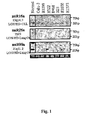
- miR genes are also found within class II homeobox gene clusters; for example, seven microRNAs (miR-129-1, miR-153-2, let-7a-1, let-7f-1, let-7d, miR-202 and miR-139) are located within 0.5 Mb of class II homeotic genes. See Example 5 and Figure 2 for a description of miRs associated with homeobox genes or gene clusters in the human genome.
- Examples of homeobox genes associated with miR genes in the human genome include genes in the HOXA cluster, genes in the HOXB cluster, genes in the HOXC cluster, genes in the HOXD cluster, NK1, NK3, NK4, Lbx, Tlx, Emx, Vax, Hmx, NK6, Msx, Cdx, Xlox, Gsx, En, HB9, Gbx, Msx-1, Msx-2, GBX2, HLX, HEX, PAM1, DLX, LHX2 and CDX2.
- homeobox gene clusters associated with miR genes in the human genome include HOXA, HOXB, HOXC, HOXD, extended Hox, NKL, ParaHox, and EHGbox, PAX, PBX, MEIS, REIG and PREP/KNOX1.
- cancers associated with homeobox genes or gene clusters include renal cancer, Wilm's tumor, colorectal cancer, small cell lung cancer, melanoma, breast cancer, prostate cancer, skin cancer, osteosarcoma, neuroblastoma, leukemia (acute lymphocytic leukemia, acute myeloid leukemia, chronic lymphocytic leukemia), glioblastoma multiform, medulloblastoma, lymphoplasmacytoid lymphoma, thyroid cancer, rhabdomyosarcoma and solid tumors.
- leukemia acute lymphocytic leukemia, acute myeloid leukemia, chronic lymphocytic leukemia
- glioblastoma multiform medulloblastoma
- lymphoplasmacytoid lymphoma thyroid cancer
- rhabdomyosarcoma solid tumors.
- miR genes associated with homeobox genes or gene clusters include miR-148, miR-10a, miR-196-1, miR-152, miR-196-2, miR-148b, miR-10b, miR-129-1, miR-153-2, miR-202, miR-139, let-7a, let-7f, let-7d and combinations thereof.
- homeobox gene cluster HOXA is associated with miR-148.
- Homeobox gene cluster HOXB is associated with miR-148, miR-10a, miR-196-1, miR-152 and combinations thereof.
- Homeobox gene cluster HOXC is associated with miR-196-2, miR-148b or a combination thereof.
- Homeobox gene cluster HOXD is associated with miR-10b. Where more than one miR gene is associated with a homeobox gene or gene cluster, it is understood that the cancer associated with those genes can be diagnosed by evaluating any one of the miR genes, or by evaluating any combination of the miR genes.
- the mIR gene or gene product that is measured or analyzed is not miR-15, miR-16, miR-143 and/or miR-145.
- perturbations in the genomic structure or chromosomal architecture of a cell which comprise the cancer-associated chromosomal feature can affect the expression of the miR gene(s) associated with the feature in that cell.
- a CAGR can comprise an amplification of the region containing an miR gene(s), causing an up-regulation of miR gene expression.
- the CAGR can comprise a chromosomal breakpoint or a deletion that disrupts gene expression, and results in a down-regulation of miR gene expression.
- HPV integrations and FRAs can cause deletions, amplifications or rearrangement of the surrounding DNA, which can also affect the structure or expression of any associated miR genes.
- a change in status of miR genes associated with a cancer-associated chromosomal feature can be detected prior to, or in the early stages of, the development of transformed or neoplastic phenotypes in cells of a subject. Therefore, also disclosed is a method of screening subjects for a predisposition to developing a cancer associated with a cancer-associated chromosomal feature, by evaluating the status of at least one miR gene associated with a cancer-associated chromosomal feature in a tissue or cell sample from a subject, relative to the status of that miR gene in a control sample.
- Subjects with a change in the status of one or more miR genes associated with a cancer-associated chromosomal feature are candidates for further testing to determine or confirm that the subjects have cancer.
- Such further testing can comprise histological examination of blood or tissue samples, or other techniques within the skill in the art.
- the "status of an miR gene” refers to the condition of the miR gene in terms of its physical sequence or structure, or its ability to express a gene product.
- the status of an miR gene in cells of a subject can be evaluated by any technique suitable for detecting genetic or epigenetic changes in the miR gene, or by any technique suitable for detecting the level of miR gene product produced from the miR gene.
- the level of at least one miR gene product produced from an miR gene can be measured in cells of a biological sample obtained from the subject.
- An alteration in the level (i.e., an up- or down-regulation) of miR gene product in the sample obtained from the subject relative to the level of miR gene product in a control sample is indicative of the presence of the cancer in the subject.
- a "subject" is any mammal suspected of having a cancer associated with a cancer-associated chromosomal feature.
- the subject is a human suspected of having a cancer associated with a cancer-associated chromosomal feature.
- expression of an miR gene is “up-regulated” when the amount of miR gene product produced from that gene in a cell or tissue sample from a subject is greater than the amount produced from the same gene in a control cell or tissue sample.
- expression of an miR gene is “down-regulated” when the amount of miR gene product produced from that gene in a cell or tissue sample from a subject is less than the amount produced from the same gene in a control cell or tissue sample.
- tissue sample can be removed from a subject suspected of having cancer associated with a cancer-associated chromosomal feature by conventional biopsy techniques.
- a blood sample can be removed from the subject, and white blood cells isolated for DNA extraction by standard techniques.
- the blood or tissue sample is preferably obtained from the subject prior to initiation of radiotherapy, chemotherapy or other therapeutic treatment.
- a corresponding control tissue or blood sample can be obtained from unaffected tissues of the subject, from a normal human individual or population of normal individuals, or from cultured cells corresponding to the majority of cells in the subject's sample.
- the control tissue or blood sample is then processed along with the sample from the subject, so that the levels of miR gene product produced from a given miR gene in cells from the subject's sample can be compared to the corresponding miR gene product levels from cells of the control sample.
- the relative miR gene expression in the control and normal samples can be conveniently determined with respect to one or more RNA expression standards.
- the standards can comprise, for example, a zero miR gene expression level, the miR gene expression level in a standard cell line, or the average level of miR gene expression previously obtained for a population of normal human controls.
- RNA transcripts of a particular gene in cells are within the skill in the art.
- total cellular RNA can be purified from cells by homogenization in the presence of nucleic acid extraction buffer, followed by centrifugation. Nucleic acids are precipitated, and DNA is removed by treatment with DNase and precipitation. The RNA molecules are then separated by gel electrophoresis on agarose gels according to standard techniques, and transferred to nitrocellulose filters by, e.g., the so-called "Northern" blotting technique. The RNA is then immobilized on the filters by heating. Detection and quantification of specific RNA is accomplished using appropriately labeled DNA or RNA probes complementary to the RNA in question. See, for example, Molecular Cloning: A Laboratory Manual, J. Sambrook et al., eds., 2nd edition, Cold Spring Harbor Laboratory Press, 1989, Chapter 7 .
- Suitable probes for Northern blot hybridization of a given miR gene product can be produced from the nucleic acid sequences provided in Table 1. Methods for preparation of labeled DNA and RNA probes, and the conditions for hybridization thereof to target nucleotide sequences, are described in Molecular Cloning: A Laboratory Manual, J. Sambrook et al., eds., 2nd edition, Cold Spring Harbor Laboratory Press, 1989, Chapters 10 and 11 .
- the nucleic acid probe can be labeled with, e.g., a radionuclide such as 3 H , 32 P, 33 P, 14 C, or 35 S; a heavy metal; or a ligand capable of functioning as a specific binding pair member for a labeled ligand (e.g., biotin, avidin or an antibody), a fluorescent molecule, a chemiluminescent molecule, an enzyme or the like.
- a radionuclide such as 3 H , 32 P, 33 P, 14 C, or 35 S
- a heavy metal e.g., a ligand capable of functioning as a specific binding pair member for a labeled ligand (e.g., biotin, avidin or an antibody), a fluorescent molecule, a chemiluminescent molecule, an enzyme or the like.
- Probes can be labeled to high specific activity by either the nick translation method of Rigby et al. (1977), J. Mol. Biol. 113:237-251 or by the random priming method of Fienberg et al. (1983), Anal. Biochem. 132:6-13 .
- the latter is the method of choice for synthesizing 32 P-labeled probes of high specific activity from single-stranded DNA or from RNA templates. For example, by replacing preexisting nucleotides with highly radioactive nucleotides according to the nick translation method, it is possible to prepare 32 P-labeled nucleic acid probes with a specific activity well in excess of 10 8 cpm/microgram.
- Autoradiographic detection of hybridization can then be performed by exposing hybridized filters to photographic film. Densitometric scanning of the photographic films exposed by the hybridized filters provides an accurate measurement of miR gene transcript levels. Using another approach, miR gene transcript levels can be quantified by computerized imaging systems, such the Molecular Dynamics 400-B 2D Phosphorimager available from Amersham Biosciences, Piscataway, NJ.
- the random-primer method can be used to incorporate an analogue, for example, the dTTP analogue 5-(N-(N-biotinyl-epsilon-aminocaproyl)-3-aminoallyl)deoxyuridine triphosphate, into the probe molecule.
- analogue for example, the dTTP analogue 5-(N-(N-biotinyl-epsilon-aminocaproyl)-3-aminoallyl)deoxyuridine triphosphate
- the biotinylated probe oligonucleotide can be detected by reaction with biotin-binding proteins, such as avidin, streptavidin, and antibodies (e.g., anti-biotin antibodies) coupled to fluorescent dyes or enzymes that produce color reactions.
- determining the levels of RNA transcripts can be accomplished using the technique of in situ hybridization.
- This technique requires fewer cells than the Northern blotting technique, and involves depositing whole cells onto a microscope cover slip and probing the nucleic acid content of the cell with a solution containing radioactive or otherwise labeled nucleic acid (e.g., cDNA or RNA) probes.
- a solution containing radioactive or otherwise labeled nucleic acid e.g., cDNA or RNA
- This technique is particularly well-suited for analyzing tissue biopsy samples from subjects.
- the practice of the in situ hybridization technique is described in more detail in U.S. Pat. No. 5,427,916 .
- Suitable probes for in situ hybridization of a given miR gene product can be produced from the nucleic acid sequences provided in Table 1, as described above.
- the relative number of miR gene transcripts in cells can also be determined by reverse transcription of miR gene transcripts, followed by amplification of the reverse-transcribed transcripts by polymerase chain reaction (RT-PCR).
- the levels of miR gene transcripts can be quantified in comparison with an internal standard, for example, the level of mRNA from a "housekeeping" gene present in the same sample.
- a suitable "housekeeping" gene for use as an internal standard includes, e.g., myosin or glyceraldehyde-3-phosphate dehydrogenase (G3PDH).
- G3PDH glyceraldehyde-3-phosphate dehydrogenase
- an oligolibrary in microchip format may be constructed containing a set of probe oligonucleotides specific for a set of miR genes.
- the oligolibrary contains probes corresponding to all known miRs from the human genome.
- the microchip oligolibrary may be expanded to include additional miRNAs as they are discovered.
- the microchip is prepared from gene-specific oligonucleotide probes generated from known miRNAs.
- the array contains two different oligonucleotide probes for each miRNA, one containing the active sequence and the other being specific for the precursor of the miRNA.
- the array may also contain controls such as one or more mouse sequences differing from human orthologs by only a few bases, which can serve as controls for hybridization stringency conditions, tRNAs from both species may also be printed on the microchip, providing an internal, relatively stable positive control for specific hybridization.
- One or more appropriate controls for non-specific hybridization may also be included on the microchip. For this purpose, sequences are selected based upon the absence of any homology with any known miRNAs.
- the microchip may be fabricated by techniques known in the art. For example, probe oligonucleotides of an appropriate length, e.g., 40 nucleotides, are 5'-amine modified at position C6 and printed using commercially available microarray systems, e.g., the GeneMachine OmniGrid TM 100 Microarrayer and Amersham CodeLink TM activated slides. Labeled cDNA oligomer corresponding to the target RNAs is prepared by reverse transcribing the target RNA with labeled primer. Following first strand synthesis, the RNA/DNA hybrids are denatured to degrade the RNA templates. The labeled target cDNAs thus prepared are then hybridized to the microarray chip under hybridizing conditions, e.g.
- the labeled cDNA oligomer is a biotin-labeled cDNA, prepared from a biotin-labeled primer.
- microarray is then processed by direct detection of the biotin-containing transcripts using, e.g., Streptavidin-Alexa647 conjugate, and scanned utilizing conventional scanning methods. Images intensities of each spot on the array are proportional to the abundance of the corresponding miR in the patient sample.
- the use of the array has several advantages for miRNA expression detection.
- the relatively limited number of miRNAs allows the construction of a common microarray for several species, with distinct oligonucleotide probes for each. Such a tool would allow for analysis of trans-species expression for each known miR under various conditions.
- a microchip containing miRNA-specific probe oligonucleotides corresponding to a substantial portion of the miRNome, preferably the entire miRNome may be employed to carry out miR gene expression profiling, for analysis of miR expression patterns. Distinct miR signatures may be associated with established disease markers, or directly with a disease state. As described hereinafter in Example 11, two distinct clusters of human B-cell chronic lymphocytic leukemia (CLL) samples are associated with the presence or the absence of Zap-70 expression, a predictor of early disease progression.
- CLL chronic lymphocytic leukemia
- miR gene expression profiles can be used for diagnosing the disease state of a cancer, such as whether a cancer is malignant or benign, based on whether or not a given profile is representative of a cancer that is associated with one or more established adverse prognostic markers.
- Prognostic markers that are suitable for this method include ZAP-70 expression, unmutated IgV H gene, CD38 expression, deletion at chromosome11q23, loss or mutation of TP53, and any combination thereof.
- total RNA from a sample from a subject suspected of having a cancer is quantitatively reverse transcribed to provide a set of labeled target oligodeoxynucleotides complementary to the RNA in the sample.
- the target oligodeoxynucleotides are then hybridized to a microarray comprising miRNA-specific probe oligonucleotides to provide a hybridization profile for the sample.
- the result is a hybridization profile for the sample representing the expression pattern of miRNA in the sample.
- the hybridization profile comprises the signal from the binding of the target oligodeoxynucleotides from the sample to the miRNA-specific probe oligonucleotides in the microarray.
- the profile may be recorded as the presence or absence of binding (signal vs. zero signal). More preferably, the profile recorded includes the intensity of the signal from each hybridization. The profile is compared to the hybridization profile generated from a normal, i.e., noncancerous, control sample. An alteration in the signal is indicative of the presence of the cancer in the subject.
- the status of an miR gene in a cell of a subject can also be evaluated by analyzing at least one miR gene in the sample for a deletion, mutation or amplification, wherein detection of a deletion, mutation or amplification in the miR gene relative to the miR gene in a control sample is indicative of the presence of the cancer in the subject.
- a mutation is any alteration in the sequence of a gene of interest that results from one or more nucleotide changes. Such changes include, but are not limited to, allelic polymorphisms, and may affect gene expression and/or function of the gene product.
- a deletion, mutation or amplification in an miR gene can be detected by determining the structure or sequence of genes in cells from a biological sample from a subject suspected of having cancer associated with a cancer-associated chromosomal feature, and comparing this with the structure or sequence of these genes in cells from a control sample.
- Subject and control samples can be obtained as described herein.
- Candidate miR genes for this type of analysis include, but are not limited to, miR-16-1, miR-27b and miR-206. As described in Example 13, mutations in these three miR genes have been identified in samples from CLL patients.
- any technique suitable for detecting alterations in the structure or sequence of genes can be used in the practice of the present method.
- the presence of miR gene deletions, mutations or amplifications can be detected by Southern blot hybridization of the genomic DNA from a subject, using nucleic acid probes specific for miR gene sequences.
- genomic DNA isolated from a subject's sample can be digested with restriction endonucleases. This digestion generates restriction fragments of the genomic DNA that can be separated by electrophoresis, for example, on an agarose gel. The restriction fragments are then blotted onto a hybridization membrane (e.g., nitrocellulose or nylon), and hybridized with labeled probes specific for a given miR gene or genes. A deletion or mutation of these genes is indicated by an alteration of the restriction fragment patterns on the hybridization membrane, as compared to DNA from a control sample that has been treated identically to the DNA from the subject's sample.
- a hybridization membrane e.g., nitrocellulose or nylon
- Probe labeling and hybridization conditions suitable for detecting alterations in gene structure or sequence can be readily determined by one of ordinary skill in the art.
- the miR gene nucleic acid probes for Southern blot hybridization can be designed based upon the nucleic acid sequences provided in Table 1, as described herein. Nucleic acid probe hybridization can then be detected by exposing hybridized filters to photographic film, or by employing computerized imaging systems, such the Molecular Dynamics 400-B 2D Phosphorimager available from Amersham Biosciences, Piscataway, NJ.
- Deletions, mutations and/or amplifications of an miR gene can also be detected by amplifying a fragment of these genes by polymerase chain reaction (PCR), and analyzing the amplified fragment by sequencing or by electrophoresis to determine if the sequence and/or length of the amplified fragment from the subject's DNA sample is different from that of a control DNA sample.
- PCR polymerase chain reaction
- Suitable reaction and cycling conditions for PCR amplification of DNA fragments can be readily determined by one of ordinary skill in the art.
- Deletions of an miR gene can also be identified by detecting deletions of chromosomal markers that are closely linked to the miR gene. Mutations in an miR gene can also be detected by the technique of single strand conformational polymorphism (SSCP), for example, as described in Orita et al. (1989), Genomics 5:874-879 and Hayashi (1991), PCR Methods and Applic. 1:34-38 .
- the SSCP technique consists of amplifying a fragment of the gene of interest by PCR; denaturing the fragment and electrophoresing the two denatured single strands under non-denaturing conditions. The single strands assume a complex sequence-dependent intrastrand secondary structure that affects the strands electrophoretic mobility.
- the status of an miR gene in cells of a subject can also be evaluated by measuring the copy number of the at least one miR gene in the sample, wherein a gene copy number other than two for miR genes on somatic chromosomes and sex chromosomes in a female, or other than one for miR genes on sex chromosomes in a male, is indicative of the presence of the cancer in the subject.
- Any technique suitable for detecting gene copy number can be used in the practice of the present method, including the Southern blot and PCR amplification techniques described above.
- An alternative method of determining the miR gene copy number in a sample of tissue relies on the fact that many miR genes or gene clusters are closely linked to chromosomal markers or other genes. The loss of a copy of an miR gene in an individual who is heterozygous at a marker or gene closely linked to the miR gene can be inferred from the loss of heterozygosity in the closely linked marker or gene. Methods for determining loss of heterozygosity of chromosomal markers are within the skill in the art.
- the human miR genes are closely associated with different classes of chromosomal features that are themselves associated with cancer. These cancers are likely caused, in part, by the perturbation in the chromosome or genomic DNA caused by the cancer-associated chromosomal feature, which can affect expression of oncogenes or tumor-suppressor genes located near the site of perturbation. Without wishing to be bound by any theory, it is believed that the perturbations caused by the cancer-associated chromosomal features also affect the expression level of miR genes associated with the feature, and that this also may also contribute to cancerigenesis. Therefore, a given cancer can be treated by restoring the level of miR gene expression associated with that cancer to normal.
- the cancer can be treated by raising the miR expression level.
- the level of miR gene expression is up-regulated in cancer cells of a subject, then the cancer can be treated by reducing the miR expression level.
- the cancers associated with different cancer-associated chromosomal features, and the miR genes associated with these features, are described above and in Tables 5, 6 and 7 and Figure 2 .
- expression the appropriate miR gene or genes associated with a particular cancer and/or cancer-associated chromosomal features is altered by the compositions and methods described herein.
- specific groupings of miR gene(s) that are associated with a particular cancer-associated chromosomal feature and/or cancer are evident from Tables 5, 6 and 7 and in Figure 2 , and are preferred.
- a method of treatment comprising administering an miR gene product.
- a method of treatment comprising administering an miR gene product, provided the miR gene product is not miR-15, mIR-16, miR-143 and/or miR-145.
- the level of at least one miR gene product in cancer cells of a subject is first determined relative to control cells. Techniques suitable for determining the relative level of miR gene product in cells are described above. If miR gene expression is down-regulated in the cancer cell relative to control cells, then the cancer cells are treated with an effective amount of a compound comprising the isolated miR gene product from the miR gene which is down-regulated. If miR gene expression is up-regulated in cancer cells relative to control cells, then the cancer cells are treated with an effective amount of a compound that inhibits miR gene expression. In one embodiment, the level of miR gene product in a cancer cell is not determined beforehand, for example, in those cancers where miR gene expression is known to be up- or down-regulated.
- an effective amount of at least one isolated miR gene product can be administered to a subject.
- an "effective amount" of an isolated miR gene product is an amount sufficient to inhibit proliferation of a cancer cell in a subject suffering from a cancer associated with a cancer-associated chromosomal feature.
- One skilled in the art can readily determine an effective amount of an miR gene product to be administered to a given subject, by taking into account factors such as the size and weight of the subject; the extent of disease penetration; the age, health and sex of the subject; the route of administration; and whether the administration is regional or systemic.
- an effective amount of isolated miR gene product can be based on the approximate weight of a tumor mass to be treated.
- the approximate weight of a tumor mass can be determined by calculating the approximate volume of the mass, wherein one cubic centimeter of volume is roughly equivalent to one gram.
- An effective amount of the isolated miR gene product based on the weight of a tumor mass can be at least about 10 micrograms/gram of tumor mass, and is preferably between about 10-500 micrograms/gram of tumor mass. More preferably, the effective amount is at least about 60 micrograms/gram of tumor mass. Particularly preferably, the effective amount is at least about 100 micrograms/gram of tumor mass. It is preferred that an effective amount based on the weight of the tumor mass be injected directly into the tumor.
- an effective amount of an isolated miR gene product can also be based on the approximate or estimated body weight of a subject to be treated. Preferably, such effective amounts are administered parenterally or enterally, as described herein.
- an effective amount of the isolated miR gene product is administered to a subject can range from about 5 - 3000 micrograms/kg of body weight, and is preferably between about 700 - 1000 micrograms/kg of body weight, and is more preferably greater than about 1000 micrograms/kg of body weight.
- an appropriate dosage regimen for the administration of an isolated miR gene product to a given subject can be administered to the subject once (e.g ., as a single injection or deposition).
- an miR gene product can be administered once or twice daily to a subject for a period of from about three to about twenty-eight days, more preferably from about seven to about ten days.
- an miR gene product is administered once a day for seven days.
- a dosage regimen comprises multiple administrations, it is understood that the effective amount of the miR gene product administered to the subject can comprise the total amount of gene product administered over the entire dosage regimen.
- an "isolated" miR gene product is one which is synthesized, or altered or removed from the natural state through human intervention.
- an miR gene product naturally present in a living animal is not “isolated.”
- An isolated miR gene product can exist in substantially purified form, or can exist in a cell into which the miR gene product has been delivered.
- an miR gene product which is deliberately delivered to, or expressed in, a cell is considered an "isolated” miR gene product.
- An miR gene product produced inside a cell by from an miR precursor molecule is also considered to be “isolated” molecule.
- Isolated miR gene products can be obtained using a number of standard techniques.
- the miR gene products can be chemically synthesized or recombinantly produced using methods known in the art.
- miR gene products are chemically synthesized using appropriately protected ribonucleoside phosphoramidites and a conventional DNA/RNA synthesizer.
- RNA molecules or synthesis reagents include, e.g., Proligo (Hamburg, Germany), Dharmacon Research (Lafayette, CO, USA), Pierce Chemical (part of Perbio Science, Rockford, IL, USA), Glen Research (Sterling, VA, USA), ChemGenes (Ashland, MA, USA) and Cruachem (Glasgow, UK).
- the miR gene products can be expressed from recombinant circular or linear DNA plasmids using any suitable promoter.
- suitable promoters for expressing RNA from a plasmid include, e.g., the U6 or H1 RNA pol III promoter sequences, or the cytomegalovirus promoters. Selection of other suitable promoters is within the skill in the art.
- the recombinant plasmids of the invention can also comprise inducible or regulatable promoters for expression of the miR gene products in cancer cells.
- the miR gene products that are expressed from recombinant plasmids can be isolated from cultured cell expression systems by standard techniques.
- the miR gene products which are expressed from recombinant plasmids can also be delivered to, and expressed directly in, the cancer cells.
- the use of recombinant plasmids to deliver the miR gene products to cancer cells is discussed in more detail below.
- the miR gene products can be expressed from a separate recombinant plasmid, or can be expressed from the same recombinant plasmid.
- the miR gene products are expressed as the RNA precursor molecules from a single plasmid, and the precursor molecules are processed into the functional miR gene product by a suitable processing system, including processing systems extant within a cancer cell.
- suitable processing systems include, e.g., the in vitro Drosophila cell lysate system as described in U.S. published application 2002/0086356 to Tuschl et al. and the E. coli RNAse III system described in U.S. published patent application 2004/0014113 to Yang et al.
- plasmids suitable for expressing the miR gene products are within the skill in the art. See, for example, Zeng et al. (2002), Molecular Cell 9:1327-1333 ; Tuschl (2002), Nat. Biotechnol, 20:446-448 ; Brummelkamp et al. (2002), Science 296:550-553 ; Miyagishi et al. (2002), Nat. Biotechnol. 20:497-500 ; Paddison et al. (2002), Genes Dev. 16:948-958 ; Lee et al. (2002), Nat. Biotechnol. 20:500-505 ; and Paul et al. (2002), Nat. Biotechnol. 20:505-508 .
- a plasmid expressing the miR gene products comprises a sequence encoding a miR precursor RNA under the control of the CMV intermediate-early promoter.
- "under the control" of a promoter means that the nucleic acid sequences encoding the miR gene product are located 3' of the promoter, so that the promoter can initiate transcription of the miR gene product coding sequences.
- the miR gene products can also be expressed from recombinant viral vectors. It is contemplated that the miR gene products can be expressed from two separate recombinant viral vectors, or from the same viral vector.
- the RNA expressed from the recombinant viral vectors can either be isolated from cultured cell expression systems by standard techniques, or can be expressed directly in cancer cells. The use of recombinant viral vectors to deliver the miR gene products to cancer cells is discussed in more detail below.
- the recombinant viral vectors of the invention comprise sequences encoding the miR gene products and any suitable promoter for expressing the RNA sequences.
- suitable promoters include, for example, the U6 or H1 RNA pol III promoter sequences, or the cytomegalovirus promoters. Selection of other suitable promoters is within the skill in the art.
- the recombinant viral vectors of the invention can also comprise inducible or regulatable promoters for expression of the miR gene products in a cancer cell.
- Any viral vector capable of accepting the coding sequences for the miR gene products can be used; for example, vectors derived from adenovirus (AV); adeno-associated virus (AAV); retroviruses (e.g ., lentiviruses (LV), Rhabdoviruses, murine leukemia virus); herpes virus, and the like.
- AV adenovirus
- AAV adeno-associated virus
- retroviruses e.g ., lentiviruses (LV), Rhabdoviruses, murine leukemia virus
- herpes virus and the like.
- the tropism of the viral vectors can be modified by pseudotyping the vectors with envelope proteins or other surface antigens from other viruses, or by substituting different viral capsid proteins, as appropriate.
- lentiviral vectors of the invention can be pseudotyped with surface proteins from vesicular stomatitis virus (VSV), rabies, Ebola, Mokola, and the like.
- AAV vectors of the invention can be made to target different cells by engineering the vectors to express different capsid protein serotypes.
- an AAV vector expressing a serotype 2 capsid on a serotype 2 genome is called AAV 2/2.
- This serotype 2 capsid gene in the AAV 2/2 vector can be replaced by a serotype 5 capsid gene to produce an AAV 2/5 vector.
- AAV vectors which express different capsid protein serotypes are within the skill in the art; see, e.g ., Rabinowitz J.E. et al. (2002), J Virol 76:791-801 .
- recombinant viral vectors suitable for use in the invention methods for inserting nucleic acid sequences for expressing RNA into the vector, methods of delivering the viral vector to the cells of interest, and recovery of the expressed RNA products are within the skill in the art. See, for example, Domburg (1995), Gene Therap. 2:301-310 ; Eglitis (1988), Biotechniques 6:608-614 ; Miller (1990), Hum. Gene Therap. 1:5-14 ; and Anderson (1998), Nature 392:25-30 .
- Preferred viral vectors are those derived from AV and AAV.
- a suitable AV vector for expressing the miR gene products, a method for constructing the recombinant AV vector, and a method for delivering the vector into target cells are described in Xia et al. (2002), Nat. Biotech. 20:1006-1010 .
- Suitable AAV vectors for expressing the miR gene products, methods for constructing the recombinant AAV vector, and methods for delivering the vectors into target cells are described in Samulski et al. (1987), J. Virol. 61:3096-3101 ; Fisher et al. (1996), J. Virol., 70:520-532 ; Samulski et al. (1989), J. Virol.
- the miR gene products are expressed from a single recombinant AAV vector comprising the CMV intermediate early promoter.
- a recombinant AAV viral vector comprises a nucleic acid sequence encoding an miR precursor RNA in operable connection with a polyT termination sequence under the control of a human U6 RNA promoter.
- operable connection with a polyT termination sequence means that the nucleic acid sequences encoding the sense or antisense strands are immediately adjacent to the polyT termination signal in the 5' direction.
- the polyT termination signals act to terminate transcription.
- an effective amount of at least one compound which inhibits miR gene expression can also be administered to the subject.
- inhibiting miR gene expression means that the production of miR gene product from the miR gene in the cancer cell after treatment is less than the amount produced prior to treatment.
- One skilled in the art can readily determine whether miR gene expression has been inhibited in a cancer cell, using for example the techniques for determining miR transcript level discussed above for the diagnostic method.
- an "effective amount" of a compound that inhibits miR gene expression is an amount sufficient to inhibit proliferation of a cancer cell in a subject suffering from a cancer associated with a cancer-associated chromosomal feature.
- an effective amount of an miR gene expression-inhibiting compound to be administered to a given subject by taking into account factors such as the size and weight of the subject; the extent of disease penetration; the age, health and sex of the subject; the route of administration; and whether the administration is regional or systemic.
- an effective amount of the expression-inhibiting compound can be based on the approximate weight of a tumor mass to be treated.
- the approximate weight of a tumor mass can be determined by calculating the approximate volume of the mass, wherein one cubic centimeter of volume is roughly equivalent to one gram.
- An effective amount based on the weight of a tumor mass can be at least about 10 micrograms/gram of tumor mass, and is preferably between about 10-500 micrograms/gram of tumor mass. More preferably, the effective amount is at least about 60 micrograms/gram of tumor mass. Particularly preferably, the effective amount is at least about 100 micrograms/gram of tumor mass. It is preferred that an effective amount based on the weight of the tumor mass be injected directly into the tumor.
- an effective amount of a compound that inhibits miR gene expression can also be based on the approximate or estimated body weight of a subject to be treated. Preferably, such effective amounts are administered parenterally or enterally, as described herein.
- an effective amount of the expression-inhibiting compound administered to a subject can range from about 5 -3000 micrograms/kg of body weight, and is preferably between about 700 - 1000 micrograms/kg of body weight, and is more preferably greater than about 1000 micrograms/kg of body weight.
- an expression-inhibiting compound can be administered to the subject once ( e.g ., as a single injection or deposition).
- an expression-inhibiting compound can be administered once or twice daily to a subject for a period of from about three to about twenty-eight days, more preferably from about seven to about ten days.
- an expression-inhibiting compound is administered once a day for seven days.
- a dosage regimen comprises multiple administrations, it is understood that the effective amount of the expression-inhibiting compound administered to the subject can comprise the total amount of compound administered over the entire dosage regimen.
- Suitable compounds for inhibiting miR gene expression include double-stranded RNA (such as short- or small-interfering RNA or "siRNA”), antisense nucleic acids, and enzymatic RNA molecules such as ribozymes. Each of these compounds can be targeted to a given miR gene product and destroy or induce the destruction of the target miR gene product.
- siRNA short- or small-interfering RNA or "siRNA”
- antisense nucleic acids such as ribozymes.
- enzymatic RNA molecules such as ribozymes.
- RNA interference of the miR gene can be inhibited by inducing RNA interference of the miR gene with an isolated double-stranded RNA ("dsRNA") molecule which has at least 90%, for example 95%, 98%, 99% or 100%, sequence homology with at least a portion of the miR gene product.
- dsRNA isolated double-stranded RNA
- the dsRNA molecule is a "short or small interfering RNA" or "siRNA.”
- siRNA useful in the present methods comprise short double-stranded RNA from about 17 nucleotides to about 29 nucleotides in length, preferably from about 19 to about 25 nucleotides in length.
- the siRNA comprise a sense RNA strand and a complementary antisense RNA strand annealed together by standard Watson-Crick base-pairing interactions (hereinafter "base-paired").
- the sense strand comprises a nucleic acid sequence which is substantially identical to a nucleic acid sequence contained within the target miR gene product.
- a nucleic acid sequence in an siRNA which is "substantially identical" to a target sequence contained within the target mRNA is a nucleic acid sequence that is identical to the target sequence, or that differs from the target sequence by one or two nucleotides.
- the sense and antisense strands of the siRNA can comprise two complementary, single-stranded RNA molecules, or can comprise a single molecule in which two complementary portions are base-paired and are covalently linked by a single-stranded "hairpin" area.
- the siRNA can also be altered RNA that differs from naturally-occurring RNA by the addition, deletion, substitution and/or alteration of one or more nucleotides.
- Such alterations can include addition of non-nucleotide material, such as to the end(s) of the siRNA or to one or more internal nucleotides of the siRNA, or modifications that make the siRNA resistant to nuclease digestion, or the substitution of one or more nucleotides in the siRNA with deoxyribonucleotides.
- the siRNA can also comprise a 3' overhang.
- a "3' overhang” refers to at least one unpaired nucleotide extending from the 3'-end of a duplexed RNA strand.
- the siRNA comprises at least one 3' overhang of from 1 to about 6 nucleotides (which includes ribonucleotides or deoxyribonucleotides) in length, preferably from 1 to about 5 nucleotides in length, more preferably from 1 to about 4 nucleotides in length, and particularly preferably from about 2 to about 4 nucleotides in length.
- the 3' overhang is present on both strands of the siRNA, and is 2 nucleotides in length.
- each strand of the siRNA can comprise 3' overhangs of dithymidylic acid ("TT") or diuridylic acid (“uu").
- the siRNA can be produced chemically or biologically, or can be expressed from a recombinant plasmid or viral vector, as described above for the isolated miR gene products.
- Exemplary methods for producing and testing dsRNA or siRNA molecules are described in U.S. published patent application 2002/0173478 to Gewirtz and in U.S. published patent application 2004/0018176 to Reich et al.
- an antisense nucleic acid refers to a nucleic acid molecule that binds to target RNA by means of RNA-RNA or RNA-DNA or RNA-peptide nucleic acid interactions, which alters the activity of the target RNA.
- Antisense nucleic acids suitable for use in the present methods are single-stranded nucleic acids (e.g. , RNA, DNA, RNA-DNA chimeras, PNA) that generally comprise a nucleic acid sequence complementary to a contiguous nucleic acid sequence in an miR gene product.
- the antisense nucleic acid comprises a nucleic acid sequence that is 50-100% complementary, more preferably 75-100% complementary, and most preferably 95-100% complementary to a contiguous nucleic acid sequence in an miR gene product.
- Nucleic acid sequences for the miR gene products are provided in Table 1. Without wishing to be bound by any theory, it is believed that the antisense nucleic acids activate RNase H or some other cellular nuclease that digests the miR gene product/antisense nucleic acid duplex.
- Antisense nucleic acids can also contain modifications to the nucleic acid backbone or to the sugar and base moieties (or their equivalent) to enhance target specificity, nuclease resistance, delivery or other properties related to efficacy of the molecule. Such modifications include cholesterol moieties, duplex intercalators such as acridine or the inclusion of one or more nuclease-resistant groups.
- Antisense nucleic acids can be produced chemically or biologically, or can be expressed from a recombinant plasmid or viral vector, as described above for the isolated miR gene products. Exemplary methods for producing and testing are within the skill in the art; see, e.g., Stein and Cheng (1993), Science 261:1004 and U.S. Pat. No. 5,849,902 to Woolf et al.
- an "enzymatic nucleic acid” refers to a nucleic acid comprising a substrate binding region that has complementarity to a contiguous nucleic acid sequence of an miR gene product, and which is able to specifically cleave the miR gene product.
- the enzymatic nucleic acid substrate binding region is 50-100% complementary, more preferably 75-100% complementary, and most preferably 95-100% complementary to a contiguous nucleic acid sequence in an miR gene product.
- the enzymatic nucleic acids can also comprise modifications at the base, sugar, and/or phosphate groups.
- An exemplary enzymatic nucleic acid for use in the present methods is a ribozyme.
- the enzymatic nucleic acids can be produced chemically or biologically, or can be expressed from a recombinant plasmid or viral vector, as described above for the isolated miR gene products.
- exemplary methods for producing and testing dsRNA or siRNA molecules are described in Werner and Uhlenbeck (1995), Nucl. Acids Res. 23:2092-96 ; Hammann et al. (1999), Antisense and Nucleic Acid Drug Dev. 9:25-31 ; and U.S. Pat. No. 4,987,071 to Cech et al.
- Administration of at least one miR gene product, or at least one compound for inhibiting miR gene expression will inhibit the proliferation of cancer cells in a subject who has a cancer associated with a cancer-associated chromosomal feature.
- to "inhibit the proliferation of a cancer cell” means to kill the cell, or permanently or temporarily arrest or slow the growth of the cell.
- Inhibition of cancer cell proliferation can be inferred if the number of such cells in the subject remains constant or decreases after administration of the miR gene products or miR gene expression-inhibiting compounds.
- An inhibition of cancer cell proliferation can also be inferred if the absolute number of such cells increases, but the rate of tumor growth decreases.
- the number of cancer cells in a subject's body can be determined by direct measurement, or by estimation from the size of primary or metastatic tumor masses.
- the number of cancer cells in a subject can be measured by immunohistological methods, flow cytometry, or other techniques designed to detect characteristic surface markers of cancer cells.
- the size of a tumor mass can be ascertained by direct visual observation, or by diagnostic imaging methods, such as X-ray, magnetic resonance imaging, ultrasound, and scintigraphy. Diagnostic imaging methods used to ascertain size of the tumor mass can be employed with or without contrast agents, as is known in the art.
- the size of a tumor mass can also be ascertained by physical means, such as palpation of the tissue mass or measurement of the tissue mass with a measuring instrument, such as a caliper.
- the miR gene products or miR gene expression-inhibiting compounds can be administered to a subject by any means suitable for delivering these compounds to cancer cells of the subject.
- the miR gene products or miR expression inhibiting compounds can be administered by methods suitable to transfect cells of the subject with these compounds, or with nucleic acids comprising sequences encoding these compounds.
- the cells are transfected with a plasmid or viral vector comprising sequences encoding at least one miR gene product or miR gene expression inhibiting compound.
- Transfection methods for eukaryotic cells include, e.g., direct injection of the nucleic acid into the nucleus or pronucleus of a cell; electroporation; liposome transfer or transfer mediated by lipophilic materials; receptor mediated nucleic acid delivery, bioballistic or particle acceleration; calcium phosphate precipitation, and transfection mediated by viral vectors.
- cells can be transfected with a liposomal transfer compound, e.g., DOTAP (N-[1-(2,3-dioleoyloxy)propyl]-N,N,N-trimethyl-ammonium methylsulfate, Boehringer - Mannheim) or an equivalent, such as LIPOFECTIN.
- DOTAP N-[1-(2,3-dioleoyloxy)propyl]-N,N,N-trimethyl-ammonium methylsulfate, Boehringer - Mannheim
- LIPOFECTIN LIPOFECTIN
- An miR gene product or miR gene expression inhibiting compound can also be administered to a subject by any suitable enteral or parenteral administration route.
- Suitable enteral administration routes for the present methods include, e.g., oral, rectal, or intranasal delivery.
- Suitable parenteral administration routes include, e.g., intravascular administration
- intravenous bolus injection e.g., intravenous bolus injection, intravenous infusion, intra-arterial bolus injection, intra-arterial infusion and catheter instillation into the vasculature
- peri- and intra-tissue injection e.g., peri-tumoral and intra-tumoral injection, intra-retinal injection, or subretinal injection
- subcutaneous injection or deposition including subcutaneous infusion (such as by osmotic pumps)
- direct application to the tissue of interest for example by a catheter or other placement device (e.g ., a retinal pellet or a suppository or an implant comprising a porous, non-porous, or gelatinous material); and inhalation.
- Preferred administration routes are injection, infusion and direct injection into the tumor.
- an miR gene product or miR gene expression inhibiting compound can be administered to the subject either as naked RNA, in combination with a delivery reagent, or as a nucleic acid (e.g ., a recombinant plasmid or viral vector) comprising sequences that express the miR gene product or expression inhibiting compound.
- a delivery reagent include, e.g, the Mirus Transit TKO lipophilic reagent; lipofectin; lipofectamine; cellfectin; polycations ( e.g. , polylysine), and liposomes.
- Recombinant plasmids and viral vectors comprising sequences that express the miR gene products or miR gene expression inhibiting compounds, and techniques for delivering such plasmids and vectors to cancer cells, are discussed above.
- liposomes are used to deliver an miR gene product or miR gene expression-inhibiting compound (or nucleic acids comprising sequences encoding them) to a subject. Liposomes can also increase the blood half-life of the gene products or nucleic acids.
- Liposomes suitable for use in the invention can be formed from standard vesicle-forming lipids, which generally include neutral or negatively charged phospholipids and a sterol, such as cholesterol.
- the selection of lipids is generally guided by consideration of factors such as the desired liposome size and half-life of the liposomes in the blood stream.
- a variety of methods are known for preparing liposomes, for example, as described in Szoka et al. (1980), Ann. Rev. Biophys. Bioeng. 9:467 ; and U.S. Pat. Nos. 4,235,871 , 4,501,728 , 4,837,028 , and 5,019,369 .
- the liposomes for use in the present methods can comprise a ligand molecule that targets the liposome to cancer cells.
- Ligands which bind to receptors prevalent in cancer cells such as monoclonal antibodies that bind to tumor cell antigens, are preferred.
- the liposomes for use in the present methods can also be modified so as to avoid clearance by the mononuclear macrophage system ("MMS") and reticuloendothelial system ("RES").
- MMS mononuclear macrophage system
- RES reticuloendothelial system
- modified liposomes have opsonization-inhibition moieties on the surface or incorporated into the liposome structure.
- a liposome of the invention can comprise both opsonization-inhibition moieties and a ligand.
- Opsonization-inhibiting moieties for use in preparing the liposomes of the invention are typically large hydrophilic polymers that are bound to the liposome membrane.
- an opsonization inhibiting moiety is "bound" to a liposome membrane when it is chemically or physically attached to the membrane, e.g., by the intercalation of a lipid-soluble anchor into the membrane itself, or by binding directly to active groups of membrane lipids.
- These opsonization-inhibiting hydrophilic polymers form a protective surface layer that significantly decreases the uptake of the liposomes by the MMS and RES; e.g., as described in U.S. Pat. No. 4,920,016 .
- Opsonization inhibiting moieties suitable for modifying liposomes are preferably water-soluble polymers with a number-average molecular weight from about 500 to about 40,000 daltons, and more preferably from about 2,000 to about 20,000 daltons.
- Such polymers include polyethylene glycol (PEG) or polypropylene glycol (PPG) derivatives; e.g., methoxy PEG or PPG, and PEG or PPG stearate; synthetic polymers such as polyacrylamide or poly N-vinyl pyrrolidone; linear, branched, or dendrimeric polyamidoamines; polyacrylic acids; polyalcohols, e.g., polyvinylalcohol and polyxylitol to which carboxylic or amino groups are chemically linked, as well as gangliosides, such as ganglioside GM1.
- PEG polyethylene glycol
- PPG polypropylene glycol
- synthetic polymers such as polyacrylamide or poly N-viny
- Copolymers of PEG, methoxy PEG, or methoxy PPG, or derivatives thereof, are also suitable.
- the opsonization inhibiting polymer can be a block copolymer of PEG and either a polyamino acid, polysaccharide, polyamidoamine, polyethyleneamine, or polynucleotide.
- the opsonization inhibiting polymers can also be natural polysaccharides containing amino acids or carboxylic acids, e.g., galacturonic acid, glucuronic acid, mannuronic acid, hyaluronic acid, pectic acid, neuraminic acid, alginic acid, carrageenan; aminated polysaccharides or oligosaccharides (linear or branched); or carboxylated polysaccharides or oligosaccharides, e.g., reacted with derivatives of carbonic acids with resultant linking of carboxylic groups.
- the opsonization-inhibiting moiety is a PEG, PPG, or derivatives thereof. Liposomes modified with PEG or PEG-derivatives are sometimes called "PEGylated liposomes.”
- the opsonization inhibiting moiety can be bound to the liposome membrane by any one of numerous well-known techniques.
- an N-hydroxysuccinimide ester of PEG can be bound to a phosphatidyl-ethanolamine lipid-soluble anchor, and then bound to a membrane.
- a dextran polymer can be derivatized with a stearylamine lipid-soluble anchor via reductive amination using Na(CN)BH 3 and a solvent mixture, such as tetrahydrofuran and water in a 30:12 ratio at 60 °C.
- Liposomes modified with opsonization-inhibition moieties remain in the circulation much longer than unmodified liposomes. For this reason, such liposomes are sometimes called "stealth” liposomes. Stealth liposomes are known to accumulate in tissues fed by porous or "leaky” microvasculature. Thus, tissue characterized by such microvasculature defects, for example solid tumors, will efficiently accumulate these liposomes; see Gabizon, et al. (1988), Proc. Natl. Acad. Sci., USA, 18:6949-53. In addition, the reduced uptake by the RES lowers the toxicity of stealth liposomes by preventing significant accumulation of the liposomes in the liver and spleen. Thus, liposomes that are modified with opsonization-inhibition moieties are particularly suited to deliver the miR gene products or miR gene expression inhibition compounds (or nucleic acids comprising sequences encoding them) to tumor cells.
- the miR gene products or miR gene expression inhibition compounds are preferably formulated as pharmaceutical compositions, sometimes called “medicaments," prior to administering to a subject, according to techniques known in the art.
- Pharmaceutical compositions are characterized as being at least sterile and pyrogen-free.
- pharmaceutical formulations include formulations for human and veterinary use. Methods for preparing pharmaceutical compositions are within the skill in the art, for example as described in Remington's Pharmaceutical Science, 17th ed., Mack Publishing Company, Easton, Pa. (1985).
- the present pharmaceutical formulations comprise at least one miR gene product or miR gene expression inhibition compound (or at least one nucleic acid comprising sequences encoding them) (e.g., 0.1 to 90% by weight), or a physiologically acceptable salt thereof, mixed with a pharmaceutically-acceptable carrier.
- the pharmaceutical formulations can also comprise at least one miR gene product or miR gene expression inhibition compound (or at least one nucleic acid comprising sequences encoding them) which are encapsulated by liposomes and a pharmaceutically-acceptable carrier.
- the pharmaceutical compositions comprise an mIR gene or gene product that is is not miR-15, miR-16, miR-143 and/or miR-145.
- Preferred pharmaceutically-acceptable carriers are water, buffered water, normal saline, 0.4% saline, 0.3% glycine, hyaluronic acid and the like.
- the pharmaceutical compositions comprise at least one miR gene product or miR gene expression inhibition compound (or at least one nucleic acid comprising sequences encoding them) which is resistant to degradation by nucleases.
- miR gene product or miR gene expression inhibition compound or at least one nucleic acid comprising sequences encoding them
- nucleases One skilled in the art can readily synthesize nucleic acids which are nuclease resistant, for example by incorporating one or more ribonucleotides that are modified at the 2'-position into the miR gene products. Suitable 2'-modified ribonucleotides include those modified at the 2'-position with fluoro, amino, alkyl, alkoxy, and O-allyl.
- compositions can also comprise conventional pharmaceutical excipients and/or additives.
- Suitable pharmaceutical excipients include stabilizers, antioxidants, osmolality adjusting agents, buffers, and pH adjusting agents.
- Suitable additives include, e.g., physiologically biocompatible buffers (e.g., tromethamine hydrochloride), additions of chelants (such as, for example, DTPA or DTPA-bisamide) or calcium chelate complexes (such as, for example, calcium DTPA, CaNaDTPA-bisamide), or, optionally, additions of calcium or sodium salts (for example, calcium chloride, calcium ascorbate, calcium gluconate or calcium lactate).
- Pharmaceutical compositions of the invention can be packaged for use in liquid form, or can be lyophilized.
- conventional nontoxic solid pharmaceutically-acceptable carriers can be used; for example, pharmaceutical grades of mannitol, lactose, starch, magnesium stearate, sodium saccharin, talcum, cellulose, glucose, sucrose, magnesium carbonate, and the like.
- a solid pharmaceutical composition for oral administration can comprise any of the carriers and excipients listed above and 10-95%, preferably 25%-75%, of the at least one miR gene product or miR gene expression inhibition compound (or at least one nucleic acid comprising sequences encoding them).
- a pharmaceutical composition for aerosol (inhalational) administration can comprise 0.01-20% by weight, preferably 1%-10% by weight, of the at least one miR gene product or miR gene expression inhibition compound (or at least one nucleic acid comprising sequences encoding them) encapsulated in a liposome as described above, and a propellant.
- a carrier can also be included as desired; e.g., lecithin for intranasal delivery.
- a set of 187 human miR genes was compiled (see Table 1).
- the set comprises 153 miRs identified in the miR Registry (http://www.sanger.ac.uk/Software/Rfam/mima/; maintained by the Wellcome Trust Sanger Institute, Cambridge, UK), and 36 other miRs manually curated from published papers ( Lim et al., 2003, Science 299:1540 ; Lagos-Quintana et al., 2001, Science 294:853-858 ; Lau et al., 2001, Science 294:858-862 ; Lee et al., 2001, Science 294:862-864 ; Mourelatos et al., 2002, Genes Dev.
- a BLAST search was performed using the default parameters against the human genome to find the precise location, followed by mapping using the maps available at the Human Genome Resources at the NCBI website. See also Altschul et al. (1990), J. Mol. Biol. 215:403-10 and Altschul et al. (1997), Nucleic Acids Res. 25:3389-3402 , for a discussion of the BLAST search algorithm.
- the human clone corresponding to each miR was identified and mapped to the human genome (see Table 2).
- Per1 scripts for the automatic submission of BLAST jobs and for the retrieval of the search results were based on the LPW, HTML, and HTPP Per1 modules and BioPerl modules.
- This database was constructed using the Virtual Gene Nomenclature Workshop (htpp://www.gene.ucl.ac.uk/nomenclature/workshop), maintained by the HUGO Gene Nomenclature Committee at University College, London.
- the literature was screened for publications reporting the cloning of the locus. In ten cases, genomic positions for both centromeric and telomeric ends were found. The total genomic length of these FRA loci is 26.9 Mb. In twenty-nine cases, only one anchoring marker was identified. It was determined, based on the published data, that 3 Mb can be used as the median length for each FRA locus. Therefore, 3 Mb was used as a guideline or window length for considering whether miR were in close proximity to the FRA sites.
- HPV16 integration sites The human clones for seventeen HPV16 integration sites (IS) were also precisely mapped on the human genome.
- "close" vicinity was defined to be a distance of less than 2 Mb.
- the PubMed database was screened on-line for publications describing cancer-related abnormalities such as minimal regions of loss-of-heterozygosity (minimal LOH) and minimal regions of amplification (minimal amplicons) using the words “LOH and genome-wide,” “amplification and genome-wide” and "amplicon and cancer.”
- the PubMed database is maintained by the NCBI and was accessed via its website.
- the data obtained from thirty-two papers were used to screen for putative CAGRs, based on markers with high frequency of LOH/amplification.
- a literature search was performed to determine the presence or absence of the above three types of alterations and to determine the precise location of miRs with respect to CAGRs (see above).
- Search phrases included the combinations "minimal regions of LOH AND cancer", and "minimal region amplification AND cancer.”
- a total of 296 publications were found and manually curated to find regions defined by both telomeric and centromeric markers.
- One hundred fifty-four minimally deleted regions (median length- 4.14 Mb) and 37 minimally amplified regions (median length- 2.45 Mb) were identified with precise genomic mapping for both telomeric and centromeric ends involving all human chromosomes except Y.
- PubMed was searched with the combination "translocation AND cloning AND breakpoint AND cancer.” The search yielded 308 papers, which were then manually curated. Among these papers, 45 translocations with at least one breakpoint precisely mapped were reported.
- This model provided the incidence rate ratio (IRR), 2-sided 95% confidence interval of the IRR, and 2-sided p-values for testing the hypothesis that the IRR is 1.0.
- IRR incidence rate ratio
- 2-sided 95% confidence interval of the IRR 2-sided 95% confidence interval of the IRR
- 2-sided p-values for testing the hypothesis that the IRR is 1.0.
- An IRR significantly greater than 1 indicates an increase in the number of miR genes within a region.
- CLL chronic lymphocytic leukemia
- mononuclear cells were isolated through Ficoll-Hypaque gradient centrifugation (Amersham Pharmacia Biotech, Piscataway, NJ), as previously described ( Calin et al., Proc. Natl. Acad. Sci. USA 2002, 99:15524-15529 ).
- Samples were then processed for RNA and DNA extraction according to standard protocols as described in Sambrook J et al. (1989), Molecular cloning: A Laboratory Manual (Cold Spring Harbor Laboratory Press, Cold Spring Harbor, NY ).
- RNA isolation from patient samples and cell lines described above was performed using the Tri-Reagent protocol (Molecular Research Center, Inc). RNA samples (30 ⁇ g each) were run on 15% acryl amide denaturing (urea) Criterion recast gels (Bio-Red Laboratories, Hercules, CA) and then transferred onto Hyoid-N+ membrane (Amersham Pharmacia Biotech), as previously described ( Calin et al., Proc. Natl. Acad. Sci. USA 2002, 99:15524-15529 ). Hybridization with gamma- 32 P ATP labeled probes was performed at 42°C in 7% SDS, 0.2 M Na 2 PO 4 , pH 7.0 overnight.
- Membranes were washed at 42°C, twice in 2x SSPE, 0.1% SDS and twice with 0.5x SSPE, 0.1% SDS. Blots were stripped by boiling in 0.1% aqueous SDS/0.1x SSC for 10 minutes, and were reprobed several times. As a gel loading control, 5S rRNA was also loaded and was stained with ethidium bromide. Lung tissue RNA was utilized as the normal control; normal lung total RNA was purchased from Clontech (Palo Alto, CA).
- Example 1- miR Genes are Non-Randomly Distributed in the Human Genome
- the distribution of the 186 human miR genes was found to be non-random.
- the largest cluster found comprises six genes (miR-17 / miR-18 / miR-19a / miR-20 / miR-19b1 / miR-92-1 ) and is located at 13q31 (Table 2).
- Chromosome 1 is used as the baseline in the model with a rate of miR gene incidence of ⁇ 0.057, which is approximately equal to the overall rate of miR gene incidence across the genome.
- Table 3 Location of miRs by chromosome and results of mixed effects Poisson regression model.
- Example 2- miR Genes are Located In or Near Fragile Sites
- the other three of the four most active sites contain known or candidate TS genes; i.e., FHIT, WWOX and PARK2, respectively ( Ohta et al., 1996, Cell 84:587-597 ; Paige et al., 2001, Proc. Natl. Acad. Sci. USA 98:11417-11422 ; Cesari et al., 2003, Proc. Natl. Acad. Sci. USA 100:5956-5961 ).
- Example 3 - miR genes are Located In or Near Human Papilloma Virus (HPV) Integration Sites
- Example 4A - miR Genes are Located In or Near Cancer Associated Genomic Regions
- miR genes might be involved in malignancies through other mechanisms, such as deletion, amplification, or epigenetic modifications.
- a search was performed for reported genomic alterations in human cancers, located in regions containing miR genes. PubMed was searched for reports of CAGR such as minimal regions of loss-of heterozygosity (LOH) suggestive of the presence of tumor-suppressor genes (TSs), minimal regions of amplification suggestive of the presence of oncogenes (OGs), and common breakpoint regions in or near possible OGs or TSs (see General Methods above).
- LH loss-of heterozygosity
- TSs tumor-suppressor genes
- OGs oncogenes
- TSs oncogenes
- miR-31 near IFNA miR-204 near D9S 15
- miR-181 and miR-147 near GSN miR-181 and miR-147 near GSN
- miR-123 near D9S67 miR-123 near D9S67.
- High frequency LOH at 17p13.3 and relatively low TP53 mutation frequency in cases of hepatocellular carcinomas (HCC), lung cancers and astrocytomas indicate the presence of other TSs involved in the development of these tumors.
- miR-195 is located between ENO3 and TP53 on chromosome 17.
- HD Homozygous deletions in cancer can indicate the presence of TSs ( Huebner et al., 1998, Annu. Rev. Genet. 32:7-31 ), and several miR genes are located in homozygously deleted regions without known TSs.
- miR-15a and miR-16a located at 13q14 HD region in B-CLL the cluster miR-99a / let-7c / miR-125b-2 mapped in a 21p11.1 region of HD in lung cancers and miR-32 at 9q31.2 in a region of HD in various types of cancer.
- microRNAs Located in Minimal Deleted Regions, Minimal Amplified Regions, and Breakpoint Regions Involved in Human Cancers * Chromosome Location (defining markers) Size Mb MiR Gene Histotype Known OG/TS 3p21.1-21.2-D ARP-DRR1 7 let-7g / miR-135-1 lung, breast cancer - 3p21.3(AP20)-D GOLGA4-VILL 0.75 miR-26a epithelial cancer - 3p23-21.31 (MDR2)-D D3S1768 - D3S1767 12.32 miR-26a; miR-138-1 nasopharyngeal cancer - 5q32-D ADRB2 - ATX1 2.92 miR-145 / miR-143 myelodysplastic syndrome - 9q22.3-D D9S280 - D9S1809 1.46 miR-24-1 / mir-27b / miR-23b; let-7a-1 / let-7f-1
- miR genes in a cluster are separated by a slash.
- Table 7 MicroRNAs Located in Minimal Deleted Regions, Minimal Amplified Regions and Breakpoint Regions Involved in Human Cancers Chromosome Type of region (name) Marker 1 Position (Mb) Marker 2 Position (Mb) Size/ Distance (Mb) Histotype Closest miR miR Location (Mb) 01p31 D D1S2638 62.92 ARHI 67.885 4.96 ovarian and breast cancer miR-101-1 64.9 01p36.3 D D1S468 3.36 D1s2697 15.23 0 Non Small Cell Lung Ca. miR-34 8.8 02q21 D D2S1334 136.66 0.1 gastric ca.
- miR-31 21.4 09q22 D D9S280 92.47 D9S1809 93.93 1.46 urothelial ca. miR-24-1 / miR-27b / miR-23b; let-7a-1 / let-7f1 / let-7d 92.9; 92.3 09q22.3 D (reg B) D9S12 91.21 D9S180 R 96.03 4.82 bladder ca. let-7a-1 / let-7f1 / let-7d; miR-24-1 / miR-27b / miR-23b 92.3; 92.9 09q32 D D9S1677 107.35 0.2 Small Cell Lung Ca., Non-Small Cell Lung Ca.
- miR-34a-1 / miR-34a-2 111 11q23 D D11S1345 121.83 D11S1328 123.56 1.73 lung adenocarcinoma miR125b-1 / let-7a-2 / miR-100 121.5 11q23 D D11S1345 121.83 D11S1328 123.56 1.73 lung adenocarcinoma miR125b-1 / let-7a-2 / miR-100 121.5 11q23.1-23.2
- D (LOH) D11S4167 121.68 D11S4144 122.96 1.28 cervical ca.
- miR-92-2 / miR-19b-2 / miR-106a 132 Note: * - D - deletion; A - amplification; ca. - cancer; sy - syndrome. The distance (in Mb) from the markers used in genome-wide analysis is shown. miRs in clusters are separated by a slash. Positions are according to BUILD 34, version 1, of the Human Genome at http://www.ncbi.gov*
- miR-26a and miR-99a located at 3p21 and 21q1.2, respectively, are not expressed or are expressed at low levels in lung cancer cell lines.
- the locations of miR-26a and miR-99a correlate with regions of LOH/HD in lung tumors.
- the expression of miR-16a located at 13q14 was unchanged in the majority of lung tumor cell lines as compared to normal lung (see Figure 1 ).
- miR genes are located near breakpoint regions, including miR-142s at 50 nt from the t(8;17) translocation involving chromosome 17 and MYC, and miR-180 at 1 kb from the MN1 gene involved in a t(4;22) translocation in meningioma (Table 6).
- the t(8;17) translocation brings the MYC gene near the miR gene promoter, with consequent MYC over-expression, while the t(4;22) translocation inactivates the MN1 gene, and possibly inactivates the miR gene located in the same position.
- miR-122a is located in the minimal amplicon around MALT1 in aggressive marginal zone lymphoma (MZL), and was found to be about 160 kb from the breakpoint region of translocation t(11;18) in mucosa-associated lymphoid tissue (MALT) lymphoma ( Sanchez-Izquierdo et al., 2003, Blood 101:4539-4546 ).
- miR-122a Apart from miR-122a, several other miR genes were located in regions particularly prone to cancer-specific abnormalities, such as miR-142s and miR-142as, located at 17q23 close to a t(8;17) breakpoint in B cell acute leukemia, and also located within the minimal amplicon in breast cancer and near the FRA17B site, which is also a target for HPV16 integration in cervical tumors (see Tables 5 and 7).
- Example 5 MicroRNAs are Located In or Near HOX Gene Clusters
- Homeobox-containing genes are a family of transcription factor genes that play crucial roles during normal development and in oncogenesis.
- HOXB4, HOXB5, HOXC9, HOXC10, HOXD4 and HOXD8, all with miR gene neighbors, are deregulated in a variety of solid and hematopoietic cancers ( Cillo et al., 1999, Exp. Cell Res. 248:1-9 ; Owebs et al., 2002, Stem Cells 20:364-379 ).
- a strong correlation was found between the location of specific miR genes and homeobox (HOX) genes.
- the miR-10a and miR-196-1 genes are located within the HOX B cluster on 17q21, while miR-196-2 is within the HOX C cluster at 12q13, and miR-10b maps to the HOX D cluster at 2q31 (see Figure 2 ).
- miR-148, miR-152 and miR-148b are close to HOX clusters (less than 1 Mb; see Figure 2 ).
- the 1 Mb distance was selected because some form of long-range coordinated regulation of gene expression was shown to expand up to one megabase to HOX clusters ( Kamath et al., 2003, Nature 421:231-7 ).
- miR genes were located within class II HOX gene clusters as well.
- the cDNA sequence encoding the entire miR precursor transcript of an miR gene is separately cloned into the context of an irrelevant mRNA expressed under the control of the cytomegalovirus immediate early (CMV-IE) promoter, according to the procedure of Zeng et al., 2002, Mol. Cell 9:1327-1333 .
- CMV-IE cytomegalovirus immediate early
- Xho I linkers are placed on the end of double-stranded cDNA sequences encoding an miR precursor, and this construct is separately cloned into the Xho I site present in the pBCl2/CMV plasmid.
- the pBCl2/CMV plasmid is described in Cullen, 1986, Cell 46:973-982 .
- pCMV plasmid containing the miR precursor coding sequence is transfected into cultured human 293T cells by standard techniques using the FuGene 6 reagent (Roche). Total RNA is extracted as described above, and the presence of the processed miR transcript is detected by Northern blot analysis with an miR probe specific for the miR transcript.
- the proliferative disease or cancer cell types include ovarian cancer, breast cancer, small cell lung cancer, sporadic follicular thyroid tumor, chronic lymphocytic leukemia, cervical cancer, acute myeloid leukemia, adenocarcinomas, male germ cell tumor, non-small cell lung cancer, gastric cancer, hepatocellular carcinoma, lung cancer, nasopharyngeal cancer, B-chronic lymphocytic leukemia, lipoma, mesothelioma, kidney cancer, NF1 microdeletion, neuroblastoma, medulloblastoma, pancreatic cancer, biliary cancer, colon cancer, gastric adenocarcinoma, head/neck squamous carcinoma, astrocytoma, meningioma, B cell leukemia, primary bladder cancer, prostate cancer, myel
- RNA is extracted as described above, and the presence of processed miR transcripts in the cancer cells is detected by Northern blot analysis with miR specific probes.
- the transfected cells are also evaluated for changes in morphology, the ability to overcome contact inhibition, and other markers indicative of a transformed phenotype.
- Liposome Preparation 1 Liposomes composed of lactosyl cerebroside, phosphatidylglycerol, phosphatidylcholine, and cholesterol in molar ratios of 1:1:4:5 are prepared by the reverse phase evaporation method described in U.S. Pat. No. 4,235,871 .
- the liposomes are prepared in an aqueous solution of 100 Og/ml processed miR transcripts or 500 ⁇ glml pCMV-microRNA.
- the liposomes thus prepared encapsulate either the processed microRNA, or the pCMV-microRNA plasmids.
- the liposomes are then passed through a 0.4 polycarbonate membrane and suspended in saline, and are separated from non-encapsulated material by column chromatography in 135 mM sodium chloride, 10 mM sodium phosphate (pH 7.4).
- the liposomes are used without further modification, or are modified as described herein.
- a quantity of the liposomes prepared above are charged to an appropriate reaction vessel to which is added, with stirring, a solution of 20 mM sodium metaperiodate, 135 mM sodium chloride and 10 mM sodium phosphate (pH 7.4).
- the resulting mixture is allowed to stand in darkness for 90 minutes at a temperature of about 20°C. Excess periodate is removed by dialysis of the reaction mixture against 250 ml of buffered saline (135 mM sodium chloride, 10 mM sodium phosphate, pH 7.4) for 2 hours.
- the product is a liposome having a surface modified by oxidation of carbohydrate hydroxyl groups to aldehyde groups. Targeting groups or opsonization inhibiting moieties are conjugated to the liposome surface via these aldehyde groups.
- Liposome Preparation 2 A second liposome preparation composed of maleimidobenzoyl-phosphatidylethanolamine (MBPE), phosphatidylcholine and cholesterol is obtained as follows.
- MBPE is an activated phospholipid for coupling sulfhydryl-containing compounds, including proteins, to the liposomes.
- Dimyristoylphosphatidylethanolamine (100 mmoles) is dissolved in 5 ml of anhydrous methanol containing 2 equivalents of triethylamine and 50 mg of m-maleimidobenzoyl N-hydroxysuccinimide ester, as described in Kitagawa et al. (1976), J. Biochem 79:233-236 .
- the resulting reaction is allowed to proceed under a nitrogen gas atmosphere overnight at room temperature, and is subjected to thin layer chromatography on Silica gel H in chloroform/methanol/water (65/25/4), which reveals quantitative conversion of the DMPE to a faster migrating product. Methanol is removed under reduced pressure and the products re-dissolved in chloroform.
- the chloroform phase is extracted twice with 1% sodium chloride and the maleimidobenzoyl-phosphatidylethanolamine (MBPE) purified by silicic acid chromatography with chloroform/methanol (4/1) as the solvent. Following purification, thin-layer chromatography indicates a single phosphate containing spot that is ninhydrin negative.
- MBPE maleimidobenzoyl-phosphatidylethanolamine
- Liposomes are prepared with MBPE, phosphatidylcholine and cholesterol in molar ratios of 1:9:8 by the reverse phase evaporation method of U.S. Pat. No. 4,235,871 , supra, in an aqueous solution of 100 ⁇ g/ml processed microRNA or a solution of 500 ⁇ g/ml pCMV-miR (see above). Liposomes are separated from non-encapsulated material by column chromatography in 100 mM sodium chloride-2 mM sodium phosphate (pH 6.0).
- An appropriate vessel is charged with 1.1 ml (containing about 10 mmoles) of Liposome Preparation 1 (see above) carrying reactive aldehyde groups, or Liposome Preparation 2 (see above).
- 0.2 ml of a 200 mM sodium cyanoborohydride solution and 1.0 ml of a 3 mg/ml solution of a monoclonal antibody directed against a tumor cell antigen is added to the preparation, with stirring.
- the resulting reaction mixture is allowed to stand overnight while maintained at a temperature of 4°C.
- the reaction mixture is separated on a Biogel A5M agarose column (Biorad, Richmond, Ca.; 1.5 X 37 cm).
- a cancer cell line such as one of the lung cancer cell lines described above or a tumor-derived cell, is inoculated into nude mice, and the mice are divided into treatment and control groups.
- tumors in the mice reach 100 to 250 cubic millimeters
- processed miR transcripts encapsulated in liposomes are injected directly into the tumors of the test group.
- the tumors of the control group are injected with liposomes encapsulating carrier solution only. Tumor volume is measured throughout the study.
- a micro-chip microarray was prepared as follows, containing 368 gene-specific oligonucleotide probes generated from 248 miRNAs (161 human, 84 mouse, and 3 arabidopsis) and 15 tRNAs (8 human and 7 mouse). These sequences correspond to human and mouse miRNAs found in the miRNA Registry at www.sanger.ac.uk/Software/Rfani/mima/ (June 2003) ( Griffiths-Jones, S. (2004) Nucleic Acids Res. 32, D109-D111 ) or collected from published literature ( Lagos-Quintana, M., Rauhut, R., Lendeckel, W. & Tuschl, T. (2001) Science 294, 853-858 ; Lim, L.
- oligonucleotide-probes were printed in triplicate. Fourteen oligonucleotides had a total of six replicates because of identical mouse and human sequences and therefore were spotted on both human and mouse sections of the array. Several mouse and human orkhologs differ only in few bases, serving as controls for the hybridization stringency conditions. tRNAs from both species were also printed on the microchip, providing an internal, relatively stable positive, control for specific hybridization, while Arabidopsis sequences were selected, based on the absence of any homology with known miRNAs from other species, and used as controls for non-specific hybridization.
- miRNA Oligonucleotide Probe Design A total of 281 miRNA precursor sequences (190 Homo sapiens, 88 Mus musculus, and 3 Arabidopsis thaliana ) with annotated active sites were selected for oligonucleotide design. These correspond to human and mouse miRNAs found in the miRNA Registry or collected from published literature. All of the sequences were confirmed by BLAST alignment with the corresponding genome at www.ncbi.nlm.nih.gov and the hairpin structures were analyzed at www.bioinfo.rpi.edu/applications/mfold/old/rna.
- oligonucleotide was selected that contained each active site of each miRNA. This produced a total of 259 oligonucleotides; there were 11 clusters with multiple annotated active sites.
- each of these additional oligonucleotides we required ⁇ 75% global cross-homology and ⁇ 20 bases in any 100% alignment to the relevant organism, ⁇ 16 bases in alignments to repetitive elements, ⁇ 16 bases of low-complexity, homopolymeric stretches of no more than 6 bases, G+C content between 30-70% and no more than 11 windows of size 10 with G+C content outside 30-70%, and no self 5-mers.
- a total of 76 additional oligonucleotides were designed.
- miRNA Microarray Fabrication 40-mer 5' amine modified C6 oligonucleotides were resuspended in 50 mM phosphate buffer pH 8.0 at 20 mM concentration. The individual oligonucleotide-probe was printed in triplicate on Amersham CodeLinkTM activated slides under 45% humidity by GeneMachine OmniGridTM 100 Microarrayer in 2 x 2 pin configuration and 20 x 20 spot configuration of each subarray. The spot diameter was 100 ⁇ m and distance from center to center was 200 ⁇ m. The printed miRNA microarrays were further chemically covalently-coupled under 70% humidity overnight. The miRNA microarrays were ready for sample hybridization after additional blocking and washing steps.
- RNA Five ⁇ g of total RNA were separately added to a reaction mix in a final volume of 12 ⁇ l, containing 1 ⁇ g of [3'(N)8-(A)12-biotin-(A)12-biotin 5'] oligonucleotide primer. The mixture was incubated for 10 min at 70°C and chilled on ice.
- Array Hybridization Labeled targets from 5 ⁇ g of total RNA were used for hybridization on each KCC/TJU miRNA microarray containing 368 probes in triplicate, corresponding to 245 human and mouse miRNA genes. All probes on these microarrays were 40-mer oligonucleotides spotted by contacting technologies and covalently attached to a polymeric matrix.
- the microarrays were hybridized in 6X SSPE/30% formamide at 25°C for 18 hours, washed in 0.75X TNT at 37°C for 40 min, and processed using direct detection of the biotin-containing transcripts by Streptavidin-Alexa647 conjugate. Processed slides were scanned using a Perkin Elmer ScanArray® XL5K Scanner with the laser set to 635 nm, at Power 80 and PMT 70 setting, and a scan resolution of 10 microns.
- HeLa cells were purchased from ATCC and grown as recommended.
- Mouse macrophage cell line RAW264.7 (established from BALB/c mice) was also used ( 1968, C. D., Ceci, J. D., Tsatsanis, C., Kontoyiannis, D., Stamatakis, K., Lin, J. H., Patriotis, C., Jenkins, N. A., Copeland, N. G., Kollias, G. & Tsichlis, P. N. (2000) Cell 103, 1071-83 ).
- RNA from 20 normal human tissues including 18 of adult origin (7 hematopoietic: bone marrow, lymphocytes B, T, and CD5+ cells from 2 individuals, peripheral blood leukocytes derived from three healthy donors, spleen, and thymus; and 11 solid tissues, including brain, breast, ovary, testis, prostate, lung, heart, kidney, liver, skeletal muscle, and placenta) and 2 of fetal origin (fetal liver and fetal brain) were assessed for miRNA expression. Each RNA was labeled and hybridized in duplicate and the average expression was calculated. For all the normal tissues, except lymphocytes B, T and CD5+ cells, total RNA was purchased from Ambion (Austin, TX).
- MNC Mononuclear cells
- T cells were purified from these MNC by rosetting with neuraminidase treated SRBC and depletion of contaminant monocytes (Cd11b+), natural killer cells (CD16+) and B lymphocytes (CD19+) were purified using magnetic beads (Dynabeads, Unipath, Milano, Italy) and specific monoclonal antibodies (Becton Dickinson, San Jose, CA).
- Total B cells and CD5+ B cells were prepared from tonsils as described ( Dono, M., Zupo, S., Leanza, N., Melioli, G., Fogli, M., Melagrana, A., Chiorazzi, N. & Ferrarini, M. (2000) J. Immunol 164, 5596-604 ). Briefly, tonsils were obtained from patients in the pediatric age group undergoing routine tonsillectomies, after informed consent. Purified B cells were prepared by rosetting T cells from MNC cells with neuraminidase treated SRBC. In order to obtain CD5+ B cells, purified B cells were incubated with anti CD5 monoclonal antibody followed by goat anti mouse Ig conjugated with magnetic microbeads. CD5+ B cells were positively selected by collecting the cells retained on the magnetic column MS by Mini MACS system (Miltenyi Biotec, Auburn, CA). The degree of purification of the cell preparations was higher than 95%, as assessed by flow cytometry.
- RNA Extraction and Northern Blots Total RNA isolation and blots were performed as described ( Calin, et al., (2002) Proc Natl Acad Sc USA. 99, 15524-15529 ). After RNA isolation, the washing step with ethanol was not performed, or if performed, the tube walls were rinsed with 75% ethanol without perturbing the RNA pellet ( Lagos-Quintana, et al., (2001) Science 294, 853-858 ). For reuse, blots were stripped by boiling in 0.1% aqueous SDS/0.1xSSC for 10 min, and were reprobed. 5S rRNA stained with ethidium bromide served as a loading control.
- the miRNAs analyzed included miR-15a, miR-16-1, miR-18, miR-20, miR-21, miR-28-2, miR-30d, miR-93-1, miR-105, miR-124a-2, miR-147, miR-216, miR-219 , and miR-224 .
- the primers used were as published ( Schmittgen, T. D., Jiang, J., Liu, Q. & Yang, L. (2004) Nucleic Acid Research 32, 43-53 ).
- Microarray Data submission All data were submitted using MIAMExpress to Array Express database and each of the 44 samples described here received an ID number ranging from SAMPLE169150SUB621 to SAMPLE 169193SIUB621.
- Hybridization Sensitivity The hybridization sensitivity of the miRNA microarray was tested using various quantities of total RNA from HeLa cells, starting from 2.5 ⁇ g up to 20 ⁇ g.
- standard deviation calculated for miRNA triplicates was below 10% for the vast majority (>95%) of oligonucleotides. All other experiments described here were performed with 5 ⁇ g of total RNA.
- Microarray specificity To test the specificity of the microchip, miRNA expression in human blood leukocytes from three healthy donors and 2 samples of mouse macrophages was analyzed. Samples derived from the same type of tissue presented homogenous patterns of miRNA expression. Furthermore, the pattern of hybridization is different for the two species. To confirm microarray results, the same RNA samples from mouse macrophages and HeLa cells were also analyzed by quantitative RT-PCR for a randomly selected set of 14 miRNAs ( Schmittgen, T. D., Jiang, J., Liu, Q. & Yang, L. (2004) Nucleic Acid Research 32, 43-53 ).
- each quantified result represents the specific expression of a single miRNA member and not the combined expression of the entire family.
- a portion of oligonucleotide was 100% identical for two probes (for example, the 23mer of active molecule present in the 40-mer oligonucleotides for both mir-16 sequences from chromosome 13 and chromosome 3), very similar profiles were observed. Therefore, both sequence similarity and secondary structure influence the cross-hybridization between different molecules on this type of microarray.
- RNAs from human normal tissues including 18 of adult origin (7 hematopoietic and 11 solid tissues) and 2 of fetal origin (fetal liver and brain). For 15 of them, at least two different RNA samples or two replicates from the same preparation were used (for a detailed list of samples see the above Methods). The results demonstrated that different tissues have distinctive patterns of miRNome expression (defined as the full complement of miRNAs in a cell) with each tissue presenting a specific signature. Using unsupervised hierarchical clustering, the same types of tissue from different individuals clustered together.
- RNA of fetal or adult type from the same tissue origin (brain) present different miRNA expression pattern.
- the types of normalization of the GeneSpring software did not influence these results.
- the miRNome expression in 38 individual human B-cell chronic lymphocytic leukemia (CLL) cell samples was determined utilizing the microchip of Example 10.
- MNC blood mononuclear cell
- two distinct clusters of CLL samples associated with the presence or the absence of Zap-70 expression, a predictor of early disease progression.
- Two miRNA signatures were associated with presence or absence of mutations in the expressed immunoglobulin variable-region genes or with deletions at 13q14 respectively.
- Tissue Samples and CLL Samples 47 samples were used for this study, including 41 samples from 38 patients with CLL, and 6 normal samples, including one lymph node, tonsillar CD5+ B cells from two normal donors and blood mononuclear cells from three normal donors. For three cases, two independent samples were collected and processed.
- CLL samples were obtained after informed consent from patients diagnosed with CLL at the CLL Research Consortium institutions. Briefly, blood was obtained from CLL patients, mononuclear cells were isolated through Ficoll/Hypaque gradient centrifugation (Amersham Pharmacia Biotech) and processed for RNA extraction according to described protocols ( M. Lagos-Quintana, R. Rauhut, W. Lendeckel, T. Tuschl, Science 294, 853-858 (2001 )).
- MNC Mononuclear cells
- T cells were purified from these MNC by rosetting with neuraminidase-treated sheep red blood cells (SRBC) and depletion of contaminant monocytes (Cd11b+), natural killer cells (CD16+) and B lymphocytes (CD19+) were purified using magnetic beads (Dynabeads, Unipath, Milano, Italy) and specific monoclonal antibodies (Becton Dickinson, San Jose, CA).
- SRBC sheep red blood cells
- CD11b+ contaminant monocytes
- CD16+ natural killer cells
- B lymphocytes CD19+
- RNA Extraction and Northern Blots Total RNA isolation and blots were performed as described ( G. A. Calin et al., Proc Natl Acad Sc USA. 99, 15524-15529 (2002 )). After RNA isolation, the washing step with ethanol was not performed, or if performed, the tube walls were rinsed with 75% ethanol without perturbing the RNA pellet ( M. Lagos-Quintana, R. Rauhut, W. Lendeckel, T. Tuschl, Science 294, 853-858 (2001 )). For reuse, blots were stripped by boiling in 0.1 % aqueous SDS/0.1xSSC for 10 minutes, and were reprobed. 5S rRNA stained with ethidium bromide served as a sample loading control.
- RNA blot analysis was performed as described in Example 10, utilizing the microchip of Example 10. Briefly, labeled targets from 5 ⁇ g of total RNA was used for hybridization on each miRNA microarray chip containing 368 probes in triplicate, corresponding to 245 human and mouse miRNA genes.
- the microarrays were hybridized in 6x SSPE/30% formamide at 25°C for 18 hrs, washed in 0.75x TNT at 37°C for 40 min, and processed using a method of direct detection of the biotin-containing transcripts by Streptavidin-Alexa647 conjugate. Processed slides were scanned using a Perkin Elmer ScanArray® XL5K Scanner, with the laser set to 635 nm, at Power 80 and PMT 70 setting, and a scan resolution of 10 microns.
- RNA was reverse transcribed to cDNA with gene-specific primers and Thermoscript and the relative amount of each miRNA to tRNA for initiator methionine was described, using the equation 2 -dC T, where dC T (C TmiRNA - C TU6 or HUMTMIRNA ).
- dC T (C TmiRNA - C TU6 or HUMTMIRNA ).
- the set of analyzed miRNAs included miR-15a, miR-16-1, miR-18, miR-20, and miR-21.
- the primers used were as published ( Id .).
- Protein lysates were prepared from the leukemia cells of 7 CLL patients and from isolated tonsillar CD5 + B cells. Western blot analysis was performed with a polyclonal Pten antibody (Cell Signaling Technology, Beverly, MA) and was normalized using an anti-actin antibody (Sigma, St. Louis, MO).
- the results illustrate the complexity of the patterns of miRNA expression in CLL and indicate the existence of mechanisms regulating individual miRNA genes that map in the same chromosome region.
- miR-223 reported to be expressed at high levels in granulocytes ( M. Lagos-Quintana et al., Curr Biol 12, 735-739 (2002 )), was expressed at significantly lower levels in the CLL samples than in the MNC, but at about the same level as that noted for CD5 + B cells (which generally constitute less than a few percent of blood MNC).
- miR-213 As indicated in the CLL vs. CD5+ B cell list of Table 10, several miRNAs located exactly inside fragile sites ( miR-183 at FRA7H, miR-190 at FRA15A and miR-24-1 at FRA9D) and miR-213.
- the mature miR-213 molecule is expressed at lower levels in all the CLL samples, and the precursor miR-213 is reduced in expression in 62.5% of the samples.
- miR-16-1 at 13q14.3, which we previously reported to be down-regulated in the majority of CLL cases by microarray analysis ( G. A. Calin et al., Proc Natl Acad Sc USA. 99, 15524-15529 (2002 ), was expressed at low levels in 45% of CLL samples.
- miR-16-1 An identical mature miR-16 exists on chromosome 3; because the 40-mer oligonucleotide for both miR-16 sequences from chromosome 13 ( miR-16-1 ) and chromosome 3 ( miR-16-2 ) exhibit the same 23-mer mature sequence, very similar profiles were observed. However, since we observed very low levels of miR-1 6-2 expression in CLL samples by Northern blot, the expression observed is mainly contributed by miR-16-1. The other miRNA of 13q14.3, miR-15a, was expressed at low levels in ⁇ 25% of CLL cases. Overall, these data demonstrate that CLL is a malignancy with extensive alterations of miRNA expression.
- Unsupervised hierarchical clustering generated two clearly distinguishable miRNA signatures within the set of CLL samples, one closer to the miRNA expression profile observed in human leukocytes and the other clearly different ( Figure 3 ).
- a list of the microRNAs differentially expressed between the two main CLL clusters is given in Table 11.
- the name of each miRNA is as in the miRNA Registry.
- the disregulation of either active molecule or precursor is specified in the name.
- the location in minimally deleted or minimally amplified or breakpoint regions or in fragile sites is presented.
- the top 25 differentially expressed miRNA in these two signatures include genes known or suggested to be involved in cancer.
- the precursor of miR-155 is over-expressed in the majority of childhood Burkitt's lymphoma ( M. Metzler, M.
- miR-21 is located at the fragile site FRA17B ( G. A. Calin et al., Proc Natl Acad Sci USA. 101, 2999-3004. (2004 )), miR-26a is at 3p21.3, a region frequently deleted region in epithelial cancers, while miR-92-1 and miR-17 are at 13q32, a region amplified in follicular lymphoma ( Id .). Table 11: microRNAs differentially expressed between the two main CLL clusters*.
- hsa-miR-027a-prec miR-27a 19p13.2 0.00000024 hsa-miR-023a-prec miR-23a 19p13.2 0.00000032 hsa-miR-092-prec-X 092-2 miR-92-2 Xq26.2 0.00000040 Del - Advanced Ovarian ca.
- hsa-miR-030b-precNo1 miR-30b 08q24.2 0.000004 hsa-miR-026a-precNo1 miR-26a 03p21 0.000009 Del - Epithelial malignancies hsa-miR-093-prec-7.1 093-1 miR-93-1 07q22 0.000009 Amp - Folicular Ly/ Del - HCC; FRA7F hsa-miR-194-precNo1 miR-194 01q41 0.000015 FRA1H hsa-miR-155-prec miR-155 21q21 0.000028 Amp - Colon ca ; Childhood Burkit Ly hsa-miR-153-2-prec miR-153-2 07q36 0.000028 t(7;12)(q36;p13) - Acute Myeloid Leukemia hsa-miR-193-precNo2 miR-193-pre
- hsa-miR-130a-precNo1 miR-130a 11q12 0.0001 hsa-miR-023b-prec miR-23b 09q22.1 0.0001 Del - Urothelial Ca.; FRA9D hsa-miR-030c-prec miR-30c 06q13 0.0001 hsa-miR-139-prec mix-139 11q13 0.0001 hsa-miR-144-precNo2 miR-144-prec 17q11.2 0.0001 Amp - Primary Breast ca.
- HCC hsa-miR-127-prec miR-127 14q32 0.0326 Del - Nasopharyngeal ca. hsa-miR-202-prec miR-202 10q26.3 0.0333 hsa-let-7g-precNo2 let-7g-prec 03p21.3 0.0350 Del - Lung Ca., Breast Ca.
- hsa-miR-222-precNo1 miR-222 Xp11.3 0.0351 hsa-miR-009-1No2 miR-009-1-prec 05q14 0.0382 hsa-miR-136-precNo2 miR-136-prec 14q32 0.0391 Del - Nasopharyngeal ca. hsa-miR-010b-precNo1 miR-10b 02q31 0.0403 hsa-miR-223-prec miR-223 Xq12-13.3 0.0407 * - The location in minimally deleted or minimally amplified or breakpoint regions or in fragile sites is presented. HCC - Hepatocellular ca.; AML - acute myeloid leukemia.
- the two clusters may be distinguished by at least one clinico-biological factor.
- the mean value of ZAP-70 was 19% ( ⁇ 31% S.D.) vs. 35% ( ⁇ 30% S.D.), respectively or otherwise the two clusters can discriminate between patients who express and who do not express this protein (at levels ⁇ 20% ZAP-70 is considered as non-expressed) (Table 9).
- ZAP-70 is a tyrosine kinase, which is a strong predictor of early disease progression, and low levels of expression are proved to be a finding associated with good prognosis ( J. A. Orchard et al., Lancet 363, 105-11 (2004 )).
- the microarray data revealed specific molecular signatures predictive for subsets of CLL that differ in clinical behavior.
- CLL cases harbor deletions at chromosome 13q14.3 in approximately 50% of cases ( F. Bullrich, C. M. Croce, Chronic Lymphoid leukemia. B. D. Chenson, Ed. (Dekker, New York, 2001 )).
- F. Bullrich, C. M. Croce, Chronic Lymphoid leukemia. B. D. Chenson, Ed. (Dekker, New York, 2001 ) As a single cytogenetic defect, these CLL patients have a relatively good prognosis, compared with patients with leukemia cells harboring complex cytogenetic changes ( H. Dohner et al., N Engl J Med. 343, 1910-6. (2000 )).
- deletion at 13q14.3 was associated with the presence of mutated immunoglobulin V H (IgV H ) genes ( D. G. Oscier et al., Blood. 100, 1177-84 (2002 )), another good prognostic factor.
- IgV H immunoglobulin V H
- miR-24-2, miR-195, miR-203, miR-220 and miR-221 are expressed at significantly reduced levels, while miR-7-1, miR-19a, miR-136, miR-154, miR-217 and the precursor of miR-218-2 are expressed at significantly higher levels in the samples with 13q14.3 deletions, respectively (Table 12). All these genes are located in different regions of the genome and differ in their nucleotide sequences, excluding the possibility of cross-hybridization. Without wishing to be bound by any theory, these results suggest the existence of functional miRNA networks in which hierarchical regulation may be present, with some miRNA (such as miR-16-1 ) controlling or influencing the expression of other miRNA Table 12.
- microRNAs signatures associated with prognosis in B-CLL 1 miRNA Chr. location P-value Association Observation miR-7-1 9q21.33 0.030 13q14 normal miR-16-1 13q14.3 0.030 IGVH mutations negative 0.023 13q14 deleted miR-19a 13q31 0.024 13q14 normal miR-24-2 19p13.2 0.033 13q14 deleted miR-29c 1q32.2-32.3 0.018 IGVH mutations positive cluster miR-29c-miR 102 miR-102 1q32.2-32.3 0.023 IGVH mutations positive cluster miR-29c-miR 102 miR-132 17p13.3 0.033 IGVH mutations negative miR-136 14q32 0.045 13q14 normal miR-154 14q32 0.020 13q14 normal miR-186 1p31 0.038 IGVH mutations negative mir-195 17p13 0.036 13q14 deleted miR-203 14q32.33 0.026 13q14 deleted miR
- mutated IgV H is a favorable prognostic marker ( D. G. Oscier et al., Blood 100, 1177-84 (2002 )).
- miRNA signature composed of 5 differentially expressed genes ( miR-186, miR-132, miR-16-1, miR-102 and miR-29c ) that distinguished CLL samples that expressed mutated IgV H gene from those that expressed unmutated IgV H genes, indicating that miRNA expression profiles have prognostic significance in CLL.
- miRNA signature composed of 5 differentially expressed genes ( miR-186, miR-132, miR-16-1, miR-102 and miR-29c ) that distinguished CLL samples that expressed mutated IgV H gene from those that expressed unmutated IgV H genes, indicating that miRNA expression profiles have prognostic significance in CLL.
- miRNAs expand the spectrum of adverse prognostic markers in CLL, such as expression of ZAP-70, unmutated IgV H , CD38, deletion at chromosome 11q23, or loss or mutation of TP53.
- Example 12 Identification of miRNA Signature Profiles Associated with Prognostic Factors and Disease Survival in B-Cell Chronic Leukemia Samples.
- miRNome the full complement of microRNAs in a cell
- miRNACHIP the miRNACHIP
- RNA Extraction and Northern Blots Total RNA isolation and RNA blotting were performed as described ( G. A. Calin et al., Proc Proc. Natl. Acad. Sc. U. S. A. 99, 15524-15529 (2002 )).
- Microarray Experiments Microarray experiments were performed as described in Example 11. Of note, for 76 microRNAs on the miRNACHIP, two specific oligonucleotides were synthesized - one identifying the active 22 nucleotide part of the molecule and the other identifying the 60-110 nucleotide precursor. All probes on these microarrays are 40-mer oligonucleotides spotted by contacting technologies and covalently attached to a polymeric matrix.
- ZAP-70 negative ZAP-70 expression ⁇ 20%
- ZAP-70 positive ZAP-70 expression >20%
- IgV H unmutated homology ⁇ 98%
- IgV H mutated homology ⁇ 98%.
- the numbers indicate the PAM scores in the two classes (n score and y score).
- the expression profile generated by a spectrum of 9 microRNAs can differentiate between two subsets of patients in the group of 94 tested - one subset with a short interval from diagnosis to initial therapy and the second subset with a significantly longer interval (see Table 14 and Figure 5 ).
- the significance of Kaplan-Meier curves improves if we restrict the analyses to the two main groups of 83 patients (the Zap-70 positive /IgV H Umut and Zap-70 negative /IgV H Mut groups) or if we use only the 17 microRNAs from the signature (P decreases from ⁇ 0.01 to P ⁇ 0.005 and P ⁇ 0.001, respectively). All of the microRNAs which can predict the time to initial therapy, with the exception of mir-29c, are overexpressed in the group characterized by a short interval from diagnosis to initial therapy.
- Example 13 Identification of sequence alterations in miR genes associated with CLL.
- tumor DNA from CLL samples we screened more than 700kb of tumor DNAs (mean 39 patients/miRNA for mean 500bp/miRNA) for sequence alterations in each of 35 different miR genes. Very rare polymorphisms or tumor specific mutations were identified in 4 of the 39 CLL cases, affecting one of three different miR genes: miR-16-1, miR-27b and miR-206. In two other miR genes, miR-34b and miR-100, polymorphisms were identified in both CLL and normal samples with similar frequencies.
- miR genes were analyzed for the presence of a mutation, including 16 members of the miR expression signature identified in Example 12 ( mir-15a, mir-16-1, mir-23a, mir-23b, mir-24-1, mir-24-2, mir-27a, mir-27b, mir-29b-2, mir-29c, mir-146, mir-155, mir-181a, mir-221, mir-222, mir-223 ) and 19 other miR genes selected randomly ( let-7a2, let-7b, mir-21, mir-30a, mir-30b, mir-30c, mir-30d, mir-30e, mir-32, mir-100, mir-108, mir-125b1, mir-142-5p, mir-142-3p, mir-193, mir-181a, mir-206, mir-213 and mir-224 ).
- the algorithm for screening for miR gene mutations in CLL samples was performed as follows: the genomic region corresponding to each precursor miRNA from either 39 CLL samples or 3 normal mononuclear cell samples from healthy individuals was amplified, including at least 50 base pairs in the 5' and 3' extremities. For the miRNAs located in clusters covering less than one kilobase, the entire corresponding genomic region was amplified and sequenced using the Applied Biosystems Model 377 DNA sequencing system (PE, Applied Biosystems, Foster City, CA). When a deviation from the normal sequence was found, a panel of blood DNAs from 95 normal individuals was screened to confirm that the deviation represented a polymorphism.
- PE Applied Biosystems Model 377 DNA sequencing system
- sequencing data were normal, an additional panel of 37 CLL cases was screened to determine the frequency of mutations in a total of 76 cancer patients. If additional mutations were found, another set of 65 normal DNAs was screened, to assess the frequency of the specific alteration in a total of 160 normal samples.
- mir-16-1 mutant effects In vivo studies of mir-16-1 mutant effects.
- mir-16-1-WT and mir-16-1-MUT constructs were assessed by Northern blotting as previously described ( G. A. Calin et al., Proc. Natl. Acad. Sc. U. S. A. 99, 15524-15529 (2002 )).
- hsa-mir-16-1-normal 641 hsa-mir-16-1-MUT 642 hsa-mir-100 643 hsa-mir-100-MUT 644 hsa-mir-27b-normal 645 hsa-mir-27b-MUT 646 has-mir-206 230 has-mir-206-MUT 647 hsa-mir-34b-normal 648 hsa-mir-34b-MUT 650
- Each mutation/polymorphism is underlined and indicated in bold in the sequences marked "MUT".
- the miR-16-1 gene is located at 13q13.4.
- SEQ. ID NO. 642 located in a 3' region of the miR-16-1 precursor with strong conservation in all of the primates analyzed ( E. Berezikov et al., Cell 120(1):21-4 (2005 )), suggesting that this polymorphism has functional implications.
- RT-PCR and Northern blotting we have shown that the precursor miRNA includes the 3' region harboring the base substitution. Both patients have a significant reduction in mir-16-1 expression in comparison with normal CD5+ cells by miRNACHIP and Northern blotting ( Figure 6A ).
- the miR-27b gene is located on chromosome 9.
- the miR-34b gene is located at 11q23.
- CLL patients out of 39 carried two associated polymorphisms, a G to A polymorphism, as shown in SEQ. ID NO. 650, and a T to G polymorphism located in the 3' region of the miR-34b precursor. Both polymorphisms were within the transcript of the mir-34b-mir-34c cluster.
- One patient was found to be homozygous (presenting by FISH heterozygous abnormal chromosome 11q23), while the other three were heterozygous for the polymorphisms. The same frequency of mutation was found in 35 normal individuals tested.
Claims (13)
- Un procédé in vitro ou ex vivo pour diagnostiquer si un sujet est atteint d'un cancer ou présente le risque de développer un cancer, comprenant:(i) la détermination, dans un échantillon à analyser provenant du sujet précité, du taux d'au moins un produit génique de miR provenant du gène miR, une altération du taux du produit génique de miR dans l'échantillon à analyser par rapport au taux dans un échantillon témoin du produit génique de miR correspondant étant indicative de ce que le sujet est atteint d'un cancer ou présente le risque de développer un cancer;(ii) l'analyse, dans un échantillon à analyser provenant du sujet, d'au moins un gène miR codant pour au moins un produit génique de miR en vue de déterminer une délétion, mutation et/ou amplification, la détection d'une telle délétion, mutation et2/ou amplification du gène miR dans l'échantillon à analyser par comparaison à un échantillon témoin étant indicative de ce que le sujet est atteint d'un cancer ou présente le risque de développer un cancer; et/ou(iii) la détermination, dans un échantillon à analyser provenant du sujet, du nombre de copies variantes d'au moins un gène miR codant pour au moins un produit génique de miR, un nombre de copies variantes autre que deux pour un gène miR sur un chromosome somatique ou chromosome sexuel chez une femme, ou un nombre de copies variantes autre que un pour un gène miR sur un chromosome sexuel chez un homme, étant indicatif de ce que le sujet est atteint d'un cancer ou présente le risque de développer un cancer,dans lequel procédé:(a) le cancer est un cancer du poumon: au moins un des produits géniques de miR pour (i) est miR-34a-1 ou miR-34a-2, et au moins un des gènes miR pour (ii) et (iii) est un gène codant pour miR-34a-1 ou miR-34a-2; ou(b) le cancer est un cancer du sein: au moins un des produits géniques de miR pour (i) est choisi dans le groupe comprenant miR-21, miR-142as, miR-142s et miR-301, et au moins un des gènes miR pour (ii) et (iii) est choisi dans le groupe comprenant des gènes codant pour miR-21, miR-142as, miR-142s et miR-301; ou(c) le cancer est un cancer colorectal: au moins un des produits géniques de miR pour (i) est miR-155(BIC), et au moins un des gènes miR pour (ii) et (iii) est un gène codant pour miR-155(BIC); ou(d) le cancer est un cancer pancreatique: au moins un des produits géniques de miR pour (i) est miR-7-3, et au moins un des gènes miR pour (ii) et (iii) est un gène codant pour miR-7-3; ou(e) le cancer est un cancer de la prostate: au moins un des produits géniques de miR pour (i) est choisi dans le groupe comprenant miR-129-1, miR-182s, miR-182as, miR-96, miR-183, miR-29a et miR-29b et au moins un des gènes miR pour (ii) et (iii) est choisi dans le groupe comprenant des gènes codant pour miR-129-1, miR-182s, miR-182as, miR-96, miR-183, miR-29a, et miR-29b; ou(f) le cancer est un cancer gastrique: au moins un des produits géniques de miR pour (i) est miR-128a ou miR-31, et au moins un des gènes miR pour (ii) et (iii) est un gène codant pour miR-128a ou miR-31; ou(g) le cancer est un cancer de l'oesophage: au moins un des produits géniques de miR pour (i) est choisi dans le groupe comprenant miR-29b, miR-29a, miR-96, miR-182s, miR-182as, miR-183 et miR-129-1, et au moins un des gènes miR pour (ii) et (iii) est choisi dans le groupe comprenant des gènes codant pour miR-29b, miR-29a, miR-96, miR-182s, miR-182as, miR-183, et miR-129-1; ou(h) le cancer est un cancer de la vessie: au moins un des produits géniques de miR pour (i) est choisi dans le groupe comprenant let-7a-1, let-7d, let-7f1, miR-23b, miR-24-1 et miR-27b, et au moins un des gènes miR pour (ii) et (iii) est choisi dans le groupe comprenant des gènes codant pour let-7a-1 , let-7d, let-7f-1, miR-23b, miR-24-1 et miR-27b; ou(j) le cancer est un cancer du col de l'utérus: au moins un des produits géniques de miR pour (i) est choisi dans le groupe comprenant miR-21, miR-301, miR-142as, miR-142s, miR-194, miR-215 et miR-32, et au moins un des gènes miR pour (ii) et (iii) est choisi dans le groupe comprenant des gènes codant pour miR-21, miR-301, miR-142as, miR-142s, miR-194, miR-215 et miR-32.
- Le procédé selon la revendication 1, ledit procédé étant mis en oeuvre sur un échantillon biologique provenant du sujet.
- Le procédé selon la revendication 1, dans lequel dans (i), le taux d'au moins un des produits géniques de miR est déterminé par un test choisi dans le groupe comprenant: le transfert de Northern; l'hybridation in situ; et la réaction quantitative de polymérisation en chaîne couplée à une polymérase - transcriptase inverse.
- Le procédé selon la revendication 1, dans lequel dans (ii), au moins un des gènes miR est analysé comme étant une délétion, mutation et/ou amplification par un test choisi dans le groupe comprenant: hybridation par transfert de Southern; test d'amplification en chaîne par polymérase; séquençage; et test de polymorphisme de conformation simple brin.
- Le procédé selon la revendication 1, dans lequel dans (iii), le nombre de copies variantes du gène miR, est déterminé par l'analyse de la perte d'hétérozygotes de un ou plusieurs marqueurs des caractéristiques chromosomales associées au cancer.
- Le procédé selon la revendication 1, dans lequel dans (i), le taux d'au moins un produit génique de miR dans l'échantillon à analyser est déterminé- par transcription inverse quantitative du produit génique de miR pour former une cible d'oligodéoxynucléotides complémentaire et- l'hybridation de la cible oligodéoxynucléotidique en une micromatrice comprenant une sonde d'oligonucléotides spécifique pour le produit génique de miR.
- Le procédé selon la revendication 1, dans lequel dans (ii), une partie ou la totalité d'au moins un gène miR de l'échantillon à analyser est supprimée et/ou le taux du produit génique de miR dans l'échantillon à analyser est inférieur au taux du produit génique de miR dans l'échantillon témoin.
- Le procédé selon la revendication 1, dans lequel dans (iii), au moins un des gènes miR fait partie d'un chromosome somatique et comporte un nombre de copie inférieur à deux.
- Un produit génique de miR isolé, un composé qui inhibe l'expression du gène miR, dans lequel le composé qui inhibe l'expression du gène miR est un acide nucléique antisens qui se lie au produit génique de miR ou est un acide nucléique codant pour le produit génique miR isolé ou le composé qui inhibe l'expression du gène miR pour une utilisation dans le traitement d'un cancer, dans lequel:(a) le cancer est un cancer du poumon et le produit génique de miR est miR-34a-1 ou miR-34a-2, ou(b) le cancer est un cancer du sein et le produit génique de miR est choisi dans le groupe consistant en miR-21, miR-142as, miR-142s et miR-301; ou(c) le cancer est un cancer colorectal et le produit génique de miR est choisi dans le groupe consistant en miR-155(BIC); ou(d) le cancer est un cancer pancréatique et le produit génique de miR est miR-7-3; ou(e) le cancer est un cancer de la prostate et le produit génique de miR est choisi dans le groupe comprenant miR-129-1, miR-182s, miR-182as, miR-96, miR-183, miR-29a, et miR-29b; ou(f) le cancer est un cancer gastrique et le produit génique de miR est choisi dans le groupe comprenant miR-128a ou miR-31; ou(g) le cancer est un cancer de l'oesophage et le produit génique de miR est choisi dans le groupe comprenant miR-29b, miR-29a, miR-96, miR-182s, miR-182as, miR-183, et miR-129-1; ou(h) le cancer est un cancer de la vessie et le produit génique de miR est choisi dans le groupe comprenant let-7a-1, let-7d, let-7f-1, miR-23b, miR-24-1 et miR-27b; ou(j) le cancer est un cancer du col de l'utérus et le produit génique de miR est choisi dans le groupe comprenant miR-21, miR-301, miR-142as, miR-142s, miR-194, miR-215 et miR-32,dans lequel produit les cellules cancéreuses sont traitées par le produit génique de miR isolé lorsque l'expression du gène miR est soumis dans les cellules cancéreuses à un contrôle négatif par rapport aux cellules témoins, ou sont traitées par le composé inhibant l'expression du gène miR lorsque l'expression du gène miR est soumis dans les cellules cancéreuses à un contrôle positif par rapport aux cellules témoins.
- Le produit génique de miR isolé, le composé qui inhibe l'expression du gène miR, ou l'acide nucléique codant pour le produit génique de miR isolé ou le produit inhibant l'expression du gène miR pour une utilisation selon la revendication 9, dans lequel le produit génique de miR isolé, le composé inhibant l'expression du gène miR, ou l'acide nucléique codant pour le produit génique de miR ou le composé inhibant l'expression du gène miR est destiné à être administré par voie parentérale ou entérale.
- Le produit génique de miR isolé, le composé qui inhibe l'expression du gène miR, ou l'acide nucléique codant pour le produit génique de miR isolé ou le produit inhibant l'expression du gène miR pour une utilisation selon la revendication 9, lequel produit génique de miR isolé, composé inhibant l'expression du gène miR, ou acide nucléique codant pour le produit génique de miR isolé ou produit inhibant l'expression du gène miR est destiné à être administré par transfection de cellules du sujet.
- Le produit génique de miR isolé, le composé qui inhibe l'expression du gène miR, l'acide nucléique codant pour le produit génique de miR isolé ou le produit inhibant l'expression du gène miR pour une utilisation selon la revendication 9, lequel produit génique de miR isolé, composé qui inhibe l'expression du gène miR, acide nucléique codant pour le produit génique de miR isolé ou produit inhibant l'expression du gène miR est destiné à être administré comme ARN nu, en association avec un réactif libérant un acide nucléique ou comme acide nucléique comprenant une séquence qui exprime le produit génique de miR ou un composé inhibant l'expression du gène miR.
- Le produit génique de miR isolé, le composé qui inhibe l'expression du gène miR, l'acide nucléique codant pour le produit génique de miR isolé ou le produit inhibant l'expression du gène miR pour une utilisation selon la revendication 12, dans lequel un réactif libérant un acide nucléique comprend un plasmide recombinant, un vecteur viral recombinant ou un liposome.
Applications Claiming Priority (7)
| Application Number | Priority Date | Filing Date | Title |
|---|---|---|---|
| US54311904P | 2004-02-09 | 2004-02-09 | |
| US54296304P | 2004-02-09 | 2004-02-09 | |
| US54294004P | 2004-02-09 | 2004-02-09 | |
| US54292904P | 2004-02-09 | 2004-02-09 | |
| US58079704P | 2004-06-18 | 2004-06-18 | |
| US58095904P | 2004-06-18 | 2004-06-18 | |
| EP05723130A EP1713938A2 (fr) | 2004-02-09 | 2005-02-09 | Diagnostic et traitement de cancers a l'aide de microarn present dans ou au voisinage de caracteristiques chromosomiennes liees aux cancers |
Related Parent Applications (2)
| Application Number | Title | Priority Date | Filing Date |
|---|---|---|---|
| EP05723130.0 Division | 2005-02-09 | ||
| EP05723130A Division EP1713938A2 (fr) | 2004-02-09 | 2005-02-09 | Diagnostic et traitement de cancers a l'aide de microarn present dans ou au voisinage de caracteristiques chromosomiennes liees aux cancers |
Publications (3)
| Publication Number | Publication Date |
|---|---|
| EP2295604A2 EP2295604A2 (fr) | 2011-03-16 |
| EP2295604A3 EP2295604A3 (fr) | 2012-05-09 |
| EP2295604B1 true EP2295604B1 (fr) | 2015-04-08 |
Family
ID=34865553
Family Applications (2)
| Application Number | Title | Priority Date | Filing Date |
|---|---|---|---|
| EP05723130A Withdrawn EP1713938A2 (fr) | 2004-02-09 | 2005-02-09 | Diagnostic et traitement de cancers a l'aide de microarn present dans ou au voisinage de caracteristiques chromosomiennes liees aux cancers |
| EP10179994.8A Not-in-force EP2295604B1 (fr) | 2004-02-09 | 2005-02-09 | Diagnostic et traitement de cancers à l'aide de microARN présent dans ou au voisinage de caractéristiques chromosomiques associées aux cancers |
Family Applications Before (1)
| Application Number | Title | Priority Date | Filing Date |
|---|---|---|---|
| EP05723130A Withdrawn EP1713938A2 (fr) | 2004-02-09 | 2005-02-09 | Diagnostic et traitement de cancers a l'aide de microarn present dans ou au voisinage de caracteristiques chromosomiennes liees aux cancers |
Country Status (4)
| Country | Link |
|---|---|
| US (7) | US7723030B2 (fr) |
| EP (2) | EP1713938A2 (fr) |
| CA (1) | CA2554818A1 (fr) |
| WO (1) | WO2005078139A2 (fr) |
Families Citing this family (236)
| Publication number | Priority date | Publication date | Assignee | Title |
|---|---|---|---|---|
| US7906326B2 (en) * | 2003-05-07 | 2011-03-15 | Rosetta Genomics Ltd. | Bioinformatically detectable group of novel regulatory oligonucleotides associated with alzheimer's disease and uses thereof |
| EP1793674B1 (fr) | 2003-11-26 | 2018-05-30 | University of Massachusetts | Inhibition sequence-specifique de la fonction du petit arn |
| WO2005078139A2 (fr) * | 2004-02-09 | 2005-08-25 | Thomas Jefferson University | Diagnostic et traitement de cancers a l'aide de microarn present dans ou au voisinage de caracteristiques chromosomiennes liees aux cancers |
| NZ551019A (en) * | 2004-04-20 | 2009-08-28 | Genaco Biomedical Products Inc | Method for detecting ncRNA |
| US8563252B2 (en) | 2004-05-14 | 2013-10-22 | Rosetta Genomics Ltd. | Methods for distinguishing between lung squamous carcinoma and other non small cell lung cancers |
| EP2290068A3 (fr) | 2004-05-28 | 2012-01-04 | Asuragen, Inc. | Procédés et compositions impliquant du microARN |
| AU2011253959B2 (en) * | 2004-05-28 | 2014-05-15 | Asuragen, Inc. | Methods and compositions involving MicroRNA |
| FR2877350B1 (fr) * | 2004-11-03 | 2010-08-27 | Centre Nat Rech Scient | IDENTIFICATION ET UTILISATION DE miRNAs IMPLIQUES DANS LA DIFFERENCIATION DE CELLULES ISSUES D'UNE LEUCEMIE MYELOIDE |
| ES2503765T3 (es) * | 2004-11-12 | 2014-10-07 | Asuragen, Inc. | Procedimientos y composiciones que implican miARN y moléculas inhibidoras de miARN |
| US20070166724A1 (en) * | 2005-02-07 | 2007-07-19 | Itzhak Bentwich | Micrornas and related nucleic acids |
| US8592384B2 (en) | 2005-04-04 | 2013-11-26 | The Board Of Regents Of The University Of Texas System | Micro-RNA's that regulate muscle cells |
| JP2008539731A (ja) * | 2005-05-02 | 2008-11-20 | コールド スプリング ハーバー ラボラトリー | 癌の診断及び治療のための組成物及び方法 |
| US20070054287A1 (en) * | 2005-05-31 | 2007-03-08 | Applera Corporation | Method for identifying medically important cell populations using micro rna as tissue specific biomarkers |
| EP2377559A1 (fr) * | 2005-06-03 | 2011-10-19 | Southern Adelaide Health Service - Flinders Medical Centre | Ciblage de cellules dotées d'une expression de microARN |
| CN105902559A (zh) * | 2005-08-01 | 2016-08-31 | 俄亥俄州立大学研究基金会 | 用于乳腺癌的诊断、预后和治疗的基于MicroRNA的方法和组合物 |
| GB0517466D0 (en) * | 2005-08-26 | 2005-10-05 | Immunodiagnostic Systems Plc | Diagnostic assay and therapeutic treatment |
| WO2007028030A2 (fr) * | 2005-09-02 | 2007-03-08 | Picobella, Llc | Arn regulateurs oncogenes pour le diagnostic et la therapie |
| CN101296702B (zh) | 2005-09-12 | 2012-11-28 | 俄亥俄州立大学研究基金会 | 用于诊断或治疗bcl2相关癌症的组合物和方法 |
| EP1940456A4 (fr) * | 2005-10-05 | 2009-10-21 | Univ Ohio State Res Found | Gene wwox, vecteurs renfermant celui-ci et utilisations de ceux-ci dans le traitement de cancer |
| US8445198B2 (en) * | 2005-12-01 | 2013-05-21 | Medical Prognosis Institute | Methods, kits and devices for identifying biomarkers of treatment response and use thereof to predict treatment efficacy |
| AU2013206405B2 (en) * | 2006-01-05 | 2016-02-04 | The Ohio State University Research Foundation | MicroRNA expression abnormalities in pancreatic endocrine and acinar tumors |
| AU2014203454B2 (en) * | 2006-01-05 | 2016-03-17 | The Ohio State University Research Foundation | MicroRNA-based methods and compositions for the diagnosis, prognosis and treatment of lung cancer |
| ES2545118T3 (es) * | 2006-01-05 | 2015-09-08 | The Ohio State University Research Foundation | Métodos basados en microARN y composiciones para el diagnóstico y tratamiento de cánceres sólidos |
| US7670840B2 (en) * | 2006-01-05 | 2010-03-02 | The Ohio State University Research Foundation | Micro-RNA expression abnormalities of pancreatic, endocrine and acinar tumors |
| AU2012238336B8 (en) * | 2006-01-05 | 2014-08-07 | The Ohio State University Research Foundation | MicroRNA-based methods and compositions for the diagnosis, prognosis and treatment of lung cancer |
| AU2016203848B2 (en) * | 2006-01-05 | 2017-12-14 | The Ohio State University Research Foundation | MicroRNA-based methods and compositions for the diagnosis, prognosis and treatment of lung cancer |
| AU2007205234B2 (en) * | 2006-01-05 | 2012-07-12 | The Ohio State University Research Foundation | MicroRNA-based methods and compositions for the diagnosis, prognosis and treatment of lung cancer |
| FI121236B (fi) * | 2006-01-18 | 2010-08-31 | Licentia Oy | Menetelmä asbestialtistukseen liittyvien keuhkosyöpien tunnistamiseksi |
| GB0603251D0 (en) | 2006-02-17 | 2006-03-29 | Isis Innovation | DNA conformation |
| AU2007223311C1 (en) * | 2006-03-02 | 2013-06-20 | The Board Of Regents Of The University Of Oklahoma | MicroRNA expression profile associated with pancreatic cancer |
| WO2007109236A2 (fr) * | 2006-03-20 | 2007-09-27 | The Ohio State University Research Foundation | Empreintes digitales micro-arn pendant une mégacaryocytopoïese |
| ES2569558T3 (es) | 2006-04-03 | 2016-05-11 | Roche Innovation Center Copenhagen A/S | Composición farmacéutica que comprende oligonucleótidos antisentido anti-ARNmi |
| WO2007112753A2 (fr) | 2006-04-03 | 2007-10-11 | Santaris Pharma A/S | Composition pharmaceutique |
| US8207325B2 (en) * | 2006-04-03 | 2012-06-26 | Univ. of Copenhagen | MicroRNA biomarkers for human breast and lung cancer |
| US8039234B1 (en) * | 2006-05-22 | 2011-10-18 | Applied Biosystems, Llc | Methods, panels of identification markers, and kits for identifying forensic samples |
| WO2007150071A1 (fr) * | 2006-06-23 | 2007-12-27 | Myriad Genetics, Inc. | Amplifications et délétions de gènes |
| WO2008002672A2 (fr) | 2006-06-28 | 2008-01-03 | The Cleveland Clinic Foundation | Cibles utilisées pour le diagnostic, le pronostic et la thérapie du cancer |
| ES2434075T3 (es) * | 2006-07-13 | 2013-12-13 | The Ohio State University Research Foundation | MIR-203 para el diagnóstico de adenocarcinoma de colon con mal pronóstico de supervivencia |
| CA2659364C (fr) | 2006-08-01 | 2017-08-22 | Board Of Regents Of The University Of Texas System | Identification d'un microarn qui active l'expression de la chaine lourde de .beta.-myosine |
| WO2008024343A2 (fr) * | 2006-08-21 | 2008-02-28 | The University Of North Carolina At Chapel Hill | Puces à micro-arn et leurs procédés d'utilisation |
| ES2374446T3 (es) | 2006-09-19 | 2012-02-16 | The Ohio State University Research Foundation | Expresión de tcl1 en la leucemia linfocítica crónica (llc) regulada por mir-29 y mir-181. |
| JP5520605B2 (ja) * | 2006-09-19 | 2014-06-11 | アシュラジェン インコーポレイテッド | 膵臓疾患で差次的に発現されるマイクロrnaおよびその使用 |
| US7803784B2 (en) * | 2006-10-24 | 2010-09-28 | The Board Of Trustees Of The Leland Stanford Junior University | Modulation of T cell signaling threshold and T cell sensitivity to antigens |
| US8343719B2 (en) * | 2006-10-30 | 2013-01-01 | Research Foundation Of State University Of New York | Microrna as biomarker in cancer |
| EP2087135B8 (fr) * | 2006-11-01 | 2013-07-24 | The Ohio State University Research Foundation | Signature de l'expression de microarn pour la prédiction de la survie et des métastases dans le carcinome hépato-cellulaire |
| US8338109B2 (en) | 2006-11-02 | 2012-12-25 | Mayo Foundation For Medical Education And Research | Predicting cancer outcome |
| WO2008064519A1 (fr) * | 2006-11-28 | 2008-06-05 | Capitalbio Corporation | Méthodes et compositions permettant de diagnostiquer un cancer de l'oesophage, d'établir un pronostic et d'améliorer la survie des patients |
| US20080131878A1 (en) * | 2006-12-05 | 2008-06-05 | Asuragen, Inc. | Compositions and Methods for the Detection of Small RNA |
| US20090092974A1 (en) * | 2006-12-08 | 2009-04-09 | Asuragen, Inc. | Micrornas differentially expressed in leukemia and uses thereof |
| AU2007333106A1 (en) * | 2006-12-08 | 2008-06-19 | Asuragen, Inc. | miR-20 regulated genes and pathways as targets for therapeutic intervention |
| US20100086928A1 (en) * | 2006-12-20 | 2010-04-08 | The Brigham And Women's Hospital, Inc. | Detection of organ rejection |
| WO2008079303A2 (fr) | 2006-12-20 | 2008-07-03 | The Brigham And Women's Hospital, Inc. | Détection de rejet d'organe |
| US20090175827A1 (en) * | 2006-12-29 | 2009-07-09 | Byrom Mike W | miR-16 REGULATED GENES AND PATHWAYS AS TARGETS FOR THERAPEUTIC INTERVENTION |
| EP2111408A4 (fr) * | 2007-01-17 | 2010-02-03 | Univ Johns Hopkins | Compositions et procédés comprenant des microarn pour traiter une néoplasie |
| US8389486B2 (en) * | 2007-01-26 | 2013-03-05 | Rosetta Genomics, Ltd | Methods for treating hematopoietic malignancies |
| CN101627134B (zh) * | 2007-01-31 | 2013-11-06 | 俄亥俄州立大学研究基金会 | 用于急性髓细胞白血病(aml)的诊断、预后和治疗的基于微rna的方法和组合物 |
| AU2008220438A1 (en) * | 2007-02-27 | 2008-09-04 | Rosetta Genomics Ltd. | Composition and methods for modulating cell proliferation and cell death |
| US9006206B2 (en) | 2007-02-27 | 2015-04-14 | Rosetta Genomics Ltd. | Composition and methods for modulating cell proliferation and cell death |
| US20120064520A1 (en) | 2007-03-01 | 2012-03-15 | Ranit Aharonov | Diagnosis and prognosis of various types of cancers |
| EP2126116A2 (fr) * | 2007-03-01 | 2009-12-02 | Rosetta Genomics Ltd | Procédés pour distinguer un carcinome squameux du poumon d'autres cancers du poumon à non-petites cellules |
| WO2008115387A2 (fr) * | 2007-03-15 | 2008-09-25 | University Hospitals Of Cleveland | Criblage, diagnostic, traitement et pronostic d'états pathophysiologiques par régulation d'arn |
| AU2008237036A1 (en) * | 2007-04-10 | 2008-10-16 | Dcb-Usa Llc | Predicting post-treatment survival in cancer patients with microRNAs |
| US20090105174A1 (en) * | 2007-04-20 | 2009-04-23 | Amgen Inc. | Nucleic acids hybridizable to micro rna and precursors thereof |
| CN101827941B (zh) * | 2007-04-30 | 2014-07-16 | 俄亥俄州立大学研究基金会 | 用于区分胰腺癌与正常胰腺功能和/或慢性胰腺炎的方法 |
| US20100167948A1 (en) * | 2007-05-22 | 2010-07-01 | The Brigham And Women's Hospital, Inc. | MicroRNA Expression Profiling of Cerebrospinal Fluid |
| US20090232893A1 (en) * | 2007-05-22 | 2009-09-17 | Bader Andreas G | miR-143 REGULATED GENES AND PATHWAYS AS TARGETS FOR THERAPEUTIC INTERVENTION |
| AU2008261951A1 (en) * | 2007-06-08 | 2008-12-18 | Asuragen, Inc. | miR-34 regulated genes and pathways as targets for therapeutic intervention |
| JP5690588B2 (ja) * | 2007-06-08 | 2015-03-25 | アメリカ合衆国 | 肝細胞癌のサブタイプを決定し、肝癌幹細胞を検出するための方法 |
| US8053186B2 (en) | 2007-06-15 | 2011-11-08 | The Ohio State University Research Foundation | Oncogenic ALL-1 fusion proteins for targeting Drosha-mediated microRNA processing |
| AU2013224690B2 (en) * | 2007-06-15 | 2015-07-09 | The Ohio State University Research Foundation | Oncogenic ALL-1 fusion proteins for targeting drosha-mediated microRNA processing |
| US20130190379A1 (en) * | 2007-07-06 | 2013-07-25 | Edwin Pieter Johan Cuppen | Small rna molecules, precursors thereof, means and methods for detecting them, and uses thereof in typing samples |
| WO2009012263A2 (fr) * | 2007-07-18 | 2009-01-22 | The Trustees Of Columbia University In The City Of New York | Micro-arn à spécificité cellulaire et compositions et utilisations de ces micro-arn |
| NZ583025A (en) | 2007-07-31 | 2012-06-29 | Univ Texas | Micro-rnas that control myosin expression and myofiber identity |
| EP2182969B1 (fr) | 2007-07-31 | 2016-11-16 | The Board of Regents of The University of Texas System | Famille de micro-arn modulant une fibrose et procédés d'utilisation associés |
| AU2008282318B2 (en) * | 2007-07-31 | 2014-02-27 | The Ohio State University Research Foundation | Methods for reverting methylation by targeting methyltransferases |
| EP2650383A1 (fr) * | 2007-08-03 | 2013-10-16 | The Ohio State University Research Foundation | Régions ultraconservées codant de l'ARNnm |
| WO2009025790A1 (fr) * | 2007-08-17 | 2009-02-26 | Catalyst Assets Llc | Utilisation de micro-arn associés au cancer de la vessie humaine pour le diagnostic et de pronostic |
| EP3028708A1 (fr) | 2007-08-22 | 2016-06-08 | The Ohio State University Research Foundation | Procédés et compositions pour induire une dérégulation de phosphorylation erk et epha7 dans des leucémies aiguës humaines |
| AU2014202073B2 (en) * | 2007-09-06 | 2015-06-25 | The Ohio State University Research Foundation | MicroRNA signatures in human ovarian cancer |
| JP5401460B2 (ja) * | 2007-09-06 | 2014-01-29 | ジ・オハイオ・ステイト・ユニバーシティ・リサーチ・ファウンデイション | ヒト卵巣癌中のマイクロrnaシグネチャー |
| US8361714B2 (en) | 2007-09-14 | 2013-01-29 | Asuragen, Inc. | Micrornas differentially expressed in cervical cancer and uses thereof |
| EP2208499A4 (fr) * | 2007-10-03 | 2011-12-28 | Kyowa Hakko Kirin Co Ltd | Acide nucléique capable de réguler la prolifération d'une cellule |
| EA019939B1 (ru) | 2007-10-04 | 2014-07-30 | Сантарис Фарма А/С | МОДИФИЦИРОВАННЫЙ ОЛИГОМЕР ДЛЯ УМЕНЬШЕНИЯ КОЛИЧЕСТВА микроРНК В КЛЕТКЕ |
| WO2009049129A1 (fr) | 2007-10-11 | 2009-04-16 | The Ohio State University Research Foundation | Procédés et compositions destinés au diagnostic et au traitement de l'adénocarcinome de l'œsophage |
| WO2009052386A1 (fr) * | 2007-10-18 | 2009-04-23 | Asuragen, Inc. | Micro arn exprimés différentiellement dans des maladies pulmonaires et leurs utilisations |
| CN103898069A (zh) | 2007-10-26 | 2014-07-02 | 俄亥俄州立大学研究基金会 | 鉴定脆性组氨酸三联体(Fhit)相互作用的方法及其用途 |
| WO2009058907A2 (fr) | 2007-10-29 | 2009-05-07 | Isis Pharmaceuticals, Inc. | Ciblage de microarn pour le traitement du cancer du foie |
| CA2700215A1 (fr) * | 2007-10-31 | 2009-05-07 | Rosetta Genomics Ltd. | Diagnostic et pronostic de cancers specifiques |
| MX2010005166A (es) | 2007-11-09 | 2010-10-07 | Univ Texas | Micro arn de la familia mir-15 que modulan la sobrevivencia de cardiomiocitos y reparacion cardiaca. |
| EP2225396A4 (fr) * | 2007-11-30 | 2011-03-02 | Univ Ohio State Res Found | Profilage et criblage d'expression de micro-arn dans du sang périphérique dans un cancer du poumon |
| WO2009070805A2 (fr) | 2007-12-01 | 2009-06-04 | Asuragen, Inc. | Gènes régulés par le mir-124 et cheminements servant de cibles pour une intervention thérapeutique |
| US8476420B2 (en) * | 2007-12-05 | 2013-07-02 | The Wistar Institute Of Anatomy And Biology | Method for diagnosing lung cancers using gene expression profiles in peripheral blood mononuclear cells |
| JP5116026B2 (ja) | 2008-01-23 | 2013-01-09 | 富士フイルム株式会社 | 癌の検出方法および癌抑制剤 |
| US20110076283A1 (en) * | 2008-01-24 | 2011-03-31 | Universite De Lausanne | Method for predicting and diagnosing brain tumor |
| JP2011513238A (ja) * | 2008-02-21 | 2011-04-28 | ザ ボード オブ リージェンツ オブ ザ ユニバーシティ オブ テキサス システム | 平滑筋の増殖および分化を調節するマイクロrnaならびにこれらの使用 |
| CN102007408A (zh) * | 2008-02-28 | 2011-04-06 | 俄亥俄州立大学研究基金会 | 与急性髓性白血病(aml)中的细胞遗传学和预后相关的微rna特征及其用途 |
| EP2254668A4 (fr) * | 2008-02-28 | 2012-08-15 | Univ Ohio State Res Found | Signatures de microarn associées à la leucémie lymphoïde chronique humaine (llc) et leurs utilisations |
| WO2009108860A2 (fr) * | 2008-02-28 | 2009-09-03 | The Ohio University Rasearch Foundation | Procédés et compositions à base de micro-arn permettant de diagnostiquer, de pronostiquer et de traiter des troubles liés à la prostate |
| WO2009111375A2 (fr) * | 2008-03-01 | 2009-09-11 | Abraxis Bioscience, Llc | Traitement, diagnostic et procédé pour découvrir un antagoniste à l'aide d'arnmi spécifique de sparc |
| WO2009109665A1 (fr) | 2008-03-07 | 2009-09-11 | Santaris Pharma A/S | Compositions pharmaceutiques pour le traitement de maladies associées aux microarn |
| ES2325726B1 (es) * | 2008-03-13 | 2010-06-17 | Fundacion Centro Nacional De Investigaciones Oncologicas Carlos Iii | Uso del microrna-203 y de sistemas de expresion del mismo para fabricar medicamentos contra el cancer. |
| CN102036689B (zh) | 2008-03-17 | 2014-08-06 | 得克萨斯系统大学董事会 | 神经肌肉突触维持和再生中涉及的微小rna的鉴定 |
| WO2009126726A1 (fr) * | 2008-04-08 | 2009-10-15 | Asuragen, Inc | Procédés et compositions pour diagnostiquer et moduler le papillomavirus humain (hpv) |
| EP2112235A1 (fr) * | 2008-04-24 | 2009-10-28 | Max-Planck-Gesellschaft zur Förderung der Wissenschaften e.V. | Compositions et procédés pour le profilage de l'expression de microARN du carcinome du nasopharynx |
| CA2723716A1 (fr) * | 2008-05-07 | 2009-11-12 | Abraxis Bioscience, Llc | Amelioration de pharmacotherapie par precurseurs arn |
| US8258111B2 (en) * | 2008-05-08 | 2012-09-04 | The Johns Hopkins University | Compositions and methods related to miRNA modulation of neovascularization or angiogenesis |
| EP2123258A1 (fr) | 2008-05-23 | 2009-11-25 | Liplasome Pharma A/S | Liposomes pour l'administration de médicaments |
| WO2009143603A1 (fr) | 2008-05-28 | 2009-12-03 | Genomedx Biosciences, Inc. | Systèmes et procédés de discrimination basée sur l’expression d’états pathologiques cliniques distincts dans le cancer de la prostate |
| US10407731B2 (en) | 2008-05-30 | 2019-09-10 | Mayo Foundation For Medical Education And Research | Biomarker panels for predicting prostate cancer outcomes |
| WO2009147658A2 (fr) * | 2008-06-02 | 2009-12-10 | New York University | Compositions et procédés pour le diagnostic, le pronostic et le traitement d'un mésothéliome |
| WO2009149318A2 (fr) | 2008-06-05 | 2009-12-10 | Research Foundation Of State University Of New York | Arnmi en tant que cibles thérapeutiques dans le cancer |
| US8247388B2 (en) * | 2008-06-06 | 2012-08-21 | The Board Of Trustees Of The Leland Stanford Junior University | Role of miRNA in T cell leukemia |
| US9125923B2 (en) | 2008-06-11 | 2015-09-08 | The Ohio State University | Use of MiR-26 family as a predictive marker for hepatocellular carcinoma and responsiveness to therapy |
| US9540695B2 (en) | 2008-06-17 | 2017-01-10 | Rosetta Genomics Ltd. | Compositions and methods for prognosis of ovarian cancer |
| US20100234445A1 (en) * | 2008-07-01 | 2010-09-16 | Weng Onn Lui | Patterns of known and novel small RNAS in human cervical cancer |
| US20100003674A1 (en) * | 2008-07-03 | 2010-01-07 | Cope Frederick O | Adult stem cells, molecular signatures, and applications in the evaluation, diagnosis, and therapy of mammalian conditions |
| KR101043433B1 (ko) * | 2008-07-18 | 2011-06-22 | 국립암센터 | 마이크로rna분자를 포함하는 항암 조성물 |
| US20100092961A1 (en) | 2008-07-25 | 2010-04-15 | The Cleveland Clinic Foundation | Methods and compositions for detection of Cowden Syndrome (CS) and CS-like syndrome |
| ES2541442T3 (es) | 2008-08-01 | 2015-07-20 | Roche Innovation Center Copenhagen A/S | Modulación mediada por microARN de factores estimulantes de colonias |
| WO2010014975A2 (fr) * | 2008-08-01 | 2010-02-04 | Academia Sinica | Utilisation de signatures en micro-arn pour évaluer des niveaux de risque de neuroblastome |
| US20110280861A1 (en) * | 2008-08-08 | 2011-11-17 | Massachusetts Institute Of Technology | Method for mir-125a in promoting hematopoietic stem cell self renewal and expansion |
| WO2010018563A2 (fr) * | 2008-08-12 | 2010-02-18 | Rosetta Genomics Ltd. | Compositions et procédés de pronostic d'un lymphome |
| CA2734179A1 (fr) * | 2008-08-12 | 2010-02-18 | The Ohio State University Research Foundation | Compositions a base de micro-arn et procedes de diagnostic, de pronostic et de traitement d'un myelome multiple |
| US20110224284A1 (en) * | 2008-09-22 | 2011-09-15 | University Of Southern California | Putative tumor suppressor microrna-101 modulates the cancer epigenome by repressing the polycomb group protein ezh2 |
| WO2010042831A2 (fr) * | 2008-10-10 | 2010-04-15 | Swedish Health Services | Diagnostic, pronostic et traitement du glioblastome multiforme |
| US9068974B2 (en) | 2008-11-08 | 2015-06-30 | The Wistar Institute Of Anatomy And Biology | Biomarkers in peripheral blood mononuclear cells for diagnosing or detecting lung cancers |
| US9074206B2 (en) | 2008-11-13 | 2015-07-07 | Fudan University | Compositions and methods for micro-RNA expression profiling of colorectal cancer |
| CA3153682A1 (en) | 2008-11-17 | 2010-05-20 | Veracyte, Inc. | Methods and compositions of molecular profiling for disease diagnostics |
| US10236078B2 (en) | 2008-11-17 | 2019-03-19 | Veracyte, Inc. | Methods for processing or analyzing a sample of thyroid tissue |
| US9495515B1 (en) | 2009-12-09 | 2016-11-15 | Veracyte, Inc. | Algorithms for disease diagnostics |
| US20150197810A1 (en) * | 2008-12-05 | 2015-07-16 | Yeda Research And Development Co. Ltd. | DOWNREGULATION OF miR-218 MAY BE USED AS A BIOMARKER FOR HUMAN ALS |
| US8206910B2 (en) | 2008-12-08 | 2012-06-26 | The Cleveland Clinic Foundation | Targets for use in diagnosis, prognosis and therapy of breast cancer |
| WO2010066851A1 (fr) * | 2008-12-10 | 2010-06-17 | Ghent University | Signature d’expression multigénique pour le pronostic de neuroblastome |
| US20120015049A1 (en) * | 2009-01-14 | 2012-01-19 | Lin Zhang | Microrna biomarker in cancer |
| US20100240049A1 (en) * | 2009-01-16 | 2010-09-23 | Cepheid | Methods of Detecting Cervical Cancer |
| EP2379720B1 (fr) | 2009-01-20 | 2016-08-17 | Alona Zilberberg | Thérapie anticancéreuse ciblée entraînée par le promoteur de mir-21 |
| KR20110128838A (ko) | 2009-02-04 | 2011-11-30 | 보드 오브 리전츠 더 유니버시티 오브 텍사스 시스템 | 심장 질환 치료에서 mir-208 및 mir-499의 이중 표적화 |
| US9074258B2 (en) | 2009-03-04 | 2015-07-07 | Genomedx Biosciences Inc. | Compositions and methods for classifying thyroid nodule disease |
| IT1398768B1 (it) * | 2009-03-24 | 2013-03-18 | Zollo | Uso di microrna-199b-5p in campo medico e diagnostico. |
| ES2599979T3 (es) | 2009-04-24 | 2017-02-06 | Roche Innovation Center Copenhagen A/S | Composiciones farmacéuticas para el tratamiento de pacientes de VHC que no responden al interferón |
| US8669057B2 (en) | 2009-05-07 | 2014-03-11 | Veracyte, Inc. | Methods and compositions for diagnosis of thyroid conditions |
| CN102725632A (zh) * | 2009-08-28 | 2012-10-10 | 奥斯瑞根公司 | 肺病的miRNA生物标志物 |
| WO2011030334A1 (fr) * | 2009-09-09 | 2011-03-17 | New York University | Compositions et procédés pour le traitement, le diagnostic et le pronostic de mésothéliome |
| DK177532B1 (en) | 2009-09-17 | 2013-09-08 | Bio Bedst Aps | Medical use of sPLA2 hydrolysable liposomes |
| JP5812491B2 (ja) * | 2009-10-01 | 2015-11-11 | 国立研究開発法人国立がん研究センター | 腫瘍治療剤 |
| US8771937B2 (en) * | 2009-10-13 | 2014-07-08 | Institut National De La Sante Et De La Recherche Medicale (Inserm) | Methods for diagnosing and treating a pathology associated with a synonymous mutation occuring within a gene of interest |
| US8916533B2 (en) | 2009-11-23 | 2014-12-23 | The Ohio State University | Materials and methods useful for affecting tumor cell growth, migration and invasion |
| WO2011067346A1 (fr) * | 2009-12-04 | 2011-06-09 | Institut National De La Sante Et De La Recherche Medicale (Inserm) | Procédé pour déterminer l'efficacité d'un traitement du cancer |
| EP2510122B1 (fr) * | 2009-12-08 | 2017-04-12 | Université Joseph Fourier | Utilisation de miarns comme biomarqueurs dans le diagnostic de gliomes |
| US10446272B2 (en) | 2009-12-09 | 2019-10-15 | Veracyte, Inc. | Methods and compositions for classification of samples |
| US8673875B2 (en) | 2009-12-10 | 2014-03-18 | Kaohsiung Medical University | Method for treating atherosclerosis |
| US8354520B2 (en) * | 2009-12-10 | 2013-01-15 | Kaohsiung Medical University | Composition and method for treating atherosclerosis, method for determining if a subject has atherosclerosis and method of screening an anti-atherosclerotic drug |
| EP2341145A1 (fr) * | 2009-12-30 | 2011-07-06 | febit holding GmbH | Empreinte ARNm dans le diagnostic de maladies |
| WO2011088309A1 (fr) | 2010-01-14 | 2011-07-21 | Regulus Therapeutics Inc. | Compositions de microarn et procédés |
| US20130130927A1 (en) * | 2010-03-11 | 2013-05-23 | Helen Heneghan | Detection and quantification of micrornas in the circulation and the use of circulating micrornas as biomarkers for cancer |
| CA2794142A1 (fr) * | 2010-03-26 | 2011-09-29 | The Ohio State University | Materiels et methodes lies a la modulation de la reparation des mesappariements et a la stabilite genomique par mir-155 |
| DK2558578T3 (en) | 2010-04-13 | 2016-01-25 | Life Technologies Corp | CONFIGURATIONS AND METHODS FOR INHIBITION OF nucleic acid function |
| US9598734B2 (en) | 2010-04-29 | 2017-03-21 | Medical Prognosis Institute A/S | Methods and devices for predicting treatment efficacy |
| US20110312520A1 (en) | 2010-05-11 | 2011-12-22 | Veracyte, Inc. | Methods and compositions for diagnosing conditions |
| CN103080334B (zh) * | 2010-06-04 | 2016-05-04 | 复旦大学 | 用于诊断早期结直肠癌及高级腺瘤的微rna生物标记及方法 |
| WO2012005572A1 (fr) | 2010-07-06 | 2012-01-12 | Interna Technologies Bv | Miarn et ses utilisations diagnostiques et thérapeutiques pour des maladies ou des états associés au mélanome, ou pour des maladies ou des états associés à la voie braf activée |
| WO2012012716A2 (fr) | 2010-07-23 | 2012-01-26 | Regulus Therapeutics, Inc. | Ciblage de microarn pour le traitement de la fibrose |
| US8980549B2 (en) | 2010-08-01 | 2015-03-17 | Ramot At Tel Aviv University Ltd. | MicroRNA patterns for the diagnosis, prognosis and treatment of melanoma |
| WO2012041959A1 (fr) * | 2010-09-30 | 2012-04-05 | University Of Zurich | Traitement d'un lymphome à cellules b avec un microarn |
| SG180031A1 (en) * | 2010-10-15 | 2012-05-30 | Agency Science Tech & Res | Combination treatment of cancer |
| WO2012065049A1 (fr) | 2010-11-12 | 2012-05-18 | The Ohio State University Research Foundation | Matériaux et procédés relatifs aux microarn-21, réparation de désappariement et cancer colorectal |
| CA2817982C (fr) | 2010-11-15 | 2020-06-30 | The Regents Of The University Of Michigan | Systemes mucoadhesifs a liberation controlee |
| US9150928B2 (en) | 2010-11-16 | 2015-10-06 | The Brigham And Women's Hospital, Inc. | Diagnosing and monitoring CNS malignancies using MicroRNA |
| MX2013005875A (es) | 2010-12-15 | 2014-04-30 | Miragen Therapeutics | Inhibidores de microarn que comprenden nucleotidos bloqueados. |
| WO2012087242A1 (fr) * | 2010-12-20 | 2012-06-28 | Agency For Science, Technology And Research | Ciblage de cellules souches de gliome par une inhibition fonctionnelle spécifique de l'oncogène mir-138 pro-survie |
| US8455518B2 (en) | 2010-12-28 | 2013-06-04 | Avon Products, Inc. | Method of treating skin with microRNA modulators |
| US20140024554A1 (en) * | 2010-12-30 | 2014-01-23 | Febit Holding Gmbh | Complex sets of mirnas as non-invasive biomarkers for glioblastoma |
| EP2474617A1 (fr) | 2011-01-11 | 2012-07-11 | InteRNA Technologies BV | MIR pour traiter une nouvelle angiogenèse |
| CN102174516A (zh) * | 2011-01-20 | 2011-09-07 | 中南大学 | 一种与ebv感染相关的鼻咽癌诊断和治疗的分子靶标及其应用 |
| CN103476931A (zh) | 2011-02-03 | 2013-12-25 | 米尔纳医疗股份有限公司 | Mir-34的合成模拟物 |
| CA2826043A1 (fr) | 2011-02-03 | 2012-08-09 | Mirna Therapeutics, Inc. | Mimetiques synthetiques de mir-124 |
| US8664192B2 (en) | 2011-03-07 | 2014-03-04 | The Ohio State University | Mutator activity induced by microRNA-155 (miR-155) links inflammation and cancer |
| US8945829B2 (en) | 2011-03-22 | 2015-02-03 | Cornell University | Distinguishing benign and malignant indeterminate thyroid lesions |
| WO2012142199A1 (fr) * | 2011-04-14 | 2012-10-18 | The Regents Of The University Of Colorado, A Body Corporate | Miarn dérégulé dans le sarcome d'ewing |
| WO2013033436A1 (fr) | 2011-09-01 | 2013-03-07 | University Of Iowa Research Foundation | Sondes à base d'oligonucléotide pour la détection de nucléases bactériennes |
| JP6153932B2 (ja) * | 2011-09-07 | 2017-06-28 | 株式会社スリー・ディー・マトリックス | 骨肉腫のためのマイクロrnaに基づく方法およびアッセイ |
| WO2013040251A2 (fr) | 2011-09-13 | 2013-03-21 | Asurgen, Inc. | Méthodes et compositions incluant mir-135b, permettant de faire la distinction entre un cancer du pancréas et une maladie pancréatique bénigne |
| JP6177243B2 (ja) | 2011-10-06 | 2017-08-09 | ミラゲン セラピューティクス, インコーポレイテッド | マイクロrnaの調節による全身エネルギーホメオスタシスの制御 |
| WO2013056217A1 (fr) | 2011-10-14 | 2013-04-18 | The Ohio State University | Méthodes et matériaux relatifs au cancer des ovaires |
| WO2013067048A1 (fr) * | 2011-10-31 | 2013-05-10 | The Ohio State University | Matériels et méthodes associés à mir-221 et à un carcinome hépatocellulaire |
| TWI462741B (zh) * | 2011-11-03 | 2014-12-01 | Univ Kaohsiung Medical | 使用微rna 195提供神經保護作用的方法 |
| KR101414383B1 (ko) * | 2011-11-09 | 2014-07-10 | 경희대학교 산학협력단 | Dlk-1 유전자 발현억제용 조성물 |
| CA2859430A1 (fr) | 2011-12-13 | 2013-06-20 | Ohio State Innovation Foundation | Procedes et compositions se rapportant a mir-21 et mir-29a, a l'inhibition d'exosome, et a la metastase cancereuse |
| CA2858581A1 (fr) | 2011-12-13 | 2013-06-20 | Genomedx Biosciences, Inc. | Diagnostics du cancer a l'aide de transcriptions non codantes |
| WO2013110053A1 (fr) | 2012-01-20 | 2013-07-25 | The Ohio State University | Signatures de marqueurs biologiques du cancer du sein concernant le pouvoir envahissant et le pronostic |
| US20150190532A1 (en) | 2012-04-04 | 2015-07-09 | The Trustees Of Columbia University In The City Of New York | Smooth muscle specific inhibition for anti-restenotic therapy |
| US8846316B2 (en) | 2012-04-30 | 2014-09-30 | Industrial Technology Research Institute | Biomarker for human liver cancer |
| US9163235B2 (en) | 2012-06-21 | 2015-10-20 | MiRagen Therapeutics, Inc. | Inhibitors of the miR-15 family of micro-RNAs |
| EP2864482A4 (fr) | 2012-06-21 | 2016-02-10 | Miragen Therapeutics Inc | Inhibiteurs à base d'oligonucléotides comprenant un motif d'acide nucléique bloqué |
| EP3435084B1 (fr) | 2012-08-16 | 2023-02-22 | Decipher Biosciences, Inc. | Pronostic du cancer de la prostate utilisant des biomarqueurs |
| KR101480366B1 (ko) * | 2013-01-08 | 2015-01-09 | 서울대학교산학협력단 | microRNA를 이용한 방사선 반응 조절 |
| CN103160509B (zh) * | 2013-03-07 | 2015-02-18 | 新乡医学院第一附属医院 | 一种与食管癌术后早期复发和预后相关的miRNA标志物及其应用 |
| GB2525804B (en) | 2013-03-15 | 2020-08-05 | Veracyte Inc | Methods and compositions for classification of samples |
| GB2514549A (en) * | 2013-05-27 | 2014-12-03 | Nat Univ Ireland | A biomarker of breast cancer |
| CN103417987B (zh) * | 2013-07-17 | 2015-06-24 | 上海交通大学医学院附属瑞金医院 | miR-148用于制备控制胰岛β细胞的增殖的药物 |
| EP3030240B1 (fr) * | 2013-08-05 | 2018-10-03 | Vib Vzw | Inhibiteurs de carnitine palmitoyltransférase 1 pour inhiber l'angiogenèse pathologique |
| WO2015026827A2 (fr) * | 2013-08-19 | 2015-02-26 | University Of Notre Dame | Procédé et composition pour la détection de hpv oncogène |
| US20160312301A1 (en) * | 2013-12-20 | 2016-10-27 | The Feinstein Institute For Medical Research | Microrna biomarkers for ovarian cancer |
| EP3680348B1 (fr) | 2014-02-07 | 2023-04-12 | University of Iowa Research Foundation | Sondes à base d'oligonucléotides et procédés de détection de microbes |
| CA2947622A1 (fr) * | 2014-05-13 | 2015-11-19 | Sangamo Biosciences, Inc. | Methodes d'edition genomique et compositions pour la prevention ou le traitement d'une maladie |
| US10247720B2 (en) | 2014-05-22 | 2019-04-02 | University Of Notre Dame Du Lac | Integrated membrane sensor for rapid molecular detection |
| CN104131113B (zh) * | 2014-08-22 | 2016-01-06 | 上海赛安生物医药科技有限公司 | 一种miRNA检测试剂盒及其应用 |
| EP3198035B1 (fr) | 2014-09-26 | 2022-11-02 | Allarity Therapeutics Europe ApS | Procédés de prédiction de la réactivité à un médicament |
| EP3215170A4 (fr) | 2014-11-05 | 2018-04-25 | Veracyte, Inc. | Systèmes et procédés de diagnostic de la fibrose pulmonaire idiopathique sur des biopsies transbronchiques à l'aide de l'apprentissage automatique et de données de transcription dimensionnelle élevée |
| SG11201705907XA (en) | 2015-01-20 | 2017-08-30 | Miragen Therapeutics Inc | Mir-92 inhibitors and uses thereof |
| US11243212B2 (en) | 2015-05-06 | 2022-02-08 | Seracare Life Sciences, Inc. | Liposomal preparations for non-invasive-prenatal or cancer screening |
| EP3303590A4 (fr) | 2015-06-05 | 2019-01-02 | Miragen Therapeutics, Inc. | Inhibiteurs de mir-155 pour le traitement du lymphome cutané à cellules t (ctcl) |
| US10188750B1 (en) | 2015-10-23 | 2019-01-29 | University Of South Florida | Self-replicating cell selective gene delivery compositions, methods, and uses thereof |
| MA45477A (fr) | 2016-04-15 | 2019-02-20 | Res Inst Nationwide Childrens Hospital | Administration à vecteurs de virus adéno-associé de microarn-29 et micro-dystrophine pour traiter la dystrophie musculaire |
| CN106086178B (zh) * | 2016-06-16 | 2020-03-31 | 朱伟 | 一种与胃癌辅助诊断相关的血清miRNA标志物及其应用 |
| CN106282347A (zh) * | 2016-08-17 | 2017-01-04 | 中南大学 | HoxC11作为生物标志物在制备肺腺癌的预诊断试剂中的应用 |
| CN110506127B (zh) | 2016-08-24 | 2024-01-12 | 维拉科特Sd公司 | 基因组标签预测前列腺癌患者对术后放射疗法应答性的用途 |
| US9725769B1 (en) | 2016-10-07 | 2017-08-08 | Oncology Venture ApS | Methods for predicting drug responsiveness in cancer patients |
| WO2018080658A1 (fr) * | 2016-10-27 | 2018-05-03 | Aalborg University | Ciblage thérapeutique d'un microarn pour traiter la dystrophie musculaire de duchenne |
| AU2017258901A1 (en) | 2016-12-30 | 2018-07-19 | Allarity Therapeutics Europe ApS | Methods for predicting drug responsiveness in cancer patients |
| EP3571322B9 (fr) | 2017-01-20 | 2023-10-04 | VERACYTE SD, Inc. | Sous-typage moléculaire, pronostic et traitement du cancer de la vessie |
| CA3055925A1 (fr) | 2017-03-09 | 2018-09-13 | Decipher Biosciences, Inc. | Sous-typage du cancer de la prostate pour predire la reponse a une therapie hormonale |
| WO2018205035A1 (fr) | 2017-05-12 | 2018-11-15 | Genomedx Biosciences, Inc | Signatures génétiques pour prédire une métastase du cancer de la prostate et identifier la virulence d'une tumeur |
| US11217329B1 (en) | 2017-06-23 | 2022-01-04 | Veracyte, Inc. | Methods and systems for determining biological sample integrity |
| US20200157563A1 (en) * | 2017-07-18 | 2020-05-21 | The Broad Institute, Inc. | Methods of producing human cancer cell models and methods of use |
| WO2019078916A1 (fr) | 2017-10-18 | 2019-04-25 | Research Institute At Nationwide Children's Hospital | Administration par vecteur à virus adéno-associé de micro-dystrophine spécifique de muscles pour traiter la dystrophie musculaire |
| CN108103202A (zh) * | 2018-03-06 | 2018-06-01 | 苏州吉玛基因股份有限公司 | 与肝癌诊疗相关的7种血清外泌体miRNAs及其应用 |
| CN109758471A (zh) * | 2019-02-26 | 2019-05-17 | 贵州理工学院 | 一种microRNA及其靶基因在结直肠癌中的应用 |
| WO2021072163A1 (fr) * | 2019-10-09 | 2021-04-15 | The Regents Of The University Of California | Compositions et procédés de reprogrammation des cellules non neuronales à limite d'âge |
| CN111471683B (zh) * | 2020-04-15 | 2021-09-07 | 湖南省科域生物医药科技有限公司 | miR-93-5p作为诊断和治疗胃癌标志物的应用 |
| US20230407401A1 (en) * | 2020-05-29 | 2023-12-21 | National Institute Of Immunology | Dna damage dependent microrna signature for cancers, methods and uses related thereto |
| CN111500739B (zh) * | 2020-06-15 | 2022-05-17 | 南通大学附属医院 | 一种用于检测鼻咽癌预后的生物标志物及其应用 |
| WO2022040530A2 (fr) * | 2020-08-21 | 2022-02-24 | Capsida, Inc. | Compositions de virus pour traiter la mucopolysaccharidose ii |
| CN112941185B (zh) * | 2021-03-26 | 2023-06-23 | 杭州医学院 | miR-29a作为标志物在制备恶性间皮瘤检测试剂盒中的应用 |
| CN113684277B (zh) * | 2021-09-06 | 2022-05-17 | 南方医科大学南方医院 | 一种基于基因组拷贝数变异的生物标志物预测卵巢癌同源重组缺陷的方法及应用 |
| CN116426648B (zh) * | 2023-03-27 | 2024-03-08 | 艾一生命科技(广东)有限公司 | 一种用于鉴定干细胞外泌体的miRNA组合及其qRCR引物 |
Citations (2)
| Publication number | Priority date | Publication date | Assignee | Title |
|---|---|---|---|---|
| WO2003029459A2 (fr) * | 2001-09-28 | 2003-04-10 | MAX-PLANCK-Gesellschaft zur Förderung der Wissenschaften e.V. | Molecules de micro-arn |
| US20110052502A1 (en) * | 2008-02-28 | 2011-03-03 | The Ohio State University Research Foundation | MicroRNA Signatures Associated with Human Chronic Lymphocytic Leukemia (CCL) and Uses Thereof |
Family Cites Families (23)
| Publication number | Priority date | Publication date | Assignee | Title |
|---|---|---|---|---|
| US4235871A (en) | 1978-02-24 | 1980-11-25 | Papahadjopoulos Demetrios P | Method of encapsulating biologically active materials in lipid vesicles |
| US4501728A (en) | 1983-01-06 | 1985-02-26 | Technology Unlimited, Inc. | Masking of liposomes from RES recognition |
| US5019369A (en) | 1984-10-22 | 1991-05-28 | Vestar, Inc. | Method of targeting tumors in humans |
| US5139941A (en) | 1985-10-31 | 1992-08-18 | University Of Florida Research Foundation, Inc. | AAV transduction vectors |
| US4987071A (en) | 1986-12-03 | 1991-01-22 | University Patents, Inc. | RNA ribozyme polymerases, dephosphorylases, restriction endoribonucleases and methods |
| US4920016A (en) | 1986-12-24 | 1990-04-24 | Linear Technology, Inc. | Liposomes with enhanced circulation time |
| US4837028A (en) | 1986-12-24 | 1989-06-06 | Liposome Technology, Inc. | Liposomes with enhanced circulation time |
| ATE120497T1 (de) | 1988-05-09 | 1995-04-15 | Univ Temple | Verfahren zur voraussage der wirksamkeit einer antineoplastichen behandlung bei einzelnen patienten. |
| US5252479A (en) | 1991-11-08 | 1993-10-12 | Research Corporation Technologies, Inc. | Safe vector for gene therapy |
| US5587308A (en) | 1992-06-02 | 1996-12-24 | The United States Of America As Represented By The Department Of Health & Human Services | Modified adeno-associated virus vector capable of expression from a novel promoter |
| US5478745A (en) | 1992-12-04 | 1995-12-26 | University Of Pittsburgh | Recombinant viral vector system |
| US5849902A (en) | 1996-09-26 | 1998-12-15 | Oligos Etc. Inc. | Three component chimeric antisense oligonucleotides |
| KR20080023768A (ko) | 2000-03-30 | 2008-03-14 | 화이트헤드 인스티튜트 포 바이오메디칼 리서치 | Rna 간섭의 rna 서열 특이적인 매개체 |
| US20020173478A1 (en) | 2000-11-14 | 2002-11-21 | The Trustees Of The University Of Pennsylvania | Post-transcriptional gene silencing by RNAi in mammalian cells |
| JP2005536195A (ja) | 2002-05-31 | 2005-12-02 | ザ リージェント オブ ザ ユニバーシティ オブ カリフォルニア | 哺乳動物細胞における効率的なrna干渉のための方法 |
| US7148342B2 (en) | 2002-07-24 | 2006-12-12 | The Trustees Of The University Of Pennyslvania | Compositions and methods for sirna inhibition of angiogenesis |
| CA2504605C (fr) * | 2002-11-13 | 2016-01-19 | Thomas Jefferson University | Compositions et procedes de diagnostic et de therapie du cancer |
| WO2005013901A2 (fr) * | 2003-07-31 | 2005-02-17 | Isis Pharmaceuticals, Inc. | Composes oligomeres et compositions utilisables pour moduler des petits arn non-codants |
| WO2005078139A2 (fr) * | 2004-02-09 | 2005-08-25 | Thomas Jefferson University | Diagnostic et traitement de cancers a l'aide de microarn present dans ou au voisinage de caracteristiques chromosomiennes liees aux cancers |
| CN105902559A (zh) * | 2005-08-01 | 2016-08-31 | 俄亥俄州立大学研究基金会 | 用于乳腺癌的诊断、预后和治疗的基于MicroRNA的方法和组合物 |
| ES2545118T3 (es) * | 2006-01-05 | 2015-09-08 | The Ohio State University Research Foundation | Métodos basados en microARN y composiciones para el diagnóstico y tratamiento de cánceres sólidos |
| US7670840B2 (en) * | 2006-01-05 | 2010-03-02 | The Ohio State University Research Foundation | Micro-RNA expression abnormalities of pancreatic, endocrine and acinar tumors |
| AU2007205234B2 (en) * | 2006-01-05 | 2012-07-12 | The Ohio State University Research Foundation | MicroRNA-based methods and compositions for the diagnosis, prognosis and treatment of lung cancer |
-
2005
- 2005-02-09 WO PCT/US2005/004865 patent/WO2005078139A2/fr not_active Application Discontinuation
- 2005-02-09 EP EP05723130A patent/EP1713938A2/fr not_active Withdrawn
- 2005-02-09 EP EP10179994.8A patent/EP2295604B1/fr not_active Not-in-force
- 2005-02-09 CA CA002554818A patent/CA2554818A1/fr not_active Abandoned
- 2005-07-29 US US11/194,055 patent/US7723030B2/en not_active Expired - Fee Related
-
2010
- 2010-03-31 US US12/751,463 patent/US20100203544A1/en not_active Abandoned
- 2010-04-26 US US12/767,279 patent/US20100234241A1/en not_active Abandoned
-
2013
- 2013-08-21 US US13/972,759 patent/US8778676B2/en active Active
-
2014
- 2014-05-19 US US14/281,756 patent/US9150859B2/en active Active
-
2015
- 2015-09-04 US US14/846,193 patent/US9506065B2/en active Active
-
2016
- 2016-08-15 US US15/237,396 patent/US20170175123A1/en not_active Abandoned
Patent Citations (2)
| Publication number | Priority date | Publication date | Assignee | Title |
|---|---|---|---|---|
| WO2003029459A2 (fr) * | 2001-09-28 | 2003-04-10 | MAX-PLANCK-Gesellschaft zur Förderung der Wissenschaften e.V. | Molecules de micro-arn |
| US20110052502A1 (en) * | 2008-02-28 | 2011-03-03 | The Ohio State University Research Foundation | MicroRNA Signatures Associated with Human Chronic Lymphocytic Leukemia (CCL) and Uses Thereof |
Non-Patent Citations (6)
| Title |
|---|
| FEI JIA ET AL: "Inhibitory effects of anti-miRNA oligonucleotides (AMOs) on A549 cell growth", JOURNAL OF DRUG TARGETING : AN INTERNATIONAL FORUM ON DRUG DELIVERY AND TARGETING, INFORMA HEALTHCARE, UK, vol. 16, no. 9, 1 November 2008 (2008-11-01), pages 688 - 693, XP008146109, ISSN: 1061-186x, DOI: 10.1080/10611860802295946) * |
| J.-K. PARK ET AL: "miR-221 Silencing Blocks Hepatocellular Carcinoma and Promotes Survival", CANCER RESEARCH, vol. 71, no. 24, 18 October 2011 (2011-10-18), pages 7608 - 7616, XP055068294, ISSN: 0008-5472, DOI: 10.1158/0008-5472.CAN-11-1144 * |
| LAGOS-QUINTANA M ET AL: "Identification of novel genes coding for small expressed RNAs", SCIENCE, AMERICAN ASSOCIATION FOR THE ADVANCEMENT OF SCIENCE, vol. 294, no. 5543, 26 October 2001 (2001-10-26), pages 853 - 858, XP002971837, ISSN: 0036-8075, DOI: 10.1126/SCIENCE.1064921 * |
| LEWIS B P: "Prediction of Mammalian MicroRNA Targets", CELL, CELL PRESS, US, vol. 115, no. 7, 26 December 2003 (2003-12-26), pages 787 - 798, XP002295226, ISSN: 0092-8674, DOI: 10.1016/S0092-8674(03)01018-3 * |
| TAO LI ET AL: "miR-21 as an Independent Biochemical Recurrence Predictor and Potential Therapeutic Target for Prostate Cancer", THE JOURNAL OF UROLOGY, vol. 187, no. 4, 1 April 2012 (2012-04-01), pages 1466 - 1472, XP055114090, ISSN: 0022-5347, DOI: 10.1016/j.juro.2011.11.082 * |
| YUJUAN XIONG ET AL: "Effects of MicroRNA-29 on apoptosis, tumorigenicity, and prognosis of hepatocellular carcinoma", HEPATOLOGY, vol. 51, no. 3, 1 March 2010 (2010-03-01), pages NA - NA, XP055114055, ISSN: 0270-9139, DOI: 10.1002/hep.23380 * |
Also Published As
| Publication number | Publication date |
|---|---|
| EP1713938A2 (fr) | 2006-10-25 |
| WO2005078139A3 (fr) | 2006-03-23 |
| US20170175123A1 (en) | 2017-06-22 |
| US20100203544A1 (en) | 2010-08-12 |
| EP2295604A2 (fr) | 2011-03-16 |
| US9150859B2 (en) | 2015-10-06 |
| US20130330403A1 (en) | 2013-12-12 |
| US20060105360A1 (en) | 2006-05-18 |
| US7723030B2 (en) | 2010-05-25 |
| US20140256040A1 (en) | 2014-09-11 |
| US8778676B2 (en) | 2014-07-15 |
| EP2295604A3 (fr) | 2012-05-09 |
| US20150368647A1 (en) | 2015-12-24 |
| US20100234241A1 (en) | 2010-09-16 |
| CA2554818A1 (fr) | 2005-08-25 |
| US9506065B2 (en) | 2016-11-29 |
| WO2005078139A2 (fr) | 2005-08-25 |
Similar Documents
| Publication | Publication Date | Title |
|---|---|---|
| US9506065B2 (en) | Diagnosis and treatment of cancers with microrna located in or near cancer-associated chromosomal features | |
| EP2514433B1 (fr) | Procédés à base de microARN pour le diagnostic, le pronostic et le traitement du cancer du poumon | |
| US8865885B2 (en) | MicroRNA fingerprints during human megakaryocytopoiesis | |
| AU2014203454B2 (en) | MicroRNA-based methods and compositions for the diagnosis, prognosis and treatment of lung cancer |
Legal Events
| Date | Code | Title | Description |
|---|---|---|---|
| PUAI | Public reference made under article 153(3) epc to a published international application that has entered the european phase |
Free format text: ORIGINAL CODE: 0009012 |
|
| AC | Divisional application: reference to earlier application |
Ref document number: 1713938 Country of ref document: EP Kind code of ref document: P |
|
| AK | Designated contracting states |
Kind code of ref document: A2 Designated state(s): AT BE BG CH CY CZ DE DK EE ES FI FR GB GR HU IE IS IT LI LT LU MC NL PL PT RO SE SI SK TR |
|
| PUAL | Search report despatched |
Free format text: ORIGINAL CODE: 0009013 |
|
| AK | Designated contracting states |
Kind code of ref document: A3 Designated state(s): AT BE BG CH CY CZ DE DK EE ES FI FR GB GR HU IE IS IT LI LT LU MC NL PL PT RO SE SI SK TR |
|
| RIC1 | Information provided on ipc code assigned before grant |
Ipc: A61K 48/00 20060101ALI20120404BHEP Ipc: A61P 35/00 20060101ALI20120404BHEP Ipc: C12Q 1/68 20060101AFI20120404BHEP |
|
| 17P | Request for examination filed |
Effective date: 20121109 |
|
| 17Q | First examination report despatched |
Effective date: 20130412 |
|
| RIC1 | Information provided on ipc code assigned before grant |
Ipc: A61K 31/713 20060101ALI20140730BHEP Ipc: A61P 35/00 20060101ALI20140730BHEP Ipc: A61K 48/00 20060101ALI20140730BHEP Ipc: C12N 15/113 20100101ALI20140730BHEP Ipc: A61K 45/06 20060101ALI20140730BHEP Ipc: A61K 31/7088 20060101ALI20140730BHEP Ipc: C12Q 1/68 20060101AFI20140730BHEP |
|
| GRAP | Despatch of communication of intention to grant a patent |
Free format text: ORIGINAL CODE: EPIDOSNIGR1 |
|
| INTG | Intention to grant announced |
Effective date: 20140916 |
|
| GRAS | Grant fee paid |
Free format text: ORIGINAL CODE: EPIDOSNIGR3 |
|
| GRAA | (expected) grant |
Free format text: ORIGINAL CODE: 0009210 |
|
| AC | Divisional application: reference to earlier application |
Ref document number: 1713938 Country of ref document: EP Kind code of ref document: P |
|
| AK | Designated contracting states |
Kind code of ref document: B1 Designated state(s): AT BE BG CH CY CZ DE DK EE ES FI FR GB GR HU IE IS IT LI LT LU MC NL PL PT RO SE SI SK TR |
|
| REG | Reference to a national code |
Ref country code: GB Ref legal event code: FG4D |
|
| REG | Reference to a national code |
Ref country code: CH Ref legal event code: EP |
|
| REG | Reference to a national code |
Ref country code: IE Ref legal event code: FG4D |
|
| REG | Reference to a national code |
Ref country code: AT Ref legal event code: REF Ref document number: 720637 Country of ref document: AT Kind code of ref document: T Effective date: 20150515 |
|
| REG | Reference to a national code |
Ref country code: DE Ref legal event code: R096 Ref document number: 602005046300 Country of ref document: DE Effective date: 20150521 |
|
| REG | Reference to a national code |
Ref country code: AT Ref legal event code: MK05 Ref document number: 720637 Country of ref document: AT Kind code of ref document: T Effective date: 20150408 |
|
| REG | Reference to a national code |
Ref country code: NL Ref legal event code: VDEP Effective date: 20150408 |
|
| REG | Reference to a national code |
Ref country code: LT Ref legal event code: MG4D |
|
| PG25 | Lapsed in a contracting state [announced via postgrant information from national office to epo] |
Ref country code: NL Free format text: LAPSE BECAUSE OF FAILURE TO SUBMIT A TRANSLATION OF THE DESCRIPTION OR TO PAY THE FEE WITHIN THE PRESCRIBED TIME-LIMIT Effective date: 20150408 |
|
| PG25 | Lapsed in a contracting state [announced via postgrant information from national office to epo] |
Ref country code: ES Free format text: LAPSE BECAUSE OF FAILURE TO SUBMIT A TRANSLATION OF THE DESCRIPTION OR TO PAY THE FEE WITHIN THE PRESCRIBED TIME-LIMIT Effective date: 20150408 Ref country code: FI Free format text: LAPSE BECAUSE OF FAILURE TO SUBMIT A TRANSLATION OF THE DESCRIPTION OR TO PAY THE FEE WITHIN THE PRESCRIBED TIME-LIMIT Effective date: 20150408 Ref country code: LT Free format text: LAPSE BECAUSE OF FAILURE TO SUBMIT A TRANSLATION OF THE DESCRIPTION OR TO PAY THE FEE WITHIN THE PRESCRIBED TIME-LIMIT Effective date: 20150408 Ref country code: PT Free format text: LAPSE BECAUSE OF FAILURE TO SUBMIT A TRANSLATION OF THE DESCRIPTION OR TO PAY THE FEE WITHIN THE PRESCRIBED TIME-LIMIT Effective date: 20150810 |
|
| PG25 | Lapsed in a contracting state [announced via postgrant information from national office to epo] |
Ref country code: GR Free format text: LAPSE BECAUSE OF FAILURE TO SUBMIT A TRANSLATION OF THE DESCRIPTION OR TO PAY THE FEE WITHIN THE PRESCRIBED TIME-LIMIT Effective date: 20150709 Ref country code: AT Free format text: LAPSE BECAUSE OF FAILURE TO SUBMIT A TRANSLATION OF THE DESCRIPTION OR TO PAY THE FEE WITHIN THE PRESCRIBED TIME-LIMIT Effective date: 20150408 Ref country code: IS Free format text: LAPSE BECAUSE OF FAILURE TO SUBMIT A TRANSLATION OF THE DESCRIPTION OR TO PAY THE FEE WITHIN THE PRESCRIBED TIME-LIMIT Effective date: 20150808 |
|
| REG | Reference to a national code |
Ref country code: DE Ref legal event code: R097 Ref document number: 602005046300 Country of ref document: DE |
|
| PG25 | Lapsed in a contracting state [announced via postgrant information from national office to epo] |
Ref country code: DK Free format text: LAPSE BECAUSE OF FAILURE TO SUBMIT A TRANSLATION OF THE DESCRIPTION OR TO PAY THE FEE WITHIN THE PRESCRIBED TIME-LIMIT Effective date: 20150408 Ref country code: EE Free format text: LAPSE BECAUSE OF FAILURE TO SUBMIT A TRANSLATION OF THE DESCRIPTION OR TO PAY THE FEE WITHIN THE PRESCRIBED TIME-LIMIT Effective date: 20150408 |
|
| PLBE | No opposition filed within time limit |
Free format text: ORIGINAL CODE: 0009261 |
|
| STAA | Information on the status of an ep patent application or granted ep patent |
Free format text: STATUS: NO OPPOSITION FILED WITHIN TIME LIMIT |
|
| REG | Reference to a national code |
Ref country code: FR Ref legal event code: PLFP Year of fee payment: 12 |
|
| PG25 | Lapsed in a contracting state [announced via postgrant information from national office to epo] |
Ref country code: RO Free format text: LAPSE BECAUSE OF NON-PAYMENT OF DUE FEES Effective date: 20150408 Ref country code: PL Free format text: LAPSE BECAUSE OF FAILURE TO SUBMIT A TRANSLATION OF THE DESCRIPTION OR TO PAY THE FEE WITHIN THE PRESCRIBED TIME-LIMIT Effective date: 20150408 Ref country code: SK Free format text: LAPSE BECAUSE OF FAILURE TO SUBMIT A TRANSLATION OF THE DESCRIPTION OR TO PAY THE FEE WITHIN THE PRESCRIBED TIME-LIMIT Effective date: 20150408 Ref country code: CZ Free format text: LAPSE BECAUSE OF FAILURE TO SUBMIT A TRANSLATION OF THE DESCRIPTION OR TO PAY THE FEE WITHIN THE PRESCRIBED TIME-LIMIT Effective date: 20150408 |
|
| 26N | No opposition filed |
Effective date: 20160111 |
|
| PG25 | Lapsed in a contracting state [announced via postgrant information from national office to epo] |
Ref country code: IT Free format text: LAPSE BECAUSE OF FAILURE TO SUBMIT A TRANSLATION OF THE DESCRIPTION OR TO PAY THE FEE WITHIN THE PRESCRIBED TIME-LIMIT Effective date: 20150408 |
|
| PG25 | Lapsed in a contracting state [announced via postgrant information from national office to epo] |
Ref country code: SI Free format text: LAPSE BECAUSE OF FAILURE TO SUBMIT A TRANSLATION OF THE DESCRIPTION OR TO PAY THE FEE WITHIN THE PRESCRIBED TIME-LIMIT Effective date: 20150408 Ref country code: BE Free format text: LAPSE BECAUSE OF NON-PAYMENT OF DUE FEES Effective date: 20160229 |
|
| PG25 | Lapsed in a contracting state [announced via postgrant information from national office to epo] |
Ref country code: BE Free format text: LAPSE BECAUSE OF FAILURE TO SUBMIT A TRANSLATION OF THE DESCRIPTION OR TO PAY THE FEE WITHIN THE PRESCRIBED TIME-LIMIT Effective date: 20150408 |
|
| PG25 | Lapsed in a contracting state [announced via postgrant information from national office to epo] |
Ref country code: MC Free format text: LAPSE BECAUSE OF FAILURE TO SUBMIT A TRANSLATION OF THE DESCRIPTION OR TO PAY THE FEE WITHIN THE PRESCRIBED TIME-LIMIT Effective date: 20150408 Ref country code: LU Free format text: LAPSE BECAUSE OF FAILURE TO SUBMIT A TRANSLATION OF THE DESCRIPTION OR TO PAY THE FEE WITHIN THE PRESCRIBED TIME-LIMIT Effective date: 20160209 |
|
| REG | Reference to a national code |
Ref country code: CH Ref legal event code: PL |
|
| PG25 | Lapsed in a contracting state [announced via postgrant information from national office to epo] |
Ref country code: CH Free format text: LAPSE BECAUSE OF NON-PAYMENT OF DUE FEES Effective date: 20160229 Ref country code: LI Free format text: LAPSE BECAUSE OF NON-PAYMENT OF DUE FEES Effective date: 20160229 |
|
| REG | Reference to a national code |
Ref country code: IE Ref legal event code: MM4A |
|
| PG25 | Lapsed in a contracting state [announced via postgrant information from national office to epo] |
Ref country code: IE Free format text: LAPSE BECAUSE OF NON-PAYMENT OF DUE FEES Effective date: 20160209 |
|
| REG | Reference to a national code |
Ref country code: FR Ref legal event code: PLFP Year of fee payment: 13 |
|
| PG25 | Lapsed in a contracting state [announced via postgrant information from national office to epo] |
Ref country code: SE Free format text: LAPSE BECAUSE OF FAILURE TO SUBMIT A TRANSLATION OF THE DESCRIPTION OR TO PAY THE FEE WITHIN THE PRESCRIBED TIME-LIMIT Effective date: 20150408 |
|
| REG | Reference to a national code |
Ref country code: FR Ref legal event code: PLFP Year of fee payment: 14 |
|
| PGFP | Annual fee paid to national office [announced via postgrant information from national office to epo] |
Ref country code: GB Payment date: 20180207 Year of fee payment: 14 Ref country code: DE Payment date: 20180214 Year of fee payment: 14 |
|
| PG25 | Lapsed in a contracting state [announced via postgrant information from national office to epo] |
Ref country code: HU Free format text: LAPSE BECAUSE OF FAILURE TO SUBMIT A TRANSLATION OF THE DESCRIPTION OR TO PAY THE FEE WITHIN THE PRESCRIBED TIME-LIMIT; INVALID AB INITIO Effective date: 20050209 Ref country code: CY Free format text: LAPSE BECAUSE OF FAILURE TO SUBMIT A TRANSLATION OF THE DESCRIPTION OR TO PAY THE FEE WITHIN THE PRESCRIBED TIME-LIMIT Effective date: 20150408 |
|
| PGFP | Annual fee paid to national office [announced via postgrant information from national office to epo] |
Ref country code: FR Payment date: 20180223 Year of fee payment: 14 |
|
| PG25 | Lapsed in a contracting state [announced via postgrant information from national office to epo] |
Ref country code: TR Free format text: LAPSE BECAUSE OF FAILURE TO SUBMIT A TRANSLATION OF THE DESCRIPTION OR TO PAY THE FEE WITHIN THE PRESCRIBED TIME-LIMIT Effective date: 20150408 |
|
| PG25 | Lapsed in a contracting state [announced via postgrant information from national office to epo] |
Ref country code: BG Free format text: LAPSE BECAUSE OF FAILURE TO SUBMIT A TRANSLATION OF THE DESCRIPTION OR TO PAY THE FEE WITHIN THE PRESCRIBED TIME-LIMIT Effective date: 20150408 |
|
| REG | Reference to a national code |
Ref country code: DE Ref legal event code: R119 Ref document number: 602005046300 Country of ref document: DE |
|
| GBPC | Gb: european patent ceased through non-payment of renewal fee |
Effective date: 20190209 |
|
| PG25 | Lapsed in a contracting state [announced via postgrant information from national office to epo] |
Ref country code: GB Free format text: LAPSE BECAUSE OF NON-PAYMENT OF DUE FEES Effective date: 20190209 Ref country code: DE Free format text: LAPSE BECAUSE OF NON-PAYMENT OF DUE FEES Effective date: 20190903 |
|
| PG25 | Lapsed in a contracting state [announced via postgrant information from national office to epo] |
Ref country code: FR Free format text: LAPSE BECAUSE OF NON-PAYMENT OF DUE FEES Effective date: 20190228 |