EP2178921B1 - Monoclonal antibodies against glypican-3 - Google Patents
Monoclonal antibodies against glypican-3 Download PDFInfo
- Publication number
- EP2178921B1 EP2178921B1 EP08796249.4A EP08796249A EP2178921B1 EP 2178921 B1 EP2178921 B1 EP 2178921B1 EP 08796249 A EP08796249 A EP 08796249A EP 2178921 B1 EP2178921 B1 EP 2178921B1
- Authority
- EP
- European Patent Office
- Prior art keywords
- antibody
- seq
- variable region
- chain variable
- glypican
- Prior art date
- Legal status (The legal status is an assumption and is not a legal conclusion. Google has not performed a legal analysis and makes no representation as to the accuracy of the status listed.)
- Active
Links
- 108050001154 Glypican Proteins 0.000 title claims description 162
- 102000010956 Glypican Human genes 0.000 title claims description 161
- 108050007237 Glypican-3 Proteins 0.000 title claims description 153
- 241000282414 Homo sapiens Species 0.000 claims description 300
- 230000027455 binding Effects 0.000 claims description 198
- 210000004027 cell Anatomy 0.000 claims description 197
- 125000003275 alpha amino acid group Chemical group 0.000 claims description 159
- 239000000427 antigen Substances 0.000 claims description 117
- 108091007433 antigens Proteins 0.000 claims description 115
- 102000036639 antigens Human genes 0.000 claims description 115
- 238000000034 method Methods 0.000 claims description 71
- 239000000203 mixture Substances 0.000 claims description 54
- 239000003814 drug Substances 0.000 claims description 30
- 150000007523 nucleic acids Chemical class 0.000 claims description 30
- 102000039446 nucleic acids Human genes 0.000 claims description 28
- 108020004707 nucleic acids Proteins 0.000 claims description 28
- 229940127121 immunoconjugate Drugs 0.000 claims description 27
- 239000013604 expression vector Substances 0.000 claims description 18
- 231100000599 cytotoxic agent Toxicity 0.000 claims description 17
- 229940124597 therapeutic agent Drugs 0.000 claims description 14
- 239000002619 cytotoxin Substances 0.000 claims description 13
- 210000004881 tumor cell Anatomy 0.000 claims description 13
- 239000003937 drug carrier Substances 0.000 claims description 7
- 230000002285 radioactive effect Effects 0.000 claims description 6
- 101710112752 Cytotoxin Proteins 0.000 claims description 5
- 230000004614 tumor growth Effects 0.000 claims description 4
- 210000005228 liver tissue Anatomy 0.000 claims description 3
- 230000002401 inhibitory effect Effects 0.000 claims description 2
- 210000004602 germ cell Anatomy 0.000 description 114
- 108090000623 proteins and genes Proteins 0.000 description 95
- 101100112922 Candida albicans CDR3 gene Proteins 0.000 description 76
- 102100035361 Cerebellar degeneration-related protein 2 Human genes 0.000 description 64
- 101000737796 Homo sapiens Cerebellar degeneration-related protein 2 Proteins 0.000 description 64
- 102100035360 Cerebellar degeneration-related antigen 1 Human genes 0.000 description 62
- 101000737793 Homo sapiens Cerebellar degeneration-related antigen 1 Proteins 0.000 description 62
- 108010047041 Complementarity Determining Regions Proteins 0.000 description 47
- 101001014668 Homo sapiens Glypican-3 Proteins 0.000 description 46
- 102000004169 proteins and genes Human genes 0.000 description 44
- 235000018102 proteins Nutrition 0.000 description 42
- 102000048373 human GPC3 Human genes 0.000 description 38
- 230000004048 modification Effects 0.000 description 38
- 238000012986 modification Methods 0.000 description 38
- 108020004414 DNA Proteins 0.000 description 35
- 241000699670 Mus sp. Species 0.000 description 32
- 125000000539 amino acid group Chemical group 0.000 description 32
- 235000001014 amino acid Nutrition 0.000 description 31
- 239000012634 fragment Substances 0.000 description 30
- 238000003556 assay Methods 0.000 description 28
- 210000004408 hybridoma Anatomy 0.000 description 27
- 108060003951 Immunoglobulin Proteins 0.000 description 25
- 102000018358 immunoglobulin Human genes 0.000 description 25
- 150000001875 compounds Chemical class 0.000 description 23
- 241000699666 Mus <mouse, genus> Species 0.000 description 22
- 239000012636 effector Substances 0.000 description 22
- 229940027941 immunoglobulin g Drugs 0.000 description 22
- 230000014509 gene expression Effects 0.000 description 21
- 230000013595 glycosylation Effects 0.000 description 21
- 238000006206 glycosylation reaction Methods 0.000 description 21
- 238000006467 substitution reaction Methods 0.000 description 21
- 238000007792 addition Methods 0.000 description 20
- 230000001225 therapeutic effect Effects 0.000 description 20
- 208000037265 diseases, disorders, signs and symptoms Diseases 0.000 description 19
- 206010073071 hepatocellular carcinoma Diseases 0.000 description 19
- 108700005091 Immunoglobulin Genes Proteins 0.000 description 18
- 230000000295 complement effect Effects 0.000 description 18
- 239000003795 chemical substances by application Substances 0.000 description 17
- 210000004978 chinese hamster ovary cell Anatomy 0.000 description 17
- 238000004458 analytical method Methods 0.000 description 16
- 230000000694 effects Effects 0.000 description 16
- -1 for example Substances 0.000 description 16
- 206010028980 Neoplasm Diseases 0.000 description 15
- LOKCTEFSRHRXRJ-UHFFFAOYSA-I dipotassium trisodium dihydrogen phosphate hydrogen phosphate dichloride Chemical compound P(=O)(O)(O)[O-].[K+].P(=O)(O)([O-])[O-].[Na+].[Na+].[Cl-].[K+].[Cl-].[Na+] LOKCTEFSRHRXRJ-UHFFFAOYSA-I 0.000 description 15
- 239000002953 phosphate buffered saline Substances 0.000 description 15
- 238000012217 deletion Methods 0.000 description 14
- 230000037430 deletion Effects 0.000 description 14
- 229940079593 drug Drugs 0.000 description 14
- 230000004927 fusion Effects 0.000 description 14
- 230000003053 immunization Effects 0.000 description 14
- 108010073807 IgG Receptors Proteins 0.000 description 13
- 241001529936 Murinae Species 0.000 description 13
- 229940024606 amino acid Drugs 0.000 description 13
- 150000001413 amino acids Chemical group 0.000 description 13
- 230000010056 antibody-dependent cellular cytotoxicity Effects 0.000 description 13
- 238000002649 immunization Methods 0.000 description 13
- 230000035772 mutation Effects 0.000 description 13
- 239000002773 nucleotide Substances 0.000 description 13
- 125000003729 nucleotide group Chemical group 0.000 description 13
- 201000010099 disease Diseases 0.000 description 12
- 238000000684 flow cytometry Methods 0.000 description 12
- 238000001727 in vivo Methods 0.000 description 12
- 239000013598 vector Substances 0.000 description 12
- MTCFGRXMJLQNBG-REOHCLBHSA-N (2S)-2-Amino-3-hydroxypropansäure Chemical compound OC[C@H](N)C(O)=O MTCFGRXMJLQNBG-REOHCLBHSA-N 0.000 description 11
- 239000004475 Arginine Substances 0.000 description 11
- 102000009490 IgG Receptors Human genes 0.000 description 11
- 239000002202 Polyethylene glycol Substances 0.000 description 11
- MTCFGRXMJLQNBG-UHFFFAOYSA-N Serine Natural products OCC(N)C(O)=O MTCFGRXMJLQNBG-UHFFFAOYSA-N 0.000 description 11
- 239000002253 acid Substances 0.000 description 11
- ODKSFYDXXFIFQN-UHFFFAOYSA-N arginine Natural products OC(=O)C(N)CCCNC(N)=N ODKSFYDXXFIFQN-UHFFFAOYSA-N 0.000 description 11
- 238000001943 fluorescence-activated cell sorting Methods 0.000 description 11
- 238000000338 in vitro Methods 0.000 description 11
- 229920001223 polyethylene glycol Polymers 0.000 description 11
- 239000000047 product Substances 0.000 description 11
- NFGXHKASABOEEW-UHFFFAOYSA-N 1-methylethyl 11-methoxy-3,7,11-trimethyl-2,4-dodecadienoate Chemical compound COC(C)(C)CCCC(C)CC=CC(C)=CC(=O)OC(C)C NFGXHKASABOEEW-UHFFFAOYSA-N 0.000 description 10
- 238000012450 HuMAb Mouse Methods 0.000 description 10
- 238000012449 Kunming mouse Methods 0.000 description 10
- 238000013459 approach Methods 0.000 description 10
- 230000037396 body weight Effects 0.000 description 10
- 238000004519 manufacturing process Methods 0.000 description 10
- 238000002360 preparation method Methods 0.000 description 10
- 102000005962 receptors Human genes 0.000 description 10
- 108020003175 receptors Proteins 0.000 description 10
- 230000001105 regulatory effect Effects 0.000 description 10
- 150000003839 salts Chemical class 0.000 description 10
- 210000001519 tissue Anatomy 0.000 description 10
- 238000011282 treatment Methods 0.000 description 10
- 102000004190 Enzymes Human genes 0.000 description 9
- 108090000790 Enzymes Proteins 0.000 description 9
- 102100032530 Glypican-3 Human genes 0.000 description 9
- 238000010790 dilution Methods 0.000 description 9
- 239000012895 dilution Substances 0.000 description 9
- 238000001802 infusion Methods 0.000 description 9
- 208000014018 liver neoplasm Diseases 0.000 description 9
- 230000001404 mediated effect Effects 0.000 description 9
- 239000008194 pharmaceutical composition Substances 0.000 description 9
- 239000000523 sample Substances 0.000 description 9
- 241000894007 species Species 0.000 description 9
- 230000009261 transgenic effect Effects 0.000 description 9
- 102000009109 Fc receptors Human genes 0.000 description 8
- 108010087819 Fc receptors Proteins 0.000 description 8
- 108010021625 Immunoglobulin Fragments Proteins 0.000 description 8
- 102000008394 Immunoglobulin Fragments Human genes 0.000 description 8
- KZSNJWFQEVHDMF-BYPYZUCNSA-N L-valine Chemical compound CC(C)[C@H](N)C(O)=O KZSNJWFQEVHDMF-BYPYZUCNSA-N 0.000 description 8
- 241001465754 Metazoa Species 0.000 description 8
- 108091028043 Nucleic acid sequence Proteins 0.000 description 8
- KZSNJWFQEVHDMF-UHFFFAOYSA-N Valine Natural products CC(C)C(N)C(O)=O KZSNJWFQEVHDMF-UHFFFAOYSA-N 0.000 description 8
- 238000002703 mutagenesis Methods 0.000 description 8
- 231100000350 mutagenesis Toxicity 0.000 description 8
- 239000000243 solution Substances 0.000 description 8
- 239000004474 valine Substances 0.000 description 8
- 102000017420 CD3 protein, epsilon/gamma/delta subunit Human genes 0.000 description 7
- 108050005493 CD3 protein, epsilon/gamma/delta subunit Proteins 0.000 description 7
- LFQSCWFLJHTTHZ-UHFFFAOYSA-N Ethanol Chemical compound CCO LFQSCWFLJHTTHZ-UHFFFAOYSA-N 0.000 description 7
- ZDXPYRJPNDTMRX-VKHMYHEASA-N L-glutamine Chemical compound OC(=O)[C@@H](N)CCC(N)=O ZDXPYRJPNDTMRX-VKHMYHEASA-N 0.000 description 7
- FFEARJCKVFRZRR-BYPYZUCNSA-N L-methionine Chemical compound CSCC[C@H](N)C(O)=O FFEARJCKVFRZRR-BYPYZUCNSA-N 0.000 description 7
- 206010035226 Plasma cell myeloma Diseases 0.000 description 7
- 108700019146 Transgenes Proteins 0.000 description 7
- 239000004480 active ingredient Substances 0.000 description 7
- 238000012512 characterization method Methods 0.000 description 7
- 239000003153 chemical reaction reagent Substances 0.000 description 7
- 208000035475 disorder Diseases 0.000 description 7
- 238000012377 drug delivery Methods 0.000 description 7
- 238000002347 injection Methods 0.000 description 7
- 239000007924 injection Substances 0.000 description 7
- 229930182817 methionine Natural products 0.000 description 7
- 201000000050 myeloid neoplasm Diseases 0.000 description 7
- 210000002966 serum Anatomy 0.000 description 7
- 208000024891 symptom Diseases 0.000 description 7
- 238000011830 transgenic mouse model Methods 0.000 description 7
- AOJJSUZBOXZQNB-TZSSRYMLSA-N Doxorubicin Chemical compound O([C@H]1C[C@@](O)(CC=2C(O)=C3C(=O)C=4C=CC=C(C=4C(=O)C3=C(O)C=21)OC)C(=O)CO)[C@H]1C[C@H](N)[C@H](O)[C@H](C)O1 AOJJSUZBOXZQNB-TZSSRYMLSA-N 0.000 description 6
- PEDCQBHIVMGVHV-UHFFFAOYSA-N Glycerine Chemical compound OCC(O)CO PEDCQBHIVMGVHV-UHFFFAOYSA-N 0.000 description 6
- DHMQDGOQFOQNFH-UHFFFAOYSA-N Glycine Chemical compound NCC(O)=O DHMQDGOQFOQNFH-UHFFFAOYSA-N 0.000 description 6
- DNIAPMSPPWPWGF-UHFFFAOYSA-N Propylene glycol Chemical compound CC(O)CO DNIAPMSPPWPWGF-UHFFFAOYSA-N 0.000 description 6
- 108010076504 Protein Sorting Signals Proteins 0.000 description 6
- 239000002671 adjuvant Substances 0.000 description 6
- 201000011510 cancer Diseases 0.000 description 6
- 150000001720 carbohydrates Chemical class 0.000 description 6
- 230000001472 cytotoxic effect Effects 0.000 description 6
- 230000007423 decrease Effects 0.000 description 6
- 238000009795 derivation Methods 0.000 description 6
- 230000006870 function Effects 0.000 description 6
- 230000028993 immune response Effects 0.000 description 6
- 210000004185 liver Anatomy 0.000 description 6
- 239000002609 medium Substances 0.000 description 6
- 108090000765 processed proteins & peptides Proteins 0.000 description 6
- 238000003127 radioimmunoassay Methods 0.000 description 6
- 238000010561 standard procedure Methods 0.000 description 6
- 238000012360 testing method Methods 0.000 description 6
- 206010069754 Acquired gene mutation Diseases 0.000 description 5
- 102000004127 Cytokines Human genes 0.000 description 5
- 108090000695 Cytokines Proteins 0.000 description 5
- 238000002965 ELISA Methods 0.000 description 5
- 102000004269 Granulocyte Colony-Stimulating Factor Human genes 0.000 description 5
- 108010017080 Granulocyte Colony-Stimulating Factor Proteins 0.000 description 5
- 108010017213 Granulocyte-Macrophage Colony-Stimulating Factor Proteins 0.000 description 5
- 102100039620 Granulocyte-macrophage colony-stimulating factor Human genes 0.000 description 5
- 101000878602 Homo sapiens Immunoglobulin alpha Fc receptor Proteins 0.000 description 5
- 102100038005 Immunoglobulin alpha Fc receptor Human genes 0.000 description 5
- DCXYFEDJOCDNAF-REOHCLBHSA-N L-asparagine Chemical compound OC(=O)[C@@H](N)CC(N)=O DCXYFEDJOCDNAF-REOHCLBHSA-N 0.000 description 5
- HNDVDQJCIGZPNO-YFKPBYRVSA-N L-histidine Chemical compound OC(=O)[C@@H](N)CC1=CN=CN1 HNDVDQJCIGZPNO-YFKPBYRVSA-N 0.000 description 5
- ROHFNLRQFUQHCH-UHFFFAOYSA-N Leucine Natural products CC(C)CC(N)C(O)=O ROHFNLRQFUQHCH-UHFFFAOYSA-N 0.000 description 5
- KDXKERNSBIXSRK-UHFFFAOYSA-N Lysine Natural products NCCCCC(N)C(O)=O KDXKERNSBIXSRK-UHFFFAOYSA-N 0.000 description 5
- 239000004472 Lysine Substances 0.000 description 5
- 206010057249 Phagocytosis Diseases 0.000 description 5
- 208000003874 Simpson-Golabi-Behmel syndrome Diseases 0.000 description 5
- 239000000872 buffer Substances 0.000 description 5
- DQLATGHUWYMOKM-UHFFFAOYSA-L cisplatin Chemical compound N[Pt](N)(Cl)Cl DQLATGHUWYMOKM-UHFFFAOYSA-L 0.000 description 5
- 229940127089 cytotoxic agent Drugs 0.000 description 5
- 230000001419 dependent effect Effects 0.000 description 5
- 239000006185 dispersion Substances 0.000 description 5
- ZDXPYRJPNDTMRX-UHFFFAOYSA-N glutamine Natural products OC(=O)C(N)CCC(N)=O ZDXPYRJPNDTMRX-UHFFFAOYSA-N 0.000 description 5
- 230000036541 health Effects 0.000 description 5
- HNDVDQJCIGZPNO-UHFFFAOYSA-N histidine Natural products OC(=O)C(N)CC1=CN=CN1 HNDVDQJCIGZPNO-UHFFFAOYSA-N 0.000 description 5
- 238000001990 intravenous administration Methods 0.000 description 5
- 230000000670 limiting effect Effects 0.000 description 5
- 239000003550 marker Substances 0.000 description 5
- 239000011159 matrix material Substances 0.000 description 5
- 239000013642 negative control Substances 0.000 description 5
- 230000008782 phagocytosis Effects 0.000 description 5
- 230000002829 reductive effect Effects 0.000 description 5
- 230000037439 somatic mutation Effects 0.000 description 5
- 238000012289 standard assay Methods 0.000 description 5
- 230000008685 targeting Effects 0.000 description 5
- 238000002560 therapeutic procedure Methods 0.000 description 5
- DCXYFEDJOCDNAF-UHFFFAOYSA-N Asparagine Natural products OC(=O)C(N)CC(N)=O DCXYFEDJOCDNAF-UHFFFAOYSA-N 0.000 description 4
- 102000000844 Cell Surface Receptors Human genes 0.000 description 4
- 108010001857 Cell Surface Receptors Proteins 0.000 description 4
- 101000913074 Homo sapiens High affinity immunoglobulin gamma Fc receptor I Proteins 0.000 description 4
- 108010042653 IgA receptor Proteins 0.000 description 4
- 108010054477 Immunoglobulin Fab Fragments Proteins 0.000 description 4
- 102000006496 Immunoglobulin Heavy Chains Human genes 0.000 description 4
- 108010019476 Immunoglobulin Heavy Chains Proteins 0.000 description 4
- 102000013463 Immunoglobulin Light Chains Human genes 0.000 description 4
- 108010065825 Immunoglobulin Light Chains Proteins 0.000 description 4
- SHZGCJCMOBCMKK-DHVFOXMCSA-N L-fucopyranose Chemical compound C[C@@H]1OC(O)[C@@H](O)[C@H](O)[C@@H]1O SHZGCJCMOBCMKK-DHVFOXMCSA-N 0.000 description 4
- AGPKZVBTJJNPAG-WHFBIAKZSA-N L-isoleucine Chemical compound CC[C@H](C)[C@H](N)C(O)=O AGPKZVBTJJNPAG-WHFBIAKZSA-N 0.000 description 4
- ROHFNLRQFUQHCH-YFKPBYRVSA-N L-leucine Chemical compound CC(C)C[C@H](N)C(O)=O ROHFNLRQFUQHCH-YFKPBYRVSA-N 0.000 description 4
- KDXKERNSBIXSRK-YFKPBYRVSA-N L-lysine Chemical compound NCCCC[C@H](N)C(O)=O KDXKERNSBIXSRK-YFKPBYRVSA-N 0.000 description 4
- FBOZXECLQNJBKD-ZDUSSCGKSA-N L-methotrexate Chemical compound C=1N=C2N=C(N)N=C(N)C2=NC=1CN(C)C1=CC=C(C(=O)N[C@@H](CCC(O)=O)C(O)=O)C=C1 FBOZXECLQNJBKD-ZDUSSCGKSA-N 0.000 description 4
- AYFVYJQAPQTCCC-GBXIJSLDSA-N L-threonine Chemical compound C[C@@H](O)[C@H](N)C(O)=O AYFVYJQAPQTCCC-GBXIJSLDSA-N 0.000 description 4
- 241000124008 Mammalia Species 0.000 description 4
- FAPWRFPIFSIZLT-UHFFFAOYSA-M Sodium chloride Chemical compound [Na+].[Cl-] FAPWRFPIFSIZLT-UHFFFAOYSA-M 0.000 description 4
- AYFVYJQAPQTCCC-UHFFFAOYSA-N Threonine Natural products CC(O)C(N)C(O)=O AYFVYJQAPQTCCC-UHFFFAOYSA-N 0.000 description 4
- 239000004473 Threonine Substances 0.000 description 4
- 238000010521 absorption reaction Methods 0.000 description 4
- RJURFGZVJUQBHK-UHFFFAOYSA-N actinomycin D Natural products CC1OC(=O)C(C(C)C)N(C)C(=O)CN(C)C(=O)C2CCCN2C(=O)C(C(C)C)NC(=O)C1NC(=O)C1=C(N)C(=O)C(C)=C2OC(C(C)=CC=C3C(=O)NC4C(=O)NC(C(N5CCCC5C(=O)N(C)CC(=O)N(C)C(C(C)C)C(=O)OC4C)=O)C(C)C)=C3N=C21 RJURFGZVJUQBHK-UHFFFAOYSA-N 0.000 description 4
- 230000002776 aggregation Effects 0.000 description 4
- 238000004220 aggregation Methods 0.000 description 4
- 239000003963 antioxidant agent Substances 0.000 description 4
- 235000006708 antioxidants Nutrition 0.000 description 4
- 229960001230 asparagine Drugs 0.000 description 4
- 235000009582 asparagine Nutrition 0.000 description 4
- 239000011230 binding agent Substances 0.000 description 4
- 230000015572 biosynthetic process Effects 0.000 description 4
- 235000014633 carbohydrates Nutrition 0.000 description 4
- 239000002299 complementary DNA Substances 0.000 description 4
- 230000001268 conjugating effect Effects 0.000 description 4
- 231100000433 cytotoxic Toxicity 0.000 description 4
- 239000002254 cytotoxic agent Substances 0.000 description 4
- 238000010494 dissociation reaction Methods 0.000 description 4
- 230000005593 dissociations Effects 0.000 description 4
- 239000012091 fetal bovine serum Substances 0.000 description 4
- MHMNJMPURVTYEJ-UHFFFAOYSA-N fluorescein-5-isothiocyanate Chemical compound O1C(=O)C2=CC(N=C=S)=CC=C2C21C1=CC=C(O)C=C1OC1=CC(O)=CC=C21 MHMNJMPURVTYEJ-UHFFFAOYSA-N 0.000 description 4
- 238000009472 formulation Methods 0.000 description 4
- 230000012010 growth Effects 0.000 description 4
- 230000001900 immune effect Effects 0.000 description 4
- 239000003018 immunosuppressive agent Substances 0.000 description 4
- 229960000310 isoleucine Drugs 0.000 description 4
- AGPKZVBTJJNPAG-UHFFFAOYSA-N isoleucine Natural products CCC(C)C(N)C(O)=O AGPKZVBTJJNPAG-UHFFFAOYSA-N 0.000 description 4
- 239000003446 ligand Substances 0.000 description 4
- 239000002502 liposome Substances 0.000 description 4
- 201000007270 liver cancer Diseases 0.000 description 4
- 210000004698 lymphocyte Anatomy 0.000 description 4
- 210000002540 macrophage Anatomy 0.000 description 4
- 239000000463 material Substances 0.000 description 4
- 238000005259 measurement Methods 0.000 description 4
- 238000002844 melting Methods 0.000 description 4
- 230000008018 melting Effects 0.000 description 4
- 238000001823 molecular biology technique Methods 0.000 description 4
- 210000001616 monocyte Anatomy 0.000 description 4
- 238000011275 oncology therapy Methods 0.000 description 4
- 238000000159 protein binding assay Methods 0.000 description 4
- 238000003259 recombinant expression Methods 0.000 description 4
- 238000012216 screening Methods 0.000 description 4
- 238000002741 site-directed mutagenesis Methods 0.000 description 4
- DAEPDZWVDSPTHF-UHFFFAOYSA-M sodium pyruvate Chemical compound [Na+].CC(=O)C([O-])=O DAEPDZWVDSPTHF-UHFFFAOYSA-M 0.000 description 4
- 238000007920 subcutaneous administration Methods 0.000 description 4
- 239000000126 substance Substances 0.000 description 4
- 239000000758 substrate Substances 0.000 description 4
- 239000006228 supernatant Substances 0.000 description 4
- 230000014616 translation Effects 0.000 description 4
- DGVVWUTYPXICAM-UHFFFAOYSA-N β‐Mercaptoethanol Chemical compound OCCS DGVVWUTYPXICAM-UHFFFAOYSA-N 0.000 description 4
- IIZPXYDJLKNOIY-JXPKJXOSSA-N 1-palmitoyl-2-arachidonoyl-sn-glycero-3-phosphocholine Chemical compound CCCCCCCCCCCCCCCC(=O)OC[C@H](COP([O-])(=O)OCC[N+](C)(C)C)OC(=O)CCC\C=C/C\C=C/C\C=C/C\C=C/CCCCC IIZPXYDJLKNOIY-JXPKJXOSSA-N 0.000 description 3
- STQGQHZAVUOBTE-UHFFFAOYSA-N 7-Cyan-hept-2t-en-4,6-diinsaeure Natural products C1=2C(O)=C3C(=O)C=4C(OC)=CC=CC=4C(=O)C3=C(O)C=2CC(O)(C(C)=O)CC1OC1CC(N)C(O)C(C)O1 STQGQHZAVUOBTE-UHFFFAOYSA-N 0.000 description 3
- CSCPPACGZOOCGX-UHFFFAOYSA-N Acetone Chemical compound CC(C)=O CSCPPACGZOOCGX-UHFFFAOYSA-N 0.000 description 3
- 102000002260 Alkaline Phosphatase Human genes 0.000 description 3
- 108020004774 Alkaline Phosphatase Proteins 0.000 description 3
- 241000283690 Bos taurus Species 0.000 description 3
- 108091003079 Bovine Serum Albumin Proteins 0.000 description 3
- 241000699802 Cricetulus griseus Species 0.000 description 3
- SHZGCJCMOBCMKK-UHFFFAOYSA-N D-mannomethylose Natural products CC1OC(O)C(O)C(O)C1O SHZGCJCMOBCMKK-UHFFFAOYSA-N 0.000 description 3
- 102000053602 DNA Human genes 0.000 description 3
- 108010092160 Dactinomycin Proteins 0.000 description 3
- PNNNRSAQSRJVSB-SLPGGIOYSA-N Fucose Natural products C[C@H](O)[C@@H](O)[C@H](O)[C@H](O)C=O PNNNRSAQSRJVSB-SLPGGIOYSA-N 0.000 description 3
- 241000287828 Gallus gallus Species 0.000 description 3
- 239000004471 Glycine Substances 0.000 description 3
- 206010019695 Hepatic neoplasm Diseases 0.000 description 3
- 108010001336 Horseradish Peroxidase Proteins 0.000 description 3
- 102000001706 Immunoglobulin Fab Fragments Human genes 0.000 description 3
- 108010074328 Interferon-gamma Proteins 0.000 description 3
- CKLJMWTZIZZHCS-REOHCLBHSA-N L-aspartic acid Chemical compound OC(=O)[C@@H](N)CC(O)=O CKLJMWTZIZZHCS-REOHCLBHSA-N 0.000 description 3
- WHUUTDBJXJRKMK-VKHMYHEASA-N L-glutamic acid Chemical compound OC(=O)[C@@H](N)CCC(O)=O WHUUTDBJXJRKMK-VKHMYHEASA-N 0.000 description 3
- 241000829100 Macaca mulatta polyomavirus 1 Species 0.000 description 3
- 241000699660 Mus musculus Species 0.000 description 3
- NWIBSHFKIJFRCO-WUDYKRTCSA-N Mytomycin Chemical compound C1N2C(C(C(C)=C(N)C3=O)=O)=C3[C@@H](COC(N)=O)[C@@]2(OC)[C@@H]2[C@H]1N2 NWIBSHFKIJFRCO-WUDYKRTCSA-N 0.000 description 3
- ONIBWKKTOPOVIA-UHFFFAOYSA-N Proline Natural products OC(=O)C1CCCN1 ONIBWKKTOPOVIA-UHFFFAOYSA-N 0.000 description 3
- 108010029485 Protein Isoforms Proteins 0.000 description 3
- 102000001708 Protein Isoforms Human genes 0.000 description 3
- 108010022394 Threonine synthase Proteins 0.000 description 3
- 108060008682 Tumor Necrosis Factor Proteins 0.000 description 3
- 150000007513 acids Chemical class 0.000 description 3
- 150000001412 amines Chemical class 0.000 description 3
- 239000003242 anti bacterial agent Substances 0.000 description 3
- 230000009830 antibody antigen interaction Effects 0.000 description 3
- 238000011091 antibody purification Methods 0.000 description 3
- 239000002246 antineoplastic agent Substances 0.000 description 3
- 230000004071 biological effect Effects 0.000 description 3
- 230000008499 blood brain barrier function Effects 0.000 description 3
- 210000001218 blood-brain barrier Anatomy 0.000 description 3
- 239000000969 carrier Substances 0.000 description 3
- 239000006285 cell suspension Substances 0.000 description 3
- 230000001413 cellular effect Effects 0.000 description 3
- 238000003508 chemical denaturation Methods 0.000 description 3
- 235000013330 chicken meat Nutrition 0.000 description 3
- 229960004316 cisplatin Drugs 0.000 description 3
- KRKNYBCHXYNGOX-UHFFFAOYSA-N citric acid Chemical compound OC(=O)CC(O)(C(O)=O)CC(O)=O KRKNYBCHXYNGOX-UHFFFAOYSA-N 0.000 description 3
- 238000000576 coating method Methods 0.000 description 3
- 238000002648 combination therapy Methods 0.000 description 3
- 230000004540 complement-dependent cytotoxicity Effects 0.000 description 3
- 239000013068 control sample Substances 0.000 description 3
- 230000008878 coupling Effects 0.000 description 3
- 238000010168 coupling process Methods 0.000 description 3
- 238000005859 coupling reaction Methods 0.000 description 3
- STQGQHZAVUOBTE-VGBVRHCVSA-N daunorubicin Chemical compound O([C@H]1C[C@@](O)(CC=2C(O)=C3C(=O)C=4C=CC=C(C=4C(=O)C3=C(O)C=21)OC)C(C)=O)[C@H]1C[C@H](N)[C@H](O)[C@H](C)O1 STQGQHZAVUOBTE-VGBVRHCVSA-N 0.000 description 3
- 238000001514 detection method Methods 0.000 description 3
- 102000004419 dihydrofolate reductase Human genes 0.000 description 3
- 239000002612 dispersion medium Substances 0.000 description 3
- 230000005684 electric field Effects 0.000 description 3
- 230000008030 elimination Effects 0.000 description 3
- 238000003379 elimination reaction Methods 0.000 description 3
- 150000002148 esters Chemical class 0.000 description 3
- 238000001506 fluorescence spectroscopy Methods 0.000 description 3
- 108020001507 fusion proteins Proteins 0.000 description 3
- 102000037865 fusion proteins Human genes 0.000 description 3
- 238000004128 high performance liquid chromatography Methods 0.000 description 3
- 230000005847 immunogenicity Effects 0.000 description 3
- 229940099472 immunoglobulin a Drugs 0.000 description 3
- 229960003444 immunosuppressant agent Drugs 0.000 description 3
- 239000007943 implant Substances 0.000 description 3
- 238000011534 incubation Methods 0.000 description 3
- 239000004615 ingredient Substances 0.000 description 3
- 238000007918 intramuscular administration Methods 0.000 description 3
- 239000007951 isotonicity adjuster Substances 0.000 description 3
- 239000000787 lecithin Substances 0.000 description 3
- 229940067606 lecithin Drugs 0.000 description 3
- 235000010445 lecithin Nutrition 0.000 description 3
- 229960000485 methotrexate Drugs 0.000 description 3
- 231100000252 nontoxic Toxicity 0.000 description 3
- 230000003000 nontoxic effect Effects 0.000 description 3
- 210000001672 ovary Anatomy 0.000 description 3
- 238000002823 phage display Methods 0.000 description 3
- 230000000704 physical effect Effects 0.000 description 3
- 229920000136 polysorbate Polymers 0.000 description 3
- 239000000843 powder Substances 0.000 description 3
- 230000002265 prevention Effects 0.000 description 3
- 102000004196 processed proteins & peptides Human genes 0.000 description 3
- 230000000069 prophylactic effect Effects 0.000 description 3
- 229940051022 radioimmunoconjugate Drugs 0.000 description 3
- 231100000336 radiotoxic Toxicity 0.000 description 3
- 230000001690 radiotoxic effect Effects 0.000 description 3
- 230000009257 reactivity Effects 0.000 description 3
- 238000012552 review Methods 0.000 description 3
- 239000002904 solvent Substances 0.000 description 3
- 210000000952 spleen Anatomy 0.000 description 3
- 230000003393 splenic effect Effects 0.000 description 3
- 210000004988 splenocyte Anatomy 0.000 description 3
- 238000010186 staining Methods 0.000 description 3
- 238000012409 standard PCR amplification Methods 0.000 description 3
- 239000007858 starting material Substances 0.000 description 3
- 239000003053 toxin Substances 0.000 description 3
- 231100000765 toxin Toxicity 0.000 description 3
- 238000012448 transchromosomic mouse model Methods 0.000 description 3
- 238000013519 translation Methods 0.000 description 3
- 102000003390 tumor necrosis factor Human genes 0.000 description 3
- XLYOFNOQVPJJNP-UHFFFAOYSA-N water Substances O XLYOFNOQVPJJNP-UHFFFAOYSA-N 0.000 description 3
- 238000001262 western blot Methods 0.000 description 3
- YBJHBAHKTGYVGT-ZKWXMUAHSA-N (+)-Biotin Chemical compound N1C(=O)N[C@@H]2[C@H](CCCCC(=O)O)SC[C@@H]21 YBJHBAHKTGYVGT-ZKWXMUAHSA-N 0.000 description 2
- JWDFQMWEFLOOED-UHFFFAOYSA-N (2,5-dioxopyrrolidin-1-yl) 3-(pyridin-2-yldisulfanyl)propanoate Chemical compound O=C1CCC(=O)N1OC(=O)CCSSC1=CC=CC=N1 JWDFQMWEFLOOED-UHFFFAOYSA-N 0.000 description 2
- JKMHFZQWWAIEOD-UHFFFAOYSA-N 2-[4-(2-hydroxyethyl)piperazin-1-yl]ethanesulfonic acid Chemical compound OCC[NH+]1CCN(CCS([O-])(=O)=O)CC1 JKMHFZQWWAIEOD-UHFFFAOYSA-N 0.000 description 2
- KIUMMUBSPKGMOY-UHFFFAOYSA-N 3,3'-Dithiobis(6-nitrobenzoic acid) Chemical compound C1=C([N+]([O-])=O)C(C(=O)O)=CC(SSC=2C=C(C(=CC=2)[N+]([O-])=O)C(O)=O)=C1 KIUMMUBSPKGMOY-UHFFFAOYSA-N 0.000 description 2
- VXPSQDAMFATNNG-UHFFFAOYSA-N 3-[2-(2,5-dioxopyrrol-3-yl)phenyl]pyrrole-2,5-dione Chemical compound O=C1NC(=O)C(C=2C(=CC=CC=2)C=2C(NC(=O)C=2)=O)=C1 VXPSQDAMFATNNG-UHFFFAOYSA-N 0.000 description 2
- 102100021266 Alpha-(1,6)-fucosyltransferase Human genes 0.000 description 2
- CIWBSHSKHKDKBQ-JLAZNSOCSA-N Ascorbic acid Chemical compound OC[C@H](O)[C@H]1OC(=O)C(O)=C1O CIWBSHSKHKDKBQ-JLAZNSOCSA-N 0.000 description 2
- 108010006654 Bleomycin Proteins 0.000 description 2
- 239000004322 Butylated hydroxytoluene Substances 0.000 description 2
- NLZUEZXRPGMBCV-UHFFFAOYSA-N Butylhydroxytoluene Chemical compound CC1=CC(C(C)(C)C)=C(O)C(C(C)(C)C)=C1 NLZUEZXRPGMBCV-UHFFFAOYSA-N 0.000 description 2
- 241000282472 Canis lupus familiaris Species 0.000 description 2
- DLGOEMSEDOSKAD-UHFFFAOYSA-N Carmustine Chemical compound ClCCNC(=O)N(N=O)CCCl DLGOEMSEDOSKAD-UHFFFAOYSA-N 0.000 description 2
- 102000005600 Cathepsins Human genes 0.000 description 2
- 108010084457 Cathepsins Proteins 0.000 description 2
- 108091026890 Coding region Proteins 0.000 description 2
- 241000938605 Crocodylia Species 0.000 description 2
- 241000701022 Cytomegalovirus Species 0.000 description 2
- FBPFZTCFMRRESA-FSIIMWSLSA-N D-Glucitol Natural products OC[C@H](O)[C@H](O)[C@@H](O)[C@H](O)CO FBPFZTCFMRRESA-FSIIMWSLSA-N 0.000 description 2
- FBPFZTCFMRRESA-JGWLITMVSA-N D-glucitol Chemical compound OC[C@H](O)[C@@H](O)[C@H](O)[C@H](O)CO FBPFZTCFMRRESA-JGWLITMVSA-N 0.000 description 2
- 229920002307 Dextran Polymers 0.000 description 2
- 239000006144 Dulbecco’s modified Eagle's medium Substances 0.000 description 2
- KCXVZYZYPLLWCC-UHFFFAOYSA-N EDTA Chemical compound OC(=O)CN(CC(O)=O)CCN(CC(O)=O)CC(O)=O KCXVZYZYPLLWCC-UHFFFAOYSA-N 0.000 description 2
- 238000012286 ELISA Assay Methods 0.000 description 2
- 102100031940 Epithelial cell adhesion molecule Human genes 0.000 description 2
- 241000283086 Equidae Species 0.000 description 2
- 241000282326 Felis catus Species 0.000 description 2
- 108010019236 Fucosyltransferases Proteins 0.000 description 2
- 108010010803 Gelatin Proteins 0.000 description 2
- 229930182566 Gentamicin Natural products 0.000 description 2
- CEAZRRDELHUEMR-URQXQFDESA-N Gentamicin Chemical compound O1[C@H](C(C)NC)CC[C@@H](N)[C@H]1O[C@H]1[C@H](O)[C@@H](O[C@@H]2[C@@H]([C@@H](NC)[C@@](C)(O)CO2)O)[C@H](N)C[C@@H]1N CEAZRRDELHUEMR-URQXQFDESA-N 0.000 description 2
- 108050007238 Glypican-1 Proteins 0.000 description 2
- 108050009388 Glypican-2 Proteins 0.000 description 2
- 108050009387 Glypican-4 Proteins 0.000 description 2
- 108050009385 Glypican-6 Proteins 0.000 description 2
- 102000001989 Glypican-6 Human genes 0.000 description 2
- 239000007995 HEPES buffer Substances 0.000 description 2
- 102100026122 High affinity immunoglobulin gamma Fc receptor I Human genes 0.000 description 2
- 101000920667 Homo sapiens Epithelial cell adhesion molecule Proteins 0.000 description 2
- 108010067060 Immunoglobulin Variable Region Proteins 0.000 description 2
- 102000017727 Immunoglobulin Variable Region Human genes 0.000 description 2
- 102000008070 Interferon-gamma Human genes 0.000 description 2
- 102000000589 Interleukin-1 Human genes 0.000 description 2
- 108010002352 Interleukin-1 Proteins 0.000 description 2
- 108010002350 Interleukin-2 Proteins 0.000 description 2
- 102000000588 Interleukin-2 Human genes 0.000 description 2
- 102000004889 Interleukin-6 Human genes 0.000 description 2
- 108090001005 Interleukin-6 Proteins 0.000 description 2
- 229930182816 L-glutamine Natural products 0.000 description 2
- COLNVLDHVKWLRT-QMMMGPOBSA-N L-phenylalanine Chemical compound OC(=O)[C@@H](N)CC1=CC=CC=C1 COLNVLDHVKWLRT-QMMMGPOBSA-N 0.000 description 2
- QIVBCDIJIAJPQS-VIFPVBQESA-N L-tryptophane Chemical compound C1=CC=C2C(C[C@H](N)C(O)=O)=CNC2=C1 QIVBCDIJIAJPQS-VIFPVBQESA-N 0.000 description 2
- OUYCCCASQSFEME-QMMMGPOBSA-N L-tyrosine Chemical compound OC(=O)[C@@H](N)CC1=CC=C(O)C=C1 OUYCCCASQSFEME-QMMMGPOBSA-N 0.000 description 2
- GQYIWUVLTXOXAJ-UHFFFAOYSA-N Lomustine Chemical compound ClCCN(N=O)C(=O)NC1CCCCC1 GQYIWUVLTXOXAJ-UHFFFAOYSA-N 0.000 description 2
- 241001494479 Pecora Species 0.000 description 2
- 102000035195 Peptidases Human genes 0.000 description 2
- 108091005804 Peptidases Proteins 0.000 description 2
- ISWSIDIOOBJBQZ-UHFFFAOYSA-N Phenol Chemical compound OC1=CC=CC=C1 ISWSIDIOOBJBQZ-UHFFFAOYSA-N 0.000 description 2
- NBIIXXVUZAFLBC-UHFFFAOYSA-N Phosphoric acid Chemical compound OP(O)(O)=O NBIIXXVUZAFLBC-UHFFFAOYSA-N 0.000 description 2
- 108010004729 Phycoerythrin Proteins 0.000 description 2
- ZTHYODDOHIVTJV-UHFFFAOYSA-N Propyl gallate Chemical compound CCCOC(=O)C1=CC(O)=C(O)C(O)=C1 ZTHYODDOHIVTJV-UHFFFAOYSA-N 0.000 description 2
- 239000004365 Protease Substances 0.000 description 2
- 108020004511 Recombinant DNA Proteins 0.000 description 2
- 108010073443 Ribi adjuvant Proteins 0.000 description 2
- 108010084592 Saporins Proteins 0.000 description 2
- 229920002684 Sepharose Polymers 0.000 description 2
- 201000002946 Simpson-Golabi-Behmel syndrome type 1 Diseases 0.000 description 2
- 210000001744 T-lymphocyte Anatomy 0.000 description 2
- QIVBCDIJIAJPQS-UHFFFAOYSA-N Tryptophan Natural products C1=CC=C2C(CC(N)C(O)=O)=CNC2=C1 QIVBCDIJIAJPQS-UHFFFAOYSA-N 0.000 description 2
- VGQOVCHZGQWAOI-UHFFFAOYSA-N UNPD55612 Natural products N1C(O)C2CC(C=CC(N)=O)=CN2C(=O)C2=CC=C(C)C(O)=C12 VGQOVCHZGQWAOI-UHFFFAOYSA-N 0.000 description 2
- 241000251539 Vertebrata <Metazoa> Species 0.000 description 2
- JXLYSJRDGCGARV-WWYNWVTFSA-N Vinblastine Natural products O=C(O[C@H]1[C@](O)(C(=O)OC)[C@@H]2N(C)c3c(cc(c(OC)c3)[C@]3(C(=O)OC)c4[nH]c5c(c4CCN4C[C@](O)(CC)C[C@H](C3)C4)cccc5)[C@@]32[C@H]2[C@@]1(CC)C=CCN2CC3)C JXLYSJRDGCGARV-WWYNWVTFSA-N 0.000 description 2
- 238000002835 absorbance Methods 0.000 description 2
- RJURFGZVJUQBHK-IIXSONLDSA-N actinomycin D Chemical compound C[C@H]1OC(=O)[C@H](C(C)C)N(C)C(=O)CN(C)C(=O)[C@@H]2CCCN2C(=O)[C@@H](C(C)C)NC(=O)[C@H]1NC(=O)C1=C(N)C(=O)C(C)=C2OC(C(C)=CC=C3C(=O)N[C@@H]4C(=O)N[C@@H](C(N5CCC[C@H]5C(=O)N(C)CC(=O)N(C)[C@@H](C(C)C)C(=O)O[C@@H]4C)=O)C(C)C)=C3N=C21 RJURFGZVJUQBHK-IIXSONLDSA-N 0.000 description 2
- 230000009471 action Effects 0.000 description 2
- 238000001042 affinity chromatography Methods 0.000 description 2
- 102000012086 alpha-L-Fucosidase Human genes 0.000 description 2
- 108010061314 alpha-L-Fucosidase Proteins 0.000 description 2
- VGQOVCHZGQWAOI-HYUHUPJXSA-N anthramycin Chemical compound N1[C@@H](O)[C@@H]2CC(\C=C\C(N)=O)=CN2C(=O)C2=CC=C(C)C(O)=C12 VGQOVCHZGQWAOI-HYUHUPJXSA-N 0.000 description 2
- 230000000844 anti-bacterial effect Effects 0.000 description 2
- 229940121375 antifungal agent Drugs 0.000 description 2
- 239000003429 antifungal agent Substances 0.000 description 2
- 230000000890 antigenic effect Effects 0.000 description 2
- 229940009098 aspartate Drugs 0.000 description 2
- WZSDNEJJUSYNSG-UHFFFAOYSA-N azocan-1-yl-(3,4,5-trimethoxyphenyl)methanone Chemical compound COC1=C(OC)C(OC)=CC(C(=O)N2CCCCCCC2)=C1 WZSDNEJJUSYNSG-UHFFFAOYSA-N 0.000 description 2
- 210000003719 b-lymphocyte Anatomy 0.000 description 2
- 239000002585 base Substances 0.000 description 2
- 230000008033 biological extinction Effects 0.000 description 2
- 230000002051 biphasic effect Effects 0.000 description 2
- 210000004369 blood Anatomy 0.000 description 2
- 239000008280 blood Substances 0.000 description 2
- 239000007853 buffer solution Substances 0.000 description 2
- 235000010354 butylated hydroxytoluene Nutrition 0.000 description 2
- 229940095259 butylated hydroxytoluene Drugs 0.000 description 2
- 210000004899 c-terminal region Anatomy 0.000 description 2
- AIYUHDOJVYHVIT-UHFFFAOYSA-M caesium chloride Chemical compound [Cl-].[Cs+] AIYUHDOJVYHVIT-UHFFFAOYSA-M 0.000 description 2
- 229930195731 calicheamicin Natural products 0.000 description 2
- 238000005251 capillar electrophoresis Methods 0.000 description 2
- 229960005243 carmustine Drugs 0.000 description 2
- 239000012876 carrier material Substances 0.000 description 2
- 230000030833 cell death Effects 0.000 description 2
- 230000010261 cell growth Effects 0.000 description 2
- 210000000170 cell membrane Anatomy 0.000 description 2
- JCKYGMPEJWAADB-UHFFFAOYSA-N chlorambucil Chemical compound OC(=O)CCCC1=CC=C(N(CCCl)CCCl)C=C1 JCKYGMPEJWAADB-UHFFFAOYSA-N 0.000 description 2
- 229960004630 chlorambucil Drugs 0.000 description 2
- OSASVXMJTNOKOY-UHFFFAOYSA-N chlorobutanol Chemical compound CC(C)(O)C(Cl)(Cl)Cl OSASVXMJTNOKOY-UHFFFAOYSA-N 0.000 description 2
- 210000000349 chromosome Anatomy 0.000 description 2
- 238000003776 cleavage reaction Methods 0.000 description 2
- 239000011248 coating agent Substances 0.000 description 2
- 239000003636 conditioned culture medium Substances 0.000 description 2
- 230000021615 conjugation Effects 0.000 description 2
- 238000013270 controlled release Methods 0.000 description 2
- 239000003431 cross linking reagent Substances 0.000 description 2
- 230000009260 cross reactivity Effects 0.000 description 2
- 125000000151 cysteine group Chemical group N[C@@H](CS)C(=O)* 0.000 description 2
- 230000009089 cytolysis Effects 0.000 description 2
- 229960000640 dactinomycin Drugs 0.000 description 2
- 230000006378 damage Effects 0.000 description 2
- 229960000975 daunorubicin Drugs 0.000 description 2
- 230000006240 deamidation Effects 0.000 description 2
- 238000004925 denaturation Methods 0.000 description 2
- 230000036425 denaturation Effects 0.000 description 2
- CFCUWKMKBJTWLW-UHFFFAOYSA-N deoliosyl-3C-alpha-L-digitoxosyl-MTM Natural products CC=1C(O)=C2C(O)=C3C(=O)C(OC4OC(C)C(O)C(OC5OC(C)C(O)C(OC6OC(C)C(O)C(C)(O)C6)C5)C4)C(C(OC)C(=O)C(O)C(C)O)CC3=CC2=CC=1OC(OC(C)C1O)CC1OC1CC(O)C(O)C(C)O1 CFCUWKMKBJTWLW-UHFFFAOYSA-N 0.000 description 2
- 238000011161 development Methods 0.000 description 2
- 238000003745 diagnosis Methods 0.000 description 2
- 238000000113 differential scanning calorimetry Methods 0.000 description 2
- 239000000539 dimer Substances 0.000 description 2
- 239000002552 dosage form Substances 0.000 description 2
- 229960004679 doxorubicin Drugs 0.000 description 2
- 239000012893 effector ligand Substances 0.000 description 2
- 238000001962 electrophoresis Methods 0.000 description 2
- 238000005516 engineering process Methods 0.000 description 2
- 239000003623 enhancer Substances 0.000 description 2
- 210000003527 eukaryotic cell Anatomy 0.000 description 2
- 230000001605 fetal effect Effects 0.000 description 2
- 239000007850 fluorescent dye Substances 0.000 description 2
- 125000002446 fucosyl group Chemical group C1([C@@H](O)[C@H](O)[C@H](O)[C@@H](O1)C)* 0.000 description 2
- 238000002825 functional assay Methods 0.000 description 2
- 101150023212 fut8 gene Proteins 0.000 description 2
- 238000001502 gel electrophoresis Methods 0.000 description 2
- 229920000159 gelatin Polymers 0.000 description 2
- 239000008273 gelatin Substances 0.000 description 2
- 235000019322 gelatine Nutrition 0.000 description 2
- 235000011852 gelatine desserts Nutrition 0.000 description 2
- 229930195712 glutamate Natural products 0.000 description 2
- 210000003714 granulocyte Anatomy 0.000 description 2
- 239000001963 growth medium Substances 0.000 description 2
- 238000009396 hybridization Methods 0.000 description 2
- 210000002865 immune cell Anatomy 0.000 description 2
- 210000000987 immune system Anatomy 0.000 description 2
- 238000010166 immunofluorescence Methods 0.000 description 2
- 230000002163 immunogen Effects 0.000 description 2
- 238000003364 immunohistochemistry Methods 0.000 description 2
- 230000005764 inhibitory process Effects 0.000 description 2
- 229960003130 interferon gamma Drugs 0.000 description 2
- 238000007912 intraperitoneal administration Methods 0.000 description 2
- 230000002147 killing effect Effects 0.000 description 2
- 210000005229 liver cell Anatomy 0.000 description 2
- 238000012423 maintenance Methods 0.000 description 2
- 230000036210 malignancy Effects 0.000 description 2
- 210000004962 mammalian cell Anatomy 0.000 description 2
- 239000012528 membrane Substances 0.000 description 2
- GLVAUDGFNGKCSF-UHFFFAOYSA-N mercaptopurine Chemical compound S=C1NC=NC2=C1NC=N2 GLVAUDGFNGKCSF-UHFFFAOYSA-N 0.000 description 2
- CFCUWKMKBJTWLW-BKHRDMLASA-N mithramycin Chemical compound O([C@@H]1C[C@@H](O[C@H](C)[C@H]1O)OC=1C=C2C=C3C[C@H]([C@@H](C(=O)C3=C(O)C2=C(O)C=1C)O[C@@H]1O[C@H](C)[C@@H](O)[C@H](O[C@@H]2O[C@H](C)[C@H](O)[C@H](O[C@@H]3O[C@H](C)[C@@H](O)[C@@](C)(O)C3)C2)C1)[C@H](OC)C(=O)[C@@H](O)[C@@H](C)O)[C@H]1C[C@@H](O)[C@H](O)[C@@H](C)O1 CFCUWKMKBJTWLW-BKHRDMLASA-N 0.000 description 2
- 229960004857 mitomycin Drugs 0.000 description 2
- 210000000581 natural killer T-cell Anatomy 0.000 description 2
- 210000000056 organ Anatomy 0.000 description 2
- 230000003204 osmotic effect Effects 0.000 description 2
- 230000036961 partial effect Effects 0.000 description 2
- 239000002245 particle Substances 0.000 description 2
- 244000052769 pathogen Species 0.000 description 2
- 230000006320 pegylation Effects 0.000 description 2
- COLNVLDHVKWLRT-UHFFFAOYSA-N phenylalanine Natural products OC(=O)C(N)CC1=CC=CC=C1 COLNVLDHVKWLRT-UHFFFAOYSA-N 0.000 description 2
- 210000002826 placenta Anatomy 0.000 description 2
- 229960003171 plicamycin Drugs 0.000 description 2
- 229920005862 polyol Polymers 0.000 description 2
- 150000003077 polyols Chemical class 0.000 description 2
- 229920001184 polypeptide Polymers 0.000 description 2
- MFDFERRIHVXMIY-UHFFFAOYSA-N procaine Chemical compound CCN(CC)CCOC(=O)C1=CC=C(N)C=C1 MFDFERRIHVXMIY-UHFFFAOYSA-N 0.000 description 2
- 229960004919 procaine Drugs 0.000 description 2
- 230000002035 prolonged effect Effects 0.000 description 2
- AQHHHDLHHXJYJD-UHFFFAOYSA-N propranolol Chemical compound C1=CC=C2C(OCC(O)CNC(C)C)=CC=CC2=C1 AQHHHDLHHXJYJD-UHFFFAOYSA-N 0.000 description 2
- RXWNCPJZOCPEPQ-NVWDDTSBSA-N puromycin Chemical compound C1=CC(OC)=CC=C1C[C@H](N)C(=O)N[C@H]1[C@@H](O)[C@H](N2C3=NC=NC(=C3N=C2)N(C)C)O[C@@H]1CO RXWNCPJZOCPEPQ-NVWDDTSBSA-N 0.000 description 2
- 231100001258 radiotoxin Toxicity 0.000 description 2
- 238000010188 recombinant method Methods 0.000 description 2
- 230000010076 replication Effects 0.000 description 2
- 238000011160 research Methods 0.000 description 2
- 230000004044 response Effects 0.000 description 2
- 230000007017 scission Effects 0.000 description 2
- 230000003248 secreting effect Effects 0.000 description 2
- 230000028327 secretion Effects 0.000 description 2
- 239000006152 selective media Substances 0.000 description 2
- 230000019491 signal transduction Effects 0.000 description 2
- 239000011780 sodium chloride Substances 0.000 description 2
- 229940054269 sodium pyruvate Drugs 0.000 description 2
- GEHJYWRUCIMESM-UHFFFAOYSA-L sodium sulfite Chemical compound [Na+].[Na+].[O-]S([O-])=O GEHJYWRUCIMESM-UHFFFAOYSA-L 0.000 description 2
- 210000001082 somatic cell Anatomy 0.000 description 2
- 239000000600 sorbitol Substances 0.000 description 2
- 230000009870 specific binding Effects 0.000 description 2
- UCSJYZPVAKXKNQ-HZYVHMACSA-N streptomycin Chemical compound CN[C@H]1[C@H](O)[C@@H](O)[C@H](CO)O[C@H]1O[C@@H]1[C@](C=O)(O)[C@H](C)O[C@H]1O[C@@H]1[C@@H](NC(N)=N)[C@H](O)[C@@H](NC(N)=N)[C@H](O)[C@H]1O UCSJYZPVAKXKNQ-HZYVHMACSA-N 0.000 description 2
- 235000000346 sugar Nutrition 0.000 description 2
- 150000008163 sugars Chemical class 0.000 description 2
- 239000004094 surface-active agent Substances 0.000 description 2
- 238000013268 sustained release Methods 0.000 description 2
- 229960000814 tetanus toxoid Drugs 0.000 description 2
- 230000004797 therapeutic response Effects 0.000 description 2
- WYWHKKSPHMUBEB-UHFFFAOYSA-N tioguanine Chemical compound N1C(N)=NC(=S)C2=C1N=CN2 WYWHKKSPHMUBEB-UHFFFAOYSA-N 0.000 description 2
- 239000003104 tissue culture media Substances 0.000 description 2
- 231100000331 toxic Toxicity 0.000 description 2
- 230000002588 toxic effect Effects 0.000 description 2
- 238000013518 transcription Methods 0.000 description 2
- 230000035897 transcription Effects 0.000 description 2
- 230000002103 transcriptional effect Effects 0.000 description 2
- 238000001890 transfection Methods 0.000 description 2
- OUYCCCASQSFEME-UHFFFAOYSA-N tyrosine Natural products OC(=O)C(N)CC1=CC=C(O)C=C1 OUYCCCASQSFEME-UHFFFAOYSA-N 0.000 description 2
- 241000701161 unidentified adenovirus Species 0.000 description 2
- 229960003048 vinblastine Drugs 0.000 description 2
- JXLYSJRDGCGARV-XQKSVPLYSA-N vincaleukoblastine Chemical compound C([C@@H](C[C@]1(C(=O)OC)C=2C(=CC3=C([C@]45[C@H]([C@@]([C@H](OC(C)=O)[C@]6(CC)C=CCN([C@H]56)CC4)(O)C(=O)OC)N3C)C=2)OC)C[C@@](C2)(O)CC)N2CCC2=C1NC1=CC=CC=C21 JXLYSJRDGCGARV-XQKSVPLYSA-N 0.000 description 2
- OGWKCGZFUXNPDA-XQKSVPLYSA-N vincristine Chemical compound C([N@]1C[C@@H](C[C@]2(C(=O)OC)C=3C(=CC4=C([C@]56[C@H]([C@@]([C@H](OC(C)=O)[C@]7(CC)C=CCN([C@H]67)CC5)(O)C(=O)OC)N4C=O)C=3)OC)C[C@@](C1)(O)CC)CC1=C2NC2=CC=CC=C12 OGWKCGZFUXNPDA-XQKSVPLYSA-N 0.000 description 2
- 229960004528 vincristine Drugs 0.000 description 2
- OGWKCGZFUXNPDA-UHFFFAOYSA-N vincristine Natural products C1C(CC)(O)CC(CC2(C(=O)OC)C=3C(=CC4=C(C56C(C(C(OC(C)=O)C7(CC)C=CCN(C67)CC5)(O)C(=O)OC)N4C=O)C=3)OC)CN1CCC1=C2NC2=CC=CC=C12 OGWKCGZFUXNPDA-UHFFFAOYSA-N 0.000 description 2
- 238000005406 washing Methods 0.000 description 2
- QIJRTFXNRTXDIP-UHFFFAOYSA-N (1-carboxy-2-sulfanylethyl)azanium;chloride;hydrate Chemical compound O.Cl.SCC(N)C(O)=O QIJRTFXNRTXDIP-UHFFFAOYSA-N 0.000 description 1
- FLCQLSRLQIPNLM-UHFFFAOYSA-N (2,5-dioxopyrrolidin-1-yl) 2-acetylsulfanylacetate Chemical compound CC(=O)SCC(=O)ON1C(=O)CCC1=O FLCQLSRLQIPNLM-UHFFFAOYSA-N 0.000 description 1
- ZTOJFFHGPLIVKC-YAFCTCPESA-N (2e)-3-ethyl-2-[(z)-(3-ethyl-6-sulfo-1,3-benzothiazol-2-ylidene)hydrazinylidene]-1,3-benzothiazole-6-sulfonic acid Chemical compound S\1C2=CC(S(O)(=O)=O)=CC=C2N(CC)C/1=N/N=C1/SC2=CC(S(O)(=O)=O)=CC=C2N1CC ZTOJFFHGPLIVKC-YAFCTCPESA-N 0.000 description 1
- QEIFSLUFHRCVQL-UHFFFAOYSA-N (5-bromo-4-chloro-1h-indol-3-yl) hydrogen phosphate;(4-methylphenyl)azanium Chemical compound CC1=CC=C(N)C=C1.C1=C(Br)C(Cl)=C2C(OP(O)(=O)O)=CNC2=C1 QEIFSLUFHRCVQL-UHFFFAOYSA-N 0.000 description 1
- MWWSFMDVAYGXBV-MYPASOLCSA-N (7r,9s)-7-[(2r,4s,5s,6s)-4-amino-5-hydroxy-6-methyloxan-2-yl]oxy-6,9,11-trihydroxy-9-(2-hydroxyacetyl)-4-methoxy-8,10-dihydro-7h-tetracene-5,12-dione;hydrochloride Chemical compound Cl.O([C@@H]1C[C@@](O)(CC=2C(O)=C3C(=O)C=4C=CC=C(C=4C(=O)C3=C(O)C=21)OC)C(=O)CO)[C@H]1C[C@H](N)[C@H](O)[C@H](C)O1 MWWSFMDVAYGXBV-MYPASOLCSA-N 0.000 description 1
- 125000000027 (C1-C10) alkoxy group Chemical group 0.000 description 1
- FDKXTQMXEQVLRF-ZHACJKMWSA-N (E)-dacarbazine Chemical compound CN(C)\N=N\c1[nH]cnc1C(N)=O FDKXTQMXEQVLRF-ZHACJKMWSA-N 0.000 description 1
- GVJHHUAWPYXKBD-IEOSBIPESA-N (R)-alpha-Tocopherol Natural products OC1=C(C)C(C)=C2O[C@@](CCC[C@H](C)CCC[C@H](C)CCCC(C)C)(C)CCC2=C1C GVJHHUAWPYXKBD-IEOSBIPESA-N 0.000 description 1
- IAKHMKGGTNLKSZ-INIZCTEOSA-N (S)-colchicine Chemical compound C1([C@@H](NC(C)=O)CC2)=CC(=O)C(OC)=CC=C1C1=C2C=C(OC)C(OC)=C1OC IAKHMKGGTNLKSZ-INIZCTEOSA-N 0.000 description 1
- 108091032973 (ribonucleotides)n+m Proteins 0.000 description 1
- HNSDLXPSAYFUHK-UHFFFAOYSA-N 1,4-bis(2-ethylhexyl) sulfosuccinate Chemical compound CCCCC(CC)COC(=O)CC(S(O)(=O)=O)C(=O)OCC(CC)CCCC HNSDLXPSAYFUHK-UHFFFAOYSA-N 0.000 description 1
- BOKGTLAJQHTOKE-UHFFFAOYSA-N 1,5-dihydroxynaphthalene Chemical compound C1=CC=C2C(O)=CC=CC2=C1O BOKGTLAJQHTOKE-UHFFFAOYSA-N 0.000 description 1
- PNDPGZBMCMUPRI-HVTJNCQCSA-N 10043-66-0 Chemical compound [131I][131I] PNDPGZBMCMUPRI-HVTJNCQCSA-N 0.000 description 1
- RTQWWZBSTRGEAV-PKHIMPSTSA-N 2-[[(2s)-2-[bis(carboxymethyl)amino]-3-[4-(methylcarbamoylamino)phenyl]propyl]-[2-[bis(carboxymethyl)amino]propyl]amino]acetic acid Chemical compound CNC(=O)NC1=CC=C(C[C@@H](CN(CC(C)N(CC(O)=O)CC(O)=O)CC(O)=O)N(CC(O)=O)CC(O)=O)C=C1 RTQWWZBSTRGEAV-PKHIMPSTSA-N 0.000 description 1
- WUIABRMSWOKTOF-OYALTWQYSA-N 3-[[2-[2-[2-[[(2s,3r)-2-[[(2s,3s,4r)-4-[[(2s,3r)-2-[[6-amino-2-[(1s)-3-amino-1-[[(2s)-2,3-diamino-3-oxopropyl]amino]-3-oxopropyl]-5-methylpyrimidine-4-carbonyl]amino]-3-[(2r,3s,4s,5s,6s)-3-[(2r,3s,4s,5r,6r)-4-carbamoyloxy-3,5-dihydroxy-6-(hydroxymethyl)ox Chemical compound OS([O-])(=O)=O.N([C@H](C(=O)N[C@H](C)[C@@H](O)[C@H](C)C(=O)N[C@@H]([C@H](O)C)C(=O)NCCC=1SC=C(N=1)C=1SC=C(N=1)C(=O)NCCC[S+](C)C)[C@@H](O[C@H]1[C@H]([C@@H](O)[C@H](O)[C@H](CO)O1)O[C@@H]1[C@H]([C@@H](OC(N)=O)[C@H](O)[C@@H](CO)O1)O)C=1NC=NC=1)C(=O)C1=NC([C@H](CC(N)=O)NC[C@H](N)C(N)=O)=NC(N)=C1C WUIABRMSWOKTOF-OYALTWQYSA-N 0.000 description 1
- XZKIHKMTEMTJQX-UHFFFAOYSA-N 4-Nitrophenyl Phosphate Chemical compound OP(O)(=O)OC1=CC=C([N+]([O-])=O)C=C1 XZKIHKMTEMTJQX-UHFFFAOYSA-N 0.000 description 1
- FJKROLUGYXJWQN-UHFFFAOYSA-N 4-hydroxybenzoic acid Chemical compound OC(=O)C1=CC=C(O)C=C1 FJKROLUGYXJWQN-UHFFFAOYSA-N 0.000 description 1
- 108010066676 Abrin Proteins 0.000 description 1
- 101710146120 Alpha-(1,6)-fucosyltransferase Proteins 0.000 description 1
- 101100496169 Arabidopsis thaliana CLH1 gene Proteins 0.000 description 1
- COVZYZSDYWQREU-UHFFFAOYSA-N Busulfan Chemical compound CS(=O)(=O)OCCCCOS(C)(=O)=O COVZYZSDYWQREU-UHFFFAOYSA-N 0.000 description 1
- 101150013553 CD40 gene Proteins 0.000 description 1
- 101100228196 Caenorhabditis elegans gly-4 gene Proteins 0.000 description 1
- OYPRJOBELJOOCE-UHFFFAOYSA-N Calcium Chemical compound [Ca] OYPRJOBELJOOCE-UHFFFAOYSA-N 0.000 description 1
- 241000283707 Capra Species 0.000 description 1
- 238000003734 CellTiter-Glo Luminescent Cell Viability Assay Methods 0.000 description 1
- 108020004705 Codon Proteins 0.000 description 1
- 102000008186 Collagen Human genes 0.000 description 1
- 108010035532 Collagen Proteins 0.000 description 1
- 102000000989 Complement System Proteins Human genes 0.000 description 1
- 108010069112 Complement System Proteins Proteins 0.000 description 1
- 108020004635 Complementary DNA Proteins 0.000 description 1
- 241000759568 Corixa Species 0.000 description 1
- CMSMOCZEIVJLDB-UHFFFAOYSA-N Cyclophosphamide Chemical compound ClCCN(CCCl)P1(=O)NCCCO1 CMSMOCZEIVJLDB-UHFFFAOYSA-N 0.000 description 1
- UHDGCWIWMRVCDJ-CCXZUQQUSA-N Cytarabine Chemical compound O=C1N=C(N)C=CN1[C@H]1[C@@H](O)[C@H](O)[C@@H](CO)O1 UHDGCWIWMRVCDJ-CCXZUQQUSA-N 0.000 description 1
- FBPFZTCFMRRESA-KVTDHHQDSA-N D-Mannitol Chemical compound OC[C@@H](O)[C@@H](O)[C@H](O)[C@H](O)CO FBPFZTCFMRRESA-KVTDHHQDSA-N 0.000 description 1
- 101150074155 DHFR gene Proteins 0.000 description 1
- 238000001712 DNA sequencing Methods 0.000 description 1
- WEAHRLBPCANXCN-UHFFFAOYSA-N Daunomycin Natural products CCC1(O)CC(OC2CC(N)C(O)C(C)O2)c3cc4C(=O)c5c(OC)cccc5C(=O)c4c(O)c3C1 WEAHRLBPCANXCN-UHFFFAOYSA-N 0.000 description 1
- FEWJPZIEWOKRBE-JCYAYHJZSA-N Dextrotartaric acid Chemical compound OC(=O)[C@H](O)[C@@H](O)C(O)=O FEWJPZIEWOKRBE-JCYAYHJZSA-N 0.000 description 1
- 102000016607 Diphtheria Toxin Human genes 0.000 description 1
- 108010053187 Diphtheria Toxin Proteins 0.000 description 1
- 101100012887 Drosophila melanogaster btl gene Proteins 0.000 description 1
- 101100012878 Drosophila melanogaster htl gene Proteins 0.000 description 1
- LVGKNOAMLMIIKO-UHFFFAOYSA-N Elaidinsaeure-aethylester Natural products CCCCCCCCC=CCCCCCCCC(=O)OCC LVGKNOAMLMIIKO-UHFFFAOYSA-N 0.000 description 1
- MBYXEBXZARTUSS-QLWBXOBMSA-N Emetamine Natural products O(C)c1c(OC)cc2c(c(C[C@@H]3[C@H](CC)CN4[C@H](c5c(cc(OC)c(OC)c5)CC4)C3)ncc2)c1 MBYXEBXZARTUSS-QLWBXOBMSA-N 0.000 description 1
- YQYJSBFKSSDGFO-UHFFFAOYSA-N Epihygromycin Natural products OC1C(O)C(C(=O)C)OC1OC(C(=C1)O)=CC=C1C=C(C)C(=O)NC1C(O)C(O)C2OCOC2C1O YQYJSBFKSSDGFO-UHFFFAOYSA-N 0.000 description 1
- PIICEJLVQHRZGT-UHFFFAOYSA-N Ethylenediamine Chemical compound NCCN PIICEJLVQHRZGT-UHFFFAOYSA-N 0.000 description 1
- 108091029865 Exogenous DNA Proteins 0.000 description 1
- 206010016654 Fibrosis Diseases 0.000 description 1
- 238000012413 Fluorescence activated cell sorting analysis Methods 0.000 description 1
- GHASVSINZRGABV-UHFFFAOYSA-N Fluorouracil Chemical compound FC1=CNC(=O)NC1=O GHASVSINZRGABV-UHFFFAOYSA-N 0.000 description 1
- 102000006471 Fucosyltransferases Human genes 0.000 description 1
- 101150074355 GS gene Proteins 0.000 description 1
- WQZGKKKJIJFFOK-GASJEMHNSA-N Glucose Natural products OC[C@H]1OC(O)[C@H](O)[C@@H](O)[C@@H]1O WQZGKKKJIJFFOK-GASJEMHNSA-N 0.000 description 1
- WHUUTDBJXJRKMK-UHFFFAOYSA-N Glutamic acid Natural products OC(=O)C(N)CCC(O)=O WHUUTDBJXJRKMK-UHFFFAOYSA-N 0.000 description 1
- 102000003886 Glycoproteins Human genes 0.000 description 1
- 108090000288 Glycoproteins Proteins 0.000 description 1
- 229920002683 Glycosaminoglycan Polymers 0.000 description 1
- 102000051366 Glycosyltransferases Human genes 0.000 description 1
- 108700023372 Glycosyltransferases Proteins 0.000 description 1
- 108010026389 Gramicidin Proteins 0.000 description 1
- 102100021519 Hemoglobin subunit beta Human genes 0.000 description 1
- 108091005904 Hemoglobin subunit beta Proteins 0.000 description 1
- 241000711549 Hepacivirus C Species 0.000 description 1
- 229920002971 Heparan sulfate Polymers 0.000 description 1
- HTTJABKRGRZYRN-UHFFFAOYSA-N Heparin Chemical compound OC1C(NC(=O)C)C(O)OC(COS(O)(=O)=O)C1OC1C(OS(O)(=O)=O)C(O)C(OC2C(C(OS(O)(=O)=O)C(OC3C(C(O)C(O)C(O3)C(O)=O)OS(O)(=O)=O)C(CO)O2)NS(O)(=O)=O)C(C(O)=O)O1 HTTJABKRGRZYRN-UHFFFAOYSA-N 0.000 description 1
- 241000700721 Hepatitis B virus Species 0.000 description 1
- 241000282412 Homo Species 0.000 description 1
- 101000819490 Homo sapiens Alpha-(1,6)-fucosyltransferase Proteins 0.000 description 1
- 101001002657 Homo sapiens Interleukin-2 Proteins 0.000 description 1
- 101000917826 Homo sapiens Low affinity immunoglobulin gamma Fc region receptor II-a Proteins 0.000 description 1
- 101000917824 Homo sapiens Low affinity immunoglobulin gamma Fc region receptor II-b Proteins 0.000 description 1
- 101000904196 Homo sapiens Pancreatic secretory granule membrane major glycoprotein GP2 Proteins 0.000 description 1
- 101000934346 Homo sapiens T-cell surface antigen CD2 Proteins 0.000 description 1
- 101000716102 Homo sapiens T-cell surface glycoprotein CD4 Proteins 0.000 description 1
- 101000946843 Homo sapiens T-cell surface glycoprotein CD8 alpha chain Proteins 0.000 description 1
- 101000914514 Homo sapiens T-cell-specific surface glycoprotein CD28 Proteins 0.000 description 1
- 241000714260 Human T-lymphotropic virus 1 Species 0.000 description 1
- VSNHCAURESNICA-UHFFFAOYSA-N Hydroxyurea Chemical compound NC(=O)NO VSNHCAURESNICA-UHFFFAOYSA-N 0.000 description 1
- 102100026120 IgG receptor FcRn large subunit p51 Human genes 0.000 description 1
- 101710177940 IgG receptor FcRn large subunit p51 Proteins 0.000 description 1
- DGAQECJNVWCQMB-PUAWFVPOSA-M Ilexoside XXIX Chemical compound C[C@@H]1CC[C@@]2(CC[C@@]3(C(=CC[C@H]4[C@]3(CC[C@@H]5[C@@]4(CC[C@@H](C5(C)C)OS(=O)(=O)[O-])C)C)[C@@H]2[C@]1(C)O)C)C(=O)O[C@H]6[C@@H]([C@H]([C@@H]([C@H](O6)CO)O)O)O.[Na+] DGAQECJNVWCQMB-PUAWFVPOSA-M 0.000 description 1
- 206010061218 Inflammation Diseases 0.000 description 1
- 108010064593 Intercellular Adhesion Molecule-1 Proteins 0.000 description 1
- 102100037877 Intercellular adhesion molecule 1 Human genes 0.000 description 1
- 102100037850 Interferon gamma Human genes 0.000 description 1
- XUJNEKJLAYXESH-REOHCLBHSA-N L-Cysteine Chemical compound SC[C@H](N)C(O)=O XUJNEKJLAYXESH-REOHCLBHSA-N 0.000 description 1
- ONIBWKKTOPOVIA-BYPYZUCNSA-N L-Proline Chemical compound OC(=O)[C@@H]1CCCN1 ONIBWKKTOPOVIA-BYPYZUCNSA-N 0.000 description 1
- QNAYBMKLOCPYGJ-REOHCLBHSA-N L-alanine Chemical compound C[C@H](N)C(O)=O QNAYBMKLOCPYGJ-REOHCLBHSA-N 0.000 description 1
- ODKSFYDXXFIFQN-BYPYZUCNSA-P L-argininium(2+) Chemical compound NC(=[NH2+])NCCC[C@H]([NH3+])C(O)=O ODKSFYDXXFIFQN-BYPYZUCNSA-P 0.000 description 1
- 239000011786 L-ascorbyl-6-palmitate Substances 0.000 description 1
- QAQJMLQRFWZOBN-LAUBAEHRSA-N L-ascorbyl-6-palmitate Chemical compound CCCCCCCCCCCCCCCC(=O)OC[C@H](O)[C@H]1OC(=O)C(O)=C1O QAQJMLQRFWZOBN-LAUBAEHRSA-N 0.000 description 1
- NNJVILVZKWQKPM-UHFFFAOYSA-N Lidocaine Chemical compound CCN(CC)CC(=O)NC1=C(C)C=CC=C1C NNJVILVZKWQKPM-UHFFFAOYSA-N 0.000 description 1
- 102100029204 Low affinity immunoglobulin gamma Fc region receptor II-a Human genes 0.000 description 1
- 108010074338 Lymphokines Proteins 0.000 description 1
- 102000008072 Lymphokines Human genes 0.000 description 1
- FYYHWMGAXLPEAU-UHFFFAOYSA-N Magnesium Chemical compound [Mg] FYYHWMGAXLPEAU-UHFFFAOYSA-N 0.000 description 1
- 229930195725 Mannitol Natural products 0.000 description 1
- 102000018697 Membrane Proteins Human genes 0.000 description 1
- 108010052285 Membrane Proteins Proteins 0.000 description 1
- 101100044057 Mesocricetus auratus SYCP3 gene Proteins 0.000 description 1
- VFKZTMPDYBFSTM-KVTDHHQDSA-N Mitobronitol Chemical compound BrC[C@@H](O)[C@@H](O)[C@H](O)[C@H](O)CBr VFKZTMPDYBFSTM-KVTDHHQDSA-N 0.000 description 1
- 229930192392 Mitomycin Natural products 0.000 description 1
- OVRNDRQMDRJTHS-UHFFFAOYSA-N N-acelyl-D-glucosamine Natural products CC(=O)NC1C(O)OC(CO)C(O)C1O OVRNDRQMDRJTHS-UHFFFAOYSA-N 0.000 description 1
- OVRNDRQMDRJTHS-FMDGEEDCSA-N N-acetyl-beta-D-glucosamine Chemical compound CC(=O)N[C@H]1[C@H](O)O[C@H](CO)[C@@H](O)[C@@H]1O OVRNDRQMDRJTHS-FMDGEEDCSA-N 0.000 description 1
- MBLBDJOUHNCFQT-LXGUWJNJSA-N N-acetylglucosamine Natural products CC(=O)N[C@@H](C=O)[C@@H](O)[C@H](O)[C@H](O)CO MBLBDJOUHNCFQT-LXGUWJNJSA-N 0.000 description 1
- MBBZMMPHUWSWHV-BDVNFPICSA-N N-methylglucamine Chemical compound CNC[C@H](O)[C@@H](O)[C@H](O)[C@H](O)CO MBBZMMPHUWSWHV-BDVNFPICSA-N 0.000 description 1
- 241001045988 Neogene Species 0.000 description 1
- 239000000020 Nitrocellulose Substances 0.000 description 1
- 108091005461 Nucleic proteins Proteins 0.000 description 1
- 238000012408 PCR amplification Methods 0.000 description 1
- 229930012538 Paclitaxel Natural products 0.000 description 1
- 102100024019 Pancreatic secretory granule membrane major glycoprotein GP2 Human genes 0.000 description 1
- 229930182555 Penicillin Natural products 0.000 description 1
- JGSARLDLIJGVTE-MBNYWOFBSA-N Penicillin G Chemical compound N([C@H]1[C@H]2SC([C@@H](N2C1=O)C(O)=O)(C)C)C(=O)CC1=CC=CC=C1 JGSARLDLIJGVTE-MBNYWOFBSA-N 0.000 description 1
- 102000003992 Peroxidases Human genes 0.000 description 1
- 102000004160 Phosphoric Monoester Hydrolases Human genes 0.000 description 1
- 108090000608 Phosphoric Monoester Hydrolases Proteins 0.000 description 1
- 241000276498 Pollachius virens Species 0.000 description 1
- 229920002732 Polyanhydride Polymers 0.000 description 1
- 239000004698 Polyethylene Substances 0.000 description 1
- 229920000954 Polyglycolide Polymers 0.000 description 1
- 229920001710 Polyorthoester Polymers 0.000 description 1
- ZLMJMSJWJFRBEC-UHFFFAOYSA-N Potassium Chemical compound [K] ZLMJMSJWJFRBEC-UHFFFAOYSA-N 0.000 description 1
- 241000288906 Primates Species 0.000 description 1
- 102000016611 Proteoglycans Human genes 0.000 description 1
- 108010067787 Proteoglycans Proteins 0.000 description 1
- 101000762949 Pseudomonas aeruginosa (strain ATCC 15692 / DSM 22644 / CIP 104116 / JCM 14847 / LMG 12228 / 1C / PRS 101 / PAO1) Exotoxin A Proteins 0.000 description 1
- 239000012980 RPMI-1640 medium Substances 0.000 description 1
- 102000007056 Recombinant Fusion Proteins Human genes 0.000 description 1
- 108010008281 Recombinant Fusion Proteins Proteins 0.000 description 1
- 108010039491 Ricin Proteins 0.000 description 1
- 239000006146 Roswell Park Memorial Institute medium Substances 0.000 description 1
- 238000011579 SCID mouse model Methods 0.000 description 1
- AUVVAXYIELKVAI-UHFFFAOYSA-N SJ000285215 Natural products N1CCC2=CC(OC)=C(OC)C=C2C1CC1CC2C3=CC(OC)=C(OC)C=C3CCN2CC1CC AUVVAXYIELKVAI-UHFFFAOYSA-N 0.000 description 1
- 239000002262 Schiff base Substances 0.000 description 1
- 150000004753 Schiff bases Chemical class 0.000 description 1
- 101100080600 Schizosaccharomyces pombe (strain 972 / ATCC 24843) nse6 gene Proteins 0.000 description 1
- 201000004283 Shwachman-Diamond syndrome Diseases 0.000 description 1
- DBMJMQXJHONAFJ-UHFFFAOYSA-M Sodium laurylsulphate Chemical compound [Na+].CCCCCCCCCCCCOS([O-])(=O)=O DBMJMQXJHONAFJ-UHFFFAOYSA-M 0.000 description 1
- ZSJLQEPLLKMAKR-UHFFFAOYSA-N Streptozotocin Natural products O=NN(C)C(=O)NC1C(O)OC(CO)C(O)C1O ZSJLQEPLLKMAKR-UHFFFAOYSA-N 0.000 description 1
- OUUQCZGPVNCOIJ-UHFFFAOYSA-M Superoxide Chemical compound [O-][O] OUUQCZGPVNCOIJ-UHFFFAOYSA-M 0.000 description 1
- 102100025237 T-cell surface antigen CD2 Human genes 0.000 description 1
- 102100036011 T-cell surface glycoprotein CD4 Human genes 0.000 description 1
- 102100034922 T-cell surface glycoprotein CD8 alpha chain Human genes 0.000 description 1
- 102100027213 T-cell-specific surface glycoprotein CD28 Human genes 0.000 description 1
- FEWJPZIEWOKRBE-UHFFFAOYSA-N Tartaric acid Natural products [H+].[H+].[O-]C(=O)C(O)C(O)C([O-])=O FEWJPZIEWOKRBE-UHFFFAOYSA-N 0.000 description 1
- 102100040245 Tumor necrosis factor receptor superfamily member 5 Human genes 0.000 description 1
- GBOGMAARMMDZGR-UHFFFAOYSA-N UNPD149280 Natural products N1C(=O)C23OC(=O)C=CC(O)CCCC(C)CC=CC3C(O)C(=C)C(C)C2C1CC1=CC=CC=C1 GBOGMAARMMDZGR-UHFFFAOYSA-N 0.000 description 1
- 108090000848 Ubiquitin Proteins 0.000 description 1
- 102000044159 Ubiquitin Human genes 0.000 description 1
- 208000008383 Wilms tumor Diseases 0.000 description 1
- 238000012452 Xenomouse strains Methods 0.000 description 1
- 230000001594 aberrant effect Effects 0.000 description 1
- 239000003070 absorption delaying agent Substances 0.000 description 1
- 230000002378 acidificating effect Effects 0.000 description 1
- 229930183665 actinomycin Natural products 0.000 description 1
- 239000013543 active substance Substances 0.000 description 1
- 238000005917 acylation reaction Methods 0.000 description 1
- 210000004100 adrenal gland Anatomy 0.000 description 1
- 229940009456 adriamycin Drugs 0.000 description 1
- 238000000246 agarose gel electrophoresis Methods 0.000 description 1
- 235000004279 alanine Nutrition 0.000 description 1
- 150000001299 aldehydes Chemical class 0.000 description 1
- 125000001931 aliphatic group Chemical group 0.000 description 1
- 229910052784 alkaline earth metal Inorganic materials 0.000 description 1
- 150000001342 alkaline earth metals Chemical class 0.000 description 1
- 229940100198 alkylating agent Drugs 0.000 description 1
- 239000002168 alkylating agent Substances 0.000 description 1
- 238000005804 alkylation reaction Methods 0.000 description 1
- 101150087698 alpha gene Proteins 0.000 description 1
- 229940087168 alpha tocopherol Drugs 0.000 description 1
- 230000004075 alteration Effects 0.000 description 1
- 229910000147 aluminium phosphate Inorganic materials 0.000 description 1
- 150000001408 amides Chemical class 0.000 description 1
- 230000003321 amplification Effects 0.000 description 1
- 238000010171 animal model Methods 0.000 description 1
- MWPLVEDNUUSJAV-UHFFFAOYSA-N anthracene Chemical compound C1=CC=CC2=CC3=CC=CC=C3C=C21 MWPLVEDNUUSJAV-UHFFFAOYSA-N 0.000 description 1
- 229940045799 anthracyclines and related substance Drugs 0.000 description 1
- 239000002260 anti-inflammatory agent Substances 0.000 description 1
- 230000003110 anti-inflammatory effect Effects 0.000 description 1
- 230000000340 anti-metabolite Effects 0.000 description 1
- 230000002096 anti-tetanic effect Effects 0.000 description 1
- 230000000259 anti-tumor effect Effects 0.000 description 1
- 229940088710 antibiotic agent Drugs 0.000 description 1
- 230000005875 antibody response Effects 0.000 description 1
- 210000000628 antibody-producing cell Anatomy 0.000 description 1
- 238000011319 anticancer therapy Methods 0.000 description 1
- 230000030741 antigen processing and presentation Effects 0.000 description 1
- 210000000612 antigen-presenting cell Anatomy 0.000 description 1
- 229940100197 antimetabolite Drugs 0.000 description 1
- 239000002256 antimetabolite Substances 0.000 description 1
- 239000003080 antimitotic agent Substances 0.000 description 1
- 229940034982 antineoplastic agent Drugs 0.000 description 1
- 230000003078 antioxidant effect Effects 0.000 description 1
- 239000012122 aqueous mounting media Substances 0.000 description 1
- 239000007864 aqueous solution Substances 0.000 description 1
- 238000000149 argon plasma sintering Methods 0.000 description 1
- 159000000032 aromatic acids Chemical class 0.000 description 1
- 125000003118 aryl group Chemical group 0.000 description 1
- 235000010323 ascorbic acid Nutrition 0.000 description 1
- 229960005070 ascorbic acid Drugs 0.000 description 1
- 239000011668 ascorbic acid Substances 0.000 description 1
- 235000010385 ascorbyl palmitate Nutrition 0.000 description 1
- 235000003704 aspartic acid Nutrition 0.000 description 1
- 230000005784 autoimmunity Effects 0.000 description 1
- 238000000376 autoradiography Methods 0.000 description 1
- 230000009286 beneficial effect Effects 0.000 description 1
- 230000008901 benefit Effects 0.000 description 1
- JUHORIMYRDESRB-UHFFFAOYSA-N benzathine Chemical compound C=1C=CC=CC=1CNCCNCC1=CC=CC=C1 JUHORIMYRDESRB-UHFFFAOYSA-N 0.000 description 1
- WQZGKKKJIJFFOK-VFUOTHLCSA-N beta-D-glucose Chemical compound OC[C@H]1O[C@@H](O)[C@H](O)[C@@H](O)[C@@H]1O WQZGKKKJIJFFOK-VFUOTHLCSA-N 0.000 description 1
- OQFSQFPPLPISGP-UHFFFAOYSA-N beta-carboxyaspartic acid Natural products OC(=O)C(N)C(C(O)=O)C(O)=O OQFSQFPPLPISGP-UHFFFAOYSA-N 0.000 description 1
- 238000004166 bioassay Methods 0.000 description 1
- 229920000249 biocompatible polymer Polymers 0.000 description 1
- 230000008512 biological response Effects 0.000 description 1
- 230000005540 biological transmission Effects 0.000 description 1
- 238000001574 biopsy Methods 0.000 description 1
- 229960002685 biotin Drugs 0.000 description 1
- 235000020958 biotin Nutrition 0.000 description 1
- 239000011616 biotin Substances 0.000 description 1
- 229960001561 bleomycin Drugs 0.000 description 1
- OYVAGSVQBOHSSS-UAPAGMARSA-O bleomycin A2 Chemical compound N([C@H](C(=O)N[C@H](C)[C@@H](O)[C@H](C)C(=O)N[C@@H]([C@H](O)C)C(=O)NCCC=1SC=C(N=1)C=1SC=C(N=1)C(=O)NCCC[S+](C)C)[C@@H](O[C@H]1[C@H]([C@@H](O)[C@H](O)[C@H](CO)O1)O[C@@H]1[C@H]([C@@H](OC(N)=O)[C@H](O)[C@@H](CO)O1)O)C=1N=CNC=1)C(=O)C1=NC([C@H](CC(N)=O)NC[C@H](N)C(N)=O)=NC(N)=C1C OYVAGSVQBOHSSS-UAPAGMARSA-O 0.000 description 1
- 229960004395 bleomycin sulfate Drugs 0.000 description 1
- 230000000903 blocking effect Effects 0.000 description 1
- 230000036765 blood level Effects 0.000 description 1
- RSIHSRDYCUFFLA-DYKIIFRCSA-N boldenone Chemical compound O=C1C=C[C@]2(C)[C@H]3CC[C@](C)([C@H](CC4)O)[C@@H]4[C@@H]3CCC2=C1 RSIHSRDYCUFFLA-DYKIIFRCSA-N 0.000 description 1
- 210000001185 bone marrow Anatomy 0.000 description 1
- 229940098773 bovine serum albumin Drugs 0.000 description 1
- 210000000481 breast Anatomy 0.000 description 1
- 229960002092 busulfan Drugs 0.000 description 1
- DQXBYHZEEUGOBF-UHFFFAOYSA-N but-3-enoic acid;ethene Chemical compound C=C.OC(=O)CC=C DQXBYHZEEUGOBF-UHFFFAOYSA-N 0.000 description 1
- 235000019282 butylated hydroxyanisole Nutrition 0.000 description 1
- 238000012410 cDNA cloning technique Methods 0.000 description 1
- 229910052791 calcium Inorganic materials 0.000 description 1
- 229960005069 calcium Drugs 0.000 description 1
- 239000011575 calcium Substances 0.000 description 1
- 239000001506 calcium phosphate Substances 0.000 description 1
- 229960001714 calcium phosphate Drugs 0.000 description 1
- 229910000389 calcium phosphate Inorganic materials 0.000 description 1
- 235000011010 calcium phosphates Nutrition 0.000 description 1
- HXCHCVDVKSCDHU-LULTVBGHSA-N calicheamicin Chemical compound C1[C@H](OC)[C@@H](NCC)CO[C@H]1O[C@H]1[C@H](O[C@@H]2C\3=C(NC(=O)OC)C(=O)C[C@](C/3=C/CSSSC)(O)C#C\C=C/C#C2)O[C@H](C)[C@@H](NO[C@@H]2O[C@H](C)[C@@H](SC(=O)C=3C(=C(OC)C(O[C@H]4[C@@H]([C@H](OC)[C@@H](O)[C@H](C)O4)O)=C(I)C=3C)OC)[C@@H](O)C2)[C@@H]1O HXCHCVDVKSCDHU-LULTVBGHSA-N 0.000 description 1
- 230000005773 cancer-related death Effects 0.000 description 1
- 238000000533 capillary isoelectric focusing Methods 0.000 description 1
- 150000001718 carbodiimides Chemical class 0.000 description 1
- 231100000504 carcinogenesis Toxicity 0.000 description 1
- 230000007910 cell fusion Effects 0.000 description 1
- 230000022534 cell killing Effects 0.000 description 1
- 239000013592 cell lysate Substances 0.000 description 1
- 230000003833 cell viability Effects 0.000 description 1
- 230000005889 cellular cytotoxicity Effects 0.000 description 1
- 230000005754 cellular signaling Effects 0.000 description 1
- 210000001638 cerebellum Anatomy 0.000 description 1
- 210000004720 cerebrum Anatomy 0.000 description 1
- 210000003679 cervix uteri Anatomy 0.000 description 1
- 230000008859 change Effects 0.000 description 1
- 239000002738 chelating agent Substances 0.000 description 1
- NDAYQJDHGXTBJL-MWWSRJDJSA-N chembl557217 Chemical compound C1=CC=C2C(C[C@H](NC(=O)[C@@H](CC(C)C)NC(=O)[C@H](CC=3C4=CC=CC=C4NC=3)NC(=O)[C@@H](CC(C)C)NC(=O)[C@H](CC=3C4=CC=CC=C4NC=3)NC(=O)[C@@H](CC(C)C)NC(=O)[C@H](CC=3C4=CC=CC=C4NC=3)NC(=O)[C@@H](C(C)C)NC(=O)[C@H](C(C)C)NC(=O)[C@@H](C(C)C)NC(=O)[C@H](C)NC(=O)[C@H](NC(=O)CNC(=O)[C@@H](NC=O)C(C)C)CC(C)C)C(=O)NCCO)=CNC2=C1 NDAYQJDHGXTBJL-MWWSRJDJSA-N 0.000 description 1
- 238000012412 chemical coupling Methods 0.000 description 1
- 238000002512 chemotherapy Methods 0.000 description 1
- 229960004926 chlorobutanol Drugs 0.000 description 1
- VDANGULDQQJODZ-UHFFFAOYSA-N chloroprocaine Chemical compound CCN(CC)CCOC(=O)C1=CC=C(N)C=C1Cl VDANGULDQQJODZ-UHFFFAOYSA-N 0.000 description 1
- 229960002023 chloroprocaine Drugs 0.000 description 1
- OEYIOHPDSNJKLS-UHFFFAOYSA-N choline Chemical compound C[N+](C)(C)CCO OEYIOHPDSNJKLS-UHFFFAOYSA-N 0.000 description 1
- 229960001231 choline Drugs 0.000 description 1
- 238000004587 chromatography analysis Methods 0.000 description 1
- 230000001684 chronic effect Effects 0.000 description 1
- 238000002983 circular dichroism Methods 0.000 description 1
- 230000007882 cirrhosis Effects 0.000 description 1
- 208000019425 cirrhosis of liver Diseases 0.000 description 1
- 238000010367 cloning Methods 0.000 description 1
- 238000011260 co-administration Methods 0.000 description 1
- 229920001436 collagen Polymers 0.000 description 1
- 210000001072 colon Anatomy 0.000 description 1
- 238000004440 column chromatography Methods 0.000 description 1
- 238000011220 combination immunotherapy Methods 0.000 description 1
- 230000004154 complement system Effects 0.000 description 1
- 230000009918 complex formation Effects 0.000 description 1
- 238000013329 compounding Methods 0.000 description 1
- 239000000470 constituent Substances 0.000 description 1
- 239000000356 contaminant Substances 0.000 description 1
- 239000000599 controlled substance Substances 0.000 description 1
- 238000007796 conventional method Methods 0.000 description 1
- 101150111293 cor-1 gene Proteins 0.000 description 1
- 239000007822 coupling agent Substances 0.000 description 1
- 238000012258 culturing Methods 0.000 description 1
- 230000001186 cumulative effect Effects 0.000 description 1
- 229960004397 cyclophosphamide Drugs 0.000 description 1
- XUJNEKJLAYXESH-UHFFFAOYSA-N cysteine Natural products SCC(N)C(O)=O XUJNEKJLAYXESH-UHFFFAOYSA-N 0.000 description 1
- 229960002433 cysteine Drugs 0.000 description 1
- 235000018417 cysteine Nutrition 0.000 description 1
- 229960001305 cysteine hydrochloride Drugs 0.000 description 1
- 229960000684 cytarabine Drugs 0.000 description 1
- GBOGMAARMMDZGR-TYHYBEHESA-N cytochalasin B Chemical compound C([C@H]1[C@@H]2[C@@H](C([C@@H](O)[C@@H]3/C=C/C[C@H](C)CCC[C@@H](O)/C=C/C(=O)O[C@@]23C(=O)N1)=C)C)C1=CC=CC=C1 GBOGMAARMMDZGR-TYHYBEHESA-N 0.000 description 1
- GBOGMAARMMDZGR-JREHFAHYSA-N cytochalasin B Natural products C[C@H]1CCC[C@@H](O)C=CC(=O)O[C@@]23[C@H](C=CC1)[C@H](O)C(=C)[C@@H](C)[C@@H]2[C@H](Cc4ccccc4)NC3=O GBOGMAARMMDZGR-JREHFAHYSA-N 0.000 description 1
- 238000007822 cytometric assay Methods 0.000 description 1
- 210000001151 cytotoxic T lymphocyte Anatomy 0.000 description 1
- 230000003013 cytotoxicity Effects 0.000 description 1
- 231100000135 cytotoxicity Toxicity 0.000 description 1
- 230000034994 death Effects 0.000 description 1
- 231100000517 death Toxicity 0.000 description 1
- 230000003247 decreasing effect Effects 0.000 description 1
- 230000006735 deficit Effects 0.000 description 1
- RSIHSRDYCUFFLA-UHFFFAOYSA-N dehydrotestosterone Natural products O=C1C=CC2(C)C3CCC(C)(C(CC4)O)C4C3CCC2=C1 RSIHSRDYCUFFLA-UHFFFAOYSA-N 0.000 description 1
- 230000001934 delay Effects 0.000 description 1
- 210000004443 dendritic cell Anatomy 0.000 description 1
- 238000013461 design Methods 0.000 description 1
- 230000001627 detrimental effect Effects 0.000 description 1
- 150000001991 dicarboxylic acids Chemical class 0.000 description 1
- ZBCBWPMODOFKDW-UHFFFAOYSA-N diethanolamine Chemical compound OCCNCCO ZBCBWPMODOFKDW-UHFFFAOYSA-N 0.000 description 1
- 229940043237 diethanolamine Drugs 0.000 description 1
- UGMCXQCYOVCMTB-UHFFFAOYSA-K dihydroxy(stearato)aluminium Chemical compound CCCCCCCCCCCCCCCCCC(=O)O[Al](O)O UGMCXQCYOVCMTB-UHFFFAOYSA-K 0.000 description 1
- 238000007865 diluting Methods 0.000 description 1
- 239000002270 dispersing agent Substances 0.000 description 1
- 238000009826 distribution Methods 0.000 description 1
- 150000002019 disulfides Chemical class 0.000 description 1
- 238000001035 drying Methods 0.000 description 1
- 229930184221 duocarmycin Natural products 0.000 description 1
- 229960005501 duocarmycin Drugs 0.000 description 1
- 238000004520 electroporation Methods 0.000 description 1
- AUVVAXYIELKVAI-CKBKHPSWSA-N emetine Chemical compound N1CCC2=CC(OC)=C(OC)C=C2[C@H]1C[C@H]1C[C@H]2C3=CC(OC)=C(OC)C=C3CCN2C[C@@H]1CC AUVVAXYIELKVAI-CKBKHPSWSA-N 0.000 description 1
- 229960002694 emetine Drugs 0.000 description 1
- AUVVAXYIELKVAI-UWBTVBNJSA-N emetine Natural products N1CCC2=CC(OC)=C(OC)C=C2[C@H]1C[C@H]1C[C@H]2C3=CC(OC)=C(OC)C=C3CCN2C[C@H]1CC AUVVAXYIELKVAI-UWBTVBNJSA-N 0.000 description 1
- 238000000295 emission spectrum Methods 0.000 description 1
- 239000003995 emulsifying agent Substances 0.000 description 1
- 230000002124 endocrine Effects 0.000 description 1
- 238000012407 engineering method Methods 0.000 description 1
- 210000003979 eosinophil Anatomy 0.000 description 1
- 230000002327 eosinophilic effect Effects 0.000 description 1
- 210000003238 esophagus Anatomy 0.000 description 1
- BEFDCLMNVWHSGT-UHFFFAOYSA-N ethenylcyclopentane Chemical compound C=CC1CCCC1 BEFDCLMNVWHSGT-UHFFFAOYSA-N 0.000 description 1
- ZMMJGEGLRURXTF-UHFFFAOYSA-N ethidium bromide Chemical compound [Br-].C12=CC(N)=CC=C2C2=CC=C(N)C=C2[N+](CC)=C1C1=CC=CC=C1 ZMMJGEGLRURXTF-UHFFFAOYSA-N 0.000 description 1
- 229960005542 ethidium bromide Drugs 0.000 description 1
- 125000001495 ethyl group Chemical group [H]C([H])([H])C([H])([H])* 0.000 description 1
- LVGKNOAMLMIIKO-QXMHVHEDSA-N ethyl oleate Chemical compound CCCCCCCC\C=C/CCCCCCCC(=O)OCC LVGKNOAMLMIIKO-QXMHVHEDSA-N 0.000 description 1
- 229940093471 ethyl oleate Drugs 0.000 description 1
- 239000005038 ethylene vinyl acetate Substances 0.000 description 1
- 229940012017 ethylenediamine Drugs 0.000 description 1
- VJJPUSNTGOMMGY-MRVIYFEKSA-N etoposide Chemical compound COC1=C(O)C(OC)=CC([C@@H]2C3=CC=4OCOC=4C=C3[C@@H](O[C@H]3[C@@H]([C@@H](O)[C@@H]4O[C@H](C)OC[C@H]4O3)O)[C@@H]3[C@@H]2C(OC3)=O)=C1 VJJPUSNTGOMMGY-MRVIYFEKSA-N 0.000 description 1
- 229960005420 etoposide Drugs 0.000 description 1
- 230000029142 excretion Effects 0.000 description 1
- 239000013613 expression plasmid Substances 0.000 description 1
- 239000012894 fetal calf serum Substances 0.000 description 1
- 239000012997 ficoll-paque Substances 0.000 description 1
- 229960002949 fluorouracil Drugs 0.000 description 1
- 229940014144 folate Drugs 0.000 description 1
- OVBPIULPVIDEAO-LBPRGKRZSA-N folic acid Chemical compound C=1N=C2NC(N)=NC(=O)C2=NC=1CNC1=CC=C(C(=O)N[C@@H](CCC(O)=O)C(O)=O)C=C1 OVBPIULPVIDEAO-LBPRGKRZSA-N 0.000 description 1
- 235000019152 folic acid Nutrition 0.000 description 1
- 239000011724 folic acid Substances 0.000 description 1
- 238000013467 fragmentation Methods 0.000 description 1
- 238000006062 fragmentation reaction Methods 0.000 description 1
- 238000004108 freeze drying Methods 0.000 description 1
- 229960003297 gemtuzumab ozogamicin Drugs 0.000 description 1
- 238000012637 gene transfection Methods 0.000 description 1
- 230000002068 genetic effect Effects 0.000 description 1
- 238000010362 genome editing Methods 0.000 description 1
- 239000003862 glucocorticoid Substances 0.000 description 1
- 239000008103 glucose Substances 0.000 description 1
- 235000013922 glutamic acid Nutrition 0.000 description 1
- 239000004220 glutamic acid Substances 0.000 description 1
- 150000004676 glycans Polymers 0.000 description 1
- 108700014210 glycosyltransferase activity proteins Proteins 0.000 description 1
- 101150039713 gpc3 gene Proteins 0.000 description 1
- 239000003102 growth factor Substances 0.000 description 1
- 230000009036 growth inhibition Effects 0.000 description 1
- PJJJBBJSCAKJQF-UHFFFAOYSA-N guanidinium chloride Chemical compound [Cl-].NC(N)=[NH2+] PJJJBBJSCAKJQF-UHFFFAOYSA-N 0.000 description 1
- 229940093915 gynecological organic acid Drugs 0.000 description 1
- 210000002216 heart Anatomy 0.000 description 1
- 238000010438 heat treatment Methods 0.000 description 1
- 208000010710 hepatitis C virus infection Diseases 0.000 description 1
- 208000006359 hepatoblastoma Diseases 0.000 description 1
- 231100000844 hepatocellular carcinoma Toxicity 0.000 description 1
- 210000005260 human cell Anatomy 0.000 description 1
- 229940096329 human immunoglobulin a Drugs 0.000 description 1
- 229940098197 human immunoglobulin g Drugs 0.000 description 1
- 150000007857 hydrazones Chemical class 0.000 description 1
- BHEPBYXIRTUNPN-UHFFFAOYSA-N hydridophosphorus(.) (triplet) Chemical compound [PH] BHEPBYXIRTUNPN-UHFFFAOYSA-N 0.000 description 1
- 150000002433 hydrophilic molecules Chemical class 0.000 description 1
- 125000002887 hydroxy group Chemical group [H]O* 0.000 description 1
- 229960001330 hydroxycarbamide Drugs 0.000 description 1
- 229960001001 ibritumomab tiuxetan Drugs 0.000 description 1
- 238000003384 imaging method Methods 0.000 description 1
- 239000012642 immune effector Substances 0.000 description 1
- 238000003018 immunoassay Methods 0.000 description 1
- 229940072221 immunoglobulins Drugs 0.000 description 1
- 238000011532 immunohistochemical staining Methods 0.000 description 1
- 239000000367 immunologic factor Substances 0.000 description 1
- 229940121354 immunomodulator Drugs 0.000 description 1
- 230000001506 immunosuppresive effect Effects 0.000 description 1
- 230000001861 immunosuppressant effect Effects 0.000 description 1
- 229940125721 immunosuppressive agent Drugs 0.000 description 1
- 230000002637 immunotoxin Effects 0.000 description 1
- 229940051026 immunotoxin Drugs 0.000 description 1
- 239000002596 immunotoxin Substances 0.000 description 1
- 231100000608 immunotoxin Toxicity 0.000 description 1
- 230000001771 impaired effect Effects 0.000 description 1
- 238000000099 in vitro assay Methods 0.000 description 1
- 238000005462 in vivo assay Methods 0.000 description 1
- APFVFJFRJDLVQX-AHCXROLUSA-N indium-111 Chemical compound [111In] APFVFJFRJDLVQX-AHCXROLUSA-N 0.000 description 1
- 230000004054 inflammatory process Effects 0.000 description 1
- 239000007972 injectable composition Substances 0.000 description 1
- 229940100601 interleukin-6 Drugs 0.000 description 1
- 238000001361 intraarterial administration Methods 0.000 description 1
- 238000007913 intrathecal administration Methods 0.000 description 1
- 230000001788 irregular Effects 0.000 description 1
- 238000002955 isolation Methods 0.000 description 1
- 210000003734 kidney Anatomy 0.000 description 1
- 238000002372 labelling Methods 0.000 description 1
- 125000001909 leucine group Chemical group [H]N(*)C(C(*)=O)C([H])([H])C(C([H])([H])[H])C([H])([H])[H] 0.000 description 1
- 210000000265 leukocyte Anatomy 0.000 description 1
- 229960004194 lidocaine Drugs 0.000 description 1
- 108020001756 ligand binding domains Proteins 0.000 description 1
- 239000007788 liquid Substances 0.000 description 1
- 230000003908 liver function Effects 0.000 description 1
- 230000004807 localization Effects 0.000 description 1
- 229960002247 lomustine Drugs 0.000 description 1
- 210000004072 lung Anatomy 0.000 description 1
- OHSVLFRHMCKCQY-NJFSPNSNSA-N lutetium-177 Chemical compound [177Lu] OHSVLFRHMCKCQY-NJFSPNSNSA-N 0.000 description 1
- 210000001165 lymph node Anatomy 0.000 description 1
- 125000003588 lysine group Chemical group [H]N([H])C([H])([H])C([H])([H])C([H])([H])C([H])([H])C([H])(N([H])[H])C(*)=O 0.000 description 1
- 230000002132 lysosomal effect Effects 0.000 description 1
- 229920002521 macromolecule Polymers 0.000 description 1
- 229910052749 magnesium Inorganic materials 0.000 description 1
- 239000011777 magnesium Substances 0.000 description 1
- 239000000594 mannitol Substances 0.000 description 1
- 235000010355 mannitol Nutrition 0.000 description 1
- 150000008146 mannosides Chemical class 0.000 description 1
- 238000001906 matrix-assisted laser desorption--ionisation mass spectrometry Methods 0.000 description 1
- 230000007246 mechanism Effects 0.000 description 1
- 229960004961 mechlorethamine Drugs 0.000 description 1
- HAWPXGHAZFHHAD-UHFFFAOYSA-N mechlorethamine Chemical compound ClCCN(C)CCCl HAWPXGHAZFHHAD-UHFFFAOYSA-N 0.000 description 1
- 201000001441 melanoma Diseases 0.000 description 1
- SGDBTWWWUNNDEQ-LBPRGKRZSA-N melphalan Chemical compound OC(=O)[C@@H](N)CC1=CC=C(N(CCCl)CCCl)C=C1 SGDBTWWWUNNDEQ-LBPRGKRZSA-N 0.000 description 1
- 229960001924 melphalan Drugs 0.000 description 1
- 229960001428 mercaptopurine Drugs 0.000 description 1
- 229910052751 metal Inorganic materials 0.000 description 1
- 239000002184 metal Substances 0.000 description 1
- 239000004530 micro-emulsion Substances 0.000 description 1
- 238000001471 micro-filtration Methods 0.000 description 1
- 244000005700 microbiome Species 0.000 description 1
- 230000003278 mimic effect Effects 0.000 description 1
- 150000007522 mineralic acids Chemical class 0.000 description 1
- 229960005485 mitobronitol Drugs 0.000 description 1
- 229960001156 mitoxantrone Drugs 0.000 description 1
- KKZJGLLVHKMTCM-UHFFFAOYSA-N mitoxantrone Chemical compound O=C1C2=C(O)C=CC(O)=C2C(=O)C2=C1C(NCCNCCO)=CC=C2NCCNCCO KKZJGLLVHKMTCM-UHFFFAOYSA-N 0.000 description 1
- 238000012544 monitoring process Methods 0.000 description 1
- 239000000178 monomer Substances 0.000 description 1
- 150000002772 monosaccharides Chemical class 0.000 description 1
- 230000000869 mutational effect Effects 0.000 description 1
- 229950006780 n-acetylglucosamine Drugs 0.000 description 1
- 210000000822 natural killer cell Anatomy 0.000 description 1
- 101150091879 neo gene Proteins 0.000 description 1
- 210000000440 neutrophil Anatomy 0.000 description 1
- 230000003448 neutrophilic effect Effects 0.000 description 1
- 229920001220 nitrocellulos Polymers 0.000 description 1
- 238000003199 nucleic acid amplification method Methods 0.000 description 1
- 235000008390 olive oil Nutrition 0.000 description 1
- 239000004006 olive oil Substances 0.000 description 1
- 230000006548 oncogenic transformation Effects 0.000 description 1
- 150000007524 organic acids Chemical class 0.000 description 1
- 235000005985 organic acids Nutrition 0.000 description 1
- 150000002895 organic esters Chemical class 0.000 description 1
- 230000003647 oxidation Effects 0.000 description 1
- 238000007254 oxidation reaction Methods 0.000 description 1
- 229960001592 paclitaxel Drugs 0.000 description 1
- 210000002741 palatine tonsil Anatomy 0.000 description 1
- 210000000496 pancreas Anatomy 0.000 description 1
- 238000007911 parenteral administration Methods 0.000 description 1
- 230000001575 pathological effect Effects 0.000 description 1
- 229940049954 penicillin Drugs 0.000 description 1
- KHIWWQKSHDUIBK-UHFFFAOYSA-N periodic acid Chemical compound OI(=O)(=O)=O KHIWWQKSHDUIBK-UHFFFAOYSA-N 0.000 description 1
- 210000003819 peripheral blood mononuclear cell Anatomy 0.000 description 1
- 108040007629 peroxidase activity proteins Proteins 0.000 description 1
- 210000001539 phagocyte Anatomy 0.000 description 1
- 230000001817 pituitary effect Effects 0.000 description 1
- 230000003169 placental effect Effects 0.000 description 1
- 229920001200 poly(ethylene-vinyl acetate) Polymers 0.000 description 1
- 229920000747 poly(lactic acid) Polymers 0.000 description 1
- 230000008488 polyadenylation Effects 0.000 description 1
- 229920000573 polyethylene Polymers 0.000 description 1
- 239000004633 polyglycolic acid Substances 0.000 description 1
- 239000004626 polylactic acid Substances 0.000 description 1
- 229920000642 polymer Polymers 0.000 description 1
- 150000004804 polysaccharides Polymers 0.000 description 1
- 239000013641 positive control Substances 0.000 description 1
- 239000011591 potassium Substances 0.000 description 1
- 229910052700 potassium Inorganic materials 0.000 description 1
- 238000001556 precipitation Methods 0.000 description 1
- 239000003755 preservative agent Substances 0.000 description 1
- 150000003141 primary amines Chemical class 0.000 description 1
- 125000002924 primary amino group Chemical group [H]N([H])* 0.000 description 1
- 238000004393 prognosis Methods 0.000 description 1
- 210000001236 prokaryotic cell Anatomy 0.000 description 1
- 230000009465 prokaryotic expression Effects 0.000 description 1
- 229960003712 propranolol Drugs 0.000 description 1
- 239000000473 propyl gallate Substances 0.000 description 1
- 235000010388 propyl gallate Nutrition 0.000 description 1
- 229940075579 propyl gallate Drugs 0.000 description 1
- 210000002307 prostate Anatomy 0.000 description 1
- 238000001742 protein purification Methods 0.000 description 1
- 238000001243 protein synthesis Methods 0.000 description 1
- 238000000746 purification Methods 0.000 description 1
- 229950010131 puromycin Drugs 0.000 description 1
- 230000005855 radiation Effects 0.000 description 1
- 239000002287 radioligand Substances 0.000 description 1
- 239000012217 radiopharmaceutical Substances 0.000 description 1
- 229940121896 radiopharmaceutical Drugs 0.000 description 1
- 230000002799 radiopharmaceutical effect Effects 0.000 description 1
- 238000002708 random mutagenesis Methods 0.000 description 1
- 238000002271 resection Methods 0.000 description 1
- 230000000717 retained effect Effects 0.000 description 1
- 238000004007 reversed phase HPLC Methods 0.000 description 1
- 210000003079 salivary gland Anatomy 0.000 description 1
- 230000035945 sensitivity Effects 0.000 description 1
- 238000000926 separation method Methods 0.000 description 1
- 230000008054 signal transmission Effects 0.000 description 1
- 210000002027 skeletal muscle Anatomy 0.000 description 1
- 210000003491 skin Anatomy 0.000 description 1
- 210000000813 small intestine Anatomy 0.000 description 1
- 239000011734 sodium Substances 0.000 description 1
- 229910052708 sodium Inorganic materials 0.000 description 1
- WBHQBSYUUJJSRZ-UHFFFAOYSA-M sodium bisulfate Chemical compound [Na+].OS([O-])(=O)=O WBHQBSYUUJJSRZ-UHFFFAOYSA-M 0.000 description 1
- 229910000342 sodium bisulfate Inorganic materials 0.000 description 1
- 229940100996 sodium bisulfate Drugs 0.000 description 1
- HRZFUMHJMZEROT-UHFFFAOYSA-L sodium disulfite Chemical compound [Na+].[Na+].[O-]S(=O)S([O-])(=O)=O HRZFUMHJMZEROT-UHFFFAOYSA-L 0.000 description 1
- 238000002415 sodium dodecyl sulfate polyacrylamide gel electrophoresis Methods 0.000 description 1
- 229940001584 sodium metabisulfite Drugs 0.000 description 1
- 235000010262 sodium metabisulphite Nutrition 0.000 description 1
- 229940001482 sodium sulfite Drugs 0.000 description 1
- 235000010265 sodium sulphite Nutrition 0.000 description 1
- 230000000392 somatic effect Effects 0.000 description 1
- 235000010199 sorbic acid Nutrition 0.000 description 1
- 239000004334 sorbic acid Substances 0.000 description 1
- 229940075582 sorbic acid Drugs 0.000 description 1
- 230000001954 sterilising effect Effects 0.000 description 1
- 238000004659 sterilization and disinfection Methods 0.000 description 1
- 238000012414 sterilization procedure Methods 0.000 description 1
- 210000002784 stomach Anatomy 0.000 description 1
- 238000003860 storage Methods 0.000 description 1
- 229960005322 streptomycin Drugs 0.000 description 1
- 229960001052 streptozocin Drugs 0.000 description 1
- ZSJLQEPLLKMAKR-GKHCUFPYSA-N streptozocin Chemical compound O=NN(C)C(=O)N[C@H]1[C@@H](O)O[C@H](CO)[C@@H](O)[C@@H]1O ZSJLQEPLLKMAKR-GKHCUFPYSA-N 0.000 description 1
- 150000005846 sugar alcohols Polymers 0.000 description 1
- 238000002198 surface plasmon resonance spectroscopy Methods 0.000 description 1
- 108010033090 surfactant protein A receptor Proteins 0.000 description 1
- 239000000725 suspension Substances 0.000 description 1
- 230000002459 sustained effect Effects 0.000 description 1
- 239000012730 sustained-release form Substances 0.000 description 1
- 229940037128 systemic glucocorticoids Drugs 0.000 description 1
- 239000003826 tablet Substances 0.000 description 1
- 239000011975 tartaric acid Substances 0.000 description 1
- 235000002906 tartaric acid Nutrition 0.000 description 1
- RCINICONZNJXQF-MZXODVADSA-N taxol Chemical compound O([C@@H]1[C@@]2(C[C@@H](C(C)=C(C2(C)C)[C@H](C([C@]2(C)[C@@H](O)C[C@H]3OC[C@]3([C@H]21)OC(C)=O)=O)OC(=O)C)OC(=O)[C@H](O)[C@@H](NC(=O)C=1C=CC=CC=1)C=1C=CC=CC=1)O)C(=O)C1=CC=CC=C1 RCINICONZNJXQF-MZXODVADSA-N 0.000 description 1
- NRUKOCRGYNPUPR-QBPJDGROSA-N teniposide Chemical compound COC1=C(O)C(OC)=CC([C@@H]2C3=CC=4OCOC=4C=C3[C@@H](O[C@H]3[C@@H]([C@@H](O)[C@@H]4O[C@@H](OC[C@H]4O3)C=3SC=CC=3)O)[C@@H]3[C@@H]2C(OC3)=O)=C1 NRUKOCRGYNPUPR-QBPJDGROSA-N 0.000 description 1
- 229960001278 teniposide Drugs 0.000 description 1
- 210000001550 testis Anatomy 0.000 description 1
- 229960002372 tetracaine Drugs 0.000 description 1
- GKCBAIGFKIBETG-UHFFFAOYSA-N tetracaine Chemical compound CCCCNC1=CC=C(C(=O)OCCN(C)C)C=C1 GKCBAIGFKIBETG-UHFFFAOYSA-N 0.000 description 1
- 238000001757 thermogravimetry curve Methods 0.000 description 1
- 150000003568 thioethers Chemical class 0.000 description 1
- 125000003396 thiol group Chemical group [H]S* 0.000 description 1
- 210000001541 thymus gland Anatomy 0.000 description 1
- 210000001685 thyroid gland Anatomy 0.000 description 1
- 229960003087 tioguanine Drugs 0.000 description 1
- AOBORMOPSGHCAX-DGHZZKTQSA-N tocofersolan Chemical compound OCCOC(=O)CCC(=O)OC1=C(C)C(C)=C2O[C@](CCC[C@H](C)CCC[C@H](C)CCCC(C)C)(C)CCC2=C1C AOBORMOPSGHCAX-DGHZZKTQSA-N 0.000 description 1
- 229960000984 tocofersolan Drugs 0.000 description 1
- 238000011200 topical administration Methods 0.000 description 1
- 230000000699 topical effect Effects 0.000 description 1
- 229960005267 tositumomab Drugs 0.000 description 1
- 230000002110 toxicologic effect Effects 0.000 description 1
- 231100000759 toxicological effect Toxicity 0.000 description 1
- 238000012549 training Methods 0.000 description 1
- 238000012546 transfer Methods 0.000 description 1
- 230000014723 transformation of host cell by virus Effects 0.000 description 1
- 238000012451 transgenic animal system Methods 0.000 description 1
- 238000002054 transplantation Methods 0.000 description 1
- 238000011269 treatment regimen Methods 0.000 description 1
- QORWJWZARLRLPR-UHFFFAOYSA-H tricalcium bis(phosphate) Chemical compound [Ca+2].[Ca+2].[Ca+2].[O-]P([O-])([O-])=O.[O-]P([O-])([O-])=O QORWJWZARLRLPR-UHFFFAOYSA-H 0.000 description 1
- 239000013638 trimer Substances 0.000 description 1
- 238000009966 trimming Methods 0.000 description 1
- 231100000588 tumorigenic Toxicity 0.000 description 1
- 230000000381 tumorigenic effect Effects 0.000 description 1
- 210000004291 uterus Anatomy 0.000 description 1
- 238000001291 vacuum drying Methods 0.000 description 1
- 238000009777 vacuum freeze-drying Methods 0.000 description 1
- 235000015112 vegetable and seed oil Nutrition 0.000 description 1
- 239000008158 vegetable oil Substances 0.000 description 1
- 239000003981 vehicle Substances 0.000 description 1
- 230000003612 virological effect Effects 0.000 description 1
- 229920003169 water-soluble polymer Polymers 0.000 description 1
- 230000003442 weekly effect Effects 0.000 description 1
- 239000000080 wetting agent Substances 0.000 description 1
- 239000002023 wood Substances 0.000 description 1
- 239000002076 α-tocopherol Substances 0.000 description 1
- 235000004835 α-tocopherol Nutrition 0.000 description 1
Images

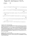

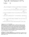
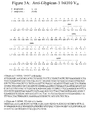


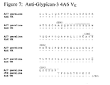
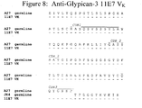

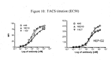
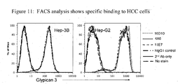


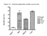
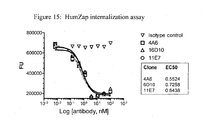
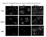
Classifications
-
- C—CHEMISTRY; METALLURGY
- C07—ORGANIC CHEMISTRY
- C07K—PEPTIDES
- C07K16/00—Immunoglobulins [IGs], e.g. monoclonal or polyclonal antibodies
- C07K16/18—Immunoglobulins [IGs], e.g. monoclonal or polyclonal antibodies against material from animals or humans
- C07K16/28—Immunoglobulins [IGs], e.g. monoclonal or polyclonal antibodies against material from animals or humans against receptors, cell surface antigens or cell surface determinants
-
- A—HUMAN NECESSITIES
- A61—MEDICAL OR VETERINARY SCIENCE; HYGIENE
- A61P—SPECIFIC THERAPEUTIC ACTIVITY OF CHEMICAL COMPOUNDS OR MEDICINAL PREPARATIONS
- A61P35/00—Antineoplastic agents
-
- C—CHEMISTRY; METALLURGY
- C07—ORGANIC CHEMISTRY
- C07K—PEPTIDES
- C07K16/00—Immunoglobulins [IGs], e.g. monoclonal or polyclonal antibodies
- C07K16/18—Immunoglobulins [IGs], e.g. monoclonal or polyclonal antibodies against material from animals or humans
- C07K16/28—Immunoglobulins [IGs], e.g. monoclonal or polyclonal antibodies against material from animals or humans against receptors, cell surface antigens or cell surface determinants
- C07K16/30—Immunoglobulins [IGs], e.g. monoclonal or polyclonal antibodies against material from animals or humans against receptors, cell surface antigens or cell surface determinants from tumour cells
- C07K16/303—Liver or Pancreas
-
- A—HUMAN NECESSITIES
- A61—MEDICAL OR VETERINARY SCIENCE; HYGIENE
- A61K—PREPARATIONS FOR MEDICAL, DENTAL OR TOILETRY PURPOSES
- A61K39/00—Medicinal preparations containing antigens or antibodies
-
- C—CHEMISTRY; METALLURGY
- C07—ORGANIC CHEMISTRY
- C07K—PEPTIDES
- C07K2317/00—Immunoglobulins specific features
- C07K2317/20—Immunoglobulins specific features characterized by taxonomic origin
- C07K2317/21—Immunoglobulins specific features characterized by taxonomic origin from primates, e.g. man
-
- C—CHEMISTRY; METALLURGY
- C07—ORGANIC CHEMISTRY
- C07K—PEPTIDES
- C07K2317/00—Immunoglobulins specific features
- C07K2317/50—Immunoglobulins specific features characterized by immunoglobulin fragments
- C07K2317/56—Immunoglobulins specific features characterized by immunoglobulin fragments variable (Fv) region, i.e. VH and/or VL
- C07K2317/565—Complementarity determining region [CDR]
-
- C—CHEMISTRY; METALLURGY
- C07—ORGANIC CHEMISTRY
- C07K—PEPTIDES
- C07K2317/00—Immunoglobulins specific features
- C07K2317/70—Immunoglobulins specific features characterized by effect upon binding to a cell or to an antigen
- C07K2317/73—Inducing cell death, e.g. apoptosis, necrosis or inhibition of cell proliferation
- C07K2317/732—Antibody-dependent cellular cytotoxicity [ADCC]
-
- C—CHEMISTRY; METALLURGY
- C07—ORGANIC CHEMISTRY
- C07K—PEPTIDES
- C07K2317/00—Immunoglobulins specific features
- C07K2317/70—Immunoglobulins specific features characterized by effect upon binding to a cell or to an antigen
- C07K2317/77—Internalization into the cell
-
- C—CHEMISTRY; METALLURGY
- C07—ORGANIC CHEMISTRY
- C07K—PEPTIDES
- C07K2317/00—Immunoglobulins specific features
- C07K2317/90—Immunoglobulins specific features characterized by (pharmaco)kinetic aspects or by stability of the immunoglobulin
- C07K2317/92—Affinity (KD), association rate (Ka), dissociation rate (Kd) or EC50 value
Definitions
- Glypican-3 is an oncofetal antigen that belongs to the glypican family of glycosyl-phosphatidylinositol-anchored heparin sulfate proteoglycans. Glypicans are characterized by a covalent linkage to complex polysaccharide chains called heparinsulphate glycosaminoglycans. Glypicans are involved in cell signaling at the cellular-extracellular matrix interface. ( Sasisekharan et al., Nature Reviews I Cancer, Volume 2 (2002 ).) To date, six distinct members of the human glypican family have been identified. Cell membrane-bound Glypican-3 is composed of two subunits, linked by one or more disulfide bonds.
- Glypican-3 is expressed in fetal liver and placenta during development and is down-regulated or silenced in normal adult tissues. Mutations and depletions in the Glypican-3 gene are responsible for the Simpson-Golabi-Behmel or Simpson dysmorphia syndrome in humans. Glypican-3 is expressed in various cancers and, in particular, hepatocellular carcinoma ("HCC"), melanoma, Wilm's tumor, and hepatoblastoma. ( Jakubovic and Jothy; Ex. Mol. Path. 82:184-189 (2007 ); Nakatsura and Nishimura, Biodrugs 19(2):71-77 (2005 ).)
- HCC hepatocellular carcinoma
- HCC is the third leading cause of cancer-related deaths worldwide. Each year, HCC accounts for about 1 million deaths.
- Hepatitis B virus, hepatitis C virus, and chronic heavy alcohol use leading to cirrhosis of the liver remain the most common causes of HCC. Its incidence has increased dramatically in the United States because of the spread of hepatitis C virus infection and is expected to increase for the next 2 decades.
- HCC is treated primarily by liver transplantation or tumor resection. Patient prognosis is dependent on both the underlying liver function and the stage at which the tumor is diagnosed. ( Parikh and Hyman, Am J Med. 120(3):194-202 (2007 ).) Effective HCC treatment strategies are needed.
- EP1674111 discloses the preparation of monoclonal antibodies binding glypican-3 showing a strong CDC activity (Ex.16) and the humanization of one of them. The affinity of these antibodies is not measured.
- the present disclosure provides isolated monoclonal antibodies, in particular human monoclonal antibodies, that bind to Glypican-3 and that exhibit numerous desirable properties. These properties include high affinity binding to human Glypican-3.
- the disclosure pertains to an isolated monoclonal antibody, or an antigen-binding portion thereof, wherein the antibody:
- the antibody is a human antibody, although in alternative aspects of the disclosure the antibody can be a murine antibody, a chimeric antibody or humanized antibody.
- the antibody binds to human Glypican-3 with a K D of 5 x 10 -8 M or less, binds to human Glypican-3 with a K D of 2 x 10 -8 M or less, binds to human Glypican-3 with a K D of 1 x 10 -8 M or less, binds to human Glypican-3 with a K D of 5x10 -9 M or less, binds to human Glypican-3 with a K D of 4x10 -9 M or less, binds to human Glypican-3 with a K D of 3x10 -9 M or less, or binds to human Glypican-3 with a K D of 2 x 10 -9 M or less.
- the disclosure provides an isolated monoclonal antibody, or antigen binding portion thereof, wherein the antibody cross-competes for binding to Glypican-3 with a reference antibody comprising:
- the reference antibody comprises:
- the disclosure provides an isolated monoclonal antibody, or an antigen-binding portion thereof, comprising a heavy chain variable region that is the product of or derived from a human V H 5-51 gene, wherein the antibody specifically binds Glypican-3.
- the disclosure also provides an isolated monoclonal antibody, or an antigen-binding portion thereof, comprising a light chain variable region that is the product of or derived from a human V K A27 gene, wherein the antibody specifically binds Glypican-3.
- the disclosure provides an isolated monoclonal antibody, or an antigen-binding portion thereof, comprising:
- the disclosure provides an isolated monoclonal antibody, or antigen binding portion thereof, comprising:
- the heavy chain variable region CDR2 sequence comprises an amino acid sequence selected from the group consisting of amino acid sequences of SEQ ID NOs:4, 5, and 6, and conservative modifications thereof; and the light chain variable region CDR2 sequence comprises an amino acid sequence selected from the group consisting of amino acid sequences of SEQ ID NOs:13, 14, and 15, and conservative modifications thereof.
- the heavy chain variable region CDR1 sequence comprises an amino acid sequence selected from the group consisting of amino acid sequences of SEQ ID NOs:1, 2, and 3, and conservative modifications thereof; and the light chain variable region CDR1 sequence comprises an amino acid sequence selected from the group consisting of amino acid sequences of SEQ ID NOs:10, 11, and 12, and conservative modifications thereof.
- a preferred combination comprises:
- Another preferred combination comprises:
- a preferred combination comprises:
- Another preferred combination comprises:
- Another preferred combination comprises:
- antibodies, or antigen-binding portions thereof are provided that compete for binding to Glypican-3 with any of the aforementioned antibodies.
- the antibodies of the invention can be, for example, full-length antibodies, for example of an IgG1 or IgG4 isotype.
- the antibodies can be antibody fragments, such as Fab, Fab' or Fab'2 fragments, or single chain antibodies.
- compositions comprising an antibody, or antigen-binding portion thereof, or immunoconjugate or bispecific molecule of the invention and a pharmaceutically acceptable carrier are also provided.
- Nucleic acid molecules encoding the antibodies, or antigen-binding portions thereof, of the invention are also encompassed by the invention, as well as expression vectors comprising such nucleic acids and host cells comprising such expression vectors.
- the disclosure provides methods of inhibiting the growth of tumor cells expressing Glypican-3.
- the methods comprise contacting the cells with an anti-Glypican-3 antibody, or antigen-binding portion thereof, in an amount effective to inhibit growth of the tumor cells.
- the tumor cells are derived from liver tissue.
- the antibody, or antigen-binding portion thereof may be immunoconjugates.
- the immunoconjugates of the invention may be therapeutic agents, for example, cytotoxins or radioactive isotopes.
- Glypican-3 “glypican proteoglycan 3," “GPC3,” “OTTHUMP00000062492,” “GTR2-2,” “SGB,” “DGSX,” “SDYS,” “SGBS,” “OCI-5,” and “SGBS1” are used interchangeably, and include variants, isoforms and species homologs of human Glypican-3.
- human antibodies of this disclosure may, in certain cases, cross-react with Glypican-3 from species other than human.
- the antibodies may be completely specific for one or more human Glypican-3 proteins and may not exhibit species or other types of non-human cross-reactivity.
- the complete amino acid sequence of an exemplary human Glypican-3 has Genbank/NCBI accession number NM_004484 (SEQ ID NO:36).
- immune response refers to the action of, for example, lymphocytes, antigen presenting cells, phagocytic cells, granulocytes, and soluble macromolecules produced by the above cells or the liver (including antibodies, cytokines, and complement) that results in selective damage to, destruction of, or elimination from the human body of invading pathogens, cells or tissues infected with pathogens, cancerous cells, or, in cases of autoimmunity or pathological inflammation, normal human cells or tissues.
- binding fragments encompassed within the present disclosure include (i) a Fab fragment, a monovalent fragment consisting of the V L , V H , C L and C H 1 domains; (ii) a F(ab') 2 fragment, a bivalent fragment comprising two Fab fragments linked by a disulfide bridge at the hinge region; (iii) a Fab' fragment, which is essentially an Fab with part of the hinge region (see, FUNDAMENTAL IMMUNOLOGY (Paul ed., 3.sup.rd ed.
- the two domains of the Fv fragment, V L and V H are coded for by separate genes, they can be joined, using recombinant methods, by a synthetic linker that enables them to be made as a single protein chain in which the V L and V H regions pair to form monovalent molecules (known as single chain Fv (scFv); see e.g., Bird et al. (1988) Science 242:423-426 ; and Huston et al. (1988) Proc. Natl. Acad. Sci. USA 85:5879-5883 ).
- single chain Fv single chain Fv
- Such single chain antibodies are also intended to be encompassed within the term "antigen-binding portion" of an antibody.
- an "isolated antibody,” as used herein, is intended to refer to an antibody that is substantially free of other antibodies having different antigenic specificities (e.g., an isolated antibody that specifically binds Glypican-3 is substantially free of antibodies that specifically bind antigens other than Glypican-3).
- An isolated antibody that specifically binds Glypican-3 may, however, have cross-reactivity to other antigens, such as Glypican-3 molecules from other species.
- an isolated antibody may be substantially free of other cellular material and/or chemicals.
- monoclonal antibody or “monoclonal antibody composition” as used herein refer to a preparation of antibody molecules of single molecular composition.
- a monoclonal antibody composition displays a single binding specificity and affinity for a particular epitope.
- human antibody is intended to include antibodies having variable regions in which both the framework and CDR regions are derived from human germline immunoglobulin sequences. Furthermore, if the antibody contains a constant region, the constant region also is derived from human germline immunoglobulin sequences.
- the human antibodies of the invention may include amino acid residues not encoded by human germline immunoglobulin sequences (e.g., mutations introduced by random or site-specific mutagenesis in vitro or by somatic mutation in vivo ).
- the term "human antibody,” as used herein is not intended to include antibodies in which CDR sequences derived from the germline of another mammalian species, such as a mouse, have been grafted onto human framework sequences.
- human monoclonal antibody refers to antibodies displaying a single binding specificity which have variable regions in which both the framework and CDR regions are derived from human germline immunoglobulin sequences.
- the human monoclonal antibodies are produced by a hybridoma which includes a B cell obtained from a transgenic nonhuman animal, e.g., a transgenic mouse, having a genome comprising a human heavy chain transgene and a light chain transgene fused to an immortalized cell.
- recombinant human antibody includes all human antibodies that are prepared, expressed, created or isolated by recombinant means, such as (a) antibodies isolated from an animal (e.g., a mouse) that is transgenic or transchromosomal for human immunoglobulin genes or a hybridoma prepared therefrom (described further below), (b) antibodies isolated from a host cell transformed to express the human antibody, e.g., from a transfectoma, (c) antibodies isolated from a recombinant, combinatorial human antibody library, and (d) antibodies prepared, expressed, created or isolated by any other means that involve splicing of human immunoglobulin gene sequences to other DNA sequences.
- Such recombinant human antibodies have variable regions in which the framework and CBR regions are derived from human germline immunoglobulin sequences.
- such recombinant human antibodies can be subjected to in vitro mutagenesis (or, when an animal transgenic for human Ig sequences is used, in vivo somatic mutagenesis) and thus the amino acid sequences of the V H and V L regions of the recombinant antibodies are sequences that, while derived from and related to human germline V H and V L sequences, may not naturally exist within the human antibody germline repertoire in vivo.
- isotype refers to the antibody class (e.g. , IgM or IgG1) that is encoded by the heavy chain constant region genes.
- an antibody recognizing an antigen and "an antibody specific for an antigen” are used interchangeably herein with the term “an antibody which binds specifically to an antigen.”
- human antibody derivatives refers to any modified form of the human antibody, e.g ., a conjugate of the antibody and another agent or antibody.
- humanized antibody is intended to refer to antibodies in which CDR sequences derived from the germline of another mammalian species, such as a mouse, have been grafted onto human framework sequences. Additional framework region modifications may be made within the human framework sequences.
- chimeric antibody is intended to refer to antibodies in which the variable region sequences are derived from one species and the constant region sequences are derived from another species, such as an antibody in which the variable region sequences are derived from a mouse antibody and the constant region sequences are derived from a human antibody.
- an antibody that "specifically binds to human Glypican-3" is intended to refer to an antibody that binds to human Glypican-3 with a K D of 1 x 10 -7 M or less, more preferably 5 x 10 -8 M or less, more preferably 3 x 10 -8 M or less, more preferably 1 x 10 -8 M or less, even more preferably 5 x 10 -9 M or less.
- does not substantially bind to a protein or cells, as used herein, means does not bind or does not bind with a high affinity to the protein or cells, i.e. binds to the protein or cells with a K D of 1 x 10 -6 M or more, more preferably 1 x 10 -5 M or more, more preferably 1 x 10 -4 M or more, more preferably 1 x 10 -3 M or more, even more preferably 1 x 10 -2 M or more.
- K assoc or "K a ,” as used herein, is intended to refer to the association rate of a particular antibody-antigen interaction
- K dis or "K d ,” as used herein, is intended to refer to the dissociation rate of a particular antibody-antigen interaction
- K D is intended to refer to the dissociation constant, which is obtained from the ratio of K d to K a (i.e,. K d /K a ) and is expressed as a molar concentration (M).
- K D values for antibodies can be determined using methods well established in the art. A preferred method for determining the K D of an antibody is by using surface plasmon resonance, preferably using a biosensor system such as a Biacore ® system.
- the term "high affinity" for an IgG antibody refers to an antibody having a K D of 1 x 10 -7 M or less, more preferably 5 x 10 -8 M or less, even more preferably 1x10 -8 M or less, even more preferably 5 x 10 -9 M or less and even more preferably 1 x 10 -9 M or less for a target antigen.
- “high affinity” binding can vary for other antibody isotypes.
- “high affinity” binding for an IgM isotype refers to an antibody having a K D of 10 -6 M or less, more preferably 10 -7 M or less, even more preferably 10 -8 M or less.
- the term “subject” includes any human or nonhuman animal.
- nonhuman animal includes all vertebrates, e.g ., mammals and non-mammals, such as nonhuman primates, sheep, dogs, cats, horses, cows, chickens, amphibians, reptiles, etc.
- the antibody preferably binds to an antigenic epitope present in Glypican-3, which epitope is not present in other proteins.
- the antibody typically binds to Glypican-3 but does not bind to other proteins, or binds to other proteins with a low affinity, such as with a K D of 1 x 10 -6 M or more, more preferably 1 x 10 -5 M or more, more preferably 1 x 10 -4 M or more, more preferably 1 x 10 -3 M or more, even more preferably 1 x 10 -2 M or more.
- the antibody does not substantially bind to related proteins, for example, the antibody does not substantially bind to Glypican-1, Glypican-2, Glypican-4 or Glypican-6.
- Standard assays to evaluate the binding ability of the antibodies toward Glypican-3 are known in the art, including for example, ELISAs, Western blots, RIAs, and flow cytometry analysis. Suitable assays are described in detail in the Examples.
- the binding kinetics (e.g ., binding affinity) of the antibodies also can be assessed by standard assays known in the art, such as by Biacore ® system analysis.
- Biacore ® system analysis To assess binding to tumor cells, e.g. Hep-3b or Hep-G2 (ATCC Deposit No. HB-8064 and HB-8065, respectively), cells can be obtained from publicly available sources, such as the American Type Culture Collection, and used in standard assays, such as flow cytometric analysis.
- Preferred antibodies of the invention are the human monoclonal antibodies 4A6, 11E7, and 16D10, isolated and structurally characterized as described in Examples 1 and 2.
- the V H amino acid sequences of 4A6, 11E7, and 16D10 are shown in SEQ ID NOs:19, 20, and 21, respectively.
- the V L amino acid sequences of 4A6, 11E7, and 16D10 are shown in SEQ ID NOs:22, 23, and 24, respectively.
- V H and V L sequences can be "mixed and matched" to create other anti-Glypican-3 binding molecules of the disclosure
- Glypican-3 binding of such "mixed and matched” antibodies can be tested using the binding assays described above and in the Examples (e.g., ELISAs).
- a V H sequence from a particular V H /V L pairing is replaced with a structurally similar V H sequence.
- a V L sequence from a particular V H /V L pairing is replaced with a structurally similar V L sequence.
- Preferred heavy and light chain combinations include:
- the disclosure provides antibodies that comprise the heavy chain and light chain CDR1s, CDR2s and CDR3s of 4A6, 11E7, and 16D10, or combinations thereof.
- the amino acid sequences of the V H CDR1s of 4A6, 11E7, and 16D10 are shown in SEQ ID NOs:1, 2 and 3, respectively.
- the amino acid sequences of the V H CDR2s of 4A6, 11E7, and 16D10 are shown in SEQ ID NOs:4, 5, and 6, respectively.
- the amino acid sequences of the V H CDR3s of 4A6, 11E7, and 16D10 are shown in SEQ ID NOs:7, 8, and 9, respectively.
- the amino acid sequences of the V k CDR1s of 4A6, 11E7, and 16D10 are shown in SEQ ID NOs:10, 11, and 12, respectively.
- the amino acid sequences of the V k CDR2s of 4A6, 11E7, and 16D10 are shown in SEQ ID NOs:13, 14, and 15, respectively.
- the amino acid sequences of the V k CDR3s of 4A6, 11E7, and 16D10 are shown in SEQ ID NOs:16,17, and 18, respectively.
- the CDR regions are delineated using the Kabat system (Kabat, E. A., et al. (1991) Sequences of Proteins of Immunological Interest, Fifth Edition, U.S. Department of Health and Human Services, NIH Publication No. 91-3242).
- V H CDR1, CDR2, and CDR3 sequences and V k CDR1, CDR2, and CDR3 sequences can be "mixed and matched" (i.e., CDRs from different antibodies can be mixed and match, although each antibody must contain a V H CDR1, CDR2, and CDR3 and a V k CDR1, CDR2, and CDR3) to create other anti-Glypican-3 binding molecules of the disclosure.
- Glypican-3 binding of such "mixed and matched" antibodies can be tested using the binding assays described above and in the Examples (e.g. , ELISAs, Biacore ® analysis).
- the CDR1, CDR2 and/or CDR3 sequence from a particular V H sequence is replaced with a structurally similar CDR sequence(s).
- V k CDR sequences are mixed and matched, the CDR1, CDR2 and/or CDR3 sequence from a particular V k sequence preferably is replaced with a structurally similar CDR sequence(s).
- V H and V L sequences can be created by substituting one or more V H and/or V L CDR region sequences with structurally similar sequences from the CDR sequences disclosed herein for monoclonal antibodies 4A6, 11E7, and 16D10.
- the disclosure provides an isolated monoclonal antibody, or antigen binding portion thereof comprising:
- the antibody comprises:
- the antibody comprises:
- the antibody comprises:
- the disclosure provides an isolated monoclonal antibody, or antigen binding portion thereof, comprising a heavy chain variable region comprising CDR1, CDR2, and CDR3 sequences and a light chain variable region comprising CDR1, CDR2, and CDR3 sequences, wherein:
- the heavy chain variable region CDR2 sequence comprises an amino acid sequence selected from the group consisting of amino acid sequences of SEQ ID NOs:4, 5, and 6, and conservative modifications thereof; and the light chain variable region CDR2 sequence comprises an amino acid sequence selected from the group consisting of amino acid sequences of SEQ ID NOs:13, 14, and 15, and conservative modifications thereof.
- the heavy chain variable region CDR1 sequence comprises an amino acid sequence selected from the group consisting of amino acid sequences of SEQ ID NOs:1, 2, and 3 and conservative modifications thereof; and the light chain variable region CDR1 sequence comprises an amino acid sequence selected from the group consisting of amino acid sequences of SEQ ID NOs:10, 11, and 12, and conservative modifications thereof.
- the antibody can be, for example, human antibodies, humanized antibodies or chimeric antibodies.
- conservative sequence modifications is intended to refer to amino acid modifications that do not significantly affect or alter the binding characteristics of the antibody containing the amino acid sequence. Such conservative modifications include amino acid substitutions, additions and deletions. Modifications can be introduced into an antibody of the invention by standard techniques known in the art, such as site-directed mutagenesis and PCR-mediated mutagenesis. Conservative amino acid substitutions are ones in which the amino acid residue is replaced with an amino acid residue having a similar side chain. Families of amino acid residues having similar side chains have been defined in the art.
- amino acids with basic side chains e.g., lysine, arginine, histidine
- acidic side chains e.g., aspartic acid, glutamic acid
- uncharged polar side chains e.g., glycine, asparagine, glutamine, serine, threonine, tyrosine, cysteine, tryptophan
- nonpolar side chains e.g ., alanine, valine, leucine, isoleucine, proline, phenylalanine, methionine
- beta-branched side chains e.g., threonine, valine, isoleucine
- aromatic side chains e.g., tyrosine, phenylalanine, tryptophan, histidine
- the heavy chain CDR1 sequence of SEQ ID NO:1, 2, or 3 may comprise one or more conservative sequence modification, such as one, two, three, four, five or more amino acid substitutions, additions or deletions;
- the light chain CDR1 sequence of SEQ ID NO:10, 11, or 12 may comprise one or more conservative sequence modification, such as one, two, three, four, five or more amino acid substitutions, additions or deletions;
- the heavy chain CDR2 sequence shown in SEQ ID NO:4, 5, or 6 may comprise one or more conservative sequence modification, such as one, two, three, four, five or more amino acid substitutions, additions or deletions;
- the light chain CDR2 sequence shown in SEQ ID NO:13, 14, or 15 may comprise one or more conservative sequence modification, such as one, two, three, four, five or more amino acid substitutions, additions or deletions;
- the heavy chain CDR3 sequence shown in SEQ ID NO:7, 8, or 9 may comprise one or more conservative sequence modification, such as one, two, three, four, five or more amino
- the disclosure provides antibodies that bind to the same epitope on human Glypican-3 as any of the Glypican-3 monoclonal antibodies of the invention (i.e ., antibodies that have the ability to cross-compete for binding to Glypican-3 with any of the monoclonal antibodies of the invention).
- the reference antibody for cross-competition studies can be the monoclonal antibody 4A6 (having V H and V L sequences as shown in SEQ ID NOs:19 and 22, respectively), the monoclonal antibody 11E7 (having V H and V L sequences as shown in SEQ ID NOs:20 and 23, respectively), or the monoclonal antibody 16D10 (having V H and V L sequences as shown in SEQ ID NOs:21 and 24, respectively).
- Such cross-competing antibodies can be identified based on their ability to cross-compete with 4A6, 11E7, or 16D10 in standard Glypican-3 binding assays.
- Biacore analysis, ELISA assays or flow cytometry may be used to demonstrate cross-competition with the antibodies of the current invention.
- the ability of a test antibody to inhibit the binding of, for example, 4A6, 11E7, or 16D10, to human Glypican-3 demonstrates that the test antibody can compete with 4A6, 11E7, or 16D10 for binding to human Glypican-3 and thus binds to the same epitope on human Glypican-3 as 4A6, 11E7, or 16D10.
- the antibody that binds to the same epitope on human Glypican-3 as 4A6, 11E7, or 16D10 is a human monoclonal antibody.
- Such human monoclonal antibodies can be prepared and isolated as described in the Examples.
- CDR grafting can be used to engineer variable regions of antibodies.
- Antibodies interact with target antigens predominantly through amino acid residues that are located in the six heavy and light chain complementarity determining regions (CDRs). For this reason, the amino acid sequences within CDRs are more diverse between individual antibodies than sequences outside of CDRs. Because CDR sequences are responsible for most antibody-antigen interactions, it is possible to express recombinant antibodies that mimic the properties of specific naturally occurring antibodies by constructing expression vectors that include CDR sequences from the specific naturally occurring antibody grafted onto framework sequences from a different antibody with different properties (see, e.g ., Riechmann, L. et al. (1998) Nature 332:323-327 ; Jones, P.
- Such framework sequences can be obtained from public DNA databases or published references that include germline antibody gene sequences.
- germline DNA sequences for human heavy and light chain variable region genes can be found in the "VBase" human germline sequence database (available on the Internet at www.mrc-cpe.cam.ac.uk/vbase), as well as in Kabat, E. A., et al. (1991) Sequences of Proteins of Immunological Interest, Fifth Edition, U.S. Department of Health and Human Services, NIH Publication No. 91-3242 ; Tomlinson, I. M., et al.
- the following heavy chain germline sequences found in the HCo12 HuMAb mouse are available in the accompanying Genbank accession numbers: 1-69 (NG_0010109, NT_024637 and BC070333), 5-51 (NG_0010109 and NT_024637), 4-34 (NG_0010109 and NT_024637), 3-30.3 (CAJ556644) and 3-23 (AJ406678).
- the identities are exact amino acid matches between the antibody sequence and the protein database over the entire length of the sequence.
- the positives are not identical but amino acid substitutions guided by the BLOSUM62 substitution matrix. If the antibody sequence matches two of the database sequences with same identity, the hit with most positives would be decided to be the matching sequence hit.
- the instant disclosure provides isolated anti-Glypican-3 monoclonal antibodies, or antigen binding portions thereof, comprising a heavy chain variable region comprising: (a) a V H CDR1 region comprising an amino acid sequence selected from the group consisting of SEQ ID NOs:1, 2, and 3, or an amino acid sequence having one, two, three, four or five amino acid substitutions, deletions or additions as compared to SEQ ID NOs:1, 2 and 3; (b) a V H CDR2 region comprising an amino acid sequence selected from the group consisting of SEQ ID NOs:4, 5, and 6, or an amino acid sequence having one, two, three, four or five amino acid substitutions, deletions or additions as compared to SEQ ID NOs:4, 5, and 6; (c) a V H CDR3 region comprising an amino acid sequence selected from the group consisting of SEQ ID NOs:7, 8, and 9, or an amino acid sequence having one, two, three, four or five amino acid substitutions, deletions or additions as compared to S
- amino acid residue #73 (within FR3) of V H is an arginine (SEQ ID NO:19) whereas this residue in the corresponding V H 5-51 germline sequence is lysine (SEQ ID NO:31).
- somatic mutations can be "backmutated" to the germline sequence by, for example, site-directed mutagenesis or PCR-mediated mutagenesis (e.g., residue #73 (residue #8 of FR3) of the V H of 4A6 can be "backmutated” from arginine to lysine).
- amino acid residue #89 (within FR3) of V H is leucine (SEQ ID NO:19) whereas this residue in the corresponding V H 5-51 germline sequence is a methionine (SEQ ID NO:31).
- residue #89 (residue #27 of FR3) of the V H of 4A6 can be "backmutated” from leucine to methionine.
- Such "backmutated” antibodies are also intended to be encompassed by the invention.
- amino acid residue #76 (within FR3) of V H is arginine (SEQ ID NO:20) whereas this residue in the corresponding V H 5-51 germline sequence is a serine (SEQ ID NO:31).
- residue #76 (residue #11 of FR3) of the V H of 11E7 can be "backmutated” from arginine to serine.
- Such "backmutated” antibodies are also intended to be encompassed by the invention.
- amino acid residue #10 (within FR1) of V H is aspartate (SEQ ID NO:21) whereas this residue in the corresponding V H 5-51 germline sequence is a glutamate (SEQ ID NO:31).
- residue #10 (residue #10 of FR1) of the V H of 16D10 can be "backmutated” from aspartate to glutamate.
- Such "backmutated” antibodies are also intended to be encompassed by the invention.
- amino acid residue #12 (within FR1) of V H is threonine (SEQ ID NO:21) whereas this residue in the corresponding V H 5-51 germline sequence is a lysine (SEQ ID NO:31).
- residue #12 (residue #12 of FR1) of the V H of 16D10 can be "backmutated” from threonine to lysine.
- Such "backmutated” antibodies are also intended to be encompassed by the invention.
- amino acid residue #24 (within FR1) of V H is valine (SEQ ID NO:21) whereas this residue in the corresponding V H 5-51 germline sequence is a glycine (SEQ ID NO:31).
- residue #24 (residue #24 of FR1) of the V H of 16D10 can be "backmutated” from valine to glycine.
- Such "backmutated” antibodies are also intended to be encompassed by the invention.
- amino acid residue #28 (within FR1) of V H is arginine (SEQ ID NO:21) whereas this residue in the corresponding V H 5-51 germline sequence is a serine (SEQ ID NO:31).
- residue #28 (residue #28 of FR1) of the V H of 16D10 can be "backmutated” from arginine to serine.
- Such "backmutated” antibodies are also intended to be encompassed by the invention.
- amino acid residue #41 (within FR2) of V H is serine (SEQ ID NO:21) whereas this residue in the corresponding V H 5-51 germline sequence is a proline (SEQ ID NO:31).
- residue #41 (residue #6 of FR2) of the V H of 16D10 can be "backmutated” from serine to proline.
- Such "backmutated” antibodies are also intended to be encompassed by the invention.
- amino acid residue #76 (within FR3) of V H is asparagine (SEQ ID NO:21) whereas this residue in the corresponding V H 5-51 germline sequence is a serine (SEQ ID NO:31).
- residue #76 (residue #11 of FR3) of the V H of 16D10 can be "backmutated” from asparagine to serine.
- Such "backmutated” antibodies are also intended to be encompassed by the invention.
- amino acid residue #81 (within FR3) of V H is arginine (SEQ ID NO:21) whereas this residue in the corresponding V H 5-51 germline sequence is a glutamine (SEQ ID NO:31).
- residue #81 (residue #16 of FR3) of the V H of 16D10 can be "backmutated” from arginine to glutamine.
- Such "backmutated” antibodies are also intended to be encompassed by the invention.
- amino acid residue #89 (within FR3) of V H is isoleucine (SEQ ID NO:21) whereas this residue in the corresponding V H 5-51 germline sequence is a methionine (SEQ ID NO:31).
- residue #89 (residue #27 of FR3) of the V H of 16D10 can be "backmutated” from isoleucine to methionine.
- Such "backmutated” antibodies are also intended to be encompassed by the invention.
- Another type of framework modification involves mutating one or more residues within the framework region, or even within one or more CDR regions, to remove T cell epitopes to thereby reduce the potential immunogenicity of the antibody. This approach is also referred to as "deimmunization" and is described in further detail in U.S. Patent Publication No. 20030153043 by Carr et al.
- antibodies of the invention may be engineered to include modifications within the Fc region, typically to alter one or more functional properties of the antibody, such as serum half-life, complement fixation, Fc receptor binding, and/or antigen-dependent cellular cytotoxicity.
- an antibody of the invention may be chemically modified (e.g., one or more chemical moieties can be attached to the antibody) or be modified to alter its glycosylation, again to alter one or more functional properties of the antibody.
- the hinge region of CH1 is modified such that the number of cysteine residues in the hinge region is altered, e.g., increased or decreased.
- This approach is described further in U.S. Patent No. 5,677,425 by Bodmer et al.
- the number of cysteine residues in the hinge region of CH1 is altered to, for example, facilitate assembly of the light and heavy chains or to increase or decrease the stability of the antibody.
- the Fc hinge region of an antibody is mutated to decrease the biological half life of the antibody. More specifically, one or more amino acid mutations are introduced into the CH2-CH3 domain interface region of the Fc-hinge fragment such that the antibody has impaired Staphylococcyl protein A (SpA) binding relative to native Fc-hinge domain SpA binding.
- SpA Staphylococcyl protein A
- the antibody is modified to increase its biological half life.
- Various approaches are possible. For example, one or more of the following mutations can be introduced: T252L, T254S, and T256F, as described in U.S. Patent No. 6,277,375 to Ward .
- the antibody can be altered within the CH1 or C L region to contain a salvage receptor binding epitope taken from two loops of a CH2 domain of an Fc region of an IgG, as described in U.S. Patent Nos. 5,869,046 and 6,121,022 by Presta et al.
- the Fc region is altered by replacing at least one amino acid residue with a different amino acid residue to alter the effector function(s) of the antibody.
- one or more amino acids selected from amino acid residues 234, 235, 236, 237, 297, 318, 320 and 322 can be replaced with a different amino acid residue such that the antibody has an altered affinity for an effector ligand but retains the antigen-binding ability of the parent antibody.
- the effector ligand to which affinity is altered can be, for example, an Fc receptor or the C1 component of complement. This approach is described in further detail in U.S. Patent Nos. 5,624,821 and 5,648,260 , both by Winter et al.
- one or more amino acids selected from amino acid residues 329, 331 and 322 can be replaced with a different amino acid residue such that the antibody has altered C1q binding and/orreduced or abolished complement dependent cytotoxicity (CDC).
- CDC complement dependent cytotoxicity
- one or more amino acid residues within amino acid positions 231 and 239 are altered to thereby alter the ability of the antibody to fix complement. This approach is described further in PCT Publication WO 94/29351 by Bodmer et al.
- the Fc region is modified to increase the ability of the antibody to mediate antibody dependent cellular cytotoxicity (ADCC) and/or to increase the affinity of the antibody for an Fc ⁇ receptor by modifying one or more amino acids at the following positions: 238, 239, 248, 249, 252, 254, 255, 256, 258, 265, 267, 268, 269, 270, 272, 276, 278, 280, 283, 285, 286, 289, 290, 292, 293, 294, 295, 296, 298, 301, 303, 305, 307, 309, 312, 315, 320, 322, 324, 326, 327, 329, 330, 331, 333, 334, 335, 337, 338, 340, 360, 373, 376, 378, 382, 388, 389, 398, 414, 416, 419, 430, 434, 435, 437, 438 or 439.
- ADCC antibody dependent cellular cytotoxicity
- an antibody can be made that has an altered type of glycosylation, such as a hypofucosylated antibody-having reduced amounts of fucosyl residues or an antibody having increased bisecting GlcNac structures.
- altered glycosylation patterns have been demonstrated to increase the ADCC ability of antibodies.
- carbohydrate modifications can be accomplished by, for example, expressing the antibody in a host cell with altered glycosylation machinery. Cells with altered glycosylation machinery have been described in the art and can be used as host cells in which to express recombinant antibodies of the invention to thereby produce an antibody with altered glycosylation.
- the cell lines Ms704, Ms705, and Ms709 lack the fucosyltransferase gene, FUT8 (alpha (1,6) fucosyltransferase), such that antibodies expressed in the Ms704, Ms705, and Ms709 cell lines lack fucose on their carbohydrates.
- the Ms704, Ms705, and Ms709 FUT8 -/- cell lines were created by the targeted disruption of the FUT8 gene in CHO/DG44 cells using two replacement vectors (see U.S. Patent Publication No. 20040110704 by Yamane et al. and Yamane-Ohnuki et al. (2004) Biotechnol Bioeng 87:614-22 ).
- EP 1,176,195 by Hanai et al. describes a cell line with a functionally disrupted FUT8 gene, which encodes a fucosyl transferase, such that antibodies expressed in such a cell line exhibit hypofucosylation by reducing or eliminating the alpha 1,6 bond-related enzyme.
- Hanai et al. also describe cell lines which have a low enzyme activity for adding fucose to the N-acetylglucosamine that binds to the Fc region of the antibody or does not have the enzyme activity, for example the rat myeloma cell line YB2/0 (ATCC CRL 1662).
- glycoprotein-modifying glycosyl transferases e.g., beta(1,4)-N-acetylglucosaminyltransferase III (GnTIII)
- GnTIII glycoprotein-modifying glycosyl transferases
- the fucose residues of the antibody may be cleaved off using a fucosidase enzyme.
- the fucosidase alpha-L-fucosidase removes fucosyl residues from antibodies ( Tarentino, A.L. et al. (1975) Biochem. 14:5516-23 ).
- An antibody can be pegylated to, for example, increase the biological (e.g., serum) half life of the antibody.
- the antibody, or fragment thereof typically is reacted with polyethylene glycol (PEG), such as a reactive ester or aldehyde derivative of PEG, under conditions in which one or more PEG groups become attached to the antibody or antibody fragment.
- PEG polyethylene glycol
- the pegylation is carried out via an acylation reaction or an alkylation reaction with a reactive PEG molecule (or an analogous reactive water-soluble polymer).
- polyethylene glycol is intended to encompass any of the forms of PEG that have been used to derivatize other proteins, such as mono (C1-C10) alkoxy or aryloxy-polyethylene glycol or polyethylene glycol-maleimide.
- the antibody to be pegylated is an aglycosylated antibody. Methods for pegylating proteins are known in the art and can be applied to the antibodies of the invention. See for example, EP 0 154 316 by Nishimura et al. and EP 0 401 384 by Ishikawa et al.
- the antibodies of the present invention may be further characterized by the various physical properties of the anti-Glypican-3 antibodies.
- Various assays may be used to detect and/or differentiate different classes of antibodies based on these physical properties.
- antibodies of the present invention may contain one or more glycosylation sites in either the light or heavy chain variable region.
- the presence of one or more glycosylation sites in the variable region may result in increased immunogenicity of the antibody or an alteration of the pK of the antibody due to altered antigen binding ( Marshall et al (1972) Annu Rev Biochem 41:673-702 ; Gala FA and Morrison SL (2004) J Immunol 172:5489-94 ; Wallick et al (1988) J Exp Med 168:1099-109 ; Spiro RG (2002) Glycobiology 12:43R-56R ; Parekh et al (1985) Nature 316:452-7 ; Mimura et al.
- the antibodies of the present invention do not contain asparagine isomerism sites.
- a deamidation or isoaspartic acid effect may occur on N-G or D-G sequences, respectively.
- the deamidation or isoaspartic acid effect results in the creation of isoaspartic acid which decreases the stability of an antibody by creating a kinked structure off a side chain carboxy terminus rather than the main chain.
- the creation of isoaspartic acid can be measured using an iso-quant assay, which uses a reverse-phase HPLC to test for isoaspartic acid.
- an anti-Glypican-3 antibody that contains a pI value that falls in the normal range. This can be achieved either by selecting antibodies with a pI in the normal range, or by mutating charged surface residues using standard techniques well known in the art.
- each antibody will have a melting temperature that is indicative of thermal stability (Krishnamurthy R and Manning MC (2002) Curr Pharm Biotechnol 3 :361-71). A higher thermal stability indicates greater overall antibody stability in vivo.
- the melting point of an antibody may be measure using techniques such as differential scanning calorimetry ( Chen et al (2003) Pharm Res 20:1952-60 ; Ghirlando et al (1999) Immunol Lett 68:47-52 ).
- T M1 indicates the temperature of the initial unfolding of the antibody.
- T M2 indicates the temperature of complete unfolding of the antibody.
- the T M1 of an antibody of the present invention is greater than 60°C, preferably greater than 65°C, even more preferably greater than 70°C.
- the thermal stability of an antibody may be measure using circular dichroism ( Murray et al. (2002) J. Chromatogr Sci 40:343-9 ).
- the anti-Glypican-3 antibodies having V H and V K sequences disclosed herein can be used to create new anti-Glypican-3 antibodies by modifying the V H and/or V K sequences, or the constant region(s) attached thereto.
- the structural features of an anti-Glypican-3 antibody of the invention e.g. 4A6, 11E7, and 16D10, are used to create structurally related anti-Glypican-3 antibodies that retain at least one functional property of the antibodies of the invention, such as binding to human Glypican-3.
- the disclosure provides a method for preparing an anti-Glypican-3 antibody comprising:
- nucleic acid molecules that encode the antibodies of the invention.
- the nucleic acids may be present in whole cells, in a cell lysate, or in a partially purified or substantially pure form.
- a nucleic acid is "isolated” or “rendered substantially pure” when purified away from other cellular components or other contaminants, e.g., other cellular nucleic acids or proteins, by standard techniques, including alkaline/SDS treatment, CsCl banding, column chromatography, agarose gel electrophoresis and others well known in the art. See, F. Ausubel, et al., ed. (1987) Current Protocols in Molecular Biology, Greene Publishing and Wiley Interscience, New York .
- a nucleic acid of the invention can be, for example, DNA or RNA and may or may not contain intronic sequences.
- the nucleic acid is a cDNA molecule.
- Nucleic acids of the invention can be obtained using standard molecular biology techniques.
- hybridomas e.g., hybridomas prepared from transgenic mice carrying human immunoglobulin genes as described further below
- cDNAs encoding the light and heavy chains of the antibody made by the hybridoma can be obtained by standard PCR amplification or cDNA cloning techniques.
- nucleic acid encoding the antibody can be recovered from the library.
- Preferred nucleic acids molecules of the invention are those encoding the V H and V L sequences of the 4A6, 11E7, or 16D10 monoclonal antibodies.
- DNA sequences encoding the V H sequences of 4A6, 11E7, or 16D10 are shown in SEQ ID NOs:25, 26, and 27, respectively.
- DNA sequences encoding the V L sequences of 4A6, 11E7, or 16D10 are shown in SEQ ID NOs:28, 29, and 30, respectively.
- nucleic acids of the invention are nucleic acids having at least 80% sequence identity, such as at least 85%, at least 90%, at least 95%, at least 98% or at least 99% sequence identity, with one of the sequences shown in SEQ ID NO:25, 26, 27, 28, 29, and 30, which nucleic acids encode an antibody of the invention, or an antigen-binding portion thereof.
- the percent identity between two nucleic acid sequences is the number of positions in the sequence in which the nucleotide is identical, taking into account the number of gaps and the length of each gap, which need to be introduced for optimal alignment of the two sequences.
- the comparison of sequences and determination of percent identity between two sequences can be accomplished using a mathematical algorithm, such as the algorithm of Meyers and Miller or the XBLAST program of Altschul described above.
- nucleic acids of the disclosure comprise one or more CDR-encoding portions of the nucleic acid sequences shown in SEQ ID NOs: 25, 26, 27, 28, 29, and 30.
- the nucleic acid may encode the heavy chain CDR1, CDR2 and/or CDR3 sequence of 4A6, 11E7, or 16D10 or the light chain COR1, CDR2 and/or CDR3 sequence of 4A6, 11E7, or 16D10.
- Nucleic acids which have at least 80%, such as at least 85%, at least 90%, at least 95%, at least 98% or at least 99% sequence identity, with such a CDR-encoding portion of SEQ ID NO: 25, 26, 27, 28, 29, or 30 are also preferred nucleic acids of the disclosure Such nucleic acids may differ from the corresponding portion of SEQ ID NO: 25, 26, 27, 28, 29, or 30 in a non-CDR coding region and/or in a CDR-coding region. Where the difference is in a CDR-coding region, the nucleic acid CDR region encoded by the nucleic acid typically comprises one or more conservative sequence modification as defined herein compared to the corresponding CDR sequence of 4A6, 11E7, and 16D10.
- V H and V L segments are obtained, these DNA fragments can be further manipulated by standard recombinant DNA techniques, for example to convert the variable region genes to full-length antibody chain genes, to Fab fragment genes or to a scFv gene.
- a V L - or V H -encoding DNA fragment is operatively linked to another DNA fragment encoding another protein, such as an antibody constant region or a flexible linker.
- the term "operatively linked,” as used in this context, is intended to mean that the two DNA fragments are joined such that the amino acid sequences encoded by the two DNA fragments remain in-frame.
- the isolated DNA encoding the V H region can be converted to a full-length heavy chain gene by operatively linking the VH-encoding DNA to another DNA molecule encoding heavy chain constant regions (CH1, CH2 and CH3).
- heavy chain constant regions CH1, CH2 and CH3
- the sequences of human heavy chain constant region genes are known in the art (see e.g., Kabat. E. A., el al. (1991) Sequences of Proteins of Immunological Interest, Fifth Edition, U.S. Department of Health and Human Services, NIH Publication No. 91-3242 ) and DNA fragments encompassing these regions can be obtained by standard PCR amplification.
- the heavy chain constant region can be an IgG1, IgG2, IgG3, IgG4, IgA, IgE, IgM or IgD constant region, but most preferably is an IgG1 or IgG4 constant region.
- the V H -encoding DNA can be operatively linked to another DNA molecule encoding only the heavy chain CH1 constant region.
- the isolated DNA encoding the V L region can be converted to a full-length light chain gene (as well as a Fab light chain gene) by operatively linking the V L -encoding DNA to another DNA molecule encoding the light chain constant region, CL.
- the sequences of human light chain constant region genes are known in the art (see e.g., Kabat, E. A., et al. (1991) Sequences of Proteins of Immunological Interest, Fifth Edition, U.S. Department of Health and Human Services, NIH Publication No. 91-3242) and DNA fragments encompassing these regions can be obtained by standard PCR amplification.
- the light chain constant region can be a kappa or lambda constant region.
- hybridomas The preferred animal system for preparing hybridomas is the murine system.
- Hybridoma production in the mouse is a very well-established procedure. Immunization protocols and techniques for isolation of immunized splenocytes for fusion are known in the art. Fusion partners (e.g., murine myeloma cells) and fusion procedures are also known.
- Chimeric or humanized antibodies of the present disclosure can be prepared based on the sequence of a non-human monoclonal antibody prepared as described above.
- DNA encoding the heavy and light chain immunoglobulins can be obtained from the non-human hybridoma of interest and engineered to contain non-murine (e.g., human) immunoglobulin sequences using standard molecular biology techniques.
- murine variable regions can be linked to human constant regions using methods known in the art (see e.g., U.S. Patent No. 4,816,567 to Cabilly et al. ).
- murine CDR regions can be inserted into a human framework using methods known in the art (see e.g., U.S. Patent No. 5,225,539 to Winter , and U.S. Patent Nos. 5,530,101 ; 5,585,089 ; 5,693,762 and 6,180,370 to Queen et al. ).
- the antibodies of the invention are human monoclonal antibodies.
- Such human monoclonal antibodies directed against Glypican-3 can be generated using transgenic or transchromosomic mice carrying parts of the human immune system rather than the mouse system.
- transgenic and transchromosomic mice include mice referred to herein as the HuMAb Mouse ® and KM Mouse ® , respectively, and are collectively referred to herein as "human Ig mice.”
- the HuMAb Mouse ® (Medarex ® , Inc.) contains human immunoglobulin gene miniloci that encode unrearranged human heavy ( ⁇ and ⁇ ) and ⁇ light chain immunoglobulin sequences, together with targeted mutations that inactivate the endogenous ⁇ and ⁇ chain loci (see e.g., Lonberg, et al. (1994) Nature 368(6474): 856-859 ). Accordingly, the mice exhibit reduced expression of mouse IgM or ⁇ , and in response to immunization, the introduced human heavy and light chain transgenes undergo class switching and somatic mutation to generate high affinity human IgG ⁇ monoclonal antibodies (Lonberg, N. et al. (1994), supra; reviewed in Lonberg, N.
- human antibodies of the disclosure can be raised using a mouse that carries human immunoglobulin sequences on transgenes and transchomosomes, such as a mouse that carries a human heavy chain transgene and a human light chain transchromosome.
- This mouse is referred to herein as a "KM mouse ® ,” and is described in detail in PCT Publication WO 02/43478 to Ishida et al.
- transgenic animal systems expressing human immunoglobulin genes are available in the art and can be used to raise anti-Glypican-3 antibodies of the disclosure.
- an alternative transgenic system referred to as the Xenomouse (Abgenix, Inc.) can be used; such mice are described in, for example, U.S. Patent Nos. 5,939,598 ; 6,075,181 ; 6,114,598 ; 6,150,584 and 6,162,963 to Kucherlapati et al.
- mice carrying both a human heavy chain transchromosome and a human light chain tranchromosome referred to as "TC mice” can be used; such mice are described in Tomizuka et al. (2000) Proc. Natl. Acad. Sci. USA 97:722-727 .
- cows carrying human heavy and light chain transchromosomes have been described in the art ( Kuroiwa et al. (2002) Nature Biotechnology 20:889-894 ) and PCT application No. WO/2002/092812 and can be used to raise anti-Glypican-3 antibodies of the disclosure.
- splenocytes and/or lymph node cells from immunized mice can be isolated and fused to an appropriate immortalized cell line, such as a mouse myeloma cell line.
- an appropriate immortalized cell line such as a mouse myeloma cell line.
- the resulting hybridomas can be screened for the production of antigen-specific antibodies.
- single cell suspensions of splenic lymphocytes from immunized mice can be fused to one-sixth the number of P3X63-Ag8.653 nonsecreting mouse myeloma cells (ATCC, CRL 1580) with 50% PEG.
- the single cell suspension of splenic lymphocytes from immunized mice can be fused using an electric field based electrofusion method, using a CytoPulse large chamber cell fusion electroporator (CytoPulse Sciences, Inc., Glen Burnie Maryland).
- Cells are plated at approximately 2 x 10 5 in flat bottom microtiter plate, followed by a two week incubation in selective medium containing 20% fetal Clone Serum, 18% "653" conditioned media, 5% origen (IGEN), 4 mM L-glutamine, 1 mM sodium pyruvate, 5mM HEPES, 0.055 mM 2-mercaptoethanol, 50 units/ml penicillin, 50 mg/ml streptomycin, 50 mg/ml gentamycin and 1X HAT (Sigma; the HAT is added 24 hours after the fusion). After approximately two weeks, cells can be cultured in medium in which the HAT is replaced with HT.
- selective medium containing 20% fetal Clone Serum, 18% "653" conditioned media, 5% origen (IGEN), 4 mM L-glutamine, 1 mM sodium pyruvate, 5mM HEPES, 0.055 mM 2-mercaptoethanol, 50 units/ml penicillin,
- selected hybridomas can be grown in two-liter spinner-flasks for monoclonal antibody purification.
- Supernatants can be filtered-and concentrated before affinity chromatography with protein A-sepharose (Pharmacia, Piscataway, N.J.).
- Eluted IgG can be checked by gel electrophoresis and high performance liquid chromatography to ensure purity.
- the buffer solution can be exchanged into PBS, and the concentration can be determined by OD280 using 1.43 extinction coefficient.
- the monoclonal antibodies can be aliquoted and stored at -80° C.
- Antibodies of the invention also can be produced in a host cell transfectoma using, for example, a combination of recombinant DNA techniques and gene transfection methods as is well known in the art (e.g., Morrison, S. (1985) Science 229:1202 ).
- the antibody light chain gene and the antibody heavy chain gene can be inserted into separate vector or, more typically, both genes are inserted into the same expression vector.
- the antibody genes are inserted into the expression vector by standard methods (e.g., ligation of complementary restriction sites on the antibody gene fragment and vector, or blunt end ligation if no restriction sites are present).
- the light and heavy chain variable regions of the antibodies described herein can be used to create full-length antibody genes of any antibody isotype by inserting them into expression vectors already encoding heavy chain constant and light chain constant regions of the desired isotype such that the V H segment is operatively linked to the C H segment(s) within the vector and the V K segment is operatively linked to the C L segment within the vector.
- the recombinant expression vector can encode a signal peptide that facilitates secretion of the antibody chain from a host cell.
- the antibody chain gene can be cloned into the vector such that the signal peptide is linked in-frame to the amino terminus of the antibody chain gene.
- the signal peptide can be an immunoglobulin signal peptide or a heterologous signal peptide (i.e., a signal peptide from a non-immunoglobulin protein).
- the recombinant expression vectors of the invention carry regulatory sequences that control the expression of the antibody chain genes in a host cell.
- the term "regulatory sequence” is intended to include, promoters, enhancers and other expression control elements (e.g., polyadenylation signals) that control the transcription or translation of the antibody chain genes.
- Such regulatory sequences are described, for example, in Goeddel (Gene Expression Technology. Methods in Enzymolgy 185, Academic Press, San Diego, CA (1990 )). It will be appreciated by those skilled in the art that the design of the expression vector, including the selection of regulatory sequences, may depend on such factors as the choice of the host cell to be transformed, the level of expression of protein desired, etc.
- Preferred regulatory sequences for mammalian host cell expression include viral elements that direct high levels of protein expression in mammalian cells, such as promoters and/or enhancers derived from cytomegalovirus (CMV), Simian Virus 40 (SV40), adenovirus, (e.g., the adenovirus major late promoter (AdMLP) and polyoma.
- CMV cytomegalovirus
- SV40 Simian Virus 40
- AdMLP adenovirus major late promoter
- nonviral regulatory sequences may be used, such as the ubiquitin promoter or ⁇ -globin promoter.
- regulatory elements composed of sequences from different sources such as the SR ⁇ promoter system, which contains sequences from the SV40 early promoter and the long terminal repeat of human T cell leukemia virus type 1 ( Takebe, Y. et al. (1988) Mol. Cell. Biol. 8:466-472 ).
- the recombinant expression vectors of the invention may carry additional sequences, such as sequences that regulate replication of the vector in host cells (e.g., origins of replication) and selectable marker genes.
- the selectable marker gene facilitates selection of host cells into which the vector has been introduced (see, e.g., U.S. Pat. Nos. 4,399,216 , 4,634,665 and 5,179,017, all by Axel et al. ).
- the selectable marker gene confers resistance to drugs, such as G418, hygromycin or methotrexate, on a host cell into which the vector has been introduced.
- Preferred selectable marker genes include the dihydrofolate reductase (DHFR) gene (for use in dhfr- host cells with methotrexate selection/amplification) and the neo gene (for G418 selection).
- DHFR dihydrofolate reductase
- the expression vector(s) encoding the heavy and light chains is transfected into a host cell by standard techniques.
- the various forms of the term "transfection" are intended to encompass a wide variety of techniques commonly used for the introduction of exogenous DNA into a prokaryotic or eukaryotic host cell, e.g., electroporation, calcium-phosphate precipitation, DEAE-dextran transfection and the like.
- Preferred mammalian host cells for expressing the recombinant antibodies of the invention include Chinese Hamster Ovary (CHO cells) (including dhfr CHO cells, described in Urlaub and Chasin, (1980) Proc. Natl. Acad. Sci. USA 77:4216-4220 , used with a DHFR selectable marker, e.g., as described in R. J. Kaufman and P. A. Sharp (1982) J. Mol. Biol. 159:601-621 ), NSO myeloma cells, COS cells and SP2 cells.
- Chinese Hamster Ovary CHO cells
- dhfr CHO cells described in Urlaub and Chasin, (1980) Proc. Natl. Acad. Sci. USA 77:4216-4220 , used with a DHFR selectable marker, e.g., as described in R. J. Kaufman and P. A. Sharp (1982) J. Mol. Biol. 159:601-621
- another preferred expression system is the GS gene expression system disclosed in WO 87/04462 (to Wilson ), WO 89/01036 (to Bebbington ) and EP 338,841 (to Bebbington ).
- the antibodies are produced by culturing the host cells for a period of time sufficient to allow for expression of the antibody in the host cells or, more preferably, secretion of the antibody into the culture medium in which the host cells are grown.
- Antibodies can be recovered from the culture medium using standard protein purification methods.
- Antibodies of the invention can be tested for binding to Glypican-3 by, for example, standard ELISA. Briefly, microtiter plates are coated with purified Glypican-3 at 0.25 ⁇ g/ml in PBS, and then blocked with 5% bovine serum albumin in PBS. Dilutions of antibody (e.g. , dilutions of plasma from Glypican-3-immunized mice) are added to each well and incubated for 1-2 hours at 37°C.
- An ELISA assay as described above can also be used to screen for hybridomas that show positive reactivity with a Glypican-3 immunogen.
- Hybridomas that bind with high avidity to Glypican-3 are subcloned and further characterized.
- One clone from each hybridoma, which retains the reactivity of the parent cells (by ELISA) can be chosen for making a 5-10 vial cell bank stored at -140°C, and for antibody purification.
- selected hybridomas can be grown in two-liter spinner-flasks for monoclonal antibody purification.
- Supernatants can be filtered and concentrated before affinity chromatography with protein A-sepharose (Pharmacia, Piscataway, NJ).
- Eluted IgG can be checked by gel electrophoresis and high performance liquid chromatography to ensure purity.
- the buffer solution can be exchanged into PBS, and the concentration can be determined by OD280 using 1.43 extinction coefficient.
- the monoclonal antibodies can be aliquoted and stored at -80°C.
- each antibody can be biotinylated using commercially available reagents (Pierce, Rockford, IL). Competition studies using unlabeled monoclonal antibodies and biotinylated monoclonal antibodies can be performed using Glypican-3 coated-ELISA plates as described above. Biotinylated mAb binding can be detected with a strep-avidin-alkaline phosphatase probe.
- isotype ELISAs can be performed using reagents specific for antibodies of a particular isotype. For example, to determine the isotype of a human monoclonal antibody, wells of microtiter plates can be coated with 1 ⁇ g/ml of anti-human immunoglobulin overnight at 4°C. After blocking with 1% BSA, the plates are reacted with 1 ⁇ g /ml or less of test monoclonal antibodies or purified isotype controls, at ambient temperature for one to two hours. The wells can then be reacted with either human IgG1 or human IgM-specific alkaline phosphatase-conjugated probes. Plates are developed and analyzed as described above.
- Anti-Glypican-3 human IgGs can be further tested for reactivity with Glypican-3 antigen by Western blotting. Briefly, Glypican-3 can be prepared and subjected to sodium dodecyl sulfate polyacrylamide gel electrophoresis. After electrophoresis, the separated antigens are transferred to nitrocellulose membranes, blocked with 10% fetal calf serum, and probed with the monoclonal antibodies to be tested. Human IgG binding can be detected using anti-human IgG alkaline phosphatase and developed with BCIP/NBT substrate tablets (Sigma Chem. Co., St. Louis, Mo.).
- the binding specificity of an antibody of the invention may also be determined by monitoring binding of the antibody to cells expressing Glypican-3, for example-by flow cytometry.
- a cell line such as a CHO cell line
- the transfected protein may comprises a tag, such as a myc-tag, preferably at the N-terminus, for detection using an antibody to the tag.
- Binding of an antibody of the invention to Glypican-3 may be determined by incubating the transfected cells with the antibody, and detecting bound antibody. Binding of an antibody to the tag on the transfected protein may be used as a positive control.
- the specificity of an antibody of the invention for Glypican-3 may be further studied by determining whether or not the antibody binds to other proteins (e.g. Glypican-1, Glypican-2, Glypican-4 or Glypican-6) or to Glypican-3 itself using the same methods by which binding to Glypican-3 is determined.
- other proteins e.g. Glypican-1, Glypican-2, Glypican-4 or Glypican-6
- Glypican-3 itself using the same methods by which binding to Glypican-3 is determined.
- Cytotoxins can be conjugated to antibodies of the invention using linker technology available in the art.
- linker types that have been used to conjugate a cytotoxin to an antibody include, but are not limited to, hydrazones, thioethers, esters, disulfides and peptide-containing linkers.
- a linker can be chosen that is, for example, susceptible to cleavage by low pH within the lysosomal compartment or susceptible to cleavage by proteases, such as proteases preferentially expressed in tumor tissue such as cathepsins ( e.g ., cathepsins B, C, D).
- Antibodies of the present invention also can be conjugated to a radioactive isotope to generate cytotoxic radiopharmaceuticals, also referred to as radioimmunoconjugates.
- radioactive isotopes that can be conjugated to antibodies for use diagnostically or therapeutically include, but are not limited to, iodine 131 , indium 111 , yttriuin 90 and lutetium 177 .
- Method for preparing radioimmunconjugates are established in the art. Examples of radioimmunoconjugates are commercially available, including Zevalin® (IDEC Pharmaceuticals) and Bexxar® (Corixa Pharmaceuticals), and similar methods can be used to prepare radioimmunoconjugates using the antibodies of the invention.
- the present invention features bispecific molecules comprising an anti-Glypican-3 antibody, or a fragment thereof, of the invention.
- An antibody of the invention, or antigen-binding portions thereof can be derivatized or linked to another functional molecule, e.g., another peptide or protein (e.g., another antibody or ligand for a receptor) to generate a bispecific molecule that binds to at least two different binding sites or target molecules.
- the antibody of the invention may in fact be derivatized or linked to more than one other functional molecule to generate multispecific molecules that bind to more than two different binding sites and/or target molecules; such multispecific molecules are also intended to be encompassed by the term "bispecific molecule" as used herein.
- the present invention includes bispecific molecules comprising at least one first binding specificity for Glypican-3 and a second binding specificity or a second target epitope.
- the second target epitope is an Fc receptor, e.g., human Fc ⁇ RI (CD64) or a human Fc ⁇ receptor (CD89). Therefore, the invention includes bispecific molecules capable of binding both to Fc ⁇ R or Fc ⁇ R expressing effector cells (e.g ., monocytes, macrophages or polymorphonuclear cells (PMNs)), and to target cells expressing Glypican-3.
- effector cells e.g ., monocytes, macrophages or polymorphonuclear cells (PMNs)
- bispecific molecules target Glypican-3 expressing cells to effector cell and trigger Fc receptor-mediated effector cell activities, such as phagocytosis of a Glypican-3 expressing cells, antibody dependent cell-mediated cytotoxicity (ADCC), cytokine release, or generation of superoxide anion.
- ADCC antibody dependent cell-mediated cytotoxicity
- the molecule can further include a third binding specificity, in addition to an anti-Fc binding specificity and an anti-Glypican-3 binding specificity.
- the third binding specificity is an anti-enhancement factor (EF) portion, e.g., a molecule which binds to a surface protein involved in cytotoxic activity and thereby increases the immune response against the target cell.
- EF anti-enhancement factor
- the "anti-enhancement factor portion” can be an antibody, functional antibody fragment or a ligand that binds to a given molecule, e.g ., an antigen or a receptor, and thereby results in an enhancement of the effect of the binding determinants for the Fc receptor or target cell antigen.
- the binding specificity for an Fc ⁇ receptor is provided by a monoclonal antibody, the binding of which is not blocked by human immunoglobulin G (IgG).
- IgG receptor refers to any of the eight ⁇ -chain genes located on chromosome 1. These genes encode a total of twelve transmembrane or soluble receptor isoforms which are grouped into three Fc ⁇ receptor classes: Fc ⁇ RI (CD64), Fc ⁇ RII(CD32), and Fc ⁇ RIII (CD16).
- the Fc ⁇ receptor a human high affinity Fc ⁇ RI.
- the human Fc ⁇ RI is a 72 kDa molecule, which shows high affinity for monomeric IgG (10 8 - 10 9 M -1 ).
- the anti-Fc ⁇ receptor antibody is a humanized form of monoclonal antibody 22 (H22).
- H22 monoclonal antibody 22
- the production and characterization of the H22 antibody is described in Graziano, R.F. et al. (1995) J. Immunol 155 (10): 4996-5002 and PCT Publication WO 94/10332 to Tempest et al..
- the H22 antibody producing cell line was deposited at the American Type Culture Collection under the designation HA022CL1 and has the accession no. CRL 11177.
- the binding specificity for an Fc receptor is provided by an antibody that binds to a human IgA receptor, e.g ., an Fc-alpha receptor (Fc ⁇ RI (CD89)), the binding of which is preferably not blocked by human immunoglobulin A (IgA).
- IgA receptor is intended to include the gene product of one ⁇ -gene (Fc ⁇ RI) located on chromosome 19. This gene is known to encode several alternatively spliced transmembrane isoforms of 55 to 110 kDa.
- Fc ⁇ RI (CD89) is constitutively expressed on monocytes/macrophages, eosinophilic and neutrophilic granulocytes, but not on non-effector cell populations.
- Fc ⁇ RI has medium affinity ( ⁇ 5 ⁇ 10 7 M -1 ) for both IgA1 and IgA2, which is increased upon exposure to cytokines such as G-CSF or GM-CSF ( Morton, H.C. et al. (1996) Critical Reviews in Immunology 16:423-440 ).
- cytokines such as G-CSF or GM-CSF
- Fc ⁇ RI and Fc ⁇ RI are preferred trigger receptors for use in the bispecific molecules of the invention because they are (1) expressed primarily on immune effector cells, e.g ., monocytes, PMNs, macrophages and dendritic cells; (2) expressed at high levels (e.g., 5,000-100,000 per cell); (3) mediators of cytotoxic activities (e.g., ADCC, phagocytosis); and (4) mediate enhanced antigen presentation of antigens, including / self-antigens, targeted to them.
- immune effector cells e.g ., monocytes, PMNs, macrophages and dendritic cells
- mediators of cytotoxic activities e.g., ADCC, phagocytosis
- human monoclonal antibodies are preferred, other antibodies which can be employed in the bispecific molecules of the disclosure are murine, chimeric and humanized monoclonal antibodies.
- cross-linking agents examples include protein A, carbodiimide, N-succinimidyl-S-acetyl-thioacetate (SATA), 5,5'-dithiobis(2-nitrobenzoic acid) (DTNB), o-phenylenedimaleimide (oPDM), N-succinimidyl-3-(2-pyridyldithio)propionate (SPDP), and sulfosuccinimidyl 4-(N-maleimidomethyl) cyclohaxane-1-carboxylate (sulfo-SMCC) (see e.g., Karpovsky et al. (1984) J. Exp. Med.
- the binding specificities are antibodies, they can be conjugated via sulfhydryl bonding of the C-terminus hinge regions of the two heavy chains.
- the hinge region is modified to contain an odd number of sulfhydryl residues, preferably one, prior to conjugation.
- both binding specificities can be encoded in the same vector and expressed and assembled in the same host cell.
- This method is particularly useful where the bispecific molecule is a mAb x mAb, mAb x Fab, Fab x F(ab') 2 or ligand x Fab fusion protein.
- a bispecific molecule of the invention can be a single chain molecule comprising one single chain antibody and a binding determinant, or a single chain bispecific molecule comprising two binding determinants. Bispecific molecules may comprise at least two single chain molecules. Methods for preparing bispecific molecules are described for example in U.S.
- Binding of the bispecific molecules to their specific targets can be confirmed by, for example, enzyme-linked immunosorbant assay (ELISA), radioimmunoassay (RIA), FACS analysis, bioassay (e.g ., growth inhibition), or Western Blot assay.
- ELISA enzyme-linked immunosorbant assay
- RIA radioimmunoassay
- FACS analysis bioassay (e.g ., growth inhibition)
- bioassay e.g ., growth inhibition
- Western Blot assay Western Blot assay.
- Each of these assays generally detects the presence of protein-antibody complexes of particular interest by employing a labeled reagent (e.g., an antibody) specific for the complex of interest.
- the FcR-antibody complexes can be detected using e.g ., an enzyme-linked antibody or antibody fragment which recognizes and specifically binds to the antibody-FcR complexes.
- the complexes can be detected using any of a variety of other immunoassays.
- the antibody can be radioactively labeled and used in a radioimmunoassay (RIA) (see, for example, Weintraub, B., Principles of Radioimmunoassays, Seventh Training Course on Radioligand Assay Techniques, The Endocrine Society, March, 1986 .
- RIA radioimmunoassay
- the radioactive isotope can be detected by such means as the use of a ⁇ counter or a scintillation counter or by autoradiography.
- the present invention provides a composition, e.g ., a pharmaceutical composition, containing one or a combination of monoclonal antibodies, or antigen-binding portion(s) thereof, of the present invention, formulated together with a pharmaceutically acceptable carrier.
- a pharmaceutical composition of the invention can comprise a combination of antibodies (or immunoconjugates or bispecifics) that bind to different epitopes on the target antigen or that have complementary activities.
- the pharmaceutical compounds of the invention may include one or more pharmaceutically acceptable salts.
- a "pharmaceutically acceptable salt” refers to a salt that retains the desired biological activity of the parent compound and does not impart any undesired toxicological effects (see e.g., Berge, S.M., et al. (1977) J. Pharm. Sci. 66:1-19 ). Examples of such salts include acid addition salts and base addition salts.
- isotonic agents for example, sugars, polyalcohols such as mannitol, sorbitol, or sodium chloride in the composition.
- Prolonged absorption of the injectable compositions can be brought about by including in the composition an agent that delays absorption, for example, monostearate salts and gelatin.
- Sterile injectable solutions can be prepared by incorporating the active compound in the required amount in an appropriate solvent with one or a combination of ingredients enumerated above, as required, followed by sterilization microfiltration.
- dispersions are prepared by incorporating the active compound into a sterile vehicle that contains a basic dispersion medium and the required other ingredients from those enumerated above.
- the preferred methods of preparation are vacuum drying and freeze-drying (lyophilization) that yield a powder of the active ingredient plus any additional desired ingredient from a previously sterile-filtered solution thereof.
- two or more monoclonal antibodies with different binding specificities are administered simultaneously, in which case the dosage of each antibody administered falls within the ranges indicated.
- Antibody is usually administered on multiple occasions. Intervals between single dosages can be, for example, weekly, monthly, every three monthgs or yearly. Intervals can also be irregular as indicated by measuring blood levels of antibody to the target antigen in the patient. In some methods, dosage is adjusted to achieve a plasma antibody concentration of about 1-1000 ⁇ g /ml and in some methods about 25-300 ⁇ g /ml.
- antibody can be administered as a sustained release formulation, in which case less frequent administration is required. Dosage and frequency vary depending on the half-life of the antibody in the patient. In general, human antibodies show the longest half life, followed by humanized antibodies, chimeric antibodies, and nonhuman antibodies. The dosage and frequency of administration can vary depending on whether the treatment is prophylactic or therapeutic. In prophylactic applications, a relatively low dosage is administered at relatively infrequent intervals over a long period of time. Some patients continue to receive treatment for the rest of their lives. In therapeutic applications, a relatively high dosage at relatively short intervals is sometimes required until progression of the disease is reduced or terminated, and preferably until the patient shows partial or complete amelioration of symptoms of disease. Thereafter, the patient can be administered a prophylactic regime.
- a “therapeutically effective dosage" of an anti-Glypican-3 antibody of the invention preferably results in a decrease in severity of disease symptoms, an increase in frequency and duration of disease symptom-free periods, or a prevention of impairment or disability due to the disease affliction.
- a "therapeutically effective dosage” preferably inhibits cell growth or tumor growth by at least about 20%, more preferably by at least about 40%, even more preferably by at least about 60%, and still more preferably by at least about 80% relative to untreated subjects.
- the ability of a compound to inhibit tumor growth can be evaluated in an animal model system predictive of efficacy in human tumors.
- compositions can be evaluated by examining the ability of the compound to inhibit cell growth, such inhibition can be measured in vitro by assays known to the skilled practitioner.
- a "therapeutically effective amount of a therapeutic compound can decrease tumor size, or otherwise ameliorate symptoms in a subject.
- One of ordinary skill in the art would be able to determine such amounts based on such factors as the subject's size, the severity of the subject's symptoms, and the particular composition or route of administration selected.
- an antibody of the invention can be administered via a non-parenteral route, such as a topical, epidermal or mucosal route of administration, for example, intranasally, orally, vaginally, rectally, sublingually or topically.
- a non-parenteral route such as a topical, epidermal or mucosal route of administration, for example, intranasally, orally, vaginally, rectally, sublingually or topically.
- Preferred subjects include human patients having disorders mediated by Glypican-3 activity.
- the methods are particularly suitable for treating human patients having a disorder associated with aberrant Glypican-3 expression.
- antibodies to Glypican-3 are administered together with another agent, the two can be administered in either order or simultaneously.
- the antibodies e.g., human monoclonal antibodies, multispecific and bispecific molecules and compositions
- the antibodies can be used to detect levels of Glypican-3, or levels of cells which contain Glypican-3 on their membrane surface, which levels can then be linked to certain disease symptoms.
- the antibodies can be used to inhibit or block Glypican-3 function which, in turn, can be linked to the prevention or amelioration of certain disease symptoms, thereby implicating Glypican-3 as a mediator of the disease. This can be achieved by contacting a sample and a control sample with the anti- Glypican-3 antibody under conditions that allow for the formation of a complex between the antibody and Glypican-3. Any complexes formed between the antibody and Glypican-3 are detected and compared in the sample and the control.
- the antibodies e.g ., human antibodies, multispecific and bispecific molecules and compositions
- the antibodies can be initially tested for binding activity associated with therapeutic or diagnostic use in vitro.
- compositions of the invention can be tested using the flow cytometric assays described in the Examples below.
- Suitable routes of administering the antibody compositions e.g ., human monoclonal antibodies, multispecific and bispecific molecules and immunoconjugates
- the antibody compositions can be administered by injection (e.g ., intravenous or subcutaneous).
- Suitable dosages of the molecules used will depend on the age and weight of the subject and the concentration and/or formulation of the antibody composition.
- Such therapeutic agents include, among others, anti-neoplastic agents such as doxorubicin (adriamycin), cisplatin bleomycin sulfate, carmustine, chlorambucil, and cyclophosphamide hydroxyurea which, by themselves, are only effective at levels which are toxic or subtoxic to a patient.
- anti-neoplastic agents such as doxorubicin (adriamycin), cisplatin bleomycin sulfate, carmustine, chlorambucil, and cyclophosphamide hydroxyurea which, by themselves, are only effective at levels which are toxic or subtoxic to a patient.
- Cisplatin is intravenously administered as a 100 mg/kg dose once every four weeks and adriamycin is intravenously administered as a 60-75 mg/ml dose once every 21 days.
- Coadministration of the human anti-Glypican-3 antibodies, or antigen binding fragments thereof, of the present invention with chemotherapeutic agents provides two anti-cancer agents which operate via different mechanisms which yield a cytotoxic effect to human tumor cells. Such co-administration can solve problems due to development of resistance to drugs or a change in the antigenicity of the tumor cells which would render them unreactive with the antibody.
- Target-specific effector cells e.g., effector cells linked to compositions (e.g., human antibodies, multispecific and bispecific molecules) of the invention can also be used as therapeutic agents.
- Effector cells for targeting can be human leukocytes such as macrophages, neutrophils or monocytes. Other cells include eosinophils, natural killer cells and other IgG- or IgA-receptor bearing cells. If desired, effector cells can be obtained from the subject to be treated.
- the target-specific effector cells can be administered as a suspension of cells in a physiologically acceptable solution. The number of cells administered can be in the order of 10 8 -10 9 but will vary depending on the therapeutic purpose.
- the amount will be sufficient to obtain localization at the target cell, e.g., a tumor cell expressing Glypican-3, and to affect cell killing by, e.g ., phagocytosis.
- Routes of administration can also vary.
- Bispecific and multispecific molecules of the invention can also be used to modulate Fc ⁇ R or Fc ⁇ R levels on effector cells, such as by capping and elimination of receptors on the cell surface. Mixtures of anti-Fc receptors can also be used for this purpose.
- compositions e.g ., human antibodies, multispecific and bispecific molecules and immunoconjugates
- complement binding sites such as portions from IgG1, -2, or -3 or IgM which bind complement
- ex vivo treatment of a population of cells comprising target cells with a binding agent of the invention and appropriate effector cells can be supplemented by the addition of complement or serum containing complement.
- Phagocytosis of target cells coated with a binding agent of the invention can be improved by binding of complement proteins.
- target cells coated with the compositions (e.g., human antibodies, multispecific and bispecific molecules) of the invention can also be lysed by complement.
- the compositions of the invention do not activate complement.
- compositions e.g ., human antibodies, multispecific and bispecific molecules and immunoconjugates
- the instant disclosure provides compositions comprising human antibodies, multispecific or bispecific molecules and serum or complement. These compositions can be advantageous when the complement is located in close proximity to the human antibodies, multispecific or bispecific molecules.
- the human antibodies, multispecific or bispecific molecules of the invention and the complement or serum can be administered separately.
- kits comprising the antibody compositions of the invention (e.g ., human antibodies, bispecific or multispecific molecules, or immunoconjugates) and instructions for use.
- the kit can further contain one ore more additional reagents, such as an immunosuppressive reagent, a cytotoxic agent or a radiotoxic agent, or one or more additional human antibodies of the invention (e.g., a human antibody having a complementary activity which binds to an epitope in the Glypican-3 antigen distinct from the first human antibody).
- patients treated with antibody compositions of the invention can be additionally administered (prior to, simultaneously with, or following administration of a human antibody of the invention) with another therapeutic agent, such as a cytotoxic or radiotoxic agent, which enhances or augments the therapeutic effect of the human antibodies.
- another therapeutic agent such as a cytotoxic or radiotoxic agent, which enhances or augments the therapeutic effect of the human antibodies.
- the subject can be additionally treated with an agent that modulates, e.g., enhances or inhibits, the expression or activity of Fe ⁇ or Fc ⁇ receptors by, for example, treating the subject with a cytokine.
- cytokines for administration during treatment with the multispecific molecule include of granulocyte colony-stimulating factor (G-CSF), granulocyte- macrophage colony-stimulating factor (GM-CSF), interferon- ⁇ (IFN- ⁇ ), and tumor necrosis factor (TNF).
- G-CSF granulocyte colony-stimulating factor
- GM-CSF granulocyte- macrophage colony-stimulating factor
- IFN- ⁇ interferon- ⁇
- TNF tumor necrosis factor
- compositions e.g ., human antibodies, multispecific and bispecific molecules
- the compositions can also be used to target cells expressing Fc ⁇ R or Glypican-3, for example for labeling such cells.
- the binding agent can be linked to a molecule that can be detected.
- the invention provides methods for localizing ex vivo or in vitro cells expressing Fc receptors, such as Fc ⁇ R, or Glypican-3.
- the detectable label can be, e.g., a radioisotope, a fluorescent compound, an enzyme, or an enzyme co-factor.
- the invention provides methods for detecting the presence of Glypican-3 antigen in a sample, or measuring the amount of Glypican-3 antigen, comprising contacting the sample, and a control sample, with a human monoclonal antibody, or an antigen binding portion thereof, which specifically binds to Glypican-3, under conditions that allow for formation of a complex between the antibody or portion thereof and Glypican-3. The formation of a complex is then detected, wherein a difference complex formation between the sample compared to the control sample is indicative the presence of Glypican-3 antigen in the sample.
- the disclosure provides methods for treating a Glypican-3 mediated disorder in a subject, e.g ., HCC and other liver cancers.
- immunoconjugates of the invention can be used to target compounds (e.g ., therapeutic agents, labels, cytotoxins, radiotoxoins immunosuppressants, etc.) to cells which have Glypican-3 cell surface receptors by linking such compounds to the antibody.
- compounds e.g ., therapeutic agents, labels, cytotoxins, radiotoxoins immunosuppressants, etc.
- an anti-Glypican-3 antibody can be conjugated to any of the toxin compounds described in US Patent Nos. 6, 281, 354 and 6,548,530 , US patent publication Nos. 20030050331 , 20030064984 , 20030073852 , and 20040087497 , or published in WO 03/022806 .
- the disclosure also provides methods for localizing ex vivo or in vivo cells expressing Glypican-3 (e.g., with a detectable label, such as a radioisotope, a fluorescent compound, an enzyme, or an enzyme co-factor).
- a detectable label such as a radioisotope, a fluorescent compound, an enzyme, or an enzyme co-factor.
- the immunoconjugates can be used to kill cells which have Glypican-3 cell surface receptors by targeting cytotoxins or radiotoxins to Glypican-3.
- Glypican-3-his A recombinant fusion protein, Glypican-3-his, was created that comprises the extracellular domain of the Glypican-3 protein linked to a non-Glypican-3 polypeptide, a histidine tag.
- Glypican-3-his was generated by standard recombinant methods and used as an antigen for immunization.
- CHO cells transfected with a Glypican-3-Myc expression construct and a cell line, Hep-G2 were used as antigens for immunization.
- mice of the HuMAb Mouse ® and KM Mouse ® were immunized with Glypican-3-his protein expressed in CHO-S cells, CHO cells expressing the Glypicn-3-Myc protein, and Hep-G2 cells.
- General immunization schemes for the HuMAb Mouse ® are described in Lonberg, N. et al (1994) Nature 368(6474): 856-859 ; Fishwild, D. et al. (1996) Nature Biotechnology 14: 845-851 and PCT Publication WO 98/24884 .
- the mice were 6-16 weeks of age upon the first infusion of antigen.
- a purified recombinant preparation (5-25 ⁇ g) of Glypican-3 fusion protein or Glypican-3 expression cells (1x10 7 cells) were used to immunize each HuMab mouse ® and KM mouse ®
- Transgenic mice were immunized with antigen in complete Freund's adjuvant or Ribi adjuvant either intraperitonealy (IP), subcutaneously (Sc) or via footpad (FP), followed by 3-21 days IP, Sc or FP immunization (up to a total of 12 immunizations) with the antigen in incomplete Freund's or Ribi adjuvant.
- IP intraperitonealy
- Sc subcutaneously
- FP footpad
- the immune response was monitored by retroorbital bleeds.
- the plasma was screened by ELISA (as described below) and FACS, and mice with sufficient titers of anti-Glypican-3 human immunoglobulin were used for fusions. Mice were boosted intravenously with antigen 3 and 2 days before sacrifice and removal of the spleen. As is typical, between ten and 35 fusions for each antigen were performed. Several dozen mice were immunized for each antigen.
- the cDNA sequences encoding the heavy and light chain variable regions of the 4A6, 11E7, and 16D10 monoclonal antibodies were obtained from the 4A6, 11E7, and 16D10 hybridomas, respectively, using standard PCR techniques and were sequenced using standard DNA sequencing techniques.
- nucleotide and amino acid sequences of the light chain variable region of 4A6 are shown in Figure 1B and in SEQ ID NO:28 and 22, respectively.
- nucleotide and amino acid sequences of the heavy chain variable region of 11E7 are shown in Figure 2A and in SEQ ID NO:26 and 20, respectively.
- binding affinity, kinetics and specificity of anti-Glypican-3 antibodies were examined by Biacore analysis and flow cytometry.
- the monoclonal antibodies 4A6, 11E7, and 16D10 were shown to kill Gcp3 positive Hep-G2 cells in the presence of natural killer T-cells using an antibody-dependent cellular cytotoxicity ("ADCC") assay.
- ADCC antibody-dependent cellular cytotoxicity
Priority Applications (5)
| Application Number | Priority Date | Filing Date | Title |
|---|---|---|---|
| MEP-2016-86A ME02345B (me) | 2007-07-17 | 2008-07-17 | MONOKLONSKA ANTlTELA PROTIV GLIPIKANA-3 |
| SI200831584A SI2178921T1 (sl) | 2007-07-17 | 2008-07-17 | Monoklonska protitelesa proti glipikan-3 |
| PL08796249T PL2178921T3 (pl) | 2007-07-17 | 2008-07-17 | Monoklonalne przeciwciała przeciwko glipikanowi-3 |
| CY20161100174T CY1117236T1 (el) | 2007-07-17 | 2016-03-01 | Μονοκλωνικα αντισωματα εναντι γλυπικανης-3 |
| HRP20160270TT HRP20160270T1 (hr) | 2007-07-17 | 2016-03-15 | Monoklonska antitijela protiv glipikana-3 |
Applications Claiming Priority (2)
| Application Number | Priority Date | Filing Date | Title |
|---|---|---|---|
| US95984507P | 2007-07-17 | 2007-07-17 | |
| PCT/US2008/070344 WO2009012394A1 (en) | 2007-07-17 | 2008-07-17 | Monoclonal antibodies against glypican-3 |
Publications (2)
| Publication Number | Publication Date |
|---|---|
| EP2178921A1 EP2178921A1 (en) | 2010-04-28 |
| EP2178921B1 true EP2178921B1 (en) | 2016-01-06 |
Family
ID=39874207
Family Applications (1)
| Application Number | Title | Priority Date | Filing Date |
|---|---|---|---|
| EP08796249.4A Active EP2178921B1 (en) | 2007-07-17 | 2008-07-17 | Monoclonal antibodies against glypican-3 |
Country Status (18)
| Country | Link |
|---|---|
| US (2) | US8680247B2 (el) |
| EP (1) | EP2178921B1 (el) |
| JP (2) | JP5868593B2 (el) |
| KR (2) | KR101570252B1 (el) |
| CN (1) | CN101815726B (el) |
| AU (1) | AU2008275985B2 (el) |
| CY (1) | CY1117236T1 (el) |
| DK (1) | DK2178921T3 (el) |
| ES (1) | ES2562790T3 (el) |
| HR (1) | HRP20160270T1 (el) |
| HU (1) | HUE028737T2 (el) |
| ME (1) | ME02345B (el) |
| MX (1) | MX2010000537A (el) |
| PL (1) | PL2178921T3 (el) |
| PT (1) | PT2178921E (el) |
| RS (1) | RS54624B1 (el) |
| SI (1) | SI2178921T1 (el) |
| WO (1) | WO2009012394A1 (el) |
Families Citing this family (55)
| Publication number | Priority date | Publication date | Assignee | Title |
|---|---|---|---|---|
| JP5620626B2 (ja) | 2005-03-31 | 2014-11-05 | 中外製薬株式会社 | 会合制御によるポリペプチド製造方法 |
| CA2647846C (en) | 2006-03-31 | 2016-06-21 | Chugai Seiyaku Kabushiki Kaisha | Methods for controlling blood pharmacokinetics of antibodies |
| CN101815726B (zh) * | 2007-07-17 | 2013-04-03 | 米德列斯公司 | 针对磷脂酰肌醇蛋白聚糖-3的单克隆抗体 |
| EP4339294A2 (en) | 2007-09-26 | 2024-03-20 | Chugai Seiyaku Kabushiki Kaisha | Method of modifying isoelectric point of antibody via amino acid substitution in cdr |
| MX2010003158A (es) * | 2007-09-28 | 2010-04-14 | Chugai Pharmaceutical Co Ltd | Anticuerpo anti-glipicano-3 que tiene cinetica mejorada en plasma. |
| CL2009000647A1 (es) * | 2008-04-04 | 2010-06-04 | Chugai Pharmaceutical Co Ltd | Composicion farmaceutica para tratar o prevenir cancer hepatico que comprende una combinacion de un agente quimioterapeutico y un anticuerpo anti-glipicano 3; agente para atenuar un efecto secundario que comprende dicho anticuerpo; metodo para tratar o prevenir un cancer hepatico de un sujeto. |
| CN102459338A (zh) * | 2009-05-08 | 2012-05-16 | 霍夫曼-拉罗奇有限公司 | 人源化抗egfl7抗体及其使用方法 |
| AU2011331232B2 (en) | 2010-11-15 | 2015-05-07 | Pieris Ag | Muteins of human lipocalin 2 with affinity for Glypican-3 (GPC3) |
| EP4279512A3 (en) | 2010-11-30 | 2024-02-28 | Chugai Seiyaku Kabushiki Kaisha | Cytotoxicity-inducing therapeutic agent |
| BR112013018751B1 (pt) * | 2010-12-28 | 2021-01-05 | Chugai Seiyaku Kabushiki Kaisha (Chugai Pharmaceutical Co., Ltd.) | método para modular o nível de componentes de homogeneidade de uma proteína, método para produzir uma proteína desejada, medicamento e método para preparação do mesmo |
| ES2666550T3 (es) | 2011-04-19 | 2018-05-07 | The United States Of America, As Represented By The Secretary, Department Of Health And Human Services | Anticuerpos monoclonales humanos específicos para glipicano 3 y uso de los mismos |
| US8852599B2 (en) | 2011-05-26 | 2014-10-07 | Bristol-Myers Squibb Company | Immunoconjugates, compositions for making them, and methods of making and use |
| WO2013070468A1 (en) * | 2011-11-08 | 2013-05-16 | The Trustees Of The University Of Pennsylvania | Glypican-3-specific antibody and uses thereof |
| WO2013181543A1 (en) * | 2012-06-01 | 2013-12-05 | The United States Of America, As Represented By The Secretary, Dept. Of Health And Human Services | High-affinity monoclonal antibodies to glypican-3 and use thereof |
| CN103833852A (zh) * | 2012-11-23 | 2014-06-04 | 上海市肿瘤研究所 | 针对磷脂酰肌醇蛋白多糖-3和t细胞抗原的双特异性抗体 |
| SG11201506243XA (en) | 2013-02-14 | 2015-09-29 | Bristol Myers Squibb Co | Tubulysin compounds, methods of making and use |
| AU2014306592B2 (en) | 2013-08-14 | 2019-04-04 | Bristol-Myers Squibb Company | Derivatives of uncialamycin, methods of synthesis and their use as antitumor agents |
| JP6534615B2 (ja) | 2013-09-27 | 2019-06-26 | 中外製薬株式会社 | ポリペプチド異種多量体の製造方法 |
| WO2016036973A1 (en) | 2014-09-04 | 2016-03-10 | The Trustees Of The University Of Pennsylvania | Glypican-3 antibody and uses thereof |
| MA40764A (fr) | 2014-09-26 | 2017-08-01 | Chugai Pharmaceutical Co Ltd | Agent thérapeutique induisant une cytotoxicité |
| US10077287B2 (en) | 2014-11-10 | 2018-09-18 | Bristol-Myers Squibb Company | Tubulysin analogs and methods of making and use |
| RS60998B1 (sr) | 2014-11-21 | 2020-11-30 | Bristol Myers Squibb Co | Antitela koja sadrže modifikovane regione teškog lanca |
| CN104829704B (zh) * | 2014-12-15 | 2016-08-17 | 河北省科学院生物研究所 | 一种磷脂酰肌醇蛋白聚糖gpc3蛋白片段及其应用和制备的杂交瘤细胞株 |
| CA2973355A1 (en) | 2015-01-14 | 2016-07-21 | Bristol-Myers Squibb Company | Heteroarylene-bridged benzodiazepine dimers, conjugates thereof, and methods of making and using |
| KR20170102980A (ko) | 2015-01-14 | 2017-09-12 | 브리스톨-마이어스 스큅 컴퍼니 | 벤조디아제핀 이량체, 그의 접합체, 및 제조 및 사용 방법 |
| JP7082484B2 (ja) | 2015-04-01 | 2022-06-08 | 中外製薬株式会社 | ポリペプチド異種多量体の製造方法 |
| CN105037540A (zh) * | 2015-05-13 | 2015-11-11 | 北京比洋生物技术有限公司 | 抗磷脂酰肌醇蛋白聚糖3全人源抗体 |
| KR20180008649A (ko) | 2015-05-18 | 2018-01-24 | 피어이스 파마슈티컬즈 게엠베하 | 글리피칸-3(gpc3)에 대해 친화도를 갖는 인간 리포칼린 2의 뮤테인 |
| AU2016262845B2 (en) | 2015-05-18 | 2020-07-23 | Pieris Pharmaceuticals Gmbh | Anti-cancer fusion polypeptide |
| JP6762485B2 (ja) * | 2015-06-24 | 2020-09-30 | 学校法人慶應義塾 | 抗グリピカン−1−免疫抗原受容体 |
| ES2883240T3 (es) * | 2015-08-03 | 2021-12-07 | Cafa Therapeutics Ltd | Anticuerpo contra glipicano-3 y aplicación del mismo |
| KR101796688B1 (ko) | 2015-10-29 | 2017-12-01 | 재단법인 목암생명과학연구소 | 신규 항-글리피칸 3 항체 및 이를 포함하는 약학적 조성물 |
| WO2017112624A1 (en) | 2015-12-21 | 2017-06-29 | Bristol-Myers Squibb Company | Variant antibodies for site-specific conjugation |
| CN105717303A (zh) * | 2016-01-29 | 2016-06-29 | 山东康力医疗器械科技有限公司 | 一种用荧光免疫层析法检测磷酯酰肌醇蛋白聚糖3的方法及其试剂盒 |
| KR20180116215A (ko) | 2016-03-14 | 2018-10-24 | 추가이 세이야쿠 가부시키가이샤 | 암의 치료에 이용하기 위한 세포상해 유도 치료제 |
| US20170326249A1 (en) * | 2016-05-10 | 2017-11-16 | Bristol-Myers Squibb Company | Antibody-drug conjugate of an anti-glypican-3 antibody and a tubulysin analog, preparation and uses |
| WO2017210335A1 (en) | 2016-06-01 | 2017-12-07 | Bristol-Myers Squibb Company | Imaging methods using 18f-radiolabeled biologics |
| CN106084041A (zh) * | 2016-06-24 | 2016-11-09 | 安徽未名细胞治疗有限公司 | 一种全人源抗GPC3的全分子IgG抗体及其应用 |
| AU2017301826A1 (en) | 2016-07-26 | 2019-03-14 | Tessa Therapeutics Ltd. | Chimeric antigen receptor |
| US10287291B2 (en) | 2016-08-19 | 2019-05-14 | Bristol-Myers Squibb Company | Seco-cyclopropapyrroloindole compounds, antibody-drug conjugates thereof, and methods of making and use |
| US10398783B2 (en) | 2016-10-20 | 2019-09-03 | Bristol-Myers Squibb Company | Antiproliferative compounds and conjugates made therefrom |
| CA3059820A1 (en) * | 2017-04-26 | 2018-11-01 | Eureka Therapeutics, Inc. | Constructs specifically recognizing glypican 3 and uses thereof |
| BR112019024419A2 (pt) | 2017-05-25 | 2020-07-14 | Bristol-Myers Squibb Company | anticorpos compreendendo regiões constantes pesadas modificadas |
| CN109988240B (zh) * | 2017-12-29 | 2022-06-28 | 安源医药科技(上海)有限公司 | 抗gpc-3抗体及其用途 |
| MX2020012674A (es) | 2018-05-29 | 2021-02-09 | Bristol Myers Squibb Co | Porciones autoinmolantes modificadas para usarse en profarmacos y conjugados y metodos de uso y fabricacion. |
| TW202033547A (zh) | 2018-10-23 | 2020-09-16 | 美商蜻蜓醫療公司 | 異二聚體fc融合蛋白 |
| KR20210096167A (ko) | 2018-11-28 | 2021-08-04 | 브리스톨-마이어스 스큅 컴퍼니 | 변형된 중쇄 불변 영역을 포함하는 항체 |
| WO2020112588A1 (en) | 2018-11-30 | 2020-06-04 | Bristol-Myers Squibb Company | Antibody comprising a glutamine-containing light chain c-terminal extension, conjugates thereof, and methods and uses |
| CN113544155A (zh) | 2018-12-12 | 2021-10-22 | 百时美施贵宝公司 | 经修饰用于转谷氨酰胺酶缀合的抗体、其缀合物以及方法和用途 |
| US20220162335A1 (en) * | 2019-03-21 | 2022-05-26 | Agency For Science, Technology And Research | Composition |
| CA3175809A1 (en) | 2020-04-22 | 2021-10-28 | Dragonfly Therapeutics, Inc. | Formulation, dosage regimen, and manufacturing process for heterodimeric fc-fused proteins |
| TW202241947A (zh) | 2020-12-18 | 2022-11-01 | 比利時商艾伯霖克斯公司 | 包含靶向磷脂醯肌醇蛋白聚糖—3和t細胞受體的免疫球蛋白單可變結構域的多肽 |
| CA3214202A1 (en) * | 2021-04-23 | 2022-10-27 | Shanghai Henlius Biotech, Inc. | Anti-gpc3 antibodies and methods of use |
| CN113354737B (zh) * | 2021-07-20 | 2022-11-18 | 广州爱思迈生物医药科技有限公司 | 一种磷脂酰肌醇蛋白聚糖3抗体及其应用 |
| WO2024077277A1 (en) | 2022-10-07 | 2024-04-11 | Ambrx, Inc. | Drug linkers and antibody conjugates thereof |
Family Cites Families (17)
| Publication number | Priority date | Publication date | Assignee | Title |
|---|---|---|---|---|
| JP3426001B2 (ja) | 1993-09-16 | 2003-07-14 | 東芝機械株式会社 | ステージ内蔵型雰囲気室装置 |
| CA2219361C (en) * | 1995-04-27 | 2012-02-28 | Abgenix, Inc. | Human antibodies derived from immunized xenomice |
| CA2451493C (en) * | 2001-06-22 | 2016-08-23 | Chugai Seiyaku Kabushiki Kaisha | Cell growth inhibitors containing anti-glypican 3 antibody |
| WO2004022597A1 (ja) * | 2002-09-04 | 2004-03-18 | Chugai Seiyaku Kabushiki Kaisha | Gpc3の血中可溶化n端ペプチドに対する抗体 |
| WO2004022595A1 (ja) | 2002-09-04 | 2004-03-18 | Chugai Seiyaku Kabushiki Kaisha | MRL/lprマウスを用いた抗体の作製 |
| AU2002330482A1 (en) | 2002-09-04 | 2004-03-29 | Perseus Proteomics Inc. | Method of diagnosing cancer by detecting gpc3 |
| WO2004067571A1 (fr) | 2003-01-30 | 2004-08-12 | Wu, Mengchao | Anticorps monoclonal contre l'hepatome humain et utilisation de celui-ci |
| JP4501859B2 (ja) | 2003-06-27 | 2010-07-14 | 日本電気株式会社 | 薄膜トランジスタ、薄膜トランジスタ基板、電子機器及び多結晶半導体薄膜の製造方法 |
| CN111925445A (zh) | 2004-07-09 | 2020-11-13 | 中外制药株式会社 | 抗-磷脂酰肌醇蛋白聚糖3抗体 |
| CN102698267B (zh) | 2004-08-24 | 2017-07-21 | 中外制药株式会社 | 使用抗磷脂酰肌醇蛋白聚糖‑3抗体的辅助疗法 |
| UA93488C2 (uk) | 2004-10-26 | 2011-02-25 | Чугаі Сейяку Кабусікі Кайся | Композиція анти-гліпікан 3-антитіл, що мають модифікований цукровий ланцюг |
| WO2006137931A2 (en) * | 2005-03-14 | 2006-12-28 | The Government Of The United States Of America, As Represented By The Secretary, Department Of Health And Human Serivces | Human monoclonal antibodies against hendra and nipah viruses |
| RS54271B1 (en) * | 2005-07-01 | 2016-02-29 | E. R. Squibb & Sons, L.L.C. | HUMAN MONOCLONIC ANTIBODIES FOR LIGAND PROGRAMMED DEATH 1 (PD-L1) |
| US7744800B2 (en) | 2006-01-06 | 2010-06-29 | Molded Fiber Glass Companies | Method and apparatus for controlling the stacking height of stackable products during compression molding off stops |
| CN103172743B (zh) * | 2006-12-01 | 2015-04-08 | 梅达雷克斯有限责任公司 | 结合cd22的人抗体及其用途 |
| CN101815726B (zh) | 2007-07-17 | 2013-04-03 | 米德列斯公司 | 针对磷脂酰肌醇蛋白聚糖-3的单克隆抗体 |
| US8852599B2 (en) * | 2011-05-26 | 2014-10-07 | Bristol-Myers Squibb Company | Immunoconjugates, compositions for making them, and methods of making and use |
-
2008
- 2008-07-17 CN CN2008801031669A patent/CN101815726B/zh not_active Expired - Fee Related
- 2008-07-17 KR KR1020107003345A patent/KR101570252B1/ko active IP Right Grant
- 2008-07-17 SI SI200831584A patent/SI2178921T1/sl unknown
- 2008-07-17 DK DK08796249.4T patent/DK2178921T3/en active
- 2008-07-17 RS RS20160157A patent/RS54624B1/en unknown
- 2008-07-17 AU AU2008275985A patent/AU2008275985B2/en not_active Ceased
- 2008-07-17 ES ES08796249.4T patent/ES2562790T3/es active Active
- 2008-07-17 WO PCT/US2008/070344 patent/WO2009012394A1/en active Application Filing
- 2008-07-17 HU HUE08796249A patent/HUE028737T2/en unknown
- 2008-07-17 ME MEP-2016-86A patent/ME02345B/me unknown
- 2008-07-17 EP EP08796249.4A patent/EP2178921B1/en active Active
- 2008-07-17 US US12/668,965 patent/US8680247B2/en active Active
- 2008-07-17 PT PT87962494T patent/PT2178921E/pt unknown
- 2008-07-17 PL PL08796249T patent/PL2178921T3/pl unknown
- 2008-07-17 MX MX2010000537A patent/MX2010000537A/es active IP Right Grant
- 2008-07-17 KR KR1020157007446A patent/KR101527342B1/ko active IP Right Grant
- 2008-07-17 JP JP2010517161A patent/JP5868593B2/ja not_active Expired - Fee Related
-
2014
- 2014-02-10 US US14/176,790 patent/US9217033B2/en active Active
- 2014-07-03 JP JP2014137921A patent/JP2014236734A/ja active Pending
-
2016
- 2016-03-01 CY CY20161100174T patent/CY1117236T1/el unknown
- 2016-03-15 HR HRP20160270TT patent/HRP20160270T1/hr unknown
Also Published As
| Publication number | Publication date |
|---|---|
| WO2009012394A1 (en) | 2009-01-22 |
| CN101815726A (zh) | 2010-08-25 |
| HRP20160270T1 (hr) | 2016-05-06 |
| JP2014236734A (ja) | 2014-12-18 |
| US8680247B2 (en) | 2014-03-25 |
| KR101527342B1 (ko) | 2015-06-09 |
| US20140186892A1 (en) | 2014-07-03 |
| AU2008275985B2 (en) | 2013-09-19 |
| CY1117236T1 (el) | 2017-04-05 |
| KR20100056467A (ko) | 2010-05-27 |
| DK2178921T3 (en) | 2016-04-04 |
| US20100209432A1 (en) | 2010-08-19 |
| MX2010000537A (es) | 2010-03-25 |
| KR101570252B1 (ko) | 2015-11-19 |
| JP2010533498A (ja) | 2010-10-28 |
| JP5868593B2 (ja) | 2016-02-24 |
| CN101815726B (zh) | 2013-04-03 |
| EP2178921A1 (en) | 2010-04-28 |
| SI2178921T1 (sl) | 2016-05-31 |
| KR20150038732A (ko) | 2015-04-08 |
| US9217033B2 (en) | 2015-12-22 |
| RS54624B1 (en) | 2016-08-31 |
| AU2008275985A1 (en) | 2009-01-22 |
| HUE028737T2 (en) | 2017-01-30 |
| PT2178921E (pt) | 2016-03-23 |
| ME02345B (me) | 2016-08-31 |
| ES2562790T3 (es) | 2016-03-08 |
| PL2178921T3 (pl) | 2016-06-30 |
Similar Documents
| Publication | Publication Date | Title |
|---|---|---|
| US20220403046A1 (en) | Human monoclonal antibodies to fucosyl-gm1 and methods for using anti-fucosyl-gm1 antibodies | |
| EP2178921B1 (en) | Monoclonal antibodies against glypican-3 | |
| US9505845B2 (en) | Treating lung cancer using human monoclonal antibodies to protein tyrosine kinase 7 (PTK7) | |
| EP1899379B1 (en) | Cd19 antibodies and their uses | |
| AU2012241084B2 (en) | Human monoclonal antibodies to fucosyl-GM1 and methods for using anti-fucosyl-GM1 | |
| AU2013257387A1 (en) | Monoclonal antibodies against glypican-3 |
Legal Events
| Date | Code | Title | Description |
|---|---|---|---|
| PUAI | Public reference made under article 153(3) epc to a published international application that has entered the european phase |
Free format text: ORIGINAL CODE: 0009012 |
|
| 17P | Request for examination filed |
Effective date: 20100217 |
|
| AK | Designated contracting states |
Kind code of ref document: A1 Designated state(s): AT BE BG CH CY CZ DE DK EE ES FI FR GB GR HR HU IE IS IT LI LT LU LV MC MT NL NO PL PT RO SE SI SK TR |
|
| AX | Request for extension of the european patent |
Extension state: AL BA MK RS |
|
| RAX | Requested extension states of the european patent have changed |
Extension state: RS Payment date: 20100217 |
|
| 17Q | First examination report despatched |
Effective date: 20111215 |
|
| RAP1 | Party data changed (applicant data changed or rights of an application transferred) |
Owner name: MEDAREX, LLC |
|
| RAP1 | Party data changed (applicant data changed or rights of an application transferred) |
Owner name: MEDAREX, L.L.C. |
|
| RAP1 | Party data changed (applicant data changed or rights of an application transferred) |
Owner name: E. R. SQUIBB & SONS, L.L.C. |
|
| GRAP | Despatch of communication of intention to grant a patent |
Free format text: ORIGINAL CODE: EPIDOSNIGR1 |
|
| INTG | Intention to grant announced |
Effective date: 20150713 |
|
| GRAS | Grant fee paid |
Free format text: ORIGINAL CODE: EPIDOSNIGR3 |
|
| GRAA | (expected) grant |
Free format text: ORIGINAL CODE: 0009210 |
|
| AK | Designated contracting states |
Kind code of ref document: B1 Designated state(s): AT BE BG CH CY CZ DE DK EE ES FI FR GB GR HR HU IE IS IT LI LT LU LV MC MT NL NO PL PT RO SE SI SK TR |
|
| AX | Request for extension of the european patent |
Extension state: RS |
|
| REG | Reference to a national code |
Ref country code: GB Ref legal event code: FG4D |
|
| REG | Reference to a national code |
Ref country code: CH Ref legal event code: EP |
|
| REG | Reference to a national code |
Ref country code: IE Ref legal event code: FG4D |
|
| REG | Reference to a national code |
Ref country code: AT Ref legal event code: REF Ref document number: 768808 Country of ref document: AT Kind code of ref document: T Effective date: 20160215 Ref country code: CH Ref legal event code: NV Representative=s name: MARKS AND CLERK (LUXEMBOURG) LLP, CH |
|
| REG | Reference to a national code |
Ref country code: DE Ref legal event code: R096 Ref document number: 602008041882 Country of ref document: DE |
|
| REG | Reference to a national code |
Ref country code: ES Ref legal event code: FG2A Ref document number: 2562790 Country of ref document: ES Kind code of ref document: T3 Effective date: 20160308 |
|
| REG | Reference to a national code |
Ref country code: HR Ref legal event code: TUEP Ref document number: P20160270 Country of ref document: HR |
|
| REG | Reference to a national code |
Ref country code: RO Ref legal event code: EPE |
|
| REG | Reference to a national code |
Ref country code: PT Ref legal event code: SC4A Free format text: AVAILABILITY OF NATIONAL TRANSLATION Effective date: 20160215 |
|
| REG | Reference to a national code |
Ref country code: DK Ref legal event code: T3 Effective date: 20160331 |
|
| REG | Reference to a national code |
Ref country code: SE Ref legal event code: TRGR |
|
| REG | Reference to a national code |
Ref country code: NL Ref legal event code: FP |
|
| REG | Reference to a national code |
Ref country code: NO Ref legal event code: T2 Effective date: 20160106 |
|
| REG | Reference to a national code |
Ref country code: HR Ref legal event code: T1PR Ref document number: P20160270 Country of ref document: HR |
|
| REG | Reference to a national code |
Ref country code: EE Ref legal event code: FG4A Ref document number: E011730 Country of ref document: EE Effective date: 20160331 |
|
| REG | Reference to a national code |
Ref country code: FR Ref legal event code: PLFP Year of fee payment: 9 |
|
| REG | Reference to a national code |
Ref country code: GR Ref legal event code: EP Ref document number: 20160400388 Country of ref document: GR Effective date: 20160505 |
|
| REG | Reference to a national code |
Ref country code: SK Ref legal event code: T3 Ref document number: E 20668 Country of ref document: SK |
|
| REG | Reference to a national code |
Ref country code: DE Ref legal event code: R097 Ref document number: 602008041882 Country of ref document: DE |
|
| PLBE | No opposition filed within time limit |
Free format text: ORIGINAL CODE: 0009261 |
|
| STAA | Information on the status of an ep patent application or granted ep patent |
Free format text: STATUS: NO OPPOSITION FILED WITHIN TIME LIMIT |
|
| 26N | No opposition filed |
Effective date: 20161007 |
|
| REG | Reference to a national code |
Ref country code: HU Ref legal event code: AG4A Ref document number: E028737 Country of ref document: HU |
|
| REG | Reference to a national code |
Ref country code: FR Ref legal event code: PLFP Year of fee payment: 10 |
|
| REG | Reference to a national code |
Ref country code: FR Ref legal event code: PLFP Year of fee payment: 11 |
|
| REG | Reference to a national code |
Ref country code: HR Ref legal event code: ODRP Ref document number: P20160270 Country of ref document: HR Payment date: 20180627 Year of fee payment: 11 |
|
| PGFP | Annual fee paid to national office [announced via postgrant information from national office to epo] |
Ref country code: SK Payment date: 20180612 Year of fee payment: 11 Ref country code: HR Payment date: 20180627 Year of fee payment: 11 Ref country code: CZ Payment date: 20180628 Year of fee payment: 11 Ref country code: LU Payment date: 20180625 Year of fee payment: 11 Ref country code: LT Payment date: 20180620 Year of fee payment: 11 Ref country code: EE Payment date: 20180626 Year of fee payment: 11 Ref country code: MC Payment date: 20180627 Year of fee payment: 11 |
|
| PGFP | Annual fee paid to national office [announced via postgrant information from national office to epo] |
Ref country code: NL Payment date: 20180526 Year of fee payment: 11 Ref country code: BE Payment date: 20180614 Year of fee payment: 11 Ref country code: IS Payment date: 20180607 Year of fee payment: 11 Ref country code: GR Payment date: 20180613 Year of fee payment: 11 Ref country code: BG Payment date: 20180615 Year of fee payment: 11 Ref country code: RO Payment date: 20180625 Year of fee payment: 11 |
|
| PGFP | Annual fee paid to national office [announced via postgrant information from national office to epo] |
Ref country code: MT Payment date: 20180719 Year of fee payment: 11 |
|
| PGFP | Annual fee paid to national office [announced via postgrant information from national office to epo] |
Ref country code: IE Payment date: 20180710 Year of fee payment: 11 |
|
| PGFP | Annual fee paid to national office [announced via postgrant information from national office to epo] |
Ref country code: LV Payment date: 20180627 Year of fee payment: 11 Ref country code: HU Payment date: 20180620 Year of fee payment: 11 Ref country code: DK Payment date: 20180711 Year of fee payment: 11 Ref country code: SE Payment date: 20180710 Year of fee payment: 11 Ref country code: FI Payment date: 20180710 Year of fee payment: 11 Ref country code: TR Payment date: 20180717 Year of fee payment: 11 Ref country code: AT Payment date: 20180625 Year of fee payment: 11 Ref country code: SI Payment date: 20180621 Year of fee payment: 11 |
|
| PGFP | Annual fee paid to national office [announced via postgrant information from national office to epo] |
Ref country code: PT Payment date: 20180718 Year of fee payment: 11 |
|
| PGFP | Annual fee paid to national office [announced via postgrant information from national office to epo] |
Ref country code: CY Payment date: 20180613 Year of fee payment: 11 |
|
| PGFP | Annual fee paid to national office [announced via postgrant information from national office to epo] |
Ref country code: FR Payment date: 20190619 Year of fee payment: 12 |
|
| PGFP | Annual fee paid to national office [announced via postgrant information from national office to epo] |
Ref country code: ES Payment date: 20190801 Year of fee payment: 12 Ref country code: DE Payment date: 20190702 Year of fee payment: 12 Ref country code: IT Payment date: 20190719 Year of fee payment: 12 |
|
| PGFP | Annual fee paid to national office [announced via postgrant information from national office to epo] |
Ref country code: GB Payment date: 20190717 Year of fee payment: 12 |
|
| REG | Reference to a national code |
Ref country code: HR Ref legal event code: PBON Ref document number: P20160270 Country of ref document: HR Effective date: 20190717 |
|
| REG | Reference to a national code |
Ref country code: EE Ref legal event code: MM4A Ref document number: E011730 Country of ref document: EE Effective date: 20190731 |
|
| REG | Reference to a national code |
Ref country code: NO Ref legal event code: MMEP Ref country code: DK Ref legal event code: EBP Effective date: 20190731 |
|
| REG | Reference to a national code |
Ref country code: LT Ref legal event code: MM4D Effective date: 20190717 |
|
| REG | Reference to a national code |
Ref country code: FI Ref legal event code: MAE |
|
| PG25 | Lapsed in a contracting state [announced via postgrant information from national office to epo] |
Ref country code: MC Free format text: LAPSE BECAUSE OF NON-PAYMENT OF DUE FEES Effective date: 20190731 |
|
| REG | Reference to a national code |
Ref country code: CH Ref legal event code: PL |
|
| REG | Reference to a national code |
Ref country code: SE Ref legal event code: EUG |
|
| REG | Reference to a national code |
Ref country code: AT Ref legal event code: MM01 Ref document number: 768808 Country of ref document: AT Kind code of ref document: T Effective date: 20190717 |
|
| REG | Reference to a national code |
Ref country code: SK Ref legal event code: MM4A Ref document number: E 20668 Country of ref document: SK Effective date: 20190717 |
|
| REG | Reference to a national code |
Ref country code: AT Ref legal event code: UEP Ref document number: 768808 Country of ref document: AT Kind code of ref document: T Effective date: 20160106 |
|
| REG | Reference to a national code |
Ref country code: BE Ref legal event code: MM Effective date: 20190731 |
|
| REG | Reference to a national code |
Ref country code: SI Ref legal event code: KO00 Effective date: 20200310 |
|
| PG25 | Lapsed in a contracting state [announced via postgrant information from national office to epo] |
Ref country code: PT Free format text: LAPSE BECAUSE OF NON-PAYMENT OF DUE FEES Effective date: 20200117 Ref country code: GR Free format text: LAPSE BECAUSE OF NON-PAYMENT OF DUE FEES Effective date: 20200206 Ref country code: LT Free format text: LAPSE BECAUSE OF NON-PAYMENT OF DUE FEES Effective date: 20190717 Ref country code: BG Free format text: LAPSE BECAUSE OF NON-PAYMENT OF DUE FEES Effective date: 20200131 Ref country code: LV Free format text: LAPSE BECAUSE OF NON-PAYMENT OF DUE FEES Effective date: 20190717 Ref country code: SE Free format text: LAPSE BECAUSE OF NON-PAYMENT OF DUE FEES Effective date: 20190718 Ref country code: NL Free format text: LAPSE BECAUSE OF NON-PAYMENT OF DUE FEES Effective date: 20190801 Ref country code: NO Free format text: LAPSE BECAUSE OF NON-PAYMENT OF DUE FEES Effective date: 20190731 Ref country code: EE Free format text: LAPSE BECAUSE OF NON-PAYMENT OF DUE FEES Effective date: 20190731 Ref country code: HU Free format text: LAPSE BECAUSE OF NON-PAYMENT OF DUE FEES Effective date: 20190718 Ref country code: FI Free format text: LAPSE BECAUSE OF NON-PAYMENT OF DUE FEES Effective date: 20190717 Ref country code: RO Free format text: LAPSE BECAUSE OF NON-PAYMENT OF DUE FEES Effective date: 20190717 Ref country code: AT Free format text: LAPSE BECAUSE OF NON-PAYMENT OF DUE FEES Effective date: 20190717 |
|
| REG | Reference to a national code |
Ref country code: NL Ref legal event code: MM Effective date: 20190801 |
|
| PG25 | Lapsed in a contracting state [announced via postgrant information from national office to epo] |
Ref country code: CH Free format text: LAPSE BECAUSE OF NON-PAYMENT OF DUE FEES Effective date: 20190731 Ref country code: BE Free format text: LAPSE BECAUSE OF NON-PAYMENT OF DUE FEES Effective date: 20190731 Ref country code: SK Free format text: LAPSE BECAUSE OF NON-PAYMENT OF DUE FEES Effective date: 20190717 Ref country code: CY Free format text: LAPSE BECAUSE OF NON-PAYMENT OF DUE FEES Effective date: 20190717 Ref country code: CZ Free format text: LAPSE BECAUSE OF NON-PAYMENT OF DUE FEES Effective date: 20190717 Ref country code: SI Free format text: LAPSE BECAUSE OF NON-PAYMENT OF DUE FEES Effective date: 20190718 Ref country code: HR Free format text: LAPSE BECAUSE OF NON-PAYMENT OF DUE FEES Effective date: 20190717 Ref country code: LU Free format text: LAPSE BECAUSE OF NON-PAYMENT OF DUE FEES Effective date: 20190717 Ref country code: LI Free format text: LAPSE BECAUSE OF NON-PAYMENT OF DUE FEES Effective date: 20190731 |
|
| PG25 | Lapsed in a contracting state [announced via postgrant information from national office to epo] |
Ref country code: DK Free format text: LAPSE BECAUSE OF NON-PAYMENT OF DUE FEES Effective date: 20190731 Ref country code: IE Free format text: LAPSE BECAUSE OF NON-PAYMENT OF DUE FEES Effective date: 20190717 |
|
| REG | Reference to a national code |
Ref country code: DE Ref legal event code: R119 Ref document number: 602008041882 Country of ref document: DE |
|
| GBPC | Gb: european patent ceased through non-payment of renewal fee |
Effective date: 20200717 |
|
| PG25 | Lapsed in a contracting state [announced via postgrant information from national office to epo] |
Ref country code: GB Free format text: LAPSE BECAUSE OF NON-PAYMENT OF DUE FEES Effective date: 20200717 Ref country code: FR Free format text: LAPSE BECAUSE OF NON-PAYMENT OF DUE FEES Effective date: 20200731 |
|
| PG25 | Lapsed in a contracting state [announced via postgrant information from national office to epo] |
Ref country code: DE Free format text: LAPSE BECAUSE OF NON-PAYMENT OF DUE FEES Effective date: 20210202 |
|
| PG25 | Lapsed in a contracting state [announced via postgrant information from national office to epo] |
Ref country code: IS Free format text: LAPSE BECAUSE OF NON-PAYMENT OF DUE FEES Effective date: 20190801 |
|
| PG25 | Lapsed in a contracting state [announced via postgrant information from national office to epo] |
Ref country code: PL Free format text: LAPSE BECAUSE OF NON-PAYMENT OF DUE FEES Effective date: 20190717 |
|
| PG25 | Lapsed in a contracting state [announced via postgrant information from national office to epo] |
Ref country code: IT Free format text: LAPSE BECAUSE OF NON-PAYMENT OF DUE FEES Effective date: 20200717 |
|
| REG | Reference to a national code |
Ref country code: ES Ref legal event code: FD2A Effective date: 20220103 |
|
| PG25 | Lapsed in a contracting state [announced via postgrant information from national office to epo] |
Ref country code: MT Free format text: LAPSE BECAUSE OF FAILURE TO SUBMIT A TRANSLATION OF THE DESCRIPTION OR TO PAY THE FEE WITHIN THE PRESCRIBED TIME-LIMIT Effective date: 20190717 Ref country code: ES Free format text: LAPSE BECAUSE OF NON-PAYMENT OF DUE FEES Effective date: 20200718 |
|
| PG25 | Lapsed in a contracting state [announced via postgrant information from national office to epo] |
Ref country code: TR Free format text: LAPSE BECAUSE OF NON-PAYMENT OF DUE FEES Effective date: 20190717 |