EP2105735B1 - PROCÉDÉ DE MESURE DU TAUX D'HÉMOGLOBINE STABLE A1c ET APPAREIL D'ÉLECTROPHORÈSE - Google Patents
PROCÉDÉ DE MESURE DU TAUX D'HÉMOGLOBINE STABLE A1c ET APPAREIL D'ÉLECTROPHORÈSE Download PDFInfo
- Publication number
- EP2105735B1 EP2105735B1 EP07860211.7A EP07860211A EP2105735B1 EP 2105735 B1 EP2105735 B1 EP 2105735B1 EP 07860211 A EP07860211 A EP 07860211A EP 2105735 B1 EP2105735 B1 EP 2105735B1
- Authority
- EP
- European Patent Office
- Prior art keywords
- measurement
- migration path
- sample
- electrophoresis
- buffer solution
- Prior art date
- Legal status (The legal status is an assumption and is not a legal conclusion. Google has not performed a legal analysis and makes no representation as to the accuracy of the status listed.)
- Not-in-force
Links
Images


Classifications
-
- G—PHYSICS
- G01—MEASURING; TESTING
- G01N—INVESTIGATING OR ANALYSING MATERIALS BY DETERMINING THEIR CHEMICAL OR PHYSICAL PROPERTIES
- G01N27/00—Investigating or analysing materials by the use of electric, electrochemical, or magnetic means
- G01N27/26—Investigating or analysing materials by the use of electric, electrochemical, or magnetic means by investigating electrochemical variables; by using electrolysis or electrophoresis
- G01N27/416—Systems
- G01N27/447—Systems using electrophoresis
- G01N27/44704—Details; Accessories
- G01N27/44747—Composition of gel or of carrier mixture
-
- C—CHEMISTRY; METALLURGY
- C07—ORGANIC CHEMISTRY
- C07K—PEPTIDES
- C07K1/00—General methods for the preparation of peptides, i.e. processes for the organic chemical preparation of peptides or proteins of any length
- C07K1/14—Extraction; Separation; Purification
- C07K1/24—Extraction; Separation; Purification by electrochemical means
- C07K1/26—Electrophoresis
-
- G—PHYSICS
- G01—MEASURING; TESTING
- G01N—INVESTIGATING OR ANALYSING MATERIALS BY DETERMINING THEIR CHEMICAL OR PHYSICAL PROPERTIES
- G01N2333/00—Assays involving biological materials from specific organisms or of a specific nature
- G01N2333/795—Porphyrin- or corrin-ring-containing peptides
- G01N2333/805—Haemoglobins; Myoglobins
Definitions
- the present invention relates to a method for measuring hemoglobin that enables short-time, high-accuracy measurement of stable hemoglobin Alc, which is used as a diagnostic indicator of diabetes mellitus.
- Hemoglobin (Hb), in particular hemoglobin Alc (hereinafter, also referred to as HbAlc) of a form of glycosylated hemoglobins reflects an average blood sugar level in past 1 to 2 months. Therefore, hemoglobin Alc is widely used in a screening test for diabetes mellitus and as a test item for checking whether a diabetic keeps the blood sugar under control.
- HPLC HPLC
- immunoassay immunoassay
- electrophoresis or the like.
- HPLC is widely used in clinical examinations. HPLC requires only 1 to 2 minutes to measure each sample, and has achieved a measurement accuracy of about 1.0% in terms of a CV value obtained by a within-run reproducibility test. Measurement methods used for checking whether a diabetic keeps the blood sugar under control are required to perform at this level.
- an electrophoresis apparatus has a simple configuration, and can be formed as a low-cost small system such as a microdevice electrophoresis system.
- Patent Document 1 discloses a method for separating HbAlc by capillary electrophoresis.
- Patent Document 1 does not overcome the problem of taking a long time to measure, and also may denature Hb due to use of a buffer solution with a high pH of 9 to 12. For these reasons, it has been difficult to apply this method to the clinical examinations.
- EP 0733900 A2 discloses a method using capillary electrophoresis in which an ionic polymer is allowed to flow through a capillary to dynamically coat the inner surface of the capillary with the ionic polymer, and a buffer solution containing a sulfated polysaccharide is used. This method enables measurement in a shorter time compared to gel electrophoresis, and takes only about 10 minutes to measure.
- the coating layer formed by such a dynamic coating technique is significantly altered by each sample measurement, and this alteration prevents measurement of another sample without performing required procedure.
- the washing and coating procedure needs to be performed between each measurement, resulting in an increase in the measurement time.
- the washing and coating procedure may cause a measurement error, and in addition requires a coating reagent to be prepared for the measurement, leading to a disadvantage in cost.
- the technique takes about 10 minutes to measure, which is much longer than needed in HPLC, and is unsatisfactory for application to the clinical examinations.
- EP 1 548 429 A1 provides methods for electrophoresing substances, which comprises the steps of (a) adding a substance to be analyzed to an electrophoresis medium retained in a substrate, whose surface that has come in contact with the electrophoresis medium has been coated with a polymer membrane; and (b) adding electrophoretic pressure to the electrophoresis medium.
- the present invention aims to provide a method for measuring hemoglobin that enables short-time, high-accuracy measurement of stable hemoglobin Alc, which is used as a diagnostic indicator of diabetes mellitus, an electrophoresis apparatus being suitably used in this measurement method.
- the present invention provides a method of claim 1.
- the present invention is defined in the claims.
- a method for measuring hemoglobin using electrophoresis in which a migration path having an inner surface coated with a cationic substance to be immobilized on the inner surface or a migration path having an inner surface made of a cationic material, and a buffer solution containing a water-soluble polymer having an anionic group are used for electrophoresis enables short-time, high-accuracy measurement.
- a migration path having an inner surface coated with a cationic substance to be immobilized on the inner surface or a migration path having an inner surface made of a cationic material is used.
- the migration path having the cationic inner surface avoids non-specific adsorption of measurement components and the like, and enables high-accuracy measurement.
- the measurement method of the present invention can avoid procedure such as the coating procedure and the washing procedure to be carried out after each measurement, and thereby enables short-time measurement.
- the measurement method of the present invention is significantly advantageous in terms of cost because the measurement method of the present invention does not need a coating reagent provided in a measurement system.
- the measurement method of the present invention enables separation of stable hemoglobin Alc from abnormal hemoglobins and the like, which is impossible in the dynamic coating technique, and also performs with remarkably improved measurement accuracy compared to the dynamic coating technique.
- the migration path is defined as a path in which a measurement sample is moved and/or separated by electrophoresis (a path from a position at which a sample is injected to a position at which components of the sample are detected). Specific examples are as follows: In capillary electrophoresis, the migration path is defined as a part of a capillary from a position at which a sample is injected to a position at which components of the sample are detected by a detector. In microdevice electrophoresis, the migration path is defined as a part of a flow channel on a microdevice from a position at which a sample is injected to a position at which components of the sample are detected by a detector in a microdevice electrophoresis apparatus.
- the inner surface of the migration path is defined, for example, in capillary electrophoresis, as the inner surface of the migration path of the capillary, and in microdevice electrophoresis, as the inner surface of the migration path of the flow channel on the microdevice in the microdevice electrophoresis apparatus.
- the migration path used in the measurement method of the present invention is (1) a migration path having an inner surface coated with a cationic substance to be immobilized on the inner surface, or (2) a migration path having an inner surface made of a cationic material.
- the immobilized coating refers to coating in which a coating agent is permanently adsorbed on the surface of a material forming the inner surface of the migration path such as glass (borosilicate glass, quartz, etc.), silica (fused silica, polydimethylsiloxane, etc.), metals, and resins (polyacrylic resins, polystyrenic resins, polylactic acid resins, polycarbonate resins, olefinic resins, etc.) by an interaction between the material of the migration path and the coating agent such as an ionic interaction or hydrophobic interaction, or coating in which the both are linked to each other by a covalent bond.
- a coating agent is permanently adsorbed on the surface of a material forming the inner surface of the migration path such as glass (borosilicate glass, quartz, etc.), silica (fused silica, polydimethylsiloxane, etc.), metals, and resins (polyacrylic resins, polystyrenic resins, poly
- the immobilized coating enables repetitive measurement without reperforming the coating procedure.
- the immobilized coating avoids the coating procedure to be performed immediately before measurement, and allows the migration path to be stored for a long period of time.
- a surface alteration treatment may be the method (a) in which the inner surface of the migration path is coated with a cationic polymer to be immobilized thereon.
- the cationic polymer used herein is a polymer having a cationic group in the polymer.
- the cationic group is not particularly limited, and examples thereof include primary, secondary and tertiary amino groups, quaternary ammonium group, and the like.
- the cationic polymer is desirably hydrophilic.
- the cationic polymer is not particularly limited, and examples thereof include aminated polysaccharides, organic synthetic polymers having an amino group, and the like.
- aminated polysaccharides are not particularly limited, and examples thereof include known aminated polysaccharides, and the like.
- Examples of the known aminated polysaccharides include chitosan derivatives such as chitin and chitosan, and salts thereof; N-substituted cellulose derivatives such as aminocellulose and N-methylaminocellulose, and salts thereof; compounds for introducing an amino group to a neutral polysaccharide such as dextran, agarose, mannan or starch, or a derivative thereof, and salts thereof; and other known aminated polysaccharides.
- the organic synthetic polymers having an amino group are not particularly limited, and examples thereof include known organic synthetic polymers having an amino group, and the like.
- Examples of the known organic synthetic polymers having an amino group include polyethyleneimine, polybrene, and poly(meth)acrylates having an amino group such as poly 2-diethylaminoethyl (meth)acrylate and polyacrylamide, copolymers thereof, and the like.
- the desirable lower limit of the weight average molecular weight of the cationic polymer is 500. In the case where the weight average molecular weight of the cationic polymer is less than 500, it is difficult to sufficiently coat the inner surface of the migration path, possibly leading to poor hemoglobin separation performance.
- a method for immobilizing the cationic polymer on the migration path is not particularly limited, and a conventionally known method can be used. Examples thereof include a method in which the cationic polymer is allowed to contact the inner surface of the migration path and to physically adsorb on the inner surface of the migration path by a hydrophobic or electrostatic interaction or the like and thus immobilized thereon; a method in which the cationic polymer is immobilized on the inner surface of the migration path by a covalent bond between respective functional groups of the inner surface and the cationic polymer, or a covalent bond via another substance and the like; and other methods.
- Specific examples include a method in which a solution containing the cationic polymer is allowed to flow through the inside of the migration path, air is injected into the migration path to send out the solution, and then processes such as heating and drying are repeatedly performed; and other methods.
- the cationic polymer immobilized by these methods through the processes such as the heating and drying processes is less likely to peel off, and allows repetitive measurement.
- the cationic polymer to be immobilized on the migration path is desirably provided, although depending on the type of the cationic polymer or the immobilization method, as a cationic polymer solution in the immobilization method.
- the desirable lower limit of the concentration of the cationic polymer solution is 0.01%, and the desirable upper limit thereof is 20%.
- the concentration of the cationic polymer solution is less than 0.01%, the cationic polymer may be insufficiently immobilized.
- the concentration of the cationic polymer is more than 20%, the formed layer of the cationic polymer is uneven and may peel off during measurement of hemoglobin, which in turn may cause low repeatability.
- the immobilized coating layer may be any structure as long as the cationic polymer is immobilized to form the innermost surface of the migration path.
- the immobilized coating layer desirably include a non-ionic hydrophilic polymer immobilized between the inner surface of the migration path and the cationic polymer layer, because this structure makes it possible to effectively prevent non-specific adsorption during electrophoresis, reduction in the separation performance due to electroosmotic flow, and the like.
- the inner surface of the migration path is coated with the non-ionic hydrophilic polymer to be immobilized on the inner surface, and additionally the obtained non-ionic hydrophilic polymer layer is coated with the cationic polymer to be immobilized on the non-ionic hydrophilic polymer layer.
- the non-ionic hydrophilic polymer is not particularly limited, and desirable examples thereof include hydrophilic polymers without ion exchange groups. Specifically, for example, a polymer having a non-ionic hydrophilic group such as hydroxyl group, glycol group and epoxy group without ion exchange groups in its structure is desirable.
- the non-ionic hydrophilic polymer is not particularly limited, and examples thereof include non-ionic polysaccharides, non-ionic organic synthesis polymers, and the like.
- the non-ionic polysaccharides are not particularly limited, and examples thereof include polysaccharides such as cellulose, dextran, agarose, mannan and starch, derivatives and mixtures thereof.
- the non-ionic organic synthesis polymers are not particularly limited, and examples thereof include water-soluble polymers such as polyethylene glycol, polypropylene glycol, polyvinyl alcohol, and polyvinyl pyrrolidone; and polymers and copolymers obtained by polymerizing acrylic monomers such as 2-hydroxyethyl (meth)acrylate, 2-hydroxypropyl (meth)acrylate, polyethylene glycol (meth)acrylate, polypropylene glycol (meth)acrylate, glycerol (meth)acrylate, and glycidyl (meth)acrylate, salts, derivatives thereof and/or the like.
- water-soluble polymers such as polyethylene glycol, polypropylene glycol, polyvinyl alcohol, and polyvinyl pyrrolidone
- acrylic monomers such as 2-hydroxyethyl (meth)acrylate, 2-hydroxypropyl (meth)acrylate, polyethylene glycol (meth)acrylate, polypropylene glycol (meth)acrylate
- the non-ionic hydrophilic polymer may be a mixture or copolymer of the above-mentioned materials, or a copolymer of the above-mentioned materials and materials not listed above.
- a migration path made of a cationic material used herein is a migration path formed using a cationic material by any known molding or processing method. Therefore, a cationic migration path in which the inner surface is altered by any conventional techniques or the coating methods disclosed herein to show a cationic property is not encompassed. Namely, "the migration path made of a cationic material” means a migration path with a cationic property derived from the original structure of the material prior to being formed into the migration path without being subjected to any procedure of the dynamic coating method and the immobilized coating method.
- the cationic material has a cationic group in its structure, and the cationic group shows a cationic property under running conditions of electrophoresis.
- the cationic group is not particularly limited, and examples thereof include primary, secondary and tertially amino groups, quaternary ammonium group, pyridium group, guadinino group, functional groups containing these groups, and the like. Especially, primary, secondary and tertially amino groups and quaternary ammonium group are desirable.
- the cationic material is not particularly limited, a polymer material having the above-mentioned cationic group is desirable.
- a method for preparing the polymer material having the cationic group is not particularly limited, and examples thereof include a method in which monomers each having the cationic group are polymerized; a method in which the cationic group is introduced into a polymer material serving as a base material; and other methods. Especially, the method in which monomers each having the cationic group are polymerized is desirable.
- the material having the cationic group is desirably hydrophilic.
- the monomer having the cationic group is not particularly limited, and examples thereof include (meth)acrylamides; alkyl(meth)acrylamides such as methyl(meth)acrylamide, dimethyl(meth)acrylamide, ethyl(meth)acrylamide, ethylhexyl(meth)acrylamide, and N-isopropyl(meth)acrylamide; alkylaminoalkyl(meth)acrylamides such as methylaminoethyl(meth)acrylamide, diethylaminopropyl(meth)acrylamide, dimethylaminohydroxypropyl(meth)acrylamide, and N-tert-butyl(meth)acrylamide; alkylamino(meth)acrylates such as methylamino(meth)acrylate, and ethylamino(meth)acrylate; dialkylaminoalkyl(meth)acrylates such as dimethylaminoethyl(meth)acrylate, dieth
- One type of the monomers having the cationic group may be used alone, or two or more types of these may be used in combination. Alternatively, they may be used as a copolymer in combination with monomers not listed above.
- the migration path, the flow channel and the device except the inner surface of the migration path may be made of the cationic material, or may be made of a material other than the cationic material as long as the inner surface of the migration path is made of the cationic material.
- the inner surface of the migration path used in the method for measuring hemoglobin of the present invention is desirably ozonized.
- Ozonization of the inner surface of the migration path coated with the immobilized cationic substance or ozonization of the inner surface of the migration path made of the cationic material makes it possible to hydrophilize even slightly-hydrophobic portions.
- Such hydrophilization makes it possible to prevent non-specific adsorption of components, such as proteins and lipids, contained in a human blood sample flowing through the inside of the migration path, and to sufficiently separate and measure target hemoglobins.
- the ozonization is not particularly limited, and examples thereof include an ozone gas treatment, an ozone water treatment, and the like.
- a method for performing the ozone gas treatment is not particularly limited, and examples thereof include a method in which ozone gas is allowed to contact the migration path.
- a method for performing the ozone water treatment is not particularly limited, and examples thereof include a method in which ozone water is allowed to contact the migration path. Especially, the ozone water treatment is desirable.
- the concentration of ozone gas dissolved in ozone water used in the ozone water treatment is not particularly limited, the desirable lower limit thereof is 20 ppm. Ozone water containing ozone gas at a concentration of less than 20 ppm may require a longer time to complete ozonization, or fail to sufficiently prevent non-specific adsorption of target measurement substances and the like. The more desirable lower limit is 50 ppm.
- the migration path may be further coated with the cationic substance to be immobilized thereon after the ozonization. Even when the ozonization is followed by immobilized coating with the cationic substance, the effect of the ozonization performed a priori is preserved.
- the migration path used in the method for measuring hemoglobin of the present invention is desirably filled with an ion exchanger.
- the inside of the migration path may be entirely filled with the ion exchanger, or the inside of the migration path may be partially filled with the ion exchanger.
- the ion exchanger used herein is an insoluble carrier having an ion exchange group.
- the ion exchanger is not particularly limited, and examples thereof include insoluble polymers having an ion exchange group.
- the ion exchange group is not particularly limited, and examples thereof include cation exchange groups such as carboxyl group, phosphate group, and sulfonic acid group; and anion exchange groups such as amino group.
- a polymer skeleton of the polymers having an ion exchange group is not particularly limited, and examples thereof include organic polymers such as organic synthesis polymers and polysaccharides; inorganic polymers such as silica based polymers and ceramic polymers; and the like.
- the ion exchanger may be crosslinked or may not be crosslinked as long as it is an insoluble particle during electrophoresis. However, the ion exchanger is desirably crosslinked.
- the ion exchanger may be a polymer obtained by polymerizing monomers each having the above-mentioned ion exchange group, or a polymer obtained by preparing a polymer and introducing the ion exchange group into the polymer.
- the polymer obtained by polymerizing monomers each having the ion exchange group is desirable.
- the ion exchanger is desirably hydrophilic. More specifically, for example, ion exchangers made of a hydrophilic material and ion exchangers of a hydrophilized polysaccharide, a hydrophilized organic synthesis polymer, hydrophilized silica, or the like are desirable, and ion exchangers of a hydrophilic organic synthesis polymer are more desirable.
- Polysaccharides used as the ion exchanger are not particularly limited, and examples thereof include insoluble compounds of polysaccharides containing a cationic group, and the like.
- the polysaccharides containing a cationic group include chitosan derivatives such as chitin and chitosan, and salts thereof; N-substituted cellulose derivatives such as aminocellulose and N-methylaminocellulose, and salts thereof; compounds for introducing an amino group to a neutral polysaccharide such as dextran, agarose, mannan, or starch, or a derivative thereof, and salts thereof; and other polysaccharides containing a cationic group.
- polysaccharides containing an anionic group examples include polysaccharides containing a sulfonic acid group such as chondroitin sulfate, dextran sulfate, heparin, heparan and, fucoidan, and salts thereof; polysaccharides containing a carboxyl group such as alginic acid and pectic acid, and salts thereof; compounds for introducing an anionic group to a neutral polysaccharide such as cellulose, dextran, agarose, mannan, or starch, or a derivative thereof, and salts thereof; and other known polysaccharide containing an anionic group.
- a sulfonic acid group such as chondroitin sulfate, dextran sulfate, heparin, heparan and, fucoidan, and salts thereof
- carboxyl group such as alginic acid and pectic acid, and salts thereof
- the above-mentioned ion exchangeable polysaccharides may be used in a form of a mixture with a neutral polysaccharide such as starch, dextran, agarose, or mannan, or a substance bound to the neutral polysaccharide.
- a neutral polysaccharide such as starch, dextran, agarose, or mannan, or a substance bound to the neutral polysaccharide.
- the organic synthesis polymers used as the ion exchanger are not particularly limited, and examples thereof include organic synthesis polymers obtained by polymerizing the monomers each having the ion exchange group alone; organic synthesis polymers obtained by copolymerizing the monomers each having the ion exchange group, and other hydrophilic monomers without the ion exchange group; and other organic synthesis polymers. Specifically, for example, acrylic polymers obtained by polymerizing acrylic monomers are desirable.
- the monomer having the ion exchange group is not particularly limited, and examples thereof include monomers having a carboxyl group such as (meth)acrylic acid; monomers having a sulfonic acid group such as 2-acrylamide-2-methylpropanesulfonic acid; monomers having an amino group such as 2-diethylaminoethyl (meth)acrylate; and other monomers having the ion exchange group.
- glycidyl(meth)acrylate may be used through substitution of the glycidyl group by a carboxyl group, a sulfonic acid group or the like after polymerization.
- an insoluble compound of polyethyleneimine or polybrene that has an amino group, or the like can also be used.
- the hydrophilic monomer without the ion exchange group is not particularly limited, and examples thereof include 2-hydroxyethyl(meth)acrylate, polyethyleneglycol di(meth)acrylate, polymethylol alkane poly(meth)acrylate, (meth)acrylamide, glycidyl(meth)acrylate, and the like.
- an insoluble compound of polyvinyl alcohol, polyvinylpyrrolidone or polyethyleneglycol, or the like can also be used.
- the shape of the ion exchanger is not particularly limited, and examples thereof include sphere, crushed shapes, and other shapes. Especially, sphere is desirable, and perfect sphere or a shape close to perfect sphere is more desirable.
- the size of the ion exchanger is not particularly limited as long as it can be charged into the migration path.
- the desirable lower limit of the diameter is 0.1 ⁇ m
- the desirable upper limit is 30 ⁇ m.
- An ion exchanger having a diameter of less than 0.1 ⁇ m forms too small voids, and thereby makes it difficult for a sample to travel, possibly leading to insufficient electrophoresis performance.
- An ion exchanger having a diameter of more than 30 ⁇ m forms too large voids, and thereby causes insufficient interaction between the sample and the ion exchanger, possibly leading to poor separation performance.
- the more desirable lower limit is 1 ⁇ m, and the more desirable upper limit is 20 ⁇ m.
- a buffer solution containing a water-soluble polymer having an anionic group is used as an electrophoresis buffer solution.
- the electrophoresis buffer solution (hereinafter, also simply referred to as "buffer solution”) used herein includes (A) a buffer solution for filling the inside of the migration path for electrophoresis, (B) a buffer solution for filling an anode reservoir and cathode reservoir set for the ends of the migration path for electrophoresis, (C) a buffer solution for washing the inside of the migration path, (D) a hemolysing agent for dissolving and diluting a sample, and (E) other buffer solutions.
- buffer solution for filling the inside of the migration path for electrophoresis
- B a buffer solution for filling an anode reservoir and cathode reservoir set for the ends of the migration path for electrophoresis
- C a buffer solution for washing the inside of the migration path
- D a hemolysing agent for dissolving and diluting a sample
- E other buffer solutions.
- a generally used additive may be added to the above-mentioned buffer solution.
- the generally used additive include surfactants, various polymers, hydrophilic low-molecular-weight compounds, and the like.
- the anionic group of the water-soluble polymer having an anionic group is not particularly limited, and examples thereof include conventionally known anionic groups such as carboxyl group, phosphate group, and sulfonic acid group. Especially, sulfonic acid group is desirable.
- the water-soluble polymer having an anionic group may have two or more of the above-mentioned anionic groups in the polymer, or may have plural kinds of the above-mentioned anionic groups.
- the water-soluble polymer having an anionic group is completely dissolved in the buffer solution to be used.
- the desirable lower limit of the water solubility thereof is 1 g/L.
- a water-soluble polymer having an anionic group with a water solubility of less than 1 g/L may be less effective due to the low concentration, possibly leading to insufficient measurement accuracy.
- the more desirable lower limit is 5 g/L.
- the water-soluble polymer having an anionic group is not particularly limited, and examples thereof include water-soluble polysaccharides having an anionic group, and water-soluble organic synthesis polymers having an anionic group.
- the water-soluble polysaccharides having an anionic group is not particularly limited, and examples thereof include polysaccharides containing a sulfonic acid group such as chondroitin sulfate, dextran sulfate, heparin, heparan, and fucoidan, and salts thereof; polysaccharides containing a carboxyl group such as alginic acid, and pectic acid, and salts thereof; conventionally known polysaccharides having an anionic group such as compounds for introducing an anionic group to a neutral polysaccharide such as cellulose, dextran, agarose, mannan, or starch, and a derivative thereof, and salts thereof; and other water-soluble polysaccharides having an anionic group.
- sulfated polysaccharides such as chondroitin sulfate and dextran sulfate are suitable.
- the water-soluble organic synthesis polymer having an anionic group are not particularly limited, and examples thereof include known water-soluble organic synthesis polymers containing an anionic group.
- water-soluble acrylic polymers having an anionic group that is, polymers mainly composed of acrylic acid or methacrylic acid, or a derivative or an ester thereof, and the like are desirable.
- acrylic polymers obtained by polymerizing monomers having a carboxyl group such as (meth)acrylic acid and 2-(meth)acryloyloxyethyl succinic acid
- acrylic polymers obtained by polymerizing monomers having a phosphate group such as ((meth)acryloyloxyethyl) acid phosphate, (2-(methacryloyloxyethyl) acid phosphate, and (3-(meth)acryloyloxypropyl) acid phosphate
- acrylic polymers obtained by polymerizing monomers having a sulfonic acid group such as 2-(meth)acrylamide-2-methylpropanesulfonic acid, (meth)acrylamide propanesulfonic acid, sulfopropyl(meth)acrylate and (meth)acryloyloxynaphthalene sulfonic acid
- other water-soluble acrylic polymers having an anionic group Especially, the acrylic polymers obtained by polymerizing monomers having a sulfonic
- the acrylic polymer may be a copolymer of (meth)acrylic monomers having an anionic group and (meth)acrylic monomers without anionic groups.
- the (meth)acrylic monomers without anionic groups are not particularly limited as long as the (meth)acrylic monomers can be copolymerized with the (meth)acrylic monomers having an anionic group.
- non-ionic hydrophilic (meth)acrylic acid esters are desirable. Specific examples thereof include 2-hydroxyethyl(meth)acrylate, 2-hydroxypropyl(meth)acrylate, glycerol(meth)acrylate, glycidyl(meth)acrylate, polyethylene glycol (meth)acrylate, polypropylene glycol (meth) acrylate, and the like.
- the amount of the (meth)acrylic monomer without anionic groups to be added is not particularly limited as long as the copolymer to be produced is water-soluble.
- the desirable upper limit is 1000 parts by weight with respect to 100 parts by weight of the (meth)acrylic monomer having an anionic group.
- the copolymer to be produced may be insoluble to water.
- the desirable lower limit of the amount of the water-soluble polymer having an anionic group contained in the buffer solution is 0.01% by weight, and the desirable upper limit thereof is 10% by weight.
- the buffer solution contains less than 0.01% by weight of the water-soluble polymer having an anionic group, the effect of the water-soluble polymer addition is less likely to be produced, possibly leading to, for example, insufficient separation performance in the measurement of hemoglobin.
- the buffer solution contains more than 10% by weight of the water-soluble polymer having an anionic group, problems such as an increase in the measurement time and insufficient separation may occur.
- the buffer solution desirably further contains a chaotropic compound.
- the chaotropic compound used herein is a compound that characteristically breaks the interaction between water molecules, and reduces the hydrophobic interaction between hydrophobic molecules to increase the water solubility of the hydrophobic molecule.
- the chaotropic compound is not particularly limited, and examples thereof include compounds containing an anionic chaotropic ions such as tribromoacetate ion, trichloroacetate ion, thiocyanate ion, iodide ion, perchlorate ion, dichloroacetate ion, nitrate ion, bromide ion, chloride ion, and acetate ion; compounds containing a cationic chaotropic ion such as barium ion, calcium ion, lithium ion, cesium ion, potassium ion, magnesium ion, and guanidine ion; urea compounds such as urea and thiourea; and other chaotropic compounds.
- an anionic chaotropic ions such as tribromoacetate ion, trichloroacetate ion, thiocyanate ion, iodide ion, perchlorate i
- the amount of the chaotropic compound to be added is not particularly limited.
- the desirable lower limit is 0.01% by weight, and the desirable upper limit is 30% by weight although these values vary depending on the type of the chaotropic compound.
- Addition of the chaotropic compound in an amount of less than 0.01% by weight may not deliver desired separation performance of hemoglobin, and the like.
- Addition of the chaotropic compound in an amount of more than 30% by weight may generate heat during electrophoresis, and causes problems such as deformed peaks in an obtained electropherogram.
- the more desirable lower limit is 0.05% by weight, and the more desirable upper limit is 20% by weight.
- the chaotropic compound may be added to all the above-mentioned buffer solutions (A) to (E), or may be added only to some of the buffer solutions.
- the chaotropic compound is desirably added to at least the buffer solutions (A) and (B).
- the buffer solutions desirably further contain a nitrite.
- the nitrite is not particularly limited, and examples thereof include sodium nitrite, potassium nitrite, calcium nitrite, and the like. Especially, sodium nitrite or potassium nitrite is desirable.
- the nitrite concentrations of the buffer solutions are not particularly limited as long as the nitrite is dissolved to be used.
- the desirable lower limit is 0.0001 mol/L
- the desirable upper limit is 5 mol/L.
- a buffer solution containing the nitrite at a concentration of less than 0.0001 mol/L may fail to deliver excellent separation performance.
- a buffer solution containing the nitrite at a concentration of more than 5 mol/L may cause an increase in the measurement time, or poor separation performance.
- the more desirable lower limit is 0.001 mol/L, and the more desirable upper limit is 1 mol/L.
- the nitrite may be added to all the above-mentioned buffer solutions (A) to (E), or may be added only to some of the buffer solutions.
- the nitrite is desirably added to at least the buffer solutions (D) or (E).
- An electrophoresis apparatus used in the method for measuring hemoglobin using electrophoresis of the present invention is not particularly limited, and examples thereof include a capillary electrophoresis apparatus, a microdevice electrophoresis apparatus, and the like.
- Fig. 1 shows an example of a capillary electrophoresis apparatus used in the method for measuring hemoglobin of the present invention.
- the capillary electrophoresis apparatus 11 is provided with an anode reservoir 12, a cathode reservoir 13, a capillary 14, a high-voltage power supply 15, a detector 16, and a pair of electrodes 17 and 18.
- Each end of the capillary 14 is immersed in a buffer solution in the anode reservoir 12 or the cathode reservoir 13, and the inside of the tubular capillary 14 is filled with the buffer solution.
- the electrodes 17 and 18 are electrically connected with the high-voltage power supply 15.
- the inner surface the capillary 14 serving as the migration path is coated with a cationic substance to be immobilized thereon, or the capillary 14 is made of a cationic material.
- the respective buffer solutions in the anode reservoir 12, the cathode reservoir 13, or the capillary 14 desirably contain the water-soluble polymer having an anionic group.
- a sample is injected into the capillary 14 from one end, and a predetermined voltage is applied by the high-voltage power supply 15 to measure a target measurement component traveling in the capillary 14 with the detector 16.
- the length of the capillary 14 is not particularly limited, and the desirable lower limit thereof is 10 mm, and the desirable upper limit thereof is 800 mm. In the case where the length of the capillary 14 is less than 10 mm, a measurement sample may be insufficiently separated, which in turn may prevent accurate measurement. In the case where the length of the capillary 14 is more than 800 mm, it may take longer to measure, or an obtained electropherogram may have a deformed peak, which in turn may prevent accurate measurement.
- the width or the diameter of the capillary 14 is not particularly limited, and the desirable lower limit thereof is 1 ⁇ m, and the desirable upper limit thereof is 200 ⁇ m. In the case where the width or the diameter of the capillary 14 is less than 1 ⁇ m, the optical path length for detection by a detector is short, possibly leading to low measurement accuracy. In the case where the width or the diameter of the capillary 14 is more than 200 ⁇ m, an obtained electropherogram may have a broad peak due to diffusion of the sample inside the migration path, possibly leading to low measurement accuracy.
- a microdevice electrophoresis apparatus is particularly desirably used.
- An electrophoresis apparatus used in the method for measuring hemoglobin of the present invention which includes: a measurement unit including a microdevice having electrodes, and a migration path having an inner surface coated, with a cationic substance to be immobilized on the inner surface or a migration path having an inner surface made of a cationic material; a power supply unit; and a detection unit.
- Figs. 2a and 2b show an example of an electrophoresis apparatus.
- Fig. 2a is a top view schematically showing a microdevice electrophoresis apparatus.
- the microdevice electrophoresis apparatus is provided with a measurement unit 2 having reservoirs 21, electrodes 22 set in the reservoirs and a flow channel including a migration path 23 in which a measurement sample is moved and separated during electrophoresis.
- the reservoirs 21 serve as liquid reservoirs for holding a buffer solution.
- Fig. 2a shows one example of a microdevice electrophoresis apparatus including a cross-shaped flow channel including the migration path 23, four reservoirs 21 set for the ends of the migration path 23, and the electrodes 22 set in the reservoirs 21.
- Each electrode 22 is connected with a power supply unit 3 that is a high-voltage power supply via a voltage supply cable 31.
- Fig. 2b is a transverse cross-sectional view schematically showing the microdevice electrophoresis apparatus.
- the microdevice electrophoresis apparatus 1 has a detection unit 4.
- the detection unit 4 has a light source 41 and a light receiving part 42.
- the light source 41 and the light receiving part 42 are located on the detection unit 4 on the opposite sides with respect to the measurement unit 2.
- the light source 41 generates a light having a specific wavelength, and irradiates a predetermined portion of the migration path 23 formed in the measurement unit 2 with the light.
- the light receiving part 42 measures absorbance of a target measurement component separated in the migration path 23.
- the electrophoresis apparatus includes a measurement unit including the electrodes, and the microdevice having the migration path having the inner surface coated with a cationic substance to be immobilized on the inner surface or the migration path having the inner surface made of a cationic material.
- the microdevice used herein is a flat substrate made of an inorganic or organic material with a size of not more than approximately 150 mm square. Specific examples thereof include conventionally known microchips used in the techniques called ⁇ -TAS or Lab-on-a-chip, and the like.
- the material of the microdevice is not particularly limited, and any conventionally known material can be used. Desirable examples thereof include glass materials such as borosilicate glass and quartz, silica based materials such as fused silica and polydimethylsiloxane, resin materials such as polyacrylic resins, polystyrenic resins, polylactic acid resins, polycarbonate resins, and olefinic resins. Especially, the glass materials, the silica materials, and the acrylic resins are desirable.
- the lower limit of the length of the migration path of the microdevice is 10 mm, and the upper limit thereof is 100 mm.
- the length of the migration path is less than 10 mm, a sample may be insufficiently separated, which in turn may prevent accurate measurement.
- the length of the migration path is more than 100 mm, it may take longer to measure, or an obtained electropherogram may have a deformed peak, which in turn may prevent accurate measurement.
- the lower limit of the width of the migration path of the microdevice is 10 ⁇ m, and the upper limit thereof is 100 ⁇ m.
- the width of the migration path is less than 10 ⁇ m, the optical path length for detection by the detector is short, possibly leading to low measurement accuracy.
- the width of the migration path is more than 100 ⁇ m, an obtained electropherogram may have a broad peak due to diffusion of the sample inside the migration path, possibly leading to low measurement accuracy.
- the shape of the migration path of the microdevice is not particularly limited, and examples thereof include straight line, curve with a certain curvature, spiral, and the like. Especially, a linear migration path is desirable.
- the cross-sectional shape of the migration path is not particularly limited, and examples thereof include rectangle, round, and the like.
- the microdevice may have a single migration path, or plural migration paths.
- the microdevice may be used repeatedly, or may be used only once.
- the migration path of the microdevice has the inner surface coated with a cationic substance to be immobilized on the inner surface by the method described above or the inner surface made of a cationic material.
- the migration path is filled with a buffer solution.
- the buffer solution used to fill the migration path is the buffer solution used in the measurement method described above, that is, the buffer solution containing the water-soluble polymer having an anionic group.
- the measurement unit desirably has reservoirs.
- the reservoirs are provided at the ends of the migration path, and serve as supply openings and discharge openings of the buffer solution used for electrophoresis, a measurement sample, etc., and electrode inlet ports.
- the shape of the reservoir is not particularly limited, and reservoirs having a conventionally known shape can be used.
- the size of the reservoir is not particularly limited, and reservoirs having a conventionally known size can be used.
- the reservoir may optionally have a connection part at the bottom or the upper portion to supply and discharge liquid.
- the measurement unit has electrodes.
- the electrode contacts the buffer solution in the flow channel of the measurement unit, and is connected to the power supply unit described below. Voltage supplied from the power supply unit is applied to a measurement sample through the buffer solution filling the flow channel of the measurement unit to allow electrophoresis to run.
- the setting positions of the electrodes are not particularly limited as long as the electrodes are set to contact the buffer solution in the flow channel of the measurement unit.
- the electrodes may be fixed to the measurement unit, or may be fixed to a covering device of the measurement unit, a support base (chip holder) that fixes the measurement unit, or the like.
- the positions at which the electrode are allowed to contact the buffer solution are not particularly limited as long as the electrodes are at opposite positions on the flow channel, outer from the migration path. However, the electrodes are desirably allowed to contact the buffer solution in the reservoirs.
- the material of the electrode is not particularly limited, and a conventionally known material such as a conductive metal like platinum can be used.
- the electrophoresis apparatus has a power supply unit.
- the power supply unit functions to supply voltage for electrophoresis.
- the desirable lower limit of voltage to be applied by the power supply unit is 100 V, and the desirable upper limit thereof is 3000 V.
- a target measurement component may be insufficiently moved by electrophoresis.
- an obtained electropherogram may have problems such as deformed peaks, due to generation of Joule heat, etc., possibly leading to low measurement accuracy.
- the power supply unit is connected with the electrodes set in the reservoirs and the like of the measurement unit to supply voltage to the buffer solution in the measurement unit.
- the method for connecting the power supply unit and the electrode is not particularly limited, and examples thereof include a method using a conventionally known code and the like.
- the electrophoresis apparatus has a detection unit.
- the detection unit is a mechanism for optically detecting components in a measurement sample separated by electrophoresis.
- the principle of the optical detection is not particularly limited as long as it allows detection of hemoglobin as a measurement sample. However, an absorption spectroscopy using visible light in the maximum absorption wavelength range of hemoglobin is simple and desirable.
- the absorbance of visible light can be measured, for example, as follows. A predetermined position of the migration path is irradiated with light including visible light from a light source. The absorbance of visible light of various components of hemoglobin traveling in the inside of the migration path is measured in a light receiving device set on the opposite side across the migration path.
- the detection unit is desirably provided with the light source and the light receiving device.
- arrangement of the light source and the light receiving device is not particularly limited, and, for example, the light source and the light receiving device may be set across the migration path on an upper portion and a lower portion of the measurement unit, or may be set across the migration path on side portions of the measurement unit.
- the direction of light emitted from the light resource to the light receiving device may be from above to below, from below to above, or from side to side depending on the arrangement of the light resource and the light receiving device.
- the light may be emitted with a predetermined angle.
- the migration path is pre-filled with the buffer solution.
- pre-filled with the buffer solution implies that it is possible to avoid the procedure for filling the migration path with the buffer solution to be carried out immediately before performing electrophoresis. Namely, for example, the migration path is filled with the buffer solution, and then left to stand for a certain period of time. This enables electrophoresis to be performed using the buffer solution having been poured to fill the migration path, that is, without pouring the buffer solution to fill the migration path again.
- the phrase also implies that any substances except a measurement sample are not added to the migration path from start to finish of the measurement.
- microdevice electrophoresis apparatuses require a migration path on a microdevice to be filled with a buffer solution immediately before measurement, and to be set in the electrophoresis apparatus to start the measurement.
- the microdevice with its migration path not filled with the buffer solution is set for electrophoresis, and then the migration path on the microdevice is filled with the buffer solution immediately before measurement using a filling mechanism provided in the electrophoresis apparatus.
- preparation of the microdevice with its migration path not filled with the buffer solution is the first step of the measurement.
- the electrophoresis apparatus does not require filling the migration path with the buffer solution immediately before measurement because the migration path is pre-filled with the buffer solution. Therefore, the electrophoresis apparatus does not need a mechanism such as a buffer solution filling mechanism.
- the migration path can be preserved for a long period of time with the buffer solution filling the migration path. This is because the electrophoresis apparatus has the migration path having the inner surface coated with the cationic substance to be immobilized on the inner surface, or the migration path having the inner surface made of the cationic material.
- the desirable pH of the buffer solution is 5.0 to 6.0
- the desirable buffer composition concentration, that is, the desirable salt concentration of the buffer solution is 10 to 300 mM
- the desirable voltage to be applied for electrophoresis is 100 to 2000 V.
- one wavelength is selected from a range of from 400 to 430 nm as a dominant wavelength, and one wavelength is selected from a range of from 450 to 600 nm as a subwavelength, and the absorbance at the dominant wavelength and the absorbance at the subwavelength are desirably measured at the same time. Selection of specific wavelengths as described above enables low-cost and high-accuracy measurement in a short time.
- the desirable range of the dominant wavelength is 410 to 420 nm, and the desirable range of the sub wavelength is 500 to 550 nm.
- a peak area value of a target hemoglobin can be calculated by producing an electropherogram based on the difference between the absorbance at the dominant wavelength and the absorbance at the subwavelength measured at the same time (the absorbance at the dominant wavelength - the absorbance at the subwavelength).
- a method for measuring the absorbance at the dominant wavelength and the absorbance at the subwavelength is not particularly limited, and for example, a conventionally known detection technique such as a wavelength filter, a spectroscope, a mirror of any type or a lens of any type can be used.
- Attachment components may be provided in the electrophoresis apparatus in addition to the basic components: the measurement unit; the power supply unit; and the detection unit. Such attachment components allow electrophoresis to be more efficiently performed.
- the attachment components are not particularly limited, and examples thereof include a control mechanism for controlling voltage applied by the power supply unit, and polarity and application time thereof, and for performing a series of automatization programs; a supply and discharge mechanism for supplying and discharging a cleaning liquid and the like for optionally washing the reservoirs, the migration path and the electrodes; a data-processing mechanism for producing an electropherogram based on the measured absorbance, and calculating and printing stable HbA1c values; an automatic dilution mechanism for automatically diluting a measurement sample, or automatically supplying a measurement sample to the measurement unit; a sample container; a mechanism for washing a dilution reservoir or a flow channel; a mechanism for installing or supplying the sample container; and other mechanisms.
- a control mechanism for controlling voltage applied by the power supply unit, and polarity and application time thereof, and for performing a series of automatization programs
- a supply and discharge mechanism for supplying and discharging a cleaning liquid and the like for optionally washing the reservoirs, the migration path
- the electrophoresis apparatus allows a mechanism for measuring glucose to be installed in the measurement unit. Use of this measurement unit enables simultaneous measurement of two substances, that is, glucose and stable HbA1c, which is used as an indicator of diabetes diagnosis.
- the mechanism for measuring glucose is not particularly limited, and any conventionally known measurement mechanism can be used. Examples thereof include a mechanism using a reduction method in which a reductive effect of glucose in an alkaline solution is utilized; a mechanism using a condensation method in which the condensation reaction of glucose and an aromatic amine is utilized; a mechanism using an enzyme method in which a reaction of glucose and an enzyme such as glucose dehydrogenase, glucose oxidase, and hexokinase is utilized; and other mechanisms. Especially, the mechanism using an enzyme method is desirable.
- the mechanism for measuring glucose is desirably provided on the microdevice that is the measurement unit of the electrophoresis apparatus.
- the measurement mechanism of glucose for example, the mechanism using an enzyme method
- a known method such as a method in which platinum thin film electrodes or the like are formed on the microdevice by a process such as sputtering, and glucose dehydrogenase, glucose oxidase, or the like is immobilized on these electrodes to form an enzyme senor on the microdevice can be used.
- the hemoglobin measurement mechanism by electrophoresis and glucose measurement mechanism by electrophoresis may be formed on different microdevices, or may be formed on the same microchip.
- Figs 3a to 3c show specific examples of structures.
- Fig. 3a shows an example of a structure in which a sample supply opening for hemoglobin measurement (reference numeral 26 in the figure) and a sample supply opening for glucose measurement (reference numeral 27 in the figure) are independently provided.
- hemoglobin is measured as follows: introducing a sample into a sample supply opening 26; applying voltage to electrodes 22 to start electrophoresis; separating each component in the migration path 23; and optically detecting each component at a detection position 25.
- Glucose is measured by separately introducing a sample into a sample supply opening 27 using enzymatic electrodes 28.
- one sample is introduced into a sample supply opening 29, the sample is led to a reservoir 211 for hemoglobin measurement and a reservoir 212 for glucose measurement, and then voltage is applied between the reservoir 211 and a reservoir 21 to allow electrophoresis of hemoglobin to be performed in a migration path 23.
- Glucose is measured by enzymatic electrodes 32 in the reservoir 212.
- a sample is introduced into a sample supply opening 29, and glucose is measured first by enzymatic electrodes 28 in the opening 29. Thereafter, voltage is applied to electrodes set in the opening 29 and a reservoir 21 to allow electrophoresis to be performed in a migration path 23 for separation of hemoglobin.
- All of the above-mentioned structures have the components of the above-mentioned electrophoresis apparatus of hemoglobin, and additionally have a known component for glucose measurement on the measurement unit of the apparatus.
- Any structure can be adapted as long as it includes the essential components, that is, the above-mentioned basic components.
- a measurement system using independent mechanisms similar to those shown in Fig. 3a , or a measurement system using linked mechanisms similar to that shown in Fig. 3b or 3c may be used to measure hemoglobin and glucose by the above-mentioned method using electrophoresis.
- the independent mechanisms enable measurement of only one target, that is, either of hemoglobin or glucose without allowing the mechanism for measuring the non-target to run, leading to cut down of reagent consumption, and the like.
- the linked mechanisms enable simultaneous measurement of both by introducing one measurement sample.
- the linked mechanisms also enable measurement of only either one, needless to say.
- Hemoglobin and glucose may be measured in any order, as described in, for example, the description of the mechanisms of Fig. 3c .
- One target may be measured followed by another one, or both may be simultaneously measured.
- An apparatus of this type can be assembled at a low cost, and use of this apparatus enables simultaneous or separate measurement of hemoglobin and glucose with high accuracy in a short time in the one apparatus.
- Hemoglobin to be measured in the method for measuring hemoglobin of the present invention or by the electrophoresis apparatus of the present invention is stable HbA1c.
- stable HbA1c which is a type of HbA1c and used as a diabetic indicator, should be separated from unstable HbA1c, fetal hemoglobin, modified Hbs such as carbamylated Hbs and abnormal hemoglobins such as HbS.
- the method for measuring hemoglobin of the present invention it is possible to obtain an electropherogram having a stable hemoglobin A1c peak separated from peaks of other hemoglobins. Use of such an electropherogram enables more accurate and stable measurement of stable hemoglobin A1c compared to conventional techniques.
- the present invention can provide a method for measuring hemoglobin that enables short-time, high-accuracy measurement of stable hemoglobin A1c, which is used as a diagnostic indicator of diabetes mellitus, an electrophoresis apparatus being suitably used in this measurement method.
- a migration path with a cationic polymer immobilized thereon was used, and a buffer solution containing a water-soluble acrylic polymer having an anionic group was used as the electrophoresis buffer solution for measurement in each of Examples 1 to 3.
- a 0.2N hydrochloric acid solution containing 0.2% by weight of chitosan (chitosan 100, produced by Wako Pure Chemical Industries, Ltd.) as the cationic polymer was prepared.
- a 0.2N NaOH aqueous solution, ion exchange water, and a 0.5N HCl aqueous solution were allowed to flow through a capillary made of fused silica (produced by GL Sciences, Inc., 25 ⁇ m in inside diameter ⁇ 30 cm in full length) in this order to wash the inside of the capillary, and then the prepared chitosan solution was allowed to flow through the capillary for 20 minutes.
- the prepared capillary having the inner surface coated with chitosan (cationic polymer) was set in a capillary electrophoresis apparatus (PAC/E MDQ, produced by Beckman Coulter, Inc.).
- the whole mixture obtained by 10-hour polymerization was poured into a dialysis tube (produced by Sanko Junyaku Co., Ltd., UC C65-50), and dialyzed in ion exchange water for 12 hours to obtain an acrylic polymer to be used as the buffer solution.
- a citrate buffer solution (pH 4.7) containing 2.0% by weight of the obtained acrylic polymer was set to each end of the capillary to fill the capillary with the buffer solution.
- a blood sample was collected from a healthy human with heparin. To 70 ⁇ L of this healthy human whole blood sample was added 200 ⁇ L of a citrate buffer solution (pH 6.0) containing 2% by weight of the acrylic polymer prepared in the process (2) to hemolyze and dilute the blood sample. The sample thus obtained was used as a measurement sample.
- a citrate buffer solution pH 6.0
- the sample was injected into the capillary from one end, and subjected to electrophoresis by applying a voltage of 20 kV to the buffer solutions set for the ends of the capillary to measure transition of visible light absorbance at 415 nm.
- stable HbA1c in the human blood was measured by capillary electrophoresis.
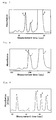
- Fig. 4 is an electropherogram obtained by the measurement of the healthy human blood sample in Example 1.
- the peak 1 represents stable HbA1c
- the peak 2 represents HbAo. The results show that stable HbA1c could be separated.
- the sample was injected into the capillary from one end, and subjected to electrophoresis by applying a voltage of 20 kV to the buffer solutions set for the ends of the capillary to measure transition of visible light absorbance at 415 nm.
- stable HbA1c in the human blood was measured by capillary electrophoresis.
- Fig. 5 is an electropherogram obtained by the measurement of the sample containing the modified Hb in Example 1.
- the peak 1 represents stable HbA1c
- the peak 2 represents HbAo
- the peak 3 represents the modified lib (unstable HbA1c).
- stable HbA1c and unstable HbA1c that is a form of modified Hbs were favorably separated.
- Glucose was added to the healthy human blood sample to give a concentration of 2000 mg/dL to prepare a sample containing unstable HbA1c as described in the process (4).
- Sodium cyanate was added to a healthy human blood sample collected from the same person to give a concentration of 50 mg/dL.
- a sample containing a carbamylated Hb was prepared. Both samples together with the healthy human blood sample were measured under the above-mentioned conditions.
- Table 1 shows values ( ⁇ HbA1c value) calculated by subtraction of the stable HbA1c value of the healthy human blood sample from the stable HbA1c values of each of the samples containing the modified Hb.
- Example 2 One healthy human blood sample was continuously measured 10 times under the conditions of Example 1, and a CV value of the stable HbA1c values determined by the measurement was calculated.
- the CV value was determined by dividing the standard deviation by the average value (standard deviation/average value). Table 2 shows the results.
- the inner surface of the capillary was coated by the same method as in Example 1, except that 0.5% by weight aqueous solution of polybrene (produced by Nacalai Tesque, Inc.) as the cationic polymer was used instead of the chitosan solution.
- the healthy human blood sample prepared in the process (3) of Example 1 and the sample containing the modified Hb prepared in the process (4) of Example 1 were measured using the same buffer solution containing the acrylic polymer as that used in Example 2.
- An electropherogram obtained by the measurement of the healthy human blood sample was similar to that shown in Fig. 4 .
- An electropherogram obtained by the measurement of the sample containing the modified Hb was similar to that shown in Fig. 5 .
- Example 3 a migration path with a cationic polymer immobilized thereon was used, and a buffer solution containing a water-soluble acrylic polymer having an anionic group was used as the electrophoresis buffer solution, like in Examples 1 and 2.
- a microdevice type electrophoresis apparatus including a measurement unit, a power supply unit, and a detection unit was assembled for measurement.
- a microdevice electrophoresis apparatus shown in Fig. 2 was assembled.
- a glass microdevice (outside dimensions: 50 mm ⁇ 75 mm ⁇ 3 mm) including platinum electrodes (1 mm in diameter ⁇ 5 mm in length) and a cross-shaped migration path (length: 50 mm, width: 80 ⁇ m) was used as the measurement unit.
- the electrophoresis apparatus was assembled as follows: inserting the electrodes into reservoirs on the microdevice; connecting the electrodes and the power supply unit (produced by LabSmith, multi-channel high-voltage sequencer electric power supply device, HVS448) via a voltage supply code; and setting the detection unit including a halogen lamp light source (produced by Moritex Corp., MHF-V501), a spectroscope (produced by B&W Tek, Inc., BTC112), and a computer for data processing on the microdevice.
- a halogen lamp light source produced by Moritex Corp., MHF-V501
- a spectroscope produced by B&W Tek, Inc., BTC112
- the migration path of the microdevice was coated by the same method as in Example 1, except that a 0.2N hydrochloric acid solution containing 0.2% by weight of chitosan (chitosan 100, produced by Wako Pure Chemical Industries, Ltd.) was used.
- the healthy human blood sample and the sample containing the modified Hb were measured using the same buffer solution containing the acrylic polymer as that used in Example 1 by microdevice electrophoresis at a voltage of 1000 V.
- Fig. 6 is an electropherogram obtained in Example 3 by the measurement of the healthy human blood sample prepared in the process (3) of Example 1.
- the peak 1 represents stable HbA1c
- the peak 2 represents HbAo
- Fig. 7 is an electropherogram obtained in Example 3 by the measurement of the sample containing the modified Hb prepared in the process (4) of Example 1.
- the peak 1 represents stable HbA1c
- the peak 2 represents HbAo
- the peak 3 represents the modified Hb (unstable HbA1c).
- stable HbA1c and unstable HbA1c that is a form of modified Hbs were favorably separated.
- the modified Hb separation performance test and the within-run reproducibility test were performed by the same methods as in the processes (5) and (6) of Example 1, respectively. Table 1 and Table 2 show the respective results.
- a migration path with a cationic polymer immobilized thereon was used like in Examples 1 and 2, but a buffer solution without the water-soluble acrylic polymer having an anionic group was used for measurement in Comparative Example 1.
- the healthy human blood sample prepared in the process (3) of Example 1 and the sample containing the modified Hb prepared in the process (4) of Example 1 were measured by the same method as in Example 1 except that a citrate buffer solution (pH 4.7) without acrylic polymers was used instead of the citrate buffer solution containing the acrylic polymer (prepared in Example 1). No peak was detected in electropherograms obtained by the measurement of both samples. Thus, the results show that hemoglobins could not be separated when using a buffer solution without the water-soluble polymer having an anionic group.
- a cationic polymer was used for coating like in Examples 1 and 2, but a dynamic coating technique was used as the coating method instead of the immobilized coating method in Comparative Example 2.
- a buffer solution containing a water-soluble non-acrylic polymer having an anionic group was used as the buffer solution for measurement.
- a capillary made of fused silica produced by GL Sciences, Inc., 25 ⁇ m in inside diameter ⁇ 30 cm in full length
- a 0.2N NaOH aqueous solution, ion exchange water, and a 0.5N HCl aqueous solution were allowed to flow through the capillary in this order.
- a malate buffer solution (pH 4.7) containing 0.5% by weight of horse serum albumin (cationic polymer) was allowed to flow through the capillary for 1 minute to dynamically coat the inside of the capillary with the buffer solution.
- a malate buffer solution pH 4.7
- chondroitin sulfate produced by Wako Pure Chemical Industries, Ltd., non-acrylic polymer
- Example 1 The sample containing the modified Hb prepared in the process (4) of Example 1 was measured by the same method as in Example 1.
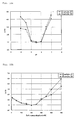
- Fig. 8 is an electropherogram obtained by the measurement of the sample containing the modified Hb in Comparative Example 2.
- the peak 1 represents stable HbA1c
- the peak 2 represents HbAo
- the peak 3 represents the modified Hb (unstable HbA1c).
- the peak 1 representing stable HbA1c overlaps the peak 3 representing the modified Hb in the obtained electropherogram.
- the modified Hb separation performance test and the within-run reproducibility test were performed by the same methods as in the processes (5) and (6) of Example 1, respectively.
- 0.2N NaOH and a malate buffer solution (pH 4.7) containing 0.2% by weight of chondroitin sulfate were sequentially allowed to flow through the capillary for 1 minute and 2 minutes, respectively, after each measurement to wash the capillary.
- the dynamic coating technique described in Comparative Example 2 was performed again for repetitive measurement.
- the wirhin-run reproducibility test was performed.
- Table 1 and Table 2 show the respective results.
- [Table 1] ⁇ HbA1c value(%) (Sample containing modified Hb - Healthy human sample) Sample containing unstable HbA1c Sample containing carbamylated Hb Example 1 0.0 0.1 Example 2 -0.1 0.1 Example 3 0.1 0.2 Comparative Example 2 0.4 -1.3
- Table 1 shows that the ⁇ HbA1c value obtained under the measurement conditions of Example 1 was remarkably small, that is, only a slight difference is found in the stable HbA1c values of the sample containing the modified Hb and the healthy human blood sample without the modified Hb. Accordingly, the results show that stable HbA1c can be accurately measured under the measurement conditions of Example 1 even under the presence of modified Hbs.
- the results of Examples 2 and 3 were also favorable like in Example 1. However, the ⁇ HbA1c value obtained under the measurement conditions of Comparative Example 2 was large. Accordingly, the results show that the measurement under the conditions of Comparative Example 2 is not accurate. [Table 2] Within-run reproducibility test CV(%) Example 1 0.93 Example 2 1.00 Example 3 1.05 Comparative Example 2 3.36
- Example 2 As shown in Table 2, the CV value indicating the data variation obtained by the within-run reproducibility test under the measurement conditions of Example 1 was as good as about 1%. The results of Examples 2 and 3 were also favorable like in Example 1. On the contrary, the CV value obtained by the measurement conditions of Comparative Example 2 was remarkably large and completely unsatisfactory for use in control of the HbA1c value of diabetics.
- a migration path with a hydrophilic compound having a molecular weight of 200 to 800 with a cationic group linked to the migration path by a covalent bond was used, and a buffer solution containing a water-soluble polymer having an anionic group was used as the electrophoresis buffer solution for measurement in each of reference Examples 4 to 6.
- a capillary made of fused silica (produced by GL Sciences, Inc., 25 ⁇ m in inside diameter ⁇ 30 cm in full length) was set in a capillary electrophoresis apparatus (produced by Beckman Coulter, Inc., PAC/E MDQ).
- a capillary electrophoresis apparatus produced by Beckman Coulter, Inc., PAC/E MDQ.
- a 0.2N NaOH solution, ion exchange water, and a 0.5N HCl solution were allowed to flow through the capillary in this order.
- an aqueous solution containing 1.0% by weight of 3-aminopropyltriethoxysilane hydrophilic compound, molecular weight: 221, produced by Shin-Etsu Silicones
- the capillary having the hydrophilic compound linked by a covalent bond to form the innermost surface of the capillary was prepared.
- a blood sample was collected from a healthy human with EDTA. To 70 ⁇ L of this healthy human whole blood sample was added 200 ⁇ L of a citrate buffer solution (pH 6.0) containing 0.01% by weight of Triton X-100 (produced by Wako Pure Chemical Industries, Ltd.) to hemolyze and dilute the blood sample. The sample thus obtained was used as a healthy human blood sample.
- a citrate buffer solution pH 6.0
- Triton X-100 produced by Wako Pure Chemical Industries, Ltd.
- the healthy human blood sample and the sample containing the modified Hb were measured using the capillary having the surface linked with the hydrophilic compound by a covalent bond.
- a 240 nM citrate buffer solution (pH 5.0) containing 2.0% by weight of chondroitin sulfate (anionic polymer, produced by Wako Pure Chemical Industries, Ltd.) was used as the electrophoresis buffer solution.
- the healthy human sample or the sample containing the modified Hb was injected into the capillary from one end, and subjected to electrophoresis at a voltage of 20 kV to measure absorbance at 415 nm.
- the electropherogram obtained by the measurement of the healthy human blood sample in reference Example 4 was similar to that shown in Fig. 4 .
- the stable HbA1c peak was favorably separated.
- the electropherogram obtained by the measurement of the sample containing the modified Hb in reference Example 4 was similar to that shown in Fig. 5 .
- Stable HbA1c was favorably separated from unstable HbA1c that is a form of modified Hbs.
- the modified Hb separation performance test and the within-run reproducibility test were performed using the samples prepared in the process (2) by the same methods as in the processes (5) and (6) of Example 1, respectively.
- a migration path with a hydrophilic compound having a molecular weight of 200 to 800 with a cationic group linked to the migration path by a covalent bond was used, and a buffer solution containing a water-soluble polymer having an anionic group was used as the electrophoresis buffer solution in reference Example 5 like in reference Example 4.
- a microdevice type electrophoresis apparatus including a measurement unit, a power supply unit and a detection unit similar to that of Example 3 was assembled for measurement.
- a double-T-shaped electrophoresis flow channel having a width of 90 ⁇ m was formed on a microdevice made of polydimethylsiloxane (outside dimensions: 50 mm ⁇ 75 mm ⁇ 3 mm).
- a 0.1N NaOH solution, ion exchange water, and a 0.2N HCl solution were allowed to flow through the formed flow channel in this order to wash the inside of the flow channel.
- an aqueous solution containing 1.0% by weight of 3-aminopropyltrimethoxysilane hydrophilic compound, molecular weight: 179, produced by Shin-Etsu Silicones
- the flow channel having the hydrophilic compound linked by a covalent bond to form the innermost surface of the flow channel was prepared.
- a healthy human blood sample and a sample containing the modified Hb were prepared by the same method as in reference Example 4.
- the healthy human sample and the sample containing the modified Hb were measured using the flow channel having the hydrophilic compound linked to the surface by a covalent bond.
- a 250 mM succinate buffer solution (pH 5.2) containing 2.0% by weight of chondroitin sulfate (anionic polymer, produced by Wako Pure Chemical Industries, Ltd.) was used as the electrophoresis buffer solution.
- the healthy human sample or the sample containing the modified Hb was injected into the flow channel from one end, and subjected to electrophoresis at a voltage of 0.8 kV to measure absorbance at 415 nm.
- the electropherogram obtained by the measurement of the healthy human blood sample was similar to that shown in Fig. 6 .
- the electropherogram obtained by the measurement of the sample containing the modified Hb was similar to that shown in Fig. 7 .
- a double-T-shaped electrophoresis flow channel having a width of 80 ⁇ m was formed on a microdevice made of a copolymer of methyl methacrylate and glycidyl methacrylate (outside dimensions: 50 mm ⁇ 75 mm ⁇ 3 mm).
- a 0.01N NaOH solution, ion exchange water, and a 0.01N HCl solution was allowed to flow through the formed flow channel in this order to wash the inside of the flow channel, and then a 20% by weight aqueous solution of ethylenediamine (hydrophilic compound, molecular weight: 60, produced by Wako Pure Chemical Industries, Ltd.) was allowed to flow through the flow channel for 5 minutes.
- a healthy human blood sample and a sample containing the modified Hb were prepared by the same method as in Example 4.
- the healthy human sample and the sample containing the modified Hb were measured using the flow channel having the surface linked with the hydrophilic compound by a covalent bond.
- a 300 mM malate buffer solution (pH 5.2) containing 1.7% by weight of a copolymer including 2-acrylamide-2-methylpropanesulfonic acid (produced by TOAGOSEI Co., Ltd.) was used as the electrophoresis buffer solution.
- the healthy human sample or the sample containing the modified Hb was injected into the flow channel from one end, and subjected to electrophoresis at a voltage of 1.0 kV to measure absorbance at 415 nm.
- the electropherogram obtained by the measurement of the healthy human blood sample was similar to that shown in Fig. 6 .
- the electropherogram obtained by the measurement of the sample containing the modified Hb was similar to that shown in Fig. 7 .
- a migration path with a hydrophilic compound having a cationic group linked thereto by a covalent bond was used for measurement of Comparative Example 3.
- the hydrophilic compound had a larger molecular weight than that of the hydrophilic compound used in the present invention (molecular weight: 200 to 800).
- a buffer solution containing a water-soluble polymer having an anionic group was used as the electrophoresis buffer solution for measurement.
- a flow channel with the hydrophilic compound linked by a covalent bond to form the innermost surface of the flow channel was prepared by following the same procedure as in reference Example 6, except that 0.1% by weight of polylysine (molecular weight: 1000 to 4000, produced by Wako Pure Chemical Industries, Ltd.) was used as the hydrophilic compound.
- the sample containing the modified Hb was measured under the same conditions as those of reference Example 6. Each peak in the obtained electropherogram was favorably separated from the others like in Fig. 7 .
- Table 3 shows that the ⁇ HbA1c values obtained under the respective measurement conditions of reference Examples 4 to 6 were remarkably small. Accordingly, the results show that stable HbA1c can be accurately measured under the conditions of references Examples 4 to 6 even under the presence of modified Hbs. However, the ⁇ HbA1c value obtained under the measurement conditions of Comparative Example 3 was slightly larger than those obtained in reference Examples 4 to 6. Accordingly, the results show that measurement under the conditions of Comparative Example 3 was susceptible to modified Hbs. [Table 4] Within-run reproducibility test CV(%) Reference Example 4 1.06 Reference Example 5 1.08 Reference Example 6 0.99 Comparative Example 3 4.22
- a migration path coated with a cationic polymer immobilized on an immobilized coating layer of a non-ionic hydrophilic polymer was used, and a buffer solution containing a water-soluble polymer having an anionic group was used as the electrophoresis buffer solution for measurement in each of reference Examples 7 to 9.
- a capillary made of fused silica (produced by GL Sciences, Inc., 75 ⁇ m in inside diameter ⁇ 50 cm in full length) was set in a capillary electrophoresis apparatus (produced by Beckman Coulter, Inc., PAC/E MDQ).
- a capillary electrophoresis apparatus produced by Beckman Coulter, Inc., PAC/E MDQ.
- 0.2N NaOH, ion exchange water, and 0.5N HCl were allowed to flow through the capillary in this order.
- a 1.0% by weight aqueous solution of polyvinyl alcohol (produced by Nihon Synthetic Chemical Industry Co. Ltd., GOHSENOL GH-20, non-ionic hydrophilic polymer) was allowed to flow through the capillary for 20 minutes.
- a blood sample was collected from a healthy human with heparin. To 70 ⁇ L of this healthy human whole blood sample was added 200 ⁇ L of a citrate buffer solution (pH 6.0) containing 0.01% by weight of Triton X-100 (produced by Wako Pure Chemical Industries, Ltd.) to hemolyze and dilute the blood sample. The sample thus obtained was used as a healthy human blood sample.
- a citrate buffer solution pH 6.0
- Triton X-100 produced by Wako Pure Chemical Industries, Ltd.
- the healthy human blood sample and the sample containing the modified Hb thus prepared were measured using the above-mentioned capillary.
- a 200 mM citrate buffer solution (pH 5.0) containing 2.0% by weight of chondroitin sulfate (produced by Wako Pure Chemical Industries, Ltd., water-soluble polymer having an anionic group) was used as the electrophoresis buffer solution. Either of the samples was injected into the capillary from one end, and subjected to electrophoresis at a voltage of 22 kV to measure absorbance at 415 nm.
- a double-T-shaped electrophoresis migration path having a width of 100 ⁇ m was formed on a microdevice made of fused silica (7 cm ⁇ 5 cm).
- 0.1N NaOH, ion exchange water, and 0.2N HCl were allowed to flow through the migration path in this order.
- a 1.0% by weight aqueous solution of polyvinylpyrrolidone (produced by NIPPON SHOKUBAI Co., Ltd., PVP-K-90W, non-ionic hydrophilic polymer) was allowed to flow through the migration path for 20 minutes.
- a healthy human blood sample and a sample containing the modified Hb were prepared by the same method as in reference Example 7.
- the healthy human sample and the sample containing the modified Hb thus prepared were measured using the above-mentioned migration path.
- a 250 mM succinate buffer solution (pH 5.2) containing 1.5% by weight of dextran sulfate (produced by Wako Pure Chemical Industries, Ltd., water-soluble polymer having an anionic group) was used as the electrophoresis buffer solution.
- Either of the samples was injected into the migration path from one end, and subjected to electrophoresis at a voltage of 0.8 kV to measure absorbance at 415 nm.
- the electropherogram obtained by the measurement of the healthy human blood was similar to that shown in Fig. 6 .
- the electropherogram obtained by the measurement of the sample containing the modified Hb is similar to that shown in Fig. 7 .
- a double-T-shaped electrophoresis migration path having a width of 80 ⁇ m was formed on a microdevice made of polydimethylsiloxane (7 cm ⁇ 5 cm).
- 0.1N NaOH, ion exchange water, and 0.2N HCl were allowed to flow through the migration path on the microdevice in this order.
- a polymer aqueous solution containing 1.0% by weight of a copolymer including 2-hydroxyethyl methacrylate produced by Wako Pure Chemical Industries, Ltd., non-ionic hydrophilic polymer
- a healthy human blood sample and a sample containing the modified Hb were prepared by the same method as in reference Example 7.
- the prepared healthy human sample and the sample containing the modified Hb thus prepared were measured using the above-mentioned migration path.
- a 300 mM malate buffer solution (pH 5.2) containing 1.7% by weight of a 2-acrylamide-2-methylpropanesulfonic acid copolymer (produced by TOAGOSEI Co., Ltd., water-soluble polymer having an anionic group) was used as the electrophoresis buffer solution.
- Either of the samples was injected into the migration path from one end, and subjected to electrophoresis at a voltage of 1.0 kV to measure absorbance at 415 nm.
- the electropherogram obtained by the measurement of the healthy human blood sample was similar to that shown in Fig. 6 .
- the electropherogram obtained by the measurement of the sample containing the modified Hb is similar to that shown in Fig. 7 .
- Comparative Example 4 the non-ionic hydrophilic polymer and the cationic polymer used in reference Examples 7 to 9 were used for coating but in the reverse order. Namely, an inner surface of a migration path was coated with the cationic polymer, and then coated with the non-ionic polymer.
- a capillary made of fused silica (produced by GL Sciences, Inc., 75 ⁇ m in inside diameter ⁇ 50 cm in full length) was set in a capillary electrophoresis apparatus (produced by Beckman Coulter, Inc., PAC/E MDQ).
- a capillary electrophoresis apparatus produced by Beckman Coulter, Inc., PAC/E MDQ.
- 0.2N NaOH, ion exchange water, and 0.5N HCl were allowed to flow through the capillary in this order.
- a 5.0% by weight aqueous solution of polybrene (produced by Wako Pure Chemical Industries, Ltd., cationic polymer) was allowed to flow through the capillary for 20 minutes.
- a 1.0% by weight aqueous solution of polyvinyl alcohol (produced by Nihon Synthetic Chemical Industry Co. Ltd., GOHSENOL GH-20, non-ionic hydrophilic polymer) was allowed to flow through the prepared capillary for 20 minutes by the same method as that described above. Subsequently, air was injected into the capillary to send out the polyvinyl alcohol aqueous solution, and then the capillary was dried in a drier at a temperature of 40°C for 12 hours. Thereafter, the procedure of injection of the polyvinyl alcohol aqueous solution, injection of air and drying was repeated 5 times to coat the cationic polymer (polybrene) coating the inner surface of the capillary with the non-ionic hydrophilic polymer.
- polyvinyl alcohol produced by Nihon Synthetic Chemical Industry Co. Ltd., GOHSENOL GH-20, non-ionic hydrophilic polymer
- a healthy human sample and a sample containing the modified Hb were prepared by the same method as in reference Example 7.
- electrophoresis buffer solution The same electrophoresis buffer solution as that used in reference Example 7 was used as the electrophoresis buffer solution. Either of the samples was injected into the capillary from one end, and subjected to electrophoresis at a voltage of 20 kV to measure absorbance at 415 nm. No hemoglobin peak was found in either of the results of the healthy human blood sample and the sample containing the modified Hb. Accordingly, the results show that without the cationic innermost surface, hemoglobin cannot be measured.
- Table 5 shows that the ⁇ HbA1c values obtained under the respective measurement conditions of reference Examples 7 and 8 were remarkably small. Accordingly, the results show that stable HbA1c can be accurately measured under the measurement conditions of reference Examples 7 and 8 even under the presence of modified Hbs.
- [Table 6] Within-run reproducibility test CV(%) Reference Example 7 1.02 Reference Example 8 0.99 Reference Example 9 1.05
- a migration path with a cationic polymer immobilized thereon filled with an ion exchanger was used, and a buffer solution containing a water-soluble acrylic polymer having an anionic group was used as the electrophoresis buffer solution for measurement in reference Example 10.
- a cross-shaped open channel having a size of 100 ⁇ m in width ⁇ 70 mm in length was formed on an electrophoresis microdevice made of polydimethylsiloxane (50 mm ⁇ 100 mm).
- the open channel was used as the migration path.
- the formed migration path was coated using a chitosan solution by the same method as in Example 2, and filled with the ion exchanger having an amino group prepared in the ion exchanger preparation process (1).
- the healthy human blood sample and the sample containing the modified Hb were measured by the same method as in reference Example 8, except that a citrate buffer solution (pH 4.7) containing 2.0% by weight of chondroitin sulfate was used as the electrophoresis buffer solution, and that the samples were subjected to electrophoresis at a voltage of 1000 V.
- a citrate buffer solution pH 4.7
- the electropherogram obtained by the measurement of the healthy human blood sample was similar to that shown in Fig. 4 .
- the electropherogram obtained by the measurement of the sample containing the modified Hb was similar to that shown in Fig. 5 .
- a migration path with an ozonized inner surface was used for measurement in each of Examples 11 to 13.
- a capillary made of fused silica (produced by GL Sciences, Inc., 25 ⁇ m in inside diameter ⁇ 30 cm in full length) was set in a capillary electrophoresis apparatus (PAC/E MDQ, produced by Beckman Coulter, Inc.).
- PAC/E MDQ capillary electrophoresis apparatus
- a 0.2N NaOH aqueous solution, ion exchange water, and a 0.5N HCl aqueous solution were allowed to flow through the capillary in this order.
- a 0.2N hydrochloric acid solution containing 0.5% by weight of chitosan was allowed to flow through the capillary for 20 minutes.
- the prepared capillary was filled with ozone water having a dissolved ozone gas concentration of 100 ppm, and allowed to stand. After 10 minutes standing, the ozone water was sucked up, and the same ozone water was injected again. Thereafter, the capillary was allowed to stand for 10 minutes. Thus, the capillary having the migration path ozonized with ozone water was prepared.
- the prepared capillary was filled with a 150 mM citrate buffer solution (pH 5.2) containing 2.0% by weight of chondroitin sulfate.
- a blood sample was collected from a healthy human with heparin.
- To 100 ⁇ L of the healthy human whole blood sample was added 150 ⁇ L of a citrate buffer solution (pH 6.0) containing 0.05% by weight of Triton X-100 (surfactant, produced by Wako Pure Chemical Industries, Ltd.) to hemolyze and dilute the blood sample.
- the diluted blood sample thus obtained was used as a diluted healthy human blood sample.
- the diluted blood sample was injected into the capillary ozonized with ozone water from one end, and subjected to electrophoresis by applying a voltage of 25 kV to both ends of the capillary to measure transition of visible light absorbance at 415 nm.
- a voltage of 25 kV to both ends of the capillary to measure transition of visible light absorbance at 415 nm.
- the electropherogram obtained by the measurement of the healthy human blood sample was similar to that shown in Fig. 6 .
- glucose To the healthy human whole blood sample used in the measurement of the healthy human blood was added glucose to give a concentration of 2500 mg/dL, and warmed at a temperature of 37°C for 3 hours to artificially prepare a sample containing a large amount of unstable HbA1c that is a form of modified Hbs.
- the sample containing the modified Hb was measured by the same method as in the measurement of the healthy human blood sample.
- the electropherogram obtained by the measurement of the sample containing the modified Hb was similar to that shown in Fig. 7 .
- a migration path having a length of 75 mm and a width of 60 ⁇ m was formed on a microdevice made of polydimethylsiloxane.
- the migration path was filled with a 0.5% by weight aqueous solution of polybrene, and allowed to stand for 20 minutes. Subsequently, air was injected into the migration path to send out the polybrene aqueous solution, and then the migration path was dried in a drier at a temperature of 40°C for 12 hours. Thereafter, the procedure of injection of the polybrene aqueous solution, injection of air and drying was repeated 5 times. Thus, the electrophoresis microdevice having the coated migration path was obtained.
- the electrophoresis microdevice having the migration path thus coated was ozonized with ozone water by the same method as in Example 11.
- the electrophoresis microdevice ozonized with ozone water was prepared.
- the migration path thus ozonized with ozone water of the electrophoresis microdevice was filled with a 50 mM succinate buffer solution (pH 5.8) containing 1.5% by weight of dextran sulfate.
- the samples were measured by following the same procedure as in the healthy human blood measurement process (2) and the sample containing the modified Hb measurement process (3) of Example 11, respectively.
- the samples were subjected to electrophoresis using the electrophoresis microdevice thus ozonized with ozone water by applying a voltage of 500 V to both ends of the migration path to measure transition of visible light absorbance at 415 nm.
- the electropherogram obtained by the measurement of the healthy human blood sample was similar to that shown in Fig. 6 .
- the electropherogram obtained by the measurement of the sample containing the modified Hb was similar to that shown in Fig. 7 .
- An all-peak area value in the electropherogram obtained by the measurement of the healthy human blood sample under the measurement conditions of Example 12 was calculated.
- a relative value of the all-peak area value of Example 12 with respect to an all-peak area value of Example 11 as 100 was calculated.
- a migration path having a length of 90 mm and a width of 50 ⁇ m was formed on a microdevice made of fused silica.
- the electrophoresis microdevice was prepared.
- the migration path was filled with a 0.2N hydrochloric acid solution containing 0.5% by weight of chitosan, and allowed to stand for 20 minutes. Thereafter, air was injected into the migration path to send out the chitosan solution, and then the migration path was dried in a drier at a temperature of 40°C for 12 hours. Subsequently, the procedure of injection of the chitosan solution, injection of air, and drying were repeated 5 times.
- the electrophoresis microdevice having the coated migration path was prepared.
- the migration path thus coated of the electrophoresis microdevice was further ozonized with ozone water by the same method as in Example 1.
- the microdevice ozonized with ozone water was prepared.
- the migration path of the electrophoresis microdevice thus ozonized with ozone water was filled with a 100 mM phosphate buffer solution (pH 5.5) containing 2.0% by weight of chondroitin sulfate.
- the healthy human blood sample and the sample containing the modified Hb were measured by the same method as in Example 2, except that the samples were subjected to electrophoresis using the electrophoresis microdevice ozonized with ozone water by applying a voltage of 700 V to both ends of the migration path.
- the electropherogram obtained by the measurement of the healthy human blood sample was similar to that shown in Fig. 6 .
- the electropherogram obtained by the measurement of the sample containing the modified Hb was similar to that shown in Fig. 7 .
- the migration path was formed by the same method as in Examples 11 to 13, except that ozonization with ozone water was not performed, and a sample containing the modified Hb was measured.
- a migration path coated with a cationic polymer immobilized thereon was used, and a buffer solution containing a water-soluble polymer having an anionic group and a chaotropic compound were used as the buffer solution for measurement in each of Examples 14 to 16.
- a hydrochloric acid solution containing 0.5% by weight of chitosan (chitosan-100, produced by Wako Pure Chemical Industries, Ltd.) as a cationic polymer was prepared.
- chitosan-100 produced by Wako Pure Chemical Industries, Ltd.
- 0.2N-NaOH, ion exchange water, and 0.5N-HCl were allowed to flow through a capillary made of fused silica (produced by GL Sciences, Inc., 25 ⁇ m in inside diameter ⁇ 30 cm in full length) in this order to wash the inside of the capillary, and then the prepared chitosan solution was allowed to flow through the capillary for 20 minutes.
- the chitosan immobilized capillary thus prepared was set in a capillary electrophoresis apparatus (PAC/E MDQ, produced by Beckman Coulter, Inc.).
- a malate buffer solution (pH 4.8) containing 1.0% by weight of sodium nitrate as the chaotropic compound and 2.0% by weight of chondroitin sulfate was the water-soluble polymer having an anionic group was prepared as the buffer solution.
- a blood sample was collected from a healthy human with heparin.
- a healthy human whole blood sample was added 200 ⁇ L of a citrate buffer solution (pH 6.0) containing 0.05% Triton X-100 (surfactant, produced by Wako Pure Chemical Industries, Ltd.) to hemolyze and dilute the blood sample.
- the sample thus obtained was used as a measurement sample.
- the sample was injected into the capillary from one end, and subjected to electrophoresis by applying a voltage of 20 kV to the buffer solutions set for the ends of the capillary to measure transition of visible light absorbance at 415 nm.
- stable HbA1c in the human blood was measured by capillary electrophoresis.
- a sample containing a large amount of unstable HbA1c that is a form of modified Hbs was artificially prepared.
- the sample was injected into the capillary from one end, and subjected to electrophoresis by applying a voltage of 20 kV to the buffer solutions set for the ends of the capillary to measure transition of visible light absorbance at 415 nm.
- stable HbA1c in the human blood was measured by capillary electrophoresis.
- the electropherogram obtained by the measurement of the healthy human blood sample was similar to that shown in Fig. 6 .
- the electropherogram obtained by the measurement of the sample containing the modified Hb was similar to that shown in Fig. 7 .
- An inner surface of a capillary was coated by the same method as in Example 1, except that a 0.5% by weight aqueous solution of polybrene (produced by Wako Pure Chemical Industries, Ltd.) as the cationic polymer was used. Thus, an electrophoresis apparatus was prepared.
- the whole mixture obtained by 10 hour polymerization was poured into a dialysis tube (produced by Sanko Junyaku Co., Ltd., UC C65-50), and dialyzed in ion exchange water for 12 hours to obtain an water-soluble acrylic polymer having an anionic group.
- a malate buffer solution (pH 4.7) containing 1.0 % by weight of sodium perchlorate as the chaotropic compound and 2.0 % by weight of the water-soluble polymer having an anionic group was prepared.
- the healthy human blood sample and the sample containing the modified Hb were measured by the same method as in Example 1, except that the electrophoresis apparatus and the buffer solution prepared above were used.
- the electropherogram obtained by the measurement of the healthy human blood sample was similar to that shown in Fig. 6 .
- the electropherogram obtained by the measurement of the sample containing the modified Hb was similar to that shown in Fig. 7 .
- a cross-shaped migration path was formed on a microdevice made of polydimethylsiloxane (50 mm ⁇ 75 mm ⁇ 3 mm), and a buffer solution reservoir was set for each end of the migration path.
- the migration path was coated by the same method as in Example 1, except that chitosan (0.5% by weight, in a 0.5N hydrochloric acid solution) as the ionic polymer was used.
- the healthy human blood sample and the sample containing the modified Hb were measured by the same method as in Example 1, except that a malate buffer solution (pH 4.8) containing 5.0% by weight of urea as the chaotropic compound and 2.0% by weight of chondroitin sulfate as the water-soluble polymer having an anionic group was used, and the samples were subjected to microdevice electrophoresis at a voltage of 1000 V.
- a malate buffer solution pH 4.8
- the electropherogram obtained by the measurement of the healthy human blood sample was similar to that shown in Fig. 6 .
- the electropherogram obtained by the measurement of the sample containing the modified Hb was similar to that shown in Fig. 7 .
- the healthy human blood sample and the sample containing the modified Hb were measured by the same method as in Example 14, except that no chaotropic compound was used.
- a migration path coated with a cationic polymer immobilized thereon was used, and a buffer solution containing a water-soluble polymer having an anionic group and a nitrite was used as the buffer solution for measurement in each of Examples 17 to 20.
- a 0.2N hydrochloric acid solution containing 0.2% by weight of chitosan (produced by Wako Pure Chemical Industries, Ltd., chitosan 100) as the anionic polymer was prepared.
- chitosan produced by Wako Pure Chemical Industries, Ltd., chitosan 100
- 0.2N-NaOH, ion exchange water, and 0.5N-HCl were allowed to flow through a capillary made of fused silica (produced by GL Sciences, Inc., 25 ⁇ m in inside diameter ⁇ 30 cm in full length) in this order to wash the inside of the capillary, and then the prepared chitosan solution was allowed to flow through the capillary for 20 minutes.
- the chitosan immobilized capillary thus prepared was set in a capillary electrophoresis apparatus (produced by Beckman Coulter, Inc., PAC/E MDQ). Subsequently, a citrate buffer solution (pH 4.8) containing 50 mM sodium nitrite (nitrite, produced by Wako Pure Chemical Industries, Ltd.) and 2.0% by weight of chondroitin sulfate (produced by Wako Pure Chemical Industries, Ltd.) was used as the electrophoresis buffer solution, and set for both ends of the capillary to fill the capillary with the buffer solution.
- a citrate buffer solution pH 4.8
- 50 mM sodium nitrite nitrite, produced by Wako Pure Chemical Industries, Ltd.
- chondroitin sulfate produced by Wako Pure Chemical Industries, Ltd.
- a blood sample was collected from a healthy human with sodium fluoride. To 70 ⁇ L of the healthy human whole blood sample was added 200 ⁇ L of a citrate buffer solution (pH 6.0) containing 0.05% by weight of Triton X-100 and 20 mM sodium nitrite at to hemolyze and dilute the blood sample. The sample thus obtained was used as a measurement sample.
- a citrate buffer solution pH 6.0
- the sample was injected into the capillary from one end, and subjected to electrophoresis by applying a voltage of 20 kV to the buffer solutions set for the ends of the capillary to measure transition of visible light absorbance at 415 nm.
- stable HbA1c in the human blood was measured by capillary electrophoresis.
- the electropherogram obtained by the measurement of the healthy human blood sample was similar to that shown in Fig. 6 .
- glucose produced by Wako Pure Chemical Industries, Ltd.
- a concentration of 2000 mg/dL was added to the healthy human whole blood sample used in the measurement of the healthy human blood sample.
- warmed at a temperature of 37°C for 3 hours was added to artificially prepare a sample containing a large amount of unstable HbA1c that is a form of modified Hbs.
- the electropherogram obtained by the measurement of the sample containing the modified Hb was similar to that shown in Fig. 7 .
- a healthy human blood sample and a sample containing the modified Hb were measured by capillary electrophoresis by following the same procedure as in Example 17, except that an inner surface of a capillary was coated using a 0.5% by weight aqueous solution of polybrene (produced by Nacalai Tesque, Inc.) as the cationic polymer by the same method as in Example 17, and that a malate buffer solution (pH 4.8) containing 30 mM sodium nitrite and 2.0 % by weight of chondroitin sulfate was used as the electrophoresis buffer solution.
- polybrene produced by Nacalai Tesque, Inc.
- a malate buffer solution pH 4.8
- Table 10 shows the results of the between-run reproducibility test performed by following the same procedure as in Example 17.
- a cross-shaped migration path (flow channel width: 100 ⁇ m) was formed on a microdevice made of glass (50 mm in width ⁇ 30 mm in length ⁇ 2 mm in thickness), and a buffer solution reservoir was set for each end of the migration path.
- the migration path was coated with chitosan (1.0% by weight aqueous solution) that is the cationic polymer by the same method as in Example 17. Then, the healthy human blood sample and the sample containing the modified Hb were measured by microdevice electrophoresis at a voltage of 1000 V using the same buffer solution as that used in Example 17.
- Table 10 shows the results of the between-run reproducibility test performed by following the same procedure as in Example 17.
- a double-T-shaped migration path (flow channel width: 80 ⁇ m) was formed on a microdevice made of polydimethylsiloxane 50 mm in width ⁇ 30 mm in length ⁇ 2 mm in thickness), and a buffer solution reservoir was set for each end of the migration path.
- the migration path was coated with polybrene (1.0% by weight aqueous solution) that is the cationic polymer by the same method as in Example 17. Subsequently, the healthy human blood sample and the sample containing the modified Hb were measured by microdevice electrophoresis at a voltage of 900 V using the same electrophoresis buffer solution as that used in Example 12.
- Table 10 shows the results of the between-run reproducibility test performed by following the same procedure as in Example 17.
- Table 10 shows the results of the between-run reproducibility test performed by following the same procedure as in Example 17.
- Example 17 4.4 4.5 4.5 4.6 4.5 4.5 0.07 1.6
- Example 18 4.5 4.5 4.5 4.4 4.5 4.5 0.04 1.0
- Example 19 4.6 4.5 4.5 4.6 4.5 4.5 0.05 1.2
- a migration path made of a cationic material was used, and a buffer solution containing a water-soluble polymer having an anionic group was used as the electrophoresis buffer solution for measurement in each of Examples 21 to 23.
- a microdevice (50 mm ⁇ 80 mm) was formed by using a terpolymer (diethylaminoethyl methacrylate (monomer having a cationic group))-(2-hydroxyethyl methacrylate)-(methyl methacrylate).
- a flow channel (100 ⁇ m in inside diameter ⁇ 50 mm) was formed on the obtained microdevice.
- a citrate buffer solution (pH 5.0) containing 2.0% by weight of chondroitin sulfate (anionic polymer) was injected into the flow channel as the buffer solution.
- a blood sample was collected from a healthy human with sodium fluoride. To 70 ⁇ L of the healthy human whole blood sample was added 200 ⁇ L of a citrate buffer solution (pH 6.0) containing 0.5% by weight of saponin to hemolyze and dilute the blood sample. The sample thus obtained (healthy human sample) was used.
- a citrate buffer solution pH 6.0
- the obtained healthy human sample was injected into the flow channel, and subjected to electrophoresis by applying a voltage of 800 V to both ends of the flow channel. A portion on the migration path was irradiated with visible light at 415 nm, and transition of absorbance of the transmitted light was measured. The obtained electropherogram was similar to that shown in Fig. 6 .
- the L-A1c sample and the CHb sample were measured, and the obtained electropherograms were similar to that shown in Fig. 7 .
- the healthy human sample, the L-A1c sample and the CHb sample were measured by following the same procedure as in Example 1, except that a microdevice was prepared using acrylamide as the monomer having a cationic group, and a phosphate buffer solution (pH 5.4) containing 1.5% by weight of dextran sulfate was used as a buffer solution.
- a phosphate buffer solution pH 5.4
- the obtained electropherograms were similar to those shown in Figs. 6 and 7 .
- the healthy human sample, the L-A1c sample and the CHb sample were measured by following the same procedure as in Example 1, except that a microdevice was prepared using a terpolymer (ethylene imine (monomer having a cationic group)) - (ethylene vinyl alcohol) - (methyl methacrylate), and a malate buffer solution (pH 4.9) containing 1.0% by weight of a copolymer (2-(meth)acrylamide-2-methylpropanesulfonic acid)-(2-hydroxyethyl methacrylate) was used as the buffer solution.
- a terpolymer ethylene imine (monomer having a cationic group)
- ethylene vinyl alcohol ethylene vinyl alcohol
- methyl methacrylate methyl methacrylate
- a malate buffer solution pH 4.9
- a microdevice having a migration path coated with a cationic polymer immobilized thereon or a migration path made of a cationic material was used, and a buffer solution containing a water-soluble polymer having an anionic group was used as the electrophoresis buffer solution for measurement in each of Examples 24 to 26.
- the migration path of the used microdevice had been filled with the buffer solution in advance. Abnormal hemoglobins were also measured using the device.
- a measurement system of hemoglobin was assembled based on the schematic view shown in Fig. 2 .
- Absorbance at 415 nm was measured using a multichannel high-voltage sequencer power supply device (produced by LabSmith, HVS448), a 50 W halogen light source device (produced by Moritex Corp., MHF-V501) and a spectroscope (produced by B&W Tek, Inc., CCD spectroscope, BTC112) as the power supply device and the light source and the detector, respectively.
- the obtained data of the absorbance at 415 nm was processed according to a self-made program.
- a device supporting unit made of Teflon (registered trademark) designed to have a size fit for a microdevice described below was produced. Platinum electrodes (1 mm in diameter ⁇ 20 mm in length) were attached to the obtained device supporting unit. The platinum electrodes were connected to the power supply device via a power cable. All other parts were self-made.
- cross-shaped electrophoresis microdevice made of fused silica (outside dimensions: 50 mm ⁇ 80 mm) was prepared.
- a linear migration path having a size of 100 ⁇ m in inside diameter ⁇ 50 mm in length was formed on the microdevice.
- a 0.5N sodium hydroxide aqueous solution, distilled water, a 0.5N hydrochloric acid aqueous solution, and distilled water were sequentially allowed to flow through a flow channel on the microdevice to wash the inside of the flow channel. Subsequently, the flow channel was filled with a 0.5N hydrochloric acid solution containing 0.5% by weight of chitosan (chitosan 100, cationic polymer, produced by Wako Pure Chemical Industries, Ltd.). The flow channel was allowed to stand at room temperature for 30 minutes, and then air was injected into the flow channel to send out the chitosan solution from the flow channel. Thereafter, the flow channel was warmed at a temperature of 40°C for 5 hours.
- chitosan chitosan 100, cationic polymer, produced by Wako Pure Chemical Industries, Ltd.
- the flow channel including the migration path thus coated with the cationic polymer of the microdevice was filled with a citrate buffer solution (pH 5.0) containing 2.0% by weight of chondroitin sulfate (anionic polymer, produced by Nacalai Tesque, Inc.).
- a citrate buffer solution pH 5.0
- chondroitin sulfate anionic polymer, produced by Nacalai Tesque, Inc.
- a blood sample collected from a healthy human with sodium fluoride was used as a healthy human whole blood sample.
- To 70 ⁇ L of the healthy human whole blood sample was added 200 ⁇ L of a citrate buffer solution (pH 6.0) containing 0.5% by weight of saponin (produced by Wako Pure Chemical Industries, Ltd.) to hemolyze and dilute the healthy human whole blood sample.
- the sample thus prepared was used as a healthy human sample.
- the prepared healthy human sample was injected into the obtained flow channel of the microdevice, and subjected to electrophoresis by applying a voltage of 800 V to both ends of the flow channel. A portion on the migration path was irradiated with visible light at 415 nm, and transition of absorbance of the transmitted light was measured. The obtained electropherogram was similar to that shown in Fig. 6 .
- the L-A1c sample and the CHb sample thus prepared were measured by electrophoresis performed by following the same procedure as in the measurement of the healthy human sample, and the obtained electropherograms were similar to that shown in Fig. 7 .
- a double-T shaped electrophoresis microdevice made of polydimethylsiloxane (outside dimensions: 50 mm ⁇ 80 mm) was prepared.
- a linear migration path having a size of 100 ⁇ m in inside diameter ⁇ 50 mm in length was formed on the microdevice.
- a 0.1N sodium hydroxide aqueous solution, distilled water, a 0.1N hydrochloric acid aqueous solution, and distilled water were sequentially allowed to flow through a flow channel on the obtained microdevice to wash the inside of the flow channel.
- the flow channel was filled with a 0.5% by weight aqueous solution of polyvinyl alcohol (GOHSENOL GH20, produced by Nihon Synthetic Chemical Industry Co. Ltd.).
- the flow channel was allowed to stand at room temperature for 30 minutes, and then air was injected into the flow channel to send out the polyvinyl alcohol aqueous solution from the flow channel. Thereafter, the flow channel was warmed at a temperature of 40°C for 5 hours.
- the flow channel was filled with 4% by weight aqueous solution of polybrene (cationic polymer, produced by SIGMA-ALDRICH Japan K.K.).
- polybrene cationic polymer, produced by SIGMA-ALDRICH Japan K.K.
- the flow channel was allowed to stand at room temperature for 30 minutes, and then air was injected into the flow channel to send out the polybrene aqueous solution from the flow channel. Thereafter, the flow channel was warmed at a temperature of 40°C for 5 hours. The procedure of injection of the polybrene aqueous solution and warming was repeated 3 times for coating with the cationic polymer.
- the obtained flow channel on the microdevice was filled with a phosphate buffer solution (pH 5.4) containing 1.5% by weight of dextran sulfate (produced by Wako Pure Chemical Industries, Ltd.).
- a phosphate buffer solution pH 5.4
- dextran sulfate produced by Wako Pure Chemical Industries, Ltd.
- the healthy human sample, the L-A1c sample, and the CHb sample were measured by following the same procedure as in Example 24, except that a voltage or 900 V was applied.
- the electropherogram obtained by the measurement of the healthy human sample was similar to that shown in Fig. 6 .
- the electropherograms obtained by the measurement of the L-A1c sample and the CHb sample were similar to that shown in Fig. 7 .
- a cross-shaped electrophoresis microdevice (outside dimensions: 50 mm ⁇ 80 mm) was produced using a terpolymer (diethylaminoethyl methacrylate (monomer having a cationic group))-(2-hydroxyethyl methacrylate)-(methyl methacrylate).
- a linear migration path with a size of 100 ⁇ m in inside diameter ⁇ 50 mm in length was formed on the microdevice.
- the obtained flow channel of the microdevice was filled with a malate buffer solution (pH 5.0) containing 1.5% by weight of chondroitin sulfate (produced by Wako Pure Chemical Industries, Ltd.).
- a malate buffer solution pH 5.0
- chondroitin sulfate produced by Wako Pure Chemical Industries, Ltd.
- the healthy human sample, the L-A1c sample, and the CHb sample were measured by following the same procedure as in Example 24, except that a voltage of 900 V was applied.
- the electropherogram obtained by the measurement of the healthy human sample was similar to that shown in Fig. 6 .
- the electropherograms obtained by the measurement of the L-A1c sample and the CHb sample were similar to that shown in Fig. 7 .
- a cross-shaped electrophoresis microdevice made of fused silica (outside dimensions: 50 mm ⁇ 80 mm) was produced.
- a linear migration path having a size of 100 ⁇ m in inside diameter ⁇ 50 mm in length was formed on the microdevice.
- a 0.5N sodium hydroxide aqueous solution, distilled water, a 0.5N hydrochloric acid aqueous solution, and distilled water were sequentially allowed to flow through a flow channel on the obtained microdevice to wash the inside of the flow channel.
- a 0.5N hydrochloric acid solution containing 0.5% by weight of chitosan (chitosan 100, cationic polymer, produced by Wako Pure Chemical Industries, Ltd.) was allowed to flow through the flow channel for 2 minutes to dynamically coat the flow channel.
- the flow channel including the migration path thus coated with the cationic polymer of the microdevice was filled with a citrate buffer solution (pH 5.0) containing 2.0% by weight of chondroitin sulfate (anionic polymer, produced by Nacalai Tesque, Inc.).
- a citrate buffer solution pH 5.0
- chondroitin sulfate anionic polymer, produced by Nacalai Tesque, Inc.
- the healthy human sample, the L-Alc sample, and the CHb sample were measured by following the same procedure as in Example 24.
- the electropherogram obtained by the measurement of the healthy human sample was similar to that shown in Fig. 6 .
- the electropherograms obtained by the measurement of the L-Alc sample and the CHb sample were similar to that shown in Fig. 7 .
- FIG. 9 shows the obtained electropherogram.
- the peak 1 represents stable HbA1c
- the peak 2 represents HbAo
- the peak 3 represents unstable HbA1c
- the peak 4 represents HbF (fetal Hb)
- the peak 5 represents HbS
- the peak 6 represents HbC.
- stable HbA1c, HbS and HbC were favorably separated.
- the electropherograms obtained in Examples 25 and 26 and Comparative Example 13 were similar to that shown in Fig. 9 .
- FIG. 10 shows the obtained electropherogram.
- the peak 1 represents stable HbA1c
- the peak 2 represents HbAo
- the peak 4 represents HbF (fetal Hb)
- the peak 7 represents HbA2.
- stable HbA1c and HbA2 were favorably separated.
- the electropherograms obtained in Examples 25 and 26 and Comparative Example 13 were similar to that shown in Fig. 10 .
- Fig. 11 is a graph showing the relationship between the obtained stable HbA1c values (%) and the storage period.
- the microdevices produced in Examples 24 to 26 showed measurement performance equivalent to that shown immediately after the preparation.
- the microdevice of Comparative Example 13 showed poor stability, and could be used for measurement only immediately after coated. Accordingly, the results show that the migration path pre-filled with the buffer solution in the measurement system of hemoglobin can avoid the coating procedure when used, and stably stored for a certain period of time after filled with the buffer solution.
- the results also show that the microdevice of Comparative Example 13 showed poor storage stability, and cannot be used as the microdevice having the migration path pre-filled with the buffer solution.
- a microdevice having a migration path coated with a cationic polymer immobilized thereon was used, and a buffer solution containing a water-soluble polymer having an anionic group was used as the electrophoresis buffer solution to analyze effects of the pH and the salt concentration of the buffer solution in each of Examples 27 and 28.
- a migration path having a length of 80 mm and a width of 80 ⁇ m was formed on a cross-shaped device made of polydimethylsiloxane.
- the electrophoresis microdevice was prepared.
- the migration path was filled with a 0.5% chitosan solution (cationic polymer), and left to stand for 20 minutes. Subsequently, air was injected into the migration path to send out the chitosan solution, and then the migration path was dried in a drier at a temperature of 40°C for 12 hours. Thereafter, the sequential procedure of injection of the chitosan solution, injection of air and drying was repeated 5 times.
- the prepared migration path of the chitosan coated microdevice was filled with a 150 mM citrate buffer solution (pH 5.2) containing 2.0% chondroitin sulfate. Thus, the microdevice for measurement was prepared.
- a blood sample was collected from a healthy human with heparin.
- a citrate buffer solution pH 6.0
- Triton X-100 surfactant, produced by Wako Pure Chemical Industries, Ltd.
- the hemolyzed sample thus prepared was injected into one end of the migration path of the microdevice, and subjected to electrophoresis by applying a voltage of 1000 V to both ends of the migration path to measure transition of visible light absorbance at 415 nm.
- stable HbA1c was measured.
- the obtained electropherogram was similar to that shown in Fig. 6 .
- the hemolyzed sample thus prepared was injected into one end of the prepared migration path of the microdevice, and subjected to electrophoresis by applying a voltage of 1000 V to both ends of the migration path to measure transition of visible light absorbance at 415 nm.
- stable HbA1c was measured.
- the obtained electropherogram was similar to that shown in Fig. 7 .
- a migration path having a length of 50 mm and a width of 50 ⁇ m was formed on a device made of fused silica.
- the cross-shaped electrophoresis microdevice was prepared.
- the migration path was filled with a 0.5% polybrene aqueous solution, and allowed to stand for 20 minutes. Subsequently, air was injected into the migration path to send out the polybrene aqueous solution, and then the migration path was dried in a drier at a temperature of 40°C for 12 hours. Thereafter, the procedure of injection of the polybrene aqueous solution, injection of air and drying was repeated 5 times.
- the migration path of the polybrene coated microdevice was filled with a 50 mM succinate buffer solution (pH 5.8) containing 1.5% dextran sulfate. Thus, a microdevice for measurement was prepared.
- the healthy human blood sample and the sample containing the modified Hb were measured by the same method as in Example 27, except that a voltage of 500 V was applied to both ends of the migration path for electrophoresis.
- the electropherogram obtained by the measurement of the healthy human blood sample was similar to that shown in Fig. 6 .
- the electropherogram obtained by the measurement of the sample containing the modified Hb was similar to that shown in Fig. 7 .
- Fig. 12a shows the results obtained by changing the pHs
- Fig. 12b shows the results obtained by changing the salt concentrations.
- a microdevice electrophoresis apparatus provided with a microdevice having a migration path coated with a cationic polymer immobilized thereon was used, and a buffer solution containing a water-soluble polymer having an anionic group was used as the electrophoresis buffer solution to analyze an effect of the applied measurement wavelength in Examples 29 and 30.
- a glucose measurement mechanism was installed on the above-mentioned electrophoresis apparatus, and glucose and stable HbA1c were simultaneously measured.
- a migration path having a length of 80 mm and a width of 80 ⁇ m was formed on a chip made of polydimethylsiloxane.
- the migration path was filled with a 0.5% chitosan solution, and left to stand for 20 minutes. Subsequently, air was injected into the migration path to send out the chitosan solution, and then the migration path was dried in a drier at a temperature of 40°C for 12 hours. Thereafter, the procedure of injection of the chitosan solution, injection of air and drying was repeated 5 times. Thus, the microchip having the coated migration path was prepared.
- the migration path of the chitosan coated microchip was filled with a 150 mM citrate buffer solution (pH 5.2) containing 2.0% chondroitin sulfate.
- the electrophoresis apparatus was assembled as follows: placing the obtained microchip for measurement on a support base having platinum electrodes (1 mm in diameter ⁇ 5 mm in length); inserting the electrodes into reservoirs on the microchip; connecting the electrodes and the power supply unit (produced by LabSmith) via a voltage supply code; and setting a detector including a halogen lamp light source (produced by Moritex Corp., MHF-V501), a spectroscope (produced by B&W Tek, Inc., BTC112), and a computer for data processing on the microchip.
- a halogen lamp light source produced by Moritex Corp., MHF-V501
- a spectroscope produced by B&W Tek, Inc., BTC112
- a blood sample was collected from a healthy human with sodium fluoride.
- a healthy human whole blood sample was added 200 ⁇ L of a citrate buffer solution (pH 6.0) containing 0.05% Triton X-100 (surfactant, produced by Wako Pure Chemical Industries, Ltd.) to hemolyze and dilute the blood sample.
- Triton X-100 surfactant, produced by Wako Pure Chemical Industries, Ltd.
- the hemolyzed sample was injected into one end of the migration path of the obtained microchip, and subjected to electrophoresis by applying a voltage of 1000 V to both ends of the migration path to measure transition of visible light absorbance at a dominant wavelength of 415 nm and a subwavelength of 500 nm.
- a voltage of 1000 V to both ends of the migration path to measure transition of visible light absorbance at a dominant wavelength of 415 nm and a subwavelength of 500 nm.
- the obtained electropherogram was similar to that shown in Fig. 6 .
- the hemolyzed sample containing the modified Hb was added to the reservoir formed on one end of the migration path of the microchip prepared in the process (1), and subjected to electrophoresis by applying a voltage of 1000 V to both ends of the migration path to measure transition of visible light absorbance at a dominant wavelength of 415 nm and a subwavelength of 500 nm.
- a voltage of 1000 V to both ends of the migration path to measure transition of visible light absorbance at a dominant wavelength of 415 nm and a subwavelength of 500 nm.
- the obtained electropherogram was similar to that shown in Fig. 7 .
- a migration path having a length of 50 mm and a width of 50 ⁇ m was formed on a microchip made of fused silica.
- the migration path was filled with a 0.5% polybrene aqueous solution, and allowed to stand for 20 minutes. Subsequently, air was injected into the migration path to send out the polybrene aqueous solution, and then the migration path was dried in a drier at a temperature of 40°C for 12 hours. Thereafter, the procedure of injection of the polybrene aqueous solution, injection of air and drying was repeated 5 times. Thus, the microchip having the coated migration path was prepared.
- the migration path of the polybrene coated microchip was filled with a 50 mM. succinate buffer solution (pH 5.8) containing 1.5% dextran, sulfate.
- a microchip for measurement was prepared.
- the healthy human blood sample and the sample containing the modified Hb were measured by the same method as in Example 1, except that the microchip for measurement thus prepared was used.
- the electropherogram obtained by the measurement of the healthy human blood sample was similar to that shown in Fig. 6 .
- the electropherogram obtained by the measurement of the sample containing the modified Hb was similar to that shown in Fig. 7 .
- a migration path (length: 80 mm, width: 80 ⁇ m) similar to that shown in Fig. 3c was formed on the microchip made of polydimethylsiloxane used in Example 29.
- the used electrodes for glucose measurement (indicated by reference numeral 28 in the same figure, BF-6M, produced by Oji Sceientific Instruments) were hydrogen peroxide electrodes made of platinum having an immobilized enzyme membrane (thickness: 0.6 mm) on the surface.
- the immobilized enzyme membrane was obtained by immobilizing glucose oxidase (GOD) into a membrane. Electrolysis of hydrogen peroxide generated by the reaction of GOD in the immobilized membrane with glucose contained in the measurement sample was used to measure glucose.
- GOD glucose oxidase
- the CV value of the stable HbA1c values and the CV value of the glucose values obtained by the 10-time continuous measurement of the healthy human blood sample were as good as 1.06% and 0.46%, respectively.
- the measurement under the electrophoresis conditions of Example 2 using the material of the microchip used in Example 2 also favorably resulted in similar CV values.
- Microchips designed to have the same structure as that shown in Fig. 3 a or 3b also achieved equivalent performance.
- the present invention can provide a method for measuring hemoglobin that enables short-time, high-accuracy measurement of stable hemoglobin A1c, which is used as a diagnostic indicator of diabetes mellitus.
Landscapes
- Chemical & Material Sciences (AREA)
- Health & Medical Sciences (AREA)
- Life Sciences & Earth Sciences (AREA)
- Electrochemistry (AREA)
- Molecular Biology (AREA)
- Chemical Kinetics & Catalysis (AREA)
- General Health & Medical Sciences (AREA)
- Organic Chemistry (AREA)
- Analytical Chemistry (AREA)
- Biochemistry (AREA)
- Pathology (AREA)
- General Physics & Mathematics (AREA)
- Immunology (AREA)
- Physics & Mathematics (AREA)
- General Chemical & Material Sciences (AREA)
- Biophysics (AREA)
- Genetics & Genomics (AREA)
- Medicinal Chemistry (AREA)
- Proteomics, Peptides & Aminoacids (AREA)
- Dispersion Chemistry (AREA)
- Investigating Or Analysing Biological Materials (AREA)
Claims (10)
- Procédé de mesure d'hémoglobine stable A1c dans un appareil d'électrophorèse comprenant l'utilisation d'une trajectoire de migration avec une solution tampon contenant un polymère soluble dans l'eau présentant un groupe anionique comme une solution tampon d'électrophorèse,
dans lequel ledit appareil d'électrophorèse comprend un microdispositif présentant un canal d'écoulement présentant une surface interne formée avec un polymère cationique dans ladite trajectoire de migration, et
caractérisé en ce que ladite trajectoire de migration est obtenue- par formation dudit microdispositif avec ledit polymère cationique ou- par immobilisation dudit polymère cationique sur ladite surface interne de ladite trajectoire de migration-- par liaison covalente dudit polymère cationique à ladite surface interne ou-- par un procédé comprenant de manière répétitive le traitement de ladite surface interne avec une solution dudit polymère cationique suivi par une injection d'air pour expulser ladite solution de la trajectoire de migration et le séchage subséquent de la trajectoire de migration. - Procédé selon la revendication 1,
dans lequel, dans le cas où ladite trajectoire de migration est obtenue par immobilisation dudit polymère cationique sur ladite surface interne de ladite trajectoire de migration, la surface interne de la trajectoire de migration est obtenue par prérevêtement de celle-ci avec un polymère hydrophile non ionique. - Procédé selon la revendication 1 ou 2,
dans lequel la surface interne de la trajectoire de migration est de plus ozonisée. - Procédé selon l'une quelconque des revendications 1, 2 ou 3,
dans lequel la trajectoire de migration est remplie avec un échangeur d'ions. - Procédé selon la revendication 1, 2, 3 ou 4,
dans lequel le polymère soluble dans l'eau présentant un groupe anionique contenu dans la solution tampon d'électrophorèse est un polysaccharide sulfaté. - Procédé selon la revendication 1, 2, 3, 4 ou 5,
dans lequel le polymère soluble dans l'eau présentant un groupe anionique est un polymère acrylique soluble dans l'eau présentant un groupe anionique. - Procédé selon la revendication 6,
dans lequel le groupe anionique du polymère soluble dans l'eau présentant un groupe anionique est un groupe acide sulfonique. - Procédé selon la revendication 1, 2, 3, 4, 5, 6 ou 7,
dans lequel la solution tampon d'électrophorèse contient de plus un composé chaotropique. - Procédé selon la revendication 1, 2, 3, 4, 5, 6, 7 ou 8,
dans lequel la solution tampon d'électrophorèse contient de plus un nitrite. - Procédé selon l'une quelconque des revendications 1 à 9,
dans lequel ledit polymère cationique présente une masse moléculaire moyenne en masse de 500 ou supérieure.
Priority Applications (1)
| Application Number | Priority Date | Filing Date | Title |
|---|---|---|---|
| EP13165825.4A EP2623975B1 (fr) | 2006-12-26 | 2007-12-26 | Procédé de mesure du taux d'hémoglobine stable A1c et de glucose |
Applications Claiming Priority (13)
| Application Number | Priority Date | Filing Date | Title |
|---|---|---|---|
| JP2006350209A JP4854502B2 (ja) | 2006-12-26 | 2006-12-26 | ヘモグロビン類の測定方法 |
| JP2006353030A JP4854503B2 (ja) | 2006-12-27 | 2006-12-27 | 安定型ヘモグロビンA1cの測定方法 |
| JP2006355979A JP4912867B2 (ja) | 2006-12-28 | 2006-12-28 | ヘモグロビン類の測定方法 |
| JP2007005094A JP4854525B2 (ja) | 2007-01-12 | 2007-01-12 | ヘモグロビン類の測定方法 |
| JP2007005093A JP4854524B2 (ja) | 2007-01-12 | 2007-01-12 | ヘモグロビン類の測定方法 |
| JP2007010671A JP4854528B2 (ja) | 2007-01-19 | 2007-01-19 | ヘモグロビン類の測定方法 |
| JP2007097626A JP4854570B2 (ja) | 2007-04-03 | 2007-04-03 | 電気泳動用キャピラリー |
| JP2007107110A JP4854577B2 (ja) | 2007-04-16 | 2007-04-16 | ヘモグロビン類の測定方法 |
| JP2007279187A JP4854638B2 (ja) | 2007-10-26 | 2007-10-26 | 安定型ヘモグロビンA1c値の測定方法 |
| JP2007279186A JP4854637B2 (ja) | 2007-10-26 | 2007-10-26 | ヘモグロビン類の測定方法 |
| JP2007323910A JP2009145245A (ja) | 2007-12-14 | 2007-12-14 | ヘモグロビン類の測定システム |
| JP2007326978A JP4854651B2 (ja) | 2007-12-19 | 2007-12-19 | ヘモグロビン類の測定装置及びヘモグロビン類の測定方法 |
| PCT/JP2007/074982 WO2008078781A1 (fr) | 2006-12-26 | 2007-12-26 | Procédé de mesure du taux d'hémoglobine et appareil d'électrophorèse |
Related Child Applications (2)
| Application Number | Title | Priority Date | Filing Date |
|---|---|---|---|
| EP13165825.4A Division-Into EP2623975B1 (fr) | 2006-12-26 | 2007-12-26 | Procédé de mesure du taux d'hémoglobine stable A1c et de glucose |
| EP13165825.4A Division EP2623975B1 (fr) | 2006-12-26 | 2007-12-26 | Procédé de mesure du taux d'hémoglobine stable A1c et de glucose |
Publications (3)
| Publication Number | Publication Date |
|---|---|
| EP2105735A1 EP2105735A1 (fr) | 2009-09-30 |
| EP2105735A4 EP2105735A4 (fr) | 2010-04-07 |
| EP2105735B1 true EP2105735B1 (fr) | 2014-11-26 |
Family
ID=39562572
Family Applications (2)
| Application Number | Title | Priority Date | Filing Date |
|---|---|---|---|
| EP13165825.4A Not-in-force EP2623975B1 (fr) | 2006-12-26 | 2007-12-26 | Procédé de mesure du taux d'hémoglobine stable A1c et de glucose |
| EP07860211.7A Not-in-force EP2105735B1 (fr) | 2006-12-26 | 2007-12-26 | PROCÉDÉ DE MESURE DU TAUX D'HÉMOGLOBINE STABLE A1c ET APPAREIL D'ÉLECTROPHORÈSE |
Family Applications Before (1)
| Application Number | Title | Priority Date | Filing Date |
|---|---|---|---|
| EP13165825.4A Not-in-force EP2623975B1 (fr) | 2006-12-26 | 2007-12-26 | Procédé de mesure du taux d'hémoglobine stable A1c et de glucose |
Country Status (3)
| Country | Link |
|---|---|
| US (1) | US9017536B2 (fr) |
| EP (2) | EP2623975B1 (fr) |
| WO (1) | WO2008078781A1 (fr) |
Cited By (1)
| Publication number | Priority date | Publication date | Assignee | Title |
|---|---|---|---|---|
| US9623440B2 (en) | 2010-07-23 | 2017-04-18 | Roche Diagnostics Operations, Inc. | Method for hydrophilizing surfaces of fluidic components and parts containing such components |
Families Citing this family (26)
| Publication number | Priority date | Publication date | Assignee | Title |
|---|---|---|---|---|
| US8137512B2 (en) | 2006-09-04 | 2012-03-20 | National Institute Of Advanced Industrial Science And Technology | Process for analyzing sample by capillary electrophoresis method |
| EP2060912B1 (fr) | 2006-09-04 | 2014-10-01 | National Institute of Advanced Industrial Science and Technology | Procede pour analyse d'echantillon par electrophorese capillaire |
| US8361292B2 (en) * | 2007-04-27 | 2013-01-29 | Arkray, Inc. | Method for analyzing hemoglobin by capillary electrophoresis and additive used therein |
| US9494553B2 (en) | 2007-04-27 | 2016-11-15 | National Institute Of Advanced Industrial Science And Technology | Electrophoresis chip, electrophoresis apparatus, and method for analyzing sample by capillary electrophoresis |
| JP5395076B2 (ja) * | 2008-07-22 | 2014-01-22 | アークレイ株式会社 | キャピラリー電気泳動法による分析装置および分析方法 |
| CN102077085A (zh) | 2008-07-22 | 2011-05-25 | 爱科来株式会社 | 利用毛细管电泳法的分析装置 |
| EP2281900A1 (fr) | 2009-08-03 | 2011-02-09 | Roche Diagnostics GmbH | Fructosyl-oxydase peptidique et capteur pour la détermination des protéines glycées |
| US8702941B2 (en) | 2009-12-25 | 2014-04-22 | ARKARY, Inc. | Method of analyzing hemoglobin by electrophoresis |
| JP5551092B2 (ja) * | 2010-01-19 | 2014-07-16 | アークレイ株式会社 | 電気泳動を用いた試料の分析方法及びその利用 |
| JP5462738B2 (ja) * | 2010-08-03 | 2014-04-02 | アークレイ株式会社 | 分析装置 |
| JP5462841B2 (ja) | 2010-08-16 | 2014-04-02 | アークレイ株式会社 | ヘモグロビンの分析方法 |
| JP5778547B2 (ja) | 2010-10-28 | 2015-09-16 | アークレイ株式会社 | ヘモグロビンA1c測定における校正方法 |
| CN103502804B (zh) * | 2011-03-04 | 2015-07-22 | 加利福尼亚大学董事会 | 用于可逆的离子和分子感测或迁移的纳米孔装置 |
| GB2505785A (en) * | 2011-03-15 | 2014-03-12 | Carclo Technical Plastics Ltd | Sample metering |
| JP5977269B2 (ja) | 2013-01-22 | 2016-08-24 | アークレイ株式会社 | 試料の分析方法及びそれに用いる溶液 |
| JP6422131B2 (ja) | 2013-11-12 | 2018-11-14 | 公立大学法人福島県立医科大学 | 分離分析用キャピラリーデバイス、分離分析用マイクロ流体チップ、タンパク質又はペプチド分析方法、電気泳動装置、及び分離分析用マイクロ流体チップ電気泳動装置 |
| US10545117B2 (en) * | 2015-01-15 | 2020-01-28 | Arkray, Inc. | Sample analysis method and solution therefor |
| JP6052927B2 (ja) * | 2015-01-15 | 2016-12-27 | アークレイ株式会社 | 試料の分析方法及びそれに用いる溶液 |
| EP3315197B1 (fr) * | 2016-10-25 | 2019-12-04 | ARKRAY, Inc. | Micropuce, système d'analyse et procédé de fabrication de micropuce |
| EP3754338A1 (fr) * | 2019-06-21 | 2020-12-23 | ARKRAY, Inc. | Procédé de mesure d'a1c stable |
| CN112198212B (zh) * | 2019-06-21 | 2024-04-12 | 爱科来株式会社 | 稳定型A1c的测定方法 |
| US11852577B2 (en) | 2021-09-29 | 2023-12-26 | Orange Biomed Ltd., Co. | Apparatus for measuring properties of particles in a solution and related methods |
| US11747348B2 (en) | 2021-09-29 | 2023-09-05 | Orange Biomed Ltd., Co. | Apparatus for measuring glycation of red blood cells and glycated hemoglobin level using physical and electrical characteristics of cells, and related methods |
| WO2023147522A1 (fr) * | 2022-01-28 | 2023-08-03 | GATC Health Corp | Biomarqueurs pour la détection précoce du diabète |
| USD1014780S1 (en) | 2022-04-15 | 2024-02-13 | Instrumentation Laboratory Co. | Cuvette |
| JP2024064686A (ja) * | 2022-10-28 | 2024-05-14 | アークレイ株式会社 | 試料分析方法、キャピラリ電気泳動用溶液、及び試料分析用キット |
Citations (3)
| Publication number | Priority date | Publication date | Assignee | Title |
|---|---|---|---|---|
| EP2144055A1 (fr) * | 2007-04-27 | 2010-01-13 | Arkray, Inc. | Procédé d'analyse d'hémoglobine reposant sur une électrophorèse capillaire et additif à utiliser dans un tel procédé |
| EP2144056A1 (fr) * | 2007-04-27 | 2010-01-13 | Arkray, Inc. | Puce pour électrophorèse et appareil d'électrophorèse |
| EP2148193A1 (fr) * | 2007-04-27 | 2010-01-27 | National Institute Of Advanced Industrial Science and Technology | Puce d'electrophorese, dispositif d'electrophorese et procede d'analyse d'echantillon par un procede d'electrophorese capillaire |
Family Cites Families (25)
| Publication number | Priority date | Publication date | Assignee | Title |
|---|---|---|---|---|
| JPH02159563A (ja) * | 1988-12-12 | 1990-06-19 | Hitachi Ltd | 診断システム |
| US5599433A (en) | 1995-01-17 | 1997-02-04 | Beckman Instruments, Inc. | Capillary electrophoresis of glycosylated proteins |
| JP4026724B2 (ja) | 1995-01-27 | 2007-12-26 | ノースイースタン ユニバーシティー | 毛管電気泳動のためのポリビニルアルコール(pva)をベースとする共有結合の安定した親水性コーティング |
| US5611903A (en) | 1995-03-22 | 1997-03-18 | Analis S. A. | Capillary electrophoresis method using initialized capillary and polyanion-containing buffer and chemical kit therefor |
| US5630924A (en) | 1995-04-20 | 1997-05-20 | Perseptive Biosystems, Inc. | Compositions, methods and apparatus for ultrafast electroseparation analysis |
| US5578179A (en) | 1995-07-12 | 1996-11-26 | The Perkin-Elmer Corporation | Method and silicate composition for conditioning silica surfaces |
| EP1022562B1 (fr) | 1996-12-03 | 2007-08-15 | Eisai R&D Management Co., Ltd. | Capillaire avec revetement de la paroi interne |
| US5792331A (en) | 1996-12-19 | 1998-08-11 | Dionex Corporation | Preformed polymer coating process and product |
| JP2000146910A (ja) | 1998-09-02 | 2000-05-26 | Sankyo Co Ltd | 電気泳動システム |
| JP2001099838A (ja) | 1999-10-01 | 2001-04-13 | Tadashi Matsunaga | 化学発光反応に基づく糖尿病診断のための多項目検査項目の同時測定方法 |
| US6878255B1 (en) * | 1999-11-05 | 2005-04-12 | Arrowhead Center, Inc. | Microfluidic devices with thick-film electrochemical detection |
| ES2225466T3 (es) * | 2000-01-31 | 2005-03-16 | Diagnoswiss S.A. | Dispositivo de sujecion para un molde, la mitad de un molde o un porta-moldes en una maquina de moldeo por inyeccion. |
| JP2001264336A (ja) * | 2000-03-22 | 2001-09-26 | Internatl Reagents Corp | 自己血糖検査手段 |
| JP3400782B2 (ja) * | 2000-09-05 | 2003-04-28 | 株式会社日立製作所 | ガラスキャピラリの被覆の除去方法及びガラスキャピラリ |
| JP2004514136A (ja) | 2000-11-16 | 2004-05-13 | ビーエーエスエフ アクチェンゲゼルシャフト | タンパク質の分離および検出 |
| IL159810A0 (en) * | 2001-07-16 | 2004-06-20 | Protein Forest Inc | Matrixes, arrays, systems and methods |
| JP2004077305A (ja) | 2002-08-19 | 2004-03-11 | Nec Corp | 検出装置 |
| CA2498040A1 (fr) | 2002-09-05 | 2004-04-15 | Katayanagi Institute | Procede de separation de substances |
| JP4521754B2 (ja) | 2004-03-31 | 2010-08-11 | 富山県 | キャピラリーチューブ及びその製造方法 |
| US20070178240A1 (en) | 2004-04-21 | 2007-08-02 | Yoshiaki Yamazaki | Substrate for labo-on-a-chip |
| JP4393947B2 (ja) | 2004-08-24 | 2010-01-06 | 株式会社テクノメデイカ | 特定タンパク質の糖化割合測定方法及び特定タンパク質の糖化割合測定用使い捨て検査具 |
| US7524671B2 (en) | 2005-01-27 | 2009-04-28 | Prescient Medical, Inc. | Handheld raman blood analyzer |
| EP1872139A2 (fr) | 2005-04-09 | 2008-01-02 | Cell Biosciences, Inc. | Systeme d'analyse de microvolumes automatise |
| EP1889918B1 (fr) | 2005-05-02 | 2011-04-06 | Oji Paper Co., Ltd. | Appareil d 'analyse et procédé d'analyse pour l' hémoglobine glycosylée |
| WO2008139867A1 (fr) | 2007-04-27 | 2008-11-20 | Arkray, Inc. | Puce d'analyse et appareil d'analyse |
-
2007
- 2007-12-26 EP EP13165825.4A patent/EP2623975B1/fr not_active Not-in-force
- 2007-12-26 WO PCT/JP2007/074982 patent/WO2008078781A1/fr active Application Filing
- 2007-12-26 US US12/448,279 patent/US9017536B2/en not_active Expired - Fee Related
- 2007-12-26 EP EP07860211.7A patent/EP2105735B1/fr not_active Not-in-force
Patent Citations (3)
| Publication number | Priority date | Publication date | Assignee | Title |
|---|---|---|---|---|
| EP2144055A1 (fr) * | 2007-04-27 | 2010-01-13 | Arkray, Inc. | Procédé d'analyse d'hémoglobine reposant sur une électrophorèse capillaire et additif à utiliser dans un tel procédé |
| EP2144056A1 (fr) * | 2007-04-27 | 2010-01-13 | Arkray, Inc. | Puce pour électrophorèse et appareil d'électrophorèse |
| EP2148193A1 (fr) * | 2007-04-27 | 2010-01-27 | National Institute Of Advanced Industrial Science and Technology | Puce d'electrophorese, dispositif d'electrophorese et procede d'analyse d'echantillon par un procede d'electrophorese capillaire |
Cited By (1)
| Publication number | Priority date | Publication date | Assignee | Title |
|---|---|---|---|---|
| US9623440B2 (en) | 2010-07-23 | 2017-04-18 | Roche Diagnostics Operations, Inc. | Method for hydrophilizing surfaces of fluidic components and parts containing such components |
Also Published As
| Publication number | Publication date |
|---|---|
| EP2105735A4 (fr) | 2010-04-07 |
| EP2105735A1 (fr) | 2009-09-30 |
| EP2623975A3 (fr) | 2013-10-23 |
| US20100006436A1 (en) | 2010-01-14 |
| EP2623975A2 (fr) | 2013-08-07 |
| EP2623975B1 (fr) | 2016-02-03 |
| US9017536B2 (en) | 2015-04-28 |
| WO2008078781A1 (fr) | 2008-07-03 |
Similar Documents
| Publication | Publication Date | Title |
|---|---|---|
| EP2105735B1 (fr) | PROCÉDÉ DE MESURE DU TAUX D'HÉMOGLOBINE STABLE A1c ET APPAREIL D'ÉLECTROPHORÈSE | |
| US8257567B2 (en) | Hemoglobin determination method | |
| JP4854525B2 (ja) | ヘモグロビン類の測定方法 | |
| US6074541A (en) | Preformed polymer coating process and product | |
| Srinivasan et al. | Cross-linked polymer coatings for capillary electrophoresis and application to analysis of basic proteins, acidic proteins, and inorganic ions | |
| Asiabi et al. | Preparation and evaluation of a novel molecularly imprinted polymer coating for selective extraction of indomethacin from biological samples by electrochemically controlled in-tube solid phase microextraction | |
| EP2602021B1 (fr) | Charge de remplissage pour la chromatographie liquide par échange d'ions, procédé de production de la charge de remplissage pour chromatographie liquide par échange d'ions et procédé pour l'analyse de l'hémoglobine glycosylé | |
| CN101568829B (zh) | 血红蛋白类的测定方法及电泳装置 | |
| JP4854637B2 (ja) | ヘモグロビン類の測定方法 | |
| JP4660653B2 (ja) | キャピラリー電気泳動法によるヘモグロビンの分析方法およびそれに用いる添加剤 | |
| US20100155242A1 (en) | Method of Analyzing a Sample by Capillary Electrophoresis | |
| JP4854524B2 (ja) | ヘモグロビン類の測定方法 | |
| JP2009145245A (ja) | ヘモグロビン類の測定システム | |
| US20020046966A1 (en) | Use of support materials in capillary electrochromatography | |
| JP2008164382A (ja) | ヘモグロビン類の測定方法 | |
| JP4854570B2 (ja) | 電気泳動用キャピラリー | |
| JP4854577B2 (ja) | ヘモグロビン類の測定方法 | |
| JP4854528B2 (ja) | ヘモグロビン類の測定方法 | |
| JP5524920B2 (ja) | ヘモグロビン類及びグルコースの測定装置、並びに、ヘモグロビン類とグルコースとの同時測定方法 | |
| JP4854651B2 (ja) | ヘモグロビン類の測定装置及びヘモグロビン類の測定方法 | |
| CN107228896A (zh) | 一种毛细管动态涂层的制备方法 | |
| JP2008164500A (ja) | ヘモグロビン類の測定方法 | |
| JP2013064752A (ja) | イオン交換液体クロマトグラフィー用充填剤及び糖化ヘモグロビンの分析方法 | |
| JP5022208B2 (ja) | ヘモグロビン類の測定システム | |
| US20050217994A1 (en) | Use of a reversed-phase support material in capillary electrochromatography |
Legal Events
| Date | Code | Title | Description |
|---|---|---|---|
| PUAI | Public reference made under article 153(3) epc to a published international application that has entered the european phase |
Free format text: ORIGINAL CODE: 0009012 |
|
| 17P | Request for examination filed |
Effective date: 20090717 |
|
| AK | Designated contracting states |
Kind code of ref document: A1 Designated state(s): AT BE BG CH CY CZ DE DK EE ES FI FR GB GR HU IE IS IT LI LT LU LV MC MT NL PL PT RO SE SI SK TR |
|
| A4 | Supplementary search report drawn up and despatched |
Effective date: 20100304 |
|
| DAX | Request for extension of the european patent (deleted) | ||
| TPAC | Observations filed by third parties |
Free format text: ORIGINAL CODE: EPIDOSNTIPA |
|
| 17Q | First examination report despatched |
Effective date: 20110506 |
|
| RIN1 | Information on inventor provided before grant (corrected) |
Inventor name: OISHI, KAZUYUKI Inventor name: KAWABE, TOSHIKI Inventor name: KUSAKA, ERIKO Inventor name: OMOTO, IZUMI |
|
| GRAP | Despatch of communication of intention to grant a patent |
Free format text: ORIGINAL CODE: EPIDOSNIGR1 |
|
| RIC1 | Information provided on ipc code assigned before grant |
Ipc: G01N 27/447 20060101AFI20140617BHEP Ipc: C07K 1/26 20060101ALI20140617BHEP |
|
| INTG | Intention to grant announced |
Effective date: 20140701 |
|
| RIN1 | Information on inventor provided before grant (corrected) |
Inventor name: OISHI, KAZUYUKI Inventor name: KAWABE, TOSHIKI Inventor name: OMOTO, IZUMI Inventor name: KUSAKA, ERIKO |
|
| GRAS | Grant fee paid |
Free format text: ORIGINAL CODE: EPIDOSNIGR3 |
|
| GRAA | (expected) grant |
Free format text: ORIGINAL CODE: 0009210 |
|
| AK | Designated contracting states |
Kind code of ref document: B1 Designated state(s): AT BE BG CH CY CZ DE DK EE ES FI FR GB GR HU IE IS IT LI LT LU LV MC MT NL PL PT RO SE SI SK TR |
|
| REG | Reference to a national code |
Ref country code: GB Ref legal event code: FG4D |
|
| REG | Reference to a national code |
Ref country code: CH Ref legal event code: EP |
|
| REG | Reference to a national code |
Ref country code: AT Ref legal event code: REF Ref document number: 698477 Country of ref document: AT Kind code of ref document: T Effective date: 20141215 |
|
| REG | Reference to a national code |
Ref country code: IE Ref legal event code: FG4D |
|
| REG | Reference to a national code |
Ref country code: DE Ref legal event code: R096 Ref document number: 602007039484 Country of ref document: DE Effective date: 20150115 |
|
| REG | Reference to a national code |
Ref country code: NL Ref legal event code: VDEP Effective date: 20141126 |
|
| REG | Reference to a national code |
Ref country code: AT Ref legal event code: MK05 Ref document number: 698477 Country of ref document: AT Kind code of ref document: T Effective date: 20141126 |
|
| REG | Reference to a national code |
Ref country code: LT Ref legal event code: MG4D |
|
| PG25 | Lapsed in a contracting state [announced via postgrant information from national office to epo] |
Ref country code: PT Free format text: LAPSE BECAUSE OF FAILURE TO SUBMIT A TRANSLATION OF THE DESCRIPTION OR TO PAY THE FEE WITHIN THE PRESCRIBED TIME-LIMIT Effective date: 20150326 Ref country code: FI Free format text: LAPSE BECAUSE OF FAILURE TO SUBMIT A TRANSLATION OF THE DESCRIPTION OR TO PAY THE FEE WITHIN THE PRESCRIBED TIME-LIMIT Effective date: 20141126 Ref country code: LT Free format text: LAPSE BECAUSE OF FAILURE TO SUBMIT A TRANSLATION OF THE DESCRIPTION OR TO PAY THE FEE WITHIN THE PRESCRIBED TIME-LIMIT Effective date: 20141126 Ref country code: ES Free format text: LAPSE BECAUSE OF FAILURE TO SUBMIT A TRANSLATION OF THE DESCRIPTION OR TO PAY THE FEE WITHIN THE PRESCRIBED TIME-LIMIT Effective date: 20141126 Ref country code: IS Free format text: LAPSE BECAUSE OF FAILURE TO SUBMIT A TRANSLATION OF THE DESCRIPTION OR TO PAY THE FEE WITHIN THE PRESCRIBED TIME-LIMIT Effective date: 20150326 Ref country code: NL Free format text: LAPSE BECAUSE OF FAILURE TO SUBMIT A TRANSLATION OF THE DESCRIPTION OR TO PAY THE FEE WITHIN THE PRESCRIBED TIME-LIMIT Effective date: 20141126 |
|
| PG25 | Lapsed in a contracting state [announced via postgrant information from national office to epo] |
Ref country code: CY Free format text: LAPSE BECAUSE OF FAILURE TO SUBMIT A TRANSLATION OF THE DESCRIPTION OR TO PAY THE FEE WITHIN THE PRESCRIBED TIME-LIMIT Effective date: 20141126 Ref country code: AT Free format text: LAPSE BECAUSE OF FAILURE TO SUBMIT A TRANSLATION OF THE DESCRIPTION OR TO PAY THE FEE WITHIN THE PRESCRIBED TIME-LIMIT Effective date: 20141126 Ref country code: LV Free format text: LAPSE BECAUSE OF FAILURE TO SUBMIT A TRANSLATION OF THE DESCRIPTION OR TO PAY THE FEE WITHIN THE PRESCRIBED TIME-LIMIT Effective date: 20141126 Ref country code: SE Free format text: LAPSE BECAUSE OF FAILURE TO SUBMIT A TRANSLATION OF THE DESCRIPTION OR TO PAY THE FEE WITHIN THE PRESCRIBED TIME-LIMIT Effective date: 20141126 Ref country code: GR Free format text: LAPSE BECAUSE OF FAILURE TO SUBMIT A TRANSLATION OF THE DESCRIPTION OR TO PAY THE FEE WITHIN THE PRESCRIBED TIME-LIMIT Effective date: 20150227 |
|
| PG25 | Lapsed in a contracting state [announced via postgrant information from national office to epo] |
Ref country code: BE Free format text: LAPSE BECAUSE OF NON-PAYMENT OF DUE FEES Effective date: 20141231 |
|
| PG25 | Lapsed in a contracting state [announced via postgrant information from national office to epo] |
Ref country code: RO Free format text: LAPSE BECAUSE OF FAILURE TO SUBMIT A TRANSLATION OF THE DESCRIPTION OR TO PAY THE FEE WITHIN THE PRESCRIBED TIME-LIMIT Effective date: 20141126 Ref country code: EE Free format text: LAPSE BECAUSE OF FAILURE TO SUBMIT A TRANSLATION OF THE DESCRIPTION OR TO PAY THE FEE WITHIN THE PRESCRIBED TIME-LIMIT Effective date: 20141126 Ref country code: CZ Free format text: LAPSE BECAUSE OF FAILURE TO SUBMIT A TRANSLATION OF THE DESCRIPTION OR TO PAY THE FEE WITHIN THE PRESCRIBED TIME-LIMIT Effective date: 20141126 Ref country code: SK Free format text: LAPSE BECAUSE OF FAILURE TO SUBMIT A TRANSLATION OF THE DESCRIPTION OR TO PAY THE FEE WITHIN THE PRESCRIBED TIME-LIMIT Effective date: 20141126 Ref country code: DK Free format text: LAPSE BECAUSE OF FAILURE TO SUBMIT A TRANSLATION OF THE DESCRIPTION OR TO PAY THE FEE WITHIN THE PRESCRIBED TIME-LIMIT Effective date: 20141126 |
|
| REG | Reference to a national code |
Ref country code: CH Ref legal event code: PL |
|
| REG | Reference to a national code |
Ref country code: DE Ref legal event code: R097 Ref document number: 602007039484 Country of ref document: DE |
|
| PG25 | Lapsed in a contracting state [announced via postgrant information from national office to epo] |
Ref country code: MC Free format text: LAPSE BECAUSE OF FAILURE TO SUBMIT A TRANSLATION OF THE DESCRIPTION OR TO PAY THE FEE WITHIN THE PRESCRIBED TIME-LIMIT Effective date: 20141126 Ref country code: PL Free format text: LAPSE BECAUSE OF FAILURE TO SUBMIT A TRANSLATION OF THE DESCRIPTION OR TO PAY THE FEE WITHIN THE PRESCRIBED TIME-LIMIT Effective date: 20141126 |
|
| REG | Reference to a national code |
Ref country code: IE Ref legal event code: MM4A |
|
| PLBE | No opposition filed within time limit |
Free format text: ORIGINAL CODE: 0009261 |
|
| STAA | Information on the status of an ep patent application or granted ep patent |
Free format text: STATUS: NO OPPOSITION FILED WITHIN TIME LIMIT |
|
| PG25 | Lapsed in a contracting state [announced via postgrant information from national office to epo] |
Ref country code: IE Free format text: LAPSE BECAUSE OF NON-PAYMENT OF DUE FEES Effective date: 20141226 Ref country code: LI Free format text: LAPSE BECAUSE OF NON-PAYMENT OF DUE FEES Effective date: 20141231 Ref country code: CH Free format text: LAPSE BECAUSE OF NON-PAYMENT OF DUE FEES Effective date: 20141231 |
|
| 26N | No opposition filed |
Effective date: 20150827 |
|
| REG | Reference to a national code |
Ref country code: FR Ref legal event code: PLFP Year of fee payment: 9 |
|
| PG25 | Lapsed in a contracting state [announced via postgrant information from national office to epo] |
Ref country code: IT Free format text: LAPSE BECAUSE OF FAILURE TO SUBMIT A TRANSLATION OF THE DESCRIPTION OR TO PAY THE FEE WITHIN THE PRESCRIBED TIME-LIMIT Effective date: 20141126 |
|
| PGFP | Annual fee paid to national office [announced via postgrant information from national office to epo] |
Ref country code: GB Payment date: 20151223 Year of fee payment: 9 |
|
| PG25 | Lapsed in a contracting state [announced via postgrant information from national office to epo] |
Ref country code: SI Free format text: LAPSE BECAUSE OF FAILURE TO SUBMIT A TRANSLATION OF THE DESCRIPTION OR TO PAY THE FEE WITHIN THE PRESCRIBED TIME-LIMIT Effective date: 20141126 |
|
| PGFP | Annual fee paid to national office [announced via postgrant information from national office to epo] |
Ref country code: FR Payment date: 20151110 Year of fee payment: 9 |
|
| PGFP | Annual fee paid to national office [announced via postgrant information from national office to epo] |
Ref country code: DE Payment date: 20151222 Year of fee payment: 9 |
|
| PG25 | Lapsed in a contracting state [announced via postgrant information from national office to epo] |
Ref country code: BG Free format text: LAPSE BECAUSE OF FAILURE TO SUBMIT A TRANSLATION OF THE DESCRIPTION OR TO PAY THE FEE WITHIN THE PRESCRIBED TIME-LIMIT Effective date: 20141126 |
|
| PG25 | Lapsed in a contracting state [announced via postgrant information from national office to epo] |
Ref country code: TR Free format text: LAPSE BECAUSE OF FAILURE TO SUBMIT A TRANSLATION OF THE DESCRIPTION OR TO PAY THE FEE WITHIN THE PRESCRIBED TIME-LIMIT Effective date: 20141126 Ref country code: MT Free format text: LAPSE BECAUSE OF FAILURE TO SUBMIT A TRANSLATION OF THE DESCRIPTION OR TO PAY THE FEE WITHIN THE PRESCRIBED TIME-LIMIT Effective date: 20141126 Ref country code: HU Free format text: LAPSE BECAUSE OF FAILURE TO SUBMIT A TRANSLATION OF THE DESCRIPTION OR TO PAY THE FEE WITHIN THE PRESCRIBED TIME-LIMIT; INVALID AB INITIO Effective date: 20071226 Ref country code: LU Free format text: LAPSE BECAUSE OF NON-PAYMENT OF DUE FEES Effective date: 20141226 |
|
| REG | Reference to a national code |
Ref country code: DE Ref legal event code: R119 Ref document number: 602007039484 Country of ref document: DE |
|
| GBPC | Gb: european patent ceased through non-payment of renewal fee |
Effective date: 20161226 |
|
| REG | Reference to a national code |
Ref country code: FR Ref legal event code: ST Effective date: 20170831 |
|
| PG25 | Lapsed in a contracting state [announced via postgrant information from national office to epo] |
Ref country code: FR Free format text: LAPSE BECAUSE OF NON-PAYMENT OF DUE FEES Effective date: 20170102 |
|
| PG25 | Lapsed in a contracting state [announced via postgrant information from national office to epo] |
Ref country code: GB Free format text: LAPSE BECAUSE OF NON-PAYMENT OF DUE FEES Effective date: 20161226 Ref country code: DE Free format text: LAPSE BECAUSE OF NON-PAYMENT OF DUE FEES Effective date: 20170701 |