EP1192463B1 - Radiolabeling kit and binding assay - Google Patents
Radiolabeling kit and binding assay Download PDFInfo
- Publication number
- EP1192463B1 EP1192463B1 EP00913645A EP00913645A EP1192463B1 EP 1192463 B1 EP1192463 B1 EP 1192463B1 EP 00913645 A EP00913645 A EP 00913645A EP 00913645 A EP00913645 A EP 00913645A EP 1192463 B1 EP1192463 B1 EP 1192463B1
- Authority
- EP
- European Patent Office
- Prior art keywords
- antibody
- cells
- dtpa
- radiolabeled
- antigen
- Prior art date
- Legal status (The legal status is an assumption and is not a legal conclusion. Google has not performed a legal analysis and makes no representation as to the accuracy of the status listed.)
- Expired - Lifetime
Links
- 238000000163 radioactive labelling Methods 0.000 title claims abstract description 107
- 238000000159 protein binding assay Methods 0.000 title claims abstract description 48
- 238000000034 method Methods 0.000 claims description 137
- 230000027455 binding Effects 0.000 claims description 91
- 239000000427 antigen Substances 0.000 claims description 80
- 108091007433 antigens Proteins 0.000 claims description 80
- 102000036639 antigens Human genes 0.000 claims description 80
- 102100022005 B-lymphocyte antigen CD20 Human genes 0.000 claims description 78
- 101000897405 Homo sapiens B-lymphocyte antigen CD20 Proteins 0.000 claims description 78
- 108091006905 Human Serum Albumin Proteins 0.000 claims description 61
- 102000008100 Human Serum Albumin Human genes 0.000 claims description 61
- 239000002738 chelating agent Substances 0.000 claims description 57
- 108090000623 proteins and genes Proteins 0.000 claims description 43
- QPCDCPDFJACHGM-UHFFFAOYSA-N N,N-bis{2-[bis(carboxymethyl)amino]ethyl}glycine Chemical group OC(=O)CN(CC(O)=O)CCN(CC(=O)O)CCN(CC(O)=O)CC(O)=O QPCDCPDFJACHGM-UHFFFAOYSA-N 0.000 claims description 42
- 238000002372 labelling Methods 0.000 claims description 41
- 235000018102 proteins Nutrition 0.000 claims description 38
- 102000004169 proteins and genes Human genes 0.000 claims description 38
- 239000012537 formulation buffer Substances 0.000 claims description 36
- VMHLLURERBWHNL-UHFFFAOYSA-M Sodium acetate Chemical compound [Na+].CC([O-])=O VMHLLURERBWHNL-UHFFFAOYSA-M 0.000 claims description 35
- 239000001632 sodium acetate Substances 0.000 claims description 35
- 235000017281 sodium acetate Nutrition 0.000 claims description 35
- 239000000243 solution Substances 0.000 claims description 34
- FZDFGHZZPBUTGP-UHFFFAOYSA-N 2-[[2-[bis(carboxymethyl)amino]-3-(4-isothiocyanatophenyl)propyl]-[2-[bis(carboxymethyl)amino]propyl]amino]acetic acid Chemical compound OC(=O)CN(CC(O)=O)C(C)CN(CC(O)=O)CC(N(CC(O)=O)CC(O)=O)CC1=CC=C(N=C=S)C=C1 FZDFGHZZPBUTGP-UHFFFAOYSA-N 0.000 claims description 32
- 239000006228 supernatant Substances 0.000 claims description 32
- 239000013024 dilution buffer Substances 0.000 claims description 25
- 238000000746 purification Methods 0.000 claims description 24
- 229910052751 metal Inorganic materials 0.000 claims description 23
- 239000002184 metal Substances 0.000 claims description 23
- 239000000203 mixture Substances 0.000 claims description 23
- 238000005119 centrifugation Methods 0.000 claims description 20
- 238000002156 mixing Methods 0.000 claims description 14
- CIWBSHSKHKDKBQ-JLAZNSOCSA-N Ascorbic acid Chemical compound OC[C@H](O)[C@H]1OC(=O)C(O)=C1O CIWBSHSKHKDKBQ-JLAZNSOCSA-N 0.000 claims description 13
- 229940124553 radioprotectant Drugs 0.000 claims description 10
- PEDCQBHIVMGVHV-UHFFFAOYSA-N Glycerine Chemical compound OCC(O)CO PEDCQBHIVMGVHV-UHFFFAOYSA-N 0.000 claims description 9
- 235000010323 ascorbic acid Nutrition 0.000 claims description 7
- 239000011668 ascorbic acid Substances 0.000 claims description 7
- 229940072107 ascorbate Drugs 0.000 claims description 6
- KCXVZYZYPLLWCC-UHFFFAOYSA-N EDTA Chemical compound OC(=O)CN(CC(O)=O)CCN(CC(O)=O)CC(O)=O KCXVZYZYPLLWCC-UHFFFAOYSA-N 0.000 claims description 5
- WXTMDXOMEHJXQO-UHFFFAOYSA-N 2,5-dihydroxybenzoic acid Chemical compound OC(=O)C1=CC(O)=CC=C1O WXTMDXOMEHJXQO-UHFFFAOYSA-N 0.000 claims description 4
- PVNIIMVLHYAWGP-UHFFFAOYSA-N Niacin Chemical compound OC(=O)C1=CC=CN=C1 PVNIIMVLHYAWGP-UHFFFAOYSA-N 0.000 claims description 4
- ISWSIDIOOBJBQZ-UHFFFAOYSA-N Phenol Chemical compound OC1=CC=CC=C1 ISWSIDIOOBJBQZ-UHFFFAOYSA-N 0.000 claims description 4
- RWSXRVCMGQZWBV-WDSKDSINSA-N glutathione Chemical compound OC(=O)[C@@H](N)CCC(=O)N[C@@H](CS)C(=O)NCC(O)=O RWSXRVCMGQZWBV-WDSKDSINSA-N 0.000 claims description 4
- 238000007865 diluting Methods 0.000 claims description 3
- OBWILOKKNDYPLX-HBMCJLEFSA-N (1S,2S,5S)-2-(4-glutaridylbenzyl)-5-phenylcyclohexan-1-ol Chemical compound C1([C@H]2CC[C@@H](C[C@@H]2O)C=2C=CC=CC=2)=CC=C(C(=O)NCCCC(O)=O)C=C1 OBWILOKKNDYPLX-HBMCJLEFSA-N 0.000 claims description 2
- 108010024636 Glutathione Proteins 0.000 claims description 2
- XUJNEKJLAYXESH-REOHCLBHSA-N L-Cysteine Chemical compound SC[C@H](N)C(O)=O XUJNEKJLAYXESH-REOHCLBHSA-N 0.000 claims description 2
- QAQJMLQRFWZOBN-LAUBAEHRSA-N L-ascorbyl-6-palmitate Chemical compound CCCCCCCCCCCCCCCC(=O)OC[C@H](O)[C@H]1OC(=O)C(O)=C1O QAQJMLQRFWZOBN-LAUBAEHRSA-N 0.000 claims description 2
- 239000011786 L-ascorbyl-6-palmitate Substances 0.000 claims description 2
- 229910004879 Na2S2O5 Inorganic materials 0.000 claims description 2
- RAHZWNYVWXNFOC-UHFFFAOYSA-N Sulphur dioxide Chemical compound O=S=O RAHZWNYVWXNFOC-UHFFFAOYSA-N 0.000 claims description 2
- 235000010385 ascorbyl palmitate Nutrition 0.000 claims description 2
- XUJNEKJLAYXESH-UHFFFAOYSA-N cysteine Natural products SCC(N)C(O)=O XUJNEKJLAYXESH-UHFFFAOYSA-N 0.000 claims description 2
- 235000018417 cysteine Nutrition 0.000 claims description 2
- 229960002433 cysteine Drugs 0.000 claims description 2
- 229960005219 gentisic acid Drugs 0.000 claims description 2
- 229960003180 glutathione Drugs 0.000 claims description 2
- 235000001968 nicotinic acid Nutrition 0.000 claims description 2
- 229960003512 nicotinic acid Drugs 0.000 claims description 2
- 239000011664 nicotinic acid Substances 0.000 claims description 2
- XWGJFPHUCFXLBL-UHFFFAOYSA-M rongalite Chemical compound [Na+].OCS([O-])=O XWGJFPHUCFXLBL-UHFFFAOYSA-M 0.000 claims description 2
- 239000012266 salt solution Substances 0.000 claims description 2
- HRZFUMHJMZEROT-UHFFFAOYSA-L sodium disulfite Chemical compound [Na+].[Na+].[O-]S(=O)S([O-])(=O)=O HRZFUMHJMZEROT-UHFFFAOYSA-L 0.000 claims description 2
- AKHNMLFCWUSKQB-UHFFFAOYSA-L sodium thiosulfate Chemical compound [Na+].[Na+].[O-]S([O-])(=O)=S AKHNMLFCWUSKQB-UHFFFAOYSA-L 0.000 claims description 2
- LSNNMFCWUKXFEE-UHFFFAOYSA-L sulfite Chemical class [O-]S([O-])=O LSNNMFCWUKXFEE-UHFFFAOYSA-L 0.000 claims description 2
- 229960005070 ascorbic acid Drugs 0.000 claims 1
- 125000003289 ascorbyl group Chemical group [H]O[C@@]([H])(C([H])([H])O*)[C@@]1([H])OC(=O)C(O*)=C1O* 0.000 claims 1
- 229960003742 phenol Drugs 0.000 claims 1
- 239000003153 chemical reaction reagent Substances 0.000 abstract description 51
- 206010028980 Neoplasm Diseases 0.000 abstract description 43
- 238000012360 testing method Methods 0.000 abstract description 36
- 230000001225 therapeutic effect Effects 0.000 abstract description 26
- 238000011282 treatment Methods 0.000 abstract description 15
- 208000003950 B-cell lymphoma Diseases 0.000 abstract description 14
- 238000003384 imaging method Methods 0.000 abstract description 9
- 210000004027 cell Anatomy 0.000 description 305
- 230000000694 effects Effects 0.000 description 103
- 210000003719 b-lymphocyte Anatomy 0.000 description 81
- 241000282414 Homo sapiens Species 0.000 description 67
- 238000006243 chemical reaction Methods 0.000 description 63
- 239000002953 phosphate buffered saline Substances 0.000 description 62
- 210000001519 tissue Anatomy 0.000 description 57
- 210000000056 organ Anatomy 0.000 description 55
- 238000003556 assay Methods 0.000 description 53
- 210000000988 bone and bone Anatomy 0.000 description 53
- 238000011534 incubation Methods 0.000 description 51
- 210000004369 blood Anatomy 0.000 description 49
- 239000008280 blood Substances 0.000 description 49
- 239000000523 sample Substances 0.000 description 46
- 241000699670 Mus sp. Species 0.000 description 44
- 238000004458 analytical method Methods 0.000 description 44
- 210000004185 liver Anatomy 0.000 description 41
- 238000002360 preparation method Methods 0.000 description 41
- 241001465754 Metazoa Species 0.000 description 40
- 239000000872 buffer Substances 0.000 description 39
- 239000000499 gel Substances 0.000 description 39
- 229960003330 pentetic acid Drugs 0.000 description 39
- 238000002415 sodium dodecyl sulfate polyacrylamide gel electrophoresis Methods 0.000 description 39
- 210000000952 spleen Anatomy 0.000 description 39
- FAPWRFPIFSIZLT-UHFFFAOYSA-M Sodium chloride Chemical compound [Na+].[Cl-] FAPWRFPIFSIZLT-UHFFFAOYSA-M 0.000 description 38
- 210000004072 lung Anatomy 0.000 description 38
- 238000011725 BALB/c mouse Methods 0.000 description 37
- 210000003734 kidney Anatomy 0.000 description 37
- 210000003205 muscle Anatomy 0.000 description 36
- 108091003079 Bovine Serum Albumin Proteins 0.000 description 35
- 210000002966 serum Anatomy 0.000 description 35
- XLYOFNOQVPJJNP-UHFFFAOYSA-N water Substances O XLYOFNOQVPJJNP-UHFFFAOYSA-N 0.000 description 32
- 210000002216 heart Anatomy 0.000 description 31
- 210000003491 skin Anatomy 0.000 description 30
- 241001529936 Murinae Species 0.000 description 29
- -1 125I Chemical compound 0.000 description 28
- 229940098773 bovine serum albumin Drugs 0.000 description 28
- 238000010200 validation analysis Methods 0.000 description 28
- 230000005855 radiation Effects 0.000 description 26
- VEXZGXHMUGYJMC-UHFFFAOYSA-M Chloride anion Chemical compound [Cl-] VEXZGXHMUGYJMC-UHFFFAOYSA-M 0.000 description 25
- 238000000211 autoradiogram Methods 0.000 description 24
- 210000004698 lymphocyte Anatomy 0.000 description 23
- 239000004743 Polypropylene Substances 0.000 description 22
- 229920001155 polypropylene Polymers 0.000 description 22
- DHMQDGOQFOQNFH-UHFFFAOYSA-N Glycine Chemical compound NCC(O)=O DHMQDGOQFOQNFH-UHFFFAOYSA-N 0.000 description 21
- 230000008569 process Effects 0.000 description 21
- PXIPVTKHYLBLMZ-UHFFFAOYSA-N Sodium azide Chemical compound [Na+].[N-]=[N+]=[N-] PXIPVTKHYLBLMZ-UHFFFAOYSA-N 0.000 description 20
- 238000009826 distribution Methods 0.000 description 19
- 238000000338 in vitro Methods 0.000 description 19
- 238000010348 incorporation Methods 0.000 description 19
- 230000014759 maintenance of location Effects 0.000 description 19
- 231100000987 absorbed dose Toxicity 0.000 description 18
- AXAVXPMQTGXXJZ-UHFFFAOYSA-N 2-aminoacetic acid;2-amino-2-(hydroxymethyl)propane-1,3-diol Chemical compound NCC(O)=O.OCC(N)(CO)CO AXAVXPMQTGXXJZ-UHFFFAOYSA-N 0.000 description 17
- 230000001955 cumulated effect Effects 0.000 description 17
- 238000002474 experimental method Methods 0.000 description 17
- 239000000463 material Substances 0.000 description 17
- 239000011780 sodium chloride Substances 0.000 description 17
- 210000001685 thyroid gland Anatomy 0.000 description 17
- 101000958041 Homo sapiens Musculin Proteins 0.000 description 16
- 210000001035 gastrointestinal tract Anatomy 0.000 description 16
- 102000046949 human MSC Human genes 0.000 description 16
- 241000283707 Capra Species 0.000 description 15
- 210000001744 T-lymphocyte Anatomy 0.000 description 15
- 239000013522 chelant Substances 0.000 description 15
- 230000021615 conjugation Effects 0.000 description 15
- 210000003608 fece Anatomy 0.000 description 15
- 238000002347 injection Methods 0.000 description 15
- 239000007924 injection Substances 0.000 description 15
- 238000004809 thin layer chromatography Methods 0.000 description 15
- 210000002700 urine Anatomy 0.000 description 15
- 229910052727 yttrium Inorganic materials 0.000 description 15
- VWQVUPCCIRVNHF-UHFFFAOYSA-N yttrium atom Chemical compound [Y] VWQVUPCCIRVNHF-UHFFFAOYSA-N 0.000 description 15
- 210000000496 pancreas Anatomy 0.000 description 14
- 210000001550 testis Anatomy 0.000 description 14
- 239000012981 Hank's balanced salt solution Substances 0.000 description 13
- 238000000376 autoradiography Methods 0.000 description 13
- 238000004980 dosimetry Methods 0.000 description 13
- MHMNJMPURVTYEJ-UHFFFAOYSA-N fluorescein-5-isothiocyanate Chemical compound O1C(=O)C2=CC(N=C=S)=CC=C2C21C1=CC=C(O)C=C1OC1=CC(O)=CC=C21 MHMNJMPURVTYEJ-UHFFFAOYSA-N 0.000 description 13
- 238000010253 intravenous injection Methods 0.000 description 13
- 210000001672 ovary Anatomy 0.000 description 13
- 238000003608 radiolysis reaction Methods 0.000 description 13
- 238000003391 densitometric scan Methods 0.000 description 12
- 238000010790 dilution Methods 0.000 description 12
- 239000012895 dilution Substances 0.000 description 12
- 239000000047 product Substances 0.000 description 12
- 210000004291 uterus Anatomy 0.000 description 12
- 238000004364 calculation method Methods 0.000 description 11
- 239000003814 drug Substances 0.000 description 11
- 238000004519 manufacturing process Methods 0.000 description 11
- 241000282567 Macaca fascicularis Species 0.000 description 10
- 210000004556 brain Anatomy 0.000 description 10
- 230000015556 catabolic process Effects 0.000 description 10
- 230000001054 cortical effect Effects 0.000 description 10
- 238000006731 degradation reaction Methods 0.000 description 10
- 239000012510 hollow fiber Substances 0.000 description 10
- 210000004408 hybridoma Anatomy 0.000 description 10
- 230000002285 radioactive effect Effects 0.000 description 10
- QTBSBXVTEAMEQO-UHFFFAOYSA-M Acetate Chemical compound CC([O-])=O QTBSBXVTEAMEQO-UHFFFAOYSA-M 0.000 description 9
- CSCPPACGZOOCGX-UHFFFAOYSA-N Acetone Chemical compound CC(C)=O CSCPPACGZOOCGX-UHFFFAOYSA-N 0.000 description 9
- 241000282693 Cercopithecidae Species 0.000 description 9
- WZSDNEJJUSYNSG-UHFFFAOYSA-N azocan-1-yl-(3,4,5-trimethoxyphenyl)methanone Chemical compound COC1=C(OC)C(OC)=CC(C(=O)N2CCCCCCC2)=C1 WZSDNEJJUSYNSG-UHFFFAOYSA-N 0.000 description 9
- 210000000845 cartilage Anatomy 0.000 description 9
- 239000011521 glass Substances 0.000 description 9
- 239000004033 plastic Substances 0.000 description 9
- 229920003023 plastic Polymers 0.000 description 9
- 230000009257 reactivity Effects 0.000 description 9
- LFQSCWFLJHTTHZ-UHFFFAOYSA-N Ethanol Chemical compound CCO LFQSCWFLJHTTHZ-UHFFFAOYSA-N 0.000 description 8
- 101000820460 Homo sapiens Stomatin Proteins 0.000 description 8
- VEXZGXHMUGYJMC-UHFFFAOYSA-N Hydrochloric acid Chemical compound Cl VEXZGXHMUGYJMC-UHFFFAOYSA-N 0.000 description 8
- 241000699666 Mus <mouse, genus> Species 0.000 description 8
- 102100021685 Stomatin Human genes 0.000 description 8
- 230000001588 bifunctional effect Effects 0.000 description 8
- 238000004587 chromatography analysis Methods 0.000 description 8
- 230000003247 decreasing effect Effects 0.000 description 8
- 230000008034 disappearance Effects 0.000 description 8
- 229910052738 indium Inorganic materials 0.000 description 8
- APFVFJFRJDLVQX-UHFFFAOYSA-N indium atom Chemical compound [In] APFVFJFRJDLVQX-UHFFFAOYSA-N 0.000 description 8
- 230000036512 infertility Effects 0.000 description 8
- 238000003127 radioimmunoassay Methods 0.000 description 8
- 239000011541 reaction mixture Substances 0.000 description 8
- 238000012413 Fluorescence activated cell sorting analysis Methods 0.000 description 7
- 239000012980 RPMI-1640 medium Substances 0.000 description 7
- KJTLSVCANCCWHF-UHFFFAOYSA-N Ruthenium Chemical compound [Ru] KJTLSVCANCCWHF-UHFFFAOYSA-N 0.000 description 7
- 238000013459 approach Methods 0.000 description 7
- 238000012512 characterization method Methods 0.000 description 7
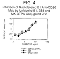
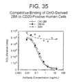
- 238000012875 competitive assay Methods 0.000 description 7
- 238000011161 development Methods 0.000 description 7
- 230000018109 developmental process Effects 0.000 description 7
- 239000012091 fetal bovine serum Substances 0.000 description 7
- 239000012467 final product Substances 0.000 description 7
- 238000000684 flow cytometry Methods 0.000 description 7
- 238000009472 formulation Methods 0.000 description 7
- 210000005260 human cell Anatomy 0.000 description 7
- 238000009206 nuclear medicine Methods 0.000 description 7
- 230000002829 reductive effect Effects 0.000 description 7
- 230000004044 response Effects 0.000 description 7
- 229910052707 ruthenium Inorganic materials 0.000 description 7
- 210000002784 stomach Anatomy 0.000 description 7
- 210000004881 tumor cell Anatomy 0.000 description 7
- 230000035899 viability Effects 0.000 description 7
- 239000004471 Glycine Substances 0.000 description 6
- OKKJLVBELUTLKV-UHFFFAOYSA-N Methanol Chemical compound OC OKKJLVBELUTLKV-UHFFFAOYSA-N 0.000 description 6
- 229910009523 YCl3 Inorganic materials 0.000 description 6
- 238000004108 freeze drying Methods 0.000 description 6
- 230000004807 localization Effects 0.000 description 6
- 229960004641 rituximab Drugs 0.000 description 6
- 239000008227 sterile water for injection Substances 0.000 description 6
- 239000011550 stock solution Substances 0.000 description 6
- 230000008685 targeting Effects 0.000 description 6
- PSCMQHVBLHHWTO-UHFFFAOYSA-K Indium trichloride Inorganic materials Cl[In](Cl)Cl PSCMQHVBLHHWTO-UHFFFAOYSA-K 0.000 description 5
- 206010025323 Lymphomas Diseases 0.000 description 5
- 239000012901 Milli-Q water Substances 0.000 description 5
- 230000008901 benefit Effects 0.000 description 5
- 239000006285 cell suspension Substances 0.000 description 5
- 230000006378 damage Effects 0.000 description 5
- 230000008021 deposition Effects 0.000 description 5
- 230000004927 fusion Effects 0.000 description 5
- 230000036541 health Effects 0.000 description 5
- 238000001727 in vivo Methods 0.000 description 5
- 230000005764 inhibitory process Effects 0.000 description 5
- 238000005259 measurement Methods 0.000 description 5
- 230000009871 nonspecific binding Effects 0.000 description 5
- 230000035484 reaction time Effects 0.000 description 5
- 230000009467 reduction Effects 0.000 description 5
- 239000008223 sterile water Substances 0.000 description 5
- 231100000041 toxicology testing Toxicity 0.000 description 5
- 210000000689 upper leg Anatomy 0.000 description 5
- OOIBFPKQHULHSQ-UHFFFAOYSA-N (3-hydroxy-1-adamantyl) 2-methylprop-2-enoate Chemical compound C1C(C2)CC3CC2(O)CC1(OC(=O)C(=C)C)C3 OOIBFPKQHULHSQ-UHFFFAOYSA-N 0.000 description 4
- IJGRMHOSHXDMSA-UHFFFAOYSA-N Atomic nitrogen Chemical compound N#N IJGRMHOSHXDMSA-UHFFFAOYSA-N 0.000 description 4
- 102000014914 Carrier Proteins Human genes 0.000 description 4
- 108010078791 Carrier Proteins Proteins 0.000 description 4
- FBPFZTCFMRRESA-KVTDHHQDSA-N D-Mannitol Chemical compound OC[C@@H](O)[C@@H](O)[C@H](O)[C@H](O)CO FBPFZTCFMRRESA-KVTDHHQDSA-N 0.000 description 4
- 238000002965 ELISA Methods 0.000 description 4
- 102000004190 Enzymes Human genes 0.000 description 4
- 108090000790 Enzymes Proteins 0.000 description 4
- 229930195725 Mannitol Natural products 0.000 description 4
- 235000006508 Nelumbo nucifera Nutrition 0.000 description 4
- 240000002853 Nelumbo nucifera Species 0.000 description 4
- 235000006510 Nelumbo pentapetala Nutrition 0.000 description 4
- 241000288906 Primates Species 0.000 description 4
- 229920002684 Sepharose Polymers 0.000 description 4
- GLNADSQYFUSGOU-GPTZEZBUSA-J Trypan blue Chemical compound [Na+].[Na+].[Na+].[Na+].C1=C(S([O-])(=O)=O)C=C2C=C(S([O-])(=O)=O)C(/N=N/C3=CC=C(C=C3C)C=3C=C(C(=CC=3)\N=N\C=3C(=CC4=CC(=CC(N)=C4C=3O)S([O-])(=O)=O)S([O-])(=O)=O)C)=C(O)C2=C1N GLNADSQYFUSGOU-GPTZEZBUSA-J 0.000 description 4
- 230000005856 abnormality Effects 0.000 description 4
- 238000002835 absorbance Methods 0.000 description 4
- 230000009137 competitive binding Effects 0.000 description 4
- 238000011109 contamination Methods 0.000 description 4
- 230000007423 decrease Effects 0.000 description 4
- 238000012137 double-staining Methods 0.000 description 4
- 230000007717 exclusion Effects 0.000 description 4
- 210000003736 gastrointestinal content Anatomy 0.000 description 4
- 210000000987 immune system Anatomy 0.000 description 4
- 238000003018 immunoassay Methods 0.000 description 4
- 229940127121 immunoconjugate Drugs 0.000 description 4
- 239000007788 liquid Substances 0.000 description 4
- 210000001165 lymph node Anatomy 0.000 description 4
- 239000000594 mannitol Substances 0.000 description 4
- 235000010355 mannitol Nutrition 0.000 description 4
- 210000005259 peripheral blood Anatomy 0.000 description 4
- 239000011886 peripheral blood Substances 0.000 description 4
- 230000003252 repetitive effect Effects 0.000 description 4
- 238000013341 scale-up Methods 0.000 description 4
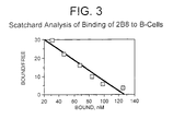
- 238000013391 scatchard analysis Methods 0.000 description 4
- 210000000813 small intestine Anatomy 0.000 description 4
- 239000008229 sterile water for irrigation Substances 0.000 description 4
- 239000000126 substance Substances 0.000 description 4
- 238000011285 therapeutic regimen Methods 0.000 description 4
- 230000003442 weekly effect Effects 0.000 description 4
- IVLXQGJVBGMLRR-UHFFFAOYSA-N 2-aminoacetic acid;hydron;chloride Chemical compound Cl.NCC(O)=O IVLXQGJVBGMLRR-UHFFFAOYSA-N 0.000 description 3
- MFYSYFVPBJMHGN-UHFFFAOYSA-N Cortisone Natural products O=C1CCC2(C)C3C(=O)CC(C)(C(CC4)(O)C(=O)CO)C4C3CCC2=C1 MFYSYFVPBJMHGN-UHFFFAOYSA-N 0.000 description 3
- WSFSSNUMVMOOMR-UHFFFAOYSA-N Formaldehyde Chemical compound O=C WSFSSNUMVMOOMR-UHFFFAOYSA-N 0.000 description 3
- 108010010803 Gelatin Proteins 0.000 description 3
- 241000282412 Homo Species 0.000 description 3
- MHAJPDPJQMAIIY-UHFFFAOYSA-N Hydrogen peroxide Chemical compound OO MHAJPDPJQMAIIY-UHFFFAOYSA-N 0.000 description 3
- 101710160107 Outer membrane protein A Proteins 0.000 description 3
- 206010035226 Plasma cell myeloma Diseases 0.000 description 3
- 230000035508 accumulation Effects 0.000 description 3
- 238000009825 accumulation Methods 0.000 description 3
- 239000002253 acid Substances 0.000 description 3
- 239000002671 adjuvant Substances 0.000 description 3
- 230000010056 antibody-dependent cellular cytotoxicity Effects 0.000 description 3
- 239000011324 bead Substances 0.000 description 3
- 230000036760 body temperature Effects 0.000 description 3
- 230000037396 body weight Effects 0.000 description 3
- 230000022534 cell killing Effects 0.000 description 3
- 230000001413 cellular effect Effects 0.000 description 3
- 230000008859 change Effects 0.000 description 3
- 239000003795 chemical substances by application Substances 0.000 description 3
- 210000004978 chinese hamster ovary cell Anatomy 0.000 description 3
- 150000003841 chloride salts Chemical class 0.000 description 3
- 230000001419 dependent effect Effects 0.000 description 3
- 238000000502 dialysis Methods 0.000 description 3
- 230000004069 differentiation Effects 0.000 description 3
- 239000012153 distilled water Substances 0.000 description 3
- 239000002158 endotoxin Substances 0.000 description 3
- GNBHRKFJIUUOQI-UHFFFAOYSA-N fluorescein Chemical compound O1C(=O)C2=CC=CC=C2C21C1=CC=C(O)C=C1OC1=CC(O)=CC=C21 GNBHRKFJIUUOQI-UHFFFAOYSA-N 0.000 description 3
- 238000001943 fluorescence-activated cell sorting Methods 0.000 description 3
- 239000008273 gelatin Substances 0.000 description 3
- 229920000159 gelatin Polymers 0.000 description 3
- 235000019322 gelatine Nutrition 0.000 description 3
- 235000011852 gelatine desserts Nutrition 0.000 description 3
- ZDXPYRJPNDTMRX-UHFFFAOYSA-N glutamine Natural products OC(=O)C(N)CCC(N)=O ZDXPYRJPNDTMRX-UHFFFAOYSA-N 0.000 description 3
- 239000011544 gradient gel Substances 0.000 description 3
- 229910001385 heavy metal Inorganic materials 0.000 description 3
- 230000028993 immune response Effects 0.000 description 3
- 238000001114 immunoprecipitation Methods 0.000 description 3
- LBGSDOMRVQPKJM-UHFFFAOYSA-N indium yttrium Chemical compound [Y][In] LBGSDOMRVQPKJM-UHFFFAOYSA-N 0.000 description 3
- 238000001802 infusion Methods 0.000 description 3
- 238000001155 isoelectric focusing Methods 0.000 description 3
- 238000012417 linear regression Methods 0.000 description 3
- 239000003550 marker Substances 0.000 description 3
- 150000002739 metals Chemical class 0.000 description 3
- 244000005700 microbiome Species 0.000 description 3
- 201000000050 myeloid neoplasm Diseases 0.000 description 3
- 230000001613 neoplastic effect Effects 0.000 description 3
- 238000005457 optimization Methods 0.000 description 3
- 239000002245 particle Substances 0.000 description 3
- 239000008188 pellet Substances 0.000 description 3
- 210000005105 peripheral blood lymphocyte Anatomy 0.000 description 3
- 230000035755 proliferation Effects 0.000 description 3
- 239000002510 pyrogen Substances 0.000 description 3
- 238000011363 radioimmunotherapy Methods 0.000 description 3
- 230000000405 serological effect Effects 0.000 description 3
- 238000001542 size-exclusion chromatography Methods 0.000 description 3
- 239000001509 sodium citrate Substances 0.000 description 3
- NLJMYIDDQXHKNR-UHFFFAOYSA-K sodium citrate Chemical compound O.O.[Na+].[Na+].[Na+].[O-]C(=O)CC(O)(CC([O-])=O)C([O-])=O NLJMYIDDQXHKNR-UHFFFAOYSA-K 0.000 description 3
- 239000000758 substrate Substances 0.000 description 3
- 238000002560 therapeutic procedure Methods 0.000 description 3
- 210000001541 thymus gland Anatomy 0.000 description 3
- 238000002562 urinalysis Methods 0.000 description 3
- OBOSXEWFRARQPU-UHFFFAOYSA-N 2-n,2-n-dimethylpyridine-2,5-diamine Chemical compound CN(C)C1=CC=C(N)C=N1 OBOSXEWFRARQPU-UHFFFAOYSA-N 0.000 description 2
- USFZMSVCRYTOJT-UHFFFAOYSA-N Ammonium acetate Chemical compound N.CC(O)=O USFZMSVCRYTOJT-UHFFFAOYSA-N 0.000 description 2
- 239000005695 Ammonium acetate Substances 0.000 description 2
- 208000037914 B-cell disorder Diseases 0.000 description 2
- 229940124292 CD20 monoclonal antibody Drugs 0.000 description 2
- QXNVGIXVLWOKEQ-UHFFFAOYSA-N Disodium Chemical class [Na][Na] QXNVGIXVLWOKEQ-UHFFFAOYSA-N 0.000 description 2
- AOJJSUZBOXZQNB-TZSSRYMLSA-N Doxorubicin Chemical compound O([C@H]1C[C@@](O)(CC=2C(O)=C3C(=O)C=4C=CC=C(C=4C(=O)C3=C(O)C=21)OC)C(=O)CO)[C@H]1C[C@H](N)[C@H](O)[C@H](C)O1 AOJJSUZBOXZQNB-TZSSRYMLSA-N 0.000 description 2
- 229940123457 Free radical scavenger Drugs 0.000 description 2
- 108090000288 Glycoproteins Proteins 0.000 description 2
- 102000003886 Glycoproteins Human genes 0.000 description 2
- 101100495232 Homo sapiens MS4A1 gene Proteins 0.000 description 2
- NWIBSHFKIJFRCO-WUDYKRTCSA-N Mytomycin Chemical compound C1N2C(C(C(C)=C(N)C3=O)=O)=C3[C@@H](COC(N)=O)[C@@]2(OC)[C@@H]2[C@H]1N2 NWIBSHFKIJFRCO-WUDYKRTCSA-N 0.000 description 2
- 241000609499 Palicourea Species 0.000 description 2
- 229930040373 Paraformaldehyde Natural products 0.000 description 2
- 239000004793 Polystyrene Substances 0.000 description 2
- CDBYLPFSWZWCQE-UHFFFAOYSA-L Sodium Carbonate Chemical compound [Na+].[Na+].[O-]C([O-])=O CDBYLPFSWZWCQE-UHFFFAOYSA-L 0.000 description 2
- WDLRUFUQRNWCPK-UHFFFAOYSA-N Tetraxetan Chemical compound OC(=O)CN1CCN(CC(O)=O)CCN(CC(O)=O)CCN(CC(O)=O)CC1 WDLRUFUQRNWCPK-UHFFFAOYSA-N 0.000 description 2
- 238000001994 activation Methods 0.000 description 2
- 238000001042 affinity chromatography Methods 0.000 description 2
- 235000019257 ammonium acetate Nutrition 0.000 description 2
- 229940043376 ammonium acetate Drugs 0.000 description 2
- 230000001093 anti-cancer Effects 0.000 description 2
- 239000003855 balanced salt solution Substances 0.000 description 2
- 238000010923 batch production Methods 0.000 description 2
- 239000012148 binding buffer Substances 0.000 description 2
- 230000004071 biological effect Effects 0.000 description 2
- 230000033228 biological regulation Effects 0.000 description 2
- 230000015572 biosynthetic process Effects 0.000 description 2
- 210000001185 bone marrow Anatomy 0.000 description 2
- 229910021538 borax Inorganic materials 0.000 description 2
- 201000011510 cancer Diseases 0.000 description 2
- 229920001429 chelating resin Polymers 0.000 description 2
- 230000000295 complement effect Effects 0.000 description 2
- 239000012141 concentrate Substances 0.000 description 2
- 230000009089 cytolysis Effects 0.000 description 2
- 238000000326 densiometry Methods 0.000 description 2
- 239000002274 desiccant Substances 0.000 description 2
- 238000001514 detection method Methods 0.000 description 2
- 238000011026 diafiltration Methods 0.000 description 2
- 239000003085 diluting agent Substances 0.000 description 2
- 239000012636 effector Substances 0.000 description 2
- 230000008030 elimination Effects 0.000 description 2
- 238000003379 elimination reaction Methods 0.000 description 2
- 238000005516 engineering process Methods 0.000 description 2
- 238000011156 evaluation Methods 0.000 description 2
- 230000001747 exhibiting effect Effects 0.000 description 2
- 239000000284 extract Substances 0.000 description 2
- 238000013213 extrapolation Methods 0.000 description 2
- 238000001914 filtration Methods 0.000 description 2
- 235000013305 food Nutrition 0.000 description 2
- 238000001502 gel electrophoresis Methods 0.000 description 2
- 230000003394 haemopoietic effect Effects 0.000 description 2
- 210000005104 human peripheral blood lymphocyte Anatomy 0.000 description 2
- 230000016784 immunoglobulin production Effects 0.000 description 2
- 238000009169 immunotherapy Methods 0.000 description 2
- 210000004969 inflammatory cell Anatomy 0.000 description 2
- 230000000977 initiatory effect Effects 0.000 description 2
- 238000001990 intravenous administration Methods 0.000 description 2
- 230000005865 ionizing radiation Effects 0.000 description 2
- 150000002500 ions Chemical class 0.000 description 2
- 230000002262 irrigation Effects 0.000 description 2
- 238000003973 irrigation Methods 0.000 description 2
- 210000002429 large intestine Anatomy 0.000 description 2
- 210000000265 leukocyte Anatomy 0.000 description 2
- 239000003446 ligand Substances 0.000 description 2
- 210000003563 lymphoid tissue Anatomy 0.000 description 2
- 238000012423 maintenance Methods 0.000 description 2
- 230000003211 malignant effect Effects 0.000 description 2
- 238000000386 microscopy Methods 0.000 description 2
- 239000000178 monomer Substances 0.000 description 2
- 229910052757 nitrogen Inorganic materials 0.000 description 2
- 229920002866 paraformaldehyde Polymers 0.000 description 2
- 230000000149 penetrating effect Effects 0.000 description 2
- NMHMNPHRMNGLLB-UHFFFAOYSA-N phloretic acid Chemical compound OC(=O)CCC1=CC=C(O)C=C1 NMHMNPHRMNGLLB-UHFFFAOYSA-N 0.000 description 2
- 229920002223 polystyrene Polymers 0.000 description 2
- 238000012910 preclinical development Methods 0.000 description 2
- 238000012545 processing Methods 0.000 description 2
- 239000002516 radical scavenger Substances 0.000 description 2
- 239000000700 radioactive tracer Substances 0.000 description 2
- 239000011535 reaction buffer Substances 0.000 description 2
- 229920005989 resin Polymers 0.000 description 2
- 239000011347 resin Substances 0.000 description 2
- 239000012465 retentate Substances 0.000 description 2
- 238000003345 scintillation counting Methods 0.000 description 2
- 238000012216 screening Methods 0.000 description 2
- HEMHJVSKTPXQMS-UHFFFAOYSA-M sodium hydroxide Inorganic materials [OH-].[Na+] HEMHJVSKTPXQMS-UHFFFAOYSA-M 0.000 description 2
- 235000010339 sodium tetraborate Nutrition 0.000 description 2
- 241000894007 species Species 0.000 description 2
- 210000000278 spinal cord Anatomy 0.000 description 2
- 210000004988 splenocyte Anatomy 0.000 description 2
- 230000000087 stabilizing effect Effects 0.000 description 2
- 238000003860 storage Methods 0.000 description 2
- 235000000346 sugar Nutrition 0.000 description 2
- 239000012134 supernatant fraction Substances 0.000 description 2
- 239000000725 suspension Substances 0.000 description 2
- 239000003053 toxin Substances 0.000 description 2
- 231100000765 toxin Toxicity 0.000 description 2
- 108700012359 toxins Proteins 0.000 description 2
- 238000012546 transfer Methods 0.000 description 2
- 230000001052 transient effect Effects 0.000 description 2
- BSVBQGMMJUBVOD-UHFFFAOYSA-N trisodium borate Chemical compound [Na+].[Na+].[Na+].[O-]B([O-])[O-] BSVBQGMMJUBVOD-UHFFFAOYSA-N 0.000 description 2
- 238000000108 ultra-filtration Methods 0.000 description 2
- 210000003932 urinary bladder Anatomy 0.000 description 2
- 239000008215 water for injection Substances 0.000 description 2
- NWUYHJFMYQTDRP-UHFFFAOYSA-N 1,2-bis(ethenyl)benzene;1-ethenyl-2-ethylbenzene;styrene Chemical compound C=CC1=CC=CC=C1.CCC1=CC=CC=C1C=C.C=CC1=CC=CC=C1C=C NWUYHJFMYQTDRP-UHFFFAOYSA-N 0.000 description 1
- GFXQUCWFEPCALC-UHFFFAOYSA-N 1-(4-isothiocyanato-2-nitrophenyl)imidazole Chemical compound [O-][N+](=O)C1=CC(N=C=S)=CC=C1N1C=NC=C1 GFXQUCWFEPCALC-UHFFFAOYSA-N 0.000 description 1
- PIGCSKVALLVWKU-UHFFFAOYSA-N 2-Aminoacridone Chemical compound C1=CC=C2C(=O)C3=CC(N)=CC=C3NC2=C1 PIGCSKVALLVWKU-UHFFFAOYSA-N 0.000 description 1
- QKNYBSVHEMOAJP-UHFFFAOYSA-N 2-amino-2-(hydroxymethyl)propane-1,3-diol;hydron;chloride Chemical compound Cl.OCC(N)(CO)CO QKNYBSVHEMOAJP-UHFFFAOYSA-N 0.000 description 1
- ZCYVEMRRCGMTRW-UHFFFAOYSA-N 7553-56-2 Chemical compound [I] ZCYVEMRRCGMTRW-UHFFFAOYSA-N 0.000 description 1
- 208000024893 Acute lymphoblastic leukemia Diseases 0.000 description 1
- 208000014697 Acute lymphocytic leukaemia Diseases 0.000 description 1
- 206010067484 Adverse reaction Diseases 0.000 description 1
- 206010003445 Ascites Diseases 0.000 description 1
- BTBUEUYNUDRHOZ-UHFFFAOYSA-N Borate Chemical compound [O-]B([O-])[O-] BTBUEUYNUDRHOZ-UHFFFAOYSA-N 0.000 description 1
- 238000009010 Bradford assay Methods 0.000 description 1
- VTYYLEPIZMXCLO-UHFFFAOYSA-L Calcium carbonate Chemical compound [Ca+2].[O-]C([O-])=O VTYYLEPIZMXCLO-UHFFFAOYSA-L 0.000 description 1
- OKTJSMMVPCPJKN-NJFSPNSNSA-N Carbon-14 Chemical compound [14C] OKTJSMMVPCPJKN-NJFSPNSNSA-N 0.000 description 1
- 244000132059 Carica parviflora Species 0.000 description 1
- 235000014653 Carica parviflora Nutrition 0.000 description 1
- 241000700198 Cavia Species 0.000 description 1
- ZVGCGHVMJAECEG-UHFFFAOYSA-N Chinol Natural products COC1=C(O)C(C)=C(C)C(O)=C1OC ZVGCGHVMJAECEG-UHFFFAOYSA-N 0.000 description 1
- 208000005443 Circulating Neoplastic Cells Diseases 0.000 description 1
- 108020004414 DNA Proteins 0.000 description 1
- 108010087819 Fc receptors Proteins 0.000 description 1
- 102000009109 Fc receptors Human genes 0.000 description 1
- 238000011993 High Performance Size Exclusion Chromatography Methods 0.000 description 1
- 241001272567 Hominoidea Species 0.000 description 1
- 101000738771 Homo sapiens Receptor-type tyrosine-protein phosphatase C Proteins 0.000 description 1
- 101000637792 Homo sapiens Solute carrier family 35 member G5 Proteins 0.000 description 1
- 206010020751 Hypersensitivity Diseases 0.000 description 1
- 108060003951 Immunoglobulin Proteins 0.000 description 1
- 102000008394 Immunoglobulin Fragments Human genes 0.000 description 1
- 108010021625 Immunoglobulin Fragments Proteins 0.000 description 1
- 229910021617 Indium monochloride Inorganic materials 0.000 description 1
- 102000015696 Interleukins Human genes 0.000 description 1
- 108010063738 Interleukins Proteins 0.000 description 1
- 108010052285 Membrane Proteins Proteins 0.000 description 1
- 102000018697 Membrane Proteins Human genes 0.000 description 1
- 241000699673 Mesocricetus auratus Species 0.000 description 1
- FSVCELGFZIQNCK-UHFFFAOYSA-N N,N-bis(2-hydroxyethyl)glycine Chemical compound OCCN(CCO)CC(O)=O FSVCELGFZIQNCK-UHFFFAOYSA-N 0.000 description 1
- 238000011887 Necropsy Methods 0.000 description 1
- 208000015914 Non-Hodgkin lymphomas Diseases 0.000 description 1
- 206010057249 Phagocytosis Diseases 0.000 description 1
- 239000002202 Polyethylene glycol Substances 0.000 description 1
- 208000006664 Precursor Cell Lymphoblastic Leukemia-Lymphoma Diseases 0.000 description 1
- 102100037422 Receptor-type tyrosine-protein phosphatase C Human genes 0.000 description 1
- 229920005654 Sephadex Polymers 0.000 description 1
- 239000012507 Sephadex™ Substances 0.000 description 1
- 102100032019 Solute carrier family 35 member G5 Human genes 0.000 description 1
- 230000024932 T cell mediated immunity Effects 0.000 description 1
- 108091008874 T cell receptors Proteins 0.000 description 1
- 102000016266 T-Cell Antigen Receptors Human genes 0.000 description 1
- 241000251539 Vertebrata <Metazoa> Species 0.000 description 1
- GIEAQDPIWWODTH-UHFFFAOYSA-N [Na].[Na].[Na].[Ca] Chemical compound [Na].[Na].[Na].[Ca] GIEAQDPIWWODTH-UHFFFAOYSA-N 0.000 description 1
- 230000004913 activation Effects 0.000 description 1
- 230000006838 adverse reaction Effects 0.000 description 1
- 125000003277 amino group Chemical group 0.000 description 1
- 238000012801 analytical assay Methods 0.000 description 1
- 230000000259 anti-tumor effect Effects 0.000 description 1
- 230000005875 antibody response Effects 0.000 description 1
- 210000000612 antigen-presenting cell Anatomy 0.000 description 1
- 239000002246 antineoplastic agent Substances 0.000 description 1
- 230000001174 ascending effect Effects 0.000 description 1
- OHDRQQURAXLVGJ-HLVWOLMTSA-N azane;(2e)-3-ethyl-2-[(e)-(3-ethyl-6-sulfo-1,3-benzothiazol-2-ylidene)hydrazinylidene]-1,3-benzothiazole-6-sulfonic acid Chemical compound [NH4+].[NH4+].S/1C2=CC(S([O-])(=O)=O)=CC=C2N(CC)C\1=N/N=C1/SC2=CC(S([O-])(=O)=O)=CC=C2N1CC OHDRQQURAXLVGJ-HLVWOLMTSA-N 0.000 description 1
- 230000009286 beneficial effect Effects 0.000 description 1
- 125000001797 benzyl group Chemical group [H]C1=C([H])C([H])=C(C([H])=C1[H])C([H])([H])* 0.000 description 1
- 239000007998 bicine buffer Substances 0.000 description 1
- 239000011230 binding agent Substances 0.000 description 1
- 230000000903 blocking effect Effects 0.000 description 1
- 230000036765 blood level Effects 0.000 description 1
- 238000009835 boiling Methods 0.000 description 1
- 238000010322 bone marrow transplantation Methods 0.000 description 1
- 210000000481 breast Anatomy 0.000 description 1
- 229920005549 butyl rubber Polymers 0.000 description 1
- 239000012830 cancer therapeutic Substances 0.000 description 1
- 230000022131 cell cycle Effects 0.000 description 1
- 230000011712 cell development Effects 0.000 description 1
- 230000024245 cell differentiation Effects 0.000 description 1
- 239000002771 cell marker Substances 0.000 description 1
- 230000003833 cell viability Effects 0.000 description 1
- 108091092356 cellular DNA Proteins 0.000 description 1
- 230000007541 cellular toxicity Effects 0.000 description 1
- 210000003710 cerebral cortex Anatomy 0.000 description 1
- 210000003679 cervix uteri Anatomy 0.000 description 1
- 230000009920 chelation Effects 0.000 description 1
- 239000013043 chemical agent Substances 0.000 description 1
- 238000003759 clinical diagnosis Methods 0.000 description 1
- 230000007012 clinical effect Effects 0.000 description 1
- 210000001072 colon Anatomy 0.000 description 1
- 238000002648 combination therapy Methods 0.000 description 1
- 150000001875 compounds Chemical class 0.000 description 1
- 230000001010 compromised effect Effects 0.000 description 1
- 238000001816 cooling Methods 0.000 description 1
- 230000008878 coupling Effects 0.000 description 1
- 238000010168 coupling process Methods 0.000 description 1
- 238000005859 coupling reaction Methods 0.000 description 1
- 229940127089 cytotoxic agent Drugs 0.000 description 1
- 238000010217 densitometric analysis Methods 0.000 description 1
- 238000013461 design Methods 0.000 description 1
- 239000003599 detergent Substances 0.000 description 1
- VBXWCGWXDOBUQZ-UHFFFAOYSA-K diacetyloxyindiganyl acetate Chemical compound [In+3].CC([O-])=O.CC([O-])=O.CC([O-])=O VBXWCGWXDOBUQZ-UHFFFAOYSA-K 0.000 description 1
- 238000003745 diagnosis Methods 0.000 description 1
- 238000002059 diagnostic imaging Methods 0.000 description 1
- LOKCTEFSRHRXRJ-UHFFFAOYSA-I dipotassium trisodium dihydrogen phosphate hydrogen phosphate dichloride Chemical group P(=O)(O)(O)[O-].[K+].P(=O)(O)([O-])[O-].[Na+].[Na+].[Cl-].[K+].[Cl-].[Na+] LOKCTEFSRHRXRJ-UHFFFAOYSA-I 0.000 description 1
- 229960004679 doxorubicin Drugs 0.000 description 1
- 229940079593 drug Drugs 0.000 description 1
- 239000003937 drug carrier Substances 0.000 description 1
- 238000001962 electrophoresis Methods 0.000 description 1
- 239000012149 elution buffer Substances 0.000 description 1
- 210000002889 endothelial cell Anatomy 0.000 description 1
- 210000002919 epithelial cell Anatomy 0.000 description 1
- 210000003743 erythrocyte Anatomy 0.000 description 1
- 210000003238 esophagus Anatomy 0.000 description 1
- 230000029142 excretion Effects 0.000 description 1
- 239000013604 expression vector Substances 0.000 description 1
- 230000002550 fecal effect Effects 0.000 description 1
- 239000012894 fetal calf serum Substances 0.000 description 1
- 210000002950 fibroblast Anatomy 0.000 description 1
- 238000011049 filling Methods 0.000 description 1
- 238000003348 filter assay Methods 0.000 description 1
- 239000012634 fragment Substances 0.000 description 1
- 230000006870 function Effects 0.000 description 1
- 238000002825 functional assay Methods 0.000 description 1
- 238000005227 gel permeation chromatography Methods 0.000 description 1
- 238000011991 general safety test Methods 0.000 description 1
- 210000001280 germinal center Anatomy 0.000 description 1
- 230000012010 growth Effects 0.000 description 1
- 238000003306 harvesting Methods 0.000 description 1
- 239000013628 high molecular weight specie Substances 0.000 description 1
- 230000004727 humoral immunity Effects 0.000 description 1
- 210000003405 ileum Anatomy 0.000 description 1
- 239000012216 imaging agent Substances 0.000 description 1
- 230000001900 immune effect Effects 0.000 description 1
- 229940042743 immune sera Drugs 0.000 description 1
- 238000002649 immunization Methods 0.000 description 1
- 230000003053 immunization Effects 0.000 description 1
- 102000018358 immunoglobulin Human genes 0.000 description 1
- 229940072221 immunoglobulins Drugs 0.000 description 1
- APHGZSBLRQFRCA-UHFFFAOYSA-M indium(1+);chloride Chemical compound [In]Cl APHGZSBLRQFRCA-UHFFFAOYSA-M 0.000 description 1
- 230000006698 induction Effects 0.000 description 1
- 239000004615 ingredient Substances 0.000 description 1
- 239000003112 inhibitor Substances 0.000 description 1
- 238000011221 initial treatment Methods 0.000 description 1
- 238000003780 insertion Methods 0.000 description 1
- 230000037431 insertion Effects 0.000 description 1
- 230000003993 interaction Effects 0.000 description 1
- 229940047122 interleukins Drugs 0.000 description 1
- 238000007912 intraperitoneal administration Methods 0.000 description 1
- 108091005979 iodinated proteins Proteins 0.000 description 1
- 230000026045 iodination Effects 0.000 description 1
- 238000006192 iodination reaction Methods 0.000 description 1
- 229910052740 iodine Inorganic materials 0.000 description 1
- 239000011630 iodine Substances 0.000 description 1
- 239000003456 ion exchange resin Substances 0.000 description 1
- 229920003303 ion-exchange polymer Polymers 0.000 description 1
- 238000010983 kinetics study Methods 0.000 description 1
- 231100000518 lethal Toxicity 0.000 description 1
- 230000001665 lethal effect Effects 0.000 description 1
- 230000000670 limiting effect Effects 0.000 description 1
- 238000005567 liquid scintillation counting Methods 0.000 description 1
- 230000007774 longterm Effects 0.000 description 1
- 239000003287 lymphocyte surface marker Substances 0.000 description 1
- 230000002934 lysing effect Effects 0.000 description 1
- 210000002540 macrophage Anatomy 0.000 description 1
- 230000036210 malignancy Effects 0.000 description 1
- 230000001404 mediated effect Effects 0.000 description 1
- 150000001455 metallic ions Chemical class 0.000 description 1
- 229960004857 mitomycin Drugs 0.000 description 1
- 210000001616 monocyte Anatomy 0.000 description 1
- 210000004400 mucous membrane Anatomy 0.000 description 1
- 239000013642 negative control Substances 0.000 description 1
- 231100000252 nontoxic Toxicity 0.000 description 1
- 230000003000 nontoxic effect Effects 0.000 description 1
- 230000003647 oxidation Effects 0.000 description 1
- 238000007254 oxidation reaction Methods 0.000 description 1
- 210000002741 palatine tonsil Anatomy 0.000 description 1
- 230000002093 peripheral effect Effects 0.000 description 1
- 210000000578 peripheral nerve Anatomy 0.000 description 1
- 102000013415 peroxidase activity proteins Human genes 0.000 description 1
- 108040007629 peroxidase activity proteins Proteins 0.000 description 1
- 210000001986 peyer's patch Anatomy 0.000 description 1
- 230000008782 phagocytosis Effects 0.000 description 1
- 238000009520 phase I clinical trial Methods 0.000 description 1
- 239000002504 physiological saline solution Substances 0.000 description 1
- 210000004180 plasmocyte Anatomy 0.000 description 1
- 229920002401 polyacrylamide Polymers 0.000 description 1
- 238000002264 polyacrylamide gel electrophoresis Methods 0.000 description 1
- 229920001223 polyethylene glycol Polymers 0.000 description 1
- 239000013641 positive control Substances 0.000 description 1
- 230000003389 potentiating effect Effects 0.000 description 1
- 239000000843 powder Substances 0.000 description 1
- 238000001556 precipitation Methods 0.000 description 1
- 108090000765 processed proteins & peptides Proteins 0.000 description 1
- 102000004196 processed proteins & peptides Human genes 0.000 description 1
- 230000002062 proliferating effect Effects 0.000 description 1
- 230000002035 prolonged effect Effects 0.000 description 1
- 210000002307 prostate Anatomy 0.000 description 1
- 239000000941 radioactive substance Substances 0.000 description 1
- 229940051022 radioimmunoconjugate Drugs 0.000 description 1
- 239000012217 radiopharmaceutical Substances 0.000 description 1
- 229940121896 radiopharmaceutical Drugs 0.000 description 1
- 230000002799 radiopharmaceutical effect Effects 0.000 description 1
- 230000003439 radiotherapeutic effect Effects 0.000 description 1
- 238000001959 radiotherapy Methods 0.000 description 1
- 230000008707 rearrangement Effects 0.000 description 1
- 238000011084 recovery Methods 0.000 description 1
- 238000012429 release testing Methods 0.000 description 1
- 230000008263 repair mechanism Effects 0.000 description 1
- 238000011160 research Methods 0.000 description 1
- 230000000717 retained effect Effects 0.000 description 1
- YAYGSLOSTXKUBW-UHFFFAOYSA-N ruthenium(2+) Chemical compound [Ru+2] YAYGSLOSTXKUBW-UHFFFAOYSA-N 0.000 description 1
- 239000012723 sample buffer Substances 0.000 description 1
- 229920006395 saturated elastomer Polymers 0.000 description 1
- 230000003248 secreting effect Effects 0.000 description 1
- 230000035945 sensitivity Effects 0.000 description 1
- 238000000926 separation method Methods 0.000 description 1
- 238000002333 serotherapy Methods 0.000 description 1
- 231100000161 signs of toxicity Toxicity 0.000 description 1
- 210000002027 skeletal muscle Anatomy 0.000 description 1
- 210000002363 skeletal muscle cell Anatomy 0.000 description 1
- 210000000329 smooth muscle myocyte Anatomy 0.000 description 1
- 229910000029 sodium carbonate Inorganic materials 0.000 description 1
- 239000007787 solid Substances 0.000 description 1
- 239000002904 solvent Substances 0.000 description 1
- 238000010186 staining Methods 0.000 description 1
- 238000013190 sterility testing Methods 0.000 description 1
- 150000008163 sugars Chemical class 0.000 description 1
- 101150047061 tag-72 gene Proteins 0.000 description 1
- 230000009258 tissue cross reactivity Effects 0.000 description 1
- 239000003104 tissue culture media Substances 0.000 description 1
- 238000004448 titration Methods 0.000 description 1
- 231100000027 toxicology Toxicity 0.000 description 1
- 238000013024 troubleshooting Methods 0.000 description 1
- 230000005909 tumor killing Effects 0.000 description 1
- 239000003981 vehicle Substances 0.000 description 1
- 210000003462 vein Anatomy 0.000 description 1
- 238000012795 verification Methods 0.000 description 1
- 239000011534 wash buffer Substances 0.000 description 1
- 238000005406 washing Methods 0.000 description 1
- 238000005303 weighing Methods 0.000 description 1
Images


Classifications
-
- G—PHYSICS
- G01—MEASURING; TESTING
- G01N—INVESTIGATING OR ANALYSING MATERIALS BY DETERMINING THEIR CHEMICAL OR PHYSICAL PROPERTIES
- G01N33/00—Investigating or analysing materials by specific methods not covered by groups G01N1/00 - G01N31/00
- G01N33/48—Biological material, e.g. blood, urine; Haemocytometers
- G01N33/50—Chemical analysis of biological material, e.g. blood, urine; Testing involving biospecific ligand binding methods; Immunological testing
- G01N33/53—Immunoassay; Biospecific binding assay; Materials therefor
- G01N33/531—Production of immunochemical test materials
- G01N33/532—Production of labelled immunochemicals
- G01N33/534—Production of labelled immunochemicals with radioactive label
-
- A—HUMAN NECESSITIES
- A61—MEDICAL OR VETERINARY SCIENCE; HYGIENE
- A61P—SPECIFIC THERAPEUTIC ACTIVITY OF CHEMICAL COMPOUNDS OR MEDICINAL PREPARATIONS
- A61P35/00—Antineoplastic agents
-
- G—PHYSICS
- G01—MEASURING; TESTING
- G01N—INVESTIGATING OR ANALYSING MATERIALS BY DETERMINING THEIR CHEMICAL OR PHYSICAL PROPERTIES
- G01N33/00—Investigating or analysing materials by specific methods not covered by groups G01N1/00 - G01N31/00
- G01N33/48—Biological material, e.g. blood, urine; Haemocytometers
- G01N33/50—Chemical analysis of biological material, e.g. blood, urine; Testing involving biospecific ligand binding methods; Immunological testing
- G01N33/58—Chemical analysis of biological material, e.g. blood, urine; Testing involving biospecific ligand binding methods; Immunological testing involving labelled substances
- G01N33/60—Chemical analysis of biological material, e.g. blood, urine; Testing involving biospecific ligand binding methods; Immunological testing involving labelled substances involving radioactive labelled substances
Definitions
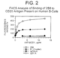
- kits of the present disclosure are used for making and evaluating radiolabeled antibody conjugates that will be used for the treatment and imaging of B-cell lymphoma tumors by targeting the B cell surface antigen BP35 ("CD20").
- the immune system of vertebrates (for example, primates, which include humans, apes, monkeys, etc.) consists of a number of organs and cell types which have evolved to: accurately and specifically recognize foreign microorganisms ("antigen") which invade the vertebrate-host; specifically bind to such foreign microorganisms; and, eliminate/destroy such foreign microorganisms.
- Lymphocytes as well as other types of cells, are critical to the immune system. Lymphocytes are produced in the thymus, spleen and bone marrow (adult) and represent about 30% of the total white blood cells present in the circulatory system of humans (adult).
- T cells are responsible for cell mediated immunity, while B cells are responsible for antibody production (humoral immunity).
- B cells are responsible for antibody production (humoral immunity).
- T cells and B cells can be considered as interdependent -- in a typical immune response, T cells are activated when the T cell receptor binds to fragments of an antigen that are bound to major histocompatability complex ("MHC") glycoproteins on the surface of an antigen presenting cell; such activation causes release of biological mediators (“interleukins”) which, in essence, stimulate B cells to differentiate and produce antibody (immunoglobulins”) against the antigen.
- MHC major histocompatability complex
- Each B cell within the host expresses a different antibody on its surfacethus one B cell will express antibody specific for one antigen, while another B cell will express antibody specific for a different antigen. Accordingly, B cells are quite diverse, and this diversity is critical to the immune system. In humans, each B cell can produce an enormous number of antibody molecules (i.e., about 10 7 to 10 8 ). Such antibody production most typically ceases (or substantially decreases) when the foreign antigen has been neutralized. Occasionally, however, proliferation of a particular B cell will continue unabated; such proliferation can result in a cancer referred to as "B cell lymphoma.”
- T cells and B cells both comprise cell surface proteins which can be utilized as “markers” for differentiation and identification.
- One such human B cell marker is the human B lymphocyte-restricted differentiation antigen Bp35, referred to as "CD20.”
- CD20 is expressed during early pre-B cell development and remains until plasma cell differentiation. Specifically, the CD20 molecule may regulate a step in the activation process which is required for cell cycle initiation and differentiation and is usually expressed at very high levels on neoplastic ("tumor”) B cells.
- CD20 by definition, is present on both "normal” B cells as well as “malignant" B cells, i.e., those B cells whose unabated proliferation can lead to B cell lymphoma.
- the CD20 surface antigen has the potential of serving as a candidate for "targeting" of B cell lymphomas.
- such targeting can be generalized as follows: antibodies specific to the CD20 surface antigen of B cells are, e.g., injected into a patient. These anti-CD20 antibodies specifically bind to the CD20 cell surface antigen of (ostensibly) both normal and malignant B cells; the anti-CD20 antibody bound to the CD20 surface antigen may lead to the destruction and depletion of neoplastic B cells. Additionally, chemical agents or radioactive labels having the potential to destroy the tumor can be conjugated to the anti-CD20 antibody such that the agent is specifically "delivered" to, e.g., the neoplastic B cells. Irrespective of the approach, a primary goal is to destroy the tumor: the specific approach can be determined by the particular anti-CD20 antibody which is utilized and, thus, the available approaches to targeting the CD20 antigen can vary considerably.
- Murine (mouse) monoclonal antibody 1F5 an anti-CD20 antibody
- 1F5 an anti-CD20 antibody
- Extremely high levels (>2 grams) of 1F5 were reportedly required to deplete circulating tumor cells, and the results were described as being "transient.” Press et al., "Monoclonal Antibody 1F5 (Anti-CD20) Serotherapy of Human B-Cell Lymphomas," Blood 69/2:584-591 (1987 ).
- non-human monoclonal antibodies typically lack human effector functionality, i.e., they are unable to, inter alia, mediate complement dependent lysis or lyse human target cells through antibody dependent cellular toxicity or Fc-receptor mediated phagocytosis.
- non-human monoclonal antibodies can be recognized by the human host as a foreign protein; therefore, repeated injections of such foreign antibodies can lead to the induction of immune responses leading to harmful hypersensitivity reactions.
- HAMA Human Anti-Mouse Antibody response
- these "foreign" antibodies can be attacked by the immune system of the host such that they are, in effect, neutralized before they reach their target site.
- lymphoma cells are inherently sensitive to radiotherapy. Therefore, B cell malignancies are attractive targets for radioimmunotherapy (RIT) for several reasons: the local emission of ionizing radiation of radiolabeled antibodies may kill cells with or without the target antigen (e.g., CD20) in close proximity to antibody bound to the antigen; penetrating radiation, i.e., beta emitters, may obviate the problem of limited access to the antibody in bulky or poly vascularized tumors; and, the total amount of antibody required may be reduced.
- target antigen e.g., CD20
- penetrating radiation i.e., beta emitters
- Radiolabeled antibodies include the use of a radioactive substance which may require the need for precautions for both the patient (i.e., possible bone marrow transplantation) as well as the health care provider (i.e., the need to exercise a high degree of caution when working with radioactivity).
- an approach at improving the ability of murine monoclonal antibodies to effect the treatment of B-cell disorders has been to conjugate a radioactive label to the antibody such that the label or toxin is localized at the tumor site.
- Toxins i.e., chemotherapeutic agents such as doxorubicin or mitomycin C
- chemotherapeutic agents such as doxorubicin or mitomycin C
- “Chimeric” antibodies i.e., antibodies which comprise portions from two or more different species (e.g., mouse and human) have been developed as an alternative to “conjugated” antibodies.
- Mouse/human chimeric antibodies have been created, and shown to exhibit the binding characteristics of the parental mouse antibody, and effector functions associated with the human constant region. See e.g., Cabilly et al., U.S. Patent 4,816,567 ; Shoemaker et al., U.S. Patent 4,978,745 ; Beavers et al., U.S. Patent 4,975,369 ; and Boss et al., U.S. Patent 4,816,397 all of which are incorporated by reference herein.
- these chimeric antibodies are constructed by preparing a genomic gene library from DNA extracted from pre-existing murine hybridomas. Nishimura et al. (1987) Cancer Research 47: 999 . The library is then screened for variable region genes from both heavy and light chains exhibiting the correct antibody fragment rearrangement patterns. The cloned variable region genes are then ligated into an expression vector containing cloned cassettes of the appropriate heavy or light chain human constant region gene. The chimeric genes are then expressed in a cell line of choice, usually a murine myeloma line.
- Liu, A.Y., et al. "Production of a Mouse-Human Chimeric Monoclonal Antibody to CD20 with Potent Fc-Dependent Biologic Activity", J. Immun. 139/10:3521-3526 (1987 ), describes a mouse/human chimeric antibody directed against the CD20 antigen. See also, PCT Publication No. WO 88/04936 . However, no information is provided as to the ability, efficacy or practicality of using Liu's chimeric antibodies for the treatment of B cell disorders in the reference.
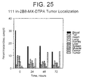
- In2B8 conjugate comprises a murine monoclonal antibody, 2B8, specific to human CD20 antigen, that is attached to Indium[111] ( 111 In) via a bifunctional chelator, i.e., MX-DTPA (diethylenetriaminepentaacetic acid), which comprises a 1:1 mixture of 1-isothiocyanatobenzyl-3-methyl-DTPA and 1-methyl-3-isothiocyanatobenzyl-DTPA.
- MX-DTPA diethylenetriaminepentaacetic acid
- Patents relating to chelators and chelator conjugates are known in the art.
- U.S. Patent No. 4,831,175 of Gansow is directed to polysubstituted diethylenetriaminepentaacetic acid chelates and protein conjugates containing the same, and methods for their preparation.
- U.S. Patent Nos. 5,099,069 , 5,246,692 , 5,286,850 , and 5,124,471 of Gansow also relate to polysubstituted DTPA chelates. These patents are incorporated herein in their entirety.
- the specific bifunctional chelator used to facilitate chelation in applications 08/475,813 , 08/475,815 and 08/478,967 was selected as it possesses high affinity for trivalent metals, and provides for increased tumor-to-non-tumor ratios, decreased bone uptake, and greater in vivo retention of radionuclide at target sites, i.e., B-cell lymphoma tumor sites.
- target sites i.e., B-cell lymphoma tumor sites.
- other bifunctional chelators are known in the art and may also be beneficial in tumor therapy.
- the Y2B8 conjugate comprises the same anti-human CD20 murine monoclonal antibody, 2B8, attached to yttrium-[90] ( 90 Y) via the same bifunctional chelator.
- This radionuclide was selected for therapy for several reasons.
- the 64 hour half-life of 90 Y is long enough to allow antibody accumulation by the tumor and, unlike e.g. 131 I, it is a pure beta emitter of high energy with no accompanying gamma irradiation in its decay, with a range of 100 to 1000 cell diameters.
- the minimal amount of penetrating radiation allows for outpatient administration of 90 Y-labeled antibodies. Furthermore, internalization of labeled antibodies is not required for cell killing, and the local emission of ionizing radiation should be lethal for adjacent tumor cells lacking the target antigen.
- the Y2B8 conjugate possesses the same advantages discussed above, e.g., increased retention of radionuclide at a target site (tumor).
- a diagnostic "imaging" radionuclide such as 111 In, can be used for determining the location and relative size of a tumor prior to and/or following administration of therapeutic chimeric or 90 Y-labeled antibodies for the purpose of tumor reduction.
- indium-labeled antibody enables dosimetric assessment to be made.
- radionuclides which have been used in clinical diagnosis include 131 I, 125 I, 123 I, 99 Tc, 67 Ga, as well as 111 In.
- Antibodies have also been labeled with a variety of radionuclides for potential use in targeted immunotherapy ( Peirersz et al. (1987) The use of monoclonal antibody conjugates for the diagnosis and treatment of cancer. Immunol. Cell Biol. 65: 111-125 ).
- radionuclides include 188 Re and 186 Re as well as 90 Y, and to a lesser extent 199 Au and 67 Cu. I-[131] has also been used for therapeutic purposes.
- U.S. Patent No. 5,460,785 provides a listing of such radioisotopes and is herein incorparted by reference.
- U.S. Application Serial No. 08/475,813 discloses sequential administration of Rituxan®, a chimeric anti-CD20, with both or either indium-labeled or yttrium-labeled murine monoclonal antibody.
- the radiolabeled antibodies used in these combined therapies are murine antibodies
- initial treatment with chimeric anti-CD20 sufficiently depletes the B cell population such that the HAMA response is decreased, thereby facilitating a combined therapeutic and diagnostic regimen.
- murine antibodies may find particular utility as diagnostic reagents.
- a therapeutically effective dosage of the yttrium-labeled anti-CD20 antibody following administration of Rituxan® is sufficient to (a) clear any remaining peripheral blood B cells not cleared by the chimeric anti-CD20 antibody; (b) begin B cell depletion from lymph nodes; or (c) begin B cell depletion from other tissues.
- conjugation of radiolabels to cancer therapeutic antibodies provides a valuable clinical tool which may be used to assess the potential therapeutic efficacy of such antibodies, create diagnostic reagents to monitor the progress of treatment, and devise additional therapeutic reagents which may be used to enhance the initial tumor-killing potential of the chimeric antibody.
- additional therapeutic reagents which may be used to enhance the initial tumor-killing potential of the chimeric antibody.
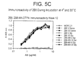
- radiolabeled proteins may be inherently unstable, particularly those labeled with radiolytic isotopes such as 90 Y, which have the tendency to cause damage to the antibody the longer they are attached to it in close proximity.
- radiolysis causes unreliable efficiency of the therapeutic due to loss of radiolabel and/or reduced binding to the target antigen, and may lead to undesired immune responses directed at denatured protein.
- clinicians have had no choice but to order therapeutic antibodies already labeled, or have them labeled off site at a related facility and transported in following labeling for administration to the patient. All such manipulations add precious time to the period between labeling and administration, thereby contributing to the instability of the therapeutic, while in effect decreasing the utility of radiolabeling kits in the clinical setting.
- Cytogen has recently launched a commercial kit for radiolabeling a murine monoclonal antibody directed to tumor-associated glycoprotein TAG-72.
- Cytogen's antibody is particularly unamenable to a kit formulation due to the tendency to develop particulates during storage which must later be removed by a further filtration step.
- Cytogen's antibody has caused adverse reactions in patients due to a HAMA responses.
- WO 94/11026 describes therapeutic methods involving the administration of radiolabeled anti-CD20 antibodies. Methods for preparing such radiolabeled antibodies are also described.
- kits of the present disclosure allow rapid labeling which may be affected in approximately a half an hour or as little as five minutes depending on the label.
- kit protocols of the present disclosure have a labeling efficiency of over 95% thereby foregoing the need for further purification.
- the half-life of the radiolabel and the integrity of the antibody is reserved for the therapeutic purpose for which it is labeled.
- kits and methods whereby diagnostic and therapeutic antibodies may be radiolabeled and administered to a patient in a reproducible, reliable and convenient manner.
- the kits of the present disclosure transform the process of radiolabeling antibodies into a hassle-free, worry-free standardized process, which greatly facilitates patient treatment protocols.
- the present kits provide advantages over the prior art in that the optimum parameters for labeling and administering therapeutic or diagnostic antibodies have been determined, thereby reducing the cost of goods. Since the kits described herein provide the optimum parameters according to the particular label, use of a kit designed for a particular label will also minimize cannibalization, i.e., which occurs when an inappropriate kit is used for a particular label. Avoiding cannibalization in turn also provides for optimum labeling efficiency. Moreover, the protocols and sterile, pyrogen-free ingredients included with each kit make for a more user-friendly process, since sterility, pyrogen testing and post-labeling purification of the reagents are obviated.
- the present invention provides, in a first aspect, a method for radiolabeling a chelator-conjugated antibody with 90 Y for administration to a patient comprising:
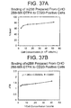
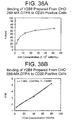
- the present invention provides a binding assay for determining the percent binding of a radiolabeled antibody to its target cell, comprising:
- kits for radiolabeling a diagnostic or therapeutic antibody before administration to a patient comprising at least (i) a vial containing a chelator-conjugated antibody, (ii) a vial containing formulation buffer for stabilizing and administering the radiolabeled antibody, and (iii) instructions for radiolabeling the antibody, wherein said vial components are supplied in such an amount and at such a concentration that when they are combined with a radiolabel of sufficient purity and activity according to the kit instructions, no further purification of the labeled antibody is required before administration to said patient.
- such isotope incorporation may reach levels higher than 95%, and even as high as 98 % or higher.
- the antibody included in the kit is most preferably an anti-CD20 antibody.
- the antibody is supplied in a form whereby it is attached to a bifunctional chelator.
- the antibody is conjugated to MX-DTPA, but other chelators such as phenyl- or benzyl-conjugated DTPA, cyclohexyl-DTPA, EDTA derivatives and DOTA may be used.
- a chelator according to the present invention may be any chelator that is at least bifunctional, i.e., which possesses at least two binding sites (at least one site for chelating a metallic ion and at least one site for coupling to a protein ligand).
- the conjugated antibody is typically supplied at a concentration of 0.5 to 30 mg/ml, more preferably 2 mg/ml.
- the volume of conjugated antibody will depend on the concentration and the amount required for optimum labeling depending on the radiolabel. However, the conjugated antibody is to be supplied in such a volume and concentration that the entire volume will be added to the reaction vial using a sterile syringe and aseptic technique. This will allow for increased reproducibility and ease of use. All reagents of the kits disclosed herein are sterile and pyrogen-free, and specifically designed for simplicity and speed in advancing directly from antibody testing to administration. With some labels, the need for testing labeling efficiency may not be required.
- a particularly advantageous component of the kit is the formulation buffer for stabilizing against the effects of radiolysis and administering the radiolabeled conjugated antibody to a patient.
- the formulation buffer is a pharmaceutically acceptable carrier which serves as both a diluent for the labeled antibody and an administration buffer.
- any pharmaceutically acceptable diluent may be used for administering therapeutic or diagnostic antibodies to patient, the formulation buffer of the present disclosure is particularly suited for administering radiolabeled antibodies.
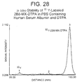
- the formulation buffer of the present disclosure comprises a radioprotectant such as human serum albumin (HSA) or ascorbate, which minimize radiolysis due to yttrium, and to a lesser degree, indium.
- a radioprotectant such as human serum albumin (HSA) or ascorbate
- HSA human serum albumin
- Other radioprotectants are known in the art and could also be used in the formulation buffer of the present disclosure, i.e., free radical scavengers (phenol, sulfites, glutathione, cysteine, gentisic acid, nicotinic acid, ascorbyl palmitate, HOP(:O)H 2 , glycerol, sodium formaldehyde sulfoxylate, Na 2 S 2 O 5 , Na 2 S 2 O 3 , and SO 2 , etc.).
- free radical scavengers phenol, sulfites, glutathione, cysteine, gentisic acid, nicotinic acid,
- radioprotectants are generally employed in the formulation buffer to protect the antibody from radiolysis, it may be possible to affect further protection by including the radioprotectant in the reaction buffer as well. This has generally not been done before, i.e., with HSA, due to the presence of metals which would interfere with the labeling process. However, it may be possible to "clean" the HSA using a chelating resin such that it could be included in the reaction buffer as well. Ascorbate or other radioprotectants may also need to be treated to remove contaminating metals.
- the formulation buffer of the present disclosure also comprises excess unconjugated chelator.
- the purpose for including unconjugated chelator is that this chelator serves to scavenge any non-protein bound radiolabel in the patient, and effects excretion of the radiolabel thereby reducing uptake of "bone-seeking" isotopes, i.e., 90 Y, by the bones of the patient.
- excess DTPA or any other chelator may be included in the formulation buffer.
- the formation buffer is also preferably supplied in a volume such that the entire contents are transferred to the reaction vial. As discussed above, this results in increased ease of use and reproducibility because exact volumes do not have to be measured and transferred.
- a preferred formulation buffer comprises phosphate buffered or physiological saline, human serum albumin and DTPA.
- the human serum albumin is preferably at a concentration of between about 1 to 25% (w/v), and more preferably at a concentration of about 7.5% (w/v).
- the concentration of DTPA is preferably about 1 mM.
- Ascorbate may be used as an alternative to human serum albumin, and is typically used at a concentration of about 1 to 100 mg/ml. Although a wider range of concentrations may be used without compromising patient safety.
- the antibody of the radiolabeling kit is readily labeled with a radioisotope of choice via a bifunctional chelator according to the methods of the present disclosure.
- the kit of the present disclosure may also include a vial containing a buffer for adjusting the pH of the radioisotope solution, and a sterile glass reaction vial for performing the labeling and subsequently for resuspending the final radiolabeled antibody in formulation buffer.
- a 10 ml reaction vial is typically sufficient, but vials capable of holding 5 to 20 mls may also be used.
- the buffer is preferably a low-metal sodium acetate solution at a concentration of 10 to 1000 mM, most preferably 50 mM.
- a specific kit of the present disclosure comprises the MX-DTPA conjugated antibody, 2B8-MX-DTPA.
- 2B8 is an anti-CD20 antibody shown to affect B cell depletion upon administration to lymphoma patients.
- the radiolabeling kit of the present disclosure may be optimized for the radiolabeling of other anti-CD20 antibodies, or any other antibody which has been conjugated to DTPA or other polyvalent chelator.
- the preferred kit of the present disclosure may comprise at least (i) a vial containing the MX-DTPA conjugated 2B8 antibody, either in solution or lyophilized (requiring reconstitution); and (ii) a vial containing formulation buffer for administering the radiolabeled antibody to a patient.
- the preferred kit will also contain (iii) a buffer for adjusting isotope pH, and (iv) a reaction vial.
- the buffer is supplied in the reaction vial, thereby eliminating the steps of measuring and transferring the buffer and increasing the simplicity, consistency and sterility of the kit components,
- the reaction vial could be supplied with the required antibody volume.
- the isotope/buffer vial could be made large enough to accommodate addition of the antibody conjugate, i.e., directly to the supplier's vial. This would eliminate the need for the reaction vial.
- the reaction vial itself contains the required volume of conjugated antibody (i.e., 1 or 1.5 mL for 111 In and 90 Y, respectively).
- the antibody may be supplied in a buffer that provides the appropriate radiolabeling pH according to the specific desired isotope (i.e., pH 3-6 for 111 In, pH 3-5 for 90 Y). Different buffers may be used, depending on the isotope (i.e., sodium acetate for 90 Y, sodium citrate for 111 In).
- the pH and composition of the buffer may also vary depending on the nature of the binding ligand to be labeled (i.e., labeling peptides may permit ⁇ pH 3 to be used). Essentially then, the isotope would be transferred directly to the reaction vial, as would the formulation buffer. Limiting use of the kit to two transfer steps would further increase reproducibility and simplicity, and further decrease the chance for contamination of sterility during manipulation of the kit components.
- the radiolabeling kits of the present disclosure may further comprise a vial of radioisotope, or radioisotope may be ordered separately from an appropriate supplier.
- Preferred radioisotopes of the present disclosure are 111 In chloride and 90 Y chloride in HCl although the disclosed methods are not limited to these isotopes.
- Other radionuclides that have been used for imaging applications are known in the art, i.e., as described in U.S. Patent Nos. 4,634,586 , 5,460,785 and 5,766,571 , which are herein incorporated by reference.
- Indium-[111] is particularly advantageous for imaging B cell tumors and beta emitters such as 90 Y are particularly useful as radiotherapeutic agents.
- other radioisotopes suitable for these or other purposes i.e., alpha emitters, may be used depending on the chelator used for antibody conjugation.
- a further kit will also include a separate vial of chimeric antibody, i.e., Rituxan®, to be administered before or after the radiolabeled anti-CD20 antibody.
- chimeric antibody i.e., Rituxan®
- the HAMA response which might generally occur in response to administration of a murine anti-CD20 antibody may be significantly decreased, thereby increasing the therapeutic utility of radiolabeled murine antibodies.
- chimeric anti-CD20 is employed to clear circulating B cells, subsequent diagnostic images achieved with 111 In-labeled antibodies may be much clearer.
- kits of the present disclosure may include separate, perhaps color-coded, buffer vials specifically formulated according to the optimum pH requirements for radiolabeling antibodies with the specific radioisotopes to be used. Such a system would ensure that the appropriate buffer was used for each label, and would allow the clinician the same ease in radiolabeling the two antibodies as if two kits had been purchased. Such a kit in effect combines the components from two radiolabeling kits into one.
- the components of the radiolabeling kit of the present disclosure are supplied at the appropriate concentration and pH so that sterility is readily maintained before antibody administration and there is little need for additional buffers or media.
- some of the reagents can be prepared, sterilized and tested for sterility on site.
- variations of the kit of the present disclosure are envisioned depending on the budget and preference of the consumer.
- the radiolabeling kit of the present disclosure may be used in a method for radiolabeling a chelator-conjugated antibody for administration to a patient.
- a method for radiolabeling a chelator-conjugated antibody for administration to a patient comprises, in general,
- the antibody is an anti-CD20 antibody, and in particular, the anti-CD20 antibody may be 2B8.
- the antibody may be conjugated to any appropriate chelator, i.e., MX-DTPA, CHX-DTPA, phenyl- or benzyl-DTPA, DOTA, EDTA derivatives, etc. MX-DTPA is preferred. Methods for affecting antibody conjugation are known in the art (Kozak et al. (1989); Mirzadeh et al. (1990), Brachbiel et al. (1986)).
- the method of radiolabeling a chelator-conjugated antibody works best wherein the solution containing the radiolabel is adjusted to a pH of between about 3.0 and 6.0, and more preferably to about 4.2 before it is mixed with the chelator-conjugated antibody.
- Low-metal sodium acetate is particularly preferred for adjusting the pH, although other buffers may be used.
- the sodium acetate is at a concentration of between about 10 and 1000 mM, and more preferably 50 mM.
- the volume quantity of 111 In chloride which should be used to prepare a single administrative dose is typically about 5.5 mCi divided by the radioactivity concentration at the time of labeling.
- a typical diagnostic dose of 111 In is about 2 to 10 mCi.
- the quantity of sodium acetate used for adjusting the pH varies depending on the sodium acetate concentration and the isotope carrier solution, and may therefore be quite broad.
- the concentration of sodium acetate is 50 mM
- the amount required for adjusting the pH is typically about 1.2 times the volume quantity of 111 In chloride used although larger volumes may be used.
- the ratio of sodium acetate to HCl is what is important, and the amount of sodium acetate used would change depending on the amount and concentration of HCl in the buffer.
- About 1 ml of a chelator-conjugated antibody at a concentration of about 2 mg/ml is then mixed with the radiolabel acetate solution, and the mixture is incubated for about 30 minutes, or for a time sufficient to achieve optimal labeling of the antibody. Such time may range from about 30 seconds to about 60 minutes.
- Formulation buffer is then added in an amount necessary to achieve a total final volume of about 10 ml.
- the optimum time required for labeling the antibody may vary depending on the antibody, the particular radiolabel and the particular conjugate employed.
- An underlying factor in the optimization of the time allotted for radiolabeling is the chelator to antibody ratio of the reagent which is to be labeled.
- the chelator to antibody ratio must be high enough to achieve a therapeutically useful level of incorporation, i.e., 90 to 95% depending on the radioisotope, but must also not be too high such that the structural integrity or immunoreactivity of the antibody is compromised. This requires a certain balancing process that in some cases may lead to a lower level of conjugated chelator and longer labeling time.
- the volume quantity of 90 Y chloride used for preparing a single administrative dose typically ranges from about 10 to 50 mCi, and is preferably about 45 mCi, divided by the radioactivity concentration at the time of labeling.
- the quantity of sodium acetate used for adjusting the pH varies depending on the sodium acetate concentration and the concentration of isotope carrier, and may therefore be quite broad.
- the concentration of sodium acetate is 50 mM and the 90 Y is supplied in 50 mM HCl
- the amount required for adjusting the pH is typically about 1.2 times the volume quantity of 90 Y chloride used.
- a chelator-conjugated antibody at a concentration of about 2 mg/ml is then mixed with the radiolabel acetate solution, and incubated for about 5 minutes, or for a time sufficient to achieve optimal labeling of the antibody. Such time may range from about 30 seconds to about 60 minutes.
- Formulation buffer is added in an amount necessary to achieve a total final volume of about 10 ml.
- the radiolabeling method of the present disclosure is performed using the radiolabeling kit described herein.
- the preferred components and conditions are merely acceptable guidelines for practicing the method of the present disclosure, and may be altered to some degree with appropriate optimization. Conditions which depart from those preferred but still accomplish the purpose of the method are considered to be within the scope of the present disclosure.
- the radiolabeling kit of the present disclosure may also be supplied with reagents suitable for conveniently verifying the binding affinity of the antibody following radiolabeling.
- the kit of the disclosure may also be used for determining the percent binding of a radiolabeled antibody to its target cell before administering the antibody to a patient.
- the present inventors have also found that the particular binding assay kit disclosed may be useful for testing the affinity of any antibody generally for which purified antigen is not available. Accordingly, the binding assay components may also be sold as a separate kit.
- a binding assay and radiolabeling kit comprises (i) at least one vial of lyophilized cells which express the antigen which is recognized by the antibody in the kit; (ii) a vial containing chelator-conjugated antibody; (iii) a vial containing formulation buffer, and (iv) instructions for radiolabeling the antibody such that the radiolabeled antibody may be administered directly to a patient without the need for subsequent purification.
- this kit may also comprise a vial containing a buffer for adjusting the pH of the radioisotope, and a sterile glass reaction vial.
- the buffer is a low-metal sodium acetate solution at a concentration of between about 10 and 1000 mM, and the glass reaction vial holds a volume of at least 5 ml.
- the antibody is preferably an anti-CD20 antibody, and the chelator is preferably MX-DTPA. Other chelators may be used as described previously.
- the preferred conjugated antibody is 2B8-MX-DTPA, although any chelator-conjugated antibody may be labeled and its affinity assessed.
- the formulation buffer is phosphate buffered saline comprising a radioprotectant and unconjugated chelator as described above, and radioisotope may or may not be included and is preferably 111 In chloride or 90 Y chloride. Other radioisotopes may be used depending on the chelator.
- the difference between the binding assay/radiolabeling kit and the radiolabeling kit described above is the inclusion of antigen-positive cells to serve as a substrate target for testing antibody affinity.
- antigen is CD20
- preferred CD20-positive cells are SB cells (ATCC # CCL 120) but any CD20-positive cells may be used.
- the binding assay and radiolabeling kit may further include antigen-negative cells for use as a negative control.
- Preferred CD20-negative cells are HSB cells (ATCC # CCL 120.1) but any CD20-negative cells may be used.
- the combined radiolabeling and binding assay kit may further comprise a vial of chimeric anti-CD20 antibody in addition to the antibody to be labeled for the purposes of affecting a combined therapeutic regimen, or for clearing peripheral B cells prior to diagnostic imagery.
- a separate antibody is preferably Rituxan®, but may be any antibody shown to effectuate tumor cell killing.
- two different types of antibodies may be combined in one kit, i.e., antibodies directed to two different B cell antigens, so long as the combined therapeutic regimen serves to target the same type of cell, i.e., the B cell lymphoma.
- the binding assay and radiolabeling kit of the present disclosure is particularly suited for the commercial setting in that the target cells are provided in lyophilized form. This allows the verification of antibody efficacy to proceed simply and systematically, and negates the hassle and expense involved in maintaining tissue culture facilities.
- the lyophilized cells are generally supplied in aliquots of between 0.5 and 500 X 10 6 cells per vial according to the methods of the disclosure.
- the present disclosure also provides for a binding assay kit for determining the percent binding of a radiolabeled antibody to its target cell.
- a binding assay kit for determining the percent binding of a radiolabeled antibody to its target cell.
- Such a kit includes at least one vial of fixed and/or lyophilized antigen-positive cells, and may optionally contain antigen-negative cells as described above for the binding assay and radiolabeling kit.
- variations of such a kit may include an unlabeled control antibody for verifying the binding specificity of the consumer's antibody via a competitive assay.
- the CD20-positive cells are preferably SB cells (ATCC # CCL 120) and the CD20-negative cells are preferably HSB cells (ATCC # CCL120.1), which are supplied in lyophilized form in aliquots of between 0.5 and 50 X 10 6 cells.
- the antibody is preferably an MX-DTPA conjugate of 2B8 labeled with 111 In or 90 Y.
- the present inventors have also found that the method used for preparing fixed and lyophilized cells for the binding assay kits of the present disclosure is particularly suitable for preparing cells for commercial kits.
- Cells may be fixed prior to lyophilization to improve structure/stability.
- the cells of the present disclosure demonstrate high reproducibility when used for antibody binding assays.
- the present disclosure includes a method of preparing lyophilized cells comprising (i) harvesting cells at a cell density of 0.5 to 2 X 10 6 cells per ml by centrifugation; (ii) washing cells at least one time in a balanced salt solution, i.e., HBSS; (iii) resuspending pelleted cells in a lyophilization buffer comprising a balanced salt solution containing carrier protein and at least one type of sugar; (iv) dispensing an aliquot of resuspended cells into a microfuge tube or a glass vial; and (v) lyophilizing the cells 12-96h and more preferably 24-72h at about 30-60 millitor.
- the method is particularly suitable for preparing lyophilized cells wherein said cells are SB cells (ATCC # CCL 120) or HSB cells (ATCC # CCL 120.1), but is likely applicable to other cell types as well.
- the buffer generally contains bovine serum albumin as the carrier protein at a concentration of 1 % (w/v) and mannitol at a concentration of 10%.
- carrier proteins i.e., HSA, and other sugars may be used.
- the cells are harvested by centrifugation at a speed of about 1300 rpm, and the salt solution HBSS (Hank's balanced salt solution) is added.
- the cells are generally resuspended at a concentration of 50 X 10 6 cells per ml.
- the above conditions may be modified slightly without significantly compromising cell viability.
- the above conditions may be supplemented by additional procedures designed to optimize the process for larger quantities of cells, e.g., tangential flow diafiltration to exchange cells into the lyophilization buffer.
- a binding assay for determining the percent binding of a radiolabeled antibody to its target cell comprises in general the following steps: (i) mixing at least one aliquot of a radiolabeled antibody with at least one aliquot of antigen positive cells; (ii) mixing at least one aliquot of a radiolabeled antibody identical to the aliquot of step (i) with at least one aliquot of dilution buffer of the same volume as the aliquot of antigen-positive cells in step (i) as a control; (iii) pelleting the cells by centrifugation; (iv) measuring the radioactivity in the supernatant of the pelleted cells and the control; and (v) comparing the quantity of radioactivity in the cell supernatant to the quantity of radioactivity in the control.
- the binding assay of the present disclosure is typically performed with antibodies labeled with 111 In or 90 Y.
- 111 In is the radiolabel
- radioactivity in the assay tubes is measure using a gamma counter.
- 90 Y is the label, radioactivity is measured using a scintillation counter, although a gamma counter could be used.
- the preferred antibody is an anti-CD20 antibody
- the anti-CD20 antibody is preferably 2B8, wherein the 2B8 antibody is labeled using the radiolabeling kit of the present disclosure.
- any radiolabeled antibody may be tested provided cells expressing the particular antigen are available.
- CD20 is the antigen
- the preferred cells for performing the assay are SB cells (ATCC # CCL 120), however, the assay may also be optimized and performed with any radiolabeled antibody and appropriate target cell.
- the dilution buffer used for the assay should maintain binding of the antibody, i.e., physiological buffer, possibly containing a carrier protein, e.g. BSA, to minimize non-specific binding to cells.
- a carrier protein e.g. BSA
- a further control may be included in the assay by using antigen-negative cells.
- the binding assay further comprises the following steps: (i) mixing at least one aliquot of a radiolabeled antibody with at least one aliquot of antigen-negative cells; (ii) pelleting the antigen-negative cells by centrifugation; (iv) measuring the radioactivity in the supernatant of the antigen-negative pelleted cells; and (v) comparing the quantity of radioactivity in the antigen-negative cell supernatant to the quantity of radioactivity in the supernatant of the antigen-positive cell supernatant and the control. Comparing the radioactivity obtained from this tube to the dilution buffer control will serve as a measure of the amount of non-specific binding to antigen-positive cells.
- CD20 negative cells are preferably HSB cells (ATCC # CCL 120.1).
- the lyophilized cells of the present disclosure provide a simple, efficient and reproducible standard for testing the binding efficacy of a radiolabeled antibody. Therefore, the binding assay of the present disclosure is preferably performed using the lyophilized cells included in the binding assay kits of the present disclosure.
- the radiolabeling assays of the present disclosure may be combined with the binding assays of the present disclosure, wherein the antibody is first labeled by the method of labeling an chelator-conjugated antibody as described in the present disclosure. Most preferably, the binding assay of the present disclosure is performed using one of the binding assay and radiolabeling kits described herein.
- the present disclosure also encompasses a competitive binding assay for assessing affinity of a test antibody to a target cell, comprising (i) preparing a ruthenium-labeled control antibody using a known antibody specific for the same antigen; (ii) incubating increasing amounts of test antibody and increasing amounts of unlabeled control antibody with a fixed concentration of target cells and a trace amount of ruthenium-labeled antibody wherein each separate concentration of test antibody and each separate concentration of control antibody are in separate tubes, respectively; (iii) determining the quantity of binding in each reaction tube based on relative electrochemiluminescence (ECL) using ORIGEN instrumentation; and (iv) calculating the average affinity value of the test antibody.
- ECL electrochemiluminescence
- the average affinity value may be calculated from the EC50 values and the known concentration of trace antibody using the method of Muller (J. Immunological Methods (1980) 34:345 ) or any other appropriate method. It should be noted that this assay may also be used to test the affinity of radiolabeled antibodies, or any antibody for which antigen cannot be purified and cells are required as an antigen source.
- the fixed, lyophilized cells of the present disclosure may be used as target cells.
- the control antibody may be 2B8, or any other unconjugated anti-CD20 antibody.
- the control antibody may be a chelator-conjugated antibody.
- the test antibody may also be a chelator-conjugate of the control antibody.
- the test antibody may be another anti-CD20 antibody whose binding affinity to CD20 as compared to 2B8 is of interest.
- the assay may be adapted for use with antibodies having other specificities so long as an appropriate target cell is available.
- the preferred target cells are CD20-positive cells, more preferably SB cells (ATCC # CCL 120), and are more preferably resuspended lyophilized SB cells prepared according to the method of the present disclosure. Cells lyophilized using other methods or fixed cells may also be used.
- the ruthenium-labeled antibody is typically prepared by a process comprising incubating the control antibody with N-hydroxysuccinimide ester of ruthenium (II) tris-bypyridine chelator (TAG-NHS), although other known method of labeling antibodies are also envisioned.
- TAG-NHS N-hydroxysuccinimide ester of ruthenium (II) tris-bypyridine chelator
- the control antibody and TAG-NHS are preferably incubated at about a 1:15 molar ratio.
- low metal - refers to reagents treated to reduce metal contamination to a level which does not impact radioincorporation.
- antigen positive - means expresses antigen that is recognized by particular antibody of the disclosure in such a way that the antibody is capable of binding.
- % radioincorporation - refers to the amount of radiolabel from a radiolabeling reaction that is conjugated to the antibody relative to the total amount of radiolabel initially added to the reaction.
- % binding - refers to the amount of antibody from a sample which binds to the target antigen, with or without specificity.
- % immunoreactivity or binding specificity- refers to that amount of an antibody sample which binds to the target antigen with specificity.
- diagnostic antibody - refers to an antibody conjugated to a radiolabel such us 111 I which can effect diagnostic imaging of tumors and antigen positive cells.
- therapeutic antibody - refers to an antibody conjugated to an alpha or beta emitting radiolabel (such as 90 Y) which can effect cell killing when bound to the targeted antigen.
- the human cell lines SB and HSB were obtained from the American Type Culture Collection and cultured in RPMI-1640 containing 10% fetal bovine serum.
- the CD20-positive SB cell line is a B lymphoblastoid cell line derived from the peripheral blood buffy coat of a patient with acute lymphoblastic leukemia (1).
- the antigen-negative cell line HSB is a T lymphoblastoid cell line developed from tumors induced in newborn Syrian hamsters (2).
- the murine myeloma cell line SP2/0 was similarly maintained in RPMI-1640 containing 10% fetal bovine serum.
- the anti-CD20 antibodies Bl and Leu 16 were purchased from Coulter Immunology and Becton/Dickinson, respectively.
- the 125 I-labeled goat anti-mouse IgG and goat anti-human IgG antibodies were obtained from ICN.
- Goat F(ab')2 anti-mouse IgG was obtained from Cappel.
- Reagents used for conjugation and radiolabeling protocols were processed to remove contaminating heavy metal ions which could compete with the radioisotopes during the radiolabeling step.
- Reagents were typically processed by passing the solutions through a column of Chelex 100 ion exchange resin (BioRad Industries) or batch processing by addition of Chelex 100 to a prepared solution.
- Low metal-containing water, either Milli-Q-purified or Water for Irrigation (WFIr) was used for all preparations and dilutions.
- the metal-free solutions were sterile-filtered and collected in sterile plastic containers.
- mice Ten BALB/c mice were immunized with 20 million SB cells suspended in PBS containing Freunds complete adjuvant. The cells were injected both s.c and i.p at multiple sites on the animal. After a 2 week rest period the mice were injected a second time with SB cells emulsified in Freund's incomplete adjuvant. Subsequent immunization boosters were performed on a weekly schedule with SB cells suspended in PBS. Mice were immunized for a period of 6 weeks to 4 months.
- Radiolabeled antibody was placed in each well of a previously blocked filter assay plate (blocking buffer: dilution buffer containing 10% FBS) along with 50 ⁇ L of hybridoma supernatant from test wells and 100,000 SB cells suspended in 50 ⁇ L dilution buffer. The suspension was incubated for one hour at ambient temperature. The plates were washed thoroughly with wash buffer (PBS, pH 7.4, containing 0.2% gelatin and 0.02% sodium azide) on a V&P Scientific vacuum manifold and filter bottoms containing trapped SB cells were transferred to a gamma counter. Wells containing only HAT media and labeled Bl antibody were used as background controls and identical wells containing SB cells were used as positive controls. Inhibition controls contained radiolabeled B1 and various amounts of unlabeled Bl antibody ranging from 2 ⁇ g to 2 ng.
- blocking buffer dilution buffer containing 10% FBS
- fluorescein was coupled to purified 2B8 antibody through the reaction of amino groups with fluorescein isothiocyanate (FITC). Briefly, 2B8 antibody (1.2 mg/mL) was incubated in pH 9.5, 0.1M sodium carbonate buffer with 150-200 ⁇ g FITC per mg protein. The solution was incubated at room temperature for 2 hours and the resulting 2B8-FITC conjugate was purified on a Sephadex G-25 column. Other reagents used in these studies such as Bl and Leu 16 were purchased as fluorescein conjugates directly from Coulter or Becton Dickinson.
- FITC fluorescein isothiocyanate
- Cells to be analyzed were harvested and washed three times with PBS containing 0.2% BSA and 0.1% sodium azide. Viability was determined by trypan blue exclusion with a viability requirement of >90%. Cell concentrations were adjusted to 3 million per ml with 50 ⁇ L added per well into 96 well U-bottom plates. Primary antibody (50 ⁇ L) was added to appropriate wells and the mixture incubated for 30 min. to 1 h. at ambient temperature; subsequently the cells were washed 5 times with 200 ⁇ L/well of PBS containing 0.2% BSA and 0.02% sodium azide. Cells were centrifuged in the plates at 1300 RPM for 1 min.
- Labeled antibodies and cells were centrifuged for 2 min at 1500 rpm to mix cells and antibodies and all 6 samples were then placed on ice and incubated for 30 min. Subsequently the tubes were removed from the ice and lysing buffer (prewarmed to 37°C) was added to a volume of 12 mL. The samples were then incubated for 15 min at room temperature, centrifuged for 5 min at 4°C at 1500 rpm, and the supernatants removed. Cell pellets were then washed twice in labeling buffer (PBS containing 1% BSA and 0.05% sodium azide).
- labeling buffer PBS containing 1% BSA and 0.05% sodium azide
- the cells were fixed by the addition of 0.5 mL of fixation buffer (0.15 M sodium chloride, pH 7.4, containing 1% paraformaldehyde) per tube and analyzed on a Becton Dickinson FACScan instrument using autocompensation and precalibration with Calibrite beads. Green fluorescence from fluorescein was measured in FL1 mode and red fluorescence from phycoeretherin was measured in FL2 mode. Data were expressed in log form. Viable lymphocyte populations were initially identified by forward vs. right angle light scatter in a dot plot bitmap. The total lymphocyte population was then isolated by gating out all other events. Subsequent fluorescence measurements reflected only those specific events which occurred within the gated area.
- fixation buffer 0.15 M sodium chloride, pH 7.4, containing 1% paraformaldehyde
- pre-study lymphocyte levels were determined for each cynomolgus monkey and used as baseline values. The percentage of T- and B-cells and T:B ratios were calculated and used as depletion references. The pre-study B cell population was enumerated with Leu 16 and anti-human IgM antibodies.
- SB cells were divided into two equal parts after surface iodination with 125 I and Iodobeads (Pierce Chemical Co.). The cells were washed repeatedly by centrifugation until radioactivity levels in the supernatant returned to background. One hundred micrograms of 2B8 or Bl (Coulter Immunology) antibody were added to either of the two samples of labeled B cells. The antibodies and SB cells were incubated overnight and then washed three times by centrifugation until all of the unbound antibody was removed. The cell pellets containing bound 2B8 and Bl were then lysed and extracted by addition of 1 % NP-40 detergent in 0.1 M Tris-HCl, pH 8.0, followed by incubation at room temperature for 1 h.
- 1 % NP-40 detergent in 0.1 M Tris-HCl, pH 8.0
- the extract was centrifuged in a microfuge at high speed for 30 min and the supernatants were transferred to new tubes.
- Protein A-Sepharose 300 ⁇ L was added to each tube and the resin pelleted by centrifugation. The protein A-Sepharose was then washed 20 times to remove non specifically bound iodinated protein. When the bead-to-supernatant radioactivity ratio reached a value of 100, the pellet was extracted with SDS PAGE sample buffer and heated to boiling. After cooling, approximately 15,000 cpm of each of the extracts were added to wells of a 10% polyacrylamide gel. A low molecular weight pre-stained standard (BioRad Inc.) was added to a separate well and used for molecular weight estimation. The proteins were resolved by electrophoresis and the gel was dried and exposed to a sheet of X-ray film for 24 hours at -70°C; subsequently the film was developed and analyzed.
- Radiolabeled 2B8 was prepared by reaction with 125 I in the presence of Iodobeads. Following removal of free iodine the radiolabeled antibody was incubated in various concentrations, in duplicate, ranging from 5000 ng per well to 35 ng/well with 10,000 SB cells. The amount of antibody binding to cells was calculated from the specific activity of the 125 I-labeled 2B8. The ratio of bound/free antibody was plotted against the molar concentration of bound antibody and the apparent affinity constant was determined from the ratio of the X and Y axis intercepts.
- MX-DTPA 1-isothiocyanatobenzyl-3-methyldiethylenetriaminepentaacetic acid
- the murine anti-CD20 antibody 2B8 was purified initially from ascites by Protein A and QAE chromatography. For later experiments 2B8 was purified from hollow-fiber bioreactor supernatants using the same purification process. The hollow-fiber-derived antibody has now been replaced for commercialization purposes with the CHO-derived antibody described in Example 2.
- the antibody was prepared for conjugation by transferring it into metal-free 50 mM bicine-NaOH, pH 8.6, containing 150 mM NaCl, using dialysis or repetitive buffer exchange.
- buffer exchange was effected using repetitive ultrafiltration with Centricon 30 (Amicon) spin filters (30,000D MWCO).
- 50-200 ⁇ L of protein (10 mg/mL) was added to the filter unit and 2 mL of bicine buffer added.
- the filter was centrifuged at 4°C in a Sorval SS-34 rotor at 6,000 rpm for 45 min. Retentate volume was approximately 50-100 ⁇ L. This process was repeated twice with the same filter.
- Retentate was transferred to a polypropylene 1.5 mL screw cap tube, assayed for protein, diluted to 10.0 mg/mL and stored at 4°C until used for conjugation.
- the protein was transferred into 50 mM sodium citrate, pH 5.5 containing 150mM NaCl and 0.05% sodium azide using the same protocol described above.
- Conjugation of 2B8 with MX-DTPA was performed in polypropylene tubes at ambient temperature. Frozen stock solutions of MX-DTPA were thawed immediately before use. Typically, 50-200 ⁇ L of antibody at 10 mg/mL were reacted with chelate at a molar ratio of chelate-to-protein of 4:1. Reactions were initiated by adding the chelate stock solution and gently mixing; the conjugation was allowed to proceed overnight, generally for 14 to 20 h, at ambient temperature. Unreacted chelate was removed from the conjugate by dialysis or repetitive ultrafiltration, as described above, into metal-free normal saline (0.9% w/v) containing 0.05% sodium azide. The protein concentration was adjusted to 10 mg/mL and stored at 4°C in a polypropylene tube until radiolabeled.
- Chelate incorporation was determined by scintillation counting and comparing the value obtained with the purified conjugate to the specific activity of the carbon-[14]-labeled chelate. For later studies, in which non-radioactive chelate obtained from Coulter Immunology was used, chelate incorporation was assessed by incubating the conjugate with an excess of a radioactive carrier solution of 90 Y of known concentration and specific activity.
- a stock solution of yttrium chloride of known concentration was prepared in metal-free 0.05 N HCl to which carrier-free 90 Y (chloride salt) was added. An aliquot of this solution was analyzed by liquid scintillation counting to determine an accurate specific activity for this reagent.
- a volume of the yttrium chloride reagent equal to 3-times the number of mols of chelate expected to be attached to the antibody, typically 2 mol/mol antibody, was added to a polypropylene tube, and the pH adjusted to 4.0-4.5 with 2 M sodium acetate. Conjugated antibody was subsequently added and the mixture incubated 15-30 min at ambient temperature. The reaction was quenched by adding 20 mM EDTA to a final concentration of 1 mM and the pH of the solution adjusted to approximately pH 6 with 2M sodium acetate.
- conjugated 2B8 was assessed using whole-cell ELISA.
- Mid-log phase SB cells were harvested from culture by centrifugation and washed two times with 1X HBSS. Cells were diluted to 1-2 X 10 6 cells/mL in HBSS and aliquoted into 96-well polystyrene microliter plates at 50,000-100,000 cells/well. The plates were dried under vacuum for 2 h at 40-45°C to fix the cells to the plastic. The plates were stored dry at -20°C until used. For assay, the plates were warmed to ambient temperature immediately before use, then blocked with 1X PBS, pH 7.2-7.4 containing 1 % BSA (2 h).
- Samples for assay were diluted in 1X PBS/1 % BSA, applied to plates and serially diluted (1:2) into the same buffer. After incubating plates for 1 h at ambient temperature, the plates were washed three times with 1X PBS. Secondary antibody (goat anti-mouse IgGl-specific HRP conjugate) (50 ⁇ L) was added to wells (1:1500 dilution in 1X PBS/1% BSA) and incubated 1 h at ambient temperature. Plates were washed four times with 1X PBS followed by the addition of ABTS substrate solution (50 mM sodium citrate, pH 4.5 containing 0.01% ATBS and 0.001% H 2 O 2 ).
- ABTS substrate solution 50 mM sodium citrate, pH 4.5 containing 0.01% ATBS and 0.001% H 2 O 2 .
- the radiolabeled conjugate was diluted to a final antibody concentration of 1-5 ng/mL with dilution buffer and 0.35 mL was added to each tube. Following a 75-90 min incubation period at ambient temperature the cells were pelleted by centrifugation and the supernatants collected. Radioactivity remaining in the supernatant fraction was determined with a gamma or scintillation counter. The data were plotted as the quotient of the total radioactivity added divided by the cell-associated radioactivity, versus the inverse of the cell number per tube. The y axis intercept thus represents the immunoreactive fraction.
- the in vitro stability of 111 In- and 90 Y-labeted 2B8-MX-DTPA was assessed by incubation in human serum at 37°C for 96 hours.
- the conjugated antibody was prepared and radiolabeled with 111 In ("mix-and-shoot" protocol) or 90 Y as described above.
- the specific activities of the 111 In and 90 Y-labeled conjugates were 2.5 and 14.6 mCi/mg, respectively; the radiolabeled conjugates were suspended in buffer containing 75 mg/mL human serum albumin (HSA) and 1 mM DTPA (yttrium-labeled conjugate) or buffer containing 50mg/mL HSA (indium-labeled conjugate).
- radiolabeled conjugates were diluted 1:10 with normal human serum (non-heat-inactivated) and aliquots placed aseptically into sterile capped tubes; these tubes were then incubated at 37°C for periods up to 96 hours. At selected times conjugate samples were removed and analyzed by non-reducing SDS-PAGE in 4-20% gradient gels followed by autoradiography, and by instant thin layer chromatography.
- the 2B8-MX-DTPA conjugate was radiolabeled with 111 In and used without HPLC purification ("mix-and-shoot" protocol).
- the radiolabeled antibody was diluted into PBS and human serum albumin (HSA) added to a final concentration of 50 mg/mL.
- HSA human serum albumin
- the specific activity of the formulated radiolabeled conjugate was 2.2 mCi/mg.
- the formulated conjugate was subsequently incubated at 4°C for 48 hours and aliquots analyzed at time 0, 24 h and 48 hours using non-reducing SDS-PAGE in 4-20% gradient gels followed by autoradiography, and by instant thin layer chromatography.
- the immunoreactivity at each time point was assessed using the whole-cell suspension assay described in section 1 above.
- the 2B8-MX-DTPA conjugate was radiolabeled with 90 Y and purified by size-exclusion chromatography on HPLC using 1X PBS as an elution buffer.
- the radiolabeled conjugate fractions were pooled and human serum albumin and DTPA were added to final concentrations of 75 mg/mL and 1 mM, respectively.
- the specific activity of the formulated radiolabeled conjugate was 14.6 mCi/mg.
- the formulated conjugate was subsequently incubated at 4°C for 48 hours and aliquots analyzed at time 0, 24 h and 48 hours using non-reducing SDS-PAGE in 4-20% gradient gels followed by autoradiography, and instant thin layer chromatography. Immunoreactivity at each time point was assessed using the whole-cell suspension assay described in section 1 above.
- Antibody 2B8 was evaluated in a high-dose pharmacology study performed under GLP regulations at White Sands Research Center (Study Number 920111).
- Adult Macaca fascicularis (cynomolgus) monkeys were used; study groups each consisted of one male and one female.
- the antibody was injected intravenously every 48 hours for a total of seven injections.
- the study consisted of five groups: Group I (saline); Group II (0.6 mg/kg); Group III (2.5 mg/kg); Group IV (10 mg/kg); and, Group V (10 mg/kg on day 0 only).
- the mean serum beta half life of 2B8 in cynomolgus monkeys was determined using Group V animals (above).
- Goat anti-mouse IgGl (Fisher Scientific) was diluted to 2.0 ⁇ g per ml in 10 mM borate buffer, pH 9.6, and 50 ⁇ L was added to each well of a 96-well plate.
- the antibody was allowed to bind to the plate during an overnight incubation at 4°C, or for 2 h at ambient temperature. Each plate was blocked for 30 min. at ambient temperature with 150 ⁇ L per well of PBS containing 1% BSA.
- the plates were washed with distilled water and serum or plasma samples were applied in triplicate to individual wells at 1:100 initial dilution followed by serial 1:2 dilutions.
- Purified 2B8 was added to pre-bleed sera and diluted for use as a standard curve beginning with 0.5 mg/mL; samples were diluted 1:100 and then serially diluted as with the other samples. The plates were incubated for 1 h at ambient temperature and washed 4 times with distilled water. The secondary reagent (goat anti-mouse IgGl-HRPO) was then added at 1:4000 dilution and incubated at ambient temperature for an additional hour. The plates were washed again in distilled water and 0.1 mL peroxidase substrate was added containing hydrogen peroxide. Color was allowed to develop from the reaction for 20 min.; the absorbance was subsequently determined at 405 nm using a microplate ELISA reader. The results were plotted in ⁇ g antibody per mL serum.
- ⁇ t 1/2 values of 2B8 and 2B8-MX-DTPA were determined in BALB/c mice.
- Unconjugated 2B8 stored at -70°C in 1X PBS, pH 7.4/10% glycerol was thawed, diluted to 0.5 mg/mL and sterile filtered.
- Conjugated antibody was prepared following standard protocols but with carbon-[14]-labeled chelate; chelate incorporation was 1.5 mol/mol antibody.
- the purified conjugate was diluted to 0.5 mg/mL in normal saline (0.9%), sterile filtered, and stored at 4°C with the native antibody until used.
- mice Six-to-eight week old mice were injected with 100 ⁇ L of purified 2B8 antibody at a concentration of 250 ⁇ g/mL. Mice were subsequently bled by retro-orbital puncture at various times ranging from 0 to 264 hours and their sera analyzed for the presence of the native and conjugated 2B8 antibody by whole-cell enzyme immunoassay using the antigen-positive B-cell line SB as the capture. The resulting data were plotted as the concentration of 2B8 or 2B8-MX-DTPA versus time; from these results a linear regression plot was generated and the slope used to determine the ⁇ t 1/2 values.
- Yttrium-[89]-bearing 2B8-MX-DTPA was prepared using the protocol described for insertion of 90 Y, except that HPLC purification was not used.
- the non-radioactive, metal-bearing conjugate was formulated in 1X PBS containing 75 mg/mL HSA and 1 mM DTPA and evaluated in GLP study number 920611 at White Sands Research Center.
- One male and one female monkey were included in each of four groups. The animals were injected intravenously every 48 hours for a total of 7 injections with the following amounts of drug: group 1 (saline); group II (0.003 mg/kg); group III (0.03 mg/kg); and, group IV (0.3 mg/kg).
- mice were evaluated during the study by determining body weights and temperatures, food and water consumption, elimination, serum chemistries, hematology, urinalysis, and physical examinations. Animals in groups I through IV were bled prior to infusion on days 0, 2, 7, 10 and 14 and the blood analyzed for circulating B-cell levels by FACS analyses.
- 111 In-labeled 2B8-MX-DTPA was evaluated for tissue biodistribution in six-to-eight week old BALB/c mice.
- the radiolabeled conjugate was prepared using clinical-grade 2B8-MX-DTPA following the "mix and shoot" protocol described above.
- the specific activity of the conjugate was 2.3 mCi/mg and the conjugate was formulated in PBS, pH 7.4 containing 50mg/mL HSA.
- Mice were injected intravenously with 100 ⁇ L of 111 In-labeled 2B8-MX-DTPA (approximately 21 ⁇ Ci) and groups of three mice were sacrificed by cervical dislocation at 0, 24, 48, and 72 hours.
- 2B8-MX-DTPA was radiolabeled with 111 In to a specific activity of 2.3 mCi/mg and approximately 1.1 ⁇ Ci was injected into each of 20 BALB/c mice. Subsequently, groups of five mice each were sacrificed at 1, 24, 48 and 72 hours and their organs removed and prepared for analysis. In addition, portions of the skin, muscle and bone were removed and processed for analysis; the urine and feces were also collected and analyzed for the 24-72 hour time points.
- 2B8-MX-DTPA was also radiolabeled with 90 Y and its biological distribution evaluated in BALB/c mice over a 72-hour time period.
- four groups of five mice each were injected intravenously with approximately 1 ⁇ Ci of clinically-formulated conjugate (specific activity: 12.2 mCi/mg); groups were subsequently sacrificed at 1, 24, 48 and 72 hours and their organs and tissues analyzed as described above. Radioactivity associated with each tissue specimen was determined by measuring bremstrahlung energy with a gamma scintillation counter. Activity values were subsequently expressed as percent injected dose per gram tissue or percent injected dose per organ. While organs and other tissues were rinsed repeatedly to remove superficial blood, the organs were not perfused. Thus, organ activity values were not discounted for the activity contribution represented by internally associated blood.
- the localization of radiolabeled 2B8-MX-DTPA was determined in athymic mice bearing Ramos B-cell tumors.
- Six-to-eight week old athymic mice were injected subcutaneously (left-rear flank) with 0.1 mL of RPMI-1640 containing 1.2 X 10 7 Ramos tumor cells which had been previously adapted for growth in athymic mice. Tumors arose within two weeks and ranged in weight from 0.07 to 1.1 grams.
- Mice were injected intravenously with 100 ⁇ L of 111 In-labeled 2B8-MX-DTPA (16.7 ⁇ Ci) and groups of three mice were sacrificed by cervical dislocation at 0, 24, 48, and 72 hours.
- the radiation absorbed dose estimates were determined for each of the radiolabeled conjugates for the listed tissues (Tables 15, 16, 17 and 18).
- the self-dose of a given organ was summed with the absorbed dose produced by activity in adjacent organs or tissues.
- certain of the values are absent for the listed tissues (e.g. adrenals).
- WALL 0.505 MARROW (RED) 0.602 KIDNEYS 0.625 MARROW (YELLOW) 0.602 LIVER 1.533 CARTILAGE 0.474 LUNGS 0.582 OTHER CONSTIT.
- hybridoma was expanded into a 24 well plate.
- the first two antibodies isolated from fusions 3 and 4 were isotyped and both identified as IgM.
- the third antibody, produced in fusion 5 and designated 2B8, was determined to be an IgGl kappa isotype and was selected for continuation studies.
- Clone 2B8.Hll was expanded and placed in long term storage in liquid nitrogen.
- Clone 2B8.Hll was subcloned to produce clone 2B8.Hll.G3 and again to produce clone 2B8.Hll.G3.G9. This clone was expanded for further study and the antibody was purified by protein A affinity chromatography.
- Double Staining B1-FITC/anti CD19-RPE 10 Leu 16-FITC/anti CD 19-RPE 10 2B8 FITC/anti CD 19-RPE 9 anti-CD 19 FITC/anti CD19-RPE 13 B1-FITC/anti Hu Ig RPE 10 2B8-FITC/anti Hu Ig RPE 10 B72.3-FITC/anti Hu Ig RPE 2 Leucogate Simultest 99
- the 2B8-MX-DTPA conjugate was produced by reacting the antibody with a 4:1 molar excess of isothiocyanatobenzyl-3-methyldiethylene-triaminepentaacetic acid (4). Typically, 1-2 mol of MX-DTPA chelate were introduced per mol of 2B8 antibody. As shown by the results presented in Fig. 4 , the 2B8-MX-DTPA conjugate exhibited no apparent loss in immunoreactivity, vis a vis native 2B8, as both the native and conjugated 2B8 antibodies exhibited virtually identical B1 inhibition profiles; the IC50 values for 2B8 and 2B8-MX-DTPA were approximately 3 and 4 ⁇ g/mL, respectively.
- samples in normal saline or saline containing 10 mM glycine-HCl, pH 6.8, were incubated at 4° and 30°C for 12 weeks and aliquots were assayed weekly using the following assays: immunoreactivity by whole-cell enzyme immunoassay, SDS-PAGE under reducing and non-reducing conditions, and isoelectric focusing gel electrophoresis.
- the headings represent: 2B8 4 SAL, 2B8 incubated at 4°C in saline; 2B8 30 SAL, 2B8 incubated at 30°C in saline; 2B8 4 GLY, 2B8 incubated at 4°C in normal saline containing 10 mM glycine; 2B8 30 GLY, 2B8 incubated at 30°C in normal saline containing 10mM glycine; 2B8-MX 4 SAL; 2B8-MX-DTPA (conjugate) incubated at 4°C in saline; 2B8-MX 30 SAL, conjugate incubated at 30°C in saline; 2B8-MX 4'GLY, conjugate incubated at 4°C in normal saline containing 10 mM glycine; and, 2B8-MX 30 GLY, conjugate incubated at 30°C in normal saline containing 10 mM glycine
- Desensitometric Scans of Non-Reducing SDS Gels Sample Percentage High MW Monomer Low MW Reference 0 100.00 0 12 wk/4°C/saline 0 95.42 4.58 12 wk/4°C/glycine 0 100.00 0 12 wk/30°C/saline 7.63 83.34 9.03 12 wk/30°C/glycine 16.70 72.11 11.18
- samples of 2B8-MX-DTPA, incubated at both 4° and 30°C were also tested for radiometal incorporation using 90 Y. Samples assayed at weeks 4, 8, and 12 incorporated >90% of the 90 Y, regardless of the incubation temperature.
- the radiolabeling protocols for both 111 In and 90 Y were found to be reproducible. Typically, radioincorporations of >95% for 111 In and >90% for 90 Y were obtained. Specific activities for 111 I- and 90 Y-labeled conjugates were routinely in the range of 2-3 and 10-15 mCi/mg antibody, respectively.
- uncomplexed radioisotopes were removed from the radiolabeled 2B8-MX-DTPA using HPLC gel permeation chromatography. In later experiments, HPLC purification of the indium-labeled conjugate was eliminated because of the high radioincorporations obtained (>95%) with this isotope.
- the immunoreactivity of 111 In and 90 Y-labeled preparations of 2B8-MX-DTPA were analyzed by the method of Lindmo (3).
- the 111 In labeled 2B8-MX-DTPA was found to be 100% immunoreactive ( Fig. 6 ), and the 90 Y-labeled conjugate was determined to be 60% immunoreactive (data not shown).
- the 90 Y-labeled antibody was formulated in 1X PBS, pH 7.4 containing 75 mg/mL HSA; diethylenetriaminepentaacetic acid (DTPA) was also added to a final concentration of 1 mM to insure that any 90 Y which may be lost from the antibody would be chelated.
- Degradation of 2B8-MX-DTPA, radiolabeled to a specific activity of 14.6 mCi/mg was evaluated at 0 and 48 hours using SDS-PAGE and autoradiography.
- Figures 8 and 9 show that the radiolabeled antibody exhibited no significant degradation over a period of 48 h when incubated at 4°C.
- Conjugate stability was analyzed at the times shown by SDS-PAGE and autoradiography, instant thin-layer chromatography and by whole-cell binding assay. The results show that approximately 96% of the radiometal remained associated with the conjugate after 48 hours at 4°C, and that antibody immunoreactivity remained constant at approximately 60%.
- Chromatography strips were developed by ascending chromatography in 10% ammonium acetate in methanol:water (1:1; v/v), dried, and cut in half crosswise. The radioactivity associated with the bottom and top halves of each strip was then determined and the percent conjugate-associated radioactivity calculated. (Free radiometal migrates with the solvent front while protein-associated radioactivity remains at the origin.) The means of each determination of conjugate-associated radioactivity are shown.
- the study duration was 14 days and the animals were evaluated during the study in the following categories: clinical observations, body weights, body temperature, food and water intake, fecal elimination, serum chemistries, hematology, urinalysis, and physical examinations. Additionally, the animals in each group were bled on days 0, 1, 7, and 13 and the blood analyzed for serum antibody (2B8) levels and for T- and B-cell levels. On day 13 the animals in Groups III and IV were sacrificed and selected tissues examined by light microscopy following specimen preparation. The tissues evaluated were: heart, spleen, liver, kidney, lung, cerebral cortex, spinal cord, lymph node, stomach, ileum, colon, skeletal muscle, testis/ovary, pancreas, and bone marrow.
- mice in groups II through V exhibited >50% loss of circulating B-cells through day 13 ( Fig. 19 ); administration of the antibody had no effect on T-cell levels (data not shown). All groups receiving 2B8 showed saturation of B-cells and excess antibody in the plasma (not shown). The animals in group V, which received a single 10.0 mg/Kg dose of 2B8 also exhibited reduction in circulating B-cell levels equivalent to that observed in animals in the other groups.
- the animals in groups I, II, and V were examined through day 52 ( Fig. 20 ).
- the levels of B-cells returned to > 70% of normal by day 38, except for one animal in Group II (PRO804) and one animal in Group V (PRO716).
- the levels of circulating B-cells in these animals remained at approximately 40 % of normal levels after 52 days.
- the 89 Y-labeled 2B8-MX-DTPA had little, if any, effect on circulating B-cells in these animals, regardless of the dose administered.
- other than a general depletion of lymphocytes (20-43%) no significant abnormalities were found in any clinical parameter evaluated, including serum chemistry, urinalysis, body weights and temperatures.
- conjugated 2B8 was radiolabeled with 111 In to a specific activity of 2.3 mCi/mg and roughly 1.1 ⁇ Ci was injected into each of twenty BALB/c mice to determine biodistribution of the radiolabeled material. Subsequently, groups of five mice each were sacrificed at 1, 24, 48 and 72 hours and their organs and a portion of the skin, muscle and bone were removed and processed for analysis. In addition, the urine and feces were collected and analyzed for the 24-72 hour time-points. The level of radioactivity in the blood dropped from 40.3% of the injected dose per gram at 1 hour to 18.9% at 72 hours (Tables 1-4; Fig.
- 2B8-MX-DTPA was prepared and radiolabeled with 111 In to a specific activity of 2.7 mCi/mg.
- One hundred microliters of labeled conjugate (approximately 24 ⁇ Ci) were subsequently injected into each of 12 athymic mice bearing Ramos B-cell tumors. Tumors ranged in weight from 0.1 to 1.0 grams.
- 50 ⁇ L of blood was removed by retro-orbital puncture, the mice sacrificed by cervical dislocation, and the tail, heart, lungs, liver, kidney, spleen, muscle, femur, and tumor removed.
- the radioactivity associated with each tissue specimen was determined using a gamma counter and the values expressed as percent injected dose per gram.
- Fig. 25 demonstrate that the tumor concentrations of the 111 In-2B8-MX-DTPA increased steadily throughout the course of the experiment. Thirteen percent of the injected dose was accumulated in the tumor after 72 hours. The blood levels, by contrast, dropped during the experiment from over 30% at time zero to 13 % at 72 hours. All other tissues (except muscle) contained between 1.3 and 6.0% of the injected dose per gram tissue by the end of the experiment; muscle tissue contained approximately 13 % of the injected dose per gram.
- mice and guinea pigs were administered a single intra peritoneal dose of 2B8 (0.5 mL or 5.0 mL, respectively) and observed for seven days. No overt signs of toxicity were detected.
- the tissue reactivity of murine monoclonal antibody 2B8 was evaluated using a panel of 32 different human tissues fixed with acetone.
- Antibody 2B8 reacts with the anti-CD20 antigen which had a very restricted pattern of tissue distribution, being observed only in a subset of cells in lymphoid tissues including those of hematopoietic origin.
- lymph node immunoreactivity was observed in a population of mature cortical B-lymphocytes as well as proliferating cells in the germinal centers. Positive reactivity was also observed in the peripheral blood, B-cell areas of the tonsils, white pulp of the spleen, and with 40-70% of the medullary lymphocytes found in the thymus. Positive reactivity was also seen in the follicles of the lamina limbal (Peyer's Patches) of the large intestines.
- aggregates or scattered lymphoid cells in the stroma of various organs including the bladder, breast, cervix, esophagus, lung, parotid, prostate, small intestine, and stomach, were also positive with antibody 2B8.
- the tissue reactivity of the 2B8-MX-DTPA conjugate was evaluated using a panel of sixteen human tissues which had been fixed with acetone. As previously demonstrated with the native antibody, the 2B8-MX-DTPA conjugate recognized the CD20 antigen which exhibited a highly restricted pattern of distribution, being found only on a subset of cells of lymphoid origin. In the lymph node, immunoreactivity was observed in the B-cell population. Strong reactivity was seen in the white pulp of the spleen and in the medullary lymphocytes of the thymus. Immunoreactivity was also observed in scattered lymphocytes in the bladder, heart, large intestines, liver, lung, and uterus, and was attributed to the presence of inflammatory cells present in these tissues. As described with the native antibody (above), no reactivity was observed with neuroectodermal cells or with mesenchymal elements.
- a 90 Y-labeled murine monoclonal anti-CD20 antibody (2B8) has been evaluated in a Phase I clinical trial for the treatment of relapsed B-cell lymphoma.
- the original protocol used for the preparation of the yttrium-labeled antibody used a high performance liquid chromatographic (HPLC) step for removal of non-protein bound radioisotope prior to formulation and administration to patients.
- HPLC high performance liquid chromatographic
- this process is particularly time consuming, resulting in a longer exposure of the antibody to radioisotope in an unprotected state. This results in increased radiolysis of the antibody with a concomitant decrease in immunoreactivity.
- the laborious aspect of the process makes it difficult to prepare more than one dose per day at the radiopharmacy. Simplification of the process would expedite implementation at the clinical site as an alternative to using NIPI Pharmacy Services as a radiopharmacy.
- the human lymphoblastic cell lines SB (CD20 positive) and HSB (CD20 negative; ) were obtained from the American Type Culture Collection and maintained in RPMI-1640 containing 10% fetal bovine serum and supplemented with glutamine.
- the 2B8 antibody was purified by the Manufacturing department from hollow-fiber bioreactor supernatant using protocols previously described in the IND (BB-IND 4850/4851).
- Yttrium-[90] chloride was obtained from Amersham. All other reagents were obtained from sources described in the appended reports cited below. Reagents used for radiolabeling protocols were processed to remove contaminating heavy metal ions which could compete with-the radioisotopes during the radiolabeling step (see Methods section). Reagents were made under GMP conditions by IDEC's- Manufacturing department following established Batch Production Records.
- Clinical-grade MX-DTPA was obtained from Coulter Immunology as the disodium salt in water and stored at -70°C.
- Conjugate (2B8-MX-DTPA) was prepared by the Manufacturing department. Two different lots of conjugate were used in these studies; both were provided in normal saline at 10 mg/mL. The conjugates were filled in sterile 2 mL polypropylene syringes and stored at 2-8°C.
- Radioincorporation was determined using instant thin-layer chromatography (ITLC) in triplicate according to SOP SP-13-008.
- the protocol was as follows: radiolabeled conjugate was diluted 1:20 in 1X PBS containing 1 mM DTPA or 5 mM EDTA, then 1 ⁇ -L spotted 1.5 cm from one end of a 1 x 5 cm strip of ITLC SG paper (Gelman Sciences). The paper was developed using 10% ammonium acetate in methanol:water (1:1;v/v). The strips were dried, cut in half crosswise, and the radioactivity associated with each section determined by scintillation counting. The radioactivity associated with the bottom half of the strip (protein-associated radioactivity) was expressed as a percentage of the total radioactivity determined by summing the values for both top and bottom halves.
- Antibodies were radiolabeled with carrier-free 90 Y chloride provided by Amersham in 0.04 M HCI. An aliquot of radioisotope (10-20 mCi/mg antibody) was transferred to a polypropylene tube and 0.02X volume of metalfree 2 M sodium acetate was added to adjust the solution to pH 3.6. 2B8-NaDTPA (0.3 mg; 10.0 mg/mL in normal saline) was added immediately and the solution gently mixed. The solution was checked with pH paper to verify a pH of 3.8-4.1 and incubated for 5 min.
- the reaction was quenched by transferring the reaction mixture to a separate polypropylene tube containing 1XPBS with 75 mg/mL human serum albumin (HSA) and 1 mM diethylenetriaminepentaacetic acid (DTPA) and gently mixed.
- HSA human serum albumin
- DTPA diethylenetriaminepentaacetic acid
- Immunoreactivity of 90 Y-labeled conjugate was determined using SOP #SP13-009 based on a modified version of the whole-cell binding assay described by Lindmo. Increasing concentrations of log phase, CD20-positive SB cells or CD20-negative HSB cells were added to duplicate sets of 1.5 mL polypropylene tubes; final volume of cells, 0.40 mL. The radiolabeled conjugate was diluted to a final antibody concentration of 1-2.5 ng/mL and 0.35 mL was added to each tube. Following a 90 min incubation, the cells were pelleted by centrifugation and the supernatants collected. Radioactivity remaining in the supernatant fraction was determined with a scintillation counter. Data were plotted as the quotient of the total radioactivity added divided by the cell-associated radioactivity versus the inverse of the cell number per tube. The y axis intercept represents the immunoreactive fraction.
- the 2B8-MX-DTPA conjugate was radiolabeled with 90 Y and formulated as described in the "mix & shoot" protocol provided above. Two lots of radiolabeled conjugate were prepared; one lot was used for assessing radioincorporation stability and the other lot used to assess retention of immunoreactivity. The formulated conjugates were incubated at 4°C for 48 hours and aliquots analyzed at time 0, 24 h and 48 hours using non-reducing SDS-PAGE and autoradiography. Immunoreactivity at each time point was assessed using the assay described above.
- the stability of 90 Y-labeled 2B8-MX-DTPA was assessed by incubation in human serum at 37°C for up to 72 hours.
- the conjugated antibody was radiolabeled with yttrium-[90] and formulated as described above.
- the radiolabeled conjugate was diluted 1:10 with normal human serum (non-heatinactivated) and aliquots incubated in plastic tubes at 37°C. At selected times, samples were removed and analyzed by non-reducing SDS-PAGE and autoradiography.
- Yttrium-[90]-labeled 2B8-MX-DTPA was evaluated for tissue biodistribution in eight to ten week old BALB/c mice.
- the radiolabeled conjugate was prepared and formulated as described above. Mice were injected intravenously with 5 ⁇ Ci of 90 Y-labeled 2B8-MX-DTPA and groups of five mice were sacrificed at 1, 24, 48, and 72 hours. After sacrifice, the tail, heart, lungs, liver, kidney, spleen, muscle, femur were removed, washed, weighed; a sample of blood and skin were also removed for analysis. Radioactivity associated with each tissue sample was determined by measuring bremstrahlung radiation using a gamma counter and the percent injected dose per gram tissue and percent injected dose per organ determined.
- Biodistribution data obtained using mice injected with 90 Y-labeled 2B8-MX-DTPA were used to calculate estimates of the radiation doses absorbed from a 1.0 mCi dose administered to a 70 Kg patient. Estimates were made according to methods adopted by the Medical Internal Radiation Dose (MIRD) Committee of the Society of Nuclear Medicine. These calculations were performed Mr. Phillip Hagan, Nuclear Medicine Service, VA Medical Center, La Jolla, CA 92161.
- the immunoreactivity of the 90 Y-labeled 2B8-MX-DTPA prepared using this protocol was typically greater than 70%, compared with the 55-60% observed for the validations runs for the HPLC protocol ( Figure 26 ). This difference is probably due to the reduced effects of radiolysis because of the reduced incubation time with the "mix-and-shoot" protocol. This result was typical, and, as discussed below, was representative of the validation runs for the scale-up protocol for preparing clinical doses of the radiolabeled conjugate.
- radioactivity decreased from 10-12% at 1 h to 8%-10% at 72 h.
- radioactivity was relatively constant at approximately 3% from 24 h through 72 h.
- the radioactivity in the gastrointestinal tract was constant at 0.5-1 % from 24 h to 72 h.
- Radioactivity for muscle remained approximately 0.6% throughout the course of the study.
- the uptake of radioactivity by femur (bone) remained less than 4% at all time points indicating that the amount free yttrium in the conjugate preparation was negligible and that little free radiometal was released during the course of the study.
- the original radiolabeling protocol for preparing 90 Y-labeled 2B8-MX-DTPA utilized a particularly laborious and time-consuming HPLC purification step for removing non-protein bound 90 Y from the preparation.
- HPLC step in favor of what has been termed a "mix-and-shoot" protocol.
- the goal was to identify radiolabeling conditions which would result in a very high radioincorporation of isotope into the conjugate, thereby obviating the need for the purification step. It was discovered that > 95 % radioincorporation could be-obtained at pH 3.6 with a five to ten minute incubation.
- the dosimetry results are comparable to results obtained for patients enrolled in an on-going clinical trial (IDEC study #1315), when compared per millicurie of injected dose.
- mean values (rads ⁇ SD) for whole body, heart, liver, and spleen were 1.40 ⁇ 0.57, 10.50 ⁇ 4.68, 9.89 ⁇ 8.91, and 9.75 ⁇ 6.00, respectively.
- the radiolabeled conjugate was stable when incubated in human serum at 37°C for 72 hours.
- Biodistribution studies in BALB/c mice demonstrated no unusual tissue deposition, including bone.
- Estimates of radiation absorbed doses to a "standard" 70 Kg human were comparable to those obtained in an on-going clinical trial using 90 Y-labeled 2B8-MX-DTPA.
- the results of these studies showed that 90 Y-labeled 2B8-MX-DTPA produced using the "mix-and-shoot" protocol was comparable to that prepared using the conventional HPLC process.
- Validation of the scale-up protocol for preparing clinical-grade radiolabeled conjugate showed that the method was reproducible and that the product was comparable to that produced using the current HPLC method.
- the results of these pre-clinical studies indicate that this new "mix-&-shoot" protocol can be used to prepare 90 Y-labeled 2B8-MX-DTPA suitable for use in clinical trials.
- Radiolabeling kit protocols for preparation of 111 In and 90 Y-labeled 2B8-MX-DTPA (In2B8 and Y2B8, respectively) and to establish release specifications for clinical products.
- the radiolabeling kit protocols are reproducible with respect to radioincorporation and binding to antigen-positive SB cells and indicate the suitability of the radiolabeling kit for use in the clinical trials. It is recommended that In2B8 and Y2B8 release specifications for radioincorporation and binding be established at ⁇ 95% and ⁇ 70%, respectively.
- Y2B8 Y-labeled murine monoclonal anti-CD20 antibody
- the yttrium isotope lacks a gamma component making it unsuitable for imaging systems. Therefore, 111 In-labeled 2B8-MX-DTPA (In2B8) will be used to assess tumor localization and dosimetry in patients prior to or after treatment with the yttrium-labeled therapeutic.
- the new radiolabeling kit is preferably comprised of four components: 1.) 2B8-MX-DTPA in low-metal normal saline at 2 mg/mL, 2.) 50 mM sodium acetate used to adjust radioisotope solution to appropriate labeling pH, 3.) formulation buffer (IX PBS, pH 7.4 containing 7.5% human serum albumin and 1 mM DTPA), 4.) empty 10 mL glass vial (reaction vial). All components are tested to be sterile and pyrogen-free.
- Kit reagents were prepared and filled into glass septum vials.
- Type I borosilicate vials (2 or 10 mL) were rinsed with sterile water for injection (WFI) and autoclaved before filling.
- Butyl rubber septa were rinsed with sterile WFI and autoclaved before use.
- Reagents were manually filled and crimped in a Class 100 room and tested for pyrogenicity and sterility using USP methods.
- kits for the radiolabeling kit protocols were prepared by IDEC Pharmaceuticals under GMP conditions (Good Manufacturing Conditions according to the Code of Federal Regulations) and determined to be sterile and pyrogen-free.
- radiolabeled antibodies were formulated with 1X PBS containing 7.5% (w/v) human serum albumin (HSA; clinical-grade; Baxter-Hyland) and 1 mM DTPA. Results of the release tests performed on each validation lot are described below.
- the human cell lines SB (CD20-positive) and HSB (CD20-negative) were obtained from American Type Culture Collection and cultured in T-flasks using RPMI-1640 containing 10% fetal bovine serum supplemented with 2% glutamine. Cultures were maintained at 37°C and 5% CO 2 . Cells were typically split 1:2 every other day and harvested at 0.5-2.5 x 10 6 cells/mL and viability's > 80%. Cell concentrations were determined using a hemacytometer and viability determined by trypan blue exclusion.
- Cells were harvested at ambient temperature at a cell density of 0.5-2 X 10 6 cells/mL by centrifugation (1300 rpm in a Sorvall centrifuge) and washed twice with 1X HBSS. Pelleted cells were resuspended to 50 x 10 6 cells/mL in 1X HBSS containing 1 % (w/v) bovine serum albumin (BSA) and 10% (w/v/) mannitol (lyophilization buffer), 0.5 mL dispensed into 1.5 mL polypropylene microfuge tubes with o-ring gaskets and stored at - 70°C, and lyophilized overnight at 30 - 60 millitorr. Tubes of lyophilized cells were stored desiccated at 2 - 8° C and reconstituted in sterile water for assays; tubes of cells lyophilized in microfuge tubes were stored with desiccant.
- BSA bovine serum albumin
- lyophilization buffer lyophilization buffer
- Percent binding was assessed by each operator using lyophilized CD20 positive SB cells according to the following protocols for In2B8 and Y2B8, respectively. These assays provide for a fast and efficient method of confirming that the radiolabeled antibody still recognizes CD20 as an antigen. At one clinical site, CD20-negative HSB cells were also evaluated. Lyophilized cells were prepared and stored according to the above method, "Preparation of Lyophilized SB and HSB Cells".
- kits To simplify the current radiolabeling methods for In2B8 and Y2B8, a four-component kit was developed. The concentrations of sodium acetate and 2B8-MX-DTPA were reduced to 50 mM and 2 mg/mL, respectively, to allow accurate volume transfers using syringes. All kit components were preferably filled in glass septum vials and tested for sterility and pyrogenicity by IDEC before release, thus eliminating the need for these tests to be performed at the clinical sites. At the site, all reagent manipulations are performed using sterile syringes and needles. Therefore, adherence to aseptic technique customarily found in a radiopharmacy environment insures that the radiolabeled and formulated anti-bodies are suitable for patient administration.
- Radioincorporation values for Y2B8 averaged 98.8 % (range of 96.3% to 99.5 %).
- the murine anti-CD20 monoclonal antibody designated 2B8 has been cloned in CHO cells to yield a high expression cell line. Specificity of the CHO-derived antibody for CD20-positive human cells was demonstrated by FACS analysis and competitive binding. Negligible binding was observed to human T-cells. The affinity of the antibody for CD20-positive cells was determined to be 1.3 X 10 -10 M using a competitive binding assay. The antibody was reacted with the chelating agent MX-DTPA to form a conjugate, 2B8-MX-DTPA, with negligible loss of immunoreactivity (affinity value was 4.4 X 10 -10 M.
- Optimal chelator conjugation as determined by measuring radioincorporation of 111 In, was achieved after eight hours reaction. Radiolabeling protocols for 2B8-MX-DTPA were optimized for 90 Y or 111 In with respect to pH and incubation time to insure maximal radioincorporation ( ⁇ 95 %) and retention of immunoreactivity ( ⁇ 70%). Release specifications for In2B8 and Y2B8 prepared using CHO-derived 2B8-MX-DTPA in clinical trials were recommended for radioincorporation ( ⁇ 95%) and binding to lyophilized and reconstituted CD20-positive human cells ( ⁇ 70%). Taken together, these results indicate the suitability of CHO-derived 2B8-MX-DTPA for use in clinical trials.
- the 2B8 antibody previously used was produced in hollow-fiber bioreactors. To reduce the manufacturing costs of this antibody, it has been cloned and expressed in CHO cells to yield a high-expression production cell line.
- This example describes results of the in vitro characterization of the CHO-derived 2B8 antibody, the conjugated antibody (2B8-MX-DTPA), and the 90 Y and 111 In - labeled antibody products prepared using the clinical radiolabeling kit protocols.
- the human cell lines SB (CD20-positive) and HSB (CD20-negative) were obtained from American Type Culture Collection and cultured in T-flasks using RPMI-1640 containing 10% fetal bovine serum supplemented with 2% glutamine. Cultures were maintained at 37°C and 5% CO 2 . Cells were typically split 1:2 every other day and harvested at 0.5-2.5 x 10 6 cells/mL and viability's >80%. Cell concentrations were determined using a hemacytometer and viability determined by trypan blue exclusion. Specific information on cell lots is recorded in Notebook# 1553 and in the binder titled "Cell Activity Logbook 1995 & 1996" authored by Ron Morena.
- CHO-derived 2B8 was produced under GMP conditions in IDEC's manufacturing facility. The antibody was formulated in low-metal normal saline at 11.5 mg/ml. Antibodies were determined to be homogeneous by SDS-PAGE. 2B8-MX-DTPA was produced under GMP conditions according to PSBR-043 from CHO-derived 2B8 and formulated in low-metal saline at 2 mg/mL (Lot #'s 0165A and 0165B).
- 111 In chloride was purchased from Amersham (U.K.) or Cyclotron Products Inc. (Coral Gables, FL). Yttrium[90] chloride was obtained from Amersham (U.K.), Nordion International (Kanatta, Canada), or Pacific Northwest National Laboratory (Richland, WA). MX-DTPA prepared under GMP was obtained from Hauser Chemical (Boulder, CO). Clinical-grade calcium trisodium diethylenetriaminepentaacetic acid (DTPA) was obtained from Heyl (Berlin, Germany). TAG-NHS was obtained from IGEN Inc. (Rockville, MD) Murine anti-CD19 beads were purchased from Dynal Inc. (Lake Success, NY). Goat anti-mouse FITC-labeled F(ab') 2 was purchased from Jackson ImmunoResearch.
- Reagents requiring removal of contaminating heavy metals were batch treated with Chelex 100 (BioRad Industries) or with Chelating Sepharose (Pharmacia) by passing solutions through a column.
- Low-metal stock solutions were diluted with Sterile Water for Irrigation (SWFIr). Solutions were stored in sterile plastic containers.
- Cells were cultured as described above and harvested at ambient temperature at a cell density of 0.5-2 X 10 6 cells/mL by centrifugation (1300 rpm in a Sorvall centrifuge) and washed twice with 1X HBSS. Pelleted cells were resuspended to 50 x 10 6 cells/mL in 1X HBSS containing 1% (w/v) bovine serum albumin (BSA) and 10% (w/v/) mannitol (lyophilization buffer), 0.5 mL dispensed into 1.5 mL polypropylene microfuge tubes with o-ring gaskets and stored at - 70°C, and lyophilized overnight at 30 - 60 millitorr. Tubes of lyophilized cells were stored desiccated at 2 - 8° C and reconstituted in sterile water for assays; tubes of cells lyophilized in microfuge tubes were stored with desiccant.
- BSA bovine serum albumin
- lyophilization buffer 0.5
- Immunoreactivity of 2B8 and 2B8-MX-DTPA was determined by competitive binding to CD20-positive SB cells using the ORIGEN electrochemiluminescent method (Leland and Powell).
- Log-phase SB cells were harvested from culture and washed twice with 1X HBSS. Cells were diluted in 1X PBS pH 7.2 containing 1 % (w/v) bovine serum albumin. In some experiments, lyophilized cells were used after reconstitution with sterile water.
- Ruthenium-labeled tracer antibody was prepared by incubating CHO-derived 2B8 (lot #165) in 1X PBS, pH 7.2 with the N-hydroxysuccinimide ester of ruthenium (II) tris-bipyridine chelator (TAG-NHS) at a 15:1 molar ratio of TAG-NHS to antibody. After 1 h incubation at ambient temperature, protected from light, the reaction was quenched with glycine for 10 min. Unreacted TAG was removed by size exclusion chromatography using a Pharmacia PD-10 column equilibrated with 1X PBS. Protein concentration was determined using the Bradford protein assay. TAG incorporation was determined by measuring absorbance at 455 nm. The molar ratio of TAG to protein was calculated to be 3.0.
- Assays were performed in 12 X 75 mm polypropylene tubes. Varying amounts of competing antibody (0.002 - 17 ug/mL) were incubated in 1X PBS, pH 7.2, containing 1% (w/v) BSA with 0.08 ug/mL TAG-labeled CHO 2B8, 0.08 mg/mL anti-CD19 beads, and 167,000 cells/mL. After incubation at ambient temperature with orbital mixing for 3 h, relative electrochemiluminescence (ECL) was determined using the ORIGEN instrument. Mean ECL values were determined for duplicated samples and plotted vs. competing antibody concentration using Kaleidagraph software. For some experiments, per cent inhibition was plotted.
- ECL electrochemiluminescence
- Average affinity values were calculated from EC50 values and the known concentration of trace antibody using the method of Muller.
- the chelating agent, 1-isothiocyanatobenzyl-3-methyldiethylenetriaminepentaacetic acid was provided as a dry powder (free-acid) and stored desiccated at -20° or -70°C.
- Approximately 3 mg of CHO 2B8 antibody in low-metal normal saline were adjusted to pH 8.6 by adding one-tenth volume of 50 mM sodium borate, pH 8.6.
- Antibody at 10-11 mg/mL was incubated at a 4:1 molar ratio of MX-DTPA to protein by adding MX-DTPA dissolved in 50 mM sodium borate, pH 8.6. After incubation at ambient temperature (2 to 24 h), unreacted chelator was removed from the conjugate by repetitive diafiltration in low-metal normal saline using Centricon 30 spin-filters.
- In2B8 was prepared using the radiolabeling kit protocol as described herein. Antibody was labeled at a specific activity of 3 mCi/mg and formulated to 0.2 mg/mL. Briefly, 0.5 to 2 mCi of 111 In chloride was transferred to a metal-free microfuge tube and adjusted to approximately pH 4.2 using a 1.2X volume of low-metal 50 mM sodium acetate.
- 2B8-MX-DTPA at 2 mg/mL was added to the indium acetate solution and after incubation at ambient temperature for 30 min., the labeled antibody was formulated to 0.2 mg/mL in 1X PBS, pH 7.2 containing 7.5% (w/v) human serum albumin and 1 mM DTPA (4 % to 6 % final concentration of HSA). All samples were tested for radioincorporation in triplicate; values were >95%.
- Y2B8 was also prepared using a small-scale version of the radiolabeling kit protocol described in Example 1. Antibody was labeled at a specific activity of 15 mCi/mg and formulated to 0.3 mg/mL. Briefly, 0.5 to 2 mCi of 90 Y chloride was transferred to a metal-free microfuge tube and adjusted to approximately pH 4.2 using a 1.2X volume of low-metal 50 mM sodium acetate.
- 2B8-MX-DTPA at 2 mg/mL was added to the 90 Y acetate solution and after incubation at ambient temperature for 5 min., the labeled antibody was formulated to 0.3 mg/mL in 1X PBS, pH 7.2 containing 7.5% (w/v) human serum albumin and 1 mM DTPA (final concentration of HSA, 4% to 6%). All samples were tested for radioincorporation in triplicate; values were > 95%.
- the radioactivity concentrations of the final radiolabeled products were calculated based on the amount of radioactivity added to the reaction mixture and by reference to the Certificate of Analysis for the radioisotope.
- Antibody concentration of the quenched reaction mixtures were calculated from the known amount of antibody added.
- the pH of the reaction mixtures was adjusted by adding varying amounts of low-metal 50 mM sodium acetate (0.8 to 2.2X volume of radioisotope solution).
- the amount of radioactivity associated with the conjugates (radioincorporation) in the final products or incubation samples was determined using a commercially available kit manufactured by Biodex (Tec-Control Radiochromatographic Kit; see Example 1). In general, 1 ⁇ L of the test samples were applied in duplicate or triplicate using a micropipetter and developed according to the Biodex instructional insert. Strip halves were counted for radioactivity in glass tubes using an Isodata gamma counter or a Packard Top Count scintillation counter as described below. The radiolabel incorporation was calculated by dividing the amount of radioactivity in the top half of the strip by the total radioactivity found in both top and bottom halves. This value was expressed as a percentage and the mean value determined.
- In2B8 and Y2B8 were prepared and formulated as described above.
- Assay In2B8 or Y2B8 samples were diluted with assay dilution buffer (1XPBS, pH 7.2, containing 1% (w/v) bovine serum albumin (BSA) to 40 ng/mL and 11 ng/mL, respectively.
- assay dilution buffer 1XPBS, pH 7.2, containing 1% (w/v) bovine serum albumin (BSA)
- Antigen-positive (SB) and antigen-negative (HSB) cells were maintained in RPMI 1640 supplemented with 10% fetal calf serum at 37° C and 5% CO 2 .
- Cells (viability > 90% as determined by trypan blue exclusion) were harvested at ambient temperature at a cell density of 0.5 - 2 x 10 6 cells/mL by centrifugation (1300 rpm in a Sorvall centrifuge) and washed twice with 50 mL 1X HBSS.
- Pelleted cells were resuspended to 50 x 10 6 cells/mL in prechilled 1X HBSS containing 1 % (w/v) bovine serum albumin (BSA) and 10 % (w/v/) mannitol (lyophilization buffer).
- Cell suspensions were dispensed into 1.5 mL polypropylene microfuge tubes with o-ring gaskets at 50 X 10 6 cells/mL (0.5 mL per tube) and lyophilized overnight at 30 to 60 millitorr. Lyophilized cells were stored desiccated at 2 - 8°C and reconstituted in sterile water for assays.
- Lyophilized SB and HSB cells in 1.5 mL polypropylene tubes were reconstituted to 50 X 10 6 cells/mL using sterile water. Diluted In2B8 or Y2B8 was added to cells, in triplicate, and incubated for 45 to 60 min with end-over-end mixing at ambient temperature, respectively. After incubation, cells were pelleted by centrifugation and cell-bound radioactivity determined by counting samples in an Isodata Gamma Counter or a Packard Top Count scintillation counter as described below. Radioactivity bound (B) to cells was calculated by subtracting the unbound radioactivity (supernatant) from the total radioactivity added. Total radioactivity was determined from the radioactivity counted in tubes without cells. Percent binding was calculated by expressing the bound counts as a percentage of the total counts.
- Radioincorporation samples were counted for 1 min. using an Isodata gamma counter.
- the counter was set for energy windows of 100-500 KeV and the background adjusted to zero immediately before use for samples using 111 In.
- the Isodata gamma counter was also used for counting ITLC strips having 90 Y spotted on them.
- the energy windows for detection of the bremstrulung radiation were 100 - 1000 KeV.
- Release specifications for radioincorporation and binding to CD20-positive cells were established by preparing six doses each of In2B8 and Y2B8 using two lots of clinical-grade 2B8-MX-DTPA (lot #'s 0219 and 0220) prepared according to the present disclosure and filled under GMP conditions. Release assays were performed as described above.
- Binding of CHO 2B8 to CD20-positive cells was evaluated in competition assays using the ORIGEN chemiluminescent detection system. Lyophilized and reconstituted antigen-positive SB cells were incubated with increasing amounts of antibody in the presence of ruthenium-labeled CHO 2B8 tracer. Results showed that CHO 2B8 inhibits binding to CD20-positive cells to the same extent as the antibody derived from hollow-fiber bioreactors (2B8-49) ( Figure 35 ). The EC50 values were determined graphically and the method of Muller (1980) used to calculate average affinity values.
- the affinity for CHO 2B8 was determined to be 1.3 X 10 -10 M; the 2B8 antibody derived from hollow-fiber bioreactors gave an affinity value of 2.5 10 -10 M. Non-specific binding was negligible as demonstrated by the lack of competition with the irrelevant isotype antibody, S004.
- the 2B8 conjugate (2B8-MX-DTPA) was prepared using a protocol similar to that used for the previously characterized 2B8-49. Reactions were performed using approximately 3 mg of antibody and a 4:1 molar ratio of chelator to antibody. Incubations times of 2, 4, 8, 17, and 24 h were evaluated to determine the reaction time giving acceptable retention of binding to CD20 positive cells and high radioincorporation with 111 In.
- Competitive binding curves comparing CHO 2B8 to CHO 2B8-MX-DTPA conjugate reacted for 8 - 24 h were similar, indicating that the conjugation process did not significantly alter the binding of the antibody to the CD20 antigen ( Figure 36 ).
- Indium-[111]-labeled CHO 2B8-MX-DTPA (In2B8) was prepared using the small-scale radiolabeling kit protocol previously described for the hollow-fiber bioreactor-derived antibody (Example 1). Briefly, conjugated antibody (CHO-derived 2B8-MX-DTPA; lot # 0165A) was incubated with 111 In acetate at the indicated pH for 30 minutes at ambient temperature. Reaction mixtures were formulated with PBS, pH 7.2, containing 7.5% (w/v) human serum albumin and 1 mM DTPA. Formulated samples of In2B8 were assayed for radioincorporation using instant thin-layer chromatography.
- Binding of In2B8 to CD20-positive cells was determined using lyophilized and reconstituted SB cells.
- the conjugate prepared from hybridoma-produced antibody(2B8-49) was incubated with 111 In acetate for 30 min. at pH 4.2 (conditions previously established for this antibody).
- Yttrium-[90]-labeled antibody was prepared by incubating conjugated antibody (CHO-derived 2B8-MX-DTPA) with 90 Y acetate at the indicated pH for 5 minutes at ambient temperature. Reaction mixtures were formulated in PBS, pH 7.2 containing 7.5%(w/v) human serum albumin and 1 mM DTPA. Formulated samples of Y2B8 were assayed for radioincorporation using instant thin-layer chromatography. Binding of Y2B8 to CD20-positive cells was determined using lyophilized and reconstituted SB cells. For comparison, the conjugate prepared from hybridoma-produced antibody (2B8-49) was incubated with 90 Y acetate for 5 min. at pH 4.2 (conditions previously established for this antibody).
- conjugated antibody CHO-derived 2B8-MX-DTPA
- Y2B8 Radiolabeling Kinetics Effect of Incubation Time on Radioincorporation and Binding to CD20-Positive Cells Incubation Time (min) Radioincorporation (%) Binding (%) pH 3.9: 3 97.0 82.0 5 98.9 82.1 10 99.2 82.3 pH 4.7: 3 97.2 82.5 5 96.7 81.8 10 97.6 81.5 Control (2B8-49) 99.2 84.2
- Immunoreactivities for In2B8 and Y2B8 prepared from CHO 2B8 were determined using the method of Lindmo et al. Increasing amounts of freshly harvested CD20-positive SB cells were incubated with a fixed amount of In2B8 or Y2B8 under conditions of antigen excess. Reciprocal plot analysis of the binding data allowed immunoreactivities of 80.6% and 72.2% for In2B8 and Y2B8, respectively, to be determined ( Figures 37 and 38 ).
- In2B8/Y2B8 radiolabeling kits Two lots of clinical-grade In2B8/Y2B8 radiolabeling kits were used to prepare six lots each of In2B8 and Y2B8. In2B8 and Y2B8 were prepared using small-scale versions of the radiolabeling protocols currently used in the clinical trials. Each lot of radiolabeled 2B8-MX-DTPA was tested for radioincorporation and binding to CD20-positive (SB) and CD20-negative (HSB) human cells. These results are summarized in Tables 45 and 46. For the six lots of In2B8 prepared, radioincorporation ranged from 98.9% to 99.3% with a mean of 99.1%.
- Binding to CD20-positive cells ranged from 81.9% to 85.1 % with a mean of 83.6%; binding to CD20-negative cells was ⁇ 4%.
- radioincorporation ranged from 97.4% to 98.7% with a mean of 98.2%.
- Binding to CD20-positive cells ranged from 81.4% to 82.7% with a mean of 81.9%; binding to CD20-negative cells was ⁇ 8%.
- the anti-CD20 murine monoclonal antibody (2B8) cloned and expressed in CHO cells maintains specificity for CD20-positive human cells as shown by FACS and competitive binding analysis. Binding to human T-cells was minimal.
- the affinity of the antibody for human CD20-positive cells was determined to be 1.3 X 10 -10 M using a competitive binding assay. Using the same assay, the 2B8 antibody derived from hollow-fiber bioreactors gave an affinity value of 2.5 X 10 -10 M.
- the CHO 2B8 antibody was reacted with MX-DTPA to form a conjugate, 2B8-MX-DTPA, while maintaining suitable retention of immunoreactivity.
- Optimal chelator incorporation was determined by measuring radioincorporation with 111 In and was achieved after eight hours incubation at ambient temperature. Radiolabeling protocols for the 2B8-MX-DTPA conjugate were optimized for 90 Y or 111 In with respect to pH and incubation time to insure maximal radioincorporation and retention of immunoreactivity.
- the present disclosure describes a labeling procedure, referred to as the "mix-and-shoot" method, for the preparation of clinical doses of radiolabeled antibodies which obviates the need for the currently used high performance liquid chromatographic (HPLC) step for removal of non-protein bound radioisotope.
- HPLC high performance liquid chromatographic
- the simplified protocol eliminates this laborious purification step while maintaining a high level of radioisotope incorporation (> 95 %) and improved retention of immunoreactivity (> 70%).
- the clinically-formulated radiolabeled conjugate was found to be stable in vitro when incubated at 4°C for 48 hours based on retention of radioisotope and immunoreactivity. Additionally, the radiolabeled conjugate was stable when incubated in human serum at 37°C for 72 hours.
Landscapes
- Health & Medical Sciences (AREA)
- Life Sciences & Earth Sciences (AREA)
- Immunology (AREA)
- Engineering & Computer Science (AREA)
- Chemical & Material Sciences (AREA)
- Urology & Nephrology (AREA)
- Molecular Biology (AREA)
- Hematology (AREA)
- Biomedical Technology (AREA)
- Medicinal Chemistry (AREA)
- General Health & Medical Sciences (AREA)
- Pathology (AREA)
- Food Science & Technology (AREA)
- General Physics & Mathematics (AREA)
- Biochemistry (AREA)
- Analytical Chemistry (AREA)
- Biotechnology (AREA)
- Cell Biology (AREA)
- Physics & Mathematics (AREA)
- Microbiology (AREA)
- Organic Chemistry (AREA)
- Nuclear Medicine, Radiotherapy & Molecular Imaging (AREA)
- Veterinary Medicine (AREA)
- Pharmacology & Pharmacy (AREA)
- Animal Behavior & Ethology (AREA)
- Public Health (AREA)
- Chemical Kinetics & Catalysis (AREA)
- General Chemical & Material Sciences (AREA)
- Medicines Containing Antibodies Or Antigens For Use As Internal Diagnostic Agents (AREA)
- Peptides Or Proteins (AREA)
- Medicines That Contain Protein Lipid Enzymes And Other Medicines (AREA)
- Investigating Or Analysing Biological Materials (AREA)
- Steroid Compounds (AREA)
- Investigating Or Analysing Materials By The Use Of Chemical Reactions (AREA)
- Packages (AREA)
- Agricultural Chemicals And Associated Chemicals (AREA)
- Measuring Or Testing Involving Enzymes Or Micro-Organisms (AREA)
- Acyclic And Carbocyclic Compounds In Medicinal Compositions (AREA)
Priority Applications (4)
| Application Number | Priority Date | Filing Date | Title |
|---|---|---|---|
| EP09158514.1A EP2112512B1 (en) | 1999-03-01 | 2000-02-29 | Radiolabeling kit and binding assay |
| DK09158514.1T DK2112512T3 (en) | 1999-03-01 | 2000-02-29 | Kit for radiolabeling and binding assay |
| SI200031036T SI1192463T1 (sl) | 1999-03-01 | 2000-02-29 | Komplet za radiooznačevanje in vezavni preizkus |
| CY20091100912T CY1109335T1 (el) | 1999-03-01 | 2009-09-01 | Κιτ ραδιοεπισημανσης και δοκιμασια δεσμευσης |
Applications Claiming Priority (3)
| Application Number | Priority Date | Filing Date | Title |
|---|---|---|---|
| US09/259,337 US20020102208A1 (en) | 1999-03-01 | 1999-03-01 | Radiolabeling kit and binding assay |
| US259337 | 1999-03-01 | ||
| PCT/US2000/005061 WO2000052473A2 (en) | 1999-03-01 | 2000-02-29 | Radiolabeling kit and binding assay |
Related Child Applications (1)
| Application Number | Title | Priority Date | Filing Date |
|---|---|---|---|
| EP09158514.1A Division EP2112512B1 (en) | 1999-03-01 | 2000-02-29 | Radiolabeling kit and binding assay |
Publications (2)
| Publication Number | Publication Date |
|---|---|
| EP1192463A2 EP1192463A2 (en) | 2002-04-03 |
| EP1192463B1 true EP1192463B1 (en) | 2009-06-03 |
Family
ID=22984524
Family Applications (2)
| Application Number | Title | Priority Date | Filing Date |
|---|---|---|---|
| EP00913645A Expired - Lifetime EP1192463B1 (en) | 1999-03-01 | 2000-02-29 | Radiolabeling kit and binding assay |
| EP09158514.1A Expired - Lifetime EP2112512B1 (en) | 1999-03-01 | 2000-02-29 | Radiolabeling kit and binding assay |
Family Applications After (1)
| Application Number | Title | Priority Date | Filing Date |
|---|---|---|---|
| EP09158514.1A Expired - Lifetime EP2112512B1 (en) | 1999-03-01 | 2000-02-29 | Radiolabeling kit and binding assay |
Country Status (35)
Families Citing this family (94)
| Publication number | Priority date | Publication date | Assignee | Title |
|---|---|---|---|---|
| US6107090A (en) | 1996-05-06 | 2000-08-22 | Cornell Research Foundation, Inc. | Treatment and diagnosis of prostate cancer with antibodies to extracellur PSMA domains |
| US6136311A (en) | 1996-05-06 | 2000-10-24 | Cornell Research Foundation, Inc. | Treatment and diagnosis of cancer |
| MY133346A (en) | 1999-03-01 | 2007-11-30 | Biogen Inc | Kit for radiolabeling ligands with yttrium-90 |
| US20020102208A1 (en) * | 1999-03-01 | 2002-08-01 | Paul Chinn | Radiolabeling kit and binding assay |
| US6207858B1 (en) * | 1999-03-03 | 2001-03-27 | Idec Pharmaceuticals Corporation | Regioselective synthesis of DTPA derivatives |
| EP1918305A1 (en) | 1999-08-11 | 2008-05-07 | Biogen Idec Inc. | New clinical parameters for determining hematologic toxicity prior to radioimmunotherapy |
| WO2002028481A2 (en) | 2000-10-02 | 2002-04-11 | Chiron Corporation | Methods of therapy for b-cell malignancies using antagonist anti-cd40 antibodies |
| JP2005500018A (ja) * | 2001-04-02 | 2005-01-06 | アイデック ファーマスーティカルズ コーポレイション | GnTIIIと同時発現する組換え抗体 |
| US7514078B2 (en) | 2001-06-01 | 2009-04-07 | Cornell Research Foundation, Inc. | Methods of treating prostate cancer with anti-prostate specific membrane antigen antibodies |
| JP4619651B2 (ja) | 2001-06-01 | 2011-01-26 | コーネル・リサーチ・ファンデーション・インコーポレイテッド | 前立腺特異的膜抗原に対する修飾抗体およびその使用 |
| AU2002305767B2 (en) | 2001-09-20 | 2008-04-10 | Cornell Research Foundation, Inc. | Methods and compositions for treating and preventing skin disorders using binding agents specific for PSMA |
| DE60323677D1 (de) | 2003-01-10 | 2008-10-30 | Millennium Pharm Inc | Verfahren zur bestimmung des wiederauftretens von prostata krebs |
| TWI353991B (en) | 2003-05-06 | 2011-12-11 | Syntonix Pharmaceuticals Inc | Immunoglobulin chimeric monomer-dimer hybrids |
| ATE402947T1 (de) * | 2003-06-25 | 2008-08-15 | Peregrine Pharmaceuticals Inc | Verfahren und vorrichtung zur kontinuierlichen radioaktiven markierung von proteinen im grossen massstab |
| SI1682177T1 (sl) | 2003-11-04 | 2010-12-31 | Novartis Vaccines & Diagnostic | Uporaba antagonističnih anti-CD40 protiteles za zdravljenje kronične limfocitne levkemije |
| PT1684869E (pt) | 2003-11-04 | 2011-09-16 | Novartis Vaccines & Diagnostic | Métodos de terapêutica para cancros relacionados com células b |
| RS53073B (sr) | 2003-11-04 | 2014-04-30 | Novartis Vaccines And Diagnostics Inc. | Upotreba antagonističkih anti-cd40 monoklonska antitela |
| DK1684805T3 (da) | 2003-11-04 | 2010-10-04 | Novartis Vaccines & Diagnostic | Anvendelse af antagonist anti-CD40-monoklonale antistoffer til behandling af multipel myeloma |
| EP2301575A1 (en) | 2003-11-04 | 2011-03-30 | Novartis Vaccines and Diagnostics, Inc. | Methods of therapy for solid tumors expressing the CD40 cell-surface antigen |
| JP5421590B2 (ja) | 2005-05-18 | 2014-02-19 | ノバルティス アーゲー | 自己免疫および/または炎症性成分を有する疾患の診断および治療のための方法 |
| WO2007008547A2 (en) | 2005-07-08 | 2007-01-18 | Biogen Idec Ma Inc. | Sp35 antibodies and uses thereof |
| WO2007023408A2 (en) * | 2005-08-25 | 2007-03-01 | Koninklijke Philips Electronics N.V. | 4d image-based planning methods and apparatus for targeted therapy |
| EP3095467B1 (en) * | 2005-11-23 | 2020-05-06 | Ventana Medical Systems, Inc. | Antibody-enzyme conjugate |
| GEP20125628B (en) | 2006-04-21 | 2012-09-10 | Novartis Ag | Pharmaceutical compositions containing antagonist anti-cd40 antibody |
| DK2037967T3 (en) | 2006-06-16 | 2017-03-13 | Univ Pennsylvania | PROSTAGLANDIN-D2 RECEPTOR ANTAGONISTS FOR TREATMENT OF ANDROGENETIC ALOPECI |
| US9382327B2 (en) | 2006-10-10 | 2016-07-05 | Vaccinex, Inc. | Anti-CD20 antibodies and methods of use |
| EP2089401B1 (en) * | 2006-11-10 | 2012-07-25 | RadioProtect Alphaptose UG (haftungsbeschränkt) & Co. KG | Use of tri-substituted glycerol compounds for the treatment of radiation injuries |
| CZ300074B6 (cs) * | 2006-12-20 | 2009-01-21 | Azacycles S. R. O. | Inteligentní trífunkcní makrokonjugáty a farmaceutický nebo diagnostický prostredek s jejich obsahem |
| GB0700133D0 (en) | 2007-01-04 | 2007-02-14 | Humabs Llc | Human cytomegalovirus neutralising antibodies and use thereof |
| NO2740744T3 (enExample) | 2007-01-09 | 2018-08-25 | ||
| NZ585516A (en) * | 2007-12-21 | 2012-07-27 | Hoffmann La Roche | Anti-cd20 antibody formulation |
| JP2011527572A (ja) | 2008-07-09 | 2011-11-04 | バイオジェン・アイデック・エムエイ・インコーポレイテッド | Lingo抗体または断片を含む組成物 |
| JP5475774B2 (ja) | 2008-07-16 | 2014-04-16 | インスティテュート フォー リサーチ イン バイオメディシン | ヒトサイトメガロウイルス中和抗体およびその使用 |
| EP2352759B1 (en) | 2008-07-16 | 2017-11-01 | Institute for Research in Biomedicine | Human cytomegalovirus neutralizing antibodies and use thereof |
| US8871207B2 (en) | 2008-07-25 | 2014-10-28 | Humabs, LLC | Neutralizing anti-influenza A virus antibodies and uses thereof |
| CN102216327A (zh) | 2008-07-25 | 2011-10-12 | 生物医学研究所 | 中和抗甲型流感病毒抗体及其用途 |
| WO2010043977A2 (en) | 2008-10-13 | 2010-04-22 | Institute For Research In Biomedicine | Dengue virus neutralizing antibodies and uses thereof |
| JP2012514458A (ja) | 2008-12-31 | 2012-06-28 | バイオジェン・アイデック・エムエイ・インコーポレイテッド | 抗リンホトキシン抗体 |
| WO2011092593A2 (en) | 2010-01-20 | 2011-08-04 | Institute For Research In Biomedicine | Hiv-1 neutralizing antibodies and uses thereof |
| SI3117709T1 (sl) | 2010-03-12 | 2018-12-31 | Genzyme Corporation | Kombinirana terapija za zdravljenje raka dojke |
| WO2012006635A1 (en) | 2010-07-09 | 2012-01-12 | Biogen Idec Hemophilia Inc. | Processable single chain molecules and polypeptides made using same |
| US9441029B2 (en) | 2010-08-06 | 2016-09-13 | Genzyme Corporation | VEGF antagonist compositions and uses thereof |
| US9587285B2 (en) | 2010-08-13 | 2017-03-07 | José Raimundo Filho | Method and composition for reducing the color of sugar |
| EP3045183B1 (en) | 2011-03-11 | 2018-07-04 | Genzyme Corporation | Pegylated apelin and uses thereof |
| EP3418300B1 (en) | 2011-07-18 | 2020-10-28 | Institute for Research in Biomedicine | Neutralizing anti-influenza a virus antibodies and uses thereof |
| UY34317A (es) | 2011-09-12 | 2013-02-28 | Genzyme Corp | Anticuerpo antireceptor de célula T (alfa)/ß |
| WO2013092998A1 (en) | 2011-12-23 | 2013-06-27 | Innate Pharma | Enzymatic conjugation of antibodies |
| NZ630920A (en) | 2012-03-20 | 2017-01-27 | Humabs Biomed Sa | Antibodies that neutralize rsv, mpv and pvm and uses thereof |
| WO2013175276A1 (en) | 2012-05-23 | 2013-11-28 | Argen-X B.V | Il-6 binding molecules |
| RU2522203C2 (ru) * | 2012-06-26 | 2014-07-10 | Василий Александрович Ишутин | Способ контроля стерилизации материалов и изделий |
| WO2014009426A2 (en) * | 2012-07-13 | 2014-01-16 | Innate Pharma | Screening of conjugated antibodies |
| US9790268B2 (en) | 2012-09-12 | 2017-10-17 | Genzyme Corporation | Fc containing polypeptides with altered glycosylation and reduced effector function |
| WO2014072482A1 (en) | 2012-11-09 | 2014-05-15 | Innate Pharma | Recognition tags for tgase-mediated conjugation |
| KR102420934B1 (ko) | 2013-03-11 | 2022-07-15 | 젠자임 코포레이션 | 과글리코실화된 결합 폴리펩티드 |
| WO2014165216A1 (en) | 2013-03-12 | 2014-10-09 | The Trustees Of The University Of Pennsylvania | Diagnosing and treating cancer |
| WO2014140300A1 (en) | 2013-03-15 | 2014-09-18 | Innate Pharma | Solid phase tgase-mediated conjugation of antibodies |
| WO2014202773A1 (en) | 2013-06-20 | 2014-12-24 | Innate Pharma | Enzymatic conjugation of polypeptides |
| KR20160042871A (ko) | 2013-06-21 | 2016-04-20 | 이나뜨 파르마, 에스.아. | 폴리펩티드의 효소적 콘쥬게이션 |
| RU2558929C2 (ru) * | 2013-08-08 | 2015-08-10 | Федеральное государственное бюджетное учреждение "Научно-исследовательский институт кардиологии" (НИИ Кардиологии) | СПОСОБ ОПРЕДЕЛЕНИЯ СВЯЗЫВАНИЯ РАДИОФАРМПРЕПАРАТА НА ОСНОВЕ ЦИПРОФЛОКСАЦИНА, МЕЧЕННОГО 99mTc C БАКТЕРИЯМИ |
| US9845363B2 (en) | 2013-08-13 | 2017-12-19 | Sanofi | Antibodies to plasminogen activator inhibitor-1 (PAI-1) and uses thereof |
| US11052177B2 (en) | 2013-09-06 | 2021-07-06 | The Trustees Of The University Of Pennsylvania | Antimicrobial polymer layers |
| SG10202103140XA (en) | 2013-10-02 | 2021-05-28 | Medimmune Llc | Neutralizing anti-influenza a antibodies and uses thereof |
| CA2940823C (en) | 2014-02-27 | 2017-03-28 | Board Of Regents, The University Of Texas System | Methods and compositions for isolating exosomes |
| EP4640239A2 (en) | 2014-03-19 | 2025-10-29 | Genzyme Corporation | Site-specific glycoengineering of targeting moieties |
| JP6598844B2 (ja) | 2014-03-27 | 2019-10-30 | バード・ロック・バイオ・インコーポレイテッド | ヒトカンナビノイド1(cb1)受容体に結合する抗体 |
| RU2739952C2 (ru) | 2014-07-15 | 2020-12-30 | МЕДИММЬЮН, ЭлЭлСи | Нейтрализующие антитела к вирусу гриппа b и пути их применения |
| US12214038B2 (en) | 2014-07-22 | 2025-02-04 | The Trustees Of The University Of Pennsylvania | Compositions and methods for cancer immunotherapy |
| US20170296650A1 (en) | 2014-10-08 | 2017-10-19 | Novartis Ag | Combination of human cytomegalovirus neutralizing antibodies |
| PL3204425T3 (pl) | 2014-10-09 | 2021-03-08 | Genzyme Corporation | Glikomodyfikowane koniugaty przeciwciał z lekami |
| WO2016075546A2 (en) | 2014-11-14 | 2016-05-19 | Antonio Lanzavecchia | Antibodies that neutralize ebola virus and uses thereof |
| EP3220947B1 (en) | 2014-11-18 | 2020-10-28 | Humabs Biomed S.A. | Antibodies that potently neutralize rabies virus and other lyssaviruses and uses thereof |
| MX389708B (es) | 2015-06-01 | 2025-03-20 | Medimmune Llc | Neutralizacion de moleculas de union anti-influenza y usos de las mismas. |
| US12201625B2 (en) | 2015-07-30 | 2025-01-21 | The Trustees Of The University Of Pennsylvania | Single nucleotide polymorphic alleles of human DP-2 gene for detection of susceptibility to hair growth inhibition by PGD2 |
| WO2017040708A1 (en) | 2015-08-31 | 2017-03-09 | Fruition Marketing, Llc | Consumer usable device with redeemable member |
| KR20180042449A (ko) | 2015-09-15 | 2018-04-25 | 보드 오브 리전츠, 더 유니버시티 오브 텍사스 시스템 | T 세포 수용체(tcr) 결합 항체 및 이의 용도 |
| JP2019502651A (ja) | 2015-09-30 | 2019-01-31 | バード・ロック・バイオ・インコーポレイテッドBird Rock Bio, Inc. | ヒトカンナビノイド1(cb1)受容体に結合する抗体 |
| WO2017059878A1 (en) | 2015-10-07 | 2017-04-13 | Humabs Biomed Sa | Antibodies that potently neutralize hepatitis b virus and uses thereof |
| SG11201805001UA (en) | 2016-01-13 | 2018-07-30 | Medimmune Llc | Method of treating influenza a |
| WO2018010789A1 (en) | 2016-07-13 | 2018-01-18 | Humabs Biomed Sa | Novel antibodies specifically binding to zika virus epitopes and uses thereof |
| IL269904B2 (en) | 2017-04-19 | 2025-10-01 | Inst Res Biomedicine | Plasmodium sporozoite NPDP peptides as a vaccine and target for novel malaria vaccines and antibody binding |
| CN110636858B (zh) | 2017-05-05 | 2024-03-19 | 瓦西尼斯公司 | 人抗脑信号蛋白4d抗体 |
| EP3655036B1 (en) | 2017-07-19 | 2024-09-04 | Hoffman Technologies, LLC | Compositions for treating stress-related disorders |
| WO2019042555A1 (en) | 2017-08-31 | 2019-03-07 | Humabs Biomed Sa | MULTISPECIFIC ANTIBODIES SPECIFICALLY BINDING TO ZIKA VIRUS EPITOPES AND USES THEREOF |
| CN109550061A (zh) * | 2017-09-26 | 2019-04-02 | 南京江原安迪科正电子研究发展有限公司 | 一种用于放射性核素标记抗体的试剂盒及应用 |
| RU2700584C1 (ru) * | 2018-12-13 | 2019-09-18 | Общество С Ограниченной Ответственностью "Апто-Фарм" | Способ оценки сродства олигонуклеотида |
| US12304946B2 (en) | 2018-12-19 | 2025-05-20 | Humabs Biomed Sa | Antibodies that neutralize hepatitis B virus and uses thereof |
| EP3947458A1 (en) | 2019-04-03 | 2022-02-09 | Genzyme Corporation | Anti-alpha beta tcr binding polypeptides with reduced fragmentation |
| NZ785788A (en) | 2019-08-29 | 2025-09-26 | Vir Biotechnology Inc | Antibody compositions and methods for treating hepatitis b virus infection |
| CA3169521A1 (en) | 2020-02-28 | 2021-09-02 | Marie-Priscille Brun | Modified binding polypeptides for optimized drug conjugation |
| CN116782930A (zh) | 2020-06-11 | 2023-09-19 | 瓦西尼斯公司 | Cxcl13结合分子促进周围神经再生的用途 |
| US20240092872A1 (en) | 2021-01-26 | 2024-03-21 | Vir Biotechnology, Inc. | Compositions and methods for treating hepatitis b virus infection |
| TW202430561A (zh) | 2022-09-30 | 2024-08-01 | 法商賽諾菲公司 | 抗cd28抗體 |
| KR20250094703A (ko) | 2022-10-25 | 2025-06-25 | 아블린쓰 엔.브이. | 향상된 효과기 기능을 갖는 당조작된 Fc 변이체 폴리펩타이드 |
| US12195546B2 (en) | 2022-12-19 | 2025-01-14 | Sanofi | CD28/OX40 bispecific antibodies |
Family Cites Families (68)
| Publication number | Priority date | Publication date | Assignee | Title |
|---|---|---|---|---|
| US3994966A (en) * | 1972-09-28 | 1976-11-30 | The Board Of Trustees Of The Leland Stanford Junior University | Chelating agents |
| US4043998A (en) * | 1974-10-09 | 1977-08-23 | The Board Of Trustees Of The Leland Stanford Junior University | 1-(P-benzenediazonium)-ethylenediamine tetraacetic acid |
| US4315851A (en) * | 1978-12-29 | 1982-02-16 | Kureha Kagaku Kogyo Kabushiki Kaisha | Pharmaceutical composition having antitumor activity |
| US4361544A (en) * | 1980-03-03 | 1982-11-30 | Goldenberg Milton David | Tumor localization and therapy with labeled antibodies specific to intracellular tumor-associated markers |
| US4460559A (en) * | 1980-03-03 | 1984-07-17 | Goldenberg Milton David | Tumor localization and therapy with labeled antibodies specific to intracellular tumor-associated markers |
| US4444744A (en) * | 1980-03-03 | 1984-04-24 | Goldenberg Milton David | Tumor localization and therapy with labeled antibodies to cell surface antigens |
| US4348376A (en) * | 1980-03-03 | 1982-09-07 | Goldenberg Milton David | Tumor localization and therapy with labeled anti-CEA antibody |
| US4331647A (en) * | 1980-03-03 | 1982-05-25 | Goldenberg Milton David | Tumor localization and therapy with labeled antibody fragments specific to tumor-associated markers |
| US4460561A (en) * | 1980-03-03 | 1984-07-17 | Goldenberg M David | Tumor localization and therapy with labeled antibodies specific to intracellular tumor-associated markers |
| US4622420A (en) * | 1980-03-18 | 1986-11-11 | The Regents Of The University Of California | Chelating agents and method |
| US4472509A (en) * | 1982-06-07 | 1984-09-18 | Gansow Otto A | Metal chelate conjugated monoclonal antibodies |
| US4454106A (en) * | 1982-06-07 | 1984-06-12 | Gansow Otto A | Use of metal chelate conjugated monoclonal antibodies |
| JPS5982222U (ja) * | 1982-11-26 | 1984-06-02 | 松井 三郎 | 眼鏡 |
| GB8308235D0 (en) | 1983-03-25 | 1983-05-05 | Celltech Ltd | Polypeptides |
| US4816567A (en) | 1983-04-08 | 1989-03-28 | Genentech, Inc. | Recombinant immunoglobin preparations |
| US4707352A (en) | 1984-01-30 | 1987-11-17 | Enzo Biochem, Inc. | Method of radioactively labeling diagnostic and therapeutic agents containing a chelating group |
| US4636380A (en) | 1984-04-23 | 1987-01-13 | Wong Dennis W | Novel physiologic chemical method of labeling protein substances with the radionuclides of indium |
| US4634586A (en) * | 1984-05-21 | 1987-01-06 | The Board Of Trustees Of The Leland Stanford Junior University | Reagent and method for radioimaging leukocytes |
| US4722892A (en) * | 1984-08-31 | 1988-02-02 | Meares Claude F | Monoclonal antibodies against metal chelates |
| US4824986A (en) * | 1985-04-26 | 1989-04-25 | The United States Of America As Represented By The Secretary Of The Department Of Health And Human Services | Metal chelate protein conjugate |
| US4735210A (en) * | 1985-07-05 | 1988-04-05 | Immunomedics, Inc. | Lymphographic and organ imaging method and kit |
| US5101827A (en) * | 1985-07-05 | 1992-04-07 | Immunomedics, Inc. | Lymphographic and organ imaging method and kit |
| US4926869A (en) * | 1986-01-16 | 1990-05-22 | The General Hospital Corporation | Method for the diagnosis and treatment of inflammation |
| DK172629B1 (da) * | 1986-02-14 | 1999-03-22 | Nihon Mediphysics Co Ltd | Reaktive højmolekylære forbindelser med mindst én fri aminogruppe, højmolekylære forbindelser kombineret med et fysiologisk |
| US4831175A (en) * | 1986-09-05 | 1989-05-16 | The United States Of America As Represented By The Secretary Of The Department Of Health And Human Services | Backbone polysubstituted chelates for forming a metal chelate-protein conjugate |
| US5246692A (en) * | 1986-09-05 | 1993-09-21 | The United States Of America As Represented By The Secretary Of Health And Human Services | Backbone polysubstituted chelates for forming a metal chelate-protein conjugate |
| EP0529645B1 (en) * | 1986-09-05 | 1996-10-23 | GANSOW, Otto A. | Process for the preparation of backbone polysubstituted chelates |
| US5099069A (en) | 1986-09-05 | 1992-03-24 | Gansow Otto A | Backbone polysubstituted chelates for forming a metal chelate-protein conjugate |
| US5034223A (en) * | 1986-10-09 | 1991-07-23 | Neorx Corporation | Methods for improved targeting of antibody, antibody fragments, hormones and other targeting agents, and conjugates thereof |
| US4921690A (en) * | 1986-12-29 | 1990-05-01 | City Of Hope | Method of enhancing the biodistribution of antibody for localization in lesions |
| IL85035A0 (en) * | 1987-01-08 | 1988-06-30 | Int Genetic Eng | Polynucleotide molecule,a chimeric antibody with specificity for human b cell surface antigen,a process for the preparation and methods utilizing the same |
| US5217704A (en) | 1987-11-06 | 1993-06-08 | Abbott Laboratories | Methods and materials for the preparation of metal labelled antibody solutions |
| AU619218B2 (en) | 1987-11-06 | 1992-01-23 | Abbott Laboratories | Methods and materials for the preparation of metal labelled antibody solutions |
| US5130118A (en) | 1987-11-06 | 1992-07-14 | Abbott Laboratories | Methods and materials for the preparation of metal labelled antibody solutions |
| US4978745A (en) | 1987-11-23 | 1990-12-18 | Centocor, Inc. | Immunoreactive heterochain antibodies |
| US4861579A (en) * | 1988-03-17 | 1989-08-29 | American Cyanamid Company | Suppression of B-lymphocytes in mammals by administration of anti-B-lymphocyte antibodies |
| US4975369A (en) | 1988-04-21 | 1990-12-04 | Eli Lilly And Company | Recombinant and chimeric KS1/4 antibodies directed against a human adenocarcinoma antigen |
| ATE134999T1 (de) * | 1988-05-25 | 1996-03-15 | Us Commerce | Makrocyclische chelate und verwendungsverfahren |
| US4923985A (en) * | 1988-05-25 | 1990-05-08 | The United States Of America As Represented By The Department Of Health & Human Services | Process for synthesizing macrocyclic chelates |
| US5059518A (en) * | 1988-10-20 | 1991-10-22 | Coulter Corporation | Stabilized lyophilized mammalian cells and method of making same |
| CA2010511A1 (en) | 1989-03-01 | 1990-09-01 | Roberto L. Ceriani | Method of enhancing cancer therapy by administration of unsaturated fatty acids |
| US5376356A (en) * | 1989-03-14 | 1994-12-27 | Neorx Corporation | Imaging tissue sites of inflammation |
| US5162115A (en) * | 1989-05-09 | 1992-11-10 | Pietronigro Dennis D | Antineoplastic solution and method for treating neoplasms |
| US5460785A (en) | 1989-08-09 | 1995-10-24 | Rhomed Incorporated | Direct labeling of antibodies and other protein with metal ions |
| US5124471A (en) * | 1990-03-26 | 1992-06-23 | The United States Of America As Represented By The Secretary Of The Department Of Health And Human Services | Bifunctional dtpa-type ligand |
| US5219556A (en) * | 1990-07-09 | 1993-06-15 | Mallinckrodt Medical, Inc. | Stabilized therapeutic radiopharmaceutical complexes |
| US5009069A (en) * | 1990-08-24 | 1991-04-23 | Molini Alberto E | Method of recovering energy from ocean water |
| GB9022543D0 (en) * | 1990-10-17 | 1990-11-28 | Wellcome Found | Antibody production |
| JPH06501705A (ja) | 1990-11-05 | 1994-02-24 | ブリストル−マイアーズ スクイブ カンパニー | 抗−腫瘍抗体及び生物学的活性剤の組合せによる相乗治療 |
| US5208008A (en) * | 1990-11-14 | 1993-05-04 | University Of Pittsburgh | Regioselective chemical modification of monoclonal antibodies |
| EP0522134A1 (en) * | 1991-01-11 | 1993-01-13 | COBE Laboratories, Inc. | Method of detecting circulating antibody types using dried or lyophilized cells or cell-like material |
| US5403573A (en) * | 1992-04-23 | 1995-04-04 | The Curators Of The University Of Missouri | Radiolabeled protein composition and method for radiation synovectomy |
| US5541287A (en) | 1992-06-09 | 1996-07-30 | Neorx Corporation | Pretargeting methods and compounds |
| US5620675A (en) | 1992-06-23 | 1997-04-15 | Diatech, Inc. | Radioactive peptides |
| US5716596A (en) | 1992-06-23 | 1998-02-10 | Diatide, Inc. | Radioactively labeled somatostatin-derived peptides for imaging and therapeutic uses |
| US5736137A (en) | 1992-11-13 | 1998-04-07 | Idec Pharmaceuticals Corporation | Therapeutic application of chimeric and radiolabeled antibodies to human B lymphocyte restricted differentiation antigen for treatment of B cell lymphoma |
| ES2091684T3 (es) * | 1992-11-13 | 1996-11-01 | Idec Pharma Corp | Aplicacion terapeutica de anticuerpos quimericos y radiomarcados contra el antigeno de diferenciacion restringida de los linfocitos b humanos para el tratamiento del linfoma de las celulas b. |
| US5650134A (en) | 1993-01-12 | 1997-07-22 | Novartis Ag (Formerly Sandoz Ltd.) | Peptides |
| US5595721A (en) * | 1993-09-16 | 1997-01-21 | Coulter Pharmaceutical, Inc. | Radioimmunotherapy of lymphoma using anti-CD20 |
| IL113610A0 (en) * | 1994-06-03 | 1995-08-31 | Immunomedics Inc | A method of radiolabeling a protein, radiolabeled proteins prepared thereby and diagnostic kits containing the same |
| US5686578A (en) | 1994-08-05 | 1997-11-11 | Immunomedics, Inc. | Polyspecific immunoconjugates and antibody composites for targeting the multidrug resistant phenotype |
| US5728369A (en) * | 1994-10-05 | 1998-03-17 | Immunomedics, Inc. | Radioactive phosphorus labeling of proteins for targeted radiotherapy |
| US5874540A (en) | 1994-10-05 | 1999-02-23 | Immunomedics, Inc. | CDR-grafted type III anti-CEA humanized mouse monoclonal antibodies |
| US5942210A (en) | 1994-11-15 | 1999-08-24 | Cytogen Corporation | Methods for lyoprotecting a macromolecule using tricine |
| US5830431A (en) | 1995-06-07 | 1998-11-03 | Mallinckrodt Medical, Inc. | Radiolabeled peptide compositions for site-specific targeting |
| US5961955A (en) * | 1997-06-03 | 1999-10-05 | Coulter Pharmaceutical, Inc. | Radioprotectant for peptides labeled with radioisotope |
| MY133346A (en) * | 1999-03-01 | 2007-11-30 | Biogen Inc | Kit for radiolabeling ligands with yttrium-90 |
| US20020102208A1 (en) * | 1999-03-01 | 2002-08-01 | Paul Chinn | Radiolabeling kit and binding assay |
-
1999
- 1999-03-01 US US09/259,337 patent/US20020102208A1/en not_active Abandoned
-
2000
- 2000-02-28 MY MYPI20000754A patent/MY138674A/en unknown
- 2000-02-29 HK HK02107258.0A patent/HK1045730B/zh not_active IP Right Cessation
- 2000-02-29 HU HU1300275A patent/HU230516B1/hu unknown
- 2000-02-29 SI SI200031036T patent/SI1192463T1/sl unknown
- 2000-02-29 EP EP00913645A patent/EP1192463B1/en not_active Expired - Lifetime
- 2000-02-29 KR KR1020017011093A patent/KR100729247B1/ko not_active Expired - Lifetime
- 2000-02-29 WO PCT/US2000/005061 patent/WO2000052473A2/en not_active Ceased
- 2000-02-29 ES ES09158514.1T patent/ES2526723T3/es not_active Expired - Lifetime
- 2000-02-29 PT PT00913645T patent/PT1192463E/pt unknown
- 2000-02-29 DK DK00913645T patent/DK1192463T3/da active
- 2000-02-29 MX MXPA01008837A patent/MXPA01008837A/es active IP Right Grant
- 2000-02-29 DE DE60042326T patent/DE60042326D1/de not_active Expired - Lifetime
- 2000-02-29 SK SK1216-2001A patent/SK287745B6/sk not_active IP Right Cessation
- 2000-02-29 BR BRPI0017596A patent/BRPI0017596B8/pt not_active IP Right Cessation
- 2000-02-29 RS YU61601A patent/RS51778B/sr unknown
- 2000-02-29 PL PL352531A patent/PL205780B1/pl unknown
- 2000-02-29 ME MEP-2009-21A patent/ME00782B/me unknown
- 2000-02-29 BR BRPI0008719 patent/BRPI0008719B8/pt not_active IP Right Cessation
- 2000-02-29 HR HR20010714A patent/HRP20010714B1/xx not_active IP Right Cessation
- 2000-02-29 HU HU0202522A patent/HU229627B1/hu unknown
- 2000-02-29 CZ CZ2010-746A patent/CZ306959B6/cs not_active IP Right Cessation
- 2000-02-29 ES ES00913645T patent/ES2328100T3/es not_active Expired - Lifetime
- 2000-02-29 PT PT91585141T patent/PT2112512E/pt unknown
- 2000-02-29 CZ CZ20013126A patent/CZ303262B6/cs not_active IP Right Cessation
- 2000-02-29 AU AU35052/00A patent/AU776747B2/en not_active Expired
- 2000-02-29 CA CA2362119A patent/CA2362119C/en not_active Expired - Lifetime
- 2000-02-29 NZ NZ513788A patent/NZ513788A/xx not_active IP Right Cessation
- 2000-02-29 IL IL14510900A patent/IL145109A0/xx not_active IP Right Cessation
- 2000-02-29 CN CNB008057397A patent/CN1235639C/zh not_active Expired - Lifetime
- 2000-02-29 EP EP09158514.1A patent/EP2112512B1/en not_active Expired - Lifetime
- 2000-02-29 UA UA2001096636A patent/UA78484C2/uk unknown
- 2000-02-29 DK DK09158514.1T patent/DK2112512T3/en active
- 2000-02-29 JP JP2000602634A patent/JP4704570B2/ja not_active Expired - Lifetime
- 2000-02-29 CA CA2742153A patent/CA2742153C/en not_active Expired - Lifetime
- 2000-02-29 EE EEP200100465A patent/EE05650B1/xx unknown
- 2000-02-29 AT AT00913645T patent/ATE433108T1/de active
- 2000-02-29 SK SK91-2010A patent/SK288096B6/sk not_active IP Right Cessation
- 2000-02-29 RU RU2001126400/15A patent/RU2251110C2/ru active
- 2000-02-29 NZ NZ530868A patent/NZ530868A/en not_active IP Right Cessation
- 2000-02-29 SI SI200031085T patent/SI2112512T1/sl unknown
- 2000-03-01 TW TW089103582A patent/TWI294969B/zh not_active IP Right Cessation
-
2001
- 2001-08-22 ZA ZA200106943A patent/ZA200106943B/xx unknown
- 2001-08-31 NO NO20014245A patent/NO329142B1/no not_active IP Right Cessation
- 2001-09-27 BG BG105957A patent/BG65690B1/bg unknown
-
2005
- 2005-01-12 US US11/033,439 patent/US7608241B2/en not_active Expired - Fee Related
-
2009
- 2009-09-01 CY CY20091100912T patent/CY1109335T1/el unknown
-
2010
- 2010-05-18 NO NO20100717A patent/NO330705B1/no not_active IP Right Cessation
-
2015
- 2015-02-16 CY CY20151100160T patent/CY1116265T1/el unknown
Also Published As
Similar Documents
| Publication | Publication Date | Title |
|---|---|---|
| EP1192463B1 (en) | Radiolabeling kit and binding assay | |
| US7618613B2 (en) | Method for radiolabeling antibodies with yttrium-90 | |
| HK1131208B (en) | Radiolabeling kit and binding assay |
Legal Events
| Date | Code | Title | Description |
|---|---|---|---|
| PUAI | Public reference made under article 153(3) epc to a published international application that has entered the european phase |
Free format text: ORIGINAL CODE: 0009012 |
|
| 17P | Request for examination filed |
Effective date: 20011001 |
|
| AK | Designated contracting states |
Kind code of ref document: A2 Designated state(s): AT BE CH CY DE DK ES FI FR GB GR IE IT LI LU MC NL PT SE |
|
| AX | Request for extension of the european patent |
Free format text: AL;LT PAYMENT 20011001;LV;MK PAYMENT 20011001;RO PAYMENT 20011001;SI PAYMENT 20011001 |
|
| RIN1 | Information on inventor provided before grant (corrected) |
Inventor name: LEONARD, JOHN, E. Inventor name: LABARRE, MICHAEL Inventor name: MORENA, RONALD Inventor name: CHINN, PAUL |
|
| RAP1 | Party data changed (applicant data changed or rights of an application transferred) |
Owner name: BIOGEN IDEC INC. |
|
| 17Q | First examination report despatched |
Effective date: 20070713 |
|
| GRAP | Despatch of communication of intention to grant a patent |
Free format text: ORIGINAL CODE: EPIDOSNIGR1 |
|
| GRAJ | Information related to disapproval of communication of intention to grant by the applicant or resumption of examination proceedings by the epo deleted |
Free format text: ORIGINAL CODE: EPIDOSDIGR1 |
|
| RAX | Requested extension states of the european patent have changed |
Extension state: RO Payment date: 20011001 Extension state: SI Payment date: 20011001 Extension state: LT Payment date: 20011001 |
|
| GRAP | Despatch of communication of intention to grant a patent |
Free format text: ORIGINAL CODE: EPIDOSNIGR1 |
|
| RAX | Requested extension states of the european patent have changed |
Extension state: LT Payment date: 20011001 Extension state: LV Payment date: 20011001 Extension state: RO Payment date: 20011001 Extension state: SI Payment date: 20011001 |
|
| RAP1 | Party data changed (applicant data changed or rights of an application transferred) |
Owner name: BIOGEN IDEC INC. |
|
| GRAS | Grant fee paid |
Free format text: ORIGINAL CODE: EPIDOSNIGR3 |
|
| GRAA | (expected) grant |
Free format text: ORIGINAL CODE: 0009210 |
|
| AK | Designated contracting states |
Kind code of ref document: B1 Designated state(s): AT BE CH CY DE DK ES FI FR GB GR IE IT LI LU MC NL PT SE |
|
| AX | Request for extension of the european patent |
Extension state: LT LV RO SI |
|
| REG | Reference to a national code |
Ref country code: GB Ref legal event code: FG4D |
|
| REG | Reference to a national code |
Ref country code: CH Ref legal event code: EP |
|
| REG | Reference to a national code |
Ref country code: IE Ref legal event code: FG4D |
|
| REF | Corresponds to: |
Ref document number: 60042326 Country of ref document: DE Date of ref document: 20090716 Kind code of ref document: P |
|
| REG | Reference to a national code |
Ref country code: CH Ref legal event code: NV Representative=s name: ISLER & PEDRAZZINI AG |
|
| REG | Reference to a national code |
Ref country code: SE Ref legal event code: TRGR |
|
| REG | Reference to a national code |
Ref country code: PT Ref legal event code: SC4A Free format text: AVAILABILITY OF NATIONAL TRANSLATION Effective date: 20090903 |
|
| REG | Reference to a national code |
Ref country code: DK Ref legal event code: T3 |
|
| REG | Reference to a national code |
Ref country code: GR Ref legal event code: EP Ref document number: 20090402188 Country of ref document: GR |
|
| REG | Reference to a national code |
Ref country code: ES Ref legal event code: FG2A Ref document number: 2328100 Country of ref document: ES Kind code of ref document: T3 |
|
| PLBE | No opposition filed within time limit |
Free format text: ORIGINAL CODE: 0009261 |
|
| STAA | Information on the status of an ep patent application or granted ep patent |
Free format text: STATUS: NO OPPOSITION FILED WITHIN TIME LIMIT |
|
| 26N | No opposition filed |
Effective date: 20100304 |
|
| REG | Reference to a national code |
Ref country code: FR Ref legal event code: PLFP Year of fee payment: 16 |
|
| REG | Reference to a national code |
Ref country code: CH Ref legal event code: PFA Owner name: BIOGEN INC., US Free format text: FORMER OWNER: BIOGEN IDEC INC., US |
|
| REG | Reference to a national code |
Ref country code: DE Ref legal event code: R082 Ref document number: 60042326 Country of ref document: DE Representative=s name: BOSCH JEHLE PATENTANWALTSGESELLSCHAFT MBH, DE Ref country code: DE Ref legal event code: R081 Ref document number: 60042326 Country of ref document: DE Owner name: BIOGEN INC., CAMBRIDGE, US Free format text: FORMER OWNER: BIOGEN IDEC INC., CAMBRIDGE, MA 02142, MASS., US |
|
| REG | Reference to a national code |
Ref country code: FR Ref legal event code: CA Effective date: 20150819 Ref country code: FR Ref legal event code: CD Owner name: BIOGEN INC., US Effective date: 20150819 |
|
| REG | Reference to a national code |
Ref country code: SI Ref legal event code: SP73 Owner name: BIOGEN INC.; US Effective date: 20151001 |
|
| REG | Reference to a national code |
Ref country code: ES Ref legal event code: PC2A Owner name: BIOGEN INC. Effective date: 20151209 |
|
| REG | Reference to a national code |
Ref country code: AT Ref legal event code: HC Ref document number: 433108 Country of ref document: AT Kind code of ref document: T Owner name: BIOGEN INC., US Effective date: 20151211 |
|
| REG | Reference to a national code |
Ref country code: FR Ref legal event code: PLFP Year of fee payment: 17 |
|
| REG | Reference to a national code |
Ref country code: FR Ref legal event code: PLFP Year of fee payment: 18 |
|
| REG | Reference to a national code |
Ref country code: FR Ref legal event code: PLFP Year of fee payment: 19 |
|
| PGFP | Annual fee paid to national office [announced via postgrant information from national office to epo] |
Ref country code: LU Payment date: 20190225 Year of fee payment: 20 |
|
| PGFP | Annual fee paid to national office [announced via postgrant information from national office to epo] |
Ref country code: IT Payment date: 20190227 Year of fee payment: 20 Ref country code: CY Payment date: 20190123 Year of fee payment: 20 Ref country code: DE Payment date: 20190226 Year of fee payment: 20 Ref country code: IE Payment date: 20190222 Year of fee payment: 20 Ref country code: ES Payment date: 20190315 Year of fee payment: 20 Ref country code: GB Payment date: 20190208 Year of fee payment: 20 Ref country code: MC Payment date: 20190225 Year of fee payment: 20 Ref country code: FI Payment date: 20190222 Year of fee payment: 20 Ref country code: CH Payment date: 20190225 Year of fee payment: 20 |
|
| PGFP | Annual fee paid to national office [announced via postgrant information from national office to epo] |
Ref country code: AT Payment date: 20190222 Year of fee payment: 20 Ref country code: BE Payment date: 20190225 Year of fee payment: 20 Ref country code: SE Payment date: 20190207 Year of fee payment: 20 Ref country code: FR Payment date: 20190225 Year of fee payment: 20 Ref country code: DK Payment date: 20190225 Year of fee payment: 20 Ref country code: GR Payment date: 20190227 Year of fee payment: 20 |
|
| PGFP | Annual fee paid to national office [announced via postgrant information from national office to epo] |
Ref country code: NL Payment date: 20190226 Year of fee payment: 20 |
|
| PGFP | Annual fee paid to national office [announced via postgrant information from national office to epo] |
Ref country code: PT Payment date: 20190128 Year of fee payment: 20 |
|
| REG | Reference to a national code |
Ref country code: DE Ref legal event code: R071 Ref document number: 60042326 Country of ref document: DE |
|
| REG | Reference to a national code |
Ref country code: DK Ref legal event code: EUP Expiry date: 20200229 |
|
| REG | Reference to a national code |
Ref country code: CH Ref legal event code: PL |
|
| REG | Reference to a national code |
Ref country code: FI Ref legal event code: MAE |
|
| REG | Reference to a national code |
Ref country code: GB Ref legal event code: PE20 Expiry date: 20200228 |
|
| REG | Reference to a national code |
Ref country code: IE Ref legal event code: MK9A |
|
| REG | Reference to a national code |
Ref country code: BE Ref legal event code: MK Effective date: 20200229 |
|
| REG | Reference to a national code |
Ref country code: AT Ref legal event code: MK07 Ref document number: 433108 Country of ref document: AT Kind code of ref document: T Effective date: 20200229 |
|
| PG25 | Lapsed in a contracting state [announced via postgrant information from national office to epo] |
Ref country code: GB Free format text: LAPSE BECAUSE OF EXPIRATION OF PROTECTION Effective date: 20200228 Ref country code: PT Free format text: LAPSE BECAUSE OF EXPIRATION OF PROTECTION Effective date: 20200312 Ref country code: IE Free format text: LAPSE BECAUSE OF EXPIRATION OF PROTECTION Effective date: 20200229 |
|
| REG | Reference to a national code |
Ref country code: SE Ref legal event code: EUG |
|
| REG | Reference to a national code |
Ref country code: NL Ref legal event code: MK Effective date: 20200228 |
|
| REG | Reference to a national code |
Ref country code: ES Ref legal event code: FD2A Effective date: 20200806 |
|
| PG25 | Lapsed in a contracting state [announced via postgrant information from national office to epo] |
Ref country code: ES Free format text: LAPSE BECAUSE OF EXPIRATION OF PROTECTION Effective date: 20200301 |