EP0809110B1 - Microparticules macromoléculaires et procédés de préparation - Google Patents
Microparticules macromoléculaires et procédés de préparation Download PDFInfo
- Publication number
- EP0809110B1 EP0809110B1 EP97112821A EP97112821A EP0809110B1 EP 0809110 B1 EP0809110 B1 EP 0809110B1 EP 97112821 A EP97112821 A EP 97112821A EP 97112821 A EP97112821 A EP 97112821A EP 0809110 B1 EP0809110 B1 EP 0809110B1
- Authority
- EP
- European Patent Office
- Prior art keywords
- microparticles
- macromolecule
- microparticle composition
- particles
- group
- Prior art date
- Legal status (The legal status is an assumption and is not a legal conclusion. Google has not performed a legal analysis and makes no representation as to the accuracy of the status listed.)
- Expired - Lifetime
Links
Images
Classifications
-
- A—HUMAN NECESSITIES
- A61—MEDICAL OR VETERINARY SCIENCE; HYGIENE
- A61K—PREPARATIONS FOR MEDICAL, DENTAL OR TOILETRY PURPOSES
- A61K9/00—Medicinal preparations characterised by special physical form
- A61K9/14—Particulate form, e.g. powders, Processes for size reducing of pure drugs or the resulting products, Pure drug nanoparticles
- A61K9/16—Agglomerates; Granulates; Microbeadlets ; Microspheres; Pellets; Solid products obtained by spray drying, spray freeze drying, spray congealing,(multiple) emulsion solvent evaporation or extraction
- A61K9/1682—Processes
- A61K9/1694—Processes resulting in granules or microspheres of the matrix type containing more than 5% of excipient
-
- A—HUMAN NECESSITIES
- A61—MEDICAL OR VETERINARY SCIENCE; HYGIENE
- A61K—PREPARATIONS FOR MEDICAL, DENTAL OR TOILETRY PURPOSES
- A61K47/00—Medicinal preparations characterised by the non-active ingredients used, e.g. carriers or inert additives; Targeting or modifying agents chemically bound to the active ingredient
- A61K47/50—Medicinal preparations characterised by the non-active ingredients used, e.g. carriers or inert additives; Targeting or modifying agents chemically bound to the active ingredient the non-active ingredient being chemically bound to the active ingredient, e.g. polymer-drug conjugates
- A61K47/69—Medicinal preparations characterised by the non-active ingredients used, e.g. carriers or inert additives; Targeting or modifying agents chemically bound to the active ingredient the non-active ingredient being chemically bound to the active ingredient, e.g. polymer-drug conjugates the conjugate being characterised by physical or galenical forms, e.g. emulsion, particle, inclusion complex, stent or kit
- A61K47/6921—Medicinal preparations characterised by the non-active ingredients used, e.g. carriers or inert additives; Targeting or modifying agents chemically bound to the active ingredient the non-active ingredient being chemically bound to the active ingredient, e.g. polymer-drug conjugates the conjugate being characterised by physical or galenical forms, e.g. emulsion, particle, inclusion complex, stent or kit the form being a particulate, a powder, an adsorbate, a bead or a sphere
- A61K47/6927—Medicinal preparations characterised by the non-active ingredients used, e.g. carriers or inert additives; Targeting or modifying agents chemically bound to the active ingredient the non-active ingredient being chemically bound to the active ingredient, e.g. polymer-drug conjugates the conjugate being characterised by physical or galenical forms, e.g. emulsion, particle, inclusion complex, stent or kit the form being a particulate, a powder, an adsorbate, a bead or a sphere the form being a solid microparticle having no hollow or gas-filled cores
-
- A—HUMAN NECESSITIES
- A61—MEDICAL OR VETERINARY SCIENCE; HYGIENE
- A61K—PREPARATIONS FOR MEDICAL, DENTAL OR TOILETRY PURPOSES
- A61K9/00—Medicinal preparations characterised by special physical form
- A61K9/14—Particulate form, e.g. powders, Processes for size reducing of pure drugs or the resulting products, Pure drug nanoparticles
- A61K9/16—Agglomerates; Granulates; Microbeadlets ; Microspheres; Pellets; Solid products obtained by spray drying, spray freeze drying, spray congealing,(multiple) emulsion solvent evaporation or extraction
- A61K9/1605—Excipients; Inactive ingredients
- A61K9/1629—Organic macromolecular compounds
- A61K9/1658—Proteins, e.g. albumin, gelatin
-
- A—HUMAN NECESSITIES
- A61—MEDICAL OR VETERINARY SCIENCE; HYGIENE
- A61P—SPECIFIC THERAPEUTIC ACTIVITY OF CHEMICAL COMPOUNDS OR MEDICINAL PREPARATIONS
- A61P3/00—Drugs for disorders of the metabolism
- A61P3/08—Drugs for disorders of the metabolism for glucose homeostasis
- A61P3/10—Drugs for disorders of the metabolism for glucose homeostasis for hyperglycaemia, e.g. antidiabetics
-
- G—PHYSICS
- G01—MEASURING; TESTING
- G01N—INVESTIGATING OR ANALYSING MATERIALS BY DETERMINING THEIR CHEMICAL OR PHYSICAL PROPERTIES
- G01N33/00—Investigating or analysing materials by specific methods not covered by groups G01N1/00 - G01N31/00
- G01N33/48—Biological material, e.g. blood, urine; Haemocytometers
- G01N33/50—Chemical analysis of biological material, e.g. blood, urine; Testing involving biospecific ligand binding methods; Immunological testing
- G01N33/53—Immunoassay; Biospecific binding assay; Materials therefor
- G01N33/543—Immunoassay; Biospecific binding assay; Materials therefor with an insoluble carrier for immobilising immunochemicals
- G01N33/54313—Immunoassay; Biospecific binding assay; Materials therefor with an insoluble carrier for immobilising immunochemicals the carrier being characterised by its particulate form
- G01N33/54326—Magnetic particles
-
- Y—GENERAL TAGGING OF NEW TECHNOLOGICAL DEVELOPMENTS; GENERAL TAGGING OF CROSS-SECTIONAL TECHNOLOGIES SPANNING OVER SEVERAL SECTIONS OF THE IPC; TECHNICAL SUBJECTS COVERED BY FORMER USPC CROSS-REFERENCE ART COLLECTIONS [XRACs] AND DIGESTS
- Y10—TECHNICAL SUBJECTS COVERED BY FORMER USPC
- Y10S—TECHNICAL SUBJECTS COVERED BY FORMER USPC CROSS-REFERENCE ART COLLECTIONS [XRACs] AND DIGESTS
- Y10S530/00—Chemistry: natural resins or derivatives; peptides or proteins; lignins or reaction products thereof
- Y10S530/81—Carrier - bound or immobilized peptides or proteins and the preparation thereof, e.g. biological cell or cell fragment as carrier
- Y10S530/812—Peptides or proteins is immobilized on, or in, an organic carrier
Definitions
- the present method relates to the field of biochemistry and more specifically relates to a method of making microparticles for use in diagnostics, therapeutics, and research.
- Microparticles, microspheres, and microcapsules are solid particles having a diameter of less than one millimetre, more preferably less than 1x10 -4 m (100 microns), which can be formed of a variety of materials, including synthetic polymers, proteins, and polysaccharides. Microparticles have been used in many different appolications, primarily separations, diagnostics, and drug delivery.
- microparticles used in separations techniques are those which are formed of polymers of either synthetic or protein origin, such as polyacrylamide, hydroxyapatite or agarose, which are used to separate molecules such as proteins based on molecular weight and/or ionic charge, or by interaction with molecules chemically coupled to the microparticles.
- microparticles are most frequently used in the form of a microparticle which serves to immobilize an enzyme, substrate for the enzyme, or labelled antibody, which is then interacted with a molecule to be detected, either directly or indirectly.
- GB 2,079,937 discloses microcapsules for use in immunological tests, especially agglutination reactions, which comprise an antigen or antibody bound via a crosslinking agent to functional groups on a wall surface of the microcapsules.
- EP 0,106,495 discloses a reagent for detecting antivirus antibody which comprises carrier particles sensitized with a viral antigen.
- EP 0,414,223 discloses a method for measuring an immunologically active material using dehydrated solid fine particles, such as microorganisms, having an antibody covalently bonded thereto.
- microparticles are formed in mixture with molecules to be encapsulated within the microparticles, for subsequent release.
- a number of different techniques are routinely used to make these microparticles from synthetic polymers, natural polymers, proteins and polysaccharides, including phase separation, solvent evaporation, emulsification, and spray drying.
- GB 2,002,319 discloses a process for dehydrating a colloidal dispersion of liposomes in an aqueous liquid medium, which comprises mixing the liposome dispersion with a hydrophilic compound and then lyophilizing the resultant mixture to form a stable liposome-containing powder.
- the liposomes may be used to encompass biologically active substances.
- Microparticles may also be created as a byproduct of separations technology, for example, in some precipitation processes, such as precipitation with ammonium sulfate. However, in these cases, the precipitate is collected and compacted by centrifugation and/or filtration, then redissolved in a solvent to separate out the precipitating agent, the salt, from the molecule precipitated with the salt. Accordingly, the microparticles are unstable and function solely as an intermediate product, not as the end product per se .
- Spherical beads or particles have been commercially available as a tool for biochemists for many years.
- antibodies are often conjugated to beads to create relatively large particles specific for particular ligands.
- the large antibody-coated particles are routinely used to crosslink receptors on the surface of a cell for cellular activation, are bound to a solid phase for immunoaffinity purification, or are used to deliver a therapeutic agent that is slowly released over time at a distant site, using tissue or tumor-specific antibodies conjugated to the particles to target the agent to the desired site.
- the most common method of covalently binding an antibody to a solid phase matrix is to activate a bead with a chemical conjugation agent and then bind the antibody to the activated bead.
- a synthetic polymeric bead rather than a protein molecule allows the use of much harsher activation conditions than many proteins can sustain, is relatively inexpensive, and often yields a linkage that is stable to a wide range of denaturing conditions.
- a number of activated beads are commercially available, all with various constituents and sizes. Beads formed from synthetic polymers such as polyacrylamide, polyacrylic, polystyrene, or latex are commercially available from numerous sources such as Bio-Rad Laboratories, Richmond, California and LKB Artser, Sweden.
- Bead formed from natural macromolecules and particles such as agarose, crosslinked agarose, globulin, deoxyribose nucleic acid, and liposomes are commercially available from sources such as Bio-Rad Laboratories, Richmond, California; Pharmacia, Piscataway, NY; and IBF (France). Beads formed from copolymers of polyacrylamide and agarose are commercially available from sources such as IBF and Pharmacia. Magnetic beads are commercially available from sources such as Dynal Inc., Great Neck, NY.
- Microparticles, methods of production, and methods of use thereof are provided based on methods for "dehydrating" macromolecules such as proteins, carbohydrates, polysaccharides, nucleic acids, viruses, virus particles, organic or inorganic synthetic pharmaceutical drugs, or any mixture thereof; and forming the macromolecule microparticles by incubation in the presence of heat or "crosslinking" the macromolecules while in a liquid phase.
- the macromolecules are dehydrated using an agent that effectively dehydrates all but “pockets" of macromolecules that remain dissolved or suspended in an aqueous phase, for example, by dehydration with high concentrations of linear or branch polymers.
- the macromolecules can consist of drugs, biologically active molecules, carrier molecules, affinity molecules or mixtures thereof.
- Microparticles are formed by incubation of the dehydrated macromolecules for a predetermined length of time at a temperature greater than room temperature. Alternatively, at various temperatures, the macromolecules are crosslinked to form microparticles using a crosslinking agent such as glutaraldehyde or other agents such as amines, multivalent ions, and multifunctional molecules that have an "affinity" for specific reactive groups on the macromolecule being crosslinked.
- a crosslinking agent such as glutaraldehyde or other agents such as amines, multivalent ions, and multifunctional molecules that have an "affinity" for specific reactive groups on the macromolecule being crosslinked.
- microparticles are then separated from the dehydration agent and excess crosslinking agent, if present, by separation methods such as filtration or centrifugation.
- the microparticles can then be washed with a quenching reagent that binds to any unreacted binding sites on the crosslinking agents to effectively reduce any subsequent non-specific binding of the microparticle when reacted with a target molecule.
- microparticles are useful for a wide variety of separations, diagnostic, therapeutic and research purposes.
- the microparticles are formed of (1) proteins such as antibodies that have been dehydrated with a high concentration of a linear polymer mixture containing polyvinylpyrrolidone and polyethyleneglycol which were then crosslinked with glutaraldehyde; (2) polysaccharides such as alginate mixed with biologically active molecules that have been dehydrated with a high concentration of a linear polymer mixture containing polyvinylpyrrolidone and polyethyleneglycol and crosslinked with multivalent ions such as polyaminoacids or divalent cations; (3) peptide hormones such as insulin, crosslinked with glutaraldehyde following dehydration with a mixture of polyvinylpyrrolidone and polyethylene glycol; and (4) proteins such as albumin that are dehydrated with a linear polymer mixture containing polyvinylpyrrolidone and polyethyleneglycol and incubated in the presence of heat.
- proteins such as antibodies that have been dehydrated with a high concentration of a linear polymer mixture containing polyviny
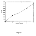
- Figure 1 is a graph showing counts per minutes (counts) of bound radioactive carcinogenic embryonic antigen versus anti-carcinogenic embryonic antigen microparticle concentration.
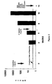
- Figure 2 is a graph showing antibody titer versus weeks after immunization with primary and secondary doses of tetanus toxoid particles.
- microparticles, methods of production, and kits are provided for diagnostic, therapeutic and research use.
- the microparticles are crosslinked macromolecular structures having a large surface area.
- the macromolecules forming the microparticles include, but are not limited to proteins, carbohydrates, polysaccharides, nucleic acids, viruses, virus particles, organic or inorganic synthetic pharmaceutical drugs, or mixtures thereof that can be crosslinked in a liquid phase under conditions of dehydration.
- the microparticle is formed by incubating macromolecules in solution or in liquid phase in the presence of a dehydrating agent and heat or a crosslinking agent for a sufficient amount of time to form particles.
- the macromolecule is first dissolved in an aqueous solvent, then either the macromolecule solution is added to the dehydrating agent or the dehydrating agent is added to the macromolecule solution, preferably the latter.
- the dehydrated macromolecule solution is then preferably heated for a predetermined length of time for the formation of microparticles.
- a crosslinking agent is added to the dehydrated macromolecule solution for microparticle formation at various temperatures above, below, or at room temperature.
- the resulting microparticles are then separated from any unreacted components present in the incubation mixture by physical separation methods well known to those skilled in the art.
- the macromolecule forming the microparticle is any molecule capable of being crosslinked in liquid phase.
- the macromolecule is a biomolecule such as a protein, carbohydrate, polysaccharide, nucleic acid molecule, virus, virus particle, or a mixture thereof.
- the macromolecule can also be a natural or synthetic pharmaceutical compound that is capable of being crosslinked. It will be understood by those skilled in the art that a compound incapable of being crosslinked can be formed into a microparticle by incorporation of the compound into a carrier molecule that is then crosslinked in accordance with the methods provided herein.
- the macromolecule can also be a portion of a molecule having the requisite activity to bind or interact with a ligand, such as, for example, a peptide, a single-stranded segment of a double-stranded nucleic acid molecule, or a virus particle.
- a ligand such as, for example, a peptide, a single-stranded segment of a double-stranded nucleic acid molecule, or a virus particle.
- the term "macromolecule” includes a plurality of macromolecules and includes combinations of different macromolecules such as a combination of a pharmaceutical compound and an affinity molecule for targeting the pharmaceutical compound to a tissue, organ or tumor requiring treatment.
- an affinity macromolecule can be either the receptor portion or the ligand portion of a receptor-ligand interaction.
- ligands that interact with other biomolecules include viruses, bacteria, polysaccharides, or toxins that act as antigens to generate an immune response when administered to an animal and cause the production of antibodies.
- the concentration of macromolecule in the incubation mixture is preferably between 0.1 and 100 mg/mL, depending on the incubation conditions.
- the macromolecule can be labelled with a detectable label.
- the various types of labels and methods of labelling proteins and nucleic acid molecules are well known to those skilled in the art. It will be understood by those skilled in the art that a magnetic substance, such as a metal, is included within the definition of the term label.
- the macromolecule can be labelled with a metallic substance, such as a metal, so that the microparticles can be separated from other substances in a solution with the aid of a magnetic device.
- the label can be a radiolabel such as, but not restricted to, 32 P, 3 H, 14 C, 35 S, 125 I, or 131 I.
- a 32 P label can be conjugated to a protein with a conjugating reagent or incorporated into the sequence of a nucleic acid molecule by nick-translation, end-labelling or incorporation of labelled nucleotide.
- a 3 H, 14 C or 35 S label can be incorporated into a nucleotide sequence by incorporation of a labelled precursor or by chemical modification, whereas an 125 I or 131 I label is generally incorporated into a nucleotide sequence by chemical modification.
- Detection of a label can be by methods such as scintillation counting, gamma ray spectrometry or autoradiography.
- the label can also be a Mass or Nuclear Magnetic Resonance (NMR) label such as, for example, 13 C, 15 N, or 19 O. Detection of such a label can be by Mass Spectrometry or NMR.
- NMR Nuclear Magnetic Resonance
- Dyes, chemiluminescent agents and fluorogens can also be used to label the macromolecule.
- dyes useful for labelling nucleic acids include ethidium bromide, actidines, propidium and other intercalating dyes, and 4',6'-diamidino-2-phenylindole (DAPI) (Sigma Chemical Company, St. Louis, MO) or other proprietary nucleic acid stains.
- DAPI 4',6'-diamidino-2-phenylindole
- f luorogens include fluorescein and derivatives, phycoerythrin, allo-phycocyanin, phycocyanin, rhodamine, Texas Red or other proprietary fluorogens.
- the fluorogens are generally attached by chemical modification.
- the dye labels can be detected by a spectrophotometer and the fluorogens can be detected by a fluorescence detector.
- the macromolecule can alternatively be labelled with a chromogen (enzyme substrate) to provide an enzyme or affinity label, or enzyme.
- a chromogen enzyme substrate
- the macromolecule can be biotinylated so that it can be utilized in a biotin-avidin reaction which may also be coupled to a label such as an enzyme or f luorogen.
- the macromolecule can be labelled with peroxidase, alkaline phosphatase or other enzymes giving a chromogenic or fluorogenic reaction upon addition of substrate.
- additives such as 5-amino-2,3-dihydro-1,4-phthalazinedione (also known as LuminolTM) (Sigma Chemical Company, St.
- rate enhancers such as p-hydroxybiphenyl (also known as p-phenylphenol) (Sigma Chemical Company, St. Louis, MO) can be used to amplify enzymes such as horseradish peroxidase through a luminescent reaction; and luminogeneic or fluorogenic dioxetane derivatives of enzyme substrates can also be used.
- Recognition sites for enzymes can also be incorporated into an macromolecule to provide a detectable label.
- a label can also be made by incorporating any modified base, amino acid, or precursor containing any label, incorporation of a modified base or amino acid containing a chemical group recognizable by specific antibodies, or by detecting any bound antibody complex by various means including immunofluorescence or immuno-enzymatic reactions.
- Such labels can be detected using enzyme-linked immunoassays (ELISA) or by detecting a color change with the aid of a spectrophotometer.
- the dehydrating agent is a chemical compound or mixture of compounds capable of diffusing water from the macromolecule to a highly ionic media.
- Suitable dehydrating agents include water soluble nonionizable linear or branched polymers of high molecular weight
- the dehydrating agent is a mixture of two or more soluble, linear polymers such as polyvinylpyrrolidone and polyethylene glycol.
- soluble, linear polymers such as polyvinylpyrrolidone and polyethylene glycol.
- Such a polymer mixture may be prepared in accordance with known methods. It will be understood by those skilled in the art that other soluble, linear polymers, such as dextran, nonylphenol-ethoxylates, polyvinyl alcohol, and mixtures thereof could be used in addition to PVP and PEG or in place of either PVP or PEG.
- PVP is a non-ionogenic, hydrophilic polymer having a mean molecular weight ranging from approximately 10,000 to 700,000 and the chemical formula (C 6 H 9 NO) n .
- PVP is also known as poly[1-(2-oxo-1-pyrrolidinyl)ethylene], PovidoneTM, PolyvidoneTM, RP 143TM, KollidonTM, Peregal STTM, PeristonTM, PlasdoneTM, PlasmosanTM, ProtagentTM, Subtosan, and VinisilTM.
- PVP is non-toxic, highly hygroscopic and readily dissolves in water or organic solvents.
- Polyethylene glycol also known as poly(oxyethylene) glycol, is a condensation polymer of ethylene oxide and water having the general chemical formula HO(CH 2 CH 2 O) n H.
- Dextran is a term applied to polysaccharides produced by bacteria growing on a sucrose substrate.
- Native dextrans produced by bacteria such as Leuconostoc mesenteroides and Lactobacteria dextranicum usually have a high molecular weight.
- NPEs Nonylphenol-ethoxylates
- Polyvinyl alcohol is a polymer prepared from polyvinyl acetates by replacement of the acetate groups with hydroxyl groups and has the formula (CH 2 CHOH) n . Most polyvinyl alcohols are soluble in water.
- PEG, dextran, PVA and PVP are commercially available from chemical suppliers such as the Sigma Chemical Company (St. Louis, MO). NPEs require custom synthesis and can be ordered from special chemical producers.
- the dehydrating agent is polymer mixture containing an aqueous solution of PVP having a molecular weight between 10,000 and 360,000, most preferably 40,000 and PEG having a molecular weight between 200 and 35,000.
- PVP having a molecular weight of 40,000 and PEG having a molecular weight of 3500 is preferred.
- PVP having a molecular weight of 360,000 is preferred for obtaining microparticles having uniform size.
- the PVP is dissolved in an acetate buffer and PEG is added to the aqueous PVP solution.
- the concentration of each polymer is preferably between 1 and 40 g/100 ml depending of the molecular weight of each polymer.
- the concentration of each polymer is 24 g/100 ml or 24%.
- Equal concentrations of PVP and PEG generally provide the most favorable polymer matrix for the formation of a polymer microparticle.
- the volume of polymer added to the macromolecule varies depending on the size and quantity of the macromolecule. Preferably, three volumes of the polymer mixture are added to one volume of a solution containing the macromolecule.
- Microparticles are formed by incubation of the macromolecule and dehydrating agent mixture at a temperature greater than room temperature for a predetermined length of time.
- the mixture is incubated in a water bath at a temperature greater than or equal to 37°C and less than or equal to 80°C for between approximately 5 minutes and 2 hours.
- the mixture is incubated for 15-30 minutes at a temperature between 50°C and 70°C.
- Microparticle size can be controlled by adjusting the incubation conditions. For example, incubation temperatures can be increased gradually or incrementally from room temperature to the desired elevated incubation temperature or overall incubation time can be increased. In addition, the amount of microparticle aggregation can be controlled by varying the concentration, volume, or composition of the dehydrating agent.
- Microparticles are alternatively formed by the addition of a crosslinking reagent to crosslink the dehydrated macromolecule.
- the crosslinking reagent is a bi- or multi-functional chemical reagent that physically links the macromolecules, and, in some cases, the dehydrating agent.
- suitable crosslinking agents include dialdehydes or other agents such as amines, multivalent ions, and multifunctional molecules that have an "affinity" for specific reactive groups on the macromolecule being crosslinked.
- the crosslinking agent covalently connects the macromolecules into a stable three-dimensional structure.
- the crosslinking agent is a bifunctional reagent such as glutaraldehyde; p,p' -difluoro- m,m' -dinitro diphenyl sulphone; hexamethylene diisocyanate; n,n' -(1,3-Phenylene)- bis -maleimide; n,n' -ethylene- bis -iodoacetamide; 3,6- bis -(mecurimethyl)-dioxan; bis-diazobenzidine; Woodward's K; bis-oxiranes; dimethyl adipimidate; dimethyl suberimidate; diethyl malonimidate; phenol-2,4-disulphonyl-chloride; divinylsulphone; and carbodiimides.
- the crosslinking agent is a dialdehyde such as glutaraldehyde which forms a Schiff base with primary amines, which on reduction with borohydride give stable secondary amines under mild conditions.
- N-substituted maleimides which are specific for sulphydryl groups under mild conditions.
- N-aryl and N-alkyl-bis-maleimides are commercially available, including azophenyldimaleimide. These are insoluble in water and are generally added in stoichiometric amounts as a solid to aqueous solution at pH 7 to 8 of the reactants.
- Bifunctional alkyl halides react primarily with thiol, imidazole and amino groups. At neutral to slightly alkaline pH the reaction with sulphydryl groups is favored while at higher pH values reaction with amino groups.
- Other compounds include aryl halides such as 1,5-difluoro-2,4-dinitrobenzene, which are insoluble in water and preferentially react with amino groups and tyrosine phenolic groups, but which will also react with sulphydryl and imidazole groups. Relatively high pH values are required for a rapid reaction.
- the reagent is generally added as a concentrated acetone solution to an aqueous solution of the reactants and product formation.
- Isocyanates react with amines to form substitute ureas, with alcohols to form urethanes and with water to give amines and carbon dioxide. At alkaline pH the reaction with amines is preferred. 2,2-dicarboxy-4,4'-azophenyldiisocyanate is water-soluble and has the advantage that the bridge it forms can be readily cleaved by reduction of the azo group by dithionite.
- Acylating agents can also be used, such as many of the aliphatic or aromatic dicarboxylic or disulphonic acids that are activated to provide bifunctional acylating agents capable of reacting under mild conditions. The nitrophenylesters of dicarboxylic acids and the aromatic-bis-sulphonyl chlorides are examples.
- the concentration of the crosslinking reagent in the incubation mixture should be sufficient to bind all of the active groups of the macromolecule. There is a direct relationship between the concentration of crosslinking agent and the number of microparticles formed after incubation. Generally, more microparticles are formed as the concentration of crosslinking agent in the incubation mixture is increased. Preferably, the concentration of crosslinking agent in the incubation mixture is between approximately 5 and 200 microliters of a 25% solution of glutaraldehyde per milliliter of incubation mixture.
- the dehydrating agent is a polymer solution, where the macromolecule, polymer and crosslinkng agent mixure are vigorously mixed together, such as by vortexing, to allow sufficient interaction between the macromolecules, polymers and crosslinking agent, and incubated, while mixing, at room temperature (20°C), or at a temperature below room temperature, for a sufficient amount of time to allow maximal formation of microparticles.
- microparticles can be formed utilizing a combination of crosslinking agent and heat, preferably by incubation at a temperature greater than or equal to 37°C and less than or equal to 80°C.
- the length of incubation time is dependent upon the respective concentrations of polymer and affinity molecule and the incubation temperature.
- the polymer mixture and macromolecules are incubated between 30 minutes and 24 hours.
- the polymer mixture and macromolecules are mixed, by stirring or rocking, for 120 minutes at room temperature and are then placed at 4°C overnight without mixing.
- the pH of the incubation mixture is generally determined by the pH of the dehydrating agent and may be adjusted by adding the appropriate amount of an acidic or basic buffer to either or both dehydrating or macromolecule solutions before they are mixed.
- the dehydrating agent is a linear polymer solution
- the pH of the linear polymer incubation mixture is preferably between approximately 5 and 8.
- a quenching reagent may be added to the resulting microparticles after incubation to block any unreacted binding sites of the crosslinking reagent in order to reduce subsequent non-specific binding.
- suitable quenching reagents are compounds, such as amino acids or albumin, that contain substantial numbers of amino groups.
- the quenching reagent is a solution containing lysine or glycine.
- the quenching reagent is the amino acid glycine in a concentration ranging from 0.1 to 0.5 M.
- the formed microparticles are separated from the non-reacted components of the incubation mixture by conventional separation methods well known to those skilled in the art.
- the incubation mixture is centrifuged so that the microparticles fall to the bottom of the centrifuge tube and the non-reacted components remain in the supernatant, which is then decanted.
- the incubation mixture containing the formed microparticles is filtered so that the microparticles are retained on the filter and the non-reacted components pass through the filter.
- washing solution is a buffer, most preferably a phosphate buffered saline solution containing the quenching reagent. Repeated washings can be utilized as necessary.
- dehydrating agent may be incorporated within the macromolecule structure and actually contribute to the molecular composition of each microparticle.
- microparticles formed by the foregoing process can be spherical or non-spherical in shape depending on temperature, polymer size and mixture and protein concentration with one or more active sites present on the surface of each microparticle.
- the elliptical shape and granularity of the microparticles create a particle having a greater surface area than spherical microparticle beads and allows for the incorporation of a larger number of macromolecules per microparticle than could be achieved with conventional spherical beads.
- microparticles are formed of macromolecules such as immunoglobulin crosslinked with glutaraldehyde in the presence of PVP/PEG, the microparticles are stable at alkaline and acid pH and are not absorbed when administered in vivo .
- the microparticles are useful for a wide variety of diagnostic, therapeutic, and research purposes as discussed in more detail below.
- the microparticles can include a macromolecule such as an immunoglobulin or cell receptor labelled with a detectable label. Injection of the labelled microparticle into a patient creates an imaging agent for the diagnosis of a proliferative disorder such as cancer or a tool for the evaluation of the success of a therapeutic agent in reducing the proliferation of a particular adverse cell or organism.
- microparticles containing a macromolecule such as an immunoglobulin, cell receptor or oligonucleotide probe specific for the cell or organism under investigation, are combined with a test sample, the microparticles are separated from any non-bound components of the sample, and bound molecules are detected by conventional methods.
- the microparticles are useful as therapeutic agents when the microparticles include a therapeutic drug and are injected into a patient for slow release or targeted delivery of the drug to the site requiring therapy.
- microparticles are also useful for the purification of molecules from a complex mixture, as a reagent for the detection or quantification of a specific molecule, or for the production of molecules, such as antibodies.
- a macromolecule such as an immunoglobulin
- microparticles containing a macromolecule, such as an immunoglobulin can be attached to a chromatography column and used in immunoaffinity chromatography to separate a ligand from a complex mixture.
- microparticles including a labelled macromolecule or a mixture of labelled macromolecules specific for different cells or biomolecules, such as cell receptors can be used to detect changes in the number of cells or biomolecules in response to a particular test condition using techniques such as flow cytometry.
- the microparticles can be used as adjuvants for vaccine production wherein antigen-containing microparticles are injected into a research animal, such as a mouse or rabbit, to trigger an enhanced immune response for the production of antibodies to the antigen.
- microparticles described herein are useful as solid phase particles in an assay, such as an enzyme-linked immunosorbant assay, dot-blot, or Western blot, for the detection of a particular target such as a cell, biomolecule or drug in a biological sample.
- the microparticles designed for this use are composed of affinity molecules specific for the target molecule.
- the macromolecule is an immunoglobulin, cell receptor or oligonucleotide probe and is bound to a test tube or microtiter plate.
- a sample is combined with a solution containing the microparticles, the macromolecules on the microparticles are reacted with the target molecule, the microparticles are separated from any non-bound components of the sample, and microparticles containing bound molecules are detected by conventional methods.
- Fluorescently stained microparticles are particularly well suited for flow cytometry analysis in accordance with methods well known to those skilled in the art.
- microparticles described herein are useful as visual probes or markers of pathology in a histological sample.
- the macromolecules of microparticles designed for this use are specific for biomolecules expressed during a particular pathologic condition and are labelled with a detectable label.
- the macromolecule is an immunoglobulin, cell receptor or oligonucleotide probe specific for an abnormal cell, such as a rapidly proliferating cell, or pathological organism, for example, a virus.
- a histological sample is combined with a solution containing the microparticles, the labelled macromolecules on the microparticles are reacted with the target molecule of interest, and bound microparticles are detected by detecting the label in accordance with methods well known to those skilled in the art.
- microparticles described herein are useful as imaging agents for in vivo localization of a particular molecule, cell type or pathologic condition in a manner similar to that described above with regard to the use of the microparticles for histopathology.
- the macromolecules on microparticles designed for this use are specific for molecules expressed by a particular cell or pathologic organism and are labelled with a detectable label.
- the macromolecule is an immunoglobulin, cell receptor or oligonucleotide probe specific for a tumor cell or pathological organism, such as a virus.
- the microparticles are used to either detect a pathologic condition or to monitor the success of therapy, such as chemotherapy or surgery to ensure that the size of an abnormal tissue tumor has decreased or has been completely excised.
- a patient receives an administration of a microparticle solution, preferably intravenously, the labelled macromolecules on the microparticles are given a sufficient amount of time to localize to the affected organ or region of the body, the macromolecule is reacted with a target molecule expressed by the cell or organism under investigation, and bound microparticles are detected by detecting the label by conventional imaging techniques well known to those skilled in the art, such as x-ray.
- the microparticles are useful for therapy when composed of a crosslinked pharmaceutical compound or a crosslinked carrier, such as albumin, containing a therapeutic agent.
- the microparticle can either provide for the slow release of the agent throughout the body or the microparticle can include an affinity molecule specific for a target tissue, or tumor, and be injected into a patient for targeted delivery of the therapeutic agent, such as an antitumor, antiviral, antibacterial, antiparasitic, or antiarthritic agent, cytokine, hormone, or insulin directly to the site requiring therapy.
- microparticles are also useful as research tools for the purification of a biomolecule from a complex mixture, as a reagent for the detection or quantification of a biomolecule, or for the production of biomolecules, such as antibodies.
- microparticles composed of a macromolecule are attached to a chromatography column and used in immunoaffinity chromatography to separate a ligand from a complex mixture.
- a macromolecule such as an immunoglobulin
- immunoaffinity chromatography to separate a ligand from a complex mixture.
- microparticles including a labelled macromolecule or a mixture of labelled macromolecules specific for different cells or cell receptors are used to detect changes in the number of cells or cell surface receptors in response to a particular test condition using techniques such as flow cytometry.
- microparticles are useful adjuvants for antibody production wherein antigen-containing microparticles are injected into an animal, such as a mouse or rabbit, for vaccine production, or a human, to induce immunity to an antigen, to trigger an enhanced immune response for the production of antibodies to the antigen.
- kits for the production of microparticles contains the following reagents: a dehydrating agent and a crosslinking agent.
- the user of the kit may use the kit for the preparation on custom microparticles wherein the user will supply the macromolecule that will be formed into the microparticles.
- the kit can contain one or more macromolecules, in solution or lyophilized form, for the preparation of microparticles of interest to the user.
- the formed microparticles can then be used for research, therapeutics or diagnostics as described above.
- the kit preferably also contains a buffer, such as phosphate buffered saline, containing a quenching reagent, such as glycine, to block non-specific binding by the crosslinking reagent.
- a detectable label, or prelabelled macromolecule can also be included with the kit to provide a means for detecting the presence of the microparticle in a sample or patient.
- Example 1 Preparation of Microparticles with Gammaglobulin and a Polymer Mixture of Polyvinylpyrrolidone and Polyethylene Glycol and a Stability Analysis Thereof
- Microparticles were formed by combining gammaglobulin, one of five molecular weight preparations (MW 10,000-360,000) of a 5-25% polyvinylpyrrolidone solution, and a constant molecular weight preparation (MW 3500) of a 25% polyethylene glycol solution (both prepared as described below in Example 2) in the presence of glutaraldehyde, at a reaction pH range between 6.9 and 7.75, by the following process.
- the microparticles were stable in both acidic and basic solutions.
- polymer mixtures each containing a different molecular weight preparation of polyvinylpyrrolidone, were prepared as indicated below in Table 1.
- the resuspended samples were again centrifuged, 100 ⁇ l removed and reacted with 300 ⁇ l TM Blue, supernatants decanted, and precipitates resuspended in 1 ml PBS/Tween. A 100 ⁇ l aliquot of the resuspended precipitate was reacted with 300 ⁇ l TM Blue.
- One hundred microliters of the first resuspended precipitate of reaction #3 were placed into three test tubes. Two hundred microliters of deionized water were added to the first tube. Particles were observed. Two hundred microliters of 1 N acetic acid were added to the second tube. Particles having the same size as observed in the first tube were observed. Two hundred microliters of 1% Na OH were added to the third tube. Particles having the same size as observed in the first tube were observed.
- Tubes 1 and 2 did not change. Tube 3 appeared to have smaller particles than tubes 1 or 2.
- Anti-CEA (carcinogenic embryonic antigen) microparticles were formed as generally described for Reaction #3 in Example 1 and described in more detail below. The resulting microparticles were then combined with various concentrations of radioactive CEA to determine whether the anti-CEA antibodies incorporated in the microparticles retained affinity for the CEA ligand.
- Resuspended precipitates were washed with 1.8 ml of the polymer mixture, preadjusted to pH 6.25, allowed to stand at 3°C for 20 minutes, and centrifuged at 5000 rpm for 30 minutes at 20°C.
- Reactants were centrifuged at 5000 rpm for 30 minutes at 20°C. The sample was placed at 4°C for 60 hours and then re-centrifuged. The supernatant appeared clear and was decanted. The precipitate, containing the anti-CEA microparticles, was resuspended in 10 ml phosphate buffered saline (1X).
- Radioactive CEA (I 125 ) was diluted with phosphate buffered saline containing 10% liquid fish gelatin.
- the diluted I 125 CEA contained 39313 counts per 100 ⁇ l.
- the radioactive CEA/anti-CEA microparticle mixtures in tubes 1-5 were incubated for 2 hours in the refrigerator while shaking.
- the radioactive CEA/anti-CEA microparticle mixtures in tubes 6-9 were incubated for 15 minutes at room temperature while shaking.
- Reactants were washed three times with phosphate buffered saline containing 10% liquid fish gelatin, centrifuged 1 minute in a high speed centrifuge, and resuspended in phosphate buffered saline.
- the results indicate that the anti-CEA microparticles are immunologically active and react with CEA as shown numerically in Table 3 and graphically in Figure 1.
- a polymer mixture of PVP/PEG was prepared as generally described in Example 2 (48% total polymers) and was adjusted to the pH indicated in Table 4. One milliliter of each solution was placed in each of 7 centrifuge tubes: Adjusted pH of PVP/PEG Mixture Tube # pH of PVP/PEG 1 4.6 2 5.2 3 5.8 4 6.6 5 7.4 6 8.1 7 9.2
- Tube #7 had to be treated very carefully because the precipitate was not adhered to the wall of the tube. 90% of the supernate was carefully removed by a disposable pipet.
- the particles were brought to room temperature, mixed well, and then centrifuged at 2600 RPM at 20°C for 30 minutes. Supernatants were decanted and precipitates resuspended in 5.0 ml of 0.5 M glycine in 1X PBS buffer, pH 5.6. The wash procedure was then repeated 3 more times until the supernatants were clear.
- anti-HCG Fluorescently-labelled anti-Human Chorionic Gonadotropin
- the mouse was then killed by cervical dislocation.
- a volume of a PVP/PEG polymer mixture was prepared as described in Example 2 to form a polymer mixture having a polymer concentration of 53.3%, adjusted to pH 4.8.
- Polymer dilutions were made as set forth below in Table 5 using 0.1 M sodium acetate at pH 5.0: Volume and Percent of Polymer Mixture Polymer Mixture (mls) Na Acetate (mls) Polymers % 10 0 53.3% 9 1 48.0% 8 2 42.4% 7 3 37.1% 6 4 31.8% 5 5 26.7% 4 6 21.3% 3 7 15.9% 2 9 10.7% 1 9 5.3% 0 10 0%
- the tubes were then reacted with 300 ⁇ l fluorescently labelled human IgG.
- the purified human antibody was 3X concentrated in a 0.1 M glycine solution in PBS buffer at pH 11.0 and labelled with fluorescein isothiocyanate in the 0.1 M glycine buffer at a concentration of 3 mg/ml.
- the antibody was added to the polymer mixture dilutions while vortexing, and the tubes were mixed for 1 hour followed by 30 minutes centrifugation at 2500 rpm at 20°C.
- Each tube had a different appearance from the start of the reaction.
- the tube containing 5.3% polymer had only slight precipitation, much of which clumped together on the walls of the tube.
- the tube containing 53.3% polymer had very fine particles which did not stick together or to the walls of the tube.
- control precipitate was very small and exhibited a bright orange color, as were the tubes with very low polymer concentrations.
- ease with which the precipitates were broken up in the 0.5 M glycine wash buffer was also related to polymer concentration.
- the precipitate the tube containing only 5.3% polymer mixture was sticky and difficult to break up, but in each successive tube the particles were less sticky. At a polymer concentration of 31.8% or greater, the particles separated very easily.
- Polymer concentration plays a definite role in determining the characteristics of antibody particles made with the polymer mixture. Polymer concentrations of 40% and over provide very fine particles that do not stick. Also, the higher the concentration of polymers in the polymer mixture, the greater the number of antibody particles that can be formed.
- the size of antibody particles can easily be controlled by altering the pH of the polymer mixture.
- glutaraldehyde forms very strong bonds with the amino groups of proteins, accounting for the yellow-orange color and overall stickiness of the increasingly basic samples.
- acidic pH values the effects of the glutaraldehyde are lessened. Particles can still form but they are much more fine with less of a tendency to clump than those particles made with the basic polymer mixture.
- This experiment was performed to show the effect of increasing the amount of glutaraldehyde on the formation of IgG microparticles.
- Particles were stored at 4°C in 5.0 ml 0.5 M glycine in 1X PBS buffer pH 7.0.
- HCG human chorionic gonadotropin
- HCG tracer iodinated HCG antigen, Becton Dickinson, San Jose, CA
- FSH follicle stimulating hormone
- estradiol tracer iodinated Estradiol antigen
- anti-HCG microparticles are capable of binding iodinated HCG antigen in an immunoassay.
- the anti-HCG microparticles are not capable of nonspecific binding of other iodinated antigens such as FSH and Estradiol in an immunoassay.
- This experiment was performed to prepare tetanus toxoid microparticles with a polymer mixture of PVP and PEG and glutaraldehyde.
- the reaction containing 100 ⁇ l glutaraldehyde contained more particles than the reaction with 10 ⁇ l glutaraldehyde. Particles for both reactions were similar in size. The particles appear very small to the eye.
- This experiment was performed to demonstrate the formation of bovine serum albumin (BSA) microparticles with a polymer mixture of PVP/PEG and glutaraldehyde and to observe the effect of different volumes of glutaraldehyde used in this process.
- BSA bovine serum albumin
- Bovine serum albumin microparticles were formed in each tube. As the glutaraldehyde amount decreased, larger size aggregates were formed in addition to small fine particles seen with all volumes of glutaraldehyde (1 ⁇ l to 200 ⁇ l). Also, as the glutaraldehyde amount increased the number of the small fine particles increased.
- Example 12 Preparation of Immunoglobulin Microparticles with Saturated Ammonium Sulfate as the Dehydrating Agent
- This experiment was performed to prepare immunoglobulin microparticles using saturated ammonium sulfate, over a pH gradient, as the dehydrating agent and glutaraldehyde as the crosslinking agent.
- the size of the particles did not differ between the range of pHs of the saturated ammonium sulfate solutions.
- Example 13 Preparation of Immunoglobulin Microparticles with a Mixture of Saturated Ammonium Sulfate and Polyethylene Glycol as the Dehydrating Agent
- This experiment was performed to prepare immunoglobulin microparticles using a mixture of saturated ammonium sulfate and polyethylene glycol (PEG) as the dehydrating agent and glutaraldehyde as the crosslinking agent.
- PEG polyethylene glycol
- a 100 ml volume of a saturated ammonium sulfate solution was prepared as described in Example 12.
- a 20% solution of polyethylene glycol (Sigma, St. Louis, MO) in 0.1 M sodium acetate, pH 4.8 was also prepared.
- the ammonium sulfate solution was added, in small increments, to 10 ml of the PEG solution until the ammonium sulfate precipitated.
- the largest volume of ammonium sulfate that would stay in solution was 1.2 ml.
- One ml of this ammonium sulfate/PEG solution was aliquoted into each of eight tubes. The tubes then received 25% glutaraldehyde (Sigma, St.
- Tubes 5-8 displayed a color reaction that increased in strength across the gradient. The brightness of the color also increased over time. This was a result of the glutaraldehyde reacting with the amines of the ammonium sulfate. Tubes 1-4 did not change color because the glutaraldehyde concentrations in these tubes were very low.
- a 300 ⁇ l aliquot of 3 times concentrated purified porcine IgG antibody was added to each tube while vortexing. The tubes then were mixed for 60 minutes and were centrifuged for 30 minutes at 3600 RPM at 20°C.
- the supernatants were decanted and 10 ml of 0.5 M glycine (Sigma, St. Louis, MO), pH 7.0, in phosphate buffered saline, was added to each precipitate. The tubes were shaken well to break up the precipitate and stored overnight at 4°C.
- 0.5 M glycine Sigma, St. Louis, MO
- pH 7.0 phosphate buffered saline
- the tubes were centrifuged for 30 minutes at 3600 RPM at 20°C.
- the supernatants were decanted and the precipitates washed with 5 ml of the 0.5 M glycine buffer solution.
- the tubes were centrifuged for 30 minutes at 3600 RPM at 20°C. Following centrifugation, the supernatants were decanted and the precipitates received 5 ml of the 0.5 M glycine buffer.
- the tubes were shaken well.
- the precipitates differed in size and color, with the precipitates growing larger and darker as the glutaraldehyde concentration increased.
- the particles were apparent in all tubes except tube #1, which received no glutaraldehyde. There were fine particles that appeared to be of uniform size in tubes 2-7. However, the particles were sticky and tended to clump in all tubes. There appeared to be a greater proportion of clumping relative to the amount of particles in the tube in those tubes that received less than 20 ⁇ l glutaraldehyde. These results were consistent over the final two washes.
- This experiment was performed to prepare insulin microparticles with a polymer mixture of PVP and PEG and glutaraldehyde, measure the concentration of microparticles formed, and assay their immunologic activity.
- the washing procedure consisted of adding 5.0 ml lysine solution to the particles, mixing well but briefly. Centrifuged at 2300 rpm for 30 minutes at 20°C. Aspirated supernatant and repeated the process. Stored particles in the lysine solution overnight at 4°C.
- the pH of each resuspension was as follows: tube A was pH 9.2, tube B was pH 9.2, and tube C was pH 8.8.
- the pH of each resuspension was as follows: tube A was pH 8.0, tube B was pH 7.4, and tube C was pH 7.2.
- the particles formed were very small and fine. During centrifugation in the lysine solution, the particles were unable to fully compact. After washing with the phosphate buffered saline solution, particles were able to compact to a greater degree and also formed aggregates of particles.
- Tube C had the largest amount of particles. Tube B had more particles than Tube A. Therefore, particle formation increased with the amount of glutaraldehyde added to each tube.
- the immunologic activity of the insulin microparticles was determined by measuring the ability of the microparticles to bind anti-sheep insulin in a sheep anti-insulin peroxidase assay.
- the immunologic activity of the insulin microparticles was determined by measuring the ability of the microparticles to bind anti-sheep insulin in an inhibition assay.
- Tetanus toxoid microparticles were prepared as generally described above in Example 10 as follows:
- tetanus toxoid 1.0 mg/ml
- the combination was mixed for 4 hours and 15 minutes at room temperature and then centrifuged at 3000 RPM at 4°C for 30 minutes. Supernatants were decanted, particles resuspended in 2.0 ml of 0.5 M glycine in 1 x PBS buffer and allowed to stand overnight at 4°C.
- Tetanus toxoid particles were finally resuspended in 0.5 ml 1x PBS buffer.
- the particles had a particle size of between approximately 50 and 100 microns .
- mice were each injected subcutaneously, following FDA protocol, with a primary dose containing 2 ⁇ g of the tetanus toxoid particles.
- a secondary injection containing 2 ⁇ g of the tetanus toxoid particles was administered seven weeks after the primary injection.
- Blood samples were taken from the mice at 2, 4, 5, 8, and 10 weeks after primary injection and antibody titers in the blood were determined by immunoassay.
- Mice were challenged with a lethal dose of tetanus toxoid fourteen weeks after the primary injection.
- antibody titers increased from approximately 10, at 4 weeks after primary injection, to approximately 3000, at 10 weeks after primary injection.
- the dotted line indicates titers conferring 2 antitoxin units of protection from tetanus toxoid. Titers above the dotted line indicate a positive immune response, according to FDA standards. Therefore, as shown in the Figure 2, the mice had a positive immune response as early as four weeks after injection with the tetanus toxoid microparticles.
- the increase in antibody titer from four to ten weeks indicates that the microparticles may be providing slow release of the tetanus toxoid antigen.
- mice survived after challenge with the lethal dose of tetanus toxoid administered at fourteen weeks. This survival rate indicates a strong immune response conferring protection against tetanus toxoid.
- mice appeared healthy. There was no noticeable inflammation or scarring at the site of injection and no significant weight loss after inoculation. Therefore, the tetanus toxoid microparticles were generally non-toxic when administered subcutaneously.
- This experiment provides in vivo data showing that administration of tetanus toxoid microparticles, prepared in accordance with the method described above, provides a slow release of tetanus toxoid antigen, causes a positive immune response, and protects against lethal challenge by tetanus toxoid in the absence of adverse effects.
- This experiment was performed to prepare albumin microparticles by incubating albumin with polymer mixtures of PVP and PEG at various temperatures.
- bovine serum albumin-FITC solution containing 1% bovine serum albumin (BSA) plus 10 ⁇ l dialyzed fluorescein isothiocyanate (FITC) albumin.
- BSA bovine serum albumin
- FITC fluorescein isothiocyanate
- Reaction 1 was mixed at room temperature for 1.5 hours.
- Reaction 2 was mixed at room temperature for 30 minutes, then incubated in a 58°C water bath for 30 minutes.
- Reaction 3 was immediately placed in a 58°C water bath for 30 minutes.
- Reaction 4 was mixed at room temperature for 30 minutes, then incubated in a 37°C water bath for 30 minutes, then incubated in a 58°C water bath for 30 minutes.
- Reaction mixture 2 contained non-aggregated microparticles between approximately 1 and 10 ⁇ m in diameter.
- Reaction mixture 3 contained non-aggregated microparticles approximately 10 ⁇ m in diameter with many microparticles less than 1 ⁇ m in diameter.
- Reaction mixture 4 contained non-aggregated microparticles between approximately 10 and 25 ⁇ m in diameter with some microparticles less than 1 ⁇ m in diameter.
- reaction mixtures to stand for 30 minutes at room temperature, then placed tubes in a 37-40°C water bath for 30 minutes, then transferred tubes to a 56-60°C water bath for 30 minutes.
- microparticles formed using 0.5 ml of polymer mixture were mostly uniform, having a diameter range between 1 and 3 ⁇ m. Small clusters were observed. A few large microparticles were seen having a diameter of approximately 25 ⁇ m.
- microparticles formed using 1.0 ml of polymer mixture were less uniform than those formed using 0.5 ml, having a diameter range of between less than 1 and 10 ⁇ m. Fewer clusters were observed than above. No large microparticles were seen.
- microparticles formed using 2.0 ml of polymer mixture were less uniform than those formed using 0.5 ml, having a diameter range of between less than 1 and 15 ⁇ m. Very few clusters were observed. No large microparticles were seen.
- microparticles formed using 3.0 ml of polymer mixture were less uniform than those formed using 0.5 ml, having a diameter range of between less than 1 and 20 ⁇ m. Very few clusters were observed. No large microparticles were seen.
- microparticles formed using 4.0 ml of polymer mixture were less uniform than those formed using 0.5 ml, having a diameter range of between less than 1 and 25 ⁇ m. No clusters or large microparticles were observed.
- microparticles formed using 5.0 ml of polymer mixture were less uniform than those formed using 0.5 ml, having a diameter range of betweeb less than 1 and 30 ⁇ m. No clusters or large microparticles were observed.
- microparticles formed using 0.5 ml of polymer mixture were in the form of small aggregates containing between 10 and 20 microparticles each having a diameter of less than 1 ⁇ m. Some large microparticles were observed having a diameter of approximately between 5 and 20 ⁇ m.
- microparticles formed using 1.0 ml of polymer mixture were in the form of small aggregates containing between 10 and 20 microparticles each having a diameter of less than 1 ⁇ m. Large microparticles having a diameter of approximately between 5 and 50 ⁇ m were frequently observed.
- microparticles formed using 2.0 ml of polymer mixture were in the form of large aggregates containing microparticles that were submicron in diameter. Some microparticles having a diameter of between 1 and 10 ⁇ m were observed.
- microparticles formed using 3.0 ml of polymer mixture were in the form of large and small aggregates. Occasionally, individual microparticles having a diameter of approximately 5 ⁇ m were seen.
- microparticles formed using 4.0 ml of polymer mixture were in the form of small aggregates containing 10-20 microparticles each having a diameter smaller than those observed when 0.5 ml of polymer mixture was used. No individual microparticles were observed.
- microparticles formed using 5.0 ml of polymer mixture were in the form of small and large aggregates containing very small microparticles. No individual microparticles were observed.
- reaction mixtures to stand for 30 minutes at room temperature, then placed tubes in a 37 to 40°C water bath for 30 minutes, then transferred tubes to a 56 to 60°C water bath for 30 minutes.
- microparticles formed using 0.5 ml of polymer mixture were in the form of small aggregates of tiny microparticles less than 1 ⁇ m in diameter with approximately between 10 and 20 per aggregate. Occasionally, larger single microparticles having a diameter of between 1 and 10 ⁇ m were observed.
- microparticles formed using 0.75 ml of polymer mixture were in the form of small aggregates with approximately between 1 and 10 microparticles per aggregate. Occasionally, larger single microparticles having a diameter of between 1 and 10 ⁇ m were observed.
- microparticles formed using 1.0 ml of polymer mixture formed large adherent aggregates during the 37 to 40°C incubation.
- the aggregates contained approximately between 1 and 5 microparticles per aggregate. Occasionally, larger single microparticles having a diameter of between 1 and 10 ⁇ m were observed.
- microparticles formed using 1.25 ml of polymer mixture formed aggregates during the 56 to 60°C incubation.
- the aggregates contained approximately between 1 and 5 microparticles per aggregate. Occasionally, larger single microparticles having a diameter of between 1 and 10 ⁇ m were observed.
- microparticles formed using 1.5 ml of polymer mixture formed aggregates during the 56 to 60°C incubation.
- the aggregates contained approximately between 1 and 5 microparticles per aggregate.
- Several larger, single microparticles having a diameter of between 1 and 10 ⁇ m were observed.
- microparticles formed using 1.75 ml of polymer mixture formed during the 56 to 60°C incubation. Due to their small size, it was difficult to determine whether the microparticles were present as aggregates. Several larger, single microparticles having a diameter of between 1 and 10 ⁇ m were observed.
- microparticles formed using 2.0 ml of polymer mixture formed large aggregates of 10 to 100 microparticles per aggregate. Some single microparticles having a diameter of 1 ⁇ m were observed.
- albumin microparticles could be prepared by incubating albumin and a PVP/PEG mixture at a temperature between 37°C and 60°C for approximately 30 minutes.
- the size of the microparticles and degree of aggregate formation could be changed by altering the composition or volume of the PVP/PEG polymer mixture added to the albumin.
Claims (25)
- Composition de micro particules, comprenant des macro molécules en juxtaposition à un agent de déshydratation, dans laquelle l'agent de déshydratation est sélectionné parmi le groupe constitué de polymères linéaires et ramifiés, solubles, et dans laquelle la composition de micro particules peut être obtenue par incubation de la macro molécule avec l'agent de déshydratation à une température supérieure à la température ambiante ou en présence d'un agent de réticulation pour une quantité suffisante de temps pour former les micro particules.
- Composition de micro particules selon la revendication 1, dans laquelle la macro molécule est sélectionnée parmi le groupe constitué d'une protéine, d'un glucide, d'un polysaccharide, d'une molécule d'acide nucléique, d'un virus, d'une particule de virus, d'un médicament pharmaceutique et de mélanges de ces derniers.
- Composition de micro particules selon la revendication 2, dans laquelle la protéine est sélectionnée parmi le groupe constitué d'une immunoglobuline, d'un antigène et d'un récepteur cellulaire.
- Composition de micro particules selon la revendication 1, comprenant en outre une molécule thérapeutique.
- Composition de micro particules selon la revendication 4, dans laquelle la molécule thérapeutique est sélectionnée parmi le groupe constitué d'agents chimiothérapeutiques, d'agents antiviraux, d'agents antibactériens, d'agents antiparasitiques, d'agents d'immuno suppression, de cytokines, d'hormones, d'enzymes et de mélanges de ces derniers.
- Composition de micro particules selon la revendication 1, comprenant en outre une substance magnétique.
- Composition de micro particules selon la revendication 1, dans laquelle la macro molécule est marquée à l'aide d'un marqueur détectable.
- Composition de micro particules selon la revendication 7, dans laquelle le marqueur détectable est sélectionné parmi le groupe constitué d'un fluoro chrome, d'un marqueur chimioluminescent, d'une particule magnétique, d'une enzyme, d'un substrat d'enzyme et d'un marqueur radioactif.
- Composition de micro particules selon la revendication 1, dans laquelle la micro particule est stable in vitro et in vivo.
- Composition de micro particules selon la revendication 1, dans laquelle l'agent de déshydratation est un polymère linéaire soluble, sélectionné parmi le groupe constitué de la polyvinylpyrrolidone, du polyéthylèneglycol, de la dextrane, d'éthoxylates de nonylphénol, de l'alcool polyvinylique et de mélanges et de dérivés de ces derniers.
- Composition de micro particules selon la revendication 1, dans laquelle les macro molécules sont réticulées à l'aide d'un composé sélectionné parmi le groupe constitué de dialdéhydes, d'amines, d'ions multivalents, de molécules multi fonctionnelles ayant une affinité pour des groupements réactifs spécifiques sur la macromolécule en cours de réticulation, des maléimides N-substitués, des alkyl halogénures bi-fonctionnels, des aryl halogénures, des isocyanates, des acides dicarboxyliques aliphatiques ou aromatiques, des acides disulfoniques aliphatiques ou aromatiques, des imidoesters bi-fonctionnels, des carbodiimides et des vinyl sulfones, composé qui est inclus dans les mélanges d'incubation.
- Procédé de fabrication d'une composition de micro particules selon la revendication 1, comprenant les étapes :(a) d'incubation d'une macro molécule avec un agent de déshydratation à une température supérieure à la température ambiante ou en présence d'un agent de réticulation pour une quantité suffisante de temps pour former les micro particules, dans laquelle l'agent de déshydratation est sélectionné parmi les polymères linéaires et ramifiés solubles, et(b) de séparation des particules du mélange d'incubation.
- Procédé selon la revendication 12, dans lequel la température d'incubation est supérieure ou égale à 37°C et inférieure ou égale à 80°C.
- Procédé selon la revendication 12, dans lequel le mélange d'incubation est incubé pendant une période comprise entre 5 minutes et 24 heures.
- Procédé selon la revendication 12, dans lequel la température d'incubation est inférieure ou égale à la température ambiante, lorsqu'un agent de réticulation est présent.
- Procédé selon la revendication 12, comprenant en outre l'étape de lavage des particules à l'aide d'un tampon qui contient un réactif de trempage.
- Procédé selon la revendication 12, dans lequel l'étape d'incubation est effectuée à un pH compris entre 5 et 8.
- Procédé selon la revendication 12, dans lequel l'agent de déshydratation est un polymère linéaire soluble sélectionné parmi le groupe constitué de la polyvinylpyrrolidone, du polyéthylèneglycol, de la dextrane, d'éthoxylates de nonylphénol, de l'alcool polyvinylique et de mélanges et de dérivés de ces derniers.
- Procédé selon la revendication 12, dans lequel l'agent de réticulation est sélectionné parmi le groupe constitué de dialdéhydes, d'aminés, d'ions multivalents, de molécules multi fonctionnelles ayant une affinité pour des groupements réactifs spécifiques sur la macromolécule en cours de réticulation, des maléimides N-substitués, des alkyl halogénures bi-fonctionnels, des aryl halogénures, des isocyanates, des acides dicarboxyliques aliphatiques ou aromatiques, des acides disulfoniques aliphatiques ou aromatiques, des imidoesters bi-fonctionnels, des carbodiimides et des vinyl sulfones.
- Procédé selon la revendication 12, dans lequel la macro molécule est sélectionnée parmi le groupe constitué d'une protéine, d'un glucides, d'un polysaccharide, d'une molécule d'acide nucléique, d'un virus, de particule de virus, d'un médicament pharmaceutique et de mélanges de ces derniers.
- Procédé en vue de l'isolement d'une molécule cible provenant d'un mélange complexe contenant la molécule, comprenant les étapes :(a) de mélange du mélange complexe avec une composition de micro particules macro moléculaire selon la revendication 1, ayant une affinité pour la molécule cible pour une quantité suffisante de temps pour permettre à la molécule cible de se lier à la macro molécule, dans lequel/laquelle la composition de micro particules peut s'obtenir par incubation de la macro molécule avec un agent de déshydratation à une température supérieure à la température ambiante ou en présence d'un agent de réticulation pour une quantité suffisante de temps pour former des micro particules, dans lequel l'agent de déshydratation est sélectionné parmi des polymères linéaires et ramifiés solubles, dans lequel la macro molécule comprend une molécule d'affinité spécifique à la molécule cible, et(b) de séparation de la molécule cible liée provenant du mélange complexe.
- Procédé selon la revendication 21, dans lequel la micro particule est immobilisée.
- Procédé selon la revendication 21, dans lequel la molécule cible est sélectionnée parmi le groupe constitué d'une protéine, d'un glucide, d'une molécule d'acide nucléique, d'un virus et de cellules.
- Procédé en vue de la détection d'une bio molécule cible dans un échantillon comprenant :(a) la combinaison avec l'échantillon d'une composition de micro particules selon la revendication 1, dans lequel la composition de micro particules peut s'obtenir par incubation de la macro molécule avec un agent de déshydratation à une température supérieure à la température ambiante ou en présence d'un agent de réticulation pour une quantité suffisante de temps pour former les micro particules, dans lequel l'agent de déshydratation est sélectionné parmi des polymères linéaires et ramifiés solubles et dans lequel la macromolécule comprend une molécule d'affinité spécifique à la bio molécule cible marquée à l'aide d'un agent d'imagerie détectable et(b) la détection de l'agent d'imagerie détectable.
- Procédé selon la revendication 24, dans lequel l'agent d'imagerie détectable est sélectionné parmi le groupe constitué d'un fluoro chrome, d'un marqueur chimiluminescent, des particules magnétiques, d'un enzyme, d'un substrat d'enzyme et d'un marqueur radioactif.
Applications Claiming Priority (3)
| Application Number | Priority Date | Filing Date | Title |
|---|---|---|---|
| US2823793A | 1993-03-09 | 1993-03-09 | |
| US28237 | 1993-03-09 | ||
| EP94910826A EP0688429B1 (fr) | 1993-03-09 | 1994-03-04 | Microparticules macromoleculaires et procedes de preparation |
Related Parent Applications (2)
| Application Number | Title | Priority Date | Filing Date |
|---|---|---|---|
| EP94910826A Division EP0688429B1 (fr) | 1993-03-09 | 1994-03-04 | Microparticules macromoleculaires et procedes de preparation |
| EP94910826.0 Division | 1994-09-15 |
Publications (2)
| Publication Number | Publication Date |
|---|---|
| EP0809110A1 EP0809110A1 (fr) | 1997-11-26 |
| EP0809110B1 true EP0809110B1 (fr) | 2004-01-28 |
Family
ID=21842313
Family Applications (2)
| Application Number | Title | Priority Date | Filing Date |
|---|---|---|---|
| EP94910826A Expired - Lifetime EP0688429B1 (fr) | 1993-03-09 | 1994-03-04 | Microparticules macromoleculaires et procedes de preparation |
| EP97112821A Expired - Lifetime EP0809110B1 (fr) | 1993-03-09 | 1994-03-04 | Microparticules macromoléculaires et procédés de préparation |
Family Applications Before (1)
| Application Number | Title | Priority Date | Filing Date |
|---|---|---|---|
| EP94910826A Expired - Lifetime EP0688429B1 (fr) | 1993-03-09 | 1994-03-04 | Microparticules macromoleculaires et procedes de preparation |
Country Status (11)
| Country | Link |
|---|---|
| US (1) | US5578709A (fr) |
| EP (2) | EP0688429B1 (fr) |
| JP (5) | JPH08507806A (fr) |
| AT (2) | ATE258685T1 (fr) |
| AU (1) | AU6358594A (fr) |
| CA (1) | CA2157793C (fr) |
| DE (2) | DE69408527T2 (fr) |
| DK (1) | DK0809110T3 (fr) |
| ES (2) | ES2215207T3 (fr) |
| PT (1) | PT809110E (fr) |
| WO (1) | WO1994020856A1 (fr) |
Cited By (4)
| Publication number | Priority date | Publication date | Assignee | Title |
|---|---|---|---|---|
| US8323615B2 (en) | 2008-08-20 | 2012-12-04 | Baxter International Inc. | Methods of processing multi-phasic dispersions |
| US8367427B2 (en) | 2008-08-20 | 2013-02-05 | Baxter International Inc. | Methods of processing compositions containing microparticles |
| US8389493B2 (en) | 2006-08-04 | 2013-03-05 | Baxter International Inc. | Microsphere-based composition for preventing and/or reversing new-onset autoimmune diabetes |
| US9339465B2 (en) | 2004-05-12 | 2016-05-17 | Baxter International, Inc. | Nucleic acid microspheres, production and delivery thereof |
Families Citing this family (130)
| Publication number | Priority date | Publication date | Assignee | Title |
|---|---|---|---|---|
| US6090925A (en) | 1993-03-09 | 2000-07-18 | Epic Therapeutics, Inc. | Macromolecular microparticles and methods of production and use |
| WO1996020012A2 (fr) * | 1994-12-23 | 1996-07-04 | Middlesex Sciences, Inc. | Procedes de preparation et de purification de conjugues macromoleculaires |
| AU733212B2 (en) | 1996-03-28 | 2001-05-10 | Board Of Trustees Of The University Of Illinois, The | Material and methods for making improved liposome compositions |
| US6217886B1 (en) | 1997-07-14 | 2001-04-17 | The Board Of Trustees Of The University Of Illinois | Materials and methods for making improved micelle compositions |
| SE512663C2 (sv) | 1997-10-23 | 2000-04-17 | Biogram Ab | Inkapslingsförfarande för aktiv substans i en bionedbrytbar polymer |
| CA2322954C (fr) | 1998-03-06 | 2011-06-07 | Biosepra Inc. | Particules implantables faisant gonfler les tissus pour le traitement du reflux gastro-oesophagien pathologique, de l'incontinence urinaire et de des rides |
| US6660301B1 (en) | 1998-03-06 | 2003-12-09 | Biosphere Medical, Inc. | Injectable microspheres for dermal augmentation and tissue bulking |
| WO2000004916A1 (fr) * | 1998-07-23 | 2000-02-03 | Societe De Conseils De Recherches Et D'applications Scientifiques Sas | Encapsulation de peptides hydrosolubles |
| US20070009605A1 (en) * | 1998-07-23 | 2007-01-11 | Ignatious Francis X | Encapsulation of water soluble peptides |
| US6270700B1 (en) | 1998-07-23 | 2001-08-07 | Societe De Conseils De Recherches Et D'applications Scientifiques, Sas | Encapsulation of water soluble peptides |
| FR2784580B1 (fr) | 1998-10-16 | 2004-06-25 | Biosepra Inc | Microspheres de polyvinyl-alcool et procedes de fabrication de celles-ci |
| EP1074248A1 (fr) * | 1999-07-08 | 2001-02-07 | Arnold Hilgers | Système de délivrance pour marériaux biologiques |
| US8106098B2 (en) * | 1999-08-09 | 2012-01-31 | The General Hospital Corporation | Protein conjugates with a water-soluble biocompatible, biodegradable polymer |
| US6822086B1 (en) | 1999-08-09 | 2004-11-23 | The General Hospital Corporation | Drug-carrier complexes and methods of use thereof |
| US6458387B1 (en) * | 1999-10-18 | 2002-10-01 | Epic Therapeutics, Inc. | Sustained release microspheres |
| AU2001247244B2 (en) | 2000-02-28 | 2005-06-02 | Genesegues, Inc. | Nanocapsule encapsulation system and method |
| US7338657B2 (en) * | 2001-03-15 | 2008-03-04 | Biosphere Medical, Inc. | Injectable microspheres for tissue construction |
| US6436424B1 (en) * | 2000-03-20 | 2002-08-20 | Biosphere Medical, Inc. | Injectable and swellable microspheres for dermal augmentation |
| EP1274472A2 (fr) | 2000-03-20 | 2003-01-15 | Biosphere Medical, Inc. | Microspheres injectables, susceptibles de foisonnement, visant a faire gonfler un tissu |
| CN1430505A (zh) | 2000-03-24 | 2003-07-16 | 生物领域医疗公司 | 用于主动栓塞术的微球体 |
| SE517422C2 (sv) | 2000-10-06 | 2002-06-04 | Bioglan Ab | Farmaceutiskt acceptabel stärkelse |
| SE517421C2 (sv) | 2000-10-06 | 2002-06-04 | Bioglan Ab | Mikropartiklar, lämpade för parenteral administration, väsentligen bestående av stärkelse med minst 85 % amylopektin och med reducerad molekylvikt, samt framställning därav |
| WO2002028375A1 (fr) | 2000-10-06 | 2002-04-11 | Jagotec Ag | Preparation de microparticules administrables par voie parenterale a liberation regulee |
| US7374782B2 (en) | 2000-10-27 | 2008-05-20 | Baxter International Inc. | Production of microspheres |
| SE518008C2 (sv) * | 2000-11-16 | 2002-08-13 | Bioglan Ab | Parenteralt administrerbara mikropartiklar och förfarande för framställning av desamma |
| SE518007C2 (sv) | 2000-11-16 | 2002-08-13 | Bioglan Ab | Förfarande för framställning av mikropartiklar |
| US20070142325A1 (en) * | 2001-01-08 | 2007-06-21 | Gustavsson Nils O | Starch |
| JP4799749B2 (ja) * | 2001-03-29 | 2011-10-26 | 花王株式会社 | 樹脂複合体 |
| PT1379224E (pt) | 2001-03-29 | 2009-10-22 | Synergy Pharmaceuticals Inc | Agonistas do receptor da guanilato ciclase para o tratamento de inflamação de tecidos e carcinogénese |
| WO2002100444A1 (fr) * | 2001-06-08 | 2002-12-19 | Biosphere Medical Inc. | Microparticules marquees a l'aide d'un metal colloidal, production et utilisation de celles-ci |
| PT1418890E (pt) * | 2001-08-16 | 2008-06-09 | Baxter Int | Formulações de micropartículas à base de propulsor |
| US7105181B2 (en) * | 2001-10-05 | 2006-09-12 | Jagotec, Ag | Microparticles |
| SE0201599D0 (sv) * | 2002-03-21 | 2002-05-30 | Skyepharma Ab | Microparticles |
| US7053134B2 (en) * | 2002-04-04 | 2006-05-30 | Scimed Life Systems, Inc. | Forming a chemically cross-linked particle of a desired shape and diameter |
| US20040038303A1 (en) * | 2002-04-08 | 2004-02-26 | Unger Gretchen M. | Biologic modulations with nanoparticles |
| EP1555938A4 (fr) * | 2002-06-21 | 2010-11-03 | Reuven Avrahom Cyrulnik | Procede et systeme d'imagerie de macromolecules |
| SG135204A1 (en) | 2003-07-18 | 2007-09-28 | Baxter Int | Methods for fabrication, uses and compositions of small spherical particles prepared by controlled phase separation |
| US20070092452A1 (en) * | 2003-07-18 | 2007-04-26 | Julia Rashba-Step | Methods for fabrication, uses, compositions of inhalable spherical particles |
| US20050142205A1 (en) * | 2003-07-18 | 2005-06-30 | Julia Rashba-Step | Methods for encapsulating small spherical particles prepared by controlled phase separation |
| US20050048127A1 (en) * | 2003-07-22 | 2005-03-03 | Larry Brown | Small spherical particles of low molecular weight organic molecules and methods of preparation and use thereof |
| US9176121B2 (en) * | 2004-02-13 | 2015-11-03 | Roche Diagnostics Hematology, Inc. | Identification of blood elements using inverted microscopy |
| US8728525B2 (en) | 2004-05-12 | 2014-05-20 | Baxter International Inc. | Protein microspheres retaining pharmacokinetic and pharmacodynamic properties |
| MXPA06012992A (es) | 2004-05-12 | 2007-02-12 | Baxter Int | Microesferas que comprenden proteina y que muestran inyectabilidad en altas concentraciones de este agente. |
| PT1758558E (pt) | 2004-05-12 | 2013-12-05 | Baxter Healthcare Sa | Microesferas contendo oligonucleótidos, sua utilização para o fabrico de um medicamento para o tratamento de diabetes do tipo 1 |
| WO2006119968A2 (fr) | 2005-05-09 | 2006-11-16 | Biosphere Medical S.A. | Compositions et procedes d'utilisation de microspheres et agents de contraste non ioniques |
| US8017152B2 (en) * | 2005-05-27 | 2011-09-13 | Stratosphere Pharma Ab | Cores and microcapsules suitable for parenteral administration as well as process for their manufacture |
| WO2007011708A2 (fr) | 2005-07-15 | 2007-01-25 | Micell Technologies, Inc. | Stent a revetement polymere renfermant de la rapamycine amorphe |
| EP1909973B1 (fr) | 2005-07-15 | 2018-08-22 | Micell Technologies, Inc. | Enrobages polymeres renfermant de la poudre de medicament a morphologie commandee |
| US7846445B2 (en) * | 2005-09-27 | 2010-12-07 | Amunix Operating, Inc. | Methods for production of unstructured recombinant polymers and uses thereof |
| US7855279B2 (en) * | 2005-09-27 | 2010-12-21 | Amunix Operating, Inc. | Unstructured recombinant polymers and uses thereof |
| CA2622441A1 (fr) * | 2005-09-27 | 2007-04-05 | Amunix, Inc. | Produits pharmaceutiques proteiques et utilisations de ceux-ci |
| US20090099031A1 (en) * | 2005-09-27 | 2009-04-16 | Stemmer Willem P | Genetic package and uses thereof |
| WO2007041190A2 (fr) * | 2005-09-30 | 2007-04-12 | The University Of Iowa Research Foundation | Systeme d'administration a base de polymere pour immunotherapie anticancereuse |
| BRPI0620268B8 (pt) * | 2005-12-22 | 2021-05-25 | Oakwood Laboratories LLC | composição eficaz como sistema de fornecimento para liberação controlada, seu uso, seu método de fabricação, e método para proteger um agente biologicamente ativo |
| EP2918283B1 (fr) | 2005-12-30 | 2018-01-31 | Zensun (Shanghai) Science & Technology, Co., Ltd. | Libération prolongée de la neuréguline destinée à améliorer la fonction cardiaque |
| KR102180849B1 (ko) | 2006-01-24 | 2020-11-20 | 안선 바이오파르마, 아이엔씨. | 고분자 미소 구체들의 조제를 위한 기술 |
| EP1986707A2 (fr) | 2006-01-30 | 2008-11-05 | Surgica Corporation | Particules d'embolisation intravasculaire compressible, procedes connexes et systemes d'administration |
| US20080039890A1 (en) * | 2006-01-30 | 2008-02-14 | Surgica Corporation | Porous intravascular embolization particles and related methods |
| JP2007279028A (ja) * | 2006-03-13 | 2007-10-25 | Mitsubishi Chemicals Corp | 孔を有する生体物質構造体及びその製造方法、並びに、それを用いた生体物質担持体、対象物質の精製方法、アフィニティークロマトグラフィー用容器、分離用チップ、対象物質の解析方法、対象物質の解析用分離装置、及びセンサーチップ |
| PL2019657T3 (pl) | 2006-04-26 | 2015-10-30 | Micell Technologies Inc | Powłoki zawierające wiele leków |
| WO2008086369A1 (fr) | 2007-01-08 | 2008-07-17 | Micell Technologies, Inc. | Stents comportant des couches biodégradables |
| US11426494B2 (en) | 2007-01-08 | 2022-08-30 | MT Acquisition Holdings LLC | Stents having biodegradable layers |
| US9433516B2 (en) | 2007-04-17 | 2016-09-06 | Micell Technologies, Inc. | Stents having controlled elution |
| US8808747B2 (en) | 2007-04-17 | 2014-08-19 | Baxter International Inc. | Nucleic acid microparticles for pulmonary delivery |
| US20090098110A1 (en) * | 2007-05-30 | 2009-04-16 | Duke University | Injectable forms of solid-forming crosslinked bioelastic biopolymers for local drug delivery |
| US7879802B2 (en) | 2007-06-04 | 2011-02-01 | Synergy Pharmaceuticals Inc. | Agonists of guanylate cyclase useful for the treatment of gastrointestinal disorders, inflammation, cancer and other disorders |
| US8969514B2 (en) | 2007-06-04 | 2015-03-03 | Synergy Pharmaceuticals, Inc. | Agonists of guanylate cyclase useful for the treatment of hypercholesterolemia, atherosclerosis, coronary heart disease, gallstone, obesity and other cardiovascular diseases |
| PL2014280T3 (pl) | 2007-07-13 | 2015-04-30 | Cc Ery Gmbh | Mikrocząsteczki, substytut krwi i sposób ich formowania |
| WO2009023270A2 (fr) | 2007-08-15 | 2009-02-19 | Amunix, Inc. | Compositions et procédés d'amélioration de la production de polypeptides recombinés |
| EA201000646A1 (ru) * | 2007-10-15 | 2010-10-29 | Байоджен Айдек Эмэй Инк. | Способ получения биопрепарата (варианты) |
| US8506933B2 (en) * | 2007-11-30 | 2013-08-13 | Msdx, Inc. | Methods of detecting a neurological condition via analysis of circulating phagocytes |
| EA020655B1 (ru) | 2008-04-17 | 2014-12-30 | Миселл Текнолоджиз, Инк. | Стенты, имеющие биорассасывающиеся слои |
| AU2009237577B2 (en) | 2008-04-18 | 2014-06-05 | Baxter Healthcare Sa | Microsphere-based composition for preventing and/or reversing new-onset autoimmune diabetes |
| KR20190026056A (ko) | 2008-04-21 | 2019-03-12 | 오토노미, 인코포레이티드 | 귀 질환 및 병태를 치료하기 위한 귀 조제물 |
| US9602777B2 (en) | 2008-04-25 | 2017-03-21 | Roche Diagnostics Hematology, Inc. | Systems and methods for analyzing body fluids |
| JP2012515931A (ja) | 2008-04-25 | 2012-07-12 | ウィンケルマン、ジェイムズ | 全血球数及び白血球百分率を決定するシステム及び方法 |
| EP2810951B1 (fr) * | 2008-06-04 | 2017-03-15 | Synergy Pharmaceuticals Inc. | Agonistes de guanylate cyclase utile dans le traitement de troubles gastro-intestinaux, d'une inflammation, d'un cancer et d'autres troubles |
| AU2009256156B2 (en) | 2008-06-04 | 2014-09-18 | Bausch Health Ireland Limited | Agonists of guanylate cyclase useful for the treatment of gastrointestinal disorders, inflammation, cancer and other disorders |
| ES2624828T3 (es) | 2008-07-16 | 2017-07-17 | Synergy Pharmaceuticals Inc. | Agonistas de la guanilato ciclasa útiles para el tratamiento de trastornos gastrointestinales, inflamación, cáncer y otros |
| JP2011528275A (ja) | 2008-07-17 | 2011-11-17 | ミセル テクノロジーズ,インク. | 薬物送達医療デバイス |
| US8784870B2 (en) * | 2008-07-21 | 2014-07-22 | Otonomy, Inc. | Controlled release compositions for modulating free-radical induced damage and methods of use thereof |
| CN102105133B (zh) | 2008-07-21 | 2015-06-17 | 奥德纳米有限公司 | 控制释放型耳结构调节和先天性免疫系统调节组合物以及治疗耳部病症的方法 |
| US8323685B2 (en) * | 2008-08-20 | 2012-12-04 | Baxter International Inc. | Methods of processing compositions containing microparticles |
| WO2010062678A2 (fr) * | 2008-10-30 | 2010-06-03 | David Liu | Échafaudages biocompatibles poreux microsphériques, et procédés et appareil pour la fabrication de ces échafaudages |
| ES2608050T3 (es) | 2008-12-03 | 2017-04-05 | Synergy Pharmaceuticals Inc. | Formulaciones de agonistas de guanilato ciclasa C y métodos de uso |
| CN116925238A (zh) | 2009-02-03 | 2023-10-24 | 阿穆尼克斯制药公司 | 延伸重组多肽和包含该延伸重组多肽的组合物 |
| CN102481195B (zh) | 2009-04-01 | 2015-03-25 | 米歇尔技术公司 | 涂覆支架 |
| EP2453834A4 (fr) | 2009-07-16 | 2014-04-16 | Micell Technologies Inc | Dispositif médical distributeur de médicament |
| CA2772051C (fr) | 2009-08-24 | 2020-08-18 | Amunix Operating Inc. | Compositions de facteur ix de coagulation et leurs procedes de fabrication et d'utilisation |
| WO2011053675A2 (fr) | 2009-10-30 | 2011-05-05 | Cns Therapeutics, Inc. | Molécules de neurturine améliorées |
| WO2011066460A1 (fr) * | 2009-11-27 | 2011-06-03 | Msdx, Inc. | Procédés de détection ou de surveillance de l'activité d'un état inflammatoire ou neurodégénératif |
| US11369498B2 (en) | 2010-02-02 | 2022-06-28 | MT Acquisition Holdings LLC | Stent and stent delivery system with improved deliverability |
| EP2560576B1 (fr) | 2010-04-22 | 2018-07-18 | Micell Technologies, Inc. | Endoprothèses et autres dispositifs ayant un revêtement de matrice extracellulaire |
| US20130172853A1 (en) | 2010-07-16 | 2013-07-04 | Micell Technologies, Inc. | Drug delivery medical device |
| JP6393037B2 (ja) | 2010-09-15 | 2018-09-19 | シナジー ファーマシューティカルズ インコーポレイテッド | グアニル酸シクラーゼcアゴニストの製剤および使用方法 |
| US9616097B2 (en) | 2010-09-15 | 2017-04-11 | Synergy Pharmaceuticals, Inc. | Formulations of guanylate cyclase C agonists and methods of use |
| US9111343B2 (en) | 2011-01-18 | 2015-08-18 | Roche Diagnostics Hematology, Inc. | Microscope slide coordinate system registration |
| US8673309B2 (en) | 2011-02-11 | 2014-03-18 | Msdx, Inc. | Methods for measuring high molecular weight complexes of fibrinogen with fibronectin and fibulin-1 |
| US10117972B2 (en) | 2011-07-15 | 2018-11-06 | Micell Technologies, Inc. | Drug delivery medical device |
| US10188772B2 (en) | 2011-10-18 | 2019-01-29 | Micell Technologies, Inc. | Drug delivery medical device |
| US9603907B2 (en) | 2012-02-01 | 2017-03-28 | Protalix Ltd. | Dry powder formulations of dNase I |
| LT2822577T (lt) | 2012-02-15 | 2019-03-25 | Bioverativ Therapeutics Inc. | Rekombinantiniai faktoriaus viii baltymai |
| ES2935489T3 (es) | 2012-02-15 | 2023-03-07 | Bioverativ Therapeutics Inc | Composiciones de factor VIII y métodos de preparación y uso de las mismas |
| WO2014005103A2 (fr) | 2012-06-28 | 2014-01-03 | Ansun Biopharma, Inc. | Formulations de microparticules pour la délivrance au système respiratoire inférieur et central et procédés de fabrication |
| ES2698611T3 (es) | 2012-07-12 | 2019-02-05 | SpecGx LLC | Composiciones farmacéuticas disuasorias del abuso y de liberación prolongada |
| PT2921166T (pt) * | 2012-11-15 | 2017-09-19 | Utah-Inha Dds & Advanced Therapeutics Res Center | Micropérolas biodegradáveis com capacidade de adsorção de fármacos anticancro melhorada, contendo albumina e sulfato de dextrano, e seu processo de preparação |
| WO2014084530A1 (fr) | 2012-11-27 | 2014-06-05 | 재단법인 유타 인하 디디에스 및 신의료기술개발공동연구소 | Microbille biodégradable comprenant un polymère anionique pour améliorer la capacité d'adsorption de médicaments anticancéreux, et procédé de préparation de celle-ci |
| US11039943B2 (en) | 2013-03-12 | 2021-06-22 | Micell Technologies, Inc. | Bioabsorbable biomedical implants |
| EP2970384A1 (fr) | 2013-03-15 | 2016-01-20 | Synergy Pharmaceuticals Inc. | Agonistes de la guanylate cyclase et leurs utilisations |
| KR102270973B1 (ko) | 2013-05-02 | 2021-07-01 | 바스프 에스이 | 폴리아릴에테르설폰 공중합체 |
| AU2014265460B2 (en) | 2013-05-15 | 2018-10-18 | Micell Technologies, Inc. | Bioabsorbable biomedical implants |
| US10548953B2 (en) | 2013-08-14 | 2020-02-04 | Bioverativ Therapeutics Inc. | Factor VIII-XTEN fusions and uses thereof |
| JP6679593B2 (ja) | 2014-09-03 | 2020-04-15 | ジーンセグエス,インコーポレイテッド | 療法用ナノ粒子および関連する組成物、方法、およびシステム |
| US20170304459A1 (en) | 2014-10-10 | 2017-10-26 | Alnylam Pharmaceuticals, Inc. | Methods and compositions for inhalation delivery of conjugated oligonucleotide |
| EP3303585A4 (fr) | 2015-06-03 | 2018-10-31 | Board of Regents of the University of Nebraska | Traitement des données d'une séquence d'adn utilisant un adn monocaténaire |
| KR20180037994A (ko) | 2015-07-28 | 2018-04-13 | 오토노미, 인코포레이티드 | TrkB 또는 TrkC 아고니스트 조성물 및 귀 병태의 치료 방법 |
| TWI741992B (zh) | 2015-08-03 | 2021-10-11 | 美商百歐維拉提夫治療公司 | 因子ix融合蛋白以及其製備及使用方法 |
| CN109689027A (zh) | 2016-06-29 | 2019-04-26 | 奥德纳米有限公司 | 甘油三酯耳用制剂及其用途 |
| WO2018031771A1 (fr) | 2016-08-11 | 2018-02-15 | University Of Iowa Research Foundation | Nanoparticules inhibitrice de camkii cationique pour le traitement de l'asthme allergique. |
| JP2022552375A (ja) | 2019-10-15 | 2022-12-15 | コーネル・ユニバーシティー | 遺伝子治療発現カセットからの発現レベルを調節するための方法 |
| MX2022004524A (es) | 2019-10-16 | 2022-07-21 | Univ Cornell | Tratamiento génico para la enfermedad de alzheimer. |
| US20230001017A1 (en) | 2019-10-28 | 2023-01-05 | University Of Lowa Research Foundation | Formulation for delivery of lubricin gene |
| EP4065600A1 (fr) | 2019-11-25 | 2022-10-05 | Cornell University | Thérapie génique à base d'apoe |
| EP4138884A1 (fr) | 2020-04-20 | 2023-03-01 | Sorrento Therapeutics, Inc. | Administration pulmonaire de polypeptides ace2 |
| US20230312639A1 (en) | 2020-04-23 | 2023-10-05 | University Of Iowa Research Foundation | Gper proteolytic targeting chimeras |
| US20230293678A1 (en) | 2020-08-21 | 2023-09-21 | Aliasger K. Salem | Cationic nanoparticle adjuvants |
| US20230414787A1 (en) | 2020-08-27 | 2023-12-28 | University Of Iowa Research Foundation | Gene knock-out for treatment of glaucoma |
| US20240100183A1 (en) | 2020-12-11 | 2024-03-28 | University Of Iowa Research Foundation | Compositions comprising molecules for cystic fibrosis treatment |
| WO2022246280A1 (fr) | 2021-05-21 | 2022-11-24 | University Of Iowa Research Foundation | Particules contenant un antioxydant et procédés d'utilisation |
| WO2022271951A1 (fr) | 2021-06-23 | 2022-12-29 | University Of Iowa Research Foundation | Formulations à libération prolongée comprenant un modulateur sélectif du récepteur des androgènes |
Citations (1)
| Publication number | Priority date | Publication date | Assignee | Title |
|---|---|---|---|---|
| US6090925A (en) * | 1993-03-09 | 2000-07-18 | Epic Therapeutics, Inc. | Macromolecular microparticles and methods of production and use |
Family Cites Families (23)
| Publication number | Priority date | Publication date | Assignee | Title |
|---|---|---|---|---|
| US3137631A (en) * | 1959-12-01 | 1964-06-16 | Faberge Inc | Encapsulation in natural products |
| US3663687A (en) * | 1968-06-26 | 1972-05-16 | Minnesota Mining & Mfg | Biodegradable parenteral microspherules |
| BE789195A (fr) * | 1971-09-24 | 1973-03-22 | Gist Brocades Nv | Compositions d'enzymes |
| CA1077842A (fr) * | 1975-10-09 | 1980-05-20 | Minnesota Mining And Manufacturing Company | Excipient albumineux pour medicament |
| US4115534A (en) * | 1976-08-19 | 1978-09-19 | Minnesota Mining And Manufacturing Company | In vitro diagnostic test |
| US4169804A (en) * | 1976-08-19 | 1979-10-02 | Minnesota Mining And Manufacturing Company | Magnetically responsive composite microparticle |
| CH621479A5 (fr) * | 1977-08-05 | 1981-02-13 | Battelle Memorial Institute | |
| US4377567A (en) * | 1978-10-02 | 1983-03-22 | The Procter & Gamble Company | Lipid membrane drug delivery |
| JPS5719660A (en) * | 1980-07-09 | 1982-02-01 | Fuji Photo Film Co Ltd | Microcapsule for immune reaction |
| JPS5951355A (ja) * | 1982-09-17 | 1984-03-24 | Fujirebio Inc | 抗ウイルス抗体検出用試薬 |
| US5141738A (en) * | 1983-04-15 | 1992-08-25 | Schering Aktiengesellschaft | Ultrasonic contrast medium comprising gas bubbles and solid lipophilic surfactant-containing microparticles and use thereof |
| US4794000A (en) * | 1987-01-08 | 1988-12-27 | Synthetic Blood Corporation | Coacervate-based oral delivery system for medically useful compositions |
| US4963367A (en) * | 1984-04-27 | 1990-10-16 | Medaphore, Inc. | Drug delivery compositions and methods |
| SE8404467D0 (sv) * | 1984-09-06 | 1984-09-06 | Ferrosan Ab | Controlled-release medical preparations |
| US5223263A (en) * | 1988-07-07 | 1993-06-29 | Vical, Inc. | Liponucleotide-containing liposomes |
| SE459005B (sv) * | 1985-07-12 | 1989-05-29 | Aake Rikard Lindahl | Saett att framstaella sfaeriska polymerpartiklar |
| SE462894B (sv) * | 1985-10-28 | 1990-09-17 | Biogram Ab | Mikrokapslar, foerfarande foer framstaellning daerav samt anvaendning |
| JPS6317904A (ja) * | 1986-07-09 | 1988-01-25 | Mitsubishi Chem Ind Ltd | 多孔質架橋ポリビニルアルコ−ル粒子の製造法 |
| JPS6377821A (ja) * | 1986-09-19 | 1988-04-08 | Teikoku Seiyaku Kk | 安定なプロスタグランジンe組成物 |
| IL83451A (en) * | 1987-08-06 | 1991-06-10 | Univ Ramot | Stabilized water soluble enzymes and a method for their preparation |
| DE69027560T2 (de) * | 1989-08-23 | 1997-01-09 | Canon Kk | Methode zur Messung eines immunologisch aktiven Materials und Vorrichtung, die dazu geeignet ist |
| US5264618A (en) * | 1990-04-19 | 1993-11-23 | Vical, Inc. | Cationic lipids for intracellular delivery of biologically active molecules |
| US5525519A (en) * | 1992-01-07 | 1996-06-11 | Middlesex Sciences, Inc. | Method for isolating biomolecules from a biological sample with linear polymers |
-
1994
- 1994-03-04 ES ES97112821T patent/ES2215207T3/es not_active Expired - Lifetime
- 1994-03-04 US US08/206,456 patent/US5578709A/en not_active Expired - Lifetime
- 1994-03-04 JP JP6520171A patent/JPH08507806A/ja not_active Withdrawn
- 1994-03-04 EP EP94910826A patent/EP0688429B1/fr not_active Expired - Lifetime
- 1994-03-04 PT PT97112821T patent/PT809110E/pt unknown
- 1994-03-04 AT AT97112821T patent/ATE258685T1/de active
- 1994-03-04 ES ES94910826T patent/ES2113094T3/es not_active Expired - Lifetime
- 1994-03-04 DE DE69408527T patent/DE69408527T2/de not_active Expired - Lifetime
- 1994-03-04 AU AU63585/94A patent/AU6358594A/en not_active Abandoned
- 1994-03-04 EP EP97112821A patent/EP0809110B1/fr not_active Expired - Lifetime
- 1994-03-04 WO PCT/US1994/002316 patent/WO1994020856A1/fr active IP Right Grant
- 1994-03-04 CA CA002157793A patent/CA2157793C/fr not_active Expired - Lifetime
- 1994-03-04 DK DK97112821T patent/DK0809110T3/da active
- 1994-03-04 DE DE69433523T patent/DE69433523T2/de not_active Expired - Lifetime
- 1994-03-04 AT AT94910826T patent/ATE163230T1/de active
-
2004
- 2004-06-01 JP JP2004163547A patent/JP2004323528A/ja not_active Withdrawn
-
2005
- 2005-06-20 JP JP2005179651A patent/JP2006023297A/ja not_active Withdrawn
-
2007
- 2007-03-05 JP JP2007054936A patent/JP2007212464A/ja active Pending
- 2007-05-16 JP JP2007131091A patent/JP2007297633A/ja not_active Withdrawn
Patent Citations (1)
| Publication number | Priority date | Publication date | Assignee | Title |
|---|---|---|---|---|
| US6090925A (en) * | 1993-03-09 | 2000-07-18 | Epic Therapeutics, Inc. | Macromolecular microparticles and methods of production and use |
Cited By (4)
| Publication number | Priority date | Publication date | Assignee | Title |
|---|---|---|---|---|
| US9339465B2 (en) | 2004-05-12 | 2016-05-17 | Baxter International, Inc. | Nucleic acid microspheres, production and delivery thereof |
| US8389493B2 (en) | 2006-08-04 | 2013-03-05 | Baxter International Inc. | Microsphere-based composition for preventing and/or reversing new-onset autoimmune diabetes |
| US8323615B2 (en) | 2008-08-20 | 2012-12-04 | Baxter International Inc. | Methods of processing multi-phasic dispersions |
| US8367427B2 (en) | 2008-08-20 | 2013-02-05 | Baxter International Inc. | Methods of processing compositions containing microparticles |
Also Published As
| Publication number | Publication date |
|---|---|
| DE69408527D1 (de) | 1998-03-19 |
| US5578709A (en) | 1996-11-26 |
| ES2215207T3 (es) | 2004-10-01 |
| ATE163230T1 (de) | 1998-02-15 |
| JP2007212464A (ja) | 2007-08-23 |
| WO1994020856A1 (fr) | 1994-09-15 |
| JPH08507806A (ja) | 1996-08-20 |
| DK0809110T3 (da) | 2004-05-24 |
| DE69433523T2 (de) | 2004-12-23 |
| ES2113094T3 (es) | 1998-04-16 |
| JP2004323528A (ja) | 2004-11-18 |
| EP0809110A1 (fr) | 1997-11-26 |
| EP0688429B1 (fr) | 1998-02-11 |
| CA2157793C (fr) | 1999-07-13 |
| AU6358594A (en) | 1994-09-26 |
| DE69433523D1 (de) | 2004-03-04 |
| PT809110E (pt) | 2004-06-30 |
| DE69408527T2 (de) | 1998-06-04 |
| EP0688429A1 (fr) | 1995-12-27 |
| CA2157793A1 (fr) | 1994-09-15 |
| ATE258685T1 (de) | 2004-02-15 |
| JP2006023297A (ja) | 2006-01-26 |
| JP2007297633A (ja) | 2007-11-15 |
Similar Documents
| Publication | Publication Date | Title |
|---|---|---|
| EP0809110B1 (fr) | Microparticules macromoléculaires et procédés de préparation | |
| US4671954A (en) | Microspheres for incorporation of therapeutic substances and methods of preparation thereof | |
| US5811124A (en) | Microparticles with high drug loading | |
| US6387408B1 (en) | Adhesive drug delivery composition | |
| JP3168215B2 (ja) | 微粒子形態のポリ電解質コンプレックスおよび少くとも一つの活性物質を含む薬学的組成物 | |
| US6090925A (en) | Macromolecular microparticles and methods of production and use | |
| US5981719A (en) | Macromolecular microparticles and methods of production and use | |
| US4871716A (en) | Magnetically responsive, hydrophilic microspheres for incorporation of therapeutic substances and methods of preparation thereof | |
| Margel et al. | Polyglutaraldehyde: A new reagent for coupling proteins to microspheres and for labeling cell-surface receptors. II. Simplified labeling method by means of non-magnetic and magnetic polyglutaraldehyde microspheres | |
| US4816253A (en) | Novel mutant strain of Listeria monocytogenes and its use in production of IgM antibodies and as an immunotherapeutic agent | |
| JP2000509494A (ja) | イムノアッセイのためのリポソーム試薬 | |
| US4046723A (en) | Azide bonding of a protein to a latex | |
| US4567041A (en) | Mutant strain of Listeria monocytogenes and its use in production of IgM antibodies and as an immunotherapeutic agent | |
| US4845042A (en) | Adjuvant for immunization | |
| JPS6161062B2 (fr) | ||
| Coleman et al. | The number and affinity of IgE receptors on dispersed human lung mast cells. | |
| CA2155648C (fr) | Composition de microparticules en phase condensee et procede correspondant | |
| Spiegel et al. | Membrane sialoglycolipids emerging as possible signal transducers for lymphocyte stimulation. | |
| US5648208A (en) | Use of a collagen as solid binding substrate for a ligand capable of reacting specifically with an element to be detected in a biological medium, reactant and implementation | |
| US5656447A (en) | Antibodies specific to quinolinic acid | |
| JPH02115A (ja) | 薬剤の担体としての並びに免疫分析および細胞/分子分別のミクロ浮選装置としての乳脂小球の使用法 | |
| JPS6232363A (ja) | 抗核抗体の検出方法及び装置 | |
| KR20000000265A (ko) | 콜레라 독소 b 소단위로 표면 수식된 마이크로스피어 | |
| RU2139539C1 (ru) | Способ получения трепонемного антигенного диагностикума | |
| CA2114224A1 (fr) | Utilisation de collagene comme substrat solide de liaison a un ligand particulier |
Legal Events
| Date | Code | Title | Description |
|---|---|---|---|
| PUAI | Public reference made under article 153(3) epc to a published international application that has entered the european phase |
Free format text: ORIGINAL CODE: 0009012 |
|
| AC | Divisional application: reference to earlier application |
Ref document number: 688429 Country of ref document: EP |
|
| AK | Designated contracting states |
Kind code of ref document: A1 Designated state(s): AT BE CH DE DK ES FR GB GR IE IT LI LU MC NL PT SE |
|
| RAP1 | Party data changed (applicant data changed or rights of an application transferred) |
Owner name: EPIC THERAPEUTICS, INC.. |
|
| RIN1 | Information on inventor provided before grant (corrected) |
Inventor name: WOISZWILLO, JAMES E. |
|
| 17P | Request for examination filed |
Effective date: 19980527 |
|
| 17Q | First examination report despatched |
Effective date: 20011122 |
|
| RAP1 | Party data changed (applicant data changed or rights of an application transferred) |
Owner name: BAXTER HEALTHCARE SA Owner name: BAXTER INTERNATIONAL INC. |
|
| GRAP | Despatch of communication of intention to grant a patent |
Free format text: ORIGINAL CODE: EPIDOSNIGR1 |
|
| GRAS | Grant fee paid |
Free format text: ORIGINAL CODE: EPIDOSNIGR3 |
|
| GRAA | (expected) grant |
Free format text: ORIGINAL CODE: 0009210 |
|
| AC | Divisional application: reference to earlier application |
Ref document number: 0688429 Country of ref document: EP Kind code of ref document: P |
|
| AK | Designated contracting states |
Kind code of ref document: B1 Designated state(s): AT BE CH DE DK ES FR GB GR IE IT LI LU MC NL PT SE |
|
| REG | Reference to a national code |
Ref country code: GB Ref legal event code: FG4D |
|
| REG | Reference to a national code |
Ref country code: CH Ref legal event code: EP |
|
| REG | Reference to a national code |
Ref country code: IE Ref legal event code: FG4D |
|
| REG | Reference to a national code |
Ref country code: SE Ref legal event code: TRGR |
|
| REF | Corresponds to: |
Ref document number: 69433523 Country of ref document: DE Date of ref document: 20040304 Kind code of ref document: P |
|
| PGFP | Annual fee paid to national office [announced via postgrant information from national office to epo] |
Ref country code: GR Payment date: 20040326 Year of fee payment: 11 |
|
| PGFP | Annual fee paid to national office [announced via postgrant information from national office to epo] |
Ref country code: LU Payment date: 20040402 Year of fee payment: 11 |
|
| PGFP | Annual fee paid to national office [announced via postgrant information from national office to epo] |
Ref country code: MC Payment date: 20040407 Year of fee payment: 11 |
|
| REG | Reference to a national code |
Ref country code: GR Ref legal event code: EP Ref document number: 20040400986 Country of ref document: GR |
|
| REG | Reference to a national code |
Ref country code: DK Ref legal event code: T3 |
|
| REG | Reference to a national code |
Ref country code: PT Ref legal event code: SC4A Free format text: AVAILABILITY OF NATIONAL TRANSLATION Effective date: 20040426 |
|
| ET | Fr: translation filed | ||
| REG | Reference to a national code |
Ref country code: ES Ref legal event code: FG2A Ref document number: 2215207 Country of ref document: ES Kind code of ref document: T3 |
|
| REG | Reference to a national code |
Ref country code: CH Ref legal event code: NV Representative=s name: MOINAS & SAVOYE SA |
|
| PLBE | No opposition filed within time limit |
Free format text: ORIGINAL CODE: 0009261 |
|
| STAA | Information on the status of an ep patent application or granted ep patent |
Free format text: STATUS: NO OPPOSITION FILED WITHIN TIME LIMIT |
|
| 26N | No opposition filed |
Effective date: 20041029 |
|
| PG25 | Lapsed in a contracting state [announced via postgrant information from national office to epo] |
Ref country code: LU Free format text: LAPSE BECAUSE OF NON-PAYMENT OF DUE FEES Effective date: 20050304 |
|
| PG25 | Lapsed in a contracting state [announced via postgrant information from national office to epo] |
Ref country code: MC Free format text: LAPSE BECAUSE OF NON-PAYMENT OF DUE FEES Effective date: 20050331 Ref country code: DK Free format text: LAPSE BECAUSE OF NON-PAYMENT OF DUE FEES Effective date: 20050331 |
|
| PG25 | Lapsed in a contracting state [announced via postgrant information from national office to epo] |
Ref country code: GR Free format text: LAPSE BECAUSE OF NON-PAYMENT OF DUE FEES Effective date: 20051004 |
|
| REG | Reference to a national code |
Ref country code: DK Ref legal event code: EBP |
|
| REG | Reference to a national code |
Ref country code: DK Ref legal event code: EGE |
|
| PGFP | Annual fee paid to national office [announced via postgrant information from national office to epo] |
Ref country code: PT Payment date: 20100219 Year of fee payment: 17 |
|
| REG | Reference to a national code |
Ref country code: PT Ref legal event code: MM4A Free format text: LAPSE DUE TO NON-PAYMENT OF FEES Effective date: 20110905 |
|
| PG25 | Lapsed in a contracting state [announced via postgrant information from national office to epo] |
Ref country code: PT Free format text: LAPSE BECAUSE OF NON-PAYMENT OF DUE FEES Effective date: 20110905 |
|
| PGFP | Annual fee paid to national office [announced via postgrant information from national office to epo] |
Ref country code: IT Payment date: 20120327 Year of fee payment: 19 |
|
| PGFP | Annual fee paid to national office [announced via postgrant information from national office to epo] |
Ref country code: IE Payment date: 20130325 Year of fee payment: 20 Ref country code: CH Payment date: 20130325 Year of fee payment: 20 Ref country code: ES Payment date: 20130326 Year of fee payment: 20 Ref country code: FR Payment date: 20130405 Year of fee payment: 20 Ref country code: GB Payment date: 20130327 Year of fee payment: 20 Ref country code: DE Payment date: 20130327 Year of fee payment: 20 Ref country code: DK Payment date: 20130325 Year of fee payment: 20 Ref country code: SE Payment date: 20130327 Year of fee payment: 20 |
|
| PGFP | Annual fee paid to national office [announced via postgrant information from national office to epo] |
Ref country code: AT Payment date: 20130220 Year of fee payment: 20 |
|
| PGFP | Annual fee paid to national office [announced via postgrant information from national office to epo] |
Ref country code: BE Payment date: 20130327 Year of fee payment: 20 |
|
| PGFP | Annual fee paid to national office [announced via postgrant information from national office to epo] |
Ref country code: NL Payment date: 20130326 Year of fee payment: 20 |
|
| REG | Reference to a national code |
Ref country code: DE Ref legal event code: R071 Ref document number: 69433523 Country of ref document: DE |
|
| REG | Reference to a national code |
Ref country code: DE Ref legal event code: R071 Ref document number: 69433523 Country of ref document: DE |
|
| REG | Reference to a national code |
Ref country code: DK Ref legal event code: EUP Effective date: 20140304 |
|
| REG | Reference to a national code |
Ref country code: NL Ref legal event code: V4 Effective date: 20140304 |
|
| REG | Reference to a national code |
Ref country code: CH Ref legal event code: PL |
|
| REG | Reference to a national code |
Ref country code: GB Ref legal event code: PE20 Expiry date: 20140303 |
|
| BE20 | Be: patent expired |
Owner name: *BAXTER INTERNATIONAL INC. Effective date: 20140304 Owner name: S.A. *BAXTER HEALTHCARE Effective date: 20140304 |
|
| REG | Reference to a national code |
Ref country code: AT Ref legal event code: MK07 Ref document number: 258685 Country of ref document: AT Kind code of ref document: T Effective date: 20140304 |
|
| REG | Reference to a national code |
Ref country code: IE Ref legal event code: MK9A |
|
| REG | Reference to a national code |
Ref country code: SE Ref legal event code: EUG |
|
| PG25 | Lapsed in a contracting state [announced via postgrant information from national office to epo] |
Ref country code: GB Free format text: LAPSE BECAUSE OF EXPIRATION OF PROTECTION Effective date: 20140303 Ref country code: DE Free format text: LAPSE BECAUSE OF EXPIRATION OF PROTECTION Effective date: 20140305 |
|
| PG25 | Lapsed in a contracting state [announced via postgrant information from national office to epo] |
Ref country code: IE Free format text: LAPSE BECAUSE OF EXPIRATION OF PROTECTION Effective date: 20140304 |
|
| REG | Reference to a national code |
Ref country code: ES Ref legal event code: FD2A Effective date: 20140925 |
|
| REG | Reference to a national code |
Ref country code: ES Ref legal event code: FD2A Effective date: 20140926 |
|
| PG25 | Lapsed in a contracting state [announced via postgrant information from national office to epo] |
Ref country code: ES Free format text: LAPSE BECAUSE OF EXPIRATION OF PROTECTION Effective date: 20140305 |