EP0491007B1 - Modifikationen der struktur des egf-rezeptor-gens in menschlichen glioma - Google Patents
Modifikationen der struktur des egf-rezeptor-gens in menschlichen glioma Download PDFInfo
- Publication number
- EP0491007B1 EP0491007B1 EP90915375A EP90915375A EP0491007B1 EP 0491007 B1 EP0491007 B1 EP 0491007B1 EP 90915375 A EP90915375 A EP 90915375A EP 90915375 A EP90915375 A EP 90915375A EP 0491007 B1 EP0491007 B1 EP 0491007B1
- Authority
- EP
- European Patent Office
- Prior art keywords
- egfr
- antibody
- protein
- immunoreactive
- gly
- Prior art date
- Legal status (The legal status is an assumption and is not a legal conclusion. Google has not performed a legal analysis and makes no representation as to the accuracy of the status listed.)
- Expired - Lifetime
Links
Images


Classifications
-
- C—CHEMISTRY; METALLURGY
- C07—ORGANIC CHEMISTRY
- C07K—PEPTIDES
- C07K14/00—Peptides having more than 20 amino acids; Gastrins; Somatostatins; Melanotropins; Derivatives thereof
- C07K14/435—Peptides having more than 20 amino acids; Gastrins; Somatostatins; Melanotropins; Derivatives thereof from animals; from humans
- C07K14/705—Receptors; Cell surface antigens; Cell surface determinants
- C07K14/71—Receptors; Cell surface antigens; Cell surface determinants for growth factors; for growth regulators
-
- A—HUMAN NECESSITIES
- A61—MEDICAL OR VETERINARY SCIENCE; HYGIENE
- A61K—PREPARATIONS FOR MEDICAL, DENTAL OR TOILETRY PURPOSES
- A61K47/00—Medicinal preparations characterised by the non-active ingredients used, e.g. carriers or inert additives; Targeting or modifying agents chemically bound to the active ingredient
- A61K47/50—Medicinal preparations characterised by the non-active ingredients used, e.g. carriers or inert additives; Targeting or modifying agents chemically bound to the active ingredient the non-active ingredient being chemically bound to the active ingredient, e.g. polymer-drug conjugates
- A61K47/51—Medicinal preparations characterised by the non-active ingredients used, e.g. carriers or inert additives; Targeting or modifying agents chemically bound to the active ingredient the non-active ingredient being chemically bound to the active ingredient, e.g. polymer-drug conjugates the non-active ingredient being a modifying agent
- A61K47/68—Medicinal preparations characterised by the non-active ingredients used, e.g. carriers or inert additives; Targeting or modifying agents chemically bound to the active ingredient the non-active ingredient being chemically bound to the active ingredient, e.g. polymer-drug conjugates the non-active ingredient being a modifying agent the modifying agent being an antibody, an immunoglobulin or a fragment thereof, e.g. an Fc-fragment
- A61K47/6835—Medicinal preparations characterised by the non-active ingredients used, e.g. carriers or inert additives; Targeting or modifying agents chemically bound to the active ingredient the non-active ingredient being chemically bound to the active ingredient, e.g. polymer-drug conjugates the non-active ingredient being a modifying agent the modifying agent being an antibody, an immunoglobulin or a fragment thereof, e.g. an Fc-fragment the modifying agent being an antibody or an immunoglobulin bearing at least one antigen-binding site
- A61K47/6849—Medicinal preparations characterised by the non-active ingredients used, e.g. carriers or inert additives; Targeting or modifying agents chemically bound to the active ingredient the non-active ingredient being chemically bound to the active ingredient, e.g. polymer-drug conjugates the non-active ingredient being a modifying agent the modifying agent being an antibody, an immunoglobulin or a fragment thereof, e.g. an Fc-fragment the modifying agent being an antibody or an immunoglobulin bearing at least one antigen-binding site the antibody targeting a receptor, a cell surface antigen or a cell surface determinant
-
- A—HUMAN NECESSITIES
- A61—MEDICAL OR VETERINARY SCIENCE; HYGIENE
- A61K—PREPARATIONS FOR MEDICAL, DENTAL OR TOILETRY PURPOSES
- A61K51/00—Preparations containing radioactive substances for use in therapy or testing in vivo
- A61K51/02—Preparations containing radioactive substances for use in therapy or testing in vivo characterised by the carrier, i.e. characterised by the agent or material covalently linked or complexing the radioactive nucleus
- A61K51/04—Organic compounds
- A61K51/08—Peptides, e.g. proteins, carriers being peptides, polyamino acids, proteins
- A61K51/10—Antibodies or immunoglobulins; Fragments thereof, the carrier being an antibody, an immunoglobulin or a fragment thereof, e.g. a camelised human single domain antibody or the Fc fragment of an antibody
- A61K51/1027—Antibodies or immunoglobulins; Fragments thereof, the carrier being an antibody, an immunoglobulin or a fragment thereof, e.g. a camelised human single domain antibody or the Fc fragment of an antibody against receptors, cell-surface antigens or cell-surface determinants
- A61K51/103—Antibodies or immunoglobulins; Fragments thereof, the carrier being an antibody, an immunoglobulin or a fragment thereof, e.g. a camelised human single domain antibody or the Fc fragment of an antibody against receptors, cell-surface antigens or cell-surface determinants against receptors for growth factors or receptors for growth regulators
-
- A—HUMAN NECESSITIES
- A61—MEDICAL OR VETERINARY SCIENCE; HYGIENE
- A61P—SPECIFIC THERAPEUTIC ACTIVITY OF CHEMICAL COMPOUNDS OR MEDICINAL PREPARATIONS
- A61P35/00—Antineoplastic agents
-
- C—CHEMISTRY; METALLURGY
- C07—ORGANIC CHEMISTRY
- C07K—PEPTIDES
- C07K16/00—Immunoglobulins [IG], e.g. monoclonal or polyclonal antibodies
- C07K16/18—Immunoglobulins [IG], e.g. monoclonal or polyclonal antibodies against material from animals or humans
- C07K16/28—Immunoglobulins [IG], e.g. monoclonal or polyclonal antibodies against material from animals or humans against receptors, cell surface antigens or cell surface determinants
- C07K16/2863—Immunoglobulins [IG], e.g. monoclonal or polyclonal antibodies against material from animals or humans against receptors, cell surface antigens or cell surface determinants against receptors for growth factors, growth regulators
-
- C—CHEMISTRY; METALLURGY
- C12—BIOCHEMISTRY; BEER; SPIRITS; WINE; VINEGAR; MICROBIOLOGY; ENZYMOLOGY; MUTATION OR GENETIC ENGINEERING
- C12Q—MEASURING OR TESTING PROCESSES INVOLVING ENZYMES, NUCLEIC ACIDS OR MICROORGANISMS; COMPOSITIONS OR TEST PAPERS THEREFOR; PROCESSES OF PREPARING SUCH COMPOSITIONS; CONDITION-RESPONSIVE CONTROL IN MICROBIOLOGICAL OR ENZYMOLOGICAL PROCESSES
- C12Q1/00—Measuring or testing processes involving enzymes, nucleic acids or microorganisms; Compositions therefor; Processes of preparing such compositions
- C12Q1/68—Measuring or testing processes involving enzymes, nucleic acids or microorganisms; Compositions therefor; Processes of preparing such compositions involving nucleic acids
- C12Q1/6876—Nucleic acid products used in the analysis of nucleic acids, e.g. primers or probes
- C12Q1/6883—Nucleic acid products used in the analysis of nucleic acids, e.g. primers or probes for diseases caused by alterations of genetic material
- C12Q1/6886—Nucleic acid products used in the analysis of nucleic acids, e.g. primers or probes for diseases caused by alterations of genetic material for cancer
-
- A—HUMAN NECESSITIES
- A61—MEDICAL OR VETERINARY SCIENCE; HYGIENE
- A61K—PREPARATIONS FOR MEDICAL, DENTAL OR TOILETRY PURPOSES
- A61K2123/00—Preparations for testing in vivo
-
- A—HUMAN NECESSITIES
- A61—MEDICAL OR VETERINARY SCIENCE; HYGIENE
- A61K—PREPARATIONS FOR MEDICAL, DENTAL OR TOILETRY PURPOSES
- A61K38/00—Medicinal preparations containing peptides
-
- C—CHEMISTRY; METALLURGY
- C07—ORGANIC CHEMISTRY
- C07K—PEPTIDES
- C07K2317/00—Immunoglobulins specific features
- C07K2317/30—Immunoglobulins specific features characterized by aspects of specificity or valency
- C07K2317/34—Identification of a linear epitope shorter than 20 amino acid residues or of a conformational epitope defined by amino acid residues
-
- C—CHEMISTRY; METALLURGY
- C12—BIOCHEMISTRY; BEER; SPIRITS; WINE; VINEGAR; MICROBIOLOGY; ENZYMOLOGY; MUTATION OR GENETIC ENGINEERING
- C12Q—MEASURING OR TESTING PROCESSES INVOLVING ENZYMES, NUCLEIC ACIDS OR MICROORGANISMS; COMPOSITIONS OR TEST PAPERS THEREFOR; PROCESSES OF PREPARING SUCH COMPOSITIONS; CONDITION-RESPONSIVE CONTROL IN MICROBIOLOGICAL OR ENZYMOLOGICAL PROCESSES
- C12Q2600/00—Oligonucleotides characterized by their use
- C12Q2600/156—Polymorphic or mutational markers
-
- G—PHYSICS
- G01—MEASURING; TESTING
- G01N—INVESTIGATING OR ANALYSING MATERIALS BY DETERMINING THEIR CHEMICAL OR PHYSICAL PROPERTIES
- G01N2474/00—Immunochemical assays or immunoassays characterised by detection mode or means of detection
- G01N2474/20—Immunohistochemistry assay
-
- Y—GENERAL TAGGING OF NEW TECHNOLOGICAL DEVELOPMENTS; GENERAL TAGGING OF CROSS-SECTIONAL TECHNOLOGIES SPANNING OVER SEVERAL SECTIONS OF THE IPC; TECHNICAL SUBJECTS COVERED BY FORMER USPC CROSS-REFERENCE ART COLLECTIONS [XRACs] AND DIGESTS
- Y10—TECHNICAL SUBJECTS COVERED BY FORMER USPC
- Y10S—TECHNICAL SUBJECTS COVERED BY FORMER USPC CROSS-REFERENCE ART COLLECTIONS [XRACs] AND DIGESTS
- Y10S436/00—Chemistry: analytical and immunological testing
- Y10S436/804—Radioisotope, e.g. radioimmunoassay
-
- Y—GENERAL TAGGING OF NEW TECHNOLOGICAL DEVELOPMENTS; GENERAL TAGGING OF CROSS-SECTIONAL TECHNOLOGIES SPANNING OVER SEVERAL SECTIONS OF THE IPC; TECHNICAL SUBJECTS COVERED BY FORMER USPC CROSS-REFERENCE ART COLLECTIONS [XRACs] AND DIGESTS
- Y10—TECHNICAL SUBJECTS COVERED BY FORMER USPC
- Y10S—TECHNICAL SUBJECTS COVERED BY FORMER USPC CROSS-REFERENCE ART COLLECTIONS [XRACs] AND DIGESTS
- Y10S436/00—Chemistry: analytical and immunological testing
- Y10S436/811—Test for named disease, body condition or organ function
- Y10S436/813—Cancer
-
- Y—GENERAL TAGGING OF NEW TECHNOLOGICAL DEVELOPMENTS; GENERAL TAGGING OF CROSS-SECTIONAL TECHNOLOGIES SPANNING OVER SEVERAL SECTIONS OF THE IPC; TECHNICAL SUBJECTS COVERED BY FORMER USPC CROSS-REFERENCE ART COLLECTIONS [XRACs] AND DIGESTS
- Y10—TECHNICAL SUBJECTS COVERED BY FORMER USPC
- Y10S—TECHNICAL SUBJECTS COVERED BY FORMER USPC CROSS-REFERENCE ART COLLECTIONS [XRACs] AND DIGESTS
- Y10S530/00—Chemistry: natural resins or derivatives; peptides or proteins; lignins or reaction products thereof
- Y10S530/81—Carrier - bound or immobilized peptides or proteins and the preparation thereof, e.g. biological cell or cell fragment as carrier
- Y10S530/812—Peptides or proteins is immobilized on, or in, an organic carrier
Definitions
- the invention relates to tumors and carcinoma involving mutations of the epidermal growth factor receptor (EGFR).
- EGFR epidermal growth factor receptor
- Tumor specific molecules to aid in better diagnosis and treatment of human and animal cancer have been sought since the last century.
- Hard evidence of tumor-specific substances, based on molecular structural data, has been difficult to provide in most types of human cancer except those based on virally-induced cancer and involving molecular structures specified by the virus genome.
- tumor-specific molecules based on novel molecular structures.
- malignant human gliomas and other tumors potentially associated with amplification or changes in the epidermal growth factor receptor molecule, such as carcinoma of the breast and other human carcinomas there have been no unequivocal demonstrations of structurally altered molecules with unique sequences.
- the epidermal growth factor receptor is the 170 kilodalton membrane glycoprotein product of the proto-oncogene c-erb B.
- the sequence of the EGFR gene is known (Ullrich et al., 1984).
- the EGFR gene is the cellular homolog of the erb B oncogene originally identified in avian erytroblastosis viruses (Downward et al., 1984; Ullrich. et al. 1984).
- v-erb B oncogenes are amino-truncated versions of the normal receptor; they lack most of the extracytoplasmic domain but retain the transmembrane and tyrosine kinase domains (Fung et al., 1984; Yamamoto et al., 1983, Nilsen et al., 1985; Gammett et al., 1986).
- carboxy-terminal deletions appear to be associated only with tumors that arise through retroviral transduction and seem to determine host range and tumor type specificity (Gammett et al., 1986; Raines et al., 1985). Transfection experiments with amino-truncated avian c-erb B genes or chimeric viral oncogene-human EGF receptors demonstrates that this deletion is sufficient alone to create a transforming protein (Pelley et al., 1988; Wells et al., 1988).
- Amplification of the EGFR gene occurs in 40% of malignant human gliomas (Libermann et al., 1985; Wong et al., 1987). Rearrangement of the receptor gene is evident in many of the tumors with gene amplification. The structural alterations seem to preferentially affect the amino terminal half of the gene (Yamazaki et al., 1988; Malden et al., 1988), but the nature of the rearrangements has not been precisely characterized in any tumor.
- Proviral--Activated c-erbB is Leukemogenic but not Sarcomagenic: Characterization of a Replication-Competent Retrovirus Containing the Activated c-erbB. Journal of Virology 62 :1840-1844.
- Human Epidermal Growth Factor Receptor cDNA is Homologous to a Variety of RNAs Overproduced in A431 Carcinoma Cells. Nature 309 :806-810.
- compositions for use in diagnostic methods for the detection of gliomas are provided.
- compositions for use in methods for the treatment of gliomas are provided.
- one aspect of the invention contemplates an intron-free DNA molecule which encodes an EGFR mutant type I, II or III peptide.
- the invention contemplates substantially pure EGFR mutant types I, II or III peptides.
- the invention also contemplates antibodies which specifically react with the EGFR mutants but which do not cross-react with normal, intact EGFR.
- Still further aspects of the invention relate to a compositions useful in the diagnosis of glioma by determining the presence of the mutant EGFR proteins or the genes coding for them.
- the invention contemplates a composition for use in the treatment of gliomas employing an antibody which is specific for EGFR mutant type I, II or III peptides.
- the invention provides an important step forward in the diagnosis and treatment of tumors associated with altered EGFR genes. These tumors have previously been characterized by the presence of amplified EGFR genes. The present discovery is based on the existence of specific deletions/rearrangements in these amplified genes. These altered genes produce mutant EGFR proteins that can be identified by specific antibodies. A variety of materials attached to the antibodies allows highly specific diagnosis and treatment of tumors bearing these deletion/rearrangement sequences.
- Figure 1 is an illustration of a mutant type I EGFR peptide and a nucleic acid sequence therefor.
- Figure 2 is an illustration of a mutant type II EGFR peptide and a nucleic acid sequence therefor.
- Figure 3 is an illustration of a mutant type III EGFR peptide and a nucleic acid sequence therefor.
- Figures 4A-C show: an EcoRI map of the human EGFR gene; the structures of mutant EGFR Types I-III genes; and Southern blot hybridization results.
- Figures 5A-E characterize the rearranged fragment of a Type III mutant EGFR gene
- Figure 6 with panels A-C characterize a Type III mutant EGFR transcript.
- Figures 7A-C show transcript analysis by RNase protection.
- Figures 8A-C are analysis results of the gene products from mutant Types I-III.
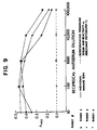
- Figures 9 and 10 describe polyclonal antibodies raised against a Type II mutant EGFR peptide.
- Figure 9 shows the reactivity of antisera from three rabbits with fusion junction peptide in an ELISA assay. Free peptide was bound to polyvinyl chloride plates.
- Figure 10, left shows immunoprecipitation of mutant EGFR protein but not intact EGFR by anti-peptide 2 antibody. On the right, immunoprecipitation by monoclonal antibody 528 of EGFR from A431-X, D-256MG-X and D-270MG-X is shown.
- Figure 11 is the cDNA sequence of normal EGFR.
- Mutant EGFR protein is present in cells exhibiting one or more of three types of genetic deletion and/or rearrangement which result in a structurally altered receptor.
- the mutations resulting in these three types of altered receptor are illustrated in Figures 1-3.
- the first class of deletions (Type I, Figure 1) results in a gap in the extracytoplasmic domain near the transmembrane domain.
- the second type of deletion (Type II, Figure 2) results in the elimination of the distal portion of the extracytoplasmic domain of EGFR.
- the Type I and II in-frame deletions produce two new junction points in the amino acid sequence.
- a third type of abnormality (Type III, Figure 3) is characterized by a deletion of the majority of the external domain of the EGFR leaving substantially only the transmembrane portion and the intracytoplasmic domain.
- Type III mutations leave little or no extracellular protein. Although antibodies may detect an extracellular sequence, it is preferable to solubilize a cell with a Type III mutation in a detergent before exposure to antibodies.
- Intron-free DNA sequences encoding deletion mutant EGFR Types I, II and III can be determined from the information provided in Figures 1, 2 and 3 respectively, in view of the previously disclosed sequence for the normal receptor.
- a gene encoding deletion mutant EGFR Type I contains DNA segments corresponding to base numbers 1817 and 2067 of the normal sequence connected together as shown in Figure 1 with the segment corresponding to base numbers 1818-2066 deleted.
- a gene encoding deletion mutant EGFR Type II contains DNA segments corresponding to base numbers 1-274 and 1076 to end of the normal sequence connected together as shown in Figure 2 with the segment corresponding to base numbers 275-1075 deleted.
- a gene encoding deletion mutant EGFR Type III contains a DNA segment corresponding to base numbers 1817 to the end with the segment corresponding to base numbers 1-1816 deleted.
- Other DNA sequences which encode the same amino acid sequences may be used to generate EGFR mutant peptides in recombinant organisms, due to the degeneracy of the genetic code.
- Intron-free DNA molecules may be obtained using reverse transcriptase, for example, and an EGFR mRNA as a template. Alternatively, the DNA molecules can be chemically synthesized according to the sequences disclosed herein. Such molecules can be amplified using polymerase chain reaction (PCR) to facilitate analysis and manipulations.
- PCR polymerase chain reaction
- useful vectors may comprise segments of chromosomal, non-chromosomal (such as various known derivatives of SV40 and known bacterial plasmids, e.g., plasmids from E.coli including colEl, pcRl pBR322, pMB9 and RP4), or synthetic DNA sequences, phage DNAs (M13) including derivatives of phage (e.g., NM 989) and filamentous single-stranded DNA phages, vectors useful in yeasts (such as the 2u plasmid), vectors useful in eukaryotic cells (such as vectors useful in animal cells, e.g.
- plasmids and phage DNAs those containing SV-40 adenovirus and retrovirus derived DNA sequences
- vectors derived from combinations of plasmids and phage DNAs such as plasmids which have been modified to employ phage DNA, or other derivatives thereof.
- Such expression vectors are also characterized by at least one expression control sequence that may be operatively linked to the mutant EGFR DNA sequence inserted in the vector to control and regulate the expression of the cloned DNA sequence.
- useful expression control sequences are the lac system, the trp system, the tac system, the trc system, major operator and promoter regions of phage lambda, the control region of fd coat protein, the glycolytic promoters of yeast (e.g., the promoter for 3-phosphoglycerate kinase), the promoters of yeast acid phosphatase (e.g., Pho5), the promoters of the yeast a-mating factors, and promoters derived from polyoma, adenovirus, retrovirus, or simian virus (e.g., the early and late promoters of SV40), and other sequences known to control the expression of genes of prokaryotic or eukaryotic cells and their viruses or combinations thereof.
- each specific expression vector various sites may be selected for insertion of the DNA sequences of this invention. These sites are usually designated by the restriction endonuclease which cuts them. They are well recognized by those of skill in the art. It is, of course, to be understood that an expression vector useful in this invention need not have a restriction endonuclease site for insertion of the chosen DNA fragment. Instead, the vector can be joined to the fragment by alternative means.
- the host cell, expression vector, and in particular the site chosen therein for insertion of a selected DNA fragment and its operative linking therein to an expression control sequence is determined by a variety of factors, e.g., number of sites susceptible to a particular restriction enzyme, size of the protein and its susceptibility to proteolytic degradation by host cell enzymes, contamination or binding of the protein to be expressed by host cell proteins difficult to remove during purification; expression characteristics, such as the location of start and stop codons relative to the vector and an insertion site for a DNA sequence is determined by a balance of these factors, not all selections being equally effective for a given case.
- Useful expression hosts may include well known eukaryotic and prokaryotic hosts, such as strains of E.coli, such as E.coli SG-936, E.coli HB 101, E.coli W3110, E.coli X1776, E.coli X2282, E.coli DHI, and E.coli MRC1, Pseudomonas , Bacillus , such as Bacillus subtilis , Streptomyces , yeasts and other fungi, animal cells, such as COS cells and CHO cells, and human cells and plant cells in tissue culture.
- E.coli such as E.coli SG-936, E.coli HB 101, E.coli W3110, E.coli X1776, E.coli X2282, E.coli DHI, and E.coli MRC1, Pseudomonas , Bacillus , such as Bacillus subtilis , Streptomyces , yeasts and other fungi, animal cells, such as COS cells and CHO cells,
- host/expression vector combinations function with equal efficiency in expressing the DNA sequences of this invention or in producing the polypeptides of this invention.
- a particular selection of a host/expression vector combination may be made by those skilled in the art. For example, the selection should be based on a balancing of a number of factors. These include compatibility of the host and vector, toxicity of the proteins encoded by the DNA sequence to the host, ease of recovery of the desired protein, expression characteristics of the DNA sequences and the expression control sequences operatively linked to them, biosafety, costs and the folding, form or any other necessary postexpression modifications of the desired protein.
- One preferred means of obtaining the deletion mutant EGFR preparations of this invention is to synthesize them according to standard techniques known in the art using the sequences taught herein. Another means contemplates culturing cells transfected with an expression vector comprising an intron-free DNA molecule corresponding to Figure 1 or Figure 2 or Figure 3 using culture conditions which are well-known in the art. The cells are then harvested and the cell membrane fraction may be separated by standard separation procedures, such as differential centrifugation, which are well-known in the art. A crude extract can be obtained by solubilizing the cell membrane fraction with detergents.
- purification can be accomplished according to techniques which are well-known in the protein purification art. For example, various types of chromatography may be used. Columns which may be used include a DEAE cellulose column, or an anion exchange column, as well as a gel permeation column.
- the deletion mutant EGFR protein or peptide fragments thereof can also be purified using immunoaffinity techniques.
- immunoaffinity techniques As antibodies are provided here which are specific for the epitopes shown in Figures 1, 2, and 3, peptides corresponding to Type I, II, or III mutants can be positively selected from a mixture of many proteins. The use of the antibodies of the present invention to purify the proteins of the invention allows good separation from those proteins which are most similar to them.
- peptides of this invention may be purified by immunoaffinity using antibodies to the normal EGFR, especially to epitopes on the cytoplasmic domain.
- other techniques of purification are known in the art and can be used to purify the peptides of the invention.
- substantially pure mutant EGFR peptides within the contemplation of this invention are those which are substantially free of other human proteins.
- Peptides according to the present invention are linear polymers of amino acids which do not contain the full intact sequence of EGFR found in normal cells. Typically these are greater than ten amino acids in length and less than about fifty. Desirably the peptides are long enough to elicit a unique antibody response but short enough so that antibodies are not elicited which are immunoreactive with the intact EGFR protein.
- the peptide product of the prokaryotic and eukaryotic hosts transformed with the DNA sequences of this invention can be employed in the production of antibodies.
- the substantially pure preparation of polypeptide comprising the amino acid sequence corresponding to the nucleotide sequences of Figures 1, 2 and 3 can be made using any of the techniques which are known in the art. For example, the Merrifield technique (Journal of American Chemical Society, vol. 85, pp. 2149-2154, 1968), can be used. Substantial purity means that the preparation is greater than 75% free of other proteins normally found in human cells. Preferably the preparation is greater than 90% free of other human proteins. Polypeptides may be longer or shorter or have conservative amino acid changes which do not change the epitope(s) found on deletion mutant EGFR but not found on normal intact EGFR.
- Polypeptides can be tested to determine if they are able to stimulate mammals to produce antibodies which are immunoreactive with epitopes found on deletion mutant EGFR, but not found on normal EGFR. Methods of immunizing mammals to stimulate antibody production are well known in the art. Methods for testing the immunoreactivity of antibodies for known antigens are also well known.
- the substantially pure preparation of polypeptide of the present invention can be used to affinity purify antibodies specific for the deletion mutant EGFR protein.
- the preparation of polypeptide of the present invention can be used to stimulate production of antibodies in a mammal by immunizing the mammal with the preparation.
- immunization may optionally employ coupling of the polypeptide to a larger immunogenic substance such as keyhole limpet hemocyanin.
- the polypeptide can be coupled to an inert matrix, such as agarose beads. Techniques for such coupling are well known in the art.
- the preparation of the polypeptide can also be used to quantitate antibodies specific for deletion mutant EGFR in an antibody preparation. In such a case, the synthetic peptide will usually be coupled to a larger inert proteinaceous substance such as bovine serum albumin.
- the techniques for coupling polypeptides to such matrices are well known in the art.
- antibodies which are specific for the deletion mutant EGFR proteins in that they are immunoreactive with the deletion mutant EGFR protein but not with normal, intact EGFR can be made using a synthetic polypeptide of the present invention. Immunization of mammals such as rabbits, mice, goats, etc. to produce antibodies is well known in the art. Such polyclonal antibody preparations can be purified using immunoaffinity techniques employing a synthetic polypeptide of the present invention. Such purification methods are well-known in the art. Monoclonal antibodies can also be raised which are specific for deletion mutant EGFR epitopes and do not cross-react with normal EGFR.
- a rat or mouse will be immunized with the synthetic polypeptide (or deletion mutant protein) of the present invention and the rodent will later be sacrificed and spleen cells recovered for fusion with myeloma cells.
- Hybrid cells can be selected according to techniques known in the art, for example, selections involving complementation of two phenotypes, one from each parental cell.
- the antibody produced by each hybrid cell clone can be screened individually to select antibodies which bind to epitopes on deletion mutant EGFR but not on normal, intact EGFR.
- Antibodies can be tested for immunoreactivity with deletion mutant EGFR using a substantially pure preparation of the protein, or with fragments of the deletion mutant EGFR protein according to the present invention conjugated to a larger moiety such as bovine serum albumin.
- the desired specific antibodies should be positive in either or both of these tests.
- the antibodies should also be tested for immunoreactivity with normal, intact EGFR; desired antibodies having absolute specificity for deletion mutant EGFR should be negative in such tests.
- Antibodies can also be detected using this battery of tests which have relative specificity for deletion mutant EGFR compared to normal EGFR. That is, some monoclonal antibodies can be found which react more strongly with the deletion mutant protein than with the normal protein. These antibodies of relative specificity for mutant EGFR may also be useful. Means for using them are discussed below.
- Immunoaffinity techniques to purify monospecific polyclonal antibodies reactive with deletion mutant EGFR but not with normal EGFR can be used. Similar binding properties are employed as in the tests described for monoclonal antibodies above. That is to say that antibodies which immunoreact with deletion mutant EGFR will be positively selected, while those that immunoreact with normal EGFR will be removed from the desired antibody preparation.
- Antibodies which show relative or preferential specificity for deletion mutant EGFR relative to normal EGFR can be rendered absolutely specific by altering the conditions under which immunoreactivity is assayed.
- Conditions in the assay medium which can be altered include, but are not limited to: the ionic strength; the detergent concentration; the concentration of chaotropic agents, such as urea, guanidine, and potassium thiocyanate; and the pH. Alteration of these conditions leads to destabilization of the various bonding forces which contribute to antibody-antigen binding. Titration of reagents altering each of these conditions allows determination of a set of conditions where relatively or preferentially specific antibodies immunoreact with deletion mutant EGFR but not with normal EGFR.
- potassium chloride can be titrated from about 0.05M to 2M.
- Detergents either ionic or non-ionic, can be titrated from about 0.05% to 2%.
- Chaotropic agents can be titrated from about 0.5M to 8M.
- the range of pH can be titrated from about 2 to 10. Such conditions can be useful both to screen for monoclonal antibodies immunoreactive with deletion mutant EGFR and to assay for deletion mutant EGFR in various biological sources.
- the nucleotide sequences provided by the invention can be used to form gene probes in accordance with any of the standard techniques.
- the DNA probes contemplated for use in this invention may be derived from the DNA of cell lines grown in vitro or xenografts maintained in vivo which contain the DNA spanning the deletion site.
- the size of a DNA probe can vary from approximately 20 nucleotides to hundreds of nucleotides.
- the DNA probe may be radiolabeled, labeled with a fluorescent material, or the like. Procedures for the preparation and labeling of DNA probes are well known in the art.
- the diagnostic test employing a DNA probe will employ a cell sample from an individual who is being screened for the presence of glioma. The sample will be isolated from the suspect tissue. DNA is recovered from the cell employing standard techniques well known to those skilled in the art. The DNA is then incubated with a probe under conditions where homologous sequences hybridize but sequences which diverge do not, and hybridization is thereafter detected. Hybridization to a deletion-specific probe indicates the presence of the deletion. Enzymes such as S1 nuclease can be employed to remove portions of a DNA or RNA molecule which do not hybridize. The size of the duplex nucleic acid which remains can be determined by standard means. Duplexes which are smaller than the probe indicate a deletion, rearrangement, or other mismatch. Thus probes which are useful may be derived from intact as well as mutant alleles.
- Antibodies of the invention are capable of binding to the mutant EGFR proteins and not to the intact EGFR protein from normal cells. These antibodies also permit the use of imaging analysis with isotopes, conjugated antibodies, or other ligands. Examples of suitable imaging agents are 15I, 13I, 131I, or Indium-111 conjugated to the antibodies specific for deletion mutants of the EGFR.
- the antibodies of the present invention can be used to detect deletion mutant EGFR epitopes in histological sections of glioma tissue as well as in other solid tumors. Tissue samples are preferably solubilized with detergent to release membrane proteins into solution prior to immunological detection.
- the antibodies can be used as immunohistochemical reagents to visualize EGFR mutant proteins in tissue sections.
- the antibodies of the invention can be administered to a patient for imaging analysis.
- the antibodies are typically conjugated to an imaging agent, such as 13I, 131I, or Indium-111.
- an imaging agent such as 13I, 131I, or Indium-111.
- a diagnostically effective amount of antibody is one which allows the observer to distinguish between normal tissues and those containing mutant type EGFR. Determination of such amounts is within the skill of the art.
- a particularly useful stain for use in enzyme-linked antibody assays employs peroxidase, hydrogen peroxide and a chromogenic substance such as aminoethyl carbazole.
- the peroxidase (a well known enzyme available from many sources) can be coupled to the antibody specific for deletion mutant EGFR or merely complexed to it via one or more antibodies.
- a goat anti-peroxidase antibody and a goat antibody specific for deletion mutant EGFR can be complexed via an anti-goat IgG.
- Other chromogenic substances and enzymes may also be used.
- Radio-labeled antibodies may be specific for deletion mutant EGFR or second antibodies immunoreactive with antibodies specific for deletion mutant EGFR. Again, such techniques are well known. The precise technique by which mutant EGFR is detected in glioma patients is not critical to the invention. Biochemical or immunological techniques can now be used which do not employ immunohistochemistry, although that is a preferred method of the present invention.
- One particularly preferred method of detecting and/or quantitating deletion mutant EGFR protein in solubilized samples employs a competitive assay.
- An antibody immunoreactive with an epitope found on deletion mutant EGFR but not found on normal EGFR is attached to a solid support such as a polystyrene microtiter dish or nitrocellulose paper, using techniques known in the art.
- the solid support is then incubated in the presence of the fluid to be analyzed under conditions where antibody-antigen complexes form and are stable. Excess and unbound components of the fluid are removed and the solid support is washed so that antibody-antigen complexes are retained on the solid support.
- a fixed amount of a polypeptide containing an epitope found on deletion mutant EGFR but not found on normal EGFR is then incubated with the solid support.
- the polypeptide binds to an antibody immunoreactive with mutant EGFR which is attached to the solid support.
- the polypeptide has been conjugated to a detectable moiety, such as biotin, peroxidase or radiolabel, by means well known in the art. Excess and unbound polypeptide is removed and the solid support is washed, as above. The detectable moiety attached to the solid support is quantitated.
- the solubilized mutant EGFR in the fluid to be analyzed can be quantitated by its diminution of the binding of the polypeptide to the solid support.
- Antibodies employed in this assay may be immunoreactive with deletion mutant EGFR but not with normal EGFR. Alternatively, relatively specific antibodies may be used under conditions which destabilize immunoreactivity with normal EGFR. Polyclonal antibodies which contain an antibody species immunoreactive with an epitope on deletion mutant EGFR but not on normal EGFR, may also be used.
- a preferred diagnostic method for determination of Type III gliomas involves differential detection of the intracytoplasmic and extracytoplasmic domains. This detection may be on the gene or peptide level. On the gene level, nucleotide probes specific for the intracytoplasmic domain (base numbers 2192-3720 on Figure 11) and for the extracytoplasmic domain (base numbers 190-2122) are used. DNA extracted from Type III gliomas will hybridize with the intracytoplasmic but not the extracytoplasmic probes. Similarly, detergent solubilized membrane proteins from suspect tissue samples can be tested immunologically using antibodies specific for epitopes found in the intracytoplasmic and the extracytoplasmic domains.
- the antibodies can be prepared by immunizing mammals with peptides expressed from the sequences corresponding to these domains, as indicated above, and selecting those antibodies specific to each domain using techniques that are well known to those skilled in the art.
- the membrane protein fraction from Type III gliomas will react with antibodies to the intracytoplasmic domain of EGFR but not with antibodies specific for most the extracytoplasmic domain.
- the particular procedures for gene probe assays and immunoassays will be well-known to those skilled in the art.
- antibodies specific for Type III EGFR mutant proteins can be used. Such antibodies will react with the epitopes which are not present on intact EGFR.
- Treatment may be with radioactive isotopes including 131I [Bullard et al. (1986) and Lee et al. (1988)] or appropriate drugs also conjugated to those antibodies.
- radioactive isotopes including 131I [Bullard et al. (1986) and Lee et al. (1988)] or appropriate drugs also conjugated to those antibodies.
- 131I Busllard et al. (1986) and Lee et al. (1988)
- a number of treatment protocols employing monoclonal and polyclonal antibodies have been developed in the art of cancer therapy which are useful for the present invention.
- each of these protocols depends on the specificity of the antibody as a targeting agent for their respective tumor antigen: 1) immune system effector cells -- either endogenous (Sears, 1984 & 1985, colorectal carcinoma; Meeker, 1985, B lymphocyte malignancy; Shouval, 1987, liver cancer) or isolated from the patient and reinjected; (Sears, 1982, and Douillard, 1986, colorectal carcinoma) 2) cytotoxic drugs; (EP 0153 144, ricin) or 3) radioactive isotopes (Carrasquillo, 1984, directing 131I to metastatic melanoma).
- the role of the antibody is to direct the active agent to particular tumor cells whose surfaces carry antigens corresponding to the respective antibodies.
- Treatment comprises administration to a glioma patient of an effective amount of an antibody specific for the mutant EGFR and unreactive with normal EGFR, said antibodies optionally labelled with radioactive elements or conjugated to cytotoxic drugs.
- an antibody specific for the mutant EGFR and unreactive with normal EGFR said antibodies optionally labelled with radioactive elements or conjugated to cytotoxic drugs.
- the appropriate level of antibody for treatment can be determined for each patient using standard methods and no more than routine experimentation.
- the xenografts used in the present study were derived from surgical biopsies of malignant human gliomas. Their establishment and karyotypes have been described previously (Humphrey et al., 1988). Southern blotting experiments with EGFR cDNA probes have shown that five of the xenografts (D245MG, D256MG, D270MG, G298MG and D317MG) contain rearranged and amplified EGFR genes. The same rearranged fragments were detected in the original tumor biopsies (Humphrey et al., 1988).
- Tumor xenograft D320MG exhibited amplified EGFR genes without any detectable rearrangements.
- Xenografts D263 and D274 exhibited no amplification or rearrangement of the EGFR gene. Therefore, five of the 8 glioma xenografts examined contained rearranged and amplified EGFR genes which were all present in the initial tumor biopsy.
- phage clones Forty-eight phage clones were obtained and used to assemble an EcoRI map of the EGF receptor gene (Figure 4A). The clones spanned the entire gene except for two small gaps. Southern blot hybridization (using genomic DNA digested with various enzymes) confirmed that the clones identified all EcoRI fragments within the gene (including those containing the gaps). The map deduced using these tumor-derived phage clones was not rearranged in comparison to normal genomic DNA.
- Phage clones 40 and 24 revealed deletions and rearrangements in Type II tumors D270 ( Figure 4C, lane C) and D317 (data not shown), but not in Type I tumors D256MG or D298MG. Conversely, clones 26 ( Figure 4C) and 29 (data not shown) demonstrated a rearrangement in tumors D256MG ( Figure 4C, Lane B) and D298MG (lane E) but not in tumors D270 or D317.
- the abnormally migrating fragments were unlikely to be the result of restriction fragmentation length polymorphisms (RFLPs) since new bands were also observed when the tumor DNA was digested with several other enzymes.
- RFLPs restriction fragmentation length polymorphisms
- EGFR normal EGF receptor gene
- D245" sequence is derived from the amplification unit of D245MG
- TEG normal locus
- TEG normal locus
- Cross hybridization and restriction mapping indicated that the 1.7 kb EcoRI restriction fragment from the D245MG DNA was the product of a rearrangement between a 1.8 EcoRI fragment from the EGFR locus and a 2.2 kb EcoRI fragment from the TEG locus.
- the three fragments were subcloned into plasmid vectors and partially sequenced.
- the nucleotide sequences are presented in Figure 5B and diagrammed in Figure 5C.
- Several features of the sequences were notable: (i) the rearrangement occurred within an intron upstream of the EGF receptor exon corresponding to nucleotides 1818 to 1908 of the EGFR cDNA sequence; (ii) an 18 nucleotide A-T rich motif was repeated four times in the vicinity of the breakpoint in the rearranged fragment, but only once in the corresponding part of the TEG fragment; (iii) at the site of recombination, seven additional nucleotides were present which did not appear to be derived from either the TEG or EGF receptor loci; and (iv) Alu-type repeats were present in both the TEG and EGF receptor derived fragments and one such repeat is present in the clone containing the rearrangement.
- TEG human-mouse somatic cell hybrids with the TEG specific probe. This demonstrated that TEG, like the EGF receptor gene, was located on chromosome 7. Further hybridization to hybrids containing various deletions on chromosome 7 (Bartels et al., 1986; Zergerling et al., 1987), showed that both TEG and the EGF receptor were located in the same subchromosomal region at 7p12-7p14 (Fig. 5D). Thus, the rearrangement in D245 was intrachromosomal. The juxtaposition of Alu sequences around the breakpoints (Fig.
- the protein produced in D245MG is similar to v-erb B
- the translation product of this RNA had an N-terminus corresponding to amino acid 543 of the EGF receptor protein and that the open reading frame was preceded by a long 5' untranslated sequence.
- the predicted protein product of this mRNA would be a truncated version of the normal receptor containing 644 amino acids, a predicted molecular weight of 72 kd, and retention of 3 N-linked glycosylation sites (Ullrich et al., 1984).
- Probe I contained a 910 base pair fragment from the 5' end of the cDNA. It yielded a fragment of 910 bp from all RNA samples tested (Fig. 7C); in the RNA samples from the tumors with rearrangements, the normal size fragments were probably a result of transcription from the remaining normal EGFR gene.
- Probe II revealed 210-215 base pair fragments in tumors D270 and D317 as well as less intense 914 base pair fragment corresponding to the normal transcript ( Figure 4C, probe II, lanes C and F).
- the cluster of closely migrating fragments was most likely due to imperfect cleavage of the duplex; RNase protection experiments in which similar RNA probes were hybridized to cloned genomic fragments also produced a cluster of fragments.
- the data obtained with probes I and II were consistent with the hypothesis that the deletion in both tumors D270 and D317 resulted in an approximately 800 base pair deletion near the 5' end of the transcript. While the genomic deletions were not identical ( Figure 4), it appeared that the two tumors had lost similar exon sequences.
- Probe III demonstrated the normal 970 base pair fragments in D270 and D317 ( Figure 7C, probe III, lanes C and F), but 450 and 250 bp fragments were observed in D256 and D298 (Lane B and E), suggesting an approximately 270 base pair deletion within the transcript of these latter two tumors.
- Tumor D245 also had an abnormal RNase protection pattern ( Figure 7C, probe III, lane A); the 495 bp fragment in D245 corresponded to the break point in the transcript determined from the cDNA clones.
- Probes specific for the 3' half of the receptor including the tyrosine kinase domain and autophosphorylation sites) showed no abnormalities in any of the five tumors, indicating that there were not point mutations detectable by this method.
- the RNase protection and genomic Southern blot data provided an approximation of the nature of the deletion, but it was difficult to determine the precise nature of the abnormalities.
- the polymerase chain reaction (PCR) was used to generate cDNA fragments from the mutant alleles.
- the data from the RNase protection experiments and published information on the normal EGFR cDNA sequence was used to guide the choice of primers.
- cDNA was generated by first annealing the 3' primer to total tumor RNA, and then extended by using MMLV reverse transcriptase. PCR was then carried out using a method similar to that described by Kawasaki et al. (Kawasaki et al., 1988).
- RNA from tumors D270 and D317 was analyzed with one set of primers (Fig. 8A) a major fragment of 230 base pairs were produced ( Figure 8A, lanes A and B), while the normal size 1100 base pair fragment was seen only faintly (not shown), consistent with the size of the deletions inferred by the RNase protection experiments.
- Another glial tumor xenograft, D397 had a Southern blot pattern very similar to that of D270 when probed with the EGFR cDNA. It was analyzed in this PCR experiment and produced a fragment similar to that from D270 and D317 (Fig. 8A, lane C).
- a second set of primers revealed a fragment of 220 base pairs in tumors D256 and D298 (Fig. 8A, lanes D and E respectively), instead of the normal size 440 base pairs which was found with D320 RNA (lane F); the 220 bp fragment size was consistent with the data from the RNase protection experiments.
- the proteins immunoprecipitated from tumors 270 and 317MG were 150 kilodaltons (predicted polypeptide size of 120,000) and those from D298 and D256 were 130 kilodaltons (predicted polypeptide size of 100,000).
- the differences in size from that predicted from the cDNA sequences may reflect the degree of glycosylation, as there is a similar difference between the predicted molecular weight of normal EGFR protein and its mobility on SDS gels.
- the binding affinity for EGF in these tumors was less than three-fold lower than that of the control cell lines with normal EGFR gene sequences.
- the domain thought to be responsible for EGF binding was not deleted in any of the tumors except for D245MG.
- a second subset of tumors might have altered receptors whose structure results in a molecule that is constantly active and is only partially or not at all regulated by ligand. Such independence from the normal signals controlling cellular growth is the essence of tumorigenesis. Tumor types I, II, and III appear to be of the second subset.
- DNA was purified using Proteinase K-SDS and extracted with phenol and chloroform (Vogelstein et al., 1987).
- DNA (4 ug) was digested with EcoRI (BRL) using buffers and conditions specified by the manufacturer, and after electrophoresis through a 1% agarose gel, transferred to nylon membranes (Bio-Rad) using 0.4N NaOH (Reed et al., 1985). Pre-hybridization and hybridization were done as described previously (Vogelstein et al., 1987). Probes were labelled with d-3PdCTP using the oligolabelling method (Feinberg and Vogelstein, 1984). Repeated sequences were removed by the pre-association method of Sealey et al. (Sealey et al., 1985).
- the genomic library was constructed from D320MG as described previously (Ruppert et al., 1988). After partial Mbo I digestion, DNA was size-fractionated through sucrose density gradient ultracentrifugation. The fractions containing 17 to 24 kilobase fragments were cloned into the Bam HI site of Lambda Fix (Stratagene) after partial fill-in of Mbo I ends. The ligation product was packaged with lambda phage extracts (Stratagene) and used to infect E. coli C600 cells. DNA from the resulting plaques was lifted with Colony Plaque Screen membranes (Dupont, NEN Research Products) and screened with EGF receptor cDNA probes (Ullrich et al., 1984; Merlino et al., 1985).
- D245 DNA was digested with EcoRI and size selected by electrophoresis on a 1% agarose gel. DNA was eluted from the gel (Vogelstein et al., 1987), ligated to gt10 arms (Promega), packaged with lambda phage extracts and used to infect C600 cells. The library was screened with an EGFR cDNA probe and a clone containing the 1.7 kb rearranged fragment was identified.
- a total genomic library was first made from D245MG DNA and screened with the 1.7 kb rearranged EcoRI fragment, resulting in a 15 kb phage clone bridging the rearrangement between TEG and EGF receptor loci.
- the 4.4 kb fragment from this phage clone was then used to screen a genomic library made from D259 (a glial tumor cell line without EGFR amplification or rearrangement) to clone the normal TEG locus.
- the fragments from D245, EGFR and TEG participating in the recombination were subcloned into pBluescript (Stratagene).
- nested deletions were generated using exonuclease III and mung bean nuclease (Henikof et al., 1984). Plasmids were transformed into HB101 cells (F+::Tn5) and single stranded DNA prepared using R408 helper phage (Russel et al., 1986). Sequencing by the di-deoxy method was done using a modified form of T7 polymerase (USB).
- poly(A)+RNA was isolated from 10 ug total RNA by selection on oligo-dT cellulose, separated by electrophoresis through a 1.5% MOPS/formaldehyde gel and transferred in dilute alkali to nylon (Bio-Rad).
- first strand cDNA was prepared using MMLV reverse transcriptase (BRL) and random hexamer primers (Pharmacia). The second strand was synthesized using the method of Gubler and Hoffman (Gubler et al., 1983). The resulting cDNA was methylated with EcoRI methylase and ligated to EcoRI linkers (New England Biolabs).
- the linked cDNA was cleaved with EcoRI, and fragments >1.0 kb were isolated following electrophoresis through a 1% agarose gel.
- This cDNA was ligated into ZAP (Stratagene) and packaged with phage extracts (Stratagene).
- the library was screened with an 0.7 kb TaqI-EcoRI fragment derived from the EGFR end of the 1.7 kb EcoRI fragment of D245 (See Fig. 5A). Plasmids containing inserts were derived from phase plaques by using the excision process recommended by the manufacturer.
- Ribonuclease protection was performed as described (Winter et al., 1985) with the following modifications: hybridizations were performed in a final volume of 10 ul; only RNase A at 12.5 ug per ml was used; and the RNase A and Proteinase K digestions were performed at room temperature for 30 minutes.
- primer set A was used, consisting of 5'-AGTCGGGCTCTGGAGGA-3' and 5'-CACTGATGGAGGTGCAGT-3'.
- primer set B was used consisting of 5'-(CTG)CAGGTCTGCCATGCCTTG-3' and 5'-(GGT)ACCATCCCAGTGGCGATG-3'. The sequences in parentheses were not present in the EGFR sequence and were added to complete either a Pst I or Kpn I restriction site.
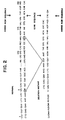
- Figure 4 Deletions in the EGF receptor gene in human gliomas.
- Figure 4A is an EcoRI map of the Human EGF receptor gene. The sizes of the fragments are indicated in kilobases. Representative phage clones used to assemble the map are shown below it.
- Figure 4B is the deduced EGFR gene structure of five glioma xenografts.
- the numbers to the left correspond to the glial tumors described in the text.
- the solid lines indicate sequences present in the tumors, while the approximate points of deletion are indicated by x's.
- Figure 4C represents a Southern blot hybridization with phage clones demonstrating deletions in the EGF receptor gene.
- the blots were hybridized with radio-labelled phage inserts, and the numbers above each blot refer to the phage clone used as the hybridization probe. Rearranged fragments are indicated with an asterisk (*).
- the tick marks to the right of each autoradiograph refer to the sizes of marker fragments in kilobases which are given on the extreme right.
- the lanes contained DNA according to the following key:
- Figure 5A is an EcoRI restriction map of genomic clones containing the 1.7 kb rearranged fragment from D245MG and the corresponding unrearranged fragments from the EGF receptor gene and TEG locus.
- the open box indicates sequences derived from the EGF receptor gene and the shaded box represents the TEG locus.
- the numbers refer to the sizes of the EcoRI fragments in kilobases.
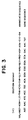
- Figure 5B shows a partial nucleotide sequence of the area flanking the site of recombination. Other features based on this sequence are shown in Figure 5C.
- An 18 nucleotide A-T rich motif is repeated four times (rptl-rpt4) in the rearranged D245 fragment but only once in the TEG fragment; an insertion has been made to optimize alignment.
- Seven nucleotides are present at the site of recombination ("Break point") which cannot be aligned with either of the recombination partners.
- Portions of the Alu repeats present in the TEG and EGF receptor gene are underlined; the arrowhead indicates the orientation of these repeats according to Deininger et al. (Deininger et al., 1981). Numbers to the right are relative nucleotide positions with respect to the D245 sequence.
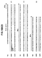
- Figure 5C schematically shows the presence of Alu repeats (arrows), the A-T rich motif (open box labeled AT) and exon from the EGF receptor (filled box corresponding to EGFR nt 1818-1908) in the 1.77 kb EcoRI rearranged fragment from D245 and the corresponding unrearranged fragment from the EGF receptor gene and TEG locus.
- Figure 5D illustrates chromosomal localization of the TEG locus.
- a human-mouse somatic cell hybrid panel containing various deletions of human chromosome 7 (20-21) was digested with EcoRI and blotted to a nylon membrane. It was first hybridized with the 2.2 kb EcoRI fragment from the TEG locus (top) and then hybridized with a 1.9 kb EcoRI genomic fragment from the EGFR locus (bottom).
- Figure 5E describes a possible mechanism for intrastrand recombination in D245 involving oppositely paired Alu repeats based on the model proposed by Lehrman et al. (Lehrman et al., 1985). Features of the TEG and EGF receptor fragments from Fig. 5C are reproduced here. The oppositely oriented Alu repeats in the EGF receptor gene and TEG locus on chromosome 7p could base pair forming a stem loop structure. Recombination at the stem would result in the rearrangement seen in D245.
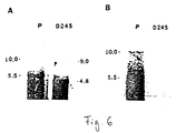
- Panel 6A is a Northern blot hybridization demonstrating abnormal transcript sizes in D245. Two and one-half ug of poly A+ selected RNA from placenta and tumor D245 were electrophoresed in a 1.5% formaldehyde gel and transferred to nylon membranes. The blot was hybridized with a probe specific for the intracytoplasmic domain of the EGF receptor. The numbers to the right and left indicate the size of the bands in kb. The major band in the D245 sample is 4.8 kb; there is also a faint band of unclear derivation at 5.5 kb which was not derived from the normal EGFR mRNA, since it did not hybridize to an extracytoplasmic domain probe, as seen in panel 6B.
- Panel 6B is a separate blot that was hybridized with a probe from the extracytoplasmic domain (the 730 bp EcoRI-BamHI fragment of pE7.
- Panel 6C shows a cDNA sequence of the EGF receptor related mRNA from D245.
- D245" refers to the nucleotide sequence of a clone derived from tumor D245 and mRNA.
- EGFR refers to the normal EGF receptor cDNA sequences.
- D245AA is the deduced amino acid sequence from “D245", which is aligned with that of "v-erb B” (the v-erb B oncoprotein is a fusion protein between viral and EGFR sequences; only the EGFR related sequences are shown).
- the presumed initiator codon for tumor D245 is underlined.
- the numbers to the right refer to the D245 sequence.
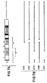
- FIG 7A is a schematic drawing of the probes used.
- the 5.5 kb EGF receptor cDNA is diagrammed above.
- the striped box is the signal peptide, followed by the extracytoplasic domain which is divided into four domains (1-4), with the cysteine rich regions as indicated.
- the solid box is the transmembrane domain followed by the intracytoplasmic domain containing the kinase domain.
- the thin lines are the untranslated regions.
- Figure 7B presents the deduced transcript structures from the RNase protection experiments. Symbols are as in Figure 4B.
- Figure 7C depicts RNase protection experiments using RNA from the glial tumor xenografts.
- the numerals I-III refer to the probes in Fig. 7A.
- the numbers to the right refer to the sizes of the protected fragments (see text).
- the lanes contain RNA from: A) D245; B) D256; C) 270; D) D320; E) D298; F) D317; G) tRNA (negative control); and H) undigested probe.
- Figure 8 PCR analysis of the gene products from the altered region.
- Figure 8A shows gel electrophoresis of polymerase chain reaction products. The gel shows 1/10th of the reaction after 35 cycles.
- the lanes contained products from: A) 270; B) 317; C) 397: D) 256; E) 298: F) 320.
- Primer set A was used for D270, D317, and D397; primer set B was used for D256, D298, and D320 (see Experimental Procedures).
- the numbers refer to the sizes of fragments in base pairs judged from co-electrophoresed markers.
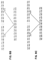
- Figure 8B shows a sequence of the PCR products from tumors D270, D317, and D397.
- Figure 8C illustrates the sequence of the PCR products from D256 and D298.
- the numbers and asterisks refer to the EGF receptor cDNA nucleotides flanking the deleted area.
- the sequence to the right and left of those shown in the figure were the same as in normal EGFR cDNA.
- mice for the production of hybridomas were injected according to the schedule below. Mice populations were at least 5 and generally up to 10 in each immunization arm with both Type I and Type II EGFR deletion mutant peptides, as shown in Figures 1 and 2.
- Hybridoma supernatants were screened using solid phase radioimmunassy using 0.5 ug/well of pep - 2. Hyperimmune sera, specific for pep-2 versus pep-1, as well as preimmune sera were used as baseline controls.
- Rabbits were injected with synthetic peptides of the deletion mutant type II coupled to keyhole limpet hemocyanin initially, and after two months, three further injections at one month intervals.
- the immunization pattern was performed with rabbits according to the following schedule:
- Antibody titers against peptide were determined in ELISA. Briefly, a concentrated solution of peptide (1 mg/1 ml in PBS) was diluted to 10 ⁇ g/ml in 200 mM NaHCO3 buffer (pH 9.2), and 50 ⁇ l were added to 96-plate wells of polyvinyl chloride (Dynatech Laboratories, Inc., Chantilly, VA). The peptide solution was incubated in the plate wells overnight at 4°C. The peptide solution was discarded, and the plates were washed 3 times with Hanks' buffered salt solution containing 0.05% Tween 20 (Sigma, St. Louis, MO).
- Nonspecific binding was then blocked for 1 hr at room temperature with 200 ⁇ l of 0.5% bovine serum albumin in PBS.
- the plates were washed as above.
- Pre-immune rabbit sera or antisera 100 ⁇ l of dilutions in Hanks' buffered salt solution with 0.5% BSA) were added for 2 hr at room temperature.
- the plates were washed and 50 ⁇ l of peroxidase-conjugated goat antirabbit IgG (Zymed Laboratories, Inc., San Francisco, CA) was added for 1 hr at 37°C.
- the plates were washed and 100 ⁇ l of substrate solution was added.
- the substrate solution was prepared by adding 15 mg of o-phenylenediamine (Sigma) to 1 ml methanol, with subsequent addition of 49 ml of deionized water and 50 ⁇ l of hydrogen peroxide. Incubation was at room temperature for 15-20 min., and the well absorbances were then measured in an automated plate reader (Titertek Multiskan MCC/340, Flow Laboratories, McLean, VA) at 492 nm. The ELISA could also be performed with purified antipeptide antibody. The half-maximal titer in this assay was 0.5 ⁇ g antipeptide IgG/ml of PBS.
- binding reaction was specific, as the pre-immune serum from each rabbit was nonreactive; a nonreactive baseline was also obtained when the antisera were reacted with a second 14-amino-acid peptide of different sequence (Pep-1; sequence: H-Asn-Leu-Leu-Glu-Gly-Cys-Thr-Gly-Pro-Gly-Leu-Glu-Gly-Cys-OH).
- the antipeptide antibody was purified from antiserum by a peptide-Sepharose affinity column with elution by acidic pH.
- the affinity column was prepared by coupling 5 mg of peptide to cyanogen bromide-activated Sepharose (Sigma), as described in the Pharmacia (Piscataway, N.J.) protocol. The extent of coupling was 100%, as determined by BCA protein assay (Pierce, Rockford, IL) of the solution overlying the gel.
- Ten millititers of antiserum from rabbit 396 (the rabbit with the highest half-maximal titer of 1:50,000 in ELISA in reaction against peptide) were passed over the column, and the column was washed extensively with 500 ml of PBS.
- Control IgG was purified from rabbit pre-immune sera by protein A-Sepharose affinity chromatography.
- the size and homogeneity of the purified antibodies were monitored under nondenaturing conditions by size exclusion HPLC and under denaturing condition by SDS-PAGE.
- HPLC was performed on a calibrated 1 x 30 cm Waters 300 SW column (previously calibrated with standards in the size range of M r of 20,000-400,000 daltons). Protein elution was followed at 215 nm.
- SDS-PAGE utilized a 10% resolving gel in the SDS-discontinuous buffer system of Laemmli (Laemmli, Nature (1970), 270:680-688).
- the protein bands were visualized with Coomassie blue staining.
- the eluted antibody was identified as IgG by apparent molecular weights on a size exclusion HPLC column and SDS gel (data not shown). Purity was greater than 98%.
- the affinity-purified antibody was characterized in immunocytochemistry using frozen tissue sections and the avidinbiotin complex method, as described (Humphrey, et al., Cancer Res. (1988), 48:2231-2238).
- the tissues tested included a range of normal fetal and adult tissues, carcinomas (prostatic, bladder, breast, and lung), glioma biopsies (-Bx), and gliomas grown in xenograft form in nude mice (-X).
- the normal tissues, carcinomas, and glioma biopsies were from the Duke University Medical Center Tissue Bank.
- the glioma xenografts were grown as described (Humphrey, et al., Cancer Res. (1988), 48:2231-2238).
- the affinity purified antipeptide antibody and the purified pre-immune rabbit control IgG were used at a concentration of 1-2 ⁇ g/ml, as determined by initial titration experiments.
- the antibody could be used in antiserum form with an optimal titer in immunocytochemistry of 1:3000.
- F(ab')2 fragments of normal rabbit IgG and the antipeptide antibody were also tested in immunocytochemistry on glioma D-270 MG-X and skin. F(ab')2 fragments were generated and purified as described (Colapinto, et al., Cancer Res. (1988), 48:5701-5707).
- the antipeptide IgG reacted with the native mutant EGFR in frozen tissue sections fixed briefly with acetone.
- the antibody specifically recognized only the mutant glioma EGFR and not the intact A431-X squamous cell carcinoma EGFR in both xenograft (data not shown) and biopsy tissue (Table 1).
- This immunostaining of the mutant EGFR was specific, as purified pre-immune IgG was nonreactive, and preincubation of antipeptide IgG with excess peptide blocked with immunocytochemical staining. In both glioma biopsy and xenograft tissues which bound the antipeptide antibody, the immunostaining was localized to the cytoplasm and cell surface; virtually every tumor cell exhibited immunoreactivity.
- Immunoreactive mutant EGFR was identified in two human glioma biopsies (D-270 MG-Bx and D-317 MG-Bx) and the corresponding xenografts (D-270 MG-X and D-317 MG-X) known to amplify the mutant EGFR gene and express mutant EGFR protein.
- glioblastoma biopsies exhibited immunostaining with the antipeptide antibody; one of these (D-397 MG-Bx) was subsequently tested using an RNA-based PCR assay and shown by sequencing to possess the same deletion mutation as gliomas D-270 MG-Bx and D-317 MG-Bx.
- the purified antipeptide antibody and pre-immune IgG were both labeled with 15I at a specific activity of about 1.6 ⁇ Cl/ ⁇ g using a variation off the iodogen method as described previously (Colapinto, et al., Cancer Res. (1988), 48:5701-5707).
- Radioiodinated proteins 22 ng, 50-70 k cpm
- the membranes tested were from D-270 MG-X, a glioma tumor expressing the in-frame deletion-mutant EGFR; A431-X, a squamous cell carcinoma overexpressing the intact EGFR; and D-245 MG-X, a tumor containing or expressing an EGFR molecule which lacks most of the extracellular domain and serves as a negative tissue control.
- the membranes were separated from unbound activity using 0.22- ⁇ m cellulose acetate centrifuge filter units (Spin-x, Costar, Cambridge, MA) washed twice with 1 ml of the incubation buffer. The filters had been pretreated by a 30-min incubation at room temperature followed by 3 washes with buffer. Using this procedure, nonspecific binding of radioactivity to the filters was ⁇ 0.2%. Filters and washes were assayed for 15I activity in similar counting geometries using an automated gamma counter.
- Percent specific binding represents binding of 15I-labeled antipeptide IgG minus nonspecific binding determined from association of preimmune IgG with membranes.
- Glioma D-270 MG-X membranes express the in-frame deletion-mutant EGFR and squamous cell carcinoma A431-X membranes overexpress the intact EGFR;
- glioma D-245 MG-X membranes express a v-erbB-like EGFR which lacks most of the extracellular domain and serves as a negative tissue control (Humphrey, et al., Cancer Res. (1988), 48:2231-2238; Wong, et al., J. Cell Biochem., Suppl. 13B, Abst. 149).
- the molecular specificity of the antipeptide IgG was tested by immunoprecipitation reaction. The binding in this reaction was followed by EGFR autophosphorylation, SDS-PAGE, and autoradiography, essentially as described. Monoclonal antibody 528 (Ab-1, Oncogene Science, Inc., Manhasset, New York) was used as a positive control, as it will immunoprecipitate both intact and mutant EGFR. Briefly, Triton X-100 detergent solubilized intact A431-X EGFR and mutant D-256 MG-X and D-270 MG-X EGFRs were immunoprecipitated with monoclonal antibody 528 or purified antipeptide antibody.
- the antipeptide antibody reacted with mutant EGFR in a detergent-solubilized state, as judged by immunoprecipitation, with autophosphorylation and SDS-PAGE (Fig. 10).
- the positive control immunoprecipitation was with monoclonal antibody 528, which is directed against the EGFR external domain.
- This antibody reacted with both the intact A431-X and mutant D-270 MG-X EGFR.
- the antipeptide antibody in contrast, specifically immunoprecipitated the 145-kDa mutant EGFR in glioma D-270 MG-X and failed to immunoprecipitate the intact A431-X EGFR. Purified preimmune IgG did not immunoprecipitate the mutant EGFR.
- the effect of the purified antibody on 15I-labeled epidermal growth factor (EGF) (New England Nuclear, Doraville, GA) binding was determined using the reaction mixture of microsomal membranes and 15I-labeled EGF, as described (Humphrey, et al., Cancer Res. (1988), 48:2231-2238).
- EGF epidermal growth factor
- the phosphorylation was carried out in a volume of 50 ⁇ l consisting of membranes (5-10 ⁇ g), 20-mM Hepes (pH 7.4), 4 mM MnCl2, 10 mM MgCl2, 1% DMSO, ⁇ 400 ng of EGF, ⁇ normal rabbit IgG, ⁇ antipeptide antibody.
- the mixture was incubated for 20 min at room temperature and then cooled to 4°C.
- [ ⁇ -3P]-ATP 1.5 ⁇ M; 10-15,000 cpm/pmol
- sample buffer containing sodium dodecyl sulfate and betamercaptoethanol was added and the reaction was stopped after 3 min by adding 25 ⁇ l of sample buffer containing sodium dodecyl sulfate and betamercaptoethanol.
- the samples were subjected to electrophoresis on 7.5% polyacrylamide gels (Laemmli, Nature (1970), 270:680-688), and the extent of receptor phosphorylation was visualized by autoradiography
- the anti-synthetic peptide antibody did not affect binding of 15I-labeled EGF to the mutant EGFR, nor did it affect either basal or EGF-stimulated intrinsic kinase activity.
- Specific binding of 15I-labeled EGF defined as counts of total 15I EGF binding minus counts not displaced by a 100-fold excess of unlabeled EGF, did not change with addition of antibody (data not shown). This may be due to the location of the fusion junction at a site distant from the EGF binding domain at residues 351-364 (Wu, et al., J. Biol. Chem. (1989), 264:17469-17475).
- the kinase activity of the mutant EGFR did not change with the presence of antibody, as assessed by band intensity in autoradiography of an SDS-polyacrylamide gel (data not shown).
- Binding of antipeptide IgG to live tumor cells was determined by immunofluorescence assay.
- D-270 MG-X or A431-X cells were prepared by dissociation from tumor xenografts grown subcutaneously in athymic mice. Briefly, subcutaneous tumors were removed from the mouse and the viable portions were separated from areas of necrosis. The viable tissue was minced with scissors and dissociated into a single cell suspension by incubating 1 hr in 0.8% collagenase at 37°C in a trypsinization flask. The cells were washed twice with cold Richter's zinc-option medium containing 1% normal goat serum.
- One million viable cells were incubated 30 min at 4°C with zinc-option medium plus 10% normal goat serum to block nonspecific binding.
- the cells were incubated 30 min at 4°C with 100 ⁇ l of antipeptide antibody (20 ⁇ g per ml) or pre-immune IgG. After washing 3 times with zinc-option medium plus 1% normal goat serum, the cells were incubated with 100 ⁇ l of fluorescein-labeled goat antirabbit IgG for 30 min at 4°C.
- the cells were washed as above and resuspended in 500 ⁇ l of cold zinc-option medium plus 1% normal goat serum.
- One-hundred-microliter aliquots were removed and warmed to 37°C for 10 to 60 min and observed with a Zeiss fluorescent microscope.
- the anti-synthetic peptide antibody rapidly bound the surface mutant EGFR expressed on live glioma D-270 MG-X cells, and this binding was reflected by a rimming pattern of the immunofluorescent secondary antibody label. Internationalization rapidly ensued and was manifested as a speckled intracytoplasmic morphology. Internalization with loss of the peripheral plasma membrane rimming was complete by 60 min at 37°C.
- EGFR from xenograft tissue was solubilized and immunoprecipitated as follows. Ten mg of frozen (-70°C) xenograft tissue were homogenized in 1 ml of ice-cold solubilization buffer composed of 20 mM iodoacetate, and Aprotinin at 1 mg/ml. After 2 h at 4°C, the preparation was centrifuged for 15 min at 4°C in a Beckman tabletop centrifuge at 12,000 x g. The supernatant was used in EGFR immunoprecipitation. Immunoprecipitation was performed with monoclonal antibody Ab-1 (clone 528: Oncogene Science, Inc.), reactive against the normal external domain of EGFR.
- Monoclonal antibody Ab-1 is an IgG2a which inhibits EGF binding to its receptor.
- 5 ug of monoclonal antibody Ab-1 (528) or 15 ul of undiluted antiserum containing polyclonal antibody were bound to 2 mg of Protein A-Sepharose 4B by incubation in 115 mM sodium phosphate buffer, pH 7.4, for 30 min at room temperature.
- Antibody-Protein A-Sepharose complex was washed 3 times with 115 mM sodium phosphate buffer, pH 7.4,EGFR immunoprecipitation was performed with 500-ul aliquots of solubilized xenograft tissue, 500 ul of 115 mM sodium phosphate buffer, pH 7.4, and the antibody-Protein A-Sepharose pellet.
- Western immunoblot analysis was performed with the use of a semidry horizontal electrophoretic transfer system with graphite electrodes (LKB Multiphor II Nova Blot System). Briefly, frozen A431 and glioma xenografts were solubilized in SDS-gel sample buffer, boiled, and electrophoresed in a 7.5% SDS-polyacrylamide gel. The proteins were transferred to nitrocellulosse at 150 mA for 2 h, and immunohistochemical detection of EGFR was accomplished with antibody 528 and polyclonal antisera from rabbit no. 4 directed against a type II peptide.
Landscapes
- Health & Medical Sciences (AREA)
- Chemical & Material Sciences (AREA)
- Life Sciences & Earth Sciences (AREA)
- Organic Chemistry (AREA)
- Immunology (AREA)
- Proteomics, Peptides & Aminoacids (AREA)
- General Health & Medical Sciences (AREA)
- Medicinal Chemistry (AREA)
- Genetics & Genomics (AREA)
- Zoology (AREA)
- Biochemistry (AREA)
- Molecular Biology (AREA)
- Biophysics (AREA)
- Engineering & Computer Science (AREA)
- Animal Behavior & Ethology (AREA)
- Pharmacology & Pharmacy (AREA)
- Public Health (AREA)
- Veterinary Medicine (AREA)
- Bioinformatics & Cheminformatics (AREA)
- Analytical Chemistry (AREA)
- Wood Science & Technology (AREA)
- Pathology (AREA)
- Epidemiology (AREA)
- Cell Biology (AREA)
- Physics & Mathematics (AREA)
- Hospice & Palliative Care (AREA)
- Gastroenterology & Hepatology (AREA)
- General Engineering & Computer Science (AREA)
- Oncology (AREA)
- Microbiology (AREA)
- Optics & Photonics (AREA)
- Biotechnology (AREA)
- Toxicology (AREA)
- Nuclear Medicine, Radiotherapy & Molecular Imaging (AREA)
- General Chemical & Material Sciences (AREA)
- Chemical Kinetics & Catalysis (AREA)
- Peptides Or Proteins (AREA)
- Preparation Of Compounds By Using Micro-Organisms (AREA)
- Medicines Containing Antibodies Or Antigens For Use As Internal Diagnostic Agents (AREA)
- Medicines That Contain Protein Lipid Enzymes And Other Medicines (AREA)
Claims (28)
- Intronfreies DNA-Molekül, enthaltend eine DNA-Sequenz, die für ein mutantes EGFR-Peptid kodiert, das aus der Gruppe ASN-LEU-LEU-GLU-GLY-CYS-THR-GLY-PRO-GLY und LEU-GLU-GLU-LYS-LYS-GLY-ASN-TYR-VAL-VAL-THR ausgewählt ist.
- Intronfreies DNA-Molekül, enthaltend eine DNA-Sequenz, die aus der Gruppe AAG-CTT-CTG-GAG-GGA-TGC-ACT-GGG-CCA-GGT und CTG-GAG-GAA-AAG-AAA-GGT-AAT-TAT-GTG-GTG-ACA ausgewählt ist.
- Intronfreies DNA-Molekül, die für ein mutantes EGFR-Peptid kodiert, das folgende N-terminale Sequenz aufweist: MET-ASN-ILE-THR-CYS-THR-GLY-ARG-GLY-PRO-ASP-ASN-CYS.
- Mutantes EGFR-Peptid, umfassend eine aus der Gruppe ASN-LEU-LEU-GLU-GLY-CYS-THR-GLY-PRO-GLY und LEU-GLU-GLU-LYS-LYS-GLY-ASN-TYR-VAL-VAL-THR ausgewählte Aminosäuresequenz, das frei von anderen humanen Proteinen ist.
- Mutantes EGFR-Peptid, das die N-terminale Sequenz MET-ASN-ILE-THR-CYS-THR-GLY-ARG-GLY-PRO-ASP-ASN-CYS aufweist und frei von anderen humanen Proteinen ist.
- Vektor, enthaltend ein DNA-Molekül nach Anspruch 1.
- Vektor, enthaltend ein DNA-Molekül nach Anspruch 2.
- Vektor, enthaltend das DNA-Molekül nach Anspruch 3.
- Transformierte Zelle, enthaltend den Vektor nach Anspruch 6.
- Transformierte Zelle, enthaltend den Vektor nach Anspruch 7.
- Transformierte Zelle, enthaltend den Vektor nach Anspruch 8.
- Zusammensetzung, enthaltend eine für ein DNA-Molekül nach Anspruch 1 spezifische Sonde zur Verwendung in einem Verfahren zur GliomDiagnose, wobei das Verfahren die Bestimmung der Anwesenheit in einer Zelle einer Nucleinsäuresequenz, die für ein mutantes EGFR-Protein mit einer Aminosäuresequenz gemäß der Definition in Anspruch 4 kodiert, umfaßt.
- Zusammensetzung, enthaltend eine für ein DNA-Molekül nach Anspruch 2 spezifische Sonde zur Verwendung in einem Verfahren zur Gliom-Diagnose, wobei das Verfahren die Bestimmung der Anwesenheit in einer Zelle einer Nucleinsäuresequenz gemäß der Definition in Anspruch 2 oder von deren Ribonucleotid-Äquivalenten umfaßt.
- Zusammensetzung, enthaltend eine für ein DNA-Molekül nach Anspruch 3 spezifische Sonde zur Verwendung in einem Verfahren zur Gliom-Diagnose, wobei das Verfahren die Bestimmung der Anwesenheit in einer Zelle einer Nucleinsäuresequenz, die für ein mutantes EGFR-Protein, das die N-terminale Aminosäuresequenz nach Anspruch 3 enthält, umfaßt.
- Zusammensetzung, enthaltend einen Antikörper, der mit einem mutanten EGFR-Peptid nach Anspruch 4 immunoreaktiv, jedoch nicht mit einem intakten EGFR-Protein immunoreaktiv ist, zur Verwendung in einem Verfahren zur Gliom-Diagnose, wobei das Verfahren den Test auf die Anwesenheit eines mutanten EGFR-Proteins, das die Aminosäuresequenz nach Anspruch 4 enthält, umfaßt.
- Zusammensetzung nach Anspruch 15, wobei der Antikörper an ein Abbildungsmittel konjugiert ist.
- Zusammensetzung, enthaltend einen Antikörper, der mit einem mutanten EGFR-Peptid nach Anspruch 5 immunoreaktiv, jedoch nicht mit intaktem EGFR-Protein immunoreaktiv ist, zur Verwendung in einem Verfahren zur Gliom-Diagnose, wobei das Verfahren den Test auf die Anwesenheit eines mutanten EGFR-Proteins, der die N-terminale Aminosäuresequenz nach Anspruch 5 enthält, umfaßt.
- Zusammensetzung nach Anspruch 17, wobei der Antikörper an ein Abbildungsmittel gekuppelt ist.
- Zusammensetzung, enthaltend einen Antikörper, der mit einem mutanten EGFR-Peptid nach Anspruch 4 immunoreaktiv, jedoch nicht mit einem intakten EGFR-Protein immunoreaktiv ist, zur Verwendung in einem Verfahren zur Behandlung von Gliom.
- Zusammensetzung nach Anspruch 19, wobei der Antikörper an ein therapeutisches Mittel gekuppelt ist.
- Zusammensetzung, enthaltend einen Antikörper, der mit einem mutanten EGFR-Peptid nach Anspruch 5 immunoreaktiv, jedoch nicht mit einem intakten EGFR-Protein immunoreaktiv ist, zur Verwendung in einem Verfahren zur Behandlung von Gliom.
- Zusammensetzung nach Anspruch 21, wobei der Antikörper an ein therapeutisches Mittel gekuppelt ist.
- Antikörperpräparat, das mit einem mutanten EGFR-Peptid nach Anspruch 4 immunoreaktiv ist, wobei der Antikörper jedoch mit intaktem EGFR-Protein nicht immunoreaktiv ist.
- Antikörperpräparat nach Anspruch 23, das polyklonal ist.
- Antikörperpräparat nach Anspruch 23, das monoklonal ist.
- Antikörperpräparat, das mit einem mutanten EGFR-Protein nach Anspruch 5 inmunoreaktiv ist, wobei der Antikörper aber nicht mit intaktem EGFR-Protein inmunoreaktiv ist.
- Antikörperpräparat nach Anspruch 26, das polyklonal ist.
- Antikörperpräparat nach Anspruch 26, das monoklonal ist.
Applications Claiming Priority (5)
| Application Number | Priority Date | Filing Date | Title |
|---|---|---|---|
| US40422689A | 1989-09-08 | 1989-09-08 | |
| US404226 | 1989-09-08 | ||
| US53141090A | 1990-06-01 | 1990-06-01 | |
| US531410 | 1990-06-01 | ||
| PCT/US1990/004489 WO1991003489A1 (en) | 1989-09-08 | 1990-08-15 | Structural alterations of the egf receptor gene in human gliomas |
Publications (2)
| Publication Number | Publication Date |
|---|---|
| EP0491007A1 EP0491007A1 (de) | 1992-06-24 |
| EP0491007B1 true EP0491007B1 (de) | 1996-03-13 |
Family
ID=27018564
Family Applications (1)
| Application Number | Title | Priority Date | Filing Date |
|---|---|---|---|
| EP90915375A Expired - Lifetime EP0491007B1 (de) | 1989-09-08 | 1990-08-15 | Modifikationen der struktur des egf-rezeptor-gens in menschlichen glioma |
Country Status (8)
| Country | Link |
|---|---|
| US (4) | US5212290A (de) |
| EP (1) | EP0491007B1 (de) |
| JP (1) | JP2975679B2 (de) |
| AT (1) | ATE135373T1 (de) |
| AU (1) | AU639726B2 (de) |
| CA (1) | CA2066428C (de) |
| DE (1) | DE69025946T2 (de) |
| WO (1) | WO1991003489A1 (de) |
Cited By (1)
| Publication number | Priority date | Publication date | Assignee | Title |
|---|---|---|---|---|
| US7628986B2 (en) | 2003-06-27 | 2009-12-08 | Amgen Fremont Inc. | Antibodies directed to the deletion mutants of epidermal growth factor receptor and uses thereof |
Families Citing this family (228)
| Publication number | Priority date | Publication date | Assignee | Title |
|---|---|---|---|---|
| US5981725A (en) * | 1989-09-08 | 1999-11-09 | The Johns Hopkins Univiersity | Structural alterations of the EGF receptor gene in human tumors |
| DE4129533A1 (de) * | 1991-09-05 | 1993-03-11 | Max Planck Gesellschaft | Mutierter wachstumsfaktorrezeptor als arzneimittel und seine verwendung zur behandlung von krebs |
| EP0659439B1 (de) | 1993-12-24 | 2001-10-24 | MERCK PATENT GmbH | Immunokonjugate |
| CA2206343C (en) | 1994-11-28 | 2009-04-07 | Thomas Jefferson University | Reagents and processes for targeting mutant epidermal growth factor receptors |
| US5635351A (en) * | 1995-03-14 | 1997-06-03 | The Regents Of The University Of California | Genetic gain and loss in gliomas |
| US5905027A (en) * | 1995-12-27 | 1999-05-18 | Uab Research Foundation | Method of diagnosing and treating gliomas |
| US6667156B2 (en) * | 1995-12-27 | 2003-12-23 | Uab Research Foundation | Diagnosis and treatment of neuroectodermal tumors |
| CA2376596C (en) | 1999-06-25 | 2009-10-06 | Genentech, Inc. | Humanized anti-erbb2 antibodies and treatment with anti-erbb2 antibodies |
| US6790614B1 (en) * | 1999-11-19 | 2004-09-14 | Novartis Ag | Selectable cell surface marker genes |
| CN1427891A (zh) * | 2000-02-25 | 2003-07-02 | 美国政府由(美国)卫生和福利部部长代表 | 具有提高的细胞毒性和产量的抗EGFRvIII的scFv、基于其的免疫毒素、及其应用方法 |
| WO2001068711A1 (en) * | 2000-03-10 | 2001-09-20 | Thomas Jefferson University | Sensitive detection of wild-type and mutant egfr by specific elisa assays in any biological sample |
| AUPQ841800A0 (en) | 2000-06-28 | 2000-07-20 | Biomolecular Research Institute Limited | Truncated egf receptor |
| US20100056762A1 (en) * | 2001-05-11 | 2010-03-04 | Old Lloyd J | Specific binding proteins and uses thereof |
| EP1392359B2 (de) | 2001-05-11 | 2013-03-13 | Ludwig Institute for Cancer Research Ltd. | Spezifische bindungsproteine und ihre verwendung |
| US8021658B2 (en) * | 2001-05-25 | 2011-09-20 | Thomas Jefferson University | Alternative splice forms of proteins as basis for multiple therapeutic modalities |
| US20060088899A1 (en) * | 2002-05-31 | 2006-04-27 | Alvarez Vernon L | Combination chemotherapy with chlorotoxin |
| WO2003101474A1 (en) * | 2002-05-31 | 2003-12-11 | Transmolecular, Inc. | Combination chemotherapy with chlorotoxin |
| ES2392525T3 (es) | 2002-07-15 | 2012-12-11 | F. Hoffmann-La Roche Ag | Tratamiento del cáncer con el anticuerpo dirigido contra ErbB2 rhuMAb 2C4 |
| US7498298B2 (en) | 2003-11-06 | 2009-03-03 | Seattle Genetics, Inc. | Monomethylvaline compounds capable of conjugation to ligands |
| US7767792B2 (en) | 2004-02-20 | 2010-08-03 | Ludwig Institute For Cancer Research Ltd. | Antibodies to EGF receptor epitope peptides |
| CN101501211A (zh) | 2004-03-31 | 2009-08-05 | 综合医院公司 | 测定癌症对表皮生长因子受体靶向性治疗反应性的方法 |
| KR20120064120A (ko) | 2004-06-01 | 2012-06-18 | 제넨테크, 인크. | 항체 약물 접합체 및 방법 |
| KR101270829B1 (ko) | 2004-09-23 | 2013-06-07 | 제넨테크, 인크. | 시스테인 유전자조작 항체 및 접합체 |
| JO3000B1 (ar) | 2004-10-20 | 2016-09-05 | Genentech Inc | مركبات أجسام مضادة . |
| CA2592177A1 (en) | 2005-01-21 | 2006-07-27 | Genentech, Inc. | Fixed dosing of her antibodies |
| EP1850874B1 (de) | 2005-02-23 | 2013-10-16 | Genentech, Inc. | Verzögerung des krankheitsverlauf bei eierstockkrebspatienten durch pertuzumab |
| MX2007009963A (es) * | 2005-02-24 | 2007-09-26 | Amgen Inc | Mutaciones del receptor del factor de crecimiento epidermico. |
| WO2006096861A2 (en) * | 2005-03-08 | 2006-09-14 | Genentech, Inc. | METHODS FOR IDENTIFYING TUMORS RESPONSIVE TO TREATMENT WITH HER DIMERIZATION INHIBITORS (HDIs) |
| GB2424886A (en) | 2005-04-04 | 2006-10-11 | Dxs Ltd | Polynucleotide primers against epidermal growth factor receptor and method of detecting gene mutations |
| AR053272A1 (es) | 2005-05-11 | 2007-04-25 | Hoffmann La Roche | Determinacion de responsivos a la quimioterapia |
| CA2647671A1 (en) * | 2006-03-31 | 2007-11-01 | Massachusetts Institute Of Technology | Treatment of tumors expressing mutant egf receptors |
| US20100008910A1 (en) | 2006-09-12 | 2010-01-14 | John Chant | Methods and compositions for the diagnosis and treatment of cancer |
| US9090693B2 (en) | 2007-01-25 | 2015-07-28 | Dana-Farber Cancer Institute | Use of anti-EGFR antibodies in treatment of EGFR mutant mediated disease |
| US9340601B2 (en) * | 2007-03-01 | 2016-05-17 | The Board Of Trustees Of The Leland Stanford Junior University | Splice variants of the EGF receptor |
| MX2009008981A (es) | 2007-03-02 | 2009-09-02 | Genentech Inc | Prediccion de respuesta a un inhibidor her. |
| ES2542152T3 (es) | 2007-03-15 | 2015-07-31 | Ludwig Institute For Cancer Research Ltd. | Método de tratamiento que emplea anticuerpos de EGFR e inhibidores de Src y formulaciones relacionadas |
| EP2171090B1 (de) | 2007-06-08 | 2013-04-03 | Genentech, Inc. | Genexpressionsmarker für tumorresistenz gegen behandlung mit her2-inhibitor |
| CN108424454B (zh) | 2007-08-14 | 2022-05-31 | 路德维格癌症研究所有限公司 | 靶向egf受体的单克隆抗体175及其衍生物和用途 |
| TWI472339B (zh) | 2008-01-30 | 2015-02-11 | Genentech Inc | 包含結合至her2結構域ii之抗體及其酸性變異體的組合物 |
| EP2274437B1 (de) * | 2008-04-10 | 2015-12-23 | Cell Signaling Technology, Inc. | Zusammensetzungen und verfahren zur erkennung von egfr-mutationen bei krebs |
| US8227439B2 (en) | 2008-05-15 | 2012-07-24 | Morphotek, Inc. | Treatment of metastatic tumors |
| CN104447995A (zh) | 2009-03-20 | 2015-03-25 | 霍夫曼-拉罗奇有限公司 | 双特异性抗-her抗体 |
| US9345661B2 (en) | 2009-07-31 | 2016-05-24 | Genentech, Inc. | Subcutaneous anti-HER2 antibody formulations and uses thereof |
| ES2354922B1 (es) | 2009-09-02 | 2012-02-07 | Fundacion Institut De Recerca De L'hospital Universitari Vall D'hebron | Marcadores para la selección de terapias personalizadas para el tratamiento del c�?ncer. |
| US20110076232A1 (en) * | 2009-09-29 | 2011-03-31 | Ludwig Institute For Cancer Research | Specific binding proteins and uses thereof |
| EP2507381A4 (de) | 2009-12-04 | 2016-07-20 | Hoffmann La Roche | Multispezifische antikörper, antikörperanaloge, zusammensetzungen und verfahren |
| US20130034573A1 (en) | 2009-12-22 | 2013-02-07 | Celldex Therapeutics, Inc. | Vaccine compositions |
| EP2523957A1 (de) | 2010-01-12 | 2012-11-21 | F. Hoffmann-La Roche AG | Trizyklische heterozyklische verbindungen und zusammensetzungen sowie anwendungsverfahren dafür |
| JP5858932B2 (ja) | 2010-02-04 | 2016-02-10 | モルフォテック, インコーポレイテッド | クロロトキシンポリペプチドおよびコンジュゲートならびにその使用 |
| DK2536748T3 (da) | 2010-02-18 | 2014-10-13 | Genentech Inc | Neuregulin-antagonister og anvendelse deraf ved behandling af kræft |
| CA2793024A1 (en) | 2010-03-17 | 2011-09-22 | F. Hoffmann-La Roche Ag | Imidazopyridine compounds, compositions and methods of use |
| MX2012011887A (es) | 2010-04-16 | 2012-11-30 | Genentech Inc | Foxo 3a como biomarcador predictivo para la eficacia del inhibidor de la via de quinasa pi3k/akt. |
| JP2013532126A (ja) | 2010-05-11 | 2013-08-15 | フレッド ハッチンソン キャンサー リサーチ センター | クロロトキシン変異体、コンジュゲート、およびそれらを使用する方法 |
| WO2011146568A1 (en) | 2010-05-19 | 2011-11-24 | Genentech, Inc. | Predicting response to a her inhibitor |
| KR20130103734A (ko) | 2010-08-31 | 2013-09-24 | 제넨테크, 인크. | 바이오마커 및 치료 방법 |
| MX2013003019A (es) | 2010-09-15 | 2013-05-28 | Hoffmann La Roche | Compuestos de azabenzotiazol, composiciones y metodos de uso. |
| EP2637692A4 (de) | 2010-11-12 | 2014-09-10 | Scott & White Healthcare | Antikörper gegen den endothelialen tumormarker 8 |
| MX2013005445A (es) | 2010-11-19 | 2013-07-29 | Hoffmann La Roche | Pirazolopiridinas y el uso de pirazolopiridinas como inhibidores de tirosina cinasa 2 (tyk2). |
| WO2012085176A1 (en) | 2010-12-23 | 2012-06-28 | F. Hoffmann-La Roche Ag | Tricyclic pyrazinone compounds, compositions and methods of use thereof as janus kinase inhibitors |
| JP2014527397A (ja) | 2011-06-20 | 2014-10-16 | トラスラショナル、キャンサー、ドラッグス、ファーマ、ソシエダッド、リミターダTraslational Cancer Drugs Pharma, S.L. | 癌患者の化学療法に対する臨床応答を予測する方法 |
| WO2013007765A1 (en) | 2011-07-13 | 2013-01-17 | F. Hoffmann-La Roche Ag | Fused tricyclic compounds for use as inhibitors of janus kinases |
| WO2013007768A1 (en) | 2011-07-13 | 2013-01-17 | F. Hoffmann-La Roche Ag | Tricyclic heterocyclic compounds, compositions and methods of use thereof as jak inhibitors |
| CN103889976A (zh) | 2011-08-12 | 2014-06-25 | 弗·哈夫曼-拉罗切有限公司 | 吲唑化合物、组合物及使用方法 |
| US20140363438A1 (en) | 2011-08-17 | 2014-12-11 | Genentech, Inc. | Neuregulin antibodies and uses thereof |
| JP2014526538A (ja) | 2011-09-20 | 2014-10-06 | エフ.ホフマン−ラ ロシュ アーゲー | イミダゾピリジン化合物、組成物及び使用方法 |
| HK1202252A1 (en) | 2011-11-16 | 2015-09-25 | Amgen Inc. | Methods of treating epidermal growth factor deletion mutant viii related disorders |
| US9233171B2 (en) | 2011-11-21 | 2016-01-12 | Immunogen, Inc. | Method of treatment of tumors that are resistant to EGFR antibody therapies by EGFR antibody cytotoxic agent conjugate |
| RU2014126098A (ru) | 2011-11-30 | 2016-01-27 | Дженентек, Инк. | МУТАЦИИ ErbB3 ПРИ РАКЕ |
| RU2014136886A (ru) | 2012-03-27 | 2016-05-20 | Дженентек, Инк. | Диагностика и виды лечения, связанные с ингибиторами her3 |
| MX2014014356A (es) | 2012-05-24 | 2015-07-06 | Fundació Inst D Investigació Biomédica De Bellvitge Idibell | Metodo para la identificacion del origen de un cancer de origen primario desconocido. |
| EP2879705A4 (de) | 2012-08-02 | 2016-08-03 | Univ Leland Stanford Junior | Peptidimpfstoffe auf basis der egfrviii-sequenz zur behandlung von tumoren |
| US12024568B2 (en) | 2012-09-13 | 2024-07-02 | Cornell University | Treatment of brain cancers using central nervous system mediated gene transfer of monoclonal antibodies |
| AU2013334493B2 (en) | 2012-10-26 | 2018-11-29 | The University Of Queensland | Use of endocytosis inhibitors and antibodies for cancer therapy |
| US20170275367A1 (en) | 2012-11-21 | 2017-09-28 | Janssen Biotech, Inc. | Bispecific EGFR/C-Met Antibodies |
| CN113201073A (zh) | 2012-11-21 | 2021-08-03 | 詹森生物科技公司 | 双特异性EGFR/c-Met抗体 |
| CN105008393A (zh) | 2012-12-10 | 2015-10-28 | 弗莱德哈钦森癌症研究中心 | 用于筛选的方法 |
| DK2958943T3 (da) | 2013-02-20 | 2019-12-09 | Univ Pennsylvania | Behandling af cancer ved anvendelse af humaniseret anti-EGFRvIII kimær antigenreceptor |
| EP2958592A1 (de) | 2013-02-22 | 2015-12-30 | F. Hoffmann-La Roche AG | Verfahren zur behandlung von krebs und zur verhinderung von arzneimittelresistenz |
| KR20150123250A (ko) | 2013-03-06 | 2015-11-03 | 제넨테크, 인크. | 암 약물 내성의 치료 및 예방 방법 |
| WO2014139326A1 (en) | 2013-03-13 | 2014-09-18 | Genentech, Inc. | Pyrazolo compounds and uses thereof |
| KR20150127216A (ko) | 2013-03-14 | 2015-11-16 | 제넨테크, 인크. | 암의 치료 및 암 약물 내성의 예방 방법 |
| JP2016515132A (ja) | 2013-03-14 | 2016-05-26 | ジェネンテック, インコーポレイテッド | Mek阻害剤化合物のher3/egfr阻害剤化合物との組み合わせ及び使用方法 |
| WO2014144850A1 (en) | 2013-03-15 | 2014-09-18 | Genentech, Inc. | Methods of treating cancer and preventing cancer drug resistance |
| TR201904121T4 (tr) | 2013-07-09 | 2019-04-22 | The Government Of The United States As Represented By The Secretary Of The Dept Of Health And Human | İnsan bispesifik egfrviii antikoru birleştirme molekülleri. |
| MX2016002794A (es) | 2013-09-05 | 2016-09-13 | Genentech Inc | Compuestos antiproliferativos. |
| AU2014391422A1 (en) | 2013-09-17 | 2015-12-17 | Obi Pharma, Inc. | Compositions of a carbohydrate vaccine for inducing immune responses and uses thereof in cancer treatment |
| US11559580B1 (en) | 2013-09-17 | 2023-01-24 | Blaze Bioscience, Inc. | Tissue-homing peptide conjugates and methods of use thereof |
| TW201605857A (zh) | 2013-10-03 | 2016-02-16 | 赫孚孟拉羅股份公司 | Cdk8之醫療性抑制劑及其用途 |
| CN105744954B (zh) | 2013-10-18 | 2021-03-05 | 豪夫迈·罗氏有限公司 | 抗rspo2和/或抗rspo3抗体及其用途 |
| WO2015095404A2 (en) | 2013-12-17 | 2015-06-25 | Genentech, Inc. | Methods of treating cancers using pd-1 axis binding antagonists and taxanes |
| AU2014364606A1 (en) | 2013-12-17 | 2016-07-07 | Genentech, Inc. | Combination therapy comprising OX40 binding agonists and PD-1 axis binding antagonists |
| AU2015229591B2 (en) | 2014-03-11 | 2020-10-22 | Regeneron Pharmaceuticals, Inc. | Anti-EGFRvlll antibodies and uses thereof |
| TWI731535B (zh) | 2014-03-21 | 2021-06-21 | 美商艾伯維有限公司 | 抗-egfr抗體及抗體藥物結合物 |
| EP3122900A1 (de) | 2014-03-24 | 2017-02-01 | F. Hoffmann-La Roche AG | Krebsbehandlung mit c-met-antagonisten und korrelation desselben mit hgf-expression |
| EP3126394B1 (de) | 2014-03-31 | 2019-10-30 | F.Hoffmann-La Roche Ag | Anti-ox40-antikörper und verfahren zur verwendung |
| RU2016142476A (ru) | 2014-03-31 | 2018-05-07 | Дженентек, Инк. | Комбинированная терапия, включающая антиангиогенезные агенты и агонисты, связывающие ох40 |
| US9493764B2 (en) * | 2014-04-22 | 2016-11-15 | Korea Institute Of Science And Technology | Hybrid electronic sheets |
| EP3189046B1 (de) | 2014-09-05 | 2020-08-26 | Genentech, Inc. | Therapeutische verbindungen und ihre verwendung |
| WO2016044694A1 (en) | 2014-09-19 | 2016-03-24 | Genentech, Inc. | Use of cbp/ep300 and bet inhibitors for treatment of cancer |
| EP3204379B1 (de) | 2014-10-10 | 2019-03-06 | Genentech, Inc. | Pyrrolidinamid-verbindungen als histondemethylase-inhibitoren |
| EP3215850B1 (de) | 2014-11-03 | 2019-07-03 | F. Hoffmann-La Roche AG | Tests zum nachweis von te-zell-immununtergruppen und verfahren zur verwendung davon |
| MX2017005751A (es) | 2014-11-03 | 2018-04-10 | Genentech Inc | Métodos y biomarcadores para predecir la eficacia y evaluación de un tratamiento con agonista de ox40. |
| KR20170072343A (ko) | 2014-11-06 | 2017-06-26 | 제넨테크, 인크. | Ox40 결합 효능제 및 tigit 억제제를 포함하는 병용 요법 |
| WO2016077375A1 (en) | 2014-11-10 | 2016-05-19 | Genentech, Inc. | Bromodomain inhibitors and uses thereof |
| MA40940A (fr) | 2014-11-10 | 2017-09-19 | Constellation Pharmaceuticals Inc | Pyrrolopyridines substituées utilisées en tant qu'inhibiteurs de bromodomaines |
| MA40943A (fr) | 2014-11-10 | 2017-09-19 | Constellation Pharmaceuticals Inc | Pyrrolopyridines substituées utilisées en tant qu'inhibiteurs de bromodomaines |
| MX2017006320A (es) | 2014-11-17 | 2017-08-10 | Genentech Inc | Terapia combinada que comprende agonistas de unión de ox40 y antagonistas de unión del eje de pd-1. |
| US9763922B2 (en) | 2014-11-27 | 2017-09-19 | Genentech, Inc. | Therapeutic compounds and uses thereof |
| JP6864953B2 (ja) | 2014-12-09 | 2021-04-28 | アンスティチュ ナショナル ドゥ ラ サンテ エ ドゥ ラ ルシェルシュ メディカル | Axlに対するヒトモノクローナル抗体 |
| BR112017010788A2 (pt) | 2014-12-23 | 2017-12-26 | Genentech Inc | métodos de identificação de um paciente com câncer e de identificação de um paciente que sofre de câncer que pode beneficiar da administração de um antagonista de vegf, método de tratamento de um paciente com câncer e método para determinar o estágio de câncer de ovário |
| JP2018503373A (ja) | 2014-12-30 | 2018-02-08 | ジェネンテック, インコーポレイテッド | がんの予後診断及び治療のための方法及び組成物 |
| EP3242874B1 (de) | 2015-01-09 | 2018-10-31 | Genentech, Inc. | Pyridazinon-derivate und deren verwendung zur behandlung von krebs |
| EP3242872B1 (de) | 2015-01-09 | 2019-07-03 | Genentech, Inc. | (piperidin-3-yl)(naphthalen-2-yl)methanon-derivate und verwandte verbindungen als inhibitoren der histon demethylase kdm2b zur behandlung von krebs |
| EP3242875B1 (de) | 2015-01-09 | 2022-02-23 | Genentech, Inc. | 4,5-dihydroimidazol- derivate und ihre verwendung als histone demethylase (kdm2b) inhibitoren |
| EP3250571B1 (de) | 2015-01-29 | 2022-11-30 | Genentech, Inc. | Therapeutische verbindunegn und deren verwendung |
| WO2016123387A1 (en) | 2015-01-30 | 2016-08-04 | Genentech, Inc. | Therapeutic compounds and uses thereof |
| MA41598A (fr) | 2015-02-25 | 2018-01-02 | Constellation Pharmaceuticals Inc | Composés thérapeutiques de pyridazine et leurs utilisations |
| WO2016135041A1 (en) | 2015-02-26 | 2016-09-01 | INSERM (Institut National de la Santé et de la Recherche Médicale) | Fusion proteins and antibodies comprising thereof for promoting apoptosis |
| AU2016246695A1 (en) | 2015-04-07 | 2017-10-26 | Genentech, Inc. | Antigen binding complex having agonistic activity and methods of use |
| RS61152B2 (sr) | 2015-05-12 | 2024-06-28 | Hoffmann La Roche | Terapeutski i dijagnostički postupci za lečenje raka |
| JP7144935B2 (ja) | 2015-05-29 | 2022-09-30 | ジェネンテック, インコーポレイテッド | 癌のための治療方法及び診断方法 |
| KR20180011839A (ko) | 2015-06-08 | 2018-02-02 | 제넨테크, 인크. | 항-ox40 항체를 이용한 암의 치료 방법 |
| HK1251474A1 (zh) | 2015-06-08 | 2019-02-01 | 豪夫迈‧罗氏有限公司 | 使用抗ox40抗体和pd-1轴结合拮抗剂治疗癌症的方法 |
| JP6896650B2 (ja) | 2015-06-17 | 2021-06-30 | ジェネンテック, インコーポレイテッド | Pd−1軸結合アンタゴニスト及びタキサンを使用した局所進行性または転移性乳癌の治療方法 |
| DK3341376T3 (da) | 2015-08-26 | 2021-03-29 | Fundacion Del Sector Publico Estatal Centro Nac De Investigaciones Oncologicas Carlos Iii F S P Cnio | Kondenserede tricykliske forbindelser som proteinkinase-inhibitorer |
| KR20180050339A (ko) | 2015-09-04 | 2018-05-14 | 오비아이 파머 인코퍼레이티드 | 글리칸 어레이 및 사용 방법 |
| CA2994858C (en) | 2015-09-25 | 2024-01-23 | Genentech, Inc. | Anti-tigit antibodies and methods of use |
| PL3978500T3 (pl) | 2015-12-16 | 2024-03-11 | Genentech, Inc. | Sposób wytwarzania tricyklicznych związków inhibitorów pi3k |
| JP6949030B2 (ja) | 2016-01-08 | 2021-10-13 | エフ・ホフマン−ラ・ロシュ・アクチェンゲゼルシャフト | Pd−1軸結合アンタゴニスト及び抗cea/抗cd3二重特異性抗体を用いたcea陽性がんの治療方法 |
| JP6821693B2 (ja) | 2016-02-29 | 2021-01-27 | ジェネンテック, インコーポレイテッド | がんのための治療方法及び診断方法 |
| AU2017239637A1 (en) | 2016-03-29 | 2018-11-15 | Obi Pharma, Inc. | Antibodies, pharmaceutical compositions and methods |
| US10980894B2 (en) | 2016-03-29 | 2021-04-20 | Obi Pharma, Inc. | Antibodies, pharmaceutical compositions and methods |
| EP3443004A1 (de) | 2016-04-14 | 2019-02-20 | H. Hoffnabb-La Roche Ag | Anti-rspo3-antikörper und verfahren zur verwendung |
| WO2017181149A1 (en) | 2016-04-15 | 2017-10-19 | Blaze Bioscience, Inc. | Methods of treating breast cancer |
| WO2017180581A1 (en) | 2016-04-15 | 2017-10-19 | Genentech, Inc. | Diagnostic and therapeutic methods for cancer |
| CN109154613A (zh) | 2016-04-15 | 2019-01-04 | 豪夫迈·罗氏有限公司 | 用于监测和治疗癌症的方法 |
| AU2017248766A1 (en) | 2016-04-15 | 2018-11-01 | Genentech, Inc. | Methods for monitoring and treating cancer |
| CN109379889A (zh) | 2016-04-22 | 2019-02-22 | 台湾浩鼎生技股份有限公司 | 通过经由globo系列抗原的免疫激活或免疫调节的癌症免疫疗法 |
| WO2017194554A1 (en) | 2016-05-10 | 2017-11-16 | Inserm (Institut National De La Sante Et De La Recherche Medicale) | Combinations therapies for the treatment of cancer |
| EP3464286B1 (de) | 2016-05-24 | 2021-08-18 | Genentech, Inc. | Pyrazolopyridinderivate zur behandlung von krebs |
| MA45122A (fr) | 2016-05-24 | 2019-04-10 | Constellation Pharmaceuticals Inc | Inhibiteurs hétérocycliques de cbp/ep300 et leur utilisation dans le traitement du cancer |
| US20200129519A1 (en) | 2016-06-08 | 2020-04-30 | Genentech, Inc. | Diagnostic and therapeutic methods for cancer |
| US20190336504A1 (en) | 2016-07-15 | 2019-11-07 | Novartis Ag | Treatment and prevention of cytokine release syndrome using a chimeric antigen receptor in combination with a kinase inhibitor |
| KR20230117482A (ko) | 2016-07-27 | 2023-08-08 | 오비아이 파머 인코퍼레이티드 | 면역원성/치료 글리칸 조성물 및 그의 용도 |
| WO2018023121A1 (en) | 2016-07-29 | 2018-02-01 | Obi Pharma, Inc. | Human antibodies, pharmaceutical compositions and methods |
| US20190161542A1 (en) | 2016-08-01 | 2019-05-30 | Novartis Ag | Treatment of cancer using a chimeric antigen receptor in combination with an inhibitor of a pro-m2 macrophage molecule |
| US11046776B2 (en) | 2016-08-05 | 2021-06-29 | Genentech, Inc. | Multivalent and multiepitopic antibodies having agonistic activity and methods of use |
| WO2018029124A1 (en) | 2016-08-08 | 2018-02-15 | F. Hoffmann-La Roche Ag | Therapeutic and diagnostic methods for cancer |
| AU2017339517B2 (en) | 2016-10-06 | 2024-03-14 | Foundation Medicine, Inc. | Therapeutic and diagnostic methods for cancer |
| EP3532091A2 (de) | 2016-10-29 | 2019-09-04 | H. Hoffnabb-La Roche Ag | Anti-mic-antikörper und verfahren zur verwendung |
| TWI822055B (zh) | 2016-11-21 | 2023-11-11 | 台灣浩鼎生技股份有限公司 | 共軛生物分子、醫藥組成物及方法 |
| CN110546277B (zh) | 2017-03-01 | 2024-06-11 | 豪夫迈·罗氏有限公司 | 用于癌症的诊断和治疗方法 |
| KR20190136076A (ko) | 2017-04-13 | 2019-12-09 | 에프. 호프만-라 로슈 아게 | 암 치료 방법에 사용하기 위한 인터루킨-2 면역접합체, cd40 작용제 및 임의적인 pd-1 축 결합 길항제 |
| KR102922386B1 (ko) | 2017-07-21 | 2026-02-04 | 제넨테크, 인크. | 암에 대한 치료 및 진단 방법 |
| CN111295394B (zh) | 2017-08-11 | 2024-06-11 | 豪夫迈·罗氏有限公司 | 抗cd8抗体及其用途 |
| AU2018329925B2 (en) | 2017-09-08 | 2025-05-29 | F. Hoffmann-La Roche Ag | Diagnostic and therapeutic methods for cancer |
| WO2019084395A1 (en) | 2017-10-27 | 2019-05-02 | University Of Virginia Patent Foundation | COMPOUNDS AND METHODS FOR REGULATING, LIMITING OR INHIBITING AVIL EXPRESSION |
| ES2984919T3 (es) | 2017-11-06 | 2024-10-31 | Hoffmann La Roche | Procedimientos diagnósticos y terapéuticos para el cáncer |
| AU2019207535B2 (en) | 2018-01-15 | 2021-12-23 | Epiaxis Therapeutics Pty Ltd | Agents and methods for predicting response to therapy |
| CN111836831A (zh) | 2018-02-26 | 2020-10-27 | 豪夫迈·罗氏有限公司 | 用于抗tigit拮抗剂抗体和抗pd-l1拮抗剂抗体治疗的给药 |
| CN112771177A (zh) | 2018-05-21 | 2021-05-07 | 纳米线科技公司 | 分子基因标签及其使用方法 |
| CN112585166A (zh) | 2018-06-23 | 2021-03-30 | 豪夫迈·罗氏有限公司 | 用pd-1轴结合拮抗剂、铂剂和拓扑异构酶ii抑制剂治疗肺癌的方法 |
| WO2020006176A1 (en) | 2018-06-27 | 2020-01-02 | Obi Pharma, Inc. | Glycosynthase variants for glycoprotein engineering and methods of use |
| BR112021000673A2 (pt) | 2018-07-18 | 2021-04-20 | Genentech, Inc. | métodos para tratar um indivíduo com câncer de pulmão, kits, anticorpo anti-pd-l1 e composições |
| WO2020051099A1 (en) | 2018-09-03 | 2020-03-12 | Genentech, Inc. | Carboxamide and sulfonamide derivatives useful as tead modulators |
| JP2022501332A (ja) | 2018-09-19 | 2022-01-06 | ジェネンテック, インコーポレイテッド | 膀胱がんの治療方法および診断方法 |
| KR102739487B1 (ko) | 2018-09-21 | 2024-12-10 | 제넨테크, 인크. | 3중-음성 유방암에 대한 진단 방법 |
| US12404540B2 (en) | 2018-10-17 | 2025-09-02 | The University Of Queensland | Epigenetic biomarker and uses therefor |
| MX2021004348A (es) | 2018-10-18 | 2021-05-28 | Genentech Inc | Procedimientos de diagnóstico y terapéuticos para el cáncer de riñón sarcomatoide. |
| EP3898615A1 (de) | 2018-12-19 | 2021-10-27 | Array Biopharma, Inc. | 7-((3,5-dimethoxyphenyl)amino)chinoxalin-derivate als fgfr-inhibitoren zur behandlung von krebs |
| WO2020131627A1 (en) | 2018-12-19 | 2020-06-25 | Array Biopharma Inc. | Substituted pyrazolo[1,5-a]pyridine compounds as inhibitors of fgfr tyrosine kinases |
| EP3921443A1 (de) | 2019-02-08 | 2021-12-15 | F. Hoffmann-La Roche AG | Diagnose- und therapieverfahren für krebs |
| WO2020172553A1 (en) | 2019-02-22 | 2020-08-27 | Novartis Ag | Combination therapies of egfrviii chimeric antigen receptors and pd-1 inhibitors |
| KR20210133237A (ko) | 2019-02-27 | 2021-11-05 | 제넨테크, 인크. | 항-tigit 및 항-cd20 또는 항-cd38 항체로 치료를 위한 투약 |
| EP3931565A4 (de) | 2019-02-27 | 2023-04-12 | Epiaxis Therapeutics Pty Ltd | Verfahren und mittel zur beurteilung der t-zell-funktion und vorhersage der reaktion auf eine therapie |
| WO2020223233A1 (en) | 2019-04-30 | 2020-11-05 | Genentech, Inc. | Prognostic and therapeutic methods for colorectal cancer |
| CN114269376A (zh) | 2019-05-03 | 2022-04-01 | 豪夫迈·罗氏有限公司 | 用抗pd-l1抗体治疗癌症的方法 |
| CN112300279A (zh) | 2019-07-26 | 2021-02-02 | 上海复宏汉霖生物技术股份有限公司 | 针对抗cd73抗体和变体的方法和组合物 |
| WO2021046159A1 (en) | 2019-09-04 | 2021-03-11 | Genentech, Inc. | Cd8 binding agents and uses thereof |
| EP4048693A1 (de) | 2019-09-27 | 2022-08-31 | F. Hoffmann-La Roche AG | Dosieren zur behandlung mit anti-tigit- und anti-pd-l1-antagonist-antikörpern |
| EP4051674A1 (de) | 2019-10-29 | 2022-09-07 | F. Hoffmann-La Roche AG | Bifunktionale verbindungen zur krebsbehandlung |
| AU2020378330A1 (en) | 2019-11-06 | 2022-05-12 | F. Hoffmann-La Roche Ag | Diagnostic and therapeutic methods for treatment of hematologic cancers |
| CN114728905B (zh) | 2019-11-13 | 2025-08-01 | 基因泰克公司 | 治疗性化合物及使用方法 |
| WO2021119505A1 (en) | 2019-12-13 | 2021-06-17 | Genentech, Inc. | Anti-ly6g6d antibodies and methods of use |
| EP4076667A1 (de) | 2019-12-20 | 2022-10-26 | Erasca, Inc. | Tricyclische pyridone und pyrimidone |
| WO2021194481A1 (en) | 2020-03-24 | 2021-09-30 | Genentech, Inc. | Dosing for treatment with anti-tigit and anti-pd-l1 antagonist antibodies |
| WO2021177980A1 (en) | 2020-03-06 | 2021-09-10 | Genentech, Inc. | Combination therapy for cancer comprising pd-1 axis binding antagonist and il6 antagonist |
| EP4127724A1 (de) | 2020-04-03 | 2023-02-08 | Genentech, Inc. | Therapeutische und diagnostische verfahren für krebs |
| EP4143345A1 (de) | 2020-04-28 | 2023-03-08 | Genentech, Inc. | Verfahren und zusammensetzungen zur immuntherapie gegen nicht-kleinzelligen lungenkrebs |
| EP3915576A1 (de) | 2020-05-28 | 2021-12-01 | Fundació Privada Institut d'Investigació Oncològica de Vall-Hebron | Für p95her2 spezifische chimäre antigenrezeptoren und deren verwendung |
| MX2022015877A (es) | 2020-06-16 | 2023-01-24 | Genentech Inc | Metodos y composiciones para tratar cancer de mama triple negativo. |
| CN115916348A (zh) | 2020-06-18 | 2023-04-04 | 基因泰克公司 | 使用抗tigit抗体和pd-1轴结合拮抗剂的治疗 |
| CN115997123A (zh) | 2020-06-30 | 2023-04-21 | 国家医疗保健研究所 | 用于预测实体癌患者在术前辅助治疗后复发和/或死亡风险的方法 |
| JP7741831B2 (ja) | 2020-06-30 | 2025-09-18 | アンスティチュ ナショナル ドゥ ラ サンテ エ ドゥ ラ ルシェルシュ メディカル | 術前補助療法及び根治手術後の固形がんを患っている患者の再発及び/又は死亡のリスクを予測するための方法 |
| EP3939999A1 (de) | 2020-07-14 | 2022-01-19 | Fundación del Sector Público Estatal Centro Nacional de Investigaciones Oncológicas Carlos III (F.S.P. CNIO) | Antikörper zur neutralisierung der interleukin-11-rezeptor-alpha-unterheinheit (il11ra) und verwendungen davon |
| US11787775B2 (en) | 2020-07-24 | 2023-10-17 | Genentech, Inc. | Therapeutic compounds and methods of use |
| EP4189121A1 (de) | 2020-08-03 | 2023-06-07 | Genentech, Inc. | Diagnostische und therapeutische verfahren für lymphom |
| EP4196612A1 (de) | 2020-08-12 | 2023-06-21 | Genentech, Inc. | Diagnostische und therapeutische verfahren für krebs |
| AU2021347232A1 (en) | 2020-09-23 | 2023-04-27 | Erasca, Inc. | Tricyclic pyridones and pyrimidones |
| KR20230082632A (ko) | 2020-10-05 | 2023-06-08 | 제넨테크, 인크. | 항-fcrh5/항-cd3 이중특이성 항체를 사용한 치료를 위한 투약 |
| WO2022133345A1 (en) | 2020-12-18 | 2022-06-23 | Erasca, Inc. | Tricyclic pyridones and pyrimidones |
| UY39610A (es) | 2021-01-20 | 2022-08-31 | Abbvie Inc | Conjugados anticuerpo-fármaco anti-egfr |
| JP7821808B2 (ja) | 2021-02-12 | 2026-02-27 | エフ・ホフマン-ラ・ロシュ・アクチェンゲゼルシャフト | がんの治療のための二環式テトラヒドロアゼピン誘導体 |
| CA3221390A1 (en) | 2021-05-25 | 2022-12-01 | Erasca, Inc. | Sulfur-containing heteroaromatic tricyclic kras inhibitors |
| US20240293558A1 (en) | 2021-06-16 | 2024-09-05 | Erasca, Inc. | Kras inhibitor conjugates |
| EP4359440A1 (de) | 2021-06-22 | 2024-05-01 | Regeneron Pharmaceuticals, Inc. | Anti-egfrviii-antikörper-wirkstoffkonjugate und verwendungen davon |
| WO2023018699A1 (en) | 2021-08-10 | 2023-02-16 | Erasca, Inc. | Selective kras inhibitors |
| TW202340212A (zh) | 2021-11-24 | 2023-10-16 | 美商建南德克公司 | 治療性化合物及其使用方法 |
| EP4436957A1 (de) | 2021-11-24 | 2024-10-02 | Genentech, Inc. | Therapeutische indazolverbindungen und verfahren zur verwendung bei der behandlung von krebs |
| EP4253418A1 (de) | 2022-03-29 | 2023-10-04 | Fundació Privada Institut d'Investigació Oncològica de Vall-Hebron | Immunzellen, die chimäre antigenrezeptoren exprimieren, und bispezifische antikörper und verwendungen davon |
| IL315770A (en) | 2022-04-01 | 2024-11-01 | Genentech Inc | Dosage for treatment with bispecific anti-FCRH5/anti-CD3 antibodies |
| CN119317641A (zh) | 2022-05-11 | 2025-01-14 | 基因泰克公司 | 针对用抗fcrh5/抗cd3双特异性抗体进行治疗的给药 |
| EP4537107A2 (de) | 2022-06-07 | 2025-04-16 | Genentech, Inc. | Verfahren zur bestimmung der wirksamkeit einer lungenkrebsbehandlung mit einem anti-pd-l1-antagonisten und einem anti-tigit-antagonisten-antikörper |
| JP2025523020A (ja) | 2022-07-13 | 2025-07-17 | ジェネンテック, インコーポレイテッド | 抗FcRH5/抗CD3二重特異性抗体による処置のための投与 |
| CA3261510A1 (en) | 2022-07-19 | 2024-01-25 | F. Hoffmann-La Roche Ag | DOSAGE FOR TREATMENT WITH BISOPECIFIC ANTI-FCRH5/ANTI-CD3 ANTIBODIES |
| CR20250043A (es) | 2022-08-11 | 2025-03-25 | Hoffmann La Roche | Derivados bicíclicos de tetrahidrotiazepina |
| WO2024033457A1 (en) | 2022-08-11 | 2024-02-15 | F. Hoffmann-La Roche Ag | Bicyclic tetrahydrothiazepine derivatives |
| WO2024033388A1 (en) | 2022-08-11 | 2024-02-15 | F. Hoffmann-La Roche Ag | Bicyclic tetrahydrothiazepine derivatives |
| TW202417001A (zh) | 2022-08-11 | 2024-05-01 | 瑞士商赫孚孟拉羅股份公司 | 雙環四氫吖呯衍生物 |
| JP2025538859A (ja) | 2022-10-21 | 2025-12-02 | 公益財団法人川崎市産業振興財団 | 非吸着性またはスーパーステルス小胞 |
| WO2024091991A1 (en) | 2022-10-25 | 2024-05-02 | Genentech, Inc. | Therapeutic and diagnostic methods for multiple myeloma |
| EP4406973A1 (de) | 2023-01-27 | 2024-07-31 | Fundació Privada Institut de Recerca de la SIDA-Caixa | Antikörper und verwendungen davon zur behandlung von infektionen durch eingehüllte viren |
| TW202434206A (zh) | 2023-02-17 | 2024-09-01 | 美商伊瑞斯卡公司 | Kras抑制劑 |
| WO2024233341A1 (en) | 2023-05-05 | 2024-11-14 | Genentech, Inc. | Dosing for treatment with anti-fcrh5/anti-cd3 bispecific antibodies |
| CN121285742A (zh) | 2023-06-08 | 2026-01-06 | 基因泰克公司 | 用于淋巴瘤的诊断和治疗方法的巨噬细胞特征 |
| WO2025024257A1 (en) | 2023-07-21 | 2025-01-30 | Genentech, Inc. | Diagnostic and therapeutic methods for cancer |
| WO2026030476A1 (en) | 2024-07-30 | 2026-02-05 | Genentech, Inc. | Precision medicine for optimal dosage of combined therapies systems and methods of use thereof |
| WO2026030464A1 (en) | 2024-07-30 | 2026-02-05 | Genentech, Inc. | Dosage regimen for reducing cytokine release syndrome (crs) with anti-fcrh5/anti-cd3 bispecific antibodies in multiple myeloma therapy |
-
1990
- 1990-08-15 EP EP90915375A patent/EP0491007B1/de not_active Expired - Lifetime
- 1990-08-15 AU AU65427/90A patent/AU639726B2/en not_active Expired
- 1990-08-15 CA CA002066428A patent/CA2066428C/en not_active Expired - Lifetime
- 1990-08-15 AT AT90915375T patent/ATE135373T1/de not_active IP Right Cessation
- 1990-08-15 JP JP2514314A patent/JP2975679B2/ja not_active Expired - Lifetime
- 1990-08-15 DE DE69025946T patent/DE69025946T2/de not_active Expired - Lifetime
- 1990-08-15 WO PCT/US1990/004489 patent/WO1991003489A1/en not_active Ceased
- 1990-12-17 US US07/627,869 patent/US5212290A/en not_active Expired - Lifetime
-
1992
- 1992-12-15 US US07/991,286 patent/US5401828A/en not_active Expired - Lifetime
-
1995
- 1995-03-28 US US08/411,567 patent/US5814317A/en not_active Expired - Lifetime
- 1995-06-02 US US08/459,673 patent/US5710010A/en not_active Expired - Lifetime
Cited By (8)
| Publication number | Priority date | Publication date | Assignee | Title |
|---|---|---|---|---|
| US7628986B2 (en) | 2003-06-27 | 2009-12-08 | Amgen Fremont Inc. | Antibodies directed to the deletion mutants of epidermal growth factor receptor and uses thereof |
| US7736644B2 (en) | 2003-06-27 | 2010-06-15 | Amgen Fremont Inc. | Antibodies directed to the deletion mutants of epidermal growth factor receptor and uses thereof |
| US9062113B2 (en) | 2003-06-27 | 2015-06-23 | Amgen Fremont Inc. | Antibodies directed to the deletion mutants of epidermal growth factor receptor and uses thereof |
| US9073998B2 (en) | 2003-06-27 | 2015-07-07 | Amgen Fremont Inc. | Antibodies directed to the deletion mutants of epidermal growth factor receptor and uses thereof |
| US9085624B2 (en) | 2003-06-27 | 2015-07-21 | Amgen Fremont Inc. | Antibodies directed to the deletion mutants of epidermal growth factor receptor and uses thereof |
| US9096672B2 (en) | 2003-06-27 | 2015-08-04 | Amgen Fremont Inc. | Antibodies directed to the deletion mutants of epidermal growth factor receptor and uses thereof |
| US10118968B2 (en) | 2003-06-27 | 2018-11-06 | Amgen Fremont Inc. | Antibodies directed to the deletion mutants of epidermal growth factor receptor and uses thereof |
| US10508153B2 (en) | 2003-06-27 | 2019-12-17 | Amgen Fremont Inc. | Antibodies directed to the deletion mutants of epidermal growth factor receptor and uses thereof |
Also Published As
| Publication number | Publication date |
|---|---|
| JP2975679B2 (ja) | 1999-11-10 |
| DE69025946T2 (de) | 1996-10-17 |
| US5814317A (en) | 1998-09-29 |
| WO1991003489A1 (en) | 1991-03-21 |
| AU6542790A (en) | 1991-04-08 |
| CA2066428C (en) | 2000-11-28 |
| US5212290A (en) | 1993-05-18 |
| EP0491007A1 (de) | 1992-06-24 |
| US5710010A (en) | 1998-01-20 |
| JPH05500158A (ja) | 1993-01-21 |
| ATE135373T1 (de) | 1996-03-15 |
| AU639726B2 (en) | 1993-08-05 |
| DE69025946D1 (de) | 1996-04-18 |
| US5401828A (en) | 1995-03-28 |
Similar Documents
| Publication | Publication Date | Title |
|---|---|---|
| EP0491007B1 (de) | Modifikationen der struktur des egf-rezeptor-gens in menschlichen glioma | |
| US6455498B1 (en) | Structural alterations of the EGF receptor genes in human tumors | |
| EP0412116B1 (de) | Nachweis der expression von neu genen und produkten | |
| Humphrey et al. | Anti-synthetic peptide antibody reacting at the fusion junction of deletion-mutant epidermal growth factor receptors in human glioblastoma. | |
| US6639060B1 (en) | erbB-3 nucleic acids | |
| EP0796277B1 (de) | Fusion junction typ iii mutant egf rezeptor peptid-tumorvakzin | |
| US7749715B2 (en) | TCL-1 gene and protein and related methods and compositions | |
| JP3293823B2 (ja) | 肝細胞成長因子(hgf)レセプターの部分切除形態 | |
| US6500941B1 (en) | Gamma-heregulin | |
| US7175995B1 (en) | TCL-1 protein and related methods | |
| EP0938556A1 (de) | Gamma-heregulin | |
| JPH09511644A (ja) | プロテインチロシンキナーゼアゴニスト抗体 | |
| US5879898A (en) | Antibodies specific for peptide corresponding to CD44 exon 6, and use of these antibodies for diagnosis of tumors | |
| CA2343980C (en) | Mammaglobin, a secreted mammary-specific breast cancer protein | |
| US8703917B2 (en) | Epidermal growth factor receptor variants and pharmaceutical compositions thereof | |
| WO1995016709A2 (en) | Anti-mullerian hormone receptor polypeptides and antibodies thereto | |
| WO2012048667A1 (zh) | 表皮生长因子受体的外显子缺失变异体 | |
| US20040101528A1 (en) | Type I IL-1 receptors |
Legal Events
| Date | Code | Title | Description |
|---|---|---|---|
| PUAI | Public reference made under article 153(3) epc to a published international application that has entered the european phase |
Free format text: ORIGINAL CODE: 0009012 |
|
| 17P | Request for examination filed |
Effective date: 19920407 |
|
| AK | Designated contracting states |
Kind code of ref document: A1 Designated state(s): AT BE CH DE DK ES FR GB IT LI LU NL SE |
|
| 17Q | First examination report despatched |
Effective date: 19930405 |
|
| GRAA | (expected) grant |
Free format text: ORIGINAL CODE: 0009210 |
|
| AK | Designated contracting states |
Kind code of ref document: B1 Designated state(s): AT BE CH DE DK ES FR GB IT LI LU NL SE |
|
| PG25 | Lapsed in a contracting state [announced via postgrant information from national office to epo] |
Ref country code: NL Free format text: LAPSE BECAUSE OF FAILURE TO SUBMIT A TRANSLATION OF THE DESCRIPTION OR TO PAY THE FEE WITHIN THE PRESCRIBED TIME-LIMIT Effective date: 19960313 Ref country code: DK Effective date: 19960313 Ref country code: ES Free format text: THE PATENT HAS BEEN ANNULLED BY A DECISION OF A NATIONAL AUTHORITY Effective date: 19960313 |
|
| REF | Corresponds to: |
Ref document number: 135373 Country of ref document: AT Date of ref document: 19960315 Kind code of ref document: T |
|
| REG | Reference to a national code |
Ref country code: CH Ref legal event code: NV Representative=s name: HEPP, WENGER & RYFFEL AG |
|
| REF | Corresponds to: |
Ref document number: 69025946 Country of ref document: DE Date of ref document: 19960418 |
|
| ET | Fr: translation filed | ||
| ITF | It: translation for a ep patent filed | ||
| PG25 | Lapsed in a contracting state [announced via postgrant information from national office to epo] |
Ref country code: SE Effective date: 19960613 |
|
| NLV1 | Nl: lapsed or annulled due to failure to fulfill the requirements of art. 29p and 29m of the patents act | ||
| GRAH | Despatch of communication of intention to grant a patent |
Free format text: ORIGINAL CODE: EPIDOS IGRA |
|
| PLBE | No opposition filed within time limit |
Free format text: ORIGINAL CODE: 0009261 |
|
| STAA | Information on the status of an ep patent application or granted ep patent |
Free format text: STATUS: NO OPPOSITION FILED WITHIN TIME LIMIT |
|
| 26N | No opposition filed | ||
| REG | Reference to a national code |
Ref country code: GB Ref legal event code: IF02 |
|
| PGFP | Annual fee paid to national office [announced via postgrant information from national office to epo] |
Ref country code: FR Payment date: 20090814 Year of fee payment: 20 |
|
| PGFP | Annual fee paid to national office [announced via postgrant information from national office to epo] |
Ref country code: GB Payment date: 20090812 Year of fee payment: 20 Ref country code: DE Payment date: 20090814 Year of fee payment: 20 Ref country code: AT Payment date: 20090812 Year of fee payment: 20 Ref country code: LU Payment date: 20090824 Year of fee payment: 20 Ref country code: CH Payment date: 20090814 Year of fee payment: 20 |
|
| PGFP | Annual fee paid to national office [announced via postgrant information from national office to epo] |
Ref country code: BE Payment date: 20090825 Year of fee payment: 20 |
|
| PGFP | Annual fee paid to national office [announced via postgrant information from national office to epo] |
Ref country code: IT Payment date: 20090813 Year of fee payment: 20 |
|
| BE20 | Be: patent expired |
Owner name: THE JOHNS *HOPKINS UNIVERSITY Effective date: 20100815 Owner name: *DUKE UNIVERSITY Effective date: 20100815 |
|
| REG | Reference to a national code |
Ref country code: CH Ref legal event code: PL |
|
| REG | Reference to a national code |
Ref country code: GB Ref legal event code: PE20 Expiry date: 20100814 |
|
| PG25 | Lapsed in a contracting state [announced via postgrant information from national office to epo] |
Ref country code: GB Free format text: LAPSE BECAUSE OF EXPIRATION OF PROTECTION Effective date: 20100814 |
|
| PG25 | Lapsed in a contracting state [announced via postgrant information from national office to epo] |
Ref country code: DE Free format text: LAPSE BECAUSE OF EXPIRATION OF PROTECTION Effective date: 20100815 |