WO2013115128A1 - 歯科用インプラントおよびその製造方法 - Google Patents
歯科用インプラントおよびその製造方法 Download PDFInfo
- Publication number
- WO2013115128A1 WO2013115128A1 PCT/JP2013/051746 JP2013051746W WO2013115128A1 WO 2013115128 A1 WO2013115128 A1 WO 2013115128A1 JP 2013051746 W JP2013051746 W JP 2013051746W WO 2013115128 A1 WO2013115128 A1 WO 2013115128A1
- Authority
- WO
- WIPO (PCT)
- Prior art keywords
- implant
- tissue
- periodontal
- cell mass
- tooth
- Prior art date
Links
Images


Classifications
-
- A—HUMAN NECESSITIES
- A61—MEDICAL OR VETERINARY SCIENCE; HYGIENE
- A61C—DENTISTRY; APPARATUS OR METHODS FOR ORAL OR DENTAL HYGIENE
- A61C8/00—Means to be fixed to the jaw-bone for consolidating natural teeth or for fixing dental prostheses thereon; Dental implants; Implanting tools
- A61C8/0012—Means to be fixed to the jaw-bone for consolidating natural teeth or for fixing dental prostheses thereon; Dental implants; Implanting tools characterised by the material or composition, e.g. ceramics, surface layer, metal alloy
- A61C8/0013—Means to be fixed to the jaw-bone for consolidating natural teeth or for fixing dental prostheses thereon; Dental implants; Implanting tools characterised by the material or composition, e.g. ceramics, surface layer, metal alloy with a surface layer, coating
-
- A—HUMAN NECESSITIES
- A61—MEDICAL OR VETERINARY SCIENCE; HYGIENE
- A61D—VETERINARY INSTRUMENTS, IMPLEMENTS, TOOLS, OR METHODS
- A61D5/00—Instruments for treating animals' teeth
-
- A—HUMAN NECESSITIES
- A61—MEDICAL OR VETERINARY SCIENCE; HYGIENE
- A61K—PREPARATIONS FOR MEDICAL, DENTAL OR TOILETRY PURPOSES
- A61K6/00—Preparations for dentistry
- A61K6/50—Preparations specially adapted for dental root treatment
- A61K6/54—Filling; Sealing
-
- A—HUMAN NECESSITIES
- A61—MEDICAL OR VETERINARY SCIENCE; HYGIENE
- A61K—PREPARATIONS FOR MEDICAL, DENTAL OR TOILETRY PURPOSES
- A61K6/00—Preparations for dentistry
- A61K6/50—Preparations specially adapted for dental root treatment
- A61K6/58—Preparations specially adapted for dental root treatment specially adapted for dental implants
-
- A—HUMAN NECESSITIES
- A61—MEDICAL OR VETERINARY SCIENCE; HYGIENE
- A61K—PREPARATIONS FOR MEDICAL, DENTAL OR TOILETRY PURPOSES
- A61K6/00—Preparations for dentistry
- A61K6/70—Preparations for dentistry comprising inorganic additives
- A61K6/71—Fillers
- A61K6/74—Fillers comprising phosphorus-containing compounds
- A61K6/75—Apatite
-
- A—HUMAN NECESSITIES
- A61—MEDICAL OR VETERINARY SCIENCE; HYGIENE
- A61K—PREPARATIONS FOR MEDICAL, DENTAL OR TOILETRY PURPOSES
- A61K6/00—Preparations for dentistry
- A61K6/80—Preparations for artificial teeth, for filling teeth or for capping teeth
- A61K6/831—Preparations for artificial teeth, for filling teeth or for capping teeth comprising non-metallic elements or compounds thereof, e.g. carbon
- A61K6/838—Phosphorus compounds, e.g. apatite
-
- A—HUMAN NECESSITIES
- A61—MEDICAL OR VETERINARY SCIENCE; HYGIENE
- A61L—METHODS OR APPARATUS FOR STERILISING MATERIALS OR OBJECTS IN GENERAL; DISINFECTION, STERILISATION OR DEODORISATION OF AIR; CHEMICAL ASPECTS OF BANDAGES, DRESSINGS, ABSORBENT PADS OR SURGICAL ARTICLES; MATERIALS FOR BANDAGES, DRESSINGS, ABSORBENT PADS OR SURGICAL ARTICLES
- A61L27/00—Materials for grafts or prostheses or for coating grafts or prostheses
- A61L27/28—Materials for coating prostheses
- A61L27/30—Inorganic materials
- A61L27/32—Phosphorus-containing materials, e.g. apatite
-
- A—HUMAN NECESSITIES
- A61—MEDICAL OR VETERINARY SCIENCE; HYGIENE
- A61L—METHODS OR APPARATUS FOR STERILISING MATERIALS OR OBJECTS IN GENERAL; DISINFECTION, STERILISATION OR DEODORISATION OF AIR; CHEMICAL ASPECTS OF BANDAGES, DRESSINGS, ABSORBENT PADS OR SURGICAL ARTICLES; MATERIALS FOR BANDAGES, DRESSINGS, ABSORBENT PADS OR SURGICAL ARTICLES
- A61L27/00—Materials for grafts or prostheses or for coating grafts or prostheses
- A61L27/28—Materials for coating prostheses
- A61L27/34—Macromolecular materials
-
- A—HUMAN NECESSITIES
- A61—MEDICAL OR VETERINARY SCIENCE; HYGIENE
- A61L—METHODS OR APPARATUS FOR STERILISING MATERIALS OR OBJECTS IN GENERAL; DISINFECTION, STERILISATION OR DEODORISATION OF AIR; CHEMICAL ASPECTS OF BANDAGES, DRESSINGS, ABSORBENT PADS OR SURGICAL ARTICLES; MATERIALS FOR BANDAGES, DRESSINGS, ABSORBENT PADS OR SURGICAL ARTICLES
- A61L27/00—Materials for grafts or prostheses or for coating grafts or prostheses
- A61L27/36—Materials for grafts or prostheses or for coating grafts or prostheses containing ingredients of undetermined constitution or reaction products thereof, e.g. transplant tissue, natural bone, extracellular matrix
- A61L27/3604—Materials for grafts or prostheses or for coating grafts or prostheses containing ingredients of undetermined constitution or reaction products thereof, e.g. transplant tissue, natural bone, extracellular matrix characterised by the human or animal origin of the biological material, e.g. hair, fascia, fish scales, silk, shellac, pericardium, pleura, renal tissue, amniotic membrane, parenchymal tissue, fetal tissue, muscle tissue, fat tissue, enamel
- A61L27/3608—Bone, e.g. demineralised bone matrix [DBM], bone powder
-
- A—HUMAN NECESSITIES
- A61—MEDICAL OR VETERINARY SCIENCE; HYGIENE
- A61L—METHODS OR APPARATUS FOR STERILISING MATERIALS OR OBJECTS IN GENERAL; DISINFECTION, STERILISATION OR DEODORISATION OF AIR; CHEMICAL ASPECTS OF BANDAGES, DRESSINGS, ABSORBENT PADS OR SURGICAL ARTICLES; MATERIALS FOR BANDAGES, DRESSINGS, ABSORBENT PADS OR SURGICAL ARTICLES
- A61L27/00—Materials for grafts or prostheses or for coating grafts or prostheses
- A61L27/36—Materials for grafts or prostheses or for coating grafts or prostheses containing ingredients of undetermined constitution or reaction products thereof, e.g. transplant tissue, natural bone, extracellular matrix
- A61L27/3641—Materials for grafts or prostheses or for coating grafts or prostheses containing ingredients of undetermined constitution or reaction products thereof, e.g. transplant tissue, natural bone, extracellular matrix characterised by the site of application in the body
- A61L27/3645—Connective tissue
- A61L27/365—Bones
-
- A—HUMAN NECESSITIES
- A61—MEDICAL OR VETERINARY SCIENCE; HYGIENE
- A61L—METHODS OR APPARATUS FOR STERILISING MATERIALS OR OBJECTS IN GENERAL; DISINFECTION, STERILISATION OR DEODORISATION OF AIR; CHEMICAL ASPECTS OF BANDAGES, DRESSINGS, ABSORBENT PADS OR SURGICAL ARTICLES; MATERIALS FOR BANDAGES, DRESSINGS, ABSORBENT PADS OR SURGICAL ARTICLES
- A61L27/00—Materials for grafts or prostheses or for coating grafts or prostheses
- A61L27/36—Materials for grafts or prostheses or for coating grafts or prostheses containing ingredients of undetermined constitution or reaction products thereof, e.g. transplant tissue, natural bone, extracellular matrix
- A61L27/38—Materials for grafts or prostheses or for coating grafts or prostheses containing ingredients of undetermined constitution or reaction products thereof, e.g. transplant tissue, natural bone, extracellular matrix containing added animal cells
- A61L27/3804—Materials for grafts or prostheses or for coating grafts or prostheses containing ingredients of undetermined constitution or reaction products thereof, e.g. transplant tissue, natural bone, extracellular matrix containing added animal cells characterised by specific cells or progenitors thereof, e.g. fibroblasts, connective tissue cells, kidney cells
- A61L27/3821—Bone-forming cells, e.g. osteoblasts, osteocytes, osteoprogenitor cells
-
- A—HUMAN NECESSITIES
- A61—MEDICAL OR VETERINARY SCIENCE; HYGIENE
- A61L—METHODS OR APPARATUS FOR STERILISING MATERIALS OR OBJECTS IN GENERAL; DISINFECTION, STERILISATION OR DEODORISATION OF AIR; CHEMICAL ASPECTS OF BANDAGES, DRESSINGS, ABSORBENT PADS OR SURGICAL ARTICLES; MATERIALS FOR BANDAGES, DRESSINGS, ABSORBENT PADS OR SURGICAL ARTICLES
- A61L27/00—Materials for grafts or prostheses or for coating grafts or prostheses
- A61L27/36—Materials for grafts or prostheses or for coating grafts or prostheses containing ingredients of undetermined constitution or reaction products thereof, e.g. transplant tissue, natural bone, extracellular matrix
- A61L27/38—Materials for grafts or prostheses or for coating grafts or prostheses containing ingredients of undetermined constitution or reaction products thereof, e.g. transplant tissue, natural bone, extracellular matrix containing added animal cells
- A61L27/3804—Materials for grafts or prostheses or for coating grafts or prostheses containing ingredients of undetermined constitution or reaction products thereof, e.g. transplant tissue, natural bone, extracellular matrix containing added animal cells characterised by specific cells or progenitors thereof, e.g. fibroblasts, connective tissue cells, kidney cells
- A61L27/3834—Cells able to produce different cell types, e.g. hematopoietic stem cells, mesenchymal stem cells, marrow stromal cells, embryonic stem cells
-
- A—HUMAN NECESSITIES
- A61—MEDICAL OR VETERINARY SCIENCE; HYGIENE
- A61L—METHODS OR APPARATUS FOR STERILISING MATERIALS OR OBJECTS IN GENERAL; DISINFECTION, STERILISATION OR DEODORISATION OF AIR; CHEMICAL ASPECTS OF BANDAGES, DRESSINGS, ABSORBENT PADS OR SURGICAL ARTICLES; MATERIALS FOR BANDAGES, DRESSINGS, ABSORBENT PADS OR SURGICAL ARTICLES
- A61L27/00—Materials for grafts or prostheses or for coating grafts or prostheses
- A61L27/36—Materials for grafts or prostheses or for coating grafts or prostheses containing ingredients of undetermined constitution or reaction products thereof, e.g. transplant tissue, natural bone, extracellular matrix
- A61L27/38—Materials for grafts or prostheses or for coating grafts or prostheses containing ingredients of undetermined constitution or reaction products thereof, e.g. transplant tissue, natural bone, extracellular matrix containing added animal cells
- A61L27/3839—Materials for grafts or prostheses or for coating grafts or prostheses containing ingredients of undetermined constitution or reaction products thereof, e.g. transplant tissue, natural bone, extracellular matrix containing added animal cells characterised by the site of application in the body
- A61L27/3843—Connective tissue
- A61L27/3865—Dental/periodontal tissues
-
- A—HUMAN NECESSITIES
- A61—MEDICAL OR VETERINARY SCIENCE; HYGIENE
- A61L—METHODS OR APPARATUS FOR STERILISING MATERIALS OR OBJECTS IN GENERAL; DISINFECTION, STERILISATION OR DEODORISATION OF AIR; CHEMICAL ASPECTS OF BANDAGES, DRESSINGS, ABSORBENT PADS OR SURGICAL ARTICLES; MATERIALS FOR BANDAGES, DRESSINGS, ABSORBENT PADS OR SURGICAL ARTICLES
- A61L2430/00—Materials or treatment for tissue regeneration
- A61L2430/12—Materials or treatment for tissue regeneration for dental implants or prostheses
Definitions
- the present invention relates to a dental implant and a manufacturing method thereof. More specifically, the present invention relates to a dental implant capable of forming a functional periodontal tissue and a method for manufacturing the dental implant.
- Various treatment means are known in order to regain the function of a tooth lost due to dental caries or periodontal disease.
- a method is known in which a denture made of an artificial material such as metal or ceramics is buried in a tooth root.
- a denture made of an artificial material such as metal or ceramics
- oral implant treatment has been carried out as one of the advanced treatments for this dental replacement medicine.
- the oral implant treatment is a means for implanting an artificial tooth root such as titanium in the jawbone of the lost tooth site.
- the root of a natural tooth is covered with a periodontal ligament, which is a part of periodontal tissue, whereas the implant site of a dental implant usually does not have a periodontal ligament.
- a periodontal ligament Around the natural tooth, there is a fibrous periodontal ligament tissue connecting the cementum on the root side and the outer alveolar bone.
- the cementum has a function of protecting the root surface and attaching a periodontal membrane to the root surface.
- the periodontal ligament is roughly divided into 1) buffering action of occlusal force, 2) tooth mobility (mechanics used for orthodontic treatment), and 3) nociceptive stimulation (pain stimulation, etc.) such as occlusion and correction.
- the periodontal ligament has fibers that run in a direction perpendicular to the long axis direction of the root to buffer the occlusal force of the teeth, and the running of the fibers in the periodontal ligament tissue is the periodontal ligament. It is known that it is an indispensable structure for the functional expression of.
- Non-patent document 1 This document discloses the use of cultured cells derived from progenitor cells collected from rat periodontal ligament. This document discloses coating cultured cells together with Matrigel on SLA-treated implants. Moreover, this document mentions that periodontal tissue was formed when this implant was transplanted to a tooth defect part of a rat. However, the periodontal ligament formed around the implant in this document is parallel to the long axis direction of the implant and is different from the natural periodontal ligament. Since the periodontal ligament has an important meaning to support the occlusal force of the teeth, the periodontal tissue having such a periodontal ligament cannot be expected to have a function of supporting the occlusal force.
- Non-patent document 2 This document discloses a method of implanting an EMD (emdogain) treated titanium implant into a jawbone and simultaneously injecting PDL cells collected from the periodontal ligament into the transplanted part.
- EMD epithelium
- a drug is used in combination with enamel matrix protein as a main component in order to form periodontal tissue around the implant.
- bonded with the alveolar bone which the epithelium tissue is not mixed mentions formation of the structure
- the cementum structure which is one of the periodontal tissues is not recognized in the formed periodontal tissues.
- running of the periodontal ligament in the periodontal tissue has not been confirmed.
- An object of the present invention is to provide an implant that enables functional periodontal tissue formation around the implant after transplantation of the dental implant.
- the present invention is a dental implant that enables functional periodontal tissue formation, wherein a cell mass derived from tooth germ tissue or periodontal ligament tissue is disposed on the surface of the implant.
- the dental implant is characterized in that the surface of the implant on which the cell mass is arranged is the whole or a part of the surface surrounded by the alveolar bone of the recipient at the time of implanting the implant.
- the cell mass derived from the tooth germ tissue is derived from a tooth germ mesenchymal tissue or a tooth follicle tissue. It is a cell mass.
- the cell mass is derived from a tooth germ tissue, and the tooth germ tissue has a cap-like shape, a bell shape, It is characterized by being in any one stage of development selected from the group consisting of the early period and the late bell-shaped period.
- the said implant is correctable after an implant transplant, It is characterized by the above-mentioned.
- the formed periodontal tissue comprises (i) functional cementum and functional periodontal ligament. It has the characteristics of at least one of having and (ii) having a functional nerve fiber.
- the implant further includes a coating layer of a surface coating agent on the entire surface of the implant or a part thereof, The cell mass is arranged on the surface of the coating layer.
- the surface coating agent comprises hydroxyapatite, ⁇ -tricalcium phosphate, ⁇ -tricalcium phosphate, and , Selected from the group consisting of collagen.
- the said implant can accelerate
- the said implant can improve the regeneration capability of alveolar bone, It is characterized by the above-mentioned.
- any combination of one or more features of the present invention described above is a dental implant of the present invention.
- Another aspect of the present invention is a method for producing a dental implant that can form a functional periodontal tissue, and the entire implant surface surrounded by a recipient's alveolar bone at the time of implant implantation or a part thereof.
- the present invention relates to a method for producing an implant, comprising a step of arranging a cell mass derived from a tooth germ tissue or a periodontal ligament tissue in a part.
- the cell mass derived from the tooth germ tissue is derived from a tooth germ mesenchymal tissue or a dental follicle. It is a cell mass derived from a tissue.
- the cell mass is derived from a tooth germ tissue, and the tooth germ tissue is in a cap-like stage. , Or any one of the developmental stages selected from the group consisting of the early bell-shaped and the late bell-shaped.
- the implant can be corrected after implant implantation.
- the said periodontal tissue formed is (i) functional cementum and functional It has the characteristics of at least one of having periodontal ligament and (ii) having functional nerve fiber.
- the entire surface of the implant or a part thereof may be used before the step of arranging the cell mass.
- the method further comprises the step of forming a coating layer of a surface coating agent on the surface surrounded by the alveolar bone of the recipient at the time of implant implantation, wherein the cell mass is disposed on the surface of the coating layer.
- the surface coating agent comprises hydroxyapatite, ⁇ -tricalcium phosphate, ⁇ -triphosphate. It is selected from the group consisting of calcium and collagen. Needless to say, an arbitrary combination of one or more features of the present invention described above is also a method for manufacturing a dental implant of the present invention.
- Another aspect of the present invention is a method for transplanting an implant into a mammal lacking a tooth, the method comprising the step of transplanting the implant into a site lacking the tooth.
- the animal is a non-human mammal.
- a functional periodontal tissue can be formed around the implant after implant implantation. According to the dental implant of the present invention, it is possible not only to form a functional periodontal tissue around the implant after implant implantation, but also to promote regeneration of the alveolar bone around the implant transplant site. . In addition, according to the dental implant of the present invention, the ability to regenerate alveolar bone around the implant transplant site can also be improved.
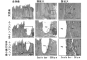
- FIG. 1A shows an image of a natural tooth derived from a tooth germ and an adult mouse extracted from a C57 / BL / 6 mouse of day 18 of gestation.
- FIG. 1B shows images of dental follicle tissue isolated from tooth embryos derived from C57 / BL / 6 mice of day 18 and periodontal ligament tissue isolated from natural teeth derived from adult mice.
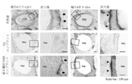
- FIG. 2 shows an HE-stained image of periodontal tissue on the 21st day after transplantation when the dental follicle tissue is transplanted to the periodontal ligament removal model. The HE-stained image is obtained by staining a frontal section of the jawbone and is viewed from the incisor direction toward the occipital region.
- FIG. 1A shows an image of a natural tooth derived from a tooth germ and an adult mouse extracted from a C57 / BL / 6 mouse of day 18 of gestation.
- FIG. 1B shows images of dental follicle tissue isolated from tooth embryos derived
- FIG. 2A shows a HE-stained image of the control group in which the dental follicle tissue was not transplanted
- FIG. 2B shows a HE-stained image of the group in which the dental follicle tissue was transplanted.
- an asterisk (*) indicates an ankylosing formation site
- D indicates dentin
- AB indicates alveolar bone
- C indicates cementum
- PDL indicates periodontal ligament.
- FIG. 3 shows an implant (dental follicle-provided HA) in which a dental follicle tissue is pasted on the coating layer of an implant having a hydroxyapatite (HA) coating layer according to an embodiment of the present invention. The schematic of the transplant model of an implant is shown.
- FIG. 4A is an embodiment of the present invention, a substantial image (left middle diagram) of an HA implant (dental follicle-added HA implant) wound with a dental follicle tissue, and a GFP fluorescence image (right middle) The figure shows an image taken from the side, and the right figure shows an image taken from the bottom).
- an actual image of an implant (HA implant) having a hydroxyapatite coating layer before winding the dental follicle tissue is shown (left figure).
- FIG. 4B shows CT images immediately after transplantation of the HA implant and the dental follicle-attached HA implant (left diagram) and 21 days after transplantation (left middle diagram, right middle diagram, right diagram). Arrows indicate the formation of the periodontal cavity.
- FIG. 5 shows a periodontal tissue (lower stage) after transplantation of a dental follicle-attached HA implant according to an embodiment of the present invention, a periodontal tissue of natural teeth (upper stage), and surrounding tissues after HA implant transplantation. (Interrupted) HE stained image is shown. imp indicates an implant.
- FIG. 5 shows a periodontal tissue (lower stage) after transplantation of a dental follicle-attached HA implant according to an embodiment of the present invention, a periodontal tissue of natural teeth (upper stage), and surrounding tissues after HA implant transplantation. (Interrupted) HE stained image is shown. imp indicates an implant.
- FIG. 6 shows an embodiment of the present invention, in which an HA implant provided with dental follicle tissue derived from a GFP mouse is transplanted into the oral cavity, and an entity image, GFP fluorescence image, around the implant 21 days after transplantation, And the figure which superimposed the entity image and the GFP fluorescence image is shown (the upper image shows the image image
- the arrow indicates the implant.
- FIG. 7 shows an Azan-stained image of the periodontal ligament formed when the dental follicle-attached HA implant according to one embodiment of the present invention is transplanted (right figure), and an Azan-stained image of the natural periodontal periodontal ligament (left).
- FIG. 8 is a scanning electron microscope showing periodontal tissue (lower stage) and natural tooth periodontal tissue (upper stage) formed when the dental follicle-added HA implant according to one embodiment of the present invention was transplanted. The image image
- FIG. 9 shows an image obtained by photographing a periodontal tissue formed when a dental follicle-attached HA implant according to an embodiment of the present invention is transplanted using a transmission electron microscope.
- FIG. 10 shows the elements of titanium (Ti), calcium (Ca), or phosphorus (P) in the periodontal tissue formed when the dental follicle-added HA implant according to one embodiment of the present invention is transplanted.
- An image obtained by analyzing the distribution using an X-ray microanalyzer is shown (the upper image in FIG. 10).
- the lower image in FIG. 10 is an image showing an entity image corresponding to an image obtained by analyzing each element distribution (upper image).
- FIG. 11 shows the elements of titanium (Ti), calcium (Ca), or phosphorus (P) in the periodontal tissue formed when the dental follicle-added HA implant according to one embodiment of the present invention is transplanted.
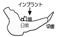
- FIG. 12 is a schematic diagram showing the positional relationship among incisors, molars, and implants in an implant transplantation model used for nerve fiber analysis in periodontal ligament tissue formed after implant transplantation.
- FIG. 13A shows an image in which images before and after correction are superimposed in a test in which correction force is applied to a dental follicle-added HA implant, natural tooth, and HA implant according to an embodiment of the present invention.
- FIG. 13B shows a graph representing the distance traveled by the natural tooth, the transplanted HA implant, and the transplanted dental follicle-provided HA implant when an orthodontic force is applied.
- FIG. 12 is a schematic diagram showing the positional relationship among incisors, molars, and implants in an implant transplantation model used for nerve fiber analysis in periodontal ligament tissue formed after implant transplantation.
- FIG. 13A shows an image in which images before and after correction are superimposed in a test in which correction force is applied to a dental follicle-added HA
- FIG. 14 shows a bone resorption marker (CSF-1) on the compression side of the sixth day after applying orthodontic force to the dental follicle-attached HA implant, natural tooth, and HA implant according to an embodiment of the present invention.
- CSF-1 bone formation marker
- Ocn bone formation marker
- FIG. 15 is an image obtained by observing the expression of a neurofilament, which is a peripheral nerve marker, in the dental follicle-attached HA implant, natural tooth, and periodontal ligament region around the HA implant according to an embodiment of the present invention.
- FIG. 16 shows that noxious stimulation is applied to the dental follicle-provided HA implant, natural tooth, and periodontal ligament surrounding the HA implant according to an embodiment of the present invention by orthodontic force, and two hours later, the trigeminal nerve pulp
- An image of a section obtained by analyzing c-Fos protein expressed in the tract nucleus by in situ hybridization is shown.
- the arrow indicates the expression site of c-Fos protein.
- T indicates the trigeminal spinal tract nucleus.
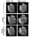
- FIG. 17 is a schematic diagram showing a production process of a three-wall bone defect model.
- FIG. 17 shows an entity photograph of a three-wall bone defect model, a three-dimensionally constructed micro CT image (three-dimensional CT image), and a micro CT image (frontal and horizontal cuts).
- FIG. 17A shows the extraction of the first molar and treatment of the bone
- FIG. 17B shows the creation of a triwall bone defect
- FIG. 17C shows the implantation of the implant.
- FIG. 18 shows a transplantation date (day 0) when a dental follicle-attached HA implant (lower stage) or an HA implant (middle stage) according to an embodiment of the present invention is transplanted to a three-walled bone defect model.
- FIG. 18 shows a three-dimensional CT image of the alveolar bone of the three-walled bone defect model when an implant or the like is not transplanted to the three-walled bone defect model.
- the arrow indicates that the alveolar bone is regenerated to its original height.
- FIG. 19 shows the transplantation date (day 0) and the 14th day from the transplantation date when the dental follicle-added HA implant according to one embodiment of the present invention is transplanted to the three-walled bone defect model.
- FIG. 20 shows the regeneration of alveolar bone on the 50th day of transplantation when the dental follicle-attached HA implant or HA implant according to one embodiment of the present invention is implanted into the three-walled bone defect model. It is a graph which shows the ratio of quantity. Moreover, the ratio of the regenerated amount of the alveolar bone in the three-walled bone defect model in which the implant was not transplanted is shown. In the graph, 100% indicates the amount of alveolar bone in a state before alveolar bone loss.
- FIG. 21 shows a transplantation date (day 0) when a dental follicle-attached HA implant (lower stage) or an HA implant (interruption) according to an embodiment of the present invention is transplanted to a three-walled bone defect model. Eye) and an image of the cut surface of the sagittal cut of the alveolar bone on the 50th day from the date of transplantation. In addition, as a target, sagittal cuts of the alveolar bone on the transplantation date (day 0) and the 50th day from the transplantation date when the HA implant was transplanted into the normal alveolar bone in which the alveolar bone side surface is not defective (upper stage) The image of a cut surface is shown.
- FIG. 21 shows a transplantation date (day 0) when a dental follicle-attached HA implant (lower stage) or an HA implant (interruption) according to an embodiment of the present invention is transplanted to a three-walled bone defect model. Eye) and an image
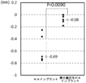
- FIG. 22 shows an alveolar bone of an implant on the 50th day of transplantation when a dental follicle-attached HA implant or an HA implant according to an embodiment of the present invention is implanted into a three-walled bone defect model. It is a graph which shows the amount of depression inward. Moreover, it is a graph which shows the amount of the fall of the implant into the alveolar bone on the 50th day of the transplantation when transplanting the HA implant into the normal alveolar bone where the alveolar bone side surface is not deficient.
- the dental implant according to the present invention is a dental implant that enables functional periodontal tissue formation, a cell mass derived from tooth germ tissue or periodontal ligament tissue is disposed on the surface of the implant,
- the surface of the implant on which the cell mass is disposed is the whole or a part of the surface surrounded by the alveolar bone of the recipient at the time of implant implantation.
- an “implant” is a device for implantation in a living body that is generally used for humans or animals for medical purposes. As used herein, “implant” refers specifically to a dental prosthesis for implantation in the jawbone in place of a lost tooth.
- any material that is conventionally used as an implant material can be used as long as it has no biological harm, is biocompatible, and has a high strength that can withstand occlusion.
- the material of the implant include metal, metal alloy, plastic material, ceramic, composite material, bone substitute material, and the like.
- examples of the metal and metal alloy that can be used as an implant include titanium, steel, iron, alloy steel, iron alloy, titanium alloy, CoCr alloy, silver, copper, calcium, magnesium, and zinc. it can.
- plastic materials that can be used as implants include, for example, polyethylene, polypropylene, polytetrafluoroethylene, polyethylene terephthalate, polyamides, polyurethanes, polysiloxanes, polysiloxane elastomers, polyether ether ketones, Examples thereof include polymers such as polysulfones, polysaccharides and polylactides.
- ceramic materials that can be used as implants include, for example, oxides or nitrides such as aluminum oxide, zirconium oxide, titanium oxide, and silicon oxide, for example, calcium phosphate such as hydroxyapatite, glass and glass ceramic, preferably examples thereof include glass and glass ceramic that dissolve or decompose under physiological conditions.
- the material used for the implant of the present invention is more preferably titanium or a titanium alloy from the viewpoint of biocompatibility and mechanical compatibility.
- Examples of the bone substitute material include autologous teeth, teeth obtained from the same species, or teeth obtained from different species.
- the shape and size of the implant used in the present invention can be appropriately designed by those skilled in the art according to the tooth defect of the transplant destination.
- the dental implant can form the coating layer of a surface coating agent on the surface.
- the “surface coating agent” refers to an agent used for forming a scaffold when a cell mass is adhered to an implant.
- the coating layer of the surface coating agent formed on the implant surface can improve the adhesion of the cell mass to the implant.
- hydroxyapatite examples include gel materials such as hydroxyapatite, ⁇ -TCP (tricalcium phosphate), ⁇ -TCP, and collagen.
- hydroxyapatite has biological activity that promotes bone formation, and can promote the formation of cementum around the implant after implant implantation and the engraftment of the implant on the bone.
- hydroxyapatite is preferably used as a surface coating agent.
- an implant material of the present invention for example, a material having the same effect as a surface coating agent such as using hydroxyapatite can be used as an implant material.
- a cell mass derived from tooth germ tissue or periodontal ligament tissue can be directly arranged on the implant surface.
- a surface coating agent different from the material of the implant can be further coated on the implant surface, and further a cell mass can be arranged on the surface.
- the surface coating agent may be coated so as to cover the entire surface or a part of the implant surface, and the surface surrounded by the alveolar bone of the recipient at the time of implant implantation. Further, the coating layer of the surface coating agent is formed so as to be interposed between the implant and the cell mass to be arranged on the implant.
- the surface surrounded by the recipient's alveolar bone refers to the portion of the implant surface that is buried in the recipient's tooth defect immediately after implant implantation. This part will be connected to the recipient's periodontal tissue in the future.
- the surface coating agent can be coated by a method well known to those skilled in the art. For example, when hydroxyapatite is coated on an implant, it can be performed by vapor deposition, plasma spraying, or the like.
- the thickness of the surface coating agent layer and the coating range of the surface coating agent can be appropriately set by those skilled in the art according to the implant to be coated and the defect of the transplant destination. In one embodiment of the present invention, for example, the thickness of the coating layer can be 1 ⁇ m to 2 ⁇ m.
- a commercially available implant that is already coated with a surface coating agent such as hydroxyapatite can also be used.
- Periodontal tissue refers to a tissue composed mainly of cementum, periodontal ligament, alveolar bone, and gingiva formed mainly in the outer layer of the tooth.
- Periodontal tissues formed by transplantation of the implant of the present invention are, in particular, cementum, periodontal ligament, and alveolar bone.
- the cementum and periodontal ligament formed after implant transplantation of the present invention form periodontal tissue by being connected to the alveolar bone, gingiva and the like on the recipient side.
- Cementum, periodontal ligament, and alveolar bone can be easily identified morphologically by tissue staining or the like.
- tissue staining for example, normal hematoxylin-eosin (HE) staining can be used.
- staining can be performed through processes, such as producing a continuous section
- those skilled in the art can perform histological evaluation by performing tissue staining according to a general method.
- tooth embryo is an early embryo of a tooth that is determined to become a future tooth, and is generally used in the tooth development stage (Bud stage), cap stage ( It refers to the stage from the Cap stage to the bell stage (Bell stage), and in particular, it is a tissue in which the dentin and enamel accumulation, which is a feature of the dental hard tissue, is not recognized.
- the tooth germ tissue that can be used in the present invention can be a tooth germ in a cap-like phase, an early bell-like phase, or a late bell-like phase.
- the tooth germs in the cap-like phase, the early bell-like phase, and the late bell-like phase are preferable in that they have a high ability to differentiate into functional periodontal tissues when transplanted together with the implant.
- a cell mass derived from a tooth germ in the early bell-like period is preferable in that it can further promote the formation of a functional periodontal tissue accompanied by the formation of cementum.
- fetal age 13 to 15 days corresponds to the cap-like period
- fetal age 16 to 18 days corresponds to the early bell-shaped period
- fetal age 19 to postnatal period corresponds to the late bell-shaped period.
- a “regenerated tooth germ” artificially formed by a cell culture technique can be used. Even when regenerated tooth germs are used, cell clusters can be collected at a preferred developmental stage.
- the regenerated tooth germ used in the present invention may be prepared by any method, for example, a first cell aggregate substantially composed of mesenchymal cells and an epithelial cell. It can be produced by a method comprising a step of closely arranging a second cell aggregate substantially constituted and a step of culturing the first and second cell aggregates inside a support carrier. .
- Examples of the method for producing a regenerated tooth germ include WO 2006/129672 pamphlet, JP 2008-29756 A, JP 2008-206500 A, JP 2008-200033 A, and JP 2008-29757 A. , International Publication No. 2011/056007 pamphlet and International Publication No. 2011/056008 pamphlet, the disclosures of each of which are incorporated herein by reference in their entirety.
- Tooth germs and other tissues include mammalian primates (eg, humans, monkeys, etc.), ungulates (eg, pigs, cows, horses, etc.), small mammalian rodents (eg, mice, rats, rabbits, etc.) In addition, it can be collected from the jawbone and periodontal tissue of various animals such as dogs and cats.
- the conditions used for tissue collection are usually applied as they are, and they may be taken out in a sterile state and stored in an appropriate preservation solution.
- Examples of human tooth germs include fetal tooth germs as well as third molars, so-called wisdom tooth germs, but it is preferable to use wisdom tooth germs from the viewpoint of use of autologous tissue.
- the “cell mass” refers to a cell mass derived from a tooth germ tissue or a periodontal ligament tissue disposed on the surface of the implant.
- the cell mass refers to a whole or a part of a tissue derived from the cell mass and at least partially maintaining a functional bond between cells forming the tissue.
- the tooth germ tissue-derived cell mass that can be used in the present invention includes tooth germ mesenchymal tissue, dental follicle tissue, and the like. Tooth germ mesenchymal tissue is present in the tooth germ in the rod-like, cap-like or bell-like phase. Dental follicular tissue is also present in early and late bell tooth germs.
- the periodontal ligament tissue used in the present invention can be collected from a completed tooth. Separation of tissues such as tooth germ mesenchymal tissue and dental follicle tissue from tooth germ tissue, and separation of periodontal ligament tissue from completed teeth may be performed by directly applying the conditions used for tissue collection, It can be removed in a sterile state and stored in an appropriate storage solution. At this time, an enzyme may be used for easy separation. Examples of the enzyme include dispase, collagenase, trypsin and the like.
- the cell mass can be used by physically cutting a tissue extracted from a living body into several cell masses. At this time, it is preferable that the cell mass is cut so that the functional bond between cells at the time of forming the tissue is partially maintained. In particular, it is more preferable that the cell mass after cutting retains the shape so that the outside and inside of the tissue can be distinguished. As described above, the cell mass used in the present invention partially has a functional connection between cells at the time of forming a tissue, so that it is normal when transplanted with an implant. It is possible to form periodontal tissue having various functions.
- the shape of the separated cell mass is not particularly limited as long as it has a shape that can be easily placed on the surface of the implant, and can be separated into a long and narrow shape such as a strip shape.
- the conditions usually used for tissue collection may be applied as they are.
- the method for arranging the cell mass on the implant surface is not particularly limited.
- the cell mass cut into strips can be pasted on the implant surface so that the cell masses do not overlap each other. Or it can also arrange
- the implant to which the cell mass is attached is slightly dried in the air, the adhesiveness is improved.
- a cell mass can be arrange
- the cell mass can be placed on the surface of the implant after implantation, in whole or in part on the surface surrounded by the recipient's alveolar bone. Moreover, it is preferable to arrange
- the side surface forming the inside of the tissue is preferably disposed so as to contact the implant surface. By arranging in this way, the side surface that formed the outside of the tissue faces the recipient's alveolar bone.
- a dental follicle-implanting implant an implant in which dental follicle cells are arranged is referred to as a dental follicle-implanting implant.
- Another aspect of the present invention provides an implant transplantation method including the step of transplanting an implant produced by the implant production method according to the present invention into a tooth defect.
- a functional periodontal tissue can be formed around the implant after implant implantation.
- the side that formed the inside of the tissue is preferably in contact with the surface of the implant.
- positions so that the side which formed the outer side of the tissue may oppose a recipient's periodontal tissue.
- the formation of a functional periodontal tissue can be promoted by bringing the cell mass derived from the tooth germ tissue or periodontal ligament closer to the state existing in the actual periodontal tissue of natural teeth.
- deletion part means the part provided in gums by tooth extraction etc., and there is no restriction
- the location of the defect and the type of target tooth are not particularly limited.
- the defect is usually located in the jaw bone, the alveolar bone of the oral cavity, or the like.
- a known method used clinically for implant implantation such as GTR method (guided tissue regeneration) for the defect site. Bone regeneration may be performed by a method to increase bone mass. After placement in the defect site to be implanted, it is preferable to perform suturing or the like according to normal processing.
- a defect having a diameter slightly larger than the implant diameter is surgically formed in order to prevent the cell mass stuck on the implant surface from peeling off from the surface. You can also When an implant is transplanted into a defect having such a large diameter, the space between the transplanted implant and the recipient's alveolar bone is filled with blood from the surrounding tissue.
- the transplant target is preferably the same species as the animal from which the tooth germ tissue or periodontal ligament tissue used for the manufacture of the implant is extracted, and the individual from which the tooth germ tissue or periodontal ligament tissue is extracted More preferably, the same individual.
- animals include mammals including humans, cows, horses, pigs, dogs, cats, mice and the like. A non-human mammal is also preferred.
- the implant site of an implant can be easily observed by a person skilled in the art visually or by CT image.
- CT images and CT cross-sectional images can also be easily captured by using a device known to those skilled in the art.
- 3D micro X-ray CT R_mCT for laboratory animals can be used, and for example, it can be performed under conditions such as 90 kV, 150 mA, and a tomographic thickness of 10 mm.
- those skilled in the art can perform image construction and analysis using appropriate image analysis software after CT imaging.
- image analysis software for example, image filing software for small animals, i-VIEW type R, and high-definition 3D / 4D image analysis software Imaris (Bitplane) can be used.
- image analysis software for example, image filing software for small animals, i-VIEW type R, and high-definition 3D / 4D image analysis software Imaris (Bitplane) can be used.
- image analysis software for example, image filing software for small animals, i-VIEW type R, and high
- a functional periodontal tissue can be formed around the implant after transplantation to the recipient.
- functional periodontal tissue means (i) having functional cementum and functional periodontal ligament, or (ii) having functional nerve fibers. That is, it preferably has both the characteristics (i) and (ii). That is, a functional periodontal tissue can be evaluated by, for example, whether or not it has a functional cementum and a functional periodontal ligament. Confirm that functional cementum and functional periodontal ligament have a layer structure equivalent to that of periodontal tissue of natural teeth, for example, by histological analysis by HE staining, Azan staining, etc. It can be evaluated by doing.
- the periodontal ligament of a natural tooth usually has periodontal ligament fibers that run in a direction perpendicular to the long axis direction of the root.
- This periodontal ligament plays an important role especially in supporting the occlusal force of the teeth. Therefore, by analyzing whether or not periodontal ligament fibers running perpendicular to the long axis direction of the implant are formed, as in natural teeth, the function of the periodontal ligament can be particularly evaluated.
- the analysis of periodontal tissue morphology and periodontal ligament analysis can be observed by, for example, a scanning electron microscope or a transmission electron microscope in addition to the above method.
- the layer structure of the periodontal tissue can be confirmed.
- the function of bone remodeling with respect to the load of correction force as described in Example 4 below, can be evaluated. Or it can evaluate by analyzing whether the implant after a transplant is movable by the load of correction force.
- the bone remodeling can be evaluated by, for example, analyzing the expression of a bone formation marker and / or a bone resorption marker after applying a correction force.
- the periodontal tissue formed when the implant of the present invention is transplanted has a movement amount of 68% or more compared to the natural tooth when the same correction force as that of the natural tooth is applied, preferably 80% or more.
- a functional periodontal tissue can be evaluated by, for example, whether or not it has a functional nerve fiber.
- a functional nerve fiber refers to a nerve fiber that can transmit a stimulus to the central nervous system when there is a stimulus to periodontal tissue or the like. Whether the nerve fiber is functional or not is determined, for example, in the trigeminal tract nucleus after applying stimulation by the orthodontic force load to the periodontal tissue to be evaluated as in the method described in Example 6 below. It can be evaluated by expression analysis of c-fos.
- the dental implant of the present invention not only the formation of functional cementum and periodontal ligament, but also the regeneration of alveolar bone can be promoted, and the regeneration ability of alveolar bone can also be improved.
- “improving the regeneration ability of the alveolar bone” means that when a conventional implant is transplanted by transplanting the dental implant according to the present invention at a transplant site where a portion of the alveolar bone is missing. Alternatively, it means that the alveolar bone can be regenerated so that it is closer to the original shape of the alveolar bone than when the implant is not transplanted.
- the dental implant according to the present invention can prevent a situation in which the implant gradually falls into the alveolar bone due to a partial loss of the alveolar bone after the transplantation. It is possible to maintain the position (for example, height) and direction.
- Embodiments of the present invention may be described with reference to schematic diagrams, but in the case of schematic diagrams, they may be exaggerated for clarity of explanation.
- terms such as first, second, etc. are used to represent various elements, it is understood that these elements should not be limited by those terms. These terms are only used to distinguish one element from another, for example, the first element is referred to as the second element, and similarly, the second element is the first element. Can be made without departing from the scope of the present invention.
- any numerical value used for indicating a numerical range or the like is interpreted as including the meaning of the term “about” unless otherwise specified.
- “10 times” is understood to mean “about 10 times” unless otherwise specified.
- Example 1 Analysis of periodontal tissue formation ability by dental follicle tissue transplantation in periodontal ligament removal model >> (Preparation of dental follicle tissue)
- the dental follicle tissue in this experiment was obtained from C57 / BL / 6 mice of day 18 gestational age in the bell-like stage. Specifically, the dental follicle tissue was separated from the tooth germ that became the lower first molar. Tooth germ extraction was performed according to the method of Nakao et al. (Nakao K, et al. Nat Methods. 2007; 4 (3): 227-30.). The method for extracting the dental follicle tissue from the excised tooth germ of day 18 was performed as follows.
- FCS Fetal calf serum
- SIGMA strepto St. L.
- the tissue was physically separated with a 25G needle (NN-2516R, Terumo, Tokyo, Japan) in Dulbecco's modified easy medium (D-MEM; WAKO, Osaka, Japan) to which was added.
- D-MEM Dulbecco's modified easy medium
- Dental follicular tissue isolated from mouse embryonic day 18 tooth germ had a sac-like morphology (see FIG. 1).
- the periodontal ligament on the buccal side surface of the second molar was physically removed from the alveolar septum with the second molar adjacent to the first molar using a 25G needle (NN-2516R, Terumo, Tokyo, Japan).
- the dental follicle tissue separated from a mouse embryonic day 18 tooth germ was physically cut into a plurality of cell clusters in a strip shape.
- transplantation was performed so that the side surface of the cell mass forming the inside of the dental follicle tissue was attached to the periodontal ligament removal site so that the side surface of the second molar tooth root side from which the periodontal ligament was removed was directed.
- each cell mass was affixed to the periodontal ligament removal site so as not to overlap.
- the jawbone was removed, and the tissue analysis of the dental follicle tissue transplantation site was performed by HE staining.
- Example 2 Evaluation of periodontal tissue-forming ability by implantation of implants with dental follicle tissue >> (Preparation of implant) For the implant body for transplantation, cut a titanium wire having a diameter of 0.6 mm (Niraco Co., Ltd., Tokyo, Japan) for a length of 1.7 mm, and forming a cone shape of about 0.5 mm from the apical side. It was produced with.
- the surface of the implant body is coated with hydroxyapatite by vacuum deposition and covered with a hydroxyapatite layer having a thickness of 2 ⁇ m, so that the implant body is coated with hydroxyapatite (hereinafter referred to as HA implant) (Yamahachi Teikoku Kogyo Co., Ltd.) , Aichi Prefecture, Japan).
- HA implant hydroxyapatite
- a dental follicle tissue extracted from a C57BL / 6 mouse of day 18 of gestation was wrapped around the HA implant in the same manner as in Example 1 (hereinafter referred to as a dental follicle-attached HA implant; FIG. 4A).
- the dental follicle tissue was surgically cut into strips of cell mass, and the cell mass was placed on the HA implant by wrapping so that the cell masses did not overlap each other. The side of the cell mass that formed the inside of the dental follicle tissue was adhered to the surface of the HA implant.
- the mandibular first molar was extracted from 4-week-old C57BL / 6 mice, a tooth defect was provided, and a gingival healing period of 4-5 days was provided. Thereafter, gingival incision and peeling of the extraction site of the lower first molar were performed. Cutting the alveolar bone of the tooth defect using a dental micromotor (Viva-Mate Plus, Nakanishi, Tokyo, Japan) and a dental reamer (MANI, Tochigi, Japan), diameter 0.9 mm, depth 1.2 mm An implant fovea was formed.
- a dental micromotor Viva-Mate Plus, Nakanishi, Tokyo, Japan
- MANI Tochigi, Japan
- the dental follicle-provided HA implant produced above was transplanted into the graft fossa.
- the control group which transplanted the HA implant which has not wound the dental follicle tissue was also produced.
- the implant transplantation site was sutured to the gingiva with an 8-0 nylon suture (Bearmedic, Chiba, Japan) (FIG. 3). Mice transplanted with each implant body were subjected to micro CT imaging immediately after transplantation and on the 21st day after transplantation (In vivo Micro X-ray CT System; R_mCT, Rigaku Corporation, Tokyo, Japan).
- integrated image processing software i-VIEW-3DX, Morita, Osaka, Japan
- HE staining On the 21st day after transplantation, the jawbone containing the implant was removed from the recipient mouse and histologically evaluated by HE staining.
- the control group transplanted with the HA implant exhibited osseointegration in which the alveolar bone was directly bonded to the implant surface layer (FIGS. 4B and 5).
- the HA implant to which the dental follicle tissue was applied a periodontal ligament space was observed between the implant surface layer and the surrounding alveolar bone (lower row in FIG. 4B, arrow in the middle right diagram).
- the HA implant to which the dental follicle tissue was added in the HE-stained image, a tissue structure equivalent to the periodontal tissue of natural teeth such as cementum, periodontal ligament, and alveolar bone was recognized from the implant surface layer (FIG. 5). .
- GFP mice C57BL / 6-Tg (CAG-EGFP) mice (Japan SLC, Inc., An HA implant body provided with a dental follicle tissue derived from Japan)
- the transplant site was photographed for GFP color development using a fluorescent stereomicroscope (AxioLumer, Carl Zeiss, Jene, Germany).
- a fluorescent stereomicroscope Auto Microscope
- formation of tissue derived from GFP mice was observed in the periodontal ligament and alveolar bone region around the implant on day 21 (FIG. 6). From this, it was shown that the transplantation of the implant with the dental follicle tissue engrafted in the recipient's jawbone with periodontal tissue formation.
- Example 3 Analysis of periodontal tissue formed after implant transplantation >> As shown in Example 2, implantation of the dental follicle-attached HA implant formed periodontal tissue engrafted in the jawbone. Therefore, in order to analyze the running of the periodontal ligament formed after transplantation of the dental follicle-provided HA implant, periodontal tissue of natural teeth, periodontal tissue around the implant 21 days after transplantation of the dental follicle-provided HA implant, and Periodontal tissue around the implant on the 21st day after transplantation of the HA implant in which the dental follicle tissue was not wound was stained with Azan staining, and the periodontal ligament was analyzed.
- the periodontal tissue around the implant on the 28th day after transplanting the dental follicle-provided HA implant was analyzed using a scanning electron microscope and a transmission type microscope. It was observed with an electron microscope to analyze the morphology of periodontal tissue formed after implant transplantation.
- the periodontal tissue around the day implant was analyzed for elemental distribution of titanium (Ti), calcium (Ca), and phosphorus (P) using an X-ray microanalyzer.
- paraffin section prepared to a thickness of 6 ⁇ m was immersed in xylene for 6 minutes. Then, paraffin was removed by immersing in 100%, 90%, and 70% alcohol for 3 minutes each. The sections from which the paraffin was removed were allowed to adjust to running water for 5 minutes and then immersed in 10% trichloracetic acid and 10% potassium dichromate for 15 minutes. The sections were again flushed with water and then immersed in azocarmine G solution for 30 minutes to stain the nucleus and cytoplasm. The azocarmine G solution was washed with running water, then immersed in aniline / alcohol for 10 seconds for separation, and immersed in acetic acid alcohol for 1 minute to stop the separation.
- the periodontal ligament tissue formed around the implant on the 21st day after implantation of the dental follicle-provided HA implant has running perpendicular to the long axis direction of the implant, like running of the periodontal ligament of natural teeth. ( Figure 7).
- Example 2 (Morphological analysis by electron microscope) In the same manner as in Example 2, the dental follicle-attached HA implant was transplanted into the mandibular first molar defect of a 4-week-old C57BL / 6 mouse. On day 28 after implant transplantation, mice were subjected to transcardial perfusion fixation using Karnovsky fixative under deep anesthesia. After transcardial perfusion fixation, the jawbone of the mouse was removed, and then the jawbone was postfixed with 31% osmium tetrachloride at 4 ° C. Next, after dehydrating the jawbone with ethanol, it was dried in a critical point dryer (HCP-2, Hitachi, Tokyo, Japan) and replaced with an epoxy resin and embedded.
- HCP-2 critical point dryer
- a portion of the sample embedded with the epoxy resin was cut into a sagittal cut using a diamond disk so that the center of the transplanted implant was a cut surface.
- the cut sample was subjected to a conductive treatment using an ion sputtering apparatus (E-1030, Hitachi), and then subjected to Au—Pd vapor deposition. Thereafter, the Au—Pd vapor-deposited sample was observed using a scanning electron microscope (S-4700, Hitachi) at an acceleration voltage of 5 kV.
- S-4700, Hitachi scanning electron microscope
- an ultra-thin section (100 nm) was prepared from a part of an epoxy resin-embedded sample prepared from a mouse transplanted with a dental follicle-attached HA implant using an ultramicrotome (ULTRACUT-UCT; Leica microsystems). did.
- the prepared sections were observed using a transmission electron microscope (H-7600, Hitachi) at an acceleration voltage of 75 kV.
- a scanning electron microscope (SEM) image of the periodontal tissue around the implant on day 28 after transplantation of the dental follicle-provided HA implant was obtained and observed.
- SEM scanning electron microscope
- dense periodontal ligament fiber bundles running from the surface of the implant toward the alveolar bone and the formation of lamellar cementum were observed, and the tissue morphology was almost the same as that of natural teeth.
- FIG. 8 Further, in the SEM image obtained by observing the periodontal tissue around the implant after implant implantation, binding between the implant surface layer and the periodontal ligament fibers was observed (lower row in FIG. 8). Further, in the transmission electron microscope (TEM) image in which the periodontal tissue around the implant after transplantation was observed, binding between cementum and periodontal ligament fibers was also observed (arrow in FIG. 9).
- EPMA-1610 Shimadzu Corporation, Kyoto, Japan
- EPMA-1610 Shimadzu Corporation, Kyoto, Japan
- the implant surface layer made of titanium (FIG. 10). Since the thickness of the hydroxyapatite coating on the implant surface is as very thin as 2 ⁇ m or less, the distribution of Ca and P in the implant surface layer is considered to be derived from cementum containing Ca and P as constituent elements. Therefore, it became clear that the hard tissue which consists of cementum was formed in the implant surface layer. In addition, in the SEM image of the periodontal tissue around the implant after the implant transplantation, no distribution of Ca and P was observed in the portion where the fiber was running.
- periodontal ligament does not contain Ca and P as constituent elements, it became clear that the periodontal ligament region was secured in the portion where the distribution of Ca and P was not recognized.
- the distribution images of the constituent elements were superimposed and the distribution of the three elements was observed more clearly, it was possible to confirm the location where Ca and P were accumulated on the implant surface (Ti surface) (FIG. 11).
- Example 4 Functional analysis of periodontal ligament formed after implant transplantation >> In order to analyze the function of the periodontal ligament formed after transplantation of the dental follicle-provided HA implant, natural teeth, the dental follicle-provided HA implant 21 days after transplantation, and the dental follicle tissue 21 days after transplantation are not wrapped Experimental correction was performed on the HA implant, and the amount of tooth and implant movement over time was measured. In addition, as an analysis of bone remodeling by experimental correction, we analyzed the expression of bone resorption markers and bone formation markers in periodontal tissue after correction force loading.
- Example 2 The experimental correction of natural teeth and implants, and the method of analyzing the amount of movement of natural teeth and implants due to the load of correction force were performed as follows.
- dental follicle-fed HA implants or HA implants were implanted into C57 / BL / 6 mice, respectively.
- a nickel titanium wire (VIM-NT, Oralcare Co., Ltd. Tokyo, Japan) having a diameter of 0.010 inches was fixed to a mandibular incisor and a natural tooth or implant to be corrected.
- a load of 10 to 15 g was horizontally applied using a dial tension gauge (Mitutoyo).
- the correction force was applied for 14 days from the lingual side to the buccal side (perpendicular to the side of the dentition and outside the oral cavity) with respect to the natural tooth, HA implant, and dental follicle-attached HA implant.
- CSF-1 Colony stimulating factor 1
- OCN osteocalcin
- a DIG-labeled RNA probe was synthesized using the obtained PCR product and T7 RNA polymerase and digoxigenin (DIG) RNA Labeling Mix (Roche, Mannheim, Germany).
- DIG digoxigenin
- the excised jawbone was fixed with a 4% paraformaldehyde solution for 24 hours, and then decalcified for 72 hours using 10% sodium formate citrate and 22.5% formic acid decalcification solution. Thereafter, the jawbone was immersed in 12.5% (w / v) and 25% (w / v) sucrose solutions in order for 12 hours, and frozen and embedded using OCT compound (Miles Inc, Naperville, IL).
- RNA probe was hybridized at 70 ° C. for 16 hours. Localization of mRNA was detected by enzyme coloring at an appropriate time at 30 ° C. by an immunological technique using alkaline phosphatase-labeled anti-DIG Fab Fragments and NBT / BCIP (Roche, Mannheim, Germany).
- -Mouse osteocalcin (mOCN)
- forward primer (SEQ ID NO: 1) TAGCAGACACCATGAGGACC -Mouse osteocalcin (mOCN)
- reverse primer (SEQ ID NO: 2) TGACATCCATACTTGCAGGG -Mouse CSF1 (mCSF1)
- forward primer (SEQ ID NO: 3) TACTGAACCTGCCTGCTGAA -Mouse CSF1 (mCSF1)
- reverse primer SEQ ID NO: 4
- Example 5 Analysis of nerve fibers in periodontal ligament tissue formed after implant transplantation >> Peripheral nerves invade the periodontal ligament tissue of natural teeth in order to maintain the function and homeostasis of the tooth, and have a function by afferent stimulus transmission. Therefore, it was evaluated whether or not nerve fibers having normal functions were formed in the HA implants on the 21st day after transplantation and in the periodontal ligament tissues around the dental follicle-attached HA implants on the 21st day after transplantation. . First, in the same manner as in Example 2, the dental follicle-attached HA implant and HA implant were transplanted into C57 / BL / 6 mice.
- the dental follicle-provided HA implant or the jawbone containing the HA implant was extracted from the recipient mouse.
- the excised jawbone was fixed with a 4% paraformaldehyde solution for 24 hours, and then decalcified for 72 hours using 10% sodium formate citrate and 22.5% formic acid decalcification solution. Thereafter, the jawbone was immersed in 12.5% (w / v) and 25% (w / v) sucrose solutions in order for 12 hours, and frozen and embedded using OCT compound (Miles Inc, Naperville, IL). .
- a 10 ⁇ m thick section of the jawbone was prepared using a cryostat (CM3050S; Leica microsystems), and immunostaining of these tissue sections was performed.
- immunostaining of neurofilament as a nerve fiber marker was performed.
- the primary antibody was neurofilament SMI312 (mouse anti-NF Ab; 1: 1000, Abcam, Cambridge, Mass.),
- the secondary antibody was Alexa Fluor594-conjugated goat anti-rat IgG (1: 500, Prob).
- the evaluation method was to induce production in the trigeminal spinal tract nucleus, an index of pain stimulation, after applying experimental correction force to natural teeth, HA implants 21 days after transplantation, and HA implants with dental follicles 21 days after transplantation.
- the analysis was performed by analyzing the expression of c-Fos protein.
- the dental follicle-attached HA implant and HA implant were transplanted into C57 / BL / 6 mice in the same manner as in Example 2.
- mice were fixed under deep anesthesia.
- a correction force of 10 to 15 g was applied from the lingual side to the buccal side of the natural first molar and the implant engrafted in the jawbone.
- the chest wall of the mouse under anesthesia was opened with scissors.
- a 25 G needle was inserted from the lower left ventricle where the heart was exposed, and a PBS ( ⁇ ) solution was perfused from the heart to the whole body using a peristaltic pump.
- a blood removal route was secured by excising the left atrium. After complete blood removal, fixation was performed by continuously refluxing a 4% paraformaldehyde solution to the whole body in the same manner.
- medullary tissue was removed from the skull of the mouse, and each sucrose solution of 12.5% (w / v) and 25% (w / v) was used for 12 hours. Immerse in order. After immersion, the medullary tissue was frozen and embedded using OCT compound (Miles Inc, Naperville, IL). After embedding, a 40 ⁇ m-thick section was prepared with a cryostat (CM3050S; Leica microsystems).
- the prepared sections were blocked with endogenous peroxidase with methanol supplemented with 0.3% H 2 O 2 and blocked with 3% serum. Then, it was reacted with anti-c-Fos Ab (1: 10,000, Santa Cruz Biotechnology, Santa Cruz, Calif.), And peroxidase-labeled goat anti-rabbit IgG (1: 300, Cappel Laboratory, IgG). ). Then, immunostaining was performed using PAP immunocomplex (1: 3000, Cappel).
- c-Fos protein was not observed when the corrective force was applied to the HA implant.
- c-Fos expression was observed 2 hours after orthodontic stimulation, indicating that nociceptive stimulation to the periodontal ligament was transmitted to the central nerve (FIG. 16). From these results, it was shown that when a dental follicle-attached HA implant was transplanted, a nerve function capable of transmitting a noxious stimulus was formed in the periodontal ligament around the implant.
- Example 7 Analysis of alveolar bone regeneration after implant implantation in a three-walled bone defect model >>
- a three-walled bone defect model lacking bone on one side of the alveolar bone at the implant transplantation site was prepared.
- the amount of regenerated alveolar bone was analyzed.
- the mandibular first molars of 4 week old C57BL / 6 mice were extracted, and a bone tissue healing period of 2-3 weeks was provided.
- the dental follicle-attached HA implant or HA implant was transplanted into the transplantation fossa, and gingival suture was performed with 8-0 nylon sutures (Bearmedic, Chiba, Japan).
- Mice transplanted with an implant use a micro CT apparatus (In vivo Micro X-ray CT System; R_mCT, Rigaku, Tokyo, Japan) immediately after transplantation and on the 14th, 28th, and 50th days after transplantation.
- a micro CT image was acquired.
- the acquired micro CT image (slice image) data was three-dimensionally constructed using integrated image processing software (i-VIEW-3DX, Morita, Osaka, Japan) to obtain a three-dimensional CT image.
- the regeneration of the buccal bone in the alveolar bone at the transplant site was evaluated over time.
- a sample in which only a three-walled bone defect without implant implantation was prepared, and the regeneration of the buccal bone in the alveolar bone was similarly evaluated over time.
- FIG. 18 shows three-dimensional CT images immediately after transplantation and on the 14th, 28th, and 50th days after transplantation.
- the group where the HA implant was transplanted and in the group where the implant was not transplanted it was possible to observe a certain amount of alveolar bone regeneration on the 50th day, but the position where the alveolar bone should originally exist (dotted line in the figure) Until the alveolar bone did not regenerate.
- FIG. 19 shows micro CT images on the 14th, 28th, and 50th days from the transplantation in the three-walled bone defect model transplanted with the dental follicle-attached HA implant.
- Alveolar bone was regenerated on the 14th day after the transplantation, and almost completely recovered alveolar bone was observed on the 50th day after the transplantation.
- a periodontal cavity-like gap was observed between the regenerated alveolar bone and the dental follicle-implanted implant (arrowhead in FIG. 19).
- the dental follicle-implanted implant was able to enable not only periodontal ligament formation but also engraftment of the implant with alveolar bone regeneration.
- three-walled defect a group in which a dental follicle-attached HA implant was transplanted for a three-walled defect model, a group in which an HA implant was transplanted in a three-walled defect model, and a three-walled bone defect in which no implant was transplanted.
- a superimposition of images on the 0th and 50th day of transplantation was created, and the regenerated area of the alveolar bone was quantified. The result is shown in FIG. As shown in FIG. 20, a significantly higher alveolar bone regeneration amount was observed in the transplanted group of the dental follicle-added HA implant than in the other two groups.
- Example 2 In addition, in order to evaluate the drop of the implant into the alveolar bone after transplantation, it was prepared in the same manner as in Example 2 (not a triwall bone defect), and a group in which the HA implant was transplanted into the graft fossa, and a triwall bone defect Vertical movement of the implant on the 0th and 50th days after transplantation in the group transplanted with the HA implant in the model and the group transplanted with the dental follicle-attached HA implant in the three-walled bone defect model The amount was measured and graphed (FIGS. 21 and 22).
Abstract
Description
さらに近年、この歯科置換医療の先端的治療法の一つとして、口腔インプラント治療が実施されている。口腔インプラント治療とは、喪失歯部位の顎骨にチタン等の人工歯根を植立する手段である。
天然歯の周囲には、歯根側のセメント質と外側の歯槽骨を繋ぐ線維状の歯根膜組織が存在する。セメント質は、歯根面の保護と歯根膜を歯根面に付着させる機能を有している。また、歯根膜は、大きく分けて、1)咬合力の緩衝作用、2)歯の移動能(歯科矯正治療などに用いられるメカニクス)、および、3)咬合および矯正などの侵害刺激(痛み刺激等)を中枢神経系へ伝える神経伝達機能の3つの機能を有することが知られている。このうち、特に歯根膜は、歯の咬合力を緩衝するために、歯根の長軸方向に対して垂直方向に走行する繊維を有しており、この歯根膜組織における線維の走行が、歯根膜の機能発現のために必要不可欠な構成であることが知られている。
このように、歯科用インプラントを移植した際にも、天然歯と同様の歯周組織を形成可能な技術が長い間望まれていた。
<非特許文献1>
本文献は、ラット歯根膜から採取した前駆細胞由来の培養細胞の利用について開示している。本文献は、SLA処理したインプラントへ、培養細胞をマトリゲルとともにコーティングすることを開示している。また、本文献は、このインプラントを、ラットの歯牙欠損部に移植した場合に、歯周組織が形成されたことについて言及している。
しかし、本文献においてインプラントの周囲に形成された歯根膜の走行は、インプラントの長軸方向と平行であり、天然歯根膜と相違するものである。歯根膜の走行は、歯の咬合力を支えるのに重要な意味を持つため、このような歯根膜を有する歯周組織は、咬合力を支える機能を期待することができない。
本文献には、EMD(エムドゲイン)処理したチタンインプラントを顎骨に移植し、同時に歯根膜から採取したPDL細胞を移植部に注入する方法が開示されている。この方法は、インプラント周囲の歯周組織の形成を図るために、エナメルマトリックスタンパクを主成分として薬剤を併用するものである。そして、本文献は、上皮組織の混入していない歯槽骨と結合した組織の形成について言及している。しかし、形成された歯周組織中に歯周組織の1つであるセメント質の構造は認められていない。また、歯周組織中の歯根膜の走行も確認されていない。
すなわち、本発明は、機能的な歯周組織形成を可能とする歯科用インプラントであって、ここで、歯胚組織由来または歯根膜組織由来の細胞塊が、前記インプラントの表面に配置されており、前記細胞塊が配置される前記インプラントの表面は、前記インプラント移植時にレシピエントの歯槽骨に囲まれる表面の全体又は一部であることを特徴とする歯科用インプラントに関する。
また、本発明の機能的な歯周組織形成を可能とする歯科用インプラントの一実施態様においては、前記細胞塊が歯胚組織由来であって、前記歯胚組織が、帽状期、鐘状前期、および、鐘状後期からなる群より選択されるいずれか1つの発生段階にあることを特徴とする。
また、本発明の機能的な歯周組織形成を可能とする歯科用インプラントの一実施態様においては、前記形成される歯周組織が、(i)機能的なセメント質および機能的な歯根膜を有する、および、(ii)機能的な神経線維を有する、のうちの少なくとも一方の特徴を有することを特徴とする。
また、本発明の機能的な歯周組織形成を可能とする歯科用インプラントの一実施態様においては、前記表面コーティング剤が、ハイドロキシアパタイト、α-リン酸三カルシウム、β-リン酸三カルシウム、および、コラーゲンからなる群より選択されることを特徴とする。
また、本発明の機能的な歯周組織形成を可能とする歯科用インプラントの一実施態様においては、前記インプラントは、歯槽骨の再生を促進することができることを特徴とする。
また、本発明の機能的な歯周組織形成を可能とする歯科用インプラントの一実施態様においては、前記インプラントは、歯槽骨の再生能を改善することができることを特徴とする。
なお、以上述べた本発明の一又は複数の特徴を、任意に組み合わせたものも、本発明の歯科用インプラントであることはいうまでもない。
ここで、本発明の機能的な歯周組織を形成可能とする歯科用インプラントの製造方法の一実施態様においては、前記歯胚組織由来の細胞塊が、歯胚間葉組織由来または歯小嚢組織由来の細胞塊であることを特徴とする。
また、本発明の機能的な歯周組織を形成可能とする歯科用インプラントの製造方法の一実施態様においては、インプラント移植後に、前記インプラントが矯正可能であることを特徴とする。
また、本発明の機能的な歯周組織を形成可能とする歯科用インプラントの製造方法の一実施態様においては、前記細胞塊を配置する工程の前に、インプラントの表面全体またはその一部であって、インプラント移植時にレシピエントの歯槽骨に囲まれる表面上に、表面コーティング剤のコーティング層を形成する工程をさらに含み、前記細胞塊が前記コーティング層の表面上に配置されていることを特徴とする。
なお、以上述べた本発明の一又は複数の特徴を、任意に組み合わせたものも、本発明の歯科用インプラントの製造方法であることはいうまでもない。
また、本発明の歯を欠損した哺乳動物へのインプラントの移植方法の一実施態様においては、前記動物が非ヒト哺乳動物であることを特徴とする。
なお、本発明の歯科用インプラントによれば、インプラント移植後においてインプラント周囲に機能的な歯周組織を形成させることができるのみならず、インプラント移植部位周辺の歯槽骨の再生を促進することができる。また、本発明の歯科用インプラントによれば、インプラント移植部位周辺の歯槽骨の再生能も改善することができる。
本明細書において「表面コーティング剤」とは、インプラントへ細胞塊を接着させる際の足場の形成に使用されるものをいう。インプラント表面に形成される表面コーティング剤のコーティング層は、インプラントへの細胞塊の接着を向上させることができる。
特に、ハイドロキシアパタイトは、骨形成を促進させる生物活性を有しており、インプラント移植後のインプラント周囲におけるセメント質形成の促進や、インプラントの骨への生着を促進させることができる。このような点において、ハイドロキシアパタイトは、表面コーティング剤として使用されることが好ましい。
例えば、ハイドロキシアパタイトをインプラントにコーティングする際には、蒸着、プラズマ溶射法等により行うことができる。表面コーティング剤の層の厚さや表面コーティング剤のコーティングの範囲は、コーティング対象のインプラントや移植先の欠損部に応じて、当業者が適宜設定することができる。本発明の一実施の態様においては、例えば、コーティング層の厚みを、1μm~2μmとすることができる。
また、上記のように表面コーティング剤のコーティング層をインプラント表面に形成させる以外に、ハイドロキシアパタイトなどの表面コーティング剤が、すでにコーティングされている市販のインプラントを使用することもできる。
なお、例えば、マウスの場合、胎齢13~15日が帽状期に相当し、胎齢16~18日が鐘状前期に相当し、胎齢19日~生後が鐘状後期に相当する。
なお、本明細書において、例えば、歯小嚢細胞を配置したインプラントを、歯小嚢付与インプラントという。
本発明に係るインプラントによれば、インプラント移植後において、インプラント周囲に機能的な歯周組織を形成させることができる。
欠損部は、通常、顎骨、口腔の歯槽骨などに位置する。また歯の喪失に伴って歯槽骨量が低下している場合には、欠損部位に対してGTR法(guided tissue regeneration:組織再生誘導法)など、インプラントの埋設のために臨床で用いられる公知の方法により骨の再生を行って骨量を増加させてもよい。インプラント対象となる欠損部位へ配置した後は、通常の処理に従って、縫合等を行うことが好ましい。
また、欠損部へのインプラントの移植の際に、インプラント表面上に張り付けた細胞塊が表面から剥がれてしまうことを防ぐために、インプラント径よりも少し大きめの径を有する欠損部を外科的に形成させることもできる。このような大きな径を有する欠損部へインプラントを移植した場合には、移植後のインプラントとレシピエントの歯槽骨との間の空間が、周囲の組織からの血液によって満たされる。
すなわち、機能的な歯周組織は、例えば、機能的なセメント質および機能的な歯根膜を有している否かで評価することができる。機能的なセメント質および機能的な歯根膜とは、例えば、HE染色、アザン染色等による組織学的分析をした際に、天然歯の歯周組織と同等の層構造を有しているかを確認することで評価することができる。ここで、天然歯の歯根膜は、通常、歯根の長軸方向に対して垂直方向に走る歯根膜の繊維を有している。この歯根膜は、特に、歯の咬合力を支えるのに重要な役割を担っている。従って、天然歯と同様に、インプラントの長軸方向に対して垂直に走る歯根膜の線維が形成されているかを解析することで、特に歯根膜の機能を評価することができる。歯周組織の形態の解析や歯根膜の走行の解析は、上記方法の他、例えば、走査型電子顕微鏡や透過型電子顕微鏡によりその形態を観察することも可能である。また、歯周組織における、硬組織-線維性組織-硬組織の3層構造を確認するために、例えば、X線マイクロアナライザーを用いて元素の分布を解析することにより、歯周組織の層構造を確認することができる。
別の方法としては、下記実施例4に記載するような、矯正力の負荷に対する骨リモデリングの機能を評価することができる。または、移植後のインプラントが矯正力の負荷により移動可能であるか否かを解析することで評価することができる。骨リモデリングの評価には、例えば、矯正力負荷後に骨形成マーカーおよび/または骨吸収マーカーの発現を解析することで評価できる。本発明のインプラントを移植した際に形成される歯周組織は、天然歯と同様の矯正力を加えた場合に、天然歯と比較して68%以上の移動量を有しており、好ましくは、80%以上の移動量を有する。
また、本明細書において用いられる「含む」との用語は、文脈上明らかに異なる理解をすべき場合を除き、記述された事項(部材、ステップ、要素、数字など)が存在することを意図するものであり、それ以外の事項(部材、ステップ、要素、数字など)が存在することを排除しない。
異なる定義が無い限り、ここに用いられるすべての用語(技術用語及び科学用語を含む。)は、本発明が属する技術の当業者によって広く理解されるのと同じ意味を有する。ここに用いられる用語は、異なる定義が明示されていない限り、本明細書及び関連技術分野における意味と整合的な意味を有するものとして解釈されるべきであり、理想化され、又は、過度に形式的な意味において解釈されるべきではない。
本発明の実施態様は模式図を参照しつつ説明される場合があるが、模式図である場合、説明を明確にするために、誇張されて表現されている場合がある。
第一の、第二のなどの用語が種々の要素を表現するために用いられるが、これらの要素はそれらの用語によって限定されるべきではないことが理解される。これらの用語は一つの要素を他の要素と区別するためのみに用いられているのであり、例えば、第一の要素を第二の要素と記し、同様に、第二の要素は第一の要素と記すことは、本発明の範囲を逸脱することなく可能である。
(歯小嚢組織の調製)
本実験における歯小嚢組織は、鐘状期にある胎齢18日のC57/BL/6マウスより得た。具体的には、歯小嚢組織は、下顎第一臼歯となる歯胚より分離した。歯胚の摘出は、Nakaoらの方法(Nakao K, et al. Nat Methods. 2007; 4(3):227-30.)に準じて行った。
摘出した胎齢18日の歯胚から歯小嚢組織を摘出する方法は下記のようにして行った。歯胚をCa2+およびMg2+含有PBS(-)(phosphate-buffered saline)にて2回洗浄後、100 U/ml collagenase I (Worthington, Lakewood, NJ)を用いて、室温で2分間の酵素処理を行った。その後、20U/ml DNase I(タカラバイオ、滋賀、日本)を含む10% Fetal calf serum (FCS; Hyclone, Logan, UT)、および100 U/ml penicillin、100 mg/ml streptomycin (SIGMA, St. Louis, MO) を添加したDulbecco’s modified eagle medium (D-MEM; WAKO, 大阪, 日本)中で25G針 (NN-2516R、テルモ、東京、日本)にて、物理的に組織を分離した。マウス胎齢18日の歯胚より分離した歯小嚢組織は、嚢状の形態を有していた(図1参照)。
歯根膜除去部位における歯周組織形成能を解析するために、Saitoらの方法(Saito M, et al. J Biol Chem. 2011; 286(44), 38602-13.)にならい、4週齢のC57BL/6マウスの第二臼歯の歯根膜を除去した(歯根膜除去モデル)。より具体的には、深麻酔下のマウスにおいて、手技上の問題より便宜的に下顎第一臼歯を抜歯した。その後、第一臼歯に隣接した第二臼歯との歯槽中隔から25G針(NN-2516R、テルモ、東京、日本)を用いて第二臼歯の頬側側面の歯根膜を物理的に除去した。
マウス胎齢18日の歯胚より分離した歯小嚢組織を短冊状に複数の細胞塊へ物理的に切り離した。次に、歯小嚢組織の内側を形成していた細胞塊の側面が、歯根膜が除去された第二臼歯歯根側に向くように、歯根膜除去部位に貼り付けるように移植した。またこのとき、それぞれの細胞塊が重ならないよう歯根膜除去部位に貼り付けた。移植後21日目に顎骨を摘出し、HE染色にて歯小嚢組織移植部位の組織解析を行った。
(インプラントの調製)
移植用のインプラント体は、直径0.6mmのチタン線(株式会社ニラコ、東京、日本)を長さ1.7mm分切り出し、根尖方向となる側より0.5mm程度を円錐形に成形することで作製した。インプラント体の表面は、真空蒸着によりハイドロキシアパタイトのコーティングを行い、厚さ2μmのハイドロキシアパタイトの層で覆うことにより、ハイドロキシアパタイトコーティングされたインプラント体(以下、HAインプラント)(山八歯材工業株式会社、愛知県、日本)を作製した。
HAインプラント周囲には、実施例1と同様の方法により、胎齢18日のC57BL/6マウスより摘出した歯小嚢組織を巻き付けた((以下、歯小嚢付与HAインプラント;図4A)。なお、歯小嚢組織は、外科的に短冊状へいつくかの細胞塊へ切り離したものを使用した。HAインプラントへの細胞塊の配置は、細胞塊同士が重ならないように巻き付けることにより行った。また、歯小嚢組織の内側を形成していた細胞塊の側面を、HAインプラントの表面と接着させた。
4週齢C57BL/6マウスの下顎第一臼歯を抜歯し、歯牙欠損部を設け、4-5日間の歯肉の治癒期間を設けた。その後、下顎第一臼歯の抜歯部位の歯肉切開・剥離を行った。歯科用マイクロモーター(Viva-Mate Plus、ナカニシ、東京、日本)および歯科用リーマー(MANI、栃木、日本)を用いて歯牙欠損部の歯槽骨を切削し、直径0.9mm、深さ1.2mmの移植窩を形成した。移植窩に、上記で作製した歯小嚢付与HAインプラントを移植した。なお、歯小嚢組織を巻きつけていないHAインプラントを移植した対照区も作製した。8-0ナイロン縫合糸(ベアーメディック、千葉、日本)で、インプラント移植部位を歯肉縫合した(図3)。各インプラント体を移植したマウスは、移植直後および移植後21日目にマイクロCT撮影を行った(In vivo Micro X-ray CT System; R_mCT、 株式会社リガク、 東京、 日本)。画像データは、統合画像処理ソフト(i-VIEW-3DX、株式会社モリタ、大阪、日本)を用いて、インプラントとレシピエントの歯槽骨との結合を経時的に評価した。また、移植後21日目において、インプラントを含む顎骨をレシピエントマウスより摘出し、HE染色にて組織学的評価を行った。
実施例2に示すように、歯小嚢付与HAインプラントの移植は、顎骨に生着した歯周組織を形成させた。そこで、歯小嚢付与HAインプラント移植後に形成された歯根膜の走行を解析するため、天然歯の歯周組織、歯小嚢付与HAインプラント移植後21日目のインプラント周囲の歯周組織、および、歯小嚢組織を巻き付けていないHAインプラント移植後21日目のインプラント周囲の歯周組織をアザン染色により染色し、歯根膜の走行を解析した。
また、歯小嚢付与HAインプラント移植後に形成された歯周組織の形態を解析するため、歯小嚢付与HAインプラント移植後28日目のインプラント周囲の歯周組織を、走査型電子顕微鏡および透過型電子顕微鏡により観察し、インプラント移植後に形成された歯周組織の形態を解析した。
さらに、歯小嚢付与HAインプラント移植後に形成された歯周組織において、硬組織-線維性組織-硬組織の3層構造が形成されているかを確認するため、歯小嚢付与HAインプラント移植後28日目のインプラント周囲の歯周組織について、X線マイクロアナライザーを用いたチタン(Ti)、カルシウム(Ca)、およびリン(P)の元素分布の解析を行った。
実施例2と同様の方法により、歯小嚢付与HAインプラントを4週齢C57BL/6マウスの下顎第一臼歯欠損部へ移植した。移植後21日目に顎骨を摘出し、顎骨を4%パラホルムアルデヒド溶液にて24時間固定した。その後、10%ギ酸クエン酸ナトリウムおよび22.5%ギ酸脱灰液を用いて72時間の脱灰操作を行った。脱灰操作の後、顎骨のパラフィン包埋を行った。包埋後は、顎骨より6μm厚の切片をクライオスタット(CM3050S; Leica microsystems)を用いて作製した。
実施例2と同様の方法により、歯小嚢付与HAインプラントを4週齢C57BL/6マウスの下顎第一臼歯欠損部へ移植した。インプラント移植後28日目に、マウスを深麻酔下において、Karnovsky固定液を用いて経心的灌流固定を行った。経心的灌流固定後、マウスの顎骨を摘出し、その後、顎骨を4℃の条件下で31%四塩化オスミウムにて後固定を行った。次に、顎骨をエタノールでの脱水後、臨界点乾燥器(HCP-2、日立、東京、日本)中で乾燥させ、エポキシ樹脂に置換・包埋した。
エポキシ樹脂により包埋した試料の一部は、ダイヤモンドディスクを用いて、移植したインプラントの中央部が切断面となるように矢状断に切り出した。切り出した試料は、イオンスパッター装置(E-1030、日立)を用いて導電処理を行い、Au-Pd蒸着の処理を行った。その後、Au-Pd蒸着した試料を、走査型電子顕微鏡(S-4700、日立)を用いて、加速電圧を5kVとして観察した。また、上記の方法と同様にして、天然歯についても別のC57BL/6マウスより試料を作製し、走査型電子顕微鏡により天然歯の歯周組織の形態を観察した。
また、歯小嚢付与HAインプラントを移植したマウスより作製したエポキシ樹脂包埋後の試料の一部について、ウルトラミクロトーム(ULTRACUT-UCT; Leica microsystems)を用いて、100nmの超薄切した切片を作製した。作製した切片は、透過型電子顕微鏡(H-7600、日立)を用いて、加速電圧を75kVとして観察した。
歯小嚢付与HAインプラント移植後のインプラント周囲の歯周組織において、硬組織-線維性組織-硬組織の3層構造をより明確に解析するために、X線マイクロアナライザーを用いて元素分布の解析を行った。解析対象の元素として、インプラントの構成元素であるチタン(Ti)、骨組織とセメント質の構成元素であるカルシウム(Ca)、およびリン(P)を選択し、各元素の分布を解析した。また、元素分布の解析は、上記の走査型電子顕微鏡の解析で使用した同じマウス由来の試料(エポキシ樹脂に包埋し、ダイヤモンドディスクで矢断状に切り出した試料)について、電子線マイクロアナライザー(EPMA-1610、島津製作所、京都、日本)を用いて上記3元素の分布の解析を行った。
解析結果より、チタン製のインプラント表層にCaおよびPの分布がみられた(図10)。インプラント表面のハイドロキシアパタイトコーティングの厚さが2μm以下と非常に薄いことから、このインプラント表層におけるCaおよびPの分布は、CaおよびPを構成元素に含むセメント質由来のものであると考えられる。従って、インプラント表層にセメント質からなる硬組織が形成されていることが明らかとなった。また、インプラント移植後のインプラント周囲の歯周組織のSEM画像において、線維が走行している部分では、CaとPの分布が認められなかった。歯根膜は、CaとPを構成元素として含まないため、CaとPの分布が認められなかった部分は、歯根膜領域が確保されていることが明確となった。
また、各構成元素の分布画像を重ね合わせ、3元素の分布をより明確に観察したところ、インプラント表面上(Ti表面上)に、CaおよびPの集積する箇所を確認することができた(図11)。
歯小嚢付与HAインプラント移植後に形成された歯根膜の機能を解析するため、天然歯、移植後21日目の歯小嚢付与HAインプラント、移植後21日目の歯小嚢組織を巻き付けていないHAインプラントに対して、実験的矯正を行い、経時的な歯およびインプラントの移動量を計測した。また、実験的矯正による骨リモデリングの解析として、矯正力負荷後の歯周組織における骨吸収マーカーと骨形成マーカーの発現を解析した。
天然歯およびインプラントの実験的矯正、および、矯正力負荷による天然歯およびインプラントの移動量の解析方法は、下記のようにして行った。実施例2の方法と同様にして、歯小嚢付与HAインプラントまたはHAインプラントをC57/BL/6マウスへそれぞれ移植した。移植後21日目に、マウスを深麻酔下に固定した。直径0.010インチのニッケルチタンワイヤー(VIM-NT, Oralcare Co., Ltd. Tokyo, Japan)を下顎切歯と、矯正対象とする天然歯又はインプラントにレジン固定を行った。ダイヤルテンションゲージ(Mitutoyo)を用いて、水平的に10g-15gの荷重を負荷した。矯正力は、天然歯、HAインプラント、歯小嚢付与HAインプラントに対して舌側から頬側方向(歯列側面に対して垂直方向であって、口腔の外側方向)に14日間加えた。
次に、実験的矯正によるインプラント移植部位の骨リモデリングを解析した。骨リモデリング解析方法として、骨吸収のマーカーであるColony stimulating factor 1(CSF-1)と、骨形成のマーカーであるOsteocalcin(OCN)の発現解析を行った。
具体的には、まず、GenBankより公開されているmRNA配列情報より、OCNならびにCSF-1の遺伝子配列を特異的に増幅可能なプライマーを設計した。そのプライマーにT7 RNA polymerase promoter配列を付加して、PCRにより標的配列を増幅した。得られたPCR産物と、T7 RNA polymeraseおよびジゴキシゲニン (DIG) RNA Labeling Mix (Roche, Mannheim, Germany)とを用いて、DIG標識したRNAプローブを合成した。
次に、前述の方法と同様にして天然歯およびインプラントの実験的矯正を行い、矯正6日目に顎骨を摘出した。摘出した顎骨を4%パラホルムアルデヒド溶液にて24時間固定した後、10%ギ酸クエン酸ナトリウムおよび22.5%ギ酸脱灰液を用いて72時間の脱灰操作を行った。その後、顎骨を12.5%(w/v)および25%(w/v)の各Sucrose溶液に12時間ずつ順に浸漬し、OCT compound(Miles Inc, Naperville, IL)を用いて凍結包埋した。包埋後は顎骨を10μm厚の切片をクライオスタット(CM3050S; Leica microsystems)を用いて作製した。
厚さ10μmの凍結切片を作製後、凍結切片を10分間4%パラホルムアルデヒド溶液処理により固定し、1分間アセチル化溶液に浸した。1時間のプレハイブリダイゼーション後、先に合成したRNAプローブを70℃で16時間ハイブリダイズさせた。アルカリホスファターゼ標識した抗DIG Fab FragmentsとNBT/BCIP (Roche, Mannheim, Germany)を用いた免疫学的手法により、30℃において適切な時間で酵素発色させてmRNAの局在を検出した。
なお、試験に用いたプライマーを下記に示す。
・マウス オステオカルシン(mOCN)、フォーワードプライマー(配列番号1)
TAGCAGACACCATGAGGACC
・マウス オステオカルシン(mOCN)、リバースプライマー(配列番号2)
TGACATCCATACTTGCAGGG
・マウス CSF1(mCSF1)、フォーワードプライマー(配列番号3)
TACTGAACCTGCCTGCTGAA
・マウスCSF1(mCSF1)、リバースプライマー(配列番号4)
CCAGAGCTTGTGACAGGACA
天然歯の歯根膜組織には、歯の機能および恒常性維持のために、末梢神経が侵入しており、求心性の刺激伝達による機能を担っている。そこで、移植後21日目のHAインプラント、および、移植後21日目の歯小嚢付与HAインプラントの周囲の歯根膜組織においても、正常な機能を有する神経線維が形成しているかどうかを評価した。
まず、実施例2の方法と同様にして、歯小嚢付与HAインプラントおよびHAインプラントをC57/BL/6マウスへ移植した。そして、移植後21日目に、歯小嚢付与HAインプラントまたはHAインプラントを含む顎骨をレシピエントマウスより摘出した。摘出した顎骨を4%パラホルムアルデヒド溶液にて24時間固定した後、10%ギ酸クエン酸ナトリウムおよび22.5%ギ酸脱灰液を用いて72時間の脱灰操作を行った。その後、顎骨を12.5%(w/v)および25%(w/v)の各Sucrose溶液に12時間ずつ順に浸漬し、OCT compound(Miles Inc, Naperville, IL)を用いて凍結包埋した。包埋後は顎骨を10μm厚の切片をクライオスタット(CM3050S; Leica microsystems)を用いて作製し、それら組織切片の免疫染色を行った。
インプラント周囲に形成した歯根膜における神経線維を検出するために、神経線維マーカーであるneurofilamentの免疫染色を行った。一次抗体はneurofilament SMI312(mouse anti-NF Ab; 1:1000, Abcam, Cambridge, MA)を用い、二次抗体はAlexa Fluor594-conjugated goat anti-rat IgG(1:500, Molecular Probes)を用いた。これらの免疫染色画像は共焦点レーザー顕微鏡(LSM510 Meta; Carl Zeiss, Jene, Germany)を用いて蛍光を検出し、神経線維を観察した。
過度の咬合力などにより歯の組織に対して侵害刺激が加わった際に、歯根膜内に配行する末梢神経が、侵害刺激を延髄の三叉神経脊髄路核へ投影することによって痛みを認知する。この機構は、組織の傷害や、機能不全化を回避・抑制することを可能とし、生体防御に関わっている。従って、歯周組織を有するインプラントにおいても、神経線維が形成しているだけでなく、それらの末梢神経が中枢神経と連携機能していることが重要である。
そこで、歯小嚢組織付与HAインプラント移植後、インプラント周囲に形成された歯根膜内に侵入している神経線維が、中枢に侵害刺激を伝達しうることが可能か評価した。評価方法は、天然歯、移植21日後のHAインプラント、移植後21日目の歯小嚢付与HAインプラントに実験的矯正力を加えた後、痛み刺激の指標である三叉神経脊髄路核において産生誘導されるc-Fosタンパク質の発現を解析することにより行った。
歯小嚢付与HAインプラントの移植により歯槽骨の再生が可能であるかを明らかとするために、インプラント移植部位の歯槽骨の1側面の骨を欠く三壁性骨欠損モデルを作製し、当該モデルへ歯小嚢付与HAインプラントを移植後、歯槽骨の再生量の解析を行った。
4週齢C57BL/6マウスの下顎第一臼歯を抜歯し、2~3週間の骨組織の治癒期間を設けた。その後、同部の歯肉の切開・剥離を行い、歯科用マイクロモーター(Viva-Mate Plus、ナカニシ、東京、日本)および歯科用リーマー(MANI、栃木、日本)を用いて歯槽骨を切削し、直径0.8mm、深さ1.2mmの移植窩を形成した。移植窩が存在する場所の歯槽骨の頬側骨をテーパードフィッシャータイプのカーバイドバー(MANI、栃木、日本)を用いて削去し、頬側骨を欠く三壁性骨欠損モデルを作製した(図17)。移植窩に歯小嚢付与HAインプラントまたはHAインプラントを移植し、8-0ナイロン縫合糸(ベアーメディック、千葉、日本)で歯肉縫合を行った。インプラントを移植したマウスは、移植直後、ならびに、移植より14日目、28日目、および50日目にマイクロCT装置(In vivo Micro X-ray CT System; R_mCT、 リガク、東京、 日本)を用いてマイクロCT画像を取得した。また、取得したマイクロCT画像(スライス画像)データを、統合画像処理ソフト(i-VIEW-3DX, モリタ、大阪、日本)を用いて3次元構築し、3次元CT画像を取得した。得られたマイクロCT画像および3次元CT画像を用いて、移植部位の歯槽骨における頬側骨の再生を経時的に評価した。
なお、その際に対象区として、インプラント移植の行わない三壁性骨欠損のみを作製したサンプルを作製し、同様に歯槽骨における頬側骨の再生を経時的に評価した。
また、歯小嚢付与HAインプラントを移植した三壁性骨欠損モデルにおける、移植より14日目、28日目、および50日目のマイクロCT画像を図19に示す。移植より14日目において歯槽骨の再生がみられ、移植より50日目においては、ほぼ完全に回復した歯槽骨を観察することができた。ここで、再生された歯槽骨と歯小嚢付与インプラントとの間には、歯根膜腔様の間隙が認められた(図19中の矢頭)。このように、歯小嚢付与インプラントは、歯根膜形成のみならず、歯槽骨再生を伴ったインプラントの生着を可能とすることができた。
Claims (16)
- 機能的な歯周組織形成を可能とする歯科用インプラントであって、
ここで、歯胚組織由来または歯根膜組織由来の細胞塊が、前記インプラントの表面に配置されており、
前記細胞塊が配置される前記インプラントの表面は、前記インプラント移植時にレシピエントの歯槽骨に囲まれる表面の全体又は一部である
ことを特徴とする歯科用インプラント。 - 請求項1に記載のインプラントであって、
前記歯胚組織由来の細胞塊が、歯胚間葉組織由来または歯小嚢組織由来の細胞塊であることを特徴とするインプラント。 - 請求項1に記載のインプラントであって、
前記細胞塊が歯胚組織由来であって、前記歯胚組織が、帽状期、鐘状前期、および、鐘状後期からなる群より選択されるいずれか1つの発生段階にあることを特徴とするインプラント。 - 請求項1~3のいずれか一項に記載のインプラントであって、
インプラント移植後に矯正可能であることを特徴とするインプラント。 - 請求項1~4のいずれか一項に記載のインプラントであって、
前記形成される歯周組織が、
(i)機能的なセメント質および機能的な歯根膜を有する、および
(ii)機能的な神経線維を有する
のうちの少なくとも一方の特徴を有するインプラント。 - 請求項1~5のいずれか一項に記載のインプラントであって、
前記インプラントは、インプラントの表面全体またはその一部に、表面コーティング剤のコーティング層をさらに含み、前記細胞塊が前記コーティング層の表面上に配置されていることを特徴とするインプラント。 - 請求項6に記載のインプラントであって、
前記表面コーティング剤が、ハイドロキシアパタイト、α-リン酸三カルシウム、β-リン酸三カルシウム、および、コラーゲンからなる群より選択されることを特徴とするインプラント。 - 機能的な歯周組織を形成可能とする歯科用インプラントの製造方法であって、
前記インプラント移植時にレシピエントの歯槽骨に囲まれるインプラント表面の全体またはその一部に、歯胚組織由来又は歯根膜組織由来の細胞塊を配置する工程を含むことを特徴とするインプラントの製造方法。 - 請求項8に記載のインプラントの製造方法であって、
前記歯胚組織由来の細胞塊が、歯胚間葉組織由来または歯小嚢組織由来の細胞塊であることを特徴とするインプラントの製造方法。 - 請求項8に記載のインプラントの製造方法であって、
前記細胞塊が歯胚組織由来であって、前記歯胚組織が、帽状期、鐘状前期、および、鐘状後期からなる群より選択されるいずれか1つの発生段階にあることを特徴とするインプラントの製造方法。 - 請求項8~10のいずれか一項に記載のインプラントの製造方法であって、
前記インプラント移植後に、前記インプラントが矯正可能であることを特徴とするインプラントの製造方法。 - 請求項8~11のいずれか一項に記載のインプラントの製造方法であって、
前記形成される歯周組織が、
(i)機能的なセメント質および機能的な歯根膜を有する、および
(ii)機能的な神経神経線維を有する
のうちの少なくとも一方の特徴を有するインプラントの製造方法。 - 請求項8~12に記載のインプラントの製造方法であって、
前記細胞塊を配置する工程の前に、インプラントの表面全体またはその一部であって、インプラント移植時にレシピエントの歯槽骨に囲まれる表面上に、表面コーティング剤のコーティング層を形成する工程をさらに含み、
前記細胞塊が前記コーティング層の表面上に配置されていることを特徴とするインプラントの製造方法。 - 請求項13に記載のインプラントの製造方法であって、
前記表面コーティング剤が、ハイドロキシアパタイト、α-リン酸三カルシウム、β-リン酸三カルシウム、および、コラーゲンからなる群より選択されることを特徴とするインプラントの製造方法。 - 歯を欠損した哺乳動物へのインプラントの移植方法であって、請求項1~7のいずれか一項に記載のインプラントを、前記歯を欠損した部位に移植する工程を含むことを特徴とするインプラントの移植方法。
- 請求項15に記載のインプラントの移植方法であって、前記動物が非ヒト哺乳動物であることを特徴とするインプラントの移植方法。
Priority Applications (9)
| Application Number | Priority Date | Filing Date | Title |
|---|---|---|---|
| CA2863803A CA2863803C (en) | 2012-02-01 | 2013-01-28 | Dental implant and manufacturing method thereof |
| KR20147023628A KR20140130682A (ko) | 2012-02-01 | 2013-01-28 | 치과용 임플란트 및 이의 제조 방법 |
| JP2013556385A JP6188578B2 (ja) | 2012-02-01 | 2013-01-28 | 歯科用インプラントおよびその製造方法 |
| CN201380016291.7A CN104203146A (zh) | 2012-02-01 | 2013-01-28 | 牙科用种植体及其制造方法 |
| EP13743314.0A EP2823787B1 (en) | 2012-02-01 | 2013-01-28 | Dental implant and method for producing same |
| SG11201404500SA SG11201404500SA (en) | 2012-02-01 | 2013-01-28 | Dental implant and method for producing same |
| US14/375,715 US10022205B2 (en) | 2012-02-01 | 2013-01-28 | Dental implant and method for producing same |
| RU2014134534A RU2014134534A (ru) | 2012-02-01 | 2013-01-28 | Зубной имплантат и способ его изготовления |
| AU2013216173A AU2013216173B2 (en) | 2012-02-01 | 2013-01-28 | Dental implant and method for producing same |
Applications Claiming Priority (4)
| Application Number | Priority Date | Filing Date | Title |
|---|---|---|---|
| JP2012-020359 | 2012-02-01 | ||
| JP2012020359 | 2012-02-01 | ||
| JP2012020697 | 2012-02-02 | ||
| JP2012-020697 | 2012-02-02 |
Publications (1)
| Publication Number | Publication Date |
|---|---|
| WO2013115128A1 true WO2013115128A1 (ja) | 2013-08-08 |
Family
ID=48905167
Family Applications (1)
| Application Number | Title | Priority Date | Filing Date |
|---|---|---|---|
| PCT/JP2013/051746 WO2013115128A1 (ja) | 2012-02-01 | 2013-01-28 | 歯科用インプラントおよびその製造方法 |
Country Status (11)
| Country | Link |
|---|---|
| US (1) | US10022205B2 (ja) |
| EP (1) | EP2823787B1 (ja) |
| JP (1) | JP6188578B2 (ja) |
| KR (1) | KR20140130682A (ja) |
| CN (1) | CN104203146A (ja) |
| AU (1) | AU2013216173B2 (ja) |
| CA (1) | CA2863803C (ja) |
| RU (1) | RU2014134534A (ja) |
| SG (2) | SG10201605895QA (ja) |
| TW (1) | TWI607086B (ja) |
| WO (1) | WO2013115128A1 (ja) |
Cited By (4)
| Publication number | Priority date | Publication date | Assignee | Title |
|---|---|---|---|---|
| WO2018034262A1 (ja) * | 2016-08-16 | 2018-02-22 | 国立研究開発法人理化学研究所 | 歯科用インプラントデバイス |
| JP7112138B1 (ja) | 2021-10-25 | 2022-08-03 | 株式会社ルッキン | 歯科治療用のオステオトーム、穴形成器具、テストポスト、ストッパーエクステンション、歯根膜ガード、水流チューブ |
| JP7162388B1 (ja) | 2021-10-25 | 2022-10-28 | 株式会社ルッキン | 歯科治療用のオステオトーム、穴形成器具、テストポスト、ストッパーエクステンション、歯根膜ガード、水流チューブ |
| JP7197852B1 (ja) | 2021-10-25 | 2022-12-28 | 株式会社ルッキン | 歯科治療用のオステオトーム、穴形成器具、テストポスト、ストッパーエクステンション、歯根膜ガード、水流チューブ |
Families Citing this family (7)
| Publication number | Priority date | Publication date | Assignee | Title |
|---|---|---|---|---|
| US20170203009A1 (en) * | 2014-07-14 | 2017-07-20 | Temple University-Of The Commonwealth System Of Higher Education | Biomimetic Scaffold for Regenerative Dentistry |
| JP6632800B2 (ja) * | 2014-12-24 | 2020-01-22 | メディア株式会社 | 歯周病診断支援装置、歯周病診断支援システム、歯周病診断支援プログラム及び歯周病診断支援方法 |
| GB2555901B (en) * | 2016-08-26 | 2019-11-13 | Tran Scell Biologics Private Ltd | Method of preparation of a novel stem cell-biocomplex for clinical implantation in periodontal space |
| US11147747B2 (en) * | 2018-04-05 | 2021-10-19 | Terrell F. Pannkuk | Composition and method for endodontic debridement |
| US11571367B2 (en) * | 2018-04-05 | 2023-02-07 | Terrell F. Pannkuk | Composition and method for endodontic debridement |
| CN110537987B (zh) * | 2018-07-31 | 2022-02-25 | 首都医科大学 | 用于控制恒牙胚发育的方法、支架及其用途 |
| CN109289092B (zh) * | 2018-10-22 | 2020-09-11 | 付万贤 | 一种医用牙槽骨再生材料的制备方法 |
Citations (11)
| Publication number | Priority date | Publication date | Assignee | Title |
|---|---|---|---|---|
| JPH067381A (ja) * | 1992-06-23 | 1994-01-18 | Ito Shinichi | インプラントへの歯根膜組織の付着形成法 |
| JP2003144139A (ja) * | 2001-11-15 | 2003-05-20 | Inst Of Physical & Chemical Res | 立体構造物表面への細胞生着法 |
| JP2006076980A (ja) * | 2004-09-13 | 2006-03-23 | National Institute For Materials Science | 歯の再生材料とこれを利用した歯の再生方法 |
| JP2006129672A (ja) | 2004-11-01 | 2006-05-18 | Kiko Kagi Kofun Yugenkoshi | モーター単ベアリング構造 |
| JP2008029756A (ja) | 2006-07-31 | 2008-02-14 | Tokyo Univ Of Science | 歯の製造方法 |
| JP2008029757A (ja) | 2006-07-31 | 2008-02-14 | Tokyo Univ Of Science | 歯の製造方法 |
| JP2008200033A (ja) | 2007-01-22 | 2008-09-04 | Tokyo Univ Of Science | 間葉系細胞の製造方法、歯の製造方法及び歯形成用間葉系細胞 |
| JP2008206500A (ja) | 2007-02-28 | 2008-09-11 | Tokyo Univ Of Science | 歯の製造方法及びこれにより得られた歯 |
| WO2009151614A2 (en) * | 2008-06-12 | 2009-12-17 | Chan Wang | A resilient dental system and method thereof |
| JP2011056007A (ja) | 2009-09-09 | 2011-03-24 | Nintendo Co Ltd | 情報処理システムおよび情報処理装置 |
| JP2011056008A (ja) | 2009-09-09 | 2011-03-24 | Sanyo Chem Ind Ltd | ゲル状芳香剤 |
Family Cites Families (33)
| Publication number | Priority date | Publication date | Assignee | Title |
|---|---|---|---|---|
| US4725234A (en) * | 1985-08-15 | 1988-02-16 | Ethridge Edwin C | Alveolar bone grafting process with controlled surface active ceramics |
| US5158934A (en) * | 1989-09-01 | 1992-10-27 | Genentech, Inc. | Method of inducing bone growth using TGF-β |
| ATE180674T1 (de) * | 1993-03-29 | 1999-06-15 | Zymogenetics Inc | Osteoblastswachstumfordernde zusammensetzungen die pdgf und vitamin-d enthalten |
| JP2000501966A (ja) * | 1995-12-18 | 2000-02-22 | デグッサ アクチエンゲゼルシャフト | 医療用インプラント |
| FR2784284B1 (fr) * | 1998-10-13 | 2000-12-15 | Natural Implant Sa | Procede de preparation d'un implant dentaire par immersion dans une culture de cellules mesenchymateuses, dispositif de culture de cellules pour la preparation dudit implant et implant obtenu |
| US6409764B1 (en) * | 1998-12-03 | 2002-06-25 | Charles F. White | Methods and articles for regenerating bone or peridontal tissue |
| AU1472501A (en) * | 1999-11-05 | 2001-05-14 | Gerigene Medical Corporation | Augmentation and repair of age-related soft tissue defects |
| US7799325B2 (en) * | 1999-11-05 | 2010-09-21 | Kleinsek Donald A | Removal of hypertrophic scars |
| WO2002019887A2 (en) * | 2000-09-05 | 2002-03-14 | Technion Research And Development Foundation Ltd. | Methods of repairing longitudinal bone defects |
| ATE369154T1 (de) * | 2000-11-24 | 2007-08-15 | Univ Laval | Ersatz für verbindungsgeweben, verfahren zu dessen herstellung und verwendung |
| US20030026770A1 (en) * | 2001-07-25 | 2003-02-06 | Szymaitis Dennis W. | Periodontal regeneration composition and method of using same |
| US20040053968A1 (en) * | 2001-12-28 | 2004-03-18 | Hartman George D. | Methods and compositions for treating peridontal disease |
| US20050171015A1 (en) * | 2003-10-31 | 2005-08-04 | Crabtree Gerald R. | Methods and agents for enhancing bone formation or preventing bone loss |
| US20060024249A1 (en) * | 2004-07-16 | 2006-02-02 | Yelick Pamela C | Methods and compositions for bioengineering a tooth |
| ITNA20040043A1 (it) * | 2004-07-28 | 2004-10-28 | Francesco Carinci | Tecnica di ingegneria tissutale ottenibile mediante isolamento di una nuova sottopopolazione di cellule staminali mbp-shed ed mbp-dpsc, isolate da polpa di denti decidui e permanenti in grado di produrre in vitro tessuto osseo umano. |
| WO2006026325A2 (en) * | 2004-08-26 | 2006-03-09 | Pathak Chandrashekhar P | Implantable tissue compositions and method |
| CA2510288A1 (en) * | 2004-09-02 | 2006-03-02 | Odontis Ltd. | Bone regeneration |
| JP2008517660A (ja) * | 2004-10-22 | 2008-05-29 | ザ ボード オブ トラスティーズ オブ ザ ユニバーシティ オブ イリノイ | 生物剤を送達する中空かつ多孔質の整形外科用または歯科用のインプラント |
| US8071083B2 (en) * | 2004-12-13 | 2011-12-06 | Progentix Orthobiology B.V. | Tissue regeneration |
| KR101232342B1 (ko) * | 2005-05-30 | 2013-02-13 | 가부시키가이샤 오간 테크놀로지즈 | 이의 제조 방법, 이의 집합체 및 조직의 제조 방법 |
| JP5100998B2 (ja) * | 2005-10-26 | 2012-12-19 | 国立大学法人広島大学 | 抜歯体の凍結保存方法 |
| JP2007135751A (ja) * | 2005-11-16 | 2007-06-07 | Gc Corp | 歯科用インプラント |
| WO2007061889A2 (en) * | 2005-11-17 | 2007-05-31 | Biomimetic Therapeutics, Inc. | Maxillofacial bone augmentation using rhpdgf-bb and a biocompatible matrix |
| US8602780B2 (en) * | 2006-10-16 | 2013-12-10 | Natural Dental Implants, Ag | Customized dental prosthesis for periodontal or osseointegration and related systems and methods |
| US20090130631A1 (en) * | 2006-11-30 | 2009-05-21 | Chun-Leon Chen | Implant root for tooth implanting |
| AU2009291828C1 (en) * | 2008-09-09 | 2016-03-17 | Biomimetic Therapeutics, Llc | Platelet-derived growth factor compositions and methods for the treatment of tendon and ligament injuries |
| US10328103B2 (en) * | 2009-01-03 | 2019-06-25 | Ray C. Wasielewski | Medical treatment composition comprising mammalian dental pulp stem cells |
| US8470308B2 (en) * | 2009-01-03 | 2013-06-25 | Ray C. Wasielewski | Enhanced medical implant comprising disrupted tooth pulp and tooth particles |
| DE102009057502A1 (de) * | 2009-06-12 | 2010-12-16 | Neumeyer, Stefan, Dr. | Zahnimplantat |
| AU2010209265B2 (en) * | 2009-01-28 | 2014-01-16 | Organ Technologies, Inc. | Method for producing tooth |
| JP2012530548A (ja) * | 2009-06-17 | 2012-12-06 | ザ トラスティーズ オブ コロンビア ユニヴァーシティ イン ザ シティ オブ ニューヨーク | 歯の足場 |
| JP5782029B2 (ja) * | 2009-08-12 | 2015-09-24 | デューク ユニバーシティーDuke University | 細胞でコーティングされた移植可能デバイス |
| CA2794694A1 (en) * | 2010-04-07 | 2011-10-13 | Organ Technologies, Inc. | Method for manufacturing a regenerated tooth unit |
-
2013
- 2013-01-28 AU AU2013216173A patent/AU2013216173B2/en not_active Ceased
- 2013-01-28 RU RU2014134534A patent/RU2014134534A/ru unknown
- 2013-01-28 CN CN201380016291.7A patent/CN104203146A/zh active Pending
- 2013-01-28 CA CA2863803A patent/CA2863803C/en not_active Expired - Fee Related
- 2013-01-28 WO PCT/JP2013/051746 patent/WO2013115128A1/ja active Application Filing
- 2013-01-28 KR KR20147023628A patent/KR20140130682A/ko active IP Right Grant
- 2013-01-28 EP EP13743314.0A patent/EP2823787B1/en not_active Not-in-force
- 2013-01-28 JP JP2013556385A patent/JP6188578B2/ja active Active
- 2013-01-28 SG SG10201605895QA patent/SG10201605895QA/en unknown
- 2013-01-28 US US14/375,715 patent/US10022205B2/en not_active Expired - Fee Related
- 2013-01-28 SG SG11201404500SA patent/SG11201404500SA/en unknown
- 2013-01-31 TW TW102103668A patent/TWI607086B/zh not_active IP Right Cessation
Patent Citations (11)
| Publication number | Priority date | Publication date | Assignee | Title |
|---|---|---|---|---|
| JPH067381A (ja) * | 1992-06-23 | 1994-01-18 | Ito Shinichi | インプラントへの歯根膜組織の付着形成法 |
| JP2003144139A (ja) * | 2001-11-15 | 2003-05-20 | Inst Of Physical & Chemical Res | 立体構造物表面への細胞生着法 |
| JP2006076980A (ja) * | 2004-09-13 | 2006-03-23 | National Institute For Materials Science | 歯の再生材料とこれを利用した歯の再生方法 |
| JP2006129672A (ja) | 2004-11-01 | 2006-05-18 | Kiko Kagi Kofun Yugenkoshi | モーター単ベアリング構造 |
| JP2008029756A (ja) | 2006-07-31 | 2008-02-14 | Tokyo Univ Of Science | 歯の製造方法 |
| JP2008029757A (ja) | 2006-07-31 | 2008-02-14 | Tokyo Univ Of Science | 歯の製造方法 |
| JP2008200033A (ja) | 2007-01-22 | 2008-09-04 | Tokyo Univ Of Science | 間葉系細胞の製造方法、歯の製造方法及び歯形成用間葉系細胞 |
| JP2008206500A (ja) | 2007-02-28 | 2008-09-11 | Tokyo Univ Of Science | 歯の製造方法及びこれにより得られた歯 |
| WO2009151614A2 (en) * | 2008-06-12 | 2009-12-17 | Chan Wang | A resilient dental system and method thereof |
| JP2011056007A (ja) | 2009-09-09 | 2011-03-24 | Nintendo Co Ltd | 情報処理システムおよび情報処理装置 |
| JP2011056008A (ja) | 2009-09-09 | 2011-03-24 | Sanyo Chem Ind Ltd | ゲル状芳香剤 |
Non-Patent Citations (6)
| Title |
|---|
| CHOI BYUNG-HO: "Periodontal Ligament Formation Around Titanium Implants Using Cultured Periodontal Ligament Cells:A Pilot Study", THE INTERNATIONAL JOURNAL OF ORAL & MAXILLOFACIAL IMPLANTS, vol. 15, no. 2, 2000, pages 193 - 196, XP008174272 * |
| CRAIG RG ET AL., J ORAL IMPLANTOL, vol. 32, no. 5, 2006, pages 228 - 36 |
| GAULT PHILLIPPE ET AL.: "Tissue-engineered ligament:implant constructs for tooth replacement", JOURNAL OF CLINICAL PERIODONTOLOGY, vol. 37, no. 8, 2010, pages 750 - 758, XP055082066 * |
| LIN Y, J DENT RES., vol. 90, no. 2, 13 December 2010 (2010-12-13), pages 251 - 6 |
| NAKAO K ET AL., NAT METHODS., vol. 4, no. 3, 2007, pages 227 - 30 |
| SAITO M ET AL., J BIOL CHEM., vol. 286, no. 44, 2011, pages 38602 - 13 |
Cited By (10)
| Publication number | Priority date | Publication date | Assignee | Title |
|---|---|---|---|---|
| WO2018034262A1 (ja) * | 2016-08-16 | 2018-02-22 | 国立研究開発法人理化学研究所 | 歯科用インプラントデバイス |
| JPWO2018034262A1 (ja) * | 2016-08-16 | 2019-06-20 | 国立研究開発法人理化学研究所 | 歯科用インプラントデバイス |
| JP7227575B2 (ja) | 2016-08-16 | 2023-02-22 | 国立研究開発法人理化学研究所 | 歯科用インプラントデバイス |
| JP7112138B1 (ja) | 2021-10-25 | 2022-08-03 | 株式会社ルッキン | 歯科治療用のオステオトーム、穴形成器具、テストポスト、ストッパーエクステンション、歯根膜ガード、水流チューブ |
| JP7162388B1 (ja) | 2021-10-25 | 2022-10-28 | 株式会社ルッキン | 歯科治療用のオステオトーム、穴形成器具、テストポスト、ストッパーエクステンション、歯根膜ガード、水流チューブ |
| JP7197852B1 (ja) | 2021-10-25 | 2022-12-28 | 株式会社ルッキン | 歯科治療用のオステオトーム、穴形成器具、テストポスト、ストッパーエクステンション、歯根膜ガード、水流チューブ |
| WO2023074005A1 (ja) * | 2021-10-25 | 2023-05-04 | 株式会社ルッキン | 歯科治療用のオステオトーム、穴形成器具、テストポスト、ストッパーエクステンション、歯根膜ガード、水流チューブ、歯科治療計画の手技前検証用の模型 |
| JP2023064033A (ja) * | 2021-10-25 | 2023-05-10 | 株式会社ルッキン | 歯科治療用のオステオトーム、穴形成器具、テストポスト、ストッパーエクステンション、歯根膜ガード、水流チューブ |
| JP2023064034A (ja) * | 2021-10-25 | 2023-05-10 | 株式会社ルッキン | 歯科治療用のオステオトーム、穴形成器具、テストポスト、ストッパーエクステンション、歯根膜ガード、水流チューブ |
| JP2023063895A (ja) * | 2021-10-25 | 2023-05-10 | 株式会社ルッキン | 歯科治療用のオステオトーム、穴形成器具、テストポスト、ストッパーエクステンション、歯根膜ガード、水流チューブ |
Also Published As
| Publication number | Publication date |
|---|---|
| AU2013216173B2 (en) | 2017-02-16 |
| TWI607086B (zh) | 2017-12-01 |
| US20140356797A1 (en) | 2014-12-04 |
| CN104203146A (zh) | 2014-12-10 |
| EP2823787A4 (en) | 2015-09-23 |
| CA2863803C (en) | 2019-09-10 |
| US10022205B2 (en) | 2018-07-17 |
| EP2823787A1 (en) | 2015-01-14 |
| AU2013216173A1 (en) | 2014-08-21 |
| JP6188578B2 (ja) | 2017-08-30 |
| CA2863803A1 (en) | 2013-08-08 |
| TW201335370A (zh) | 2013-09-01 |
| EP2823787B1 (en) | 2019-01-09 |
| SG11201404500SA (en) | 2014-11-27 |
| KR20140130682A (ko) | 2014-11-11 |
| RU2014134534A (ru) | 2016-03-20 |
| SG10201605895QA (en) | 2016-09-29 |
| JPWO2013115128A1 (ja) | 2015-05-11 |
Similar Documents
| Publication | Publication Date | Title |
|---|---|---|
| JP6188578B2 (ja) | 歯科用インプラントおよびその製造方法 | |
| US8679755B2 (en) | Method of producing tooth, set of teeth, and method of producing tissue | |
| JP5514110B2 (ja) | 歯欠損部の修復方法及び修復材料の製造方法 | |
| Nakajima et al. | Development of a functional biohybrid implant formed from periodontal tissue utilizing bioengineering technology | |
| EP3501448A1 (en) | Dental implant device | |
| US20150366916A1 (en) | Method for producing a plurality of teeth from one isolated tooth germ | |
| Liu et al. | Biological tooth root reconstruction with a scaffold of swine treated dentin matrix |
Legal Events
| Date | Code | Title | Description |
|---|---|---|---|
| 121 | Ep: the epo has been informed by wipo that ep was designated in this application |
Ref document number: 13743314 Country of ref document: EP Kind code of ref document: A1 |
|
| ENP | Entry into the national phase |
Ref document number: 2013556385 Country of ref document: JP Kind code of ref document: A |
|
| WWE | Wipo information: entry into national phase |
Ref document number: 14375715 Country of ref document: US |
|
| ENP | Entry into the national phase |
Ref document number: 2863803 Country of ref document: CA |
|
| NENP | Non-entry into the national phase |
Ref country code: DE |
|
| WWE | Wipo information: entry into national phase |
Ref document number: 2013743314 Country of ref document: EP |
|
| ENP | Entry into the national phase |
Ref document number: 2013216173 Country of ref document: AU Date of ref document: 20130128 Kind code of ref document: A |
|
| ENP | Entry into the national phase |
Ref document number: 20147023628 Country of ref document: KR Kind code of ref document: A Ref document number: 2014134534 Country of ref document: RU Kind code of ref document: A |