JP6026103B2 - X線ct装置 - Google Patents
X線ct装置 Download PDFInfo
- Publication number
- JP6026103B2 JP6026103B2 JP2011270048A JP2011270048A JP6026103B2 JP 6026103 B2 JP6026103 B2 JP 6026103B2 JP 2011270048 A JP2011270048 A JP 2011270048A JP 2011270048 A JP2011270048 A JP 2011270048A JP 6026103 B2 JP6026103 B2 JP 6026103B2
- Authority
- JP
- Japan
- Prior art keywords
- wedge
- ray
- rays
- drive unit
- shield
- Prior art date
- Legal status (The legal status is an assumption and is not a legal conclusion. Google has not performed a legal analysis and makes no representation as to the accuracy of the status listed.)
- Active
Links
Images


Classifications
-
- A—HUMAN NECESSITIES
- A61—MEDICAL OR VETERINARY SCIENCE; HYGIENE
- A61B—DIAGNOSIS; SURGERY; IDENTIFICATION
- A61B6/00—Apparatus or devices for radiation diagnosis; Apparatus or devices for radiation diagnosis combined with radiation therapy equipment
- A61B6/06—Diaphragms
-
- A—HUMAN NECESSITIES
- A61—MEDICAL OR VETERINARY SCIENCE; HYGIENE
- A61B—DIAGNOSIS; SURGERY; IDENTIFICATION
- A61B6/00—Apparatus or devices for radiation diagnosis; Apparatus or devices for radiation diagnosis combined with radiation therapy equipment
- A61B6/02—Arrangements for diagnosis sequentially in different planes; Stereoscopic radiation diagnosis
- A61B6/03—Computed tomography [CT]
- A61B6/032—Transmission computed tomography [CT]
-
- A—HUMAN NECESSITIES
- A61—MEDICAL OR VETERINARY SCIENCE; HYGIENE
- A61B—DIAGNOSIS; SURGERY; IDENTIFICATION
- A61B6/00—Apparatus or devices for radiation diagnosis; Apparatus or devices for radiation diagnosis combined with radiation therapy equipment
- A61B6/40—Arrangements for generating radiation specially adapted for radiation diagnosis
- A61B6/4064—Arrangements for generating radiation specially adapted for radiation diagnosis specially adapted for producing a particular type of beam
- A61B6/4078—Fan-beams
-
- A—HUMAN NECESSITIES
- A61—MEDICAL OR VETERINARY SCIENCE; HYGIENE
- A61B—DIAGNOSIS; SURGERY; IDENTIFICATION
- A61B6/00—Apparatus or devices for radiation diagnosis; Apparatus or devices for radiation diagnosis combined with radiation therapy equipment
- A61B6/54—Control of apparatus or devices for radiation diagnosis
- A61B6/547—Control of apparatus or devices for radiation diagnosis involving tracking of position of the device or parts of the device
-
- G—PHYSICS
- G21—NUCLEAR PHYSICS; NUCLEAR ENGINEERING
- G21K—HANDLING OF PARTICLES OR IONISING RADIATION NOT OTHERWISE PROVIDED FOR; IRRADIATION DEVICES; GAMMA RAY OR X-RAY MICROSCOPES
- G21K1/00—Arrangements for handling particles or ionising radiation, e.g. focusing or moderating
- G21K1/02—Arrangements for handling particles or ionising radiation, e.g. focusing or moderating using diaphragms, collimators
-
- G—PHYSICS
- G21—NUCLEAR PHYSICS; NUCLEAR ENGINEERING
- G21K—HANDLING OF PARTICLES OR IONISING RADIATION NOT OTHERWISE PROVIDED FOR; IRRADIATION DEVICES; GAMMA RAY OR X-RAY MICROSCOPES
- G21K1/00—Arrangements for handling particles or ionising radiation, e.g. focusing or moderating
- G21K1/10—Scattering devices; Absorbing devices; Ionising radiation filters
-
- A—HUMAN NECESSITIES
- A61—MEDICAL OR VETERINARY SCIENCE; HYGIENE
- A61B—DIAGNOSIS; SURGERY; IDENTIFICATION
- A61B6/00—Apparatus or devices for radiation diagnosis; Apparatus or devices for radiation diagnosis combined with radiation therapy equipment
- A61B6/02—Arrangements for diagnosis sequentially in different planes; Stereoscopic radiation diagnosis
- A61B6/027—Arrangements for diagnosis sequentially in different planes; Stereoscopic radiation diagnosis characterised by the use of a particular data acquisition trajectory, e.g. helical or spiral
-
- A—HUMAN NECESSITIES
- A61—MEDICAL OR VETERINARY SCIENCE; HYGIENE
- A61B—DIAGNOSIS; SURGERY; IDENTIFICATION
- A61B6/00—Apparatus or devices for radiation diagnosis; Apparatus or devices for radiation diagnosis combined with radiation therapy equipment
- A61B6/10—Safety means specially adapted therefor
- A61B6/107—Protection against radiation, e.g. shielding
-
- A—HUMAN NECESSITIES
- A61—MEDICAL OR VETERINARY SCIENCE; HYGIENE
- A61B—DIAGNOSIS; SURGERY; IDENTIFICATION
- A61B6/00—Apparatus or devices for radiation diagnosis; Apparatus or devices for radiation diagnosis combined with radiation therapy equipment
- A61B6/54—Control of apparatus or devices for radiation diagnosis
- A61B6/542—Control of apparatus or devices for radiation diagnosis involving control of exposure
Landscapes
- Health & Medical Sciences (AREA)
- Life Sciences & Earth Sciences (AREA)
- Engineering & Computer Science (AREA)
- Medical Informatics (AREA)
- Physics & Mathematics (AREA)
- High Energy & Nuclear Physics (AREA)
- Heart & Thoracic Surgery (AREA)
- Animal Behavior & Ethology (AREA)
- Optics & Photonics (AREA)
- Pathology (AREA)
- Radiology & Medical Imaging (AREA)
- Biomedical Technology (AREA)
- Biophysics (AREA)
- Molecular Biology (AREA)
- Surgery (AREA)
- Nuclear Medicine, Radiotherapy & Molecular Imaging (AREA)
- General Health & Medical Sciences (AREA)
- Public Health (AREA)
- Veterinary Medicine (AREA)
- Spectroscopy & Molecular Physics (AREA)
- General Engineering & Computer Science (AREA)
- Pulmonology (AREA)
- Theoretical Computer Science (AREA)
- Apparatus For Radiation Diagnosis (AREA)
Description
図1は、第1の実施形態におけるX線CT装置を示している。本実施形態のX線CT装置は、被検体P(患者)をX線でスキャンするための架台(ガントリ)11と、被検体Pを架台11内に移動する寝台12と、X線CT装置全体を制御するシステム制御部13と、架台11から得られた投影データを処理し、医用画像を再構成するコンピュータとしての再構成部14とを有する。
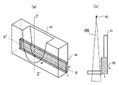
図6は、ウエッジの変形例を示している。図6(a)は、斜視図を示し、図6(b)は、点線で示すZ−Z’軸での断面をX−X’に沿って見た断面図である。
X線CT装置の仕様によっては、ウエッジ駆動の際に、ウエッジの位置精度を確保するために一旦位置情報をリセットし、ウエッジ位置を原点位置に戻すタイプのものがある。このような仕様のX線CT装置では、スキャンサイクル時間内に本実施形態のアクティブコリメーション動作を実現できないため、再構成外範囲におけるX線を遮蔽する位置と再構成範囲におけるウエッジ中心位置間の移動動作に対しては新たな位置検出方式を付加することにより実現可能とする。
12…寝台
13…システム制御部
14…再構成部
15…回転部
16…固定部
111…X線管
21、21a、21b、61、112…ウエッジ
22、62…X線遮蔽物
113…X線検出器
114…データ収集装置
115…非接触データ伝送装置
116…高電圧発生装置
117…ウエッジ駆動部
118…回転駆動部
119…寝台駆動部
Claims (14)
- X線源から照射されるX線の線量分布を調整するためのウエッジと、
前記ウエッジに付された遮蔽物と、
前記ウエッジをスライス厚方向に移動させるウエッジ駆動部と、
スキャン実行中に前記ウエッジが移動するよう前記ウエッジ駆動部を制御するシステム制御部と、を有し、
前記遮蔽物は、前記ウエッジのスライス厚方向両端部の少なくとも一方の端部に且つ当該ウエッジのファン角方向に渡って取り付けられることを特徴とするX線CT装置。 - 前記システム制御部は、スキャン実行期間の開始に生ずる再構成外範囲において、前記X線源のスライス厚方向に広がるX線のうち、医用画像の再構成に不要なX線を前記遮蔽物が遮蔽する第1の位置に前記ウエッジを移動するよう前記ウエッジ駆動部を制御する、又はスキャン終了に生ずる再構成外範囲においては、前記医用画像の再構成に不要なX線を前記遮蔽物が遮蔽する第2の位置に前記ウエッジを移動するよう前記ウエッジ駆動部を制御することを特徴とする請求項1に記載のX線CT装置。
- X線源から照射されるX線の線量分布を調整するためのウエッジと、
前記ウエッジに付された遮蔽物と、
前記ウエッジをスライス厚方向に移動させるウエッジ駆動部と、
スキャン実行中に前記ウエッジが移動するよう前記ウエッジ駆動部を制御するシステム制御部と、を有し、
前記システム制御部は、スキャン実行期間の開始に生ずる再構成外範囲において、前記X線源のスライス厚方向に広がるX線のうち、医用画像の再構成に不要なX線を前記遮蔽物が遮蔽する第1の位置に前記ウエッジを移動するよう前記ウエッジ駆動部を制御する、又はスキャン終了に生ずる再構成外範囲においては、前記医用画像の再構成に不要なX線を前記遮蔽物が遮蔽する第2の位置に前記ウエッジを移動するよう前記ウエッジ駆動部を制御することを特徴とするX線CT装置。 - 前記システム制御部は、前記医用画像の再構成範囲においては、前記遮蔽物がX線を遮蔽しない第3の位置に前記ウエッジを移動するよう前記ウエッジ駆動部を制御することを特徴とする請求項2又は請求項3に記載のX線CT装置。
- 前記システム制御部は、スキャン実行期間の開始に生ずる再構成外範囲において、前記X線源のスライス厚方向に広がるX線のうち、医用画像の再構成に不要なX線を前記遮蔽物が遮蔽する第1の位置に前記ウエッジを移動するよう前記ウエッジ駆動部を制御し、スキャン終了に生ずる再構成外範囲においては、前記医用画像の再構成に不要なX線を前記遮蔽物が遮蔽する第2の位置に前記ウエッジを移動するよう前記ウエッジ駆動部を制御し、前記医用画像の再構成範囲においては、前記遮蔽物がX線を遮蔽しない第3の位置に前記ウエッジを移動するよう前記ウエッジ駆動部を制御することを特徴とする請求項1に記載のX線CT装置。
- X線源から照射されるX線の線量分布を調整するためのウエッジと、
前記ウエッジに付された遮蔽物と、
前記ウエッジをスライス厚方向に移動させるウエッジ駆動部と、
スキャン実行中に前記ウエッジが移動するよう前記ウエッジ駆動部を制御するシステム制御部と、を有し、
前記システム制御部は、スキャン実行期間の開始に生ずる再構成外範囲において、前記X線源のスライス厚方向に広がるX線のうち、医用画像の再構成に不要なX線を前記遮蔽物が遮蔽する第1の位置に前記ウエッジを移動するよう前記ウエッジ駆動部を制御し、スキャン終了に生ずる再構成外範囲においては、前記医用画像の再構成に不要なX線を前記遮蔽物が遮蔽する第2の位置に前記ウエッジを移動するよう前記ウエッジ駆動部を制御し、前記医用画像の再構成範囲においては、前記遮蔽物がX線を遮蔽しない第3の位置に前記ウエッジを移動するよう前記ウエッジ駆動部を制御することを特徴とするX線CT装置。 - 前記ウエッジは、前記X線源側に前記遮蔽物を有することを特徴とする請求項1乃至請求項6のいずれか1項に記載のX線CT装置。
- X線源から照射されるX線の線量分布を調整するためのウエッジと、
前記ウエッジに付された遮蔽物と、
前記ウエッジをスライス厚方向に移動させるウエッジ駆動部と、
スキャン実行中に前記ウエッジが移動するよう前記ウエッジ駆動部を制御するシステム制御部と、を有し、
前記ウエッジは、前記X線源側に前記遮蔽物を有することを特徴とするX線CT装置。 - 前記遮蔽物は、前記X線のファン角に影響を与えない形状を有することを特徴とする請求項1乃至請求項8のいずれか1項に記載のX線CT装置。
- 前記遮蔽物は、鉛で構成されることを特徴とする請求項1乃至請求項9のいずれか1項に記載のX線CT装置。
- 前記ウエッジは、前記ウエッジの側面に前記遮蔽物を有することを特徴とする請求項1乃至請求項10のいずれか1項に記載のX線CT装置。
- 前記ウエッジ駆動部は、前記X線を遮蔽する前記第1の位置、前記第2の位置および前記第3の位置を検出する位置検出部を有することを特徴とする請求項5又は請求項6に記載のX線CT装置。
- 前記位置検出部は、前記X線を遮蔽する前記第1の位置および前記第2の位置を検出する遮蔽位置センサと、前記第3の位置を検出するセンタ位置センサを有することを特徴とする請求項12に記載のX線CT装置。
- 前記遮蔽位置センサおよび前記センタ位置センサはフォトセンサで構成され、前記第1の位置と前記第2の位置では前記遮蔽位置センサに反射光が入射し、前記第3の位置では、前記センタ位置センサに反射光が入力されるように、前記ウエッジに反射率の異なるラダーパターンが配置されることを特徴とする請求項13に記載のX線CT装置。
Priority Applications (1)
| Application Number | Priority Date | Filing Date | Title |
|---|---|---|---|
| JP2011270048A JP6026103B2 (ja) | 2011-01-07 | 2011-12-09 | X線ct装置 |
Applications Claiming Priority (3)
| Application Number | Priority Date | Filing Date | Title |
|---|---|---|---|
| JP2011001947 | 2011-01-07 | ||
| JP2011001947 | 2011-01-07 | ||
| JP2011270048A JP6026103B2 (ja) | 2011-01-07 | 2011-12-09 | X線ct装置 |
Publications (2)
| Publication Number | Publication Date |
|---|---|
| JP2012152545A JP2012152545A (ja) | 2012-08-16 |
| JP6026103B2 true JP6026103B2 (ja) | 2016-11-16 |
Family
ID=46457310
Family Applications (1)
| Application Number | Title | Priority Date | Filing Date |
|---|---|---|---|
| JP2011270048A Active JP6026103B2 (ja) | 2011-01-07 | 2011-12-09 | X線ct装置 |
Country Status (5)
| Country | Link |
|---|---|
| US (2) | US9173619B2 (ja) |
| EP (1) | EP2662021B1 (ja) |
| JP (1) | JP6026103B2 (ja) |
| CN (1) | CN102753099B (ja) |
| WO (1) | WO2012093440A1 (ja) |
Families Citing this family (7)
| Publication number | Priority date | Publication date | Assignee | Title |
|---|---|---|---|---|
| JP6026132B2 (ja) * | 2012-04-11 | 2016-11-16 | 東芝メディカルシステムズ株式会社 | X線コンピュータ断層撮影装置および再構成処理プログラム |
| JP6153346B2 (ja) * | 2013-03-05 | 2017-06-28 | キヤノン株式会社 | 放射線発生装置及び放射線撮影システム |
| EP2810600B1 (en) * | 2013-06-05 | 2018-08-08 | General Electric Company | Medical imaging method varying collimation of emitted radiation beam |
| KR101666943B1 (ko) | 2013-06-11 | 2016-10-28 | 삼성전자주식회사 | 대상체의 관심 영역(roi)에 대한 x선 이미지를 획득하는 방법 및 장치 |
| WO2015048178A2 (en) | 2013-09-25 | 2015-04-02 | Heartflow, Inc. | Systems and methods for visualizing elongated structures and detecting branches therein |
| CA3080986C (en) | 2017-11-06 | 2023-11-14 | The Research Foundation for State University of New York | System and method for dual-use computed tomography for imaging and radiation therapy |
| US10695011B2 (en) * | 2018-08-08 | 2020-06-30 | General Electric Company | X-ray collimator for imaging system |
Family Cites Families (31)
| Publication number | Priority date | Publication date | Assignee | Title |
|---|---|---|---|---|
| GB1443048A (en) * | 1972-12-05 | 1976-07-21 | Strahlen Umweltforsch Gmbh | X-ray source |
| US4181858A (en) * | 1977-12-30 | 1980-01-01 | Emi Limited | Adjustable compensating device for radiographic apparatus |
| JPS56152166A (en) | 1980-04-24 | 1981-11-25 | Sanyo Electric Co Ltd | Preparation of gas diffusion electrode |
| JPS6247346A (ja) * | 1985-08-27 | 1987-03-02 | 株式会社 日立メデイコ | X線ct装置 |
| JPS6298300A (ja) * | 1985-10-25 | 1987-05-07 | 横河メデイカルシステム株式会社 | X線ct装置のx線漏洩防止構造 |
| US4975933A (en) * | 1990-03-26 | 1990-12-04 | General Electric Company | Bow-tie X-ray filter assembly for dual energy tomography |
| JPH0421477A (ja) * | 1990-05-17 | 1992-01-24 | Fuji Xerox Co Ltd | 位置検出装置 |
| JPH0622951A (ja) * | 1992-07-06 | 1994-02-01 | Toshiba Corp | X線ct装置 |
| JPH07255712A (ja) * | 1994-03-23 | 1995-10-09 | Toshiba Corp | X線ct装置 |
| DE69833128T2 (de) * | 1997-12-10 | 2006-08-24 | Koninklijke Philips Electronics N.V. | Bildung eines zusammengesetzten bildes aus aufeinanderfolgenden röntgenbildern |
| US6307918B1 (en) * | 1998-08-25 | 2001-10-23 | General Electric Company | Position dependent beam quality x-ray filtration |
| JP4357612B2 (ja) * | 1998-10-01 | 2009-11-04 | 株式会社東芝 | 放射線撮像装置 |
| DE19905974A1 (de) * | 1999-02-12 | 2000-09-07 | Siemens Ag | Verfahren zur Abtastung eines Untersuchungsobjekts mittels eines CT-Geräts |
| JP4550179B2 (ja) * | 1999-02-12 | 2010-09-22 | 株式会社東芝 | X線コンピュータ断層撮影装置 |
| US6233303B1 (en) * | 1999-07-21 | 2001-05-15 | Siemens Corporate Research, Inc. | Method and apparatus for reducing X-ray dosage in a spiral scan cone beam CT imaging system |
| JP4378812B2 (ja) * | 1999-11-19 | 2009-12-09 | 株式会社島津製作所 | コーンビーム型放射線ct装置 |
| JP2002102217A (ja) * | 2000-09-28 | 2002-04-09 | Ge Medical Systems Global Technology Co Llc | X線ctシステム、ガントリ装置、コンソール端末及びその制御方法及び記憶媒体 |
| JP2003038478A (ja) * | 2001-07-17 | 2003-02-12 | Ge Medical Systems Global Technology Co Llc | X線ctシステム、そのガントリ装置および操作コンソール、ならびに制御方法 |
| US6614877B2 (en) * | 2001-11-21 | 2003-09-02 | Ge Medical Systems Global Technology Company Llc | Method and apparatus for enhancing the contrast of a medical diagnostic image acquired using collimation |
| DE10242920B4 (de) * | 2002-09-16 | 2013-08-22 | Siemens Aktiengesellschaft | Verfahren zum Betrieb eines Computertomographiegerätes und eine Vorrichtung zur Durchführung des Verfahrens |
| JP2005006772A (ja) * | 2003-06-17 | 2005-01-13 | Ge Medical Systems Global Technology Co Llc | X線診断装置及びct画像の生成方法 |
| JP3999179B2 (ja) | 2003-09-09 | 2007-10-31 | ジーイー・メディカル・システムズ・グローバル・テクノロジー・カンパニー・エルエルシー | 放射線断層撮影装置 |
| JP4509709B2 (ja) * | 2004-09-09 | 2010-07-21 | ジーイー・メディカル・システムズ・グローバル・テクノロジー・カンパニー・エルエルシー | 放射線撮影装置およびその放射線スキャン装置 |
| DE102004061347B3 (de) * | 2004-12-20 | 2006-09-28 | Siemens Ag | Röntgen-Computertomograph für schnelle Bildaufzeichung |
| WO2006080144A1 (ja) | 2005-01-27 | 2006-08-03 | Hitachi Medical Corporation | X線計測装置 |
| JP2007289297A (ja) * | 2006-04-23 | 2007-11-08 | Ge Medical Systems Global Technology Co Llc | X線ct装置 |
| JP5269358B2 (ja) | 2007-07-18 | 2013-08-21 | 株式会社東芝 | X線ct装置 |
| JP2010082428A (ja) * | 2008-09-04 | 2010-04-15 | Toshiba Corp | X線コンピュータ断層撮影装置 |
| US20100119033A1 (en) * | 2008-11-12 | 2010-05-13 | The Methodist Hospital Research Institute | Intensity-modulated, cone-beam computed tomographic imaging system, methods, and apparatus |
| CN101756709A (zh) * | 2008-12-26 | 2010-06-30 | Ge医疗系统环球技术有限公司 | X射线ct设备 |
| RU2562342C2 (ru) * | 2009-05-05 | 2015-09-10 | Конинклейке Филипс Электроникс Н.В. | Способ получения рентгеновского изображения и устройство получения рентгеновского изображения с автоматическим позиционированием клиньев |
-
2011
- 2011-12-09 WO PCT/JP2011/006890 patent/WO2012093440A1/ja not_active Ceased
- 2011-12-09 JP JP2011270048A patent/JP6026103B2/ja active Active
- 2011-12-09 CN CN201180004847.1A patent/CN102753099B/zh active Active
- 2011-12-09 EP EP11847882.5A patent/EP2662021B1/en active Active
-
2012
- 2012-06-12 US US13/494,559 patent/US9173619B2/en active Active
-
2015
- 2015-09-28 US US14/868,009 patent/US9724053B2/en active Active
Also Published As
| Publication number | Publication date |
|---|---|
| US9724053B2 (en) | 2017-08-08 |
| EP2662021A1 (en) | 2013-11-13 |
| JP2012152545A (ja) | 2012-08-16 |
| US9173619B2 (en) | 2015-11-03 |
| EP2662021B1 (en) | 2020-02-12 |
| EP2662021A4 (en) | 2016-05-04 |
| CN102753099A (zh) | 2012-10-24 |
| US20160015335A1 (en) | 2016-01-21 |
| CN102753099B (zh) | 2016-11-16 |
| US20120257709A1 (en) | 2012-10-11 |
| WO2012093440A1 (ja) | 2012-07-12 |
Similar Documents
| Publication | Publication Date | Title |
|---|---|---|
| JP6026103B2 (ja) | X線ct装置 | |
| US8396184B2 (en) | X-ray CT system and control method for same | |
| US8094775B2 (en) | X-ray computer tomography apparatus including a pair of separably movable collimators | |
| JP5855156B2 (ja) | コリメータ装置、コリメータ装置の制御方法および制御装置ならびにx線コンピュータ断層撮影装置 | |
| KR101076319B1 (ko) | 동적 제어가 가능한 시준기를 구비한 콘빔 ct 장치 | |
| US20120057670A1 (en) | Multi-detector array imaging system | |
| EP2693948B1 (en) | Pre-scan imaging with rotating gantry | |
| US8792610B2 (en) | Method and apparatus for X-ray CT imaging | |
| JP2012096021A (ja) | 広いカバー範囲及び低線量での心ct撮像のための動的コリメータ | |
| JP2015509419A (ja) | X線ビーム整形器 | |
| RU2594807C2 (ru) | Послепациентный динамический фильтр для компьютерной томографии (ст) | |
| CN106999142A (zh) | X射线成像装置和方法 | |
| JP4172201B2 (ja) | 放射線撮影装置及び放射線画像形成装置 | |
| JP2006297111A (ja) | X線装置用の絞り装置およびx線装置用の絞り装置の作動方法。 | |
| CN112955735A (zh) | X射线相位摄像系统 | |
| JP6058409B2 (ja) | X線ct装置及びそのプログラム | |
| US9757088B2 (en) | Detector apparatus for cone beam computed tomography | |
| CN112587162B (zh) | Ct成像装置及成像方法 | |
| JP6798408B2 (ja) | X線位相イメージング装置 | |
| US20190274653A1 (en) | Method and apparatus for artifact reduction for joint region in step and shoot computed tomography | |
| US20150327816A1 (en) | X-ray ct apparatus and control method | |
| CN217886033U (zh) | Ct系统及用于ct系统的限束器组件 | |
| JP6523451B2 (ja) | 放射線検出器とそれを備えたx線ct装置 | |
| JP2006262963A (ja) | 核医学診断装置およびそれに用いられる診断システム | |
| JP2006192164A (ja) | 放射線撮影装置 |
Legal Events
| Date | Code | Title | Description |
|---|---|---|---|
| A621 | Written request for application examination |
Free format text: JAPANESE INTERMEDIATE CODE: A621 Effective date: 20141024 |
|
| A977 | Report on retrieval |
Free format text: JAPANESE INTERMEDIATE CODE: A971007 Effective date: 20150630 |
|
| A131 | Notification of reasons for refusal |
Free format text: JAPANESE INTERMEDIATE CODE: A131 Effective date: 20150707 |
|
| A521 | Request for written amendment filed |
Free format text: JAPANESE INTERMEDIATE CODE: A523 Effective date: 20150907 |
|
| A131 | Notification of reasons for refusal |
Free format text: JAPANESE INTERMEDIATE CODE: A131 Effective date: 20160301 |
|
| A521 | Request for written amendment filed |
Free format text: JAPANESE INTERMEDIATE CODE: A523 Effective date: 20160428 |
|
| A711 | Notification of change in applicant |
Free format text: JAPANESE INTERMEDIATE CODE: A711 Effective date: 20160428 |
|
| TRDD | Decision of grant or rejection written | ||
| A01 | Written decision to grant a patent or to grant a registration (utility model) |
Free format text: JAPANESE INTERMEDIATE CODE: A01 Effective date: 20160913 |
|
| A61 | First payment of annual fees (during grant procedure) |
Free format text: JAPANESE INTERMEDIATE CODE: A61 Effective date: 20161012 |
|
| R150 | Certificate of patent or registration of utility model |
Ref document number: 6026103 Country of ref document: JP Free format text: JAPANESE INTERMEDIATE CODE: R150 |
|
| S533 | Written request for registration of change of name |
Free format text: JAPANESE INTERMEDIATE CODE: R313533 |
|
| R350 | Written notification of registration of transfer |
Free format text: JAPANESE INTERMEDIATE CODE: R350 |