EP3763376A1 - Traitement de maladies inflammatoires au moyen de cellules souches placentaires - Google Patents
Traitement de maladies inflammatoires au moyen de cellules souches placentaires Download PDFInfo
- Publication number
- EP3763376A1 EP3763376A1 EP20177748.9A EP20177748A EP3763376A1 EP 3763376 A1 EP3763376 A1 EP 3763376A1 EP 20177748 A EP20177748 A EP 20177748A EP 3763376 A1 EP3763376 A1 EP 3763376A1
- Authority
- EP
- European Patent Office
- Prior art keywords
- stem cells
- cells
- placental stem
- placental
- cell
- Prior art date
- Legal status (The legal status is an assumption and is not a legal conclusion. Google has not performed a legal analysis and makes no representation as to the accuracy of the status listed.)
- Withdrawn
Links
- 210000004991 placental stem cell Anatomy 0.000 title claims abstract description 607
- 238000011282 treatment Methods 0.000 title abstract description 42
- 208000027866 inflammatory disease Diseases 0.000 title description 2
- 210000000130 stem cell Anatomy 0.000 claims abstract description 344
- 238000000034 method Methods 0.000 claims abstract description 154
- 208000037265 diseases, disorders, signs and symptoms Diseases 0.000 claims abstract description 100
- 201000010099 disease Diseases 0.000 claims abstract description 59
- 208000009329 Graft vs Host Disease Diseases 0.000 claims abstract description 53
- 208000024908 graft versus host disease Diseases 0.000 claims abstract description 53
- 201000004681 Psoriasis Diseases 0.000 claims abstract description 51
- 206010039073 rheumatoid arthritis Diseases 0.000 claims abstract description 45
- 201000006417 multiple sclerosis Diseases 0.000 claims abstract description 42
- 208000035475 disorder Diseases 0.000 claims abstract description 41
- 206010025135 lupus erythematosus Diseases 0.000 claims abstract description 33
- 206010039710 Scleroderma Diseases 0.000 claims abstract description 31
- 201000005962 mycosis fungoides Diseases 0.000 claims abstract description 28
- 208000022559 Inflammatory bowel disease Diseases 0.000 claims abstract description 27
- 206010012601 diabetes mellitus Diseases 0.000 claims abstract description 18
- 208000000979 Erythema Induratum Diseases 0.000 claims abstract description 6
- 210000004027 cell Anatomy 0.000 claims description 526
- 230000003169 placental effect Effects 0.000 claims description 132
- 208000024891 symptom Diseases 0.000 claims description 104
- 102100037241 Endoglin Human genes 0.000 claims description 62
- 101000881679 Homo sapiens Endoglin Proteins 0.000 claims description 62
- 101001098352 Homo sapiens OX-2 membrane glycoprotein Proteins 0.000 claims description 61
- 102100037589 OX-2 membrane glycoprotein Human genes 0.000 claims description 61
- 239000001963 growth medium Substances 0.000 claims description 60
- 102100022464 5'-nucleotidase Human genes 0.000 claims description 53
- 101000678236 Homo sapiens 5'-nucleotidase Proteins 0.000 claims description 53
- 150000001875 compounds Chemical class 0.000 claims description 52
- 102100028967 HLA class I histocompatibility antigen, alpha chain G Human genes 0.000 claims description 47
- 108010024164 HLA-G Antigens Proteins 0.000 claims description 47
- 239000003814 drug Substances 0.000 claims description 47
- 230000028993 immune response Effects 0.000 claims description 45
- 229940124597 therapeutic agent Drugs 0.000 claims description 42
- 230000015572 biosynthetic process Effects 0.000 claims description 41
- 230000001143 conditioned effect Effects 0.000 claims description 34
- 238000011161 development Methods 0.000 claims description 33
- 210000004369 blood Anatomy 0.000 claims description 32
- 239000008280 blood Substances 0.000 claims description 32
- 230000006872 improvement Effects 0.000 claims description 29
- 206010061218 Inflammation Diseases 0.000 claims description 25
- 230000004054 inflammatory process Effects 0.000 claims description 25
- 102000003729 Neprilysin Human genes 0.000 claims description 21
- 108090000028 Neprilysin Proteins 0.000 claims description 21
- 239000003018 immunosuppressive agent Substances 0.000 claims description 15
- 208000003251 Pruritus Diseases 0.000 claims description 11
- 239000002260 anti-inflammatory agent Substances 0.000 claims description 10
- 150000003431 steroids Chemical class 0.000 claims description 8
- 230000008719 thickening Effects 0.000 claims description 8
- 206010012455 Dermatitis exfoliative Diseases 0.000 claims description 7
- 201000003066 Diffuse Scleroderma Diseases 0.000 claims description 7
- 206010015150 Erythema Diseases 0.000 claims description 7
- 208000000185 Localized scleroderma Diseases 0.000 claims description 7
- 206010027982 Morphoea Diseases 0.000 claims description 7
- 206010042953 Systemic sclerosis Diseases 0.000 claims description 7
- 229940124599 anti-inflammatory drug Drugs 0.000 claims description 7
- 210000001165 lymph node Anatomy 0.000 claims description 7
- 150000004492 retinoid derivatives Chemical class 0.000 claims description 7
- 229960003444 immunosuppressant agent Drugs 0.000 claims description 6
- 230000001861 immunosuppressant effect Effects 0.000 claims description 6
- WAEXFXRVDQXREF-UHFFFAOYSA-N vorinostat Chemical compound ONC(=O)CCCCCCC(=O)NC1=CC=CC=C1 WAEXFXRVDQXREF-UHFFFAOYSA-N 0.000 claims description 6
- 208000004434 Calcinosis Diseases 0.000 claims description 5
- 230000003476 anti-centromere Effects 0.000 claims description 5
- 208000009056 telangiectasis Diseases 0.000 claims description 5
- 230000000699 topical effect Effects 0.000 claims description 5
- 229960000237 vorinostat Drugs 0.000 claims description 5
- 201000002829 CREST Syndrome Diseases 0.000 claims description 4
- 208000030644 Esophageal Motility disease Diseases 0.000 claims description 4
- 206010029098 Neoplasm skin Diseases 0.000 claims description 4
- 208000000453 Skin Neoplasms Diseases 0.000 claims description 4
- 206010043189 Telangiectasia Diseases 0.000 claims description 4
- 206010047139 Vasoconstriction Diseases 0.000 claims description 4
- 201000011191 dyskinesia of esophagus Diseases 0.000 claims description 4
- 230000005855 radiation Effects 0.000 claims description 4
- 230000025033 vasoconstriction Effects 0.000 claims description 4
- 229940124549 vasodilator Drugs 0.000 claims description 4
- 239000003071 vasodilator agent Substances 0.000 claims description 4
- 102000014150 Interferons Human genes 0.000 claims description 3
- 108010050904 Interferons Proteins 0.000 claims description 3
- 201000003088 Limited Scleroderma Diseases 0.000 claims description 3
- 208000024140 Limited cutaneous systemic sclerosis Diseases 0.000 claims description 3
- 238000005336 cracking Methods 0.000 claims description 3
- 238000010894 electron beam technology Methods 0.000 claims description 3
- 229940079322 interferon Drugs 0.000 claims description 3
- 208000016809 linear scleroderma Diseases 0.000 claims description 3
- 229940126409 proton pump inhibitor Drugs 0.000 claims description 3
- 239000000612 proton pump inhibitor Substances 0.000 claims description 3
- 238000001959 radiotherapy Methods 0.000 claims description 3
- 208000011580 syndromic disease Diseases 0.000 claims description 3
- 235000021432 organic honey Nutrition 0.000 claims description 2
- 210000003954 umbilical cord Anatomy 0.000 abstract description 100
- 210000002826 placenta Anatomy 0.000 description 116
- 238000007799 mixed lymphocyte reaction assay Methods 0.000 description 79
- 210000001519 tissue Anatomy 0.000 description 79
- 210000001744 T-lymphocyte Anatomy 0.000 description 69
- 239000000203 mixture Substances 0.000 description 69
- 210000003491 skin Anatomy 0.000 description 67
- 230000010412 perfusion Effects 0.000 description 59
- 230000001506 immunosuppresive effect Effects 0.000 description 50
- 239000000243 solution Substances 0.000 description 48
- 238000003556 assay Methods 0.000 description 43
- 230000014509 gene expression Effects 0.000 description 42
- 239000002609 medium Substances 0.000 description 40
- 208000011231 Crohn disease Diseases 0.000 description 39
- 210000002865 immune cell Anatomy 0.000 description 39
- 210000001185 bone marrow Anatomy 0.000 description 38
- -1 fumaric acid ester Chemical class 0.000 description 38
- 230000001464 adherent effect Effects 0.000 description 37
- 210000004700 fetal blood Anatomy 0.000 description 36
- 210000001691 amnion Anatomy 0.000 description 34
- 108090000623 proteins and genes Proteins 0.000 description 34
- 210000002901 mesenchymal stem cell Anatomy 0.000 description 31
- 230000000694 effects Effects 0.000 description 30
- 230000018109 developmental process Effects 0.000 description 29
- 230000008774 maternal effect Effects 0.000 description 29
- 230000006052 T cell proliferation Effects 0.000 description 28
- 108090000765 processed proteins & peptides Proteins 0.000 description 26
- 102000004196 processed proteins & peptides Human genes 0.000 description 25
- 239000000758 substrate Substances 0.000 description 24
- 230000001629 suppression Effects 0.000 description 24
- 238000002560 therapeutic procedure Methods 0.000 description 24
- 102100031573 Hematopoietic progenitor cell antigen CD34 Human genes 0.000 description 23
- 101000777663 Homo sapiens Hematopoietic progenitor cell antigen CD34 Proteins 0.000 description 23
- 102100034922 T-cell surface glycoprotein CD8 alpha chain Human genes 0.000 description 23
- 210000001136 chorion Anatomy 0.000 description 23
- 230000035755 proliferation Effects 0.000 description 23
- 229920001184 polypeptide Polymers 0.000 description 22
- 230000002519 immonomodulatory effect Effects 0.000 description 21
- 239000003550 marker Substances 0.000 description 20
- 108091003079 Bovine Serum Albumin Proteins 0.000 description 19
- 102100032912 CD44 antigen Human genes 0.000 description 19
- 101000868273 Homo sapiens CD44 antigen Proteins 0.000 description 19
- 230000004069 differentiation Effects 0.000 description 18
- 210000003958 hematopoietic stem cell Anatomy 0.000 description 18
- 238000001727 in vivo Methods 0.000 description 18
- 108010067306 Fibronectins Proteins 0.000 description 17
- 102000016359 Fibronectins Human genes 0.000 description 17
- 230000001684 chronic effect Effects 0.000 description 17
- 239000008194 pharmaceutical composition Substances 0.000 description 17
- 238000002054 transplantation Methods 0.000 description 17
- 102100031585 ADP-ribosyl cyclase/cyclic ADP-ribose hydrolase 1 Human genes 0.000 description 16
- 101000777636 Homo sapiens ADP-ribosyl cyclase/cyclic ADP-ribose hydrolase 1 Proteins 0.000 description 16
- 238000002955 isolation Methods 0.000 description 16
- 102000004190 Enzymes Human genes 0.000 description 15
- 108090000790 Enzymes Proteins 0.000 description 15
- 206010062016 Immunosuppression Diseases 0.000 description 15
- 239000003795 chemical substances by application Substances 0.000 description 15
- 229940088598 enzyme Drugs 0.000 description 15
- 210000004993 mammalian placenta Anatomy 0.000 description 15
- 229920003023 plastic Polymers 0.000 description 15
- 239000004033 plastic Substances 0.000 description 15
- 206010009900 Colitis ulcerative Diseases 0.000 description 14
- 101000800116 Homo sapiens Thy-1 membrane glycoprotein Proteins 0.000 description 14
- 102100033523 Thy-1 membrane glycoprotein Human genes 0.000 description 14
- 201000006704 Ulcerative Colitis Diseases 0.000 description 14
- 230000001154 acute effect Effects 0.000 description 14
- 230000006862 enzymatic digestion Effects 0.000 description 14
- 239000000463 material Substances 0.000 description 14
- 238000011084 recovery Methods 0.000 description 14
- 208000004998 Abdominal Pain Diseases 0.000 description 13
- 102100022749 Aminopeptidase N Human genes 0.000 description 13
- 101000757160 Homo sapiens Aminopeptidase N Proteins 0.000 description 13
- 230000029087 digestion Effects 0.000 description 13
- 230000001605 fetal effect Effects 0.000 description 13
- 238000001943 fluorescence-activated cell sorting Methods 0.000 description 13
- 210000000056 organ Anatomy 0.000 description 13
- 239000002953 phosphate buffered saline Substances 0.000 description 13
- 210000005059 placental tissue Anatomy 0.000 description 13
- 230000035899 viability Effects 0.000 description 13
- 101001008874 Homo sapiens Mast/stem cell growth factor receptor Kit Proteins 0.000 description 12
- FBOZXECLQNJBKD-ZDUSSCGKSA-N L-methotrexate Chemical compound C=1N=C2N=C(N)N=C(N)C2=NC=1CN(C)C1=CC=C(C(=O)N[C@@H](CCC(O)=O)C(O)=O)C=C1 FBOZXECLQNJBKD-ZDUSSCGKSA-N 0.000 description 12
- 102100027754 Mast/stem cell growth factor receptor Kit Human genes 0.000 description 12
- 229960000485 methotrexate Drugs 0.000 description 12
- 210000000282 nail Anatomy 0.000 description 12
- 230000009467 reduction Effects 0.000 description 12
- 101001076407 Homo sapiens Interleukin-1 receptor antagonist protein Proteins 0.000 description 11
- 101000934338 Homo sapiens Myeloid cell surface antigen CD33 Proteins 0.000 description 11
- 101000738771 Homo sapiens Receptor-type tyrosine-protein phosphatase C Proteins 0.000 description 11
- 229940119178 Interleukin 1 receptor antagonist Drugs 0.000 description 11
- 102000051628 Interleukin-1 receptor antagonist Human genes 0.000 description 11
- 102100025243 Myeloid cell surface antigen CD33 Human genes 0.000 description 11
- 102100037422 Receptor-type tyrosine-protein phosphatase C Human genes 0.000 description 11
- 108090000631 Trypsin Proteins 0.000 description 11
- 102000004142 Trypsin Human genes 0.000 description 11
- 239000012530 fluid Substances 0.000 description 11
- 239000003407 interleukin 1 receptor blocking agent Substances 0.000 description 11
- 239000012588 trypsin Substances 0.000 description 11
- 108091007065 BIRCs Proteins 0.000 description 10
- HTTJABKRGRZYRN-UHFFFAOYSA-N Heparin Chemical compound OC1C(NC(=O)C)C(O)OC(COS(O)(=O)=O)C1OC1C(OS(O)(=O)=O)C(O)C(OC2C(C(OS(O)(=O)=O)C(OC3C(C(O)C(O)C(O3)C(O)=O)OS(O)(=O)=O)C(CO)O2)NS(O)(=O)=O)C(C(O)=O)O1 HTTJABKRGRZYRN-UHFFFAOYSA-N 0.000 description 10
- 102000055031 Inhibitor of Apoptosis Proteins Human genes 0.000 description 10
- 239000012091 fetal bovine serum Substances 0.000 description 10
- 238000000338 in vitro Methods 0.000 description 10
- 230000003902 lesion Effects 0.000 description 10
- 210000004072 lung Anatomy 0.000 description 10
- 102000039446 nucleic acids Human genes 0.000 description 10
- 108020004707 nucleic acids Proteins 0.000 description 10
- 150000007523 nucleic acids Chemical class 0.000 description 10
- 235000018102 proteins Nutrition 0.000 description 10
- 102000004169 proteins and genes Human genes 0.000 description 10
- UCSJYZPVAKXKNQ-HZYVHMACSA-N streptomycin Chemical compound CN[C@H]1[C@H](O)[C@@H](O)[C@H](CO)O[C@H]1O[C@@H]1[C@](C=O)(O)[C@H](C)O[C@H]1O[C@@H]1[C@@H](NC(N)=N)[C@H](O)[C@@H](NC(N)=N)[C@H](O)[C@H]1O UCSJYZPVAKXKNQ-HZYVHMACSA-N 0.000 description 10
- 239000013598 vector Substances 0.000 description 10
- 102000005369 Aldehyde Dehydrogenase Human genes 0.000 description 9
- 108020002663 Aldehyde Dehydrogenase Proteins 0.000 description 9
- PMATZTZNYRCHOR-CGLBZJNRSA-N Cyclosporin A Chemical compound CC[C@@H]1NC(=O)[C@H]([C@H](O)[C@H](C)C\C=C\C)N(C)C(=O)[C@H](C(C)C)N(C)C(=O)[C@H](CC(C)C)N(C)C(=O)[C@H](CC(C)C)N(C)C(=O)[C@@H](C)NC(=O)[C@H](C)NC(=O)[C@H](CC(C)C)N(C)C(=O)[C@H](C(C)C)NC(=O)[C@H](CC(C)C)N(C)C(=O)CN(C)C1=O PMATZTZNYRCHOR-CGLBZJNRSA-N 0.000 description 9
- 108010036949 Cyclosporine Proteins 0.000 description 9
- KCXVZYZYPLLWCC-UHFFFAOYSA-N EDTA Chemical compound OC(=O)CN(CC(O)=O)CCN(CC(O)=O)CC(O)=O KCXVZYZYPLLWCC-UHFFFAOYSA-N 0.000 description 9
- 108091008874 T cell receptors Proteins 0.000 description 9
- 102000016266 T-Cell Antigen Receptors Human genes 0.000 description 9
- 208000025865 Ulcer Diseases 0.000 description 9
- 230000005856 abnormality Effects 0.000 description 9
- 208000007502 anemia Diseases 0.000 description 9
- 230000008859 change Effects 0.000 description 9
- 210000001072 colon Anatomy 0.000 description 9
- 239000003636 conditioned culture medium Substances 0.000 description 9
- 230000004064 dysfunction Effects 0.000 description 9
- 239000012894 fetal calf serum Substances 0.000 description 9
- 230000012010 growth Effects 0.000 description 9
- 229920000669 heparin Polymers 0.000 description 9
- 210000004185 liver Anatomy 0.000 description 9
- 239000011159 matrix material Substances 0.000 description 9
- TXEYQDLBPFQVAA-UHFFFAOYSA-N tetrafluoromethane Chemical compound FC(F)(F)F TXEYQDLBPFQVAA-UHFFFAOYSA-N 0.000 description 9
- 230000001225 therapeutic effect Effects 0.000 description 9
- CIWBSHSKHKDKBQ-JLAZNSOCSA-N Ascorbic acid Chemical compound OC[C@H](O)[C@H]1OC(=O)C(O)=C1O CIWBSHSKHKDKBQ-JLAZNSOCSA-N 0.000 description 8
- BPYKTIZUTYGOLE-IFADSCNNSA-N Bilirubin Chemical compound N1C(=O)C(C)=C(C=C)\C1=C\C1=C(C)C(CCC(O)=O)=C(CC2=C(C(C)=C(\C=C/3C(=C(C=C)C(=O)N\3)C)N2)CCC(O)=O)N1 BPYKTIZUTYGOLE-IFADSCNNSA-N 0.000 description 8
- 108060005980 Collagenase Proteins 0.000 description 8
- 102000029816 Collagenase Human genes 0.000 description 8
- 206010040844 Skin exfoliation Diseases 0.000 description 8
- 229960002170 azathioprine Drugs 0.000 description 8
- LMEKQMALGUDUQG-UHFFFAOYSA-N azathioprine Chemical compound CN1C=NC([N+]([O-])=O)=C1SC1=NC=NC2=C1NC=N2 LMEKQMALGUDUQG-UHFFFAOYSA-N 0.000 description 8
- 238000004113 cell culture Methods 0.000 description 8
- 238000005119 centrifugation Methods 0.000 description 8
- 210000004443 dendritic cell Anatomy 0.000 description 8
- 238000002474 experimental method Methods 0.000 description 8
- 230000006870 function Effects 0.000 description 8
- 229960002897 heparin Drugs 0.000 description 8
- 229910052739 hydrogen Inorganic materials 0.000 description 8
- 229940125721 immunosuppressive agent Drugs 0.000 description 8
- KBOPZPXVLCULAV-UHFFFAOYSA-N mesalamine Chemical compound NC1=CC=C(O)C(C(O)=O)=C1 KBOPZPXVLCULAV-UHFFFAOYSA-N 0.000 description 8
- RTGDFNSFWBGLEC-SYZQJQIISA-N mycophenolate mofetil Chemical compound COC1=C(C)C=2COC(=O)C=2C(O)=C1C\C=C(/C)CCC(=O)OCCN1CCOCC1 RTGDFNSFWBGLEC-SYZQJQIISA-N 0.000 description 8
- 229960004866 mycophenolate mofetil Drugs 0.000 description 8
- 238000000926 separation method Methods 0.000 description 8
- 210000002966 serum Anatomy 0.000 description 8
- 231100000397 ulcer Toxicity 0.000 description 8
- 210000003606 umbilical vein Anatomy 0.000 description 8
- 230000004580 weight loss Effects 0.000 description 8
- 208000006820 Arthralgia Diseases 0.000 description 7
- 108020004414 DNA Proteins 0.000 description 7
- 206010012735 Diarrhoea Diseases 0.000 description 7
- 239000006144 Dulbecco’s modified Eagle's medium Substances 0.000 description 7
- JGSARLDLIJGVTE-MBNYWOFBSA-N Penicillin G Chemical compound N([C@H]1[C@H]2SC([C@@H](N2C1=O)C(O)=O)(C)C)C(=O)CC1=CC=CC=C1 JGSARLDLIJGVTE-MBNYWOFBSA-N 0.000 description 7
- 206010037660 Pyrexia Diseases 0.000 description 7
- 108010022394 Threonine synthase Proteins 0.000 description 7
- GLNADSQYFUSGOU-GPTZEZBUSA-J Trypan blue Chemical compound [Na+].[Na+].[Na+].[Na+].C1=C(S([O-])(=O)=O)C=C2C=C(S([O-])(=O)=O)C(/N=N/C3=CC=C(C=C3C)C=3C=C(C(=CC=3)\N=N\C=3C(=CC4=CC(=CC(N)=C4C=3O)S([O-])(=O)=O)S([O-])(=O)=O)C)=C(O)C2=C1N GLNADSQYFUSGOU-GPTZEZBUSA-J 0.000 description 7
- 230000002378 acidificating effect Effects 0.000 description 7
- 239000003146 anticoagulant agent Substances 0.000 description 7
- 230000003115 biocidal effect Effects 0.000 description 7
- 229960002424 collagenase Drugs 0.000 description 7
- 239000002131 composite material Substances 0.000 description 7
- 239000006071 cream Substances 0.000 description 7
- 230000007812 deficiency Effects 0.000 description 7
- 102000004419 dihydrofolate reductase Human genes 0.000 description 7
- 210000001035 gastrointestinal tract Anatomy 0.000 description 7
- 239000001257 hydrogen Substances 0.000 description 7
- 238000001990 intravenous administration Methods 0.000 description 7
- 239000007788 liquid Substances 0.000 description 7
- 238000004519 manufacturing process Methods 0.000 description 7
- 239000002773 nucleotide Substances 0.000 description 7
- 125000003729 nucleotide group Chemical group 0.000 description 7
- 239000002674 ointment Substances 0.000 description 7
- 239000002245 particle Substances 0.000 description 7
- 210000005259 peripheral blood Anatomy 0.000 description 7
- 239000011886 peripheral blood Substances 0.000 description 7
- 229920000642 polymer Polymers 0.000 description 7
- 230000008569 process Effects 0.000 description 7
- XLYOFNOQVPJJNP-UHFFFAOYSA-N water Chemical compound O XLYOFNOQVPJJNP-UHFFFAOYSA-N 0.000 description 7
- 208000016261 weight loss Diseases 0.000 description 7
- 125000004209 (C1-C8) alkyl group Chemical group 0.000 description 6
- IJGRMHOSHXDMSA-UHFFFAOYSA-N Atomic nitrogen Chemical compound N#N IJGRMHOSHXDMSA-UHFFFAOYSA-N 0.000 description 6
- 206010067982 Butterfly rash Diseases 0.000 description 6
- 125000004648 C2-C8 alkenyl group Chemical group 0.000 description 6
- 125000004649 C2-C8 alkynyl group Chemical group 0.000 description 6
- WQZGKKKJIJFFOK-GASJEMHNSA-N Glucose Natural products OC[C@H]1OC(O)[C@H](O)[C@@H](O)[C@@H]1O WQZGKKKJIJFFOK-GASJEMHNSA-N 0.000 description 6
- 206010021143 Hypoxia Diseases 0.000 description 6
- 239000007760 Iscove's Modified Dulbecco's Medium Substances 0.000 description 6
- 108010085895 Laminin Proteins 0.000 description 6
- 102000007547 Laminin Human genes 0.000 description 6
- 108010004729 Phycoerythrin Proteins 0.000 description 6
- 108010038512 Platelet-Derived Growth Factor Proteins 0.000 description 6
- 102000010780 Platelet-Derived Growth Factor Human genes 0.000 description 6
- FAPWRFPIFSIZLT-UHFFFAOYSA-M Sodium chloride Chemical compound [Na+].[Cl-] FAPWRFPIFSIZLT-UHFFFAOYSA-M 0.000 description 6
- QJJXYPPXXYFBGM-LFZNUXCKSA-N Tacrolimus Chemical compound C1C[C@@H](O)[C@H](OC)C[C@@H]1\C=C(/C)[C@@H]1[C@H](C)[C@@H](O)CC(=O)[C@H](CC=C)/C=C(C)/C[C@H](C)C[C@H](OC)[C@H]([C@H](C[C@H]2C)OC)O[C@@]2(O)C(=O)C(=O)N2CCCC[C@H]2C(=O)O1 QJJXYPPXXYFBGM-LFZNUXCKSA-N 0.000 description 6
- 239000004098 Tetracycline Substances 0.000 description 6
- 125000000217 alkyl group Chemical group 0.000 description 6
- 239000003242 anti bacterial agent Substances 0.000 description 6
- 239000000427 antigen Substances 0.000 description 6
- 239000011324 bead Substances 0.000 description 6
- 125000001797 benzyl group Chemical group [H]C1=C([H])C([H])=C(C([H])=C1[H])C([H])([H])* 0.000 description 6
- WQZGKKKJIJFFOK-VFUOTHLCSA-N beta-D-glucose Chemical compound OC[C@H]1O[C@@H](O)[C@H](O)[C@@H](O)[C@@H]1O WQZGKKKJIJFFOK-VFUOTHLCSA-N 0.000 description 6
- 239000012620 biological material Substances 0.000 description 6
- 230000010261 cell growth Effects 0.000 description 6
- 229920001577 copolymer Polymers 0.000 description 6
- 239000003246 corticosteroid Substances 0.000 description 6
- 238000005138 cryopreservation Methods 0.000 description 6
- 230000007717 exclusion Effects 0.000 description 6
- 210000003414 extremity Anatomy 0.000 description 6
- 210000002950 fibroblast Anatomy 0.000 description 6
- 239000012595 freezing medium Substances 0.000 description 6
- 108020001507 fusion proteins Proteins 0.000 description 6
- 102000037865 fusion proteins Human genes 0.000 description 6
- JYGXADMDTFJGBT-VWUMJDOOSA-N hydrocortisone Chemical compound O=C1CC[C@]2(C)[C@H]3[C@@H](O)C[C@](C)([C@@](CC4)(O)C(=O)CO)[C@@H]4[C@@H]3CCC2=C1 JYGXADMDTFJGBT-VWUMJDOOSA-N 0.000 description 6
- 239000000017 hydrogel Substances 0.000 description 6
- 238000011534 incubation Methods 0.000 description 6
- 230000000968 intestinal effect Effects 0.000 description 6
- 210000000936 intestine Anatomy 0.000 description 6
- 230000007774 longterm Effects 0.000 description 6
- 229960004963 mesalazine Drugs 0.000 description 6
- 239000008188 pellet Substances 0.000 description 6
- 230000002829 reductive effect Effects 0.000 description 6
- 230000028327 secretion Effects 0.000 description 6
- QFJCIRLUMZQUOT-HPLJOQBZSA-N sirolimus Chemical compound C1C[C@@H](O)[C@H](OC)C[C@@H]1C[C@@H](C)[C@H]1OC(=O)[C@@H]2CCCCN2C(=O)C(=O)[C@](O)(O2)[C@H](C)CC[C@H]2C[C@H](OC)/C(C)=C/C=C/C=C/[C@@H](C)C[C@@H](C)C(=O)[C@H](OC)[C@H](O)/C(C)=C/[C@@H](C)C(=O)C1 QFJCIRLUMZQUOT-HPLJOQBZSA-N 0.000 description 6
- 239000006228 supernatant Substances 0.000 description 6
- 230000008961 swelling Effects 0.000 description 6
- 229960002180 tetracycline Drugs 0.000 description 6
- 229930101283 tetracycline Natural products 0.000 description 6
- 235000019364 tetracycline Nutrition 0.000 description 6
- 150000003522 tetracyclines Chemical class 0.000 description 6
- 210000001644 umbilical artery Anatomy 0.000 description 6
- VBEQCZHXXJYVRD-GACYYNSASA-N uroanthelone Chemical compound C([C@@H](C(=O)N[C@H](C(=O)N[C@@H](CS)C(=O)N[C@@H](CC(N)=O)C(=O)N[C@@H](CS)C(=O)N[C@H](C(=O)N[C@@H]([C@@H](C)CC)C(=O)NCC(=O)N[C@@H](CC=1C=CC(O)=CC=1)C(=O)N[C@@H](CO)C(=O)NCC(=O)N[C@@H](CC(O)=O)C(=O)N[C@@H](CCCNC(N)=N)C(=O)N[C@@H](CS)C(=O)N[C@@H](CCC(N)=O)C(=O)N[C@@H]([C@@H](C)O)C(=O)N[C@@H](CCCNC(N)=N)C(=O)N[C@@H](CC(O)=O)C(=O)N[C@@H](CC(C)C)C(=O)N[C@@H](CCCNC(N)=N)C(=O)N[C@@H](CC=1C2=CC=CC=C2NC=1)C(=O)N[C@@H](CC=1C2=CC=CC=C2NC=1)C(=O)N[C@@H](CCC(O)=O)C(=O)N[C@@H](CC(C)C)C(=O)N[C@@H](CCCNC(N)=N)C(O)=O)C(C)C)[C@@H](C)O)NC(=O)[C@H](CO)NC(=O)[C@H](CC(O)=O)NC(=O)[C@H](CC(C)C)NC(=O)[C@H](CO)NC(=O)[C@H](CCC(O)=O)NC(=O)[C@@H](NC(=O)[C@H](CC=1NC=NC=1)NC(=O)[C@H](CCSC)NC(=O)[C@H](CS)NC(=O)[C@@H](NC(=O)CNC(=O)CNC(=O)[C@H](CC(N)=O)NC(=O)[C@H](CC(C)C)NC(=O)[C@H](CS)NC(=O)[C@H](CC=1C=CC(O)=CC=1)NC(=O)CNC(=O)[C@H](CC(O)=O)NC(=O)[C@H](CC=1C=CC(O)=CC=1)NC(=O)[C@H](CO)NC(=O)[C@H](CO)NC(=O)[C@H]1N(CCC1)C(=O)[C@H](CS)NC(=O)CNC(=O)[C@H]1N(CCC1)C(=O)[C@H](CC=1C=CC(O)=CC=1)NC(=O)[C@H](CO)NC(=O)[C@@H](N)CC(N)=O)C(C)C)[C@@H](C)CC)C1=CC=C(O)C=C1 VBEQCZHXXJYVRD-GACYYNSASA-N 0.000 description 6
- 102100036475 Alanine aminotransferase 1 Human genes 0.000 description 5
- 108010082126 Alanine transaminase Proteins 0.000 description 5
- 102000002260 Alkaline Phosphatase Human genes 0.000 description 5
- 108020004774 Alkaline Phosphatase Proteins 0.000 description 5
- 229940088872 Apoptosis inhibitor Drugs 0.000 description 5
- 108010003415 Aspartate Aminotransferases Proteins 0.000 description 5
- 102000004625 Aspartate Aminotransferases Human genes 0.000 description 5
- 102000001326 Chemokine CCL4 Human genes 0.000 description 5
- 108010055165 Chemokine CCL4 Proteins 0.000 description 5
- 108010035532 Collagen Proteins 0.000 description 5
- 102000008186 Collagen Human genes 0.000 description 5
- 208000014997 Crohn colitis Diseases 0.000 description 5
- CMSMOCZEIVJLDB-UHFFFAOYSA-N Cyclophosphamide Chemical compound ClCCN(CCCl)P1(=O)NCCCO1 CMSMOCZEIVJLDB-UHFFFAOYSA-N 0.000 description 5
- 101000610551 Homo sapiens Prominin-1 Proteins 0.000 description 5
- UETNIIAIRMUTSM-UHFFFAOYSA-N Jacareubin Natural products CC1(C)OC2=CC3Oc4c(O)c(O)ccc4C(=O)C3C(=C2C=C1)O UETNIIAIRMUTSM-UHFFFAOYSA-N 0.000 description 5
- FQISKWAFAHGMGT-SGJOWKDISA-M Methylprednisolone sodium succinate Chemical compound [Na+].C([C@@]12C)=CC(=O)C=C1[C@@H](C)C[C@@H]1[C@@H]2[C@@H](O)C[C@]2(C)[C@@](O)(C(=O)COC(=O)CCC([O-])=O)CC[C@H]21 FQISKWAFAHGMGT-SGJOWKDISA-M 0.000 description 5
- 102000014962 Monocyte Chemoattractant Proteins Human genes 0.000 description 5
- 108010064136 Monocyte Chemoattractant Proteins Proteins 0.000 description 5
- 208000002193 Pain Diseases 0.000 description 5
- 229930182555 Penicillin Natural products 0.000 description 5
- 102100040120 Prominin-1 Human genes 0.000 description 5
- 206010037575 Pustular psoriasis Diseases 0.000 description 5
- 206010067584 Type 1 diabetes mellitus Diseases 0.000 description 5
- 229940127219 anticoagulant drug Drugs 0.000 description 5
- 108091007433 antigens Proteins 0.000 description 5
- 102000036639 antigens Human genes 0.000 description 5
- 239000000158 apoptosis inhibitor Substances 0.000 description 5
- 206010003246 arthritis Diseases 0.000 description 5
- 125000003118 aryl group Chemical group 0.000 description 5
- 125000004432 carbon atom Chemical group C* 0.000 description 5
- 230000001413 cellular effect Effects 0.000 description 5
- 238000006243 chemical reaction Methods 0.000 description 5
- 229960001265 ciclosporin Drugs 0.000 description 5
- 206010009887 colitis Diseases 0.000 description 5
- 229920001436 collagen Polymers 0.000 description 5
- 229930182912 cyclosporin Natural products 0.000 description 5
- 238000000684 flow cytometry Methods 0.000 description 5
- 230000005484 gravity Effects 0.000 description 5
- 125000001072 heteroaryl group Chemical group 0.000 description 5
- 125000004435 hydrogen atom Chemical group [H]* 0.000 description 5
- 230000001146 hypoxic effect Effects 0.000 description 5
- 230000036737 immune function Effects 0.000 description 5
- 210000000987 immune system Anatomy 0.000 description 5
- 239000003112 inhibitor Substances 0.000 description 5
- 239000010410 layer Substances 0.000 description 5
- 210000000265 leukocyte Anatomy 0.000 description 5
- 229960004584 methylprednisolone Drugs 0.000 description 5
- 210000000822 natural killer cell Anatomy 0.000 description 5
- 230000000926 neurological effect Effects 0.000 description 5
- 230000036407 pain Effects 0.000 description 5
- 229940049954 penicillin Drugs 0.000 description 5
- 102000005962 receptors Human genes 0.000 description 5
- 108020003175 receptors Proteins 0.000 description 5
- 150000003839 salts Chemical class 0.000 description 5
- 241000894007 species Species 0.000 description 5
- 229960005322 streptomycin Drugs 0.000 description 5
- NCEXYHBECQHGNR-QZQOTICOSA-N sulfasalazine Chemical compound C1=C(O)C(C(=O)O)=CC(\N=N\C=2C=CC(=CC=2)S(=O)(=O)NC=2N=CC=CC=2)=C1 NCEXYHBECQHGNR-QZQOTICOSA-N 0.000 description 5
- 229960001940 sulfasalazine Drugs 0.000 description 5
- NCEXYHBECQHGNR-UHFFFAOYSA-N sulfasalazine Natural products C1=C(O)C(C(=O)O)=CC(N=NC=2C=CC(=CC=2)S(=O)(=O)NC=2N=CC=CC=2)=C1 NCEXYHBECQHGNR-UHFFFAOYSA-N 0.000 description 5
- 210000005166 vasculature Anatomy 0.000 description 5
- RZVAJINKPMORJF-UHFFFAOYSA-N Acetaminophen Chemical compound CC(=O)NC1=CC=C(O)C=C1 RZVAJINKPMORJF-UHFFFAOYSA-N 0.000 description 4
- 229930105110 Cyclosporin A Natural products 0.000 description 4
- 102400001368 Epidermal growth factor Human genes 0.000 description 4
- 101800003838 Epidermal growth factor Proteins 0.000 description 4
- 206010015719 Exsanguination Diseases 0.000 description 4
- 229920001917 Ficoll Polymers 0.000 description 4
- 101000935043 Homo sapiens Integrin beta-1 Proteins 0.000 description 4
- 102100025304 Integrin beta-1 Human genes 0.000 description 4
- 102000004889 Interleukin-6 Human genes 0.000 description 4
- 108090001005 Interleukin-6 Proteins 0.000 description 4
- CSNNHWWHGAXBCP-UHFFFAOYSA-L Magnesium sulfate Chemical compound [Mg+2].[O-][S+2]([O-])([O-])[O-] CSNNHWWHGAXBCP-UHFFFAOYSA-L 0.000 description 4
- 206010028813 Nausea Diseases 0.000 description 4
- 108010067372 Pancreatic elastase Proteins 0.000 description 4
- 229920000954 Polyglycolide Polymers 0.000 description 4
- 239000004365 Protease Substances 0.000 description 4
- 201000001263 Psoriatic Arthritis Diseases 0.000 description 4
- 208000036824 Psoriatic arthropathy Diseases 0.000 description 4
- 206010038063 Rectal haemorrhage Diseases 0.000 description 4
- 102000012479 Serine Proteases Human genes 0.000 description 4
- 108010022999 Serine Proteases Proteins 0.000 description 4
- 102000040945 Transcription factor Human genes 0.000 description 4
- 108091023040 Transcription factor Proteins 0.000 description 4
- 108060008682 Tumor Necrosis Factor Proteins 0.000 description 4
- 102000000852 Tumor Necrosis Factor-alpha Human genes 0.000 description 4
- OIRDTQYFTABQOQ-KQYNXXCUSA-N adenosine Chemical compound C1=NC=2C(N)=NC=NC=2N1[C@@H]1O[C@H](CO)[C@@H](O)[C@H]1O OIRDTQYFTABQOQ-KQYNXXCUSA-N 0.000 description 4
- 230000000735 allogeneic effect Effects 0.000 description 4
- 229940121363 anti-inflammatory agent Drugs 0.000 description 4
- 230000003460 anti-nuclear Effects 0.000 description 4
- 230000001363 autoimmune Effects 0.000 description 4
- 239000002458 cell surface marker Substances 0.000 description 4
- 230000003833 cell viability Effects 0.000 description 4
- 238000012512 characterization method Methods 0.000 description 4
- 229960004397 cyclophosphamide Drugs 0.000 description 4
- 230000006378 damage Effects 0.000 description 4
- 230000003247 decreasing effect Effects 0.000 description 4
- 230000002939 deleterious effect Effects 0.000 description 4
- 230000035618 desquamation Effects 0.000 description 4
- 238000003745 diagnosis Methods 0.000 description 4
- 235000021186 dishes Nutrition 0.000 description 4
- 108010007093 dispase Proteins 0.000 description 4
- 239000003623 enhancer Substances 0.000 description 4
- 229940116977 epidermal growth factor Drugs 0.000 description 4
- 238000004299 exfoliation Methods 0.000 description 4
- 206010016256 fatigue Diseases 0.000 description 4
- MHMNJMPURVTYEJ-UHFFFAOYSA-N fluorescein-5-isothiocyanate Chemical compound O1C(=O)C2=CC(N=C=S)=CC=C2C21C1=CC=C(O)C=C1OC1=CC(O)=CC=C21 MHMNJMPURVTYEJ-UHFFFAOYSA-N 0.000 description 4
- 239000006260 foam Substances 0.000 description 4
- 235000013305 food Nutrition 0.000 description 4
- 230000004927 fusion Effects 0.000 description 4
- RWSXRVCMGQZWBV-WDSKDSINSA-N glutathione Chemical compound OC(=O)[C@@H](N)CCC(=O)N[C@@H](CS)C(=O)NCC(O)=O RWSXRVCMGQZWBV-WDSKDSINSA-N 0.000 description 4
- 206010018797 guttate psoriasis Diseases 0.000 description 4
- 125000000592 heterocycloalkyl group Chemical group 0.000 description 4
- 208000009326 ileitis Diseases 0.000 description 4
- 208000008384 ileus Diseases 0.000 description 4
- 210000001503 joint Anatomy 0.000 description 4
- 210000005067 joint tissue Anatomy 0.000 description 4
- 238000002826 magnetic-activated cell sorting Methods 0.000 description 4
- 230000001404 mediated effect Effects 0.000 description 4
- 210000005087 mononuclear cell Anatomy 0.000 description 4
- 230000008693 nausea Effects 0.000 description 4
- 239000000041 non-steroidal anti-inflammatory agent Substances 0.000 description 4
- 229940021182 non-steroidal anti-inflammatory drug Drugs 0.000 description 4
- 235000015097 nutrients Nutrition 0.000 description 4
- 208000008423 pleurisy Diseases 0.000 description 4
- 229920001610 polycaprolactone Polymers 0.000 description 4
- 206010063401 primary progressive multiple sclerosis Diseases 0.000 description 4
- 238000012545 processing Methods 0.000 description 4
- 230000000750 progressive effect Effects 0.000 description 4
- ZCCUUQDIBDJBTK-UHFFFAOYSA-N psoralen Chemical compound C1=C2OC(=O)C=CC2=CC2=C1OC=C2 ZCCUUQDIBDJBTK-UHFFFAOYSA-N 0.000 description 4
- 230000002685 pulmonary effect Effects 0.000 description 4
- 208000002815 pulmonary hypertension Diseases 0.000 description 4
- 229940075993 receptor modulator Drugs 0.000 description 4
- 230000004044 response Effects 0.000 description 4
- 201000008628 secondary progressive multiple sclerosis Diseases 0.000 description 4
- 210000000813 small intestine Anatomy 0.000 description 4
- 238000003860 storage Methods 0.000 description 4
- 230000009747 swallowing Effects 0.000 description 4
- 230000009885 systemic effect Effects 0.000 description 4
- 229960001967 tacrolimus Drugs 0.000 description 4
- QJJXYPPXXYFBGM-SHYZHZOCSA-N tacrolimus Natural products CO[C@H]1C[C@H](CC[C@@H]1O)C=C(C)[C@H]2OC(=O)[C@H]3CCCCN3C(=O)C(=O)[C@@]4(O)O[C@@H]([C@H](C[C@H]4C)OC)[C@@H](C[C@H](C)CC(=C[C@@H](CC=C)C(=O)C[C@H](O)[C@H]2C)C)OC QJJXYPPXXYFBGM-SHYZHZOCSA-N 0.000 description 4
- 238000012360 testing method Methods 0.000 description 4
- 238000011285 therapeutic regimen Methods 0.000 description 4
- 238000013518 transcription Methods 0.000 description 4
- 230000035897 transcription Effects 0.000 description 4
- QORWJWZARLRLPR-UHFFFAOYSA-H tricalcium bis(phosphate) Chemical compound [Ca+2].[Ca+2].[Ca+2].[O-]P([O-])([O-])=O.[O-]P([O-])([O-])=O QORWJWZARLRLPR-UHFFFAOYSA-H 0.000 description 4
- 238000005406 washing Methods 0.000 description 4
- DGVVWUTYPXICAM-UHFFFAOYSA-N β‐Mercaptoethanol Chemical compound OCCS DGVVWUTYPXICAM-UHFFFAOYSA-N 0.000 description 4
- VKZRWSNIWNFCIQ-WDSKDSINSA-N (2s)-2-[2-[[(1s)-1,2-dicarboxyethyl]amino]ethylamino]butanedioic acid Chemical compound OC(=O)C[C@@H](C(O)=O)NCCN[C@H](C(O)=O)CC(O)=O VKZRWSNIWNFCIQ-WDSKDSINSA-N 0.000 description 3
- GVJHHUAWPYXKBD-UHFFFAOYSA-N (±)-α-Tocopherol Chemical compound OC1=C(C)C(C)=C2OC(CCCC(C)CCCC(C)CCCC(C)C)(C)CCC2=C1C GVJHHUAWPYXKBD-UHFFFAOYSA-N 0.000 description 3
- QTBSBXVTEAMEQO-UHFFFAOYSA-N Acetic acid Chemical compound CC(O)=O QTBSBXVTEAMEQO-UHFFFAOYSA-N 0.000 description 3
- 201000004384 Alopecia Diseases 0.000 description 3
- 208000003343 Antiphospholipid Syndrome Diseases 0.000 description 3
- 206010003591 Ataxia Diseases 0.000 description 3
- 206010003694 Atrophy Diseases 0.000 description 3
- 208000023275 Autoimmune disease Diseases 0.000 description 3
- 108090000145 Bacillolysin Proteins 0.000 description 3
- 206010069632 Bladder dysfunction Diseases 0.000 description 3
- 206010006784 Burning sensation Diseases 0.000 description 3
- 108700012434 CCL3 Proteins 0.000 description 3
- 108010029697 CD40 Ligand Proteins 0.000 description 3
- 102100032937 CD40 ligand Human genes 0.000 description 3
- OYPRJOBELJOOCE-UHFFFAOYSA-N Calcium Chemical compound [Ca] OYPRJOBELJOOCE-UHFFFAOYSA-N 0.000 description 3
- 102000000013 Chemokine CCL3 Human genes 0.000 description 3
- 208000002881 Colic Diseases 0.000 description 3
- 206010057254 Connective tissue inflammation Diseases 0.000 description 3
- 206010010904 Convulsion Diseases 0.000 description 3
- 101800000414 Corticotropin Proteins 0.000 description 3
- 102000008130 Cyclic AMP-Dependent Protein Kinases Human genes 0.000 description 3
- 108010049894 Cyclic AMP-Dependent Protein Kinases Proteins 0.000 description 3
- 208000019505 Deglutition disease Diseases 0.000 description 3
- 102000016911 Deoxyribonucleases Human genes 0.000 description 3
- 108010053770 Deoxyribonucleases Proteins 0.000 description 3
- 201000004624 Dermatitis Diseases 0.000 description 3
- 206010012741 Diarrhoea haemorrhagic Diseases 0.000 description 3
- IAZDPXIOMUYVGZ-UHFFFAOYSA-N Dimethylsulphoxide Chemical compound CS(C)=O IAZDPXIOMUYVGZ-UHFFFAOYSA-N 0.000 description 3
- 208000003164 Diplopia Diseases 0.000 description 3
- 229940123907 Disease modifying antirheumatic drug Drugs 0.000 description 3
- 208000004232 Enteritis Diseases 0.000 description 3
- 241000283073 Equus caballus Species 0.000 description 3
- LYCAIKOWRPUZTN-UHFFFAOYSA-N Ethylene glycol Chemical compound OCCO LYCAIKOWRPUZTN-UHFFFAOYSA-N 0.000 description 3
- 208000010201 Exanthema Diseases 0.000 description 3
- 102000010834 Extracellular Matrix Proteins Human genes 0.000 description 3
- 108010037362 Extracellular Matrix Proteins Proteins 0.000 description 3
- 206010015946 Eye irritation Diseases 0.000 description 3
- 206010015958 Eye pain Diseases 0.000 description 3
- 206010016654 Fibrosis Diseases 0.000 description 3
- 208000014540 Functional gastrointestinal disease Diseases 0.000 description 3
- 208000005577 Gastroenteritis Diseases 0.000 description 3
- 108010010803 Gelatin Proteins 0.000 description 3
- 229930182566 Gentamicin Natural products 0.000 description 3
- CEAZRRDELHUEMR-URQXQFDESA-N Gentamicin Chemical compound O1[C@H](C(C)NC)CC[C@@H](N)[C@H]1O[C@H]1[C@H](O)[C@@H](O[C@@H]2[C@@H]([C@@H](NC)[C@@](C)(O)CO2)O)[C@H](N)C[C@@H]1N CEAZRRDELHUEMR-URQXQFDESA-N 0.000 description 3
- PEDCQBHIVMGVHV-UHFFFAOYSA-N Glycerine Chemical compound OCC(O)CO PEDCQBHIVMGVHV-UHFFFAOYSA-N 0.000 description 3
- 102100028972 HLA class I histocompatibility antigen, A alpha chain Human genes 0.000 description 3
- 108010075704 HLA-A Antigens Proteins 0.000 description 3
- 208000032843 Hemorrhage Diseases 0.000 description 3
- 102100022132 High affinity immunoglobulin epsilon receptor subunit gamma Human genes 0.000 description 3
- 108091010847 High affinity immunoglobulin epsilon receptor subunit gamma Proteins 0.000 description 3
- 101000599951 Homo sapiens Insulin-like growth factor I Proteins 0.000 description 3
- 241000701044 Human gammaherpesvirus 4 Species 0.000 description 3
- 108010003272 Hyaluronate lyase Proteins 0.000 description 3
- 102000001974 Hyaluronidases Human genes 0.000 description 3
- 102100037852 Insulin-like growth factor I Human genes 0.000 description 3
- 208000029523 Interstitial Lung disease Diseases 0.000 description 3
- 206010022971 Iron Deficiencies Diseases 0.000 description 3
- 239000012825 JNK inhibitor Substances 0.000 description 3
- 229940118135 JNK inhibitor Drugs 0.000 description 3
- 206010023126 Jaundice Diseases 0.000 description 3
- 208000005777 Lupus Nephritis Diseases 0.000 description 3
- 206010067750 Lupus pancreatitis Diseases 0.000 description 3
- 206010057481 Lupus pneumonitis Diseases 0.000 description 3
- 102000005741 Metalloproteases Human genes 0.000 description 3
- 108010006035 Metalloproteases Proteins 0.000 description 3
- 102000003979 Mineralocorticoid Receptors Human genes 0.000 description 3
- 108090000375 Mineralocorticoid Receptors Proteins 0.000 description 3
- 208000007101 Muscle Cramp Diseases 0.000 description 3
- 208000010428 Muscle Weakness Diseases 0.000 description 3
- 206010028372 Muscular weakness Diseases 0.000 description 3
- 208000009525 Myocarditis Diseases 0.000 description 3
- 206010028703 Nail psoriasis Diseases 0.000 description 3
- 206010028851 Necrosis Diseases 0.000 description 3
- 208000008457 Neurologic Manifestations Diseases 0.000 description 3
- 102000035092 Neutral proteases Human genes 0.000 description 3
- 108091005507 Neutral proteases Proteins 0.000 description 3
- 208000011623 Obstructive Lung disease Diseases 0.000 description 3
- 206010030094 Odynophagia Diseases 0.000 description 3
- 208000006187 Onycholysis Diseases 0.000 description 3
- 208000001132 Osteoporosis Diseases 0.000 description 3
- 102000016387 Pancreatic elastase Human genes 0.000 description 3
- 102000035195 Peptidases Human genes 0.000 description 3
- 108091005804 Peptidases Proteins 0.000 description 3
- 108091000080 Phosphotransferase Proteins 0.000 description 3
- 206010034960 Photophobia Diseases 0.000 description 3
- 208000002151 Pleural effusion Diseases 0.000 description 3
- 102000003923 Protein Kinase C Human genes 0.000 description 3
- 108090000315 Protein Kinase C Proteins 0.000 description 3
- 208000028017 Psychotic disease Diseases 0.000 description 3
- 206010037394 Pulmonary haemorrhage Diseases 0.000 description 3
- 206010037888 Rash pustular Diseases 0.000 description 3
- 208000012322 Raynaud phenomenon Diseases 0.000 description 3
- 208000001647 Renal Insufficiency Diseases 0.000 description 3
- 201000001949 Retinal Vasculitis Diseases 0.000 description 3
- 206010040026 Sensory disturbance Diseases 0.000 description 3
- 201000001880 Sexual dysfunction Diseases 0.000 description 3
- 206010067868 Skin mass Diseases 0.000 description 3
- 208000004732 Systemic Vasculitis Diseases 0.000 description 3
- 206010047115 Vasculitis Diseases 0.000 description 3
- MMWCIQZXVOZEGG-HOZKJCLWSA-N [(1S,2R,3S,4S,5R,6S)-2,3,5-trihydroxy-4,6-diphosphonooxycyclohexyl] dihydrogen phosphate Chemical compound O[C@H]1[C@@H](O)[C@H](OP(O)(O)=O)[C@@H](OP(O)(O)=O)[C@H](O)[C@H]1OP(O)(O)=O MMWCIQZXVOZEGG-HOZKJCLWSA-N 0.000 description 3
- 231100000360 alopecia Toxicity 0.000 description 3
- 229960002576 amiloride Drugs 0.000 description 3
- 230000003429 anti-cardiolipin effect Effects 0.000 description 3
- 230000003432 anti-folate effect Effects 0.000 description 3
- 229940088710 antibiotic agent Drugs 0.000 description 3
- 229940127074 antifolate Drugs 0.000 description 3
- 210000000436 anus Anatomy 0.000 description 3
- 230000006907 apoptotic process Effects 0.000 description 3
- 230000004596 appetite loss Effects 0.000 description 3
- 239000007864 aqueous solution Substances 0.000 description 3
- 229960005070 ascorbic acid Drugs 0.000 description 3
- QVGXLLKOCUKJST-UHFFFAOYSA-N atomic oxygen Chemical compound [O] QVGXLLKOCUKJST-UHFFFAOYSA-N 0.000 description 3
- 230000037444 atrophy Effects 0.000 description 3
- 208000027625 autoimmune inner ear disease Diseases 0.000 description 3
- 208000034158 bleeding Diseases 0.000 description 3
- 230000000740 bleeding effect Effects 0.000 description 3
- 238000004820 blood count Methods 0.000 description 3
- 210000001124 body fluid Anatomy 0.000 description 3
- 229940098773 bovine serum albumin Drugs 0.000 description 3
- 201000007637 bowel dysfunction Diseases 0.000 description 3
- 239000011575 calcium Substances 0.000 description 3
- 229910052791 calcium Inorganic materials 0.000 description 3
- 229910052799 carbon Inorganic materials 0.000 description 3
- 210000003169 central nervous system Anatomy 0.000 description 3
- 210000001175 cerebrospinal fluid Anatomy 0.000 description 3
- YRQNKMKHABXEJZ-UVQQGXFZSA-N chembl176323 Chemical compound C1C[C@]2(C)[C@@]3(C)CC(N=C4C[C@]5(C)CCC6[C@]7(C)CC[C@@H]([C@]7(CC[C@]6(C)[C@@]5(C)CC4=N4)C)CCCCCCCC)=C4C[C@]3(C)CCC2[C@]2(C)CC[C@H](CCCCCCCC)[C@]21C YRQNKMKHABXEJZ-UVQQGXFZSA-N 0.000 description 3
- 230000001332 colony forming effect Effects 0.000 description 3
- 230000003750 conditioning effect Effects 0.000 description 3
- 229960001334 corticosteroids Drugs 0.000 description 3
- IDLFZVILOHSSID-OVLDLUHVSA-N corticotropin Chemical compound C([C@@H](C(=O)N[C@@H](CO)C(=O)N[C@@H](CCSC)C(=O)N[C@@H](CCC(O)=O)C(=O)N[C@@H](CC=1NC=NC=1)C(=O)N[C@@H](CC=1C=CC=CC=1)C(=O)N[C@@H](CCCNC(N)=N)C(=O)N[C@@H](CC=1C2=CC=CC=C2NC=1)C(=O)NCC(=O)N[C@@H](CCCCN)C(=O)N1[C@@H](CCC1)C(=O)N[C@@H](C(C)C)C(=O)NCC(=O)N[C@@H](CCCCN)C(=O)N[C@@H](CCCCN)C(=O)N[C@@H](CCCNC(N)=N)C(=O)N[C@@H](CCCNC(N)=N)C(=O)N1[C@@H](CCC1)C(=O)N[C@@H](C(C)C)C(=O)N[C@@H](CCCCN)C(=O)N[C@@H](C(C)C)C(=O)N[C@@H](CC=1C=CC(O)=CC=1)C(=O)N1[C@@H](CCC1)C(=O)N[C@@H](CC(N)=O)C(=O)NCC(=O)N[C@@H](C)C(=O)N[C@@H](CCC(O)=O)C(=O)N[C@@H](CC(O)=O)C(=O)N[C@@H](CCC(O)=O)C(=O)N[C@@H](CO)C(=O)N[C@@H](C)C(=O)N[C@@H](CCC(O)=O)C(=O)N[C@@H](C)C(=O)N[C@@H](CC=1C=CC=CC=1)C(=O)N1[C@@H](CCC1)C(=O)N[C@@H](CC(C)C)C(=O)N[C@@H](CCC(O)=O)C(=O)N[C@@H](CC=1C=CC=CC=1)C(O)=O)NC(=O)[C@@H](N)CO)C1=CC=C(O)C=C1 IDLFZVILOHSSID-OVLDLUHVSA-N 0.000 description 3
- 229960000258 corticotropin Drugs 0.000 description 3
- 201000003146 cystitis Diseases 0.000 description 3
- 229960003957 dexamethasone Drugs 0.000 description 3
- UREBDLICKHMUKA-CXSFZGCWSA-N dexamethasone Chemical compound C1CC2=CC(=O)C=C[C@]2(C)[C@]2(F)[C@@H]1[C@@H]1C[C@@H](C)[C@@](C(=O)CO)(O)[C@@]1(C)C[C@@H]2O UREBDLICKHMUKA-CXSFZGCWSA-N 0.000 description 3
- 239000002988 disease modifying antirheumatic drug Substances 0.000 description 3
- 210000001671 embryonic stem cell Anatomy 0.000 description 3
- 206010014665 endocarditis Diseases 0.000 description 3
- 238000005516 engineering process Methods 0.000 description 3
- 201000005884 exanthem Diseases 0.000 description 3
- 210000002744 extracellular matrix Anatomy 0.000 description 3
- 231100000013 eye irritation Toxicity 0.000 description 3
- 206010016165 failure to thrive Diseases 0.000 description 3
- 239000004052 folic acid antagonist Substances 0.000 description 3
- ZZUFCTLCJUWOSV-UHFFFAOYSA-N furosemide Chemical compound C1=C(Cl)C(S(=O)(=O)N)=CC(C(O)=O)=C1NCC1=CC=CO1 ZZUFCTLCJUWOSV-UHFFFAOYSA-N 0.000 description 3
- 208000027792 gastroduodenal Crohn disease Diseases 0.000 description 3
- 229920000159 gelatin Polymers 0.000 description 3
- 239000008273 gelatin Substances 0.000 description 3
- 235000019322 gelatine Nutrition 0.000 description 3
- 235000011852 gelatine desserts Nutrition 0.000 description 3
- 229960002518 gentamicin Drugs 0.000 description 3
- 239000003862 glucocorticoid Substances 0.000 description 3
- 239000008103 glucose Substances 0.000 description 3
- 239000003102 growth factor Substances 0.000 description 3
- 230000002489 hematologic effect Effects 0.000 description 3
- 208000006750 hematuria Diseases 0.000 description 3
- 230000002440 hepatic effect Effects 0.000 description 3
- 229960002773 hyaluronidase Drugs 0.000 description 3
- 229960000890 hydrocortisone Drugs 0.000 description 3
- 210000003405 ileum Anatomy 0.000 description 3
- 239000007924 injection Substances 0.000 description 3
- 238000002347 injection Methods 0.000 description 3
- 238000003780 insertion Methods 0.000 description 3
- 230000037431 insertion Effects 0.000 description 3
- 238000007918 intramuscular administration Methods 0.000 description 3
- 239000007927 intramuscular injection Substances 0.000 description 3
- 239000007928 intraperitoneal injection Substances 0.000 description 3
- 230000007803 itching Effects 0.000 description 3
- 208000018937 joint inflammation Diseases 0.000 description 3
- 210000003734 kidney Anatomy 0.000 description 3
- 201000006370 kidney failure Diseases 0.000 description 3
- 229960000681 leflunomide Drugs 0.000 description 3
- VHOGYURTWQBHIL-UHFFFAOYSA-N leflunomide Chemical compound O1N=CC(C(=O)NC=2C=CC(=CC=2)C(F)(F)F)=C1C VHOGYURTWQBHIL-UHFFFAOYSA-N 0.000 description 3
- 239000003446 ligand Substances 0.000 description 3
- 208000019017 loss of appetite Diseases 0.000 description 3
- 235000021266 loss of appetite Nutrition 0.000 description 3
- 238000002595 magnetic resonance imaging Methods 0.000 description 3
- 201000008350 membranous glomerulonephritis Diseases 0.000 description 3
- GLVAUDGFNGKCSF-UHFFFAOYSA-N mercaptopurine Chemical compound S=C1NC=NC2=C1NC=N2 GLVAUDGFNGKCSF-UHFFFAOYSA-N 0.000 description 3
- 239000002062 molecular scaffold Substances 0.000 description 3
- 230000000877 morphologic effect Effects 0.000 description 3
- 230000017074 necrotic cell death Effects 0.000 description 3
- 208000004296 neuralgia Diseases 0.000 description 3
- 208000021722 neuropathic pain Diseases 0.000 description 3
- 229910052757 nitrogen Inorganic materials 0.000 description 3
- 210000001328 optic nerve Anatomy 0.000 description 3
- 229910052760 oxygen Inorganic materials 0.000 description 3
- 239000001301 oxygen Substances 0.000 description 3
- 230000001734 parasympathetic effect Effects 0.000 description 3
- 244000052769 pathogen Species 0.000 description 3
- 208000008494 pericarditis Diseases 0.000 description 3
- 210000003819 peripheral blood mononuclear cell Anatomy 0.000 description 3
- 230000026731 phosphorylation Effects 0.000 description 3
- 238000006366 phosphorylation reaction Methods 0.000 description 3
- 102000020233 phosphotransferase Human genes 0.000 description 3
- 230000035935 pregnancy Effects 0.000 description 3
- 238000004321 preservation Methods 0.000 description 3
- 201000001474 proteinuria Diseases 0.000 description 3
- 230000001823 pruritic effect Effects 0.000 description 3
- 208000029561 pustule Diseases 0.000 description 3
- 210000002804 pyramidal tract Anatomy 0.000 description 3
- ZAHRKKWIAAJSAO-UHFFFAOYSA-N rapamycin Natural products COCC(O)C(=C/C(C)C(=O)CC(OC(=O)C1CCCCN1C(=O)C(=O)C2(O)OC(CC(OC)C(=CC=CC=CC(C)CC(C)C(=O)C)C)CCC2C)C(C)CC3CCC(O)C(C3)OC)C ZAHRKKWIAAJSAO-UHFFFAOYSA-N 0.000 description 3
- 206010037844 rash Diseases 0.000 description 3
- 210000000664 rectum Anatomy 0.000 description 3
- 238000011160 research Methods 0.000 description 3
- 238000003757 reverse transcription PCR Methods 0.000 description 3
- 210000003705 ribosome Anatomy 0.000 description 3
- 229960004641 rituximab Drugs 0.000 description 3
- 231100000241 scar Toxicity 0.000 description 3
- 229910052711 selenium Inorganic materials 0.000 description 3
- 239000011669 selenium Substances 0.000 description 3
- 230000035945 sensitivity Effects 0.000 description 3
- 239000012679 serum free medium Substances 0.000 description 3
- 231100000872 sexual dysfunction Toxicity 0.000 description 3
- 230000011664 signaling Effects 0.000 description 3
- 229960002930 sirolimus Drugs 0.000 description 3
- 206010040882 skin lesion Diseases 0.000 description 3
- 231100000444 skin lesion Toxicity 0.000 description 3
- 239000012453 solvate Substances 0.000 description 3
- 238000010561 standard procedure Methods 0.000 description 3
- 210000002784 stomach Anatomy 0.000 description 3
- 230000035882 stress Effects 0.000 description 3
- 230000002459 sustained effect Effects 0.000 description 3
- 238000001890 transfection Methods 0.000 description 3
- 210000002993 trophoblast Anatomy 0.000 description 3
- 239000013603 viral vector Substances 0.000 description 3
- KIUKXJAPPMFGSW-DNGZLQJQSA-N (2S,3S,4S,5R,6R)-6-[(2S,3R,4R,5S,6R)-3-Acetamido-2-[(2S,3S,4R,5R,6R)-6-[(2R,3R,4R,5S,6R)-3-acetamido-2,5-dihydroxy-6-(hydroxymethyl)oxan-4-yl]oxy-2-carboxy-4,5-dihydroxyoxan-3-yl]oxy-5-hydroxy-6-(hydroxymethyl)oxan-4-yl]oxy-3,4,5-trihydroxyoxane-2-carboxylic acid Chemical compound CC(=O)N[C@H]1[C@H](O)O[C@H](CO)[C@@H](O)[C@@H]1O[C@H]1[C@H](O)[C@@H](O)[C@H](O[C@H]2[C@@H]([C@@H](O[C@H]3[C@@H]([C@@H](O)[C@H](O)[C@H](O3)C(O)=O)O)[C@H](O)[C@@H](CO)O2)NC(C)=O)[C@@H](C(O)=O)O1 KIUKXJAPPMFGSW-DNGZLQJQSA-N 0.000 description 2
- MZOFCQQQCNRIBI-VMXHOPILSA-N (3s)-4-[[(2s)-1-[[(2s)-1-[[(1s)-1-carboxy-2-hydroxyethyl]amino]-4-methyl-1-oxopentan-2-yl]amino]-5-(diaminomethylideneamino)-1-oxopentan-2-yl]amino]-3-[[2-[[(2s)-2,6-diaminohexanoyl]amino]acetyl]amino]-4-oxobutanoic acid Chemical compound OC[C@@H](C(O)=O)NC(=O)[C@H](CC(C)C)NC(=O)[C@H](CCCN=C(N)N)NC(=O)[C@H](CC(O)=O)NC(=O)CNC(=O)[C@@H](N)CCCCN MZOFCQQQCNRIBI-VMXHOPILSA-N 0.000 description 2
- FFTVPQUHLQBXQZ-KVUCHLLUSA-N (4s,4as,5ar,12ar)-4,7-bis(dimethylamino)-1,10,11,12a-tetrahydroxy-3,12-dioxo-4a,5,5a,6-tetrahydro-4h-tetracene-2-carboxamide Chemical compound C1C2=C(N(C)C)C=CC(O)=C2C(O)=C2[C@@H]1C[C@H]1[C@H](N(C)C)C(=O)C(C(N)=O)=C(O)[C@@]1(O)C2=O FFTVPQUHLQBXQZ-KVUCHLLUSA-N 0.000 description 2
- 125000006272 (C3-C7) cycloalkyl group Chemical group 0.000 description 2
- 108091032973 (ribonucleotides)n+m Proteins 0.000 description 2
- VSNHCAURESNICA-NJFSPNSNSA-N 1-oxidanylurea Chemical compound N[14C](=O)NO VSNHCAURESNICA-NJFSPNSNSA-N 0.000 description 2
- FUFLCEKSBBHCMO-UHFFFAOYSA-N 11-dehydrocorticosterone Natural products O=C1CCC2(C)C3C(=O)CC(C)(C(CC4)C(=O)CO)C4C3CCC2=C1 FUFLCEKSBBHCMO-UHFFFAOYSA-N 0.000 description 2
- SMZOUWXMTYCWNB-UHFFFAOYSA-N 2-(2-methoxy-5-methylphenyl)ethanamine Chemical compound COC1=CC=C(C)C=C1CCN SMZOUWXMTYCWNB-UHFFFAOYSA-N 0.000 description 2
- VXGRJERITKFWPL-UHFFFAOYSA-N 4',5'-Dihydropsoralen Natural products C1=C2OC(=O)C=CC2=CC2=C1OCC2 VXGRJERITKFWPL-UHFFFAOYSA-N 0.000 description 2
- WYWHKKSPHMUBEB-UHFFFAOYSA-N 6-Mercaptoguanine Natural products N1C(N)=NC(=S)C2=C1N=CN2 WYWHKKSPHMUBEB-UHFFFAOYSA-N 0.000 description 2
- 206010000748 Acute febrile neutrophilic dermatosis Diseases 0.000 description 2
- 239000000275 Adrenocorticotropic Hormone Substances 0.000 description 2
- APKFDSVGJQXUKY-KKGHZKTASA-N Amphotericin-B Natural products O[C@H]1[C@@H](N)[C@H](O)[C@@H](C)O[C@H]1O[C@H]1C=CC=CC=CC=CC=CC=CC=C[C@H](C)[C@@H](O)[C@@H](C)[C@H](C)OC(=O)C[C@H](O)C[C@H](O)CC[C@@H](O)[C@H](O)C[C@H](O)C[C@](O)(C[C@H](O)[C@H]2C(O)=O)O[C@H]2C1 APKFDSVGJQXUKY-KKGHZKTASA-N 0.000 description 2
- 101800001288 Atrial natriuretic factor Proteins 0.000 description 2
- 102400001282 Atrial natriuretic peptide Human genes 0.000 description 2
- 101800001890 Atrial natriuretic peptide Proteins 0.000 description 2
- 206010003827 Autoimmune hepatitis Diseases 0.000 description 2
- 241000894006 Bacteria Species 0.000 description 2
- 108010081589 Becaplermin Proteins 0.000 description 2
- 239000002126 C01EB10 - Adenosine Substances 0.000 description 2
- 229940077119 CD2 antagonist Drugs 0.000 description 2
- 229940123169 Caspase inhibitor Drugs 0.000 description 2
- 229930186147 Cephalosporin Natural products 0.000 description 2
- 208000017667 Chronic Disease Diseases 0.000 description 2
- 102000012422 Collagen Type I Human genes 0.000 description 2
- 108010022452 Collagen Type I Proteins 0.000 description 2
- 102400000739 Corticotropin Human genes 0.000 description 2
- MFYSYFVPBJMHGN-ZPOLXVRWSA-N Cortisone Chemical compound O=C1CC[C@]2(C)[C@H]3C(=O)C[C@](C)([C@@](CC4)(O)C(=O)CO)[C@@H]4[C@@H]3CCC2=C1 MFYSYFVPBJMHGN-ZPOLXVRWSA-N 0.000 description 2
- MFYSYFVPBJMHGN-UHFFFAOYSA-N Cortisone Natural products O=C1CCC2(C)C3C(=O)CC(C)(C(CC4)(O)C(=O)CO)C4C3CCC2=C1 MFYSYFVPBJMHGN-UHFFFAOYSA-N 0.000 description 2
- 102000004127 Cytokines Human genes 0.000 description 2
- 108090000695 Cytokines Proteins 0.000 description 2
- 206010064769 Dactylitis Diseases 0.000 description 2
- 206010015226 Erythema nodosum Diseases 0.000 description 2
- 206010015278 Erythrodermic psoriasis Diseases 0.000 description 2
- ULGZDMOVFRHVEP-RWJQBGPGSA-N Erythromycin Chemical compound O([C@@H]1[C@@H](C)C(=O)O[C@@H]([C@@]([C@H](O)[C@@H](C)C(=O)[C@H](C)C[C@@](C)(O)[C@H](O[C@H]2[C@@H]([C@H](C[C@@H](C)O2)N(C)C)O)[C@H]1C)(C)O)CC)[C@H]1C[C@@](C)(OC)[C@@H](O)[C@H](C)O1 ULGZDMOVFRHVEP-RWJQBGPGSA-N 0.000 description 2
- 102000003951 Erythropoietin Human genes 0.000 description 2
- 108090000394 Erythropoietin Proteins 0.000 description 2
- 108010008165 Etanercept Proteins 0.000 description 2
- 206010015943 Eye inflammation Diseases 0.000 description 2
- 102000003974 Fibroblast growth factor 2 Human genes 0.000 description 2
- 108090000379 Fibroblast growth factor 2 Proteins 0.000 description 2
- 102000003972 Fibroblast growth factor 7 Human genes 0.000 description 2
- 108090000385 Fibroblast growth factor 7 Proteins 0.000 description 2
- 206010016717 Fistula Diseases 0.000 description 2
- VZCYOOQTPOCHFL-OWOJBTEDSA-N Fumaric acid Natural products OC(=O)\C=C\C(O)=O VZCYOOQTPOCHFL-OWOJBTEDSA-N 0.000 description 2
- 108010024636 Glutathione Proteins 0.000 description 2
- 102000006354 HLA-DR Antigens Human genes 0.000 description 2
- 108010058597 HLA-DR Antigens Proteins 0.000 description 2
- RPTUSVTUFVMDQK-UHFFFAOYSA-N Hidralazin Chemical compound C1=CC=C2C(NN)=NN=CC2=C1 RPTUSVTUFVMDQK-UHFFFAOYSA-N 0.000 description 2
- 101000599852 Homo sapiens Intercellular adhesion molecule 1 Proteins 0.000 description 2
- 229920001612 Hydroxyethyl starch Polymers 0.000 description 2
- HEFNNWSXXWATRW-UHFFFAOYSA-N Ibuprofen Chemical compound CC(C)CC1=CC=C(C(C)C(O)=O)C=C1 HEFNNWSXXWATRW-UHFFFAOYSA-N 0.000 description 2
- 206010061216 Infarction Diseases 0.000 description 2
- 102100037877 Intercellular adhesion molecule 1 Human genes 0.000 description 2
- 206010023204 Joint dislocation Diseases 0.000 description 2
- 208000000913 Kidney Calculi Diseases 0.000 description 2
- PWKSKIMOESPYIA-BYPYZUCNSA-N L-N-acetyl-Cysteine Chemical compound CC(=O)N[C@@H](CS)C(O)=O PWKSKIMOESPYIA-BYPYZUCNSA-N 0.000 description 2
- 239000002211 L-ascorbic acid Substances 0.000 description 2
- 235000000069 L-ascorbic acid Nutrition 0.000 description 2
- MIJPAVRNWPDMOR-ZAFYKAAXSA-N L-ascorbic acid 2-phosphate Chemical compound OC[C@H](O)[C@H]1OC(=O)C(OP(O)(O)=O)=C1O MIJPAVRNWPDMOR-ZAFYKAAXSA-N 0.000 description 2
- 101710128836 Large T antigen Proteins 0.000 description 2
- 206010024119 Left ventricular failure Diseases 0.000 description 2
- 102000004058 Leukemia inhibitory factor Human genes 0.000 description 2
- 108090000581 Leukemia inhibitory factor Proteins 0.000 description 2
- 108010052014 Liberase Proteins 0.000 description 2
- 108060001084 Luciferase Proteins 0.000 description 2
- 239000005089 Luciferase Substances 0.000 description 2
- 102000009571 Macrophage Inflammatory Proteins Human genes 0.000 description 2
- 108010009474 Macrophage Inflammatory Proteins Proteins 0.000 description 2
- 208000002720 Malnutrition Diseases 0.000 description 2
- 108010052285 Membrane Proteins Proteins 0.000 description 2
- CERQOIWHTDAKMF-UHFFFAOYSA-N Methacrylic acid Chemical compound CC(=C)C(O)=O CERQOIWHTDAKMF-UHFFFAOYSA-N 0.000 description 2
- HZQDCMWJEBCWBR-UUOKFMHZSA-N Mizoribine Chemical compound OC1=C(C(=O)N)N=CN1[C@H]1[C@H](O)[C@H](O)[C@@H](CO)O1 HZQDCMWJEBCWBR-UUOKFMHZSA-N 0.000 description 2
- 206010027918 Mononeuropathy multiplex Diseases 0.000 description 2
- 229930193140 Neomycin Natural products 0.000 description 2
- 206010028980 Neoplasm Diseases 0.000 description 2
- 206010029148 Nephrolithiasis Diseases 0.000 description 2
- 206010058105 Neutrophilic dermatosis Diseases 0.000 description 2
- 102000007399 Nuclear hormone receptor Human genes 0.000 description 2
- 108020005497 Nuclear hormone receptor Proteins 0.000 description 2
- 240000007594 Oryza sativa Species 0.000 description 2
- 235000007164 Oryza sativa Nutrition 0.000 description 2
- 206010033551 Palmar erythema Diseases 0.000 description 2
- 241001631646 Papillomaviridae Species 0.000 description 2
- 108010039918 Polylysine Proteins 0.000 description 2
- 206010036774 Proctitis Diseases 0.000 description 2
- 206010036783 Proctitis ulcerative Diseases 0.000 description 2
- 239000012980 RPMI-1640 medium Substances 0.000 description 2
- 206010037868 Rash maculo-papular Diseases 0.000 description 2
- 208000032183 Scleromalacia Diseases 0.000 description 2
- 206010040851 Skin fragility Diseases 0.000 description 2
- 201000002661 Spondylitis Diseases 0.000 description 2
- 108010090804 Streptavidin Proteins 0.000 description 2
- 208000010265 Sweet syndrome Diseases 0.000 description 2
- 102100025237 T-cell surface antigen CD2 Human genes 0.000 description 2
- 229920006362 Teflon® Polymers 0.000 description 2
- IQFYYKKMVGJFEH-XLPZGREQSA-N Thymidine Chemical compound O=C1NC(=O)C(C)=CN1[C@@H]1O[C@H](CO)[C@@H](O)C1 IQFYYKKMVGJFEH-XLPZGREQSA-N 0.000 description 2
- 108700042768 University of Wisconsin-lactobionate solution Proteins 0.000 description 2
- 108010073929 Vascular Endothelial Growth Factor A Proteins 0.000 description 2
- 102000005789 Vascular Endothelial Growth Factors Human genes 0.000 description 2
- 108010019530 Vascular Endothelial Growth Factors Proteins 0.000 description 2
- 208000036142 Viral infection Diseases 0.000 description 2
- 229960004308 acetylcysteine Drugs 0.000 description 2
- 229960005305 adenosine Drugs 0.000 description 2
- 210000000577 adipose tissue Anatomy 0.000 description 2
- 235000010443 alginic acid Nutrition 0.000 description 2
- 229920000615 alginic acid Polymers 0.000 description 2
- 108010004469 allophycocyanin Proteins 0.000 description 2
- XSDQTOBWRPYKKA-UHFFFAOYSA-N amiloride Chemical compound NC(=N)NC(=O)C1=NC(Cl)=C(N)N=C1N XSDQTOBWRPYKKA-UHFFFAOYSA-N 0.000 description 2
- APKFDSVGJQXUKY-INPOYWNPSA-N amphotericin B Chemical compound O[C@H]1[C@@H](N)[C@H](O)[C@@H](C)O[C@H]1O[C@H]1/C=C/C=C/C=C/C=C/C=C/C=C/C=C/[C@H](C)[C@@H](O)[C@@H](C)[C@H](C)OC(=O)C[C@H](O)C[C@H](O)CC[C@@H](O)[C@H](O)C[C@H](O)C[C@](O)(C[C@H](O)[C@H]2C(O)=O)O[C@H]2C1 APKFDSVGJQXUKY-INPOYWNPSA-N 0.000 description 2
- 229960003942 amphotericin b Drugs 0.000 description 2
- 206010002022 amyloidosis Diseases 0.000 description 2
- 230000000202 analgesic effect Effects 0.000 description 2
- NUZWLKWWNNJHPT-UHFFFAOYSA-N anthralin Chemical compound C1C2=CC=CC(O)=C2C(=O)C2=C1C=CC=C2O NUZWLKWWNNJHPT-UHFFFAOYSA-N 0.000 description 2
- 230000003110 anti-inflammatory effect Effects 0.000 description 2
- 230000002529 anti-mitochondrial effect Effects 0.000 description 2
- 230000000692 anti-sense effect Effects 0.000 description 2
- 239000010478 argan oil Substances 0.000 description 2
- 239000007640 basal medium Substances 0.000 description 2
- 229960004669 basiliximab Drugs 0.000 description 2
- 230000009286 beneficial effect Effects 0.000 description 2
- 210000003445 biliary tract Anatomy 0.000 description 2
- 238000001815 biotherapy Methods 0.000 description 2
- TZSMWSKOPZEMAJ-UHFFFAOYSA-N bis[(2-methoxyphenyl)methyl] carbonate Chemical compound COC1=CC=CC=C1COC(=O)OCC1=CC=CC=C1OC TZSMWSKOPZEMAJ-UHFFFAOYSA-N 0.000 description 2
- 210000000988 bone and bone Anatomy 0.000 description 2
- PHEZJEYUWHETKO-UHFFFAOYSA-N brequinar Chemical compound N1=C2C=CC(F)=CC2=C(C(O)=O)C(C)=C1C(C=C1)=CC=C1C1=CC=CC=C1F PHEZJEYUWHETKO-UHFFFAOYSA-N 0.000 description 2
- 229950010231 brequinar Drugs 0.000 description 2
- 239000000872 buffer Substances 0.000 description 2
- VTYYLEPIZMXCLO-UHFFFAOYSA-L calcium carbonate Substances [Ca+2].[O-]C([O-])=O VTYYLEPIZMXCLO-UHFFFAOYSA-L 0.000 description 2
- 230000000747 cardiac effect Effects 0.000 description 2
- NSQLIUXCMFBZME-MPVJKSABSA-N carperitide Chemical compound C([C@H]1C(=O)NCC(=O)NCC(=O)N[C@@H](CCCNC(N)=N)C(=O)N[C@@H](CCSC)C(=O)N[C@@H](CC(O)=O)C(=O)N[C@@H](CCCNC(N)=N)C(=O)N[C@H](C(NCC(=O)N[C@@H](C)C(=O)N[C@@H](CCC(N)=O)C(=O)N[C@@H](CO)C(=O)NCC(=O)N[C@@H](CC(C)C)C(=O)NCC(=O)N[C@@H](CSSC[C@@H](C(=O)N1)NC(=O)[C@H](CO)NC(=O)[C@H](CO)NC(=O)[C@H](CCCNC(N)=N)NC(=O)[C@H](CCCNC(N)=N)NC(=O)[C@H](CC(C)C)NC(=O)[C@@H](N)CO)C(=O)N[C@@H](CC(N)=O)C(=O)N[C@@H](CO)C(=O)N[C@@H](CC=1C=CC=CC=1)C(=O)N[C@@H](CCCNC(N)=N)C(=O)N[C@@H](CC=1C=CC(O)=CC=1)C(O)=O)=O)[C@@H](C)CC)C1=CC=CC=C1 NSQLIUXCMFBZME-MPVJKSABSA-N 0.000 description 2
- 229940124587 cephalosporin Drugs 0.000 description 2
- 150000001780 cephalosporins Chemical class 0.000 description 2
- 229910010293 ceramic material Inorganic materials 0.000 description 2
- 230000002490 cerebral effect Effects 0.000 description 2
- 201000001883 cholelithiasis Diseases 0.000 description 2
- 210000001612 chondrocyte Anatomy 0.000 description 2
- 208000007118 chronic progressive multiple sclerosis Diseases 0.000 description 2
- MYSWGUAQZAJSOK-UHFFFAOYSA-N ciprofloxacin Chemical compound C12=CC(N3CCNCC3)=C(F)C=C2C(=O)C(C(=O)O)=CN1C1CC1 MYSWGUAQZAJSOK-UHFFFAOYSA-N 0.000 description 2
- 238000010367 cloning Methods 0.000 description 2
- 239000011280 coal tar Substances 0.000 description 2
- 239000011248 coating agent Substances 0.000 description 2
- 238000000576 coating method Methods 0.000 description 2
- 229960004544 cortisone Drugs 0.000 description 2
- 238000012258 culturing Methods 0.000 description 2
- 230000001186 cumulative effect Effects 0.000 description 2
- 108010057085 cytokine receptors Proteins 0.000 description 2
- 102000003675 cytokine receptors Human genes 0.000 description 2
- 229960002806 daclizumab Drugs 0.000 description 2
- 230000034994 death Effects 0.000 description 2
- 230000003412 degenerative effect Effects 0.000 description 2
- 210000004207 dermis Anatomy 0.000 description 2
- 239000008121 dextrose Substances 0.000 description 2
- LOKCTEFSRHRXRJ-UHFFFAOYSA-I dipotassium trisodium dihydrogen phosphate hydrogen phosphate dichloride Chemical compound P(=O)(O)(O)[O-].[K+].P(=O)(O)([O-])[O-].[Na+].[Na+].[Cl-].[K+].[Cl-].[Na+] LOKCTEFSRHRXRJ-UHFFFAOYSA-I 0.000 description 2
- 238000002845 discoloration Methods 0.000 description 2
- 208000014793 distal colitis Diseases 0.000 description 2
- 229960002311 dithranol Drugs 0.000 description 2
- 229940079593 drug Drugs 0.000 description 2
- 230000002526 effect on cardiovascular system Effects 0.000 description 2
- 239000000839 emulsion Substances 0.000 description 2
- 230000003511 endothelial effect Effects 0.000 description 2
- 208000010227 enterocolitis Diseases 0.000 description 2
- 231100000321 erythema Toxicity 0.000 description 2
- 229940105423 erythropoietin Drugs 0.000 description 2
- 230000001747 exhibiting effect Effects 0.000 description 2
- 210000003754 fetus Anatomy 0.000 description 2
- 239000000835 fiber Substances 0.000 description 2
- 230000004761 fibrosis Effects 0.000 description 2
- 210000004905 finger nail Anatomy 0.000 description 2
- 230000003890 fistula Effects 0.000 description 2
- 210000002683 foot Anatomy 0.000 description 2
- 239000012634 fragment Substances 0.000 description 2
- 238000004108 freeze drying Methods 0.000 description 2
- 239000001530 fumaric acid Substances 0.000 description 2
- 208000001130 gallstones Diseases 0.000 description 2
- 239000000499 gel Substances 0.000 description 2
- 210000004392 genitalia Anatomy 0.000 description 2
- 229960003180 glutathione Drugs 0.000 description 2
- 235000003969 glutathione Nutrition 0.000 description 2
- 150000004676 glycans Chemical class 0.000 description 2
- 229960002706 gusperimus Drugs 0.000 description 2
- 210000003128 head Anatomy 0.000 description 2
- 208000006454 hepatitis Diseases 0.000 description 2
- 231100000283 hepatitis Toxicity 0.000 description 2
- 210000003494 hepatocyte Anatomy 0.000 description 2
- 210000005260 human cell Anatomy 0.000 description 2
- 229920002674 hyaluronan Polymers 0.000 description 2
- 229960003160 hyaluronic acid Drugs 0.000 description 2
- 125000002887 hydroxy group Chemical group [H]O* 0.000 description 2
- 229940050526 hydroxyethylstarch Drugs 0.000 description 2
- 229960001680 ibuprofen Drugs 0.000 description 2
- 201000008254 ileocolitis Diseases 0.000 description 2
- 238000002513 implantation Methods 0.000 description 2
- 238000000099 in vitro assay Methods 0.000 description 2
- CGIGDMFJXJATDK-UHFFFAOYSA-N indomethacin Chemical compound CC1=C(CC(O)=O)C2=CC(OC)=CC=C2N1C(=O)C1=CC=C(Cl)C=C1 CGIGDMFJXJATDK-UHFFFAOYSA-N 0.000 description 2
- 230000007574 infarction Effects 0.000 description 2
- 229960000598 infliximab Drugs 0.000 description 2
- FZWBNHMXJMCXLU-BLAUPYHCSA-N isomaltotriose Chemical compound O[C@@H]1[C@@H](O)[C@H](O)[C@@H](CO)O[C@@H]1OC[C@@H]1[C@@H](O)[C@H](O)[C@@H](O)[C@@H](OC[C@@H](O)[C@@H](O)[C@H](O)[C@@H](O)C=O)O1 FZWBNHMXJMCXLU-BLAUPYHCSA-N 0.000 description 2
- 201000008242 jejunoileitis Diseases 0.000 description 2
- 208000017169 kidney disease Diseases 0.000 description 2
- 210000003127 knee Anatomy 0.000 description 2
- 210000002414 leg Anatomy 0.000 description 2
- 230000000670 limiting effect Effects 0.000 description 2
- 208000019423 liver disease Diseases 0.000 description 2
- 239000003120 macrolide antibiotic agent Substances 0.000 description 2
- 208000012965 maculopapular rash Diseases 0.000 description 2
- 229910052943 magnesium sulfate Inorganic materials 0.000 description 2
- 235000019341 magnesium sulphate Nutrition 0.000 description 2
- 238000012423 maintenance Methods 0.000 description 2
- 210000004962 mammalian cell Anatomy 0.000 description 2
- 210000004379 membrane Anatomy 0.000 description 2
- 239000012528 membrane Substances 0.000 description 2
- 229960001810 meprednisone Drugs 0.000 description 2
- PIDANAQULIKBQS-RNUIGHNZSA-N meprednisone Chemical compound C1CC2=CC(=O)C=C[C@]2(C)[C@@H]2[C@@H]1[C@@H]1C[C@H](C)[C@@](C(=O)CO)(O)[C@@]1(C)CC2=O PIDANAQULIKBQS-RNUIGHNZSA-N 0.000 description 2
- 229960001428 mercaptopurine Drugs 0.000 description 2
- 238000000386 microscopy Methods 0.000 description 2
- 230000003228 microsomal effect Effects 0.000 description 2
- 229960004023 minocycline Drugs 0.000 description 2
- 229950000844 mizoribine Drugs 0.000 description 2
- 201000002003 mononeuritis multiplex Diseases 0.000 description 2
- 201000005518 mononeuropathy Diseases 0.000 description 2
- 229960003816 muromonab-cd3 Drugs 0.000 description 2
- 230000003387 muscular Effects 0.000 description 2
- IDINUJSAMVOPCM-INIZCTEOSA-N n-[(1s)-2-[4-(3-aminopropylamino)butylamino]-1-hydroxy-2-oxoethyl]-7-(diaminomethylideneamino)heptanamide Chemical compound NCCCNCCCCNC(=O)[C@H](O)NC(=O)CCCCCCN=C(N)N IDINUJSAMVOPCM-INIZCTEOSA-N 0.000 description 2
- 229960004927 neomycin Drugs 0.000 description 2
- 201000001119 neuropathy Diseases 0.000 description 2
- 230000007823 neuropathy Effects 0.000 description 2
- 238000006386 neutralization reaction Methods 0.000 description 2
- 230000003472 neutralizing effect Effects 0.000 description 2
- 235000018343 nutrient deficiency Nutrition 0.000 description 2
- 210000004248 oligodendroglia Anatomy 0.000 description 2
- 208000014965 pancolitis Diseases 0.000 description 2
- 206010033675 panniculitis Diseases 0.000 description 2
- 229960005489 paracetamol Drugs 0.000 description 2
- 230000001717 pathogenic effect Effects 0.000 description 2
- 208000033808 peripheral neuropathy Diseases 0.000 description 2
- 238000007747 plating Methods 0.000 description 2
- 229920000520 poly(3-hydroxybutyrate-co-3-hydroxyvalerate) Polymers 0.000 description 2
- 229920000747 poly(lactic acid) Polymers 0.000 description 2
- 229920002627 poly(phosphazenes) Polymers 0.000 description 2
- 229920000656 polylysine Polymers 0.000 description 2
- 108010055896 polyornithine Proteins 0.000 description 2
- 229920002714 polyornithine Polymers 0.000 description 2
- 229920001282 polysaccharide Polymers 0.000 description 2
- 239000005017 polysaccharide Substances 0.000 description 2
- UVSMNLNDYGZFPF-UHFFFAOYSA-N pomalidomide Chemical compound O=C1C=2C(N)=CC=CC=2C(=O)N1C1CCC(=O)NC1=O UVSMNLNDYGZFPF-UHFFFAOYSA-N 0.000 description 2
- OXCMYAYHXIHQOA-UHFFFAOYSA-N potassium;[2-butyl-5-chloro-3-[[4-[2-(1,2,4-triaza-3-azanidacyclopenta-1,4-dien-5-yl)phenyl]phenyl]methyl]imidazol-4-yl]methanol Chemical compound [K+].CCCCC1=NC(Cl)=C(CO)N1CC1=CC=C(C=2C(=CC=CC=2)C2=N[N-]N=N2)C=C1 OXCMYAYHXIHQOA-UHFFFAOYSA-N 0.000 description 2
- 238000002360 preparation method Methods 0.000 description 2
- 239000000047 product Substances 0.000 description 2
- 230000001185 psoriatic effect Effects 0.000 description 2
- 208000009954 pyoderma gangrenosum Diseases 0.000 description 2
- 210000003289 regulatory T cell Anatomy 0.000 description 2
- 229940116176 remicade Drugs 0.000 description 2
- 210000005084 renal tissue Anatomy 0.000 description 2
- 230000000284 resting effect Effects 0.000 description 2
- 230000000717 retained effect Effects 0.000 description 2
- 235000009566 rice Nutrition 0.000 description 2
- 210000004761 scalp Anatomy 0.000 description 2
- 229940115586 simulect Drugs 0.000 description 2
- 230000005808 skin problem Effects 0.000 description 2
- 210000002460 smooth muscle Anatomy 0.000 description 2
- KYITYFHKDODNCQ-UHFFFAOYSA-M sodium;2-oxo-3-(3-oxo-1-phenylbutyl)chromen-4-olate Chemical compound [Na+].[O-]C=1C2=CC=CC=C2OC(=O)C=1C(CC(=O)C)C1=CC=CC=C1 KYITYFHKDODNCQ-UHFFFAOYSA-M 0.000 description 2
- YEENEYXBHNNNGV-XEHWZWQGSA-M sodium;3-acetamido-5-[acetyl(methyl)amino]-2,4,6-triiodobenzoate;(2r,3r,4s,5s,6r)-2-[(2r,3s,4s,5r)-3,4-dihydroxy-2,5-bis(hydroxymethyl)oxolan-2-yl]oxy-6-(hydroxymethyl)oxane-3,4,5-triol Chemical compound [Na+].CC(=O)N(C)C1=C(I)C(NC(C)=O)=C(I)C(C([O-])=O)=C1I.O[C@H]1[C@H](O)[C@@H](CO)O[C@]1(CO)O[C@@H]1[C@H](O)[C@@H](O)[C@H](O)[C@@H](CO)O1 YEENEYXBHNNNGV-XEHWZWQGSA-M 0.000 description 2
- 210000004872 soft tissue Anatomy 0.000 description 2
- 239000007787 solid Substances 0.000 description 2
- 239000013589 supplement Substances 0.000 description 2
- 229920002994 synthetic fiber Polymers 0.000 description 2
- 238000007910 systemic administration Methods 0.000 description 2
- 238000010257 thawing Methods 0.000 description 2
- 229960003087 tioguanine Drugs 0.000 description 2
- MNRILEROXIRVNJ-UHFFFAOYSA-N tioguanine Chemical compound N1C(N)=NC(=S)C2=NC=N[C]21 MNRILEROXIRVNJ-UHFFFAOYSA-N 0.000 description 2
- 210000003371 toe Anatomy 0.000 description 2
- 210000004906 toe nail Anatomy 0.000 description 2
- 238000011200 topical administration Methods 0.000 description 2
- VZCYOOQTPOCHFL-UHFFFAOYSA-N trans-butenedioic acid Natural products OC(=O)C=CC(O)=O VZCYOOQTPOCHFL-UHFFFAOYSA-N 0.000 description 2
- 238000012546 transfer Methods 0.000 description 2
- 230000032258 transport Effects 0.000 description 2
- 208000035408 type 1 diabetes mellitus 1 Diseases 0.000 description 2
- 230000003156 vasculitic effect Effects 0.000 description 2
- 230000009385 viral infection Effects 0.000 description 2
- 210000001835 viscera Anatomy 0.000 description 2
- 238000012800 visualization Methods 0.000 description 2
- QYSXJUFSXHHAJI-YRZJJWOYSA-N vitamin D3 Chemical class C1(/[C@@H]2CC[C@@H]([C@]2(CCC1)C)[C@H](C)CCCC(C)C)=C\C=C1\C[C@@H](O)CCC1=C QYSXJUFSXHHAJI-YRZJJWOYSA-N 0.000 description 2
- 229960002647 warfarin sodium Drugs 0.000 description 2
- DIGQNXIGRZPYDK-WKSCXVIASA-N (2R)-6-amino-2-[[2-[[(2S)-2-[[2-[[(2R)-2-[[(2S)-2-[[(2R,3S)-2-[[2-[[(2S)-2-[[2-[[(2S)-2-[[(2S)-2-[[(2R)-2-[[(2S,3S)-2-[[(2R)-2-[[(2S)-2-[[(2S)-2-[[(2S)-2-[[2-[[(2S)-2-[[(2R)-2-[[2-[[2-[[2-[(2-amino-1-hydroxyethylidene)amino]-3-carboxy-1-hydroxypropylidene]amino]-1-hydroxy-3-sulfanylpropylidene]amino]-1-hydroxyethylidene]amino]-1-hydroxy-3-sulfanylpropylidene]amino]-1,3-dihydroxypropylidene]amino]-1-hydroxyethylidene]amino]-1-hydroxypropylidene]amino]-1,3-dihydroxypropylidene]amino]-1,3-dihydroxypropylidene]amino]-1-hydroxy-3-sulfanylpropylidene]amino]-1,3-dihydroxybutylidene]amino]-1-hydroxy-3-sulfanylpropylidene]amino]-1-hydroxypropylidene]amino]-1,3-dihydroxypropylidene]amino]-1-hydroxyethylidene]amino]-1,5-dihydroxy-5-iminopentylidene]amino]-1-hydroxy-3-sulfanylpropylidene]amino]-1,3-dihydroxybutylidene]amino]-1-hydroxy-3-sulfanylpropylidene]amino]-1,3-dihydroxypropylidene]amino]-1-hydroxyethylidene]amino]-1-hydroxy-3-sulfanylpropylidene]amino]-1-hydroxyethylidene]amino]hexanoic acid Chemical compound C[C@@H]([C@@H](C(=N[C@@H](CS)C(=N[C@@H](C)C(=N[C@@H](CO)C(=NCC(=N[C@@H](CCC(=N)O)C(=NC(CS)C(=N[C@H]([C@H](C)O)C(=N[C@H](CS)C(=N[C@H](CO)C(=NCC(=N[C@H](CS)C(=NCC(=N[C@H](CCCCN)C(=O)O)O)O)O)O)O)O)O)O)O)O)O)O)O)N=C([C@H](CS)N=C([C@H](CO)N=C([C@H](CO)N=C([C@H](C)N=C(CN=C([C@H](CO)N=C([C@H](CS)N=C(CN=C(C(CS)N=C(C(CC(=O)O)N=C(CN)O)O)O)O)O)O)O)O)O)O)O)O DIGQNXIGRZPYDK-WKSCXVIASA-N 0.000 description 1
- ZYECOAILUNWEAL-NUDFZHEQSA-N (4z)-4-[[2-methoxy-5-(phenylcarbamoyl)phenyl]hydrazinylidene]-n-(3-nitrophenyl)-3-oxonaphthalene-2-carboxamide Chemical compound COC1=CC=C(C(=O)NC=2C=CC=CC=2)C=C1N\N=C(C1=CC=CC=C1C=1)/C(=O)C=1C(=O)NC1=CC=CC([N+]([O-])=O)=C1 ZYECOAILUNWEAL-NUDFZHEQSA-N 0.000 description 1
- CHADEQDQBURGHL-UHFFFAOYSA-N (6'-acetyloxy-3-oxospiro[2-benzofuran-1,9'-xanthene]-3'-yl) acetate Chemical compound O1C(=O)C2=CC=CC=C2C21C1=CC=C(OC(C)=O)C=C1OC1=CC(OC(=O)C)=CC=C21 CHADEQDQBURGHL-UHFFFAOYSA-N 0.000 description 1
- WDLWHQDACQUCJR-ZAMMOSSLSA-N (6r,7r)-7-[[(2r)-2-azaniumyl-2-(4-hydroxyphenyl)acetyl]amino]-8-oxo-3-[(e)-prop-1-enyl]-5-thia-1-azabicyclo[4.2.0]oct-2-ene-2-carboxylate Chemical compound C1([C@@H](N)C(=O)N[C@H]2[C@@H]3N(C2=O)C(=C(CS3)/C=C/C)C(O)=O)=CC=C(O)C=C1 WDLWHQDACQUCJR-ZAMMOSSLSA-N 0.000 description 1
- BQCIDUSAKPWEOX-UHFFFAOYSA-N 1,1-Difluoroethene Chemical compound FC(F)=C BQCIDUSAKPWEOX-UHFFFAOYSA-N 0.000 description 1
- 125000001989 1,3-phenylene group Chemical group [H]C1=C([H])C([*:1])=C([H])C([*:2])=C1[H] 0.000 description 1
- 125000001140 1,4-phenylene group Chemical group [H]C1=C([H])C([*:2])=C([H])C([H])=C1[*:1] 0.000 description 1
- JCAULFRGWRHHIG-UHFFFAOYSA-N 1-bromo-1,1,2,2,3,3,4,4,5,5,6,6,7,7,8,8,9,9,10,10,10-henicosafluorodecane Chemical compound FC(F)(F)C(F)(F)C(F)(F)C(F)(F)C(F)(F)C(F)(F)C(F)(F)C(F)(F)C(F)(F)C(F)(F)Br JCAULFRGWRHHIG-UHFFFAOYSA-N 0.000 description 1
- IIZPXYDJLKNOIY-JXPKJXOSSA-N 1-palmitoyl-2-arachidonoyl-sn-glycero-3-phosphocholine Chemical compound CCCCCCCCCCCCCCCC(=O)OC[C@H](COP([O-])(=O)OCC[N+](C)(C)C)OC(=O)CCC\C=C/C\C=C/C\C=C/C\C=C/CCCCC IIZPXYDJLKNOIY-JXPKJXOSSA-N 0.000 description 1
- WEEMDRWIKYCTQM-UHFFFAOYSA-N 2,6-dimethoxybenzenecarbothioamide Chemical compound COC1=CC=CC(OC)=C1C(N)=S WEEMDRWIKYCTQM-UHFFFAOYSA-N 0.000 description 1
- UEJJHQNACJXSKW-UHFFFAOYSA-N 2-(2,6-dioxopiperidin-3-yl)-1H-isoindole-1,3(2H)-dione Chemical compound O=C1C2=CC=CC=C2C(=O)N1C1CCC(=O)NC1=O UEJJHQNACJXSKW-UHFFFAOYSA-N 0.000 description 1
- GXVUZYLYWKWJIM-UHFFFAOYSA-N 2-(2-aminoethoxy)ethanamine Chemical compound NCCOCCN GXVUZYLYWKWJIM-UHFFFAOYSA-N 0.000 description 1
- SGTNSNPWRIOYBX-UHFFFAOYSA-N 2-(3,4-dimethoxyphenyl)-5-{[2-(3,4-dimethoxyphenyl)ethyl](methyl)amino}-2-(propan-2-yl)pentanenitrile Chemical compound C1=C(OC)C(OC)=CC=C1CCN(C)CCCC(C#N)(C(C)C)C1=CC=C(OC)C(OC)=C1 SGTNSNPWRIOYBX-UHFFFAOYSA-N 0.000 description 1
- NIXOWILDQLNWCW-UHFFFAOYSA-N 2-Propenoic acid Natural products OC(=O)C=C NIXOWILDQLNWCW-UHFFFAOYSA-N 0.000 description 1
- JKMHFZQWWAIEOD-UHFFFAOYSA-N 2-[4-(2-hydroxyethyl)piperazin-1-yl]ethanesulfonic acid Chemical compound OCC[NH+]1CCN(CCS([O-])(=O)=O)CC1 JKMHFZQWWAIEOD-UHFFFAOYSA-N 0.000 description 1
- LCSKNASZPVZHEG-UHFFFAOYSA-N 3,6-dimethyl-1,4-dioxane-2,5-dione;1,4-dioxane-2,5-dione Chemical compound O=C1COC(=O)CO1.CC1OC(=O)C(C)OC1=O LCSKNASZPVZHEG-UHFFFAOYSA-N 0.000 description 1
- GOLORTLGFDVFDW-UHFFFAOYSA-N 3-(1h-benzimidazol-2-yl)-7-(diethylamino)chromen-2-one Chemical compound C1=CC=C2NC(C3=CC4=CC=C(C=C4OC3=O)N(CC)CC)=NC2=C1 GOLORTLGFDVFDW-UHFFFAOYSA-N 0.000 description 1
- XTOKHGASSRJDQX-UHFFFAOYSA-N 3-(1h-indol-3-yl)-4-(pentylamino)pyrrole-2,5-dione Chemical compound O=C1NC(=O)C(NCCCCC)=C1C1=CNC2=CC=CC=C12 XTOKHGASSRJDQX-UHFFFAOYSA-N 0.000 description 1
- RXMUPNVSYKGKMY-UHFFFAOYSA-N 3-amino-6-chloro-n-(diaminomethylidene)-5-(dimethylamino)pyrazine-2-carboxamide Chemical compound CN(C)C1=NC(N)=C(C(=O)N=C(N)N)N=C1Cl RXMUPNVSYKGKMY-UHFFFAOYSA-N 0.000 description 1
- UOQHWNPVNXSDDO-UHFFFAOYSA-N 3-bromoimidazo[1,2-a]pyridine-6-carbonitrile Chemical compound C1=CC(C#N)=CN2C(Br)=CN=C21 UOQHWNPVNXSDDO-UHFFFAOYSA-N 0.000 description 1
- RQQJJXVETXFINY-UHFFFAOYSA-N 5-(N,N-hexamethylene)amiloride Chemical compound N1=C(N)C(C(=O)N=C(N)N)=NC(Cl)=C1N1CCCCCC1 RQQJJXVETXFINY-UHFFFAOYSA-N 0.000 description 1
- SUBDBMMJDZJVOS-UHFFFAOYSA-N 5-methoxy-2-{[(4-methoxy-3,5-dimethylpyridin-2-yl)methyl]sulfinyl}-1H-benzimidazole Chemical compound N=1C2=CC(OC)=CC=C2NC=1S(=O)CC1=NC=C(C)C(OC)=C1C SUBDBMMJDZJVOS-UHFFFAOYSA-N 0.000 description 1
- BSYNRYMUTXBXSQ-FOQJRBATSA-N 59096-14-9 Chemical compound CC(=O)OC1=CC=CC=C1[14C](O)=O BSYNRYMUTXBXSQ-FOQJRBATSA-N 0.000 description 1
- FHVDTGUDJYJELY-UHFFFAOYSA-N 6-{[2-carboxy-4,5-dihydroxy-6-(phosphanyloxy)oxan-3-yl]oxy}-4,5-dihydroxy-3-phosphanyloxane-2-carboxylic acid Chemical compound O1C(C(O)=O)C(P)C(O)C(O)C1OC1C(C(O)=O)OC(OP)C(O)C1O FHVDTGUDJYJELY-UHFFFAOYSA-N 0.000 description 1
- YXHLJMWYDTXDHS-IRFLANFNSA-N 7-aminoactinomycin D Chemical compound C[C@H]1OC(=O)[C@H](C(C)C)N(C)C(=O)CN(C)C(=O)[C@@H]2CCCN2C(=O)[C@@H](C(C)C)NC(=O)[C@H]1NC(=O)C1=C(N)C(=O)C(C)=C2OC(C(C)=C(N)C=C3C(=O)N[C@@H]4C(=O)N[C@@H](C(N5CCC[C@H]5C(=O)N(C)CC(=O)N(C)[C@@H](C(C)C)C(=O)O[C@@H]4C)=O)C(C)C)=C3N=C21 YXHLJMWYDTXDHS-IRFLANFNSA-N 0.000 description 1
- 108700012813 7-aminoactinomycin D Proteins 0.000 description 1
- GSDSWSVVBLHKDQ-UHFFFAOYSA-N 9-fluoro-3-methyl-10-(4-methylpiperazin-1-yl)-7-oxo-2,3-dihydro-7H-[1,4]oxazino[2,3,4-ij]quinoline-6-carboxylic acid Chemical compound FC1=CC(C(C(C(O)=O)=C2)=O)=C3N2C(C)COC3=C1N1CCN(C)CC1 GSDSWSVVBLHKDQ-UHFFFAOYSA-N 0.000 description 1
- 108010006533 ATP-Binding Cassette Transporters Proteins 0.000 description 1
- 102000005416 ATP-Binding Cassette Transporters Human genes 0.000 description 1
- 208000035657 Abasia Diseases 0.000 description 1
- QTBSBXVTEAMEQO-UHFFFAOYSA-M Acetate Chemical compound CC([O-])=O QTBSBXVTEAMEQO-UHFFFAOYSA-M 0.000 description 1
- ZKHQWZAMYRWXGA-UHFFFAOYSA-N Adenosine triphosphate Natural products C1=NC=2C(N)=NC=NC=2N1C1OC(COP(O)(=O)OP(O)(=O)OP(O)(O)=O)C(O)C1O ZKHQWZAMYRWXGA-UHFFFAOYSA-N 0.000 description 1
- ZKHQWZAMYRWXGA-KQYNXXCUSA-N Adenosine triphosphate Chemical compound C1=NC=2C(N)=NC=NC=2N1[C@@H]1O[C@H](COP(O)(=O)OP(O)(=O)OP(O)(O)=O)[C@@H](O)[C@H]1O ZKHQWZAMYRWXGA-KQYNXXCUSA-N 0.000 description 1
- 229920001817 Agar Polymers 0.000 description 1
- 235000001674 Agaricus brunnescens Nutrition 0.000 description 1
- 229920000936 Agarose Polymers 0.000 description 1
- 108010088751 Albumins Proteins 0.000 description 1
- 102000009027 Albumins Human genes 0.000 description 1
- 108020005544 Antisense RNA Proteins 0.000 description 1
- BSYNRYMUTXBXSQ-UHFFFAOYSA-N Aspirin Chemical compound CC(=O)OC1=CC=CC=C1C(O)=O BSYNRYMUTXBXSQ-UHFFFAOYSA-N 0.000 description 1
- 206010050245 Autoimmune thrombocytopenia Diseases 0.000 description 1
- 210000002237 B-cell of pancreatic islet Anatomy 0.000 description 1
- 102100022005 B-lymphocyte antigen CD20 Human genes 0.000 description 1
- DWRXFEITVBNRMK-UHFFFAOYSA-N Beta-D-1-Arabinofuranosylthymine Natural products O=C1NC(=O)C(C)=CN1C1C(O)C(O)C(CO)O1 DWRXFEITVBNRMK-UHFFFAOYSA-N 0.000 description 1
- 239000004255 Butylated hydroxyanisole Substances 0.000 description 1
- 239000004322 Butylated hydroxytoluene Substances 0.000 description 1
- NLZUEZXRPGMBCV-UHFFFAOYSA-N Butylhydroxytoluene Chemical compound CC1=CC(C(C)(C)C)=C(O)C(C(C)(C)C)=C1 NLZUEZXRPGMBCV-UHFFFAOYSA-N 0.000 description 1
- 229940122739 Calcineurin inhibitor Drugs 0.000 description 1
- 101710192106 Calcineurin-binding protein cabin-1 Proteins 0.000 description 1
- 102100024123 Calcineurin-binding protein cabin-1 Human genes 0.000 description 1
- 241000283707 Capra Species 0.000 description 1
- 102100035882 Catalase Human genes 0.000 description 1
- 108010053835 Catalase Proteins 0.000 description 1
- 108090000994 Catalytic RNA Proteins 0.000 description 1
- 102000053642 Catalytic RNA Human genes 0.000 description 1
- 241000218645 Cedrus Species 0.000 description 1
- 102100025064 Cellular tumor antigen p53 Human genes 0.000 description 1
- 229920001287 Chondroitin sulfate Polymers 0.000 description 1
- 108090000317 Chymotrypsin Proteins 0.000 description 1
- PTOAARAWEBMLNO-KVQBGUIXSA-N Cladribine Chemical compound C1=NC=2C(N)=NC(Cl)=NC=2N1[C@H]1C[C@H](O)[C@@H](CO)O1 PTOAARAWEBMLNO-KVQBGUIXSA-N 0.000 description 1
- 206010053567 Coagulopathies Diseases 0.000 description 1
- 102000004266 Collagen Type IV Human genes 0.000 description 1
- 108010042086 Collagen Type IV Proteins 0.000 description 1
- 206010010741 Conjunctivitis Diseases 0.000 description 1
- 108010022152 Corticotropin-Releasing Hormone Proteins 0.000 description 1
- 102000012289 Corticotropin-Releasing Hormone Human genes 0.000 description 1
- 239000000055 Corticotropin-Releasing Hormone Substances 0.000 description 1
- 206010011224 Cough Diseases 0.000 description 1
- 208000023373 Crohn ileitis Diseases 0.000 description 1
- 208000023374 Crohn jejunoileitis Diseases 0.000 description 1
- 241000701022 Cytomegalovirus Species 0.000 description 1
- ZZZCUOFIHGPKAK-UHFFFAOYSA-N D-erythro-ascorbic acid Natural products OCC1OC(=O)C(O)=C1O ZZZCUOFIHGPKAK-UHFFFAOYSA-N 0.000 description 1
- 108091027757 Deoxyribozyme Proteins 0.000 description 1
- 229920000045 Dermatan sulfate Polymers 0.000 description 1
- 229920002307 Dextran Polymers 0.000 description 1
- 206010059866 Drug resistance Diseases 0.000 description 1
- 208000000059 Dyspnea Diseases 0.000 description 1
- 206010013975 Dyspnoeas Diseases 0.000 description 1
- 108010014258 Elastin Proteins 0.000 description 1
- 241000196324 Embryophyta Species 0.000 description 1
- 241000588724 Escherichia coli Species 0.000 description 1
- LFQSCWFLJHTTHZ-UHFFFAOYSA-N Ethanol Chemical compound CCO LFQSCWFLJHTTHZ-UHFFFAOYSA-N 0.000 description 1
- 206010064503 Excessive skin Diseases 0.000 description 1
- 108700028146 Genetic Enhancer Elements Proteins 0.000 description 1
- 102000006395 Globulins Human genes 0.000 description 1
- 108010044091 Globulins Proteins 0.000 description 1
- 102000003886 Glycoproteins Human genes 0.000 description 1
- 108090000288 Glycoproteins Proteins 0.000 description 1
- 229920002683 Glycosaminoglycan Polymers 0.000 description 1
- 239000007995 HEPES buffer Substances 0.000 description 1
- 241000193159 Hathewaya histolytica Species 0.000 description 1
- 102000002812 Heat-Shock Proteins Human genes 0.000 description 1
- 108010004889 Heat-Shock Proteins Proteins 0.000 description 1
- 229920002971 Heparan sulfate Polymers 0.000 description 1
- 208000005176 Hepatitis C Diseases 0.000 description 1
- 208000005331 Hepatitis D Diseases 0.000 description 1
- 108010068250 Herpes Simplex Virus Protein Vmw65 Proteins 0.000 description 1
- 102000007625 Hirudins Human genes 0.000 description 1
- 108010007267 Hirudins Proteins 0.000 description 1
- 101000897405 Homo sapiens B-lymphocyte antigen CD20 Proteins 0.000 description 1
- 101000721661 Homo sapiens Cellular tumor antigen p53 Proteins 0.000 description 1
- 101001027128 Homo sapiens Fibronectin Proteins 0.000 description 1
- 101000878605 Homo sapiens Low affinity immunoglobulin epsilon Fc receptor Proteins 0.000 description 1
- 241000701024 Human betaherpesvirus 5 Species 0.000 description 1
- 241000725303 Human immunodeficiency virus Species 0.000 description 1
- 241000701806 Human papillomavirus Species 0.000 description 1
- UFHFLCQGNIYNRP-UHFFFAOYSA-N Hydrogen Chemical compound [H][H] UFHFLCQGNIYNRP-UHFFFAOYSA-N 0.000 description 1
- 235000003325 Ilex Nutrition 0.000 description 1
- 241000209035 Ilex Species 0.000 description 1
- 108060003951 Immunoglobulin Proteins 0.000 description 1
- 108010021625 Immunoglobulin Fragments Proteins 0.000 description 1
- 102000008394 Immunoglobulin Fragments Human genes 0.000 description 1
- 108010052370 Inhibitor of Differentiation Proteins Proteins 0.000 description 1
- 102000018728 Inhibitor of Differentiation Proteins Human genes 0.000 description 1
- 108090001061 Insulin Proteins 0.000 description 1
- 206010022489 Insulin Resistance Diseases 0.000 description 1
- 102000000589 Interleukin-1 Human genes 0.000 description 1
- 108010002352 Interleukin-1 Proteins 0.000 description 1
- 102000004560 Interleukin-12 Receptors Human genes 0.000 description 1
- 108010017515 Interleukin-12 Receptors Proteins 0.000 description 1
- 108090001007 Interleukin-8 Proteins 0.000 description 1
- 102000004890 Interleukin-8 Human genes 0.000 description 1
- 108010076876 Keratins Proteins 0.000 description 1
- 102000011782 Keratins Human genes 0.000 description 1
- AHLPHDHHMVZTML-BYPYZUCNSA-N L-Ornithine Chemical compound NCCC[C@H](N)C(O)=O AHLPHDHHMVZTML-BYPYZUCNSA-N 0.000 description 1
- JVTAAEKCZFNVCJ-REOHCLBHSA-N L-lactic acid Chemical compound C[C@H](O)C(O)=O JVTAAEKCZFNVCJ-REOHCLBHSA-N 0.000 description 1
- KZSNJWFQEVHDMF-BYPYZUCNSA-N L-valine Chemical compound CC(C)[C@H](N)C(O)=O KZSNJWFQEVHDMF-BYPYZUCNSA-N 0.000 description 1
- 240000003553 Leptospermum scoparium Species 0.000 description 1
- 235000016887 Leptospermum scoparium Nutrition 0.000 description 1
- NNJVILVZKWQKPM-UHFFFAOYSA-N Lidocaine Chemical compound CCN(CC)CC(=O)NC1=C(C)C=CC=C1C NNJVILVZKWQKPM-UHFFFAOYSA-N 0.000 description 1
- 102100038007 Low affinity immunoglobulin epsilon Fc receptor Human genes 0.000 description 1
- JLVVSXFLKOJNIY-UHFFFAOYSA-N Magnesium ion Chemical compound [Mg+2] JLVVSXFLKOJNIY-UHFFFAOYSA-N 0.000 description 1
- 241000124008 Mammalia Species 0.000 description 1
- 102000018697 Membrane Proteins Human genes 0.000 description 1
- 102000003792 Metallothionein Human genes 0.000 description 1
- 108090000157 Metallothionein Proteins 0.000 description 1
- 241000699670 Mus sp. Species 0.000 description 1
- 101710135898 Myc proto-oncogene protein Proteins 0.000 description 1
- 102100038895 Myc proto-oncogene protein Human genes 0.000 description 1
- 101710107068 Myelin basic protein Proteins 0.000 description 1
- 108700026495 N-Myc Proto-Oncogene Proteins 0.000 description 1
- 102100030124 N-myc proto-oncogene protein Human genes 0.000 description 1
- 238000011789 NOD SCID mouse Methods 0.000 description 1
- CMWTZPSULFXXJA-UHFFFAOYSA-N Naproxen Natural products C1=C(C(C)C(O)=O)C=CC2=CC(OC)=CC=C21 CMWTZPSULFXXJA-UHFFFAOYSA-N 0.000 description 1
- SNIOPGDIGTZGOP-UHFFFAOYSA-N Nitroglycerin Chemical compound [O-][N+](=O)OCC(O[N+]([O-])=O)CO[N+]([O-])=O SNIOPGDIGTZGOP-UHFFFAOYSA-N 0.000 description 1
- 239000000006 Nitroglycerin Substances 0.000 description 1
- 108091028043 Nucleic acid sequence Proteins 0.000 description 1
- 206010029888 Obliterative bronchiolitis Diseases 0.000 description 1
- 108700020796 Oncogene Proteins 0.000 description 1
- AHLPHDHHMVZTML-UHFFFAOYSA-N Orn-delta-NH2 Natural products NCCCC(N)C(O)=O AHLPHDHHMVZTML-UHFFFAOYSA-N 0.000 description 1
- UTJLXEIPEHZYQJ-UHFFFAOYSA-N Ornithine Natural products OC(=O)C(C)CCCN UTJLXEIPEHZYQJ-UHFFFAOYSA-N 0.000 description 1
- 102100035423 POU domain, class 5, transcription factor 1 Human genes 0.000 description 1
- 101710126211 POU domain, class 5, transcription factor 1 Proteins 0.000 description 1
- 108090000526 Papain Proteins 0.000 description 1
- 229930040373 Paraformaldehyde Natural products 0.000 description 1
- 229930195708 Penicillin V Natural products 0.000 description 1
- 201000007100 Pharyngitis Diseases 0.000 description 1
- 229940099471 Phosphodiesterase inhibitor Drugs 0.000 description 1
- 206010035664 Pneumonia Diseases 0.000 description 1
- 239000004698 Polyethylene Substances 0.000 description 1
- 239000002202 Polyethylene glycol Substances 0.000 description 1
- 239000004743 Polypropylene Substances 0.000 description 1
- 102100027467 Pro-opiomelanocortin Human genes 0.000 description 1
- 241000589517 Pseudomonas aeruginosa Species 0.000 description 1
- VSWDORGPIHIGNW-UHFFFAOYSA-N Pyrrolidine dithiocarbamic acid Chemical compound SC(=S)N1CCCC1 VSWDORGPIHIGNW-UHFFFAOYSA-N 0.000 description 1
- MUPFEKGTMRGPLJ-RMMQSMQOSA-N Raffinose Natural products O(C[C@H]1[C@@H](O)[C@H](O)[C@@H](O)[C@@H](O[C@@]2(CO)[C@H](O)[C@@H](O)[C@@H](CO)O2)O1)[C@@H]1[C@H](O)[C@@H](O)[C@@H](O)[C@@H](CO)O1 MUPFEKGTMRGPLJ-RMMQSMQOSA-N 0.000 description 1
- 208000003782 Raynaud disease Diseases 0.000 description 1
- 208000037656 Respiratory Sounds Diseases 0.000 description 1
- 108010081750 Reticulin Proteins 0.000 description 1
- 206010038997 Retroviral infections Diseases 0.000 description 1
- 108010083644 Ribonucleases Proteins 0.000 description 1
- 102000006382 Ribonucleases Human genes 0.000 description 1
- 108010039491 Ricin Proteins 0.000 description 1
- 208000009359 Sezary Syndrome Diseases 0.000 description 1
- 208000021388 Sezary disease Diseases 0.000 description 1
- 241000700584 Simplexvirus Species 0.000 description 1
- 206010067723 Skin plaque Diseases 0.000 description 1
- 108020004459 Small interfering RNA Proteins 0.000 description 1
- 241000191967 Staphylococcus aureus Species 0.000 description 1
- QAOWNCQODCNURD-UHFFFAOYSA-L Sulfate Chemical compound [O-]S([O-])(=O)=O QAOWNCQODCNURD-UHFFFAOYSA-L 0.000 description 1
- 206010042674 Swelling Diseases 0.000 description 1
- 230000006044 T cell activation Effects 0.000 description 1
- 230000005867 T cell response Effects 0.000 description 1
- 208000031673 T-Cell Cutaneous Lymphoma Diseases 0.000 description 1
- 239000004809 Teflon Substances 0.000 description 1
- 108090001109 Thermolysin Proteins 0.000 description 1
- 208000031981 Thrombocytopenic Idiopathic Purpura Diseases 0.000 description 1
- 208000007536 Thrombosis Diseases 0.000 description 1
- 101710150448 Transcriptional regulator Myc Proteins 0.000 description 1
- 241000960389 Trichuris suis Species 0.000 description 1
- MUPFEKGTMRGPLJ-UHFFFAOYSA-N UNPD196149 Natural products OC1C(O)C(CO)OC1(CO)OC1C(O)C(O)C(O)C(COC2C(C(O)C(O)C(CO)O2)O)O1 MUPFEKGTMRGPLJ-UHFFFAOYSA-N 0.000 description 1
- 208000003443 Unconsciousness Diseases 0.000 description 1
- 206010046914 Vaginal infection Diseases 0.000 description 1
- 206010053496 Vaginal stricture Diseases 0.000 description 1
- 201000008100 Vaginitis Diseases 0.000 description 1
- QYKIQEUNHZKYBP-UHFFFAOYSA-N Vinyl ether Chemical compound C=COC=C QYKIQEUNHZKYBP-UHFFFAOYSA-N 0.000 description 1
- 101710101493 Viral myc transforming protein Proteins 0.000 description 1
- 241000700605 Viruses Species 0.000 description 1
- 229930003268 Vitamin C Natural products 0.000 description 1
- 229930003427 Vitamin E Natural products 0.000 description 1
- 108010031318 Vitronectin Proteins 0.000 description 1
- 102100035140 Vitronectin Human genes 0.000 description 1
- 206010047924 Wheezing Diseases 0.000 description 1
- 208000027418 Wounds and injury Diseases 0.000 description 1
- ZJEFYLVGGFISGT-VRZXRVJBSA-L [Na+].[Na+].Oc1ccc(cc1C([O-])=O)\N=N\c1ccc(O)c(c1)C([O-])=O Chemical compound [Na+].[Na+].Oc1ccc(cc1C([O-])=O)\N=N\c1ccc(O)c(c1)C([O-])=O ZJEFYLVGGFISGT-VRZXRVJBSA-L 0.000 description 1
- 229960003697 abatacept Drugs 0.000 description 1
- 238000010317 ablation therapy Methods 0.000 description 1
- 238000009825 accumulation Methods 0.000 description 1
- 229940022663 acetate Drugs 0.000 description 1
- 229960001138 acetylsalicylic acid Drugs 0.000 description 1
- 210000005221 acidic domain Anatomy 0.000 description 1
- 230000009471 action Effects 0.000 description 1
- 208000024340 acute graft versus host disease Diseases 0.000 description 1
- 229960002964 adalimumab Drugs 0.000 description 1
- 229960001456 adenosine triphosphate Drugs 0.000 description 1
- 230000002293 adipogenic effect Effects 0.000 description 1
- 210000004504 adult stem cell Anatomy 0.000 description 1
- 239000012574 advanced DMEM Substances 0.000 description 1
- 239000008272 agar Substances 0.000 description 1
- 235000010419 agar Nutrition 0.000 description 1
- 229940050528 albumin Drugs 0.000 description 1
- 125000003158 alcohol group Chemical group 0.000 description 1
- 229940072056 alginate Drugs 0.000 description 1
- 125000003545 alkoxy group Chemical group 0.000 description 1
- 108010001122 alpha(2)-microglobulin Proteins 0.000 description 1
- 229940024606 amino acid Drugs 0.000 description 1
- 235000001014 amino acid Nutrition 0.000 description 1
- 150000001413 amino acids Chemical class 0.000 description 1
- 229960000723 ampicillin Drugs 0.000 description 1
- AVKUERGKIZMTKX-NJBDSQKTSA-N ampicillin Chemical compound C1([C@@H](N)C(=O)N[C@H]2[C@H]3SC([C@@H](N3C2=O)C(O)=O)(C)C)=CC=CC=C1 AVKUERGKIZMTKX-NJBDSQKTSA-N 0.000 description 1
- 238000004458 analytical method Methods 0.000 description 1
- 239000000730 antalgic agent Substances 0.000 description 1
- 230000001028 anti-proliverative effect Effects 0.000 description 1
- 229940127090 anticoagulant agent Drugs 0.000 description 1
- 239000003429 antifungal agent Substances 0.000 description 1
- 239000002220 antihypertensive agent Substances 0.000 description 1
- 229940127088 antihypertensive drug Drugs 0.000 description 1
- 239000004599 antimicrobial Substances 0.000 description 1
- 239000003963 antioxidant agent Substances 0.000 description 1
- 230000003078 antioxidant effect Effects 0.000 description 1
- 235000006708 antioxidants Nutrition 0.000 description 1
- 229960004676 antithrombotic agent Drugs 0.000 description 1
- 210000001367 artery Anatomy 0.000 description 1
- 229940072224 asacol Drugs 0.000 description 1
- 235000010323 ascorbic acid Nutrition 0.000 description 1
- 239000011668 ascorbic acid Substances 0.000 description 1
- 239000012298 atmosphere Substances 0.000 description 1
- 230000003190 augmentative effect Effects 0.000 description 1
- 210000003050 axon Anatomy 0.000 description 1
- 210000003719 b-lymphocyte Anatomy 0.000 description 1
- 230000001580 bacterial effect Effects 0.000 description 1
- 230000004888 barrier function Effects 0.000 description 1
- 210000002469 basement membrane Anatomy 0.000 description 1
- 230000008901 benefit Effects 0.000 description 1
- 125000005605 benzo group Chemical group 0.000 description 1
- IQFYYKKMVGJFEH-UHFFFAOYSA-N beta-L-thymidine Natural products O=C1NC(=O)C(C)=CN1C1OC(CO)C(O)C1 IQFYYKKMVGJFEH-UHFFFAOYSA-N 0.000 description 1
- 230000002457 bidirectional effect Effects 0.000 description 1
- 230000002146 bilateral effect Effects 0.000 description 1
- 208000027119 bilirubin metabolic disease Diseases 0.000 description 1
- 239000005312 bioglass Substances 0.000 description 1
- 239000003124 biologic agent Substances 0.000 description 1
- 230000033228 biological regulation Effects 0.000 description 1
- 208000002352 blister Diseases 0.000 description 1
- 229920001400 block copolymer Polymers 0.000 description 1
- 239000010836 blood and blood product Substances 0.000 description 1
- 230000008499 blood brain barrier function Effects 0.000 description 1
- 229940125691 blood product Drugs 0.000 description 1
- 210000004204 blood vessel Anatomy 0.000 description 1
- 210000001218 blood-brain barrier Anatomy 0.000 description 1
- 210000002798 bone marrow cell Anatomy 0.000 description 1
- 210000004271 bone marrow stromal cell Anatomy 0.000 description 1
- 210000000133 brain stem Anatomy 0.000 description 1
- 210000000481 breast Anatomy 0.000 description 1
- 201000003848 bronchiolitis obliterans Diseases 0.000 description 1
- 208000023367 bronchiolitis obliterans with obstructive pulmonary disease Diseases 0.000 description 1
- 229940043253 butylated hydroxyanisole Drugs 0.000 description 1
- 235000019282 butylated hydroxyanisole Nutrition 0.000 description 1
- CZBZUDVBLSSABA-UHFFFAOYSA-N butylated hydroxyanisole Chemical compound COC1=CC=C(O)C(C(C)(C)C)=C1.COC1=CC=C(O)C=C1C(C)(C)C CZBZUDVBLSSABA-UHFFFAOYSA-N 0.000 description 1
- 235000010354 butylated hydroxytoluene Nutrition 0.000 description 1
- 229940095259 butylated hydroxytoluene Drugs 0.000 description 1
- 229940046731 calcineurin inhibitors Drugs 0.000 description 1
- LWQQLNNNIPYSNX-UROSTWAQSA-N calcipotriol Chemical compound C1([C@H](O)/C=C/[C@@H](C)[C@@H]2[C@]3(CCCC(/[C@@H]3CC2)=C\C=C\2C([C@@H](O)C[C@H](O)C/2)=C)C)CC1 LWQQLNNNIPYSNX-UROSTWAQSA-N 0.000 description 1
- 229960002882 calcipotriol Drugs 0.000 description 1
- 235000010216 calcium carbonate Nutrition 0.000 description 1
- WUKWITHWXAAZEY-UHFFFAOYSA-L calcium difluoride Chemical class [F-].[F-].[Ca+2] WUKWITHWXAAZEY-UHFFFAOYSA-L 0.000 description 1
- BRPQOXSCLDDYGP-UHFFFAOYSA-N calcium oxide Chemical class [O-2].[Ca+2] BRPQOXSCLDDYGP-UHFFFAOYSA-N 0.000 description 1
- 235000012255 calcium oxide Nutrition 0.000 description 1
- OSGAYBCDTDRGGQ-UHFFFAOYSA-L calcium sulfate Chemical class [Ca+2].[O-]S([O-])(=O)=O OSGAYBCDTDRGGQ-UHFFFAOYSA-L 0.000 description 1
- 235000011132 calcium sulphate Nutrition 0.000 description 1
- 230000003185 calcium uptake Effects 0.000 description 1
- KMQAPZBMEMMKSS-UHFFFAOYSA-K calcium;magnesium;phosphate Chemical class [Mg+2].[Ca+2].[O-]P([O-])([O-])=O KMQAPZBMEMMKSS-UHFFFAOYSA-K 0.000 description 1
- 229940112129 campath Drugs 0.000 description 1
- 201000011510 cancer Diseases 0.000 description 1
- 125000002843 carboxylic acid group Chemical group 0.000 description 1
- 229940105657 catalase Drugs 0.000 description 1
- 150000001768 cations Chemical class 0.000 description 1
- QYIYFLOTGYLRGG-GPCCPHFNSA-N cefaclor Chemical compound C1([C@H](C(=O)N[C@@H]2C(N3C(=C(Cl)CS[C@@H]32)C(O)=O)=O)N)=CC=CC=C1 QYIYFLOTGYLRGG-GPCCPHFNSA-N 0.000 description 1
- 229960005361 cefaclor Drugs 0.000 description 1
- 229960004841 cefadroxil Drugs 0.000 description 1
- NBFNMSULHIODTC-CYJZLJNKSA-N cefadroxil monohydrate Chemical compound O.C1([C@@H](N)C(=O)N[C@H]2[C@@H]3N(C2=O)C(=C(CS3)C)C(O)=O)=CC=C(O)C=C1 NBFNMSULHIODTC-CYJZLJNKSA-N 0.000 description 1
- 229960002129 cefixime Drugs 0.000 description 1
- OKBVVJOGVLARMR-QSWIMTSFSA-N cefixime Chemical compound S1C(N)=NC(C(=N\OCC(O)=O)\C(=O)N[C@@H]2C(N3C(=C(C=C)CS[C@@H]32)C(O)=O)=O)=C1 OKBVVJOGVLARMR-QSWIMTSFSA-N 0.000 description 1
- 229960002580 cefprozil Drugs 0.000 description 1
- 229960002588 cefradine Drugs 0.000 description 1
- 229960001668 cefuroxime Drugs 0.000 description 1
- JFPVXVDWJQMJEE-IZRZKJBUSA-N cefuroxime Chemical compound N([C@@H]1C(N2C(=C(COC(N)=O)CS[C@@H]21)C(O)=O)=O)C(=O)\C(=N/OC)C1=CC=CO1 JFPVXVDWJQMJEE-IZRZKJBUSA-N 0.000 description 1
- 210000005056 cell body Anatomy 0.000 description 1
- 230000030833 cell death Effects 0.000 description 1
- 239000002771 cell marker Substances 0.000 description 1
- 210000000170 cell membrane Anatomy 0.000 description 1
- 230000004663 cell proliferation Effects 0.000 description 1
- 238000001516 cell proliferation assay Methods 0.000 description 1
- 229940106164 cephalexin Drugs 0.000 description 1
- ZAIPMKNFIOOWCQ-UEKVPHQBSA-N cephalexin Chemical compound C1([C@@H](N)C(=O)N[C@H]2[C@@H]3N(C2=O)C(=C(CS3)C)C(O)=O)=CC=CC=C1 ZAIPMKNFIOOWCQ-UEKVPHQBSA-N 0.000 description 1
- RDLPVSKMFDYCOR-UEKVPHQBSA-N cephradine Chemical compound C1([C@@H](N)C(=O)N[C@H]2[C@@H]3N(C2=O)C(=C(CS3)C)C(O)=O)=CCC=CC1 RDLPVSKMFDYCOR-UEKVPHQBSA-N 0.000 description 1
- 239000002738 chelating agent Substances 0.000 description 1
- 239000003638 chemical reducing agent Substances 0.000 description 1
- 239000002975 chemoattractant Substances 0.000 description 1
- 238000002512 chemotherapy Methods 0.000 description 1
- 210000000038 chest Anatomy 0.000 description 1
- 125000001309 chloro group Chemical group Cl* 0.000 description 1
- 230000002648 chondrogenic effect Effects 0.000 description 1
- 229940094517 chondroitin 4-sulfate Drugs 0.000 description 1
- KXKPYJOVDUMHGS-OSRGNVMNSA-N chondroitin sulfate Chemical compound CC(=O)N[C@H]1[C@H](O)O[C@H](OS(O)(=O)=O)[C@H](O)[C@@H]1O[C@H]1[C@H](O)[C@@H](O)[C@H](O)[C@@H](C(O)=O)O1 KXKPYJOVDUMHGS-OSRGNVMNSA-N 0.000 description 1
- 229960002376 chymotrypsin Drugs 0.000 description 1
- 229960003405 ciprofloxacin Drugs 0.000 description 1
- 230000007882 cirrhosis Effects 0.000 description 1
- 208000019425 cirrhosis of liver Diseases 0.000 description 1
- 229960002436 cladribine Drugs 0.000 description 1
- 229960002626 clarithromycin Drugs 0.000 description 1
- AGOYDEPGAOXOCK-KCBOHYOISA-N clarithromycin Chemical compound O([C@@H]1[C@@H](C)C(=O)O[C@@H]([C@@]([C@H](O)[C@@H](C)C(=O)[C@H](C)C[C@](C)([C@H](O[C@H]2[C@@H]([C@H](C[C@@H](C)O2)N(C)C)O)[C@H]1C)OC)(C)O)CC)[C@H]1C[C@@](C)(OC)[C@@H](O)[C@H](C)O1 AGOYDEPGAOXOCK-KCBOHYOISA-N 0.000 description 1
- DGBIGWXXNGSACT-UHFFFAOYSA-N clonazepam Chemical compound C12=CC([N+](=O)[O-])=CC=C2NC(=O)CN=C1C1=CC=CC=C1Cl DGBIGWXXNGSACT-UHFFFAOYSA-N 0.000 description 1
- 229960003120 clonazepam Drugs 0.000 description 1
- 230000035602 clotting Effects 0.000 description 1
- 238000003501 co-culture Methods 0.000 description 1
- 239000000084 colloidal system Substances 0.000 description 1
- 239000003184 complementary RNA Substances 0.000 description 1
- 238000004624 confocal microscopy Methods 0.000 description 1
- 239000000356 contaminant Substances 0.000 description 1
- 238000011109 contamination Methods 0.000 description 1
- 238000013270 controlled release Methods 0.000 description 1
- 230000002596 correlated effect Effects 0.000 description 1
- 229940041967 corticotropin-releasing hormone Drugs 0.000 description 1
- KLVRDXBAMSPYKH-RKYZNNDCSA-N corticotropin-releasing hormone (human) Chemical compound C([C@@H](C(=O)N[C@@H](CO)C(=O)N[C@@H](CC(N)=O)C(=O)N[C@@H](CCCNC(N)=N)C(=O)N[C@@H](CCCCN)C(=O)N[C@@H](CC(C)C)C(=O)N[C@@H](CCSC)C(=O)N[C@@H](CCC(O)=O)C(=O)N[C@H](C(=O)N[C@@H]([C@@H](C)CC)C(N)=O)[C@@H](C)CC)NC(=O)[C@H](C)NC(=O)[C@H](CCC(N)=O)NC(=O)[C@H](CCC(N)=O)NC(=O)[C@H](C)NC(=O)[C@H](CC(C)C)NC(=O)[C@H](CCC(N)=O)NC(=O)[C@H](CCC(O)=O)NC(=O)[C@H](C)NC(=O)[C@H](CCCNC(N)=N)NC(=O)[C@H](C)NC(=O)[C@H](CCSC)NC(=O)[C@H](CCC(O)=O)NC(=O)[C@H](CC(C)C)NC(=O)[C@@H](NC(=O)[C@H](CCC(O)=O)NC(=O)[C@H](CCCNC(N)=N)NC(=O)[C@H](CC(C)C)NC(=O)[C@H](CC(C)C)NC(=O)[C@H](CC=1N=CNC=1)NC(=O)[C@H](CC=1C=CC=CC=1)NC(=O)[C@@H](NC(=O)[C@H](CC(C)C)NC(=O)[C@H](CC(O)=O)NC(=O)[C@H](CC(C)C)NC(=O)[C@H](CO)NC(=O)[C@@H](NC(=O)[C@H]1N(CCC1)C(=O)[C@H]1N(CCC1)C(=O)[C@H](CCC(O)=O)NC(=O)[C@H](CCC(O)=O)NC(=O)[C@@H](N)CO)[C@@H](C)CC)C(C)C)C(C)C)C1=CNC=N1 KLVRDXBAMSPYKH-RKYZNNDCSA-N 0.000 description 1
- 210000001151 cytotoxic T lymphocyte Anatomy 0.000 description 1
- 238000007405 data analysis Methods 0.000 description 1
- 230000007423 decrease Effects 0.000 description 1
- 230000003111 delayed effect Effects 0.000 description 1
- 238000012217 deletion Methods 0.000 description 1
- 230000037430 deletion Effects 0.000 description 1
- 238000000432 density-gradient centrifugation Methods 0.000 description 1
- 229940119679 deoxyribonucleases Drugs 0.000 description 1
- 230000001419 dependent effect Effects 0.000 description 1
- 238000000151 deposition Methods 0.000 description 1
- 230000008021 deposition Effects 0.000 description 1
- 229940051593 dermatan sulfate Drugs 0.000 description 1
- AVJBPWGFOQAPRH-FWMKGIEWSA-L dermatan sulfate Chemical compound CC(=O)N[C@H]1[C@H](O)O[C@H](CO)[C@H](OS([O-])(=O)=O)[C@@H]1O[C@H]1[C@H](O)[C@@H](O)[C@H](O)[C@H](C([O-])=O)O1 AVJBPWGFOQAPRH-FWMKGIEWSA-L 0.000 description 1
- 238000013461 design Methods 0.000 description 1
- 238000001514 detection method Methods 0.000 description 1
- 230000006866 deterioration Effects 0.000 description 1
- 229940119744 dextran 40 Drugs 0.000 description 1
- DOBMPNYZJYQDGZ-UHFFFAOYSA-N dicoumarol Chemical compound C1=CC=CC2=C1OC(=O)C(CC=1C(OC3=CC=CC=C3C=1O)=O)=C2O DOBMPNYZJYQDGZ-UHFFFAOYSA-N 0.000 description 1
- 238000010790 dilution Methods 0.000 description 1
- 239000012895 dilution Substances 0.000 description 1
- 230000003292 diminished effect Effects 0.000 description 1
- 229940104799 dipentum Drugs 0.000 description 1
- XEYBHCRIKKKOSS-UHFFFAOYSA-N disodium;azanylidyneoxidanium;iron(2+);pentacyanide Chemical compound [Na+].[Na+].[Fe+2].N#[C-].N#[C-].N#[C-].N#[C-].N#[C-].[O+]#N XEYBHCRIKKKOSS-UHFFFAOYSA-N 0.000 description 1
- 238000010494 dissociation reaction Methods 0.000 description 1
- 230000005593 dissociations Effects 0.000 description 1
- 238000009826 distribution Methods 0.000 description 1
- VHJLVAABSRFDPM-QWWZWVQMSA-N dithiothreitol Chemical compound SC[C@@H](O)[C@H](O)CS VHJLVAABSRFDPM-QWWZWVQMSA-N 0.000 description 1
- 239000003937 drug carrier Substances 0.000 description 1
- 210000001198 duodenum Anatomy 0.000 description 1
- 239000000975 dye Substances 0.000 description 1
- 210000004177 elastic tissue Anatomy 0.000 description 1
- 238000001962 electrophoresis Methods 0.000 description 1
- 238000004520 electroporation Methods 0.000 description 1
- 230000008030 elimination Effects 0.000 description 1
- 238000003379 elimination reaction Methods 0.000 description 1
- 239000003995 emulsifying agent Substances 0.000 description 1
- 229940073621 enbrel Drugs 0.000 description 1
- 230000002708 enhancing effect Effects 0.000 description 1
- 230000007613 environmental effect Effects 0.000 description 1
- 238000001976 enzyme digestion Methods 0.000 description 1
- 229960001123 epoprostenol Drugs 0.000 description 1
- KAQKFAOMNZTLHT-VVUHWYTRSA-N epoprostenol Chemical compound O1C(=CCCCC(O)=O)C[C@@H]2[C@@H](/C=C/[C@@H](O)CCCCC)[C@H](O)C[C@@H]21 KAQKFAOMNZTLHT-VVUHWYTRSA-N 0.000 description 1
- 230000003628 erosive effect Effects 0.000 description 1
- 210000003743 erythrocyte Anatomy 0.000 description 1
- 229960003276 erythromycin Drugs 0.000 description 1
- 229960000403 etanercept Drugs 0.000 description 1
- QDERNBXNXJCIQK-UHFFFAOYSA-N ethylisopropylamiloride Chemical compound CCN(C(C)C)C1=NC(N)=C(C(=O)N=C(N)N)N=C1Cl QDERNBXNXJCIQK-UHFFFAOYSA-N 0.000 description 1
- 230000005713 exacerbation Effects 0.000 description 1
- 230000005284 excitation Effects 0.000 description 1
- 238000001914 filtration Methods 0.000 description 1
- 125000001153 fluoro group Chemical group F* 0.000 description 1
- MKXKFYHWDHIYRV-UHFFFAOYSA-N flutamide Chemical compound CC(C)C(=O)NC1=CC=C([N+]([O-])=O)C(C(F)(F)F)=C1 MKXKFYHWDHIYRV-UHFFFAOYSA-N 0.000 description 1
- 238000009472 formulation Methods 0.000 description 1
- 238000013467 fragmentation Methods 0.000 description 1
- 238000006062 fragmentation reaction Methods 0.000 description 1
- 238000007710 freezing Methods 0.000 description 1
- 230000008014 freezing Effects 0.000 description 1
- 230000009760 functional impairment Effects 0.000 description 1
- WIGCFUFOHFEKBI-UHFFFAOYSA-N gamma-tocopherol Natural products CC(C)CCCC(C)CCCC(C)CCCC1CCC2C(C)C(O)C(C)C(C)C2O1 WIGCFUFOHFEKBI-UHFFFAOYSA-N 0.000 description 1
- 230000002496 gastric effect Effects 0.000 description 1
- 229940014259 gelatin Drugs 0.000 description 1
- 238000011223 gene expression profiling Methods 0.000 description 1
- 238000003197 gene knockdown Methods 0.000 description 1
- 238000012239 gene modification Methods 0.000 description 1
- 230000002068 genetic effect Effects 0.000 description 1
- 230000005017 genetic modification Effects 0.000 description 1
- 235000013617 genetically modified food Nutrition 0.000 description 1
- 210000004602 germ cell Anatomy 0.000 description 1
- 210000001654 germ layer Anatomy 0.000 description 1
- 239000011521 glass Substances 0.000 description 1
- 238000007446 glucose tolerance test Methods 0.000 description 1
- 229960003711 glyceryl trinitrate Drugs 0.000 description 1
- 150000002343 gold Chemical class 0.000 description 1
- 210000004013 groin Anatomy 0.000 description 1
- 230000007773 growth pattern Effects 0.000 description 1
- 230000003394 haemopoietic effect Effects 0.000 description 1
- 125000005843 halogen group Chemical group 0.000 description 1
- 210000002216 heart Anatomy 0.000 description 1
- 210000002064 heart cell Anatomy 0.000 description 1
- 238000011134 hematopoietic stem cell transplantation Methods 0.000 description 1
- 230000002008 hemorrhagic effect Effects 0.000 description 1
- 229960001008 heparin sodium Drugs 0.000 description 1
- 210000003897 hepatic stem cell Anatomy 0.000 description 1
- 208000005252 hepatitis A Diseases 0.000 description 1
- 208000002672 hepatitis B Diseases 0.000 description 1
- 201000010284 hepatitis E Diseases 0.000 description 1
- 229940006607 hirudin Drugs 0.000 description 1
- WQPDUTSPKFMPDP-OUMQNGNKSA-N hirudin Chemical compound C([C@@H](C(=O)N[C@@H](CCC(O)=O)C(=O)N[C@@H](CCC(O)=O)C(=O)N[C@@H]([C@@H](C)CC)C(=O)N1[C@@H](CCC1)C(=O)N[C@@H](CCC(O)=O)C(=O)N[C@@H](CCC(O)=O)C(=O)N[C@@H](CC=1C=CC(OS(O)(=O)=O)=CC=1)C(=O)N[C@@H](CC(C)C)C(=O)N[C@@H](CCC(N)=O)C(O)=O)NC(=O)[C@H](CC(O)=O)NC(=O)CNC(=O)[C@H](CC(O)=O)NC(=O)[C@H](CC(N)=O)NC(=O)[C@H](CC=1NC=NC=1)NC(=O)[C@H](CO)NC(=O)[C@H](CCC(N)=O)NC(=O)[C@H]1N(CCC1)C(=O)[C@H](CCCCN)NC(=O)[C@H]1N(CCC1)C(=O)[C@@H](NC(=O)CNC(=O)[C@H](CCC(O)=O)NC(=O)CNC(=O)[C@@H](NC(=O)[C@@H](NC(=O)[C@H]1NC(=O)[C@H](CCC(N)=O)NC(=O)[C@H](CC(N)=O)NC(=O)[C@H](CCCCN)NC(=O)[C@H](CCC(O)=O)NC(=O)CNC(=O)[C@H](CC(O)=O)NC(=O)[C@H](CO)NC(=O)CNC(=O)[C@H](CC(C)C)NC(=O)[C@H]([C@@H](C)CC)NC(=O)[C@@H]2CSSC[C@@H](C(=O)N[C@@H](CCC(O)=O)C(=O)NCC(=O)N[C@@H](CO)C(=O)N[C@@H](CC(N)=O)C(=O)N[C@H](C(=O)N[C@H](C(NCC(=O)N[C@@H](CCC(N)=O)C(=O)NCC(=O)N[C@@H](CC(N)=O)C(=O)N[C@@H](CCCCN)C(=O)N2)=O)CSSC1)C(C)C)NC(=O)[C@H](CC(C)C)NC(=O)[C@H]1NC(=O)[C@H](CC(C)C)NC(=O)[C@H](CC(N)=O)NC(=O)[C@H](CCC(N)=O)NC(=O)CNC(=O)[C@H](CO)NC(=O)[C@H](CCC(O)=O)NC(=O)[C@H]([C@@H](C)O)NC(=O)[C@@H](NC(=O)[C@H](CC(O)=O)NC(=O)[C@@H](NC(=O)[C@H](CC=2C=CC(O)=CC=2)NC(=O)[C@@H](NC(=O)[C@@H](N)C(C)C)C(C)C)[C@@H](C)O)CSSC1)C(C)C)[C@@H](C)O)[C@@H](C)O)C1=CC=CC=C1 WQPDUTSPKFMPDP-OUMQNGNKSA-N 0.000 description 1
- 230000006801 homologous recombination Effects 0.000 description 1
- 238000002744 homologous recombination Methods 0.000 description 1
- 235000012907 honey Nutrition 0.000 description 1
- 230000005745 host immune response Effects 0.000 description 1
- 229940048921 humira Drugs 0.000 description 1
- 210000004408 hybridoma Anatomy 0.000 description 1
- 229960002474 hydralazine Drugs 0.000 description 1
- XXSMGPRMXLTPCZ-UHFFFAOYSA-N hydroxychloroquine Chemical compound ClC1=CC=C2C(NC(C)CCCN(CCO)CC)=CC=NC2=C1 XXSMGPRMXLTPCZ-UHFFFAOYSA-N 0.000 description 1
- 229960004171 hydroxychloroquine Drugs 0.000 description 1
- 229910052588 hydroxylapatite Inorganic materials 0.000 description 1
- 208000036796 hyperbilirubinemia Diseases 0.000 description 1
- 230000007954 hypoxia Effects 0.000 description 1
- 229960002240 iloprost Drugs 0.000 description 1
- HIFJCPQKFCZDDL-ACWOEMLNSA-N iloprost Chemical compound C1\C(=C/CCCC(O)=O)C[C@@H]2[C@@H](/C=C/[C@@H](O)C(C)CC#CC)[C@H](O)C[C@@H]21 HIFJCPQKFCZDDL-ACWOEMLNSA-N 0.000 description 1
- 230000008629 immune suppression Effects 0.000 description 1
- 230000006028 immune-suppresssive effect Effects 0.000 description 1
- 229940127121 immunoconjugate Drugs 0.000 description 1
- 238000003365 immunocytochemistry Methods 0.000 description 1
- 102000018358 immunoglobulin Human genes 0.000 description 1
- 238000011065 in-situ storage Methods 0.000 description 1
- 230000000415 inactivating effect Effects 0.000 description 1
- 230000002779 inactivation Effects 0.000 description 1
- 229960000905 indomethacin Drugs 0.000 description 1
- 230000006698 induction Effects 0.000 description 1
- 230000001939 inductive effect Effects 0.000 description 1
- 208000015181 infectious disease Diseases 0.000 description 1
- 230000002458 infectious effect Effects 0.000 description 1
- 230000002757 inflammatory effect Effects 0.000 description 1
- 230000005764 inhibitory process Effects 0.000 description 1
- 208000014674 injury Diseases 0.000 description 1
- 238000011081 inoculation Methods 0.000 description 1
- 150000002484 inorganic compounds Chemical class 0.000 description 1
- 229910010272 inorganic material Inorganic materials 0.000 description 1
- 238000007689 inspection Methods 0.000 description 1
- 210000004153 islets of langerhan Anatomy 0.000 description 1
- 210000001630 jejunum Anatomy 0.000 description 1
- 206010023332 keratitis Diseases 0.000 description 1
- 201000010666 keratoconjunctivitis Diseases 0.000 description 1
- 206010023365 keratopathy Diseases 0.000 description 1
- JVTAAEKCZFNVCJ-UHFFFAOYSA-N lactic acid Chemical class CC(O)C(O)=O JVTAAEKCZFNVCJ-UHFFFAOYSA-N 0.000 description 1
- 235000014655 lactic acid Nutrition 0.000 description 1
- 229940099563 lactobionic acid Drugs 0.000 description 1
- 206010023683 lagophthalmos Diseases 0.000 description 1
- 239000000787 lecithin Substances 0.000 description 1
- 229940067606 lecithin Drugs 0.000 description 1
- 235000010445 lecithin Nutrition 0.000 description 1
- GOTYRUGSSMKFNF-UHFFFAOYSA-N lenalidomide Chemical compound C1C=2C(N)=CC=CC=2C(=O)N1C1CCC(=O)NC1=O GOTYRUGSSMKFNF-UHFFFAOYSA-N 0.000 description 1
- 231100000636 lethal dose Toxicity 0.000 description 1
- 229960004194 lidocaine Drugs 0.000 description 1
- 239000002502 liposome Substances 0.000 description 1
- 210000004698 lymphocyte Anatomy 0.000 description 1
- 210000003563 lymphoid tissue Anatomy 0.000 description 1
- 229920002521 macromolecule Polymers 0.000 description 1
- 229910001425 magnesium ion Inorganic materials 0.000 description 1
- 229960003390 magnesium sulfate Drugs 0.000 description 1
- 108010082117 matrigel Proteins 0.000 description 1
- 230000035800 maturation Effects 0.000 description 1
- 238000005259 measurement Methods 0.000 description 1
- 239000002906 medical waste Substances 0.000 description 1
- 108020004999 messenger RNA Proteins 0.000 description 1
- 229920000609 methyl cellulose Polymers 0.000 description 1
- 239000001923 methylcellulose Substances 0.000 description 1
- 229960000282 metronidazole Drugs 0.000 description 1
- VAOCPAMSLUNLGC-UHFFFAOYSA-N metronidazole Chemical compound CC1=NC=C([N+]([O-])=O)N1CCO VAOCPAMSLUNLGC-UHFFFAOYSA-N 0.000 description 1
- 230000000813 microbial effect Effects 0.000 description 1
- 230000004089 microcirculation Effects 0.000 description 1
- 239000004005 microsphere Substances 0.000 description 1
- 230000005012 migration Effects 0.000 description 1
- 238000013508 migration Methods 0.000 description 1
- KKZJGLLVHKMTCM-UHFFFAOYSA-N mitoxantrone Chemical compound O=C1C2=C(O)C=CC(O)=C2C(=O)C2=C1C(NCCNCCO)=CC=C2NCCNCCO KKZJGLLVHKMTCM-UHFFFAOYSA-N 0.000 description 1
- 229960001156 mitoxantrone Drugs 0.000 description 1
- 238000002156 mixing Methods 0.000 description 1
- 230000004048 modification Effects 0.000 description 1
- 238000012986 modification Methods 0.000 description 1
- 238000012544 monitoring process Methods 0.000 description 1
- 239000000178 monomer Substances 0.000 description 1
- 210000002200 mouth mucosa Anatomy 0.000 description 1
- 230000000510 mucolytic effect Effects 0.000 description 1
- 210000002894 multi-fate stem cell Anatomy 0.000 description 1
- 210000000663 muscle cell Anatomy 0.000 description 1
- 210000003007 myelin sheath Anatomy 0.000 description 1
- 108010065781 myosin light chain 2 Proteins 0.000 description 1
- 229960002009 naproxen Drugs 0.000 description 1
- CMWTZPSULFXXJA-VIFPVBQESA-N naproxen Chemical compound C1=C([C@H](C)C(O)=O)C=CC2=CC(OC)=CC=C21 CMWTZPSULFXXJA-VIFPVBQESA-N 0.000 description 1
- 239000013642 negative control Substances 0.000 description 1
- 230000001537 neural effect Effects 0.000 description 1
- 230000001272 neurogenic effect Effects 0.000 description 1
- 230000009251 neurologic dysfunction Effects 0.000 description 1
- 208000015015 neurological dysfunction Diseases 0.000 description 1
- 238000010984 neurological examination Methods 0.000 description 1
- 210000002569 neuron Anatomy 0.000 description 1
- 125000000449 nitro group Chemical group [O-][N+](*)=O 0.000 description 1
- 210000004967 non-hematopoietic stem cell Anatomy 0.000 description 1
- 230000001453 nonthrombogenic effect Effects 0.000 description 1
- 231100000028 nontoxic concentration Toxicity 0.000 description 1
- 229960001180 norfloxacin Drugs 0.000 description 1
- OGJPXUAPXNRGGI-UHFFFAOYSA-N norfloxacin Chemical compound C1=C2N(CC)C=C(C(O)=O)C(=O)C2=CC(F)=C1N1CCNCC1 OGJPXUAPXNRGGI-UHFFFAOYSA-N 0.000 description 1
- 238000010899 nucleation Methods 0.000 description 1
- 210000004940 nucleus Anatomy 0.000 description 1
- 229960000988 nystatin Drugs 0.000 description 1
- VQOXZBDYSJBXMA-NQTDYLQESA-N nystatin A1 Chemical compound O[C@H]1[C@@H](N)[C@H](O)[C@@H](C)O[C@H]1O[C@H]1/C=C/C=C/C=C/C=C/CC/C=C/C=C/[C@H](C)[C@@H](O)[C@@H](C)[C@H](C)OC(=O)C[C@H](O)C[C@H](O)C[C@H](O)CC[C@@H](O)[C@H](O)C[C@](O)(C[C@H](O)[C@H]2C(O)=O)O[C@H]2C1 VQOXZBDYSJBXMA-NQTDYLQESA-N 0.000 description 1
- 229960001699 ofloxacin Drugs 0.000 description 1
- 229960000381 omeprazole Drugs 0.000 description 1
- 238000011275 oncology therapy Methods 0.000 description 1
- 229940127240 opiate Drugs 0.000 description 1
- 229940035567 orencia Drugs 0.000 description 1
- 230000008816 organ damage Effects 0.000 description 1
- 150000002894 organic compounds Chemical class 0.000 description 1
- 229920000620 organic polymer Polymers 0.000 description 1
- 229960003104 ornithine Drugs 0.000 description 1
- 210000000963 osteoblast Anatomy 0.000 description 1
- 230000002188 osteogenic effect Effects 0.000 description 1
- 230000036542 oxidative stress Effects 0.000 description 1
- 229940124583 pain medication Drugs 0.000 description 1
- 210000000496 pancreas Anatomy 0.000 description 1
- 210000004923 pancreatic tissue Anatomy 0.000 description 1
- 229940055729 papain Drugs 0.000 description 1
- 235000019834 papain Nutrition 0.000 description 1
- 229920002866 paraformaldehyde Polymers 0.000 description 1
- 230000036961 partial effect Effects 0.000 description 1
- 238000005192 partition Methods 0.000 description 1
- 229940056360 penicillin g Drugs 0.000 description 1
- 229940056367 penicillin v Drugs 0.000 description 1
- XYJRXVWERLGGKC-UHFFFAOYSA-D pentacalcium;hydroxide;triphosphate Chemical compound [OH-].[Ca+2].[Ca+2].[Ca+2].[Ca+2].[Ca+2].[O-]P([O-])([O-])=O.[O-]P([O-])([O-])=O.[O-]P([O-])([O-])=O XYJRXVWERLGGKC-UHFFFAOYSA-D 0.000 description 1
- 229940072223 pentasa Drugs 0.000 description 1
- WTWWXOGTJWMJHI-UHFFFAOYSA-N perflubron Chemical compound FC(F)(F)C(F)(F)C(F)(F)C(F)(F)C(F)(F)C(F)(F)C(F)(F)C(F)(F)Br WTWWXOGTJWMJHI-UHFFFAOYSA-N 0.000 description 1
- 229960001217 perflubron Drugs 0.000 description 1
- 230000002572 peristaltic effect Effects 0.000 description 1
- 230000002085 persistent effect Effects 0.000 description 1
- ISWSIDIOOBJBQZ-UHFFFAOYSA-N phenol group Chemical group C1(=CC=CC=C1)O ISWSIDIOOBJBQZ-UHFFFAOYSA-N 0.000 description 1
- BPLBGHOLXOTWMN-MBNYWOFBSA-N phenoxymethylpenicillin Chemical compound N([C@H]1[C@H]2SC([C@@H](N2C1=O)C(O)=O)(C)C)C(=O)COC1=CC=CC=C1 BPLBGHOLXOTWMN-MBNYWOFBSA-N 0.000 description 1
- 125000001997 phenyl group Chemical group [H]C1=C([H])C([H])=C(*)C([H])=C1[H] 0.000 description 1
- 239000002571 phosphodiesterase inhibitor Substances 0.000 description 1
- 150000003904 phospholipids Chemical class 0.000 description 1
- 230000007505 plaque formation Effects 0.000 description 1
- 210000001778 pluripotent stem cell Anatomy 0.000 description 1
- 229920001432 poly(L-lactide) Polymers 0.000 description 1
- 229920001467 poly(styrenesulfonates) Polymers 0.000 description 1
- 229920000058 polyacrylate Polymers 0.000 description 1
- 230000008488 polyadenylation Effects 0.000 description 1
- 229920000573 polyethylene Polymers 0.000 description 1
- 229920001223 polyethylene glycol Polymers 0.000 description 1
- 229920001155 polypropylene Polymers 0.000 description 1
- 229920001451 polypropylene glycol Polymers 0.000 description 1
- 229920001296 polysiloxane Polymers 0.000 description 1
- 229920003226 polyurethane urea Polymers 0.000 description 1
- 229920002689 polyvinyl acetate Polymers 0.000 description 1
- 239000011118 polyvinyl acetate Substances 0.000 description 1
- 208000007232 portal hypertension Diseases 0.000 description 1
- 239000013641 positive control Substances 0.000 description 1
- 230000023603 positive regulation of transcription initiation, DNA-dependent Effects 0.000 description 1
- 239000002243 precursor Substances 0.000 description 1
- 208000028529 primary immunodeficiency disease Diseases 0.000 description 1
- 230000007425 progressive decline Effects 0.000 description 1
- 230000000770 proinflammatory effect Effects 0.000 description 1
- 238000011321 prophylaxis Methods 0.000 description 1
- XJMOSONTPMZWPB-UHFFFAOYSA-M propidium iodide Chemical compound [I-].[I-].C12=CC(N)=CC=C2C2=CC=C(N)C=C2[N+](CCC[N+](C)(CC)CC)=C1C1=CC=CC=C1 XJMOSONTPMZWPB-UHFFFAOYSA-M 0.000 description 1
- 201000010914 pustulosis of palm and sole Diseases 0.000 description 1
- 208000011797 pustulosis palmaris et plantaris Diseases 0.000 description 1
- 238000011002 quantification Methods 0.000 description 1
- LISFMEBWQUVKPJ-UHFFFAOYSA-N quinolin-2-ol Chemical compound C1=CC=C2NC(=O)C=CC2=C1 LISFMEBWQUVKPJ-UHFFFAOYSA-N 0.000 description 1
- 101150036383 rad16 gene Proteins 0.000 description 1
- MUPFEKGTMRGPLJ-ZQSKZDJDSA-N raffinose Chemical compound O[C@H]1[C@H](O)[C@@H](CO)O[C@@]1(CO)O[C@@H]1[C@H](O)[C@@H](O)[C@H](O)[C@@H](CO[C@@H]2[C@@H]([C@@H](O)[C@@H](O)[C@@H](CO)O2)O)O1 MUPFEKGTMRGPLJ-ZQSKZDJDSA-N 0.000 description 1
- 230000009257 reactivity Effects 0.000 description 1
- 230000007115 recruitment Effects 0.000 description 1
- 230000000306 recurrent effect Effects 0.000 description 1
- 230000003362 replicative effect Effects 0.000 description 1
- 230000002441 reversible effect Effects 0.000 description 1
- 108091092562 ribozyme Proteins 0.000 description 1
- 239000012266 salt solution Substances 0.000 description 1
- 238000011218 seed culture Methods 0.000 description 1
- 239000006152 selective media Substances 0.000 description 1
- 230000001953 sensory effect Effects 0.000 description 1
- 239000002356 single layer Substances 0.000 description 1
- 210000002027 skeletal muscle Anatomy 0.000 description 1
- 239000004055 small Interfering RNA Substances 0.000 description 1
- 150000003384 small molecules Chemical class 0.000 description 1
- 239000011780 sodium chloride Substances 0.000 description 1
- 229940083618 sodium nitroprusside Drugs 0.000 description 1
- 230000000392 somatic effect Effects 0.000 description 1
- 108010048090 soybean lectin Proteins 0.000 description 1
- 230000003595 spectral effect Effects 0.000 description 1
- 239000003381 stabilizer Substances 0.000 description 1
- 238000010186 staining Methods 0.000 description 1
- 230000003068 static effect Effects 0.000 description 1
- 230000023895 stem cell maintenance Effects 0.000 description 1
- 238000009168 stem cell therapy Methods 0.000 description 1
- 238000009580 stem-cell therapy Methods 0.000 description 1
- 230000003637 steroidlike Effects 0.000 description 1
- 229960002385 streptomycin sulfate Drugs 0.000 description 1
- GECHUMIMRBOMGK-UHFFFAOYSA-N sulfapyridine Chemical compound C1=CC(N)=CC=C1S(=O)(=O)NC1=CC=CC=N1 GECHUMIMRBOMGK-UHFFFAOYSA-N 0.000 description 1
- 229960002211 sulfapyridine Drugs 0.000 description 1
- 229940124530 sulfonamide Drugs 0.000 description 1
- 150000003456 sulfonamides Chemical class 0.000 description 1
- 125000000542 sulfonic acid group Chemical group 0.000 description 1
- 239000002344 surface layer Substances 0.000 description 1
- 239000000725 suspension Substances 0.000 description 1
- 230000004489 tear production Effects 0.000 description 1
- 101150061166 tetR gene Proteins 0.000 description 1
- GBNXLQPMFAUCOI-UHFFFAOYSA-H tetracalcium;oxygen(2-);diphosphate Chemical compound [O-2].[Ca+2].[Ca+2].[Ca+2].[Ca+2].[O-]P([O-])([O-])=O.[O-]P([O-])([O-])=O GBNXLQPMFAUCOI-UHFFFAOYSA-H 0.000 description 1
- 229960003433 thalidomide Drugs 0.000 description 1
- 229940104230 thymidine Drugs 0.000 description 1
- 230000036962 time dependent Effects 0.000 description 1
- 230000009772 tissue formation Effects 0.000 description 1
- 229960000707 tobramycin Drugs 0.000 description 1
- NLVFBUXFDBBNBW-PBSUHMDJSA-S tobramycin(5+) Chemical compound [NH3+][C@@H]1C[C@H](O)[C@@H](C[NH3+])O[C@@H]1O[C@H]1[C@H](O)[C@@H](O[C@@H]2[C@@H]([C@@H]([NH3+])[C@H](O)[C@@H](CO)O2)O)[C@H]([NH3+])C[C@@H]1[NH3+] NLVFBUXFDBBNBW-PBSUHMDJSA-S 0.000 description 1
- 235000010384 tocopherol Nutrition 0.000 description 1
- 229930003799 tocopherol Natural products 0.000 description 1
- 229960001295 tocopherol Drugs 0.000 description 1
- 239000011732 tocopherol Substances 0.000 description 1
- 229940046728 tumor necrosis factor alpha inhibitor Drugs 0.000 description 1
- 239000002452 tumor necrosis factor alpha inhibitor Substances 0.000 description 1
- 208000001072 type 2 diabetes mellitus Diseases 0.000 description 1
- 230000036269 ulceration Effects 0.000 description 1
- 241000701161 unidentified adenovirus Species 0.000 description 1
- 241001529453 unidentified herpesvirus Species 0.000 description 1
- 241001430294 unidentified retrovirus Species 0.000 description 1
- 210000000689 upper leg Anatomy 0.000 description 1
- 210000004291 uterus Anatomy 0.000 description 1
- 210000003934 vacuole Anatomy 0.000 description 1
- 229960001722 verapamil Drugs 0.000 description 1
- 230000000007 visual effect Effects 0.000 description 1
- 238000011179 visual inspection Methods 0.000 description 1
- 229940088594 vitamin Drugs 0.000 description 1
- 229930003231 vitamin Natural products 0.000 description 1
- 239000011782 vitamin Substances 0.000 description 1
- 235000013343 vitamin Nutrition 0.000 description 1
- 235000019154 vitamin C Nutrition 0.000 description 1
- 239000011718 vitamin C Substances 0.000 description 1
- 235000019165 vitamin E Nutrition 0.000 description 1
- 239000011709 vitamin E Substances 0.000 description 1
- 229940046009 vitamin E Drugs 0.000 description 1
- 239000002676 xenobiotic agent Substances 0.000 description 1
- 230000002034 xenobiotic effect Effects 0.000 description 1
- 229940061261 zolinza Drugs 0.000 description 1
- GVJHHUAWPYXKBD-IEOSBIPESA-N α-tocopherol Chemical compound OC1=C(C)C(C)=C2O[C@@](CCC[C@H](C)CCC[C@H](C)CCCC(C)C)(C)CCC2=C1C GVJHHUAWPYXKBD-IEOSBIPESA-N 0.000 description 1
Images


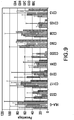


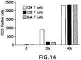


Classifications
-
- A—HUMAN NECESSITIES
- A61—MEDICAL OR VETERINARY SCIENCE; HYGIENE
- A61K—PREPARATIONS FOR MEDICAL, DENTAL OR TOILETRY PURPOSES
- A61K35/00—Medicinal preparations containing materials or reaction products thereof with undetermined constitution
- A61K35/12—Materials from mammals; Compositions comprising non-specified tissues or cells; Compositions comprising non-embryonic stem cells; Genetically modified cells
- A61K35/48—Reproductive organs
- A61K35/50—Placenta; Placental stem cells; Amniotic fluid; Amnion; Amniotic stem cells
-
- A—HUMAN NECESSITIES
- A61—MEDICAL OR VETERINARY SCIENCE; HYGIENE
- A61K—PREPARATIONS FOR MEDICAL, DENTAL OR TOILETRY PURPOSES
- A61K35/00—Medicinal preparations containing materials or reaction products thereof with undetermined constitution
- A61K35/56—Materials from animals other than mammals
- A61K35/63—Arthropods
- A61K35/64—Insects, e.g. bees, wasps or fleas
- A61K35/644—Beeswax; Propolis; Royal jelly; Honey
-
- A—HUMAN NECESSITIES
- A61—MEDICAL OR VETERINARY SCIENCE; HYGIENE
- A61K—PREPARATIONS FOR MEDICAL, DENTAL OR TOILETRY PURPOSES
- A61K38/00—Medicinal preparations containing peptides
- A61K38/16—Peptides having more than 20 amino acids; Gastrins; Somatostatins; Melanotropins; Derivatives thereof
- A61K38/17—Peptides having more than 20 amino acids; Gastrins; Somatostatins; Melanotropins; Derivatives thereof from animals; from humans
- A61K38/19—Cytokines; Lymphokines; Interferons
- A61K38/21—Interferons [IFN]
-
- A—HUMAN NECESSITIES
- A61—MEDICAL OR VETERINARY SCIENCE; HYGIENE
- A61K—PREPARATIONS FOR MEDICAL, DENTAL OR TOILETRY PURPOSES
- A61K45/00—Medicinal preparations containing active ingredients not provided for in groups A61K31/00 - A61K41/00
- A61K45/06—Mixtures of active ingredients without chemical characterisation, e.g. antiphlogistics and cardiaca
-
- A—HUMAN NECESSITIES
- A61—MEDICAL OR VETERINARY SCIENCE; HYGIENE
- A61P—SPECIFIC THERAPEUTIC ACTIVITY OF CHEMICAL COMPOUNDS OR MEDICINAL PREPARATIONS
- A61P1/00—Drugs for disorders of the alimentary tract or the digestive system
- A61P1/12—Antidiarrhoeals
-
- A—HUMAN NECESSITIES
- A61—MEDICAL OR VETERINARY SCIENCE; HYGIENE
- A61P—SPECIFIC THERAPEUTIC ACTIVITY OF CHEMICAL COMPOUNDS OR MEDICINAL PREPARATIONS
- A61P17/00—Drugs for dermatological disorders
-
- A—HUMAN NECESSITIES
- A61—MEDICAL OR VETERINARY SCIENCE; HYGIENE
- A61P—SPECIFIC THERAPEUTIC ACTIVITY OF CHEMICAL COMPOUNDS OR MEDICINAL PREPARATIONS
- A61P19/00—Drugs for skeletal disorders
- A61P19/02—Drugs for skeletal disorders for joint disorders, e.g. arthritis, arthrosis
-
- A—HUMAN NECESSITIES
- A61—MEDICAL OR VETERINARY SCIENCE; HYGIENE
- A61P—SPECIFIC THERAPEUTIC ACTIVITY OF CHEMICAL COMPOUNDS OR MEDICINAL PREPARATIONS
- A61P25/00—Drugs for disorders of the nervous system
-
- A—HUMAN NECESSITIES
- A61—MEDICAL OR VETERINARY SCIENCE; HYGIENE
- A61P—SPECIFIC THERAPEUTIC ACTIVITY OF CHEMICAL COMPOUNDS OR MEDICINAL PREPARATIONS
- A61P25/00—Drugs for disorders of the nervous system
- A61P25/02—Drugs for disorders of the nervous system for peripheral neuropathies
-
- A—HUMAN NECESSITIES
- A61—MEDICAL OR VETERINARY SCIENCE; HYGIENE
- A61P—SPECIFIC THERAPEUTIC ACTIVITY OF CHEMICAL COMPOUNDS OR MEDICINAL PREPARATIONS
- A61P29/00—Non-central analgesic, antipyretic or antiinflammatory agents, e.g. antirheumatic agents; Non-steroidal antiinflammatory drugs [NSAID]
-
- A—HUMAN NECESSITIES
- A61—MEDICAL OR VETERINARY SCIENCE; HYGIENE
- A61P—SPECIFIC THERAPEUTIC ACTIVITY OF CHEMICAL COMPOUNDS OR MEDICINAL PREPARATIONS
- A61P3/00—Drugs for disorders of the metabolism
- A61P3/08—Drugs for disorders of the metabolism for glucose homeostasis
- A61P3/10—Drugs for disorders of the metabolism for glucose homeostasis for hyperglycaemia, e.g. antidiabetics
-
- A—HUMAN NECESSITIES
- A61—MEDICAL OR VETERINARY SCIENCE; HYGIENE
- A61P—SPECIFIC THERAPEUTIC ACTIVITY OF CHEMICAL COMPOUNDS OR MEDICINAL PREPARATIONS
- A61P35/00—Antineoplastic agents
- A61P35/02—Antineoplastic agents specific for leukemia
-
- A—HUMAN NECESSITIES
- A61—MEDICAL OR VETERINARY SCIENCE; HYGIENE
- A61P—SPECIFIC THERAPEUTIC ACTIVITY OF CHEMICAL COMPOUNDS OR MEDICINAL PREPARATIONS
- A61P37/00—Drugs for immunological or allergic disorders
-
- A—HUMAN NECESSITIES
- A61—MEDICAL OR VETERINARY SCIENCE; HYGIENE
- A61P—SPECIFIC THERAPEUTIC ACTIVITY OF CHEMICAL COMPOUNDS OR MEDICINAL PREPARATIONS
- A61P37/00—Drugs for immunological or allergic disorders
- A61P37/02—Immunomodulators
-
- A—HUMAN NECESSITIES
- A61—MEDICAL OR VETERINARY SCIENCE; HYGIENE
- A61P—SPECIFIC THERAPEUTIC ACTIVITY OF CHEMICAL COMPOUNDS OR MEDICINAL PREPARATIONS
- A61P37/00—Drugs for immunological or allergic disorders
- A61P37/02—Immunomodulators
- A61P37/06—Immunosuppressants, e.g. drugs for graft rejection
-
- A—HUMAN NECESSITIES
- A61—MEDICAL OR VETERINARY SCIENCE; HYGIENE
- A61P—SPECIFIC THERAPEUTIC ACTIVITY OF CHEMICAL COMPOUNDS OR MEDICINAL PREPARATIONS
- A61P9/00—Drugs for disorders of the cardiovascular system
- A61P9/14—Vasoprotectives; Antihaemorrhoidals; Drugs for varicose therapy; Capillary stabilisers
-
- C—CHEMISTRY; METALLURGY
- C12—BIOCHEMISTRY; BEER; SPIRITS; WINE; VINEGAR; MICROBIOLOGY; ENZYMOLOGY; MUTATION OR GENETIC ENGINEERING
- C12N—MICROORGANISMS OR ENZYMES; COMPOSITIONS THEREOF; PROPAGATING, PRESERVING, OR MAINTAINING MICROORGANISMS; MUTATION OR GENETIC ENGINEERING; CULTURE MEDIA
- C12N5/00—Undifferentiated human, animal or plant cells, e.g. cell lines; Tissues; Cultivation or maintenance thereof; Culture media therefor
- C12N5/06—Animal cells or tissues; Human cells or tissues
- C12N5/0602—Vertebrate cells
- C12N5/0603—Embryonic cells ; Embryoid bodies
- C12N5/0605—Cells from extra-embryonic tissues, e.g. placenta, amnion, yolk sac, Wharton's jelly
-
- A—HUMAN NECESSITIES
- A61—MEDICAL OR VETERINARY SCIENCE; HYGIENE
- A61K—PREPARATIONS FOR MEDICAL, DENTAL OR TOILETRY PURPOSES
- A61K35/00—Medicinal preparations containing materials or reaction products thereof with undetermined constitution
- A61K35/12—Materials from mammals; Compositions comprising non-specified tissues or cells; Compositions comprising non-embryonic stem cells; Genetically modified cells
- A61K2035/122—Materials from mammals; Compositions comprising non-specified tissues or cells; Compositions comprising non-embryonic stem cells; Genetically modified cells for inducing tolerance or supression of immune responses
-
- C—CHEMISTRY; METALLURGY
- C12—BIOCHEMISTRY; BEER; SPIRITS; WINE; VINEGAR; MICROBIOLOGY; ENZYMOLOGY; MUTATION OR GENETIC ENGINEERING
- C12N—MICROORGANISMS OR ENZYMES; COMPOSITIONS THEREOF; PROPAGATING, PRESERVING, OR MAINTAINING MICROORGANISMS; MUTATION OR GENETIC ENGINEERING; CULTURE MEDIA
- C12N2500/00—Specific components of cell culture medium
- C12N2500/05—Inorganic components
- C12N2500/10—Metals; Metal chelators
- C12N2500/20—Transition metals
- C12N2500/24—Iron; Fe chelators; Transferrin
- C12N2500/25—Insulin-transferrin; Insulin-transferrin-selenium
-
- C—CHEMISTRY; METALLURGY
- C12—BIOCHEMISTRY; BEER; SPIRITS; WINE; VINEGAR; MICROBIOLOGY; ENZYMOLOGY; MUTATION OR GENETIC ENGINEERING
- C12N—MICROORGANISMS OR ENZYMES; COMPOSITIONS THEREOF; PROPAGATING, PRESERVING, OR MAINTAINING MICROORGANISMS; MUTATION OR GENETIC ENGINEERING; CULTURE MEDIA
- C12N2500/00—Specific components of cell culture medium
- C12N2500/30—Organic components
- C12N2500/36—Lipids
-
- C—CHEMISTRY; METALLURGY
- C12—BIOCHEMISTRY; BEER; SPIRITS; WINE; VINEGAR; MICROBIOLOGY; ENZYMOLOGY; MUTATION OR GENETIC ENGINEERING
- C12N—MICROORGANISMS OR ENZYMES; COMPOSITIONS THEREOF; PROPAGATING, PRESERVING, OR MAINTAINING MICROORGANISMS; MUTATION OR GENETIC ENGINEERING; CULTURE MEDIA
- C12N2501/00—Active agents used in cell culture processes, e.g. differentation
- C12N2501/10—Growth factors
- C12N2501/11—Epidermal growth factor [EGF]
-
- C—CHEMISTRY; METALLURGY
- C12—BIOCHEMISTRY; BEER; SPIRITS; WINE; VINEGAR; MICROBIOLOGY; ENZYMOLOGY; MUTATION OR GENETIC ENGINEERING
- C12N—MICROORGANISMS OR ENZYMES; COMPOSITIONS THEREOF; PROPAGATING, PRESERVING, OR MAINTAINING MICROORGANISMS; MUTATION OR GENETIC ENGINEERING; CULTURE MEDIA
- C12N2501/00—Active agents used in cell culture processes, e.g. differentation
- C12N2501/10—Growth factors
- C12N2501/135—Platelet-derived growth factor [PDGF]
-
- C—CHEMISTRY; METALLURGY
- C12—BIOCHEMISTRY; BEER; SPIRITS; WINE; VINEGAR; MICROBIOLOGY; ENZYMOLOGY; MUTATION OR GENETIC ENGINEERING
- C12N—MICROORGANISMS OR ENZYMES; COMPOSITIONS THEREOF; PROPAGATING, PRESERVING, OR MAINTAINING MICROORGANISMS; MUTATION OR GENETIC ENGINEERING; CULTURE MEDIA
- C12N2501/00—Active agents used in cell culture processes, e.g. differentation
- C12N2501/30—Hormones
- C12N2501/38—Hormones with nuclear receptors
- C12N2501/39—Steroid hormones
Definitions
- a disease, disorder or condition caused by, or relating to, an unwanted or harmful immune response for example, inflammatory bowel disease, graft-versus-host disease, multiple sclerosis, rheumatoid arthritis, psoriasis, lupus erythematosus, diabetes, mycosis fungoides (Alibert-Bazin syndrome), or scleroderma.
- Human stem cells are totipotential or pluripotential precursor cells capable of generating a variety of mature human cell lineages. Evidence exists that demonstrates that stem cells can be employed to repopulate many, if not all, tissues and restore physiologic and anatomic functionality.
- the placenta is a particularly attractive source of stem cells. Because mammalian placentas are plentiful and are normally discarded as medical waste, they represent a unique source of medically-useful stem cells. Provided herein are such isolated placental stem cells, populations of the placental stem cells, and methods of using the same to treat disease, disorder or condition caused by, or relating to, an unwanted or deleterious immune response, for example, inflammatory bowel disease, graft-versus-host disease, multiple sclerosis, rheumatoid arthritis, psoriasis, lupus erythematosus, diabetes, mycosis fungoides (Alibert-Bazin syndrome), or scleroderma.
- inflammatory bowel disease for example, graft-versus-host disease, multiple sclerosis, rheumatoid arthritis, psoriasis, lupus erythematosus, diabetes, mycosis fungoides (Alibert-Bazin
- a method of treating an individual having or at risk of developing a disease, disorder or condition associated with or caused by harmful, deleterious, inappropriate or unwanted immune response comprising administering to the individual a therapeutically effective amount of placental stem cells, or medium conditioned by placental stem cells, wherein the therapeutically effective amount is an amount sufficient to cause a detectable improvement in one or more symptoms of, or a reduction in the progression of one or more symptoms of, said disease, disorder or condition.
- placental stem cells in the manufacture of a medicament for treating, managing, ameliorating or preventing diseases, disorders and/or conditions associated with or caused by an immune response, e.g ., associated with, resulting in or caused by inflammation.
- said placental stem cells are CD10 + , CD34 - , CD105 + , CD200 + placental stem cells.
- said placental stem cells express CD200 and HLA-G, or express CD73, CD105, and CD200, or express CD200 and OCT-4, or express CD73, CD105 and HLA-G, or express CD73 and CD105 and facilitate the formation of one or more embryoid-like bodies in a population of placental cells comprising said stem cell when said population is cultured under conditions that allow for the formation of an embryoid-like body, or express OCT-4 and (c) facilitate the formation of one or more embryoid-like bodies in a population of placental cells comprising said stem cell when said population is cultured under conditions that allow for the formation of an embryoid-like body.
- the placental stem cells suppress the activity of an immune cell, e.g ., suppress proliferation of a T cell.
- said disease, disorder or condition is an inflammatory bowel disease.
- said inflammatory bowel disease is Crohn's disease.
- said Crohn's disease is gastroduodenal Crohn's disease.
- said Crohn's disease is jejunoileitis.
- said Crohn's disease is ileitis.
- said Crohn's disease is ileocolitis.
- said Crohn's disease is Crohn's colitis.
- said inflammatory bowel disease is ulcerative colitis.
- said symptom of inflammatory bowel disease is one or more of inflammation and swelling of a part of the GI tract, abdominal pain, frequent emptying of the bowel, diarrhea, rectal bleeding, anemia, weight loss, arthritis, skin problems, fever, thickening of the intestinal wall, formation of scar tissue in the intestines, formation of sores or ulcers in the intestine, development of one or more fistulas in the intestinal wall, development of one or more fissures in the anus, development of a nutritional deficiency, development of kidney stones, development of gallstones, development of a disease of the liver or biliary system, bloody diarrhea, nausea, abdominal cramps, anemia, fatigue, weight loss, loss of appetite, loss of bodily fluids and nutrients, skin lesions, joint pain, growth failure, development of osteoporosis, or eye inflammation.
- said symptom of inflammatory bowel disease is one or more of pruritic or painful rash, fever, generalized erythroderma, desquamation, raised ( e.g., higher than normal) levels of bilirubin, raised levels of alanine aminotransferase (ALT), raised levels of aspartate aminotransferase (AST), raised levels of alkaline phosphatase (AP), diarrhea, internal bleeding, cramping, abdominal pain, and ileus, burning sensation in the eye, eye irritation, photophobia, eye pain due to decreased tear secretion; dryness of the mouth, sensitivity to spicy or acidic foods, abdominal pain, dysphagia, odynophagia, weight loss, obstructive lung disease, muscular weakness, neuropathic pain, or muscle cramps.
- ALT alanine aminotransferase
- AST aspartate aminotransferase
- AP alkaline phosphatase
- said disease, disorder or condition is graft-versus-host disease.
- said graft-versus-host disease develops after an allogeneic bone marrow transplant.
- said graft-versus-host disease develops after a solid organ transplant.
- said graft-versus-host disease develops after a composite tissue allograft.
- said graft-versus-host disease is reduced in grade by at least one step by said administering.
- said graft-versus-host disease does not progress beyond grade II within 100 days after transplantation as a result of said administering.
- said graft-versus-host disease does not progress beyond grade I within 100 days after transplantation as a result of said administering.
- said disease, disorder or condition is rheumatoid arthritis (RA).
- the administration is sufficient to cause a detectable improvement in one or more symptoms of RA, or sufficient to detectably reduce the onset of one or more symptoms of RA, in at least one joint in the individual with RA.
- the administration is sufficient to cause a detectable improvement in one or more symptoms of RA, or sufficient to detectably reduce the onset of one or more symptoms of RA, in at least one non-joint tissue in the individual with RA.
- said non-joint tissue is skin (dermis), lungs, autoimmune system or blood, renal tissue, cardiovascular tissue, ocular tissue, or neurological tissue.
- said symptom of RA is a condition adjunct to RA.
- said condition adjunct to RA is pyoderma gangrenosum, neutrophilic dermatosis, Sweet's syndrome, viral infection, erythema nodosum, lobular panniculitis, atrophy of digital skin, palmar erythema, diffuse thinning (rice paper skin), skin fragility, subcutaneous nodules on an exterior surface, e.g., on the elbows, fibrosis of the lungs ( e.g., as a consequence of methotrexate therapy), Caplan's nodules, vasculitic disorders, nail fold infarcts, neuropathy, nephropathy, amyloidosis, muscular pseudohypertrophy, endoscarditis, left ventricular failure, valulitis, scleromalacia, mononeuritis multiplex, atlanto-axial subluxation.
- a pluralityoderma gangrenosum is pyoderma gangrenos
- the disease, disorder or condition is multiple sclerosis.
- said multiple sclerosis is relapsing/remitting multiple sclerosis, secondary progressive multiple sclerosis, primary progressive multiple sclerosis, or progressive/relapsing multiple sclerosis.
- said symptom of multiple sclerosis is one or more of a sensory disturbance in a limb, optic nerve dysfunction, pyramidal tract dysfunction, bladder dysfunction, bowel dysfunction, sexual dysfunction, ataxia, or diplopia.
- the disease, disorder or condition is lupus erythematosus.
- said symptom of lupus erythematosus is one or more of malar rash, butterfly rash, discoid lupus, alopecia, mouth, nasal, and vaginal ulcers, lesions on the skin, joint pain anemia and/or iron deficiency, lower than normal platelet and white blood cell counts, antiphospholipid antibody syndrome, presence of anticardiolipin antibody in the blood, pericarditis, myocarditis, endocarditis, lung and/or pleural inflammation, pleuritis, pleural effusion, lupus pneumonitis, chronic diffuse interstitial lung disease, pulmonary hypertension, pulmonary emboli, pulmonary hemorrhage, painless hematuria or proteinuria, lupus nephritis, renal failure, and/or development of membranous glomerulonephritis with "wire loop"
- deficiency in CD45 phosphatase and/or increased expression of CD40 ligand deficiency in CD45 phosphatase and/or increased expression of CD40 ligand
- nonspecific manifestations e.g ., lupus gastroenteritis, lupus pancreatitis, lupus cystitis, autoimmune inner ear disease, parasympathetic dysfunction, retinal vasculitis, systemic vasculitis, increased expression of Fc ⁇ RI ⁇ , increased and sustained calcium levels in T cells, increase of inositol triphosphate in the blood, reduction in protein kinase C phosphorylation, reduction in Ras-MAP kinase signaling, and/or a deficiency in protein kinase A I activity.
- said disease, disorder or condition is scleroderma.
- the scleroderma is diffuse scleroderma.
- the scleroderma is limited scleroderma (CREST syndrome).
- the scleroderma is morphea/linear scleroderma.
- said symptom is one or more of hardening of the skin of the face, hardening of the skin of the fingers, Reynaud's syndrome, inappropriate vasoconstriction in an extremity, calcinosis, telangiectasia, esophageal dysmotility, or presence in the blood of an anti-centromere antibody or an anti-scl70/anti-topoisomerase antibody.
- the method comprises administering a second therapeutic agent to said individual.
- said second therapeutic agent is an anti-inflammatory drug, a proton pump inhibitor, an immunosuppressant compound, or a vasodilator.
- said disease, disorder or condition is mycosis fungoides (Alibert-Bazin syndrome).
- said mycosis fungoides is in the patch phase.
- said mycosis fungoides is in the skin tumor phase.
- said mycosis fungoides is in the skin redness (erythroderma) stage.
- said mycosis fungoides is in the lymph node stage.
- said symptom is one or more of development of flat, red patches that are itchy; development of flat, red patches that are raised and hard (plaques); development of raised lumps (nodules) appear; development of large red, itchy, scaly areas over the body; cracking of the skin of the palms and soles; thickening of the skin of the palms and soles; and crack; or inflammation of the lymph nodes.
- the method comprises administering a second therapeutic agent to said individual.
- said second therapeutic agent is an anti-inflammatory drug, an immunosuppressant compound, exposure to sunlight, exposure to ultraviolet light, a topical steroid, local superficial radiotherapy, total skin electron beam radiation, application of organic honey to skin affected by erythorderma, an interferon, a retinoid, a rexinoid, or vorinostat.
- said disease, disorder or condition is diabetes.
- said diabetes is diabetes mellitus Type 1.
- said disease, disorder or condition is psoriasis.
- the psoriasis is plaque psoriasis (psoriasis vulgaris).
- said psoriasis is flexural psoriasis (inverse psoriasis).
- said psoriasis is guttate psoriasis.
- said psoriasis is pustular psoriasis.
- said psoriasis is nail psoriasis.
- said psoriasis is psoriatic arthritis.
- said psoriasis is erythrodermic psoriasis.
- the therapeutically effective amount of placental stem cells or umbilical cord stem cells, or culture medium conditioned by placental stem cells or umbilical cord stem cells is an amount sufficient to cause a 5, 10, 15, 20, 25, 30, 35, 40 or more point reduction in the Psoriasis Area Severity Index.
- the invention provides for the administration of an effective dose placental stem cells to an individual affected with psoriasis, wherein said effective dose is an amount of placental stem cells sufficient, e.g ., to cause a detectable improvement in, reduce the severity of, or reduce the progression of, one or more of the symptoms of psoriasis.
- said one or more symptoms is development of one or more of raised areas of inflamed skin covered with silvery white scaly skin; development of plaques; smooth inflamed patches of skin occurring in skin folds; development of one or more small oval spots; development of one or more pustules; a change in the appearance of one or more finger nails or toe nails; onycholysis; crumbling of one or more nails; joint and connective tissue inflammation; dactylitis; spondylitis; widespread inflammation and exfoliation of the skin over most of the body surface; or severe itching, swelling and/or pain.
- the method comprises additionally administering one or more therapeutic agents or therapies, wherein said therapeutic agents or therapies comprise one or more of a cream or ointment comprising a corticosteroid, a cream or ointment comprising a vitamin D 3 analog, a cream or ointment comprising a anthralin, a cream or ointment comprising a argan oil, a cream or ointment comprising a retinoid, or a cream or ointment comprising coal tar; one or more exposures, e.g ., for about 1, 2, 3, 4, 5, 6, 7, 8, 9, 10, 12, 14, 16, 18 or 20 minutes, to ultraviolet light, e.g ., UVB of a wavelength between about 280 nm to about 315 nm, particularly about 311 nm to about 312 nm; topical administration of psoralen in combination with exposure to UVA light; or one or more systemic administrations of one or more of methotrexate,
- the method comprises administration of a second therapeutic agent to the individual having the disease, disorder or condition.
- said second therapeutic agent is an anti-inflammatory agent, an immunomodulatory agent, and immunosuppressive agent, a pain medication, or an antibiotic.
- the second therapeutic agent is an immunomodulatory agent.
- said immunomodulatory agent is an immune suppressant.
- said immune suppressive agent is an anti-CD3 antibody (e.g., OKT3, muronomab), an anti-IL-2 receptor antibody (e.g ., basiliximab (SIMULECT®) and daclizumab (ZENAPAX®)), an anti T cell receptor antibody (e.g ., Muromonab-CD3), azathioprine, a calcineurin inhibitor, a corticosteroid, cyclosporine, methotrexate, mercaptopurine, mycophenolate mofetil, tacrolimus, or sirolimus.
- the second therapeutic agent comprises a stem cell of another type, e.g., a bone marrow-derived mesenchymal stem cell, bone marrow, or a hematopoietic stem cell.
- SH2 refers to an antibody that binds an epitope on the marker CD105.
- cells that are referred to as SH2 + are CD105 + .
- SH3 and SH4 refer to antibodies that bind epitopes present on the marker CD73.
- cells that are referred to as SH3 + and/or SH4 + are CD73 + .
- isolated stem cell means a stem cell that is substantially separated from other, non-stem cells of the tissue, e.g ., placenta, from which the stem cell is derived.
- a stem cell is “isolated” if at least 50%, 60%, 70%, 80%, 90%, 95%, or at least 99% of the non-stem cells with which the stem cell is naturally associated are removed from the stem cell, e.g ., during collection and/or culture of the stem cell.
- isolated population of cells means a population of cells that is substantially separated from other cells of the tissue, e.g ., placenta, from which the population of cells is derived.
- a population of, e.g ., stem cells is “isolated” if at least 50%, 60%, 70%, 80%, 90%, 95%, or at least 99% of the cells with which the population of stem cells are naturally associated are removed from the population of stem cells, e.g ., during collection and/or culture of the population of stem cells.
- placental stem cell refers to a stem cell or progenitor cell that is derived from a mammalian placenta, regardless of morphology, cell surface markers, or the number of passages after a primary culture, which adheres to a tissue culture substrate (e.g., tissue culture plastic or a fibronectin-coated tissue culture plate).
- tissue culture substrate e.g., tissue culture plastic or a fibronectin-coated tissue culture plate.
- placenta stem cell does not, however, refer to a trophoblast, a cytotrophoblast, embryonic germ call, or embryonic stem cell, as those cells are understood by persons of skill in the art.
- a cell is considered a "stem cell” if the cell retains at least one attribute of a stem cell, e.g.
- a marker or gene expression profile associated with one or more types of stem cells e.g ., the ability to differentiate, either in vitro, in vivo or both, into cells of one or more of the three germ layers; the lack of adult ( i.e ., differentiated) cell characteristics, or the like.
- the terms "placental stem cell” and “placenta-derived stem cell” may be used interchangeably. Unless otherwise noted herein, the term “placental” includes the umbilical cord.
- the placental stem cells disclosed herein are, in certain embodiments, multipotent in vitro (that is, the cells differentiate in vitro under differentiating conditions), multipotent in vivo (that is, the cells differentiate in vivo ), or both.
- a stem cell is "positive" for a particular marker when that marker is detectable.
- a placental stem cell is positive for, e.g ., CD73 because CD73 is detectable on placental stem cells in an amount detectably greater than background (in comparison to, e.g., an isotype control).
- a cell is also positive for a marker when that marker can be used to distinguish the cell from at least one other cell type, or can be used to select or isolate the cell when present or expressed by the cell.
- immunomodulation and “immunomodulatory” mean causing, or having the capacity to cause, a detectable change in an immune response, and the ability to cause a detectable change in an immune response.
- immunosuppression and “immunosuppressive” mean causing, or having the capacity to cause, a detectable reduction in an immune response, and the ability to cause a detectable suppression of an immune response.
- a disease, disorder or condition associated with, arising from, or related to an inappropriate, unwanted, harmful or deleterious immune response e.g., an autoimmune disease
- administering comprising administering to the individual having the disease, disorder or condition one or more doses of placental stem cells and/or umbilical cord stem cells.
- Such immunomodulation is useful in the treatment of an individual having a disease, disorder or condition caused by, or relating to, an unwanted or harmful immune response, for example, inflammatory bowel disease, graft-versus-host disease, multiple sclerosis, rheumatoid arthritis, psoriasis, lupus erythematosus, diabetes, mycosis fungoides (Alibert-Bazin syndrome), or scleroderma.
- Such immunomodulation is also useful, e.g ., in the reduction or elimination of a host immune response against allogenic tissue, e.g ., a transplanted organ, composite tissue allograft, and the like.
- a method of suppressing an immune response comprising contacting a plurality of immune cells with a plurality of placental stem cells for a time sufficient for said placental stem cells to detectably suppress an immune response, wherein said placental stem cells detectably suppress T cell proliferation in a mixed lymphocyte reaction (MLR) assay or a regression assay.
- MLR mixed lymphocyte reaction
- Placental stem cells are, e.g., the placental stem cells described elsewhere herein ( see Section 5.2). Placental stem cells used for immunosuppression can be derived or obtained from a single placenta or multiple placentas. Placental stem cells used for immunosuppression can also be derived from a single species, e.g ., the species of the intended recipient or the species of the immune cells the function of which is to be reduced or suppressed, or can be derived from multiple species.
- an “immune cell” in the context of this method means any cell of the immune system, particularly T cells and NK (natural killer) cells.
- placental stem cells are contacted with a plurality of immune cells, wherein the plurality of immune cells are, or comprises, a plurality of T cells (e.g., a plurality of CD3 + T cells, CD4 + T cells and/or CD8 + T cells) and/or natural killer cells.
- An "immune response" in the context of the method can be any response by an immune cell to a stimulus normally perceived by an immune cell, e.g., a response to the presence of an antigen.
- an immune response can be the proliferation of T cells (e.g., CD3 + T cells, CD4 + T cells and/or CD8 + T cells) in response to a foreign antigen, such as an antigen present in a transfusion or graft, or to a self-antigen, as in an autoimmune disease.
- the immune response can also be a proliferation of T cells contained within a graft.
- the immune response can also be any activity of a natural killer (NK) cell, the maturation of a dendritic cell, or the like.
- NK natural killer
- the immune response can also be a local, tissue- or organ-specific, or systemic effect of an activity of one or more classes of immune cells, e.g., the immune response can be graft versus host disease, inflammation, formation of inflammation-related scar tissue, an autoimmune condition (e.g., rheumatoid arthritis, Type I diabetes, lupus erythematosus, etc .). and the like.
- the immune response can be graft versus host disease, inflammation, formation of inflammation-related scar tissue, an autoimmune condition (e.g., rheumatoid arthritis, Type I diabetes, lupus erythematosus, etc .). and the like.
- Contacting in this context encompasses bringing the placental stem cells and immune cells together in a single container (e.g., culture dish, flask, vial, etc .) or in vivo, for example, the same individual ( e.g ., mammal, for example, human). In a preferred embodiment, the contacting is for a time sufficient, and with a sufficient number of placental stem cells and immune cells, that a change in an immune function of the immune cells is detectable.
- said contacting is sufficient to suppress immune function (e.g ., T cell proliferation in response to an antigen) by at least 50%, 60%, 70%, 80%, 90% or 95%, compared to the immune function in the absence of the placental stem cells.
- immune function e.g ., T cell proliferation in response to an antigen
- contacting the placental stem cells and plurality of immune cells can comprise combining the placental stem cells and immune cells in the same physical space such that at least a portion of the plurality of placental stem cells interacts with at least a portion of the plurality of immune cells; maintaining the placental stem cells and immune cells in separate physical spaces with common medium; or can comprise contacting medium from one or a culture of placental stem cells or immune cells with the other type of cell (for example, obtaining culture medium from a culture of placental stem cells and resuspending isolated immune cells in the medium).
- the contacting is performed in a Mixed Lymphocyte Reaction (MLR).
- MLR Mixed Lymphocyte Reaction
- the contacting is performed in a regression assay.
- Such contacting can, for example, take place in an experimental setting designed to determine the extent to which a particular plurality of placental stem cells is immunomodulatory, e.g ., immunosuppressive.
- an experimental setting can be, for example, a mixed lymphocyte reaction (MLR) or regression assay.
- MLR mixed lymphocyte reaction
- Procedures for performing the MLR and regression assays are well-known in the art. See, e.g. Schwarz, "The Mixed Lymphocyte Reaction: An In Vitro Test for Tolerance," J. Exp. Med.
- an MLR is performed in which a plurality of placental stem cells are contacted with a plurality of immune cells (e.g., lymphocytes, for example, CD3 + , CD4 + and/or CD8 + T lymphocytes).
- a plurality of placental stem cells are contacted with a plurality of immune cells (e.g., lymphocytes, for example, CD3 + , CD4 + and/or CD8 + T lymphocytes).
- the MLR can be used to determine the immunosuppressive capacity of a plurality of placental stem cells.
- a plurality of placental stem cells can be tested in an MLR comprising combining CD4 + or CD8 + T cells, dendritic cells (DC) and placental stem cells in a ratio of about 10:1:2, wherein the T cells are stained with a dye such as, e.g ., CFSE that partitions into daughter cells, and wherein the T cells are allowed to proliferate for about 6 days.
- the plurality of placental stem cells is immunosuppressive if the T cell proliferation at 6 days in the presence of placental stem cells is detectably reduced compared to T cell proliferation in the presence of DC and absence of placental stem cells.
- placental stem cells are either thawed or harvested from culture. About 20,000 placental stem cells are resuspended in 100 ⁇ l of medium (RPMI 1640, 1 mM HEPES buffer, antibiotics, and 5% pooled human serum), and allowed to attach to the bottom of a well for 2 hours.
- CD4 + and/or CD8 + T cells are isolated from whole peripheral blood mononuclear cells Miltenyi magnetic beads. The cells are CFSE stained, and a total of 100,000 T cells (CD4 + T cells alone, CD8 + T cells alone, or equal amounts of CD4 + and CD8 + T cells) are added per well. The volume in the well is brought to 200 ⁇ l, and the MLR is allowed to proceed.
- a method of suppressing an immune response comprising contacting a plurality of immune cells with a plurality of placental stem cells for a time sufficient for said placental stem cells to detectably suppress T cell proliferation in a mixed lymphocyte reaction (MLR) assay or in a regression assay.
- said placental stem cells used in the MLR represent a sample or aliquot of placental stem cells from a larger population of placental stem cells.
- Populations of placental stem cells obtained from different placentas, or different tissues within the same placenta can differ in their ability to modulate an activity of an immune cell, e.g., can differ in their ability to suppress T cell activity or proliferation or NK cell activity. It is thus desirable to determine, prior to use, the capacity of a particular population of placental stem cells for immunosuppression.
- a capacity can be determined, for example, by testing a sample of the placental stem cell population in an MLR or regression assay.
- an MLR is performed with the sample, and a degree of immunosuppression in the assay attributable to the placental stem cells is determined.
- the MLR can be used as a method of determining the absolute and relative ability of a particular population of placental stem cells to suppress immune function.
- the parameters of the MLR can be varied to provide more data or to best determine the capacity of a sample of placental stem cells to immunosuppress.
- the MLR can be performed with, in one embodiment, two or more numbers of placental stem cells, e.g., 1 x 10 3 , 3 x 10 3 , 1 x 10 4 and/or 3 x 10 4 placental stem cells per reaction.
- the number of placental stem cells relative to the number of T cells in the assay can also be varied.
- placental stem cells and T cells in the assay can be present in any ratio of, e.g. about 10:1 to about 1:10, preferably about 1:5, though a relatively greater number of placental stem cells or T cells can be used.
- the regression assay can be used in similar fashion.
- Placental stem cells and immune cells can be contacted, e.g ., in an individual that is a recipient of a plurality of placental stem cells. Where the contacting is performed in an individual, in one embodiment, the contacting is between exogenous placental stem cells (that is, placental stem cells not derived from the individual) and a plurality of immune cells endogenous to the individual.
- the immune cells within the individual are CD3 + T cells, CD4 + T cells, CD8 + T cells, and/or NK cells.
- Placental stem cell-mediated immunomodulation e.g ., immunosuppression
- the immune response is an autoimmune disease, e.g ., lupus erythematosus, diabetes, rheumatoid arthritis, or multiple sclerosis.
- the contacting of the plurality of placental stem cells with the plurality of one or more types of immune cells can occur in vivo in the context of, or as an adjunct to, for example, grafting or transplanting of one or more types of tissues to a recipient individual.
- tissues may be, for example, bone marrow or blood; an organ; a specific tissue (e.g ., skin graft); composite tissue allograft ( i.e., a graft comprising two or more different types of tissues); etc.
- the placental stem cells can be used to suppress one or more immune responses of one or more immune cells contained within the recipient individual, within the transplanted tissue or graft, or both.
- placental stem cells can be administered at the time of the transplant or graft.
- the placental stem cells can also, or alternatively, be administered prior to the transplanting or grafting, e.g. , about 1, 2, 3, 4, 5, 6 or 7 days prior to the transplanting or grafting.
- Placental stem cells can also, or alternatively, be administered to a transplant or graft recipient after the transplantation or grafting, for example, about 1, 2, 3, 4, 5, 6 or 7 days after the transplanting or grafting.
- the plurality of placental stem cells are contacted with the plurality of placental stem cells before any detectable sign or symptom of an immune response, either by the recipient individual or the transplanted tissue or graft, e.g., a detectable sign or symptom of graft-versus-host disease or detectable inflammation, is detectable.
- the contacting within an individual is primarily between exogenous placental stem cells and exogenous progenitor cells or stem cells, e.g ., exogenous progenitor cells or stem cells that differentiate into immune cells.
- exogenous progenitor cells or stem cells e.g ., exogenous progenitor cells or stem cells that differentiate into immune cells.
- individuals undergoing partial or full immunoablation or myeloablation as an adjunct to cancer therapy can receive placental stem cells in combination with one or more other types of stem or progenitor cells.
- the placental stem cells can be combined with a plurality of CD34 + cells, e.g., CD34 + hematopoietic stem cells.
- Such CD34 + cells can be, e.g., CD34 + cells from a tissue source such as peripheral blood, umbilical cord blood, placental blood, or bone marrow.
- the CD34 + cells can be isolated from such tissue sources, or the whole tissue source ( e.g ., units of umbilical cord blood or bone marrow) or a partially purified preparation from the tissue source (e.g ., white blood cells from cord blood) can be combined with the placental stem cells. Combinations of placental stem cells and cord blood, or stem cells from cord blood, are described in Hariri, U.S. Application Publication No. 2003/0180269 .
- the placental stem cells can be administered to the individual in a ratio, with respect to the known or expected number of immune cells, e.g., T cells, in the individual, of from about 10:1 to about 1:10, preferably about 1:5.
- a plurality of placental stem cells can be administered to an individual in a ratio of, in non-limiting examples, about 10,000:1, about 1,000:1, about 100:1, about 10:1, about 1:1, about 1:10, about 1:100, about 1:1,000 or about 1:10,000.
- about 1 x 10 5 to about 1 x 10 8 placental stem cells per recipient kilogram, preferably about 1 x 10 6 to about 1 x 10 7 placental stem per recipient kilogram can be administered to effect immunosuppression.
- a plurality of placental stem cells administered to an individual or subject comprises at least, about, or no more than, 1 x 10 5 , 3 x 10 5 , 1 x 10 6 , 3 x 10 6 , 1 x 10 7 , 3 x 10 7 , 1 x 10 8 , 3 x 10 8 , 1 x 10 9 , 3 X 10 9 placental stem cells, or more.
- the placental stem cells can also be administered with one or more second types of stem cells, e.g., mesenchymal stem cells from bone marrow.
- Such second stem cells can be administered to an individual with placental stem cells in a ratio of, e.g ., about 1:10 to about 10:1.
- the placental stem cells can be administered to the individual by any route sufficient to bring the placental stem cells and immune cells into contact with each other.
- the placental stem cells can be administered to the individual, e.g ., intravenously, intramuscularly, intraperitoneally, intraocularly, parenterally, or directly into an organ, e.g ., pancreas.
- the placental stem cells can be formulated as a pharmaceutical composition, as described in Section 5.6.1, below.
- the method of immunosuppression can additionally comprise the addition of one or more immunosuppressive agents, particularly in the in vivo context.
- the plurality of placental stem cells are contacted with the plurality of immune cells in vivo in an individual, and a composition comprising an immunosuppressive agent is administered to the individual.
- Immunosuppressive agents are well-known in the art and include, e.g ., anti-T cell receptor antibodies (monoclonal or polyclonal, or antibody fragments or derivatives thereof), anti-IL-2 receptor antibodies (e.g ., Basiliximab (SIMULECT®) or daclizumab (ZENAPAX)®), anti T cell receptor antibodies (e.g.
- the immumosuppressive agent is a neutralizing antibody to macrophage inflammatory protein (MIP)-1 ⁇ or MIP-1 ⁇ .
- MIP macrophage inflammatory protein
- the anti-MIP-1 ⁇ or MIP-1 ⁇ antibody is administered in an amount sufficient to cause a detectable reduction in the amount of MIP-1 ⁇ and/or MIP-1 ⁇ in said individual, e.g ., at the time of transplanting.
- placental stem cells that is, stem cells obtainable from a placenta or part thereof, that (1) adhere to a tissue culture substrate; (2) have the capacity to differentiate into non-placental cell types; and (3) have, in sufficient numbers, the capacity to detectably suppress an immune function, e.g ., proliferation of CD4 + and/or CD8 + T cells in a mixed lymphocyte reaction assay or regression assay.
- Placental stem cells are not derived from blood, e.g ., placental blood or umbilical cord blood.
- the placental stem cells used in the methods and compositions provided herein have the capacity, and are selected for their capacity, to suppress the immune system of an individual.
- Placental stem cells can be either fetal or maternal in origin (that is, can have the genotype of either the mother or fetus).
- Populations of placental stem cells, or populations of cells comprising placental stem cells can comprise placental stem cells that are solely fetal or maternal in origin, or can comprise a mixed population of placental stem cells of both fetal and maternal origin.
- the placental stem cells, and populations of cells comprising the placental stem cells can be identified and selected by the morphological, marker, and culture characteristics discussed below.
- placental stem cells used as described herein when cultured in primary cultures or in cell culture, adhere to the tissue culture substrate, e.g ., tissue culture container surface ( e.g ., tissue culture plastic). Placental stem cells in culture assume a generally fibroblastoid, stellate appearance, with a number of cyotplasmic processes extending from the central cell body.
- the placental stem cells are, however, morphologically differentiable from fibroblasts cultured under the same conditions, as the placental stem cells exhibit a greater number of such processes than do fibroblasts. Morphologically, placental stem cells are also differentiable from hematopoietic stem cells, which generally assume a more rounded, or cobblestone, morphology in culture.
- Placental stem cells, and populations of placental stem cells useful in the methods and compositions provided herein, express a plurality of markers that can be used to identify and/or isolate the stem cells, or populations of cells that comprise the stem cells.
- the placental stem cells, and stem cell populations include stem cells and stem cell-containing cell populations obtained directly from the placenta, or any part thereof ( e.g ., amnion, chorion, placental cotyledons, and the like).
- Placental stem cell populations also includes populations of (that is, two or more) placental stem cells in culture, and a population in a container, e.g ., a bag. Placental stem cells are not, however, trophoblasts.
- Placental stem cells generally express the markers CD73, CD105, CD200, HLA-G, and/or OCT-4, and do not express CD34, CD38, or CD45.
- Placental stem cells can also express HLA-ABC (MHC-1) and HLA-DR. These markers can be used to identify placental stem cells, and to distinguish placental stem cells from other stem cell types. Because the placental stem cells can express CD73 and CD105, they can have mesenchymal stem cell-like characteristics.
- the placental stem cells can express CD200 and HLA-G, a fetal-specific marker, they can be distinguished from mesenchymal stem cells, e.g., bone marrow-derived mesenchymal stem cells, which express neither CD200 nor HLA-G.
- mesenchymal stem cells e.g., bone marrow-derived mesenchymal stem cells, which express neither CD200 nor HLA-G.
- the lack of expression of CD34, CD38 and/or CD45 identifies the placental stem cells as non-hematopoietic stem cells.
- an isolated cell population comprising a plurality of immunosuppressive placental stem cells that are CD200 + , HLA-G + , wherein said plurality detectably suppresses T cell proliferation in a mixed lymphocyte reaction (MLR) assay.
- said stem cells are also CD73 + and CD105 + .
- said stem cells are also CD34 - , CD38 - or CD45 - .
- said stem cells are also CD34 - , CD38 - , CD45 - , CD73 + and CD105 + .
- said isolated population produces one or more embryoid-like bodies when cultured under conditions that allow the formation of embryoid-like bodies.
- an isolated cell population comprising a plurality of immunosuppressive placental stem cells that are CD73 + , CD105 + , CD200 + , wherein said plurality detectably suppress T cell proliferation in a mixed lymphocyte reaction (MLR) assay.
- said stem cells are HLA-G + .
- said stem cells are CD34 - , CD38 - or CD45 - .
- said stem cells are CD34 - , CD38 - and CD45 - .
- said stem cells are CD34 - , CD38 - , CD45 - , and HLA-G + .
- said population of cells produces one or more embryoid-like bodies when cultured under conditions that allow the formation of embryoid-like bodies.
- An isolated cell population useful in the compositions and methods provided herein can comprise a plurality of immunosuppressive placental stem cells that are CD200 + , OCT-4 + , wherein said plurality detectably suppresses T cell proliferation in a mixed lymphocyte reaction (MLR) assay.
- said stem cells are CD73 + and CD105 + .
- said stem cells are HLA-G + .
- said stem cells are CD34 - , CD38 - and CD45 - .
- said stem cells are CD34 - , CD38 - , CD45 - , CD73 + , CD105 + and HLA-G + .
- the population produces one or more embryoid-like bodies when cultured under conditions that allow the formation of embryoid-like bodies.
- an isolated cell population comprising a plurality of immunosuppressive placental stem cells that are CD73 + , CD105 + and HLA-G + , wherein said plurality detectably suppresses T cell proliferation in a mixed lymphocyte reaction (MLR) assay.
- said stem cells are also CD34 - , CD38 - or CD45 - .
- said stem cells are also CD34 - , CD38 - and CD45 - .
- said stem cells are also OCT-4 + .
- said stem cells are also CD200 + .
- said stem cells are also CD34 - , CD38 - , CD45 - , OCT-4 + and CD200 + .
- an isolated cell population comprising a plurality of immunosuppressive placental stem cells that are CD73 + , CD105 + stem cells, wherein said plurality forms one or more embryoid-like bodies under conditions that allow formation of embryoid-like bodies, and wherein said plurality detectably suppresses T cell proliferation in a mixed lymphocyte reaction (MLR) assay.
- said stem cells are also CD34 - , CD38 - or CD45 - .
- said stem cells are also CD34 - , CD38 - and CD45 - .
- said stem cells are also OCT-4 + .
- said stem cells are also OCT-4 + , CD34 - , CD38 - and CD45 - .
- an isolated cell population comprising a plurality of immunosuppressive placental stem cells that are OCT-4 + stem cells, wherein said population forms one or more embryoid-like bodies when cultured under conditions that allow the formation of embryoid-like bodies, and wherein said plurality detectably suppresses T cell proliferation in a mixed lymphocyte reaction (MLR) assay.
- MLR mixed lymphocyte reaction
- at least 10%, at least 20%, at least 30%, at least 40%, at least 50% at least 60%, at least 70%, at least 80%, at least 90%, or at least 95% of said isolated placental cells are OCT4 + stem cells.
- said stem cells are CD73 + and CD105 + .
- said stem cells are CD34 - , CD38 - , or CD45 - .
- said stem cells are CD200 + .
- said stem cells are CD73 + , CD105 + , CD200 + , CD34 - , CD38 - , and CD45 - .
- said population has been expanded, for example, passaged at least once, at least three times, at least five times, at least 10 times, at least 15 times, or at least 20 times.
- the immunosuppressive placental stem cells provided herein include isolated placental stem cells, populations of isolated placental stem cells, or isolated populations of cells comprising placental stem cells, wherein the placental stem cells are CD10 + , CD34 - , CD105 + and CD200 + .
- an isolated cell population comprising a plurality of immunosuppressive placental stem cells that are CD29 + , CD44 + , CD73 + , CD90 + , CD105 + , CD200 + , CD34 - and CD133 - .
- stem cells useful in the compositions and methods provided herein are isolated placental stem cell that is HLA-A,B,C - , CD45 - , CD133 - and CD34 - .
- an isolated population of placental stem cells wherein at least about 70%, at least about 80%, at least about 90%, at least about 95% or at least about 99% of said placental stem cells are HLA-A,B,C - , CD45 - , CD133 - and CD34 - .
- said stem cell or population of placental stem cells is isolated away from placental cells that are not stem cells.
- said population of placental stem cells is isolated away from placental stem cells that do not display these characteristics.
- said isolated placental stem cell is non-maternal in origin.
- at least about 90%, at least about 95%, or at least about 99% of said cells in said isolated population of placental stem cells are non-maternal in origin.
- provided herein is a method of obtaining a placental stem cell that is HLA-A,B,C - , CD45 - , CD133 - and CD34 - comprising isolating said cell from placental perfusate.
- an isolated placental stem cell that is CD10 + , CD13 + , CD33 + , CD45 - , CD117 - and CD133 - .
- an isolated population of placental stem cells wherein at least about 70%, at least about 80%, at least about 90%, at least about 95% or at least about 99% of said placental stem cells are CD10 + , CD13 + , CD33 + , CD45 - , CD117 - and CD133 - .
- said stem cell or population of placental stem cells is isolated away from placental cells that are not stem cells.
- said isolated placental stem cell is non-maternal in origin.
- At least about 90%, at least about 95%, or at least about 99% of said cells in said isolated population of placental stem cells are non-maternal in origin.
- said stem cell or population of placental stem cells is isolated away from placental stem cells that do not display these characteristics.
- provided herein is a method of obtaining a placental stem cell that is CD 10 + , CD13 + , CD33 + , CD45 - , CD117 - and CD133 - comprising isolating said cell from placental perfusate.
- an isolated placental stem cell that is CD10 - , CD33 - , CD44 + , CD45 - , and CD117 - .
- an isolated population of placental stem cells wherein at least about 70%, at least about 80%, at least about 90%, at least about 95% or at least about 99% of said placental stem cells are CD10 - , CD33 - , CD44 + , CD45 - , and CD117 - .
- said stem cell or population of placental stem cells is isolated away from placental cells that are not stem cells.
- said isolated placental stem cell is non-maternal in origin.
- At least about 90%, at least about 95%, or at least 99% of said cells in said isolated population of placental stem cells are non-maternal in origin.
- said stem cell or population of placental stem cells is isolated away from placental stem cells that do not display these characteristics.
- provided herein is a method of obtaining a placental stem cell that is CD10 - , CD33 - , CD44 + , CD45 - , CD117 - comprising isolating said cell from placental perfusate.
- an isolated placental stem cell that is CD10 - , CD13 - , CD33 - , CD45 - , and CD117 - . Further provided is an isolated population of placental stem cells, wherein at least about 70%, at least about 80%, at least about 90%, at least about 95% or at least about 99% of said placental stem cells are CD10 - , CD13 - , CD33 - , CD45 - , and CD117 - .
- said stem cell or population of placental stem cells is isolated away from placental cells that are not stem cells.
- said isolated placental stem cell is non-maternal in origin.
- At least about 90%, at least about 95%, or at least 99% of said cells in said isolated population of placental stem cells are non-maternal in origin.
- said stem cell or population of placental stem cells is isolated away from placental stem cells that do not display these characteristics.
- also provided herein is a method of obtaining a placental stem cell that is CD10 - , CD13 - , CD33 - , CD45 - , and CD117 - comprising isolating said cell from placental perfusate.
- an isolated placental stem cell that is HLA A,B,C - , CD45 - , CD34 - , CD133 - , positive for CD10, CD13, CD38, CD44, CD90, CD105, CD200 and/or HLA-G, and/or negative for CD117.
- said stem cell or population of placental stem cells is isolated away from placental cells that are not stem cells.
- said isolated placental stem cell is non-maternal in origin. In another specific embodiment, at least about 90%, at least about 95%, or at least about 99%, of said cells in said isolated population of placental stem cells, are non-maternal in origin. In another specific embodiment, said stem cell or population of placental stem cells is isolated away from placental stem cells that do not display these characteristics.
- a method of obtaining a placental stem cell that is HLA A,B,C - , CD45 - , CD34 - , CD133 - and positive for CD10, CD13, CD38, CD44, CD90, CD105, CD200 and/or HLA-G, and/or negative for CD117, comprising isolating said cell from placental perfusate.
- a placental stem cell that is CD200 + and CD10 + , as determined by antibody binding, and CD117 - , as determined by both antibody binding and RT-PCR.
- a placental stem cell that is CD10 + , CD29 - , CD54 + , CD200 + , HLA-G + , HLA class I - and ⁇ -2-microglobulin - .
- placental stem cells wherein the expression of at least one marker is at least two-fold higher than for a mesenchymal stem cell (e.g., a bone marrow-derived mesenchymal stem cell).
- said isolated placental stem cell is non-maternal in origin. In another specific embodiment, at least about 90%, at least about 95%, or at least 99%, of said cells in said isolated population of placental stem cells, are non-maternal in origin.
- an isolated population of placental stem cells wherein a plurality of said placental stem cells are positive for aldehyde dehydrogenase (ALDH), as assessed by an aldehyde dehydrogenase activity assay.
- ALDH aldehyde dehydrogenase
- said ALDH assay uses ALDEFLUOR® (Aldagen, Inc., Ashland, Oregon) as a marker of aldehyde dehydrogenase activity.
- said plurality is between about 3% and about 25% of cells in said population of cells.
- a population of umbilical cord stem cells wherein a plurality of said umbilical cord stem cells are positive for aldehyde dehydrogenase, as assessed by an aldehyde dehydrogenase activity assay that uses ALDEFLUOR® as an indicator of aldehyde dehydrogenase activity.
- said plurality is between about 3% and about 25% of cells in said population of cells.
- said population of placental stem cells or umbilical cord stem cells shows at least three-fold, or at least five-fold, higher ALDH activity than a population of bone marrow-derived mesenchymal stem cells having the same number of cells and cultured under the same conditions.
- the stem cell or population of placental stem cells are, or can comprise, cells that have been passaged at least 1, 2, 3, 4, 5, 6, 7, 8, 9, 10, 12, 14, 16, 18, or 20 times, or more, or expanded for 1, 2, 3, 4, 5, 6, 7, 8, 9, 10, 12, 14, 16, 18, 20, 22, 24, 26, 28, 30, 32, 34, 36, 38 or 40 population doublings, or more.
- the karyotype of the cells, or at least about 95% or about 99% of the cells in said population is normal.
- the cells, or cells in the population of cells are non-maternal in origin.
- Isolated placental stem cells, or isolated populations of placental stem cells, bearing any of the above combinations of markers, can be combined in any ratio. Any two or more of the above placental stem cell populations can be isolated, or enriched, to form a placental stem cell population.
- an isolated population of placental stem cells comprising a first population of placental stem cells defined by one of the marker combinations described above can be combined with a second population of placental stem cells defined by another of the marker combinations described above, wherein said first and second populations are combined in a ratio of about 1:99, 2:98, 3:97, 4:96, 5:95, 10:90, 20:80, 30:70, 40:60, 50:50, 60:40, 70:30, 80:20, 90:10, 95:5, 96:4, 97:3, 98:2, or about 99:1.
- any three, four, five or more of the above-described placental stem cells or placental stem cell populations can be combined.
- the placental stem cells constitutively secrete IL-6, IL-8 and monocyte chemoattractant protein (MCP-1).
- the immunosuppressive pluralities of placental stem cells described above can comprise about, at least, or no more than, 1 x 10 5 , 5 x 10 5 , 1 x 10 6 , 5 x 10 6 , 1 x 10 7 , 5 x 10 7 , 1 x 10 8 , 5 x 10 8 , 1 x 10 9 , 5 x 10 9 , 1 x 10 10 , 5 x 10 10 , 1 x 10 11 or more placental stem cells.
- a method of selecting a plurality of immunosuppressive placental stem cells from a plurality of placental cells comprising selecting a population of placental cells wherein at least 10%, at least 20%, at least 30%, at least 40%, at least 50% at least 60%, at least 70%, at least 80%, at least 90%, or at least 95% of said cells are CD200 + , HLA-G + placental stem cells, and wherein said placental stem cells detectably suppresses T cell proliferation in a mixed lymphocyte reaction (MLR) assay.
- said selecting comprises selecting stem cells that are also CD73 + and CD105 + .
- said selecting comprises selecting stem cells that are also CD34 - , CD38 - or CD45 - . In another specific embodiment, said selecting comprises selecting placental stem cells that are also CD34 - , CD38 - , CD45 - , CD73 + and CD105 + . In another specific embodiment, said selecting also comprises selecting a plurality of placental stem cells that forms one or more embryoid-like bodies when cultured under conditions that allow the formation of embryoid-like bodies.
- a method of selecting a plurality of immunosuppressive placental stem cells from a plurality of placental cells comprising selecting a plurality of placental cells wherein at least 10%, at least 20%, at least 30%, at least 40%, at least 50% at least 60%, at least 70%, at least 80%, at least 90%, or at least 95% of said cells are CD73 + , CD105 + , CD200 + placental stem cells, and wherein said placental stem cells detectably suppresses T cell proliferation in a mixed lymphocyte reaction (MLR) assay.
- said selecting comprises selecting stem cells that are also HLA-G + .
- said selecting comprises selecting placental stem cells that are also CD34 - , CD38 - or CD45 - . In another specific embodiment, said selecting comprises selecting placental stem cells that are also CD34 - , CD38 - and CD45 - . In another specific embodiment, said selecting comprises selecting placental stem cells that are also CD34 - , CD38 - , CD45 - , and HLA-G + . In another specific embodiment, said selecting additionally comprises selecting a population of placental cells that produces one or more embryoid-like bodies when the population is cultured under conditions that allow the formation of embryoid-like bodies.
- a method of selecting a plurality of immunosuppressive placental stem cells from a plurality of placental cells comprising selecting a plurality of placental cells wherein at least 10%, at least 20%, at least 30%, at least 40%, at least 50% at least 60%, at least 70%, at least 80%, at least 90%, or at least 95% of said cells are CD200 + , OCT-4 + placental stem cells, and wherein said placental stem cells detectably suppresses T cell proliferation in a mixed lymphocyte reaction (MLR) assay.
- said selecting comprises selecting placental stem cells that are also CD73 + and CD105 + .
- said selecting comprises selecting placental stem cells that are also HLA-G + . In another specific embodiment, said selecting comprises selecting placental stem cells that are also CD34 - , CD38 - and CD45 - . In another specific embodiment, said selecting comprises selecting placental stem cells that are also CD34 - , CD38 - , CD45 - , CD73 + , CD105 + and HLA-G + .
- a method of selecting a plurality of immunosuppressive placental stem cells from a plurality of placental cells comprising selecting a plurality of placental cells wherein at least 10%, at least 20%, at least 30%, at least 40%, at least 50% at least 60%, at least 70%, at least 80%, at least 90%, or at least 95% of said cells are CD73 + , CD105 + and HLA-G + placental stem cells, and wherein said placental stem cells detectably suppresses T cell proliferation in a mixed lymphocyte reaction (MLR) assay.
- said selecting comprises selecting placental stem cells that are also CD34 - , CD38 - or CD45 - .
- said selecting comprises selecting placental stem cells that are also CD34 - , CD38 - and CD45 - . In another specific embodiment, said selecting comprises selecting placental stem cells that are also CD200 + . In another specific embodiment, said selecting comprises selecting placental stem cells that are also CD34 - , CD38 - , CD45 - , OCT-4 + and CD200 + .
- a method of selecting a plurality of immunosuppressive placental stem cells from a plurality of placental cells comprising selecting a plurality of placental cells wherein at least 10%, at least 20%, at least 30%, at least 40%, at least 50% at least 60%, at least 70%, at least 80%, at least 90%, or at least 95% of said cells are CD73 + , CD105 + placental stem cells, and wherein said plurality forms one or more embryoid-like bodies under conditions that allow formation of embryoid-like bodies.
- said selecting comprises selecting placental stem cells that are also CD34 - , CD38 - or CD45 - .
- said selecting comprises selecting placental stem cells that are also CD34 - , CD38 - and CD45 - . In another specific embodiment, said selecting comprises selecting placental stem cells that are also OCT-4 + . In a more specific embodiment, said selecting comprises selecting placental stem cells that are also OCT-4 + , CD34 - , CD38 - and CD45 - .
- a method of selecting a plurality of immunosuppressive placental stem cells from a plurality of placental cells comprising selecting a plurality of placental cells wherein at least 10%, at least 20%, at least 30%, at least 40%, at least 50% at least 60%, at least 70%, at least 80%, at least 90%, or at least 95% of said isolated placental cells are OCT4 + stem cells, and wherein said plurality forms one or more embryoid-like bodies under conditions that allow formation of embryoid-like bodies.
- said selecting comprises selecting placental stem cells that are also CD73 + and CD105 + .
- said selecting comprises selecting placental stem cells that are also CD34 - , CD38 - , or CD45 - . In another specific embodiment, said selecting comprises selecting placental stem cells that are also CD200 + . In a more specific embodiment, said selecting comprises selecting placental stem cells that are also CD73 + , CD105 + , CD200 + , CD34 - , CD38 - , and CD45 - .
- Immunosuppressive populations, or pluralities, of placental stem cells can be produced according to the methods provided herein.
- method of producing a cell population comprising selecting any of the pluralities of placental stem cells described above, and isolating the plurality of placental stem cells from other cells, e.g., other placental cells.
- a method of producing a cell population comprising selecting placental cells, wherein said placental cells (a) adhere to a substrate, (b) express CD200 and HLA-G, or express CD73, CD105, and CD200, or express CD200 and OCT-4, or express CD73, CD105, and HLA-G, or express CD73 and CD105 and facilitate the formation of one or more embryoid-like bodies in a population of placental cells that comprise the stem cell, when said population is cultured under conditions that allow formation of embryoid-like bodies, or express OCT-4 and facilitate the formation of one or more embryoid-like bodies in a population of placental cells that comprise the stem cell, when said population is cultured under conditions that allow formation of embryoid-like bodies; and (c) detectably suppress CD4 + or CD8 + T cell proliferation in an MLR (mixed lymphocyte reaction) or regression assay; and isolating said placental cells from other cells to form a cell population.
- MLR mixed lymphocyte reaction
- immunosuppressive placental stem cell populations can be produced by a method comprising selecting placental stem cells that (a) adhere to a substrate, (b) express CD200 and HLA-G, and (c) detectably suppress CD4 + or CD8 + T cell proliferation in an MLR (mixed lymphocyte reaction); and isolating said placental stem cells from other cells to form a cell population.
- the method comprises selecting placental stem cells that (a) adhere to a substrate, (b) express CD73, CD105, and CD200, and (c) detectably suppress CD4 + or CD8 + T cell proliferation in an MLR; and isolating said placental stem cells from other cells to form a cell population.
- a method of producing a cell population comprising selecting placental stem cells that (a) adhere to a substrate, (b) express CD200 and OCT-4, and (c) detectably suppress CD4 + or CD8 + T cell proliferation in an MLR; and isolating said placental stem cells from other cells to form a cell population.
- a method of producing a cell population comprising selecting placental stem cells that (a) adhere to a substrate, (b) express CD73 and CD 105, (c) form embryoid-like bodies when cultured under conditions allowing the formation of embryoid-like bodies, and (d) detectably suppress CD4 + or CD8 + T cell proliferation in an MLR; and isolating said placental stem cells from other cells to form a cell population.
- the method comprises selecting placental stem cells that (a) adhere to a substrate, (b) express CD73, CD105, and HLA-G, and (c) detectably suppress CD4 + or CD8 + T cell proliferation in an MLR; and isolating said placental stem cells from other cells to form a cell population.
- a method of producing a cell population comprising selecting placental stem cells that (a) adhere to a substrate, (b) express OCT-4, (c) form embryoid-like bodies when cultured under conditions allowing the formation of embryoid-like bodies, and (d) detectably suppress CD4 + or CD8 + T cell proliferation in an MLR; and isolating said placental stem cells from other cells to form a cell population.
- said T cells and said placental cells are present in said MLR at a ratio of about 5:1.
- the placental cells used in the method can be derived from the whole placenta, or primarily from amnion, or amnion and chorion.
- the placental cells suppress CD4 + or CD8 + T cell proliferation by at least 50%, at least 75%, at least 90%, or at least 95% in said MLR compared to an amount of T cell proliferation in said MLR in the absence of said placental cells.
- the method can additionally comprise the selection and/or production of a placental stem cell population capable of immunomodulation, e.g., suppression of the activity of, other immune cells, e.g., an activity of a natural killer (NK) cell.
- NK natural killer
- placental stem cells as for any mammalian cell, depends in part upon the particular medium selected for growth. Under optimum conditions, placental stem cells typically double in number in 3-5 days.
- the placental stem cells provided herein adhere to a substrate in culture, e.g . the surface of a tissue culture container (e.g ., tissue culture dish plastic, fibronectin-coated plastic, and the like) and form a monolayer.
- tissue culture container e.g ., tissue culture dish plastic, fibronectin-coated plastic, and the like
- embryoid-like bodies that is, three-dimensional clusters of cells grow atop the adherent stem cell layer.
- Cells within the embryoid-like bodies express markers associated with very early stem cells, e.g., OCT-4, Nanog, SSEA3 and SSEA4.
- Cells within the embryoid-like bodies are typically not adherent to the culture substrate, as are the placental stem cells described herein, but remain attached to the adherent cells during culture.
- Embryoid-like body cells are dependent upon the adherent placental stem cells for viability, as embryoid-like bodies do not form in the absence of the adherent stem cells.
- the adherent placental stem cells thus facilitate the growth of one or more embryoid-like bodies in a population of placental cells that comprise the adherent placental stem cells.
- the cells of the embryoid-like bodies are thought to grow on the adherent placental stem cells much as embryonic stem cells grow on a feeder layer of cells.
- Mesenchymal stem cells e.g ., bone marrow-derived mesenchymal stem cells, do not develop embryoid-like bodies in culture.
- the placental stem cells useful in the methods of treating diseases, disorder or conditions associated with, or caused by inappropriate or harmful immune response, e.g ., inflammation, provided herein, are differentiable into different committed cell lineages.
- the placental stem cells can be differentiated into cells of an adipogenic, chondrogenic, neurogenic, or osteogenic lineage. Such differentiation can be accomplished, e.g., by any method known in the art for differentiating, e.g., bone marrow-derived mesenchymal stem cells into similar cell lineages, or by methods described elsewhere herein.
- the placental stem cells and umbilical cord stem cells provided herein can exhibit the capacity to differentiate into a particular cell lineage in vitro, in vivo, or in vitro and in vivo.
- the placental stem cells and umbilical cord stem cells provided herein can be differentiated in vitro when placed in conditions that cause or promote differentiation into a particular cell lineage, but do not detectably differentiate in vivo, e.g., in a NOD-SCID mouse model.
- Placental stem cells can be collected and isolated according to the methods provided herein.
- stem cells are obtained from a mammalian placenta using a physiologically-acceptable solution, e.g. , a stem cell collection composition.
- a stem cell collection composition is described in detail in related U.S. Provisional Application No. 60/754,969 , entitled "Improved Composition for Collecting and Preserving Placental Stem Cells and Methods of Using the Composition" filed on December 29, 2005.
- the stem cell collection composition can comprise any physiologically-acceptable solution suitable for the collection and/or culture of stem cells, for example, a saline solution (e.g ., phosphate-buffered saline, Kreb's solution, modified Kreb's solution, Eagle's solution, 0.9% NaCl. etc .), a culture medium (e.g., DMEM, HDMEM, etc .), and the like.
- a saline solution e.g ., phosphate-buffered saline, Kreb's solution, modified Kreb's solution, Eagle's solution, 0.9% NaCl. etc .
- a culture medium e.g., DMEM, HDMEM, etc .
- the stem cell collection composition can comprise one or more components that tend to preserve placental stem cells, that is, prevent the placental stem cells from dying, or delay the death of the placental stem cells, reduce the number of placental stem cells in a population of cells that die, or the like, from the time of collection to the time of culturing.
- Such components can be, e.g., an apoptosis inhibitor (e.g., a caspase inhibitor or JNK inhibitor); a vasodilator (e.g ., magnesium sulfate, an antihypertensive drug, atrial natriuretic peptide (ANP), adrenocorticotropin, corticotropin-releasing hormone, sodium nitroprusside, hydralazine, adenosine triphosphate, adenosine, indomethacin or magnesium sulfate, a phosphodiesterase inhibitor, etc .); a necrosis inhibitor (e.g ., 2-(1H-Indol-3-yl)-3-pentylamino-maleimide, pyrrolidine dithiocarbamate, or clonazepam); a TNF- ⁇ inhibitor; and/or an oxygen-carrying perfluorocarbon (e.g ., perfluoro
- the stem cell collection composition can comprise one or more tissue-degrading enzymes, e.g ., a metalloprotease, a serine protease, a neutral protease, an RNase, or a DNase, or the like.
- tissue-degrading enzymes include, but are not limited to, collagenases (e.g ., collagenase I, II, III or IV, a collagenase from Clostridium histolyticum , etc .); dispase, thermolysin, elastase, trypsin, LIBERASE, hyaluronidase, and the like.
- the stem cell collection composition can comprise a bacteriocidally or bacteriostatically effective amount of an antibiotic.
- the antibiotic is a macrolide (e.g., tobramycin), a cephalosporin (e.g., cephalexin, cephradine, cefuroxime, cefprozil, cefaclor, cefixime or cefadroxil), a clarithromycin, an erythromycin, a penicillin (e.g., penicillin V) or a quinolone ( e.g., ofloxacin, ciprofloxacin or norfloxacin), a tetracycline, a streptomycin, etc.
- the antibiotic is active against Gram(+) and/or Gram(-) bacteria, e.g ., Pseudomonas aeruginosa , Staphylococcus aureus , and the like.
- the stem cell collection composition can also comprise one or more of the following compounds: adenosine (about 1 mM to about 50 mM); D-glucose (about 20 mM to about 100 mM); magnesium ions (about 1 mM to about 50 mM); a macromolecule of molecular weight greater than 20,000 daltons, in one embodiment, present in an amount sufficient to maintain endothelial integrity and cellular viability (e.g ., a synthetic or naturally occurring colloid, a polysaccharide such as dextran or a polyethylene glycol present at about 25 g/l to about 100 g/l, or about 40 g/l to about 60 g/l); an antioxidant ( e.g ., butylated hydroxyanisole, butylated hydroxytoluene, glutathione, vitamin C or vitamin E present at about 25 ⁇ M to about 100 ⁇ M); a reducing agent (e.g., N-acetylcysteine present at least
- a human placenta is recovered shortly after its expulsion after birth.
- the placenta is recovered from a patient after informed consent and after a complete medical history of the patient is taken and is associated with the placenta.
- the medical history continues after delivery.
- Such a medical history can be used to coordinate subsequent use of the placenta or the stem cells harvested therefrom.
- human placental stem cells can be used, in light of the medical history, for personalized medicine for the infant associated with the placenta, or for parents, siblings or other relatives of the infant.
- the umbilical cord blood and placental blood Prior to recovery of placental stem cells, the umbilical cord blood and placental blood are removed. In certain embodiments, after delivery, the cord blood in the placenta is recovered.
- the placenta can be subjected to a conventional cord blood recovery process.
- a needle or cannula is used, with the aid of gravity, to exsanguinate the placenta (see, e.g ., Anderson, U.S. Patent No. 5,372,581 ; Hessel et al., U.S. Patent No. 5,415,665 ).
- the needle or cannula is usually placed in the umbilical vein and the placenta can be gently massaged to aid in draining cord blood from the placenta.
- cord blood recovery may be performed commercially, e.g ., LifeBank Inc., Cedar Knolls, N.J., ViaCord, Cord Blood Registry and Cryocell.
- the placenta is gravity drained without further manipulation so as to minimize tissue disruption during cord blood recovery.
- a placenta is transported from the delivery or birthing room to another location, e.g., a laboratory, for recovery of cord blood and collection of stem cells by, e.g., perfusion or tissue dissociation.
- the placenta is preferably transported in a sterile, thermally insulated transport device (maintaining the temperature of the placenta between 20-28°C), for example, by placing the placenta, with clamped proximal umbilical cord, in a sterile zip-lock plastic bag, which is then placed in an insulated container.
- the placenta is transported in a cord blood collection kit substantially as described in pending United States patent application no. 11/230,760, filed September 19, 2005 .
- the placenta is delivered to the laboratory four to twenty-four hours following delivery.
- the proximal umbilical cord is clamped, preferably within 4-5 cm (centimeter) of the insertion into the placental disc prior to cord blood recovery.
- the proximal umbilical cord is clamped after cord blood recovery but prior to further processing of the placenta.
- the placenta prior to stem cell collection, can be stored under sterile conditions and at either room temperature or at a temperature of 5 to 25°C (centigrade).
- the placenta may be stored for a period of longer than forty eight hours, and preferably for a period of four to twenty-four hours prior to perfusing the placenta to remove any residual cord blood.
- the placenta is preferably stored in an anticoagulant solution at a temperature of 5 to 25°C (centigrade). Suitable anticoagulant solutions are well known in the art. For example, a solution of heparin or warfarin sodium can be used.
- the anticoagulant solution comprises a solution of heparin (e.g ., 1% w/w in 1:1000 solution).
- the exsanguinated placenta is preferably stored for no more than 36 hours before placental stem cells are collected.
- the mammalian placenta or a part thereof, once collected and prepared generally as above, can be treated in any art-known manner, e.g., can be perfused or disrupted, e.g., digested with one or more tissue-disrupting enzymes, to obtain stem cells.
- stem cells are collected from a mammalian placenta by physical disruption, e.g., enzymatic digestion, of the organ, e.g., using the stem cell collection composition described in Section 5.3.1, above.
- the placenta, or a portion thereof may be, e.g ., crushed, sheared, minced, diced, chopped, macerated or the like, while in contact with, e .g., a buffer, medium or a stem cell collection composition, and the tissue subsequently digested with one or more enzymes.
- the placenta, or a portion thereof, may also be physically disrupted and digested with one or more enzymes, and the resulting material then immersed in, or mixed into, a buffer, medium or a stem cell collection composition.
- Any method of physical disruption can be used, provided that the method of disruption leaves a plurality, more preferably a majority, and more preferably at least 60%, 70%, 80%, 90%, 95%, 98%, or 99% of the cells in said organ viable, as determined by, e.g., trypan blue exclusion.
- placenta can be dissected into components prior to physical disruption and/or enzymatic digestion and stem cell recovery.
- placental stem cells can be obtained from the amniotic membrane, chorion, placental cotyledons, or any combination thereof, or umbilical cord, or any combination thereof.
- placental stem cells are obtained from placental tissue comprising amnion and chorion, or amnion-chorion and umbilical cord.
- stem cells are obtained from amnion-chorion and umbilical cord in about a 1:1 weight ratio.
- placental stem cells can be obtained by disruption of a small block of placental tissue, e.g., a block of placental tissue that is about 1, 2, 3, 4, 5, 6, 7, 8, 9, 10, 20, 30, 40, 50, 60, 70, 80, 90, 100, 200, 300, 400, 500, 600, 700, 800, 900 or about 1000 cubic millimeters in volume.
- a block of placental tissue e.g., a block of placental tissue that is about 1, 2, 3, 4, 5, 6, 7, 8, 9, 10, 20, 30, 40, 50, 60, 70, 80, 90, 100, 200, 300, 400, 500, 600, 700, 800, 900 or about 1000 cubic millimeters in volume.
- a preferred stem cell collection composition comprises one or more tissue-disruptive enzyme(s).
- Enzymatic digestion preferably uses a combination of enzymes, e.g ., a combination of a matrix metalloprotease and a neutral protease, for example, a combination of collagenase and dispase.
- enzymatic digestion of placental tissue uses a combination of a matrix metalloprotease, a neutral protease, and a mucolytic enzyme for digestion of hyaluronic acid, such as a combination of collagenase, dispase, and hyaluronidase or a combination of LIBERASE (Boehringer Mannheim Corp., Indianapolis, Ind.) and hyaluronidase.
- enzymes that can be used to disrupt placenta tissue include papain, deoxyribonucleases, serine proteases, such as trypsin, chymotrypsin, or elastase.
- Serine proteases may be inhibited by alpha 2 microglobulin in serum and therefore the medium used for digestion is usually serum-free.
- EDTA and DNase are commonly used in enzyme digestion procedures to increase the efficiency of cell recovery.
- the digestate is preferably diluted so as to avoid trapping stem cells within the viscous digest.
- tissue digestion enzymes can be used. Typical concentrations for tissue digestion enzymes include, e.g ., 50-200 U/mL for collagenase I and collagenase IV, 1-10 U/mL for dispase, and 10-100 U/mL for elastase.
- Proteases can be used in combination, that is, two or more proteases in the same digestion reaction, or can be used sequentially in order to liberate placental stem cells. For example, in one embodiment, a placenta, or part thereof, is digested first with an appropriate amount of collagenase I at 2 mg/ml for 30 minutes, followed by digestion with trypsin, 0.25%, for 10 minutes, at 37°C. Serine proteases are preferably used consecutively following use of other enzymes.
- the tissue can further be disrupted by the addition of a chelator, e.g ., ethylene glycol bis(2-aminoethyl ether)-N,N,N'N'-tetraacetic acid (EGTA) or ethylenediaminetetraacetic acid (EDTA) to the stem cell collection composition comprising the stem cells, or to a solution in which the tissue is disrupted and/or digested prior to isolation of the stem cells with the stem cell collection composition.
- a chelator e.g ethylene glycol bis(2-aminoethyl ether)-N,N,N'N'-tetraacetic acid (EGTA) or ethylenediaminetetraacetic acid (EDTA)
- the placental stem cells collected will comprise a mix of placental stem cells derived from both fetal and maternal sources.
- the placental stem cells collected will comprise almost exclusively fetal placental stem cells.
- Placental stem cells can also be obtained by perfusion of the mammalian placenta.
- Methods of perfusing mammalian placenta to obtain stem cells are disclosed, e.g., in Hariri, U.S. Application Publication No. 2002/0123141 , and in related U.S. Provisional Application No. 60/754,969 , entitled "Improved Composition for Collecting and Preserving Placental Stem Cells and Methods of Using the Composition” filed on December 29, 2005.
- Placental stem cells can be collected by perfusion, e.g., through the placental vasculature, using, e.g., a stem cell collection composition as a perfusion solution.
- a mammalian placenta is perfused by passage of perfusion solution through either or both of the umbilical artery and umbilical vein.
- the flow of perfusion solution through the placenta may be accomplished using, e.g., gravity flow into the placenta.
- the perfusion solution is forced through the placenta using a pump, e.g., a peristaltic pump.
- the umbilical vein can be, e.g., cannulated with a cannula, e.g., a TEFLON® or plastic cannula, that is connected to a sterile connection apparatus, such as sterile tubing.
- a sterile connection apparatus such as sterile tubing.
- the sterile connection apparatus is connected to a perfusion manifold.
- the placenta is preferably oriented ( e.g., suspended) in such a manner that the umbilical artery and umbilical vein are located at the highest point of the placenta.
- the placenta can be perfused by passage of a perfusion fluid, e.g., the stem cell collection composition provided herein, through the placental vasculature, or through the placental vasculature and surrounding tissue.
- a perfusion fluid e.g., the stem cell collection composition provided herein
- the umbilical artery and the umbilical vein are connected simultaneously to a pipette that is connected via a flexible connector to a reservoir of the perfusion solution.
- the perfusion solution is passed into the umbilical vein and artery.
- the perfusion solution exudes from and/or passes through the walls of the blood vessels into the surrounding tissues of the placenta, and is collected in a suitable open vessel from the surface of the placenta that was attached to the uterus of the mother during gestation.
- the perfusion solution may also be introduced through the umbilical cord opening and allowed to flow or percolate out of openings in the wall of the placenta which interfaced with the maternal uterine wall.
- the perfusion solution is passed through the umbilical veins and collected from the umbilical artery, or is passed through the umbilical artery and collected from the umbilical veins.
- the proximal umbilical cord is clamped during perfusion, and more preferably, is clamped within 4-5 cm (centimeter) of the cord's insertion into the placental disc.
- the first collection of perfusion fluid from a mammalian placenta during the exsanguination process is generally colored with residual red blood cells of the cord blood and/or placental blood.
- the perfusion fluid becomes more colorless as perfusion proceeds and the residual cord blood cells are washed out of the placenta.
- 30 to 100 ml (milliliter) of perfusion fluid is adequate to initially exsanguinate the placenta, but more or less perfusion fluid may be used depending on the observed results.
- the volume of perfusion liquid used to collect placental stem cells may vary depending upon the number of stem cells to be collected, the size of the placenta, the number of collections to be made from a single placenta, etc.
- the volume of perfusion liquid may be from 50 mL to 5000 mL, 50 mL to 4000 mL, 50 mL to 3000 mL, 100 mL to 2000 mL, 250 mL to 2000 mL, 500 mL to 2000 mL, or 750 mL to 2000 mL.
- the placenta is perfused with 700-800 mL of perfusion liquid following exsanguination.
- the placenta can be perfused a plurality of times over the course of several hours or several days. Where the placenta is to be perfused a plurality of times, it may be maintained or cultured under aseptic conditions in a container or other suitable vessel, and perfused with the stem cell collection composition, or a standard perfusion solution (e.g., a normal saline solution such as phosphate buffered saline ("PBS”)) with or without an anticoagulant (e.g., heparin, warfarin sodium, coumarin, bishydroxycoumarin), and/or with or without an antimicrobial agent (e.g ., ⁇ -mercaptoethanol (0.1 mM); antibiotics such as streptomycin ( e.g., at 40-100 ⁇ g/ml), penicillin ( e.g., at 40U/ml), amphotericin B (e.g., at 0.5 ⁇ g/ml).
- a standard perfusion solution e.g.
- an isolated placenta is maintained or cultured for a period of time without collecting the perfusate, such that the placenta is maintained or cultured for 1, 2, 3, 4, 5, 6, 7, 8, 9, 10, 11, 12, 13, 14, 15, 16, 17, 18, 19, 20, 21, 22, 23, or 24 hours, or 2 or 3 or more days before perfusion and collection of perfusate.
- the perfused placenta can be maintained for one or more additional time(s), e.g., 1, 2, 3, 4, 5, 6, 7, 8, 9, 10, 11, 12, 13, 14, 15, 16, 17, 18, 19, 20, 21, 22, 23, 24 or more hours, and perfused a second time with, e.g., 700-800 mL perfusion fluid.
- the placenta can be perfused 1, 2, 3, 4, 5 or more times, for example, once every 1, 2, 3, 4, 5 or 6 hours.
- perfusion of the placenta and collection of perfusion solution e.g., stem cell collection composition
- perfusion solution e.g., stem cell collection composition
- perfusion of the placenta and collection of perfusion solution is repeated until the number of recovered nucleated cells falls below 100 cells/ml.
- the perfusates at different time points can be further processed individually to recover time-dependent populations of cells, e.g., stem cells. Perfusates from different time points can also be pooled.
- placental stem cells are believed to migrate into the exsanguinated and perfused microcirculation of the placenta where they are collected, preferably by washing into a collecting vessel by perfusion.
- Perfusing the isolated placenta not only serves to remove residual cord blood but also provide the placenta with the appropriate nutrients, including oxygen.
- the placenta may be cultivated and perfused with a similar solution which was used to remove the residual cord blood cells, preferably, without the addition of anticoagulant agents.
- Perfusion as described herein results in the collection of significantly more placental stem cells than the number obtainable from a mammalian placenta not perfused with said solution, and not otherwise treated to obtain stem cells ( e.g., by tissue disruption, e.g., enzymatic digestion).
- tissue disruption e.g., enzymatic digestion
- "significantly more” means at least 10% more.
- Perfusion yields significantly more placental stem cells than, e.g., the number of placental stem cells obtainable from culture medium in which a placenta, or portion thereof, has been cultured.
- Stem cells can be isolated from placenta by perfusion with a solution comprising one or more proteases or other tissue-disruptive enzymes.
- a placenta or portion thereof e.g., amniotic membrane, amnion and chorion, placental lobule or cotyledon, or combination of any of the foregoing
- a placenta or portion thereof e.g., amniotic membrane, amnion and chorion, placental lobule or cotyledon, or combination of any of the foregoing
- tissue-disruptive enzymes in 200 mL of a culture medium for 30 minutes.
- Cells from the perfusate are collected, brought to 4°C, and washed with a cold inhibitor mix comprising 5 mM EDTA, 2 mM dithiothreitol and 2 mM beta-mercaptoethanol.
- the stem cells are washed after several minutes with a cold (e.g., 4°C) stem cell collection composition described elsewhere herein.
- perfusion using the pan method that is, whereby perfusate is collected after it has exuded from the maternal side of the placenta, results in a mix of fetal and maternal cells.
- the cells collected by this method comprise a mixed population of placental stem cells of both fetal and maternal origin.
- perfusion solely through the placental vasculature whereby perfusion fluid is passed through one or two placental vessels and is collected solely through the remaining vessel(s), results in the collection of a population of placental stem cells almost exclusively of fetal origin.
- Stem cells from mammalian placenta can initially be purified from ( i.e., be isolated from) other cells by Ficoll gradient centrifugation. Such centrifugation can follow any standard protocol for centrifugation speed, etc. In one embodiment, for example, cells collected from the placenta are recovered from perfusate by centrifugation at 5000 x g for 15 minutes at room temperature, which separates cells from, e.g., contaminating debris and platelets.
- placental perfusate is concentrated to about 200 ml, gently layered over Ficoll, and centrifuged at about 1100 x g for 20 minutes at 22°C, and the low-density interface layer of cells is collected for further processing.
- Cell pellets can be resuspended in fresh stem cell collection composition, or a medium suitable for stem cell maintenance, e.g., IMDM serum-free medium containing 2U/ml heparin and 2mM EDTA (GibcoBRL, NY).
- IMDM serum-free medium containing 2U/ml heparin and 2mM EDTA (GibcoBRL, NY).
- the total mononuclear cell fraction can be isolated, e.g., using Lymphoprep (Nycomed Pharma, Oslo, Norway) according to the manufacturer's recommended procedure.
- isolated placental stem cells means to remove at least 20%, 30%, 40%, 50%, 60%, 70%, 80%, 90%, 95% or 99% of the cells with which the stem cells are normally associated in the intact mammalian placenta.
- a stem cell from an organ is “isolated” when it is present in a population of cells that comprises fewer than 50% of the cells with which the stem cell is normally associated in the intact organ.
- Placental cells obtained by perfusion or digestion can, for example, be further, or initially, isolated by differential trypsinization using, e.g., a solution of 0.05% trypsin with 0.2% EDTA (Sigma, St. Louis MO). Differential trypsinization is possible because placental stem cells typically detach from plastic surfaces within about five minutes whereas other adherent populations typically require more than 20-30 minutes incubation.
- the detached placental stem cells can be harvested following trypsinization and trypsin neutralization, using, e.g., Trypsin Neutralizing Solution (TNS, Cambrex).
- aliquots of, for example, about 5-10 x 10 6 cells are placed in each of several T-75 flasks, preferably fibronectin-coated T75 flasks.
- the cells can be cultured with commercially available Mesenchymal Stem Cell Growth Medium (MSCGM) (Cambrex), and placed in a tissue culture incubator (37°C, 5% CO 2 ). After 10 to 15 days, non-adherent cells are removed from the flasks by washing with PBS. The PBS is then replaced by MSCGM. Flasks are preferably examined daily for the presence of various adherent cell types and in particular, for identification and expansion of clusters of fibroblastoid cells.
- MSCGM Mesenchymal Stem Cell Growth Medium
- the number and type of cells collected from a mammalian placenta can be monitored, for example, by measuring changes in morphology and cell surface markers using standard cell detection techniques such as flow cytometry, cell sorting, immunocytochemistry (e.g., staining with tissue specific or cell-marker specific antibodies) fluorescence activated cell sorting (FACS), magnetic activated cell sorting (MACS), by examination of the morphology of cells using light or confocal microscopy, and/or by measuring changes in gene expression using techniques well known in the art, such as PCR and gene expression profiling. These techniques can be used, too, to identify cells that are positive for one or more particular markers.
- standard cell detection techniques such as flow cytometry, cell sorting, immunocytochemistry (e.g., staining with tissue specific or cell-marker specific antibodies) fluorescence activated cell sorting (FACS), magnetic activated cell sorting (MACS), by examination of the morphology of cells using light or confocal microscopy, and/or by measuring changes in
- a cell comprises a detectable amount of CD34; if so, the cell is CD34 + .
- the cell is OCT-4 +
- Antibodies to cell surface markers e.g., CD markers such as CD34
- sequence of stem cell-specific genes such as OCT-4
- Placental cells may be sorted using a fluorescence activated cell sorter (FACS).
- Fluorescence activated cell sorting is a well-known method for separating particles, including cells, based on the fluorescent properties of the particles ( Kamarch, 1987, Methods Enzymol, 151:150-165 ). Laser excitation of fluorescent moieties in the individual particles results in a small electrical charge allowing electromagnetic separation of positive and negative particles from a mixture.
- cell surface marker-specific antibodies or ligands are labeled with distinct fluorescent labels. Cells are processed through the cell sorter, allowing separation of cells based on their ability to bind to the antibodies used.
- FACS sorted particles may be directly deposited into individual wells of 96-well or 384-well plates to facilitate separation and cloning.
- stem cells from placenta are sorted on the basis of expression of the markers CD34, CD38, CD44, CD45, CD73, CD105, OCT-4 and/or HLA-G. This can be accomplished in connection with procedures to select stem cells on the basis of their adherence properties in culture. For example, an adherence selection stem can be accomplished before or after sorting on the basis of marker expression. In one embodiment, for example, cells are sorted first on the basis of their expression of CD34; CD34 - cells are retained, and cells that are CD200 + HLA-G + , are separated from all other CD34 - cells.
- cells from placenta are based on their expression of markers CD200 and/or HLA-G; for example, cells displaying either of these markers are isolated for further use.
- Cells that express, e.g., CD200 and/or HLA-G can, in a specific embodiment, be further sorted based on their expression of CD73 and/or CD105, or epitopes recognized by antibodies SH2, SH3 or SH4, or lack of expression of CD34, CD38 or CD45.
- placental cells are sorted by expression, or lack thereof, of CD200, HLA-G, CD73, CD105, CD34, CD38 and CD45, and placental cells that are CD200 + , HLA-G + , CD73 + , CD105 + , CD34 - , CD38 - and CD45 - are isolated from other placental cells for further use.
- magnetic beads can be used to separate cells.
- the cells may be sorted using a magnetic activated cell sorting (MACS) technique, a method for separating particles based on their ability to bind magnetic beads (0.5-100 ⁇ m diameter).
- MCS magnetic activated cell sorting
- a variety of useful modifications can be performed on the magnetic microspheres, including covalent addition of antibody that specifically recognizes a particular cell surface molecule or hapten.
- the beads are then mixed with the cells to allow binding. Cells are then passed through a magnetic field to separate out cells having the specific cell surface marker. In one embodiment, these cells can then isolated and re-mixed with magnetic beads coupled to an antibody against additional cell surface markers. The cells are again passed through a magnetic field, isolating cells that bound both the antibodies. Such cells can then be diluted into separate dishes, such as microtiter dishes for clonal isolation.
- Placental stem cells can also be characterized and/or sorted based on cell morphology and growth characteristics. For example, placental stem cells can be characterized as having, and/or selected on the basis of, e.g., a fibroblastoid appearance in culture. Placental stem cells can also be characterized as having, and/or be selected, on the basis of their ability to form embryoid-like bodies. In one embodiment, for example, placental cells that are fibroblastoid in shape, express CD73 and CD105, and produce one or more embryoid-like bodies in culture are isolated from other placental cells. In another embodiment, OCT-4 + placental cells that produce one or more embryoid-like bodies in culture are isolated from other placental cells.
- placental stem cells can be identified and characterized by a colony forming unit assay.
- Colony forming unit assays are commonly known in the art, such as Mesen CultTM medium (Stem Cell Technologies, Inc., Vancouver British Columbia)
- Placental stem cells can be assessed for viability, proliferation potential, and longevity using standard techniques known in the art, such as trypan blue exclusion assay, fluorescein diacetate uptake assay, propidium iodide uptake assay (to assess viability); and thymidine uptake assay, MTT cell proliferation assay (to assess proliferation). Longevity may be determined by methods well known in the art, such as by determining the maximum number of population doubling in an extended culture.
- Placental stem cells can also be separated from other placental cells using other techniques known in the art, e.g., selective growth of desired cells (positive selection), selective destruction of unwanted cells (negative selection); separation based upon differential cell agglutinability in the mixed population as, for example, with soybean agglutinin; freeze-thaw procedures; filtration; conventional and zonal centrifugation; centrifugal elutriation (counter-streaming centrifugation); unit gravity separation; countercurrent distribution; electrophoresis; and the like.
- other techniques known in the art e.g., selective growth of desired cells (positive selection), selective destruction of unwanted cells (negative selection); separation based upon differential cell agglutinability in the mixed population as, for example, with soybean agglutinin; freeze-thaw procedures; filtration; conventional and zonal centrifugation; centrifugal elutriation (counter-streaming centrifugation); unit gravity separation; countercurrent distribution; electrophoresis; and the like.
- Isolated placental stem cells or placental stem cell population, or cells or placental tissue from which placental stem cells grow out, can be used to initiate, or seed, cell cultures.
- Cells are generally transferred to sterile tissue culture vessels either uncoated or coated with extracellular matrix or ligands such as laminin, collagen (e.g., native or denatured), gelatin, fibronectin, ornithine, vitronectin, and extracellular membrane protein (e.g., MATRIGEL (BD Discovery Labware, Bedford, Mass.)).
- extracellular matrix or ligands such as laminin, collagen (e.g., native or denatured), gelatin, fibronectin, ornithine, vitronectin, and extracellular membrane protein (e.g., MATRIGEL (BD Discovery Labware, Bedford, Mass.)).
- Placental stem cells can be cultured in any medium, and under any conditions, recognized in the art as acceptable for the culture of stem cells.
- the culture medium comprises serum.
- Placental stem cells can be cultured in, for example, DMEM-LG (Dulbecco's Modified Essential Medium, low glucose)/MCDB 201 (chick fibroblast basal medium) containing ITS (insulin-transferrin-selenium), LA+BSA (linoleic acid-bovine serum albumin), dextrose, L-ascorbic acid, PDGF, EGF, IGF-1, and penicillin/streptomycin; DMEM-HG (high glucose) comprising 10% fetal bovine serum (FBS); DMEM-HG comprising 15% FBS; IMDM (Iscove's modified Dulbecco's medium) comprising 10% FBS, 10% horse serum, and hydrocortisone; M199 comprising 10% FBS, EGF, and heparin; ⁇ -MEM (
- DMEM high or low glucose
- Eagle's basal medium Eagle's basal medium
- Ham's F10 medium F10
- Ham's F-12 medium F12
- Iscove's modified Dulbecco's medium Mesenchymal Stem Cell Growth Medium (MSCGM)
- MSCGM Mesenchymal Stem Cell Growth Medium
- Liebovitz's L-15 medium MCDB
- DMIEM/F12 RPMI 1640
- advanced DMEM Gibco
- DMEM/MCDB201 Sigma
- CELL-GRO FREE CELL-GRO FREE
- the culture medium can be supplemented with one or more components including, for example, serum (e.g., fetal bovine serum (FBS), preferably about 2-15% (v/v); equine (horse) serum (ES); human serum (HS)); beta-mercaptoethanol (BME), preferably about 0.001% (v/v); one or more growth factors, for example, platelet-derived growth factor (PDGF), epidermal growth factor (EGF), basic fibroblast growth factor (bFGF), insulin-like growth factor-1 (IGF-1), leukemia inhibitory factor (LIF), vascular endothelial growth factor (VEGF), and erythropoietin (EPO); amino acids, including L-valine; and one or more antibiotic and/or antimycotic agents to control microbial contamination, such as, for example, penicillin G, streptomycin sulfate, amphotericin B, gentamicin, and nystatin, either alone or in combination.
- serum e
- an isolated placental stem cell, or isolated population of stem cells e.g., a stem cell or population of stem cells separated from at least 50% of the placental cells with which the stem cell or population of stem cells is normally associated in vivo
- the stem cell or population of stem cells can be proliferated and expanded in vitro.
- a population of placental stem cells can be cultured in tissue culture containers, e.g., dishes, flasks, multiwell plates, or the like, for a sufficient time for the stem cells to proliferate to 70-90% confluence, that is, until the stem cells and their progeny occupy 70-90% of the culturing surface area of the tissue culture container.
- Placental stem cells can be seeded in culture vessels at a density that allows cell growth.
- the cells may be seeded at low density (e.g., about 1,000 to about 5,000 cells/cm 2 ) to high density ( e.g., about 50,000 or more cells/cm 2 ).
- the cells are cultured at about 0 to about 5 percent by volume CO 2 in air.
- the cells are cultured at about 2 to about 25 percent O 2 in air, preferably about 5 to about 20 percent O 2 in air.
- the cells preferably are cultured at about 25°C to about 40°C, preferably 37°C.
- the cells are preferably cultured in an incubator.
- the culture medium can be static or agitated, for example, using a bioreactor.
- Placental stem cells preferably are grown under low oxidative stress (e.g., with addition of glutathione, ascorbic acid, catalase, tocopherol, N-acetylcysteine, or the like).
- the cells may be passaged.
- the cells can be enzymatically treated, e.g., trypsinized, using techniques well-known in the art, to separate them from the tissue culture surface.
- about 20,000-100,000 stem cells preferably about 50,000 stem cells, are passaged to a new culture container containing fresh culture medium.
- the new medium is the same type of medium from which the stem cells were removed.
- populations of placental stem cells that have been passaged at least 1, 2, 3, 4, 5, 6, 7, 8, 9, 10, 12, 14, 16, 18, or 20 times, or more, and combinations of the same.
- Placental stem cell populations can be isolated directly from one or more placentas; that is, the placental stem cell population can be a population of placental cells, comprising placental stem cells, obtained from, or contained within, perfusate, or obtained from, or contained within, digestate (that is, the collection of cells obtained by enzymatic digestion of a placenta or part thereof).
- Isolated placental stem cells as described herein can also be cultured and expanded to produce placental stem cell populations.
- Populations of placental cells comprising placental stem cells can also be cultured and expanded to produce placental stem cell populations.
- Placental stem cell populations described herein comprise placental stem cells, for example, placental stem cells as described herein. In various embodiments, at least 10%, 20%, 30%, 40%, 50%, 60%, 70%, 80%, 90%, 95%, or 99% of the cells in an isolated placental stem cell population are placental stem cells. That is, a placental stem cell population can comprise, e.g., as much as 1%, 5%, 10%, 20%, 30%, 40%, 50%, 60%, 70%, 80%, 90% non-stem cells.
- a cell population can be produced by a method comprising selecting placental cells that (a) adhere to a substrate, and (b) express CD200 and HLA-G; and isolating said cells from other cells to form a cell population.
- the method of producing a cell population comprises selecting placental cells that (a) adhere to a substrate, and (b) express CD73, CD105, and CD200; and isolating said cells from other cells to form a cell population.
- the method of producing a cell population comprises selecting placental cells that (a) adhere to a substrate and (b) express CD200 and OCT-4; and isolating said cells from other cells to form a cell population.
- the method of producing a cell population comprises selecting placental cells that (a) adhere to a substrate, (b) express CD73 and CD105, and (c) facilitate the formation of one or more embryoid-like bodies in a population of placental cells comprising said stem cell when said population is cultured under conditions that allow for the formation of an embryoid-like body; and isolating said cells from other cells to form a cell population.
- the method of producing a cell population comprises selecting placental cells that (a) adhere to a substrate, and (b) express CD73, CD 105 and HLA-G; and isolating said cells from other cells to form a cell population.
- the method of producing a cell population comprises selecting placental cells that (a) adhere to a substrate, (b) express OCT-4, and (c) facilitate the formation of one or more embryoid-like bodies in a population of placental cells comprising said stem cell when said population is cultured under conditions that allow for the formation of an embryoid-like body; and isolating said cells from other cells to form a cell population.
- the method can additionally comprise selecting placental cells that express ABC-p (a placenta-specific ABC transporter protein; see, e.g., Allikmets et al., Cancer Res. 58(23):5337-9 (1998 )).
- the method can also comprise selecting cells exhibiting at least one characteristic specific to, e.g., a mesenchymal stem cell, for example, expression of CD29, expression of CD44, expression of CD90, or expression of a combination of the foregoing.
- the substrate can be any surface on which culture and/or selection of cells, e.g., placental stem cells, can be accomplished.
- the substrate is plastic, e.g., tissue culture dish or multiwell plate plastic.
- Tissue culture plastic can be coated with a biomolecule, e.g., laminin or fibronectin.
- Cells e.g., placental stem cells
- cells can be selected using an antibody or antibodies to one or more cell surface markers, for example, in flow cytometry or FACS. Selection can be accomplished using antibodies in conjunction with magnetic beads.
- Antibodies that are specific for certain stem cell-related markers are known in the art. For example, antibodies to OCT-4 (Abcam, Cambridge, MA), CD200 (Abcam), HLA-G (Abcam), CD73 (BD Biosciences Pharmingen, San Diego, CA), CD105 (Abcam; BioDesign International, Saco, ME), etc.
- Antibodies to other markers are also available commercially, e.g., CD34, CD38 and CD45 are available from, e.g., StemCell Technologies or BioDesign International.
- the isolated placental stem cell population can comprise placental cells that are not stem cells, or cells that are not placental cells.
- Isolated placental stem cell populations can be combined with one or more populations of non-stem cells or non-placental cells.
- an isolated population of placental stem cells can be combined with blood (e.g., placental blood or umbilical cord blood), blood-derived stem cells (e.g., stem cells derived from placental blood or umbilical cord blood), populations of blood-derived nucleated cells, bone marrow-derived mesenchymal cells, bone-derived stem cell populations, crude bone marrow, adult (somatic) stem cells, populations of stem cells contained within tissue, cultured stem cells, populations of fully-differentiated cells (e.g., chondrocytes, fibroblasts, amniotic cells, osteoblasts, muscle cells, cardiac cells, etc .) and the like.
- blood e.g., placental blood or umbilical cord blood
- blood-derived stem cells e.g., stem cells derived from placental blood or umbilical cord blood
- Cells in an isolated placental stem cell population can be combined with a plurality of cells of another type in ratios of about 100,000,000:1, 50,000,000:1, 20,000,000:1, 10,000,000:1, 5,000,000:1, 2,000,000:1, 1,000,000:1, 500,000:1, 200,000:1, 100,000:1, 50,000:1, 20,000:1, 10,000:1, 5,000:1, 2,000:1, 1,000:1, 500:1, 200:1, 100:1, 50:1, 20:1, 10:1, 5:1, 2:1, 1:1; 1:2; 1:5; 1:10; 1:100; 1:200; 1:500; 1:1,000; 1:2,000; 1:5,000; 1:10,000; 1:20,000; 1:50,000; 1:100,000; 1:500,000; 1:1,000,000; 1:2,000,000; 1:5,000,000; 1:10,000,000; 1:20,000,000; 1:50,000,000; or about 1:100,000,000, comparing numbers of total nucleated cells in each population.
- Cells in an isolated placental stem cell population can be combined with a plurality of cells of a plurality of cell types, as well.
- an isolated population of placental stem cells is combined with a plurality of hematopoietic stem cells.
- hematopoietic stem cells can be, for example, contained within unprocessed placental, umbilical cord blood or peripheral blood; in total nucleated cells from placental blood, umbilical cord blood or peripheral blood; in an isolated population of CD34 + cells from placental blood, umbilical cord blood or peripheral blood; in unprocessed bone marrow; in total nucleated cells from bone marrow; in an isolated population of CD34 + cells from bone marrow, or the like.
- Placental stem cells can be preserved, that is, placed under conditions that allow for long-term storage, or conditions that inhibit cell death by, e.g., apoptosis or necrosis.
- Placental stem cells can be preserved using, e.g., a composition comprising an apoptosis inhibitor, necrosis inhibitor and/or an oxygen-carrying perfluorocarbon, as described in related U.S. Provisional Application No. 60/754,969 , entitled "Improved Composition for Collecting and Preserving Placental Stem Cells and Methods of Using the Composition” filed on December 25, 2005.
- a composition comprising an apoptosis inhibitor, necrosis inhibitor and/or an oxygen-carrying perfluorocarbon
- a method of preserving a population of stem cells comprising contacting said population of stem cells with a stem cell collection composition comprising an inhibitor of apoptosis and an oxygen-carrying perfluorocarbon, wherein said inhibitor of apoptosis is present in an amount and for a time sufficient to reduce or prevent apoptosis in the population of stem cells, as compared to a population of stem cells not contacted with the inhibitor of apoptosis.
- said inhibitor of apoptosis is a caspase inhibitor.
- said inhibitor of apoptosis is a JNK inhibitor.
- said JNK inhibitor does not modulate differentiation or proliferation of said stem cells.
- said stem cell collection composition comprises said inhibitor of apoptosis and said oxygen-carrying perfluorocarbon in separate phases.
- said stem cell collection composition comprises said inhibitor of apoptosis and said oxygen-carrying perfluorocarbon in an emulsion.
- the stem cell collection composition additionally comprises an emulsifier, e.g., lecithin.
- said apoptosis inhibitor and said perfluorocarbon are between about 0°C and about 25°C at the time of contacting the stem cells.
- said apoptosis inhibitor and said perfluorocarbon are between about 2°C and 10°C, or between about 2°C and about 5°C, at the time of contacting the stem cells.
- said contacting is performed during transport of said population of stem cells.
- said contacting is performed during freezing and thawing of said population of stem cells.
- populations of placental stem cells can be preserved by a method comprising contacting said population of stem cells with an inhibitor of apoptosis and an organ-preserving compound, wherein said inhibitor of apoptosis is present in an amount and for a time sufficient to reduce or prevent apoptosis in the population of stem cells, as compared to a population of stem cells not contacted with the inhibitor of apoptosis.
- the organ-preserving compound is UW solution (described in U.S. Patent No. 4,798,824 ; also known as ViaSpan; see also Southard et al., Transplantation 49(2):251-257 (1990 )) or a solution described in Stern et al., U.S. Patent No.
- the organ-preserving compound is hydroxyethyl starch, lactobionic acid, raffinose, or a combination thereof.
- the stem cell collection composition additionally comprises an oxygen-carrying perfluorocarbon, either in two phases or as an emulsion.
- placental stem cells are contacted with a stem cell collection composition comprising an apoptosis inhibitor and oxygen-carrying perfluorocarbon, organ-preserving compound, or combination thereof, during perfusion.
- said stem cells are contacted during a process of tissue disruption, e.g., enzymatic digestion.
- placental stem cells are contacted with said stem cell collection compound after collection by perfusion, or after collection by tissue disruption, e.g., enzymatic digestion.
- a stem cell, or population of stem cells is exposed to a hypoxic condition during collection, enrichment or isolation for less than six hours during said preservation, wherein a hypoxic condition is a concentration of oxygen that is less than normal blood oxygen concentration.
- a hypoxic condition is a concentration of oxygen that is less than normal blood oxygen concentration.
- said population of stem cells is exposed to said hypoxic condition for less than two hours during said preservation.
- said population of stem cells is exposed to said hypoxic condition for less than one hour, or less than thirty minutes, or is not exposed to a hypoxic condition, during collection, enrichment or isolation.
- said population of stem cells is not exposed to shear stress during collection, enrichment or isolation.
- cryopreservation medium includes, but is not limited to, culture medium including, e.g., growth medium, or cell freezing medium, for example commercially available cell freezing medium, e.g., C2695, C2639 or C6039 (Sigma).
- Cryopreservation medium preferably comprises DMSO (dimethylsulfoxide), at a concentration of, e.g., about 10% (v/v).
- Cryopreservation medium may comprise additional agents, for example, Plasmalyte, methylcellulose and/or glycerol.
- Placental stem cells are preferably cooled at about 1°C/min during cryopreservation.
- a preferred cryopreservation temperature is about -80°C to about -180°C, preferably about - 125°C to about -140°C.
- Cryopreserved cells can be transferred to liquid nitrogen prior to thawing for use. In some embodiments, for example, once the ampoules have reached about - 90°C, they are transferred to a liquid nitrogen storage area.
- Cryopreserved cells preferably are thawed at a temperature of about 25°C to about 40°C, preferably to a temperature of about 37°C.
- compositions comprising placental stem cells, or biomolecules therefrom.
- the pluralities and populations of placental stem cells provided herein can be combined with any physiologically-acceptable or medically-acceptable compound, composition or device for use in, e.g., research or therapeutics.
- placental stem cells, and populations of the cells, described herein can be preserved, for example, cryopreserved for later use.
- Methods for cryopreservation of cells, such as stem cells are well known in the art.
- Placental stem cell populations can be prepared in a form that is easily administrable to an individual.
- placental stem cells, or populations of the placental stem cells, described herein can be contained within a container that is suitable for medical use.
- a container can be, for example, a sterile plastic bag, flask, jar, or other container from which the placental stem cell population can be easily dispensed.
- the container can be a blood bag or other plastic, medically-acceptable bag suitable for the intravenous administration of a liquid to a recipient.
- the container is preferably one that allows for cryopreservation of the combined stem cell population.
- Cryopreserved immunosuppressive placental stem cell populations can comprise placental stem cells derived from a single donor, or from multiple donors.
- the placental stem cell population can be completely HLA-matched to an intended recipient, or partially or completely HLA-mismatched.
- a composition comprising an immunosuppressive placental stem cell population in a container.
- the stem cell population is cryopreserved.
- the container is a bag, flask, or jar.
- said bag is a sterile plastic bag.
- said bag is suitable for, allows or facilitates intravenous administration of said placental stem cell population.
- the bag can comprise multiple lumens or compartments that are interconnected to allow mixing of the placental stem cells and one or more other solutions, e.g., a drug, prior to, or during, administration.
- the composition comprises one or more compounds that facilitate cryopreservation of the combined stem cell population.
- said placental stem cell population is contained within a physiologically-acceptable aqueous solution.
- said physiologically-acceptable aqueous solution is a 0.9% NaCl solution.
- said placental stem cell population comprises placental cells that are HLA-matched to a recipient of said stem cell population.
- said combined stem cell population comprises placental cells that are at least partially HLA-mismatched to a recipient of said stem cell population.
- said placental stem cells are derived from a plurality of donors.
- Immunosuppressive populations of placental stem cells, or populations of cells comprising placental stem cells can be formulated into pharmaceutical compositions for use in vivo.
- Such pharmaceutical compositions comprise a population of placental stem cells, or a population of cells comprising placental stem cells, in a pharmaceutically-acceptable carrier, e.g., a saline solution or other accepted physiologically-acceptable solution for in vivo administration.
- Pharmaceutical compositions provided herein can comprise any of the placental stem cell populations, or placental stem cell types, described elsewhere herein.
- the pharmaceutical compositions can comprise fetal, maternal, or both fetal and maternal placental stem cells.
- the pharmaceutical compositions provided herein can further comprise placental stem cells obtained from a single individual or placenta, or from a plurality of individuals or placentae.
- a single unit dose of placental stem cells can comprise, in various embodiments, about, at least, or no more than 1 x 10 5 , 5 x 10 5 , 1 x 10 6 , 5 x 10 6 , 1 x 10 7 , 5 x 10 7 , 1 x 10 8 , 5 x 10 8 , 1 x 10 9 , 5 x 10 9 , 1 x 10 10 , 5 x 10 10 , 1 x 10 11 or more placental stem cells.
- the pharmaceutical compositions provided herein can comprise populations of cells that comprise 50% viable cells or more (that is, at least 50% of the cells in the population are functional or living). Preferably, at least 60% of the cells in the population are viable. More preferably, at least 70%, 80%, 90%, 95%, or 99% of the cells in the population in the pharmaceutical composition are viable.
- compositions provided herein can comprise one or more compounds that, e.g., facilitate engraftment (e.g., anti-T-cell receptor antibodies, an immunosuppressant, or the like); stabilizers such as albumin, dextran 40, gelatin, hydroxyethyl starch, and the like.
- facilitate engraftment e.g., anti-T-cell receptor antibodies, an immunosuppressant, or the like
- stabilizers such as albumin, dextran 40, gelatin, hydroxyethyl starch, and the like.
- the placental stem cells provided herein can be used to produce conditioned medium that is immunosuppressive, that is, medium comprising one or more biomolecules secreted or excreted by the stem cells that have a detectable immunosuppressive effect on a plurality of one or more types of immune cells.
- the conditioned medium comprises medium in which placental stem cells have grown for at least 1, 2, 3, 4, 5, 6, 7, 8, 9, 10, 11, 12, 13, 14 or more days.
- the conditioned medium comprises medium in which placental stem cells have grown to at least 30%, 40%, 50%, 60%, 70%, 80%, 90% confluence, or up to 100% confluence.
- Such conditioned medium can be used to support the culture of a separate population of placental stem cells, or stem cells of another kind.
- the conditioned medium comprises medium in which placental stem cells have been differentiated into an adult cell type.
- the conditioned medium comprises medium in which placental stem cells and non-placental stem cells have been cultured.
- a composition comprising culture medium from a culture of placental stem cells, wherein said placental stem cells (a) adhere to a substrate; (b) express CD200 and HLA-G, or express CD73, CD105, and CD200, or express CD200 and OCT-4, or express CD73, CD105, and HLA-G, or express CD73 and CD105 and facilitate the formation of one or more embryoid-like bodies in a population of placental cells that comprise the placental stem cells, when said population is cultured under conditions that allow formation of embryoid-like bodies, or express OCT-4 and facilitate the formation of one or more embryoid-like bodies in a population of placental cells that comprise the placental stem cells when said population is cultured under conditions that allow formation of embryoid-like bodies; and (c) detectably suppress CD4 + or CD8 + T cell proliferation in an MLR (mixed lymphocyte reaction), wherein said culture of placental stem cells has been cultured in said medium for 24 hours or
- the composition further comprises a plurality of said placental stem cells.
- the composition comprises a plurality of non-placental cells.
- said non-placental cells comprise CD34 + cells, e.g., hematopoietic progenitor cells, such as peripheral blood hematopoietic progenitor cells, cord blood hematopoietic progenitor cells, or placental blood hematopoietic progenitor cells.
- the non-placental cells can also comprise other stem cells, such as mesenchymal stem cells, e.g., bone marrow-derived mesenchymal stem cells.
- the non-placental cells can also be one ore more types of adult cells or cell lines.
- the composition comprises an antiproliferative agent, e.g., an anti-MIP-1 ⁇ or anti-MIP-1 ⁇ antibody.
- matrices, hydrogels, scaffolds, and the like that comprise immunosuppressive placental stem cells, e .g., an immunosuppressive population of placental stem cells.
- Placental stem cells provided herein can be seeded onto a natural matrix, e.g., a placental biomaterial such as an amniotic membrane material.
- a placental biomaterial such as an amniotic membrane material.
- an amniotic membrane material can be, e.g., amniotic membrane dissected directly from a mammalian placenta; fixed or heat-treated amniotic membrane, substantially dry ( i.e ., ⁇ 20% H 2 O) amniotic membrane, chorionic membrane, substantially dry chorionic membrane, substantially dry amniotic and chorionic membrane, and the like.
- Preferred placental biomaterials on which placental stem cells can be seeded are described in Hariri, U.S. Application Publication No. 2004/0048796 .
- Placental stem cells provided herein can be suspended in a hydrogel solution suitable for, e.g., injection.
- Suitable hydrogels for such compositions include self-assembling peptides, such as RAD16.
- a hydrogel solution comprising the cells can be allowed to harden, for instance in a mold, to form a matrix having cells dispersed therein for implantation. Placental stem cells in such a matrix can also be cultured so that the cells are mitotically expanded prior to implantation.
- the hydrogel is, e.g., an organic polymer (natural or synthetic) that is cross-linked via covalent, ionic, or hydrogen bonds to create a three-dimensional open-lattice structure that entraps water molecules to form a gel.
- Hydrogel-forming materials include polysaccharides such as alginate and salts thereof, peptides, polyphosphazines, and polyacrylates, which are crosslinked ionically, or block polymers such as polyethylene oxide-polypropylene glycol block copolymers which are crosslinked by temperature or pH, respectively.
- the hydrogel or matrix is biodegradable.
- the formulation comprises an in situ polymerizable gel (see., e.g., U.S. Patent Application Publication 2002/0022676 ; Anseth et al., J. Control Release, 78(1-3): 199-209 (2002 ); Wang et al., Biomaterials, 24(22):3969-80 (2003 ).
- the polymers are at least partially soluble in aqueous solutions, such as water, buffered salt solutions, or aqueous alcohol solutions, that have charged side groups, or a monovalent ionic salt thereof.
- aqueous solutions such as water, buffered salt solutions, or aqueous alcohol solutions
- polymers having acidic side groups that can be reacted with cations are poly(phosphazenes), poly(acrylic acids), poly(methacrylic acids), copolymers of acrylic acid and methacrylic acid, poly(vinyl acetate), and sulfonated polymers, such as sulfonated polystyrene.
- Copolymers having acidic side groups formed by reaction of acrylic or methacrylic acid and vinyl ether monomers or polymers can also be used.
- acidic groups are carboxylic acid groups, sulfonic acid groups, halogenated (preferably fluorinated) alcohol groups, phenolic OH groups, and acidic OH groups.
- the placental stem cells or co-cultures thereof can be seeded onto a three-dimensional framework or scaffold and implanted in vivo.
- a three-dimensional framework or scaffold can be implanted in combination with any one or more growth factors, cells, drugs or other components that stimulate tissue formation or otherwise enhance or improve the practice of the methods of treatment described elsewhere herein.
- Nonwoven mats examples include nonwoven mats, porous foams, or self assembling peptides.
- Nonwoven mats can be formed using fibers comprised of a synthetic absorbable copolymer of glycolic and lactic acids (e.g., PGA/PLA) (VICRYL, Ethicon, Inc., Somerville, N.J.).
- Foams composed of, e.g., poly( ⁇ -caprolactone)/poly(glycolic acid) (PCL/PGA) copolymer, formed by processes such as freeze-drying, or lyophilization ( see, e.g., U.S. Pat. No. 6,355,699 ), can also be used as scaffolds.
- the scaffold is, or comprises, a nanofibrous scaffold, e.g., an electrospun nanofibrous scaffold.
- said nanofibrous scaffold comprises poly(L-lactic acid) (PLLA), type I collagen, a copolymer of vinylidene fluoride and trifluoroethylnee (PVDF-TrFE), poly(-caprolactone), poly(L-lactide-co- ⁇ -caprolactone) [P(LLA-CL)] ( e.g., 75:25), and/or a copolymer of poly(3-hydroxybutyrate-co-3-hydroxyvalerate) (PHBV) and type I collagen.
- PLLA poly(L-lactic acid)
- PVDF-TrFE poly(L-lactic acid)
- PVDF-TrFE copolymer of vinylidene fluoride and trifluoroethylnee
- PVDF-TrFE poly(-caprolactone)
- P(LLA-CL) poly(
- said scaffold promotes the differentiation of placental stem cells into chondrocytes.
- Methods of producing nanofibrous scaffolds e.g., electrospun nanofibrous scaffolds, are known in the art. See, e.g., Xu et al., Tissue Engineering 10(7):1160-1168 (2004 ); Xu et al., Biomaterials 25:877-886 (20040 ; Meng et al., J. Biomaterials Sci., Polymer Edition 18(1):81-94 (2007 ).
- Placental stem cells described herein e.g., immunosuppressive placental stem cells
- a physiologically-acceptable ceramic material including, but not limited to, mono-, di-, tri-, alpha-tri-, beta-tri-, and tetra-calcium phosphate, hydroxyapatite, fluoroapatites, calcium sulfates, calcium fluorides, calcium oxides, calcium carbonates, magnesium calcium phosphates, biologically active glasses such as BIOGLASS®, and mixtures thereof.
- Porous biocompatible ceramic materials currently commercially available include SURGIBONE® (CanMedica Corp., Canada), ENDOBON® (Merck Biomaterial France, France), CEROS® (Mathys, AG, Bettlach, Switzerland), and mineralized collagen bone grafting products such as HEALOSTM (DePuy, Inc., Raynham, MA) and VITOSS®, RHAKOSSTM, and CORTOSS® (Orthovita, Malvern, Pa.).
- the framework can be a mixture, blend or composite of natural and/or synthetic materials.
- placental stem cells can be seeded onto, or contacted with, a felt, which can be, e.g., composed of a multifilament yarn made from a bioabsorbable material such as PGA, PLA, PCL copolymers or blends, or hyaluronic acid.
- a felt which can be, e.g., composed of a multifilament yarn made from a bioabsorbable material such as PGA, PLA, PCL copolymers or blends, or hyaluronic acid.
- the placental stem cells described herein can, in another embodiment, be seeded onto foam scaffolds that may be composite structures.
- foam scaffolds can be molded into a useful shape, such as that of a portion of a specific structure in the body to be repaired, replaced or augmented.
- the framework is treated, e.g., with 0.1M acetic acid followed by incubation in polylysine, PBS, and/or collagen, prior to inoculation of the immunosuppressive placental stem cells in order to enhance cell attachment.
- External surfaces of a matrix may be modified to improve the attachment or growth of cells and differentiation of tissue, such as by plasma-coating the matrix, or addition of one or more proteins (e.g ., collagens, elastic fibers, reticular fibers), glycoproteins, glycosaminoglycans (e.g ., heparin sulfate, chondroitin-4-sulfate, chondroitin-6-sulfate, dermatan sulfate, keratin sulfate, etc .), a cellular matrix, and/or other materials such as, but not limited to, gelatin, alginates, agar, agarose, and plant gums, and the like.
- proteins e.g ., collagens, elastic fibers, reticular fibers
- glycoproteins e.g heparin sulfate, chondroitin-4-sulfate, chondroitin-6-sulfate, dermatan sulfate, keratin
- the scaffold comprises, or is treated with, materials that render it non-thrombogenic. These treatments and materials may also promote and sustain endothelial growth, migration, and extracellular matrix deposition. Examples of these materials and treatments include but are not limited to natural materials such as basement membrane proteins such as laminin and Type IV collagen, synthetic materials such as EPTFE, and segmented polyurethaneurea silicones, such as PURSPANTM (The Polymer Technology Group, Inc., Berkeley, Calif.).
- the scaffold can also comprise anti-thrombotic agents such as heparin; the scaffolds can also be treated to alter the surface charge (e.g., coating with plasma) prior to seeding with placental stem cells.
- placental stem cells and umbilical cord stem cells that are genetically modified, e.g., to produce a nucleic acid or polypeptide of interest.
- Genetic modification can be accomplished, e.g., using virus-based vectors including, but not limited to, non-integrating replicating vectors, e.g., papilloma virus vectors, SV40 vectors, adenoviral vectors; integrating viral vectors, e.g., retrovirus vector or adeno-associated viral vectors; or replication-defective viral vectors.
- virus-based vectors including, but not limited to, non-integrating replicating vectors, e.g., papilloma virus vectors, SV40 vectors, adenoviral vectors; integrating viral vectors, e.g., retrovirus vector or adeno-associated viral vectors; or replication-defective viral vectors.
- Other methods of introducing DNA into cells include the use of liposomes, electroporation, a
- Stem cells can be, e.g ., transformed or transfected with DNA controlled by or in operative association with, one or more appropriate expression control elements, for example, promoter or enhancer sequences, transcription terminators, polyadenylation sites, internal ribosomal entry sites.
- a DNA incorporates a selectable marker.
- engineered stem cells can be, e.g., grown in enriched media and then switched to selective media.
- the DNA used to engineer a placental stem cell comprises a nucleotide sequence encoding a polypeptide of interest, e.g., a cytokine, growth factor, differentiation agent, or therapeutic polypeptide.
- the DNA used to engineer the stem cell can comprise any promoter known in the art to drive expression of a nucleotide sequence in mammalian cells, e.g., human cells.
- promoters include, but are not limited to, CMV promoter/enhancer, SV40 promoter, papillomavirus promoter, Epstein-Barr virus promoter, elastin gene promoter, and the like.
- the promoter is regulatable so that the nucleotide sequence is expressed only when desired. Promoters can be either inducible ( e.g ., those associated with metallothionein and heat shock proteins) or constitutive.
- the promoter is tissue-specific or exhibits tissue specificity.
- promoters include but are not limited to: myelin basic protein gene control region ( Readhead et al., 1987, Cell 48:703 ) (oligodendrocyte cells); elastase I gene control region ( Swit et al., 1984, Cell 38:639 ; Ornitz et al., 1986, Cold Spring Harbor Symp. Quant. Biol.
- Placental stem cells may be engineered to "knock out” or “knock down” expression of one or more genes.
- the expression of a gene native to a cell can be diminished by, for example, inhibition of expression by inactivating the gene completely by, e.g., homologous recombination.
- an exon encoding an important region of the protein, or an exon 5' to that region is interrupted by a positive selectable marker, e.g., neo, preventing the production of normal mRNA from the target gene and resulting in inactivation of the gene.
- a gene may also be inactivated by creating a deletion in part of a gene or by deleting the entire gene.
- Antisense, DNAzymes, small interfering RNA, and ribozyme molecules that inhibit expression of the target gene can also be used to reduce the level of target gene activity in the stem cells.
- antisense RNA molecules which inhibit the expression of major histocompatibility gene complexes (HLA) have been shown to be most versatile with respect to immune responses.
- Triple helix molecules can be utilized in reducing the level of target gene activity. See, e.g., L. G. Davis et al. (eds), 1994, BASIC METHODS IN MOLECULAR BIOLOGY, 2nd ed., Appleton & Lange, Norwalk, Conn ., which is incorporated herein by reference.
- placental or umbilical cord stem cells can be genetically modified with a nucleic acid molecule comprising a nucleotide sequence encoding a polypeptide of interest, wherein expression of the polypeptide of interest is controllable by an exogenous factor, e.g., polypeptide, small organic molecule, or the like.
- a polypeptide can be a therapeutic polypeptide.
- the polypeptide of interest is IL-12 or interleukin-1 receptor antagonist (IL-1Ra).
- the polypeptide of interest is a fusion of interleukin-1 receptor antagonist and dihydrofolate reductase (DHFR), and the exogenous factor is an antifolate, e.g., methotrexate.
- DHFR dihydrofolate reductase
- an antifolate e.g., methotrexate.
- a construct is useful in the engineering of placental or umbilical cord stem cells that express IL-1Ra, or a fusion of IL-1Ra and DHFR, upon contact with methotrexate.
- a construct can be used, e.g., in the treatment of rheumatoid arthritis.
- the fusion of IL-1Ra and DHFR is translationally upregulated upon exposure to an antifolate such as methotrexate.
- the nucleic acid used to genetically engineer a placental stem cell or umbilical cord stem cell can comprise nucleotide sequences encoding a first polypeptide and a second polypeptide, wherein said first and second polypeptides are expressed as a fusion protein that is translationally upregulated in the presence of an exogenous factor.
- the polypeptide can be expressed transiently or long-term (e.g., over the course of weeks or months).
- Such a nucleic acid molecule can additionally comprise a nucleotide sequence encoding a polypeptide that allows for positive selection of engineered stem cells, or allows for visualization of the engineered stem cells.
- the nucleotide sequence encodes a polypeptide that is, e.g., fluorescent under appropriate visualization conditions, e.g., luciferase (Luc).
- a nucleic acid molecule can comprise IL-1Ra-DHFR-IRES-Luc, where IRES is an internal ribosomal entry site.
- Mammalian placental cells can be conditionally immortalized by transfection with any suitable vector containing a growth-promoting gene, that is, a gene encoding a protein that, under appropriate conditions, promotes growth of the transfected cell, such that the production and/or activity of the growth-promoting protein is regulatable by an external factor.
- a growth-promoting gene is an oncogene such as, but not limited to, v-myc, N-myc, c-myc, p53, SV40 large T antigen, polyoma large T antigen, E1a adenovirus or E7 protein of human papillomavirus.
- External regulation of the growth-promoting protein can be achieved by placing the growth-promoting gene under the control of an externally-regulatable promoter, e.g., a promoter the activity of which can be controlled by, for example, modifying the temperature of the transfected cells or the composition of the medium in contact with the cells.
- an externally-regulatable promoter e.g., a promoter the activity of which can be controlled by, for example, modifying the temperature of the transfected cells or the composition of the medium in contact with the cells.
- tet tetracycline
- tTA tet-controlled transactivator
- tTA is a fusion protein of the repressor (tetR) of the transposon-10-derived tet resistance operon of Escherichia coli and the acidic domain of VP16 of herpes simplex virus.
- the vector further contains a gene encoding a selectable marker, e.g., a protein that confers drug resistance.
- a selectable marker e.g., a protein that confers drug resistance.
- the bacterial neomycin resistance gene (neo R ) is one such marker that may be employed within the methods described herein.
- Cells carrying neo R may be selected by means known to those of ordinary skill in the art, such as the addition of, e.g., 100-200 ⁇ g/mL G418 to the growth medium.
- Transfection can be achieved by any of a variety of means known to those of ordinary skill in the art including, but not limited to, retroviral infection.
- a cell culture may be transfected by incubation with a mixture of conditioned medium collected from the producer cell line for the vector and DMEM/F12 containing N2 supplements.
- a placental cell culture prepared as described above may be infected after, e.g., five days in vitro by incubation for about 20 hours in one volume of conditioned medium and two volumes of DMEM/F12 containing N2 supplements.
- Transfected cells carrying a selectable marker may then be selected as described above.
- the substrate is a polyornithine/laminin substrate, consisting of tissue culture plastic coated with polyornithine (10 ⁇ g/mL) and/or laminin (10 ⁇ g/mL), a polylysine/laminin substrate or a surface treated with fibronectin.
- Cultures are then fed every 3-4 days with growth medium, which may or may not be supplemented with one or more proliferation-enhancing factors. Proliferation-enhancing factors may be added to the growth medium when cultures are less than 50% confluent.
- conditionally-immortalized placental stem cell lines can be passaged using standard techniques, such as by trypsinization, when 80-95% confluent. Up to approximately the twentieth passage, it is, in some embodiments, beneficial to maintain selection (by, for example, the addition of G418 for cells containing a neomycin resistance gene). Cells may also be frozen in liquid nitrogen for long-term storage.
- Clonal cell lines can be isolated from a conditionally-immortalized human placental stem cell line prepared as described above. In general, such clonal cell lines may be isolated using standard techniques, such as by limit dilution or using cloning rings, and expanded. Clonal cell lines may generally be fed and passaged as described above.
- Conditionally-immortalized human placental stem cell lines which may, but need not, be clonal, may generally be induced to differentiate by suppressing the production and/or activity of the growth-promoting protein under culture conditions that facilitate differentiation.
- the conditions e.g., temperature or composition of medium
- differentiation can be achieved by the addition of tetracycline to suppress transcription of the growth-promoting gene. In general, 1 ⁇ g/mL tetracycline for 4-5 days is sufficient to initiate differentiation.
- additional agents may be included in the growth medium.
- Placental stem cells can be used in assays to determine the influence of culture conditions, environmental factors, molecules (e.g ., biomolecules, small inorganic molecules. etc. ) and the like on stem cell proliferation, expansion, and/or differentiation, compared to placental stem cells not exposed to such conditions.
- molecules e.g ., biomolecules, small inorganic molecules. etc.
- placental stem cells can be assayed for changes in proliferation, expansion or differentiation upon contact with a molecule.
- a method of identifying a compound that modulates the proliferation of a plurality of placental stem cells comprising contacting said plurality of stem cells with said compound under conditions that allow proliferation, wherein if said compound causes a detectable change in proliferation of said plurality of stem cells compared to a plurality of stem cells not contacted with said compound, said compound is identified as a compound that modulates proliferation of placental stem cells.
- said compound is identified as an inhibitor of proliferation.
- said compound is identified as an enhancer of proliferation.
- compounds can be identified that modulate the expansion of a plurality of placental stem cells, comprising contacting said plurality of stem cells with said compound under conditions that allow expansion, wherein if said compound causes a detectable change in expansion of said plurality of stem cells compared to a plurality of stem cells not contacted with said compound, said compound is identified as a compound that modulates expansion of placental stem cells.
- said compound is identified as an inhibitor of expansion.
- said compound is identified as an enhancer of expansion.
- a compound that modulates the differentiation of a placental stem cell can be identified by a method comprising contacting said stem cells with said compound under conditions that allow differentiation, wherein if said compound causes a detectable change in differentiation of said stem cells compared to a stem cell not contacted with said compound, said compound is identified as a compound that modulates proliferation of placental stem cells.
- said compound is identified as an inhibitor of differentiation.
- said compound is identified as an enhancer of differentiation.
- Stem cells from postpartum placentas can be cultured in a number of different ways to produce a set of lots, e.g., a set of individually-administrable doses, of placental stem cells.
- Such lots can, for example, be obtained from stem cells from placental perfusate or from enzyme-digested placental tissue.
- Sets of lots of placental stem cells, obtained from a plurality of placentas can be arranged in a bank of placental stem cells for, e.g., long-term storage.
- adherent stem cells are obtained from an initial culture of placental material to form a seed culture, which is expanded under controlled conditions to form populations of cells from approximately equivalent numbers of doublings.
- Lots are preferably derived from the tissue of a single placenta, but can be derived from the tissue of a plurality of placentas.
- stem cell lots are obtained as follows. Placental tissue is first disrupted, e. g., by mincing, digested with a suitable enzyme, e.g., collagenase ( see Section 5.2.3, above).
- the placental tissue preferably comprises, e.g., the entire amnion, entire chorion, or both, from a single placenta, but can comprise only a part of either the amnion or chorion.
- the digested tissue is cultured, e.g., for about 1-3 weeks, preferably about 2 weeks. After removal of non-adherent cells, high-density colonies that form are collected, e.g., by trypsinization. These cells are collected and resuspended in a convenient volume of culture medium, and defined as Passage 0 cells.
- Expansion cultures can be any arrangement of separate cell culture apparatuses, e.g., a Cell Factory by NUNCTM.
- Cells in the Passage 0 culture can be subdivided to any degree so as to seed expansion cultures with, e.g., 1 x 10 3 , 2 x 10 3 , 3 x 10 3 , 4 x 10 3 , 5 x 10 3 , 6 x 10 3 , 7 x 10 3 , 8 x 10 3 , 9 x 10 3 , 1 x 10 4 , 1 x 10 4 , 2 x 10 4 , 3 x 10 4 , 4 x 10 4 , 5 x 10 4 , 6 x 10 4 , 7 x 10 4 , 8 x 10 4 , 9 x 10 4 , or 10 x 10 4 stem cells.
- each expansion culture Preferably, from about 2 x 10 4 to about 3 x 10 4 Passage 0 cells are used to seed each expansion culture.
- the number of expansion cultures can depend upon the number of Passage 0 cells, and may be greater or fewer in number depending upon the particular placenta(s) from which the stem cells are obtained.
- Expansion cultures are grown until the density of cells in culture reaches a certain value, e.g., about 1 x 10 5 cells/cm 2 .
- Cells can either be collected and cryopreserved at this point, or passaged into new expansion cultures as described above.
- Cells can be passaged, e.g., 2, 3, 4 , 5, 6, 7, 8, 9, 10, 11, 12, 13, 14, 15, 16, 17, 18, 19 or 20 times prior to use.
- a record of the cumulative number of population doublings is preferably maintained during expansion culture(s).
- the cells from a Passage 0 culture can be expanded for 2, 3, 4, 5, 6, 7, 8, 9, 10, 12, 14, 16, 18, 20, 22, 24, 26, 28, 30, 32, 34, 36, 38 or 40 doublings, or up to 60 doublings.
- the number of population doublings, prior to dividing the population of cells into individual doses is between about 15 and about 30, preferably about 20 doublings.
- the cells can be culture continuously throughout the expansion process, or can be frozen at one or more points during expansion.
- Cells to be used for individual doses can be frozen, e.g., cryopreserved for later use.
- Individual doses can comprise, e.g., about 1 million to about 100 million cells per ml, and can comprise between about 10 6 and about 10 9 cells in total.
- Passage 0 cells are cultured for approximately 4 doublings, then frozen in a first cell bank.
- Cells from the first cell bank are frozen and used to seed a second cell bank, the cells of which are expanded for about another eight doublings.
- Cells at this stage are collected and frozen and used to seed new expansion cultures that are allowed to proceed for about eight additional doublings, bringing the cumulative number of cell doublings to about 20.
- Cells at the intermediate points in passaging can be frozen in units of about 100,000 to about 10 million cells per ml, preferably about 1 million cells per ml for use in subsequent expansion culture.
- Cells at about 20 doublings can be frozen in individual doses of between about 1 million to about 100 million cells per ml for administration or use in making a stem cell-containing composition.
- the cells are diluted to about 2 million/ml in 10% HAS, 10% DMSO in Plasmalyte.
- the donor from which the placenta is obtained (e.g., the mother) is tested for at least one pathogen. If the mother tests positive for a tested pathogen, the entire lot from the placenta is discarded. Such testing can be performed at any time during production of placental stem cell lots, including before or after establishment of Passage 0 cells, or during expansion culture.
- Pathogens for which the presence is tested can include, without limitation, hepatitis A, hepatitis B, hepatitis C, hepatitis D, hepatitis E, human immunodeficiency virus (types I and II), cytomegalovirus, herpesvirus, and the like.
- an individual having a disease, disorder or condition wherein the disease, disorder or condition is caused by, or is associated with, an inappropriate or undesirable immune response, e.g., a disease, disorder or condition that can be treated beneficially by immunosuppression, comprising administering to the individual placental stem cells.
- an inappropriate or undesirable immune response e.g., a disease, disorder or condition that can be treated beneficially by immunosuppression
- the amount is an amount sufficient to detectably suppress an immune response in the individual.
- an immune response can be, e.g., proliferation of T cells in an MLR or regression assay performed using T cells from the individual.
- An individual having a disease, disorder or condition associated with or caused by an inappropriate or undesirable immune response e.g., an individual having, or at risk of developing multiple sclerosis; a person having, or at risk of developing, an inflammatory bowel disease, e.g., Crohn's disease or ulcerative colitis; a person having or at risk of developing graft-versus-host disease; a person having or at risk of developing scleroderma; a person having or at risk of developing rheumatoid arthritis; a person having or at risk of developing diabetes; a person having or at risk of developing psoriasis; a person having or at risk of developing mycosis fungoides; and the like, can be treated with a plurality of placental stem cells, and, optionally, one or more therapeutic agents, at any time during the progression of the disease.
- the individual can be treated immediately after diagnosis, or within 1, 2, 3, 4, 5, 6 days of diagnosis, or within 1, 2, 3, 4, 5, 6, 7, 8, 9, 10, 15, 20, 25, 30, 35, 40, 45, 50 or more weeks, or 1, 2, 3, 4, 5, 6, 7, 8, 9, 10 or more years after diagnosis.
- the individual can be treated once, or multiple times during the clinical course of the disease.
- the individual can be treated, as appropriate, during an acute attack, during remission, or during a chronic degenerative phase.
- the placenta stem cells useful in the treatment of such a disease, disorder or condition can be any of the placental stem cells disclosed herein.
- the placental stem cells express CD200 and HLA-G; express CD73, CD105, and CD200; express CD200 and OCT-4; express CD73, CD105 and HLA-G; express CD73 and CD105, and facilitate the formation of one or more embryoid-like bodies in a population of placental cells when said population is cultured under conditions that allow for the formation of embryoid-like bodies; or express OCT-4, and (c) facilitate the formation of one or more embryoid-like bodies in a population of placental cells when said population is cultured under conditions that allow for the formation of embryoid-like bodies; or any combination of the foregoing.
- the placental stem cells are CD10 + , CD105 + , CD200 + , CD34 - placental stem cells.
- the placental stem cells are CD117 - .
- the individual is administered a dose of about 300 million placental stem cells. Dosage, however, can vary according to the individual's physical characteristics, e.g., weight, and can range from 1 million to 10 billion placental stem cells per dose, preferably between 10 million and 1 billion per dose, or between 100 million and 50 million placental stem cells per dose.
- the administration is preferably intravenous, but can be by any medically-acceptable route for the administration of live cells, e.g., parenterally, subcutaneously, intramuscularly, intraperitoneally, intraocularly, and the like.
- the placental stem cells are from a cell bank.
- a dose of placental stem cells e.g., from amnion, amnion/chorion, chorion or umbilical cord, is contained within a blood bag or similar bag, suitable for bolus injection or administration by catheter.
- a method of treating an individual having a disease, disorder or condition, wherein the disease, disorder or condition is caused by, or is associated with, an inappropriate or undesirable immune response, e.g., a disease, disorder or condition that can be treated beneficially by immunosuppression, comprising administering to the individual culture medium that has been conditioned placental stem cells, in an amount sufficient to detectably suppress an immune response in the individual.
- an immune response can be, e.g., proliferation of T cells in an MLR or regression assay performed using T cells from the individual.
- Placental stem cells or umbilical cord stem cells, or medium conditioned by placental stem cells or umbilical cord stem cells can be administered in a single dose, or in multiple doses. Where placental stem cells are administered in multiple doses, the doses can be part of a therapeutic regimen designed to relieve one or more acute symptoms of disease, disorder or condition, wherein the disease, disorder or condition is caused by, or is associated with, an inappropriate or undesirable immune response, or can be part of a long-term therapeutic regimen designed to prevent, or lessen the severity, of a chronic course of such a disease, disorder or condition.
- a method of treating an individual having multiple sclerosis, or a symptom associated with multiple sclerosis comprising administering to the individual a plurality of placental stem cells, or medium conditioned by placental stem cells, in an amount and for a time sufficient to detectably modulate, e.g., suppress an immune response in the individual.
- MS Multiple sclerosis
- the disease results in injury to the myelin sheaths surrounding CNS and PNS axons, oligodendrocytes, and the nerve cells themselves.
- the disease is mediated by autoreactive T cells, particularly CD4 + T cells, that proliferate, cross the blood-brain barrier, and enter the CNS under the influence of cellular adhesion molecules and pro-inflammatory cytokines.
- the symptoms of MS include sensory disturbances in the limbs, optic nerve dysfunction, pyramidal tract dysfunction, bladder dysfunction, bowel dysfunction, sexual dysfunction, ataxia, and diplopia.
- the first, relapsing/remitting MS is characterized by self-limiting attacks of neurological dysfunction that manifest acutely, over the course of days to weeks, followed by a period of recovery, sometimes incomplete, over several months.
- the second type, secondary progressive MS begins as RRMS but changes such that the clinical course becomes characterized by a steady deterioration in function unrelated to acute attacks.
- the third, primary progressive MS (PPMS), is characterized by a steady decline in function from onset, with no acute attacks.
- the fourth type, progressive/relapsing MS also begins with a progressive course, with occasional attacks superimposed on the progressive decline in function.
- Motor skills assessment optionally with an MRI.
- one motor skills assessment, the expanded disability status scale, scores gradations in an affected individual's abilities as follows: 0.0 Normal neurological examination 1.0 No disability, minimal signs in one FS 1.5 No disability, minimal signs in more than one FS 2.0 Minimal disability in one FS 2.5 Mild disability in one FS or minimal disability in two FS 3.0 Moderate disability in one FS, or mild disability in three or four FS. Fully ambulatory.
- Fully ambulatory but with moderate disability in one FS and more than minimal disability in several others 4.0 Fully ambulatory without aid, self-sufficient, up and about some 12 hours a day despite relatively severe disability; able to walk without aid or rest some 500 meters 4.5 Fully ambulatory without aid, up and about much of the day, able to work a full day, may otherwise have some limitation of full activity or require minimal assistance; characterized by relatively severe disability; able to walk without aid or rest some 300 meters.
- FS refers to the eight functional systems measured, including pyramidal, cerebellar, brainstem, sensory, bowel and bladder, visual, cerebral, and other systems.
- the progress of MS has also been assessed by a determination of the attack rate.
- MS has also been assessed by magnetic resonance imaging, which can detect neural lesions associated with MS (e.g., new lesions, enhancing lesions, or combined unique active lesions).
- magnetic resonance imaging can detect neural lesions associated with MS (e.g., new lesions, enhancing lesions, or combined unique active lesions).
- a method of treating an individual having MS comprising administering to the individual a plurality of placental stem cells sufficient to detectably suppress an immune response in the individual.
- the MS is relapsing/remitting MS.
- the MS is secondary progressive MS.
- the MS is primary progressive MS.
- the MS is progressive/relapsing MS.
- the administering detectably improves one or more symptoms of MS in the individual.
- the symptom is, e.g., one or more of a sensory disturbance in the limbs, an optic nerve dysfunction, a pyramidal tract dysfunction, a bladder dysfunction, a bowel dysfunction, a sexual dysfunction, ataxia, or diplopia.
- said administering results in an improvement on the EDSS scale of at least one half point.
- said administering results in the maintenance of function, according to at least one MS scoring system, over the course of, e.g., 1, 2, 3, 4, 5, 6, 7, 8, 9, 10, or 11 months,
- said administering results in an improvement on the EDSS scale of at least one point.
- said administering results in an improvement on the EDSS scale of at least two points.
- said administering results in a detectable improvement on a multiple sclerosis assessment scale or on an MRI.
- the individual can be treated, as appropriate, during an acute attack, during remission, or during a chronic degenerative phase.
- the placental stem cells are administered to a female having MS, post-partum, to maintain the state of remission or reduced occurrence of relapse experienced during pregnancy.
- the therapeutic agent is a glucocorticoid.
- the glucocorticoid is adrenocorticotropic hormone (ACTH), methylprednisolone, or dexamethasone.
- the therapeutic agent is an immunomodulatory or immunosuppressive agent.
- the immunomodulatory or immunosuppressive agent is IFN ⁇ -1a, IFN-1b, gliatriamer acetate, cyclophosphamide, methotrexate, azathioprine, cladribine, cyclosporine or mitoxantrone.
- the therapeutic agent is intravenous immunoglobulin, plasma exchange, or sulfasalazine.
- the individual is administered any combination of the foregoing therapeutic agents.
- placental stem cells, placental stem cell populations, and/or compositions comprising placental stem cells or placental stem cell populations are used to treat an individual having, or at risk of developing, inflammatory bowel disease (IBD), e.g., Crohn's disease or ulcerative colitis.
- IBD inflammatory bowel disease
- a method of treating an individual having inflammatory bowel disease, or a symptom associated with inflammatory bowel disease comprising administering to the individual a plurality of placental stem cells, or medium conditioned by placental stem cells, in an amount and for a time sufficient to detectably modulate, e.g., suppress an immune response in the individual.
- the IBD is Crohn's disease, sometimes referred to as ileitis or enteritis.
- Crohn's disease is a chronic disorder that causes inflammation of the digestive tract (also referred to as the gastrointestinal, or GI, tract). Crohn's disease can affect any part of the GI tract, from mouth to anus, but most commonly affects the lower part of the small intestine, referred to as the ileum. Five types of Crohn's disease are known. Gastroduodenal Crohn's disease affects the stomach and duodenum (the highest portion of the small intestine). Jejunoileitis is Crohn's disease of the jejunum, the longest portion of the small intestine.
- Ileitis is Crohn's disease of the ileum, the lower portion of the small intestine. Ileocolitis, the most common form of Crohn's disease, affects the ileum and colon. Finally, Crohn's colitis (Granulomatous colitis) affects the colon, and is distinguished from ulcerative colitis in that in Crohn's colitis, there are often areas of healthy tissue between areas of diseased tissue, and Crohn's colitis can involve only the colon, without involving the rectum. Crohn's disease is thought to arise from inappropriate reaction of the body's immune system to antigens in the GI tract, including, e.g., food, beneficial bacteria, etc., resulting in an accumulation of white blood cells in the lining of the intestines. Inflammation associated with Crohn's disease has also been attributed to the action of the cytokine tumor necrosis factor (TNF- ⁇ ).
- TNF- ⁇ cytokine tumor necrosis factor
- Ulcerative colitis is a disease that causes inflammation and sores (ulcers)in the lining of the rectum and/or colon. Ulcers form where inflammation has killed the cells that usually line the colon; the ulcers typically subsequently bleed and produce pus.
- Ulcerative proctitis When inflammation occurs in the rectum and lower part of the colon, the disease is referred to as ulcerative proctitis. If the entire colon is affected, the disease is called pancolitis. If only the left side of the colon is affected, the disease is referred to as limited or distal colitis.
- ulcerative colitis include, but are not limited to, abdominal pain, bloody diarrhea, fevers, nausea, abdominal cramps, anemia, fatigue, weight loss, loss of appetite, rectal bleeding, loss of bodily fluids and nutrients, skin lesions, joint pain, and growth failure (in children). Ulcerative colitis can also cause complications such as inflammation of the eye, liver disease, and osteoporosis.
- a method of treating an individual having an inflammatory bowel disease comprising administering a therapeutically effective amount of placental stem cells to said individual, wherein said therapeutically effective amount is an amount that results in a detectable improvement in at least one symptom of said inflammatory bowel disease (IBD).
- IBD inflammatory bowel disease
- the IBD is Crohn's disease.
- said Crohn's disease is gastroduodenal Crohn's disease, jejunoileitis, ileitis, ileocolitis, or Crohn's colitis.
- said symptom is a symptom of Crohn's disease.
- said symptom of Crohn's disease is inflammation and swelling of a part of the GI tract, abdominal pain, frequent emptying of the bowel, and/or diarrhea.
- said symptom of Crohn's disease is rectal bleeding, anemia, weight loss, arthritis, skin problems, fever, thickening of the intestinal wall, formation of scar tissue in the intestines, formation of sores or ulcers in the intestine, development of one or more fistulas in the intestinal wall, development of one or more fissures in the anus, development of nutritional deficiencies (e.g ., deficiencies in one or more of proteins, calories, vitamins), development of kidney stones, development of gallstones, or diseases of the liver or biliary system.
- the IBD is ulcerative colitis.
- said ulcerative colitis is ulcerative proctitis, pancolitis, limited colitis or distal colitis.
- said symptom is a symptom of ulcerative colitis.
- said symptom is abdominal pain, bloody diarrhea, fevers, nausea, abdominal cramps, anemia, fatigue, weight loss, loss of appetite, rectal bleeding, loss of bodily fluids and nutrients, skin lesions, joint pain, and growth failure.
- the symptom is osteoporosis, eye inflammation, or liver disease.
- said individual to whom placental stem cells are administered is additionally administered one or more of a second therapy, wherein said second therapy comprises an anti-inflammatory agent, steroid, immune suppressor, and/or an antibiotic.
- anti-inflammatory drugs useful in the treatment of Crohn's disease or ulcerative colitis include, but are not limited to, mesalamine, 5-ASA (5-aminosalicylic acid) agents (e.g., ASACOL® (mesalamine, delayed-release), DIPENTUM (Osalazine), PENTASA® (mesalamine controlled-release)), sulfasalazine (a combination of 5-ASA and sulfapyridine), anti-inflammatory antibodies (e.g., Infliximab (REMICADE®)), and the like.
- steroids useful in the treatment of Crohn's disease or ulcerative colitis include, but are not limited to, cortisone, hydrocortisone, predisone, methylprednisone, and the like.
- the dosage of steroid is first delivered in a relatively large dose, followed by smaller dosages as inflammation subsides.
- immune suppressors useful in the treatment of Crohn's disease include, but are not limited to, cyclosporine A, 6-mercaptopurine or azathioprine. Any antibiotic can be used in the treatment of Crohn's disease, including, e.g., ampicillin, sulfonamide, cephalosporin, tetracycline, and/or metronidazole.
- the second therapy is an administration of porcine whipworms, e.g., ova of Trichuris suis.
- a method of treating an individual e.g., a transplant recipient or individual who will receive a transplant, that has, or is experiencing a symptom of, or is at risk for developing, graft-versus-host disease (GVHD)
- GVHD graft-versus-host disease
- administering comprising administering to the individual a therapeutically effective amount of placental stem cells, or culture medium conditioned by placental stem cells, wherein the therapeutically effective amount is an amount sufficient to cause a detectable improvement in one or more symptoms of GVHD, or sufficient to detectably reduce the onset of one or more symptoms of GVHD.
- GVHD typically develops after, or as the result of, fully- or partially-allogeneic tissue transplantation, particularly after allogeneic hematopoietic stem cell transplantation, and can include one or more of dermatitis, enteritis and hepatitis that develops typically within 5-100 days of transplantation.
- GVHD can be acute or chronic.
- Acute GVHD may be characterized by the appearance of a pruritic or painful rash, typically by day 5 to 47 after transplantation. Hyperacute GVHD may also be accompanied by fever, generalized erythroderma, and desquamation.
- the liver may also become involved, as evidenced by raised ( e.g., higher than normal) levels of bilirubin, alanine aminotransferase (ALT), aspartate aminotransferase (AST), and alkaline phosphatase (AP).
- Acute GVHD can also involve the colon, resulting in diarrhea, internal bleeding, cramping, abdominal pain, and ileus.
- Chronic GVHD can occur in transplant patients who have experienced acute GVHD, or who were previously asymptomatic.
- GVHD chronic GVHD
- a burning sensation in the eye eye irritation, photophobia, and eye pain due to decreased tear secretion
- dryness of the mouth sensitivity to spicy or acidic foods
- abdominal pain dysphagia (difficulty in swallowing), odynophagia (pain on swallowing)
- weight loss obstructive lung disease, muscular weakness, neuropathic pain, and/or muscle cramps.
- the therapeutically effective amount of placental stem cells is an amount sufficient to cause a detectable improvement in one or more symptoms of acute GVHD, or sufficient to detectably reduce the onset of one or more symptoms of acute GVHD.
- said one or more symptoms comprise dermatitis, pruritic skin, rash, enteritis, hepatitis, fever, erythroderma, desquamation, a raised level of ALT, a raised level of AST, a raised level of AP; a raised level of Bilirubin; abdominal pain, cramping, internal bleeding, or ileus.
- the therapeutically effective amount of placental stem cells is an amount sufficient to cause a detectable improvement in one or more symptoms of chronic GVHD, or sufficient to detectably reduce the onset of one or more symptoms of chronic GVHD, where said one or more symptoms comprise a burning sensation in the eye, eye irritation, decreased tear production, photophobia, eye pain due to decreased tear secretion dryness of the mouth, sensitivity to spicy or acidic foods, abdominal pain, dysphagia (difficulty in swallowing), odynophagia (pain on swallowing), weight loss, obstructive lung disease (including any of wheezing dyspnea and/or chronic coughing), muscular weakness, neuropathic pain, and/or muscle cramps.
- symptoms of acute GVHD and/or chronic GVHD comprise hyperbilirubinemia, jaundice, portal hypertension, cirrhosis, hemorrhagic conjunctivitis, psudomembrane formation, lagophthalmos, chronic keratoconjunctivitis, sicca, punctuate keratopathy, atrophy of the oral mucosa, erythema, development of lichenoid lesions of the buccal or labial mucosae, bronchiolitis obliterans, vaginitis, vaginal strictures, autoimmune thrombocytopenia, and/or anemia.
- the method is not limited by the nature of the donor or recipient.
- Transplantation can cross species lines.
- the donor and recipient are the same species, e.g., are both human.
- the transplant recipient can be fully- or partially-allogeneic to the donor.
- the transplantation can be autologous.
- Transplant recipients or donors can be less than five years of age, from 1 to 10 years of age, from 5 to 15 years of age, from 10 to 20 years of age, from 15 to 25 years of age, from 20 to 30 years of age, from 25 to 35 years of age, from 30 to 40 years of age, from 35 to 45 years of age, from 40 to 50 years of age, from 45 to 55 years of age, from 50 to 60 years of age, from 55 to 65 years of age, from 60 to 70 years of age, or 70 years of age or older.
- GVHD is generally graded by severity of symptoms. For example, in one embodiment, symptoms of GVHD are staged, and GVHD is graded from 0 (no GVHD) - IV (life-threatening GVHD) according to skin, liver, and/or intestinal symptoms, as shown in Tables 1 and 2: Table 1.
- the therapeutically effective amount of placental stem cells is an amount sufficient to cause an improvement in one or more symptoms of graft-versus-host disease, e.g., in an individual, e.g., an individual that has received a transplant (transplant recipient), such that said graft-versus-host disease is reduced in grade by at least one step.
- said graft-versus-host disease is reduced from grade IV to grade III; from grade IV to grade II; from grade IV to grade I; from grade IV to grade 0; from grade III to grade II; from grade III to grade I; from grade III to grade 0; from grade II to grade I; from grade II to grade 0; or from grade I to grade 0.
- the therapeutically effective amount of placental stem cells is an amount such that graft-versus-host disease in said individual does not develop past grade 0, grade I, grade II or grade II within 10, 20, 30, 40, 50, 60, 70, 80, 90, or 100 days post-transplantation.
- the individual having, or who is at risk for developing, GVHD are individuals receiving allogeneic hematopoietic cell transplants (e.g., individuals receiving no GVHD prophylaxis; older individuals; recipients of HLA-nonidentical hematopoietic stem cells; recipients of grafts from allosensitized donors; recipients of grafts from unrelated donors); individuals receiving solid organ transplants, particularly transplants of organs comprising lymphoid tissue, e.g., small bowel transplants; and individuals receiving unirradiated blood products (e.g., neonates and fetuses, individuals having congenital immunodeficiency syndromes, individuals receiving immunosuppressive chemotherapy, individuals receiving directed blood donations from partially HLA-identical, HLA-homologous donors), individuals receiving composite tissue allografts (that is, allografts having more than one tissue type); and the like.
- allogeneic hematopoietic cell transplants e.g., individuals receiving no
- GVHD can also occur after autologous or syngenic hematopoietic cell transplantation.
- the individual has received radiation (e.g., has been irradiated) at a sub-lethal or lethal dose as an adjunct to transplantation.
- the placental or umbilical cord stem cells are administered to the individual within 14, 13, 12, 11, 10, 9, 8, 7, 6, 5, 4, 3, 2 or 1 day prior to transplantation.
- the placental or umbilical cord stem cells are administered concurrently with transplantation.
- the placental or umbilical cord stem cells are administered within 1, 2, 3, 4, 5, 6, 7, 8, 9, 10, 11, 12, 13 or 14 days of transplantation.
- Administration of the placental or umbilical cord stem cells can be performed multiple times, e.g., multiple times before, with or after transplantation, or any combination thereof.
- umbilical cord or placental stem cells are administered at any time post-transplantation when graft-versus-host disease of Grade II or worse is manifested in the individual (transplant recipient).
- the individual e.g., transplant recipient or an individual who will receive a transplant
- the individual is administered placental stem cells or umbilical cord stem cells and additionally at least one other therapeutic agent.
- the therapeutic agent is athymocyte globulin, mycophenolate mofetil, sirolimus, Campath-IH, keratinocyte growth factor (KGF), suberoylanilide hydroxamic acid (SAHA), cortisone, hydrocortisone, predisone, or methylprednisone.
- the therapeutic agent is an immunosuppressive agent or immunomodulatory agent.
- Immunosuppressive agents and immunomodulatory agents applicable to GVHD include, but are not limited to, methothrexate, leflunomide, cyclophosphamide, cyclosporine A, macrolide antibiotics (e.g., FK506 (tacrolimus)), methylprednisolone (MP), corticosteroids, steroids, mycophenolate mofetil, rapamycin (sirolimus), mizoribine, deoxyspergualin, brequinar, malononitriloamindes ( e.g., leflunamide), T cell receptor modulators, and cytokine receptor modulators, peptide mimetics, and antibodies (e.g., human, humanized, chimeric, monoclonal, polyclonal, Fvs, ScFvs, Fab or F(ab) 2 fragments or epitope binding fragments), nucleic acid molecules (e.g., antisense nucleic acid molecules (e.g.
- immunomodulatory agents include, but are not limited to, methothrexate, leflunomide, cyclophosphamide, cytoxan, Immuran, cyclosporine A, minocycline, azathioprine, antibiotics (e.g., FK506 (tacrolimus)), methylprednisolone (MP), corticosteroids, steroids, mycophenolate mofetil, rapamycin (sirolimus), mizoribine, deoxyspergualin, brequinar, malononitriloamindes (e.g., leflunamide), T cell receptor modulators, and cytokine receptor modulators.
- T cell receptor modulators include, but are not limited to, anti-T cell receptor antibodies (e . g ., anti-CD4 antibodies (e . g ., cM-T412 (Boeringer), IDEC-CE9.Is (IDEC and SKB), mAB 4162W94, ORTHOCLONE® and OKTcdr4a (Janssen-Cilag)), anti-CD3 antibodies ( e . g ., NUVION® (Product Design Labs), OKT3 (Johnson & Johnson), or Rituxan (IDEC)), anti-CD5 antibodies ( e . g ., an anti-CD5 ricin-linked immunoconjugate), anti-CD7 antibodies ( e .
- anti-T cell receptor antibodies e . g ., anti-CD4 antibodies ( e . g ., cM-T412 (Boeringer), IDEC-CE9.Is (IDEC and SKB), mAB 4162W94, OR
- a T cell receptor modulator is a CD2 antagonist.
- a T cell receptor modulator is not a CD2 antagonist.
- the agent is antibody MEDI-501 (T10B9).
- a T cell receptor modulator is a CD2 binding molecule, preferably MEDI-507.
- a T cell receptor modulator is not a CD2 binding molecule. Any combination of the above therapeutic agents, suitable for treatment of GVHD or a symptom of GVHD, can be administered. Such therapeutic agents can be administered in any combination with the placental stem cells or umbilical cord stem cells, at the same time or as a separate course of treatment.
- a method of treating an individual that has, or is experiencing a symptom of, or is at risk for developing, rheumatoid arthritis comprising administering to the individual a therapeutically effective amount of placental stem cells or umbilical cord stem cells, or culture medium conditioned by placental stem cells or umbilical cord stem cells, wherein the therapeutically effective amount is an amount sufficient to cause a detectable improvement in one or more symptoms of RA, or sufficient to detectably reduce the onset of one or more symptoms of RA.
- Rheumatoid arthritis is a chronic, inflammatory autoimmune condition in which the body's immune system attacks the joints, and, typically, other tissues of the body.
- the administration is sufficient to cause a detectable improvement in one or more symptoms of RA, or sufficient to detectably reduce the onset of one or more symptoms of RA, in at least one joint in the individual with RA.
- the administration is sufficient to cause a detectable improvement in one or more symptoms of RA, or sufficient to detectably reduce the onset of one or more symptoms of RA, in at least one non-joint tissue in the individual with RA.
- non-joint tissue that can be affected by RA include, but are not limited to, skin (dermis), lungs, autoimmune system or blood, renal tissue, cardiovascular tissue, ocular tissue, or neurological tissue.
- the symptom of RA is, without limitation, morning stiffness (e.g., over an hour in duration), soft-tissue swelling of one or more joints or joint groups, joint pain, subcutaneous nodules, reheumatoid factor present at above 95 th percentile, or radiological changes suggestive of joint erosion.
- the administration is sufficient to cause a detectable improvement in one or more conditions adjunct to RA, or sufficient to detectably reduce the onset of one or more conditions adjunct to RA.
- conditions include, but are not limited to, pyoderma gangrenosum, neutrophilic dermatosis, Sweet's syndrome, viral infection, erythema nodosum, lobular panniculitis, atrophy of digital skin, palmar erythema, diffuse thinning (rice paper skin), skin fragility, subcutaneous nodules on an exterior surface, e.g., on the elbows, fibrosis of the lungs ( e.g., as a consequence of methotrexate therapy), Caplan's nodules, vasculitic disorders, nail fold infarcts, neuropathy, nephropathy, amyloidosis, muscular pseudohypertrophy, endoscarditis, left ventricular failure, valulitis, scleromalacia, mononeuritis multiplex
- the individual having RA is administered placental stem cells or umbilical cord stem cells and additionally at least one other therapeutic agent.
- the therapeutic agent is, e.g., an analgesic, or an anti-inflammatory agent.
- the therapeutic agent is a disease-modifying antirheumatic drug (DMARD).
- the DMARD is one or more of a xenobiotic (e.g ., azathioprine, cyclosporine A, D-pennicillamine, gold salts, hydroxychloroquine, leflunomide, methotrexate, minocycline or sulfasalazine) or a biological agent ( e .
- tumor necrosis factor alpha (TNF- ⁇ ) blockers such as etanercept (ENBREL®), infliximab (REMICADE®), adalimumab (HUMIRA®), or one of the compounds disclosed in Section 5.6.8, above; interleukin-1 blockers; anti-B cell (CD20) antibody (e.g., rituximab or RITUXAN®); or blockers of T cell activation (e.g., abatacept or ORENCIA®).
- TNF- ⁇ tumor necrosis factor alpha
- the analgesic or anti-inflammatory agent is a glucocorticoid, a non-steroidal anti-inflammatory drug, acetaminophen, ibuprofen, aspirin, an opiate, or lidocaine (topical).
- a glucocorticoid a non-steroidal anti-inflammatory drug
- acetaminophen a non-steroidal anti-inflammatory drug
- ibuprofen acetaminophen
- aspirin an opiate
- lidocaine lidocaine
- a plurality of the placental stem cells or umbilical cord stem cells administered to an individual having RA have been genetically engineered to express a polypeptide therapeutic for RA.
- the polypeptide therapeutic for RA is IL-1Ra (interleukin-1 receptor antagonist).
- the polypeptide therapeutic for RA is a fusion protein comprising IL-1Ra and DHFR (dihydrofolate reductase).
- the placental stem cell or umbilical cord stem cell is transformed with a nucleic acid encoding IL-1Ra-DHFR fusion protein, wherein expression of the fusion protein is enhanced by an antifolate, e.g., methotrexate.
- a plurality of a second type of stem cell is administered to the individual having RA, wherein a plurality of the second type of stem cell has been genetically engineered to express a polypeptide therapeutic for RA, e.g., any of the polypeptides disclosed above.
- the nucleic acid encodes IL-1Ra-DHFR-IRES-Luc, where IRES is an internal ribosomal entry site, and Luc is luciferase.
- said nucleic acid comprises a nucleotide sequence that enables control of expression of the IL-1Ra or IL-1Ra-DHFR fusion polypeptide.
- Genetically engineered placental stem cells, umbilical cord stem cells, or other kind of stem cell, used to treat RA, can be administered to an individual with RA in any combination with such stem cells that have not been genetically modified.
- a method of treating an individual that has, or is experiencing a symptom of, or is at risk for developing, scleroderma comprising administering to the individual a therapeutically effective amount of placental stem cells or umbilical cord stem cells, or culture medium conditioned by placental stem cells or umbilical cord stem cells, wherein the therapeutically effective amount is an amount sufficient to cause a detectable improvement in one or more symptoms of scleroderma, or sufficient to detectably reduce the onset of one or more symptoms of scleroderma.
- Scleroderma is a chronic disease characterized by excessive deposits of collagen in the skin or other organs. Scleroderma can be localized or generalized. The localized form of the disease, while disabling, tends not to be fatal. The generalized form of the disease, manifesting as diffuse scleroderma or systemic sclerosis, can be fatal as a result of heart, kidney, lung or intestinal damage.
- the three types of scleroderma are diffuse scleroderma and limited (CREST syndrome) scleroderma, which are systemic, and morphea/linear scleroderma, which is limited to the skin. Diffuse scleroderma is the most severe form, with victims experiencing rapid onset, widespread skin hardening, and significant internal organ damage, particularly to the lungs and gastrointestinal tract.
- scleroderma The limited form of scleroderma is much milder, exhibiting a slower onset and progression. Skin hardening is usually confined to the hands and face, internal organ involvement is less severe than in the diffuse form. Typically, Raynaud's phenomenon may precede scleroderma by several years. Raynaud's phenomenon is due to vasoconstriction of the small arteries of exposed peripheries - particularly the hands and feet - in the cold, and is classically characterized by a triphasic color change - first white, then blue and finally red on rewarming.
- CREST syndrome The limited form is often referred to as CREST syndrome, where "CREST” is an acronym for the five main features, calcinosis (calcium deposits in soft tissue, e.g., the skin), Raynaud's syndrome, esophageal dysmotility, sclerodactyly (scleroderma of the fingers), and telangiectasia (spider veins).
- scleroderma has been correlated with the presence of autoantibodies, particularly anti-centromere and anti-scl70/anti-topoisomerase antibodies. Up to 90% of affected individuals have a detectable anti-nuclear antibody. Anti-centromere antibody is more common in the limited form (80-90%) than in the systemic form (10%), and anti-scl70 is more common in the diffuse form (30-40%) and in African-American patients.
- the administration of placental stem cells or umbilical cord stem cells inhibits the development of, reduces the severity of, or reduces the progression of, one or more symptoms of scleroderma.
- the scleroderma is limited scleroderma.
- the scleroderma is diffuse scleroderma.
- the scleroderma is morphea.
- the symptom is one or more of hardening of the skin of the face, hardening of the skin of the fingers, Reynaud's syndrome, inappropriate vasoconstriction in an extremity, calcinosis, telangiectasia, or esophageal dysmotility.
- administration of placental stem cells or umbilical cord stem cells detectably reduces the amount or concentration in a milliliter of blood from the individual of one or more anti-nuclear antibodies, e.g., an anti-centromere antibody or an anti-topoisomerase antibody.
- the method of treatment provided herein comprises the administration of a second therapy or therapeutic agent, wherein the second therapy or therapeutic agent is an anti-inflammatory drug, e.g., a steroidal anti-inflammatory drug, or a non-steroidal anti-inflammatory drug (NSAID), acetaminophen, naproxen, ibuprofen, acetylsalicylic acid, and the like.
- NSAID non-steroidal anti-inflammatory drug
- PPI proton pump inhibitor
- the second therapy is an immunosuppressant compound such as mycophenolate mofetil, cyclophosphamide or methotrexate.
- a vasodilator such as prostacyclin (iloprost) is administered.
- the second therapy is a second type of cell, e .g., hematopoietic stem cells, e.g., CD34 + hematopoietic stem cells, in one or more doses of from about 10 5 cells/kg to about 10 9 cells/kg.
- said second type of stem cell is a mesenchymal stem cell, e.g., a bone marrow-derived mesenchymal stem cell.
- the second type of stem cell e.g., hematopoietic stem cell or mesenchymal stem cell
- mesenchymal stem cells can be obtained commercially or from an original source, e.g., bone marrow, bone marrow aspirate, adipose tissue, and the like.
- any combination of the above therapeutic agents suitable for treatment of scleroderma or a symptom of scleroderma, can be administered.
- Such therapeutic agents can be administered in any combination with the placental stem cells or umbilical cord stem cells, at the same time or as a separate course of treatment.
- Placental stem cells or umbilical cord stem cells can be administered to the individual suffering scleroderma in the form of a pharmaceutical composition, e.g., a pharmaceutical composition suitable for, e.g., intravenous, intramuscular or intraperitoneal injection.
- a pharmaceutical composition suitable for, e.g., intravenous, intramuscular or intraperitoneal injection.
- a method of treating an individual that has, or is experiencing a symptom of, or is at risk for developing, psoriasis comprising administering to the individual a therapeutically effective amount of placental stem cells or umbilical cord stem cells, or culture medium conditioned by placental stem cells or umbilical cord stem cells, wherein the therapeutically effective amount is an amount sufficient to cause a detectable improvement in one or more symptoms of psoriasis, sufficient to detectably reduce the onset of one or more symptoms of psoriasis, or sufficient to reduce the progress of psoriasis.
- Psoriasis is a disease affecting the skin and joints, which commonly causes red scaly patches, called psoriatic plaques, to appear on the skin. Psoriatic patches are areas of inflammation and excessive skin production. Skin rapidly accumulates at these sites and takes a silvery-white appearance. Plaques frequently occur on the skin of the elbows and knees, but can affect any area including the scalp and genitals. Psoriasis is hypothesized to be immune-mediated.
- Plaque psoriasis psoriasis vulgaris
- plaques the most common form of psoriasis, typically appears as raised areas of inflamed skin covered with silvery white scaly skin, called plaques.
- Flexural psoriasis (inverse psoriasis) appears as smooth inflamed patches of skin occurring in skin folds, for example, around the genitals (between the thigh and groin), the armpits, under an overweight stomach, and under the breasts.
- Guttate psoriasis manifests as numerous small oval (teardrop-shaped) spots that appear over large areas of the body, such as the trunk, limbs, and scalp. Guttate psoriasis is associated with streptococcal throat infection.
- Pustular psoriasis appears as raised bumps that are filled with non-infectious pus (pustules). Pustular psoriasis can be localized, commonly to the hands and feet (palmoplantar pustulosis), or generalized with widespread patches occurring randomly on any part of the body.
- Nail psoriasis produces a variety of changes in the appearance of finger and toe nails, including discoloration under the nail plate, pitting of the nails, lines going across the nails, thickening of the skin under the nail, and the loosening (onycholysis) and crumbling of the nail.
- Psoriatic arthritis involves joint and connective tissue inflammation, e.g., in the joints of the fingers and toes, which can result in a sausage-shaped swelling of the fingers and toes known as dactylitis. Psoriatic arthritis can also affect the hips, knees and spine (spondylitis).
- Erythrodermic psoriasis manifests as the widespread inflammation and exfoliation of the skin over most of the body surface. It may be accompanied by severe itching, swelling and pain. It is often the result of an exacerbation of unstable plaque psoriasis, particularly following the abrupt withdrawal of systemic treatment. This form of psoriasis can be fatal, as the extreme inflammation and exfoliation disrupt the body's ability to regulate temperature and for the skin to perform barrier functions.
- the invention provides for the administration of an effective dose placental stem cells to an individual affected with psoriasis, wherein said effective dose is an amount of placental stem cells sufficient, e.g., to cause a detectable improvement in, reduce the severity of, or reduce the progression of, one or more of the symptoms of psoriasis listed above.
- PASI Psoriasis Area Severity Index
- the body is divided into four sections: legs, the body (trunk area (stomach, chest, back, etc.); arms; and head. Each of these areas is scored by itself, and then the four scores are combined into the final PASI. For each section, the percent of area of skin afflicted with psoriasis is estimated and then transformed into a grade from 0 to 6, as follows: Percent Area Involved Grade 0 0 ⁇ 10 1 10-29% 2 30-49 3 50-69 4 70-89 5 90-100 6
- the severity is estimated by four different parameters, graded from 0 to 4: itching, erythema (redness), scaling and thickness. The sum of all four severity parameters is then calculated for each section of skin, multiplied by the area score for that area and multiplied by weight of respective section (0.1 for head, 0.2 for arms, 0.3 for body and 0.4 for legs).
- Example: (I body +E body +S body +T body ) x A body x 0.3 Total body .
- the total PASI is calculated as a sum of PASIs for all four skin sections.
- the degree of severity can also be assessed by photographing an individual afflicted with psoriasis, and calculating, by computer, the percent body area covered by psoriatic lesions.
- the psoriasis is plaque psoriasis (psoriasis vulgaris), flexural psoriasis (inverse psoriasis), guttate psoriasis, pustular psoriasis, nail psoriasis, psoriatic arthritis, or erythodermic psoriasis.
- the therapeutically effective amount of placental stem cells or umbilical cord stem cells, or culture medium conditioned by placental stem cells or umbilical cord stem cells is an amount sufficient to cause an improvement in, a delay in onset of, or a lessening of the progression of one or more symptoms of psoriasis, where said one or more symptoms are scaling of the skin, redness of the skin, thickening of the skin, formation of plaques, discoloration under the nail plate, pitting of the nails, lines going across the nails, thickening of the skin under the nail, onycholysis, development of pustules, joint or connective tissue inflammation, inflammation of the skin, or exfoliation of the skin.
- the therapeutically effective amount of placental stem cells or umbilical cord stem cells, or culture medium conditioned by placental stem cells or umbilical cord stem cells is an amount sufficient to cause a 5, 10, 15, 20, 25, 30, 35, 40 or more point reduction in the Psoriasis Area Severity Index.
- the therapeutically effective amount of placental stem cells or umbilical cord stem cells, or culture medium conditioned by placental stem cells or umbilical cord stem cells is administered in conjunction with a second therapy.
- the second therapy can be topical, e.g., creams or ointments comprising one or more of a corticosteroid (e.g., desoximetasine), a vitamin D 3 analog (e . g ., calcipotriol), anthralin, argan oil, a retinoid, or coal tar.
- the second therapy comprises one or more exposures, e.g., for about 1, 2, 3, 4, 5, 6, 7, 8, 9, 10, 12, 14, 16, 18 or 20 minutes, to ultraviolet light, e.g., UVB of a wavelength between about 280 nm to about 315 nm, particularly about 311 nm to about 312 nm.
- UVB ultraviolet light
- the second therapy comprises topical administration of psoralen in combination with exposure to UVA light.
- the second therapy comprises one or more systemic administrations of one or more of, e.g., methotrexate, cyclosporine, a retinoid, tioguanine, hydroxyurea, sulfasalazine, mycophenolate mofetil, azathioprine, oral tacrolimus and/or a fumaric acid ester.
- Symptoms of LE are numerous, and the disease may progress differently in different individuals. Symptoms can include dermatological manifestations (e.g., malar rash (also called butterfly rash), discoid lupus (thick, red scaly patches on the skin), alopecia, mouth, nasal, and vaginal ulcers, and/or lesions on the skin); musculoskeletal manifestations ( e.g., joint pain); hematological manifestations (e.g., anemia and iron deficiency, lower than normal platelet and white blood cell counts, antiphospholipid antibody syndrome (a thrombotic disorder in which autoantibodies to phospholipids are present in the patient's serum), and/or presence of anticardiolipin antibody in the blood); cardiac manifestations ( e.g., pericarditis, myocarditis, and/or endocarditis); pulmonary manifestations (e.g., lung and/or pleura inflammation, pleuritis, pleural effusion, lupus
- said therapeutically effective amount of placental stem cells or umbilical cord stem cells, or culture medium conditioned by placental stem cells or umbilical cord stem cells is an amount effective to cause a detectable improvement in one or more symptoms of LE, sufficient to detectably reduce the onset of one or more symptoms of LE, or sufficient to reduce the worsening of one or more symptoms of LE, wherein said one or more symptoms comprises one or more dermatological, hematological, musculoskeletal, neurological, renal, hepatic, or T-cell manifestations of LE.
- said therapeutically effective amount is an amount sufficient to cause a detectable improvement in one or more symptoms of LE, sufficient to detectably reduce the onset of one or more symptoms of LE, or sufficient to reduce the worsening of one or more symptoms of LE, wherein said one or more symptoms comprise malar rash, butterfly rash, discoid lupus, alopecia, mouth, nasal, and vaginal ulcers, lesions on the skin, joint pain anemia and/or iron deficiency, lower than normal platelet and white blood cell counts, antiphospholipid antibody syndrome, presence of anticardiolipin antibody in the blood, pericarditis, myocarditis, endocarditis, lung and/or pleural inflammation, pleuritis, pleural effusion, lupus pneumonitis, chronic diffuse interstitial lung disease, pulmonary hypertension, pulmonary emboli, pulmonary hemorrhage, autoimmune hepatitis; jaundice; presence of antinuclear antibody (ANA), smooth muscle antibody (SMA), liver
- Placental stem cells or umbilical cord stem cells can be administered to the individual suffering scleroderma in the form of a pharmaceutical composition, e.g., a pharmaceutical composition suitable for, e.g., intravenous, intramuscular or intraperitoneal injection.
- a pharmaceutical composition suitable for, e.g., intravenous, intramuscular or intraperitoneal injection.
- a method of treating an individual that has, or is experiencing a symptom of, or is at risk for developing, mycosis fungoides comprising administering to the individual a therapeutically effective amount of placental stem cells or umbilical cord stem cells, or culture medium conditioned by placental stem cells or umbilical cord stem cells, wherein the therapeutically effective amount is an amount sufficient to cause a detectable improvement in one or more symptoms of mycosis fungoides, sufficient to detectably reduce the onset of one or more symptoms of mycosis fungoides, or sufficient to reduce the progress of mycosis fungoides.
- Mycosis fungoides is the most common of the cutaneous T-cell lymphomas, a group of rare cancers that grow in the skin. Sezary syndrome, a more rare form in which the T cells affect the peripheral blood as well as the skin, occurs in about 5% of all cases of mycosis fungoides.
- Mycosis fungoides generally progresses in stages defined by the skin symptoms: (1) patch phase, in which the skin has flat, red patches, or, in dark-skinned individuals very light or very dark patches, that are very itchy, and may be raised and hard (plaques); (2) skin tumors phase, in which red-violet raised lumps (nodules) appear, which may be dome-shaped (like a mushroom) or ulcerated; (3) skin redness (erythroderma) stage, in which the individual's skin develops large red areas that are very itchy and scaly, and in which skin of the palms and soles may thicken and crack; and (4) lymph node stage, in which mycosis fungoides begins to move to other parts of the body via the lymph nodes, which become inflamed, and often cancerous, and may spread to the liver, lungs, or bone marrow.
- patch phase in which the skin has flat, red patches, or, in dark-skinned individuals very light or very dark patches, that are very itchy,
- the therapeutically effective amount of placental stem cells or umbilical cord stem cells, or culture medium conditioned by placental stem cells or umbilical cord stem cells is an amount effective to cause a detectable improvement in one or more symptoms of mycosis fungoides, sufficient to detectably reduce the onset of one or more symptoms of mycosis fungoides, or sufficient to reduce the worsening of one or more symptoms of LE, wherein said one or more symptoms comprises one or more dermatological, hematological, musculoskeletal, neurological, renal, hepatic, or T-cell manifestations of mycosis fungoides.
- said therapeutically effective amount is an amount sufficient to cause a detectable improvement in one or more symptoms of mycosis fungoides, sufficient to detectably reduce the onset of one or more symptoms of mycosis fungoides, or sufficient to reduce the worsening of one or more symptoms of mycosis fungoides, wherein said one or more symptoms comprise itchy light or dark patches on the skin, skin plaques, development of skin tumors, raised bumps on the skin, development of skin areas that are red, scaly and itchy, thickening of the skin of the soles or palms, cracking of the skin of the soles or palms, or inflammation of the lymph nodes.
- the therapeutically effective amount of placental stem cells or umbilical cord stem cells, or culture medium conditioned by placental stem cells or umbilical cord stem cells is administered in conjunction with a second therapy or second therapeutic agent.
- the second therapy or therapeutic agent is one or more of exposure of an affected area of said individual to sunlight or ultraviolet light, topical steroids, local superficial radiotherapy, total skin electron beam radiation, application of organic (Manuka) honey to skin affected by erythorderma, or biological therapies.
- said biological therapies comprise administration to the individual of one or more of an interferon, a retinoid, a rexinoid, vorinostat ( e .g., ZOLINZA®).
- a method of treating an individual that has, or is experiencing a symptom of, or is at risk for developing, diabetes comprising administering to the individual a therapeutically effective amount of adherent placental stem cells or adherent umbilical cord stem cells, or culture medium conditioned by placental stem cells or umbilical cord stem cells, wherein the therapeutically effective amount is an amount sufficient to cause a detectable improvement in one or more symptoms of diabetes, sufficient to detectably reduce the onset of one or more symptoms of diabetes, or sufficient to reduce the progress of diabetes.
- the diabetes is diabetes mellitus Type 1, also known as Type 1 diabetes, Type I diabetes, T1D, or insulin-dependent diabetes mellitus (IDDM).
- the placental stem cells can be administered one or more times during the course of the disease.
- the stem cells are administered within 1, 2, 3, 4, 5, or 6 days, or 1 week, of first diagnosis.
- said therapeutically effective amount of the stem cells is an amount sufficient to reverse, reduce the severity of, or otherwise ameliorate a symptom of diabetes mellitus Type 1, including abnormally high blood sugar, lack of insulin resistance as determined by a glucose tolerance test, fatigue, or loss of consciousness.
- the placental stem cells can be administered in conjunction with a second therapy, e.g., transplanted pancreatic tissue and/or islet cells; autologous or allogeneic stem cell therapy, and the like.
- a second therapy e.g., transplanted pancreatic tissue and/or islet cells; autologous or allogeneic stem cell therapy, and the like.
- the method can comprise the administration of a second therapeutic composition or second therapy.
- the recitation of specific second therapeutic compounds or second therapies in the methods of treating specific diseases, above, are not intended to be exclusive.
- any of the diseases, disorders or conditions discussed herein can be treated with any of the anti-inflammatory compounds or immunosuppressive compounds described herein.
- placental stem cells and second therapeutic agent and/or second type of stem cell can be administered at the same time or different times, e.g., the administrations can take place within 1, 2, 3, 4, 5, 6, 7, 8, 9 10, 20, 30, 40, or 50 minutes of each other, or 1, 2, 3, 4, 5, 6, 7, 8, 9, 10, 12, 14, 16, 18, 20, or 22 hours of each other, or within 1, 2, 3, 4, 5, 6, 7 8, 9 or 10 days of each other.
- treatment of a disease, disorder or condition related to or caused by an inappropriate, deleterious or harmful immune response comprises administration of a second type of stem cell, or population of a second type of stem cell.
- said second type of stem cell is a mesenchymal stem cell, e.g., a bone marrow-derived mesenchymal stem cell.
- the second type of stem cell is a multipotent stem cell, a pluripotent stem cell, a progenitor cell, a hematopoietic stem cell, e.g., a CD34 + hematopoietic stem cell, an adult stem cell, an embryonic stem cell or an embryonic germ cell.
- the second type of stem cell e.g., mesenchymal stem cell
- Mesenchymal stem cells can be obtained commercially or from an original source, e.g., bone marrow, bone marrow aspirate, adipose tissue, and the like.
- said second therapy comprises an immunomodulatory compound, wherein the immunomodulatory compound is 3-(4-amino-1-oxo-1,3-dihydroisoindol-2-yl)-piperidine-2,6-dione; 3-(4'aminoisolindoline-1'-onw)-1-piperidine-2,6-dione; 4-(Amino)-2-(2,6-dioxo(3-piperidyl))-isoindoline-1,3-dione; or ⁇ -(3-aminophthalimido) glutarimide.
- the immunomodulatory compound is 3-(4-amino-1-oxo-1,3-dihydroisoindol-2-yl)-piperidine-2,6-dione; 3-(4'aminoisolindoline-1'-onw)-1-piperidine-2,6-dione; 4-(Amino)-2-(2,6-dioxo(3-pipe
- said immunomodulatory compound is a compound having the structure
- any combination of the above therapeutic agents suitable for treatment of inflammatory bowel disease or a symptom of inflammatory bowel disease, can be administered.
- Such therapeutic agents can be administered in any combination with the placental stem cells or umbilical cord stem cells, at the same time or as a separate course of treatment.
- Placental stem cells or umbilical cord stem cells can be administered to the individual suffering IBD, e.g., Crohn's disease, in the form of a pharmaceutical composition, e.g., a pharmaceutical composition suitable for intravenous, intramuscular or intraperitoneal injection.
- Placental stem cells can be administered in a single dose, or in multiple doses. Where placental stem cells are administered in multiple doses, the doses can be part of a therapeutic regimen designed to relieve one or more acute symptoms of IBD, e . g ., Crohn's disease, of can be part of a long-term therapeutic regimen designed to prevent, or lessen the severity, of a chronic course of the disease.
- placental stem cells and second therapeutic agent and/or second type of stem cell can be administered at the same time or different times, e . g ., the administrations can take place within 1, 2, 3, 4, 5, 6, 7, 8, 9 10, 20, 30, 40, or 50 minutes of each other, or 1, 2, 3, 4, 5, 6, 7, 8, 9, 10, 12, 14, 16, 18, 20, or 22 hours of each other, or within 1, 2, 3, 4, 5, 6, 7 8, 9 or 10 days of each other.
- This example demonstrated the collection and isolation of adherent placental stem cells.
- Placenta donors were recruited from expectant mothers that enrolled in private umbilical cord blood banking programs and provided informed consent permitting the use of the exsanguinated placenta following recovery of cord blood for research purposes. These donors permitted use of blinded data generated from the normal processing of their umbilical cord blood specimens for cryopreservation. This allowed comparison between the composition of the collected cord blood and the effluent perfusate recovered using this experimental method described below.
- placenta Following exsanguination of the umbilical cord and placenta, the placenta was placed in a sterile, insulated container at room temperature and delivered to the laboratory within 4 hours of birth. Placentas were discarded if, on inspection, they had evidence of physical damage such as fragmentation of the organ or avulsion of umbilical vessels. Placentas were maintained at room temperature (23+/-2°C) or refrigerated (4°C) in sterile containers for 2 to 20 hours. Periodically, the placentas were immersed and washed in sterile saline at 25+/-3°C to remove any visible surface blood or debris.
- the umbilical cod was transected approximately 5 cm from its insertion into the placenta and the umbilical vessels were cannulated with Teflon or polypropylene catheters connected to a sterile fluid path allowing bidirectional perfusion of the placenta and recovery of the effluent fluid.
- the system employed herein enabled all aspects of conditioning, perfusion and effluent collection to be performed under controlled ambient atmospheric conditions as well as real-time monitoring of intravascular pressure and flow rates, core and perfusate temperatures and recovered effluent volumes.
- a range of conditioning protocols was evaluated over a 24 hour postpartum period and the cellular composition of the effluent fluid was analyzed by flow cytometry, light microscopy and colony forming unit assays.
- Placental Conditioning A placenta was maintained under varying conditions in an attempt to simulate and sustain a physiologically compatible environment for the proliferation and recruitment of placental stem cells.
- a cannula was flushed with IMDM serum-free medium (GibcoBRL, NY) containing 2U/ml heparin (EJkins-Sinn, N.J.).
- Perfusion of the placenta was performed at a rate of 50 mL per minute until approximately 150 mL of perfusate was collected. This volume of perfusate was labeled the "early fraction”.
- the placenta was perfused at the same rate to collect a second fraction of approximately 150 mL, which was labeled the "late fraction".
- the placenta was gently massaged to aid in the perfusion process and assist in the recovery of cellular material. Effluent fluid was collected from the perfusion circuit by both gravity drainage and aspiration through the arterial cannula.
- Placentas were obtained from delivery rooms along with cord blood after obtaining written parental consent, and were processed at room temperature within 12 to 24 hours after delivery. Before processing, the membranes were removed and the maternal site washed clean of residual blood. The umbilical vessels were cannulated with catheters made from 20 gauge Butterfly needles use for blood sample collection. Placentas were then perfused with heparinized (2U/mL) Dulbecco's modified Eagle Medium (HDMEM) at the rate of 15 mL/minute for 10 minutes and the perfusates were collected from the maternal sites within one hour and the nucleated cells counted. The perfusion and collection procedures were repeated once or twice until the number of recovered nucleated cells fell below 100/ ⁇ L.
- HDMEM Dulbecco's modified Eagle Medium
- the perfusates were pooled and subjected to light centrifugation to remove platelets, debris and de-nucleated cell membranes.
- the nucleated cells were then isolated by Ficoll-Hypaque density gradient centrifugation and after washing, resuspended in HDMEM.
- For isolation of adherent cells aliquots of 5-10 x 10 6 cells were placed in each of several T-75 flasks and cultured with commercially available Mesenchymal Stem Cell Growth Medium (MSCGM) obtained from BioWhittaker, and placed in a tissue culture incubator at 37°C, 5% CO 2 . After 10 to 15 days, the non-adherent cells were removed by washing with PBS, which was then replaced by MSCGM.
- the flasks were examined daily for the presence of various adherent cell types and in particular, for identification and expansion of clusters of fibroblastoid cells.
- Cell Recovery and Isolation Cells were recovered from the perfusates by centrifugation at about 200 x g for 15 minutes at room temperature. This procedure served to separate cells from contaminating debris and platelets. The cell pellets were resuspended in IMDM serum-free medium containing 2U/ml heparin and 2 mM EDTA (GibcoBRL, NY). The total mononuclear cell fraction was isolated using Lymphoprep (Nycomed Pharma, Oslo, Norway) according to the manufacturer's recommended procedure and the mononuclear cell fraction was resuspended. Cells were counted using a hemocytometer. Viability was evaluated by trypan blue exclusion.
- Isolation of mesenchymal cells was achieved by differential trypsinization using a solution of 0.05% trypsin with 0.2% EDTA (Sigma). Differential trypsinization was possible because fibroblastoid cells detached from plastic surfaces within about five minutes whereas the other adherent populations required more than 20-30 minutes incubation. The detached fibroblastoid cells were harvested following trypsinization and trypsin neutralization using Trypsin Neutralyzing Solution (TNS, BioWhitaker). The cells were washed in HDMEM and resuspended in MSCGM.
- Flow cytometry of the cells was carried out using a Becton-Dickinson FACSCalibur instrument using FITC and PE labeled monoclonal antibodies selected on the basis of known markers for bone marrow-derived MSC (mesenchymal stem cells). Antibodies were purchased from B.D. and Caltag laboratories (South San Francisco, Calif.), and SH2, SH3 and SH4 antibody producing hybridomas were obtained from AM. Cul. and reactivities of the antibodies in their cultured supernatants were detected by FITC or PE labeled F(ab)' 2 goat anti-mouse antibodies. Lineage differentiation was carried out using the commercially available induction and maintenance culture media (BioWhittaker), used as per manufacturer's instructions.
- these adherent placental cells When subcultured, these adherent placental cells, in contrast to their earlier phase, adhered within hours, assumed characteristic fibroblastoid shape, and formed a growth pattern similar to the reference bone marrow-derived MSC. Moreover, during subculturing and refeeding, the loosely bound mononuclear cells were washed out and the cultures remained homogeneous and devoid of any visible non-fibroblastoid cell contaminants.
- Placental stem cells are obtained from a post-partum mammalian placenta either by perfusion or by physical disruption, e.g., enzymatic digestion.
- the cells are cultured in a culture medium comprising 60% DMEM-LG (Gibco), 40% MCDB-201 (Sigma), 2% fetal calf serum (FCS) (Hyclone Laboratories), 1x insulin-transferrin-selenium (ITS), 1x lenolenic-acid-bovine-serum-albumin (LA-BSA), 10 -9 M dexamethasone (Sigma), 10 -4 M ascorbic acid 2-phosphate (Sigma), epidermal growth factor (EGF)10ng/ml (R&D Systems), platelet derived-growth factor (PDGF-BB) 10ng/ml (R&D Systems), and 100U penicillin/1000U streptomycin.
- DMEM-LG Gibco
- MCDB-201 Sigma
- FCS feta
- the culture flask in which the cells are cultured is prepared as follows. T75 flasks are coated with fibronectin (FN), by adding 5 ml PBS containing 5ng/ml human FN (Sigma F0895) to the flask. The flasks with FN solution are left at 37°C for 30 min. The FN solution is then removed prior to cell culture. There is no need to dry the flasks following treatment. Alternatively, the flasks are left in contact with the FN solution at 4°C overnight or longer; prior to culture, the flasks are warmed and the FN solution is removed.
- FN fibronectin
- Cultures of placental stem cells from placental perfusate are established as follows. Cells from a Ficoll gradient are seeded in FN-coated T75 flasks, prepared as above, at 50-100x10 6 cells/flask in 15 ml culture medium. Typically, 5 to 10 flasks are seeded. The flasks are incubated at 37°C for 12-18 hrs to allow the attachment of adherent cells. 10 ml of warm PBS is added to each flask to remove cells in suspension, and mixed gently. 15 mL of the medium is then removed and replaced with 15 ml fresh culture medium. All medium is changed 3-4 days after the start of culture. Subsequent culture medium changes are performed, during which 50% or 7.5 ml of the medium is removed.
- the culture is checked under a microscope to examine the growth of the adherent cell colonies.
- adherent cells are harvested by trypsin digestion. Cells harvested from these primary cultures are designated passage 0 (zero).
- Placental stem cell cultures are established from digested placental tissue as follows.
- the perfused placenta is placed on a sterile paper sheet with the maternal side up.
- Approximately 0.5 cm of the surface layer on maternal side of placenta is scraped off with a blade, and the blade is used to remove a placental tissue block measuring approximately 1 x 2 x 1 cm.
- This placenta tissue is then minced into approximately 1mm 3 pieces. These pieces are collected into a 50ml Falcon tube and digested with collagenase IA (2mg/ml, Sigma) for 30 minutes, followed by trypsin-EDTA (0.25%, GIBCO BRL) for 10 minutes, at 37°C in water bath.
- the resulting solution is centrifuged at 400g for 10 minutes at room temperature, and the digestion solution is removed.
- the pellet is resuspended to approximately 10 volumes with PBS (for example, a 5 ml pellet is resuspended with 45 ml PBS), and the tubes are centrifuged at 400g for 10 minutes at room temperature.
- the tissue/cell pellet is resuspended in 130 mL culture medium, and the cells are seeded at 13ml per fibronectin-coated T-75 flask. Cells are incubated at 37°C with a humidified atmosphere with 5% CO 2 . Placental Stem Cells are optionally cryopreserved at this stage.
- Cryopreserved cells can be quickly thawed in a 37°C water bath. Placental stem cells are immediately removed from the cryovial with 10ml warm medium and transferred to a 15ml sterile tube. The cells are centrifuged at 400g for 10 minutes at room temperature. The cells are gently resuspended in 10ml of warm culture medium by pipetting, and viable cell counts are determined by Trypan blue exclusion. Cells are then seeded at about 6000-7000 cells per cm 2 onto FN-coated flasks, prepared as above (approximately 5x10 5 cells per T-75 flask). The cells are incubated at 37°C, 5% CO 2 and 90% humidity.
- the cells After counting the cells and determining viability as above, the cells are centrifuged at 1000 RPM for 5 minutes at room temperature. Cells are passaged by gently resuspending the cell pellet from one T-75 flask with culture medium, and evenly plating the cells onto two FN-coated T-75 flasks.
- populations of adherent placental stem cells are identified that express markers CD105, CD117, CD33, CD73, CD29, CD44, CD10, CD90 and CD133. This population of cells did not express CD34 or CD45. Some, but not all cultures of these placental stem cells express HLA-ABC and/or HLA-DR.
- placental cells Five distinct populations of placental cells were obtained from the placentas of normal, full-term pregnancies. All donors provided full written consent for the use of their placentas for research purposes. Five populations of cells were examined: placental cells from (1) placental perfusate (from perfusion of the placental vasculature); and enzymatic digestions of (2) amnion, (3) chorion, (4) amnion-chorion plate and (5) umbilical cord cells from enzymatic digestion. The various tissues were cleaned in sterile PBS (Gibco-Invitrogen Corporation, Carlsbad, CA) and placed on separate sterile Petri dishes.
- PBS Gibco-Invitrogen Corporation, Carlsbad, CA
- the various tissues were minced using a sterile surgical scalpel and placed into 50 mL Falcon Conical tubes.
- the minced tissues were digested with 1X Collagenase (Sigma-Aldrich, St. Louis, MO) for 20 minutes in a 37°C water bath, centrifuged, and then digested with 0.25% Trypsin-EDTA (Gibco-Invitrogen Corp) for 10 minutes in a 37°C water bath.
- the various tissues were centrifuged after digestion and rinsed once with sterile PBS (Gibco-Invitrogen Corp).
- the reconstituted cells were then filtered twice, once with 100 ⁇ m cell strainers and once with 30 ⁇ m separation filters, to remove any residual extracellular matrix or cellular debris.
- the manual trypan blue exclusion method was employed post digestion to calculate cell counts and assess cellular viability.
- Cells were mixed with Trypan Blue Dye (Sigma-Aldrich) at a ratio of 1:1, and the cells were read on hemacytometer.
- Cells that were HLA ABC - /CD45 - /CD34 - /CD133 + were selected for characterization. Cells having this phenotype were identified, quantified, and characterized by two of Becton-Dickinson flow cytometers, the FACSCalibur and the FACS Aria (Becton-Dickinson, San Jose, CA, USA). The various placental cells were stained, at a ratio of about 10 ⁇ L of antibody per 1 million cells, for 30 minutes at room temperature on a shaker.
- FITC Fluorescein Isothiocyanate
- CD10 BD Immunocytometry Systems, San Jose, CA
- CD44 BD Biosciences Pharmingen, San Jose, CA
- CD105 R&D Systems Inc., Minneapolis, MN
- PE Phycoerythrin
- PE conjugated monoclonal antibodies against CD44, CD200, CD117, and CD13
- Phycoerythrin-Cy5 PE Cy5 conjugated Streptavidin and monoclonal antibodies against CD117
- Phycoerythrin-Cy7 PE Cy7 conjugated monoclonal antibodies against CD33 and CD10
- Allophycocyanin (APC) conjugated streptavidin and monoclonal antibodies against CD38 BD Biosciences Pharmingen
- Biotinylated CD90 BD Biotinylated CD90
- the cells were rinsed once to remove unbound antibodies and were fixed overnight with 4% paraformaldehyde (USB, Cleveland, OH) at 4°C. The following day, the cells were rinsed twice, filtered through a 30 ⁇ m separation filter, and were run on the flow cytometer(s).
- Samples that were stained with anti-mouse IgG antibodies were used as negative controls and were used to adjust the Photo Multiplier Tubes (PMTs). Samples that were single stained with anti-human antibodies were used as positive controls and were used to adjust spectral overlaps/compensations.
- placental cells from perfusate, amnion, or chorion was stained with 7-Amino-Actinomycin D (7AAD; BD Biosciences Pharmingen) and monoclonal antibodies specific for the phenotype of interest.
- the cells were stained at a ratio of 10 ⁇ L of antibody per 1 million cells, and were incubated for 30 minutes at room temperature on a shaker. These cells were then positively sorted for live cells expressing the phenotype of interest on the BD FACS Aria and plated into culture. Sorted (population of interest) and "All" (non-sorted) placental cell populations were plated for comparisons.
- the cells were plated onto a fibronectin (Sigma-Aldrich) coated 96 well plate at the cell densities listed in Table 4 (cells/cm 2 ). The cell density, and whether the cell type was plated in duplicate or triplicate, was determined and governed by the number of cells expressing the phenotype of interest. TABLE 4: Cell plating densities 96 Well Plate Culture Density of Plated Cells Conditions Sorted All All Max.
- Complete medium (60% DMEM-LG (Gibco) and 40% MCDB-201 (Sigma); 2% fetal calf serum (Hyclone Labs.); 1x insulin-transferrin-selenium (ITS); 1x linoleic acid-bovine serum albumin (LA-BSA); 10 -9 M dexamethasone (Sigma); 10 4 M ascorbic acid 2-phosphate (Sigma); epidermal growth factor 10 ng/mL (R&D Systems); and platelet-derived growth factor (PDGF-BB) 10 ng/mL (R&D Systems)) was added to each well of the 96 well plate and the plate was placed in a 5% CO 2 /37°C incubator. On day 7, 100 ⁇ L of complete medium was added to each of the wells. The 96 well plate was monitored for about two weeks and a final assessment of the culture was completed on day 12.
- FACSCalibur data was analyzed in FlowJo (Tree star, Inc) using standard gating techniques.
- the BD FACS Aria data was analyzed using the FACSDiva software (Becton-Dickinson).
- the FACS Aria data was analyzed using doublet discrimination gating to minimize doublets, as well as, standard gating techniques. All results were compiled in Microsoft Excel and all values, herein, are represented as average ⁇ standard deviation (number, standard error of mean).
- Post-digestion viability was assessed using the manual trypan blue exclusion method ( FIG 1 ).
- the average viability of cells obtained from the majority of the digested tissue was around 70%.
- the BD FACS Aria data also identified amnion, perfusate, and chorion as providing higher numbers of HLA ABC - /CD45 - /CD34 - /CD133 + cells than the remaining sources.
- HLA ABC - /CD45 - /CD34 - /CD133 + cells were identified and quantified from each cell source, its cells were further analyzed and characterized for their expression of cell surface markers HLA-G, CD10, CD13, CD33, CD38, CD44, CD90, CD105, CD117, CD200, and CD105.
- Perfusate-derived cells were consistently positive for HLA-G, CD33, CD117, CD10, CD44, CD200, CD90, CD38, CD105, and CD13 ( FIG. 4 ).
- Amnion-derived cells were consistently positive for HLA-G, CD33, CD117, CD10, CD44, CD200, CD90, CD38, CD105, and CD13 ( FIG 5 ).
- Umbilical cord-derived cells were consistently positive for HLA-G, CD33, CD90, CD38, CD105, and CD13, while the expression of CD117, CD10, CD44, and CD200 varied ( FIG. 8 ).
- FIG. 9 A summary of all marker expression averages is shown in FIG. 9 .
- Sorted perfusate-derived cells plated at a cell density of 40,600/cm 2 , resulted in small, round, non-adherent cells.
- Two out of the three sets of non-sorted perfusate-derived cells, each plated at a cell density of 40,600/cm 2 resulted in mostly small, round, non-adherent cells with several adherent cells located around the periphery of well.
- Non-sorted perfusate-derived cells, plated at a cell density of 93,800/cm 2 resulted in mostly small, round, non-adherent cells with several adherent cells located around the well peripheries.
- Sorted amnion-derived cells plated at a cell density of 6,300/cm 2 , resulted in small, round, non-adherent cells.
- Non-sorted amnion-derived cells plated at a cell density of 6,300/cm 2 , resulted in small, round, non-adherent cells.
- Non-sorted amnion-derived cells plated at a cell density of 62,500/cm 2 resulted in small, round, non-adherent cells.
- Sorted chorion-derived cells plated at a cell density of 6,300/cm 2 , resulted in small, round, non-adherent cells.
- Non-sorted chorion-derived cells plated at a cell density of 6,300/cm 2 , resulted in small, round, non-adherent cells.
- Non-sorted chorion-derived cells plated at a cell density of 62,500/cm 2 resulted in small, round, non-adherent cells. These cells became adherent upon subsequent culture.
- Adherent placental stem cells were differentiated into several different cell lineages. Adherent placental stem cells were isolated from the placenta by physical disruption of tissue from anatomical sites within the placenta, including the amniotic membrane, chorion, placental cotyledons, or any combination thereof, and umbilical cord stem cells were obtained by physical disruption of umbilical cord tissue.
- Placental stem cells and umbilical cord stem cells were established in a medium containing low concentrations of fetal calf serum and limited growth factors. Flow cytometry analysis showed that placental stem cells typically exhibited a CD200 + CD105 + CD73 + CD34 - CD45 - phenotype at percentages of ⁇ 70%. Placental stem cells were found to differentiate down the adipocyte, chondrocyte and osteocyte lineages.
- placental stem cells In an induction medium containing IBMX, insulin, dexamethasone and indomethacin, placental stem cells turned into fat laden adipocytes in 3 to 5 weeks. Under osteogenic induction culture conditions, placental stem cells were found to form bone nodules and have calcium depositions in their extracellular matrix. Chondrogenic differentiation of placental stem cells was performed in micropellets and was confirmed by formation of glycosaminoglycan in the tissue aggregates.
- Placental stem cells possess an immunomodulatory effect, including suppression of the proliferation of T cells and natural killer cells.
- the following experiments demonstrate that placental stem cells have the ability to modulate the response of T cells to stimulation in two assays, the mixed lymphocyte reaction assay and the regression assay.
- the MLR measures the reaction of an effector population against a target population.
- the effectors can be lymphocytes or purified subpopulations, such as CD8 + T cells or NK cells.
- the target population is either allogeneic irradiated PBMCs, or as in the present studies, mature DCs.
- the responder population consists of allo-specific cells, estimated at 20% of total T cells.
- the modified placental stem cell MLR uses placental stem cells in the reaction.
- Placental stem cells were plated in 96 well plate wells, and the effector population was added. Placental stem cells and 5(6)-carboxyfluorescein diacetate N -succinimidyl ester (CFSE) stained effectors were preincubated for 24 hours before targets, mature DCs, were added. After six days, supernatants and non-adherent cells were harvested. Supernatants were analyzed by Luminex bead analysis, and the cells were analyzed by flow cytometry.
- CFSE 5(6)-carboxyfluorescein diacetate N -succinimidyl ester
- the MLR produces a proliferative response in both the CD8 + and the CD4 + T cell compartment. This response is a na ⁇ ve T cell response, as two allogeneic donors have never encountered each other before. Both CD4 + T cells and CD8 + T cells proliferated vigorously in the standard MLR. When placental stem cells were added to the MLR, the CD4 and CD8 T cell proliferation, as measured by the percentage of CFSE Low responder cells, was dampened.
- FIGS. 10A and 10B PMLR trace
- FIG. 11 The effect of adding placental stem cells to an MLR (PMLR) can be seen in FIGS. 10A and 10B (PMLR trace) and FIG. 11 .
- the results were similar whether only CD4 + or CD8 + T cells were used individually, or whether equal amounts of CD4 + T cells and CD8 + T cells were used together.
- Placental stem cells obtained from the amnion-chorion or umbilical cord stroma suppressed the MLR to similar extents, and no difference in suppression was seen between CD4 + T cells and CD8 + T cells. This was also true for the bulk T cell reactions.
- a separate MLR was performed using CD4 + T cells, CD8 + T cells, or both CD4 + and CD8 + T cells, and allogeneic dendritic cells (DC). Placental stem cells were added to the MLR, and the degree of proliferation of the T cells was assessed, using an MLR without placental stem cells as a control.
- DC allogeneic dendritic cells
- CD4 + and CD8 + T cells, and CD14 + monocytes were isolated from buffy coats using Miltenyi MACS columns and beads, used according to manufacturer instructions.
- Dendritic cells were obtained by a six-day culture of monocytes in RPMI 1640 supplemented with 1% donor plasma, IL-4, and GM-CSF, and a two-day culture in RPMI 1640 supplemented with IL-1 ⁇ , TNF- ⁇ , and IL-6. Allogeneic T cells and DC in the ratio T:DC of 10:1 were incubated to produce a classic 6 day MLR.
- T cell proliferation was assessed by staining T cells with CFSE (Carboxy-fluorescein diacetate, succinimidyl ester) before being added to the assay.
- CFSE Carboxy-fluorescein diacetate, succinimidyl ester
- CSFE is used to assess the degree of proliferation by measurement of dilution of the stain among daughter cell populations.
- placental stem cells PSCs
- T:DC:PSC placental stem cells
- Placental stem cells Placental stem cells were obtained as described in Examples 1 and 2, above. Placental stem cells were obtained from the following placental tissues: amnion (AM), or amnion/chorion (AC). Umbilical cord stem cells were obtained from digestion of umbilical cord (UC). Fibroblasts (FB) and bone marrow-derived mesenchymal stem cells (MSCs) were added as controls.
- AM amnion
- AC amnion/chorion
- Umbilical cord stem cells were obtained from digestion of umbilical cord (UC). Fibroblasts (FB) and bone marrow-derived mesenchymal stem cells (MSCs) were added as controls.
- Placental stem cells used in the experiments reflected in FIG. 10 were derived from one placenta, designated 61665.
- the CD4 + compartment was suppressed to a greater degree than the CD8 + compartment ( FIG. 12A ). Suppression by AM and UC placental stem cells of CD4 + activation was roughly equivalent to suppression mediated by MSCs, with a suppression of about 60%-75%.
- placental stem cells suppressed proliferation in the CD4 + compartment to a far greater degree than the CD8 + compartment ( FIG. 12B ).
- CD4 + T cell proliferation suppression by AM placental stem cells approached 90%, exceeding the suppression shown by MSCs. The difference in suppression between these two compartments was most striking for AM and AC placental stem cells.
- Placental stem cells from different donors suppress T cell proliferation in the MLR to a different degree ( FIG. 13 ).
- Placental stem cells from a different placenta designated 65450, suppressed CD4 + and CD8 + T cell proliferation in the MLR differently than placental stem cells from placenta 61665.
- AC and UC PSCs from placenta 65450 suppressed T cell proliferation from 80% to 95%, exceeding the suppression in this assay by MSCs.
- AC placental stem cells from placenta 65450 did not suppress T cell proliferation to an appreciable degree (compare AM placental stem cell suppression in FIG. 10A ).
- Placental stem cells also suppressed the activity of Natural Killer (NK) cells in the MLR.
- NK Natural Killer
- Placental stem cells were shown in a regression assay to suppress a T cell response to a B cell line expressing Epstein-Barr virus (EBV) antigens.
- the regression assay is a recall assay that measures effector T cell mechanisms brought about by presentation of EBV antigen peptides on MHC Class I and II of EBV-transformed B cells.
- the assay is performed by mixing T cells with an artificially created transformed B cell line, the lymphoblastoid cell line (LCL) from the same donor.
- the LCL expresses nine Epstein-Barr virus antigens that elicit between them a range of adaptive T and B cell responses, although in the classic regression assay, only T cell effector mechanisms are measured.
- the regression assay offers a convenient way of measuring cytotoxicity to targets infected with a naturally occurring pathogen, in that the LCL expresses the activated B cell marker CD23. Therefore, the cell count of CD23-expressing cells is a measure of the number of LCL surviving in the assay.
- T cells and dendritic cells were obtained from laboratory donors.
- Epstein-Barr virus-transformed B cells lines, LCLs were obtained by incubating peripheral blood mononuclear cells (PBMCs) with supernatant from a lytic EBV line, B95.8, and cyclosporin A for two weeks.
- PBMCs peripheral blood mononuclear cells
- the LCL expressed 9 EBV antigens.
- the outgrowing LCL line is maintained in RPMI 1640 supplemented with 10% fetal calf serum.
- the regression assay was performed by mixing CD4 + or CD8 + T cells with autologous LCL at a ratio T:LCL of 10:1.
- the assay was performed in a 96-well plate in 200 ⁇ L RPMI 1640 supplemented with 5% pooled human serum (R5). To this assay, placental stem cells are added in a ratio T:LCL:PSC of 10:1:2. The assay was run for 6, 10, or 17 days.
- a six-day regression assay was performed using CSFE-labeled T cells.
- Placental stem cells from placenta 65450 suppressed T cell proliferation in the regression assay by about 65% to about 97%, a result that corresponds to the results for these PSCs in the MLR ( FIG. 15 ).
- UC and AC lines from placenta 65450 significantly suppressed T cell proliferation, while 65450 AM PSCs did not suppress proliferation.
- NK cells when the MLR or regression assay was run including 50 U/ml IL-2, the suppressive effect was about 45% (range about 40% to about 65%, SEM 5%).
- Placental stem cells are not immunogenic. In no instance was more than 5% background T cell proliferation observed against placental stem cells from any donor or any placental anatomical site.
- cytotoxic effect in the regression assay, and allo-recognition in the MLR both depend on TCR (T cell receptor):MHC interactions between target and effector cells.
- TCR T cell receptor
- MHC MHC interactions between target and effector cells.
- the requirements for cell-to-cell contact in placental stem cell-mediated suppression was assessed using a transwell assay.
- an MLR was conducted in which the T cells and placental stem cells were separated by a membrane. As seen in FIG. 16 , the higher the number of placental stem cells used in the MLR, the higher the reduction of suppression, indicating that, particularly at higher densities, placental stem cells (UC) require significant contact with the T cells to suppress T cell proliferation.
- transwell assays were performed in which placental stem cells were placed in an insert at the bottom of which a membrane allowed passage only of soluble factors.
- the MLR or T cells were the MLR or T cells alone.
- the stem cells were added at different relative densities to T cells and DCs. When umbilical cord placental stem cells were separated from the MLR, the suppressive effect was partly abrogated.
- umbilical cord stem cells showed a markedly different requirement for cell-to-cell contact for immunomodulation than that of bone marrow derived stem cells.
- placental stem cells depended more upon cell-to-cell contact to effect immunomodulation, particularly at higher numbers of placental or mesenchymal stem cells.
- BMSCs Bone marrow-derived stem cells
- UC umbilical cord stem cells
- MLR mixed leukocyte reaction assay
- UCs suppress the MLR to a lesser degree than do BMSCs, indicating a larger contact dependency at these higher placental stem cell/T cell ratios.
- placental stem cells were more suppressive cell for cell.
- FIG. 20 shows the contact dependency of the UC and BMSC MLRs. Bone marrow-derived cells are less contact dependent at higher BM/T cell ratios than are UCs. In other words, UC placental stem cells and BMSCs behave differently with respect to an important mechanistic parameter, the need for cell-to-cell contact.
- Tregs Regulatory T cells
- PBMCs peripheral blood mononuclear cells
- UCs autologous EBV (Epstein-Barr virus)-transformed cells.
- FIG. 21 there is no difference in placental stem cell-mediated suppression of the T cell response in the regression assay whether or not Tregs are present.
- T regulatory T cells are reportedly necessary for BMSC-mediated T cell suppression, T regulatory cells do not appear to play a role in placental stem cell-mediated immune suppression.
- An MLR was performed in which the T cells were taken from an MLR suppressed by placental stem cells, and the dendritic cells were added fresh.
- the T cells were stained with CFSE, which is distributed equally into daughter cells during proliferation.
- CFSE Hi cells are T cells that have not proliferated ( e.g ., the left-most peaks in the panels in FIG. 21 ). This population was obtained by sorting stained T cells on a FACS Aria. These cells were used in a second MLR with fresh dendritic cells. As can be seen in FIG. 22 , no lasting suppression was observed, as the formerly suppressed cells proliferated well against the DCs.
- the CFSE Hi population is made up of non-allo-specific cells that would not have proliferated against this DC donor, as well as T cells suppressed by placental stem cells. Once the placental stem cells were removed, the suppressed cells proliferated.
- An MLR is suppressed by BMSCs when approximately 10% of the supernatant is replaced by the supernatant from a BMSC MLR.
- no change in T cell proliferation was observed when supernatant was replaced by supernatant from an MLR comprising placental stem cells, even when 75% of the medium was replaced ( FIG. 23 ).
- DCs or resting T cells are affected by incubation with placental stem cells for different amounts of time before starting the MLR. This was tested by incubating placental stem cells or BMSCs with T cells ( FIG. 24A ) or DCs ( FIG. 24B ) for varying lengths of time before starting the assay. Preincubating T cells and placental stem cells does not alter the suppressive phenotype appreciably ( FIG. 20A ). However, BMSC T cell suppression changes depending of the length of DC/placental stem cell preincubation. As shown in FIG. 20B , suppression by BMSCs is strongest when DCs are added one day after the T cells. A much lower suppression appears, however, when DCs are added at the same time as T cells. Incubating DCs longer with BMSCs can reverse this loss of suppression. At two days preincubation, the suppression approaches the scenario where DCs are added a day after T cells (+1 day). No similar tendency is observed with placental stem cell-mediated suppression.
- Umbilical cord stem cells (UC) and placental stem cells from amnion chorion plate (AC) were determined to secrete certain cytokines into the MLR medium.
- a cytokine array was used to measure the levels of cytokines and chemokines in the supernatants.
- Several factors were found to be secreted into supernatants, the most relevant to the MLR and regression assays being macrophage inflammatory protein (MIP)-1 ⁇ and MIP-1 ⁇ . Both of these chemoattractants attract T cells, and are secreted by CD8 + T cells in response to human immunodeficiency virus (HIV) infection.
- HAV human immunodeficiency virus
- these chemoattractants' secretion correlated inversely with placental stem cell and MSC suppression of the MLR ( FIG. 25 ). Neither placental stem cells nor MSCs secreted MIP-1 ⁇ and MIP-1 ⁇ .
- MLRs included the stem cells, dendritic cells (DC), and T cells in a ratio of 2/1/10.
- Epstein-Barr virus (EBV) regression assays included stem cells, EBV tumor cells (Ts), and T cells at TS:stem cell:T ratio of 2:1:10.
- IL-6 11 ng/ml
- IL-8 16 ng/ml
- concentration of MCP-1 was determined to be about 2 ng/ml in stem cell solo cultures and non-suppressive control adherent cell MLRs and regression assays, but increased to about 10 ng/ml in suppressed stem cell MLRs and stem cell regression assays. These values fall within serum levels recorded for MCP-1.
- Interleukin-2 is both a T cell survival factor and an obligate factor for CD4 + CD25 + T regulatory cells. This T cell subset is not required for T cell suppression by the AC stem cells, but IL-2 levels consistently decrease during MLR suppression by AC stem cells. MLR supernatants in the absence of AC stem cells contained about 35 pg/ml IL-2, whereas the MLRs that included AC stem cells contained up to 440 pg/ml IL-2.
- IL-2 concentrations correlated with suppression.
- a CD4 + T cell MLR showing 85% suppression contained 330 pg/ml IL-2
- a CD8 + T cell MLR showing 85% suppression contained 66 pg/ml IL-2.
- This Example demonstrates the isolation of placental stem cell and the production of a frozen stem cell-based product.
- Placental tissue is dissected and digested, followed by primary and expansion cultures to achieve an expanded cell product that produces many cell doses.
- Cells are stored in a two-tiered cell bank and are distributed as a frozen cell product. All cell doses derived from a single donor placenta are defined as a lot, and one placenta lot is processed at a time using sterile technique in a dedicated room and Class 100 laminar flow hood.
- the cell product is defined as being CD105 + , CD200 + , CD10 + , and CD34 - , having a normal karyotype and no or substantially no maternal cell content.
- Tissue Dissection and Digestion A placenta is obtained less than 24 hours after expulsion. Placental tissue is obtained from amnion, a combination of amnion and chorion, or chorion. The tissue is minced into small pieces, about 1 mm in size. Minced tissue is digested in 1mg/ml Collagenase 1A for 1 hour at 37°C followed by Trypsin-EDTA for 30 minutes at 37°C. After three washes in 5% FBS in PBS, the tissue is resuspended in culture medium.
- a placenta is obtained les than 24 hours after expulsion.
- a hemostat is clamped to the distal end of the umbilical cord.
- the umbilical cord is cut at the junction with the placenta and transferred to a sterile dish.
- the cord is massaged to remove blood clots, and transferred to 500 ml PBS containing gentamicin and amphotericin B. 5 g of this cord is used.
- a scalpel is used to trim the remaining placental material by cutting in a radius of about 3 inches from the umbilical cord attachment point.
- Blood clots are forced from the remaining material, and 5 g of the amnion-chorion, centered at the umbilical cord root, is transferred to the same container as the umbilical cord.
- the umbilical cord and amnion-chorion tissue is sliced, then minced to pieces about 1 mm 3 in size.
- the tissue is then digested with 1mg/ml Collagenase 1A (20 ml/g tissue) for 1 hour at 37°C followed by Trypsin-EDTA (10 ml/g tissue) for 30 minutes at 37°C. After three washes in 5% FBS in PBS, the tissue is resuspended in culture medium (20 ml/g tissue) and transferred to T flasks at about 0.22 ml/cm 2 .
- Primary Culture The purpose of primary culture is to establish cells from digested placental tissue.
- the digested tissue is suspended in culture medium and placed into Corning T-flasks, which are incubated in a humidified chamber maintained at 37°C with 5% CO 2 .
- Half of the medium is replenished after 5 days of culture.
- High-density colonies of cells form by 2 weeks of culture. Colonies are harvested with Trypsin-EDTA, which is then quenched with 2% FBS in PBS. Cells are centrifuged and resuspended in culture medium for seeding expansion cultures. These cells are defined as Passage 0 cells having doubled 0 times.
- Expansion Culture Cells harvested from primary culture, harvested from expansion culture, or thawed from the cell bank are used to seed expansion cultures.
- Cell Factories (NUNCTM) are treated with 5% CO 2 in air at 50 ml/min/tray for 10 min through a sterile filter and warmed in a humidified incubator maintained at 37°C with 5% CO 2 .
- Cell seeds are counted on a hemacytometer with trypan blue, and cell number, viability, passage number, and the cumulative number of doublings are recorded.
- Cells are suspended in culture medium to about 2.3 X 10 4 cells/ml and 110 ml/tray are seeded in the Cell Factories.
- culture medium is removed and replaced with fresh medium, followed by another treatment with 5% CO 2 in air.
- cells reach approximately 10 5 cells/cm 2
- cells are harvested with Trypsin-EDTA, followed by quenching with 2% FBS in PBS. Cell are then centrifuged and resuspended in culture medium.
- Cryopreservation Cells to be frozen down are harvested from culture with Trypsin-EDTA (e.g ., .044 ml/cm 2 for 5 minutes), quenched with 2% FBS in PBS, and counted on a hemacytometer. After centrifugation (e.g ., at 300 x g), cells are resuspended with 10% DMSO in FBS to a concentration of about 1 million cells/ml for cells to be used for assembly of a cell bank, and 10 million cells/ml for individual frozen cell doses. In another embodiment, the cells are diluted to about 2 million calls/ml in 10% HAS, 10% DMSO in Plasmalyte. The cell solution is transferred to a freezing container, which is placed in an isopropyl alcohol bath in a -80°C freezer. The following day, cells are transferred to liquid nitrogen.
- Trypsin-EDTA e.g ., .044 ml/cm 2 for 5 minutes
- a "lot” is defined as all cell doses derived from a single donor placenta. Cells maintained normal growth, karyotype, and cell surface maker phenotype for over 8 passages and 30 doublings during expansion culture. Given this limitation, doses comprise cells from 5 passages and about 20 doublings. To generate a supply of equivalent cells, a single lot is expanded in culture and is stored in a two-tiered cell bank and frozen doses. In particular, cells harvested from the primary culture, which are defined as Passage 0 cells having undergone 0 doublings, are used to initiate an expansion culture. After the first passage, approximately 4 doublings occur, and cells are frozen in a Master Cell Bank (MCB). Vials from the MCB are used to seed additional expansion cultures.
- MCB Master Cell Bank
- WCB Working Cell Bank
- Frozen containers of cells are placed into a sealed plastic bag and immersed in a 37°C water bath. Containers are gently swirled until all of the contents are melted except for a small piece of ice. Containers are removed from the sealed plastic bag and a 10X volume of culture medium is slowly added to the cells with gentle mixing. A sample is counted on the hemacytometer and seeded into expansion cultures.
- Frozen containers of cells are transferred to the administration site in a dry nitrogen shipper. Prior to administration, containers are placed into a sealed plastic bag and immersed in a 37°C water bath. Containers are gently swirled until all of the contents are melted except for a small piece of ice. Containers are removed from the sealed plastic bag and an equal volume of 2.5% HSA/5% Dextran is added. Cells are injected with no further washing.
- a maternal blood sample accompanies all donor placentas.
- the sample is screened for Hepatitis B core antibody and surface antigen, Hepatitis C Virus antibody and nucleic acid, and HIV I and II antibody and nucleic acid.
- Placental processing and primary culture begins prior to the receipt of test results, but continues only for placentas associated with maternal blood samples testing negative for all viruses. A lot is rejected if the donor tests positive for any pathogen.
- the tests described in Table 6 are performed on the MCB, the WCB, and a sample of the cell dose material derived from a vial of the WCB. A lot is released only when all specifications are met.
- Cells are placed in 1% paraformaldehyde (PFA) in PBS for 20 minutes and stored in a refrigerator until stained (up to a week). Cells are washed with 2% FBS, 0.05% sodium azide in PBS (Staining Buffer) and then resuspended in staining buffer. Cells are stained with the following antibody conjugates: CD105-FITC, CD200-PE, CD34-PECy7, CD10-APC. Cells are also stained with isotype controls. After 30 minute incubation, the cells are washed and resuspended with Staining Buffer, followed by analysis on a flow cytometer. Cells having an increased fluorescence compared to isotype controls are counted as positive for a marker.
- PFA paraformaldehyde
- This Example provides example treatment regimens for immune-related diseases or conditions.
- An individual presents with ileocolitis, a form of Crohn's disease, and is experiencing abdominal pain, bloody diarrhea, and fever.
- the individual is administered 1-5 x 10 8 CD10 + CD34 - CD105 + CD200 + placental stem cells and/or umbilical cord stem cells in 0.9% NaCl solution intravenously.
- the individual is monitored over the subsequent two weeks to assess reduction in one or more of the symptoms.
- the individual is monitored over the course of the next year, and placental stem cells in the same dose are administered as needed.
- An individual awaiting an allogeneic bone marrow transplant is administered 5-10 x 10 8 CD10 + CD34 - CD10S + CD200 + placental stem cells and/or umbilical cord stem cells in 0.9% NaCl solution intravenously within 24 hours prior to bone marrow transplantation. Administration of the stem cells is repeated within 24 hours after bone marrow transplantation. The individual is monitored over the next 100 days, and is administered a follow-up dose of 5-10 x 10 8 CD10 + CD34 - CD10S + CD200 + placental stem cells and/or umbilical cord stem cells is GVHD develops and progresses beyond Grade I.
- An individual presents with rheumatoid arthritis in three or more joints.
- the individual is administered a combination of placental or umbilical cord stem cells and placental stem cells that have been modified to produce a fusion polypeptide comprising IL-1Ra and DHFR, wherein the two types of stem cells are administered in a 1:1 ratio.
- the engineered and non-engineered cells are 1-5 x 10 8 CD10 + CD34 - CD105 + CD200 + placental stem cells and/or umbilical cord stem cells in 0.9% NaCl solution.
- the individual is given methotrexate at a standard dosage and monitored for reduction in joint inflammation.
- compositions and methods disclosed herein are not to be limited in scope by the specific embodiments described herein. Indeed, various modifications of the compositions and methods in addition to those described will become apparent to those skilled in the art from the foregoing description and accompanying figures. Such modifications are intended to fall within the scope of the appended claims.
Landscapes
- Health & Medical Sciences (AREA)
- Life Sciences & Earth Sciences (AREA)
- Chemical & Material Sciences (AREA)
- Engineering & Computer Science (AREA)
- General Health & Medical Sciences (AREA)
- Veterinary Medicine (AREA)
- Public Health (AREA)
- Animal Behavior & Ethology (AREA)
- Pharmacology & Pharmacy (AREA)
- Medicinal Chemistry (AREA)
- Bioinformatics & Cheminformatics (AREA)
- Organic Chemistry (AREA)
- Nuclear Medicine, Radiotherapy & Molecular Imaging (AREA)
- General Chemical & Material Sciences (AREA)
- Chemical Kinetics & Catalysis (AREA)
- Immunology (AREA)
- Biomedical Technology (AREA)
- Zoology (AREA)
- Developmental Biology & Embryology (AREA)
- Epidemiology (AREA)
- Cell Biology (AREA)
- Biotechnology (AREA)
- Reproductive Health (AREA)
- Pregnancy & Childbirth (AREA)
- Insects & Arthropods (AREA)
- Genetics & Genomics (AREA)
- Gynecology & Obstetrics (AREA)
- Wood Science & Technology (AREA)
- Diabetes (AREA)
- Rheumatology (AREA)
- Virology (AREA)
- Neurosurgery (AREA)
- Hematology (AREA)
- Neurology (AREA)
- Animal Husbandry (AREA)
- Gastroenterology & Hepatology (AREA)
- Proteomics, Peptides & Aminoacids (AREA)
- Biochemistry (AREA)
- Microbiology (AREA)
- General Engineering & Computer Science (AREA)
Applications Claiming Priority (7)
| Application Number | Priority Date | Filing Date | Title |
|---|---|---|---|
| US90106707P | 2007-02-12 | 2007-02-12 | |
| PCT/US2008/001831 WO2008100498A2 (fr) | 2007-02-12 | 2008-02-12 | Traitement des maladies inflammatoires au moyen de cellules souches placentaires |
| EP13162384.5A EP2630959A1 (fr) | 2007-02-12 | 2008-02-12 | Traitement de maladies inflammatoires au moyen de cellules souches placentaires |
| EP14193036.2A EP2915537A3 (fr) | 2007-02-12 | 2008-02-12 | Traitement de maladies inflammatoires au moyen de cellules souches placentaires |
| EP13178020.7A EP2687220A3 (fr) | 2007-02-12 | 2008-02-12 | Traitement de maladies inflammatoires au moyen de cellules souches placentaires |
| EP08725458.7A EP2120977B1 (fr) | 2007-02-12 | 2008-02-12 | Traitement de maladies inflammatoires au moyen de cellules souches placentaires |
| EP16166462.8A EP3103462A1 (fr) | 2007-02-12 | 2008-02-12 | Traitement de maladies inflammatoires au moyen de cellules souches placentaires |
Related Parent Applications (5)
| Application Number | Title | Priority Date | Filing Date |
|---|---|---|---|
| EP16166462.8A Division EP3103462A1 (fr) | 2007-02-12 | 2008-02-12 | Traitement de maladies inflammatoires au moyen de cellules souches placentaires |
| EP08725458.7A Division EP2120977B1 (fr) | 2007-02-12 | 2008-02-12 | Traitement de maladies inflammatoires au moyen de cellules souches placentaires |
| EP14193036.2A Division EP2915537A3 (fr) | 2007-02-12 | 2008-02-12 | Traitement de maladies inflammatoires au moyen de cellules souches placentaires |
| EP13178020.7A Division EP2687220A3 (fr) | 2007-02-12 | 2008-02-12 | Traitement de maladies inflammatoires au moyen de cellules souches placentaires |
| EP13162384.5A Division EP2630959A1 (fr) | 2007-02-12 | 2008-02-12 | Traitement de maladies inflammatoires au moyen de cellules souches placentaires |
Publications (1)
| Publication Number | Publication Date |
|---|---|
| EP3763376A1 true EP3763376A1 (fr) | 2021-01-13 |
Family
ID=39618996
Family Applications (6)
| Application Number | Title | Priority Date | Filing Date |
|---|---|---|---|
| EP16166462.8A Ceased EP3103462A1 (fr) | 2007-02-12 | 2008-02-12 | Traitement de maladies inflammatoires au moyen de cellules souches placentaires |
| EP13178020.7A Withdrawn EP2687220A3 (fr) | 2007-02-12 | 2008-02-12 | Traitement de maladies inflammatoires au moyen de cellules souches placentaires |
| EP13162384.5A Withdrawn EP2630959A1 (fr) | 2007-02-12 | 2008-02-12 | Traitement de maladies inflammatoires au moyen de cellules souches placentaires |
| EP20177748.9A Withdrawn EP3763376A1 (fr) | 2007-02-12 | 2008-02-12 | Traitement de maladies inflammatoires au moyen de cellules souches placentaires |
| EP08725458.7A Active EP2120977B1 (fr) | 2007-02-12 | 2008-02-12 | Traitement de maladies inflammatoires au moyen de cellules souches placentaires |
| EP14193036.2A Withdrawn EP2915537A3 (fr) | 2007-02-12 | 2008-02-12 | Traitement de maladies inflammatoires au moyen de cellules souches placentaires |
Family Applications Before (3)
| Application Number | Title | Priority Date | Filing Date |
|---|---|---|---|
| EP16166462.8A Ceased EP3103462A1 (fr) | 2007-02-12 | 2008-02-12 | Traitement de maladies inflammatoires au moyen de cellules souches placentaires |
| EP13178020.7A Withdrawn EP2687220A3 (fr) | 2007-02-12 | 2008-02-12 | Traitement de maladies inflammatoires au moyen de cellules souches placentaires |
| EP13162384.5A Withdrawn EP2630959A1 (fr) | 2007-02-12 | 2008-02-12 | Traitement de maladies inflammatoires au moyen de cellules souches placentaires |
Family Applications After (2)
| Application Number | Title | Priority Date | Filing Date |
|---|---|---|---|
| EP08725458.7A Active EP2120977B1 (fr) | 2007-02-12 | 2008-02-12 | Traitement de maladies inflammatoires au moyen de cellules souches placentaires |
| EP14193036.2A Withdrawn EP2915537A3 (fr) | 2007-02-12 | 2008-02-12 | Traitement de maladies inflammatoires au moyen de cellules souches placentaires |
Country Status (22)
| Country | Link |
|---|---|
| US (7) | US8460650B2 (fr) |
| EP (6) | EP3103462A1 (fr) |
| JP (2) | JP5979811B2 (fr) |
| KR (2) | KR20150039214A (fr) |
| CN (3) | CN101657206B (fr) |
| AU (1) | AU2008216749B2 (fr) |
| CA (1) | CA2677397C (fr) |
| CY (1) | CY1114301T1 (fr) |
| DK (1) | DK2120977T3 (fr) |
| ES (1) | ES2425181T3 (fr) |
| HK (2) | HK1135328A1 (fr) |
| HR (1) | HRP20130765T1 (fr) |
| IL (5) | IL200338A (fr) |
| ME (1) | ME01562B (fr) |
| MX (2) | MX349225B (fr) |
| NZ (3) | NZ597779A (fr) |
| PL (1) | PL2120977T3 (fr) |
| PT (1) | PT2120977E (fr) |
| RS (1) | RS52921B (fr) |
| SI (1) | SI2120977T1 (fr) |
| WO (1) | WO2008100498A2 (fr) |
| ZA (2) | ZA200905376B (fr) |
Families Citing this family (123)
| Publication number | Priority date | Publication date | Assignee | Title |
|---|---|---|---|---|
| US8147824B2 (en) | 1999-08-05 | 2012-04-03 | Athersys, Inc. | Immunomodulatory properties of multipotent adult progenitor cells and uses thereof |
| EP1349918B1 (fr) * | 2000-12-06 | 2014-08-06 | Anthrogenesis Corporation | Technique de recueil de cellules souches placentaires |
| US7311905B2 (en) * | 2002-02-13 | 2007-12-25 | Anthrogenesis Corporation | Embryonic-like stem cells derived from post-partum mammalian placenta, and uses and methods of treatment using said cells |
| ES2522526T3 (es) | 2001-02-14 | 2014-11-14 | Anthrogenesis Corporation | Placenta post-parto de mamíferos, su uso y células troncales placentarias de la misma |
| US7498171B2 (en) | 2002-04-12 | 2009-03-03 | Anthrogenesis Corporation | Modulation of stem and progenitor cell differentiation, assays, and uses thereof |
| CN1717177A (zh) * | 2002-11-26 | 2006-01-04 | 人类起源公司 | 细胞治疗剂、细胞治疗单元及其用于治疗的方法 |
| US9572840B2 (en) | 2003-06-27 | 2017-02-21 | DePuy Synthes Products, Inc. | Regeneration and repair of neural tissue using postpartum-derived cells |
| US8790637B2 (en) | 2003-06-27 | 2014-07-29 | DePuy Synthes Products, LLC | Repair and regeneration of ocular tissue using postpartum-derived cells |
| US9592258B2 (en) | 2003-06-27 | 2017-03-14 | DePuy Synthes Products, Inc. | Treatment of neurological injury by administration of human umbilical cord tissue-derived cells |
| WO2005001079A2 (fr) | 2003-06-27 | 2005-01-06 | Ethicon, Incorporated | Reparation et regeneration des tissus mous a l'aide de cellules provenant de tissus post-partum |
| NZ550027A (en) * | 2004-03-26 | 2009-03-31 | Celgene Corp | Systems and methods for providing a stem cell bank |
| JP5340599B2 (ja) | 2004-12-23 | 2013-11-13 | エシコン・インコーポレイテッド | 臍帯組織由来産褥細胞ならびにその製造方法および使用方法 |
| WO2007047465A1 (fr) * | 2005-10-13 | 2007-04-26 | Anthrogenesis Corporation | Production d'oligodendrocytes a partir de cellules souches derivees du placenta |
| PE20070771A1 (es) | 2005-10-13 | 2007-08-11 | Anthrogenesis Corp | Inmunomodulacion mediante el uso de celulas madres de la placenta |
| US10117900B2 (en) | 2005-11-09 | 2018-11-06 | Athersys, Inc. | MAPC treatment of brain injuries and diseases |
| US11000546B2 (en) | 2005-11-09 | 2021-05-11 | Athersys, Inc. | Immunomodulatory properties of MAPCs and uses thereof |
| AU2006325710B2 (en) | 2005-12-16 | 2012-05-17 | Ethicon, Inc. | Compositions and methods for inhibiting adverse immune response in histocompatibility-mismatched transplantation |
| EP2471904B1 (fr) | 2005-12-29 | 2018-10-24 | Celularity, Inc. | Populations de cellules souches placentaires |
| CA2633775A1 (fr) * | 2005-12-29 | 2007-07-12 | Anthrogenesis Corporation | Co-culture de cellules souches placentaires et cellules souches provenant d'une seconde source |
| KR20080097190A (ko) * | 2005-12-29 | 2008-11-04 | 안트로제네시스 코포레이션 | 태반 줄기세포의 수집과 보존을 위한 개선된 조성물과 이조성물의 이용 방법 |
| CA3124132A1 (fr) | 2006-01-23 | 2007-08-02 | Abt Holding Company | Traitement a base de mapc sans recours a un traitement immunosuppresseur d'appoint |
| US7993918B2 (en) | 2006-08-04 | 2011-08-09 | Anthrogenesis Corporation | Tumor suppression using placental stem cells |
| PL2084268T3 (pl) | 2006-10-23 | 2019-08-30 | Celularity, Inc. | Sposoby i kompozycje do leczenia ubytków kości z użyciem populacji komórek łożyska |
| EP2118267B1 (fr) | 2007-01-17 | 2017-03-15 | Noveome Biotherapeutics, Inc. | Nouveaux procédés pour moduler des réponses inflammatoires et/ou immunitaires |
| CN101688177A (zh) | 2007-02-12 | 2010-03-31 | 人类起源公司 | 来自贴壁胎盘干细胞的肝细胞和软骨细胞;以及cd34+、cd45-胎盘干细胞富集的细胞群 |
| NZ597779A (en) * | 2007-02-12 | 2013-07-26 | Anthrogenesis Corp | Treatment of inflammatory diseases using placental stem cells |
| EP2164953A4 (fr) | 2007-06-18 | 2010-06-30 | Childrens Hosp & Res Ct Oak | Procédé permettant d'isoler du placenta des cellules souches et progénitrices |
| US9200253B1 (en) | 2007-08-06 | 2015-12-01 | Anthrogenesis Corporation | Method of producing erythrocytes |
| KR20140107677A (ko) | 2007-09-19 | 2014-09-04 | 플루리스템 리미티드 | 지방 또는 태반 조직 유래의 부착 세포 및 이의 치료 용도 |
| SI2203176T1 (sl) | 2007-09-28 | 2015-04-30 | Anthrogenesis Corporation | Tumorska supresija z uporabo humanega perfuzata placente in intermediarnih naravnih celic ubijalk, pridobljenih iz humane placente |
| RU2515156C2 (ru) | 2008-05-27 | 2014-05-10 | Плуристем Лтд. | Способы лечения воспалительных заболеваний ободочной кишки |
| NZ591292A (en) | 2008-08-20 | 2012-10-26 | Anthrogenesis Corp | Improved cell composition and methods of making the same |
| US8828376B2 (en) | 2008-08-20 | 2014-09-09 | Anthrogenesis Corporation | Treatment of stroke using isolated placental cells |
| US8728805B2 (en) | 2008-08-22 | 2014-05-20 | Anthrogenesis Corporation | Methods and compositions for treatment of bone defects with placental cell populations |
| CN102282249A (zh) | 2008-09-02 | 2011-12-14 | 普拉里斯坦有限公司 | 来自胎盘组织的粘附细胞及其在治疗中的用途 |
| WO2010026574A2 (fr) * | 2008-09-02 | 2010-03-11 | Pluristem Ltd. | Cellules adhérentes provenant d'un tissu placentaire et leur utilisation en thérapie |
| US20110243922A1 (en) | 2008-10-08 | 2011-10-06 | Hakon Hakonarson | Genetic Alterations Associated with Type I Diabetes and Methods of Use Thereof for Diagnosis and Treatment |
| RU2015130665A (ru) | 2008-11-19 | 2018-12-24 | Антродженезис Корпорейшн | Амниотические адгезивные клетки |
| CA2743573C (fr) * | 2008-11-21 | 2021-04-27 | Anthrogenesis Corporation | Traitement de maladies, troubles ou etats du poumon utilisant des cellules placentaires |
| BRPI0923177A2 (pt) * | 2008-12-19 | 2020-08-25 | Advanced Technologies And Regenerative Medicine, Llc | usos de composições para tratar dor neuropática e espacidade, as referidas composições, e kit |
| CN107028983A (zh) * | 2008-12-19 | 2017-08-11 | 德普伊新特斯产品有限责任公司 | 肺部疾病和病症的治疗 |
| US8771677B2 (en) | 2008-12-29 | 2014-07-08 | Vladimir B Serikov | Colony-forming unit cell of human chorion and method to obtain and use thereof |
| US20120121545A1 (en) * | 2009-01-15 | 2012-05-17 | Corestem Co., Ltd. | Pharmaceutical Composition For Bone Disease Treatment Or Countering Inflammation, Comprising Cartilage Stem Cells As An Active Principle |
| PT2411506T (pt) * | 2009-03-25 | 2019-03-19 | Celularity Inc | Supressão de tumores usando células assassinas naturais intermédias derivadas de placenta humana e compostos imunomoduladores |
| US8722034B2 (en) | 2009-03-26 | 2014-05-13 | Depuy Synthes Products Llc | hUTC as therapy for Alzheimer's disease |
| US8796315B2 (en) | 2009-06-25 | 2014-08-05 | Darlene E. McCord | Methods for improved wound closure employing olivamine and human umbilical vein endothelial cells |
| MX353489B (es) | 2009-07-02 | 2018-01-16 | Anthrogenesis Corp | Metodo para producir eritrocitos sin celulas alimentadoras. |
| US8647617B2 (en) | 2009-07-13 | 2014-02-11 | Stemnion, Inc. | Methods for modulating inflammatory and/or immune responses |
| EP2806023B1 (fr) * | 2009-11-30 | 2015-09-23 | Pluristem Ltd. | Cellules adhérentes du placenta et leur utilisation dans le traitement de pathologies |
| WO2011094181A1 (fr) | 2010-01-26 | 2011-08-04 | Anthrogenesis Corporation | Traitement de cancers associés aux os utilisant des cellules souches placentaires |
| ES2663720T3 (es) | 2010-02-18 | 2018-04-16 | Osiris Therapeutics, Inc. | Métodos de fabricación de productos terapéuticos que comprenden dispersiones placentarias vitalizadas |
| PT2556145T (pt) | 2010-04-07 | 2016-10-25 | Anthrogenesis Corp | Angiogénese usando células estaminais placentárias |
| WO2011127113A1 (fr) * | 2010-04-08 | 2011-10-13 | Anthrogenesis Corporation | Traitement de la sarcoïdose au moyen de cellules souches du sang placentaire |
| US8883210B1 (en) | 2010-05-14 | 2014-11-11 | Musculoskeletal Transplant Foundation | Tissue-derived tissuegenic implants, and methods of fabricating and using same |
| US9352003B1 (en) | 2010-05-14 | 2016-05-31 | Musculoskeletal Transplant Foundation | Tissue-derived tissuegenic implants, and methods of fabricating and using same |
| US10130736B1 (en) | 2010-05-14 | 2018-11-20 | Musculoskeletal Transplant Foundation | Tissue-derived tissuegenic implants, and methods of fabricating and using same |
| SG185811A1 (en) | 2010-06-01 | 2013-01-30 | Auxocell Lab Inc | Native wharton's jelly stem cells and their purification |
| JP5996533B2 (ja) | 2010-07-13 | 2016-09-21 | アントフロゲネシス コーポレーション | ナチュラルキラー細胞を生成させる方法 |
| CA2821818A1 (fr) * | 2010-12-17 | 2012-06-21 | Anthrogenesis Corporation | Traitement de lesion medullaire et de lesion cerebrale traumatique au moyen de cellules souches placentaires |
| KR20230005417A (ko) * | 2010-12-17 | 2023-01-09 | 셀룰래리티 인코포레이티드 | 양막 유래 부착성 세포를 사용한, 면역-관련 질환 및 장애의 치료 |
| US8574899B2 (en) | 2010-12-22 | 2013-11-05 | Vladimir B Serikov | Methods for augmentation collection of placental hematopoietic stem cells and uses thereof |
| US20120171161A1 (en) | 2010-12-30 | 2012-07-05 | Sascha Abramson | Compositions comprising placental stem cells and platelet rich plasma, and methods of use thereof |
| WO2012092485A1 (fr) * | 2010-12-31 | 2012-07-05 | Anthrogenesis Corporation | Amélioration de l'efficacité de cellules souches placentaires sous l'effet de molécules d'arn modulateur |
| US8709401B2 (en) * | 2011-02-25 | 2014-04-29 | Howmedica Osteonics Corp. | Primed stem cells and uses thereof to treat inflammatory conditions in joints |
| JP6030114B2 (ja) | 2011-03-22 | 2016-11-24 | プルリステム リミテッド | 放射線照射または化学物質による傷害を治療するための方法 |
| WO2012140681A1 (fr) * | 2011-04-13 | 2012-10-18 | Biocell Center S.P.A. | Procédé de collecte, classification et conservation d'échantillons contenant des cellules souches |
| WO2012149046A1 (fr) * | 2011-04-25 | 2012-11-01 | University Of Southern California | Méthodes et compositions permettant une meilleure régénération tissulaire par suppression de l'interféron-gamma et du facteur de nécrose tumorale alpha |
| AU2012262273B2 (en) | 2011-06-01 | 2017-09-14 | Celularity Inc. | Treatment of pain using placental stem cells |
| WO2013055476A1 (fr) | 2011-09-09 | 2013-04-18 | Anthrogenesis Corporation | Traitement de la sclérose latérale amyotrophique au moyen de cellules souches placentaires |
| ITTO20111183A1 (it) * | 2011-12-21 | 2013-06-22 | Univ Degli Studi Torino | Mezzo condizionato ottenuto da cellule staminali mesenchimali placentari e suo uso nel trattamento terapeutico della preeclampsia |
| DK3321355T3 (da) * | 2011-12-30 | 2021-11-08 | Amit Patel | Fremgangsmåder og sammensætninger til klinisk afledning af en allogen celle og terapeutiske anvendelser |
| CN103185778B (zh) * | 2011-12-30 | 2015-03-18 | 深圳市亚辉龙生物科技有限公司 | 一种检测抗Scl70抗体的试剂装置及其方法 |
| EP3338548B1 (fr) | 2012-02-23 | 2020-04-08 | Celularity, Inc. | Identification de composés antitumoraux à partir de placenta |
| US8940294B2 (en) | 2012-03-02 | 2015-01-27 | Tissuetech, Inc. | Methods of isolating and culturing stem cells |
| EP3492084B1 (fr) | 2012-08-01 | 2021-02-17 | United Therapeutics Corporation | Traitement de l'hypertension artérielle pulmonaire avec des cellules progénitrices endothéliales traitées à la prostacycline |
| CA2880808C (fr) | 2012-08-01 | 2023-01-24 | United Therapeutics Corporation | Traitement de l'hypertension arterielle pulmonaire avec des cellules souches mesenchymateuses |
| RU2522972C2 (ru) * | 2012-08-27 | 2014-07-20 | Государственное бюджетное образовательное учреждение высшего профессионального образования "Сибирский государственный медицинский университет" Министерства здравоохранения Российской Федерации ". | Способ лечения хронических воспалительных заболеваний, сопровождающихся иммунодефицитными состояниями |
| KR101532556B1 (ko) | 2012-09-03 | 2015-06-30 | 메디포스트(주) | 간엽줄기세포의 배양 방법 |
| CN103040860B (zh) | 2012-11-29 | 2018-08-14 | 刘学武 | 一种启动哺乳动物干细胞的方法及二氧化氯在制备用于启动哺乳动物干细胞的药物的应用 |
| EP2925307B1 (fr) | 2012-11-30 | 2020-10-28 | McCord, Darlene E. | Compositions d'hydroxytyrosol et d'oleuropéine pour l'induction de dommages de l'adn, de la mort de cellules et de l'inhibition lsd1 |
| ES2697573T3 (es) | 2013-01-09 | 2019-01-25 | United Therapeutics Corp | Tratamiento de la vasculopatía con prostaciclina y células madre mesenquimatosas |
| EP2953635A4 (fr) | 2013-02-05 | 2016-10-26 | Anthrogenesis Corp | Cellules tueuses naturelles provenant du placenta |
| WO2014126176A1 (fr) * | 2013-02-13 | 2014-08-21 | 国立大学法人名古屋大学 | Composition pour la prévention ou le traitement d'une maladie inflammatoire |
| CA2901653C (fr) * | 2013-03-01 | 2019-01-08 | Gunther Eissner | Protection de l'endothelium vasculaire contre des reactions cytotoxiques a mediation immunologique avec des cellules progenitrices cd34-negatif humaines |
| JP2016512555A (ja) * | 2013-03-14 | 2016-04-28 | アントフロゲネシス コーポレーション | 急性腎損傷の治療における胎盤幹細胞の使用 |
| WO2014141111A1 (fr) * | 2013-03-14 | 2014-09-18 | Pluristem Ltd. | Méthode permettant de prévenir et de traiter la maladie du greffon contre l'hôte |
| US10039792B1 (en) | 2013-03-16 | 2018-08-07 | Brahm Holdings, Llc | Methods for the treatment of inflammation and pain using human birth tissue material composition |
| JP6512759B2 (ja) * | 2013-08-19 | 2019-05-15 | 国立研究開発法人国立循環器病研究センター | 羊膜間葉系細胞組成物の製造方法及び凍結保存方法、並びに治療剤 |
| WO2015073786A1 (fr) * | 2013-11-15 | 2015-05-21 | Women And Infants Hospital Of Rhode Island | Procédés pour traiter ou prévenir un trouble pulmonaire |
| CN103555664B (zh) * | 2013-11-19 | 2015-11-04 | 武汉道培胎盘干细胞生物技术有限公司 | 一种培养人胎盘间充质干细胞的方法 |
| JP6474549B2 (ja) * | 2014-03-03 | 2019-02-27 | 国立大学法人徳島大学 | 幹細胞の培養産物の評価指標及びその利用 |
| EP3137084B1 (fr) * | 2014-05-02 | 2019-12-25 | The Board of Trustees of the Leland Stanford Junior University | Amiloride de benzyle pour le traitement du psoriasis |
| US10363278B2 (en) | 2014-06-15 | 2019-07-30 | Amnio Technology Llc | Frozen therapeutic dose and package |
| US20160095307A1 (en) * | 2014-10-07 | 2016-04-07 | NuTech Medical, Inc. | Method and composition for hypothermic storage of placental tissue |
| CN107206810B (zh) | 2015-01-30 | 2019-03-29 | 惠普发展公司,有限责任合伙企业 | 移动打印 |
| WO2016187413A1 (fr) | 2015-05-21 | 2016-11-24 | Musculoskeletal Transplant Foundation | Fibres osseuses corticales déminéralisées modifiées |
| NZ737474A (en) * | 2015-05-28 | 2022-11-25 | Celularity Inc | Placental-derived stem cells to restore the regenerative engine, correct proteomic defects and extend lifespan |
| EP3356515A4 (fr) * | 2015-09-29 | 2019-06-12 | Celularity, Inc. | Test d'activité biologique cellulaire |
| CN108473949A (zh) * | 2015-12-04 | 2018-08-31 | 弗莱德哈钦森癌症研究中心 | 扩增的造血干细胞/祖细胞群体的用途 |
| EP3479834B1 (fr) | 2016-07-04 | 2022-09-07 | Kangstem Biotech Co., Ltd. | Complexe comprenant un lysat exempt de cellules de cartilage et des cellules souches pour la stimulation de la différenciation du cartilage et son utilisation |
| WO2018039294A1 (fr) | 2016-08-24 | 2018-03-01 | Arthrex, Inc. | Utilisation de tissu pour la réparation d'une lésion |
| US20190321314A1 (en) * | 2016-09-15 | 2019-10-24 | Memorial Sloan Kettering Cancer Center | Inhibition of Acetyl-CoA Metabolism for Treatment and Prevention of Immune System Diseases and Disorders |
| TW201819623A (zh) * | 2016-10-18 | 2018-06-01 | 美商幹細胞生物科技股份有限公司 | 用於降低膽紅素位準之組成物及方法 |
| CA3041514A1 (fr) | 2016-10-24 | 2018-05-03 | United Therapeutics Corporation | Amelioration de proprietes immunomodulatrices des csm par le treprostinil |
| US11401502B2 (en) | 2016-11-15 | 2022-08-02 | Kaneka Corporation | Cell population comprising mesenchymal stem cells derived from fetal appendage, method for producing the same, and pharmaceutical composition |
| WO2018106959A1 (fr) | 2016-12-07 | 2018-06-14 | Progenity Inc. | Procédés, dispositifs et systèmes de détection du tractus gastro-intestinal |
| AU2018244922A1 (en) | 2017-03-30 | 2019-09-12 | Biora Therapeutics, Inc. | Treatment of a disease of the gastrointestinal tract with live biotherapeutics |
| CN111727238A (zh) * | 2017-10-27 | 2020-09-29 | 阿里斯制药有限公司 | 胎儿组织提取物、生产提取物的方法及其用途 |
| EP3750987A4 (fr) | 2017-12-28 | 2021-11-10 | Kaneka Corporation | Population cellulaire comprenant des cellules souches adhésives, son procédé de production, et composition pharmaceutique |
| CN108089057B (zh) * | 2018-01-26 | 2019-09-17 | 吉林大学 | 一种用于超低温光电输运测试的测试平台 |
| AU2019253009A1 (en) * | 2018-04-09 | 2020-10-15 | Cellresearch Corporation Pte. Ltd. | A method of transporting mesenchymal stem cells by means of a transporting solution and a method of administering stem cells to wounds |
| EP3804736A4 (fr) * | 2018-06-05 | 2022-03-30 | Medipost Co., Ltd. | Composition pharmaceutique comprenant des cellules souches mésenchymateuses en tant qu'ingrédient efficace pour la prévention ou le traitement d'une maladie inflammatoire |
| DE202018003814U1 (de) | 2018-08-19 | 2018-11-09 | Mohamed Shehata Ali Mohamed | Antipsoriatisches Gel für Hautanwendung II |
| KR102159630B1 (ko) * | 2018-11-06 | 2020-09-24 | 차의과학대학교 산학협력단 | 태반-유래 중간엽 줄기세포를 포함하는 담즙정체성 간질환 또는 고지혈증의 예방 또는 치료용 약학 조성물 |
| US11511017B2 (en) | 2019-03-12 | 2022-11-29 | Arthrex, Inc. | Ligament reconstruction |
| CA3125311A1 (fr) * | 2019-03-27 | 2020-10-01 | Pluristem Ltd. | Procedes et compositions pour le traitement esthetique et cosmetique et la stimulation de la pousse des cheveux |
| KR20220033465A (ko) | 2019-05-06 | 2022-03-16 | 액셀러레이티드 바이오사이언시스 코포레이션 | 전구 조절성 세포영양막 세포 및 그의 용도 |
| CN113993527A (zh) | 2019-06-14 | 2022-01-28 | 株式会社钟化 | 包含间充质细胞的细胞群、包含其的医药组合物及其制造方法 |
| KR20200000415A (ko) * | 2019-12-24 | 2020-01-02 | (주)안트로젠 | 면역 질환 또는 염증성 질환의 예방 또는 치료용 중간엽줄기세포 배양액 및 이의 제조방법 |
| WO2021181394A1 (fr) * | 2020-03-12 | 2021-09-16 | Pluristem Ltd. | Procédés et compositions pour le traitement d'infections virales et de séquelles de celles-ci |
| KR102340058B1 (ko) | 2020-05-12 | 2021-12-16 | 경북대학교 산학협력단 | 양막상피세포 유래 엑소좀을 유효성분으로 함유하는 안구질환 예방 또는 치료용 조성물 |
| CN111821251B (zh) * | 2020-07-07 | 2022-06-14 | 北京清大赛尔生物科技有限公司 | 化妆品用组合物和化妆品 |
| CN113827618B (zh) * | 2021-10-20 | 2023-09-26 | 上海交通大学医学院附属仁济医院 | 干细胞条件培养基在制备用于治疗炎症性皮肤的药物中的用途 |
Citations (18)
| Publication number | Priority date | Publication date | Assignee | Title |
|---|---|---|---|---|
| US4798824A (en) | 1985-10-03 | 1989-01-17 | Wisconsin Alumni Research Foundation | Perfusate for the preservation of organs |
| US5004681A (en) | 1987-11-12 | 1991-04-02 | Biocyte Corporation | Preservation of fetal and neonatal hematopoietic stem and progenitor cells of the blood |
| US5192553A (en) | 1987-11-12 | 1993-03-09 | Biocyte Corporation | Isolation and preservation of fetal and neonatal hematopoietic stem and progenitor cells of the blood and methods of therapeutic use |
| US5372581A (en) | 1993-07-21 | 1994-12-13 | Minneapolis Children's Services Corporation | Method and apparatus for placental blood collection |
| US5415665A (en) | 1991-03-19 | 1995-05-16 | Utah Medical Products, Inc. | Umbilical cord clamping, cutting, and blood collecting device and method |
| US5486359A (en) | 1990-11-16 | 1996-01-23 | Osiris Therapeutics, Inc. | Human mesenchymal stem cells |
| US5552267A (en) | 1992-04-03 | 1996-09-03 | The Trustees Of Columbia University In The City Of New York | Solution for prolonged organ preservation |
| US20020022676A1 (en) | 1999-04-16 | 2002-02-21 | Shulin He | Poly(Propylene Fumarate) cross linked with Poly(Ethylene Glycol) |
| US6355699B1 (en) | 1999-06-30 | 2002-03-12 | Ethicon, Inc. | Process for manufacturing biomedical foams |
| US20020123141A1 (en) | 2000-12-06 | 2002-09-05 | Hariri Robert J. | Method of collecting placental stem cells |
| WO2003068937A2 (fr) * | 2002-02-13 | 2003-08-21 | Anthrogenesis Corporation | Cellules souches de type embryonnaire derivees de placenta mammalien post-partum, utilisations, et procedes de traitement a base de cellules de ce type |
| US20030180269A1 (en) | 2002-02-13 | 2003-09-25 | Hariri Robert J. | Embryonic-like stem cells derived from post-partum mammalian placenta, and uses and methods of treatment using said cells |
| US20040048796A1 (en) | 2002-03-26 | 2004-03-11 | Hariri Robert J. | Collagen biofabric and methods of preparation and use therefor |
| WO2006051538A2 (fr) * | 2004-11-10 | 2006-05-18 | Hadasit Medical Research Services And Development Ltd. | Cellules isolees a partir de plasma, dispositif d'isolation associe et leurs utilisations |
| WO2007047468A2 (fr) * | 2005-10-13 | 2007-04-26 | Anthrogenesis Corporation | Immunomodulation realisee avec des cellules souches placentaires |
| WO2007079183A2 (fr) * | 2005-12-29 | 2007-07-12 | Anthrogenesis Corporation | Populations de cellules souches placentaires |
| WO2007108003A2 (fr) * | 2006-03-23 | 2007-09-27 | Pluristem Ltd. | Procedes de developpement cellulaire et utilisations therapeutiques des cellules et des milieux conditionnes produits de cette maniere |
| WO2008036374A2 (fr) * | 2006-09-21 | 2008-03-27 | Medistem Laboratories, Inc. | Allogreffes de cellules souches chez des receveurs non conditionnes |
Family Cites Families (294)
| Publication number | Priority date | Publication date | Assignee | Title |
|---|---|---|---|---|
| US3862002A (en) * | 1962-05-08 | 1975-01-21 | Sanfar Lab Inc | Production of physiologically active placental substances |
| US4060081A (en) | 1975-07-15 | 1977-11-29 | Massachusetts Institute Of Technology | Multilayer membrane useful as synthetic skin |
| US4458678A (en) | 1981-10-26 | 1984-07-10 | Massachusetts Institute Of Technology | Cell-seeding procedures involving fibrous lattices |
| US4520821A (en) | 1982-04-30 | 1985-06-04 | The Regents Of The University Of California | Growing of long-term biological tissue correction structures in vivo |
| US4485097A (en) | 1982-05-26 | 1984-11-27 | Massachusetts Institute Of Technology | Bone-equivalent and method for preparation thereof |
| US4829000A (en) | 1985-08-30 | 1989-05-09 | The United States Of America As Represented By The Secretary Of The Department Of Health And Human Services | Reconstituted basement membrane complex with biological activity |
| US5266480A (en) | 1986-04-18 | 1993-11-30 | Advanced Tissue Sciences, Inc. | Three-dimensional skin culture system |
| US5863531A (en) * | 1986-04-18 | 1999-01-26 | Advanced Tissue Sciences, Inc. | In vitro preparation of tubular tissue structures by stromal cell culture on a three-dimensional framework |
| US5902741A (en) | 1986-04-18 | 1999-05-11 | Advanced Tissue Sciences, Inc. | Three-dimensional cartilage cultures |
| NZ226750A (en) | 1987-10-29 | 1990-09-26 | Amrad Corp Ltd | Immortalisation of neural precursor cells by introducing a retrovirus vector containing a myc-oncogene |
| GB8803697D0 (en) | 1988-02-17 | 1988-03-16 | Deltanine Research Ltd | Clinical developments using amniotic membrane cells |
| US5284766A (en) * | 1989-02-10 | 1994-02-08 | Kao Corporation | Bed material for cell culture |
| FR2646438B1 (fr) | 1989-03-20 | 2007-11-02 | Pasteur Institut | Procede de remplacement specifique d'une copie d'un gene present dans le genome receveur par l'integration d'un gene different de celui ou se fait l'integration |
| US5399493A (en) | 1989-06-15 | 1995-03-21 | The Regents Of The University Of Michigan | Methods and compositions for the optimization of human hematopoietic progenitor cell cultures |
| US5437994A (en) | 1989-06-15 | 1995-08-01 | Regents Of The University Of Michigan | Method for the ex vivo replication of stem cells, for the optimization of hematopoietic progenitor cell cultures, and for increasing the metabolism, GM-CSF secretion and/or IL-6 secretion of human stromal cells |
| US5763266A (en) | 1989-06-15 | 1998-06-09 | The Regents Of The University Of Michigan | Methods, compositions and devices for maintaining and growing human stem and/or hematopoietics cells |
| US5635386A (en) | 1989-06-15 | 1997-06-03 | The Regents Of The University Of Michigan | Methods for regulating the specific lineages of cells produced in a human hematopoietic cell culture |
| US5605822A (en) * | 1989-06-15 | 1997-02-25 | The Regents Of The University Of Michigan | Methods, compositions and devices for growing human hematopoietic cells |
| DE69033493D1 (de) | 1989-07-25 | 2004-08-12 | Cell Genesys Inc | Homologe rekombination für universelle donorzellen und chimerische säugetierzellen |
| US5464764A (en) * | 1989-08-22 | 1995-11-07 | University Of Utah Research Foundation | Positive-negative selection methods and vectors |
| EP0747485B1 (fr) | 1989-11-06 | 1998-12-02 | Cell Genesys, Inc. | Production de protéines par recombinaison homologue |
| US5272071A (en) | 1989-12-22 | 1993-12-21 | Applied Research Systems Ars Holding N.V. | Method for the modification of the expression characteristics of an endogenous gene of a given cell line |
| US5061620A (en) | 1990-03-30 | 1991-10-29 | Systemix, Inc. | Human hematopoietic stem cell |
| US5635387A (en) | 1990-04-23 | 1997-06-03 | Cellpro, Inc. | Methods and device for culturing human hematopoietic cells and their precursors |
| US6326198B1 (en) | 1990-06-14 | 2001-12-04 | Regents Of The University Of Michigan | Methods and compositions for the ex vivo replication of stem cells, for the optimization of hematopoietic progenitor cell cultures, and for increasing the metabolism, GM-CSF secretion and/or IL-6 secretion of human stromal cells |
| US6010696A (en) | 1990-11-16 | 2000-01-04 | Osiris Therapeutics, Inc. | Enhancing hematopoietic progenitor cell engraftment using mesenchymal stem cells |
| US5837539A (en) | 1990-11-16 | 1998-11-17 | Osiris Therapeutics, Inc. | Monoclonal antibodies for human mesenchymal stem cells |
| US5226914A (en) | 1990-11-16 | 1993-07-13 | Caplan Arnold I | Method for treating connective tissue disorders |
| US5197985A (en) * | 1990-11-16 | 1993-03-30 | Caplan Arnold I | Method for enhancing the implantation and differentiation of marrow-derived mesenchymal cells |
| US5811094A (en) | 1990-11-16 | 1998-09-22 | Osiris Therapeutics, Inc. | Connective tissue regeneration using human mesenchymal stem cell preparations |
| US5733542A (en) | 1990-11-16 | 1998-03-31 | Haynesworth; Stephen E. | Enhancing bone marrow engraftment using MSCS |
| US5192312A (en) * | 1991-03-05 | 1993-03-09 | Colorado State University Research Foundation | Treated tissue for implantation and methods of treatment and use |
| US5744361A (en) * | 1991-04-09 | 1998-04-28 | Indiana University | Expansion of human hematopoietic progenitor cells in a liquid medium |
| WO1993004169A1 (fr) | 1991-08-20 | 1993-03-04 | Genpharm International, Inc. | Ciblage de genes dans des cellules animales au moyen de produits de synthese d'adn isogeniques |
| US5356373A (en) | 1991-11-15 | 1994-10-18 | Miles Inc. | Method and apparatus for autologous transfusions in premature infants |
| JPH07502404A (ja) | 1991-12-23 | 1995-03-16 | ブリティッシュ バイオ−テクノロジー リミテッド | 幹細胞阻害タンパク質 |
| US6120735A (en) | 1992-02-26 | 2000-09-19 | The Ohio States University | Fractional cell sorter |
| WO1993020228A1 (fr) | 1992-03-31 | 1993-10-14 | Toray Industries, Inc. | Nouvelle proteine physiologiquement active et promoteur de la croissance des cellules souches hemopoietiques |
| US5460964A (en) | 1992-04-03 | 1995-10-24 | Regents Of The University Of Minnesota | Method for culturing hematopoietic cells |
| AU4543193A (en) | 1992-06-22 | 1994-01-24 | Henry E. Young | Scar inhibitory factor and use thereof |
| US5672346A (en) | 1992-07-27 | 1997-09-30 | Indiana University Foundation | Human stem cell compositions and methods |
| US5928947A (en) | 1992-07-27 | 1999-07-27 | California Institute Of Technology | Mammalian multipotent neural stem cells |
| US5849553A (en) | 1992-07-27 | 1998-12-15 | California Institute Of Technology | Mammalian multipotent neural stem cells |
| AU5603894A (en) | 1992-11-16 | 1994-06-08 | Applied Immune Sciences, Inc. | Pluripotential quiescent stem cell population |
| US5772992A (en) | 1992-11-24 | 1998-06-30 | G.D. Searle & Co. | Compositions for co-administration of interleukin-3 mutants and other cytokines and hematopoietic factors |
| US5654186A (en) | 1993-02-26 | 1997-08-05 | The Picower Institute For Medical Research | Blood-borne mesenchymal cells |
| JPH08510997A (ja) * | 1993-03-31 | 1996-11-19 | プロ−ニューロン,インコーポレイテッド | 幹細胞増殖インヒビターおよびその使用 |
| GB9308271D0 (en) | 1993-04-21 | 1993-06-02 | Univ Edinburgh | Method of isolating and/or enriching and/or selectively propagating pluripotential animal cells and animals for use in said method |
| US5709854A (en) * | 1993-04-30 | 1998-01-20 | Massachusetts Institute Of Technology | Tissue formation by injecting a cell-polymeric solution that gels in vivo |
| US5698579A (en) * | 1993-07-02 | 1997-12-16 | Celgene Corporation | Cyclic amides |
| IL107483A0 (en) | 1993-11-03 | 1994-02-27 | Yeda Res & Dev | Bone marrow transplantation |
| US5599705A (en) * | 1993-11-16 | 1997-02-04 | Cameron; Robert B. | In vitro method for producing differentiated universally compatible mature human blood cells |
| US5591625A (en) * | 1993-11-24 | 1997-01-07 | Case Western Reserve University | Transduced mesenchymal stem cells |
| US6288030B1 (en) * | 1993-12-22 | 2001-09-11 | Amgen Inc. | Stem cell factor formulations and methods |
| US6001654A (en) | 1994-01-28 | 1999-12-14 | California Institute Of Technology | Methods for differentiating neural stem cells to neurons or smooth muscle cells using TGT-β super family growth factors |
| US5942496A (en) | 1994-02-18 | 1999-08-24 | The Regent Of The University Of Michigan | Methods and compositions for multiple gene transfer into bone cells |
| EP0952792B1 (fr) * | 1994-06-06 | 2003-08-27 | Case Western Reserve University | Reconstitution biomatricielle de tissus |
| US6174333B1 (en) * | 1994-06-06 | 2001-01-16 | Osiris Therapeutics, Inc. | Biomatrix for soft tissue regeneration using mesenchymal stem cells |
| DE4422667A1 (de) | 1994-06-30 | 1996-01-04 | Boehringer Ingelheim Int | Verfahren zur Herstellung und Züchtung hämatopoetischer Vorläuferzellen |
| US6103522A (en) * | 1994-07-20 | 2000-08-15 | Fred Hutchinson Cancer Research Center | Human marrow stromal cell lines which sustain hematopoiesis |
| US5516532A (en) | 1994-08-05 | 1996-05-14 | Children's Medical Center Corporation | Injectable non-immunogenic cartilage and bone preparation |
| US5827742A (en) | 1994-09-01 | 1998-10-27 | Beth Israel Deaconess Medical Center, Inc. | Method of selecting pluripotent hematopioetic progenitor cells |
| US5665557A (en) | 1994-11-14 | 1997-09-09 | Systemix, Inc. | Method of purifying a population of cells enriched for hematopoietic stem cells populations of cells obtained thereby and methods of use thereof |
| WO1996015230A2 (fr) * | 1994-11-16 | 1996-05-23 | Amgen Inc. | Utilisation du facteur des cellules souches et du recepteur de l'interleukine 6 soluble dans le developpement ex vivo des cellules multipotentielles hematopoietiques |
| US5874301A (en) * | 1994-11-21 | 1999-02-23 | National Jewish Center For Immunology And Respiratory Medicine | Embryonic cell populations and methods to isolate such populations |
| US5914268A (en) | 1994-11-21 | 1999-06-22 | National Jewish Center For Immunology & Respiratory Medicine | Embryonic cell populations and methods to isolate such populations |
| US5789147A (en) | 1994-12-05 | 1998-08-04 | New York Blood Center, Inc. | Method for concentrating white cells from whole blood by adding a red cell sedimentation reagent to whole anticoagulated blood |
| US5736396A (en) | 1995-01-24 | 1998-04-07 | Case Western Reserve University | Lineage-directed induction of human mesenchymal stem cell differentiation |
| US5695998A (en) * | 1995-02-10 | 1997-12-09 | Purdue Research Foundation | Submucosa as a growth substrate for islet cells |
| US6011000A (en) * | 1995-03-03 | 2000-01-04 | Perrine; Susan P. | Compositions for the treatment of blood disorders |
| US5906934A (en) | 1995-03-14 | 1999-05-25 | Morphogen Pharmaceuticals, Inc. | Mesenchymal stem cells for cartilage repair |
| US5716616A (en) * | 1995-03-28 | 1998-02-10 | Thomas Jefferson University | Isolated stromal cells for treating diseases, disorders or conditions characterized by bone defects |
| US6974571B2 (en) | 1995-03-28 | 2005-12-13 | Thomas Jefferson University | Isolated stromal cells and methods of using the same |
| DK0871457T3 (da) | 1995-03-28 | 2003-07-28 | Univ Jefferson | Isolerede stromaceller og fremgangsmåder til anvendelse heraf |
| US5677139A (en) | 1995-04-21 | 1997-10-14 | President And Fellows Of Harvard College | In vitro differentiation of CD34+ progenitor cells into T lymphocytes |
| US5733541A (en) * | 1995-04-21 | 1998-03-31 | The Regent Of The University Of Michigan | Hematopoietic cells: compositions and methods |
| AU698551B2 (en) | 1995-04-27 | 1998-10-29 | Procter & Gamble Company, The | Carrier substrate treated with high internal water phase inverse emulsion made with an organopolysiloxane- polyoxyalkylene emulsifier |
| US5925567A (en) | 1995-05-19 | 1999-07-20 | T. Breeders, Inc. | Selective expansion of target cell populations |
| US5908782A (en) | 1995-06-05 | 1999-06-01 | Osiris Therapeutics, Inc. | Chemically defined medium for human mesenchymal stem cells |
| US5830708A (en) | 1995-06-06 | 1998-11-03 | Advanced Tissue Sciences, Inc. | Methods for production of a naturally secreted extracellular matrix |
| AU6223296A (en) | 1995-06-07 | 1996-12-30 | Novartis Ag | Methods for obtaining compositions enriched for hematopoieti c stem cells and antibodies for use therein |
| US6306575B1 (en) | 1995-06-16 | 2001-10-23 | Stemcell Technologies, Inc. | Methods for preparing enriched human hematopoietic cell preparations |
| US5654381A (en) | 1995-06-16 | 1997-08-05 | Massachusetts Institute Of Technology | Functionalized polyester graft copolymers |
| US5877299A (en) * | 1995-06-16 | 1999-03-02 | Stemcell Technologies Inc. | Methods for preparing enriched human hematopoietic cell preparations |
| US5858782A (en) * | 1995-11-13 | 1999-01-12 | Regents Of The University Of Michigan | Functional human hematopoietic cells |
| WO1997018298A1 (fr) | 1995-11-14 | 1997-05-22 | Regents Of The University Of Minnesota | Culture ex vivo de cellules souches |
| EP0868505B1 (fr) | 1995-11-16 | 2005-02-02 | Case Western Reserve University | Induction in vitro de la chrondrogenese des cellules souches mesenchymateuses humaines |
| WO1997019172A1 (fr) * | 1995-11-17 | 1997-05-29 | Asahi Kasei Kogyo Kabushiki Kaisha | Polypeptide suppresseur et de differenciation |
| US5716794A (en) * | 1996-03-29 | 1998-02-10 | Xybernaut Corporation | Celiac antigen |
| ES2329953T3 (es) * | 1996-04-19 | 2009-12-02 | Osiris Therapeutics, Inc. | Regeneracion e incremento de hueso utilizando celulas madre mesenquimales. |
| US6497875B1 (en) | 1996-04-26 | 2002-12-24 | Case Western Reserve University | Multilayer skin or dermal equivalent having a layer containing mesenchymal stem cells |
| US5919176A (en) * | 1996-05-14 | 1999-07-06 | Children's Hospital Medical Center Of Northern California | Apparatus and method for collecting blood from an umbilical cord |
| US5635517B1 (en) * | 1996-07-24 | 1999-06-29 | Celgene Corp | Method of reducing TNFalpha levels with amino substituted 2-(2,6-dioxopiperidin-3-YL)-1-oxo-and 1,3-dioxoisoindolines |
| HU228769B1 (en) | 1996-07-24 | 2013-05-28 | Celgene Corp | Substituted 2(2,6-dioxopiperidin-3-yl)phthalimides and -1-oxoisoindolines and their use for production of pharmaceutical compositions for mammals to reduce the level of tnf-alpha |
| US5798368A (en) | 1996-08-22 | 1998-08-25 | Celgene Corporation | Tetrasubstituted 2-(2,6-dioxopiperidin-3-yl)-1-oxoisoindolines and method of reducing TNFα levels |
| US6281230B1 (en) | 1996-07-24 | 2001-08-28 | Celgene Corporation | Isoindolines, method of use, and pharmaceutical compositions |
| US5827740A (en) * | 1996-07-30 | 1998-10-27 | Osiris Therapeutics, Inc. | Adipogenic differentiation of human mesenchymal stem cells |
| US6358737B1 (en) * | 1996-07-31 | 2002-03-19 | Board Of Regents, The University Of Texas System | Osteocyte cell lines |
| DE69739181D1 (de) | 1996-08-12 | 2009-02-05 | Celgene Corp | Neue immunotherapeutische Mittel und deren Verwendung in der Reduzierung von Cytokinenspiegel |
| US5851984A (en) | 1996-08-16 | 1998-12-22 | Genentech, Inc. | Method of enhancing proliferation or differentiation of hematopoietic stem cells using Wnt polypeptides |
| US5916202A (en) * | 1996-08-30 | 1999-06-29 | Haswell; John N. | Umbilical cord blood collection |
| US6227202B1 (en) | 1996-09-03 | 2001-05-08 | Maulana Azad Medical College | Method of organogenesis and tissue regeneration/repair using surgical techniques |
| US5945337A (en) | 1996-10-18 | 1999-08-31 | Quality Biological, Inc. | Method for culturing CD34+ cells in a serum-free medium |
| US5919702A (en) | 1996-10-23 | 1999-07-06 | Advanced Tissue Science, Inc. | Production of cartilage tissue using cells isolated from Wharton's jelly |
| US5969105A (en) | 1996-10-25 | 1999-10-19 | Feng; Yiqing | Stem cell factor receptor agonists |
| US6335195B1 (en) * | 1997-01-28 | 2002-01-01 | Maret Corporation | Method for promoting hematopoietic and mesenchymal cell proliferation and differentiation |
| US5968820A (en) | 1997-02-26 | 1999-10-19 | The Cleveland Clinic Foundation | Method for magnetically separating cells into fractionated flow streams |
| US6152142A (en) | 1997-02-28 | 2000-11-28 | Tseng; Scheffer C. G. | Grafts made from amniotic membrane; methods of separating, preserving, and using such grafts in surgeries |
| US6231880B1 (en) | 1997-05-30 | 2001-05-15 | Susan P. Perrine | Compositions and administration of compositions for the treatment of blood disorders |
| DE69837491T2 (de) | 1997-07-03 | 2008-01-17 | Osiris Therapeutics, Inc. | Menschliche mesenchymale stammzellen aus peripherem blut |
| AU8401498A (en) | 1997-07-14 | 1999-02-10 | Osiris Therapeutics, Inc. | Cardiac muscle regeneration using mesenchymal stem cells |
| US7514074B2 (en) | 1997-07-14 | 2009-04-07 | Osiris Therapeutics, Inc. | Cardiac muscle regeneration using mesenchymal stem cells |
| US6077708A (en) | 1997-07-18 | 2000-06-20 | Collins; Paul C. | Method of determining progenitor cell content of a hematopoietic cell culture |
| US5879318A (en) * | 1997-08-18 | 1999-03-09 | Npbi International B.V. | Method of and closed system for collecting and processing umbilical cord blood |
| WO1999011287A1 (fr) * | 1997-09-04 | 1999-03-11 | Osiris Therapeutics, Inc. | Ligands modulant la differenciation des cellules mesenchymateuses souches |
| US5968829A (en) | 1997-09-05 | 1999-10-19 | Cytotherapeutics, Inc. | Human CNS neural stem cells |
| US6093531A (en) | 1997-09-29 | 2000-07-25 | Neurospheres Holdings Ltd. | Generation of hematopoietic cells from multipotent neural stem cells |
| US5874448A (en) * | 1997-11-18 | 1999-02-23 | Celgene Corporation | Substituted 2-(2,6 dioxo-3-fluoropiperidin-3-yl)-isoindolines and method of reducing TNFα levels |
| US5955476A (en) | 1997-11-18 | 1999-09-21 | Celgene Corporation | Substituted 2-(2,6-dioxo-3-fluoropiperidin-3-yl)-isoindolines and method of reducing inflammatory cytokine levels |
| US6248587B1 (en) | 1997-11-26 | 2001-06-19 | University Of Southern Cailfornia | Method for promoting mesenchymal stem and lineage-specific cell proliferation |
| US6059968A (en) | 1998-01-20 | 2000-05-09 | Baxter International Inc. | Systems for processing and storing placenta/umbilical cord blood |
| US6291240B1 (en) | 1998-01-29 | 2001-09-18 | Advanced Tissue Sciences, Inc. | Cells or tissues with increased protein factors and methods of making and using same |
| DE69941689D1 (de) * | 1998-02-12 | 2010-01-07 | Univ Texas | Verfahren und reagenzien zur raschen und effizienten isolierung zirkulierender krebszellen |
| US6355239B1 (en) * | 1998-03-13 | 2002-03-12 | Osiris Therapeutics, Inc. | Uses for non-autologous mesenchymal stem cells |
| TR200101503T2 (tr) | 1998-03-16 | 2002-06-21 | Celgene Corporation | 2-(2,6-dioksopiperidin-3-il) izoindolin türevleri, bunların hazırlanması ve enflamatuar sitokinlerin inhibitörleri olarak kullanımı |
| US6368636B1 (en) | 1998-03-18 | 2002-04-09 | Osiris Therapeutics, Inc. | Mesenchymal stem cells for prevention and treatment of immune responses in transplantation |
| AU755888B2 (en) | 1998-03-18 | 2003-01-02 | Mesoblast International Sarl | Mesenchymal stem cells for prevention and treatment of immune responses in transplantation |
| US20030118567A1 (en) | 1999-03-26 | 2003-06-26 | Stewart Duncan John | Cell-based therapy for the pulmonary system |
| PT1066060E (pt) | 1998-04-03 | 2003-12-31 | Osiris Therapeutics Inc | Celulas estaminais mesenquimais como imunossupressores |
| US6300314B1 (en) | 1998-05-04 | 2001-10-09 | Point Therapeutics, Inc. | Hematopoietic stimulation |
| US6835377B2 (en) | 1998-05-13 | 2004-12-28 | Osiris Therapeutics, Inc. | Osteoarthritis cartilage regeneration |
| US6225119B1 (en) | 1998-05-22 | 2001-05-01 | Osiris Therapeutics, Inc. | Production of megakaryocytes by the use of human mesenchymal stem cells |
| DK1082410T3 (da) | 1998-05-29 | 2007-11-26 | Osiris Therapeutics Inc | Humane CD45 - og/eller fibroblast mesenchymale stamceller |
| PT1084230E (pt) | 1998-06-08 | 2008-01-25 | Osiris Therapeutics Inc | Regulação da diferenciação de células estaminais hematopoiéticas através da utilização de células estaminais mesenquimatosas humanas |
| EP1108011A2 (fr) | 1998-06-08 | 2001-06-20 | Osiris Therapeutics, Inc. | Conservation in vitro de cellules souches hematopo etiques |
| US6713245B2 (en) | 1998-07-06 | 2004-03-30 | Diacrin, Inc. | Methods for storing neural cells such that they are suitable for transplantation |
| DE69813908T2 (de) | 1998-07-28 | 2004-02-05 | Synthes Ag Chur, Chur | Verwendung von kreatinsubstanzen zur behandlung von knochen- und knorpelzellen und geweben |
| US5958767A (en) | 1998-08-14 | 1999-09-28 | The Children's Medical Center Corp. | Engraftable human neural stem cells |
| JP3517359B2 (ja) | 1998-09-14 | 2004-04-12 | テルモ株式会社 | 細胞分離・回収装置および細胞の分離・回収方法 |
| US6630349B1 (en) | 1998-09-23 | 2003-10-07 | Mount Sinai Hospital | Trophoblast cell preparations |
| US6875607B1 (en) | 1998-11-09 | 2005-04-05 | Es Cell International Pte Ltd | Embryonic stem cells |
| US6548299B1 (en) * | 1999-11-12 | 2003-04-15 | Mark J. Pykett | Lymphoid tissue-specific cell production from hematopoietic progenitor cells in three-dimensional devices |
| CA2351889A1 (fr) | 1998-11-12 | 2000-05-18 | Cell Science Therapeutics, Inc. | Production de cellules lymphoides specifiques d'un tissu a partir de cellules souches hematopoietiques se trouvant dans des dispositifs tridimensionnels |
| US6184035B1 (en) * | 1998-11-18 | 2001-02-06 | California Institute Of Technology | Methods for isolation and activation of, and control of differentiation from, skeletal muscle stem or progenitor cells |
| US6102871A (en) | 1998-11-23 | 2000-08-15 | Coe; Rosemarie O. | Blood collection funnel |
| US6328765B1 (en) | 1998-12-03 | 2001-12-11 | Gore Enterprise Holdings, Inc. | Methods and articles for regenerating living tissue |
| AU760039B2 (en) | 1998-12-17 | 2003-05-08 | F. Hoffmann-La Roche Ag | 4-aryloxindoles as inhibitors of JNK protein kinases |
| US6288089B1 (en) | 1998-12-21 | 2001-09-11 | Michael Zawada | Use of kinase inhibitors for treating neurodegenerative diseases |
| ES2224726T3 (es) | 1998-12-24 | 2005-03-01 | Biosafe S.A. | Sistema de separacion sanguinea indicado en particular para la concentracion de celulas madre hematopoyeticas. |
| AU771015B2 (en) * | 1999-03-18 | 2004-03-11 | Celgene Corporation | Substituted 1-oxo- and 1,3-dioxoisoindolines and their use in pharmaceutical compositions for reducing inflammatory cytokine levels |
| US20030007954A1 (en) * | 1999-04-12 | 2003-01-09 | Gail K. Naughton | Methods for using a three-dimensional stromal tissue to promote angiogenesis |
| IN191359B (fr) | 1999-04-20 | 2003-11-29 | Nat Inst Immunology | |
| AU4860900A (en) | 1999-06-02 | 2000-12-18 | Lifebank Services, L.L.C. | Methods of isolation, cryopreservation, and therapeutic use of human amniotic epithelial cells |
| US6333029B1 (en) | 1999-06-30 | 2001-12-25 | Ethicon, Inc. | Porous tissue scaffoldings for the repair of regeneration of tissue |
| US7015037B1 (en) | 1999-08-05 | 2006-03-21 | Regents Of The University Of Minnesota | Multiponent adult stem cells and methods for isolation |
| US8147824B2 (en) | 1999-08-05 | 2012-04-03 | Athersys, Inc. | Immunomodulatory properties of multipotent adult progenitor cells and uses thereof |
| US8075881B2 (en) * | 1999-08-05 | 2011-12-13 | Regents Of The University Of Minnesota | Use of multipotent adult stem cells in treatment of myocardial infarction and congestive heart failure |
| US7119114B1 (en) | 1999-08-19 | 2006-10-10 | Signal Pharmaceuticals, Llc | Pyrazoloanthrone and derivatives thereof as JNK inhibitors and compositions and methods related thereto |
| US20040072888A1 (en) | 1999-08-19 | 2004-04-15 | Bennett Brydon L. | Methods for treating inflammatory conditions or inhibiting JNK |
| US6467630B1 (en) | 1999-09-03 | 2002-10-22 | The Cleveland Clinic Foundation | Continuous particle and molecule separation with an annular flow channel |
| US6239157B1 (en) | 1999-09-10 | 2001-05-29 | Osiris Therapeutics, Inc. | Inhibition of osteoclastogenesis |
| US6685936B2 (en) * | 1999-10-12 | 2004-02-03 | Osiris Therapeutics, Inc. | Suppressor cells induced by culture with mesenchymal stem cells for treatment of immune responses in transplantation |
| US6280718B1 (en) | 1999-11-08 | 2001-08-28 | Wisconsin Alumni Reasearch Foundation | Hematopoietic differentiation of human pluripotent embryonic stem cells |
| JP2004500824A (ja) * | 2000-03-09 | 2004-01-15 | クリオ−セル インターナショナル インコーポレーティッド | 脳および脊髄の修復のための神経組織源としてのヒト臍帯血 |
| US7211226B2 (en) | 2000-03-09 | 2007-05-01 | Fleetgaurd, Inc. | Catalyst and filter combination |
| WO2001075094A1 (fr) | 2000-04-04 | 2001-10-11 | Thomas Jefferson University | Apport de cellules d'origine myeloide au systeme nerveux |
| US7282366B2 (en) * | 2000-04-27 | 2007-10-16 | Geron Corporation | Hepatocytes for therapy and drug screening made from embryonic stem cells |
| US7442390B2 (en) * | 2000-06-05 | 2008-10-28 | University Of South Florida | Method for enhancing engraftment of cells using mesenchymal progenitor cells |
| JP2003535148A (ja) | 2000-06-06 | 2003-11-25 | グラクソ グループ リミテッド | 癌の治療方法 |
| US6455306B1 (en) | 2000-06-09 | 2002-09-24 | Transcyte, Inc. | Transfusable oxygenating composition |
| US7211594B2 (en) | 2000-07-31 | 2007-05-01 | Signal Pharmaceuticals, Llc | Indazole compounds and compositions thereof as JNK inhibitors and for the treatment of diseases associated therewith |
| US20050009876A1 (en) * | 2000-07-31 | 2005-01-13 | Bhagwat Shripad S. | Indazole compounds, compositions thereof and methods of treatment therewith |
| US6897231B2 (en) | 2000-07-31 | 2005-05-24 | Signal Pharmaceuticals, Inc. | Indazole derivatives as JNK inhibitors and compositions and methods related thereto |
| US6458810B1 (en) | 2000-11-14 | 2002-10-01 | George Muller | Pharmaceutically active isoindoline derivatives |
| CA2427996A1 (fr) | 2000-11-22 | 2002-06-06 | Geron Corporation | Tolerisation d'allogreffes de cellules souches totipotentes |
| US7429599B2 (en) | 2000-12-06 | 2008-09-30 | Signal Pharmaceuticals, Llc | Methods for treating or preventing an inflammatory or metabolic condition or inhibiting JNK |
| US20080152629A1 (en) | 2000-12-06 | 2008-06-26 | James Edinger | Placental stem cell populations |
| US7129242B2 (en) | 2000-12-06 | 2006-10-31 | Signal Pharmaceuticals, Llc | Anilinopyrimidine derivatives as JNK pathway inhibitors and compositions and methods related thereto |
| US7122544B2 (en) | 2000-12-06 | 2006-10-17 | Signal Pharmaceuticals, Llc | Anilinopyrimidine derivatives as IKK inhibitors and compositions and methods related thereto |
| US7091353B2 (en) | 2000-12-27 | 2006-08-15 | Celgene Corporation | Isoindole-imide compounds, compositions, and uses thereof |
| US20030045552A1 (en) * | 2000-12-27 | 2003-03-06 | Robarge Michael J. | Isoindole-imide compounds, compositions, and uses thereof |
| IL157332A0 (en) | 2001-02-14 | 2004-02-19 | Multipotent adult stem cells, sources thereof, methods of obtaining and maintaining same, methods of differentiation thereof, methods of use thereof and cells derived thereof | |
| ES2522526T3 (es) | 2001-02-14 | 2014-11-14 | Anthrogenesis Corporation | Placenta post-parto de mamíferos, su uso y células troncales placentarias de la misma |
| CA2856986C (fr) * | 2001-02-14 | 2019-08-13 | Anthrogenesis Corporation | Cellules souches placentaires mammaliennes post-partum destinees au traitement de maladies et troubles neurologiques ou renaux |
| US6987184B2 (en) | 2001-02-15 | 2006-01-17 | Signal Pharmaceuticals, Llc | Isothiazoloanthrones, isoxazoloanthrones, isoindolanthrones and derivatives thereof as JNK inhibitors and compositions and methods related |
| US20020132343A1 (en) | 2001-03-19 | 2002-09-19 | Clark Lum | System and method for delivering umbilical cord-derived tissue-matched stem cells for transplantation |
| US20030044977A1 (en) | 2001-08-10 | 2003-03-06 | Norio Sakuragawa | Human stem cells originated from human amniotic mesenchymal cell layer |
| DE10139783C1 (de) | 2001-08-14 | 2003-04-17 | Transtissue Technologies Gmbh | Zellzusammensetzungen zur Behandlung von Osteoarthrose, sowie Verfahren zu deren Herstellung |
| EP1288293A1 (fr) | 2001-08-30 | 2003-03-05 | Norio Sakuragawa | Cellules souches neuronales humaines originaires d'une couche de cellules mesenchymateuses amniotiques humaines |
| CN1195055C (zh) | 2001-09-06 | 2005-03-30 | 周胜利 | 从胎盘组织中提取造血干细胞用于建立造血干细胞库的新方法 |
| US20030068306A1 (en) * | 2001-09-14 | 2003-04-10 | Dilber Mehmet Sirac | Medium |
| US9969980B2 (en) | 2001-09-21 | 2018-05-15 | Garnet Biotherapeutics | Cell populations which co-express CD49c and CD90 |
| EP1442115B9 (fr) | 2001-11-15 | 2009-12-16 | Children's Medical Center Corporation | Techniques d'isolation, de developpement et de differenciation de cellules souches provenant de villosites choriales, de liquide amniotique ainsi que de placenta et applications therapeutiques |
| JP3728750B2 (ja) | 2001-11-22 | 2005-12-21 | ニプロ株式会社 | 培養皮膚及びその製造方法 |
| US7799324B2 (en) | 2001-12-07 | 2010-09-21 | Geron Corporation | Using undifferentiated embryonic stem cells to control the immune system |
| JP3934539B2 (ja) | 2001-12-12 | 2007-06-20 | 独立行政法人科学技術振興機構 | 胎盤等由来の成体又は生後組織の前駆細胞 |
| JP2005512592A (ja) | 2001-12-21 | 2005-05-12 | マウント・シナイ・ホスピタル | 細胞性組成物ならびに細胞性組成物の作製法および細胞性組成物の使用法 |
| BR0306902A (pt) | 2002-01-14 | 2006-04-11 | Ford Henry Health System | materiais de células estromais de medula óssea para uso na formação de vasos sanguìneos e produção de fatores angiogênicos e tróficos |
| AU2003205266A1 (en) * | 2002-01-22 | 2003-09-02 | Advanced Cell Technology, Inc. | Stem cell-derived endothelial cells modified to disrupt tumor angiogenesis |
| US20030161818A1 (en) | 2002-02-25 | 2003-08-28 | Kansas State University Research Foundation | Cultures, products and methods using stem cells |
| US7736892B2 (en) | 2002-02-25 | 2010-06-15 | Kansas State University Research Foundation | Cultures, products and methods using umbilical cord matrix cells |
| AU2003267949A1 (en) | 2002-03-15 | 2003-12-31 | U.S. Government As Represented By The Department Of Veterans Affairs | Methods and compositions using cellular asialodeterminants and glycoconjugates for targeting cells to tissues and organs |
| US7498171B2 (en) * | 2002-04-12 | 2009-03-03 | Anthrogenesis Corporation | Modulation of stem and progenitor cell differentiation, assays, and uses thereof |
| US20050118715A1 (en) | 2002-04-12 | 2005-06-02 | Hariri Robert J. | Modulation of stem and progenitor cell differentiation, assays, and uses thereof |
| JP2005536189A (ja) | 2002-04-12 | 2005-12-02 | セルジーン・コーポレーション | 血管新生のモジュレーターの同定方法、それにより見出された化合物および該化合物を使用する治療方法 |
| US20040161419A1 (en) | 2002-04-19 | 2004-08-19 | Strom Stephen C. | Placental stem cells and uses thereof |
| CA2483010A1 (fr) | 2002-04-19 | 2003-10-30 | University Of Pittsburgh Of The Commonwealth System Of Higher Education | Cellules souches placentaires et utilisations |
| US20050058641A1 (en) * | 2002-05-22 | 2005-03-17 | Siemionow Maria Z. | Tolerance induction and maintenance in hematopoietic stem cell allografts |
| WO2003102151A2 (fr) | 2002-05-30 | 2003-12-11 | Celgene Corporation | Procedes d'utilisation d'inhibiteurs de jnk ou de mkk en vue de moduler la differenciation cellulaire et de traiter des troubles myeloproliferatifs et des syndromes myelodysplasiques |
| US7422736B2 (en) * | 2002-07-26 | 2008-09-09 | Food Industry Research And Development Institute | Somatic pluripotent cells |
| WO2004013275A2 (fr) | 2002-07-31 | 2004-02-12 | Yves Saint-Laurent Parfums | Cellules souches issues de tissu adipeux et cellules differenciees issues de ces cellules |
| CN1717177A (zh) | 2002-11-26 | 2006-01-04 | 人类起源公司 | 细胞治疗剂、细胞治疗单元及其用于治疗的方法 |
| ES2351386T3 (es) | 2003-02-11 | 2011-02-03 | John E. Davies | Células progenitoras procedentes de la gelatina de wharton de cordón umbilical humano . |
| CN1770976A (zh) | 2003-02-13 | 2006-05-10 | 人类起源公司 | 利用脐带血治疗患有疾病、紊乱或状况的个体的用途 |
| WO2004087896A2 (fr) | 2003-03-31 | 2004-10-14 | Pfizer Products Inc. | Mise en croissance et differenciation de cellules souches |
| CN1548529A (zh) | 2003-05-09 | 2004-11-24 | 中国人民解放军军事医学科学院基础医 | 一种人胎盘间充质干细胞的分离方法 |
| US7875272B2 (en) | 2003-06-27 | 2011-01-25 | Ethicon, Incorporated | Treatment of stroke and other acute neuraldegenerative disorders using postpartum derived cells |
| WO2005001079A2 (fr) | 2003-06-27 | 2005-01-06 | Ethicon, Incorporated | Reparation et regeneration des tissus mous a l'aide de cellules provenant de tissus post-partum |
| US20050042595A1 (en) | 2003-08-14 | 2005-02-24 | Martin Haas | Banking of multipotent amniotic fetal stem cells |
| US20050089513A1 (en) | 2003-10-28 | 2005-04-28 | Norio Sakuragawa | Side population cells originated from human amnion and their uses |
| EP1682654A2 (fr) | 2003-11-10 | 2006-07-26 | Amgen Inc. | Methodes d'utilisation de cellules de c-kit+ mobilisees par g-cff pour produire des grappes de cellules semblables a un corps embryoide pour la reparation tissulaire et le traitement de la myopathie cardiaque |
| KR100560340B1 (ko) | 2003-11-11 | 2006-03-14 | 한훈 | 제대혈로부터 중간엽 줄기세포의 분리 및 배양 방법 |
| US20050118147A1 (en) | 2003-11-17 | 2005-06-02 | Il-Hoan Oh | Method of using mesenchymal stromal cells to increase engraftment |
| EP1692128A1 (fr) * | 2003-11-19 | 2006-08-23 | Signal Pharmaceuticals LLC | Composes d'indazole et leurs procedes d'utilisation en tant qu'inhibiteurs de la proteine kinase |
| JP2005151907A (ja) | 2003-11-27 | 2005-06-16 | Shigeo Saito | 胎盤又は羊膜由来ヒト幹細胞及びその樹立方法並びに臓器への分化誘導方法 |
| TWI338714B (en) | 2003-12-02 | 2011-03-11 | Cathay General Hospital | Method of isolation and enrichment of mesenchymal stem cells from amniotic fluid |
| CN1913896B (zh) | 2003-12-02 | 2010-12-01 | 细胞基因公司 | 用于治疗和控制血红蛋白病和贫血病的方法和组合物 |
| US20050176139A1 (en) | 2004-01-12 | 2005-08-11 | Yao-Chang Chen | Placental stem cell and methods thereof |
| US20050266391A1 (en) | 2004-01-15 | 2005-12-01 | Bennett Brydon L | Methods for preserving tissue |
| US20080095749A1 (en) | 2004-03-22 | 2008-04-24 | Sudeepta Aggarwal | Mesenchymal stem cells and uses therefor |
| EP2298862B1 (fr) | 2004-03-22 | 2017-08-30 | Mesoblast International Sàrl | Cellules souches mésenchymateuses et utilisations associées |
| NZ550027A (en) | 2004-03-26 | 2009-03-31 | Celgene Corp | Systems and methods for providing a stem cell bank |
| WO2005105992A1 (fr) | 2004-04-21 | 2005-11-10 | New York Eye & Ear Infirmary | Formulations de culture de chondrocytes |
| JP2006006249A (ja) | 2004-06-28 | 2006-01-12 | Hiroshima Univ | 羊膜由来細胞の培養方法及びその利用 |
| US7244759B2 (en) | 2004-07-28 | 2007-07-17 | Celgene Corporation | Isoindoline compounds and methods of making and using the same |
| WO2006015214A2 (fr) | 2004-07-29 | 2006-02-09 | Steenblock Research Institute, Inc. | Composition de cellules souches de cordon ombilical et methode de traitement de maladies neurologiques |
| BRPI0514387B8 (pt) | 2004-08-16 | 2021-05-25 | Cellresearch Corp Pte Ltd | método para isolar células-tronco epiteliais ou mesenquimais/progenitoras da membrana amniótica do cordão umbilical, método in vitro para cultivar células-tronco mesenquimais/progenitoras, composição farmacêutica e uso de uma célula-tronco epitelial ou mesenquimal/progenitora |
| US7147626B2 (en) * | 2004-09-23 | 2006-12-12 | Celgene Corporation | Cord blood and placenta collection kit |
| US7909806B2 (en) | 2004-09-23 | 2011-03-22 | Anthrogenesis Corporation | Cord blood and placenta collection kit |
| US8039258B2 (en) | 2004-09-28 | 2011-10-18 | Ethicon, Inc. | Tissue-engineering scaffolds containing self-assembled-peptide hydrogels |
| AU2005309732A1 (en) | 2004-11-23 | 2006-06-01 | Celgene Corporation | JNK inhibitors for treatment of CNS injury |
| EP1851858B1 (fr) | 2004-12-03 | 2016-06-08 | New Jersey Institute of Technology | Reconnaissance de substrat par cellules souches mesenchymateuses humaines differenciables |
| WO2006101548A2 (fr) | 2004-12-21 | 2006-09-28 | Ethicon, Inc. | Cellules post-partum derivees du tissu du cordon ombilical et leurs procedes de fabrication, de mise en culture et d'utilisation |
| US20060166361A1 (en) | 2004-12-21 | 2006-07-27 | Agnieszka Seyda | Postpartum cells derived from placental tissue, and methods of making, culturing, and using the same |
| JP5425400B2 (ja) | 2004-12-23 | 2014-02-26 | エシコン・インコーポレイテッド | 産褥由来細胞を用いた脳卒中および他の急性神経変性障害の治療 |
| EP1838842A2 (fr) | 2004-12-23 | 2007-10-03 | Ethicon, Incorporated | Traitement de maladies osteochondrales utilisant des cellules derivees de post-partum et des produits de ceux-ci |
| JP5340599B2 (ja) | 2004-12-23 | 2013-11-13 | エシコン・インコーポレイテッド | 臍帯組織由来産褥細胞ならびにその製造方法および使用方法 |
| US7850960B2 (en) | 2004-12-30 | 2010-12-14 | University Of Washington | Methods for regulation of stem cells |
| WO2006074308A2 (fr) | 2005-01-07 | 2006-07-13 | Wake Forest University Health Sciences | Regeneration des ilots pancreatiques par traitement des cellules souches du liquide amniotique |
| US7642091B2 (en) * | 2005-02-24 | 2010-01-05 | Jau-Nan Lee | Human trophoblast stem cells and use thereof |
| US20060222634A1 (en) | 2005-03-31 | 2006-10-05 | Clarke Diana L | Amnion-derived cell compositions, methods of making and uses thereof |
| WO2006111706A1 (fr) | 2005-04-16 | 2006-10-26 | Axordia Limited | Cellule souche decytotrophoblaste |
| US8435787B2 (en) | 2005-05-10 | 2013-05-07 | Rutgers, The State University Of New Jersey | Alginate polyelectrolyte encapsulation of embryonic stem cells |
| AU2006202209B2 (en) | 2005-05-27 | 2011-04-14 | Lifescan, Inc. | Amniotic fluid derived cells |
| CN101237898A (zh) * | 2005-06-10 | 2008-08-06 | 细胞基因公司 | 人胎盘胶原组合物、其制备方法、使用方法以及包含该组合物的试剂盒 |
| EP1901789A2 (fr) * | 2005-06-30 | 2008-03-26 | Anthrogenesis Corporation | Reparation de la membrane du tympan a l'aide de tissu biologique de collagene derive du placenta |
| EP1919365A2 (fr) * | 2005-07-13 | 2008-05-14 | Anthrogenesis Corporation | Obturateur oculaire forme a partir dun tissu biologique a base de collagene derive du placenta |
| EP1919500A2 (fr) * | 2005-07-13 | 2008-05-14 | Anthrogenesis Corporation | Traitement d'ulceres de la jambe a l'aide d'un tissu biologique a base de collagene derive de placenta |
| WO2007011693A2 (fr) * | 2005-07-14 | 2007-01-25 | Medistem Laboratories, Inc. | Compositions de cellules souches derivees du placenta, utilisees dans le traitement de cancers |
| EP1915440A4 (fr) | 2005-08-19 | 2009-11-04 | Bio Regenerate Inc | Compositions cellulaires enrichies destines a la combinaison de diverses populations cellulaires a base de cellules souches et de cellules progenitrices, leurs methodes d'utilisation, et methodes de mise en banque privee |
| WO2007047465A1 (fr) | 2005-10-13 | 2007-04-26 | Anthrogenesis Corporation | Production d'oligodendrocytes a partir de cellules souches derivees du placenta |
| CN100344757C (zh) | 2005-10-18 | 2007-10-24 | 天津昂赛细胞基因工程有限公司 | 人胎盘、脐带间充质干细胞库及其构建方法 |
| AU2006325710B2 (en) | 2005-12-16 | 2012-05-17 | Ethicon, Inc. | Compositions and methods for inhibiting adverse immune response in histocompatibility-mismatched transplantation |
| EP1976975B1 (fr) | 2005-12-19 | 2012-08-01 | Ethicon, Inc. | Methode d'expansion in vitro de cellules derivees de tissus puerperaux mise en oeuvre dans des flacons pour agitateurs rotatifs |
| SG10201401805YA (en) | 2005-12-22 | 2014-08-28 | Jane Ennis | Viable cells from frozen umbilical cord tissue |
| ES2628129T3 (es) | 2005-12-28 | 2017-08-01 | DePuy Synthes Products, Inc. | Tratamiento de la enfermedad vascular periférica utilizando células derivadas del posparto |
| KR20080097190A (ko) | 2005-12-29 | 2008-11-04 | 안트로제네시스 코포레이션 | 태반 줄기세포의 수집과 보존을 위한 개선된 조성물과 이조성물의 이용 방법 |
| CA2633775A1 (fr) | 2005-12-29 | 2007-07-12 | Anthrogenesis Corporation | Co-culture de cellules souches placentaires et cellules souches provenant d'une seconde source |
| US20070253931A1 (en) | 2006-01-12 | 2007-11-01 | Osiris Therapeutics, Inc. | Use of mesenchymal stem cells for treating genetic diseases and disorders |
| US9944900B2 (en) | 2006-01-18 | 2018-04-17 | Hemacell Perfusion | Pulsatile perfusion extraction method for non-embryonic pluripotent stem cells |
| CA3124132A1 (fr) | 2006-01-23 | 2007-08-02 | Abt Holding Company | Traitement a base de mapc sans recours a un traitement immunosuppresseur d'appoint |
| EP1845154A1 (fr) | 2006-04-12 | 2007-10-17 | RNL Bio Co., Ltd. | Cellules souches multipotentes dérivées de tissu placentaire et agents thérapeutiques cellulaires les comprenant |
| US20080044848A1 (en) * | 2006-06-09 | 2008-02-21 | Heidaran Mohammad A | Placental niche and use thereof to culture stem cells |
| US20070287176A1 (en) | 2006-06-13 | 2007-12-13 | Alireza Rezania | Chorionic villus derived cells |
| US8980630B2 (en) | 2006-06-28 | 2015-03-17 | Rutgers, The State University Of New Jersey | Obtaining multipotent amnion-derived stem cell (ADSC) from amniotic membrane tissue without enzymatic digestion |
| US7993918B2 (en) | 2006-08-04 | 2011-08-09 | Anthrogenesis Corporation | Tumor suppression using placental stem cells |
| US8105634B2 (en) * | 2006-08-15 | 2012-01-31 | Anthrogenesis Corporation | Umbilical cord biomaterial for medical use |
| WO2008042441A1 (fr) | 2006-10-03 | 2008-04-10 | Anthrogenesis Corporation | Utilisation de biomatériau de cordon ombilical en chirurgie oculaire |
| US8071135B2 (en) | 2006-10-04 | 2011-12-06 | Anthrogenesis Corporation | Placental tissue compositions |
| EP2664341A3 (fr) | 2006-10-06 | 2014-01-08 | Anthrogenesis Corporation | Compositions de collagène placentaire (télopeptide) natif |
| PL2084268T3 (pl) | 2006-10-23 | 2019-08-30 | Celularity, Inc. | Sposoby i kompozycje do leczenia ubytków kości z użyciem populacji komórek łożyska |
| CN101611139B (zh) | 2006-11-13 | 2012-07-04 | 伊西康公司 | 利用微载体的产后来源的细胞的体外扩增 |
| NZ597779A (en) | 2007-02-12 | 2013-07-26 | Anthrogenesis Corp | Treatment of inflammatory diseases using placental stem cells |
| CN101688177A (zh) | 2007-02-12 | 2010-03-31 | 人类起源公司 | 来自贴壁胎盘干细胞的肝细胞和软骨细胞;以及cd34+、cd45-胎盘干细胞富集的细胞群 |
| AU2008305516A1 (en) | 2007-09-26 | 2009-04-02 | Anthrogenesis Corporation | Angiogenic cells from human placental perfusate |
| SI2203176T1 (sl) | 2007-09-28 | 2015-04-30 | Anthrogenesis Corporation | Tumorska supresija z uporabo humanega perfuzata placente in intermediarnih naravnih celic ubijalk, pridobljenih iz humane placente |
| RU2010123002A (ru) | 2007-11-07 | 2011-12-20 | Антродженезис Корпорейшн (Us) | Лечение осложнений преждевременных родов |
| NZ591292A (en) * | 2008-08-20 | 2012-10-26 | Anthrogenesis Corp | Improved cell composition and methods of making the same |
| US8828376B2 (en) * | 2008-08-20 | 2014-09-09 | Anthrogenesis Corporation | Treatment of stroke using isolated placental cells |
| US8728805B2 (en) * | 2008-08-22 | 2014-05-20 | Anthrogenesis Corporation | Methods and compositions for treatment of bone defects with placental cell populations |
| RU2015130665A (ru) | 2008-11-19 | 2018-12-24 | Антродженезис Корпорейшн | Амниотические адгезивные клетки |
| CA2743573C (fr) | 2008-11-21 | 2021-04-27 | Anthrogenesis Corporation | Traitement de maladies, troubles ou etats du poumon utilisant des cellules placentaires |
| WO2010141654A1 (fr) | 2009-06-05 | 2010-12-09 | Anthrogenesis Corporation | Procédé amélioré de collecte de cellules placentaires |
| MX353489B (es) | 2009-07-02 | 2018-01-16 | Anthrogenesis Corp | Metodo para producir eritrocitos sin celulas alimentadoras. |
-
2008
- 2008-02-12 NZ NZ597779A patent/NZ597779A/xx unknown
- 2008-02-12 AU AU2008216749A patent/AU2008216749B2/en active Active
- 2008-02-12 EP EP16166462.8A patent/EP3103462A1/fr not_active Ceased
- 2008-02-12 EP EP13178020.7A patent/EP2687220A3/fr not_active Withdrawn
- 2008-02-12 ES ES08725458T patent/ES2425181T3/es active Active
- 2008-02-12 MX MX2013009114A patent/MX349225B/es unknown
- 2008-02-12 RS RS20130344A patent/RS52921B/en unknown
- 2008-02-12 JP JP2009549140A patent/JP5979811B2/ja active Active
- 2008-02-12 MX MX2009008563A patent/MX2009008563A/es active IP Right Grant
- 2008-02-12 PT PT87254587T patent/PT2120977E/pt unknown
- 2008-02-12 EP EP13162384.5A patent/EP2630959A1/fr not_active Withdrawn
- 2008-02-12 CN CN200880011754XA patent/CN101657206B/zh active Active
- 2008-02-12 WO PCT/US2008/001831 patent/WO2008100498A2/fr active Application Filing
- 2008-02-12 US US12/030,161 patent/US8460650B2/en active Active
- 2008-02-12 PL PL08725458T patent/PL2120977T3/pl unknown
- 2008-02-12 CN CN2013102336953A patent/CN103356711A/zh active Pending
- 2008-02-12 CN CN201310233685XA patent/CN103356710A/zh active Pending
- 2008-02-12 CA CA2677397A patent/CA2677397C/fr active Active
- 2008-02-12 NZ NZ578819A patent/NZ578819A/en unknown
- 2008-02-12 KR KR1020157005148A patent/KR20150039214A/ko not_active Application Discontinuation
- 2008-02-12 SI SI200831012T patent/SI2120977T1/sl unknown
- 2008-02-12 EP EP20177748.9A patent/EP3763376A1/fr not_active Withdrawn
- 2008-02-12 EP EP08725458.7A patent/EP2120977B1/fr active Active
- 2008-02-12 EP EP14193036.2A patent/EP2915537A3/fr not_active Withdrawn
- 2008-02-12 NZ NZ612888A patent/NZ612888A/en unknown
- 2008-02-12 KR KR1020097018749A patent/KR101569168B1/ko active IP Right Grant
- 2008-02-12 ME MEP-2013-92A patent/ME01562B/me unknown
- 2008-02-12 DK DK08725458.7T patent/DK2120977T3/da active
-
2009
- 2009-07-31 ZA ZA2009/05376A patent/ZA200905376B/en unknown
- 2009-08-11 IL IL200338A patent/IL200338A/en not_active IP Right Cessation
-
2010
- 2010-03-29 HK HK10103211.5A patent/HK1135328A1/xx unknown
- 2010-11-29 ZA ZA2010/08540A patent/ZA201008540B/en unknown
-
2011
- 2011-05-13 US US13/107,778 patent/US8753883B2/en active Active
- 2011-05-13 US US13/107,727 patent/US20110280845A1/en not_active Abandoned
-
2013
- 2013-05-10 US US13/891,732 patent/US8916146B2/en active Active
- 2013-08-12 HR HRP20130765AT patent/HRP20130765T1/hr unknown
- 2013-08-14 CY CY20131100699T patent/CY1114301T1/el unknown
-
2014
- 2014-06-25 IL IL233377A patent/IL233377A/en active IP Right Grant
- 2014-06-25 IL IL233376A patent/IL233376A/en active IP Right Grant
- 2014-07-02 JP JP2014136491A patent/JP2014221790A/ja active Pending
- 2014-11-21 US US14/550,524 patent/US20150190433A1/en not_active Abandoned
-
2016
- 2016-03-07 HK HK16102595.7A patent/HK1214530A1/zh unknown
- 2016-09-21 US US15/271,429 patent/US20170007648A1/en not_active Abandoned
- 2016-10-25 IL IL248463A patent/IL248463A0/en unknown
-
2018
- 2018-10-02 IL IL262075A patent/IL262075B/en unknown
-
2020
- 2020-01-24 US US16/752,326 patent/US11723930B2/en active Active
Patent Citations (19)
| Publication number | Priority date | Publication date | Assignee | Title |
|---|---|---|---|---|
| US4798824A (en) | 1985-10-03 | 1989-01-17 | Wisconsin Alumni Research Foundation | Perfusate for the preservation of organs |
| US5004681A (en) | 1987-11-12 | 1991-04-02 | Biocyte Corporation | Preservation of fetal and neonatal hematopoietic stem and progenitor cells of the blood |
| US5192553A (en) | 1987-11-12 | 1993-03-09 | Biocyte Corporation | Isolation and preservation of fetal and neonatal hematopoietic stem and progenitor cells of the blood and methods of therapeutic use |
| US5004681B1 (en) | 1987-11-12 | 2000-04-11 | Biocyte Corp | Preservation of fetal and neonatal hematopoietic stem and progenitor cells of the blood |
| US5486359A (en) | 1990-11-16 | 1996-01-23 | Osiris Therapeutics, Inc. | Human mesenchymal stem cells |
| US5415665A (en) | 1991-03-19 | 1995-05-16 | Utah Medical Products, Inc. | Umbilical cord clamping, cutting, and blood collecting device and method |
| US5552267A (en) | 1992-04-03 | 1996-09-03 | The Trustees Of Columbia University In The City Of New York | Solution for prolonged organ preservation |
| US5372581A (en) | 1993-07-21 | 1994-12-13 | Minneapolis Children's Services Corporation | Method and apparatus for placental blood collection |
| US20020022676A1 (en) | 1999-04-16 | 2002-02-21 | Shulin He | Poly(Propylene Fumarate) cross linked with Poly(Ethylene Glycol) |
| US6355699B1 (en) | 1999-06-30 | 2002-03-12 | Ethicon, Inc. | Process for manufacturing biomedical foams |
| US20020123141A1 (en) | 2000-12-06 | 2002-09-05 | Hariri Robert J. | Method of collecting placental stem cells |
| WO2003068937A2 (fr) * | 2002-02-13 | 2003-08-21 | Anthrogenesis Corporation | Cellules souches de type embryonnaire derivees de placenta mammalien post-partum, utilisations, et procedes de traitement a base de cellules de ce type |
| US20030180269A1 (en) | 2002-02-13 | 2003-09-25 | Hariri Robert J. | Embryonic-like stem cells derived from post-partum mammalian placenta, and uses and methods of treatment using said cells |
| US20040048796A1 (en) | 2002-03-26 | 2004-03-11 | Hariri Robert J. | Collagen biofabric and methods of preparation and use therefor |
| WO2006051538A2 (fr) * | 2004-11-10 | 2006-05-18 | Hadasit Medical Research Services And Development Ltd. | Cellules isolees a partir de plasma, dispositif d'isolation associe et leurs utilisations |
| WO2007047468A2 (fr) * | 2005-10-13 | 2007-04-26 | Anthrogenesis Corporation | Immunomodulation realisee avec des cellules souches placentaires |
| WO2007079183A2 (fr) * | 2005-12-29 | 2007-07-12 | Anthrogenesis Corporation | Populations de cellules souches placentaires |
| WO2007108003A2 (fr) * | 2006-03-23 | 2007-09-27 | Pluristem Ltd. | Procedes de developpement cellulaire et utilisations therapeutiques des cellules et des milieux conditionnes produits de cette maniere |
| WO2008036374A2 (fr) * | 2006-09-21 | 2008-03-27 | Medistem Laboratories, Inc. | Allogreffes de cellules souches chez des receveurs non conditionnes |
Non-Patent Citations (34)
| Title |
|---|
| "BASIC METHODS IN MOLECULAR BIOLOGY", 1994, APPLETON & LANGE |
| AGGARWALPITTENGER: "Human Mesenchymal Stem Cells Modulate Allogeneic Immune Cell Responses", BLOOD, vol. 105, no. 4, 2004, pages 1815 - 1822 |
| ALLIKMETS ET AL., CANCER RES., vol. 58, no. 23, 1998, pages 5337 - 9 |
| ANSETH ET AL., J. CONTROL RELEASE, vol. 78, no. 1-3, 2002, pages 199 - 209 |
| BELTRAMI ET AL., CELL, vol. 114, no. 6, 2003, pages 763 - 766 |
| BOSTIANBETTS, BIOCHEM. J., vol. 173, 1978, pages 787 |
| BURT RICHARD K ET AL: "High-dose immune suppression and autologous hematopoietic stem cell transplantation in refractory Crohn disease.", BLOOD, vol. 101, no. 5, 1 March 2003 (2003-03-01), pages 2064 - 2066, XP055092069, ISSN: 0006-4971 * |
| DEANS ROBERT J ET AL: "Mesenchymal stem cells: Biology and potential clinical uses", EXPERIMENTAL HEMATOLOGY, ELSEVIER INC, US, vol. 28, no. 8, 1 August 2000 (2000-08-01), pages 875 - 884, XP002201188, ISSN: 0301-472X, DOI: 10.1016/S0301-472X(00)00482-3 * |
| FORBES ET AL., J. PATHOL., vol. 197, no. 4, 2002, pages 510 - 518 |
| GOSSEN ET AL., PROC. NATL. ACAD. SCI. USA, vol. 89, 1992, pages 5547 - 5551 |
| HALFON SVETLANA ET AL: "Markers distinguishing mesenchymal stem cells from fibroblasts are downregulated with passaging", STEM CELLS AND DEVELOPMENT, MARY ANN LIEBERT, INC, NEW ROCHELLE, NY, US, vol. 20, no. 1, 1 January 2011 (2011-01-01), pages 53 - 66, XP009150670, ISSN: 1557-8534 * |
| HANAHAN, NATURE, vol. 314, 1985, pages 283 |
| HOSHIMARU ET AL., PROC. NATL. ACAD. SCI. USA, vol. 93, 1996, pages 1518 - 1523 |
| IGURA K ET AL: "ISOLATION AND CHARACTERIZATION OF MESENCHYMAL PROGENITOR CELLS FROM CHORIONIC VILLI OF HUMAN PLACENTA", CYTOTHERAPY, ISIS MEDICAL MEDIA, OXFORD, GB, vol. 6, no. 6, 1 January 2004 (2004-01-01), pages 543 - 553, XP009072873, ISSN: 1465-3249, DOI: 10.1080/14653240410005366 * |
| KAMARCH, METHODS ENZYMOL, vol. 151, 1987, pages 150 - 165 |
| KISHIMOTO ET AL., ANNU. REV. IMMUNOL., vol. 23, 2005, pages 1 - 21 |
| KLEIN ET AL., J. CLIN. INVEST, vol. 115, 2005, pages 860 - 869 |
| LACERDA ET AL.: "Human Epstein-Barr Virus (EBV)-Specific Cytotoxic T Lymphocytes Home Preferentially to and Induce Selective Regressions of Autologous EBV-Induced B Lymphoproliferations in Xenografted C.B-17 Scid/Scid Mice", J. EXP. MED., vol. 183, 1996, pages 1215 - 1228 |
| LE BLANC K ET AL: "Mesenchymal stem cells: properties and role in clinical bone marrow transplantation", CURRENT OPINION IN IMMUNOLOGY, ELSEVIER, OXFORD, GB, vol. 18, no. 5, 1 October 2006 (2006-10-01), pages 586 - 591, XP027932823, ISSN: 0952-7915, [retrieved on 20061001] * |
| LI CHANG DONG ET AL: "MESENCHYMAL STEM CELLS DERIVED FROM HUMAN PLACENTA SUPPRESS ALLOGENEIC UMBILICAL CORD BLOOD LYMPHOCYTE PROLIFERATION", CELL RESEARCH - XIBAO YANJIU, BEIJING, CN, vol. 15, no. 7, 1 July 2005 (2005-07-01), pages 539 - 547, XP009080356, ISSN: 1001-0602 * |
| MACDONALD, HEPATOLOGY, vol. 7, 1987, pages 425 |
| MAYER LLOYD ET AL: "Safety and tolerability of human placenta-derived cells (PDA001) in treatment-resistant crohn's disease: a phase 1 study.", INFLAMMATORY BOWEL DISEASES 2013 MAR-APR, vol. 19, no. 4, March 2013 (2013-03-01), pages 754 - 760, XP009174927, ISSN: 1536-4844 * |
| MENG ET AL., J. BIOMATERIALS SCI., POLYMER EDITION, vol. 18, no. l, 2007, pages 81 - 94 |
| MOMBAERTS ET AL., PROC. NAT. ACAD. SCI. U.S.A., vol. 88, 1991, pages 3084 |
| ORNITZ ET AL., COLD SPRING HARBOR SYMP. QUANT. BIOL., vol. 50, 1986, pages 399 |
| PALUDAN CASPER ET AL: "IMMUNE SUPPRESSION BY PLACENTA DERIVED ADHERENT CELLS (PDAC) CORRELATE WITH MONOCYTE CHEMOATTRACTANT PROTEIN-1 AND IL-2 SECRETION", BLOOD, AMERICAN SOCIETY OF HEMATOLOGY, US, vol. 108, no. 11 PART 2, 1 November 2006 (2006-11-01), pages 48B, XP009084830, ISSN: 0006-4971 * |
| READHEAD ET AL., CELL, vol. 48, 1987, pages 703 |
| SCHWARZ: "The Mixed Lymphocyte Reaction: An In Vitro Test for Tolerance", J. EXP. MED., vol. 127, no. 5, 1968, pages 879 - 890 |
| SOUTHARD ET AL., TRANSPLANTATION, vol. 49, no. 2, 1990, pages 251 - 257 |
| SWIT ET AL., CELL, vol. 38, 1984, pages 639 |
| WANG ET AL., BIOMATERIALS, vol. 24, no. 22, 2003, pages 3969 - 80 |
| XU ET AL., BIOMATERIALS, vol. 25, 2004, pages 877 - 886 |
| XU ET AL., TISSUE ENGINEERING, vol. 10, no. 7, 2004, pages 1160 - 1168 |
| ZHANG Y ET AL: "COMPARISON OF MESENCHYMAL STEM CELLS FROM HUMAN PLACENTA AND BONE MARROW", CHINESE MEDICAL JOURNAL / ZHONGHUA YIXUE ZAZHI YINGWEN BAN, CHINESE MEDICAL ASSOCIATION, BEIJING, CN, vol. 117, no. 6, 1 June 2004 (2004-06-01), pages 882 - 887, XP009042592, ISSN: 0366-6999 * |
Also Published As
Similar Documents
| Publication | Publication Date | Title |
|---|---|---|
| US11723930B2 (en) | Treatment of inflammatory diseases using placental stem cells | |
| US9539288B2 (en) | Immunomodulation using placental stem cells | |
| AU2014203165B2 (en) | Treatment of inflammatory diseases using placental stem cells | |
| AU2018232953A1 (en) | Treatment of inflammatory diseases using placental stem cells | |
| AU2020200467A1 (en) | Immunomodulation using placental stem cells |
Legal Events
| Date | Code | Title | Description |
|---|---|---|---|
| PUAI | Public reference made under article 153(3) epc to a published international application that has entered the european phase |
Free format text: ORIGINAL CODE: 0009012 |
|
| STAA | Information on the status of an ep patent application or granted ep patent |
Free format text: STATUS: THE APPLICATION HAS BEEN PUBLISHED |
|
| AC | Divisional application: reference to earlier application |
Ref document number: 2915537 Country of ref document: EP Kind code of ref document: P Ref document number: 2687220 Country of ref document: EP Kind code of ref document: P Ref document number: 2630959 Country of ref document: EP Kind code of ref document: P Ref document number: 3103462 Country of ref document: EP Kind code of ref document: P Ref document number: 2120977 Country of ref document: EP Kind code of ref document: P |
|
| AK | Designated contracting states |
Kind code of ref document: A1 Designated state(s): AT BE BG CH CY CZ DE DK EE ES FI FR GB GR HR HU IE IS IT LI LT LU LV MC MT NL NO PL PT RO SE SI SK TR |
|
| AX | Request for extension of the european patent |
Extension state: AL BA MK RS |
|
| REG | Reference to a national code |
Ref country code: HK Ref legal event code: DE Ref document number: 40035219 Country of ref document: HK |
|
| RAP3 | Party data changed (applicant data changed or rights of an application transferred) |
Owner name: CELULARITY INC. |
|
| STAA | Information on the status of an ep patent application or granted ep patent |
Free format text: STATUS: REQUEST FOR EXAMINATION WAS MADE |
|
| 17P | Request for examination filed |
Effective date: 20210713 |
|
| RAX | Requested extension states of the european patent have changed |
Extension state: AL Payment date: 20210713 Extension state: BA Payment date: 20210713 Extension state: MK Payment date: 20210713 Extension state: RS Payment date: 20210713 |
|
| RBV | Designated contracting states (corrected) |
Designated state(s): AT BE BG CH CY CZ DE DK EE ES FI FR GB GR HR HU IE IS IT LI LT LU LV MC MT NL NO PL PT RO SE SI SK TR |
|
| STAA | Information on the status of an ep patent application or granted ep patent |
Free format text: STATUS: EXAMINATION IS IN PROGRESS |
|
| 17Q | First examination report despatched |
Effective date: 20220408 |
|
| STAA | Information on the status of an ep patent application or granted ep patent |
Free format text: STATUS: THE APPLICATION IS DEEMED TO BE WITHDRAWN |
|
| 18D | Application deemed to be withdrawn |
Effective date: 20230918 |