EP3431994B1 - Assay modules having assay reagents and methods of making and using same - Google Patents
Assay modules having assay reagents and methods of making and using same Download PDFInfo
- Publication number
- EP3431994B1 EP3431994B1 EP18191510.9A EP18191510A EP3431994B1 EP 3431994 B1 EP3431994 B1 EP 3431994B1 EP 18191510 A EP18191510 A EP 18191510A EP 3431994 B1 EP3431994 B1 EP 3431994B1
- Authority
- EP
- European Patent Office
- Prior art keywords
- wells
- well
- plate
- assay
- desiccant
- Prior art date
- Legal status (The legal status is an assumption and is not a legal conclusion. Google has not performed a legal analysis and makes no representation as to the accuracy of the status listed.)
- Active
Links
- 239000003153 chemical reaction reagent Substances 0.000 title claims description 271
- 238000003556 assay Methods 0.000 title claims description 208
- 238000000034 method Methods 0.000 title description 38
- 239000002274 desiccant Substances 0.000 claims description 74
- 239000007788 liquid Substances 0.000 claims description 23
- XLYOFNOQVPJJNP-UHFFFAOYSA-N water Chemical compound O XLYOFNOQVPJJNP-UHFFFAOYSA-N 0.000 claims description 16
- 108020004707 nucleic acids Proteins 0.000 claims description 8
- 102000039446 nucleic acids Human genes 0.000 claims description 8
- 150000007523 nucleic acids Chemical class 0.000 claims description 8
- 238000001035 drying Methods 0.000 claims description 7
- 238000003860 storage Methods 0.000 claims description 7
- 238000009792 diffusion process Methods 0.000 claims description 2
- 238000002347 injection Methods 0.000 claims 1
- 239000007924 injection Substances 0.000 claims 1
- 238000001514 detection method Methods 0.000 description 85
- 238000009739 binding Methods 0.000 description 52
- 230000027455 binding Effects 0.000 description 49
- 239000000243 solution Substances 0.000 description 35
- 239000012491 analyte Substances 0.000 description 32
- 239000010410 layer Substances 0.000 description 29
- 239000000463 material Substances 0.000 description 24
- 238000005259 measurement Methods 0.000 description 20
- 239000007790 solid phase Substances 0.000 description 19
- 239000006187 pill Substances 0.000 description 17
- 239000011241 protective layer Substances 0.000 description 17
- 239000011324 bead Substances 0.000 description 13
- 238000007789 sealing Methods 0.000 description 11
- CZMRCDWAGMRECN-UGDNZRGBSA-N Sucrose Chemical compound O[C@H]1[C@H](O)[C@@H](CO)O[C@@]1(CO)O[C@@H]1[C@H](O)[C@@H](O)[C@H](O)[C@@H](CO)O1 CZMRCDWAGMRECN-UGDNZRGBSA-N 0.000 description 10
- 229930006000 Sucrose Natural products 0.000 description 10
- 108010057266 Type A Botulinum Toxins Proteins 0.000 description 10
- -1 haptens Proteins 0.000 description 10
- 239000005720 sucrose Substances 0.000 description 10
- 102000004127 Cytokines Human genes 0.000 description 9
- 108090000695 Cytokines Proteins 0.000 description 9
- 241000607479 Yersinia pestis Species 0.000 description 9
- 238000000159 protein binding assay Methods 0.000 description 9
- 239000000758 substrate Substances 0.000 description 9
- 238000006243 chemical reaction Methods 0.000 description 8
- 230000000694 effects Effects 0.000 description 8
- 239000000126 substance Substances 0.000 description 8
- 108010039491 Ricin Proteins 0.000 description 7
- VYPSYNLAJGMNEJ-UHFFFAOYSA-N Silicium dioxide Chemical compound O=[Si]=O VYPSYNLAJGMNEJ-UHFFFAOYSA-N 0.000 description 7
- 239000002981 blocking agent Substances 0.000 description 7
- 239000000872 buffer Substances 0.000 description 7
- LOKCTEFSRHRXRJ-UHFFFAOYSA-I dipotassium trisodium dihydrogen phosphate hydrogen phosphate dichloride Chemical compound P(=O)(O)(O)[O-].[K+].P(=O)(O)([O-])[O-].[Na+].[Na+].[Cl-].[K+].[Cl-].[Na+] LOKCTEFSRHRXRJ-UHFFFAOYSA-I 0.000 description 7
- 238000003018 immunoassay Methods 0.000 description 7
- 239000000203 mixture Substances 0.000 description 7
- 239000002953 phosphate buffered saline Substances 0.000 description 7
- 230000005540 biological transmission Effects 0.000 description 6
- OSGAYBCDTDRGGQ-UHFFFAOYSA-L calcium sulfate Chemical compound [Ca+2].[O-]S([O-])(=O)=O OSGAYBCDTDRGGQ-UHFFFAOYSA-L 0.000 description 6
- 239000003599 detergent Substances 0.000 description 6
- 230000000087 stabilizing effect Effects 0.000 description 6
- GPRLSGONYQIRFK-MNYXATJNSA-N triton Chemical compound [3H+] GPRLSGONYQIRFK-MNYXATJNSA-N 0.000 description 6
- MZOFCQQQCNRIBI-VMXHOPILSA-N (3s)-4-[[(2s)-1-[[(2s)-1-[[(1s)-1-carboxy-2-hydroxyethyl]amino]-4-methyl-1-oxopentan-2-yl]amino]-5-(diaminomethylideneamino)-1-oxopentan-2-yl]amino]-3-[[2-[[(2s)-2,6-diaminohexanoyl]amino]acetyl]amino]-4-oxobutanoic acid Chemical compound OC[C@@H](C(O)=O)NC(=O)[C@H](CC(C)C)NC(=O)[C@H](CCCN=C(N)N)NC(=O)[C@H](CC(O)=O)NC(=O)CNC(=O)[C@@H](N)CCCCN MZOFCQQQCNRIBI-VMXHOPILSA-N 0.000 description 5
- 108060008682 Tumor Necrosis Factor Proteins 0.000 description 5
- 102000000852 Tumor Necrosis Factor-alpha Human genes 0.000 description 5
- 229910052782 aluminium Inorganic materials 0.000 description 5
- XAGFODPZIPBFFR-UHFFFAOYSA-N aluminium Chemical compound [Al] XAGFODPZIPBFFR-UHFFFAOYSA-N 0.000 description 5
- 239000000976 ink Substances 0.000 description 5
- 239000003446 ligand Substances 0.000 description 5
- 230000000670 limiting effect Effects 0.000 description 5
- 239000013641 positive control Substances 0.000 description 5
- 235000018102 proteins Nutrition 0.000 description 5
- 102000004169 proteins and genes Human genes 0.000 description 5
- 239000011534 wash buffer Substances 0.000 description 5
- YBJHBAHKTGYVGT-ZKWXMUAHSA-N (+)-Biotin Chemical compound N1C(=O)N[C@@H]2[C@H](CCCCC(=O)O)SC[C@@H]21 YBJHBAHKTGYVGT-ZKWXMUAHSA-N 0.000 description 4
- 108091003079 Bovine Serum Albumin Proteins 0.000 description 4
- 229920000089 Cyclic olefin copolymer Polymers 0.000 description 4
- 239000004713 Cyclic olefin copolymer Substances 0.000 description 4
- 239000013584 assay control Substances 0.000 description 4
- MNNHAPBLZZVQHP-UHFFFAOYSA-N diammonium hydrogen phosphate Chemical compound [NH4+].[NH4+].OP([O-])([O-])=O MNNHAPBLZZVQHP-UHFFFAOYSA-N 0.000 description 4
- 230000002209 hydrophobic effect Effects 0.000 description 4
- KWGKDLIKAYFUFQ-UHFFFAOYSA-M lithium chloride Chemical compound [Li+].[Cl-] KWGKDLIKAYFUFQ-UHFFFAOYSA-M 0.000 description 4
- 229910052751 metal Inorganic materials 0.000 description 4
- 239000002184 metal Substances 0.000 description 4
- 239000003755 preservative agent Substances 0.000 description 4
- 108090000623 proteins and genes Proteins 0.000 description 4
- 239000011541 reaction mixture Substances 0.000 description 4
- 150000003839 salts Chemical class 0.000 description 4
- 238000012360 testing method Methods 0.000 description 4
- 238000005406 washing Methods 0.000 description 4
- 239000004254 Ammonium phosphate Substances 0.000 description 3
- 108090001008 Avidin Proteins 0.000 description 3
- 108090000790 Enzymes Proteins 0.000 description 3
- 102000004190 Enzymes Human genes 0.000 description 3
- 239000004743 Polypropylene Substances 0.000 description 3
- HEMHJVSKTPXQMS-UHFFFAOYSA-M Sodium hydroxide Chemical compound [OH-].[Na+] HEMHJVSKTPXQMS-UHFFFAOYSA-M 0.000 description 3
- 108010090804 Streptavidin Proteins 0.000 description 3
- 229940010556 ammonium phosphate Drugs 0.000 description 3
- 229910000148 ammonium phosphate Inorganic materials 0.000 description 3
- 235000019289 ammonium phosphates Nutrition 0.000 description 3
- 238000013459 approach Methods 0.000 description 3
- 238000000149 argon plasma sintering Methods 0.000 description 3
- 238000003491 array Methods 0.000 description 3
- 230000015572 biosynthetic process Effects 0.000 description 3
- 230000000903 blocking effect Effects 0.000 description 3
- GNBHRKFJIUUOQI-UHFFFAOYSA-N fluorescein Chemical compound O1C(=O)C2=CC=CC=C2C21C1=CC=C(O)C=C1OC1=CC(O)=CC=C21 GNBHRKFJIUUOQI-UHFFFAOYSA-N 0.000 description 3
- 239000011888 foil Substances 0.000 description 3
- 238000004108 freeze drying Methods 0.000 description 3
- 239000013642 negative control Substances 0.000 description 3
- 230000009871 nonspecific binding Effects 0.000 description 3
- 239000006174 pH buffer Substances 0.000 description 3
- 239000004033 plastic Substances 0.000 description 3
- 229920003023 plastic Polymers 0.000 description 3
- 229920000642 polymer Polymers 0.000 description 3
- 229920001282 polysaccharide Polymers 0.000 description 3
- 239000000843 powder Substances 0.000 description 3
- 239000000377 silicon dioxide Substances 0.000 description 3
- 238000001179 sorption measurement Methods 0.000 description 3
- 230000009870 specific binding Effects 0.000 description 3
- 239000003381 stabilizer Substances 0.000 description 3
- NLXLAEXVIDQMFP-UHFFFAOYSA-N Ammonia chloride Chemical compound [NH4+].[Cl-] NLXLAEXVIDQMFP-UHFFFAOYSA-N 0.000 description 2
- IJGRMHOSHXDMSA-UHFFFAOYSA-N Atomic nitrogen Chemical compound N#N IJGRMHOSHXDMSA-UHFFFAOYSA-N 0.000 description 2
- OKTJSMMVPCPJKN-UHFFFAOYSA-N Carbon Chemical compound [C] OKTJSMMVPCPJKN-UHFFFAOYSA-N 0.000 description 2
- 229920001917 Ficoll Polymers 0.000 description 2
- WSFSSNUMVMOOMR-UHFFFAOYSA-N Formaldehyde Chemical compound O=C WSFSSNUMVMOOMR-UHFFFAOYSA-N 0.000 description 2
- DHMQDGOQFOQNFH-UHFFFAOYSA-N Glycine Chemical compound NCC(O)=O DHMQDGOQFOQNFH-UHFFFAOYSA-N 0.000 description 2
- DGAQECJNVWCQMB-PUAWFVPOSA-M Ilexoside XXIX Chemical compound C[C@@H]1CC[C@@]2(CC[C@@]3(C(=CC[C@H]4[C@]3(CC[C@@H]5[C@@]4(CC[C@@H](C5(C)C)OS(=O)(=O)[O-])C)C)[C@@H]2[C@]1(C)O)C)C(=O)O[C@H]6[C@@H]([C@H]([C@@H]([C@H](O6)CO)O)O)O.[Na+] DGAQECJNVWCQMB-PUAWFVPOSA-M 0.000 description 2
- 108060003951 Immunoglobulin Proteins 0.000 description 2
- 108010021625 Immunoglobulin Fragments Proteins 0.000 description 2
- 102000008394 Immunoglobulin Fragments Human genes 0.000 description 2
- 229920000106 Liquid crystal polymer Polymers 0.000 description 2
- 239000004977 Liquid-crystal polymers (LCPs) Substances 0.000 description 2
- CSNNHWWHGAXBCP-UHFFFAOYSA-L Magnesium sulfate Chemical compound [Mg+2].[O-][S+2]([O-])([O-])[O-] CSNNHWWHGAXBCP-UHFFFAOYSA-L 0.000 description 2
- 239000004793 Polystyrene Substances 0.000 description 2
- 239000006146 Roswell Park Memorial Institute medium Substances 0.000 description 2
- 150000003863 ammonium salts Chemical class 0.000 description 2
- 239000000427 antigen Substances 0.000 description 2
- 108091007433 antigens Proteins 0.000 description 2
- 102000036639 antigens Human genes 0.000 description 2
- 230000008901 benefit Effects 0.000 description 2
- 238000004166 bioassay Methods 0.000 description 2
- 229960002685 biotin Drugs 0.000 description 2
- 235000020958 biotin Nutrition 0.000 description 2
- 239000011616 biotin Substances 0.000 description 2
- 229940098773 bovine serum albumin Drugs 0.000 description 2
- 230000003139 buffering effect Effects 0.000 description 2
- 229940095672 calcium sulfate Drugs 0.000 description 2
- 229910052799 carbon Inorganic materials 0.000 description 2
- 239000006143 cell culture medium Substances 0.000 description 2
- 238000012875 competitive assay Methods 0.000 description 2
- 239000000975 dye Substances 0.000 description 2
- 230000002255 enzymatic effect Effects 0.000 description 2
- 238000002474 experimental method Methods 0.000 description 2
- 239000012894 fetal calf serum Substances 0.000 description 2
- 239000011521 glass Substances 0.000 description 2
- 150000004676 glycans Chemical class 0.000 description 2
- KWIUHFFTVRNATP-UHFFFAOYSA-N glycine betaine Chemical compound C[N+](C)(C)CC([O-])=O KWIUHFFTVRNATP-UHFFFAOYSA-N 0.000 description 2
- 238000009396 hybridization Methods 0.000 description 2
- 230000003100 immobilizing effect Effects 0.000 description 2
- 102000018358 immunoglobulin Human genes 0.000 description 2
- 229940072221 immunoglobulins Drugs 0.000 description 2
- 230000001965 increasing effect Effects 0.000 description 2
- 229910052747 lanthanoid Inorganic materials 0.000 description 2
- 150000002602 lanthanoids Chemical class 0.000 description 2
- 238000004020 luminiscence type Methods 0.000 description 2
- 238000004519 manufacturing process Methods 0.000 description 2
- 150000002739 metals Chemical class 0.000 description 2
- 230000003287 optical effect Effects 0.000 description 2
- 230000000065 osmolyte Effects 0.000 description 2
- 239000008188 pellet Substances 0.000 description 2
- 239000012071 phase Substances 0.000 description 2
- 229920002493 poly(chlorotrifluoroethylene) Polymers 0.000 description 2
- 239000005023 polychlorotrifluoroethylene (PCTFE) polymer Substances 0.000 description 2
- 229920001155 polypropylene Polymers 0.000 description 2
- 239000005017 polysaccharide Substances 0.000 description 2
- 229920002223 polystyrene Polymers 0.000 description 2
- 229910001414 potassium ion Inorganic materials 0.000 description 2
- 230000002335 preservative effect Effects 0.000 description 2
- 230000008569 process Effects 0.000 description 2
- 238000012545 processing Methods 0.000 description 2
- 238000003908 quality control method Methods 0.000 description 2
- 230000035945 sensitivity Effects 0.000 description 2
- 238000000926 separation method Methods 0.000 description 2
- 239000011734 sodium Substances 0.000 description 2
- 229910001415 sodium ion Inorganic materials 0.000 description 2
- 239000002904 solvent Substances 0.000 description 2
- 239000000725 suspension Substances 0.000 description 2
- 150000003512 tertiary amines Chemical class 0.000 description 2
- YFTHZRPMJXBUME-UHFFFAOYSA-N tripropylamine Chemical compound CCCN(CCC)CCC YFTHZRPMJXBUME-UHFFFAOYSA-N 0.000 description 2
- 238000003466 welding Methods 0.000 description 2
- HDTRYLNUVZCQOY-UHFFFAOYSA-N α-D-glucopyranosyl-α-D-glucopyranoside Natural products OC1C(O)C(O)C(CO)OC1OC1C(O)C(O)C(O)C(CO)O1 HDTRYLNUVZCQOY-UHFFFAOYSA-N 0.000 description 1
- IHPYMWDTONKSCO-UHFFFAOYSA-N 2,2'-piperazine-1,4-diylbisethanesulfonic acid Chemical compound OS(=O)(=O)CCN1CCN(CCS(O)(=O)=O)CC1 IHPYMWDTONKSCO-UHFFFAOYSA-N 0.000 description 1
- BZSVVCFHMVMYCR-UHFFFAOYSA-N 2-pyridin-2-ylpyridine;ruthenium Chemical class [Ru].N1=CC=CC=C1C1=CC=CC=N1.N1=CC=CC=C1C1=CC=CC=N1.N1=CC=CC=C1C1=CC=CC=N1 BZSVVCFHMVMYCR-UHFFFAOYSA-N 0.000 description 1
- QYYMDNHUJFIDDQ-UHFFFAOYSA-N 5-chloro-2-methyl-1,2-thiazol-3-one;2-methyl-1,2-thiazol-3-one Chemical compound CN1SC=CC1=O.CN1SC(Cl)=CC1=O QYYMDNHUJFIDDQ-UHFFFAOYSA-N 0.000 description 1
- 108010088751 Albumins Proteins 0.000 description 1
- 102000009027 Albumins Human genes 0.000 description 1
- 108091023037 Aptamer Proteins 0.000 description 1
- 229920002799 BoPET Polymers 0.000 description 1
- FBPFZTCFMRRESA-FSIIMWSLSA-N D-Glucitol Natural products OC[C@H](O)[C@H](O)[C@@H](O)[C@H](O)CO FBPFZTCFMRRESA-FSIIMWSLSA-N 0.000 description 1
- FBPFZTCFMRRESA-KVTDHHQDSA-N D-Mannitol Chemical compound OC[C@@H](O)[C@@H](O)[C@H](O)[C@H](O)CO FBPFZTCFMRRESA-KVTDHHQDSA-N 0.000 description 1
- FBPFZTCFMRRESA-JGWLITMVSA-N D-glucitol Chemical compound OC[C@H](O)[C@@H](O)[C@H](O)[C@H](O)CO FBPFZTCFMRRESA-JGWLITMVSA-N 0.000 description 1
- 229920002307 Dextran Polymers 0.000 description 1
- 238000012286 ELISA Assay Methods 0.000 description 1
- 208000000832 Equine Encephalomyelitis Diseases 0.000 description 1
- 101000867232 Escherichia coli Heat-stable enterotoxin II Proteins 0.000 description 1
- 102000009109 Fc receptors Human genes 0.000 description 1
- 108010087819 Fc receptors Proteins 0.000 description 1
- 108010010803 Gelatin Proteins 0.000 description 1
- 239000004471 Glycine Substances 0.000 description 1
- 102000004457 Granulocyte-Macrophage Colony-Stimulating Factor Human genes 0.000 description 1
- 108010017213 Granulocyte-Macrophage Colony-Stimulating Factor Proteins 0.000 description 1
- 101000611183 Homo sapiens Tumor necrosis factor Proteins 0.000 description 1
- 102000013462 Interleukin-12 Human genes 0.000 description 1
- 108010065805 Interleukin-12 Proteins 0.000 description 1
- 229930195725 Mannitol Natural products 0.000 description 1
- 239000005041 Mylar™ Substances 0.000 description 1
- 108091005461 Nucleic proteins Proteins 0.000 description 1
- 229910019142 PO4 Inorganic materials 0.000 description 1
- 108091093037 Peptide nucleic acid Proteins 0.000 description 1
- 229920003171 Poly (ethylene oxide) Polymers 0.000 description 1
- 239000002202 Polyethylene glycol Substances 0.000 description 1
- 229920001213 Polysorbate 20 Polymers 0.000 description 1
- 102000007056 Recombinant Fusion Proteins Human genes 0.000 description 1
- 108010008281 Recombinant Fusion Proteins Proteins 0.000 description 1
- 229920005654 Sephadex Polymers 0.000 description 1
- 239000012507 Sephadex™ Substances 0.000 description 1
- PMZURENOXWZQFD-UHFFFAOYSA-L Sodium Sulfate Chemical compound [Na+].[Na+].[O-]S([O-])(=O)=O PMZURENOXWZQFD-UHFFFAOYSA-L 0.000 description 1
- 229920002359 Tetronic® Polymers 0.000 description 1
- HDTRYLNUVZCQOY-WSWWMNSNSA-N Trehalose Natural products O[C@@H]1[C@@H](O)[C@@H](O)[C@@H](CO)O[C@@H]1O[C@@H]1[C@H](O)[C@@H](O)[C@@H](O)[C@@H](CO)O1 HDTRYLNUVZCQOY-WSWWMNSNSA-N 0.000 description 1
- 238000002835 absorbance Methods 0.000 description 1
- 238000009825 accumulation Methods 0.000 description 1
- 239000013543 active substance Substances 0.000 description 1
- 239000000853 adhesive Substances 0.000 description 1
- 230000001070 adhesive effect Effects 0.000 description 1
- 230000004520 agglutination Effects 0.000 description 1
- 238000013019 agitation Methods 0.000 description 1
- 238000007605 air drying Methods 0.000 description 1
- HDTRYLNUVZCQOY-LIZSDCNHSA-N alpha,alpha-trehalose Chemical compound O[C@@H]1[C@@H](O)[C@H](O)[C@@H](CO)O[C@@H]1O[C@@H]1[C@H](O)[C@@H](O)[C@H](O)[C@@H](CO)O1 HDTRYLNUVZCQOY-LIZSDCNHSA-N 0.000 description 1
- PNEYBMLMFCGWSK-UHFFFAOYSA-N aluminium oxide Inorganic materials [O-2].[O-2].[O-2].[Al+3].[Al+3] PNEYBMLMFCGWSK-UHFFFAOYSA-N 0.000 description 1
- 235000019270 ammonium chloride Nutrition 0.000 description 1
- LFVGISIMTYGQHF-UHFFFAOYSA-N ammonium dihydrogen phosphate Chemical compound [NH4+].OP(O)([O-])=O LFVGISIMTYGQHF-UHFFFAOYSA-N 0.000 description 1
- 238000004458 analytical method Methods 0.000 description 1
- 229940095564 anhydrous calcium sulfate Drugs 0.000 description 1
- 239000012131 assay buffer Substances 0.000 description 1
- 238000002820 assay format Methods 0.000 description 1
- QVGXLLKOCUKJST-UHFFFAOYSA-N atomic oxygen Chemical compound [O] QVGXLLKOCUKJST-UHFFFAOYSA-N 0.000 description 1
- 229960003237 betaine Drugs 0.000 description 1
- 235000013361 beverage Nutrition 0.000 description 1
- 239000008366 buffered solution Substances 0.000 description 1
- 150000001720 carbohydrates Chemical class 0.000 description 1
- 235000014633 carbohydrates Nutrition 0.000 description 1
- 239000005018 casein Substances 0.000 description 1
- BECPQYXYKAMYBN-UHFFFAOYSA-N casein, tech. Chemical compound NCCCCC(C(O)=O)N=C(O)C(CC(O)=O)N=C(O)C(CCC(O)=N)N=C(O)C(CC(C)C)N=C(O)C(CCC(O)=O)N=C(O)C(CC(O)=O)N=C(O)C(CCC(O)=O)N=C(O)C(C(C)O)N=C(O)C(CCC(O)=N)N=C(O)C(CCC(O)=N)N=C(O)C(CCC(O)=N)N=C(O)C(CCC(O)=O)N=C(O)C(CCC(O)=O)N=C(O)C(COP(O)(O)=O)N=C(O)C(CCC(O)=N)N=C(O)C(N)CC1=CC=CC=C1 BECPQYXYKAMYBN-UHFFFAOYSA-N 0.000 description 1
- 235000021240 caseins Nutrition 0.000 description 1
- 230000003197 catalytic effect Effects 0.000 description 1
- 239000000919 ceramic Substances 0.000 description 1
- 239000013522 chelant Substances 0.000 description 1
- VXIVSQZSERGHQP-UHFFFAOYSA-N chloroacetamide Chemical compound NC(=O)CCl VXIVSQZSERGHQP-UHFFFAOYSA-N 0.000 description 1
- 238000003776 cleavage reaction Methods 0.000 description 1
- 239000011248 coating agent Substances 0.000 description 1
- 238000000576 coating method Methods 0.000 description 1
- 239000000084 colloidal system Substances 0.000 description 1
- 230000000295 complement effect Effects 0.000 description 1
- 239000002131 composite material Substances 0.000 description 1
- 229940125904 compound 1 Drugs 0.000 description 1
- 150000001875 compounds Chemical class 0.000 description 1
- 230000008878 coupling Effects 0.000 description 1
- 238000010168 coupling process Methods 0.000 description 1
- 238000005859 coupling reaction Methods 0.000 description 1
- 230000001419 dependent effect Effects 0.000 description 1
- 230000008021 deposition Effects 0.000 description 1
- 229940116349 dibasic ammonium phosphate Drugs 0.000 description 1
- 239000003085 diluting agent Substances 0.000 description 1
- 238000010494 dissociation reaction Methods 0.000 description 1
- 230000005593 dissociations Effects 0.000 description 1
- 239000003814 drug Substances 0.000 description 1
- 229940079593 drug Drugs 0.000 description 1
- 238000007876 drug discovery Methods 0.000 description 1
- 229920001971 elastomer Polymers 0.000 description 1
- 238000005516 engineering process Methods 0.000 description 1
- 231100000655 enterotoxin Toxicity 0.000 description 1
- 230000007613 environmental effect Effects 0.000 description 1
- 238000011067 equilibration Methods 0.000 description 1
- 150000002148 esters Chemical class 0.000 description 1
- 238000001917 fluorescence detection Methods 0.000 description 1
- 235000013305 food Nutrition 0.000 description 1
- 238000007710 freezing Methods 0.000 description 1
- 230000008014 freezing Effects 0.000 description 1
- 108010074605 gamma-Globulins Proteins 0.000 description 1
- 239000008273 gelatin Substances 0.000 description 1
- 229920000159 gelatin Polymers 0.000 description 1
- 235000019322 gelatine Nutrition 0.000 description 1
- 235000011852 gelatine desserts Nutrition 0.000 description 1
- 230000007274 generation of a signal involved in cell-cell signaling Effects 0.000 description 1
- 239000008187 granular material Substances 0.000 description 1
- 238000012203 high throughput assay Methods 0.000 description 1
- 239000005556 hormone Substances 0.000 description 1
- 229940088597 hormone Drugs 0.000 description 1
- 102000057041 human TNF Human genes 0.000 description 1
- 229920001477 hydrophilic polymer Polymers 0.000 description 1
- 238000010191 image analysis Methods 0.000 description 1
- 239000012535 impurity Substances 0.000 description 1
- 238000000338 in vitro Methods 0.000 description 1
- 238000011065 in-situ storage Methods 0.000 description 1
- 230000001939 inductive effect Effects 0.000 description 1
- 238000007689 inspection Methods 0.000 description 1
- 150000002632 lipids Chemical class 0.000 description 1
- 239000006193 liquid solution Substances 0.000 description 1
- 230000004807 localization Effects 0.000 description 1
- 238000007422 luminescence assay Methods 0.000 description 1
- 238000012792 lyophilization process Methods 0.000 description 1
- 229910052943 magnesium sulfate Inorganic materials 0.000 description 1
- 235000019341 magnesium sulphate Nutrition 0.000 description 1
- 230000005389 magnetism Effects 0.000 description 1
- 230000014759 maintenance of location Effects 0.000 description 1
- 239000000594 mannitol Substances 0.000 description 1
- 235000010355 mannitol Nutrition 0.000 description 1
- 230000007246 mechanism Effects 0.000 description 1
- 238000005497 microtitration Methods 0.000 description 1
- 235000013336 milk Nutrition 0.000 description 1
- 239000008267 milk Substances 0.000 description 1
- 210000004080 milk Anatomy 0.000 description 1
- 238000012986 modification Methods 0.000 description 1
- 230000004048 modification Effects 0.000 description 1
- 239000002991 molded plastic Substances 0.000 description 1
- 239000002808 molecular sieve Substances 0.000 description 1
- 238000012544 monitoring process Methods 0.000 description 1
- 238000011512 multiplexed immunoassay Methods 0.000 description 1
- 229910052757 nitrogen Inorganic materials 0.000 description 1
- 239000002736 nonionic surfactant Substances 0.000 description 1
- JPMIIZHYYWMHDT-UHFFFAOYSA-N octhilinone Chemical compound CCCCCCCCN1SC=CC1=O JPMIIZHYYWMHDT-UHFFFAOYSA-N 0.000 description 1
- 229940092253 ovalbumin Drugs 0.000 description 1
- 229910052760 oxygen Inorganic materials 0.000 description 1
- 239000001301 oxygen Substances 0.000 description 1
- 238000000059 patterning Methods 0.000 description 1
- NBIIXXVUZAFLBC-UHFFFAOYSA-K phosphate Chemical compound [O-]P([O-])([O-])=O NBIIXXVUZAFLBC-UHFFFAOYSA-K 0.000 description 1
- 239000010452 phosphate Substances 0.000 description 1
- 238000005424 photoluminescence Methods 0.000 description 1
- 239000002985 plastic film Substances 0.000 description 1
- 229920006255 plastic film Polymers 0.000 description 1
- 229920001983 poloxamer Polymers 0.000 description 1
- 229920000728 polyester Polymers 0.000 description 1
- 229920001223 polyethylene glycol Polymers 0.000 description 1
- 229920002959 polymer blend Polymers 0.000 description 1
- 239000000256 polyoxyethylene sorbitan monolaurate Substances 0.000 description 1
- 235000010486 polyoxyethylene sorbitan monolaurate Nutrition 0.000 description 1
- 229920005606 polypropylene copolymer Polymers 0.000 description 1
- 229920001451 polypropylene glycol Polymers 0.000 description 1
- 229920000136 polysorbate Polymers 0.000 description 1
- 239000004800 polyvinyl chloride Substances 0.000 description 1
- 229920000915 polyvinyl chloride Polymers 0.000 description 1
- 239000005033 polyvinylidene chloride Substances 0.000 description 1
- 229920000036 polyvinylpyrrolidone Polymers 0.000 description 1
- 239000001267 polyvinylpyrrolidone Substances 0.000 description 1
- 235000013855 polyvinylpyrrolidone Nutrition 0.000 description 1
- 235000020004 porter Nutrition 0.000 description 1
- 238000007639 printing Methods 0.000 description 1
- 230000001681 protective effect Effects 0.000 description 1
- 230000002285 radioactive effect Effects 0.000 description 1
- 108020003175 receptors Proteins 0.000 description 1
- 102000005962 receptors Human genes 0.000 description 1
- 230000009467 reduction Effects 0.000 description 1
- 230000002829 reductive effect Effects 0.000 description 1
- 238000011160 research Methods 0.000 description 1
- 239000005060 rubber Substances 0.000 description 1
- 230000007017 scission Effects 0.000 description 1
- 238000012216 screening Methods 0.000 description 1
- 238000011896 sensitive detection Methods 0.000 description 1
- 210000002966 serum Anatomy 0.000 description 1
- 239000000741 silica gel Substances 0.000 description 1
- 229910002027 silica gel Inorganic materials 0.000 description 1
- 238000001542 size-exclusion chromatography Methods 0.000 description 1
- URGAHOPLAPQHLN-UHFFFAOYSA-N sodium aluminosilicate Chemical compound [Na+].[Al+3].[O-][Si]([O-])=O.[O-][Si]([O-])=O URGAHOPLAPQHLN-UHFFFAOYSA-N 0.000 description 1
- 235000011121 sodium hydroxide Nutrition 0.000 description 1
- 229910052938 sodium sulfate Inorganic materials 0.000 description 1
- 235000011152 sodium sulphate Nutrition 0.000 description 1
- 239000000600 sorbitol Substances 0.000 description 1
- 241000894007 species Species 0.000 description 1
- 238000011895 specific detection Methods 0.000 description 1
- 230000007480 spreading Effects 0.000 description 1
- 238000003892 spreading Methods 0.000 description 1
- 238000010561 standard procedure Methods 0.000 description 1
- 150000003431 steroids Chemical class 0.000 description 1
- 235000000346 sugar Nutrition 0.000 description 1
- 150000008163 sugars Chemical class 0.000 description 1
- DLYUQMMRRRQYAE-UHFFFAOYSA-N tetraphosphorus decaoxide Chemical compound O1P(O2)(=O)OP3(=O)OP1(=O)OP2(=O)O3 DLYUQMMRRRQYAE-UHFFFAOYSA-N 0.000 description 1
- RYYWUUFWQRZTIU-UHFFFAOYSA-K thiophosphate Chemical compound [O-]P([O-])([O-])=S RYYWUUFWQRZTIU-UHFFFAOYSA-K 0.000 description 1
- 238000004448 titration Methods 0.000 description 1
- 238000012546 transfer Methods 0.000 description 1
- 230000007704 transition Effects 0.000 description 1
- UYPYRKYUKCHHIB-UHFFFAOYSA-N trimethylamine N-oxide Chemical compound C[N+](C)(C)[O-] UYPYRKYUKCHHIB-UHFFFAOYSA-N 0.000 description 1
- 238000001291 vacuum drying Methods 0.000 description 1
- 239000010457 zeolite Substances 0.000 description 1
Images


Classifications
-
- B—PERFORMING OPERATIONS; TRANSPORTING
- B01—PHYSICAL OR CHEMICAL PROCESSES OR APPARATUS IN GENERAL
- B01L—CHEMICAL OR PHYSICAL LABORATORY APPARATUS FOR GENERAL USE
- B01L3/00—Containers or dishes for laboratory use, e.g. laboratory glassware; Droppers
- B01L3/50—Containers for the purpose of retaining a material to be analysed, e.g. test tubes
- B01L3/502—Containers for the purpose of retaining a material to be analysed, e.g. test tubes with fluid transport, e.g. in multi-compartment structures
- B01L3/5025—Containers for the purpose of retaining a material to be analysed, e.g. test tubes with fluid transport, e.g. in multi-compartment structures for parallel transport of multiple samples
-
- C—CHEMISTRY; METALLURGY
- C08—ORGANIC MACROMOLECULAR COMPOUNDS; THEIR PREPARATION OR CHEMICAL WORKING-UP; COMPOSITIONS BASED THEREON
- C08F—MACROMOLECULAR COMPOUNDS OBTAINED BY REACTIONS ONLY INVOLVING CARBON-TO-CARBON UNSATURATED BONDS
- C08F8/00—Chemical modification by after-treatment
- C08F8/30—Introducing nitrogen atoms or nitrogen-containing groups
-
- B—PERFORMING OPERATIONS; TRANSPORTING
- B01—PHYSICAL OR CHEMICAL PROCESSES OR APPARATUS IN GENERAL
- B01L—CHEMICAL OR PHYSICAL LABORATORY APPARATUS FOR GENERAL USE
- B01L3/00—Containers or dishes for laboratory use, e.g. laboratory glassware; Droppers
- B01L3/50—Containers for the purpose of retaining a material to be analysed, e.g. test tubes
- B01L3/508—Containers for the purpose of retaining a material to be analysed, e.g. test tubes rigid containers not provided for above
- B01L3/5085—Containers for the purpose of retaining a material to be analysed, e.g. test tubes rigid containers not provided for above for multiple samples, e.g. microtitration plates
-
- G—PHYSICS
- G01—MEASURING; TESTING
- G01N—INVESTIGATING OR ANALYSING MATERIALS BY DETERMINING THEIR CHEMICAL OR PHYSICAL PROPERTIES
- G01N33/00—Investigating or analysing materials by specific methods not covered by groups G01N1/00 - G01N31/00
- G01N33/48—Biological material, e.g. blood, urine; Haemocytometers
- G01N33/50—Chemical analysis of biological material, e.g. blood, urine; Testing involving biospecific ligand binding methods; Immunological testing
- G01N33/53—Immunoassay; Biospecific binding assay; Materials therefor
- G01N33/5302—Apparatus specially adapted for immunological test procedures
- G01N33/5304—Reaction vessels, e.g. agglutination plates
-
- G—PHYSICS
- G01—MEASURING; TESTING
- G01N—INVESTIGATING OR ANALYSING MATERIALS BY DETERMINING THEIR CHEMICAL OR PHYSICAL PROPERTIES
- G01N33/00—Investigating or analysing materials by specific methods not covered by groups G01N1/00 - G01N31/00
- G01N33/48—Biological material, e.g. blood, urine; Haemocytometers
- G01N33/50—Chemical analysis of biological material, e.g. blood, urine; Testing involving biospecific ligand binding methods; Immunological testing
- G01N33/53—Immunoassay; Biospecific binding assay; Materials therefor
- G01N33/543—Immunoassay; Biospecific binding assay; Materials therefor with an insoluble carrier for immobilising immunochemicals
- G01N33/54366—Apparatus specially adapted for solid-phase testing
-
- G—PHYSICS
- G01—MEASURING; TESTING
- G01N—INVESTIGATING OR ANALYSING MATERIALS BY DETERMINING THEIR CHEMICAL OR PHYSICAL PROPERTIES
- G01N33/00—Investigating or analysing materials by specific methods not covered by groups G01N1/00 - G01N31/00
- G01N33/48—Biological material, e.g. blood, urine; Haemocytometers
- G01N33/50—Chemical analysis of biological material, e.g. blood, urine; Testing involving biospecific ligand binding methods; Immunological testing
- G01N33/53—Immunoassay; Biospecific binding assay; Materials therefor
- G01N33/543—Immunoassay; Biospecific binding assay; Materials therefor with an insoluble carrier for immobilising immunochemicals
- G01N33/54393—Improving reaction conditions or stability, e.g. by coating or irradiation of surface, by reduction of non-specific binding, by promotion of specific binding
-
- B—PERFORMING OPERATIONS; TRANSPORTING
- B01—PHYSICAL OR CHEMICAL PROCESSES OR APPARATUS IN GENERAL
- B01L—CHEMICAL OR PHYSICAL LABORATORY APPARATUS FOR GENERAL USE
- B01L2200/00—Solutions for specific problems relating to chemical or physical laboratory apparatus
- B01L2200/12—Specific details about manufacturing devices
-
- B—PERFORMING OPERATIONS; TRANSPORTING
- B01—PHYSICAL OR CHEMICAL PROCESSES OR APPARATUS IN GENERAL
- B01L—CHEMICAL OR PHYSICAL LABORATORY APPARATUS FOR GENERAL USE
- B01L2200/00—Solutions for specific problems relating to chemical or physical laboratory apparatus
- B01L2200/16—Reagents, handling or storing thereof
-
- B—PERFORMING OPERATIONS; TRANSPORTING
- B01—PHYSICAL OR CHEMICAL PROCESSES OR APPARATUS IN GENERAL
- B01L—CHEMICAL OR PHYSICAL LABORATORY APPARATUS FOR GENERAL USE
- B01L2300/00—Additional constructional details
- B01L2300/06—Auxiliary integrated devices, integrated components
- B01L2300/0627—Sensor or part of a sensor is integrated
- B01L2300/0636—Integrated biosensor, microarrays
-
- B—PERFORMING OPERATIONS; TRANSPORTING
- B01—PHYSICAL OR CHEMICAL PROCESSES OR APPARATUS IN GENERAL
- B01L—CHEMICAL OR PHYSICAL LABORATORY APPARATUS FOR GENERAL USE
- B01L2300/00—Additional constructional details
- B01L2300/06—Auxiliary integrated devices, integrated components
- B01L2300/0627—Sensor or part of a sensor is integrated
- B01L2300/0645—Electrodes
-
- B—PERFORMING OPERATIONS; TRANSPORTING
- B01—PHYSICAL OR CHEMICAL PROCESSES OR APPARATUS IN GENERAL
- B01L—CHEMICAL OR PHYSICAL LABORATORY APPARATUS FOR GENERAL USE
- B01L2300/00—Additional constructional details
- B01L2300/08—Geometry, shape and general structure
- B01L2300/0809—Geometry, shape and general structure rectangular shaped
- B01L2300/0829—Multi-well plates; Microtitration plates
-
- B—PERFORMING OPERATIONS; TRANSPORTING
- B01—PHYSICAL OR CHEMICAL PROCESSES OR APPARATUS IN GENERAL
- B01L—CHEMICAL OR PHYSICAL LABORATORY APPARATUS FOR GENERAL USE
- B01L2300/00—Additional constructional details
- B01L2300/08—Geometry, shape and general structure
- B01L2300/0848—Specific forms of parts of containers
- B01L2300/0851—Bottom walls
-
- B—PERFORMING OPERATIONS; TRANSPORTING
- B01—PHYSICAL OR CHEMICAL PROCESSES OR APPARATUS IN GENERAL
- B01L—CHEMICAL OR PHYSICAL LABORATORY APPARATUS FOR GENERAL USE
- B01L2300/00—Additional constructional details
- B01L2300/08—Geometry, shape and general structure
- B01L2300/0848—Specific forms of parts of containers
- B01L2300/0858—Side walls
-
- B—PERFORMING OPERATIONS; TRANSPORTING
- B01—PHYSICAL OR CHEMICAL PROCESSES OR APPARATUS IN GENERAL
- B01L—CHEMICAL OR PHYSICAL LABORATORY APPARATUS FOR GENERAL USE
- B01L2300/00—Additional constructional details
- B01L2300/10—Means to control humidity and/or other gases
- B01L2300/105—Means to control humidity and/or other gases using desiccants
-
- Y—GENERAL TAGGING OF NEW TECHNOLOGICAL DEVELOPMENTS; GENERAL TAGGING OF CROSS-SECTIONAL TECHNOLOGIES SPANNING OVER SEVERAL SECTIONS OF THE IPC; TECHNICAL SUBJECTS COVERED BY FORMER USPC CROSS-REFERENCE ART COLLECTIONS [XRACs] AND DIGESTS
- Y10—TECHNICAL SUBJECTS COVERED BY FORMER USPC
- Y10S—TECHNICAL SUBJECTS COVERED BY FORMER USPC CROSS-REFERENCE ART COLLECTIONS [XRACs] AND DIGESTS
- Y10S436/00—Chemistry: analytical and immunological testing
- Y10S436/807—Apparatus included in process claim, e.g. physical support structures
- Y10S436/809—Multifield plates or multicontainer arrays
Definitions
- assay methods and systems have one or more of the following characteristics: i) high throughput, ii) high sensitivity, iii) large dynamic range, iv) high precision and/or accuracy, v) low cost, vi) low consumption of reagents, vii) compatibility with existing instrumentation for sample handling and processing, viii) short time to result, ix) multiplexing capability, and x) insensitivity to interferents and complex sample matrices. It is also desirable in many applications that these types of performance benefits are achieved with assay formats that are easy to carry out, are amenable to automation, and/or use stable dry reagents. There is substantial value to new assay methods and systems with these characteristics.
- U.S. Patent 6,429,026 describes certain immunoassays using dry reagents and time-resolved fluorescence detection.
- a catching antibody is immobilized on the surface of a microtitration well.
- An insulating layer containing carbohydrate and/or protein is dried on top of the catching antibody at the bottom of the well.
- a labeled antibody is added in a small volume and dried on top of the insulating layer.
- the antibody is labeled with a lanthanide chelate that can be detected using dissociation enhance lanthanide fluoroimmunoassay (DELFIA) techniques.
- DELFIA dissociation enhance lanthanide fluoroimmunoassay
- U.S. Publication 2003/0108973 describes a sandwich immunoassay that employed a test tube containing a lyophilized mixture comprising a capture antibody immobilized on 2.8 ⁇ m magnetizable polystyrene beads and a detection antibody labeled with an electrochemiluminescent label.
- the mixture could also include blocking agents to reduce non-specific binding of the detection antibody to the beads during the lyophilization process.
- Addition of sample containing the analyte of interest resulted in the formation of sandwich complexes on the beads.
- a suspension of beads was then aspirated into a reusable flow cell where they were collected on an electrode and analyzed using electrochemiluminescence (ECL) detection techniques.
- ECL electrochemiluminescence
- U.S. Patent 6,673,533 of Wohlstadter et al describes an ECL-based sandwich immunoassay using dry reagents.
- a capture antibody was immobilized on a composite electrode.
- the other reagents used in assay were dried on the electrode surface by adding and lyophilizing a solution containing a detection antibody linked to an ECL label, phosphate, tripropylamine, bovine serum albumin, sucrose, chloracetamide, and TRITON X-100.
- Immunoassays were conducted by adding a sample to the dried reagents on the electrodes, incubating the solutions, and applying a potential to the electrode to induce ECL. No washing step was required.
- Multi-well assay plates also known as microtiter plates or microplates
- Multi-well assay plates can take a variety of forms, sizes, and shapes. For convenience, some standards have appeared for instrumentation used to process samples for high-throughput assays. Multiwell assay plates typically are made in standard sizes and shapes, and have standard arrangements of wells. Arrangements of wells include those found in 96-well plates (12 ⁇ 8 array of wells), 384-well plates (24 x16 array of wells), and 1536-well plates (48 ⁇ 32 array of wells).
- the Society for Biomolecular Screening has published recommended microplate specifications for a variety of plate formats (see http://www.sbsonline.org).
- US 5,766,554 discloses immunoassay plates with desiccant housing.
- US 2004/0022677 A1 discloses assay plates, reader systems and methods for luminescence test measurements.
- the invention relates to assay modules (e.g., assay plates, cartridges, or multi-well assay plates, reaction vessels, etc.), having assay reagents pre-loaded in the wells, chambers or assay regions of the assay module.
- these assay reagents are stored in a dry state.
- the assay modules may comprise desiccant materials for maintaining these assay reagents in a stable dry state.
- a method is provided for making such assay modules and methods for using the assay modules in assays.
- assay modules for example, assay plates, cartridges, multi-well assay plates, reaction vessels, etc.
- these assay reagents are stored in a dry state.
- the assay modules may comprise desiccant materials for maintaining the assay reagents in a dry state.
- the assay modules preloaded with the assay reagents can greatly improve the speed and reduce the complexity of assay measurements while maintaining excellent stability during storage.
- the dried assay reagents may be any assay reagent that can be dried and then reconstituted prior to use in an assay. These include, but are not limited to, binding reagents useful in binding assays, enzymes, enzyme substrates, indicator dyes and other reactive compounds that may be used to detect an analyte of interest.
- the assay reagents may also include substances that are not directly involved in the mechanism of detection but play an auxiliary role in an assay including, but not limited to, blocking agents, stabilizing agents, detergents, salts, pH buffers, preservatives, etc.
- Reagents may be present in free form or supported on solid phases including the surfaces of compartments (e.g., chambers, channels, flow cells, wells, etc.) in the assay modules or the surfaces of colloids, beads, or other particulate supports.
- a dry reagent e.g., a reconstitutable dry reagent
- comprises ammonium phosphate as a buffering component comprises other ammonium salts, and/or comprises less than about 1% (w/w) or less than about 0.1% (w/w) of sodium or potassium ions.
- reconstitutable dry may be used to refer to dry reagents as in reconstitutable dry reagents with labeled detection reagents or dry reconstitutable protective layers, etc.
- This terminology is used to refer to dry reagents that are reconstituted by the addition of a sample or solvent to form a solution or suspension. Preferably, they are water-soluble or otherwise reconstitutable by addition of an aqueous sample.
- an "immobilized" reagent refers to the reagent that will normally remain on a surface after addition of a sample during the conduct of an assay, although there may be specific conditions that can be used to actively dissociate it from the surface.
- Reconstitutable dry reagents may be prepared in situ in a compartment of an assay module (e.g., in the well of a multi-well assay plate).
- a volume of a liquid reagent may be dispensed into the well or other compartment and dried (e.g., by air drying, vacuum drying, freeze drying, etc.) to form the reconstitutable dry reagent.
- the resulting dry reagent may remain fixedly confined to that location.
- a volume may be added that is sufficient to spread across the bottom surface or to fill the compartment/well so as to form a dry reagent layer over the contacted surfaces.
- Reconstitutable dry reagents may be prepared outside the assay module and added to a compartment of the module (e.g., a well of a multi-well plate) in dry form (e.g., as a dry powder or as a free-standing dry pill). Pill refers herein to a contiguous dry object such as a pressed dry tablet or a lyophilized dry bead (as in U.S. Patent 5,413,732 ).
- Some embodiments include or employ dry binding reagents that are useful in carrying out binding assays.
- Binding reagents that can be used in the assay modules and methods include, but are not limited to, antibodies, receptors, ligands, haptens, antigens, epitopes, mimitopes, aptamers, hybridization partners, and intercalates.
- Suitable binding reagent compositions include, but are not limited to, proteins, nucleic acids, drugs, steroids, hormones, lipids, polysaccharides, and combinations thereof. Nucleic acids and proteins (in particular, antibodies) have proven especially useful in binding assays. The skilled artisan will be able to identify appropriate binding reagents for a specific application.
- antibody includes intact antibody molecules (including hybrid antibodies assembled by in vitro re-association of antibody subunits), antibody fragments and recombinant protein constructs comprising an antigen binding domain of an antibody (as described, e.g., in Porter & Weir, J. Cell. Physiol., 67 (Suppl. 1):51-64, 1966 and Hochman et al. Biochemistry 12:1130-1135, 1973 ).
- the term also includes intact antibody molecules, antibody fragments and antibody constructs that have been chemically modified, e.g., by the introduction of a label.
- nucleic acid will be generally applied to include not only DNA and RNA but also analogs (such as peptide nucleic acids or phosphorothioate linked nucleic acids) that can participate in specific Watson-Crick or Hoogstein hybridization reactions with DNA or RNA sequences and also includes nucleic acids and analogs that have been chemically modified, e.g., by the introduction of a label.
- analogs such as peptide nucleic acids or phosphorothioate linked nucleic acids
- capture reagent is used herein to refer to binding reagents that are immobilized on surface to form a binding surface for use in a solid phase binding assay.
- the assay modules and methods may also employ or include another binding reagent, "the detection reagent” whose participation in binding reactions on the binding surface can be measured.
- the detection reagents may be measured by measuring an intrinsic characteristic of the reagent such as color, luminescence, radioactivity, magnetic field, charge, refractive index, mass, chemical activity, etc.
- the detection reagent may be labeled with a detectable label and measured by measuring a characteristic of the label.
- Suitable labels include, but are not limited to, labels selected from the group consisting of electrochemiluminescence labels, luminescent labels, fluorescent labels, phosphorescent labels, radioactive labels, enzyme labels, electroactive labels, magnetic labels and light scattering labels.
- Assays that may be carried out include “sandwich assays” that employ an immobilized capture reagent and a detection reagent that can bind simultaneously to an analyte of interest so as to have the effect of sequestering the detection reagent on the binding surface. Thus, the presence of the analyte can be measured by measuring the accumulation of the detection reagent on the surface. Assays may also include "competitive assays” that i) employ an immobilized capture reagent that competes with an analyte for binding to a detection reagent or ii) a detection reagent that competes with an analyte for binding to an immobilized capture reagent. In the case of the competitive assay, the presence of analyte leads to a measurable decrease in the amount of detection reagent on the binding surface.
- Capture or detection reagents may directly bind to (or compete with) an analyte of interest or may interact indirectly through one or more bridging ligands. Accordingly, the dry assay reagents may include such bridging ligands.
- streptavidin or avidin may be used as capture or detection reagents by employing biotin-labeled bridging reagents that bind or compete with the analyte of interest.
- anti-hapten antibodies may be used as capture or detection reagents by employing hapten labeled binding reagents that bind or compete with the analyte of interest.
- anti-species antibodies or Fc receptors are used as capture or detection reagents through their ability to bind to analyte specific antibodies.
- Fc receptors e.g., Protein A, G or L
- Such techniques are well established in the art of binding assays and one of average skill in the art will be able to readily identify suitable bridging ligands for a specific application.
- the assay modules/plates include a capture reagent immobilized on a surface of the module/plate so as to form a binding surface. Immobilization may be carried out using well established immobilization techniques in the art of solid phase binding assays such as the techniques that have been established for carrying out ELISA assays or array-based binding assays.
- binding reagents may be non-specifically adsorbed to a surface of a well of a multi-well plate.
- the surface may be untreated or may have undergone treatment (e.g., treatment with a plasma or a charged polymer) to enhance the adsorbance properties of the surface.
- the surface may have active chemical functionality that allows for covalent coupling of binding reagents.
- the surface may, optionally, be contacted with a reagent comprising a blocking agent to block uncoated sites on the surface.
- a reagent comprising a blocking agent
- binding surfaces with arrays of different capture reagents may be used. A variety of techniques for forming arrays of capture reagents are now well established in the art of array based assays.
- the binding surfaces are, optionally, coated with a reconstitutable dry protective layer.
- the protective layer may be used to stabilize a binding surface, to prevent a binding surface from contacting detection reagents during manufacture or storage, or simply as a location to store assay reagents such as bridging reagents, blocking reagents, pH buffers, salts, detergents, electrochemiluminescence coreactants, etc.
- Stabilizers that may be found in the protective layer include, but are not limited to, sugars (sucrose, trehalose, mannitol, sorbitol, etc.), polysaccharides and sugar polymers (dextran, FICOLL, etc.), polymers (polyethylene glycol, polyvinylpyrrolidone, etc.), zwitterionic osmolytes (glycine, betaine, etc.) and other stabilizing osmolytes (trimethylamine-N-oxide, etc.).
- Blocking agents are materials that prevent non-specific binding of assay components, especially detection reagents, to binding surfaces and include proteins (such as serum albumins, gamma globulins, immunoglobulins, dry milk or purified casein, gelatin, etc.), polymers (such as polyethylene oxide and polypropylene oxide) and detergents (e.g., classes of non-ionic detergents or surfactants are known by the trade names of BRIJ, TRITON, TWEEN, THESIT, LUBROL, GENAPOL, PLURONIC, TETRONIC, and SPAN).
- a protective layer is included that comprises ammonium phosphate as a buffering component, comprises other ammonium salts, and/or comprises less than 1% or 0.1% (w/w) sodium or potassium ions.
- One embodiment is a multi-well plate comprising at least one well having (1) a first dry assay reagent and (2) a second dry assay reagent wherein one or both of said first and second dry reagents is a reconstitutable dry reagent and wherein said first and second dry reagents do not contact each other.
- the well may further include one or more additional dry reagents. These may include one or more additional reconstitutable dry reagents that do not contact the first and/or second dry reagents.
- the embodiment also includes methods for conducting assays in these plates for an analyte of interest comprising adding liquid samples to one or more wells of a plate, reconstituting reconstitutable dry reagents in the wells and measuring an analyte-dependent assay signal so as to measure analyte in the sample.
- methods for conducting assays in these plates for an analyte of interest comprising adding liquid samples to one or more wells of a plate, reconstituting reconstitutable dry reagents in the wells and measuring an analyte-dependent assay signal so as to measure analyte in the sample.
- Detectable signals that may be measured include, but are limited to, optical absorbance, photoluminescence (e.g., fluorescence), chemiluminescence, electrical current or potential, catalytic activity, chemical activity, light scattering, agglutination, radioactivity, electrochemiluminescence, magnetism, changes in refractive index, and other signals that have been used in assay measurements.
- photoluminescence e.g., fluorescence
- chemiluminescence e.g., electrical current or potential
- catalytic activity e.g., chemical activity, light scattering, agglutination, radioactivity, electrochemiluminescence, magnetism, changes in refractive index, and other signals that have been used in assay measurements.
- Another embodiment is a multi-well plate comprising at least one well having (1) a binding surface having a first binding reagent immobilized thereon and (2) at least one additional dry reagent, wherein at least one additional dry reagent is a reconstitutable dry reagent that does not contact the binding surface.
- the multi-well plate may have an electrode surface with a binding surface incorporated in at least one well of the multi-well plate.
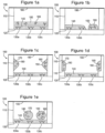
- Figures 1a-1e show non-scale schematic views of several embodiments of well 100 of a multi-well plate.
- the well is defined by well floor 120 and well walls 110.
- Floor 120 and walls 110 may be formed of a single contiguous material or may be separate components (e.g., a plate top and plate bottom) that are mated together.
- Well 100 also contains a first dry reagent 130 located on floor 120 that, as shown, may be one or more capture reagents that are immobilized on floor 120 to form a binding surface.
- First dry reagent 130 may include a plurality of immobilized capture reagents (e.g., reagents 130a, 130b, and 130c) that are patterned into a plurality of discrete binding domains (e.g., an array).
- immobilized capture reagents e.g., reagents 130a, 130b, and 130c
- discrete binding domains e.g., an array
- the binding reagents/domains may have different affinity or specificity for binding partners; such binding domains may be used to carry out multiplexed array-based measurements.
- a reconstitutable protective layer 140 covers dry reagent 130. Protective layer 140 may be omitted, e.g., when it is not required to physically separate reagents 130 and 150.
- Well 100 also comprises a second dry reagent 150 that is a reconstitutable dry reagent.
- Second dry reagent 150 may comprise a detection reagent such as labeled detection reagent 160.
- second dry reagent 150 comprises a plurality of detection reagents that differ in affinity or specificity for binding partners.
- Well 100 may also include an, optional, additional reconstitutable dry reagent 170 that comprises an assay control analyte 180 (as shown in Figures 1c-1e ). Also shown is plate seal 190. Plate seal 190, which may be omitted, is sealed against the top surface of walls 110 to protect the dry reagents from the environment.
- Figure 1a shows an embodiment in which first dry reagent 130 is coated with reconstitutable protective layer 140.
- Second dry reagent 150 is layered onto of protective layer 140 which prevents second dry reagent 150 from contacting first dry reagent layer 130.
- second dry reagent 150 is deposited by dispensing it in liquid form on protective layer 140; protective layer 140 is chosen to have enough thickness or mass such that it can adsorb this liquid without allowing it to contact dry reagent 130.
- the liquid is then dried to form second dry reagent 150.
- protective layer 140 is introduced in liquid form and frozen in the well to form a first frozen layer.
- Reagent 150 is then introduced in liquid form and frozen as a second frozen layer over the first frozen layer. Lyophilization of the two frozen layers provides the layered dry reagent structure.
- Figure 1b shows an embodiment where reagents 130 and 150 are both fixedly located on non-overlapping regions of floor 120. Additional dry reagents, such as assay control reagents (not shown), could be located on other non-overlapping regions of floor 120. The localization of reagents on selected regions of floor 120 may be carried out using standard techniques in patterned reagent deposition or dispensing.
- floor 120 has relatively hydrophilic domains surrounded by relatively hydrophobic areas such that appropriate volumes of reagents dispensed on the hydrophilic domains will spread to defined boundaries determined by the hydrophobic areas.
- reconstitutable dry reagents are located on a surface, one may pre-treat the surface with blocking agents to prevent adsorption of the reagents to the surfaces and/or include blocking agents in the reagent composition.
- Figure 1c shows an embodiment where second dry reagent 150 is fixedly located, as one or more dry reagent pills, on walls 110.
- the pills may be formed, e.g., by dispensing one or more droplets of the reagent (in liquid form) on walls 110 and drying them to form the dry reagent pills.
- Figure 1c also shows optional additional dry reagent 170 with control analyte 180 fixedly located on another non-overlapping region of walls 110.
- Figure 1d shows an embodiment that is like that shown in Figure 1c except that reagents 150 and 170 are located on shelves 115 on walls 110.
- Dry reagents 150 and 170 may be formed from liquid reagents by dispensing and drying them on shelves 115 or dispensing them above shelves 115 so that they run down walls 110 onto shelves 115 where they are dried. Alternatively, free-standing dry reagent pills may be placed on shelves 115.
- Figure 1e shows an embodiment where reagent 150 and optional reagent 170 are free standing dry reagent pills. Also included are embodiments of well 100 in which there is some combination of reconstitutable dry reagents on the well floor, well walls, well shelves, and/or in free-standing form. In alternate embodiments, some combination of fixedly located and free standing reconstitutable dry reagents is employed.
- the multi-well plates include those having wells with multiple, physically-distinct, dry reagents.
- the dry reagents may include indicators (such as dyes or fluorophores) that can be used in optical inspection of the plates.
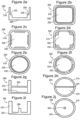
- Figure 2 shows non-scale schematic views of several embodiments of wells that have shelf elements on which liquid reagents can be held and dried and/or on which free-standing dry reagents may be supported above the well bottom.
- the shelf elements include ledges, bridges or tables as described below.
- Figure 2a is a cross-section of a well 200 showing well bottom 200 and well wall 210, the well wall having ledges such as ledges 230 and 235 that can support dry reagents.
- Ledge 230 has an angle that is substantially 90° or less than 90° relative to the wall directly above the ledge such that an appropriate volume of reagent can be dispensed on ledge 230 and accumulate on ledge 230 without overflowing onto well bottom 200.
- the ledges also have additional features to help contain reagents such as lip 240 on ledge 235.
- h s is greater than or equal to 0.02 h w , 0.05 h w or 0.1 h w but less than or equal to 0.1 h w , 0.25 h w or 0.5 h w .
- h s is greater or equal to about 0.1 mm, 0.2 mm, 0.5 mm, or 1 mm but less than or equal to about 1 mm, 2 mm, or 5 mm.
- shelf height and sample volume are chosen such that addition of sample to the well provides a height of liquid that contacts reagents on the bottom of the well and also reconstitutes reagents on one or more shelves.
- shelf height may be chosen so that addition of defined volume of a first liquid contacts dry reagents on the bottom of the well (reconstituting reconstitutable reagents on the bottom and/or allowing reactions to proceed involving reagents stored on the bottom) but does not reach the height of one or more shelves.
- Reactions involving reagents on the shelves can be commenced at a later time by adding sufficient volume of a second liquid so that the liquid level reaches the height of the shelves so as to reconstitute dry reagent on the shelves.
- the sample to be measured may be the first liquid, second liquid or both.
- Figures 2b-2f show top views of several embodiments of well 200 and show that the well openings may have a variety of shapes including, but not limited to, square ( Figures 2b-2d ) and round ( Figures 2e-2f ).
- the shelf elements may extend around the perimeter of the well as in Figures 2b and 2e or there may be one or more isolated shelf elements that only extend partially around the well as in Figures 2c-2d and 2f .
- a well may also include a plurality of shelf elements at different heights within a well.
- Figures 2g-2h show cross-section and top views, respectively, of a well 290 in which a shelf element is provided by bridge 250 that extends across the well.
- Figures 2i-2j show cross-section and top views, respectively of a well 295 in which a shelf element I provided by a table 260 that extends vertically from an area of well bottom 220.
- a multi-well plate comprising a plate body with a plurality of wells defined therein including: a) a plurality of first reagent wells holding a reconstitutable first dry reagent and b) a plurality of second reagent wells holding a second dry reagent (which may be a reconstitutable dry reagent or an immobilized reagent), wherein, the first and second reagents are matched reagents for conducting an assay (i.e., they are both used in conducting an assay of interest).
- the reagents may be located in a variety of locations with the wells such as well bottom, well walls, on shelf elements, as free-standing pills or powders, etc.
- a method for carrying out assays in these plates comprising: a) adding a sample to one of the first reagent wells, b) reconstituting reconstitutable dry labeled detection reagents in the first reagent well to form a reaction mixture, c) transferring an aliquot of the reaction mixture to one or more of the second reagent wells, and d) incubating the reaction mixture in the second reagent well(s) so as to carry out said assay on said sample.
- the multi-well assay plate can be divided into a plurality of sets of wells consisting of one first reagent well and one or more second reagent wells and the method further comprises repeating the process of (a)-(d) for each set of wells.
- Figure 3a is a (not to scale) schematic illustration of one embodiment showing cross-sectional views of two wells of a multi-well plate 300.
- Well 302 is a reagent reconstitution well comprising one or more reconstitutable dry reagents which may include a labeled detection reagent (such as dry reagent 350 comprising labeled detection reagent 360) or a an assay control analyte (such as dry reagent 370 comprising assay control analyte 380).
- These dry reagents may include additional reagent components such as blocking agents, stabilizers, preservatives, salts, pH buffers, detergents, bridging reagents, ECL coreactants and the like.
- Well 301 is a detection well comprising one or more dry reagents which may include reconstitutable dry reagents or an immobilized dry reagent. As shown, well 301 comprises immobilized capture reagents 330 that are patterned into three binding domains 330a, 330b, and 330c to form a binding surface. Well 301 also comprises a reconstitutable protective layer 340 which may be omitted. In one embodiment of an assay, sample is added to the reagent reconstitution well where reconstitutable dry reagents are reconstituted.
- FIG. 3a also shows plate seal 390 which seals against the openings of wells 301 and 302 to protect the contents of the wells from the environment.
- the detection and reagent reconstitution wells in a multi-well plate may be grouped into a plurality of assay sets consisting of one reagent reconstitution well and one or more detection wells, the reagent reconstitution well and detection wells within a set comprising matched capture and detection reagents for measuring an analyte of interest.
- Figure 3b shows an arrangement where a set has one reagent reconstitution wells 302 and three detection wells 301. During an assay, a sample added to well 302 may then be distributed among the three associated detection wells 301 so as to conduct multiple replicates or, where the detection wells hold different reagents, multiple different assays.
- Figure 3c shows an arrangement where a set has one reagent reconstitution well 302 and one detection well 301.
- Reagent reconstitution wells and detection wells may be similar in size and shape or may have different sizes and/or shapes.
- the wells in a standard multi-well plate are divided between the two types of wells.
- Figure 4 shows a non-scale schematic views of an alternative arrangement of wells.
- Figure 4a shows a top view of multiwell plate 400 having detection wells 440 that are arranged in a regular two dimensional pattern and that have detection wells walls 430 with inner wall surfaces and outer wall surfaces.
- Multi-well plate also has reagent reconstitution wells 460 in interstitial spaces between detection wells.
- Reagent reconstitution wells 460 have well walls that are defined by the outer well surfaces of detection well walls 430 and rib elements 450 that connect the outer surfaces of well walls 430 of adjacent detection wells (and, in the outermost of the wells, by the inner surface of plate frame wall 410).
- the detection wells may be shaped to have no reentrant (i.e., inward pointing) curves or angles while the interstitial wells may have reentrant curves and/or angles.
- Figure 4b shows a cross-sectional view along the dotted line in Figure 4a and shows the bottom surfaces of the two types of wells (which may be at different heights in the plate body).
- Plate 400 may be formed from a single contiguous material.
- plate 400 is formed from a plate top 405 and a plate bottom 420 that are mated along the dotted line shown in Figure 4b .
- the basic arrangement of arrays of round wells with interstitial wells defined by the well walls and rib elements is a common feature of many injection-molded 96-well plates and plate tops and allows these components to be used to form dry reagent plates as shown in Figure 4 .
- a multi-well plate comprising a) a plate body with a plurality of wells defined therein including: i) a plurality of assay wells comprising a dry assay reagent; and ii) a plurality of desiccant wells comprising a desiccant, and b) a plate seal sealed against said plate body thereby isolating said plurality of wells from the external environment.
- the assay wells comprise the necessary reagents for conducting an assay in the assay well.
- the desiccant wells are connected by drying conduits to the assay wells, the conduits permitting diffusion of water vapor from the assay wells to the desiccant wells but intersecting the wells at a height in the assay well above the location of the dry assay reagent.
- the plates themselves i.e., without dry reagents and desiccants
- plates having conduit or channel elements e.g., as shown in Figure 5 described below
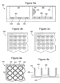
- Figure 5 shows non-scale schematic views of a multi-well plate 500 having assay wells 501 and desiccant wells 502 (desiccant and dry reagents are not shown).
- Figure 5a is a top view showing well walls 510 and conduits 508 connecting dessicant wells with one (e.g., as in row A) or more assay wells (e.g., as in row B).
- Figure 5b shows a cross-sectional view along the dotted line in Figure 5a and together with Figure 5a shows how conduits 508 may be formed by sealing plate seal 515 against channels in the top surface of the plate body.
- Plate seal 515 seals against these channels and the tops of the wells to form sets of assay and dessicant wells that are interconnected by conduits but are isolated from the environment and from other sets of wells. Accordingly, one or more sets of wells may be unsealed and used in an assay and the remaining sets of wells will be maintained in a dry environmentally protected environment.
- Plate 500 may be formed from a single contiguous material. In an alternate embodiment, plate 500 is formed from a plate top 505 and a plate bottom 512 that are mated along the dotted line shown in Figure 5b , plate bottom 512 defining the floor of at least some of the wells.
- the assay wells or sets of wells in plate 500 may include one or more of any of the dry reagent-containing wells described above, for example, in the descriptions of Figures 1-4 and may include both detection wells and reagent reconstitution wells.
- the desiccants used in the desiccant well may be selected from known desiccant materials including, but not limited to, silica, activated alumina, activated clays, molecular sieves and other zeolites, hydroscopic salts (e.g., anhydrous calcium sulfate, magnesium sulfate, sodium sulfate, sodium hydroxide and lithium chloride), hydroscopic solutions (e.g., concentrated solutions of lithium chloride) and water reactive materials such as phosphorous pentoxide.
- the desiccant is present as a free dry powder or granular material. In other embodiments, the desiccant is present as a dry pill, for example a pressed tablet or a desiccant impregnated polymeric material. In other embodiments, the desiccant is contained in a water vapor permeable bag or container (e.g., as in commercial silica pouches).
- desiccant in pill form or pre-packaged containers may be "press fit" into desiccant wells to prevent movement in the well during shipping or use.
- Figures 5c-5d show top and cross-sectional views of one embodiment of a multi-well plate 520 with assay and desiccant wells.
- Plate 520 has assay wells 521 (which may contain dry assay reagents) that are arranged in a regular two dimensional pattern and that have assay well walls 523 with inner wall surfaces and outer wall surfaces. It also has desiccant wells 522 in interstitial spaces between detection wells (alternatively, wells 521 are used as desiccant wells and wells 522 are used as assay wells).
- Desiccant wells 522 have well walls that are defined by the outer well surfaces of detection well walls 523 and rib elements 525 that connect the outer surfaces of well walls 523 of adjacent assay wells (and, in the outermost of the wells, by the inner surface of plate frame wall 526). Channels 524 notched into the top of well walls 523 provide, when mated to a plate seal, paths for water vapor to travel from assay wells to desiccant wells. As shown, the assay wells may be shaped to have no reentrant (i.e., inward pointing) curves or angles while the interstitial wells may have reentrant curves and/or angles.
- Figure 5d shows a cross-sectional view along the dotted line in Figure 5c and shows plate seal 527 which is mated to the top of the plate to form sets of assay and desiccant wells that are connected via conduits 524 but isolated from other wells and from the environment.
- Plate 520 may be formed from a single contiguous material. In an alternate embodiment, plate 520 is formed from a plate top 528 and a plate bottom 529 that are mated along the dotted line shown in Figure 5d .
- FIG. 5e shows a schematic view of another embodiment of a multi-well plate with assay wells (which may contain dry reagents) and desiccant wells and shows a plate 540 with assay wells 541 and desiccant wells 543 that are connected into sets of wells via channels 542 in the plate body.
- Multi-well plate 540 is largely analogous to the embodiment of plate 500 pictured in Figures 5a-5b except that in plate 540, desiccant wells 542 are much shallower and smaller in area than the assay wells allowing a larger portion of the plate footprint to be dedicated to wells used in assay measurements.
- Figure 5f shows a cross-sectional view along the dotted line in Figure 5e and also shows plate seal 544 that is sealed against the top of the plate to form connected sets of assay and desiccant wells.
- Plate 540 may be formed from a single contiguous material. In an alternate embodiment, plate 540 is formed from a plate top 545 and a plate bottom 546 that are mated along the dotted line shown in Figure 5f , plate bottom 546 also defining the floor of assay wells 541.
- Multi-well assay plate 600 comprises a plate top 610 with through-holes 615 that define the walls of wells. Plate top 610 is sealed against plate bottom 620 through gasket 625 such that plate bottom 620 defines the bottom surface of the wells. Optionally, plate top 610 is sealed directly to plate bottom 620 and gasket 625 is omitted. Sealing may be accomplished through traditional sealing techniques such as adhesives, solvent welding, heat sealing, sonic welding and the like. In another optional embodiment, plate top 610 fully defines the sides and bottom of the wells and plate bottom 620 and gasket 625 may be omitted.
- the contents of the wells may be protected from the outside environment by sealing (e.g., via traditional sealing techniques) plate seal 630 to plate top 610 directly or via optional gasket 635.
- plate 600 may be made from a variety of different materials including, but not limited to, plastics, metals, ceramics, rubbers, glasses or combinations thereof. In accordance with the requirements of the particular detection technology used with the plates, the components some or all of the components may be selected to be transparent, colored, opaque, or highly light scattering.
- plate top 610 is an injection-molded plastic such as injection-molded polystyrene, polypropylene, or cyclic olefin copolymer (COC).
- one or more of the components may be made of or comprise (for example in the form of a coating) a material that has a low water vapor transmission rate, e.g., a water vapor transmission rate less than 1 g/m 2 per day through a 100 um thickness.
- Low water vapor transmission materials include, but are not limited to, glass, metals or metal films (e.g., aluminum films), COC, polyvinylidene chloride (PvDC), polypropylene, polychlorotrifluoroethylene (PCTFE), and liquid crystal polymers (LCP).
- Plate 600 may include desiccant wells as described above. Alternatively, or in addition, desiccant may be incorporated directly into plate top 610, plate bottom 620, plate seal 630, gasket 625 and or gasket 635.
- desiccant may be incorporated directly into plate top 610, plate bottom 620, plate seal 630, gasket 625 and or gasket 635.
- U.S. Patent 6,174,952 to Hekal et al. describes desiccant containing polymer blends that may be molded, cast into liners, or formed into films, sheets, beads or pellets.
- plate bottom 620 has features to facilitate the patterning of reagents on the bottom of wells (e.g., patterned hydrophilic features surrounded by hydrophobic areas) and/or conductive layers that provide electrodes that are exposed to the interior volumes of the wells of plate 600 so that electrochemical or electrode induced luminescence assays (e.g., electrochemiluminescence assays) may be carried out.
- Plate bottom 620 may also include electrode contacts to allow an external instrument to apply electrical potential/current to the electrodes. Suitable approaches, configurations and compositions for such features, conductive layers and electrode contacts include those described in U.S. Publications 2004/0022677 and 2005/ 0052646 to Wohlstadter et al.
- Suitable instrumentation and methods that can be used to conduct ECL measurements using assay modules include those described in U.S. Publications 2004/0022677 and 2005/0052646 of U.S. Applications 10/185,274 and 10/185,363 , respectively; U.S. Publication2003/0113713 of U.S. Application 10/238,391 ; U.S. Publication 2005/0142033 of U.S. Application 10/980,198 ; and the concurrently filed U.S. Application 11/642,968 of Clinton et al. entitled "Assay Apparatuses, Methods and Reagents .”
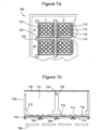
- Figure 7 provides schematic illustrations of one specific embodiment that includes some of the inventive concepts disclosed above in the context of a multi-well plate configured to carry out array-based multiplexed electrochemiluminescence assays.
- Figure 7a shows a section of multi-well plate 700 that has a plurality of assay wells 710 which may comprise dry reagents and a plurality of desiccant wells 720 which may comprise a desiccant. Channels 725 on the top surface of plate 700 link each desiccant well to an assay well. Optionally, desiccant wells 720 and channels 725 are omitted.
- Assay wells 710 have ledges 712 which may be used to support a reconstitutable dry reagent (e.g., dry reagents comprising assay controls and/or ECL labeled detection reagents). Assay wells also have working electrode surfaces 714 which are covered by patterned dielectric layer 716 so as to expose a plurality of exposed electrode surfaces or “spots" (shown as circles within the wells). In addition, counter electrodes 718 are provided to provide for a complete electrochemical circuit. Optionally, the surface of dielectric layer 716 is hydrophobic relative to electrode surface 714 so that small volumes of reagents patterned onto the spots may be kept confined to the spots.
- a reconstitutable dry reagent e.g., dry reagents comprising assay controls and/or ECL labeled detection reagents.
- Assay wells also have working electrode surfaces 714 which are covered by patterned dielectric layer 716 so as to expose a plurality of exposed electrode surfaces or “
- the different spots may have different capture reagents immobilized thereon to form a binding surface with an array of binding domains differing in specificity or affinity for binding partners (e.g., analytes of interest).
- some of the spots may have reconstitutable dry reagents confined thereon which, e.g., may contain assay controls and/or ECL labeled detection reagents.
- the assay well may further comprise a reconstitutable protective layer over the binding surface.
- Figure 7b provides an exploded cross-sectional view along the dotted line in Figure 7a and illustrates one approach to forming the electrode/dielectric layers in assay wells 710.
- the multi-well plate comprises a plate top 730 that defines desiccant wells 720 and has through-holes that define the walls of assay wells 710 and ledges 712.
- Plate top 730 also has channels 725 that form conduits between assay wells 710 and desiccant wells 720 when plate seal 750 is sealed against the top surface of plate top 730.
- plate top 730 is an injection-molded part molded from a plastic with low water vapor transmission.
- plate seal 750 is a heat sealable film comprising a low water vapor transmission plastic or a metal (e.g., aluminum) foil.
- Figure 7b also shows plate bottom 740 which seals against plate top 730 and defines the bottom of assay wells 710.
- Plate bottom 740 comprises substrate 715 which supports patterned conductive layers that provide for electrodes 714 and 718. Patterned dielectric layer 716 on the electrodes defines the exposed electrode spots.
- a variety of materials may be used to provide for the substrate and the conductive and dielectric layers (see, e.g., U.S. Publications 2004/0022677 and 2005/0052646 ).
- the substrate is a plastic film (made, e.g., of a polyester such as MYLAR, polyvinylchloride, or a low water vapor transmissive material such as COC), the conductive layers are screen printed conducting inks (e.g., screen printed carbon inks) and the dielectric layer is a screen printed insulating ink.
- electrode contacts 780 and 785 which are conductive layers on the bottom of substrate 715 that provide connectivity (e.g., via conductive through holes in substrate 715 to electrodes 714 and 718.
- the electrode contacts may also be provided by screen printed conductive inks which during printing can be caused to fill holes in substrate 715 to also provide the conductive through-holes.
- the conductive through-holes may be located directly below well walls to limit water vapor transmission through the holes.
- an optional bottom sealing layer 790 may be sealed to the bottom of substrate 715.
- Bottom sealing layer 790 is made of a low water vapor transmissive material and covers most of the bottom surface of substrate 715 except for defined openings in sealing layer 790 that are located so as to allow a plate reading instrument to contact electrode contacts 780 and 785.
- Figure 7c shows a more detailed angled view of one embodiment of plate 700 and shows desiccant pills 722 that are press-fit into desiccant wells 720.
- samples which may contain an analyte or activity of interest may be assayed.
- a sample is introduced to an assay plate or one or more wells of an assay plate having reconstitutable dry reagents pre-loaded thereon, thus reconstituting these assay reagents and an assay signal is measured so as to measure (quantitatively or qualitatively) the amount of analyte in the sample.
- the reagents may include a luminescent, electrochemiluminescent, chemiluminescent, and/or redox-active substance.
- the assay signal is preferably a luminescent or electrochemical signal.
- Assays formats that may be carried out include homogeneous and heterogeneous methods.
- Assays that may be carried out include formats that employ solid-phase supports so as to couple the measurement of an analyte or activity to the separation of labeled reagents into solution-phase and solid phase supported portions.
- Examples include solid-phase binding assays that measure the formation of a complex of a material and its specific binding partner (one of the pair being immobilized, or capable of being immobilized, on the solid phase support), the formation of sandwich complexes (including a capture reagent that is immobilized, or capable of being immobilized, on the solid phase support), the competition of two competitors for a binding partner (the binding partner or one of the competitors being immobilized, or capable of being immobilized, on the solid phase support), the enzymatic or chemical cleavage of a label (or labeled material) from a reagent that is immobilized, or capable of being immobilized on a solid phase support and the enzymatic or chemical attachment of a label (or labeled material) to a reagent that is immobil
- the term "capable of being immobilized” is used herein to refer to bridging reagents that may participate in reactions in solution and subsequently be captured on a solid phase during or prior to detection.
- the reagent may be captured using a specific binding partner of the reagent that is immobilized on the solid phase.
- the reagent is linked to a capture moiety and a specific binding partner of the capture moiety is immobilized on the solid phase.
- useful capture moiety-binding partner pairs include biotin-streptavidin (or avidin), antibody-hapten, receptor-ligand, nucleic acid - complementary nucleic acid, etc.
- the amount of analyte or activity may be determined by measuring the amount of label on the solid phase support and/or in solution using i) a surface selective technique, ii) a solution selective technique and/or iii) after separation of the two phases.
- the solid phase support may also be the working electrode used to induce electrochemiluminescence from labels bound to the solid phase.
- the electrochemiluminescence methods may include washing to remove unbound electrochemiluminescence labeled reagents prior to addition of an ECL coreactant (e.g., tertiary amines such as tripropylamine or piperazine-1,4-bis(2-ethanesulfonic acid)) and application of a potential to induce ECL from bound labels.
- an ECL coreactant e.g., tertiary amines such as tripropylamine or piperazine-1,4-bis(2-ethanesulfonic acid
- the method may be run without washing.
- the ECL coreactant may be pre-added to assay wells in the form of a reconstitutable dry reagent or protective layer.
- kits for use in conducting assays that comprise the assay modules/multi-well plates.
- the kit may include one or more additional reagents in one or more containers including, but not limited to, assay calibrators, assay controls, assay diluents, ECL coreactants, and wash buffers.
- the kit comprises one or more of the assay components in one or more plate wells, preferably in dry form.
- the kit comprises an assay plate having a binding immobilized on one or more working electrodes within an assay module and one or more additional assay reagents deposited in the form of a dry bead, pellet or a pill directly into the well, preferably at a position spacially separated from a working electrode, or alternatively deposited into one or more interstitial wells.
- the kits do not contain any liquids in the wells.
- Labeled detection antibodies were labeled with SULFO-TAG NHS ester (Meso Scale Discovery, Gaithersburg, MD), an electrochemiluminescent label based on a sulfonated derivative of ruthenium-tris-bipyridine (compound 1 pictured below). Labeled antibodies were purified by size exclusion chromatography on SEPHADEX G-50 (Pharmacia).
- Pills comprising one or more labeled detection antibodies were formed from a solution containing 1 ⁇ g/mL of each of the labeled antibodies, 2% bovine serum albumin and 20% sucrose in a phosphate buffered saline. Frozen droplets of this solution were formed by dispensing 20 ⁇ L droplets into liquid nitrogen. The frozen droplets were transferred onto chilled ( ⁇ -78°C) aluminum trays which were placed on the shelves of an ADVANTAGE XL lyophilizer (Virtis). The shelves of the lyophilizer were pre-cooled to ⁇ -45°C prior to introduction of the aluminum trays and a conductive paste was used to improve the heat transfer between the shelves and the trays containing beads.
- the lyophilizer chamber was evacuated and the shelf temperature was slowly increased to -30°C, -20°C, -15°C and finally to +20°C (ambient conditions) over the course of about 24 hours. The temperature was held at each of these levels for sufficient time to allow for equilibration while controlling the chamber pressure 0.01 torr.
- Karl Fisher titrations of lyophilized beads typically showed water contents of less than 4% by weight. The water content could be reduced to under 2% by extended storage in the presence of a dessicant.
- Electrochemiluminescence measurements were carried out using specially designed multi-well plates having integrated screen printed carbon ink electrodes for carrying out electrochemiluminescence measurements (MULTI-ARRAY or MULTI-SPOT plates, Meso Scale Discovery, a division of Meso Scale Diagnostics, LLC, Gaithersburg, MD).
- a patterned dielectric layer patterned over the working electrode on the bottom of each well exposes one or more regions or “spots" on the working electrode.
- the electrode surfaces were treated with an oxygen plasma prior to immobilizing antibodies on them.
- Different capture antibodies were immobilized on the different spots by patterned microdispensing of solutions of the antibodies on the spots using a nanoliter dispenser (Bio-Dot, Inc.).
- the volumes dispensed on the spots were selected so that they spread to the boundaries defined by the dielectric layers but remained confined on the spots, thus allowing for the immobilization of each antibody (via passive adsorption) on a defined region of the working electrode; if the electrode surfaces were not plasma treated, a small amount of TRITON X-100 detergent was added to the spotting solutions to enhance spreading. Adsorption was allowed to proceed for at least 2 hours after which the plates were washed with a stabilizing wash buffer (2% sucrose, 185 mM dibasic ammonium phosphate, 13 mM monobasic ammonium phosphate, 0.1% TWEEN 20, and KATHON CG/ICP II preservative), dried, and stored in the presence of a desiccant. By controlling the amount of wash buffer left in the wells before drying (typically between 5-20 ⁇ L), sucrose films of different thicknesses could be left over the working electrode surfaces.
- a stabilizing wash buffer 2% sucrose, 185 mM dibasic
- Electrochemiluminescence was induced and measured in the MULTI-SPOT plates using a Sector ® Imager 6000 reader or a Sector ® PR 400 reader (both from Meso Scale Discovery, a division of Meso Scale Diagnostics, LLC, Gaithersburg, MD).
- the Sector ® Imager 6000 instrument applies electrical potentials to the working electrodes in the plate and images the resultant ECL. Image analysis algorithms are employed to distinguish and quantitated the light emitted from each spot in a well.
- the Sector ® PR 400 instrument applies electrical potential to the working electrodes in one column of the plate at a time. An array of photodiodes is used to measure the ECL emitted from the wells in the column.
- High binding MULTI-SPOT plates having a 7 spot array of capture antibodies against seven different human cytokines (TNF- ⁇ , IL1- ⁇ , IL2, IL5, IL6, IL8, IL12, and GM-CSF) and lyophilized beads containing labeled detection antibodies against the same seven cytokines were prepared as described above. One bead was placed in each well and the plates were stored in the presence of dessicant until used. Multiplexed cytokine assays were carried out by introducing cytokine solutions (40 ⁇ l per well prepared in RPMI cell culture media supplemented with 10% fetal calf serum) of predefined concentrations into the wells of the plate and incubating for 2 hours at room temperature on a plate shaker.
- cytokine solutions 40 ⁇ l per well prepared in RPMI cell culture media supplemented with 10% fetal calf serum
- MSD ® READ BUFFER P (Meso Scale Discovery)
- MSD ® READ BUFFER P (Meso Scale Discovery)
- This assay used a small spot MULTI-ARRAY plate with a single spot per well.
- the spot treated, as described in the Materials and Methods section with a solution containing an anti-human TNF- ⁇ capture antibody to immobilize the antibody on the spot surface.
- the well was then filled with 75 ⁇ L of 4x MSD ® READ BUFFER P that was supplemented with 7% FICOLL (a highly branched hydrophilic polymer of sucrose), and the plate was cooled to freezing and lyophilized overnight to provide a protective "cake" layer over the bottom of the well.
- a small droplet (35 nL) of a concentrated solution of a labeled anti-human TNF- ⁇ detection antibody was dispensed on the surface of the cake.
- the plate was then vacuum dried for 5 minutes and stored in the presence of dessicant until used. Assays were carried out by adding to the wells 150 ⁇ L of solutions containing pre-determined concentrations of human TNF- ⁇ in RPMI cell culture media supplemented with 10% fetal calf serum and shaking for two hours. The plate was then analyzed on a Sector ® Imager 6000 instrument. The calculated detection limits of 5-6 pg/mL are comparable to those observed in non-washed assays using liquid detection antibody solutions.
- This assay used a small spot MULTI-ARRAY plate with a single spot per well.
- the spot was coated, as described in the Materials and Methods section with an anti-human TNF- ⁇ capture antibody.
- Droplets (1 ⁇ L) of a 24 ⁇ g/mL solution of the detection antibody in 4.8% sucrose were dispensed on the inside walls of the wells and allowed to dry. The plates were stored in the presence of dessicant until used in an assay.
- the assay protocol involved adding 80 ⁇ L of a TNF- ⁇ solution to each well, shaking the plate for 30 minutes at room temperature, washing the plate, adding 150 ⁇ L of 1x MSD ® READ BUFFER T (Meso Scale Discovery) and analyzing the plate on a Sector ® Imager 6000 instrument. Plates stored for 18 days at room temperature or 4°C gave detection limits that were less than 1 pg/mL and comparable to those observed in washed assays employing liquid detection antibody solutions.
- This assay used a MULTI-ARRAY plate with a single spot per well.
- the working electrode in each well was pre-coated with streptavidin (streptavidin MULTI-ARRAY plate, Meso Scale Discovery).
- Anti-IL1- ⁇ monoclonal antibody was immobilized on the working electrode according to the following protocol.
- the wells were washed three times with PBS and then treated with 20 ⁇ L of a 3 ⁇ g/mL solution of biotin-labeled anti-IL 1- ⁇ .
- the immobilization was allowed to proceed over 2 hours under agitation on a plate shaker.
- the wells were then washed three times with PBS.
- a 20 ⁇ L volume of a buffered solution of B SA and sucrose was added to the wells and then dried in the wells under vacuum to form a dry film on the bottom of the plates.
- a dry pill of SULFO-TAG labeled anti-IL 1- ⁇ polyclonal antibody was formed on the well wall according to the following protocol.
- a 100 nL microdroplet of a 482 ⁇ g/mL solution of the labeled antibody was dispensed on each well wall using a BIO-DOT microdispensor (Bio-Dot, Inc.) with an angled tip.
- the droplet remained on the well wall where it was allowed to dry for 30 minutes in a dessicator chamber.
- the wells were then sealed with a plate heat seal.
- a low concentration of fluorescein was added to the detection antibody solution.
- the fluorescein fluorescence could be used to provide a quality control check by identifying any well in which the detection antibody ran down the well wall or splattered on the well bottom. The fluorescein did not affect assay performance.
- Assays for IL1- ⁇ were carried out by adding 125 ⁇ L of solutions containing known amounts of IL1- ⁇ to the wells and incubating for 37 minutes while shaking the plate. The plate was then washed with PBS, MSD READ BUFFER T was added and the plate was analyzed on a Sector ® PR 400 instrument. Assays using plates with dry detection antibodies performed in a comparable fashion to assays that used liquid detection antibody solutions.
- This assay used a Multi-Spot plate configured as shown in Figure 8 .
- the plate was similar to that shown in Figure 7 except for the use of a 7-spot pattern and the omission of desiccant wells 720, channels 725, desiccant pills 722, and bottom sealing layer 790.
- the plate top was injection-molded polypropylene. Capture antibodies against were immobilized by dispensing, on the individual spots, antibodies against botulinum toxin A (BotA), dinitrophenyl (DNP), ricin, staphylococcal enterotoxin B (SEB), decielen equine encephalitis (VEE), and Yersinia pestis (YP).
- BotA botulinum toxin A
- DNP dinitrophenyl
- SEB staphylococcal enterotoxin B
- VEE Yersinia pestis
- Non-immune mouse IgG was immobilized on the remaining spot for use as a negative control. Immobilization was carried out by dispensing 75 nL of solutions comprising between 100-500 ⁇ g/mL of an antibody, 750 ⁇ g/mL of BSA, and 0.03% TRITON X-100. One exception was the BotA capture antibody which was biotinylated and immobilized after pre-binding it to 1200 ⁇ g/mL avidin and which was immobilized in the absence of BSA.
- the non-immune IgG should not participate in a sandwich complex and should give a low signal for all samples. Elevation of this signal outside of a selected range can be used as an indication that a measurement artifact is producing elevated non-specific binding of detection antibodies and that there is a risk of false positive results.
- any binding reagent that is not paired with a corresponding detection reagent may be used.
- the binding reagent may be selected to share structural properties with the test capture reagents, for example, in an iimmunoassay it may include immunoglobulins from one or more of the species from which the other capture antibodies were derived.
- the anti-DNP spot will be used as a positive control.
- the well will also include a dry SULFO-TAG labeled anti-fluorescein (FL) antibody and a defined quantity of dry BSA labeled with both DNP and FL (DNP-FL-BSA).
- FL dry SULFO-TAG labeled anti-fluorescein
- DNP-FL-BSA dry BSA labeled with both DNP and FL
- the positive control signal should, therefore give a constant positive signal indicative of the defined quantity of DNP-FL-BSA. Reduction of this signal below a selected range can be used as an indication that a sample interferes with binding reactions or signal generation and that there is a risk of false negative results.
- the positive control may be an assay for any analyte that can be spiked into the reaction mixture. Preferably, there is a low likelihood of finding the analyte in the samples of interest.
- the capture antibody solutions were allowed to dry for 30 minutes in a desiccated environment and then dried for 30-60 minutes under vacuum.
- the wells were washed with a the stabilizing wash buffer containing sucrose described in the Materials and Methods section, blocked with 5% BSA for 45 minutes and washed once more with the stabilizing wash buffer.
- a stabilizing/blocking solution (20 ⁇ L of 305 mM ammonium phosphate, 100 mM ammonium chloride, 0.02% TRITON X-100, 2% sucrose, 2%BSA, and 0.02% KATHON preservative, pH 7.4) was added and the solution was dried in the well under vacuum to form a dry reagent cake on the well bottom.
- a mixture of STAG-labeled detection antibodies (0.5 ⁇ L of a mixture of antibodies against BotA, FL, ricin, SEB, VEE, and YP at between 40-240 ⁇ g/mL in the stabilizing/blocking solution) was dispensed (using a BIO-DOT dispenser with an angled dispense tip) on the walls of the wells just above the dry reagent ledge and allowed to flow down onto the ledge.
- a solution containing 80 ng/mL of the positive control analyte (DNP-FL-BSA) was dispensed on the opposite wall.
- the detection antibody and control solutions were allowed to dry for 30-60 minutes under vacuum. The plates were then packaged with desiccant until used.
- the protocol used to conduct assays with these plates was: add 80 ⁇ L of sample (defined amounts of one or more of the target analytes in 0.1% TRITON X-100 in phosphate buffered saline (PBS)), incubate 1 hour with shaking, wash with PBS, add 150 ⁇ L 1x MSD READ BUFFER T (Meso Scale Diagnostics, LLC) and analyze plate using anMSD Sector ® Imager 6000 instrument. VEE and YP used in this assay were inactivated by irradiation. BotA was used in the assay was inactivated with formalin.
- the table below shows that the signals observed at each spot for samples containing no analyte (-) or for samples containing 10 ng/mL BotA, 1 ng/mL ricin, 50 ng/mL SEB, 1000 ng/mL VEE, or 10,000 CFU/mL YP.
- the table shows sensitive and specific detection of the target analytes and proper performance of the positive and negative control spots.
- the next table compares the signals observed with these plates to assays carried out under comparable conditions except for the use of liquid detection reagents.
- the table provides only the signal on the specific spot for a given analyte and provides both signal in the presence of analyte (10 ng/mL BotA, 1 ng/mL ricin, 50 ng/mL SEB, 1000 ng/mL VEE, or 10,000 CFU/mL YP) and background signal in the absence of analyte.
- analyte 10 ng/mL BotA, 1 ng/mL ricin, 50 ng/mL SEB, 1000 ng/mL VEE, or 10,000 CFU/mL YP
- This assay used a MULTI-SPOT plate configured as shown in Figure 9 .
- the plate was similar to that shown in Figure 8 except for the inclusion of conduits 910 connecting pairs of adjacent wells.
- the conduits were provided by shallow notches that were cut into the walls separating adjacent wells.
- one well of each pair of wells was used to carry out a multiplexed immunoassay and the other was used to hold desiccant for maintaining the assay well in a dry state during storage.
- the assay used a multiplexed sandwich immunoassay with dry capture and detection reagents prepared as in Example 5.
- the capture antibodies were anti-Bacillus subtilis var.
- BG Niger
- anti-MS2 phage anti-FL
- anti-DNP mouse IgG
- mouse IgG mouse IgG
- the dry detection antibody pill included labeled anti-BG, anti-MS2, and anti-ovalbumin (Ova) (for detecting FL-Ova and DNP-Ova).
- the desiccant wells were filled with roughly 50 mg to 200 mg of silica gel or DRIERITE (calcium sulfate) desiccants and the plates were sealed with an aluminum foil heat seal. After sealing the foil seal to the plate top, the notches in the wells provided conduits between the sets of assay and desiccant wells. Some plates were prepared with no desiccant in the desiccant wells for comparison. The plates were kept at 4°C under dry conditions for several days to allow the dry reagents to fully dry. The plates were then exposed to elevated temperature and humidity for several days prior to using them to carry out measurements of the target organisms (using the assay protocol of Example 5).
- silica gel or DRIERITE calcium sulfate
- Figure 10 provides signals for samples containing defined amounts of the target analytes and compares the signals from plates with silica desiccant, calcium sulfate desiccant and no desiccant after exposure to 60% humidity at 30°C for 7 days. Signals are provided as a percentage of the signal obtained from a plate that was prepared at the same time as the others but that had been kept dry a 4°C for the 7 day period. The results indicate that the desiccant wells were very effective at improving the stability of the dry reagents to heat and humidity.
Landscapes
- Health & Medical Sciences (AREA)
- Chemical & Material Sciences (AREA)
- Life Sciences & Earth Sciences (AREA)
- Immunology (AREA)
- Engineering & Computer Science (AREA)
- Hematology (AREA)
- Molecular Biology (AREA)
- Biomedical Technology (AREA)
- Urology & Nephrology (AREA)
- General Health & Medical Sciences (AREA)
- Analytical Chemistry (AREA)
- Chemical Kinetics & Catalysis (AREA)
- Medicinal Chemistry (AREA)
- Physics & Mathematics (AREA)
- Pathology (AREA)
- General Physics & Mathematics (AREA)
- Biochemistry (AREA)
- Food Science & Technology (AREA)
- Cell Biology (AREA)
- Microbiology (AREA)
- Biotechnology (AREA)
- Clinical Laboratory Science (AREA)
- General Chemical & Material Sciences (AREA)
- Polymers & Plastics (AREA)
- Organic Chemistry (AREA)
- Investigating Or Analysing Materials By The Use Of Chemical Reactions (AREA)
- Apparatus Associated With Microorganisms And Enzymes (AREA)
- Measuring Or Testing Involving Enzymes Or Micro-Organisms (AREA)
Applications Claiming Priority (6)
| Application Number | Priority Date | Filing Date | Title |
|---|---|---|---|
| US75274505P | 2005-12-21 | 2005-12-21 | |
| US75251305P | 2005-12-21 | 2005-12-21 | |
| EP06846006.2A EP1963853B1 (en) | 2005-12-21 | 2006-12-21 | Assay modules having assay reagents and methods of making and using same |
| EP15193494.0A EP3032257B1 (en) | 2005-12-21 | 2006-12-21 | Assay modules having assay reagents and methods of making and using same |
| PCT/US2006/049048 WO2007076023A2 (en) | 2005-12-21 | 2006-12-21 | Assay modules having assay reagents and methods of making and using same |
| US11/642,970 US7807448B2 (en) | 2005-12-21 | 2006-12-21 | Assay modules having assay reagents and methods of making and using same |
Related Parent Applications (3)
| Application Number | Title | Priority Date | Filing Date |
|---|---|---|---|
| EP15193494.0A Division EP3032257B1 (en) | 2005-12-21 | 2006-12-21 | Assay modules having assay reagents and methods of making and using same |
| EP15193494.0A Division-Into EP3032257B1 (en) | 2005-12-21 | 2006-12-21 | Assay modules having assay reagents and methods of making and using same |
| EP06846006.2A Division EP1963853B1 (en) | 2005-12-21 | 2006-12-21 | Assay modules having assay reagents and methods of making and using same |
Publications (2)
| Publication Number | Publication Date |
|---|---|
| EP3431994A1 EP3431994A1 (en) | 2019-01-23 |
| EP3431994B1 true EP3431994B1 (en) | 2024-03-06 |
Family
ID=39639158
Family Applications (3)
| Application Number | Title | Priority Date | Filing Date |
|---|---|---|---|
| EP15193494.0A Active EP3032257B1 (en) | 2005-12-21 | 2006-12-21 | Assay modules having assay reagents and methods of making and using same |
| EP18191510.9A Active EP3431994B1 (en) | 2005-12-21 | 2006-12-21 | Assay modules having assay reagents and methods of making and using same |
| EP06846006.2A Active EP1963853B1 (en) | 2005-12-21 | 2006-12-21 | Assay modules having assay reagents and methods of making and using same |
Family Applications Before (1)
| Application Number | Title | Priority Date | Filing Date |
|---|---|---|---|
| EP15193494.0A Active EP3032257B1 (en) | 2005-12-21 | 2006-12-21 | Assay modules having assay reagents and methods of making and using same |
Family Applications After (1)
| Application Number | Title | Priority Date | Filing Date |
|---|---|---|---|
| EP06846006.2A Active EP1963853B1 (en) | 2005-12-21 | 2006-12-21 | Assay modules having assay reagents and methods of making and using same |
Country Status (12)
| Country | Link |
|---|---|
| US (5) | US7807448B2 (zh) |
| EP (3) | EP3032257B1 (zh) |
| JP (2) | JP5345854B2 (zh) |
| KR (1) | KR101489804B1 (zh) |
| CN (2) | CN102841195B (zh) |
| AU (1) | AU2006330913B2 (zh) |
| CA (3) | CA3179247A1 (zh) |
| DK (1) | DK1963853T3 (zh) |
| HK (1) | HK1128527A1 (zh) |
| IL (2) | IL192156A (zh) |
| MX (3) | MX2008008280A (zh) |
| WO (1) | WO2007076023A2 (zh) |
Families Citing this family (188)
| Publication number | Priority date | Publication date | Assignee | Title |
|---|---|---|---|---|
| CA2956645A1 (en) | 2003-07-12 | 2005-03-31 | David A. Goldberg | Sensitive and rapid biodetection |
| JP5897780B2 (ja) | 2005-01-28 | 2016-03-30 | デューク ユニバーシティ | プリント回路基板上の液滴操作装置及び方法 |
| EP1752220A1 (en) * | 2005-07-27 | 2007-02-14 | The Automation Partnership (Cambridge) Limited | Sample tube |
| CA3179247A1 (en) | 2005-12-21 | 2007-07-05 | Meso Scale Technologies, Llc | Assay modules having assay reagents and methods of making and using same |
| CN108931509B (zh) | 2005-12-21 | 2022-04-15 | 梅索斯卡莱科技公司 | 分析装置、方法和试剂 |
| US8492168B2 (en) * | 2006-04-18 | 2013-07-23 | Advanced Liquid Logic Inc. | Droplet-based affinity assays |
| US20140193807A1 (en) | 2006-04-18 | 2014-07-10 | Advanced Liquid Logic, Inc. | Bead manipulation techniques |
| US9476856B2 (en) | 2006-04-13 | 2016-10-25 | Advanced Liquid Logic, Inc. | Droplet-based affinity assays |
| US8637324B2 (en) | 2006-04-18 | 2014-01-28 | Advanced Liquid Logic, Inc. | Bead incubation and washing on a droplet actuator |
| US7439014B2 (en) | 2006-04-18 | 2008-10-21 | Advanced Liquid Logic, Inc. | Droplet-based surface modification and washing |
| US10078078B2 (en) | 2006-04-18 | 2018-09-18 | Advanced Liquid Logic, Inc. | Bead incubation and washing on a droplet actuator |
| US7727723B2 (en) * | 2006-04-18 | 2010-06-01 | Advanced Liquid Logic, Inc. | Droplet-based pyrosequencing |
| US8809068B2 (en) | 2006-04-18 | 2014-08-19 | Advanced Liquid Logic, Inc. | Manipulation of beads in droplets and methods for manipulating droplets |
| US9675972B2 (en) | 2006-05-09 | 2017-06-13 | Advanced Liquid Logic, Inc. | Method of concentrating beads in a droplet |
| WO2008098236A2 (en) | 2007-02-09 | 2008-08-14 | Advanced Liquid Logic, Inc. | Droplet actuator devices and methods employing magnetic beads |
| WO2008137008A2 (en) | 2007-05-04 | 2008-11-13 | Claros Diagnostics, Inc. | Fluidic connectors and microfluidic systems |
| CN103418295B (zh) | 2007-06-21 | 2015-11-18 | 简.探针公司 | 用于混合检测腔室的内容物的仪器和方法 |
| AU2008276024A1 (en) | 2007-07-16 | 2009-01-22 | California Institute Of Technology | Microfluidic devices, methods and systems for detecting target molecules |
| US8702938B2 (en) | 2007-09-04 | 2014-04-22 | Advanced Liquid Logic, Inc. | Droplet actuator with improved top substrate |
| EP2657699B1 (en) | 2007-10-02 | 2017-03-22 | Theranos, Inc. | Modular point-of-care devices and uses thereof |
| AU2008319245B2 (en) * | 2007-10-31 | 2014-01-23 | Janssen Pharmaceutica N.V. | Device and method for high throughput screening of crystallization conditions in a vapor diffusion environment |
| CA2709928A1 (en) | 2007-12-23 | 2009-07-09 | Advanced Liquid Logic, Inc. | Droplet actuator configurations and methods of conducting droplet operations |
| DE102008007352B4 (de) * | 2008-01-28 | 2010-04-22 | Prionics Ag | Vorrichtung zur Gewinnung, Konservierung und Lagerung von Probenmaterial mit einem Probenbehälter |
| CN108362681B (zh) * | 2008-04-11 | 2021-08-17 | 梅索斯卡莱科技公司 | 分析装置、方法和试剂 |
| US8852952B2 (en) | 2008-05-03 | 2014-10-07 | Advanced Liquid Logic, Inc. | Method of loading a droplet actuator |
| DE102008025992B4 (de) * | 2008-05-30 | 2011-01-27 | Siemens Healthcare Diagnostics Gmbh | Titerplatte und Verfahren zur Detektion eines Analyten |
| DE102008028908B3 (de) * | 2008-06-18 | 2009-12-31 | IfP Privates Institut für Produktqualität GmbH | Nachweis eines Analyten in einem wässrigen Medium |
| US8813955B2 (en) | 2008-06-19 | 2014-08-26 | Arzneimittel Gmbh Apotheker Vetter & Co. Ravensburg | Container for medical products and method for production of said container |
| WO2010006604A1 (en) * | 2008-07-16 | 2010-01-21 | Radiometer Medical Aps | High capacity solid phase |
| EP2163306A1 (en) * | 2008-09-12 | 2010-03-17 | F. Hoffmann-la Roche AG | Multi-well plate with tailored chambers |
| US8187864B2 (en) * | 2008-10-01 | 2012-05-29 | The Governing Council Of The University Of Toronto | Exchangeable sheets pre-loaded with reagent depots for digital microfluidics |
| WO2010087999A1 (en) * | 2009-02-02 | 2010-08-05 | Claros Diagnostics, Inc. | Structures for controlling light interaction with microfluidic devices |
| WO2010101323A1 (ko) * | 2009-03-06 | 2010-09-10 | Lee Yong-Jin | 멀티웰이 구비된 시료장착기 |
| AU2010281493B2 (en) * | 2009-07-27 | 2015-07-09 | Meso Scale Technologies, Llc | Assay apparatuses, consumables and methods |
| US9523701B2 (en) | 2009-07-29 | 2016-12-20 | Dynex Technologies, Inc. | Sample plate systems and methods |
| GB0913258D0 (en) | 2009-07-29 | 2009-09-02 | Dynex Technologies Inc | Reagent dispenser |
| US8926065B2 (en) | 2009-08-14 | 2015-01-06 | Advanced Liquid Logic, Inc. | Droplet actuator devices and methods |
| JP2011072970A (ja) * | 2009-10-01 | 2011-04-14 | Sumitomo Bakelite Co Ltd | 糖鎖捕捉物質を含むタブレットおよびその用途 |
| US9091649B2 (en) | 2009-11-06 | 2015-07-28 | Advanced Liquid Logic, Inc. | Integrated droplet actuator for gel; electrophoresis and molecular analysis |
| EP2516669B1 (en) | 2009-12-21 | 2016-10-12 | Advanced Liquid Logic, Inc. | Enzyme assays on a droplet actuator |
| JP2011137694A (ja) * | 2009-12-28 | 2011-07-14 | Tosoh Corp | 凍結乾燥試薬 |
| US8927502B2 (en) * | 2010-02-16 | 2015-01-06 | Icahn School Of Medicine At Mount Sinai | Embedded chimeric peptide nucleic acids and uses thereof |
| US10787701B2 (en) | 2010-04-05 | 2020-09-29 | Prognosys Biosciences, Inc. | Spatially encoded biological assays |
| CN103238055A (zh) * | 2010-10-07 | 2013-08-07 | 硅生物装置有限公司 | 基于磁性粒子的生物传感器 |
| US10114020B2 (en) | 2010-10-11 | 2018-10-30 | Mbio Diagnostics, Inc. | System and device for analyzing a fluidic sample |
| EP3644058A1 (en) | 2010-10-14 | 2020-04-29 | Meso Scale Technologies, LLC | Reagent storage in an assay device |
| SG189113A1 (en) * | 2010-10-25 | 2013-05-31 | Hoffmann La Roche | Use of signal enhancing compounds in electrochemiluminescence detection |
| TWI748368B (zh) | 2011-01-21 | 2021-12-01 | 美商拉布拉多診斷有限責任公司 | 樣本使用最大化之系統及方法 |
| US8718948B2 (en) | 2011-02-24 | 2014-05-06 | Gen-Probe Incorporated | Systems and methods for distinguishing optical signals of different modulation frequencies in an optical signal detector |
| US10254204B2 (en) | 2011-03-07 | 2019-04-09 | Accelerate Diagnostics, Inc. | Membrane-assisted purification |
| ES2551922T3 (es) | 2011-03-07 | 2015-11-24 | Accelerate Diagnostics, Inc. | Sistemas rápidos de purificación celular |
| GB201106254D0 (en) | 2011-04-13 | 2011-05-25 | Frisen Jonas | Method and product |
| WO2012154745A2 (en) | 2011-05-09 | 2012-11-15 | Advanced Liquid Logic, Inc. | Microfluidic feedback using impedance detection |
| US20130087467A1 (en) * | 2011-06-24 | 2013-04-11 | Hongjun Yang | High-performance analytical instrument and method |
| KR20140064771A (ko) | 2011-07-06 | 2014-05-28 | 어드밴스드 리퀴드 로직, 아이엔씨. | 비말 작동기 상의 시약 저장 |
| US9513253B2 (en) | 2011-07-11 | 2016-12-06 | Advanced Liquid Logic, Inc. | Droplet actuators and techniques for droplet-based enzymatic assays |
| WO2013016413A2 (en) | 2011-07-25 | 2013-01-31 | Advanced Liquid Logic Inc | Droplet actuator apparatus and system |
| US9268915B2 (en) | 2011-09-25 | 2016-02-23 | Theranos, Inc. | Systems and methods for diagnosis or treatment |
| US8475739B2 (en) | 2011-09-25 | 2013-07-02 | Theranos, Inc. | Systems and methods for fluid handling |
| US20140170735A1 (en) | 2011-09-25 | 2014-06-19 | Elizabeth A. Holmes | Systems and methods for multi-analysis |
| US9632102B2 (en) | 2011-09-25 | 2017-04-25 | Theranos, Inc. | Systems and methods for multi-purpose analysis |
| US9664702B2 (en) | 2011-09-25 | 2017-05-30 | Theranos, Inc. | Fluid handling apparatus and configurations |
| US9619627B2 (en) | 2011-09-25 | 2017-04-11 | Theranos, Inc. | Systems and methods for collecting and transmitting assay results |
| US9354144B2 (en) * | 2011-09-20 | 2016-05-31 | Bio-Rad Laboratories, Inc. | Customized quality controls for analytical assays |
| US9250229B2 (en) | 2011-09-25 | 2016-02-02 | Theranos, Inc. | Systems and methods for multi-analysis |
| US9810704B2 (en) | 2013-02-18 | 2017-11-07 | Theranos, Inc. | Systems and methods for multi-analysis |
| US10012664B2 (en) | 2011-09-25 | 2018-07-03 | Theranos Ip Company, Llc | Systems and methods for fluid and component handling |
| US10731199B2 (en) | 2011-11-21 | 2020-08-04 | Advanced Liquid Logic, Inc. | Glucose-6-phosphate dehydrogenase assays |
| KR101168165B1 (ko) * | 2012-02-29 | 2012-07-24 | 케이맥(주) | 바이오 리액션 디바이스 칩 |
| EA030682B1 (ru) | 2012-03-05 | 2018-09-28 | Ой Арктик Партнерс Аб | Способы и аппараты для прогнозирования риска рака предстательной железы и объема предстательной железы |
| CN104204187B (zh) * | 2012-03-29 | 2017-06-13 | 米特根有限公司 | 用于蛋白结晶和生物技术的微孔板和方法的改进 |
| KR101483249B1 (ko) * | 2012-04-27 | 2015-01-19 | 고려대학교 산학협력단 | 세포단위 방사선 손상모델 제조방법 |
| KR101184566B1 (ko) * | 2012-05-11 | 2012-09-20 | 케이맥(주) | 실시간 중합효소 연쇄반응과 dna 칩이 통합된 검사 시스템 및 이를 이용한 통합 분석방법 |
| US9095833B2 (en) | 2012-05-31 | 2015-08-04 | Biolytic Lab Performance, Inc. | System for performing automated solid phase extractions |
| US20140004539A1 (en) * | 2012-06-15 | 2014-01-02 | The Regents Of The University Of Michigan | Systems and methods for multiplex solution assays |
| US20140299468A1 (en) * | 2012-06-25 | 2014-10-09 | Hongjun Yang | High-Performance Analytical Instrument and Method |
| BR112014032727B1 (pt) | 2012-06-27 | 2021-12-14 | Illumina France | Método e sistema para realizar operações de gotícula em uma gotícula em um atuador de gotículas para redução da formação de bolhas |
| WO2014012558A2 (en) * | 2012-07-20 | 2014-01-23 | Lattec I/S | A dry stick device and a method for determining an analyte in a sample |
| CN104884605B (zh) * | 2012-08-24 | 2018-05-18 | 耶鲁大学 | 用于高通量多重检测的系统、装置和方法 |
| US10082505B2 (en) * | 2012-10-08 | 2018-09-25 | General Electric Company | Centripetal microfluidic platform for LAL-reactive substances testing |
| US20140270459A1 (en) * | 2012-10-29 | 2014-09-18 | Mbio Diagnostics, Inc. | Particle Identification System, Cartridge And Associated Methods |
| WO2014070235A1 (en) | 2012-10-29 | 2014-05-08 | Mbio Diagnostics, Inc. | Biological particle identification system, cartridge and associated methods |
| EP3660511A1 (en) * | 2013-01-03 | 2020-06-03 | Meso Scale Technologies, LLC | Assay panels |
| US20150025004A1 (en) * | 2013-02-27 | 2015-01-22 | Meso Scale Technologies, Llc | Kidney function biomarkers |
| AU2013202778A1 (en) | 2013-03-14 | 2014-10-02 | Gen-Probe Incorporated | Systems, methods, and apparatuses for performing automated reagent-based assays |
| US9677109B2 (en) | 2013-03-15 | 2017-06-13 | Accelerate Diagnostics, Inc. | Rapid determination of microbial growth and antimicrobial susceptibility |
| TR201904004T4 (tr) * | 2013-05-24 | 2019-04-22 | Occam Biolabs Inc | Bir nükleik asit numunesi toplamak için sistem ve yöntem. |
| DK3013983T3 (da) | 2013-06-25 | 2023-03-06 | Prognosys Biosciences Inc | Spatialt kodede biologiske assays ved brug af en mikrofluidisk anordning |
| EP2821138B2 (de) * | 2013-07-05 | 2022-02-09 | Thinxxs Microtechnology Ag | Flusszelle mit integrierter Trockensubstanz |
| US10222375B2 (en) | 2013-07-17 | 2019-03-05 | Gold Standard Diagnostics | Process and machine for automated agglutination assays with image automated evaluation |
| US10422806B1 (en) | 2013-07-25 | 2019-09-24 | Theranos Ip Company, Llc | Methods for improving assays of biological samples |
| WO2015112794A1 (en) * | 2014-01-24 | 2015-07-30 | Siemens Healthcare Diagnostics Inc. | High density in situ barcode reading system |
| US10850278B2 (en) * | 2014-01-29 | 2020-12-01 | Arizona Board Of Regents On Behalf Of Arizona State University | Microreactor array platform |
| US10908156B2 (en) * | 2014-03-21 | 2021-02-02 | Nephrogen, Inc. | Portable instrument for in vitro detection and quantification of biomarkers |
| FR3019654B1 (fr) * | 2014-04-04 | 2020-10-30 | Bio Rad Innovations | Controles pour la mise en oeuvre de procedes d'analyse multiplexe |
| CN103994983B (zh) * | 2014-06-09 | 2016-08-24 | 中粮佳悦(天津)有限公司 | 一种食用植物油脂中蜡含量的快速检测方法 |
| WO2016010584A1 (en) * | 2014-07-17 | 2016-01-21 | Gold Standard Diagnostics, Inc. | Process and machine for automated agglutination assays |
| US20170189902A1 (en) * | 2014-09-12 | 2017-07-06 | Pinpoint Testing, Llc | Ready-to-constitute analytical platforms for chemical analyses and quantification |
| CN107249746B (zh) | 2014-11-21 | 2020-05-15 | 澳康姆生物实验室公司 | 用于收集核酸样本的系统和方法 |
| DK3227684T3 (da) | 2014-12-03 | 2019-11-11 | Isoplexis Corp | Analyse af og screening for cellesekretionsprofiler |
| US20160213008A1 (en) * | 2015-01-27 | 2016-07-28 | Siamak Tabibzadeh | Ultrasonic micro-droplet release of matrix bound food derived antimicrobials |
| US11353448B2 (en) | 2015-02-13 | 2022-06-07 | California Institute Of Technology | Methods and compositions for quantifying metabolites and proteins from single cells |
| WO2016130964A1 (en) | 2015-02-13 | 2016-08-18 | Abbott Laboratories | Decapping and capping apparatus, systems and methods for use in diagnostic analyzers |
| WO2016151143A1 (en) * | 2015-03-26 | 2016-09-29 | Koninklijke Philips N.V. | Manufacturing of a biosensor cartridge |
| DE102015205701A1 (de) * | 2015-03-30 | 2016-10-06 | Robert Bosch Gmbh | Detektionsvorrichtung und Verfahren zum Detektieren zumindest eines Analyten |
| US10253355B2 (en) | 2015-03-30 | 2019-04-09 | Accelerate Diagnostics, Inc. | Instrument and system for rapid microorganism identification and antimicrobial agent susceptibility testing |
| US10023895B2 (en) * | 2015-03-30 | 2018-07-17 | Accelerate Diagnostics, Inc. | Instrument and system for rapid microogranism identification and antimicrobial agent susceptibility testing |
| CN107532207B (zh) | 2015-04-10 | 2021-05-07 | 空间转录公司 | 生物样本的空间区别、多重核酸分析 |
| SE539111C2 (en) * | 2015-06-03 | 2017-04-11 | Iconovo Ab | Single dose dry powder inhaler |
| JP6640998B2 (ja) * | 2015-06-05 | 2020-02-05 | ミメタス ビー.ブイ. | マイクロ流体プレート |
| KR20190057444A (ko) | 2015-08-10 | 2019-05-28 | 에센릭스 코프. | 단계 간단화, 소량 샘플, 속도 가속화, 사용이 용이한 생물화학 측정장치 및 방법 |
| WO2017048881A1 (en) | 2015-09-14 | 2017-03-23 | Essenlix Corporation | Device and system for collecting and analyzing vapor condensate, particularly exhaled breath condensate, as well as method of using the same |
| CN112462055A (zh) | 2015-09-14 | 2021-03-09 | Essenlix公司 | 用于分析样品,尤其是血液,的装置和系统以及其使用方法 |
| WO2017082195A1 (ja) * | 2015-11-13 | 2017-05-18 | コニカミノルタ株式会社 | カートリッジ |
| EP3415917A4 (en) | 2016-02-09 | 2019-09-11 | Sekisui Chemical Co., Ltd. | DEVICE FOR INSPECTION, INSPECTION DEVICE AND INSPECTION PROCEDURES |
| WO2017159879A1 (ja) * | 2016-03-18 | 2017-09-21 | シチズンファインデバイス株式会社 | 試料積載プレート及びその製造方法 |
| US10545137B2 (en) * | 2016-04-22 | 2020-01-28 | Becton, Dickinson And Company | Multiplex polymeric dye devices and methods for using the same |
| KR101829537B1 (ko) * | 2016-06-23 | 2018-02-20 | 경희대학교 산학협력단 | 배양 대상물의 배출 가스를 분석하기 위한 키트 및 배양기 |
| WO2018009870A1 (en) | 2016-07-07 | 2018-01-11 | David Beebe | Microtiter plate and uses thereof |
| JP6635897B2 (ja) * | 2016-08-30 | 2020-01-29 | シスメックス株式会社 | 試料分析用カートリッジ及びその製造方法、並びにその利用 |
| JP7348066B2 (ja) | 2016-11-11 | 2023-09-20 | アイソプレキシス コーポレイション | 単一細胞のゲノム、トランスクリプトームおよびプロテオームの同時解析のための組成物および方法 |
| WO2018098372A1 (en) | 2016-11-22 | 2018-05-31 | IsoPlexis Corporation | Systems, devices and methods for cell capture and methods of manufacture thereof |
| JP6667052B1 (ja) | 2016-12-21 | 2020-03-18 | エッセンリックス コーポレーション | 試料を認証するためのデバイスおよび方法ならびにその使用 |
| US10324041B2 (en) * | 2016-12-21 | 2019-06-18 | Abbott Japan Co., Ltd. | Optical imaging system using lateral illumination for digital assays |
| BR112019012579B1 (pt) * | 2016-12-23 | 2023-01-24 | Biomèrieux, Inc | Cartão de teste com plataforma de cavidade |
| JP7137842B2 (ja) | 2017-02-03 | 2022-09-15 | 国立大学法人大阪大学 | デバイス、及びそれを用いた判定システム |
| JP7003158B2 (ja) | 2017-02-07 | 2022-01-20 | エッセンリックス コーポレーション | 圧縮開放流アッセイおよび使用 |
| CN111936837A (zh) * | 2017-02-08 | 2020-11-13 | Essenlix公司 | Qmax测定及应用 |
| CA3053002A1 (en) | 2017-02-08 | 2018-08-16 | Essenlix Corp. | Bio/chemical material extraction and assay |
| WO2018148098A2 (en) | 2017-02-08 | 2018-08-16 | Becton, Dickinson And Company | Dried dye reagent devices and methods for making and using the same |
| WO2018148609A2 (en) | 2017-02-09 | 2018-08-16 | Essenlix Corporation | Colorimetric assays |
| US11883824B2 (en) | 2017-02-09 | 2024-01-30 | Essenlix Corporation | Assay using different spacing heights |
| CN110998325A (zh) | 2017-02-09 | 2020-04-10 | Essenlix公司 | 扩增测定 |
| CN111448449A (zh) | 2017-02-16 | 2020-07-24 | Essenlix公司 | 采用纹理化表面的测定 |
| JP6842329B2 (ja) | 2017-03-24 | 2021-03-17 | 株式会社Screenホールディングス | 画像処理方法および画像処理装置 |
| CN106932541B (zh) * | 2017-04-19 | 2019-05-21 | 山西华普检测技术有限公司 | 一种方便使用且具有稳定功能的生物检测仪器 |
| EP3399034B1 (en) * | 2017-05-05 | 2022-12-28 | Siemens Healthcare Diagnostics Inc. | Device for extracting nucleic acids from biological sample materials with solvent-free reagents |
| US11077444B2 (en) * | 2017-05-23 | 2021-08-03 | Roche Molecular Systems, Inc. | Packaging for a molecular diagnostic cartridge |
| US10081829B1 (en) | 2017-06-13 | 2018-09-25 | Genetics Research, Llc | Detection of targeted sequence regions |
| CN108279302B (zh) * | 2017-07-11 | 2020-05-26 | 深圳市伯劳特生物制品有限公司 | 一种用于酶联免疫试剂盒的组合物以及幽门螺旋杆菌抗体谱检测试剂盒及其制备方法 |
| US11280706B2 (en) | 2017-08-01 | 2022-03-22 | Essenlix Corporation | Dilution calibration |
| CN111492222A (zh) | 2017-08-01 | 2020-08-04 | Essenlix公司 | 样品收集、保持和测定 |
| US11725227B2 (en) | 2017-08-01 | 2023-08-15 | Essenlix Corporation | Devices and methods for examining drug effects on microorganisms |
| WO2019075415A1 (en) | 2017-10-13 | 2019-04-18 | Essenlix Corporation | DEVICES AND METHODS FOR AUTHENTICATING MEDICAL ANALYSIS AND USES THEREOF |
| US11609224B2 (en) | 2017-10-26 | 2023-03-21 | Essenlix Corporation | Devices and methods for white blood cell analyses |
| US10807095B2 (en) | 2017-10-26 | 2020-10-20 | Essenlix Corporation | Making and tracking assay card |
| US11237113B2 (en) | 2017-10-26 | 2022-02-01 | Essenlix Corporation | Rapid pH measurement |
| WO2019118652A1 (en) | 2017-12-12 | 2019-06-20 | Essenlix Corporation | Sample manipulation and assay with rapid temperature change |
| WO2019118936A2 (en) | 2017-12-14 | 2019-06-20 | Essenlix Corporation | Devices, systems, and methods for monitoring hair |
| USD889683S1 (en) | 2017-12-22 | 2020-07-07 | Biomerieux, Inc. | Test card |
| DE102017223852A1 (de) * | 2017-12-28 | 2019-07-04 | Biochip Systems GmbH | Mikrotiterplatte |
| WO2019140334A1 (en) | 2018-01-11 | 2019-07-18 | Essenlix Corporation | Homogeneous assay (ii) |
| US11874288B1 (en) * | 2018-01-16 | 2024-01-16 | Redshift Bioanalytics Inc. | Utilizing well plates with embedded artifacts |
| WO2019148054A1 (en) | 2018-01-25 | 2019-08-01 | Essenlix Corporation | Assaying cells and non-cell analytes in a sample in parallel |
| AU2019214969B2 (en) * | 2018-01-30 | 2023-01-19 | Life Technologies Corporation | Instruments, devices and consumables for use in a workflow of a smart molecular analysis system |
| US11885952B2 (en) | 2018-07-30 | 2024-01-30 | Essenlix Corporation | Optics, device, and system for assaying and imaging |
| US11291997B2 (en) * | 2018-08-02 | 2022-04-05 | Colorado State University Research Foundation | Rotary manifold for paper-based immunoassays |
| US20200123589A1 (en) * | 2018-10-19 | 2020-04-23 | Avalon HepaPOC Limited | Galactose rapid quantitative detection system and use thereof |
| WO2020123319A2 (en) | 2018-12-10 | 2020-06-18 | 10X Genomics, Inc. | Methods of using master / copy arrays for spatial detection |
| US11926867B2 (en) | 2019-01-06 | 2024-03-12 | 10X Genomics, Inc. | Generating capture probes for spatial analysis |
| US11649485B2 (en) | 2019-01-06 | 2023-05-16 | 10X Genomics, Inc. | Generating capture probes for spatial analysis |
| WO2020172362A1 (en) * | 2019-02-20 | 2020-08-27 | Genetics Research, Llc | Regulated multiplex reactions in a single tube |
| KR20220044681A (ko) | 2019-05-03 | 2022-04-11 | 메소 스케일 테크놀러지즈, 엘엘시 | 샘플내 하나 이상의 표적 핵산 분석물을 검출하기 위한 키트, 및 이를 제조 및 이용하는 방법 |
| WO2021011944A2 (en) * | 2019-07-18 | 2021-01-21 | Essenlix Corporation | Imaging based homogeneous assay |
| US11712177B2 (en) | 2019-08-12 | 2023-08-01 | Essenlix Corporation | Assay with textured surface |
| CN113008968B (zh) * | 2019-12-19 | 2022-08-16 | 北京中医药大学 | 一种IL-1β-AlGaAs/GaAs HEMT生物传感器在检测IL-1β潜在抑制剂中的应用 |
| CN115605752A (zh) * | 2019-12-20 | 2023-01-13 | Essenlix 公司(Us) | 快速细胞内测定 |
| US11821035B1 (en) | 2020-01-29 | 2023-11-21 | 10X Genomics, Inc. | Compositions and methods of making gene expression libraries |
| USD975312S1 (en) | 2020-02-14 | 2023-01-10 | Beckman Coulter, Inc. | Reagent cartridge |
| US20210277347A1 (en) * | 2020-03-06 | 2021-09-09 | Tomoaki Nakayama | Cell culture vessel and cell chip |
| US20220003766A1 (en) | 2020-05-01 | 2022-01-06 | Meso Scale Technologies, Llc. | Methods and kits for virus detection |
| WO2021224408A2 (en) * | 2020-05-08 | 2021-11-11 | Scienion Ag | Assay plate with nano-vessels and sample recovery assembly |
| WO2021236929A1 (en) | 2020-05-22 | 2021-11-25 | 10X Genomics, Inc. | Simultaneous spatio-temporal measurement of gene expression and cellular activity |
| EP4164754A4 (en) * | 2020-06-11 | 2023-08-16 | Siemens Healthcare Diagnostics, Inc. | REAGENT STRIP COUNTERFEIT PROTECTION |
| AU2021338303A1 (en) | 2020-09-02 | 2023-04-20 | Meso Scale Technologies, Llc | Kits for detecting one or more target analytes in a sample and methods of making and using the same |
| KR102568430B1 (ko) * | 2020-11-16 | 2023-08-24 | 바디텍메드(주) | 항 약물 항체의 신속 검출 방법 |
| CN116940989A (zh) | 2021-01-11 | 2023-10-24 | 中尺度技术有限责任公司 | 测定系统校准系统和方法 |
| WO2022246213A1 (en) | 2021-05-21 | 2022-11-24 | Meso Scale Techologies, Llc. | Assays for viral strain determination |
| AU2022304570A1 (en) | 2021-06-28 | 2024-01-18 | Meso Scale Technologies, Llc. | Methods, compositions, and kits for assay signal amplification |
| WO2023285964A1 (en) * | 2021-07-15 | 2023-01-19 | Foss Analytical A/S | System for the simultaneous detection of multiple analytes |
| AU2022341113A1 (en) | 2021-09-09 | 2024-03-21 | Meso Scale Technologies, Llc. | Method for detecting sense and antisense strands in an oligonucleotide duplex |
| FR3131632A1 (fr) * | 2021-12-31 | 2023-07-07 | Biomerieux | Carte d’analyse d'un échantillon biologique, procédé de fabrication et de contrôle qualité |
| US20230256450A1 (en) * | 2022-02-14 | 2023-08-17 | Omega Bio-Tek, Inc. | Reagent container for transport |
| WO2023178184A2 (en) * | 2022-03-16 | 2023-09-21 | Biodot, Inc. | Lyophilized bead manufacturing system and methods |
| USD981591S1 (en) | 2022-05-05 | 2023-03-21 | Singular Genomics Systems, Inc. | Sample cartridge |
| WO2023245184A1 (en) | 2022-06-17 | 2023-12-21 | Meso Scale Technologies, Llc. | Viral strain serology assays |
| WO2024064904A1 (en) | 2022-09-23 | 2024-03-28 | Meso Scale Technologies, Llc. | Orthopoxvirus serology assays |
| CN116087503A (zh) * | 2023-01-08 | 2023-05-09 | 杭州联科生物技术股份有限公司 | 一种实现elisa简易操作的方法 |
Family Cites Families (48)
| Publication number | Priority date | Publication date | Assignee | Title |
|---|---|---|---|---|
| DE8137962U1 (de) * | 1981-12-28 | 1982-06-16 | Biotest-Serum-Institut Gmbh, 6000 Frankfurt | Mikrotiterplatte zur blutgruppendiagnostik |
| US4450231A (en) * | 1982-03-31 | 1984-05-22 | Biostar Medical Products, Inc. | Immunoassay for determination of immune complexes with polymer-coated plastic base |
| US4847050A (en) * | 1985-07-22 | 1989-07-11 | E. I. Du Pont De Nemours And Company | Resealable lid structure for a container |
| US4735778A (en) * | 1985-08-28 | 1988-04-05 | Kureha Kagaku Kohyo Kabushiki Kaisha | Microtiter plate |
| US5084246A (en) * | 1986-10-28 | 1992-01-28 | Costar Corporation | Multi-well test plate |
| US4895706A (en) * | 1986-10-28 | 1990-01-23 | Costar Corporation | Multi-well filter strip and composite assemblies |
| US5110556A (en) * | 1986-10-28 | 1992-05-05 | Costar Corporation | Multi-well test plate |
| US4828386A (en) * | 1987-06-19 | 1989-05-09 | Pall Corporation | Multiwell plates containing membrane inserts |
| US4988618A (en) * | 1987-11-16 | 1991-01-29 | Gene-Trak Systems | Magnetic separation device and methods for use in heterogeneous assays |
| US4988627A (en) * | 1987-12-18 | 1991-01-29 | Eastman Kodak Company | Test device with dried reagent drops on inclined wall |
| DE4024544A1 (de) * | 1990-08-02 | 1992-02-06 | Boehringer Mannheim Gmbh | Analyseelement und verfahren zu seiner herstellung |
| US5413732A (en) | 1991-08-19 | 1995-05-09 | Abaxis, Inc. | Reagent compositions for analytical testing |
| JP3167415B2 (ja) * | 1992-04-30 | 2001-05-21 | オリンパス光学工業株式会社 | 固相免疫検査における抗原細胞の保存方法 |
| US6673533B1 (en) | 1995-03-10 | 2004-01-06 | Meso Scale Technologies, Llc. | Multi-array multi-specific electrochemiluminescence testing |
| US6174952B1 (en) | 1995-04-19 | 2001-01-16 | Capitol Specialty Plastics, Inc. | Monolithic polymer composition having a water absorption material |
| DK0824480T3 (da) * | 1995-04-19 | 2001-04-23 | Capitol Vial Inc | Lukket beholder med tørremiddelmateriale |
| US5962333A (en) * | 1996-01-25 | 1999-10-05 | Multisorb Technologies, Inc. | Medical diagnostic test strip with desiccant |
| US6429026B1 (en) | 1996-04-08 | 2002-08-06 | Innotrac Diagnostics Oy | One-step all-in-one dry reagent immunoassay |
| US5766554A (en) * | 1996-07-19 | 1998-06-16 | Liu; Rui Ye | Immunoassay plates with desiccant housing |
| JPH10142227A (ja) * | 1996-11-15 | 1998-05-29 | Dainabotsuto Kk | 免疫分析用ウェルプレート |
| US6410275B1 (en) * | 1997-05-02 | 2002-06-25 | Biomerieux, Inc. | Disposable test devices for performing nucleic acid amplification reactions |
| US6096562A (en) * | 1997-10-27 | 2000-08-01 | Nalge Nunc International Corporation | Multi-slide assembly including slide, frame and strip cap, and methods thereof |
| CA2347182C (en) | 1998-10-13 | 2004-06-15 | Biomicro Systems, Inc. | Fluid circuit components based upon passive fluid dynamics |
| US6601613B2 (en) | 1998-10-13 | 2003-08-05 | Biomicro Systems, Inc. | Fluid circuit components based upon passive fluid dynamics |
| US6591852B1 (en) | 1998-10-13 | 2003-07-15 | Biomicro Systems, Inc. | Fluid circuit components based upon passive fluid dynamics |
| FI990082A0 (fi) | 1999-01-18 | 1999-01-18 | Labsystems Oy | Puhdistusprosessi magneettipartikkeleita käyttäen |
| CA2378742C (en) * | 1999-07-14 | 2010-06-01 | Spectral Diagnostics, Inc. | Preparation of spheres for diagnostic tests |
| US6635430B1 (en) * | 1999-07-16 | 2003-10-21 | Dupont Pharmaceuticals Company | Filtrate plate device and reversible-well plate device |
| JP3645523B2 (ja) * | 1999-08-06 | 2005-05-11 | サーモ エレクトロン コーポレイション−ポイント オブ ケア エンド ラピッド ダイアグノスティックス | 薄膜による光の減衰に基づく分析結合アッセイ用装置 |
| US7141436B2 (en) | 1999-11-03 | 2006-11-28 | Science And Technology Corp. | Immunoassay and reagents and kits for performing the same |
| EP1386169A1 (en) | 2001-02-07 | 2004-02-04 | Biomicro Systems, Inc. | Three-dimensional microfluidics incorporating passive fluid control structures |
| AT5044U1 (de) * | 2001-05-10 | 2002-02-25 | Medsystems Diagnostics Gmbh | Quantitativer einschritt immuntest in lyophilisierter form |
| DK1412725T3 (en) | 2001-06-29 | 2019-03-25 | Meso Scale Technologies Llc | Multi-well plates for LUMINESCENSE TEST MEASUREMENTS |
| US7390463B2 (en) * | 2001-09-07 | 2008-06-24 | Corning Incorporated | Microcolumn-based, high-throughput microfluidic device |
| US7063946B2 (en) * | 2001-09-10 | 2006-06-20 | Meso Scale Technologies, Llc. | Methods, reagents, kits and apparatus for protein function analysis |
| US20030147779A1 (en) * | 2002-01-16 | 2003-08-07 | Arezou Azarani | Low volume micro-plate and volume-limiting plugs |
| US20040109793A1 (en) | 2002-02-07 | 2004-06-10 | Mcneely Michael R | Three-dimensional microfluidics incorporating passive fluid control structures |
| US7198759B2 (en) | 2002-07-26 | 2007-04-03 | Applera Corporation | Microfluidic devices, methods, and systems |
| US20050266582A1 (en) * | 2002-12-16 | 2005-12-01 | Modlin Douglas N | Microfluidic system with integrated permeable membrane |
| EP2711415B1 (en) * | 2002-12-26 | 2022-02-16 | Meso Scale Technologies, LLC. | Assay cartridges and methods of using the same |
| WO2005024436A1 (ja) * | 2003-09-02 | 2005-03-17 | Nec Corporation | カスタマイズ可能なチップおよびその製造方法 |
| ATE405336T1 (de) * | 2003-09-03 | 2008-09-15 | Cedi Diagnostics B V | Verfahren zur erfassung von mehreren analyten |
| US7981362B2 (en) * | 2003-11-04 | 2011-07-19 | Meso Scale Technologies, Llc | Modular assay plates, reader systems and methods for test measurements |
| AU2004304105B2 (en) * | 2003-12-23 | 2011-05-12 | Maharashtra Hybrid Seeds Company Limited | A method for the preparation of ready-to-use solid support for rapid enzyme-linked immunosorbent assay (ELISA) |
| US7384779B2 (en) * | 2004-04-12 | 2008-06-10 | Corning Incorporated | Porous substrate plates and the use thereof |
| CN108931509B (zh) * | 2005-12-21 | 2022-04-15 | 梅索斯卡莱科技公司 | 分析装置、方法和试剂 |
| CA3179247A1 (en) | 2005-12-21 | 2007-07-05 | Meso Scale Technologies, Llc | Assay modules having assay reagents and methods of making and using same |
| US20070258415A1 (en) | 2006-05-02 | 2007-11-08 | Accton Technology Corporation | Handshake method for wireless client |
-
2006
- 2006-12-21 CA CA3179247A patent/CA3179247A1/en active Pending
- 2006-12-21 EP EP15193494.0A patent/EP3032257B1/en active Active
- 2006-12-21 MX MX2008008280A patent/MX2008008280A/es active IP Right Grant
- 2006-12-21 AU AU2006330913A patent/AU2006330913B2/en active Active
- 2006-12-21 KR KR20087017768A patent/KR101489804B1/ko active IP Right Grant
- 2006-12-21 WO PCT/US2006/049048 patent/WO2007076023A2/en active Application Filing
- 2006-12-21 US US11/642,970 patent/US7807448B2/en active Active
- 2006-12-21 CA CA3006231A patent/CA3006231C/en active Active
- 2006-12-21 MX MX2011003020A patent/MX342351B/es unknown
- 2006-12-21 MX MX2016012567A patent/MX364297B/es unknown
- 2006-12-21 CN CN201210109497.1A patent/CN102841195B/zh active Active
- 2006-12-21 EP EP18191510.9A patent/EP3431994B1/en active Active
- 2006-12-21 JP JP2008547612A patent/JP5345854B2/ja active Active
- 2006-12-21 CN CN2006800532083A patent/CN101389960B/zh active Active
- 2006-12-21 DK DK06846006.2T patent/DK1963853T3/en active
- 2006-12-21 CA CA2634522A patent/CA2634522C/en active Active
- 2006-12-21 EP EP06846006.2A patent/EP1963853B1/en active Active
-
2008
- 2008-06-12 IL IL192156A patent/IL192156A/en active IP Right Grant
-
2009
- 2009-08-18 HK HK09107588.4A patent/HK1128527A1/xx unknown
-
2010
- 2010-08-31 US US12/871,946 patent/US8298834B2/en active Active
-
2012
- 2012-10-29 US US13/662,553 patent/US9878323B2/en active Active
-
2013
- 2013-08-19 IL IL228029A patent/IL228029A/en active IP Right Grant
-
2015
- 2015-07-22 JP JP2015144598A patent/JP6113788B2/ja active Active
-
2018
- 2018-01-29 US US15/882,914 patent/US11065615B2/en active Active
-
2021
- 2021-07-16 US US17/378,294 patent/US20210339243A1/en active Pending
Also Published As
Similar Documents
| Publication | Publication Date | Title |
|---|---|---|
| US20210339243A1 (en) | Assay modules having assay reagents and methods of making using same | |
| JP6626139B2 (ja) | アッセイ試薬を具備するアッセイモジュールとその製造方法およびその使用方法 | |
| US9797894B2 (en) | Reagent storage in an assay device | |
| AU2011265549B2 (en) | Assay Modules Having Assay Reagents and Methods of Making and Using Same | |
| CA3034949C (en) | Assay modules having assay reagents and methods of making and using same |
Legal Events
| Date | Code | Title | Description |
|---|---|---|---|
| PUAI | Public reference made under article 153(3) epc to a published international application that has entered the european phase |
Free format text: ORIGINAL CODE: 0009012 |
|
| STAA | Information on the status of an ep patent application or granted ep patent |
Free format text: STATUS: THE APPLICATION HAS BEEN PUBLISHED |
|
| AC | Divisional application: reference to earlier application |
Ref document number: 1963853 Country of ref document: EP Kind code of ref document: P Ref document number: 3032257 Country of ref document: EP Kind code of ref document: P |
|
| AK | Designated contracting states |
Kind code of ref document: A1 Designated state(s): AT BE BG CH CY CZ DE DK EE ES FI FR GB GR HU IE IS IT LI LT LU LV MC NL PL PT RO SE SI SK TR |
|
| STAA | Information on the status of an ep patent application or granted ep patent |
Free format text: STATUS: REQUEST FOR EXAMINATION WAS MADE |
|
| 17P | Request for examination filed |
Effective date: 20190723 |
|
| RBV | Designated contracting states (corrected) |
Designated state(s): AT BE BG CH CY CZ DE DK EE ES FI FR GB GR HU IE IS IT LI LT LU LV MC NL PL PT RO SE SI SK TR |
|
| RAP1 | Party data changed (applicant data changed or rights of an application transferred) |
Owner name: MESO SCALE TECHNOLOGIES, LLC. |
|
| STAA | Information on the status of an ep patent application or granted ep patent |
Free format text: STATUS: EXAMINATION IS IN PROGRESS |
|
| 17Q | First examination report despatched |
Effective date: 20220317 |
|
| GRAP | Despatch of communication of intention to grant a patent |
Free format text: ORIGINAL CODE: EPIDOSNIGR1 |
|
| STAA | Information on the status of an ep patent application or granted ep patent |
Free format text: STATUS: GRANT OF PATENT IS INTENDED |
|
| INTG | Intention to grant announced |
Effective date: 20230918 |
|
| GRAS | Grant fee paid |
Free format text: ORIGINAL CODE: EPIDOSNIGR3 |
|
| GRAA | (expected) grant |
Free format text: ORIGINAL CODE: 0009210 |
|
| STAA | Information on the status of an ep patent application or granted ep patent |
Free format text: STATUS: THE PATENT HAS BEEN GRANTED |
|
| AC | Divisional application: reference to earlier application |
Ref document number: 1963853 Country of ref document: EP Kind code of ref document: P Ref document number: 3032257 Country of ref document: EP Kind code of ref document: P |
|
| AK | Designated contracting states |
Kind code of ref document: B1 Designated state(s): AT BE BG CH CY CZ DE DK EE ES FI FR GB GR HU IE IS IT LI LT LU LV MC NL PL PT RO SE SI SK TR |
|
| P01 | Opt-out of the competence of the unified patent court (upc) registered |
Effective date: 20240130 |
|
| REG | Reference to a national code |
Ref country code: GB Ref legal event code: FG4D |
|
| REG | Reference to a national code |
Ref country code: CH Ref legal event code: EP |
|
| REG | Reference to a national code |
Ref country code: IE Ref legal event code: FG4D |
|
| REG | Reference to a national code |
Ref country code: DE Ref legal event code: R096 Ref document number: 602006060556 Country of ref document: DE |
|
| REG | Reference to a national code |
Ref country code: DK Ref legal event code: T3 Effective date: 20240325 |
|
| REG | Reference to a national code |
Ref country code: SE Ref legal event code: TRGR |
|
| REG | Reference to a national code |
Ref country code: NL Ref legal event code: FP |