EP2356423B1 - Methods and apparatuses for heating slides carrying specimens - Google Patents
Methods and apparatuses for heating slides carrying specimens Download PDFInfo
- Publication number
- EP2356423B1 EP2356423B1 EP09752682.6A EP09752682A EP2356423B1 EP 2356423 B1 EP2356423 B1 EP 2356423B1 EP 09752682 A EP09752682 A EP 09752682A EP 2356423 B1 EP2356423 B1 EP 2356423B1
- Authority
- EP
- European Patent Office
- Prior art keywords
- slide
- specimen
- microscope
- microscope slide
- heater
- Prior art date
- Legal status (The legal status is an assumption and is not a legal conclusion. Google has not performed a legal analysis and makes no representation as to the accuracy of the status listed.)
- Active
Links
Images


Classifications
-
- G—PHYSICS
- G01—MEASURING; TESTING
- G01N—INVESTIGATING OR ANALYSING MATERIALS BY DETERMINING THEIR CHEMICAL OR PHYSICAL PROPERTIES
- G01N1/00—Sampling; Preparing specimens for investigation
- G01N1/28—Preparing specimens for investigation including physical details of (bio-)chemical methods covered elsewhere, e.g. G01N33/50, C12Q
- G01N1/30—Staining; Impregnating ; Fixation; Dehydration; Multistep processes for preparing samples of tissue, cell or nucleic acid material and the like for analysis
- G01N1/31—Apparatus therefor
- G01N1/312—Apparatus therefor for samples mounted on planar substrates
-
- G—PHYSICS
- G01—MEASURING; TESTING
- G01N—INVESTIGATING OR ANALYSING MATERIALS BY DETERMINING THEIR CHEMICAL OR PHYSICAL PROPERTIES
- G01N35/00—Automatic analysis not limited to methods or materials provided for in any single one of groups G01N1/00 - G01N33/00; Handling materials therefor
- G01N35/00029—Automatic analysis not limited to methods or materials provided for in any single one of groups G01N1/00 - G01N33/00; Handling materials therefor provided with flat sample substrates, e.g. slides
-
- G—PHYSICS
- G01—MEASURING; TESTING
- G01N—INVESTIGATING OR ANALYSING MATERIALS BY DETERMINING THEIR CHEMICAL OR PHYSICAL PROPERTIES
- G01N35/00—Automatic analysis not limited to methods or materials provided for in any single one of groups G01N1/00 - G01N33/00; Handling materials therefor
- G01N35/0099—Automatic analysis not limited to methods or materials provided for in any single one of groups G01N1/00 - G01N33/00; Handling materials therefor comprising robots or similar manipulators
-
- G—PHYSICS
- G02—OPTICS
- G02B—OPTICAL ELEMENTS, SYSTEMS OR APPARATUS
- G02B21/00—Microscopes
- G02B21/34—Microscope slides, e.g. mounting specimens on microscope slides
-
- G—PHYSICS
- G01—MEASURING; TESTING
- G01N—INVESTIGATING OR ANALYSING MATERIALS BY DETERMINING THEIR CHEMICAL OR PHYSICAL PROPERTIES
- G01N35/00—Automatic analysis not limited to methods or materials provided for in any single one of groups G01N1/00 - G01N33/00; Handling materials therefor
- G01N35/00029—Automatic analysis not limited to methods or materials provided for in any single one of groups G01N1/00 - G01N33/00; Handling materials therefor provided with flat sample substrates, e.g. slides
- G01N2035/00099—Characterised by type of test elements
- G01N2035/00138—Slides
-
- G—PHYSICS
- G01—MEASURING; TESTING
- G01N—INVESTIGATING OR ANALYSING MATERIALS BY DETERMINING THEIR CHEMICAL OR PHYSICAL PROPERTIES
- G01N35/00—Automatic analysis not limited to methods or materials provided for in any single one of groups G01N1/00 - G01N33/00; Handling materials therefor
- G01N35/00029—Automatic analysis not limited to methods or materials provided for in any single one of groups G01N1/00 - G01N33/00; Handling materials therefor provided with flat sample substrates, e.g. slides
- G01N2035/00168—Manufacturing or preparing test elements
-
- G—PHYSICS
- G01—MEASURING; TESTING
- G01N—INVESTIGATING OR ANALYSING MATERIALS BY DETERMINING THEIR CHEMICAL OR PHYSICAL PROPERTIES
- G01N35/00—Automatic analysis not limited to methods or materials provided for in any single one of groups G01N1/00 - G01N33/00; Handling materials therefor
- G01N2035/00346—Heating or cooling arrangements
Definitions
- the present invention relates generally to methods for processing slides carrying specimens. More specifically, the invention is related to adhering specimens onto microscope slides.
- Tissue analysis is a diagnostic tool used by physicians, such as pathologists, to diagnose different types of illnesses and by medical researchers to obtain information about pathology, tissue composition, and tissue architecture.
- a wide range of different procedures are commonly used to prepare a tissue sample for tissue analysis. Many types of tissue are relatively soft and pliable and, thus, not suitable for sectioning.
- Techniques for preparing tissue samples include fixing the tissue, embedding the tissue in a material, sectioning the embedded tissue, and transferring the tissue sections onto microscope slides for subsequent processing and analyses, such as staining, immunohistochemistry, or in-situ hybridization.
- a relatively thin strip of tissue can be cut from a large tissue sample so that light may be transmitted through the thin strip of tissue.
- An average thickness of the strip of tissue is often on the order of about 2 microns to about 8 microns.
- Water is often used to facilitate transfer of the thin strips of tissue onto microscope slides.
- a residual droplet of water trapped between the microscope slide and the thin strips of tissue will cause the thin strips of tissue to float on the slide.
- the floating tissue sections of these wet slides are susceptible to movement along the front surface of the microscope slides. If the tissue samples move too far, the samples may fall off of the microscope slide. If the physician is unaware of the sample falling off of the microscope slide, a diagnosis may be made based on an incomplete test result, which may ultimately contribute to a misdiagnosis. For example, if a set of tissue samples are floating on residual water on a slide, one of the tissue samples may fall off the slide during a drying process. The tissue sample that fell off may be a tissue sample needed for a proper diagnosis.
- Horizontal hotplates and convection ovens are often used to heat and dry wet microscope slides. If a horizontal hotplate is used, it may take a relatively long period of time to evaporate the water beneath the tissue sample on a horizontally oriented slide. Additionally, the contact angle between the water and the slide often increases when embedding material of the sample melts and reaches the front surface of the slide. If the microscope slide moves or is not level during this drying process, the tissue sample may move a significant distance relative to the slide and, in some circumstances, may fall off of the microscope slide. If spaced apart tissue samples (e.g., a row of evenly spaced tissue samples) move significant distances relative to one another during the drying process, a physician may become concerned that one or more of the tissue samples fell off of the microscope slide.
- spaced apart tissue samples e.g., a row of evenly spaced tissue samples
- the physician may discard that sample-bearing slide and prepare a completely new sample-bearing slide to ensure that a complete set of tissue samples is analyzed. It may be necessary to obtain additional tissue samples from the subject.
- Convection ovens take a relatively long time to dry slides. Conventional convection ovens can dry vertically oriented slides in about 30 minutes to about 2 hours.
- EP 0 508 568 A2 provides apparatus and methods for the sequential multi-step processing of slide surface portions. Disclosed are apparatus subassemblies and assemblies and a computer driven control system therefor, which permit at least one step of such a process sequence to be carried out with minimal amounts of processing liquids which is advantageous for specimen treatment with costly reagents, such as aqueous compositions containing probes. Immunochemical and in situ hybridization procedures can be carried out.
- US 4,985,206 A relates to a tissue or like processing method involving application of liquids to carrier-mounted material, e.g. a thin tissue section mounted on a microscope slide, and is characterised by disposing a channel-defining element adjacent to the carrier to form an assembly providing an enclosure for the material on the carrier.
- the enclosure has an inlet and an outlet and has capillary dimensions.
- the assembly is disposed with the inlet above the outlet and liquid introduced into the inlet fills the enclosure and is retained in contact with the material on the carrier by surface tension effect. Further liquid introduced to the inlet displaces the first liquid progressively to the outlet.
- a sequence of liquids can thus be brought successively into contact with the material with minimum wastage, by feeding the liquids successively to the inlet of an assembly.
- a support for a plurality of assemblies to permit concurrent operations upon a set of material samples is disclosed, and a machine for automated processing, e.g. immunostaining, of a batch of tissue samples is also disclosed.
- the slide dryer may be adapted to hold the microscope slide in the substantially vertical orientation to promote accumulation of the residual transfer fluid proximate a gap between a periphery of the specimen and the microscope slide.
- the conductive slide heater may include an engagement surface that is at a temperature greater than about 50 degrees Celsius when the conductive slide heater generates heat.
- the conductive slide heater may include a selectively heatable plate dimensioned to support a plurality of substantially vertically orientated microscopes slides that are spaced apart from one another.
- the conductive slide heater may include a plurality of heat generating support elements, the support elements spaced apart from one another to allow at least one microscope slide to be positioned between a respective pair of adjacent support elements.
- the conductive slide heater may include at least one resistive heating element.
- the processing station may be a de-paraffinizing station configured to deliver at least one de-paraffinizing fluid towards the specimen.
- the controller may be communicatively coupled to at least one of the processing station and the transport device.
- the conductive slide heater may extend along an imaginary plane that defines an angle with an imaginary horizontal plane, the angle is greater than about 75 degrees.
- the conductive slide heater may include an engagement surface that slopes upwardly at an angle of inclination greater than about 75 degrees.
- the apparatus disclosed herein may includes an apparatus configured to dry a specimen on a microscope slide.
- the apparatus controls the temperature of a microscope slide carrying the specimen.
- the apparatus heats the specimen while the microscope slide is held in a position that both facilitates adhesion between the specimen and the slide and controls movement of the specimen, if any, relative to the microscope slide.
- the apparatus may hold the microscope slide in a near vertical orientation to limit, minimize, or substantially prevent movement of the specimen relative to the microscope slide.
- the specimen can remain at the same general position relative to the microscope slide before and/or during a drying process.
- the specimen is adhered to an area of the slide over which the specimen was originally placed. Additionally, residual transfer fluid between the specimen and the slide drains so as to bring the specimen into physical contact with the slide, thereby reducing drying time.
- the specimen can be heated to couple a back surface of the specimen to a front surface of the slide.
- a dryer may be adapted to hold a carrier in an upright position to allow residual transfer fluid between a specimen and the carrier to move away from an interface between the specimen and the carrier.
- a conductive heater of the dryer is capable of generating a sufficient amount of heat for conductively heating at least a portion of the specimen to a melt point.
- the specimen includes a biological sample of tissue and another material, such as an embedding material with a relatively low melt point. The melted embedding material can be cooled to fixedly couple the specimen to the carrier.
- the carrier is a microscope slide.
- the conductive heater supports and delivers heat to a back surface of the carrier such that heat is conducted across the thickness of the carrier to the specimen on a front surface of the carrier. At least a portion of the embedding material is melted. The melted portion of the embedding material allows the specimen to physically contact the front surface of the carrier. When cooled, the specimen is securely attached to the front surface of the carrier.
- the apparatus for processing a microscope slide carrying a specimen may include a slide dryer.
- the specimen can include a biological sample and an embedding material.
- the slide dryer can dry the specimen and embedding material without unwanted migration of the biological sample.
- the slide dryer is configured to hold a microscope slide in a substantially vertical orientation.
- the slide dryer includes a controller and a conductive slide heater communicatively coupled to the controller.
- the conductive slide heater is adapted to generate a sufficient amount of heat in response to a signal from the controller so as to conductively heat the specimen on the microscope slide to a melt point of the embedding material.
- the slide dryer can efficiently dry the microscope slide even if the surrounding ambient temperature is relatively low, for example, at room temperature.
- the slide dryer may be configured to hold the microscope slide in a vertical orientation while the wet mount microscope slide is dried.
- a conductive slide heater selectively heats the microscope slide and biological sample carried thereon to adhere the biological sample to the microscope slide.
- the slide dryer may include a conductive slide heater that has an engagement face and an angle of inclination of about at least 75 degrees.
- the conductive slide heater is adapted to heat the engagement face to a temperature equal to or greater than about 50 degrees Celsius.
- the apparatus for processing a microscope slide may include a drying station, a processing station, and a transport device.
- the drying station includes the slide dryer configured to hold a microscope slide in a substantially vertical orientation and to generate heat for conductively heating at least one specimen carried by the microscope slide for a period, such as a drying period.
- the processing station is adapted to process the specimen on the microscope slide after the drying period.
- the transport device is configured to transport microscope slides between the slide dryer and the processing station.
- the present invention provides a method for processing a specimen on a microscope slide as defined in claim 1.
- the method further comprises adhering the specimen to the microscope slide by melting paraffin of the specimen using the heat generated by the conductive slide heater.
- the method further comprises adhering the specimen to the microscope slide in less than about 1 minute.
- the method further comprises maintaining a temperature of a slide heater engagement face contacting the slide at or above about 60 degrees Celsius.
- the microscope slide in the substantially vertical orientation has a front face that defines an angle of elevation that is greater than about 75 degrees.
- the conductively heating the microscope slide includes heating an embedding material of the specimen to a temperature greater than 50 degrees Celsius.
- conductively heating the microscope slide includes maintaining a surface of the conductive slide heater at or above a melt point of an embedding material of the specimen.
- the method further comprises adhering the specimen to the microscope slide by conductively heating the specimen while draining the residual transfer fluid from between the specimen and the slide.
- the method for processing a specimen on a microscope slide includes positioning the specimen on a microscope slide such that residual transfer fluid is between the specimen and the microscope slide.
- the microscope slide is held in a substantially vertical orientation to urge the residual transfer fluid from between the specimen and the microscope slide.
- the microscope slide is conductively heated while the microscope slide is in the substantially vertical orientation using a conductive slide heater that physically engages the microscope slide.
- a method for processing a specimen carried by a microscope slide includes positioning a microscope slide carrying a specimen in a substantially vertical orientation.
- the specimen floats on residual transfer fluid trapped on the microscope slide.
- the residual transfer fluid is drained from between at least a portion of the floating specimen and the microscope slide.
- the microscope slide is conductively heated using a conductive slide heater.
- a slide dryer can dry a microscope slide generally independent of a temperature of the ambient air. Heat can be conductively delivered to the microscope slide to rapidly heat the microscope slide generally independent of the surrounding air temperature. A user can easily access the slide dryer to manually load microscope slides onto the slide dryer and to remove the microscope slides after a drying period.
- the slide dryer can have a controller that can be programmed to perform different types of drying processes.
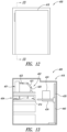
- Figure 1 shows a slide dryer 100 capable of drying one or more specimen-carrying microscope slides.
- the slide dryer 100 can heat the microscope slides and specimens carried thereon in order to dry the slides and/or specimens, to couple the specimens to the slides, and/or to perform any other desired thermal processing.
- the specimen-carrying microscope slides are held in an orientation that promotes removal of residual transfer fluid between the specimens and the respective microscope slides while limiting, minimizing, or substantially eliminating unwanted movement of the specimens.
- the specimen-carrying slides are dried to fixedly couple the specimens to the respective slides.
- the specimen-carrying slides are removed from the slide dryer 100 for subsequent processing and tissue analyses, such as staining, immunohistochemistry, in-situ hybridization, or other processing.
- tissue analyses such as staining, immunohistochemistry, in-situ hybridization, or other processing.
- the illustrated slide drying 100 is portable and can be readily carried (e.g., manually transported) by a person. In a laboratory setting, the slide dryer 100 can be manually transported between workstations.
- the microscope slides can be held in substantially vertical orientations to promote removal of residual transfer fluid trapped beneath the specimens to reduce drying times.
- a region of a slide beneath a specimen can be rapidly dried by draining flowable residual transfer fluid away from the specimen. As such, only a portion of the residual transfer fluid is evaporated to dry the region of the slide facing the specimen.
- the slide dryer 100 produces heat to both dry the slides and facilitate coupling of the specimens to the slides.
- the specimens can include, without limitation, a biological sample (e.g., a tissue sample) and a material (e.g., an embedding material) in which the sample is embedded.
- the embedding material is melted, brought into contact with the slide, and solidified so as to attach the biological sample directly to the slide.
- the embedding material can move towards the microscope slide and can be ultimately deposited on the slide. If that material is more hydrophobic than the material of the slide and the residual transfer fluid (e.g., water), the embedding material may promote beading of the fluid.
- the residual transfer fluid e.g., water
- the embedding material may promote beading of the fluid.
- a contact angle between the fluid and slide will increase. Because the microscope slide is in the upright orientation, the residual transfer fluid will tend to accumulate at a lower end of the specimen due to gravity. An upper end of the specimen can physically contact the front face of the slide and minimize, limit, or substantially prevent unwanted migration of the specimen. After a sufficient amount of residual transfer fluid has accumulated, it can drain downwardly away from the specimen, thereby leaving the specimen on the front face of the slide. This process can be performed on a wide range of wetted microscope slides.
- the illustrated slide dryer 100 of Figure 1 includes a conductive slide heater 110, a controller 114, and a main body 118 that houses internal components of the slide dryer 100.
- the conductive slide heater 110 can physically contact and support one or more microscope slides.
- the main body 118 includes a slide support 120 adjacent to the conductive slide heater 110.
- the conductive slide heater 110 and slide support 120 can cooperate to hold a microscope slide in a substantially vertical orientation such that heat generated by the conductive slide heater 110 is transferred to the microscope slide for a desired period, such as a drying period.
- the conductive slide heater 110 is capable of generating a sufficient amount of heat in response to a signal from the controller 114 so as to conductively heat the specimen to a desired temperature. Residual transfer fluid can flow away from the specimen, evaporate, and/or otherwise be removed from the specimen-carrying microscope slide.
- the heated specimen is brought into physical contact with the microscope slide.
- the specimen can be elevated to a temperature that facilitates adhesion between the specimen and the microscope slide, as discussed in connection with Figures 6-9 .
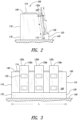
- Figures 2 and 3 show a plurality of microscope slides 130a, 130b, 130c (collectively 130) resting against the conductive slide heater 110 and on an upper surface 136 of the slide support 120.
- the microscope slides 130 can be generally flat transparent substrates carrying specimens 132 for examination using equipment, such as optical equipment (e.g., a microscopic).
- each microscope slide 130 may be a generally rectangular piece of a transparent material (e.g., glass) having a front face for receiving specimens and a back face for engaging the slide dryer 100.
- the microscope slides 130 may be charged or uncharged depending on the application and can have a wide range of different dimensions.
- the slides 130 may have a length of about 3 inches (75 mm) and a width of about 1 inch (25 mm) and may include a label, such as a barcode.
- the slides may have a length of about 75 mm, a width of about 25 mm, and a thickness of about 1 mm.
- the microscope slides 130 can be in the form of standard microscope slides.
- the illustrated microscope slides 130 carry the uncovered specimens 132 (e.g., without coverslips).
- the dimensions of slide support 120 may be such that a label on a slide 130 is located on a face of the slide opposite the slide support and above the portion of the slide that is in contact with the slide support. In this manner, the label can be protected from heat during the drying process.
- the main body 118 rests on a generally horizontal support surface 140 such that the microscope slides 130 are held generally upright.
- the controller 114 may be conveniently accessed by a user to control operation of the slide dryer 100.
- Figures 1-3 show the controller 114 communicatively coupled to the conductive slide heater 110 including a housing 146, a display 150, and an input device 154.
- the display 150 can be a screen or other display device.
- the input device 154 can include, without limitation, one or more buttons, keyboards, input pads, buttons, control modules, or other suitable input elements.
- the illustrated input device 154 is in the form of an input pad, such as a touch pad, used to program the slide dryer 100.
- the controller 114 can generally include, without limitation, one or more central processing units, processing devices, microprocessors, digital signal processors, central processing units, processing devices, microprocessors, digital signal processors (DSP), application-specific integrated circuits (ASIC), readers, and the like.
- the controller 114 can also include, without limitation, one or more storage elements, such as volatile memory, non-volatile memory, read-only memory (ROM), random access memory (RAM), and the like.
- the controller 114 can be programmed based on the desired processing of the specimen-carrying slides. In a fixed temperature mode of operation, the controller 114 is used to maintain the conductive slide heater 110 at a generally constant temperature.
- the controller 114 is used to adjust the temperature of the conductive slide heater 110.
- the controller 114 can store one or more programs for controlling the operation of the conductive slide heater 110.
- the input device 154 can be used to switch between different programs, modes of operation, or the like.
- the illustrated conductive slide heater 110 of Figure 2 has a height H that is greater than or equal to the lengths of specimen mounting regions of the slides 130. A substantial portion of a longitudinal length L 3 of each microscope slide 130 can contact the conductive slide heater 110 to facilitate generally even heat distribution throughout the entire mounting regions.
- the height H may be at least about 2.5 inches (63.5 mm), about 2.75 inches (70 mm), or about 2.9 inches (74.7 mm), as well as ranges encompassing such heights.
- Upper ends 139a, 139b, 139c of the slides 130a, 130b, 130c, respectively, can protrude upwardly past the slide dryer 100 for conveniently grasping the slides 130.
- a longitudinal length L of the conductive slide heater 110 can be selected based on the desired number of microscope slides 130 to be processed.
- the length L can be increased or decreased to increase or decrease the number of microscope slides that can be processed, spacing between the microscope slides, or the like.
- the apparatus illustrated in Figure 3 has three spaced apart microscope slides 130.
- the length L may be at least 6 inches (152 mm) to allow at least three microscope slides 130 to be concurrently processed. Other dimensions are also possible.
- the conductive slide heater 110 can be a plate having a generally rectangular shape, as viewed from the side (see Figure 3 ).
- the thermal properties of the conductive slide heater 110 may be selected based on desired processing criteria, such as the desired processing temperature, temperature distribution, rates of heating/cooling, and the like.
- desired processing criteria such as the desired processing temperature, temperature distribution, rates of heating/cooling, and the like.
- an engagement face 160 of the slide heater 110 can have a relatively low thermal mass and a high thermal conductivity for rapidly transferring heat across the entire face 160.
- the engagement face 160 can have a substantially uniform heat distribution to ensure that all or most of the slides 130 are maintained at substantially the same temperature.
- the conductive slide heater 110 can be made, in whole or in part, of one or more thermally conductive materials, for example, metals such as copper, steel, aluminum, iron, combinations thereof, or the like.
- the engagement face 160 may be made mostly of steel (e.g., stainless steel) and is highly resistant to wear and corrosion.
- the engagement face 160 may be made mostly of copper for rapidly transferring heat between internal heating elements and the slides 130.
- the number of internal heating elements, such as resistive heaters of the heater 110 can be reduced because of this rapid heat transfer.
- the conductive slide heater 110 can have a multi-layer construction to enhance wear characteristics and thermal performance.
- the engagement face 160 can be a thin sheet of steel, and an inner layer of copper in the heater 110 can help distribute and deliver heat to the face 160.
- the conductive slide heater 110 has a monolayer construction.
- the engagement face 160 can be substantially flat to increase the areas of contact between the slides 130 and the face 160.
- the engagement face 160 can be a highly polished face that is extremely flat for contacting most or substantially all of overlying portions of the slides 130.
- the engagement face 160 may be configured to minimize, limit, or substantially prevent relative movement of the microscope slides 130.
- anti-migration features e.g., protrusions, protuberances, grooves, partitions, texturing, and the like
- the main body 118 of Figures 1-3 has a base 170 for resting on the surface 140.
- the main body 118 protects internal components, even when the slide dryer 100 is used in harsh environments, including, without limitation, corrosive environments often found in labs, or other testing sites.
- the slide support 120 is integrally formed with the main body 118.
- the upper surface 136 may extend generally perpendicularly with respect to the engagement face 160.
- the back surfaces 141 of the slides 130 lie flat against the engagement face 160.
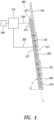
- Figure 4 shows the controller 114, a power source 200, a line 202 that delivers electrical power to the conductive slide heater 110, and a sensor 212 for evaluating operation of the slide heater 110.
- a line 220 communicatively couples the controller 114 to the sensor 212.
- the illustrated conductive slide heater 110 includes an outer portion 210 and a thermo-electric element 204.
- the term "thermo-electric element" is a broad term that includes, without limitation, one or more electric devices capable of generating heat and/or absorbing heat.
- Substantially all or most of the engagement face 160 can have a substantially uniform temperature when the element 204 is activated.
- substantially all or most of the engagement face 160 can be within a temperature range of about 10 degrees Celsius.
- the engagement face 160 may be maintained at generally the same temperature.
- the average temperature of the engagement face 160 can be in a range of about 5 degrees Celsius. Different portions of the engagement face 160 can be maintained at different temperatures to accommodate drying of slides holding different types of tissues and for tissues embedded with different embedding materials.
- the outer portion 210 may be a hollow plate, and the thermo-electric element 204 is a heating element adapted to convert electrical energy to thermal energy.
- the heating element 204 When the heating element 204 generates heat, heat is transferred through the portion 210 and is absorbed by the microscope slide 130. Heat is ultimately transferred from the slide 130 to the specimens 132.
- the amount of electrical energy delivered to the element 204 can be increased or decreased to increase or decrease the temperature of the specimens 132.
- the heating element 204 can be a resistive heating element.
- resistive heating elements e.g., plate resistive heaters, coil resistive heaters, strip heaters, or the like
- Other types of thermal elements such as cooling elements, heating/cooling elements, or the like, can be utilized.
- cooling element is a broad term that includes, without limitation, one or more elements capable of actively absorbing heat so as to effectively cool at least a portion of the conductive slide heater 110.
- a cooling element can be a cooling tube or channel through which a chilled fluid flows.
- the conductive slide heater 110 may include heating elements for producing heat during a heating period and cooling elements for absorbing heat during a cooling period.
- the element 204 may be a heating/cooling element, such as a Peltier device.
- Peltier devices may be solid state components which become hot on one side and cool on an opposing side, depending on a direction of current passed therethrough. By simply selecting the direction of current, the Peltier device 204 can be employed to heat the engagement face 160 for a desired length of time. By switching the direction of the current, the device 204 cools the engagement face 160.
- the heating/cooling element 204 can be in the form of channels through which a working fluid flows. Heated fluid can be passed through the channels for a heating period, and a chilled fluid can be passed through the channels for a cooling period. The position, number, and type of heating/cooling elements 204 can be selected based on the desired temperature profile of the conductive slide heater 210.
- the thermal properties of the portion 210 can be selected to achieve a desired temperature distribution along a wall 211 of the engagement face 160.
- the portion 210 can be made, in whole or in part, of a highly conductive material, such as copper, or other suitable material with sufficient thermal conductivity to reduce or limit any significant local temperature non-uniformities associated with discrete heating elements 204. Because heat is lost to the surrounding air (e.g., air at room temperature), the elements 204 can continually produce a constant flux.
- the interior portions of the silde heater 110 may be hotter than the periphery of the heater 110 because heat is dissipated faster from the periphery of the slide heater 110 due to its exposed edges.
- An array of closely spaced heating elements 204 may be used to maintain a generally uniform temperature across the surface 160. Other configurations are also possible.
- the sensor 212 is a temperature sensor that detects the temperature of the heater 110 and sends one or more signals indicative of that temperature.
- the sensor 212 can be mounted on a back surface 217 of the conductive slide heater 110, embedded in the heater 110, mounted on or embedded in the engagement face 160, or positioned at any other suitable location for measuring the temperatures of any portion of the slide heater 110 and/or the microscope slides 130.
- the sensor 212 can include, without limitation, one or more thermal couples, thermometers (e.g., an IR thermometer) pyrometers, resistance temperature detectors (RTDs), thermistors, or the like.
- Figure 5 is a flow diagram of one method of preparing and analyzing a specimen.
- a specimen can be placed on a microscope slide using a thin tissue section transfer fluid such as water.
- the specimen-carrying microscope slide is loaded into the slide dryer and dried such that the specimen is adhered to the microscope slide.
- the slide dryer 100 rapidly dries the slide while minimizing, limiting, or substantially eliminating unwanted migration of the specimen relative to the microscope slide.
- the specimen can remain at the same general position relative to the slide during the drying process. If a plurality of specimens is mounted on the slide, the spacing between the specimens can be maintained throughout the drying process. This may reduce or eliminate a physician's concern about tissue specimens falling off of the microscope slide.
- the specimen-carrying slide can then be conveniently transported and analyzed using a wide range of different examination techniques and equipment. This process is discussed in detail below.
- a biological sample such as a sample of tissue, is processed to preserve its characteristics, such as the tissue structure, the cell structure, and the like.
- the tissue can be any collection of cells mountable on a microscope slide including, without limitation, a section of an organ, tumor section, bodily fluid, smear, frozen section, cytology prep, or cell lines.
- the tissue sample can be a sample obtained using an incisional biopsy, a core biopsy, an excisional biopsy, a needle aspiration biopsy, a core needle biopsy, a stereotactic biopsy, an open biopsy, a surgical biopsy, or the like.
- a fixative is used to fix and preserve the sample.
- Fixatives can fix and preserve cellular structure, inhibit or substantially stop enzymatic action that may result in the purification or autolysis of the tissue, or the like.
- the fixation process can increase the rigidity of the tissue, thereby making it more convenient to section, as detailed below.
- Formaldehyde, ethanol, acetone, paraformaldehyde, or other types of fixatives can be used.
- the type and number of fixatives can be selected based on the desired processes to be performed, such as staining, cytological staining, immunohistochemistry, or in-situ hybridization.
- the sample is embedded in a material that has mechanical properties that may facilitate sectioning.
- Materials for embedding include, but are not limited to, paraffin, resin (e.g., plastic resins), polymers, agarose, nitrocellulose, gelatin, mixtures thereof, or the like.
- the embedding material may comprise mostly or entirely of paraffin.
- Paraffin is a white or generally colorless water insoluble solid substance that is resistant to many reagents.
- paraffin can be a mixture of hydrocarbons chiefly of the alkaline series obtained from petroleum. A wide range of different mixtures of similar hydrocarbons can be used to make paraffin, and these mixtures can be solid, semi-solid, and/or oily.
- the paraffin may be a wax.
- tissue samples can be mixed or combined with material that can permeate the tissue sample so as to impart properties that facilitate a cutting process. In this manner, the tissue samples are embedded. If the tissue sample is to be sectioned with a microtome or similar device, the tissue sample can be embedded in paraffin or other suitable material, such as a plastic resin. If the embedding material is paraffin, the paraffin can be heated and melted. The hot liquid paraffin at least partially impregnates the biological sample and is subsequently solidified.
- the specimen is cut into mountable sections, placed on a microscope slide, and then dried.
- a microtome can cut the specimen into thin sections, for example, slices on the order of about 5 microns to about 6 microns thick. Each section can include a portion of the tissue sample and some of the embedding material.
- the cut sections may be floated on water to spread or flatten the sections. If the sections are pieces of paraffin embedded tissue, the sections can be floated on a warm bath to keep the sections in generally flat configurations, thereby reducing or preventing folding, creasing, or bending.
- a microscope slide is inserted into the warm bath. A front surface of the slide is used to pick up the tissue specimens.
- tissue samples e.g., a set of tissue samples, each taken at a different location of a subject
- a plurality of the tissue samples may be sequentially floated onto the slide. These wet slides are then dried using the slide dryer 100.
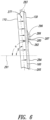
- Figure 6 shows a specimen 286 positioned on a droplet of transfer fluid 282.
- the droplet of transfer fluid 282 can be water or other suitable fluid (including aqueous mediums) that may or may not contain any additives (e.g., wetting agents, reagents, dyes, etc.). If water is employed, the water may be de-ionized water, double-distilled de-ionized water, purified water, or the like.
- the droplet of transfer fluid 282 can be formed as the slide 130 is pulled out of the bath as described above. Alternatively, the droplet 282 can be deposited by directly dropping the transfer fluid onto the front surface 284 and thereafter placing the specimen on top of the droplet. A droplet placed directly onto surface 284 then functions to allow positioning of the specimen on the front surface 284.
- the contact angle between the transfer fluid 282 and the slide 130 is relatively small so that the specimen 286 is kept at the same general location, even if the slide 130 is moved between, for example, workstations or different equipment.
- the surface tension of the transfer fluid 282 helps maintain a generally flat configuration of the specimen 286 to limit, reduce, or substantially prevent unwanted irregularities of the specimen 286, such as folding, creasing, protruding, buckling, or the like. Because the fluid 282 is trapped between the specimen 286 and the microscope slide 130, the specimen 286 is kept away from the front surface 284.
- the microscope slide 130 is held in a substantially vertical orientation to promote draining of the fluid 282 to reduce drying times.
- the illustrated slide 130 is at an angle of inclination ⁇ defined by a generally horizontal imaginary plane 291 (e.g., an imaginary plane generally parallel to the support surface 140 of Figures 2 and 3 ) and the engagement face 160. Because the slide 130 lays flat against the conductive slide heater 110, the slide 130 is held at the same general angle of inclination.
- the illustrated conductive slide heater 110 extends generally along an imaginary plane 283 that defines an angle ⁇ with the imaginary horizontal plane 291 that is greater than about 70 degrees, 80 degrees, 85 degrees, or 90 degrees.
- a longitudinal axis 295 of the slide 130 is generally parallel to the imaginary plane 283.
- the angle ⁇ can be equal to or greater than about 80 degrees to keep the microscope slide 130 at an angle of inclination ⁇ of about 80 degrees. Other angles are also possible, if needed or desired.
- the engagement surface 160 is maintained at or above a melt point of the embedding material of the specimen 286 in order to conductively heat the specimen 286. If the embedding material is paraffin with a melt point between about 50 degrees Celsius and 57 degrees Celsius, the surface 160 is kept at or above a temperature of about 50 degrees Celsius. Arrows 277 represent heat being transferred from the heater 110 to the specimen 286. When the embedding material melts, the melted material may float along the upper surface of the transfer fluid 282 and become deposited on the front surface 284. If the embedding material is more hydrophobic than the microscope slide 130, the contact angle at which the transfer fluid 282 interfaces with the surface 284 may increase, thereby causing the fluid 282 to form an unstable droplet susceptible to migration along the surface 284.
- the tilted slide 130 promotes accumulation of the transfer fluid 282 proximate a lower portion 287 of the specimen 286.
- the fluid 282 collects at a gap 285 between the specimen 286 and the microscope slide 130 such that the fluid 282 eventually drains down the front surface 284.
- Figure 6 also shows a position 296 (shown in phantom line) of the fluid 282 after the contact angle has increased assuming the microscope 130 was in a horizontal orientation instead of the vertical orientation. Because the illustrated microscope slide 130 is at an upright orientation, the fluid 282 tends to accumulate at the gap 285 due to gravity, as shown in Figure 7 .
- An upper portion 293 of the specimen 286 can physically contact the heated surface 284 to limit, minimize, or substantially prevent migration of the specimen 286.
- the upper portion 293, for example, may stick to the surface 284.
- the illustrated angle of inclination of Figure 7 is greater than about 70 degrees such that the beaded transfer fluid 282 drains after a sufficient amount of the embedding material is melted so as to appreciably increase the contact angle between the fluid 282 and the surface 284.
- the angle of inclination may be greater than about 75 degrees.
- Such examples are especially well suited to cause draining of residual transfer fluid (e.g., water) away from relatively small specimens, such as specimens containing tissue samples obtained using core needle biopsies. Residual transfer fluid can be drawn away at different speeds from different types and sizes of specimens, including relatively large specimens or small specimens.
- the angle of inclination may be greater than about 75 degrees to promote the rapid accumulation of transfer fluid 282 as the specimen 286 is heated. Once the embedding material is heated to its melt point, the transfer fluid 282 rapidly beads up and is drained therefrom.
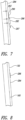
- Figure 8 shows the specimen-carrying slide 130 after completion of a drying period.
- the engagement face 160 can be maintained at a temperature equal to or greater than about 50 degrees Celsius and the ambient air can be less than 30 degrees Celsius (e.g., a temperature of about 21 degrees Celsius).
- the slide 130 is placed against the heater 110.
- the engagement face 160 heats the slide 130 such that the specimen 286 is adhered to the surface 284 in a relatively short period of time, for example, less than or equal to about 5 minutes, 1 minute, 45 seconds, 30 seconds, 20 seconds, or ranges encompassing such periods of time.
- Most of the transfer fluid 282 is drained away from the specimen 286 to avoid drying times associated with evaporating the entire droplet.
- the length of the drying period can depend on the amount of transfer fluid, the characteristics of the tissue of the specimen 286, characteristics of the embedding material, or the like.
- the engagement face 160 of Figure 7 can be maintained at or above a temperature of about 50 degrees Celsius, 55 degrees Celsius, or 65 degrees Celsius, as well as within ranges encompassing such temperatures (e.g., a range of about 50 degrees Celsius to about 65 degrees Celsius). Such temperatures are especially well suited to melt paraffin or other materials with a relatively low melt point.
- the specimen 286 may be adhered to the surface 284 in less than about 1 minute. For example, the specimen 286 can be adhered in about 10 seconds to about 1 minute.
- the engagement face 160 may be maintained at a temperature of about 65 degrees Celsius or above.
- the engagement face 160 can be at a temperature that is less than, greater than, or equal to about 65 degrees Celsius, 70 degrees Celsius, 80 degrees Celsius, or ranges of such temperatures.
- An infrared thermometer is used to measure the temperature of the engagement face 160 to maintain accuracy.
- the temperature of the engagement face 160 can be increased or decreased using feedback from the thermometer to decrease or increase drying times.
- the engagement face 160 can be maintained at a somewhat constant temperature during the heating period for consistent drying.
- the slide heater 110 is thus capable of drying slides without appreciable temperature changes.
- the engagement face 160 can be maintained at a generally constant temperature at or above a melt point of embedding material to ensure short drying times.
- Tthe temperature of the engagement face 160 may be kept within an operating temperature range that is above the melt point.
- the operating temperature range can be 50 degrees Celsius to 60 degrees Celsius, 55 degrees Celsius to 65 degrees Celsius, or 60 degrees Celsius to 70 degrees Celsius. Other ranges are also possible.
- the engagement face 160 can be pre-heated to immediately transfer heat to the slide 130 upon contact. Pre-heating can be used to avoid ramp-up times associated with cyclic heating processes. Slides can be repeatedly loaded onto the slide dryer 100 without waiting for the engagement face 160 to reach a certain temperature. Of course, the temperature of the engagement face 160 may decrease slightly as the slide 130 absorbs heat. These effects can be minimized or avoided by continuously generating heat with the slide heater 110.
- the engagement face 160 can be heated after the slide 130 placed against the face 160.
- the engagement face 160 can be maintained at a low temperature, for example, at about room temperature or at a temperature between room temperature and a desired temperature for drying.
- the low temperature may be a standby temperature.
- the engagement face 160 can be kept at a standby temperature in a range of about 25 degrees Celsius to about 50 degrees Celsius.
- the temperature of the engagement face 160 is increased to at least about 50 degrees Celsius during or after loading of the slides 130. After drying, the engagement surface 160 returns to the standby temperature.
- a wide range of different types of heating cycles can be employed to reduce or limit the amount of energy used to during drying processes.
- the conductive slide heater 110 can also include cooling elements (e.g., Peltier elements) used to rapidly absorb heat from the dried specimen-bearing slide 130. Once the specimen-bearing slide 130 is sufficiently cooled, it can be removed from the slide dryer 100.
- cooling elements e.g., Peltier elements
- the specimen 286 is stained for examination.
- the specimen can also be baked, de-waxed, de-paraffinized, or the like.
- a stain may be applied to the specimen 286 after performing a de-parafinizing process.
- the microscope slide is then cover slipped for subsequent optical examination.
- the specimen 286 can be examined using optical equipment (e.g., a microscope), optical instruments, or the like.
- optical equipment e.g., a microscope
- Different types of examination processes can be used to perform a wide range of different tissue analyses used to obtain information about pathology, tissue composition, and the tissue architecture. This information can be used by a physician to diagnose different types of illnesses and to perform various medical research.
- a slide dryer 300 includes a conductive slide heater 310, a base 312, and a cover 314.
- the base 312 includes a controller 320 for controlling operation of the conductive slide heater 310.
- the cover 314 can enclose and surround the conductive slide heater 310 to prevent unwanted contaminates from landing on the specimen-carrying microscope slides.
- the base 312 includes a collector 337 surrounding the slide conductive heater 310. The collector 337 can collect residual transfer fluid or other materials that are removed from the slides.
- the illustrated conductive slide heater 310 includes a plurality of heat generating support elements 330a-h (collectively 330) spaced apart from one another.
- the support elements 330 can be independently operated or operated together and are oriented at the same or different inclines.
- the illustrated elements 330 are generally parallel to one another and spaced to allow at least one generally vertically oriented microscope slide to be inserted between a pair of adjacent elements 330.
- Each of the support elements 330 can include one or more energizable thermal devices (e.g., electro-thermal elements).
- a back plate 441 extending between the support elements 330 may have thermal devices (e.g., internal thermal devices) capable of generating heat that is conducted through the support elements 330.
- microscope slides can lean against respective support elements 330.
- the cover 314 can be moved over the slide heater 310 as indicated by the arrows 339.
- the conductive slide heater 310 can dry an entire row of vertically oriented specimen-bearing microscope slides. After the microscope slides are processed, the cover 314 is removed to access and remove the microscope slides.
- Figure 10 shows a heater assembly 360 that includes a frame 361 and a plurality of conductive slide heaters 362, 364 coupled to the frame 361.
- the conductive slide heaters 362, 364 can be generally similar to the slide conductive heater 310 of Figure 9 .
- the heater assembly 360 can be incorporated into different types of slide processing systems, such as an automated slide processing systems or stand alone slide dryers, and may include a controller, power sources, or the like.
- FIG 11 shows an apparatus 400 that includes a drying station 402 and a plurality of processing stations 404, 406, 408.
- a controller 410 controls operation of the drying station 402 and one or more of the processing stations 404, 406, 408.
- the illustrated controller 410 is communicatively coupled to and commands each of the stations 402, 404, 406, 408.
- Microscope slides can be automatically processed (e.g., via a process that is substantially free of human intervention) using the apparatus 400.
- the controller 410 can control the amount of heat produced by a slide dryer 401 at the station 402 ( Figure 13 ), rate of drying, length of time of the drying period, or other processing parameters, preferably while keeping thermal damage to the tissue sample at or below a desired level.
- processing station includes, without limitation, a baking station, a material removal station (e.g., a de-waxing station, a de-paraffinizing station, or the like), staining station, cover-slipping station, or the like.
- processing stations 404, 406, 408 can be a de-paraffinizing station, staining station, and cover-slipping station, respectively.
- a transport device 418 transports specimen-bearing microscope slides between the drying station 402 and the other stations 404, 406, 408.
- the transport device 418 can include, without limitation, one or more elevators, slide handlers, slide trays, slide holders, or the like.
- Slide handlers can include, but are not limited to, slide manipulators, X-Y-Z transport systems, robotic systems, or other automated systems capable of receiving and transporting slides.
- a robotic system can include, without limitation, one or more pick and place robots, robotic arms, or the like.
- the drying station 402 includes the slide dryer 401 and a slide handler 420, illustrated as a robotic slide handler.
- the slide dryer 401 generates heat for conductively heating the specimen-carrying microscope slides.
- the robotic slide handler 420 includes an arm 421 and an end effector 423 capable of picking up and carrying slides between the conductive slide heater 401 and a slide transporter 424, illustrated schematically in Figure 13 .
- the slide dryer 401 can be generally similar to the slide dryer 100 of Figures 1-3 or the slide dryer 300 of Figure 10 .
- Various types of other automated slide processing systems can also have the slide dryers and other features disclosed herein.
- U.S. Application No. 10/414,804 U.S. Publication No. 2004/00021 63
- the transport device 418 of Figure 13 includes an elevator system 430 and a movable platform 434, shown carrying the slide transporter 424.
- the elevator system 430 moves the transporter 424 up and down a rail 440.
- specimen-carrying microscope slides are loaded onto a slide tray, which is placed on the platform 434.
- the slide handler 420 loads the specimen-carrying microscope slides into the slide dryer 401.
- the slide dryer 401 dries the specimen-carrying microscope slides. After the specimen-carrying microscope slides are dried a sufficient amount, the slide handler 420 transports the slides back to the transporter 424.
- the transporter 242 is vertically lowered and positioned adjacent to the processing station 404 for de-paraffinizing.
- the station 404 is capable of removing at least a portion of the embedding material of the specimen.
- the de-paraffinizing station 404 can be a bath-type, de-paraffinizing station or a spray-type, de-paraffinizing station.
- the illustrated de-paraffinizing station 404 includes a modular compartment 414 and includes one or more wash dispense nozzles 416 directed downwardly. De-paraffinizing substances are delivered onto the specimens using the nozzles 416. After removing the embedding material (e.g., paraffin), the slides can be rinsed with substances, such as de-ionized water, to remove the de-paraffinizing substance and the extra paraffin leaving the bare tissue sample adhered to the microscope slide.
- substances such as de-ionized water
- the de-paraffinizing substances can be fluids, for example, aqueous-based fluids that promote separation of paraffin and tissue specimens, such as those disclosed in U.S. Patent No. 6,855,559, issued February 15, 2005 and U.S. Patent No. 6,544,798, issued April 8, 2003 , including de-ionized water, citrate buffer (pH 6.0-8.0), tris-HCI buffer (pH 6-10), phosphate buffer (pH 6.0-8.0), acidic buffers or solutions (pH 1-6.9), basic buffers or solutions (pH 7.1-14), or the like.
- the substance may also contain one or more ionic or non-ionic surfactants.
- the de-paraffinizing substances can be heated.
- the substances e.g., fluids
- the substances may be heated to a temperature greater than the melting point of the embedding material, e.g., between 60-70 degrees Celsius.
- U.S. Patent No. 7,303,725, issued December 4, 2007 discloses various components (e.g., probes, filters, sprayers, etc.) for use with de-paraffinizing substances.
- the station 404 may also include one or more heating elements for baking the embedding material.
- the slides can be heated to soften the embedding material to facilitate material removal.
- the transport system 424 delivers the specimen-carrying slides to the station 406 for staining.
- a desired stain is applied to the tissue samples.
- the stain can be a biological or chemical substance which, when applied to targeted molecules in tissue, renders the tissue detectable under an instrument. Stains include, without limitation, detectable nucleic acid probes, antibodies, hematoxylin, eosin, and dyes (e.g., iodine, methylene blue, Wright's stain, etc.).
- the station 408 may be a drying station.
- the station 408 dries the stained slides and the slides are ready for cover slipping.
- the drying station 408 may conductively heat the stained specimens using a slide dryer, such as those discussed in connection with Figures 1-10 .
- the drying station 408 may be in the form of a convection oven or microwave oven.
- the apparatus 400 can also include other types of processing stations.
- the number, configurations, and types of processing stations can be selected based on the types of processing to be performed.
- U.S. Patent No. 7,396,508 discloses apparatuses for staining and treating tissues.
- the processing station 406 may include a carousel type system, such as the carousel system disclosed in U.S. Patent No. 7,396,508 .
Landscapes
- Life Sciences & Earth Sciences (AREA)
- Health & Medical Sciences (AREA)
- Physics & Mathematics (AREA)
- Chemical & Material Sciences (AREA)
- General Physics & Mathematics (AREA)
- Analytical Chemistry (AREA)
- Pathology (AREA)
- Biochemistry (AREA)
- General Health & Medical Sciences (AREA)
- Immunology (AREA)
- Engineering & Computer Science (AREA)
- Molecular Biology (AREA)
- Biomedical Technology (AREA)
- Robotics (AREA)
- Optics & Photonics (AREA)
- Sampling And Sample Adjustment (AREA)
Applications Claiming Priority (2)
| Application Number | Priority Date | Filing Date | Title |
|---|---|---|---|
| US11396408P | 2008-11-12 | 2008-11-12 | |
| PCT/US2009/064235 WO2010056883A1 (en) | 2008-11-12 | 2009-11-12 | Methods and apparatuses for heating slides carrying specimens |
Publications (2)
| Publication Number | Publication Date |
|---|---|
| EP2356423A1 EP2356423A1 (en) | 2011-08-17 |
| EP2356423B1 true EP2356423B1 (en) | 2024-08-07 |
Family
ID=41396000
Family Applications (1)
| Application Number | Title | Priority Date | Filing Date |
|---|---|---|---|
| EP09752682.6A Active EP2356423B1 (en) | 2008-11-12 | 2009-11-12 | Methods and apparatuses for heating slides carrying specimens |
Country Status (8)
| Country | Link |
|---|---|
| US (4) | US10184862B2 (enExample) |
| EP (1) | EP2356423B1 (enExample) |
| JP (1) | JP5474080B2 (enExample) |
| KR (1) | KR101548407B1 (enExample) |
| CN (1) | CN102209889B (enExample) |
| AU (1) | AU2009313985B2 (enExample) |
| CA (1) | CA2742473C (enExample) |
| WO (1) | WO2010056883A1 (enExample) |
Families Citing this family (26)
| Publication number | Priority date | Publication date | Assignee | Title |
|---|---|---|---|---|
| US7677289B2 (en) * | 2004-07-08 | 2010-03-16 | President And Fellows Of Harvard College | Methods and apparatuses for the automated production, collection, handling, and imaging of large numbers of serial tissue sections |
| US9144709B2 (en) | 2008-08-22 | 2015-09-29 | Alton Reich | Adaptive motor resistance video game exercise apparatus and method of use thereof |
| US9272186B2 (en) | 2008-08-22 | 2016-03-01 | Alton Reich | Remote adaptive motor resistance training exercise apparatus and method of use thereof |
| CA2742473C (en) | 2008-11-12 | 2015-02-24 | Ventana Medical Systems, Inc. | Methods and apparatuses for heating slides carrying specimens |
| EP2614376B1 (en) | 2010-09-07 | 2022-11-02 | President and Fellows of Harvard College | Methods and systems for collection of tissue sections |
| DE102010060825B4 (de) * | 2010-11-26 | 2017-10-19 | Leica Biosystems Nussloch Gmbh | Vorrichtung zur Handhabung von Objektträgern |
| US20120307355A1 (en) * | 2011-06-01 | 2012-12-06 | Streamline Automation, Llc | Microscope slide heating and mounting apparatus and method of operation therefor |
| CN103121535A (zh) * | 2011-11-21 | 2013-05-29 | 昆山丘钛微电子科技有限公司 | 半自动预热烘烤机台 |
| WO2013079072A1 (en) * | 2011-11-30 | 2013-06-06 | Dako Denmark A/S | Method and system for automated deparaffinization and non-immunohistochemical special staining of tissue samples |
| KR101326655B1 (ko) * | 2012-02-29 | 2013-11-08 | 한국표준과학연구원 | 전도 및 근접복사열 가열수단을 이용한 적외선 열화상 부품 결함 측정 장치 |
| US9398640B2 (en) * | 2012-12-21 | 2016-07-19 | Halliburton Energy Services, Inc. | Digital multi-use thermo-cup |
| AU2014232777B2 (en) | 2013-03-15 | 2018-02-01 | Lee H. Angros | Method of removing floatation liquid |
| ES2877332T3 (es) * | 2013-05-28 | 2021-11-16 | Todos Medical Ltd | Un método para indicar la presencia de tumores benignos mediante el uso de una muestra de células mononucleares de sangre periférica (PBMC) |
| JP6302066B2 (ja) | 2013-12-13 | 2018-03-28 | ベンタナ メディカル システムズ, インコーポレイテッド | 顕微鏡スライドを熱処理する自動化された処理システムおよび方法 |
| CN105980828B (zh) | 2013-12-13 | 2021-12-17 | 文塔纳医疗系统公司 | 用于生物标本组织处理的染色试剂和其它液体及关联技术 |
| CN105182522A (zh) * | 2015-07-30 | 2015-12-23 | 苏州欧可罗电子科技有限公司 | 一种智能加热式显微镜 |
| WO2017034940A1 (en) * | 2015-08-21 | 2017-03-02 | Skone Rene Benson | Coverslip removal systems and methods |
| US20210292702A1 (en) * | 2018-08-08 | 2021-09-23 | UNIVERSITé LAVAL | Live cell imaging chamber and measurement thereof |
| IL261096A (en) * | 2018-08-10 | 2020-02-27 | Ez Pack Water Ltd | A system and method for storing renewable energy as hot or cold water in a flexible heating tank |
| JP7094398B2 (ja) * | 2019-02-08 | 2022-07-01 | 平田機工株式会社 | 標本作製方法 |
| JP7236358B2 (ja) | 2019-09-04 | 2023-03-09 | 平田機工株式会社 | 標本作製装置 |
| WO2021044543A1 (ja) * | 2019-09-04 | 2021-03-11 | 平田機工株式会社 | 標本作製装置 |
| CN112326393A (zh) * | 2020-11-30 | 2021-02-05 | 上海北昂医药科技股份有限公司 | 一种细胞自动制片仪 |
| USD971438S1 (en) * | 2021-05-10 | 2022-11-29 | Rushabh Instruments, Llc | Laboratory slide heater |
| KR102665179B1 (ko) * | 2022-02-04 | 2024-05-09 | 국립부경대학교 산학협력단 | 검사 키트, 검사 장치 및 검사 키트 제조 방법 |
| KR102945754B1 (ko) * | 2023-04-12 | 2026-03-27 | 가톨릭대학교 산학협력단 | 인공지능 기반 병리 슬라이드 건조 최적화 장치, 방법 및 시스템 |
Citations (3)
| Publication number | Priority date | Publication date | Assignee | Title |
|---|---|---|---|---|
| US4985206A (en) * | 1987-09-30 | 1991-01-15 | Shandon Scientific Limited | Tissue processing apparatus |
| US5023187A (en) * | 1985-09-13 | 1991-06-11 | Fisher Scientific Company | Method and device for accelerated treatment of thin sample on surface |
| US7396508B1 (en) * | 2000-07-12 | 2008-07-08 | Ventana Medical Systems, Inc. | Automated molecular pathology apparatus having independent slide heaters |
Family Cites Families (325)
| Publication number | Priority date | Publication date | Assignee | Title |
|---|---|---|---|---|
| GB211201A (en) | 1922-11-13 | 1924-02-13 | Newsome Henry Clough | Improvements in wireless installations on vehicles such as automobiles |
| US3219416A (en) | 1962-10-30 | 1965-11-23 | Scientific Industries | Apparatus for the automatic chemical sequential treatment and analysis of small quantities of material |
| US3650437A (en) | 1968-05-09 | 1972-03-21 | Aerojet General Co | Automated biological reaction instrument |
| US3574064A (en) | 1968-05-09 | 1971-04-06 | Aerojet General Co | Automated biological reaction instrument |
| US3695281A (en) | 1970-11-16 | 1972-10-03 | Technicon Instr | Method and apparatus for fluid injection |
| US3665148A (en) | 1971-04-07 | 1972-05-23 | Gen Motors Corp | Six-axis manipulator |
| BE788877A (fr) | 1971-09-17 | 1973-01-02 | Vickers Ltd | Agitation d'echantillons liquides pour l'obtention de melanges homogenes |
| SE373963B (enExample) | 1973-06-14 | 1975-02-17 | Janson Sven Olof | |
| FR2239167A6 (en) | 1973-07-26 | 1975-02-21 | Ministere Agriculture Service | Machine for carrying out laboratory analyses - has automatic sample transfer, reactant addition and washing of transfer pipettes |
| US3853092A (en) | 1973-10-25 | 1974-12-10 | Corning Glass Works | Apparatus for nutating and staining a microscope slide |
| USRE30730E (en) | 1975-06-11 | 1981-09-01 | National Research Development Corporation | Apparatus for use in investigating specimens |
| US4043292A (en) * | 1975-07-21 | 1977-08-23 | Corning Glass Works | Microscope slide staining apparatus having temperature control |
| US4013038A (en) | 1975-07-21 | 1977-03-22 | Corning Glass Works | Apparatus for controlling the temperature of a liquid body |
| US4058367A (en) | 1976-05-19 | 1977-11-15 | Gilford Instrument Laboratories Inc. | Automatic asynchronous fluid processing apparatus |
| US4298571A (en) | 1976-12-17 | 1981-11-03 | Eastman Kodak Company | Incubator including cover means for an analysis slide |
| US4092952A (en) | 1977-08-19 | 1978-06-06 | Wilkie Ronald N | Automatic slide stainer |
| FR2403465A2 (fr) | 1977-09-16 | 1979-04-13 | Valois Sa | Pompe manuelle a piston pour distribution ou pulverisation |
| SE7801564L (sv) | 1978-02-10 | 1979-08-11 | Lkb Produkter Ab | Anordning for infergning av biologiska preparat |
| JPS5514157A (en) | 1978-07-14 | 1980-01-31 | Shin Meiwa Ind Co Ltd | Automatic welder |
| US4286637A (en) | 1978-11-09 | 1981-09-01 | Connaught Laboratories Limited | Apparatus for dispensing liquids into tubes |
| JPS55107957A (en) | 1979-02-13 | 1980-08-19 | Agency Of Ind Science & Technol | Analyzing method of blood image |
| DE2915248C3 (de) | 1979-04-14 | 1982-01-14 | Gise, Frhr. von, Hardo, Dr.med., 7400 Tübingen | Einrichtung zum automatischen wahlweisen u. exakten Behandeln von Präparaten |
| JPS55136958A (en) | 1979-04-14 | 1980-10-25 | Olympus Optical Co Ltd | Automatic analyzer |
| DE3029795C2 (de) | 1979-08-07 | 1983-10-27 | Olympus Optical Co., Ltd., Tokyo | Automatisches Analysiergerät für Flüssigproben |
| US4346056A (en) | 1979-08-15 | 1982-08-24 | Olympus Optical Company Limited | Automatic analyzing apparatus |
| US4647431A (en) | 1980-05-31 | 1987-03-03 | Fuji Photo Film Co., Ltd. | Device for maintaining a constant temperature for chemical analysis |
| DE3133191C2 (de) | 1980-08-22 | 1986-01-16 | Olympus Optical Co., Ltd., Tokio/Tokyo | Automatisches chemisches Analysiergerät |
| JPH0217341Y2 (enExample) | 1981-02-10 | 1990-05-15 | ||
| IT1144709B (it) | 1981-05-15 | 1986-10-29 | Dea Spa | Sistema di misura dimensionale servito da una pluralita di bracci operativi e controllato da un sistema a calcolatore |
| US4384193A (en) | 1981-06-09 | 1983-05-17 | Immulok, Inc. | Incubating device for specimen mounted on glass slides in immunoassays |
| US4453807A (en) | 1981-06-17 | 1984-06-12 | Smithkline Beckman Corp | System for handling slides |
| US4430299A (en) | 1981-06-18 | 1984-02-07 | Coulter Electronics, Inc. | Apparatus for monitoring chemical reactions |
| EP0085700A4 (en) | 1981-07-20 | 1985-10-14 | American Hospital Supply Corp | COUPLING SYSTEM FOR AUTOMATIC CHEMICAL ANALYZER. |
| JPS5821566A (ja) | 1981-07-31 | 1983-02-08 | Fuji Photo Film Co Ltd | インキユベ−タ |
| US4447395A (en) | 1982-02-12 | 1984-05-08 | The United States Of America As Represented By The Secretary Of The Army | Sampling device |
| US4727409A (en) | 1982-04-12 | 1988-02-23 | Signetics Corporation | Programmable read-only memory formed with opposing PN diodes |
| FR2528122B1 (fr) | 1982-06-04 | 1988-07-15 | Valois Sa | Pompe pour vaporisateur toutes positions |
| GB2143205B (en) | 1983-07-15 | 1986-07-16 | Leicester Polytechnic | Robots |
| US4629862A (en) | 1984-03-28 | 1986-12-16 | Olympus Optical Company Ltd. | Sample heater for use in microscopes |
| US4577514A (en) | 1984-04-09 | 1986-03-25 | Vanderbilt University | Method and apparatus for sampling liquid phase components from a liquid-semisolid fluid |
| US4961906A (en) | 1984-04-12 | 1990-10-09 | Fisher Scientific Company | Liquid handling |
| US4539855A (en) | 1984-05-03 | 1985-09-10 | Eastman Kodak Company | Apparatus for transferring liquid out of a capped container, and analyzer utilizing same |
| JPS6111293A (ja) | 1984-06-27 | 1986-01-18 | Kanzaki Paper Mfg Co Ltd | 熱転写記録用受像シ−ト |
| JPS6149205A (ja) | 1984-08-16 | 1986-03-11 | Seiko Instr & Electronics Ltd | ロボツト制御方式 |
| JPH0690211B2 (ja) | 1984-09-21 | 1994-11-14 | オリンパス光学工業株式会社 | 免疫学的分析装置およびその方法 |
| US4727494A (en) | 1985-01-01 | 1988-02-23 | Zymark Corporation | Computerized robot control system with scheduling feature |
| US4720463A (en) | 1985-03-01 | 1988-01-19 | Sherwood Medical Company | Automated microbiological testing apparatus |
| US4708886A (en) | 1985-02-27 | 1987-11-24 | Fisher Scientific Company | Analysis system |
| US4764342A (en) | 1985-02-27 | 1988-08-16 | Fisher Scientific Company | Reagent handling |
| JPS61219847A (ja) | 1985-03-27 | 1986-09-30 | Chiyoda Seisakusho:Kk | 顕微鏡標本の自動封入装置 |
| US5656493A (en) | 1985-03-28 | 1997-08-12 | The Perkin-Elmer Corporation | System for automated performance of the polymerase chain reaction |
| US4648023A (en) | 1985-05-23 | 1987-03-03 | Powell Roger A | Method for resource allocation for the manufacture of a product |
| JPH0786509B2 (ja) | 1985-06-18 | 1995-09-20 | 株式会社東芝 | 自動化学分析装置 |
| JPH076992B2 (ja) | 1985-06-21 | 1995-01-30 | 富士写真フイルム株式会社 | 化学分析装置 |
| US4774055A (en) | 1985-06-26 | 1988-09-27 | Japan Tectron Instruments Corporation | Automatic analysis apparatus |
| US4681741A (en) | 1985-07-01 | 1987-07-21 | American Hospital Supply Corporation | Reagent dispenser for an analyzing system |
| US4676951A (en) | 1985-07-01 | 1987-06-30 | American Hospital Supply Corp. | Automatic specimen analyzing system |
| US4643879A (en) | 1985-07-01 | 1987-02-17 | American Hospital Supply Corporation | Tower for analyzing system |
| JPS63500117A (ja) | 1985-07-01 | 1988-01-14 | バクスター・ダイアグノスティックス・インコーポレイテッド | 分析装置用の試薬投与装置 |
| US4729661A (en) | 1985-07-29 | 1988-03-08 | Specialty Medical Industries, Inc. | Asynchronous serial cuvette reader |
| US4731335A (en) | 1985-09-13 | 1988-03-15 | Fisher Scientific Company | Method for treating thin samples on a surface employing capillary flow |
| US4695430A (en) | 1985-10-31 | 1987-09-22 | Bio/Data Corporation | Analytical apparatus |
| US4670974A (en) | 1985-11-06 | 1987-06-09 | Westinghouse Electric Corp. | Windshield insertion system for a vehicle on a moving conveyor apparatus |
| US4678752A (en) | 1985-11-18 | 1987-07-07 | Becton, Dickinson And Company | Automatic random access analyzer |
| US4858155A (en) | 1985-12-24 | 1989-08-15 | Beckman Instruments, Inc. | Reaction temperature control system |
| DE3786087T2 (de) | 1986-02-07 | 1993-09-16 | Fuji Photo Film Co Ltd | Geraet fuer chemische analysen. |
| GB8605324D0 (en) | 1986-03-04 | 1986-04-09 | Rank Taylor Hobson Ltd | Metrological apparatus |
| US4843566A (en) | 1986-03-07 | 1989-06-27 | Hewlett-Packard Company | Robot motion control system |
| US5104621A (en) | 1986-03-26 | 1992-04-14 | Beckman Instruments, Inc. | Automated multi-purpose analytical chemistry processing center and laboratory work station |
| US4815978A (en) | 1986-04-30 | 1989-03-28 | Baxter Travenol Laboratories, Inc. | Clinical analysis methods and systems |
| US4835711A (en) | 1986-05-30 | 1989-05-30 | Zymark Corporation | Quickly reconfigurable robotic system |
| FR2600166B1 (fr) | 1986-06-17 | 1988-10-07 | Rhone Poulenc Rech | Procede et dispositif de prise en charge et d'analyses automatiques d'echantillons de produits amenes de facon aleatoire |
| US4965049A (en) | 1986-07-11 | 1990-10-23 | Beckman Instruments, Inc. | Modular analyzer system |
| US4933146A (en) | 1986-07-11 | 1990-06-12 | Beckman Instruments, Inc. | Temperature control apparatus for automated clinical analyzer |
| JP2533495B2 (ja) | 1986-07-25 | 1996-09-11 | 株式会社日立製作所 | ワ−クスケジユ−リング方法及び装置 |
| US5154889A (en) | 1986-08-07 | 1992-10-13 | Fuji Photo Film Co., Ltd. | Chemical analysis apparatus |
| EP0282601B1 (en) | 1986-09-16 | 1993-02-10 | Mitsubishi Corporation | Automatic analyzer |
| GB2196116B (en) | 1986-10-07 | 1990-08-15 | Weston Terence E | Apparatus for chemical analysis. |
| GB2196428B (en) | 1986-10-14 | 1990-05-23 | Tiyoda Seisakusho Kk | Apparatus for dyeing specimens automatically preparatory to microscopic examination |
| US4895706A (en) | 1986-10-28 | 1990-01-23 | Costar Corporation | Multi-well filter strip and composite assemblies |
| GR871619B (en) | 1986-10-31 | 1988-03-03 | Genetic Systems Corp | Automated patient sample analysis instrument |
| JPS63203761A (ja) | 1987-02-18 | 1988-08-23 | Sumitomo Metal Ind Ltd | 高密着性Cr被膜を有する鋼材とその製造法 |
| JPS63208761A (ja) | 1987-02-25 | 1988-08-30 | Chiyoda Seisakusho:Kk | 顕微鏡標本の自動染色装置により複数種類の染色方法を同時に行なう手順を求める方法及び装置 |
| US5002736A (en) | 1987-03-31 | 1991-03-26 | Fisher Scientific Co. | Microscope slide and slide assembly |
| US5148370A (en) | 1987-06-17 | 1992-09-15 | The Standard Oil Company | Expert system and method for batch production scheduling and planning |
| JPS645779A (en) | 1987-06-29 | 1989-01-10 | Fanuc Ltd | Robot arrangement examination system |
| US4979093A (en) | 1987-07-16 | 1990-12-18 | Cavro Scientific Instruments | XYZ positioner |
| US4847208A (en) | 1987-07-29 | 1989-07-11 | Bogen Steven A | Apparatus for immunohistochemical staining and method of rinsing a plurality of slides |
| JPS6480864A (en) | 1987-09-24 | 1989-03-27 | Fuji Photo Film Co Ltd | Biochemical analyzer |
| US4911915A (en) | 1987-10-13 | 1990-03-27 | Richard-Allan Medical Industries | Method of processing tissue specimens and dehydrant solvent for use therein |
| US5051238A (en) | 1987-11-20 | 1991-09-24 | Hitachi, Ltd. | Automatic analyzing system |
| US4935875A (en) | 1987-12-02 | 1990-06-19 | Data Chem, Inc. | Chemical analyzer |
| US4902481A (en) | 1987-12-11 | 1990-02-20 | Millipore Corporation | Multi-well filtration test apparatus |
| US4911098A (en) | 1987-12-28 | 1990-03-27 | Shiraimatsu & Co., Ltd. | Automatic straining apparatus for slide specimens |
| JP2546701B2 (ja) | 1988-01-18 | 1996-10-23 | 富士写真フイルム株式会社 | 血液等の供給方法 |
| US5035866A (en) | 1988-02-16 | 1991-07-30 | Wannlund Jon C | Luminescence reaction test apparatus |
| US4795710A (en) | 1988-02-26 | 1989-01-03 | Eastman Kodak Company | Mounting of analyzer sample tray |
| US4896269A (en) | 1988-02-29 | 1990-01-23 | General Electric Company | Job shop scheduling and production method and apparatus |
| GB2216259A (en) | 1988-03-31 | 1989-10-04 | Microvol Ltd | Dispenser for chemical analysis carrying a code |
| GB8816982D0 (en) | 1988-07-16 | 1988-08-17 | Probus Biomedical Ltd | Bio-fluid assay apparatus |
| US5229074A (en) | 1988-07-25 | 1993-07-20 | Precision Systems, Inc. | Automatic multiple-sample multiple-reagent chemical analyzer |
| ES2085854T3 (es) | 1988-08-02 | 1996-06-16 | Abbott Lab | Metodo y dispositivo de produccion de datos de calibrado para analisis. |
| US4964544A (en) | 1988-08-16 | 1990-10-23 | Bobrick Washroom Equipment, Inc. | Push up dispenser with capsule valve |
| US4865986A (en) | 1988-10-06 | 1989-09-12 | Coy Corporation | Temperature control apparatus |
| US5122959A (en) | 1988-10-28 | 1992-06-16 | Automated Dispatch Services, Inc. | Transportation dispatch and delivery tracking system |
| FR2641436A1 (enExample) | 1988-12-30 | 1990-07-06 | Labo Electronique Physique | |
| JPH02284885A (ja) | 1989-04-27 | 1990-11-22 | Nissan Motor Co Ltd | ワーク位置決め装置の位置教示方法 |
| US5180606A (en) | 1989-05-09 | 1993-01-19 | Wescor, Inc. | Apparatus for applying a controlled amount of reagent to a microscope slide or the like |
| US5093557A (en) | 1989-05-16 | 1992-03-03 | Microscience, Inc. | Substrate heater and heating element |
| US4975250A (en) | 1989-08-21 | 1990-12-04 | Fisher Scientific Co. | Aligned slideholder and assembly |
| GB8919145D0 (en) | 1989-08-23 | 1989-10-04 | Royal Postgrad Med School | Apparatus for the in situ hybridisation of slide-mounted cell samples |
| US5209903A (en) | 1989-09-06 | 1993-05-11 | Toa Medical Electronics, Co., Ltd. | Synthetic apparatus for inspection of blood |
| US5040123A (en) | 1989-09-08 | 1991-08-13 | General Motors Corporation | Expert system scheduler |
| FR2654661B1 (fr) | 1989-11-17 | 1994-06-10 | Lorraine Laminage | Poche pour l'elaboration d'un metal munie d'un revetement refractaire le fond et procede de realisation d'un tel revetement refractaire de fond. |
| US5089229A (en) | 1989-11-22 | 1992-02-18 | Vettest S.A. | Chemical analyzer |
| US5646046A (en) | 1989-12-01 | 1997-07-08 | Akzo Nobel N.V. | Method and instrument for automatically performing analysis relating to thrombosis and hemostasis |
| US5233533A (en) | 1989-12-19 | 1993-08-03 | Symmetrix, Inc. | Scheduling method and apparatus |
| US5595707A (en) * | 1990-03-02 | 1997-01-21 | Ventana Medical Systems, Inc. | Automated biological reaction apparatus |
| JP3186764B2 (ja) | 1990-03-02 | 2001-07-11 | ベンタナ・メデイカル・システムズ・インコーポレーテツド | 自動化生物学的反応装置 |
| WO1991013350A2 (en) | 1990-03-02 | 1991-09-05 | Tekmar Company | Analyzer transport device |
| US6472217B1 (en) | 1990-03-02 | 2002-10-29 | Ventana Medical Systems, Inc. | Slide aqueous volume controlling apparatus |
| US5207987A (en) | 1990-05-21 | 1993-05-04 | Pb Diagnostic Systems Inc. | Temperature controlled chamber for diagnostic analyzer |
| US5075079A (en) | 1990-05-21 | 1991-12-24 | Technicon Instruments Corporation | Slide analysis system |
| WO1992001919A1 (en) | 1990-07-18 | 1992-02-06 | Australian Biomedical Corporation Ltd. | Automatic tissue staining for immunohistochemistry |
| US5425918A (en) | 1990-07-18 | 1995-06-20 | Australian Biomedical Corporation | Apparatus for automatic tissue staining for immunohistochemistry |
| US5181259A (en) | 1990-09-25 | 1993-01-19 | The United States Of America As Represented By The Administrator Of The National Aeronautics And Space Administration | General method of pattern classification using the two domain theory |
| KR100236506B1 (ko) | 1990-11-29 | 2000-01-15 | 퍼킨-엘머시터스인스트루먼츠 | 폴리머라제 연쇄 반응 수행 장치 |
| FR2670944B1 (fr) | 1990-12-21 | 1994-04-08 | Framatome | Squelette d'assemblage combustible nucleaire. |
| DE69130205T2 (de) | 1990-12-25 | 1999-03-25 | Ngk Insulators, Ltd., Nagoya, Aichi | Heizungsapparat für eine Halbleiterscheibe und Verfahren zum Herstellen desselben |
| US5525302A (en) | 1991-02-01 | 1996-06-11 | Astle; Thomas W. | Method and device for simultaneously transferring plural samples |
| US5273905A (en) | 1991-02-22 | 1993-12-28 | Amoco Corporation | Processing of slide mounted material |
| US5116496A (en) | 1991-03-19 | 1992-05-26 | Minnesota Mining And Manufacturing Company | Membrane-containing wells for microtitration and microfiltration |
| JPH04356845A (ja) | 1991-03-27 | 1992-12-10 | Matsushita Electric Works Ltd | 通信パケットの制御方式 |
| US5218645A (en) | 1991-03-29 | 1993-06-08 | Cell Analysis Systems, Inc. | Method and apparatus for separating cell objects for analysis |
| WO1992019952A1 (en) | 1991-05-07 | 1992-11-12 | Wescor, Inc. | Improved staining method for histology and cytology specimens |
| US5282149A (en) | 1991-06-03 | 1994-01-25 | Abbott Laboratories | Adaptive scheduling system and method for a biological analyzer with reproducible operation time periods |
| US5246665A (en) | 1991-06-03 | 1993-09-21 | Abbott Laboratories | Heat and air flow control for assay carrier |
| US5576215A (en) | 1991-06-03 | 1996-11-19 | Abbott Laboratories | Adaptive scheduling system and method for operating a biological sample analyzer with variable interval periods |
| US5402350A (en) | 1991-06-28 | 1995-03-28 | Texas Instruments Incorporated | Scheduling for multi-task manufacturing equipment |
| US5232665A (en) | 1991-07-26 | 1993-08-03 | E. I. Du Pont De Nemours And Company | Multi-linear automatic apparatus for processing immunoassays |
| EP0596883A4 (en) | 1991-07-26 | 1994-12-07 | Cirrus Diagnostics Inc | AUTOMATIC ANALYZER FOR IMMUNOASSAYS. |
| US5696887A (en) | 1991-08-05 | 1997-12-09 | Biotek Solutions, Incorporated | Automated tissue assay using standardized chemicals and packages |
| US5355439A (en) | 1991-08-05 | 1994-10-11 | Bio Tek Instruments | Method and apparatus for automated tissue assay |
| US5232664A (en) | 1991-09-18 | 1993-08-03 | Ventana Medical Systems, Inc. | Liquid dispenser |
| US5266272A (en) | 1991-10-31 | 1993-11-30 | Baxter Diagnostics Inc. | Specimen processing and analyzing systems with a station for holding specimen trays during processing |
| AU656826B2 (en) | 1991-10-31 | 1995-02-16 | Microscan, Inc. | Specimen processing and analyzing systems with associated fluid dispensing apparatus |
| FI915731A0 (fi) | 1991-12-05 | 1991-12-05 | Derek Henry Potter | Foerfarande och anordning foer reglering av temperaturen i ett flertal prov. |
| DE4203638A1 (de) | 1992-02-08 | 1993-08-12 | Boehringer Mannheim Gmbh | Fluessigkeitstransfereinrichtung fuer ein analysegeraet |
| US5646049A (en) | 1992-03-27 | 1997-07-08 | Abbott Laboratories | Scheduling operation of an automated analytical system |
| US5376313A (en) | 1992-03-27 | 1994-12-27 | Abbott Laboratories | Injection molding a plastic assay cuvette having low birefringence |
| JPH0624903A (ja) | 1992-04-09 | 1994-02-01 | Nippon Bayeragrochem Kk | 植物処理剤 |
| JP3193443B2 (ja) | 1992-04-24 | 2001-07-30 | オリンパス光学工業株式会社 | 自動分析装置 |
| US5380487A (en) | 1992-05-05 | 1995-01-10 | Pasteur Sanofi Diagnostics | Device for automatic chemical analysis |
| US6180061B1 (en) | 1992-05-11 | 2001-01-30 | Cytologix Corporation | Moving platform slide stainer with heating elements |
| US5645114A (en) | 1992-05-11 | 1997-07-08 | Cytologix Corporation | Dispensing assembly with interchangeable cartridge pumps |
| US5947167A (en) | 1992-05-11 | 1999-09-07 | Cytologix Corporation | Dispensing assembly with interchangeable cartridge pumps |
| US5316452A (en) | 1992-05-11 | 1994-05-31 | Gilbert Corporation | Dispensing assembly with interchangeable cartridge pumps |
| WO1993023732A1 (en) | 1992-05-13 | 1993-11-25 | Australian Biomedical Corporation Limited | Automatic staining apparatus for slide specimens |
| NL9200909A (nl) | 1992-05-22 | 1993-12-16 | Prolion Dev B V | Inrichting voor het op een voedingsbodem brengen van een vloeistofmonster. |
| US5332549A (en) | 1992-07-01 | 1994-07-26 | Pb Diagnostic Systems, Inc. | Assay module transport apparatus for use in an automated analytical instrument |
| US5314825A (en) | 1992-07-16 | 1994-05-24 | Schiapparelli Biosystems, Inc. | Chemical analyzer |
| US5428470A (en) | 1992-07-17 | 1995-06-27 | Beckman Instruments, Inc. | Modular system and method for an automatic analyzer |
| US5356814A (en) | 1992-09-29 | 1994-10-18 | Hoffmann-La Roche Inc. | Disaggregation device for cytological specimens |
| US5601141A (en) | 1992-10-13 | 1997-02-11 | Intelligent Automation Systems, Inc. | High throughput thermal cycler |
| US5355695A (en) | 1992-11-30 | 1994-10-18 | Mitsubishi Denki Kabushiki Kaisha | Refrigeration device using hydrofluorocarbon refrigerant |
| US5439645A (en) | 1993-01-25 | 1995-08-08 | Coulter Corporation | Apparatus for automatically, selectively handling multiple, randomly associated hematological samples |
| US5334353A (en) | 1993-02-03 | 1994-08-02 | Blattner Frederick R | Micropipette device |
| US5525515A (en) | 1993-02-03 | 1996-06-11 | Blattner; Frederick R. | Process of handling liquids in an automated liquid handling apparatus |
| US5352612A (en) | 1993-02-09 | 1994-10-04 | Baxter Diagnostics Inc. | Method and apparatus for the stepwise movement of items |
| US5364790A (en) | 1993-02-16 | 1994-11-15 | The Perkin-Elmer Corporation | In situ PCR amplification system |
| US5578455A (en) | 1993-03-19 | 1996-11-26 | Tanabe Seiyaku Co., Ltd. | Method for determining endotoxin and apparatus therefor |
| US5446652A (en) | 1993-04-27 | 1995-08-29 | Ventana Systems, Inc. | Constraint knowledge in simulation modeling |
| US5439649A (en) | 1993-09-29 | 1995-08-08 | Biogenex Laboratories | Automated staining apparatus |
| CA2102016C (en) | 1993-10-29 | 1995-08-15 | Heiner Ophardt | Liquid soap dispenser for simplified replacement of soap reservoir |
| US5512248A (en) | 1993-11-23 | 1996-04-30 | Van; Jack F. J. | Twin-probe blood sample diluting device |
| JP3051626B2 (ja) | 1993-12-09 | 2000-06-12 | 富士写真フイルム株式会社 | インキュベータ |
| US5479581A (en) | 1993-12-30 | 1995-12-26 | Storage Technology Corporation | Multiple library media exchange system and method |
| US6632598B1 (en) | 1994-03-11 | 2003-10-14 | Biogenex Laboratories | Deparaffinization compositions and methods for their use |
| US6451551B1 (en) | 1994-03-11 | 2002-09-17 | Biogenex Laboratories | Releasing embedding media from tissue specimens |
| JP2996860B2 (ja) | 1994-03-18 | 2000-01-11 | 株式会社日立製作所 | 自動分析装置 |
| US5930461A (en) | 1994-03-24 | 1999-07-27 | Bernstein; Steven A. | Method and apparatus for automated tissue assay |
| US5523056A (en) | 1994-04-29 | 1996-06-04 | Johnson & Johnson Clinical Diagnostics, Inc. | Twin rotor incubator assembly |
| SE9402076D0 (sv) | 1994-06-13 | 1994-06-13 | Vincenzo Vassarotti | Method for concentrating or washing macromolecules in a solution and device for carrying out said mehod |
| US6076583A (en) | 1994-07-19 | 2000-06-20 | Fisher Scientific Company | Automated slide staining system |
| US6387326B1 (en) | 1994-07-19 | 2002-05-14 | Fisher Scientific Company L.L.C. | Automated slide staining system and method thereof |
| US5700346A (en) | 1994-07-19 | 1997-12-23 | Edwards; Peter S. | Automated slide staining system |
| US5614376A (en) | 1994-10-20 | 1997-03-25 | Dicin Resources, Inc. | Method for facilitating tissue slide preparation |
| US5672512A (en) | 1994-12-12 | 1997-09-30 | Johnson & Johnson Clinical Diagnostics, Inc. | Chain incubator/conveyor and method of use |
| JP3571092B2 (ja) | 1994-12-20 | 2004-09-29 | 富士写真フイルム株式会社 | 乾式分析フィルム片への試料液点着方法 |
| AUPN038995A0 (en) | 1995-01-05 | 1995-01-27 | Australian Biomedical Corporation Limited | Method and apparatus for human or animal cell sample treatment |
| GB9506312D0 (en) | 1995-03-28 | 1995-05-17 | Medical Res Council | Improvements in or relating to sample processing |
| US5676910A (en) | 1995-06-07 | 1997-10-14 | Alpha Scientific Corporation | Automatic blood film preparation device |
| AUPN357495A0 (en) | 1995-06-15 | 1995-07-06 | Australian Biomedical Corporation Limited | Coverslip pick-up and laydown apparatus |
| US5737498A (en) | 1995-07-11 | 1998-04-07 | Beckman Instruments, Inc. | Process automation method and apparatus |
| US5690892A (en) | 1995-09-15 | 1997-11-25 | Accumed, Inc. | Cassette for use with automated specimen handling system |
| US5871696A (en) | 1995-11-14 | 1999-02-16 | Coulter International Corp. | Cassette for blood smear slides and cooperative slide ejection assembly |
| NO954650D0 (no) | 1995-11-17 | 1995-11-17 | Torstein Ljungmann | Fargemaskin for farging av vevspröver på objektglass |
| US6004512A (en) | 1995-12-08 | 1999-12-21 | Mj Research | Sample cartridge slide block |
| JPH09196565A (ja) * | 1996-01-23 | 1997-07-31 | San Techno:Kk | 伸展器 |
| US5762873A (en) | 1996-02-21 | 1998-06-09 | Biomerieux Vitek, Inc. | Automatic sample testing machine |
| US6156565A (en) | 1996-02-21 | 2000-12-05 | Biomerieux, Inc. | Incubation station for test sample cards |
| US5674454A (en) | 1996-02-21 | 1997-10-07 | Bio Merieux Vitek, Inc. | Stacking disposal system for test sample cards or other similarly shaped objects |
| ES2245462T3 (es) | 1996-04-26 | 2006-01-01 | Dade Behring Inc. | Procedimiento y aparato para retratar muestras en un analizador quimico automatico. |
| US6054099A (en) | 1996-05-15 | 2000-04-25 | Levy; Abner | Urine specimen container |
| US5975740A (en) | 1996-05-28 | 1999-11-02 | Applied Materials, Inc. | Apparatus, method and medium for enhancing the throughput of a wafer processing facility using a multi-slot cool down chamber and a priority transfer scheme |
| US5874004A (en) | 1996-06-19 | 1999-02-23 | Sheila H. Dewitt | Phase separation filter device |
| KR19990067484A (ko) | 1996-09-18 | 1999-08-25 | 이치로 세키 | 생체시료용 약액처리장치 |
| JP3801697B2 (ja) * | 1996-09-26 | 2006-07-26 | 財団法人神奈川科学技術アカデミー | 組織観察用薄切片試料作製方法 |
| US5839091A (en) | 1996-10-07 | 1998-11-17 | Lab Vision Corporation | Method and apparatus for automatic tissue staining |
| US6735531B2 (en) | 1996-10-07 | 2004-05-11 | Lab Vision Corporation | Method and apparatus for automatic tissue staining |
| DE19652339A1 (de) | 1996-12-17 | 1998-06-18 | Microm Laborgeraete Gmbh | Vorrichtung zur Behandlung von Objekten |
| US5909674A (en) | 1996-12-23 | 1999-06-01 | Philips Electronics North America Corp. | Method for optimizing the layout and charge maps of a flowline of pick and place machines |
| US5958341A (en) | 1996-12-23 | 1999-09-28 | American Registry Of Pathology | Apparatus for efficient processing of tissue samples on slides |
| US6703247B1 (en) | 1996-12-23 | 2004-03-09 | American Registry Of Pathology | Apparatus and methods for efficient processing of biological samples on slides |
| AUPO439496A0 (en) | 1996-12-24 | 1997-01-23 | Australian Biomedical Corporation Pty. Ltd. | Smearer mechanism |
| US5948359A (en) | 1997-03-21 | 1999-09-07 | Biogenex Laboratories | Automated staining apparatus |
| US5861094A (en) | 1997-04-17 | 1999-01-19 | Goehde; Wolfgang | Filter for biological particle separation |
| US5985214A (en) | 1997-05-16 | 1999-11-16 | Aurora Biosciences Corporation | Systems and methods for rapidly identifying useful chemicals in liquid samples |
| KR100248512B1 (ko) | 1997-06-30 | 2000-03-15 | 전주범 | 테이프 레코더의 프론트 로딩 메커니즘 |
| US20020110494A1 (en) | 2000-01-14 | 2002-08-15 | Ventana Medical Systems, Inc. | Method and apparatus for modifying pressure within a fluid dispenser |
| US6093574A (en) | 1997-08-11 | 2000-07-25 | Ventana Medical Systems | Method and apparatus for rinsing a microscope slide |
| KR20010023070A (ko) | 1997-08-20 | 2001-03-26 | 더 유니버시티 오브 마이애미 | 고품질의 연속적인 재료 처리, 조직 고정-탈수-지방제거-함침 방법 |
| JPH1183703A (ja) | 1997-09-02 | 1999-03-26 | Chiyoda Manufacturing Co Ltd | 顕微鏡標本作製用伸展器及びその伸展板 |
| NO306357B1 (no) | 1997-10-16 | 1999-10-25 | Torstein Ljungmann | Fargemaskin for preparering av vevspröver som er plassert pÕ objektglass |
| US6193933B1 (en) | 1997-10-27 | 2001-02-27 | Hitachi, Ltd. | Automatic analysis apparatus |
| US6296764B1 (en) | 1997-11-04 | 2001-10-02 | Lamina, Inc. | Apparatus for mixing and separating particulate matter from a fluid |
| US6068393A (en) | 1997-11-05 | 2000-05-30 | Zymark Corporation | Robotic system for processing chemical products |
| US6589650B1 (en) | 2000-08-07 | 2003-07-08 | 3M Innovative Properties Company | Microscope cover slip materials |
| US6096271A (en) | 1998-02-27 | 2000-08-01 | Cytologix Corporation | Random access slide stainer with liquid waste segregation |
| JP3847559B2 (ja) | 1998-02-27 | 2006-11-22 | ベンタナ・メデイカル・システムズ・インコーポレーテツド | 独立スライド加熱器を有する自動化分子病理学装置 |
| US6582962B1 (en) | 1998-02-27 | 2003-06-24 | Ventana Medical Systems, Inc. | Automated molecular pathology apparatus having independent slide heaters |
| US6405609B1 (en) | 1998-02-27 | 2002-06-18 | Ventana Medical Systems, Inc. | System and method of aspirating and dispensing reagent |
| US6183693B1 (en) | 1998-02-27 | 2001-02-06 | Cytologix Corporation | Random access slide stainer with independent slide heating regulation |
| US20030211630A1 (en) | 1998-02-27 | 2003-11-13 | Ventana Medical Systems, Inc. | Automated molecular pathology apparatus having independent slide heaters |
| US6855559B1 (en) * | 1998-09-03 | 2005-02-15 | Ventana Medical Systems, Inc. | Removal of embedding media from biological samples and cell conditioning on automated staining instruments |
| US6471958B2 (en) | 1998-03-24 | 2002-10-29 | University Of North Texas Health Science Center | Non-contracting tissue equivalent |
| US6495106B1 (en) | 1998-03-24 | 2002-12-17 | Biogenex Laboratories | Automated staining apparatus |
| WO1999053357A1 (en) | 1998-04-09 | 1999-10-21 | 3M Innovative Properties Company | Adhesive cover slip and method for microscopy |
| US6143512A (en) * | 1998-08-17 | 2000-11-07 | Markovic; Nenad | Cap-pap test |
| US7410753B2 (en) | 1998-09-03 | 2008-08-12 | Ventana Medical Systems, Inc. | Removal of embedding media from biological samples and cell conditioning on automated staining instruments |
| AU5812899A (en) | 1998-09-08 | 2000-03-27 | Biocrystal Limited | Device and process for automated cell staining |
| US6649128B1 (en) | 1998-09-23 | 2003-11-18 | Randox Laboratories Ltd | Assay device processing instrument |
| EP1129346A2 (en) | 1998-10-16 | 2001-09-05 | Intelligent Automation Systems | Continuous processing automated workstation |
| US6110425A (en) | 1998-12-03 | 2000-08-29 | Coulter International Corp. | Blood smear slide outloader |
| NO986025D0 (no) | 1998-12-21 | 1998-12-21 | Torstein Ljungmann | Maskin for automatisk pÕklebing av dekkglass pÕ objektglass |
| WO2000038838A1 (en) | 1998-12-23 | 2000-07-06 | American Registry Of Pathology | Apparatus and methods for efficient processing of biological samples on slides |
| US6544798B1 (en) * | 1999-02-26 | 2003-04-08 | Ventana Medical Systems, Inc. | Removal of embedding media from biological samples and cell conditioning on automated staining instruments |
| US6387653B1 (en) | 1999-04-09 | 2002-05-14 | Culterra, Llc | Apparatus and method for automatically producing tissue slides |
| US6605475B1 (en) * | 1999-04-16 | 2003-08-12 | Perspective Biosystems, Inc. | Apparatus and method for sample delivery |
| DE19918442B4 (de) | 1999-04-23 | 2005-04-14 | Leica Microsystems Nussloch Gmbh | Färbeautomat zum Einfärben von Objekten zur mikroskopischen Untersuchung |
| US6568447B1 (en) | 1999-05-11 | 2003-05-27 | Kabushiki Kaisha Tiyoda Seisakusho | Cover glass adhering device |
| US6133548A (en) * | 1999-05-26 | 2000-10-17 | Grover; Marilyn L. | Apparatus and method for heating microscope slides |
| US6818185B1 (en) | 1999-05-28 | 2004-11-16 | Cepheid | Cartridge for conducting a chemical reaction |
| US6534008B1 (en) | 1999-07-08 | 2003-03-18 | Lee Angros | In situ heat induced antigen recovery and staining apparatus and method |
| US6395554B1 (en) | 1999-09-03 | 2002-05-28 | Packard Instrument Company | Microarray loading/unloading system |
| US6291180B1 (en) | 1999-09-29 | 2001-09-18 | American Registry Of Pathology | Ultrasound-mediated high-speed biological reaction and tissue processing |
| US6979425B1 (en) | 1999-10-04 | 2005-12-27 | Robodesign International, Inc. | High capacity microarray dispensing |
| US6358473B1 (en) * | 1999-10-05 | 2002-03-19 | Albert Coello | Microscope slide heater |
| US6403931B1 (en) | 1999-10-07 | 2002-06-11 | Ventana Medical Systems, Inc. | Slide heater calibrator and temperature converter apparatus and method |
| US6759011B1 (en) | 1999-11-22 | 2004-07-06 | Ventana Medical Systems, Inc. | Stackable non-stick coverslip |
| JP2001173820A (ja) | 1999-12-20 | 2001-06-29 | Fuji Photo Film Co Ltd | バルブ、試料抜き出し装置及び添加装置 |
| US7043082B2 (en) | 2000-01-06 | 2006-05-09 | Canon Kabushiki Kaisha | Demodulation and phase estimation of two-dimensional patterns |
| US6746851B1 (en) | 2000-01-14 | 2004-06-08 | Lab Vision Corporation | Method for automated staining of specimen slides |
| WO2001057254A2 (en) | 2000-02-02 | 2001-08-09 | Cartesian Technologies, Inc. | Method and apparatus for developing dna microarrays |
| DE10006084B4 (de) | 2000-02-11 | 2009-01-02 | Leica Biosystems Nussloch Gmbh | Färbeautomat mit einer Heizstation |
| JP3750460B2 (ja) | 2000-02-18 | 2006-03-01 | 日立工機株式会社 | 分注装置及び分注方法 |
| DE60138934D1 (de) | 2000-02-25 | 2009-07-23 | Hitachi Ltd | Mischvorrichtung für Analysenautomat |
| DE10010140A1 (de) | 2000-03-03 | 2001-09-13 | Leica Microsystems | Vorrichtung zur vorzugsweise automatischen Handhabung und/oder Bearbeitung von Objekten |
| WO2001073399A2 (en) | 2000-03-24 | 2001-10-04 | Biogenex Laboratories, Inc. | Device and methods for automated specimen processing |
| AU2001253655A1 (en) | 2000-04-19 | 2001-11-07 | Corning Incorporated | Multi-well plate and method of manufacture |
| JP2002001092A (ja) | 2000-06-22 | 2002-01-08 | Shimadzu Corp | 排液装置 |
| US6284543B1 (en) | 2000-08-08 | 2001-09-04 | Iliana I. Alvarez | Rapid papanicolaou staining method for cervico-vaginal specimens |
| US20040136868A1 (en) | 2000-08-11 | 2004-07-15 | Incyte Corporation | Microarray placer unit |
| DE10041226A1 (de) | 2000-08-22 | 2002-03-07 | Leica Microsystems | Verfahren zur Behandlung von Objekten |
| US6368067B1 (en) | 2000-08-22 | 2002-04-09 | Chemand Corporation | Dual chamber liquid pump |
| DE10041231A1 (de) | 2000-08-22 | 2002-03-07 | Leica Microsystems | Vorrichtung zur Behandlung von Objekten |
| JP4076048B2 (ja) | 2000-09-22 | 2008-04-16 | 富士フイルム株式会社 | 定量吸引装置 |
| US20030215936A1 (en) | 2000-12-13 | 2003-11-20 | Olli Kallioniemi | High-throughput tissue microarray technology and applications |
| JP3988399B2 (ja) | 2001-03-09 | 2007-10-10 | スズキ株式会社 | 染色体標本展開装置 |
| JP2002267642A (ja) | 2001-03-13 | 2002-09-18 | Tosoh Corp | アルカリ土類金属イオン測定用イオンクロマトグラフィー溶離液およびそれを用いたアルカリ土類金属イオン分析方法 |
| WO2002074525A1 (en) | 2001-03-21 | 2002-09-26 | C.G. Di Cattelan Gianni & C. Snc | Continuos press, particularly for manufacturing products with joined parts as well as synthetic adhesive |
| JP2004532983A (ja) | 2001-05-03 | 2004-10-28 | アフィメトリックス インコーポレイテッド | 高スループットマイクロアレイスポッティングシステムおよび方法 |
| JP3866059B2 (ja) * | 2001-07-09 | 2007-01-10 | 富士通株式会社 | 温度制御装置、温度制御方法及びデバイス |
| US6881579B2 (en) | 2001-07-30 | 2005-04-19 | Agilent Technologies, Inc. | Sample processing apparatus and methods |
| US7666661B2 (en) | 2001-08-27 | 2010-02-23 | Platypus Technologies, Llc | Substrates, devices, and methods for quantitative liquid crystal assays |
| US6998094B2 (en) | 2001-09-06 | 2006-02-14 | Genetix Limited | Apparatus for and methods of handling biological sample containers |
| DE10144042B4 (de) | 2001-09-07 | 2006-04-13 | Leica Microsystems Nussloch Gmbh | Bearbeitungsvorrichtung zum Färben und Eindecken von Objektträgern |
| DE10144048B4 (de) | 2001-09-07 | 2012-09-20 | Leica Mikrosysteme Gmbh | Vorrichtung und Verfahren zum Handhaben von Deckgläsern für Objektträger |
| DE10144989B4 (de) | 2001-09-12 | 2007-11-08 | Leica Microsystems Nussloch Gmbh | System zum Färben und Eindecken von Objektträgern |
| DE10149345B4 (de) | 2001-10-06 | 2007-11-15 | Leica Mikrosysteme Gmbh | Vorrichtung zur schnellen Gewebepräparation für histologische Untersuchungen |
| EP1436586A2 (en) | 2001-10-19 | 2004-07-14 | MonoGen, Inc. | Automated system and method for processing multiple liquid-based specimens |
| US7270785B1 (en) | 2001-11-02 | 2007-09-18 | Ventana Medical Systems, Inc. | Automated molecular pathology apparatus having fixed slide platforms |
| US20040033163A1 (en) | 2001-11-26 | 2004-02-19 | Lab Vision Corporation | Automated tissue staining system and reagent container |
| US6998270B2 (en) | 2001-11-26 | 2006-02-14 | Lab Vision Corporation | Automated tissue staining system and reagent container |
| US7378058B2 (en) | 2002-01-30 | 2008-05-27 | Ventana Medical Systems, Inc. | Method and apparatus for modifying pressure within a fluid dispenser |
| US20030161761A1 (en) | 2002-02-28 | 2003-08-28 | Williams Roger O. | Apparatus and method for composing high density materials onto target substrates by a rapid sequence |
| WO2003089140A1 (en) | 2002-04-15 | 2003-10-30 | Ventana Medical Systems, Inc. | Automated high volume slide staining system |
| US7468161B2 (en) | 2002-04-15 | 2008-12-23 | Ventana Medical Systems, Inc. | Automated high volume slide processing system |
| EP1499872B1 (en) | 2002-04-26 | 2015-11-18 | Ventana Medical Systems, Inc. | Automated molecular pathology apparatus having fixed slide platforms |
| DK1499548T3 (da) | 2002-04-26 | 2008-09-01 | Ventana Med Syst Inc | Fremgangsmåde og apparat til automatiseret pålægning af dækglas |
| AU2003241450A1 (en) | 2002-05-13 | 2003-12-02 | Scinomix | Automated processing system and method of using same |
| JP4043288B2 (ja) | 2002-05-27 | 2008-02-06 | 株式会社エスアールエル | 染色体標本展開装置 |
| WO2003106157A2 (en) | 2002-06-14 | 2003-12-24 | Chromavision Medical Systems, Inc. | Automated slide staining apparatus |
| CN105258988A (zh) | 2002-06-20 | 2016-01-20 | 徕卡病理系统墨尔本控股有限公司 | 带有排放机构的生物反应装置 |
| WO2004003570A1 (en) | 2002-06-28 | 2004-01-08 | Preyas Sarabhai Shah | Slide stainer with controlled fluid flow |
| WO2004008106A2 (en) | 2002-07-16 | 2004-01-22 | Ventana Medical Systems, Inc. | Tray for automated histochemical processing |
| AU2002353671A1 (en) | 2002-12-17 | 2004-07-09 | Ljung Tech As | A staining machine for treatment of tissue specimens |
| US7648678B2 (en) | 2002-12-20 | 2010-01-19 | Dako Denmark A/S | Method and system for pretreatment of tissue slides |
| US7584019B2 (en) | 2003-12-15 | 2009-09-01 | Dako Denmark A/S | Systems and methods for the automated pre-treatment and processing of biological samples |
| AU2003900780A0 (en) | 2003-02-21 | 2003-03-13 | Vision Biosystems Limited | Analysis system and procedure |
| WO2004083824A1 (en) * | 2003-03-20 | 2004-09-30 | Dakocytomation Denmark A/S | System for establishing a sample cover on a substrate |
| US7860727B2 (en) | 2003-07-17 | 2010-12-28 | Ventana Medical Systems, Inc. | Laboratory instrumentation information management and control network |
| US8719053B2 (en) | 2003-07-17 | 2014-05-06 | Ventana Medical Systems, Inc. | Laboratory instrumentation information management and control network |
| US7744817B2 (en) | 2003-08-11 | 2010-06-29 | Sakura Finetek U.S.A., Inc. | Manifold assembly |
| US7501283B2 (en) | 2003-08-11 | 2009-03-10 | Sakura Finetek U.S.A., Inc. | Fluid dispensing apparatus |
| US7273591B2 (en) | 2003-08-12 | 2007-09-25 | Idexx Laboratories, Inc. | Slide cartridge and reagent test slides for use with a chemical analyzer, and chemical analyzer for same |
| EP1673608A4 (en) | 2003-09-29 | 2008-05-07 | Vision Biosystems Ltd | SYSTEM AND METHOD FOR PROCESSING HISTOLOGICAL TISSUE SPECIMENS |
| US7578978B2 (en) | 2003-10-28 | 2009-08-25 | BIOMéRIEUX, INC. | Carrier for holding test samples |
| US8236255B2 (en) | 2004-12-02 | 2012-08-07 | Lab Vision Corporation | Slide treatment apparatus and methods for use |
| EP2392908B1 (en) | 2004-12-17 | 2019-08-21 | Ventana Medical Systems, Inc. | Methods for preparing a paraffin-embedded biological sample for staining. |
| US20060252025A1 (en) | 2004-12-30 | 2006-11-09 | Ventana Medical Systems, Inc. | Low temperature deparaffinization |
| US20070128073A1 (en) * | 2005-12-07 | 2007-06-07 | Beckman Coulter, Inc. | Device for automatic preparation of blood smears on plates |
| CA2742473C (en) | 2008-11-12 | 2015-02-24 | Ventana Medical Systems, Inc. | Methods and apparatuses for heating slides carrying specimens |
| US8765476B2 (en) * | 2009-12-22 | 2014-07-01 | Biocare Medical, Llc | Methods and systems for efficient automatic slide staining in immunohistochemistry sample processing |
| WO2012048154A1 (en) * | 2010-10-06 | 2012-04-12 | Biocare Medical, Llc | Methods and systems for efficient processing of biological samples |
-
2009
- 2009-11-12 CA CA2742473A patent/CA2742473C/en active Active
- 2009-11-12 AU AU2009313985A patent/AU2009313985B2/en active Active
- 2009-11-12 EP EP09752682.6A patent/EP2356423B1/en active Active
- 2009-11-12 JP JP2011536472A patent/JP5474080B2/ja active Active
- 2009-11-12 WO PCT/US2009/064235 patent/WO2010056883A1/en not_active Ceased
- 2009-11-12 KR KR1020117013417A patent/KR101548407B1/ko active Active
- 2009-11-12 CN CN200980144840.2A patent/CN102209889B/zh active Active
- 2009-11-12 US US13/128,856 patent/US10184862B2/en active Active
-
2018
- 2018-06-15 US US16/010,357 patent/US10429280B2/en active Active
- 2018-08-13 US US16/102,598 patent/US10520403B2/en active Active
-
2019
- 2019-11-19 US US16/689,036 patent/US11493410B2/en active Active
Patent Citations (3)
| Publication number | Priority date | Publication date | Assignee | Title |
|---|---|---|---|---|
| US5023187A (en) * | 1985-09-13 | 1991-06-11 | Fisher Scientific Company | Method and device for accelerated treatment of thin sample on surface |
| US4985206A (en) * | 1987-09-30 | 1991-01-15 | Shandon Scientific Limited | Tissue processing apparatus |
| US7396508B1 (en) * | 2000-07-12 | 2008-07-08 | Ventana Medical Systems, Inc. | Automated molecular pathology apparatus having independent slide heaters |
Also Published As
| Publication number | Publication date |
|---|---|
| KR20110084988A (ko) | 2011-07-26 |
| CN102209889B (zh) | 2015-05-13 |
| KR101548407B1 (ko) | 2015-08-28 |
| US20200088614A1 (en) | 2020-03-19 |
| HK1162661A1 (en) | 2012-08-31 |
| US10184862B2 (en) | 2019-01-22 |
| JP2012508888A (ja) | 2012-04-12 |
| WO2010056883A1 (en) | 2010-05-20 |
| AU2009313985B2 (en) | 2014-07-31 |
| AU2009313985A1 (en) | 2010-05-20 |
| CA2742473C (en) | 2015-02-24 |
| US20110215081A1 (en) | 2011-09-08 |
| US10429280B2 (en) | 2019-10-01 |
| CN102209889A (zh) | 2011-10-05 |
| CA2742473A1 (en) | 2010-05-20 |
| US20180348100A1 (en) | 2018-12-06 |
| US10520403B2 (en) | 2019-12-31 |
| US20180321116A1 (en) | 2018-11-08 |
| EP2356423A1 (en) | 2011-08-17 |
| JP5474080B2 (ja) | 2014-04-16 |
| US11493410B2 (en) | 2022-11-08 |
Similar Documents
| Publication | Publication Date | Title |
|---|---|---|
| US11493410B2 (en) | Methods for heating microscope slides carrying specimens | |
| EP2938994B1 (en) | Specimen processing systems and methods for aligning slides | |
| AU2013369469B2 (en) | Automated specimen processing systems and methods of using the same | |
| EP2939030B1 (en) | Specimen processing systems and method for uniformly heating slides | |
| US10509216B2 (en) | Specimen processing systems and methods for aligning slides | |
| EP4158305B1 (en) | Apparatus and method for processing biological samples | |
| EP2938436B1 (en) | Specimen processing systems and methods for moderating evaporation | |
| HK1162661B (en) | Methods and apparatuses for heating slides carrying specimens | |
| WO2010078214A1 (en) | Automated dewatering apparatus |
Legal Events
| Date | Code | Title | Description |
|---|---|---|---|
| PUAI | Public reference made under article 153(3) epc to a published international application that has entered the european phase |
Free format text: ORIGINAL CODE: 0009012 |
|
| 17P | Request for examination filed |
Effective date: 20110531 |
|
| AK | Designated contracting states |
Kind code of ref document: A1 Designated state(s): AT BE BG CH CY CZ DE DK EE ES FI FR GB GR HR HU IE IS IT LI LT LU LV MC MK MT NL NO PL PT RO SE SI SK SM TR |
|
| DAX | Request for extension of the european patent (deleted) | ||
| STAA | Information on the status of an ep patent application or granted ep patent |
Free format text: STATUS: EXAMINATION IS IN PROGRESS |
|
| 17Q | First examination report despatched |
Effective date: 20170123 |
|
| GRAP | Despatch of communication of intention to grant a patent |
Free format text: ORIGINAL CODE: EPIDOSNIGR1 |
|
| STAA | Information on the status of an ep patent application or granted ep patent |
Free format text: STATUS: GRANT OF PATENT IS INTENDED |
|
| INTG | Intention to grant announced |
Effective date: 20240301 |
|
| GRAS | Grant fee paid |
Free format text: ORIGINAL CODE: EPIDOSNIGR3 |
|
| GRAA | (expected) grant |
Free format text: ORIGINAL CODE: 0009210 |
|
| STAA | Information on the status of an ep patent application or granted ep patent |
Free format text: STATUS: THE PATENT HAS BEEN GRANTED |
|
| RIN1 | Information on inventor provided before grant (corrected) |
Inventor name: BEER, JONATHAN, PETER |
|
| AK | Designated contracting states |
Kind code of ref document: B1 Designated state(s): AT BE BG CH CY CZ DE DK EE ES FI FR GB GR HR HU IE IS IT LI LT LU LV MC MK MT NL NO PL PT RO SE SI SK SM TR |
|
| REG | Reference to a national code |
Ref country code: GB Ref legal event code: FG4D |
|
| REG | Reference to a national code |
Ref country code: CH Ref legal event code: EP |
|
| REG | Reference to a national code |
Ref country code: IE Ref legal event code: FG4D |
|
| REG | Reference to a national code |
Ref country code: DE Ref legal event code: R096 Ref document number: 602009065328 Country of ref document: DE |
|
| REG | Reference to a national code |
Ref country code: LT Ref legal event code: MG9D |
|
| PG25 | Lapsed in a contracting state [announced via postgrant information from national office to epo] |
Ref country code: NO Free format text: LAPSE BECAUSE OF FAILURE TO SUBMIT A TRANSLATION OF THE DESCRIPTION OR TO PAY THE FEE WITHIN THE PRESCRIBED TIME-LIMIT Effective date: 20241107 |
|
| REG | Reference to a national code |
Ref country code: AT Ref legal event code: MK05 Ref document number: 1711459 Country of ref document: AT Kind code of ref document: T Effective date: 20240807 |
|
| PG25 | Lapsed in a contracting state [announced via postgrant information from national office to epo] |
Ref country code: FI Free format text: LAPSE BECAUSE OF FAILURE TO SUBMIT A TRANSLATION OF THE DESCRIPTION OR TO PAY THE FEE WITHIN THE PRESCRIBED TIME-LIMIT Effective date: 20240807 Ref country code: GR Free format text: LAPSE BECAUSE OF FAILURE TO SUBMIT A TRANSLATION OF THE DESCRIPTION OR TO PAY THE FEE WITHIN THE PRESCRIBED TIME-LIMIT Effective date: 20241108 Ref country code: PT Free format text: LAPSE BECAUSE OF FAILURE TO SUBMIT A TRANSLATION OF THE DESCRIPTION OR TO PAY THE FEE WITHIN THE PRESCRIBED TIME-LIMIT Effective date: 20241209 Ref country code: PL Free format text: LAPSE BECAUSE OF FAILURE TO SUBMIT A TRANSLATION OF THE DESCRIPTION OR TO PAY THE FEE WITHIN THE PRESCRIBED TIME-LIMIT Effective date: 20240807 Ref country code: NL Free format text: LAPSE BECAUSE OF FAILURE TO SUBMIT A TRANSLATION OF THE DESCRIPTION OR TO PAY THE FEE WITHIN THE PRESCRIBED TIME-LIMIT Effective date: 20240807 |
|
| PG25 | Lapsed in a contracting state [announced via postgrant information from national office to epo] |
Ref country code: BG Free format text: LAPSE BECAUSE OF FAILURE TO SUBMIT A TRANSLATION OF THE DESCRIPTION OR TO PAY THE FEE WITHIN THE PRESCRIBED TIME-LIMIT Effective date: 20240807 |
|
| PG25 | Lapsed in a contracting state [announced via postgrant information from national office to epo] |
Ref country code: LV Free format text: LAPSE BECAUSE OF FAILURE TO SUBMIT A TRANSLATION OF THE DESCRIPTION OR TO PAY THE FEE WITHIN THE PRESCRIBED TIME-LIMIT Effective date: 20240807 |
|
| PG25 | Lapsed in a contracting state [announced via postgrant information from national office to epo] |
Ref country code: IS Free format text: LAPSE BECAUSE OF FAILURE TO SUBMIT A TRANSLATION OF THE DESCRIPTION OR TO PAY THE FEE WITHIN THE PRESCRIBED TIME-LIMIT Effective date: 20241207 Ref country code: AT Free format text: LAPSE BECAUSE OF FAILURE TO SUBMIT A TRANSLATION OF THE DESCRIPTION OR TO PAY THE FEE WITHIN THE PRESCRIBED TIME-LIMIT Effective date: 20240807 |
|
| PG25 | Lapsed in a contracting state [announced via postgrant information from national office to epo] |
Ref country code: HR Free format text: LAPSE BECAUSE OF FAILURE TO SUBMIT A TRANSLATION OF THE DESCRIPTION OR TO PAY THE FEE WITHIN THE PRESCRIBED TIME-LIMIT Effective date: 20240807 |
|
| PG25 | Lapsed in a contracting state [announced via postgrant information from national office to epo] |
Ref country code: ES Free format text: LAPSE BECAUSE OF FAILURE TO SUBMIT A TRANSLATION OF THE DESCRIPTION OR TO PAY THE FEE WITHIN THE PRESCRIBED TIME-LIMIT Effective date: 20240807 |
|
| PG25 | Lapsed in a contracting state [announced via postgrant information from national office to epo] |
Ref country code: PT Free format text: LAPSE BECAUSE OF FAILURE TO SUBMIT A TRANSLATION OF THE DESCRIPTION OR TO PAY THE FEE WITHIN THE PRESCRIBED TIME-LIMIT Effective date: 20241209 Ref country code: PL Free format text: LAPSE BECAUSE OF FAILURE TO SUBMIT A TRANSLATION OF THE DESCRIPTION OR TO PAY THE FEE WITHIN THE PRESCRIBED TIME-LIMIT Effective date: 20240807 Ref country code: NO Free format text: LAPSE BECAUSE OF FAILURE TO SUBMIT A TRANSLATION OF THE DESCRIPTION OR TO PAY THE FEE WITHIN THE PRESCRIBED TIME-LIMIT Effective date: 20241107 Ref country code: NL Free format text: LAPSE BECAUSE OF FAILURE TO SUBMIT A TRANSLATION OF THE DESCRIPTION OR TO PAY THE FEE WITHIN THE PRESCRIBED TIME-LIMIT Effective date: 20240807 Ref country code: LV Free format text: LAPSE BECAUSE OF FAILURE TO SUBMIT A TRANSLATION OF THE DESCRIPTION OR TO PAY THE FEE WITHIN THE PRESCRIBED TIME-LIMIT Effective date: 20240807 Ref country code: IS Free format text: LAPSE BECAUSE OF FAILURE TO SUBMIT A TRANSLATION OF THE DESCRIPTION OR TO PAY THE FEE WITHIN THE PRESCRIBED TIME-LIMIT Effective date: 20241207 Ref country code: HR Free format text: LAPSE BECAUSE OF FAILURE TO SUBMIT A TRANSLATION OF THE DESCRIPTION OR TO PAY THE FEE WITHIN THE PRESCRIBED TIME-LIMIT Effective date: 20240807 Ref country code: GR Free format text: LAPSE BECAUSE OF FAILURE TO SUBMIT A TRANSLATION OF THE DESCRIPTION OR TO PAY THE FEE WITHIN THE PRESCRIBED TIME-LIMIT Effective date: 20241108 Ref country code: FI Free format text: LAPSE BECAUSE OF FAILURE TO SUBMIT A TRANSLATION OF THE DESCRIPTION OR TO PAY THE FEE WITHIN THE PRESCRIBED TIME-LIMIT Effective date: 20240807 Ref country code: ES Free format text: LAPSE BECAUSE OF FAILURE TO SUBMIT A TRANSLATION OF THE DESCRIPTION OR TO PAY THE FEE WITHIN THE PRESCRIBED TIME-LIMIT Effective date: 20240807 Ref country code: BG Free format text: LAPSE BECAUSE OF FAILURE TO SUBMIT A TRANSLATION OF THE DESCRIPTION OR TO PAY THE FEE WITHIN THE PRESCRIBED TIME-LIMIT Effective date: 20240807 Ref country code: AT Free format text: LAPSE BECAUSE OF FAILURE TO SUBMIT A TRANSLATION OF THE DESCRIPTION OR TO PAY THE FEE WITHIN THE PRESCRIBED TIME-LIMIT Effective date: 20240807 |
|
| PG25 | Lapsed in a contracting state [announced via postgrant information from national office to epo] |
Ref country code: DK Free format text: LAPSE BECAUSE OF FAILURE TO SUBMIT A TRANSLATION OF THE DESCRIPTION OR TO PAY THE FEE WITHIN THE PRESCRIBED TIME-LIMIT Effective date: 20240807 Ref country code: RO Free format text: LAPSE BECAUSE OF FAILURE TO SUBMIT A TRANSLATION OF THE DESCRIPTION OR TO PAY THE FEE WITHIN THE PRESCRIBED TIME-LIMIT Effective date: 20240807 Ref country code: SM Free format text: LAPSE BECAUSE OF FAILURE TO SUBMIT A TRANSLATION OF THE DESCRIPTION OR TO PAY THE FEE WITHIN THE PRESCRIBED TIME-LIMIT Effective date: 20240807 |
|
| PG25 | Lapsed in a contracting state [announced via postgrant information from national office to epo] |
Ref country code: EE Free format text: LAPSE BECAUSE OF FAILURE TO SUBMIT A TRANSLATION OF THE DESCRIPTION OR TO PAY THE FEE WITHIN THE PRESCRIBED TIME-LIMIT Effective date: 20240807 |
|
| PG25 | Lapsed in a contracting state [announced via postgrant information from national office to epo] |
Ref country code: CZ Free format text: LAPSE BECAUSE OF FAILURE TO SUBMIT A TRANSLATION OF THE DESCRIPTION OR TO PAY THE FEE WITHIN THE PRESCRIBED TIME-LIMIT Effective date: 20240807 |
|
| PG25 | Lapsed in a contracting state [announced via postgrant information from national office to epo] |
Ref country code: SK Free format text: LAPSE BECAUSE OF FAILURE TO SUBMIT A TRANSLATION OF THE DESCRIPTION OR TO PAY THE FEE WITHIN THE PRESCRIBED TIME-LIMIT Effective date: 20240807 |
|
| REG | Reference to a national code |
Ref country code: DE Ref legal event code: R097 Ref document number: 602009065328 Country of ref document: DE |
|
| PLBE | No opposition filed within time limit |
Free format text: ORIGINAL CODE: 0009261 |
|
| STAA | Information on the status of an ep patent application or granted ep patent |
Free format text: STATUS: NO OPPOSITION FILED WITHIN TIME LIMIT |
|
| PG25 | Lapsed in a contracting state [announced via postgrant information from national office to epo] |
Ref country code: MC Free format text: LAPSE BECAUSE OF FAILURE TO SUBMIT A TRANSLATION OF THE DESCRIPTION OR TO PAY THE FEE WITHIN THE PRESCRIBED TIME-LIMIT Effective date: 20240807 |
|
| PG25 | Lapsed in a contracting state [announced via postgrant information from national office to epo] |
Ref country code: LU Free format text: LAPSE BECAUSE OF NON-PAYMENT OF DUE FEES Effective date: 20241112 |
|
| 26N | No opposition filed |
Effective date: 20250508 |
|
| REG | Reference to a national code |
Ref country code: BE Ref legal event code: MM Effective date: 20241130 |
|
| PG25 | Lapsed in a contracting state [announced via postgrant information from national office to epo] |
Ref country code: SE Free format text: LAPSE BECAUSE OF FAILURE TO SUBMIT A TRANSLATION OF THE DESCRIPTION OR TO PAY THE FEE WITHIN THE PRESCRIBED TIME-LIMIT Effective date: 20240807 |
|
| PG25 | Lapsed in a contracting state [announced via postgrant information from national office to epo] |
Ref country code: BE Free format text: LAPSE BECAUSE OF NON-PAYMENT OF DUE FEES Effective date: 20241130 |
|
| PG25 | Lapsed in a contracting state [announced via postgrant information from national office to epo] |
Ref country code: IE Free format text: LAPSE BECAUSE OF NON-PAYMENT OF DUE FEES Effective date: 20241112 |
|
| REG | Reference to a national code |
Ref country code: CH Ref legal event code: U11 Free format text: ST27 STATUS EVENT CODE: U-0-0-U10-U11 (AS PROVIDED BY THE NATIONAL OFFICE) Effective date: 20251201 |
|
| PGFP | Annual fee paid to national office [announced via postgrant information from national office to epo] |
Ref country code: DE Payment date: 20251022 Year of fee payment: 17 |
|
| PGFP | Annual fee paid to national office [announced via postgrant information from national office to epo] |
Ref country code: GB Payment date: 20251022 Year of fee payment: 17 |
|
| PGFP | Annual fee paid to national office [announced via postgrant information from national office to epo] |
Ref country code: FR Payment date: 20251023 Year of fee payment: 17 |
|
| PGFP | Annual fee paid to national office [announced via postgrant information from national office to epo] |
Ref country code: CH Payment date: 20251201 Year of fee payment: 17 |
|
| PG25 | Lapsed in a contracting state [announced via postgrant information from national office to epo] |
Ref country code: IT Free format text: LAPSE BECAUSE OF FAILURE TO SUBMIT A TRANSLATION OF THE DESCRIPTION OR TO PAY THE FEE WITHIN THE PRESCRIBED TIME-LIMIT Effective date: 20240807 |
|
| PG25 | Lapsed in a contracting state [announced via postgrant information from national office to epo] |
Ref country code: HU Free format text: LAPSE BECAUSE OF FAILURE TO SUBMIT A TRANSLATION OF THE DESCRIPTION OR TO PAY THE FEE WITHIN THE PRESCRIBED TIME-LIMIT; INVALID AB INITIO Effective date: 20091112 |
|
| PG25 | Lapsed in a contracting state [announced via postgrant information from national office to epo] |
Ref country code: CY Free format text: LAPSE BECAUSE OF FAILURE TO SUBMIT A TRANSLATION OF THE DESCRIPTION OR TO PAY THE FEE WITHIN THE PRESCRIBED TIME-LIMIT; INVALID AB INITIO Effective date: 20091112 |