EP2332974A2 - Ostéoprotégérine - Google Patents
Ostéoprotégérine Download PDFInfo
- Publication number
- EP2332974A2 EP2332974A2 EP10181616A EP10181616A EP2332974A2 EP 2332974 A2 EP2332974 A2 EP 2332974A2 EP 10181616 A EP10181616 A EP 10181616A EP 10181616 A EP10181616 A EP 10181616A EP 2332974 A2 EP2332974 A2 EP 2332974A2
- Authority
- EP
- European Patent Office
- Prior art keywords
- opg
- met
- huopg
- polypeptide
- bone
- Prior art date
- Legal status (The legal status is an assumption and is not a legal conclusion. Google has not performed a legal analysis and makes no representation as to the accuracy of the status listed.)
- Withdrawn
Links
Images


Classifications
-
- C—CHEMISTRY; METALLURGY
- C12—BIOCHEMISTRY; BEER; SPIRITS; WINE; VINEGAR; MICROBIOLOGY; ENZYMOLOGY; MUTATION OR GENETIC ENGINEERING
- C12N—MICROORGANISMS OR ENZYMES; COMPOSITIONS THEREOF; PROPAGATING, PRESERVING, OR MAINTAINING MICROORGANISMS; MUTATION OR GENETIC ENGINEERING; CULTURE MEDIA
- C12N15/00—Mutation or genetic engineering; DNA or RNA concerning genetic engineering, vectors, e.g. plasmids, or their isolation, preparation or purification; Use of hosts therefor
- C12N15/09—Recombinant DNA-technology
- C12N15/11—DNA or RNA fragments; Modified forms thereof; Non-coding nucleic acids having a biological activity
-
- C—CHEMISTRY; METALLURGY
- C07—ORGANIC CHEMISTRY
- C07K—PEPTIDES
- C07K14/00—Peptides having more than 20 amino acids; Gastrins; Somatostatins; Melanotropins; Derivatives thereof
- C07K14/435—Peptides having more than 20 amino acids; Gastrins; Somatostatins; Melanotropins; Derivatives thereof from animals; from humans
- C07K14/705—Receptors; Cell surface antigens; Cell surface determinants
- C07K14/70578—NGF-receptor/TNF-receptor superfamily, e.g. CD27, CD30, CD40, CD95
-
- A—HUMAN NECESSITIES
- A61—MEDICAL OR VETERINARY SCIENCE; HYGIENE
- A61K—PREPARATIONS FOR MEDICAL, DENTAL OR TOILETRY PURPOSES
- A61K38/00—Medicinal preparations containing peptides
- A61K38/16—Peptides having more than 20 amino acids; Gastrins; Somatostatins; Melanotropins; Derivatives thereof
- A61K38/17—Peptides having more than 20 amino acids; Gastrins; Somatostatins; Melanotropins; Derivatives thereof from animals; from humans
-
- A—HUMAN NECESSITIES
- A61—MEDICAL OR VETERINARY SCIENCE; HYGIENE
- A61P—SPECIFIC THERAPEUTIC ACTIVITY OF CHEMICAL COMPOUNDS OR MEDICINAL PREPARATIONS
- A61P19/00—Drugs for skeletal disorders
- A61P19/08—Drugs for skeletal disorders for bone diseases, e.g. rachitism, Paget's disease
- A61P19/10—Drugs for skeletal disorders for bone diseases, e.g. rachitism, Paget's disease for osteoporosis
-
- A—HUMAN NECESSITIES
- A61—MEDICAL OR VETERINARY SCIENCE; HYGIENE
- A61P—SPECIFIC THERAPEUTIC ACTIVITY OF CHEMICAL COMPOUNDS OR MEDICINAL PREPARATIONS
- A61P43/00—Drugs for specific purposes, not provided for in groups A61P1/00-A61P41/00
-
- C—CHEMISTRY; METALLURGY
- C07—ORGANIC CHEMISTRY
- C07K—PEPTIDES
- C07K14/00—Peptides having more than 20 amino acids; Gastrins; Somatostatins; Melanotropins; Derivatives thereof
- C07K14/435—Peptides having more than 20 amino acids; Gastrins; Somatostatins; Melanotropins; Derivatives thereof from animals; from humans
- C07K14/705—Receptors; Cell surface antigens; Cell surface determinants
- C07K14/715—Receptors; Cell surface antigens; Cell surface determinants for cytokines; for lymphokines; for interferons
-
- C—CHEMISTRY; METALLURGY
- C12—BIOCHEMISTRY; BEER; SPIRITS; WINE; VINEGAR; MICROBIOLOGY; ENZYMOLOGY; MUTATION OR GENETIC ENGINEERING
- C12N—MICROORGANISMS OR ENZYMES; COMPOSITIONS THEREOF; PROPAGATING, PRESERVING, OR MAINTAINING MICROORGANISMS; MUTATION OR GENETIC ENGINEERING; CULTURE MEDIA
- C12N5/00—Undifferentiated human, animal or plant cells, e.g. cell lines; Tissues; Cultivation or maintenance thereof; Culture media therefor
- C12N5/10—Cells modified by introduction of foreign genetic material
-
- C—CHEMISTRY; METALLURGY
- C12—BIOCHEMISTRY; BEER; SPIRITS; WINE; VINEGAR; MICROBIOLOGY; ENZYMOLOGY; MUTATION OR GENETIC ENGINEERING
- C12Q—MEASURING OR TESTING PROCESSES INVOLVING ENZYMES, NUCLEIC ACIDS OR MICROORGANISMS; COMPOSITIONS OR TEST PAPERS THEREFOR; PROCESSES OF PREPARING SUCH COMPOSITIONS; CONDITION-RESPONSIVE CONTROL IN MICROBIOLOGICAL OR ENZYMOLOGICAL PROCESSES
- C12Q1/00—Measuring or testing processes involving enzymes, nucleic acids or microorganisms; Compositions therefor; Processes of preparing such compositions
- C12Q1/68—Measuring or testing processes involving enzymes, nucleic acids or microorganisms; Compositions therefor; Processes of preparing such compositions involving nucleic acids
-
- G—PHYSICS
- G01—MEASURING; TESTING
- G01N—INVESTIGATING OR ANALYSING MATERIALS BY DETERMINING THEIR CHEMICAL OR PHYSICAL PROPERTIES
- G01N33/00—Investigating or analysing materials by specific methods not covered by groups G01N1/00 - G01N31/00
- G01N33/48—Biological material, e.g. blood, urine; Haemocytometers
- G01N33/50—Chemical analysis of biological material, e.g. blood, urine; Testing involving biospecific ligand binding methods; Immunological testing
-
- A—HUMAN NECESSITIES
- A01—AGRICULTURE; FORESTRY; ANIMAL HUSBANDRY; HUNTING; TRAPPING; FISHING
- A01K—ANIMAL HUSBANDRY; CARE OF BIRDS, FISHES, INSECTS; FISHING; REARING OR BREEDING ANIMALS, NOT OTHERWISE PROVIDED FOR; NEW BREEDS OF ANIMALS
- A01K2217/00—Genetically modified animals
- A01K2217/05—Animals comprising random inserted nucleic acids (transgenic)
-
- A—HUMAN NECESSITIES
- A61—MEDICAL OR VETERINARY SCIENCE; HYGIENE
- A61K—PREPARATIONS FOR MEDICAL, DENTAL OR TOILETRY PURPOSES
- A61K38/00—Medicinal preparations containing peptides
-
- A—HUMAN NECESSITIES
- A61—MEDICAL OR VETERINARY SCIENCE; HYGIENE
- A61K—PREPARATIONS FOR MEDICAL, DENTAL OR TOILETRY PURPOSES
- A61K48/00—Medicinal preparations containing genetic material which is inserted into cells of the living body to treat genetic diseases; Gene therapy
-
- C—CHEMISTRY; METALLURGY
- C07—ORGANIC CHEMISTRY
- C07K—PEPTIDES
- C07K2319/00—Fusion polypeptide
Definitions
- the invention relates generally to polypeptides involved in the regulation of bone metabolism. More particularly, the invention relates to a novel polypeptide, termed osteoprotegerin, which is a member of the tumor necrosis factor receptor superfamily.
- the polypeptide is used to treat bone diseases characterized by increased bone loss such as osteoporosis.
- Polypeptide growth factors and cytokines are secreted factors which signal a wide variety of changes in cell growth, differentiation, and metabolism, by specifically binding to discrete, surface bound receptors.
- receptors vary in their structure and mode of signal transduction. They are characterized by having an extracellular domain that is involved in ligand binding, and cytoplasmic domain which transmits an appropriate intracellular signal. Receptor expression patterns ultimately determine which cells will respond to a given ligand, while the structure of a given receptor dictates the cellular response induced by ligand binding.
- Receptors have been shown to transmit intracellular signals via their cytoplasmic domains by activating protein tyrosine, or protein serine/threonine phosphorylation (e.g., platelet derived growth factor receptor (PDGFR) or transforming growth factor- ⁇ receptor-I (TGF ⁇ R-I), by stimulating G-protein activation (e.g., ⁇ -adrenergic receptor), and by modulating associations with cytoplasmic signal transducing proteins (e.g., TNFR-1 and Fas/APO) ( Heldin, Cell 80, 213-223 (1995 )).
- protein tyrosine e.g., platelet derived growth factor receptor (PDGFR) or transforming growth factor- ⁇ receptor-I (TGF ⁇ R-I)
- PDGFR platelet derived growth factor receptor
- TGF ⁇ R-I transforming growth factor- ⁇ receptor-I
- cytoplasmic signal transducing proteins e.g., TNFR-1 and Fas/APO
- the tumor necrosis factor receptor (TNFR) superfamily is a group of type I transmembrane proteins which share a conserved cysteine-rich motif which is repeated three to six times in the extracellular domain ( Smith, et al. Cell 76, 953-962 (1994 )). Collectively, these repeat units form the ligand binding domains of these receptors ( Chen et al., Chemistry 270, 2874-2878 (1995 )). The ligands for these receptors are a structurally related group of proteins homologous to TNF ⁇ . ( Goeddel et al. Cold Spring Harbor Symp. Quart. Biol. 51, 597-609 (1986 ); Nagata et al. Science 267. 1449-1456 (1995 )).
- TNF ⁇ binds to distinct, but closely related receptors, TNFR-1 and TNFR-2. TNF ⁇ produces a variety of biological responses in receptor bearing cells, including, proliferation, differentiation, and cytotoxicity and apoptosis ( Beutler et al. Ann. Rev. Biochem. 57. 505-518 (1988 )).
- TNF ⁇ is believed to mediate acute and chronic inflammatory responses ( Beutler et al. Ann. Rev. Biochem. 57. 505-508 (1988 )). Systemic delivery of TNF ⁇ induces toxic shock and widespread tissue necrosis. Because of this, TNF ⁇ may be responsible for the severe morbidity and mortality associated with a variety of infectious diseases, including sepsis. Mutations in FasL, the ligand for the TNFR-related receptor Fas/APO ( Suda et al. Cell 75., 1169-1178 (1993 )), is associated with autoimmunity ( Fisher et al. Cell 81, 935-946 (1995 )), while overproduction of FasL may be implicated in drug-induced hepatitis.
- ligands to the various TNFR-related proteins often mediate the serious effects of many disease states, which suggests that agents that neutralize the activity of these ligands would have therapeutic value.
- Soluble TNFR-1 receptors, and antibodies that bind TNF ⁇ have been tested for their ability to neutralize systemic TNF ⁇ ( Loetscher et al. Cancer Cells 3(6), 221-226 (1991 )).
- a naturally occurring form of a secreted TNFR-1 mRNA was recently cloned, and its product tested for its ability to neutralize TNF ⁇ activity in vitro and in vivo ( Kohno et al. PNAS USA 87, 8331-8335 (1990 )).
- TNFR superfamily which encodes a secreted protein that is closely related to TNFR-2.
- the TNFR-2 related protein may negatively regulate the activity of its ligand, and thus may be useful in the treatment of certain human diseases.
- TNFR tumor necrosis factor receptor
- the invention provides for nucleic acids encoding a polypeptide having at least one of the biological activities of OPG.
- Nucleic acids which hybridize to nucleic acids encoding mouse, rat or human OPG as shown in Figures 2B-2C (SEQ ID NO:120), 9A-9B (SEQ ID NO:122), and 9C-9D (SEQ ID NO:124) are also provided.
- OPG is mammalian OPG and more preferably is human OPG.
- Recombinant vectors and host cells expressing OPG are also encompassed as are methods of producing recombinant OPG. Antibodies or fragments thereof which specifically bind the polypeptide are also disclosed.
- the polypeptides are useful for preventing bone resorption and may be used to treat any condition resulting in bone loss such as osteoporosis, hypercalcemia, Paget's disease of bone, and bone loss due to rheumatoid arthritis or osteomyelitis, and the like. Bone diseases may also be treated with anti-sense or gene therapy using nucleic acids of the invention. Pharmaceutical compositions comprising OPG nucleic acids and polypeptides are also encompassed.
- TNFR tumor necrosis factor receptor
- EST expressed sequence tag
- tissue distribution of the rat and human mRNA was determined as described in Example 2.
- mRNA expression was detected in kidney, liver, placenta and heart with the highest expression in the kidney.
- Expression in skeletal muscle and pancreas was also detected.
- expression was detected in the same tissues along with lymph node, thymus, spleen and appendix.
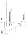
- the rat cDNA was expressed in transgenic mice (Example 3) using the liver-specific ApoE promoter expression system. Analysis of expressors showed a marked increase in bone density, particularly in long bones (femurs), vertebrae and flat bones (pelvis). Histological analysis of stained sections of bone showed severe osteopetrosis (see Example 4) indicating a marked imbalance between bone formation and resorption which has led to a marked accumulation of bone and cartilage. A decrease in the number of trabecular osteoclasts in the bones of OPG expressor animals indicate that a significant portion of the activity of the TNFR-related protein may be to prevent bone resorption, a process mediated by osteoclasts. In view of the activity in transgenic expressors, the TNFR-related proteins described herein are termed OPGs.
- mouse and human cDNA clones were isolated (Example 5). Expression of mouse OPG in 293 cells and human OPG in E. coli is described in Examples. 7 and 8. Mouse OPG was produced as an Fc fusion which was purified by Protein A affinity chromatography. Also described in Example 7 is the expression of full-length and truncated human and mouse OPG polypeptides in CHO and 293 cells either as fusion polypeptides to the Fc region of human IgGI or as unfused polypeptides. The expression of full-length and truncated human and mouse OPGs in E . coli either as Fc fusion polypeptides or as unfused polypeptides is described in Example 8. Purification of recombinantly produced mammalian and bacterial OPG is described in Example 10.
- OPG The biological activity of OPG was determined using an in vitro osteoclast maturation assay, an in vivo model of interleukin-1 (IL-1) induced hypercalcemia, and injection studies of bone density in normal mice (see Example 11).
- Truncated OPG polypeptides having deletions in the region of amino acids 186-401 e.g., OPG [1-185] and OPG [1-194]
- OPG may be important in regulating bone resorption.
- the protein appears to act as a soluble receptor of the TNF family and may prevent a receptor-ligand interaction involved in the osteolytic pathway.
- One aspect of the regulation appears to be a reduction in the number of osteoclasts.
- the invention provides for an isolated nucleic acid encoding a polypeptide having at least one of the biological activities of OPG.
- the biological activities of OPG include, but are not limited to, any activity involving bone metabolism and in particular, include increasing bone density.
- the nucleic acids of the invention are selected from the following:
- the invention provides for nucleic acids which encode rat, mouse and human OPG as well as nucleic acid sequences hybridizing thereto which encode a polypeptide having at least one of the biological activities of OPG. Also provided for are nucleic acids which hybridize to a rat OPG EST encompassing nucleotides 148-337 as shown in Figure 1A .
- the conditions for hybridization are generally of high stringency such as 5xSSC, 50% formamide and 42°C described in Example 1 of the specification. Equivalent stringency to these conditions may be readily obtained by adjusting salt and organic solvent concentrations and temperature.
- the nucleic acids in (b) encompass sequences encoding OPG-related polypeptides which do not undergo detectable hybridization with other known members of the TNF receptor superfamily. In a preferred embodiment, the nucleic acids are as shown in Figures 2B-2C (SEQ ID NO:120), 9A-9B (SEQ ID NO:122), and 9C-9D (SEQ ID NO:124).
- hybridizing nucleic acids of the invention may be variable since hybridization may occur in part or all of the polypeptide-encoding regions as shown in Figures 2B-2C (SEQ ID NO:120), 9A-9B (SEQ ID NO:122), and 9C-9D (SEQ ID NO:124), and may also occur in adjacent noncoding regions. Therefore, hybridizing nucleic acids may be truncations or extensions of the sequences shown in Figures 2B-2C (SEQ ID NO:120), 9A-9B (SEQ ID NO:122), and 9C-9D (SEQ ID NO:124) . Truncated or extended nucleic acids are encompassed by the invention provided they retain one or more of the biological properties of OPG.
- the hybridizing nucleic acids may also include adjacent noncoding regions which are 5' and/or 3' to the OPG coding region.
- the noncoding regions include regulatory regions involved in OPG expression, such as promoters, enhance, translational initiation sites, transcription termination sites and the like.
- DNA encoding rat OPG was provided in plasmid pMO-B1.1 deposited with the American Type Culture Collection, Rockville, MD on December 27, 1995 under ATCC accession no. 69970.
- DNA encoding mouse OPG was provided in plasmid pRcCMV-murine OPG deposited with the American Type Culture Collection, Rockville, MD on December 27, 1995 under accession no. 69971.
- DNA encoding human OPG was provided in plasmid pRcCMV - human OPG deposited with the American Type Culture Collection, Rockville, MD on December 27, 1995 under accession no. 69969.
- the nucleic acids of the invention will hybridize under stringent conditions to the DNA inserts of ATCC accession nos. 69969, 69970, and 69971 and have at least one of the biological activities of OPG .
- derivatives of the nucleic acid sequences as shown in Figures 2B , 9A and 9B .
- derivatives include nucleic acid sequences having addition, substitution, insertion or deletion of one or more residues such that the resulting sequences encode polypeptides having one or more amino acid residues which have been added, deleted, inserted or substituted and the resulting polypeptide has the activity of OPG.
- the nucleic acid derivatives may be naturally occurring, such as by splice variation or polymorphism, or may be constructed using site-directed mutagenesis techniques available to the skilled worker.
- nucleic acid encoding a lys to asn change at residue 3 within the leader sequence (see Example 5). It is anticipated that nucleic acid derivatives will encode amino acid changes in regions of the molecule which are least likely to disrupt biological activity. Other derivatives include a nucleic acid encoding a membrane-bound form of OPG having an extracellular domain as shown in Figures 2B-2C (SEQ ID NO:120), 9A-9B (SEQ ID NO:122), and 9C-9D (SEQ ID NO:124) along with transmembrane and cytoplasmic domains.
- derivatives of OPG include nucleic acids encoding truncated forms of OPG having one or more amino acids deleted from the carboxy terminus.
- Nucleic acids encoding OPG may have from 1 to 216 amino acids deleted from the carboxy terminus.
- an antibody Fc region may extend from the new carboxy terminus to yield a biologically active OPG-Fc fusion polypeptide. (see Example 11).
- nucleic acids encode OPG having the amino acid sequence from residues 22-185, 22-189, 22-194 or 22-201 (using numbering in Figure 9E-F ) and optionally, encoding an Fc region of human IgG.
- nucleic acids encoding truncated forms of OPG having one or more amino acids deleted from the amino terminus Truncated forms include those lacking part or all the 21 amino acids comprising the leader sequence.
- the invention provides for nucleic acids encoding OPG having from 1 to 10 amino acids deleted from the mature amino terminus (at residue 22) and ,optionally, having from 1 to 216 amino acids deleted from the carboxy terminus (at residue 401).
- the nucleic acids may encode a methionine residue at the amino terminus. Examples of such OPG truncated polypeptides are described in Example 8.
- nucleic acids of the invention examples include cDNA, genomic DNA, synthetic DNA and RNA.
- cDNA is obtained from libraries prepared from mRNA isolated from various tissues expressing OPG. In humans, tissue sources for OPG include kidney, liver, placenta and heart.
- Genomic DNA encoding OPG is obtained from genomic libraries which are commercially available from a variety of species.
- Synthetic DNA is obtained by chemical synthesis of overlapping oligonucleotide fragments followed by assembly of the fragments to reconstitute part or all of the coding region and flanking sequences (see U.S. Patent No. 4,695,623 describing the chemical synthesis of interferon genes).
- RNA is obtained most easily by procaryotic expression vectors which direct high-level synthesis of mRNA, such as vectors using T7 promoters and RNA polymerase.
- Nucleic acid sequences of the invention are used for the detection of OPG sequences in biological samples in order to determine which cells and tissues are expressing OPG mRNA.
- the sequences may also be used to screen cDNA and genomic libraries for sequences related to OPG. Such screening is well within the capabilities of one skilled in the art using appropriate hybridization conditions to detect homologus sequences.
- the nucleic acids are also useful for modulating the expression of OPG levels by anti-sense therapy or gene therapy.
- the nucleic acids are also used for the development of transgenic animals which may be used for the production of the polypeptide and for the study of biological activity (see Example 3).
- Expression vectors containing nucleic acid sequences encoding OPG, host cells transformed with said vectors and methods for the production of OPG are also provided by the invention.
- An overview of expression of recombinant proteins is found in Methods of Enzymology v. 185, Goeddel, D.V. ed. Academic Press (1990 ).
- Host cells for the production of OPG include procaryotic host cells, such as E . coli, yeast, plant, insect and mammalian host cells.
- E . coli strains such as HB101 or JM101 are suitable for expression.
- Preferred mammalian host cells include COS, CHOd-, 293, CV-1, 3T3, baby hamster kidney (BHK) cells and others.
- Mammalian host cells are preferred when post-translational modifications, such as glycosylation and polypeptide processing, are important for OPG activity. Mammalian expression allows for the production of secreted polypeptides which may be recovered from the growth medium.
- Vectors for the expression of OPG contain at a minimum sequences required for vector propogation and for expression of the cloned insert. These sequences include a replication origin, selection marker, promoter, ribosome binding site, enhancer sequences, RNA splice sites and transcription termination site. Vectors suitable for expression in the aforementioned host cells are readily available and the nucleic acids of the invention are inserted into the vectors using standard recombinant DNA techniques. Vectors for tissue-specific expression of OPG are also included. Such vectors include promoters which function specifically in liver, kidney or other organs for production in mice, and viral vectors for the expression of OPG in targeted human cells.
- OPG is produced recombinantly by culturing a host cell transformed with an expression vector containing nucleic acid sequences encoding OPG under conditions such that OPG is produced, and isolating the product of expression.
- OPG is produced in the supernatant of transfected mammalian cells or in inclusion bodies of transformed bacterial host cells. OPG so produced may be purified by procedures known to one skilled in the art as described below.
- the expression of OPG in mammalian and bacterial host systems is described in Examples 7 and 8.
- Expression vectors for mammalian hosts are exemplified by plasmids such as pDSR ⁇ described in PCT Application No. 90/14363 .
- Plasmid pAMG21 was deposited with the American Type Culture Collection, Rockville, MD on July 24, 1996 under accession no. 98113. Plasmid pAMG22-His was deposited with the American Type Culture Collection, Rockville, MD on July 24, 1996 under accession no. 98112. It is anticipated that the specific plasmids and host cells described are for illustrative purposes and that other available plasmids and host cells could also be used to express the polypeptides.
- the invention also provides for expression of OPG from endogenous nucleic acids by in vivo or ex vivo recombination events to allow modulation of OPG from the host chromosome.
- Expression of OPG by the introduction of exogenous regulatory sequences (e.g. promoters or enhancers) capable of directing the production of OPG from endogenous OPG coding regions is also encompassed.
- Stimulation of endogenous regulatory sequences capable of directing OPG production is also provided by the invention. the invention.
- OPG a novel member of the TNF receptor superfamily, having an activity associated with bone metabolism and in particular having the activity of inhibiting bone resorption thereby increasing bone density.
- OPG refers to a polypeptide having an amino acid sequence of mouse, rat or human OPG or a derivative thereof having at least one of the biological activities of OPG.
- the amino acid sequences of rat, mouse and human OPG are shown in Figures 2B-2C (SEQ ID NO:121), 9A-9B (SEQ ID NO:123), and 9C-9D (SEQ ID NO:125) respectively.
- a derivative of OPG refers to a polypeptide having an addition, deletion, insertion or substitution of one or more amino acids such that the resulting polypeptide has at least one of the biological activities of OPG.
- the biological activities of OPG include, but are not limited to, activities involving bone metabolism.
- the polypeptides will have the amino terminal leader sequence of 21 amino acids removed.
- OPG polypeptides encompassed by the invention include rat [1-401], rat [22-180], rat [22-401], rat [22-401]-Fc fusion, rat [1-180]-Fc fusion, mouse [1-401], mouse [1-180], mouse [22-401], human [1-401], mouse [22-180], human [22-401], human [22-180], human [1-180], human [22-180]-Fc fusion and human met-32-401. Amino acid numbering is as shown in SEQ ID NO:121 (rat), SEQ ID NO:123 (mouse) and SEQ ID NO:125 (human).
- polypeptide derivatives having deletions or carboxy-terminal truncations of part or all of amino acids residues 180-401 of OPG; one or more amino acid changes in residues 180-401; deletion of part or all of a cysteine-rich domain of OPG, in particular deletion of the distal (carboxy-terminal) cysteine-rich domain; and one or more amino acid changes in a cysteine-rich domain, in particular in the distal (carboxy-terminal) cysteine-rich domain.
- OPG has from 1 to about 216 amino acids deleted from the carboxy terminus.
- OPG has from 1 to about 10 amino acids deleted from the mature amino terminus (wherein the mature amino terminus is at residue 22) and, optionally, has from 1 to about 216 amino acids deleted from the carboxy terminus.
- Additional OPG polypeptides encompassed by the invention include the following: human [22-180]-Fc fusion, human [22-201]-Fc fusion, human [22-401]-Fc fusion, mouse [22-185]-Fc fusion, mouse [22-194]-Fc fusion, These polypeptides are produced in mammalian host cells, such as CHO or 293 cells, Additional OPG polypeptides encompassed by the invention which are expressed in procaryotic host cells include the following: human met[22-401], Fc-human met[22-401] fusion (Fc region is fused at the amino terminus of the full-length OPG coding sequence as described in Example 8), human met[22-401]-Fc fusion (Fc region fused to the full-lengh OPG sequence), Fc-mouse met[22-401] fusion, mouse met[22-401]-Fc fusion, human met[27-401], human met[22-185], human met[22-189], human
- OPG polypeptides produced in procaryotic host cells have an amino-terminal methionine residue, if such a residue is not indicated.
- OPG-Fc fusion were produced using a 227 amino acid region of human IgGl- ⁇ l was used having the sequence as shown in Ellison et al. (Nuc. Acids Res. 10 , 4071-4079 (1982) ) .
- variants of the Fc region of human IgG may also be used.
- OPG carboxy-terminal OPG truncations fused to the human IgGI Fc region indicates a portion of OPG of about 164 amino acids which is required for activity.
- This region encompasses amino acids 22-185, preferably those in Figure 9C-9D (SEQ ID NO:125), and comprises four cysteine-rich domains characteristic of the cysteine-rich domains of TNFR extraceullular domains.
- OPG extracellular ligand binding domains of TNF receptor family members
- a three-dimensional model of OPG was generated based upon the known crystal structure of the extracellular domain of TNFR-I (see Example 6). This model was used to identify those residues within OPG which may be important for biological activity. Cysteine residues that are involved in maintaining the structure of the four cysteine-rich domains were identified.
- Domain 1 cys41 to cys54, cys44 to cys62, tyr23 and his 66 may act to stabilize the structure of this domain; Domain 2: cys65 to cys80, cys83 to cys98, cys87 to cys105; Domain 3: cys107 to cys118 cys124 to cys142; Domain 4: cys145 to cys160, cys166 to cys185. Residues were also identified which were in close proximity to TNF ⁇ as shown in Figures 11 and 12A-12B .
- OPG binds to a corresponding ligand
- TNF ⁇ was used as a model ligand to simulate the interaction of OPG with its ligand.
- residues in OPG may be important for ligand binding: glu34, lys43, pro66 to gln91 (in particular, pro66, his68, tyr69, tyr70, thr71, asp72, ser73, his76, ser77, asp78, glu79, leu81, tyr82, pro85, val86, lys88, glu90 and gln91), glu153 and ser155.
- Alterations in these amino acid residues may alter the biological activity of OPG.
- changes in specific cysteine residues may alter the structure of individual cysteine-rich domains, whereas changes in residues important for ligand binding may affect physical interactions of OPG with ligand.
- Structural models can aid in identifying analogs which have more desirable properties, such as enhanced biological activity, greater stability, or greater ease of formulation.
- OPG multimer comprising OPG monomers.
- OPG appears to be active as a multimer (e.g, dimer, trimer of a higher number of monomers).
- OPG multimers are dimers or trimers.
- OPG multimers may comprise monomers having the amino acid sequence of OPG sufficient to promote multimer formation or may comprise monomers having heterologous sequences such as an antibody Fc region. Analysis of carboxy-terminal deletions of OPG suggest that at least a portion of the region 186-401 is involved in association of OPG polypeptides. Substitution of part or all of the region of OPG amino acids 186-401 with an amino acid sequence capable of self-association is also encompassed by the invention.
- OPG polypeptides or derivatives thereof may be modified to form dimers or multimers by site directed mutagenesis to create unpaired cysteine residues for interchain disulfide bond romation, by photochemical crosslinking, such as exposure to ultraviolet light, or by chemical crosslinking with bifunctional linker molecules such as bifunctional polyethylene glycol and the like.
- OPG polypeptides are encompassed by the invention and include post-translational modifications (e.g., N-linked or O-linked carbohydrate chains, processing of N-terminal or C-terminal ends), attachment of chemical moieties to the amino acid backbone, chemical modifications of N-linked or O-linked carbohydrate chains, and addition of an N-terminal methionine residue as a result of procaryotic host cell expression.
- the polypeptides may also be modified with a detectable label, such as an enzymatic, fluorescent, isotopic or affinity label to allow for detection and isolation of the protein.
- OPG chimeric proteins wherein OPG is fused to a heterologous amino acid sequence.
- the heterologous sequence may be any sequence which allows the resulting fusion protein to retain the activity of OPG.
- the heterologous sequences include for example, immunoglobulin fusions, such as Fc fusions, which may aid in purification of the protein.
- a heterologous sequence which promotes association of OPG monomers to form dimers, trimers and other higher multimeric forms is preferred.
- polypeptides of the invention are isolated and purified from other polypeptides present in tissues, cell lines and transformed host cells expressing OPG, or purified from components in cell cultures containing the secreted protein.
- the polypeptide is free from association with other human proteins, such as the expression product of a bacterial host cell.
- the chemical moieties for derivitization may be selected from water soluble polymers such as polyethylene glycol, ethylene glycol/propylene glycol copolymers, carboxymethylcellulose, dextran, polyvinyl alcohol and the like.
- the polypeptides may be modified at random positions within the molecule, or at predetermined positions within the molecule and may include one, two, three or more attached chemical moieties.
- the polymer may be of any molecular weight, and may be branched or unbranched.
- the preferred molecular weight is between about IkDa and about 100kDa (the term "about” indicating that in preparations of polyethylene glycol, some molecules will weigh more, some less, than the stated molecular weight) for ease in handling and manufacturing.
- Other sizes may be used, depending on the desired therapeutic profile (e.g., the duration of sustained release desired, the effects, if any on biological activity, the ease in handling, the degree or lack of antigenicity and other known effects of the polyethylene glycol to a therapeutic protein or analog).
- polyethylene glycol molecules should be attached to the protein with consideration of effects on functional or antigenic domains of the protein.
- attachment methods available to those skilled in the art, e.g. EP 0 401 384 herein incorporated by reference (coupling PEG to G-CSF), see also Malik et al., Exp. Hematol. 20: 1028-1035 (1992 ) (reporting pegylation of GM-CSF using tresyl chloride).
- polyethylene glycol may be covalently bond through amino acid residues via a reactive group, such as, a free amino or carboxyl group.
- Reactive groups are those to which an activated polyethylene glycol molecule may be bound.
- the amino acid residues having a free amino group may include lysine residues and the N-terminal amino acid residues; those having a free carboxyl group may include aspartic acid residues glutamic acid residues and the C-terminal amino acid residue.
- Sulfhydrl groups may also be used as a reactive group for attaching the polyethylene glycol molecule(s). Preferred for therapeutic purposes is attachment at an amino group, such as attachment at the N-terminus or lysine group.
- N-terminally chemically modified protein One may specifically desire N-terminally chemically modified protein.
- polyethylene glycol as an illustration of the present compositions, one may select from a variety of polyethylene glycol molecules (by molecular weight, branching, etc.), the proportion of polyethylene glycol molecules to protein (or peptide) molecules in the reaction mix, the type of pegylation reaction to be performed, and the method of obtaining the selected N-terminally pegylated protein.
- the method of obtaining the N-terminally pegylated preparation i.e., separating this moiety from other monopegylated moieties if necessary
- Selective N-terminal chemically modification may be accomplished by reductive alkylation which exploits differential reactivity of different types of primary amino groups (lysine versus the N-terminal) available for derivatization in a particular protein. Under the appropriate reaction conditions, substantially selective derivatization of the protein at the N-terminus with a carbonyl group containing polymer is achieved.
- Synthetic OPG dimers may be prepared by various chemical crosslinking procedures.
- OPG monomers may be chemically linked in any fashion that retains or enhances the biological activity of OPG.
- a variety of chemical crosslinkers may be used depending upon which properties of the protein dimer are desired. For example, crosslinkers may be short and relatively rigid or longer and more flexible, may be biologically reversible, and may provide reduced immunogenicity or longer pharmacokinetic half-life.
- OPG molecules are linked through the amino terminus by a two step synthesis (see Example 12).
- OPG is chemically modified at the amino terminus to introduce a protected thiol, which after purification is deprotected and used as a point of attachment for site-specific conjugation through a variety of crosslinkers with a second OPG molecule.
- Amino-terminal crosslinks include, but are not limited to, a disulfide bond, thioether linkages using short-chain, bis-functional aliphatic crosslinkers, and thioether linkages to variable length, bifunctional polyethylene glycol crosslinkers (PEG "dumbbells").
- OPG dumbbell synthesis of OPG dimers is a byproduct of such synthesis, termed a "monobell".
- An OPG monobell consists of a monomer coupled to a linear bifunctional PEG with a free polymer terminus.
- OPG may be crosslinked directly through a variety of amine specific homobifunctional crosslinking techniques which include reagents such as: diethylenetriaminepentaacetic dianhydride (DTPA), p-benzoquinone (pBQ) or bis(sulfosuccinimidyl) suberate (BS 3 ) as well as others known in the art.
- DTPA diethylenetriaminepentaacetic dianhydride
- pBQ p-benzoquinone
- BS 3 bis(sulfosuccinimidyl) suberate
- a method for the purification of OPG from natural sources and from transfected host cells is also included.
- the purification process may employ one or more standard protein purification steps in an appropriate order to obtain purified protein.
- the chromatography steps can include ion exchange, gel filtration, hydrophobic interaction, reverse phase, chromatofocusing, affinity chromatography employing an anti-OPG antibody or biotin-streptavidin affinity complex and the like.
- Antibodies specifically binding to OPG are antibodies specifically binding to OPG.
- Antigens for the generation of antibodies may be full-length polypeptides or peptides spanning a portion of the OPG sequence. Immunological procedures for the generation of polyclonal or monoclonal antibodies reactive with OPG are known to one skilled in the art (see, for example, Harlow and Lane, Antibodies: A Laboratory Manual Cold Spring Harbor Laboratory Press, Cold Spring Harbor N.Y. (1988 )). Antibodies so produced are characterized for binding specificity and epitope recognition using standard enzyme-linked immunosorbent assays. Antibodies also include chimeric antibodies having variable and constant domain regions derived from different species. In one embodiment, the chimeric antibodies are humanized antibodies having murine variable domains and human constant domains.
- CDR-grafted-antibodies complementary determining regions grafted to a human framework
- Chimeric and CDR-grafted antibodies are made by recombinant methods known to one skilled in the art.
- human antibodies made in mice.
- Anti-OPG antibodies of the invention may be used as an affinity reagent to purify OPG from biological samples (see Example 10).
- the antibody is immobilized on CnBr-activated Sepharose and a column of antibody-Sepharose conjugate is used to remove OPG from liquid samples.
- Antibodies are also used as diagnostic reagents to detect and quantitate OPG in biological samples by methods described below.
- compositions comprising a therapeutically effective amount of the polypeptide of the invention together with a pharmaceutically acceptable diluent, carrier, solubilizer, emulsifier, preservative and/or adjuvant.
- therapeutically effective amount means an amount which provides a therapeutic effect for a specified condition and route of administration.
- the composition may be in a liquid or lyophilized form and comprises a diluent (Tris, acetate or phosphate buffers) having various pH values and ionic strengths, solubilizer such as Tween or Polysorbate, carriers such as human serum albumin or gelatin, preservatives such as thimerosal or benzyl alcohol, and antioxidants such as ascrobic acid or sodium metabisulfite.
- a diluent Tris, acetate or phosphate buffers
- solubilizer such as Tween or Polysorbate
- carriers such as human serum albumin or gelatin
- preservatives such as thimerosal or benzyl alcohol
- antioxidants such as ascrobic acid or sodium metabisulfite.
- compositions comprising OPG modified with water soluble polymers to increase solubility or stability.
- Compositions may also comprise incorporation of OPG into liposomes, microemulsions, micelles or vesicles for controlled delivery over an extended
- OPG compositions may comprise incorporation into polymer matricies such as hydrogels, silicones, polyethylenes, ethylene-vinyl acetate copolymers, or biodegradable polymers.
- hydrogels include polyhydroxyalkylmethacrylates (p-HEMA), polyacrylamide, polymethacrylamide, polyvinylpyrrolidone, polyvinyl alcohol and various polyelectrolyte complexes.
- biodegradable polymers include polylactic acid (PLA), polyglycolic acid (PGA), copolymers of PLA and PGA, polyamides and copolymers of polyamides and polyesters.
- Other controlled release formulations include microcapsules, microspheres, macromolecular complexes and polymeric beads which may be administered by injection.
- compositions of the invention may be administered by injection, either subcutaneous, intravenous or intramuscular, or by oral, nasal, pulmonary or rectal administration.
- the route of administration eventually chosen will depend upon a number of factors and may be ascertained by one skilled in the art.
- the invention also provides for pharmaceutical compositions comprising a therapeutically effective amount of the nucleic acids of the invention together with a pharmaceutically acceptable adjuvant.
- Nucleic acid compositions will be suitable for the delivery of part or all of the OPG coding region to cells and tissues as part of an anti-sense or gene therapy regimen.
- Bone tissue provides support for the body and consists of mineral (largely calcium and phosphorous), a matrix of collagenous and noncollagenous proteins, and cells.
- mineral largely calcium and phosphorous
- a matrix of collagenous and noncollagenous proteins a matrix of collagenous and noncollagenous proteins
- cells Three types of cells found in bone, osteocytes, osteoblasts and osteoclasts, are involved in the dynamic process by which bone is continually formed and resorbed. Osteoblasts promote formation of bone tissue whereas osteoclasts are associated with resorption. Resorption, or the dissolution of bone matrix and mineral, is a fast and efficient process compared to bone formation and can release large amounts of mineral from bone. Osteoclasts are involved in the regulation of the normal remodeling of skeletal tissue and in resorption induced by hormones.
- resorption is stimulated by the secretion of parathyroid hormone in response to decreasing concentrations of calcium ion in extracellular fluids.
- inhibition of resorption is the principal function of calcitonin.
- metabolites of vitamin D alter the responsiveness of bone to parathyroid hormone and calcitonin.
- the amount of bone in the skeleton reflects the balance (or imbalance) of bone formation and bone resorption. Peak bone mass occurs after skeletal maturity prior to the fourth decade. Between the fourth and fifth decades, the equilibrium shifts and bone resorption dominates. The inevitable decrease in bone mass with advancing years starts earlier in females than males and is distinctly accelerated after menopause in some females (principally those of Caucasian and Asian descent).
- Osteopenia is a condition relating generally to any decrease in bone mass to below normal levels. Such a condition may arise from a decrease in the rate of bone synthesis or an increase in the rate of bone destruction or both.
- the most common form of osteopenia is primary osteoporosis, also referred to as postmenopausal and senile osteoporosis. This form of osteoporosis is a consequence of the universal loss of bone with age and is usually a result of increase in bone resorption with a normal rate of bone formation. About 25 to 30 percent of all white females in the United States develop symptomatic osteoporosis.
- osteoporosis A direct relationship exists between osteoporosis and the incidence of hip, femoral, neck and inter-trochanteric fracture in women 45 years and older. Elderly males develop symptomatic osteoporosis between the ages of 50 and 70, but the disease primarily affects females.
- the invention provides for a method of treating a bone disorder using a therapeutically effective amount of OPG.
- the bone disorder may be any disorder characterized by a net bone loss (osteopenia or osteolysis).
- treatment with OPG is anticipated when it is necessary to suppress the rate of bone resorption.
- treatment may be done to reduce the rate of bone resorption where the resorption rate is above normal or to reduce bone resorption to below normal levels in order to compensate for below normal levels of bone formation.
- Conditions which are treatable with OPG include the following:
- osteoprotegerein is used in conjunction with a therapeutically effective amount of a factor which stimulates bone formation.
- factors include but are not limited to the bone morphogenic factors designated BMP-1 through BMP-12, transforming growth factor- ⁇ (TGF- ⁇ ) and TGF- ⁇ family members, interleukin-1 inhibitors, TNF ⁇ inhibitors, parathyroid hormone and analogs thereof, parathyroid related protein and analogs thereof, E series prostaglandins, bisphosphonates (such as alendronate and others), and bone-enhancing minerals such as fluoride and calcium.
- PCR Polymerase chain reactions
- 25-50 ⁇ l reactions were denatured at 94°C, followed by 20-40 cycles of 94°C for 5 seconds, 50-60°C for 5 seconds, and 72°C for 3-5 minutes. Reactions were the treated for 72 °C for 3-5 minutes. Reactions were then analyzed by gel electrophoresis as described in Maniatis et al., ibid.
- a cDNA library was constructed using mRNA isolated from embryonic d20 intestine for EST analysis ( Adams et al. Science 252, 1651-1656 (1991 )). Rat embryos were dissected, and the entire developing small and large intestine removed and washed in PBS. Total cell RNA was purified by acid guanidinium thiocyanate-phenol-chloroform extraction ( Chomczynski and Sacchi Anal. Biochem. 162, 156-159, (1987 )). The poly (A+) mRNA fraction was obtained from the total RNA preparation by adsorption to, and elution from, Dynabeads Oligo (dT)25 (Dynal Corp) using the manufacturer's recommended procedures.
- a random primed cDNA library was prepared using the Superscript Plasmid System (Gibco BRL, Gaithersburg, Md).
- the random cDNA primer containing an internal Not I restriction site was used to initiate first strand synthesis and had the following sequence:
- For the first strand synthesis three separate reactions were assembled that contained 2.5 ⁇ g of poly(A) RNA and 120 ng, 360 ng or 1,080 ng of random primer. After second strand synthesis, the reaction products were separately extracted with a mixture of phenol:choroform:isoamyl alcohol (25:24:1 ratio), and then ethanol precipitated.
- the double strand (ds) cDNA products of the three reactions were combined and ligated to the following ds oligonucleotide adapter:
- the cDNA was digested to completion with Not I, extracted with phenol:chloroform:isoamyl (25:24:1) alcohol and ethanol precipitated.
- the resuspended cDNA was then size fractionated by gel filtration using premade columns provided with the Superscript Plasmid System (Gibco BRL, Gaithersburg, Md) as recommended by the manufacturer.
- the two fractions containing the largest cDNA products were pooled, ethanol precipitated and then directionally ligated into Not I and Sal I digested pMOB vector DNA (Strathmann et al, 1991).
- the ligated cDNA was introduced into competent ElectroMAX DH10B E.
- glycerol stocks were thawed, and small aliquots dilute 1:25 in distilled. Approximately 3.0 ul of diluted bacterial cultures were added to PCR reaction mixture (Boehringer-Mannheim) containing the following oligonucleotides:
- thermocycler Perkin-Elmer 9600
- cycle conditions 94 C for 2 minutes; 30 cycles of 94°C for 5 seconds, 50°C for 5 seconds, and 72°C for 3 minutes.; 72°C for 4 minutes.
- the reactions were diluted with 2.0 mL of water.
- the amplified DNA fragments were further purified using Centricon columns (Princeton Separations) using the manufacturer's recommended procedures.
- PCR reaction products were sequenced on an Applied Biosystems 373A automated DNA sequencer using T3 primer (oligonucleotide 353-23; 5'-CAATTAACCCTCACTAAAGG-3') (SEQ ID NO:6) Taq dye-terminator reactions (Applied Biosystems) following the manufacturer's recommended procedures.
- T3 primer oligonucleotide 353-23; 5'-CAATTAACCCTCACTAAAGG-3'
- SEQ ID NO:6 Taq dye-terminator reactions
- the resulting 5' nucleotide sequence obtained from randomly picked cDNA clones translated and then compared to the existing database of known protein sequences using a modified version of the FASTA program ( Pearson et al. Meth. Enzymol. 183, (1990 )).
- Translated sequences were also analysed for the presence of a specific cysteine-rich protein motif found in all known members of the tumor necrosis factor receptor (TNFR) superfamily ( Smith et al. Cell 76., 959-962 (1994 )), using the sequence profile method of Gribskov et al. (Proc. Natl. Acad. Sci. USA 83, 4355-4359 (1987 )), as modified by Luethy et al. (Protein Science 3, 139-146 (1994 ) ) .
- TNFR tumor necrosis factor receptor
- FRI-1 Fetal Rat Intestine-1
- FRI-1 contained an approximately 600 bp insert with a LORF of about 150 amino acids.
- the closest match in the database was the human type II TNFR (TNFR-2).
- TNFR-2 human type II TNFR
- TNFR-2 human type II TNFR
- Profile analysis using the first and second cysteine-rich repeats of the TNFR superfamily yielded a Z score of -8, indicating that the FRI-1 gene possibly encodes a new family member.
- the fetal rat intestine cDNA library was screened for full length clones. The following oligonucleotides were derived from the original FRI-1 sequence:
- PCR reaction mixture Boehringer-Mannheim
- Perkin-Elmer 96 well thermal cycler with the following cycle conditions: 2 min at 94°C,1 .cycle; 15 sec at 94°C, then 45 sec at 65°C, 30 cycles; 7 min at 65°C, 1 cycle.
- PCR reaction products were analysed by gel electrophoresis. 13 out of 96 plasmid DNA pools gave rise to amplified DNA products with the expected relative molecular mass.
- DNA from one positive pool was used to transform competent ElectroMAX DH10B E . coli (Gibco BRL, Gaithersburg, MD) as described above. Approximately 40,000 transformants were plated onto sterile nitrocellulose filters (BA-85, Schleicher and Schuell), and then screened by colony hybridization using a 32 P-dCTP labelled version of the PCR product obtained above. Filters were prehybridized in 5X SSC, 50% deionized formamide, 5X Denhardt's solution, 0.5% SDS, and 100 ug/ml denatured salmon sperm DNA for 2-4 hours at 42°C.
- Filters were then hybridized in 5X SSC, 50% deionized formamide, 2X Denhardt's solution, 0.1% SDS, 100 ⁇ g/ml denatured salmon sperm DNA, and ⁇ 5 ng/ml of labelled probe for ⁇ 18 hours at 42°C. The filters were then washed in 2X SSC for 10 min at RT, 1X SSC for 10 min at 55°C, and finally in 0.5X SSC for 10-15 min at 55°C. Hybridizing clones were detected following autoradiography, and then replated onto nitrocellulose filters for secondary screening.
- plasmid clone (pBl.1) was isolated, then amplified in L-broth media containing 100 ug/ml ampicillin and the plasmid DNA obtained. Both strands of the 2.4 kb pBl.1 insert were sequenced.
- the pBl.1 insert sequence was used for a FASTA search of the public database to detect any existing sequence matches and/or similarities. No matches to any known genes or EST's were found, although there was an approximate 45% similarity to the human and mouse TNFR-2 genes.
- a methionine start codon is found at bp 124 of the nucleotide sequence, followed by a LORF encoding 401 aa residues that terminates at bp 1327.
- the 401 aa residue product is predicted to have a hydrophobic signal peptide of approximately 31 residues at its N-terminus, and 4 potential sites of N-linked glycosylation.
- the homologus region maps to the extracellular domain of TNFR family members, and corresponds to the three or four cysteine-rich repeats found in the ligand binding domain of these proteins. This suggested that the FRI-1 gene encoded a novel TNFR family member. Since no transmembrane spanning region was detected we predicted that this may be a secreted receptor, similar to TNFR-1 derived soluble receptors ( Kohno et al. Proc. Natl. Acad. Sci. USA 87, 8331-8335 (1990 )). Due to the apparent biological activity of the FRI-1 gene (vide infra), the product was named Osteoprotegerin (OPG).
- OPG Osteoprotegerin
- Northern blots were prehybridized in 5X SSPE, 50% formamide, 5X Denhardt's solution, 0.5% SDS, and 100 ⁇ g/ml denatured salmon sperm DNA for 2-4 hr at 42°C. The blots were then hybridized in 5X SSPE, 50% formamide, 2X Denhardt's solution, 0.1% SDS, 100 ⁇ g/ml denatured salmon sperm DNA, and 5 ng/ml labelled probe for 18-24 hr at 42°C. The blots were then washed in 2X SSC for 10 min at RT, 1X SSC for 10 min at 50°C, then in 0.5X SSC for 10-15 min.
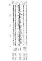
- a predominant mRNA species with a relative molecular mass of about 2.4 kb is detected in several tissues, including kidney, liver, placenta, and heart. Highest levels are detected in the kidney. A large mRNA species of Mr 4.5 and 7.5 kb was detected in skeletal muscle and pancreas. In human fetal tissue, kidney was found to express relatively high levels of the 2.4 kb mRNA. Using a human probe (vide infra), only the 2.4 kb transcript is detected in these same tissues. In addition, relatively high levels of the 2.4 kb transcript was detected in the lymph node, thymus, spleen and appendix. The size of the transcript detected by both the rat and human Osteosprotegerin gene is almost identical to the length of the rat pBl.1 FRI-1 insert, suggesting it was a full length cDNA clone.
- the rat OPG clone pBl.1 was used as template to PCR amplify the coding region for subcloning into an ApoE-liver specific expression vector ( Simonet et al. J. Clin. Invest. 94. 1310-1319 (1994 ), and PCT Application No. US94/11675 and co-owned U.S. Serial No. 08/221,767 .
- the following 5' and 3' oligonucleotide primers were used for PCR amplification, respectively:
- the PCR reaction mixture (Boehringer-Mannheim) was treated as follows: 94°C for 1 minute, 1 cycle; 94°C for 20 sec, 62°C for 30 sec, and 74 C for 1 minute, 25 cycles. Following amplification, the samples were purified over Qiagen PCR columns and digested overnight with SpeI and NotI restriction enzymes. The digested products were extracted and precipitated and subcloned into the ApoE promoter expression vector. Prior to microinjecting the resulting clone, HE-OPG, it was sequenced to ensure it was mutation-free.
- the HE-OPG plasmid was purified through two rounds of CsCl density gradient centrifugation.
- the purified plasmid DNA was digested with XhoI and Ase I, and the 3.6 kb transgene insert was purified by gel electrophoresis.
- the purified fragment was diluted to a stock injection solution of 1 ⁇ g/ml in 5 mM Tris, pH 7.4, 0.2 mM EDTA.
- Single-cell embryos from BDF1 x BDF1-bred mice were injected essentially as described ( Brinster et al., Proc. Natl. Acad. Sci. USA 82, 4338 (1985 )), except that injection needles were beveled and siliconized before use.
- Embryos were cultured overnight in a CO 2 incubator and 15 to 20 2-cell embryos were transferred to the oviducts of pseudopregnant CD1 female mice.
- offspring were obtained from implantation of microinjected embryos.
- the offspring were screened by PCR amplification of the integrated transgene in genomic DNA samples.
- the target region for amplification was a 369 bp region of the human Apo E intron which was included in the expression vector.
- the oligos used for PCR amplification were:
- the conditions for PCR were: 94°C for 2 minute, 1 cycle; 94°C for 1 min, 63°C for 20 sec, and 72°C for 30 sec, 30 cycles. Of the 49 original offspring, 9 were identified as PCR positive transgenic founders.
- mice were anesthetized and a lobe of liver was surgically removed.
- Total cellular RNA was isolated from livers of all transgenic founders, and 5 negative control littermates as described ( McDonald et al. Meth. Enzymol. 152, 219 (1987 )). Northern blot analysis was performed on these samples to assess the level of transgene expression.
- RNA from each animal liver was resolved by electrophoresis denaturing gels ( Ogden et al. Meth. Enzymol 152, 61 (1987 )), then transferred to HYBOND-N nylon membrane (Amersham), and probed with 32 P dCTP-labelled pBl.1 insert DNA. Hybridization was performed overnight at 42°C in 50% Formamide, 5 x SSPE, 0.5% SDS, 5 x Denhardt's solution, 100 ⁇ g/ml denatured salmon sperm DNA and 2-4 x 10 6 cpm of labeled probe/ml of hybridization buffer.
- the northern blot data indicate that 7 of the transgenic founders express detectable levels of the transgene mRNA (animal #'s 2, 11, 16, 17, 22, 33, and 45).
- animal 2, 17 and 22 expressed the highest levels of transgene mRNA, and may show more extensive biological effects on host cells and tissues.
- mice Five of the transgenic mice (animals 2,11,16,17 and 28) and 5 control littermates (animals 1,12,15,18, and 30) were sacrificed for necropsy and pathological analysis using the following procedures: Prior to euthanasia, all animals had their identification numbers verified, then were weighed, anesthetized and blood drawn. The blood was saved as both serum and whole blood for a complete serum chemistry and hematology panel. Radiography was performed just after terminal anesthesia by lethal CO2 inhalation, and prior to the gross dissection. Following this, tissues were removed and fixed in 10% buffered Zn-Formalin for histological examination.
- the tissues collected included the liver, spleen, pancreas, stomach, duodenum, ileum, colon, kidney, reproductive organs, skin and mammary glands, bone, brain, heart, lung, thymus, trachea, eosphagus, thyroid, jejunem, cecum, rectum, adrenals, urinary bladder, and skeletal muscle. Prior to fixation the whole organ weights were determined for the liver, stomach, kidney, adrenals, spleen, and thymus. After fixation the tissues were processed into paraffin blocks, and 3 um sections were obtained. Bone tissue was decalcified using a formic acid solution, and all sections were stained with hematoxylin and eosin.
- the sections were quenched with 3% hydrogen peroxide, blocked with Protein Block (Lipshaw, Pittsburgh, PA), and incubated in rat monoclonal anti-mouse F480 (Harlan, Indianapolis, IN). This antibody was detected by biotinylated rabbit antirat immunoglobulins, peroxidase conjugated strepavidin (BioGenex San Ramon, CA) with DAB as chromagen (BioTek, Santa Barbara, CA). Sections were counterstained with hematoxylin.
- transgene expressors Upon gross dissection and observation of visceral tissues, no abnormalities were found in the transgene expressors or control littermates. Analysis of organ weight indicate that spleen size increased by approximately 38% in the transgenic mice relative to controls. There was a slight enlargement of platelet size and increased circulating unstained cells in the transgene expressors. There was a marginal decrease in platelet levels in the transgene expressors. In addition, the serum uric acid, urea nitrogen, and alkaline phosphatase levels all trended lower in the transgene expressors. The expressors were found to have increased radiodensity of the skeleton, including long bones (femurs), vertebrae, and flat bones (pelvis). The relative size of femurs in the expressors were not different from the the control mice.
- F480 a cell surface antigen expressed by cells of monocyte-macrophage derivation in the mouse, showed the presence of F480 positive cells in the marrow spaces. Focally, flattened F480 positive cells could be seen directly adjacent to trabecular bone surfaces.
- osteoclasts were rarely found on the trabecular bone surfaces in the OPG expressors. In contrast, osteoclasts and/or chondroclasts were seen in the region of the growth plate resorbing cartilage, but their numbers may be reduced compared to controls. Also, osteoclasts were present on the cortical surface of the metaphysis where modelling activity is usually robust. The predominant difference between the expressors and controls was the profound decrease in trabecular osteoclasts, both in the vertebrae and femurs. The extent of bone accumulation was directly correlated with the level of OPG transgene mRNA detected by northern blotting of total liver RNA.
- the spleens from the OPG expressors had an increased amount of red pulp with the expansion due to increased hematopoiesis. All hematopoietic lineages are represented. F480 positive cells were present in both control and OPG expressors in the red pulp. Two of the expressors (2 and 17)had foci of extramedullary hematopoiesis within the liver and this is likely due to the osteopetrotic marrow.
- a cDNA clone corresponding to the 5' end of the mouse OPG mRNA was isolated from a mouse kidney cDNA library (Clontech) by PCR amplification.
- the oligonucleotides were derived from the rat OPG cDNA sequence and are shown below:
- the partial and full-length cDNA products obtained in this process were sequenced.
- the full-length product was digested with Not I and Xba I, then directionally cloned into the plasmid vector pRcCMV (Invitrogen).
- the resulting plasmid was named pRcCMV-Mu-OPG.
- the nucleotide sequence of the cloned product was compared to the rat OPG cDNA sequence. Over the 1300 bp region spanning the OPG LORF, the rat and mouse DNA sequences are approximately 88% identical.
- the mouse cDNA sequence contained a 401 aa LORF, which was compared to the rat OPG protein sequence and found to be ⁇ 94% identical without gaps.
- mouse cDNA sequence isolated encodes the murine OPG protein, and that the sequence and structure has been highly conserved throughout evolution.
- the mouse OPG protein sequence contains an identical putative signal peptide at its N-terminus, and all 4 potential sites of N-linked glycosylation are conserved.
- a partial human OPG cDNA was cloned from a human kidney cDNA library using the following rat-specific oligonucleotides:
- This PCR product was sequenced and used to design primers for amplifying the 3' end of the human cDNA using a human OPG genomic clone in lambda as template:
- the amplified PCR product was sequenced, and together with the 5' end sequence, was used to design 5' and 3' human-specific primers useful for amplifying the entire human OPG cDNA coding sequences:
- the full-length human PCR product was sequenced, then directionally cloned into the plasmid vector pRcCMV (Invitrogen) using Not I and Xba I.
- the resulting plasmid was named pRcCMV-human OPG.
- the nucleotide sequence of the cloned product was compared to the rat and mouse OPG cDNA sequences. Over the 1300 bp region spanning the OPG LORF, the rat and mouse DNA sequences are approximately 78-88% identical to the human OPG cDNA.
- the human OPG cDNA sequence also contained a 401 aa LORF, and it was compared to the rat and mouse protein sequences.
- the predicted human OPG protein is approximatlely 85% identical, and -90% identical to the rat and mouse proteins, respectively. Sequence alignment of rat, mouse and human proteins show that they have been highly conserved during evolution. The human protein is predicted to have a N-terminal signal peptide, and 5 potential sites of N-linked glycosylation, 4 of which are conserved between the rat and mouse OPG proteins.
- the DNA and predicted amino acid sequence of mouse OPG is shown in Figure 9A and 9B (SEQ ID NO:122).
- the DNA and predicted amino acid sequence of human OPG is shown in Figure 9C an 9D (SEQ ID NO:124).
- a comparison of the rat, mouse and human OPG amino acid sequences is shown in Figure 9E and 9F .
- OPG The amino-terminal portion of OPG has homology to the extracellular portion of all known members of the TNFR superfamily ( Figure 1C ).
- the most notable motif in this region of TNFR-related genes is an ⁇ 40 amino acid, cysteine-rich repeat sequence which folds into distinct structures ( Banner et al. Cell 73, 431-445 (1993 )).
- This motif is usually displayed in four (range 3-6) tandem repeats (see Figure 1C ), and is known to be involved in ligand binding ( Beutler and van Huffel Science 264, 667-663 (1994 )). Each repeat usually contains six interspaced cysteine residues, which are involved in forming three intradomain disulfide bonds, termed SS1, SS2, and SS3 (Banner et al., ibid) . In some receptors, such as TNFR2, CD30 and CD40, some of the repeat domains contain only two intrachain disulfide bonds (SS1 and SS3).
- the human OPG protein sequence was aligned to a TNFR1 extracellular domain profile using methods described by Luethy, et al., ibid, and the results were graphically displayed using the PrettyPlot program from the Wisconsin Package, version 8.1 (Genetics Computer Group, Madison, WI) ( Figure 10 ). The alignment indicates a clear conservation of cysteine residues involved in formation of domains 1-4. This alignment was then used to construct a three-dimensional (3-D) model of the human OPG N-terminal domain using the known 3-D structure of the extracellular domain of p55 TNFR1 (Banner et al., ibid ) as the template.
- This model was then used to find the residues of OPG that could interact with its ligand using the following approach: The solvent accessible area of all residues in the complex and one single OPG model were calculated. The residues that have different accessibility in the complex than in the monomer are likely to interact with the ligand.
- the human and mouse OPG amino acid sequences were realigned using this information to highlight sequences comprising each of the cysteine rich domains 1-4 ( Figure 12A and 12B ). Each domain has individual structural characteristics which can be predicted:
- This region of OPG also contains an region stretching from P66-Q91 which aligns to the portion of TNFR1 domain 2 which forms close contacts with TNF ⁇ (see above), and may interact with an OPG ligand.
- residues P66, H68, Y69, Y70, T71, D72, S73, H75, T76, S77, D78, E79, L81, Y82, P85, v86, K88, E89, L90, and Q91 are predicted to interact with a bound ligand based on our structural data.
- the predicted structural model for OPG identifies a number of highly conserved residues which are likely to be important for its biological activity.
- mouse OPG cDNA was fused to the human IgGl Fc domain as a tag ( Capon et al. Nature 337, 525-531 (1989 )), and expressed in human 293 fibroblasts. Fc fusions were carried out using the vector pFc-A3.
- pFc-A3 contains the region encoding the Fc portion of human immunoglobulin IgG- ⁇ l heavy chain (Ellison et al. ibid) from the first amino acid of the hinge domain (Glu-99) to the carboxyl terminus and is flanked by a 5'-NotI fusion site and 3'-SalI and XbaI sites.
- the plasmid was constructed by PCR amplification of the human spleen cDNA library (Clontech). PCR reactions were in a final volume of 100 ⁇ l and employed 2 units of Vent DNA polymerase (New England Biolabs) in 20 mM Tris-HCl (pH 8 . 8), 10 mM KCl, 10 ⁇ M (NH 4 ) 2SO 4 , 2 mM MgSO 4 , 0.1% Triton X-100 with 400 ⁇ m each dNTP and 1 ng of the cDNA library to be amplified together with 1 ⁇ M of each primer. Reactions were initiated by denaturation at 95°C for 2 min, followed by 30 cycles of 95°C for 30 s, 55°C for 30 s, and 73°C for 2 min. The 5' primer
- the cloned mouse cDNA in plasmid pRcCMV-MuOPG was amplified using the following two sets of primer pairs:
- the first pair amplifies the entire OPG LORF, and creates a NotI restriction site which is compatible with the in-frame Not I site in Fc fusion vector pFcA3 pFcA3 was prepared by engineering a NotI restriction site 5' to aspartic acid reside 216 of the human IgGl Fc cDNA
- This construct introduces a linker which encodes two irrelevant amino acids which span the junction between the OPG protein and the IgG Fc region.
- This product when linked to the Fc portion, would encode all 401 OPG residues directly followed by all 227 amino acid residues of the human IgGl Fc region (Fl.Fc).
- the second primer pair amplifies the DNA sequences encoding the first 180 amino acid residues of OPG, which encompasses its putative ligand binding domain.
- the 3' primer creates an artificial Not I restriction site which fuses the C-terminal truncated OPG LORF at position threonine 180 directly to the IgGl Fc domain (CT.fc) .
- the amino acid sequence junction linking OPG residue 401 and aseptic acid residue 221 of the human Fc region can be modified as follows:
- the DNA encoding residues 216-220 of the human Fc region can be deleted as described below, or the cysteine residue corresponding to C220 of the human Fc region can be mutated to either serine or alanine.
- OPF-Fc fusion protein encoded by these modifed vectors can be transfected into human 293 cells, or CHO cells, and recombinant OPG-Fc fusion protein purified as described below.
- pCEP4 contains the Epstein-Barr virus origin of replication, and is capable of episomal replication in 293-EBNA-1 cells.
- the parent pCEP4, and pCEP4-Fl.Fc and pCEP4-CT.Fc vectors were lipofected into 293-EBNA-1 cells using the manufacturer's recommended methods.
- the transfected cells were then selected in 100 ⁇ g/ml hygromycin to select for vector expression, and the resulting drug-resistant mass cultures were grown to confluence.
- the cells were then cultured in serum-free media for 72 hr, and the conditioned media removed and analysed by SDS-PAGE.
- a silver staining of the polyacrylamide gel detects the major conditioned media proteins produced by the drug resistant 293 cultures.
- unique bands of the predicted sizes were abundantly secreted (see Figures 13B and 13C ).
- the full-length Fc fusion protein accumulated to a high concentration, indicating that it may be stable.
- Both Fc fusion proteins were detected by anti-human IgG1 Fc antibodies (Pierce) on western blots, indicating that they are recombinant OPG products.
- the full length OPG-Fc fusion protein was purified by Protein-A column chromatography (Pierce) using the manufacturers recommended procedures. The protein was then subjected to N-terminal sequence analysis by automated Edman degradation as essentially described by Matsudaira et al. (J. Biol. Chem. 262, 10-35 (1987 )). The following amino acid sequence was read after 19 cycles:
- Murine OPG cDNA encoding amino acids 1-185 fused to the Fc region of human IgGl [muOPG Ct(185).Fc] was constructed as follows.
- Murine OPG cDNA from plasmid pRcCMV Mu Osteoprotegerin (described in Example 5) was amplified using the following primer pair in a polymerase chain reaction as described above:
- This primer pair amplifies the murine OPG cDNA region encoding amino acid residues 63-185 (corresponding to bp 278-645) of the OPG reading frame as shown in Figure 9A .
- the 3' primer contains a Not I restriction site which is compatible with the in-frame Not I site of the Fc fusion vector pFcA3.
- the product also spans a unique ECORI restriction site located at bp 436.
- the amplified PCR product was purified, cleaved with NotI and EcoRI, and the resulting EcoRI-NotI restriction fragment was purified.
- the vector pCEP4 having the murine 1-401 OPG-Fc fusion insert was cleaved with EcoRI and NotI, purified, and ligated to the PCR product generated above.
- the resulting pCEP4-based expression vector encodes OPG residues 1-185 directly followed by all 227 amino acid residues of the human IgGl Fc region.
- the murine OPG 1-185.Fc fusion vector was transfected into 293 cells, drug selected, and conditioned media was produced as described above.
- the resulting secreted murine OPG 1-185.Fc fusion product was purified by Protein-A column chromatography (Pierce) using the manufacturers recommended procedures.
- Murine OPG DNA encoding amino acid residues 1-194 fused to the Fc region of human IgGl (muOPG Ct(194).Fc) was constructed as follows.
- Mouse OPG cDNA from plasmid pRcCMV Mu-Osteoprotegerin was amplified using the following primer pairs:
- This primer pair amplifies the murine OPG cDNA region encoding amino acid residues 70-194 (corresponding to bp 298-672) of the OPG reading frame.
- the 3' primer contains a Not I restriction site which is compatible with the in-frame Not I site of the Fc fusion vector pFcA3.
- the product also spans a unique EcoRI restriction site located at bp 436.
- the amplified PCR product was cloned into the murine OPG[1-401] Fc fusion vector as described above.
- the resulting pCEP4-based expression vector encodes OPG residues 1-194 directly followed by all 227 amino acid residues of the human IgGl Fc region.
- the murine OPG 1-194.Fc fusion vector was transfected into 293 cells, drug selected, and conditioned media was produced.
- the resulting secreted fusion product was purified by Protein-A column chromatography (Pierce) using the manufacturers recommended procedures.
- Human OPG DNA encoding amino acids 1-401 fused to the Fc region of human IgGl was constructed as follows. Human OPG DNA in plasmid pRcCMV-hu osteoprotegerin (described in Example 5) was amplified using the following oligonucleotide primers:
- the resulting PCR product encodes the full-length human OPG protein and creates a Not I restriction site which is compatible with the in-frame Not I site Fc fusion vector FcA3.
- the PCR product was directionally cloned into the plasmid vector pCEP4 as described above.
- the resulting expression vector encodes human OPG residues 1-401 directly followed by 227 amino acid residues of the human IgGl Fc region.
- Conditioned media from transfected and drug selected cells was produced and the huOPG Fl.Fc fusion product was purified by Protein-A column chromatography (Pierce) using the manufacturers recommended procedures.
- Human OPG DNA encoding amino acid residues 1-201 fused to the Fc region of human IgGl [huOPG Ct(201).Fc] was constructed as follows.
- the cloned human OPG cDNA from plasmid pRrCMV-hu osteoprotegerin was amplified by PCR using the following oligonucleotide primer pair:
- This primer pair amplifies the human OPG cDNA region encoding amino acid residues 1-201 of the OPG reading frame, and creates a Not I restriction site at the 3' end which is compatable with the in-frame Not I site Fc fusion vector FcA3.
- This product when linked to the Fc portion, encodes OPG residues 1-201 directly followed by all 221 amino acid residues of the human IgGl Fc region.
- the PCR product was directionally cloned into the plasmid vector pCEP4 as described above. Conditioned media from transfected and drug selected cells was produced, and the hu OPG Ct(201).Fc fusion products purified by Protein-A column chromatography (Pierce) using the manufacturer's recommended procedures.
- a plasmid for mammalian expression of full-length murine OPG (residues 1-401) was generated by PCR amplification of the murine OPG cDNA insert from pRcCMV Mu-Osteoprotegerin and subcloned into the expression vector pDSRa ( DeClerck et. atl. J. Biol. Chem. 266, 3893 (1991 )).
- the following oligonucleotide primers were used:
- the murine OPG full length reading frame was amplified by PCR as described above.
- the PCR product was purified and digested with restriction endonucleases Hind III and Xba I (Boehringer Mannheim, Indianapolis, IN) under the manufacturers recommended conditions, then ligated to Hind III and Xba I digested pDSRa. Recombinant clones were detected by restriction endonuclease digestion, then sequenced to ensure no mutations were produced during the PCR amplification steps.
- the resulting plasmid, pDSRa-muOPG was introduced into Chinese hamster ovary (CHO) cells by calcium mediated transfection ( Wigler et al. Cell 11, 233 (1977 )). Individual colonies were selected based upon expression of the dihydrofolate reductase (DHFR) gene in the plasmid vector and several clones were isolated. Expression of the murine OPG recombinant protein was monitored by western blot analysis of CHO cell conditioned media. High expressing cells were selected, and OPG expression was further amplified by treatment with methotrexate as described (DeClerck et al., idid). Conditioned media from CHO cell lines was produced for further purification of recombinant secreted murine OPG protein.
- DHFR dihydrofolate reductase
- a plasmid for mammalian expression of full-length human OPG was generated by subcloning the cDNA insert in pRcCMV-hu Osteoprotegerin directly into vector pDSRa (DeClerck et al., ibid).
- the pRcCMV-OPG plasmid was digested to completion with Not I, blunt ended with Klenow, then digested to completion with Xba I.
- Vector DNA was digested with Hind III, blunt ended with Klenow, then digested with Xba I, then ligated to the OPG insert.
- Recombinant plasmids were then sequenced to confirm proper orientation of the human OPG cDNA.
- the resulting plasmid pDSRa-huOPG was introduced into Chinese hamster ovary (CHO) cells as described above. Individual colonies were selected based upon expression of the dihydrofolate reductase (DHFR) gene in the plasmid vector and several clones were isolated. Expression of the human OPG recombinant protein was monitored by western blot analysis of CHO cell conditioned media. High expressing clones were selected, and OPG expression was further amplified by treatment with methotrexate. Conditioned media from CHO cell lines expressing human OPG was produced for protein purification.
- DHFR dihydrofolate reductase
- Murine OPG cDNA from pRcCMV-Mu OPG was amplified using the following oligonucleotide primers:
- This primer pair amplifies the murine OPG cDNA region encoding amino acids 63-185 of the OPG reading frame (bp 278-645) and contains an artificial stop codon directly after the cysteine codon (C185), which is followed by an artificial Sal I restriction endonuclease site.
- the predicted product contains an internal Eco RI restriction site useful for subcloning into a pre-existing vector. After PCR amplification, the resulting purified product was cleaved with Eco RI and Sal I restriction endonucleases, and the large fragment was gel purified. The purified product was then subcloned into the large restriction fragment of an Eco RI and Sal I digest of pBluescript-muOPG Fl.Fc described above.
- the resulting plasmid was digested with Hind III and Xho I and the small fragment was gel purified. This fragment, which contains a open reading frame encoding residues 1-185 was then subcloned into a Hind III and Xho I digest of the expression vector pCEP4.
- the resulting vector, pmuOPG [1-185] encodes a truncated OPG polypeptide which terminates at a cysteine residue located at position 185.
- Conditioned media from transfected and drug selected cells was produced as described above.
- This primer pair amplifies the murine OPG cDNA region encoding amino acids 70-194 of the OPG reading frame (bp 298-672) and contains an artificial stop codon directly after the lysine codon (K194), which is followed by an artificial Sal I restriction endonuclease site.
- the predicted product contains an internal Eco RI restriction site useful for subcloning into a pre-existing vector. After PCR amplification, the resulting purified product was cleaved with Eco RI and Sal I restriction endonucleases, and the large fragment was gel purified. The purified product was then subcloned into the large restriction fragment of an Eco RI and Sal I digest of pBluescript-muOPG Fl.Fc described above.
- the resulting plasmid was digested with Hind III and Xho I and the small fragment was gel purified. This fragment, which contains a open reading frame encoding residues 1-185 was then subcloned into a Hind III and Xho I digest of the expression vector pCEP4.
- the resulting vector, pmuOPG [1-185] encodes a truncated OPG polypeptide which terminates at a lysine at position 194.
- Conditioned media from transfected and drug selected cells was produced as described above.
- Each resulting muOPG [22-401]-Fc plasmid containing the appropriate mutation was then transfected into human 293 cells, the mutant OPG-Fc fusion protein purified from conditioned media as described above. The biological activity of each protein was assessed the in vitro osteoclast forming assay described in Example 11.
- the expression plasmid pAMG21 can be derived from the Amgen expression vector pCFM1656 (ATCC #69576) which in turn be derived from the Amgen expression vector system described in US Patent No. 4,710,473 .
- the pCFM1656 plasmid can be derived from the described pCFM836 plasmid (Patent No. 4,710,473 ) by: (a) destroying the two endogenous NdeI restriction sites by end filling with T4 polymerase enzyme followed by blunt end ligation; (b) replacing the DNA sequence between the unique AatII and ClaI restriction sites containing the synthetic P L promoter with a similar fragment obtained from pCFM636 (patent No. 4,710,473 ) containing the PL promoter and then (c) substituting the small DNA sequence between the unique ClaI and KpnI restriction sites with the following oligonucleotide:
- the expression plasmid pAMG21 can then be derived from pCFM1656 by making a series of site directed base changes by PCR overlapping oligo mutagenesis and DNA sequence substitutions. Starting with the BglII site (plasmid bp #.
- the base pair changes are as follows: pAMG21 bp # bp in pcFM1656 bp changed to in pAMG21 # 204 T/A C/G # 428 A/T G/C # 509 G/C A/T #617 - - insert two G/C bp # 679 G/C T/A # 980 T/A C/G # 994 G/C A/T # 1004 A/T C/G # 1007 C/G T/A # 1028 A/T T/A # 1047 C/G T/A # 1178 G/C T/A # 11466 G/C T/A # 2028 G/C bp deletion # 2187 C/G T/A # 2480 A/T T/A # 2499-2502 AGTG GTCA TCAC CAGT # 2642 TCCGAGC 7 bp deletion AGGCTCG # 3435 G/C A
- the expression plasmid pAMG22-His can be derived from the Amgen expression vector pAMG22 by substituting the small DNA sequence between the unique NdeI ( #4795) and EcoRI ( #4818) restriction sites of pAMG22 with the following oligonucleotide duplex:
- the expression plasmid pAMG22 can be derived from the Amgen expression vector pCFM1656 (ATCC #69576) which in turn be derived from the Amgen expression vector system described in US Patent No. 4,710,473 granted December 1, 1987 .
- the pCFM1656 plasmid can be derived from the described pCFM836 plasmid (Patent No. 4,710,473 ) by:
- the expression plasmid pAMG22 can then be derived from pCFM1656 by making a series of site directed base changes by PCR overlapping oligo mutagenesis and DNA sequence substitutions. Starting with the BglII site (plasmid bp # 180) immediately 5' to the plasmid replication promoter PcopB and proceeding toward the plasmid replication genes, the base pair changes are as follows: pAMG22 bp # bp in pCFM1656 bp changed to in pAMG22 # 204 T/A C/G # 428 A/T G/C # 509 G/C A/T # 617 - - insert two G/C bp # 679 G/C T/A # 980 T/A C/G # 994 G/C A/T # 1004 A/T C/G # 1007 C/G T/A # 1028 A/T T/A # 1047 C/G T/A # 1178 G/C T/A # 1466 G/C
- the expression vector used was pAMG21, a derivative of pCFM1656 (ATCC accession no. 69576) which contains appropriate restriction sites for insertion of genes downstream from the lux PR promoter. (See U.S. Patent No. 5,169,318 for description of the luX expression system).
- the host cell used was GM120 (ATCC accession no. 55764). This host has the lacIQ promoter and lacI gene integrated into a second site in the host chromosome of a prototrophic E. coli K12 host. Other commonly used E. coli expression vectors and host cells are also suitable for expression.
- a DNA sequence coding for an N-terminal methionine and amino acids 32-401 of the human OPG polypeptide was placed under control of the luxPR promoter in the plasmid expression vector pAMG21 as follows. To accomplish this, PCR using oligonucleotides #1257-20 and #1257-19 as primers was performed using as a template plasmid pRcCMV-Hu OPG DNA containing the human OPG cDNA and thermocycling for 30 cycles with each cycle being: 94°C for 20 seconds, followed by 37°C for 30 seconds, followed by 72°C for 30 seconds.
- PCR sample was resolved on an agarose gel, the PCR product was excised, purified, and restricted with KpnI and BamHI restriction endonucleases and purified.
- Synthetic oligonucleotides #1257-21 and #1257-22 were phophorylated individually using T4 polynucleotide kinase and ATP, and were then mixed together, heated at 94°C and allowed to slow cool to room temperature to form an oligonucleotide linker duplex containing NdeI and KpnI sticky ends.
- the synthetic linker utilized E . coli codons and provided for a N-terminal methionine.
- Refractile inclusion bodies were observed in induced cultures indicating that some of the recombinant huOPG met[32-401] gene product was produced insolubly in E . coli .
- Some bacterial pellets were resuspended in 10mM Tris-HCl/pH8, 1mM EDTA and lysed directly by addition of 2X Laemlli sample buffer to 1X final, and ⁇ -mercaptoethanol to 5% final concentration, and analyzed by SDS-PAGE.
- a substantially more intense coomassie stained band of approximately 42kDa was observed on a SDS-PAGE gel containing total cell lysates of 30°C and 37°C induced cultures versus lane 2 which is a total cell lysate of a 30°C uninduced culture ( Figure 14B ).
- the expected gene product would be 370 amino acids in length and have an expected molecular weight of about 42.2 kDa.
- an additional culture was pelleted and either processed for isolation of inclusion bodies (see below) or processed by microfluidizing.
- the pellet processed for microfluidizing was resuspended in 25mM Tris-HCl/pH8, 0.5M NaCl buffer and passed 20 times through a Microfluidizer Model 1108 (Microfluidics Corp.) and collected. An aliquot was removed of the collected sample (microfluidized total lysate), and the remainder was pelleted at 20,000 x g for 20 minutes. The supernatant following centrifugation was removed (microfluidized soluble fraction) and the pellet resuspended in a 25mM Tris-HCl/pH8, 0.5M NaCl, 6M urea solution (microfluidized insoluble fraction).
- the bacterial pellet was resuspended in 5 ml of water and then diluted to a final volume of 10 ml with water.
- the sonicated cell suspension was centrifuged in a Beckman Optima TLX ultracentrifuge equipped with a TLA 100.3 rotor at 195,000 x g for 5 to 10 minutes at 23°C. The supernatant was discarded and the pellet rinsed with a stream of water from a squirt bottle.
- the pellets were collected by scraping with a micro spatula and transferred to a glass homogenizer (15 ml capacity). Five ml of Percoll solution (75% liquid Percoll, 0.15 M sodium chloride) was added to the homogenizer and the contents are homogenized until uniformly suspended. The volume was increased to 19.5 ml by the addition of Percoll solution, mixed, and distributed into 3 Beckman Quick-Seal tubes (13 x 32 mm). Tubes were sealed according to manufacturers instructions. The tubes were spun in a Beckman TLA 100.3 rotor at 23°C, 20,000 rpm (21,600 x g) , 30 minutes. The tubes were examined for the appropriate banding pattern. To recover the refractile bodies, gradient fractions were recovered and pooled, then diluted with water. The inclusion bodies were pelleted by centrifugation, and the protein concentration estimated following SDS-PAGE.
- Percoll solution 75% liquid Percoll, 0.15 M sodium chloride
- This sequence was found to be identical to the first 19 amino acids encoded by the pAMG21 Hu-OPG met[32-401] expression vector, produced by a methionine residue provided by the bacterial expression vector.
- a DNA sequence coding for an N-terminal methionine and amino acids 22 through 401 of human OPG was placed under control of the luxPR promoter in a prokaryotic plasmid expression vector pAMG21 as follows. Isolated plasmid DNA of pAMG21-huOPG met[32-401] (see Section B) was cleaved with KpnI and BamHI restriction endonucleases and the resulting fragments were resolved on an agarose gel. The B fragment ( ⁇ 1064 bp fragment) was isolated from the gel using standard methodology.
- Synthetic oligonucleotides (oligos) #1267-06 and #1267-07 were phosphorylated individually and allowed to form an oligo linker duplex, which contained NdeI and KpnI cohesive ends, using methods described in Section B.
- the synthetic linker duplex utilized E. coli codons and provided for an N-terminal methionine.
- the phosphorylated oligo linker containing NdeI and KpnI cohesive ends and the isolated ⁇ 1064 bp fragment of pAMG21-huOP met[32-401] digested with KpnI and BamHI restriction endonucleases were directionally inserted between the NdeI and BamHI sites of pAMG21 using standard recombinant DNA methodology.
- the ligation mixture was transformed into E. coli host 393 by electroporation utilizing the manufacturer's protocol. Clones were selected, plasmid DNA was isolated, and DNA sequencing was performed to verify the DNA sequence of the huOPG-met[22-401] gene.
- Bacterial pellets of either 30°C PreI, 30°C UI, 30°C I+6, or 37°C I+6 cultures were resuspended, lysed, and analyzed by SDS-polyacrylamide gel electrophoresis (PAGE) as described in Section B.
- Polyacrylamide gels were either stained with coomassie blue and/or Western transferred to nitrocellulose and immunoprobed with rabbit anti-mu OPG-Fc polyclonal antibody as described in Example 10.
- the level of gene product following induction compared to either an uninduced (30°C UI) or pre-induction (30°C PreI) sample.
- a DNA sequence coding for an N-terminal methionine and amino acids 22 through 401 of the murine (mu) OPG (OPG) polypeptide was placed under control of the luxPR promoter in a prokaryotic plasmid expression vector pAMG21 as follows. PCR was performed using oligonucleotides #1257-16 and #1257-15 as primers, plasmid pRcCMV-Mu OPG DNA as a template and thermocycling conditions as described in Section B. The PCR product was purified and cleaved with KpnI and BamHI restriction endonucleases as described in Section B.
- Synthetic oligos #1260-61 and #1260-82 were phosphorylated individually and allowed to form an oligo linker duplex with NdeI and KpnI cohesive ends using methods described in Section B.
- the synthetic linker duplex utilized E. coli codons and provided for an N-terminal methionine.
- the phosphorylated linker duplex formed between oligos #1260-61 and #1260-82 containing NdeI and KpnI cohesive ends and the KpnI and BamHI digested and purified PCR product generated using oligo primers #1257-16 and #1257-15 were directionally inserted between the NdeI and BamHI sites of pAMG21 using standard methodology.
- the ligation mixture was transformed into E. coli host 393 by electroporation utilizing the manufacturer's protocol. Clones were selected, plasmid DNA was isolated, and DNA sequencing was performed to verify the DNA sequence of the MuOPG met[22-401] gene.
- a DNA sequence coding for an N-terminal methionine and amino acids 32 through 401 of murine OPG was placed under control of the luxPR promoter in a prokaryotic plasmid expression vector pAMG21 as follows. To accomplish this, Synthetic oligos #1267-08 and #1267-09 were phosphorylated individually and allowed to form an oligo linker duplex using methods described in Section B. The synthetic linker duplex utilized E. coli codons and provided for an N-terminal methionine.
- the ligation mixture was transformed into E coli host 393 by electroporation utilizing the manufacturer's protocol. Clones were selected, plasmid DNA was isolated, and DNA sequencing was performed to verify the DNA sequence of the muOPG-met[32-401] gene.
- a DNA sequence coding for an N-terminal methionine followed by a lysine residue and amino acids 22 through 401 of murine OPG was placed under control of the lux PR promoter in prokaryotic expression vector pAMG21 as follows. Synthetic oligos #1282-95 and #1282-96 were phosphorylated individually and allowed to form an oligo linker duplex using methods described in Section B. The synthetic linker duplex utilized E. coli codons and provided for an N-terminal methionine.
- the phosphorylated linker duplex formed between oligos #1282-95 and #1282-96 containing NdeI and KpnI cohesive ends and the KpnI and BamHI digested and purified PCR product described in Section D was directionally inserted between the NdeI and BamHI sites in pAMG21 using standard methodology.
- the ligation mixture was transformed into E. coli host 393 by electroporation utilizing the manufacturer's protocol. Clones were selected, plasmid DNA was isolated, and DNA sequencing was performed to verify the DNA sequence of the MuOPGMet-Lys[22-401] gene.
- Murine OPG met-lys-(his) 7 f22-4011
- the resulting PCR sample was resolved on an agarose gel, the PCR product was excised, purified, cleaved with NdeI and BamHI restriction endonucleases and purified.
- the NdeI and BamHI digested and purified PCR product generated using oligo primers #1300-50 and #1257-15 was directionally inserted between the NdeI and BamHI sites of pAMG21 using standard DNA methodology.
- the ligation mixture was transformed into E. coli host 393 by electroporation utilizing the manufacturer's protocol. Clones were selected, plasmid DNA was isolated, and DNA sequencing performed to verify the DNA sequence of the muOPG-MKH[22-401] gene.
- a DNA sequence coding for a N-terminal met-lys, amino acids 22 through 401 murine OPG, and seven histidine residues following amino acid 401 was placed under control of the lux PR promoter in prokaryotic expression vector pAMG21 as follows. PCR was performed using oligonucleotides #1300-49 and #1300-51 as primers and pAMG21-muOPG met[22-401] DNA as template. Thermocycling conditions were as described in Section B. The resulting PCR sample was resolved on an agarose gel, the PCR product was excised, purified, restricted with NdeI and BAMHI restriction endonucleases, and purified.
- the NdeI and BamHI digested and purified PCR product was directionally inserted between the NdeI and BamHI sites in pAMG21 using standard methodology.
- the ligation was transformed into E. coli host 393 by electroporation utilizing the manufacturer's protocol. Clones were selected, plasmid DNA was isolated, and DNA sequencing was performed to verify the DNA sequence of the muOPG MK[22-401]-H 7 gene.
- a DNA sequence coding for a N-terminal methionine and amino acids 27 through 401 of murine OPG was placed under control of the lux PR promoter of prokaryotic expression vector pAMG21 as follows. PCR was performed with oligonucleotides #1309-74 and #1257-15 as primers and plasmid pAMG21-muOPG-met[22-401] DNA as template. Thermocycling conditions were as described in Section B. The resulting PCR sample was resolved on an agarose gel, the PCR product was excised, purified, cleaved with NdeI and BamHI restriction endonucleases, and purified.
- the NdeI and BamHI digested and purified PCR product was directionally inserted between the NdeI and BamHI sites of pAMG21 using standard methodology.
- the ligation mixture was transformed into E. coli host 393 by electroporation utilizing the manufacturer's protocol. Clones were selected, plasmid DNA was isolated, and DNA sequencing was performed to verify the DNA sequence of the muOPG-met[27-401] gene.
- a DNA sequence coding for a N-terminal methionine and amino acids 27 through 401 of human OPG was placed under control of the lux PR promoter of prokaryotic expression vector pAMG21 as follows. PCR was performed using oligonucleotides #1309-75 and #1309-76 as primers and plasmid pAMG21-huOPG-met[22-401] DNA as template. Thermocycling conditions were as described in Section B. The resulting PCR sample was resolved on an agarose gel, the PCR product was excised, purified, restricted with AseI and BamHI restriction endonucleases, and purified.
- the AseI and BamHI digested and purified PCR product above was directionally inserted between the NdeI and BamHI sites of pAMG21 using standard methodology.
- the ligation mixture was transformed into E. coli host 393 by electroporation utilizing the manufacturer's protocol. Clones were selected, plasmid DNA was isolated, and DNA sequencing was performed to verify the DNA sequence of the huOPG-met[27-401] gene.
- a DNA sequence coding for a N-terminal methionine and amino acids 22 through 180 of murine OPG was placed under control of the lux PR promoter of prokaryotic expression vector pAMG21 as follows. PCR was performed with oligonucleotides #1309-72 and #1309-73 as primers and plasmid pAMG21-muOPG-met[22-401] DNA as template. Thermocycling conditions were as described in Section B. The resulting PCR sample was resolved on an agarose gel, the PCR product was excised, purified, restricted with NdeI and BamHI restriction endonucleases, and purified.
- the NdeI and BamHI digested and purified PCR product above was directionally inserted between the NdeI and BamHI sites of pAMG21 using standard methodology.
- the ligation was transformed into E. coli host 393 by electroporation utilizing the manufacturer's protocol. Clones were selected, plasmid DNA was isolated, and DNA sequencing was performed to verify the DNA sequence of the muOPG-met[22-180] gene.
- a DNA sequence coding for a N-terminal methionine and amino acids 27 through 180 of murine OPG was placed under the control of the lux PR promoter of prokaryotic expression vector pAMG21 as follows. PCR was performed using oligonucleotides #1309-74 (see Section I) and #1309-73 (see Section K) as primers and plasmid pAMG21-muOPG met[22-401] DNA as template. Thermocycling conditions were as described in Section B. The resulting PCR sample was resolved on an agarose gel, the PCR product excised, purified, restricted with NdeI and BamHI restriction endonucleases, and purified.
- the NdeI and BamHI digested and purified PCR product above was directionally inserted between the NdeI and BamHI sites in pAMG21 using standard methodology.
- the ligation mixture was transformed into E. coli host 393 by electroporation utilizing the manufacturer's protocol. Clones were selected, plasmid DNA was isolated, and DNA sequencing was performed to verify the DNA sequence of the muOPG met[27-180] gene.
- a DNA sequence coding for a N-terminal methionine and either amino acids 22 through 189, or 22 through 194 of murine OPG was placed under control of the lux PR promoter of prokaryotic expression vector pAMG21 as follows.
- oligo linker duplexes formed between either oligos #1337-92 and #1337-93 (muOPG-189 linker) or oligos #1333-57 and #1333-58 (muOPG-194 linker) containing BspEI and BamHI cohesive ends, and the isolated ⁇ 413 bp B fragment of plasmid pAMG21-muOPG-met[22-401] digested with KpnI and BspEI restriction endonucleases above, was directionally inserted between the KpnI and BamHI sites of pAMG21-muOPG met[22-401] using standard methodology. Each ligation mixture was transformed into E.
- a DNA sequence coding for a N-terminal methionine and either amino acids 27 through 189, or 27 through 194 of murine OPG was placed under control of the lux PR promoter of prokaryotic expression vector pAMG21 as follows.
- Phosphorylated oligo linkers either "muOPG-189 linker” or “muOPG-194 linker” (see Section M) containing BspEI and BamHI cohesive ends, and the isolated ⁇ 413 bp B fragment of plasmid pAMG21-muOPG-met[22-401] digested with KpnI and BspEI restriction endonucleases were directionally inserted between the KpnI and BamHI sites of plasmid pAMG21-muOPG-met[27-401] using standard methodology.
- Each ligation was transformed into E. coli host 393 by electroporation utilizing the manufacturer's protocol. Clones were selected, plasmid DNA was isolated, and DNA sequencing was performed to verify the DNA sequence of either the muOPG met [27-189] or muOPG met [27-194] genes.
- a DNA sequence coding for a N-terminal methionine and either amino acids 22 through 185, 22 through 189, or 22 through 194 of the human OPG polypeptide was placed under control of the lux PR promoter of prokaryotic expression vector pAMG21 as follows.
- Each ligation was transformed into E. coli host 393 by electroporation utilizing the manufacturer's protocol. Clones were selected, plasmid DNA was isolated, and DNA sequencing was performed to verify the DNA sequence of either the huOPG-met[22-185], huOPG-met[22-189], or huOPG-met[22-194] genes.
- a DNA sequence coding for a N-terminal methionine and either amino acids 27 through 185, 27 through 189, or 27 through 194 of the human OPG polypeptide was placed under control of the lux PR promoter of prokaryotic expression vector pAMG21 as follows.
- Phosphorylated oligo linkers “huOPG-185 linker”, “huOPG-189 linker”, or “huOPG-194 linker” (See Section O) each containing NdeI and BamHI cohesive ends, and the isolated ⁇ 407 bp B fragment of plasmid pAMG21-huOPG-met[27-401] digested with KpnI and NdeI restriction endonucleases (See Section O) were directionally inserted between the KpnI and BamHI sites of plasmid pAMG21-huOPG-met[27-401] (See Section J) using standard methodology. Each ligation was transformed into E. coli host 393 by electroporation utilizing the manufacturer's protocol. Clones were selected, plasmid DNA isolated, and DNA sequencing performed to verify the DNA sequence of either the huOPG-met[27-185], huOPG-met[27-189], or huOPG-met[27-194] genes.
- a DNA sequence coding for an N-terminal methionine and amino acids 27 through 48 of human OPG followed by amino acid residues 49 through 401 of murine OPG was placed under control of the lux PR promoter of prokaryotic expression vector pAMG21 as follows.
- Purified plasmid DNA of pAMG21-huOPG-met[27-401] (See Section J) was cleaved with AatII and KpnI restriction endonucleases and a ⁇ 1075 bp B fragment isolated from an agarose gel using standard recombinant DNA methodology.
- Plasmid pAMG21-muOPG-met[22-401] DNA was digested with KpnI and BamHI restriction endonucleases and the -1064 bp B fragment isolated as described above.
- the isolated ⁇ 1075 bp pAMG21-huOPG-met[27-401] restriction fragment containing AatII & KpnI cohesive ends (see above), the ⁇ 1064 bp pAMG21-muOPG-met[22-401] restriction fragment containing KpnI and BamHI sticky ends and a ⁇ 5043 bp restriction fragment containing AatII and BamHI cohesive ends and corresponding to the nucleic acid sequence of pAMG21 between AatII & BamHI were ligated using standard recombinant DNA methodology. The ligation was transformed into E. coli host 393 by electroporation utilizing the manufacturer's protocol.
- muOPG-27-401 (P33E, G36S, A45P) gene. Amino acid changes in muOPG from proline-33 to glutamic acid-33, glycine-36 to serine-36, and alanine-45 to proline-45, result from replacement of muOPG residues 27 through 48 with huOPG residues 27 through 48.
- Murine OPG met-lys-(his) 7 -ala-ser-(asp) 4 -lys[22-401] SA45T)
- a DNA sequence coding for an N-terminal His tag and enterokinase recognition sequence which is (NH 2 to COOH terminus): Met-Lys-His-His-His-His-His-His-His-His-His-Ala-Ser-Asp-Asp-Asp-Lys ( HEK), followed by amino acids 22 through 401 of the murine OPG polypeptide was placed under control of the lac repressor regulated Ps4 promoter as follows.
- pAMG22-His See Section A was digested with NheI and BamHI restriction endonucleases, and the large fragment (the A fragment) isolated from an agarose gel using standard recombinant DNA methodology.
- Oligonucleotides #1282-91 and #1282-92 were phosphorylated individually and allowed to form an oligo linker duplex using methods previously described (See Section B).
- the phosphorylated linker duplex formed between oligos #1282-91 and #1282-92 containing NheI and KpnI cohesive ends, the KpnI and BamHI digested and purified PCR product described (see Section D), and the A fragment of vector pAMG22-His digested with NheI and BamHI were ligated using standard recombinant DNA methodology.
- the ligation was transformed into E . coli host GM120 by electroporation utilizing the manufacturer's protocol.
- Clones were selected, plasmid DNA isolated and DNA sequencing performed to verify the DNA sequence of the muOPG-HEK[22-401] gene. DNA sequencing revealed a spurious mutation in the natural muOPG sequence that resulted in a single amino acid change of Alanine-45 of muOPG polypeptide to a Threonine.
- oligonucleotides (1338-09 to 1338-16 shown below) were designed to produce a 175 base fragment as overlapping, double stranded DNA.
- the oligos were annealed, ligated, and the 5' and 3' oligos were used as PCR primers to produce large quantities of the 175 base fragment.
- the final PCR gene products were digested with restriction endonucleases ClaI and KpnI to yield a fragment which replaces the N-terminal 28 codons of human OPG.
- the ClaI and KpnI digested PCR product was inserted into pAMG21-huOPG [27-401] which had also been cleaved with ClaI and KpnI.
- Ligated DNA was transformed into competent host cells of E. coli strain 393. Clones were screened for the ability to produce the recombinant protein product and to possess the gene fusion having the correct nucleotide sequence. Protein expression levels were determined from 50 ml shaker flask studies. Whole cell lysate and sonic pellet were analyzed for expression of the construct by Coomassie stained PAGE gels and Western analysis with murine anti-OPG antibody. Expression of huOPG Met-Arg-Gly-Ser-(His) 6 [22-401] resulting in the formation of large inclusion bodies and the protein was localized to the insoluble (pellet) fraction.
- overlapping oligonucleotides were designed to add the appropriate number of lysine residues.
- the two oligos for each construct were designed to overlap, allowing two rounds of PCR to produce the final product.
- the template for the first PCR reaction was a plasmid DNA preparation containing the human OPG 22-401 gene.
- the first PCR added the lysine residue(s).
- the second PCR used the product of the first round and added sequence back to the first restriction site, ClaI.
- the final PCR gene products were digested with restriction endonucleases ClaI and KpnI, which replace the N-terminal 28 codons of hu OPG, and then ligated into plasmid pAMG21-hu OPG [27-401] which had been also digested with the two restriction endonucleases.
- Ligated DNA was transformed into competent host cells of E. coli strain 393. Clones were screened for the ability to produce the recombinant protein product and to possess the gene fusion having the correct nucleotide sequence. Protein expression levels were determined from 50 ml shaker flask studies.
- fusion genes were constructed using standard PCR technology. Template for PCR reactions were plasmid preparations containing the target genes. Overlapping oligos were designed to combine the C-terminal portion of one gene with the N terminal constructed using the fusion vector pFc-A3 described in Example 7.
- fusion genes were constructed using standard PCR technology, Template for PCR reactions were plasmid preparations containing the target genes, Overlapping oligos were designed to combine the C-terminal portion of one gene with the N terminal portion of the other gene. This process allows fusing the two genes together in the correct reading frame after the appropriate PCR reactions have been performed. Initially one "fusion" oligo for each gene was put into a PCR reaction with a universal primer for the vector carrying the target gene. The complimentary "fusion" oligo was used with a universal primer to PCR the other gene. At the end of this first PCR reaction, two separate products were obtained, with each individual gene having the fusion site present, creating enough overlap to drive the second round of PCR and create the desired fusion. In the second round of PCR, the first two PCR products were combined along with universal primers and via the overlapping regions, the full length fusion DNA sequence was produced.
- the final PCR gene products were digested with restriction endonucleases XbaI and BamHI, and then ligated into the vector pAMG21 having been also digested with the two restriction endonucleases.
- Ligated DNA was transformed into competent host cells of E. coli strain 393. Clones were screened for the ability to produce the recombinant protein product and to possess the gene fusion having the correct nucleotide sequence. Protein expression levels were determined from 50 ml shaker flask studies. Whole cell lysate, sonic pellet, and supernatant were analyzed for expression of the fusion by Coomassie stained PAGE gels and Western analysis with murine anti-OPG antibody.
- This construct combines a proline to alanine amino acid change at position 25 (P25A) with the huOPG met[22-401]-Fc fusion.
- the plasmid was digested with restriction endonucleases ClaI and KpnI, which removes the N-terminal 28 codons of the gene, and the resulting small (less than 200 base pair) fragment was gel purified.
- This fragment containing the proline to alanine change was then ligated into plasmid pAMG21-huOPG [22-401]-Fc fusion which had been digested with the two restriction endonucleases.
- the ligated DNA was transformed into competent host cells of E. coli strain 393.
- Clones were screened for the ability to produce the recombinant protein product and to possess the gene fusion having the correct nucleotide sequence. Protein expression levels were determined from 50 ml shaker flask studies. Whole cell lysate and sonic pellet were analyzed for expression of the construct by Coomassie stained PAGE gels and Western analysis with murine anti-OPG antibody. The expression level of the fusion peptide was detected on a Coomassie stained PAGE gel and on a Western blot. The protein was in the insoluble (pellet) fraction. The cells had large inclusion bodies.
- a DNA sequence coding for an N-terminal methionine and amino acids 22 through 401 of human OPG with the proline at position 25 being substituted by alanine under control of the lux P R promoter in prokaryotic expression vector pAMG21 was constructed as follows: Synthetic oligos # 1289-84 and 1289-85 were annealed to form an oligo linker duplex with XbaI and KpnI cohesive ends. The synthetic linker duplex utilized optimal E. coli codons and encoded an N-terminal methionine. The linker also included an SpeI restriction site which was not present in the original sequence.
- the linker duplex was directionally inserted between the XbaI and KpnI sites in pAMG21-huOPG-22-401 using standard methods.
- the ligation mixture was introduced into E. coli host GM221 by transformation. Clones were initially screened for production of the recombinant protein. Plasmid DNA was isolated from positive clones and DNA sequencing was performed to verify the DNA sequence of the HuOPG-Met[22-401](P25A) gene. The following oligonucleotides were used to generate the XbaI - KpnI linker:
- a DNA sequence coding for an N-terminal methionine and amino acids 22 through 401 of human OPG with the proline at position 26 being substituted by alanine under control of the lux P R promoter in prokaryotic expression vector pAMG21 was constructed as follows: Synthetic oligos # 1289-86 and 1289-87 were annealed to form an oligo linker duplex with XbaI and SpeI cohesive ends. The synthetic linker duplex utilized optimal E. coli codons and encoded an N-terminal methionine. The linker duplex was directionally inserted between the XbaI and SpeI sites in pAMG21-huOPG[22-401](P25A) using standard methods.
- the ligation mixture was introduced into E . coli host GM221 by transformation. Clones were initially screened for production of the recombinant protein. Plasmid DNA was isolated from positive clones and DNA sequencing was performed to verify the DNA sequence of the huOPG-met[22-401] (P26A) gene. One of the clones sequenced was found to have the proline at position 26 substituted by aspartic acid rather than alanine, and this clone was designated huOPG-met[22-401](P26D). The following oligonucleotides were used to generate the XbaI - SpeI linker:
- a DNA sequence coding for an N-terminal methionine and amino acids 22 through 194 of human OPG with the proline at position 25 being substituted by alanine under control of the lux P R promoter in prokaryotic expression vector pAMG21 was constructed as follows: The plasmids pAMG21-huOPG[27-194] and pAMG21-huOPG [22-401] (P25A) were each digested with KpnI and BamHI endonucleases. The 450 bp fragment was isolated from pAMG21-huOPG[27-194] and the 6.1 kbp fragment was isolated from pAMG21-huOPG[22-401] (P25A).
- CHO cells engineered to overexpress muOPG [22-401] were used to generate conditioned media for the analysis of secreted recombinant OPG using rabbit polyclonal anti-OPG antibodies.
- An aliquot of conditioned media was concentrated 20-fold, then analysed by reducing and non-reducing SDS-PAGE ( Figure 15 ).
- the protein migrated as a Mr 50-55 kd polypeptide, as would be predicted if the mature product was glycosylated at one or more of its consensus N-linked glycosylation sites.
- the majority of the protein migrated as an approximately 100 kd polypeptide, twice the size of the reduced protein.
- the predicted mature OPG polypeptide contains 23 cysteine residues, 18 of which are predicted to be involved in forming intrachain disulfide bridges which comprise the four cysteine-rich domains ( Figure 12A ).
- the five remaining C-terminal cysteine residues are not involved in secondary structure which can be predicted based upon homology with other TNFR family members.
- CHO cells expressing muOPG [22-401] were metabolically labelled as described above in serum-free medium containing 35 S methionine and cysteine for 30 min. After this period, the media was removed, and replaced with complete medium containing unlabelled methionine and cysteine at levels approximately 2,000-fold excess to the original concentration of radioactive amino acids.
- 1hr, 2 hr, 4 hr, 6 hr and 12 hr post addition cultures were harvested by the removal of the conditioned media, and lysates of the conditioned media and adherent monolayers were prepared.
- the culture media and cell lysates were clarified as described above, and then immunoprecipitated using anti-OPG antibodies as described above. After the immunoprecipitates were washed, they were released by boiling in non-reducing SDS-PAGE buffer then split into two equal halves. To one half, the reducing agent ⁇ -mercaptothanol was added to 5% (v/v) final concentration, while the other half was maintained in non-reducing conditions. Both sets of immunoprecipitates were analysed by SDS-PAGE as described above, then processed for autoradiography and exposed to film. The results are shown in Figure 16 . The samples analysed by reducing SDS-PAGE are depicted in the bottom two panels.
- OPG polypeptide After synthesis, the OPG polypeptide is rapidly processed to a slightly larger polypeptide, which probably represents modification by N-linked glycoslyation. After approximately 1-2 hours, the level of OPG in the cell decreases dramatically, and concomitantly appears in the culture supernatant. This appears to be the result of the vectoral transport of OPG from the cell into the media over time, consistent with the notion that OPG is a naturally secreted protein. Analysis of the same immunoprecipitates under nonreducing conditions reveals the relationship between the formation of OPG dimers and secretion into the conditioned media ( Figure 16 , upper panels). In the first 30-60 minutes, OPG monomers are processed in the cell by apparent glycoslylation, followed by dimer formation.
- OPG dimers Over time, the bulk of OPG monomers are driven into dimers, which subsequently disappear from the cell. Beginning about 60 minutes after synthesis, OPG dimers appear in the conditioned media, and accumulate over the duration of the experiment. Following this period, OPG dimers are formed, which are then secreted into the culture media. OPG monomers persist at a low level inside the cell over time, and small amounts also appear in the media. This does not appear to be the result of breakdown of covalent OPG dimers, but rather the production of sub-stoichiometric amounts of monomers in the cell and subsequent secretion.
- OPG Recombinantly produced OPG from transfected CHO cells appears to be predominantly a dimer.
- CTLL-2 cell line a murine cytotoxic T lymphocytic cell line (ATCC accession no. TIB-214), was found to express OPG mRNA in a screen of tissue and cell line RNA.
- the OPG transcript was found to be the same as the cloned and sequenced 2.5-3.0 kb RNA identified from kidney and found to encode a secreted molecule.
- OPG transgenic mouse tissues and serum were analysed to determine the nature of the OPG molecule expressed in OPG transgenic mice. Since the rat OPG cDNA was expressed under the control of a hepatocyte control element, extracts made from the parenchyma of control and transgenic mice under non-reducing conditions were analysed ( Figure 18 ). In extract from transgenic, but not control mice, OPG dimers are readily detected, along with substoichiometric amounts of monomers. The OPG dimers and monomers appear identical to the recombinant murine protein expressed in the genetically engineered CHO cells. This strongly suggests that OPG dimers are indeed a natural form of the gene product, and are likely to be key active components.
- OPG recombinant OPG is expressed predominantly as a dimeric protein in OPG transgenic mice, and the dimer form may be the basis for the osteopetrotic phenotype in OPG mice.
- OPG recombinant protein may also exist in higher molecular weight "trimeric" forms.
- the murine OPG codons for cytsteine residues 195 (C195), C202, C277, C319, and C400 were changed to serine using the QuickChangeTM Site-Directed Mutagenesis Kit (Stratagene, San Diego, CA) as described above.
- the muOPG gene was subcloned between the Not I and Xba I sites of the pcDNA 3.1 (+) vector (Invitrogen, San Diego, CA).
- the resulting plasmid, pcDNA3.1-muOPG, and mutagenic primers were treated with Pfu polymerase in the presence of deoxynucleotides, then amplified in a thermocycler as described above. An aliqout of the reaction is then transfected into competent E. coli XL1-Blue by heatshock, then plated. Plasmid DNA from transformants was then sequenced to verify mutations.
- Each resulting muOPG [22-401] plasmid containing the appropriate mutation was then transfected into human 293 cells, the mutant OPG-Fc fusion protein purified from conditioned media as described above. The biological activity of each protein was assessed the in vitro osteoclast forming assay described in example 11. Conditioned media from each transfectant was analysed by non-reducing SDS-PAGE and western blotting with anti-OPG antibodies.
- C-terminal OPG deletion mutants were constructed to map the region(s) of the OPG C-terminal domain which are important for OPG homodimerization. These OPG mutants were constructed by PCR amplification using primers which introduce premature stop translation signals in the C-terminal region of murine OPG.
- the 5' oligo was designed to the MuOPG start codon (containing a HindIII restriction site) and the 3' oligonucleotides (containing a stop codon and XhoI site) were designed to truncate the C-terminal region of muOPG ending at either threonine residue 200 (CT 200), proline 212 (CT212), glutamic acid 293 (CT-293), or serine 355 (CT-355).
- Each resulting muOPG-ct plasmid containing the appropriate truncation was then transfected into human 293 cells, the mutant OPG-Fc fusion protein purified from conditioned media as described above.
- the biological activity of each protein was assessed the in vitro osteoclast forming assay described in example 11.
- the conditioned medias. were also analysed by non-reducing SDS-PAGE and western blotting using anti-OPG antibodies.
- CT 355 is predominantly monomeric, although some dimer is formed.
- CT 293 forms what appears to be equal molar amounts of monomer and dimer, and also high molecular weight aggregates.
- CT 212 and CT 200 are monomeric.
- 5 L of conditioned media from 293 cells expressing an OPG-Fc fusion protein were prepared as follows. A frozen sample of cells was thawed into 10 ml of 293S media (DMEM-high glucose, 1x L-glutamine, 10% heat inactivated fetal bovine serum (FBS) and 100 ug/ml hygromycin) and fed with fresh media after one day. After three days, cells were split into two T175 flasks at 1:10 and 1:20 dilutions. Two additional 1:10 splits were done to scale up to 200 T175 flasks. Cells were at 5 days post-thawing at this point.
- 293S media DMEM-high glucose, 1x L-glutamine, 10% heat inactivated fetal bovine serum (FBS) and 100 ug/ml hygromycin
- Cells were grown to near confluency (about three days) at which time serum-containing media was aspirated, cells were washed one time with 25 m1 PBS per flask and 25 ml of SF media (DMEM-high glucose, 1x L-glutamine) was added to each flask. Cells were maintained at 5% CO2 for three days at which point the media was harvested, centrifuged, and filtered through 0.45m cellulose nitrate filters (Corning) .
- SF media DMEM-high glucose, 1x L-glutamine
- OPG-Fc fusion proteins were purified using a Protein G Sepharose column (Pharmacia) equilibrated in PBS. The column size varied depending on volume of starting media. Conditioned media prepared as described above was loaded onto the column, the column washed with PBS, and pure protein eluted using 100mM glycine pH 2.7. Fractions were collected into tubes containing 1M Tris pH 9.2 in order to neutralize as quickly as possible. Protein containing fractions were pooled, concentrated in either an Amicon Centricon 10 or Centriprep 10 and diafiltered into PBS. The pure protein is stored at - 80°C.
- Murine [22-401]-Fc, Murine [22-180]-Fc, Murine [22-194]-Fc, human [22-401]-Fc and human [22-201]Fc were purified by this procedure.
- Murine [22-185]-Fc is purified by this procedure.
- Three New Zealand White rabbits (5-8 lbs initial wt) were injected subcutaneously with muOPG[22-401]-Fc fusion protein. Each rabbit was immunized on day 1 with 50 ⁇ g of antigen emulsified in an equal volume of Freunds complete adjuvant. Further boosts (Days 14 and 28) were performed by the same procedure with the substitution of Freunds incomplete adjuvant. Antibody titers were monitored by EIA. After the second boost, the antisera revealed high antibody titers and 25ml production bleeds were obtained from each animal. The sera was first passed over an affinity column to which murine OPG-Fc had be immobilized.
- the anti-OPG antibodies were eluted with Pierce Gentle Elution Buffer containing 1% glacial acetic acid. The eluted protein was then dialyzed into PBS and passed over a Fc column to remove any antibodies specific for the Fc portion of the OPG fusion protein. The run through fractions containing anti-OPG specific antibodies were dialyzed into PBS.
- Affinity purified anti-OPG antibodies were diafiltered into coupling buffer (0.1M sodium carbonate pH 8.3, 0.5M NaCl), and mixed with CNBr-activated sepharose beads (Pharmacia) for two hours at room temperature.
- the resin was then washed with coupling buffer extensively before blocking unoccupied sited with 1M ethanolamine (pH 8.0) for two hours at room temperature.
- the resin was then washed with low pH (0.1M sodium acetate pH 4.0, 0.5M NaCl) followed by a high pH wash (0.1M Tris-HCl pH 8.0, 0.5M NaCl). The last washes were repeated three times.
- the resin was finally equilibrated with PBS before packing into a column. Once packed, the resin was washed with PBS. A blank elution was performed with 0.1M glycine-HCl, pH 2.5), followed by re-equilibration with PBS.
- Concentrated conditioned media from CHO cells expressing muOPG[22-410] was applied to the column at a low flow rate.
- the column was washed with PBS until UV absorbance measured at 280nm returned to baseline.
- the protein was eluted from the column first with 0.1M glycine-HCl (pH 2.5), re-equilibrated with PBS, and eluted with a second buffer (0.1M CAPS, pH 10.5), 1M NaCl).
- the two elution pools were diafiltered separately into PBS and sterile filtered before freezing at -20°C.
- CHO cell conditioned media was concentrated 23x in an Amicon spiral wound cartridge (S10Y10) and diafiltered into 20mM tris pH 8.0. The diafiltered media was then applied to a Q-sepharose HP (Pharmacia) column which had been equilibrated with 20mM tris pH 8.0. The column was then washed until absorbence at 280nm reached baseline. Protein was eluted with a 20 column volume gradient of 0-300mM NaCl in tris pH 8.0. OPG protein was detected using a western blot of column fractions.
- OPG was detected by coomassie blue staining of SDS-polyacrylamide gels and by western blotting. Fractions were pooled and diafiltered onto PBS and frozen at -80°C. The purified protein runs as a monomer and will remain so after diafiltration into PBS. The monomer is stable when stored frozen or at pH 5 at 4°C. However if stored at 4°C in PBS, dimers and what appears to be trimers and tetramers will form after one week.
- the bacterial cell paste was suspended into 10 mM EDTA to a concentration of 15% (w/v) using a low shear homogenizer at 5°C.
- the cells were then disrupted by two homogenizations at 15,000 psi each at 5°C.
- the resulting homogenate was centrifuged at 5,000 x g for one hour at 5°C.
- the centrifugal pellet was washed by low shear homogenization into water at the original homogenization volume followed by centrifugation as before.
- the washed pellet was then solubilized to 15% (w/v) by a solution of (final concentration) 6 M guanidine HC1, 10 mM dithiothreitol, 10 mM TrisHCl, pH 8.5 at ambient temperature for 30 minutes.
- This solution was diluted 30-fold into 2M urea containing 50 mM CAPS, pH 10.5, 1 mM reduced glutathione and then stirred for 72 hours at 5°C.
- the OPG was purified from this solution at 25°C by first adjustment to pH 4.5 with acetic acid and then chromatography over a column of SP-HP Sepharose resin equilibrated with 25 mM sodium acetate, pH 4.5.
- the column elution was carried out with a linear sodium chloride gradient from 50 mM to 550 mM in the same buffer using 20 column volumes at a flow rate of 0.1 column volumes/minute.
- the peak fractions containing only the desired OPG form were pooled and stored at 5°C or buffer exchanged into phosphate buffered saline, concentrated by ultrafiltration, and then stored at 5°C.
- This material was analyzed by reverse phase HPLC, SDS-PAGE, limulus amebocyte lysate assay for the presence of endotoxin, and N-terminal sequencing.
- techniques such as mass spectrometry, pH/temperature stability, fluoresence, circular dichroism, differential scanning calorimetry, and protease profiling assays may also be used to examine the folded nature of the protein.
- the non-adherent cells are collected, subjected to gradient purification, and then cocultured with cells from the bone marrow cell line ST2 (1 x 10 6 non-adherent cells : 1 x 10 5 ST2 cells/ ml media).
- the media is supplemented with dexamethasone (100 nM) and the biologically-active metabolite of vitamin D3 known as 1,25 dihydroxyvitamin D3 (1,25 (OH) 2 D3, 10 nM) .
- prostaglandin E2 250 nM
- the coculture period usually ranges from 8 - 10 days and the media, with all of the supplements freshly added, is renewed every 3-4 days.
- the cultures are assessed for the presence of tartrate acid phosphatase (TRAP) using either a histochemical stain (Sigma Kit # 387A, Sigma, St. Louis, MO) or TRAP solution assay.
- the TRAP histochemical method allows for the identification of osteoclasts phenotypically which are multinucleated ( ⁇ 3 nuclei) cells that are also TRA.P+.
- the solution assay involves lysing the osteoclast-containing cultures in a citrate buffer (100 mM, pH 5.0) containing 0.1% Triton X-100.
- Tartrate resistant acid phosphatase activity is then measured based on the conversion of p-nitrophenylphosphate (20 nM) to p-nitrophenol in the presence of 80 mM sodium tartrate which occurs during a 3-5 minute incubation at RT.
- the reaction is terminated by the addition of NaOH to a final concentration of 0.5 M.
- the optical density at 405 nm is measured and the results are plotted.
- the huOPG [22-401]-Fc fusion was purified from CHO cell conditioned media and subsequently utilized in the osteoclast forming assay. At 100 ng/ml of huOPG [22-401]-Fc, osteoclast formation was virtually 100% inhibited ( Figure 19A ).
- the levels of TRAP measured in lysed cultures in microtitre plate wells were also inhibited in the presence of OPG with an ID 50 of approximately 3 ng/ml ( Figure 20 ).
- the level of TRAP activity in lysates appeared to correlate with the relative number of osteoclasts seen by TRAP cytochemistry (compare Figures 19A-19G and 20 ).
- osteoclast precursors are derived by taking the nonadherent bone marrow cells after an overnight incubation in CSF-1/M-CSF, and culturing the cells for an additional 4 days with 1,000 - 2,000 U/ml CSF-1. Following 4 days of culture, termed the growth phase, the non-adherent cells are removed.
- the adherent cells which are bone marrow macrophages, can then be exposed for up to 2 days to various treatments in the presence of 1,000 - 2,000 U/ml CSF-1. This 2 day period is called the intermediate differentiation period. Thereafter, the cell layers are again rinsed and then ST-2 cells (1 X 10 5 cell/ml), dexamethasone (100 nM) and 1,25 (OH) 2 D3 (10 nM) are added for the last 8 days for what is termed the terminal differentiation period. Test agents can be added during this terminal period as well. Acquisition of phenotypic markers of osteoclast differentiation are acquired during this terminal period (Lacey et al. ibid ).
- huOPG [22-401]-Fc (100 ng/ml) was tested for its effects on osteoclast formation in this model by adding it during either the intermediate, terminal or, alternatively, both differentiation periods. Both TRAP cytochemistry and solution assays were performed. The results of the solution assay are shown in Figure 21 .
- HuOPG [22-401]-Fc inhibited the appearance of TRAP activity when added to both the intermediate and terminal or only the terminal differentiation phases. When added to the intermediate phase and then removed from the cultures by rinsing, huOPG [22-401]-Fc did not block the appearance of TRAP activity in culture lysates. The cytochemistry results parallel the solution assay data.
- IL1 increases bone resorption both systemically and locally when injected subcutaneously over the calvaria of mice ( Boyce et al., Endocrinology 125, 1142-1150 (1989 )).
- the systemic effects can be assessed by the degree of hypercalcemia and the local effects histologically by assessing the relative magnitude of the osteoclast-mediated response.
- the aim of these experiments was to determine if recombinant muOPG [22-401]-Fc could modify the local and/or systemic actions of IL1 when injected subcutaneously over the same region of the calvaria as IL1.
- mice Male mice (ICR Swiss white) aged 4 weeks were divided into the following treatment groups (5 mice per group): Control group: IL1 treated animals (mice received 1 injection/day of 2.5 ug of IL1- ⁇ ); Low dose muOPG [22-401]-Fc treated animals (mice received 3 injections/day of 1 ⁇ g of muOPG [22-401]-Fc); Low dose muopg [22-401]-Fc and IL1- ⁇ ; High dose muOPG [22-401]-Fc treated animals (mice receive 3 injections/day of 10 ⁇ g muOPG [22-401]-Fc); High dose muOPG [22-401]-Fc and IL1- ⁇ .
- Control group IL1 treated animals (mice received 1 injection/day of 2.5 ug of IL1- ⁇ ); Low dose muOPG [22-401]-Fc treated animals (mice received 3 injections/day of 1 ⁇ g of muOPG [22-401]-Fc); Low dose muopg
- mice received the same total number of injections of either active factor or vehicle (0.1% bovine serum albumin in phosphate buffered saline). All groups are sacrificed on the day after the last injection. The weights and blood ionized calcium levels are measured before the first injections, four hours after the second injection and 24 hours after the third IL1 injection, just before the animals were sacrificed. After sacrifice the calvaria were removed and processed for paraffin sectioning.
- mice Male mice (ICR Swiss white) aged 4 weeks were divided into the following treatment groups (5 mice per group): Control group; IL1 alpha treated animals (mice received 1 injection/day of 5 ug of IL1-alpha); Low dose muOPG [22-401]-Fc treated animals (mice received 1 injection/day of 10 ⁇ g of muOPG [22-401]-Fc; Low dose muopg [22-401]-Fc and IL1-alpha, (dosing as above); High dose muopg [22-401]-Fc treated animals (mice received 3 injections/day of 10 ⁇ g muOPG [22-401]-Fc; High dose muOPG [22-401]-Fc and IL1- ⁇ .
- Control group IL1 alpha treated animals (mice received 1 injection/day of 5 ug of IL1-alpha);
- Low dose muOPG [22-401]-Fc treated animals mice received 1 injection/day of 10 ⁇ g of muOPG
- mice received the same number of injections/day of either active factor or vehicle. All groups were sacrificed on the day after the last injection. The blood ionized calcium levels were measured before the first injection, four hours after the second injection and 24 hours after the third IL1 injection, just before the animals were sacrificed. The animal weights were measured before the first injection, four hours after the second injection and 24 hours after the third IL1 injection, just before the animals were sacrificed. After sacrifice the calvaria were removed and processed for paraffin sectioning.
- Calvarial bone samples were fixed in zinc formalin, decalcified in formic acid, dehydrated through ethanol and mounted in paraffin. Sections (5 ⁇ m thick) were cut through the calvaria adjacent to the lambdoid suture and stained with either hematoxylin and eosin or reacted for tartrate resistant acid phosphatase activity (Sigma Kit# 387A) and counterstained with hematoxylin. Bone resorption was assessed in the IL1- ⁇ treated mice by histomorphometric methods using the Osteomeasure (Osteometrics, Atlanta, GA) by tracing histologic features onto a digitizor platen using a microscope-mounted camera lucida attachment. Osteoclast numbers, osteoclast lined surfaces, and eroded surfaces were determined in the marrow spaces of the calvarial bone. The injected and non-injected sides of the calvaria were measured separately.
- IL1- ⁇ and IL1- ⁇ produced hypercalcemia at the doses used, particularly on the second day, presumably by the induction of increased bone resorption systemically.
- the hypercalcemic response was blocked by muOPG [22-401]-Fc in the IL1-beta treated mice and significantly diminished in mice treated with IL1-alpha, an effect most apparent on day 2 ( Figure 22A-22B ).
- Histologic analysis of the calvariae of mice treated with IL1-alpha and beta shows that IL1 treatments alone produce a marked increase in the indices of bone resorption including: osteoclast number, osteoclast lined surface, and eroded surface (surfaces showing deep scalloping due to osteoclastic action ( Figure 23B , Table 2).
- IL1- ⁇ or IL1- ⁇ the increases in bone resorption were similar on the injected and non-injected sides of the calvaria.
- Muopg [22-401]-Fc injections reduced bone resorption in both ILl-alpha and beta treated mice and in mice receiving vehicle alone but this reduction was seen only on the muopg [22-401]-Fc injected sides of the calvariae.
- muOPG [22-401]-Fc inhibited bone resorption, a conclusion supported by the reduction of both the total osteoclast number and the percentage of available bone surface undergoing bone resorption, in the region of the calvaria adjacent to the muOPG [22-401]-Fc injection sites.
- the actions of muOPG [22-401]-Fc appeared to be most marked locally by histology, but the fact that muOPG [22-401]-Fc also blunted IL1-induced hypercalcemia suggests that muOPG [22-401]-Fc has more subtle effects on bone resorption systemically.
- Table 2 Effects of OPG on variables of bone resorption in IL-1 injected mice.
- mice Male BDF1 mice aged 3-4 weeks, weight range 9.2- 15.7g were divided into groups of ten mice per group. These mice were injected subcutaneously with saline or muOPG [22-401]-Fc 2.5mg/kg bid for 14 days (5mg/kg/day). The mice were radiographed before treatment, at day 7 and on day 14. The mice were sacrificed 24 hours after the final injection. The right femur was removed, fixed in zinc formalin, decalcified in formic acid and embedded in paraffin. Sections were cut through the mid region of the distal femoral metaphysis and the femoral shaft.
- Bone density by histomorphometry, was determined in six adjacent regions extending from the metaphyseal limit of the growth plate, through the primary and secondary spongiosa and into the femoral diaphysis (shaft). Each region was 0.5 ⁇ 0.5 mm 2 .
- the distal femoral metaphysis showed increased bone density in a regions 1.1 to 2.65 mm in distance from the growth plate ( Figures 25 and 26A-26B ). This is a region where bone is rapidly removed by osteoclast-mediated bone resorption in mice. In these rapidly growing young mice, the increase in bone in this region observed with OPG treatment is consistent with an inhibition of bone resorption.
- the in vivo actions of recombinant OPG parallel the changes seen in OPG transgenic mice.
- the reduction in osteoclast number seen in the OPG transgenic is reproduced by injecting recombinant OPG locally over the calvaria in both normal mice and in mice treated with IL1- ⁇ or IL1- ⁇ .
- the OPG transgenic mice develop an osteopetrotic phenotype with progressive filling of the marrow cavity with bone and unremodelled cartilage extending from the growth plates from day 1 onward after birth.
- OPG treatments also led to retention of bone and unremodelled cartilage in regions of endochondral bone formation, an effect observed radiographically and confirmed histologically.
- OPG produces phenotypic changes in normal animals similar to those seen in the transgenic animals and the changes are consistent with OPG-induced inhibition of bone resorption. Based on in vitro assays of osteoclast formation, a significant portion of this inhibition is due to impaired osteoclast formation. Consistent with this hypothesis, OPG blocks ovariectomy-induced osteoporosis in rat. Bone loss in this model is known to be mediated by activated osteoclasts, suggesting a role for OPG in treatment of primary osteoporosis.
- the progress of the reaction and the extent of OPG PEGylation was monitored by size exclusion HPLC on a G3000SW XL column (Toso Haas) eluting with 100 mM NaPO 4 , 0.5 M NaCl, 10% ethanol, pH 6.9. Typically the reaction was allowed to proceed for 16-18 hours, after which the reaction mixture was diluted 6-8 times and the pH lowered to 3.5-4.
- the reaction mixture was fractionated by ion exchange chromatography (HP SP HiLoad 16/10, Pharmacia) eluting with 20 mM NaOAc pH 4 with a linear gradient to 0.75M NaCl over 25 column volumes at a flow rate of 30 cm/h.
- This procedure was generally used to prepare the following PEG-OPG conjugates: 5 kD polyPEG, 20 kD polyPEG, 40 kD poly branched PEG, 50 kD poly PEG.
- HuOPG met [22-194] P25A is prepared for thiolation at 1-3 mg/ml in a phosphate buffer at near neutral pH.
- S-acetyl mecaptosuccinic anhydride (AMSA) is added in a 3-7 fold molar excess while maintaining pH at 7.0 and the rxn stirred at 4°C for 2 hrs.
- the monothiolated-OPG is separated from unmodified and polythiolated OPG by ion exchange chromatography and the protected thiol deprotected by treatment with hydroxylamine After deprotection, the hydroxylamine is removed by gel filtration and the resultant monothiolated-OPG is subjected to a variety of thiol specific crosslinking chemistries.
- the thiolated OPG at >1mg/ml is allowed to undergo air oxidation by dialysis in slightly basic phosphate buffer.
- the covalent thioether OPG dimer was prepared by reacting the bis-maleimide crosslinker, N,N-bis(3-maleimido propianyl)-2-hydroxy 1,3 propane with the thiolated OPG at >1mg/ml at a 0.6x molar ratio of crosslinker:OPG in phosphate buffer at pH 6.5.
- the PEG dumbbells are produced by reaction of substoichiometric amounts of bis-maleimide PEG crosslinkers with thiolated OPG at >1mg/ml in phosphate buffer at pH 6.5. Any of the above dimeric conjugates may be further purified using either ion exchange or size exclusion chromatographies.
- Dimeric PEG-OPG conjugates (where OPG is HuOPG met [22-194]
- P25A prepared using the above procedures include disulfide-bonded OPG dimer, covalent thioether OPG dimer with an aliphatic amine type crosslinker, 3.4 kD and 8kD PEG dumbbells and monobells.
- PEG-OPG conjugates were tested for activity in vitro using the osteoclast maturation assay described in Example 11A and for activity in vivo by measuring increased bone density after injection into mice as described in Example 11C.
- the in vivo activity is shown below in Table 3.
- Table 3 In vivo biological activity of Pegylated OPG OPG Construct Increase in Tibial Bone Density muOPG met [22-194] - muOPG met [22-194] 5k PEG + muOPG met [22-194] 20k PEG + huOPG met [22-194] P25A - huOPG met [22-194] P25A 5k PEG + huOPG met [22-194] P25A 20k PEG + huOPG met [22-194] P25A 31k PEG + huOPG met [22-194] P25A 57k PEG + huOPG met [22-194] P25A 12k PEG + huOPG met
Applications Claiming Priority (4)
| Application Number | Priority Date | Filing Date | Title |
|---|---|---|---|
| US08/577,788 US6613544B1 (en) | 1995-12-22 | 1995-12-22 | Osteoprotegerin |
| US08/706,945 US6369027B1 (en) | 1995-12-22 | 1996-09-03 | Osteoprotegerin |
| EP08003204A EP1990415A1 (fr) | 1995-12-22 | 1996-12-20 | Ostéoprotégérine |
| EP96309363A EP0784093B1 (fr) | 1995-12-22 | 1996-12-20 | Ostéoprotégerin |
Related Parent Applications (2)
| Application Number | Title | Priority Date | Filing Date |
|---|---|---|---|
| EP96309363.8 Division | 1996-12-20 | ||
| EP08003204.8 Division | 2008-02-21 |
Publications (2)
| Publication Number | Publication Date |
|---|---|
| EP2332974A2 true EP2332974A2 (fr) | 2011-06-15 |
| EP2332974A3 EP2332974A3 (fr) | 2012-01-11 |
Family
ID=27077338
Family Applications (4)
| Application Number | Title | Priority Date | Filing Date |
|---|---|---|---|
| EP96945279A Withdrawn EP0870023A1 (fr) | 1995-12-22 | 1996-12-20 | Osteoprotegerine |
| EP10181616A Withdrawn EP2332974A3 (fr) | 1995-12-22 | 1996-12-20 | Ostéoprotégérine |
| EP96309363A Expired - Lifetime EP0784093B1 (fr) | 1995-12-22 | 1996-12-20 | Ostéoprotégerin |
| EP08003204A Withdrawn EP1990415A1 (fr) | 1995-12-22 | 1996-12-20 | Ostéoprotégérine |
Family Applications Before (1)
| Application Number | Title | Priority Date | Filing Date |
|---|---|---|---|
| EP96945279A Withdrawn EP0870023A1 (fr) | 1995-12-22 | 1996-12-20 | Osteoprotegerine |
Family Applications After (2)
| Application Number | Title | Priority Date | Filing Date |
|---|---|---|---|
| EP96309363A Expired - Lifetime EP0784093B1 (fr) | 1995-12-22 | 1996-12-20 | Ostéoprotégerin |
| EP08003204A Withdrawn EP1990415A1 (fr) | 1995-12-22 | 1996-12-20 | Ostéoprotégérine |
Country Status (31)
| Country | Link |
|---|---|
| US (1) | US6369027B1 (fr) |
| EP (4) | EP0870023A1 (fr) |
| JP (1) | JP4657388B2 (fr) |
| KR (1) | KR100463584B1 (fr) |
| CN (1) | CN1318588C (fr) |
| AR (1) | AR004400A1 (fr) |
| AT (1) | ATE409745T1 (fr) |
| AU (1) | AU710587B2 (fr) |
| BG (1) | BG63347B1 (fr) |
| CA (1) | CA2210467C (fr) |
| CZ (1) | CZ292587B6 (fr) |
| DE (1) | DE19654610A1 (fr) |
| DK (1) | DK0784093T3 (fr) |
| EE (1) | EE04643B1 (fr) |
| ES (1) | ES2316152T3 (fr) |
| FR (1) | FR2742767B1 (fr) |
| GB (1) | GB2312899B (fr) |
| HK (1) | HK1001526A1 (fr) |
| HU (1) | HU227482B1 (fr) |
| IL (1) | IL121520A (fr) |
| MX (1) | MX9706193A (fr) |
| NO (1) | NO973699L (fr) |
| NZ (2) | NZ332915A (fr) |
| PL (1) | PL187408B1 (fr) |
| PT (1) | PT784093E (fr) |
| RO (1) | RO121386B1 (fr) |
| SI (1) | SI0784093T1 (fr) |
| SK (1) | SK110797A3 (fr) |
| TR (1) | TR199601036A2 (fr) |
| WO (1) | WO1997023614A1 (fr) |
| YU (1) | YU69196A (fr) |
Families Citing this family (85)
| Publication number | Priority date | Publication date | Assignee | Title |
|---|---|---|---|---|
| US6919434B1 (en) | 1995-02-20 | 2005-07-19 | Sankyo Co., Ltd. | Monoclonal antibodies that bind OCIF |
| IL117175A (en) * | 1995-02-20 | 2005-11-20 | Sankyo Co | Osteoclastogenesis inhibitory factor protein |
| US7078493B1 (en) | 1995-03-15 | 2006-07-18 | Human Genome Sciences, Inc. | Antibodies to human tumor necrosis factor receptor-like genes |
| US7005413B1 (en) | 1995-12-22 | 2006-02-28 | Amgen Inc. | Combination therapy for conditions leading to bone loss |
| US7632922B1 (en) * | 1995-12-22 | 2009-12-15 | Amgen, Inc. | Osteoprotegerin |
| JPH1057071A (ja) * | 1996-08-19 | 1998-03-03 | Snow Brand Milk Prod Co Ltd | 新規dna及びそれを用いた蛋白質の製造方法 |
| EP2002846B1 (fr) | 1996-12-06 | 2017-01-25 | Amgen Inc. | Thérapie combinée utilisant un inhibiteur IL-1 pour traiter les maladies liées au IL-1 |
| US6271349B1 (en) | 1996-12-23 | 2001-08-07 | Immunex Corporation | Receptor activator of NF-κB |
| CA2274987C (fr) | 1996-12-23 | 2012-01-24 | Immunex Corporation | Ligand pour l'activateur des recepteurs du nf-kappa b, ligand membre de la superfamille de tnf |
| DK0911342T4 (da) * | 1997-04-15 | 2013-07-29 | Daiichi Sankyo Co Ltd | Nyt protein og fremgangsmåde til fremstilling deraf |
| US6316408B1 (en) * | 1997-04-16 | 2001-11-13 | Amgen Inc. | Methods of use for osetoprotegerin binding protein receptors |
| ATE363533T1 (de) | 1997-04-16 | 2007-06-15 | Amgen Inc | Osteoprotegerin bindende proteine und rezeptoren |
| CA2288351A1 (fr) * | 1997-05-01 | 1998-11-05 | Amgen Inc. | Polypeptides opg chimeres |
| AU9013998A (en) * | 1997-07-21 | 1999-02-10 | Zymogenetics Inc. | Tumor necrosis factor receptor ztnfr-5 |
| EP1019502A2 (fr) * | 1997-08-06 | 2000-07-19 | Regeneron Pharmaceuticals, Inc. | Nouveau recepteur orphelin |
| US6346388B1 (en) | 1997-08-13 | 2002-02-12 | Smithkline Beecham Corporation | Method of identifying agonist and antagonists for tumor necrosis related receptors TR1 and TR2 |
| WO1999011790A1 (fr) * | 1997-09-04 | 1999-03-11 | Zymogenetics, Inc. | Recepteur ztnfr-6 du facteur de necrose des tumeurs |
| PT1015587E (pt) | 1997-09-18 | 2008-07-31 | Genentech Inc | Polipéptido dcr3, um homólogo de tnfr |
| CA2269114A1 (fr) * | 1997-09-24 | 1999-03-24 | Snow Brand Milk Products Co., Ltd. | Methode de diagnostic de maladies metaboliques des os |
| US6087555A (en) * | 1997-10-15 | 2000-07-11 | Amgen Inc. | Mice lacking expression of osteoprotegerin |
| JPH11155420A (ja) * | 1997-12-02 | 1999-06-15 | Snow Brand Milk Prod Co Ltd | トランスジェニック動物 |
| WO1999031128A2 (fr) * | 1997-12-16 | 1999-06-24 | Incyte Pharmaceuticals, Inc. | Proteines du type facteur humain de necrose tumorale r2 |
| US6077689A (en) * | 1997-12-24 | 2000-06-20 | Amgen Inc. | Enhanced solubility of recombinant proteins |
| CA2321105A1 (fr) * | 1998-02-17 | 1999-08-19 | Incyte Pharmaceuticals, Inc. | Proteine de la famille des recepteurs a chaine courte humains du tnf |
| US6103472A (en) * | 1998-02-20 | 2000-08-15 | Amgen Inc. | Methods and compositions for identifying novel secreted mammalian polypeptides in yeast |
| US6150098A (en) * | 1998-02-20 | 2000-11-21 | Amgen Inc. | Methods for identifying novel secreted mammalian polypeptides |
| DE69940058D1 (de) | 1998-04-23 | 2009-01-22 | Ajinomoto Kk | Stoff mit antithrombotischer aktivität und verfahren zur bestimmung von glycokallidin |
| US6790823B1 (en) | 1998-04-23 | 2004-09-14 | Amgen Inc. | Compositions and methods for the prevention and treatment of cardiovascular diseases |
| ES2317694T3 (es) | 1998-05-14 | 2009-04-16 | Immunex Corporation | Metodo para inhibir la actividad osteoclastica. |
| US6210924B1 (en) | 1998-08-11 | 2001-04-03 | Amgen Inc. | Overexpressing cyclin D 1 in a eukaryotic cell line |
| PL196790B1 (pl) * | 1998-09-15 | 2008-01-31 | Pharmexa As | Zastosowanie zwierzęcego polipeptydu OPGL lub jego podsekwencji, lub analogu OPGL, zastosowanie fragmentu kwasu nukleinowego, zastosowanie wektora przenoszącego fragment kwasu nukleinowego, zastosowanie niepatogennej transformowanej komórki, zastosowanie kompozycji zawierającej taki fragment lub wektor, sposób identyfikacji zmodyfikowanego polipeptydu OPGL, sposób wytwarzania kompozycji immunogennej |
| US6420339B1 (en) | 1998-10-14 | 2002-07-16 | Amgen Inc. | Site-directed dual pegylation of proteins for improved bioactivity and biocompatibility |
| US6660843B1 (en) | 1998-10-23 | 2003-12-09 | Amgen Inc. | Modified peptides as therapeutic agents |
| MY126795A (en) | 1998-10-23 | 2006-10-31 | Amgen K A Inc | Dimeric thrombopoietic peptide mimetics binding to mp1 receptor and having thrombopoietic activity. |
| US7488590B2 (en) | 1998-10-23 | 2009-02-10 | Amgen Inc. | Modified peptides as therapeutic agents |
| RU2223782C2 (ru) * | 1998-10-28 | 2004-02-20 | Санкио Компани, Лимитед | Средство для лечения патологического костного метаболизма |
| US6245740B1 (en) | 1998-12-23 | 2001-06-12 | Amgen Inc. | Polyol:oil suspensions for the sustained release of proteins |
| JP2005517379A (ja) * | 1999-01-18 | 2005-06-16 | オステオミーター・ビオテク・エー/エス | 遺伝的疾病素因 |
| US20030007972A1 (en) * | 1999-02-24 | 2003-01-09 | Edward Tobinick | Cytokine antagonists and other biologics for the treatment of bone metastases |
| US7259253B2 (en) * | 1999-05-14 | 2007-08-21 | Quark Biotech, Inc. | Genes associated with mechanical stress, expression products therefrom, and uses thereof |
| AU6078500A (en) * | 1999-07-09 | 2001-01-30 | Amgen, Inc. | Combination therapy for conditions leading to bone loss |
| AUPQ167599A0 (en) * | 1999-07-19 | 1999-08-12 | St. Vincent's Institute Of Medical Research | Inhibitor of osteoclast precursor formation |
| IL130989A0 (en) | 1999-07-20 | 2001-01-28 | Compugen Ltd | Variants of alternative splicing |
| US6673771B1 (en) * | 1999-07-28 | 2004-01-06 | The Trustees Of The University Of Pennsylvania | Methods of inhibiting osteoclast activity |
| US8106098B2 (en) | 1999-08-09 | 2012-01-31 | The General Hospital Corporation | Protein conjugates with a water-soluble biocompatible, biodegradable polymer |
| AU6946300A (en) * | 1999-08-30 | 2001-03-26 | Mayo Foundation For Medical Education And Research | Use of dna encoding osteoprotegerin to prevent or inhibit metabolic bone disorders |
| AU2005237128B2 (en) * | 1999-09-03 | 2008-09-11 | Amgen Inc. | Compositions and Methods for the Prevention or Treatment of Cancer and Bone Loss Associated with Cancer |
| JP2003508491A (ja) * | 1999-09-03 | 2003-03-04 | アムジエン・インコーポレーテツド | 癌および癌に関連する骨損失の予防または治療のための組成物および方法 |
| US20030144187A1 (en) * | 1999-09-03 | 2003-07-31 | Colin R. Dunstan | Opg fusion protein compositions and methods |
| US6808902B1 (en) * | 1999-11-12 | 2004-10-26 | Amgen Inc. | Process for correction of a disulfide misfold in IL-1Ra Fc fusion molecules |
| EP1238077A1 (fr) * | 1999-12-16 | 2002-09-11 | Amgen Inc., | Molecules de type tnfr/opg et leurs utilisations |
| US20030103978A1 (en) | 2000-02-23 | 2003-06-05 | Amgen Inc. | Selective binding agents of osteoprotegerin binding protein |
| JP2004519205A (ja) | 2000-06-28 | 2004-07-02 | アムジェン インコーポレイテッド | 胸腺間質リンホポイエチンレセプター分子およびその使用 |
| JP4854174B2 (ja) * | 2000-09-22 | 2012-01-18 | イミュネックス・コーポレーション | NF−κBの受容体活性化剤のアゴニストまたはアンタゴニストのスクリーニングアッセイ法 |
| WO2002064782A2 (fr) * | 2001-02-09 | 2002-08-22 | Maxygen Holdings Ltd. | Polypeptides de liaison de ligand rank - |
| AU2002253133B2 (en) | 2001-04-03 | 2008-02-28 | Societe Des Produits Nestle S.A. | Osteoprotegerin in milk |
| WO2002098362A2 (fr) * | 2001-06-06 | 2002-12-12 | Immunex Corporation | Utilisation d'antagonistes rank dans le traitement du cancer |
| DE122010000047I1 (de) | 2001-06-26 | 2011-05-05 | Amgen Fremont Inc | Antikörper gegen opgl |
| CZ20022231A3 (cs) * | 2001-06-29 | 2003-02-12 | Sankyo Company Limited | Komplex skládající se z osteoklastogenezi inhibujícího faktoru a polysacharidu |
| KR100427299B1 (ko) * | 2001-08-10 | 2004-04-14 | 한국생명공학연구원 | 인체 골 재흡수 억제인자(hOPG)를 생산하는 재조합플라스미드 pGHOPG(KCTC 1019BP) |
| US20030049694A1 (en) * | 2001-09-10 | 2003-03-13 | Chung-Hsiun Wu | Production of fusion proteins and use for identifying binding molecules |
| US6800462B2 (en) * | 2001-09-10 | 2004-10-05 | Abgenomics Corporation | Production of recombinant proteins in vivo and use for generating antibodies |
| US7138370B2 (en) | 2001-10-11 | 2006-11-21 | Amgen Inc. | Specific binding agents of human angiopoietin-2 |
| US7521053B2 (en) | 2001-10-11 | 2009-04-21 | Amgen Inc. | Angiopoietin-2 specific binding agents |
| US7205275B2 (en) | 2001-10-11 | 2007-04-17 | Amgen Inc. | Methods of treatment using specific binding agents of human angiopoietin-2 |
| JP4336467B2 (ja) * | 2001-10-15 | 2009-09-30 | 株式会社林原生物化学研究所 | 破骨細胞形成抑制因子の産生を調節し得る物質のスクリーニング方法 |
| US20030191056A1 (en) | 2002-04-04 | 2003-10-09 | Kenneth Walker | Use of transthyretin peptide/protein fusions to increase the serum half-life of pharmacologically active peptides/proteins |
| MXPA04009681A (es) * | 2002-04-05 | 2005-01-11 | Amgen Inc | Anticuerpos humanos neutralizantes anti-ligando de osteoprotegerina como inhibidores selectivos de la ruta del ligando de osteoprotegerina. |
| US20080249068A1 (en) * | 2002-09-05 | 2008-10-09 | Deluca Hector F | Method of Extending the Dose Range of Vitamin D Compounds |
| US7259143B2 (en) * | 2002-09-05 | 2007-08-21 | Wisconsin Alumni Research Foundation | Method of extending the dose range of vitamin D compounds |
| JP2006521084A (ja) | 2002-12-10 | 2006-09-21 | シェーリング−プラウ・リミテッド | イヌranklならびにイヌranklを調製および使用するための方法 |
| TWI293882B (en) | 2003-03-24 | 2008-03-01 | Sankyo Co | Polymeric modifiers and pharmaceutical compositions |
| MX2007000216A (es) | 2004-07-08 | 2007-03-15 | Amgen Inc | Peptidos terapeuticos. |
| NZ554390A (en) * | 2004-12-13 | 2010-07-30 | Cephalon Australia Vic Pty Ltd | Osteoprotegerin variant proteins |
| US8008453B2 (en) | 2005-08-12 | 2011-08-30 | Amgen Inc. | Modified Fc molecules |
| JO3324B1 (ar) | 2006-04-21 | 2019-03-13 | Amgen Inc | مركبات علاجية مجففة بالتبريد تتعلق بالعصارة الهضمية |
| MX2009012609A (es) | 2007-05-22 | 2009-12-07 | Amgen Inc | Composiciones y metodos para producir proteinas de fusion bioactivas. |
| NZ714757A (en) | 2009-08-14 | 2018-07-27 | Us Gov Health & Human Services | Use of il-15 to increase thymic output and to treat lymphopenia |
| ES2630031T3 (es) | 2010-09-28 | 2017-08-17 | Aegerion Pharmaceuticals, Inc. | Un polipéptido de leptina pinnípedo-humano quimérico con solubilidad aumentada |
| ES2942483T3 (es) | 2011-10-05 | 2023-06-01 | Oned Mat Inc | Materiales activos de nanoestructuras de silicio para baterías de iones de litio y procesos, composiciones, componentes y dispositivos relacionados con los mismos |
| BR112014024398B1 (pt) * | 2012-03-31 | 2022-03-08 | R-Pharm International, Llc | Polipeptídeo que se liga ao rankl humano, composições terapêuticas compreendendo o dito polipeptídeo, ácido nucleico isolado, vetor de expressão e uso terapêutico do dito polipeptídeo |
| EP3574017A1 (fr) | 2017-01-27 | 2019-12-04 | Kymab Limited | Anticorps anti-opg |
| JP6550413B2 (ja) * | 2017-02-24 | 2019-07-24 | アール−ファーム・インターナショナル・リミテッド・ライアビリティ・カンパニーR−Pharm International, Llc | オステオプロテゲリン由来の組成物およびその使用 |
| CN111004318B (zh) * | 2019-12-30 | 2022-03-04 | 北京博康健基因科技有限公司 | rhPTH(1-34)蛋白原液的纯化方法 |
| TW202203964A (zh) * | 2020-04-17 | 2022-02-01 | 小利蘭史丹佛大學董事會 | 用於生物醫藥調配物之聚合物賦形劑 |
Citations (7)
| Publication number | Priority date | Publication date | Assignee | Title |
|---|---|---|---|---|
| US4179337A (en) | 1973-07-20 | 1979-12-18 | Davis Frank F | Non-immunogenic polypeptides |
| US4695623A (en) | 1982-05-06 | 1987-09-22 | Amgen | Consensus human leukocyte interferon |
| US4710473A (en) | 1983-08-10 | 1987-12-01 | Amgen, Inc. | DNA plasmids |
| WO1990014363A1 (fr) | 1989-05-19 | 1990-11-29 | Amgen Inc. | Inhibiteur de metalloproteinase |
| EP0401384A1 (fr) | 1988-12-22 | 1990-12-12 | Kirin-Amgen, Inc. | Facteur de stimulation de colonies de granulocytes modifies chimiquement |
| US5169318A (en) | 1988-12-21 | 1992-12-08 | Endo Technic Corporation | Optical fiber for use with dental laser |
| US9411675B2 (en) | 2010-06-04 | 2016-08-09 | Micron Technology, Inc. | Advanced bitwise operations and apparatus in a multi-level system with nonvolatile memory |
Family Cites Families (10)
| Publication number | Priority date | Publication date | Assignee | Title |
|---|---|---|---|---|
| US5395760A (en) * | 1989-09-05 | 1995-03-07 | Immunex Corporation | DNA encoding tumor necrosis factor-α and -β receptors |
| US5118667A (en) * | 1991-05-03 | 1992-06-02 | Celtrix Pharmaceuticals, Inc. | Bone growth factors and inhibitors of bone resorption for promoting bone formation |
| CA2068389A1 (fr) * | 1991-05-13 | 1992-11-14 | Masahiko Sato | Methode d'inhibition de la resorption osseuse |
| US5447851B1 (en) | 1992-04-02 | 1999-07-06 | Univ Texas System Board Of | Dna encoding a chimeric polypeptide comprising the extracellular domain of tnf receptor fused to igg vectors and host cells |
| JP3284481B2 (ja) | 1993-08-20 | 2002-05-20 | 本田技研工業株式会社 | 車両用油圧作動式変速機の油圧制御回路 |
| US6268212B1 (en) | 1993-10-18 | 2001-07-31 | Amgen Inc. | Tissue specific transgene expression |
| JP3433495B2 (ja) | 1993-12-30 | 2003-08-04 | カシオ計算機株式会社 | 表示制御装置および表示制御方法 |
| US6309853B1 (en) * | 1994-08-17 | 2001-10-30 | The Rockfeller University | Modulators of body weight, corresponding nucleic acids and proteins, and diagnostic and therapeutic uses thereof |
| IL117175A (en) | 1995-02-20 | 2005-11-20 | Sankyo Co | Osteoclastogenesis inhibitory factor protein |
| WO1996028546A1 (fr) | 1995-03-15 | 1996-09-19 | Human Genome Sciences, Inc. | Recepteur humain du facteur de necrose tumorale |
-
1996
- 1996-09-03 US US08/706,945 patent/US6369027B1/en not_active Expired - Fee Related
- 1996-12-19 AR ARP960105797A patent/AR004400A1/es unknown
- 1996-12-20 KR KR1019970705843A patent/KR100463584B1/ko not_active IP Right Cessation
- 1996-12-20 CA CA2210467A patent/CA2210467C/fr not_active Expired - Fee Related
- 1996-12-20 HU HU9801122A patent/HU227482B1/hu not_active IP Right Cessation
- 1996-12-20 FR FR9615707A patent/FR2742767B1/fr not_active Expired - Fee Related
- 1996-12-20 CN CNB961934417A patent/CN1318588C/zh not_active Expired - Fee Related
- 1996-12-20 NZ NZ332915A patent/NZ332915A/xx unknown
- 1996-12-20 CZ CZ19972538A patent/CZ292587B6/cs not_active IP Right Cessation
- 1996-12-20 YU YU69196A patent/YU69196A/sr unknown
- 1996-12-20 RO RO97-01539A patent/RO121386B1/ro unknown
- 1996-12-20 GB GB9626618A patent/GB2312899B/en not_active Expired - Fee Related
- 1996-12-20 NZ NZ326579A patent/NZ326579A/xx unknown
- 1996-12-20 EE EE9700164A patent/EE04643B1/xx not_active IP Right Cessation
- 1996-12-20 MX MX9706193A patent/MX9706193A/es not_active IP Right Cessation
- 1996-12-20 EP EP96945279A patent/EP0870023A1/fr not_active Withdrawn
- 1996-12-20 PT PT96309363T patent/PT784093E/pt unknown
- 1996-12-20 EP EP10181616A patent/EP2332974A3/fr not_active Withdrawn
- 1996-12-20 AU AU14686/97A patent/AU710587B2/en not_active Ceased
- 1996-12-20 JP JP52386197A patent/JP4657388B2/ja not_active Expired - Fee Related
- 1996-12-20 EP EP96309363A patent/EP0784093B1/fr not_active Expired - Lifetime
- 1996-12-20 TR TR96/01036A patent/TR199601036A2/xx unknown
- 1996-12-20 DK DK96309363T patent/DK0784093T3/da active
- 1996-12-20 SK SK1107-97A patent/SK110797A3/sk not_active Application Discontinuation
- 1996-12-20 PL PL96321938A patent/PL187408B1/pl not_active IP Right Cessation
- 1996-12-20 AT AT96309363T patent/ATE409745T1/de active
- 1996-12-20 ES ES96309363T patent/ES2316152T3/es not_active Expired - Lifetime
- 1996-12-20 EP EP08003204A patent/EP1990415A1/fr not_active Withdrawn
- 1996-12-20 SI SI9630764T patent/SI0784093T1/sl unknown
- 1996-12-20 WO PCT/US1996/020621 patent/WO1997023614A1/fr active Application Filing
- 1996-12-20 DE DE19654610A patent/DE19654610A1/de not_active Ceased
-
1997
- 1997-08-05 BG BG101813A patent/BG63347B1/bg unknown
- 1997-08-11 IL IL121520A patent/IL121520A/en not_active IP Right Cessation
- 1997-08-12 NO NO973699A patent/NO973699L/no not_active Application Discontinuation
-
1998
- 1998-01-16 HK HK98100367A patent/HK1001526A1/xx not_active IP Right Cessation
Patent Citations (7)
| Publication number | Priority date | Publication date | Assignee | Title |
|---|---|---|---|---|
| US4179337A (en) | 1973-07-20 | 1979-12-18 | Davis Frank F | Non-immunogenic polypeptides |
| US4695623A (en) | 1982-05-06 | 1987-09-22 | Amgen | Consensus human leukocyte interferon |
| US4710473A (en) | 1983-08-10 | 1987-12-01 | Amgen, Inc. | DNA plasmids |
| US5169318A (en) | 1988-12-21 | 1992-12-08 | Endo Technic Corporation | Optical fiber for use with dental laser |
| EP0401384A1 (fr) | 1988-12-22 | 1990-12-12 | Kirin-Amgen, Inc. | Facteur de stimulation de colonies de granulocytes modifies chimiquement |
| WO1990014363A1 (fr) | 1989-05-19 | 1990-11-29 | Amgen Inc. | Inhibiteur de metalloproteinase |
| US9411675B2 (en) | 2010-06-04 | 2016-08-09 | Micron Technology, Inc. | Advanced bitwise operations and apparatus in a multi-level system with nonvolatile memory |
Non-Patent Citations (39)
| Title |
|---|
| "Methods of Enzymology", 1990, ACADEMIC PRESS |
| A.R. GENNARO,: "Remington's Pharmaceutical Sciences. 18th ed.", 1980, MACK, EASTON, PA |
| BANNER ET AL., CELL, vol. 22, 1993, pages 431 - 445 |
| BEUTLER ET AL., ANN. REV. BIOCHEM., vol. 57, 1988, pages 505 - 508 |
| BEUTLER ET AL., ANN. REV. BIOCHEM., vol. 57, 1988, pages 505 - 518 |
| BEUTLER; VAN HUFFEL, SCIENCE, vol. 264, 1994, pages 667 - 663 |
| BOYCE ET AL., ENDOCRINOLOGY, vol. 12A, 1989, pages 1142 - 1150 |
| BRINSTER ET AL., PROC. NATL. ACAD. SCI. USA, vol. 31, 1985, pages 4338 |
| CHEN ET AL., CHEMISTRY, vol. 270, 1995, pages 2874 - 2878 |
| CHOMCZYNSKI; SACCHI, ANAL. BIOCHEM., vol. 162, 1987, pages 156 - 159 |
| CHOU; FASMAN, ADV. ENZ., vol. 47, 1948, pages 45 - 147 |
| EISENBERG ET AL., PROC. NATL. ACAD. SCI. USA, vol. 81, 1984, pages 140 - 144 |
| FISHER ET AL., CELL, vol. 81, 1995, pages 935 - 946 |
| GOEDDEL ET AL., COLD SPRING HARBOR SYMP. QUART. BIOL., vol. 51, 1986, pages 597 - 609 |
| GOLDMAN ET AL., ANN. REV. BIOPHYS. BIOPHYS. CHEM. I-D, 1986, pages 321 - 353 |
| GRIBSKOV ET AL., PROC. NATL. ACAD. SCI. USA, 1987, pages 4355 - 4359 |
| HARLOW; LANE: "Antibodies: A Laboratory Manual", COLD SPRING HARBOR LABORATORY PRESS |
| HELDIN, CELL, vol. 80, 1995, pages 213 - 223 |
| KOHNO ET AL., PNAS USA, vol. 87, 1990, pages 8331 - 8335 |
| KOHNO ET AL., PROC. NATL. ACAD. SCI. USA, vol. 32, 1990, pages 8331 - 8335 |
| KRAULIS, J., APPL. CRYST., vol. 2A, 1991, pages 946 - 950 |
| KYTE; DOOLITTLE, J. MOL. BIOL., vol. 157, 1982, pages 105 - 132 |
| LACEY ET AL., ENDOCRINOLOGY, vol. 136, 1995, pages 2367 - 2376 |
| LOETSCHER ET AL., CANCER CELLS, vol. 3, no. 6, 1991, pages 221 - 226 |
| LUETHY ET AL., PROTEIN SCIENCE, vol. 3, 1994, pages 139 - 146 |
| MALIK ET AL., EXP. HEMATOL., vol. 2ü, 1992, pages 1028 - 1035 |
| MATSUDAIRA ET AL., J. BIOL. CHEM., vol. 262, 1987, pages 10 - 35 |
| MCDONALD ET AL., METH. ENZYMOL., vol. 152, 1987, pages 219 |
| NAGATA ET AL., SCIENCE, vol. 267, 1995, pages 1449 - 1456 |
| OGDEN ET AL., METH. ENZYMOL, vol. 152, 1987, pages 61 |
| PEARSON ET AL., METH. ENZYMOL., vol. 183, 1990 |
| PROC. NATL. ACAD. SCI. USA, vol. 37, 1990, pages 7260 - 7264 |
| SAMBROOK ET AL.: "Molecular Cloning: A Laboratory Manual, 2nd ed.", 1989, COLD SPRING HARBOR LABORATORY PRESS |
| SIMONET ET AL., J. CLIN. INVEST., vol. 94, 1994, pages 1310 - 1319 |
| SMITH ET AL., CELL, vol. 76, 1994, pages 953 - 962 |
| SMITH ET AL., CELL, vol. 76, 1994, pages 959 - 962 |
| SUDA ET AL., CELL M, 1993, pages 1169 - 1178 |
| UDAGAWA ET AL., ENDOCRINOLOGY, vol. 125, 1989, pages 1805 - 1813 |
| YAN ET AL., J. BIOL. CHEM, vol. 266, 1994, pages 12099 - 12104 |
Also Published As
Similar Documents
| Publication | Publication Date | Title |
|---|---|---|
| EP0784093B1 (fr) | Ostéoprotégerin | |
| US6613544B1 (en) | Osteoprotegerin | |
| AU2005201799B2 (en) | Osteoprotegerin binding proteins and receptors | |
| US20050221331A1 (en) | Osteoprotegerin | |
| EP2311955A2 (fr) | Protéines liant l'ostéoprotégérine et leurs récepteurs. | |
| WO2001003719A9 (fr) | Polytherapie destinee a des affections entrainant une perte osseuse | |
| US20100298229A1 (en) | Osteoprotegerin | |
| US7005413B1 (en) | Combination therapy for conditions leading to bone loss | |
| US20050147611A1 (en) | Combination therapy for conditions leading to bone loss | |
| AU758672B2 (en) | Osteoprotegerin | |
| TWI221482B (en) | Osteoprotegerin |
Legal Events
| Date | Code | Title | Description |
|---|---|---|---|
| PUAI | Public reference made under article 153(3) epc to a published international application that has entered the european phase |
Free format text: ORIGINAL CODE: 0009012 |
|
| AC | Divisional application: reference to earlier application |
Ref document number: 0784093 Country of ref document: EP Kind code of ref document: P Ref document number: 1990415 Country of ref document: EP Kind code of ref document: P |
|
| AK | Designated contracting states |
Kind code of ref document: A2 Designated state(s): AT BE CH DE DK ES FI FR GB GR IE IT LI LU MC NL PT SE |
|
| AX | Request for extension of the european patent |
Extension state: AL LT LV SI |
|
| PUAL | Search report despatched |
Free format text: ORIGINAL CODE: 0009013 |
|
| AK | Designated contracting states |
Kind code of ref document: A3 Designated state(s): AT BE CH DE DK ES FI FR GB GR IE IT LI LU MC NL PT SE |
|
| AX | Request for extension of the european patent |
Extension state: AL LT LV SI |
|
| RIC1 | Information provided on ipc code assigned before grant |
Ipc: C12N 1/21 20060101ALI20111205BHEP Ipc: A61K 48/00 20060101ALI20111205BHEP Ipc: A61K 38/17 20060101ALI20111205BHEP Ipc: G01N 33/566 20060101ALI20111205BHEP Ipc: G01N 33/50 20060101ALI20111205BHEP Ipc: C12Q 1/68 20060101ALI20111205BHEP Ipc: C07K 1/107 20060101ALI20111205BHEP Ipc: C07K 16/28 20060101ALI20111205BHEP Ipc: C12N 15/62 20060101ALI20111205BHEP Ipc: C07K 19/00 20060101ALI20111205BHEP Ipc: A01K 67/027 20060101ALI20111205BHEP Ipc: C12N 5/10 20060101ALI20111205BHEP Ipc: C07K 14/715 20060101ALI20111205BHEP Ipc: C12N 15/12 20060101ALI20111205BHEP Ipc: C07K 14/705 20060101ALI20111205BHEP Ipc: C07K 14/51 20060101AFI20111205BHEP |
|
| STAA | Information on the status of an ep patent application or granted ep patent |
Free format text: STATUS: THE APPLICATION IS DEEMED TO BE WITHDRAWN |
|
| 18D | Application deemed to be withdrawn |
Effective date: 20120703 |