EP1958965A2 - Anticorps agonistes pour un récepteur musk, et leurs utilisations thérapeutiques - Google Patents
Anticorps agonistes pour un récepteur musk, et leurs utilisations thérapeutiques Download PDFInfo
- Publication number
- EP1958965A2 EP1958965A2 EP08002810A EP08002810A EP1958965A2 EP 1958965 A2 EP1958965 A2 EP 1958965A2 EP 08002810 A EP08002810 A EP 08002810A EP 08002810 A EP08002810 A EP 08002810A EP 1958965 A2 EP1958965 A2 EP 1958965A2
- Authority
- EP
- European Patent Office
- Prior art keywords
- antibody
- seq
- cells
- mpl
- antibodies
- Prior art date
- Legal status (The legal status is an assumption and is not a legal conclusion. Google has not performed a legal analysis and makes no representation as to the accuracy of the status listed.)
- Withdrawn
Links
Images


Classifications
-
- C—CHEMISTRY; METALLURGY
- C40—COMBINATORIAL TECHNOLOGY
- C40B—COMBINATORIAL CHEMISTRY; LIBRARIES, e.g. CHEMICAL LIBRARIES
- C40B40/00—Libraries per se, e.g. arrays, mixtures
- C40B40/02—Libraries contained in or displayed by microorganisms, e.g. bacteria or animal cells; Libraries contained in or displayed by vectors, e.g. plasmids; Libraries containing only microorganisms or vectors
-
- A—HUMAN NECESSITIES
- A61—MEDICAL OR VETERINARY SCIENCE; HYGIENE
- A61P—SPECIFIC THERAPEUTIC ACTIVITY OF CHEMICAL COMPOUNDS OR MEDICINAL PREPARATIONS
- A61P37/00—Drugs for immunological or allergic disorders
- A61P37/02—Immunomodulators
-
- A—HUMAN NECESSITIES
- A61—MEDICAL OR VETERINARY SCIENCE; HYGIENE
- A61P—SPECIFIC THERAPEUTIC ACTIVITY OF CHEMICAL COMPOUNDS OR MEDICINAL PREPARATIONS
- A61P7/00—Drugs for disorders of the blood or the extracellular fluid
-
- A—HUMAN NECESSITIES
- A61—MEDICAL OR VETERINARY SCIENCE; HYGIENE
- A61P—SPECIFIC THERAPEUTIC ACTIVITY OF CHEMICAL COMPOUNDS OR MEDICINAL PREPARATIONS
- A61P7/00—Drugs for disorders of the blood or the extracellular fluid
- A61P7/04—Antihaemorrhagics; Procoagulants; Haemostatic agents; Antifibrinolytic agents
-
- A—HUMAN NECESSITIES
- A61—MEDICAL OR VETERINARY SCIENCE; HYGIENE
- A61P—SPECIFIC THERAPEUTIC ACTIVITY OF CHEMICAL COMPOUNDS OR MEDICINAL PREPARATIONS
- A61P7/00—Drugs for disorders of the blood or the extracellular fluid
- A61P7/06—Antianaemics
-
- C—CHEMISTRY; METALLURGY
- C07—ORGANIC CHEMISTRY
- C07K—PEPTIDES
- C07K16/00—Immunoglobulins [IGs], e.g. monoclonal or polyclonal antibodies
- C07K16/18—Immunoglobulins [IGs], e.g. monoclonal or polyclonal antibodies against material from animals or humans
- C07K16/28—Immunoglobulins [IGs], e.g. monoclonal or polyclonal antibodies against material from animals or humans against receptors, cell surface antigens or cell surface determinants
- C07K16/286—Immunoglobulins [IGs], e.g. monoclonal or polyclonal antibodies against material from animals or humans against receptors, cell surface antigens or cell surface determinants against neuromediator receptors, e.g. serotonin receptor, dopamine receptor
-
- C—CHEMISTRY; METALLURGY
- C12—BIOCHEMISTRY; BEER; SPIRITS; WINE; VINEGAR; MICROBIOLOGY; ENZYMOLOGY; MUTATION OR GENETIC ENGINEERING
- C12N—MICROORGANISMS OR ENZYMES; COMPOSITIONS THEREOF; PROPAGATING, PRESERVING, OR MAINTAINING MICROORGANISMS; MUTATION OR GENETIC ENGINEERING; CULTURE MEDIA
- C12N15/00—Mutation or genetic engineering; DNA or RNA concerning genetic engineering, vectors, e.g. plasmids, or their isolation, preparation or purification; Use of hosts therefor
- C12N15/09—Recombinant DNA-technology
- C12N15/10—Processes for the isolation, preparation or purification of DNA or RNA
- C12N15/1034—Isolating an individual clone by screening libraries
- C12N15/1037—Screening libraries presented on the surface of microorganisms, e.g. phage display, E. coli display
-
- A—HUMAN NECESSITIES
- A61—MEDICAL OR VETERINARY SCIENCE; HYGIENE
- A61K—PREPARATIONS FOR MEDICAL, DENTAL OR TOILETRY PURPOSES
- A61K38/00—Medicinal preparations containing peptides
Definitions
- This invention relates to the recombinant synthesis and purification of protein antibodies that influence survival, proliferation, differentiation or maturation of hematopoietic cells, especially platelet progenitor cells and to antibodies that influence the growth and differentiation of cells expressing a protein kinase receptor.
- This invention also relates to the cloning and expression of nucleic acids encoding antibody ligands (thrombopoietin receptor agonist antibodies) capable of binding to and activating a thrombopoietin receptor such as c-mpl, a member of the cytokine receptor superfamily.
- This invention further relates to the use of these antibodies alone or in combination with other cytokines to treat immune or hematopoietic disorders including thrombocytopenia and to uses in assays.
- TPO thrombopoietin
- c-Mpl The expression of c-Mpl was found to be restricted to progenitor cells, megakaryocytes and platelets, and c-Mpl antisense oligonucleotides selectively inhibited in vitro megakaryocytopoiesis ( M. Methia et al., Blood 82:1395 (1993 )). From this it was postulated that c-Mpl played a critical role in regulating megakaryocytopoiesis and that its putative ligand may be the long sought TPO (M. Methia et al., supra).
- TPO The Mp1 ligand is currently referred to as either TPO or as megakaryocyte growth and differentiation factor (MGDF).
- MGDF megakaryocyte growth and differentiation factor
- Human TPO consists of 332 amino acids that can be divided into 2 domains; an amino terminal domain of 153 amino acids showing 23% identity (50% similarity) to erythropoietin (EPO) and a unique 181 amino acid C-terminal domain that is highly glycosylated ((F. de Sauvage et al., supra; S. Lok et al., supra; TD. Bartley et al., supra).
- the EPO-like domain of TPO contains 4 cysteines, 3 of which are conserved with EPO. The first and last and the two middle cysteines form two disulfide bridges, respectively, which are both required for activity ( T. Kato et al., Blood 86 (suppl 1):365 (1995 )).
- the carboxy terminal domain of TPO contains 6 N-linked and 18 O-linked glycosylated sites and is rich in proline, serine and threonine (M. Eng et al., supra). The function of this domain remains to be elucidated. However, because of its high degree of glycosylation this region may act to stabilize and increase the half life of circulating TPO. This is supported by the observation that rTP0153 has a half life of 1.5 hours compared to 18-24 hours for full length glycosylated rTPO ( GR. Thomas et al., Stem Cells 14(suppl 1) (1996 ).
- TPO The two domains of TPO are separated by a potential dibasic proteolytic cleavage site that is conserved among the various species examined. Processing at this site could be responsible for releasing the C-terminal region from the EPO domain in vivo. The physiological relevance of this potential cleavage site is unclear at this time. Whether TPO circulates as an intact full length molecule or as a truncated form is also equivocal. When aplastic porcine plasma was subjected to gel filtration chromatography, TPO activity present in this plasma resolved with a Mr. of ⁇ 150,000 ((F. de Sauvage et al., supra ). Purified full length rTPO also resolves at this Mr., whereas the truncated forms resolve with Mr.
- TPO ELISAs that selectively detect either full length or truncated TPO it has also been shown that full length TPO is the predominant form in the plasma of marrow transplant patients ( YG. Meng et al., Blood 86(suppl. 1):313 (1995 )).
- rTPO induces the differentiation and proliferation of megakaryocyte colonies in semisolid cultures and single megakaryocytes in liquid suspension cultures. This activity appears to be a direct effect of TPO as limiting dilution experiments show a direct correlation between progenitors seeded and megakaryocytes obtained (N. Debili et al., supra). In addition comparable results are obtained in serum free or serum containing culture conditions (N. Banu et al., supra; N. Debili et al., supra; P. Angchaisuksiri et al., supra;). These observations indicate that neither accessory cells or serum components are required for TPO to induce megakaryocyte growth and differentiation in vitro.
- rTPO induces highly purified murine or human progenitor cells in liquid culture to differentiate into very large mature polyploid megakaryocytes (FC. Zeigler et al., supra; VC. Broudy et al., supra; JL. Nichol et al., supra; N. Debili et al., supra). Megakaryocytes from such cultures exhibit ploidy of 4N-16N with ploidy classes of 64N and 128N also being detected in these cultures (N. Debili et al., supra).
- megakaryocytes produced from these cultures undergo a terminal maturation process and appear to develop proplatelets and shed platelet like structures into the medium (FC. Zeigler et al., supra; N. Debili et al., supra; ES. Choi et al., supra).
- the platelets produced from such cultures have been shown to be morphologically and functionally indistinct from plasma-derived platelets (ES. Choi et al., supra ).
- rTPO appears to act directly on hematopoietic progenitors to induce megakaryocyte differentiation, it also acts synergistically and additively with early and late acting hematopoietic factors.
- kit ligand (KL) or EPO act synergistically and IL-3 and IL-6 act additively with rTPO to stimulate proliferation of megakaryocyte progenitors (VC. Broudy et al., supra).
- KL human megakaryocytopoiesis assays IL-3 and IL-6 effects are additive to rTPO, while KL acts synergistically with rTPO (JL.
- rTPO erythroid burst
- rTPO also stimulates CFU-E development, indicating that TPO acts on both early and late erythroid progenitors ( M. Kobayashi et al., supra; T. Papayannopoulou et al., supra ). In the absence of EPO, however, rTPO has no effect on erythropoiesis. An effect of rTPO on myeloid colony growth in normal hematopoietic cultures has not been demonstrated in vitro, however.
- rTPO has a dramatic effect on platelet production when administered to normal animals.
- Pharmacological doses of recombinant forms of TPO cause as much as a 10 fold increase in platelet levels in mice and non-human primates ( EF. Winton et al., Exp. Hematol. 23:879 (1995 ); AM. Farese et al., Blood 86:54 (1995 ); KH. Sprugel et al., Blood 86(suppl 1):20 (1995 ); LA. Harker et al., Blood 87:1833 (1996 ); K. Kaushansky et al., Exp. Hematol. 24:265 (1996 ); TR.
- rTPO treatment caused an expansion of BFU-E and CFU-GM and a redistribution CFU-E in normal mice (K. Kaushansky et al., supra) and expanded CFU-mixed in rhesus monkeys (AM. Farese et al., supra).
- mice deficient in either the c-mpl or the TPO genes WS. Alexander et al., Blood 87:2162 (1996 ); FJ. de Sauvage et al., J.Exp.Med. 183:651 (1996 ); AL. Gurney et al., Science 265:1445 (1994 )).
- a dramatic 85 to 90% drop in platelet counts is observed with a similar decrease of megakaryocytes in the spleen and bone marrow.
- the megakaryocytes of the knockout mice are smaller and exhibit a lower ploidy than those of control mice.
- the similarity in phenotype observed for these knock-outs (KO) indicates that the system is non-redundant and that there is probably only one receptor for TPO and one ligand for c-Mpl.
- the platelet number is reduced in the KO mice their platelets appear normal, both structurally and functionally, and are sufficient to prevent overt bleeding. The genes and factors involved in the production of this basal level of platelets and megakaryocytes still remain to be identified.
- CFU-megakaryocyte CFU-Meg
- c-mpl deficient mice Comparison of CFU-megakaryocyte (CFU-Meg) from TPO or c-mpl deficient and normal mice shows that the number of megakaryocytes progenitors is decreased in both knock-outs compared to control, suggesting that TPO acts on very early megakaryocyte progenitors.
- both erythroid and myeloid progenitors are also reduced in the TPO and c-Mpl knockout mice (WS. Alexander et al., supra; K. Carver-Moore et al., 88:803 (1996)). This reduction in progenitors from all lineages indicates that TPO probably acts on a very early pluripotent progenitor cell.
- TPO may directly affect the proliferation of primitive murine hematopoietic stem or progenitor cells ( E. Stinicka et al., Blood 87:4998 (1996 ); M. Kobayashi et al., Blood 88:429 (1996 ); H. Ku et al., Blood 87:4544 (1996 )). This, in part, may explain the effect TPO has on erythropoiesis and myelopoiesis in vitro and in vivo.
- TPO specific ELISAs and cell proliferation assays have now confirmed that TPO levels increase and decrease inversely with platelet mass (JL. Nichol et al., supra; EVB. Emmons et al., Blood 87:4068 (1996 ); H. Oh et al., Blood 87:4918 (1996 ); M. Chang et al., Blood 86(suppl 1):368 (1995 )).
- TPO does not appear to be regulated at the transcriptional level, but rather by platelet mass.
- TPO mRNA levels in thrombocytopenic mice are not increased even though TPO levels are elevated by at least 10 fold ( PJ. Fielder et al., Blood 87:2154 (1996 ); R. Stoffel et al., Blood 87:567 (1996 )).
- the gene dosage effect observed in TPO heterozygous knockout mice refute the regulation of TPO production by platelet mass (FJ. de Sauvage et al., supra).
- platelets bind TPO with high affinity (Kd(100-400pM) and internalize and degrade TPO (PJ.
- rTPO is clinically useful in alleviating thrombocytopenia associated with myelosuppressive and myeloablative therapies for cancer patients.
- myelosuppressive and myeloablative murine and monkey preclinical models recombinant forms of TPO have been shown to significantly affect platelet recovery.
- mice treated with carboplatin and sublethal irradiation in combination JP Leonard et al., Blood 83:1499 (1994 )
- daily treatment with rTPO both reduced the severity of the platelet nadir and accelerated platelet recovery by 10-12 days when compared to excipient treated animals (GR.
- rTPO hematopoiesis
- myelosuppressive therapy It is likely that elevated levels of EPO, G-CSF or other cytokines essential for erythropoiesis and myelopoiesis present following myelosuppressive treatment interact with rTPO to have a multilineage effect (K. Kaushansky et al., supra). In normal mice the level of these cytokines are insufficient and the effects of rTPO on erythroid and myeloid lineages are less significant.
- rTPO also stimulates platelet production in humans.
- MGDF pegylated and truncated form of rTPO
- MGDF pegylated and truncated form of rTPO
- TPO TPO also expanded marrow progenitors of megakaryocyte, erythroid, myeloid and multipotential lineages (S. Vaden-Raj et al., supra ). This later observation suggests that rTPO may be useful as a priming agent.
- IL-2 b and g chains
- IL-2 b and g chains
- IL-3 Itoh et al., Science, 247:324-328 (1990 );
- IL-4 Mosley et al., Cell, 59:335-348 (1989 ), IL-5 ( Takaki et al., EMBO J., 9:4367-4374 (1990 ); Tavernier et al., Cell, 66:1175-1184 (1991 )), IL-6 ( Yamasaki et al., Science, 241:825-828 (1988 ); Hibi et al., Cell, 63:1149-1157 (1990 )), IL-7 ( Goodwin et al., Cell, 60:941-951 (1990 )), IL-9 ( Renault et al., Proc. Natl. Acad Sci.
- GM-CSF granulocyte-macrophage colony-stimulating factor
- G-CSF granulocyte colony-stimulating factor
- GH growth hormone
- CNTF ciliary neurotrophic factor
- the cytokine receptor superfamily may be grouped into three functional categories (for review see Nicola et al., Cell, 67:1-4 (1991 )).
- the first class comprises single chain receptors, such as erythropoietin receptor (EPO-R) or granulocyte colony stimulating factor receptor (G-CSF-R), which bind ligand with high affinity via the extracellular domain and also generate an intracellular signal.
- a second class of receptors, so called a-subunits includes interleukin-6 receptor (IL6-R), granulocyte-macrophage colony stimulating factor receptor (GM-CSF-R), interleukin-3 receptor (IL3-Ra) and other members of the cytokine receptor superfamily.
- IL6-R interleukin-6 receptor
- GM-CSF-R granulocyte-macrophage colony stimulating factor receptor
- IL3-Ra interleukin-3 receptor
- a high affinity receptor capable of signaling is generated by a heterodimer between an a-subunit and a member of a third class of cytokine receptors, termed b-subunits, e.g., b c , the common b-subunit for the three a-subunits of IL-3-R, IL-5-R and GM-CSF-R ( Nicola N. A. et. al. Cell 67: I-4 (1991 ))
- mpl is a member of the cytokine receptor superfamily comes from sequence homology ( Gearing, EMBO J., 8:3667-3676 (1988 ); Bazan, Proc. Natl. Acad Sci. USA, 87:6834-6938 (1990 ); Davis et al., Science, 253:59-63 (1991 ) and Vigon et al., Proc. Natl. Acad Sci. USA, 89:5640-5644 (1992 )) and its ability to transduce proliferative signals.
- the extracellular domain contains 465 amino acid residues and is composed of two subdomains each with four highly conserved cysteines and a particular motif in the N-terminal subdomain and in the C-terminal subdomain.
- the ligand-binding extracellular domains are predicted to have similar double b-barrel fold structural geometries.
- mpl may belong to the low affinity ligand binding class of cytokine receptors.
- mpl is reported to be one of the most conserved members of the cytokine receptor superfamily (Vigon supra).
- Activation of certain hematopoietic receptors is believed to cause one or more effects including; stimulation of proliferation, stimulation of differentiation, stimulation of growth and inhibition of apoptosis ( Libol et al Proc. Natl. Acad. Sci. 248:378 (1993 ).
- Activation of hematopoietic receptors upon ligand binding may be due to dimerization of two or more copies of the receptor.
- agonist antibodies may also activate receptors by crosslinking or otherwise causing dimerization of a receptor. Such antibodies are useful for the same indications as the natural ligand and may have advantageous properties such as a longer half-life.
- EPO-R erythropoietin receptor
- the objects of the invention are achieved by providing an antibody or fragment thereof that activates a hematopoietic growth factor superfamily receptor having a biological activity within 2 orders of magnitude (100), preferably within one order of magnitude (10), of the natural ligand on a weight basis.
- the antibody activates the thrombopoietin (TPO) receptor.
- TPO thrombopoietin
- This antibody referred to as an agonist antibody, activates a thrombopoietin receptor which preferably comprises a mammalian c-mpl, more preferably human c-mpl.
- the antibody will be a full length antibody such as an IgG antibody.
- Suitable representative fragment agonist antibodies include Fv, ScFv, Fab, F(ab') 2 fragments, as well as diabodies and linear antibodies. These fragments may be fused to other sequences including, for example, the F" or Fc region of an antibody, a "leucine zipper” or other sequences including pegylated sequences or Fc mutants used to improve or modulate half-life.
- the antibody is a human antibody and may be a non-naturally occurring antibody, including affinity matured antibodies.
- Representative antibodies that activate c-mpl are selected from the group 12E10, 12B5, 10F6 and 12D5, and affinity matured derivatives thereof.
- agonist antibodies to c-mpl are selected from the group consisting of Ab1, Ab2, Ab3, Ab4, Ab5 and Ab6, wherein each Ab1-Ab6 contains a VH and VL chain and each VH and VL chain contains complementarity determining region (CDR) amino acid sequences designated CDR1, CDR2 and CDR3 separated by framework amino acid sequences, the amino acid sequence of each CDR in each VH and VL chain of Ab1-Ab6 is shown in Table I.
- CDR complementarity determining region
- VH CDR1 VH CDR2 VH CDR3 DNA (SEQ ID NO: 1) (SEQ ID NO: 3) (SEQ ID NO: 5) protein (SEQ ID NO: 2) (SEQ ID NO: 4) (SEQ ID NO: 6) VL CDR1 VL CDR2 VL CDR3 DNA (SEQ ID NO: 7) (SEQ ID NO: 9) (SEQ ID NO:11) protein (SEQ ID NO: 8) (SEQ ID NO: 10) (SEQ ID NO: 12)
- Ab2: VH CDR1 VH CDR2 VH CDR3 DNA (SEQ ID NO: 13) (SEQ ID NO: 15) (SEQ ID NO: 17) protein (SEQ ID NO: 14) (SEQ ID NO: 16) (SEQ ID NO: 18) VL CDR1 VL CDR2 VL CDR3 DNA (SEQ ID NO: 19) (SEQ ID NO: 21) (SEQ ID NO: 23) protein (SEQ ID NO: 20) (SEQ ID NO:
- c-mpl agonist antibodies of this invention include those that activate platelets in a manner similar to TPO or in a manner similar to ADP, collagen and the like.
- the c-mpl agonist antibodies of this invention do not activate platelets.
- the c-mpl agonist antibodies of this invention are used in a manner similar to TPO.
- substantially pure single chain antibodies which bind to and act as agonist or antagonist antibodies to a cytokine receptor or to a kinase receptor.
- the invention also provides a method of obtaining these antibodies, in particular a method of screening a library of phage displayed antibodies, preferably human single chain antibodies.
- agonist and “agonistic” when used herein refer to or describe a molecule which is capable of, directly or indirectly, substantially inducing, promoting or enhancing cytokine biological activity or cytokine receptor activation.
- agonist antibodies are antibodies or fragments thereof that possess the property of binding to a cytokine superfamily receptor and causing the receptor to transduce a survival, proliferation, maturation and/or differentiation signal. Included within the definition of transducing a survival signal is a signal which modulates cell survival or death by apoptosis. To be therapeutically useful the agonist antibodies of this invention will be capable of inducing or causing survival, proliferation, maturation or differentiation at a concentration equal to or not less than 2 orders of magnitude (100-fold) below that of the natural in vivo ligand on a weight basis.
- Activate a receptor is used interchangeably with transduce a growth, survival, proliferation, maturation and/or differentiation signal.
- Activate platelets means to stimulate platelets to make them more likely to aggregate by comparison to unactivated platelets.
- ADP and collagen are substances known to activate platelets.
- affinity matured antibodies are antibodies that have had their binding affinity and/or biological activity increased by altering the type or location of one or more residues in the variable region.
- An example of alteration is a mutation which may be in either a CDR or a framework region.
- An affinity matured antibody will typically have its binding affinity increased above that of the isolated or natural antibody or fragment thereof by from 2 to 500 fold.
- Preferred affinity matured antibodies will have nanomolar or even picomolar affinities to the receptor antigen.
- Affinity matured antibodies are produced by procedures known in the art. Marks, J. D. et al. BiolTechnology 10:779-783 (1992 ) describes affinity maturation by VH and VL domain shuffling.
- Cytokine is a generic term for proteins released by one cell population which act on another cell as intercellular mediators.
- cytokines are lymphokines, monokines, and traditional polypeptide hormones. Included among the cytokines are growth hormone, insulin-like growth factors, human growth hormone, N-methionyl human growth hormone, bovine growth hormone, parathyroid hormone, thyroxine, insulin, proinsulin, relaxin, prorelaxin, glycoprotein hormones such as follicle stimulating hormone (FSH), thyroid stimulating hormone (TSH), and leutinizing hormone (LH), hematopoietic growth factor, hepatic growth factor, fibroblast growth factor, prolactin, placental lactogen, tumor necrosis factor-a (TNF-a and TNF-b) mullerian-inhibiting substance, mouse gonadotropin-associated peptide, inhibin, activin, vascular endothelial growth factor, integrin, nerve growth factors such as NGF-
- the foregoing terms are meant to include proteins from natural sources or from recombinant cell culture. Similarly, the terms are intended to include biologically active equivalents; e.g., differing in amino acid sequence by one or more amino acids or in type or extent of glycosylation.
- Cytokine superfamily receptors and “hematopoietic growth factor superfamily receptors” are used interchangeably herein and are a group of closely related glycoprotein cell surface receptors that share considerable homology including frequently a WSXWS domain and are generally classified as members of the cytokine receptor superfamily (see e.g. Nicola et al., Cell, 67:1-4 (1991 ) and Skoda, R.C. et al. EMBO J. 12:2645-2653 (1993 )). Generally, these receptors are interleukins (IL) or colony-stimulating factors (CSF).
- IL interleukins
- CSF colony-stimulating factors
- IL-2 b and g chains
- IL-2 b and g chains
- IL-3 Itoh et al., Science, 247:324-328 (1990 );
- IL-4 Mosley et al., Cell, 59:335-348 (1989 ), IL-5 ( Takaki et al., EMBOJ., 9:4367-4374 (1990 ); Tavernier et al.. Cell, 66:1175-1184 (1991 )), IL-6 ( Yamasaki et al., Science, 241:825-828 (1988 ); Hibi et al., Cell, 63:1149-1157 (1990 )), IL-7 ( Goodwin et al., Cell, 60:941-951 (1990 )), IL-9 ( Renault et al., Proc. Natl. Acad Sci.
- GM-CSF granulocyte-macrophage colony-stimulating factor
- G-CSF granulocyte colony-stimulating factor
- Thrombocytopenia in humans is defined as a platelet count below 150 X 10 9 per liter of blood.
- Thrombopoietic activity is defined as biological activity that consists of accelerating the proliferation, differentiation and/or maturation of megakaryocytes or megakaryocyte precursors into the platelet producing form of these cells. This activity may be measured in various assays including an in vivo mouse platelet rebound synthesis assay, induction of platelet cell surface antigen assay as measured by an anti-platelet immunoassay (anti-GPII b III a ) for a human leukemia megakaryoblastic cell line (CMK), and induction of polyploidization in a megakaryoblastic cell line (DAMI).
- an in vivo mouse platelet rebound synthesis assay induction of platelet cell surface antigen assay as measured by an anti-platelet immunoassay (anti-GPII b III a ) for a human leukemia megakaryoblastic cell line (CMK), and induction of polyploidization in a megakaryoblastic cell line (DAMI).
- a "thrombopoietin receptor” is a mammalian polypeptide receptor which, when activated by a ligand binding thereto, includes, causes or otherwise gives rise to "thrombopoietic activity" in a cell or mammal, including a human.
- Control sequences when referring to expression means DNA sequences necessary for the expression of an operably linked coding sequence in a particular host organism.
- Eukaryotic cells are known to utilize promoters, polyadenylation signals, and enhancers.
- operably linked when referring to nucleic acids means that the nucleic acids are placed in a functional relationship with another nucleic acid sequence.
- DNA for a presequence or secretory leader is operably linked to DNA for a polypeptide if it is expressed as a preprotein that participates in the secretion of the polypeptide;
- a promoter or enhancer is operably linked to a coding sequence if it affects the transcription of the sequence; or a ribosome binding site is operably linked to a coding sequence if it is positioned so as to facilitate translation.
- "operably linked” means that the DNA sequences being linked are contiguous and, in the case of a secretory leader, contiguous and in reading phase. However, enhancers do not have to be contiguous. Linking is accomplished by ligation at convenient restriction sites. If such sites do not exist, the synthetic oligonucleotide adapters or linkers are used in accord with conventional practice.
- Exogenous when referring to an element means a nucleic acid sequence that is foreign to the cell, or homologous to the cell but in a position within the host cell nucleic acid in which the element is ordinarily not found.
- Cell Cell
- cell line cell line
- cell culture are used interchangeably herein and such designations include all progeny of a cell or cell line.
- terms like “transformants” and “transformed cells” include the primary subject cell and cultures derived therefrom without regard for the number of transfers. It is also understood that all progeny may not be precisely identical in DNA content, due to deliberate or inadvertent mutations. Mutant progeny that have the same function or biological activity as screened for in the originally transformed cell are included. Where distinct designations are intended, it will be clear from the context.
- Plasmids are autonomously replicating circular DNA molecules possessing independent origins of replication and are designated herein by a lower case “p” preceded and/or followed by capital letters and/or numbers.
- the starting plasmids herein are either commercially available, publicly available on an unrestricted basis, or can be constructed from such available plasmids in accordance with published procedures.
- other equivalent plasmids are known in the art and will be apparent to the ordinary artisan.
- Restriction enzyme digestion when referring to DNA means catalytic cleavage of internal phosphodiester bonds of DNA with an enzyme that acts only at certain locations or sites in the DNA sequence. Such enzymes are called “restriction endonucleases”. Each restriction endonuclease recognizes a specific DNA sequence called a “restriction site” that exhibits two-fold symmetry.
- restriction enzymes commonly are designated by abbreviations composed of a capital letter followed by other letters representing the microorganism from which each restriction enzyme originally was obtained and then a number designating the particular enzyme.
- plasmid or DNA fragment is used with about 1-2 units of enzyme in about 20 ⁇ l of buffer solution.
- buffers and substrate amounts for particular restriction enzymes are specified by the manufacturer.
- Incubation of about 1 hour at 37°C is ordinarily used, but may vary in accordance with the supplier's instructions. After incubation, protein or polypeptide is removed by extraction with phenol and chloroform, and the digested nucleic acid is recovered from the aqueous fraction by precipitation with ethanol.
- Digestion with a restriction enzyme may be followed with bacterial alkaline phosphatase hydrolysis of the terminal 5' phosphates to prevent the two restriction-cleaved ends of a DNA fragment from "circularizing" or forming a closed loop that would impede insertion of another DNA fragment at the restriction site.
- digestion of plasmids is not followed by 5' terminal dephosphorylation.
- Procedures and reagents for dephosphorylation are conventional as described in sections 1.56-1.61 of Sambrook et al., Molecular Cloning: A Laboratory Manual (New York: Cold Spring Harbor Laboratory Press, 1989 ).
- Recovery or “isolation” of a given fragment of DNA from a restriction digest means separation of the digest on polyacrylamide or agarose gel by electrophoresis, identification of the fragment of interest by comparison of its mobility versus that of marker DNA fragments of known molecular weight, removal of the gel section containing the desired fragment, and separation of the gel from DNA.
- This procedure is known generally. For example, see Lawn et al., Nucleic Acids Res., 9:6103-6114 (1981 ), and Goeddel et al., Nucleic Acids Res., 8:4057 (1980 ).
- Southern analysis or “Southern blotting” is a method by which the presence of DNA sequences in a restriction endonuclease digest of DNA or DNA-containing composition is confirmed by hybridization to a known, labeled oligonucleotide or DNA fragment.
- Southern analysis typically involves electrophoretic separation of DNA digests on agarose gels, denaturation of the DNA after electrophoretic separation, and transfer of the DNA to nitrocellulose, nylon, or another suitable membrane support for analysis with a radiolabeled, biotinylated, or enzyme-labeled probe as described in sections 9.37-9.52 of Sambrook et al., supra.
- RNA sequences that hybridize to a known probe such as an oligonucleotide, DNA fragment, cDNA or fragment thereof, or RNA fragment.
- the probe is labeled with a radioisotope such as 32 P, or by biotinylation, or with an enzyme.
- the RNA to be analyzed is usually electrophoretically separated on an agarose or polyacrylamide gel, transferred to nitrocellulose, nylon, or other suitable membrane, and hybridized with the probe, using standard techniques well known in the art such as those described in sections 7.39-7.52 of Sambrook et al., supra.
- “Ligation” is the process of forming phosphodiester bonds between two nucleic acid fragments.
- the ends of the fragments must be compatible with each other. In some cases, the ends will be directly compatible after endonuclease digestion. However, it may be necessary first to convert the staggered ends commonly produced after endonuclease digestion to blunt ends to make them compatible for ligation.
- the DNA is treated in a suitable buffer for at least 15 minutes at 15°C with about 10 units of the Klenow fragment of DNA polymerase 1 or T4 DNA polymerase in the presence of the four deoxyribonucleotide triphosphates.
- the DNA is then purified by phenol-chloroform extraction and ethanol precipitation.
- the DNA fragments that are to be ligated together are put in solution in about equimolar amounts.
- the solution will also contain ATP, ligase buffer, and a ligase such as T4 DNA ligase at about 10 units per 0.5 ⁇ g of DNA.
- the vector is first linearized by digestion with the appropriate restriction endonuclease(s).
- the linearized fragment is then treated with bacterial alkaline phosphatase or calf intestinal phosphatase to prevent self-ligation during the ligation step.
- Preparation of DNA from cells means isolating the plasmid DNA from a culture of the host cells. Commonly used methods for DNA preparation are the large- and small-scale plasmid preparations described in sections 1.25-1.33 of Sambrook et al., supra. After preparation of the DNA, it can be purified by methods well known in the art such as that described in section 1.40 of Sambrook et al., supra.
- Oligonucleotides are short-length, single- or double-stranded polydeoxynucleotides that are chemically synthesized by known methods (such as phosphotriester, phosphite, or phosphoramidite chemistry, using solid-phase techniques such as described in EP 266,032 published 4 May 1988 , or via deoxynucleoside H-phosphonate intermediates as described by Froehler et al., Nucl. Acids Res., 14:5399-5407 (1986 )). Further methods include the polymerase chain reaction defined below and other autoprimer methods and oligonucleotide syntheses on solid supports. All of these methods are described in Engels el al., Agnew. Chem.
- PCR Polymerase chain reaction
- sequence information from the ends of the region of interest or beyond needs to be available, such that oligonucleotide primers can be designed; these primers will be identical or similar in sequence to opposite strands of the template to be amplified.
- the 5' terminal nucleotides of the two primers may coincide with the ends of the amplified material.
- PCR can be used to amplify specific RNA sequences, specific DNA sequences from total genomic DNA, and cDNA transcribed from total cellular RNA, bacteriophage or plasmid sequences, etc. See generally Mullis et al., Cold Spring Harbor Symp. Quant Biol., 51:263 (1987 ); Erlich, ed., PCR Technology, (Stockton Press, NY, 1989 ).
- PCR is considered to be one, but not the only, example of a nucleic acid polymerase reaction method for amplifying a nucleic acid test sample comprising the use of a known nucleic acid as a primer and a nucleic acid polymerase to amplify or generate a specific piece of nucleic acid.
- “Native antibodies and immunoglobulins” are usually heterotetrameric glycoproteins of about 150,000 daltons, composed of two identical light (L) chains and two identical heavy (H) chains. Each light chain is linked to a heavy chain by one covalent disulfide bond, while the number of disulfide linkages varies between the heavy chains of different immunoglobulin isotypes. Each heavy and light chain also has regularly spaced intrachain disulfide bridges. Each heavy chain has at one end a variable domain (V H ) followed by a number of constant domains.
- V H variable domain
- Each light chain has a variable domain at one and (V L ) and a constant domain at its other end; the constant domain of the light chain is aligned with the first constant domain of the heavy chain, and the light chain variable domain is aligned with the variable domain of the heavy chain.
- Particular amino acid residues are believed to form an interface between the light and heavy chain variable domains ( Clothia et al., J. Mol. Biol., 186:651-663 (1985 ); Novotny and Haber, Proc. Natl. Acad. Sci. USA, 82:4592-4596 (1985 )).
- variable refers to the fact that certain portions of the variable domains differ extensively in sequence among antibodies and are used in the binding and specificity of each particular antibody for its particular antigen. However, the variability is not evenly distributed through the variable domains of antibodies. It is concentrated in three segments called complementarity determining regions (CDRs) or hypervariable regions both in the light chain and the heavy chain variable domains. The more highly conserved portions of variable domains are called the framework (FR).
- CDRs complementarity determining regions
- FR framework
- the variable domains of native heavy and light chains each comprise four FR regions, largely adopting a b-sheet configuration, connected by three CDRs, which form loops connecting, and in some cases forming part of, the b-sheet structure.
- the CDRs in each chain are held together in close proximity by the FR regions and, with the CDRs from the other chain, contribute to the formation of the antigen binding site of antibodies (see Kabat et al., Sequences of Proteins of Immunological Interest, National Institute of Health, Bethesda, MD (1987)).
- the constant domains are not involved directly in binding an antibody to an antigen, but exhibit various effector functions, such as participation of the antibody in antibody-dependent cellular toxicity.
- Papain digestion of antibodies produces two identical antigen binding fragments, called “Fab” fragments, each with a single antigen binding site, and a residual "Fc” fragment, whose name reflects its ability to crystallize readily.
- Pepsin treatment yields an F(ab') 2 fragment that has two antigen combining sites and is still capable of cross-linking antigen.
- Fv is the minimum antibody fragment which contains a complete antigen recognition and binding site. This region consists of a dimer of one heavy and one light chain variable domain in tight, non-covalent association. It is in this configuration that the three CDRs of each variable domain interact to define an antigen binding site on the surface of the V H -V L dimer. Collectively, the six CDRs confer antigen binding specificity to the antibody. However, even a single variable domain (or half of an Fv comprising only three CDRs specific for an antigen) has the ability to recognize and bind antigen, although at a lower affinity than the entire binding site.
- the Fab fragment also contains the constant domain of the light chain and the first constant domain (CH1) of the heavy chain.
- Fab" fragments differ from Fab fragments by the addition of a few residues at the carboxy terminus of the heavy chain CH1 domain including one or more cysteines from the antibody hinge region.
- Fab'-SH is the designation herein for Fab' in which the cysteine residue(s) of the constant domains bear a free thiol group.
- F(ab') 2 antibody fragments originally were produced as pairs of Fab' fragments which have hinge cysteines between them. Other, chemical couplings of antibody fragments are also known.
- the "light chains" of antibodies (immunoglobulins) from any vertebrate species can be assigned to one of two clearly distinct types, called kappa and lambda (1), based on the amino acid sequences of their constant domains.
- immunoglobulins can be assigned to different classes. There are five major classes of immunoglobulins: IgA, IgD, IgE, IgG and IgM, and several of these may be further divided into subclasses (isotypes), e.g., IgG-1, IgG-2, IgG-3, and IgG-4; IgA-1 and IgA-2.
- the heavy chain constant domains that correspond to the different classes of immunoglobulins are called alpha, delta, epsilon, gamma, and ⁇ , respectively.
- the subunit structures and three-dimensional configurations of different classes of immunoglobulins are well known.
- antibody is used in the broadest sense and specifically covers single monoclonal antibodies (including agonist and antagonist antibodies), antibody compositions with polyepitopic specificity, as well as antibody fragments (e.g., Fab, F(ab') 2 , scFv and Fv), so long as they exhibit the desired biological activity.
- the term "monoclonal antibody” as used herein refers to an antibody obtained from a population of substantially homogeneous antibodies, i.e., the individual antibodies comprising the population are identical except for possible naturally occurring mutations that may be present in minor amounts. Monoclonal antibodies are highly specific, being directed against a single antigenic site. Furthermore, in contrast to conventional (polyclonal) antibody preparations which typically include different antibodies directed against different determinants (epitopes), each monoclonal antibody is directed against a single determinant on the antigen. In addition to their specificity, the monoclonal antibodies are advantageous in that they are synthesized by the hybridoma culture, uncontaminated by other immunoglobulins.
- the modifier "monoclonal” indicates the character of the antibody as being obtained from a substantially homogeneous population of antibodies, and is not to be construed as requiring production of the antibody by any particular method.
- the monoclonal antibodies to be used in accordance with the present invention may be made by the hybridoma method first described by Kohler & Milstein, Nature, 256:495 (1975 ), or may be made by recombinant DNA methods (see, e.g., U.S. Patent No. 4,816,567 (Cabilly et al. )).
- the monoclonal antibodies herein specifically include "chimeric" antibodies (immunoglobulins) in which a portion of the heavy and/or light chain is identical with or homologous to corresponding sequences in antibodies derived from a particular species or belonging to a particular antibody class or subclass, while the remainder of the chain(s) is identical with or homologous to corresponding sequences in antibodies derived from another species or belonging to another antibody class or subclass, as well as fragments of such antibodies, so long as they exhibit the desired biological activity, e.g. binding to and activating mpl ( U.S. Patent No. 4,816,567 (Cabilly et al. ); and Morrison et al., Proc. Natl. Acad Sci. USA, 81:6851-6855 (1984 )).
- chimeric antibodies immunoglobulins in which a portion of the heavy and/or light chain is identical with or homologous to corresponding sequences in antibodies derived from a particular species or belonging to a particular antibody class or
- Humanized forms of non-human (e.g., murine) antibodies are chimeric immunoglobulins, immunoglobulin chains or fragments thereof (such as Fv, Fab, Fab', F(ab') 2 or other antigen-binding subsequences of antibodies) which contain minimal sequence derived from non-human immunoglobulin.
- humanized antibodies are human immunoglobulins (recipient antibody) in which residues from a complementary determining region (CDR) of the recipient are replaced by residues from a CDR of a non-human species (donor antibody) such as mouse, rat or rabbit having the desired specificity, affinity and capacity.
- CDR complementary determining region
- humanized antibody may comprise residues which are found neither in the recipient antibody nor in the imported CDR or framework sequences. These modifications are made to further refine and optimize antibody performance.
- the humanized antibody will comprise substantially all of at least one, and typically two, variable domains, in which all or substantially all of the CDR regions correspond to those of a non-human immunoglobulin and all or substantially all of the FR regions are those of a human immunoglobulin consensus sequence.
- the humanized antibody optimally also will comprise at least a portion of an immunoglobulin constant region (Fc), typically that of a human immunoglobulin.
- Fc immunoglobulin constant region
- Single-chain Fv or “sFv” antibody fragments comprise the V H and V L domains of antibody, wherein these domains are present in a single polypeptide chain.
- the Fv polypeptide further comprises a polypeptide linker between the V H and V L domains which enables the sFv to form the desired structure for antigen binding.
- diabodies refers to small antibody fragments with two antigen-binding sites, which fragments comprise a heavy chain variable domain (V H ) connected to a light chain variable domain (V L ) in the same polypeptide chain (V H and V L ).
- V H heavy chain variable domain
- V L light chain variable domain
- linear antibodies when used throughout this application refers to the antibodies described in Zapata et al. Protein Eng. 8(10):1057-1062 (1995 ). Briefly, these antibodies comprise a pair of tandem Fd segments (V H -C H 1-V H -C H 1) which form a pair of antigen binding regions. Linear antibodies can be bispecific or monospecific.
- a “variant” antibody refers herein to a molecule which differs in amino acid sequence from a "parent” antibody amino acid sequence by virtue of addition, deletion and/or substitution of one or more amino acid residue(s) in the parent antibody sequence.
- the variant comprises one or more amino acid substitution(s) in one or more hypervariable region(s) of the parent antibody.
- the variant may comprise at least one, e.g. from about one to about ten, and preferably from about two to about five, substitutions in one or more hypervariable regions of the parent antibody.
- the variant will have an amino acid sequence having at least 75% amino acid sequence identity with the parent antibody heavy or light chain variable domain sequences, more preferably at least 80%, more preferably at least 85%, more preferably at least 90%, and most preferably at least 95%.
- Identity or homology with respect to this sequence is defined herein as the percentage of amino acid residues in the candidate sequence that are identical with the parent antibody residues, after aligning the sequences and introducing gaps, if necessary, to achieve the maximum percent sequence identity. See Fig. 1 . None of N-terminal, C-tenninal, or internal extensions, deletions, or insertions into the antibody sequence shall be construed as affecting sequence identity or homology.
- the variant retains the ability to bind the receptor and preferably has properties which are superior to those of the parent antibody.
- the variant may have a stronger binding affinity, enhanced ability to activate the receptor, etc.
- To analyze such properties one should compare a Fab form of the variant to a Fab form of the parent antibody or a full length form of the variant to a full length form of the parent antibody, for example, since it has been found that the format of the antibody impacts its activity in the biological activity assays disclosed herein.
- the variant antibody of particular interest herein is one which displays at least about 10 fold, preferably at least about 20 fold, and most preferably at least about 50 fold, enhancement in biological activity when compared to the parent antibody.
- the "parent” antibody herein is one which is encoded by an amino acid sequence used for the preparation of the variant.
- the parent antibody has a human framework region and has human antibody constant region(s).
- the parent antibody may be a humanized or human antibody.
- an “isolated” antibody is one which has been identified and separated and/or recovered from a component of its natural environment. Contaminant components of its natural environment are materials which would interfere with diagnostic or therapeutic uses for the antibody, and may include enzymes, hormones, and other proteinaceous or nonproteinaceous solutes.
- the antibody will be purified (1) to greater than 95% by weight of antibody as determined by the Lowry method, and most preferably more than 99% by weight, (2) to a degree sufficient to obtain at least 15 residues of N-tenninal or internal amino acid sequence by use of a spinning cup sequenator, or (3) to homogeneity by SDS-PAGE under reducing or nonreducing conditions using Coomassie blue or, preferably, silver stain.
- Isolated antibody includes the antibody in situ within recombinant cells since at least one component of the antibody's natural environment will not be present. Ordinarily, however, isolated antibody will be prepared by at least one purification step.
- epitope tag polypeptide has enough residues to provide an epitope against which an antibody thereagainst can be made, yet is short enough such that it does not interfere with activity of the antibody.
- the epitope tag preferably is sufficiently unique so that the antibody thereagainst does not substantially cross-react with other epitopes.
- Suitable tag polypeptides generally have at least 6 amino acid residues and usually between about 8-50 amino acid residues (preferably between about 9-30 residues). Examples include the flu HA tag polypeptide and its antibody 12CA5 ( Field et al. Mol. Cell. Biol.
- the epitope tag is a "salvage receptor binding epitope".
- the term "salvage receptor binding epitope” refers to an epitope of the Fc region of an IgG molecule (e.g., IgG 1 , IgG 2 , IgG 3 , or IgG 4 ) that is responsible for increasing the in vivo serum half-life of the IgG molecule.
- mpl ligand mpl ligand polypeptide
- ML mpl ligand polypeptide
- TPO thrombopoietin
- An exemplary biological property is the ability to stimulate the incorporation of labeled nucleotides (e.g. 3 H-thymidine) into the DNA of IL-3 dependent Ba/F3 cells transfected with human mpl.
- Another exemplary biological property is the ability to stimulate the incorporation of 35 S into circulating platelets in a mouse platelet rebound assay.
- This definition encompasses a polypeptide isolated from a mpl ligand source such as aplastic porcine plasma described herein or from another source, such as another animal species, including humans, or prepared by recombinant or synthetic methods. Examples include TPO(332) and rhTPO 332 . Also included in this definition is the thrombopoietic ligand described in WO 95/28907 having a molecular weight of about 31,000 daltons (31 kd) as determined by SDS gel under reducing conditions and 28,000 daltons (28kd) under non-reducing conditions.
- TPO includes variant forms, such as fragments, alleles, isoforms, analogues, chimera thereof and mixtures of these forms. For convenience, all of these ligands will be referred to below simply as "TPO" recognizing that all individual ligands and ligand mixtures are referred to by this term.
- the TPO is a compound having thrombopoietic activity or being capable of increasing serum platelet counts in a mammal.
- the TPO is preferably capable of increasing endogenous platelet counts by at least 10%, more preferably by 50%, and most preferably capable of elevating platelet counts in a human to greater than about 150 X 10 9 per liter of blood.
- the TPO of this invention preferably has at least 70% overall sequence identity with the amino acid sequence of the highly purified substantially homogeneous porcine mpl ligand polypeptide and at least 80% sequence identity with the "EPO-domain" of the porcine mpl ligand polypeptide.
- the TPO of this invention may be a mature human mpl ligand (hML), or a variant or post-transcriptionally modified form thereof or a protein having about 80% sequence identity with mature human mpl ligand.
- the TPO may be a fragment, especially an amino-terminus or "EPO-domain" fragment, of the mature human mpl ligand.
- the amino terminus fragment retains substantially all of the human ML sequence between the first and fourth cysteine residues but may contain substantial additions, deletions or substitutions outside that region.
- the fragment polypeptide may be represented by the formula: X-hTPO(7-151)-Y
- hTPO(7-151) represents the human TPO (hML) amino acid sequence from Cys 7 through Cys 151 inclusive
- X represents the amino group of Cys 7 or one or more of the amino-terminus amino acid residue(s) of the mature TPO or amino acid residue extensions thereto such as Met, Lys, Tyr or amino acid substitutions thereof such as arginine to lysine or leader sequences containing, for example, proteolytic cleavage sites (e.g. Factor Xa or thrombin)
- Y represents the carboxy terminal group of Cys 151 or one or more carboxy-terminus amino acid residue(s) of the mature TPO or extensions thereto.
- a "TPO fragment” means a portion of a naturally occurring mature full length mpl ligand or TPO sequence having one or more amino acid residues or carbohydrate units deleted.
- the deleted amino acid residue(s) may occur anywhere in the peptide including at either the N-terminal or C-terminal end or internally, so long as the fragment shares at least one biological property in common with mpl ligand.
- Mpl ligand fragments typically will have a consecutive sequence of at least 10, 15, 20, 25, 30 or 40 amino acid residues that are identical to the sequences of the mpl ligand isolated from a mammal including the ligand isolated from aplastic porcine plasma or the human or murine ligand, especially the EPO-domain thereof.
- Representative examples of N-terminal fragments are TPO(153), hML 153 or TPO(Met -1 1-153).
- TPO isoform(s) and TPO sequence isoform(s) or the term “derivatives” in association with TPO, etc. as used herein means a biologically active material as defined below having less than 100% sequence identity with the TPO isolated from recombinant cell culture, aplastic porcine plasma or the human mpl ligand.
- a biologically active mpl ligand or TPO isoform will have an amino acid sequence having at least about 70% amino acid sequence identity with the mpl ligand/TPO isolated from aplastic porcine plasma or the mature murine, human mpl ligand or fragments thereof, preferably at least about 75%, more preferably at least about 80%, still more preferably at least about 85%, even more preferably at least about 90%, and most preferably at least about 95%.
- TPO "analogues” include covalent modification of TPO or mpl ligand by linking the TPO polypeptide to one of a variety of nonproteinaceous polymers, e.g. polyethylene glycol, polypropylene glycol, or polyoxyalkylenes, in the manner set forth in U.S. Patent Nos. 4,640,835 ; 4,496,689 ; 4,301,144 ; 4,670,417 ; 4,791,192 or 4,179,337 .
- TPO polypeptides covalently linked to the forgoing polymers are referred to herein as pegylated TPO.
- TPO polypeptides of this invention include mpl ligand sequence variants and chimeras.
- preferred mpl ligand sequence variants and chimeras are biologically active mpl ligand variants that have an amino acid sequence having at least 90% amino acid sequence identity with the human mpl ligand and most preferably at least 95%.
- An exemplary preferred mpl ligand variant is a N-terminal domain hML variant (referred to as the "EPO-domain" because of its sequence homology to erythropoietin).
- the preferred hML EPO-domain comprises about the first 153 amino acid residues of mature hML and is referred to as hML 153 .
- An optionally preferred hML sequence variant comprises one in which one or more of the basic or dibasic amino acid residue(s) in the C-terminal domain is substituted with a non-basic amino acid residue(s) (e.g., hydrophobic, neutral, acidic, aromatic, Gly, Pro and the like).
- a preferred hML C-terminal domain sequence variant comprises one in which Arg residues 153 and 154 are replaced with Ala residues. This variant is referred to as hML 332 (R153A, R154A).
- a preferred chimera is a fusion between mpl ligand or fragment (defined below) thereof with a heterologous polypeptide or fragment thereof.
- hML 153 may be fused to an IgG fragment to improve serum half-life or to IL-3, G-CSF or EPO to produce a molecule with enhanced thrombopoietic or chimeric hematopoietic activity.
- mpl ligand fragments have a Met preceding the amino terminus Ser (e.g. Met -1 TPO 153 ). This is preferred when, for example, the protein is expressed directly in a microorganism such as E coli.
- these mpl ligand fragments may contain amino acid substitutions to facilitate derivitization.
- Arg 153 or other residues of the carbohydrate domain may be substituted with Lys to create additional sites to add polyethylene glycol.
- Preferred mpl ligand fragments according to this option include Met -1 TPO(1-X) where X is about 153, 164, 191, 199, 205, 207, 217, 229, or 245 for the sequence of residues 1-X.
- Other preferred mpl ligand fragments include those produced as a result of chemical or enzymatic hydrolysis or digestion of the purified ligand.
- Essentially pure protein means a composition purified to remove contaminating proteins and other cellular components, preferably comprising at least about 90% by weight of the protein, based on total weight of the composition, more preferably at least about 95% by weight.
- Essentially homogeneous protein means a composition comprising at least about 99% by weight of protein, based on total weight of the composition.
- preferred antibodies of this invention are substantially homogeneous antibodies and variants thereof, referred to as agonist antibodies (aAb), that possess the property of binding to c-mpl, a member of the hematopoietic growth factor receptor superfamily, and transducing a survival, proliferation, maturation and/or differentiation signal.
- aAb agonist antibodies
- Such signal transduction may be determined by measuring stimulation of incorporation of labeled nucleotides ( 3 H-thymidine) into the DNA of IL-3 dependent Ba/F3 cells transfected with human mpl P, or with a CMK Assay measuring Induction of the platelet antigen GPII b III a expression.
- Signal transduction may also be determined by KIRA ELISA by measuring phosphorylation of the c-mpl-Rse.gD chimeric receptor, in a c-mpl/Mab HU-03 cell proliferation assay or in a liquid suspension megakaryocytopoiesis assay.
- Preferred c-mpl agonist antibodies of this invention are also capable of inducing or causing survival, proliferation, maturation or differentiation of CD34+ cells into the platelet producing form at a concentration equal to or not less than 2 orders of magnitude (100-fold) below that of thrombopoietin on a weight basis.
- More preferred c- mpl aAb(s) are substantially purified aAb(s) having hematopoietic, especially megakaryocytopoietic or thrombocytopoietic activity - namely, being capable of stimulating proliferation, maturation and/or differentiation of immature megakaryocytes or their predecessors into the mature platelet-producing form that demonstrate a biological activity equal to or within 2 orders of magnitude of that of rhTPO on a weight basis.
- Most preferred aAb(s) of this invention are human aAb (s) including full length antibodies having an intact human Fc region and including fragments thereof having hematopoietic, megakaryocytopoietic or thrombopoietic activity. Exemplary fragments having the above described biological activity include; Fv, scFv, F(ab'), F(ab') 2 ,
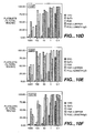
- scFv fragments denominated 10F6, 5E5, 10D10, 12B5, 12D5 and 12E10 having sequences for CDRs and Framework regions provided in Figure 1 .
- the above enumerated scFvs are affinity matured by mutating 1-3 amino acid residues in one or more of the CDRs or in the framework regions between the CDRs.
- the framework regions may be derived from a "consensus sequence" (i.e. the most common amino acids of a class, subclass or subgroup of heavy or light chains of human immunoglobulins) or may be derived from an individual human antibody framework region or from a combination of different framework region sequences.
- Consensus sequence i.e. the most common amino acids of a class, subclass or subgroup of heavy or light chains of human immunoglobulins
- Many human antibody framework region sequences are compiled in Kabat et al., Sequences of Proteins of Immunological Interest, 5th Ed. Public Health Service, National Institutes of Health, Bethesda, MD. (1991), pages 647-669 ), for example.
- a suitable method for purifying mpl antibodies comprises contacting an antibody source containing the mpl antibody molecules with an immobilized receptor polypeptide, specifically mpl or a mpl fusion polypeptide, under conditions whereby the mpl antibody molecules to be purified are selectively adsorbed onto the immobilized receptor polypeptide, washing the immobilized support to remove non-adsorbed material, and eluting the molecules to be purified from the immobilized receptor polypeptide with an elution buffer.
- the source containing the mpl antibody may be a library of antibodies having different binding epitopes and the receptor may be immobilized on a plate, tube, particle or other suitable surface using known methods.
- the source containing the antibody is recombinant cell culture where the concentration of antibody in either the culture medium or in cell lysates is generally higher than in plasma or other natural sources.
- the preferred purification method to provide substantially homogeneous antibody comprises: removing particulate debris, either host cells or lysed fragments by, for example, centrifugation or ultrafiltration; optionally, protein may be concentrated with a commercially available protein concentration filter; followed by separating the antibody from other impurities by one or more steps selected from; immunoaffinity, ion-exchange (e.g., DEAE or matrices containing carboxymethyl or sulfopropyl groups), Blue-SEPHAROSE, CM Blue-SEPHAROSE, MONO-Q, MONO-S, lentil lectin-SEPHAROSE, WGA-SEPHAROSE, Con A-SEPHAROSE, Ether TOYPEARL, Butyl TOYPEARL, Phenyl TOYPEARL, protein A SEPHAROSE, SDS-P
- the isolated antibody is monoclonal ( Kohler and Milstein, Nature, 256:495-497 (1975 ); Campbell, Laboratory Techniques in Biochemistry and Molecular Biology, Burdon et al., Eds, Volume 13, Elsevier Science Publishers, Amsterdam (1985 ); and Huse et al., Science, 246:1275-1281 (1989 )).
- a preferred mpl antibody is one that binds to mpl receptor with an affinity of at least about 10 6 l/mole. More preferably the antibody binds with an affinity of at least about 10 7 l/mole or even at least 10 9 l/mole. Most preferably, the antibody is raised against a mpl receptor having one of the above described effector functions.
- the isolated antibody capable of binding to the mpl receptor may optionally be fused to a second polypeptide and the antibody or fusion thereof may be used to isolate and purify mpl from a source as described above for immobilized mpl polypeptide.
- the invention provides a method for detecting the mpl ligand in vitro or in vivo comprising contacting the antibody with a sample, especially a serum sample, suspected of containing the ligand and detecting if binding has occurred.
- the invention also provides an isolated nucleic acid molecule encoding the mpl antibody or fragments thereof, which nucleic acid molecule may be labeled or unlabeled with a detectable moiety, and a nucleic acid molecule having a sequence that is complementary to, or hybridizes under stringent or moderately stringent conditions with, a nucleic acid molecule having a sequence encoding a mpl antibody.
- a preferred mpl antibody nucleic acid is RNA or DNA that encodes a biologically active human antibody.
- the nucleic acid molecule is cDNA encoding the mpl antibody and further comprises a replicable vector in which the cDNA is operably linked to control sequences recognized by a host transformed with the vector.
- This aspect further includes host cells transformed with the vector and a method of using the cDNA to effect production of antibody, comprising expressing the cDNA encoding the antibody in a culture of the transformed host cells and recovering the antibody from the host cell culture.
- the antibody prepared in this manner is preferably substantially homogeneous human antibody.
- a preferred host cell for producing the antibody is Chinese hamster ovary (CHO) cells.
- An alternative preferred host cell is E. coli.
- the invention further includes a preferred method for treating a mammal having an immunological or hematopoietic disorder, especially thrombocytopenia comprising administering a therapeutically effective amount of a mpl agonist or antagonist antibody to the mammal.
- the antibody is administered in combination with a cytokine, especially a colony stimulating factor or interleukin.
- cytokine especially a colony stimulating factor or interleukin.
- Preferred colony stimulating factors or interleukins include; kit-ligand, LIF, G-CSF, GM-CSF, M-CSF, EPO, IL-1, IL-2, IL-3, IL-5, IL-6, IL-7, IL-8, IL-9 or IL-11.
- the antibody is administered in combination with an Insulin-like growth factor (e.g., IGF-1) or a tumor necrosis factor (e.g., lymphotoxin (LT)).
- IGF-1 Insulin-like growth factor
- LT lymph
- Nucleic acid encoding the agonist and/or antagonist antibodies of the invention can be prepared from a library of single chain antibodies displayed on a bacteriophage.
- the preparation of such a library is well known to one of skill in this art.
- Suitable libraries may be prepared by the methods described in WO 92/01047 , WO 92/20791 , WO 93/06213 , WO 93/11236 , WO 93/19172 , WO 95/01438 and WO 95/15388 .
- a library of single chain antibodies (scFv) may be generated from a diverse population of human B-cells from human donors.
- mRNA corresponding to the VH and VL antibody chains is isolated and purified using standard techniques and reverse transcribed to generate a population of cDNA.
- DNA coding for single chain antibodies is assembled using a linker, such as Gly 4 Ser, and cloned into suitable expression vectors.
- a phage library is then prepared in which the population of single chain antibodies is displayed on the surface of the phage. Suitable methods for preparing phage libraries have been reviewed and are described in Winter et. al., Annu. Rev. Immunol., 1994, 12:433-55 ; Soderlind et. al., Immunological Reviews, 1992, 130:109-123 ; Hoogenboom, Tibtech February 1997, Vol. 15 ; Neri et. al., Cell Biophysics, 1995, 27:47-61 , and the references described therein.
- the antibodies of the invention having agonist or antagonist properties may be selected by immobilizing a receptor and then panning a library of human scFv prepared as described above using the immobilized receptor to bind antibody.
- Griffiths et. al. EMBO-J, 1993, 12:725-734 .
- the specificity and activity of specific clones can be assessed using known assays.
- Griffiths et. al.; Clarkson et. al., Nature, 1991, 352:642-648 After a first panning step, one obtains a library of phage containing a plurality of different single chain antibodies displayed on phage having improved binding to the receptor. Subsequent panning steps provide additional libraries with higher binding affinities.
- monovalent phage display libraries may be used in which less than 20%, preferably less than 10%, and more preferably less than 1% of the phage display more than one copy of an antibody on the surface of the phage.
- Monovalent display can be accomplished with the use of phagemid and helper phage as described, for example, in Lowman et. al., Methods: A Companion to Methods in Enzymology, 1991, 3(3):205-216 .
- a preferred phage is M13 and display is preferably as a fusion protein with coat protein 3 as described in Lowman et. al., supra.
- Other suitable phage include fl and fd filamentous phage. Fusion protein display with other virus coat proteins is also known and may be used in this invention. See U.S. 5,223,409 .
- Amino acid sequence variants of the antibody are prepared by introducing appropriate nucleotide changes into the antibody DNA, or by peptide synthesis.
- Such variants include, for example, deletions from, and/or insertions into and/or substitutions of, residues within the amino acid sequences of the antibodies of the examples herein. Any combination of deletion, insertion, and substitution is made to arrive at the final construct, provided that the final construct possesses the desired characteristics.
- the amino acid changes also may alter post-translational processes of the humanized or variant antibody, such as changing the number or position of glycosylation sites.
- a useful method for identification of certain residues or regions of the antibody that are preferred locations for mutagenesis is called "alanine scanning mutagenesis," as described by Cunningham and Wells Science, 244:1081-1085 (1989 ).
- a residue or group of target residues are identified (e.g., charged residues such as arg, asp, his, lys, and glu) and replaced by a neutral or negatively charged amino acid (most preferably alanine or polyalanine) to affect the interaction of the amino acids with the receptor.
- Those amino acid locations demonstrating functional sensitivity to the substitutions then are refined by introducing further or other variants at, or for, the sites of substitution.
- the site for introducing an amino acid sequence variation is predetermined, the nature of the mutation per se need not be predetermined. For example, to analyze the performance of a mutation at a given site, ala scanning or random mutagenesis is conducted at the target codon or region and the expressed antibody variants are screened for the desired activity.
- Amino acid sequence insertions include amino- and/or carboxyl-terminal fusions ranging in length from one residue to polypeptides containing a hundred or more residues, as well as intrasequence insertions of single or multiple amino acid residues.
- terminal insertions include an antibody with an N-terminal methionyl residue or the antibody fused to an epitope tag.
- Other insertional variants of the antibody molecule include the fusion to the N- or C-terminus of the antibody of an enzyme or a polypeptide which increases the serum half-life of the antibody.
- variants are an amino acid substitution variant. These variants have at least one amino acid residue in the antibody molecule removed and a different residue inserted in its place.
- the sites of greatest interest for substitutional mutagenesis include the hypervariable regions, but FR alterations are also contemplated.
- Conservative substitutions are shown in Table 2 under the heading of "preferred substitutions". If such substitutions result in a change in biological activity, then more substantial changes, denominated "exemplary substitutions" in Table 2, or as further described below in reference to amino acid classes, may be introduced and the products screened.
- Substantial modifications in the biological properties of the antibody are accomplished by selecting substitutions that differ significantly in their effect on maintaining (a) the structure of the polypeptide backbone in the area of the substitution, for example, as a sheet or helical conformation, (b) the charge or hydrophobicity of the molecule at the target site, or (c) the bulk of the side chain.
- Naturally occurring residues are divided into groups based on common side-chain properties:
- Non-conservative substitutions will entail exchanging a member of one of these classes for another class.
- cysteine residue not involved in maintaining the proper conformation of the humanized or variant antibody also may be substituted, generally with serine, to improve the oxidative stability of the molecule and prevent aberrant crosslinking.
- cysteine bond(s) may be added to the antibody to improve its stability (particularly where the antibody is an antibody fragment such as an Fv fragment).
- a particularly preferred type of substitutional variant involves substituting one or more hypervariable region residues of a parent antibody (e.g. a humanized or human antibody).
- a parent antibody e.g. a humanized or human antibody
- the resulting variant(s) selected for further development will have improved biological properties relative to the parent antibody from which they are generated.
- a convenient way for generating such substitutional variants is affinity maturation using phage using methods known in the art. Briefly, several hypervariable region sites (e.g. 3-7 sites) are mutated to generate all possible amino substitutions at each site.
- the antibody variants thus generated are displayed in a monovalent fashion from filamentous phage particles as fusions to the gene III product of M13 packaged within each particle. The phage-displayed variants are then screened for their biological activity (e.g.
- alanine scanning mutagenesis can be performed to identified hypervariable region residues contributing significantly to antigen binding.
- Another type of amino acid variant of the antibody alters the original glycosylation pattern of the antibody. By altering is meant deleting one or more carbohydrate moieties found in the antibody, and/or adding one or more glycosylation sites that are not present in the antibody.
- N-linked refers to the attachment of the carbohydrate moiety to the side chain of an asparagine residue.
- the tripeptide sequences asparagine-X-serine and asparagine-X-threonine, where X is any amino acid except proline, are the recognition sequences for enzymatic attachment of the carbohydrate moiety to the asparagine side chain.
- X is any amino acid except proline
- O-linked glycosylation refers to the attachment of one of the sugars N-aceylgalactosamine, galactose, or xylose to a hydroxyamino acid, most commonly serine or threonine, although 5-hydroxyproline or 5-hydroxylysine may also be used.
- glycosylation sites to the antibody is conveniently accomplished by altering the amino acid sequence such that it contains one or more of the above-described tripeptide sequences (for N-linked glycosylation sites).
- the alteration may also be made by the addition of, or substitution by, one or more serine or threonine residues to the sequence of the original antibody (for O-linked glycosylation sites).
- Nucleic acid molecules encoding amino acid sequence variants of the antibody are prepared by a variety of methods known in the art. These methods include, but are not limited to, isolation from a natural source (in the case of naturally occurring amino acid sequence variants) or preparation by oligonucleotide-mediated (or site-directed) mutagenesis, PCR mutagenesis, and cassette mutagenesis of an earlier prepared variant or a non-variant version of the antibody.
- the antibodies are prepared by standard recombinant procedures which involve production of the antibodies by culturing cells transfected to express antibody nucleic acid (typically by transforming the cells with an expression vector) and recovering the antibody from the cells of cell culture.
- the nucleic acid (e.g., cDNA or genomic DNA) encoding mpl antibody selected as described above is inserted into a replicable vector for further cloning (amplification of the DNA) or for expression.
- a replicable vector for further cloning (amplification of the DNA) or for expression.
- Many vectors are available, and selection of the appropriate vector will depend on (1) whether it is to be used for DNA amplification or for DNA expression, (2) the size of the nucleic acid to be inserted into the vector, and (3) the host cell to be transformed with the vector.
- Each vector contains various components depending on its function (amplification of DNA or expression of DNA) and the host cell with which it is compatible.
- the vector components generally include, but are not limited to, one or more of the following: a signal sequence, an origin of replication, one or more marker genes, an enhancer element, a promoter, and a transcription termination sequence.
- the mpl antibody of this invention may be expressed not only directly, but also as a fusion with a heterologous polypeptide, preferably a signal sequence or other polypeptide having a specific cleavage site at the N-terminus of the mature protein or polypeptide.
- the signal sequence may be a component of the vector, or it may be a part of the mpl antibody DNA that is inserted into the vector.
- the heterologous signal sequence selected should be one that is recognized and processed (i.e., cleaved by a signal peptidase) by the host cell.
- prokaryotic host cells For prokaryotic host cells a prokaryotic signal sequence selected, for example, from the group of the alkaline phosphatase, penicillinase, lpp, or heat-stable enterotoxin 11 leaders.
- the native signal sequence may be substituted by, e.g., the yeast invertase, alpha factor, or acid phosphatase leaders, the C. albicans glucoamylase leader ( EP 362,179 published 4 April 1990 ), or the signal described in WO 90/13646 published 15 November 1990 .
- the native signal sequence i.e., the mpl ligand presequence that normally directs secretion of mpl ligand from its native mammalian cells in vivo
- the native signal sequence i.e., the mpl ligand presequence that normally directs secretion of mpl ligand from its native mammalian cells in vivo
- signal sequences from other mpl ligand polypeptides or from the same mpl ligand from a different animal species signal sequences from a mpl ligand, and signal sequences from secreted polypeptides of the same or related species, as well as viral secretory leaders, for example, the herpes simplex gD signal.
- Both expression and cloning vectors contain a nucleic acid sequence that enables the vector to replicate in one or more selected host cells.
- this sequence is one that enables the vector to replicate independently of the host chromosomal DNA, and includes origins of replication or autonomously replicating sequences.
- origins of replication or autonomously replicating sequences are well known for a variety of bacteria, yeast, and viruses.
- the origin of replication from the plasmid pBR322 is suitable for most Gram-negative bacteria, the 2 ⁇ plasmid origin is suitable for yeast, and various viral origins (SV40, polyoma, adenovirus, VSV or BPV) are useful for cloning vectors in mammalian cells.
- the origin of replication component is not needed for mammalian expression vectors (the SV40 origin may typically be used only because it contains the early promoter).
- Most expression vectors are "shuttle" vectors, i.e., they are capable of replication in at least one class of organisms but can be transfected into another organism for expression.
- a vector is cloned in E. coli and then the same vector is transfected into yeast or mammalian cells for expression even though it is not capable of replicating independently of the host cell chromosome.
- DNA may also be amplified by insertion into the host genome. This is readily accomplished using Bacillus species as hosts, for example, by including in the vector a DNA sequence that is complementary to a sequence found in Bacillus genomic DNA. Transfection of Bacillus with this vector results in homologous recombination with the genome and insertion of antibody DNA. However, the recovery of genomic DNA encoding antibody is more complex than that of an exogenously replicated vector because restriction enzyme digestion is required to excise the antibody DNA.
- Selection genes should contain a selection gene, also termed a selectable marker. This gene encodes a protein necessary for the survival or growth of transformed host cells grown in a selective culture medium. Host cells not transformed with the vector containing the selection gene will not survive in the culture medium.
- Typical selection genes encode proteins that (a) confer resistance to antibiotics or other toxins, e.g., ampicillin, neomycin, methotrexate, or tetracycline, (b) complement auxotrophic deficiencies, or (c) supply critical nutrients not available from complex media, e.g., the gene encoding D-alanine racemase for Bacilli.
- a selection scheme utilizes a drug to arrest growth of a host cell. Those cells that are successfully transformed with a heterologous gene express a protein conferring drug resistance and thus survive the selection regimen. Examples of such dominant selection use the drugs neomycin ( Southern et al., J. Molec. Appl. Genet., 1:327 (1982 )) mycophenolic acid ( Mulligan et al., Science, 209:1422 (1980 )) or hygromycin Sugden et al., Mol. Cell. Biol., 5:410-413 (1985 )). The three examples given above employ bacterial genes under eukaryotic control to convey resistance to the appropriate drug G418 or neomycin (geneticin), xgpt (mycophenolic acid), or hygromycin, respectively.
- Suitable selectable markers for mammalian cells are those that enable the identification of cells competent to take up the antibody nucleic acid, such as dihydrofolate reductase (DHFR) or thymidine kinase.
- DHFR dihydrofolate reductase
- the mammalian cell transformants are placed under selection pressure that only the transformants are uniquely adapted to survive by virtue of having taken up the marker. Selection pressure is imposed by culturing the transformants under conditions in which the concentration of selection agent in the medium is successively changed, thereby leading to amplification of both the selection gene and the DNA that encodes antibody.
- Amplification is the process by which genes in greater demand for the production of a protein critical for growth are reiterated in tandem within the chromosomes of successive generations of recombinant cells. Increased quantities of antibody are synthesized from the amplified DNA.
- cells transformed with the DHFR selection gene are first identified by culturing all of the transformants in a culture medium that contains methotrexate (Mtx), a competitive antagonist of DHFR.
- Mtx methotrexate
- An appropriate host cell when wild-type DHFR is employed is the Chinese hamster ovary (CHO) cell line deficient in DHFR activity, prepared and propagated as described by Urlaub and Chasin, Proc. Natl. Acad Sci. USA, 77:4216 (1980 ).
- the transformed cells are then exposed to increased levels of Mtx. This leads to the synthesis of multiple copies of the DHFR gene, and, concomitantly, multiple copies of other DNA comprising the expression vectors, such as the DNA encoding antibody.
- This amplification technique can be used with any otherwise suitable host, e.g., ATCC No. CCL61 CHO-K1, notwithstanding the presence of endogenous DHFR if, for example, a mutant DHFR gene that is highly resistant to Mtx is employed ( EP 117,060 ).
- host cells can be selected by cell growth in medium containing a selection agent for the selectable marker such as an aminoglycosidic antibiotic, e.g., kanamycin, neomycin, or G418. See U.S. Patent No. 4,965,199 .
- a suitable selection gene for use in yeast is the trp l gene present in the yeast plasmid YRp7 ( Stinchcomb et al, Nature, 282:39 (1979 ); Kingsman et al., Gene, 7:141 (1979 ); or Tschemper et al., Gene, 10:157 (1980 )).
- the trp 1 gene provides a selection marker for a mutant strain of yeast lacking the ability to grow in tryptophan, for example, ATCC No. 44076 or PEP4-1 ( Jones, Genetics, 85:12 (1977 )).
- the presence of the trp 1 lesion in the yeast host cell genome then provides an effective environment for detecting transformation by growth in the absence of tryptophan.
- Leu 2-deficient yeast strains (ATCC No. 20,622 or 38,626) are complemented by known plasmids bearing the Leu 2 gene.
- Expression and cloning vectors usually contain a promoter that is recognized by the host organism and is operably linked to the antibody nucleic acid. Promoters are untranslated sequences located upstream (5') to the start codon of a structural gene (generally within about 100 to 1000 bp) that control the transcription and translation of particular nucleic acid sequence, such as the antibody nucleic acid sequence, to which they are operably linked. Such promoters typically fall into two classes, inducible and constitutive. Inducible promoters are promoters that initiate increased levels of transcription from DNA under their control in response to some change in culture conditions, e.g., the presence or absence of a nutrient or a change in temperature.
- promoters recognized by a variety of potential host cells are well known. These promoters are operably linked to antibody encoding DNA by removing the promoter from the source DNA by restriction enzyme digestion and inserting the isolated promoter sequence into the vector. Both the native antibody promoter sequence and many heterologous promoters may be used to direct amplification and/or expression of the antibody DNA. However, heterologous promoters are preferred, as they generally permit greater transcription and higher yields of expressed antibody as compared to the native promoter.
- Promoters suitable for use with prokaryotic hosts include the ⁇ -lactamase and lactose promoter systems ( Chang et al., Nature, 275:615 (1978 ); and Goeddel et al, Nature, 281:544 (1979 )), alkaline phosphatase, a tryptophan (trp) promoter system ( Goeddel, Nucleic Acids Res., 8:4057 (1980 ) and EP 36,776 ) and hybrid promoters such as the tac promoter ( deBoer et al., Proc. Natl. Acad Sci. USA, 80:21-25 (1983 )).
- trp tryptophan
- hybrid promoters such as the tac promoter ( deBoer et al., Proc. Natl. Acad Sci. USA, 80:21-25 (1983 )).
- other known bacterial promoters are suitable.
- Promoter sequences are known for eukaryotes. Virtually all eukaryotic genes have an AT-rich region located approximately 25 to 30 bases upstream from the site where transcription is initiated. Another sequence found 70 to 80 bases upstream from the start of transcription of many genes is a CXCAAT region where X may be any nucleotide. At the 3 end of most eukaryotic genes is an AATAAA sequence that may be the signal for addition of the poly A tail to the 3 end of the coding sequence. All of these sequences are suitably inserted into eukaryotic expression vectors.
- Suitable promoting sequences for use with yeast hosts include the promoters for 3-phosphoglycerate kinase ( Hitzeman et al., J. Biol. Chem., 255:2073 (1980 )) or other glycolytic enzymes ( Hess et al., J. Adv.
- enolase such as enolase, glyceraldehyde-3-phosphate dehydrogenase, hexokinase, pyruvate decarboxylase, phosphofructokinase, glucose-6-phosphate isomerase, 3-phosphoglycerate mutase, pyruvate kinase, triosephosphate isomerase, phosphoglucose
- yeast promoters which are inducible promoters having the additional advantage of transcription controlled by growth conditions, are the promoter regions for alcohol dehydrogenase 2, isocytochrome C, acid phosphatase, degradative enzymes associated with nitrogen metabolism, metallothionein, glyceraldehyde-3-phosphate dehydrogenase, and enzymes responsible for maltose and galactose utilization.
- Suitable vectors and promoters for use in yeast expression are further described in Hitzeman et al., EP 73,657A .
- Yeast enhancers also are advantageously used with yeast promoters.
- Antibody transcription from vectors in mammalian host cells may be controlled, for example, by promoters obtained from the genomes of viruses such as polyoma virus, fowlpox virus ( UK 2,211,504 published 5 July 1989 ), adenovirus (such as Adenovirus 2), bovine papilloma virus, avian sarcoma virus, cytomegalovirus, a retrovirus, hepatitis-B virus and most preferably Simian Virus 40 (SV40), from heterologous mammalian promoters, e.g., the actin promoter or an immunoglobulin promoter, from heat-shock promoters, and from the promoter normally associated with the antibody sequence, provided such promoters are compatible with the host cell systems.
- viruses such as polyoma virus, fowlpox virus ( UK 2,211,504 published 5 July 1989 ), adenovirus (such as Adenovirus 2), bovine papilloma virus, avian sar
- the early and late promoters of the SV40 virus are conveniently obtained as an SV40 restriction fragment that also contains the SV40 viral origin of replication. Fiers et al., Nature, 273:113 (1978 ); Mulligan and Berg, Science, 209:1422-1427 (1980 ); Pavlakis et al., Proc. Nail. Acad. Sci. USA, 78:7398-7402 (1981 ).
- the immediate early promoter of the human cytomegalovirus is conveniently obtained as a Hin dIII E restriction fragment. Greenaway et al., Gene, 18:355-360 (1982 ).
- a system for expressing DNA in mammalian hosts using the bovine papilloma virus as a vector is disclosed in U.S. Patent No.
- Enhancers are cis-acting elements of DNA, usually about from 10 to 300 bp, that act on a promoter to increase its transcription. Enhancers are relatively orientation and position independent, having been found 5 ( Laiminset al., Proc. Natl. Acad Sci. USA, 78:993 (1981 )) and 3 ( Luskyet al., Mol. Cell Bio., 3:1108 (1983 )) to the transcription unit, within an intron ( Banerji et al., Cell. 33:729 (1983 )), as well as within the coding sequence itself ( Osborne et al., Mol.
- enhancer sequences are now known from mammalian genes (globin, elastase, albumin, a-fetoprotein, and insulin). Typically, however, one will use an enhancer from a eukaryotic cell virus. Examples include the SV40 enhancer on the late side of the replication origin (bp 100-270), the cytomegalovirus early promoter enhancer, the polyoma enhancer on the late side of the replication origin, and adenovirus enhancers. See also Yaniv, Nature, 297:17-18 (1982 ) on enhancing elements for activation of eukaryotic promoters. The enhancer may be spliced into the vector at a position 5 or 3 to the antibody encoding sequence, but is preferably located at a site 5 from the promoter.
- Expression vectors used in eukaryotic host cells will also contain sequences necessary for the termination of transcription and for stabilizing the mRNA. Such sequences are commonly available from the 5 and, occasionally 3 untranslated regions of eukaryotic or viral DNAs or cDNAs. These regions contain nucleotide segments transcribed as polyadenylated fragments in the untranslated portion of the mRNA encoding antibody.
- Plasmids containing one or more of the above listed components employs standard ligation techniques. Isolated plasmids or DNA fragments are cleaved, tailored, and religated in the form desired to generate the plasmids required.
- the ligation mixtures are used to transform E. coli K12 strain 294 (ATCC No. 31,446) and successful transformants selected by ampicillin or tetracycline resistance where appropriate. Plasmids from the transformants are prepared, analyzed by restriction endonuclease digestion, and/or sequenced by the method of Messing et al., Nucleic Acids Res., 9:309 (1981 ) or by the method of Maxam et al., Methods in Enzymology, 65:499 (1980 ).
- transient expression involves the use of an expression vector that is able to replicate efficiently in a host cell, such that the host cell accumulates many copies of the expression vector and, in tum, synthesizes high levels of a desired polypeptide encoded by the expression vector.
- Transient expression systems comprising a suitable expression vector and a host cell, allow for the convenient positive identification of polypeptides encoded by cloned DNAs, as well as for the rapid screening of such polypeptides for desired biological or physiological properties.
- transient expression systems are particularly useful in the invention for purposes of identifying analogues and variants of antibody polypeptide that have antibody polypeptide biological activity.
- Suitable host cells for cloning or expressing the vectors herein are the prokaryote, yeast, or higher eukaryotic cells described above.
- Suitable prokaryotes include eubacteria, such as Gram-negative or Gram-positive organisms, for example, E. coli, Bacilli such as B. subtilis, Pseudomonas species such as P. aeruginosa, Salmonella typhimurium, or Serratia marcescans.
- E. coli cloning host is E. coli 294 (ATCC No. 31,446), although other strains such as E. coli B, E. coli X1776 (ATCC No. 31,537), and E. coli W3110 (ATCC No.
- the host cell should secrete minimal amounts of proteolytic enzymes.
- in vitro methods of cloning e.g., PCR or other nucleic acid polymerase reactions, are suitable.
- eukaryotic microbes such as filamentous fungi or yeast are suitable hosts for antibody encoding vectors.
- Saccharomyces cerevisiae or common baker's yeast, is the most commonly used among lower eukaryotic host microorganisms.
- Schizosaccharomyces pombe Beach and Nurse, Nature, 290:140 (1981 ); EP 139,383 published 2 May 1985
- Kluyveromyces hosts U.S. Patent No. 4,943,529
- K. laclis Louvencourt et al., J.
- filamentous fungi such as, e.g., Neurospora, Penicillium, Tolypocladium ( WO 91/00357 published 10 January 1991 ), and Aspergillus hosts such as A. nidulans ( Ballance et al., Biochem. Biophys. Res. Commun., 112:284-289 (1983 ); Tilburn et al., Gene, 26:205-221 (1983 ); Yelton et al., Proc. Natl. Acad Sci. USA, 81:1470-1474 (1984 )) and A. niger ( Kelly and Hynes, EMBO J., 4:475-479 (1985 )).
- A. niger Kelly and Hynes, EMBO J., 4:475-479 (1985 )
- Suitable host cells for the expression of glycosylated antibody are derived from multicellular organisms. Such host cells are capable of complex processing and glycosylation activities. In principle, any higher eukaryotic cell culture is workable, whether from vertebrate or invertebrate culture. Examples of invertebrate cells include plant and insect cells. Numerous baculoviral strains and variants and corresponding permissive insect host cells from hosts such as Spodoptera frugiperda (caterpillar), Aedes aegypti (mosquito), Aedes albopictus (mosquito), Drosophila melanogaster (fruitfly), and Bombyx mori have been identified.
- a variety of viral strains for transfection are publicly available, e.g., the L-1 variant of Autographa californica NPV and the Bm-5 strain of Bombyx mori NPV, and such viruses may be used as the virus herein according to the present invention, particularly for transfection of Spodoptera frugiperda cells.
- Plant cell cultures of cotton, com, potato, soybean, petunia, tomato, and tobacco can be utilized as hosts.
- plant cells are transfected by incubation with certain strains of the bacterium Agrobacterium tumefaciens, which has been previously manipulated to contain the antibody DNA.
- A. tumefaciens the DNA encoding the antibody is transferred to the plant cell host such that it is transfected, and will, under appropriate conditions, express the antibody DNA.
- regulatory and signal sequences compatible with plant cells are available, such as the nopaline synthase promoter and polyadenylation signal sequences. Depicker et al., J. Mol. Appl.
- DNA segments isolated from the upstream region of the T-DNA 780 gene are capable of activating or increasing transcription levels of plant-expressible genes in recombinant DNA-containing plant tissue.
- tissue culture Tissue Culture
- useful mammalian host cell lines are monkey kidney CV1 line transformed by SV40 (COS-7, ATCC CRL 1651); human embryonic kidney line (293 or 293 cells subcloned for growth in suspension culture, Graham et al., J. Gen Virol., 36:59 (1977 )); baby hamster kidney cells (BHK, ATCC CCL 10); Chinese hamster ovary cells/-DHFR (CHO, Urlaub and Chasin, Proc. Natl. Acad Sci.
- COS-7 monkey kidney CV1 line transformed by SV40
- human embryonic kidney line (293 or 293 cells subcloned for growth in suspension culture, Graham et al., J. Gen Virol., 36:59 (1977 )
- baby hamster kidney cells BHK, ATCC CCL 10
- Chinese hamster ovary cells/-DHFR CHO, Urlaub and Chasin, Proc. Natl. Acad Sci.
- mice sertoli cells TM4, Mather, Biol. Reprod., 23:243-251 (1980 )); monkey kidney cells (CV1 ATCC CCL 70); African green monkey kidney cells (VERO-76, ATCC CRL-1587); human cervical carcinoma cells (HELA, ATCC CCL 2); canine kidney cells (MDCK, ATCC CCL 34); buffalo rat liver cells (BRL 3A, ATCC CRL 1442); human lung cells (W138, ATCC CCL 75); human liver cells (Hep G2, HB 8065); mouse mammary tumor (MMT 060562, ATCC CCL51); TRI cells ( Mather et al., Annals N.Y. Acad Sci., 383:44-68 (1982 )); MRC 5 cells; FS4 cells; and a human hepatoma line (Hep G2).
- Host cells are transfected and preferably transformed with the above-described expression or cloning vectors of this invention and cultured in conventional nutrient media modified as appropriate for inducing promoters, selecting transformants, or amplifying the genes encoding the desired sequences.
- Transfection refers to the taking up of an expression vector by a host cell whether or not any coding sequences are in fact expressed. Numerous methods of transfection are known to the ordinarily skilled artisan, for example, CaPO 4 and electroporation. Successful transfection is generally recognized when any indication of the operation of this vector occurs within the host cell.
- Transformation means introducing DNA into an organism so that the DNA is replicable, either as an extrachromosomal element or by chromosomal integrant. Depending on the host cell used, transformation is done using standard techniques appropriate to such cells.
- the calcium treatment employing calcium chloride, as described in section 1.82 of Sambrook et al., supra, is generally used for prokaryotes or other cells that contain substantial cell-wall barriers.
- Infection with Agrobacterium tumefaciens is used for transformation of certain plant cells, as described by Shaw et al., Gene, 23:315 (1983 ) and WO 89/05859 published 29 June 1989 .
- plants may be transfected using ultrasound treatment as described in WO 91/00358 published 10 January 1991 .
- Prokaryotic cells used to produce the antibody polypeptide of this invention are cultured in suitable media as described generally in Sambrook et al., supra.
- the mammalian host cells used to produce the antibody of this invention may be cultured in a variety of media.
- Commercially available media such as Ham's F10 (Sigma), Minimal Essential Medium ((MEM), Sigma), RPMI-1640 (Sigma), and Dulbecco's Modified Eagle's Medium ((DMEM), Sigma) are suitable for culturing the host cells.
- any of these media may be supplemented as necessary with hormones and/or other growth factors (such as insulin, transferrin, or epidermal growth factor), salts (such as sodium chloride, calcium, magnesium, and phosphate), buffers (such as HEPES), nucleosides (such as adenosine and thymidine), antibiotics (such as GentamycinTM drug), trace elements (defined as inorganic compounds usually present at final concentrations in the micromolar range), and glucose or an equivalent energy source. Any other necessary supplements may also be included at appropriate concentrations that would be known to those skilled in the art.
- the culture conditions such as temperature, pH, and the like, are those previously used with the host cell selected for expression, and will be apparent to the ordinarily skilled artisan.
- the host cells referred to in this disclosure encompass cells in in vitro culture as well as cells that are within a host animal.
- Gene amplification and/or expression may be measured in a sample directly, for example, by conventional Southern blotting, northern blotting to quantitate the transcription of mRNA ( Thomas, Proc. Natl. Acad. Sci. USA, 77:5201-5205 (1980 )), dot blotting (DNA analysis), or in situ hybridization, using an appropriately labeled probe, based on the sequences provided herein.
- Various labels may be employed, most commonly radioisotopes, particularly 32 P.
- other techniques may also be employed, such as using biotin-modified nucleotides for introduction into a polynucleotide.
- the biotin then serves as the site for binding to avidin or antibodies, which may be labeled with a wide variety of labels, such as radionuclides, fluorescers, enzymes, or the like.
- antibodies may be employed that can recognize specific duplexes, including DNA duplexes, RNA duplexes, and DNA-RNA hybrid duplexes or DNA-protein duplexes.
- the antibodies in turn may be labeled and the assay may be carried out where the duplex is bound to a surface, so that upon the formation of duplex on the surface, the presence of antibody bound to the duplex can be detected.
- Gene expression may be measured by immunological methods, such as immunohistochemical staining of tissue sections and assay of cell culture or body fluids, to quantitate directly the expression of gene product.
- immunohistochemical staining techniques a cell sample is prepared, typically by dehydration and fixation, followed by reaction with labeled antibodies specific for the gene product coupled, where the labels are usually visually detectable, such as enzymatic labels, fluorescent labels, luminescent labels, and the like.
- a particularly sensitive staining technique suitable for use in the present invention is described by Hsu et al., Am. J. Clin. Path., 75:734-738 (1980 ).
- Antibody preferably is recovered from the culture medium as a secreted polypeptide, although it also may be recovered from host cell lysates when directly expressed without a secretory signal.
- the antibody When the antibody is expressed in a recombinant cell other than one of human origin, the antibody is completely free of proteins or polypeptides of human origin. However, it is still usually necessary to purify the antibody from other recombinant cell proteins or polypeptides to obtain preparations that are substantially homogeneous as to the mpl ligand per se.
- the culture medium or lysate is centrifuged to remove particulate cell debris.
- the membrane and soluble protein fractions are then separated.
- a commercially available protein concentration filter e.g., AMICON or Millipore PELLICON ultrafiltration units
- the antibody may then be purified from the soluble protein fraction.
- the antibody thereafter is purified from contaminant soluble proteins and polypeptides by salting out and exchange or chromatographic procedures employing various gel matrices.
- These matrices include; acrylamide, agarose, dextran, cellulose and others common to protein purification.
- Exemplary chromatography procedures suitable for protein purification include immunoaffinity, receptor affinity (e.g., mpl-IgG or protein A SEPHAROSE), hydrophobic interaction chromatography (HIC) (e.g., ether, butyl, or phenyl Toyopearl).
- lectin chromatography e.g., Con A-SEPHAROSE, lentil-lectin-SEPHAROSE
- size exclusion e.g., SEPHADEX G-75
- cation- and anion-exchange columns e.g., DEAE or carboxymethyl- and sulfopropyl-cellulose
- RP-HPLC reverse-phase high performance liquid chromatography
- Other purification steps optionally include; ethanol precipitation; ammonium sulfate precipitation; chromatofocusing; preparative SDS-PAGE, and the like.
- Antibody variants in which residues have been deleted, inserted, or substituted are recovered in the same fashion, taking account of any substantial changes in properties occasioned by the variation.
- preparation of a an antibody fusion with another protein or polypeptide e.g., a bacterial or viral antigen
- an immunoaffinity column containing antibody to the antigen can be used to adsorb the fusion polypeptide.
- Immunoaffinity columns such as a rabbit polyclonal anti-antibody column can be employed to absorb the antibody variant by binding it to at least one remaining immune epitope.
- the antibody may be purified by affinity chromatography using a purified receptor-IgG coupled to a (preferably) immobilized resin such as AFFI-Gel 10 (Bio-Rad, Richmond, CA) or the like, by means well known in the art.
- a protease inhibitor such as phenyl methyl sulfonyl fluoride (PMSF) also may be useful to inhibit proteolytic degradation during purification, and antibiotics may be included to prevent the growth of adventitious contaminants.
- PMSF phenyl methyl sulfonyl fluoride
- purification methods suitable for native the antibody may require modification to account for changes in the character of the antibody or its variants upon expression in recombinant cell culture.
- the antibodies are agonist antibodies (aAb).
- agonist antibody is meant an antibody which is able to bind to and to activate, a particular hematopoietic growth factor receptor.
- the agonist may bind to the extracellular domain of the receptor and thereby cause differentiation and proliferation of megakaryocyte colonies in semisolid cultures and single megakaryocytes in liquid suspension cultures and platelet production in vitro and/or in vivo.
- the agonist antibodies are preferably against epitopes within the extracellular domain of the receptor Accordingly, the antibody preferably binds to substantially the same epitope as the 12E10, 12B5, 10F6, and 12D5 monoclonal antibodies specifically disclosed herein.
- the antibody will also have substantially the same or greater antigen binding affinity as the monoclonal antibodies disclosed herein.
- a monoclonal antibody has the same specificity as an antibody specifically disclosed, one can, for example, use a competitive ELISA binding assay.
- DNA encoding the monoclonal antibodies useful in the method of the invention is readily isolated and sequenced using conventional procedures (e.g., by using oligonucleotide probes that are capable of binding specifically to genes encoding the heavy and light chains of human antibodies).
- the phage of the invention serve as a preferred source of such DNA.
- the DNA may be placed into expression vectors, which are then transfected into host cells such as E. coli cells, simian COS cells, Chinese Hamster Ovary (CHO) cells, or myeloma cells that do not otherwise produce immunoglobulin protein, to obtain the synthesis of monoclonal antibodies in the recombinant host cells.
- the antibodies disclosed herein are useful for in vitro diagnostic assays for activating the receptor of interest. This is useful in order to study the role of the receptor in megakaryocyte growth and/or differentiation and platelet production.
- the biologically active c-mpl agonist antibody capable of stimulating either proliferation, differentiation or maturation and/or modulation (either stimulation or inhibition) of apoptosis of hematopoietic cells may be used in a sterile pharmaceutical preparation or formulation to stimulate megakaryocytopoietic or thrombopoietic activity in patients suffering from thrombocytopenia due to impaired production, sequestration, or increased destruction of platelets.
- Thrombocytopenia-associated bone marrow hypoplasia may be effectively treated with the aAb compounds of this invention as well as disorders such as disseminated intravascular coagulation (DIC), immune thrombocytopenia (including HIV-induced ITP and non HIV-induced ITP), chronic idiopathic thrombocytopenia, congenital thrombocytopenia, myelodysplasia, and thrombotic thrombocytopenia.
- DIC disseminated intravascular coagulation
- immune thrombocytopenia including HIV-induced ITP and non HIV-induced ITP
- chronic idiopathic thrombocytopenia congenital thrombocytopenia
- myelodysplasia myelodysplasia
- thrombotic thrombocytopenia e.g., thrombotic thrombocytopenia.
- Preferred uses of the megakaryocytopoietic or thrombocytopoietic biologically active c-mpl agonist antibody of this invention are in: myelotoxic chemotherapy for treatment of leukemia or solid tumors, myeloablative chemotherapy for autologous or allogeneic bone marrow transplant, myelodysplasia, idiopathic aplastic anemia, congenital thrombocytopenia, and immune thrombocytopenia.
- the biologically active c-mpl agonist antibody of the instant invention may be employed alone or in combination with other cytokines, hematopoietins, interleukins, growth factors, or antibodies in the treatment of the above-identified disorders and conditions.
- the instant compounds may be employed in combination with other protein or peptide having hematopoietic activity including G-CSF, GM-CSF, LIF, M-CSF, IL-1, IL-3, erythropoietin (EPO), kit ligand, IL-6, and IL-11.
- the biologically active c-mpl agonist antibody of the instant invention may be used in the same way and for the same indications as thrombopoietin (TPO).
- TPO thrombopoietin
- Some forms of the aAb have a longer half-life than native or pegylated TPO and thus are used in indications where a longer half-life are indicated.
- the antibody formulation When used for in vivo administration, the antibody formulation must be sterile. This is readily accomplished by filtration through sterile filtration membranes, prior to or following lyophilization and reconstitution. The antibody ordinarily will be stored in lyophilized form or in solution.
- Therapeutic antibody compositions generally are placed into a container having a sterile access port, for example, an intravenous solution bag or vial having a stopper pierceable by a hypodermic injection needle.
- the route of antibody administration is in accord with known methods, e.g., injection or infusion by intravenous, intraperitoneal, intracerebral, intramuscular, intraocular, intraarterial, intrathecal, inhalation or intralesional routes, or by sustained release systems as noted below.
- the antibody is preferably administered continuously by infusion or by bolus injection.
- an effective amount of antibody to be employed therapeutically will depend, for example, upon the therapeutic objectives, the route of administration, and the condition of the patient. Accordingly, it will be necessary for the therapist to titer the dosage and modify the route of administration as required to obtain the optimal therapeutic effect. Typically, the clinician will administer antibody until a dosage is reached that achieves the desired effect. The progress of this therapy is easily monitored by conventional assays.
- the antibodies of the invention may be prepared in a mixture with a pharmaceutically acceptable carrier.
- This therapeutic composition can be administered intravenously or through the nose or lung, preferably as a liquid or powder aerosol (lyophilized).
- the composition may also be administered parenterally or subcutaneously as desired.
- the therapeutic composition should be sterile, pyrogen-free and in a parenterally acceptable solution having due regard for pH, isotonicity, and stability. These conditions are known to those skilled in the art.
- dosage formulations of the compounds of the present invention are prepared for storage or administration by mixing the compound having the desired degree of purity with physiologically acceptable carriers, excipients, or stabilizers.
- Such materials are non-toxic to the recipients at the dosages and concentrations employed, and include buffers such as TRIS HCl, phosphate, citrate, acetate and other organic acid salts; antioxidants such as ascorbic acid; low molecular weight (less than about ten residues) peptides such as polyarginine, proteins, such as serum albumin, gelatin, or immunoglobulins; hydrophilic polymers such as polyvinylpyrrolidinone; amino acids such as glycine, glutamic acid, aspartic acid, or arginine; monosaccharides, disaccharides, and other carbohydrates including cellulose or its derivatives, glucose, mannose, or dextrins; chelating agents such as EDTA; sugar alcohols such as mannitol or sorbitol; counterions such as sodium and/or nonionic surfactants such as TWEEN, PLURONICS or polyethyleneglycol.
- buffers such as TRIS HCl, phosphate, citrate, a
- Sterile compositions for injection can be formulated according to conventional pharmaceutical practice. For example, dissolution or suspension of the active compound in a vehicle such as water or naturally occurring vegetable oil like sesame, peanut, or cottonseed oil or a synthetic fatty vehicle like ethyl oleate or the like may be desired. Buffers, preservatives, antioxidants and the like can be incorporated according to accepted pharmaceutical practice.
- sustained-release preparations include semipermeable matrices of solid hydrophobic polymers containing the polypeptide, which matrices are in the form of shaped articles, e.g., films, or microcapsules.
- sustained-release matrices include polyesters, hydrogels (e.g., poly(2-hydroxyethyl-methacrylate) as described by Langer et al., J Biomed Mater. Res., 15:167-277 (1981 ) and Langer, Chem. Tech., 12:98-105 (1982 ) or poly(vinylalcohol)), polylactides ( U.S. Patent No.
- Sustained-release compositions also include liposomally entrapped TPO.
- Liposomes containing TPO are prepared by methods known per se: DE 3,218,121 ; Epstein et al., Proc. Natl. Acad Sci. USA, 82:3688-3692 (1985 ); Hwang et al., Proc. Natl. Acad Sci. USA. 77:4030-4034 (1980 ); EP 52,322 ; EP 36,676 ; EP 88,046 ; EP 143,949 ; EP 142,641 ; Japanese patent application 83-118008 ; U.S. Patent Nos. 4,485,045 and 4,544,545 ; and EP 102,324 .
- the liposomes are of the small (about 200-800 Angstroms) unilamellar type in which the lipid content is greater than about 30 mol. % cholesterol, the selected proportion being adjusted for the optimal therapy.
- the dosage of the antibody will be determined by the attending physician taking into consideration various factors known to modify the action of drugs including severity and type of disease, body weight, sex, diet, time and route of administration, other medications and other relevant clinical factors.
- Therapeutically effective dosages may be determined by either in vitro or in vivo methods.
- an effective amount of the agonist antibody to be employed therapeutically will depend, for example, upon the therapeutic objectives, the route of administration, and the condition of the patient. Accordingly, it will be necessary for the therapist to titer the dosage and modify the route of administration as required to obtain the optimal therapeutic effect.
- a typical daily dosage might range from about 1 ⁇ g/kg to up to 1000 mg/kg or more, depending on the factors mentioned above.
- the clinician will administer the molecule until a dosage is reached that achieves the desired effect. The progress of this therapy is easily monitored by conventional assays.
- from about 0.001 mg/kg to about 1000 mg/kg, more preferably about 0.01 mg to 100 mg/kg, more preferably about 0.010 to 20 mg/kg of the agonist antibody might be an initial candidate dosage for administration to the patient, whether, for example, by one or more separate administrations, or by continuous infusion.
- the treatment is repeated until a desired suppression of disease symptoms occurs or the desired improvement in the patient's condition is achieved.
- other dosage regimens may also be useful.
- the mpl agonist antibody assays were conducted essentially as described in WO 95/18858 .
- the Ba/F3-mpl cell line was established ( F. de Sauvage et al., Nature, 369:533 (1994 )) by introduction of the cDNA encoding the entire mpl receptor into the IL-3 dependent murine lymphoblastoid cell line Ba/F3. Stimulation of proliferation of Ba/F3-mpl cells in response to various concentrations of antibodies or TPO was measured by the amount of incorporation of 3 H-thymidine as previously described (F. de Sauvage et al., supra).
- CMK cells are maintained in RMPl 1640 medium (Sigma) supplemented with 10% fetal bovine serum and 10mM glutamine.
- RMPl 1640 medium Sigma
- the cells are harvested, washed and resuspended at 5x10 5 cells/ml in serum-free GIF medium supplemented with 5mg/l bovine insulin, 10mg/l apo-transferrin, 1 X trace elements.
- the TPO standard or experimental agonist antibody samples are added to each well at appropriate dilutions in 100 ml volumes. 100 ml of the CMK cell suspension is added to each well and the plates are incubated at 37°C, in a 5% CO 2 incubator for 48 hours.
- the human mpl receptor has been disclosed by Vigon et al., PNAS, USA 89:5640-5644 (1992 ).
- a chimeric receptor comprising the extracellular domain (ECD) of the mpl receptor and the transmembrane and intracellular domain (ICD) of Rse ( Mark et al., J. of Biol, Chem. 269(14):10720-10728 (1994 )) with a carboxyl-terminal flag polypeptide (i.e. Rse.gD) was made for use in the KIRA ELISA described herein.
- Monoclonal anti-gD (clone 5B6) was produced against a peptide from Herpes simplex virus glycoprotein D ( Paborsky et al., Protein Engineering 3(6):547-553 (1990 )).
- the purified stock preparation was adjusted to 3.0mg/ml in phosphate buffered saline (PBS), pH 7.4 and 1.0ml aliquots were stored at -20° C.
- PBS phosphate buffered saline
- Monoclonal anti-phosphotyrosine, clone 4G10 was purchased from UBI (Lake Placid, NY) and biotinylated using long-arm biotin-N-hydroxysuccinamide (Biotin-X-NHS, Research Organics, Cleveland, OH).
- the mpl ligand was prepared by the recombinant techniques described herein.
- the purified mpl ligand was stored at 4 °C. as a stock solution.
- KIRA ELISA results for agonist antibodies of the invention are shown in Fig. 9 .
- This assay indicates that the antibodies of the invention activate the mpl receptor to a degree similar to the cognate ligand TPO.
- NUNC 96-well immunoplates were coated with 50 ⁇ l of rabbit anti-human IgG Fc (Jackson Labs) at 2 ⁇ g/ml in carbonate buffer (pH9.6) overnight at 4°C. After blocking with ELISA buffer (PBS, I % BSA, 0.2 % TWEEN 20), the plates were incubated for 2 hr with conditioned media from mpi-Ig-transfected 293 cells. Plates were washed, and 2.5 ng/ml biotinylated TPO was added in the presence or absence of various concentrations of antibodies.
- ELISA buffer PBS, I % BSA, 0.2 % TWEEN 20
- TPO bound was detected by incubation with streptAvidin-HRP (Sigma) followed by TMB peroxidase substrate (Kirkegaard & Perry). All dilutions were performed in ELISA buffer, and all incubations were at room temperature. Color development was quenched with H 3 PO 4 and absorbance was read at 450-650 nm.
- the HU-01 cell line (D. Morgan, Hahnemann University) is derived from a patient with acute megakaryoblastic leukemia and is dependent on granulocyte-macrophage colony stimulating factor (GM-CSF) for growth.
- GM-CSF granulocyte-macrophage colony stimulating factor
- the HU-03 cell line used here was derived from HU-01 cells by adaptation to growth in rhTPO rather than GM-CSF.
- HU-03 cells were maintained in RPMI 1640 supplemented with 2 % heat-inactivated human male serum and 5 ng/ml rhTPO. Before assay, cells were starved by removing TPO, decreasing serum concentrration to 1 %, and adjusting the concentration of cells to 2.5 x 10 5 cells/ml, followed by incubation for 16 hr. Cells were then washed and seeded into 96-well plates at a density of 5 x 10 4 cells per well in medium containing TPO or antibodies at various concentrations. Quadruplicate assays were performed. 1 ⁇ Ci 3H-thymidine was added to each well before incubation for 24 hr. Cells were collected with a Packard cell harvester and incorporation of 3 H-thymidine was measured with a Top Count Counter (Packard).
- Packard Top Count Counter
- Mpl agonist antibodies on human megakaryocytopoiesis was determined using a modification of the liquid suspension assay previously described ( Grant et al, Blood 69:1334-1339 (1997 )). Buffy coats were collected from human umbilical cord blood and cells washed in phosphate-buffered saline (PBS) by centrifugation at 120 g for 15 min at room temperature to remove platelet-rich plasma.
- PBS phosphate-buffered saline
- IMDM Iscove's modified Dulbecco's medium
- GIBCO Iscove's modified Dulbecco's medium
- FBS fetal bovine serum
- 2-mercaptoethanol 20 ⁇ M 2-mercaptoethanol
- TPO thrombopoietin
- Fab'2 forms of antibody 12B5 or antibody 12D5 were added to quadruplicate wells; control wells contained no additional supplements. Final volumes were 1 ml per well.
- the cultures were grown in a humidified incubator at 37 °C in 5% CO 2 for 14 days.
- Megakaryocytopoiesis was quantified using radiolabelled murine monoclonal antibody HP1-1D (provided by W. L. Nichols, Mayo Clinic) which has been shown to be specific for the human megakaryocyte glycoprotein IIb/IIIa (Grant et al., supra).
- FBS used in this assay was treated with Dextran T40 at I mg/ml and charcoal at 10 mg/ml for 30 min, centrifuged, decanted, filter sterilized and heat inactivated at 56 °C for 30 min.
- HU-03 cells were cultured as described above.
- Platelet rich plasma (PRP) was prepared by centrifugation of citrated whole blood at 400 g's for 5 minutes. Binding studies were conducted within three hours of collection.
- 125 I-TPO was prepared by indirect iodination ( Fielder, P. J., Hass, P., Nagel, M., Stefanich, E., Widmer, R., Bennett, G. L., Keller, G., de Savage, F. J., and Eaton, D. 1997. Human platelets as a model for the binding and degradation of thrombopoietin. Blood 89: 2782-2788 ) and yielded a specific activity of 15-50 ⁇ Ci/ ⁇ g protein.
- HU-03 cells In a volume of 110 microliters containing 100pM iodinated TPO, 2 x 10 6 washed HU-03 cells in Hank's Balanced Salt Solution,5 mg/ml bovine serum albumin (HBSSB), or 4 x 10 7 platelets in plasma, were incubated at 37°C for 30 minutes with varying concentrations of antibody in triplicate. HU-03 cells were agitated during the incubation period to keep them in suspension. The reaction mixture was overlayed on I ml 20 % sucrose-HBSSB and microcentrifuged at 13,500 rpm for five minutes. The supernatants were aspirated, tube bottoms containing the cell pellets were cut off, and cell- or platelet-associated radioactivity was measured with an Iso Data Model 120 gamma counter.
- HBSSB bovine serum albumin
- the receptor-binding affinities of several Fab fragments were calculated (Lofas & Johnsson, 1990) from association and dissociation rate constants measured using a BIACORE surface plasmon resonance system (Pharmacia Biosensor).
- a biosensor chip was activated for covalent coupling of gD-mpl receptor using N-ethyl-N"'-(3-dimethylaminopropyl)-carbodiimide hydrochloride (EDC) and N-hydroxysuccinimide (NHS) according to the supplier's (Pharmacia Biosensor) instructions.
- gD-mpl was buffer-exchanged into 10 mM sodium acetate buffer (pH 4.5) and diluted to approximately 30 ⁇ g/mL.
- Standard deviations, s on for k on and s off for k off were obtained from measurements with >4 protein concentrations (k on ) or with >7 protein concentrations (k off ).
- Dissociation data were fit to a simple AB-->A+B model to obtain koff +/- s.d. (standard deviation of measurements).
- Pseudo-first order rate constant (ks) were calculated for each association curve, and plotted as a function of protein concentration to obtain kon +/- s.e. (standard error of fit).
- Figures 2 and 3 presents a cartoon of the construction of a phage library containing 6x10 9 different clones containing single-chain Fv (scFv) antibodies fused to gene 3 of a phage. Binding selection against an antigen , in this case c-mpl, can be carried out as shown in Fig.4 and described in greater detail below.
- NUNC immunotubes were coated with 2 ml of a solution of 10 microg/ml of gD-c-mpl in PBS at 4°C overnight. After rinsing with PBS, tubes were blocked with 3% dry milk in PBS (MPBS) for 2 hr at room temperature. For the first round, 10 ⁇ l of C.A.T. antibody phage library containing ⁇ 1 x 10 12 c.f.u. were added to 1 ml MPBS for blocking for 1 hr at room temperature. Blocked phage were added to coated tubes, and binding of phage to antigen allowed to continue for 2 hr at 37°C on a rotating wheel.
- MPBS dry milk in PBS
- Tubes were washed 6 times with PBS-TWEEN and 6 times with PBS, and phage were then eluted with 100 mM TEA for 10 min at room temperature, neutralized with 500 ⁇ l of 1 M TRIS (pH 7.4), and stored on ice until needed. For subsequent rounds, washing was increased to 20 times with PBS-TWEEN, and 20 times with PBS.
- Eluted phage were used to infect 5 ml of log phase E. coli TG1 cells and plated on 2YT agar supplemented with 2% glucose and 100 ⁇ g/ml carbenicillin. After overnight growth at 30°C, colonies were scraped into 10 ml 2YT. 50 ⁇ l of this solution was used to inoculate 25 ml of 2YT with carbenicillin and glucose and incubated, shaking, for two hours at 37°C. Helper phage M13K07 (Pharmacia) were added at an m.o.i. of 10.
- the cells were pelleted and resuspended in 25 ml of 2YT with carbenicillin (100 ⁇ g/ml) and kanamycin (50 ⁇ g/ml) and growth continued at 30°C for 4 hr.
- E. coli were removed from the phage by centrifugation, and I ml of these phage (approx. 10 12 c.f.u.) were used in subsequent rounds of selection.
- the library was also selected using soluble biotinylated antigen and streptavidin-coated paramagnetic beads (see Fig. 5 ).
- gD-c-mpl was biotinylated using IMMUNOPURE NHS-biotin (biotiny-N-hydroxysuccinimide, Pierce) according to manufacturer's recommendations.
- DYNABEADS M-280, coated with streptavidin were washed 3 times with 1 ml MPBST (using a DYNAL Magnetic Particle Concentrator) and then blocked for 2 hr at 37°C with 1 ml fresh MPBST on a rotator.
- the DYNABEADS and attached phage were then washed a total of 7 times: 3 times with 1 ml PBS-TWEEN, once with MPBS, followed by 3 times with PBS.
- Phage were eluted from the beads by incubating 5 min at room temperature with 300 ⁇ l of 100 mM triethylamine. The phage-containing supernatant was removed and neutralized with 150 ⁇ l of 1M TRIS-HCl (pH 7.4). Neutralized phage were used to infect mid-log TG 1 host cells as described above. Plating, induction and harvesting of phage were also as for selection on tubes.
- Fig. 6 illustrates the phage ELISA process.
- NUNC MAXISORP microtiter plates were coated with 50 ⁇ l per well of gD-c-mpl, BSA, or gDgp120, at 2 ⁇ g/ml in 50 mM carbonate buffer (pH 9.6), at 4°C overnight. After removing antigen, plates were blocked with 3% dry milk in PBS (MPBS) for 2 hours at room temperature.
- MPBS PBS
- Phage cultures were centrifuged and 100 ⁇ l of phage-containing supernatants were blocked with 20 ⁇ l of 6 x PBS / 18% dry milk for I hour at room temperature. Block was removed from titer plates and blocked phage added and allowed to bind for 1 hour at room temperature. After washing, phage were detected with a 1:5000 dilution of horseradish peroxidase-conjugated anti-M13 antibody (Pharmacia) in MPBS followed by 3',3',5',5'-tetramethylbenzidine (TMB). Reactions were stopped by the addition of H 2 SO 4 and readings taken by subtracting the A 405nm from the A 450nm .
- TMB horseradish peroxidase-conjugated anti-M13 antibody
- Soluble scFv was induced in the bacterial supernatants of clones by growth in 2YT containing carbenicillin (100 ⁇ g/ml) and IPTG (1mM) ON at 30°C.
- ELISA plates were either coated with gD-c-mpl or, for capture ELISA, with anti-c-myc Mab 9E10. Plates were blocked with I x ELISA diluent (PBS supplemented with 0.5% BSA, 0.05% Tween 20, pH 7.4), and soluble scFv was blocked by adding 20 ⁇ l of 6 x ELISA dil to 100 ⁇ l of supernatant.
- soluble scFv was detected by adding 50 ⁇ l of I ⁇ g/ml Mab 9E10 per well, followed by horseradish peroxidase-conjugated goat anti-murine Ig, and then TMB as described above.
- soluble scFv was detected by addition of biotinylated c-mpl, followed by streptavidin-peroxidase conjugate and then TMB as above.
- Table 3 Anti-c-mpl scFv antibodies from CAT library Clones screened: 1534 Clones positive by ELISA: 361 Clones different by BstNI and sequencing: 24 Clones that express protein well 17 clones that are agonists by KIRA: 9 clones that are agonists by BaF3 proliferation assay: 6 clones that are agonists by Hu3 proliferation assay: 4
- the diversity of c-mpl-binding clones was determined by PCR amplifying the scFv insert using primers pUC19R (5 AGC GGA TAA CAA TTT CAC ACA GG 3) (SEQ. ID. NO: 54) which anneals upstream of the leader sequence and fdtetseq (5 GTC GTC TTT CCA GAC GGT AGT 3 ) (SEQ. ID. NO: 55) which anneals in the 5 end of gene III, followed by digestion with the frequent-cutting restriction enzyme BstNI (see Fig. 7 ).
- Fig. 8A-C Typical patterns seen after analysis on a 3% agarose gel are shown in Fig. 8A-C .
- Protocol Mix A dH20 67 ⁇ l 10 x ampliTaq buffer 10 25 mM MgCl2 10 DMSO, 50% 2 forward primer 1 Mix B: 2.5 mM dNTPs 8 ⁇ l AMPLITAQ 0.5 reverse primer 1.0
- the nucleotide sequence of representative clones of each fingerprint were obtained. Colonies were inoculated into 50 ml of LB medium supplemented with 2% glucose and 100 ⁇ g/ml carbenicillin, and grown overnight at 30°C. DNA was isolated using Qiagen Tip-100s and the manufacturer's protocol and cycle sequenced with fluorescent dideoxy chain terminators (Applied Biosystems). Samples were run on an Applied Biosystems 373A Automated DNA Sequencer and sequences analyzed using the program "Sequencher" (Gene Codes Corporation). The VH and VL genes were assigned to a germline segment using the antibody database, V-BASE.
- DNA sequence was obtained for 39 clones and resulted in 24 different c-mpl-binding scFvs.
- E. coli strain 33D3 was transformed with phagemid DNA.
- Five ml of 2YT with carbenicillin and glucose was used to grow overnight cultures at 30°C.
- 0.2 ml of these cultures were diluted into 200 ml of the same media and grown to an OD 600 of approximately 0.9.
- the cells were pelleted and resuspended in 250 ml of 2YT containing IPTG (1 mM) and carbenicillin (100 ⁇ g/ml) and to induce expression and grown for a further 5 hours at 30°C.
- Cell pellets were harvested and frozen at -20°C.
- the antibodies were purified by immobilized metal chelate affinity chromatography (IMAC). Frozen pellets were resuspended in 10 ml of ice-cold shockate buffer (25 mM TRIS-HCl, 1 mM EDTA, 200 mM NaCl, 20 % sucrose, 1 mM PMSF) by shaking on ice for 1 hr. Imidazole was added to 20 mM, and cell debris removed by centrifugation. The supernatants were adjusted to 1mM MgCl 2 and 50 mM phosphate buffer pH 7.5. Ni-NTA agarose resin from Qiagen was used according to the manufacturers instructions.
- IMAC immobilized metal chelate affinity chromatography
- the resin was equilibrated with 50 mM sodium phosphate buffer pH 7.5, 500 mM NaCl, 20 mM imidazole, and the shockate added. Binding occurred in either a batch mode or on a gravity flow column. The resin was then washed twice with 10 bed volumes of equilibration buffer, and twice with buffer containing imidazole increased to 50mM. Elution of proteins was with 50 mM phosphate buffer pH 7.5, 500 mM NaCl and 250 mM imidazole. Excess salt and imidazole was removed on a PD-10 column (Pharmacia), and proteins were concentrated using a Centricom 10 to a volume of about I ml.
- Plasmid DNA for scFv clones 12B5, 12D5, 12E10, 10D10, 10F6 and 5E5 (named pMpl.12B5.scFv.his; pMpl.12D5.scFv.his; pMp1.12E10.scFv.his; pMpl.10D10.scFv.his; pMpl.10F6.scFv.his; and pMpl.5E5.scFv.his, respectively) has been deposited with ATCC, Manassas, Virginia, USA.
- coli enterotoxin II (stII) signal sequence (Picken, et al.) to direct secretion to the periplasmic space of E. coli.
- This vector also contains the human k 1 C L ( Palm et al.. Infect. Immun. 42:269-275 (1983 )) and the human IgGI C H 1 ( Ellison, et al, Nucleic Acids Res. 10: 4071-4079 (1982 )) constant domains.
- the C H 1 gene is immediately followed by the bacteriophage ⁇ to transcriptional terminator ( Scholtissek and Grosse Nucleic Acids Res. 15: 3185 (1987 )).
- Fab' and Fab'2 variants were facilitated by insertion into pAK19 of unique restriction sites at the junctions of the stII and V L domain (Sfi I), the V 1 and Ck domains (Rsr II), the stlI and V H domain (MluI), and the V H and C H l domains (Apa I), using oligonucleotide directed mutagenesis.
- Fab' molecules In order to insure expression of monovalent Fab' molecules, the free cysteine at the 3' end of the CH1 domain was mutated to a threonine, these Fab' molecules thus end in the amino acid sequence thr-ala-ala-pro, rather than thr-cys-ala-ala as in pAK19.
- This vector for the expression of Fab' molecules is named pXCA730.
- Fab'2 molecules For expression of the antibodies as Fab'2 molecules, a vector was constructed which adds the human IgG1 hinge region onto the C H l domain of pXCA730. This is followed by the yeast GCN4 leucine zipper domain ( Hu, et al. Science 250:1400-1403 (1990 )) for stability.
- DNA fragments were constructed using synthesized oligonucleotides and encode the amino acid sequence: cys-pro-pro-cys-ala-pro-glu-leu-leu-gly-gly-arg-met-lys-gln-leu-glu-asp-lys-val-glu-glu-leu-leu-ser-lys-asn-tyr-his-leu-glu-asn-glu-val-ala-arg-leu-lys-lys-leu-val-gly-glu-arg (SEQ. ID. NO: 56).
- the resultant plasmid is named pXCA740.
- variable domains of the scFvs were amplified and restriction sites added for subcloning into the vectors described above by the PCR technique.
- Specific oligonucleotides were designed for each V L or V H region as shown below. 12B5, 12D5, and 10D10 Light chain variable domains:
- PCR reactions were carried out using 100 ng of plasmid DNA containing the scFv, 0.5 ⁇ M of the appropriate 5 and 3 primer, 200 ⁇ M each dNTP, 10 mM KCI, 6 mM(NH 4 ) 2 SO 4 , 20 mM TRIS-HCl, pH 8.0, 2 mM MgCl 2 , 1% Triton X-100, 100 ⁇ M BSA and 2.5 units of Pfu DNA polymerase (Stratagene). Amplification was for 30 cycles of: 30 sec at 95 °C, 30 sec at 55°C, 30 sec at 72°C.
- the reaction products were separated by agarose gel electrophoresis and the approximately 350 bp band was isolated using a Gene Clean II kit (BIO 101, Vista, CA).
- the fragments for the light chain variable regions were ligated into the vectors previously digested with Sfi I and Rsr II for the kappa isotypes, or Sfi I and Msc I for the lambda isotypes, and transformed into E. coli DH5a. Desired recombinants were identified using restriction enzyme analysis and sequenced to confirm the presence of the desired fragments.
- the heavy chain variable domains were then cloned similarly into the plasmids containing the light chains using the restriction enzymes Mlu I and Apa I, and the final constructions were again checked by DNA sequencing.
- the Sfi I to Not I fragments of the scFv forms of p12B5, p12D5, p10F6, and p12E10 were subcloned into a derivative of pAK19 containing the phoA promoter and stII signal sequence rather than the lacZ promoter and hybrid signal sequence of the original library.
- a DNA fragment coding for 12 amino acids metal-ala-asp-pro-asn-arg-phe-arg-gly-lys-asp-leu
- herpes simplex virus type 1 glycoprotein D Lasky and Dowbenko DNA (N.Y.) 3:23-29 (1984 .) was synthesized and inserted at the 3 end of the V L domain in place of the (his) 6 and c-myc epitope originally present in the C.A.T. library clones.
- Plasmids containing genes for scFv-gD, Fab' or Fab'2 molecules were expressed in E. coli strain 33B6 (W3110 DfhuA pho ADE15 deoC2 ilvG2096(val R ) degP41(DPstI-Kan R ) D(argF-lac)169 IN(rrnD-rrnE)1) grown for approximately 40 hr at 30°C in an aerated 10-liter fermentor as described previously ( Carter et al Bio/Technology 10:163-167(1992 .)).
- the heavy chain variable domains were subcloned from the Fab constructs into a derivative of expression vector pRK ( Suva et al., Science 237:893-896 (1987 )) which contains the human IgG1 CH1, CH2, and CH3 domains and a human antibody signal sequence ( Carter et al., Proc. Natl. Acad Sci. USA. 89:4285-4289 (1992 )).
- the light chain was cloned into a separate pRK plasmid.
- the light and heavy chain expression vectors were cotransfected into adenovirus-transformed human embryonic kidney cell line 293 by a high-efficiency procedure ( Gorman et al, DNA Protein Eng. Technol. 2:3-10 (1990 )). Harvested conditioned media was shown to contain anti-mpl antibody by ELISA.
- the light and heavy chains were moved into the SVLDI expression vector previously described ( Lucas et al., Nucleic Acids Res. 24: 1774-1779 (1996 ).
- This vector contains the mouse DHFR cDNA in the intron of the expression vector pRK and allows for amplification of expression by selection in methotrexate
- the light chain is cloned into the same plasmid with expression driven by a second SV40 promoter/ enhancer.
- the plasmid was linearized and transfected into CHO cells using lipofectamine (Gibco-BRL) following manufacturer's instructions. Seven to ten days after transfer to selective medium, clones were isolated into 96 well plates for later study, or pooled and expanded for culture in roller bottles.
- Conditioned media for purification of the antibodies was generated in roller bottles. Cells were seeded into the roller bottles at an initial cell density of 2 x 10⁷ cells in 200 ml rich medium (DMEM: Ham's F12 (1:1) supplemented with 5% fetal bovine serum. At approximately 80% confluency, the media was replaced with serum-free PS-24 production medium supplemented with insulin (10 ⁇ g/ml), transferrin (10 ⁇ g/ml), trace elements and lipid alcohol. Conditioned media was harvested after 10 days.
- Frozen cell paste was resuspended at 1 gm/ml TE (25 mM TRIS, 1 mM EDTA, pH 7.4) and gently agitated 18 hr on ice. Cell debris was removed by centrifugation at 10,000 x g for 30 min. The supernatant was loaded onto an affinity column (2.5 x 9.0 cm) consisting of an anti-gD monoclonal antibody 5B6 ( Paborsky, L. R. et al., Protein Eng. 3: 547-553 (1990 )) coupled to CNBr SEPHAROSE which had been equilibrated with PBS.
- affinity column 2.5 x 9.0 cm
- the column was washed 18 hr with PBS, and then washed with PBS containing 1 M NaCl until the absorbance of the column effluent was equivalent to baseline. All steps were done at 4°C at a linear flow rate of 25 cm/hr. Elution was performed with 0.1 M acetic acid, 0.5 M NaCl, pH 2.9. Column fractions were monitored by absorbance at 280 nm and peak fractions pooled, neutralized with 1.0 M TRIS, pH 8.0, dialyzed against PBS, and sterile filtered. The resultant protein preparations were analyzed by non-reducing SDS-PAGE.
- Fab' molecules For purification of Fab' molecules, 5 g of frozen cell paste was resuspended in 5 ml of TE (25 mM TRIS, 1 mM EDTA, pH 7.4) and gently stirred 18 hr on ice. The pH of the shockate was adjusted to 5.6 with 2 M HCl and the precipitate and cell debris removed by centrifugation at 10,000 x g for 30 min. The supernatant was loaded onto a 1 ml BAKERBOND ABx column (0.5 x 5.0 cm) (J. T. Baker, Phillipsburg, NJ) pre-equilibrated with 20 mM MES, pH 5.5.
- TE 25 mM TRIS, 1 mM EDTA, pH 7.4
- the Fab' was eluted using a 10 ml linear gradient from 0 to 100% of 20 mM NaOAc, 0.5 M (NH 4 ) 2 SO 4 , pH 7.2, with a flow rate of 153 cm/hr. Fractions containing Fab' were pooled, and buffer exchanged into PBS.
- Frozen cell paste (100 gm) was thawed into 10 volumes of 25 mM TRIS, 5 mM EDTA, I mM NaN3, pH 7.4 and disrupted by three passages through a microfluidizer (TECH-MAR). PMSF was added to 1 mM and the cell debris removed by centrifugation at 10,000 x g for 30 min. The supernatant was filtered sequentially through a 0.45 ⁇ m, and a 0.2 ⁇ m SUPORCAP filter (Gelman), and loaded onto a 50 ml SEPHAROSE-fast-flow Protein-G column (Pharmacia) pre-equilibrated with PBS.
- Fab'2 was eluted with 0.1 M glycine ethyl ester, pH 2.3, into tubes with contained 1/10 volume of I M TRIS, pH 8.0. Fractions containing Fab'2 were pooled and concentrated by Ultrasette with a 30 kilodalton molecular weight cut off, and buffer exchanged into 20 mM NaOAc, 0.01% octylglucoside, pH 5.5.
- This material was loaded onto a 30 ml S-SEPHAROSE column (Pharmacia) pre-equilibrated with 20 mM NaOAc, washed to baseline with 20 mM NaOAc, pH 5.5, and eluted with a linear gradient of 0-1 M NaCl in 25mM NaOAc over 10 column volumes. Fractions containing Fab'2 were pooled and buffer exchanged to PBS.
- Conditioned medium harvested from roller bottles was loaded onto a 5 ml Protein-A SEPHAROSE column (1.0 x 5.0 cm) pre-equilibrated with PBS, washed with PBS, and then washed to baseline with PBS containing I M NaCi.
- Antibody was eluted with 0.1 M HOAc, 0.5 N NaCl, pH 2.9, neutralized with I M TRIS, and buffer exchanged to PBS.
- the invention provides a method of selecting an antibody which binds to and dimerizes a receptor protein.
- a library of antibodies is panned using a receptor protein having two protein subunits as the binding target.
- the library is panned as described above for mpl agonist antibodies.
- the antibodies are human and more preferably monoclonal.
- the library is conveniently a library of single chain antibodies, preferably displayed on the surface of phage.
- the display of proteins, including antibodies, on the surface of phage is well known in the art as discussed above and these known methods may be used in this invention.
- Antibody libraries are also commercially available, for example, from Cambridge Antibody Technologies (CAT), Cambridge, UK.
- the antibody selected by the method of the invention activates the receptor by dimerizing the receptor and thereby achieves an effector result similar to the effector result generated when the natural endogenous ligand for the receptor binds the receptor.
- the method of the invention can be used to find agonist antibodies to any receptor having two components which is known and /or can be cloned. It is not necessary to know the primary, secondary or tertiary structure of the receptor protein, although this information is useful for cloning, etc., since the method of the invention allows selection of antibodies which will bind any displayed receptor which is activated by dimerization. Many known receptor proteins are activated by dimerization and any of these known receptors may be used in the invention. Suitable receptors include tyrosine kinase receptors and hematopoietic receptors that lack kinase activity.
- Activation of a receptor such as a tyrosine kinase receptor by a scFv is an unexpected result.
- Current understanding of receptor activation argues that for many classes of receptors, including tyrosine kinase receptors and hematopoietic cytokine receptors that lack intrinsic tyrosine kinase activity (but associate with intracellular kinases), it is a dimerization event mediated by a ligand that is the key event in receptor activation.
- This view is supported by crystal structures of receptor ligand complexes as well as the demonstrated agonist ability of certain monoclonal antibodies (but not the Fab' fragments of these antibodies).
- a single chain antibody would not, therefore, be expected to be able to cause receptor dimerization and activation.
- MuSK is a recently identified tyrosine kinase localized to the postsynaptic surface of the neuromuscular junction. ( Valenzuela et. al.. 1995. Neuron 15 573-584 .) Mice made deficient in MuSK fail to form neuromuscular junctions ( Dechiara el. al.. 1996. Cell 85 501-512 .), a phenotype highly similar to that observed in mice lacking the nerve derived signaling molecule agrin ( Gautam et. al., 1996, Cell 85 525-535 ).
- Formation of the neuromuscular junction is achieved through a process that includes the differentiation of membrane on the muscle fiber proximal to the neuron terminus and changes in gene expression within the nuclei proximal to this junction (reviewed by Bowe et. al., 1995, Annu-Rev-Neurosci. 18 443-462 and Kleiman et. al., 1996, Cell 85 461-464 .).
- a striking feature of this complex process is the redistribution and concentration of AChRs within the myotube membrane. Agrin is able to the induce this clustering of AChRs as well as changes in the extracellular matrix and cytoskeletal components of the synaptic apparatus (Bowe et. al., supra; Godfrey et.
- Agrin is a secreted protein with a core molecular weight of ⁇ 200 kDa that contains several copies of EGF repeats, laminin-like globular domains and sequences that resemble protease inhibitors. It is released by motor neuron terminals and maintained within the basil lamina of the synaptic cleft. While agrin apparently does not to bind MuSK with high affinity (Glass et.
- Antigen specific scFv identified by panning a diverse library of scFv expressed, for example, on M 13 phage provide a source of molecules capable of mediating specific therapeutic activities, and offer a rapid new approach to study the function of novel or recently identified molecules such as MuSK.
- scFv are identified below that mediate receptor activation and that direct MuSK activation induces changes in AChR distribution and tyrosine phosphorylation similar to that observed with agrin.
- the antibodies of the invention are, therefore, useful in assaying the upregulation of MuSK receptors in sample tissues to determine the degree of neuromuscular damage associated with this upregulation.
- the antibodies are also useful for activating the MuSK receptor and inducing AChR clustering at neuromuscular junctions as a direct result of the agonist properties of these antibodies.
- Administration of the antibodies to a person suffering from denervation or muscle immobilization, e.g. muscular dystrophy provides a method of improving the function of the neuromuscular junctions in these people.
- scFv having agonist activity
- antibodies were selected which induce a proliferative response in a factor dependent cell line through a chimeric MuSK-Mpl receptor comprised of the extracellular domain of MuSK and the intracellular domain of the hematopoietic cytokine receptor c-Mpl (the receptor for thrombopoietin, TPO).
- c-Mpl the receptor for thrombopoietin, TPO.
- Activation of c-Mpl is believed to require homodimerization, as is the case for the growth hormone receptor, the erythropoietin receptor and other related receptors of this class ( Carter et. al.,1996, Annu-Rev-Physiol 58 187-207 ; Gurney et. al., 1995, Proc Natl.
- Ba/F3 cells expressing MuSK-Mpl were starved of IL-3 and exposed to a range of concentrations of each scFv expressed as soluble protein. Surprisingly, 4 of the 21 scFv were able to induce a robust proliferative response in the MuSK-Mpl expressing cells ( Fig. 11 ). This activity was observed at nM concentrations of scFv. The scFv were without effect on the parental, untransfected Ba/F3. Agonist activity was also present among those IgG that were derived from agonist scFv but was not noted among IgG derived from non-agonist scFv.
- Soluble agrin c-terminal domain was without effect supporting previous observations that agrin does not bind MuSK directly.
- the c-terminal domain of agrin is known to contain the AChR clustering activity of agrin and is essential for neuromuscular junction formation ( Ruegg et al., 1992, Neuron 8 691-699 ; Tsim et. al., supra ).
- the EC 50 for the ability to induce proliferation was 5 nM for the most active agonist clone when expressed as either scFv or IgG.
- the affinity of these scFv and IgG for MuSK was determined using BIAcoreTM analysis.
- the agonist scFv and several non-agonist scFv each displayed affinity for MuSK within the range of 5-25 nM.
- the affinities of the IgG for MuSK were 10-30 pM. See Table 5 below.
- Table 5 clone # Agonist k d ⁇ k a Affinity musk #2-scFv V + 3.34 x 10 -3 8.78 x 10 5 3.8 nM musk #3-scFv - 2.39 x 10 -3 1.05 x 10 5 23 nM musk #4-scFv + 1.57 x 10 -3 1.84 x 10 5 8.5 nM musk #5-scFv - 2.49 x 10 -3 5.29 x 10 5 4.7 nM musk #6-scFv - 4.95 x 10 -3 1.05 x 10 5 4.7 nM musk #13-scfv + 2.32 x 10 -3 4.53 x 10 5
- scFv were examined for the ability to induce tyrosine phosphorylation of full length MuSK tyrosine kinase.
- the murine myoblastic cell line C2C12 was cultured under conditions that promote myotube differentiation and subsequently exposed to scFv, IgG or c-agrin.
- scFv IgG or c-agrin.
- c-agrin was able to induce MuSK tyrosine phosphorylation.
- the agonist scFv and IgG were also found to rapidly induce tyrosine phosphorylation of MuSK as determined by western blot analysis with anti-phosphotyrosine antibody whereas other scFv and non-agonist anti-MuSK IgG were without effect.
- scFv MuSK agonists to induce AChR clustering in cultured C2C12 myotubes. Following stimulation, the cells were fixed and the distribution of cell surface AChR was revealed with rhodamine labeled bungarotoxin. In undifferentiated myoblasts, AChR were dispersed and unfocused in the presence of c-agrin, scFv, or IgG. In contrast, upon myotube differentiation, c-agrin and agonist scFv and IgG induced marked aggregation of AChR into large and intensely stained clusters. Non-agonist scFv and non-agonist IgG directed against MuSK or an irrelevant antigen were without effect.
- tyrosine phosphorylation of subunits of the AChR was also examined utilizing an antisera that recognizes the and chains of the receptor. Tyrosine phosphorylation levels of both the and chains were markedly induced by c-agrin as well as the agonist scFv and agonist IgG but were unaffected by control scFv and IgG.
- Variants of the MuSK agonist antibodies of the invention may be prepared as described above for thrombopoietic antibodies.
- MuSK-Fc was prepared by fusion of the extracellular domain of MuSK (a.a. 1-492) in frame with the Fc region of human IgG1 in the eukaryotic expression vector pRK5tkNEO. MuSK-Fc was transiently expressed in 293 cells and purified over a protein G column.
- a chimeric receptor, MuSK-Mpl comprised of the extracellular domain of MuSK (amino acids 1-492) and the transmembrane and intracellular domain of the human c-Mpl receptor (amino acids 491-635) was prepared by sequential PCR and cloned into pRK5tkNEO.
- Stable cell lines expressing the chimeric receptor were obtained by electroporation (5 million cells, 250 volts, 960 ⁇ F) of linearized vector (20 ⁇ g) into Ba/F3 cells followed by selection for neomycin resistance with 2 mg/ml G418.
- Full length MuSK in pRK5tkNEO was transfected into 293 cells and stable transformants were obtained following two weeks of G418 selection (400 ⁇ g/ml). The sequence of the DNA constructs were confirmed by DNA sequencing. Expression of MuSK was assessed by flow cytometry analysis as described below.
- Ba/F3 cells were maintained in RPMI 1640 media supplemented with 10% fetal calf serum and 5% conditioned media from WEHI-3B cells as source of IL-3.
- C-agrin (amino acids 1137-1949 of the rat agrin (Ag +8 active splice form ( Ferns et al, 1993, Neuron 11 491-502 .)) was expressed by transient transfection from 293 cells in serum free media with an expression vector, pRK-gD-c-Agrin, as a fusion protein with the gD signal sequence and epitope tag and a genenase cleavage site (MGGAAARLGAVILFVV IVGLHGVRGKYALADASLKMADPNRFRGKDLPVLDQLLEGGAAHYALLPG) (SEQ ID NO. 71) fused to the N-terminus.
- pRK-gD-c-Agrin as a fusion protein with the gD signal sequence and epitope tag and a genenase cleavage site (MGGAAARLGAVILFVV IVGLHGVRGKYALADASLKMADPNRFRGKDLPVLDQLLEGGAAHY
- clones as IgG For expression of clones as IgG the sequences encoding the V H and V L regions of the scFv were introduced by PCR into mammalian expression vector pIgG-kappa which was designed to enable the expression of fully human light and heavy chains of kappa type IgG. Expression vectors for the individual clones were transfected into CHO cells and IgG were harvested from conditioned serum free media and purified over a protein A column.
- AChR clustering assay C2C12 were maintained in 10% FBS in high glucose DMEM at subfluency.
- C2C12 were seeded on glass slides coated with fibronectin and poly-lysine and myotube differentiation was induced by 48 hour incubation in 2% horse serum high glucose DMEM.
- scFv or c-agrin were added to the culture medium and incubated overnight (16 hours).
- Cells were then' washed with PBS and fixed in 4% paraformaldehyde.
- Rhodamine conjugated bungarotoxin (Molecular Probes, Eugene OR) was used to reveal the localization of AChRs as described (Ferns et al, supra).
- Binding affinity analysis Protein interaction analysis using BlAcoreTM instruments was performed as described ( Mark et al, 1996, J. Biol. Chem. 271 9785-9789 ). Briefly, research grade CM5 sensor chips were activated by injection of 20 ⁇ l of 1:1 N-Ethyl-N'-(3-dimethylaminopropyl)carboiimide hydrochloride and N-hydroxysuccinimide at 5 ⁇ l /min flow rate. 20 ⁇ l of MuSK-Fc at 20 ⁇ g/min in 10 mM sodium acetate, pH 5.0 was injected over the sensor chip, followed by 30 ⁇ l of ethanolamine.
- C2C12 were maintained in 10% FBS high glucose DMEM and induced to differentiate by 72 hour incubation in 2% horse serum. Cells were then stimulated by addition of c-agrin, scFv or IgG for the time indicated in the figures. ScFv and IgG were used at 50 nM. The c-agrin containing conditioned media was used at level that provided maximal tyrosine phosphorylation. Cell extracts were prepared as described (Gurney et al, supra).
- Extracts were incubated for 60 minutes at 4°C with 30 ⁇ l agarose conjugated antiphosphotyrosine monoclonal antibody 4G10 (UBI inc., Lake Placid NY) or 1 ⁇ g of anti-MuSK IgG #13 followed by 30 ⁇ l protein A sepharose beads.
- Western blot analysis with antiphosphotyrosine antibody 4G10 or anti AChR antibody was performed as recommended by the manufacturer and revealed with HRP conjugated secondary antibody and ECL (Amersham).
- MuSK scFv are readily observed as dimers when resolved by nondenaturing gel electrophoresis. Additionally, the abundance of dimeric species may be significantly altered in the local context of scFv bound to receptor on the cell surface.
- screening an scFv phage library with a divalent antigen, in this case MuSK-Fc allows direct selection of scFv that bind to and facilitate the formation of a receptor dimer.
- An agonist antibody, fragment, or variant thereof which binds to a thrombopoietin receptor 2.
- the antibody of 1, wherein the thrombopoietin receptor comprises mammalian c-mpl.
- the antibody of 2, wherein the antibody stimulates proliferation, differentiation or growth of megakaryocytes.
- the antibody of 2, wherein the antibody stimulates megakaryocytes to produce platelets.
- the antibody of 2, wherein the thrombopoietin receptor is human c-mpl.
- the antibody of I which is selected from the group consisting of 12E10, 12B5, 1OF6 and 12D5. 10.
- the antibody of 2 which does not stimulate megakaryocytes to produce platelets.
- the antibody of 1 having a detectable label.
- the composition of 17, which is sterile. 19 The composition of 17, which is lyophilized.
- 20. A library of different single chain antibodies, comprising a plurality of the antibody of 14. 21.
- 24. A phage displaying on the surface thereof, the antibody of 14. 25.
- a method of stimulating proliferation, differentiation or growth of megakaryocytes comprising contacting megakaryocytes with an effective amount of the antibody of 1.
- the method of 29, comprising administering the antibody of 1 to a patient in need thereof.
- 31. A method of increasing platelet production, comprising contacting megakaryocytes with an effective amount of the antibody of 1.
- 32. comprising administering the antibody of 1 to a patient in need thereof.
- 33. Isolated nucleic acid encoding the antibody of 1.
- a vector comprising the nucleic acid of 33.
- a host cell comprising the vector of 34.
- a method of producing an agonist antibody comprising culturing the cell of 35 under conditions wherein the nucleic acid is expressed. 37.
- An agonist antibody, fragment or variant thereof which binds to a MuSK receptor 38.
- 39. The method of 38, wherein the receptor is a tyrosine kinase receptor.
- 41. A method of improving neuromuscular function in a patient in need thereof and expressing MuSK receptor, comprising administering to the patient an effective amount of the antibody of 37. 42.
- the method of 38, wherein the single chain antibody is expressed as a human IgG.
Applications Claiming Priority (2)
| Application Number | Priority Date | Filing Date | Title |
|---|---|---|---|
| US08/918,148 US6342220B1 (en) | 1997-08-25 | 1997-08-25 | Agonist antibodies |
| EP98939976A EP1009831B1 (fr) | 1997-08-25 | 1998-08-21 | Anticorps agonistes envers le recepteur de thrombopoietine, et utilisation therapeutique de ces anticorps |
Related Parent Applications (2)
| Application Number | Title | Priority Date | Filing Date |
|---|---|---|---|
| EP98939976A Division EP1009831B1 (fr) | 1997-08-25 | 1998-08-21 | Anticorps agonistes envers le recepteur de thrombopoietine, et utilisation therapeutique de ces anticorps |
| EP98939976.1 Division | 1998-08-21 |
Publications (2)
| Publication Number | Publication Date |
|---|---|
| EP1958965A2 true EP1958965A2 (fr) | 2008-08-20 |
| EP1958965A3 EP1958965A3 (fr) | 2010-12-08 |
Family
ID=25439886
Family Applications (2)
| Application Number | Title | Priority Date | Filing Date |
|---|---|---|---|
| EP98939976A Expired - Lifetime EP1009831B1 (fr) | 1997-08-25 | 1998-08-21 | Anticorps agonistes envers le recepteur de thrombopoietine, et utilisation therapeutique de ces anticorps |
| EP08002810A Withdrawn EP1958965A3 (fr) | 1997-08-25 | 1998-08-21 | Anticorps agonistes pour un récepteur musk, et leurs utilisations thérapeutiques |
Family Applications Before (1)
| Application Number | Title | Priority Date | Filing Date |
|---|---|---|---|
| EP98939976A Expired - Lifetime EP1009831B1 (fr) | 1997-08-25 | 1998-08-21 | Anticorps agonistes envers le recepteur de thrombopoietine, et utilisation therapeutique de ces anticorps |
Country Status (9)
| Country | Link |
|---|---|
| US (1) | US6342220B1 (fr) |
| EP (2) | EP1009831B1 (fr) |
| JP (1) | JP2001513999A (fr) |
| AT (1) | ATE389721T1 (fr) |
| AU (1) | AU755822B2 (fr) |
| CA (1) | CA2301964C (fr) |
| DE (1) | DE69839270T2 (fr) |
| ES (1) | ES2304795T3 (fr) |
| WO (1) | WO1999010494A2 (fr) |
Families Citing this family (188)
| Publication number | Priority date | Publication date | Assignee | Title |
|---|---|---|---|---|
| US6812327B1 (en) | 1996-10-25 | 2004-11-02 | Human Genome Sciences, Inc. | Neutrokine-alpha polypeptides |
| US7531643B2 (en) * | 1997-09-11 | 2009-05-12 | Chugai Seiyaku Kabushiki Kaisha | Monoclonal antibody inducing apoptosis |
| TWI242563B (en) * | 1998-04-30 | 2005-11-01 | Tanox Inc | Monoclonal agonist antibodies which specifically bind to or interact with human G-CSF receptor |
| CN1810832B (zh) | 1998-10-23 | 2012-12-12 | 麒麟-安姆根有限公司 | 与MPl受体结合并具有血小板生成活性的模拟二聚体血小板生成素肽 |
| US7696325B2 (en) * | 1999-03-10 | 2010-04-13 | Chugai Seiyaku Kabushiki Kaisha | Polypeptide inducing apoptosis |
| US7459540B1 (en) | 1999-09-07 | 2008-12-02 | Amgen Inc. | Fibroblast growth factor-like polypeptides |
| EP1216037A2 (fr) * | 1999-09-21 | 2002-06-26 | Emory University | Procedes et compositions pour le traitement de troubles lies aux plaquettes au moyen d'agents inhibiteurs de trajet de mpl |
| USD464436S1 (en) * | 1999-11-19 | 2002-10-15 | Fox Lite, Inc. | Collapsible skylight tube having open ends and a light reflecting inner surface |
| NZ521634A (en) * | 2000-03-30 | 2005-09-30 | Novartis Ag | Identifying genetically modified mammalian cells with a cell surface mutated muscle specific tyrosine kinase receptor (MuSK-R) operatively linked to a promoter |
| EP1149906A1 (fr) * | 2000-04-25 | 2001-10-31 | Pliva, Farmaceutska, Industrija, Dionicko Drustvo | Peptide modulateur du récepteur de la thrombopoietine |
| GB0014878D0 (en) | 2000-06-16 | 2000-08-09 | Isis Innovation | Neurotransmission disorders |
| PT2275449T (pt) | 2000-06-16 | 2016-12-27 | Human Genome Sciences Inc | Anticorpos que se ligam imunoespecificamente a blys |
| US7879328B2 (en) | 2000-06-16 | 2011-02-01 | Human Genome Sciences, Inc. | Antibodies that immunospecifically bind to B lymphocyte stimulator |
| WO2002000724A2 (fr) | 2000-06-28 | 2002-01-03 | Amgen, Inc. | Molecules de recepteur de lymphopoietine de stroma thymique et utilisations de celles-ci |
| US20030091565A1 (en) | 2000-08-18 | 2003-05-15 | Beltzer James P. | Binding polypeptides and methods based thereon |
| CA2424977C (fr) * | 2000-10-06 | 2008-03-18 | Kyowa Hakko Kogyo Co., Ltd. | Procede de purification d'un anticorps |
| US7064191B2 (en) * | 2000-10-06 | 2006-06-20 | Kyowa Hakko Kogyo Co., Ltd. | Process for purifying antibody |
| US20040242847A1 (en) * | 2000-10-20 | 2004-12-02 | Naoshi Fukushima | Degraded agonist antibody |
| US8034903B2 (en) * | 2000-10-20 | 2011-10-11 | Chugai Seiyaku Kabushiki Kaisha | Degraded TPO agonist antibody |
| AU2003295623B2 (en) * | 2000-12-05 | 2008-06-05 | Alexion Pharmaceuticals, Inc. | Rationally designed antibodies |
| US7396917B2 (en) * | 2000-12-05 | 2008-07-08 | Alexion Pharmaceuticals, Inc. | Rationally designed antibodies |
| ATE544785T1 (de) | 2000-12-05 | 2012-02-15 | Alexion Pharma Inc | Rationell entworfene antikörper |
| US7754208B2 (en) | 2001-01-17 | 2010-07-13 | Trubion Pharmaceuticals, Inc. | Binding domain-immunoglobulin fusion proteins |
| EP1379274A4 (fr) * | 2001-03-23 | 2006-02-15 | Univ Texas | Methodes servant a inhiber l'angiogenese, la croissance tumorale et la metastase faisant appel a un anti-il8 et a un anti-muc18 entierement humains dans divers types de tumeurs |
| US7604955B2 (en) * | 2001-08-13 | 2009-10-20 | Swey-Shen Alex Chen | Immunoglobulin E vaccines and methods of use thereof |
| KR100467750B1 (ko) * | 2001-11-29 | 2005-01-24 | 씨제이 주식회사 | 생체내 에리스로포이에틴 활성이 증진된 융합단백질 |
| ES2307824T3 (es) * | 2001-12-28 | 2008-12-01 | Amgen Fremont Inc. | Uso de anticuerpos contra el antigeno muc18. |
| EP1467756A4 (fr) * | 2001-12-28 | 2007-03-21 | Abgenix Inc | Procedes d'utilisation d'anticorps anti-muc18 |
| US20050069549A1 (en) | 2002-01-14 | 2005-03-31 | William Herman | Targeted ligands |
| GB0202206D0 (en) * | 2002-01-31 | 2002-03-20 | Bioinvent Int Ab | Method of making libraries of anti-ligands |
| US7662924B2 (en) | 2002-02-22 | 2010-02-16 | The Board Of Trustees Of The University Of Illinois | Beta chain-associated regulator of apoptosis |
| NZ553470A (en) | 2002-03-15 | 2008-10-31 | Schering Corp | Methods of modulating CD200 receptors |
| AU2003235833A1 (en) | 2002-04-26 | 2003-11-10 | Chugai Seiyaku Kabushiki Kaisha | Method of screening agonistic antibody |
| WO2004003144A2 (fr) * | 2002-07-01 | 2004-01-08 | Human Genome Sciences, Inc. | Anticorps qui se lient specifiquement a reg iv |
| EP1572955A2 (fr) * | 2002-08-02 | 2005-09-14 | Human Genome Sciences, Inc. | Anticorps diriges contre le recepteur c3a |
| US20040028661A1 (en) * | 2002-08-07 | 2004-02-12 | Bartelmez Stephen H. | Expansion of cells using thrombopoietin and anti-transforming growth factor-beta |
| NZ593428A (en) | 2002-09-06 | 2013-01-25 | Amgen Inc | Therapeutic human anti-il-1r1 monoclonal antibody |
| DE60324700D1 (de) * | 2002-10-11 | 2008-12-24 | Chugai Pharmaceutical Co Ltd | Zelltod-induzierender wirkstoff |
| JP2004279086A (ja) * | 2003-03-13 | 2004-10-07 | Konica Minolta Holdings Inc | 放射線画像変換パネル及び放射線画像変換パネルの製造方法 |
| JP4739954B2 (ja) * | 2003-03-13 | 2011-08-03 | 中外製薬株式会社 | 変異受容体に対するアゴニスト活性を有するリガンド |
| EP1609803A4 (fr) * | 2003-03-31 | 2006-05-24 | Chugai Pharmaceutical Co Ltd | Anticorps modifie dirige contre cd22 et son utilisation |
| JP4794301B2 (ja) * | 2003-06-11 | 2011-10-19 | 中外製薬株式会社 | 抗体の製造方法 |
| WO2005033134A2 (fr) * | 2003-09-30 | 2005-04-14 | Regeneron Pharmaceuticals, Inc. | Therapeutique et utilisations d'une proteine secretee |
| WO2005035753A1 (fr) | 2003-10-10 | 2005-04-21 | Chugai Seiyaku Kabushiki Kaisha | Anticorps a double specificite remplaçant une proteine fonctionnelle |
| WO2005056602A1 (fr) * | 2003-12-12 | 2005-06-23 | Chugai Seiyaku Kabushiki Kaisha | Procede de criblage d'anticorps modifies ayant une activite agoniste |
| TW200530269A (en) * | 2003-12-12 | 2005-09-16 | Chugai Pharmaceutical Co Ltd | Anti-Mpl antibodies |
| WO2005056603A1 (fr) * | 2003-12-12 | 2005-06-23 | Chugai Seiyaku Kabushiki Kaisha | Agent induisant la mort cellulaire |
| JP4799405B2 (ja) * | 2004-04-09 | 2011-10-26 | 中外製薬株式会社 | 細胞死誘導剤 |
| TW200605906A (en) * | 2004-05-11 | 2006-02-16 | Chugai Pharmaceutical Co Ltd | Remedy for thrombopenia |
| WO2006015373A2 (fr) | 2004-08-04 | 2006-02-09 | Amgen Inc | Anticorps contre dkk-1 |
| US20060134698A1 (en) | 2004-12-20 | 2006-06-22 | Evanston Northwestern Healthcare Research Institute | Methods for treating cardiac disease by modifying an N-terminal domain of troponin I |
| US20060275288A1 (en) * | 2005-01-20 | 2006-12-07 | Grihalde Nelson D | GLP-1 receptor agonist and allosteric modulator monoclonal antibodies and uses thereof |
| PA8660701A1 (es) | 2005-02-04 | 2006-09-22 | Pfizer Prod Inc | Agonistas de pyy y sus usos |
| TW200722518A (en) | 2005-03-31 | 2007-06-16 | Chugai Pharmaceutical Co Ltd | Sc(fv)2 structural isomers |
| US10011858B2 (en) | 2005-03-31 | 2018-07-03 | Chugai Seiyaku Kabushiki Kaisha | Methods for producing polypeptides by regulating polypeptide association |
| EP1927367A4 (fr) * | 2005-05-18 | 2009-08-12 | Univ Tokushima | Nouvel agent pharmaceutique a base d un anticorps anti-hla |
| JP5085322B2 (ja) | 2005-06-10 | 2012-11-28 | 中外製薬株式会社 | sc(Fv)2を含有する医薬組成物 |
| JP5068167B2 (ja) * | 2005-06-10 | 2012-11-07 | 中外製薬株式会社 | メグルミンを含有するタンパク質製剤の安定化剤、およびその利用 |
| US20090028854A1 (en) * | 2005-06-10 | 2009-01-29 | Chugai Seiyaku Kabushiki Kaisha | sc(Fv)2 SITE-DIRECTED MUTANT |
| DK1912675T3 (en) | 2005-07-25 | 2014-03-24 | Emergent Product Dev Seattle | B-cell reduction using specific and cd37-cd20-specific binding molecules |
| US9168286B2 (en) | 2005-10-13 | 2015-10-27 | Human Genome Sciences, Inc. | Methods and compositions for use in treatment of patients with autoantibody positive disease |
| WO2007087428A2 (fr) * | 2006-01-25 | 2007-08-02 | Amgen Inc. | Composes thrombopoietiques |
| EP2010569A4 (fr) | 2006-03-20 | 2009-09-09 | Xoma Technology Ltd | Anticorps humains spécifiques à des matières et des procédés à base de gastrines |
| JP4294082B2 (ja) * | 2006-03-23 | 2009-07-08 | 協和発酵キリン株式会社 | ヒトトロンボポエチン受容体に対するアゴニスト抗体 |
| WO2007123765A2 (fr) | 2006-03-31 | 2007-11-01 | Human Genome Sciences Inc. | NEUTROKINE-ALPHA et variant d'epissage de la neutrokine-alpha |
| DK3056568T3 (da) | 2006-03-31 | 2021-11-01 | Chugai Pharmaceutical Co Ltd | Fremgangsmåder til kontrollering af antistoffers blodfarmakokinetik |
| ES2654040T3 (es) | 2006-03-31 | 2018-02-12 | Chugai Seiyaku Kabushiki Kaisha | Método de modificación de anticuerpos para la purificación de anticuerpos biespecíficos |
| JP2009539413A (ja) | 2006-06-12 | 2009-11-19 | トゥルビオン・ファーマシューティカルズ・インコーポレーテッド | エフェクター機能を有する単鎖多価結合タンパク質 |
| US20100040600A1 (en) * | 2006-06-14 | 2010-02-18 | Chugai Seiyaku Kabushiki Kaisha | Agents for Promoting the Growth of Hematopoietic Stem Cells |
| TW200808351A (en) * | 2006-07-13 | 2008-02-16 | Chugai Pharmaceutical Co Ltd | Cell death-inducing agents |
| RU2554747C9 (ru) | 2006-12-20 | 2015-10-20 | Ксома (Сша) Ллс | Способы лечения il-1бета-зависимых заболеваний |
| US8637016B2 (en) * | 2007-03-08 | 2014-01-28 | Kalobios Pharmaceuticals, Inc. | EphA3 antibodies for the treatment of solid tumors |
| CL2008000719A1 (es) * | 2007-03-12 | 2008-09-05 | Univ Tokushima Chugai Seiyaku | Agente terapeutico para cancer resistente a agentes quimioterapeuticos que comprende un anticuerpo que reconoce hla de clase i como ingrediente activo; composicion farmaceutica que comprende dicho anticuerpo; y metodo para tratar cancer resistente a |
| CL2008002444A1 (es) | 2007-08-21 | 2009-09-04 | Amgen Inc | Anticuerpo o fragmento del mismo que se une a la proteina c-fms humana; molecula de acido nucleico que la codifica; vector y celula huesped; metodo de elaboracion; composicion farmaceutica que la comprende; y su uso para tratar o prevenir una condicion asociada con c-fms en un paciente. |
| TW200918553A (en) | 2007-09-18 | 2009-05-01 | Amgen Inc | Human GM-CSF antigen binding proteins |
| CN106519025B (zh) | 2007-09-26 | 2021-04-23 | 中外制药株式会社 | 利用cdr的氨基酸取代来改变抗体等电点的方法 |
| US8715941B2 (en) | 2007-11-16 | 2014-05-06 | Arca Biopharma, Inc. | Antibodies to LRP6 |
| PT2391650E (pt) | 2007-12-20 | 2015-01-14 | Xoma Us Llc | Métodos para o tratamento de gota |
| JO2913B1 (en) | 2008-02-20 | 2015-09-15 | امجين إنك, | Antibodies directed towards angiopoietin-1 and angiopoietin-2 proteins and their uses |
| ES2368700T3 (es) | 2008-04-11 | 2011-11-21 | Emergent Product Development Seattle, Llc | Agente inmunoterapéutico para cd37 y combinación con un agente quimioterapéutico bifuncional del mismo. |
| JOP20190083A1 (ar) | 2008-06-04 | 2017-06-16 | Amgen Inc | بولي ببتيدات اندماجية طافرة لـfgf21 واستخداماتها |
| EP2671891A3 (fr) | 2008-06-27 | 2014-03-05 | Amgen Inc. | Inhibition d'ang-2 pour traiter la sclérose en plaques |
| EA032727B1 (ru) | 2008-10-10 | 2019-07-31 | Амген Инк. | Мутантный резистентный к протеолизу полипептид fgf21 и его применение |
| JO3382B1 (ar) | 2008-12-23 | 2019-03-13 | Amgen Inc | أجسام مضادة ترتبط مع مستقبل cgrp بشري |
| HRP20240135T1 (hr) | 2009-05-05 | 2024-04-12 | Amgen Inc. | Fgf21 mutanti i njihove upotrebe |
| US20120052069A1 (en) | 2009-05-05 | 2012-03-01 | Amgen Inc | Fgf21 mutants and uses thereof |
| CA2774260C (fr) | 2009-09-16 | 2018-10-09 | Immunomedics, Inc. | Anticorps anti-cea de classe i et leurs utilisations |
| US9885711B2 (en) | 2009-09-25 | 2018-02-06 | Xoma Technology Ltd. | Screening methods |
| US8926976B2 (en) | 2009-09-25 | 2015-01-06 | Xoma Technology Ltd. | Modulators |
| JO3244B1 (ar) | 2009-10-26 | 2018-03-08 | Amgen Inc | بروتينات ربط مستضادات il – 23 البشرية |
| UA109888C2 (uk) | 2009-12-07 | 2015-10-26 | ІЗОЛЬОВАНЕ АНТИТІЛО АБО ЙОГО ФРАГМЕНТ, ЩО ЗВ'ЯЗУЄТЬСЯ З β-КЛОТО, РЕЦЕПТОРАМИ FGF І ЇХНІМИ КОМПЛЕКСАМИ | |
| AU2011239689A1 (en) | 2010-04-15 | 2012-11-08 | Amgen Inc. | Human FGF receptor and beta-Klotho binding proteins |
| US8206715B2 (en) | 2010-05-04 | 2012-06-26 | Five Prime Therapeutics, Inc. | Antibodies that bind colony stimulating factor 1 receptor (CSF1R) |
| CA2798331C (fr) | 2010-05-14 | 2022-03-15 | Baxter International Inc. | Genes ospa chimeres, proteines et procedes d'utilisation associes |
| GB201012420D0 (en) | 2010-07-23 | 2010-09-08 | Univ Erasmus Medical Ct | Foetal heamoglobin inhibitor |
| GB201012410D0 (en) | 2010-07-23 | 2010-09-08 | Medical Res Council | Intracellular immunity |
| TWI636994B (zh) | 2010-10-27 | 2018-10-01 | 安美基公司 | Dkk1抗體及使用方法 |
| TR201802772T4 (tr) | 2010-11-17 | 2018-03-21 | Chugai Pharmaceutical Co Ltd | Kan pıhtılaşma faktörü VIII in işlevi için alternatif işleve sahip multi-spesifik antijen bağlayıcı molekül. |
| EP2663578A2 (fr) | 2011-01-14 | 2013-11-20 | Five Prime Therapeutics, Inc. | Antagonistes de l'il-27 utilisables en vue du traitement de maladies inflammatoires |
| WO2012118903A2 (fr) | 2011-03-01 | 2012-09-07 | Amgen Inc. | Agents liants bispécifiques |
| CN103620409B (zh) * | 2011-05-20 | 2017-04-12 | 阿波特日本有限公司 | 用于减少非特异性结合的免疫检验方法和试剂 |
| SG10201902706VA (en) | 2011-06-03 | 2019-04-29 | Xoma Technology Ltd | Antibodies specific for tgf-beta |
| US9574002B2 (en) | 2011-06-06 | 2017-02-21 | Amgen Inc. | Human antigen binding proteins that bind to a complex comprising β-Klotho and an FGF receptor |
| CA2838340A1 (fr) | 2011-06-17 | 2012-12-20 | Amgen Inc. | Methode de traitement ou d'amelioration de troubles metaboliques a l'aide de clec-2 |
| WO2013074636A1 (fr) * | 2011-11-14 | 2013-05-23 | New York University | Kinase de récepteur musculaire spécifique et modulation de celle-ci |
| CA2858572C (fr) | 2011-12-08 | 2023-01-17 | Amgen Inc. | Proteines liant un antigene lcat humain et leur utilisation en therapie |
| US20150017157A1 (en) | 2011-12-19 | 2015-01-15 | Xoma (Us) Llc | Methods for treating acne |
| GB201202268D0 (en) | 2012-02-09 | 2012-03-28 | Medical Res Council | Intracellular immunity |
| SI3431492T1 (sl) | 2012-04-27 | 2021-07-30 | Novo Nordisk A/S | Antigen-vezavni proteini liganda človeškega CD30 |
| EP2847220A1 (fr) | 2012-05-11 | 2015-03-18 | Five Prime Therapeutics, Inc. | Méthodes destinées à traiter des affections avec des anticorps qui se lient au récepteur du facteur 1 de stimulation des colonies (csf1r) |
| CN110042114A (zh) * | 2012-07-05 | 2019-07-23 | 弗·哈夫曼-拉罗切有限公司 | 表达和分泌系统 |
| KR20150038380A (ko) | 2012-07-27 | 2015-04-08 | 백스터 인터내셔널 인코포레이티드 | 키메라 ospa 분자를 포함하는 조성물 및 이의 사용 방법 |
| US9074186B2 (en) | 2012-08-15 | 2015-07-07 | Boston Medical Center Corporation | Production of red blood cells and platelets from stem cells |
| AU2013308635A1 (en) | 2012-08-31 | 2015-03-12 | Five Prime Therapeutics, Inc. | Methods of treating conditions with antibodies that bind colony stimulating factor 1 receptor (CSF1R) |
| EP2966088B1 (fr) * | 2012-08-31 | 2019-10-16 | The Scripps Research Institute | Anticorps qui modulent des cellules eucaryotes |
| US20140271629A1 (en) | 2013-03-14 | 2014-09-18 | Amgen Inc. | Chrdl-1 antigen binding proteins and methods of treatment |
| AP2015008732A0 (en) | 2013-03-15 | 2015-09-30 | Amgen Inc | Human pac1 antibodies |
| US9850297B2 (en) | 2013-03-15 | 2017-12-26 | Amgen Inc. | Secreted frizzle-related protein 5 (SFRP5) binding proteins |
| TW201605896A (zh) | 2013-08-30 | 2016-02-16 | 安美基股份有限公司 | Gitr抗原結合蛋白 |
| BR112016006197B1 (pt) | 2013-09-27 | 2023-04-11 | Chugai Seiyaku Kabushiki Kaisha | Método para produzir um anticorpo biespecífico de polipeptídeos |
| WO2015175861A1 (fr) | 2014-05-16 | 2015-11-19 | Amgen Inc. | Dosage pour la détection de populations de cellules th1 et th2 |
| KR20170016501A (ko) | 2014-06-23 | 2017-02-13 | 파이브 프라임 테라퓨틱스, 인크. | 콜로니 자극 인자 1 수용체 (csf1r)에 결합하는 항체를 이용하여 병태를 치료하는 방법 |
| WO2016040767A2 (fr) | 2014-09-12 | 2016-03-17 | Amgen Inc. | Anticorps et épitopes chrdl-1 |
| US10080790B2 (en) | 2014-09-19 | 2018-09-25 | The Regents Of The University Of Michigan | Staphylococcus aureus materials and methods |
| TWI701435B (zh) | 2014-09-26 | 2020-08-11 | 日商中外製藥股份有限公司 | 測定fviii的反應性之方法 |
| TWI700300B (zh) | 2014-09-26 | 2020-08-01 | 日商中外製藥股份有限公司 | 中和具有第viii凝血因子(fviii)機能替代活性的物質之抗體 |
| MA40764A (fr) | 2014-09-26 | 2017-08-01 | Chugai Pharmaceutical Co Ltd | Agent thérapeutique induisant une cytotoxicité |
| KR102636539B1 (ko) | 2014-10-29 | 2024-02-13 | 파이브 프라임 테라퓨틱스, 인크. | 암에 대한 조합 요법 |
| EP3223865A4 (fr) | 2014-10-31 | 2018-10-03 | Jounce Therapeutics, Inc. | Méthodes de traitement d'états pathologiques avec des anticorps qui se lient à b7-h4 |
| CA2969341C (fr) | 2014-12-22 | 2023-07-04 | Five Prime Therapeutics, Inc. | Anticorps anti-csf1r pour le traitement d'une svnp |
| WO2016111947A2 (fr) | 2015-01-05 | 2016-07-14 | Jounce Therapeutics, Inc. | Anticorps inhibiteurs d'interactions de tim-3:lilrb2 et leurs utilisations |
| EP3265491A1 (fr) | 2015-03-03 | 2018-01-10 | Xoma (Us) Llc | Traitement d'une hyperinsulinémie post-prandiale et d'une hypoglycémie après chirurgie bariatrique |
| SI3273992T1 (sl) | 2015-03-23 | 2020-09-30 | Jounce Therapeutics, Inc. | Protitelesa proti ICOS |
| EP3279216A4 (fr) | 2015-04-01 | 2019-06-19 | Chugai Seiyaku Kabushiki Kaisha | Procédé pour la production d'un hétéro-oligomère polypeptidique |
| WO2016161410A2 (fr) | 2015-04-03 | 2016-10-06 | Xoma Technology Ltd. | Traitement du cancer à l'aide d'inhibiteurs de tgf-bêta et pd-1 |
| BR112017020952A2 (pt) | 2015-04-13 | 2018-07-10 | Five Prime Therapeutics Inc | método de tratamento de câncer, composição e uso da composição |
| WO2016179194A1 (fr) | 2015-05-04 | 2016-11-10 | Jounce Therapeutics, Inc. | Lilra3 et son procédé d'utilisation |
| CA2994841A1 (fr) | 2015-08-06 | 2017-02-09 | Xoma (Us) Llc | Fragments d'anticorps contre le recepteur d'insuline et utilisations de ceux-ci pour traiter l'hypoglycemie |
| EP3352760A4 (fr) | 2015-09-21 | 2019-03-06 | Aptevo Research and Development LLC | Polypeptides de liaison à cd3 |
| US20180280474A1 (en) | 2015-10-01 | 2018-10-04 | Amgen Inc. | Treatment of bile acid disorders |
| US10968277B2 (en) | 2015-10-22 | 2021-04-06 | Jounce Therapeutics, Inc. | Gene signatures for determining ICOS expression |
| RU2021107536A (ru) | 2015-11-23 | 2021-07-02 | Файв Прайм Терапьютикс, Инк. | Ингибиторы fgfr2 отдельно или в комбинации с иммуностимулирующими агентами в лечении рака |
| PE20181327A1 (es) | 2015-12-23 | 2018-08-20 | Amgen Inc | Metodo para tratar o mejorar trastornos metabolicos con proteinas de union para el receptor peptidico inhibidor gastrico (gipr) en combinacion con agonistas de glp-1 |
| WO2017115773A1 (fr) | 2015-12-28 | 2017-07-06 | 中外製薬株式会社 | Procédé pour favoriser l'efficacité de purification d'un polypeptide contenant une région fc |
| CA3018382A1 (fr) | 2016-03-29 | 2017-10-05 | University Of Southern California | Recepteurs antigeniques chimeriques ciblant le cancer |
| US20190263919A1 (en) | 2016-07-01 | 2019-08-29 | Five Prime Therapeutics, Inc. | Combined anti tumor therapy with a gitr agonist and cpg |
| EP3490600A1 (fr) | 2016-08-01 | 2019-06-05 | Xoma (Us) Llc | Anticorps du récepteur 1 de l'hormone de parathyroïde (pth1r) et leurs utilisations |
| CA3035327A1 (fr) | 2016-09-06 | 2018-03-15 | Chugai Seiyaku Kabushiki Kaisha | Procedes d'utilisation d'un anticorps bispecifique qui reconnait le facteur de coagulation ix et/ou le facteur de coagulation ix active et le facteur de coagulation x et/ou le facteur de coagulation x active |
| CA3038679A1 (fr) | 2016-09-28 | 2018-04-05 | Xoma (Us) Llc | Anticorps se liant a l'interleukin-2 et leurs utilisations |
| AU2017355401A1 (en) | 2016-11-02 | 2019-05-02 | Jounce Therapeutics, Inc. | Antibodies to PD-1 and uses thereof |
| US11773163B2 (en) | 2016-11-21 | 2023-10-03 | INSERM (Institut National de la Santé et de la Recherche Médicale) | Methods and pharmaceutical compositions for the prophylactic treatment of metastases |
| WO2018128454A1 (fr) | 2017-01-06 | 2018-07-12 | 에이비엘바이오 주식회사 | ANTICORPS ANTI-α-SYN ET SON UTILISATION |
| JOP20190177A1 (ar) | 2017-01-17 | 2019-07-16 | Amgen Inc | طريقة لعلاج أو تحسين اضطرابات أيضية باستخدام مساعدات مستقبل glp-1 مقترنة بمناهضات لمستقبل ببتيد مثبط معوي (gipr) |
| JP2020512357A (ja) | 2017-03-31 | 2020-04-23 | ファイブ プライム セラピューティクス, インコーポレイテッド | 抗gitr抗体を使用した癌の併用療法 |
| SG10202112636SA (en) | 2017-05-16 | 2021-12-30 | Five Prime Therapeutics Inc | Anti-fgfr2 antibodies in combination with chemotherapy agents in cancer treatment |
| JOP20190259A1 (ar) | 2017-05-31 | 2019-10-31 | Amgen Inc | بروتينات ربط مولد ضد مضادة لـ jagged1 |
| WO2018222901A1 (fr) | 2017-05-31 | 2018-12-06 | Elstar Therapeutics, Inc. | Molécules multispécifiques se liant à une protéine de leucémie myéloproliférative (mpl) et leurs utilisations |
| BR112019024410A2 (pt) | 2017-06-20 | 2020-07-14 | Amgen Inc. | método de tratamento ou melhoria de disfunções metabólicas usando proteínas de ligação para receptor do peptídeo inibidor gástrico (gipr) em combinação com agonistas de glp 1 |
| US11174322B2 (en) | 2017-07-24 | 2021-11-16 | INSERM (Institut National de la Santé et de la Recherche Médicale) | Antibodies and peptides to treat HCMV related diseases |
| MX2020001327A (es) | 2017-08-04 | 2020-03-20 | Amgen Inc | Metodo de conjugacion de cys-acm. |
| WO2019035034A1 (fr) | 2017-08-16 | 2019-02-21 | The Broad Institute, Inc. | Procédé de dosage neuronal impliquant la calcineurine |
| SG11202001606XA (en) | 2017-09-13 | 2020-03-30 | Five Prime Therapeutics Inc | Combination anti-csf1r and anti-pd-1 antibody combination therapy for pancreatic cancer |
| MX2020002710A (es) | 2017-09-29 | 2020-07-20 | Chugai Pharmaceutical Co Ltd | Molecula de union al antigeno multiespecifica que tiene actividad de sustitucion de la funcion de cofactor del factor viii de coagulacion de sangre (fviii) y formulacion farmaceutica que contiene tal molecula como ingrediente activo. |
| CN112040970A (zh) | 2017-11-29 | 2020-12-04 | 得克萨斯大学体系董事会 | 用于癌症疗法的组合物和方法 |
| EP3725806A4 (fr) | 2017-12-14 | 2022-03-30 | ABL Bio Inc. | Anticorps bispécifique contre a-syn/igf1r et utilisation associée |
| EP4219559A3 (fr) | 2017-12-22 | 2023-10-18 | Jounce Therapeutics, Inc. | Anticorps pour lilrb2 |
| WO2019225787A1 (fr) | 2018-05-24 | 2019-11-28 | 에이비엘바이오 주식회사 | Anticorps anti-b7-h3 et son utilisation |
| PE20211604A1 (es) | 2018-07-09 | 2021-08-23 | Five Prime Therapeutics Inc | Anticuerpos de union a ilt4 |
| EP3873928A1 (fr) | 2018-10-29 | 2021-09-08 | INSERM (Institut National de la Santé et de la Recherche Médicale) | Composition pour le traitement d'une carence en anticorps |
| EP3877413A1 (fr) | 2018-11-06 | 2021-09-15 | INSERM (Institut National de la Santé et de la Recherche Médicale) | Procédés et compositions pharmaceutiques pour le traitement de la leucémie myéloïde aiguë par éradication de cellules souches leucémiques |
| JP2022516408A (ja) | 2018-12-14 | 2022-02-28 | インセルム(インスティチュート ナショナル デ ラ サンテ エ デ ラ リシェルシェ メディカル) | 単離されたMHC由来ヒトペプチドおよびCD8+CD45RClowTregの抑制機能を刺激し、かつ活性化するためのその使用 |
| WO2020169472A2 (fr) | 2019-02-18 | 2020-08-27 | INSERM (Institut National de la Santé et de la Recherche Médicale) | Procédés d'induction de changements phénotypiques dans des macrophages |
| CA3130765A1 (fr) * | 2019-02-26 | 2020-09-03 | Sorrento Therapeutics, Inc. | Proteines de liaison a l'antigene se liant a bcma |
| BR112021025086A2 (pt) | 2019-06-14 | 2022-01-25 | Abl Bio Inc | Anticorpo biespecífico contra a-syn/igfir e uso do mesmo |
| US20220324962A1 (en) | 2019-09-27 | 2022-10-13 | INSERM (Institut National de la Santé et de la Recherche Médicale) | Anti-müllerian inhibiting substance antibodies and uses thereof |
| WO2021058729A1 (fr) | 2019-09-27 | 2021-04-01 | INSERM (Institut National de la Santé et de la Recherche Médicale) | Anticorps dirigés contre le récepteur de type i d'une substance inhibitrice anti-mullérienne et leurs utilisations |
| US20210147525A1 (en) | 2019-10-18 | 2021-05-20 | The Regents Of The University Of California | Methods and compositions for treating pathogenic blood vessel disorders |
| EP4061944A1 (fr) | 2019-11-22 | 2022-09-28 | INSERM (Institut National de la Santé et de la Recherche Médicale) | Inhibiteurs de l'adrénomédulline pour le traitement de la leucémie aiguë myéloïde par éradication des cellules souches leucémiques |
| IL293845A (en) | 2019-12-26 | 2022-08-01 | Abl Bio Inc | A method for the purification of a biologically active peptide by using affinity chromatography of protein a |
| CN115996950A (zh) | 2020-01-06 | 2023-04-21 | 高诚生物医药(香港)有限公司 | 抗tnfr2抗体和其用途 |
| CA3163988A1 (fr) | 2020-01-07 | 2021-07-15 | Germain MARGALL DUCOS | Anticorps anti-galectine-9 et ses utilisations |
| US11692038B2 (en) | 2020-02-14 | 2023-07-04 | Gilead Sciences, Inc. | Antibodies that bind chemokine (C-C motif) receptor 8 (CCR8) |
| GB202003444D0 (en) * | 2020-03-10 | 2020-04-22 | argenx BV | Musk agonist antibody |
| EP4149558A1 (fr) | 2020-05-12 | 2023-03-22 | INSERM (Institut National de la Santé et de la Recherche Médicale) | Nouveau procédé de traitement de lymphomes cutanés à lymphocytes t et de lymphomes dérivés de tfh |
| US20230303695A1 (en) | 2020-08-10 | 2023-09-28 | Tengfei XIAO | Compositions and methods for treating autoimmune diseases and cancers by targeting igsf8 |
| WO2022081718A1 (fr) | 2020-10-14 | 2022-04-21 | Five Prime Therapeutics, Inc. | Anticorps anti-récepteur de chimiokine c-c 8 (ccr8) et leurs procédés d'utilisation |
| WO2023139292A1 (fr) | 2022-01-24 | 2023-07-27 | Cambridge Enterprise Limited | Thérapie anti-tau |
| WO2023235699A1 (fr) | 2022-05-31 | 2023-12-07 | Jounce Therapeutics, Inc. | Anticorps dirigés contre lilrb4 et leurs utilisations |
Citations (64)
| Publication number | Priority date | Publication date | Assignee | Title |
|---|---|---|---|---|
| US3773919A (en) | 1969-10-23 | 1973-11-20 | Du Pont | Polylactide-drug mixtures |
| US4179337A (en) | 1973-07-20 | 1979-12-18 | Davis Frank F | Non-immunogenic polypeptides |
| EP0036676A1 (fr) | 1978-03-24 | 1981-09-30 | The Regents Of The University Of California | Procédé de préparation de liposomes de taille identique et les liposomes ainsi obtenus |
| EP0036776A2 (fr) | 1980-03-24 | 1981-09-30 | Genentech, Inc. | Méthode pour la création d'un plasmide d'expression |
| US4301144A (en) | 1979-07-11 | 1981-11-17 | Ajinomoto Company, Incorporated | Blood substitute containing modified hemoglobin |
| EP0052322A2 (fr) | 1980-11-10 | 1982-05-26 | Gersonde, Klaus, Prof. Dr. | Méthode de préparation de vésicules lipidiques par traitement aux ultra-sons, utilisation de ce procédé et l'appareillage ainsi utilisé |
| USRE30985E (en) | 1978-01-01 | 1982-06-29 | Serum-free cell culture media | |
| EP0058481A1 (fr) | 1981-02-16 | 1982-08-25 | Zeneca Limited | Compositions pharmaceutiques pour la libération continue de la substance active |
| EP0073657A1 (fr) | 1981-08-31 | 1983-03-09 | Genentech, Inc. | Préparation d'antigène de surface de l'hépatite B dans une levure |
| JPS58118008A (ja) | 1982-01-06 | 1983-07-13 | Nec Corp | デ−タ処理装置 |
| US4399216A (en) | 1980-02-25 | 1983-08-16 | The Trustees Of Columbia University | Processes for inserting DNA into eucaryotic cells and for producing proteinaceous materials |
| EP0088046A2 (fr) | 1982-02-17 | 1983-09-07 | Ciba-Geigy Ag | Lipides en phase aqueuse |
| DE3218121A1 (de) | 1982-05-14 | 1983-11-17 | Leskovar, Peter, Dr.-Ing., 8000 München | Arzneimittel zur tumorbehandlung |
| US4419446A (en) | 1980-12-31 | 1983-12-06 | The United States Of America As Represented By The Department Of Health And Human Services | Recombinant DNA process utilizing a papilloma virus DNA as a vector |
| EP0102324A2 (fr) | 1982-07-29 | 1984-03-07 | Ciba-Geigy Ag | Lipides et composés tensio-actifs en phase aqueuse |
| EP0117058A2 (fr) | 1983-01-19 | 1984-08-29 | Genentech, Inc. | Procédés de production de protéines matures dans des cellules hôtes de vertébrés |
| EP0117060A2 (fr) | 1983-01-19 | 1984-08-29 | Genentech, Inc. | Méthodes de criblage et d'amplification dans des cellules eucaryotiques hôtes, et séquences de nucléotides et vecteurs d'expression à utiliser dans celles-ci |
| US4485045A (en) | 1981-07-06 | 1984-11-27 | Research Corporation | Synthetic phosphatidyl cholines useful in forming liposomes |
| US4496689A (en) | 1983-12-27 | 1985-01-29 | Miles Laboratories, Inc. | Covalently attached complex of alpha-1-proteinase inhibitor with a water soluble polymer |
| EP0133988A2 (fr) | 1983-08-02 | 1985-03-13 | Hoechst Aktiengesellschaft | Préparations pharmaceutiques contenant des peptides régulateurs à libération retardée et procédé pour leur préparation |
| EP0139383A1 (fr) | 1983-08-16 | 1985-05-02 | Zymogenetics, Inc. | Méthode pour l'expression de gènes étrangers en Schizosaccharomyces pombe et l'utilisation de ces produits en compositions thérapeutiques, constructions d'ADN et souches transformantes de Schizosaccharomyces pombe utilisables dans une telle méthode et leur préparation |
| EP0142641A2 (fr) | 1983-09-26 | 1985-05-29 | Udo Dr. Ehrenfeld | Moyen et produit pour le diagnostic et la thérapie de tumeurs ainsi que pour le traitement de déficiences du système immunitaire cellulaire et humoral |
| EP0143949A1 (fr) | 1983-11-01 | 1985-06-12 | TERUMO KABUSHIKI KAISHA trading as TERUMO CORPORATION | Composition pharmaceutique contenant de l'urokinase |
| US4544545A (en) | 1983-06-20 | 1985-10-01 | Trustees University Of Massachusetts | Liposomes containing modified cholesterol for organ targeting |
| US4560655A (en) | 1982-12-16 | 1985-12-24 | Immunex Corporation | Serum-free cell culture medium and process for making same |
| EP0183070A2 (fr) | 1984-10-30 | 1986-06-04 | Phillips Petroleum Company | Transformation de levures du genre Pichia |
| US4601978A (en) | 1982-11-24 | 1986-07-22 | The Regents Of The University Of California | Mammalian metallothionein promoter system |
| WO1987000195A1 (fr) | 1985-06-28 | 1987-01-15 | Celltech Limited | Culture de cellules animales |
| US4640835A (en) | 1981-10-30 | 1987-02-03 | Nippon Chemiphar Company, Ltd. | Plasminogen activator derivatives |
| US4657866A (en) | 1982-12-21 | 1987-04-14 | Sudhir Kumar | Serum-free, synthetic, completely chemically defined tissue culture media |
| US4670417A (en) | 1985-06-19 | 1987-06-02 | Ajinomoto Co., Inc. | Hemoglobin combined with a poly(alkylene oxide) |
| US4683195A (en) | 1986-01-30 | 1987-07-28 | Cetus Corporation | Process for amplifying, detecting, and/or-cloning nucleic acid sequences |
| EP0244234A2 (fr) | 1986-04-30 | 1987-11-04 | Alko Group Ltd. | Transformation de trichoderma |
| EP0266032A1 (fr) | 1986-08-29 | 1988-05-04 | Beecham Group Plc | Enzyme fibrinolytique modifiée |
| US4767704A (en) | 1983-10-07 | 1988-08-30 | Columbia University In The City Of New York | Protein-free culture medium |
| US4791192A (en) | 1986-06-26 | 1988-12-13 | Takeda Chemical Industries, Ltd. | Chemically modified protein with polyethyleneglycol |
| EP0307247A2 (fr) | 1987-09-11 | 1989-03-15 | Genentech, Inc. | Méthode pour cultiver des cellules recombinantes |
| EP0321196A2 (fr) | 1987-12-18 | 1989-06-21 | Mycogen Plant Science, Inc. | Activateur de transcription du gène 780 d |
| WO1989005859A1 (fr) | 1987-12-21 | 1989-06-29 | The Upjohn Company | Transformation par l'agrobacterium de graines de plantes de germination |
| GB2211504A (en) | 1987-10-23 | 1989-07-05 | Nat Res Dev | Fowlpox virus promoters |
| EP0362179A2 (fr) | 1988-08-25 | 1990-04-04 | Smithkline Beecham Corporation | Saccharomycète recombinant |
| WO1990003430A1 (fr) | 1988-09-23 | 1990-04-05 | Cetus Corporation | Milieu de culture de cellules pour l'amelioration de la croissance des cellules, de la longivite de la culture et de l'expression du produit |
| US4927762A (en) | 1986-04-01 | 1990-05-22 | Cell Enterprises, Inc. | Cell culture medium with antioxidant |
| US4943529A (en) | 1982-05-19 | 1990-07-24 | Gist-Brocades Nv | Kluyveromyces as a host strain |
| US4965199A (en) | 1984-04-20 | 1990-10-23 | Genentech, Inc. | Preparation of functional human factor VIII in mammalian cells using methotrexate based selection |
| WO1990013646A1 (fr) | 1989-04-28 | 1990-11-15 | Transgene S.A. | Application de nouveaux fragments d'adn en tant que sequence codant pour un peptide signal pour la secretion de proteines matures par des levures recombinantes, cassettes d'expression, levures transformees et procede de preparation de proteines correspondant |
| EP0402226A1 (fr) | 1989-06-06 | 1990-12-12 | Institut National De La Recherche Agronomique | Vecteurs de transformation de la levure yarrowia |
| EP0404097A2 (fr) | 1989-06-22 | 1990-12-27 | BEHRINGWERKE Aktiengesellschaft | Récepteurs mono- et oligovalents, bispécifiques et oligospécifiques, ainsi que leur production et application |
| WO1991000358A1 (fr) | 1989-06-29 | 1991-01-10 | Danisco A/S | Procede d'introduction de molecules, notamment de matiere genetique, dans des cellules vegetales |
| WO1991000357A1 (fr) | 1989-06-30 | 1991-01-10 | Cayla | Nouvelle souche et ses mutants de champignons filamenteux, procede de production de proteines recombinantes a l'aide de ladite souche et souches et proteines obtenues selon ce procede |
| WO1991008291A2 (fr) | 1989-11-22 | 1991-06-13 | Genentech, Inc. | Peptide associe a la latence et utilisations |
| WO1992001047A1 (fr) | 1990-07-10 | 1992-01-23 | Cambridge Antibody Technology Limited | Procede de production de chainon de paires a liaison specifique |
| WO1992020791A1 (fr) | 1990-07-10 | 1992-11-26 | Cambridge Antibody Technology Limited | Methode de production de chainons de paires de liaison specifique |
| WO1993006213A1 (fr) | 1991-09-23 | 1993-04-01 | Medical Research Council | Production d'anticorps chimeriques - une approche combinatoire |
| WO1993011161A1 (fr) | 1991-11-25 | 1993-06-10 | Enzon, Inc. | Proteines multivalentes de fixation aux antigenes |
| WO1993011236A1 (fr) | 1991-12-02 | 1993-06-10 | Medical Research Council | Production d'anticorps anti-auto-antigenes a partir de repertoires de segments d'anticorps affiches sur phage |
| US5223409A (en) | 1988-09-02 | 1993-06-29 | Protein Engineering Corp. | Directed evolution of novel binding proteins |
| WO1993019172A1 (fr) | 1992-03-24 | 1993-09-30 | Cambridge Antibody Technology Limited | Procedes de production d'elements de paires de liaison specifiques |
| US5258287A (en) | 1988-03-22 | 1993-11-02 | Genentech, Inc. | DNA encoding and methods of production of insulin-like growth factor binding protein BP53 |
| WO1995001438A1 (fr) | 1993-06-30 | 1995-01-12 | Medical Research Council | Membres d'une paire de liaison specifique dont une fraction chimique est liee par covalence dans le site de liaison; production et selection de ces membres |
| WO1995015388A1 (fr) | 1993-12-03 | 1995-06-08 | Medical Research Council | Proteines et peptides de liaison recombines |
| WO1995018858A1 (fr) | 1994-01-03 | 1995-07-13 | Genentech, Inc. | Thrombopoietine |
| WO1995028907A2 (fr) | 1994-04-14 | 1995-11-02 | Rosenberg Robert D | La megapoietine: nouveau facteur favorisant la croissance des megakaryocytes |
| WO1996003438A1 (fr) | 1994-07-26 | 1996-02-08 | Amgen Inc. | Anticorps activant un recepteur de l'erythropoietine |
Family Cites Families (8)
| Publication number | Priority date | Publication date | Assignee | Title |
|---|---|---|---|---|
| US4626507A (en) | 1980-09-25 | 1986-12-02 | The Salk Institute For Biological Studies | Hybridomas producing monoclonal antibodies specific for a human cell surface glycoprotein |
| US4558005A (en) | 1982-09-13 | 1985-12-10 | University Patents, Inc. | Monoclonal anti-erythropoietin |
| US4710457A (en) | 1983-06-29 | 1987-12-01 | Sloan-Kettering Institute For Cancer Research | Monoclonal antibody for human hematopoietic glycoproteins and method |
| US5635177A (en) | 1992-01-22 | 1997-06-03 | Genentech, Inc. | Protein tyrosine kinase agonist antibodies |
| ATE163231T1 (de) | 1993-11-23 | 1998-02-15 | Genentech Inc | Kinaserezeptoraktivierungstest |
| US5635388A (en) | 1994-04-04 | 1997-06-03 | Genentech, Inc. | Agonist antibodies against the flk2/flt3 receptor and uses thereof |
| JPH09289892A (ja) | 1996-04-25 | 1997-11-11 | Yamanouchi Pharmaceut Co Ltd | c−MPLリガンドアゴニスト抗体 |
| US5980893A (en) | 1997-07-17 | 1999-11-09 | Beth Israel Deaconess Medical Center, Inc. | Agonist murine monoclonal antibody as a stimulant for megakaryocytopoiesis |
-
1997
- 1997-08-25 US US08/918,148 patent/US6342220B1/en not_active Expired - Lifetime
-
1998
- 1998-08-21 WO PCT/US1998/017364 patent/WO1999010494A2/fr active IP Right Grant
- 1998-08-21 CA CA2301964A patent/CA2301964C/fr not_active Expired - Lifetime
- 1998-08-21 ES ES98939976T patent/ES2304795T3/es not_active Expired - Lifetime
- 1998-08-21 DE DE69839270T patent/DE69839270T2/de not_active Expired - Lifetime
- 1998-08-21 AU AU88312/98A patent/AU755822B2/en not_active Ceased
- 1998-08-21 JP JP2000507802A patent/JP2001513999A/ja active Pending
- 1998-08-21 EP EP98939976A patent/EP1009831B1/fr not_active Expired - Lifetime
- 1998-08-21 AT AT98939976T patent/ATE389721T1/de active
- 1998-08-21 EP EP08002810A patent/EP1958965A3/fr not_active Withdrawn
Patent Citations (65)
| Publication number | Priority date | Publication date | Assignee | Title |
|---|---|---|---|---|
| US3773919A (en) | 1969-10-23 | 1973-11-20 | Du Pont | Polylactide-drug mixtures |
| US4179337A (en) | 1973-07-20 | 1979-12-18 | Davis Frank F | Non-immunogenic polypeptides |
| USRE30985E (en) | 1978-01-01 | 1982-06-29 | Serum-free cell culture media | |
| EP0036676A1 (fr) | 1978-03-24 | 1981-09-30 | The Regents Of The University Of California | Procédé de préparation de liposomes de taille identique et les liposomes ainsi obtenus |
| US4301144A (en) | 1979-07-11 | 1981-11-17 | Ajinomoto Company, Incorporated | Blood substitute containing modified hemoglobin |
| US4399216A (en) | 1980-02-25 | 1983-08-16 | The Trustees Of Columbia University | Processes for inserting DNA into eucaryotic cells and for producing proteinaceous materials |
| EP0036776A2 (fr) | 1980-03-24 | 1981-09-30 | Genentech, Inc. | Méthode pour la création d'un plasmide d'expression |
| EP0052322A2 (fr) | 1980-11-10 | 1982-05-26 | Gersonde, Klaus, Prof. Dr. | Méthode de préparation de vésicules lipidiques par traitement aux ultra-sons, utilisation de ce procédé et l'appareillage ainsi utilisé |
| US4419446A (en) | 1980-12-31 | 1983-12-06 | The United States Of America As Represented By The Department Of Health And Human Services | Recombinant DNA process utilizing a papilloma virus DNA as a vector |
| EP0058481A1 (fr) | 1981-02-16 | 1982-08-25 | Zeneca Limited | Compositions pharmaceutiques pour la libération continue de la substance active |
| US4485045A (en) | 1981-07-06 | 1984-11-27 | Research Corporation | Synthetic phosphatidyl cholines useful in forming liposomes |
| EP0073657A1 (fr) | 1981-08-31 | 1983-03-09 | Genentech, Inc. | Préparation d'antigène de surface de l'hépatite B dans une levure |
| US4640835A (en) | 1981-10-30 | 1987-02-03 | Nippon Chemiphar Company, Ltd. | Plasminogen activator derivatives |
| JPS58118008A (ja) | 1982-01-06 | 1983-07-13 | Nec Corp | デ−タ処理装置 |
| EP0088046A2 (fr) | 1982-02-17 | 1983-09-07 | Ciba-Geigy Ag | Lipides en phase aqueuse |
| DE3218121A1 (de) | 1982-05-14 | 1983-11-17 | Leskovar, Peter, Dr.-Ing., 8000 München | Arzneimittel zur tumorbehandlung |
| US4943529A (en) | 1982-05-19 | 1990-07-24 | Gist-Brocades Nv | Kluyveromyces as a host strain |
| EP0102324A2 (fr) | 1982-07-29 | 1984-03-07 | Ciba-Geigy Ag | Lipides et composés tensio-actifs en phase aqueuse |
| US4601978A (en) | 1982-11-24 | 1986-07-22 | The Regents Of The University Of California | Mammalian metallothionein promoter system |
| US4560655A (en) | 1982-12-16 | 1985-12-24 | Immunex Corporation | Serum-free cell culture medium and process for making same |
| US4657866A (en) | 1982-12-21 | 1987-04-14 | Sudhir Kumar | Serum-free, synthetic, completely chemically defined tissue culture media |
| EP0117060A2 (fr) | 1983-01-19 | 1984-08-29 | Genentech, Inc. | Méthodes de criblage et d'amplification dans des cellules eucaryotiques hôtes, et séquences de nucléotides et vecteurs d'expression à utiliser dans celles-ci |
| EP0117058A2 (fr) | 1983-01-19 | 1984-08-29 | Genentech, Inc. | Procédés de production de protéines matures dans des cellules hôtes de vertébrés |
| US4544545A (en) | 1983-06-20 | 1985-10-01 | Trustees University Of Massachusetts | Liposomes containing modified cholesterol for organ targeting |
| EP0133988A2 (fr) | 1983-08-02 | 1985-03-13 | Hoechst Aktiengesellschaft | Préparations pharmaceutiques contenant des peptides régulateurs à libération retardée et procédé pour leur préparation |
| EP0139383A1 (fr) | 1983-08-16 | 1985-05-02 | Zymogenetics, Inc. | Méthode pour l'expression de gènes étrangers en Schizosaccharomyces pombe et l'utilisation de ces produits en compositions thérapeutiques, constructions d'ADN et souches transformantes de Schizosaccharomyces pombe utilisables dans une telle méthode et leur préparation |
| EP0142641A2 (fr) | 1983-09-26 | 1985-05-29 | Udo Dr. Ehrenfeld | Moyen et produit pour le diagnostic et la thérapie de tumeurs ainsi que pour le traitement de déficiences du système immunitaire cellulaire et humoral |
| US4767704A (en) | 1983-10-07 | 1988-08-30 | Columbia University In The City Of New York | Protein-free culture medium |
| EP0143949A1 (fr) | 1983-11-01 | 1985-06-12 | TERUMO KABUSHIKI KAISHA trading as TERUMO CORPORATION | Composition pharmaceutique contenant de l'urokinase |
| US4496689A (en) | 1983-12-27 | 1985-01-29 | Miles Laboratories, Inc. | Covalently attached complex of alpha-1-proteinase inhibitor with a water soluble polymer |
| US4965199A (en) | 1984-04-20 | 1990-10-23 | Genentech, Inc. | Preparation of functional human factor VIII in mammalian cells using methotrexate based selection |
| EP0183070A2 (fr) | 1984-10-30 | 1986-06-04 | Phillips Petroleum Company | Transformation de levures du genre Pichia |
| US4670417A (en) | 1985-06-19 | 1987-06-02 | Ajinomoto Co., Inc. | Hemoglobin combined with a poly(alkylene oxide) |
| WO1987000195A1 (fr) | 1985-06-28 | 1987-01-15 | Celltech Limited | Culture de cellules animales |
| US4683195A (en) | 1986-01-30 | 1987-07-28 | Cetus Corporation | Process for amplifying, detecting, and/or-cloning nucleic acid sequences |
| US4683195B1 (fr) | 1986-01-30 | 1990-11-27 | Cetus Corp | |
| US4927762A (en) | 1986-04-01 | 1990-05-22 | Cell Enterprises, Inc. | Cell culture medium with antioxidant |
| EP0244234A2 (fr) | 1986-04-30 | 1987-11-04 | Alko Group Ltd. | Transformation de trichoderma |
| US4791192A (en) | 1986-06-26 | 1988-12-13 | Takeda Chemical Industries, Ltd. | Chemically modified protein with polyethyleneglycol |
| EP0266032A1 (fr) | 1986-08-29 | 1988-05-04 | Beecham Group Plc | Enzyme fibrinolytique modifiée |
| EP0307247A2 (fr) | 1987-09-11 | 1989-03-15 | Genentech, Inc. | Méthode pour cultiver des cellules recombinantes |
| GB2211504A (en) | 1987-10-23 | 1989-07-05 | Nat Res Dev | Fowlpox virus promoters |
| EP0321196A2 (fr) | 1987-12-18 | 1989-06-21 | Mycogen Plant Science, Inc. | Activateur de transcription du gène 780 d |
| WO1989005859A1 (fr) | 1987-12-21 | 1989-06-29 | The Upjohn Company | Transformation par l'agrobacterium de graines de plantes de germination |
| US5258287A (en) | 1988-03-22 | 1993-11-02 | Genentech, Inc. | DNA encoding and methods of production of insulin-like growth factor binding protein BP53 |
| EP0362179A2 (fr) | 1988-08-25 | 1990-04-04 | Smithkline Beecham Corporation | Saccharomycète recombinant |
| US5223409A (en) | 1988-09-02 | 1993-06-29 | Protein Engineering Corp. | Directed evolution of novel binding proteins |
| WO1990003430A1 (fr) | 1988-09-23 | 1990-04-05 | Cetus Corporation | Milieu de culture de cellules pour l'amelioration de la croissance des cellules, de la longivite de la culture et de l'expression du produit |
| WO1990013646A1 (fr) | 1989-04-28 | 1990-11-15 | Transgene S.A. | Application de nouveaux fragments d'adn en tant que sequence codant pour un peptide signal pour la secretion de proteines matures par des levures recombinantes, cassettes d'expression, levures transformees et procede de preparation de proteines correspondant |
| EP0402226A1 (fr) | 1989-06-06 | 1990-12-12 | Institut National De La Recherche Agronomique | Vecteurs de transformation de la levure yarrowia |
| EP0404097A2 (fr) | 1989-06-22 | 1990-12-27 | BEHRINGWERKE Aktiengesellschaft | Récepteurs mono- et oligovalents, bispécifiques et oligospécifiques, ainsi que leur production et application |
| WO1991000358A1 (fr) | 1989-06-29 | 1991-01-10 | Danisco A/S | Procede d'introduction de molecules, notamment de matiere genetique, dans des cellules vegetales |
| WO1991000357A1 (fr) | 1989-06-30 | 1991-01-10 | Cayla | Nouvelle souche et ses mutants de champignons filamenteux, procede de production de proteines recombinantes a l'aide de ladite souche et souches et proteines obtenues selon ce procede |
| WO1991008291A2 (fr) | 1989-11-22 | 1991-06-13 | Genentech, Inc. | Peptide associe a la latence et utilisations |
| WO1992020791A1 (fr) | 1990-07-10 | 1992-11-26 | Cambridge Antibody Technology Limited | Methode de production de chainons de paires de liaison specifique |
| WO1992001047A1 (fr) | 1990-07-10 | 1992-01-23 | Cambridge Antibody Technology Limited | Procede de production de chainon de paires a liaison specifique |
| WO1993006213A1 (fr) | 1991-09-23 | 1993-04-01 | Medical Research Council | Production d'anticorps chimeriques - une approche combinatoire |
| WO1993011161A1 (fr) | 1991-11-25 | 1993-06-10 | Enzon, Inc. | Proteines multivalentes de fixation aux antigenes |
| WO1993011236A1 (fr) | 1991-12-02 | 1993-06-10 | Medical Research Council | Production d'anticorps anti-auto-antigenes a partir de repertoires de segments d'anticorps affiches sur phage |
| WO1993019172A1 (fr) | 1992-03-24 | 1993-09-30 | Cambridge Antibody Technology Limited | Procedes de production d'elements de paires de liaison specifiques |
| WO1995001438A1 (fr) | 1993-06-30 | 1995-01-12 | Medical Research Council | Membres d'une paire de liaison specifique dont une fraction chimique est liee par covalence dans le site de liaison; production et selection de ces membres |
| WO1995015388A1 (fr) | 1993-12-03 | 1995-06-08 | Medical Research Council | Proteines et peptides de liaison recombines |
| WO1995018858A1 (fr) | 1994-01-03 | 1995-07-13 | Genentech, Inc. | Thrombopoietine |
| WO1995028907A2 (fr) | 1994-04-14 | 1995-11-02 | Rosenberg Robert D | La megapoietine: nouveau facteur favorisant la croissance des megakaryocytes |
| WO1996003438A1 (fr) | 1994-07-26 | 1996-02-08 | Amgen Inc. | Anticorps activant un recepteur de l'erythropoietine |
Non-Patent Citations (245)
| Title |
|---|
| "PCR Technology", 1989, STOCKTON PRESS |
| "Pluckthun in The Pharmacology of Monoclonal Antibodies", vol. 113, 1994, SPRINGER-VERLAG, pages: 269 - 315 |
| "Tissue Culture", 1973, ACADEMIC PRESS |
| A. ODA ET AL., BLOOD, vol. 87, 1996, pages 4664 |
| AL. GURNEY ET AL., SCIENCE, vol. 265, 1994, pages 1445 |
| AM. FARESE ET AL., BLOOD, vol. 86, 1995, pages 54 |
| AM. FARESE ET AL., J CLIN. LNVEST, vol. 97, 1996, pages 2145 |
| BALLANCE ET AL., BIOCHEM. BIOPHYS. RES. COMMUN, vol. 112, 1983, pages 284 - 289 |
| BANERJI ET AL., CELL, vol. 33, 1983, pages 729 |
| BARBAS, C. F. ET AL., PROC NOT. ACAD SCI, USA, vol. 91, 1994, pages 3809 - 3813 |
| BARNES; SATO, ANAL. BIOCHEM., vol. 102, 1980, pages 255 |
| BAZAN, PROC. NATL. ACAD SCI. USA, vol. 87, 1990, pages 6834 - 6938 |
| BEACH; NURSE, NATURE, vol. 290, 1981, pages 140 |
| BOUTIN ET AL., PROC. NATL. ACAD SCI. USA, vol. 88, 1988, pages 7744 - 7748 |
| BOUTIN ET AL., PROC. NATL. ACAD. SCI. USA, vol. 88, 1988, pages 7744 - 7748 |
| BOWE, ANNU-REV-NEUROSCI., vol. 18, 1995, pages 443 - 462 |
| CAMPBELL ET AL.: "Laboratory Techniques in Biochemistry and Molecular Biology", vol. 13, 1985, ELSEVIER SCIENCE |
| CANAANI; BERG, PROC. NATL. ACAD SCI. USA, vol. 79, 1982, pages 5166 - 5170 |
| CARTER ET AL., BIO/TECHNOLOGY, vol. 10, 1992, pages 163 - 167 |
| CARTER ET AL., METHODS: A COMPANION TO METHODS IN ENZYMOLOGY, vol. 3, 1991, pages 183 - 192 |
| CARTER ET AL., PROC. NATL. ACAD. SCI. USA., vol. 89, 1992, pages 4285 - 4289 |
| CARTER, ANNU-REV-PHYSIOL, vol. 58, 1996, pages 187 - 207 |
| CARTER, P; GARRARD, L.; HENNER, D., METHODS: A COMPANION TO METHODS IN ENZYMOLOGY, vol. 3, 1991, pages 183 - 192 |
| CASE ET AL., PROC. NATL. ACAD. SCI. USA, vol. 76, 1979, pages 5259 - 5263 |
| CF. TOOMBS ET AL., BLOOD, vol. 86, no. 1, 1995, pages 369 |
| CF. TOOMBS ET AL., THROMB. RES, vol. 80, 1995, pages 23 |
| CHANG ET AL., GENE, vol. 44, 1986, pages 121 - 125 |
| CHANG ET AL., NATURE, vol. 275, 1978, pages 615 |
| CLACKSON, NATURE, vol. 352, 1991, pages 642 - 648 |
| CLARKSON, NATURE, vol. 352, 1991, pages 642 - 648 |
| CLOTHIA ET AL., 1. MOL BIOL, vol. 186, 1985, pages 651 - 663 |
| CUNNINGHAM; WELLS, SCIENCE, vol. 244, 1989, pages 1081 - 1085 |
| D'ANDREA ET AL., CELL, vol. 57, 1989, pages 277 - 285 |
| DAVIS ET AL., SCIENCE, vol. 253, 1991, pages 59 - 63 |
| DEBOER ET AL., PROC. NATL. ACAD SCI. USA, vol. 80, 1983, pages 21 - 25 |
| DECHIARA, CELL, vol. 85, 1996, pages 501 - 512 |
| DEPICKER ET AL., J. MOL. APPL. GEN., vol. 1, 1982, pages 561 |
| DJ. KUTER ET AL., BLOOD, vol. 84, 1994, pages 1464 |
| DJ. KUTER ET AL., PROC. NATL. ACAD SCI., vol. 91, 1994, pages 11104 |
| DL. EATON ET AL., BLOOD, vol. 84, no. 1, 1994, pages 241 |
| E. KELEMEN ET AL., ACTA HAEMATOL (BASEL, vol. 20, 1958, pages 350 - 355 |
| E. STINICKA ET AL., BLOOD, vol. 87, 1996, pages 4998 |
| EDERY ET AL., PROC. NATL. ACAD. SCI. USA, vol. 86, 1989, pages 2112 - 2116 |
| EF. WINTON ET AL., EXP. HEMATOL., vol. 23, 1995, pages 879 |
| ELLISON ET AL., NUCLEIC ACIDS RES., vol. 10, 1982, pages 4071 - 4079 |
| EMMONS ET AL., BLOOD, vol. 87, 1996, pages 4068 |
| EPSTEIN ET AL., PROC. NATL. ACAD SCI. USA, vol. 82, 1985, pages 3688 - 3692 |
| ES. CHOI ET AL., BLOOD, vol. 85, 1995, pages 402 |
| EVAN ET AL., MOL. CELL. BIOL, vol. 5, no. 12, 1985, pages 3610 - 3616 |
| F. DE SAUVAGE ET AL., NATURE, vol. 369, 1994, pages 533 |
| F. WENDLING ET AL., NATURE, vol. 369, 1994, pages 571 |
| FC. ZEIGLER ET AL., BLOOD, vol. 84, 1994, pages 4045 |
| FEMS ET AL., NEURON, vol. 1, 1993, pages 491 - 502 |
| FIELD ET AL., MOL. CELL. BIOL., vol. 8, 1988, pages 2159 - 2165 |
| FIELDER, P. J. ET AL.: "Human platelets as a model for the binding and degradation of thrombopoietin", BLOOD, vol. 89, 1997, pages 2782 - 2788 |
| FIERS ET AL., NATURE, vol. 273, 1978, pages 113 |
| FJ. DE SAUVAGE ET AL., J.EXP.MED, vol. 183, 1996, pages 651 |
| FROEHLER ET AL., NUCL. ACIDS RES, vol. 14, 1986, pages 5399 - 5407 |
| FUKUNAGA ET AL., CELL, vol. 61, 1990, pages 341 - 350 |
| FUKUNAGA ET AL., PROC. NATL. ACAD SCI. USA, vol. 87, 1990, pages 8702 - 8706 |
| G. BEGLEY ET AL., PROCEEDINGS OF ASCO, vol. 15, 1996, pages 271 |
| G. DE GABRIELE ET AL., BR. J HAEMATOL., vol. 13, 1967, pages 202 |
| G. DE GABRIELE ET AL., BR. J. HAEMATOL., vol. 13, 1967, pages 210 |
| G. MOLINEUX ET AL., BLOOD, vol. 86, no. 1, 1995, pages 227 |
| G. MONTRUCCHIO ET AL., BLOOD, vol. 87, 1996, pages 2762 |
| GAUTAM, CELL, vol. 85, 1996, pages 525 - 535 |
| GEARING ET AL., EMBO J, vol. 8, 1991, pages 3667 - 3676 |
| GEARING ET AL., EMBO J., vol. 10, 1991, pages 2839 - 2848 |
| GEARING ET AL., EMBO J., vol. 8, 1991, pages 3667 - 3676 |
| GEARING, EMBO J, vol. 8, 1988, pages 3667 - 3676 |
| GETHING ET AL., NATURE, vol. 293, 1981, pages 620 - 625 |
| GLASS, CELL, vol. 85, 1996, pages 513 - 523 |
| GODFREY, J. CELL BIOL., vol. 99, 1984, pages 615 - 627 |
| GOEDDEL ET AL., NATURE, vol. 281, 1979, pages 544 |
| GOEDDEL, NUCLEIC ACIDS RES., vol. 8, 1980, pages 4057 |
| GOODWIN ET AL., CELL, vol. 60, 1990, pages 941 - 951 |
| GORMAN ET AL., DNA PROTEIN ENG. TECHNOL, vol. 2, 1990, pages 3 - 10 |
| GORMAN ET AL., PROC. NATL. ACAD SCI. USA, vol. 87, 1990, pages 5459 - 5463 |
| GORMAN ET AL., PROC. NATL. ACAD. SCI. USA, vol. 87, 1990, pages 5459 - 5463 |
| GORMAN, PROC. NATL. ACAD. SCI. USA, vol. 79, 1982, pages 6777 - 6781 |
| GR. THOMAS ET AL., STEM CELLS, vol. L4, no. L, 1996 |
| GRAHAM, J. GEN VIROL, vol. 36, 1977, pages 59 |
| GRAHAM; VAN DER EB, VIROLOGY, vol. 52, 1978, pages 456 - 457 |
| GRANT ET AL., BLOOD, vol. 69, 1997, pages 1334 - 1339 |
| GRAY ET AL., NATURE, vol. 295, 1982, pages 503 - 508 |
| GREENAWAY ET AL., GENE, vol. 18, 1982, pages 355 - 360 |
| GRIFFITHS ET AL., EMBO-J, vol. 12, January 1999 (1999-01-01), pages 725 - 734 |
| GRIFFITHS, EMBO-J, vol. 12, 1993, pages 725 - 734 |
| GURNEY, PROC .NATL. ACAD SCI. USA, vol. 92, 1995, pages 5292 - 5296 |
| H. KU ET AL., BLOOD, vol. 87, 1996, pages 4544 |
| H. OH ET AL., BLOOD, vol. 87, 1996, pages 4918 |
| HAM; WALLACE, METH. ENZ, vol. 58, 1979, pages 44 |
| HATAKEYAMA ET AL., SCIENCE, vol. 244, 1989, pages 551 - 556 |
| HAWKINS, R.E. ET AL., J. MOL. BIOL., vol. 226, 1992, pages 889 - 896 |
| HAYASHIDA ET AL., PROC. NATL ACAD SCI. USA, vol. 244, 1990, pages 9655 - 9659 |
| HAYASHIDA ET AL., PROC. NATL. ACAD. SCI. USA, vol. 244, 1990, pages 9655 - 9659 |
| HESS ET AL., J. ADV. ENZYME REG, vol. 7, 1968, pages 149 |
| HIBI ET AL., CELL, vol. 63, 1990, pages 1149 - 1157 |
| HITZEMAN, 1. BIOL. CHEM, vol. 255, 1980, pages 2073 |
| HOLLAND, BIOCHEMISTRY, vol. 17, 1978, pages 4900 |
| HOLLINGER ET AL., PROC. NATL. ACAD SCI. USA, vol. 90, 1993, pages 6444 - 6448 |
| HOOGENBOOM, TIBTECH, vol. 15, February 1997 (1997-02-01) |
| HSIAO ET AL., PROC. NATL. ACAD SCI. (USA, vol. 76, 1979, pages 3829 |
| HSU, AM. J. CLIN. PATH., vol. 75, 1980, pages 734 - 738 |
| HU ET AL., SCIENCE, vol. 250, 1990, pages 1400 - 1403 |
| HUSE ET AL., SCIENCE, vol. 246, 1989, pages 1275 - 1281 |
| HWANG ET AL., PROC. NATL. ACAD SCI. USA., vol. 77, 1980, pages 4030 - 4034 |
| I. VIGON ET AL., PROC. NAIL. ACAD SCI., vol. 89, 1992, pages 5640 |
| I. VIGON ET AL., PROC. NATL. ACAD SCI., vol. 89, 1992, pages 5640 |
| ITOH ET AL., SCIENCE, vol. 247, 1990, pages 324 - 328 |
| JACKSON, J.R. ET AL., J. LMMUNOL, vol. 154, no. 7, 1995, pages 3310 - 9 |
| JEJ. RASKO ET AL., BLOOD, vol. 86, no. 1, 1995, pages 497 |
| JL. NICHOL ET AL., J CLIN. INVEST., vol. 95, 1995, pages 2973 |
| JONES ET AL., BLOOD, vol. 76, 1990, pages 31 - 35 |
| JONES ET AL., NATURE, vol. 321, 1986, pages 522 - 525 |
| JONES, GENETICS, vol. 85, 1977, pages 12 |
| JP LEONARD ET AL., BLOOD, vol. 83, 1994, pages 1499 |
| K. AULT ET AL., BLOOD, vol. 86, no. 1, 1995, pages 367 |
| K. KABAYA ET AL., BLOOD, vol. 86, no. 1, 1995, pages 1 14 |
| K. KAUSHANSKY ET AL., EXP. HEMATOL., vol. 24, 1996, pages 265 |
| K. KAUSHANSKY ET AL., J. CLIN. INVEST., vol. 96, 1995, pages 1683 |
| K. KAUSHANSKY ET AL., NATURE, vol. 369, 1994, pages 568 |
| KABAT: "Sequences of Proteins of Immunological Interest", 1991, pages: 647 - 669 |
| KH. SPRUGEL ET AL., BLOOD, vol. 86, no. 1, 1995, pages 20 |
| KINGSMAN ET AL., GENE, vol. 7, 1979, pages 141 |
| KITAMURA ET AL., CELL, vol. 66, 1991, pages 1165 - 1174 |
| KITAMURA ET AL., PROC. NATL. ACAD SCI. USA, vol. 88, 1991, pages 5082 - 5086 |
| KITAMURA ET AL., PROC. NATL. ACOD SCI. USA, vol. 88, 1991, pages 5082 - 5086 |
| KJ. NEELIS ET AL., BLOOD, vol. 86, no. 1, 1995, pages 256 |
| KLEIMAN, CELL, vol. 85, 1996, pages 461 - 464 |
| KOHLER; MILSTEIN, NATURE, vol. 256, 1975, pages 495 |
| LA. HARKER ET AL., BLOOD, vol. 87, 1996, pages 1833 |
| LAIMINSET, PROC. NATL. ACAD SCI. USA, vol. 78, 1981, pages 993 |
| LANGER ET AL., J BIOMED MATER. RES., vol. 15, 1981, pages 167 - 277 |
| LANGER, CHEM. TECH., vol. 12, 1982, pages 98 - 105 |
| LARSEN ET AL., J. EXP. MED, vol. 172, 1990, pages 1559 - 1570 |
| LASKY; DOWBENKO, DNA (N.Y., vol. 3, 1984, pages 23 - 29 |
| LAWN, NUCLEIC ACIDS RES., vol. 9, 1981, pages 6103 - 6114 |
| LEUNG ET AL., NATURE, vol. 330, 1987, pages 537 - 543 |
| LIBOL ET AL., PROC. NATL. ACAD. SCI., vol. 248, 1993, pages 378 |
| LOUVENCOURT ET AL., J. BACTERIOL., 1983, pages 737 |
| LOWMAN, METHODS: A COMPANION TO METHODS IN ENZYMOLOGY, vol. 3, no. 3, 1991, pages 205 - 216 |
| LTOH ET AL., SCIENCE, vol. 247, 1990, pages 324 - 328 |
| LUCAS ET AL., NUCLEIC ACIDS RES., vol. 24, 1996, pages 1774 - 1779 |
| LUCKOW ET AL., BIOLTECHNOLOGY, vol. 6, 1988, pages 47 - 55 |
| LUSKYET, MOL. CELL BIO, vol. 3, 1983, pages 1108 |
| M. CHANG ET AL., BLOOD, vol. 86, no. 1, 1995, pages 368 |
| M. ENG ET AL., PROTEIN SCIENCE, vol. 5, no. 1, 1996, pages 105 |
| M. FANUCCHI ET AL., PROCEEDINGS OF ASCO, vol. 15, 1996, pages 271 |
| M. KOBAYASHI ET AL., BLOOD, vol. 86, 1995, pages 2494 |
| M. KOBAYASHI ET AL., BLOOD, vol. 88, 1996, pages 429 |
| M. METHIA ET AL., BLOOD, vol. 82, 1993, pages 1395 |
| M. SOUYRI ET AL., CELL, vol. 63, 1990, pages 1137 |
| MANTEI ET AL., NATURE, vol. 281, 1979, pages 40 - 46 |
| MARK ET AL., J. BIOL. CHEM., vol. 271, 1996, pages 9785 - 9789 |
| MARK, J. OF BIOL. CHEM., vol. 269, no. 14, 1994, pages 10720 - 10728 |
| MARKS, J. D. ET AL., BIOLTECHNOLOGY, vol. 10, 1992, pages 779 - 783 |
| MATHER ET AL., ANNALS N.Y. ACAD. SCI., vol. 383, 1982, pages 44 - 68 |
| MATHER, BIOL. REPROD, vol. 23, 1980, pages 243 - 251 |
| MESSING ET AL., NUCLEIC ACIDS RES., vol. 9, 1981, pages 309 |
| METHODS IN ENZYMOLOGY, vol. 65, 1980, pages 499 |
| MILLER ET AL.: "Genetic Engineering", vol. 8, 1986, PLENUM, pages: 277 - 279 |
| MM. HOKOM ET AL., BLOOD, vol. 86, 1995, pages 4486 |
| MORRISON ET AL., PROC. NAIL. ACAD SCI. USA, vol. 81, 1984, pages 6851 - 6855 |
| MOSLEY ET AL., CELL, vol. 59, 1989, pages 335 - 348 |
| MS. GORDON ET AL., BLOOD, vol. 80, 1992, pages 302 |
| MULLIGAN ET AL., SCIENCE, vol. 209, 1980, pages 1422 |
| MULLIGAN; BERG, SCIENCE, vol. 209, 1980, pages 1422 - 1427 |
| MULLIS ET AL., COLD SPRING HARBOR SYMP. QUANT. BIOL., vol. 51, 1987, pages 263 |
| N. BANU, BLOOD, vol. 86, 1995, pages 1331 |
| N. DEBILI ET AL., BLOOD, vol. 86, 1995, pages 2516 |
| NC. DAW ET AL., BLOOD, vol. 86, no. 1, 1995, pages 5006 |
| NERI, CELL BIOPHYSICS, vol. 27, 1995, pages 47 - 61 |
| NICOLA ET AL., CELL, vol. 67, 1991, pages 1 - 4 |
| NICOLA N. A., CELL, vol. 67, 1991, pages 1 - 4 |
| NITKIN, J CELL BIOL, vol. 105, 1987, pages 2471 - 2478 |
| NOVOTNY; HABER, PROC. NATL. ACAD. SCI. USA, vol. 82, 1985, pages 4592 - 4596 |
| OSBORNE ET AL., MOL. CELL BIO, vol. 4, 1984, pages 1293 |
| O'TOOLE, NATL. ACAD. SCI. USA, vol. 93, 1996, pages 7369 - 7374 |
| P. ANGCHAISUKSIRI ET AL., BR. J. HAEMATOL., vol. 93, 1996, pages 13 |
| PABORSKY, L. R. ET AL., PROTEIN ENG, vol. 3, 1990, pages 547 - 553 |
| PALM ET AL., INFECT. IMMUN., vol. 42, 1983, pages 269 - 275 |
| PAVLAKIS ET AL., PROC. NAIL. ACAD SCI. USA, vol. 78, 1981, pages 7398 - 7402 |
| PJ. FIELDER ET AL., BLOOD, vol. 87, 1996, pages 2154 |
| R. BASSER ET AL., BLOOD, vol. 86, no. 1, 1995, pages 257 |
| R. STOFFEL ET AL., BLOOD, vol. 87, 1996, pages 567 |
| RA. SHIVDASANI ET AL., BLOOD SUBMITTED, 1996 |
| REICHMANN ET AL., NATURE, vol. 332, 1988, pages 323 - 329 |
| RENAULT ET AL., PROC. NATL. ACAD SCI. USA, vol. 89, 1992, pages 5690 - 5694 |
| RENAULT ET AL., PROC. NATL. ACAD. SCI. USA, vol. 89, 1992, pages 5690 - 5694 |
| REYES ET AL., NATURE, vol. 297, 1982, pages 598 - 601 |
| RJ. HILL ET AL., EXP.HEMATOL, vol. 20, 1992, pages 354 |
| ROSE ET AL., PROC. NATL. ACAD SCI. USA, vol. 88, 1991, pages 8641 - 8645 |
| RUEGG ET AL., NEURON, vol. 8, 1992, pages 691 - 699 |
| RUPP, NEURON, vol. 6, 1991, pages 811 - 823 |
| S. LOK ET AL., NATURE, vol. 369, 1994, pages 565 |
| S. VADEN-RAJ ET AL.: "Stimulation of megakaryocyte and platelet production by a single dose of recombinant human thrombopoietin", CANCER PATIENTS. SUBMITTED, 1996 |
| S. YAMAMOTO, ACTA HAEMATOL JPN, vol. 20, 1957, pages 163 - 178 |
| SAMBROOK ET AL.: "Molecular Cloning: A Laboratory Manual", 1989, COLD SPRING HARBOR LABORATORY PRESS |
| SCHIER, R. ET AL., GENE, vol. 169, 1995, pages 147 - 155 |
| SCHOLTISSEK; GROSSE, NUCLEIC ACIDS RES., vol. 15, 1987, pages 3185 |
| SHAW ET AL., GENE, vol. 23, 1983, pages 315 |
| SHIVDASANI ET AL., CELL, vol. 81, 1995, pages 695 |
| SIDMAN ET AL., BIOPOLYMERS, vol. 22, 1983, pages 547 - 556 |
| SIEBENLIST ET AL., CELL, vol. 20, 1980, pages 269 |
| SKODA, R.C. ET AL., EMBO J., vol. 12, 1993, pages 2645 - 2653 |
| SODERLIND, IMMUNOLOGICAL REVIEWS, vol. 130, 1992, pages 109 - 123 |
| SOUTHERN ET AL., J MOLEC. APPL. GENET, vol. 1, 1982, pages 327 |
| SREEKRISHNA, J. BASIC MICROBIOL., vol. 28, 1988, pages 265 - 278 |
| STINCHCOMB ET AL., NATURE, vol. 282, 1979, pages 39 |
| SUGDEN ET AL., MOL. CELL. BIOL., vol. 5, 1985, pages 410 - 413 |
| SUVA ET AL., SCIENCE, vol. 237, 1987, pages 893 - 896 |
| T. KATO ET AL., BLOOD, vol. 86, no. 1, 1995, pages 365 |
| T. PAPAYANNOPOULOU ET AL., BLOOD, vol. 87, 1996, pages 1833 |
| TAKAKI ET AL., EMBO J, vol. 9, 1990, pages 4367 - 4374 |
| TAKAKI ET AL., EMBOJ, vol. 9, 1990, pages 4367 - 4374 |
| TAKESHITA ET AL., SCIENCE, vol. 257, 1991, pages 379 - 382 |
| TAVERNIER ET AL., CELL, vol. 66, 1991, pages 1175 - 1184 |
| TD. BARTLEY ET AL., CELL, vol. 77, 1994, pages 1117 |
| THOMAS, PROC. NATL. ACAD SCI. USA, vol. 77, 1980, pages 5201 - 5205 |
| TILBURN ET AL., GENE, vol. 26, 1983, pages 205 - 221 |
| TP. MCDONALD, AM. J PEDIATR. HEMATOF.LONCOL., vol. 14, 1992, pages 8 |
| TR. ULICH ET AL., BLOOD, vol. 87, 1996, pages 5006 |
| TSCHEMPER ET AL., GENE, vol. 10, 1980, pages 157 |
| TSIM, NEURON, vol. 8, 1992, pages 677 - 689 |
| URDAL ET AL., J. CHROMATOG., vol. 296, 1984, pages 171 |
| URLAUB; CHASIN, PROC. NAIL. ACAD. SCI. USA, vol. 77, 1980, pages 4216 |
| URLAUB; CHASIN, PROC. NATL. ACAD SCI. USA, vol. 77, 1980, pages 4216 |
| VALENZUELA, NEURON, vol. 15, 1995, pages 573 - 584 |
| VAN SOLINGEN ET AL., J BACT., vol. 130, 1977, pages 946 |
| VC. BROUDY ET AL., BLOOD, vol. 85, 1995, pages 1719 |
| VIGON ET AL., ONCOGENE, vol. 8, 1993, pages 2607 - 2615 |
| VIGON ET AL., PNAS, USA, vol. 89, 1992, pages 5640 - 5644 |
| VIGON ET AL., PROC. NATL. ACAD. SCI. USA, vol. 89, 1992, pages 5640 - 5644 |
| W. VAINCHENKER ET AL., CRITICAL REV. ONCOLOGY/HEMAROLOGY, vol. 20, 1995, pages 165 |
| W. VAINCHENKER ET AL., CRITICAL REV. ONCOLOGY/HEMATOLOGY, vol. 20, 1995, pages 165 |
| WE. FIBBE ET AL., BLOOD, vol. 86, 1995, pages 3308 |
| WINTER, ANNU. REV. IMMUNOL., vol. 12, 1994, pages 433 - 55 |
| WS. ALEXANDER ET AL., BLOOD, vol. 87, 1996, pages 2162 |
| Y. SOHMA ET AL., FEBS LETTERS, vol. 353, 1994, pages 57 |
| YAMASAKI ET AL., SCIENCE, vol. 241, 1988, pages 825 - 828 |
| YANIV, NATURE, vol. 297, 1982, pages 17 - 18 |
| YELTON ET AL., PROC. NATL. ACAD SCI. USA, vol. 81, 1984, pages 1470 - 1474 |
| YELTON, D. E., 1. IMMUNOL, vol. 155, 1995, pages 1994 - 2004 |
| ZAPATA ET AL., PROTEIN ENG., vol. 8, no. 10, 1995, pages 1057 - 1062 |
Also Published As
| Publication number | Publication date |
|---|---|
| CA2301964A1 (fr) | 1999-03-04 |
| DE69839270D1 (de) | 2008-04-30 |
| ATE389721T1 (de) | 2008-04-15 |
| EP1009831B1 (fr) | 2008-03-19 |
| CA2301964C (fr) | 2012-02-07 |
| WO1999010494A2 (fr) | 1999-03-04 |
| ES2304795T3 (es) | 2008-10-16 |
| AU755822B2 (en) | 2002-12-19 |
| WO1999010494A3 (fr) | 1999-05-20 |
| US6342220B1 (en) | 2002-01-29 |
| EP1958965A3 (fr) | 2010-12-08 |
| EP1009831A2 (fr) | 2000-06-21 |
| JP2001513999A (ja) | 2001-09-11 |
| AU8831298A (en) | 1999-03-16 |
| DE69839270T2 (de) | 2009-04-30 |
Similar Documents
| Publication | Publication Date | Title |
|---|---|---|
| EP1009831B1 (fr) | Anticorps agonistes envers le recepteur de thrombopoietine, et utilisation therapeutique de ces anticorps | |
| US20060177441A1 (en) | Agonist antibodies | |
| CA2178482C (fr) | Thrombopoietine | |
| EP0885299B1 (fr) | Recepteur et ligands ob | |
| EP1370589B1 (fr) | Anticorps concus de maniere rationnelle | |
| CN102770453B (zh) | 抗flt3抗体及其使用方法 | |
| EP1746160A2 (fr) | Antagonistes du récepteur du facteur de croissance des hepatocytes et leurs utilisations | |
| WO1995001997A1 (fr) | ANTICORPS RECOMBINANTS ET HUMANISES, DIRIGES CONTRE L'IL-1β ET DESTINES AU TRAITEMENT DE TROUBLES INFLAMMATOIRES INDUITS PAR IL-1 CHEZ L'HOMME | |
| WO2002062850A2 (fr) | Anticorps hybrides et leurs utilisations | |
| WO2002078612A2 (fr) | Anticorps synthetique contenant de la thrombopoietine (tpo) pour la stimulation de la production de plaquettes | |
| JP2006518585A (ja) | 合理的に設計された抗体 | |
| AU2003201322B2 (en) | Agonist antibodies to the thrombopoietin receptor, and their therapeutic uses | |
| RU2245365C2 (ru) | Тромбопоэтин | |
| WO2004108078A2 (fr) | Anticorps conçus de maniere rationnelle | |
| US20040136980A1 (en) | Thrombopoietin(tpo) synthebody for stimulation of platelet production | |
| BE1009021A5 (fr) | Thrombopoietine. | |
| US8147844B1 (en) | Mpl ligand (thrombopoietin), nucleic acids encoding such, and methods of treatment using mpl ligand | |
| US8278099B1 (en) | Monoclonal antibody to human thrombopoietin | |
| US8241900B1 (en) | mpl ligand | |
| US6660256B1 (en) | Porcine mpl ligand | |
| KR100607613B1 (ko) | 트롬보포이에틴 | |
| KR20040065249A (ko) | 트롬보포이에틴 |
Legal Events
| Date | Code | Title | Description |
|---|---|---|---|
| PUAI | Public reference made under article 153(3) epc to a published international application that has entered the european phase |
Free format text: ORIGINAL CODE: 0009012 |
|
| 17P | Request for examination filed |
Effective date: 20080303 |
|
| AC | Divisional application: reference to earlier application |
Ref document number: 1009831 Country of ref document: EP Kind code of ref document: P |
|
| AK | Designated contracting states |
Kind code of ref document: A2 Designated state(s): AT BE CH CY DE DK ES FI FR GB GR IE IT LI LU MC NL PT SE |
|
| PUAL | Search report despatched |
Free format text: ORIGINAL CODE: 0009013 |
|
| AK | Designated contracting states |
Kind code of ref document: A3 Designated state(s): AT BE CH CY DE DK ES FI FR GB GR IE IT LI LU MC NL PT SE |
|
| RIC1 | Information provided on ipc code assigned before grant |
Ipc: C07K 16/28 20060101AFI20080711BHEP Ipc: A61K 39/395 20060101ALI20101029BHEP |
|
| AKX | Designation fees paid |
Designated state(s): AT BE CH CY DE DK ES FI FR GB GR IE IT LI LU MC NL PT SE |
|
| STAA | Information on the status of an ep patent application or granted ep patent |
Free format text: STATUS: THE APPLICATION IS DEEMED TO BE WITHDRAWN |
|
| 18D | Application deemed to be withdrawn |
Effective date: 20110609 |