WO2021215428A1 - 核酸の検出方法及びオリゴヌクレオチドプローブ - Google Patents
核酸の検出方法及びオリゴヌクレオチドプローブ Download PDFInfo
- Publication number
- WO2021215428A1 WO2021215428A1 PCT/JP2021/015991 JP2021015991W WO2021215428A1 WO 2021215428 A1 WO2021215428 A1 WO 2021215428A1 JP 2021015991 W JP2021015991 W JP 2021015991W WO 2021215428 A1 WO2021215428 A1 WO 2021215428A1
- Authority
- WO
- WIPO (PCT)
- Prior art keywords
- hybridization
- sequence
- target sequence
- probe
- genuine
- Prior art date
- Legal status (The legal status is an assumption and is not a legal conclusion. Google has not performed a legal analysis and makes no representation as to the accuracy of the status listed.)
- Ceased
Links
Images


Classifications
-
- C—CHEMISTRY; METALLURGY
- C12—BIOCHEMISTRY; BEER; SPIRITS; WINE; VINEGAR; MICROBIOLOGY; ENZYMOLOGY; MUTATION OR GENETIC ENGINEERING
- C12Q—MEASURING OR TESTING PROCESSES INVOLVING ENZYMES, NUCLEIC ACIDS OR MICROORGANISMS; COMPOSITIONS OR TEST PAPERS THEREFOR; PROCESSES OF PREPARING SUCH COMPOSITIONS; CONDITION-RESPONSIVE CONTROL IN MICROBIOLOGICAL OR ENZYMOLOGICAL PROCESSES
- C12Q1/00—Measuring or testing processes involving enzymes, nucleic acids or microorganisms; Compositions therefor; Processes of preparing such compositions
- C12Q1/68—Measuring or testing processes involving enzymes, nucleic acids or microorganisms; Compositions therefor; Processes of preparing such compositions involving nucleic acids
- C12Q1/6876—Nucleic acid products used in the analysis of nucleic acids, e.g. primers or probes
- C12Q1/6883—Nucleic acid products used in the analysis of nucleic acids, e.g. primers or probes for diseases caused by alterations of genetic material
- C12Q1/6886—Nucleic acid products used in the analysis of nucleic acids, e.g. primers or probes for diseases caused by alterations of genetic material for cancer
-
- C—CHEMISTRY; METALLURGY
- C12—BIOCHEMISTRY; BEER; SPIRITS; WINE; VINEGAR; MICROBIOLOGY; ENZYMOLOGY; MUTATION OR GENETIC ENGINEERING
- C12Q—MEASURING OR TESTING PROCESSES INVOLVING ENZYMES, NUCLEIC ACIDS OR MICROORGANISMS; COMPOSITIONS OR TEST PAPERS THEREFOR; PROCESSES OF PREPARING SUCH COMPOSITIONS; CONDITION-RESPONSIVE CONTROL IN MICROBIOLOGICAL OR ENZYMOLOGICAL PROCESSES
- C12Q1/00—Measuring or testing processes involving enzymes, nucleic acids or microorganisms; Compositions therefor; Processes of preparing such compositions
- C12Q1/68—Measuring or testing processes involving enzymes, nucleic acids or microorganisms; Compositions therefor; Processes of preparing such compositions involving nucleic acids
- C12Q1/6813—Hybridisation assays
- C12Q1/6841—In situ hybridisation
-
- C—CHEMISTRY; METALLURGY
- C12—BIOCHEMISTRY; BEER; SPIRITS; WINE; VINEGAR; MICROBIOLOGY; ENZYMOLOGY; MUTATION OR GENETIC ENGINEERING
- C12Q—MEASURING OR TESTING PROCESSES INVOLVING ENZYMES, NUCLEIC ACIDS OR MICROORGANISMS; COMPOSITIONS OR TEST PAPERS THEREFOR; PROCESSES OF PREPARING SUCH COMPOSITIONS; CONDITION-RESPONSIVE CONTROL IN MICROBIOLOGICAL OR ENZYMOLOGICAL PROCESSES
- C12Q1/00—Measuring or testing processes involving enzymes, nucleic acids or microorganisms; Compositions therefor; Processes of preparing such compositions
- C12Q1/68—Measuring or testing processes involving enzymes, nucleic acids or microorganisms; Compositions therefor; Processes of preparing such compositions involving nucleic acids
- C12Q1/6876—Nucleic acid products used in the analysis of nucleic acids, e.g. primers or probes
- C12Q1/6883—Nucleic acid products used in the analysis of nucleic acids, e.g. primers or probes for diseases caused by alterations of genetic material
-
- C—CHEMISTRY; METALLURGY
- C12—BIOCHEMISTRY; BEER; SPIRITS; WINE; VINEGAR; MICROBIOLOGY; ENZYMOLOGY; MUTATION OR GENETIC ENGINEERING
- C12Q—MEASURING OR TESTING PROCESSES INVOLVING ENZYMES, NUCLEIC ACIDS OR MICROORGANISMS; COMPOSITIONS OR TEST PAPERS THEREFOR; PROCESSES OF PREPARING SUCH COMPOSITIONS; CONDITION-RESPONSIVE CONTROL IN MICROBIOLOGICAL OR ENZYMOLOGICAL PROCESSES
- C12Q2600/00—Oligonucleotides characterized by their use
- C12Q2600/158—Expression markers
Definitions
- Oligonucleotide probes have become a useful diagnostic tool in diseases in which gene mutations and amplifications are observed. For example, hybridization with DNA, mRNA, or the like extracted from a sample such as a cell or tissue is useful in diagnosing infectious diseases, tumors, or the like that can be diagnosed by detecting such DNA or the like (Patent Document 1).
- Hybridization is a useful method, but it still has various problems. For example, if hybridization is performed at a low temperature in order to sufficiently increase the hybridization efficiency, the specificity of hybridization decreases. On the other hand, hybridization at a higher temperature may improve specificity but decrease sensitivity. At present, as one solution, a probe having a high Tm is designed to ensure both hybridization efficiency and hybridization specificity. Further, in order to improve the specificity, one method is to carry out sufficient washing under high stringency conditions after hybridization.
- PNAs Peptide nucleic acids
- the present specification provides a nucleic acid detection method that realizes a highly accurate and reproducible diagnosis.
- an oligonucleotide probe used for such a detection method is provided.
- the present inventors provide a photocrosslinkable functional group in an oligonucleotide probe, obtain a hybrid product crosslinked by photocrosslinking after a hybridization step, and then impart denaturation conditions for the hybrid product to a test sample.
- a photocrosslinkable functional group in an oligonucleotide probe obtains a hybrid product crosslinked by photocrosslinking after a hybridization step, and then impart denaturation conditions for the hybrid product to a test sample.
- Nucleic acid detection method At least a hybridizing sequence for a target sequence of nucleic acid existing in a test sample and a target base in the target sequence when specifically hybridized with the target sequence in the hybridizing sequence by light irradiation.
- a hybridization step in which an oligonucleotide probe having one covalent group and the nucleic acid are brought into contact with each other to form a genuine hybridization product.
- the oligonucleotide probe derived from the non-specific hybridization product is provided with a modification condition capable of maintaining the genuine crosslinked hybridization product and dissociating the non-specific hybridization product to the test sample after the light irradiation step.
- the denaturation process to separate and A method.
- the modification step further promotes the dissociation of hybridization of the genuine hybrid product to separate the oligonucleotide probe derived from the genuine hybrid product from the genuine crosslinked hybrid product.
- [3] The method according to [1] or [2], wherein the hybridization step is a step of performing in situ hybridization.
- the hybridization step is a step of supplying the oligonucleotide probe into a tissue section collected from cultured cells or a cancer patient or the like.
- the modification step includes modification of formamide with an aqueous medium mixture at a concentration of more than 60% by volume with respect to the test sample after the hybridization step.
- Method The modification step according to any one of [1] to [5], wherein the modification step includes modification using a liquid at room temperature or higher.
- the modification step includes modification with a modification solution containing a modification agent other than formamide.
- the hybridization sequence and at least one covalent group are genuine hybrid products in which the hybrid sequence and the target sequence are hybridized, and a genuine cross-linked hybrid product bridged by light irradiation is maintained.
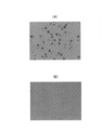
- (B) shows hybridization with covalent bonding by a probe having no covalent group (80% by volume 2 ⁇ SSC solution, after modification / washing at 55 ° C.). It is a figure which shows the hybridization by EBER (EB virus-producing small RNA) CISH, (A) is the room temperature hybridization (50% by volume formamide 2 ⁇ SSC (room temperature)) with the covalent bond by the probe which has a covalent group. (After denaturation / washing), (B) shows 55 ° C. hybridization with a commercially available PNA probe (stringent buffer, after denaturation / washing at 55 ° C.).
- EBER EB virus-producing small RNA
- the disclosure herein relates to nucleic acid detection methods and oligonucleotide probes.
- a genuine crosslinked hybridization product is irreversibly bound to a target sequence with high specificity. Therefore, if the modification conditions are such that the genuine cross-linked hybridization product can be maintained and the non-specific hybridization product can be dissociated, the true cross-linking hybridization product can be easily separated from the non-specific hybridization product and detected. can. That is, according to the above-mentioned denaturation conditions, a nucleic acid having a target sequence can be detected with excellent accuracy and reproducibility without being largely dependent on stringency intensity. As a result, true positives can be detected with high sensitivity and true negatives can be detected with high specificity.
- the test sample is not particularly limited as long as it is a sample for which detection of the target nucleic acid is required. Therefore, the test sample may be a sample that may contain the nucleic acid to be detected, and in addition to the sample taken from nature or living body, DNA or RNA in the sample was extracted from such a sample. A sample and a sample in which the content of DNA or RNA in such a sample is increased or reverse transcribed by a known nucleic acid amplification method or the like is included.
- the test samples include, for example, various biological liquid samples such as blood and urine artificially collected or naturally separated from animals such as humans and domestic animals, cell samples and tissue samples, and nucleic acid extraction / amplification of these.
- test samples such as processed samples that have been reverse-transferred.
- the test sample includes a biological sample or a processed sample of various parts or forms artificially collected or naturally separated from a plant or other organism.
- test samples include samples that are for non-medical purposes and may contain nucleic acids.
- the nucleic acid is a component thereof as long as it has a cross-linking property with a natural nucleic acid which is DNA and RNA held by a living body and an oligonucleotide probe disclosed in the present specification. Includes modified DNA and RNA, including modifications to.
- the DNA is not particularly limited as long as it is a naturally occurring DNA that can be retained by a living body or a DNA artificially introduced from the outside, but includes DNA on a vector such as a genome or a plasmid.
- RNA is not particularly limited as long as it is naturally derived RNA that can be retained by a living body or RNA introduced from the outside, but mRNA, tRNA, rRNA, ncRNA such as siRNA and miRNA, ribozyme and double-stranded RNA and the like. Is included.
- the target sequence is not particularly limited, and includes a target sequence in a nucleic acid that is a target of a known nucleic acid detection method.
- Target sequences include, for example, nucleic acid and other sequence features associated with a particular gene in the organism, nucleic acid mutations and other sequence features associated with the gene that characterizes the individual organism, the animal or plant. It is at least a part of a sequence such as a nucleic acid related to a gene that characterizes a disease or a pathogen in the above, a mutation on the nucleic acid, and other sequence features.
- the method for detecting nucleic acid disclosed in the present specification is a method for hybridizing a nucleic acid present in a test sample with respect to a target sequence, and the target when specifically hybridized with the target sequence in the hybridizing sequence.
- An oligonucleotide probe having a target base in the sequence and at least one covalent group that can be crosslinked by light irradiation is used.
- the oligonucleotide probe will be described, and then the detection method will be described.
- the oligonucleotide probe disclosed herein (hereinafter, also referred to as the probe) is prepared as a single-stranded nucleic acid having a skeleton known as a natural nucleic acid or a modified nucleic acid.
- a skeleton known as a natural nucleic acid or a modified nucleic acid.
- the nucleic acid skeleton other than deoxyribose or ribose include known skeletons such as D-threoninol.
- the probe has, for example, a backbone in which modified deoxyribose and / or ribose are linked by phosphodiester bonds or other similar linking groups.
- the probe also typically comprises a base that constitutes a natural DNA that is hybridizable with the DNA in the test sample, except for the covalent groups described below.
- This probe like the known oligonucleotide probes, has a hybridization sequence for the target sequence.
- the length of the target sequence is not particularly limited and varies depending on the intended use and the type of target nucleic acid, but is, for example, about 10 mer or more and 300 mer or less, and for example, 10 mer or more and 300 mer or less, or for example. 10 mer or more and 200 mer or less, for example, 10 mer or more and 100 mer or less, for example, 10 mer or more and 50 mer or less, and for example, 5 mer or more and 50 mer or less.
- it is 20 mer or more and 200 mer or less, and for example, 20 mer or more and 100 mer or less, and for example, 20 mer or more and 80 mer or less, and for example, 20 mer or more and 70 mer or less, and for example, 20 mer or more and 50 mer or less.
- this detection method even if the length of the hybridization sequence is 100 mer or 50 mer or less, a genuine cross-linked hybrid product is formed only when a perfect match is made, and a genuine cross-linked hybrid product other than the genuine cross-linked hybrid product is formed by the modification step described later. Is effectively removed, and as a result, a high signal-to-noise ratio can be ensured.
- the hybridization sequence has enough complementarity to specifically hybridize with the target sequence.
- the probe since the probe includes at least one covalent group so as to replace the base in the hybridization sequence, at least one base corresponding to the position of the covalent group.
- the base sequence excluding is, for example, 90% or more identity with respect to the target sequence, for example, 95% or more identity, for example, 98% or more identity, and for example, 99% or more identity. It also has, for example, 100% identity. The identity will be described later.
- a person skilled in the art can set the target sequence and the hybridization sequence by appropriately considering the base length and Tm at the time of hybridization.
- This probe comprises at least one covalent group in the hybridization sequence that can be crosslinked by light irradiation with the target base in the target sequence when specifically hybridized with the target sequence in the hybridization sequence. be able to.
- covalent groups are provided by binding to a backbone in the base sequence, such as deoxyribose or ribose.
- the covalent group is not particularly limited, but is, for example, a known photocrosslinkable group disclosed in JP-A-2016-145229, JP-A-2017-210448, JP-A-2019-26569 and the like.
- Crosslinkable functional groups can be mentioned. All of these are base positions adjacent to the 3'side of the base in the target sequence that pairs with the covalent group by light irradiation (+1 position: a position shifted from the paired base to the 3'side by one base).
- a crosslinked structure is formed with the pyrimidine ring of the pyrimidine base.
- a deoxyribonucleoside having a covalent group disclosed in JP-A-2016-145299 is shown below.
- the probe comprises such a nucleoside bound by an adjacent nucleoside with a (1'-5') -phosphodiester bond.
- the pyrimidine base forming the crosslinked structure is not particularly limited, but if it is DNA, it is thymine (T) or cytosine (C), and if it is RNA, it is cytosine (C) or uracil (U).
- these bases may be modified as much as possible with a covalent group and photocrosslinking, and examples thereof include 5-methylcytosine and 5-hydroxymethylcytosine.
- the covalent group is a sequence for hybridization so as to replace an arbitrary base pairing with a base at the base position (-1 position) adjacent to the 5'side of the pyrimidine base in the target sequence, for example, in the target sequence.
- Be prepared for In other words, the covalent group replaces any base at the base position (+1 position) adjacent to the 3'side of the purine base (adenine (A) or guanine (G)) in the hybridization sequence.
- the covalent group is provided in the hybridizing sequence so as to replace an arbitrary base pairing with the base at the base position (+1 position) adjacent to the 3'side of the pyrimidine base in the target sequence. In other words, the covalent group should replace any base at the base position (-1 position) adjacent to the 5'side of the purine base (adenine (A) or guanine (G)) in the hybridization sequence. Be prepared for.
- the covalent group is provided in place of the base at the portion where the base is bound in the skeleton such as deoxyribose, ribose and D-threoninol, for example.
- the number of covalent groups in this probe is not particularly limited, but varies depending on the length of the hybridization sequence and the type and sensitivity of the labeling element.
- the covalent groups are, for example, 1 or more and 10 or less, for example, 1 or more and 6 or less, and for example, 1 or more and 5 when the hybridization sequence is about 10 to 100 mer.
- the number is, for example, 1 or more and 4 or less, and for example, 1 or more and 3 or less, and for example, 1 or more and 2 or less.
- two or more covalent groups are provided with contiguous sites in one probe and additional covalent groups are further provided, they are added at least 10 bases apart from the continuous covalent groups. It is preferable to have a covalent group of.
- the position of the covalent group in this probe is not particularly limited, but is, for example, a 5'-terminal side and / or a 3'-terminal side, and is a position that does not suppress hybridization to the target sequence of this probe as much as possible. Can be. Typically, it is a 5'terminal region or a 3'terminal region. Here, the 5'terminal region is, for example, within 20% of the total number of bases in the hybrid sequence from the 5'terminal, and the 3'terminal region is, for example, 3 'From the terminal terminal, the number of bases is within 20% of the total number of bases in the hybridizing sequence. Further, for example, if the covalent group is on the 5'end side, it may be inside the end, and if it is on the 3'end side, at least one covalent group may be present inside.
- the position of the covalent group is, for example, if the hybridization sequence is about 10 mer or more and 100 mer, the hybridization is performed from the central portion of the hybridization sequence, that is, from the 5'end and 3'end of the probe, respectively. It may be in the range of more than 25% of the base length of the sequence. If the hybridization sequence has a certain length or longer, a genuine hybridization product and a genuine cross-linked hybrid product are formed even in the central part of the hybridization sequence without inhibiting specific hybridization with the target sequence. can.
- This probe has, for example, a plurality of probes (for example, several to several tens) having a hybrid sequence of about 10 to 100 mer for a long target sequence, typically a target sequence exceeding several hundred mer to several thousand mer. ) May be prepared as a probe set.
- the long probe has high sequence specificity but generally has a poor signal-to-noise ratio, but this probe of 10 to 100 mer has low sequence specificity but photocrosslinks at the time of perfect match, and then is not due to denaturation. Since specific hybrid products and uncrosslinked true hybrid products can be removed, a high signal-to-noise ratio can be ensured even if a long target sequence is detected with multiple probes, and false positive detection results can be avoided or sufficiently. Can be suppressed.
- a probe having a size of about 10 to 100 mer has a lower synthesis cost and better penetration into cells than a long probe.
- This probe can be equipped with a labeling element, if necessary.
- the labeling element is a signal-generating element that can generate a signal by itself or by excitation or the like, and further, a signal-generating element that does not generate a signal by itself but can be combined with a signal-generating element. It may be a connecting element.
- signaling and binding elements are well known to those of skill in the art in the art, and those skilled in the art can optionally select labeling elements and attach them to the probe.
- the detection method can include a hybridization step in which the probe is brought into contact with a target nucleic acid in a test sample to form a genuine hybridization product. By contacting the target nucleic acid with the present probe in the hybridization step, a genuine hybridization product in which the present probe and the target nucleic acid are specifically hybridized can be obtained.
- Conditions such as temperature, pH, salt concentration, and time in the hybridization step can be appropriately set in stringency according to the Tm of this probe, the type of test sample, and the like.
- the stringency becomes high, the specificity of hybridization is improved, and the hybridization efficiency tends to decrease.
- the stringency becomes low, the hybridization specificity decreases, but the hybridization efficiency tends to improve.
- the stringency can also be adjusted by the salt concentration and pH.
- a genuine hybrid product can be photocrosslinked by a light irradiation step in the subsequent stage, and a modification step can be further carried out to maintain the genuine hybrid product with high selectivity. Therefore, the hybridization step does not necessarily have to be performed with high stringency. Therefore, the hybridization step can be performed, for example, at room temperature described later, and can also be performed at 25 ° C. or higher and 40 ° C. or lower, and for example, 30 ° C. or higher and 40 ° C. or lower.
- pretreatment may be required to enable hybridization between the target nucleic acid and the present probe, depending on the test sample used and the detection method.
- the pretreatment is well known to those skilled in the art according to the test sample, the detection method, and the like, and those skilled in the art can determine the pretreatment method and the hybridization conditions based on the well-known technique.
- the hybridization step it is necessary to avoid photocrosslinking of the covalent group of this probe. Therefore, depending on the type of covalent group, the hybridization step is carried out in a light-blocked state, if necessary.
- the embodiment of the hybridization step varies depending on the nucleic acid detection method to which this detection method is applied.
- a method for detecting such a nucleic acid for example, a detection method using a solid phase such as an array or strip on which the probe is fixed, or a detection method by ISH or FISH that performs in situ hybridization may be used.
- In situ hybridization can be performed on collected cells, cultured cells, biological tissue sections, or the like.
- this probe can be supplied to cells collected from a breast cancer patient or cultured cells thereof, or into a tissue section collected from a breast cancer patient.
- cells carrying Epstein-Barr virus (EBV) infection or cultured cells thereof, tissue sections collected from patients with malignant tumor diseases caused by EBV-infected diseases, and the like can be mentioned.
- EBV Epstein-Barr virus
- nucleic acid hybridization step includes hybridization in a liquid phase, hybridization by chromatography using a column or strip on which the probe is fixed, and the like. All of these methods are well known at the time of filing the application. Those skilled in the art can carry out appropriate hybridization steps according to the type of detection method to be applied, based on well-known techniques.
- the test sample after the hybridization step is irradiated with light to crosslink between the covalent group and the target sequence in the genuine hybridization product to have a genuine hybrid product having a crosslinked structure (the present invention). It is a step of forming a genuine crosslinked hybridization product in the specification). In the previous hybridization step, in addition to the genuine hybrid product, a non-specific hybrid product containing a mismatch but hybridizing can also be formed.
- the hybridizing sequence containing the covalent group In the light irradiation step, the hybridizing sequence containing the covalent group appropriately hybridizes to the target sequence and has a highly specific cross-linked structure with the pyrimidine base on the +1-position side of the pairing base of the target sequence. To form.
- a mismatch it is difficult to form a crosslinkable positional relationship between the hybridization target sequence and the pyrimidine base to be crosslinked with the target sequence, and it is difficult to form a crosslinked structure even if appropriate light irradiation is performed. Therefore, a crosslinked structure is specifically formed in the genuine hybridization product. Therefore, according to this detection method, false negative detection results can be suppressed, thereby increasing test sensitivity.
- the light irradiation conditions for forming the crosslinked structure are determined by the type of covalent group used and the like.
- the light can be light having a temperature of 330 to 380 nm, 340 to 380 nm, and 350 to 380 nm.
- the covalent group disclosed in JP-A-2019-26569 is used, for example, 350 to 600 nm, for example, 400 to 600 nm, for example, 400 to 550 nm, and for example, 400 to 450 nm. It can irradiate light.
- the temperature and time in the light irradiation step are not particularly limited, but are, for example, 0 ° C. to 50 ° C., for example, 0 to 40 ° C., for example, 0 to 30 ° C., for example, 0 to 20 ° C., and for example, for example. 0 to 10 ° C., for example, 0 to 5 ° C.
- the time can be, for example, 1 to 120 seconds, or 1 to 60 seconds, for example.
- a modification condition capable of maintaining the genuine crosslinked hybridization product and dissociating the non-specific hybridization product is applied to the test sample after the light irradiation step, and the oligo derived from the non-specific hybridization product is applied.
- This is a step of separating the nucleotide probe.
- denaturation conditions for example, various conditions capable of dissociating double strands such as DNA are applied.
- the non-specific hybridization product can be easily separated from the genuine crosslinked hybridization product and removed (washed) under the above-mentioned modification conditions.
- washing with high stringency may also denature and wash the genuine hybrid product.
- washed with low stringency there is a problem that the non-specific hybridization product cannot be sufficiently washed.
- the genuine crosslinked hybridization product has a crosslinked structure by covalent bond, the product no longer dissociates even under modification conditions that dissociate the hybrid product by base pairing. Therefore, since the genuine crosslinked hybrid product can be retained even if a high-strength modification condition is applied, the modification condition can be easily set and only the genuine crosslinked hybrid product can be reliably maintained.
- the denaturation step also promotes hybridization dissociation of the uncrosslinked genuine hybrid product so that the oligonucleotide probe derived from the uncrosslinked genuine hybrid product and the genuine crosslinked hybrid product are separated. Can be carried out. According to this detection method, rather than high or low stringency, high-strength denaturation conditions that can distinguish cross-linked / non-cross-linked hybrid products can also be used, and as a result, false positive detection results are suppressed. This can improve the specificity of the test.
- this detection method as described above, false positive detection results are also suppressed, and as a result, the accuracy with which true positives and true negatives can be detected can be improved. Even if this detection method is, for example, a method of classifying and evaluating at multiple levels from the test results based on the signal intensity, the improvement of sensitivity, specificity, and accuracy will improve the accuracy and accuracy of the test. Can contribute.
- the stringency conditions suitable for cleaning differ depending on the probe.
- this detection method since it is sufficient to maintain the genuine crosslinked hybridization product, detailed condition setting such as changing the denaturation condition for each probe can be performed.
- the denaturation strength that promotes the dissociation of the non-specific hybrid product and thus the genuine hybrid product can be adjusted by the temperature, salt concentration, pH, etc. in addition to the denaturant.
- various factors such as a denaturing agent, temperature, base and pH can be used alone or in combination of two or more.
- a known denaturant known to dissociate the hybridization of DNA or RNA can be used.
- a solvent can be used as such a denaturing agent.
- the solvent include aldehydes such as formamide and dimethylformamide, pyridines, piperidines, dimethyl sulfoxide, tetrahydrofuran and acetonitrile.
- the denaturing conditions can be adjusted not only by the type of denaturing agent but also by the concentration of the denaturing agent and the diluent used at the same time.
- the solvent as the modifier is not particularly limited, but is, for example, 40% by volume or more, for example, 50% by volume or more, for example, 60% by volume, for example, 70% by volume or more, and for example, 80% by volume. It can be used as a solution of% or more, for example 90% by volume or more, for example 95% by volume or more, and for example 100% by volume. Preferably, it is 70% by volume or more and 80% by volume or more.
- the diluent may be, for example, a biocompatible buffer solution such as phosphate buffered saline, but may be an aqueous medium of water having higher stringency (pure water or the like). can. That is, it can be an aqueous medium mixed solution.
- a solvent used as the denaturing agent, the strength of the denaturing condition can be increased more than expected depending on the type of diluent and, for example, by using pure water instead of the buffer solution.
- FA formamide
- F aqueous solution or buffer solution 50% by volume FA aqueous solution or buffer solution, 60% by volume FA aqueous solution or buffer solution, 70% by volume FA aqueous solution or buffer solution, 80% by volume FA aqueous solution or buffer A liquid solution or the like can be used. It is preferably 50% by volume or more of the FA aqueous solution or buffer solution, more preferably 60% by volume or more of the FA aqueous solution or buffer solution, and further preferably 80% by volume or more of the FA aqueous solution or buffer solution. Is.
- the FA is, for example, an aqueous solution or buffer solution containing 60% by volume or more or more than 60% by volume of an aqueous solution or a buffer solution, or an aqueous solution or buffer solution having an amount of 80% by volume or more or more than 80% by volume. It is preferable to use a modifier (modifier) having a modification effect equal to or higher than that of a mixed solution of an aqueous medium such as a solution.
- a modifier modifier having a modification effect equal to or higher than that of a mixed solution of an aqueous medium such as a solution.
- Examples of other denaturing agents include alkylamines such as urea and tetramethylammonium chloride, alkalis such as NaOH, iodide ions, guanidine, peroxides, and thiosulfides. These denaturants can adjust the pH, base and the like. These denaturants can be used by dissolving them in a suitable solvent.
- a suitable solvent for example, a buffer solution such as biocompatible phosphate buffered saline, an aqueous medium such as pure water, or a solvent as a denaturant described above can be used.
- the denaturation step can be carried out by the conditions alone, but all other denaturation factors are accompanied by temperature conditions. Therefore, the temperature conditions applied to the modification step are appropriately set according to the modification strength such as the type and degree of modification factors applied to the modification step.
- the applicable temperature condition is the temperature of the place where the present detection method is carried out without any special temperature control. It can be carried out at a temperature that is neither heated nor cooled (in the present specification, the temperature is also referred to as room temperature).
- room temperature means, for example, 1 ° C. or higher and 35 ° C.
- Such a denaturation step at room temperature is particularly suitable in that damage to a test sample such as a tissue subjected to in situ hybridization is suppressed, and is advantageous in that a special temperature control device or the like is not required. be.
- the temperature conditions in the modification step can be, for example, 20 ° C. or higher, for example, 30 ° C. or higher, for example, 35 ° C.
- the upper limit can be, for example, 70 ° C. or lower, or 60 ° C. or lower, for example.
- the temperature condition may also be, for example, a temperature exceeding room temperature, or 40 ° C. or higher and 60 ° C. or lower.
- temperature when used as the denaturing factor, it can be, for example, 30 ° C. or higher and 90 ° C. or lower, and for example, 40 ° C. or higher and 90 ° C. or lower, 50 ° C. or higher and 90 ° C. or lower, 70 ° C. or higher and 90 ° C. or lower.
- a strong temperature condition of 70 ° C. or higher and 90 ° C. or lower when adopted, only the genuine crosslinked hybridization product can be maintained even if only water or a buffer solution is used without using a denaturing agent. ..
- Such a modification step can be carried out by applying one or more types of modification factors to a hybrid product or the like as appropriate.
- the denaturation step By carrying out the denaturation step, the dissociation of hybridization is promoted, the probe constituting the non-specific hybrid product can be removed, and further, the probe constituting the genuine hybrid product can be removed.
- Authentic cross-linked hybridization products can be reliably left behind, and background and false positive factors are easily removed.
- the detection step is appropriately carried out according to the embodiment of the hybridization step.
- the detection step is typically performed by a known method utilizing a labeling element previously imparted to the probe.
- non-specific hybrid products and genuine hybrid products can be removed without using a long probe for raising the temperature of Tm and without depending on the stringency intensity.
- Authentic crosslinked hybridization products can be detected with high accuracy.
- the oligonucleotide probes for detecting nucleic acids disclosed herein can have the configurations as described above.
- the probe is provided with a hybridization sequence and at least one covalent group capable of selectively removing nonspecific hybrid product hybridization products while maintaining a true crosslinked hybrid product. There is. By doing so, in this detection method, the accuracy and reproducibility of detection can be improved.
- the probe is also provided as a kit in combination with a denaturing agent useful in the denaturing step of the detection method.
- a denaturing agent can include, for example, a denaturing agent equal to or more than 60% by volume of a formamide aqueous medium mixed solution.
- Hybridization sequence 1 (34 mer): tgctactaccaccaagatctgcacctgcggcggc (SEQ ID NO: 1)
- Hybridization sequence 2 (25 mer): gctactaccaccaagatctgcacct (SEQ ID NO: 2)
- Hybridization sequence 3 (17mer): tgctactaccaccaaga (SEQ ID NO: 3)
- X one covalent group (X) was introduced at the position shown below.
- three types of DNA oligonucleotide probes 1 to 3 in which the 5'end was modified with Cy3 were synthesized.
- X is represented by the following structural formula.
- Hybridization sequence 1X (34mer): Cy3-tgctactaccaccaagatctgcaxctgcggcggc (SEQ ID NO: 4)
- Hybridization sequence 2X (25 mer): Cy3-gctactaccaccaagatctgcaxct (SEQ ID NO: 5)
- Hybridization sequence 3X (17mer): Cy3-tgctactaccaccaxga (SEQ ID NO: 6)
- Paraffin sections of tissues collected from breast cancer patients were deparaffinized and deproteinized according to a conventional method, fixed with 4% paraformaldehyde, and then neutralized with aldehyde.
- a probe solution prepared so that the final concentration of the probe is 1 ⁇ g / ml is heated at 80 ° C. for 3 minutes and then rapidly cooled on ice to hybridize the probe.
- 50 ⁇ l of the main chain was added dropwise onto the section, covered with a cover glass, and allowed to stand in an oven at 37 ° C. for 1 hour for hybridization.
- the cover glass is removed, and the tissue section is irradiated with UV light having a wavelength of 366 nm for 60 seconds, and then three times with 50% by volume formamide / SSC or 80% by volume formamide / SSC warmed to 50 ° C. Then, the cells were immersed twice in SSC and once in ultrapure water for about 1 to 3 minutes to wash, and then sealed with glycerol.
- the probes 1 to 3 of 34 mer, 25 mer and 17 mer all showed a signal after 80% by volume FA degeneration, but the signal of the probe 3 of 17 mer was the weakest, and the probes 1 and 2 of 34 mer and 25 mer were the weakest. Signals were about the same. From the above, it was found that a hybridization sequence of about 20 mer or more is suitable. Further, in the hybridization sequence 1 (34 mer), a DNA probe 4 having a hybridization sequence 1X'in which the X position is 3'and the third G from the end is replaced with X is synthesized, and the DNA probe 1 and the same 4 are combined.
- tissue sections derived from the same breast cancer patient were subjected to hybridization, light irradiation, and 80% by volume FA washing in the same manner as described above, and the signal of the sections was observed. As a result, both signals were equivalent. From the above, it was found that the covalent group can sufficiently form a true crosslinked hybrid product even if it is not on the 3'terminal side.
- Example 2 (Examination of light irradiation conditions using a 25-mer probe) Using the probe 2 synthesized in Example 1, 28S rRNA FISH having different UV irradiation times was performed, and the effect of irradiation time was evaluated. FISH was performed in the same manner as in Example 1 except that the light irradiation conditions were set to 0.1 second, 5 seconds, 60 seconds, and 300 seconds.
- Example 1 (Examination of covalent group)
- the probe 2 synthesized in Example 1 and another covalent group D having a D-threoninol skeleton represented by the following structural formula at the same position as the covalent group X introduced into the probe 2 are introduced.
- 28S rRNA FISH was performed according to Example 1 to evaluate the effect of the type of covalent group.
- the light irradiation time was set to 5 seconds or 60 seconds, 80% by volume FA was used for washing, and FISH was performed in the same manner as in Example 1.
- the signal of probe 5 was stronger than that of probe 2 at both the UV irradiation time of 5 seconds and 60 seconds.
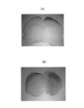
- Frozen sections of mouse brain were thawed at 37 ° C. for 30 minutes, soaked in 4% paraformaldehyde at 37 ° C. for 10 minutes, and post-fixed. Immersion in TBS buffer twice for 5 minutes and then in 2 mg / ml glycine / PBS solution for 30 minutes to neutralize the aldehyde into a 40% by volume deionized formamide / 4 x SCC solution at 55 ° C. Soaked for 30 minutes. Then, the hybridization solution containing 1 ⁇ g / ml of each of the above probes was heated at 80 ° C. for 5 minutes, 50 ⁇ l thereof was supplied, and hybridization was performed at 37 ° C. for 1 hour.
- the slide on which this section was placed was placed on ice and irradiated with a wavelength of 366 nm (1000 mV) for 1 minute. Further, immersion denaturation and washing with 80% by volume formamide / TBS buffer solution at 50 ° C. or 80% by volume formamide aqueous solution at the same temperature for 3 minutes were repeated three times.
- Fig. 2 shows the results of confirming the color development from blue to bluish purple for these section samples.
- FIG. 2 (B) when the 80% by volume formamide buffer solution was used, traces of Satb2-specific signals were observed in the cerebral cortex, but they were not clear.
- FIG. 2 (A) when an 80% by volume formamide aqueous solution having a stronger denaturing intensity was used, the non-specific signal was lowered and a clear Satb2-specific signal was transmitted to the cerebral cortex. I was able to confirm.
- probes HER2 protein mRNA CISH in breast cancer cultured cells
- the following two probes were designed to hybridize to a target sequence spanning Exons 15 to 17 in the HER2 mRNA.
- the probe can be photocrosslinked with uracil (U) or cytosine (C) in the target sequence, and a covalent group X is introduced at the 5'end in the same manner as in Example 1.
- Probe 1 GTCCACACAGGAGTGGGTGCAXTT (SEQ ID NO: 33)
- Probe 2 CGTCAGAGGGCTGGCTCTCTGXTC (SEQ ID NO: 34)
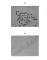
- BT474 HER2 positive
- BT20 HER2 negative
- BT474 HER2 positive
- BT20 HER2 negative
- Immunostaining for the HER2 protein was performed to confirm that each was HER2 positive or negative
- ISH was performed using probes 1 and 2 containing the covalent group X.
- a hybridization solution (TE buffer solution containing 40% by volume formamide) containing two types of probes at 1 ⁇ g / ml is heated at 80 ° C. for 3 minutes and then rapidly cooled, and 50 ⁇ l of the solution is supplied to the tissue and then covered. The mixture was allowed to stand in a constant temperature bath at 55 ° C. for 1 hour for hybridization.
- a clear signal was confirmed in HER2-positive cells by ISH using a probe containing a covalent group X.
- no signal was confirmed in HER2-negative cells.
- the results obtained by ISH were in agreement with the results of immunostaining performed in advance, and it was found that HER2 mRNA could be specifically detected.
- EBER EB virus-producing small RNA
- ISH ISH
- the following two probes were designed to hybridize to a target sequence consisting of about 100 mer in EBER1.
- the probe was synthesized using the unit used in Example 1 of uracil in the target sequence as X in the following sequence, and DIG was added to the 5'end.
- the probe of the comparative example was synthesized in the same manner except that guanine or adenine was provided instead of X.
- Probe 1 GTCTCCTCCCTAGCAAAXCC (SEQ ID NO: 35)
- Probe 2 GTCTGGGAAGACAACCAXAG (SEQ ID NO: 36)
- Epstein-Barr virus-infected tissue collected from gastric cancer patients is deparaffinized and made hydrophilic, the specimen is warmed in an oven at 60 ° C to thaw the paraffin, and then 6 times with Clear Plus (deparaffinizing agent, Pharma Co., Ltd.) at 100% EtOH. It was washed 3 times, 2 times with 95% EtOH, 1 time with 80% EtOH, and 5 times with pure water. Further, a protein kinase solution was supplied, treated at room temperature for 30 minutes, and then washed twice with pure water. Then, it was immersed in 95% EtOH and dried with a dryer.
- Clear Plus deparaffinizing agent
- a hybridization solution (TE buffer solution containing 40% by volume formamide) containing two types of probes at 1 ⁇ g / ml is heated at 80 ° C. for 3 minutes and then rapidly cooled, and 50 ⁇ l of the solution is supplied to the tissue and then covered. And allowed to stand at room temperature for 1 hour for hybridization. Similarly, hybridization was also performed on the probes of Comparative Examples.
- the probe having the covalent group X and the probe not having the covalent group X have denaturation resistance. It was found that a probe with a covalent group and high-intensity denaturation conditions are required to detect the target sequence with high accuracy.
- the attached protocol was followed. According to this protocol, hybridization was performed at 55 ° C. for 30 minutes, and washing was performed using a stringent buffer solution at 55 ° C. and a color development reaction using alkaline phosphatase.
- a probe having a covalent group X and a section treated with a PNA probe kit were evaluated. The results are shown in FIG. As shown in FIG. 5, the probe having the covalent group X was treated by hybridization at room temperature and denaturation at room temperature, but clear staining could be confirmed. Further, with respect to the PNA probe kit, the detection result equivalent to that of the probe having the covalent group X could be obtained by hybridization at 55 ° C. and washing at 55 ° C. However, in the PNA kit, the background was too high for hybridization at room temperature, and specific detection was not possible.
- the probe having the covalent group X can detect the target nucleic acid with high accuracy while facilitating temperature control.
Landscapes
- Chemical & Material Sciences (AREA)
- Life Sciences & Earth Sciences (AREA)
- Health & Medical Sciences (AREA)
- Organic Chemistry (AREA)
- Proteomics, Peptides & Aminoacids (AREA)
- Wood Science & Technology (AREA)
- Zoology (AREA)
- Engineering & Computer Science (AREA)
- Immunology (AREA)
- Analytical Chemistry (AREA)
- Genetics & Genomics (AREA)
- Pathology (AREA)
- Biochemistry (AREA)
- Biotechnology (AREA)
- Physics & Mathematics (AREA)
- Biophysics (AREA)
- Bioinformatics & Cheminformatics (AREA)
- General Engineering & Computer Science (AREA)
- General Health & Medical Sciences (AREA)
- Molecular Biology (AREA)
- Microbiology (AREA)
- Hospice & Palliative Care (AREA)
- Oncology (AREA)
- Measuring Or Testing Involving Enzymes Or Micro-Organisms (AREA)
Priority Applications (4)
| Application Number | Priority Date | Filing Date | Title |
|---|---|---|---|
| US17/920,597 US20230193398A1 (en) | 2020-04-22 | 2021-04-20 | Nucleic acid detection method and oligonucleotide probe |
| JP2022517049A JPWO2021215428A1 (cg-RX-API-DMAC7.html) | 2020-04-22 | 2021-04-20 | |
| CN202180037170.5A CN115803462A (zh) | 2020-04-22 | 2021-04-20 | 核酸的检测方法及寡核苷酸探针 |
| EP21793509.7A EP4141115A4 (en) | 2020-04-22 | 2021-04-20 | NUCLEIC ACID DETECTION METHODS AND OLIGONUCLEOTIDE PROBE |
Applications Claiming Priority (2)
| Application Number | Priority Date | Filing Date | Title |
|---|---|---|---|
| JP2020076369 | 2020-04-22 | ||
| JP2020-076369 | 2020-04-22 |
Publications (1)
| Publication Number | Publication Date |
|---|---|
| WO2021215428A1 true WO2021215428A1 (ja) | 2021-10-28 |
Family
ID=78269139
Family Applications (1)
| Application Number | Title | Priority Date | Filing Date |
|---|---|---|---|
| PCT/JP2021/015991 Ceased WO2021215428A1 (ja) | 2020-04-22 | 2021-04-20 | 核酸の検出方法及びオリゴヌクレオチドプローブ |
Country Status (6)
| Country | Link |
|---|---|
| US (1) | US20230193398A1 (cg-RX-API-DMAC7.html) |
| EP (1) | EP4141115A4 (cg-RX-API-DMAC7.html) |
| JP (1) | JPWO2021215428A1 (cg-RX-API-DMAC7.html) |
| CN (1) | CN115803462A (cg-RX-API-DMAC7.html) |
| TW (1) | TWI799842B (cg-RX-API-DMAC7.html) |
| WO (1) | WO2021215428A1 (cg-RX-API-DMAC7.html) |
Cited By (1)
| Publication number | Priority date | Publication date | Assignee | Title |
|---|---|---|---|---|
| CN113025611A (zh) * | 2021-03-16 | 2021-06-25 | 华中农业大学 | π-FISH定位单分子的探针组合物及其在核酸原位检测中的应用 |
Citations (9)
| Publication number | Priority date | Publication date | Assignee | Title |
|---|---|---|---|---|
| JPH10503655A (ja) * | 1994-08-05 | 1998-04-07 | ザ リージェンツ オブ ザ ユニバーシティ オブ カリフォルニア | ミコバクテリアの検出 |
| JP2002524091A (ja) * | 1998-08-21 | 2002-08-06 | ナックスコー・インコーポレイテッド | 架橋可能な固定化核酸を用いるアッセイ |
| JP2003535599A (ja) * | 2000-06-06 | 2003-12-02 | ティーエム バイオサイエンス コーポレイション | 核酸の捕捉部とその使用 |
| JP2015073523A (ja) * | 2013-10-11 | 2015-04-20 | 国立大学法人 東京大学 | 核酸の検出方法、検出プローブ、マイクロアレイ、核酸検出キット、核酸‐検出プローブ‐捕捉プローブ複合体、核酸固定化担体、及び流体デバイス |
| JP2015142574A (ja) | 2009-05-29 | 2015-08-06 | ヴェンタナ メディカル システムズ, インク. | インサイツハイブリダイゼーションを使用した生物学試料の遺伝子コピー数のスコアリング法 |
| JP2016145229A (ja) | 2013-03-28 | 2016-08-12 | 国立研究開発法人科学技術振興機構 | 光クロスリンク能を有する光応答性ヌクレオチドアナログ |
| JP2016145299A (ja) | 2015-02-09 | 2016-08-12 | 日立化成株式会社 | 導電材料及びそれを用いた導電体 |
| JP2017210448A (ja) | 2016-05-26 | 2017-11-30 | 国立大学法人北陸先端科学技術大学院大学 | アンチジーン法用光応答性人工核酸プローブ |
| JP2019026569A (ja) | 2017-07-26 | 2019-02-21 | 日華化学株式会社 | 可視光域で光架橋可能な光応答性ヌクレオチドアナログ |
Family Cites Families (15)
| Publication number | Priority date | Publication date | Assignee | Title |
|---|---|---|---|---|
| US5616464A (en) * | 1994-12-27 | 1997-04-01 | Naxcor | Nucleic acid sequence detection employing amplification probes |
| US6699710B1 (en) * | 1998-02-25 | 2004-03-02 | The United States Of America As Represented By The Department Of Health And Human Services | Tumor tissue microarrays for rapid molecular profiling |
| NZ555308A (en) * | 2004-12-22 | 2010-10-29 | Ambrx Inc | Compositions containing, methods involving, and uses of non-natural amino acids and polypeptides |
| GB0707208D0 (en) * | 2007-04-13 | 2007-05-23 | Istituto Superiore Di Sanito | Novel disease treatments |
| EP2292633A1 (en) * | 2009-09-08 | 2011-03-09 | Humboldt Universität zu Berlin | Probes comprising fluorescent artificial nucleobases and use thereof for detection of single base alteration |
| GB201001088D0 (en) * | 2010-01-23 | 2010-03-10 | Trillion Genomics Ltd | Detection |
| US8658780B2 (en) * | 2010-05-18 | 2014-02-25 | California Institute Of Technology | Triggered covalent probes for imaging and silencing genetic expression |
| EP2766501B1 (en) * | 2011-10-14 | 2020-01-01 | Accugenomics, Inc. | Quantitative nucleic acid amplification |
| CN104428424A (zh) * | 2012-08-31 | 2015-03-18 | 东丽株式会社 | 目标核酸的检测方法 |
| CN110237880A (zh) * | 2013-01-18 | 2019-09-17 | 生米公司 | 分析设备 |
| US9745571B2 (en) * | 2013-03-07 | 2017-08-29 | Bio-Rad Laboratories, Inc. | Repetitive reverse transcription partition assay |
| JP2014187934A (ja) * | 2013-03-27 | 2014-10-06 | Toray Ind Inc | 標的核酸の検出方法 |
| EP3490616B1 (en) * | 2016-07-27 | 2022-09-28 | The Board of Trustees of the Leland Stanford Junior University | Highly-multiplexed fluorescent imaging |
| US20180094322A1 (en) * | 2016-08-23 | 2018-04-05 | The Trustees Of Columbia University In The City Of New York | Biomarker for Predicting Colon Cancer Responsiveness to Anti-Tumor Treatment |
| WO2019071262A1 (en) * | 2017-10-06 | 2019-04-11 | The Regents Of The University Of California | RAPID SITU DETECTION OF DNA AND RNA |
-
2021
- 2021-04-20 US US17/920,597 patent/US20230193398A1/en not_active Abandoned
- 2021-04-20 JP JP2022517049A patent/JPWO2021215428A1/ja active Pending
- 2021-04-20 EP EP21793509.7A patent/EP4141115A4/en not_active Withdrawn
- 2021-04-20 WO PCT/JP2021/015991 patent/WO2021215428A1/ja not_active Ceased
- 2021-04-20 CN CN202180037170.5A patent/CN115803462A/zh active Pending
- 2021-04-22 TW TW110114450A patent/TWI799842B/zh active
Patent Citations (9)
| Publication number | Priority date | Publication date | Assignee | Title |
|---|---|---|---|---|
| JPH10503655A (ja) * | 1994-08-05 | 1998-04-07 | ザ リージェンツ オブ ザ ユニバーシティ オブ カリフォルニア | ミコバクテリアの検出 |
| JP2002524091A (ja) * | 1998-08-21 | 2002-08-06 | ナックスコー・インコーポレイテッド | 架橋可能な固定化核酸を用いるアッセイ |
| JP2003535599A (ja) * | 2000-06-06 | 2003-12-02 | ティーエム バイオサイエンス コーポレイション | 核酸の捕捉部とその使用 |
| JP2015142574A (ja) | 2009-05-29 | 2015-08-06 | ヴェンタナ メディカル システムズ, インク. | インサイツハイブリダイゼーションを使用した生物学試料の遺伝子コピー数のスコアリング法 |
| JP2016145229A (ja) | 2013-03-28 | 2016-08-12 | 国立研究開発法人科学技術振興機構 | 光クロスリンク能を有する光応答性ヌクレオチドアナログ |
| JP2015073523A (ja) * | 2013-10-11 | 2015-04-20 | 国立大学法人 東京大学 | 核酸の検出方法、検出プローブ、マイクロアレイ、核酸検出キット、核酸‐検出プローブ‐捕捉プローブ複合体、核酸固定化担体、及び流体デバイス |
| JP2016145299A (ja) | 2015-02-09 | 2016-08-12 | 日立化成株式会社 | 導電材料及びそれを用いた導電体 |
| JP2017210448A (ja) | 2016-05-26 | 2017-11-30 | 国立大学法人北陸先端科学技術大学院大学 | アンチジーン法用光応答性人工核酸プローブ |
| JP2019026569A (ja) | 2017-07-26 | 2019-02-21 | 日華化学株式会社 | 可視光域で光架橋可能な光応答性ヌクレオチドアナログ |
Non-Patent Citations (3)
| Title |
|---|
| FUJIMOTO, K ET AL.: "RNA fluorescence in situ hybridization using 3-cyanovinylcarbazole modified oligodeoxyribonucleotides as photo-cross-linkable probes", BIOORGANIC & MEDICINAL CHEMISTRY LETTERS, vol. 26, 2016, pages 5312 - 5314, XP029775004, DOI: 10.1016/j.bmcl.2016.09.035 * |
| FUJIMOTO, K. ET AL.: "RNA fluorescence in situ hybridization hybridisation using photo-cross- linkable beacon probes containing pyranocarbazole in living E. coli", BIOORGANIC AND MEDICINAL CHEMISTRY LETTERS, vol. 29, 2019, pages 2173 - 2177, XP085759033, DOI: 10.1016/j.bmcl.2019.06.051 * |
| See also references of EP4141115A4 |
Cited By (2)
| Publication number | Priority date | Publication date | Assignee | Title |
|---|---|---|---|---|
| CN113025611A (zh) * | 2021-03-16 | 2021-06-25 | 华中农业大学 | π-FISH定位单分子的探针组合物及其在核酸原位检测中的应用 |
| CN113025611B (zh) * | 2021-03-16 | 2022-06-21 | 华中农业大学 | π-FISH定位单分子的探针组合物及其在核酸原位检测中的应用 |
Also Published As
| Publication number | Publication date |
|---|---|
| TW202223390A (zh) | 2022-06-16 |
| US20230193398A1 (en) | 2023-06-22 |
| EP4141115A1 (en) | 2023-03-01 |
| CN115803462A (zh) | 2023-03-14 |
| JPWO2021215428A1 (cg-RX-API-DMAC7.html) | 2021-10-28 |
| EP4141115A4 (en) | 2024-09-11 |
| TWI799842B (zh) | 2023-04-21 |
Similar Documents
| Publication | Publication Date | Title |
|---|---|---|
| JP7372927B6 (ja) | 遺伝子およびタンパク質の発現を検出する生体分子プローブおよびその検出方法 | |
| EP4365305B1 (en) | Method of detecting an analyte | |
| JP3020601B2 (ja) | イン シチュ ハイブリット形成分析法 | |
| EP4267760A1 (en) | Methods and compositions for analyte detection | |
| CN116406428A (zh) | 用于使用酶促核酸延伸进行原位单细胞分析的组合物和方法 | |
| CN103797119B (zh) | 靶核酸的检测方法 | |
| KR20010072846A (ko) | 가교결합성 고정 핵산을 사용하는 분석법 | |
| JP7392048B2 (ja) | 分析方法及びキット | |
| CN107532212A (zh) | 用于检测靶分子的装置和方法 | |
| CN100582236C (zh) | 核酸的pcr扩增方法、pcr引物组、pcr扩增产物、以及利用该扩增方法的核酸检测方法 | |
| TWI799842B (zh) | 核酸的檢測方法及寡核苷酸探針 | |
| JPWO2018003550A1 (ja) | 2以上の標的核酸を検出するためのプライマーセット、キット及び方法 | |
| JP4477575B2 (ja) | 大腸がんの検査に使用する遺伝子セット | |
| JP4968577B2 (ja) | サイトケラチン19(CK19)mRNAの迅速測定法、並びにそのためのプライマー及びプローブ | |
| CN119979711A (zh) | 一种可编程的自动级联机制用于人乳腺组织中多种非编码rna的单分子检测试剂盒及用途 | |
| JP7346533B2 (ja) | 分析方法及びキット | |
| KR102539423B1 (ko) | 엑소좀 유래 rna를 이용한 비침습적 대장암 진단 및 분석법 | |
| CN113913404B (zh) | 一种荧光交联RNase H突变体缀合物及其应用 | |
| JP5613160B2 (ja) | ゲノムdna中の標的配列の検出又は解析方法 | |
| JP7371405B2 (ja) | 核酸試料の品質評価方法 | |
| EP3639022A1 (en) | Compositions and methods for detection of genomic variations | |
| WO2018155631A1 (ja) | 新たな染色体領域をターゲットにした腫瘍検出用マーカー | |
| KR20150046958A (ko) | 마이크로어레이 칩을 이용한 흰반점바이러스 진단용 멀티플렉스 키트 | |
| JP2007222160A (ja) | 生体由来試料中の蛋白質の解析方法 | |
| JP2017158523A (ja) | リステリア・モノサイトゲネスを検出するためのプライマーセット、キット及び方法 |
Legal Events
| Date | Code | Title | Description |
|---|---|---|---|
| 121 | Ep: the epo has been informed by wipo that ep was designated in this application |
Ref document number: 21793509 Country of ref document: EP Kind code of ref document: A1 |
|
| ENP | Entry into the national phase |
Ref document number: 2022517049 Country of ref document: JP Kind code of ref document: A |
|
| NENP | Non-entry into the national phase |
Ref country code: DE |
|
| ENP | Entry into the national phase |
Ref document number: 2021793509 Country of ref document: EP Effective date: 20221122 |
|
| WWW | Wipo information: withdrawn in national office |
Ref document number: 2021793509 Country of ref document: EP |