US5500348A - Basophil-binding monoclonal antibody, method for separation of basophils, method for chemical mediator release from basophils, and method for testing release of basophil-derived chemical mediators - Google Patents
Basophil-binding monoclonal antibody, method for separation of basophils, method for chemical mediator release from basophils, and method for testing release of basophil-derived chemical mediators Download PDFInfo
- Publication number
- US5500348A US5500348A US08/144,447 US14444793A US5500348A US 5500348 A US5500348 A US 5500348A US 14444793 A US14444793 A US 14444793A US 5500348 A US5500348 A US 5500348A
- Authority
- US
- United States
- Prior art keywords
- basophils
- release
- antibody
- histamine
- immobilized
- Prior art date
- Legal status (The legal status is an assumption and is not a legal conclusion. Google has not performed a legal analysis and makes no representation as to the accuracy of the status listed.)
- Expired - Lifetime
Links
Images


Classifications
-
- C—CHEMISTRY; METALLURGY
- C07—ORGANIC CHEMISTRY
- C07K—PEPTIDES
- C07K16/00—Immunoglobulins [IGs], e.g. monoclonal or polyclonal antibodies
- C07K16/18—Immunoglobulins [IGs], e.g. monoclonal or polyclonal antibodies against material from animals or humans
- C07K16/28—Immunoglobulins [IGs], e.g. monoclonal or polyclonal antibodies against material from animals or humans against receptors, cell surface antigens or cell surface determinants
-
- C—CHEMISTRY; METALLURGY
- C07—ORGANIC CHEMISTRY
- C07K—PEPTIDES
- C07K14/00—Peptides having more than 20 amino acids; Gastrins; Somatostatins; Melanotropins; Derivatives thereof
- C07K14/435—Peptides having more than 20 amino acids; Gastrins; Somatostatins; Melanotropins; Derivatives thereof from animals; from humans
- C07K14/46—Peptides having more than 20 amino acids; Gastrins; Somatostatins; Melanotropins; Derivatives thereof from animals; from humans from vertebrates
- C07K14/47—Peptides having more than 20 amino acids; Gastrins; Somatostatins; Melanotropins; Derivatives thereof from animals; from humans from vertebrates from mammals
- C07K14/4701—Peptides having more than 20 amino acids; Gastrins; Somatostatins; Melanotropins; Derivatives thereof from animals; from humans from vertebrates from mammals not used
- C07K14/4702—Regulators; Modulating activity
- C07K14/4705—Regulators; Modulating activity stimulating, promoting or activating activity
-
- C—CHEMISTRY; METALLURGY
- C12—BIOCHEMISTRY; BEER; SPIRITS; WINE; VINEGAR; MICROBIOLOGY; ENZYMOLOGY; MUTATION OR GENETIC ENGINEERING
- C12N—MICROORGANISMS OR ENZYMES; COMPOSITIONS THEREOF; PROPAGATING, PRESERVING, OR MAINTAINING MICROORGANISMS; MUTATION OR GENETIC ENGINEERING; CULTURE MEDIA
- C12N5/00—Undifferentiated human, animal or plant cells, e.g. cell lines; Tissues; Cultivation or maintenance thereof; Culture media therefor
- C12N5/06—Animal cells or tissues; Human cells or tissues
- C12N5/0602—Vertebrate cells
- C12N5/0634—Cells from the blood or the immune system
- C12N5/0642—Granulocytes, e.g. basopils, eosinophils, neutrophils, mast cells
-
- C—CHEMISTRY; METALLURGY
- C12—BIOCHEMISTRY; BEER; SPIRITS; WINE; VINEGAR; MICROBIOLOGY; ENZYMOLOGY; MUTATION OR GENETIC ENGINEERING
- C12N—MICROORGANISMS OR ENZYMES; COMPOSITIONS THEREOF; PROPAGATING, PRESERVING, OR MAINTAINING MICROORGANISMS; MUTATION OR GENETIC ENGINEERING; CULTURE MEDIA
- C12N5/00—Undifferentiated human, animal or plant cells, e.g. cell lines; Tissues; Cultivation or maintenance thereof; Culture media therefor
- C12N5/10—Cells modified by introduction of foreign genetic material
- C12N5/12—Fused cells, e.g. hybridomas
- C12N5/16—Animal cells
- C12N5/163—Animal cells one of the fusion partners being a B or a T lymphocyte
-
- G—PHYSICS
- G01—MEASURING; TESTING
- G01N—INVESTIGATING OR ANALYSING MATERIALS BY DETERMINING THEIR CHEMICAL OR PHYSICAL PROPERTIES
- G01N33/00—Investigating or analysing materials by specific methods not covered by groups G01N1/00 - G01N31/00
- G01N33/48—Biological material, e.g. blood, urine; Haemocytometers
- G01N33/50—Chemical analysis of biological material, e.g. blood, urine; Testing involving biospecific ligand binding methods; Immunological testing
- G01N33/5005—Chemical analysis of biological material, e.g. blood, urine; Testing involving biospecific ligand binding methods; Immunological testing involving human or animal cells
- G01N33/5091—Chemical analysis of biological material, e.g. blood, urine; Testing involving biospecific ligand binding methods; Immunological testing involving human or animal cells for testing the pathological state of an organism
-
- G—PHYSICS
- G01—MEASURING; TESTING
- G01N—INVESTIGATING OR ANALYSING MATERIALS BY DETERMINING THEIR CHEMICAL OR PHYSICAL PROPERTIES
- G01N33/00—Investigating or analysing materials by specific methods not covered by groups G01N1/00 - G01N31/00
- G01N33/48—Biological material, e.g. blood, urine; Haemocytometers
- G01N33/50—Chemical analysis of biological material, e.g. blood, urine; Testing involving biospecific ligand binding methods; Immunological testing
- G01N33/53—Immunoassay; Biospecific binding assay; Materials therefor
- G01N33/569—Immunoassay; Biospecific binding assay; Materials therefor for microorganisms, e.g. protozoa, bacteria, viruses
- G01N33/56966—Animal cells
- G01N33/56972—White blood cells
-
- C—CHEMISTRY; METALLURGY
- C12—BIOCHEMISTRY; BEER; SPIRITS; WINE; VINEGAR; MICROBIOLOGY; ENZYMOLOGY; MUTATION OR GENETIC ENGINEERING
- C12N—MICROORGANISMS OR ENZYMES; COMPOSITIONS THEREOF; PROPAGATING, PRESERVING, OR MAINTAINING MICROORGANISMS; MUTATION OR GENETIC ENGINEERING; CULTURE MEDIA
- C12N2509/00—Methods for the dissociation of cells, e.g. specific use of enzymes
Definitions
- the present invention relates to monoclonal antibodies, having the properties of (1) being reactive with basophils, (2) retaining their reactivity with basophils even after immobilization onto a solid carrier, (3) not inhibiting IgE-mediated specific histamine release from the basophils, and (4) not inducing nonspecific histamine release from the basophils, and also relates to solid-immobilized monoclonal antibodies which are prepared by immobilizing the monoclonal antibodies onto solid carriers.
- the present invention further relates to hybridomas producing the monoclonal antibodies, a method for separating basophils from humoral fluids using the monoclonal antibodies, a method for releasing chemical mediators from the basophils separated by the method for separating basophils, and a method for testing release of chemical mediators from basophils.
- the basophil a kind of leukocyte and a target cell of IgE, stores various chemical mediators such as histamine in granules, and, like mast cells, draws attention as a key entity of allergic reactions.
- IgE antibodies bound to receptors on the basophil membrane surface are crosslinked by allergen or anti-IgE antibody and this stimulation causes a degranulation reaction, resulting in chemical mediator release.
- chemical mediator release occurs directly without crosslinking of the IgEs on the basophil membrane surface.
- this second type of chemical mediator release can occur even in the absence of anti-IgE antibody and allergen (hereinafter referred to as nonspecific chemical mediator release).
- IgE-mediated specific chemical mediator release typically by the histamine release test.
- Histamine a very important chemical mediator causing type I allergic reactions, is known to induce various reactions such as bronchial smooth muscle constriction and accentuating of vascular permeability.
- the histamine release test is an unique testing method based on a biological reaction, in which immunoglobulin E (IgE) bound via receptor onto the human peripheral blood basophil surface is reacted with various allergens to release histamine and the amount of histamine thus released is determined.
- IgE immunoglobulin E
- the histamine release test serves well as a means for searching causative allergens in allergy patients in a setting closer to actual clinical conditions.
- This histamine release test using peripheral blood can be carried out in two different ways, one using whole blood and the other using washed leukocytes.
- the whole blood method may be useful in generally determining the patient's allergic condition, there is the possibility that non-basophil serum components can affect histamine release assay.
- washed leukocytes when accurate basophil reactivity is analyzed for research into the mechanism of action of drugs etc. or for basic research into the mechanism of histamine release.
- separation of washed leukocytes requires troublesome procedures, including erythrocyte removal with dextran solution, followed by two or three cycles of centrifugation and washing and subsequent leukocyte count adjustment. This results in a requirement for an increased volume of blood for the test.
- chemical mediator release from basophils involves two types: IgE-mediated specific chemical mediator release by allergic reaction and nonspecific chemical mediator release.
- IgE-mediated specific chemical mediator release by allergic reaction it is necessary to test the IgE-mediated specific chemical mediator release.
- troublesome operation and a large amount of blood are required because leukocytes must be separated to ensure the histamine release test without influence of serum components as described above. This is undesirable for routine examination.
- the present inventors investigated in order to solve the above problem by separating basophils from the blood and using them in release tests for histamine etc.
- basophil separation/purification Conventional methods for basophil separation/purification include the method of Frederik P. J. Mul et al (Journal of Immunological Methods 149, 207 (1992)), in which impurity cells are removed from the sample, the basophil density of which is previously increased by gravitational centrifugation, using magnetic particles coupled with an antibody reactive to non-basophil cell components, to purify basophils.
- basophils can be separated and purified using a basophil-reactive antibody which specifically targets basophils, a simple and very precise purification will be possible.
- the present inventors investigated in search for an antibody which can be used for this purpose, i.e., a monoclonal antibody which retains its reactivity with the basophil even after immobilization on a solid carrier and which does not damage the basophil even after coupling with the basophil, i.e., a monoclonal antibody which does not have significant influence on IgE-mediated specific histamine release from the basophil or on nonspecific histamine release from the basophil.
- a monoclonal antibody which does not have significant influence on IgE-mediated specific histamine release from the basophil or on nonspecific histamine release from the basophil.
- the inventors established a method for easily separating basophils by immobilizing such an antibody onto a solid carrier, reacting it with a humoral fluid, capturing basophils in the humoral fluid with the monoclonal antibody and then removing the unreacted humoral fluid, and succeeded in reacting the separated basophils with an allergen or an anti-IgE antibody to release histamine. Also, the present inventors have established a method for testing IgE-mediated specific histamine release from basophils which were separated using said antibody, and completed the present invention.
- an object of this invention is to provide monoclonal antibodies overcoming the above-noted drawbacks.
- Another object of this invention is to provide a solid carrier-immobilized monoclonal antibody which is prepared by immobilizing the monoclonal antibody of the present invention onto a solid carrier.
- a further object of this invention is to provide a hybridoma which produces the monoclonal antibody of the present invention.
- Yet another object of this invention is to provide a method for separating basophils from humoral fluids using the solid carrier-immobilized monoclonal antibody.
- a still further object of this invention is to provide a method for releasing a chemical mediator from basophils using the separated basophil.
- An additional object of this invention is to provide a method for testing chemical mediator release from basophils.
- the monoclonal antibodies of the present invention have the following properties:
- the monoclonal antibodies of the present invention makes it possible to separate basophils suitable for the IgE-mediated specific chemical mediator release test, because they retain their reactivity with basophils even after being immobilized onto a solid carrier, and because they do not inhibit IgE-mediated specific chemical mediator release from basophils caused by allergens or anti-IgE antibody, and do not induce nonspecific chemical mediator release from basophils. Also, the method for separating basophils of the present invention simplifies the separation of basophils from blood, and by using this method, the histamine release test which otherwise requires complex procedures can be simplified.
- the cells obtained by the method for separating basophils of the present invention can easily be utilized in the release tests for chemical mediators released from basophils such as leukotriene and PAF. In the absence of the present method, expert handling is required to obtain cells adequate for testing leukotriene or PAF release.
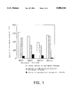
- FIG. 1 is a graph showing the total amount of histamine in the basophils separated with the monoclonal antibody of the present invention immobolized on beads (white bar), the amount of IgE-mediated specific histamine release from the basophils (hatched bar), and the amount of nonspecific histamine release from the basophils (black bar).
- FIG. 2 is a graph showing a curve of anti-human IgE antibody concentration-dependent histamine release reaction using whole blood or the cells separated with the magnetic beads having antibody BA312 immobilized thereon.
- FIG. 3 is a graph showing a curve of egg white allergen concentration-dependent histamine release reaction using the cells separated from the blood of pediatric patients with atopic dermatitis with the magnetic beads having antibody BA312 immobilized thereon.
- FIG. 4 is a graph showing a standard curve of histamine concentration obtained by RIA.
- B/B 0 means percentage level of labeled analyte bound.
- the present invention relates to monoclonal antibodies having the following properties:
- any carrier can be used for this purpose, as long as it is an ordinary carrier for solid immobilization of antibody, such as glass or synthetic resin particles (beads), spheres (balls), tubes, plates or magnetic particles such as magnetic beads, with a preference given to magnetic particles.
- the basophil-capturing capability of a solid carrier-immobilized antibody is determined by testing whether it can couple with basophils or not. Specifically, this determination is carried out by the method described below, using histamine as an index.
- a monoclonal antibody reactive with the human basophil, immobilized on magnetic beads is made to react with human blood.
- the beads are collected with a magnet and the supernatant is removed, after which a histamine release buffer (the HEPES buffer containing calcium chloride, magnesium chloride, etc.) is added, and freeze-thawing is repeated in several cycles.
- the total histamine content in the supernatant is determined by a known method such as a histamine assay system using HPLC (Y. Tsuruta et al., Journal of Chromatography 224, 105 (1981)) (manufactured by Shimadzu Corporation.
- An antibody for which histamine is detected is selected as an antibody having basophil capturing capability.
- the basophil substantially maintains its IgE-mediated specific histamine release capability can be confirmed by a comparative experiment with BA312, a monoclonal antibody found in the present invention.
- the subject solid-immobilized monoclonal antibody is judged as "not inhibiting IgE-mediated specific histamine release from the basophils" and as "being within the scope of the present invention," when the amount of histamine released from the basophils coupled with the subject monoclonal antibody is not less than 60% of that released from the basophils coupled with BA312 as determined by reacting the subject solid carrier-immobilized monoclonal antibody with basophils under the same conditions as those for solid carrier-immobilized BA312, and treating the resulting two samples of basophils coupled with respective monoclonal antibodies with anti-IgE antibody under the same conditions to release histamine.
- the amount of nonspecific histamine release can be determined by adding to the basophils an appropriate solvent such as a hydroxyethylpiperazine-N'-2-ethanesulfonic acid (HEPES) buffer containing calcium chloride, magnesium chloride, human serum albumin (HSA), etc. (hereinafter referred to as histamine release buffer), carrying out a reaction at 10°to 50° C. for 10 to 60 minutes to release histamine, and then measuring the released amount of histamine using a known method such as HPLC.
- HEPES hydroxyethylpiperazine-N'-2-ethanesulfonic acid
- histamine release buffer containing calcium chloride, magnesium chloride, human serum albumin (HSA), etc.
- the amount of IgE-mediated specific histamine release can be determined by reacting the basophils with the anti-IgE antibody dissolved in the histamine release buffer at the same reaction temperature and for the same reaction time as above, and measuring the amount of histamine released by the same method and under the same conditions as above.
- a method for preparing the monoclonal antibody of the present invention is hereinafter described briefly.
- a mouse is immunized with human basophils by several intraperitoneal, subcutaneous or intravenous injections of human basophils at intervals of several weeks.
- Antibody-producing cells are collected from the immunized mouse and fused with myeloma cells to yield hybridomas.
- the thus-obtained hybridomas are tested for human basophil reactivity, and a hybridoma which produces an antibody reactive with human basophils are selected as positive hybridomas.
- the thus-selected hybridoma is cloned. After the antibody produced by the resulting hybridoma clone is immobilized onto magnetic beads, said beads being previously coupled with sheep anti-mouse immunoglobulin antibody, the human blood basophil-capturing capability of the antibody and the histamine release properties of the separated basophils are determined.
- the basophil-capturing capability can be determined by the method described above.
- the antibody immobilized on magnetic beads is made to react with human blood as described in the determination of the basophil-capturing capability.
- the beads are collected with a magnet, and the supernatant is removed, after which the amounts of nonspecific histamine release and those of IgE-mediated specific histamine release of the cells bound to the beads are determined by the method described above.
- the antibody which has not inhibited IgE-mediated specific histamine release and which has not induced nonspecific histamine release i.e., the antibody, with which the amount of nonspecific histamine release is not more than 30%, preferably not more than 10% of IgE-mediated specific histamine release, is selected.
- a hybridoma clone producing an antibody which retains its reactivity with its basophils, that is, basophil-capturing capability, even after immobilization on a carrier, which does not inhibit IgE-mediated specific histamine release from the separated basophils, and which does not induce nonspecific histamine release from the separated basophils is selected.
- This hybridoma clone is cultured, and the monoclonal antibody is recovered.
- the selected hybridoma is proliferated by in vitro culture or in vivo culture to yield a monoclonal antibody.
- the hybridoma is cultured in an RPMI medium supplemented with fetal calf serum (complete RPMI medium) until the proliferation limit is reached, and the culture supernatant is recovered to obtain the monoclonal antibody.
- the hybridoma is transferred to the abdominal cavity of a mouse, previously treated by intraperitoneal administration of pristane, and several weeks later abdominal swelling is confirmed and ascites fluid is collected. From the ascites fluid, an IgG or IgM fraction is separated and purified by an appropriate combination of known methods such as ammonium sulfate fractionation and DEAE-Sepharose column chromatography to obtain the desired monoclonal antibody.
- the hybridomas obtained by the above method which produce the monoclonal antibodies of the present invention have been deposited since Sep. 10, 1992 with the Fermentation Research Institute, Agency of Industrial Science and Technology, 1-3, Higashi 1-chome, Tsukuba-shi, Ibaragi-ken 305, Japan, under the terms of the Budapest Treaty as "BA101 SHIONOGI,” “BA20 SHIONOGI,” “BA135 SHIONOGI,” and “BA312 SHIONOGI,” under accession numbers of FERM BP-4004, FERM BP-4005, FERM BP-4006 and FERM BP-4007, respectively.
- the monoclonal antibody of the present invention is thus exemplified by those produced by the deposited hybridomas.
- any monoclonal antibody is within the scope of the present invention as long as it has the above-described properties of (1) to (4), with a preference given to monoclonal antibody BA312.
- the monoclonal antibody of the present invention reacts with leukocytes, including the human basophil, but does not react with human erythrocytes.
- the monoclonal antibody of the present invention is capable of satisfactorily capturing basophils even after immobilization onto a solid carrier, and does not induce nonspecific chemical mediator release from the captured basophil nor inhibit IgE-mediated specific chemical mediator release.
- the monoclonal antibody of the present invention can be advantageously used as a solid carrier-immobilized monoclonal antibody obtained by immobilizing it onto a carrier as described above.
- the method for immobilizing the monoclonal antibody onto a solid carrier is not subject to limitation, and can be carried out by a known method such as the method of Lea et al. (T.
- the monoclonal antibody is reacted with a solid carrier, which is uncoated or coated with a compound having a functional group such as a tosyl group or with anti-mouse immunoglobulin, at 0°to 50° C., preferably 4 to 40° C. for 5 minutes to 72 hours, preferably 30 minutes to 48 hours.
- a solid carrier which is uncoated or coated with a compound having a functional group such as a tosyl group or with anti-mouse immunoglobulin, at 0°to 50° C., preferably 4 to 40° C. for 5 minutes to 72 hours, preferably 30 minutes to 48 hours.
- the present invention also provides a basophil separation method wherein the solid carrier-immobilized monoclonal antibody of the present invention as obtained above is made to react with a humoral fluid and basophils in the humoral fluid are captured by coupling to the solid carrier-immobilized monoclonal antibody.
- Such humoral fluids include blood, nasal secretions, tears and saliva, with a preference given to blood.
- the basophil separation method of the present invention permits separation of a basophil-rich cell sample by a simple procedure in which the monoclonal antibody of the present invention is immobilized onto a solid carrier such as a magnetic particle directly or via anti-mouse immunoglobulin to yield an antibody-coupled magnetic particle, which is then reacted with a humoral fluid, preferably blood, at 0°to 50° C., preferably 15 to 40° C. for a short period of time (1 to 60 minutes), after which the magnetic particle is collected with a magnet to collect basophils from the unreacted sample.
- a humoral fluid preferably blood
- the present invention also provides a method for testing chemical mediator release from basophils wherein basophils separated by the basophil separation method of the present invention are made to react with allergen or anti-IgE antibody.
- allergen commonly used in the diagnosis of allergy can be used according to necessity, which includes inhalants such as house dust and pollen, dietary allergens such as meat and eggs, and chemical substances.
- the reaction of the basophils with an allergen or anti-IgE antibody is carried out at 10°to 50° C., preferably 25°to 40° C. for 0.5 to 300 minutes, preferably 10 to 60 minutes.
- the present invention also provides a method for testing chemical mediator release from basophils comprising the following steps (a) through (c):
- steps (a) and (b) described above be carried out in accordance with the above-described basophil separation method of the present invention and the method for chemical mediator release from basophils described above.
- the determination of the amount of released chemical mediator in step (c) can be achieved by a known method chosen as appropriate for the desired chemical mediator.
- the amount in the case of histamine, the amount can be determined by a histamine assay system using HPLC (Y. Tsuruta et al., Journal of Chromatography 224, 105 (1981)) (Shimadzu Corporation).
- the amount in the case of leukotriene and PAF, the amount can be determined by the method of Hayes et al. (E. Hayes et al., Journal of Immunology 131, 429 (1983)) and the method of Smal et al. (M. A. Smal et al., Journal of Immunological Methods 128, 183 (1990)), respectively.
- the histamine assay using HPLC can be performed in the following steps: a sample is passed through a column packed with a cation exchange resin to elute histamine; 0.1% orthophthalaldehyde and NaOH are added to the eluate and allowed to react with histamine at 45° to form a histamine phosphor; the histamine phosphor is made to react with H 2 SO 4 for stabilization and intensification of fluorescence; and then the intensity of fluorescence is determined.
- the amount of chemical mediators can be determined by RIA.
- leukotriene assay using RIA it can be performed as follows: anti-leukotriene antibody and 3 H-labeled leukotriene are added to a sample or a standard non-labeled leukotriene and allowed to react at 4° C. for 5 to 24 hours; the obtained mixture is made to react with dextran-coated charcoal for 10 minutes while cooling in ice; and then the resulting mixture is centrifuged and the radioactivity of the supernatant is determined.
- PAF assay using RIA it can be performed as follows: anti-PAF antibody, 125 I-labeled PAF and the second antibody are added to a sample or a standard non-labeled PAF and allowed to react for 5 to 24 hours; the obtained mixture is centrifuged, and the radioactivity of the sediment is determined.
- histamine assay by RIA a sample or a standard non-labeled histamine is made to react with anti-histamine antibody and 125 I-labeled histamine at 0°to 40° C., preferably at 0°to 10° C., for 5 minutes to 48 hours, preferably for 30 minutes to 24 hours.
- a B/F separating agent such as polyethyleneglycol is added and allowed to react for 10 minutes while cooling in ice. After centrifugation, the radioactivity of the sediment is determined.
- the amount of histamine can also be determined by EIA. Although any known EIA methods such as the sandwich method, competitive method and ELISA can be used, a preference is given to the method reported, for example, by Knox Van Dyke, et al. (Luminescence Immunoassay and Molecular Application, CRC Press, pp.2-10, (1990)). Specifically, a mixture of a sample, biotin-labeled standard histamine and enzyme-labeled anti-histamine antibody is made to react with an avidin-immobilized carrier.
- the enzymatic activity of the complex between biotin-labeled histamine and enzyme-labeled anti-histamine antibody, which is bound with the avidin-immobilized carrier is determined.
- Any carriers and enzymes that are usually used for EIA can be used, but it is preferable to use a microplate as the carrier and a horseradish peroxidase as the enzyme.
- the spleen was excised from each mouse and prepared as a cell suspension in RPMI medium. These splenocytes (2 ⁇ 10 8 ) and NS-1 myeloma cells (6 ⁇ 10 7 ) were mixed and centrifugally settled. To the sediment, 1 ml of 50% polyethylene glycol (average molecular weight of 4000) was added with gentle stirring, followed by stirring for 1 more minute. To this mixture, 1 ml of RPMI medium was added over a 1-minute period, followed by addition of 1 ml and then addition of 7 ml over a 3-minute period.
- polyethylene glycol average molecular weight of 4000
- the sediment was suspended in 40 ml of an RPMI medium containing 15% fetal calf serum (complete RPMI medium), and this suspension was inoculated to four 96-well microplates at 0.1 ml per well, followed by cultivation at 37° C. in the presence of 7% carbon dioxide. Twenty-four hours later, 0.1 ml of a complete RPMI medium containing 100 ⁇ M hypoxanthine, 0.4 ⁇ M aminopterin and 16 ⁇ M thymidine (HAT medium) was added. At 2, 3, 5 and 8 days of cultivation, 0.1 ml of the culture supernatant was discarded, and 0.1 ml of HAT medium was added.
- Hybridomas in each culture plate well were cultured in a complete RPMI medium, and the culture supernatant was assayed for specific antibody production as follows:
- a cell suspension was prepared at 4 ⁇ 10 5 basophils/ml in a 10 mM hydroxyethylpiperazine-N'-2-ethanesulfonic acid (HEPES) buffer (pH 7.4) containing 0.8% sodium chloride, 0.037% potassium chloride and 0.03% human serum albumin (HSA) (HA-HEPES buffer).
- HSA human serum albumin
- reaction mixture was once washed with HA-HEPES buffer, a 50 ⁇ l mixture of goat anti-human IgE antibody labeled with fluorescein isothiocyanate (FITC) and goat anti-mouse immunoglobulin antibody labeled with phycoerythrin was added, followed by reaction at room temperature for 1 hour. After being once washed with HA-HEPES buffer, the reaction mixture was observed under a fluorescent microscope to determine whether the FITC-labeled cells were labeled by phycoerythrin as well. Thus, one hundred wells, in which the monoclonal antibody contained in the hybridoma culture supernatant was reactive to the FITC-labeled basophil, were selected.
- FITC fluorescein isothiocyanate
- each clone was cultured in a flask until the proliferation limit was reached. A 1 ml portion of this culture supernatant was added to 15 mg of magnetic beads, previously coupled with sheep anti-mouse immunoglobulin antibody, followed by reaction at 4° C. for 16 hours with gentle stirring, to yield magnetic beads having a monoclonal antibody immobilized thereon.
- magnetic beads human peripheral blood basophil capturing capability and the IgE-mediated specific histamine release and nonspecific histamine release properties of the separated basophils were examined in accordance with the method described in Example 4 below.
- hybridomas BA101 Shionogi (FERM BP-4004), BA20 Shionogi (FERM BP-4005), BA135 Shionogi (FERM BP-4006) and BA312 Shionogi (FERM BP-4007) were selected, which respectively produce monoclonal antibodies BA101, BA20, BA135 and BA312, which possess the above-described properties and which are suitable to the methods of the present invention for separation of cells including the human basophils and for testing of histamine release from the separated cells, as demonstrated in Experimental Example below.
- the hybridoma was cultured in a complete RPMI medium until the proliferation limit (1 ⁇ 10 6 cells/ml) was reached, and the culture supernatant was recovered.
- the abdominal ascites obtained in the above process were salted out with an 18% sodium sulfate solution, and the resulting precipitate was dissolved in 0.01M borate-buffered saline (pH 8.0), followed by dialysis against the same solution.
- a 20 mg aliquot of the monoclonal antibody obtained by this salting-out was dissolved in 2 ml of 0.01 M borate-buffered saline (pH 8.0) and adsorbed to a column (1.6 ⁇ 5 cm) of protein A-Sepharose (manufactured by Pharmacia AB).
- the beads were collected with a magnet and the supernatant was removed, the beads were washed 4 times with 0.01M HEPES buffer (pH 7.4) containing 0.5% HSA for 5 minutes at a time, and then washed with HA-HEPES buffer at 4° C. for 16 hours, to obtain magnetic beads having BA312 immobilized thereon.
- HA-HEPES buffer was added to prepare a 20% blood solution.
- a suspension of magnetic beads having the monoclonal antibody in the culture supernatant of hybridoma BA20 Shionogi, BA101 Shionogi, BA135 Shionogi or BA312 Shionogi immobilized thereon were prepared at 3 mg beads/ml with HA-HEPES buffer.
- the 20% blood solution was dispensed to test tubes at 500 ⁇ l per test tube.
- the IgE-mediated specific histamine release reaction was carried out by reacting the sample with monoclonal anti-human IgE antibody at 37° C. for 1 hour.
- the nonspecific histamine release reaction was carried out by incubating the sample in the histamine release buffer at 37° C. for 1 hour. After completion of the reaction, the reaction mixture was stirred and then centrifuged at 1500 rpm for 5 minutes, and 300 ⁇ l of the supernatant was obtained.
- the amount of histamine released was determined by a histamine assay system with HPLC (Y. Tsuruta et al., Journal of Chromatography 224, 105 (1981)) (manufactured by Shimadzu Corporation).
- CD9 antibody TP82TM, manufactured by Nichirei
- CD11b antibody BEAR1TM, manufactured by Immunotech
- CD13 antibody MCS2TM, manufactured by Nichirei
- CD32 antibody 2ElTM, manufactured by Immunotech
- CD9 antibody, CD11b antibody, BA20 and BA312 showed good basophil-capturing capability, while CD13 antibody and CD32 antibody did not.
- CD9 antibody and CD11b antibody were each immobilized onto magnetic beads.
- Cells separated using these magnetic beads were evaluated for three parameters of histamine release capability, i.e., the total amount of histamine, the amount of IgE-mediated specific histamine release and the amount of nonspecific histamine release.
- an antibody immobilized on magnetic beads is assumed to be useful in chemical mediator release tests, provided that the relative amount of nonspecific histamine release is not higher than 30%, particularly not higher than 10%, of the amount of IgE-mediated specific histamine release from the basophils captured and separated by the antibody.
- the cells captured and separated by these antibodies often show markedly higher nonspecific histamine release in comparison with IgE-mediated specific histamine release; therefore, the above ratio by far exceeds 30%, as shown in Table 3, and it was very difficult to analyze IgE-mediated specific histamine release with these separated cells.
- BA312 a monoclonal antibody of the present invention gave an excellent ratio of 7.5%.
- a 10 ml blood sample was taken in a heparinized blood sampling tube.
- This blood was dispensed to test tubes at 100 ⁇ l per test tube, and 400 ⁇ l of histamine release buffer or a 50000, 5000, 500, 50 or 5 ng/ml suspension of monoclonal anti-human IgE antibody (HE-69B) in histamine release buffer was added to each tube, followed by stirring to give a sample (the final concentrations of monoclonal anti-human IgE antibodies were 40000, 4000, 400, 40 and 4 ng/ml, respectively).
- the total amount of histamine was determined using the sample after 3 cycles of freeze-thawing in the histamine buffer.
- the IgE-mediated specific histamine release reaction was carried out by reacting a sample with monoclonal anti-human IgE antibody at 37° C. for 1 hour.
- the nonspecific histamine release reaction was carried out by incubating a sample in the histamine release buffer at 37° C. for 1 hour. After completion of the reaction, the reaction mixture was stirred and then centrifuged at 1500 rpm for 5 minutes, and 300 ⁇ l of the supernatant was obtained and assayed for the amount of released histamine by a histamine assay system with HPLC (manufactured by Shimadzu Corporation).
- histamine release rate was determined.
- the specific histamine release rate can be calculated using the following equation: ##EQU2##
- the whole blood method and the separated cell method gave comparable results for specific histamine release rate. This demonstrates that the cells separated using the monoclonal antibody of the present invention retain their histamine release capability naturally observed in the blood.
- Example 4 For two blood samples from pediatric patients with atopic dermatitis caused by egg white, cells were separated and tested for histamine release in the same manner as in Example 4 above, using BA312-immobilized magnetic beads prepared by the method of Example 3 above.
- the egg white allergen used in the test was adjusted to concentrations of 4000, 400, 40, 4 and 0.4 ng/ml with the histamine release buffer, and the ratio of IgE-mediated specific histamine release was calculated using the equation as described above.
- the ratio of IgE-mediated specific histamine release depended on an egg white allergen concentration. Also, a bell-shaped concentration dependency curve was obtained in some cases, in which the histamine release rate decreased as the allergen concentration increased above a given level.
- the concentration of histamine was determined by radioimmunoassay (RIA). Specifically, 100 ⁇ l of a sample containing released histamine or 100 ⁇ l of a standard histamine solution was placed in a test tube, respectively. To each test tube, 100 ⁇ l of 125 I-labeled histamine solution (10KBq/ml), which was prepared with 50 mM phosphate buffered saline (pH 7.0) containing 0.3% human serum albumin and 0.1% sodium azide (assay buffer), and 100 ⁇ l of anti-histamine antibody solution prepared by the assay buffered were added and allowed to react for 2 hours at 4° C.
- assay buffer 50 mM phosphate buffered saline
- anti-histamine antibody solution prepared by the assay buffered were added and allowed to react for 2 hours at 4° C.
- FIG. 4 shows the standard curve of histamine concentration obtained by RIA.
Landscapes
- Health & Medical Sciences (AREA)
- Life Sciences & Earth Sciences (AREA)
- Chemical & Material Sciences (AREA)
- Immunology (AREA)
- Engineering & Computer Science (AREA)
- Biomedical Technology (AREA)
- Molecular Biology (AREA)
- Organic Chemistry (AREA)
- Hematology (AREA)
- Biochemistry (AREA)
- General Health & Medical Sciences (AREA)
- Cell Biology (AREA)
- Genetics & Genomics (AREA)
- Medicinal Chemistry (AREA)
- Urology & Nephrology (AREA)
- Biotechnology (AREA)
- Zoology (AREA)
- Microbiology (AREA)
- Bioinformatics & Cheminformatics (AREA)
- Wood Science & Technology (AREA)
- Tropical Medicine & Parasitology (AREA)
- Proteomics, Peptides & Aminoacids (AREA)
- Food Science & Technology (AREA)
- Physics & Mathematics (AREA)
- Analytical Chemistry (AREA)
- General Physics & Mathematics (AREA)
- Pathology (AREA)
- Biophysics (AREA)
- Physiology (AREA)
- Virology (AREA)
- General Engineering & Computer Science (AREA)
- Toxicology (AREA)
- Gastroenterology & Hepatology (AREA)
- Peptides Or Proteins (AREA)
- Preparation Of Compounds By Using Micro-Organisms (AREA)
- Micro-Organisms Or Cultivation Processes Thereof (AREA)
- Medicines Containing Antibodies Or Antigens For Use As Internal Diagnostic Agents (AREA)
Applications Claiming Priority (2)
| Application Number | Priority Date | Filing Date | Title |
|---|---|---|---|
| JP4-321164 | 1992-11-04 | ||
| JP32116492 | 1992-11-04 |
Publications (1)
| Publication Number | Publication Date |
|---|---|
| US5500348A true US5500348A (en) | 1996-03-19 |
Family
ID=18129523
Family Applications (1)
| Application Number | Title | Priority Date | Filing Date |
|---|---|---|---|
| US08/144,447 Expired - Lifetime US5500348A (en) | 1992-11-04 | 1993-11-02 | Basophil-binding monoclonal antibody, method for separation of basophils, method for chemical mediator release from basophils, and method for testing release of basophil-derived chemical mediators |
Country Status (9)
| Country | Link |
|---|---|
| US (1) | US5500348A (ja) |
| EP (1) | EP0596479B1 (ja) |
| JP (1) | JP3550410B2 (ja) |
| KR (1) | KR100280239B1 (ja) |
| AT (1) | ATE176930T1 (ja) |
| DE (1) | DE69323589T2 (ja) |
| DK (1) | DK0596479T3 (ja) |
| ES (1) | ES2129482T3 (ja) |
| TW (1) | TW378213B (ja) |
Cited By (13)
| Publication number | Priority date | Publication date | Assignee | Title |
|---|---|---|---|---|
| DE19643427C1 (de) * | 1996-10-22 | 1998-02-12 | Bundesrep Deutschland | Verfahren zur Bestimmung von Allergenen in der Luft |
| US5722470A (en) * | 1995-11-09 | 1998-03-03 | Glaxo Group Limited | Bead dispensing device and methods |
| US6151973A (en) * | 1999-01-29 | 2000-11-28 | Glaxo Wellcome Inc. | Bead picking apparatus and method |
| US20020115214A1 (en) * | 1988-11-23 | 2002-08-22 | Carl H. June | Methods for selectively stimulating proliferation of t cells |
| US20030022250A1 (en) * | 2001-05-29 | 2003-01-30 | Dreskin Stephen C. | Functional IgE test methods and compositions |
| EP1304571A1 (en) * | 2000-07-27 | 2003-04-23 | Kyowa Medex Co., Ltd. | Method of immunity examination with insoluble carrier particle and reagent therefor |
| US20030099643A1 (en) * | 1988-11-23 | 2003-05-29 | Carl H. June | Methods for selectively stimulating proliferation of t cells |
| US20030143758A1 (en) * | 2000-06-30 | 2003-07-31 | Kayoko Shigenobu | Insoluble carrier particle nephelometric immunoassay reagent |
| US20040001829A1 (en) * | 1988-11-23 | 2004-01-01 | June Carl H. | Methods for selectively stimulating proliferation of T cells |
| US20060099177A1 (en) * | 1988-11-23 | 2006-05-11 | The United States Of America As Represented By The Secretary Of The Navy | Methods for treating HIV infected subjects |
| US20060140919A1 (en) * | 1994-06-03 | 2006-06-29 | The United States Of America As Represented By The Secretary Of The Navy | Methods for selectively stimulating proliferation of T cells |
| US20090309689A1 (en) * | 2005-08-23 | 2009-12-17 | Lear Corporation | Electrical Connector Housing |
| CN112415191A (zh) * | 2020-11-06 | 2021-02-26 | 郑州人福博赛生物技术有限责任公司 | 一种白细胞三烯检测试剂盒 |
Families Citing this family (2)
| Publication number | Priority date | Publication date | Assignee | Title |
|---|---|---|---|---|
| FR2765341A1 (fr) * | 1997-06-25 | 1998-12-31 | Alain Funes | Methode pour l'analyse de l'activation des basophiles humains par mesure de l'expression membranaire du marqueur cd63 |
| ES2230108T3 (es) * | 1999-05-19 | 2005-05-01 | Eberhard-Karls-Universitat Tubingen Universitatsklinikum | Utilizacion de un anticuerpo para detectar basofilos y/o mastocitos. |
Citations (4)
| Publication number | Priority date | Publication date | Assignee | Title |
|---|---|---|---|---|
| US4559310A (en) * | 1983-05-20 | 1985-12-17 | Dana Farber Cancer Institute | Assay methods and systems utilizing mast cell clones |
| EP0239400A2 (en) * | 1986-03-27 | 1987-09-30 | Medical Research Council | Recombinant antibodies and methods for their production |
| AU2294888A (en) * | 1987-09-30 | 1989-04-06 | Sanofi-Synthelabo | Immunometric assay kit and method applicable to whole cells |
| WO1991001368A1 (en) * | 1989-07-24 | 1991-02-07 | Dynal A.S. | Hapten/anti-hapten affinity linking in cell separation |
-
1993
- 1993-10-06 TW TW082108242A patent/TW378213B/zh not_active IP Right Cessation
- 1993-11-02 US US08/144,447 patent/US5500348A/en not_active Expired - Lifetime
- 1993-11-02 JP JP29737993A patent/JP3550410B2/ja not_active Expired - Lifetime
- 1993-11-03 AT AT93117830T patent/ATE176930T1/de not_active IP Right Cessation
- 1993-11-03 KR KR1019930023230A patent/KR100280239B1/ko not_active IP Right Cessation
- 1993-11-03 EP EP93117830A patent/EP0596479B1/en not_active Expired - Lifetime
- 1993-11-03 DE DE69323589T patent/DE69323589T2/de not_active Expired - Lifetime
- 1993-11-03 ES ES93117830T patent/ES2129482T3/es not_active Expired - Lifetime
- 1993-11-03 DK DK93117830T patent/DK0596479T3/da active
Patent Citations (4)
| Publication number | Priority date | Publication date | Assignee | Title |
|---|---|---|---|---|
| US4559310A (en) * | 1983-05-20 | 1985-12-17 | Dana Farber Cancer Institute | Assay methods and systems utilizing mast cell clones |
| EP0239400A2 (en) * | 1986-03-27 | 1987-09-30 | Medical Research Council | Recombinant antibodies and methods for their production |
| AU2294888A (en) * | 1987-09-30 | 1989-04-06 | Sanofi-Synthelabo | Immunometric assay kit and method applicable to whole cells |
| WO1991001368A1 (en) * | 1989-07-24 | 1991-02-07 | Dynal A.S. | Hapten/anti-hapten affinity linking in cell separation |
Non-Patent Citations (22)
| Title |
|---|
| Bodger et al, 1987. The purification of human basophils: their immunophenotype and cytochemistry. Br. J. Haematol. 67:281 4. * |
| Bodger et al, 1987. The purification of human basophils: their immunophenotype and cytochemistry. Br. J. Haematol. 67:281-4. |
| Bodger et al, Blood, vol. 69, No. 5, pp. 1414 1418 (1987). * |
| Bodger et al, Blood, vol. 69, No. 5, pp. 1414-1418 (1987). |
| E. F. Knol et al., "Monitoring human basophil activation via CD 63 monoclonal antibody 435", J. Allergy Clin. Immunol, Sep. 1991, vol. 88, pp. 328-338. |
| E. F. Knol et al., Monitoring human basophil activation via CD 63 monoclonal antibody 435 , J. Allergy Clin. Immunol, Sep. 1991, vol. 88, pp. 328 338. * |
| Gaudernack et al, Journal of Immunological Methods, vol. 90, pp. 179 187 (1986). * |
| Gaudernack et al, Journal of Immunological Methods, vol. 90, pp. 179-187 (1986). |
| Lett Brown et al, 1989. Purification of human basophils. Their response to anti IgE. J. Immunol. Meth 117:163 7. * |
| Lett-Brown et al, 1989. Purification of human basophils. Their response to anti-IgE. J. Immunol. Meth 117:163-7. |
| MacGlashan et al, 1980, The purification of human basophils, J. Immunol 124:2519 2521. * |
| MacGlashan et al, 1980, The purification of human basophils, J. Immunol 124:2519-2521. |
| Mul et al, Journal of Immunological Methods, vol. 149, pp. 207 214 (1992). * |
| Mul et al, Journal of Immunological Methods, vol. 149, pp. 207-214 (1992). |
| Schroeder et al, Journal of Immunological Methods, vol. 133, pp. 269 277 (1990). * |
| Schroeder et al, Journal of Immunological Methods, vol. 133, pp. 269-277 (1990). |
| Valent et al, 1989. Mast cell typing: demonstration of a distinct hematopoietic cell type and evidence for immunophenotypic relationship to mononuclear phagocytes. Blood 73:1778 85. * |
| Valent et al, 1989. Mast cell typing: demonstration of a distinct hematopoietic cell type and evidence for immunophenotypic relationship to mononuclear phagocytes. Blood 73:1778-85. |
| Valent et al, Int. Arch Allergy Appl. Immunol., vol. 91, pp. 198 203 (1990). * |
| Valent et al, Int. Arch Allergy Appl. Immunol., vol. 91, pp. 198-203 (1990). |
| Van Toorenenberger, 1981. IgG4 and passive sensitization of basophil leukocytes. Int. Archs. Allergy Appl. Immunol 65:432 40. * |
| Van Toorenenberger, 1981. IgG4 and passive sensitization of basophil leukocytes. Int. Archs. Allergy Appl. Immunol 65:432-40. |
Cited By (25)
| Publication number | Priority date | Publication date | Assignee | Title |
|---|---|---|---|---|
| US6905680B2 (en) | 1988-11-23 | 2005-06-14 | Genetics Institute, Inc. | Methods of treating HIV infected subjects |
| US7232566B2 (en) | 1988-11-23 | 2007-06-19 | The United States As Represented By The Secretary Of The Navy | Methods for treating HIV infected subjects |
| US7479269B2 (en) | 1988-11-23 | 2009-01-20 | Genetics Institute, Llc | Methods for selectively enriching TH1 and TH2 cells |
| US20020115214A1 (en) * | 1988-11-23 | 2002-08-22 | Carl H. June | Methods for selectively stimulating proliferation of t cells |
| US20060013832A1 (en) * | 1988-11-23 | 2006-01-19 | The United States Of America As Represented By The Secretary Of The Navy | Methods for treating HIV infected subjects |
| US7144575B2 (en) | 1988-11-23 | 2006-12-05 | The Regents Of The University Of Michigan | Methods for selectively stimulating proliferation of T cells |
| US20030099643A1 (en) * | 1988-11-23 | 2003-05-29 | Carl H. June | Methods for selectively stimulating proliferation of t cells |
| US20060099177A1 (en) * | 1988-11-23 | 2006-05-11 | The United States Of America As Represented By The Secretary Of The Navy | Methods for treating HIV infected subjects |
| US20040001829A1 (en) * | 1988-11-23 | 2004-01-01 | June Carl H. | Methods for selectively stimulating proliferation of T cells |
| US6887466B2 (en) | 1988-11-23 | 2005-05-03 | Genetics Institute, Inc. | Methods for selectively stimulating proliferation of T cells |
| US20060140919A1 (en) * | 1994-06-03 | 2006-06-29 | The United States Of America As Represented By The Secretary Of The Navy | Methods for selectively stimulating proliferation of T cells |
| US6905681B1 (en) | 1994-06-03 | 2005-06-14 | Genetics Institute, Inc. | Methods for selectively stimulating proliferation of T cells |
| US20060205069A1 (en) * | 1994-06-03 | 2006-09-14 | The United States Of America As Represented By The Secretary Of The Navy | Compositions comprising a first agent which provides a primary activation signal to T cells and a second agent which stimulates an accessory molecule on the surface of T cells |
| US7175843B2 (en) | 1994-06-03 | 2007-02-13 | Genetics Institute, Llc | Methods for selectively stimulating proliferation of T cells |
| US5722470A (en) * | 1995-11-09 | 1998-03-03 | Glaxo Group Limited | Bead dispensing device and methods |
| DE19643427C1 (de) * | 1996-10-22 | 1998-02-12 | Bundesrep Deutschland | Verfahren zur Bestimmung von Allergenen in der Luft |
| US6151973A (en) * | 1999-01-29 | 2000-11-28 | Glaxo Wellcome Inc. | Bead picking apparatus and method |
| US20030143758A1 (en) * | 2000-06-30 | 2003-07-31 | Kayoko Shigenobu | Insoluble carrier particle nephelometric immunoassay reagent |
| US20040053426A1 (en) * | 2000-07-27 | 2004-03-18 | Kayoko Shigenobu | Method of immunity examination with insoluble carrier particle and reagent therefor |
| EP1304571A1 (en) * | 2000-07-27 | 2003-04-23 | Kyowa Medex Co., Ltd. | Method of immunity examination with insoluble carrier particle and reagent therefor |
| EP1304571A4 (en) * | 2000-07-27 | 2007-06-27 | Kyowa Medex Co Ltd | IMMUNOLOGICAL ANALYSIS USING INSOLUBLE SUPPORT PARTICLES AND REACTIVE THEREFOR |
| US8431415B2 (en) | 2000-07-27 | 2013-04-30 | Tfb, Inc. | Immunoassay using insoluble carrier particles and reagent therefor |
| US20030022250A1 (en) * | 2001-05-29 | 2003-01-30 | Dreskin Stephen C. | Functional IgE test methods and compositions |
| US20090309689A1 (en) * | 2005-08-23 | 2009-12-17 | Lear Corporation | Electrical Connector Housing |
| CN112415191A (zh) * | 2020-11-06 | 2021-02-26 | 郑州人福博赛生物技术有限责任公司 | 一种白细胞三烯检测试剂盒 |
Also Published As
| Publication number | Publication date |
|---|---|
| TW378213B (en) | 2000-01-01 |
| EP0596479A3 (en) | 1995-04-19 |
| JPH06205695A (ja) | 1994-07-26 |
| ES2129482T3 (es) | 1999-06-16 |
| KR100280239B1 (ko) | 2001-02-01 |
| DK0596479T3 (da) | 1999-09-27 |
| DE69323589T2 (de) | 1999-10-14 |
| EP0596479B1 (en) | 1999-02-24 |
| KR940011480A (ko) | 1994-06-21 |
| JP3550410B2 (ja) | 2004-08-04 |
| DE69323589D1 (de) | 1999-04-01 |
| ATE176930T1 (de) | 1999-03-15 |
| EP0596479A2 (en) | 1994-05-11 |
Similar Documents
| Publication | Publication Date | Title |
|---|---|---|
| US5500348A (en) | Basophil-binding monoclonal antibody, method for separation of basophils, method for chemical mediator release from basophils, and method for testing release of basophil-derived chemical mediators | |
| US4363799A (en) | Monoclonal antibody to human T cells, and methods for preparing same | |
| EP0119629A2 (en) | Use of anti-idiotype antibodies in immunoassay | |
| EP0189688A2 (en) | Monoclonal antibody capable of recognizing arteriosclerotic lesions and agents for detecting and treating arteriosclerosis | |
| JP2820402B2 (ja) | 補体成分C5aに対するモノクローナル抗体 | |
| EP0161638A2 (en) | Monoclonal antibody and method for quantitation of immoglobulins using the same | |
| EP0311383B1 (en) | Monoclonal antibody to methamphetamine, preparation of the same, assay method and assay kit of methamphetamine | |
| US6942977B1 (en) | Immunoassays for determining vitamin b12, and reagents and kits therefor | |
| JP2644029B2 (ja) | Nk細胞および細胞障害性tリンパ球の同定 | |
| EP0205352A2 (en) | Monoclonal anti-human IgG4 antibodies, their production and use | |
| US4515894A (en) | Hybrid cell line for producing monoclonal antibody to human T cells | |
| JP4215462B2 (ja) | 非グリコシル化ヘモグロビン、グリコシル化ヘモグロビンに特異的に結合するモノクローナル抗体の製造方法 | |
| JP2840852B2 (ja) | C反応性蛋白質に対するモノクローナル坑体 | |
| JPS63258493A (ja) | 抗ガングリオシドgm↓1単クロ−ン性抗体、これを産生する細胞及びこれから成る試薬 | |
| EP0501779A1 (en) | Antibodies reactive with human cytosolic phospholipase A2 | |
| JP2635946B2 (ja) | 抗ガングリオシドGD1a単クローン性抗体MZを産生する細胞 | |
| JP3036545B2 (ja) | モノクローナル抗体とこれを産生するハイブリドーマ細胞株、およびこれを用いる免疫学的測定法 | |
| EP0307186B1 (en) | Anti-ganglioside GD1a monoclonal antibody MZ, MZ-producing cells and MZ-containing reagent | |
| JP3841364B2 (ja) | 抗ヒトビトロネクチン・トロンビン・アンチトロンビンiii 複合体モノクローナル抗体、ハイブリドーマ及び免疫学的測定方法 | |
| CA2110019C (en) | Immunoassays for determining vitamin b12, and reagents and kits therefor | |
| JP2868841B2 (ja) | Gmp異常症の検出方法及びキット | |
| RU1776691C (ru) | Штамм гибридных культивируемых клеток животных MUS мUSсULUS L, - продуцент моноклональных антител против J @ Е человека | |
| JPH02196787A (ja) | シュードウリジン誘導体 | |
| EP0352817A2 (en) | Monoclonal antibody to catecholamine metabolite and quantitative assay for catecholamine metabolite | |
| JPH0678786A (ja) | Lecam−1と反応するモノクローナル抗体及びlecam−1の測定方法 |
Legal Events
| Date | Code | Title | Description |
|---|---|---|---|
| AS | Assignment |
Owner name: INTERFACE, INC., GEORGIA Free format text: ASSIGNMENT OF ASSIGNORS INTEREST;ASSIGNORS:ADAMS, HAROLD F.;SUAREZ, RODERICK A.;ENGLE, LEWIS H.;REEL/FRAME:006705/0665 Effective date: 19930910 |
|
| AS | Assignment |
Owner name: SHIONOGI & CO., LTD., JAPAN Free format text: ASSIGNMENT OF ASSIGNORS INTEREST;ASSIGNORS:NISHIMURA, SHINJI;NISHI, HIROSHI;NISHIMURA, MASAJI;REEL/FRAME:006803/0411 Effective date: 19930917 |
|
| STCF | Information on status: patent grant |
Free format text: PATENTED CASE |
|
| FPAY | Fee payment |
Year of fee payment: 4 |
|
| FPAY | Fee payment |
Year of fee payment: 8 |
|
| FPAY | Fee payment |
Year of fee payment: 12 |