EP4183451B1 - Compositions and methods for adjoining type i and type ii extracellular domains as heterologous chimeric proteins - Google Patents
Compositions and methods for adjoining type i and type ii extracellular domains as heterologous chimeric proteins Download PDFInfo
- Publication number
- EP4183451B1 EP4183451B1 EP22199596.2A EP22199596A EP4183451B1 EP 4183451 B1 EP4183451 B1 EP 4183451B1 EP 22199596 A EP22199596 A EP 22199596A EP 4183451 B1 EP4183451 B1 EP 4183451B1
- Authority
- EP
- European Patent Office
- Prior art keywords
- cells
- ox40l
- human
- amino acid
- panel
- Prior art date
- Legal status (The legal status is an assumption and is not a legal conclusion. Google has not performed a legal analysis and makes no representation as to the accuracy of the status listed.)
- Active
Links
Images


Classifications
-
- A—HUMAN NECESSITIES
- A61—MEDICAL OR VETERINARY SCIENCE; HYGIENE
- A61K—PREPARATIONS FOR MEDICAL, DENTAL OR TOILETRY PURPOSES
- A61K38/00—Medicinal preparations containing peptides
- A61K38/16—Peptides having more than 20 amino acids; Gastrins; Somatostatins; Melanotropins; Derivatives thereof
- A61K38/17—Peptides having more than 20 amino acids; Gastrins; Somatostatins; Melanotropins; Derivatives thereof from animals; from humans
- A61K38/177—Receptors; Cell surface antigens; Cell surface determinants
- A61K38/1774—Immunoglobulin superfamily (e.g. CD2, CD4, CD8, ICAM molecules, B7 molecules, Fc-receptors, MHC-molecules)
-
- A—HUMAN NECESSITIES
- A61—MEDICAL OR VETERINARY SCIENCE; HYGIENE
- A61K—PREPARATIONS FOR MEDICAL, DENTAL OR TOILETRY PURPOSES
- A61K38/00—Medicinal preparations containing peptides
- A61K38/16—Peptides having more than 20 amino acids; Gastrins; Somatostatins; Melanotropins; Derivatives thereof
- A61K38/17—Peptides having more than 20 amino acids; Gastrins; Somatostatins; Melanotropins; Derivatives thereof from animals; from humans
- A61K38/19—Cytokines; Lymphokines; Interferons
- A61K38/191—Tumor necrosis factors [TNF], e.g. lymphotoxin [LT], i.e. TNF-beta
-
- A—HUMAN NECESSITIES
- A61—MEDICAL OR VETERINARY SCIENCE; HYGIENE
- A61P—SPECIFIC THERAPEUTIC ACTIVITY OF CHEMICAL COMPOUNDS OR MEDICINAL PREPARATIONS
- A61P35/00—Antineoplastic agents
-
- A—HUMAN NECESSITIES
- A61—MEDICAL OR VETERINARY SCIENCE; HYGIENE
- A61P—SPECIFIC THERAPEUTIC ACTIVITY OF CHEMICAL COMPOUNDS OR MEDICINAL PREPARATIONS
- A61P37/00—Drugs for immunological or allergic disorders
- A61P37/02—Immunomodulators
-
- A—HUMAN NECESSITIES
- A61—MEDICAL OR VETERINARY SCIENCE; HYGIENE
- A61P—SPECIFIC THERAPEUTIC ACTIVITY OF CHEMICAL COMPOUNDS OR MEDICINAL PREPARATIONS
- A61P37/00—Drugs for immunological or allergic disorders
- A61P37/02—Immunomodulators
- A61P37/04—Immunostimulants
-
- A—HUMAN NECESSITIES
- A61—MEDICAL OR VETERINARY SCIENCE; HYGIENE
- A61P—SPECIFIC THERAPEUTIC ACTIVITY OF CHEMICAL COMPOUNDS OR MEDICINAL PREPARATIONS
- A61P37/00—Drugs for immunological or allergic disorders
- A61P37/02—Immunomodulators
- A61P37/06—Immunosuppressants, e.g. drugs for graft rejection
-
- C—CHEMISTRY; METALLURGY
- C07—ORGANIC CHEMISTRY
- C07K—PEPTIDES
- C07K14/00—Peptides having more than 20 amino acids; Gastrins; Somatostatins; Melanotropins; Derivatives thereof
-
- C—CHEMISTRY; METALLURGY
- C07—ORGANIC CHEMISTRY
- C07K—PEPTIDES
- C07K14/00—Peptides having more than 20 amino acids; Gastrins; Somatostatins; Melanotropins; Derivatives thereof
- C07K14/435—Peptides having more than 20 amino acids; Gastrins; Somatostatins; Melanotropins; Derivatives thereof from animals; from humans
- C07K14/52—Cytokines; Lymphokines; Interferons
- C07K14/525—Tumour necrosis factor [TNF]
-
- C—CHEMISTRY; METALLURGY
- C07—ORGANIC CHEMISTRY
- C07K—PEPTIDES
- C07K14/00—Peptides having more than 20 amino acids; Gastrins; Somatostatins; Melanotropins; Derivatives thereof
- C07K14/435—Peptides having more than 20 amino acids; Gastrins; Somatostatins; Melanotropins; Derivatives thereof from animals; from humans
- C07K14/705—Receptors; Cell surface antigens; Cell surface determinants
- C07K14/70596—Molecules with a "CD"-designation not provided for elsewhere
-
- C—CHEMISTRY; METALLURGY
- C07—ORGANIC CHEMISTRY
- C07K—PEPTIDES
- C07K19/00—Hybrid peptides, i.e. peptides covalently bound to nucleic acids, or non-covalently bound protein-protein complexes
-
- C—CHEMISTRY; METALLURGY
- C12—BIOCHEMISTRY; BEER; SPIRITS; WINE; VINEGAR; MICROBIOLOGY; ENZYMOLOGY; MUTATION OR GENETIC ENGINEERING
- C12N—MICROORGANISMS OR ENZYMES; COMPOSITIONS THEREOF; PROPAGATING, PRESERVING, OR MAINTAINING MICROORGANISMS; MUTATION OR GENETIC ENGINEERING; CULTURE MEDIA
- C12N15/00—Mutation or genetic engineering; DNA or RNA concerning genetic engineering, vectors, e.g. plasmids, or their isolation, preparation or purification; Use of hosts therefor
- C12N15/09—Recombinant DNA-technology
- C12N15/63—Introduction of foreign genetic material using vectors; Vectors; Use of hosts therefor; Regulation of expression
- C12N15/79—Vectors or expression systems specially adapted for eukaryotic hosts
- C12N15/85—Vectors or expression systems specially adapted for eukaryotic hosts for animal cells
-
- C—CHEMISTRY; METALLURGY
- C12—BIOCHEMISTRY; BEER; SPIRITS; WINE; VINEGAR; MICROBIOLOGY; ENZYMOLOGY; MUTATION OR GENETIC ENGINEERING
- C12N—MICROORGANISMS OR ENZYMES; COMPOSITIONS THEREOF; PROPAGATING, PRESERVING, OR MAINTAINING MICROORGANISMS; MUTATION OR GENETIC ENGINEERING; CULTURE MEDIA
- C12N7/00—Viruses; Bacteriophages; Compositions thereof; Preparation or purification thereof
-
- A—HUMAN NECESSITIES
- A61—MEDICAL OR VETERINARY SCIENCE; HYGIENE
- A61K—PREPARATIONS FOR MEDICAL, DENTAL OR TOILETRY PURPOSES
- A61K38/00—Medicinal preparations containing peptides
-
- C—CHEMISTRY; METALLURGY
- C07—ORGANIC CHEMISTRY
- C07K—PEPTIDES
- C07K2319/00—Fusion polypeptide
-
- C—CHEMISTRY; METALLURGY
- C07—ORGANIC CHEMISTRY
- C07K—PEPTIDES
- C07K2319/00—Fusion polypeptide
- C07K2319/70—Fusion polypeptide containing domain for protein-protein interaction
- C07K2319/74—Fusion polypeptide containing domain for protein-protein interaction containing a fusion for binding to a cell surface receptor
-
- Y—GENERAL TAGGING OF NEW TECHNOLOGICAL DEVELOPMENTS; GENERAL TAGGING OF CROSS-SECTIONAL TECHNOLOGIES SPANNING OVER SEVERAL SECTIONS OF THE IPC; TECHNICAL SUBJECTS COVERED BY FORMER USPC CROSS-REFERENCE ART COLLECTIONS [XRACs] AND DIGESTS
- Y02—TECHNOLOGIES OR APPLICATIONS FOR MITIGATION OR ADAPTATION AGAINST CLIMATE CHANGE
- Y02A—TECHNOLOGIES FOR ADAPTATION TO CLIMATE CHANGE
- Y02A50/00—TECHNOLOGIES FOR ADAPTATION TO CLIMATE CHANGE in human health protection, e.g. against extreme weather
- Y02A50/30—Against vector-borne diseases, e.g. mosquito-borne, fly-borne, tick-borne or waterborne diseases whose impact is exacerbated by climate change
Definitions
- the present invention relates to, inter alia, compositions, including chimeric proteins that find use in the treatment of disease, such as immunotherapies for cancer and autoimmunity.
- checkpoint inhibition therapy still fails in many patients. Therefore, as with most cancer therapies, there remains a need for new compositions and methods that can improve the effectiveness of these agents.
- US 2015/183881 A1 discloses chimeric proteins comprising the ectodomains of RANK and TGF ⁇ RII or PD-1.
- US 2015/266942 A1 discloses a SIRP ⁇ -Fc-VEGFR1D2 fusion protein.
- the present invention provides for compositions that are useful for cancer immunotherapy, e.g. to manipulate or modify immune signals for therapeutic benefit.
- the invention reverses or suppresses immune inhibitory signals while providing immune activating or costimulatory signals in a beneficial context.
- the present invention provides chimeric protein comprising: (a) a first domain comprising a first extracellular domain of SIRP ⁇ (CD172a) at or near the N-terminus, wherein the first domain is capable of binding a SIRP ⁇ (CD172a) ligand, (b) a second domain comprising a second extracellular domain of CD40 ligand (CD40L) at or near the C-terminus, wherein the second domain is capable of binding a CD40L receptor, and (c) a linker linking the first and the second domain and comprising an IgG hinge-CH2-CH3 Fc domain.
- the chimeric protein of the present invention comprises an extracellular domain of the immune inhibitory agent CD172a/SIRP ⁇ .
- the chimeric protein is engineered to disrupt, block, reduce, and/or inhibit the transmission of an immune inhibitory signal, the binding of CD172a with CD47.
- the chimeric protein of the present invention comprises an extracellular domain of the immune stimulatory signal, which is CD40 ligand.
- the chimeric protein is engineered to enhance, increase, and/or stimulate the transmission of an immune stimulatory signal, by the binding of CD40 with CD40 ligand.
- the chimeric protein comprises an immune inhibitory receptor extracellular domain and an immune stimulatory ligand extracellular domain which can, without limitation, deliver an immune stimulation to a T cell while masking a tumor cell's immune inhibitory signals.
- the present chimeric proteins provide improved immunotherapeutic benefits by effectively causing the substitution of an immune inhibitory signal for an immune stimulatory signal.
- the present chimeric proteins in some embodiments are capable of, or find use in methods involving, reducing or eliminating an inhibitory immune signal and/or increasing or activating an immune stimulatory signal.
- beneficial properties are enhanced by the single construct approach of the present chimeric proteins. For instance, the signal replacement can be effected nearly simultaneously and the signal replacement is tailored to be local at a site of clinical importance ( e.g. the tumor microenvironment).
- the present chimeric proteins are capable of, or find use in methods involving, shifting the balance of immune cells in favor of immune attack of a tumor.
- the present chimeric proteins can shift the ratio of immune cells at a site of clinical importance in favor of cells that can kill a tumor (e.g . T cells, cytotoxic T lymphocytes, T helper cells, natural killer (NK) cells, natural killer T (NKT) cells, anti-tumor macrophages (e.g. M1 macrophages), B cells, and dendritic cells and in opposition to cells that protect tumors (e.g.
- T cells e.g cytotoxic T lymphocytes, T helper cells, natural killer (NK) cells, natural killer T (NKT) cells, anti-tumor macrophages (e.g. M1 macrophages), B cells, and dendritic cells and in opposition to cells that protect tumors (e.g.
- the present chimeric protein is capable of increasing a ratio of effector T cells to regulatory T cells.
- the present chimeric protein unexpectedly provides binding of the extracellular domain components to their respective binding partners with longer off rates (Kd or K off ) and therefore, inter alia, accords longer occupancy of the receptor to ligand and vice versa. For instance, in some embodiments, this provides a sustained negative signal masking effect. Further, in some embodiments, this delivers a longer positive signal effect, e.g. to allow an effector cell to be adequately stimulated ( e.g. for proliferation and/or release of stimulatory signals like cytokines). Also, this stable synapse of cells ( e.g.
- a tumor cell bearing negative signals and a T cell which could attack the tumor provides spatial orientation to favor tumor reduction - such as positioning the T cells to attack tumor cells and/or sterically preventing the tumor cell from delivering negative signals, including negative signals beyond those masked by the chimeric protein of the invention.
- this provides longer on-target (e.g. intra-tumoral) half-life (t 1/2 ) as compared to serum t 1/2 of the chimeric proteins.
- on-target e.g. intra-tumoral
- t 1/2 half-life
- the present chimeric protein is for use in a method for treating cancer comprising administering an effective amount of a pharmaceutical composition comprising the chimeric protein to a patient in need thereof.
- the present chimeric protein is for use in a method for treating infections, including without limitation, viral infections or other intracellular pathogens.
- the present chimeric protein is for use in a method for treating autoimmune diseases.
- the present invention is based, in part, on the discovery that chimeric proteins can be engineered from the extracellular, or effector, regions of immune-modulating transmembrane proteins in a manner that exploits the orientations of these proteins (e.g. type I versus type II) and therefore allows the delivery of immune stimulatory and/or immune inhibitory signals, including, for example, masking an immune inhibitory signal and replacing it with an immune stimulatory signal in the treatment of cancer.
- the present invention relates to a chimeric protein comprising: (a) a first domain comprising an extracellular domain of SIRP ⁇ (CD172a) at or near the N-terminus, wherein the first domain is capable of binding a SIRP ⁇ (CD172a) ligand, (b) a second domain comprising an extracellular domain of CD40 ligand (CD40L) at or near the C-terminus, wherein the second domain is capable of binding a CD40L receptor, and (c) a linker linking the first domain and the second domain and comprising an IgG hinge-CH2-CH3 Fc domain.
- an extracellular domain refers to a portion of a transmembrane protein which is capable of interacting with the extracellular environment. In various embodiments, an extracellular domain refers to a portion of a transmembrane protein which is sufficient to bind to a ligand or receptor and effective transmit a signal to a cell. In various embodiments, an extracellular domain is the entire amino acid sequence of a transmembrane protein which is external of a cell or the cell membrane. In various embodiments, an extracellular domain is the that portion of an amino acid sequence of a transmembrane protein which is external of a cell or the cell membrane and is needed for signal transduction and/or ligand binding as may be assayed using methods know in the art (e.g. in vitro ligand binding and/or cellular activation assays).
- an immune inhibitory signal refers to a signal that diminishes or eliminates an immune response.
- such signals may diminish or eliminate antitumor immunity.
- inhibitory signal are useful in the maintenance of self-tolerance (e.g. prevention of autoimmunity ) and also to protect tissues from damage when the immune system is responding to pathogenic infection.
- immune inhibitory signal may be identified by detecting an increase in cellular proliferation, cytokine production, cell killing activity or phagocytic activity when such an inhibitory signal is blocked.
- inhibitory signals include blockade of PD-1 of PD-L1/L2 using antibody mediated blockade or through competitive inhibition of PD-L1/L2 using PD-1 containing fusion proteins.
- an inhibitory signal When such an inhibitory signal is blocked through inhibition of PD-L1/L2, it leads to enhance tumor killing activity by T cells because they are no longer being inhibited by PD-L1 or PD-L2.
- inhibitory signal may be provided by CD47 to macrophages expressing CD172a. Binding of CD47 to CD172a typically inhibits the ability of a macrophage to phagocytose a target cell, which can be restored through blockade of CD47 with blocking antibodies or through competitive inhibition of CD47 using CD172a containing fusion proteins.
- an immune stimulatory signal refers to a signal that enhances an immune response.
- such signals may enhance antitumor immunity.
- immune stimulatory signal may be identified by directly stimulating proliferation, cytokine production, killing activity or phagocytic activity of leukocytes.
- Specific examples include direct stimulation of TNF superfamily receptors such as OX40, 4-1BB or TNFRSF25 using either receptor agonist antibodies or using fusion proteins encoding the ligands for such receptors (OX40L, 4-1 BBL, TL1A, respectively). Stimulation from any one of these receptors may directly stimulate the proliferation and cytokine production of individual T cell subsets.
- Another example includes direct stimulation of an immune inhibitory cell with through a receptor that inhibits the activity of such an immune suppressor cell.
- This would include, for example, stimulation of CD4+FoxP3+ regulatory T cells with a GITR agonist antibody or GITRL containing fusion protein, which would reduce the ability of those regulatory T cells to suppress the proliferation of conventional CD4+ or CD8+ T cells.
- this would include stimulation of CD40 on the surface of an antigen presenting cell using a CD40 agonist antibody or a fusion protein containing CD40L, causing activation of antigen presenting cells including enhanced ability of those cells to present antigen in the context of appropriate native costimulatory molecules, including those in the B7 or TNF superfamily.
- Membrane proteins typically consist of an extracellular domain, one or a series of trans-membrane domains, and an intracellular domain.
- the extracellular domain of a membrane protein is responsible for interacting with a soluble or membrane bound receptor or ligand.
- the trans-membrane domain(s) are responsible for localizing a protein to the plasma membrane.
- the intracellular domain of a membrane protein is responsible for coordinating interactions with cellular signaling molecules to coordinate intracellular responses with the extracellular environment (or visa-versa).
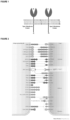
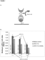
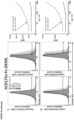
- type I there are two types of single-pass membrane proteins, those with an extracellular amino terminus and intracellular carboxy terminus (type I) and those with an extracellular carboxy terminus and intracellular amino terminus (type II). Both type I and type II membrane proteins can be either receptors or ligands.
- type I membrane proteins the amino terminus of the protein faces outside the cell, and therefore contains the functional domains that are responsible for interacting with other binding partners (either ligands or receptors) in the extracellular environment ( FIG. 1 , left image).
- type II membrane proteins the carboxy terminus of the protein faces outside the cell, and therefore contains the functional domains that are responsible for interacting with other binding partners (either ligands or receptors) in the extracellular environment ( FIG. 1 , right image). Thus, these two types of proteins have opposite orientations to each other.
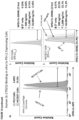
- This construct could be produced by cloning of these three fragments (the extracellular domain of a type I protein, followed by a linker sequence, followed by the extracellular domain of a type II protein) into a vector (plasmid, viral or other) wherein the amino terminus of the complete sequence corresponded to the 'left' side of the molecule containing the type I protein and the carboxy terminus of the complete sequence corresponded to the 'right' side of the molecule containing the type II protein.
- the present chimeric proteins are engineered as such.
- costimulatory and coinhibitory signals Two major families of costimulatory molecules include the B7 and the tumor necrosis factor (TNF) families. These molecules bind to receptors on T cells belonging to the CD28 or TNF receptor families, respectively. Many well-defined coinhibitors and their receptors belong to the B7 and CD28 families.
- B7 and CD28 families Two major families of costimulatory molecules include the B7 and the tumor necrosis factor (TNF) families. These molecules bind to receptors on T cells belonging to the CD28 or TNF receptor families, respectively. Many well-defined coinhibitors and their receptors belong to the B7 and CD28 families.
- TNF tumor necrosis factor
- the chimeric protein of the present invention comprises an extracellular domain of SIRP ⁇ /CD172a.
- the chimeric protein is engineered to disrupt, block, reduce, and/or inhibit the transmission of an immune inhibitory signal, by binding of CD172a with CD47.
- the chimeric protein of the present invention comprises an extracellular domain of CD40 ligand (CD40L).
- the chimeric protein simulates binding of an inhibitory signal ligand to its cognate receptor (CD172a to CD47) but inhibits the inhibitory signal transmission to an immune cell (e.g. a T cell, macrophage or other leukocyte).
- an immune cell e.g. a T cell, macrophage or other leukocyte.
- the chimeric protein comprises an immune inhibitory receptor extracellular domain and an immune stimulatory ligand extracellular domain which can, without limitation, deliver an immune stimulation to a T cell while masking a tumor cell's immune inhibitory signals.
- the chimeric protein delivers a signal that has the net result of T cell activation.
- the chimeric protein comprises both (i) an immune inhibitory signal which is a receptor of an immune inhibitory signal and this acts on a tumor cell that bears a cognate ligand of the immune inhibitory signal and (ii) an immune stimulatory signal which is a ligand of an immune stimulatory signal and this acts on a T cell that bears a cognate receptor of the immune stimulatory signal.
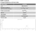
- the chimeric protein exhibits enhanced stability and protein half-life. In some embodiments, the chimeric protein binds to FcRn with high affinity. In various embodiments, the chimeric protein may bind to FcRn with a K D of about 70 nM to about 80 nM. For example, the chimeric protein may bind to FcRn with a K D of about 70 nM, about 71 nM, about 72 nM, about 73 nM, about 74 nM, about 75 nM, about 76 nM, about 77 nM, about 78 nM, about 79 nM, or about 80 nM. In some embodiments, the chimeric protein does not substantially bind to other Fc receptors (i.e. other than FcRn) with effector function.
- FcRn Fc receptors
- the chimeric protein comprises the extracellular domain of the immune inhibitory agent CD172a/SIRPa and is paired with an immune stimulatory agent as CD172a/CD40L.
- the present chimeric proteins may comprises variants of the extracellular domains described herein, for instance, a sequence having at least about 60%, or at least about 61%, or at least about 62%, or at least about 63%, or at least about 64%, or at least about 65%, or at least about 66%, or at least about 67%, or at least about 68%, or at least about 69%, or at least about 70%, or at least about 71%, or at least about 72%, or at least about 73%, or at least about 74%, or at least about 75%, or at least about 76%, or at least about 77%, or at least about 78%, or at least about 79%, or at least about 80%, or at least about 81%, or at least about 82%, or at least about 83%, or at least about 84%, or at least about 85%, or at least about 86%, or at least about 87%, or at least about 88%, or at least about 89%, or at least about 90%, or at least about 91%, or at least
- the chimeric protein may comprise an amino acid sequence having one or more amino acid mutations relative to any of the protein sequences described herein.
- the one or more amino acid mutations may be independently selected from substitutions, insertions, deletions, and truncations.
- the amino acid mutations are amino acid substitutions, and may include conservative and/or non-conservative substitutions.
- Constant substitutions may be made, for instance, on the basis of similarity in polarity, charge, size, solubility, hydrophobicity, hydrophilicity, and/or the amphipathic nature of the amino acid residues involved.
- the 20 naturally occurring amino acids can be grouped into the following six standard amino acid groups: (1) hydrophobic: Met, Ala, Val, Leu, Ile; (2) neutral hydrophilic: Cys, Ser, Thr; Asn, Gln; (3) acidic: Asp, Glu; (4) basic: His, Lys, Arg; (5) residues that influence chain orientation: Gly, Pro; and (6) aromatic: Trp, Tyr, Phe.
- conservative substitutions are defined as exchanges of an amino acid by another amino acid listed within the same group of the six standard amino acid groups shown above. For example, the exchange of Asp by Glu retains one negative charge in the so modified polypeptide.
- glycine and proline may be substituted for one another based on their ability to disrupt ⁇ -helices,
- non-conservative substitutions are defined as exchanges of an amino acid by another amino acid listed in a different group of the six standard amino acid groups (1) to (6) shown above.
- the substitutions may also include non-classical amino acids (e.g. selenocysteine, pyrrolysine, N-formylmethionine ⁇ -alanine, GABA and ⁇ -Aminolevulinic acid, 4-aminobenzoic acid (PABA), D-isomers of the common amino acids, 2,4-diaminobutyric acid, ⁇ -amino isobutyric acid, 4-aminobutyric acid, Abu, 2-amino butyric acid, ⁇ -Abu, ⁇ -Ahx, 6-amino hexanoic acid, Aib, 2-amino isobutyric acid, 3-amino propionic acid, ornithine, norleucine, norvaline, hydroxyproline, sarcosme, citrulline, homocitrulline, cysteic acid, t-butylglycine, t-butylalanine, phenylglycine,
- Mutations may also be made to the nucleotide sequences of the chimeric proteins by reference to the genetic code, including taking into account codon degeneracy.
- the chimeric protein comprises a linker.
- the linker is a hinge-CH2-CH3 Fc domain of an antibody (e.g., of IgG, IgA, IgD, and IgE, inclusive of subclasses (e.g. IgG1, IgG2, IgG3, and IgG4, and lgA1 and IgA2)).
- the hinge region found in IgG, IgA, IgD, and IgE class antibodies, acts as a flexible spacer, allowing the Fab portion to move freely in space.
- the hinge domains are structurally diverse, varying in both sequence and length among immunoglobulin classes and subclasses. For example, the length and flexibility of the hinge region varies among the IgG subclasses.
- the hinge region of IgG1 encompasses amino acids 216-231 and, because it is freely flexible, the Fab fragments can rotate about their axes of symmetry and move within a sphere centered at the first of two inter-heavy chain disulfide bridges.
- IgG2 has a shorter hinge than IgG1, with 12 amino acid residues and four disulfide bridges.
- the hinge region of IgG2 lacks a glycine residue, is relatively short, and contains a rigid poly-proline double helix, stabilized by extra inter-heavy chain disulfide bridges. These properties restrict the flexibility of the IgG2 molecule.
- IgG3 differs from the other subclasses by its unique extended hinge region (about four times as long as the IgG1 hinge), containing 62 amino acids (including 21 prolines and 11 cysteines), forming an inflexible poly-proline double helix.
- the Fab fragments are relatively far away from the Fc fragment, giving the molecule a greater flexibility.
- the elongated hinge in IgG3 is also responsible for its higher molecular weight compared to the other subclasses.
- the hinge region of IgG4 is shorter than that of IgG1 and its flexibility is intermediate between that of IgG1 and IgG2.
- the flexibility of the hinge regions reportedly decreases in the order IgG3>IgG1>lgG4>lgG2.
- the linker may be derived from human IgG4 and contain one or more mutations to enhance dimerization (including S228P) or FcRn binding.
- the immunoglobulin hinge region can be further subdivided functionally into three regions: the upper hinge region, the core region, and the lower hinge region.
- the upper hinge region includes amino acids from the carboxyl end of C H1 to the first residue in the hinge that restricts motion, generally the first cysteine residue that forms an interchain disulfide bond between the two heavy chains.
- the length of the upper hinge region correlates with the segmental flexibility of the antibody.
- the core hinge region contains the inter-heavy chain disulfide bridges, and the lower hinge region joins the amino terminal end of the C H2 domain and includes residues in C H2 . Id.
- the core hinge region of wild-type human IgG1 contains the sequence Cys-Pro-Pro-Cys which, when dimerized by disulfide bond formation, results in a cyclic octapeptide believed to act as a pivot, thus conferring flexibility.
- the present linker comprises, one, or two, or three of the upper hinge region, the core region, and the lower hinge region of any antibody (e.g., of IgG, IgA, IgD, and IgE, inclusive of subclasses (e.g. IgG1, IgG2, IgG3, and IgG4, and lgA1 and IgA2)).
- the hinge region may also contain one or more glycosylation sites, which include a number of structurally distinct types of sites for carbohydrate attachment.
- lgA1 contains five glycosylation sites within a 17-amino-acid segment of the hinge region, conferring resistance of the hinge region polypeptide to intestinal proteases, considered an advantageous property for a secretory immunoglobulin.
- the linker of the present invention comprises one or more glycosylation sites.
- the linker comprises a hinge-CH2-CH3 Fc domain derived from a human IgG4 antibody. In various embodiments, the linker comprises a hinge-CH2-CH3 Fc domain derived from a human IgG1 antibody. In some embodiments, the Fc domain exhibits increased affinity for and enhanced binding to the neonatal Fc receptor (FcRn). In some embodiments, the Fc domain includes one or more mutations that increases the affinity and enhances binding to FcRn. Without wishing to be bound by theory, it is believed that increased affinity and enhanced binding to FcRn increases the in vivo half-life of the present chimeric proteins.
- the Fc domain linker contains one or more amino acid substitutions at amino acid residue 250, 252, 254, 256, 308, 309, 311, 428, 433 or 434 (in accordance with Kabat numbering), or equivalents thereof.
- the amino acid substitution at amino acid residue 250 is a substitution with glutamine.
- the amino acid substitution at amino acid residue 252 is a substitution with tyrosine, phenylalanine, tryptophan or threonine.
- the amino acid substitution at amino acid residue 254 is a substitution with threonine.
- the amino acid substitution at amino acid residue 256 is a substitution with serine, arginine, glutamine, glutamic acid, aspartic acid, or threonine.
- the amino acid substitution at amino acid residue 308 is a substitution with threonine.
- the amino acid substitution at amino acid residue 309 is a substitution with proline.
- the amino acid substitution at amino acid residue 311 is a substitution with serine.
- the amino acid substitution at amino acid residue 385 is a substitution with arginine, aspartic acid, serine, threonine, histidine, lysine, alanine or glycine.
- the amino acid substitution at amino acid residue 386 is a substitution with threonine, proline, aspartic acid, serine, lysine, arginine, isoleucine, or methionine.
- the amino acid substitution at amino acid residue 387 is a substitution with arginine, proline, histidine, serine, threonine, or alanine.
- the amino acid substitution at amino acid residue 389 is a substitution with proline, serine or asparagine.
- the amino acid substitution at amino acid residue 428 is a substitution with leucine.
- the amino acid substitution at amino acid residue 433 is a substitution with arginine, serine, isoleucine, proline, or glutamine.
- the amino acid substitution at amino acid residue 434 is a substitution with histidine, phenylalanine, or tyrosine.
- the Fc domain linker (e.g ., comprising an IgG constant region) comprises one or more mutations such as substitutions at amino acid residue 252, 254, 256, 433, 434, or 436 (in accordance with Kabat numbering).
- the IgG constant region includes a triple M252Y/S254T/T256E mutation or YTE mutation.
- the IgG constant region includes a triple H433K/N434F/Y436H mutation or KFH mutation.
- the IgG constant region includes an YTE and KFH mutation in combination.
- the modified humanized antibodies of the invention comprise an IgG constant region that contains one or more mutations at amino acid residues 250, 253, 307, 310, 380, 428, 433, 434, and 435.
- Illustrative mutations include T250Q, M428L, T307A, E380A, I253A, H310A, M428L, H433K, N434A, N434F, N434S, and H435A.
- the IgG constant region comprises a M428L/N434S mutation or LS mutation.
- the IgG constant region comprises a T250Q/M428L mutation or QL mutation.
- the IgG constant region comprises an N434A mutation. In another embodiment, the IgG constant region comprises a T307A/E380A/N434A mutation or AAA mutation. In another embodiment, the IgG constant region comprises an I253A/H310A/H435A mutation or IHH mutation. In another embodiment, the IgG constant region comprises a H433K/N434F mutation. In another embodiment, the IgG constant region comprises a M252Y/S254T/T256E and a H433K/N434F mutation in combination.
- the linker has the amino acid sequence of SEQ ID NO: 70, or at least 90%, or 93%, or 95%, or 97%, or 98%, or 99% identity thereto.
- mutations are made to SEQ ID No: 70 to increase stability and/or half-life.
- the linker has the amino acid sequence of SEQ ID NO: 71 or 72, or at least 90%, or 93%, or 95%, or 97%, or 98%, or 99% identity thereto.
- An illustrative Fc stabilizing mutant is S228P.
- Illustrative Fc half-life extending mutants are T250Q, M428L, V308T, L309P, and Q311S and the present linkers may comprise 1, or 2, or 3, or 4, or 5 of these mutants.
- one or more joining linkers may be employed to connect the present IgG linkers (e.g . one or SEQ ID NOs: 70, 71, or 71, or at least 90%, or 93%, or 95%, or 97%, or 98%, or 99% identity thereto) and the extracellular domains.
- the present IgG linkers e.g . one or SEQ ID NOs: 70, 71, or 71, or at least 90%, or 93%, or 95%, or 97%, or 98%, or 99% identity thereto
- any one of SEQ ID NO: 73, SEQ ID NO: 74, SEQ ID NO: 75, SEQ ID NO: 76, SEQ ID NO: 77, SEQ ID NO: 78, or variants thereof may connect an extracellular domain as described herein and a linker as described herein.
- any one of SEQ ID NO: 73, SEQ ID NO: 74, SEQ ID NO: 75, SEQ ID NO: 76, SEQ ID NO: 77, SEQ ID NO: 78, or variants thereof are displaced between an extracellular domain as described herein and a linker as described herein.
- the linker may be functional.
- the linker may function to improve the folding and/or stability, improve the expression, improve the pharmacokinetics, and/or improve the bioactivity of the present chimeric protein.
- the linker may function to target the chimeric protein to a particular cell type or location.
- the present chimeric proteins are capable of, and can be used in methods comprising, promoting immune activation (e.g . against tumors). In various embodiments, the present chimeric proteins are capable of, and can be used in methods comprising, suppressing immune inhibition ( e.g . that allows tumors to survive). In various embodiments, the present chimeric proteins provide improved immune activation and/or improved suppression of immune inhibition due to the proximity of signaling that is provided by the chimeric nature of the constructs.
- the present chimeric proteins are capable of, or can be used in methods comprising, modulating the amplitude of an immune response, e.g. modulating the level of effector output.
- the present chimeric proteins alter the extent of immune stimulation as compared to immune inhibition to increase the amplitude of a T cell response, including, without limitation, stimulating increased levels of cytokine production, proliferation or target killing potential.
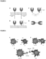
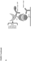
- the present chimeric proteins in some embodiments are capable of, or find use in methods involving, masking an inhibitory ligand on the surface of a tumor cell and replacing that immune inhibitory ligand with an immune stimulatory ligand (see, e.g. FIG. 4 ). Accordingly, the present chimeric proteins, in some embodiments are capable of, or find use in methods involving, reducing or eliminating an inhibitory immune signal and/or increasing or activating an immune stimulatory signal. For example, a tumor cell bearing an inhibitory signal (and thus evading an immune response) may be substituted for a positive signal binding on a T cell that can then attack a tumor cell.
- an inhibitory immune signal is masked by the present constructs and a stimulatory immune signal is activated.
- a stimulatory immune signal is activated.
- beneficial properties are enhanced by the single construct approach of the present chimeric proteins.
- the signal replacement can be effected nearly simultaneously and the signal replacement is tailored to be local at a site of clinical importance (e.g. the tumor microenvironment).
- the present chimeric proteins are capable of, or find use in methods comprising, stimulating or enhancing the binding of the immune stimulatory receptor/ligand pair CD40:CD40-L. In various embodiments, the present chimeric proteins are capable of, or find use in methods comprising, inhibiting or reducing the binding of the immune inhibitory receptor/ligand pair CD172a/CD47.
- the present chimeric proteins are capable of, or find use in methods involving, enhancing, restoring, promoting and/or stimulating immune modulation.
- the present chimeric proteins described herein restore, promote and/or stimulate the activity or activation of one or more immune cells against tumor cells including, but not limited to: T cells, cytotoxic T lymphocytes, T helper cells, natural killer (NK) cells, natural killer T (NKT) cells, anti-tumor macrophages (e.g. M1 macrophages), B cells, and dendritic cells.
- the present chimeric proteins enhance, restore, promote and/or stimulate the activity and/or activation of T cells, including, by way of a non-limiting example, activating and/or stimulating one or more T- cell intrinsic signals, including a pro-survival signal; an autocrine or paracrine growth signal; a p38 MAPK-, ERK-, STAT-, JAK-, AKT- or PI3K-mediated signal; an anti-apoptotic signal; and/or a signal promoting and/or necessary for one or more of: proinflammatory cytokine production or T cell migration or T cell tumor infiltration.
- T- cell intrinsic signals including a pro-survival signal; an autocrine or paracrine growth signal; a p38 MAPK-, ERK-, STAT-, JAK-, AKT- or PI3K-mediated signal; an anti-apoptotic signal; and/or a signal promoting and/or necessary for one or more of: proinflammatory cytokine production or T cell migration or
- the present chimeric proteins are capable of, or find use in methods involving, causing an increase of one or more of T cells (including without limitation cytotoxic T lymphocytes, T helper cells, natural killer T (NKT) cells), B cells, natural killer (NK) cells, natural killer T (NKT) cells, dendritic cells, monocytes, and macrophages ( e.g. one or more of M1 and M2) into a tumor or the tumor microenvironment.
- T cells including without limitation cytotoxic T lymphocytes, T helper cells, natural killer T (NKT) cells), B cells, natural killer (NK) cells, natural killer T (NKT) cells, dendritic cells, monocytes, and macrophages (e.g. one or more of M1 and M2) into a tumor or the tumor microenvironment.
- T cells including without limitation cytotoxic T lymphocytes, T helper cells, natural killer T (NKT) cells), B cells, natural killer (NK) cells, natural killer T (NKT) cells, dendriti
- the present therapies may alter the ratio of M1 versus M2 macrophages in the tumor site and/or TME to favor M1 macrophages.
- the present chimeric proteins are capable of, and can be used in methods comprising, inhibiting and/or reducing T cell inactivation and/or immune tolerance to a tumor, comprising administering an effective amount of a chimeric protein described herein to a subject.
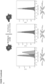
- the present chimeric proteins are able to increase the serum levels of various cytokines including, but not limited to, one or more of IFN ⁇ , TNF ⁇ , IL-2, IL-4, IL-5, IL-6, IL-9, IL-10, IL-13, IL-17A, IL-17F, and IL-22.
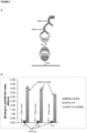
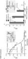
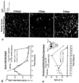
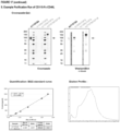
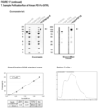
- the present chimeric proteins are capable of enhancing IL-2, IL-4, IL-5, IL-10, IL-13, IL-17A, IL-22, TNF ⁇ or IFN ⁇ in the serum of a treated subject (see, e.g. Figure 11 , panel J). Detection of such a cytokine response may provide a method to determine the optimal dosing regimen for the indicated chimeric fusion protein ( see , e.g. Figure 11 , panel K).
- the present chimeric proteins inhibit, block and/or reduce cell death of an anti-tumor CD8+ and/or CD4+ T cell; or stimulate, induce, and/or increase cell death of a pro-tumor T cell.
- T cell exhaustion is a state of T cell dysfunction characterized by progressive loss of proliferative and effector functions, culminating in clonal deletion.
- a pro-tumor T cell refers to a state of T cell dysfunction that arises during many chronic infections and cancer. This dysfunction is defined by poor proliferative and/or effector functions, sustained expression of inhibitory receptors and a transcriptional state distinct from that of functional effector or memory T cells. Exhaustion prevents optimal control of infection and tumors.
- an anti-tumor CD8+ and/or CD4+ T cell refers to T cells that can mount an immune response to a tumor.
- Illustrative pro-tumor T cells include, but are not limited to, Tregs, CD4+ and/or CD8+ T cells expressing one or more checkpoint inhibitory receptors, Th2 cells and Th17 cells.
- Checkpoint inhibitory receptors refers to receptors (e.g. CTLA-4, B7-H3, B7-H4, TIM-3) expressed on immune cells that prevent or inhibit uncontrolled immune responses.
- the present chimeric proteins are capable of, and can be used in methods comprising, increasing a ratio of effector T cells to regulatory T cells.
- Illustrative effector T cells include ICOS + effector T cells; cytotoxic T cells (e.g . ⁇ TCR, CD3+, CD8+, CD45RO+); CD4+ effector T cells ( e.g . ⁇ TCR, CD3+, CD4+, CCR7+, CD62Lhi, IL-7R/CD127+); CD8+ effector T cells ( e.g . ⁇ TCR, CD3+, CD8+, CCR7+, CD62Lhi, IL-7R/CD127+); effector memory T cells ( e.g .
- TH1 effector T-cells e.g. CXCR3+, CXCR6+ and CCR5+; or ⁇ TCR, CD3+, CD4+, IL-12R + , IFN ⁇ R + , CXCR3+
- TH2 effector T cells e.g. CCR3+, CCR4+ and CCR8+; or ⁇ TCR, CD3+, CD4+, IL-4R + , IL-33R + , CCR4+, IL-17RB+, CRTH2+
- TH9 effector T cells e.g.
- TH17 effector T cells e.g. ⁇ TCR, CD3+, CD4+, IL-23R + , CCR6 + , IL-1R + ); CD4+CD45RO+CCR7+ effector T cells, CD4+CD45RO+CCR7(-) effector T cells; and effector T cells secreting IL-2, IL-4 and/or IFN- ⁇ .
- Illustrative regulatory T cells include ICOS + regulatory T cells, CD4+CD25+FOXP3+ regulatory T cells, CD4+CD25+ regulatory T cells, CD4 + CD25 - regulatory T cells, CD4+CD25high regulatory T cells, TIM-3+PD-1+ regulatory T cells, lymphocyte activation gene-3 (LAG-3)+ regulatory T cells, CTLA-4/CD152+ regulatory T cells, neuropilin-1 (Nrp-1)+ regulatory T cells, CCR4+CCR8+ regulatory T cells, CD62L (L-selectin)+ regulatory T cells, CD45RBlow regulatory T cells, CD127low regulatory T cells, LRRC32/GARP+ regulatory T cells, CD39+ regulatory T cells, GITR + regulatory T cells, LAP+ regulatory T cells, 1B11 + regulatory T cells, BTLA+ regulatory T cells, type 1 regulatory T cells (Tr1 cells),T helper type 3 (Th3) cells, regulatory cell of natural killer T cell phenotype (NKTregs), CD8+ regulatory T cells
- the present chimeric proteins are capable of, and can be used in methods comprising, transiently stimulating effector T cells for no longer than about 12 hours, about 24 hours, about 48 hours, about 72 hours or about 96 hours or about 1 week or about 2 weeks. In various embodiments, the present chimeric proteins are capable of, and can be used in methods comprising, transiently depleting or inhibiting regulatory T cells for no longer than about 12 hours, about 24 hours, about 48 hours, about 72 hours or about 96 hours or about 1 week or about 2 weeks.
- the transient stimulation of effector T cells and/or transient depletion or inhibition of regulatory T cells occurs substantially in a patient's bloodstream or in a particular tissue/location including lymphoid tissues such as for example, the bone marrow, lymph-node, spleen, thymus, mucosa-associated lymphoid tissue (MALT), non-lymphoid tissues, or in the tumor microenvironment.
- lymphoid tissues such as for example, the bone marrow, lymph-node, spleen, thymus, mucosa-associated lymphoid tissue (MALT), non-lymphoid tissues, or in the tumor microenvironment.
- the present chimeric proteins provide advantages including, without limitation, ease of use and ease of production. This is because two distinct immunotherapy agents are combined into a single product which allows for a single manufacturing process instead of two independent manufacturing processes. In addition, administration of a single agent instead of two separate agents allows for easier administration and greater patient compliance. Further, in contrast to , for example, monoclonal antibodies, which are large multimeric proteins containing numerous disulfide bonds and post-translational modifications such as glycosylation, the present chimeric proteins are easier and more cost effective to manufacture.
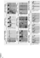
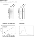
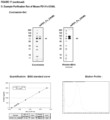
- the present chimeric protein is produceable in a mammalian host cell as a secretable and fully functional single polypeptide chain (see, e.g., Figure 13 , panel A, Figure 17 , panels E-H, Figure 17 , panels N-S).
- the present chimeric protein e.g. via the long off rate binding allows sufficient signal transmission to provide release of stimulatory signals, such as, for example, cytokines Also.
- the stable synapse of cells promoted by the present agents e.g. a tumor cell bearing negative signals and a T cell which could attack the tumor
- this provides longer on-target (e.g. intra-tumoral) half-life (t 1/2 ) as compared to serum t 1/2 of the chimeric proteins.
- on-target e.g. intra-tumoral
- t 1/2 half-life
- the present chimeric proteins provide synergistic therapeutic effects as it allows for improved site-specific interplay of two immunotherapy agents. In some embodiments, the present chimeric proteins provide the potential for reducing off-site and/or systemic toxicity.
- the present disclosure pertains to cancers and/or tumors; for example, the treatment or prevention of cancers and/or tumors.
- the treatment of cancer may involve in various embodiments, modulating the immune system with the present chimeric proteins to favor immune stimulation over immune inhibition.
- Cancers or tumors refer to an uncontrolled growth of cells and/or abnormal increased cell survival and/or inhibition of apoptosis which interferes with the normal functioning of the bodily organs and systems. Included are benign and malignant cancers, polyps, hyperplasia, as well as dormant tumors or micrometastases. Also, included are cells having abnormal proliferation that is not impeded by the immune system (e.g. virus infected cells).
- the cancer may be a primary cancer or a metastatic cancer.
- the primary cancer may be an area of cancer cells at an originating site that becomes clinically detectable, and may be a primary tumor.
- the metastatic cancer may be the spread of a disease from one organ or part to another non-adjacent organ or part.
- the metastatic cancer may be caused by a cancer cell that acquires the ability to penetrate and infiltrate surrounding normal tissues in a local area, forming a new tumor, which may be a local metastasis.
- the cancer may also be caused by a cancer cell that acquires the ability to penetrate the walls of lymphatic and/or blood vessels, after which the cancer cell is able to circulate through the bloodstream (thereby being a circulating tumor cell) to other sites and tissues in the body.
- the cancer may be due to a process such as lymphatic or hematogeneous spread.
- the cancer may also be caused by a tumor cell that comes to rest at another site, re-penetrates through the vessel or walls, continues to multiply, and eventually forms another clinically detectable tumor.
- the cancer may be this new tumor, which may be a metastatic (or secondary) tumor.
- the cancer may be caused by tumor cells that have metastasized, which may be a secondary or metastatic tumor.
- the cells of the tumor may be like those in the original tumor.
- the secondary tumor while present in the liver, is made up of abnormal breast or colon cells, not of abnormal liver cells.
- the tumor in the liver may thus be a metastatic breast cancer or a metastatic colon cancer, not liver cancer.
- the cancer may have an origin from any tissue.
- the cancer may originate from melanoma, colon, breast, or prostate, and thus may be made up of cells that were originally skin, colon, breast, or prostate, respectively.
- the cancer may also be a hematological malignancy, which may be leukemia or lymphoma.
- the cancer may invade a tissue such as liver, lung, bladder, or intestinal.
- Representative cancers and/or tumors of the present invention include, but are not limited to, a basal cell carcinoma, biliary tract cancer; bladder cancer; bone cancer; brain and central nervous system cancer; breast cancer; cancer of the peritoneum; cervical cancer; choriocarcinoma; colon and rectum cancer; connective tissue cancer; cancer of the digestive system; endometrial cancer; esophageal cancer; eye cancer; cancer of the head and neck; gastric cancer (including gastrointestinal cancer); glioblastoma; hepatic carcinoma; hepatoma; intra-epithelial neoplasm; kidney or renal cancer; larynx cancer; leukemia; liver cancer; lung cancer ( e.g., small-cell lung cancer, non-small cell lung cancer, adenocarcinoma of the lung, and squamous carcinoma of the lung); melanoma; myeloma; neuroblastoma; oral cavity cancer (lip, tongue, mouth, and pharynx); ova
- the chimeric protein is used to treat a subject that has a treatment-refractory cancer. In some embodiments, the chimeric protein is used to treat a subject that is refractory to one or more immune-modulating agents. For example, in some embodiments, the chimeric protein is used to treat a subject that presents no response to treatment, or even progress, after 12 weeks or so of treatment.
- the subject is refractory to a PD-1 and/or PD-L1 and/or PD-L2 agent, including, for example, nivolumab (ONO-4538/BMS-936558, MDX1106, OPDIVO, BRISTOL MYERS SQUIBB), pembrolizumab (KEYTRUDA, MERCK), pidilizumab (CT-011, CURE TECH), MK-3475 (MERCK), BMS 936559 (BRISTOL MYERS SQUIBB), Ibrutinib (PHARMACYCLICS/ABBVIE), atezolizumab (TECENTRIQ, GENENTECH), and/or MPDL328OA (ROCHE)-refractory patients.
- nivolumab ONO-4538/BMS-936558, MDX1106, OPDIVO, BRISTOL MYERS SQUIBB
- pembrolizumab KEYTRUDA, MERCK
- pidilizumab C
- the subject is refractory to an anti-CTLA-4 agent, e.g. ipilimumab (YERVOY)-refractory patients (e.g. melanoma patients).
- an anti-CTLA-4 agent e.g. ipilimumab (YERVOY)-refractory patients (e.g. melanoma patients).
- YERVOY ipilimumab
- the present invention provides methods of cancer treatment that rescue patients that are non-responsive to various therapies, including monotherapy of one or more immune-modulating agents.
- the present methods provide treatment with the chimeric protein in a patient who is refractory to an additional agent, such "additional agents" being described elsewhere herein, inclusive, without limitation, of the various chemotherapeutic agents described herein.
- the present chimeric agents are used to eliminate intracellular pathogens. In some aspects, the present chimeric agents are used to treat one or more infections. In some embodiments, the present chimeric proteins are used in methods of treating viral infections (including, for example, HIV and HCV), parasitic infections (including, for example, malaria), and bacterial infections. In various embodiments, the infections induce immunosuppression. For example, HIV infections often result in immunosuppression in the infected subjects. Accordingly, as described elsewhere herein, the treatment of such infections may involve, in various embodiments, modulating the immune system with the present chimeric proteins to favor immune stimulation over immune inhibition. Alternatively, the present invention provides methods for treating infections that induce immunoactivation. For example, intestinal helminth infections have been associated with chronic immune activation. In these embodiments, the treatment of such infections may involve modulating the immune system with the present chimeric proteins to favor immune inhibition over immune stimulation.
- the viral infection is caused by a virus of family Picornaviridae, e.g., poliovirus, rhinovirus, coxsackievirus.
- the viral infection is caused by a member of Orthomyxoviridae, e.g., an influenza virus.
- the viral infection is caused by a member of Retroviridae, e.g., a lentivirus.
- the viral infection is caused by a member of Paramyxoviridae, e.g., respiratory syncytial virus, a human parainfluenza virus, rubulavirus ( e.g., mumps virus), measles virus, and human metapneumovirus.
- the viral infection is caused by a member of Bunyaviridae, e.g., hantavirus. In other embodiments, the viral infection is caused by a member of Reoviridae, e.g., a rotavirus.
- Illustrative protozoan parasites include, but are not limited to, Entamoeba hystolytica, Giardia lamblia, Cryptosporidium muris, Trypanosomatida gambiense, Trypanosomatida rhodesiense, Trypanosomatida crusi, Leishmania mexicana, Leishmania braziliensis, Leishmania tropica, Leishmania donovani, Toxoplasma gondii, Plasmodium vivax, Plasmodium ovale, Plasmodium malariae, Plasmodium falciparum, Trichomonas vaginalis, and Histomonas meleagridis.
- the parasitic infection is by a helminthic parasite such as nematodes (e.g., Adenophorea).
- the parasite is selected from Secementea (e.g., Trichuris trichiura, Ascaris lumbricoides, Enterobius vermicularis, Ancylostoma duodenale, Necator americanus, Strongyloides stercoralis, Wuchereria bancrofti, Dracunculus medinensis ) .
- the parasite is selected from trematodes (e.g. blood flukes, liver flukes, intestinal flukes, and lung flukes).
- the parasite is selected from: Schistosoma mansoni, Schistosoma haematobium, Schistosoma japonicum, Fasciola hepatica, Fasciola gigantica, Heterophyes heterophyes, Paragonimus westermani.
- the parasite is selected from cestodes (e.g., Taenia solium, Taenia saginata, Hymenolepis nana, Echinococcus granulosus).
- the present disclosure provides methods of treating bacterial infections.
- the bacterial infection is by a gram-positive bacteria, gram-negative bacteria, aerobic and/or anaerobic bacteria.
- the bacteria is selected from, but not limited to, Staphylococcus, Lactobacillus, Streptococcus, Sarcina, Escherichia, Enterobacter, Klebsiella, Pseudomonas, Acinetobacter, Mycobacterium, Proteus, Campylobacter, Citrobacter, Nisseria, Baccillus, Bacteroides, Peptococcus, Clostridium, Salmonella, Shigella, Serratia, Haemophilus, Brucella and other organisms.
- the bacteria is selected from, but not limited to, Pseudomonas aeruginosa, Pseudomonas fluorescens, Pseudomonas acidovorans, Pseudomonas alcaligenes, Pseudomonas putida, Stenotrophomonas maltophilia, Burkholderia cepacia, Aeromonas hydrophilia, Escherichia coli, Citrobacter freundii, Salmonella typhimurium, Salmonella typhi, Salmonella paratyphi, Salmonella enteritidis, Shigella dysenteriae, Shigella flexneri, Shigella sonnei, Enterobacter cloacae, Enterobacter aerogenes, Klebsiella pneumoniae, Klebsiella oxytoca, Serratia marcescens, Francisella tularensis, Morganella morganii, Proteus mirabili
- the present chimeric agents are used to treat one or more autoimmune diseases or disorders.
- the treatment of an autoimmune disease or disorder may involve modulating the immune system with the present chimeric proteins to favor immune inhibition over immune stimulation.
- Illustrative autoimmune diseases or disorders treatable with the present chimeric proteins include those in which the body's own antigens become targets for an immune response, such as, for example, rheumatoid arthritis, systemic lupus erythematosus, diabetes mellitus, ankylosing spondylitis, Sjögren's syndrome, inflammatory bowel diseases (e.g.
- colitis ulcerosa Crohn's disease
- multiple sclerosis sarcoidosis
- psoriasis Grave's disease
- Hashimoto's thyroiditis e.g., hay fever, asthma, and acute edema cause type I hypersensitivity reactions
- vasculitis e.g., allergies, hay fever, asthma, and acute edema cause type I hypersensitivity reactions
- the present disclosure is directed toward methods of treating and preventing T cell-mediated diseases and disorders, such as, but not limited to diseases or disorders described elsewhere herein and inflammatory disease or disorder, graft-versus-host disease (GVHD), transplant rejection, and T cell proliferative disorder.
- diseases or disorders described elsewhere herein e.g., GVHD
- transplant rejection e.g., transplant rejection, T cell proliferative disorder.
- T cell-mediated diseases and disorders such as, but not limited to diseases or disorders described elsewhere herein and inflammatory disease or disorder, graft-versus-host disease (GVHD), transplant rejection, and T cell proliferative disorder.
- the present chimeric agents are used in methods of activating a T cell, e.g. via the extracellular domain having an immune stimulatory signal.
- the present chimeric agents are used in methods of preventing the cellular transmission of an immunosuppressive signal.
- the disclosure provides for chimeric proteins and methods that further comprise administering an additional agent to a subject.
- the disclosure pertains to coadministration and/or co-formulation. Any of the compositions described herein may be co-formulated and/or co-administered.
- any chimeric protein described herein acts synergistically when co-administered with another agent and is administered at doses that are lower than the doses commonly employed when such agents are used as monotherapy.
- any agent referenced herein may be used in combination with any of the chimeric proteins described herein.
- chemotherapeutic agents include, but are not limited to, alkylating agents such as thiotepa and CYTOXAN cyclosphosphamide; alkyl sulfonates such as busulfan, improsulfan and piposulfan; aziridines such as benzodopa, carboquone, meturedopa, and uredopa; ethylenimines and methylamelamines including altretamine, triethylenemelamine, trietylenephosphoramide, triethiylenethiophosphoramide and trimethylolomelamine; acetogenins ( e.g., bullatacin and bullatacinone); a camptothecin (including the synthetic analogue topotecan); bryostatin; cally statin; CC-1065 (including its adozelesin
- dynemicin including dynemicin A; bisphosphonates, such as clodronate; an esperamicin; as well as neocarzinostatin chromophore and related chromoprotein enediyne antibiotic chromophores), aclacinomysins, actinomycin, authramycin, azaserine, bleomycins, cactinomycin, carabicin, caminomycin, carzinophilin, chromomycinis, dactinomycin, daunorubicin, detorubicin, 6-diazo-5-oxo-L-norleucine, ADRIAMYCIN doxorubicin (including morpholino-doxorubicin, cyanomorpholino-doxorubicin, 2-pyrrolino-doxorubicin and deoxy doxorubicin), epirub
- irinotecan Camptosar, CPT-11 (including the treatment regimen of irinotecan with 5-FU and leucovorin); topoisomerase inhibitor RFS 2000; difluoromethylornithine (DMFO); retinoids such as retinoic acid; capecitabine; combretastatin; leucovorin (LV); oxaliplatin, including the oxaliplatin treatment regimen (FOLFOX); lapatinib (TYKERB); inhibitors of PKC- ⁇ , Raf, H-Ras, EGFR (e.g., erlotinib (Tarceva)) and VEGF-A that reduce cell proliferation and pharmaceutically acceptable salts, acids or derivatives of any of the above.
- the methods of treatment can further include the use
- the present additional agent is one or more immune-modulating agents selected from an agent that blocks, reduces and/or inhibits PD-1 and PD-L1 or PD-L2 and/or the binding of PD-1 with PD-L1 or PD-L2 (by way of non-limiting example, one or more of nivolumab (ONO-4538/BMS-936558, MDX1106, OPDIVO, BRISTOL MYERS SQUIBB), pembrolizumab (KEYTRUDA, Merck), pidilizumab (CT-011, CURE TECH), MK-3475 (MERCK), BMS 936559 (BRISTOL MYERS SQUIBB), atezolizumab (TECENTRIQ, GENENTECH), MPDL328OA (ROCHE)), an agent that increases and/or stimulates CD137 (4-1 BB) and/or the binding of CD137 (4-1BB) with one or more of 4-1BB ligand (by way of nivolumab (ONO-45
- the present disclosure pertains to anti-infectives as additional agents.
- the anti-infective is an anti-viral agent including, but not limited to, Abacavir, Acyclovir, Adefovir, Amprenavir, Atazanavir, Cidofovir, Darunavir, Delavirdine, Didanosine, Docosanol, Efavirenz, Elvitegravir, Emtricitabine, Enfuvirtide, Etravirine, Famciclovir, and Foscarnet.
- the anti-infective is an anti-bacterial agent including, but not limited to, cephalosporin antibiotics (cephalexin, cefuroxime, cefadroxil, cefazolin, cephalothin, cefaclor, cefamandole, cefoxitin, cefprozil, and ceftobiprole); fluoroquinolone antibiotics (cipro, Levaquin, floxin, tequin, avelox, and norflox); tetracycline antibiotics (tetracycline, minocycline, oxytetracycline, and doxycycline); penicillin antibiotics (amoxicillin, ampicillin, penicillin V, dicloxacillin, carbenicillin, vancomycin, and methicillin); monobactam antibiotics (aztreonam); and carbapenem antibiotics (ertapenem, doripenem, imipenem/cilastatin, and meropenem).
- cephalosporin antibiotics ce
- the anti-infectives include anti-malarial agents (e.g., chloroquine, quinine, mefloquine, primaquine, doxycycline, artemether/lumefantrine, atovaquone/proguanil and sulfadoxine/pyrimethamine), metronidazole, tinidazole, ivermectin, pyrantel pamoate, and albendazole.
- anti-malarial agents e.g., chloroquine, quinine, mefloquine, primaquine, doxycycline, artemether/lumefantrine, atovaquone/proguanil and sulfadoxine/pyrimethamine
- metronidazole e.g., chloroquine, quinine, mefloquine, primaquine, doxycycline, artemether/lumefantrine, atovaquone/proguanil and sulfa
- the additional agent is an immunosuppressive agent.
- the immunosuppressive agent is an anti-inflammatory agent such as a steroidal anti-inflammatory agent or a non-steroidal anti-inflammatory agent (NSAID).
- NSAID non-steroidal anti-inflammatory agent
- Steroids, particularly the adrenal corticosteroids and their synthetic analogues, are well known in the art.
- NSAIDS that may be used in the present invention, include but are not limited to, salicylic acid, acetyl salicylic acid, methyl salicylate, glycol salicylate, salicylmides, benzyl-2,5-diacetoxybenzoic acid, ibuprofen, fulindac, naproxen, ketoprofen, etofenamate, phenylbutazone, and indomethacin.
- the immunosupressive agent may be cytostatics such as alkylating agents, antimetabolites (e.g., azathioprine, methotrexate), cytotoxic antibiotics, antibodies (e.g., basiliximab, daclizumab, and muromonab), anti-immunophilins (e.g., cyclosporine, tacrolimus, sirolimus), inteferons, opioids, TNF binding proteins, mycophenolates, and small biological agents (e.g., fingolimod, myriocin).
- cytostatics such as alkylating agents, antimetabolites (e.g., azathioprine, methotrexate), cytotoxic antibiotics, antibodies (e.g., basiliximab, daclizumab, and muromonab), anti-immunophilins (e.g., cyclosporine, tacrolimus, sirolimus), inteferons, opioids, T
- the chimeric proteins (and/or additional agents) described herein include derivatives that are modified, i.e., by the covalent attachment of any type of molecule to the composition such that covalent attachment does not prevent the activity of the composition.
- derivatives include composition that have been modified by, inter alia, glycosylation, lipidation, acetylation, pegylation, phosphorylation, amidation, derivatization by known protecting/blocking groups, proteolytic cleavage, linkage to a cellular ligand or other protein, etc.
- the derivative can contain one or more non-classical amino acids.
- the chimeric proteins (and/or additional agents) described herein further comprise a cytotoxic agent, comprising, in illustrative embodiments, a toxin, a chemotherapeutic agent, a radioisotope, and an agent that causes apoptosis or cell death. Such agents may be conjugated to a composition described herein.
- chimeric proteins (and/or additional agents) described herein may thus be modified post-translationally to add effector moieties such as chemical linkers, detectable moieties such as for example fluorescent dyes, enzymes, substrates, bioluminescent materials, radioactive materials, and chemiluminescent moieties, or functional moieties such as for example streptavidin, avidin, biotin, a cytotoxin, a cytotoxic agent, and radioactive materials.
- effector moieties such as chemical linkers, detectable moieties such as for example fluorescent dyes, enzymes, substrates, bioluminescent materials, radioactive materials, and chemiluminescent moieties, or functional moieties such as for example streptavidin, avidin, biotin, a cytotoxin, a cytotoxic agent, and radioactive materials.
- the chimeric proteins (and/or additional agents) described herein can possess a sufficiently basic functional group, which can react with an inorganic or organic acid, or a carboxyl group, which can react with an inorganic or organic base, to form a pharmaceutically acceptable salt.
- a pharmaceutically acceptable acid addition salt is formed from a pharmaceutically acceptable acid, as is well known in the art.
- Such salts include the pharmaceutically acceptable salts listed in, for example, Journal of Pharmaceutical Science, 66, 2-19 (1977 ) and The Handbook of Pharmaceutical Salts; Properties, Selection, and Use. P. H. Stahl and C. G. Wermuth (eds.), Verlag, Zurich (Switzerland) 2002 .
- compositions described herein are in the form of a pharmaceutically acceptable salt.
- any chimeric protein (and/or additional agents) described herein can be for administration to a subject as a component of a composition that comprises a pharmaceutically acceptable carrier or vehicle.
- Such compositions can optionally comprise a suitable amount of a pharmaceutically acceptable excipient so as to provide the form for proper administration.
- Pharmaceutical excipients can be liquids, such as water and oils, including those of petroleum, animal, vegetable, or synthetic origin, such as peanut oil, soybean oil, mineral oil, sesame oil and the like.
- the pharmaceutical excipients can be, for example, saline, gum acacia, gelatin, starch paste, talc, keratin, colloidal silica, urea and the like.
- the pharmaceutically acceptable excipients are sterile when administered to a subject.
- Water is a useful excipient when any agent described herein is administered intravenously.
- Saline solutions and aqueous dextrose and glycerol solutions can also be employed as liquid excipients, specifically for injectable solutions.
- Suitable pharmaceutical excipients also include starch, glucose, lactose, sucrose, gelatin, malt, rice, flour, chalk, silica gel, sodium stearate, glycerol monostearate, talc, sodium chloride, dried skim milk, glycerol, propylene, glycol, water, ethanol and the like. Any agent described herein, if desired, can also comprise minor amounts of wetting or emulsifying agents, or pH buffering agents.
- compositions described herein are resuspended in a saline buffer (including, without limitation TBS, PBS, and the like).
- a saline buffer including, without limitation TBS, PBS, and the like.
- the chimeric proteins may by conjugated and/or fused with another agent to extend half-life or otherwise improve pharmacodynamic and pharmacokinetic properties.
- the chimeric proteins may be fused or conjugated with one or more of PEG, XTEN ( e.g., as rPEG), polysialic acid (POLYXEN), albumin (e.g., human serum albumin or HAS), elastin-like protein (ELP), PAS, HAP, GLK, CTP, transferrin, and the like.
- each of the individual chimeric proteins is fused to one or more of the agents described in BioDrugs (2015) 29:215-239 .
- the present invention includes the described chimeric protein (and/or additional agents) in various formulations.
- Any chimeric protein (and/or additional agents) described herein can take the form of solutions, suspensions, emulsion, drops, tablets, pills, pellets, capsules, capsules containing liquids, powders, sustained-release formulations, suppositories, emulsions, aerosols, sprays, suspensions, or any other form suitable for use.
- DNA or RNA constructs encoding the protein sequences may also be used.
- the composition is in the form of a capsule ( see, e.g., U.S. Patent No. 5,698,155 ).
- suitable pharmaceutical excipients are described in Remington's Pharmaceutical Sciences 1447-1676 (Alfonso R. Gennaro eds., 19th ed. 1995 ).
- the formulations comprising the chimeric protein (and/or additional agents) can also include a solubilizing agent.
- the agents can be delivered with a suitable vehicle or delivery device as known in the art.
- Combination therapies outlined herein can be co-delivered in a single delivery vehicle or delivery device.
- Compositions for administration can optionally include a local anesthetic such as, for example, lignocaine to lessen pain at the site of the injection.
- the formulations comprising the chimeric protein (and/or additional agents) of the present invention may conveniently be presented in unit dosage forms and may be prepared by any of the methods well known in the art of pharmacy. Such methods generally include the step of bringing the therapeutic agents into association with a carrier, which constitutes one or more accessory ingredients. Typically, the formulations are prepared by uniformly and intimately bringing the therapeutic agent into association with a liquid carrier, a finely divided solid carrier, or both, and then, if necessary, shaping the product into dosage forms of the desired formulation (e.g., wet or dry granulation, powder blends, etc., followed by tableting using conventional methods known in the art)
- a carrier which constitutes one or more accessory ingredients.
- the formulations are prepared by uniformly and intimately bringing the therapeutic agent into association with a liquid carrier, a finely divided solid carrier, or both, and then, if necessary, shaping the product into dosage forms of the desired formulation (e.g., wet or dry granulation, powder blends, etc., followed by tablet
- any chimeric protein (and/or additional agents) described herein is formulated in accordance with routine procedures as a composition adapted for a mode of administration described herein.
- Routes of administration include, for example: intradermal, intramuscular, intraperitoneal, intravenous, subcutaneous, intranasal, epidural, oral, sublingual, intranasal, intracerebral, intravaginal, transdermal, rectally, by inhalation, or topically, particularly to the ears, nose, eyes, or skin.
- the administering is effected orally or by parenteral injection.
- administration results in the release of any agent described herein into the bloodstream.
- Any chimeric protein (and/or additional agents) described herein can be administered orally.
- Such chimeric proteins (and/or additional agents) can also be administered by any other convenient route, for example, by intravenous infusion or bolus injection, by absorption through epithelial or mucocutaneous linings ( e.g., oral mucosa, rectal and intestinal mucosa, etc .) and can be administered together with another biologically active agent. Administration can be systemic or local.
- Various delivery systems are known, e.g., encapsulation in liposomes, microparticles, microcapsules, capsules, etc., and can be used to administer.
- the chimeric protein (and/or additional agents) are administered in the tumor microenvironment (e.g . cells, molecules, extracellular matrix and/or blood vessels that surround and/or feed a tumor cell, inclusive of, for example, tumor vasculature; tumor-infiltrating lymphocytes; fibroblast reticular cells; endothelial progenitor cells (EPC); cancer-associated fibroblasts; pericytes; other stromal cells; components of the extracellular matrix (ECM); dendritic cells; antigen presenting cells; T-cells; regulatory T cells; macrophages; neutrophils; and other immune cells located proximal to a tumor) or lymph node and/or targeted to the tumor microenvironment or lymph node.
- the chimeric protein (and/or additional agents) are administered intratumorally.
- the present chimeric protein allows for a dual effect that provides less side effects than are seen in conventional immunotherapy (e.g. treatments with one or more of OPDIVO, KEYTRUDA, YERVOY, and TECENTRIQ).
- the present chimeric proteins reduce or prevent commonly observed immune-related adverse events that affect various tissues and organs including the skin, the gastrointestinal tract, the kidneys, peripheral and central nervous system, liver, lymph nodes, eyes, pancreas, and the endocrine system; such as hypophysitis, colitis, hepatitis, pneumonitis, rash, and rheumatic disease.
- the present local administration e.g.
- Dosage forms suitable for parenteral administration include, for example, solutions, suspensions, dispersions, emulsions, and the like. They may also be manufactured in the form of sterile solid compositions (e.g. lyophilized composition), which can be dissolved or suspended in sterile injectable medium immediately before use. They may contain, for example, suspending or dispersing agents known in the art.
- any chimeric protein (and/or additional agents) described herein as well as the dosing schedule can depend on various parameters, including, but not limited to, the disease being treated, the subject's general health, and the administering physician's discretion.
- Any chimeric protein described herein can be administered prior to ( e.g., 5 minutes, 15 minutes, 30 minutes, 45 minutes, 1 hour, 2 hours, 4 hours, 6 hours, 12 hours, 24 hours, 48 hours, 72 hours, 96 hours, 1 week, 2 weeks, 3 weeks, 4 weeks, 5 weeks, 6 weeks, 8 weeks, or 12 weeks before), concurrently with, or subsequent to ( e.g ., 5 minutes, 15 minutes, 30 minutes, 45 minutes, 1 hour, 2 hours, 4 hours, 6 hours, 12 hours, 24 hours, 48 hours, 72 hours, 96 hours, 1 week, 2 weeks, 3 weeks, 4 weeks, 5 weeks, 6 weeks, 8 weeks, or 12 weeks after) the administration of an additional agent, to a subject in need thereof.
- any chimeric protein and additional agent described herein are administered 1 minute apart, 10 minutes apart, 30 minutes apart, less than 1 hour apart, 1 hour apart, 1 hour to 2 hours apart, 2 hours to 3 hours apart, 3 hours to 4 hours apart, 4 hours to 5 hours apart, 5 hours to 6 hours apart, 6 hours to 7 hours apart, 7 hours to 8 hours apart, 8 hours to 9 hours apart, 9 hours to 10 hours apart, 10 hours to 11 hours apart, 11 hours to 12 hours apart, no more than 24 hours apart or no more than 48 hours apart.
- any chimeric protein (and/or additional agents) described herein can depend on several factors including the severity of the condition, whether the condition is to be treated or prevented, and the age, weight, and health of the subject to be treated. Additionally, pharmacogenomic (the effect of genotype on the pharmacokinetic, pharmacodynamic or efficacy profile of a therapeutic) information about a particular subject may affect dosage used. Furthermore, the exact individual dosages can be adjusted somewhat depending on a variety of factors, including the specific combination of the agents being administered, the time of administration, the route of administration, the nature of the formulation, the rate of excretion, the particular disease being treated, the severity of the disorder, and the anatomical location of the disorder. Some variations in the dosage can be expected.
- the dosage is normally 0.1 mg to 250 mg per day, 1 mg to 20 mg per day, or 3 mg to 5 mg per day. Injections may be given up to four times daily.
- the dosage of any agent described herein is normally 0.1 mg to 1500 mg per day, or 0.5 mg to 10 mg per day, or 0.5 mg to 5 mg per day. A dosage of up to 3000 mg per day can be administered.
- delivery can be in a vesicle, in particular a liposome (see Langer, 1990, Science 249:1527-1533 ; Treat et al., in Liposomes in the Therapy of Infectious Disease and Cancer, Lopez-Berestein and Fidler (eds.), Liss, New York, pp. 353-365 (1989 ).
- a liposome see Langer, 1990, Science 249:1527-1533 ; Treat et al., in Liposomes in the Therapy of Infectious Disease and Cancer, Lopez-Berestein and Fidler (eds.), Liss, New York, pp. 353-365 (1989 ).
- Any chimeric protein (and/or additional agents) described herein can be administered by controlled-release or sustained-release means or by delivery devices that are well known to those of ordinary skill in the art. Examples include, but are not limited to, those described in U.S. Patent Nos. 3,845,770 ; 3,916,899 ; 3,536,809 ; 3,598,123 ; 4,008,719 ; 5,674,533 ; 5,059,595 ; 5,591,767 ; 5,120,548 ; 5,073,543 ; 5,639,476 ; 5,354,556 ; and 5,733,556 .
- Such dosage forms can be useful for providing controlled- or sustained-release of one or more active ingredients using, for example, hydropropylmethyl cellulose, other polymer matrices, gels, permeable membranes, osmotic systems, multilayer coatings, microparticles, liposomes, microspheres, or a combination thereof to provide the desired release profile in varying proportions.
- Controlled- or sustained-release of an active ingredient can be stimulated by various conditions, including but not limited to, changes in pH, changes in temperature, stimulation by an appropriate wavelength of light, concentration or availability of enzymes, concentration or availability of water, or other physiological conditions or compounds.
- polymeric materials can be used (see Medical Applications of Controlled Release, Langer and Wise (eds.), CRC Pres., Boca Raton, Florida (1974 ); Controlled Drug Bioavailability, Drug Product Design and Performance, Smolen and Ball (eds.), Wiley, New York (1984 ); Ranger and Peppas, 1983, J. Macromol. Sci. Rev. Macromol. Chem. 23:61 ; see also Levy et al., 1985, Science 228:190 ; During et al., 1989, Ann. Neurol. 25:351 ; Howard et al., 1989, J. Neurosurg. 71:105 ).
- a controlled-release system can be placed in proximity of the target area to be treated, thus requiring only a fraction of the systemic dose (s ee, e.g., Goodson, in Medical Applications of Controlled Release, supra, vol. 2, pp. 115-138 (1984)).
- Other controlled-release systems discussed in the review by Langer, 1990, Science 249:1527-1533 ) may be used.
- Administration of any chimeric protein (and/or additional agents) described herein can, independently, be one to four times daily or one to four times per month or one to six times per year or once every two, three, four or five years. Administration can be for the duration of one day or one month, two months, three months, six months, one year, two years, three years, and may even be for the life of the subject.
- any chimeric protein (and/or additional agents) described herein can be selected in accordance with a variety of factors including type, species, age, weight, sex and medical condition of the subject; the severity of the condition to be treated; the route of administration; the renal or hepatic function of the subject; the pharmacogenomic makeup of the individual; and the specific compound of the invention employed.
- Any chimeric protein (and/or additional agents) described herein can be administered in a single daily dose, or the total daily dosage can be administered in divided doses of two, three or four times daily.
- any chimeric protein (and/or additional agents) described herein can be administered continuously rather than intermittently throughout the dosage regimen.
- the present invention provides an expression vector, comprising a nucleic acid encoding the chimeric protein described herein.
- the expression vector comprises DNA or RNA.
- the expression vector is a mammalian expression vector.
- Prokaryotic vectors include constructs based on E . coli sequences ( see, e.g., Makrides, Microbiol Rev 1996, 60:512-538 ).
- Non-limiting examples of regulatory regions that can be used for expression in E coli include lac, trp, Ipp, phoA, recA, tac, T3, T7 and ⁇ P L .
- Non-limiting examples of prokaryotic expression vectors may include the Agt vector series such as Agt11 ( Huynh et al., in "DNA Cloning Techniques, Vol. I: A Practical Approach," 1984, (D. Glover, ed.), pp.
- Prokaryotic host-vector systems cannot perform much of the post-translational processing of mammalian cells, however. Thus, eukaryotic host- vector systems may be particularly useful.
- a variety of regulatory regions can be used for expression of the chimeric proteins in mammalian host cells. For example, the SV40 early and late promoters, the cytomegalovirus (CMV) immediate early promoter, and the Rous sarcoma virus long terminal repeat (RSV-LTR) promoter can be used.
- Inducible promoters that may be useful in mammalian cells include, without limitation, promoters associated with the metallothionein II gene, mouse mammary tumor virus glucocorticoid responsive long terminal repeats (MMTV-LTR), the ⁇ -interferon gene, and the hsp70 gene (see, Williams et al., Cancer Res 1989, 49:2735-42 ; and Taylor et al., Mol Cell Biol 1990, 10:165-75 ). Heat shock promoters or stress promoters also may be advantageous for driving expression of the fusion proteins in recombinant host cells.
- expression vectors of the invention comprise a nucleic acid encoding the chimeric proteins (and/or additional agents), or a complement thereof, operably linked to an expression control region, or complement thereof, that is functional in a mammalian cell.
- the expression control region is capable of driving expression of the operably linked blocking and/or stimulating agent encoding nucleic acid such that the blocking and/or stimulating agent is produced in a human cell transformed with the expression vector.
- Expression control regions are regulatory polynucleotides (sometimes referred to herein as elements), such as promoters and enhancers, that influence expression of an operably linked nucleic acid.
- An expression control region of an expression vector of the invention is capable of expressing operably linked encoding nucleic acid in a human cell.
- the cell is a tumor cell.
- the cell is a non-tumor cell.
- the expression control region confers regulatable expression to an operably linked nucleic acid.
- a signal (sometimes referred to as a stimulus) can increase or decrease expression of a nucleic acid operably linked to such an expression control region.
- Such expression control regions that increase expression in response to a signal are often referred to as inducible.
- Such expression control regions that decrease expression in response to a signal are often referred to as repressible.
- the amount of increase or decrease conferred by such elements is proportional to the amount of signal present; the greater the amount of signal, the greater the increase or decrease in expression.
- the present invention contemplates the use of inducible promoters capable of effecting high level of expression transiently in response to a cue.
- inducible promoters capable of effecting high level of expression transiently in response to a cue.
- a cell transformed with an expression vector for the chimeric protein (and/or additional agents) comprising such an expression control sequence is induced to transiently produce a high level of the agent by exposing the transformed cell to an appropriate cue.
- Illustrative inducible expression control regions include those comprising an inducible promoter that is stimulated with a cue such as a small molecule chemical compound. Particular examples can be found, for example, in U.S. Pat. Nos. 5,989,910 , 5,935,934 , 6,015,709 , and 6,004,941 .
- Expression control regions and locus control regions include full-length promoter sequences, such as native promoter and enhancer elements, as well as subsequences or polynucleotide variants which retain all or part of full-length or non-variant function.
- the term "functional" and grammatical variants thereof, when used in reference to a nucleic acid sequence, subsequence or fragment, means that the sequence has one or more functions of native nucleic acid sequence (e.g., non-variant or unmodified sequence).
- operable linkage refers to a physical juxtaposition of the components so described as to permit them to function in their intended manner.
- the relationship is such that the control element modulates expression of the nucleic acid.
- an expression control region that modulates transcription is juxtaposed near the 5' end of the transcribed nucleic acid (i.e., "upstream”).
- Expression control regions can also be located at the 3' end of the transcribed sequence ( i.e., "downstream") or within the transcript ( e.g., in an intron).
- Expression control elements can be located at a distance away from the transcribed sequence (e.g., 100 to 500, 500 to 1000, 2000 to 5000, or more nucleotides from the nucleic acid).
- a specific example of an expression control element is a promoter, which is usually located 5' of the transcribed sequence.
- Another example of an expression control element is an enhancer, which can be located 5' or 3' of the transcribed sequence, or within the transcribed sequence.
- a promoter functional in a human cell is any DNA sequence capable of binding mammalian RNA polymerase and initiating the downstream (3') transcription of a coding sequence into mRNA.
- a promoter will have a transcription initiating region, which is usually placed proximal to the 5' end of the coding sequence, and typically a TATA box located 25-30 base pairs upstream of the transcription initiation site. The TATA box is thought to direct RNA polymerase II to begin RNA synthesis at the correct site.
- a promoter will also typically contain an upstream promoter element (enhancer element), typically located within 100 to 200 base pairs upstream of the TATA box.
- An upstream promoter element determines the rate at which transcription is initiated and can act in either orientation.
- promoters are the promoters from mammalian viral genes, since the viral genes are often highly expressed and have a broad host range. Examples include the SV40 early promoter, mouse mammary tumor virus LTR promoter, adenovirus major late promoter, herpes simplex virus promoter, and the CMV promoter.
- transcription termination and polyadenylation sequences recognized by mammalian cells are regulatory regions located 3' to the translation stop codon and thus, together with the promoter elements, flank the coding sequence.
- the 3' terminus of the mature mRNA is formed by site-specific post-translational cleavage and polyadenylation.
- transcription terminator and polyadenylation signals include those derived from SV40. Introns may also be included in expression constructs.
- nucleic acids there are a variety of techniques available for introducing nucleic acids into viable cells.
- Techniques suitable for the transfer of nucleic acid into mammalian cells in vitro include the use of liposomes, electroporation, microinjection, cell fusion, polymer-based systems, DEAE-dextran, viral transduction, the calcium phosphate precipitation method, etc.
- liposomes For in vivo gene transfer, a number of techniques and reagents may also be used, including liposomes; natural polymer-based delivery vehicles, such as chitosan and gelatin; viral vectors are also suitable for in vivo transduction.
- a targeting agent such as an antibody or ligand specific for a tumor cell surface membrane protein.
- proteins which bind to a cell surface membrane protein associated with endocytosis may be used for targeting and/or to facilitate uptake, e.g., capsid proteins or fragments thereof tropic for a particular cell type, antibodies for proteins which undergo internalization in cycling, proteins that target intracellular localization and enhance intracellular half-life.
- the technique of receptor-mediated endocytosis is described, for example, by Wu et al., J. Biol. Chem. 262, 4429-4432 (1987 ); and Wagner et al., Proc. Natl. Acad. Sci. USA 87, 3410-3414 (1990 ).
- gene delivery agents such as, e.g., integration sequences can also be employed.
- Numerous integration sequences are known in the art (see, e.g., Nunes-Duby et al., Nucleic Acids Res. 26:391-406, 1998 ; Sadwoski, J. Bacteriol., 165:341-357, 1986 ; Bestor, Cell, 122(3):322-325, 2005 ; Plasterk et al., TIG 15:326-332, 1999 ; Kootstra et al., Ann. Rev. Pharm. Toxicol., 43:413-439, 2003 ). These include recombinases and transposases. Examples include Cre ( Sternberg and Hamilton, J. Mol.
- transposases of the mariner family (Plasterk et al., supra), and components for integrating viruses such as AAV, retroviruses, and antiviruses having components that provide for virus integration such as the LTR sequences of retroviruses or lentivirus and the ITR sequences of AAV ( Kootstra et al., Ann. Rev. Pharm. Toxicol., 43:413-439, 2003 ).
- direct and targeted genetic integration strategies may be used to insert nucleic acid sequences encoding the chimeric fusion proteins including CRISPR/CAS9, zinc finger, TALEN, and meganuclease gene-editing technologies.
- the invention provides expression vectors for the expression of the chimeric proteins (and/or additional agents) that are viral vectors.
- viral vectors useful for gene therapy are known (see, e.g., Lundstrom, Trends Biotechnol., 21: 1 17, 122, 2003 .
- Illustrative viral vectors include those selected from Antiviruses (LV), retroviruses (RV), adenoviruses (AV), adeno-associated viruses (AAV), and ⁇ viruses, though other viral vectors may also be used.
- viral vectors that do not integrate into the host genome are suitable for use, such as ⁇ viruses and adenoviruses.
- ⁇ viruses include Sindbis virus, Venezuelan equine encephalitis (VEE) virus, and Semliki Forest virus (SFV).
- VEE Venezuelan equine encephalitis
- SFV Semliki Forest virus
- viral vectors that integrate into the host genome are suitable, such as retroviruses, AAV, and Antiviruses.
- the invention provides methods of transducing a human cell in vivo, comprising contacting a solid tumor in vivo with a viral vector of the invention.
- the present invention provides a host cell, comprising the expression vector comprising the chimeric protein described herein.
- Expression vectors can be introduced into host cells for producing the present chimeric proteins.
- Cells may be cultured in vitro or genetically engineered, for example.
- Useful mammalian host cells include, without limitation, cells derived from humans, monkeys, and rodents (see, for example, Kriegler in "Gene Transfer and Expression: A Laboratory Manual," 1990, New York, Freeman & Co .).
- monkey kidney cell lines transformed by SV40 e.g., COS-7, ATCC CRL 1651
- human embryonic kidney lines e.g., 293, 293-EBNA, or 293 cells subcloned for growth in suspension culture, Graham et al., J Gen Virol 1977, 36:59
- baby hamster kidney cells e.g., BHK, ATCC CCL 10
- Chinese hamster ovary-cells-DHFR e.g., CHO, Urlaub and Chasin, Proc Natl Acad Sci USA 1980, 77:4216
- DG44 CHO cells CHO-K1 cells, mouse sertoli cells ( Mather, Biol Reprod 1980, 23:243-251 )
- mouse fibroblast cells e.g., NIH-3T3
- monkey kidney cells e.g., CV1 ATCC CCL 70
- African green monkey kidney cells e.g., NIH-3T3
- human cervical carcinoma cells e.g., HELA, ATCC CCL 2
- canine kidney cells e.g., MDCK, ATCC CCL 34
- buffalo rat liver cells e.g., BRL 3A, ATCC CRL 1442
- human lung cells e.g., W138, ATCC CCL 75
- human liver cells e.g., Hep G2, HB 8065
- mouse mammary tumor cells e.g., MMT 060562, ATCC CCL51.
- Illustrative cancer cell types for expressing the fusion proteins described herein include mouse fibroblast cell line, NIH3T3, mouse Lewis lung carcinoma cell line, LLC, mouse mastocytoma cell line, P815, mouse lymphoma cell line, EL4 and its ovalbumin transfectant, E.G7, mouse melanoma cell line, B16F10, mouse fibrosarcoma cell line, MC57, and human small cell lung carcinoma cell lines, SCLC#2 and SCLC#7.
- Host cells can be obtained from normal or affected subjects, including healthy humans, cancer patients, and patients with an infectious disease, private laboratory deposits, public culture collections such as the American Type Culture Collection, or from commercial suppliers.
- Cells that can be used for production of the present chimeric proteins in vitro, ex vivo, and/or in vivo include, without limitation, epithelial cells, endothelial cells, keratinocytes, fibroblasts, muscle cells, hepatocytes; blood cells such as T lymphocytes, B lymphocytes, monocytes, macrophages, neutrophils, eosinophils, megakaryocytes, granulocytes; various stem or progenitor cells, in particular hematopoietic stem or progenitor cells (e.g., as obtained from bone marrow), umbilical cord blood, peripheral blood, fetal liver, etc.
- the choice of cell type depends on the type of tumor or infectious disease being treated or prevented, and can be determined by one of skill in the art.
- the subject and/or animal is a mammal, e.g., a human, mouse, rat, guinea pig, dog, cat, horse, cow, pig, rabbit, sheep, or non-human primate, such as a monkey, chimpanzee, or baboon.
- the subject and/or animal is a non-mammal, such, for example, a zebrafish.
- the subject and/or animal may comprise fluorescently-tagged cells (with e.g. GFP).
- the subject and/or animal is a transgenic animal comprising a fluorescent cell.
- the subject and/or animal is a human.
- the human is a pediatric human.
- the human is an adult human.
- the human is a geriatric human.
- the human may be referred to as a patient.
- the human has an age in a range of from about 0 months to about 6 months old, from about 6 to about 12 months old, from about 6 to about 18 months old, from about 18 to about 36 months old, from about 1 to about 5 years old, from about 5 to about 10 years old, from about 10 to about 15 years old, from about 15 to about 20 years old, from about 20 to about 25 years old, from about 25 to about 30 years old, from about 30 to about 35 years old, from about 35 to about 40 years old, from about 40 to about 45 years old, from about 45 to about 50 years old, from about 50 to about 55 years old, from about 55 to about 60 years old, from about 60 to about 65 years old, from about 65 to about 70 years old, from about 70 to about 75 years old, from about 75 to about 80 years old, from about 80 to about 85 years old, from about 85 to about 90 years old, from about 90 to about 95 years old or from about 95 to about 100 years old.
- the subject is a non-human animal, and therefore the invention pertains to veterinary use.
- the non-human animal is a household pet.
- the non-human animal is a livestock animal.
- kits that can simplify the administration of any agent described herein.
- An illustrative kit of the invention comprises any composition described herein in unit dosage form.
- the unit dosage form is a container, such as a pre-filled syringe, which can be sterile, containing any agent described herein and a pharmaceutically acceptable carrier, diluent, excipient, or vehicle.
- the kit can further comprise a label or printed instructions instructing the use of any agent described herein.
- the kit may also include a lid speculum, topical anesthetic, and a cleaning agent for the administration location.
- the kit can also further comprise one or more additional agent described herein.
- the kit comprises a container containing an effective amount of a composition of the invention and an effective amount of another composition, such those described herein. The invention will be further described in the following example, which does not limit the scope of the invention described in the claims.
- a chimeric mouse PD-1-Fc-OX40L construct was generated and its expression in CHO-K1 cells was verified using a mouse IgG capture ELISA assay (here, the Fc is derived from IgG1).
- CHO-K1 cells were stably nucleofected with pVITRO2-GS-hygro or pcDNA3.4 vectors expressing either the mouse extracellular domain (ECD) of PD-1 fused to Fc (mPD-1-Fc) or mPD-1-Fc fused to the ECD of OX40L (mPD-1-Fc-OX40L).
- ECD mouse extracellular domain
- mPD-1-Fc mouse extracellular domain
- mPD-1-Fc-OX40L Antibiotic-resistant single cell clones were isolated via limiting dilution.
- the concentration of each chimeric protein secreted into the culture media was determined by a mlgG capture ELISA as shown in Figure 5 .
- Binding assays were carried out to characterize the ability of mouse PD-1-Fc-OX40L to bind to mOX40 as well as to mPD-L1.
- Figure 6 panel A, shows a schematic representation of the ELISA assay used to detect binding of mouse PD-1-Fc-OX40L to mOX40. Specifically, recombinant mOX40 fused to human Fc (mOX40-hFc) was used to capture mPD-1-Fc-OX40L in the culture media.
- a rabbit polyclonal antibody to mPD-1 was used to detect the mPD-1 domain in the chimeric protein and subsequently detected using a horseradish peroxidase (HRP)-conjugated polyclonal antibody to rabbit IgG (H+L).
- HRP horseradish peroxidase
- Figure 6 panel B, shows that mouse PD-1-Fc-OX40L efficiently bound to OX40 compared to the mPD-1-Fc negative control.
- Figure 7 panel A, shows a schematic representation of the ELISA assay used to detect binding of mouse PD-1-Fc-OX40L to mPD-L1.
- mPD-L1-hFc recombinant mPD-L1 fused to human Fc
- HRP horseradish peroxidase
- H+L horseradish peroxidase
- mice were carried out to characterize the activity of mouse PD-1-Fc-OX40L in eliciting T-cell response and in treating tumors.
- Chicken ovalbumin antigen-specific OT-I/EGFP, CD8+T cells (5 ⁇ 10 5 ) were adoptively transferred to C57/BL6-albino mice via tail vein injections 2 days prior to inoculation with B16.F10-ova tumor cells (5 ⁇ 10 5 ) into the right flank of the mice.
- B16.F10-ova tumor cells 5 ⁇ 10 5
- PD-1-Fc-OX40L expressing DNA 50 ⁇ g was electroporated into the tumor using a defined electrical pulse (1500 V/cm) using 8 pulses at 100 ⁇ S.
- FIG. 9 shows that the in vivo intratumoral delivery of mPD-1-Fc-OX40L also led to tumor regression in the B16.F10-ova tumor model.
- B16.F10-ova tumors were generated in C57/BI6-albino mice that were adoptively transferred with CD8+OT-I/EGFP cells and electroporated once with mPD-1-Fc-OX40L expressing DNA (50 ⁇ g). Control mice did not receive DNA but were subjected to electroporation (EP only). Tumor diameters were measured using a digital caliper over the assigned time course following electroporation.
- Figure 9 demonstrates that the administration of mPD-1-Fc-OX40L significantly reduced tumor size.
- a mPD-1-Fc-OX40L construct was generated which included the mouse extracellular domain (ECD) of PD-1 fused to the ECD of OX40L via a hinge-CH2-CH3 Fc domain derived from IgG1 (mPD-1-Fc-OX40L).
- the mPD-1-Fc-OX40L construct was transiently expressed in 293 cells and purified using protein A affinity chromatography. Western blot and functional ELISA analysis were performed to validate the detection and binding of all 3 components of mPD-1-Fc-OX40L ( Figure 10 , panel A). Quanitation of mPD1-Fc-OX40L can be assessed using a murine IgG capture and detection ELISA ( Figure 10 , panel B).
- mPD-1-Fc-OX40L primary mouse splenocytes were isolated and activated for 2 days with PMA/PHA/lonomycin, in order to up-regulate OX40 and PD-L1 expression. Activated splenocytes were then treated with 500 ng/mL of mPD-1-Fc-OX40L and analyzed by flow cytometry for binding (Fc-PE) ( Figure 8 , panel D). To isolate PD-L1 expressing cells, splenocytes were co-stained with an antibody targeting MHC II on antigen presenting cells (I-A/I-E).
- I-A/I-E antibody targeting MHC II on antigen presenting cells
- splenocytes were co-stained with CD4.
- mPD-1-Fc-OX40L bound significantly to both PD-L1+ and OX40+ populations of splenocytes, indicating that mPD-1-Fc-OX40L had been generated and purified competently to bind its targets on primary derived cells.
- the binding activity of mPD1-Fc-OX40L to primary mouse tumor cell lines expressing PD-L1 was also assessed.
- the murine 4T1 tumor cell line was identified as expressing low amounts of PD-L1 and the B16.F10 tumor cell line expressed comparatively high amounts of PD-L1.
- mPD1-Fc-OX40L was shown to bind the PD-L1 positive B16.F10 tumor cell line to a greater extent than the PD-L1 low 4T1 tumor cell line ( Figure 10 , panel E).
- mPD-1-Fc-OX40L Additional functional activities of mPD-1-Fc-OX40L were characterized using a T cell activation/tumor co-culture assay.
- murine PD-L1 low (4T1) and PD-L1 high (B16.F10) cells were identified by flow cytometry ( Figure 8 , panel E).
- mouse splenocytes were activated for 2 days with CD3/CD28 beads and a sub-saturating concentration of IL2. After 2 days, activated splenocytes were co-cultured with either irradiated 4T1 or B16.F10 cells in the presence or absence of mPD-1-Fc-OX40L.
- mPD-1-Fc-OX40L was capable of significant induction of IL2 secretion, especially in cocultures containing PD-L1 high tumor cells. Without wishing to be bound by theory, it is believed that mPD-1-Fc-OX40L was concomitantly blocking the suppressive effects of PD-L1 while also activating T cells via OX40/OX40L signaling, thereby inducing IL2 secretion. Altogether, these findings suggest that mPD-1-Fc-OX40L may provide significant anti-tumor immunity in pre-clinical models.
- the anti-tumor potency of mPD-1-Fc-OX40L was tested using several preclinical tumor model systems. Specifically murine models of colorectal cancer (CT26 and MC38) were used to assess the effects of mPD-1-Fc-OX40L on tumor growth, overall survival, and the induction of a serum cytokine response following therapy. These experiments were performed head-to-head with extensively characterized OX40 agonist (OX86) and PD-L1 blocking (10F.9G2) antibodies given as monotherapy or in combination, at an equivalent active dose to mPD-1-Fc-OX40L via intraperitoneal injection (2 doses of 100 ug each).
- mPD-1-Fc-OX40L significantly reduced tumor size in the MC38 model. More particularly, administration of mPD-1-Fc-OX40L resulted in greater tumor regression than the OX40 agonist and PD-L1 blocking antibodies administered individually or in combination. Importantly, repeat challenge of mice that rejected the primary tumor with the parental MC38 tumor cell line was performed for each group. These data demonstrated that, in the absence of repeat treatment, mice treated with mPD1-Fc-OX40L were able to reject a re-challenge with the parental tumor to a greater degree than any of the other treatment groups ( Figure 11 , panels A and B).
- fusion constructs including mPD1-Fc-GITRL and mPD1-Fc-41BBL were produced and used in tumor bearing mice as described above for mPD1-Fc-OX40L. Both the GITRL and 41BBL containing constructs led to reduced tumor size in treated animals.
- a pharmacodynamic biomarker for mPD-1-Fc-OX40L signaling in vivo was also determined. Specifically, a serum cytokine analysis for mice treated with anti-PD-L1 and anti-OX40 antibodies as well as with PD-1-Fc-OX40L was performed.
- treatment with one or two injections of 40 ⁇ g did not lead to a detectable serum cytokine response, while treatment with 100 ⁇ g once led to an intermediate cytokine response and treatment with 100 ⁇ g two times led to a higher cytokine response ( Figure 11 , panel K).
- Treatment of mice with mPD1-Fc-GITRL was also shown to stimulated a specific serum cytokine response.
- mice bearing MC38 tumors were sacrificed on day 13 of the experiment to evaluate the cellular immune response in the tumor, peripheral blood and spleen.
- mPD1-Fc-OX40L, mPD1-Fc-GITRL and mCD172a-Fc-CD40L were all shown to cause reduced tumor growth as compared to untreated animals or animals treated with OX40 agonist antibodies, GITR agonist antibodies or PD-L1 blocking antibodies ( Figure 11 , panel E).
- mice treated with mPD1-Fc-OX40L or mPD1-Fc-GITRL were shown to have increased numbers of tumor antigen specific tumor infiltrating lymphocytes (TIL) on day 13 of the experiment ( Figure 11 , panel F).
- TIL tumor antigen specific tumor infiltrating lymphocytes
- the pharmacodynamic biomarkers for PD-1-Fc-OX40L signaling in vivo was also determined using the CT26 model. Specifically, a serum cytokine analysis for mice treated with anti-PD-L1 and anti-OX40 antibodies, individually or in combination, as well as with PD-1-Fc-OX40L was performed. As shown in Figure 11 , panel D, the cytokine signature following treatment with mPD-1-Fc-OX40L was remarkably similar to the cytokine signature observed following the combined administration of anti-PD-L1 and anti-OX40 antibodies.
- the cytokine signature comprised of increased IFN ⁇ , TNF ⁇ , IL-2, IL-4, IL-5, IL-6, IL-9, IL-10, IL-13, IL-17A, IL-17F, and IL-22 ( Figure 11 , panels J and K).
- mPD-1-Fc-OX40L also significantly reduced tumor size in the CT26 colorectal cancer model.
- use of mPD-1-Fc-OX40L resulted in greater tumor regression than the OX40 agonist and PD-L1 blocking antibodies ( Figure 11 , panel L).
- mice administered with mPD-1-Fc-OX40L exhibited longer survival time than mice administered with the OX40 agonist and PD-L1 blocking antibodies ( Figure 11 , panel L).
- chimeric fusion protein constructs including PD1-Fc-GITRL, PD1-Fc-41BBL and PD1-Fc-TL1A were all shown to exhibit delayed tumor growth an regression in the CT26 model ( Figure 11 , panel L).
- a human PD-1-Fc-OX40L comprising human PD-1 linked to OX40L via a hinge-CH2-CH3 Fc domain derived from the human immunoglobulin 4 (IgG4) antibody was constructed. This construct was referred to as SL-279252.
- the mRNA sequence of human OX40L was as follows:
- the amino acid sequence of human OX40L was as follows (shaded - extracellular domain):
- the nucleic acid sequence of the hinge-CH2-CH3 Sequence from human IgG4 was as follows:
- the cDNA sequence of human PD-1 was as follows:
- the sequence was codon optimized for expression by Chinese Hamster (CHO) cells as follows:
- SL-279252 may include other signaling peptides such as those derived from human collagen V or human IgG heavy chain.
- SL-279252 may include one or more mutations in the Fc domain to increase stability or to increase binding affinity to FcRn, such as those previously described.
- the human PD-1-Fc-OX40L construct was imported into the protein tertiary prediction software RaptorX, to ensure proper folding of the three major domains (see Figure 12 , panel A).
- SL-279252 was detected by all three antibodies and when the protein was run under reducing conditions, migrated at approximately 75 kDa. Approximately 50% of the non-reduced protein ran as a dimer, which was a potential advantage, given the in vivo oligomerization associated with OX40/L signaling and function. The predicted molecular weight for SL-279252 was 60.3 kDa.
- the reduced fraction of SL-279252 was detected at a higher molecular weight, which, without wishing to be bound by theory, may be due to glycosylation. This was verified by treating SL-279252 with a protein deglycosylase, PNGase F ( Figure 13 , panel B). Following deglycosylation, the reduced fraction of SL-279252 migrated exactly at the predicted molecular weight of 60.3 kDa. This provided evidence that SL-279252 was co/post-translationally modified through glycosylation, which played essential roles in the proper folding and stability of proteins, and cell-to-cell adhesion ( Dalziel M, Dwek RA. Science 2014 , Maverakis E, Lebrilla CB. J Autoimmun. 2015 ).
- SPR surface plasmon resonance
- the K D of SL-279252 binding to human PD-L1 was calculated to be 2.08 nM, nearly identical to the observed K D of BMS's OPDIVO ( ⁇ 4 nM).
- the K D of SL-279252 binding to human PD-L2 was calculated to be 1.24 nM ( Figure 14 , panel B).
- SL-279252 bound with high affinity to human OX40 (246 pM), again with a fast 'on-rate' and slow ⁇ off-rate' ( Figure 14 , panel C).
- SL-279252 SPR was performed, analyzing the binding affinities of SL-279252 to chip-bound, Fcy receptors Fc ⁇ R1A and to the neonatal receptor, FcRn.
- the human immunoglobulin IgG1 was shown to bind with the highest affinities to FcyR1A, followed by FcRn, in addition to low-level binding to FcyR2b ( Figure 14 , panel C and D).
- SL-279252 did not bind to Fc ⁇ R1A or FcyR2B, but did bind to FcRn at 73 nM affinity ( Figure 14 , panel D and E).
- this binding characteristic may be important to the fusion protein because FcRn is involved in IgG recycling to the surface of a cell, thereby avoiding lysosomal degradation, and potentially extending the in vivo half-life of SL-279252.
- Summary data for SL-279252 binding affinities are including ( Figure 14 , panel F).
- SPR surface plasmon resonance
- colPD1-FcRnOX40L was then flowed over the bound ligands over a time course and a relative index of 'on-rate' (Ka) and 'off-rate' (Kd) was generated to calculate binding affinity (K D ) of colPD1-FcRnOX40L to each partner.
- Ka 'on-rate'
- Kd 'off-rate'
- Recombinant human PD-1-Fc and OX40L-Fc were used as positive controls for binding. These controls have a relatively fast 'on-rate' and an equally fast 'off-rate', resulting in low nanomolar binding affinities.
- the K D of colPD1-FcRnOX40L binding to human PD-L2 was calculated to be 7.93 nM ( Figure 14 , panel H).
- colPD1-FcRnOX40L SPR was performed, analyzing the binding affinities of coIPD1-FcRnOX40L to chip-bound, Fcy receptors Fc ⁇ R1A and to the neonatal receptor, FcRn.
- the human immunoglobulin IgG1 was shown to bind with the highest affinities to FcyR1A, followed by FcRn, in addition to low-level binding to FcyR2b ( Figure 14 , panel J and K).
- colPD1-FcRnOX40L did not bind to Fc ⁇ R1A or FcyR2B, but did bind to FcRn at 2.51 nM affinity ( Figure 14 , panel K).
- this binding characteristic may be important to the fusion protein because FcRn is involved in IgG recycling to the surface of a cell, thereby avoiding lysosomal degradation, and potentially extending the in vivo half-life of coIPD1-FcRnOX40L.
- Summary data for coIPD1-FcRnOX40L binding affinities are including ( Figure 14 , panel L).
- the in vivo half-life of the purified SL-279252 was tested in C57BU6 mice by injecting 200 ⁇ g of the protein by intra-peritoneal injection. Blood was then collected from treated animals by cardiac puncture at 10 minutes, 30 minutes, 1 hour, 3, 6, 12 and 24 hours and allowed to clot for 2 hours at room temperature. The serum was then assayed using a human IgG or OX40L specific ELISA as outlined above. As shown in Figure 14 , panel M, the serum half-life of SL-279252 in mice following a single injection of 200 ⁇ g of protein was determined to be between 7-15 hours. It is anticipated that constructs containing mutations to increase binding affinity to FcRn will lead to longer half-life in vivo.
- SL-279252 may have a longer on-target (i.e. intratumoral) half-life than serum half life.
- immunocompromised NSG mice were implanted with a PD-L1 negative HeLa (human cervical cancer) tumor on one flank, and with a PD-L1 expressing HeLa tumor on the opposite flank. Mice were treated with single injections of 200 ⁇ g of SL-279252 and individual mice were sacrificed at defined time points. At the time of sacrifice, both HeLa tumors were excised and bisected. Half of the tumor was dissociated and analyzed for SL-279252 binding by flow cytometry.
- SL-279252 binds to immobilized targets at low nanomolar affinities and was detectable by multiple protein assays. Additional analysis was carried out to determine whether SL-279252 could bind its targets on the surface of living cells in vitro.
- the human AML T cell line Jurkat was engineered to overexpress OX40, creating Jurkat/hOX40 cells (verified by flow cytometry; Figure 15 , panel A).
- the Chinese hamster ovary cell line, CHO-K1 which does not express human PD-L1, was transfected to stably express human PD-L1 ( Figure 15 , panel B).
- CHO-K1 cells were transfected to stably express human CD47 ( Figure 15 , panel C).
- CHO-K1 or CHO-K1-PD-L1 cells were then treated with increasing amounts of SL-279252 and analyzed by flow cytometry for the detection of the human OX40L domain using anti-human OX40L-APC antibodies.
- SL-279252 did not bind to parental CHO-K1 cells since they expressed no detectable human PD-L1.
- nearly the entire population of CHO-K1-PD-L1 cells shifted significantly, indicating that the human PD1 component of SL-279252 was capable of binding its receptor on living cells ( Figure 15 , panel D).
- SL-279252 did not bind parental Jurkat cells with high efficiency, since they express low amounts of human OX40. However, nearly the entire population of Jurkat/OX40 cells shifted significantly, indicating that the human OX40L component of SL-279252 was capable of binding its receptor on living cells ( Figure 15 , panel E).
- CD172a-Fc-OX40L human CD172a-Fc-OX40L, CHO-K1 or CHO-K1-CD47 cells were then treated with increasing amounts of CD172a-Fc-OX40L and analyzed by flow cytometry for the detection of the human OX40L domain using anti-human OX40L-APC antibodies.
- CD172a-Fc-OX40L did not bind to parental CHO-K1 cells since they expressed no detectable human CD47.
- nearly the entire population of CHO-K1-PD-L1 cells shifted significantly, indicating that the human CD172a component of CD172a-Fc-OX40L was capable of binding its receptor on living cells ( Figure 15 , panel F).
- CD172a-Fc-OX40L did not bind parental Jurkat cells with high efficiency, since they express low amounts of human OX40.
- nearly the entire population of Jurkat/OX40 cells shifted significantly, indicating that the human OX40L component of CD172a-Fc-OX40L was capable of binding its receptor on living cells ( Figure 15 , panel G).
- a number of human tumor cell lines were screened for differing levels of endogenous human PD-L1 expression by flow cytometry.
- a prostate cancer cell line (PC3) as PD-L1 low and a lung adenocarcinoma cell line (HCC827) as PD-L1 high were identified ( Figure 15 , panel H).
- the PC3 and HCC827 cells were incubated with increasing amounts of SL-279252, and binding was detected using flow cytometry.
- SL-279252 did not bind to PC3 cells (PD-L1 low ) efficiently ( Figure 15 , panel I). However, SL-279252 bound significantly to HCC827 cells (PD-L1 high ) in a concentration dependent manner ( Figure 15 , panel J). This clearly indicated that SL-279252 could bind both human OX40 and PD-L1 expressed on the cell surface, which provided compelling evidence for its dual binding functionality.
- a T cell activation/lL2 release assay was utilized to assess the extent that PD-L1 expression on tumor cells inhibited T cell secretion of the anti-tumorigenic cytokine IL2 when the cells were co-cultured ( Figure 16 , panel C). After 2 days, activated T cells were plated on irradiated PD-L1 loW (PC3) and PD-L1 high (HCC827) expressing cancer cell lines in the presence or absence of SL-279252. Various readouts of T cell activation were assessed for up to 1 week following initial T cell isolation, including IL2 secretion ( Figure 16 panel D), proliferation and cytokine expression ( Figure 16 , panel E).
- T cell activation was analyzed, including expression of the proliferation marker Ki67 ( Figure 16 , panel E; top).
- Co-culture of activated T cells with PD-L1 high HCC827 cells inhibited proliferation as compared to the level observed in the absence of HCC827 cells (black line).
- the addition of SL-279252 to the co-culture increased Ki67 staining in both CD4+ and CD8+ T cells.
- activated T cells expressed higher levels of the cytokines IFN ⁇ and TNFa when co-cultured on HCC827 cells than when T cells were cultured alone, possibly due to the secretion of other stimulatory factors by the tumor cells ( Figure 16 , panel E; bottom).
- the expression of these cytokines increased significantly following treatment with SL-279252.
- Additional constructs were generated which include: additional human PD-1-Fc-OX40L constructs (not part of the invention) as well as human hCD172a-Fc-OX40L, hPD1-Fc-TL1A (not part of the invention), hBTLA-Fc-OX40L (not part of the invention), hTMIGD2-Fc-OX40L (not part of the invention), hTIM3-Fc-OX40L (not part of the invention), mPD1-Fc-GITRL (not part of the invention), mPD1-Fc-41 BBL (not part of the invention), mPD1-Fc-TL1A (not part of the invention), mCD172a-Fc-CD40L, hTIGIT-Fc-OX40L (not part of the invention) and canine PD-1-Fc-OX40L (not part of the invention).
- Each of these constructs was codon optimized for expression in Chinese Hamster Ovary (CHO) cells, transfected into CHO cells and individual clones were selected for high expression. High expressing clones were then used for small-scale manufacturing in stirred bioreactors in serum-free media and the relevant chimeric fusion proteins were purified with Protein A binding resin columns.
- CHO Chinese Hamster Ovary
- FIG. 17 panel A shows a Western blot characterization of various chimeric proteins including hCD172a-Fc-OX40L, hPD1-Fc-TL1A, hBTLA-Fc-OX40L, hTMIGD2-Fc-OX40L, hTIM3-Fc-OX40L, mPD1-Fc-GITRL, mPD1-Fc-41BBL, mPD1-Fc-TL1A, mCD172a-Fc-CD40L, hTIGIT-Fc-OX40L.
- Binding assays were carried out to characterize the ability of the various human ECD-Fc-OX40L constructs to bind to hOX40.
- X refers to the ECD of each protein listed in the bracket on the left (with reference to Figure 17 , panel B).
- Panel B of Figure 17 shows a schematic representation of the ELISA method used to detect binding of hX ECD -Fc-OX40L to hOX40.
- Recombinant hOX40 fused to human Fc (hOX40-hFc) was used to capture hX ECD -Fc-OX40L in the culture media.
- hOX40 fusion protein used to capture the target fusion proteins also contains a hlgG region
- blocking was performed using a non-HRP conjugated anti-hlgG prior to incubation with the culture supernatants containing the target fusion proteins.
- a rabbit polyclonal antibody to hlgG was used to detect the hlgG domain in the chimeric protein and subsequently detected using a horseradish peroxidase (HRP)-conjugated polyclonal antibody to rabbit IgG (H+L).
- HRP horseradish peroxidase
- the human CD172a-Fc-OX40L construct was imported into the protein tertiary prediction software RaptorX to determine the tertiary structure.
- the predicted tertiary structure is shown in Figure 17 , panel D.
- SL-279252 was produced from a transient transfection from 293 cells, purified by affinity chromatography to Protein A columns and evaluated by Coomassie staining, Western blot and quantitated as compared to a BCG standard ( Figure 17 , panel E).
- CD172a-Fc-OX40L was produced from a transient transfection from 293 cells, purified by affinity chromatography to Protein A columns and evaluated by Coomassie staining, Western blot and quantitated as compared to a BCG standard ( Figure 17 , panel F).
- CD172a-Fc-CD40L was produced from a transient transfection from 293 cells, purified by affinity chromatography to Protein A columns and evaluated by the Perkin Elmer LabChip system and quantitated as compared to a BCG standard ( Figure 17 , panel G).
- human TIGIT-Fc-OX40L was produced from a transient transfection from 293 cells, purified by affinity chromatography to Protein A columns and evaluated by Coomassie staining, Western blot and quantitated as compared to a BCG standard ( Figure 17 , panel H).
- the binding affinity of human CD172a-Fc-OX40L was evaluated by surface plasmon resonance (SPR) analysis to hCD47, hOX40 and various human Fc receptors ( Figure 17 , panels I-M). Specifically, polyhistidine-tagged versions of recombinant human CD47 and human OX40 was bound to ProteOn HTG tris-NTA chips (BIORAD). CD172a-Fc-OX40L was then flowed over the bound ligands over a time course and a relative index of 'on-rate' (Ka) and 'off-rate' (Kd) was generated to calculate binding affinity (K D ) of CD172a-Fc-OX40L to each partner.
- SPR surface plasmon resonance
- the K D of CD172a-Fc-OX40L binding to human CD47 was calculated to be 3.59 nM.
- CD172a-Fc-OX40L bound with high affinity to human OX40 (869 pM), again with a fast 'on-rate' and slow 'off-rate' ( Figure 17 , panel J).
- CD172a-Fc-OX40L SPR was performed, analyzing the binding affinities of CD172a-Fc-OX40L to chip-bound, Fcy receptors Fc ⁇ R1A and to the neonatal receptor, FcRn.
- the human immunoglobulin IgG1 was shown to bind with the highest affinities to FcyR1A, followed by FcRn, in addition to low-level binding to FcyR2b ( Figure 17 , panel K and L).
- CD172a-Fc-OX40L did not bind to Fc ⁇ R1A or FcyR2B, but did bind to FcRn at 790 nM affinity ( Figure 17 , panel L).
- this binding characteristic may be important to the fusion protein because FcRn is involved in IgG recycling to the surface of a cell, thereby avoiding lysosomal degradation, and potentially extending the in vivo half-life of CD172a-Fc-OX40L.
- Summary data for CD172a-Fc-OX40L binding affinities are including ( Figure 17 , panel M).
- the codon-optimized DNA sequence of several additional chimeric fusion proteins were synthesized and directionally cloned into pVITRO2, pcDNA3.4 and other expression vectors. Vectors were then either transiently or stably transfected into CHO or 293 cells and individual clones were selected for high expression.
- canine PD1-Fc-OX40L was produced from a transient transfection from 293 cells, purified by affinity chromatography to Protein A columns and evaluated by Coomassie staining, Western blot and quantitated as compared to a BCG standard ( Figure 17 , panel N).
- mouse PD1-Fc-OX40L was produced from a transient transfection from 293 cells, purified by affinity chromatography to Protein A columns and evaluated by Coomassie staining, Western blot and quantitated as compared to a BCG standard ( Figure 17 , panel O).
- mouse PD1-Fc-GITRL was produced from a transient transfection from 293 cells, purified by affinity chromatography to Protein A columns and evaluated by Coomassie staining, Western blot and quantitated as compared to a BCG standard ( Figure 17 , panel P).
- mouse PD1-Fc-41BBL was produced from a transient transfection from 293 cells, purified by affinity chromatography to Protein A columns and evaluated by Coomassie staining, Western blot and quantitated as compared to a BCG standard ( Figure 17 , panel Q).
- mouse PD1-Fc-TL1A was produced from a transient transfection from 293 cells, purified by affinity chromatography to Protein A columns and evaluated by Coomassie staining, Western blot and quantitated as compared to a BCG standard ( Figure 17 , panel R).
- CD115-Fc-CD40L was produced from a transient transfection from 293 cells, purified by affinity chromatography to Protein A columns and evaluated by Coomassie staining, Western blot and quantitated as compared to a BCG standard ( Figure 17 , panel S).
- Each purified protein is characterized by ELISA assays to bind to the marker, e.g. the intended inhibitory ligand as well as the intended costimulatory receptor.
- the marker e.g. the intended inhibitory ligand as well as the intended costimulatory receptor.
- recombinant PD-L1-Fc is adsorbed to microtiter plates and used to capture PD-1-Fc-OX40L. Any bound PD-1-Fc-OX40L is then detected by using recombinant human OX40-Fc linked to biotin, which is then detected in a chromogenic assay through binding with streptravidin-HRP.
- each purified protein has been characterized by flow cytometry to bind the intended inhibitory ligand as well as the intended costimulatory receptor.
- human tumor cell lines are characterized for endogenous expression of PD-L1, which was found to be particularly abundant on several human melanoma tumor cell lines. These same tumor cell lines were shown to be negative for human OX40L. Following incubation with PD-1-Fc-OX40L, any bound chimeric fusion protein is detected with human OX40L specific antibodies. Similarly, human Jurkat cells were transfected with human OX40 and shown to be negative for human PD-L1.
- any bound complex is detected using anti-human PD-L1 specific antibodies.
- a series of screening cell lines were generated in order to detect specific cell surface binding of each chimeric fusion protein to its respective receptor/ligand, these included: CHO-K1-CD47, CHO-K1-PD-L1, CHO-K1-HVEM, CHO-K1-HHLA2, CHO-K1-VISTA, CHO-K1-Gal9, HeLa-PD-L1, HeLa-CD47, HeLa-HVEM, HeLa-HHLA2, HeLa-VISTA, HeLa-Gal9.
- in vitro T cell proliferation assays are performed in the presence of inhibitory ligand positive human tumor cells.
- human melanoma tumor cells expressing PD-L1 are pulsed with peptides specific for hen egg lysozyme (HEL) and incubated with human HEL specific T cells expressing OX40 receptor.
- HEL hen egg lysozyme
- the proliferation of these cells is monitored in the presence and absence of the PD-1-Fc-OX40L construct and found to be functionally responsive to the presence of the chimeric constructs.
- human tumors expressing HVEM, CD47, galectin-9, TIGIT receptors or TMIGD2 receptors are used.
- mouse PD-1-Fc-OX40L or mouse PD-1-Fc-TL1A are used to treat murine tumors known to be positive for murine PD-L1 (including B16-F10 melanoma, MC38 colon carcinoma and CT26 colon carcinoma).
- murine PD-L1 including B16-F10 melanoma, MC38 colon carcinoma and CT26 colon carcinoma.
- established tumors are treated with purified chimeric fusion proteins as compared to PD-1-Fc fusion proteins, anti-PD-1 or anti-PD-L1 monoclonal antibodies or anti-OX40 or anti-GITR monoclonal antibodies.
- the activity of the chimeric constructs is observed to lead to enhanced antigen-specific T cell responses and increased rates of tumor rejection as compared to the individual therapeutics.
- nucleic acid constructs encoding PD-1-Fc-OX40L or PD-1-Fc-TL1A are directly electroporated into established tumors.
- the chimeric constructs are shown to lead to increased rates of tumor rejection as well as increased tumor antigen specific CD8+ T cell proliferation detected both in the peripheral blood and within established tumors.
- purified chimeric fusion proteins are characterized by size exclusion chromatography. This analysis is important because, for example, the OX40L ECD is known to form a homo-trimer, while the Fc region is known to form a homo-dimer, while the inhibitory ligand binding receptor may either be monomeric (e.g. PD-1) or form homo-multimers (e.g. TIM3). Thus, there are several possibilities for the individual species that may be formed by these chimeric constructs. Further molecular characterization by mass spec, thermal stability, pH stability, physical stability, charge profile, hydrophobicity, physical stability, buffer compatibility and solubility up to 100 mg/mL are also performed.
- Human SIRP ⁇ has the entry number P78324 and the entry name SHPS1_HUMAN in the Uniprot database and a length of 504 amino acids.
- Human CD40L has the entry number P29965 and the entry name CD40L_HUMAN in the Uniprot database and a length of 261 amino acids.
- Table of Sequences in Appended Sequence Listing SEQ ID Illustrative Description 1 DNA sequence of chimeric protein - comprising the extracellular domain of PD-1, followed by the hinge-CH2-CH3 domain of human IgG4 and short linker, followed by the extracellular domain of 2 Amino acid sequence of chimeric protein - comprising the extracellular domain of PD-1, followed by the hinge-CH2-CH3 domain of human IgG4 and short linker, followed by the extracellular 3 Codon-optimized nucleic acid sequence, which is optimized for expression by Chinese Hamster (CHO) cells 4 DNA sequence encoding the extracellular domain of PD-1 -Fc - the extracellular domain of TL1A 5 Amino acid sequence encoded by a codon optimized nucleotide sequence of SEQ ID NO: 4 6 Nucleotide sequence of construct (ex: A
- the present chimeric protein comprises: (i) the amino acid sequence of SEQ ID NO: 33, or at least 90%, or 93%, or 95%, or 97%, or 98%, or 99% identity thereto, and (ii) the amino acid sequence of SEQ ID NO: 60, or at least 90%, or 93%, or 95%, or 97%, or 98%, or 99% identity thereto, and (iii) an Ig linker selected from the amino acid sequences of SEQ ID NO: 70, SEQ ID NO: 71, and SEQ ID NO: 72, or at least 90%, or 93%, or 95%, or 97%, or 98%, or 99% identity thereto, where, also optionally, one or more of (i) and (ii) are connected to (iii) via (iv) a joining linker of SEQ ID NO: 73, SEQ ID NO: 74, SEQ ID NO: 75, SEQ iD NO: 76, SEQ iD NO: 77, SEQ iD
Landscapes
- Health & Medical Sciences (AREA)
- Life Sciences & Earth Sciences (AREA)
- Chemical & Material Sciences (AREA)
- Organic Chemistry (AREA)
- General Health & Medical Sciences (AREA)
- Immunology (AREA)
- Medicinal Chemistry (AREA)
- Engineering & Computer Science (AREA)
- Bioinformatics & Cheminformatics (AREA)
- Genetics & Genomics (AREA)
- Zoology (AREA)
- Proteomics, Peptides & Aminoacids (AREA)
- Biochemistry (AREA)
- Pharmacology & Pharmacy (AREA)
- Animal Behavior & Ethology (AREA)
- Public Health (AREA)
- Veterinary Medicine (AREA)
- Gastroenterology & Hepatology (AREA)
- Molecular Biology (AREA)
- Biophysics (AREA)
- Chemical Kinetics & Catalysis (AREA)
- General Chemical & Material Sciences (AREA)
- Nuclear Medicine, Radiotherapy & Molecular Imaging (AREA)
- Epidemiology (AREA)
- Cell Biology (AREA)
- Wood Science & Technology (AREA)
- General Engineering & Computer Science (AREA)
- Biotechnology (AREA)
- Biomedical Technology (AREA)
- Toxicology (AREA)
- Microbiology (AREA)
- Physics & Mathematics (AREA)
- Plant Pathology (AREA)
- Transplantation (AREA)
- Virology (AREA)
- Peptides Or Proteins (AREA)
- Medicines That Contain Protein Lipid Enzymes And Other Medicines (AREA)
- Micro-Organisms Or Cultivation Processes Thereof (AREA)
- Medicines Containing Material From Animals Or Micro-Organisms (AREA)
- Medicines Containing Antibodies Or Antigens For Use As Internal Diagnostic Agents (AREA)
Applications Claiming Priority (5)
| Application Number | Priority Date | Filing Date | Title |
|---|---|---|---|
| US201562235727P | 2015-10-01 | 2015-10-01 | |
| US201562263313P | 2015-12-04 | 2015-12-04 | |
| US201662372574P | 2016-08-09 | 2016-08-09 | |
| EP16852655.6A EP3356393A4 (en) | 2015-10-01 | 2016-09-30 | COMPOSITIONS AND METHODS OF ADJUSTING TYPE I AND TYPE II EXTRACELLULAR DOMAINS AS HETEROLOGICAL CHIMERIC PROTEINS |
| PCT/US2016/054598 WO2017059168A1 (en) | 2015-10-01 | 2016-09-30 | Compositions and methods for adjoining type i and type ii extracellular domains as heterologous chimeric proteins |
Related Parent Applications (1)
| Application Number | Title | Priority Date | Filing Date |
|---|---|---|---|
| EP16852655.6A Division EP3356393A4 (en) | 2015-10-01 | 2016-09-30 | COMPOSITIONS AND METHODS OF ADJUSTING TYPE I AND TYPE II EXTRACELLULAR DOMAINS AS HETEROLOGICAL CHIMERIC PROTEINS |
Publications (2)
| Publication Number | Publication Date |
|---|---|
| EP4183451A1 EP4183451A1 (en) | 2023-05-24 |
| EP4183451B1 true EP4183451B1 (en) | 2024-11-27 |
Family
ID=58424302
Family Applications (2)
| Application Number | Title | Priority Date | Filing Date |
|---|---|---|---|
| EP22199596.2A Active EP4183451B1 (en) | 2015-10-01 | 2016-09-30 | Compositions and methods for adjoining type i and type ii extracellular domains as heterologous chimeric proteins |
| EP16852655.6A Withdrawn EP3356393A4 (en) | 2015-10-01 | 2016-09-30 | COMPOSITIONS AND METHODS OF ADJUSTING TYPE I AND TYPE II EXTRACELLULAR DOMAINS AS HETEROLOGICAL CHIMERIC PROTEINS |
Family Applications After (1)
| Application Number | Title | Priority Date | Filing Date |
|---|---|---|---|
| EP16852655.6A Withdrawn EP3356393A4 (en) | 2015-10-01 | 2016-09-30 | COMPOSITIONS AND METHODS OF ADJUSTING TYPE I AND TYPE II EXTRACELLULAR DOMAINS AS HETEROLOGICAL CHIMERIC PROTEINS |
Country Status (15)
| Country | Link |
|---|---|
| US (14) | US10183060B2 (enExample) |
| EP (2) | EP4183451B1 (enExample) |
| JP (3) | JP6981973B2 (enExample) |
| KR (1) | KR20180051651A (enExample) |
| CN (3) | CN116063567A (enExample) |
| AU (3) | AU2016331076B2 (enExample) |
| BR (1) | BR112018006578A2 (enExample) |
| IL (4) | IL316210A (enExample) |
| MX (2) | MX389750B (enExample) |
| PH (1) | PH12018500694A1 (enExample) |
| RU (2) | RU2766200C1 (enExample) |
| SA (1) | SA518391219B1 (enExample) |
| SG (5) | SG10201913576RA (enExample) |
| UA (1) | UA126549C2 (enExample) |
| WO (1) | WO2017059168A1 (enExample) |
Families Citing this family (90)
| Publication number | Priority date | Publication date | Assignee | Title |
|---|---|---|---|---|
| US12533312B2 (en) | 2011-02-04 | 2026-01-27 | Seed Health, Inc. | Method and system for preventing sore throat in humans |
| US10842834B2 (en) | 2016-01-06 | 2020-11-24 | Joseph E. Kovarik | Method and system for reducing the likelihood of developing liver cancer in an individual diagnosed with non-alcoholic fatty liver disease |
| US10512661B2 (en) | 2011-02-04 | 2019-12-24 | Joseph E. Kovarik | Method and system for reducing the likelihood of developing liver cancer in an individual diagnosed with non-alcoholic fatty liver disease |
| US12257272B2 (en) | 2015-12-24 | 2025-03-25 | Seed Health, Inc. | Method and system for reducing the likelihood of developing depression in an individual |
| US11273187B2 (en) | 2015-11-30 | 2022-03-15 | Joseph E. Kovarik | Method and system for reducing the likelihood of developing depression in an individual |
| US11951140B2 (en) | 2011-02-04 | 2024-04-09 | Seed Health, Inc. | Modulation of an individual's gut microbiome to address osteoporosis and bone disease |
| US11844720B2 (en) | 2011-02-04 | 2023-12-19 | Seed Health, Inc. | Method and system to reduce the likelihood of dental caries and halitosis |
| US12279989B2 (en) | 2011-02-04 | 2025-04-22 | Seed Health, Inc. | Method and system for increasing beneficial bacteria and decreasing pathogenic bacteria in the oral cavity |
| US11951139B2 (en) | 2015-11-30 | 2024-04-09 | Seed Health, Inc. | Method and system for reducing the likelihood of osteoporosis |
| US11998479B2 (en) | 2011-02-04 | 2024-06-04 | Seed Health, Inc. | Method and system for addressing adverse effects on the oral microbiome and restoring gingival health caused by sodium lauryl sulphate exposure |
| US11419903B2 (en) | 2015-11-30 | 2022-08-23 | Seed Health, Inc. | Method and system for reducing the likelihood of osteoporosis |
| US10183069B2 (en) | 2011-03-21 | 2019-01-22 | Altimmune Inc. | Rapid and prolonged immunologic-therapeutic |
| US11980643B2 (en) | 2013-12-20 | 2024-05-14 | Seed Health, Inc. | Method and system to modify an individual's gut-brain axis to provide neurocognitive protection |
| US11969445B2 (en) | 2013-12-20 | 2024-04-30 | Seed Health, Inc. | Probiotic composition and method for controlling excess weight, obesity, NAFLD and NASH |
| US12005085B2 (en) | 2013-12-20 | 2024-06-11 | Seed Health, Inc. | Probiotic method and composition for maintaining a healthy vaginal microbiome |
| US11839632B2 (en) | 2013-12-20 | 2023-12-12 | Seed Health, Inc. | Topical application of CRISPR-modified bacteria to treat acne vulgaris |
| US11833177B2 (en) | 2013-12-20 | 2023-12-05 | Seed Health, Inc. | Probiotic to enhance an individual's skin microbiome |
| US11998574B2 (en) | 2013-12-20 | 2024-06-04 | Seed Health, Inc. | Method and system for modulating an individual's skin microbiome |
| US11826388B2 (en) | 2013-12-20 | 2023-11-28 | Seed Health, Inc. | Topical application of Lactobacillus crispatus to ameliorate barrier damage and inflammation |
| US12329783B2 (en) | 2013-12-20 | 2025-06-17 | Seed Health, Inc. | Method and system to improve the health of a person's skin microbiome |
| US12246043B2 (en) | 2013-12-20 | 2025-03-11 | Seed Health, Inc. | Topical application to treat acne vulgaris |
| CA2991628C (en) | 2015-07-16 | 2020-04-07 | Bioxcel Therapeutics, Inc. | A novel approach for treatment of cancer using immunomodulation |
| RU2769769C2 (ru) | 2017-01-05 | 2022-04-05 | Кахр Медикал Лтд. | СЛИТЫЙ БЕЛОК SIRPα-4-1BBL И СПОСОБЫ ЕГО ПРИМЕНЕНИЯ |
| CN110536693B (zh) | 2017-01-05 | 2023-12-22 | 卡尔医学有限公司 | Pd1-41bbl融合蛋白及使用其的方法 |
| US11566060B2 (en) | 2017-01-05 | 2023-01-31 | Kahr Medical Ltd. | PD1-CD70 fusion protein and methods of use thereof |
| US11299530B2 (en) | 2017-01-05 | 2022-04-12 | Kahr Medical Ltd. | SIRP alpha-CD70 fusion protein and methods of use thereof |
| EP4249503A3 (en) * | 2017-02-27 | 2023-12-20 | Shattuck Labs, Inc. | Vsig8-based chimeric proteins |
| US20190367579A1 (en) | 2017-02-27 | 2019-12-05 | Shattuck Labs, Inc. | Tigit- and light-based chimeric proteins |
| US11267856B2 (en) * | 2017-02-27 | 2022-03-08 | Shattuck Labs, Inc. | CSF1R-CD40L chimeric proteins |
| US20200048321A1 (en) * | 2017-03-24 | 2020-02-13 | Orpheus Bioscience Inc. | Pantids for treatment of autoimmune disorders |
| KR20180120893A (ko) * | 2017-04-28 | 2018-11-07 | 재단법인 목암생명과학연구소 | Pvr 및 4-1bbl을 포함하는 융합단백질 및 이의 용도 |
| US11612625B2 (en) * | 2017-07-26 | 2023-03-28 | Oncorus, Inc. | Oncolytic viral vectors and uses thereof |
| CN109646678A (zh) * | 2017-10-12 | 2019-04-19 | 中国科学院上海生命科学研究院 | Sun2蛋白、其制药用途及药物 |
| CN108384795A (zh) * | 2017-10-13 | 2018-08-10 | 江苏西迪尔生物技术有限公司 | 作用于tme免疫效应细胞的人工合成基因及验证方法和应用 |
| WO2019080883A1 (en) * | 2017-10-26 | 2019-05-02 | Immuneonco Biopharmaceuticals (Shanghai) Co., Ltd. | NOVEL RECOMBINANT FUSION PROTEINS, PREPARATION THEREOF AND USE THEREOF |
| WO2019094360A1 (en) * | 2017-11-07 | 2019-05-16 | The Board Of Regents Of The University Of Texas System | Targeting lilrb4 with car-t or car-nk cells in the treatment of cancer |
| US12448415B2 (en) | 2018-01-04 | 2025-10-21 | Washington University | Compositions and methods for inhibition of alphavirus infection |
| KR102171766B1 (ko) * | 2018-02-02 | 2020-10-29 | 주식회사 뉴라클제네틱스 | Pd-l1 결합력이 증대된 pd-1 변이체 |
| WO2019221516A1 (ko) * | 2018-05-16 | 2019-11-21 | 주식회사 지놈앤컴퍼니 | Lrit2 억제제를 유효성분으로 포함하는 암 예방 또는 치료용 약학 조성물 |
| CA3100349A1 (en) * | 2018-05-17 | 2019-11-21 | The Board Of Trustees Of The Leland Stanford Junior University | Receptor inhibition by phosphatase recruitment |
| CN112566650A (zh) | 2018-06-21 | 2021-03-26 | 沙塔克实验室有限公司 | 异二聚体蛋白及其用途 |
| EP3820887A4 (en) * | 2018-07-11 | 2022-04-20 | KAHR Medical Ltd. | Pd1-4-1bbl variant fusion protein and methods of use thereof |
| KR102945860B1 (ko) | 2018-07-11 | 2026-03-31 | 카 메디컬 리미티드 | SIRPalpha-4-1BBL 변이체 융합 단백질 및 이의 사용 방법 |
| JP2021534196A (ja) | 2018-08-23 | 2021-12-09 | シージェン インコーポレイテッド | 抗tigit抗体 |
| JP2022512541A (ja) * | 2018-08-29 | 2022-02-07 | シャタック ラボ,インコーポレイテッド | Pd-1系キメラタンパク質を含む併用療法 |
| EP3844177A4 (en) * | 2018-08-29 | 2022-08-10 | Shattuck Labs, Inc. | COMBINATION THERAPIES |
| US20210179689A1 (en) * | 2018-08-29 | 2021-06-17 | Shattuck Labs, Inc. | Combination therapies comprising tim-3-based chimeric proteins |
| WO2020047329A1 (en) * | 2018-08-29 | 2020-03-05 | Shattuck Labs, Inc. | Chimeric proteins comprising extracellular domains and uses thereof |
| AU2019333175A1 (en) * | 2018-08-29 | 2021-03-04 | Shattuck Labs, Inc. | FLT3L-based chimeric proteins |
| US10780121B2 (en) | 2018-08-29 | 2020-09-22 | Shattuck Labs, Inc. | FLT3L-based chimeric proteins |
| CN109053902B (zh) * | 2018-09-11 | 2021-06-15 | 河南科技大学 | 一种嵌合蛋白aqp4-ch2、制备及其应用 |
| US12331320B2 (en) | 2018-10-10 | 2025-06-17 | The Research Foundation For The State University Of New York | Genome edited cancer cell vaccines |
| TWI812820B (zh) * | 2018-12-10 | 2023-08-21 | 美商百歐克斯賽爾治療公司 | 使用先天性免疫修飾劑及ox40促效劑治療疾病之組合療法 |
| WO2020127369A1 (en) * | 2018-12-21 | 2020-06-25 | Ose Immunotherapeutics | Bifunctional molecule directed against human pd-1 |
| PH12021551475A1 (en) | 2019-01-07 | 2022-05-02 | Shattuck Labs Inc | Heterodimeric proteins for modulating gamma delta t cells |
| US11098093B2 (en) * | 2019-01-07 | 2021-08-24 | Shattuck Labs, Inc. | Heterodimeric proteins for modulating gamma delta T cells |
| EP3908613A4 (en) * | 2019-01-07 | 2022-10-19 | Celluris Participações Ltda. | BISPECIFIC IN-TANDEM RECEPTOR CAR AND METHODS FOR MODULATION OF THE TUMOR MICROAREA |
| WO2020150149A1 (en) * | 2019-01-14 | 2020-07-23 | Regents Of The University Of Minnesota | Cd200ar ligands for cancer immunotherapy |
| CN111606999B (zh) * | 2019-02-26 | 2022-09-06 | 南京惟亚德生物医药有限公司 | 兼具激活免疫共刺激信号通路和阻断免疫检查点的复制型溶瘤腺病毒及其应用 |
| EP3930851A4 (en) * | 2019-02-28 | 2023-03-29 | Shattuck Labs, Inc. | COMBINATION THERAPIES |
| JP2022532430A (ja) * | 2019-05-16 | 2022-07-14 | シャタック ラボ,インコーポレイテッド | Nk細胞指向性キメラタンパク質 |
| GB201907882D0 (en) | 2019-06-03 | 2019-07-17 | Cyprus Foundation For Muscular Dystrophy Res | Methods |
| US20220259281A1 (en) * | 2019-06-21 | 2022-08-18 | Shattuck Labs, Inc. | Chimeric protein expressing t-cells |
| US20220267409A1 (en) * | 2019-07-11 | 2022-08-25 | Kahr Medical Ltd. | Heterodimers and methods of use thereof |
| US20220289822A1 (en) * | 2019-08-21 | 2022-09-15 | AskGene Pharma, Inc. | Novel il-21 prodrugs and methods of use thereof |
| CA3149583A1 (en) * | 2019-08-30 | 2021-03-04 | Shattuck Labs, Inc. | Chimeric proteins in autoimmunity |
| CN112480237B (zh) * | 2019-09-12 | 2023-10-17 | 上海津曼特生物科技有限公司 | 一种融合蛋白及其制备方法和应用 |
| CN110592172B (zh) * | 2019-10-29 | 2021-05-28 | 华中农业大学 | 利用CRISPR/Cas9敲除文库技术筛选JEV抗性基因的方法与靶点 |
| EP4058035A4 (en) * | 2019-11-14 | 2023-12-27 | Ludwig Institute for Cancer Research Ltd | COMPOSITIONS AND METHODS FOR IMMUNOTHERAPY |
| CN110964119A (zh) * | 2019-12-05 | 2020-04-07 | 沣潮医药科技(上海)有限公司 | 抗疟二聚体免疫粘附素、药物组合物和用途 |
| US20210230248A1 (en) * | 2020-01-25 | 2021-07-29 | Jun Chen | Multivalent pharmacophores for high avidity and overexpressed-target specific binding and uses thereof |
| CN111849913B (zh) * | 2020-07-31 | 2021-05-25 | 南京北恒生物科技有限公司 | 工程化免疫细胞及其用途 |
| JP2023546725A (ja) * | 2020-10-26 | 2023-11-07 | シャタック ラボ,インコーポレイテッド | ブチロフィリンを含むホモ二量体及びヘテロ二量体タンパク質 |
| EP4237000A4 (en) * | 2020-11-02 | 2024-09-18 | Athae Bio, Inc. | TWO-DOMAIN MULTIMERIC FUSION PROTEINS |
| AU2021390545A1 (en) * | 2020-12-03 | 2023-07-06 | Shattuck Labs, Inc. | Methods of treating cancer using tigit-and light-based chimeric proteins |
| KR102671409B1 (ko) * | 2021-03-02 | 2024-05-31 | 진화섭 | Cd30을 발현하는 세포에 독성을 나타내는 재조합 단백질 및 이를 유효성분으로 포함하는 암의 치료용 약학적 조성물 |
| AU2022229900A1 (en) * | 2021-03-05 | 2023-10-05 | Shattuck Labs, Inc. | Combination therapies with sirp alpha-based chimeric proteins |
| WO2022187583A1 (en) * | 2021-03-05 | 2022-09-09 | Shattuck Labs, Inc. | Combination therapies with tim-3-based chimeric proteins |
| KR20220138909A (ko) * | 2021-04-06 | 2022-10-14 | 주식회사 이뮤노로지컬디자이닝랩 | Cd47에 특이적으로 결합하는 키메릭 항원 수용체 및 이의 용도 |
| US20250092143A1 (en) * | 2021-07-23 | 2025-03-20 | University Of Southern California | Genetically engineered multifunctional exosomes for immunotherapy |
| EP4384280A4 (en) * | 2021-08-12 | 2025-10-01 | Baylor College Medicine | MODIFIED SOLUBLE DECOY RECEPTORS TO ENHANCE CANCER IMMUNOTHERAPY |
| EP4426725A2 (en) * | 2021-11-01 | 2024-09-11 | Shattuck Labs, Inc. | Chimeric proteins for treating cutaneous inflammation |
| US20250051410A1 (en) * | 2021-12-23 | 2025-02-13 | Kahr Medical Ltd. | Lilrb polypeptides and uses thereof |
| US20250082684A1 (en) * | 2022-01-20 | 2025-03-13 | Exocure Sweden Ab | Recombinant Cell Line Expressing Membrane Proteins and Vesicles Prepared Therefrom |
| EP4551692A1 (en) * | 2022-07-01 | 2025-05-14 | Fate Therapeutics, Inc. | Enhancing effector cell durability and efficacy in adoptive cell therapies |
| WO2024123812A1 (en) | 2022-12-05 | 2024-06-13 | Shattuck Labs, Inc. | Fusion proteins for the treatment of cardiometabolic diseases |
| TW202509071A (zh) | 2023-05-12 | 2025-03-01 | 丹麥商珍美寶股份有限公司 | 能夠與ox40結合之抗體、其變異體及其用途 |
| TW202540189A (zh) | 2023-11-30 | 2025-10-16 | 德商生物新技術公司 | 在組合療法中能夠結合ox40之抗體 |
| CN117886891B (zh) * | 2024-03-15 | 2024-06-07 | 黑龙江八一农垦大学 | 靶向巨噬细胞cd169受体的多肽及其应用 |
| CN120721960B (zh) * | 2025-08-19 | 2025-11-21 | 上海金域医学检验所有限公司 | 一种自身免疫性脑炎抗体检测指示试剂、试剂盒与制备工艺 |
Family Cites Families (98)
| Publication number | Priority date | Publication date | Assignee | Title |
|---|---|---|---|---|
| US3536809A (en) | 1969-02-17 | 1970-10-27 | Alza Corp | Medication method |
| US3598123A (en) | 1969-04-01 | 1971-08-10 | Alza Corp | Bandage for administering drugs |
| US3845770A (en) | 1972-06-05 | 1974-11-05 | Alza Corp | Osmatic dispensing device for releasing beneficial agent |
| US3916899A (en) | 1973-04-25 | 1975-11-04 | Alza Corp | Osmotic dispensing device with maximum and minimum sizes for the passageway |
| US4008719A (en) | 1976-02-02 | 1977-02-22 | Alza Corporation | Osmotic system having laminar arrangement for programming delivery of active agent |
| IE58110B1 (en) | 1984-10-30 | 1993-07-14 | Elan Corp Plc | Controlled release powder and process for its preparation |
| US5073543A (en) | 1988-07-21 | 1991-12-17 | G. D. Searle & Co. | Controlled release formulations of trophic factors in ganglioside-lipsome vehicle |
| IT1229203B (it) | 1989-03-22 | 1991-07-25 | Bioresearch Spa | Impiego di acido 5 metiltetraidrofolico, di acido 5 formiltetraidrofolico e dei loro sali farmaceuticamente accettabili per la preparazione di composizioni farmaceutiche in forma a rilascio controllato attive nella terapia dei disturbi mentali organici e composizioni farmaceutiche relative. |
| US5120548A (en) | 1989-11-07 | 1992-06-09 | Merck & Co., Inc. | Swelling modulated polymeric drug delivery device |
| US5698155A (en) | 1991-05-31 | 1997-12-16 | Gs Technologies, Inc. | Method for the manufacture of pharmaceutical cellulose capsules |
| US5844095A (en) | 1991-06-27 | 1998-12-01 | Bristol-Myers Squibb Company | CTLA4 Ig fusion proteins |
| US5580578A (en) | 1992-01-27 | 1996-12-03 | Euro-Celtique, S.A. | Controlled release formulations coated with aqueous dispersions of acrylic polymers |
| EP0745121B1 (en) | 1992-05-14 | 2007-06-20 | Baylor College Of Medicine | Mutated steroid hormone receptors, methods for their use and molecular switch for gene therapy |
| US5591767A (en) | 1993-01-25 | 1997-01-07 | Pharmetrix Corporation | Liquid reservoir transdermal patch for the administration of ketorolac |
| US5674704A (en) * | 1993-05-07 | 1997-10-07 | Immunex Corporation | Cytokine designated 4-IBB ligand |
| US6004941A (en) | 1993-06-14 | 1999-12-21 | Basf Aktiengesellschaft | Methods for regulating gene expression |
| IT1270594B (it) | 1994-07-07 | 1997-05-07 | Recordati Chem Pharm | Composizione farmaceutica a rilascio controllato di moguisteina in sospensione liquida |
| DK0770397T3 (da) | 1995-10-18 | 2004-08-09 | Akzo Nobel Nv | Newcastle disease viruskombinationsvaccine |
| US6015709A (en) | 1997-08-26 | 2000-01-18 | Ariad Pharmaceuticals, Inc. | Transcriptional activators, and compositions and uses related thereto |
| US5989910A (en) | 1998-08-03 | 1999-11-23 | University Of Lausanne | Potent genetic switch allowing regulated gene expression in eukaryotic cells |
| IL150571A0 (en) | 2000-01-03 | 2003-02-12 | Tr Associates L L C | Novel chimeric proteins and methods for using the same |
| US20110041190A1 (en) | 2002-10-31 | 2011-02-17 | Tykocinski Mark L | Novel chimeric proteins |
| US7696168B2 (en) | 2000-04-21 | 2010-04-13 | Tufts Medical Center, Inc. | G protein coupled receptor agonists and antagonists and methods of activating and inhibiting G protein coupled receptors using the same |
| US7083784B2 (en) | 2000-12-12 | 2006-08-01 | Medimmune, Inc. | Molecules with extended half-lives, compositions and uses thereof |
| CA2466279A1 (en) * | 2001-11-13 | 2003-05-22 | Dana-Farber Cancer Institute, Inc. | Agents that modulate immune cell activation and methods of use thereof |
| US7470428B2 (en) * | 2002-01-30 | 2008-12-30 | The Brigham And Women's Hospital, Inc. | Compositions and methods related to TIM-3, a Th1-specific cell surface molecule |
| JP4685764B2 (ja) * | 2003-04-10 | 2011-05-18 | アボット バイオセラピューティクス コーポレイション | 変異誘発による抗体のFcRn結合親和力又は血清半減期の改変 |
| US7618632B2 (en) * | 2003-05-23 | 2009-11-17 | Wyeth | Method of treating or ameliorating an immune cell associated pathology using GITR ligand antibodies |
| CA2541006C (en) * | 2003-10-03 | 2015-02-17 | Brigham And Women's Hospital | Tim-3 polypeptides |
| ES2383300T3 (es) * | 2003-11-13 | 2012-06-20 | Hanmi Holdings Co., Ltd | Fragmento Fc de IgG para un vehículo de fármacos y procedimiento para su preparación |
| EP1750746A1 (en) * | 2004-06-04 | 2007-02-14 | Regeneron Pharmaceuticals, Inc. | Methods of using il-1 antagonists to treat autoinflammatory disease |
| US8802820B2 (en) * | 2004-11-12 | 2014-08-12 | Xencor, Inc. | Fc variants with altered binding to FcRn |
| JP2006345852A (ja) | 2005-06-16 | 2006-12-28 | Virxsys Corp | 抗体複合体 |
| US8008453B2 (en) * | 2005-08-12 | 2011-08-30 | Amgen Inc. | Modified Fc molecules |
| JP2009537145A (ja) | 2006-05-15 | 2009-10-29 | バイラル ロジック システムズ テクノロジー コーポレーション | 免疫学的疾患および障害を治療するためのcd47と関連した組成物および方法 |
| NZ573646A (en) | 2006-06-12 | 2012-04-27 | Wyeth Llc | Single-chain multivalent binding proteins with effector function |
| US20110028688A1 (en) * | 2006-06-21 | 2011-02-03 | Genentech, Inc. | Crystal structure of ox40l and ox40l complexed with ox40 receptor |
| WO2008061377A1 (en) * | 2006-11-22 | 2008-05-29 | Centre Hospitalier De L'universite De Montreal | Novel receptor for cd40l and uses thereof |
| ES2582340T3 (es) | 2008-01-15 | 2016-09-12 | The Board Of Trustees Of The Leland Stanford Junior University | Métodos para manipular fagocitosis mediada por CD47 |
| WO2009114110A1 (en) | 2008-03-08 | 2009-09-17 | Immungene, Inc. | Engineered fusion molecules immunotherapy in cancer and inflammatory diseases |
| SG10202112838YA (en) * | 2008-04-09 | 2021-12-30 | Genentech Inc | Novel compositions and methods for the treatment of immune related diseases |
| EP2111869A1 (en) | 2008-04-23 | 2009-10-28 | Stichting Sanquin Bloedvoorziening | Compositions and methods to enhance the immune system |
| CA2729351C (en) | 2008-06-30 | 2017-01-24 | University Of Pennsylvania | Fn14/trail fusion proteins |
| NZ590667A (en) | 2008-07-02 | 2013-01-25 | Emergent Product Dev Seattle | Tgf-b antagonist multi-target binding proteins |
| NO2344540T3 (enExample) * | 2008-10-02 | 2018-04-28 | ||
| US8183207B2 (en) | 2008-11-26 | 2012-05-22 | Five Prime Therapeutics, Inc. | Treatment of osteolytic disorders and cancer using CSF1R extracellular domain fusion molecules |
| WO2010062399A2 (en) | 2008-11-26 | 2010-06-03 | Five Prime Therapeutics, Inc. | Csf1r extracellular domain fusion molecules and treatments using same |
| WO2010070047A1 (en) * | 2008-12-19 | 2010-06-24 | Novartis Ag | Soluble polypeptides for use in treating autoimmune and inflammatory disorders |
| CA2755198A1 (en) | 2009-03-13 | 2010-09-16 | Mark L. Tykocinski | Ox40/trail fusion proteins |
| BR112012017164A2 (pt) * | 2009-12-22 | 2019-09-24 | Novartis Ag | proteína de fusão de região constante de anticorpo-cd47 tetravalente |
| CA3253628A1 (en) | 2010-03-05 | 2025-11-29 | The Johns Hopkins University | Compositions and methods for targeted immunomodulatory antibodies and fusion proteins |
| AU2011309689B2 (en) | 2010-09-28 | 2015-01-15 | Hadasit Medical Research Services & Development Ltd. | Compositions and methods for treatment of hematological malignancies |
| CN107090029B (zh) | 2010-11-11 | 2021-07-13 | 港大科桥有限公司 | 可溶性 pd-1变体、融合构建体及其用途 |
| EP3415528B1 (en) * | 2011-04-13 | 2021-05-26 | Bristol-Myers Squibb Company | Fc fusion proteins comprising novel linkers or arrangements |
| US10179164B2 (en) | 2011-05-25 | 2019-01-15 | Cel-Sci Corporation | Method for inducing an immune response for treatment of cancer and autoimmune diseases or conditions |
| AU2012275295B2 (en) * | 2011-06-28 | 2016-11-10 | Inhibrx, Lp | WAP domain fusion polypeptides and methods of use thereof |
| CN102850458B (zh) | 2011-06-28 | 2014-10-01 | 华博生物医药技术(上海)有限公司 | 新型重组双功能融合蛋白及其制法和用途 |
| MX2014001222A (es) | 2011-07-29 | 2014-09-15 | Univ Pennsylvania | Receptores coestimuladores de cambio. |
| WO2013119202A1 (en) | 2012-02-06 | 2013-08-15 | Providence Health & Services - Oregon | Cancer treatment and monitoring methods using ox40 agonists |
| PL3489254T3 (pl) | 2012-04-30 | 2023-01-30 | Biocon Limited | Ukierunkowujące/immunomodulujące białko fuzyjne i sposoby jego otrzymywania |
| PT2850106T (pt) | 2012-05-18 | 2022-07-18 | Aptevo Res & Development Llc | Imunofusão biespecífica (bif) de scfv de ligação a cd123 e cd3 |
| EP3202419A1 (en) | 2012-06-06 | 2017-08-09 | OncoMed Pharmaceuticals, Inc. | Pvrig binding agents that modulate the hippo pathway and uses thereof |
| CN112587671A (zh) | 2012-07-18 | 2021-04-02 | 博笛生物科技有限公司 | 癌症的靶向免疫治疗 |
| PL3575326T3 (pl) | 2012-12-17 | 2022-07-11 | Pf Argentum Ip Holdings Llc | Leczenie komórek chorobowych cd47+ z użyciem fuzji sirp alfa-fc |
| WO2014106839A1 (en) | 2013-01-01 | 2014-07-10 | Kahr Medical Ltd. | Stable form of signal converting protein fusion proteins, and methods of use and preparation thereof |
| WO2014116846A2 (en) | 2013-01-23 | 2014-07-31 | Abbvie, Inc. | Methods and compositions for modulating an immune response |
| WO2015116178A1 (en) | 2014-01-31 | 2015-08-06 | Thomas Jefferson University | Fusion proteins for modulating regulatory and effector t cells |
| EP2951199A4 (en) | 2013-01-31 | 2016-07-20 | Univ Jefferson | Fusion proteins for the modulation of regulatory and effector T cells |
| WO2014121085A1 (en) | 2013-01-31 | 2014-08-07 | Thomas Jefferson University | Pd-l1 and pd-l2-based fusion proteins and uses thereof |
| WO2014121093A1 (en) * | 2013-01-31 | 2014-08-07 | Thomas Jefferson University | Fusion proteins that facilitate cancer cell destruction |
| WO2014121099A1 (en) | 2013-01-31 | 2014-08-07 | Thomas Jefferson University | Agonist fusion protein for cd40 ox40 and uses thereof |
| CN113684185A (zh) * | 2013-02-26 | 2021-11-23 | 纪念斯隆-凯特琳癌症中心 | 用于免疫疗法的组合物和方法 |
| CA2902830C (en) | 2013-03-12 | 2023-09-19 | Biocon Ltd. | Fusion immunomodulatory proteins and methods for making same |
| CN112552401B (zh) * | 2013-09-13 | 2023-08-25 | 广州百济神州生物制药有限公司 | 抗pd1抗体及其作为治疗剂与诊断剂的用途 |
| CA2927756A1 (en) * | 2013-10-15 | 2015-04-23 | Novo Nordisk Health Care Ag | Coagulation factor vii polypeptides |
| US10144770B2 (en) | 2013-10-17 | 2018-12-04 | National University Of Singapore | Chimeric receptors and uses thereof in immune therapy |
| US10196435B2 (en) | 2013-11-18 | 2019-02-05 | University Of Southern California | OX40L fusion protein for the immunotherapy of tumors of veterinary animals |
| AU2014364593A1 (en) * | 2013-12-17 | 2016-07-07 | Genentech, Inc. | Methods of treating cancer using PD-1 axis binding antagonists and an anti-CD20 antibody |
| WO2015095423A2 (en) | 2013-12-17 | 2015-06-25 | Genentech, Inc. | Combination therapy comprising ox40 binding agonists and pd-1 axis binding antagonists |
| AU2015205530B8 (en) | 2014-01-13 | 2019-09-19 | Pieris Pharmaceuticals Gmbh | Multi-specific polypeptide useful for localized tumor immunomodulation |
| CA2936244A1 (en) | 2014-01-21 | 2015-07-30 | Medimmune, Llc | Compositions and methods for modulating and redirecting immune responses |
| GB201403875D0 (en) * | 2014-03-05 | 2014-04-16 | Cantargia Ab | Novel antibodies and uses thereof |
| JP6355032B2 (ja) * | 2014-03-24 | 2018-07-11 | イミューンオンコ バイオファーマシューティカルズ (シャンハイ) カンパニー リミテッド | 新規組換え二機能性融合タンパク質、それらの調製および使用 |
| EP3148573A4 (en) | 2014-05-29 | 2017-11-15 | Medimmune, LLC | Ox40l fusion proteins and uses thereof |
| EP3160497A4 (en) | 2014-06-27 | 2018-01-17 | H. Lee Moffitt Cancer Center And Research Institute, Inc. | Conjugates for immunotherapy |
| WO2016023040A1 (en) | 2014-08-08 | 2016-02-11 | Alexo Therapeutics International | Sirp-alpha variant constructs and uses thereof |
| AU2015301936B2 (en) | 2014-08-11 | 2019-03-07 | Delinia, Inc. | Modified IL-2 variants that selectively activate regulatory T cells for the treatment of autoimmune diseases |
| PL3180363T3 (pl) | 2014-08-15 | 2020-02-28 | Merck Patent Gmbh | Białka fuzyjne immunoglobulin sirp-alfa |
| SG10201807625PA (en) | 2014-11-17 | 2018-10-30 | Genentech Inc | Combination therapy comprising ox40 binding agonists and pd-1 axis binding antagonists |
| CA2969730A1 (en) * | 2014-12-05 | 2016-06-09 | Immunext, Inc. | Identification of vsig8 as the putative vista receptor and its use thereof to produce vista/vsig8 modulators |
| RU2017129236A (ru) * | 2015-01-26 | 2019-03-07 | Макродженикс, Инк. | Мультивалентные молекулы, содержащие dr5-связывающие домены |
| WO2016126608A1 (en) | 2015-02-02 | 2016-08-11 | Novartis Ag | Car-expressing cells against multiple tumor antigens and uses thereof |
| MA41460A (fr) | 2015-02-03 | 2017-12-12 | Oncomed Pharm Inc | Agents de liaison à la tnfrsf et leurs utilisations |
| SG11201705844SA (en) | 2015-02-06 | 2017-08-30 | Heat Biologics Inc | Vector co-expressing vaccine and costimulatory molecules |
| WO2016166139A1 (en) | 2015-04-14 | 2016-10-20 | Eberhard Karls Universität Tübingen | Bispecific fusion proteins for enhancing immune responses of lymphocytes against tumor cells |
| CN114425077A (zh) | 2015-05-18 | 2022-05-03 | 起源生物医药公司 | Sirp多肽组合物和使用方法 |
| AU2016304794B2 (en) | 2015-08-07 | 2021-07-15 | ALX Oncology Inc. | Constructs having a SIRP-alpha domain or variant thereof |
| EP3529276A4 (en) | 2016-10-21 | 2020-06-17 | Arch Oncology, Inc. | CD47 THERAPEUTIC ANTIBODIES |
-
2016
- 2016-09-30 JP JP2018517183A patent/JP6981973B2/ja active Active
- 2016-09-30 WO PCT/US2016/054598 patent/WO2017059168A1/en not_active Ceased
- 2016-09-30 US US15/281,196 patent/US10183060B2/en active Active
- 2016-09-30 IL IL316210A patent/IL316210A/en unknown
- 2016-09-30 RU RU2018111119A patent/RU2766200C1/ru active
- 2016-09-30 AU AU2016331076A patent/AU2016331076B2/en active Active
- 2016-09-30 KR KR1020187012378A patent/KR20180051651A/ko active Pending
- 2016-09-30 IL IL303586A patent/IL303586B2/en unknown
- 2016-09-30 CN CN202211295776.1A patent/CN116063567A/zh active Pending
- 2016-09-30 RU RU2022102624A patent/RU2022102624A/ru unknown
- 2016-09-30 CN CN201680001952.2A patent/CN108350055B/zh active Active
- 2016-09-30 SG SG10201913576RA patent/SG10201913576RA/en unknown
- 2016-09-30 IL IL258029A patent/IL258029B/en unknown
- 2016-09-30 EP EP22199596.2A patent/EP4183451B1/en active Active
- 2016-09-30 SG SG10201913573UA patent/SG10201913573UA/en unknown
- 2016-09-30 EP EP16852655.6A patent/EP3356393A4/en not_active Withdrawn
- 2016-09-30 UA UAA201802923A patent/UA126549C2/uk unknown
- 2016-09-30 IL IL295423A patent/IL295423B2/en unknown
- 2016-09-30 SG SG10201913591SA patent/SG10201913591SA/en unknown
- 2016-09-30 BR BR112018006578-1A patent/BR112018006578A2/pt not_active Application Discontinuation
- 2016-09-30 CN CN202211295685.8A patent/CN116063566A/zh active Pending
- 2016-09-30 SG SG11201801984TA patent/SG11201801984TA/en unknown
- 2016-09-30 MX MX2018004022A patent/MX389750B/es unknown
- 2016-09-30 SG SG10201913597VA patent/SG10201913597VA/en unknown
-
2017
- 2017-11-06 US US15/804,533 patent/US10086042B2/en active Active
- 2017-12-22 US US15/853,241 patent/US10188701B2/en active Active
-
2018
- 2018-03-27 PH PH12018500694A patent/PH12018500694A1/en unknown
- 2018-03-28 MX MX2022001299A patent/MX2022001299A/es unknown
- 2018-03-28 SA SA518391219A patent/SA518391219B1/ar unknown
- 2018-06-29 US US16/024,226 patent/US10493128B2/en active Active
- 2018-06-29 US US16/024,214 patent/US10646545B2/en active Active
- 2018-06-29 US US16/024,218 patent/US10660936B2/en active Active
- 2018-06-29 US US16/024,228 patent/US10525102B2/en active Active
- 2018-06-29 US US16/024,209 patent/US10449233B2/en active Active
- 2018-11-28 US US16/203,469 patent/US10543253B2/en active Active
-
2019
- 2019-11-08 US US16/678,382 patent/US10653748B2/en active Active
-
2020
- 2020-03-09 US US16/813,165 patent/US12178847B2/en active Active
- 2020-04-08 US US16/843,029 patent/US11654180B2/en active Active
-
2021
- 2021-01-20 AU AU2021200367A patent/AU2021200367C1/en active Active
- 2021-08-02 JP JP2021126595A patent/JP7303254B2/ja active Active
- 2021-09-20 US US17/479,776 patent/US11547742B1/en active Active
-
2023
- 2023-06-22 JP JP2023102740A patent/JP2023112132A/ja active Pending
-
2024
- 2024-04-08 AU AU2024202253A patent/AU2024202253A1/en active Pending
- 2024-11-25 US US18/958,155 patent/US20250332219A1/en active Pending
Also Published As
Similar Documents
| Publication | Publication Date | Title |
|---|---|---|
| EP4183451B1 (en) | Compositions and methods for adjoining type i and type ii extracellular domains as heterologous chimeric proteins | |
| US20220227831A1 (en) | Tigit- and light-based chimeric proteins | |
| US11267857B2 (en) | CSF1R-based chimeric proteins | |
| EP3585410B1 (en) | Vsig8-based chimeric proteins | |
| RU2775490C2 (ru) | Химерные белки на основе tigit и light | |
| HK40101388A (en) | Vsig8-based chimeric proteins | |
| HK40092182A (zh) | 作为异源嵌合蛋白邻接i型和ii型胞外结构域的组合物和方法 | |
| HK40091905A (zh) | 作为异源嵌合蛋白邻接i型和ii型胞外结构域的组合物和方法 | |
| HK40019689B (en) | Vsig8-based chimeric proteins | |
| HK40019689A (en) | Vsig8-based chimeric proteins |
Legal Events
| Date | Code | Title | Description |
|---|---|---|---|
| PUAI | Public reference made under article 153(3) epc to a published international application that has entered the european phase |
Free format text: ORIGINAL CODE: 0009012 |
|
| STAA | Information on the status of an ep patent application or granted ep patent |
Free format text: STATUS: THE APPLICATION HAS BEEN PUBLISHED |
|
| AC | Divisional application: reference to earlier application |
Ref document number: 3356393 Country of ref document: EP Kind code of ref document: P |
|
| AK | Designated contracting states |
Kind code of ref document: A1 Designated state(s): AL AT BE BG CH CY CZ DE DK EE ES FI FR GB GR HR HU IE IS IT LI LT LU LV MC MK MT NL NO PL PT RO RS SE SI SK SM TR |
|
| STAA | Information on the status of an ep patent application or granted ep patent |
Free format text: STATUS: REQUEST FOR EXAMINATION WAS MADE |
|
| 17P | Request for examination filed |
Effective date: 20230519 |
|
| P01 | Opt-out of the competence of the unified patent court (upc) registered |
Effective date: 20230524 |
|
| RBV | Designated contracting states (corrected) |
Designated state(s): AL AT BE BG CH CY CZ DE DK EE ES FI FR GB GR HR HU IE IS IT LI LT LU LV MC MK MT NL NO PL PT RO RS SE SI SK SM TR |
|
| REG | Reference to a national code |
Ref country code: HK Ref legal event code: DE Ref document number: 40092421 Country of ref document: HK |
|
| GRAP | Despatch of communication of intention to grant a patent |
Free format text: ORIGINAL CODE: EPIDOSNIGR1 |
|
| STAA | Information on the status of an ep patent application or granted ep patent |
Free format text: STATUS: GRANT OF PATENT IS INTENDED |
|
| RIC1 | Information provided on ipc code assigned before grant |
Ipc: A61K 38/19 20060101ALI20240304BHEP Ipc: A61K 38/17 20060101ALI20240304BHEP Ipc: C12N 15/85 20060101ALI20240304BHEP Ipc: C12N 7/00 20060101ALI20240304BHEP Ipc: C07K 14/705 20060101ALI20240304BHEP Ipc: C07K 14/525 20060101ALI20240304BHEP Ipc: A61P 35/00 20060101AFI20240304BHEP |
|
| INTG | Intention to grant announced |
Effective date: 20240409 |
|
| GRAJ | Information related to disapproval of communication of intention to grant by the applicant or resumption of examination proceedings by the epo deleted |
Free format text: ORIGINAL CODE: EPIDOSDIGR1 |
|
| STAA | Information on the status of an ep patent application or granted ep patent |
Free format text: STATUS: REQUEST FOR EXAMINATION WAS MADE |
|
| INTC | Intention to grant announced (deleted) | ||
| GRAP | Despatch of communication of intention to grant a patent |
Free format text: ORIGINAL CODE: EPIDOSNIGR1 |
|
| STAA | Information on the status of an ep patent application or granted ep patent |
Free format text: STATUS: GRANT OF PATENT IS INTENDED |
|
| INTG | Intention to grant announced |
Effective date: 20240625 |
|
| RAP1 | Party data changed (applicant data changed or rights of an application transferred) |
Owner name: KOPFKINO IP, LLC |
|
| GRAS | Grant fee paid |
Free format text: ORIGINAL CODE: EPIDOSNIGR3 |
|
| GRAA | (expected) grant |
Free format text: ORIGINAL CODE: 0009210 |
|
| STAA | Information on the status of an ep patent application or granted ep patent |
Free format text: STATUS: THE PATENT HAS BEEN GRANTED |
|
| AC | Divisional application: reference to earlier application |
Ref document number: 3356393 Country of ref document: EP Kind code of ref document: P |
|
| AK | Designated contracting states |
Kind code of ref document: B1 Designated state(s): AL AT BE BG CH CY CZ DE DK EE ES FI FR GB GR HR HU IE IS IT LI LT LU LV MC MK MT NL NO PL PT RO RS SE SI SK SM TR |
|
| REG | Reference to a national code |
Ref country code: GB Ref legal event code: FG4D |
|
| REG | Reference to a national code |
Ref country code: CH Ref legal event code: EP |
|
| REG | Reference to a national code |
Ref country code: IE Ref legal event code: FG4D |
|
| REG | Reference to a national code |
Ref country code: DE Ref legal event code: R096 Ref document number: 602016090469 Country of ref document: DE |
|
| REG | Reference to a national code |
Ref country code: LT Ref legal event code: MG9D |
|
| REG | Reference to a national code |
Ref country code: NL Ref legal event code: MP Effective date: 20241127 |
|
| PG25 | Lapsed in a contracting state [announced via postgrant information from national office to epo] |
Ref country code: HR Free format text: LAPSE BECAUSE OF FAILURE TO SUBMIT A TRANSLATION OF THE DESCRIPTION OR TO PAY THE FEE WITHIN THE PRESCRIBED TIME-LIMIT Effective date: 20241127 Ref country code: IS Free format text: LAPSE BECAUSE OF FAILURE TO SUBMIT A TRANSLATION OF THE DESCRIPTION OR TO PAY THE FEE WITHIN THE PRESCRIBED TIME-LIMIT Effective date: 20250327 Ref country code: PT Free format text: LAPSE BECAUSE OF FAILURE TO SUBMIT A TRANSLATION OF THE DESCRIPTION OR TO PAY THE FEE WITHIN THE PRESCRIBED TIME-LIMIT Effective date: 20250327 |
|
| PG25 | Lapsed in a contracting state [announced via postgrant information from national office to epo] |
Ref country code: FI Free format text: LAPSE BECAUSE OF FAILURE TO SUBMIT A TRANSLATION OF THE DESCRIPTION OR TO PAY THE FEE WITHIN THE PRESCRIBED TIME-LIMIT Effective date: 20241127 Ref country code: NL Free format text: LAPSE BECAUSE OF FAILURE TO SUBMIT A TRANSLATION OF THE DESCRIPTION OR TO PAY THE FEE WITHIN THE PRESCRIBED TIME-LIMIT Effective date: 20241127 |
|
| REG | Reference to a national code |
Ref country code: AT Ref legal event code: MK05 Ref document number: 1745164 Country of ref document: AT Kind code of ref document: T Effective date: 20241127 |
|
| PG25 | Lapsed in a contracting state [announced via postgrant information from national office to epo] |
Ref country code: BG Free format text: LAPSE BECAUSE OF FAILURE TO SUBMIT A TRANSLATION OF THE DESCRIPTION OR TO PAY THE FEE WITHIN THE PRESCRIBED TIME-LIMIT Effective date: 20241127 |
|
| PG25 | Lapsed in a contracting state [announced via postgrant information from national office to epo] |
Ref country code: ES Free format text: LAPSE BECAUSE OF FAILURE TO SUBMIT A TRANSLATION OF THE DESCRIPTION OR TO PAY THE FEE WITHIN THE PRESCRIBED TIME-LIMIT Effective date: 20241127 |
|
| PG25 | Lapsed in a contracting state [announced via postgrant information from national office to epo] |
Ref country code: NO Free format text: LAPSE BECAUSE OF FAILURE TO SUBMIT A TRANSLATION OF THE DESCRIPTION OR TO PAY THE FEE WITHIN THE PRESCRIBED TIME-LIMIT Effective date: 20250227 |
|
| PG25 | Lapsed in a contracting state [announced via postgrant information from national office to epo] |
Ref country code: GR Free format text: LAPSE BECAUSE OF FAILURE TO SUBMIT A TRANSLATION OF THE DESCRIPTION OR TO PAY THE FEE WITHIN THE PRESCRIBED TIME-LIMIT Effective date: 20250228 Ref country code: LV Free format text: LAPSE BECAUSE OF FAILURE TO SUBMIT A TRANSLATION OF THE DESCRIPTION OR TO PAY THE FEE WITHIN THE PRESCRIBED TIME-LIMIT Effective date: 20241127 Ref country code: AT Free format text: LAPSE BECAUSE OF FAILURE TO SUBMIT A TRANSLATION OF THE DESCRIPTION OR TO PAY THE FEE WITHIN THE PRESCRIBED TIME-LIMIT Effective date: 20241127 |
|
| PG25 | Lapsed in a contracting state [announced via postgrant information from national office to epo] |
Ref country code: PL Free format text: LAPSE BECAUSE OF FAILURE TO SUBMIT A TRANSLATION OF THE DESCRIPTION OR TO PAY THE FEE WITHIN THE PRESCRIBED TIME-LIMIT Effective date: 20241127 |
|
| PG25 | Lapsed in a contracting state [announced via postgrant information from national office to epo] |
Ref country code: RS Free format text: LAPSE BECAUSE OF FAILURE TO SUBMIT A TRANSLATION OF THE DESCRIPTION OR TO PAY THE FEE WITHIN THE PRESCRIBED TIME-LIMIT Effective date: 20250227 |
|
| PG25 | Lapsed in a contracting state [announced via postgrant information from national office to epo] |
Ref country code: SM Free format text: LAPSE BECAUSE OF FAILURE TO SUBMIT A TRANSLATION OF THE DESCRIPTION OR TO PAY THE FEE WITHIN THE PRESCRIBED TIME-LIMIT Effective date: 20241127 |
|
| PG25 | Lapsed in a contracting state [announced via postgrant information from national office to epo] |
Ref country code: DK Free format text: LAPSE BECAUSE OF FAILURE TO SUBMIT A TRANSLATION OF THE DESCRIPTION OR TO PAY THE FEE WITHIN THE PRESCRIBED TIME-LIMIT Effective date: 20241127 |
|
| PG25 | Lapsed in a contracting state [announced via postgrant information from national office to epo] |
Ref country code: EE Free format text: LAPSE BECAUSE OF FAILURE TO SUBMIT A TRANSLATION OF THE DESCRIPTION OR TO PAY THE FEE WITHIN THE PRESCRIBED TIME-LIMIT Effective date: 20241127 |
|
| PG25 | Lapsed in a contracting state [announced via postgrant information from national office to epo] |
Ref country code: RO Free format text: LAPSE BECAUSE OF FAILURE TO SUBMIT A TRANSLATION OF THE DESCRIPTION OR TO PAY THE FEE WITHIN THE PRESCRIBED TIME-LIMIT Effective date: 20241127 |
|
| PG25 | Lapsed in a contracting state [announced via postgrant information from national office to epo] |
Ref country code: SK Free format text: LAPSE BECAUSE OF FAILURE TO SUBMIT A TRANSLATION OF THE DESCRIPTION OR TO PAY THE FEE WITHIN THE PRESCRIBED TIME-LIMIT Effective date: 20241127 |
|
| PG25 | Lapsed in a contracting state [announced via postgrant information from national office to epo] |
Ref country code: CZ Free format text: LAPSE BECAUSE OF FAILURE TO SUBMIT A TRANSLATION OF THE DESCRIPTION OR TO PAY THE FEE WITHIN THE PRESCRIBED TIME-LIMIT Effective date: 20241127 |
|
| PG25 | Lapsed in a contracting state [announced via postgrant information from national office to epo] |
Ref country code: IT Free format text: LAPSE BECAUSE OF FAILURE TO SUBMIT A TRANSLATION OF THE DESCRIPTION OR TO PAY THE FEE WITHIN THE PRESCRIBED TIME-LIMIT Effective date: 20241127 |
|
| REG | Reference to a national code |
Ref country code: DE Ref legal event code: R097 Ref document number: 602016090469 Country of ref document: DE |
|
| PG25 | Lapsed in a contracting state [announced via postgrant information from national office to epo] |
Ref country code: SE Free format text: LAPSE BECAUSE OF FAILURE TO SUBMIT A TRANSLATION OF THE DESCRIPTION OR TO PAY THE FEE WITHIN THE PRESCRIBED TIME-LIMIT Effective date: 20241127 |
|
| REG | Reference to a national code |
Ref country code: CH Ref legal event code: U11 Free format text: ST27 STATUS EVENT CODE: U-0-0-U10-U11 (AS PROVIDED BY THE NATIONAL OFFICE) Effective date: 20251001 |
|
| PLBE | No opposition filed within time limit |
Free format text: ORIGINAL CODE: 0009261 |
|
| STAA | Information on the status of an ep patent application or granted ep patent |
Free format text: STATUS: NO OPPOSITION FILED WITHIN TIME LIMIT |
|
| REG | Reference to a national code |
Ref country code: CH Ref legal event code: L10 Free format text: ST27 STATUS EVENT CODE: U-0-0-L10-L00 (AS PROVIDED BY THE NATIONAL OFFICE) Effective date: 20251008 |
|
| PGFP | Annual fee paid to national office [announced via postgrant information from national office to epo] |
Ref country code: DE Payment date: 20250919 Year of fee payment: 10 |
|
| PGFP | Annual fee paid to national office [announced via postgrant information from national office to epo] |
Ref country code: GB Payment date: 20250919 Year of fee payment: 10 |
|
| PGFP | Annual fee paid to national office [announced via postgrant information from national office to epo] |
Ref country code: FR Payment date: 20250922 Year of fee payment: 10 |
|
| 26N | No opposition filed |
Effective date: 20250828 |
|
| PGFP | Annual fee paid to national office [announced via postgrant information from national office to epo] |
Ref country code: CH Payment date: 20251001 Year of fee payment: 10 |