EP4144372A2 - Antikörper gegen signalregulatorisches protein alpha und verfahren zur verwendung - Google Patents
Antikörper gegen signalregulatorisches protein alpha und verfahren zur verwendung Download PDFInfo
- Publication number
- EP4144372A2 EP4144372A2 EP22182961.7A EP22182961A EP4144372A2 EP 4144372 A2 EP4144372 A2 EP 4144372A2 EP 22182961 A EP22182961 A EP 22182961A EP 4144372 A2 EP4144372 A2 EP 4144372A2
- Authority
- EP
- European Patent Office
- Prior art keywords
- antibody
- seq
- amino acid
- acid sequence
- domain
- Prior art date
- Legal status (The legal status is an assumption and is not a legal conclusion. Google has not performed a legal analysis and makes no representation as to the accuracy of the status listed.)
- Pending
Links
- 238000000034 method Methods 0.000 title claims abstract description 85
- 101000863873 Homo sapiens Tyrosine-protein phosphatase non-receptor type substrate 1 Proteins 0.000 title description 10
- 102100029948 Tyrosine-protein phosphatase non-receptor type substrate 1 Human genes 0.000 title description 10
- 241000282414 Homo sapiens Species 0.000 claims abstract description 262
- 108090000765 processed proteins & peptides Proteins 0.000 claims abstract description 178
- 101150036449 SIRPA gene Proteins 0.000 claims abstract description 162
- 102000004196 processed proteins & peptides Human genes 0.000 claims abstract description 153
- 229920001184 polypeptide Polymers 0.000 claims abstract description 145
- 239000013598 vector Substances 0.000 claims abstract description 21
- 102000040430 polynucleotide Human genes 0.000 claims abstract description 12
- 108091033319 polynucleotide Proteins 0.000 claims abstract description 12
- 239000002157 polynucleotide Substances 0.000 claims abstract description 12
- 125000003275 alpha amino acid group Chemical group 0.000 claims description 384
- 210000004027 cell Anatomy 0.000 claims description 159
- -1 CD79b Proteins 0.000 claims description 115
- 239000000427 antigen Substances 0.000 claims description 104
- 108091007433 antigens Proteins 0.000 claims description 101
- 102000036639 antigens Human genes 0.000 claims description 101
- 206010028980 Neoplasm Diseases 0.000 claims description 67
- 101000868279 Homo sapiens Leukocyte surface antigen CD47 Proteins 0.000 claims description 56
- 108090000623 proteins and genes Proteins 0.000 claims description 54
- 102100032913 Leukocyte surface antigen CD47 Human genes 0.000 claims description 53
- 206010057249 Phagocytosis Diseases 0.000 claims description 51
- 230000008782 phagocytosis Effects 0.000 claims description 51
- 102000004169 proteins and genes Human genes 0.000 claims description 44
- 210000004443 dendritic cell Anatomy 0.000 claims description 37
- 201000011510 cancer Diseases 0.000 claims description 34
- 239000003795 chemical substances by application Substances 0.000 claims description 33
- 239000003112 inhibitor Substances 0.000 claims description 31
- 238000001727 in vivo Methods 0.000 claims description 27
- 101000633784 Homo sapiens SLAM family member 7 Proteins 0.000 claims description 26
- 102100029198 SLAM family member 7 Human genes 0.000 claims description 26
- 210000002540 macrophage Anatomy 0.000 claims description 26
- 239000002955 immunomodulating agent Substances 0.000 claims description 21
- 229940127089 cytotoxic agent Drugs 0.000 claims description 20
- 150000003384 small molecules Chemical class 0.000 claims description 20
- 108010074708 B7-H1 Antigen Proteins 0.000 claims description 19
- 102100024216 Programmed cell death 1 ligand 1 Human genes 0.000 claims description 19
- 230000004913 activation Effects 0.000 claims description 19
- 102000052116 epidermal growth factor receptor activity proteins Human genes 0.000 claims description 16
- 108700015053 epidermal growth factor receptor activity proteins Proteins 0.000 claims description 16
- YOHYSYJDKVYCJI-UHFFFAOYSA-N n-[3-[[6-[3-(trifluoromethyl)anilino]pyrimidin-4-yl]amino]phenyl]cyclopropanecarboxamide Chemical compound FC(F)(F)C1=CC=CC(NC=2N=CN=C(NC=3C=C(NC(=O)C4CC4)C=CC=3)C=2)=C1 YOHYSYJDKVYCJI-UHFFFAOYSA-N 0.000 claims description 16
- 101000998120 Homo sapiens Interleukin-3 receptor subunit alpha Proteins 0.000 claims description 15
- 102100033493 Interleukin-3 receptor subunit alpha Human genes 0.000 claims description 15
- 102100040678 Programmed cell death protein 1 Human genes 0.000 claims description 15
- 239000002246 antineoplastic agent Substances 0.000 claims description 15
- 102100024263 CD160 antigen Human genes 0.000 claims description 14
- 108010066687 Epithelial Cell Adhesion Molecule Proteins 0.000 claims description 14
- 102000018651 Epithelial Cell Adhesion Molecule Human genes 0.000 claims description 14
- 101000761938 Homo sapiens CD160 antigen Proteins 0.000 claims description 14
- 101000914324 Homo sapiens Carcinoembryonic antigen-related cell adhesion molecule 5 Proteins 0.000 claims description 14
- 101000914321 Homo sapiens Carcinoembryonic antigen-related cell adhesion molecule 7 Proteins 0.000 claims description 14
- 101001014223 Homo sapiens MAPK/MAK/MRK overlapping kinase Proteins 0.000 claims description 14
- 102100031520 MAPK/MAK/MRK overlapping kinase Human genes 0.000 claims description 14
- 102100028389 Melanoma antigen recognized by T-cells 1 Human genes 0.000 claims description 14
- 102100034256 Mucin-1 Human genes 0.000 claims description 14
- 102100022019 Pregnancy-specific beta-1-glycoprotein 2 Human genes 0.000 claims description 14
- 101150047061 tag-72 gene Proteins 0.000 claims description 14
- 208000023275 Autoimmune disease Diseases 0.000 claims description 13
- 102100027207 CD27 antigen Human genes 0.000 claims description 13
- 101000914511 Homo sapiens CD27 antigen Proteins 0.000 claims description 13
- 102100025243 Myeloid cell surface antigen CD33 Human genes 0.000 claims description 13
- 102100038082 Natural killer cell receptor 2B4 Human genes 0.000 claims description 13
- 208000027866 inflammatory disease Diseases 0.000 claims description 13
- 101000971513 Homo sapiens Natural killer cells antigen CD94 Proteins 0.000 claims description 12
- 102100021462 Natural killer cells antigen CD94 Human genes 0.000 claims description 12
- 230000012010 growth Effects 0.000 claims description 12
- 101000884270 Homo sapiens Natural killer cell receptor 2B4 Proteins 0.000 claims description 11
- 239000008194 pharmaceutical composition Substances 0.000 claims description 11
- 101000971538 Homo sapiens Killer cell lectin-like receptor subfamily F member 1 Proteins 0.000 claims description 10
- 101000917839 Homo sapiens Low affinity immunoglobulin gamma Fc region receptor III-B Proteins 0.000 claims description 10
- 102100021458 Killer cell lectin-like receptor subfamily F member 1 Human genes 0.000 claims description 10
- 108091008605 VEGF receptors Proteins 0.000 claims description 10
- 102100033177 Vascular endothelial growth factor receptor 2 Human genes 0.000 claims description 10
- 210000000987 immune system Anatomy 0.000 claims description 10
- 101000917858 Homo sapiens Low affinity immunoglobulin gamma Fc region receptor III-A Proteins 0.000 claims description 9
- 102100024222 B-lymphocyte antigen CD19 Human genes 0.000 claims description 8
- 102100022005 B-lymphocyte antigen CD20 Human genes 0.000 claims description 8
- 102100038077 CD226 antigen Human genes 0.000 claims description 8
- 102000004127 Cytokines Human genes 0.000 claims description 8
- 108090000695 Cytokines Proteins 0.000 claims description 8
- 101000980825 Homo sapiens B-lymphocyte antigen CD19 Proteins 0.000 claims description 8
- 101000897405 Homo sapiens B-lymphocyte antigen CD20 Proteins 0.000 claims description 8
- 101000884298 Homo sapiens CD226 antigen Proteins 0.000 claims description 8
- 101000934338 Homo sapiens Myeloid cell surface antigen CD33 Proteins 0.000 claims description 8
- 101000581981 Homo sapiens Neural cell adhesion molecule 1 Proteins 0.000 claims description 8
- 101000617725 Homo sapiens Pregnancy-specific beta-1-glycoprotein 2 Proteins 0.000 claims description 8
- 101000633786 Homo sapiens SLAM family member 6 Proteins 0.000 claims description 8
- 102100027347 Neural cell adhesion molecule 1 Human genes 0.000 claims description 8
- 101710089372 Programmed cell death protein 1 Proteins 0.000 claims description 8
- 102100029197 SLAM family member 6 Human genes 0.000 claims description 8
- 102100027744 Semaphorin-4D Human genes 0.000 claims description 8
- 210000000822 natural killer cell Anatomy 0.000 claims description 8
- SSOORFWOBGFTHL-OTEJMHTDSA-N (4S)-5-[[(2S)-1-[[(2S)-1-[[(2S)-1-[[(2S)-1-[[(2S)-1-[[(2S)-1-[[(2S)-1-[[(2S)-6-amino-1-[[(2S)-1-[[(2S)-1-[[(2S)-1-[[(2S)-1-[[2-[(2S)-2-[[(2S)-1-[[(2S)-1-[[(2S)-1-[[(2S)-1-[[(2S)-1-[[(2S)-1-[[(2S)-6-amino-1-[[(2S)-1-[[(2S)-1-[[(2S,3S)-1-[[(2S)-1-[[(2S)-1-[[(2S)-6-amino-1-[[(2S)-1-[[(2S)-1-[[(2S)-1-[[(2S)-1-[[(2S)-1-[[(2S)-5-amino-1-[[(2S)-1-[[(2S)-1-[[(2S)-6-amino-1-[[(2S)-6-amino-1-[[(2S)-1-[[(2S)-1-[[(2S)-5-amino-1-[[(2S)-5-carbamimidamido-1-[[(2S)-5-carbamimidamido-1-[[(1S)-4-carbamimidamido-1-carboxybutyl]amino]-1-oxopentan-2-yl]amino]-1-oxopentan-2-yl]amino]-1,5-dioxopentan-2-yl]amino]-5-carbamimidamido-1-oxopentan-2-yl]amino]-5-carbamimidamido-1-oxopentan-2-yl]amino]-1-oxohexan-2-yl]amino]-1-oxohexan-2-yl]amino]-5-carbamimidamido-1-oxopentan-2-yl]amino]-4-methyl-1-oxopentan-2-yl]amino]-1,5-dioxopentan-2-yl]amino]-4-methyl-1-oxopentan-2-yl]amino]-3-hydroxy-1-oxopropan-2-yl]amino]-3-hydroxy-1-oxopropan-2-yl]amino]-3-hydroxy-1-oxopropan-2-yl]amino]-1-oxopropan-2-yl]amino]-1-oxohexan-2-yl]amino]-3-hydroxy-1-oxopropan-2-yl]amino]-1-oxo-3-phenylpropan-2-yl]amino]-3-methyl-1-oxopentan-2-yl]amino]-3-methyl-1-oxobutan-2-yl]amino]-5-carbamimidamido-1-oxopentan-2-yl]amino]-1-oxohexan-2-yl]amino]-3-methyl-1-oxobutan-2-yl]amino]-5-carbamimidamido-1-oxopentan-2-yl]amino]-3-methyl-1-oxobutan-2-yl]amino]-4-methyl-1-oxopentan-2-yl]amino]-1-oxopropan-2-yl]amino]-5-carbamimidamido-1-oxopentan-2-yl]carbamoyl]pyrrolidin-1-yl]-2-oxoethyl]amino]-3-(1H-indol-3-yl)-1-oxopropan-2-yl]amino]-4-methyl-1-oxopentan-2-yl]amino]-1-oxo-3-phenylpropan-2-yl]amino]-5-carbamimidamido-1-oxopentan-2-yl]amino]-1-oxohexan-2-yl]amino]-3-methyl-1-oxobutan-2-yl]amino]-5-carbamimidamido-1-oxopentan-2-yl]amino]-4-methyl-1-oxopentan-2-yl]amino]-1-oxo-3-phenylpropan-2-yl]amino]-3-(1H-imidazol-4-yl)-1-oxopropan-2-yl]amino]-3-methyl-1-oxobutan-2-yl]amino]-4-methyl-1-oxopentan-2-yl]amino]-4-[[(2S)-2-[[(2S)-2-[[(2S)-2,6-diaminohexanoyl]amino]-3-methylbutanoyl]amino]propanoyl]amino]-5-oxopentanoic acid Chemical compound CC[C@H](C)[C@H](NC(=O)[C@@H](NC(=O)[C@H](CCCNC(N)=N)NC(=O)[C@H](CCCCN)NC(=O)[C@@H](NC(=O)[C@H](CCCNC(N)=N)NC(=O)[C@@H](NC(=O)[C@H](CC(C)C)NC(=O)[C@H](C)NC(=O)[C@H](CCCNC(N)=N)NC(=O)[C@@H]1CCCN1C(=O)CNC(=O)[C@H](Cc1c[nH]c2ccccc12)NC(=O)[C@H](CC(C)C)NC(=O)[C@H](Cc1ccccc1)NC(=O)[C@H](CCCNC(N)=N)NC(=O)[C@H](CCCCN)NC(=O)[C@@H](NC(=O)[C@H](CCCNC(N)=N)NC(=O)[C@H](CC(C)C)NC(=O)[C@H](Cc1ccccc1)NC(=O)[C@H](Cc1c[nH]cn1)NC(=O)[C@@H](NC(=O)[C@H](CC(C)C)NC(=O)[C@H](CCC(O)=O)NC(=O)[C@H](C)NC(=O)[C@@H](NC(=O)[C@@H](N)CCCCN)C(C)C)C(C)C)C(C)C)C(C)C)C(C)C)C(C)C)C(=O)N[C@@H](Cc1ccccc1)C(=O)N[C@@H](CO)C(=O)N[C@@H](CCCCN)C(=O)N[C@@H](C)C(=O)N[C@@H](CO)C(=O)N[C@@H](CO)C(=O)N[C@@H](CO)C(=O)N[C@@H](CC(C)C)C(=O)N[C@@H](CCC(N)=O)C(=O)N[C@@H](CC(C)C)C(=O)N[C@@H](CCCNC(N)=N)C(=O)N[C@@H](CCCCN)C(=O)N[C@@H](CCCCN)C(=O)N[C@@H](CCCNC(N)=N)C(=O)N[C@@H](CCCNC(N)=N)C(=O)N[C@@H](CCC(N)=O)C(=O)N[C@@H](CCCNC(N)=N)C(=O)N[C@@H](CCCNC(N)=N)C(=O)N[C@@H](CCCNC(N)=N)C(O)=O SSOORFWOBGFTHL-OTEJMHTDSA-N 0.000 claims description 7
- BGFTWECWAICPDG-UHFFFAOYSA-N 2-[bis(4-chlorophenyl)methyl]-4-n-[3-[bis(4-chlorophenyl)methyl]-4-(dimethylamino)phenyl]-1-n,1-n-dimethylbenzene-1,4-diamine Chemical compound C1=C(C(C=2C=CC(Cl)=CC=2)C=2C=CC(Cl)=CC=2)C(N(C)C)=CC=C1NC(C=1)=CC=C(N(C)C)C=1C(C=1C=CC(Cl)=CC=1)C1=CC=C(Cl)C=C1 BGFTWECWAICPDG-UHFFFAOYSA-N 0.000 claims description 7
- LKKMLIBUAXYLOY-UHFFFAOYSA-N 3-Amino-1-methyl-5H-pyrido[4,3-b]indole Chemical compound N1C2=CC=CC=C2C2=C1C=C(N)N=C2C LKKMLIBUAXYLOY-UHFFFAOYSA-N 0.000 claims description 7
- 101710163881 5,6-dihydroxyindole-2-carboxylic acid oxidase Proteins 0.000 claims description 7
- 102100031585 ADP-ribosyl cyclase/cyclic ADP-ribose hydrolase 1 Human genes 0.000 claims description 7
- 108010008014 B-Cell Maturation Antigen Proteins 0.000 claims description 7
- 102000006942 B-Cell Maturation Antigen Human genes 0.000 claims description 7
- 102100038080 B-cell receptor CD22 Human genes 0.000 claims description 7
- 108700020463 BRCA1 Proteins 0.000 claims description 7
- 102000036365 BRCA1 Human genes 0.000 claims description 7
- 101150072950 BRCA1 gene Proteins 0.000 claims description 7
- 102000052609 BRCA2 Human genes 0.000 claims description 7
- 108700020462 BRCA2 Proteins 0.000 claims description 7
- 102100021663 Baculoviral IAP repeat-containing protein 5 Human genes 0.000 claims description 7
- 101150008921 Brca2 gene Proteins 0.000 claims description 7
- 108010008629 CA-125 Antigen Proteins 0.000 claims description 7
- 108700012439 CA9 Proteins 0.000 claims description 7
- 102100024217 CAMPATH-1 antigen Human genes 0.000 claims description 7
- 108010065524 CD52 Antigen Proteins 0.000 claims description 7
- 102100025221 CD70 antigen Human genes 0.000 claims description 7
- 102100024423 Carbonic anhydrase 9 Human genes 0.000 claims description 7
- 102100025064 Cellular tumor antigen p53 Human genes 0.000 claims description 7
- 102100039496 Choline transporter-like protein 4 Human genes 0.000 claims description 7
- 108010025464 Cyclin-Dependent Kinase 4 Proteins 0.000 claims description 7
- 102100036252 Cyclin-dependent kinase 4 Human genes 0.000 claims description 7
- 102100036466 Delta-like protein 3 Human genes 0.000 claims description 7
- 101150016325 EPHA3 gene Proteins 0.000 claims description 7
- 101710130332 ETS domain-containing protein Elk-4 Proteins 0.000 claims description 7
- 108010090557 Endothelin B Receptor Proteins 0.000 claims description 7
- 102000013128 Endothelin B Receptor Human genes 0.000 claims description 7
- 108010001687 Enterotoxin Receptors Proteins 0.000 claims description 7
- 102000000820 Enterotoxin Receptors Human genes 0.000 claims description 7
- 108010055179 EphA4 Receptor Proteins 0.000 claims description 7
- 102100030324 Ephrin type-A receptor 3 Human genes 0.000 claims description 7
- 102100021616 Ephrin type-A receptor 4 Human genes 0.000 claims description 7
- 102000010451 Folate receptor alpha Human genes 0.000 claims description 7
- 108050001931 Folate receptor alpha Proteins 0.000 claims description 7
- 101710088083 Glomulin Proteins 0.000 claims description 7
- 102100041003 Glutamate carboxypeptidase 2 Human genes 0.000 claims description 7
- 102100030595 HLA class II histocompatibility antigen gamma chain Human genes 0.000 claims description 7
- 101000777636 Homo sapiens ADP-ribosyl cyclase/cyclic ADP-ribose hydrolase 1 Proteins 0.000 claims description 7
- 101000884305 Homo sapiens B-cell receptor CD22 Proteins 0.000 claims description 7
- 101000934356 Homo sapiens CD70 antigen Proteins 0.000 claims description 7
- 101000721661 Homo sapiens Cellular tumor antigen p53 Proteins 0.000 claims description 7
- 101000928513 Homo sapiens Delta-like protein 3 Proteins 0.000 claims description 7
- 101000892862 Homo sapiens Glutamate carboxypeptidase 2 Proteins 0.000 claims description 7
- 101001082627 Homo sapiens HLA class II histocompatibility antigen gamma chain Proteins 0.000 claims description 7
- 101000606465 Homo sapiens Inactive tyrosine-protein kinase 7 Proteins 0.000 claims description 7
- 101001034652 Homo sapiens Insulin-like growth factor 1 receptor Proteins 0.000 claims description 7
- 101000578784 Homo sapiens Melanoma antigen recognized by T-cells 1 Proteins 0.000 claims description 7
- 101000628547 Homo sapiens Metalloreductase STEAP1 Proteins 0.000 claims description 7
- 101001133056 Homo sapiens Mucin-1 Proteins 0.000 claims description 7
- 101000897042 Homo sapiens Nucleotide pyrophosphatase Proteins 0.000 claims description 7
- 101001109282 Homo sapiens NudC domain-containing protein 1 Proteins 0.000 claims description 7
- 101000842302 Homo sapiens Protein-cysteine N-palmitoyltransferase HHAT Proteins 0.000 claims description 7
- 101001012157 Homo sapiens Receptor tyrosine-protein kinase erbB-2 Proteins 0.000 claims description 7
- 101000801433 Homo sapiens Trophoblast glycoprotein Proteins 0.000 claims description 7
- 101000610605 Homo sapiens Tumor necrosis factor receptor superfamily member 10A Proteins 0.000 claims description 7
- 101000610604 Homo sapiens Tumor necrosis factor receptor superfamily member 10B Proteins 0.000 claims description 7
- 101000851376 Homo sapiens Tumor necrosis factor receptor superfamily member 8 Proteins 0.000 claims description 7
- 101000621309 Homo sapiens Wilms tumor protein Proteins 0.000 claims description 7
- 102100039813 Inactive tyrosine-protein kinase 7 Human genes 0.000 claims description 7
- 102100039688 Insulin-like growth factor 1 receptor Human genes 0.000 claims description 7
- 108010042918 Integrin alpha5beta1 Proteins 0.000 claims description 7
- 102100023678 Killer cell lectin-like receptor subfamily B member 1 Human genes 0.000 claims description 7
- 102100031413 L-dopachrome tautomerase Human genes 0.000 claims description 7
- 101710093778 L-dopachrome tautomerase Proteins 0.000 claims description 7
- 108010000851 Laminin Receptors Proteins 0.000 claims description 7
- 102000002297 Laminin Receptors Human genes 0.000 claims description 7
- 108010010995 MART-1 Antigen Proteins 0.000 claims description 7
- 102000003735 Mesothelin Human genes 0.000 claims description 7
- 108090000015 Mesothelin Proteins 0.000 claims description 7
- 102100026712 Metalloreductase STEAP1 Human genes 0.000 claims description 7
- 108010008707 Mucin-1 Proteins 0.000 claims description 7
- 102100023123 Mucin-16 Human genes 0.000 claims description 7
- 102000015728 Mucins Human genes 0.000 claims description 7
- 108010063954 Mucins Proteins 0.000 claims description 7
- 102100035486 Nectin-4 Human genes 0.000 claims description 7
- 101710043865 Nectin-4 Proteins 0.000 claims description 7
- 102100021969 Nucleotide pyrophosphatase Human genes 0.000 claims description 7
- 102100022475 NudC domain-containing protein 1 Human genes 0.000 claims description 7
- 108060006580 PRAME Proteins 0.000 claims description 7
- 102000036673 PRAME Human genes 0.000 claims description 7
- 102100021768 Phosphoserine aminotransferase Human genes 0.000 claims description 7
- 102100023832 Prolyl endopeptidase FAP Human genes 0.000 claims description 7
- 108010072866 Prostate-Specific Antigen Proteins 0.000 claims description 7
- 102100030616 Protein-cysteine N-palmitoyltransferase HHAT Human genes 0.000 claims description 7
- 102000014128 RANK Ligand Human genes 0.000 claims description 7
- 108010025832 RANK Ligand Proteins 0.000 claims description 7
- 102100030086 Receptor tyrosine-protein kinase erbB-2 Human genes 0.000 claims description 7
- 102100039664 Receptor-type tyrosine-protein phosphatase H Human genes 0.000 claims description 7
- 101710138742 Receptor-type tyrosine-protein phosphatase H Proteins 0.000 claims description 7
- 108091007561 SLC44A4 Proteins 0.000 claims description 7
- 108010002687 Survivin Proteins 0.000 claims description 7
- 108010008125 Tenascin Proteins 0.000 claims description 7
- 102100033579 Trophoblast glycoprotein Human genes 0.000 claims description 7
- 102100040113 Tumor necrosis factor receptor superfamily member 10A Human genes 0.000 claims description 7
- 102100040112 Tumor necrosis factor receptor superfamily member 10B Human genes 0.000 claims description 7
- 102100036857 Tumor necrosis factor receptor superfamily member 8 Human genes 0.000 claims description 7
- 108060008724 Tyrosinase Proteins 0.000 claims description 7
- 102000005789 Vascular Endothelial Growth Factors Human genes 0.000 claims description 7
- 108010019530 Vascular Endothelial Growth Factors Proteins 0.000 claims description 7
- 102100022748 Wilms tumor protein Human genes 0.000 claims description 7
- 229940127276 delta-like ligand 3 Drugs 0.000 claims description 7
- 108010087914 epidermal growth factor receptor VIII Proteins 0.000 claims description 7
- 229920001481 poly(stearyl methacrylate) Polymers 0.000 claims description 7
- 102000016914 ras Proteins Human genes 0.000 claims description 7
- 108010014186 ras Proteins Proteins 0.000 claims description 7
- 102000015735 Beta-catenin Human genes 0.000 claims description 6
- 108060000903 Beta-catenin Proteins 0.000 claims description 6
- 102100025570 Cancer/testis antigen 1 Human genes 0.000 claims description 6
- 102100027816 Cytotoxic and regulatory T-cell molecule Human genes 0.000 claims description 6
- 101000856237 Homo sapiens Cancer/testis antigen 1 Proteins 0.000 claims description 6
- 101001027081 Homo sapiens Killer cell immunoglobulin-like receptor 2DL1 Proteins 0.000 claims description 6
- 101000945371 Homo sapiens Killer cell immunoglobulin-like receptor 2DL2 Proteins 0.000 claims description 6
- 101000945333 Homo sapiens Killer cell immunoglobulin-like receptor 2DL3 Proteins 0.000 claims description 6
- 101000945337 Homo sapiens Killer cell immunoglobulin-like receptor 2DL5A Proteins 0.000 claims description 6
- 101000945335 Homo sapiens Killer cell immunoglobulin-like receptor 2DL5B Proteins 0.000 claims description 6
- 101000945340 Homo sapiens Killer cell immunoglobulin-like receptor 2DS1 Proteins 0.000 claims description 6
- 101000945339 Homo sapiens Killer cell immunoglobulin-like receptor 2DS2 Proteins 0.000 claims description 6
- 101000945343 Homo sapiens Killer cell immunoglobulin-like receptor 2DS3 Proteins 0.000 claims description 6
- 101000945342 Homo sapiens Killer cell immunoglobulin-like receptor 2DS4 Proteins 0.000 claims description 6
- 101000945346 Homo sapiens Killer cell immunoglobulin-like receptor 2DS5 Proteins 0.000 claims description 6
- 101000945492 Homo sapiens Killer cell immunoglobulin-like receptor 3DS1 Proteins 0.000 claims description 6
- 101000971533 Homo sapiens Killer cell lectin-like receptor subfamily G member 1 Proteins 0.000 claims description 6
- 101001109501 Homo sapiens NKG2-D type II integral membrane protein Proteins 0.000 claims description 6
- 101000589305 Homo sapiens Natural cytotoxicity triggering receptor 2 Proteins 0.000 claims description 6
- 101000650817 Homo sapiens Semaphorin-4D Proteins 0.000 claims description 6
- 101000596234 Homo sapiens T-cell surface protein tactile Proteins 0.000 claims description 6
- 102100037363 Killer cell immunoglobulin-like receptor 2DL1 Human genes 0.000 claims description 6
- 102100033599 Killer cell immunoglobulin-like receptor 2DL2 Human genes 0.000 claims description 6
- 102100033634 Killer cell immunoglobulin-like receptor 2DL3 Human genes 0.000 claims description 6
- 102100033629 Killer cell immunoglobulin-like receptor 2DL5A Human genes 0.000 claims description 6
- 102100033628 Killer cell immunoglobulin-like receptor 2DL5B Human genes 0.000 claims description 6
- 102100033631 Killer cell immunoglobulin-like receptor 2DS1 Human genes 0.000 claims description 6
- 102100033630 Killer cell immunoglobulin-like receptor 2DS2 Human genes 0.000 claims description 6
- 102100033625 Killer cell immunoglobulin-like receptor 2DS3 Human genes 0.000 claims description 6
- 102100033624 Killer cell immunoglobulin-like receptor 2DS4 Human genes 0.000 claims description 6
- 102100033626 Killer cell immunoglobulin-like receptor 2DS5 Human genes 0.000 claims description 6
- 102100034833 Killer cell immunoglobulin-like receptor 3DS1 Human genes 0.000 claims description 6
- 102100021457 Killer cell lectin-like receptor subfamily G member 1 Human genes 0.000 claims description 6
- 102100022680 NKG2-D type II integral membrane protein Human genes 0.000 claims description 6
- 108010004217 Natural Cytotoxicity Triggering Receptor 1 Proteins 0.000 claims description 6
- 108010004222 Natural Cytotoxicity Triggering Receptor 3 Proteins 0.000 claims description 6
- 102100032870 Natural cytotoxicity triggering receptor 1 Human genes 0.000 claims description 6
- 102100032851 Natural cytotoxicity triggering receptor 2 Human genes 0.000 claims description 6
- 102100032852 Natural cytotoxicity triggering receptor 3 Human genes 0.000 claims description 6
- 102100035268 T-cell surface protein tactile Human genes 0.000 claims description 6
- 108010072917 class-I restricted T cell-associated molecule Proteins 0.000 claims description 6
- 229960005486 vaccine Drugs 0.000 claims description 6
- 101710131918 Killer cell lectin-like receptor subfamily B member 1A Proteins 0.000 claims description 5
- 238000012258 culturing Methods 0.000 claims description 5
- 230000028617 response to DNA damage stimulus Effects 0.000 claims description 5
- 102100029185 Low affinity immunoglobulin gamma Fc region receptor III-B Human genes 0.000 claims description 4
- 238000011467 adoptive cell therapy Methods 0.000 claims description 4
- 239000003937 drug carrier Substances 0.000 claims description 4
- 229940122531 Anaplastic lymphoma kinase inhibitor Drugs 0.000 claims description 3
- 101001005269 Arabidopsis thaliana Ceramide synthase 1 LOH3 Proteins 0.000 claims description 3
- 101001005312 Arabidopsis thaliana Ceramide synthase LOH1 Proteins 0.000 claims description 3
- 239000012664 BCL-2-inhibitor Substances 0.000 claims description 3
- 229940123711 Bcl2 inhibitor Drugs 0.000 claims description 3
- 229940124297 CDK 4/6 inhibitor Drugs 0.000 claims description 3
- 102100035108 High affinity nerve growth factor receptor Human genes 0.000 claims description 3
- 101000692455 Homo sapiens Platelet-derived growth factor receptor beta Proteins 0.000 claims description 3
- 229940124785 KRAS inhibitor Drugs 0.000 claims description 3
- 239000012661 PARP inhibitor Substances 0.000 claims description 3
- 239000012828 PI3K inhibitor Substances 0.000 claims description 3
- 102100026547 Platelet-derived growth factor receptor beta Human genes 0.000 claims description 3
- 229940121906 Poly ADP ribose polymerase inhibitor Drugs 0.000 claims description 3
- 101000668858 Spinacia oleracea 30S ribosomal protein S1, chloroplastic Proteins 0.000 claims description 3
- 101000898746 Streptomyces clavuligerus Clavaminate synthase 1 Proteins 0.000 claims description 3
- 229940121647 egfr inhibitor Drugs 0.000 claims description 3
- 229940124302 mTOR inhibitor Drugs 0.000 claims description 3
- 239000003628 mammalian target of rapamycin inhibitor Substances 0.000 claims description 3
- 244000309459 oncolytic virus Species 0.000 claims description 3
- 229940043441 phosphoinositide 3-kinase inhibitor Drugs 0.000 claims description 3
- 102100039905 Isocitrate dehydrogenase [NADP] cytoplasmic Human genes 0.000 claims description 2
- 108091034117 Oligonucleotide Proteins 0.000 claims description 2
- 102000006495 integrins Human genes 0.000 claims description 2
- 108010044426 integrins Proteins 0.000 claims description 2
- 102100030310 5,6-dihydroxyindole-2-carboxylic acid oxidase Human genes 0.000 claims 1
- 102100038126 Tenascin Human genes 0.000 claims 1
- 102100039094 Tyrosinase Human genes 0.000 claims 1
- 238000004519 manufacturing process Methods 0.000 abstract description 13
- 230000027455 binding Effects 0.000 description 142
- 235000001014 amino acid Nutrition 0.000 description 57
- 230000035772 mutation Effects 0.000 description 50
- 235000018102 proteins Nutrition 0.000 description 43
- 241000699666 Mus <mouse, genus> Species 0.000 description 37
- 241000287828 Gallus gallus Species 0.000 description 36
- 235000013330 chicken meat Nutrition 0.000 description 36
- 239000000178 monomer Substances 0.000 description 31
- 229940024606 amino acid Drugs 0.000 description 29
- 150000001413 amino acids Chemical class 0.000 description 28
- 239000012634 fragment Substances 0.000 description 27
- 241000699670 Mus sp. Species 0.000 description 26
- 238000011282 treatment Methods 0.000 description 26
- 239000008186 active pharmaceutical agent Substances 0.000 description 24
- 238000003556 assay Methods 0.000 description 24
- 239000002953 phosphate buffered saline Substances 0.000 description 24
- 102000009109 Fc receptors Human genes 0.000 description 23
- 108010087819 Fc receptors Proteins 0.000 description 23
- 230000036252 glycation Effects 0.000 description 23
- 101000872823 Xenopus laevis Probable histone deacetylase 1-A Proteins 0.000 description 22
- 238000006467 substitution reaction Methods 0.000 description 22
- 230000000694 effects Effects 0.000 description 21
- 102000008394 Immunoglobulin Fragments Human genes 0.000 description 20
- 108010021625 Immunoglobulin Fragments Proteins 0.000 description 20
- NFGXHKASABOEEW-UHFFFAOYSA-N 1-methylethyl 11-methoxy-3,7,11-trimethyl-2,4-dodecadienoate Chemical compound COC(C)(C)CCCC(C)CC=CC(C)=CC(=O)OC(C)C NFGXHKASABOEEW-UHFFFAOYSA-N 0.000 description 19
- 239000000872 buffer Substances 0.000 description 19
- 230000006240 deamidation Effects 0.000 description 18
- 208000037265 diseases, disorders, signs and symptoms Diseases 0.000 description 18
- 230000004927 fusion Effects 0.000 description 18
- 230000002829 reductive effect Effects 0.000 description 18
- 201000010099 disease Diseases 0.000 description 17
- 230000003993 interaction Effects 0.000 description 16
- 230000011664 signaling Effects 0.000 description 16
- 108060003951 Immunoglobulin Proteins 0.000 description 15
- 230000000903 blocking effect Effects 0.000 description 15
- 229960005395 cetuximab Drugs 0.000 description 15
- 239000003814 drug Substances 0.000 description 15
- 102000018358 immunoglobulin Human genes 0.000 description 15
- 210000002865 immune cell Anatomy 0.000 description 14
- 238000000338 in vitro Methods 0.000 description 14
- 102100032937 CD40 ligand Human genes 0.000 description 13
- 108010047041 Complementarity Determining Regions Proteins 0.000 description 13
- 239000012636 effector Substances 0.000 description 13
- 239000003981 vehicle Substances 0.000 description 13
- 206010009944 Colon cancer Diseases 0.000 description 12
- WQZGKKKJIJFFOK-GASJEMHNSA-N Glucose Natural products OC[C@H]1OC(O)[C@H](O)[C@@H](O)[C@@H]1O WQZGKKKJIJFFOK-GASJEMHNSA-N 0.000 description 12
- 230000020411 cell activation Effects 0.000 description 12
- 230000003247 decreasing effect Effects 0.000 description 12
- 101000835928 Homo sapiens Signal-regulatory protein gamma Proteins 0.000 description 11
- 102100029193 Low affinity immunoglobulin gamma Fc region receptor III-A Human genes 0.000 description 11
- 241001465754 Metazoa Species 0.000 description 11
- 241001529936 Murinae Species 0.000 description 11
- 101100477532 Mus musculus Sirpa gene Proteins 0.000 description 11
- 230000010056 antibody-dependent cellular cytotoxicity Effects 0.000 description 11
- 230000009467 reduction Effects 0.000 description 11
- 238000002198 surface plasmon resonance spectroscopy Methods 0.000 description 11
- 101000840273 Homo sapiens Immunoglobulin lambda constant 1 Proteins 0.000 description 10
- 108010073807 IgG Receptors Proteins 0.000 description 10
- 102000009490 IgG Receptors Human genes 0.000 description 10
- 102100029610 Immunoglobulin lambda constant 1 Human genes 0.000 description 10
- 108091007491 NSP3 Papain-like protease domains Proteins 0.000 description 10
- 108010076504 Protein Sorting Signals Proteins 0.000 description 10
- 240000004808 Saccharomyces cerevisiae Species 0.000 description 10
- 235000014680 Saccharomyces cerevisiae Nutrition 0.000 description 10
- 239000002254 cytotoxic agent Substances 0.000 description 10
- 231100000599 cytotoxic agent Toxicity 0.000 description 10
- 230000006870 function Effects 0.000 description 10
- 210000004408 hybridoma Anatomy 0.000 description 10
- 238000002347 injection Methods 0.000 description 10
- 239000007924 injection Substances 0.000 description 10
- 230000001404 mediated effect Effects 0.000 description 10
- 230000001225 therapeutic effect Effects 0.000 description 10
- 210000004322 M2 macrophage Anatomy 0.000 description 9
- 108010003723 Single-Domain Antibodies Proteins 0.000 description 9
- 230000001363 autoimmune Effects 0.000 description 9
- 230000000295 complement effect Effects 0.000 description 9
- 238000010494 dissociation reaction Methods 0.000 description 9
- 230000005593 dissociations Effects 0.000 description 9
- 230000016784 immunoglobulin production Effects 0.000 description 9
- 230000002757 inflammatory effect Effects 0.000 description 9
- 230000004048 modification Effects 0.000 description 9
- 238000012986 modification Methods 0.000 description 9
- 210000003819 peripheral blood mononuclear cell Anatomy 0.000 description 9
- 241000282693 Cercopithecidae Species 0.000 description 8
- 101000576894 Homo sapiens Macrophage mannose receptor 1 Proteins 0.000 description 8
- 102100040061 Indoleamine 2,3-dioxygenase 1 Human genes 0.000 description 8
- 102100025354 Macrophage mannose receptor 1 Human genes 0.000 description 8
- FAPWRFPIFSIZLT-UHFFFAOYSA-M Sodium chloride Chemical compound [Na+].[Cl-] FAPWRFPIFSIZLT-UHFFFAOYSA-M 0.000 description 8
- 102000004887 Transforming Growth Factor beta Human genes 0.000 description 8
- 108090001012 Transforming Growth Factor beta Proteins 0.000 description 8
- 102100040653 Tryptophan 2,3-dioxygenase Human genes 0.000 description 8
- 101710136122 Tryptophan 2,3-dioxygenase Proteins 0.000 description 8
- 150000001875 compounds Chemical class 0.000 description 8
- 238000005516 engineering process Methods 0.000 description 8
- 238000001943 fluorescence-activated cell sorting Methods 0.000 description 8
- 230000001965 increasing effect Effects 0.000 description 8
- 239000000203 mixture Substances 0.000 description 8
- 230000037361 pathway Effects 0.000 description 8
- ZRKFYGHZFMAOKI-QMGMOQQFSA-N tgfbeta Chemical compound C([C@H](NC(=O)[C@H](C(C)C)NC(=O)CNC(=O)[C@H](CCC(O)=O)NC(=O)[C@H](CCCNC(N)=N)NC(=O)[C@H](CC(N)=O)NC(=O)[C@H](CC(C)C)NC(=O)[C@H]([C@@H](C)O)NC(=O)[C@H](CCC(O)=O)NC(=O)[C@H]([C@@H](C)O)NC(=O)[C@H](CC(C)C)NC(=O)CNC(=O)[C@H](C)NC(=O)[C@H](CO)NC(=O)[C@H](CCC(N)=O)NC(=O)[C@@H](NC(=O)[C@H](C)NC(=O)[C@H](C)NC(=O)[C@@H](NC(=O)[C@H](CC(C)C)NC(=O)[C@@H](N)CCSC)C(C)C)[C@@H](C)CC)C(=O)N[C@@H]([C@@H](C)O)C(=O)N[C@@H](C(C)C)C(=O)N[C@@H](CC=1C=CC=CC=1)C(=O)N[C@@H](C)C(=O)N1[C@@H](CCC1)C(=O)N[C@@H]([C@@H](C)O)C(=O)N[C@@H](CC(N)=O)C(=O)N[C@@H](CCC(O)=O)C(=O)N[C@@H](C)C(=O)N[C@@H](CC=1C=CC=CC=1)C(=O)N[C@@H](CCCNC(N)=N)C(=O)N[C@@H](C)C(=O)N[C@@H](CC(C)C)C(=O)N1[C@@H](CCC1)C(=O)N1[C@@H](CCC1)C(=O)N[C@@H](CCCNC(N)=N)C(=O)N[C@@H](CCC(O)=O)C(=O)N[C@@H](CCCNC(N)=N)C(=O)N[C@@H](CO)C(=O)N[C@@H](CCCNC(N)=N)C(=O)N[C@@H](CC(C)C)C(=O)N[C@@H](CC(C)C)C(O)=O)C1=CC=C(O)C=C1 ZRKFYGHZFMAOKI-QMGMOQQFSA-N 0.000 description 8
- 210000004881 tumor cell Anatomy 0.000 description 8
- 102100029822 B- and T-lymphocyte attenuator Human genes 0.000 description 7
- 102100036301 C-C chemokine receptor type 7 Human genes 0.000 description 7
- 102100028668 C-type lectin domain family 4 member C Human genes 0.000 description 7
- 238000011740 C57BL/6 mouse Methods 0.000 description 7
- 102100032976 CCR4-NOT transcription complex subunit 6 Human genes 0.000 description 7
- 108010009992 CD163 antigen Proteins 0.000 description 7
- 108010029697 CD40 Ligand Proteins 0.000 description 7
- 101150013553 CD40 gene Proteins 0.000 description 7
- 102100039498 Cytotoxic T-lymphocyte protein 4 Human genes 0.000 description 7
- 102100034458 Hepatitis A virus cellular receptor 2 Human genes 0.000 description 7
- 101710083479 Hepatitis A virus cellular receptor 2 homolog Proteins 0.000 description 7
- 101000834898 Homo sapiens Alpha-synuclein Proteins 0.000 description 7
- 101000864344 Homo sapiens B- and T-lymphocyte attenuator Proteins 0.000 description 7
- 101000766907 Homo sapiens C-type lectin domain family 4 member C Proteins 0.000 description 7
- 101000889276 Homo sapiens Cytotoxic T-lymphocyte protein 4 Proteins 0.000 description 7
- 101000984199 Homo sapiens Leukocyte immunoglobulin-like receptor subfamily A member 4 Proteins 0.000 description 7
- 101000984189 Homo sapiens Leukocyte immunoglobulin-like receptor subfamily B member 2 Proteins 0.000 description 7
- 101000984192 Homo sapiens Leukocyte immunoglobulin-like receptor subfamily B member 3 Proteins 0.000 description 7
- 101000984186 Homo sapiens Leukocyte immunoglobulin-like receptor subfamily B member 4 Proteins 0.000 description 7
- 101000611936 Homo sapiens Programmed cell death protein 1 Proteins 0.000 description 7
- 101000652359 Homo sapiens Spermatogenesis-associated protein 2 Proteins 0.000 description 7
- 101000914514 Homo sapiens T-cell-specific surface glycoprotein CD28 Proteins 0.000 description 7
- 101000801234 Homo sapiens Tumor necrosis factor receptor superfamily member 18 Proteins 0.000 description 7
- 101000851370 Homo sapiens Tumor necrosis factor receptor superfamily member 9 Proteins 0.000 description 7
- 101000666896 Homo sapiens V-type immunoglobulin domain-containing suppressor of T-cell activation Proteins 0.000 description 7
- 102000017578 LAG3 Human genes 0.000 description 7
- 101150030213 Lag3 gene Proteins 0.000 description 7
- 108010017736 Leukocyte Immunoglobulin-like Receptor B1 Proteins 0.000 description 7
- 102100025555 Leukocyte immunoglobulin-like receptor subfamily A member 4 Human genes 0.000 description 7
- 102100025584 Leukocyte immunoglobulin-like receptor subfamily B member 1 Human genes 0.000 description 7
- 102100025583 Leukocyte immunoglobulin-like receptor subfamily B member 2 Human genes 0.000 description 7
- 102100025582 Leukocyte immunoglobulin-like receptor subfamily B member 3 Human genes 0.000 description 7
- 102100025578 Leukocyte immunoglobulin-like receptor subfamily B member 4 Human genes 0.000 description 7
- 101710150918 Macrophage colony-stimulating factor 1 receptor Proteins 0.000 description 7
- 102100028198 Macrophage colony-stimulating factor 1 receptor Human genes 0.000 description 7
- 108010061593 Member 14 Tumor Necrosis Factor Receptors Proteins 0.000 description 7
- 101100407308 Mus musculus Pdcd1lg2 gene Proteins 0.000 description 7
- 108700030875 Programmed Cell Death 1 Ligand 2 Proteins 0.000 description 7
- 102100024213 Programmed cell death 1 ligand 2 Human genes 0.000 description 7
- 102100025831 Scavenger receptor cysteine-rich type 1 protein M130 Human genes 0.000 description 7
- 229940126547 T-cell immunoglobulin mucin-3 Drugs 0.000 description 7
- 102100027213 T-cell-specific surface glycoprotein CD28 Human genes 0.000 description 7
- 102100028785 Tumor necrosis factor receptor superfamily member 14 Human genes 0.000 description 7
- 102100033728 Tumor necrosis factor receptor superfamily member 18 Human genes 0.000 description 7
- 102100022153 Tumor necrosis factor receptor superfamily member 4 Human genes 0.000 description 7
- 101710165473 Tumor necrosis factor receptor superfamily member 4 Proteins 0.000 description 7
- 102100040245 Tumor necrosis factor receptor superfamily member 5 Human genes 0.000 description 7
- 102100036856 Tumor necrosis factor receptor superfamily member 9 Human genes 0.000 description 7
- 102100038282 V-type immunoglobulin domain-containing suppressor of T-cell activation Human genes 0.000 description 7
- 239000012491 analyte Substances 0.000 description 7
- 238000013459 approach Methods 0.000 description 7
- 238000010367 cloning Methods 0.000 description 7
- 229940079593 drug Drugs 0.000 description 7
- IJJVMEJXYNJXOJ-UHFFFAOYSA-N fluquinconazole Chemical compound C=1C=C(Cl)C=C(Cl)C=1N1C(=O)C2=CC(F)=CC=C2N=C1N1C=NC=N1 IJJVMEJXYNJXOJ-UHFFFAOYSA-N 0.000 description 7
- 230000003053 immunization Effects 0.000 description 7
- 238000002649 immunization Methods 0.000 description 7
- 238000000746 purification Methods 0.000 description 7
- 230000004044 response Effects 0.000 description 7
- 241000894007 species Species 0.000 description 7
- 102000004452 Arginase Human genes 0.000 description 6
- 108700024123 Arginases Proteins 0.000 description 6
- 101710149863 C-C chemokine receptor type 4 Proteins 0.000 description 6
- 101000868215 Homo sapiens CD40 ligand Proteins 0.000 description 6
- 101001046687 Homo sapiens Integrin alpha-E Proteins 0.000 description 6
- 101001057504 Homo sapiens Interferon-stimulated gene 20 kDa protein Proteins 0.000 description 6
- 101001055144 Homo sapiens Interleukin-2 receptor subunit alpha Proteins 0.000 description 6
- 101000831007 Homo sapiens T-cell immunoreceptor with Ig and ITIM domains Proteins 0.000 description 6
- 101001102797 Homo sapiens Transmembrane protein PVRIG Proteins 0.000 description 6
- 102100021317 Inducible T-cell costimulator Human genes 0.000 description 6
- 102100022341 Integrin alpha-E Human genes 0.000 description 6
- 102100027268 Interferon-stimulated gene 20 kDa protein Human genes 0.000 description 6
- 101150051337 NRP1 gene Proteins 0.000 description 6
- 102100024834 T-cell immunoreceptor with Ig and ITIM domains Human genes 0.000 description 6
- 210000001744 T-lymphocyte Anatomy 0.000 description 6
- 102000003627 TRPC1 Human genes 0.000 description 6
- 102000007000 Tenascin Human genes 0.000 description 6
- 102100039630 Transmembrane protein PVRIG Human genes 0.000 description 6
- 102000003425 Tyrosinase Human genes 0.000 description 6
- 238000004458 analytical method Methods 0.000 description 6
- 238000006243 chemical reaction Methods 0.000 description 6
- 238000012217 deletion Methods 0.000 description 6
- 230000037430 deletion Effects 0.000 description 6
- 230000013595 glycosylation Effects 0.000 description 6
- 238000006206 glycosylation reaction Methods 0.000 description 6
- 102000054510 human SIRPG Human genes 0.000 description 6
- 239000007943 implant Substances 0.000 description 6
- 210000000265 leukocyte Anatomy 0.000 description 6
- 230000000670 limiting effect Effects 0.000 description 6
- 210000001616 monocyte Anatomy 0.000 description 6
- 238000000159 protein binding assay Methods 0.000 description 6
- UCSJYZPVAKXKNQ-HZYVHMACSA-N streptomycin Chemical compound CN[C@H]1[C@H](O)[C@@H](O)[C@H](CO)O[C@H]1O[C@@H]1[C@](C=O)(O)[C@H](C)O[C@H]1O[C@@H]1[C@@H](NC(N)=N)[C@H](O)[C@@H](NC(N)=N)[C@H](O)[C@H]1O UCSJYZPVAKXKNQ-HZYVHMACSA-N 0.000 description 6
- 208000024891 symptom Diseases 0.000 description 6
- 238000012360 testing method Methods 0.000 description 6
- 102100022716 Atypical chemokine receptor 3 Human genes 0.000 description 5
- 101000840545 Bacillus thuringiensis L-isoleucine-4-hydroxylase Proteins 0.000 description 5
- 241000894006 Bacteria Species 0.000 description 5
- 102100031151 C-C chemokine receptor type 2 Human genes 0.000 description 5
- 101710149815 C-C chemokine receptor type 2 Proteins 0.000 description 5
- 101710149858 C-C chemokine receptor type 7 Proteins 0.000 description 5
- 102100036166 C-X-C chemokine receptor type 1 Human genes 0.000 description 5
- 102100028989 C-X-C chemokine receptor type 2 Human genes 0.000 description 5
- 102100028990 C-X-C chemokine receptor type 3 Human genes 0.000 description 5
- 102100031650 C-X-C chemokine receptor type 4 Human genes 0.000 description 5
- 108090000835 CX3C Chemokine Receptor 1 Proteins 0.000 description 5
- 102100039196 CX3C chemokine receptor 1 Human genes 0.000 description 5
- 102100021396 Cell surface glycoprotein CD200 receptor 1 Human genes 0.000 description 5
- 108020004414 DNA Proteins 0.000 description 5
- 241000282412 Homo Species 0.000 description 5
- 101000678890 Homo sapiens Atypical chemokine receptor 3 Proteins 0.000 description 5
- 101000947174 Homo sapiens C-X-C chemokine receptor type 1 Proteins 0.000 description 5
- 101000916050 Homo sapiens C-X-C chemokine receptor type 3 Proteins 0.000 description 5
- 101000922348 Homo sapiens C-X-C chemokine receptor type 4 Proteins 0.000 description 5
- 101000969553 Homo sapiens Cell surface glycoprotein CD200 receptor 1 Proteins 0.000 description 5
- 101001037256 Homo sapiens Indoleamine 2,3-dioxygenase 1 Proteins 0.000 description 5
- 101001046686 Homo sapiens Integrin alpha-M Proteins 0.000 description 5
- 101000959820 Homo sapiens Interferon alpha-1/13 Proteins 0.000 description 5
- 101001018097 Homo sapiens L-selectin Proteins 0.000 description 5
- 101000984185 Homo sapiens Leukocyte immunoglobulin-like receptor subfamily B member 5 Proteins 0.000 description 5
- 101000934372 Homo sapiens Macrosialin Proteins 0.000 description 5
- 101000946889 Homo sapiens Monocyte differentiation antigen CD14 Proteins 0.000 description 5
- 101001098352 Homo sapiens OX-2 membrane glycoprotein Proteins 0.000 description 5
- 101001117519 Homo sapiens Prostaglandin E2 receptor EP2 subtype Proteins 0.000 description 5
- 101000709472 Homo sapiens Sialic acid-binding Ig-like lectin 15 Proteins 0.000 description 5
- 101000863882 Homo sapiens Sialic acid-binding Ig-like lectin 7 Proteins 0.000 description 5
- 101000863883 Homo sapiens Sialic acid-binding Ig-like lectin 9 Proteins 0.000 description 5
- 101000836978 Homo sapiens Sperm-associated antigen 11B Proteins 0.000 description 5
- 102100022338 Integrin alpha-M Human genes 0.000 description 5
- 102100022297 Integrin alpha-X Human genes 0.000 description 5
- 108010047852 Integrin alphaVbeta3 Proteins 0.000 description 5
- 108090000174 Interleukin-10 Proteins 0.000 description 5
- 108010018951 Interleukin-8B Receptors Proteins 0.000 description 5
- 108010092694 L-Selectin Proteins 0.000 description 5
- FBOZXECLQNJBKD-ZDUSSCGKSA-N L-methotrexate Chemical compound C=1N=C2N=C(N)N=C(N)C2=NC=1CN(C)C1=CC=C(C(=O)N[C@@H](CCC(O)=O)C(O)=O)C=C1 FBOZXECLQNJBKD-ZDUSSCGKSA-N 0.000 description 5
- 102000016551 L-selectin Human genes 0.000 description 5
- 102100033467 L-selectin Human genes 0.000 description 5
- 102100025577 Leukocyte immunoglobulin-like receptor subfamily B member 5 Human genes 0.000 description 5
- 102100025136 Macrosialin Human genes 0.000 description 5
- 241000124008 Mammalia Species 0.000 description 5
- 102100035877 Monocyte differentiation antigen CD14 Human genes 0.000 description 5
- 102100037589 OX-2 membrane glycoprotein Human genes 0.000 description 5
- 102100024448 Prostaglandin E2 receptor EP2 subtype Human genes 0.000 description 5
- 102100038280 Prostaglandin G/H synthase 2 Human genes 0.000 description 5
- 108050003267 Prostaglandin G/H synthase 2 Proteins 0.000 description 5
- 102000016971 Proto-Oncogene Proteins c-kit Human genes 0.000 description 5
- 108010014608 Proto-Oncogene Proteins c-kit Proteins 0.000 description 5
- 101001037255 Saccharomyces cerevisiae (strain ATCC 204508 / S288c) Indoleamine 2,3-dioxygenase Proteins 0.000 description 5
- 108010029180 Sialic Acid Binding Ig-like Lectin 3 Proteins 0.000 description 5
- 102100034361 Sialic acid-binding Ig-like lectin 15 Human genes 0.000 description 5
- 102100029946 Sialic acid-binding Ig-like lectin 7 Human genes 0.000 description 5
- 102100029965 Sialic acid-binding Ig-like lectin 9 Human genes 0.000 description 5
- 102100033733 Tumor necrosis factor receptor superfamily member 1B Human genes 0.000 description 5
- 101710187830 Tumor necrosis factor receptor superfamily member 1B Proteins 0.000 description 5
- 241000251539 Vertebrata <Metazoa> Species 0.000 description 5
- 238000001042 affinity chromatography Methods 0.000 description 5
- 230000000259 anti-tumor effect Effects 0.000 description 5
- 230000004071 biological effect Effects 0.000 description 5
- 230000001413 cellular effect Effects 0.000 description 5
- 230000001419 dependent effect Effects 0.000 description 5
- 239000001963 growth medium Substances 0.000 description 5
- 102000044459 human CD47 Human genes 0.000 description 5
- 239000003550 marker Substances 0.000 description 5
- 150000007523 nucleic acids Chemical class 0.000 description 5
- 230000000242 pagocytic effect Effects 0.000 description 5
- 238000012510 peptide mapping method Methods 0.000 description 5
- 229940124597 therapeutic agent Drugs 0.000 description 5
- 230000004614 tumor growth Effects 0.000 description 5
- LMDZBCPBFSXMTL-UHFFFAOYSA-N 1-ethyl-3-(3-dimethylaminopropyl)carbodiimide Chemical compound CCN=C=NCCCN(C)C LMDZBCPBFSXMTL-UHFFFAOYSA-N 0.000 description 4
- HZAXFHJVJLSVMW-UHFFFAOYSA-N 2-Aminoethan-1-ol Chemical compound NCCO HZAXFHJVJLSVMW-UHFFFAOYSA-N 0.000 description 4
- 108091003079 Bovine Serum Albumin Proteins 0.000 description 4
- 102100024746 Dihydrofolate reductase Human genes 0.000 description 4
- 241000588724 Escherichia coli Species 0.000 description 4
- 101100477531 Homo sapiens SIRPA gene Proteins 0.000 description 4
- 108010058683 Immobilized Proteins Proteins 0.000 description 4
- 208000008839 Kidney Neoplasms Diseases 0.000 description 4
- 206010025323 Lymphomas Diseases 0.000 description 4
- MWUXSHHQAYIFBG-UHFFFAOYSA-N Nitric oxide Chemical compound O=[N] MWUXSHHQAYIFBG-UHFFFAOYSA-N 0.000 description 4
- 208000015914 Non-Hodgkin lymphomas Diseases 0.000 description 4
- 239000002202 Polyethylene glycol Substances 0.000 description 4
- 206010038389 Renal cancer Diseases 0.000 description 4
- 102100025795 Signal-regulatory protein gamma Human genes 0.000 description 4
- 208000005718 Stomach Neoplasms Diseases 0.000 description 4
- 230000015572 biosynthetic process Effects 0.000 description 4
- 238000012512 characterization method Methods 0.000 description 4
- 230000003013 cytotoxicity Effects 0.000 description 4
- 231100000135 cytotoxicity Toxicity 0.000 description 4
- 230000029087 digestion Effects 0.000 description 4
- 108020001096 dihydrofolate reductase Proteins 0.000 description 4
- 238000009509 drug development Methods 0.000 description 4
- 238000002474 experimental method Methods 0.000 description 4
- 206010017758 gastric cancer Diseases 0.000 description 4
- 210000004602 germ cell Anatomy 0.000 description 4
- 230000005847 immunogenicity Effects 0.000 description 4
- 230000002401 inhibitory effect Effects 0.000 description 4
- NOESYZHRGYRDHS-UHFFFAOYSA-N insulin Chemical compound N1C(=O)C(NC(=O)C(CCC(N)=O)NC(=O)C(CCC(O)=O)NC(=O)C(C(C)C)NC(=O)C(NC(=O)CN)C(C)CC)CSSCC(C(NC(CO)C(=O)NC(CC(C)C)C(=O)NC(CC=2C=CC(O)=CC=2)C(=O)NC(CCC(N)=O)C(=O)NC(CC(C)C)C(=O)NC(CCC(O)=O)C(=O)NC(CC(N)=O)C(=O)NC(CC=2C=CC(O)=CC=2)C(=O)NC(CSSCC(NC(=O)C(C(C)C)NC(=O)C(CC(C)C)NC(=O)C(CC=2C=CC(O)=CC=2)NC(=O)C(CC(C)C)NC(=O)C(C)NC(=O)C(CCC(O)=O)NC(=O)C(C(C)C)NC(=O)C(CC(C)C)NC(=O)C(CC=2NC=NC=2)NC(=O)C(CO)NC(=O)CNC2=O)C(=O)NCC(=O)NC(CCC(O)=O)C(=O)NC(CCCNC(N)=N)C(=O)NCC(=O)NC(CC=3C=CC=CC=3)C(=O)NC(CC=3C=CC=CC=3)C(=O)NC(CC=3C=CC(O)=CC=3)C(=O)NC(C(C)O)C(=O)N3C(CCC3)C(=O)NC(CCCCN)C(=O)NC(C)C(O)=O)C(=O)NC(CC(N)=O)C(O)=O)=O)NC(=O)C(C(C)CC)NC(=O)C(CO)NC(=O)C(C(C)O)NC(=O)C1CSSCC2NC(=O)C(CC(C)C)NC(=O)C(NC(=O)C(CCC(N)=O)NC(=O)C(CC(N)=O)NC(=O)C(NC(=O)C(N)CC=1C=CC=CC=1)C(C)C)CC1=CN=CN1 NOESYZHRGYRDHS-UHFFFAOYSA-N 0.000 description 4
- 230000003834 intracellular effect Effects 0.000 description 4
- 210000003734 kidney Anatomy 0.000 description 4
- 201000010982 kidney cancer Diseases 0.000 description 4
- 210000003292 kidney cell Anatomy 0.000 description 4
- 239000003446 ligand Substances 0.000 description 4
- 208000014018 liver neoplasm Diseases 0.000 description 4
- 239000002609 medium Substances 0.000 description 4
- 229960000485 methotrexate Drugs 0.000 description 4
- 201000006417 multiple sclerosis Diseases 0.000 description 4
- 210000004985 myeloid-derived suppressor cell Anatomy 0.000 description 4
- 210000000440 neutrophil Anatomy 0.000 description 4
- 102000039446 nucleic acids Human genes 0.000 description 4
- 108020004707 nucleic acids Proteins 0.000 description 4
- 238000002823 phage display Methods 0.000 description 4
- 229920001223 polyethylene glycol Polymers 0.000 description 4
- 102000054765 polymorphisms of proteins Human genes 0.000 description 4
- 102000005962 receptors Human genes 0.000 description 4
- 108020003175 receptors Proteins 0.000 description 4
- 238000010188 recombinant method Methods 0.000 description 4
- 230000010076 replication Effects 0.000 description 4
- 238000012216 screening Methods 0.000 description 4
- 239000011780 sodium chloride Substances 0.000 description 4
- 201000011549 stomach cancer Diseases 0.000 description 4
- 238000003860 storage Methods 0.000 description 4
- 210000001519 tissue Anatomy 0.000 description 4
- 229960003989 tocilizumab Drugs 0.000 description 4
- 239000003053 toxin Substances 0.000 description 4
- 231100000765 toxin Toxicity 0.000 description 4
- 108700012359 toxins Proteins 0.000 description 4
- 238000013518 transcription Methods 0.000 description 4
- 230000035897 transcription Effects 0.000 description 4
- BXTJCSYMGFJEID-XMTADJHZSA-N (2s)-2-[[(2r,3r)-3-[(2s)-1-[(3r,4s,5s)-4-[[(2s)-2-[[(2s)-2-[6-[3-[(2r)-2-amino-2-carboxyethyl]sulfanyl-2,5-dioxopyrrolidin-1-yl]hexanoyl-methylamino]-3-methylbutanoyl]amino]-3-methylbutanoyl]-methylamino]-3-methoxy-5-methylheptanoyl]pyrrolidin-2-yl]-3-met Chemical compound C([C@H](NC(=O)[C@H](C)[C@@H](OC)[C@@H]1CCCN1C(=O)C[C@H]([C@H]([C@@H](C)CC)N(C)C(=O)[C@@H](NC(=O)[C@H](C(C)C)N(C)C(=O)CCCCCN1C(C(SC[C@H](N)C(O)=O)CC1=O)=O)C(C)C)OC)C(O)=O)C1=CC=CC=C1 BXTJCSYMGFJEID-XMTADJHZSA-N 0.000 description 3
- UZOVYGYOLBIAJR-UHFFFAOYSA-N 4-isocyanato-4'-methyldiphenylmethane Chemical compound C1=CC(C)=CC=C1CC1=CC=C(N=C=O)C=C1 UZOVYGYOLBIAJR-UHFFFAOYSA-N 0.000 description 3
- 208000004476 Acute Coronary Syndrome Diseases 0.000 description 3
- 206010001052 Acute respiratory distress syndrome Diseases 0.000 description 3
- HJCMDXDYPOUFDY-WHFBIAKZSA-N Ala-Gln Chemical compound C[C@H](N)C(=O)N[C@H](C(O)=O)CCC(N)=O HJCMDXDYPOUFDY-WHFBIAKZSA-N 0.000 description 3
- 201000001320 Atherosclerosis Diseases 0.000 description 3
- 206010006187 Breast cancer Diseases 0.000 description 3
- 208000011231 Crohn disease Diseases 0.000 description 3
- 201000003066 Diffuse Scleroderma Diseases 0.000 description 3
- 238000002965 ELISA Methods 0.000 description 3
- 241000196324 Embryophyta Species 0.000 description 3
- 201000009273 Endometriosis Diseases 0.000 description 3
- 102000004190 Enzymes Human genes 0.000 description 3
- 108090000790 Enzymes Proteins 0.000 description 3
- 241000206602 Eukaryota Species 0.000 description 3
- 206010016654 Fibrosis Diseases 0.000 description 3
- 241000233866 Fungi Species 0.000 description 3
- 108700028146 Genetic Enhancer Elements Proteins 0.000 description 3
- 206010018364 Glomerulonephritis Diseases 0.000 description 3
- 208000009329 Graft vs Host Disease Diseases 0.000 description 3
- 241000238631 Hexapoda Species 0.000 description 3
- 101000913074 Homo sapiens High affinity immunoglobulin gamma Fc receptor I Proteins 0.000 description 3
- 101000840271 Homo sapiens Immunoglobulin lambda constant 2 Proteins 0.000 description 3
- 101000617285 Homo sapiens Tyrosine-protein phosphatase non-receptor type 6 Proteins 0.000 description 3
- 201000009794 Idiopathic Pulmonary Fibrosis Diseases 0.000 description 3
- 208000014919 IgG4-related retroperitoneal fibrosis Diseases 0.000 description 3
- 108010054477 Immunoglobulin Fab Fragments Proteins 0.000 description 3
- 102000001706 Immunoglobulin Fab Fragments Human genes 0.000 description 3
- 102100029620 Immunoglobulin lambda constant 2 Human genes 0.000 description 3
- 108090000172 Interleukin-15 Proteins 0.000 description 3
- 102000003812 Interleukin-15 Human genes 0.000 description 3
- 108010002350 Interleukin-2 Proteins 0.000 description 3
- 108010002586 Interleukin-7 Proteins 0.000 description 3
- QIVBCDIJIAJPQS-VIFPVBQESA-N L-tryptophane Chemical compound C1=CC=C2C(C[C@H](N)C(O)=O)=CNC2=C1 QIVBCDIJIAJPQS-VIFPVBQESA-N 0.000 description 3
- OUYCCCASQSFEME-QMMMGPOBSA-N L-tyrosine Chemical compound OC(=O)[C@@H](N)CC1=CC=C(O)C=C1 OUYCCCASQSFEME-QMMMGPOBSA-N 0.000 description 3
- 101710099301 Low affinity immunoglobulin gamma Fc region receptor III-A Proteins 0.000 description 3
- 239000004472 Lysine Substances 0.000 description 3
- KDXKERNSBIXSRK-UHFFFAOYSA-N Lysine Natural products NCCCCC(N)C(O)=O KDXKERNSBIXSRK-UHFFFAOYSA-N 0.000 description 3
- 108090000157 Metallothionein Proteins 0.000 description 3
- 206010027476 Metastases Diseases 0.000 description 3
- 201000002481 Myositis Diseases 0.000 description 3
- 229930193140 Neomycin Natural products 0.000 description 3
- 229930182555 Penicillin Natural products 0.000 description 3
- JGSARLDLIJGVTE-MBNYWOFBSA-N Penicillin G Chemical compound N([C@H]1[C@H]2SC([C@@H](N2C1=O)C(O)=O)(C)C)C(=O)CC1=CC=CC=C1 JGSARLDLIJGVTE-MBNYWOFBSA-N 0.000 description 3
- 241000312117 Phago Species 0.000 description 3
- 206010035226 Plasma cell myeloma Diseases 0.000 description 3
- 229920001213 Polysorbate 20 Polymers 0.000 description 3
- 102220470494 Proteasome subunit beta type-3_M34L_mutation Human genes 0.000 description 3
- 201000004681 Psoriasis Diseases 0.000 description 3
- 206010038748 Restrictive cardiomyopathy Diseases 0.000 description 3
- 206010038979 Retroperitoneal fibrosis Diseases 0.000 description 3
- 206010039710 Scleroderma Diseases 0.000 description 3
- 206010040047 Sepsis Diseases 0.000 description 3
- 208000021386 Sjogren Syndrome Diseases 0.000 description 3
- 208000006045 Spondylarthropathies Diseases 0.000 description 3
- 201000009594 Systemic Scleroderma Diseases 0.000 description 3
- 206010042953 Systemic sclerosis Diseases 0.000 description 3
- 108090000631 Trypsin Proteins 0.000 description 3
- 102000004142 Trypsin Human genes 0.000 description 3
- QIVBCDIJIAJPQS-UHFFFAOYSA-N Tryptophan Natural products C1=CC=C2C(CC(N)C(O)=O)=CNC2=C1 QIVBCDIJIAJPQS-UHFFFAOYSA-N 0.000 description 3
- 102100021657 Tyrosine-protein phosphatase non-receptor type 6 Human genes 0.000 description 3
- 206010047115 Vasculitis Diseases 0.000 description 3
- 241000700605 Viruses Species 0.000 description 3
- 230000003213 activating effect Effects 0.000 description 3
- 239000004480 active ingredient Substances 0.000 description 3
- 239000000556 agonist Substances 0.000 description 3
- 150000001412 amines Chemical class 0.000 description 3
- 208000006673 asthma Diseases 0.000 description 3
- 229960003852 atezolizumab Drugs 0.000 description 3
- 230000001746 atrial effect Effects 0.000 description 3
- 210000003719 b-lymphocyte Anatomy 0.000 description 3
- 230000001580 bacterial effect Effects 0.000 description 3
- 210000004899 c-terminal region Anatomy 0.000 description 3
- 102200153291 c.100A>G Human genes 0.000 description 3
- 238000004113 cell culture Methods 0.000 description 3
- 230000000973 chemotherapeutic effect Effects 0.000 description 3
- 208000019425 cirrhosis of liver Diseases 0.000 description 3
- 238000011284 combination treatment Methods 0.000 description 3
- 230000009089 cytolysis Effects 0.000 description 3
- 231100000433 cytotoxic Toxicity 0.000 description 3
- 230000001472 cytotoxic effect Effects 0.000 description 3
- 230000007423 decrease Effects 0.000 description 3
- 230000003111 delayed effect Effects 0.000 description 3
- UQLDLKMNUJERMK-UHFFFAOYSA-L di(octadecanoyloxy)lead Chemical compound [Pb+2].CCCCCCCCCCCCCCCCCC([O-])=O.CCCCCCCCCCCCCCCCCC([O-])=O UQLDLKMNUJERMK-UHFFFAOYSA-L 0.000 description 3
- 206010012601 diabetes mellitus Diseases 0.000 description 3
- 239000012149 elution buffer Substances 0.000 description 3
- 201000010048 endomyocardial fibrosis Diseases 0.000 description 3
- 229940088598 enzyme Drugs 0.000 description 3
- 210000003979 eosinophil Anatomy 0.000 description 3
- 239000013604 expression vector Substances 0.000 description 3
- 230000004761 fibrosis Effects 0.000 description 3
- 230000003176 fibrotic effect Effects 0.000 description 3
- 238000000684 flow cytometry Methods 0.000 description 3
- 238000009472 formulation Methods 0.000 description 3
- 208000024908 graft versus host disease Diseases 0.000 description 3
- 230000036541 health Effects 0.000 description 3
- 206010073071 hepatocellular carcinoma Diseases 0.000 description 3
- 101150023193 hum-6 gene Proteins 0.000 description 3
- 230000001900 immune effect Effects 0.000 description 3
- 230000005917 in vivo anti-tumor Effects 0.000 description 3
- 230000005764 inhibitory process Effects 0.000 description 3
- 238000003780 insertion Methods 0.000 description 3
- 230000037431 insertion Effects 0.000 description 3
- 208000036971 interstitial lung disease 2 Diseases 0.000 description 3
- 230000000302 ischemic effect Effects 0.000 description 3
- 210000004962 mammalian cell Anatomy 0.000 description 3
- 206010028417 myasthenia gravis Diseases 0.000 description 3
- 206010028537 myelofibrosis Diseases 0.000 description 3
- 229960004927 neomycin Drugs 0.000 description 3
- 230000001575 pathological effect Effects 0.000 description 3
- 229960002621 pembrolizumab Drugs 0.000 description 3
- 229940049954 penicillin Drugs 0.000 description 3
- 210000001322 periplasm Anatomy 0.000 description 3
- 230000026731 phosphorylation Effects 0.000 description 3
- 238000006366 phosphorylation reaction Methods 0.000 description 3
- 238000003752 polymerase chain reaction Methods 0.000 description 3
- 239000000256 polyoxyethylene sorbitan monolaurate Substances 0.000 description 3
- 235000010486 polyoxyethylene sorbitan monolaurate Nutrition 0.000 description 3
- 239000000047 product Substances 0.000 description 3
- VYXXMAGSIYIYGD-NWAYQTQBSA-N propan-2-yl 2-[[[(2R)-1-(6-aminopurin-9-yl)propan-2-yl]oxymethyl-(pyrimidine-4-carbonylamino)phosphoryl]amino]-2-methylpropanoate Chemical compound CC(C)OC(=O)C(C)(C)NP(=O)(CO[C@H](C)Cn1cnc2c(N)ncnc12)NC(=O)c1ccncn1 VYXXMAGSIYIYGD-NWAYQTQBSA-N 0.000 description 3
- 208000005069 pulmonary fibrosis Diseases 0.000 description 3
- 230000010410 reperfusion Effects 0.000 description 3
- 206010039073 rheumatoid arthritis Diseases 0.000 description 3
- 239000012146 running buffer Substances 0.000 description 3
- 210000002966 serum Anatomy 0.000 description 3
- 239000007974 sodium acetate buffer Substances 0.000 description 3
- 230000003393 splenic effect Effects 0.000 description 3
- 201000005671 spondyloarthropathy Diseases 0.000 description 3
- 229960005322 streptomycin Drugs 0.000 description 3
- 201000000596 systemic lupus erythematosus Diseases 0.000 description 3
- 239000012588 trypsin Substances 0.000 description 3
- 230000005740 tumor formation Effects 0.000 description 3
- OUYCCCASQSFEME-UHFFFAOYSA-N tyrosine Natural products OC(=O)C(N)CC1=CC=C(O)C=C1 OUYCCCASQSFEME-UHFFFAOYSA-N 0.000 description 3
- YBJHBAHKTGYVGT-ZKWXMUAHSA-N (+)-Biotin Chemical compound N1C(=O)N[C@@H]2[C@H](CCCCC(=O)O)SC[C@@H]21 YBJHBAHKTGYVGT-ZKWXMUAHSA-N 0.000 description 2
- GVJXGCIPWAVXJP-UHFFFAOYSA-N 2,5-dioxo-1-oxoniopyrrolidine-3-sulfonate Chemical compound ON1C(=O)CC(S(O)(=O)=O)C1=O GVJXGCIPWAVXJP-UHFFFAOYSA-N 0.000 description 2
- OSJPPGNTCRNQQC-UWTATZPHSA-N 3-phospho-D-glyceric acid Chemical compound OC(=O)[C@H](O)COP(O)(O)=O OSJPPGNTCRNQQC-UWTATZPHSA-N 0.000 description 2
- FWMNVWWHGCHHJJ-SKKKGAJSSA-N 4-amino-1-[(2r)-6-amino-2-[[(2r)-2-[[(2r)-2-[[(2r)-2-amino-3-phenylpropanoyl]amino]-3-phenylpropanoyl]amino]-4-methylpentanoyl]amino]hexanoyl]piperidine-4-carboxylic acid Chemical compound C([C@H](C(=O)N[C@H](CC(C)C)C(=O)N[C@H](CCCCN)C(=O)N1CCC(N)(CC1)C(O)=O)NC(=O)[C@H](N)CC=1C=CC=CC=1)C1=CC=CC=C1 FWMNVWWHGCHHJJ-SKKKGAJSSA-N 0.000 description 2
- XAUDJQYHKZQPEU-KVQBGUIXSA-N 5-aza-2'-deoxycytidine Chemical compound O=C1N=C(N)N=CN1[C@@H]1O[C@H](CO)[C@@H](O)C1 XAUDJQYHKZQPEU-KVQBGUIXSA-N 0.000 description 2
- NMUSYJAQQFHJEW-KVTDHHQDSA-N 5-azacytidine Chemical compound O=C1N=C(N)N=CN1[C@H]1[C@H](O)[C@H](O)[C@@H](CO)O1 NMUSYJAQQFHJEW-KVTDHHQDSA-N 0.000 description 2
- GANZODCWZFAEGN-UHFFFAOYSA-N 5-mercapto-2-nitro-benzoic acid Chemical compound OC(=O)C1=CC(S)=CC=C1[N+]([O-])=O GANZODCWZFAEGN-UHFFFAOYSA-N 0.000 description 2
- 208000024893 Acute lymphoblastic leukemia Diseases 0.000 description 2
- 208000031261 Acute myeloid leukaemia Diseases 0.000 description 2
- 102000002260 Alkaline Phosphatase Human genes 0.000 description 2
- 108020004774 Alkaline Phosphatase Proteins 0.000 description 2
- 101710154825 Aminoglycoside 3'-phosphotransferase Proteins 0.000 description 2
- 206010061424 Anal cancer Diseases 0.000 description 2
- 239000004475 Arginine Substances 0.000 description 2
- CIWBSHSKHKDKBQ-JLAZNSOCSA-N Ascorbic acid Chemical compound OC[C@H](O)[C@H]1OC(=O)C(O)=C1O CIWBSHSKHKDKBQ-JLAZNSOCSA-N 0.000 description 2
- 208000010839 B-cell chronic lymphocytic leukemia Diseases 0.000 description 2
- 208000003950 B-cell lymphoma Diseases 0.000 description 2
- 206010005003 Bladder cancer Diseases 0.000 description 2
- 208000003174 Brain Neoplasms Diseases 0.000 description 2
- 208000026310 Breast neoplasm Diseases 0.000 description 2
- 108010056102 CD100 antigen Proteins 0.000 description 2
- 102000017420 CD3 protein, epsilon/gamma/delta subunit Human genes 0.000 description 2
- 108050005493 CD3 protein, epsilon/gamma/delta subunit Proteins 0.000 description 2
- CURLTUGMZLYLDI-UHFFFAOYSA-N Carbon dioxide Chemical compound O=C=O CURLTUGMZLYLDI-UHFFFAOYSA-N 0.000 description 2
- 201000009030 Carcinoma Diseases 0.000 description 2
- 206010008342 Cervix carcinoma Diseases 0.000 description 2
- 108090000317 Chymotrypsin Proteins 0.000 description 2
- 206010009900 Colitis ulcerative Diseases 0.000 description 2
- 208000001333 Colorectal Neoplasms Diseases 0.000 description 2
- 241000699800 Cricetinae Species 0.000 description 2
- 241000699802 Cricetulus griseus Species 0.000 description 2
- 102000036364 Cullin Ring E3 Ligases Human genes 0.000 description 2
- CMSMOCZEIVJLDB-UHFFFAOYSA-N Cyclophosphamide Chemical compound ClCCN(CCCl)P1(=O)NCCCO1 CMSMOCZEIVJLDB-UHFFFAOYSA-N 0.000 description 2
- 101150074155 DHFR gene Proteins 0.000 description 2
- 206010012438 Dermatitis atopic Diseases 0.000 description 2
- BWGNESOTFCXPMA-UHFFFAOYSA-N Dihydrogen disulfide Chemical compound SS BWGNESOTFCXPMA-UHFFFAOYSA-N 0.000 description 2
- 241000255925 Diptera Species 0.000 description 2
- AOJJSUZBOXZQNB-TZSSRYMLSA-N Doxorubicin Chemical compound O([C@H]1C[C@@](O)(CC=2C(O)=C3C(=O)C=4C=CC=C(C=4C(=O)C3=C(O)C=21)OC)C(=O)CO)[C@H]1C[C@H](N)[C@H](O)[C@H](C)O1 AOJJSUZBOXZQNB-TZSSRYMLSA-N 0.000 description 2
- 239000006144 Dulbecco’s modified Eagle's medium Substances 0.000 description 2
- KCXVZYZYPLLWCC-UHFFFAOYSA-N EDTA Chemical compound OC(=O)CN(CC(O)=O)CCN(CC(O)=O)CC(O)=O KCXVZYZYPLLWCC-UHFFFAOYSA-N 0.000 description 2
- 108010021468 Fc gamma receptor IIA Proteins 0.000 description 2
- 108010021472 Fc gamma receptor IIB Proteins 0.000 description 2
- 102100031132 Glucose-6-phosphate isomerase Human genes 0.000 description 2
- 108010070600 Glucose-6-phosphate isomerase Proteins 0.000 description 2
- 244000068988 Glycine max Species 0.000 description 2
- 235000010469 Glycine max Nutrition 0.000 description 2
- 102100033067 Growth factor receptor-bound protein 2 Human genes 0.000 description 2
- 102100026122 High affinity immunoglobulin gamma Fc receptor I Human genes 0.000 description 2
- 101000716065 Homo sapiens C-C chemokine receptor type 7 Proteins 0.000 description 2
- 101001057612 Homo sapiens Dual specificity protein phosphatase 5 Proteins 0.000 description 2
- 101000871017 Homo sapiens Growth factor receptor-bound protein 2 Proteins 0.000 description 2
- 101001049181 Homo sapiens Killer cell lectin-like receptor subfamily B member 1 Proteins 0.000 description 2
- 101001109503 Homo sapiens NKG2-C type II integral membrane protein Proteins 0.000 description 2
- 101000633708 Homo sapiens Src kinase-associated phosphoprotein 2 Proteins 0.000 description 2
- 208000010159 IgA glomerulonephritis Diseases 0.000 description 2
- 206010021263 IgA nephropathy Diseases 0.000 description 2
- 102000006496 Immunoglobulin Heavy Chains Human genes 0.000 description 2
- 108010019476 Immunoglobulin Heavy Chains Proteins 0.000 description 2
- 206010061218 Inflammation Diseases 0.000 description 2
- 102100023915 Insulin Human genes 0.000 description 2
- 108090001061 Insulin Proteins 0.000 description 2
- 108010065805 Interleukin-12 Proteins 0.000 description 2
- 101150069255 KLRC1 gene Proteins 0.000 description 2
- 101710098610 Leukocyte surface antigen CD47 Proteins 0.000 description 2
- 102100029204 Low affinity immunoglobulin gamma Fc region receptor II-a Human genes 0.000 description 2
- 102100029205 Low affinity immunoglobulin gamma Fc region receptor II-b Human genes 0.000 description 2
- 206010058467 Lung neoplasm malignant Diseases 0.000 description 2
- 101100404845 Macaca mulatta NKG2A gene Proteins 0.000 description 2
- TWRXJAOTZQYOKJ-UHFFFAOYSA-L Magnesium chloride Chemical compound [Mg+2].[Cl-].[Cl-] TWRXJAOTZQYOKJ-UHFFFAOYSA-L 0.000 description 2
- 208000002030 Merkel cell carcinoma Diseases 0.000 description 2
- 102000003792 Metallothionein Human genes 0.000 description 2
- 208000034578 Multiple myelomas Diseases 0.000 description 2
- 102100022682 NKG2-A/NKG2-B type II integral membrane protein Human genes 0.000 description 2
- 102100022683 NKG2-C type II integral membrane protein Human genes 0.000 description 2
- 101710141230 Natural killer cell receptor 2B4 Proteins 0.000 description 2
- 206010029266 Neuroendocrine carcinoma of the skin Diseases 0.000 description 2
- 108091028043 Nucleic acid sequence Proteins 0.000 description 2
- 206010033128 Ovarian cancer Diseases 0.000 description 2
- 206010061535 Ovarian neoplasm Diseases 0.000 description 2
- 229910019142 PO4 Inorganic materials 0.000 description 2
- 206010061902 Pancreatic neoplasm Diseases 0.000 description 2
- 102000004160 Phosphoric Monoester Hydrolases Human genes 0.000 description 2
- 108090000608 Phosphoric Monoester Hydrolases Proteins 0.000 description 2
- 239000004793 Polystyrene Substances 0.000 description 2
- 206010060862 Prostate cancer Diseases 0.000 description 2
- 208000000236 Prostatic Neoplasms Diseases 0.000 description 2
- 229940079156 Proteasome inhibitor Drugs 0.000 description 2
- 108010029485 Protein Isoforms Proteins 0.000 description 2
- 102000001708 Protein Isoforms Human genes 0.000 description 2
- 201000001263 Psoriatic Arthritis Diseases 0.000 description 2
- 208000036824 Psoriatic arthropathy Diseases 0.000 description 2
- RADKZDMFGJYCBB-UHFFFAOYSA-N Pyridoxal Chemical compound CC1=NC=C(CO)C(C=O)=C1O RADKZDMFGJYCBB-UHFFFAOYSA-N 0.000 description 2
- 241000700159 Rattus Species 0.000 description 2
- 208000015634 Rectal Neoplasms Diseases 0.000 description 2
- 239000006146 Roswell Park Memorial Institute medium Substances 0.000 description 2
- 229940044665 STING agonist Drugs 0.000 description 2
- 208000004337 Salivary Gland Neoplasms Diseases 0.000 description 2
- 206010061934 Salivary gland cancer Diseases 0.000 description 2
- 206010039491 Sarcoma Diseases 0.000 description 2
- 241000607720 Serratia Species 0.000 description 2
- 102100029213 Src kinase-associated phosphoprotein 2 Human genes 0.000 description 2
- IQFYYKKMVGJFEH-XLPZGREQSA-N Thymidine Chemical compound O=C1NC(=O)C(C)=CN1[C@@H]1O[C@H](CO)[C@@H](O)C1 IQFYYKKMVGJFEH-XLPZGREQSA-N 0.000 description 2
- 208000024770 Thyroid neoplasm Diseases 0.000 description 2
- 102000002689 Toll-like receptor Human genes 0.000 description 2
- 201000006704 Ulcerative Colitis Diseases 0.000 description 2
- 208000007097 Urinary Bladder Neoplasms Diseases 0.000 description 2
- 208000006593 Urologic Neoplasms Diseases 0.000 description 2
- 208000006105 Uterine Cervical Neoplasms Diseases 0.000 description 2
- 208000002495 Uterine Neoplasms Diseases 0.000 description 2
- 206010047741 Vulval cancer Diseases 0.000 description 2
- 238000002835 absorbance Methods 0.000 description 2
- 102000035181 adaptor proteins Human genes 0.000 description 2
- 108091005764 adaptor proteins Proteins 0.000 description 2
- 238000007792 addition Methods 0.000 description 2
- OIRDTQYFTABQOQ-KQYNXXCUSA-N adenosine Chemical compound C1=NC=2C(N)=NC=NC=2N1[C@@H]1O[C@H](CO)[C@@H](O)[C@H]1O OIRDTQYFTABQOQ-KQYNXXCUSA-N 0.000 description 2
- 239000002671 adjuvant Substances 0.000 description 2
- 229960000548 alemtuzumab Drugs 0.000 description 2
- 201000007538 anal carcinoma Diseases 0.000 description 2
- 238000010171 animal model Methods 0.000 description 2
- 239000003242 anti bacterial agent Substances 0.000 description 2
- 229940088710 antibiotic agent Drugs 0.000 description 2
- 230000000890 antigenic effect Effects 0.000 description 2
- ODKSFYDXXFIFQN-UHFFFAOYSA-N arginine Natural products OC(=O)C(N)CCCNC(N)=N ODKSFYDXXFIFQN-UHFFFAOYSA-N 0.000 description 2
- 201000008937 atopic dermatitis Diseases 0.000 description 2
- 208000037979 autoimmune inflammatory disease Diseases 0.000 description 2
- 229960002756 azacitidine Drugs 0.000 description 2
- 230000003115 biocidal effect Effects 0.000 description 2
- 230000008033 biological extinction Effects 0.000 description 2
- 230000033228 biological regulation Effects 0.000 description 2
- 108091006004 biotinylated proteins Proteins 0.000 description 2
- 201000000053 blastoma Diseases 0.000 description 2
- 210000004369 blood Anatomy 0.000 description 2
- 239000008280 blood Substances 0.000 description 2
- 229960001467 bortezomib Drugs 0.000 description 2
- GXJABQQUPOEUTA-RDJZCZTQSA-N bortezomib Chemical compound C([C@@H](C(=O)N[C@@H](CC(C)C)B(O)O)NC(=O)C=1N=CC=NC=1)C1=CC=CC=C1 GXJABQQUPOEUTA-RDJZCZTQSA-N 0.000 description 2
- 229940098773 bovine serum albumin Drugs 0.000 description 2
- 229940022399 cancer vaccine Drugs 0.000 description 2
- 238000009566 cancer vaccine Methods 0.000 description 2
- 239000000969 carrier Substances 0.000 description 2
- 230000003915 cell function Effects 0.000 description 2
- 230000010261 cell growth Effects 0.000 description 2
- 230000004663 cell proliferation Effects 0.000 description 2
- 238000005119 centrifugation Methods 0.000 description 2
- 201000010881 cervical cancer Diseases 0.000 description 2
- 239000003153 chemical reaction reagent Substances 0.000 description 2
- 230000001684 chronic effect Effects 0.000 description 2
- 229960002376 chymotrypsin Drugs 0.000 description 2
- 210000001072 colon Anatomy 0.000 description 2
- 208000029742 colonic neoplasm Diseases 0.000 description 2
- 230000008878 coupling Effects 0.000 description 2
- 238000010168 coupling process Methods 0.000 description 2
- 238000005859 coupling reaction Methods 0.000 description 2
- 239000013078 crystal Substances 0.000 description 2
- 208000017763 cutaneous neuroendocrine carcinoma Diseases 0.000 description 2
- 125000000151 cysteine group Chemical group N[C@@H](CS)C(=O)* 0.000 description 2
- 230000016396 cytokine production Effects 0.000 description 2
- 229960003603 decitabine Drugs 0.000 description 2
- 238000001514 detection method Methods 0.000 description 2
- 238000011161 development Methods 0.000 description 2
- 230000018109 developmental process Effects 0.000 description 2
- JXTHNDFMNIQAHM-UHFFFAOYSA-N dichloroacetic acid Chemical compound OC(=O)C(Cl)Cl JXTHNDFMNIQAHM-UHFFFAOYSA-N 0.000 description 2
- 239000003085 diluting agent Substances 0.000 description 2
- LOKCTEFSRHRXRJ-UHFFFAOYSA-I dipotassium trisodium dihydrogen phosphate hydrogen phosphate dichloride Chemical compound P(=O)(O)(O)[O-].[K+].P(=O)(O)([O-])[O-].[Na+].[Na+].[Cl-].[K+].[Cl-].[Na+] LOKCTEFSRHRXRJ-UHFFFAOYSA-I 0.000 description 2
- 229950009791 durvalumab Drugs 0.000 description 2
- 201000008184 embryoma Diseases 0.000 description 2
- 239000003623 enhancer Substances 0.000 description 2
- 230000002708 enhancing effect Effects 0.000 description 2
- 230000002255 enzymatic effect Effects 0.000 description 2
- 239000012091 fetal bovine serum Substances 0.000 description 2
- 239000012997 ficoll-paque Substances 0.000 description 2
- 230000002496 gastric effect Effects 0.000 description 2
- 208000005017 glioblastoma Diseases 0.000 description 2
- 239000008103 glucose Substances 0.000 description 2
- ZDXPYRJPNDTMRX-UHFFFAOYSA-N glutamine Natural products OC(=O)C(N)CCC(N)=O ZDXPYRJPNDTMRX-UHFFFAOYSA-N 0.000 description 2
- 102000005396 glutamine synthetase Human genes 0.000 description 2
- 108020002326 glutamine synthetase Proteins 0.000 description 2
- 201000009277 hairy cell leukemia Diseases 0.000 description 2
- 201000010536 head and neck cancer Diseases 0.000 description 2
- 208000014829 head and neck neoplasm Diseases 0.000 description 2
- 238000005734 heterodimerization reaction Methods 0.000 description 2
- 239000000710 homodimer Substances 0.000 description 2
- 229940088597 hormone Drugs 0.000 description 2
- 239000005556 hormone Substances 0.000 description 2
- 102000043381 human DUSP5 Human genes 0.000 description 2
- 238000001597 immobilized metal affinity chromatography Methods 0.000 description 2
- 230000037451 immune surveillance Effects 0.000 description 2
- 230000002163 immunogen Effects 0.000 description 2
- 229940072221 immunoglobulins Drugs 0.000 description 2
- 230000001506 immunosuppresive effect Effects 0.000 description 2
- 230000001976 improved effect Effects 0.000 description 2
- 238000000099 in vitro assay Methods 0.000 description 2
- 238000011534 incubation Methods 0.000 description 2
- 230000004054 inflammatory process Effects 0.000 description 2
- 229940125396 insulin Drugs 0.000 description 2
- 230000004068 intracellular signaling Effects 0.000 description 2
- PGLTVOMIXTUURA-UHFFFAOYSA-N iodoacetamide Chemical compound NC(=O)CI PGLTVOMIXTUURA-UHFFFAOYSA-N 0.000 description 2
- 238000004255 ion exchange chromatography Methods 0.000 description 2
- 229960005386 ipilimumab Drugs 0.000 description 2
- 229940043355 kinase inhibitor Drugs 0.000 description 2
- GOTYRUGSSMKFNF-UHFFFAOYSA-N lenalidomide Chemical compound C1C=2C(N)=CC=CC=2C(=O)N1C1CCC(=O)NC1=O GOTYRUGSSMKFNF-UHFFFAOYSA-N 0.000 description 2
- 208000032839 leukemia Diseases 0.000 description 2
- 201000007270 liver cancer Diseases 0.000 description 2
- 210000005229 liver cell Anatomy 0.000 description 2
- 201000005202 lung cancer Diseases 0.000 description 2
- 208000020816 lung neoplasm Diseases 0.000 description 2
- 125000003588 lysine group Chemical group [H]N([H])C([H])([H])C([H])([H])C([H])([H])C([H])([H])C([H])(N([H])[H])C(*)=O 0.000 description 2
- 229920002521 macromolecule Polymers 0.000 description 2
- 208000015486 malignant pancreatic neoplasm Diseases 0.000 description 2
- 238000004949 mass spectrometry Methods 0.000 description 2
- 201000001441 melanoma Diseases 0.000 description 2
- 239000012528 membrane Substances 0.000 description 2
- 229910052751 metal Inorganic materials 0.000 description 2
- 239000002184 metal Substances 0.000 description 2
- 229950002142 minretumomab Drugs 0.000 description 2
- 238000010172 mouse model Methods 0.000 description 2
- 208000025113 myeloid leukemia Diseases 0.000 description 2
- 229960003301 nivolumab Drugs 0.000 description 2
- 238000011275 oncology therapy Methods 0.000 description 2
- 210000001672 ovary Anatomy 0.000 description 2
- 201000002528 pancreatic cancer Diseases 0.000 description 2
- 208000008443 pancreatic carcinoma Diseases 0.000 description 2
- 208000030940 penile carcinoma Diseases 0.000 description 2
- 201000008174 penis carcinoma Diseases 0.000 description 2
- 201000002628 peritoneum cancer Diseases 0.000 description 2
- 210000001539 phagocyte Anatomy 0.000 description 2
- 239000000546 pharmaceutical excipient Substances 0.000 description 2
- NBIIXXVUZAFLBC-UHFFFAOYSA-K phosphate Chemical compound [O-]P([O-])([O-])=O NBIIXXVUZAFLBC-UHFFFAOYSA-K 0.000 description 2
- 239000010452 phosphate Substances 0.000 description 2
- 239000003757 phosphotransferase inhibitor Substances 0.000 description 2
- BASFCYQUMIYNBI-UHFFFAOYSA-N platinum Chemical compound [Pt] BASFCYQUMIYNBI-UHFFFAOYSA-N 0.000 description 2
- 208000030761 polycystic kidney disease Diseases 0.000 description 2
- 230000004481 post-translational protein modification Effects 0.000 description 2
- 239000003207 proteasome inhibitor Substances 0.000 description 2
- 230000005180 public health Effects 0.000 description 2
- 229960003581 pyridoxal Drugs 0.000 description 2
- 239000011674 pyridoxal Substances 0.000 description 2
- 230000002285 radioactive effect Effects 0.000 description 2
- 230000004043 responsiveness Effects 0.000 description 2
- 229960004641 rituximab Drugs 0.000 description 2
- 230000019491 signal transduction Effects 0.000 description 2
- 125000006850 spacer group Chemical group 0.000 description 2
- 210000000952 spleen Anatomy 0.000 description 2
- 206010041823 squamous cell carcinoma Diseases 0.000 description 2
- 208000017572 squamous cell neoplasm Diseases 0.000 description 2
- 230000000087 stabilizing effect Effects 0.000 description 2
- 238000010186 staining Methods 0.000 description 2
- 238000007920 subcutaneous administration Methods 0.000 description 2
- 230000004083 survival effect Effects 0.000 description 2
- 230000002195 synergetic effect Effects 0.000 description 2
- 230000008685 targeting Effects 0.000 description 2
- 201000002510 thyroid cancer Diseases 0.000 description 2
- 230000009261 transgenic effect Effects 0.000 description 2
- 229950007217 tremelimumab Drugs 0.000 description 2
- 210000003171 tumor-infiltrating lymphocyte Anatomy 0.000 description 2
- 201000005112 urinary bladder cancer Diseases 0.000 description 2
- 206010046766 uterine cancer Diseases 0.000 description 2
- 230000035899 viability Effects 0.000 description 2
- 230000003612 virological effect Effects 0.000 description 2
- 201000005102 vulva cancer Diseases 0.000 description 2
- 238000005406 washing Methods 0.000 description 2
- HDTRYLNUVZCQOY-UHFFFAOYSA-N α-D-glucopyranosyl-α-D-glucopyranoside Natural products OC1C(O)C(O)C(CO)OC1OC1C(O)C(O)C(O)C(CO)O1 HDTRYLNUVZCQOY-UHFFFAOYSA-N 0.000 description 1
- DENYZIUJOTUUNY-MRXNPFEDSA-N (2R)-14-fluoro-2-methyl-6,9,10,19-tetrazapentacyclo[14.2.1.02,6.08,18.012,17]nonadeca-1(18),8,12(17),13,15-pentaen-11-one Chemical compound FC=1C=C2C=3C=4C(CN5[C@@](C4NC3C1)(CCC5)C)=NNC2=O DENYZIUJOTUUNY-MRXNPFEDSA-N 0.000 description 1
- MFZSNESUTRVBQX-XEURHVNRSA-N (2S)-2-amino-6-[4-[[3-[[(2S)-1-[[(1S,2R,3S,5S,6S,16E,18E,20R,21S)-11-chloro-21-hydroxy-12,20-dimethoxy-2,5,9,16-tetramethyl-8,23-dioxo-4,24-dioxa-9,22-diazatetracyclo[19.3.1.110,14.03,5]hexacosa-10,12,14(26),16,18-pentaen-6-yl]oxy]-1-oxopropan-2-yl]-methylamino]-3-oxopropyl]disulfanyl]pentanoylamino]hexanoic acid Chemical compound CO[C@@H]1\C=C\C=C(C)\Cc2cc(OC)c(Cl)c(c2)N(C)C(=O)C[C@H](OC(=O)[C@H](C)N(C)C(=O)CCSSC(C)CCC(=O)NCCCC[C@H](N)C(O)=O)[C@]2(C)O[C@H]2[C@H](C)[C@@H]2C[C@@]1(O)NC(=O)O2 MFZSNESUTRVBQX-XEURHVNRSA-N 0.000 description 1
- FOIAQXXUVRINCI-LBAQZLPGSA-N (2S)-2-amino-6-[[4-[2-[bis(carboxymethyl)amino]-3-[2-[bis(carboxymethyl)amino]ethyl-(carboxymethyl)amino]propyl]phenyl]carbamothioylamino]hexanoic acid Chemical compound N[C@@H](CCCCNC(=S)Nc1ccc(CC(CN(CCN(CC(O)=O)CC(O)=O)CC(O)=O)N(CC(O)=O)CC(O)=O)cc1)C(O)=O FOIAQXXUVRINCI-LBAQZLPGSA-N 0.000 description 1
- ZMEWRPBAQVSBBB-GOTSBHOMSA-N (2s)-2-[[(2s)-2-[(2-aminoacetyl)amino]-3-(4-hydroxyphenyl)propanoyl]amino]-6-[[2-[2-[2-[bis(carboxymethyl)amino]ethyl-(carboxymethyl)amino]ethyl-(carboxymethyl)amino]acetyl]amino]hexanoic acid Chemical compound OC(=O)CN(CC(O)=O)CCN(CC(O)=O)CCN(CC(O)=O)CC(=O)NCCCC[C@@H](C(O)=O)NC(=O)[C@@H](NC(=O)CN)CC1=CC=C(O)C=C1 ZMEWRPBAQVSBBB-GOTSBHOMSA-N 0.000 description 1
- NYNZQNWKBKUAII-KBXCAEBGSA-N (3s)-n-[5-[(2r)-2-(2,5-difluorophenyl)pyrrolidin-1-yl]pyrazolo[1,5-a]pyrimidin-3-yl]-3-hydroxypyrrolidine-1-carboxamide Chemical compound C1[C@@H](O)CCN1C(=O)NC1=C2N=C(N3[C@H](CCC3)C=3C(=CC=C(F)C=3)F)C=CN2N=C1 NYNZQNWKBKUAII-KBXCAEBGSA-N 0.000 description 1
- FPVKHBSQESCIEP-UHFFFAOYSA-N (8S)-3-(2-deoxy-beta-D-erythro-pentofuranosyl)-3,6,7,8-tetrahydroimidazo[4,5-d][1,3]diazepin-8-ol Natural products C1C(O)C(CO)OC1N1C(NC=NCC2O)=C2N=C1 FPVKHBSQESCIEP-UHFFFAOYSA-N 0.000 description 1
- FDKXTQMXEQVLRF-ZHACJKMWSA-N (E)-dacarbazine Chemical compound CN(C)\N=N\c1[nH]cnc1C(N)=O FDKXTQMXEQVLRF-ZHACJKMWSA-N 0.000 description 1
- 108091032973 (ribonucleotides)n+m Proteins 0.000 description 1
- VSNHCAURESNICA-NJFSPNSNSA-N 1-oxidanylurea Chemical compound N[14C](=O)NO VSNHCAURESNICA-NJFSPNSNSA-N 0.000 description 1
- GZCWLCBFPRFLKL-UHFFFAOYSA-N 1-prop-2-ynoxypropan-2-ol Chemical compound CC(O)COCC#C GZCWLCBFPRFLKL-UHFFFAOYSA-N 0.000 description 1
- CTLOSZHDGZLOQE-UHFFFAOYSA-N 14-methoxy-9-[(4-methylpiperazin-1-yl)methyl]-9,19-diazapentacyclo[10.7.0.02,6.07,11.013,18]nonadeca-1(12),2(6),7(11),13(18),14,16-hexaene-8,10-dione Chemical compound O=C1C2=C3C=4C(OC)=CC=CC=4NC3=C3CCCC3=C2C(=O)N1CN1CCN(C)CC1 CTLOSZHDGZLOQE-UHFFFAOYSA-N 0.000 description 1
- NHBKXEKEPDILRR-UHFFFAOYSA-N 2,3-bis(butanoylsulfanyl)propyl butanoate Chemical compound CCCC(=O)OCC(SC(=O)CCC)CSC(=O)CCC NHBKXEKEPDILRR-UHFFFAOYSA-N 0.000 description 1
- UFBJCMHMOXMLKC-UHFFFAOYSA-N 2,4-dinitrophenol Chemical compound OC1=CC=C([N+]([O-])=O)C=C1[N+]([O-])=O UFBJCMHMOXMLKC-UHFFFAOYSA-N 0.000 description 1
- UEJJHQNACJXSKW-UHFFFAOYSA-N 2-(2,6-dioxopiperidin-3-yl)-1H-isoindole-1,3(2H)-dione Chemical compound O=C1C2=CC=CC=C2C(=O)N1C1CCC(=O)NC1=O UEJJHQNACJXSKW-UHFFFAOYSA-N 0.000 description 1
- YQNRVGJCPCNMKT-LFVJCYFKSA-N 2-[(e)-[[2-(4-benzylpiperazin-1-ium-1-yl)acetyl]hydrazinylidene]methyl]-6-prop-2-enylphenolate Chemical compound [O-]C1=C(CC=C)C=CC=C1\C=N\NC(=O)C[NH+]1CCN(CC=2C=CC=CC=2)CC1 YQNRVGJCPCNMKT-LFVJCYFKSA-N 0.000 description 1
- JKMHFZQWWAIEOD-UHFFFAOYSA-N 2-[4-(2-hydroxyethyl)piperazin-1-yl]ethanesulfonic acid Chemical compound OCC[NH+]1CCN(CCS([O-])(=O)=O)CC1 JKMHFZQWWAIEOD-UHFFFAOYSA-N 0.000 description 1
- PBUUPFTVAPUWDE-UGZDLDLSSA-N 2-[[(2S,4S)-2-[bis(2-chloroethyl)amino]-2-oxo-1,3,2lambda5-oxazaphosphinan-4-yl]sulfanyl]ethanesulfonic acid Chemical compound OS(=O)(=O)CCS[C@H]1CCO[P@](=O)(N(CCCl)CCCl)N1 PBUUPFTVAPUWDE-UGZDLDLSSA-N 0.000 description 1
- BCSHRERPHLTPEE-NRFANRHFSA-N 2-[[5-chloro-2-[[(6s)-6-[4-(2-hydroxyethyl)piperazin-1-yl]-1-methoxy-6,7,8,9-tetrahydro-5h-benzo[7]annulen-2-yl]amino]pyrimidin-4-yl]amino]-n-methylbenzamide Chemical compound CNC(=O)C1=CC=CC=C1NC1=NC(NC=2C(=C3CCC[C@@H](CC3=CC=2)N2CCN(CCO)CC2)OC)=NC=C1Cl BCSHRERPHLTPEE-NRFANRHFSA-N 0.000 description 1
- QKNYBSVHEMOAJP-UHFFFAOYSA-N 2-amino-2-(hydroxymethyl)propane-1,3-diol;hydron;chloride Chemical compound Cl.OCC(N)(CO)CO QKNYBSVHEMOAJP-UHFFFAOYSA-N 0.000 description 1
- FDAYLTPAFBGXAB-UHFFFAOYSA-N 2-chloro-n,n-bis(2-chloroethyl)ethanamine Chemical compound ClCCN(CCCl)CCCl FDAYLTPAFBGXAB-UHFFFAOYSA-N 0.000 description 1
- AOJJSUZBOXZQNB-VTZDEGQISA-N 4'-epidoxorubicin Chemical compound O([C@H]1C[C@@](O)(CC=2C(O)=C3C(=O)C=4C=CC=C(C=4C(=O)C3=C(O)C=21)OC)C(=O)CO)[C@H]1C[C@H](N)[C@@H](O)[C@H](C)O1 AOJJSUZBOXZQNB-VTZDEGQISA-N 0.000 description 1
- TVZGACDUOSZQKY-LBPRGKRZSA-N 4-aminofolic acid Chemical compound C1=NC2=NC(N)=NC(N)=C2N=C1CNC1=CC=C(C(=O)N[C@@H](CCC(O)=O)C(O)=O)C=C1 TVZGACDUOSZQKY-LBPRGKRZSA-N 0.000 description 1
- WSTUJEXAPHIEIM-UHFFFAOYSA-N 4-fluoro-n-[6-[[4-(2-hydroxypropan-2-yl)piperidin-1-yl]methyl]-1-[4-(propan-2-ylcarbamoyl)cyclohexyl]benzimidazol-2-yl]benzamide Chemical compound C1CC(C(=O)NC(C)C)CCC1N(C=1C(=CC=C(CN2CCC(CC2)C(C)(C)O)C=1)N\1)C/1=N/C(=O)C1=CC=C(F)C=C1 WSTUJEXAPHIEIM-UHFFFAOYSA-N 0.000 description 1
- 101710169336 5'-deoxyadenosine deaminase Proteins 0.000 description 1
- NMUSYJAQQFHJEW-UHFFFAOYSA-N 5-Azacytidine Natural products O=C1N=C(N)N=CN1C1C(O)C(O)C(CO)O1 NMUSYJAQQFHJEW-UHFFFAOYSA-N 0.000 description 1
- IDPUKCWIGUEADI-UHFFFAOYSA-N 5-[bis(2-chloroethyl)amino]uracil Chemical compound ClCCN(CCCl)C1=CNC(=O)NC1=O IDPUKCWIGUEADI-UHFFFAOYSA-N 0.000 description 1
- AILRADAXUVEEIR-UHFFFAOYSA-N 5-chloro-4-n-(2-dimethylphosphorylphenyl)-2-n-[2-methoxy-4-[4-(4-methylpiperazin-1-yl)piperidin-1-yl]phenyl]pyrimidine-2,4-diamine Chemical compound COC1=CC(N2CCC(CC2)N2CCN(C)CC2)=CC=C1NC(N=1)=NC=C(Cl)C=1NC1=CC=CC=C1P(C)(C)=O AILRADAXUVEEIR-UHFFFAOYSA-N 0.000 description 1
- MJZJYWCQPMNPRM-UHFFFAOYSA-N 6,6-dimethyl-1-[3-(2,4,5-trichlorophenoxy)propoxy]-1,6-dihydro-1,3,5-triazine-2,4-diamine Chemical compound CC1(C)N=C(N)N=C(N)N1OCCCOC1=CC(Cl)=C(Cl)C=C1Cl MJZJYWCQPMNPRM-UHFFFAOYSA-N 0.000 description 1
- UBMTZODMRPHSBC-UHFFFAOYSA-N 6,6-dimethyl-8-[7-(trifluoromethyl)imidazo[1,5-a]pyridin-5-yl]-1,3,8-triazaspiro[4.5]decane-2,4-dione Chemical compound CC1(C)CN(CCC11NC(=O)NC1=O)C1=CC(=CC2=CN=CN12)C(F)(F)F UBMTZODMRPHSBC-UHFFFAOYSA-N 0.000 description 1
- WYWHKKSPHMUBEB-UHFFFAOYSA-N 6-Mercaptoguanine Natural products N1C(N)=NC(=S)C2=C1N=CN2 WYWHKKSPHMUBEB-UHFFFAOYSA-N 0.000 description 1
- GLYMPHUVMRFTFV-QLFBSQMISA-N 6-amino-5-[(1r)-1-(2,6-dichloro-3-fluorophenyl)ethoxy]-n-[4-[(3r,5s)-3,5-dimethylpiperazine-1-carbonyl]phenyl]pyridazine-3-carboxamide Chemical compound O([C@H](C)C=1C(=C(F)C=CC=1Cl)Cl)C(C(=NN=1)N)=CC=1C(=O)NC(C=C1)=CC=C1C(=O)N1C[C@H](C)N[C@H](C)C1 GLYMPHUVMRFTFV-QLFBSQMISA-N 0.000 description 1
- STQGQHZAVUOBTE-UHFFFAOYSA-N 7-Cyan-hept-2t-en-4,6-diinsaeure Natural products C1=2C(O)=C3C(=O)C=4C(OC)=CC=CC=4C(=O)C3=C(O)C=2CC(O)(C(C)=O)CC1OC1CC(N)C(O)C(C)O1 STQGQHZAVUOBTE-UHFFFAOYSA-N 0.000 description 1
- FJHBVJOVLFPMQE-QFIPXVFZSA-N 7-Ethyl-10-Hydroxy-Camptothecin Chemical compound C1=C(O)C=C2C(CC)=C(CN3C(C4=C([C@@](C(=O)OC4)(O)CC)C=C33)=O)C3=NC2=C1 FJHBVJOVLFPMQE-QFIPXVFZSA-N 0.000 description 1
- RHXHGRAEPCAFML-UHFFFAOYSA-N 7-cyclopentyl-n,n-dimethyl-2-[(5-piperazin-1-ylpyridin-2-yl)amino]pyrrolo[2,3-d]pyrimidine-6-carboxamide Chemical compound N1=C2N(C3CCCC3)C(C(=O)N(C)C)=CC2=CN=C1NC(N=C1)=CC=C1N1CCNCC1 RHXHGRAEPCAFML-UHFFFAOYSA-N 0.000 description 1
- 102000013563 Acid Phosphatase Human genes 0.000 description 1
- 108010051457 Acid Phosphatase Proteins 0.000 description 1
- 102100036664 Adenosine deaminase Human genes 0.000 description 1
- 241000256118 Aedes aegypti Species 0.000 description 1
- 241000256173 Aedes albopictus Species 0.000 description 1
- 229920000936 Agarose Polymers 0.000 description 1
- ZGCSNRKSJLVANE-UHFFFAOYSA-N Aglycone-Rebeccamycin Natural products N1C2=C3NC4=C(Cl)C=CC=C4C3=C(C(=O)NC3=O)C3=C2C2=C1C(Cl)=CC=C2 ZGCSNRKSJLVANE-UHFFFAOYSA-N 0.000 description 1
- 108010011170 Ala-Trp-Arg-His-Pro-Gln-Phe-Gly-Gly Proteins 0.000 description 1
- 108010088751 Albumins Proteins 0.000 description 1
- 102000009027 Albumins Human genes 0.000 description 1
- 108700028369 Alleles Proteins 0.000 description 1
- 102100023635 Alpha-fetoprotein Human genes 0.000 description 1
- 235000006576 Althaea officinalis Nutrition 0.000 description 1
- ATRRKUHOCOJYRX-UHFFFAOYSA-N Ammonium bicarbonate Chemical compound [NH4+].OC([O-])=O ATRRKUHOCOJYRX-UHFFFAOYSA-N 0.000 description 1
- 229910000013 Ammonium bicarbonate Inorganic materials 0.000 description 1
- 108010065511 Amylases Proteins 0.000 description 1
- 102000013142 Amylases Human genes 0.000 description 1
- 229940080328 Arginase inhibitor Drugs 0.000 description 1
- 206010003445 Ascites Diseases 0.000 description 1
- DCXYFEDJOCDNAF-UHFFFAOYSA-N Asparagine Natural products OC(=O)C(N)CC(N)=O DCXYFEDJOCDNAF-UHFFFAOYSA-N 0.000 description 1
- 241000271566 Aves Species 0.000 description 1
- 108090001008 Avidin Proteins 0.000 description 1
- 238000011725 BALB/c mouse Methods 0.000 description 1
- MLDQJTXFUGDVEO-UHFFFAOYSA-N BAY-43-9006 Chemical compound C1=NC(C(=O)NC)=CC(OC=2C=CC(NC(=O)NC=3C=C(C(Cl)=CC=3)C(F)(F)F)=CC=2)=C1 MLDQJTXFUGDVEO-UHFFFAOYSA-N 0.000 description 1
- DWRXFEITVBNRMK-UHFFFAOYSA-N Beta-D-1-Arabinofuranosylthymine Natural products O=C1NC(=O)C(C)=CN1C1C(O)C(O)C(CO)O1 DWRXFEITVBNRMK-UHFFFAOYSA-N 0.000 description 1
- 108010006654 Bleomycin Proteins 0.000 description 1
- 241000255789 Bombyx mori Species 0.000 description 1
- COVZYZSDYWQREU-UHFFFAOYSA-N Busulfan Chemical compound CS(=O)(=O)OCCCCOS(C)(=O)=O COVZYZSDYWQREU-UHFFFAOYSA-N 0.000 description 1
- 239000002126 C01EB10 - Adenosine Substances 0.000 description 1
- 229960005509 CAT-3888 Drugs 0.000 description 1
- 229940126609 CR6261 Drugs 0.000 description 1
- 229960005529 CRLX101 Drugs 0.000 description 1
- OYPRJOBELJOOCE-UHFFFAOYSA-N Calcium Chemical compound [Ca] OYPRJOBELJOOCE-UHFFFAOYSA-N 0.000 description 1
- KLWPJMFMVPTNCC-UHFFFAOYSA-N Camptothecin Natural products CCC1(O)C(=O)OCC2=C1C=C3C4Nc5ccccc5C=C4CN3C2=O KLWPJMFMVPTNCC-UHFFFAOYSA-N 0.000 description 1
- 241000222122 Candida albicans Species 0.000 description 1
- 241000282465 Canis Species 0.000 description 1
- GAGWJHPBXLXJQN-UORFTKCHSA-N Capecitabine Chemical compound C1=C(F)C(NC(=O)OCCCCC)=NC(=O)N1[C@H]1[C@H](O)[C@H](O)[C@@H](C)O1 GAGWJHPBXLXJQN-UORFTKCHSA-N 0.000 description 1
- GAGWJHPBXLXJQN-UHFFFAOYSA-N Capecitabine Natural products C1=C(F)C(NC(=O)OCCCCC)=NC(=O)N1C1C(O)C(O)C(C)O1 GAGWJHPBXLXJQN-UHFFFAOYSA-N 0.000 description 1
- 108090000489 Carboxy-Lyases Proteins 0.000 description 1
- 208000005623 Carcinogenesis Diseases 0.000 description 1
- 102000000844 Cell Surface Receptors Human genes 0.000 description 1
- 108010001857 Cell Surface Receptors Proteins 0.000 description 1
- 229940123587 Cell cycle inhibitor Drugs 0.000 description 1
- 206010057248 Cell death Diseases 0.000 description 1
- 108010019670 Chimeric Antigen Receptors Proteins 0.000 description 1
- JWBOIMRXGHLCPP-UHFFFAOYSA-N Chloditan Chemical compound C=1C=CC=C(Cl)C=1C(C(Cl)Cl)C1=CC=C(Cl)C=C1 JWBOIMRXGHLCPP-UHFFFAOYSA-N 0.000 description 1
- 241000282552 Chlorocebus aethiops Species 0.000 description 1
- KRKNYBCHXYNGOX-UHFFFAOYSA-K Citrate Chemical compound [O-]C(=O)CC(O)(CC([O-])=O)C([O-])=O KRKNYBCHXYNGOX-UHFFFAOYSA-K 0.000 description 1
- PTOAARAWEBMLNO-KVQBGUIXSA-N Cladribine Chemical compound C1=NC=2C(N)=NC(Cl)=NC=2N1[C@H]1C[C@H](O)[C@@H](CO)O1 PTOAARAWEBMLNO-KVQBGUIXSA-N 0.000 description 1
- 206010052360 Colorectal adenocarcinoma Diseases 0.000 description 1
- 102000000989 Complement System Proteins Human genes 0.000 description 1
- 108010069112 Complement System Proteins Proteins 0.000 description 1
- 108091035707 Consensus sequence Proteins 0.000 description 1
- 229920000742 Cotton Polymers 0.000 description 1
- UHDGCWIWMRVCDJ-CCXZUQQUSA-N Cytarabine Chemical compound O=C1N=C(N)C=CN1[C@H]1[C@@H](O)[C@H](O)[C@@H](CO)O1 UHDGCWIWMRVCDJ-CCXZUQQUSA-N 0.000 description 1
- FBPFZTCFMRRESA-FSIIMWSLSA-N D-Glucitol Natural products OC[C@H](O)[C@H](O)[C@@H](O)[C@H](O)CO FBPFZTCFMRRESA-FSIIMWSLSA-N 0.000 description 1
- FBPFZTCFMRRESA-KVTDHHQDSA-N D-Mannitol Chemical compound OC[C@@H](O)[C@@H](O)[C@H](O)[C@H](O)CO FBPFZTCFMRRESA-KVTDHHQDSA-N 0.000 description 1
- FBPFZTCFMRRESA-JGWLITMVSA-N D-glucitol Chemical compound OC[C@H](O)[C@@H](O)[C@H](O)[C@H](O)CO FBPFZTCFMRRESA-JGWLITMVSA-N 0.000 description 1
- WQZGKKKJIJFFOK-QTVWNMPRSA-N D-mannopyranose Chemical compound OC[C@H]1OC(O)[C@@H](O)[C@@H](O)[C@@H]1O WQZGKKKJIJFFOK-QTVWNMPRSA-N 0.000 description 1
- 229940123780 DNA topoisomerase I inhibitor Drugs 0.000 description 1
- 229940124087 DNA topoisomerase II inhibitor Drugs 0.000 description 1
- 108010092160 Dactinomycin Proteins 0.000 description 1
- ZBNZXTGUTAYRHI-UHFFFAOYSA-N Dasatinib Chemical compound C=1C(N2CCN(CCO)CC2)=NC(C)=NC=1NC(S1)=NC=C1C(=O)NC1=C(C)C=CC=C1Cl ZBNZXTGUTAYRHI-UHFFFAOYSA-N 0.000 description 1
- 229920002307 Dextran Polymers 0.000 description 1
- 239000004375 Dextrin Substances 0.000 description 1
- 229920001353 Dextrin Polymers 0.000 description 1
- 108090000204 Dipeptidase 1 Proteins 0.000 description 1
- 241001576705 Discodes Species 0.000 description 1
- 206010061818 Disease progression Diseases 0.000 description 1
- 241000255601 Drosophila melanogaster Species 0.000 description 1
- 102100028987 Dual specificity protein phosphatase 2 Human genes 0.000 description 1
- XXPXYPLPSDPERN-UHFFFAOYSA-N Ecteinascidin 743 Natural products COc1cc2C(NCCc2cc1O)C(=O)OCC3N4C(O)C5Cc6cc(C)c(OC)c(O)c6C(C4C(S)c7c(OC(=O)C)c(C)c8OCOc8c37)N5C XXPXYPLPSDPERN-UHFFFAOYSA-N 0.000 description 1
- 229940126626 Ektomab Drugs 0.000 description 1
- 241000588914 Enterobacter Species 0.000 description 1
- 241000588921 Enterobacteriaceae Species 0.000 description 1
- 101800003838 Epidermal growth factor Proteins 0.000 description 1
- YQYJSBFKSSDGFO-UHFFFAOYSA-N Epihygromycin Natural products OC1C(O)C(C(=O)C)OC1OC(C(=C1)O)=CC=C1C=C(C)C(=O)NC1C(O)C(O)C2OCOC2C1O YQYJSBFKSSDGFO-UHFFFAOYSA-N 0.000 description 1
- HTIJFSOGRVMCQR-UHFFFAOYSA-N Epirubicin Natural products COc1cccc2C(=O)c3c(O)c4CC(O)(CC(OC5CC(N)C(=O)C(C)O5)c4c(O)c3C(=O)c12)C(=O)CO HTIJFSOGRVMCQR-UHFFFAOYSA-N 0.000 description 1
- 241000588698 Erwinia Species 0.000 description 1
- 241000588722 Escherichia Species 0.000 description 1
- 229940126611 FBTA05 Drugs 0.000 description 1
- GHASVSINZRGABV-UHFFFAOYSA-N Fluorouracil Chemical compound FC1=CNC(=O)NC1=O GHASVSINZRGABV-UHFFFAOYSA-N 0.000 description 1
- 108010010803 Gelatin Proteins 0.000 description 1
- 101001070329 Geobacillus stearothermophilus 50S ribosomal protein L18 Proteins 0.000 description 1
- 108010073178 Glucan 1,4-alpha-Glucosidase Proteins 0.000 description 1
- 102100022624 Glucoamylase Human genes 0.000 description 1
- 102000030595 Glucokinase Human genes 0.000 description 1
- 108010021582 Glucokinase Proteins 0.000 description 1
- 241000219146 Gossypium Species 0.000 description 1
- 108010017213 Granulocyte-Macrophage Colony-Stimulating Factor Proteins 0.000 description 1
- 102100039620 Granulocyte-macrophage colony-stimulating factor Human genes 0.000 description 1
- 239000007995 HEPES buffer Substances 0.000 description 1
- 101100082540 Haemophilus influenzae (strain ATCC 51907 / DSM 11121 / KW20 / Rd) pcp gene Proteins 0.000 description 1
- 208000009889 Herpes Simplex Diseases 0.000 description 1
- 102000005548 Hexokinase Human genes 0.000 description 1
- 108700040460 Hexokinases Proteins 0.000 description 1
- 101100059527 Homo sapiens CD47 gene Proteins 0.000 description 1
- 101000838335 Homo sapiens Dual specificity protein phosphatase 2 Proteins 0.000 description 1
- 101100018613 Homo sapiens IGLC2 gene Proteins 0.000 description 1
- 101001134437 Homo sapiens Immunoglobulin lambda joining 1 Proteins 0.000 description 1
- 101001080401 Homo sapiens Proteasome assembly chaperone 1 Proteins 0.000 description 1
- 101100095828 Homo sapiens SIRPB1 gene Proteins 0.000 description 1
- 101000987297 Homo sapiens Serine/threonine-protein kinase PAK 4 Proteins 0.000 description 1
- 241000701024 Human betaherpesvirus 5 Species 0.000 description 1
- 206010020751 Hypersensitivity Diseases 0.000 description 1
- XDXDZDZNSLXDNA-TZNDIEGXSA-N Idarubicin Chemical compound C1[C@H](N)[C@H](O)[C@H](C)O[C@H]1O[C@@H]1C2=C(O)C(C(=O)C3=CC=CC=C3C3=O)=C3C(O)=C2C[C@@](O)(C(C)=O)C1 XDXDZDZNSLXDNA-TZNDIEGXSA-N 0.000 description 1
- XDXDZDZNSLXDNA-UHFFFAOYSA-N Idarubicin Natural products C1C(N)C(O)C(C)OC1OC1C2=C(O)C(C(=O)C3=CC=CC=C3C3=O)=C3C(O)=C2CC(O)(C(C)=O)C1 XDXDZDZNSLXDNA-UHFFFAOYSA-N 0.000 description 1
- DGAQECJNVWCQMB-PUAWFVPOSA-M Ilexoside XXIX Chemical class C[C@@H]1CC[C@@]2(CC[C@@]3(C(=CC[C@H]4[C@]3(CC[C@@H]5[C@@]4(CC[C@@H](C5(C)C)OS(=O)(=O)[O-])C)C)[C@@H]2[C@]1(C)O)C)C(=O)O[C@H]6[C@@H]([C@H]([C@@H]([C@H](O6)CO)O)O)O.[Na+] DGAQECJNVWCQMB-PUAWFVPOSA-M 0.000 description 1
- 102000009786 Immunoglobulin Constant Regions Human genes 0.000 description 1
- 108010009817 Immunoglobulin Constant Regions Proteins 0.000 description 1
- 102000018071 Immunoglobulin Fc Fragments Human genes 0.000 description 1
- 108010091135 Immunoglobulin Fc Fragments Proteins 0.000 description 1
- 108010067060 Immunoglobulin Variable Region Proteins 0.000 description 1
- 102000017727 Immunoglobulin Variable Region Human genes 0.000 description 1
- 102100034172 Immunoglobulin lambda joining 1 Human genes 0.000 description 1
- 206010062016 Immunosuppression Diseases 0.000 description 1
- 102100030704 Interleukin-21 Human genes 0.000 description 1
- 108010038452 Interleukin-3 Receptors Proteins 0.000 description 1
- 102000010790 Interleukin-3 Receptors Human genes 0.000 description 1
- 108090000978 Interleukin-4 Proteins 0.000 description 1
- 241000588748 Klebsiella Species 0.000 description 1
- 241000235649 Kluyveromyces Species 0.000 description 1
- QNAYBMKLOCPYGJ-REOHCLBHSA-N L-alanine Chemical compound C[C@H](N)C(O)=O QNAYBMKLOCPYGJ-REOHCLBHSA-N 0.000 description 1
- ODKSFYDXXFIFQN-BYPYZUCNSA-P L-argininium(2+) Chemical compound NC(=[NH2+])NCCC[C@H]([NH3+])C(O)=O ODKSFYDXXFIFQN-BYPYZUCNSA-P 0.000 description 1
- DCXYFEDJOCDNAF-REOHCLBHSA-N L-asparagine Chemical compound OC(=O)[C@@H](N)CC(N)=O DCXYFEDJOCDNAF-REOHCLBHSA-N 0.000 description 1
- ZDXPYRJPNDTMRX-VKHMYHEASA-N L-glutamine Chemical compound OC(=O)[C@@H](N)CCC(N)=O ZDXPYRJPNDTMRX-VKHMYHEASA-N 0.000 description 1
- HNDVDQJCIGZPNO-YFKPBYRVSA-N L-histidine Chemical compound OC(=O)[C@@H](N)CC1=CN=CN1 HNDVDQJCIGZPNO-YFKPBYRVSA-N 0.000 description 1
- 102100034671 L-lactate dehydrogenase A chain Human genes 0.000 description 1
- ROHFNLRQFUQHCH-YFKPBYRVSA-N L-leucine Chemical compound CC(C)C[C@H](N)C(O)=O ROHFNLRQFUQHCH-YFKPBYRVSA-N 0.000 description 1
- FFEARJCKVFRZRR-BYPYZUCNSA-N L-methionine Chemical compound CSCC[C@H](N)C(O)=O FFEARJCKVFRZRR-BYPYZUCNSA-N 0.000 description 1
- AYFVYJQAPQTCCC-GBXIJSLDSA-N L-threonine Chemical compound C[C@@H](O)[C@H](N)C(O)=O AYFVYJQAPQTCCC-GBXIJSLDSA-N 0.000 description 1
- KZSNJWFQEVHDMF-BYPYZUCNSA-N L-valine Chemical compound CC(C)[C@H](N)C(O)=O KZSNJWFQEVHDMF-BYPYZUCNSA-N 0.000 description 1
- 239000005411 L01XE02 - Gefitinib Substances 0.000 description 1
- 239000005551 L01XE03 - Erlotinib Substances 0.000 description 1
- 239000002147 L01XE04 - Sunitinib Substances 0.000 description 1
- 239000005511 L01XE05 - Sorafenib Substances 0.000 description 1
- 239000002067 L01XE06 - Dasatinib Substances 0.000 description 1
- 239000002136 L01XE07 - Lapatinib Substances 0.000 description 1
- 239000005536 L01XE08 - Nilotinib Substances 0.000 description 1
- 239000003798 L01XE11 - Pazopanib Substances 0.000 description 1
- 239000002118 L01XE12 - Vandetanib Substances 0.000 description 1
- 239000002146 L01XE16 - Crizotinib Substances 0.000 description 1
- 239000002138 L01XE21 - Regorafenib Substances 0.000 description 1
- 108010088350 Lactate Dehydrogenase 5 Proteins 0.000 description 1
- GUBGYTABKSRVRQ-QKKXKWKRSA-N Lactose Natural products OC[C@H]1O[C@@H](O[C@H]2[C@H](O)[C@@H](O)C(O)O[C@@H]2CO)[C@H](O)[C@@H](O)[C@H]1O GUBGYTABKSRVRQ-QKKXKWKRSA-N 0.000 description 1
- 244000207740 Lemna minor Species 0.000 description 1
- 235000006439 Lemna minor Nutrition 0.000 description 1
- ROHFNLRQFUQHCH-UHFFFAOYSA-N Leucine Natural products CC(C)CC(N)C(O)=O ROHFNLRQFUQHCH-UHFFFAOYSA-N 0.000 description 1
- GQYIWUVLTXOXAJ-UHFFFAOYSA-N Lomustine Chemical compound ClCCN(N=O)C(=O)NC1CCCCC1 GQYIWUVLTXOXAJ-UHFFFAOYSA-N 0.000 description 1
- 235000007688 Lycopersicon esculentum Nutrition 0.000 description 1
- 229940124647 MEK inhibitor Drugs 0.000 description 1
- 241000282567 Macaca fascicularis Species 0.000 description 1
- 108010046938 Macrophage Colony-Stimulating Factor Proteins 0.000 description 1
- 102100028123 Macrophage colony-stimulating factor 1 Human genes 0.000 description 1
- FYYHWMGAXLPEAU-UHFFFAOYSA-N Magnesium Chemical compound [Mg] FYYHWMGAXLPEAU-UHFFFAOYSA-N 0.000 description 1
- 229930195725 Mannitol Natural products 0.000 description 1
- 240000004658 Medicago sativa Species 0.000 description 1
- 235000017587 Medicago sativa ssp. sativa Nutrition 0.000 description 1
- 241000219828 Medicago truncatula Species 0.000 description 1
- 229930192392 Mitomycin Natural products 0.000 description 1
- 108090000143 Mouse Proteins Proteins 0.000 description 1
- 241000699660 Mus musculus Species 0.000 description 1
- 101000650589 Mus musculus Roundabout homolog 3 Proteins 0.000 description 1
- 101710135898 Myc proto-oncogene protein Proteins 0.000 description 1
- 102100038895 Myc proto-oncogene protein Human genes 0.000 description 1
- NWIBSHFKIJFRCO-WUDYKRTCSA-N Mytomycin Chemical compound C1N2C(C(C(C)=C(N)C3=O)=O)=C3[C@@H](COC(N)=O)[C@@]2(OC)[C@@H]2[C@H]1N2 NWIBSHFKIJFRCO-WUDYKRTCSA-N 0.000 description 1
- QPCDCPDFJACHGM-UHFFFAOYSA-N N,N-bis{2-[bis(carboxymethyl)amino]ethyl}glycine Chemical compound OC(=O)CN(CC(O)=O)CCN(CC(=O)O)CCN(CC(O)=O)CC(O)=O QPCDCPDFJACHGM-UHFFFAOYSA-N 0.000 description 1
- ZDZOTLJHXYCWBA-VCVYQWHSSA-N N-debenzoyl-N-(tert-butoxycarbonyl)-10-deacetyltaxol Chemical compound O([C@H]1[C@H]2[C@@](C([C@H](O)C3=C(C)[C@@H](OC(=O)[C@H](O)[C@@H](NC(=O)OC(C)(C)C)C=4C=CC=CC=4)C[C@]1(O)C3(C)C)=O)(C)[C@@H](O)C[C@H]1OC[C@]12OC(=O)C)C(=O)C1=CC=CC=C1 ZDZOTLJHXYCWBA-VCVYQWHSSA-N 0.000 description 1
- 244000061176 Nicotiana tabacum Species 0.000 description 1
- 235000002637 Nicotiana tabacum Nutrition 0.000 description 1
- 108700001237 Nucleic Acid-Based Vaccines Proteins 0.000 description 1
- 239000004677 Nylon Substances 0.000 description 1
- 108020005187 Oligonucleotide Probes Proteins 0.000 description 1
- 241000283973 Oryctolagus cuniculus Species 0.000 description 1
- 229960005552 PAC-1 Drugs 0.000 description 1
- 102000038030 PI3Ks Human genes 0.000 description 1
- 108091007960 PI3Ks Proteins 0.000 description 1
- 229930012538 Paclitaxel Natural products 0.000 description 1
- 108010067372 Pancreatic elastase Proteins 0.000 description 1
- 102000016387 Pancreatic elastase Human genes 0.000 description 1
- 108090000526 Papain Proteins 0.000 description 1
- 229930040373 Paraformaldehyde Natural products 0.000 description 1
- 108010087702 Penicillinase Proteins 0.000 description 1
- 102000057297 Pepsin A Human genes 0.000 description 1
- 108090000284 Pepsin A Proteins 0.000 description 1
- 240000007377 Petunia x hybrida Species 0.000 description 1
- 108090000430 Phosphatidylinositol 3-kinases Proteins 0.000 description 1
- 102000003993 Phosphatidylinositol 3-kinases Human genes 0.000 description 1
- 102000001105 Phosphofructokinases Human genes 0.000 description 1
- 108010069341 Phosphofructokinases Proteins 0.000 description 1
- 102000012288 Phosphopyruvate Hydratase Human genes 0.000 description 1
- 108010022181 Phosphopyruvate Hydratase Proteins 0.000 description 1
- 108091000080 Phosphotransferase Proteins 0.000 description 1
- 241000276498 Pollachius virens Species 0.000 description 1
- 239000004698 Polyethylene Substances 0.000 description 1
- 239000004743 Polypropylene Substances 0.000 description 1
- 235000001855 Portulaca oleracea Nutrition 0.000 description 1
- 241000288906 Primates Species 0.000 description 1
- 102100033237 Pro-epidermal growth factor Human genes 0.000 description 1
- 239000004365 Protease Substances 0.000 description 1
- 241000588769 Proteus <enterobacteria> Species 0.000 description 1
- 101100084022 Pseudomonas aeruginosa (strain ATCC 15692 / DSM 22644 / CIP 104116 / JCM 14847 / LMG 12228 / 1C / PRS 101 / PAO1) lapA gene Proteins 0.000 description 1
- 108010011939 Pyruvate Decarboxylase Proteins 0.000 description 1
- 102000013009 Pyruvate Kinase Human genes 0.000 description 1
- 108020005115 Pyruvate Kinase Proteins 0.000 description 1
- 229940044606 RIG-I agonist Drugs 0.000 description 1
- 239000012980 RPMI-1640 medium Substances 0.000 description 1
- 241000700157 Rattus norvegicus Species 0.000 description 1
- QEHOIJJIZXRMAN-UHFFFAOYSA-N Rebeccamycin Natural products OC1C(O)C(OC)C(CO)OC1N1C2=C3NC4=C(Cl)C=CC=C4C3=C3C(=O)NC(=O)C3=C2C2=CC=CC(Cl)=C21 QEHOIJJIZXRMAN-UHFFFAOYSA-N 0.000 description 1
- 108020004511 Recombinant DNA Proteins 0.000 description 1
- 241000283984 Rodentia Species 0.000 description 1
- 241000714474 Rous sarcoma virus Species 0.000 description 1
- 241000235070 Saccharomyces Species 0.000 description 1
- 241000607142 Salmonella Species 0.000 description 1
- 241000293869 Salmonella enterica subsp. enterica serovar Typhimurium Species 0.000 description 1
- 102100027940 Serine/threonine-protein kinase PAK 4 Human genes 0.000 description 1
- 108010071390 Serum Albumin Proteins 0.000 description 1
- 102000007562 Serum Albumin Human genes 0.000 description 1
- 241000607768 Shigella Species 0.000 description 1
- 108020004459 Small interfering RNA Proteins 0.000 description 1
- 239000004268 Sodium erythorbin Substances 0.000 description 1
- 240000003768 Solanum lycopersicum Species 0.000 description 1
- 244000061456 Solanum tuberosum Species 0.000 description 1
- 235000002595 Solanum tuberosum Nutrition 0.000 description 1
- 241000256251 Spodoptera frugiperda Species 0.000 description 1
- ZSJLQEPLLKMAKR-UHFFFAOYSA-N Streptozotocin Natural products O=NN(C)C(=O)NC1C(O)OC(CO)C(O)C1O ZSJLQEPLLKMAKR-UHFFFAOYSA-N 0.000 description 1
- 229930006000 Sucrose Natural products 0.000 description 1
- CZMRCDWAGMRECN-UGDNZRGBSA-N Sucrose Chemical compound O[C@H]1[C@H](O)[C@@H](CO)O[C@@]1(CO)O[C@@H]1[C@H](O)[C@@H](O)[C@H](O)[C@@H](CO)O1 CZMRCDWAGMRECN-UGDNZRGBSA-N 0.000 description 1
- 108700012920 TNF Proteins 0.000 description 1
- 229940126624 Tacatuzumab tetraxetan Drugs 0.000 description 1
- LGGHDPFKSSRQNS-UHFFFAOYSA-N Tariquidar Chemical compound C1=CC=CC2=CC(C(=O)NC3=CC(OC)=C(OC)C=C3C(=O)NC3=CC=C(C=C3)CCN3CCC=4C=C(C(=CC=4C3)OC)OC)=CN=C21 LGGHDPFKSSRQNS-UHFFFAOYSA-N 0.000 description 1
- 229940123237 Taxane Drugs 0.000 description 1
- WFWLQNSHRPWKFK-UHFFFAOYSA-N Tegafur Chemical compound O=C1NC(=O)C(F)=CN1C1OCCC1 WFWLQNSHRPWKFK-UHFFFAOYSA-N 0.000 description 1
- BPEGJWRSRHCHSN-UHFFFAOYSA-N Temozolomide Chemical compound O=C1N(C)N=NC2=C(C(N)=O)N=CN21 BPEGJWRSRHCHSN-UHFFFAOYSA-N 0.000 description 1
- 241000255588 Tephritidae Species 0.000 description 1
- 239000004098 Tetracycline Substances 0.000 description 1
- FOCVUCIESVLUNU-UHFFFAOYSA-N Thiotepa Chemical compound C1CN1P(N1CC1)(=S)N1CC1 FOCVUCIESVLUNU-UHFFFAOYSA-N 0.000 description 1
- AYFVYJQAPQTCCC-UHFFFAOYSA-N Threonine Natural products CC(O)C(N)C(O)=O AYFVYJQAPQTCCC-UHFFFAOYSA-N 0.000 description 1
- 239000004473 Threonine Substances 0.000 description 1
- 102000006601 Thymidine Kinase Human genes 0.000 description 1
- 108020004440 Thymidine kinase Proteins 0.000 description 1
- 108010034949 Thyroglobulin Proteins 0.000 description 1
- 102100033504 Thyroglobulin Human genes 0.000 description 1
- IVTVGDXNLFLDRM-HNNXBMFYSA-N Tomudex Chemical compound C=1C=C2NC(C)=NC(=O)C2=CC=1CN(C)C1=CC=C(C(=O)N[C@@H](CCC(O)=O)C(O)=O)S1 IVTVGDXNLFLDRM-HNNXBMFYSA-N 0.000 description 1
- 239000000365 Topoisomerase I Inhibitor Substances 0.000 description 1
- 239000000317 Topoisomerase II Inhibitor Substances 0.000 description 1
- 101710150448 Transcriptional regulator Myc Proteins 0.000 description 1
- 102000004338 Transferrin Human genes 0.000 description 1
- 108090000901 Transferrin Proteins 0.000 description 1
- HDTRYLNUVZCQOY-WSWWMNSNSA-N Trehalose Natural products O[C@@H]1[C@@H](O)[C@@H](O)[C@@H](CO)O[C@@H]1O[C@@H]1[C@H](O)[C@@H](O)[C@@H](O)[C@@H](CO)O1 HDTRYLNUVZCQOY-WSWWMNSNSA-N 0.000 description 1
- 102000005924 Triose-Phosphate Isomerase Human genes 0.000 description 1
- 108700015934 Triose-phosphate isomerases Proteins 0.000 description 1
- 101710162629 Trypsin inhibitor Proteins 0.000 description 1
- 229940122618 Trypsin inhibitor Drugs 0.000 description 1
- 108060008682 Tumor Necrosis Factor Proteins 0.000 description 1
- 102000000852 Tumor Necrosis Factor-alpha Human genes 0.000 description 1
- 102100033019 Tyrosine-protein phosphatase non-receptor type 11 Human genes 0.000 description 1
- 101710116241 Tyrosine-protein phosphatase non-receptor type 11 Proteins 0.000 description 1
- 101150117115 V gene Proteins 0.000 description 1
- 244000000188 Vaccinium ovalifolium Species 0.000 description 1
- KZSNJWFQEVHDMF-UHFFFAOYSA-N Valine Natural products CC(C)C(N)C(O)=O KZSNJWFQEVHDMF-UHFFFAOYSA-N 0.000 description 1
- JXLYSJRDGCGARV-WWYNWVTFSA-N Vinblastine Natural products O=C(O[C@H]1[C@](O)(C(=O)OC)[C@@H]2N(C)c3c(cc(c(OC)c3)[C@]3(C(=O)OC)c4[nH]c5c(c4CCN4C[C@](O)(CC)C[C@H](C3)C4)cccc5)[C@@]32[C@H]2[C@@]1(CC)C=CCN2CC3)C JXLYSJRDGCGARV-WWYNWVTFSA-N 0.000 description 1
- IXKSXJFAGXLQOQ-XISFHERQSA-N WHWLQLKPGQPMY Chemical compound C([C@@H](C(=O)N[C@@H](CC=1C2=CC=CC=C2NC=1)C(=O)N[C@@H](CC(C)C)C(=O)N[C@@H](CCC(N)=O)C(=O)N[C@@H](CC(C)C)C(=O)N1CCC[C@H]1C(=O)NCC(=O)N[C@@H](CCC(N)=O)C(=O)N[C@@H](CC(O)=O)C(=O)N1CCC[C@H]1C(=O)N[C@@H](CCSC)C(=O)N[C@@H](CC=1C=CC(O)=CC=1)C(O)=O)NC(=O)[C@@H](N)CC=1C2=CC=CC=C2NC=1)C1=CNC=N1 IXKSXJFAGXLQOQ-XISFHERQSA-N 0.000 description 1
- 238000012452 Xenomouse strains Methods 0.000 description 1
- 240000008042 Zea mays Species 0.000 description 1
- 235000005824 Zea mays ssp. parviglumis Nutrition 0.000 description 1
- 235000002017 Zea mays subsp mays Nutrition 0.000 description 1
- FKAWLXNLHHIHLA-YCBIHMBMSA-N [(2r,3r,5r,7r,8s,9s)-2-[(1s,3s,4s,5r,6r,7e,9e,11e,13z)-14-cyano-3,5-dihydroxy-1-methoxy-4,6,8,9,13-pentamethyltetradeca-7,9,11,13-tetraenyl]-9-[(e)-3-[2-[(2s)-4-[[(2s,3s,4s)-4-(dimethylamino)-2,3-dihydroxy-5-methoxypentanoyl]amino]butan-2-yl]-1,3-oxazol-4 Chemical compound O1C([C@@H](C)CCNC(=O)[C@@H](O)[C@@H](O)[C@H](COC)N(C)C)=NC(\C=C\C[C@H]2[C@H]([C@H](O)C[C@]3(O2)C([C@@H](OP(O)(O)=O)[C@@H]([C@H](C[C@H](O)[C@H](C)[C@H](O)[C@H](C)\C=C(/C)\C(\C)=C\C=C\C(\C)=C/C#N)OC)O3)(C)C)C)=C1 FKAWLXNLHHIHLA-YCBIHMBMSA-N 0.000 description 1
- XYVNHPYNSPGYLI-UUOKFMHZSA-N [(2r,3s,4r,5r)-5-(2-amino-6-oxo-3h-purin-9-yl)-4-hydroxy-2-(phosphonooxymethyl)oxolan-3-yl] dihydrogen phosphate Chemical compound C1=2NC(N)=NC(=O)C=2N=CN1[C@@H]1O[C@H](COP(O)(O)=O)[C@@H](OP(O)(O)=O)[C@H]1O XYVNHPYNSPGYLI-UUOKFMHZSA-N 0.000 description 1
- IEDXPSOJFSVCKU-HOKPPMCLSA-N [4-[[(2S)-5-(carbamoylamino)-2-[[(2S)-2-[6-(2,5-dioxopyrrolidin-1-yl)hexanoylamino]-3-methylbutanoyl]amino]pentanoyl]amino]phenyl]methyl N-[(2S)-1-[[(2S)-1-[[(3R,4S,5S)-1-[(2S)-2-[(1R,2R)-3-[[(1S,2R)-1-hydroxy-1-phenylpropan-2-yl]amino]-1-methoxy-2-methyl-3-oxopropyl]pyrrolidin-1-yl]-3-methoxy-5-methyl-1-oxoheptan-4-yl]-methylamino]-3-methyl-1-oxobutan-2-yl]amino]-3-methyl-1-oxobutan-2-yl]-N-methylcarbamate Chemical compound CC[C@H](C)[C@@H]([C@@H](CC(=O)N1CCC[C@H]1[C@H](OC)[C@@H](C)C(=O)N[C@H](C)[C@@H](O)c1ccccc1)OC)N(C)C(=O)[C@@H](NC(=O)[C@H](C(C)C)N(C)C(=O)OCc1ccc(NC(=O)[C@H](CCCNC(N)=O)NC(=O)[C@@H](NC(=O)CCCCCN2C(=O)CCC2=O)C(C)C)cc1)C(C)C IEDXPSOJFSVCKU-HOKPPMCLSA-N 0.000 description 1
- 229950005186 abagovomab Drugs 0.000 description 1
- 229960000446 abciximab Drugs 0.000 description 1
- 229950001573 abemaciclib Drugs 0.000 description 1
- 229950005008 abituzumab Drugs 0.000 description 1
- 229950008347 abrilumab Drugs 0.000 description 1
- 239000002253 acid Substances 0.000 description 1
- 230000002378 acidificating effect Effects 0.000 description 1
- 229940125665 acridine carboxamide Drugs 0.000 description 1
- 229930183665 actinomycin Natural products 0.000 description 1
- 230000009471 action Effects 0.000 description 1
- 229950004283 actoxumab Drugs 0.000 description 1
- 229960002964 adalimumab Drugs 0.000 description 1
- 229950009084 adecatumumab Drugs 0.000 description 1
- 229960005305 adenosine Drugs 0.000 description 1
- 229950008995 aducanumab Drugs 0.000 description 1
- 230000002411 adverse Effects 0.000 description 1
- 229960003227 afelimomab Drugs 0.000 description 1
- 238000001261 affinity purification Methods 0.000 description 1
- 229950008459 alacizumab pegol Drugs 0.000 description 1
- 235000004279 alanine Nutrition 0.000 description 1
- 229960001611 alectinib Drugs 0.000 description 1
- KDGFLJKFZUIJMX-UHFFFAOYSA-N alectinib Chemical compound CCC1=CC=2C(=O)C(C3=CC=C(C=C3N3)C#N)=C3C(C)(C)C=2C=C1N(CC1)CCC1N1CCOCC1 KDGFLJKFZUIJMX-UHFFFAOYSA-N 0.000 description 1
- 229960004539 alirocumab Drugs 0.000 description 1
- 229940100198 alkylating agent Drugs 0.000 description 1
- 239000002168 alkylating agent Substances 0.000 description 1
- SHGAZHPCJJPHSC-YCNIQYBTSA-N all-trans-retinoic acid Chemical compound OC(=O)\C=C(/C)\C=C\C=C(/C)\C=C\C1=C(C)CCCC1(C)C SHGAZHPCJJPHSC-YCNIQYBTSA-N 0.000 description 1
- 208000026935 allergic disease Diseases 0.000 description 1
- HDTRYLNUVZCQOY-LIZSDCNHSA-N alpha,alpha-trehalose Chemical compound O[C@@H]1[C@@H](O)[C@H](O)[C@@H](CO)O[C@@H]1O[C@@H]1[C@H](O)[C@@H](O)[C@H](O)[C@@H](CO)O1 HDTRYLNUVZCQOY-LIZSDCNHSA-N 0.000 description 1
- 108010026331 alpha-Fetoproteins Proteins 0.000 description 1
- 229950009106 altumomab Drugs 0.000 description 1
- 229950001537 amatuximab Drugs 0.000 description 1
- 150000001408 amides Chemical class 0.000 description 1
- 125000000539 amino acid group Chemical group 0.000 description 1
- 125000003277 amino group Chemical group 0.000 description 1
- 229960003896 aminopterin Drugs 0.000 description 1
- 235000012538 ammonium bicarbonate Nutrition 0.000 description 1
- 239000001099 ammonium carbonate Substances 0.000 description 1
- 229960000723 ampicillin Drugs 0.000 description 1
- AVKUERGKIZMTKX-NJBDSQKTSA-N ampicillin Chemical compound C1([C@@H](N)C(=O)N[C@H]2[C@H]3SC([C@@H](N3C2=O)C(O)=O)(C)C)=CC=CC=C1 AVKUERGKIZMTKX-NJBDSQKTSA-N 0.000 description 1
- 230000003321 amplification Effects 0.000 description 1
- XCPGHVQEEXUHNC-UHFFFAOYSA-N amsacrine Chemical compound COC1=CC(NS(C)(=O)=O)=CC=C1NC1=C(C=CC=C2)C2=NC2=CC=CC=C12 XCPGHVQEEXUHNC-UHFFFAOYSA-N 0.000 description 1
- 229960001220 amsacrine Drugs 0.000 description 1
- 235000019418 amylase Nutrition 0.000 description 1
- 229940025131 amylases Drugs 0.000 description 1
- 229950006061 anatumomab mafenatox Drugs 0.000 description 1
- 229950006588 anetumab ravtansine Drugs 0.000 description 1
- 229940121369 angiogenesis inhibitor Drugs 0.000 description 1
- 239000004037 angiogenesis inhibitor Substances 0.000 description 1
- 229950010117 anifrolumab Drugs 0.000 description 1
- 230000003042 antagnostic effect Effects 0.000 description 1
- 239000005557 antagonist Substances 0.000 description 1
- 229940045799 anthracyclines and related substance Drugs 0.000 description 1
- 230000003110 anti-inflammatory effect Effects 0.000 description 1
- 230000000340 anti-metabolite Effects 0.000 description 1
- 229940044684 anti-microtubule agent Drugs 0.000 description 1
- 230000000118 anti-neoplastic effect Effects 0.000 description 1
- 238000011091 antibody purification Methods 0.000 description 1
- 229940100197 antimetabolite Drugs 0.000 description 1
- 239000002256 antimetabolite Substances 0.000 description 1
- 239000003963 antioxidant agent Substances 0.000 description 1
- 235000006708 antioxidants Nutrition 0.000 description 1
- 229960003982 apatinib Drugs 0.000 description 1
- 229950003145 apolizumab Drugs 0.000 description 1
- 239000007864 aqueous solution Substances 0.000 description 1
- 229950005725 arcitumomab Drugs 0.000 description 1
- 229960005070 ascorbic acid Drugs 0.000 description 1
- 235000010323 ascorbic acid Nutrition 0.000 description 1
- 239000011668 ascorbic acid Substances 0.000 description 1
- 229950000847 ascrinvacumab Drugs 0.000 description 1
- 229950002882 aselizumab Drugs 0.000 description 1
- 235000009582 asparagine Nutrition 0.000 description 1
- 229960001230 asparagine Drugs 0.000 description 1
- 125000000613 asparagine group Chemical group N[C@@H](CC(N)=O)C(=O)* 0.000 description 1
- 229950005122 atinumab Drugs 0.000 description 1
- 229950000103 atorolimumab Drugs 0.000 description 1
- 229950002916 avelumab Drugs 0.000 description 1
- 229960003005 axitinib Drugs 0.000 description 1
- RITAVMQDGBJQJZ-FMIVXFBMSA-N axitinib Chemical compound CNC(=O)C1=CC=CC=C1SC1=CC=C(C(\C=C\C=2N=CC=CC=2)=NN2)C2=C1 RITAVMQDGBJQJZ-FMIVXFBMSA-N 0.000 description 1
- VSRXQHXAPYXROS-UHFFFAOYSA-N azanide;cyclobutane-1,1-dicarboxylic acid;platinum(2+) Chemical compound [NH2-].[NH2-].[Pt+2].OC(=O)C1(C(O)=O)CCC1 VSRXQHXAPYXROS-UHFFFAOYSA-N 0.000 description 1
- 229960002170 azathioprine Drugs 0.000 description 1
- LMEKQMALGUDUQG-UHFFFAOYSA-N azathioprine Chemical compound CN1C=NC([N+]([O-])=O)=C1SC1=NC=NC2=C1NC=N2 LMEKQMALGUDUQG-UHFFFAOYSA-N 0.000 description 1
- 229950001863 bapineuzumab Drugs 0.000 description 1
- 229960004669 basiliximab Drugs 0.000 description 1
- 229950007843 bavituximab Drugs 0.000 description 1
- 239000011324 bead Substances 0.000 description 1
- 229950003269 bectumomab Drugs 0.000 description 1
- 229960004965 begelomab Drugs 0.000 description 1
- 229960003270 belimumab Drugs 0.000 description 1
- 229960002707 bendamustine Drugs 0.000 description 1
- YTKUWDBFDASYHO-UHFFFAOYSA-N bendamustine Chemical compound ClCCN(CCCl)C1=CC=C2N(C)C(CCCC(O)=O)=NC2=C1 YTKUWDBFDASYHO-UHFFFAOYSA-N 0.000 description 1
- 230000009286 beneficial effect Effects 0.000 description 1
- 230000008901 benefit Effects 0.000 description 1
- 229950000321 benralizumab Drugs 0.000 description 1
- 229950010015 bertilimumab Drugs 0.000 description 1
- 229950010559 besilesomab Drugs 0.000 description 1
- WQZGKKKJIJFFOK-VFUOTHLCSA-N beta-D-glucose Chemical compound OC[C@H]1O[C@@H](O)[C@H](O)[C@@H](O)[C@@H]1O WQZGKKKJIJFFOK-VFUOTHLCSA-N 0.000 description 1
- 108010051210 beta-Fructofuranosidase Proteins 0.000 description 1
- IQFYYKKMVGJFEH-UHFFFAOYSA-N beta-L-thymidine Natural products O=C1NC(=O)C(C)=CN1C1OC(CO)C(O)C1 IQFYYKKMVGJFEH-UHFFFAOYSA-N 0.000 description 1
- 102000006635 beta-lactamase Human genes 0.000 description 1
- 229960000397 bevacizumab Drugs 0.000 description 1
- 229950008086 bezlotoxumab Drugs 0.000 description 1
- 229950001303 biciromab Drugs 0.000 description 1
- 230000001588 bifunctional effect Effects 0.000 description 1
- 229950006326 bimagrumab Drugs 0.000 description 1
- 229950002853 bimekizumab Drugs 0.000 description 1
- 229950003054 binimetinib Drugs 0.000 description 1
- ACWZRVQXLIRSDF-UHFFFAOYSA-N binimetinib Chemical compound OCCONC(=O)C=1C=C2N(C)C=NC2=C(F)C=1NC1=CC=C(Br)C=C1F ACWZRVQXLIRSDF-UHFFFAOYSA-N 0.000 description 1
- 238000002306 biochemical method Methods 0.000 description 1
- 230000008512 biological response Effects 0.000 description 1
- 238000005460 biophysical method Methods 0.000 description 1
- 229960002685 biotin Drugs 0.000 description 1
- 235000020958 biotin Nutrition 0.000 description 1
- 239000011616 biotin Substances 0.000 description 1
- 230000006287 biotinylation Effects 0.000 description 1
- 238000007413 biotinylation Methods 0.000 description 1
- CGVWPQOFHSAKRR-NDEPHWFRSA-N biricodar Chemical compound COC1=C(OC)C(OC)=CC(C(=O)C(=O)N2[C@@H](CCCC2)C(=O)OC(CCCC=2C=NC=CC=2)CCCC=2C=NC=CC=2)=C1 CGVWPQOFHSAKRR-NDEPHWFRSA-N 0.000 description 1
- 229950005124 biricodar Drugs 0.000 description 1
- 229960005522 bivatuzumab mertansine Drugs 0.000 description 1
- 229960001561 bleomycin Drugs 0.000 description 1
- OYVAGSVQBOHSSS-UAPAGMARSA-O bleomycin A2 Chemical compound N([C@H](C(=O)N[C@H](C)[C@@H](O)[C@H](C)C(=O)N[C@@H]([C@H](O)C)C(=O)NCCC=1SC=C(N=1)C=1SC=C(N=1)C(=O)NCCC[S+](C)C)[C@@H](O[C@H]1[C@H]([C@@H](O)[C@H](O)[C@H](CO)O1)O[C@@H]1[C@H]([C@@H](OC(N)=O)[C@H](O)[C@@H](CO)O1)O)C=1N=CNC=1)C(=O)C1=NC([C@H](CC(N)=O)NC[C@H](N)C(N)=O)=NC(N)=C1C OYVAGSVQBOHSSS-UAPAGMARSA-O 0.000 description 1
- 229960003008 blinatumomab Drugs 0.000 description 1
- 229950005042 blosozumab Drugs 0.000 description 1
- IUEWAGVJRJORLA-HZPDHXFCSA-N bmn-673 Chemical compound CN1N=CN=C1[C@H]1C(NNC(=O)C2=CC(F)=C3)=C2C3=N[C@@H]1C1=CC=C(F)C=C1 IUEWAGVJRJORLA-HZPDHXFCSA-N 0.000 description 1
- 229950011350 bococizumab Drugs 0.000 description 1
- 230000037396 body weight Effects 0.000 description 1
- 210000001185 bone marrow Anatomy 0.000 description 1
- 229960000455 brentuximab vedotin Drugs 0.000 description 1
- 229960002874 briakinumab Drugs 0.000 description 1
- 229950004272 brigatinib Drugs 0.000 description 1
- 229960003735 brodalumab Drugs 0.000 description 1
- 229950000025 brolucizumab Drugs 0.000 description 1
- 229950001478 brontictuzumab Drugs 0.000 description 1
- 229960002092 busulfan Drugs 0.000 description 1
- 229940126608 cBR96-doxorubicin immunoconjugate Drugs 0.000 description 1
- 239000011575 calcium Substances 0.000 description 1
- 229910052791 calcium Inorganic materials 0.000 description 1
- 229930182747 calyculin Natural products 0.000 description 1
- 229940127093 camptothecin Drugs 0.000 description 1
- VSJKWCGYPAHWDS-FQEVSTJZSA-N camptothecin Chemical compound C1=CC=C2C=C(CN3C4=CC5=C(C3=O)COC(=O)[C@]5(O)CC)C4=NC2=C1 VSJKWCGYPAHWDS-FQEVSTJZSA-N 0.000 description 1
- 229960001838 canakinumab Drugs 0.000 description 1
- 230000036952 cancer formation Effects 0.000 description 1
- 229950007296 cantuzumab mertansine Drugs 0.000 description 1
- 229950011547 cantuzumab ravtansine Drugs 0.000 description 1
- 229960004117 capecitabine Drugs 0.000 description 1
- 229950002176 caplacizumab Drugs 0.000 description 1
- 108010023376 caplacizumab Proteins 0.000 description 1
- 229940034605 capromab pendetide Drugs 0.000 description 1
- 239000002775 capsule Substances 0.000 description 1
- 150000001720 carbohydrates Chemical class 0.000 description 1
- 235000014633 carbohydrates Nutrition 0.000 description 1
- 239000001569 carbon dioxide Substances 0.000 description 1
- 229910002092 carbon dioxide Inorganic materials 0.000 description 1
- 229960004562 carboplatin Drugs 0.000 description 1
- 150000007942 carboxylates Chemical group 0.000 description 1
- 231100000504 carcinogenesis Toxicity 0.000 description 1
- 229950000771 carlumab Drugs 0.000 description 1
- 229960000419 catumaxomab Drugs 0.000 description 1
- 229950006754 cedelizumab Drugs 0.000 description 1
- 230000030833 cell death Effects 0.000 description 1
- 238000002737 cell proliferation kit Methods 0.000 description 1
- 239000006285 cell suspension Substances 0.000 description 1
- 238000002659 cell therapy Methods 0.000 description 1
- 230000005754 cellular signaling Effects 0.000 description 1
- 229920002678 cellulose Polymers 0.000 description 1
- 235000010980 cellulose Nutrition 0.000 description 1
- 229960001602 ceritinib Drugs 0.000 description 1
- WRXDGGCKOUEOPW-UHFFFAOYSA-N ceritinib Chemical compound CC=1C=C(NC=2N=C(NC=3C(=CC=CC=3)NS(=O)(=O)C(C)C)C(Cl)=CN=2)C(OC(C)C)=CC=1C1CCNCC1 WRXDGGCKOUEOPW-UHFFFAOYSA-N 0.000 description 1
- 229960003115 certolizumab pegol Drugs 0.000 description 1
- 208000019065 cervical carcinoma Diseases 0.000 description 1
- 239000002738 chelating agent Substances 0.000 description 1
- IHOVFYSQUDPMCN-DBEBIPAYSA-N chembl444172 Chemical compound C([C@H](COC=1C2=CC=CN=C2C=CC=1)O)N(CC1)CCN1[C@@H]1C2=CC=CC=C2[C@H]2C(F)(F)[C@H]2C2=CC=CC=C12 IHOVFYSQUDPMCN-DBEBIPAYSA-N 0.000 description 1
- 238000012412 chemical coupling Methods 0.000 description 1
- 239000007795 chemical reaction product Substances 0.000 description 1
- 229960004630 chlorambucil Drugs 0.000 description 1
- JCKYGMPEJWAADB-UHFFFAOYSA-N chlorambucil Chemical compound OC(=O)CCCC1=CC=C(N(CCCl)CCCl)C=C1 JCKYGMPEJWAADB-UHFFFAOYSA-N 0.000 description 1
- 239000013611 chromosomal DNA Substances 0.000 description 1
- DQLATGHUWYMOKM-UHFFFAOYSA-L cisplatin Chemical compound N[Pt](N)(Cl)Cl DQLATGHUWYMOKM-UHFFFAOYSA-L 0.000 description 1
- 229960004316 cisplatin Drugs 0.000 description 1
- 229950010905 citatuzumab bogatox Drugs 0.000 description 1
- 229950006647 cixutumumab Drugs 0.000 description 1
- 229960002436 cladribine Drugs 0.000 description 1
- 210000003690 classically activated macrophage Anatomy 0.000 description 1
- 229950001565 clazakizumab Drugs 0.000 description 1
- 238000003776 cleavage reaction Methods 0.000 description 1
- 229950002334 clenoliximab Drugs 0.000 description 1
- 229950002595 clivatuzumab tetraxetan Drugs 0.000 description 1
- 229960000928 clofarabine Drugs 0.000 description 1
- WDDPHFBMKLOVOX-AYQXTPAHSA-N clofarabine Chemical compound C1=NC=2C(N)=NC(Cl)=NC=2N1[C@@H]1O[C@H](CO)[C@@H](O)[C@@H]1F WDDPHFBMKLOVOX-AYQXTPAHSA-N 0.000 description 1
- 238000000749 co-immunoprecipitation Methods 0.000 description 1
- 229960002271 cobimetinib Drugs 0.000 description 1
- RESIMIUSNACMNW-BXRWSSRYSA-N cobimetinib fumarate Chemical compound OC(=O)\C=C\C(O)=O.C1C(O)([C@H]2NCCCC2)CN1C(=O)C1=CC=C(F)C(F)=C1NC1=CC=C(I)C=C1F.C1C(O)([C@H]2NCCCC2)CN1C(=O)C1=CC=C(F)C(F)=C1NC1=CC=C(I)C=C1F RESIMIUSNACMNW-BXRWSSRYSA-N 0.000 description 1
- 229950007906 codrituzumab Drugs 0.000 description 1
- 229950005458 coltuximab ravtansine Drugs 0.000 description 1
- 230000002860 competitive effect Effects 0.000 description 1
- 230000024203 complement activation Effects 0.000 description 1
- 230000004154 complement system Effects 0.000 description 1
- 230000004540 complement-dependent cytotoxicity Effects 0.000 description 1
- 239000008139 complexing agent Substances 0.000 description 1
- 229950007276 conatumumab Drugs 0.000 description 1
- 229950009735 concizumab Drugs 0.000 description 1
- 230000003750 conditioning effect Effects 0.000 description 1
- 238000010276 construction Methods 0.000 description 1
- 235000005822 corn Nutrition 0.000 description 1
- 230000000139 costimulatory effect Effects 0.000 description 1
- 229950001954 crenezumab Drugs 0.000 description 1
- 229960005061 crizotinib Drugs 0.000 description 1
- KTEIFNKAUNYNJU-GFCCVEGCSA-N crizotinib Chemical compound O([C@H](C)C=1C(=C(F)C=CC=1Cl)Cl)C(C(=NC=1)N)=CC=1C(=C1)C=NN1C1CCNCC1 KTEIFNKAUNYNJU-GFCCVEGCSA-N 0.000 description 1
- 238000004132 cross linking Methods 0.000 description 1
- 230000009260 cross reactivity Effects 0.000 description 1
- 229960004397 cyclophosphamide Drugs 0.000 description 1
- 229960000684 cytarabine Drugs 0.000 description 1
- 229960003901 dacarbazine Drugs 0.000 description 1
- 229950007409 dacetuzumab Drugs 0.000 description 1
- 229960002806 daclizumab Drugs 0.000 description 1
- 229960002482 dalotuzumab Drugs 0.000 description 1
- 229950005026 dapirolizumab pegol Drugs 0.000 description 1
- 108010048522 dapirolizumab pegol Proteins 0.000 description 1
- 229960002204 daratumumab Drugs 0.000 description 1
- 229960002448 dasatinib Drugs 0.000 description 1
- 238000007405 data analysis Methods 0.000 description 1
- STQGQHZAVUOBTE-VGBVRHCVSA-N daunorubicin Chemical compound O([C@H]1C[C@@](O)(CC=2C(O)=C3C(=O)C=4C=CC=C(C=4C(=O)C3=C(O)C=21)OC)C(C)=O)[C@H]1C[C@H](N)[C@H](O)[C@H](C)O1 STQGQHZAVUOBTE-VGBVRHCVSA-N 0.000 description 1
- 229960000975 daunorubicin Drugs 0.000 description 1
- 229950008135 dectrekumab Drugs 0.000 description 1
- 230000007812 deficiency Effects 0.000 description 1
- 230000002950 deficient Effects 0.000 description 1
- 230000022811 deglycosylation Effects 0.000 description 1
- 229950007998 demcizumab Drugs 0.000 description 1
- 229950004079 denintuzumab mafodotin Drugs 0.000 description 1
- 229960001251 denosumab Drugs 0.000 description 1
- 230000000779 depleting effect Effects 0.000 description 1
- 238000009795 derivation Methods 0.000 description 1
- 229940126610 derlotuximab biotin Drugs 0.000 description 1
- 238000013461 design Methods 0.000 description 1
- 229950008962 detumomab Drugs 0.000 description 1
- 235000019425 dextrin Nutrition 0.000 description 1
- 238000000502 dialysis Methods 0.000 description 1
- 229960005215 dichloroacetic acid Drugs 0.000 description 1
- NLORYLAYLIXTID-ISLYRVAYSA-N diethylstilbestrol diphosphate Chemical compound C=1C=C(OP(O)(O)=O)C=CC=1C(/CC)=C(\CC)C1=CC=C(OP(O)(O)=O)C=C1 NLORYLAYLIXTID-ISLYRVAYSA-N 0.000 description 1
- 239000000539 dimer Substances 0.000 description 1
- 238000006471 dimerization reaction Methods 0.000 description 1
- 229960004497 dinutuximab Drugs 0.000 description 1
- 229950011037 diridavumab Drugs 0.000 description 1
- 150000002016 disaccharides Chemical class 0.000 description 1
- 230000005750 disease progression Effects 0.000 description 1
- BFMYDTVEBKDAKJ-UHFFFAOYSA-L disodium;(2',7'-dibromo-3',6'-dioxido-3-oxospiro[2-benzofuran-1,9'-xanthene]-4'-yl)mercury;hydrate Chemical compound O.[Na+].[Na+].O1C(=O)C2=CC=CC=C2C21C1=CC(Br)=C([O-])C([Hg])=C1OC1=C2C=C(Br)C([O-])=C1 BFMYDTVEBKDAKJ-UHFFFAOYSA-L 0.000 description 1
- 208000035475 disorder Diseases 0.000 description 1
- 150000004662 dithiols Chemical class 0.000 description 1
- VSJKWCGYPAHWDS-UHFFFAOYSA-N dl-camptothecin Natural products C1=CC=C2C=C(CN3C4=CC5=C(C3=O)COC(=O)C5(O)CC)C4=NC2=C1 VSJKWCGYPAHWDS-UHFFFAOYSA-N 0.000 description 1
- 229960003668 docetaxel Drugs 0.000 description 1
- 229950005168 dorlimomab aritox Drugs 0.000 description 1
- 229960004679 doxorubicin Drugs 0.000 description 1
- 229950009964 drozitumab Drugs 0.000 description 1
- 229950003468 dupilumab Drugs 0.000 description 1
- 229950011453 dusigitumab Drugs 0.000 description 1
- 229960002224 eculizumab Drugs 0.000 description 1
- 229950011109 edobacomab Drugs 0.000 description 1
- 229960001776 edrecolomab Drugs 0.000 description 1
- 229960000284 efalizumab Drugs 0.000 description 1
- 229950010217 eldelumab Drugs 0.000 description 1
- 229950002519 elgemtumab Drugs 0.000 description 1
- 229960004137 elotuzumab Drugs 0.000 description 1
- 229950002507 elsilimomab Drugs 0.000 description 1
- 229950004647 emactuzumab Drugs 0.000 description 1
- 229950004255 emibetuzumab Drugs 0.000 description 1
- DYLUUSLLRIQKOE-UHFFFAOYSA-N enasidenib Chemical compound N=1C(C=2N=C(C=CC=2)C(F)(F)F)=NC(NCC(C)(O)C)=NC=1NC1=CC=NC(C(F)(F)F)=C1 DYLUUSLLRIQKOE-UHFFFAOYSA-N 0.000 description 1
- 229950010133 enasidenib Drugs 0.000 description 1
- 229950003048 enavatuzumab Drugs 0.000 description 1
- 229950004930 enfortumab vedotin Drugs 0.000 description 1
- 229950000565 enlimomab pegol Drugs 0.000 description 1
- 229950004270 enoblituzumab Drugs 0.000 description 1
- 229950007313 enokizumab Drugs 0.000 description 1
- 229950001752 enoticumab Drugs 0.000 description 1
- 229950010640 ensituximab Drugs 0.000 description 1
- 229950000521 entrectinib Drugs 0.000 description 1
- 230000006862 enzymatic digestion Effects 0.000 description 1
- 229940116977 epidermal growth factor Drugs 0.000 description 1
- 229960001904 epirubicin Drugs 0.000 description 1
- 229950006414 epitumomab cituxetan Drugs 0.000 description 1
- 229930013356 epothilone Natural products 0.000 description 1
- 150000003883 epothilone derivatives Chemical class 0.000 description 1
- 229950009760 epratuzumab Drugs 0.000 description 1
- 229960003649 eribulin Drugs 0.000 description 1
- UFNVPOGXISZXJD-XJPMSQCNSA-N eribulin Chemical compound C([C@H]1CC[C@@H]2O[C@@H]3[C@H]4O[C@H]5C[C@](O[C@H]4[C@H]2O1)(O[C@@H]53)CC[C@@H]1O[C@H](C(C1)=C)CC1)C(=O)C[C@@H]2[C@@H](OC)[C@@H](C[C@H](O)CN)O[C@H]2C[C@@H]2C(=C)[C@H](C)C[C@H]1O2 UFNVPOGXISZXJD-XJPMSQCNSA-N 0.000 description 1
- 229950004292 erlizumab Drugs 0.000 description 1
- 229960001433 erlotinib Drugs 0.000 description 1
- AAKJLRGGTJKAMG-UHFFFAOYSA-N erlotinib Chemical compound C=12C=C(OCCOC)C(OCCOC)=CC2=NC=NC=1NC1=CC=CC(C#C)=C1 AAKJLRGGTJKAMG-UHFFFAOYSA-N 0.000 description 1
- 229950008579 ertumaxomab Drugs 0.000 description 1
- HCZKYJDFEPMADG-UHFFFAOYSA-N erythro-nordihydroguaiaretic acid Natural products C=1C=C(O)C(O)=CC=1CC(C)C(C)CC1=CC=C(O)C(O)=C1 HCZKYJDFEPMADG-UHFFFAOYSA-N 0.000 description 1
- 229960001842 estramustine Drugs 0.000 description 1
- FRPJXPJMRWBBIH-RBRWEJTLSA-N estramustine Chemical compound ClCCN(CCCl)C(=O)OC1=CC=C2[C@H]3CC[C@](C)([C@H](CC4)O)[C@@H]4[C@@H]3CCC2=C1 FRPJXPJMRWBBIH-RBRWEJTLSA-N 0.000 description 1
- 229950009569 etaracizumab Drugs 0.000 description 1
- 229960005420 etoposide Drugs 0.000 description 1
- VJJPUSNTGOMMGY-MRVIYFEKSA-N etoposide Chemical compound COC1=C(O)C(OC)=CC([C@@H]2C3=CC=4OCOC=4C=C3[C@@H](O[C@H]3[C@@H]([C@@H](O)[C@@H]4O[C@H](C)OC[C@H]4O3)O)[C@@H]3[C@@H]2C(OC3)=O)=C1 VJJPUSNTGOMMGY-MRVIYFEKSA-N 0.000 description 1
- 229950004912 etrolizumab Drugs 0.000 description 1
- 210000003527 eukaryotic cell Anatomy 0.000 description 1
- 229950004341 evinacumab Drugs 0.000 description 1
- 229960002027 evolocumab Drugs 0.000 description 1
- 229950009429 exatecan Drugs 0.000 description 1
- ZVYVPGLRVWUPMP-FYSMJZIKSA-N exatecan Chemical compound C1C[C@H](N)C2=C(CN3C4=CC5=C(C3=O)COC(=O)[C@]5(O)CC)C4=NC3=CC(F)=C(C)C1=C32 ZVYVPGLRVWUPMP-FYSMJZIKSA-N 0.000 description 1
- 229950005562 exbivirumab Drugs 0.000 description 1
- 229950000484 exisulind Drugs 0.000 description 1
- 229940093443 fanolesomab Drugs 0.000 description 1
- 229950001488 faralimomab Drugs 0.000 description 1
- 229950009929 farletuzumab Drugs 0.000 description 1
- 229950000335 fasinumab Drugs 0.000 description 1
- 230000002349 favourable effect Effects 0.000 description 1
- 229950001563 felvizumab Drugs 0.000 description 1
- QXNWVJOHUAQHLM-AZUAARDMSA-N ferruginol Chemical compound CC([C@@H]1CC2)(C)CCC[C@]1(C)C1=C2C=C(C(C)C)C(O)=C1 QXNWVJOHUAQHLM-AZUAARDMSA-N 0.000 description 1
- HOJWCCXHGGCJQV-YLJYHZDGSA-N ferruginol Natural products CC(C)c1ccc2c(CC[C@@H]3C(C)(C)CCC[C@]23C)c1O HOJWCCXHGGCJQV-YLJYHZDGSA-N 0.000 description 1
- 229950010512 fezakinumab Drugs 0.000 description 1
- 229950002846 ficlatuzumab Drugs 0.000 description 1
- 229950008085 figitumumab Drugs 0.000 description 1
- 229950004409 firivumab Drugs 0.000 description 1
- 229950010320 flanvotumab Drugs 0.000 description 1
- 229950010043 fletikumab Drugs 0.000 description 1
- 229960000961 floxuridine Drugs 0.000 description 1
- ODKNJVUHOIMIIZ-RRKCRQDMSA-N floxuridine Chemical compound C1[C@H](O)[C@@H](CO)O[C@H]1N1C(=O)NC(=O)C(F)=C1 ODKNJVUHOIMIIZ-RRKCRQDMSA-N 0.000 description 1
- 229960000390 fludarabine Drugs 0.000 description 1
- GIUYCYHIANZCFB-FJFJXFQQSA-N fludarabine phosphate Chemical compound C1=NC=2C(N)=NC(F)=NC=2N1[C@@H]1O[C@H](COP(O)(O)=O)[C@@H](O)[C@@H]1O GIUYCYHIANZCFB-FJFJXFQQSA-N 0.000 description 1
- GNBHRKFJIUUOQI-UHFFFAOYSA-N fluorescein Chemical compound O1C(=O)C2=CC=CC=C2C21C1=CC=C(O)C=C1OC1=CC(O)=CC=C21 GNBHRKFJIUUOQI-UHFFFAOYSA-N 0.000 description 1
- 229960002949 fluorouracil Drugs 0.000 description 1
- 229950004923 fontolizumab Drugs 0.000 description 1
- 229950004356 foralumab Drugs 0.000 description 1
- 229950011078 foravirumab Drugs 0.000 description 1
- 229960000297 fosfestrol Drugs 0.000 description 1
- 229960004783 fotemustine Drugs 0.000 description 1
- YAKWPXVTIGTRJH-UHFFFAOYSA-N fotemustine Chemical compound CCOP(=O)(OCC)C(C)NC(=O)N(CCCl)N=O YAKWPXVTIGTRJH-UHFFFAOYSA-N 0.000 description 1
- 229950004003 fresolimumab Drugs 0.000 description 1
- 229950009370 fulranumab Drugs 0.000 description 1
- 108020001507 fusion proteins Proteins 0.000 description 1
- 102000037865 fusion proteins Human genes 0.000 description 1
- 229950002140 futuximab Drugs 0.000 description 1
- 229950001109 galiximab Drugs 0.000 description 1
- 229960002963 ganciclovir Drugs 0.000 description 1
- IRSCQMHQWWYFCW-UHFFFAOYSA-N ganciclovir Chemical compound O=C1NC(N)=NC2=C1N=CN2COC(CO)CO IRSCQMHQWWYFCW-UHFFFAOYSA-N 0.000 description 1
- 229950004896 ganitumab Drugs 0.000 description 1
- 229950002508 gantenerumab Drugs 0.000 description 1
- 229950004792 gavilimomab Drugs 0.000 description 1
- 229960002584 gefitinib Drugs 0.000 description 1
- XGALLCVXEZPNRQ-UHFFFAOYSA-N gefitinib Chemical compound C=12C=C(OCCCN3CCOCC3)C(OC)=CC2=NC=NC=1NC1=CC=C(F)C(Cl)=C1 XGALLCVXEZPNRQ-UHFFFAOYSA-N 0.000 description 1
- 238000001502 gel electrophoresis Methods 0.000 description 1
- 239000008273 gelatin Substances 0.000 description 1
- 229920000159 gelatin Polymers 0.000 description 1
- 235000019322 gelatine Nutrition 0.000 description 1
- 235000011852 gelatine desserts Nutrition 0.000 description 1
- 229960005277 gemcitabine Drugs 0.000 description 1
- SDUQYLNIPVEERB-QPPQHZFASA-N gemcitabine Chemical compound O=C1N=C(N)C=CN1[C@H]1C(F)(F)[C@H](O)[C@@H](CO)O1 SDUQYLNIPVEERB-QPPQHZFASA-N 0.000 description 1
- 229960003297 gemtuzumab ozogamicin Drugs 0.000 description 1
- 230000007614 genetic variation Effects 0.000 description 1
- BRZYSWJRSDMWLG-CAXSIQPQSA-N geneticin Natural products O1C[C@@](O)(C)[C@H](NC)[C@@H](O)[C@H]1O[C@@H]1[C@@H](O)[C@H](O[C@@H]2[C@@H]([C@@H](O)[C@H](O)[C@@H](C(C)O)O2)N)[C@@H](N)C[C@H]1N BRZYSWJRSDMWLG-CAXSIQPQSA-N 0.000 description 1
- 229950003717 gevokizumab Drugs 0.000 description 1
- 229950002026 girentuximab Drugs 0.000 description 1
- 239000011521 glass Substances 0.000 description 1
- 229950009672 glembatumumab vedotin Drugs 0.000 description 1
- 108060003196 globin Proteins 0.000 description 1
- 102000018146 globin Human genes 0.000 description 1
- 108020004445 glyceraldehyde-3-phosphate dehydrogenase Proteins 0.000 description 1
- 102000006602 glyceraldehyde-3-phosphate dehydrogenase Human genes 0.000 description 1
- 230000002414 glycolytic effect Effects 0.000 description 1
- 229960001743 golimumab Drugs 0.000 description 1
- 229940126613 gomiliximab Drugs 0.000 description 1
- 239000003102 growth factor Substances 0.000 description 1
- 229960004198 guanidine Drugs 0.000 description 1
- PJJJBBJSCAKJQF-UHFFFAOYSA-N guanidinium chloride Chemical compound [Cl-].NC(N)=[NH2+] PJJJBBJSCAKJQF-UHFFFAOYSA-N 0.000 description 1
- 229950010864 guselkumab Drugs 0.000 description 1
- 229940093915 gynecological organic acid Drugs 0.000 description 1
- 108010067006 heat stable toxin (E coli) Proteins 0.000 description 1
- 230000002489 hematologic effect Effects 0.000 description 1
- 210000003958 hematopoietic stem cell Anatomy 0.000 description 1
- 239000000833 heterodimer Substances 0.000 description 1
- HNDVDQJCIGZPNO-UHFFFAOYSA-N histidine Natural products OC(=O)C(N)CC1=CN=CN1 HNDVDQJCIGZPNO-UHFFFAOYSA-N 0.000 description 1
- 239000003276 histone deacetylase inhibitor Substances 0.000 description 1
- 230000003054 hormonal effect Effects 0.000 description 1
- 229920001477 hydrophilic polymer Polymers 0.000 description 1
- 238000004191 hydrophobic interaction chromatography Methods 0.000 description 1
- 238000012872 hydroxylapatite chromatography Methods 0.000 description 1
- 230000009610 hypersensitivity Effects 0.000 description 1
- 229950010245 ibalizumab Drugs 0.000 description 1
- 229950006359 icrucumab Drugs 0.000 description 1
- 229960000908 idarubicin Drugs 0.000 description 1
- 229960001101 ifosfamide Drugs 0.000 description 1
- HOMGKSMUEGBAAB-UHFFFAOYSA-N ifosfamide Chemical compound ClCCNP1(=O)OCCCN1CCCl HOMGKSMUEGBAAB-UHFFFAOYSA-N 0.000 description 1
- 229950002200 igovomab Drugs 0.000 description 1
- 229950003680 imalumab Drugs 0.000 description 1
- 229950007354 imciromab Drugs 0.000 description 1
- 229960002751 imiquimod Drugs 0.000 description 1
- DOUYETYNHWVLEO-UHFFFAOYSA-N imiquimod Chemical compound C1=CC=CC2=C3N(CC(C)C)C=NC3=C(N)N=C21 DOUYETYNHWVLEO-UHFFFAOYSA-N 0.000 description 1
- 230000003100 immobilizing effect Effects 0.000 description 1
- 230000002519 immonomodulatory effect Effects 0.000 description 1
- 230000005934 immune activation Effects 0.000 description 1
- 230000036737 immune function Effects 0.000 description 1
- 230000028993 immune response Effects 0.000 description 1
- 208000026278 immune system disease Diseases 0.000 description 1
- 230000006058 immune tolerance Effects 0.000 description 1
- 230000036039 immunity Effects 0.000 description 1
- 238000003018 immunoassay Methods 0.000 description 1
- 238000001114 immunoprecipitation Methods 0.000 description 1
- 230000003308 immunostimulating effect Effects 0.000 description 1
- 230000001024 immunotherapeutic effect Effects 0.000 description 1
- 229950009230 inclacumab Drugs 0.000 description 1
- 229950011428 indatuximab ravtansine Drugs 0.000 description 1
- 230000006698 induction Effects 0.000 description 1
- 230000001939 inductive effect Effects 0.000 description 1
- 229950000932 indusatumab vedotin Drugs 0.000 description 1
- 230000028709 inflammatory response Effects 0.000 description 1
- 229950007937 inolimomab Drugs 0.000 description 1
- 150000002484 inorganic compounds Chemical class 0.000 description 1
- 229910010272 inorganic material Inorganic materials 0.000 description 1
- 229950004101 inotuzumab ozogamicin Drugs 0.000 description 1
- 230000002452 interceptive effect Effects 0.000 description 1
- 108010074108 interleukin-21 Proteins 0.000 description 1
- 229950001014 intetumumab Drugs 0.000 description 1
- 238000007912 intraperitoneal administration Methods 0.000 description 1
- 238000001990 intravenous administration Methods 0.000 description 1
- 239000001573 invertase Substances 0.000 description 1
- 235000011073 invertase Nutrition 0.000 description 1
- 229950010939 iratumumab Drugs 0.000 description 1
- 229960004768 irinotecan Drugs 0.000 description 1
- UWKQSNNFCGGAFS-XIFFEERXSA-N irinotecan Chemical compound C1=C2C(CC)=C3CN(C(C4=C([C@@](C(=O)OC4)(O)CC)C=4)=O)C=4C3=NC2=CC=C1OC(=O)N(CC1)CCC1N1CCCCC1 UWKQSNNFCGGAFS-XIFFEERXSA-N 0.000 description 1
- 229950005254 irofulven Drugs 0.000 description 1
- NICJCIQSJJKZAH-AWEZNQCLSA-N irofulven Chemical compound O=C([C@@]1(O)C)C2=CC(C)=C(CO)C2=C(C)C21CC2 NICJCIQSJJKZAH-AWEZNQCLSA-N 0.000 description 1
- SZVJSHCCFOBDDC-UHFFFAOYSA-N iron(II,III) oxide Inorganic materials O=[Fe]O[Fe]O[Fe]=O SZVJSHCCFOBDDC-UHFFFAOYSA-N 0.000 description 1
- 229950007752 isatuximab Drugs 0.000 description 1
- 238000002955 isolation Methods 0.000 description 1
- 229950003818 itolizumab Drugs 0.000 description 1
- WIJZXSAJMHAVGX-DHLKQENFSA-N ivosidenib Chemical compound FC1=CN=CC(N([C@H](C(=O)NC2CC(F)(F)C2)C=2C(=CC=CC=2)Cl)C(=O)[C@H]2N(C(=O)CC2)C=2N=CC=C(C=2)C#N)=C1 WIJZXSAJMHAVGX-DHLKQENFSA-N 0.000 description 1
- 229950010738 ivosidenib Drugs 0.000 description 1
- 229960002014 ixabepilone Drugs 0.000 description 1
- FABUFPQFXZVHFB-CFWQTKTJSA-N ixabepilone Chemical compound C/C([C@@H]1C[C@@H]2O[C@]2(C)CCC[C@@H]([C@@H]([C@H](C)C(=O)C(C)(C)[C@H](O)CC(=O)N1)O)C)=C\C1=CSC(C)=N1 FABUFPQFXZVHFB-CFWQTKTJSA-N 0.000 description 1
- 229960005435 ixekizumab Drugs 0.000 description 1
- 229960000318 kanamycin Drugs 0.000 description 1
- 229930027917 kanamycin Natural products 0.000 description 1
- SBUJHOSQTJFQJX-NOAMYHISSA-N kanamycin Chemical compound O[C@@H]1[C@@H](O)[C@H](O)[C@@H](CN)O[C@@H]1O[C@H]1[C@H](O)[C@@H](O[C@@H]2[C@@H]([C@@H](N)[C@H](O)[C@@H](CO)O2)O)[C@H](N)C[C@@H]1N SBUJHOSQTJFQJX-NOAMYHISSA-N 0.000 description 1
- 229930182823 kanamycin A Natural products 0.000 description 1
- 229950010828 keliximab Drugs 0.000 description 1
- 108010045069 keyhole-limpet hemocyanin Proteins 0.000 description 1
- 230000002147 killing effect Effects 0.000 description 1
- 238000002372 labelling Methods 0.000 description 1
- 229950000518 labetuzumab Drugs 0.000 description 1
- 239000008101 lactose Substances 0.000 description 1
- 229950000482 lampalizumab Drugs 0.000 description 1
- 108010032674 lampalizumab Proteins 0.000 description 1
- 229950010652 laniquidar Drugs 0.000 description 1
- TULGGJGJQXESOO-UHFFFAOYSA-N laniquidar Chemical compound C12=CC=CC=C2CCN2C(C(=O)OC)=CN=C2C1=C1CCN(CCC=2C=CC(OCC=3N=C4C=CC=CC4=CC=3)=CC=2)CC1 TULGGJGJQXESOO-UHFFFAOYSA-N 0.000 description 1
- 229960004891 lapatinib Drugs 0.000 description 1
- BCFGMOOMADDAQU-UHFFFAOYSA-N lapatinib Chemical compound O1C(CNCCS(=O)(=O)C)=CC=C1C1=CC=C(N=CN=C2NC=3C=C(Cl)C(OCC=4C=C(F)C=CC=4)=CC=3)C2=C1 BCFGMOOMADDAQU-UHFFFAOYSA-N 0.000 description 1
- 229950003970 larotrectinib Drugs 0.000 description 1
- 229950002183 lebrikizumab Drugs 0.000 description 1
- 229950001275 lemalesomab Drugs 0.000 description 1
- 229960004942 lenalidomide Drugs 0.000 description 1
- 229960003784 lenvatinib Drugs 0.000 description 1
- WOSKHXYHFSIKNG-UHFFFAOYSA-N lenvatinib Chemical compound C=12C=C(C(N)=O)C(OC)=CC2=NC=CC=1OC(C=C1Cl)=CC=C1NC(=O)NC1CC1 WOSKHXYHFSIKNG-UHFFFAOYSA-N 0.000 description 1
- 229950007439 lenzilumab Drugs 0.000 description 1
- 229950010470 lerdelimumab Drugs 0.000 description 1
- 229940121292 leronlimab Drugs 0.000 description 1
- 229950005173 libivirumab Drugs 0.000 description 1
- 229950004529 lifastuzumab vedotin Drugs 0.000 description 1
- 229950009923 ligelizumab Drugs 0.000 description 1
- 229940126616 lilotomab satetraxetan Drugs 0.000 description 1
- 229950002950 lintuzumab Drugs 0.000 description 1
- 239000007791 liquid phase Substances 0.000 description 1
- 229950011263 lirilumab Drugs 0.000 description 1
- 238000013332 literature search Methods 0.000 description 1
- 229950006208 lodelcizumab Drugs 0.000 description 1
- 229950000359 lokivetmab Drugs 0.000 description 1
- 229960002247 lomustine Drugs 0.000 description 1
- 229950001290 lorlatinib Drugs 0.000 description 1
- IIXWYSCJSQVBQM-LLVKDONJSA-N lorlatinib Chemical compound N=1N(C)C(C#N)=C2C=1CN(C)C(=O)C1=CC=C(F)C=C1[C@@H](C)OC1=CC2=CN=C1N IIXWYSCJSQVBQM-LLVKDONJSA-N 0.000 description 1
- 229950003526 lorvotuzumab mertansine Drugs 0.000 description 1
- 101150074251 lpp gene Proteins 0.000 description 1
- 229950004563 lucatumumab Drugs 0.000 description 1
- 229950008140 lulizumab pegol Drugs 0.000 description 1
- 229950010079 lumretuzumab Drugs 0.000 description 1
- 210000005265 lung cell Anatomy 0.000 description 1
- 229950002654 lurtotecan Drugs 0.000 description 1
- RVFGKBWWUQOIOU-NDEPHWFRSA-N lurtotecan Chemical compound O=C([C@]1(O)CC)OCC(C(N2CC3=4)=O)=C1C=C2C3=NC1=CC=2OCCOC=2C=C1C=4CN1CCN(C)CC1 RVFGKBWWUQOIOU-NDEPHWFRSA-N 0.000 description 1
- 238000002794 lymphocyte assay Methods 0.000 description 1
- HAVFFEMDLROBGI-UHFFFAOYSA-N m8926c7ilx Chemical compound C1CC(O)CCN1CC1=CC=C(OC=2C3=C(C(NN=C33)=O)C=CC=2)C3=C1 HAVFFEMDLROBGI-UHFFFAOYSA-N 0.000 description 1
- 229950000547 mafosfamide Drugs 0.000 description 1
- 229910052749 magnesium Inorganic materials 0.000 description 1
- 239000011777 magnesium Substances 0.000 description 1
- 229910001629 magnesium chloride Inorganic materials 0.000 description 1
- 239000000594 mannitol Substances 0.000 description 1
- 235000010355 mannitol Nutrition 0.000 description 1
- 229950003135 margetuximab Drugs 0.000 description 1
- 229950002736 marizomib Drugs 0.000 description 1
- 229950008083 maslimomab Drugs 0.000 description 1
- 229960003951 masoprocol Drugs 0.000 description 1
- HCZKYJDFEPMADG-TXEJJXNPSA-N masoprocol Chemical compound C([C@H](C)[C@H](C)CC=1C=C(O)C(O)=CC=1)C1=CC=C(O)C(O)=C1 HCZKYJDFEPMADG-TXEJJXNPSA-N 0.000 description 1
- 229950008001 matuzumab Drugs 0.000 description 1
- 230000007246 mechanism Effects 0.000 description 1
- HAWPXGHAZFHHAD-UHFFFAOYSA-N mechlorethamine Chemical compound ClCCN(C)CCCl HAWPXGHAZFHHAD-UHFFFAOYSA-N 0.000 description 1
- 229960004961 mechlorethamine Drugs 0.000 description 1
- SGDBTWWWUNNDEQ-LBPRGKRZSA-N melphalan Chemical compound OC(=O)[C@@H](N)CC1=CC=C(N(CCCl)CCCl)C=C1 SGDBTWWWUNNDEQ-LBPRGKRZSA-N 0.000 description 1
- 229960001924 melphalan Drugs 0.000 description 1
- 229960005108 mepolizumab Drugs 0.000 description 1
- GLVAUDGFNGKCSF-UHFFFAOYSA-N mercaptopurine Chemical compound S=C1NC=NC2=C1NC=N2 GLVAUDGFNGKCSF-UHFFFAOYSA-N 0.000 description 1
- 229960001428 mercaptopurine Drugs 0.000 description 1
- 108020004999 messenger RNA Proteins 0.000 description 1
- 230000002503 metabolic effect Effects 0.000 description 1
- 150000002739 metals Chemical class 0.000 description 1
- 230000009401 metastasis Effects 0.000 description 1
- 229950005555 metelimumab Drugs 0.000 description 1
- MYWUZJCMWCOHBA-VIFPVBQESA-N methamphetamine Chemical compound CN[C@@H](C)CC1=CC=CC=C1 MYWUZJCMWCOHBA-VIFPVBQESA-N 0.000 description 1
- 229930182817 methionine Natural products 0.000 description 1
- HPNSFSBZBAHARI-UHFFFAOYSA-N micophenolic acid Natural products OC1=C(CC=C(C)CCC(O)=O)C(OC)=C(C)C2=C1C(=O)OC2 HPNSFSBZBAHARI-UHFFFAOYSA-N 0.000 description 1
- 244000005700 microbiome Species 0.000 description 1
- 238000000386 microscopy Methods 0.000 description 1
- 229950003734 milatuzumab Drugs 0.000 description 1
- 230000003278 mimic effect Effects 0.000 description 1
- CFCUWKMKBJTWLW-BKHRDMLASA-N mithramycin Chemical compound O([C@@H]1C[C@@H](O[C@H](C)[C@H]1O)OC=1C=C2C=C3C[C@H]([C@@H](C(=O)C3=C(O)C2=C(O)C=1C)O[C@@H]1O[C@H](C)[C@@H](O)[C@H](O[C@@H]2O[C@H](C)[C@H](O)[C@H](O[C@@H]3O[C@H](C)[C@@H](O)[C@@](C)(O)C3)C2)C1)[C@H](OC)C(=O)[C@@H](O)[C@@H](C)O)[C@H]1C[C@@H](O)[C@H](O)[C@@H](C)O1 CFCUWKMKBJTWLW-BKHRDMLASA-N 0.000 description 1
- 230000000116 mitigating effect Effects 0.000 description 1
- 239000002829 mitogen activated protein kinase inhibitor Substances 0.000 description 1
- 229960004857 mitomycin Drugs 0.000 description 1
- 229960000350 mitotane Drugs 0.000 description 1
- KKZJGLLVHKMTCM-UHFFFAOYSA-N mitoxantrone Chemical compound O=C1C2=C(O)C=CC(O)=C2C(=O)C2=C1C(NCCNCCO)=CC=C2NCCNCCO KKZJGLLVHKMTCM-UHFFFAOYSA-N 0.000 description 1
- 229960001156 mitoxantrone Drugs 0.000 description 1
- 229950003063 mitumomab Drugs 0.000 description 1
- 238000002156 mixing Methods 0.000 description 1
- 229950005674 modotuximab Drugs 0.000 description 1
- 229950007699 mogamulizumab Drugs 0.000 description 1
- 210000004980 monocyte derived macrophage Anatomy 0.000 description 1
- 150000002772 monosaccharides Chemical class 0.000 description 1
- 229950008897 morolimumab Drugs 0.000 description 1
- 229960001521 motavizumab Drugs 0.000 description 1
- 229950000720 moxetumomab pasudotox Drugs 0.000 description 1
- 102000051367 mu Opioid Receptors Human genes 0.000 description 1
- 229960003816 muromonab-cd3 Drugs 0.000 description 1
- 229960000951 mycophenolic acid Drugs 0.000 description 1
- HPNSFSBZBAHARI-RUDMXATFSA-N mycophenolic acid Chemical compound OC1=C(C\C=C(/C)CCC(O)=O)C(OC)=C(C)C2=C1C(=O)OC2 HPNSFSBZBAHARI-RUDMXATFSA-N 0.000 description 1
- 210000000066 myeloid cell Anatomy 0.000 description 1
- 201000000050 myeloid neoplasm Diseases 0.000 description 1
- XBGNERSKEKDZDS-UHFFFAOYSA-N n-[2-(dimethylamino)ethyl]acridine-4-carboxamide Chemical compound C1=CC=C2N=C3C(C(=O)NCCN(C)C)=CC=CC3=CC2=C1 XBGNERSKEKDZDS-UHFFFAOYSA-N 0.000 description 1
- WPEWQEMJFLWMLV-UHFFFAOYSA-N n-[4-(1-cyanocyclopentyl)phenyl]-2-(pyridin-4-ylmethylamino)pyridine-3-carboxamide Chemical compound C=1C=CN=C(NCC=2C=CN=CC=2)C=1C(=O)NC(C=C1)=CC=C1C1(C#N)CCCC1 WPEWQEMJFLWMLV-UHFFFAOYSA-N 0.000 description 1
- HAYYBYPASCDWEQ-UHFFFAOYSA-N n-[5-[(3,5-difluorophenyl)methyl]-1h-indazol-3-yl]-4-(4-methylpiperazin-1-yl)-2-(oxan-4-ylamino)benzamide Chemical compound C1CN(C)CCN1C(C=C1NC2CCOCC2)=CC=C1C(=O)NC(C1=C2)=NNC1=CC=C2CC1=CC(F)=CC(F)=C1 HAYYBYPASCDWEQ-UHFFFAOYSA-N 0.000 description 1
- UZWDCWONPYILKI-UHFFFAOYSA-N n-[5-[(4-ethylpiperazin-1-yl)methyl]pyridin-2-yl]-5-fluoro-4-(7-fluoro-2-methyl-3-propan-2-ylbenzimidazol-5-yl)pyrimidin-2-amine Chemical compound C1CN(CC)CCN1CC(C=N1)=CC=C1NC1=NC=C(F)C(C=2C=C3N(C(C)C)C(C)=NC3=C(F)C=2)=N1 UZWDCWONPYILKI-UHFFFAOYSA-N 0.000 description 1
- 229950003027 nacolomab tafenatox Drugs 0.000 description 1
- 229950007708 namilumab Drugs 0.000 description 1
- 229950009793 naptumomab estafenatox Drugs 0.000 description 1
- 229960005027 natalizumab Drugs 0.000 description 1
- 229960002915 nebacumab Drugs 0.000 description 1
- 229960000513 necitumumab Drugs 0.000 description 1
- 229960000801 nelarabine Drugs 0.000 description 1
- IXOXBSCIXZEQEQ-UHTZMRCNSA-N nelarabine Chemical compound C1=NC=2C(OC)=NC(N)=NC=2N1[C@@H]1O[C@H](CO)[C@@H](O)[C@@H]1O IXOXBSCIXZEQEQ-UHTZMRCNSA-N 0.000 description 1
- 229950009675 nerelimomab Drugs 0.000 description 1
- 229950002697 nesvacumab Drugs 0.000 description 1
- 230000003472 neutralizing effect Effects 0.000 description 1
- 229960001346 nilotinib Drugs 0.000 description 1
- HHZIURLSWUIHRB-UHFFFAOYSA-N nilotinib Chemical compound C1=NC(C)=CN1C1=CC(NC(=O)C=2C=C(NC=3N=C(C=CN=3)C=3C=NC=CC=3)C(C)=CC=2)=CC(C(F)(F)F)=C1 HHZIURLSWUIHRB-UHFFFAOYSA-N 0.000 description 1
- 229950010203 nimotuzumab Drugs 0.000 description 1
- 229950011068 niraparib Drugs 0.000 description 1
- PCHKPVIQAHNQLW-CQSZACIVSA-N niraparib Chemical compound N1=C2C(C(=O)N)=CC=CC2=CN1C(C=C1)=CC=C1[C@@H]1CCCNC1 PCHKPVIQAHNQLW-CQSZACIVSA-N 0.000 description 1
- 102000037979 non-receptor tyrosine kinases Human genes 0.000 description 1
- 108091008046 non-receptor tyrosine kinases Proteins 0.000 description 1
- 239000002736 nonionic surfactant Substances 0.000 description 1
- 231100000252 nontoxic Toxicity 0.000 description 1
- 230000003000 nontoxic effect Effects 0.000 description 1
- 238000003199 nucleic acid amplification method Methods 0.000 description 1
- 239000002773 nucleotide Substances 0.000 description 1
- 125000003729 nucleotide group Chemical group 0.000 description 1
- 235000015097 nutrients Nutrition 0.000 description 1
- 229920001778 nylon Polymers 0.000 description 1
- 229960003419 obiltoxaximab Drugs 0.000 description 1
- 229960003347 obinutuzumab Drugs 0.000 description 1
- 229960000435 oblimersen Drugs 0.000 description 1
- MIMNFCVQODTQDP-NDLVEFNKSA-N oblimersen Chemical compound O=C1NC(=O)C(C)=CN1[C@@H]1O[C@H](COP(S)(=O)O[C@@H]2[C@H](O[C@H](C2)N2C3=NC=NC(N)=C3N=C2)COP(O)(=S)O[C@@H]2[C@H](O[C@H](C2)N2C(N=C(N)C=C2)=O)COP(O)(=S)O[C@@H]2[C@H](O[C@H](C2)N2C(N=C(N)C=C2)=O)COP(O)(=S)O[C@@H]2[C@H](O[C@H](C2)N2C3=C(C(NC(N)=N3)=O)N=C2)COP(O)(=S)O[C@@H]2[C@H](O[C@H](C2)N2C(N=C(N)C=C2)=O)COP(O)(=S)O[C@@H]2[C@H](O[C@H](C2)N2C3=C(C(NC(N)=N3)=O)N=C2)COP(O)(=S)O[C@@H]2[C@H](O[C@H](C2)N2C(NC(=O)C(C)=C2)=O)COP(O)(=S)O[C@@H]2[C@H](O[C@H](C2)N2C3=C(C(NC(N)=N3)=O)N=C2)COP(O)(=S)O[C@@H]2[C@H](O[C@H](C2)N2C(N=C(N)C=C2)=O)COP(O)(=S)O[C@@H]2[C@H](O[C@H](C2)N2C3=C(C(NC(N)=N3)=O)N=C2)COP(O)(=S)O[C@@H]2[C@H](O[C@H](C2)N2C3=NC=NC(N)=C3N=C2)COP(O)(=S)O[C@@H]2[C@H](O[C@H](C2)N2C(N=C(N)C=C2)=O)COP(O)(=S)O[C@@H]2[C@H](O[C@H](C2)N2C(N=C(N)C=C2)=O)COP(O)(=S)O[C@@H]2[C@H](O[C@H](C2)N2C(N=C(N)C=C2)=O)COP(O)(=S)O[C@@H]2[C@H](O[C@H](C2)N2C(NC(=O)C(C)=C2)=O)COP(O)(=S)O[C@@H]2[C@H](O[C@H](C2)N2C(N=C(N)C=C2)=O)COP(O)(=S)O[C@@H]2[C@H](O[C@H](C2)N2C(NC(=O)C(C)=C2)=O)CO)[C@@H](O)C1 MIMNFCVQODTQDP-NDLVEFNKSA-N 0.000 description 1
- 229950009090 ocaratuzumab Drugs 0.000 description 1
- 229950005751 ocrelizumab Drugs 0.000 description 1
- 229950010465 odulimomab Drugs 0.000 description 1
- 229960002450 ofatumumab Drugs 0.000 description 1
- 229960000572 olaparib Drugs 0.000 description 1
- FAQDUNYVKQKNLD-UHFFFAOYSA-N olaparib Chemical compound FC1=CC=C(CC2=C3[CH]C=CC=C3C(=O)N=N2)C=C1C(=O)N(CC1)CCN1C(=O)C1CC1 FAQDUNYVKQKNLD-UHFFFAOYSA-N 0.000 description 1
- 229950008516 olaratumab Drugs 0.000 description 1
- 239000002751 oligonucleotide probe Substances 0.000 description 1
- 229950010006 olokizumab Drugs 0.000 description 1
- 229960000470 omalizumab Drugs 0.000 description 1
- 229950000846 onartuzumab Drugs 0.000 description 1
- 229950002104 ontuxizumab Drugs 0.000 description 1
- 229950010704 opicinumab Drugs 0.000 description 1
- 229950007283 oregovomab Drugs 0.000 description 1
- 230000008816 organ damage Effects 0.000 description 1
- 150000007524 organic acids Chemical class 0.000 description 1
- 235000005985 organic acids Nutrition 0.000 description 1
- 229950009007 orticumab Drugs 0.000 description 1
- 229960003278 osimertinib Drugs 0.000 description 1
- DUYJMQONPNNFPI-UHFFFAOYSA-N osimertinib Chemical compound COC1=CC(N(C)CCN(C)C)=C(NC(=O)C=C)C=C1NC1=NC=CC(C=2C3=CC=CC=C3N(C)C=2)=N1 DUYJMQONPNNFPI-UHFFFAOYSA-N 0.000 description 1
- 229950002610 otelixizumab Drugs 0.000 description 1
- 229950000121 otlertuzumab Drugs 0.000 description 1
- DWAFYCQODLXJNR-BNTLRKBRSA-L oxaliplatin Chemical compound O1C(=O)C(=O)O[Pt]11N[C@@H]2CCCC[C@H]2N1 DWAFYCQODLXJNR-BNTLRKBRSA-L 0.000 description 1
- 229960001756 oxaliplatin Drugs 0.000 description 1
- 229950003709 oxelumab Drugs 0.000 description 1
- 229950009723 ozanezumab Drugs 0.000 description 1
- 229950004327 ozoralizumab Drugs 0.000 description 1
- 229960001592 paclitaxel Drugs 0.000 description 1
- 229950010626 pagibaximab Drugs 0.000 description 1
- 229960004390 palbociclib Drugs 0.000 description 1
- AHJRHEGDXFFMBM-UHFFFAOYSA-N palbociclib Chemical compound N1=C2N(C3CCCC3)C(=O)C(C(=O)C)=C(C)C2=CN=C1NC(N=C1)=CC=C1N1CCNCC1 AHJRHEGDXFFMBM-UHFFFAOYSA-N 0.000 description 1
- 229960000402 palivizumab Drugs 0.000 description 1
- 229950007072 pamiparib Drugs 0.000 description 1
- 229960001972 panitumumab Drugs 0.000 description 1
- 229940126618 pankomab Drugs 0.000 description 1
- 229950003570 panobacumab Drugs 0.000 description 1
- 229940055729 papain Drugs 0.000 description 1
- 235000019834 papain Nutrition 0.000 description 1
- 229920002866 paraformaldehyde Polymers 0.000 description 1
- 229950004260 parsatuzumab Drugs 0.000 description 1
- 239000002245 particle Substances 0.000 description 1
- 229950011485 pascolizumab Drugs 0.000 description 1
- 229950000037 pasotuxizumab Drugs 0.000 description 1
- 229950003522 pateclizumab Drugs 0.000 description 1
- 244000052769 pathogen Species 0.000 description 1
- 229950010966 patritumab Drugs 0.000 description 1
- 229960000639 pazopanib Drugs 0.000 description 1
- CUIHSIWYWATEQL-UHFFFAOYSA-N pazopanib Chemical compound C1=CC2=C(C)N(C)N=C2C=C1N(C)C(N=1)=CC=NC=1NC1=CC=C(C)C(S(N)(=O)=O)=C1 CUIHSIWYWATEQL-UHFFFAOYSA-N 0.000 description 1
- 229960005079 pemetrexed Drugs 0.000 description 1
- QOFFJEBXNKRSPX-ZDUSSCGKSA-N pemetrexed Chemical compound C1=N[C]2NC(N)=NC(=O)C2=C1CCC1=CC=C(C(=O)N[C@@H](CCC(O)=O)C(O)=O)C=C1 QOFFJEBXNKRSPX-ZDUSSCGKSA-N 0.000 description 1
- 229960005570 pemtumomab Drugs 0.000 description 1
- 229950011098 pendetide Drugs 0.000 description 1
- 229950009506 penicillinase Drugs 0.000 description 1
- 229940067082 pentetate Drugs 0.000 description 1
- 229960002340 pentostatin Drugs 0.000 description 1
- FPVKHBSQESCIEP-JQCXWYLXSA-N pentostatin Chemical compound C1[C@H](O)[C@@H](CO)O[C@H]1N1C(N=CNC[C@H]2O)=C2N=C1 FPVKHBSQESCIEP-JQCXWYLXSA-N 0.000 description 1
- 229940111202 pepsin Drugs 0.000 description 1
- 229950005079 perakizumab Drugs 0.000 description 1
- 230000003094 perturbing effect Effects 0.000 description 1
- 229960002087 pertuzumab Drugs 0.000 description 1
- 229950003203 pexelizumab Drugs 0.000 description 1
- 239000012071 phase Substances 0.000 description 1
- 101150009573 phoA gene Proteins 0.000 description 1
- 102000020233 phosphotransferase Human genes 0.000 description 1
- 229950010074 pinatuzumab vedotin Drugs 0.000 description 1
- 229940126620 pintumomab Drugs 0.000 description 1
- 229960000952 pipobroman Drugs 0.000 description 1
- NJBFOOCLYDNZJN-UHFFFAOYSA-N pipobroman Chemical compound BrCCC(=O)N1CCN(C(=O)CCBr)CC1 NJBFOOCLYDNZJN-UHFFFAOYSA-N 0.000 description 1
- PEZPMAYDXJQYRV-UHFFFAOYSA-N pixantrone Chemical compound O=C1C2=CN=CC=C2C(=O)C2=C1C(NCCN)=CC=C2NCCN PEZPMAYDXJQYRV-UHFFFAOYSA-N 0.000 description 1
- 229960004403 pixantrone Drugs 0.000 description 1
- 230000003169 placental effect Effects 0.000 description 1
- 229950008092 placulumab Drugs 0.000 description 1
- 229910052697 platinum Inorganic materials 0.000 description 1
- 229960003171 plicamycin Drugs 0.000 description 1
- 229950009416 polatuzumab vedotin Drugs 0.000 description 1
- 229920002401 polyacrylamide Polymers 0.000 description 1
- 229920000573 polyethylene Polymers 0.000 description 1
- 229920002704 polyhistidine Polymers 0.000 description 1
- 229920000642 polymer Polymers 0.000 description 1
- 229920001155 polypropylene Polymers 0.000 description 1
- 229920002223 polystyrene Polymers 0.000 description 1
- 229920000036 polyvinylpyrrolidone Polymers 0.000 description 1
- 239000001267 polyvinylpyrrolidone Substances 0.000 description 1
- 235000013855 polyvinylpyrrolidone Nutrition 0.000 description 1
- 229960000688 pomalidomide Drugs 0.000 description 1
- UVSMNLNDYGZFPF-UHFFFAOYSA-N pomalidomide Chemical compound O=C1C=2C(N)=CC=CC=2C(=O)N1C1CCC(=O)NC1=O UVSMNLNDYGZFPF-UHFFFAOYSA-N 0.000 description 1
- 229950003486 ponezumab Drugs 0.000 description 1
- 230000003389 potentiating effect Effects 0.000 description 1
- 239000002243 precursor Substances 0.000 description 1
- 238000002360 preparation method Methods 0.000 description 1
- 239000003755 preservative agent Substances 0.000 description 1
- 230000002265 prevention Effects 0.000 description 1
- 229950003700 priliximab Drugs 0.000 description 1
- 125000002924 primary amino group Chemical group [H]N([H])* 0.000 description 1
- 229950011407 pritoxaximab Drugs 0.000 description 1
- 229950009904 pritumumab Drugs 0.000 description 1
- 230000000861 pro-apoptotic effect Effects 0.000 description 1
- CPTBDICYNRMXFX-UHFFFAOYSA-N procarbazine Chemical compound CNNCC1=CC=C(C(=O)NC(C)C)C=C1 CPTBDICYNRMXFX-UHFFFAOYSA-N 0.000 description 1
- 229960000624 procarbazine Drugs 0.000 description 1
- 230000008569 process Effects 0.000 description 1
- 238000012545 processing Methods 0.000 description 1
- 238000004393 prognosis Methods 0.000 description 1
- 230000035755 proliferation Effects 0.000 description 1
- 108020001580 protein domains Proteins 0.000 description 1
- 230000002797 proteolythic effect Effects 0.000 description 1
- 235000008164 pyridoxal Nutrition 0.000 description 1
- 229950003033 quilizumab Drugs 0.000 description 1
- 229950011613 racotumomab Drugs 0.000 description 1
- 229950011639 radretumab Drugs 0.000 description 1
- 229950009885 ralpancizumab Drugs 0.000 description 1
- 229960004432 raltitrexed Drugs 0.000 description 1
- 229960002633 ramucirumab Drugs 0.000 description 1
- 229960003876 ranibizumab Drugs 0.000 description 1
- 229960004910 raxibacumab Drugs 0.000 description 1
- 230000008707 rearrangement Effects 0.000 description 1
- 229960005567 rebeccamycin Drugs 0.000 description 1
- INSACQSBHKIWNS-QZQSLCQPSA-N rebeccamycin Chemical compound O[C@@H]1[C@@H](O)[C@H](OC)[C@@H](CO)O[C@H]1N1C2=C3N=C4[C](Cl)C=CC=C4C3=C3C(=O)NC(=O)C3=C2C2=CC=CC(Cl)=C21 INSACQSBHKIWNS-QZQSLCQPSA-N 0.000 description 1
- 229950000987 refanezumab Drugs 0.000 description 1
- 230000008929 regeneration Effects 0.000 description 1
- 238000011069 regeneration method Methods 0.000 description 1
- 229960004836 regorafenib Drugs 0.000 description 1
- FNHKPVJBJVTLMP-UHFFFAOYSA-N regorafenib Chemical compound C1=NC(C(=O)NC)=CC(OC=2C=C(F)C(NC(=O)NC=3C=C(C(Cl)=CC=3)C(F)(F)F)=CC=2)=C1 FNHKPVJBJVTLMP-UHFFFAOYSA-N 0.000 description 1
- 230000001105 regulatory effect Effects 0.000 description 1
- 230000003362 replicative effect Effects 0.000 description 1
- 238000011160 research Methods 0.000 description 1
- 229960003254 reslizumab Drugs 0.000 description 1
- 230000000979 retarding effect Effects 0.000 description 1
- 238000012552 review Methods 0.000 description 1
- 229940120975 revlimid Drugs 0.000 description 1
- 229940061969 rheumatrex Drugs 0.000 description 1
- 229950003687 ribociclib Drugs 0.000 description 1
- 229950003238 rilotumumab Drugs 0.000 description 1
- 229950005978 rinucumab Drugs 0.000 description 1
- 229950001808 robatumumab Drugs 0.000 description 1
- 229950010699 roledumab Drugs 0.000 description 1
- 229950010968 romosozumab Drugs 0.000 description 1
- 229950010316 rontalizumab Drugs 0.000 description 1
- 229950009092 rovelizumab Drugs 0.000 description 1
- VHXNKPBCCMUMSW-FQEVSTJZSA-N rubitecan Chemical compound C1=CC([N+]([O-])=O)=C2C=C(CN3C4=CC5=C(C3=O)COC(=O)[C@]5(O)CC)C4=NC2=C1 VHXNKPBCCMUMSW-FQEVSTJZSA-N 0.000 description 1
- 229950009213 rubitecan Drugs 0.000 description 1
- 229950004707 rucaparib Drugs 0.000 description 1
- HMABYWSNWIZPAG-UHFFFAOYSA-N rucaparib Chemical compound C1=CC(CNC)=CC=C1C(N1)=C2CCNC(=O)C3=C2C1=CC(F)=C3 HMABYWSNWIZPAG-UHFFFAOYSA-N 0.000 description 1
- 229950005374 ruplizumab Drugs 0.000 description 1
- NGWSFRIPKNWYAO-SHTIJGAHSA-N salinosporamide A Chemical compound C([C@@H]1[C@H](O)[C@]23C(=O)O[C@]2([C@H](C(=O)N3)CCCl)C)CCC=C1 NGWSFRIPKNWYAO-SHTIJGAHSA-N 0.000 description 1
- NGWSFRIPKNWYAO-UHFFFAOYSA-N salinosporamide A Natural products N1C(=O)C(CCCl)C2(C)OC(=O)C21C(O)C1CCCC=C1 NGWSFRIPKNWYAO-UHFFFAOYSA-N 0.000 description 1
- 150000003839 salts Chemical class 0.000 description 1
- 229950000106 samalizumab Drugs 0.000 description 1
- 239000000523 sample Substances 0.000 description 1
- 229950006348 sarilumab Drugs 0.000 description 1
- 190014017285 satraplatin Chemical compound 0.000 description 1
- 229960005399 satraplatin Drugs 0.000 description 1
- 229950007308 satumomab Drugs 0.000 description 1
- 238000002317 scanning near-field acoustic microscopy Methods 0.000 description 1
- 238000013391 scatchard analysis Methods 0.000 description 1
- 230000007017 scission Effects 0.000 description 1
- 230000003248 secreting effect Effects 0.000 description 1
- 230000028327 secretion Effects 0.000 description 1
- 229960004540 secukinumab Drugs 0.000 description 1
- 238000010187 selection method Methods 0.000 description 1
- 229950010746 selumetinib Drugs 0.000 description 1
- CYOHGALHFOKKQC-UHFFFAOYSA-N selumetinib Chemical compound OCCONC(=O)C=1C=C2N(C)C=NC2=C(F)C=1NC1=CC=C(Br)C=C1Cl CYOHGALHFOKKQC-UHFFFAOYSA-N 0.000 description 1
- 238000013207 serial dilution Methods 0.000 description 1
- 229950008834 seribantumab Drugs 0.000 description 1
- 210000000717 sertoli cell Anatomy 0.000 description 1
- 229950003850 setoxaximab Drugs 0.000 description 1
- 229950004951 sevirumab Drugs 0.000 description 1
- 238000004904 shortening Methods 0.000 description 1
- 229950008684 sibrotuzumab Drugs 0.000 description 1
- 229950010077 sifalimumab Drugs 0.000 description 1
- 229960003323 siltuximab Drugs 0.000 description 1
- 229910052709 silver Inorganic materials 0.000 description 1
- 239000004332 silver Substances 0.000 description 1
- 229950009513 simtuzumab Drugs 0.000 description 1
- 229950003804 siplizumab Drugs 0.000 description 1
- 229950006094 sirukumab Drugs 0.000 description 1
- 239000011734 sodium Substances 0.000 description 1
- 229910052708 sodium Inorganic materials 0.000 description 1
- 238000002415 sodium dodecyl sulfate polyacrylamide gel electrophoresis Methods 0.000 description 1
- 229950007874 solanezumab Drugs 0.000 description 1
- 239000007790 solid phase Substances 0.000 description 1
- 229950011267 solitomab Drugs 0.000 description 1
- 239000002904 solvent Substances 0.000 description 1
- 229950006551 sontuzumab Drugs 0.000 description 1
- 229960003787 sorafenib Drugs 0.000 description 1
- 239000000600 sorbitol Substances 0.000 description 1
- 230000009870 specific binding Effects 0.000 description 1
- 230000003595 spectral effect Effects 0.000 description 1
- 229950002549 stamulumab Drugs 0.000 description 1
- 238000010561 standard procedure Methods 0.000 description 1
- 210000000130 stem cell Anatomy 0.000 description 1
- 229960001052 streptozocin Drugs 0.000 description 1
- ZSJLQEPLLKMAKR-GKHCUFPYSA-N streptozocin Chemical compound O=NN(C)C(=O)N[C@H]1[C@@H](O)O[C@H](CO)[C@@H](O)[C@@H]1O ZSJLQEPLLKMAKR-GKHCUFPYSA-N 0.000 description 1
- 239000000126 substance Substances 0.000 description 1
- 239000005720 sucrose Substances 0.000 description 1
- 235000000346 sugar Nutrition 0.000 description 1
- 150000008163 sugars Chemical class 0.000 description 1
- 229950010708 sulesomab Drugs 0.000 description 1
- MVGSNCBCUWPVDA-MFOYZWKCSA-N sulindac sulfone Chemical compound CC1=C(CC(O)=O)C2=CC(F)=CC=C2\C1=C/C1=CC=C(S(C)(=O)=O)C=C1 MVGSNCBCUWPVDA-MFOYZWKCSA-N 0.000 description 1
- 229960001796 sunitinib Drugs 0.000 description 1
- WINHZLLDWRZWRT-ATVHPVEESA-N sunitinib Chemical compound CCN(CC)CCNC(=O)C1=C(C)NC(\C=C/2C3=CC(F)=CC=C3NC\2=O)=C1C WINHZLLDWRZWRT-ATVHPVEESA-N 0.000 description 1
- 239000013589 supplement Substances 0.000 description 1
- 239000004094 surface-active agent Substances 0.000 description 1
- 238000004114 suspension culture Methods 0.000 description 1
- 229950001915 suvizumab Drugs 0.000 description 1
- 229960005566 swainsonine Drugs 0.000 description 1
- FXUAIOOAOAVCGD-FKSUSPILSA-N swainsonine Chemical compound C1CC[C@H](O)[C@H]2[C@H](O)[C@H](O)CN21 FXUAIOOAOAVCGD-FKSUSPILSA-N 0.000 description 1
- FXUAIOOAOAVCGD-UHFFFAOYSA-N swainsonine Natural products C1CCC(O)C2C(O)C(O)CN21 FXUAIOOAOAVCGD-UHFFFAOYSA-N 0.000 description 1
- 229950010265 tabalumab Drugs 0.000 description 1
- 239000003826 tablet Substances 0.000 description 1
- 229950001072 tadocizumab Drugs 0.000 description 1
- 229950007205 talacotuzumab Drugs 0.000 description 1
- 229950004550 talazoparib Drugs 0.000 description 1
- 229950004218 talizumab Drugs 0.000 description 1
- 229950007866 tanespimycin Drugs 0.000 description 1
- AYUNIORJHRXIBJ-TXHRRWQRSA-N tanespimycin Chemical compound N1C(=O)\C(C)=C\C=C/[C@H](OC)[C@@H](OC(N)=O)\C(C)=C\[C@H](C)[C@@H](O)[C@@H](OC)C[C@H](C)CC2=C(NCC=C)C(=O)C=C1C2=O AYUNIORJHRXIBJ-TXHRRWQRSA-N 0.000 description 1
- 229950008160 tanezumab Drugs 0.000 description 1
- 229950001603 taplitumomab paptox Drugs 0.000 description 1
- 229950007435 tarextumab Drugs 0.000 description 1
- 229950005890 tariquidar Drugs 0.000 description 1
- DKPFODGZWDEEBT-QFIAKTPHSA-N taxane Chemical class C([C@]1(C)CCC[C@@H](C)[C@H]1C1)C[C@H]2[C@H](C)CC[C@@H]1C2(C)C DKPFODGZWDEEBT-QFIAKTPHSA-N 0.000 description 1
- RCINICONZNJXQF-MZXODVADSA-N taxol Chemical compound O([C@@H]1[C@@]2(C[C@@H](C(C)=C(C2(C)C)[C@H](C([C@]2(C)[C@@H](O)C[C@H]3OC[C@]3([C@H]21)OC(C)=O)=O)OC(=O)C)OC(=O)[C@H](O)[C@@H](NC(=O)C=1C=CC=CC=1)C=1C=CC=CC=1)O)C(=O)C1=CC=CC=C1 RCINICONZNJXQF-MZXODVADSA-N 0.000 description 1
- 229950008300 telimomab aritox Drugs 0.000 description 1
- 229960004964 temozolomide Drugs 0.000 description 1
- CBPNZQVSJQDFBE-HGVVHKDOSA-N temsirolimus Chemical compound C1C[C@@H](OC(=O)C(C)(CO)CO)[C@H](OC)C[C@@H]1C[C@@H](C)[C@H]1OC(=O)[C@@H]2CCCCN2C(=O)C(=O)[C@](O)(O2)[C@H](C)CCC2C[C@H](OC)/C(C)=C/C=C/C=C/[C@@H](C)C[C@@H](C)C(=O)[C@H](OC)[C@H](O)/C(C)=C/[C@@H](C)C(=O)C1 CBPNZQVSJQDFBE-HGVVHKDOSA-N 0.000 description 1
- 229950001289 tenatumomab Drugs 0.000 description 1
- 229950000301 teneliximab Drugs 0.000 description 1
- 229950010127 teplizumab Drugs 0.000 description 1
- 229950009054 tesidolumab Drugs 0.000 description 1
- 229960005353 testolactone Drugs 0.000 description 1
- BPEWUONYVDABNZ-DZBHQSCQSA-N testolactone Chemical compound O=C1C=C[C@]2(C)[C@H]3CC[C@](C)(OC(=O)CC4)[C@@H]4[C@@H]3CCC2=C1 BPEWUONYVDABNZ-DZBHQSCQSA-N 0.000 description 1
- 229960002180 tetracycline Drugs 0.000 description 1
- 229930101283 tetracycline Natural products 0.000 description 1
- 235000019364 tetracycline Nutrition 0.000 description 1
- 150000003522 tetracyclines Chemical class 0.000 description 1
- 229960003433 thalidomide Drugs 0.000 description 1
- 238000002560 therapeutic procedure Methods 0.000 description 1
- 239000004308 thiabendazole Substances 0.000 description 1
- 150000003573 thiols Chemical class 0.000 description 1
- 229960001196 thiotepa Drugs 0.000 description 1
- 229940104230 thymidine Drugs 0.000 description 1
- 229960002175 thyroglobulin Drugs 0.000 description 1
- 229950004742 tigatuzumab Drugs 0.000 description 1
- 229950005515 tildrakizumab Drugs 0.000 description 1
- 229960003087 tioguanine Drugs 0.000 description 1
- MNRILEROXIRVNJ-UHFFFAOYSA-N tioguanine Chemical compound N1C(N)=NC(=S)C2=NC=N[C]21 MNRILEROXIRVNJ-UHFFFAOYSA-N 0.000 description 1
- 229960000303 topotecan Drugs 0.000 description 1
- UCFGDBYHRUNTLO-QHCPKHFHSA-N topotecan Chemical compound C1=C(O)C(CN(C)C)=C2C=C(CN3C4=CC5=C(C3=O)COC(=O)[C@]5(O)CC)C4=NC2=C1 UCFGDBYHRUNTLO-QHCPKHFHSA-N 0.000 description 1
- 229950001802 toralizumab Drugs 0.000 description 1
- 229950008836 tosatoxumab Drugs 0.000 description 1
- 229960005267 tositumomab Drugs 0.000 description 1
- 229950005808 tovetumab Drugs 0.000 description 1
- 229960000977 trabectedin Drugs 0.000 description 1
- PKVRCIRHQMSYJX-AIFWHQITSA-N trabectedin Chemical compound C([C@@]1(C(OC2)=O)NCCC3=C1C=C(C(=C3)O)OC)S[C@@H]1C3=C(OC(C)=O)C(C)=C4OCOC4=C3[C@H]2N2[C@@H](O)[C@H](CC=3C4=C(O)C(OC)=C(C)C=3)N(C)[C@H]4[C@@H]21 PKVRCIRHQMSYJX-AIFWHQITSA-N 0.000 description 1
- 235000013619 trace mineral Nutrition 0.000 description 1
- 239000011573 trace mineral Substances 0.000 description 1
- 229950000835 tralokinumab Drugs 0.000 description 1
- 229960004066 trametinib Drugs 0.000 description 1
- LIRYPHYGHXZJBZ-UHFFFAOYSA-N trametinib Chemical compound CC(=O)NC1=CC=CC(N2C(N(C3CC3)C(=O)C3=C(NC=4C(=CC(I)=CC=4)F)N(C)C(=O)C(C)=C32)=O)=C1 LIRYPHYGHXZJBZ-UHFFFAOYSA-N 0.000 description 1
- 230000005030 transcription termination Effects 0.000 description 1
- 238000012546 transfer Methods 0.000 description 1
- 239000012581 transferrin Substances 0.000 description 1
- 229960000575 trastuzumab Drugs 0.000 description 1
- 229950010086 tregalizumab Drugs 0.000 description 1
- 229960001727 tretinoin Drugs 0.000 description 1
- 190014017283 triplatin tetranitrate Chemical compound 0.000 description 1
- 229950002860 triplatin tetranitrate Drugs 0.000 description 1
- 229950010147 troxacitabine Drugs 0.000 description 1
- RXRGZNYSEHTMHC-BQBZGAKWSA-N troxacitabine Chemical compound O=C1N=C(N)C=CN1[C@H]1O[C@@H](CO)OC1 RXRGZNYSEHTMHC-BQBZGAKWSA-N 0.000 description 1
- 239000002753 trypsin inhibitor Substances 0.000 description 1
- 229950003364 tucotuzumab celmoleukin Drugs 0.000 description 1
- 108700008509 tucotuzumab celmoleukin Proteins 0.000 description 1
- 229950005082 tuvirumab Drugs 0.000 description 1
- 108010087967 type I signal peptidase Proteins 0.000 description 1
- 229950004593 ublituximab Drugs 0.000 description 1
- 229950010095 ulocuplumab Drugs 0.000 description 1
- 238000001195 ultra high performance liquid chromatography Methods 0.000 description 1
- 238000000108 ultra-filtration Methods 0.000 description 1
- 238000011144 upstream manufacturing Methods 0.000 description 1
- 229960001055 uracil mustard Drugs 0.000 description 1
- 229950005972 urelumab Drugs 0.000 description 1
- VBEQCZHXXJYVRD-GACYYNSASA-N uroanthelone Chemical compound C([C@@H](C(=O)N[C@H](C(=O)N[C@@H](CS)C(=O)N[C@@H](CC(N)=O)C(=O)N[C@@H](CS)C(=O)N[C@H](C(=O)N[C@@H]([C@@H](C)CC)C(=O)NCC(=O)N[C@@H](CC=1C=CC(O)=CC=1)C(=O)N[C@@H](CO)C(=O)NCC(=O)N[C@@H](CC(O)=O)C(=O)N[C@@H](CCCNC(N)=N)C(=O)N[C@@H](CS)C(=O)N[C@@H](CCC(N)=O)C(=O)N[C@@H]([C@@H](C)O)C(=O)N[C@@H](CCCNC(N)=N)C(=O)N[C@@H](CC(O)=O)C(=O)N[C@@H](CC(C)C)C(=O)N[C@@H](CCCNC(N)=N)C(=O)N[C@@H](CC=1C2=CC=CC=C2NC=1)C(=O)N[C@@H](CC=1C2=CC=CC=C2NC=1)C(=O)N[C@@H](CCC(O)=O)C(=O)N[C@@H](CC(C)C)C(=O)N[C@@H](CCCNC(N)=N)C(O)=O)C(C)C)[C@@H](C)O)NC(=O)[C@H](CO)NC(=O)[C@H](CC(O)=O)NC(=O)[C@H](CC(C)C)NC(=O)[C@H](CO)NC(=O)[C@H](CCC(O)=O)NC(=O)[C@@H](NC(=O)[C@H](CC=1NC=NC=1)NC(=O)[C@H](CCSC)NC(=O)[C@H](CS)NC(=O)[C@@H](NC(=O)CNC(=O)CNC(=O)[C@H](CC(N)=O)NC(=O)[C@H](CC(C)C)NC(=O)[C@H](CS)NC(=O)[C@H](CC=1C=CC(O)=CC=1)NC(=O)CNC(=O)[C@H](CC(O)=O)NC(=O)[C@H](CC=1C=CC(O)=CC=1)NC(=O)[C@H](CO)NC(=O)[C@H](CO)NC(=O)[C@H]1N(CCC1)C(=O)[C@H](CS)NC(=O)CNC(=O)[C@H]1N(CCC1)C(=O)[C@H](CC=1C=CC(O)=CC=1)NC(=O)[C@H](CO)NC(=O)[C@@H](N)CC(N)=O)C(C)C)[C@@H](C)CC)C1=CC=C(O)C=C1 VBEQCZHXXJYVRD-GACYYNSASA-N 0.000 description 1
- 229950004362 urtoxazumab Drugs 0.000 description 1
- 229960003824 ustekinumab Drugs 0.000 description 1
- BNJNAEJASPUJTO-DUOHOMBCSA-N vadastuximab talirine Chemical compound COc1ccc(cc1)C2=CN3[C@@H](C2)C=Nc4cc(OCCCOc5cc6N=C[C@@H]7CC(=CN7C(=O)c6cc5OC)c8ccc(NC(=O)[C@H](C)NC(=O)[C@@H](NC(=O)CCCCCN9C(=O)C[C@@H](SC[C@H](N)C(=O)O)C9=O)C(C)C)cc8)c(OC)cc4C3=O BNJNAEJASPUJTO-DUOHOMBCSA-N 0.000 description 1
- 239000004474 valine Substances 0.000 description 1
- 229960000653 valrubicin Drugs 0.000 description 1
- ZOCKGBMQLCSHFP-KQRAQHLDSA-N valrubicin Chemical compound O([C@H]1C[C@](CC2=C(O)C=3C(=O)C4=CC=CC(OC)=C4C(=O)C=3C(O)=C21)(O)C(=O)COC(=O)CCCC)[C@H]1C[C@H](NC(=O)C(F)(F)F)[C@H](O)[C@H](C)O1 ZOCKGBMQLCSHFP-KQRAQHLDSA-N 0.000 description 1
- 229960000241 vandetanib Drugs 0.000 description 1
- UHTHHESEBZOYNR-UHFFFAOYSA-N vandetanib Chemical compound COC1=CC(C(/N=CN2)=N/C=3C(=CC(Br)=CC=3)F)=C2C=C1OCC1CCN(C)CC1 UHTHHESEBZOYNR-UHFFFAOYSA-N 0.000 description 1
- 229950001876 vandortuzumab vedotin Drugs 0.000 description 1
- 229950008718 vantictumab Drugs 0.000 description 1
- 229950000386 vapaliximab Drugs 0.000 description 1
- 229950001067 varlilumab Drugs 0.000 description 1
- 229950002148 vatelizumab Drugs 0.000 description 1
- 229960004914 vedolizumab Drugs 0.000 description 1
- 229950011257 veliparib Drugs 0.000 description 1
- JNAHVYVRKWKWKQ-CYBMUJFWSA-N veliparib Chemical compound N=1C2=CC=CC(C(N)=O)=C2NC=1[C@@]1(C)CCCN1 JNAHVYVRKWKWKQ-CYBMUJFWSA-N 0.000 description 1
- 229950000815 veltuzumab Drugs 0.000 description 1
- LQBVNQSMGBZMKD-UHFFFAOYSA-N venetoclax Chemical compound C=1C=C(Cl)C=CC=1C=1CC(C)(C)CCC=1CN(CC1)CCN1C(C=C1OC=2C=C3C=CNC3=NC=2)=CC=C1C(=O)NS(=O)(=O)C(C=C1[N+]([O-])=O)=CC=C1NCC1CCOCC1 LQBVNQSMGBZMKD-UHFFFAOYSA-N 0.000 description 1
- 229960001183 venetoclax Drugs 0.000 description 1
- 229950005208 vepalimomab Drugs 0.000 description 1
- 229950010789 vesencumab Drugs 0.000 description 1
- 229960003048 vinblastine Drugs 0.000 description 1
- JXLYSJRDGCGARV-XQKSVPLYSA-N vincaleukoblastine Chemical compound C([C@@H](C[C@]1(C(=O)OC)C=2C(=CC3=C([C@]45[C@H]([C@@]([C@H](OC(C)=O)[C@]6(CC)C=CCN([C@H]56)CC4)(O)C(=O)OC)N3C)C=2)OC)C[C@@](C2)(O)CC)N2CCC2=C1NC1=CC=CC=C21 JXLYSJRDGCGARV-XQKSVPLYSA-N 0.000 description 1
- 229960004528 vincristine Drugs 0.000 description 1
- OGWKCGZFUXNPDA-XQKSVPLYSA-N vincristine Chemical compound C([N@]1C[C@@H](C[C@]2(C(=O)OC)C=3C(=CC4=C([C@]56[C@H]([C@@]([C@H](OC(C)=O)[C@]7(CC)C=CCN([C@H]67)CC5)(O)C(=O)OC)N4C=O)C=3)OC)C[C@@](C1)(O)CC)CC1=C2NC2=CC=CC=C12 OGWKCGZFUXNPDA-XQKSVPLYSA-N 0.000 description 1
- OGWKCGZFUXNPDA-UHFFFAOYSA-N vincristine Natural products C1C(CC)(O)CC(CC2(C(=O)OC)C=3C(=CC4=C(C56C(C(C(OC(C)=O)C7(CC)C=CCN(C67)CC5)(O)C(=O)OC)N4C=O)C=3)OC)CN1CCC1=C2NC2=CC=CC=C12 OGWKCGZFUXNPDA-UHFFFAOYSA-N 0.000 description 1
- 229960002066 vinorelbine Drugs 0.000 description 1
- GBABOYUKABKIAF-GHYRFKGUSA-N vinorelbine Chemical compound C1N(CC=2C3=CC=CC=C3NC=22)CC(CC)=C[C@H]1C[C@]2(C(=O)OC)C1=CC([C@]23[C@H]([C@]([C@H](OC(C)=O)[C@]4(CC)C=CCN([C@H]34)CC2)(O)C(=O)OC)N2C)=C2C=C1OC GBABOYUKABKIAF-GHYRFKGUSA-N 0.000 description 1
- 229950004393 visilizumab Drugs 0.000 description 1
- 239000011800 void material Substances 0.000 description 1
- 229950001212 volociximab Drugs 0.000 description 1
- WAEXFXRVDQXREF-UHFFFAOYSA-N vorinostat Chemical compound ONC(=O)CCCCCCC(=O)NC1=CC=CC=C1 WAEXFXRVDQXREF-UHFFFAOYSA-N 0.000 description 1
- 229960000237 vorinostat Drugs 0.000 description 1
- 229950003511 votumumab Drugs 0.000 description 1
- 239000003643 water by type Substances 0.000 description 1
- 229950008250 zalutumumab Drugs 0.000 description 1
- 229950009002 zanolimumab Drugs 0.000 description 1
- 229950009083 ziralimumab Drugs 0.000 description 1
- 229950007157 zolbetuximab Drugs 0.000 description 1
- 229950001346 zolimomab aritox Drugs 0.000 description 1
- 229950005752 zosuquidar Drugs 0.000 description 1
- 108020001612 μ-opioid receptors Proteins 0.000 description 1
Images
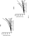

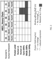
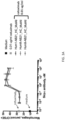
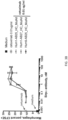

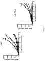


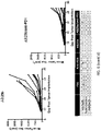
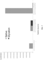

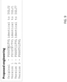

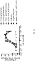
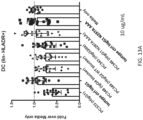
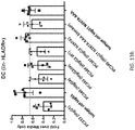
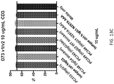
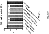
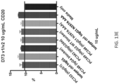
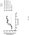


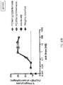
Classifications
-
- C—CHEMISTRY; METALLURGY
- C07—ORGANIC CHEMISTRY
- C07K—PEPTIDES
- C07K16/00—Immunoglobulins [IGs], e.g. monoclonal or polyclonal antibodies
- C07K16/18—Immunoglobulins [IGs], e.g. monoclonal or polyclonal antibodies against material from animals or humans
- C07K16/28—Immunoglobulins [IGs], e.g. monoclonal or polyclonal antibodies against material from animals or humans against receptors, cell surface antigens or cell surface determinants
- C07K16/2896—Immunoglobulins [IGs], e.g. monoclonal or polyclonal antibodies against material from animals or humans against receptors, cell surface antigens or cell surface determinants against molecules with a "CD"-designation, not provided for elsewhere
-
- A—HUMAN NECESSITIES
- A61—MEDICAL OR VETERINARY SCIENCE; HYGIENE
- A61K—PREPARATIONS FOR MEDICAL, DENTAL OR TOILETRY PURPOSES
- A61K39/00—Medicinal preparations containing antigens or antibodies
- A61K39/395—Antibodies; Immunoglobulins; Immune serum, e.g. antilymphocytic serum
-
- A—HUMAN NECESSITIES
- A61—MEDICAL OR VETERINARY SCIENCE; HYGIENE
- A61K—PREPARATIONS FOR MEDICAL, DENTAL OR TOILETRY PURPOSES
- A61K39/00—Medicinal preparations containing antigens or antibodies
- A61K39/395—Antibodies; Immunoglobulins; Immune serum, e.g. antilymphocytic serum
- A61K39/39533—Antibodies; Immunoglobulins; Immune serum, e.g. antilymphocytic serum against materials from animals
- A61K39/3955—Antibodies; Immunoglobulins; Immune serum, e.g. antilymphocytic serum against materials from animals against proteinaceous materials, e.g. enzymes, hormones, lymphokines
-
- A—HUMAN NECESSITIES
- A61—MEDICAL OR VETERINARY SCIENCE; HYGIENE
- A61K—PREPARATIONS FOR MEDICAL, DENTAL OR TOILETRY PURPOSES
- A61K45/00—Medicinal preparations containing active ingredients not provided for in groups A61K31/00 - A61K41/00
- A61K45/06—Mixtures of active ingredients without chemical characterisation, e.g. antiphlogistics and cardiaca
-
- A—HUMAN NECESSITIES
- A61—MEDICAL OR VETERINARY SCIENCE; HYGIENE
- A61P—SPECIFIC THERAPEUTIC ACTIVITY OF CHEMICAL COMPOUNDS OR MEDICINAL PREPARATIONS
- A61P29/00—Non-central analgesic, antipyretic or antiinflammatory agents, e.g. antirheumatic agents; Non-steroidal antiinflammatory drugs [NSAID]
-
- A—HUMAN NECESSITIES
- A61—MEDICAL OR VETERINARY SCIENCE; HYGIENE
- A61P—SPECIFIC THERAPEUTIC ACTIVITY OF CHEMICAL COMPOUNDS OR MEDICINAL PREPARATIONS
- A61P35/00—Antineoplastic agents
-
- A—HUMAN NECESSITIES
- A61—MEDICAL OR VETERINARY SCIENCE; HYGIENE
- A61P—SPECIFIC THERAPEUTIC ACTIVITY OF CHEMICAL COMPOUNDS OR MEDICINAL PREPARATIONS
- A61P37/00—Drugs for immunological or allergic disorders
-
- A—HUMAN NECESSITIES
- A61—MEDICAL OR VETERINARY SCIENCE; HYGIENE
- A61P—SPECIFIC THERAPEUTIC ACTIVITY OF CHEMICAL COMPOUNDS OR MEDICINAL PREPARATIONS
- A61P37/00—Drugs for immunological or allergic disorders
- A61P37/02—Immunomodulators
-
- A—HUMAN NECESSITIES
- A61—MEDICAL OR VETERINARY SCIENCE; HYGIENE
- A61P—SPECIFIC THERAPEUTIC ACTIVITY OF CHEMICAL COMPOUNDS OR MEDICINAL PREPARATIONS
- A61P37/00—Drugs for immunological or allergic disorders
- A61P37/02—Immunomodulators
- A61P37/06—Immunosuppressants, e.g. drugs for graft rejection
-
- C—CHEMISTRY; METALLURGY
- C07—ORGANIC CHEMISTRY
- C07K—PEPTIDES
- C07K16/00—Immunoglobulins [IGs], e.g. monoclonal or polyclonal antibodies
- C07K16/18—Immunoglobulins [IGs], e.g. monoclonal or polyclonal antibodies against material from animals or humans
- C07K16/28—Immunoglobulins [IGs], e.g. monoclonal or polyclonal antibodies against material from animals or humans against receptors, cell surface antigens or cell surface determinants
- C07K16/2803—Immunoglobulins [IGs], e.g. monoclonal or polyclonal antibodies against material from animals or humans against receptors, cell surface antigens or cell surface determinants against the immunoglobulin superfamily
- C07K16/2818—Immunoglobulins [IGs], e.g. monoclonal or polyclonal antibodies against material from animals or humans against receptors, cell surface antigens or cell surface determinants against the immunoglobulin superfamily against CD28 or CD152
-
- C—CHEMISTRY; METALLURGY
- C07—ORGANIC CHEMISTRY
- C07K—PEPTIDES
- C07K16/00—Immunoglobulins [IGs], e.g. monoclonal or polyclonal antibodies
- C07K16/18—Immunoglobulins [IGs], e.g. monoclonal or polyclonal antibodies against material from animals or humans
- C07K16/28—Immunoglobulins [IGs], e.g. monoclonal or polyclonal antibodies against material from animals or humans against receptors, cell surface antigens or cell surface determinants
- C07K16/2803—Immunoglobulins [IGs], e.g. monoclonal or polyclonal antibodies against material from animals or humans against receptors, cell surface antigens or cell surface determinants against the immunoglobulin superfamily
- C07K16/2827—Immunoglobulins [IGs], e.g. monoclonal or polyclonal antibodies against material from animals or humans against receptors, cell surface antigens or cell surface determinants against the immunoglobulin superfamily against B7 molecules, e.g. CD80, CD86
-
- C—CHEMISTRY; METALLURGY
- C07—ORGANIC CHEMISTRY
- C07K—PEPTIDES
- C07K16/00—Immunoglobulins [IGs], e.g. monoclonal or polyclonal antibodies
- C07K16/18—Immunoglobulins [IGs], e.g. monoclonal or polyclonal antibodies against material from animals or humans
- C07K16/28—Immunoglobulins [IGs], e.g. monoclonal or polyclonal antibodies against material from animals or humans against receptors, cell surface antigens or cell surface determinants
- C07K16/2863—Immunoglobulins [IGs], e.g. monoclonal or polyclonal antibodies against material from animals or humans against receptors, cell surface antigens or cell surface determinants against receptors for growth factors, growth regulators
-
- A—HUMAN NECESSITIES
- A61—MEDICAL OR VETERINARY SCIENCE; HYGIENE
- A61K—PREPARATIONS FOR MEDICAL, DENTAL OR TOILETRY PURPOSES
- A61K39/00—Medicinal preparations containing antigens or antibodies
- A61K2039/505—Medicinal preparations containing antigens or antibodies comprising antibodies
-
- A—HUMAN NECESSITIES
- A61—MEDICAL OR VETERINARY SCIENCE; HYGIENE
- A61K—PREPARATIONS FOR MEDICAL, DENTAL OR TOILETRY PURPOSES
- A61K39/00—Medicinal preparations containing antigens or antibodies
- A61K2039/505—Medicinal preparations containing antigens or antibodies comprising antibodies
- A61K2039/507—Comprising a combination of two or more separate antibodies
-
- A—HUMAN NECESSITIES
- A61—MEDICAL OR VETERINARY SCIENCE; HYGIENE
- A61K—PREPARATIONS FOR MEDICAL, DENTAL OR TOILETRY PURPOSES
- A61K2121/00—Preparations for use in therapy
-
- A—HUMAN NECESSITIES
- A61—MEDICAL OR VETERINARY SCIENCE; HYGIENE
- A61K—PREPARATIONS FOR MEDICAL, DENTAL OR TOILETRY PURPOSES
- A61K2300/00—Mixtures or combinations of active ingredients, wherein at least one active ingredient is fully defined in groups A61K31/00 - A61K41/00
-
- C—CHEMISTRY; METALLURGY
- C07—ORGANIC CHEMISTRY
- C07K—PEPTIDES
- C07K2317/00—Immunoglobulins specific features
- C07K2317/20—Immunoglobulins specific features characterized by taxonomic origin
- C07K2317/24—Immunoglobulins specific features characterized by taxonomic origin containing regions, domains or residues from different species, e.g. chimeric, humanized or veneered
-
- C—CHEMISTRY; METALLURGY
- C07—ORGANIC CHEMISTRY
- C07K—PEPTIDES
- C07K2317/00—Immunoglobulins specific features
- C07K2317/30—Immunoglobulins specific features characterized by aspects of specificity or valency
- C07K2317/31—Immunoglobulins specific features characterized by aspects of specificity or valency multispecific
-
- C—CHEMISTRY; METALLURGY
- C07—ORGANIC CHEMISTRY
- C07K—PEPTIDES
- C07K2317/00—Immunoglobulins specific features
- C07K2317/30—Immunoglobulins specific features characterized by aspects of specificity or valency
- C07K2317/32—Immunoglobulins specific features characterized by aspects of specificity or valency specific for a neo-epitope on a complex, e.g. antibody-antigen or ligand-receptor
-
- C—CHEMISTRY; METALLURGY
- C07—ORGANIC CHEMISTRY
- C07K—PEPTIDES
- C07K2317/00—Immunoglobulins specific features
- C07K2317/40—Immunoglobulins specific features characterized by post-translational modification
- C07K2317/41—Glycosylation, sialylation, or fucosylation
-
- C—CHEMISTRY; METALLURGY
- C07—ORGANIC CHEMISTRY
- C07K—PEPTIDES
- C07K2317/00—Immunoglobulins specific features
- C07K2317/50—Immunoglobulins specific features characterized by immunoglobulin fragments
- C07K2317/515—Complete light chain, i.e. VL + CL
-
- C—CHEMISTRY; METALLURGY
- C07—ORGANIC CHEMISTRY
- C07K—PEPTIDES
- C07K2317/00—Immunoglobulins specific features
- C07K2317/50—Immunoglobulins specific features characterized by immunoglobulin fragments
- C07K2317/52—Constant or Fc region; Isotype
-
- C—CHEMISTRY; METALLURGY
- C07—ORGANIC CHEMISTRY
- C07K—PEPTIDES
- C07K2317/00—Immunoglobulins specific features
- C07K2317/60—Immunoglobulins specific features characterized by non-natural combinations of immunoglobulin fragments
- C07K2317/62—Immunoglobulins specific features characterized by non-natural combinations of immunoglobulin fragments comprising only variable region components
- C07K2317/622—Single chain antibody (scFv)
-
- C—CHEMISTRY; METALLURGY
- C07—ORGANIC CHEMISTRY
- C07K—PEPTIDES
- C07K2317/00—Immunoglobulins specific features
- C07K2317/70—Immunoglobulins specific features characterized by effect upon binding to a cell or to an antigen
- C07K2317/71—Decreased effector function due to an Fc-modification
-
- C—CHEMISTRY; METALLURGY
- C07—ORGANIC CHEMISTRY
- C07K—PEPTIDES
- C07K2317/00—Immunoglobulins specific features
- C07K2317/70—Immunoglobulins specific features characterized by effect upon binding to a cell or to an antigen
- C07K2317/73—Inducing cell death, e.g. apoptosis, necrosis or inhibition of cell proliferation
-
- C—CHEMISTRY; METALLURGY
- C07—ORGANIC CHEMISTRY
- C07K—PEPTIDES
- C07K2317/00—Immunoglobulins specific features
- C07K2317/70—Immunoglobulins specific features characterized by effect upon binding to a cell or to an antigen
- C07K2317/76—Antagonist effect on antigen, e.g. neutralization or inhibition of binding
-
- C—CHEMISTRY; METALLURGY
- C07—ORGANIC CHEMISTRY
- C07K—PEPTIDES
- C07K2317/00—Immunoglobulins specific features
- C07K2317/90—Immunoglobulins specific features characterized by (pharmaco)kinetic aspects or by stability of the immunoglobulin
- C07K2317/92—Affinity (KD), association rate (Ka), dissociation rate (Kd) or EC50 value
Definitions
- the present disclosure relates to isolated, humanized antibodies that bind an extracellular domain (e.g., the D1 domain) of a human SIRP- ⁇ polypeptide, as well as polynucleotides, vectors, host cells, and methods related thereto.
- an extracellular domain e.g., the D1 domain
- SIRP- ⁇ Signal-regulatory protein alpha
- a family of cell-surface receptors that plays critical roles in the regulation of the immune system (see, e.g ., Barclay, A.N. and Brown, M.H. (2006) Nat. Rev. Immunol. 6:457-64 ).
- SIRP- ⁇ is expressed on the surface of various cells, including leukocytes such as dendritic cells, eosinophils, neutrophils, and macrophages.
- SIRP- ⁇ includes an extracellular domain that interacts with external stimuli such as ligands and an intracellular domain that mediates a variety of intracellular signals.
- CD47 is expressed on the surface of a variety of cell types.
- IgSF domain of CD47 binds the extracellular domain (e.g., the D1 domain) of SIRP- ⁇ expressed on an immune cell (e.g., a macrophage)
- CD47 serves to convey what has been termed a "don't eat me" signal to the immune system that prevents phagocytosis of healthy cells ( see, e.g., WO2015/138600 and Weiskopf, K. et al.
- CD47 has also been shown to be highly expressed by a variety of cancers, and its interaction with SIRP- ⁇ in this context is thought to allow tumors to mimic the healthy "don't eat me” signal in order to evade immune surveillance and phagocytosis by macrophages ( see, e.g., Majeti, R. et al. (2009) Cell 138:286-99 ; Zhao, X.W. et al. (2011) Proc. Natl. Acad Sci. 108:18342-7 ). As such, antibodies that block this interaction are highly desirable.
- SIRP- ⁇ is known to be a highly polymorphic protein in humans, monkeys, and mice.
- polymorphic differences have been identified between SIRP- ⁇ proteins in the NOD and C57BL/6 mouse strains, and these polymorphisms lead to functional consequences related to CD47 binding and engraftment of human hematopoietic stem cells in these mouse strains.
- two prevalent alleles of the SIRPA gene have been reported ( Treffers, LW. et al. (2016), Eur. J. Immunol. 48:344-354 ; Zhao, X. et al. (2011), PNAS. 108:18342-47 ; van der Heijden, J. (2014). Genetic variation in human Fc gamma receptors: Functional consequences of polymorphisms and copy number variation (Doctoral thesis )).
- SIRP- ⁇ -CD47 Due to the importance of the SIRP- ⁇ -CD47 interaction in normal immune function and tumorigenesis, the identification of antibodies that bind human SIRP- ⁇ is of great interest for development of clinical candidates.
- antibodies that bind an extracellular domain of a human SIRP- ⁇ polypeptide possess one or more of many useful in vitro and/or in vivo properties, such as binding to multiple SIRP- ⁇ polypeptides with high affinity, cross-reactivity against multiple mammalian SIRP- ⁇ polypeptides ( e.g ., human v1, human v2, cynomolgus, and/or multiple murine SIRP- ⁇ proteins), the ability to enhance macrophage phagocytosis, the ability to enhance dendritic cell activation, and/or the ability to inhibit in vivo growth of a tumor that expresses CD47.
- the present disclosure provides antibodies with variant light chains that have been engineered to remove potential liabilities, such as residues that may be modified by deamidation or glycation, resulting in antibodies with more desirable characteristics for manufacturing, storage, and/or drug development.
- the present disclosure provides antibodies (e.g ., isolated antibodies) that bind an extracellular domain of a human SIRP- ⁇ polypeptide, where the antibodies comprise: a heavy chain comprising a heavy chain variable (VH) domain that comprises the amino acid sequence of SEQ ID NO:26; and a light chain comprising a light chain variable (VL) domain that comprises an amino acid sequence according to the formula SYELTQPPSVSVSPGQTARITCSGGSYSSYYYAWYQQKPGQAPVTLIYSDDKRPSNI PERFSGSSSGTTVTLTISGVQAEDEADYYCGGYDQSSYTNPFGX 1 GTX 2 X 3 TVL (SEQ ID NO:71), wherein X 1 is G or T; X 2 is K, Q, or R; and X 3 is L or V, and wherein the VL domain does not comprise the sequence of SEQ ID NO:25.
- VH heavy chain variable
- VL light chain variable
- the VL domain comprises an amino acid sequence selected from the group consisting of SEQ ID NOs:39-41.
- the light chain further comprises a light chain constant (CL) domain sequence that comprises an amino acid sequence according to the formula GQPKANPTVTLFPPSSEELQANKATLVCLISDFYPGAVTVAWKADGSPVKAGVETT KPSKQSX 4 X 5 KYAASSYLSLTPEQWKSHRSYSCQVTHEGSTVEKTVAPTECS (SEQ ID NO:72), wherein X 4 X 5 is ND, DN, DS, or SD.
- the light chain further comprises a light chain constant (CL) domain sequence that comprises an amino acid sequence selected from the group consisting of SEQ ID NOs:43-46. In some embodiments, the light chain further comprises a kappa light chain constant (CL) domain. In some embodiments, the light chain comprises the amino acid sequence of SEQ ID NO:36. In some embodiments, the light chain further comprises a lambda light chain constant (CL) domain. In some embodiments, the light chain comprises the amino acid sequence of SEQ ID NO:37 or SEQ ID NO:38. In some embodiments, the light chain comprises an amino acid sequence selected from the group consisting of SEQ ID NOs:48-57.
- CL light chain constant domain sequence that comprises an amino acid sequence selected from the group consisting of SEQ ID NOs:43-46. In some embodiments, the light chain further comprises a kappa light chain constant (CL) domain. In some embodiments, the light chain comprises the amino acid sequence of SEQ ID NO:36. In some embodiments, the light chain further comprises a lambd
- the antibody is a scFv-Fc, single domain antibody, single heavy chain antibody, or single light chain antibody. In some embodiments, the antibody is a monoclonal antibody. In some embodiments, the antibody comprises a heavy chain comprising a heavy chain constant domain that comprises an Fc region. In some embodiments, the Fc region is a human Fc region selected from the group consisting of an IgG1 Fc region, an IgG2 Fc region, and an IgG4 Fc region. In some embodiments, the Fc region is a human IgG1 Fc region.
- the Fc region is a human IgG1 Fc region comprising L234A, L235A, and G237A substitutions, amino acid position numbering according to EU. In some embodiments, the Fc region is a human IgG2 Fc region. In some embodiments, the Fc region is a human IgG2 Fc region comprising A330S and P331S substitutions, amino acid position numbering according to EU. In some embodiments, the Fc region further comprises an N297A substitution, amino acid position numbering according to EU. In some embodiments, the Fc region is a human IgG2 Fc region comprising an N297A substitution, amino acid position numbering according to EU.
- the Fc region is a human IgG4 Fc region, and wherein the heavy chain comprises an S228P substitution, amino acid position numbering according to EU.
- the heavy chain comprises a heavy chain constant domain that comprises an amino acid sequence selected from the group consisting of SEQ ID NOs:31-35.
- the heavy chain comprises a heavy chain constant domain that comprises an amino acid sequence selected from the group consisting of SEQ ID NOs:33, 34, and 137.
- the heavy chain comprises a heavy chain constant domain that comprises an amino acid sequence selected from the group consisting of SEQ ID NOs:132-139.
- the heavy chain comprises an amino acid sequence selected from the group consisting of SEQ ID NOs:58-62. In some embodiments, the heavy chain comprises an amino acid sequence selected from the group consisting of SEQ ID NOs:60, 61, and 129. In some embodiments, the heavy chain comprises an amino acid sequence selected from the group consisting of SEQ ID NOs:124-131.
- the present disclosure provides antibodies (e.g ., isolated antibodies) that bind an extracellular domain of a human SIRP- ⁇ polypeptide, wherein: the heavy chain comprises the amino acid sequence of SEQ ID NO:62, and the light chain comprises the amino acid sequence of SEQ ID NO:52; the heavy chain comprises the amino acid sequence of SEQ ID NO:62, and the light chain comprises the amino acid sequence of SEQ ID NO:53; the heavy chain comprises the amino acid sequence of SEQ ID NO:62, and the light chain comprises the amino acid sequence of SEQ ID NO:54; the heavy chain comprises the amino acid sequence of SEQ ID NO:62, and the light chain comprises the amino acid sequence of SEQ ID NO:55; the heavy chain comprises the amino acid sequence of SEQ ID NO:62, and the light chain comprises the amino acid sequence of SEQ ID NO:56; or the heavy chain comprises the amino acid sequence of SEQ ID NO:62, and the light chain comprises the amino acid sequence of SEQ ID NO:57.
- the heavy chain comprises the amino acid sequence of SEQ ID NO:62,
- the present disclosure provides antibodies (e.g ., isolated antibodies) that bind an extracellular domain of a human SIRP- ⁇ polypeptide, wherein: the heavy chain comprises the amino acid sequence of SEQ ID NO:58, and the light chain comprises the amino acid sequence of SEQ ID NO:52; the heavy chain comprises the amino acid sequence of SEQ ID NO:59, and the light chain comprises the amino acid sequence of SEQ ID NO:52; the heavy chain comprises the amino acid sequence of SEQ ID NO:60, and the light chain comprises the amino acid sequence of SEQ ID NO:52; the heavy chain comprises the amino acid sequence of SEQ ID NO:61, and the light chain comprises the amino acid sequence of SEQ ID NO:52; the heavy chain comprises the amino acid sequence of SEQ ID NO:58, and the light chain comprises the amino acid sequence of SEQ ID NO:53; the heavy chain comprises the amino acid sequence of SEQ ID NO:59, and the light chain comprises the amino acid sequence of SEQ ID NO:53; the heavy chain comprises the amino acid sequence of SEQ ID NO:60, and the light
- the antibody enhances phagocytosis by a macrophage expressing a human SIRP- ⁇ polypeptide. In some embodiments, the antibody enhances activation of a dendritic cell expressing a human SIRP- ⁇ polypeptide. In some embodiments, the antibody inhibits in vivo growth of a tumor that expresses CD47.
- the antibody is conjugated to an agent, e.g ., a cytotoxic agent, label, or moiety that modules the immune sy stem.
- an agent e.g ., a cytotoxic agent, label, or moiety that modules the immune sy stem.
- the antibody is a bispecific antibody.
- the antibody comprises a first antigen binding domain that binds an extracellular domain of a human SIRP- ⁇ polypeptide and a second antigen binding domain that binds an antigen expressed by a cancer cell.
- the antigen expressed by the cancer cell is selected from the group consisting of CD19, CD20, CD22, CD30, CD33, CD38, CD52, CD56, CD70, CD74, CD79b, CD123, CD138, CS1/SLAMF7, Trop-2, 5T4, EphA4, BCMA, Mucin 1, Mucin 16, PD-L1, PTK7, STEAP1, Endothelin B Receptor, mesothelin, EGFRvIII, ENPP3, SLC44A4, GNMB, nectin 4, NaPi2b, LIV-1A, Guanylyl cyclase C, DLL3, EGFR, HER2, VEGF, VEGFR, integrin ⁇ V ⁇ 3, integrin ⁇ 5 ⁇ 1, MET, IGF1R, TRAILR1, TRAILR2, RANKL, FAP, Tenascin, Le y , EpCAM, CEA, gpA33, PSMA, TAG72,
- the antibody comprises a first antigen binding domain that binds an extracellular domain of a human SIRP- ⁇ polypeptide and a second antigen binding domain that binds an antigen expressed by an immune cell.
- the antigen expressed by the immune cell is selected from the group consisting of BDCA2, BDCA4, ILT7, LILRB1, LILRB2, LILRB3, LILRB4, LILRB5, Siglec-3, Siglec-7, Siglec-9, Siglec-15, FGL-1, CD200, CD200R, CSF-1R, CD40, CD40L, CD163, CD206, DEC205, CD47, CD123, arginase, IDO, TDO, AhR, EP2, COX-2, CCR2, CCR-7, CXCR1, CX3CR1, CXCR2, CXCR3, CXCR4, CXCR7, TGF- ⁇ RI, TGF- ⁇ RII, c-Kit, CD244, L-selectin/CD62L,
- the antibody comprises a first antigen binding domain that binds an extracellular domain of a human SIRP- ⁇ polypeptide and a second antigen binding domain that binds an antigen expressed by a natural killer (NK) cell.
- the antigen expressed by the NK cell is selected from the group consisting of NKR-PlA, CD94, KLRG1, KIR2DL5A, KIR2DL5B, KIR2DL1, KIR2DL2, KIR2DL3, KIR2DS2, KIR2DS3, KIR2DS4, KIR2DS5, KIR3DS1, KIR2DS1, CD94, NKG2D, CD160, CD16, NKp46, NKp30, NKp44, DNAM1, CRTAM, CD27, NTB-A, PSGL1, CD96, CD100, NKp80, SLAMF7, and CD244.
- the present disclosure provides a polynucleotide encoding the antibody according to any one of the above embodiments.
- the present disclosure provides a vector comprising the polynucleotide according to any one of the above embodiments.
- the present disclosure provides a host cell comprising the polynucleotide or vector according to any one of the above embodiments.
- the present disclosure provides a method of producing an antibody, the method comprising culture the host cell according to any one of the above embodiments such that the antibody is produced. In some embodiments, the method further comprises recovering the antibody from the host cell.
- the present disclosure provides a method of treating or delaying progression of cancer in an individual, the method comprising administering to the individual an effective amount of the antibody according to any one of the above embodiments. In some embodiments, the method further comprises administering to the individual an effective amount of a second antibody that binds an antigen expressed by the cancer.
- the antigen expressed by the cancer is selected from the group consisting of CD19, CD20, CD22, CD30, CD33, CD38, CD52, CD56, CD70, CD74, CD79b, CD123, CD138, CS1/SLAMF7, Trop-2, 5T4, EphA4, BCMA, Mucin 1, Mucin 16, PTK7, STEAP1, Endothelin B Receptor, mesothelin, EGFRvIII, ENPP3, SLC44A4, GNMB, nectin 4, NaPi2b, LIV-1A, Guanylyl cyclase C, DLL3, EGFR, HER2, VEGF, VEGFR, integrin ⁇ V ⁇ 3, integrin ⁇ 5 ⁇ 1, MET, IGF1R, TRAILR1, TRAILR2, RANKL, FAP, Tenascin, Le y , EpCAM, CEA, gpA33, PSMA, TAG72, a mucin, CA
- the method further comprises administering to the individual an effective amount of an immunotherapeutic agent.
- the immunotherapeutic agent comprises a second antibody.
- the second antibody binds to an antigen selected from the group consisting of BDCA2, BDCA4, ILT7, LILRB1, LILRB2, LILRB3, LILRB4, LILRB5, Siglec-3, Siglec-7, Siglec-9, Siglec-15, FGL-1, CD200, CD200R, CSF-1R, CD40, CD40L, CD163, CD206, DEC205, CD47, CD123, arginase, IDO, TDO, AhR, EP2, COX-2, CCR2, CCR-7, CXCR1, CX3CR1, CXCR2, CXCR3, CXCR4, CXCR7, TGF- ⁇ RI, TGF- ⁇ RII, c-Kit, CD244, L-selectin/CD62L, CD11b, CD11c, CD68, 41BB,
- the second antibody binds to PD-1. In some embodiments, the second antibody binds to PD-L1. In some embodiments, the immunotherapeutic agent comprises a vaccine, oncolytic virus, adoptive cell therapy, cytokine, or small molecule. In some embodiments, the method further comprises administering to the individual an effective amount of a second antibody that binds an antigen expressed by a natural killer (NK) cell.
- NK natural killer
- the antigen expressed by the NK cell is selected from the group consisting of NKR-P1A, CD94, KLRG1, KIR2DL5A, KIR2DL5B, KIR2DL1, KIR2DL2, KIR2DL3, KIR2DS2, KIR2DS3, KIR2DS4, KIR2DS5, KIR3DS1, KIR2DS1, CD94, NKG2D, CD160, CD16, NKp46, NKp30, NKp44, DNAM1, CRTAM, CD27, NTB-A, PSGL1, CD96, CD100, NKp80, SLAMF7, and CD244.
- the method further comprises administering to the individual an effective amount of a chemotherapeutic agent or small molecule anticancer agent. In some embodiments, the method further comprises administering to the individual an effective amount of a targeted small molecule inhibitor.
- the targeted small molecule inhibitor is a VEGFR and/or PDGFR inhibitor, EGFR inhibitor, ALK inhibitor, CDK4/6 inhibitor, PARP inhibitor, mTOR inhibitor, KRAS inhibitor, TRK inhibitor, BCL2 inhibitor, IDB inhibitor, PI3K inhibitor, DNA damage response (DDR) inhibitor, or hypomethylation agent.
- the present disclosure provides a method of treating or delaying progression of an autoimmune disease or an inflammatory disease in an individual, the method comprising administering to the individual an effective amount of the antibody according to any one of the above embodiments.
- the autoimmune disease or inflammatory disease is selected from the group consisting of multiple sclerosis, rheumatoid arthritis, a spondyloarthropathy, systemic lupus erythematosus, an antibody-mediated inflammatory or autoimmune disease, graft versus host disease, sepsis, diabetes, psoriasis, psoriatic arthritis, atherosclerosis, Sjogren's syndrome, progressive systemic sclerosis, scleroderma, acute coronary syndrome, ischemic reperfusion, Crohn's Disease, ulcerative colitis, endometriosis, glomerulonephritis, IgA nephropathy, polycystic kidney disease, myasthenia gravis, idiopathic
- the present disclosure describes antibodies that bind the extracellular domains (e.g., the D1 domains) of one or more human SIRP- ⁇ polypeptides and have a variety of SIRP- ⁇ binding profiles of potential interest.
- the present disclosure describes anti-SIRP- ⁇ antibodies with one or more in vitro and/or in vivo biological properties of interest, such as binding to multiple SIRP- ⁇ polypeptides with high affinity and the ability to enhance macrophage phagocytosis, enhance dendritic cell activation, and/or inhibit in vivo growth of a tumor that expresses CD47.
- These antibodies have a light chain derived from chicken, which provides unique opportunities to generate antibodies that cross-react across multiple mammalian SIRP- ⁇ polypeptides.
- these antibodies were found to cross-react with human SIRP- ⁇ v1 and v2 cynomolgus SIRP- ⁇ , and murine SIRP- ⁇ from multiple strains, making them highly advantageous for both pre-clinical testing and clinical application across a large human population.
- these antibodies have also been engineered to remove potential liabilities, such as residues that may be modified by deamidation or glycation, resulting in antibodies with more desirable characteristics for manufacturing, storage, and/or drug development.
- isolated antibodies that bind the extracellular domain (e.g., the D1 domain) of a human SIRP- ⁇ polypeptide.
- polynucleotides and vectors encoding the antibodies of the present disclosure, as well as methods of antibody production related thereto.
- provided herein are methods for treating or delaying progression of cancer in an individual, comprising administering to the individual an effective amount of an antibody of the present disclosure.
- provided herein are methods for treating or delaying progression of an autoimmune or inflammatory disease in an individual, comprising administering to the individual an effective amount of an antibody of the present disclosure.
- aspects and embodiments of the present disclosure include “comprising,” “consisting,” and “consisting essentially of” aspects and embodiments.
- SIRP- ⁇ polypeptide may refer to any endogenous or naturally occurring SIRP- ⁇ polypeptide encoded by a genome from any vertebrate, including mammals such as humans, monkeys, rodents ( e. g., mouse or rat), and birds, such as chickens.
- the term also includes naturally occurring variants, e.g ., alternatively spliced variants, allelic variants, or polymorphisms ( e. g., those described herein).
- the term may further refer to full-length, unprocessed SIRP- ⁇ polypeptides as well as SIRP- ⁇ polypeptides that result from cellular processing, e.g ., removal of a signal sequence, etc.
- a human SIRP- ⁇ polypeptide is one encoded by a human SIRPA gene, e.g., as described by NCBI Gene ID No. 140885.
- SIRP- ⁇ polypeptides are highly polymorphic within and among species, including, for example, multiple human variants with amino acid polymorphisms in the extracellular domain.
- SIRP- ⁇ polypeptides include an extracellular domain that binds ligands/partners, e.g ., CD47.
- SIRP- ⁇ polypeptides comprise 3 highly homologous immunoglobulin (Ig)-like extracellular domains-D1, D2, and D3.
- the SIRP- ⁇ D1 domain (“D1 domain") refers to the membrane distal, extracellular domain of SIRP- ⁇ and mediates binding of SIRP- ⁇ to CD47 (see, e.g., Hatherley, D. et al. (2008) Mo/. Cell 31:266-77 ; Hatherley, D. et al. (2007) J. Biol. Chem. 282:14567-75 ; Hatherley, D. et al. (2009) J. Biol.
- the extracellular domain generally refers to the entire extracellular portion of SIRP- ⁇ , e.g ., as expressed on a cell surface, and may include distinct SIRP- ⁇ . domains, such as the D1 domain.
- the D1 domain contains residues shown to be critical for mediating CD47 binding ( see, e.g., Lee, W.Y. et al. (2007) J. Immunol. 179:7741-50 ).
- an antibody that binds an extracellular domain of a SIRP- ⁇ polypeptide binds one or more residues of the D1 domain.
- CD47 refers to a polypeptide that, among other roles, serves as a binding partner for SIRP- ⁇ polypeptides.
- CD47 refers to a human CD47 polypeptide, e.g., a polypeptide encoded by a human CD47 gene, such as that described by NCBI Ref Seq ID No. 961.
- Exemplary human CD47 amino acid sequences are known (see, e.g., NCBI Reference Sequence Accession No. NP_001768).
- the IgSF domain of CD47 refers to the N-terminal extracellular domain of CD47 that is known to be critical for SIRP- ⁇ binding (see, e.g., Barclay, A.N. and Brown, M.H. (2006) Nat. Rev. Immunol. 6:457-64 and Hatherley, D. et al. (2009) J. Biol. Chem. 284:26613-9 ).
- the term "CD47” may also include modified CD47 polypeptides that are able to bind SIRP- ⁇ , e.g ., a polypeptide comprising an IgSF domain of CD47 conjugated to another polypeptide or other moiety, e . g ., an Ig Fc region.
- SIRP- ⁇ signaling may refer to antagonizing, agonizing, or otherwise interfering with one or more aspects of SIRP- ⁇ signaling in a cell expressing a SIRP- ⁇ polypeptide.
- SIRP- ⁇ signaling may refer to one or more intracellular signaling events mediated by activation of a SIRP- ⁇ polypeptide, including without limitation tyrosine phosphorylation of the intracellular region of SIRP- ⁇ , phosphatase ( e . g ., SHP1) binding, adaptor protein binding (e.g., SCAP2, FYB, and/or GRB2), cytokine production (e.g.
- IL-10 IL-10, IL-1 ⁇ , IFN or TNF
- nitric oxide production and/or one or more intercellular phenotypes, including without limitation macrophage phagocytosis and other activating or suppressive phenotypes of macrophages, eosinophils, neutrophils, dendritic cells, and myeloid-derived suppressor cells (MDSCs).
- macrophage phagocytosis including without limitation macrophage phagocytosis and other activating or suppressive phenotypes of macrophages, eosinophils, neutrophils, dendritic cells, and myeloid-derived suppressor cells (MDSCs).
- MDSCs myeloid-derived suppressor cells
- antibody may refer to intact antibodies; antibody fragments (including without limitation Fab, F(ab')2, Fab'-SH, Fv, diabodies, scFv, scFv-Fc, single domain antibodies, single heavy chain antibodies, and single light chain antibodies), provided that they exhibit the desired biological activity (e.g. epitope binding); monoclonal antibodies; polyclonal antibodies; monospecific antibodies; multi-specific antibodies (e.g., bispecific antibodies); and antibody-like proteins.
- antibody fragments including without limitation Fab, F(ab')2, Fab'-SH, Fv, diabodies, scFv, scFv-Fc, single domain antibodies, single heavy chain antibodies, and single light chain antibodies
- each binding specificity when used in reference to an antibody or antibody fragment includes an antibody or antibody fragment that possesses two different binding specificities.
- each binding specificity may recognize a different antigen, or each binding specificity may recognize the same antigen with different affinity and/or precise epitope.
- each different binding specificity comprises one or more different antibody antigen binding domains (e . g ., variable domains), such that the bispecific antibody or antibody fragment comprises at least a first antigen binding domain with a first binding specificity and a second antigen binding domain with a second binding specificity.
- variable domains e.g ., variable domains
- an “isolated” antibody may refer to an antibody that has been separated and/or recovered from a component of its natural environment, e . g ., a host cell or organism.
- an antibody is purified to a desired purity by weight (e.g., at least 95%); and/or homogeneity by SDS-PAGE using, for example, staining by silver, Coomassie, etc.
- an isolated antibody is obtained following one or more purification steps.
- “native” antibodies refer to typically heterotetrameric complexes including two identical light (L) chains and two identical heavy (H) chains. Variable numbers of disulfide bonds connect the two heavy chains, and one connects each light chain to a heavy chain, in addition to intrachain disulfide bridges.
- the heavy chains include a variable domain (VH) followed (N-terminus to C-terminus) by three or four constant domains.
- the light chains include a variable domain (VL) followed by a constant domain (CL).
- VH variable domain
- CL constant domain
- mammalian light chains fall into one of two categories based on amino acid sequence: kappa and lambda.
- a “constant domain” may refer to the more conserved portion of the antibody or fragment, e . g ., outside the variable domains.
- the term may include the CL domain as well as heavy chain constant domains CH1, CH2, and CH3, along with the heavy chain hinge region.
- a heavy chain constant domain may further comprise a CH4 heavy chain constant domain.
- Constant domains of the heavy chain can be assigned to one of 5 major types: IgA, IgD, IgE, IgG, and IgM. Several subtypes exist for many of these major types. The subunit structures and three-dimensional configurations of different classes of immunoglobulins are well known and described generally in, for example, Abbas et al. Cellular and Mol. Immunology, 4th ed. (W.B. Saunders, Co., 2000 ).
- antibody variable domain refers to the portions of the light and heavy chains of an antibody that include the complementary determining regions (CDRs, e . g ., CDR L1, CDR L2, CDR L3, CDR H1, CDR H2, and CDR H3) and framework regions (FRs).
- CDRs complementary determining regions
- FRs framework regions
- variable refers to the fact that subsequences of the variable domains differ substantially in sequence between antibodies and are critical to the binding specificity of a particular antibody for its antigen. Variability is concentrated in three hypervariable regions (HVRs) in both VH and VL domains. The more conserved portions of variable domains are called the framework regions (FR) in which the HVRs are interspersed.
- HVRs hypervariable regions
- FR framework regions
- the variable domains of native heavy and light chains each comprise four FR regions connected by three HVRs that form loops ( see Kabat et al., Sequences of Proteins of Immunological Interest, Fifth Edition, National Institute of Health, Bethesda, Md. (1991 )).
- HVR hypervariable region
- VH and VL domains characterized by enhanced sequence variability and/or formation of defined loops. These include three HVRs in the VH domain (H1, H2, and H3) and three HVRs in the VL domain (L1, L2, and L3). H3 is believed to be critical in imparting fine binding specificity, with L3 and H3 showing the highest level of diversity. See Johnson and Wu, in Methods in Molecular Biology 248:1-25 (Lo, ed., Human Press, Totowa, N.J., 2003 ).
- the Kabat Complementarity Determining Regions are based on sequence variability and are the most commonly used ( Kabat et al, Sequences of Proteins of Immunological Interest, 5th Ed. Public Health Service, National Institutes of Health, Bethesda, Md. (1991 )). Chothia refers instead to the location of the structural loops ( Chothia and Lesk J. Mol. Biol. 196:901-917 (1987 )).
- the AbM HVRs represent a compromise between the Kabat HVRs and Chothia structural loops, and are used by Oxford Molecular's AbM antibody modeling software.
- the "contact" HVRs are based on an analysis of the available complex crystal structures.
- Extended HVRs are also known: 24-36 or 24-34 (L1), 46-56 or 50-56 (L2) and 89-97 or 89-96 (L3) in the VL and 26-35 (H1), 50-65 or 49-65 (H2) and 93-102, 94-102, or 95-102 (H3) in the VH (Kabat numbering).
- “Numbering according to Kabat” may refer to the numbering system used for heavy chain variable domains or light chain variable domains of the compilation of antibodies in Kabat et al., supra.
- the actual linear amino acid sequence may contain fewer or additional amino acids corresponding to a shortening of, or insertion into, a FR or HVR of the variable domain.
- the Kabat numbering of residues may be determined for a given antibody by alignment at regions of homology of the sequence of the antibody with a "standard” Kabat numbered sequence.
- the Kabat numbering is used when referring to a residue in the variable domains (approximately residues 1-107 of the light chain and residues 1-113 of the heavy chain), whereas the EU numbering system or index ( e . g ., the EU index as in Kabat, numbering according to EU IgG1) is generally used when referring to a residue in the heavy chain constant region.
- Fc region typically include heavy chains with an Fc region, e.g., as opposed to an antibody fragment.
- Antigen-binding "Fab" fragments with a single antigen binding site may be released from the residual Fc fragment by papain digestion.
- F(ab')2 fragments include two antigen-binding sites produced by pepsin treatment of an antibody.
- Antibody fragments will, however, include one or more antibody variable regions.
- an "Fv” fragment contains a complete antigen-binding site.
- a single chain Fv can include a VH and a VL domain linked by a peptide linker such that the VH and VL domains associate, e.g., as in an antibody or Fab fragment, such that the HVRs form an antigen binding site. See Pluckthün, in The Pharmacology of Monoclonal Antibodies, vol. 113, Rosenburg and Moore eds., (Springer-Verlag, New York, 1994), pp. 269-315 .
- the scFv is fused to an antibody Fc domain ( e . g ., scFv-Fc).
- HVRs typically comprise an antigen binding site
- a single variable domain with three HVRs is still capable of binding an antigen, albeit at a lower affinity.
- Single domain antibodies e.g., camelid antibodies
- Single heavy chain (VHH) and single light chain antibodies are also known.
- a Fab' fragment typically includes a few more residues at the C-terminal end than a Fab fragment.
- a Fab'-SH includes cysteine residues with a free thiol.
- Various chemical couplings of antibody fragments are known in the art.
- a “diabody” includes antibody fragments with two antigen-binding sites. These include a VH and VL domain connected by a linker, which is typically too short to facilitate pairing of domains in the same chain. Diabodies may be bivalent or bispecific. Tribodies and tetrabodies, or other numbers of VH/VL domains are known. See Hudson et al., Nat. Med. 9:129-134 (2003 ).
- a “monoclonal” antibody refers to an antibody obtained from a population of substantially homogeneous antibodies, e.g., substantially identical but allowing for minor levels of background mutations and/or modifications. "Monoclonal” denotes the substantially homogeneous character of antibodies, and does not require production of the antibody by any particular method.
- a monoclonal antibody is selected by its HVR, VH, and/or VL sequences and/or binding properties, e . g ., selected from a pool of clones ( e . g ., recombinant, hybridoma, or phage-derived).
- a monoclonal antibody may be engineered to include one or more mutations, e.g ., to affect binding affinity or other properties of the antibody, create a humanized or chimeric antibody, improve antibody production and/or homogeneity, engineer a multispecific antibody, resultant antibodies of which are still considered to be monoclonal in nature.
- a population of monoclonal antibodies may be distinguished from polyclonal antibodies as the individual monoclonal antibodies of the population recognize the same antigenic site.
- Methods 284(1-2): 119-132 (2004 ), and technologies for producing human or human-like antibodies in animals that have parts or all of the human immunoglobulin loci or genes encoding human immunoglobulin sequences see, e.g., WO 1998/24893 ; WO 1996/34096 ; WO 1996/33735 ; WO 1991/10741 ; Jakobovits et al., Proc. Natl. Acad. Sci. USA 90: 2551 (1993 ); Jakobovits et al., Nature 362: 255-258 (1993 ); Bruggemann et al., Year in Immunol. 7:33 (1993 ); U.S. Pat. Nos.
- Chimeric antibodies may refer to an antibody with one portion of the heavy and/or light chain from a particular isotype, class, or organism and another portion from another isotype, class, or organism.
- the variable region will be from one source or organism, and the constant region will be from another.
- Humanized antibodies may refer to antibodies with predominantly human sequence and a minimal amount of non-human (e . g ., mouse or chicken) sequence.
- a humanized antibody has one or more HVR sequences (bearing a binding specificity of interest) from an antibody derived from a non-human ( e . g ., mouse or chicken) organism grafted onto a human recipient antibody framework (FR).
- non-human residues are further grafted onto the human framework (not present in either source or recipient antibodies), e.g., to improve antibody properties.
- a humanized antibody will comprise substantially all of at least one, and typically two, variable domains, in which all or substantially all of the hypervariable loops correspond to those of a non-human immunoglobulin, and all or substantially all of the FRs are those of a human immunoglobulin sequence.
- the humanized antibody optionally will also comprise at least a portion of an immunoglobulin constant region (Fc), typically that of a human immunoglobulin. See Jones et al., Nature 321:522-525 (1986 ); Riechmann et al., Nature 332:323-329 (1988 ); and Presta, Curr. Op. Struct. Biol. 2:593-596 (1992 ).
- a "human” antibody may refer to an antibody having an amino acid sequence which corresponds to that of an antibody produced by a human and/or has been made using any of the techniques for making human antibodies as disclosed herein.
- Human antibodies can be produced using various techniques known in the art, including phage-display libraries. Hoogenboom and Winter, J. Mol. Biol., 227:381 (1991 ); Marks et a/., J. Mol. Biol, 222:581 (1991 ); preparation of human monoclonal antibodies as described in Cole et al., Monoclonal Antibodies and Cancer Therapy, Alan R. Liss, p. 77 (1985 ); Boerner et al., J.
- linker refers to a linkage between two elements, e.g., protein domains.
- a linker can be a covalent bond or a spacer.
- spacer refers to a moiety (e.g., a polyethylene glycol (PEG) polymer) or an amino acid sequence (e.g., a 1-200 amino acid sequence) occurring between two polypeptides or polypeptide domains to provide space or flexibility (or both space and flexibility) between the two polypeptides or polypeptide domains.
- an amino acid spacer is part of the primary sequence of a polypeptide (e.g., joined to the spaced polypeptides or polypeptide domains via the polypeptide backbone).
- cytotoxic agent may refer to any agent that inhibits cellular proliferation or induces cell death. Cytotoxic agents include, but are not limited to, chemotherapeutic agents; radioactive isotopes; growth inhibitory agents; and toxins such as small molecule toxins or enzymatically active toxins, including fragments and/or variants thereof.
- Exemplary cytotoxic agents include without limitation metabolic inhibitors, anti-microtubule agents, platinum containing compounds, alkylating agents, proteasome inhibitors, topoisomerase II inhibitors, antimetabolites, topoisomerase I inhibitors, signal transduction pathway inhibitors, non-receptor tyrosine kinase angiogenesis inhibitors, hormones and hormonal analogues, proapoptotic agents, inhibitors of LDH-A, cell cycle inhibitors, HDAC inhibitors, and antibiotic agents.
- a "label” may include any moiety that serves as a detection agent, e.g., of binding between a labeled antibody of the present disclosure and a macromolecule or cell.
- exemplary labels include without limitation fluorescent (e . g ., compounds or proteins), radioactive, or enzymatic moieties, as well as affinity purification tags.
- an antibody may be said to "bind” an antigen with an affinity sufficient to render the antibody useful for in vitro and/or in vivo manipulation of the antigen.
- an antibody that "binds" an antigen has a dissociation constant (K D ) for the antigen that is less than or equal to 1 ⁇ M at 25°C.
- binding affinity refers to the strength of the binding interaction between two molecules.
- binding affinity refers to the strength of the sum total of non-covalent interactions between a molecule and its binding partner, such as a high affinity SIRP- ⁇ D1 variant and CD47.
- binding affinity refers to intrinsic binding affinity, which reflects a 1:1 interaction between members of a binding pair.
- the binding affinity between two molecules is commonly described by the dissociation constant (K D ) or the association constant (K A ). Two molecules that have low binding affinity for each other generally bind slowly, tend to dissociate easily, and exhibit a large K D .
- K D Two molecules that have high affinity for each other generally bind readily, tend to remain bound longer, and exhibit a small K D .
- the K D of two interacting molecules is determined using known methods and techniques, e.g., surface plasmon resonance (SPR). K D can be calculated as the ratio of k off /k on .
- K D less than refers to a numerically smaller K D value and an increasing binding affinity relative to the recited K D value.
- K D greater than refers to a numerically larger K D value and a decreasing binding affinity relative to the recited K D value.
- treatment may refer to therapeutic administration of a molecule, compound, formulation, composition, etc. so as to alter one or more pathological symptoms in an individual or cell being treated. Desirable effects of treatment can include without limitation decelerating disease progression, ameliorating or palliating a pathological symptom or disease state, improving prognosis, and/or achieving disease remission.
- an individual's cancer is successfully "treated” if one or more symptoms associated with cancer are mitigated or abolished, such as, without limitation, reducing the proliferation of cancer cells, eliminating cancer cells or tumor burden, decreasing symptoms resulting from the cancer, increasing the quality of life of the individual, lessening the dose of other medication(s), and/or prolonging survival of the individual.
- an autoimmune or inflammatory disease may be successfully "treated” if one or more symptoms associated with the autoimmune or inflammatory disease are mitigated or abolished, such as, without limitation, reducing autoreactive immune cells and/or inflammatory immune cells or cytokines, decreasing immune activation and/or inflammation, slowing or mitigating organ damage resulting from the disease, decreasing symptoms resulting from the disease, increasing the quality of life of the individual, lessening the dose of other medication(s), and/or prolonging survival of the individual.
- symptoms associated with the autoimmune or inflammatory disease are mitigated or abolished, such as, without limitation, reducing autoreactive immune cells and/or inflammatory immune cells or cytokines, decreasing immune activation and/or inflammation, slowing or mitigating organ damage resulting from the disease, decreasing symptoms resulting from the disease, increasing the quality of life of the individual, lessening the dose of other medication(s), and/or prolonging survival of the individual.
- delaying progression of a disease may refer to slowing, retarding, deferring, postponing development of, stabilizing, or otherwise hindering the pathological course of the disease.
- the term may refer to a delay sufficient to effectively encompass prevention, e . g ., in preventing the individual from developing the disease.
- delaying progression may include delaying metastasis.
- the precise length of delay may depend, e . g ., upon the specific disease, condition of the individual, and the like.
- cancer and “cancerous” may describe dysregulated or unregulated cell growth/proliferation by a cell or cells in a mammal. Any cancer type known in the art may be included, such as but not limited to carcinoma, sarcoma, lymphoma, leukemia, lymphoma, and blastoma.
- cancers include, but are not limited to, lung cancer, squamous cell cancer, brain tumors, glioblastoma, head and neck cancer, hepatocellular cancer, colorectal cancer (e.g., colon or rectal cancers), liver cancer, bladder cancer, gastric or stomach cancer, pancreatic cancer, cervical cancer, ovarian cancer, cancer of the urinary tract, breast cancer, peritoneal cancer, uterine cancer, salivary gland cancer, kidney or renal cancer, prostate cancer, vulval cancer, thyroid cancer, anal carcinoma, penile carcinoma, melanoma, multiple myeloma and B-cell lymphoma (including non-Hodgkin's lymphomas (NHL)); acute lymphoblastic leukemia (ALL); chronic lymphocytic leukemia (CLL); acute myeloid leukemia (AML); Merkel cell carcinoma; hairy cell leukemia: chronic myeloblastic leukemia (CML); and associated metastases.
- NHL non-Hodgkin's lymph
- the term "effective amount" may refer to an amount of an antibody of the present disclosure or a pharmaceutical composition containing an antibody of the present disclosure that is sufficient and effective in achieving a desired therapeutic effect in treating or delaying progression of a patient having a disease, such as a cancer, e.g., solid tumor or hematological cancer.
- a therapeutically effective amount will avoid adverse side effects, and/or such side effects will be outweighed by beneficial effects.
- An effective amount may depend upon the individual being treated, e . g ., age, weight, sex, disease state, as well as the ability of the agent to produce a desired response.
- An effective amount can be administered in one or more administrations.
- an effective amount of a drug, compound, or pharmaceutical composition may or may not be achieved in conjunction with another drug, compound, or pharmaceutical composition, such as another therapeutic agent.
- an "effective amount" may also be considered in the context of administering one or more additional therapeutic agents, and a single agent may be considered to be given in an effective amount if, in conjunction with one or more other agents, a desirable result may be or is achieved.
- the term "pharmaceutical composition” may refer to a medicinal or pharmaceutical formulation that includes an active ingredient as well as excipients or diluents (or both excipients and diluents) and enables the active ingredient to be administered by suitable methods of administration.
- the pharmaceutical compositions disclosed herein include pharmaceutically acceptable components that are compatible with one or more antibodies of the present disclosure.
- the pharmaceutical composition is in tablet or capsule form for oral administration or in aqueous form for intravenous or subcutaneous administration, for example by injection.
- the terms "subject,” “individual,” and “patient” are used interchangeably to refer to a vertebrate, for example, a mammal. Mammals include, but are not limited to, murines, simians, humans, farm animals, sport animals, and pets.
- conjunction with or “in combination with” may refer to administration of one therapeutic in addition to ( e . g ., before, during, and/or after) another therapeutic.
- antibodies that bind the extracellular domain (e.g., the D1 domain) of a human SIRP- ⁇ polypeptide.
- the antibody is a humanized antibody.
- the antibody is a monoclonal antibody.
- an antibody of the present disclosure comprises a heavy chain comprising a VH domain shown in Table 2 and/or a light chain comprising a VL domain that comprises an amino acid sequence according to the formula SYELTQPPSVSVSPGQTARITCSGGSYSSYYYAWYQQKPGQAPVTLIYSDDKRPSNI PERFSGSSSGTTVTLTISGVQAEDEADYYCGGYDQSSYTNPFGX 1 GTX 2 X 3 TVL (SEQ ID NO:71), where X 1 is G or T; X 2 is K, Q, or R; and X 3 is L or V.
- an antibody of the present disclosure comprises a heavy chain comprising a VH domain shown in Table 2 and a light chain comprising a VL domain that comprises an amino acid sequence according to the formula SYELTQPPSVSVSVSPGQTARITCSGGSYSSYYYAWYQQKPGQAPVTLIYSDDKRPSNI PERFSGSSSGTTVTLTISGVQAEDEADYYCGGYDQSSYTNPFGX 1 GTX 2 X 3 TVL (SEQ ID NO:71), where X 1 is G or T; X 2 is K, Q, or R; and X 3 is L or V.
- the VL domain does not comprise the sequence of SEQ ID NO:25.
- an antibody of the present disclosure comprises a heavy chain comprising a VH domain that comprises the amino acid sequence of SEQ ID NO:26 and a light chain comprising a VL domain that comprises an amino acid sequence according to the formula SYELTQPPSVSVSVSPGQTARITCSGGSYSSYYYAWYQQKPGQAPVTLIYSDDKRPSNI PERFSGSSSGTTVTLTISGVQAEDEADYYCGGYDQSSYTNPFGX 1 GTX 2 X 3 TLV (SEQ ID NO:71), where X 1 is G or T; X 2 is K, Q, or R; and X 3 is L or V.
- the VL domain does not comprise the sequence of SEQ ID NO:25.
- an antibody of the present disclosure comprises a heavy chain comprising a VH domain shown in Table 2 and/or a light chain comprising a VL domain shown in Table 1. In some embodiments, an antibody of the present disclosure comprises a heavy chain comprising a VH domain shown in Table 2 and a light chain comprising a VL domain shown in Table 1.
- an antibody of the present disclosure comprises a heavy chain comprising a VH domain that comprises the amino acid sequence of SEQ ID NO:26 and/or a light chain comprising a VL domain that comprises an amino acid sequence selected from SEQ ID NOs:39-41. In some embodiments, an antibody of the present disclosure comprises a heavy chain comprising a VH domain that comprises the amino acid sequence of SEQ ID NO:26 and a light chain comprising a VL domain that comprises an amino acid sequence selected from SEQ ID NOs:39-41.
- an antibody of the present disclosure comprises a heavy chain comprising a VH domain shown in Table 2 and/or a light chain comprising an amino acid sequence selected from SEQ ID NOs:48-57. In some embodiments, an antibody of the present disclosure comprises a heavy chain comprising a VH domain shown in Table 2 and a light chain comprising an amino acid sequence selected from SEQ ID NOs:48-57. In some embodiments, an antibody of the present disclosure comprises a heavy chain comprising a VH domain that comprises the amino acid sequence of SEQ ID NO:26 and/or a light chain comprising an amino acid sequence selected from SEQ ID NOs:48-57.
- an antibody of the present disclosure comprises a heavy chain comprising a VH domain that comprises the amino acid sequence of SEQ ID NO:26 and a light chain comprising an amino acid sequence selected from SEQ ID NOs:48-57. In some embodiments, an antibody of the present disclosure comprises a heavy chain comprising a VH domain that comprises the amino acid sequence of SEQ ID NO:81 and a light chain comprising an amino acid sequence selected from SEQ ID NOs:48-57. In some embodiments, an antibody of the present disclosure comprises a heavy chain comprising a VH domain that comprises the amino acid sequence of SEQ ID NO:83 and a light chain comprising an amino acid sequence selected from SEQ ID NOs:48-57.
- an antibody of the present disclosure comprises a heavy chain comprising a VH domain that comprises an amino acid sequence selected from SEQ ID NOs:77-111 and a light chain comprising an amino acid sequence selected from SEQ ID NOs:48-57. In some embodiments, an antibody of the present disclosure comprises a heavy chain comprising a VH domain that comprises an amino acid sequence selected from SEQ ID NOs:77-111 and a light chain that comprises an amino acid sequence selected from SEQ ID NOs:48-51.
- an antibody of the present disclosure comprises a heavy chain comprising a VH domain shown in Table 2 and a constant domain that comprises an amino acid sequence selected from the group consisting of SEQ ID NOs:33, 34, and 137 and/or a light chain comprising an amino acid sequence selected from SEQ ID NOs:48-57.
- an antibody of the present disclosure comprises a heavy chain comprising an amino acid sequence selected from the group consisting of SEQ ID NOs:60, 61, and 129 and/or a light chain comprising an amino acid sequence selected from SEQ ID NOs:48-57.
- an antibody of the present disclosure comprises a heavy chain comprising a VH domain that comprises an amino acid sequence selected from SEQ ID NOs:26, 81, or 83 and a light chain that comprises an amino acid sequence selected from SEQ ID NOs:48-57.
- an antibody of the present disclosure comprises a heavy chain comprising a VH domain that comprises an amino acid sequence selected from SEQ ID NOs:26, 81, or 83 and a light chain comprising a VL domain that comprises an amino acid sequence selected from SEQ ID NOs:39-41.
- an antibody of the present disclosure comprises a heavy chain comprising a VH domain that comprises the amino acid sequence of SEQ ID NO:26 and a light chain comprising a VL domain that comprises an amino acid sequence selected from SEQ ID NOs:39-41.
- an antibody of the present disclosure comprises a heavy chain comprising a VH domain that comprises an amino acid sequence selected from SEQ ID NOs:26, 81, or 83 and a light chain that comprises a VL domain sequence selected from SEQ ID NOs:39-41 and a CL domain comprising a sequence selected from SEQ ID NOs:36-38.
- an antibody of the present disclosure comprises a heavy chain comprising a VH domain that comprises the amino acid sequence of SEQ ID NO:26 and a light chain that comprises a VL domain sequence selected from SEQ ID NOs:39-41 and a CL domain comprising a sequence selected from SEQ ID NOs:36-38.
- an antibody of the present disclosure comprises a heavy chain comprising a VH domain that comprises an amino acid sequence selected from SEQ ID NOs:26, 81, or 83 and a light chain that comprises a VL domain comprising the sequence of SEQ ID NO:25 and a CL domain sequence selected from SEQ ID NOs:43-46.
- an antibody of the present disclosure comprises a heavy chain comprising a VH domain that comprises the amino acid sequence of SEQ ID NO:26 and a light chain that comprises a VL domain comprising the sequence of SEQ ID NO:25 and a CL domain sequence selected from SEQ ID NOs:43-46.
- an antibody of the present disclosure comprises a heavy chain that comprises a VH domain comprising the amino acid sequence of SEQ ID NO:83 and a light chain that comprises a VL domain sequence selected from SEQ ID NOs:39-41. In some embodiments, an antibody of the present disclosure comprises a heavy chain that comprises a VH domain comprising the amino acid sequence of SEQ ID NO:83 and a light chain that comprises a VL domain comprising the amino acid sequence of SEQ ID NO:40. In some embodiments, an antibody of the present disclosure comprises a heavy chain that comprises a VH domain comprising the amino acid sequence of SEQ ID NO:83 and a light chain that comprises the amino acid sequence of SEQ ID NO:55.
- an antibody of the present disclosure comprises a heavy chain comprising an amino acid sequence selected from the group consisting of SEQ ID NOs:119-123 and a light chain that comprises a VL domain sequence selected from SEQ ID NOs:39-41. In some embodiments, an antibody of the present disclosure comprises a heavy chain comprising an amino acid sequence selected from the group consisting of SEQ ID NOs: 119-123 and a light chain that comprises a VL domain comprising the amino acid sequence of SEQ ID NO:40. In some embodiments, an antibody of the present disclosure comprises a heavy chain comprising an amino acid sequence selected from the group consisting of SEQ ID NOs:119-123 and a light chain that comprises the amino acid sequence of SEQ ID NO:55.
- an antibody of the present disclosure comprises a heavy chain that comprises the amino acid sequence of SEQ ID NO:119 and a light chain that comprises the amino acid sequence of SEQ ID NO:55. In some embodiments, an antibody of the present disclosure comprises a heavy chain that comprises the amino acid sequence of SEQ ID NO: 120 and a light chain that comprises the amino acid sequence of SEQ ID NO:55. In some embodiments, an antibody of the present disclosure comprises a heavy chain that comprises the amino acid sequence of SEQ ID NO:121 and a light chain that comprises the amino acid sequence of SEQ ID NO:55. In some embodiments, an antibody of the present disclosure comprises a heavy chain that comprises the amino acid sequence of SEQ ID NO:122 and a light chain that comprises the amino acid sequence of SEQ ID NO:55. In some embodiments, an antibody of the present disclosure comprises a heavy chain that comprises the amino acid sequence of SEQ ID NO:123 and a light chain that comprises the amino acid sequence of SEQ ID NO:55.
- an antibody of the present disclosure comprises a heavy chain that comprises a VH domain comprising the amino acid sequence of SEQ ID NO:26 and a constant domain sequence selected from the group consisting of SEQ ID NOs:132-139; and/or a light chain comprising a VL domain that comprises the amino acid sequence of SEQ ID NO:40.
- an antibody of the present disclosure comprises a heavy chain that comprises a VH domain comprising the amino acid sequence of SEQ ID NO:26 and a constant domain sequence selected from the group consisting of SEQ ID NOs:33,34, and 137; and/or a light chain comprising a VL domain that comprises the amino acid sequence of SEQ ID NO:40.
- an antibody of the present disclosure comprises a heavy chain that comprises a VH domain comprising the amino acid sequence of SEQ ID NO:26 and a constant domain sequence selected from the group consisting of SEQ ID NOs:132-139; and/or a light chain comprising the amino acid sequence of SEQ ID NO:55.
- an antibody of the present disclosure comprises a heavy chain that comprises a VH domain comprising the amino acid sequence of SEQ ID NO:26 and a constant domain sequence selected from the group consisting of SEQ ID NOs:33,34, and 137; and/or a light chain comprising the amino acid sequence of SEQ ID NO:55.
- an antibody of the present disclosure comprises a heavy chain that comprises an amino acid sequence selected from the group consisting of SEQ ID NOs:124-131; and/or a light chain comprising a VL domain that comprises the amino acid sequence of SEQ ID NO:40.
- an antibody of the present disclosure comprises a heavy chain that comprises an amino acid sequence selected from the group consisting of SEQ ID NOs:60, 61, and 129; and/or a light chain comprising a VL domain that comprises the amino acid sequence of SEQ ID NO:40.
- an antibody of the present disclosure comprises a heavy chain that comprises an amino acid sequence selected from the group consisting of SEQ ID NOs:124-131; and/or a light chain comprising the amino acid sequence of SEQ ID NO:55.
- an antibody of the present disclosure comprises a heavy chain that comprises an amino acid sequence selected from the group consisting of SEQ ID NOs:60, 61, and 129; and/or a light chain comprising the amino acid sequence of SEQ ID NO:55.
- the heavy chain comprises the amino acid sequence of SEQ ID NO:60
- the light chain comprises the amino acid sequence of SEQ ID NO:55.
- the heavy chain comprises the amino acid sequence of SEQ ID NO:61, and the light chain comprises the amino acid sequence of SEQ ID NO:55. In some embodiments, the heavy chain comprises the amino acid sequence of SEQ ID NO:137, and the light chain comprises the amino acid sequence of SEQ ID NO:55.
- an antibody of the present disclosure comprises a heavy chain that comprises an amino acid sequence selected from SEQ ID NOs:58-62 and/or a light chain comprising a VL domain that comprises an amino acid sequence according to the formula SYELTQPPSVSVSPGQTARITCSGGSYSSYYYAWYQQKPGQAPVTLIYSDDKRPSNI PERFSGSSSGTTVTLTISGVQAEDEADYYCGGYDQSSYTNPFGX 1 GTX 2 X 3 TVL (SEQ ID NO:71), where X 1 is G or T; X 2 is K, Q, or R; and X 3 is L or V.
- an antibody of the present disclosure comprises a heavy chain that comprises an amino acid sequence selected from SEQ ID NOs:58-62 and a light chain comprising a VL domain that comprises an amino acid sequence according to the formula SYELTQPPSVSVSVSPGQTARITCSGGSYSSYYYAWYQQKPGQAPVTLIYSDDKRPSNI PERFSGSSSGTTVTLTISGVQAEDEADYYCGGYDQSSYTNPFGX 1 GTX 2 X 3 TVL (SEQ ID NO:71), where X 1 is G or T; X 2 is K, Q, or R; and X 3 is L or V.
- the VL domain does not comprise the sequence of SEQ ID NO:25.
- an antibody of the present disclosure comprises a heavy chain that comprises an amino acid sequence selected from SEQ ID NOs:58-62 and a light chain comprising a VL domain shown in Table 1. In some embodiments, an antibody of the present disclosure comprises a heavy chain that comprises an amino acid sequence selected from SEQ ID NOs:58-62 and a light chain comprising a VL domain that comprises an amino acid sequence selected from SEQ ID NOs:39-41. In some embodiments, an antibody of the present disclosure comprises a heavy chain that comprises an amino acid sequence selected from SEQ ID NOs:58-62 and a light chain comprising an amino acid sequence selected from SEQ ID NOs:48-57.
- an antibody of the present disclosure comprises a heavy chain that comprises an amino acid sequence selected from SEQ ID NOs:58-62 and a light chain comprising a VL domain that comprises an amino acid sequence selected from SEQ ID NOs:39-41 and a CL domain that comprises an amino acid sequence selected from SEQ ID NOs:36-38.
- an antibody of the present disclosure comprises a heavy chain that comprises an amino acid sequence selected from SEQ ID NOs:58-62 and a light chain comprising a VL domain that comprises the amino acid sequence of SEQ ID NO:25 and a CL domain that comprises an amino acid sequence selected from SEQ ID NOs:43-46.
- an antibody of the present disclosure comprises a heavy chain that comprises the amino acid sequence of SEQ ID NO:62 and a light chain that comprises the amino acid sequence of SEQ ID NO:52. In some embodiments, an antibody of the present disclosure comprises a heavy chain that comprises the amino acid sequence of SEQ ID NO:62 and a light chain that comprises the amino acid sequence of SEQ ID NO:53. In some embodiments, an antibody of the present disclosure comprises a heavy chain that comprises the amino acid sequence of SEQ ID NO:62 and a light chain that comprises the amino acid sequence of SEQ ID NO:54. In some embodiments, an antibody of the present disclosure comprises a heavy chain that comprises the amino acid sequence of SEQ ID NO:62 and a light chain that comprises the amino acid sequence of SEQ ID NO:55.
- an antibody of the present disclosure comprises a heavy chain that comprises the amino acid sequence of SEQ ID NO:62 and a light chain that comprises the amino acid sequence of SEQ ID NO:56. In some embodiments, an antibody of the present disclosure comprises a heavy chain that comprises the amino acid sequence of SEQ ID NO:62 and a light chain that comprises the amino acid sequence of SEQ ID NO:57.
- an antibody of the present disclosure comprises a heavy chain that comprises the amino acid sequence of SEQ ID NO:58 and a light chain that comprises the amino acid sequence of SEQ ID NO:52. In some embodiments, an antibody of the present disclosure comprises a heavy chain that comprises the amino acid sequence of SEQ ID NO:59 and a light chain that comprises the amino acid sequence of SEQ ID NO:52. In some embodiments, an antibody of the present disclosure comprises a heavy chain that comprises the amino acid sequence of SEQ ID NO:60 and a light chain that comprises the amino acid sequence of SEQ ID NO:52. In some embodiments, an antibody of the present disclosure comprises a heavy chain that comprises the amino acid sequence of SEQ ID NO:61 and a light chain that comprises the amino acid sequence of SEQ ID NO:52.
- an antibody of the present disclosure comprises a heavy chain that comprises the amino acid sequence of SEQ ID NO:58 and a light chain that comprises the amino acid sequence of SEQ ID NO:53. In some embodiments, an antibody of the present disclosure comprises a heavy chain that comprises the amino acid sequence of SEQ ID NO:59 and a light chain that comprises the amino acid sequence of SEQ ID NO:53. In some embodiments, an antibody of the present disclosure comprises a heavy chain that comprises the amino acid sequence of SEQ ID NO:60 and a light chain that comprises the amino acid sequence of SEQ ID NO:53. In some embodiments, an antibody of the present disclosure comprises a heavy chain that comprises the amino acid sequence of SEQ ID NO:61 and a light chain that comprises the amino acid sequence of SEQ ID NO:53.
- an antibody of the present disclosure comprises a heavy chain that comprises the amino acid sequence of SEQ ID NO:58 and a light chain that comprises the amino acid sequence of SEQ ID NO:54. In some embodiments, an antibody of the present disclosure comprises a heavy chain that comprises the amino acid sequence of SEQ ID NO:59 and a light chain that comprises the amino acid sequence of SEQ ID NO:54. In some embodiments, an antibody of the present disclosure comprises a heavy chain that comprises the amino acid sequence of SEQ ID NO:60 and a light chain that comprises the amino acid sequence of SEQ ID NO:54. In some embodiments, an antibody of the present disclosure comprises a heavy chain that comprises the amino acid sequence of SEQ ID NO:61 and a light chain that comprises the amino acid sequence of SEQ ID NO:54.
- an antibody of the present disclosure comprises a heavy chain that comprises the amino acid sequence of SEQ ID NO:58 and a light chain that comprises the amino acid sequence of SEQ ID NO:55. In some embodiments, an antibody of the present disclosure comprises a heavy chain that comprises the amino acid sequence of SEQ ID NO:59 and a light chain that comprises the amino acid sequence of SEQ ID NO:55. In some embodiments, an antibody of the present disclosure comprises a heavy chain that comprises the amino acid sequence of SEQ ID NO:60 and a light chain that comprises the amino acid sequence of SEQ ID NO:55. In some embodiments, an antibody of the present disclosure comprises a heavy chain that comprises the amino acid sequence of SEQ ID NO:61 and a light chain that comprises the amino acid sequence of SEQ ID NO:55.
- an antibody of the present disclosure comprises a heavy chain that comprises the amino acid sequence of SEQ ID NO:58 and a light chain that comprises the amino acid sequence of SEQ ID NO:56. In some embodiments, an antibody of the present disclosure comprises a heavy chain that comprises the amino acid sequence of SEQ ID NO:59 and a light chain that comprises the amino acid sequence of SEQ ID NO:56. In some embodiments, an antibody of the present disclosure comprises a heavy chain that comprises the amino acid sequence of SEQ ID NO:60 and a light chain that comprises the amino acid sequence of SEQ ID NO:56. In some embodiments, an antibody of the present disclosure comprises a heavy chain that comprises the amino acid sequence of SEQ ID NO:61 and a light chain that comprises the amino acid sequence of SEQ ID NO:56.
- an antibody of the present disclosure comprises a heavy chain that comprises the amino acid sequence of SEQ ID NO:58 and a light chain that comprises the amino acid sequence of SEQ ID NO:57. In some embodiments, an antibody of the present disclosure comprises a heavy chain that comprises the amino acid sequence of SEQ ID NO:59 and a light chain that comprises the amino acid sequence of SEQ ID NO:57. In some embodiments, an antibody of the present disclosure comprises a heavy chain that comprises the amino acid sequence of SEQ ID NO:60 and a light chain that comprises the amino acid sequence of SEQ ID NO:57. In some embodiments, an antibody of the present disclosure comprises a heavy chain that comprises the amino acid sequence of SEQ ID NO:61 and a light chain that comprises the amino acid sequence of SEQ ID NO:57.
- an antibody of the present disclosure comprises a heavy chain that comprises a VH domain comprising a sequence selected from SEQ ID NOs:77-111 and a light chain that comprises a VL domain comprising the sequence of SEQ ID NO:25. In some embodiments, an antibody of the present disclosure comprises a heavy chain that comprises a VH domain comprising a sequence selected from SEQ ID NOs:77-111 and a light chain that comprises a VL domain comprising a sequence selected from SEQ ID NOs:39-41.
- an antibody of the present disclosure comprises a heavy chain that comprises a VH domain comprising a sequence selected from SEQ ID NOs:77-111 and a light chain that comprises a VL domain comprising a sequence selected from SEQ ID NOs:39-41 and a CL domain comprising a sequence selected from SEQ ID NOs:36-38.
- an antibody of the present disclosure comprises a heavy chain that comprises a VH domain comprising a sequence selected from SEQ ID NOs:77-111 and a light chain that comprises a VL domain comprising the sequence of SEQ ID NO:25 and a CL domain comprising a sequence selected from SEQ ID NOs:43-46.
- an antibody of the present disclosure comprises a heavy chain that comprises a sequence selected from SEQ ID NOs:114-123 and a light chain that comprises a sequence selected from SEQ ID NOs:47-63. In some embodiments, an antibody of the present disclosure comprises a heavy chain that comprises a sequence selected from SEQ ID NOs:124-131 and a light chain that comprises a sequence selected from SEQ ID NOs:47-63. In some embodiments, an antibody of the present disclosure comprises a heavy chain that comprises a sequence selected from SEQ ID NOs:114-131 and a light chain that comprises a sequence selected from SEQ ID NOs:47-63.
- an antibody of the present disclosure comprises a heavy chain that comprises an amino acid sequence selected from SEQ ID NOs:114-118 and a light chain that comprises an amino acid sequence selected from SEQ ID NOs:48-57. In some embodiments, an antibody of the present disclosure comprises a heavy chain that comprises an amino acid sequence selected from SEQ ID NOs: 119-123 and a light chain that comprises an amino acid sequence selected from SEQ ID NOs:48-57. In some embodiments, an antibody of the present disclosure comprises a heavy chain that comprises an amino acid sequence selected from SEQ ID NOs:114-123 and a light chain that comprises a VL domain comprising a sequence selected from SEQ ID NOs:39-41.
- an antibody of the present disclosure comprises a heavy chain that comprises an amino acid sequence selected from SEQ ID NOs:114-123 and a light chain that comprises a VL domain comprising a sequence selected from SEQ ID NOs:39-41 and a CL domain comprising a sequence selected from SEQ ID NOs:36-38.
- an antibody of the present disclosure comprises a heavy chain that comprises an amino acid sequence selected from SEQ ID NOs:114-123 and a light chain that comprises a VL domain comprising the sequence of SEQ ID NO:25 and a CL domain comprising a sequence selected from SEQ ID NOs:43-46.
- an antibody of the present disclosure comprises three CDRs from a VH domain comprising a sequence set forth in Table 2 and/or three CDRs from a VL domain comprising a sequence set forth in Table 1.
- an antibody of the present disclosure comprises a VH domain comprising a sequence that is at least 90%, at least 91%, at least 92%, at least 93%, at least 94%, at least 95%, at least 96%, at least 97%, at least 98%, at least 99%, or 100% identical to a VH domain sequence set forth in Table 2 and optionally three CDRs from a VH domain comprising a sequence set forth in Table 2, and/or a VL domain comprising a sequence that is at least 90%, at least 91%, at least 92%, at least 93%, at least 94%, at least 95%, at least 96%, at least 97%, at least 98%, at least 99%, or 100% identical to a VL domain sequence set forth in Table 1 and optionally three CDRs from a
- an antibody of the present disclosure comprises a heavy chain comprising a sequence that is at least 90%, at least 91%, at least 92%, at least 93%, at least 94%, at least 95%, at least 96%, at least 97%, at least 98%, at least 99%, or 100% identical to a heavy chain sequence set forth in Table 2 and optionally three CDRs from a VH domain comprising a sequence set forth in Table 2, and/or a light chain comprising a sequence that is at least 90%, at least 91%, at least 92%, at least 93%, at least 94%, at least 95%, at least 96%, at least 97%, at least 98%, at least 99%, or 100% identical to a light chain sequence set forth in Table 1 and optionally three CDRs from a VL domain comprising a sequence set forth in Table 1.
- an antibody of the present disclosure comprises HVRs as defined by Chothia, Kabat, IMGT, or a combination thereof (e.g ., one or more HVRs as defined by one delineation and one or more HVRs as defined by a different delineation).
- HVRs as defined by Chothia, Kabat, IMGT, or a combination thereof (e.g ., one or more HVRs as defined by one delineation and one or more HVRs as defined by a different delineation).
- the numbering of HVR or CDR residues is defined by Kabat numbering.
- an antibody of the present disclosure binds an extracellular domain (e.g., the D1 domain) of a human SIRP- ⁇ v1 polypeptide comprising the amino acid sequence of EEELQVIQPDKSVLVAAGETATLRCTATSLTPVGPTQWFRGAGPGRELIYNQKEGHFP RVTTVSDLTKRNNMDFSIRIGNITPADAGTYYCVKFRKGSPDDVEFKSGAGTELSVR AKPS (SEQ ID NO:5).
- an antibody of the present disclosure binds an extracellular domain (e.g., the D1 domain) of a human SIRP- ⁇ v2 polypeptide comprising the amino acid sequence of EEELQVIQPDKSVSVAAGESAILHCTVTSLIPVGPIQWFRGAGPARELIYNQKEGHFPR VTTVSESTKRENMDFSISISNITPADAG7YYCVKFRKGSPDTEFKSGAGTELSVRAKPS (SEQ ID NO:6).
- an antibody of the present disclosure binds an extracellular domain (e.g., the D1 domain) of a human SIRP- ⁇ v1 polypeptide comprising the amino acid sequence of EEELQVIQPDKSVLVAAGETATLRCTATSLIPVGPIQWFRGAGPGRELIYNQKEGHFP RVTTVSDLTKRNNMDFSIRIGNITPADAGTYYCVKFRKGSPDDVEFKSGAGTELSVR AKPS (SEQ ID NO:5) and an extracellular domain (e.g., the D1 domain) of a human SIRP- ⁇ v2 polypeptide comprising the amino acid sequence of
- an antibody of the present disclosure binds an extracellular domain (e.g., the D1 domain) of a monkey SIRP- ⁇ polypeptide ( e.g , the D1 domain of a monkey SIRP- ⁇ polypeptide). In some embodiments, an antibody of the present disclosure binds an extracellular domain (e.g., the D1 domain) of a cynomolgus SIRP- ⁇ polypeptide ( e.g ., found in the organism Macaca fascicularis ) . In some embodiments, the antibody binds the extracellular domains (e.g., the D1 domains) of at least two different monkey SIRP- ⁇ variant polypeptides.
- the antibody binds the extracellular domains (e.g., the D1 domains) of at least two different cynomolgus SIRP- ⁇ variant polypeptides.
- the antibody binds an extracellular domain (e.g., the D1 domain) of a cynomolgus SIRP- ⁇ polypeptide comprising the amino acid sequence of EEELQVIQPEKSVSVAAGESATLNCTATSLIPVGPIQWFRGVGPGRELIYHQKEGHFP RVTPVSDPTKKNNMDFSIRISNITPADAGTYYCVKFRKGSPDVELKSGAGTELSVRAK PS (SEQ ID NO:11), an extracellular domain (e.g., the D1 domain) of a cynomolgus SIRP- ⁇ polypeptide comprising the amino acid sequence of EEELQVIQPEKSVSVAAGDSATLNCTVSSLIPVGPIQWFRGAGPGRELIYNLKEGHFP RVTAVSDPTKRNNM
- an antibody of the present disclosure binds an extracellular domain of a murine or mouse SIRP- ⁇ polypeptide (e.g., found in the organism Mus musculus ; e.g., the D1 domain of a murine or mouse SIRP- ⁇ polypeptide).
- the antibody binds the extracellular domains (e.g., the D1 domains) of two or more different murine SIRP- ⁇ variant polypeptides.
- a variety of murine SIRP- ⁇ variant polypeptides from different mouse strains are known.
- the murine SIRP- ⁇ variant polypeptide comprises an amino acid sequence selected from
- an antibody of the present disclosure binds an extracellular domain (e.g., the D1 domain) of a human SIRP family protein. In some embodiments, an antibody of the present disclosure binds an extracellular domain (e.g., the D1 domain) of a human SIRP- ⁇ polypeptide.
- a human SIRP- ⁇ polypeptide refers to a polypeptide encoded by a human SIRPB1 gene, e.g ., as described by NCBI Ref Seq ID No. 10326.
- the extracellular domain (e.g., the D1 domain) of the human SIRP- ⁇ polypeptide comprises the amino acid sequence of
- an antibody of the present disclosure binds an extracellular domain (e.g., the D1 domain) of a human SIRP- ⁇ polypeptide.
- a human SIRP- ⁇ polypeptide refers to a polypeptide encoded by a human SIRPG gene, e.g., as described by NCBI Ref Seq ID No. 55423.
- the extracellular domain (e.g., the D1 domain) of the human SIRP- ⁇ polypeptide comprises the amino acid sequence of
- an antibody of the present disclosure binds an IgSF domain of CD47 (e.g., human CD47). In some embodiments, an antibody of the present disclosure binds a polypeptide comprising the amino acid sequence of
- an antibody of the present disclosure modulates SIRP- ⁇ signaling in a cell expressing a human SIRP- ⁇ polypeptide. In some embodiments, an antibody of the present disclosure antagonizes SIRP- ⁇ signaling in a cell expressing a human SIRP- ⁇ polypeptide. In some embodiments, an antibody of the present disclosure interferes with SIRP- ⁇ signaling in a cell expressing a human SIRP- ⁇ polypeptide. In some embodiments, an antibody of the present disclosure agonizes SIRP- ⁇ signaling in a cell expressing a human SIRP- ⁇ polypeptide.
- SIRP- ⁇ signaling includes one or more intracellular signaling events mediated by activation of a SIRP- ⁇ polypeptide, including without limitation tyrosine phosphorylation of the intracellular region of SIRP- ⁇ , phosphatase (e.g., SHP1) binding, adaptor protein binding (e.g., SCAP2, FYB, and/or GRB2), and nitric oxide production.
- a SIRP- ⁇ polypeptide including without limitation tyrosine phosphorylation of the intracellular region of SIRP- ⁇ , phosphatase (e.g., SHP1) binding, adaptor protein binding (e.g., SCAP2, FYB, and/or GRB2), and nitric oxide production.
- Various assays for measuring SIRP- ⁇ signaling in a cell include without limitation SIRP- ⁇ phosphorylation, SHP1 and SHP2 co-immunoprecipitation, PI3-kinase signaling, cytokine production (both inflammatory IL-12, IL-23, TNF ⁇ , IFN and suppressive cytokines IL-10, IL-4, IL-13, cell surface markers levels for M1 and M2 macrophage markers) or dendritic cell activation and function; Kharitonenkov, A. et al. (1997) Nature 386: 181-6 ; Ochi, F. et al. (1997) Biochem. Biophys. Res. Commun. 239:483-7 ; Kim, E.J. et al. (2013) Inflammation Research 62:377-86 ; Yi, T. et a/. (2015) Immunity 43:764-75 .
- the cell expressing a human SIRP- ⁇ polypeptide is a leukocyte.
- the cell is a macrophage, dendritic cell, neutrophil, eosinophil, or myeloid-derived suppressor cell (MDSC).
- an antibody of the present disclosure decreases or antagonizes SIRP- ⁇ signaling in a cell expressing a human SIRP- ⁇ polypeptide by at least 20%, at least 30%, at least 40%, at least 50%, at least 60%, at least 70%, at least 80%, or at least 90%, e.g., using one or more of the SIRP- ⁇ signaling assays described herein or otherwise known in the art.
- an antibody of the present disclosure increases or agonizes SIRP- ⁇ signaling in a cell expressing a human SIRP- ⁇ polypeptide by at least 20%, at least 30%, at least 40%, at least 50%, at least 60%, at least 70%, at least 80%, or at least 90%, e.g ., using one or more of the SIRP- ⁇ signaling assays described herein or otherwise known in the art.
- an antibody of the present disclosure modulates an intercellular phenotype mediated by SIRP- ⁇ . In some embodiments, an antibody of the present disclosure enhances phagocytosis by a macrophage expressing a human SIRP- ⁇ polypeptide.
- phagocytic activity of a macrophage treated or contacted with an antibody of the present disclosure can be compared with phagocytic activity of a macrophage not treated or contacted with the antibody, or phagocytic activity of a macrophage that expresses a human SIRP- ⁇ polypeptide and is treated or contacted with an antibody of the present disclosure can be compared with phagocytic activity of a macrophage that does not express a human SIRP- ⁇ polypeptide and is treated or contacted with the antibody.
- Exemplary phagocytosis assays may be found, e.g., in Wieskopf, K. et al (2013) Science 341: 88 and Willingham, S.B. et al.
- an antibody of the present disclosure increases phagocytosis by a macrophage expressing a human SIRP- ⁇ polypeptide by at least 20%, at least 30%, at least 40%, at least 50%, at least 60%, at least 70%, at least 80%, or at least 90%, e.g., using one or more of the phagocytosis assays described herein or otherwise known in the art.
- an antibody of the present disclosure enhances activation of dendritic cell(s) expressing a human SIRP- ⁇ polypeptide (e.g., an increased level of activation of individual dendritic cells, or an increased proportion of dendritic cells that are activated within a sample population).
- activation of dendritic cell(s) treated or contacted with an antibody of the present disclosure can be compared with activation of dendritic cell(s) not treated or contacted with the antibody, or activation of dendritic cell(s) that express a human SIRP- ⁇ polypeptide and are treated or contacted with an antibody of the present disclosure can be compared with activation of dendritic cell(s) that do not express a human SIRP- ⁇ polypeptide and are treated or contacted with the antibody.
- Exemplary dendritic cell activation assays are described herein.
- an antibody of the present disclosure increases dendritic cell (e.g ., expressing a human SIRP- ⁇ polypeptide) activation by at least 20%, at least 30%, at least 40%, at least 50%, at least 60%, at least 70%, at least 80%, or at least 90%, e.g., using one or more of the dendritic cell activation assays described herein or otherwise known in the art.
- an antibody of the present disclosure inhibits in vivo growth of a tumor that expresses CD47.
- in vivo growth of a tumor that expresses CD47 and is treated with an antibody of the present disclosure can be compared against in vivo growth of a tumor that expresses CD47 and is not treated with an antibody of the present disclosure.
- Exemplary in vivo tumor growth assays are described herein.
- an antibody of the present disclosure inhibits in vivo growth of a tumor that expresses CD47 by at least 20%, at least 30%, at least 40%, at least 50%, at least 60%, at least 70%, at least 80%, or at least 90%, e.g., using one or more of the in vivo tumor growth assays described herein or otherwise known in the art.
- an antibody of the present disclosure blocks binding between an extracellular domain (e.g., the D1 domain) of a human SIRP- ⁇ polypeptide and an IgSF domain of a human CD47 polypeptide (e.g., a "blocking" antibody).
- the antibody and the CD47 polypeptide may "compete" for the same SIRP- ⁇ epitope, and/or antibody binding to SIRP- ⁇ may be mutually exclusive with CD47 binding to SIRP- ⁇ .
- the binding interface between SIRP- ⁇ and CD47, as well as residues of both proteins that participate in binding, are known; see Hatherley, D. et al. (2007) J. Biol. Chem. 282:14567-75 and Nakaishi, A. et al.
- an antibody of the present disclosure blocks binding between an extracellular domain (e.g., the D1 domain) of a human SIRP- ⁇ polypeptide and an IgSF domain of a human CD47 polypeptide in an in vitro assay such as an ELISA or SPR assay, e.g., using purified SIRP- ⁇ and/or CD47 polypeptides.
- an extracellular domain e.g., the D1 domain
- an IgSF domain of a human CD47 polypeptide in an in vitro assay such as an ELISA or SPR assay, e.g., using purified SIRP- ⁇ and/or CD47 polypeptides.
- An antibody of the present disclosure may be produced by any means known in the art. Exemplary techniques for antibody production are described below; however these exemplary techniques are provided for illustrative purposes only and are not intended to be limiting. In addition, exemplary antibody properties contemplated for use with the antibodies described herein are further described.
- an antibody that "binds" an antigen has a dissociation constant (K D ) for the antigen that is less than or equal to 1 ⁇ M at 25°C.
- an antibody of the present disclosure has a dissociation constant (K D ) for human v1 and/or v2 SIRP- ⁇ polypeptides that is less than or equal to 1 ⁇ M at 25°C, less than or equal to 500 nM at 25°C, less than or equal to 400 nM at 25°C, less than or equal to 300 nM at 25°C, less than or equal to 250 nM at 25°C, less than or equal to 200 nM at 25°C, less than or equal to 200 nM at 25°C, less than or equal to 100 nM at 25°C, or less than or equal to 50 nM at 25°C.
- an antibody that binds a human SIRP- ⁇ polypeptide and one or more non-human SIRP- ⁇ polypeptides binds the human SIRP- ⁇ polypeptide at a higher affinity (e.g., 10-fold or 100-fold higher) than the non-human SIRP- ⁇ polypeptide, though it still considered to "bind" both polypeptides.
- an antibody that binds a non-human SIRP- ⁇ polypeptide and one or more human SIRP- ⁇ polypeptides binds the non-human SIRP- ⁇ polypeptide at a higher affinity ( e . g ., 10-fold or 100-fold higher) than the human SIRP- ⁇ polypeptide, though it still considered to "bind" both polypeptides.
- Assays for determining binding affinity include without limitation surface plasmon resonance (SPR), e.g., as described herein; radiolabeled antigen binding assay (RIA), e.g ., using a Fab version of an antibody and its antigen; and the like.
- SPR surface plasmon resonance
- RIA radiolabeled antigen binding assay
- the antigen may be purified or otherwise obtained from a natural source, or it may be expressed using recombinant techniques.
- the antigen may be used as a soluble protein.
- the antigen may be conjugate to another polypeptide or other moiety, e.g., to increase its immunogenicity.
- an antigen described herein may be coupled with an Fc region.
- a cell expressing the antigen on its cell surface may be used as the antigen.
- Polyclonal antibodies can be raised in an animal by multiple subcutaneous (sc) or intraperitoneal (ip) injections of the antigen and an adjuvant.
- sc subcutaneous
- ip intraperitoneal
- the antigen is conjugated with an immunogenic protein, e.g., keyhole limpet hemocyanin, serum albumin, bovine thyroglobulin, or soybean trypsin inhibitor using a bifunctional or derivatizing agent.
- an immunogenic protein e.g., keyhole limpet hemocyanin, serum albumin, bovine thyroglobulin, or soybean trypsin inhibitor using a bifunctional or derivatizing agent.
- Exemplary methods for immunization of chickens are provided herein. Relevant methods suitable for a variety of other organisms, such as mammals, are well known in the art.
- monoclonal antibodies may be produced by a variety of methods.
- a monoclonal antibody of the present disclosure is made using the hybridoma method first described by Kohler et al., Nature, 256:495 (1975 ), and further described in Hongo et al., Hybridoma, 14 (3): 253-260 (1995 ); Harlow et al., Antibodies: A Laboratory Manual, (Cold Spring Harbor Laboratory Press, 2nd ed. 1988 ); and Hammerling et al., in: Monoclonal Antibodies and T-Cell Hybridomas 563-681 (Elsevier, N.Y., 1981 ).
- a culture medium in which hybridoma cells are grown may be screened for the presence of an antibody of interest, e.g., by in vitro binding assay, immunoprecipitation, ELISA, RIA, etc.; and the binding affinity may be determined, e . g ., by Scatchard analysis.
- a hybridoma that produces an antibody with desired binding properties can be subcloned and grown using known culture techniques, grown in vivo as ascites tumors in an animal, and the like.
- a monoclonal antibody is made using a library method, such as a phage display library.
- a library method such as a phage display library. See , e.g ., Hoogenboom et al. in Methods in Molecular Biology 178:1-37 (O'Brien et al., ed., Human Press, Totowa, NJ, 2001 ).
- repertoires of VH and VL genes are cloned by polymerase chain reaction (PCR) and recombined randomly in phage libraries, which are then screened for antigen-binding phage, e.g., as described in Winter et al., Ann. Rev. Immunol, 12: 433-455 (1994 ).
- Phage typically display antibody fragments, either as single-chain Fv (scFv) fragments or as Fab fragments.
- the naive repertoire can be cloned ( e.g ., from human) to provide a single source of antibodies to a wide range of non-self and also self-antigens without any immunization as described by Griffiths et al., EMBO J, 12: 725-734 (1993 ).
- naive libraries can also be made synthetically by cloning unrearranged V-gene segments from stem cells, and using PCR primers containing random sequence to encode the highly variable CDR3 regions and to accomplish rearrangement in vitro, as described by Hoogenboom and Winter, J. Mo/. Biol., 227: 381-388 (1992 ).
- an antibody of the present disclosure is a chicken antibody.
- Chicken antibodies can be produced using various techniques known in the art; see, e.g., US Pat. Nos. 6,143,559 ; 8,592,644 ; and 9,380,769 .
- an antibody of the present disclosure is a chimeric antibody. See , e.g., U.S. Patent No. 4,816,567 and Morrison et al., Proc. Natl. Acad. Sci. USA, 81:6851-6855 (1984 ).
- a chimeric antibody comprises a non-human variable region (e.g., a variable region derived from a chicken, mouse, rat, hamster, rabbit, or non-human primate, such as a monkey) and a human constant region.
- a chimeric antibody is a "class switched" antibody in which the class or subclass has been changed from that of the parent antibody. Chimeric antibodies include antigen-binding fragments thereof.
- a chimeric antibody is a humanized antibody.
- a non-human antibody can be humanized to reduce immunogenicity to humans, while retaining the specificity and affinity of the parental non-human antibody.
- a humanized antibody comprises one or more variable domains in which HVRs, e . g ., CDRs, (or portions thereof) are derived from a non-human antibody (e.g., a chicken antibody), and FRs (or portions thereof) are derived from human antibody sequences.
- HVRs e. g ., CDRs
- FRs or portions thereof
- a humanized antibody optionally will also comprise at least a portion of a human constant region.
- some FR residues in a humanized antibody are substituted with corresponding residues from a non-human antibody (e.g., the antibody from which the HVR residues are derived), e . g ., to restore or improve antibody specificity or affinity.
- a non-human antibody e.g., the antibody from which the HVR residues are derived
- Humanized antibodies and methods of making them are reviewed, e.g., in Almagro and Fransson, Front. Biosci. 13:1619-1633 (2008 ). Methods of humanizing a chicken antibody have also been described, e . g ., in WO2005014653 .
- Human framework regions useful for humanization include but are not limited to: framework regions selected using the "best-fit” method; framework regions derived from the consensus sequence of human antibodies of a particular subgroup of light or heavy chain variable regions; human somatically mutated framework regions or human germline framework regions; and framework regions derived from screening FR libraries. See, e . g ., Sims et al. J. Immunol. 151:2296 (1993 ) ; Carter et al. Proc. Natl. Acad Sci. USA, 89:4285 (1992 ) ; Presta et al. J. Immunol., 151:2623 (1993 ); Almagro and Fransson, Front. Biosci. 13:1619-1633 (2008 ); and Baca et al., J. Biol. Chem. 272:10678-10684 (1997 ).
- an antibody of the present disclosure is a human antibody.
- Human antibodies can be produced using various techniques known in the art.
- the human antibody is produced by a non-human animal, such as the genetically engineered chickens ( see , e . g ., US Pat. Nos. 8,592,644 ; and 9,380,769 ) and/or mice described herein. Human antibodies are described generally in Lonberg, Curr. Opin. Immunol. 20:450-459 (2008 ).
- an antibody of the present disclosure is generated by or derived from a chicken, e . g ., using the methods described herein.
- an antibody of the present disclosure is an antibody fragment, including without limitation a Fab, F(ab')2, Fab'-SH, Fv, or scFv fragment, or a single domain, single heavy chain, or single light chain antibody.
- Antibody fragments can be generated, e . g ., by enzymatic digestion or by recombinant techniques.
- Proteolytic digestion of an intact antibody is used to generate an antibody fragment, e.g ., as described in Morimoto et al., Journal of Biochemical and Biophysical Methods 24:107-117 (1992 ) and Brennan et al., Science, 229:81 (1985 ).
- an antibody fragment is produced by a recombinant host cell.
- Fab, Fv and ScFv antibody fragments are expressed by and secreted from E. coli.
- Antibody fragments can alternatively be isolated from an antibody phage library.
- Fab'-SH fragments can be directly recovered from E . coli and chemically coupled to form F(ab') 2 fragments. See Carter et al., Bio/Technology 10:163-167 (1992 ). F(ab') 2 fragments can also be isolated directly from a recombinant host cell culture. Fab and F(ab') 2 fragment with increased in vivo half-life comprising salvage receptor binding epitope residues are described in U.S. Pat. No. 5,869,046 .
- an antibody is a single chain Fv fragment (scFv). See WO 93/16185 and U.S. Pat. Nos. 5,571,894 and 5,587,458 .
- scFv fusion proteins can be constructed to produce a fusion of an effector protein at either the amino or the carboxy terminus of an scFv.
- the antibody fragment may also be a "linear antibody", e.g., as described in U.S. Pat. No. 5,641,870 , for example. Such linear antibodies may be monospecific or bispecific.
- an antibody of the present disclosure is a multispecific antibody. Multispecific antibodies possess binding specificities against more than one antigen (e . g ., having two, three, or more binding specificities).
- the antibody is a bispecific antibody.
- a bispecific antibody comprises two different binding specificities for the same antigen (e.g., having different binding affinity and/or specific epitope of the same antigen).
- a bispecific antibody comprises binding specificities for two distinct antigens.
- the bispecific antibody is a full-length or intact antibody.
- the bispecific antibody is an antibody fragment of the present disclosure.
- bispecific or multispecific antibodies with a variety of combinations of binding specificities are contemplated herein.
- the bispecific antibody has a first binding specificity for one or more SIRP- ⁇ polypeptides as described herein.
- the bispecific antibody has a second binding specificity for an antigen expressed by a cancer cell, e . g ., on the cell surface.
- antigens include without limitation CD19, CD20, CD22, CD30, CD33, CD38, CD52, CD56, CD70, CD74, CD79b, CD123, CD138, CS1/SLAMF7, Trop-2, 5T4, EphA4, BCMA, Mucin 1, Mucin 16, PTK7, PD-L1, STEAP1, Endothelin B Receptor, mesothelin, EGFRvIII, ENPP3, SLC44A4, GNMB, nectin 4, NaPi2b, LIV-1A, Guanylyl cyclase C, DLL3, EGFR, HER2, VEGF, VEGFR, integrin ⁇ V ⁇ 3, integrin ⁇ 5 ⁇ 1, MET, IGF1R, TRAILR1, TRAILR2, RANKL, FAP, Tenascin, Le y , EpCAM, CEA, gpA33, PSMA, TAG72, a mucin, CAIX, EPHA3,
- a binding specificity with a binding specificity against a SIRP- ⁇ is particularly advantageous, e . g ., to direct FcR-expressing leukocytes to target a tumor cell with the second binding specificity while also inhibiting the responsiveness of SIRP- ⁇ expressed by the leukocyte to any CD47 expressed by the tumor cell with the first binding specificity.
- the bispecific antibody has a second binding specificity for an antigen expressed by an immune cell, e . g ., on the cell surface.
- antigens include without limitation BDCA2, BDCA4, ILT7, LILRB1, LILRB2, LILRB3, LILRB4, CSF-1R, CD40, CD40L, CD163, CD206, DEC205, CD47, CD123, IDO, TDO, 41BB, CTLA4, PD1, PD-L1, PD-L2, TIM-3, BTLA, VISTA, LAG-3, CD28, OX40, GITR, CD137, CD27, HVEM, CCR4, CD25, CD103, KIrg1, Nrp1, CD278, Gpr83, TIGIT, CD154, CD160, PVRIG, DNAM, and ICOS.
- the antigen is expressed on a myeloid cell.
- Such antigens can include without limitation BDCA2, BDCA4, ILT7, LILRB1, LILRB2, LILRB3, LILRB4, CSF-1R, CD40, CD40L, CD163, CD206, DEC205, CD47, CD123, IDO, and TDO.
- the antigen is expressed on a T cell.
- Such antigens can include without limitation 41BB, CTLA4, PD1, PD-L1, PD-L2, TIM-3, BTLA, VISTA, LAG-3, CD28, OX40, GITR, CD137, CD27, HVEM, CCR4, CD25, CD103, KIrg1, Nrp1, CD278, Gpr83, TIGIT, CD154, CD160, TNFR2, PVRIG, DNAM, and ICOS.
- the bispecific antibody has a second binding specificity for an antigen expressed by an NK cell, e . g ., on the cell surface.
- antigens include without limitation NKR-P1A (KLRB1), CD94 (NKG2A), KLRG1, KIR2DL5A, KIR2DL5B, KIR2DL1, KIR2DL2, KIR2DL3, KIR2DS2, KIR2DS3, KIR2DS4, KIR2DS5, KIR3DS1, KIR2DS1, CD94 (NKG2C/E), NKG2D, CD160 (BY55), CD16 (Fc ⁇ RIIIA), NKp46 (NCR1), NKp30 (NCR3), NKp44 (NCR2), DNAM1 (CD226), CRTAM, CD27, NTB-A (SLAMF6), PSGL1, CD96 (Tactile), CD100 (SEMA4D), NKp80 (KLRF1, CLEC5C), SLAMF7 (KLRB1),
- a hole refers to a void that is created when an original amino acid in a protein is replaced with a different amino acid having a smaller side-chain volume.
- a knob refers to a bump that is created when an original amino acid in a protein is replaced with a different amino acid having a larger side-chain volume.
- an amino acid being replaced is in the CH3 antibody constant domain of an Fc domain monomer and involved in the dimerization of two Fc domain monomers.
- a hole in one CH3 antibody constant domain is created to accommodate a knob in another CH3 antibody constant domain, such that the knob and hole amino acids act to promote or favor the heterodimerization of the two Fc domain monomers.
- a hole in one CH3 antibody constant domain is created to better accommodate an original amino acid in another CH3 antibody constant domain.
- a knob in one CH3 antibody constant domain is created to form additional interactions with original amino acids in another CH3 antibody constant domain.
- a hole is constructed by replacing amino acids having larger side chains such as tyrosine or tryptophan with amino acids having smaller side chains such as alanine, valine, or threonine, for example a Y407V mutation in the CH3 antibody constant domain.
- a knob is constructed by replacing amino acids having smaller side chains with amino acids having larger side chains, for example a T366W mutation in the CH3 antibody constant domain.
- one Fc domain monomer includes the knob mutation T366W and the other Fc domain monomer includes hole mutations T366S, L358A, and Y407V.
- a polypeptide of the disclosure including a high affinity SIRP- ⁇ D1 variant is fused to an Fc domain monomer including the knob mutation T366W to limit unwanted knob-knob homodimer formation.
- knob-into-hole amino acid pairs are included, without limitation, in Table 3.
- Fc domain monomer 1 Y407T Y407A F405A T394S T366S L358A Y407V T394W Y407T T394S Y407A T366W T394S Fc domain monomer 2 T366Y T366W T394W F405W T366W T366Y F405A T366W F405W F405W Y407A
- Another approach uses antibody variable domains with the desired binding specificities (antibody-antigen combining sites) fused to immunoglobulin constant domain sequences, e.g ., with an immunoglobulin heavy chain constant domain, comprising at least part of the hinge, CH2, and CH3 regions.
- the bispecific antibody has a hybrid immunoglobulin heavy chain with a first binding specificity in one arm and a hybrid immunoglobulin heavy chain-light chain pair (providing a second binding specificity) in the other arm. See WO 94/04690 .
- Another approach uses cross-linking ( see , e . g ., US Pat No. 4,676,980 ) to produce a heterconjugate antibody.
- bispecific antibodies can be prepared using chemical linkage (see , e . g ., Brennan et al., Science, 229: 81 (1985 )) to proteolytically cleave an intact antibody into F(ab') 2 fragments that are reduced in the presence of a dithiol complexing agent and converted to thionitrobenzoate (TNB) derivatives, one of which is reconverted to the Fab'-thiol by reduction and mixed with the other Fab'-TNB derivative to form the bispecific antibody.
- Fab'-SH fragments are chemically coupled.
- bispecific antibody fragments are produced in cell culture using leucine zippers, as in Kostelny et al., J. Immunol., 148(5):1547-1553 (1992 ).
- leucine zippers as in Kostelny et al., J. Immunol., 148(5):1547-1553 (1992 ).
- bispecific antibody formats see, e . g ., Spiess, C. et al. (2015) Mol. Immunol. 67:95-106 .
- an antibody of the present disclosure is a diabody. See , e.g., Hollinger et al., Proc. Natl. Acad. Sci. USA, 90:6444-6448 (1993 ).
- a diabody the V H and V L domains of one fragment pair with complementary V L and V H domains of another fragment, thus forming two antigen-binding sites.
- Another strategy for making bispecific antibody fragments by the use of single-chain Fv (sFv) dimers has also been reported. See Gruber et al, J. Immunol, 152:5368 (1994 ).
- an antibody of the present disclosure is a single-domain antibody.
- a single-domain antibody refers to a single polypeptide chain comprising all or a portion of the heavy chain variable domain or all or a portion of the light chain variable domain of an antibody.
- a single-domain antibody is a human single-domain antibody ( see , e . g ., U.S. Pat. No. 6,248,516 B1 ).
- a single-domain antibody includes all or a portion of the heavy chain variable domain of an antibody. Camelid antibodies are also known.
- Antibodies can be produced using recombinant methods.
- nucleic acid encoding the antibody is isolated and inserted into a replicable vector for further cloning (amplification of the DNA) or for expression.
- DNA encoding the antibody may be readily isolated and sequenced using conventional procedures (e . g ., by using oligonucleotide probes that are capable of binding specifically to genes encoding the heavy and light chains of the antibody).
- Many vectors are available.
- the vector components generally include, but are not limited to, one or more of the following: a signal sequence, an origin of replication, one or more marker genes, an enhancer element, a promoter, and a transcription termination sequence.
- An antibody of the present disclosure can be produced recombinantly as a fusion polypeptide with a heterologous polypeptide, e . g ., a signal sequence or other polypeptide having a specific cleavage site at the N-terminus of the mature protein or polypeptide.
- the heterologous signal sequence selected can be one that is recognized and processed (e.g., cleaved by a signal peptidase) by the host cell.
- the signal sequence is substituted by a prokaryotic signal sequence selected, for example, from alkaline phosphatase, penicillinase, lpp, or heat-stable enterotoxin II leaders.
- the native signal sequence may be substituted by, e . g ., the yeast invertase leader, a factor leader (including Saccharomyces and Kluyveromyces ⁇ -factor leaders), or acid phosphatase leader, the C . albicans glucoamylase leader, etc..
- yeast invertase leader e. g ., the yeast invertase leader, a factor leader (including Saccharomyces and Kluyveromyces ⁇ -factor leaders), or acid phosphatase leader, the C . albicans glucoamylase leader, etc.
- mammalian signal sequences as well as viral secretory leaders for example, the herpes simplex gD signal, are available.
- Both expression and cloning vectors contain a nucleic acid sequence that enables the vector to replicate in one or more selected host cells, e.g., to allow the vector to replicate independently of the host chromosomal DNA.
- This sequence can include origins of replication or autonomously replicating sequences. Such sequences are well known for a variety of bacteria, yeast, and viruses. Generally, the origin of replication component is not needed for mammalian expression vectors (the SV40 origin may be used because it contains the early promoter).
- Selection genes can contain a selection gene or selectable marker.
- Typical selection genes encode proteins that (a) confer resistance to antibiotics or other toxins, e . g ., ampicillin, neomycin, methotrexate, or tetracycline, (b) complement auxotrophic deficiencies, or (c) supply critical nutrients not available from complex media. Examples of dominant selection use the drugs neomycin, mycophenolic acid and hygromycin.
- Suitable selectable markers for mammalian cells are those that enable the identification of cells competent to take up antibody-encoding nucleic acid, such as DHFR, glutamine synthetase (GS), thymidine kinase, metallothionein-I and -II, preferably primate metallothionein genes, adenosine deaminase, omithine decarboxylase, and the like.
- DHFR glutamine synthetase
- GS glutamine synthetase
- GS glutamine synthetase
- metallothionein-I and -II preferably primate metallothionein genes
- adenosine deaminase omithine decarboxylase
- CHO Chinese hamster ovary
- host cells transformed or co-transformed with DNA sequences encoding an antibody of interest, wild-type DHFR gene, and another selectable marker such as aminoglycoside 3'-phosphotransferase (APH) can be selected by cell growth in medium containing a selection agent for the selectable marker such as an aminoglycosidic antibiotic, e . g ., kanamycin, neomycin, or G418.
- APH aminoglycoside 3'-phosphotransferase
- Expression and cloning vectors generally contain a promoter that is recognized by the host organism and is operably linked to nucleic acid encoding an antibody.
- Promoters suitable for use with prokaryotic hosts include the pho A promoter, ⁇ -lactamase and lactose promoter systems, alkaline phosphatase promoter, a tryptophan (trp) promoter system, and hybrid promoters such as the tac promoter.
- trp tryptophan
- promoter sequences are known for eukaryotes.
- Yeast promoters are well known in the art and can include inducible promoters/enhancers regulated by growth conditions.
- Virtually all eukaryotic genes have an AT-rich region located approximately 25 to 30 bases upstream from the site where transcription is initiated.
- Examples include without limitation the promoters for 3-phosphoglycerate kinase or other glycolytic enzymes, such as enolase, glyceraldehyde-3-phosphate dehydrogenase, hexokinase, pyruvate decarboxylase, phosphofructokinase, glucose-6-phosphate isomerase, 3-phosphoglycerate mutase, pyruvate kinase, triosephosphate isomerase, phosphoglucose isomerase, and glucokinase.
- 3-phosphoglycerate kinase or other glycolytic enzymes such as enolase, glyceraldehyde-3-phosphate dehydrogenase, hexokinase, pyruvate decarboxylase, phosphofructokinase, glucose
- Antibody transcription from vectors in mammalian host cells can be controlled, for example, by promoters obtained from the genomes of viruses.
- the early and late promoters of the SV40 virus are conveniently obtained as an SV40 restriction fragment that also contains the SV40 viral origin of replication.
- the immediate early promoter of the human cytomegalovirus is conveniently obtained as a HindIII E restriction fragment.
- the Rous Sarcoma Virus long terminal repeat can be used as the promoter.
- Enhancer sequences are now known from mammalian genes (globin, elastase, albumin, ⁇ -fetoprotein, and insulin). Typically, however, one will use an enhancer from a eukaryotic cell virus.
- Expression vectors used in eukaryotic host cells will also contain sequences necessary for the termination of transcription and for stabilizing the mRNA.
- Suitable host cells for cloning or expressing the DNA in the vectors herein are the prokaryote, yeast, or higher eukaryote cells described above.
- Suitable prokaryotes for this purpose include eubacteria, such as Gram-negative or Gram-positive organisms, for example, Enterobacteriaceae such as Escherichia , e . g ., E. coli, Enterobacter , Erwinia , Klebsiella , Proteus , Salmonella , e.g ., Salmonella typhimurium , Serratia , e . g ., Serratia marcescans , and Shigella , etc.
- Enterobacteriaceae such as Escherichia , e . g ., E. coli, Enterobacter , Erwinia , Klebsiella , Proteus
- Salmonella e.g ., Salmonella typhimuri
- eukaryotic microbes such as filamentous fungi or yeast are suitable cloning or expression hosts for antibody-encoding vectors.
- Saccharomyces cerevisiae or common baker's yeast, is the most commonly used among lower eukaryotic host microorganisms.
- Certain fungi and yeast strains may be selected in which glycosylation pathways have been "humanized,” resulting in the production of an antibody with a partially or fully human glycosylation pattern. See, e.g., Li et al., Nat. Biotech. 24:210-215 (2006 ).
- Plant cell cultures of cotton, corn, potato, soybean, petunia, tomato, duckweed ( Leninaceae ), alfalfa ( M. truncatula ), and tobacco can also be utilized as hosts.
- Suitable host cells for the expression of glycosylated antibody are also derived from multicellular organisms (invertebrates and vertebrates). Examples of invertebrate cells include plant and insect cells. Numerous baculoviral strains and variants and corresponding permissive insect host cells from hosts such as Spodoptera frugiperda (caterpillar), Aedes aegypti (mosquito), Aedes albopictus (mosquito), Drosophila melanogaster (fruitfly), and Bombyx mori have been identified.
- Vertebrate cells may be used as hosts, and propagation of vertebrate cells in culture (tissue culture) has become a routine procedure.
- useful mammalian host cell lines are monkey kidney CV1 line transformed by SV40 (COS-7, ATCC CRL 1651); human embryonic kidney line (293 or 293 cells subcloned for growth in suspension culture, Graham et al., J. Gen Virol. 36:59 (1977 )); baby hamster kidney cells (BHK, ATCC CCL 10); mouse sertoli cells (TM4, Mather, Biol. Reprod.
- monkey kidney cells (CV1 ATCC CCL 70); African green monkey kidney cells (VERO-76, ATCC CRL-1587); human cervical carcinoma cells (HELA, ATCC CCL 2); canine kidney cells (MDCK, ATCC CCL 34); buffalo rat liver cells (BRL 3A, ATCC CRL 1442); human lung cells (W138, ATCC CCL 75); human liver cells (Hep G2, HB 8065); mouse mammary tumor (MMT 060562, ATCC CCL51); TRI cells ( Mather et al., Annals N.Y. Acad. Sci. 383:44-68 (1982 )); MRC 5 cells; FS4 cells; and a human hepatoma line (Hep G2).
- CHO Chinese hamster ovary
- DHFR - CHO cells Urlaub et al., Proc. Natl. Acad. Sci. USA 77:4216 (1980 )
- myeloma cell lines such as NS0 and Sp2/0.
- Yazaki and Wu Methods in Molecular Biology, Vol. 248 (B. K. C. Lo, ed., Humana Press, Totowa, N.J., 2003), pp. 255-268 .
- the host cells of the present disclosure may be cultured in a variety of media.
- Commercially available media such as Ham's F10 (Sigma), Minimal Essential Medium ((MEM), (Sigma), RPMI-1640 (Sigma), and Dulbecco's Modified Eagle's Medium ((DMEM), Sigma) are suitable for culturing the host cells.
- 4,767,704 4,657,866 ; 4,927,762 ; 4,560,655 ; or 5,122,469 ; WO 90/03430 ; WO 87/00195 ; or U.S. Pat. Re. 30,985 may be used as culture media for the host cells.
- any of these media may be supplemented as necessary with hormones and/or other growth factors (such as insulin, transferrin, or epidermal growth factor), salts (such as sodium chloride, calcium, magnesium, and phosphate), buffers (such as HEPES), nucleotides (such as adenosine and thymidine), antibiotics (such as GENTAMYCIN TM drug), trace elements (defined as inorganic compounds usually present at final concentrations in the micromolar range), and glucose or an equivalent energy source. Any other necessary supplements may also be included at appropriate concentrations that would be known to those skilled in the art.
- the culture conditions such as temperature, pH, and the like, are those previously used with the host cell selected for expression, and will be apparent to one of skill in the art.
- the antibody can be produced intracellularly, in the periplasmic space, or directly secreted into the medium. If the antibody is produced intracellularly, as a first step, the particulate debris, either host cells or lysed fragments, are removed, for example, by centrifugation or ultrafiltration. Carter et al., Bio/Technology 10:163-167 (1992 ) describe a procedure for isolating antibodies which are secreted to the periplasmic space of E. coli.
- the antibody composition prepared from the cells can be purified using, for example, hydroxylapatite chromatography, hydrophobic interaction chromatography, gel electrophoresis, dialysis, and affinity chromatography, with affinity chromatography being among one of the typically preferred purification steps.
- an antibody of the present disclosure comprises a light chain comprising a light chain constant (CL) domain sequence that comprises an amino acid sequence according to the formula GQPKANPTVTLFPPSSEELQANKATLVCLISDFYPGAVTVAWKADGSPVKAGVETTK PSKQSX 4 X 5 KYAASSYLSLTPEQWKSHRSYSCQVTHEGSTVEKTVAPTECS (SEQ ID NO:72), where X 4 X 5 is ND, DN, DS, or SD.
- the CL domain comprises an amino acid sequence selected from SEQ ID NOs:43-46.
- an antibody of the present disclosure comprises a light chain variable (VL) domain comprising one, two, three, or four IGLV3 framework sequences, including without limitation SYELTQPPSVSVSPGQTARITC (SEQ ID NO:27), WYQQKPGQAPVTLIY (SEQ ID NO:28), NIPERFSGSSSGTTVTLTTSGVQAEDEADYYC (SEQ ID NO:29), and FGGGTKLTVL (SEQ ID NO:30).
- VL light chain variable
- an antibody of the present disclosure comprises a light chain variable (VL) domain comprising the structure FW1-HVR-L1-FW 2 -HVR-L2-FW3-HVR-L3-FW4, wherein FW1 comprises the sequence SYELTQPPSVSVSVSPGQTARITC (SEQ ID NO:27), FW2 comprises the sequence WYQQKPGQAPVTLIY (SEQ ID NO:28), FW3 comprises the sequence NIPERFSGSSSGTTVTLTISGVQAEDEADYYC (SEQ ID NO:29), and FW4 comprises the sequence FGGGTKLTVL (SEQ ID NO:30).
- VL light chain variable
- an antibody of the present disclosure comprises a light chain comprising a kappa or lambda light chain constant (CL) domain. In some embodiments, an antibody of the present disclosure comprises a light chain comprising a light chain constant domain comprising the amino acid sequence of one of SEQ ID NOs:36-38. In some embodiments, an antibody of the present disclosure comprises a light chain comprising a light chain constant domain comprising the amino acid sequence of one of SEQ ID NOs:43-46. In some embodiments, an antibody of the present disclosure comprises a light chain comprising a light chain constant domain sequence shown in Table 1.
- an antibody of the present disclosure comprises a light chain that comprises a VL domain comprising the sequence of SEQ ID NO:25 and a CL domain sequence selected from SEQ ID NOs:36-38 and 43-46. In some embodiments, an antibody of the present disclosure comprises a light chain that comprises a VL domain comprising the sequence of SEQ ID NO:25 and a CL domain sequence selected from SEQ ID NOs:43-46. In some embodiments, an antibody of the present disclosure comprises a light chain that comprises an amino acid sequence selected from SEQ ID NOs:48-51.
- an antibody of the present disclosure comprises a light chain that comprises a VL domain sequence selected from SEQ ID NOs:39-41 and a CL domain comprising a sequence selected from SEQ ID NOs:36-38. Any of the above light chains can be combined with a heavy chain shown in Table 2 or an antibody heavy chain comprising a VH domain shown in Table 2.
- an antibody of the present disclosure includes a heavy chain comprising a heavy chain constant domain that comprises an Fc region.
- the Fc region is a human Fc region, e . g ., IgG1, IgG2, or IgG4 and subtypes thereof.
- Exemplary and non-limiting Fc regions are provided within the heavy chain constant domains comprising the amino acid sequences of SEQ ID NOs:31-35 and 132-139, as shown in Table 4.
- an Fc region within one or more of the heavy chain constant domain amino acid sequences of SEQ ID NOs:31-35 and 132-139 comprises one or more of the mutations described herein, e.g., infra.
- an antibody of the present disclosure comprises a heavy chain that comprises a heavy chain chain constant domain sequence shown in Table 2.
- Table 4. Exemplary constant region sequences Name SEQ ID NO Sequence IgG1 wildtype 31 IgG1_AAA_N297A 32 IgG2 33 IgG2Da 34 IgG4_S228P 35 Human Kappa 36 Human Lambda IGLC1 37 Human Lambda IGLC2 38 IgG1_AAA 132 IgG2 C232S 133 IgG2 C233S 134 IgG2Da C232S 135 IgG2Da C233S 136 IgG2 N297A 137 IgG2 N297A C232S 138 IgG2 N297A C233S 139
- the Fc region includes one or more mutations that influence one or more antibody properties, such as stability, pattern of glycosylation or other modifications, effector cell function, pharmacokinetics, and so forth.
- an antibody of the present disclosure has reduced or minimal glycosylation.
- an antibody of the present disclosure has ablated or reduced effector function.
- an antibody of the present disclosure has improved stability ( e . g ., improved stability of the hinge domain and/or reduced monomer exchange of IgG4 antibodies through use of the S228P mutation).
- Exemplary Fc mutations include without limitation (i) a human IgG1 Fc region mutations L234A, L235A, G237A, and optionally N297A; (ii) a human IgG2 Fc region mutations A330S, P331S and optionally N297A; and (iii) a human IgG4 Fc region mutations S228P and optionally E233P, F234V, L235A, delG236, and N297A (EU numbering).
- the human IgG1 Fc region comprises L234A, L235A, and G237A mutations.
- the human IgG1 Fc region comprises L234A, L235A, G237A, and N297A mutations. In some embodiments, the human IgG2 Fc region comprises A330S and P331S mutations. In some embodiments, the human IgG2 Fc region comprises A330S, P331S, and N297A mutations. In some embodiments, the human IgG4 Fc region comprises an S288P mutation. In some embodiments, the human IgG4 Fc region comprises S288P and L235E mutations.
- Antibodies that target cell surface antigens can trigger immunostimulatory and effector functions that are associated with Fc receptor (FcR) engagement on immune cells.
- Fc receptor Fc receptor
- Binding of the Fc region to Fc receptors on cell surfaces can trigger a number of biological responses including phagocytosis of antibody-coated particles (antibody-dependent cell-mediated phagocytosis, or ADCP), clearance of immune complexes, lysis of antibody-coated cells by killer cells (antibody-dependent cell-mediated cytotoxicity, or ADCC) and, release of inflammatory mediators, placental transfer, and control of immunoglobulin production. Additionally, binding of the C1 component of complement to antibodies can activate the complement system. Activation of complement can be important for the lysis of cellular pathogens. However, the activation of complement can also stimulate the inflammatory response and can also be involved in autoimmune hypersensitivity or other immunological disorders. Variant Fc regions with reduced or ablated ability to bind certain Fc receptors are useful for developing therapeutic antibodies and Fc-fusion polypeptide constructs which act by targeting, activating, or neutralizing ligand functions while not damaging or destroying local cells or tissues.
- a Fc domain monomer refers to a polypeptide chain that includes second and third antibody constant domains (e.g., CH2 and CH3).
- an Fc domain monomer also includes a hinge domain.
- the Fc domain monomer is of any immunoglobulin antibody isotype, including IgG, IgE, IgM, IgA, and IgD. Additionally, in some embodiments, an Fc domain monomer is of any IgG subtype (e.g., IgG1, IgG2, IgG2a, IgG2b, IgG2c, IgG3, and IgG4).
- Fc domain monomers include as many as ten changes from a wild-type Fc domain monomer sequence (e.g., 1-10, 1-8, 1-6, 1-4 amino acid substitutions, additions or insertions, deletions, or combinations thereof) that alter the interaction between an Fc domain and an Fc receptor.
- a wild-type Fc domain monomer sequence e.g., 1-10, 1-8, 1-6, 1-4 amino acid substitutions, additions or insertions, deletions, or combinations thereof
- an Fc domain monomer of an immunoglobulin or a fragment of an Fc domain monomer is capable of forming an Fc domain with another Fc domain monomer. In some embodiments, an Fc domain monomer of an immunoglobulin or a fragment of an Fc domain monomer is not capable of forming an Fc domain with another Fc domain monomer. In some embodiments, an Fc domain monomer or a fragment of an Fc domain is fused to a polypeptide of the disclosure to increase serum half-life of the polypeptide.
- an Fc domain monomer or a fragment of an Fc domain monomer fused to a polypeptide of the disclosure dimerizes with a second Fc domain monomer to form an Fc domain which binds an Fc receptor, or alternatively, an Fc domain monomer binds to an Fc receptor.
- an Fc domain or a fragment of the Fc domain fused to a polypeptide to increase serum half-life of the polypeptide does not induce any immune system-related response.
- An Fc domain includes two Fc domain monomers that are dimerized by the interaction between the CH3 antibody constant domains.
- a wild-type Fc domain forms the minimum structure that binds to an Fc receptor, e.g., FcyRI, Fc ⁇ RIIa, Fc ⁇ RIIb, Fc ⁇ RIIIa, Fc ⁇ RIIIb, and FcyRIV.
- the Fc domain in an antibody of the present disclosure comprises one or more amino acid substitutions, additions or insertions, deletions, or any combinations thereof that lead to decreased effector function such as decreased antibody-dependent cell-mediated cytotoxicity (ADCC), decreased complement-dependent cytolysis (CDC), decreased antibody-dependent cell-mediated phagocytosis (ADCP), or any combinations thereof.
- an antibody of the present disclosure can exhibit decreased binding (e.g., minimal binding or absence of binding) to a human Fc receptor and decreased binding (e.g., minimal binding or absence of binding) to complement protein C1q; decreased binding (e.g., minimal binding or absence of binding) to human Fc ⁇ RI, Fc ⁇ RIIA, Fc ⁇ RIIB, Fc ⁇ RIIIB, Fc ⁇ RIIIB, or any combinations thereof, and C1q; altered or reduced antibody-dependent effector function, such as ADCC, CDC, ADCP, or any combinations thereof; and so forth.
- Exemplary mutations include without limitation one or more amino acid substitutions at E233, L234, L235, G236, G237, D265, D270, N297, E318, K320, K322, A327, A330, P331, or P329 (numbering according to the EU index of Kabat (Sequences of Proteins of Immunological Interest, 5th Ed. Public Health Service, National Institutes of Health, Bethesda, MD. (1991 )).
- an antibody of the present disclosure has reduced or ablated binding to CD16a, CD32a, CD32b, CD32c, and CD64 Fc ⁇ receptors.
- an antibody with a non-native Fc region described herein exhibits at least a 5%, 10%, 15%, 20%, 30%, 40%, 50%, 60%, 70%, 80%, 90% or greater reduction in C1q binding compared to an antibody comprising a wild-type Fc region.
- an antibody with a non-native Fc region as described herein exhibit at least a 5%, 10%, 15%, 20%, 30%, 40%, 50%, 60%, 70%, 80%, 90% or greater reduction in CDC compared to an antibody comprising a wild-type Fc region.
- the Fc variants herein are minimally glycosylated or have reduced glycosylation relative to a wild-type sequence.
- deglycosylation is accomplished with a mutation of N297A, or by mutating N297 to any amino acid which is not N.
- variants of antibody IgG constant regions possess a reduced capacity to specifically bind Fc ⁇ receptors or have a reduced capacity to induce phagocytosis.
- variants of antibody IgG constant regions possess a reduced capacity to specifically bind Fc ⁇ receptors and have a reduced capacity to induce phagocytosis.
- an Fc domain is mutated to lack effector functions, typical of a "dead" Fc domain.
- an Fc domain includes specific amino acid substitutions that are known to minimize the interaction between the Fc domain and an Fc ⁇ receptor.
- an Fc domain monomer is from an IgG1 antibody and includes one or more of amino acid substitutions L234A, L235A, G237A, and N297A (amino acid position numbering as designated according to the EU numbering system per Kabat et al., 1991).
- one or more additional mutations are included in such IgG1 Fc variant.
- additional mutations for human IgG1 Fc variants include E318A and K322A.
- a human IgG1 Fc variant has up to 12, 11, 10, 9, 8, 7, 6, 5 or 4 or fewer mutations in total as compared to wild-type human IgG1 sequence.
- one or more additional deletions are included in such IgG1 Fc variant.
- the C-terminal lysine of the Fc IgG1 heavy chain constant region is deleted, for example to increase the homogeneity of the polypeptide when the polypeptide is produced in bacterial or mammalian cells.
- a human IgG1 Fc variant has up to 12, 11, 10, 9, 8, 7, 6, 5 or 4 or fewer deletions in total as compared to wild-type human IgG1 sequence.
- an Fc domain monomer is from an IgG2 antibody and includes amino acid substitutions A330S, P331S, or both A330S and P331S.
- the aforementioned amino acid positions are defined according to Kabat, et al. (1991).
- the Kabat numbering of amino acid residues can be determined for a given antibody by alignment at regions of homology of the sequence of the antibody with a "standard" Kabat numbered sequence.
- the Fc variant comprises a human IgG2 Fc sequence comprising one or more of A330S, P331S and N297A amino acid substitutions (amino acid position numbering as designated according to the EU numbering system per Kabat, et al. (1991).
- one or more additional mutations are included in such IgG2 Fc variants.
- additional mutations for human IgG2 Fc variant include V234A, G237A, P238S, V309L and H268A (as designated according to the EU numbering system per Kabat et al. (1991)).
- a human IgG2 Fc variant has up to 12, 11, 10, 9, 8, 7, 6, 5, 4, 3 or fewer mutations in total as compared to wild-type human IgG2 sequence.
- one or more additional deletions are included in such IgG2 Fc variant.
- such Fc variant comprises a S228P, E233P, F234V, L235A, L235E, or delG236 mutation (amino acid position numbering as designated according to Kabat, et al. (1991)).
- a human IgG4 Fc variant has up to 12, 11, 10, 9, 8, 7, 6, 5, 4, 3, 2 or 1 mutation(s) in total as compared to wild-type human IgG4 sequence.
- the Fc variant exhibits reduced binding to an Fc receptor of the subject compared to the wild-type human IgG Fc region. In some embodiments, the Fc variant exhibits ablated binding to an Fc receptor of the subject compared to the wild-type human IgG Fc region. In some embodiments, the Fc variant exhibits a reduction of phagocytosis compared to the wild-type human IgG Fc region. In some embodiments, the Fc variant exhibits ablated phagocytosis compared to the wild-type human IgG Fc region.
- Antibody-dependent cell-mediated cytotoxicity which is also referred to herein as ADCC, refers to a form of cytotoxicity in which secreted Ig bound onto Fc receptors (FcRs) present on certain cytotoxic cells (e.g., Natural Killer (NK) cells and neutrophils) enabling these cytotoxic effector cells to bind specifically to an antigen-bearing target cell and subsequently kill the target cell.
- FcRs Fc receptors
- Antibody-dependent cell-mediated phagocytosis which is also referred to herein as ADCP, refers to a form of cytotoxicity in which secreted Ig bound onto Fc receptors (FcRs) present on certain phagocytic cells (e.g., macrophages) enabling these phagocytic effector cells to bind specifically to an antigen-bearing target cell and subsequently engulf and digest the target cell.
- FcRs Fc receptors
- Ligand-specific high-affinity IgG antibodies directed to the surface of target cells can stimulate the cytotoxic or phagocytic cells and can be used for such killing.
- polypeptide constructs comprising an Fc variant as described herein exhibit reduced ADCC or ADCP as compared to a polypeptide construct comprising a wild-type Fc region.
- polypeptide constructs comprising an Fc variant as described herein exhibit at least a 5%, 10%, 15%, 20%, 30%, 40%, 50%, 60%, 70%, 80%, 90% or greater reduction in ADCC or ADCP compared to a polypeptide construct comprising a wild-type Fc region.
- antibodies comprising an Fc variant as described herein exhibit ablated ADCC or ADCP as compared to a polypeptide construct comprising a wild-type Fc region.
- Complement-directed cytotoxicity which is also referred to herein as CDC, refers to a form of cytotoxicity in which the complement cascade is activated by the complement component C1q binding to antibody Fc.
- polypeptide constructs comprising an Fc variant as described herein exhibit at least a 5%, 10%, 15%, 20%, 30%, 40%, 50%, 60%, 70%, 80%, 90% or greater reduction in C1q binding compared to a polypeptide construct comprising a wild-type Fc region.
- polypeptide constructs comprising an Fc variant as described herein exhibit reduced CDC as compared to a polypeptide construct comprising a wild-type Fc region.
- polypeptide constructs comprising an Fc variant as described herein exhibit at least a 5%, 10%, 15%, 20%, 30%), 40%, 50%, 60%, 70%, 80%, 90% or greater reduction in CDC compared to a polypeptide construct comprising a wild-type Fc region.
- antibodies comprising an Fc variant as described herein exhibit negligible CDC as compared to a polypeptide construct comprising a wild-type Fc region.
- Fc variants herein include those that exhibit reduced binding to an Fc ⁇ receptor compared to the wild-type human IgG Fc region.
- an Fc variant exhibits binding to an Fc ⁇ receptor that is less than the binding exhibited by a wild-type human IgG Fc region to an Fc ⁇ receptor.
- an Fc variant has reduced binding to an Fc ⁇ receptor by a factor of 10%, 20% 30%), 40%, 50%, 60%), 70%, 80%, 90%, 95%, 96%, 97%, 98%, 99% or 100% (fully ablated effector function).
- the reduced binding is for any one or more Fc ⁇ receptors, e.g., CD16a, CD32a, CD32b, CD32c, or CD64.
- the Fc variants disclosed herein exhibit a reduction of phagocytosis compared to its wild-type human IgG Fc region.
- Such Fc variants exhibit a reduction in phagocytosis compared to its wild-type human IgG Fc region, wherein the reduction of phagocytosis activity is e.g., by a factor of 10%, 20%, 30%, 40%, 50%, 60%, 70%, 80%, 90%, 95%, 96%, 97%, 98%, 99% or 100%.
- an Fc variant exhibits ablated phagocytosis compared to its wild-type human IgG Fc region.
- an antibody of the present disclosure is conjugated to an agent.
- the agent is a cytotoxic agent, including but not limited to the exemplary cytotoxic agents described herein.
- the agent is a label, including but not limited to the exemplary labels described herein.
- the agent is a moiety that modulates the immune system.
- the moiety may target and/or modulate the function of a cell expressing SIRP- ⁇ on its surface, such as a small molecule that modulates a cellular signaling pathway of the cell expressing SIRP- ⁇ , e.g., an IDO/TDO inhibitor, AhR inhibitor, arginase inhibitor, A2a R inhibitor, TLR agonists, STING agonist, or Rig-1 agonist.
- the moiety may recruit another macromolecule or cell into proximity with a cell expressing SIRP- ⁇ on its surface.
- the moiety comprises a cytokine, e.g ., IL2, IL7, IL-10, IL15, or IFN.
- the moiety e.g., a small molecule modulates the activity of a cytokine, e.g. , IL2, IL7, IL-10, IL15, or IFN.
- the moiety comprises a cancer vaccine (comprising, e.g ., DNA, RNA, peptide, or other cellular component(s)).
- the moiety comprises an adjuvant.
- the moiety comprises a CpG oligonucleotide.
- the moiety affects antibody purification, screening, and/or display. In some embodiments, the moiety also affects the degree of binding to Fc receptors or the degree of phagocytosis reduction.
- fusion partners are linked to the Fc variant sequence via a linker sequence.
- the linker sequence generally comprises a small number of amino acids, such as less than ten amino acids, although longer linkers are also utilized.
- the linker has a length less than 10, 9, 8, 7, 6, or 5 amino acids or shorter.
- the linker has a length of at least 10, 11, 12, 13, 14, 15, 20, 25, 30, or 35 amino acids or longer.
- a cleavable linker is employed.
- a fusion partner is a targeting or signal sequence that directs an Fc variant protein and any associated fusion partners to a desired cellular location or to the extracellular media.
- certain signaling sequences target a protein to be either secreted into the growth media, or into the periplasmic space, located between the inner and outer membrane of the cell.
- a fusion partner is a sequence that encodes a peptide or protein that enables purification or screening.
- Such fusion partners include, but are not limited to, polyhistidine lags (His-lags) (for example His6 and His10) or other tags for use with Immobilized Metal Affinity Chromatography (IMAC) systems (e.g., Ni+2 affinity columns), GST fusions, MBP fusions, Strep-tag, the BSP biotinylation target sequence of the bacterial enzyme BirA, and epitope tags which are targeted by antibodies (for example c-myc tags, flag-tags, and the like).
- His-lags polyhistidine lags
- IMAC Immobilized Metal Affinity Chromatography
- such tags are useful for purification, for screening, or both.
- an Fc variant is purified using a His-tag by immobilizing it to a Ni+2 affinity column, and then after purification the same His-tag is used to immobilize the antibody to a Ni+2 coated plate to perform an ELISA or other binding assay.
- fusion partners that enable a variety of selection methods are available. For example, by fusing the members of an Fc variant library to the gene III protein, phage display can be employed. In some embodiments, fusion partners enable Fc variants to be labeled. Alternatively, in some embodiments, a fusion partner binds to a specific sequence on the expression vector, enabling the fusion partner and associated Fc variant to be linked covalently or noncovalently with the nucleic acid that encodes them.
- the therapeutic moiety is, e.g., a cytotoxic agent, a peptide, a protein, an antibody, a siRNA, or a small molecule.
- an antibody of the present disclosure is bound to various carriers or labels and used to detect the presence of specific antigen expressing cells.
- carriers include glass, polystyrene, polypropylene, polyethylene, dextran, nylon, amylases, natural and modified celluloses, polyacrylamides, agaroses, and magnetite.
- the nature of the carrier can be either soluble or insoluble.
- labels and methods of labeling are known. Examples of labels include enzymes, radioisotopes, fluorescent compounds, colloidal metals, chemiluminescent compounds, and bio-luminescent compounds.
- the antibodies are coupled to low molecular weight haptens.
- the hapten biotin is used with avidin or the haptens dinitrophenol, pyridoxal, or fluorescein are detected with specific anti-hapten antibodies (e.g., anti-dinitrophenol antibodies, anti-pyridoxal antibodies, and anti-fluorescein antibodies respectively).
- the antibodies described herein are utilized in vitro for binding assays, such as immune assays.
- the antibodies are utilized in liquid phase or bound to a solid phase carrier.
- antibodies utilized for immunoassays are detectably labeled in various ways.
- the disease is cancer.
- the disease is an autoimmune or inflammatory disease.
- antibodies described herein may be useful in the treatment of cancer, e.g ., by abrogating the cancer's ability to inhibit phagocytosis and immune surveillance through the CD47: SIRP- ⁇ signaling axis, or by otherwise enhancing activation of the immune system (such as by activation of dendritic cells).
- an antibody of the present disclosure is administered in combination with a second antibody, e.g ., an antibody that binds an antigen expressed by the cancer (e.g., an effective amount of the second antibody, which in some embodiments as described above may be considered in the context of administering an anti-SIRP- ⁇ antibody of the present disclosure).
- a second antibody e.g ., an antibody that binds an antigen expressed by the cancer
- an effective amount of the second antibody which in some embodiments as described above may be considered in the context of administering an anti-SIRP- ⁇ antibody of the present disclosure.
- antigens expressed by cancers include without limitation CD19, CD20, CD22, CD30, CD33, CD38, CD52, CD56, CD70, CD74, CD79b, CD123, CD138, CS1/SLAMF7, Trop-2, 5T4, EphA4, BCMA, Mucin 1, Mucin 16, PTK7, PD-L1, STEAP1, Endothelin B Receptor, mesothelin, EGFRvIII, ENPP3, SLC44A4, GNMB, nectin 4, NaPi2b, LIV-1A, Guanylyl cyclase C, DLL3, EGFR, HER2, VEGF, VEGFR, integrin ⁇ Y ⁇ 3, integrin ⁇ 5 ⁇ 1, MET, IGF1R, TRAILR1, TRAILR2, RANKL, FAP, Tenascin, Le y , EpCAM, CEA, gpA33, PSMA, TAG72, a muc
- an antibody of the present disclosure is administered in combination with a monoclonal antibody that binds CD123 (also known as IL-3 receptor alpha), such as talacotuzumab (also known as CSL362 and JNJ-56022473).
- a monoclonal antibody that binds EGFR such as cetuximab.
- the second antibody includes one or more effector functions, e.g ., effector functions that are associated with Fc receptor (FcR) engagement on immune cells including without limitation ADCC or ADCP, and/or complement-dependent cytotoxicity (CDC).
- an antibody of the present disclosure is particularly advantageous, e.g., to direct FcR-expressing leukocytes to target a tumor cell to which the second antibody is bound while also inhibiting the responsiveness of SIRP- ⁇ expressed by the leukocyte to any CD47 expressed by the tumor cell with the SIRP- ⁇ antibody.
- an antibody of the present disclosure is administered in combination with a second antibody that binds an antigen expressed by an NK cell.
- exemplary antigens expressed by an NK cell include, without limitation, NKR-P1A (KLRB1), CD94 (NKG2A), KLRG1, KIR2DL5A, KIR2DL5B, KIR2DL1, KIR2DL2, KIR2DL3, KIR2DS2, KIR2DS3, KIR2DS4, KIR2DS5, KIR3DS1, KIR2DS1, CD94 (NKG2C/E), NKG2D, CD160 (BY55), CD16 (Fc ⁇ RIIIA), NKp46 (NCR1), NKp30 (NCR3), NKp44 (NCR2), DNAM1 (CD226), CRTAM, CD27, NTB-A (SLAMF6), PSGL1, CD96 (Tactile), CD100 (SEMA4D), NKp80 (KLRF1, CLEC5C), SLAMF7 (
- an antibody of the present disclosure is administered in combination with an immunotherapeutic agent (e.g ., an effective amount of the immunotherapeutic agent, which in some embodiments as described above may be considered in the context of administering an anti-SIRP- ⁇ antibody of the present disclosure).
- An immunotherapeutic agent may refer to any therapeutic that targets the immune system and promotes a therapeutic redirection of the immune system, such as a modulator of a costimulatory pathway, cancer vaccine, recombinantly modified immune cell, etc. Exemplary and non-limiting immunotherapeutic agents are described infra.
- the antibodies of the present disclosure are suitable for use with immunotherapeutic agents due to complementary mechanisms of action, e.g ., in activating both macrophages and other immune cells such as T effector cells to target tumor cells.
- the immunotherapeutic agent comprises an antibody.
- exemplary antigens of immunotherapeutic antibodies are known in the art and include without limitation BDCA2, BDCA4, ILT7, LILRB1, LILRB2, LILRB3, LILRB4, LILRB5, Siglec-3, Siglec-7, Siglec-9, Siglec-15, FGL-1, CD200, CD200R, CSF-1R, CD40, CD40L, CD163, CD206, DEC205, CD47, CD123, arginase, IDO, TDO, AhR, EP2, COX-2, CCR2, CCR-7, CXCR1, CX3CR1, CXCR2, CXCR3, CXCR4, CXCR7, TGF- ⁇ RI, TGF- ⁇ RII, c-Kit, CD244, L-selectin/CD62L, CD11b, CD11c, CD68, 41BB, CTLA4, PD1, PD-L1, PD-L2, TIM-3, BTLA, VISTA, L
- Immunotherapeutic agents that are approved or in late-stage clinical testing include, without limitation, ipilimumab, pembrolizumab, nivolumab, atezolizumab, avelumab, durvalumab, and the like.
- an antibody of the present disclosure is administered in combination with an inhibitor of the PD-L1/PD-1 pathway, e.g ., an anti-PD-L1 or anti-PD-1 antibody.
- an anti-SIRP- ⁇ antibody of the present disclosure and an inhibitor of the PD-L1/PD-1 pathway can result in synergistic anti-tumor activity.
- the immunotherapeutic agent comprises a vaccine, oncolytic virus, adoptive cell therapy, cytokine, or small molecule immunotherapeutic agent.
- immunotherapeutic agents are known in the art.
- adoptive cell therapies and therapeutics can include without limitation chimeric antigen receptor T-cell therapy (CAR-T), tumor infiltrating lymphocytes (TILs), TCR engineered NK cell, and macrophage cell products.
- Vaccines can include without limitation polynucleotide vaccines, polypeptide vaccines, or cell-based (e.g ., tumor or dendritic cell-based) vaccines.
- cytokines useful for the treatment of cancer include without limitation IL-2, IL-15, IL-7, IL-10, IL-12, IL21, TNFa, IFNs, GM-CSF, and engineered cytokine mutants.
- Small molecule immunotherapeutic agents can include without limitation IDO/TDO inhibitors, AhR inhibitors, arginase inhibitors, A2a R inhibitors, TLR agonists, STING agonists, and Rig-1 agonists.
- an antibody of the present disclosure is administered in combination with a chemotherapeutic agent or small molecule anti-cancer agent.
- an antibody of the present disclosure is administered in combination with an immunotherapeutic agent and a chemotherapeutic agent or small molecule anti-cancer agent.
- kinase inhibitors or other inhibitors of signaling pathways e.g ., PAK4, PI3K, etc.
- the antibodies of the present disclosure may find use in combination with one or more chemotherapeutic agents and/or small molecules (e.g ., kinase inhibitors) for treating cancer.
- Non-limiting examples of chemotherapeutic agents and/or anti-cancer agents contemplated for use in combination with an antibody of the present disclosure are provided infra.
- an antibody of the present disclosure is administered in combination with a therapeutic agent (e.g., a chemotherapeutic/cytotoxic agent) including and not limited to methotrexate (RHEUMATREX ® , Amethopterin) cyclophosphamide (CYTOXAN ® ), thalidomide (THALIDOMID ® ), acridine carboxamide, Actimid ® , actinomycin, 17-N-allylamino-17- demethoxygeldanamycin, aminopterin, amsacrine, anthracycline, antineoplastic, antineoplaston, 5-azacytidine, azathioprine, BL22, bendamustine, biricodar, bleomycin, bortezomib, b ostatin, busulfan, calyculin, camptothecin, capecitabine, carboplatin, cetuximab, chlorambucil, cis
- masoprocol mechlorethamine, melphalan, mercaptopurine, mitomycin, mitotane, mitoxan- trone, nelarabine, nilotinib, oblimersen, oxaliplatin, PAC-1, paclitaxel, pemetrexed, pentostatin, pipobroman, pixantrone, plicamycin, procarbazine, proteasome inhibitors (e.g., bortezomib), raltitrexed, rebeccamycin, Revlimid ® , rubite- can, SN-38, salinosporamide A, satraplatin, streptozotocin, swainsonine, tariquidar, taxane, tegafur-uracil, temozolo- mide, testolactone, thioTEPA, tioguanine, topotecan, tra- bectedin,
- an antibody of the present disclosure is administered in combination with a targeted small molecule inhibitor.
- a VEGFR/PDGFR inhibitor e.g., sorafenib, sunitinib, lenvatinib, vandetanib, cabozatinib, apatinib, pazopanib, axitinib, or regorafenib
- EGFR inhibitor e.g ., erlotinib, gefitinib, or osimertinib
- MEK inhibitor e.g ., trametinib, cobimetinib, binimetinib, or selumetinib
- ALK inhibitor e.g ., crizotinib, ceritinib, alectinib, brigatinib, lorlatinib, entrectinib, TSR-01
- an antibody of the present disclosure is administered in combination with a therapeutic agent including and not limited to 3F8, 8H9, Abagovomab, Abciximab, Abituzumab, Abrilumab, Actoxumab, Adalimumab, Adecatumumab, Aducanumab, Afelimomab, Afutuzumab, Alacizumab pegol, ALD518, Alemtuzumab, Alirocumab, Altumomab pentetate, Amatuximab, Anatumomab mafenatox, Anetumab ravtansine, Anifrolumab, kinzumab (IMA-638), Apolizumab, Arcitumomab, Ascrinvacumab, Aselizumab, Atezolizumab, Atinumab, Atlizumab (tocilizumab), Atorolimumab,
- an antibody of the present disclosure is administered in combination with a chemotherapeutic/cytotoxic agent and an antibody or targeted small molecule inhibitor.
- an antibody of the present disclosure is administered in combination with a chemotherapeutic/cytotoxic agent and an immunotherapeutic agent.
- an antibody of the present disclosure is administered in combination with an antibody or targeted small molecule inhibitor and an immunotherapeutic agent.
- cancers include, but are not limited to, lung cancer, squamous cell cancer, brain tumors, glioblastoma, head and neck cancer, hepatocellular cancer, colorectal cancer (e.g ., colon or rectal cancers), liver cancer, bladder cancer, gastric or stomach cancer, pancreatic cancer, cervical cancer, ovarian cancer, cancer of the urinary tract, breast cancer, peritoneal cancer, uterine cancer, salivary gland cancer, kidney or renal cancer, prostate cancer, vulval cancer, thyroid cancer, anal carcinoma, penile carcinoma, melanoma, multiple myeloma and B-cell lymphoma (including non-Hodgkin's lymphomas (NHL)); acute lymphoblastic leukemia (ALL); chronic lymphocytic leukemia (CLL); acute myeloma and B-cell lymphoma (including non-Hodgkin's lymphomas (NHL)); acute lymphoblastic leukemia (ALL); chronic lymphocytic leuk
- the antibodies provided herein are useful in therapies in which monoclonal antibodies are administered for the purpose of depleting cells, e.g ., in the treatment of inflammatory diseases by depletion immune cells.
- the an antibody provided herein is administered in combination with a second therapeutic antibody, e.g. with rituximab for depletion of B cells in inflammatory diseases and autoimmune conditions; alemtuzumab for multiple sclerosis; OKT3 for immunosuppression; others for bone marrow transplant conditioning; and the like.
- Autoimmune diseases and inflammatory diseases amenable to treatment according to the disclosure include, but are not limited to, multiple sclerosis, rheumatoid arthritis, a spondyloarthropathy, systemic lupus erythematosus, an antibody-mediated inflammatory or autoimmune disease, graft versus host disease, sepsis, diabetes, psoriasis, atherosclerosis, Sjogren's syndrome, progressive systemic sclerosis, scleroderma, acute coronary syndrome, ischemic reperfusion, Crohn's Disease, endometriosis, glomerulonephritis, myasthenia gravis, idiopathic pulmonary fibrosis, fibrotic diseases (e.g ., pulmonary fibrosis, liver cirrhosis, atrial fibrosis, endo
- an antibody of the present disclosure is administered in combination with a therapeutic agent, such as an immunosuppressive, anti-inflammatory, or immunomodulatory agent.
- a therapeutic agent such as an immunosuppressive, anti-inflammatory, or immunomodulatory agent.
- an antibody provided herein is used in the treatment of an autoimmune disease or an inflammatory disease, e.g., multiple sclerosis, rheumatoid arthritis, a spondyloarthropathy, systemic lupus erythematosus, an antibody-mediated inflammatory or autoimmune disease, graft versus host disease, sepsis, diabetes, psoriasis, psoriatic arthritis, atherosclerosis, Sjogren's syndrome, progressive systemic sclerosis, scleroderma, acute coronary syndrome, ischemic reperfusion, Crohn's Disease, ulcerative colitis, endometriosis, glomerulonephritis, IgA nephropathy, polycystic kidney disease, myas
- pulmonary fibrosis liver cirrhosis, atrial fibrosis, endomyocardial fibrosis, myelofibrosis, or retroperitoneal fibrosis
- asthma atopic dermatitis
- ARDS acute respiratory distress syndrome
- vasculitis or inflammatory autoimmune myositis.
- an antibody of the present disclosure is part of a pharmaceutical formulation, e.g ., including the antibody and one or more pharmaceutically acceptable carriers.
- Pharmaceutical compositions and formulations as described herein can be prepared by mixing the active ingredients (such as an antibody or a polypeptide) having the desired degree of purity with one or more optional pharmaceutically acceptable carriers ( Remington's Pharmaceutical Sciences 16th edition, Osol, A. Ed, (1980 )), in the form of lyophilized formulations or aqueous solutions.
- Pharmaceutically acceptable carriers are generally nontoxic to recipients at the dosages and concentrations employed, and include, but are not limited to: buffers such as phosphate, citrate, and other organic acids; antioxidants including ascorbic acid and methionine; preservatives; low molecular weight (less than about 10 residues) polypeptides; proteins, such as serum albumin, gelatin, or immunoglobulins; hydrophilic polymers such as polyvinylpyrrolidone; amino acids; monosaccharides, disaccharides, and other carbohydrates including glucose, mannose, or dextrins; chelating agents such as EDTA; sugars such as sucrose, mannitol, trehalose or sorbitol; salt-forming counter-ions such as sodium; metal complexes ( e.g . Zn-protein complexes); and/or non-ionic surfactants such as polyethylene glycol (PEG).
- buffers such as phosphate, citrate, and other organic acids
- antioxidants including ascorbic acid
- an individual is administered a dose normalized to the body weight of the individual. In some embodiments, an individual is administered a dose of about 10 ⁇ g/kg, about 50 ⁇ g/kg, about 100 ⁇ g/kg, about 200 ⁇ g/kg. about 300 ⁇ g/kg, about 400 ⁇ g/kg, about 500 ⁇ g/kg, about 600 ⁇ g/kg, about 700 ⁇ g/kg, about 800 ⁇ g/kg, about 900 ⁇ g/kg, about 1,000 ⁇ g/kg, about 1,100 ⁇ g/kg, 1,200 ⁇ g/kg.
- the period of time that an antibody of the present disclosure is administered to the individual is any suitable period as determined by the stage of the disease, the patient's medical history and the attending physician's discretion.
- suitable periods include, but are not limited to, at least about 3 months, at least about 4 months, at least about 5 months, at least about 6 months, at least about 7 months, at least about 8 months, at least about 9 months, at least about 10 months, at least about 11 months, at least about 12 months, at least about 13 months, at least about 14 months, at least about 15 months, at least about 16 months, at least about 17 months, at least about 18 months, at least about 19 months, at least about 20 months, at least about 21 months, at least about 22 months, at least about 23 months, or at least about 24 months or longer.
- the treatment period is continued for longer than 24 months, if desired, such as for 30 months, 31 months, 32 months, 33 months, 34 months, 35 months, 36 months, or longer than 36 months. In some embodiments, the period is 6 months, 1 year or 2 years.
- the period of time of dosing for any of the methods described herein is for at least about 2 weeks, at least about 4 weeks, at least about 8 weeks, at least about 16 weeks, at least about 17 weeks, at least about 18 weeks, at least about 19 weeks, at least about 20 weeks, at least about 24 weeks, at least about 28 weeks, at least about 32 weeks, at least about 36 weeks, at least about 40 weeks, at least about 44 weeks, at least about 48 weeks, at least about 52 weeks, at least about 60 weeks, at least about 68 weeks, at least about 72 weeks, at least about 80 weeks, at least about 88 weeks, at least about 96 weeks, or at least about 104 weeks.
- the following proteins were used for immunization. Each includes a human or mouse SIRP- ⁇ peptide fused to a modified Fc region (either a human IgG4 Fc with a hinge region containing an S228P mutation, or an L234A/L235A/G237A/N297A human IgG1 Fc designated as IgG1_AAA_N297A) for increased immunogenicity.
- a modified Fc region either a human IgG4 Fc with a hinge region containing an S228P mutation, or an L234A/L235A/G237A/N297A human IgG1 Fc designated as IgG1_AAA_N297A
- the above proteins were used to immunize wild-type chickens, SynVH chickens which are transgenic chickens containing VH from human and VL from chicken, or chickens with fully human "HuMAB” immunoglobulin loci (Crystal Bioscience; see, e.g ., WO2012162422 , WO2011019844 , and WO2013059159 ). Chickens were immunized with varied schedules having alternating doses of antigen.
- An exemplary immunization schedule is as follows: initial immunization with 100 ⁇ g dose of antigen having the sequence of SEQ ID NO:1 at week 1, boost of 100 ⁇ g of antigen having the sequence of SEQ ID NO: 2 at week 3, draw at week 4, boost with 50 ⁇ g dose of antigen having the sequence of SEQ ID NO:1 at week 5, draw at week 6, boost with 50 ⁇ g of antigen having the sequence of SEQ ID NO: 2 at week 7, and draw at week 8. Additional descriptions of chicken immunization may be found, e.g., in Mettler Izquierdo, S. et al. (2016) Microscopy (Oxf) 1-16 .
- the sequence of chicken SIPR ⁇ was found to be significantly more divergent. Without wishing to be bound to theory, it was thought that the divergence between mammalian and chicken SIRP- ⁇ sequences would provide unique opportunities to generate antibodies that cross-react across multiple mammalian SIRP- ⁇ proteins. For example, it may be difficult to generate anti-SIRP- ⁇ antibodies that cross-react with the murine sequence from a mouse host due to immune tolerance. Moreover, the greater divergence between the chicken and mammalian immune systems may lead to a greater diversity in antibody production.
- antibodies with cross-reactive binding among human, cynomologus, and/or murine proteins may allow for characterization of antibodies in both animal models and clinical testing.
- Antibodies with isoform- and/or variant-specific binding may be useful for personalized medicine approaches to specific human populations and/or studies on specific variants of interest.
- Binding of the antibody clones to various SIRP proteins was determined using surface plasmon resonance (SPR) detection on a ProteOn XPR36 instrument (Bio-Rad, Hercules, CA) using phosphate buffered saline (PBS, pH 7.4) supplemented with 0.01% Tween-20 (PBST) as running buffer.
- the pre-filtered media containing the secreted antibodies was used directly for the assay.
- anti-Human IgGFc (BR-1008-39, GE Healthcare) was amine-coupled onto a GLC sensor chip to generate the capture surfaces for the antibodies. About 4000 RU per flow cell of immobilized anti-human IgG Fc is achieved. Each clone is screened using the same method as follows.
- SIRP analytes used for the screen were SIRP- ⁇ from various species (human v1, human v2, cynomolgus, mouse 129, BL6, BALBc, NOD), human SIRP- ⁇ , and human SIRP- ⁇ ; SEQ ID NOs:5, 6, 11, 7, 9, 10, 8, 13, and 15, respectively.
- Biosensor data were double-referenced by subtracting the interspot data (containing no immobilized anti-human IgG Fc) from the reaction spot data (immobilized anti-human IgG Fc) and then subtracting the response of a buffer "blank" analyte injection from that of an analyte injection. Binding was fitted using a 1:1 Langmuir and K off (1/S) values calculated. All SPR assays were performed at 25 °C.
- the running buffer was PBS pH 7.4 with 0.01% Tween-20 (PBST+). All analytes were used at their nominal concentrations as determined by A280 Absorbance and using their molar calculated extinction coefficient. Analytes were injected in a "one-shot" kinetic mode as described elsewhere ( see, e.g ., Bravman, T. et al. (2006) Anal. Biochem. 358:281-288 ).
- the analytes were injected and flowed over anti-SIRP ⁇ antibodies immobilized ( ⁇ 1000 RUs) on GLC chips using Proteon Amine Coupling Kit.
- GLC chip was activated with EDAC/Sulpho-NHS 1:1 (Biorad) diluted 1/100 for 300s at 25 ⁇ L/mm.
- Anti-SIRPa antibodies were diluted to 80 nM concentration in 10 mM sodium acetate buffer pH 4.5 and immobilized to the chip at 30 ⁇ L/min for 50s. Chip was inactivated with ethanolamine for 300s at 25 ⁇ L/min.
- the analytes e.g., SIRP- ⁇ from different species, SIRP- ⁇ , SIRP- ⁇
- the analytes were injected in a "one-shot" kinetic mode at nominal concentrations of 100, 33, 11, 3.7, 1.2 and 0 nM. Association times were monitored for 90s at 100 ⁇ L/min, and dissociation times were monitored for 1200s.
- the surfaces were regenerated with a 2:1 v/v blend of Pierce IgG elution buffer/4M NaCl.
- K D determination was performed using antibody capture via an NLC chip.
- 15ug/mL biotinylated protein A (Thermofisher) was injected at 30uL/min for 120 s over the NLC chip to obtain an immobilization response of ⁇ 1000-1200RUs.
- anti-SIRPa antibodies ( ⁇ 160 nM) were injected for 80s at 30uL/min.
- the analytes (SIRP ⁇ from different species, SIRP- ⁇ and SIPR- ⁇ ) were subsequently injected in a "one-shot" kinetic mode at nominal concentrations of 100, 33, 11, 3.7, 1.2 and 0 nM. Association times were monitored for 60s at 25 ⁇ L/min, and dissociation times were monitored for 120s.
- the surfaces were regenerated with a 2:1 v/v blend of Pierce IgG elution buffer/4M NaCl.
- Biosensor data were double-referenced by subtracting the interspot data (containing no immobilized protein) from the reaction spot data (immobilized protein) and then subtracting the response of a buffer "blank” analyte injection from that of an analyte injection.
- mice proteins that were tested include BALBc (SEQ ID NO:10), BL6 (SEQ ID NO:9), NOD (SEQ ID NO:8), and m129 (SEQ ID NO:7) SIRP- ⁇ proteins.
- BALBc SEQ ID NO:10
- BL6 SEQ ID NO:9
- NOD SEQ ID NO:8
- m129 SEQ ID NO:7
- antibody clones are referred to by clone ID number.
- S[clone number] refers to an sc-Fv-Fc format
- AB[clone number] refers to a full IgG antibody format
- the notation "AB[clone number]b” refers to a mouse IgG1 N297A format
- “AB[clone number]c” refers to a mouse IgG2a format.
- antibody clones labeled as "b” were tested as full-length mouse IgG1 antibodies with an N297A mutation.
- Antibody clones labeled as "c” were tested as full-length mouse IgG2a antibodies.
- MC38 cells were implanted subcutaneously in C57BL/6 mice and randomized into groups (8-10mice/group). Treatment groups included vehicle (PBS), AB25b, AB25c, and AB27b. All anti-SIRP ⁇ antibodies had a mouse IgG1 Fc region bearing an N297A mutation except for AB25c, which had a mouse IgG2a. Treatment was initiated when tumors were an average of 60-65mm 3 , day 7 post implant. Mice were dosed intraperitoneally (IP) at 10 mg/kg twice a week for three weeks for anti-SIRPa. Animals were sacrificed when tumors reached a volume of ⁇ 2000mm 3 .
- IP intraperitoneally
- the in vivo anti-tumor effects of various anti-SIRP- ⁇ antibodies were assayed in a syngeneic mouse colon carcinoma model.
- the anti-tumor activities of blocking anti-SIRP- ⁇ antibodies AB25b, AB25c, and AB27b were examined in an MC38 syngeneic mouse colon carcinoma model to assess their single agent activities.
- the blocking anti-SIRP- ⁇ antibodies AB25b, AB25c, and AB27b delayed tumor formation at 10 mg/kg as compared to vehicle alone ( FIG. 1 ) in the MC38 syngeneic mouse model.
- mice below 600mm 3 for AB25b On day 25, groups treated with anti-SIRP ⁇ antibodies had three mice below 600mm 3 for AB25b, four mice below 600mm 3 for AB25c, and 3 mice below 600mm 3 for AB27b, while the vehicle-treated group had only two mice below 600mm 3 .
- Antibodies were humanized using standard techniques. For measuring production yield, equal volume of Expi293 cultures expressing anti-SIRP ⁇ antibodies were purified by Protein A affinity chromatography. After buffer exchange into PBS, the protein concentration was determined by A280 and expressed in mg/mL.
- each chicken light chain sequence was aligned to the closest human germline framework by IgBLAST (NCBI).
- NCBI the closest match to the chicken lambda light chain framework is human IGLV3 ( see SEQ ID NOs:27-30).
- Hum1 (AB25 HVRs + human IGLV3 framework; SEQ ID NO:25), Hum2 (AB25 HVRs + human IGLV1 framework), Hum3 (AB66 HVRs + human IGLV3 framework), Hum4 (AB66 HVRs + human IGLV1 framework), Hum5 (AB25 HVRs + human IGLV2 framework), and Hum6 (AB21 HVRs + human IGLV1 framework).
- Selected antibodies were also characterized for binding to a variety of SIRP proteins, e.g., to human SIRP- ⁇ v1 (SEQ ID NO:5), human SIRP- ⁇ v2 (SEQ ID NO:6), cynomolgus SIRP- ⁇ (SEQ ID NO:11), mouse BALB/c SIRP- ⁇ (SEQ ID NO: 10), and human SIRP- ⁇ (SEQ ID NO: 15).
- SIRP- ⁇ v1 SEQ ID NO:5
- human SIRP- ⁇ v2 SEQ ID NO:6
- cynomolgus SIRP- ⁇ SEQ ID NO:11
- mouse BALB/c SIRP- ⁇ SEQ ID NO: 10
- human SIRP- ⁇ SEQ ID NO: 15
- IGLV3 frameworks and either the antibody 25 or antibody 66 HVR sequences represented by Hum1 and Hum 3 respectively
- IGLV1 and IGLV2 frameworks represented by Hum2, Hum4, Hum5 and Hum6
- Table L infra The human IGLV3 framework was selected for further testing.
- Hum9 and Hum8 were generated based on the Hum1 VL domain.
- Hum9 contains 4 amino acid substitutions near or in HVR-L1 and -L2 that increase the humanness of the light chain to greater than or equal to 85% identity to human light chain sequence.
- Hum8 contains 5 amino acid substitutions respectively near or in HVR-L1 and -L2 that increase the humanness of the light chain to greater than or equal to 85% identity to human light chain sequence.
- Hum1, Hum8 and Hum9 VLs when paired with heavy chain VH domain all_mut_AB21 produced anti-SIRP- ⁇ antibodies that bind to human SIRP- ⁇ v1 with affinity equal or better than 10pM (Table M).
- DLD-1 human colorectal adenocarcinoma cells were maintained in growth medium comprised of RPMI (Gibco) supplemented with 10 percent heat-inactivated Fetal Bovine Serum (Gibco), one percent penicillin/streptomycin (Gibco), and one percent Glutamax (Gibco).
- PBMCs were collected from the interface and resuspended in FACS buffer (PBS with 0.5 percent Bovine Serum Albumin (Gibco)).
- CD14 + monocytes were purified by negative selection using the Monocyte Isolation Kit II (Miltenyi Biotec) and LS columns (Miltenyi Biotec) according to the manufacturer's protocol.
- CD14 + monocytes were seeded into 15 cm tissue culture plates (Coming) at 10 million cells per dish in 25 ml IMDM (Gibco) supplemented with 10 percent human AB serum (Coming), one percent penicillin/streptomycin, and one percent Glutamax. Cells were cultured for seven to ten days.
- CD14 + monocytes were seeded into 15 cm tissue culture plates (Corning) at 6 million cells per dish in 25 ml RPMI(Gibco) supplemented with 10 fetal bovine serum (Thermo Fisher), one percent penicillin/streptomycin, and one percent Glutamax, and 50ng/ml M-CSF (Miltenyi). Cells were cultured for seven to ten days.
- DLD-1 cells were detached from culture plates by washing twice with 20 ml PBS and incubation in 10 ml TrypLE Select (Gibco) for 10 minutes at 37°C. Cells were centrifuged, washed in PBS, and resuspended in medium. Cells were labeled with the Celltrace CFSE Cell Proliferation kit (Thermo Fisher) according to the manufacturer's instructions and resuspended in IMDM. Macrophages were detached from culture plates by washing twice with 20 ml PBS and incubation in 10 ml TrypLE Select for 20 minutes at 37°C. Cells were removed with a cell scraper (Corning), washed in PBS, and resuspended in
- Phagocytosis assays were assembled in ultra-low attachment U-bottom 96 well plates (Corning) containing 100,000 DLD-1 cells, 50,000 macrophages, five-fold serial dilutions of anti-SIRP- ⁇ antibody from 100 nM to 6.4 pM, and cetuximab (Absolute Antibody) at 1 or 0.01 ug/ml or control antibody of the same isotype (Southern Biotech). Plates were incubated two hours at 37°C in a humidified incubator with 5 percent carbon dioxide. Cells were pelleted by centrifugation for five minutes at 400 ⁇ g and washed in 250 ⁇ l FACS buffer.
- Macrophages were stained on ice for 15 minutes in 50 ⁇ l FACS buffer containing 10 ⁇ l human FcR Blocking Reagent (Miltenyi Biotec), 0.5 ⁇ l anti-CD33 BV421 (Biolegend), and 0.5 ⁇ l anti-CD206 APC-Cy7 (Biolegend).
- Cells were washed in 200 ⁇ l FACS buffer, washed in 250 ⁇ l PBS, and stained on ice for 30 minutes in 50 ⁇ l Fixable Viability Dye eFluor 506 (ebioscience) diluted 1:1000 in PBS. Cells were washed twice in 250 ⁇ l FACS buffer and fixed for 30 minutes on ice in 75 ⁇ l Cytofix (BD Biosciences).
- Humanized antibodies described above were tested for their effects on phagocytosis of EGFR(+) DLD-1 cells by M2 macrophages in combination with the anti-EGFR antibody cetuximab ( FIG. 3A ). All humanized antibodies were found to enhance cetuximab-induced phagocytosis.
- the humanized antibodies described above (antibody 25 heavy chain variant combined with Hum1 or Hum9 light chain and antibody 27 heavy chain variant combined with Hum1 light chain) were tested for their effects on phagocytosis of EGFR(+) DLD-1 cells by M2 macrophages in combination with the anti-EGFR antibody cetuximab ( FIG. 3B ). All humanized antibodies were found to enhance cetuximab-induced phagocytosis. All the antibodies tested were generated as full-length human IgG1 antibodies with L234A, L235A, G237A, and N297A mutations.
- FIGS. 4A & 4B various anti-SIRP-a antibodies were examined for their effects on in vivo dendritic cell activation.
- Failure to engage mouse SIRP- ⁇ receptor on splenic dendritic cells via CD47 binding leads to splenic dendritic cell activation.
- Anti-SIRP- ⁇ antibodies Hum1/AB21mutall, Hum8/AB21mutall, and Hum9/AB21mutall were tested in vivo to determine if it leads to dendritic cell activation.
- these anti-SIRP- ⁇ blocking antibodies were able to induce activation of dendritic cells.
- CT26 cells were implanted subcutaneously in BALB/c mice and randomized into groups (8-9 mice/group).
- Treatment groups included vehicle (PBS), AB25b, anti-PD-L1, and AB25b/anti-PD-L1.
- Anti-PD-L1 is generated by fusing the VH and VL domain of Atezolizumab with mouse IgG1 Fc region bearing an N297A mutation.
- All anti-SIRP- ⁇ antibodies also have a mouse IgG1 Fc region bearing an N297A mutation.
- Treatment was initiated when tumors were an average of 75-80mm 3 , day 7 or 8 post implant.
- mice were dosed intraperitoneally (IP) at 3mg/kg or 10 mg/kg twice a week for three weeks for anti-SIRPa antibodies and three doses at 3 mg/kg, five days apart for anti-PD-Ll. Animals were sacrificed when tumors reached a volume of ⁇ 2000mm 3 .
- MC38 cells were implanted subcutaneously in C57BL/6 mice and randomized into groups (8-10mice/group). Treatment groups included vehicle (PBS), AB25b, anti-PD1 (clone RMP1-14, BioXCell), and AB25b/anti-PD1. All anti-SIRPa antibodies had a murine IgG1 Fc region bearing an N297A mutation except for AB25c. Treatment was initiated when tumors were an average of 60-65mm 3 , day 7 post implant. Mice were dosed intraperitoneally (IP) at 10 mg/kg twice a week for three weeks for anti-SIRPa and three doses at 2 mg/kg for anti-PD1. Animals were sacrificed when tumors reached a volume of ⁇ 2000mm 3 .
- IP intraperitoneally
- Anti-tumor activity of the blocking AB25b anti-SIRP- ⁇ antibody was tested alone and in combination with an anti-PD-Ll antibody in the CT26 syngeneic mouse colon carcinoma model.
- administration of AB25b at 10mg/kg in combination with anti-PD-L1 at 3mg/kg delayed tumor formation when compared to treatment with each single agent or vehicle control.
- the combination treatment group had six mice with tumors below 600mm 3 in size, as compared to two, two, and two mice with tumors below 600mm 3 in size in the vehicle, anti-PD-Ll single agent, and anti-SIRP- ⁇ single agent treatment groups, respectively.
- the anti-tumor activities of the AB25b anti-SIRP- ⁇ antibody were tested alone and in combination with an anti-PD-1 antibody in the MC38 syngeneic mouse colon carcinoma model.
- combining AB25b (at 10mg/kg) with anti-PD-1 at 5mg/kg delayed tumor formation when compared to treatment with each single agent or vehicle control.
- the AB25b/PD-1 combination treatment group had seven mice with tumors below 600mm 3 in size, as compared to one, five, and two mice with tumors below 600mm 3 in size in the vehicle, anti-PD-1 single agent, and AB25b single agent treatment groups, respectively.
- Example 6 Novel anti-SIRP-a antibody light chains engineered to remove potential liability hot spots
- an antibody comprising the variant AB21 VH domain (SEQ ID NO:26) and the humanized Hum1 VL domain (SEQ ID NO:25) was selected for analysis.
- the sequence of antibody light chains comprising the Hum1 humanized variable light chain domain described above was analyzed for potential liability hot spots, e.g ., deamidation or glycation sites.
- Protein deamidation is a post-translational modification in which the side chain amide of a glutamine or asparagine residue is converted into an acidic carboxylate group.
- Non-enzymatic deamidation of asparagine is faster than that of glutamine, and hence presents a higher physiological significance and greater potential liability risk in the manufacturing and storage of polypeptide-based therapeutics.
- Glycation refers to the non-enzymatic glycosylation or Mallard reaction of proteins, which primarily occurs at the ⁇ -amino group of lysine, or a free amino group.
- the side chains of arginine, histidine, tryptophan, and cysteine residues represent additional potential glycation sites.
- Amadori-modified proteins are an early glycation product and undergo further reactions that give rise to advance glycation end products (AGEs).
- the analyses of the Hum1-containing light chains identified sites where engineering may be desired to limit risk due, e.g ., to modifications that may occur during manufacturing, storage, and/or drug development of anti-SIRP- ⁇ antibodies. This Example describes the testing and construction of Hum1 variants that remove these potential liabilities.
- samples were diluted in 6M Guanidine HCl and 1mM EDTA. 10mN DTT and 10mN iodoacetamide were used to reduce and alkylate the samples, respectively.
- the buffer was than exchanged to 0.1M Tris-HCl and samples were incubated for 4 hours for digestion.
- samples were diluted in 100mM ammonium bicarbonate. 1% progenta anionic acid labile surfactant was added.
- 10mM DTT and 10mM iodoacetamide were used to reduce and alkylate the samples respectively. Samples were incubated overnight.
- the mass spectral data were acquired by Waters Acuity UHPLC in line with a Q Exactive Hybrid Quadrupole-Orbitrap (Thermo Scientific, San Jose CA). Column used is Agilent AdvanceBio Peptide Mapping (C18, 1 ⁇ 150mm ID). Reversed phase solvents were used and the gradient used was 2% to 40% buffer over 41 minutes. Full MS scan range was 250-2000m/z. Peptide searches and relative abundance was analysed using Byonic and Byologic from Protein Metrics. Precursor Mass Accuracy of 10ppm and fragment mass accuracy of 20ppm was adopted.
- the running buffer was PBS pH 7.4 with 0.01% Tween-20 (PBST+). All analytes were used at their nominal concentrations as determined by A280 Absorbance and using their molar calculated extinction coefficient. Analytes were injected in a "one-shot" kinetic mode as described elsewhere ( see, e.g., Bravman, T. et al. (2006)Anal. Biochem. 358:281-288 ).
- the analytes were injected and flowed over anti-SIRP- ⁇ antibodies immobilized ( ⁇ 1000 RUs) on GLC chips using Proteon Amine Coupling Kit.
- GLC chip was activated with EDAC/Sulpho-NHS 1:1 (Biorad) diluted 1/100 for 300s at 25 ⁇ L/min.
- Anti-SIRP- ⁇ antibodies were diluted to 80 nM concentration in 10 mM sodium acetate buffer pH 4.5 and immobilized to the chip at 30 ⁇ L/min for 50s. Chip was inactivated with ethanolamine for 300s at 25 ⁇ L/min.
- the analytes e.g., SIRP- ⁇ from different species, SlRP- ⁇ , SIRP- ⁇
- SlRP- ⁇ SlRP- ⁇
- SIRP- ⁇ SlRP- ⁇
- SIRP- ⁇ SlRP- ⁇
- SIRP- ⁇ SlRP- ⁇
- Association times were monitored for 90s at 100uL/min, and dissociation times were monitored for 1200s.
- the surfaces were regenerated with a 2:1 v/v blend of Pierce IgG elution buffer/4M NaCl.
- Biosensor data were double-referenced by subtracting the interspot data (containing no immobilized protein) from the reaction spot data (immobilized protein) and then subtracting the response of a buffer "blank” analyte injection from that of an analyte injection.
- An anti-SIRP- ⁇ antibody comprising a light chain (SEQ ID NO:47) with the Hum1 VL domain (SEQ ID NO:25) and a human IGLC1 lambda constant domain (SEQ ID NO:37), and a heavy chain (SEQ ID NO:61) with the AB21 MutAll VH domain (SEQ ID NO:26) and a constant region (SEQ ID NO:34) comprising a human IgG2Da Fc region (comprising A330S and P331S mutations, amino acid position numbering according to EU) was carried out by trypsin and chymotrypsin digestion as described above. Overall sequence coverage of heavy chain was 100% (444 out of 444 amino acids), and light chain was 98.6% (211 out of 214 amino acids). Variable domain and full chain sequences for all antibodies are provided in Tables J1 and J2.
- Table F shows the glycation observed in the light chain. A total of 3 glycated peptides were isolated. Of these, it was observed that ⁇ 48% of the peptide with the sequence of SEQ ID NO:65 was glycated. The glycation modification was assigned to K104 (numbering based on sequential numbering of amino acids for light chain, not Kabat).
- FIG. 7 shows the total extracted ion chromatography, area under the curve (XIC, AUC) of the glycated versus unmodified form of peptides of antibody PC336. Table F. Glycated peptides from Hum1 -containing light chain of antibody PC336.
- Peptide mapping also revealed the presence of deamidated residues in the Hum1-containing light chain of antibody PC301, which comprises a light chain (SEQ ID NO:63) with the Hum1 VL domain (SEQ ID NO:25) and a human IGLC2 lambda constant domain (SEQ ID NO:38), and a heavy chain (SEQ ID NO:59) with the AB21 MutAll VH domain (SEQ ID NO:26) and a constant region (SEQ ID NO:32) comprising a human IgG1 AAA N297A Fc region (comprising L234A, L235A, G237A, and N297A mutations, amino acid position numbering according to EU). As shown in FIG.
- Antibody PC334 contained the original Hum1 VL+IGLC1 light chain (SEQ ID NO:47).
- Antibody PC338 contained a light chain with the original Hum1 VL domain and the N172D variant constant domain (SEQ ID NO:48).
- Antibody PC339 contained a light chain with the original Hum1 VL domain and the N171D variant constant domain (SEQ ID NO:49).
- Antibody PC340 contained a light chain with the original Hum1 VL domain and the N171D,N172S deamidation site variant constant domain (SEQ ID NO:50).
- Antibody PC341 contained a light chain with the original Hum1 VL domain and the N171S,N172D deamidation site variant constant domain (SEQ ID NO:51). All antibodies had a heavy chain comprising the AB21HC mut all VH domain (SEQ ID NO:26) and a constant region (SEQ ID NO:32) comprising the human IgG1 AAA N297A Fc region (full heavy chain sequence as shown in SEQ ID NO:59). The results of the binding assay are shown in Table H. All four mutants bound with equivalent affinity to human SlRP- ⁇ v1, as compared with the wildtype antibody. Table H. Binding of anti-SIRP- ⁇ antibodies with deamidation site variant Hum1 light chains to human SIRP- ⁇ v1.
- the region of the Hum1 VL domain with the glycation site is shown in FIG. 9 .
- three variants of this VL domain were created, labeled versions 1, 2, and 3 (v1, v2, and v3 are also used interchangeably herein). These variants mutate residues in and around the K104 glycation site. Versions 1 and 2 use sequences identical to the native human IGLJ1 and IGLJ7 sequence, respectively. Version 3 replaces the lysine with arginine to maintain the positive charge and size of residue.
- the glycation site variants were tested in the context of the N171S, N172D deamidation variants.
- Antibody PC334 contained the original Hum1 VL+IGLC1 light chain (SEQ ID NO:47).
- Antibody PC341 contained a light chain with the original Hum1 VL domain and the N171S,N172D (abbreviated "SD") deamidation site variant constant domain (SEQ ID NO:51).
- Antibodies PC345, PC346, and PC347 also contained the SD deamidation site variant constant domain, but included the Hum1 variant 1, 2, and 3 VL domains, respectively (SEQ ID NOs:53, 55, and 57, respectively).
- All antibodies had a heavy chain comprising the AB21HC mut all VH domain (SEQ ID NO:26) and a constant region (SEQ ID NO:32) comprising the human IgG1 AAA N297A Fc region (full heavy chain sequence as shown in SEQ ID NO:59).
- the results of the binding assay are shown in Table I1.
- Table I1. Binding of anti-SIRP- ⁇ antibodies with glycation site variant Hum1 light chains to human SIRP- ⁇ v1.
- FIGS. 10 & 11 6 preferred light chain variants were generated, and the alignment of the respective sequences (SEQ ID NOs:52-57) are shown in FIGS. 10 & 11 . They comprised combining two preferred deamidation site variants (N171D/N172S and N171S/N172D, abbreviated as "DS” and "SD,” respectively) and the 3 glycation variants (v1, v2, v3). The original hum1 VL domain and human IGLC1 lambda constant region (SEQ ID NO:47) are also shown in the alignment.
- Anti-SIRP- ⁇ antibodies with different Fc regions and light chain constant domains were tested for their effect on phagocytosis in order to understand how these sequences impact the biological properties of anti-SIRP-a antibodies.
- peripheral blood mononuclear cells were isolated from Trima residuals of healthy individuals with Ficoll-Paque Plus. 500,000 PBMCs were incubated in u-bottom 96 well plates (coming) with anti-SIRP at a concentration of 10 ug/mL for 48 hrs at 37C.
- PBMC peripheral blood mononuclear cells
- FcR blocking reagent for flow cytometry, cells were incubated in human FcR blocking reagent and stained with a cocktail of fluorochrome-labeled antibodies against lin- (CD3, CD14, CD16, CD19, CD56) and HLADR. Fixable viability dye was used to identify live cells. After staining, cells were washed and fixed with 0.5% paraformaldehyde in PBS. Prior to acquisition, absolute counting beads were added and samples were acquired with Canto II flow cytometer and analyzed using FlowJo software.
- Phagocytosis was measured using the in vitro assay described in Example 4.
- Humanized anti-SIRP- ⁇ antibodies with different Fc regions were tested for their effects on phagocytosis of EGFR(+) DLD-1 cells by M2 macrophages in combination with the anti-EGFR antibody cetuximab ( FIG. 12 ). All antibodies had the light chain sequence of SEQ ID NO:55 and the VH domain sequence of SEQ ID NO:26.
- anti-SIRP- ⁇ antibodies with different Fc regions were tested for their ability to deplete different cell types from donor PBMCs. All antibodies had the light chain sequence of SEQ ID NO:55 and the VH domain sequence of SEQ ID NO:26. As shown in FIGS. 13A & 13B , anti-SIRP- ⁇ antibodies with wild-type IgG1, wild-type IgG4, wild-type IgG2, or IgG2Da Fc regions were able to deplete DCs from PBMCs obtained from two different donors. This indicates that these Fc regions have the ability to potentiate some DC depletion in the context of the PBMC assay (DCs are known to express SIRP- ⁇ ).
- the L234A/L235A/G237A/N297A IgG1 and IgG2 N297A Fc regions did not show DC depletion. None of the antibodies tested led to significant depletion of T cells, monocytes, or B cells ( FIGS. 13C-13E ).
- the IgG2 N297A Fc region provides the same lack of depletion as the L234A/L235A/G237A/N297A IgG1 Fc region but with fewer mutations (and therefore potentially less immunogenicity).
- Anti-SIRP- ⁇ antibodies were also tested for binding affinity to human SIRP- ⁇ v1; NOD, C57BL/6, and BALBc mouse SIRP- ⁇ ; human SIRP- ⁇ ; and human SIRP- ⁇ .
- Antibodies tested included the following heavy and light chains: PC301: light chain of SEQ ID NO:63 and heavy chain of SEQ ID NO:59; PC334: light chain of SEQ ID NO:47 and heavy chain of SEQ ID NO:59; and PC367: light chain of SEQ ID NO:55 and heavy chain of SEQ ID NO: 129.
- Table I2 Binding affinities of anti-SIRP- ⁇ antibodies to SIRP proteins (K D , M).
- Human IgG2 antibodies are thought to exist in disulfide-based isoforms (A, B, and AB), and the introduction of C232S or C233S mutations into human IgG2 constant regions has been reported to reduce heterogeneity caused by disulfide shuffling ( Lightle, S. et ⁇ l. (2010) Protein Sci. 19:753-762 ). Each of these mutations is thought to force IgG2 antibodies into isoform A. In addition, the lambda light chain is also thought to promote isoform A abundance.
- Anti-SIRP- ⁇ antibodies with wild-type IgG2 FIG. 14A
- IgG2 N297A FIG.
- Fc regions comprising C232S or C233S mutations were tested for effects on phagocytosis of EGFR(+) DLD-1 cells by M2 macrophages in combination with the anti-EGFR antibody cetuximab. All antibodies had the light chain sequence of SEQ ID NO:55 and the VH domain sequence of SEQ ID NO:26. Wild-type and N297A Fc regions were tested using antibodies with the lambda light chain to drive isoform A predominance. IgG2 anti-SIRP- ⁇ antibodies comprising Fc regions with C232S or C233S mutations showed similar enhancement of phagocytosis, as compared to antibodies with wild-type IgG2 or IgG2 N297A Fc regions.
- Anti-SIRP- ⁇ antibody humanization summary (round 1). Antibody Designation VL VH Koff (1/s) In vitro phago In vivo mouse SEQ ID NO:5 SEQ ID NO:6 SEQ ID NO:11 SEQ ID NO:10 SEQ ID NO:15 (+/-) (+/-) Human V1 Human V2 Cyno BALBc SIRPg
- Parental Antibodies AB21 Chicken (AB21_LC_wt) Human (AB21_HC_wt) 7.07E-04 1.92E-03 2.29E-03 2.41E-03 9.02E-04 NT + AB25 Chicken (AB25_LC_wt) Human (AB25_HC_wt) 1.65E-04 3.53E-04 3.94E-04 1.78E-03 2.03E-04 + + Humanization of chicken light chain of AB25, AB66 - replaced with human IGLV3 framework Hum1 / AB21_HC_wt Hum1_Humaniz ed (AB25_IGLV3) Human (AB21_HC
Landscapes
- Health & Medical Sciences (AREA)
- Chemical & Material Sciences (AREA)
- Immunology (AREA)
- Organic Chemistry (AREA)
- Medicinal Chemistry (AREA)
- Life Sciences & Earth Sciences (AREA)
- General Health & Medical Sciences (AREA)
- Pharmacology & Pharmacy (AREA)
- Veterinary Medicine (AREA)
- Public Health (AREA)
- Animal Behavior & Ethology (AREA)
- Biophysics (AREA)
- Genetics & Genomics (AREA)
- Molecular Biology (AREA)
- Proteomics, Peptides & Aminoacids (AREA)
- Biochemistry (AREA)
- General Chemical & Material Sciences (AREA)
- Chemical Kinetics & Catalysis (AREA)
- Nuclear Medicine, Radiotherapy & Molecular Imaging (AREA)
- Bioinformatics & Cheminformatics (AREA)
- Engineering & Computer Science (AREA)
- Pain & Pain Management (AREA)
- Rheumatology (AREA)
- Epidemiology (AREA)
- Mycology (AREA)
- Transplantation (AREA)
- Microbiology (AREA)
- Endocrinology (AREA)
- Peptides Or Proteins (AREA)
- Medicines Containing Antibodies Or Antigens For Use As Internal Diagnostic Agents (AREA)
- Micro-Organisms Or Cultivation Processes Thereof (AREA)
- Preparation Of Compounds By Using Micro-Organisms (AREA)
Applications Claiming Priority (3)
| Application Number | Priority Date | Filing Date | Title |
|---|---|---|---|
| US201862646210P | 2018-03-21 | 2018-03-21 | |
| EP19771243.3A EP3768314A4 (de) | 2018-03-21 | 2019-03-20 | Antikörper gegen signalregulatorisches protein alpha und verfahren zur verwendung |
| PCT/US2019/023238 WO2019183266A1 (en) | 2018-03-21 | 2019-03-20 | Antibodies against signal-regulatory protein alpha and methods of use |
Related Parent Applications (1)
| Application Number | Title | Priority Date | Filing Date |
|---|---|---|---|
| EP19771243.3A Division EP3768314A4 (de) | 2018-03-21 | 2019-03-20 | Antikörper gegen signalregulatorisches protein alpha und verfahren zur verwendung |
Publications (2)
| Publication Number | Publication Date |
|---|---|
| EP4144372A2 true EP4144372A2 (de) | 2023-03-08 |
| EP4144372A3 EP4144372A3 (de) | 2023-06-14 |
Family
ID=67987993
Family Applications (2)
| Application Number | Title | Priority Date | Filing Date |
|---|---|---|---|
| EP22182961.7A Pending EP4144372A3 (de) | 2018-03-21 | 2019-03-20 | Antikörper gegen signalregulatorisches protein alpha und verfahren zur verwendung |
| EP19771243.3A Withdrawn EP3768314A4 (de) | 2018-03-21 | 2019-03-20 | Antikörper gegen signalregulatorisches protein alpha und verfahren zur verwendung |
Family Applications After (1)
| Application Number | Title | Priority Date | Filing Date |
|---|---|---|---|
| EP19771243.3A Withdrawn EP3768314A4 (de) | 2018-03-21 | 2019-03-20 | Antikörper gegen signalregulatorisches protein alpha und verfahren zur verwendung |
Country Status (17)
| Country | Link |
|---|---|
| US (2) | US11292850B2 (de) |
| EP (2) | EP4144372A3 (de) |
| JP (1) | JP7393342B2 (de) |
| KR (1) | KR20200133376A (de) |
| CN (1) | CN112040979A (de) |
| AU (1) | AU2019240111A1 (de) |
| BR (1) | BR112020018927A2 (de) |
| CA (1) | CA3094098A1 (de) |
| CL (2) | CL2020002414A1 (de) |
| IL (1) | IL277337A (de) |
| JO (1) | JOP20200206A1 (de) |
| MA (1) | MA52091A (de) |
| MX (1) | MX2020009774A (de) |
| PE (1) | PE20201265A1 (de) |
| PH (1) | PH12020551391A1 (de) |
| RU (1) | RU2020134294A (de) |
| WO (1) | WO2019183266A1 (de) |
Families Citing this family (31)
| Publication number | Priority date | Publication date | Assignee | Title |
|---|---|---|---|---|
| JOP20190009A1 (ar) | 2016-09-21 | 2019-01-27 | Alx Oncology Inc | أجسام مضادة ضد بروتين ألفا منظم للإشارات وطرق استخدامها |
| KR20200133376A (ko) | 2018-03-21 | 2020-11-27 | 알렉소 온콜로지 인크. | 신호-조절 단백질 알파에 대한 항체 및 사용 방법 |
| CA3142513A1 (en) | 2019-06-25 | 2020-12-30 | Gilead Sciences, Inc. | Flt3l-fc fusion proteins and methods of use |
| AU2020365113A1 (en) | 2019-10-18 | 2022-04-07 | Forty Seven, Inc. | Combination therapies for treating myelodysplastic syndromes and acute myeloid leukemia |
| MX2022005123A (es) | 2019-10-31 | 2022-05-30 | Forty Seven Inc | Tratamiento basado en anti-cd47 y anti-cd20 para cancer hematologico. |
| CA3165735A1 (en) | 2019-12-24 | 2021-07-01 | Carna Biosciences, Inc. | Diacylglycerol kinase modulating compounds |
| BR112022014623A2 (pt) | 2020-02-14 | 2022-09-13 | Jounce Therapeutics Inc | Anticorpos e proteínas de fusão que se ligam a ccr8 e usos dos mesmos |
| WO2021174127A1 (en) * | 2020-02-28 | 2021-09-02 | Apexigen, Inc. | Anti-sirpa antibodies and methods of use |
| JP2023523584A (ja) * | 2020-04-24 | 2023-06-06 | メモリアル スローン ケタリング キャンサー センター | 抗cd3抗体及びその使用 |
| WO2022115767A1 (en) * | 2020-11-30 | 2022-06-02 | University Of Pittsburgh-Of The Commonwealth System Of Higher Education | Methods of treating cancer |
| TW202302145A (zh) | 2021-04-14 | 2023-01-16 | 美商基利科學股份有限公司 | CD47/SIRPα結合及NEDD8活化酶E1調節次單元之共抑制以用於治療癌症 |
| TW202313094A (zh) | 2021-05-18 | 2023-04-01 | 美商基利科學股份有限公司 | 使用FLT3L—Fc融合蛋白之方法 |
| WO2022254379A1 (en) | 2021-06-04 | 2022-12-08 | Boehringer Ingelheim International Gmbh | Anti-sirp-alpha antibodies |
| AU2022298639A1 (en) | 2021-06-23 | 2023-12-07 | Gilead Sciences, Inc. | Diacylglyercol kinase modulating compounds |
| US11976072B2 (en) | 2021-06-23 | 2024-05-07 | Gilead Sciences, Inc. | Diacylglycerol kinase modulating compounds |
| CA3220923A1 (en) | 2021-06-23 | 2022-12-29 | Gilead Sciences, Inc. | Diacylglyercol kinase modulating compounds |
| US11926628B2 (en) | 2021-06-23 | 2024-03-12 | Gilead Sciences, Inc. | Diacylglyercol kinase modulating compounds |
| AU2022304582A1 (en) | 2021-06-29 | 2024-02-01 | Seagen Inc. | Methods of treating cancer with a combination of a nonfucosylated anti-cd70 antibody and a cd47 antagonist |
| WO2023028511A1 (en) * | 2021-08-25 | 2023-03-02 | Tallac Therapeutics, Inc. | Sirp-alpha antibodies and conjugates |
| WO2023072217A1 (en) * | 2021-10-28 | 2023-05-04 | Guangzhou Lintonpharm Co., Ltd. | Fusion proteins targeting cd3 and cd47 |
| WO2023076983A1 (en) | 2021-10-28 | 2023-05-04 | Gilead Sciences, Inc. | Pyridizin-3(2h)-one derivatives |
| AU2022376954A1 (en) | 2021-10-29 | 2024-05-02 | Gilead Sciences, Inc. | Cd73 compounds |
| US20230242508A1 (en) | 2021-12-22 | 2023-08-03 | Gilead Sciences, Inc. | Ikaros zinc finger family degraders and uses thereof |
| WO2023122581A2 (en) | 2021-12-22 | 2023-06-29 | Gilead Sciences, Inc. | Ikaros zinc finger family degraders and uses thereof |
| TW202340168A (zh) | 2022-01-28 | 2023-10-16 | 美商基利科學股份有限公司 | Parp7抑制劑 |
| TW202346277A (zh) | 2022-03-17 | 2023-12-01 | 美商基利科學股份有限公司 | Ikaros鋅指家族降解劑及其用途 |
| US20230355796A1 (en) | 2022-03-24 | 2023-11-09 | Gilead Sciences, Inc. | Combination therapy for treating trop-2 expressing cancers |
| TW202345901A (zh) | 2022-04-05 | 2023-12-01 | 美商基利科學股份有限公司 | 用於治療結腸直腸癌之組合療法 |
| TW202400138A (zh) | 2022-04-21 | 2024-01-01 | 美商基利科學股份有限公司 | Kras g12d調節化合物 |
| US20240116928A1 (en) | 2022-07-01 | 2024-04-11 | Gilead Sciences, Inc. | Cd73 compounds |
| US20240091351A1 (en) | 2022-09-21 | 2024-03-21 | Gilead Sciences, Inc. | FOCAL IONIZING RADIATION AND CD47/SIRPa DISRUPTION ANTICANCER COMBINATION THERAPY |
Citations (36)
| Publication number | Priority date | Publication date | Assignee | Title |
|---|---|---|---|---|
| USRE30985E (en) | 1978-01-01 | 1982-06-29 | Serum-free cell culture media | |
| US4560655A (en) | 1982-12-16 | 1985-12-24 | Immunex Corporation | Serum-free cell culture medium and process for making same |
| WO1987000195A1 (en) | 1985-06-28 | 1987-01-15 | Celltech Limited | Animal cell culture |
| US4657866A (en) | 1982-12-21 | 1987-04-14 | Sudhir Kumar | Serum-free, synthetic, completely chemically defined tissue culture media |
| US4676980A (en) | 1985-09-23 | 1987-06-30 | The United States Of America As Represented By The Secretary Of The Department Of Health And Human Services | Target specific cross-linked heteroantibodies |
| US4767704A (en) | 1983-10-07 | 1988-08-30 | Columbia University In The City Of New York | Protein-free culture medium |
| US4816567A (en) | 1983-04-08 | 1989-03-28 | Genentech, Inc. | Recombinant immunoglobin preparations |
| WO1990003430A1 (en) | 1988-09-23 | 1990-04-05 | Cetus Corporation | Cell culture medium for enhanced cell growth, culture longevity and product expression |
| US4927762A (en) | 1986-04-01 | 1990-05-22 | Cell Enterprises, Inc. | Cell culture medium with antioxidant |
| WO1991010741A1 (en) | 1990-01-12 | 1991-07-25 | Cell Genesys, Inc. | Generation of xenogeneic antibodies |
| US5122469A (en) | 1990-10-03 | 1992-06-16 | Genentech, Inc. | Method for culturing Chinese hamster ovary cells to improve production of recombinant proteins |
| WO1993016185A2 (en) | 1992-02-06 | 1993-08-19 | Creative Biomolecules, Inc. | Biosynthetic binding protein for cancer marker |
| WO1994004690A1 (en) | 1992-08-17 | 1994-03-03 | Genentech, Inc. | Bispecific immunoadhesins |
| US5545806A (en) | 1990-08-29 | 1996-08-13 | Genpharm International, Inc. | Ransgenic non-human animals for producing heterologous antibodies |
| US5545807A (en) | 1988-10-12 | 1996-08-13 | The Babraham Institute | Production of antibodies from transgenic animals |
| US5569825A (en) | 1990-08-29 | 1996-10-29 | Genpharm International | Transgenic non-human animals capable of producing heterologous antibodies of various isotypes |
| WO1996033735A1 (en) | 1995-04-27 | 1996-10-31 | Abgenix, Inc. | Human antibodies derived from immunized xenomice |
| WO1996034096A1 (en) | 1995-04-28 | 1996-10-31 | Abgenix, Inc. | Human antibodies derived from immunized xenomice |
| US5571894A (en) | 1991-02-05 | 1996-11-05 | Ciba-Geigy Corporation | Recombinant antibodies specific for a growth factor receptor |
| US5587458A (en) | 1991-10-07 | 1996-12-24 | Aronex Pharmaceuticals, Inc. | Anti-erbB-2 antibodies, combinations thereof, and therapeutic and diagnostic uses thereof |
| US5625126A (en) | 1990-08-29 | 1997-04-29 | Genpharm International, Inc. | Transgenic non-human animals for producing heterologous antibodies |
| US5633425A (en) | 1990-08-29 | 1997-05-27 | Genpharm International, Inc. | Transgenic non-human animals capable of producing heterologous antibodies |
| US5641870A (en) | 1995-04-20 | 1997-06-24 | Genentech, Inc. | Low pH hydrophobic interaction chromatography for antibody purification |
| US5661016A (en) | 1990-08-29 | 1997-08-26 | Genpharm International Inc. | Transgenic non-human animals capable of producing heterologous antibodies of various isotypes |
| US5731168A (en) | 1995-03-01 | 1998-03-24 | Genentech, Inc. | Method for making heteromultimeric polypeptides |
| WO1998024893A2 (en) | 1996-12-03 | 1998-06-11 | Abgenix, Inc. | TRANSGENIC MAMMALS HAVING HUMAN IG LOCI INCLUDING PLURAL VH AND Vλ REGIONS AND ANTIBODIES PRODUCED THEREFROM |
| US5869046A (en) | 1995-04-14 | 1999-02-09 | Genentech, Inc. | Altered polypeptides with increased half-life |
| US6075181A (en) | 1990-01-12 | 2000-06-13 | Abgenix, Inc. | Human antibodies derived from immunized xenomice |
| US6143559A (en) | 1996-11-18 | 2000-11-07 | Arch Development Corporation | Methods for the production of chicken monoclonal antibodies |
| US6150584A (en) | 1990-01-12 | 2000-11-21 | Abgenix, Inc. | Human antibodies derived from immunized xenomice |
| US6248516B1 (en) | 1988-11-11 | 2001-06-19 | Medical Research Council | Single domain ligands, receptors comprising said ligands methods for their production, and use of said ligands and receptors |
| WO2005014653A2 (en) | 2003-02-28 | 2005-02-17 | Protein Design Labs, Inc. | Humanized chicken antibodies |
| WO2011019844A1 (en) | 2009-08-13 | 2011-02-17 | Crystal Bioscience Inc. | Transgenic animal for production of antibodies having minimal cdrs |
| WO2012162422A2 (en) | 2011-05-24 | 2012-11-29 | Crystal Bioscience Inc. | Transgenic chicken comprising an inactivated immunoglobulin gene |
| WO2013059159A1 (en) | 2011-10-21 | 2013-04-25 | Crystal Bioscience, Inc. | In vivo method for generating diversity in a protein scaffold |
| WO2015138600A2 (en) | 2014-03-11 | 2015-09-17 | The Board Of Trustees Of The Leland Stanford Junior University | Anti sirp-alpha antibodies and bi-specific macrophage enhancing antibodies |
Family Cites Families (59)
| Publication number | Priority date | Publication date | Assignee | Title |
|---|---|---|---|---|
| US5530101A (en) * | 1988-12-28 | 1996-06-25 | Protein Design Labs, Inc. | Humanized immunoglobulins |
| AU3457497A (en) | 1996-06-17 | 1998-01-07 | Max-Planck-Gesellschaft Zur Forderung Der Wissenschaften E.V. | Novel PTP20, PCP-2, BDP1, CLK and SIRP proteins and related products and method |
| CA2226962A1 (en) | 1998-02-16 | 1999-08-16 | Marie Sarfati | Use of binding agents to cd47 and its ligands in the treatment or the prophylaxis of various inflammatory, autoimmune and allergic diseases and in the treatment of graft rejection |
| EP1048299A1 (de) | 1999-04-28 | 2000-11-02 | Faculteit der Geneeskunde van de Vrije Universiteit | Verfahren zur Hemmung der Zellfunktionierung zur Anwendung in der Therapie gegen Entzündungen und Krebs |
| WO2001040307A1 (de) * | 1999-11-30 | 2001-06-07 | Eberhard-Karls-Universität Tübingen Universitätsklinikum | Antikörper gegen signal-regulator-proteine |
| GB9930706D0 (en) | 1999-12-24 | 2000-02-16 | Medical Res Council | Composition for inhibiting macrophage activity |
| ES2649037T3 (es) * | 2000-12-12 | 2018-01-09 | Medimmune, Llc | Moléculas con semividas prolongadas, composiciones y usos de las mismas |
| US7282556B2 (en) | 2001-05-15 | 2007-10-16 | Emory University | Polynucleotides and polypeptides relating to the modulation of SIRPα-CD47 |
| CA2513113A1 (en) | 2003-01-23 | 2004-08-05 | Genentech, Inc. | Methods for producing humanized antibodies and improving yield of antibodies or antigen binding fragments in cell culture |
| US20040213792A1 (en) | 2003-04-24 | 2004-10-28 | Clemmons David R. | Method for inhibiting cellular activation by insulin-like growth factor-1 |
| US20100215640A1 (en) | 2003-04-24 | 2010-08-26 | The University Of North Carolina At Chapel Hill | Method for inhibiting cellular activation by insulin-like growth factor-1 |
| US8613922B2 (en) | 2003-04-24 | 2013-12-24 | The University Of North Carolina At Chapel Hill | Methods for inhibiting diabetic retinopathy with an antibody against integrin associated protein (IAP) |
| TWI306862B (en) | 2005-01-03 | 2009-03-01 | Hoffmann La Roche | Antibodies against il-13 receptor alpha 1 and uses thereof |
| TWI428444B (zh) | 2005-10-12 | 2014-03-01 | Morphosys Ag | 由全長人類HuCAL GOLD-衍生之對人類CD38有特異性之治療抗體之產生及鑑定 |
| JP2011500005A (ja) | 2007-10-11 | 2011-01-06 | ユニバーシティー ヘルス ネットワーク | ヒト造血幹細胞の生着を増加させるためのSIRPα−CD47相互作用の調節およびそのための化合物 |
| JP2011518313A (ja) | 2008-01-15 | 2011-06-23 | ボード オブ トラスティーズ オブ ザ レランド スタンフォード ジュニア ユニバーシティ | 急性骨髄性白血病幹細胞のマーカー |
| ES2740823T3 (es) | 2008-01-15 | 2020-02-06 | Univ Leland Stanford Junior | Métodos para manipular fagocitosis mediada por CD47 |
| DK2271657T3 (en) | 2008-03-04 | 2017-05-15 | Crystal Bioscience Inc | GEL-MICRO-DROP COMPOSITION AND METHOD OF USING IT |
| EP2111869A1 (de) | 2008-04-23 | 2009-10-28 | Stichting Sanquin Bloedvoorziening | Zusammensetzungen und Verfahren zur Stärkung des Immunsystems |
| WO2011041453A1 (en) | 2009-09-29 | 2011-04-07 | The Board Of Trustees Of The Leland Stanford Junior University | Isolation and use of melanoma cancer stem cells |
| CA2809498C (en) | 2010-08-27 | 2020-03-31 | University Health Network | Methods for enriching pluripotent stem cell-derived cardiomyocyte progenitor cells and cardiomyocyte cells based on sirpa expression |
| WO2013056352A1 (en) * | 2011-10-19 | 2013-04-25 | University Health Network | Antibodies and antibody fragments targeting sirp-alpha and their use in treating hematologic cancers |
| PL3144389T3 (pl) | 2011-12-30 | 2018-10-31 | Cellscript, Llc | WYTWARZANIE I STOSOWANIE ZSYNTETYZOWANEGO IN VITRO ssRNA DO WPROWADZANIA DO SSACZYCH KOMÓREK W CELU INDUKCJI EFEKTU BIOLOGICZNEGO LUB BIOCHEMICZNEGO |
| SG10201707449TA (en) | 2012-11-05 | 2017-10-30 | Regeneron Pharma | Genetically modified non-human animals and methods of use thereof |
| WO2014094122A1 (en) | 2012-12-17 | 2014-06-26 | Trillium Therapeutics Inc. | Treatment of cd47+ disease cells with sirp alpha-fc fusions |
| EP3311824B1 (de) | 2013-02-05 | 2020-03-25 | The Board of Trustees of the Leland Stanford Junior University | Auf cd47 gerichtete therapien zur behandlung von infektionskrankheiten |
| ES2728066T3 (es) | 2013-03-15 | 2019-10-22 | Univ Leland Stanford Junior | Métodos para la obtención de dosis terapéuticamente eficaces de agentes anti-CD47 |
| ES2898627T3 (es) | 2013-04-29 | 2022-03-08 | Univ Leland Stanford Junior | Uso de agentes anti-CD47 para mejorar la inmunización |
| ES2765483T3 (es) | 2013-09-18 | 2020-06-09 | Univ Leland Stanford Junior | Modulación de las vías de eferocitosis para el tratamiento de una enfermedad aterosclerótica |
| US11046763B2 (en) | 2014-01-08 | 2021-06-29 | The Board Of Trustees Of The Leland Stanford Junior University | Targeted therapy for small cell lung cancer |
| CN106535914B (zh) | 2014-08-08 | 2021-08-27 | Alx 肿瘤生物技术公司 | SIRP-α变体构建体及其用途 |
| CN107108748A (zh) | 2014-08-15 | 2017-08-29 | 默克专利有限公司 | SIRPα免疫球蛋白融合蛋白 |
| EP3186395B2 (de) | 2014-08-26 | 2023-03-29 | The Board of Trustees of the Leland Stanford Junior University | Transplantation von stammzellen mit einer kombination aus einem auf stammzellen abzielenden wirkstoff und modulation von immunregulatorischer signalisierung |
| EP3012271A1 (de) | 2014-10-24 | 2016-04-27 | Effimune | Verfahren und Zusammensetzungen zur Induzierung der Differenzierung von aus Myeloiden gewonnenen Suppressorzellen zur Behandlung von Krebs und Infektionskrankheiten |
| ES2822228T3 (es) | 2014-10-24 | 2021-04-29 | Univ Leland Stanford Junior | Composiciones y métodos para inducir la fagocitosis de células positivas de clase I de CMH y contrarrestar la resistencia a anti-CD47/SIRPA |
| JP6959865B2 (ja) | 2015-01-21 | 2021-11-05 | ザ ボード オブ トラスティーズ オブ ザ レランド スタンフォード ジュニア ユニバーシティー | ガイドとして自身のカルレティキュリンを使用して癌細胞を食べるマクロファージ |
| US10618976B2 (en) | 2015-06-16 | 2020-04-14 | The Board Of Trustees Of The Leland Stanford Junior University | SIRP-α agonist antibody |
| JP7032304B2 (ja) | 2015-07-28 | 2022-03-08 | ザ トラスティーズ オブ ザ ユニバーシティ オブ ペンシルバニア | キメラ抗原受容体を発現する改変された単球/マクロファージおよびその使用 |
| WO2017035480A1 (en) | 2015-08-26 | 2017-03-02 | The Board Of Trustees Of The Leland Stanford Junior University | Enhanced depletion of targeted cells with cd47 blockade and an immune costimulatory agonist |
| EP4186927A1 (de) | 2015-10-21 | 2023-05-31 | Ose Immunotherapeutics | Verfahren und zusammensetzungen zur modifizierung der makrophagenpolarisation in proinflammatorische zellen zur behandlung von krebs |
| US10946042B2 (en) | 2015-12-01 | 2021-03-16 | The Trustees Of The University Of Pennsylvania | Compositions and methods for selective phagocytosis of human cancer cells |
| US10344094B2 (en) | 2015-12-11 | 2019-07-09 | The Board Of Trustees Of The Leland Stanford Junior University | Treatment of cancer with dual targeting of CD47 and EGFR |
| EP3995139A1 (de) | 2016-04-10 | 2022-05-11 | Georgia State University Research Foundation, Inc. | Zusammensetzung zur behandlung von krebs |
| MX2018012434A (es) | 2016-04-14 | 2019-06-06 | Ose Immunotherapeutics | Nuevos anticuerpos anti-sirpa y sus aplicaciones terapeuticas. |
| WO2017178653A2 (en) * | 2016-04-14 | 2017-10-19 | Ose Immunotherapeutics | NEW ANTI-SIRPa ANTIBODIES AND THEIR THERAPEUTIC APPLICATIONS |
| US20190153095A1 (en) | 2016-07-05 | 2019-05-23 | National University Corporation Kobe University | Antitumor Agent |
| US10611842B2 (en) | 2016-08-03 | 2020-04-07 | The Board Of Trustees Of The Leland Stanford Junior University | Disrupting FC receptor engagement on macrophages enhances efficacy of anti-SIRPα antibody therapy |
| JOP20190009A1 (ar) | 2016-09-21 | 2019-01-27 | Alx Oncology Inc | أجسام مضادة ضد بروتين ألفا منظم للإشارات وطرق استخدامها |
| CA3044684A1 (en) | 2016-12-09 | 2018-06-14 | Alector Llc | Anti-sirp-alpha antibodies and methods of use thereof |
| BR112019015342A2 (pt) | 2017-01-30 | 2020-03-10 | The Board Of Trustees Of The Leland Stanford Junior University | Regime de condicionamento não genotóxico para o transplante de células-tronco |
| US10851164B2 (en) | 2017-04-13 | 2020-12-01 | Aduro Biotech Holdings, Europe B.V. | Anti-SIRPα antibodies |
| WO2018210793A2 (en) | 2017-05-16 | 2018-11-22 | Synthon Biopharmaceuticals B.V. | ANTI-SIRPα ANTIBODIES |
| SG11202000658PA (en) | 2017-07-26 | 2020-02-27 | Forty Seven Inc | Anti-sirp-alpha antibodies and related methods |
| KR20200133376A (ko) | 2018-03-21 | 2020-11-27 | 알렉소 온콜로지 인크. | 신호-조절 단백질 알파에 대한 항체 및 사용 방법 |
| MA52753A (fr) | 2018-05-25 | 2021-04-21 | Alector Llc | Anticorps anti-sirpa et leurs procédés d'utilisation |
| CN112673023B (zh) | 2018-07-10 | 2023-09-12 | 国立大学法人神户大学 | 抗SIRPα抗体 |
| US11591390B2 (en) | 2018-09-27 | 2023-02-28 | Celgene Corporation | SIRP-α binding proteins and methods of use thereof |
| BR112021009325A2 (pt) | 2018-11-14 | 2021-09-14 | Arch Oncology, Inc. | Anticorpo monoclonal, ou fragmento de ligação ao antígeno do mesmo; anticorpo biespecífico; método para prevenir ou tratar um câncer suscetível em um ser humano; método de ensaio de expressão de sirp¿ em células tumorais e/ou imunes; e; composição farmacêutica |
| MX2021005761A (es) | 2018-11-15 | 2021-08-11 | Byondis Bv | Anticuerpos anti-proteína alfa reguladora de señal (anti-sirpalfa) humanizados. |
-
2019
- 2019-03-20 KR KR1020207030175A patent/KR20200133376A/ko unknown
- 2019-03-20 PE PE2020001440A patent/PE20201265A1/es unknown
- 2019-03-20 MX MX2020009774A patent/MX2020009774A/es unknown
- 2019-03-20 JO JOP/2020/0206A patent/JOP20200206A1/ar unknown
- 2019-03-20 CN CN201980020501.7A patent/CN112040979A/zh active Pending
- 2019-03-20 EP EP22182961.7A patent/EP4144372A3/de active Pending
- 2019-03-20 JP JP2020550748A patent/JP7393342B2/ja active Active
- 2019-03-20 WO PCT/US2019/023238 patent/WO2019183266A1/en unknown
- 2019-03-20 CA CA3094098A patent/CA3094098A1/en active Pending
- 2019-03-20 EP EP19771243.3A patent/EP3768314A4/de not_active Withdrawn
- 2019-03-20 RU RU2020134294A patent/RU2020134294A/ru unknown
- 2019-03-20 US US16/359,799 patent/US11292850B2/en active Active
- 2019-03-20 AU AU2019240111A patent/AU2019240111A1/en active Pending
- 2019-03-20 MA MA052091A patent/MA52091A/fr unknown
- 2019-03-20 BR BR112020018927-8A patent/BR112020018927A2/pt not_active Application Discontinuation
-
2020
- 2020-09-04 PH PH12020551391A patent/PH12020551391A1/en unknown
- 2020-09-14 IL IL277337A patent/IL277337A/en unknown
- 2020-09-17 CL CL2020002414A patent/CL2020002414A1/es unknown
-
2021
- 2021-08-19 CL CL2021002209A patent/CL2021002209A1/es unknown
-
2022
- 2022-02-22 US US17/677,873 patent/US11939393B2/en active Active
Patent Citations (38)
| Publication number | Priority date | Publication date | Assignee | Title |
|---|---|---|---|---|
| USRE30985E (en) | 1978-01-01 | 1982-06-29 | Serum-free cell culture media | |
| US4560655A (en) | 1982-12-16 | 1985-12-24 | Immunex Corporation | Serum-free cell culture medium and process for making same |
| US4657866A (en) | 1982-12-21 | 1987-04-14 | Sudhir Kumar | Serum-free, synthetic, completely chemically defined tissue culture media |
| US4816567A (en) | 1983-04-08 | 1989-03-28 | Genentech, Inc. | Recombinant immunoglobin preparations |
| US4767704A (en) | 1983-10-07 | 1988-08-30 | Columbia University In The City Of New York | Protein-free culture medium |
| WO1987000195A1 (en) | 1985-06-28 | 1987-01-15 | Celltech Limited | Animal cell culture |
| US4676980A (en) | 1985-09-23 | 1987-06-30 | The United States Of America As Represented By The Secretary Of The Department Of Health And Human Services | Target specific cross-linked heteroantibodies |
| US4927762A (en) | 1986-04-01 | 1990-05-22 | Cell Enterprises, Inc. | Cell culture medium with antioxidant |
| WO1990003430A1 (en) | 1988-09-23 | 1990-04-05 | Cetus Corporation | Cell culture medium for enhanced cell growth, culture longevity and product expression |
| US5545807A (en) | 1988-10-12 | 1996-08-13 | The Babraham Institute | Production of antibodies from transgenic animals |
| US6248516B1 (en) | 1988-11-11 | 2001-06-19 | Medical Research Council | Single domain ligands, receptors comprising said ligands methods for their production, and use of said ligands and receptors |
| US6075181A (en) | 1990-01-12 | 2000-06-13 | Abgenix, Inc. | Human antibodies derived from immunized xenomice |
| WO1991010741A1 (en) | 1990-01-12 | 1991-07-25 | Cell Genesys, Inc. | Generation of xenogeneic antibodies |
| US6150584A (en) | 1990-01-12 | 2000-11-21 | Abgenix, Inc. | Human antibodies derived from immunized xenomice |
| US5625126A (en) | 1990-08-29 | 1997-04-29 | Genpharm International, Inc. | Transgenic non-human animals for producing heterologous antibodies |
| US5545806A (en) | 1990-08-29 | 1996-08-13 | Genpharm International, Inc. | Ransgenic non-human animals for producing heterologous antibodies |
| US5569825A (en) | 1990-08-29 | 1996-10-29 | Genpharm International | Transgenic non-human animals capable of producing heterologous antibodies of various isotypes |
| US5661016A (en) | 1990-08-29 | 1997-08-26 | Genpharm International Inc. | Transgenic non-human animals capable of producing heterologous antibodies of various isotypes |
| US5633425A (en) | 1990-08-29 | 1997-05-27 | Genpharm International, Inc. | Transgenic non-human animals capable of producing heterologous antibodies |
| US5122469A (en) | 1990-10-03 | 1992-06-16 | Genentech, Inc. | Method for culturing Chinese hamster ovary cells to improve production of recombinant proteins |
| US5571894A (en) | 1991-02-05 | 1996-11-05 | Ciba-Geigy Corporation | Recombinant antibodies specific for a growth factor receptor |
| US5587458A (en) | 1991-10-07 | 1996-12-24 | Aronex Pharmaceuticals, Inc. | Anti-erbB-2 antibodies, combinations thereof, and therapeutic and diagnostic uses thereof |
| WO1993016185A2 (en) | 1992-02-06 | 1993-08-19 | Creative Biomolecules, Inc. | Biosynthetic binding protein for cancer marker |
| WO1994004690A1 (en) | 1992-08-17 | 1994-03-03 | Genentech, Inc. | Bispecific immunoadhesins |
| US5731168A (en) | 1995-03-01 | 1998-03-24 | Genentech, Inc. | Method for making heteromultimeric polypeptides |
| US5869046A (en) | 1995-04-14 | 1999-02-09 | Genentech, Inc. | Altered polypeptides with increased half-life |
| US5641870A (en) | 1995-04-20 | 1997-06-24 | Genentech, Inc. | Low pH hydrophobic interaction chromatography for antibody purification |
| WO1996033735A1 (en) | 1995-04-27 | 1996-10-31 | Abgenix, Inc. | Human antibodies derived from immunized xenomice |
| WO1996034096A1 (en) | 1995-04-28 | 1996-10-31 | Abgenix, Inc. | Human antibodies derived from immunized xenomice |
| US6143559A (en) | 1996-11-18 | 2000-11-07 | Arch Development Corporation | Methods for the production of chicken monoclonal antibodies |
| WO1998024893A2 (en) | 1996-12-03 | 1998-06-11 | Abgenix, Inc. | TRANSGENIC MAMMALS HAVING HUMAN IG LOCI INCLUDING PLURAL VH AND Vλ REGIONS AND ANTIBODIES PRODUCED THEREFROM |
| WO2005014653A2 (en) | 2003-02-28 | 2005-02-17 | Protein Design Labs, Inc. | Humanized chicken antibodies |
| WO2011019844A1 (en) | 2009-08-13 | 2011-02-17 | Crystal Bioscience Inc. | Transgenic animal for production of antibodies having minimal cdrs |
| US8592644B2 (en) | 2009-08-13 | 2013-11-26 | Crystal Bioscience Inc. | Transgenic animal for production of antibodies having minimal CDRS |
| WO2012162422A2 (en) | 2011-05-24 | 2012-11-29 | Crystal Bioscience Inc. | Transgenic chicken comprising an inactivated immunoglobulin gene |
| US9380769B2 (en) | 2011-05-24 | 2016-07-05 | Crystal Bioscience Inc. | Transgenic chicken comprising an inactivated immunoglobulin gene |
| WO2013059159A1 (en) | 2011-10-21 | 2013-04-25 | Crystal Bioscience, Inc. | In vivo method for generating diversity in a protein scaffold |
| WO2015138600A2 (en) | 2014-03-11 | 2015-09-17 | The Board Of Trustees Of The Leland Stanford Junior University | Anti sirp-alpha antibodies and bi-specific macrophage enhancing antibodies |
Non-Patent Citations (78)
| Title |
|---|
| "NCBI", Database accession no. NP_001768 |
| ABBAS ET AL.: "Cellular and Mol. Immunology", 2000, W.B. SAUNDERS, CO. |
| ALMAGROFRANSSON, FRONT. BIOSCI., vol. 13, 2008, pages 1619 - 1633 |
| ALMAGROFRANSSON, FRONT. BLOSCI., vol. 13, 2008, pages 1619 - 1633 |
| BACA ET AL., J. BIOL. CHEM., vol. 272, 1997, pages 10678 - 10684 |
| BARCLAY, A.N.BROWN, M.H., NAT. REV. IMMUNOL., vol. 6, 2006, pages 457 - 64 |
| BARNES ET AL., ANAL. BIOCHEM., vol. 102, 1980, pages 255 |
| BOEMER ET AL., J. IMMUNOL., vol. 147, no. 1, 1991, pages 86 - 95 |
| BRAVMAN, T. ET AL., ANAL. BIOCHEM., vol. 358, 2006, pages 281 - 288 |
| BRENNAN ET AL., SCIENCE, vol. 229, 1985, pages 81 |
| BRUGGEMANN ET AL., YEAR IN IMMUNOL, vol. 7, 1993, pages 33 |
| CARTER ET AL., BIO/TECHNOLOGY, vol. 10, 1992, pages 163 - 167 |
| CARTER ET AL., PROC. NATL. ACAD. SCI. USA, vol. 89, 1992, pages 4285 |
| CHOTHIALESK, J. MOL. BIOL., vol. 196, 1987, pages 901 - 917 |
| CLACKSON ET AL., NATURE, vol. 352, 1991, pages 624 - 628 |
| COLE ET AL., MONOCLONAL ANTIBODIES AND CANCER THERAPY, 1985, pages 77 |
| FELLOUSE, PROC. NATL. ACAD SCI. USA, vol. 101, no. 34, 2004, pages 12467 - 12472 |
| FISHWILD ET AL., NATURE BIOTECHNOL., vol. 14, 1996, pages 826 - 851 |
| GLANVILLE, J. ET AL., PROC. NATL. ACAD. SCI., vol. 106, 2009, pages 20216 - 20221 |
| GRAHAM ET AL., J. GEN VIROL., vol. 36, 1977, pages 59 |
| GRIFFITHS ET AL., EMBO J, vol. 12, 1993, pages 725 - 734 |
| GRUBER ET AL., J. IMMUNOL, vol. 152, 1994, pages 5368 |
| HAM ET AL., METH. ENZ., vol. 58, 1979, pages 44 |
| HAMERS-CASTERMAN ET AL., NATURE, vol. 362, 1993, pages 255 - 258 |
| HAMMERLING ET AL.: "Monoclonal Antibodies and T-Cell Hybridomas", vol. 563-681, 1981, ELSEVIER, pages: 563 - 681 |
| HATHERLEY, D. ET AL., J BIOL. CHEM., vol. 282, 2007, pages 14567 - 75 |
| HATHERLEY, D. ET AL., J. BIOL. CHEM., vol. 284, 2009, pages 26613 - 9 |
| HATHERLEY, D. ET AL., MOL. CELL, vol. 31, 2008, pages 266 - 77 |
| HATHERLEY, D., J. BIOL. CHEM., vol. 282, 2007, pages 14567 - 75 |
| HOLLINGER ET AL., PROC. NATL. ACAD. SCI. USA, vol. 90, 1993, pages 6444 - 6448 |
| HONGO ET AL., HYBRIDOMA, vol. 14, no. 3, 1995, pages 253 - 260 |
| HOOGENBOOM ET AL.: "Methods in Molecular Biology", vol. 248, 2003, HUMANA PRESS, pages: 255 - 268 |
| HOOGENBOOMWINTER, J. MOF. BIOL., vol. 227, 1992, pages 381 - 388 |
| HOOGENBOOMWINTER, J. MOL. BIOL., vol. 222, 1991, pages 581 |
| HUDSON ET AL., NAT. MED., vol. 248, 2003, pages 129 - 134 |
| JAKOBOVITS ET AL., PROC. NATL. ACAD SCI. USA, vol. 90, 1993, pages 2551 |
| JAYARAM, N. ET AL., PROTEIN ENG. DES. SEL., vol. 25, 2012, pages 523 - 529 |
| JONES ET AL., NATURE, vol. 321, 1986, pages 522 - 525 |
| KABAT: "Sequences of Proteins of Immunological Interest", 1991, PUBLIC HEALTH SERVICE, NATIONAL INSTITUTES OF HEALTH |
| KHARITONENKOV, A. ET AL., NATURE, vol. 386, pages 181 - 6 |
| KIM, E.J. ET AL., INFLAMMATION RESEARCH, vol. 62, 2013, pages 377 - 86 |
| KOHLER ET AL., NATURE, vol. 256, 1975, pages 495 - 97 |
| KOSTELNY ET AL., J. IMMUNOL., vol. 148, no. 5, 1992, pages 1547 - 1553 |
| LEE ET AL., J. IMMUNOL. METHODS, vol. 284, no. 1-2, 2004, pages 119 - 132 |
| LEE, W. Y. ET AL., J IMMUNOL., vol. 179, 2007, pages 7741 - 50 |
| LEE, W. Y. ET AL., J. BIOL. CHEM., vol. 285, 2010, pages 37953 - 63 |
| LI ET AL., NAT. BIOTECH., vol. 24, 2006, pages 210 - 215 |
| LIGHTLE, S., PROTEIN SCI, vol. 19, 2010, pages 753 - 762 |
| LLOYD, C. ET AL., PROTEIN ENG. DES. SEL., vol. 22, 2009, pages 159 - 168 |
| LONBERG ET AL., NATURE, vol. 368, 1994, pages 812 - 813 |
| LONBERG, CURR. OPIN. TMMUNOL., vol. 20, 2008, pages 450 - 459 |
| LONBERGHUSZAR, INTERN. REV. IMMUNOL., vol. 13, 1995, pages 65 - 93 |
| MAJETI, R. ET AL., CELL, vol. 138, 2009, pages 286 - 99 |
| MARKS ET AL., J. MOL. BIOL., vol. 222, 1992, pages 581 - 597 |
| MATHER ET AL., ANNALS N. Y. ACAD. SCI., vol. 383, 1982, pages 44 - 68 |
| MATHER, BIOL. REPROD., vol. 23, 1980, pages 243 - 251 |
| METTLER IZQUIERDO, S. ET AL., MICROSCOPY (OXF, vol. 1-16, 2016 |
| MORIMOTO ET AL., JOURNAL OF BIOCHEMICAL AND BIOPHYSICAL METHODS, vol. 24, 1992, pages 107 - 117 |
| MORRISON ET AL., PROC. IVATL. ACAD. SET. USA, vol. 81, 1984, pages 6851 - 6855 |
| NAKAISHI, A. ET AL., J. MOL. BIOL., vol. 375, 2008, pages 650 - 60 |
| OCHI, F. ET AL., BIOCHEM. BIOPHYS. RES. COMMUN., vol. 239, 1997, pages 483 - 7 |
| PRESTA ET AL., J. IMMUNOL., vol. 151, 1993, pages 2623 |
| PRESTA, CURR. OP. STRUCT. BIO!., vol. 2, 1992, pages 593 - 596 |
| RIECHMANN ET AL., NATURE, vol. 332, 1988, pages 323 - 329 |
| SHERIFF ET AL., NATURE STRUCT. BIOL., vol. 3, 1996, pages 733 - 736 |
| SIDHU ET AL., J. MOL. BIOL., vol. 340, no. 5, 2004, pages 1073 - 1093 |
| SPIESS, C., MOL. IMMUNOL., vol. 67, 2015, pages 95 - 106 |
| TREFFERS, LW. ET AL., EUR. J. IMMUNOL., vol. 48, 2018, pages 344 - 354 |
| URLAUB ET AL., PROC. NATL. ACAD. SCI. USA, vol. 77, 1980, pages 4216 |
| VAN DER HEIJDEN, GENETIC VARIATION IN HUMAN FC GAMMA RECEPTORS: FUNCTIONAL CONSEQUENCES OF POLYMORPHISMS AND COPY NUMBER VARIATION, 2014 |
| VOLLMERSBRANDLEIN, HISTOLOGY AND HISTOPATHOLOGY, vol. 20, no. 3, 2005, pages 927 - 937 |
| VOLLMERSBRANDLEIN, METHODS AND FINDINGS IN EXPERIMENTAL AND CLINICAL PHARMACOLOGY, vol. 27, no. 3, 2005, pages 185 - 91 |
| WIESKOPF, K. ET AL., SCIENCE, vol. 341, 2013, pages 88 - 91 |
| WILLINGHAM, S.B. ET AL., PROC. NATL. ACAD. SCI., vol. 109, 2012, pages 6662 - 7 |
| WINTER ET AL., ANN. REV. IMMUNOL., vol. 12, 1994, pages 433 - 455 |
| YI, T. ET AL., IMMUNITY, vol. 43, 2015, pages 764 - 75 |
| ZHAO, X. ET AL., PNAS, vol. 108, 2011, pages 18342 - 47 |
| ZHAO, X.W. ET AL., PROC. NATL. ACAD. SCI., vol. 108, 2011, pages 18342 - 7 |
Also Published As
| Publication number | Publication date |
|---|---|
| CN112040979A (zh) | 2020-12-04 |
| EP4144372A3 (de) | 2023-06-14 |
| CL2020002414A1 (es) | 2021-01-29 |
| MA52091A (fr) | 2021-01-27 |
| PH12020551391A1 (en) | 2021-11-22 |
| EP3768314A1 (de) | 2021-01-27 |
| RU2020134294A (ru) | 2022-04-22 |
| JOP20200206A1 (ar) | 2020-08-27 |
| US11292850B2 (en) | 2022-04-05 |
| PE20201265A1 (es) | 2020-11-19 |
| JP7393342B2 (ja) | 2023-12-06 |
| KR20200133376A (ko) | 2020-11-27 |
| BR112020018927A2 (pt) | 2021-01-05 |
| JP2021518142A (ja) | 2021-08-02 |
| IL277337A (en) | 2020-10-29 |
| WO2019183266A1 (en) | 2019-09-26 |
| MX2020009774A (es) | 2020-10-08 |
| RU2020134294A3 (de) | 2022-04-22 |
| US20190352419A1 (en) | 2019-11-21 |
| AU2019240111A1 (en) | 2020-09-17 |
| US20220324996A1 (en) | 2022-10-13 |
| EP3768314A4 (de) | 2022-01-05 |
| CA3094098A1 (en) | 2019-09-26 |
| US11939393B2 (en) | 2024-03-26 |
| CL2021002209A1 (es) | 2022-03-04 |
Similar Documents
| Publication | Publication Date | Title |
|---|---|---|
| US11939393B2 (en) | Antibodies against signal-regulatory protein alpha and methods of use | |
| US11242404B2 (en) | Antibodies against signal-regulatory protein alpha and methods of use | |
| EP3152235B1 (de) | Trispezifische bindemoleküle und verfahren zur verwendung davon | |
| JP7303126B2 (ja) | 抗bcma重鎖のみ抗体 | |
| CN104968683B (zh) | 用于选择针对bcma的抗体的方法 | |
| JP7240335B2 (ja) | 抗bcma重鎖のみ抗体 | |
| JP2020504171A (ja) | 抗PD−1抗体との組み合わせのための抗Tim−3抗体 | |
| TW201806972A (zh) | 雙特異性結合蛋白 | |
| JP2020504095A (ja) | 抗PD−L1抗体との組み合わせのための抗Tim−3抗体 | |
| US20230218719A1 (en) | Combination therapies for treating cancer | |
| JP2017532290A (ja) | Cd3イプシロンおよびbcmaに対する二特異性抗体 | |
| JP2023123812A (ja) | 改良された結合、機能的および安全性特性を有する抗cd154抗体ならびにヒト免疫療法での使用 | |
| JP2019515874A (ja) | 改変された細胞死誘導を有するオビヌツズマブ変異体 | |
| JP2023182640A (ja) | エフェクター機能を改良したヒトIgGのFc領域改変体 | |
| CN116615253A (zh) | 与叶酸受体α结合的重链抗体 | |
| US20220242962A1 (en) | 4-1bb and ox40 binding proteins and related compositions and methods, antibodies against 4-1bb, antibodies against ox40 | |
| KR20230166120A (ko) | 새로운 tnfr2 결합 분자 | |
| WO2023056240A2 (en) | Multiple formats of molecular complexes |
Legal Events
| Date | Code | Title | Description |
|---|---|---|---|
| PUAI | Public reference made under article 153(3) epc to a published international application that has entered the european phase |
Free format text: ORIGINAL CODE: 0009012 |
|
| STAA | Information on the status of an ep patent application or granted ep patent |
Free format text: STATUS: THE APPLICATION HAS BEEN PUBLISHED |
|
| AC | Divisional application: reference to earlier application |
Ref document number: 3768314 Country of ref document: EP Kind code of ref document: P |
|
| AK | Designated contracting states |
Kind code of ref document: A2 Designated state(s): AL AT BE BG CH CY CZ DE DK EE ES FI FR GB GR HR HU IE IS IT LI LT LU LV MC MK MT NL NO PL PT RO RS SE SI SK SM TR |
|
| PUAL | Search report despatched |
Free format text: ORIGINAL CODE: 0009013 |
|
| AK | Designated contracting states |
Kind code of ref document: A3 Designated state(s): AL AT BE BG CH CY CZ DE DK EE ES FI FR GB GR HR HU IE IS IT LI LT LU LV MC MK MT NL NO PL PT RO RS SE SI SK SM TR |
|
| RIC1 | Information provided on ipc code assigned before grant |
Ipc: A61P 37/06 20060101ALI20230511BHEP Ipc: A61P 35/00 20060101ALI20230511BHEP Ipc: A61P 29/00 20060101ALI20230511BHEP Ipc: C07K 16/28 20060101ALI20230511BHEP Ipc: A61K 39/395 20060101AFI20230511BHEP |
|
| REG | Reference to a national code |
Ref country code: HK Ref legal event code: DE Ref document number: 40089590 Country of ref document: HK |
|
| STAA | Information on the status of an ep patent application or granted ep patent |
Free format text: STATUS: REQUEST FOR EXAMINATION WAS MADE |
|
| 17P | Request for examination filed |
Effective date: 20231214 |
|
| RBV | Designated contracting states (corrected) |
Designated state(s): AL AT BE BG CH CY CZ DE DK EE ES FI FR GB GR HR HU IE IS IT LI LT LU LV MC MK MT NL NO PL PT RO RS SE SI SK SM TR |