EP3675765B1 - Zur navigationswegverfolgung konfiguriertes robotisches system - Google Patents
Zur navigationswegverfolgung konfiguriertes robotisches system Download PDFInfo
- Publication number
- EP3675765B1 EP3675765B1 EP18866272.0A EP18866272A EP3675765B1 EP 3675765 B1 EP3675765 B1 EP 3675765B1 EP 18866272 A EP18866272 A EP 18866272A EP 3675765 B1 EP3675765 B1 EP 3675765B1
- Authority
- EP
- European Patent Office
- Prior art keywords
- instrument
- preoperative model
- data
- luminal network
- distal end
- Prior art date
- Legal status (The legal status is an assumption and is not a legal conclusion. Google has not performed a legal analysis and makes no representation as to the accuracy of the status listed.)
- Active
Links
Images


Classifications
-
- A—HUMAN NECESSITIES
- A61—MEDICAL OR VETERINARY SCIENCE; HYGIENE
- A61B—DIAGNOSIS; SURGERY; IDENTIFICATION
- A61B34/00—Computer-aided surgery; Manipulators or robots specially adapted for use in surgery
- A61B34/10—Computer-aided planning, simulation or modelling of surgical operations
-
- A—HUMAN NECESSITIES
- A61—MEDICAL OR VETERINARY SCIENCE; HYGIENE
- A61B—DIAGNOSIS; SURGERY; IDENTIFICATION
- A61B34/00—Computer-aided surgery; Manipulators or robots specially adapted for use in surgery
- A61B34/20—Surgical navigation systems; Devices for tracking or guiding surgical instruments, e.g. for frameless stereotaxis
-
- A—HUMAN NECESSITIES
- A61—MEDICAL OR VETERINARY SCIENCE; HYGIENE
- A61B—DIAGNOSIS; SURGERY; IDENTIFICATION
- A61B34/00—Computer-aided surgery; Manipulators or robots specially adapted for use in surgery
- A61B34/25—User interfaces for surgical systems
-
- A—HUMAN NECESSITIES
- A61—MEDICAL OR VETERINARY SCIENCE; HYGIENE
- A61B—DIAGNOSIS; SURGERY; IDENTIFICATION
- A61B34/00—Computer-aided surgery; Manipulators or robots specially adapted for use in surgery
- A61B34/30—Surgical robots
- A61B34/37—Leader-follower robots
-
- A—HUMAN NECESSITIES
- A61—MEDICAL OR VETERINARY SCIENCE; HYGIENE
- A61B—DIAGNOSIS; SURGERY; IDENTIFICATION
- A61B90/00—Instruments, implements or accessories specially adapted for surgery or diagnosis and not covered by any of the groups A61B1/00 - A61B50/00, e.g. for luxation treatment or for protecting wound edges
- A61B90/36—Image-producing devices or illumination devices not otherwise provided for
- A61B90/37—Surgical systems with images on a monitor during operation
-
- A—HUMAN NECESSITIES
- A61—MEDICAL OR VETERINARY SCIENCE; HYGIENE
- A61B—DIAGNOSIS; SURGERY; IDENTIFICATION
- A61B17/00—Surgical instruments, devices or methods
- A61B2017/00477—Coupling
-
- A—HUMAN NECESSITIES
- A61—MEDICAL OR VETERINARY SCIENCE; HYGIENE
- A61B—DIAGNOSIS; SURGERY; IDENTIFICATION
- A61B17/00—Surgical instruments, devices or methods
- A61B2017/00743—Type of operation; Specification of treatment sites
- A61B2017/00809—Lung operations
-
- A—HUMAN NECESSITIES
- A61—MEDICAL OR VETERINARY SCIENCE; HYGIENE
- A61B—DIAGNOSIS; SURGERY; IDENTIFICATION
- A61B34/00—Computer-aided surgery; Manipulators or robots specially adapted for use in surgery
- A61B34/10—Computer-aided planning, simulation or modelling of surgical operations
- A61B2034/101—Computer-aided simulation of surgical operations
- A61B2034/105—Modelling of the patient, e.g. for ligaments or bones
-
- A—HUMAN NECESSITIES
- A61—MEDICAL OR VETERINARY SCIENCE; HYGIENE
- A61B—DIAGNOSIS; SURGERY; IDENTIFICATION
- A61B34/00—Computer-aided surgery; Manipulators or robots specially adapted for use in surgery
- A61B34/10—Computer-aided planning, simulation or modelling of surgical operations
- A61B2034/107—Visualisation of planned trajectories or target regions
-
- A—HUMAN NECESSITIES
- A61—MEDICAL OR VETERINARY SCIENCE; HYGIENE
- A61B—DIAGNOSIS; SURGERY; IDENTIFICATION
- A61B34/00—Computer-aided surgery; Manipulators or robots specially adapted for use in surgery
- A61B34/20—Surgical navigation systems; Devices for tracking or guiding surgical instruments, e.g. for frameless stereotaxis
- A61B2034/2046—Tracking techniques
- A61B2034/2051—Electromagnetic tracking systems
-
- A—HUMAN NECESSITIES
- A61—MEDICAL OR VETERINARY SCIENCE; HYGIENE
- A61B—DIAGNOSIS; SURGERY; IDENTIFICATION
- A61B34/00—Computer-aided surgery; Manipulators or robots specially adapted for use in surgery
- A61B34/20—Surgical navigation systems; Devices for tracking or guiding surgical instruments, e.g. for frameless stereotaxis
- A61B2034/2046—Tracking techniques
- A61B2034/2059—Mechanical position encoders
-
- A—HUMAN NECESSITIES
- A61—MEDICAL OR VETERINARY SCIENCE; HYGIENE
- A61B—DIAGNOSIS; SURGERY; IDENTIFICATION
- A61B34/00—Computer-aided surgery; Manipulators or robots specially adapted for use in surgery
- A61B34/20—Surgical navigation systems; Devices for tracking or guiding surgical instruments, e.g. for frameless stereotaxis
- A61B2034/2046—Tracking techniques
- A61B2034/2061—Tracking techniques using shape-sensors, e.g. fiber shape sensors with Bragg gratings
-
- A—HUMAN NECESSITIES
- A61—MEDICAL OR VETERINARY SCIENCE; HYGIENE
- A61B—DIAGNOSIS; SURGERY; IDENTIFICATION
- A61B34/00—Computer-aided surgery; Manipulators or robots specially adapted for use in surgery
- A61B34/25—User interfaces for surgical systems
- A61B2034/252—User interfaces for surgical systems indicating steps of a surgical procedure
-
- A—HUMAN NECESSITIES
- A61—MEDICAL OR VETERINARY SCIENCE; HYGIENE
- A61B—DIAGNOSIS; SURGERY; IDENTIFICATION
- A61B34/00—Computer-aided surgery; Manipulators or robots specially adapted for use in surgery
- A61B34/30—Surgical robots
- A61B2034/301—Surgical robots for introducing or steering flexible instruments inserted into the body, e.g. catheters or endoscopes
-
- A—HUMAN NECESSITIES
- A61—MEDICAL OR VETERINARY SCIENCE; HYGIENE
- A61B—DIAGNOSIS; SURGERY; IDENTIFICATION
- A61B34/00—Computer-aided surgery; Manipulators or robots specially adapted for use in surgery
- A61B34/30—Surgical robots
- A61B2034/303—Surgical robots specifically adapted for manipulations within body lumens, e.g. within lumen of gut, spine, or blood vessels
-
- A—HUMAN NECESSITIES
- A61—MEDICAL OR VETERINARY SCIENCE; HYGIENE
- A61B—DIAGNOSIS; SURGERY; IDENTIFICATION
- A61B90/00—Instruments, implements or accessories specially adapted for surgery or diagnosis and not covered by any of the groups A61B1/00 - A61B50/00, e.g. for luxation treatment or for protecting wound edges
- A61B90/30—Devices for illuminating a surgical field, the devices having an interrelation with other surgical devices or with a surgical procedure
- A61B2090/306—Devices for illuminating a surgical field, the devices having an interrelation with other surgical devices or with a surgical procedure using optical fibres
-
- A—HUMAN NECESSITIES
- A61—MEDICAL OR VETERINARY SCIENCE; HYGIENE
- A61B—DIAGNOSIS; SURGERY; IDENTIFICATION
- A61B90/00—Instruments, implements or accessories specially adapted for surgery or diagnosis and not covered by any of the groups A61B1/00 - A61B50/00, e.g. for luxation treatment or for protecting wound edges
- A61B90/30—Devices for illuminating a surgical field, the devices having an interrelation with other surgical devices or with a surgical procedure
- A61B2090/309—Devices for illuminating a surgical field, the devices having an interrelation with other surgical devices or with a surgical procedure using white LEDs
-
- A—HUMAN NECESSITIES
- A61—MEDICAL OR VETERINARY SCIENCE; HYGIENE
- A61B—DIAGNOSIS; SURGERY; IDENTIFICATION
- A61B90/00—Instruments, implements or accessories specially adapted for surgery or diagnosis and not covered by any of the groups A61B1/00 - A61B50/00, e.g. for luxation treatment or for protecting wound edges
- A61B90/36—Image-producing devices or illumination devices not otherwise provided for
- A61B90/361—Image-producing devices, e.g. surgical cameras
- A61B2090/3614—Image-producing devices, e.g. surgical cameras using optical fibre
Definitions
- This disclosure relates generally to systems for navigation of medical instruments, and more particularly to navigation path tracing systems for medical instruments.
- Endoscopy e.g., bronchoscopy
- a patient's lumen e.g., airways
- a flexible tubular tool or instrument such as an endoscope
- a second instrument can be passed through the endoscope to a tissue site identified for diagnosis and/or treatment.
- An endoscope is advanced into the cavity through a patient lumen.
- a model of the endoscope is displayed with 3D models to help indicate a status of a surgical procedure.
- a pre-operative planning process is performed to plan the procedure and navigation of robotic tools.
- a navigation configuration system allows for on-the-fly registration of EM coordinates to 3D model coordinates without the need for independent registration prior to an endoscopic procedure.
- Graphs can be shown sequentially on a display visible to a user as the endoscope tip is advanced in the tubular network.
- US2005182295A1 relates to visual-assisted guidance of an ultra-thin flexible endoscope to a predetermined region of interest within a lung during a bronchoscopy procedure. The region may be an opacity identified by non-invasive imaging methods.
- An embedded position sensor on the flexible endoscope indicates the position of the distal tip of the probe in a Cartesian coordinate system during the procedure.
- a visual display is continually updated, showing the present position and orientation of the marker in a 3D graphical airway model generated from image reconstruction.
- the visual display also includes windows depicting a virtual fly-through perspective and real-time video images acquired at the head of the endoscope.
- Robotically-enabled medical systems can be used to perform a variety of medical procedures, including both minimally invasive procedures, such as laparoscopic procedures, and non-invasive procedures, such as endoscopic procedures.
- robotically-enabled medical systems can be used to perform bronchoscopy, ureteroscopy, gastroenterology, etc.
- a physician and/or computer system can navigate a medical instrument through a luminal network of a patient.
- the luminal network can include a plurality of branched lumens (such as in bronchial or renal networks), or a single lumen (such as a gastrointestinal tract).
- the robotically-enabled medical systems can include navigation systems for guiding (or assisting with the guidance of) the medical instrument through the luminal network.
- the navigation systems may provide guidance based at least in part on a preoperative model of the luminal network.
- Navigation path tracing is also used within the portions of the luminal network represented by the preoperative model.
- the system 10 may also include a movable tower 30, which may be connected via support cables to the cart 11 to provide support for controls, electronics, fluidics, optics, sensors, and/or power to the cart 11. Placing such functionality in the tower 30 allows for a smaller form factor cart 11 that may be more easily adjusted and/or re-positioned by an operating physician and his/her staff. Additionally, the division of functionality between the cart / table and the support tower 30 reduces operating room clutter and facilitates improving clinical workflow. While the cart 11 may be positioned close to the patient, the tower 30 may be stowed in a remote location to stay out of the way during a procedure.
- the carriage interface 19 is connected to the column 14 through slots, such as slot 20, that are positioned on opposite sides of the column 14 to guide the vertical translation of the carriage 17.
- the slot 20 contains a vertical translation interface to position and hold the carriage at various vertical heights relative to the cart base 15.
- Vertical translation of the carriage 17 allows the cart 11 to adjust the reach of the robotic arms 12 to meet a variety of table heights, patient sizes, and physician preferences.
- the individually configurable arm mounts on the carriage 17 allow the robotic arm base 21 of robotic arms 12 to be angled in a variety of configurations.
- the column 14 may internally comprise mechanisms, such as gears and motors, that are designed to use a vertically aligned lead screw to translate the carriage 17 in a mechanized fashion in response to control signals generated in response to user inputs, e.g., inputs from the console 16.
- mechanisms such as gears and motors, that are designed to use a vertically aligned lead screw to translate the carriage 17 in a mechanized fashion in response to control signals generated in response to user inputs, e.g., inputs from the console 16.
- the cart base 15 balances the weight of the column 14, carriage 17, and arms 12 over the floor. Accordingly, the cart base 15 houses heavier components, such as electronics, motors, power supply, as well as components that either enable movement and/or immobilize the cart.
- the cart base 15 includes reliable wheel-shaped casters 25 that allow for the cart to easily move around the room prior to a procedure. After reaching the appropriate position, the casters 25 may be immobilized using wheel locks to hold the cart 11 in place during the procedure.
- the console 16 allows for both a user interface for receiving user input and a display screen (or a dual-purpose device such as, for example, a touchscreen 26) to provide the physician user with both pre-operative and intra-operative data.
- Potential pre-operative data on the touchscreen 26 may include pre-operative plans, navigation and mapping data derived from pre-operative computerized tomography (CT) scans, and/or notes from pre-operative patient interviews.
- Intra-operative data on display may include optical information provided from the tool, sensor and coordinate information from sensors, as well as vital patient statistics, such as respiration, heart rate, and/or pulse.
- the console 16 may be positioned and tilted to allow a physician to access the console from the side of the column 14 opposite carriage 17. From this position, the physician may view the console 16, robotic arms 12, and patient while operating the console 16 from behind the cart 11. As shown, the console 16 also includes a handle 27 to assist with maneuvering and stabilizing cart 11.
- FIG. 3 illustrates an embodiment of a robotically-enabled system 10 arranged for ureteroscopy.
- the cart 11 may be positioned to deliver a ureteroscope 32, a procedure-specific endoscope designed to traverse a patient's urethra and ureter, to the lower abdominal area of the patient.
- the ureteroscope 32 may be directly aligned with the patient's urethra to reduce friction and forces on the sensitive anatomy in the area.
- the cart 11 may be aligned at the foot of the table to allow the robotic arms 12 to position the ureteroscope 32 for direct linear access to the patient's urethra. From the foot of the table, the robotic arms 12 may insert the ureteroscope 32 along the virtual rail 33 directly into the patient's lower abdomen through the urethra.
- the ureteroscope 32 may be navigated into the bladder, ureters, and/or kidneys for diagnostic and/or therapeutic applications.
- the ureteroscope 32 may be directed into the ureter and kidneys to break up kidney stone build up using laser or ultrasonic lithotripsy device deployed down the working channel of the ureteroscope 32. After lithotripsy is complete, the resulting stone fragments may be removed using baskets deployed down the ureteroscope 32.
- FIG. 4 illustrates an embodiment of a robotically-enabled system similarly arranged for a vascular procedure.
- the system 10 may be configured such the cart 11 may deliver a medical instrument 34, such as a steerable catheter, to an access point in the femoral artery in the patient's leg.
- the femoral artery presents both a larger diameter for navigation as well as relatively less circuitous and tortuous path to the patient's heart, which simplifies navigation.
- the cart 11 may be positioned towards the patient's legs and lower abdomen to allow the robotic arms 12 to provide a virtual rail 35 with direct linear access to the femoral artery access point in the patient's thigh / hip region.
- the medical instrument 34 may be directed and inserted by translating the instrument drivers 28.
- the cart may be positioned around the patient's upper abdomen in order to reach alternative vascular access points, such as, for example, the carotid and brachial arteries near the shoulder and wrist.
- Embodiments of the robotically-enabled medical system may also incorporate the patient's table. Incorporation of the table reduces the amount of capital equipment within the operating room by removing the cart, which allows greater access to the patient.
- FIG. 5 illustrates an embodiment of such a robotically-enabled system arranged for a bronchoscopy procedure.
- System 36 includes a support structure or column 37 for supporting platform 38 (shown as a "table” or “bed”) over the floor.
- the end effectors of the robotic arms 39 of the system 36 comprise instrument drivers 42 that are designed to manipulate an elongated medical instrument, such as a bronchoscope 40 in FIG. 5 , through or along a virtual rail 41 formed from the linear alignment of the instrument drivers 42.
- a C-arm for providing fluoroscopic imaging may be positioned over the patient's upper abdominal area by placing the emitter and detector around table 38.
- the system 36 can include a patient table or bed with adjustable arm supports in the form of bars or rails extending alongside it.
- One or more robotic arms 39 e.g., via a shoulder with an elbow joint
- the robotic arms 39 are advantageously capable of being stowed compactly beneath the patient table or bed, and subsequently raised during a procedure.
- the arms 39 may be mounted on the carriages through a set of arm mounts 45 comprising a series of joints that may individually rotate and/or telescopically extend to provide additional configurability to the robotic arms 39. Additionally, the arm mounts 45 may be positioned on the carriages 43 such that, when the carriages 43 are appropriately rotated, the arm mounts 45 may be positioned on either the same side of table 38 (as shown in FIG. 6 ), on opposite sides of table 38 (as shown in FIG. 9 ), or on adjacent sides of the table 38 (not shown).
- the table base 46 serves a similar function as the cart base 15 in cart 11 shown in FIG. 2 , housing heavier components to balance the table/bed 38, the column 37, the carriages 43, and the robotic arms 39.
- the table base 46 may also incorporate rigid casters to provide stability during procedures. Deployed from the bottom of the table base 46, the casters may extend in opposite directions on both sides of the base 46 and retract when the system 36 needs to be moved.
- the system 36 may also include a tower (not shown) that divides the functionality of system 36 between table and tower to reduce the form factor and bulk of the table.
- the tower may provide a variety of support functionalities to table, such as processing, computing, and control capabilities, power, fluidics, and/or optical and sensor processing.
- the tower may also be movable to be positioned away from the patient to improve physician access and de-clutter the operating room. Additionally, placing components in the tower allows for more storage space in the table base for potential stowage of the robotic arms.
- the tower may also include a master controller or console that provides both a user interface for user input, such as keyboard and/or pendant, as well as a display screen (or touchscreen) for pre-operative and intra-operative information, such as real-time imaging, navigation, and tracking information.
- the tower may also contain holders for gas tanks to be used for insufflation.
- a table base may stow and store the robotic arms when not in use.
- FIG. 7 illustrates a system 47 that stows robotic arms in an embodiment of the table-based system.
- carriages 48 may be vertically translated into base 49 to stow robotic arms 50, arm mounts 51, and the carriages 48 within the base 49.
- Base covers 52 may be translated and retracted open to deploy the carriages 48, arm mounts 51, and arms 50 around column 53, and closed to stow to protect them when not in use.
- the base covers 52 may be sealed with a membrane 54 along the edges of its opening to prevent dirt and fluid ingress when closed.
- FIG. 8 illustrates an embodiment of a robotically-enabled table-based system configured for a ureteroscopy procedure.
- the table 38 may include a swivel portion 55 for positioning a patient off-angle from the column 37 and table base 46.
- the swivel portion 55 may rotate or pivot around a pivot point (e.g., located below the patient's head) in order to position the bottom portion of the swivel portion 55 away from the column 37.
- a pivot point e.g., located below the patient's head
- the pivoting of the swivel portion 55 allows a C-arm (not shown) to be positioned over the patient's lower abdomen without competing for space with the column (not shown) below table 38.
- the robotic arms 39 may directly insert a ureteroscope 56 along a virtual rail 57 into the patient's groin area to reach the urethra.
- stirrups 58 may also be fixed to the swivel portion 55 of the table 38 to support the position of the patient's legs during the procedure and allow clear access to the patient's groin area.
- minimally invasive instruments may be inserted into the patient's anatomy.
- the minimally invasive instruments comprise an elongated rigid member, such as a shaft, which is used to access anatomy within the patient.
- the instruments often referred to as laparoscopes, may be directed to perform surgical or medical tasks, such as grasping, cutting, ablating, suturing, etc.
- the instruments can comprise a scope, such as a laparoscope.
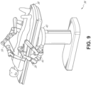
- FIG. 9 illustrates an embodiment of a robotically-enabled table-based system configured for a laparoscopic procedure. As shown in FIG.
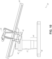
- FIG. 10 illustrates an embodiment of the robotically-enabled medical system with pitch or tilt adjustment.
- the system 36 may accommodate tilt of the table 38 to position one portion of the table at a greater distance from the floor than the other.
- the arm mounts 45 may rotate to match the tilt such that the arms 39 maintain the same planar relationship with table 38.
- the column 37 may also include telescoping portions 60 that allow vertical extension of column 37 to keep the table 38 from touching the floor or colliding with base 46.
- FIG. 11 provides a detailed illustration of the interface between the table 38 and the column 37.
- Pitch rotation mechanism 61 may be configured to alter the pitch angle of the table 38 relative to the column 37 in multiple degrees of freedom.
- the pitch rotation mechanism 61 may be enabled by the positioning of orthogonal axes 1, 2 at the column-table interface, each axis actuated by a separate motor 3, 4 responsive to an electrical pitch angle command. Rotation along one screw 5 would enable tilt adjustments in one axis 1, while rotation along the other screw 6 would enable tilt adjustments along the other axis 2.
- a ball joint can be used to alter the pitch angle of the table 38 relative to the column 37 in multiple degrees of freedom.
- the end effectors of the system's robotic arms comprise (i) an instrument driver (alternatively referred to as “instrument drive mechanism” or “instrument device manipulator”) that incorporate electro-mechanical means for actuating the medical instrument and (ii) a removable or detachable medical instrument which may be devoid of any electro-mechanical components, such as motors.
- instrument driver alternatively referred to as "instrument drive mechanism” or “instrument device manipulator”
- instrument device manipulator a removable or detachable medical instrument which may be devoid of any electro-mechanical components, such as motors.
- This dichotomy may be driven by the need to sterilize medical instruments used in medical procedures, and the inability to adequately sterilize expensive capital equipment due to their intricate mechanical assemblies and sensitive electronics. Accordingly, the medical instruments may be designed to be detached, removed, and interchanged from the instrument driver (and thus the system) for individual sterilization or disposal by the physician or the physician's staff. In contrast, the instrument drivers need not be changed or sterilized, and may be draped for protection.
- the robotic system may incorporate a drive interface, such as a sterile adapter connected to a sterile drape, that sits between the instrument driver and the medical instrument.
- a drive interface such as a sterile adapter connected to a sterile drape
- the chief purpose of the sterile adapter is to transfer angular motion from the drive shafts of the instrument driver to the drive inputs of the instrument while maintaining physical separation, and thus sterility, between the drive shafts and drive inputs.
- an example sterile adapter may comprise of a series of rotational inputs and outputs intended to be mated with the drive shafts of the instrument driver and drive inputs on the instrument.
- the sterile drape comprised of a thin, flexible material such as transparent or translucent plastic, is designed to cover the capital equipment, such as the instrument driver, robotic arm, and cart (in a cart-based system) or table (in a table-based system).
- the capital equipment such as the instrument driver, robotic arm, and cart (in a cart-based system) or table (in a table-based system).
- Use of the drape would allow the capital equipment to be positioned proximate to the patient while still being located in an area not requiring sterilization (i.e., non-sterile field).
- the medical instrument may interface with the patient in an area requiring sterilization (i.e., sterile field).
- the mated drive inputs 73 of instrument base 72 may share axes of rotation with the drive outputs 74 in the instrument driver 75 to allow the transfer of torque from drive outputs 74 to drive inputs 73.
- the drive outputs 74 may comprise splines that are designed to mate with receptacles on the drive inputs 73.
- Torque from the instrument driver 75 is transmitted down the elongated shaft 71 using tendons along the shaft 71.
- These individual tendons such as pull wires, may be individually anchored to individual drive inputs 73 within the instrument handle 72.
- the tendons are directed down one or more pull lumens along the elongated shaft 71 and anchored at the distal portion of the elongated shaft 71, or in the wrist at a distal portion of the elongated shaft.
- these tendons may be coupled to a distally mounted end effector, such as a wrist, grasper, or scissor.
- the tendon may cause a joint to rotate about an axis, thereby causing the end effector to move in one direction or another.
- the tendon may be connected to one or more jaws of a grasper at distal end of the elongated shaft 71, where tension from the tendon cause the grasper to close.
- the tendons may be coupled to a bending or articulating section positioned along the elongated shaft 71 (e.g., at the distal end) via adhesive, control ring, or other mechanical fixation.
- a bending or articulating section positioned along the elongated shaft 71 (e.g., at the distal end) via adhesive, control ring, or other mechanical fixation.
- torque exerted on drive inputs 73 would be transmitted down the tendons, causing the softer, bending section (sometimes referred to as the articulable section or region) to bend or articulate.
- the elongated shaft 71 houses a number of components to assist with the robotic procedure.
- the shaft may comprise of a working channel for deploying surgical tools (or medical instruments), irrigation, and/or aspiration to the operative region at the distal end of the shaft 71.
- the shaft 71 may also accommodate wires and/or optical fibers to transfer signals to/from an optical assembly at the distal tip, which may include of an optical camera.
- the shaft 71 may also accommodate optical fibers to carry light from proximally-located light sources, such as light emitting diodes, to the distal end of the shaft.
- the distal tip may also comprise the opening of a working channel for delivering tools for diagnostic and/or therapy, irrigation, and aspiration to an operative site.
- the distal tip may also include a port for a camera, such as a fiberscope or a digital camera, to capture images of an internal anatomical space.
- the distal tip may also include ports for light sources for illuminating the anatomical space when using the camera.
- Power and controls signals may be communicated from the non-rotational portion 84 of the instrument driver 80 to the rotational assembly 83 through electrical contacts may be maintained through rotation by a brushed slip ring connection (not shown).
- the rotational assembly 83 may be responsive to a separate drive unit that is integrated into the non-rotatable portion 84, and thus not in parallel to the other drive units.
- the rotational mechanism 83 allows the instrument driver 80 to rotate the drive units, and their respective drive outputs 81, as a single unit around an instrument driver axis 85.
- the robotic systems contemplated by this disclosure can provide for non-radiation-based navigational and localization means to reduce physician exposure to radiation and reduce the amount of equipment within the operating room.
- the term "localization” may refer to determining and/or monitoring the position of objects in a reference coordinate system. Technologies such as pre-operative mapping, computer vision, real-time EM tracking, and robot command data may be used individually or in combination to achieve a radiation-free operating environment. In other cases, where radiation-based imaging modalities are still used, the pre-operative mapping, computer vision, real-time EM tracking, and robot command data may be used individually or in combination to improve upon the information obtained solely through radiation-based imaging modalities.
- Pre-operative mapping may be accomplished through the use of the collection of low dose CT scans.
- Pre-operative CT scans are reconstructed into three-dimensional images, which are visualized, e.g., as "slices" of a cutaway view of the patient's internal anatomy.
- image-based models for anatomical cavities, spaces and structures of the patient's anatomy, such as a patient lung network may be generated.
- Techniques such as center-line geometry may be determined and approximated from the CT images to develop a three-dimensional volume of the patient's anatomy, referred to as model data 91 (also referred to as "preoperative model data" when generated using only preoperative CT scans).
- model data 91 also referred to as "preoperative model data" when generated using only preoperative CT scans.
- Network topological models may also be derived from the CT-images, and are particularly appropriate for bronchoscopy.
- Some features of the localization module 95 may identify circular geometries in the preoperative model data 91 that correspond to anatomical lumens and track the change of those geometries to determine which anatomical lumen was selected, as well as the relative rotational and/or translational motion of the camera. Use of a topological map may further enhance vision-based algorithms or techniques.
- the magnetic field induces small currents in the sensor coils of the EM sensor, which may be analyzed to determine the distance and angle between the EM sensor and the EM field generator.
- These distances and orientations may be intra-operatively "registered" to the patient anatomy (e.g., the preoperative model) in order to determine the geometric transformation that aligns a single location in the coordinate system with a position in the pre-operative model of the patient's anatomy.
- an embedded EM tracker in one or more positions of the medical instrument e.g., the distal tip of an endoscope
- Robotic command and kinematics data 94 may also be used by the localization module 95 to provide localization data 96 for the robotic system.
- Device pitch and yaw resulting from articulation commands may be determined during pre-operative calibration. Intra-operatively, these calibration measurements may be used in combination with known insertion depth information to estimate the position of the instrument. Alternatively, these calculations may be analyzed in combination with EM, vision, and/or topological modeling to estimate the position of the medical instrument within the network.
- the localization module 95 may use the input data 91-94 in combination(s). In some cases, such a combination may use a probabilistic approach where the localization module 95 assigns a confidence weight to the location determined from each of the input data 91-94. Thus, where the EM data may not be reliable (as may be the case where there is EM interference) the confidence of the location determined by the EM data 93 can be decrease and the localization module 95 may rely more heavily on the vision data 92 and/or the robotic command and kinematics data 94.
- the robotic systems discussed herein may be designed to incorporate a combination of one or more of the technologies above.
- the robotic system's computer-based control system based in the tower, bed and/or cart, may store computer program instructions, for example, within a non-transitory computer-readable storage medium such as a persistent magnetic storage drive, solid state drive, or the like, that, upon execution, cause the system to receive and analyze sensor data and user commands, generate control signals throughout the system, and display the navigational and localization data, such as the position of the instrument within the global coordinate system, anatomical map, etc.
- Navigation path tracing can be used to generate and display visual indicia indicative of a historical path (e.g., breadcrumbs) of a medical instrument as the instrument is navigated through a luminal network.
- a historical path e.g., breadcrumbs
- the position of the instrument can be determined and visual indicia indicative of the position of the instrument can be displayed (e.g., plotted or otherwise displayed).
- the visual indicia can aid a user in visualizing which portions of the luminal network have already been explored or navigated by the instrument.
- the visual indicia can also be used to visualize the shape of the luminal network and/or extend a preoperative model of the luminal network.
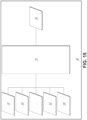
- FIG. 16 illustrates an example luminal network 130 of a patient that can be navigated using the navigation path tracing methods and systems described herein.
- the luminal network 130 is a bronchial network of airways inside a patient's lung.
- the luminal network 130 comprises a plurality of lumens 132 that are arranged in a branched structure.
- the illustrated luminal network 130 comprises a plurality of branched lumens 132, in some instances, the luminal network 13 may comprise only a single lumen 132. That is, in some instances, a luminal network 130 need not comprise a branched arrangement of lumens 132.
- FIG. 16 represents the luminal network 130 as a two-dimensional structure. This should not be construed to limit the present disclosure to two-dimensional luminal networks in any way. In general, the luminal network 130 comprises a three-dimensional structure.
- luminal network 130 can include, for example, bronchial networks, renal networks, cardiovascular networks (e.g., arteries and veins), gastrointestinal tracts, urinary tracts, etc.
- the navigation path tracing methods and systems described herein can be implemented during navigation of both branched and non-branched luminal networks 130.
- the preoperative model 150 is representative of one or more portions of the luminal network 130 that is being navigated by the medical instrument.
- the preoperative model 150 may be generated prior to navigation of the luminal network using one or more of various preoperative imaging and mapping techniques.
- preoperative mapping may be accomplished through the use of a collection of low dose CT scans.
- preoperative CT scans can generate two-dimensional images, each representing a "slice" of a cutaway view of the patient's internal anatomy.
- image-based preoperative models for anatomical cavities, spaces, and structures of the patient's anatomy such as a patient lung network (i.e., a luminal network)
- Other methods for generating the preoperative model 150 are also possible.
- the preoperative model 150 comprises a plurality of segments 152.
- the segments 152 of the preoperative model 150 correspond with at least a portion of the lumens 132 of the luminal network 130.
- the preoperative model 150 can comprise a corresponding branched arrangement of segments 152.
- the preoperative model 150 can comprise a corresponding single branch 152.
- the preoperative model 150 comprises a three-dimensional shape, corresponding to at least a portion of the three-dimensional shape of the luminal network 130.
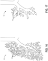
- FIG. 17 illustrates the preoperative model 150 as a two-dimensional shape for ease of illustration. In some instances, a cross-section of a three-dimensional preoperative model 150 may be displayed on a two-dimensional display.
- the preoperative model 150 may represent or correspond to only a portion of the luminal network 130. This is further illustrated in FIG. 18 , which is a view of the preoperative model 150 overlaid on the luminal network 130.
- limitations in the preoperative imaging and mapping techniques used to generate the preoperative model 150 may prevent generation of a model that corresponds to the entire luminal network 130. For example, certain branched lumens 132 within the luminal network may be sufficiently small that they cannot be clearly depicted and analyzed with common preoperative imaging and mapping techniques. As such, the preoperative model 150 may not provide a complete representation of the luminal network 130, for example, leaving various portions of the luminal network 130 unmapped and/or unrepresented in the preoperative model 150.
- the endoscope 315 has a first diameter and thus its distal end may not be able to be positioned through the smaller-diameter airways around the nodule 355. Accordingly, a steerable catheter 345 can extend from a working channel of the endoscope 315 the remaining distance to the nodule 355.
- the steerable catheter 345 may have a lumen through which instruments, such as biopsy needles, cytology brushes, tissue sampling forceps, etc., can be passed to the target tissue site of nodule 355.
- both the distal end of the endoscope 315 and the distal end of the steerable catheter 345 can be provided with EM instrument sensors (or other position sensors) for tracking their position within the airways 350.
- the overall diameter of the endoscope 315 may be small enough to reach the periphery without the steerable catheter 345, or may be small enough to get close to the periphery (e.g., within 2.5-3 cm) to deploy medical instruments through a non-steerable catheter.
- the medical instruments deployed through the endoscope 315 may be equipped with EM instrument sensors (or other position sensors).
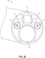
- An EM spatial measurement system may determine the location of objects within the EM field that are embedded or provided with EM sensor coils, for example, the EM coils 405 (as shown in FIG. 20 ).
- EM sensor When an EM sensor is placed inside a controlled, varying EM field as described herein, voltages are induced in sensor coil(s) included in the EM sensor. These induced voltages can be used by the EM spatial measurement system to calculate the position and orientation of the EM sensor and thus the object having the EM sensor.
- the EM fields are of a low field strength and can safely pass through human tissue, location measurement of an object is possible without the line-of-sight constraints of an optical spatial measurement system.
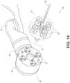
- the instrument 400 may also include illumination sources 410.
- the illumination sources 410 provide light to illuminate a portion of an anatomical space.
- the illumination sources can each be one or more light-emitting devices configured to emit light at a selected wavelength or range of wavelengths.
- the wavelengths can be any suitable wavelength, for example, visible spectrum light, infrared light, x-ray (e.g., for fluoroscopy), to name a few examples.
- illumination sources 410 can include light-emitting diodes (LEDs) located at the distal end of the instrument 400.
- LEDs light-emitting diodes
- illumination sources 410 can include one or more fiber optic fibers extending through a length of the endoscope to transmit light through the distal end from a remote light source, for example, an x-ray generator. Where the distal end includes multiple illumination sources 410 these can each be configured to emit the same or different wavelengths of light as one another.
- the imaging device 415 can include any photosensitive substrate or structure configured to convert energy representing received light into electric signals, for example, a charge-coupled device (CCD) or complementary metal-oxide semiconductor (CMOS) image sensor.
- CCD charge-coupled device
- CMOS complementary metal-oxide semiconductor
- Some examples of imaging device 415 can include one or more optical fibers, for example, a fiber optic bundle, configured to transmit light representing an image from the distal end 400 of the endoscope to an eyepiece and/or image sensor near the proximal end of the endoscope.
- Imaging device 415 can additionally include one or more lenses and/or wavelength pass or cutoff filters as required for various optical designs.
- the light emitted from the illumination sources 410 allows the imaging device 415 to capture images of the interior of a patient's luminal network. These images can then be transmitted as individual frames or series of successive frames (e.g., a video) to a computer system such as command console 200.
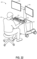
- FIG. 22 illustrates an example command console 200 that can be used with some implementations of the robotic systems described herein.
- the command console 200 includes a console base 201, displays 202 (e.g., monitors), and one or more control modules (e.g., keyboard 203 and joystick 204).
- a user 205 e.g., a physician
- the medical robotic system e.g., the systems described with reference to FIGs. 1-15
- the user 205 can use the command console 200 to navigate an instrument within a luminal network of a patient.
- the command console 200 may also display information to the user 205 that can be used to aid in navigation of the luminal network.
- the method 100 (or a system implanting the method 100) is configured to provide (e.g., plot or otherwise display) visual indicia (see, for example, FIGS. 25-27 ) indicative of positions (e.g., historical and/or current positions) of a medical instrument positioned within a luminal network (such as the luminal network 130 of FIG. 16 ).
- the visual indicia represent a path traveled by the medical instrument as it navigates the luminal network.
- the visual indicia are displayed to user, via a user display (such as display 202 of FIG. 22 ).
- the displayed visual indicia provide a user with a visualization of one or more portions of the luminal network navigated by medical instrument.
- the visual indicia can also be used to extend a preoperative model of the luminal network.
- the method 100 can be implemented for navigation of a wide variety of luminal networks, included branched luminal networks (such as bronchial networks, renal networks, cardiovascular networks (e.g., arteries and veins), etc.) and non-branched (e.g., single lumen) luminal networks (such as gastrointestinal tracts, urinary tracts, etc.).
- branched luminal networks such as bronchial networks, renal networks, cardiovascular networks (e.g., arteries and veins), etc.
- non-branched luminal networks such as gastrointestinal tracts, urinary tracts, etc.
- the method 100 displays a preoperative model (e.g., preoperative model 150), which may involve, e.g., displaying, on a user interface, a preoperative model corresponding to a mapped portion of a luminal network.
- a preoperative model e.g., preoperative model 150
- the preoperative model 150 may be generated preoperatively (prior to the current procedures), using various imaging and mapping techniques.
- the preoperative model 150 can comprise a 3D model.
- the preoperative model 150 can be retrieved from a memory, the preoperative model 150 can be stored in a memory, for example, as preoperative model data 91 described with reference to FIG. 15 .
- the preoperative model 150 can be representative of or correspond to at least a portion of the luminal network 130.
- the method 100 moves to block 105.
- the method 100 determines the position of the medical instrument relative to the preoperative model 1 50, which may involve, e.g., determining a position of a distal end of an instrument within the luminal network relative to the mapped portion of the luminal network.
- position is determined or estimated using the navigation or localization system 90 described above and shown in FIG. 15 .
- the localization system 90 may use various technologies or modalities, such as pre-operative mapping, computer vision, real-time EM tracking, and robot command data either individually or in combination to determine or estimate the position of the instrument.
- radiation-based imaging modalities e.g., fluoroscopy
- the radiation-based imaging modalities may be supplemented by pre-operative mapping, computer vision, real-time EM tracking, and/or robot command data.
- the localization system 90 may include a localization module 95 that processes multiple types of input data 91-94 from various modalities to generate location data 96 for a medical instrument (for example, location date 96 may indicate a position of a distal tip of the instrument).
- the various types of input data can include, e.g., pre-operative model data 91 (e.g., preoperative model 150), vision data 92, EM data 93 (or other position sensor data), shape sensing data, and/or robotic command and kinematics data 94 as described above.
- localization system 90 may use a probabilistic approach that assigns a confidence weight to the location determined from each of the input data 91-94. Thus, where one type of data may not be reliable (for various reason) the confidence of the location determined by that type of data can be decrease and the localization module 95 may rely more heavily on another type of data in determining the position of the instrument.
- the localization system 90 provides location data 96 indicative of the position of the instrument.
- the location data 96 can be presented in a coordinate frame that has been registered to the preoperative model 150, such that the position of the instrument relative to the preoperative model 150 can be determined.
- the EM data 93 (or other position sensor data) can be registered to the preoperative model 150 as described above, such that the position determined by the EM data can be presented within the coordinate frame of the preoperative model 150.
- one or more of the various other modalities e.g., the vision data 92 and/or the robotic command and kinematics data 94
- the location data 96 can be displayed to the user 205 on one of the displays 202.
- the robotic command and kinematics data 94 can be used to determine a position estimate for the instrument only when the instrument is positioned within the preoperative model 150. This may be because, in some embodiments, determining positon based on the robotic command and kinematics data 94 also relies partially on the preoperative model data 91. For example, robotic command and kinematics data 94 may be in conjunction with preoperative model data 91 to determine position. Outside the preoperative model 150, robotic command and kinematics data 94 may provide an unreliable estimate of position in some embodiments.
- certain of the localization modalities may be utilized regardless of whether the instrument is positioned within a portion of the luminal network 130 that is mapped by the preoperative model 150.
- EM data 93 (or other position sensor data) may be used to determine position regardless of whether the instrument is positioned within a portion of the luminal network 130 that is mapped by the preoperative model 150. This may be because, apart from being registered to the coordinate frame of the preoperative model 150, the location determination of EM data 93 (or other position sensor data) may be independent of the preoperative model data 91.
- vision data 92 and robot and robotic command and kinematics data 94 may provide a navigation modality that is not dependent on whether the instrument is positioned within the preoperative model 150.
- a vision algorithm or module may analyze images received from the imaging device 415 on the instrument to detect one openings to lumens.
- a robotic command and kinematics algorithm or module can analyze movement of the instrument through the lumen to estimate travel length of the instrument. These modalities can be combined to develop a position estimate that is not based on the preoperative model 150.
- vision data 92 and robot and robotic command and kinematics data 94 is combined to develop an artificial or two-dimensional estimate of the instrument position.
- other modalities are used to determine position (within a coordinate frame that has been registered to the preoperative model) without further basis on the preoperative model 150 and/or preoperative model data 91.
- These can include fluoroscopy, shape sensing fibers, position and/or motion sensors, etc.
- the method 100 determines whether the instrument is positioned inside or outside a portion of the luminal network 130 that is represented by the preoperative model 150. In some embodiments, if the instrument is positioned within a portion of the luminal network 130 that is represented by the preoperative model 150, the location is determined relative to the preoperative model 150 (e.g., in a coordinate frame that has been mapped to the preoperative model 150) using modalities that are dependent on preoperative model data 91 and/or modalities that are not dependent on preoperative model data 91.
- the location is determined relative to the preoperative model 150 (e.g., in a coordinate frame that has been mapped to the preoperative model 150) using modalities that are not dependent on preoperative model data 91.
- modalities that are not dependent on preoperative model data 91 include EM data 93 and/or other position sensor data that has been registered to the coordinate frame of the preoperative model 150.
- the method 100 moves to decision state 107.
- decision state 107 the method 100 determines whether to enter or remain in path tracing mode, which may involve, e.g., determining when the distal end of the instrument has been advanced past the mapped portion of the luminal network into an unmapped portion of the luminal network and entering a path tracing mode.
- path tracing mode visual indicia are displayed to indicate a historical path of the instrument through the luminal network 130.
- the determination of whether to enter or remain in path tracing mode is made based at least in part on the position of the instrument relative to the preoperative model 150 determined at block 105.
- the method 100 may enter or remain in path tracing mode when the instrument is positioned outside of the preoperative model 150.
- the method 100 triggers path tracing mode when the instrument is outside of the mapped portion 155 of the luminal network 130 as represented by the preoperative model 150.
- the determination of decision state 107 is made based on proximity to an end of the preoperative model 150 or an end of a segment of the preoperative model 150.
- the method 100 may enter path tracing mode when the instrument is positioned within 0.1 mm, 0.25 mm, 0.5 mm, 0.75 mm, 1 cm, 1.5 cm, 2 cm, 2.5 cm, 3 cm, 4 cm, 5 cm, 7.5 cm, or 10 cm of an end of the preoperative model 150 or an end of a segment of the preoperative model 150.
- the determination of decision state 107 can be made based on a direction of travel of the instrument within the luminal network 130.
- path tracing mode is activated when the instrument is advanced into the luminal network 130, retracted backwards in the luminal network 130, or both.
- the determination of decision state 107 can be made based on a distance traveled by the instrument within the luminal network 130.
- path tracing mode is activated when the instrument has traveled at least 0.1 mm, 0.25 mm, 0.5 mm, 0.75 mm, 1 cm, 1.5 cm, 2 cm, 2.5 cm, 3 cm, 4 cm, 5 cm, 7.5 cm, or 10 cm. Other distances can also be used.
- the determination of decision state 107 can be made based on time elapsed.
- path tracing mode can be activated every 0.1 seconds, 0.25 seconds, 0.5 seconds, 0.75 seconds, 1 second, 1.5 seconds, 2 seconds, 2.5 seconds, 3 seconds, 4 seconds, 5 seconds, 7.5 seconds, or 10 seconds. Other periods of time can also be used.
- the determination of whether to enter or remain in path tracing mode is made based at least in part on user input. For example, in some instances, a user may activate or deactivate path tracing mode as desired. In some embodiments, this is accomplished by entering a user command at the command console 200.
- the method 100 moves to block 109, at which visual indicia are generated and/or displayed, which may involve, e.g. displaying visual indicia of a path of the distal end of the instrument in the unmapped portion of the luminal network relative to the preoperative model of the mapped portion of the luminal network.
- the visual indicia are indicators of the historical position of the instrument as it travels through the luminal network 130.
- the visual indicia can create a trail (e.g., breadcrumbs or a historical path) that represents the path of travel of the instrument through the luminal network 130.
- the visual indicia are displayed on display 202.
- the visual indicia can comprise many types of indicators as described with reference to FIGS. 25-27 below.
- the method 100 moves to decision state 111.
- decision state 111 the method determines whether to end (for example, if a procedure is finished) or to continue (for example, if a procedure will continue). If decision state 111 determines that the method 100 should end, the method ends at block 113. If decision state 111 determines that the method 100 will continue the method moves to block 115.
- the method 100 includes two possible loops for each new position after the instrument is moved within the luminal network. If decision state 107 determines that path tracing mode is active, the loop generates and displays a new visual indicia for the new position. If the decision state 107 determines that path tracing mode is inactive, no new visual indicia is generated or displayed.
- the criteria used by decision state 107 can be varied to determine when to display visual indicia. As noted above, these criteria can include whether the instrument is inside or outside the preoperative model, distance traveled by the instrument (e.g., since the previous visual indicia), direction of travel, time elapsed between previous visual indicia, etc. One of skill in the art will appreciate that these criteria can be varied to determine the frequency with which visual indicia are generated as well as the distance between successive visual indicia.
- the path tracing method 100 thus can generate and display visual indicia indicative of historical positions of the instrument as the instrument travels through a luminal network. As visual indicia are generated and displayed, lumens traveled by the instrument can be visualized. Data regarding the lumens traveled can be associated with visual indicia and displayed to the user. The data and visual indicia can also be stored for future use. In some instances, tube-like structures can be fitted around the visual indicia to extend the preoperative model into portions of the luminal network that were previously unmapped by the preoperative model. The path tracing method 100 (or system implementing the same) can thus advantageously aid a user in navigation and visualizing the luminal network.
- FIG. 23B is a flowchart illustrating an example subroutine for, in some embodiments, performing step 105 of the method 100 of FIG. 23A .
- the subroutine of FIG. 23B need not be included in all implementations of method 100.
- the subroutine provides a method of determining the position of the instrument within the luminal network 130.
- the subroutine begins at block 117.
- the subroutine determines whether the instrument is positioned within the preoperative model 150. For example, after registering the location data 96 to a coordinate frame of the preoperative model, the location data 96 can be compared to the preoperative model 150 to determine whether the instrument is within the mapped portion 155 or the unmapped portion 135 of the luminal network 130 (see FIG. 18 ).
- the subroutine moves to block 121.
- the subroutine returns a position (e.g., position data 96) that is based in part on the preoperative model 150 and/or preoperative model data 91. That is, when the instrument is positioned within the preoperative model 150, the subroutine can return position data that is based at least in part on navigation modalities that make use of the preoperative model 150 and/or the preoperative model data.
- the subroutine then ends at block 123.
- the subroutine moves to block 125.
- the subroutine returns a position that, apart from being registered to the coordinate frame of the preoperative model 150, is not based on the preoperative model 150 and/or preoperative model data 91. That is, when the instrument is positioned outside the preoperative model 150, the subroutine can return position data that is based on navigation modalities that do not make use of the preoperative model 150 and/or the preoperative model data (apart from having their output registered to the coordinate frame of the preoperative model 150). This may be because the instrument may be positioned outside of the preoperative model and data from navigation modalities that rely on the preoperative model 150 or preoperative model date 91 may be unavailable.
- the subroutine then ends at block 127.
- the subroutine returns a position based on various navigation modalities depending on whether the instrument is within the preoperative model 150.
- a greater number of navigation modalities may be available to the localization system 90 than when the instrument is outside of the preoperative model 150.
- EM data 93 may provide a navigation modality that is not dependent on whether the instrument is positioned within the preoperative model 150. That is, apart from being registered to the coordinate frame of the preoperative model 150, the EM data 93 (or other position sensor data) may provide a location determination that is not based on the preoperative model 150. Thus, EM data 93 may be used to return position without basis on the preoperative model at block 125 of the subroutine.
- FIG. 24 is a flowchart illustrating an example process, algorithm, or method 140 of determining a navigation path of an instrument within a luminal network.
- the method 140 of FIG. 24 can be implemented in certain robotic systems, such as the robotic systems illustrated in FIGs. 1-15 and others.
- the method 140 can be implemented in or by a navigation system, such as the navigation or localization system 90 of FIG. 15 .
- the method 140 beings at block 141.
- the method 100 displays, on a user interface, a preoperative model corresponding to a mapped portion of a luminal network.
- the preoperative model 150 may be generated preoperatively (prior to the current procedures), using various imaging and mapping techniques. Numerous aspects described above with reference to the method 100 of FIG. 23A are applicable to the method 140 of FIG. 24 , and will not be repeated for the sake of brevity.
- the method 140 moves the instrument within the luminal network. This may be accomplished by or advancing or retracting the instrument within the luminal network 130. This may be accomplished by articulating the instrument.
- the method 140 determines when the distal end of the instrument has been advanced past the mapped portion of the luminal network into an unmapped portion of the luminal network and enters a path tracing mode.
- path tracing mode visual indicia are displayed to indicate a historical path of the instrument through the luminal network 130.
- the method 140 when in the path tracing mode, displays visual indicia of a path of the distal end of the instrument in the unmapped portion of the luminal network relative to the preoperative model of the mapped portion of the luminal network.
- the visual indicia can comprise many types of indicators as described with reference to FIGS. 25-27 below.
- the method 140 ends at block 147.
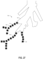
- FIG. 25 provides an example output of a navigation path tracing system or method illustrating visual indicia 170.
- the visual indicia 170 are illustrated as circles, but this need not be the case in all embodiments.
- the visual indicia 170 can be any suitable shape or marker, such as dots, dashes, X's, other shapes, etc.
- the visual indicia 170 can reveal the shape of portions of the luminal network 130 that are not represented by the preoperative model 150. That is, as the instrument is moved through the luminal network 130 and visual indicia 170 are plotted or displayed (for example, when in path tracing mode), the visual indicia 170 can provide an indication of the shape or structure of the luminal network 130.
- FIG. 26 provides another example output of a navigation path tracing system or method, illustrating several different types of visual indicia.
- the visual indicia can vary to provide various types of information to a user.
- visual indicia 170 are illustrated as darkened circles.
- Visual indicia 170 can represent a position of the instrument within a lumen of the luminal network.
- Visual indicia 172, 174 are illustrated as triangles.
- Visual indicia 172, 174 can represent positions of the instrument within the luminal network 130 at which branches are present.
- Visual indicia 172 is illustrated as a darkened triangle, which can signify that the instrument has traveled down all available branches at that location.
- Visual indicia 174 is illustrated as an undarkened triangle, which can signify that a branch from that location has not yet been explored by the instrument.
- Visual indicia 176, 178 are illustrated as undarkened circles. In this example, undarkened circles can represent the farthest points within lumens that the instrument has explored.
- Visual indicia 176 is illustrated as an undarkened circle with a solid outline. This can represent a position an end of a lumen, or a point at which the instrument cannot proceed further into the lumen because of, for example, the relative size of the instrument and the lumen.
- Visual indicia 178 is illustrated as an undarkened circle with a dashed outline. This can represent that the lumen continues and has not yet been explored by the instrument.
- Visual indicia 171 are illustrated as gray circles. In the illustrated example, visual indicia 171 illustrate historical positions of the instrument within the preoperative model.
- data 180 can be associated with the visual indicia.
- a user e.g., a physician
- the user may enter the data 180 via the command console 200 and the data 180 can be viewed via the displays 202.
- the method 100 can associate various other types of data with the visual indicia.
- vision data e.g., an image of the lumen at the location represented by the visual indicia
- Various information can be derived from the vision data such as whether branches are present, diameter/size of the lumen, etc.
- FIG. 27 provides another example output of a navigation path tracing system or method and illustrates that the navigation path tracing system can be used to extend the preoperative model 150.
- visual indicia 170 can be grouped to extend the preoperative model 150.
- path tracing mode can identify strings of visual indicia 170 as corresponding to a lumen and fit a tube-like structure 190 to the string of visual indicia to extend the preoperative model 190.
- the diameter of the tube-like structure 190 can be determined using vision data of the interior of the lumen or other methods.
- the tube-like structures 190 extend the preoperative model 150 into portions of the luminal network 130 that were previously unmapped by the preoperative model 150.
- the extending preoperative model can be saved, for example, in a computer-readable memory, for use during future procedures.
- Implementations disclosed herein provide systems, methods and apparatuses for navigation path tracing. Various implementations described herein provide for improved navigation of luminal networks.
- the term "approximately” refers to a range of measurements of a length, thickness, a quantity, time period, or other measurable value. Such range of measurements encompasses variations of +/-10% or less, preferably +/-5% or less, more preferably +/-1% or less, and still more preferably +/-0.1% or less, of and from the specified value, in so far as such variations are appropriate in order to function in the disclosed devices, systems, and techniques.
Landscapes
- Health & Medical Sciences (AREA)
- Engineering & Computer Science (AREA)
- Surgery (AREA)
- Life Sciences & Earth Sciences (AREA)
- Nuclear Medicine, Radiotherapy & Molecular Imaging (AREA)
- Robotics (AREA)
- Animal Behavior & Ethology (AREA)
- Veterinary Medicine (AREA)
- Medical Informatics (AREA)
- Molecular Biology (AREA)
- Biomedical Technology (AREA)
- General Health & Medical Sciences (AREA)
- Public Health (AREA)
- Heart & Thoracic Surgery (AREA)
- Gynecology & Obstetrics (AREA)
- Radiology & Medical Imaging (AREA)
- Oral & Maxillofacial Surgery (AREA)
- Pathology (AREA)
- Human Computer Interaction (AREA)
- Endoscopes (AREA)
Claims (14)
- Nicht-transitorisches computerlesbares Datenspeichermedium mit darauf gespeicherten Anweisungen, die, wenn sie ausgeführt werden, einen Prozessor eines Navigationssystems zum Navigieren eines Instruments durch ein luminales Netzwerk (130) eines Patienten wenigstens hierzu veranlassen:Anzeigen (142), auf einer Benutzeranzeige (202), eines präoperativen Modells (150), das einem abgebildeten Abschnitt eines luminales Netzwerks eines Patienten entspricht;Bestimmen einer Position eines distalen Endes eines Instruments, das innerhalb des luminalen Netzwerks relativ zu dem abgebildeten Abschnitt des präoperativen Modells positioniert ist;Bestimmen, dass sich die Position des distalen Endes des Instruments in einem vorbestimmten Abstand zu einem Grenzbereich befindet, welcher einem Übergang von einem Ende des abgebildeten Abschnitts des präoperativen Modells zu einem Abschnitt (135) des luminalen Netzwerks entspricht, der in dem präoperativen Modell nicht abgebildet ist, bevor das distale Ende des Instruments den Grenzbereich erreicht;Eintreten (145) in einen Pfadverfolgungsmodus in Reaktion auf das Bestimmen, dass sich die Position des distalen Endes des Instruments in dem vorbestimmten Abstand zum Grenzbereich befindet, bevor das distale Ende des Instruments den Grenzbereich erreicht; undim Pfadverfolgungsmodus, Anzeigen(146), auf der Benutzeranzeige, von visuellen Hinweisen, die einen Pfad des distalen Endes des Instruments im Hinblick auf das angezeigte präoperative Modell angeben, während das Instrument durch das luminale Netzwerk navigiert wird.
- Nicht-transitorisches computerlesbares Datenspeichermedium nach Anspruch 1, wobei die Anweisungen dafür ausgelegt sind, den Prozessor des Navigationssystems zu veranlassen, in den Pfadverfolgungsmodus zu wechseln, wenn sich die Position des distalen Endes des Instruments innerhalb von 25 %, 20 %, 15 %, 10 % oder 5% eines Endes von einem letzten Segment des präoperativen Modells befindet, bevor das Instrument über das präoperative Modell hinaus bewegt wird, wobei das präoperative Modell mehrere Segmente (152) umfasst, wobei jedes Segment einem Abschnitt des luminalen Netzwerks entspricht.
- Nicht-transitorisches computerlesbares Datenspeichermedium nach einem der Ansprüche 1 bis 2, wobei die visuellen Hinweise historische Positionen des distalen Endes des Instruments innerhalb des luminalen Netzwerks angeben.
- Nicht-transitorisches computerlesbares Datenspeichermedium nach einem der Ansprüche 1 bis 3, wobei, im Pfadverfolgungsmodus, die Anweisungen dafür ausgelegt sind, den Prozessor des Navigationssystems zu veranlassen, eine Frequenz, mit der visuelle Hinweise generiert werden, basierend auf einem Abstand, der von dem Instrument zwischen aufeinanderfolgenden visuellen Hinweisen durchlaufen wird, oder basierend auf einer Zeit, die zwischen aufeinanderfolgenden visuellen Hinweisen verstreicht, anzupassen.
- Nicht-transitorisches computerlesbares Datenspeichermedium nach einem der Ansprüche 1 bis 4, wobei die Anweisungen dafür ausgelegt sind, den Prozessor des Navigationssystems zu veranlassen, die Position des distalen Endes des Instruments basierend auf Daten von mehreren Navigationsmodalitäten zu bestimmen, wenn das Instrument innerhalb eines abgebildeten Abschnitts des präoperativen Modells positioniert ist, wobei die Daten von den mehreren Navigationsmodalitäten vorzugsweise mehrere präoperative Modelldaten, Visionsdaten, Positionssensordaten und Robotikbefehl- und Kinematikdaten umfassen.
- Nicht-transitorisches computerlesbares Datenspeichermedium nach Anspruch 5, wobei die Anweisungen dafür ausgelegt sind, den Prozessor des Navigationssystems zu veranlassen, die Position des distalen Endes des Instruments basierend auf Daten von weniger als den mehreren Navigationsmodalitäten zu bestimmen, wenn das Instrument außerhalb des abgebildeten Abschnitts des präoperativen Modells positioniert ist.
- Nicht-transitorisches computerlesbares Datenspeichermedium nach einem der Ansprüche 1 bis 6, wobei die Anweisungen dafür ausgelegt sind, den Prozessor des Navigationssystems zu veranlassen, die Position des distalen Endes des Instruments basierend auf EM-Daten, die von einem EM-Sensor empfangen werden, oder basierend auf einer Kombination von Visionsdaten und Robotikbefehl- und Kinematikdaten zu bestimmen, wenn das Instrument außerhalb des abgebildeten Abschnitts des präoperativen Modells positioniert ist.
- Nicht-transitorisches computerlesbares Datenspeichermedium nach einem der Ansprüche 1 bis 7, wobei die Anweisungen dafür ausgelegt sind, den Prozessor des Navigationssystems zu veranlassen: Visionsdaten mit den visuellen Hinweisen anzuzeigen, wobei die Visionsdaten vorzugsweise ein Bild umfassen, das von einer Bildgebungsvorrichtung am distalen Ende des Instruments empfangen wird; oder Robotikbefehl- und Kinematikdaten mit den visuellen Hinweisen anzuzeigen.
- Nicht-transitorisches computerlesbares Datenspeichermedium nach einem der Ansprüche 1 bis 8, wobei die Anweisungen dafür ausgelegt sind, den Prozessor des Navigationssystems hierzu zu veranlassen:Empfangen von Benutzereingabedaten von einer Benutzereingabe; undAnzeigen der Benutzereingabedaten mit den visuellen Hinweisen;wobei die Benutzereingabedaten vorzugsweise eines odermehrere hiervon umfassen:eine Angabe zu einem durchlaufenen Lumen;eine Angabe zu einem nicht durchlaufenen Lumen;eine Angabe zu einem Ende eines Lumens;eine Angabe zu einer Öffnung eines Lumens;eine Angabe, dass sich ein aktuelles Lumen über eine aktuelle Position des Instruments hinaus erstreckt; undeinen Lumendurchmesser.
- Nicht-transitorisches computerlesbares Datenspeichermedium nach einem der Ansprüche 1 bis 9, wobei die Anweisungen dafür ausgelegt sind, den Prozessor des Navigationssystems zu veranlassen, geometrische Strukturen an die visuellen Hinweise anzupassen, um eine Visualisierung eines Lumens des luminalen Netzwerks außerhalb des abgebildeten Abschnitts des luminalen Netzwerks bereitzustellen.
- Robotiksystem zum Navigieren eines luminalen Netzwerks, wobei das System umfasst:ein Instrument (70), vorzugsweise ein Endoskop, mit einem länglichen Körper (71) und einem Sensor, der an einem distalen Ende des länglichen Körpers angeordnet ist;wenigstens einen computerlesbaren Speicher, auf dem ausführbare Anweisungen gespeichert sind; undeinen oder mehrere Prozessoren, die in Kommunikationsverbindung mit dem wenigstens einen computerlesbaren Speicher stehen und dafür ausgelegt sind, die Anweisungen nach einem der Ansprüche 1 bis 10 auszuführen.
- System nach Anspruch 11, wobei die ein oder mehreren Prozessoren dafür ausgelegt sind, die Anweisungen auszuführen, um das System zu veranlassen, einen Koordinatenrahmen des Sensors und einen Koordinatenrahmen des präoperativen Modells zu registrieren.
- System nach Anspruch 11 oder 12, wobei der Sensor ein Formmessfaser ist; oder wobei das System ferner einen Feldgenerator umfasst, der dafür ausgelegt ist, ein EM-Feld zu generieren, wobei der Sensor ein EM-Sensor ist und die ein oder mehreren Prozessoren dafür ausgelegt sind, die Anweisungen auszuführen, um das System zu veranlassen, eine Position des EM-Sensors innerhalb es EM-Feldes zu bestimmen.
- System nach einem der Ansprüche 11 bis 13, ferner umfassend eine Instrumentenpositionierungsvorrichtung, wobei das Instrument an der Instrumentenpositionierungsvorrichtung angebracht ist, wobei die Instrumentenpositionierungsvorrichtung vorzugsweise einen Roboterarm (76) umfasst.
Applications Claiming Priority (3)
| Application Number | Priority Date | Filing Date | Title |
|---|---|---|---|
| US201762572285P | 2017-10-13 | 2017-10-13 | |
| US16/143,362 US11058493B2 (en) | 2017-10-13 | 2018-09-26 | Robotic system configured for navigation path tracing |
| PCT/US2018/053206 WO2019074682A1 (en) | 2017-10-13 | 2018-09-27 | ROBOTIC SYSTEM DESIGNED TO FOLLOW A NAVIGATION PATH |
Publications (3)
| Publication Number | Publication Date |
|---|---|
| EP3675765A1 EP3675765A1 (de) | 2020-07-08 |
| EP3675765A4 EP3675765A4 (de) | 2021-06-02 |
| EP3675765B1 true EP3675765B1 (de) | 2024-07-24 |
Family
ID=66097620
Family Applications (1)
| Application Number | Title | Priority Date | Filing Date |
|---|---|---|---|
| EP18866272.0A Active EP3675765B1 (de) | 2017-10-13 | 2018-09-27 | Zur navigationswegverfolgung konfiguriertes robotisches system |
Country Status (7)
| Country | Link |
|---|---|
| US (2) | US11058493B2 (de) |
| EP (1) | EP3675765B1 (de) |
| JP (1) | JP7098723B2 (de) |
| KR (1) | KR102656307B1 (de) |
| CN (1) | CN110831535B (de) |
| AU (1) | AU2018347893B2 (de) |
| WO (1) | WO2019074682A1 (de) |
Families Citing this family (244)
| Publication number | Priority date | Publication date | Assignee | Title |
|---|---|---|---|---|
| US8414505B1 (en) | 2001-02-15 | 2013-04-09 | Hansen Medical, Inc. | Catheter driver system |
| WO2005087128A1 (en) | 2004-03-05 | 2005-09-22 | Hansen Medical, Inc. | Robotic catheter system |
| WO2007005976A1 (en) | 2005-07-01 | 2007-01-11 | Hansen Medical, Inc. | Robotic catheter system |
| US9232959B2 (en) | 2007-01-02 | 2016-01-12 | Aquabeam, Llc | Multi fluid tissue resection methods and devices |
| US12290277B2 (en) | 2007-01-02 | 2025-05-06 | Aquabeam, Llc | Tissue resection with pressure sensing |
| EP3622910B1 (de) | 2008-03-06 | 2024-07-10 | AquaBeam LLC | Gewebeablation und kauterisation mit in einem flüssigkeitsstrom getragener optischer energie |
| US8218847B2 (en) | 2008-06-06 | 2012-07-10 | Superdimension, Ltd. | Hybrid registration method |
| US9254123B2 (en) | 2009-04-29 | 2016-02-09 | Hansen Medical, Inc. | Flexible and steerable elongate instruments with shape control and support elements |
| US8672837B2 (en) | 2010-06-24 | 2014-03-18 | Hansen Medical, Inc. | Methods and devices for controlling a shapeable medical device |
| US20120071894A1 (en) | 2010-09-17 | 2012-03-22 | Tanner Neal A | Robotic medical systems and methods |
| US9138166B2 (en) | 2011-07-29 | 2015-09-22 | Hansen Medical, Inc. | Apparatus and methods for fiber integration and registration |
| CN108606773B (zh) | 2012-02-29 | 2020-08-11 | 普罗赛普特生物机器人公司 | 自动化图像引导的组织切除和处理 |
| US10383765B2 (en) | 2012-04-24 | 2019-08-20 | Auris Health, Inc. | Apparatus and method for a global coordinate system for use in robotic surgery |
| US20130317519A1 (en) | 2012-05-25 | 2013-11-28 | Hansen Medical, Inc. | Low friction instrument driver interface for robotic systems |
| US20140148673A1 (en) | 2012-11-28 | 2014-05-29 | Hansen Medical, Inc. | Method of anchoring pullwire directly articulatable region in catheter |
| US10231867B2 (en) | 2013-01-18 | 2019-03-19 | Auris Health, Inc. | Method, apparatus and system for a water jet |
| US9668814B2 (en) | 2013-03-07 | 2017-06-06 | Hansen Medical, Inc. | Infinitely rotatable tool with finite rotating drive shafts |
| US10149720B2 (en) | 2013-03-08 | 2018-12-11 | Auris Health, Inc. | Method, apparatus, and a system for facilitating bending of an instrument in a surgical or medical robotic environment |
| US9057600B2 (en) | 2013-03-13 | 2015-06-16 | Hansen Medical, Inc. | Reducing incremental measurement sensor error |
| US9566414B2 (en) | 2013-03-13 | 2017-02-14 | Hansen Medical, Inc. | Integrated catheter and guide wire controller |
| US9326822B2 (en) | 2013-03-14 | 2016-05-03 | Hansen Medical, Inc. | Active drives for robotic catheter manipulators |
| US9173713B2 (en) | 2013-03-14 | 2015-11-03 | Hansen Medical, Inc. | Torque-based catheter articulation |
| US11213363B2 (en) | 2013-03-14 | 2022-01-04 | Auris Health, Inc. | Catheter tension sensing |
| US20140277334A1 (en) | 2013-03-14 | 2014-09-18 | Hansen Medical, Inc. | Active drives for robotic catheter manipulators |
| US10376672B2 (en) | 2013-03-15 | 2019-08-13 | Auris Health, Inc. | Catheter insertion system and method of fabrication |
| US9283046B2 (en) | 2013-03-15 | 2016-03-15 | Hansen Medical, Inc. | User interface for active drive apparatus with finite range of motion |
| US20140276936A1 (en) | 2013-03-15 | 2014-09-18 | Hansen Medical, Inc. | Active drive mechanism for simultaneous rotation and translation |
| US20140276647A1 (en) | 2013-03-15 | 2014-09-18 | Hansen Medical, Inc. | Vascular remote catheter manipulator |
| US9452018B2 (en) | 2013-03-15 | 2016-09-27 | Hansen Medical, Inc. | Rotational support for an elongate member |
| US9271663B2 (en) | 2013-03-15 | 2016-03-01 | Hansen Medical, Inc. | Flexible instrument localization from both remote and elongation sensors |
| US9629595B2 (en) | 2013-03-15 | 2017-04-25 | Hansen Medical, Inc. | Systems and methods for localizing, tracking and/or controlling medical instruments |
| US9014851B2 (en) | 2013-03-15 | 2015-04-21 | Hansen Medical, Inc. | Systems and methods for tracking robotically controlled medical instruments |
| US9408669B2 (en) | 2013-03-15 | 2016-08-09 | Hansen Medical, Inc. | Active drive mechanism with finite range of motion |
| US10849702B2 (en) | 2013-03-15 | 2020-12-01 | Auris Health, Inc. | User input devices for controlling manipulation of guidewires and catheters |
| US11020016B2 (en) | 2013-05-30 | 2021-06-01 | Auris Health, Inc. | System and method for displaying anatomy and devices on a movable display |
| US10744035B2 (en) | 2013-06-11 | 2020-08-18 | Auris Health, Inc. | Methods for robotic assisted cataract surgery |
| US10426661B2 (en) | 2013-08-13 | 2019-10-01 | Auris Health, Inc. | Method and apparatus for laser assisted cataract surgery |
| EP3689284B1 (de) | 2013-10-24 | 2025-02-26 | Auris Health, Inc. | System für roboterunterstützte endoluminale chirurgie |
| EP3243476B1 (de) | 2014-03-24 | 2019-11-06 | Auris Health, Inc. | Systeme und vorrichtungen für katheter zur förderung von instinkthandlungen |
| US10046140B2 (en) | 2014-04-21 | 2018-08-14 | Hansen Medical, Inc. | Devices, systems, and methods for controlling active drive systems |
| US10569052B2 (en) | 2014-05-15 | 2020-02-25 | Auris Health, Inc. | Anti-buckling mechanisms for catheters |
| US10792464B2 (en) | 2014-07-01 | 2020-10-06 | Auris Health, Inc. | Tool and method for using surgical endoscope with spiral lumens |
| US9744335B2 (en) | 2014-07-01 | 2017-08-29 | Auris Surgical Robotics, Inc. | Apparatuses and methods for monitoring tendons of steerable catheters |
| US9561083B2 (en) | 2014-07-01 | 2017-02-07 | Auris Surgical Robotics, Inc. | Articulating flexible endoscopic tool with roll capabilities |
| US9603668B2 (en) | 2014-07-02 | 2017-03-28 | Covidien Lp | Dynamic 3D lung map view for tool navigation inside the lung |
| US9633431B2 (en) | 2014-07-02 | 2017-04-25 | Covidien Lp | Fluoroscopic pose estimation |
| KR102292155B1 (ko) | 2014-09-30 | 2021-08-25 | 아우리스 헬스, 인크. | 가상 레일 및 가요성 내시경을 구비하는 구성 변경 가능한 로봇 수술 시스템 |
| US10499999B2 (en) | 2014-10-09 | 2019-12-10 | Auris Health, Inc. | Systems and methods for aligning an elongate member with an access site |
| US10314463B2 (en) | 2014-10-24 | 2019-06-11 | Auris Health, Inc. | Automated endoscope calibration |
| US9986983B2 (en) | 2014-10-31 | 2018-06-05 | Covidien Lp | Computed tomography enhanced fluoroscopic system, device, and method of utilizing the same |
| US11819636B2 (en) | 2015-03-30 | 2023-11-21 | Auris Health, Inc. | Endoscope pull wire electrical circuit |
| US20160287279A1 (en) | 2015-04-01 | 2016-10-06 | Auris Surgical Robotics, Inc. | Microsurgical tool for robotic applications |
| WO2016164824A1 (en) | 2015-04-09 | 2016-10-13 | Auris Surgical Robotics, Inc. | Surgical system with configurable rail-mounted mechanical arms |
| US9622827B2 (en) | 2015-05-15 | 2017-04-18 | Auris Surgical Robotics, Inc. | Surgical robotics system |
| US10702226B2 (en) | 2015-08-06 | 2020-07-07 | Covidien Lp | System and method for local three dimensional volume reconstruction using a standard fluoroscope |
| US10674982B2 (en) | 2015-08-06 | 2020-06-09 | Covidien Lp | System and method for local three dimensional volume reconstruction using a standard fluoroscope |
| US10716525B2 (en) | 2015-08-06 | 2020-07-21 | Covidien Lp | System and method for navigating to target and performing procedure on target utilizing fluoroscopic-based local three dimensional volume reconstruction |
| AU2016321332B2 (en) | 2015-09-09 | 2020-10-08 | Auris Health, Inc. | Instrument device manipulator for a surgical robotics system |
| WO2017046288A1 (en) * | 2015-09-16 | 2017-03-23 | Koninklijke Philips N.V. | Apparatus for vessel characterization |
| EP3349649B1 (de) | 2015-09-18 | 2022-03-09 | Auris Health, Inc. | Navigation von rohrförmigen netzwerken |
| US9949749B2 (en) | 2015-10-30 | 2018-04-24 | Auris Surgical Robotics, Inc. | Object capture with a basket |
| US9955986B2 (en) | 2015-10-30 | 2018-05-01 | Auris Surgical Robotics, Inc. | Basket apparatus |
| US10231793B2 (en) | 2015-10-30 | 2019-03-19 | Auris Health, Inc. | Object removal through a percutaneous suction tube |
| US10143526B2 (en) | 2015-11-30 | 2018-12-04 | Auris Health, Inc. | Robot-assisted driving systems and methods |
| US10932861B2 (en) | 2016-01-14 | 2021-03-02 | Auris Health, Inc. | Electromagnetic tracking surgical system and method of controlling the same |
| US10932691B2 (en) | 2016-01-26 | 2021-03-02 | Auris Health, Inc. | Surgical tools having electromagnetic tracking components |
| US11324554B2 (en) | 2016-04-08 | 2022-05-10 | Auris Health, Inc. | Floating electromagnetic field generator system and method of controlling the same |
| US10454347B2 (en) | 2016-04-29 | 2019-10-22 | Auris Health, Inc. | Compact height torque sensing articulation axis assembly |
| US11037464B2 (en) | 2016-07-21 | 2021-06-15 | Auris Health, Inc. | System with emulator movement tracking for controlling medical devices |
| US10463439B2 (en) | 2016-08-26 | 2019-11-05 | Auris Health, Inc. | Steerable catheter with shaft load distributions |
| US11241559B2 (en) | 2016-08-29 | 2022-02-08 | Auris Health, Inc. | Active drive for guidewire manipulation |
| EP3506836B1 (de) | 2016-08-31 | 2024-10-02 | Auris Health, Inc. | Längenkonservierendes chirurgisches instrument |
| US9931025B1 (en) * | 2016-09-30 | 2018-04-03 | Auris Surgical Robotics, Inc. | Automated calibration of endoscopes with pull wires |
| US10543048B2 (en) | 2016-12-28 | 2020-01-28 | Auris Health, Inc. | Flexible instrument insertion using an adaptive insertion force threshold |
| US10136959B2 (en) | 2016-12-28 | 2018-11-27 | Auris Health, Inc. | Endolumenal object sizing |
| US10244926B2 (en) | 2016-12-28 | 2019-04-02 | Auris Health, Inc. | Detecting endolumenal buckling of flexible instruments |
| US11793579B2 (en) | 2017-02-22 | 2023-10-24 | Covidien Lp | Integration of multiple data sources for localization and navigation |
| EP3599979A4 (de) | 2017-03-28 | 2021-01-06 | Auris Health, Inc. | Schaftbetätigungsgriff |
| EP3600031A4 (de) | 2017-03-31 | 2021-01-20 | Auris Health, Inc. | Robotische systeme zur navigation von luminalen netzwerken, die physiologisches rauschen ausgleichen |
| KR102550962B1 (ko) | 2017-04-07 | 2023-07-06 | 아우리스 헬스, 인코포레이티드 | 환자 삽입기(Introducer) 정렬 |
| US10285574B2 (en) | 2017-04-07 | 2019-05-14 | Auris Health, Inc. | Superelastic medical instrument |
| US11529129B2 (en) | 2017-05-12 | 2022-12-20 | Auris Health, Inc. | Biopsy apparatus and system |
| WO2018213078A1 (en) | 2017-05-17 | 2018-11-22 | Auris Health, Inc. | Exchangeable working channel |
| US10022192B1 (en) | 2017-06-23 | 2018-07-17 | Auris Health, Inc. | Automatically-initialized robotic systems for navigation of luminal networks |
| CN110913788B (zh) | 2017-06-28 | 2024-03-12 | 奥瑞斯健康公司 | 电磁失真检测 |
| US11026758B2 (en) | 2017-06-28 | 2021-06-08 | Auris Health, Inc. | Medical robotics systems implementing axis constraints during actuation of one or more motorized joints |
| AU2018290831A1 (en) | 2017-06-28 | 2019-12-19 | Auris Health, Inc. | Instrument insertion compensation |
| KR102558063B1 (ko) | 2017-06-28 | 2023-07-25 | 아우리스 헬스, 인코포레이티드 | 전자기장 생성기 정렬 |
| US10699448B2 (en) | 2017-06-29 | 2020-06-30 | Covidien Lp | System and method for identifying, marking and navigating to a target using real time two dimensional fluoroscopic data |
| US10426559B2 (en) | 2017-06-30 | 2019-10-01 | Auris Health, Inc. | Systems and methods for medical instrument compression compensation |
| US10464209B2 (en) | 2017-10-05 | 2019-11-05 | Auris Health, Inc. | Robotic system with indication of boundary for robotic arm |
| US10016900B1 (en) | 2017-10-10 | 2018-07-10 | Auris Health, Inc. | Surgical robotic arm admittance control |
| WO2019075074A1 (en) | 2017-10-10 | 2019-04-18 | Covidien Lp | SYSTEM AND METHOD FOR IDENTIFICATION AND MARKING OF A TARGET IN A THREE-DIMENSIONAL FLUOROSCOPIC RECONSTRUCTION |
| US10145747B1 (en) | 2017-10-10 | 2018-12-04 | Auris Health, Inc. | Detection of undesirable forces on a surgical robotic arm |
| US11058493B2 (en) | 2017-10-13 | 2021-07-13 | Auris Health, Inc. | Robotic system configured for navigation path tracing |
| US10555778B2 (en) | 2017-10-13 | 2020-02-11 | Auris Health, Inc. | Image-based branch detection and mapping for navigation |
| US10987179B2 (en) | 2017-12-06 | 2021-04-27 | Auris Health, Inc. | Systems and methods to correct for uncommanded instrument roll |
| WO2019113391A1 (en) | 2017-12-08 | 2019-06-13 | Auris Health, Inc. | System and method for medical instrument navigation and targeting |
| EP3684438B1 (de) | 2017-12-08 | 2024-09-11 | Auris Health, Inc. | Systeme mit gerichteter fluidik |
| US10470830B2 (en) | 2017-12-11 | 2019-11-12 | Auris Health, Inc. | Systems and methods for instrument based insertion architectures |
| CN110869173B (zh) | 2017-12-14 | 2023-11-17 | 奥瑞斯健康公司 | 用于估计器械定位的系统与方法 |
| CN110809453B (zh) * | 2017-12-18 | 2023-06-06 | 奥瑞斯健康公司 | 用于腔网络内的器械跟踪和导航的方法和系统 |
| USD932628S1 (en) | 2018-01-17 | 2021-10-05 | Auris Health, Inc. | Instrument cart |
| USD901018S1 (en) | 2018-01-17 | 2020-11-03 | Auris Health, Inc. | Controller |
| WO2019143459A1 (en) | 2018-01-17 | 2019-07-25 | Auris Health, Inc. | Surgical platform with adjustable arm supports |
| JP7463277B2 (ja) | 2018-01-17 | 2024-04-08 | オーリス ヘルス インコーポレイテッド | 改善されたロボットアームを有する外科用ロボットシステム |
| USD873878S1 (en) | 2018-01-17 | 2020-01-28 | Auris Health, Inc. | Robotic arm |
| USD924410S1 (en) | 2018-01-17 | 2021-07-06 | Auris Health, Inc. | Instrument tower |
| USD901694S1 (en) | 2018-01-17 | 2020-11-10 | Auris Health, Inc. | Instrument handle |
| US10905498B2 (en) | 2018-02-08 | 2021-02-02 | Covidien Lp | System and method for catheter detection in fluoroscopic images and updating displayed position of catheter |
| US11364004B2 (en) | 2018-02-08 | 2022-06-21 | Covidien Lp | System and method for pose estimation of an imaging device and for determining the location of a medical device with respect to a target |
| US10930064B2 (en) | 2018-02-08 | 2021-02-23 | Covidien Lp | Imaging reconstruction system and method |
| MX2020008464A (es) | 2018-02-13 | 2020-12-07 | Auris Health Inc | Sistema y metodo para accionar instrumento medico. |
| US10827913B2 (en) | 2018-03-28 | 2020-11-10 | Auris Health, Inc. | Systems and methods for displaying estimated location of instrument |
| WO2019191144A1 (en) * | 2018-03-28 | 2019-10-03 | Auris Health, Inc. | Systems and methods for registration of location sensors |
| WO2019190657A1 (en) | 2018-03-28 | 2019-10-03 | Auris Health, Inc. | Medical instruments with variable bending stiffness profiles |
| US10643333B2 (en) | 2018-04-12 | 2020-05-05 | Veran Medical Technologies | Apparatuses and methods for navigation in and Local segmentation extension of anatomical treelike structures |
| US12380572B2 (en) | 2018-04-12 | 2025-08-05 | Veran Medical Technologies, Inc. | Methods for the segmentation of lungs, lung vasculature and lung lobes from CT data and clinical applications |
| WO2019222495A1 (en) | 2018-05-18 | 2019-11-21 | Auris Health, Inc. | Controllers for robotically-enabled teleoperated systems |
| CN110831486B (zh) | 2018-05-30 | 2022-04-05 | 奥瑞斯健康公司 | 用于基于定位传感器的分支预测的系统和方法 |
| US10898275B2 (en) | 2018-05-31 | 2021-01-26 | Auris Health, Inc. | Image-based airway analysis and mapping |
| JP7371026B2 (ja) | 2018-05-31 | 2023-10-30 | オーリス ヘルス インコーポレイテッド | 管状網の経路ベースのナビゲーション |
| CN112236083B (zh) | 2018-05-31 | 2024-08-13 | 奥瑞斯健康公司 | 用于导航检测生理噪声的管腔网络的机器人系统和方法 |
| CN112218596B (zh) | 2018-06-07 | 2025-05-16 | 奥瑞斯健康公司 | 具有高力器械的机器人医疗系统 |
| KR102712920B1 (ko) | 2018-06-27 | 2024-10-07 | 아우리스 헬스, 인코포레이티드 | 의료 기구를 위한 정렬 및 부착 시스템 |
| US10667875B2 (en) | 2018-06-27 | 2020-06-02 | Auris Health, Inc. | Systems and techniques for providing multiple perspectives during medical procedures |
| WO2020005854A1 (en) | 2018-06-28 | 2020-01-02 | Auris Health, Inc. | Medical systems incorporating pulley sharing |
| US11705238B2 (en) | 2018-07-26 | 2023-07-18 | Covidien Lp | Systems and methods for providing assistance during surgery |
| US11071591B2 (en) | 2018-07-26 | 2021-07-27 | Covidien Lp | Modeling a collapsed lung using CT data |
| CN112804946B (zh) | 2018-08-07 | 2025-03-25 | 奥瑞斯健康公司 | 将基于应变的形状感测与导管控制相结合 |
| CN112566584A (zh) | 2018-08-15 | 2021-03-26 | 奥瑞斯健康公司 | 用于组织烧灼的医疗器械 |
| EP3806758A4 (de) | 2018-08-17 | 2022-04-06 | Auris Health, Inc. | Bipolares medizinisches instrument |
| US10881280B2 (en) | 2018-08-24 | 2021-01-05 | Auris Health, Inc. | Manually and robotically controllable medical instruments |
| US11197728B2 (en) | 2018-09-17 | 2021-12-14 | Auris Health, Inc. | Systems and methods for concomitant medical procedures |
| WO2020068303A1 (en) | 2018-09-26 | 2020-04-02 | Auris Health, Inc. | Systems and instruments for suction and irrigation |
| EP3813634B1 (de) | 2018-09-26 | 2025-10-22 | Auris Health, Inc. | Artikulierende medizinische instrumente |
| US11944388B2 (en) | 2018-09-28 | 2024-04-02 | Covidien Lp | Systems and methods for magnetic interference correction |
| US12076100B2 (en) | 2018-09-28 | 2024-09-03 | Auris Health, Inc. | Robotic systems and methods for concomitant endoscopic and percutaneous medical procedures |
| EP3856064B1 (de) | 2018-09-28 | 2025-10-01 | Auris Health, Inc. | Systeme und verfahren zum andocken medizinischer instrumente |
| WO2020069080A1 (en) | 2018-09-28 | 2020-04-02 | Auris Health, Inc. | Devices, systems, and methods for manually and robotically driving medical instruments |
| US11576738B2 (en) | 2018-10-08 | 2023-02-14 | Auris Health, Inc. | Systems and instruments for tissue sealing |
| US11877806B2 (en) | 2018-12-06 | 2024-01-23 | Covidien Lp | Deformable registration of computer-generated airway models to airway trees |
| US11045075B2 (en) | 2018-12-10 | 2021-06-29 | Covidien Lp | System and method for generating a three-dimensional model of a surgical site |
| US11744646B2 (en) * | 2018-12-12 | 2023-09-05 | Acclarent, Inc. | Registration probe for image guided surgery system |
| US11617493B2 (en) | 2018-12-13 | 2023-04-04 | Covidien Lp | Thoracic imaging, distance measuring, surgical awareness, and notification system and method |
| US11801113B2 (en) | 2018-12-13 | 2023-10-31 | Covidien Lp | Thoracic imaging, distance measuring, and notification system and method |
| WO2020131186A1 (en) | 2018-12-20 | 2020-06-25 | Auris Health, Inc. | Systems and methods for robotic arm alignment and docking |
| EP3870075B1 (de) | 2018-12-20 | 2025-06-11 | Auris Health, Inc. | Abschirmung für handgelenkinstrumente |
| EP3866707A4 (de) | 2018-12-28 | 2022-07-20 | Auris Health, Inc. | Perkutane hülle für robotische medizinische systeme und verfahren |
| EP3870023A4 (de) | 2018-12-28 | 2022-07-27 | Auris Health, Inc. | Medizinisches instrument mit einem artikulierbaren segment |
| US11357593B2 (en) | 2019-01-10 | 2022-06-14 | Covidien Lp | Endoscopic imaging with augmented parallax |
| EP3883492B1 (de) | 2019-01-25 | 2025-05-21 | Auris Health, Inc. | Gefässversiegelungsvorrichtung mit heizmöglichkeiten |
| US11625825B2 (en) | 2019-01-30 | 2023-04-11 | Covidien Lp | Method for displaying tumor location within endoscopic images |
| US11925333B2 (en) | 2019-02-01 | 2024-03-12 | Covidien Lp | System for fluoroscopic tracking of a catheter to update the relative position of a target and the catheter in a 3D model of a luminal network |
| US11564751B2 (en) | 2019-02-01 | 2023-01-31 | Covidien Lp | Systems and methods for visualizing navigation of medical devices relative to targets |
| US11744643B2 (en) | 2019-02-04 | 2023-09-05 | Covidien Lp | Systems and methods facilitating pre-operative prediction of post-operative tissue function |
| EP3890644A4 (de) | 2019-02-08 | 2022-11-16 | Auris Health, Inc. | Robotergesteuerte gerinnselmanipulation und -entfernung |
| CN113453642B (zh) | 2019-02-22 | 2025-06-03 | 奥瑞斯健康公司 | 具有用于可调式臂支撑件的机动臂的外科平台 |
| JP2020141833A (ja) * | 2019-03-06 | 2020-09-10 | 川崎重工業株式会社 | 外科手術システムの制御方法および外科手術システム |
| EP3965710A4 (de) | 2019-03-08 | 2023-04-05 | Auris Health, Inc. | Kippmechanismen für medizinische systeme und anwendungen |
| US12478444B2 (en) | 2019-03-21 | 2025-11-25 | The Board Of Trustees Of The Leland Stanford Junior University | Systems and methods for localization based on machine learning |
| US11638618B2 (en) | 2019-03-22 | 2023-05-02 | Auris Health, Inc. | Systems and methods for aligning inputs on medical instruments |
| WO2020197625A1 (en) | 2019-03-25 | 2020-10-01 | Auris Health, Inc. | Systems and methods for medical stapling |
| US11617627B2 (en) | 2019-03-29 | 2023-04-04 | Auris Health, Inc. | Systems and methods for optical strain sensing in medical instruments |
| US11819285B2 (en) | 2019-04-05 | 2023-11-21 | Covidien Lp | Magnetic interference detection systems and methods |
| CN114173698A (zh) | 2019-04-08 | 2022-03-11 | 奥瑞斯健康公司 | 用于伴随规程的系统、方法和工作流程 |
| EP3989862A4 (de) | 2019-06-25 | 2023-10-04 | Auris Health, Inc. | Medizinische instrumente mit handgelenken mit hybriden umleitungsoberflächen |
| US12023119B2 (en) | 2019-06-26 | 2024-07-02 | Auris Health, Inc. | Systems and methods for robotic arm alignment and docking |
| US11369386B2 (en) | 2019-06-27 | 2022-06-28 | Auris Health, Inc. | Systems and methods for a medical clip applier |
| EP3989863A4 (de) | 2019-06-28 | 2023-10-11 | Auris Health, Inc. | Medizinische instrumente mit handgelenken mit hybriden umleitungsoberflächen |
| EP3989793B1 (de) | 2019-06-28 | 2025-11-19 | Auris Health, Inc. | Chirurgische konsoleschnittstelle |
| US12089902B2 (en) | 2019-07-30 | 2024-09-17 | Coviden Lp | Cone beam and 3D fluoroscope lung navigation |
| CN112336295B (zh) * | 2019-08-08 | 2024-07-05 | 上海安翰医疗技术有限公司 | 磁性胶囊内窥镜的控制方法、装置、存储介质、电子装置 |
| USD975275S1 (en) | 2019-08-15 | 2023-01-10 | Auris Health, Inc. | Handle for a medical instrument |
| WO2021028883A1 (en) | 2019-08-15 | 2021-02-18 | Auris Health, Inc. | Medical device having multiple bending sections |
| US11896330B2 (en) | 2019-08-15 | 2024-02-13 | Auris Health, Inc. | Robotic medical system having multiple medical instruments |
| USD978348S1 (en) | 2019-08-15 | 2023-02-14 | Auris Health, Inc. | Drive device for a medical instrument |
| US12059281B2 (en) | 2019-08-19 | 2024-08-13 | Covidien Lp | Systems and methods of fluoro-CT imaging for initial registration |
| US11269173B2 (en) | 2019-08-19 | 2022-03-08 | Covidien Lp | Systems and methods for displaying medical video images and/or medical 3D models |
| US11207141B2 (en) | 2019-08-30 | 2021-12-28 | Auris Health, Inc. | Systems and methods for weight-based registration of location sensors |
| KR20220058918A (ko) | 2019-08-30 | 2022-05-10 | 아우리스 헬스, 인코포레이티드 | 기구 이미지 신뢰성 시스템 및 방법 |
| KR102793424B1 (ko) | 2019-09-03 | 2025-04-14 | 아우리스 헬스, 인코포레이티드 | 전자기 왜곡 검출 및 보상 |
| US11864935B2 (en) | 2019-09-09 | 2024-01-09 | Covidien Lp | Systems and methods for pose estimation of a fluoroscopic imaging device and for three-dimensional imaging of body structures |
| US11931111B2 (en) | 2019-09-09 | 2024-03-19 | Covidien Lp | Systems and methods for providing surgical guidance |
| CN114375182A (zh) | 2019-09-10 | 2022-04-19 | 奥瑞斯健康公司 | 用于使用共享机器人自由度进行运动学优化的系统和方法 |
| US11627924B2 (en) | 2019-09-24 | 2023-04-18 | Covidien Lp | Systems and methods for image-guided navigation of percutaneously-inserted devices |
| WO2021059099A1 (en) | 2019-09-26 | 2021-04-01 | Auris Health, Inc. | Systems and methods for collision detection and avoidance |
| US12324645B2 (en) | 2019-09-26 | 2025-06-10 | Auris Health, Inc. | Systems and methods for collision avoidance using object models |
| US11737845B2 (en) | 2019-09-30 | 2023-08-29 | Auris Inc. | Medical instrument with a capstan |
| US11737835B2 (en) | 2019-10-29 | 2023-08-29 | Auris Health, Inc. | Braid-reinforced insulation sheath |
| CN114727850A (zh) | 2019-11-21 | 2022-07-08 | 奥瑞斯健康公司 | 用于覆盖外科系统的系统和方法 |
| CN110897717B (zh) * | 2019-12-09 | 2021-06-18 | 苏州微创畅行机器人有限公司 | 导航手术系统及其注册方法与电子设备 |
| US12102298B2 (en) | 2019-12-10 | 2024-10-01 | Covidien Lp | Lymphatic system tracking |
| WO2021137104A1 (en) | 2019-12-31 | 2021-07-08 | Auris Health, Inc. | Dynamic pulley system |
| EP4084722A4 (de) | 2019-12-31 | 2024-01-10 | Auris Health, Inc. | Ausrichtungsschnittstellen für perkutanen zugang |
| CN114901192A (zh) | 2019-12-31 | 2022-08-12 | 奥瑞斯健康公司 | 用于经皮进入的对准技术 |
| KR20220123273A (ko) | 2019-12-31 | 2022-09-06 | 아우리스 헬스, 인코포레이티드 | 해부학적 특징부 식별 및 표적설정 |
| JP7640052B2 (ja) | 2019-12-31 | 2025-03-05 | オーリス ヘルス インコーポレイテッド | 高度バスケット駆動モード |
| US11380060B2 (en) | 2020-01-24 | 2022-07-05 | Covidien Lp | System and method for linking a segmentation graph to volumetric data |
| US11847730B2 (en) | 2020-01-24 | 2023-12-19 | Covidien Lp | Orientation detection in fluoroscopic images |
| US11474310B2 (en) | 2020-02-28 | 2022-10-18 | Bard Access Systems, Inc. | Optical connection systems and methods thereof |
| US12246139B2 (en) | 2020-02-28 | 2025-03-11 | Bard Access Systems, Inc. | Catheter with optic shape sensing capabilities |
| CN215608602U (zh) | 2020-03-03 | 2022-01-25 | 巴德阿克塞斯系统股份有限公司 | 医疗设备和医疗设备监视系统 |
| WO2021198801A1 (en) | 2020-03-30 | 2021-10-07 | Auris Health, Inc. | Workspace optimization for robotic surgery |
| US11737663B2 (en) | 2020-03-30 | 2023-08-29 | Auris Health, Inc. | Target anatomical feature localization |
| WO2021202589A1 (en) | 2020-03-30 | 2021-10-07 | Bard Access Systems, Inc. | Optical and electrical diagnostic systems and methods thereof |
| US12064191B2 (en) | 2020-06-03 | 2024-08-20 | Covidien Lp | Surgical tool navigation using sensor fusion |
| WO2022003485A1 (en) | 2020-06-29 | 2022-01-06 | Auris Health, Inc. | Systems and methods for detecting contact between a link and an external object |
| US11357586B2 (en) | 2020-06-30 | 2022-06-14 | Auris Health, Inc. | Systems and methods for saturated robotic movement |
| US11931901B2 (en) | 2020-06-30 | 2024-03-19 | Auris Health, Inc. | Robotic medical system with collision proximity indicators |
| US11950950B2 (en) | 2020-07-24 | 2024-04-09 | Covidien Lp | Zoom detection and fluoroscope movement detection for target overlay |
| US12383352B2 (en) | 2020-08-13 | 2025-08-12 | Covidien Lp | Endoluminal robotic (ELR) systems and methods |
| US12256923B2 (en) | 2020-08-13 | 2025-03-25 | Covidien Lp | Endoluminal robotic systems and methods for suturing |
| US11832898B2 (en) * | 2020-08-26 | 2023-12-05 | Auris Health, Inc. | Robotically controllable field generators for aligning a guide with a target |
| US12161309B2 (en) | 2020-09-24 | 2024-12-10 | Covidien Lp | Articulating mechanism for the laparoscopic ablation device for blunt dissection |
| JP7775300B2 (ja) * | 2020-09-30 | 2025-11-25 | オーリス ヘルス インコーポレイテッド | 非接触情報に基づく外科用ロボットにおける衝突回避 |
| WO2022081586A1 (en) | 2020-10-13 | 2022-04-21 | Bard Access Systems, Inc. | Disinfecting covers for functional connectors of medical devices and methods thereof |
| CN114518075B (zh) | 2020-11-18 | 2025-11-25 | 巴德阿克塞斯系统股份有限公司 | 光纤管心针保持器 |
| CN217041033U (zh) | 2020-11-24 | 2022-07-26 | 巴德阿克塞斯系统股份有限公司 | 用于将医疗器械插入患者体内的医疗器械系统 |
| US12156704B2 (en) | 2020-12-30 | 2024-12-03 | Canon U.S.A., Inc. | Intraluminal navigation using ghost instrument information |
| US20240070883A1 (en) * | 2021-01-04 | 2024-02-29 | Intuitive Surgical Operations, Inc. | Systems for image-based registration and associated methods |
| CN112618028B (zh) * | 2021-01-06 | 2022-04-26 | 深圳市精锋医疗科技股份有限公司 | 手术机器人及其引导手术臂移动的方法、控制装置 |
| US12446979B2 (en) | 2022-08-01 | 2025-10-21 | Imperative Care, Inc. | Method of performing a multi catheter robotic neurovascular procedure |
| US12419703B2 (en) | 2022-08-01 | 2025-09-23 | Imperative Care, Inc. | Robotic drive system for achieving supra-aortic access |
| US12232838B2 (en) | 2021-08-12 | 2025-02-25 | Imperative Care, Inc. | Method of robotically performing a neurovascular procedure |
| US12447317B2 (en) | 2022-08-01 | 2025-10-21 | Imperative Care, Inc. | Method of priming concentrically stacked interventional devices |
| US12440289B2 (en) | 2022-08-01 | 2025-10-14 | Imperative Care, Inc. | Method of priming an interventional device assembly |
| US11957382B2 (en) | 2021-09-08 | 2024-04-16 | Cilag Gmbh International | Robotically controlled uterine manipulator with articulation |
| US12376883B2 (en) | 2021-09-08 | 2025-08-05 | Cilag Gmbh International | Uterine manipulator control with presentation of critical structures |
| US20230077141A1 (en) | 2021-09-08 | 2023-03-09 | Cilag Gmbh International | Robotically controlled uterine manipulator |
| US20230075930A1 (en) | 2021-09-08 | 2023-03-09 | Cilag Gmbh International | Uterine manipulator with robotically driven colpotomy cup |
| CN219354146U (zh) | 2021-09-16 | 2023-07-18 | 巴德阿克塞斯系统股份有限公司 | 连接部件 |
| US12303220B2 (en) | 2022-01-26 | 2025-05-20 | Covidien Lp | Autonomous endobronchial access with an EM guided catheter |
| US12318149B2 (en) * | 2022-03-08 | 2025-06-03 | Bard Access Systems, Inc. | Medical shape sensing devices and systems |
| US12426956B2 (en) | 2022-03-16 | 2025-09-30 | Bard Access Systems, Inc. | Medical system and method for monitoring medical device insertion and illumination patterns |
| US12089815B2 (en) | 2022-03-17 | 2024-09-17 | Bard Access Systems, Inc. | Fiber optic medical systems and devices with atraumatic tip |
| US12257082B2 (en) | 2022-06-30 | 2025-03-25 | Covidien Lp | Cone beam computed tomography integration for creating a navigation pathway to a target in the lung and method of navigating to the target |
| US20240041480A1 (en) | 2022-08-02 | 2024-02-08 | Imperative Care, Inc. | Multi catheter system with integrated fluidics management |
| US20240148411A1 (en) | 2022-11-09 | 2024-05-09 | Cilag Gmbh International | Uterine manipulator for robotic surgical system |
| US20240181214A1 (en) | 2022-12-01 | 2024-06-06 | Imperative Care, Inc. | Method for using a telescoping drive table |
| US20240197414A1 (en) | 2022-12-20 | 2024-06-20 | Cilag Gmbh International | Energized suction and irrigation device for robotic surgical system |
| US20240216044A1 (en) | 2023-01-04 | 2024-07-04 | Cilag Gmbh International | Surgical instrument with hover sensor and related methods |
| WO2024238831A2 (en) | 2023-05-17 | 2024-11-21 | Imperative Care, Inc. | Fluidics control system for multi catheter stack |
| USD1102447S1 (en) | 2023-11-30 | 2025-11-18 | Imperative Care, Inc. | Display screen or portion thereof with graphical user interface |
Family Cites Families (369)
| Publication number | Priority date | Publication date | Assignee | Title |
|---|---|---|---|---|
| US4745908A (en) | 1987-05-08 | 1988-05-24 | Circon Corporation | Inspection instrument fexible shaft having deflection compensation means |
| JP2750201B2 (ja) | 1990-04-13 | 1998-05-13 | オリンパス光学工業株式会社 | 内視鏡の挿入状態検出装置 |
| US5550953A (en) | 1994-04-20 | 1996-08-27 | The United States Of America As Represented By The Administrator Of The National Aeronautics And Space Administration | On-line method and apparatus for coordinated mobility and manipulation of mobile robots |
| US5603318A (en) | 1992-04-21 | 1997-02-18 | University Of Utah Research Foundation | Apparatus and method for photogrammetric surgical localization |
| US5526812A (en) | 1993-06-21 | 1996-06-18 | General Electric Company | Display system for enhancing visualization of body structures during medical procedures |
| US6059718A (en) | 1993-10-18 | 2000-05-09 | Olympus Optical Co., Ltd. | Endoscope form detecting apparatus in which coil is fixedly mounted by insulating member so that form is not deformed within endoscope |
| DE69528998T2 (de) | 1994-10-07 | 2003-07-03 | St. Louis University, St. Louis | Chirurgische navigationsanordnung einschliesslich referenz- und ortungssystemen |
| US6690963B2 (en) | 1995-01-24 | 2004-02-10 | Biosense, Inc. | System for determining the location and orientation of an invasive medical instrument |
| US6246898B1 (en) | 1995-03-28 | 2001-06-12 | Sonometrics Corporation | Method for carrying out a medical procedure using a three-dimensional tracking and imaging system |
| DE69622764T2 (de) | 1995-09-20 | 2003-04-24 | California Institute Of Technology, Pasadena | yNZEIGE VON THERMISCHEN UNSTETIGKEITEN AN GEFÄSSWÄNDEN |
| IL125757A (en) | 1996-02-15 | 2003-09-17 | Biosense Inc | Medical procedures and apparatus using intrabody probes |
| WO1997029679A2 (en) | 1996-02-15 | 1997-08-21 | Biosense Inc. | Precise position determination of endoscopes |
| US6063095A (en) | 1996-02-20 | 2000-05-16 | Computer Motion, Inc. | Method and apparatus for performing minimally invasive surgical procedures |
| US6047080A (en) | 1996-06-19 | 2000-04-04 | Arch Development Corporation | Method and apparatus for three-dimensional reconstruction of coronary vessels from angiographic images |
| US5831614A (en) | 1996-07-01 | 1998-11-03 | Sun Microsystems, Inc. | X-Y viewport scroll using location of display with respect to a point |
| WO1998032388A2 (en) | 1997-01-24 | 1998-07-30 | Koninklijke Philips Electronics N.V. | Image display system |
| US6246784B1 (en) | 1997-08-19 | 2001-06-12 | The United States Of America As Represented By The Department Of Health And Human Services | Method for segmenting medical images and detecting surface anomalies in anatomical structures |
| IL122578A (en) * | 1997-12-12 | 2000-08-13 | Super Dimension Ltd | Wireless six-degree-of-freedom locator |
| US6810281B2 (en) | 2000-12-21 | 2004-10-26 | Endovia Medical, Inc. | Medical mapping system |
| FR2779339B1 (fr) | 1998-06-09 | 2000-10-13 | Integrated Surgical Systems Sa | Procede et appareil de mise en correspondance pour la chirurgie robotisee, et dispositif de mise en correspondance en comportant application |
| US6425865B1 (en) | 1998-06-12 | 2002-07-30 | The University Of British Columbia | Robotically assisted medical ultrasound |
| US6665554B1 (en) | 1998-11-18 | 2003-12-16 | Steve T. Charles | Medical manipulator for use with an imaging device |
| US6493608B1 (en) | 1999-04-07 | 2002-12-10 | Intuitive Surgical, Inc. | Aspects of a control system of a minimally invasive surgical apparatus |
| US6501981B1 (en) | 1999-03-16 | 2002-12-31 | Accuray, Inc. | Apparatus and method for compensating for respiratory and patient motions during treatment |
| US6594552B1 (en) | 1999-04-07 | 2003-07-15 | Intuitive Surgical, Inc. | Grip strength with tactile feedback for robotic surgery |
| US7386339B2 (en) | 1999-05-18 | 2008-06-10 | Mediguide Ltd. | Medical imaging and navigation system |
| US6466198B1 (en) | 1999-11-05 | 2002-10-15 | Innoventions, Inc. | View navigation and magnification of a hand-held device with a display |
| FI114282B (fi) | 1999-11-05 | 2004-09-30 | Polar Electro Oy | Menetelmä, järjestely ja sykemittari sydämen lyönnin tunnistamiseksi |
| US7747312B2 (en) | 2000-01-04 | 2010-06-29 | George Mason Intellectual Properties, Inc. | System and method for automatic shape registration and instrument tracking |
| DE10011790B4 (de) | 2000-03-13 | 2005-07-14 | Siemens Ag | Medizinisches Instrument zum Einführen in ein Untersuchungsobjekt, sowie medizinisches Untersuchungs- oder Behandlungsgerät |
| US7181289B2 (en) | 2000-03-20 | 2007-02-20 | Pflueger D Russell | Epidural nerve root access catheter and treatment methods |
| DE10025285A1 (de) | 2000-05-22 | 2001-12-06 | Siemens Ag | Vollautomatische, robotergestützte Kameraführung unter Verwendung von Positionssensoren für laparoskopische Eingriffe |
| DE10033723C1 (de) | 2000-07-12 | 2002-02-21 | Siemens Ag | Visualisierung von Positionen und Orientierung von intrakorporal geführten Instrumenten während eines chirurgischen Eingriffs |
| US6865498B2 (en) | 2001-11-30 | 2005-03-08 | Thermwood Corporation | System for calibrating the axes on a computer numeric controlled machining system and method thereof |
| US6812842B2 (en) | 2001-12-20 | 2004-11-02 | Calypso Medical Technologies, Inc. | System for excitation of a leadless miniature marker |
| US8095200B2 (en) | 2002-03-06 | 2012-01-10 | Mako Surgical Corp. | System and method for using a haptic device as an input device |
| DE10210646A1 (de) | 2002-03-11 | 2003-10-09 | Siemens Ag | Verfahren zur Bilddarstellung eines in einen Untersuchungsbereich eines Patienten eingebrachten medizinischen Instruments |
| US20050256398A1 (en) | 2004-05-12 | 2005-11-17 | Hastings Roger N | Systems and methods for interventional medicine |
| EP1499235B1 (de) * | 2002-04-17 | 2016-08-17 | Covidien LP | Endoskopkonstruktionen und techniken für die navigation zu einem ziel in einer verzweigten struktur |
| US7998062B2 (en) | 2004-03-29 | 2011-08-16 | Superdimension, Ltd. | Endoscope structures and techniques for navigating to a target in branched structure |
| WO2003091839A2 (en) | 2002-04-25 | 2003-11-06 | The John Hopkins University | Robot for computed tomography interventions |
| US7180976B2 (en) | 2002-06-04 | 2007-02-20 | Koninklijke Philips Electronics N.V. | Rotational angiography based hybrid 3-D reconstruction of coronary arterial structure |
| JP4439393B2 (ja) | 2002-06-17 | 2010-03-24 | メイザー サージカル テクノロジーズ リミテッド | 整形外科的インサートと併用するロボット |
| US20040047044A1 (en) | 2002-06-25 | 2004-03-11 | Dalton Michael Nicholas | Apparatus and method for combining three-dimensional spaces |
| KR100449765B1 (ko) | 2002-10-12 | 2004-09-22 | 삼성에스디아이 주식회사 | 리튬전지용 리튬메탈 애노드 |
| US6899672B2 (en) | 2002-11-08 | 2005-05-31 | Scimed Life Systems, Inc. | Endoscopic imaging system including removable deflection device |
| AU2003278465A1 (en) | 2002-11-13 | 2004-06-03 | Koninklijke Philips Electronics N.V. | Medical viewing system and method for detecting boundary structures |
| US7599730B2 (en) | 2002-11-19 | 2009-10-06 | Medtronic Navigation, Inc. | Navigation system for cardiac therapies |
| US20040186349A1 (en) | 2002-12-24 | 2004-09-23 | Usgi Medical Corp. | Apparatus and methods for achieving endoluminal access |
| FR2852226B1 (fr) | 2003-03-10 | 2005-07-15 | Univ Grenoble 1 | Instrument medical localise a ecran orientable |
| US7203277B2 (en) | 2003-04-25 | 2007-04-10 | Brainlab Ag | Visualization device and method for combined patient and object image data |
| US7822461B2 (en) | 2003-07-11 | 2010-10-26 | Siemens Medical Solutions Usa, Inc. | System and method for endoscopic path planning |
| EP2316328B1 (de) | 2003-09-15 | 2012-05-09 | Super Dimension Ltd. | Umhüllungsvorrichtung zur Fixierung von Bronchoskopen |
| US7835778B2 (en) | 2003-10-16 | 2010-11-16 | Medtronic Navigation, Inc. | Method and apparatus for surgical navigation of a multiple piece construct for implantation |
| EP1691666B1 (de) * | 2003-12-12 | 2012-05-30 | University of Washington | Katheterskop-3d-führung und schnittstellensystem |
| US8021301B2 (en) | 2003-12-26 | 2011-09-20 | Fujifilm Corporation | Ultrasonic image processing apparatus, ultrasonic image processing method and ultrasonic image processing program |
| JP2005192632A (ja) | 2003-12-26 | 2005-07-21 | Olympus Corp | 被検体内移動状態検出システム |
| US20050193451A1 (en) | 2003-12-30 | 2005-09-01 | Liposonix, Inc. | Articulating arm for medical procedures |
| WO2005087128A1 (en) | 2004-03-05 | 2005-09-22 | Hansen Medical, Inc. | Robotic catheter system |
| US7850642B2 (en) | 2004-03-05 | 2010-12-14 | Hansen Medical, Inc. | Methods using a robotic catheter system |
| ES2552252T3 (es) | 2004-03-23 | 2015-11-26 | Boston Scientific Limited | Sistema de visualización in vivo |
| EP1731093B1 (de) | 2004-03-29 | 2013-01-09 | Olympus Corporation | System zum nachweis der position in einem untersuchten |
| US7720521B2 (en) | 2004-04-21 | 2010-05-18 | Acclarent, Inc. | Methods and devices for performing procedures within the ear, nose, throat and paranasal sinuses |
| US20070208252A1 (en) | 2004-04-21 | 2007-09-06 | Acclarent, Inc. | Systems and methods for performing image guided procedures within the ear, nose, throat and paranasal sinuses |
| US7462175B2 (en) | 2004-04-21 | 2008-12-09 | Acclarent, Inc. | Devices, systems and methods for treating disorders of the ear, nose and throat |
| US8155403B2 (en) | 2004-05-05 | 2012-04-10 | University Of Iowa Research Foundation | Methods and devices for airway tree labeling and/or matching |
| US7632265B2 (en) | 2004-05-28 | 2009-12-15 | St. Jude Medical, Atrial Fibrillation Division, Inc. | Radio frequency ablation servo catheter and method |
| US20060209019A1 (en) | 2004-06-01 | 2006-09-21 | Energid Technologies Corporation | Magnetic haptic feedback systems and methods for virtual reality environments |
| US7772541B2 (en) | 2004-07-16 | 2010-08-10 | Luna Innnovations Incorporated | Fiber optic position and/or shape sensing based on rayleigh scatter |
| US20060025668A1 (en) | 2004-08-02 | 2006-02-02 | Peterson Thomas H | Operating table with embedded tracking technology |
| US8239002B2 (en) | 2004-08-12 | 2012-08-07 | Novatek Medical Ltd. | Guiding a tool for medical treatment by detecting a source of radioactivity |
| US7536216B2 (en) * | 2004-10-18 | 2009-05-19 | Siemens Medical Solutions Usa, Inc. | Method and system for virtual endoscopy with guidance for biopsy |
| US9049954B2 (en) | 2004-11-03 | 2015-06-09 | Cambridge International, Inc. | Hanger bar assembly for architectural mesh and the like |
| JP4874259B2 (ja) | 2004-11-23 | 2012-02-15 | ヌームアールエックス・インコーポレーテッド | 標的部位にアクセスするための操縦可能な装置 |
| WO2006078678A2 (en) | 2005-01-18 | 2006-07-27 | Traxtal Inc. | Method and apparatus for guiding an instrument to a target in the lung |
| JP2006225614A (ja) | 2005-02-21 | 2006-08-31 | Sdc Technologies Asia Kk | 防曇処理塗布液および塗布物品 |
| US8335357B2 (en) | 2005-03-04 | 2012-12-18 | Kabushiki Kaisha Toshiba | Image processing apparatus |
| US20060258935A1 (en) | 2005-05-12 | 2006-11-16 | John Pile-Spellman | System for autonomous robotic navigation |
| US10555775B2 (en) | 2005-05-16 | 2020-02-11 | Intuitive Surgical Operations, Inc. | Methods and system for performing 3-D tool tracking by fusion of sensor and/or camera derived data during minimally invasive robotic surgery |
| US7756563B2 (en) | 2005-05-23 | 2010-07-13 | The Penn State Research Foundation | Guidance method based on 3D-2D pose estimation and 3D-CT registration with application to live bronchoscopy |
| US7889905B2 (en) | 2005-05-23 | 2011-02-15 | The Penn State Research Foundation | Fast 3D-2D image registration method with application to continuously guided endoscopy |
| JP4813190B2 (ja) | 2005-05-26 | 2011-11-09 | オリンパスメディカルシステムズ株式会社 | カプセル型医療装置 |
| GB2428110A (en) | 2005-07-06 | 2007-01-17 | Armstrong Healthcare Ltd | A robot and method of registering a robot. |
| JP2009501563A (ja) | 2005-07-14 | 2009-01-22 | エンハンスド・メデイカルシステム・エルエルシー | 侵襲性処置を極小にするためのロボット |
| US8583220B2 (en) | 2005-08-02 | 2013-11-12 | Biosense Webster, Inc. | Standardization of catheter-based treatment for atrial fibrillation |
| US8657814B2 (en) | 2005-08-22 | 2014-02-25 | Medtronic Ablation Frontiers Llc | User interface for tissue ablation system |
| US9661991B2 (en) | 2005-08-24 | 2017-05-30 | Koninklijke Philips N.V. | System, method and devices for navigated flexible endoscopy |
| US20070073136A1 (en) | 2005-09-15 | 2007-03-29 | Robert Metzger | Bone milling with image guided surgery |
| US8303505B2 (en) | 2005-12-02 | 2012-11-06 | Abbott Cardiovascular Systems Inc. | Methods and apparatuses for image guided medical procedures |
| US8190238B2 (en) | 2005-12-09 | 2012-05-29 | Hansen Medical, Inc. | Robotic catheter system and methods |
| DE102005059271B4 (de) | 2005-12-12 | 2019-02-21 | Siemens Healthcare Gmbh | Kathetervorrichtung |
| US8672922B2 (en) | 2005-12-20 | 2014-03-18 | Intuitive Surgical Operations, Inc. | Wireless communication in a robotic surgical system |
| US9266239B2 (en) | 2005-12-27 | 2016-02-23 | Intuitive Surgical Operations, Inc. | Constraint based control in a minimally invasive surgical apparatus |
| US7930065B2 (en) | 2005-12-30 | 2011-04-19 | Intuitive Surgical Operations, Inc. | Robotic surgery system including position sensors using fiber bragg gratings |
| US9962066B2 (en) | 2005-12-30 | 2018-05-08 | Intuitive Surgical Operations, Inc. | Methods and apparatus to shape flexible entry guides for minimally invasive surgery |
| US9782229B2 (en) | 2007-02-16 | 2017-10-10 | Globus Medical, Inc. | Surgical robot platform |
| US8191359B2 (en) | 2006-04-13 | 2012-06-05 | The Regents Of The University Of California | Motion estimation using hidden markov model processing in MRI and other applications |
| JP4822142B2 (ja) | 2006-05-02 | 2011-11-24 | 国立大学法人名古屋大学 | 内視鏡挿入支援システム及び内視鏡挿入支援方法 |
| DE102006021373A1 (de) | 2006-05-08 | 2007-11-15 | Siemens Ag | Röntgendiagnostikeinrichtung |
| US8348861B2 (en) | 2006-06-05 | 2013-01-08 | Technion Research & Development Foundation Ltd. | Controlled steering of a flexible needle |
| US9549663B2 (en) | 2006-06-13 | 2017-01-24 | Intuitive Surgical Operations, Inc. | Teleoperated surgical retractor system |
| US7505810B2 (en) | 2006-06-13 | 2009-03-17 | Rhythmia Medical, Inc. | Non-contact cardiac mapping, including preprocessing |
| US8040127B2 (en) | 2006-08-15 | 2011-10-18 | General Electric Company | Multi-sensor distortion mapping method and system |
| US8150498B2 (en) | 2006-09-08 | 2012-04-03 | Medtronic, Inc. | System for identification of anatomical landmarks |
| US7824328B2 (en) | 2006-09-18 | 2010-11-02 | Stryker Corporation | Method and apparatus for tracking a surgical instrument during surgery |
| CN100546540C (zh) | 2006-09-19 | 2009-10-07 | 上海宏桐实业有限公司 | 心内膜三维导航系统 |
| US7940977B2 (en) | 2006-10-25 | 2011-05-10 | Rcadia Medical Imaging Ltd. | Method and system for automatic analysis of blood vessel structures to identify calcium or soft plaque pathologies |
| EP2086399B1 (de) | 2006-11-10 | 2017-08-09 | Covidien LP | Adaptive navigationstechnik zur navigation eines katheters durch einen körperkanal oder eine körperhöhle |
| US7936922B2 (en) | 2006-11-22 | 2011-05-03 | Adobe Systems Incorporated | Method and apparatus for segmenting images |
| JP2010511933A (ja) | 2006-12-01 | 2010-04-15 | トムソン ライセンシング | 画像内のオブジェクトの位置推定 |
| US9220439B2 (en) | 2006-12-29 | 2015-12-29 | St. Jude Medical, Atrial Fibrillation Division, Inc. | Navigational reference dislodgement detection method and system |
| US20080183188A1 (en) | 2007-01-25 | 2008-07-31 | Warsaw Orthopedic, Inc. | Integrated Surgical Navigational and Neuromonitoring System |
| US20080183068A1 (en) | 2007-01-25 | 2008-07-31 | Warsaw Orthopedic, Inc. | Integrated Visualization of Surgical Navigational and Neural Monitoring Information |
| US20080183064A1 (en) | 2007-01-30 | 2008-07-31 | General Electric Company | Multi-sensor distortion detection method and system |
| US8672836B2 (en) * | 2007-01-31 | 2014-03-18 | The Penn State Research Foundation | Method and apparatus for continuous guidance of endoscopy |
| US20090156895A1 (en) * | 2007-01-31 | 2009-06-18 | The Penn State Research Foundation | Precise endoscopic planning and visualization |
| US9037215B2 (en) * | 2007-01-31 | 2015-05-19 | The Penn State Research Foundation | Methods and apparatus for 3D route planning through hollow organs |
| WO2008097540A2 (en) | 2007-02-02 | 2008-08-14 | Hansen Medical, Inc. | Robotic surgical instrument and methods using bragg fiber sensors |
| JP4914735B2 (ja) | 2007-02-14 | 2012-04-11 | オリンパスメディカルシステムズ株式会社 | 処置具の位置制御を行う内視鏡システム |
| WO2008103383A1 (en) | 2007-02-20 | 2008-08-28 | Gildenberg Philip L | Videotactic and audiotactic assisted surgical methods and procedures |
| US8542900B2 (en) | 2007-03-08 | 2013-09-24 | Sync-Rx Ltd. | Automatic reduction of interfering elements from an image stream of a moving organ |
| WO2008111070A2 (en) | 2007-03-12 | 2008-09-18 | David Tolkowsky | Devices and methods for performing medical procedures in tree-like luminal structures |
| EP2142095A1 (de) | 2007-05-02 | 2010-01-13 | Earlysense Ltd. | Überwachung, vorhersage und behandlung von klinischen episoden |
| US8934961B2 (en) | 2007-05-18 | 2015-01-13 | Biomet Manufacturing, Llc | Trackable diagnostic scope apparatus and methods of use |
| US20090030307A1 (en) | 2007-06-04 | 2009-01-29 | Assaf Govari | Intracorporeal location system with movement compensation |
| US9138129B2 (en) | 2007-06-13 | 2015-09-22 | Intuitive Surgical Operations, Inc. | Method and system for moving a plurality of articulated instruments in tandem back towards an entry guide |
| US9089256B2 (en) | 2008-06-27 | 2015-07-28 | Intuitive Surgical Operations, Inc. | Medical robotic system providing an auxiliary view including range of motion limitations for articulatable instruments extending out of a distal end of an entry guide |
| US20080319491A1 (en) | 2007-06-19 | 2008-12-25 | Ryan Schoenefeld | Patient-matched surgical component and methods of use |
| US20130165945A9 (en) | 2007-08-14 | 2013-06-27 | Hansen Medical, Inc. | Methods and devices for controlling a shapeable instrument |
| EP2626027B1 (de) | 2007-08-14 | 2020-04-29 | Koninklijke Philips N.V. | Roboterinstrumentensysteme mit Faseroptischem Sensor |
| US20090076476A1 (en) | 2007-08-15 | 2009-03-19 | Hansen Medical, Inc. | Systems and methods employing force sensing for mapping intra-body tissue |
| WO2009097461A1 (en) | 2008-01-29 | 2009-08-06 | Neoguide Systems Inc. | Apparatus and methods for automatically controlling an endoscope |
| KR100927096B1 (ko) | 2008-02-27 | 2009-11-13 | 아주대학교산학협력단 | 기준 좌표상의 시각적 이미지를 이용한 객체 위치 측정방법 |
| US8219179B2 (en) | 2008-03-06 | 2012-07-10 | Vida Diagnostics, Inc. | Systems and methods for navigation within a branched structure of a body |
| US8808164B2 (en) | 2008-03-28 | 2014-08-19 | Intuitive Surgical Operations, Inc. | Controlling a robotic surgical tool with a display monitor |
| US9002076B2 (en) | 2008-04-15 | 2015-04-07 | Medtronic, Inc. | Method and apparatus for optimal trajectory planning |
| US8532734B2 (en) | 2008-04-18 | 2013-09-10 | Regents Of The University Of Minnesota | Method and apparatus for mapping a structure |
| US8218846B2 (en) | 2008-05-15 | 2012-07-10 | Superdimension, Ltd. | Automatic pathway and waypoint generation and navigation method |
| JP5372407B2 (ja) | 2008-05-23 | 2013-12-18 | オリンパスメディカルシステムズ株式会社 | 医療機器 |
| EP2153794B1 (de) | 2008-08-15 | 2016-11-09 | Stryker European Holdings I, LLC | System und Verfahren zur Visualisierung des Körperinneren |
| WO2010044852A2 (en) | 2008-10-14 | 2010-04-22 | University Of Florida Research Foundation, Inc. | Imaging platform to provide integrated navigation capabilities for surgical guidance |
| BRPI0921731B1 (pt) | 2008-10-31 | 2018-01-02 | Chevron Phillips Chemical Company Lp | sistema catalisador, processo de oligomerização e processo para a preparação de um sistema catalisador |
| US20100121139A1 (en) | 2008-11-12 | 2010-05-13 | Ouyang Xiaolong | Minimally Invasive Imaging Systems |
| US8317746B2 (en) | 2008-11-20 | 2012-11-27 | Hansen Medical, Inc. | Automated alignment |
| EP2376175B1 (de) | 2008-12-12 | 2019-01-30 | Corindus, Inc. | System für eingriff mit ferngesteuertem katheter |
| US8335590B2 (en) | 2008-12-23 | 2012-12-18 | Intuitive Surgical Operations, Inc. | System and method for adjusting an image capturing device attribute using an unused degree-of-freedom of a master control device |
| JP4585048B2 (ja) | 2009-01-15 | 2010-11-24 | オリンパスメディカルシステムズ株式会社 | 内視鏡システム |
| KR100961661B1 (ko) | 2009-02-12 | 2010-06-09 | 주식회사 래보 | 수술용 항법 장치 및 그 방법 |
| US8120301B2 (en) | 2009-03-09 | 2012-02-21 | Intuitive Surgical Operations, Inc. | Ergonomic surgeon control console in robotic surgical systems |
| US8337397B2 (en) | 2009-03-26 | 2012-12-25 | Intuitive Surgical Operations, Inc. | Method and system for providing visual guidance to an operator for steering a tip of an endoscopic device toward one or more landmarks in a patient |
| US9002427B2 (en) | 2009-03-30 | 2015-04-07 | Lifewave Biomedical, Inc. | Apparatus and method for continuous noninvasive measurement of respiratory function and events |
| US9750399B2 (en) | 2009-04-29 | 2017-09-05 | Koninklijke Philips N.V. | Real-time depth estimation from monocular endoscope images |
| CN102439631A (zh) | 2009-05-08 | 2012-05-02 | 皇家飞利浦电子股份有限公司 | 不具有电磁跟踪和术前扫描路线图的实时镜跟踪和分支标记 |
| US8675736B2 (en) | 2009-05-14 | 2014-03-18 | Qualcomm Incorporated | Motion vector processing |
| WO2010131500A1 (ja) | 2009-05-15 | 2010-11-18 | シャープ株式会社 | 画像処理装置および画像処理方法 |
| WO2010133982A2 (en) | 2009-05-18 | 2010-11-25 | Koninklijke Philips Electronics, N.V. | Marker-free tracking registration and calibration for em-tracked endoscopic system |
| US20100292565A1 (en) | 2009-05-18 | 2010-11-18 | Andreas Meyer | Medical imaging medical device navigation from at least two 2d projections from different angles |
| ES2388029B1 (es) | 2009-05-22 | 2013-08-13 | Universitat Politècnica De Catalunya | Sistema robótico para cirugia laparoscópica. |
| GB0915200D0 (en) | 2009-09-01 | 2009-10-07 | Ucl Business Plc | Method for re-localising sites in images |
| US20110092808A1 (en) | 2009-10-20 | 2011-04-21 | Magnetecs, Inc. | Method for acquiring high density mapping data with a catheter guidance system |
| CN102595998A (zh) | 2009-11-04 | 2012-07-18 | 皇家飞利浦电子股份有限公司 | 利用距离传感器的碰撞避免和检测 |
| JP2013512998A (ja) | 2009-12-04 | 2013-04-18 | アーチャー・ダニエルズ・ミッドランド カンパニー | 油中のグリシジルエステルの低減 |
| US20110184238A1 (en) | 2010-01-28 | 2011-07-28 | The Penn State Research Foundation | Image-based global registration system and method applicable to bronchoscopy guidance |
| EP2377457B1 (de) | 2010-02-22 | 2016-07-27 | Olympus Corporation | Medizinische vorrichtung |
| DE102010012621A1 (de) | 2010-03-24 | 2011-09-29 | Siemens Aktiengesellschaft | Verfahren und Vorrichtung zur automatischen Adaption eines Referenzbildes |
| US8425455B2 (en) | 2010-03-30 | 2013-04-23 | Angiodynamics, Inc. | Bronchial catheter and method of use |
| IT1401669B1 (it) | 2010-04-07 | 2013-08-02 | Sofar Spa | Sistema di chirurgia robotizzata con controllo perfezionato. |
| US8581905B2 (en) | 2010-04-08 | 2013-11-12 | Disney Enterprises, Inc. | Interactive three dimensional displays on handheld devices |
| WO2011134083A1 (en) | 2010-04-28 | 2011-11-03 | Ryerson University | System and methods for intraoperative guidance feedback |
| US8494794B2 (en) | 2010-06-13 | 2013-07-23 | Angiometrix Corporation | Methods and systems for determining vascular bodily lumen information and guiding medical devices |
| US8672837B2 (en) | 2010-06-24 | 2014-03-18 | Hansen Medical, Inc. | Methods and devices for controlling a shapeable medical device |
| US8460236B2 (en) | 2010-06-24 | 2013-06-11 | Hansen Medical, Inc. | Fiber optic instrument sensing system |
| US10165928B2 (en) | 2010-08-20 | 2019-01-01 | Mark Hunter | Systems, instruments, and methods for four dimensional soft tissue navigation |
| US20120071894A1 (en) | 2010-09-17 | 2012-03-22 | Tanner Neal A | Robotic medical systems and methods |
| JP5669529B2 (ja) | 2010-11-17 | 2015-02-12 | オリンパス株式会社 | 撮像装置、プログラム及びフォーカス制御方法 |
| WO2012075631A1 (en) | 2010-12-08 | 2012-06-14 | Industrial Technology Research Institute | Methods for generating stereoscopic views from monoscopic endoscope images and systems using the same |
| US8812079B2 (en) | 2010-12-22 | 2014-08-19 | Biosense Webster (Israel), Ltd. | Compensation for magnetic disturbance due to fluoroscope |
| WO2012100211A2 (en) | 2011-01-20 | 2012-07-26 | Hansen Medical, Inc. | System and method for endoluminal and transluminal therapy |
| KR101964579B1 (ko) | 2011-02-18 | 2019-04-03 | 디퍼이 신테스 프로덕츠, 인코포레이티드 | 일체형 내비게이션 및 안내 시스템을 갖는 도구와 관련 장치 및 방법 |
| US10391277B2 (en) | 2011-02-18 | 2019-08-27 | Voxel Rad, Ltd. | Systems and methods for 3D stereoscopic angiovision, angionavigation and angiotherapeutics |
| US10362963B2 (en) | 2011-04-14 | 2019-07-30 | St. Jude Medical, Atrial Fibrillation Division, Inc. | Correction of shift and drift in impedance-based medical device navigation using magnetic field information |
| US9572481B2 (en) | 2011-05-13 | 2017-02-21 | Intuitive Surgical Operations, Inc. | Medical system with multiple operating modes for steering a medical instrument through linked body passages |
| US8900131B2 (en) | 2011-05-13 | 2014-12-02 | Intuitive Surgical Operations, Inc. | Medical system providing dynamic registration of a model of an anatomical structure for image-guided surgery |
| CN103648394B (zh) | 2011-06-27 | 2016-11-16 | 皇家飞利浦有限公司 | 使用外科工具曲线与x-射线图像的配准的实时3d血管造影 |
| US9173683B2 (en) | 2011-08-31 | 2015-11-03 | DePuy Synthes Products, Inc. | Revisable orthopedic anchor and methods of use |
| CN102973317A (zh) | 2011-09-05 | 2013-03-20 | 周宁新 | 微创手术机器人机械臂布置结构 |
| US9986931B2 (en) | 2011-09-08 | 2018-06-05 | Apn Health, Llc | Automatically determining 3D catheter location and orientation using 2D fluoroscopy only |
| US9918681B2 (en) | 2011-09-16 | 2018-03-20 | Auris Surgical Robotics, Inc. | System and method for virtually tracking a surgical tool on a movable display |
| US9504604B2 (en) | 2011-12-16 | 2016-11-29 | Auris Surgical Robotics, Inc. | Lithotripsy eye treatment |
| US8700561B2 (en) | 2011-12-27 | 2014-04-15 | Mcafee, Inc. | System and method for providing data protection workflows in a network environment |
| DE102012200996B4 (de) | 2012-01-24 | 2017-09-07 | Fraunhofer-Gesellschaft zur Förderung der angewandten Forschung e.V. | Verfahren zur Herstellung von Ethylen und anderen Olefinen aus wässrigen Lösungen der korrespondierenden Alkohole |
| EP3488803B1 (de) | 2012-02-03 | 2023-09-27 | Intuitive Surgical Operations, Inc. | Lenkbare flexible nadel mit eingebetteter formerfassung |
| JP6261516B2 (ja) | 2012-02-09 | 2018-01-17 | コーニンクレッカ フィリップス エヌ ヴェKoninklijke Philips N.V. | リアルタイムナビゲーショントラッキングのためのシャフトトラッカー |
| US10249036B2 (en) | 2012-02-22 | 2019-04-02 | Veran Medical Technologies, Inc. | Surgical catheter having side exiting medical instrument and related systems and methods for four dimensional soft tissue navigation |
| US10383765B2 (en) | 2012-04-24 | 2019-08-20 | Auris Health, Inc. | Apparatus and method for a global coordinate system for use in robotic surgery |
| US20140142591A1 (en) | 2012-04-24 | 2014-05-22 | Auris Surgical Robotics, Inc. | Method, apparatus and a system for robotic assisted surgery |
| US10039473B2 (en) | 2012-05-14 | 2018-08-07 | Intuitive Surgical Operations, Inc. | Systems and methods for navigation based on ordered sensor records |
| WO2014001977A2 (en) | 2012-06-28 | 2014-01-03 | Koninklijke Philips N.V. | Fiber optic sensor guided navigation for vascular visualization and monitoring |
| DE102012220116A1 (de) | 2012-06-29 | 2014-01-02 | Fraunhofer-Gesellschaft zur Förderung der angewandten Forschung e.V. | Mobil handhabbare Vorrichtung, insbesondere zur Bearbeitung oder Beobachtung eines Körpers, und Verfahren zur Handhabung, insbesondere Kalibrierung, einer Vorrichtung |
| KR102189666B1 (ko) | 2012-08-15 | 2020-12-11 | 인튜어티브 서지컬 오퍼레이션즈 인코포레이티드 | 기계 몸체의 운동을 조종하기 위한 가상 자유도 |
| US9183354B2 (en) | 2012-08-15 | 2015-11-10 | Musc Foundation For Research Development | Systems and methods for image guided surgery |
| CA2893369A1 (en) | 2012-08-24 | 2014-02-27 | University Of Houston | Robotic device and systems for image-guided and robot-assisted surgery |
| US20140107390A1 (en) | 2012-10-12 | 2014-04-17 | Elekta Ab (Publ) | Implementation and experimental results of real-time 4d tumor tracking using multi-leaf collimator (mlc), and/or mlc-carriage (mlc-bank), and/or treatment table (couch) |
| CN104736085B (zh) | 2012-10-12 | 2018-01-30 | 直观外科手术操作公司 | 确定医疗器械在分支解剖结构中的位置 |
| US9121455B2 (en) | 2012-11-07 | 2015-09-01 | Dana Automotive Systems Group, Llc | Clutch management system |
| US20150313503A1 (en) | 2012-11-20 | 2015-11-05 | University Of Washington Through Its Center For Commercialization | Electromagnetic sensor integration with ultrathin scanning fiber endoscope |
| LU92104B1 (en) | 2012-11-28 | 2014-05-30 | Iee Sarl | Method and system for determining a ventilatory threshold |
| JP6045417B2 (ja) | 2012-12-20 | 2016-12-14 | オリンパス株式会社 | 画像処理装置、電子機器、内視鏡装置、プログラム及び画像処理装置の作動方法 |
| US10231867B2 (en) | 2013-01-18 | 2019-03-19 | Auris Health, Inc. | Method, apparatus and system for a water jet |
| US11172809B2 (en) | 2013-02-15 | 2021-11-16 | Intuitive Surgical Operations, Inc. | Vision probe with access port |
| CN105283144B (zh) | 2013-02-26 | 2018-06-05 | 阿梅·西纳·卡巴克奇 | 机器人操纵器系统 |
| US9459087B2 (en) | 2013-03-05 | 2016-10-04 | Ezono Ag | Magnetic position detection system |
| CN104797186B (zh) | 2013-03-06 | 2016-10-12 | 奥林巴斯株式会社 | 内窥镜系统 |
| US9867635B2 (en) | 2013-03-08 | 2018-01-16 | Auris Surgical Robotics, Inc. | Method, apparatus and system for a water jet |
| US10080576B2 (en) | 2013-03-08 | 2018-09-25 | Auris Health, Inc. | Method, apparatus, and a system for facilitating bending of an instrument in a surgical or medical robotic environment |
| US10149720B2 (en) | 2013-03-08 | 2018-12-11 | Auris Health, Inc. | Method, apparatus, and a system for facilitating bending of an instrument in a surgical or medical robotic environment |
| US20140296655A1 (en) | 2013-03-11 | 2014-10-02 | ROPAMedics LLC | Real-time tracking of cerebral hemodynamic response (rtchr) of a subject based on hemodynamic parameters |
| JP5718537B2 (ja) | 2013-03-12 | 2015-05-13 | オリンパスメディカルシステムズ株式会社 | 内視鏡システム |
| US9057600B2 (en) | 2013-03-13 | 2015-06-16 | Hansen Medical, Inc. | Reducing incremental measurement sensor error |
| US9301723B2 (en) | 2013-03-15 | 2016-04-05 | Covidien Lp | Microwave energy-delivery device and system |
| US20170238807A9 (en) | 2013-03-15 | 2017-08-24 | LX Medical, Inc. | Tissue imaging and image guidance in luminal anatomic structures and body cavities |
| US9271663B2 (en) | 2013-03-15 | 2016-03-01 | Hansen Medical, Inc. | Flexible instrument localization from both remote and elongation sensors |
| US9014851B2 (en) | 2013-03-15 | 2015-04-21 | Hansen Medical, Inc. | Systems and methods for tracking robotically controlled medical instruments |
| WO2014169103A2 (en) | 2013-04-12 | 2014-10-16 | Ninepoint Medical, Inc. | Multiple aperture, multiple modal optical systems and methods |
| US9592095B2 (en) | 2013-05-16 | 2017-03-14 | Intuitive Surgical Operations, Inc. | Systems and methods for robotic medical system integration with external imaging |
| US11020016B2 (en) | 2013-05-30 | 2021-06-01 | Auris Health, Inc. | System and method for displaying anatomy and devices on a movable display |
| US20140364739A1 (en) | 2013-06-06 | 2014-12-11 | General Electric Company | Systems and methods for analyzing a vascular structure |
| US10744035B2 (en) | 2013-06-11 | 2020-08-18 | Auris Health, Inc. | Methods for robotic assisted cataract surgery |
| JP6037964B2 (ja) | 2013-07-26 | 2016-12-07 | オリンパス株式会社 | マニピュレータシステム |
| US10426661B2 (en) | 2013-08-13 | 2019-10-01 | Auris Health, Inc. | Method and apparatus for laser assisted cataract surgery |
| JP6785656B2 (ja) | 2013-08-15 | 2020-11-18 | インテュイティブ サージカル オペレーションズ, インコーポレイテッド | カテーテルの位置付け及び挿入のためのグラフィカル・ユーザインターフェイス |
| US10098565B2 (en) | 2013-09-06 | 2018-10-16 | Covidien Lp | System and method for lung visualization using ultrasound |
| WO2015049612A2 (en) | 2013-10-02 | 2015-04-09 | Koninklijke Philips N.V. | Hub design and methods for optical shape sensing registration |
| US9980785B2 (en) | 2013-10-24 | 2018-05-29 | Auris Health, Inc. | Instrument device manipulator with surgical tool de-articulation |
| EP3689284B1 (de) | 2013-10-24 | 2025-02-26 | Auris Health, Inc. | System für roboterunterstützte endoluminale chirurgie |
| US9314191B2 (en) | 2013-11-19 | 2016-04-19 | Pacesetter, Inc. | Method and system to measure cardiac motion using a cardiovascular navigation system |
| CN105979899B (zh) | 2013-12-09 | 2019-10-01 | 直观外科手术操作公司 | 用于设备感知柔性工具配准的系统和方法 |
| CN103735313B (zh) | 2013-12-11 | 2016-08-17 | 中国科学院深圳先进技术研究院 | 一种手术机器人及其状态监测方法 |
| CN105828738B (zh) | 2013-12-20 | 2018-10-09 | 奥林巴斯株式会社 | 柔性机械手用引导部件和柔性机械手 |
| KR102366023B1 (ko) | 2013-12-20 | 2022-02-23 | 인튜어티브 서지컬 오퍼레이션즈 인코포레이티드 | 의료 절차 훈련을 위한 시뮬레이터 시스템 |
| WO2015110928A1 (en) | 2014-01-24 | 2015-07-30 | Koninklijke Philips N.V. | Virtual image with optical shape sensing device perspective |
| US10314656B2 (en) | 2014-02-04 | 2019-06-11 | Intuitive Surgical Operations, Inc. | Systems and methods for non-rigid deformation of tissue for virtual navigation of interventional tools |
| US20150223902A1 (en) | 2014-02-07 | 2015-08-13 | Hansen Medical, Inc. | Navigation with 3d localization using 2d images |
| WO2015121311A1 (en) | 2014-02-11 | 2015-08-20 | KB Medical SA | Sterile handle for controlling a robotic surgical system from a sterile field |
| US10849710B2 (en) | 2014-02-21 | 2020-12-01 | The University Of Akron | Imaging and display system for guiding medical interventions |
| JP6270537B2 (ja) | 2014-02-27 | 2018-01-31 | オリンパス株式会社 | 医療用システム |
| KR20150103938A (ko) | 2014-03-04 | 2015-09-14 | 현대자동차주식회사 | 리튬황 배터리 분리막 |
| US10952751B2 (en) | 2014-03-17 | 2021-03-23 | Marksman Targeting, Inc. | Surgical targeting systems and methods |
| CN104931059B (zh) | 2014-03-21 | 2018-09-11 | 比亚迪股份有限公司 | 车载救援导航系统和方法 |
| US10912523B2 (en) | 2014-03-24 | 2021-02-09 | Intuitive Surgical Operations, Inc. | Systems and methods for anatomic motion compensation |
| US10046140B2 (en) | 2014-04-21 | 2018-08-14 | Hansen Medical, Inc. | Devices, systems, and methods for controlling active drive systems |
| US20150305650A1 (en) | 2014-04-23 | 2015-10-29 | Mark Hunter | Apparatuses and methods for endobronchial navigation to and confirmation of the location of a target tissue and percutaneous interception of the target tissue |
| CN104055520B (zh) | 2014-06-11 | 2016-02-24 | 清华大学 | 人体器官运动监测方法和手术导航系统 |
| US9788910B2 (en) | 2014-07-01 | 2017-10-17 | Auris Surgical Robotics, Inc. | Instrument-mounted tension sensing mechanism for robotically-driven medical instruments |
| US9561083B2 (en) | 2014-07-01 | 2017-02-07 | Auris Surgical Robotics, Inc. | Articulating flexible endoscopic tool with roll capabilities |
| US20160270865A1 (en) | 2014-07-01 | 2016-09-22 | Auris Surgical Robotics, Inc. | Reusable catheter with disposable balloon attachment and tapered tip |
| US9744335B2 (en) | 2014-07-01 | 2017-08-29 | Auris Surgical Robotics, Inc. | Apparatuses and methods for monitoring tendons of steerable catheters |
| US10792464B2 (en) | 2014-07-01 | 2020-10-06 | Auris Health, Inc. | Tool and method for using surgical endoscope with spiral lumens |
| US10159533B2 (en) | 2014-07-01 | 2018-12-25 | Auris Health, Inc. | Surgical system with configurable rail-mounted mechanical arms |
| US20170007337A1 (en) | 2014-07-01 | 2017-01-12 | Auris Surgical Robotics, Inc. | Driver-mounted torque sensing mechanism |
| US20160000414A1 (en) | 2014-07-02 | 2016-01-07 | Covidien Lp | Methods for marking biopsy location |
| US11188285B2 (en) | 2014-07-02 | 2021-11-30 | Covidien Lp | Intelligent display |
| JP6604977B2 (ja) | 2014-07-02 | 2019-11-13 | コヴィディエン リミテッド パートナーシップ | 3dのナビゲーションの間に距離および向きのフィードバックを提供するシステム |
| US9770216B2 (en) | 2014-07-02 | 2017-09-26 | Covidien Lp | System and method for navigating within the lung |
| US9603668B2 (en) | 2014-07-02 | 2017-03-28 | Covidien Lp | Dynamic 3D lung map view for tool navigation inside the lung |
| CN106464512B (zh) | 2014-07-03 | 2019-12-10 | 惠普发展公司,有限责任合伙企业 | 通过防火墙接收设备管理请求 |
| KR20220065894A (ko) | 2014-07-28 | 2022-05-20 | 인튜어티브 서지컬 오퍼레이션즈 인코포레이티드 | 수술중 세그먼트화를 위한 시스템 및 방법 |
| KR102292155B1 (ko) | 2014-09-30 | 2021-08-25 | 아우리스 헬스, 인크. | 가상 레일 및 가요성 내시경을 구비하는 구성 변경 가능한 로봇 수술 시스템 |
| WO2016061418A1 (en) | 2014-10-15 | 2016-04-21 | Vincent Suzara | Magnetic field structures, field generators, navigation and imaging for untethered robotic device enabled medical procedure |
| US10314463B2 (en) | 2014-10-24 | 2019-06-11 | Auris Health, Inc. | Automated endoscope calibration |
| DE102014222293A1 (de) | 2014-10-31 | 2016-05-19 | Siemens Aktiengesellschaft | Verfahren zur automatischen Überwachung des Eindringverhaltens eines von einem Roboterarm gehaltenen Trokars und Überwachungssystem |
| EP3217977A1 (de) | 2014-11-11 | 2017-09-20 | Vanderbilt University | Verfahren zur behandlung einer akuten nierenläsion |
| KR102425170B1 (ko) | 2014-11-13 | 2022-07-26 | 인튜어티브 서지컬 오퍼레이션즈 인코포레이티드 | 정위 데이터를 필터링하기 위한 시스템 및 방법 |
| JP6342794B2 (ja) | 2014-12-25 | 2018-06-13 | 新光電気工業株式会社 | 配線基板及び配線基板の製造方法 |
| US9931168B2 (en) | 2015-01-12 | 2018-04-03 | Biomet Manufacuturing. LLC | Plan implementation |
| US10197518B2 (en) | 2015-02-20 | 2019-02-05 | Teleflex Medical Devices S.À R.L. | Medical device position location systems, devices and methods |
| JP6348078B2 (ja) | 2015-03-06 | 2018-06-27 | 富士フイルム株式会社 | 分岐構造判定装置、分岐構造判定装置の作動方法および分岐構造判定プログラム |
| JP6371729B2 (ja) | 2015-03-25 | 2018-08-08 | 富士フイルム株式会社 | 内視鏡検査支援装置、内視鏡検査支援装置の作動方法および内視鏡支援プログラム |
| US20160287279A1 (en) | 2015-04-01 | 2016-10-06 | Auris Surgical Robotics, Inc. | Microsurgical tool for robotic applications |
| WO2016164824A1 (en) | 2015-04-09 | 2016-10-13 | Auris Surgical Robotics, Inc. | Surgical system with configurable rail-mounted mechanical arms |
| US9622827B2 (en) | 2015-05-15 | 2017-04-18 | Auris Surgical Robotics, Inc. | Surgical robotics system |
| DE112015006531T5 (de) | 2015-06-11 | 2018-02-15 | Olympus Corporation | Endoskopvorrichtung und Verfahren zum Bedienen der Endoskopvorrichtung |
| WO2016205653A1 (en) | 2015-06-19 | 2016-12-22 | SolidEnergy Systems | Multi-layer polymer coated li anode for high density li metal battery |
| WO2017030913A2 (en) | 2015-08-14 | 2017-02-23 | Intuitive Surgical Operations, Inc. | Systems and methods of registration for image-guided surgery |
| US10448844B2 (en) | 2015-08-31 | 2019-10-22 | Masimo Corporation | Systems and methods for patient fall detection |
| AU2016321332B2 (en) | 2015-09-09 | 2020-10-08 | Auris Health, Inc. | Instrument device manipulator for a surgical robotics system |
| ES2884356T3 (es) | 2015-09-17 | 2021-12-10 | Endomaster Pte Ltd | Sistema de endoscopia robótico flexible mejorado |
| EP3349649B1 (de) | 2015-09-18 | 2022-03-09 | Auris Health, Inc. | Navigation von rohrförmigen netzwerken |
| CA3000948C (en) | 2015-10-13 | 2024-02-20 | Lightlab Imaging, Inc. | Intravascular imaging system and methods to determine cut plane view angle of side branch |
| US10231793B2 (en) | 2015-10-30 | 2019-03-19 | Auris Health, Inc. | Object removal through a percutaneous suction tube |
| US9949749B2 (en) | 2015-10-30 | 2018-04-24 | Auris Surgical Robotics, Inc. | Object capture with a basket |
| US9955986B2 (en) | 2015-10-30 | 2018-05-01 | Auris Surgical Robotics, Inc. | Basket apparatus |
| US10143526B2 (en) | 2015-11-30 | 2018-12-04 | Auris Health, Inc. | Robot-assisted driving systems and methods |
| US11172895B2 (en) | 2015-12-07 | 2021-11-16 | Covidien Lp | Visualization, navigation, and planning with electromagnetic navigation bronchoscopy and cone beam computed tomography integrated |
| CN105511881A (zh) | 2015-12-10 | 2016-04-20 | 中国航空工业集团公司西安航空计算技术研究所 | 一种通用机载交互数据的管理方法 |
| CN105559850B (zh) | 2015-12-17 | 2017-08-25 | 天津工业大学 | 一种用于机器人辅助外科具有力传感功能的手术钻器械 |
| US10932861B2 (en) | 2016-01-14 | 2021-03-02 | Auris Health, Inc. | Electromagnetic tracking surgical system and method of controlling the same |
| US10932691B2 (en) | 2016-01-26 | 2021-03-02 | Auris Health, Inc. | Surgical tools having electromagnetic tracking components |
| US10470719B2 (en) | 2016-02-01 | 2019-11-12 | Verily Life Sciences Llc | Machine learnt model to detect REM sleep periods using a spectral analysis of heart rate and motion |
| JP6637195B2 (ja) | 2016-03-24 | 2020-01-29 | コーニンクレッカ フィリップス エヌ ヴェKoninklijke Philips N.V. | 治療評価装置 |
| JP7232051B2 (ja) | 2016-03-31 | 2023-03-02 | コーニンクレッカ フィリップス エヌ ヴェ | カテーテル配置のための画像誘導ロボット |
| US11324554B2 (en) | 2016-04-08 | 2022-05-10 | Auris Health, Inc. | Floating electromagnetic field generator system and method of controlling the same |
| US10470839B2 (en) | 2016-06-02 | 2019-11-12 | Covidien Lp | Assessment of suture or staple line integrity and localization of potential tissue defects along the suture or staple line |
| US10806516B2 (en) * | 2016-06-20 | 2020-10-20 | General Electric Company | Virtual 4D stent implantation path assessment |
| KR102764213B1 (ko) | 2016-06-30 | 2025-02-07 | 인튜어티브 서지컬 오퍼레이션즈 인코포레이티드 | 영상 안내식 시술 중에 복수의 모드에서 안내 정보를 디스플레이하기 위한 그래픽 사용자 인터페이스 |
| US11037464B2 (en) | 2016-07-21 | 2021-06-15 | Auris Health, Inc. | System with emulator movement tracking for controlling medical devices |
| EP3506836B1 (de) | 2016-08-31 | 2024-10-02 | Auris Health, Inc. | Längenkonservierendes chirurgisches instrument |
| US10238455B2 (en) * | 2016-08-31 | 2019-03-26 | Covidien Lp | Pathway planning for use with a navigation planning and procedure system |
| US9931025B1 (en) | 2016-09-30 | 2018-04-03 | Auris Surgical Robotics, Inc. | Automated calibration of endoscopes with pull wires |
| US10278778B2 (en) | 2016-10-27 | 2019-05-07 | Inneroptic Technology, Inc. | Medical device navigation using a virtual 3D space |
| US10136959B2 (en) | 2016-12-28 | 2018-11-27 | Auris Health, Inc. | Endolumenal object sizing |
| US10244926B2 (en) | 2016-12-28 | 2019-04-02 | Auris Health, Inc. | Detecting endolumenal buckling of flexible instruments |
| US10543048B2 (en) | 2016-12-28 | 2020-01-28 | Auris Health, Inc. | Flexible instrument insertion using an adaptive insertion force threshold |
| US11842030B2 (en) | 2017-01-31 | 2023-12-12 | Medtronic Navigation, Inc. | Method and apparatus for image-based navigation |
| EP3599979A4 (de) | 2017-03-28 | 2021-01-06 | Auris Health, Inc. | Schaftbetätigungsgriff |
| US10475235B2 (en) | 2017-03-29 | 2019-11-12 | Fujifilm Corporation | Three-dimensional image processing apparatus, three-dimensional image processing method, and three-dimensional image processing program |
| EP3600031A4 (de) | 2017-03-31 | 2021-01-20 | Auris Health, Inc. | Robotische systeme zur navigation von luminalen netzwerken, die physiologisches rauschen ausgleichen |
| KR102550962B1 (ko) | 2017-04-07 | 2023-07-06 | 아우리스 헬스, 인코포레이티드 | 환자 삽입기(Introducer) 정렬 |
| US10285574B2 (en) | 2017-04-07 | 2019-05-14 | Auris Health, Inc. | Superelastic medical instrument |
| US12380585B2 (en) | 2017-04-25 | 2025-08-05 | Best Medical International, Inc. | Tissue imaging system and method for tissue imaging |
| US11529129B2 (en) | 2017-05-12 | 2022-12-20 | Auris Health, Inc. | Biopsy apparatus and system |
| WO2018213078A1 (en) | 2017-05-17 | 2018-11-22 | Auris Health, Inc. | Exchangeable working channel |
| US10022192B1 (en) | 2017-06-23 | 2018-07-17 | Auris Health, Inc. | Automatically-initialized robotic systems for navigation of luminal networks |
| CN110913788B (zh) | 2017-06-28 | 2024-03-12 | 奥瑞斯健康公司 | 电磁失真检测 |
| KR102558063B1 (ko) | 2017-06-28 | 2023-07-25 | 아우리스 헬스, 인코포레이티드 | 전자기장 생성기 정렬 |
| AU2018290831A1 (en) | 2017-06-28 | 2019-12-19 | Auris Health, Inc. | Instrument insertion compensation |
| US11026758B2 (en) | 2017-06-28 | 2021-06-08 | Auris Health, Inc. | Medical robotics systems implementing axis constraints during actuation of one or more motorized joints |
| US10426559B2 (en) | 2017-06-30 | 2019-10-01 | Auris Health, Inc. | Systems and methods for medical instrument compression compensation |
| US10593052B2 (en) | 2017-08-23 | 2020-03-17 | Synaptive Medical (Barbados) Inc. | Methods and systems for updating an existing landmark registration |
| US10464209B2 (en) | 2017-10-05 | 2019-11-05 | Auris Health, Inc. | Robotic system with indication of boundary for robotic arm |
| US10016900B1 (en) | 2017-10-10 | 2018-07-10 | Auris Health, Inc. | Surgical robotic arm admittance control |
| US10145747B1 (en) | 2017-10-10 | 2018-12-04 | Auris Health, Inc. | Detection of undesirable forces on a surgical robotic arm |
| US10555778B2 (en) | 2017-10-13 | 2020-02-11 | Auris Health, Inc. | Image-based branch detection and mapping for navigation |
| US11058493B2 (en) | 2017-10-13 | 2021-07-13 | Auris Health, Inc. | Robotic system configured for navigation path tracing |
| US10987179B2 (en) | 2017-12-06 | 2021-04-27 | Auris Health, Inc. | Systems and methods to correct for uncommanded instrument roll |
| WO2019113391A1 (en) | 2017-12-08 | 2019-06-13 | Auris Health, Inc. | System and method for medical instrument navigation and targeting |
| EP3684438B1 (de) | 2017-12-08 | 2024-09-11 | Auris Health, Inc. | Systeme mit gerichteter fluidik |
| US10470830B2 (en) | 2017-12-11 | 2019-11-12 | Auris Health, Inc. | Systems and methods for instrument based insertion architectures |
| CN110869173B (zh) | 2017-12-14 | 2023-11-17 | 奥瑞斯健康公司 | 用于估计器械定位的系统与方法 |
| CN110809453B (zh) | 2017-12-18 | 2023-06-06 | 奥瑞斯健康公司 | 用于腔网络内的器械跟踪和导航的方法和系统 |
| WO2019143459A1 (en) | 2018-01-17 | 2019-07-25 | Auris Health, Inc. | Surgical platform with adjustable arm supports |
| JP7463277B2 (ja) | 2018-01-17 | 2024-04-08 | オーリス ヘルス インコーポレイテッド | 改善されたロボットアームを有する外科用ロボットシステム |
| MX2020008464A (es) | 2018-02-13 | 2020-12-07 | Auris Health Inc | Sistema y metodo para accionar instrumento medico. |
| EP3758641A4 (de) | 2018-03-01 | 2021-12-15 | Auris Health, Inc. | Verfahren und systeme zur kartierung und navigation |
| JP2019154816A (ja) | 2018-03-13 | 2019-09-19 | ソニー・オリンパスメディカルソリューションズ株式会社 | 医療用画像処理装置、医療用観察装置、及び医療用観察装置の作動方法 |
| WO2019191144A1 (en) | 2018-03-28 | 2019-10-03 | Auris Health, Inc. | Systems and methods for registration of location sensors |
| US10827913B2 (en) | 2018-03-28 | 2020-11-10 | Auris Health, Inc. | Systems and methods for displaying estimated location of instrument |
| WO2019190657A1 (en) | 2018-03-28 | 2019-10-03 | Auris Health, Inc. | Medical instruments with variable bending stiffness profiles |
| MX2020010220A (es) | 2018-03-29 | 2021-01-15 | Auris Health Inc | Sistemas médicos activados robóticamente con efectores de extremo multifunción que tienen desviaciones rotacionales. |
| CN110831486B (zh) | 2018-05-30 | 2022-04-05 | 奥瑞斯健康公司 | 用于基于定位传感器的分支预测的系统和方法 |
| US10898275B2 (en) | 2018-05-31 | 2021-01-26 | Auris Health, Inc. | Image-based airway analysis and mapping |
| JP7371026B2 (ja) | 2018-05-31 | 2023-10-30 | オーリス ヘルス インコーポレイテッド | 管状網の経路ベースのナビゲーション |
| CN112236083B (zh) | 2018-05-31 | 2024-08-13 | 奥瑞斯健康公司 | 用于导航检测生理噪声的管腔网络的机器人系统和方法 |
| US10744981B2 (en) | 2018-06-06 | 2020-08-18 | Sensata Technologies, Inc. | Electromechanical braking connector |
| CN112218596B (zh) | 2018-06-07 | 2025-05-16 | 奥瑞斯健康公司 | 具有高力器械的机器人医疗系统 |
| US10667875B2 (en) | 2018-06-27 | 2020-06-02 | Auris Health, Inc. | Systems and techniques for providing multiple perspectives during medical procedures |
| WO2020005854A1 (en) | 2018-06-28 | 2020-01-02 | Auris Health, Inc. | Medical systems incorporating pulley sharing |
| CN112804946B (zh) | 2018-08-07 | 2025-03-25 | 奥瑞斯健康公司 | 将基于应变的形状感测与导管控制相结合 |
| CN112566584A (zh) | 2018-08-15 | 2021-03-26 | 奥瑞斯健康公司 | 用于组织烧灼的医疗器械 |
| EP3806758A4 (de) | 2018-08-17 | 2022-04-06 | Auris Health, Inc. | Bipolares medizinisches instrument |
| US10881280B2 (en) | 2018-08-24 | 2021-01-05 | Auris Health, Inc. | Manually and robotically controllable medical instruments |
| EP3813634B1 (de) | 2018-09-26 | 2025-10-22 | Auris Health, Inc. | Artikulierende medizinische instrumente |
| WO2020068303A1 (en) | 2018-09-26 | 2020-04-02 | Auris Health, Inc. | Systems and instruments for suction and irrigation |
| EP3856064B1 (de) | 2018-09-28 | 2025-10-01 | Auris Health, Inc. | Systeme und verfahren zum andocken medizinischer instrumente |
| US12076100B2 (en) | 2018-09-28 | 2024-09-03 | Auris Health, Inc. | Robotic systems and methods for concomitant endoscopic and percutaneous medical procedures |
| WO2020069080A1 (en) | 2018-09-28 | 2020-04-02 | Auris Health, Inc. | Devices, systems, and methods for manually and robotically driving medical instruments |
| US11576738B2 (en) | 2018-10-08 | 2023-02-14 | Auris Health, Inc. | Systems and instruments for tissue sealing |
| EP3870075B1 (de) | 2018-12-20 | 2025-06-11 | Auris Health, Inc. | Abschirmung für handgelenkinstrumente |
| EP3866707A4 (de) | 2018-12-28 | 2022-07-20 | Auris Health, Inc. | Perkutane hülle für robotische medizinische systeme und verfahren |
-
2018
- 2018-09-26 US US16/143,362 patent/US11058493B2/en active Active
- 2018-09-27 JP JP2020520288A patent/JP7098723B2/ja active Active
- 2018-09-27 KR KR1020207013404A patent/KR102656307B1/ko active Active
- 2018-09-27 AU AU2018347893A patent/AU2018347893B2/en active Active
- 2018-09-27 EP EP18866272.0A patent/EP3675765B1/de active Active
- 2018-09-27 WO PCT/US2018/053206 patent/WO2019074682A1/en not_active Ceased
- 2018-09-27 CN CN201880044386.2A patent/CN110831535B/zh active Active
-
2021
- 2021-06-02 US US17/336,305 patent/US11969217B2/en active Active
Also Published As
| Publication number | Publication date |
|---|---|
| US11969217B2 (en) | 2024-04-30 |
| AU2018347893B2 (en) | 2024-02-29 |
| JP7098723B2 (ja) | 2022-07-11 |
| CN110831535B (zh) | 2023-03-21 |
| EP3675765A4 (de) | 2021-06-02 |
| WO2019074682A1 (en) | 2019-04-18 |
| US20190110839A1 (en) | 2019-04-18 |
| CN110831535A (zh) | 2020-02-21 |
| AU2018347893A1 (en) | 2020-04-23 |
| US11058493B2 (en) | 2021-07-13 |
| JP2020536657A (ja) | 2020-12-17 |
| EP3675765A1 (de) | 2020-07-08 |
| KR102656307B1 (ko) | 2024-04-12 |
| US20210282863A1 (en) | 2021-09-16 |
| KR20200073246A (ko) | 2020-06-23 |
Similar Documents
| Publication | Publication Date | Title |
|---|---|---|
| US11969217B2 (en) | Robotic system configured for navigation path tracing | |
| US20240215856A1 (en) | Skeleton model instrument localization | |
| US12226168B2 (en) | Systems and methods for registration of location sensors | |
| US20250025241A1 (en) | Methods and systems for instrument tracking and navigation within luminal networks | |
| EP3801348B1 (de) | Bildbasierte atemweganalyse und -kartierung | |
| US20220125527A1 (en) | Methods and systems for mapping and navigation | |
| AU2018347464A1 (en) | Image-based branch detection and mapping for navigation |
Legal Events
| Date | Code | Title | Description |
|---|---|---|---|
| STAA | Information on the status of an ep patent application or granted ep patent |
Free format text: STATUS: THE INTERNATIONAL PUBLICATION HAS BEEN MADE |
|
| PUAI | Public reference made under article 153(3) epc to a published international application that has entered the european phase |
Free format text: ORIGINAL CODE: 0009012 |
|
| STAA | Information on the status of an ep patent application or granted ep patent |
Free format text: STATUS: REQUEST FOR EXAMINATION WAS MADE |
|
| 17P | Request for examination filed |
Effective date: 20200402 |
|
| AK | Designated contracting states |
Kind code of ref document: A1 Designated state(s): AL AT BE BG CH CY CZ DE DK EE ES FI FR GB GR HR HU IE IS IT LI LT LU LV MC MK MT NL NO PL PT RO RS SE SI SK SM TR |
|
| AX | Request for extension of the european patent |
Extension state: BA ME |
|
| DAV | Request for validation of the european patent (deleted) | ||
| DAX | Request for extension of the european patent (deleted) | ||
| A4 | Supplementary search report drawn up and despatched |
Effective date: 20210503 |
|
| RIC1 | Information provided on ipc code assigned before grant |
Ipc: A61B 34/20 20160101AFI20210427BHEP Ipc: A61B 34/10 20160101ALI20210427BHEP Ipc: A61B 90/30 20160101ALN20210427BHEP |
|
| GRAP | Despatch of communication of intention to grant a patent |
Free format text: ORIGINAL CODE: EPIDOSNIGR1 |
|
| STAA | Information on the status of an ep patent application or granted ep patent |
Free format text: STATUS: GRANT OF PATENT IS INTENDED |
|
| RIC1 | Information provided on ipc code assigned before grant |
Ipc: A61B 90/30 20160101ALN20231012BHEP Ipc: A61B 34/10 20160101ALI20231012BHEP Ipc: A61B 34/20 20160101AFI20231012BHEP |
|
| INTG | Intention to grant announced |
Effective date: 20231025 |
|
| GRAJ | Information related to disapproval of communication of intention to grant by the applicant or resumption of examination proceedings by the epo deleted |
Free format text: ORIGINAL CODE: EPIDOSDIGR1 |
|
| STAA | Information on the status of an ep patent application or granted ep patent |
Free format text: STATUS: REQUEST FOR EXAMINATION WAS MADE |
|
| GRAP | Despatch of communication of intention to grant a patent |
Free format text: ORIGINAL CODE: EPIDOSNIGR1 |
|
| STAA | Information on the status of an ep patent application or granted ep patent |
Free format text: STATUS: GRANT OF PATENT IS INTENDED |
|
| INTC | Intention to grant announced (deleted) | ||
| RIC1 | Information provided on ipc code assigned before grant |
Ipc: A61B 90/30 20160101ALN20240207BHEP Ipc: A61B 34/10 20160101ALI20240207BHEP Ipc: A61B 34/20 20160101AFI20240207BHEP |
|
| INTG | Intention to grant announced |
Effective date: 20240220 |
|
| P01 | Opt-out of the competence of the unified patent court (upc) registered |
Effective date: 20240214 |
|
| GRAS | Grant fee paid |
Free format text: ORIGINAL CODE: EPIDOSNIGR3 |
|
| GRAA | (expected) grant |
Free format text: ORIGINAL CODE: 0009210 |
|
| STAA | Information on the status of an ep patent application or granted ep patent |
Free format text: STATUS: THE PATENT HAS BEEN GRANTED |
|
| AK | Designated contracting states |
Kind code of ref document: B1 Designated state(s): AL AT BE BG CH CY CZ DE DK EE ES FI FR GB GR HR HU IE IS IT LI LT LU LV MC MK MT NL NO PL PT RO RS SE SI SK SM TR |
|
| REG | Reference to a national code |
Ref country code: GB Ref legal event code: FG4D |
|
| REG | Reference to a national code |
Ref country code: CH Ref legal event code: EP |
|
| REG | Reference to a national code |
Ref country code: IE Ref legal event code: FG4D Ref country code: DE Ref legal event code: R096 Ref document number: 602018072292 Country of ref document: DE |
|
| REG | Reference to a national code |
Ref country code: LT Ref legal event code: MG9D |
|
| REG | Reference to a national code |
Ref country code: NL Ref legal event code: MP Effective date: 20240724 |
|
| PG25 | Lapsed in a contracting state [announced via postgrant information from national office to epo] |
Ref country code: PT Free format text: LAPSE BECAUSE OF FAILURE TO SUBMIT A TRANSLATION OF THE DESCRIPTION OR TO PAY THE FEE WITHIN THE PRESCRIBED TIME-LIMIT Effective date: 20241125 |
|
| REG | Reference to a national code |
Ref country code: AT Ref legal event code: MK05 Ref document number: 1705575 Country of ref document: AT Kind code of ref document: T Effective date: 20240724 |
|
| PG25 | Lapsed in a contracting state [announced via postgrant information from national office to epo] |
Ref country code: NL Free format text: LAPSE BECAUSE OF FAILURE TO SUBMIT A TRANSLATION OF THE DESCRIPTION OR TO PAY THE FEE WITHIN THE PRESCRIBED TIME-LIMIT Effective date: 20240724 |
|
| PG25 | Lapsed in a contracting state [announced via postgrant information from national office to epo] |
Ref country code: PT Free format text: LAPSE BECAUSE OF FAILURE TO SUBMIT A TRANSLATION OF THE DESCRIPTION OR TO PAY THE FEE WITHIN THE PRESCRIBED TIME-LIMIT Effective date: 20241125 Ref country code: NL Free format text: LAPSE BECAUSE OF FAILURE TO SUBMIT A TRANSLATION OF THE DESCRIPTION OR TO PAY THE FEE WITHIN THE PRESCRIBED TIME-LIMIT Effective date: 20240724 |
|
| PG25 | Lapsed in a contracting state [announced via postgrant information from national office to epo] |
Ref country code: NO Free format text: LAPSE BECAUSE OF FAILURE TO SUBMIT A TRANSLATION OF THE DESCRIPTION OR TO PAY THE FEE WITHIN THE PRESCRIBED TIME-LIMIT Effective date: 20241024 |
|
| PG25 | Lapsed in a contracting state [announced via postgrant information from national office to epo] |
Ref country code: GR Free format text: LAPSE BECAUSE OF FAILURE TO SUBMIT A TRANSLATION OF THE DESCRIPTION OR TO PAY THE FEE WITHIN THE PRESCRIBED TIME-LIMIT Effective date: 20241025 Ref country code: FI Free format text: LAPSE BECAUSE OF FAILURE TO SUBMIT A TRANSLATION OF THE DESCRIPTION OR TO PAY THE FEE WITHIN THE PRESCRIBED TIME-LIMIT Effective date: 20240724 Ref country code: PL Free format text: LAPSE BECAUSE OF FAILURE TO SUBMIT A TRANSLATION OF THE DESCRIPTION OR TO PAY THE FEE WITHIN THE PRESCRIBED TIME-LIMIT Effective date: 20240724 |
|
| PG25 | Lapsed in a contracting state [announced via postgrant information from national office to epo] |
Ref country code: BG Free format text: LAPSE BECAUSE OF FAILURE TO SUBMIT A TRANSLATION OF THE DESCRIPTION OR TO PAY THE FEE WITHIN THE PRESCRIBED TIME-LIMIT Effective date: 20240724 |
|
| PG25 | Lapsed in a contracting state [announced via postgrant information from national office to epo] |
Ref country code: LV Free format text: LAPSE BECAUSE OF FAILURE TO SUBMIT A TRANSLATION OF THE DESCRIPTION OR TO PAY THE FEE WITHIN THE PRESCRIBED TIME-LIMIT Effective date: 20240724 |
|
| PG25 | Lapsed in a contracting state [announced via postgrant information from national office to epo] |
Ref country code: IS Free format text: LAPSE BECAUSE OF FAILURE TO SUBMIT A TRANSLATION OF THE DESCRIPTION OR TO PAY THE FEE WITHIN THE PRESCRIBED TIME-LIMIT Effective date: 20241124 Ref country code: AT Free format text: LAPSE BECAUSE OF FAILURE TO SUBMIT A TRANSLATION OF THE DESCRIPTION OR TO PAY THE FEE WITHIN THE PRESCRIBED TIME-LIMIT Effective date: 20240724 |
|
| PG25 | Lapsed in a contracting state [announced via postgrant information from national office to epo] |
Ref country code: HR Free format text: LAPSE BECAUSE OF FAILURE TO SUBMIT A TRANSLATION OF THE DESCRIPTION OR TO PAY THE FEE WITHIN THE PRESCRIBED TIME-LIMIT Effective date: 20240724 |
|
| PG25 | Lapsed in a contracting state [announced via postgrant information from national office to epo] |
Ref country code: RS Free format text: LAPSE BECAUSE OF FAILURE TO SUBMIT A TRANSLATION OF THE DESCRIPTION OR TO PAY THE FEE WITHIN THE PRESCRIBED TIME-LIMIT Effective date: 20241024 Ref country code: ES Free format text: LAPSE BECAUSE OF FAILURE TO SUBMIT A TRANSLATION OF THE DESCRIPTION OR TO PAY THE FEE WITHIN THE PRESCRIBED TIME-LIMIT Effective date: 20240724 |
|
| PG25 | Lapsed in a contracting state [announced via postgrant information from national office to epo] |
Ref country code: RS Free format text: LAPSE BECAUSE OF FAILURE TO SUBMIT A TRANSLATION OF THE DESCRIPTION OR TO PAY THE FEE WITHIN THE PRESCRIBED TIME-LIMIT Effective date: 20241024 Ref country code: PL Free format text: LAPSE BECAUSE OF FAILURE TO SUBMIT A TRANSLATION OF THE DESCRIPTION OR TO PAY THE FEE WITHIN THE PRESCRIBED TIME-LIMIT Effective date: 20240724 Ref country code: NO Free format text: LAPSE BECAUSE OF FAILURE TO SUBMIT A TRANSLATION OF THE DESCRIPTION OR TO PAY THE FEE WITHIN THE PRESCRIBED TIME-LIMIT Effective date: 20241024 Ref country code: LV Free format text: LAPSE BECAUSE OF FAILURE TO SUBMIT A TRANSLATION OF THE DESCRIPTION OR TO PAY THE FEE WITHIN THE PRESCRIBED TIME-LIMIT Effective date: 20240724 Ref country code: IS Free format text: LAPSE BECAUSE OF FAILURE TO SUBMIT A TRANSLATION OF THE DESCRIPTION OR TO PAY THE FEE WITHIN THE PRESCRIBED TIME-LIMIT Effective date: 20241124 Ref country code: HR Free format text: LAPSE BECAUSE OF FAILURE TO SUBMIT A TRANSLATION OF THE DESCRIPTION OR TO PAY THE FEE WITHIN THE PRESCRIBED TIME-LIMIT Effective date: 20240724 Ref country code: GR Free format text: LAPSE BECAUSE OF FAILURE TO SUBMIT A TRANSLATION OF THE DESCRIPTION OR TO PAY THE FEE WITHIN THE PRESCRIBED TIME-LIMIT Effective date: 20241025 Ref country code: FI Free format text: LAPSE BECAUSE OF FAILURE TO SUBMIT A TRANSLATION OF THE DESCRIPTION OR TO PAY THE FEE WITHIN THE PRESCRIBED TIME-LIMIT Effective date: 20240724 Ref country code: ES Free format text: LAPSE BECAUSE OF FAILURE TO SUBMIT A TRANSLATION OF THE DESCRIPTION OR TO PAY THE FEE WITHIN THE PRESCRIBED TIME-LIMIT Effective date: 20240724 Ref country code: BG Free format text: LAPSE BECAUSE OF FAILURE TO SUBMIT A TRANSLATION OF THE DESCRIPTION OR TO PAY THE FEE WITHIN THE PRESCRIBED TIME-LIMIT Effective date: 20240724 Ref country code: AT Free format text: LAPSE BECAUSE OF FAILURE TO SUBMIT A TRANSLATION OF THE DESCRIPTION OR TO PAY THE FEE WITHIN THE PRESCRIBED TIME-LIMIT Effective date: 20240724 |
|
| PG25 | Lapsed in a contracting state [announced via postgrant information from national office to epo] |
Ref country code: RO Free format text: LAPSE BECAUSE OF FAILURE TO SUBMIT A TRANSLATION OF THE DESCRIPTION OR TO PAY THE FEE WITHIN THE PRESCRIBED TIME-LIMIT Effective date: 20240724 Ref country code: SM Free format text: LAPSE BECAUSE OF FAILURE TO SUBMIT A TRANSLATION OF THE DESCRIPTION OR TO PAY THE FEE WITHIN THE PRESCRIBED TIME-LIMIT Effective date: 20240724 Ref country code: DK Free format text: LAPSE BECAUSE OF FAILURE TO SUBMIT A TRANSLATION OF THE DESCRIPTION OR TO PAY THE FEE WITHIN THE PRESCRIBED TIME-LIMIT Effective date: 20240724 |
|
| PG25 | Lapsed in a contracting state [announced via postgrant information from national office to epo] |
Ref country code: EE Free format text: LAPSE BECAUSE OF FAILURE TO SUBMIT A TRANSLATION OF THE DESCRIPTION OR TO PAY THE FEE WITHIN THE PRESCRIBED TIME-LIMIT Effective date: 20240724 Ref country code: MC Free format text: LAPSE BECAUSE OF FAILURE TO SUBMIT A TRANSLATION OF THE DESCRIPTION OR TO PAY THE FEE WITHIN THE PRESCRIBED TIME-LIMIT Effective date: 20240724 |
|
| PG25 | Lapsed in a contracting state [announced via postgrant information from national office to epo] |
Ref country code: CZ Free format text: LAPSE BECAUSE OF FAILURE TO SUBMIT A TRANSLATION OF THE DESCRIPTION OR TO PAY THE FEE WITHIN THE PRESCRIBED TIME-LIMIT Effective date: 20240724 |
|
| REG | Reference to a national code |
Ref country code: DE Ref legal event code: R097 Ref document number: 602018072292 Country of ref document: DE |
|
| PG25 | Lapsed in a contracting state [announced via postgrant information from national office to epo] |
Ref country code: IT Free format text: LAPSE BECAUSE OF FAILURE TO SUBMIT A TRANSLATION OF THE DESCRIPTION OR TO PAY THE FEE WITHIN THE PRESCRIBED TIME-LIMIT Effective date: 20240724 Ref country code: SK Free format text: LAPSE BECAUSE OF FAILURE TO SUBMIT A TRANSLATION OF THE DESCRIPTION OR TO PAY THE FEE WITHIN THE PRESCRIBED TIME-LIMIT Effective date: 20240724 |
|
| REG | Reference to a national code |
Ref country code: CH Ref legal event code: PL |
|
| PG25 | Lapsed in a contracting state [announced via postgrant information from national office to epo] |
Ref country code: LU Free format text: LAPSE BECAUSE OF NON-PAYMENT OF DUE FEES Effective date: 20240927 |
|
| PLBE | No opposition filed within time limit |
Free format text: ORIGINAL CODE: 0009261 |
|
| STAA | Information on the status of an ep patent application or granted ep patent |
Free format text: STATUS: NO OPPOSITION FILED WITHIN TIME LIMIT |
|
| 26N | No opposition filed |
Effective date: 20250425 |
|
| REG | Reference to a national code |
Ref country code: BE Ref legal event code: MM Effective date: 20240930 |
|
| PG25 | Lapsed in a contracting state [announced via postgrant information from national office to epo] |
Ref country code: BE Free format text: LAPSE BECAUSE OF NON-PAYMENT OF DUE FEES Effective date: 20240930 |
|
| PG25 | Lapsed in a contracting state [announced via postgrant information from national office to epo] |
Ref country code: CH Free format text: LAPSE BECAUSE OF NON-PAYMENT OF DUE FEES Effective date: 20240930 |
|
| PG25 | Lapsed in a contracting state [announced via postgrant information from national office to epo] |
Ref country code: IE Free format text: LAPSE BECAUSE OF NON-PAYMENT OF DUE FEES Effective date: 20240927 |
|
| PG25 | Lapsed in a contracting state [announced via postgrant information from national office to epo] |
Ref country code: SE Free format text: LAPSE BECAUSE OF FAILURE TO SUBMIT A TRANSLATION OF THE DESCRIPTION OR TO PAY THE FEE WITHIN THE PRESCRIBED TIME-LIMIT Effective date: 20240724 |
|
| PGFP | Annual fee paid to national office [announced via postgrant information from national office to epo] |
Ref country code: DE Payment date: 20250805 Year of fee payment: 8 |
|
| PGFP | Annual fee paid to national office [announced via postgrant information from national office to epo] |
Ref country code: GB Payment date: 20250807 Year of fee payment: 8 |
|
| PGFP | Annual fee paid to national office [announced via postgrant information from national office to epo] |
Ref country code: FR Payment date: 20250808 Year of fee payment: 8 |